JP2023517543A - Blood products useful in wound healing and tissue regeneration - Google Patents
Blood products useful in wound healing and tissue regeneration Download PDFInfo
- Publication number
- JP2023517543A JP2023517543A JP2022553068A JP2022553068A JP2023517543A JP 2023517543 A JP2023517543 A JP 2023517543A JP 2022553068 A JP2022553068 A JP 2022553068A JP 2022553068 A JP2022553068 A JP 2022553068A JP 2023517543 A JP2023517543 A JP 2023517543A
- Authority
- JP
- Japan
- Prior art keywords
- cells
- prp
- angio
- blood
- blood product
- Prior art date
- Legal status (The legal status is an assumption and is not a legal conclusion. Google has not performed a legal analysis and makes no representation as to the accuracy of the status listed.)
- Pending
Links
- 239000010836 blood and blood product Substances 0.000 title claims abstract description 30
- 229940125691 blood product Drugs 0.000 title claims abstract description 30
- 230000029663 wound healing Effects 0.000 title claims abstract description 18
- 230000017423 tissue regeneration Effects 0.000 title claims abstract description 8
- 238000000034 method Methods 0.000 claims abstract description 27
- 210000004698 lymphocyte Anatomy 0.000 claims abstract description 25
- 239000000203 mixture Substances 0.000 claims abstract description 23
- 239000002537 cosmetic Substances 0.000 claims abstract description 6
- 208000027418 Wounds and injury Diseases 0.000 claims description 38
- 206010052428 Wound Diseases 0.000 claims description 36
- 238000011282 treatment Methods 0.000 claims description 36
- 210000004027 cell Anatomy 0.000 claims description 33
- 210000004369 blood Anatomy 0.000 claims description 19
- 239000008280 blood Substances 0.000 claims description 19
- 210000000265 leukocyte Anatomy 0.000 claims description 17
- 102100024616 Platelet endothelial cell adhesion molecule Human genes 0.000 claims description 15
- 239000012528 membrane Substances 0.000 claims description 14
- 210000001616 monocyte Anatomy 0.000 claims description 13
- 210000003714 granulocyte Anatomy 0.000 claims description 12
- 230000003902 lesion Effects 0.000 claims description 12
- 101000738771 Homo sapiens Receptor-type tyrosine-protein phosphatase C Proteins 0.000 claims description 11
- 102100037422 Receptor-type tyrosine-protein phosphatase C Human genes 0.000 claims description 11
- 210000003743 erythrocyte Anatomy 0.000 claims description 11
- 210000001744 T-lymphocyte Anatomy 0.000 claims description 9
- 102100031573 Hematopoietic progenitor cell antigen CD34 Human genes 0.000 claims description 8
- 101000777663 Homo sapiens Hematopoietic progenitor cell antigen CD34 Proteins 0.000 claims description 8
- 230000002491 angiogenic effect Effects 0.000 claims description 7
- 230000004888 barrier function Effects 0.000 claims description 6
- 230000033115 angiogenesis Effects 0.000 claims description 5
- 229920001971 elastomer Polymers 0.000 claims description 5
- 101000917858 Homo sapiens Low affinity immunoglobulin gamma Fc region receptor III-A Proteins 0.000 claims description 4
- 101000917839 Homo sapiens Low affinity immunoglobulin gamma Fc region receptor III-B Proteins 0.000 claims description 4
- 102100029185 Low affinity immunoglobulin gamma Fc region receptor III-B Human genes 0.000 claims description 4
- 210000003719 b-lymphocyte Anatomy 0.000 claims description 4
- 238000000684 flow cytometry Methods 0.000 claims description 4
- 210000000822 natural killer cell Anatomy 0.000 claims description 4
- 230000002209 hydrophobic effect Effects 0.000 claims description 3
- 230000000399 orthopedic effect Effects 0.000 claims description 3
- 239000011148 porous material Substances 0.000 claims description 3
- 230000008439 repair process Effects 0.000 claims description 3
- 231100000241 scar Toxicity 0.000 claims description 3
- 230000004936 stimulating effect Effects 0.000 claims description 3
- 208000002874 Acne Vulgaris Diseases 0.000 claims description 2
- 201000004384 Alopecia Diseases 0.000 claims description 2
- 102100035248 Alpha-(1,3)-fucosyltransferase 4 Human genes 0.000 claims description 2
- 206010007710 Cartilage injury Diseases 0.000 claims description 2
- 206010011026 Corneal lesion Diseases 0.000 claims description 2
- 208000008960 Diabetic foot Diseases 0.000 claims description 2
- 101001022185 Homo sapiens Alpha-(1,3)-fucosyltransferase 4 Proteins 0.000 claims description 2
- 101000946889 Homo sapiens Monocyte differentiation antigen CD14 Proteins 0.000 claims description 2
- 102100035877 Monocyte differentiation antigen CD14 Human genes 0.000 claims description 2
- 208000004210 Pressure Ulcer Diseases 0.000 claims description 2
- 206010040954 Skin wrinkling Diseases 0.000 claims description 2
- 208000025865 Ulcer Diseases 0.000 claims description 2
- 206010064996 Ulcerative keratitis Diseases 0.000 claims description 2
- 206010000496 acne Diseases 0.000 claims description 2
- 231100000360 alopecia Toxicity 0.000 claims description 2
- 230000003712 anti-aging effect Effects 0.000 claims description 2
- 210000000988 bone and bone Anatomy 0.000 claims description 2
- 201000007717 corneal ulcer Diseases 0.000 claims description 2
- 238000002955 isolation Methods 0.000 claims description 2
- 230000000771 oncological effect Effects 0.000 claims description 2
- 230000003239 periodontal effect Effects 0.000 claims description 2
- 208000037816 tissue injury Diseases 0.000 claims description 2
- 231100000397 ulcer Toxicity 0.000 claims description 2
- 230000037303 wrinkles Effects 0.000 claims description 2
- 210000001956 EPC Anatomy 0.000 claims 2
- 206010011985 Decubitus ulcer Diseases 0.000 claims 1
- 208000005230 Leg Ulcer Diseases 0.000 claims 1
- 206010061363 Skeletal injury Diseases 0.000 claims 1
- 206010040943 Skin Ulcer Diseases 0.000 claims 1
- 230000037390 scarring Effects 0.000 claims 1
- 231100000019 skin ulcer Toxicity 0.000 claims 1
- 230000008569 process Effects 0.000 abstract description 9
- 238000002360 preparation method Methods 0.000 abstract description 5
- 210000003491 skin Anatomy 0.000 description 39
- 239000002953 phosphate buffered saline Substances 0.000 description 28
- 102000004169 proteins and genes Human genes 0.000 description 24
- 108090000623 proteins and genes Proteins 0.000 description 24
- 210000002381 plasma Anatomy 0.000 description 23
- 210000001519 tissue Anatomy 0.000 description 21
- 238000004458 analytical method Methods 0.000 description 17
- 230000036074 healthy skin Effects 0.000 description 13
- 210000004623 platelet-rich plasma Anatomy 0.000 description 11
- 238000011870 unpaired t-test Methods 0.000 description 10
- 238000011002 quantification Methods 0.000 description 8
- 230000008929 regeneration Effects 0.000 description 8
- 238000011069 regeneration method Methods 0.000 description 8
- 238000000926 separation method Methods 0.000 description 8
- 238000001727 in vivo Methods 0.000 description 7
- 210000005259 peripheral blood Anatomy 0.000 description 7
- 239000011886 peripheral blood Substances 0.000 description 7
- 102000016942 Elastin Human genes 0.000 description 6
- 108010014258 Elastin Proteins 0.000 description 6
- 229920002549 elastin Polymers 0.000 description 6
- 238000000338 in vitro Methods 0.000 description 6
- 239000013642 negative control Substances 0.000 description 6
- 238000010186 staining Methods 0.000 description 6
- 241000699666 Mus <mouse, genus> Species 0.000 description 5
- 230000001413 cellular effect Effects 0.000 description 5
- 238000005119 centrifugation Methods 0.000 description 5
- 230000000875 corresponding effect Effects 0.000 description 5
- 238000009472 formulation Methods 0.000 description 5
- 108010035532 Collagen Proteins 0.000 description 4
- 102000008186 Collagen Human genes 0.000 description 4
- 102000010834 Extracellular Matrix Proteins Human genes 0.000 description 4
- 108010037362 Extracellular Matrix Proteins Proteins 0.000 description 4
- 241001465754 Metazoa Species 0.000 description 4
- 238000013459 approach Methods 0.000 description 4
- 229920001436 collagen Polymers 0.000 description 4
- 238000012937 correction Methods 0.000 description 4
- 230000003511 endothelial effect Effects 0.000 description 4
- 210000002615 epidermis Anatomy 0.000 description 4
- 238000011156 evaluation Methods 0.000 description 4
- 210000002744 extracellular matrix Anatomy 0.000 description 4
- 230000035876 healing Effects 0.000 description 4
- 238000003125 immunofluorescent labeling Methods 0.000 description 4
- 150000002500 ions Chemical class 0.000 description 4
- BDAGIHXWWSANSR-UHFFFAOYSA-N methanoic acid Natural products OC=O BDAGIHXWWSANSR-UHFFFAOYSA-N 0.000 description 4
- 108090000765 processed proteins & peptides Proteins 0.000 description 4
- 206010040882 skin lesion Diseases 0.000 description 4
- 231100000444 skin lesion Toxicity 0.000 description 4
- WEVYAHXRMPXWCK-UHFFFAOYSA-N Acetonitrile Chemical compound CC#N WEVYAHXRMPXWCK-UHFFFAOYSA-N 0.000 description 3
- 108010049003 Fibrinogen Proteins 0.000 description 3
- 102000008946 Fibrinogen Human genes 0.000 description 3
- 102000015696 Interleukins Human genes 0.000 description 3
- 108010063738 Interleukins Proteins 0.000 description 3
- 239000004698 Polyethylene Substances 0.000 description 3
- 238000000540 analysis of variance Methods 0.000 description 3
- 238000010171 animal model Methods 0.000 description 3
- 210000000601 blood cell Anatomy 0.000 description 3
- 238000012512 characterization method Methods 0.000 description 3
- 230000002596 correlated effect Effects 0.000 description 3
- 230000002500 effect on skin Effects 0.000 description 3
- 239000003480 eluent Substances 0.000 description 3
- 229940012952 fibrinogen Drugs 0.000 description 3
- 238000010874 in vitro model Methods 0.000 description 3
- 230000002757 inflammatory effect Effects 0.000 description 3
- 208000014674 injury Diseases 0.000 description 3
- 239000011159 matrix material Substances 0.000 description 3
- 230000009974 thixotropic effect Effects 0.000 description 3
- 238000007492 two-way ANOVA Methods 0.000 description 3
- OSWFIVFLDKOXQC-UHFFFAOYSA-N 4-(3-methoxyphenyl)aniline Chemical compound COC1=CC=CC(C=2C=CC(N)=CC=2)=C1 OSWFIVFLDKOXQC-UHFFFAOYSA-N 0.000 description 2
- UXVMQQNJUSDDNG-UHFFFAOYSA-L Calcium chloride Chemical compound [Cl-].[Cl-].[Ca+2] UXVMQQNJUSDDNG-UHFFFAOYSA-L 0.000 description 2
- 102000003727 Caveolin 1 Human genes 0.000 description 2
- 108090000026 Caveolin 1 Proteins 0.000 description 2
- 108090000738 Decorin Proteins 0.000 description 2
- 102000004237 Decorin Human genes 0.000 description 2
- 208000002197 Ehlers-Danlos syndrome Diseases 0.000 description 2
- 240000004153 Hibiscus sabdariffa Species 0.000 description 2
- 235000001018 Hibiscus sabdariffa Nutrition 0.000 description 2
- 102100023970 Keratin, type I cytoskeletal 10 Human genes 0.000 description 2
- 101710183404 Keratin, type I cytoskeletal 10 Proteins 0.000 description 2
- 102000002274 Matrix Metalloproteinases Human genes 0.000 description 2
- 108010000684 Matrix Metalloproteinases Proteins 0.000 description 2
- 241000699670 Mus sp. Species 0.000 description 2
- 241000283973 Oryctolagus cuniculus Species 0.000 description 2
- 102000004179 Plasminogen Activator Inhibitor 2 Human genes 0.000 description 2
- 108090000614 Plasminogen Activator Inhibitor 2 Proteins 0.000 description 2
- 108010026552 Proteome Proteins 0.000 description 2
- 102100027378 Prothrombin Human genes 0.000 description 2
- 108010094028 Prothrombin Proteins 0.000 description 2
- 208000028990 Skin injury Diseases 0.000 description 2
- 206010072170 Skin wound Diseases 0.000 description 2
- PXIPVTKHYLBLMZ-UHFFFAOYSA-N Sodium azide Chemical compound [Na+].[N-]=[N+]=[N-] PXIPVTKHYLBLMZ-UHFFFAOYSA-N 0.000 description 2
- 238000000692 Student's t-test Methods 0.000 description 2
- 108090000190 Thrombin Proteins 0.000 description 2
- 108090000631 Trypsin Proteins 0.000 description 2
- 102000004142 Trypsin Human genes 0.000 description 2
- 108010073929 Vascular Endothelial Growth Factor A Proteins 0.000 description 2
- 102000005789 Vascular Endothelial Growth Factors Human genes 0.000 description 2
- 108010019530 Vascular Endothelial Growth Factors Proteins 0.000 description 2
- 230000000735 allogeneic effect Effects 0.000 description 2
- 230000001684 chronic effect Effects 0.000 description 2
- 230000015271 coagulation Effects 0.000 description 2
- 238000005345 coagulation Methods 0.000 description 2
- 210000004207 dermis Anatomy 0.000 description 2
- 238000010586 diagram Methods 0.000 description 2
- 230000004069 differentiation Effects 0.000 description 2
- 201000010099 disease Diseases 0.000 description 2
- 208000037265 diseases, disorders, signs and symptoms Diseases 0.000 description 2
- 210000004177 elastic tissue Anatomy 0.000 description 2
- 238000002474 experimental method Methods 0.000 description 2
- 210000002950 fibroblast Anatomy 0.000 description 2
- 235000019253 formic acid Nutrition 0.000 description 2
- 238000013467 fragmentation Methods 0.000 description 2
- 238000006062 fragmentation reaction Methods 0.000 description 2
- 230000006870 function Effects 0.000 description 2
- 239000003102 growth factor Substances 0.000 description 2
- 230000036541 health Effects 0.000 description 2
- 238000007490 hematoxylin and eosin (H&E) staining Methods 0.000 description 2
- 230000003993 interaction Effects 0.000 description 2
- QWTDNUCVQCZILF-UHFFFAOYSA-N isopentane Chemical compound CCC(C)C QWTDNUCVQCZILF-UHFFFAOYSA-N 0.000 description 2
- 210000002510 keratinocyte Anatomy 0.000 description 2
- 239000003550 marker Substances 0.000 description 2
- 229940126619 mouse monoclonal antibody Drugs 0.000 description 2
- 238000003012 network analysis Methods 0.000 description 2
- 238000007427 paired t-test Methods 0.000 description 2
- 238000005192 partition Methods 0.000 description 2
- 239000008188 pellet Substances 0.000 description 2
- 239000000546 pharmaceutical excipient Substances 0.000 description 2
- -1 polyethylene Polymers 0.000 description 2
- 230000035755 proliferation Effects 0.000 description 2
- 230000004850 protein–protein interaction Effects 0.000 description 2
- 229940039716 prothrombin Drugs 0.000 description 2
- 238000011084 recovery Methods 0.000 description 2
- 210000002966 serum Anatomy 0.000 description 2
- 239000001509 sodium citrate Substances 0.000 description 2
- NLJMYIDDQXHKNR-UHFFFAOYSA-K sodium citrate Chemical compound O.O.[Na+].[Na+].[Na+].[O-]C(=O)CC(O)(CC([O-])=O)C([O-])=O NLJMYIDDQXHKNR-UHFFFAOYSA-K 0.000 description 2
- 230000003595 spectral effect Effects 0.000 description 2
- 210000000434 stratum corneum Anatomy 0.000 description 2
- 238000012353 t test Methods 0.000 description 2
- 238000004885 tandem mass spectrometry Methods 0.000 description 2
- 230000001225 therapeutic effect Effects 0.000 description 2
- 238000002560 therapeutic procedure Methods 0.000 description 2
- 229960004072 thrombin Drugs 0.000 description 2
- 239000003634 thrombocyte concentrate Substances 0.000 description 2
- 239000012588 trypsin Substances 0.000 description 2
- 238000010200 validation analysis Methods 0.000 description 2
- FWBHETKCLVMNFS-UHFFFAOYSA-N 4',6-Diamino-2-phenylindol Chemical compound C1=CC(C(=N)N)=CC=C1C1=CC2=CC=C(C(N)=N)C=C2N1 FWBHETKCLVMNFS-UHFFFAOYSA-N 0.000 description 1
- ATRRKUHOCOJYRX-UHFFFAOYSA-N Ammonium bicarbonate Chemical compound [NH4+].OC([O-])=O ATRRKUHOCOJYRX-UHFFFAOYSA-N 0.000 description 1
- 229910000013 Ammonium bicarbonate Inorganic materials 0.000 description 1
- 206010002091 Anaesthesia Diseases 0.000 description 1
- 102000004145 Annexin A1 Human genes 0.000 description 1
- 108090000663 Annexin A1 Proteins 0.000 description 1
- 208000032544 Cicatrix Diseases 0.000 description 1
- 102000002734 Collagen Type VI Human genes 0.000 description 1
- 108010043741 Collagen Type VI Proteins 0.000 description 1
- 102000004190 Enzymes Human genes 0.000 description 1
- 108090000790 Enzymes Proteins 0.000 description 1
- 206010015866 Extravasation Diseases 0.000 description 1
- 102000009123 Fibrin Human genes 0.000 description 1
- 108010073385 Fibrin Proteins 0.000 description 1
- BWGVNKXGVNDBDI-UHFFFAOYSA-N Fibrin monomer Chemical compound CNC(=O)CNC(=O)CN BWGVNKXGVNDBDI-UHFFFAOYSA-N 0.000 description 1
- 108010067306 Fibronectins Proteins 0.000 description 1
- 102000016359 Fibronectins Human genes 0.000 description 1
- 238000012413 Fluorescence activated cell sorting analysis Methods 0.000 description 1
- 208000003790 Foot Ulcer Diseases 0.000 description 1
- 102000002737 Heme Oxygenase-1 Human genes 0.000 description 1
- 108010018924 Heme Oxygenase-1 Proteins 0.000 description 1
- 108090000144 Human Proteins Proteins 0.000 description 1
- 102000003839 Human Proteins Human genes 0.000 description 1
- 108060003951 Immunoglobulin Proteins 0.000 description 1
- 238000012404 In vitro experiment Methods 0.000 description 1
- 102100025756 Keratin, type II cytoskeletal 5 Human genes 0.000 description 1
- 102100025656 Keratin, type II cytoskeletal 6A Human genes 0.000 description 1
- 108010070553 Keratin-5 Proteins 0.000 description 1
- 108010070557 Keratin-6 Proteins 0.000 description 1
- 102000011782 Keratins Human genes 0.000 description 1
- 108010076876 Keratins Proteins 0.000 description 1
- 238000012313 Kruskal-Wallis test Methods 0.000 description 1
- 102100031784 Loricrin Human genes 0.000 description 1
- 206010029113 Neovascularisation Diseases 0.000 description 1
- 206010030113 Oedema Diseases 0.000 description 1
- 229930040373 Paraformaldehyde Natural products 0.000 description 1
- 240000007643 Phytolacca americana Species 0.000 description 1
- 102100035182 Plastin-2 Human genes 0.000 description 1
- 101710081231 Plastin-2 Proteins 0.000 description 1
- 102100030477 Plectin Human genes 0.000 description 1
- 108010054050 Plectin Proteins 0.000 description 1
- 239000004743 Polypropylene Substances 0.000 description 1
- 101710192597 Protein map Proteins 0.000 description 1
- 238000010847 SEQUEST Methods 0.000 description 1
- 108050000761 Serpin Proteins 0.000 description 1
- 102000008847 Serpin Human genes 0.000 description 1
- YIQKLZYTHXTDDT-UHFFFAOYSA-H Sirius red F3B Chemical compound C1=CC(=CC=C1N=NC2=CC(=C(C=C2)N=NC3=C(C=C4C=C(C=CC4=C3[O-])NC(=O)NC5=CC6=CC(=C(C(=C6C=C5)[O-])N=NC7=C(C=C(C=C7)N=NC8=CC=C(C=C8)S(=O)(=O)[O-])S(=O)(=O)[O-])S(=O)(=O)O)S(=O)(=O)O)S(=O)(=O)[O-])S(=O)(=O)[O-].[Na+].[Na+].[Na+].[Na+].[Na+].[Na+] YIQKLZYTHXTDDT-UHFFFAOYSA-H 0.000 description 1
- 206010040851 Skin fragility Diseases 0.000 description 1
- 229930006000 Sucrose Natural products 0.000 description 1
- CZMRCDWAGMRECN-UGDNZRGBSA-N Sucrose Chemical compound O[C@H]1[C@H](O)[C@@H](CO)O[C@@]1(CO)O[C@@H]1[C@H](O)[C@@H](O)[C@H](O)[C@@H](CO)O1 CZMRCDWAGMRECN-UGDNZRGBSA-N 0.000 description 1
- 102000007000 Tenascin Human genes 0.000 description 1
- 108010008125 Tenascin Proteins 0.000 description 1
- 102000005937 Tropomyosin Human genes 0.000 description 1
- 108010030743 Tropomyosin Proteins 0.000 description 1
- 230000004913 activation Effects 0.000 description 1
- 239000000853 adhesive Substances 0.000 description 1
- 230000001070 adhesive effect Effects 0.000 description 1
- 210000000577 adipose tissue Anatomy 0.000 description 1
- 210000004504 adult stem cell Anatomy 0.000 description 1
- 150000001413 amino acids Chemical class 0.000 description 1
- 235000012538 ammonium bicarbonate Nutrition 0.000 description 1
- 239000001099 ammonium carbonate Substances 0.000 description 1
- 230000037005 anaesthesia Effects 0.000 description 1
- 230000000845 anti-microbial effect Effects 0.000 description 1
- 230000009286 beneficial effect Effects 0.000 description 1
- 230000000975 bioactive effect Effects 0.000 description 1
- 238000012742 biochemical analysis Methods 0.000 description 1
- 230000015572 biosynthetic process Effects 0.000 description 1
- 210000001185 bone marrow Anatomy 0.000 description 1
- 230000010478 bone regeneration Effects 0.000 description 1
- 239000000872 buffer Substances 0.000 description 1
- 239000001110 calcium chloride Substances 0.000 description 1
- 229910001628 calcium chloride Inorganic materials 0.000 description 1
- 239000000969 carrier Substances 0.000 description 1
- 210000000845 cartilage Anatomy 0.000 description 1
- 210000003855 cell nucleus Anatomy 0.000 description 1
- 230000008859 change Effects 0.000 description 1
- 239000003795 chemical substances by application Substances 0.000 description 1
- 230000035605 chemotaxis Effects 0.000 description 1
- 238000000546 chi-square test Methods 0.000 description 1
- 210000003040 circulating cell Anatomy 0.000 description 1
- 238000003776 cleavage reaction Methods 0.000 description 1
- 230000000295 complement effect Effects 0.000 description 1
- 150000001875 compounds Chemical class 0.000 description 1
- 238000011109 contamination Methods 0.000 description 1
- 210000004087 cornea Anatomy 0.000 description 1
- 210000005226 corpus cavernosum Anatomy 0.000 description 1
- 238000004163 cytometry Methods 0.000 description 1
- 230000003436 cytoskeletal effect Effects 0.000 description 1
- 230000006378 damage Effects 0.000 description 1
- 238000001804 debridement Methods 0.000 description 1
- 230000007423 decrease Effects 0.000 description 1
- 230000003247 decreasing effect Effects 0.000 description 1
- 230000004665 defense response Effects 0.000 description 1
- 230000001419 dependent effect Effects 0.000 description 1
- 238000011161 development Methods 0.000 description 1
- 230000018109 developmental process Effects 0.000 description 1
- 230000029087 digestion Effects 0.000 description 1
- AFABGHUZZDYHJO-UHFFFAOYSA-N dimethyl butane Natural products CCCC(C)C AFABGHUZZDYHJO-UHFFFAOYSA-N 0.000 description 1
- LOKCTEFSRHRXRJ-UHFFFAOYSA-I dipotassium trisodium dihydrogen phosphate hydrogen phosphate dichloride Chemical compound P(=O)(O)(O)[O-].[K+].P(=O)(O)([O-])[O-].[Na+].[Na+].[Cl-].[K+].[Cl-].[Na+] LOKCTEFSRHRXRJ-UHFFFAOYSA-I 0.000 description 1
- 238000006073 displacement reaction Methods 0.000 description 1
- 239000003814 drug Substances 0.000 description 1
- 229940079593 drug Drugs 0.000 description 1
- 230000000694 effects Effects 0.000 description 1
- 238000005516 engineering process Methods 0.000 description 1
- 229940088598 enzyme Drugs 0.000 description 1
- YQGOJNYOYNNSMM-UHFFFAOYSA-N eosin Chemical compound [Na+].OC(=O)C1=CC=CC=C1C1=C2C=C(Br)C(=O)C(Br)=C2OC2=C(Br)C(O)=C(Br)C=C21 YQGOJNYOYNNSMM-UHFFFAOYSA-N 0.000 description 1
- 102000052116 epidermal growth factor receptor activity proteins Human genes 0.000 description 1
- 108700015053 epidermal growth factor receptor activity proteins Proteins 0.000 description 1
- 210000000981 epithelium Anatomy 0.000 description 1
- 230000036251 extravasation Effects 0.000 description 1
- 239000000835 fiber Substances 0.000 description 1
- 229950003499 fibrin Drugs 0.000 description 1
- 239000012634 fragment Substances 0.000 description 1
- 230000037313 granulation tissue formation Effects 0.000 description 1
- 210000003566 hemangioblast Anatomy 0.000 description 1
- 238000005534 hematocrit Methods 0.000 description 1
- 230000002489 hematologic effect Effects 0.000 description 1
- 230000023597 hemostasis Effects 0.000 description 1
- 229920001903 high density polyethylene Polymers 0.000 description 1
- 239000004700 high-density polyethylene Substances 0.000 description 1
- 230000003284 homeostatic effect Effects 0.000 description 1
- 230000036571 hydration Effects 0.000 description 1
- 238000006703 hydration reaction Methods 0.000 description 1
- 238000010166 immunofluorescence Methods 0.000 description 1
- 238000010185 immunofluorescence analysis Methods 0.000 description 1
- 102000018358 immunoglobulin Human genes 0.000 description 1
- 229940072221 immunoglobulins Drugs 0.000 description 1
- 230000002055 immunohistochemical effect Effects 0.000 description 1
- 230000001771 impaired effect Effects 0.000 description 1
- 230000006872 improvement Effects 0.000 description 1
- 238000011534 incubation Methods 0.000 description 1
- 230000006698 induction Effects 0.000 description 1
- 230000028709 inflammatory response Effects 0.000 description 1
- 230000037356 lipid metabolism Effects 0.000 description 1
- 239000007788 liquid Substances 0.000 description 1
- 108010079309 loricrin Proteins 0.000 description 1
- 238000004519 manufacturing process Methods 0.000 description 1
- 238000001819 mass spectrum Methods 0.000 description 1
- 239000000463 material Substances 0.000 description 1
- 108010082117 matrigel Proteins 0.000 description 1
- 230000001404 mediated effect Effects 0.000 description 1
- 230000002503 metabolic effect Effects 0.000 description 1
- 230000004060 metabolic process Effects 0.000 description 1
- 229910052751 metal Inorganic materials 0.000 description 1
- 239000002184 metal Substances 0.000 description 1
- 230000005012 migration Effects 0.000 description 1
- 238000013508 migration Methods 0.000 description 1
- 238000002156 mixing Methods 0.000 description 1
- 230000000877 morphologic effect Effects 0.000 description 1
- 210000003205 muscle Anatomy 0.000 description 1
- YOHYSYJDKVYCJI-UHFFFAOYSA-N n-[3-[[6-[3-(trifluoromethyl)anilino]pyrimidin-4-yl]amino]phenyl]cyclopropanecarboxamide Chemical compound FC(F)(F)C1=CC=CC(NC=2N=CN=C(NC=3C=C(NC(=O)C4CC4)C=CC=3)C=2)=C1 YOHYSYJDKVYCJI-UHFFFAOYSA-N 0.000 description 1
- 230000001613 neoplastic effect Effects 0.000 description 1
- 230000008520 organization Effects 0.000 description 1
- 229920002866 paraformaldehyde Polymers 0.000 description 1
- 210000000578 peripheral nerve Anatomy 0.000 description 1
- 230000002085 persistent effect Effects 0.000 description 1
- 239000008194 pharmaceutical composition Substances 0.000 description 1
- 238000005191 phase separation Methods 0.000 description 1
- 229920000333 poly(propyleneimine) Polymers 0.000 description 1
- 229920000728 polyester Polymers 0.000 description 1
- 229920000573 polyethylene Polymers 0.000 description 1
- 229920001155 polypropylene Polymers 0.000 description 1
- 229920001296 polysiloxane Polymers 0.000 description 1
- 239000013641 positive control Substances 0.000 description 1
- 230000017363 positive regulation of growth Effects 0.000 description 1
- 230000002980 postoperative effect Effects 0.000 description 1
- 239000002243 precursor Substances 0.000 description 1
- 239000003755 preservative agent Substances 0.000 description 1
- 238000000513 principal component analysis Methods 0.000 description 1
- 230000001023 pro-angiogenic effect Effects 0.000 description 1
- 238000012545 processing Methods 0.000 description 1
- 238000011027 product recovery Methods 0.000 description 1
- 230000002062 proliferating effect Effects 0.000 description 1
- 230000001737 promoting effect Effects 0.000 description 1
- 230000009467 reduction Effects 0.000 description 1
- 230000001172 regenerating effect Effects 0.000 description 1
- 230000001105 regulatory effect Effects 0.000 description 1
- 238000007634 remodeling Methods 0.000 description 1
- 230000008521 reorganization Effects 0.000 description 1
- 238000011160 research Methods 0.000 description 1
- 230000000717 retained effect Effects 0.000 description 1
- 210000003705 ribosome Anatomy 0.000 description 1
- 230000037387 scars Effects 0.000 description 1
- 230000007017 scission Effects 0.000 description 1
- 230000028327 secretion Effects 0.000 description 1
- 239000003001 serine protease inhibitor Substances 0.000 description 1
- 238000007390 skin biopsy Methods 0.000 description 1
- 230000036560 skin regeneration Effects 0.000 description 1
- 239000000243 solution Substances 0.000 description 1
- 238000001228 spectrum Methods 0.000 description 1
- 208000020431 spinal cord injury Diseases 0.000 description 1
- 239000003381 stabilizer Substances 0.000 description 1
- 238000007619 statistical method Methods 0.000 description 1
- 210000000130 stem cell Anatomy 0.000 description 1
- 239000005720 sucrose Substances 0.000 description 1
- 230000004083 survival effect Effects 0.000 description 1
- 230000009885 systemic effect Effects 0.000 description 1
- 210000002435 tendon Anatomy 0.000 description 1
- 238000012360 testing method Methods 0.000 description 1
- 238000013334 tissue model Methods 0.000 description 1
- 230000007838 tissue remodeling Effects 0.000 description 1
- 230000000699 topical effect Effects 0.000 description 1
- 230000008733 trauma Effects 0.000 description 1
- YFDSDPIBEUFTMI-UHFFFAOYSA-N tribromoethanol Chemical compound OCC(Br)(Br)Br YFDSDPIBEUFTMI-UHFFFAOYSA-N 0.000 description 1
- 229950004616 tribromoethanol Drugs 0.000 description 1
- 238000005199 ultracentrifugation Methods 0.000 description 1
- 239000003981 vehicle Substances 0.000 description 1
- 230000035899 viability Effects 0.000 description 1
- 239000002699 waste material Substances 0.000 description 1
- XLYOFNOQVPJJNP-UHFFFAOYSA-N water Substances O XLYOFNOQVPJJNP-UHFFFAOYSA-N 0.000 description 1
- 230000010388 wound contraction Effects 0.000 description 1
Images
Classifications
-
- A—HUMAN NECESSITIES
- A61—MEDICAL OR VETERINARY SCIENCE; HYGIENE
- A61K—PREPARATIONS FOR MEDICAL, DENTAL OR TOILETRY PURPOSES
- A61K8/00—Cosmetics or similar toiletry preparations
- A61K8/18—Cosmetics or similar toiletry preparations characterised by the composition
- A61K8/72—Cosmetics or similar toiletry preparations characterised by the composition containing organic macromolecular compounds
- A61K8/84—Cosmetics or similar toiletry preparations characterised by the composition containing organic macromolecular compounds obtained by reactions otherwise than those involving only carbon-carbon unsaturated bonds
- A61K8/89—Polysiloxanes
- A61K8/891—Polysiloxanes saturated, e.g. dimethicone, phenyl trimethicone, C24-C28 methicone or stearyl dimethicone
-
- A—HUMAN NECESSITIES
- A61—MEDICAL OR VETERINARY SCIENCE; HYGIENE
- A61K—PREPARATIONS FOR MEDICAL, DENTAL OR TOILETRY PURPOSES
- A61K35/00—Medicinal preparations containing materials or reaction products thereof with undetermined constitution
- A61K35/12—Materials from mammals; Compositions comprising non-specified tissues or cells; Compositions comprising non-embryonic stem cells; Genetically modified cells
- A61K35/14—Blood; Artificial blood
- A61K35/19—Platelets; Megacaryocytes
-
- A—HUMAN NECESSITIES
- A61—MEDICAL OR VETERINARY SCIENCE; HYGIENE
- A61K—PREPARATIONS FOR MEDICAL, DENTAL OR TOILETRY PURPOSES
- A61K35/00—Medicinal preparations containing materials or reaction products thereof with undetermined constitution
- A61K35/12—Materials from mammals; Compositions comprising non-specified tissues or cells; Compositions comprising non-embryonic stem cells; Genetically modified cells
- A61K35/14—Blood; Artificial blood
- A61K35/17—Lymphocytes; B-cells; T-cells; Natural killer cells; Interferon-activated or cytokine-activated lymphocytes
-
- A—HUMAN NECESSITIES
- A61—MEDICAL OR VETERINARY SCIENCE; HYGIENE
- A61K—PREPARATIONS FOR MEDICAL, DENTAL OR TOILETRY PURPOSES
- A61K35/00—Medicinal preparations containing materials or reaction products thereof with undetermined constitution
- A61K35/12—Materials from mammals; Compositions comprising non-specified tissues or cells; Compositions comprising non-embryonic stem cells; Genetically modified cells
- A61K35/14—Blood; Artificial blood
- A61K35/18—Erythrocytes
-
- A—HUMAN NECESSITIES
- A61—MEDICAL OR VETERINARY SCIENCE; HYGIENE
- A61K—PREPARATIONS FOR MEDICAL, DENTAL OR TOILETRY PURPOSES
- A61K8/00—Cosmetics or similar toiletry preparations
- A61K8/18—Cosmetics or similar toiletry preparations characterised by the composition
- A61K8/96—Cosmetics or similar toiletry preparations characterised by the composition containing materials, or derivatives thereof of undetermined constitution
- A61K8/98—Cosmetics or similar toiletry preparations characterised by the composition containing materials, or derivatives thereof of undetermined constitution of animal origin
- A61K8/981—Cosmetics or similar toiletry preparations characterised by the composition containing materials, or derivatives thereof of undetermined constitution of animal origin of mammals or bird
- A61K8/983—Blood, e.g. plasma
-
- A—HUMAN NECESSITIES
- A61—MEDICAL OR VETERINARY SCIENCE; HYGIENE
- A61P—SPECIFIC THERAPEUTIC ACTIVITY OF CHEMICAL COMPOUNDS OR MEDICINAL PREPARATIONS
- A61P17/00—Drugs for dermatological disorders
- A61P17/02—Drugs for dermatological disorders for treating wounds, ulcers, burns, scars, keloids, or the like
-
- A—HUMAN NECESSITIES
- A61—MEDICAL OR VETERINARY SCIENCE; HYGIENE
- A61Q—SPECIFIC USE OF COSMETICS OR SIMILAR TOILETRY PREPARATIONS
- A61Q19/00—Preparations for care of the skin
-
- A—HUMAN NECESSITIES
- A61—MEDICAL OR VETERINARY SCIENCE; HYGIENE
- A61K—PREPARATIONS FOR MEDICAL, DENTAL OR TOILETRY PURPOSES
- A61K2300/00—Mixtures or combinations of active ingredients, wherein at least one active ingredient is fully defined in groups A61K31/00 - A61K41/00
-
- A—HUMAN NECESSITIES
- A61—MEDICAL OR VETERINARY SCIENCE; HYGIENE
- A61K—PREPARATIONS FOR MEDICAL, DENTAL OR TOILETRY PURPOSES
- A61K2800/00—Properties of cosmetic compositions or active ingredients thereof or formulation aids used therein and process related aspects
- A61K2800/80—Process related aspects concerning the preparation of the cosmetic composition or the storage or application thereof
- A61K2800/805—Corresponding aspects not provided for by any of codes A61K2800/81 - A61K2800/95
Landscapes
- Health & Medical Sciences (AREA)
- Life Sciences & Earth Sciences (AREA)
- Veterinary Medicine (AREA)
- Animal Behavior & Ethology (AREA)
- General Health & Medical Sciences (AREA)
- Public Health (AREA)
- Engineering & Computer Science (AREA)
- Pharmacology & Pharmacy (AREA)
- Medicinal Chemistry (AREA)
- Chemical & Material Sciences (AREA)
- Cell Biology (AREA)
- Immunology (AREA)
- Hematology (AREA)
- Epidemiology (AREA)
- Zoology (AREA)
- Virology (AREA)
- Developmental Biology & Embryology (AREA)
- Biotechnology (AREA)
- Biomedical Technology (AREA)
- Dermatology (AREA)
- Bioinformatics & Cheminformatics (AREA)
- Chemical Kinetics & Catalysis (AREA)
- General Chemical & Material Sciences (AREA)
- Nuclear Medicine, Radiotherapy & Molecular Imaging (AREA)
- Organic Chemistry (AREA)
- Birds (AREA)
- Medicines Containing Material From Animals Or Micro-Organisms (AREA)
- Cosmetics (AREA)
Abstract
本発明は、血小板およびリンパ球に富む血漿からなる血液製剤、創傷治癒および身体組織再生のためのその使用、それを含有する医薬組成物および化粧品組成物、ならびにその調製方法を提供する。The present invention provides a blood product consisting of platelet- and lymphocyte-rich plasma, its use for wound healing and body tissue regeneration, pharmaceutical and cosmetic compositions containing it, and a process for its preparation.
Description
本発明は、血小板およびリンパ球に富む血漿を含有する血液製剤、創傷治癒および体組織再生のためのその使用、それを含有する医薬組成物、ならびにその調製方法に関する。 The present invention relates to blood products containing platelet- and lymphocyte-rich plasma, their use for wound healing and body tissue regeneration, pharmaceutical compositions containing them, and processes for their preparation.
皮膚創傷治癒は、組織の完全性および機能の回復をもたらす可溶性メディエーター、血液細胞および細胞外マトリックスによって果たされる高度に組織化された動的プロセスである。プロセスを完了するために必要とされる以下の3つの段階がある:血液細胞の血管外遊出、インターロイキン走化性およびROS産生によって特徴付けられる炎症段階;表皮および真皮区画の創傷閉鎖および再結合を可能にする、血管新生、肉芽組織形成、線維芽細胞およびケラチノサイト増殖によって記述される増殖段階;創傷収縮および細胞外マトリックス再編成が組織修復を完了する、組織リモデリング。正常な創傷治癒プロセスの中断は、非治癒慢性創傷、糖尿病による足潰瘍および脊髄損傷に起因する圧迫潰瘍などのいくつかの疾患の典型的な合併症の発症につながり得る。これらの慢性創傷は我々の社会において高い罹患率の問題であり、そこでは、それらはそれらが不十分にしか回復せず、頻繁に再発するので、医療システムにとって大きなコストをもたらす。創傷ケアの重要な構成要素である清拭工程に加えて、これらの損傷を治療するための異なるアプローチ、例えば、成長因子の適用または刺激、異なるタイプの創傷ドレッシング材、および皮膚代替物などの薬理学的戦略が提案されている。単純なドレッシング材の適用が十分でない場合、抗菌作用を創傷閉鎖促進と組み合わせる、代替アプローチがある。広範な創傷については、起源(同種異系、異種、および自家)、組成物(真皮、表皮、または両方の成分)、またはタイミング(持続性または一時的な代替物)によって分類することができる、様々な皮膚代替物が利用可能である。理想的な皮膚代替物は費用効果が高く、広く利用可能であり、適用が容易である一方で、皮膚の機能を果たす。提案される別のアプローチは、他の治療と組み合わせて、血管新生、浮腫および創傷サイズの減少に対する効果を有する局所陰圧療法である。全ての場合(自己皮膚代替物の使用を除く)において、病変部位に外部薬剤を導入する問題は、すでに不安定な微小環境においてさらなる炎症応答を誘導し得る。文献において、PRP(多血小板血漿)は、整形外科的外傷において成功裏に使用される再生治療であることが報告されている(Sanchez, Anitua et al. 2007, Arora, Ramanayake et al. 2009, Sanchez, Anitua et al. 2009)。血小板誘導体の使用は、止血プロセスにおいて血小板によって通常放出される濃縮成長因子を送達する方法を表す。(Anitua, Andia et al. 2004)この血小板の濃度および適用は異なる臨床研究において報告されているように、創傷治癒を促進するより多量の生物活性因子を提供するが(Crovetti, Martinelli et al. 2004, Saldalamacchia, Lapice et al. 2004)、方法論的アプローチは、常に標準化され、再現可能であるとは限らない。潰瘍の治療におけるさらなる改善は、塩化カルシウムを添加するPRPの活性化によって表され;この操作が三次元創傷に適用可能な「ゲル様」テクスチャーであるフィブリン塊の形成を可能にする(Anitua, Aguirre et al. 2008, Rainys, Cepas et al. 2019)。細胞外リモデリングと共に、創傷治癒の障害は、新血管形成の欠如にも依存する;内皮前駆細胞(EPC)が末梢血中の循環細胞として最初に発見された、骨髄ニッチ内の血管芽細胞に由来する単能性成体幹細胞である。これらの細胞は、CD31、CD144またはCD146などの異なる内皮表面マーカーと組み合わせて、血液学的起源を同定するためのCD45などの異なるマーカーの発現によって単離されている。 Skin wound healing is a highly organized and dynamic process mediated by soluble mediators, blood cells and extracellular matrix that lead to restoration of tissue integrity and function. There are three stages required to complete the process: an inflammatory stage characterized by blood cell extravasation, interleukin chemotaxis and ROS production; wound closure and reconnection of the epidermal and dermal compartments. Proliferative stages described by angiogenesis, granulation tissue formation, fibroblast and keratinocyte proliferation that allow for; tissue remodeling, where wound contraction and extracellular matrix reorganization complete tissue repair. Disruption of the normal wound healing process can lead to the development of typical complications of several diseases such as non-healing chronic wounds, diabetic foot ulcers and pressure ulcers resulting from spinal cord injury. These chronic wounds are a problem of high morbidity in our society, where they pose significant costs to the health care system as they heal poorly and recur frequently. In addition to the debridement process, which is an important component of wound care, there are different approaches to treat these injuries, such as the application or stimulation of growth factors, different types of wound dressings, and drugs such as skin substitutes. A physical strategy is proposed. When simple dressing application is not sufficient, there are alternative approaches that combine antimicrobial action with promoting wound closure. A wide range of wounds can be classified by origin (allogeneic, xenogeneic, and autologous), composition (components of the dermis, epidermis, or both), or timing (persistent or temporary alternatives). Various skin substitutes are available. An ideal skin substitute would be cost-effective, widely available, and easy to apply while performing the functions of the skin. Another approach that has been proposed is topical negative pressure therapy, which has effects on angiogenesis, edema and reduction of wound size in combination with other treatments. In all cases (except for the use of autologous skin substitutes), the problem of introducing external agents to the lesion site can induce further inflammatory responses in an already unstable microenvironment. In the literature, PRP (platelet-rich plasma) has been reported to be a regenerative therapy successfully used in orthopedic trauma (Sanchez, Anitua et al. 2007, Arora, Ramanayake et al. 2009, Sanchez , Anitua et al. 2009). The use of platelet derivatives represents a method of delivering concentrated growth factors normally released by platelets in the process of hemostasis. (Anitua, Andia et al. 2004) Although this platelet concentration and application provides higher amounts of bioactive factors that promote wound healing, as reported in different clinical studies (Crovetti, Martinelli et al. 2004). , Saldalamacchia, Lapice et al. 2004), methodological approaches are not always standardized and reproducible. A further improvement in the treatment of ulcers is represented by the activation of PRP with the addition of calcium chloride; this manipulation allows the formation of a fibrin clot, a 'gel-like' texture that can be applied to three-dimensional wounds (Anitua, Aguirre et al. 2008, Rainys, Cepas et al. 2019). Along with extracellular remodeling, impaired wound healing also depends on a lack of neovascularization; endothelial progenitor cells (EPCs) were first discovered as circulating cells in the peripheral blood, leading to hemangioblasts within the bone marrow niche. It is a unipotent adult stem cell derived from. These cells have been isolated by expression of different markers such as CD45 to identify their hematologic origin in combination with different endothelial surface markers such as CD31, CD144 or CD146.
さらに、内皮電位を発現するTリンパ球は、Tangとして記載されており(Miao, Qiu et al. 2016, Manetti, Pratesi et al. 2017)、血管新生T細胞は一般に、CD3、CD31およびCD184の共発現によって同定される。インビトロ実験は、Tang細胞が血管内皮増殖因子(VEGF)、インターロイキン(IL)-8、IL-17およびマトリックスメタロプロテイナーゼ(MMP)-9(Hur, Yang et al. 2007)などの高レベルの血管新生促進因子の分泌を介して早期EPCを刺激することによって、血管新生および内皮修復を促進し得ることを示した。 In addition, T lymphocytes that express endothelial potential have been described as Tang (Miao, Qiu et al. 2016, Manetti, Pratesi et al. 2017), and angiogenic T cells generally co-express CD3, CD31 and CD184. Identified by expression. In vitro experiments have shown that Tang cells produce high levels of vascular endothelial growth factor (VEGF), interleukin (IL)-8, IL-17 and matrix metalloproteinase (MMP)-9 (Hur, Yang et al. 2007). We have shown that angiogenesis and endothelial repair can be promoted by stimulating early EPCs through the secretion of pro-neoplastic factors.
EP3403659は、レゲナブの名称において、チキソトロピックゲルを含むチューブ中で全血を遠心分離することによって調製されたトロンビン血清を含有する創傷加熱剤組成物の調製方法を開示している。組織治癒剤組成物は、血小板、白血球、フィブリノーゲン、自己トロンビン血清を含有する。 EP 3403659 discloses a method for preparing a wound warmer composition containing thrombin serum prepared by centrifuging whole blood in a tube containing a thixotropic gel under the name Regenab. The tissue healing composition contains platelets, white blood cells, fibrinogen, autologous thrombin serum.
EP3111974はポリエステル系チキソトロピックゲルを含有する分離チューブ中で全血を遠心分離する工程、濃縮血小板血漿を全血漿から分離する工程、および血小板濃縮物を細胞抽出物と混合する工程を含む、細胞組成物の調製方法を開示し、ここで、遠心分離は、血小板含有血漿、リンパ球、および単球と、赤血球を含有するペレットとの間に障壁を形成するのに十分な時間長さで約1500g~2000gの力で行われる。このようにして得られた単離細胞組成物は創傷または組織治癒のために、および組織再生のために、特に皮膚、軟骨、筋肉、腱、脂肪組織、角膜、末梢神経、脊椎または骨再生のために使用される。 EP 3111974 describes a cell composition comprising the steps of centrifuging whole blood in a separation tube containing a polyester-based thixotropic gel, separating the platelet concentrate plasma from the total plasma, and mixing the platelet concentrate with the cell extract. Disclosed is a method for the preparation of a product, wherein centrifugation is performed at about 1500 g for a length of time sufficient to form a barrier between the platelet-containing plasma, lymphocytes, and monocytes and the red blood cell-containing pellet. It is done with a force of ~2000g. The isolated cell composition thus obtained may be used for wound or tissue healing and for tissue regeneration, in particular skin, cartilage, muscle, tendon, adipose tissue, cornea, peripheral nerves, spine or bone regeneration. used for
EP3395383およびEP2073862は、創傷治癒剤組成物および全血の遠心分離に基づいてそれらを調製するための方法を開示している。一実施形態では全血がチキソトロピックゲルを備えたセパレーターチューブ中で遠心分離され、遠心分離工程は約1500g~最大1700gの力で、約3分~最大約15分、好ましくは1500gで約8分間、行われる。 EP3395383 and EP2073862 disclose wound healing compositions and methods for their preparation based on centrifugation of whole blood. In one embodiment whole blood is centrifuged in a separator tube with a thixotropic gel, the centrifugation step at a force of about 1500 g up to 1700 g for about 3 minutes up to about 15 minutes, preferably at 1500 g for about 8 minutes. , is done.
細胞成分に関して最適化され、それによって創傷治癒プロセスを改善し、容易に調製することができ、無菌状態での複雑な作業手順を必要とせずにすぐに使用できる、病変組織、特に皮膚組織の再生に適した組成物が必要とされている。 Regeneration of diseased tissue, in particular skin tissue, optimized with regard to cellular components, thereby improving the wound healing process, easily prepared and ready for use without the need for complex working procedures under sterile conditions There is a need for compositions suitable for
これらの目的は、損傷した身体組織の再生に特に効果的であることが証明された、血小板およびリンパ球に富む血漿(本明細書ではAngioPRPとも呼ばれる)からなる、本発明による血液製剤によって達成される。血液製剤は、皮膚培養物についてインビトロで、および皮膚病変の動物モデルにおいてインビボで研究され、顕著な組織再生および創傷治癒特性を示した。 These objects are achieved by the blood product according to the invention, consisting of platelet- and lymphocyte-rich plasma (also referred to herein as Angio PRP ), which has proven to be particularly effective in the regeneration of damaged body tissue. be done. Blood products have been studied in vitro on skin cultures and in vivo in animal models of skin lesions and have shown remarkable tissue regeneration and wound healing properties.
血液製剤をその細胞構成について分析し、それは、血小板に加えて、白血球CD45+、CD31+およびCD34-ならびに赤血球を特異的かつ再現可能な量で含むことが見出された。 The blood product was analyzed for its cellular composition and was found to contain, in addition to platelets, leukocytes CD45 + , CD31 + and CD34 − and erythrocytes in specific and reproducible amounts.
したがって、第1の実施形態では、本発明が身体組織の再生、創傷治癒、または血管新生を刺激する方法に使用するための、血小板およびリンパ球に富む血漿からなる単離された血液製剤を対象とし、前記製剤は:
110x106/ml~330x106/ml、好ましくは150x106/ml~330x106/mlの濃度の血小板;
0.1×10⁶/ml~2.0×106/ml、好ましくは0.1×106/ml~0.7×106/mlの濃度の白血球CD45+、CD31+およびCD34-;
0~0.03x10⁹/ml、好ましくは0.01x109/ml未満の濃度の赤血球;
を含む。
Accordingly, in a first embodiment, the present invention is directed to an isolated blood product consisting of platelet- and lymphocyte-rich plasma for use in a method of stimulating body tissue regeneration, wound healing, or angiogenesis. and the formulation is:
Platelets at a concentration of 110x10 6 /ml to 330x10 6 /ml, preferably 150x10 6 /ml to 330x10 6 /ml;
Leukocytes CD45 + , CD31 + and CD34 − at a concentration of 0.1×10 6 /ml to 2.0×10 6 /ml, preferably 0.1× 10 6 / ml to 0.7× 10 6 /ml;
red blood cells at a concentration of 0-0.03×10 9 /ml, preferably below 0.01×10 9 /ml;
including.
白血球画分を、その細胞成分についてさらに分析し、それは、白血球CD45+、CD31+およびCD34-の総数に対する以下の%量で、リンパ球、単球および顆粒球を含むことが見出された:
46.20%~80.60%、好ましくは61.60%~73.70%のリンパ球;
7.70%~40.00%、好ましくは15.90%~23.10%の単球;
3.30%~32.90%、好ましくは5.90%~16.70%の顆粒球。
The leukocyte fraction was further analyzed for its cellular components and was found to contain lymphocytes, monocytes and granulocytes in the following % amounts relative to the total number of leukocytes CD45 + , CD31 + and CD34 − :
46.20% to 80.60%, preferably 61.60% to 73.70% lymphocytes;
7.70% to 40.00%, preferably 15.90% to 23.10% monocytes;
3.30% to 32.90%, preferably 5.90% to 16.70% granulocytes.
本発明の好ましい、独立した実施形態において:
リンパ球は、T細胞、血管新生Tリンパ球(Tang)、B細胞およびNK細胞を含み;
単球はCD14+、CD16+またはCD16―であり;
顆粒球はCD15+/16+である。
In a preferred, independent embodiment of the invention:
Lymphocytes include T cells, angiogenic T lymphocytes (Tang), B cells and NK cells;
the monocytes are CD14 + , CD16 + or CD16 − ;
Granulocytes are CD15 + /16 + .
好ましい実施形態では、製剤がフローサイトメトリー分析によって決定されるように、25%~75%パーセンタイルで表される以下の白血球集団を含む。 In a preferred embodiment, the formulation contains the following leukocyte populations, expressed as 25%-75% percentiles, as determined by flow cytometric analysis.

別の実施形態によれば、本発明は、リンパ-血小板に富む血漿血液製剤を調製するための方法であって、以下の工程を含む方法を提供する:
・2~7ml、好ましくは2~4mlの全血を、293~660gに対応する1200~1800rpm(ラウンド/分)、好ましくは458gに対応する1500rpmで、室温で5~10分間、好ましくは5分間、遠心分離チューブ中で遠心分離することにより、上相および下相の分離が得られ、上相は血小板およびリンパ球に富む血漿を含み、下相は赤血球を含み;
・血小板およびリンパ球に富む血漿を含む上相を採取する。
According to another embodiment, the present invention provides a method for preparing a lympho-platelet rich plasma blood product comprising the steps of:
2-7 ml, preferably 2-4 ml whole blood at 1200-1800 rpm (rounds/min) corresponding to 293-660 g, preferably 1500 rpm corresponding to 458 g, at room temperature for 5-10 minutes, preferably 5 minutes , centrifugation in a centrifuge tube results in separation of upper and lower phases, the upper phase comprising platelet- and lymphocyte-rich plasma and the lower phase comprising red blood cells;
• Collect the upper phase containing plasma rich in platelets and lymphocytes.
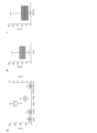
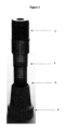
好ましくは、遠心分離チューブ(図3)が5mlの内容積を有し、チューブを上部容積および下部容積に分離する隔壁または膜(2)を備え、それによって、血小板およびリンパ球に富む血漿相が上部容積に収集される一方、赤血球が下部容積に蓄積する収集される。下部容積は、チューブの下端に配置されたリングナット(4)によって、下部チューブチャンバを上下に移動するピストン(3)により操作できる。これにより、遠心分離後の血小板およびリンパ球に富む血漿相を含む約3mlの上部チューブ容積を画定でき、赤血球を含む1~2mlで変動し得る下部容積は、リングナット-ピストンシステムによりによって調節することができる。 Preferably, the centrifuge tube (Fig. 3) has an internal volume of 5 ml and is provided with a septum or membrane (2) separating the tube into upper and lower volumes, whereby a platelet- and lymphocyte-rich plasma phase is Red blood cells accumulate in the lower volume while being collected in the upper volume. The lower volume can be manipulated by a piston (3) moving up and down the lower tube chamber by means of a ring nut (4) located at the lower end of the tube. This allows defining an upper tube volume of approximately 3 ml containing platelets and lymphocyte-rich plasma phase after centrifugation, and a lower volume containing red blood cells, which can vary from 1-2 ml, is adjusted by a ring-nut-piston system. be able to.
さらにより好ましくは、隔壁または膜が15~200ミクロンの多孔度を有し、1.5~2.5mmの直径の中心孔を備えた疎水性ディスクバリアまたは膜からなる。ディスクバリアまたは膜に使用される適切な材料としてはポリエチレンが挙げられるが、これらに限定されない。 Even more preferably, the partition or membrane has a porosity of 15-200 microns and consists of a hydrophobic disc barrier or membrane with a central pore diameter of 1.5-2.5 mm. Suitable materials for use in disc barriers or membranes include, but are not limited to, polyethylene.
さらなる態様では、本発明は、上記の特徴を有する血小板およびリンパ球に富む血漿からなる、本明細書に開示される方法によって得ることができる血液製剤を対象とする。この製剤は血管新生の可能性を示し、皮膚病変に対する血管新生促進効果を促進し、インビボでより効率的な創傷治癒を誘導する。 In a further aspect, the present invention is directed to a blood product obtainable by the method disclosed herein, comprising platelet- and lymphocyte-rich plasma having the characteristics described above. This formulation exhibits angiogenic potential, promotes pro-angiogenic effects on skin lesions, and induces more efficient wound healing in vivo.
したがって、本発明による血液製剤は、病変した身体組織の再生および創傷治癒の促進のために好都合に使用される。特定の実施形態では、本明細書で定義される血液製剤が化粧用途および治療用途に使用され:
-化粧品用途としては抗老化、瘢痕修復、しわ、座瘡、脱毛症および熱傷治療が挙げられ、
-治療用途としては創傷治癒、骨および軟骨損傷などの整形外科または腫瘍学的起源の病変の治療、手術後の病変または創傷;皮膚潰瘍、褥瘡、褥瘡、静脈性下肢潰瘍、動脈性潰瘍、糖尿病性足潰瘍、角膜および角膜潰瘍の瘢痕などの角膜病変;歯周組織損傷または骨膜病変の治療が挙げられる。
The blood products according to the invention are therefore advantageously used for regeneration of diseased body tissue and promotion of wound healing. In certain embodiments, blood products as defined herein are used for cosmetic and therapeutic applications:
- cosmetic applications include anti-aging, scar repair, wrinkle, acne, alopecia and burn treatment;
- For therapeutic use, wound healing, treatment of lesions of orthopedic or oncological origin such as bone and cartilage damage, post-operative lesions or wounds; corneal lesions such as foot ulcers, corneal and corneal ulcer scars; treatment of periodontal tissue injuries or periosteal lesions.
本発明の血液製剤は、同種または自己の治療方法において使用することができる。好ましい実施形態では血液製剤が自己適用のために使用され、すなわち、それはリンパ-血小板に富む血漿が単離される全血の同じドナーに投与される。 The blood products of the invention can be used in allogeneic or autologous treatment methods. In a preferred embodiment the blood product is used for autologous administration, ie it is administered to the same donor of whole blood from which the lymph-platelet rich plasma is isolated.
本明細書に開示される方法によって得られる血液製剤はすぐに使用可能であり、すなわち、それは、その取得の直後に、それを必要とする対象に適用または投与され得る。あるいは凍結し、必要に応じて解凍して使用することができ、または例えば、適切なビヒクル、担体、賦形剤、保存剤、緩衝剤、安定剤の添加によって実施することができる。例えば、血液製剤は、その収集に使用されるシリンジ内に塩化カルシウム溶液(2.5~10%w/v)を直接添加することによって、ゲル形態で使用することができる。 The blood product obtained by the methods disclosed herein is ready-to-use, ie, it can be applied or administered to a subject in need thereof immediately after its acquisition. Alternatively, it can be frozen and thawed as needed for use, or it can be carried out, for example, by adding suitable vehicles, carriers, excipients, preservatives, buffers, stabilizers. For example, blood products can be used in gel form by adding calcium chloride solution (2.5-10% w/v) directly into the syringe used for its collection.
したがって、さらなる態様において、本発明は適切な担体または賦形剤と一緒に、上記で定義された血液製剤を含有する医薬組成物または化粧品組成物を提供する。 Therefore, in a further aspect, the invention provides a pharmaceutical or cosmetic composition containing a blood product as defined above together with a suitable carrier or excipient.
本発明は以下の図面および実施例によってさらに説明されるが、これらに限定されない。 The invention is further illustrated by, but not limited to, the following figures and examples.
-リンパ-血小板に富む血漿(AngioPRP)の調製
本発明に係るリンパ-血小板に富む血漿の分取に用いる遠心チューブ(以下、「AngioPRP装置」)は、基本的に(図3)から構成される:
1.無菌の外部目盛り付き5mlポリプロピレンチューブで作られた本体;
2.ゴム栓(BD Vacutainer Haemogard状)(1);
3.HDPE、多孔性(孔径<200ミクロン)、疎水性、直径13mm、厚さ1.5mmの円板障壁、中心の直径2mmの孔からなる2つの部分(上部容積56%、下部容積44%)でチューブを分離する隔壁(2);
4.内側ピストン(3)を備えたリングナット(4)からなる底部。
-Preparation of Lymph-Platelet Rich Plasma (Angio PRP ) The centrifugal tube (hereinafter "Angio PRP device") used for fractionating the lymph-platelet rich plasma according to the present invention basically consists of (Fig. 3) will be:
1. A body made of a sterile external graduated 5ml polypropylene tube;
2. Rubber stopper (like BD Vacutainer Haemogard) (1);
3. HDPE, porous (pore size <200 microns), hydrophobic,
4. A bottom consisting of a ring nut (4) with an inner piston (3).
前もってクエン酸ナトリウムチューブに採取された末梢血のサンプルは、チューブ内のゴム製の蓋を通して注入され、内部バリアは血液サンプルを上部容積内に保持する。
その後、装置は5分間1500rpm(458-460g)で遠心される。ブレーキ減速なし。
内膜は、血小板(PRP)および目的の細胞集団(上部)で富化された血漿の、老廃物(赤血球、下部)からの分離を促進する。
リングナットは、上部チャンバ(膜の上方)内のPRP相全体を調整することを可能にする。
上部血漿および細胞は、シリンジで引き抜かれ、液体形態で使用する準備ができている。
A sample of peripheral blood, previously collected in a sodium citrate tube, is infused through a rubber lid within the tube, and an internal barrier retains the blood sample within the upper volume.
The device is then centrifuged at 1500 rpm (458-460 g) for 5 minutes. No brake deceleration.
The inner membrane facilitates the separation of plasma enriched with platelets (PRP) and cell populations of interest (top) from waste (erythrocytes, bottom).
The ring nut allows adjusting the entire PRP phase in the upper chamber (above the membrane).
The upper plasma and cells are withdrawn with a syringe and are ready for use in liquid form.
<AngioPRP生成物回収>
3つの症例が起こりうる:
<Angio PRP product recovery>
Three cases can occur:
(1)血漿相は完全に膜の上にある。
2.5mlのシリンジに50mmと21Gの針を使用する。針をラバーストッパに刺し、AngioPRP生成物を抜き取る。膜から3~5mmの位置に針を置き、生成物を吸い出し、血漿相に細胞と血小板を再懸濁する。それぞれのNovySep装置から平均容量1mlのAngioPRPを回収する。
AngioPRP生成物の量は、性別、年齢、ヘマトクリット値、水分補給量などのパラメータによって異なる
(1) The plasma phase is completely above the membrane.
Use a 50 mm and 21 G needle on a 2.5 ml syringe. Poke the needle through the rubber stopper and withdraw the Angio PRP product. A needle is placed 3-5 mm from the membrane, the product is aspirated, and the cells and platelets are resuspended in the plasma phase. An average volume of 1 ml of Angio PRP is collected from each NovySep device.
The amount of Angio PRP product varies with parameters such as gender, age, hematocrit, and hydration
(2)血漿相は、部分的に膜の上下に位置する。
NovySepスクリュー(リングナット)を反時計回りに回して、血漿相が膜の上に完全にくるまで膜下の容積を減らす。そして、上記(1)のようにAngioPRP生成物を回収する。
(2) The plasma phase partially lies above and below the membrane.
Turn the NovySep screw (ring nut) counterclockwise to reduce the submembrane volume until the plasma phase is completely above the membrane. The Angio PRP product is then recovered as in (1) above.
(3)血漿相は、膜の上に位置する赤血球によって目に見えて汚染される。
生成物を廃棄する。
(3) The plasma phase is visibly contaminated by red blood cells located on the membrane.
Discard the product.
生/死染色(L3224、Life Technologies、米国カリフォルニア州)を、1、2、3、4および7日間後にAngioPRPに対してインビトロで実施し、細胞生存を評価した。生存率は数日間減少するが、インビトロで7日後、82.26±8.50%の細胞が生存している。内生性の評価のために、8x10⁴ HUVEC(ATCC-LGC,米国バージニア州)を3D Matrigel(BD Biosciences)に配置し、AngioPRPまたはPRPまたはAngiocellsを150μl加え、24時間後の細胞の細分化をImageJソフトウェア(NIH)により定量化した。AngioPRPは他の条件よりも長い細分化を誘導した(対照条件と比較して327.75±9.72×10³対293.05±8.35×103μm、t検定*p<0.05)。 Live/dead staining (L3224, Life Technologies, CA, USA) was performed in vitro on Angio PRP after 1, 2, 3, 4 and 7 days to assess cell survival. Viability decreases for several days, but after 7 days in vitro, 82.26±8.50% of the cells are viable. For evaluation of endogeneity, 8×10 4 HUVEC (ATCC-LGC, Va., USA) were placed in 3D Matrigel (BD Biosciences), 150 μl of Angio PRP or PRP or Angio cells were added, and cell fragmentation was performed after 24 hours. was quantified by ImageJ software (NIH). Angio PRP induced longer fragmentation than other conditions (327.75±9.72×10 3 vs. 293.05±8.35×10 3 μm compared to control condition, t-test *p<0 .05).
-FACS分析
末梢血(PB)はミラノ(イタリア、ミラノ)のポリクリニコ病院の研究におけるヒト被験者の使用に関する委員会のガイドラインに従って、インフォームドコンセント後の健常ボランティアから、ミラノのポリクリニコ病院の輸血医学・血液学部門の血液バンクから入手した。クエン酸ナトリウムチューブに採取した2.5mlの末梢血をNovySep装置に充填し、相分離を誘導するために室温で1500rpmで5分間遠心分離した。血小板に富む血漿相および細胞を、赤血球と血漿との間の界面で回収した。分離前の血液およびAngioPRPを、血液コールター計数器(DxH 500、ベックマンコールター)によって分析した。分離前に採取した血液および細胞相を、以下に示すモノクローナル抗体で直接標識した。細胞を、SytoTM 16、抗CD45 V500、抗CD3 V450、抗CD56 PE-CY7、抗CD14 APC-H7、抗CD16 PE、抗CD19 APC-R700または抗CD31 PE、抗CD184 APC(BD Biosciences-Pharmingen,米国カリフォルニア州サンディエゴ)と共にインキュベートした。対照はアイソタイプマッチマウス免疫グロブリンであった。4℃で20分間の各インキュベーション後、1%熱不活化FCSおよび0.1%アジ化ナトリウムを含むPBS 1X中で細胞を洗浄した。サイトメトリー分析は、FACSuiteTMソフトウエア(BD Biosciences-Immunocytometry System)を用いてLYRICフローサイトメーターで行った。各解析は、各ゲートについて少なくとも1~2×10の4事象を含んだ。分析から細胞破片を除去するために光散乱ゲートを設定した。陽性細胞のパーセンテージは、特定のフルオロフォアにコンジュゲートされたアイソタイプ対照に反応するパーセンテージについて補正後に評価した。SytoTM16の陽性ゲート上で、様々な細胞亜集団の割合を計算した。
- FACS analysis Peripheral blood (PB) was obtained from healthy volunteers after informed consent according to the guidelines of the Committee for the Use of Human Subjects in Research at the Policlinico Hospital of Milan (Milan, Italy). Obtained from the departmental blood bank. 2.5 ml of peripheral blood collected in sodium citrate tubes was loaded into the NovySep device and centrifuged at 1500 rpm for 5 minutes at room temperature to induce phase separation. A platelet-rich plasma phase and cells were collected at the interface between red blood cells and plasma. Pre-separation blood and Angio PRP were analyzed by a blood Coulter counter (DxH 500, Beckman Coulter). Blood and cell phases collected prior to separation were directly labeled with the monoclonal antibodies shown below. Cells were treated with
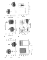
-AngioPRPのインビトロ評価
末梢血の分離をNovySep装置を用いて行い、AngioPRPを分析して血小板組成物の特徴を調べた。図1Aに報告されるように、AngioPRPは主に血小板(85.19±7.03%)から構成され、白血球(WBC)の存在は0.85±0.53%である。血小板濃度は全血中に存在する元の数に比べAngioPRPで増加する(108.5±6.62x10³血小板/μlに代えて146.8±11.35x103、図1B)。特に、細胞成分をコールターカウンターによってさらに特徴付け、分離手順の前に元の末梢血組成物と比較した。AngioPRPでは、全血と比較して、リンパ球成分が富化され(AngioPRPについて67.15±9.25%および全血について29.98±6.52%)、一方、顆粒球集団は著しく減少し(AngioPRPについて12.32±7.67%および全血について62.36±7.38%)、単球は部分的に増加した(AngioPRPについて20.13±6.30%および全血について7.69±1.56%、図1D)。
- In vitro evaluation of Angio PRP Peripheral blood separation was performed using the NovySep apparatus and Angio PRP was analyzed to characterize the platelet composition. As reported in FIG. 1A, Angio PRP is mainly composed of platelets (85.19±7.03%) with white blood cells (WBC) present at 0.85±0.53%. The platelet concentration is increased with Angio PRP compared to the original number present in whole blood (146.8±11.35×10 3 instead of 108.5±6.62×10 3 platelets/μl, FIG. 1B). In particular, cellular components were further characterized by a Coulter Counter and compared to the original peripheral blood composition prior to isolation procedures. Angio PRP was enriched in lymphocyte components compared to whole blood (67.15±9.25% for Angio PRP and 29.98±6.52% for whole blood), while the granulocyte population was significantly decreased (12.32±7.67% for Angio PRP and 62.36±7.38% for whole blood), monocytes were partially increased (20.13±6.30% for Angio PRP and 7.69±1.56% for whole blood, FIG. 1D).
-エクスビボ前臨床実験:器官型皮膚培養
ヒト真皮および表皮多層モデル(MatTek's EpiDermFT Full Thickness EFT-400)をインビトロモデルとして用い、NovySep装置の組織再生能力を評価した。組織モデル(角質層および表皮)上の皮膚生検を、EpiDerm-FTの製造業者のプロトコールに記載されるように、5mmパンチによって得た。次の4種類の培養条件下で試験した:1)AngioPRPを用いた器官型皮膚培養、2)Angiocells、3)PRP、4)PBS 1X(陰性対照として)。培養24時間後、2日後、4日後、5日後、6日後および7日後のヒト皮膚組織を分析し、創傷閉鎖および上皮再生を評価するために、免疫組織化学的および免疫蛍光分析を行った。ヘマトキシリンおよびエオシン染色を、創傷治癒をより良く特徴付けるために行い、画像定量化およびプロセスを、ImageJソフトウェア(NIH)を用いて行った。
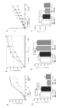
図2Aに報告されているように、AngioPRPで処置された病巣は6日後に完全な治癒を示したが、PRPでの処置は同じ結果に達するのにもう1日かかった。AngiocellsとPBSの使用は1週間で完全治癒に成功しなかった。
- Ex Vivo Preclinical Experiments: Organotypic Skin Culture A human dermis and epidermis multilayer model (MatTek's EpiDermFT Full Thickness EFT-400) was used as an in vitro model to evaluate the ability of the NovySep device to regenerate tissue. Skin biopsies on tissue models (stratum corneum and epidermis) were obtained by 5 mm punch as described in the EpiDerm-FT manufacturer's protocol. Four culture conditions were tested: 1) Organotypic skin culture with Angio PRP , 2) Angio cells , 3) PRP, 4) PBS 1X (as negative control). After 24 hours, 2 days, 4 days, 5 days, 6 days and 7 days of culture, human skin tissues were analyzed and immunohistochemical and immunofluorescent analyzes were performed to assess wound closure and epithelial regeneration. Hematoxylin and eosin staining were performed to better characterize wound healing and image quantification and processing were performed using ImageJ software (NIH).
As reported in FIG. 2A, lesions treated with Angio PRP showed complete healing after 6 days, whereas treatment with PRP took another day to reach the same result. The use of Angio cells and PBS did not lead to complete cure in 1 week.
-インビボ創傷治癒実験
5ヶ月齢のScidマウス(n=10)は、Charles River Laboratories International, Inc.(イタリア国カルコ)から入手した;この研究における動物の使用が国立保健省によって認可された(プロトコル番号51/2018-PR)。動物をアベルチンで麻酔し、5mmの全層切除を2回行い、これは、マウスの正中線の両側に1つずつ、海綿体を背側に作製した。シリコーンスプリントを、接着剤の助けを借りて創傷の周りに配置し、次いで、スプリントを、中断された縫合糸で固定した。各マウスはそれ自体の対照として作用し、一方の創傷は処置を受け、他方のリン酸緩衝生理食塩水(PBS 1X)を受けた。汚染を防ぐために透明な閉塞包帯を適用した。創傷を、2~3日ごとに写真を撮ることによってチェックし、面積を、ImageJソフトウェア(NIH)を使用してミリメートル基準に対して定量化し、創傷閉鎖に対応する、損傷の0、4、7、10、15、および21日後に測定された創傷面積のパーセンテージとして表した;マウスを、完全深部麻酔下で頸椎脱臼によって犠牲にし、背部皮膚病変を除去した;生検を、組織学的または分子生物学的分析のためにそれぞれ2つの群に分けた。1群をイソペンタンに入れ、プロテオミクス分析のために-80℃で凍結した。他の群は4%パラホルムアルデヒドのPBS溶液中、4℃で一晩インキュベートし、PBS 1X溶液中、30%スクロースにさらに24時間移した後、4℃でインキュベートし、O.C.T化合物中に包埋し、-80℃で凍結した。厚さ12μmの連続切片を切断し、免疫蛍光法および組織学的分析によって検査した。
- In Vivo Wound Healing Experiments Five month old Scid mice (n=10) were purchased from Charles River Laboratories International, Inc. (Calco, Italy); the use of animals in this study was approved by the National Ministry of Health (protocol number 51/2018-PR). Animals were anesthetized with avertin and two 5 mm full-thickness excisions were performed, one on each side of the midline of the mouse, making the corpus cavernosum dorsal. A silicone splint was placed around the wound with the aid of an adhesive, then the splint was secured with interrupted sutures. Each mouse acted as its own control, with one wound receiving treatment and the other receiving phosphate buffered saline (PBS 1X). A clear occlusive dressing was applied to prevent contamination. Wounds were checked by taking pictures every 2-3 days and the area was quantified to millimeter scale using ImageJ software (NIH), 0, 4, 7 lesions corresponding to wound closure. , expressed as a percentage of the wound area measured after 10, 15, and 21 days; mice were sacrificed by cervical dislocation under total deep anesthesia and dorsal skin lesions were removed; Each was divided into two groups for biological analysis. One group was placed in isopentane and frozen at -80°C for proteomics analysis. The other group was incubated overnight at 4°C in 4% paraformaldehyde in PBS, transferred to 30% sucrose in PBS 1X solution for an additional 24 hours, and then incubated at 4°C. C. Embedded in T compound and frozen at -80°C.
AngioPRP処置(n=5)、ヒアロマトリクス処置(n=5)およびPBS処置(n=10)についてそれぞれ皮膚創閉鎖時期を比較した。さらに、異なる条件で全閉鎖に達するのに必要な時間を評価した。21日目のAngioPRPによる処置は損傷の完全な閉鎖を誘発し、代わりに、ヒアロマトリクスで処置された皮膚は、損傷後21日目に75~80%の創傷閉鎖を示した。さらに、PBSで処理した試料は、同じ時点で創傷閉鎖の90%を示した。AngioPRP処置では6.5/7日後に60%の閉創の達成が観察されたのに対し、ヒアロマトリクス処置では15日であった(p<0.0001、ボンフェローニ補正による二元配置分散解析(ANOVA))。PBSによる対照処理は、ほぼ10日で閉鎖の60%に達する。21日後において優位な差異が、AngioPRPとヒアロマトリクスの間(p<0.0001、ボンフェローニ補正による二元分散分析(ANOVA))およびAngioPRPとPBSの間(p<0.0001、ボンフェローニ補正による二元配置分散分析(ANOVA))でも認められた(図2B)。
The skin wound closure times were compared for Angio PRP treatment (n=5), Hyalomatrix treatment (n=5) and PBS treatment (n=10). In addition, the time required to reach full closure under different conditions was evaluated. Treatment with Angio PRP on
-表皮分化
12μm皮膚組織切片の連続切片を切断し、ヘマエトキシリンおよびエオシン(H&E)、アルシアンブルーおよびピクロシリウスレッド染色を用いて、形態学的評価のための製造業者の指示に従って染色した。画像をLMD6000B(ライカ、ドイツ)で捕捉した。
- Epidermal differentiation Serial sections of 12 μm skin tissue sections were cut and stained with hemaethoxylin and eosin (H&E), Alcian blue and picrosirius red stains according to the manufacturer's instructions for morphological evaluation. . Images were captured with an LMD6000B (Leica, Germany).
免疫蛍光分析のために、組織切片を、マウスモノクローナル抗体抗-シトケラチン10(1:100、アブカム)、ウサギ抗-インボルクリン(1:100、アブカム)、マウスモノクローナル抗体抗-シトケラチン14(1:100、アブカム)、ウサギ抗-ロリクリン(1:100、アブカム)、CD206(1:400、アブカム)と共にインキュベートし、細胞核を、DAPIを用いて室温で5分間染色した。スライドを、蛍光顕微鏡LEICA DMI8(ライカ、ドイツ)を用いて分析した。 For immunofluorescence analysis, tissue sections were treated with mouse monoclonal antibody anti-cytokeratin 10 (1:100, Abcam), rabbit anti-involucrin (1:100, Abcam), mouse monoclonal antibody anti-cytokeratin 14 (1:100, Abcam). Abcam), rabbit anti-loricrin (1:100, Abcam), CD206 (1:400, Abcam) and cell nuclei were stained with DAPI for 5 minutes at room temperature. Slides were analyzed using a fluorescence microscope LEICA DMI8 (Leica, Germany).
21日間のヘマトキシリン・エオジン染色の結果、AngioPRPで処理した皮膚は、ヒアロマトリクス(市販品)で処理した皮膚と比較して、線維芽細胞およびケラチノサイトの増殖および創傷部位への移動によって誘発される完全な創傷治癒を示した。また、種々の表皮層に対する特異的マーカーを用いて表皮分化を評価した。21日間で、AngioPRPで処置された皮膚は、特異的マーカサイトケラチン5についての陽性かつ均一なシグナルによって示されるように、基底層の完全かつ均一な再生を示した。一方、ヒアロマトリクスおよびPBSで処理した試料は、不完全で不連続な再生を示した。顆粒層に関して、AngioPRPでの処理はサイトケラチン10染色によって実証されるように、完全な再生を誘導し、健康な皮膚試料と同等であった。一方、ヒアロマトリクスおよびPBSで処置した皮膚病変は顆粒層の後期再生を示し、サイトケラチン10の不均一な発現を示した。角質層に関して、最も外側の層を可視化するためにロリクリンについて免疫蛍光染色を行った;我々はAngioPRPで処理した試料においてのみ完全な再上皮化を観察したが、一方、ヒアロマトリクスおよびPBSで処理した皮膚においては21日目に不完全な上皮を観察した。
Hematoxylin and eosin staining for 21 days revealed that skin treated with Angio PRP was induced by fibroblast and keratinocyte proliferation and migration to the wound site compared to skin treated with Hyalomatrix (commercially available). It showed complete wound healing. Epidermal differentiation was also assessed using specific markers for different epidermal layers. At 21 days, Angio PRP -treated skin showed complete and uniform regeneration of the basal layer, as indicated by a positive and uniform signal for the
-皮膚弾性特性
犠牲にした後、機械的試験のための皮膚を、クランプされた端部を覆うゴム片を有する金属スクリュークランプに入れた。クランプをBose Electroforce 3100機器に入れた。0.15Nの初期トラクションを適用する。MPaで測定したトラクションは、破壊点まで毎秒0.2%増加した。力(N)および変位(mm)をxyプロッターで測定し、これらの点を続いて応力(σ=断面積当たりの力)および歪み(ε=長さ/初期長さの変化)として記録し、Excel(Ashley W.Seifert et.Al;2012)で再プロットした。
- Skin elastic properties After sacrifice, skins for mechanical testing were placed in metal screw clamps with rubber strips covering the clamped ends. The clamp was placed in a Bose Electroforce 3100 instrument. Apply an initial traction of 0.15N. Traction, measured in MPa, increased by 0.2% per second up to the breaking point. Force (N) and displacement (mm) were measured on an xy plotter and these points were subsequently recorded as stress (σ = force per cross-sectional area) and strain (ε = length/change in initial length), Replotted in Excel (Ashley W. Seifert et. Al; 2012).
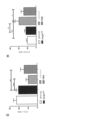
皮膚の弱さを評価するために、AngioPRP、PBS、ヒアロマトリクス(陰性対照として)および健康な皮膚(陽性対照として)で処置した皮膚の機械的特性を比較する。背側皮膚から応力‐歪曲線を導き、平均引張強度を決定した。AngioPRPで処置した皮膚は、ヒアロマトリクスまたはPBSで処置した皮膚(3MPa±0.7、0.3MPa±0.3および1.8MPa±0.70)と比較して、牽引に対してより高い耐性を示した(図2C)。さらに、LPEPで処置した皮膚は、健康な皮膚と同等の傾向を示した(3MPa±0.7および3.1±0.7)。AngioPRPで処理された皮膚において観察されるより高い弾性率は、単位面積当たりの線維性分子の数の増加によるものであり得る。エラスチンは皮膚弾性線維の主成分であり、皮膚再生に有益であるため、VI型コラーゲンの免疫蛍光染色を行った。AngioPRPで処置された皮膚は図2Fに定量化され報告されているように、ヒアルロマトリクスで処置された皮膚と比較して、コラーゲンVI発現に関して有意差を示した(不対t検定、*p<0.05)。 To assess skin fragility, the mechanical properties of skin treated with Angio PRP , PBS, Hyalomatrix (as negative control) and healthy skin (as positive control) are compared. A stress-strain curve was derived from the dorsal skin to determine the average tensile strength. Skin treated with Angio PRP was higher to traction compared to skin treated with Hyalomatrix or PBS (3 MPa ± 0.7, 0.3 MPa ± 0.3 and 1.8 MPa ± 0.70) It showed resistance (Fig. 2C). Furthermore, LPEP-treated skin showed similar trends to healthy skin (3 MPa ± 0.7 and 3.1 ± 0.7). The higher elastic modulus observed in Angio PRP -treated skin may be due to the increased number of fibrous molecules per unit area. Since elastin is a major component of skin elastic fibers and is beneficial for skin regeneration, immunofluorescence staining of type VI collagen was performed. Angio PRP -treated skin showed a significant difference in terms of collagen VI expression compared to hyaluronic matrix-treated skin, as quantified and reported in FIG. 2F (unpaired t-test, *p <0.05).
表皮の特徴付けに加えて、真皮組成の分析を行った。アルシアンブルー染色を用いて、真皮領域におけるコラーゲン線維の存在を確認した。この染色の強さのおかげで、皮膚試料中の高密度弾性線維と低密度弾性線維とを区別し、様々な処理においてこれらの2つの密度タイプによって占められる表面を定量することができた;図2Dおよび2Eに報告されるように、AngioPRPで処理された試料は、ヒアロマトリクスおよびPBS処理と比べ、健康な皮膚とより類似した皮膚組成を示し、高密度エラスチンが高く、低密度エラスチンが少ない(不対t検定、*p<0.05、**p<0.01、****p<0.0001)。 In addition to epidermal characterization, analysis of dermal composition was performed. Alcian blue staining was used to confirm the presence of collagen fibers in the dermal area. The intensity of this staining allowed us to distinguish between high- and low-density elastic fibers in skin samples and quantify the surface occupied by these two density types in various treatments; As reported in 2D and 2E, samples treated with Angio PRP showed skin composition more similar to healthy skin, with higher high density elastin and less low density elastin compared to Hyalomatrix and PBS treatments. (Unpaired t-test, *p<0.05, **p<0.01, ****p<0.0001).
-プロテオミクスおよび生化学分析
皮膚サンプルをプロテオミクス分析用に抽出し、4℃で17,000xgで10分間遠心分離し、4℃で1hで200,000xgで超遠心分離を施した。0.1M重炭酸アンモニウム(pH7.9)に再懸濁したペレットを、以前に報告された手順(Bari,Perteghella et al.2018)に従って、各試料からの50±0.5μgのタンパク質を用いてトリプシン処理した。1マイクロリットルのトリプシン消化混合物を、Q-Exactive質量分析計(Thermo Fisher Scientific,米国)に連結したcHiPLC-nanoflexシステム(Eksigent,AB SCIEX,米国)を備えたナノクロマトグラフィーにより、5~45%の溶離剤B(溶離剤A、水中0.1%ギ酸;溶離剤B、アセトニトリル中0.1%ギ酸)の65分勾配を介して、300nL/分の流速で分析した。完全質量スペクトルは、70000FWHM分解能で400~1600m/zの範囲にわたるポジティブイオンモードで記録され、続いて10のMS/MSスペクトルで17,500FWHMの分解能では、最も豊富なイオンに依存する様式でデータを生成した。生成されたデータはすべて、SEQUEST検索エンジンおよびヒトタンパク質データベース(70726エントリー、UNIPROTウェブサイト、www.uniprot.govから2017年1月にダウンロード)に基づいて、Proteome Discoverer 2.1プラットフォーム(Thermoscientific)を用いて検索した。得られたタンパク質リストをアラインメントし、正規化し(Sereni,Castiello et al.2018)、次いで線形判別解析手段(LDA)により処理した(Hilario and Kalousis 2008)。各被験体を特定の群に割り当てるために、抽出されたマーカータンパク質に従ってα値パラメータを計算した(Brambilla,Lavatelli et al.2012)。
- Proteomics and Biochemical Analysis Skin samples were extracted for proteomics analysis, centrifuged at 17,000 xg for 10 minutes at 4°C and subjected to ultracentrifugation at 200,000 xg for 1 h at 4°C. Pellets resuspended in 0.1 M ammonium bicarbonate (pH 7.9) were resuspended according to a previously reported procedure (Bari, Perteghella et al. 2018) using 50±0.5 μg of protein from each sample. Trypsinized. One microliter of the trypsin digestion mixture was subjected to nanochromatography with a cHiPLC-nanoflex system (Eksigent, AB SCIEX, USA) coupled to a Q-Exactive mass spectrometer (Thermo Fisher Scientific, USA) to give a concentration of 5-45%. Analysis was performed at a flow rate of 300 nL/min through a 65 min gradient of eluent B (eluent A, 0.1% formic acid in water; eluent B, 0.1% formic acid in acetonitrile). Full mass spectra were recorded in positive ion mode over the range 400-1600 m/z at 70,000 FWHM resolution, followed by 10 MS/MS spectra at 17,500 FWHM resolution, with data acquired in a most abundant ion-dependent manner. generated. All data generated were based on the SEQUEST search engine and the human protein database (70726 entries, UNIPROT website, downloaded from www.uniprot.gov in January 2017) using the Proteome Discoverer 2.1 platform (Thermoscientific). searched. The resulting protein list was aligned and normalized (Sereni, Castiello et al. 2018) and then processed by Linear Discriminant Analysis (LDA) (Hilario and Kalousis 2008). To assign each subject to a specific group, the α value parameter was calculated according to the extracted marker proteins (Brambilla, Lavatelli et al. 2012).
異なる創傷処置のタンパク質プロファイルを理解するために、本発明者らはnLC-MS/MS分析を実施した;単一のPBS処置を、相対群(NovySepまたはヒアロマトリクス)の内部対照として使用し、一方、健康な皮膚を分析して、同じ動物の皮膚の正常対照プロファイル(内部対照)を同定するために分析した。同定されたタンパク質の総数は1514であり、高い技術的および群再現性を有した。ヒアロマトリクス創傷組織からのプロテオミクスプロファイルは、同じ動物からの対照皮膚試料のプロファイルと比較して著しく異なる。試料の主成分解析は、対照試料がAngioPRP処置創傷から収集された試料から類似のクラスターを形成することを示す。実際、ヒアロマトリクスで処理された試料はそれらの内部対照と一致を示し、一方、単一のAngioPRP試料はそれらの単一の相対的内部対照と相関し、このことは、処理の全身作用を示唆する。健康な皮膚試料はヒアルロマトリクス群よりもAngioPRPとクラスター化し、より相関する。ボンフェローニ補正後、179個のタンパク質が少なくとも2群間で差次的に発現されたが、アップレギュレートおよびダウンレギュレートされたタンパク質は同じ比較または同じ評価条件において類似していた。単一処理およびその特異的対照を分析すると、半数の差次的タンパク質が、ヒアルロマトリクス処理よりもAngioPRP処理において変化している。処置された条件が健康な皮膚と比較される場合、同じ挙動が報告される。このことは、AngioPRPがヒアロマトリクスよりも健康な皮膚に近い傾向があることを示唆している。次いで、プロテオーム回復指数(PRi)(Roffia,De Palma et al.2018,Sereni,Castiello et al.2018)を適用して、アルゴリズムでホメオスタシス回復に関与するタンパク質を同定するために、健常および疾患プロファイルを生成および比較し、3つの異なる条件を比較した。創傷組織に存在しない、AngioPRP処理後の6種のタンパク質、および未処理の病巣に存在し、AngioPRP試料には存在しない12種のタンパク質を同定した(PBS試料を陰性対照として使用した)。97個のタンパク質の濃度はAngioPRP処理後に回収されるが(損傷組織において57個がアップレギュレートされ、40個がダウンレギュレートされる)、一方、23個のみが、ヒアロマトリクス処理後に検出された(AngioPRP群と共通が14個)。正規化されたデータのクラスター化は、同じマクロ群におけるヒアロマトリクスおよび相対対照、ならびに健康な皮膚およびAngioPRPの単一試料を、それらの相対陰性対照と相関させて、結果を確認した。差次的に発現されたタンパク質を、Cytoscapeプラットフォームを用いて分析し、代謝を評価した。ネットワーク分析は、創傷誘導および2つの相対的処置の両方について、異なる代謝クラスターの変動を強調した。特に、セルピン、防御反応に関与するタンパク質、細胞骨格構成、補体および凝固カスケード、細胞外マトリクス(ECM)およびリボソームに対するクラスターは、ヒアロマトリクス処理およびその対照条件において過剰発現され、一方、AngioPRPおよび相対対照においては健常試料と同様に正規化される。逆に、ケラチンと脂質代謝に関与するタンパク質のクラスターは、ヒアロマトリクスよりもAngioPRP条件でより多く発現する。これら8つの標的群の中で、我々は、2つの処置後に異なる方法で発現され、健康な組織と比較される17の創傷関連タンパク質:カベオリン-1、EGFR、フィブロネクチン、デコリン、プレクチン、セルピンB2、ジソイドドメイン受容体-2、プロトロンビン、ヘムオキシゲナーゼ1、フィブリノーゲンα-、β-、およびγ-鎖、テナシン、プラスチン-2、α-トロポミオシン、サイトケラチン-6AおよびアネキシンA1に注目した。これらのタンパク質の発現は、ヒアロマトリクスおよびAngioPRP処理に対して逆の傾向を示す:例えば、カベオリン1およびデコリンはAngioPRP処理後にアップレギュレートされるが、ヒアロマトリクス試料においてダウンレギュレートされる。逆に、フィブリノーゲン、セルピンB2、およびプロトロンビンなどの凝固に関与する因子は、AngioPRP処置後よりヒアロマトリクス処置後の方がより発現される。
To understand the protein profile of different wound treatments, we performed nLC-MS/MS analysis; , healthy skin was analyzed to identify a normal control profile (internal control) of skin from the same animals. The total number of proteins identified was 1514, with high technical and group reproducibility. Proteomic profiles from hyalomatrix wound tissue are significantly different compared to profiles of control skin samples from the same animals. Principal component analysis of the samples shows that the control samples form similar clusters from the samples collected from the Angio PRP treated wounds. Indeed, the Hyalomatrix-treated samples showed concordance with their internal control, while the single Angio PRP samples correlated with their single relative internal control, suggesting a systemic effect of the treatment. Suggest. Healthy skin samples clustered and correlated better with Angio PRP than the hyaluronic matrix group. After Bonferroni correction, 179 proteins were differentially expressed between at least two groups, but up- and down-regulated proteins were similar in the same comparison or the same evaluation conditions. When single treatments and their specific controls are analyzed, half the differential proteins are altered in Angio PRP treatment over hyaluromatrix treatment. The same behavior is reported when treated conditions are compared to healthy skin. This suggests that Angio PRP tends to be closer to healthy skin than Hyalomatrix. The proteome recovery index (PRi) (Roffia, De Palma et al. 2018, Sereni, Castiello et al. 2018) was then applied to algorithmically analyze healthy and disease profiles to identify proteins involved in homeostatic recovery. Generated and compared, three different conditions were compared. We identified 6 proteins after Angio PRP treatment that were not present in wound tissue and 12 proteins that were present in untreated lesions and not in Angio PRP samples (PBS samples were used as negative controls). Levels of 97 proteins were recovered after Angio PRP treatment (57 upregulated and 40 downregulated in injured tissue), whereas only 23 were detected after Hyalomatrix treatment. (14 in common with the Angio PRP group). Clustering of the normalized data correlated Hyalomatrix and relative controls in the same macrogroup, and single samples of healthy skin and Angio PRP with their relative negative controls to confirm the results. Differentially expressed proteins were analyzed using the Cytoscape platform to assess metabolism. Network analysis highlighted variations in different metabolic clusters for both wound induction and the two relative treatments. In particular, clusters for serpins, proteins involved in defense responses, cytoskeletal organization, complement and coagulation cascades, extracellular matrix (ECM) and ribosomes were overexpressed in hyalomatrix-treated and control conditions, whereas Angio PRP and Relative controls are normalized in the same way as healthy samples. Conversely, clusters of proteins involved in keratin and lipid metabolism are more expressed in Angio PRP conditions than in hyalomatrix. Among these eight target groups, we found 17 wound-associated proteins that were differentially expressed after two treatments and compared to healthy tissue: caveolin-1, EGFR, fibronectin, decorin, plectin, serpin B2, We focused on disoid domain receptor-2, prothrombin,
-ネットワークおよび統計分析
差次的およびLDA法によって選択されたタンパク質の一覧から出発して、対応するヒトタンパク質-タンパク質相互作用(PPI)ネットワークを抽出し、Cytoscapeプラグイン、STRING 8データベース(Pawlowski、Muszewskaら、2010年)の手段によって、既知の相互作用を、いくつかのデータベース、例えば、Prolink、DIP、KEGGおよびBINDから検索した。さらに、PPIをCytoscape 3.5(Shannon,Markiel et al.2003)を用いて検査し、>0.15スコアを有する実験的に検証された相互作用のみが保持された。最後に、Bingo 2.44(Maere,Heymans et al.2005)を使用して、機能的に組織化されたGO用語に基づくモジュールを強調した。
- Network and Statistical Analysis Starting from a list of proteins selected by differential and LDA methods, the corresponding human protein-protein interaction (PPI) networks were extracted and analyzed using the Cytoscape plugin,
ペプチド配列および関連タンパク質の同定のための基準は、酵素としてのトリプシン;ペプチドあたり3つの失われた切断が許容される;前駆体イオンについて10ppmおよびフラグメントイオンについて±0.6Daの質量許容差が適用された。Percolatorノードはq値に基づく0.01(厳密)のペプチドスペクトルマッチ(PSM)レベルでの最終的な偽発見率(FDR)を与えるために、標的-デコイ戦略と共に使用され、0.05の最大ΔCNを考慮し(Kall,Canterbury et al.2007)、6アミノ酸の最小ペプチド長、およびランク1が考慮され、タンパク質グループ化および厳密な節約原理が適用された。 Criteria for identification of peptide sequences and related proteins are trypsin as the enzyme; 3 missing cleavages per peptide are allowed; a mass tolerance of 10 ppm for precursor ions and ±0.6 Da for fragment ions applies. was done. The Percolator node was used with the target-decoy strategy to give a final false discovery rate (FDR) at the peptide spectral match (PSM) level of 0.01 (strict) based on q-values, with a maximum of 0.05. Considering ΔCN (Kall, Canterbury et al. 2007), a minimum peptide length of 6 amino acids, and a rank of 1 were considered, and protein grouping and strict parsimony principles were applied.
カテゴリー変数の差は、χ二乗検定を用いて評価した。Kruskal Wallis検定を定量的変数の比較に適用した。R(3.5.2)統計解析ソフトウェアを用いて解析を行い、p値<0.05を統計的に有意であるとみなした。 Differences in categorical variables were assessed using the chi-square test. Kruskal Wallis test was applied for comparison of quantitative variables. Analyzes were performed using the R(3.5.2) statistical analysis software and p-values <0.05 were considered statistically significant.
SpCはt検定によっても評価され(Zhang, VerBerkmoes et al. 2006)、LDAおよびMAProMAの両方によって選択されたタンパク質は階層的クラスタリング(Zhao and Karypis 2005)によって、Wardの方法およびユークリッド距離メトリックを適用して評価された。LDAを用いたすべての層別化群の共通共分散行列およびマハラノビス距離(Jain AK 1999)を用い、JMP 5.1ソフトウェアにより階層的クラスタリングを行った。層別小児群を識別するタンパク質を選択するために、最大F比(≧5)と最小p値(≦0.05)を持つタンパク質を考察した。スペクトルカウント(SpC、PSMとも呼ばれる)と同定されたタンパク質の相対存在量との間の直接相関に基づいて、MAProMa(多次元アルゴリズムタンパク質マップ)ソフトウェア(Mauri, Riccio et al.2014)のDAve(Differential Average)およびDCI(Differential Coefficient Index)インデックスを使用して、分析された各子供群に対応する平均(aSpC)を処理した。課せられた閾値はDAve>|0.2|およびDCI>|100|であった。 SpC was also evaluated by t-test (Zhang, VerBerkmoes et al. 2006) and proteins selected by both LDA and MAProMA were subjected to hierarchical clustering (Zhao and Karypis 2005) applying Ward's method and Euclidean distance metric. was evaluated as Hierarchical clustering was performed with JMP 5.1 software using the common covariance matrix and Mahalanobis distance (Jain AK 1999) of all stratified groups using LDA. To select proteins that discriminate stratified pediatric groups, proteins with the highest F-ratio (≧5) and lowest p-value (≦0.05) were considered. Based on the direct correlation between spectral counts (SpC, also called PSM) and the relative abundance of identified proteins, MAProMa (multidimensional algorithmic protein map) software (Mauri, Riccio et al. 2014) uses DAve (Differential Average) and DCI (Differential Coefficient Index) indices were used to process the mean (aSpC) corresponding to each group of children analyzed. The thresholds imposed were DAve>|0.2| and DCI>|100|.
Claims (16)
110x106/ml~330x106/ml、好ましくは150x106/ml~330x106/mlの濃度の血小板;
0.1×10⁶/ml~2.0×106/ml、好ましくは0.1×106/ml~0.7×106/mlの濃度の白血球CD45+、CD31+およびCD34-;
0~0.03x10⁹/ml、好ましくは0.01x109/ml未満の濃度の赤血球;
を含み、創傷治癒、皮膚組織の再生、または血管新生を刺激する方法に使用される、単離された血液製剤。 An isolated blood product consisting of platelet- and lymphocyte-rich plasma, comprising:
Platelets at a concentration of 110x10 6 /ml to 330x10 6 /ml, preferably 150x10 6 /ml to 330x10 6 /ml;
Leukocytes CD45 + , CD31 + and CD34 − at a concentration of 0.1×10 6 /ml to 2.0×10 6 /ml, preferably 0.1× 10 6 / ml to 0.7× 10 6 /ml;
red blood cells at a concentration of 0-0.03×10 9 /ml, preferably below 0.01×10 9 /ml;
and used in a method of stimulating wound healing, skin tissue regeneration, or angiogenesis.
T細胞 51.54~66.11
B細胞 6.65~10.66
NK細胞 4.92~10.89
Tang(CD3+/184+/31+) 19.20~27.32
CD31+/146+/90+(EPC) 0.195~0.565 5. The isolation of claim 4, wherein T cells, angiogenic T lymphocytes (Tang), B cells, NK cells and EPC cells have the following 25% to 75% percentiles as determined by flow cytometry analysis: blood products.
T cells 51.54-66.11
B cells 6.65-10.66
NK cells 4.92-10.89
Tang (CD3+/184+/31+) 19.20-27.32
CD31+/146+/90+ (EPC) 0.195-0.565
単球 6.44~13.02 7. The isolated blood product of claim 6, wherein said monocytes are between the following 25% to 75% percentiles as determined by flow cytometric analysis.
Monocytes 6.44-13.02
顆粒球 2.87~10.87 9. The isolated blood product of claim 8, wherein the granulocytes are the following 25% to 75% percentile as determined by flow cytometric analysis.
Granulocytes 2.87-10.87
(ii)血小板とリンパ球に富む血漿を含む上相を採取する工程;
を含む、請求項1~8のいずれか1項に記載の血液製剤を調製する方法。 (i) placing 2-7 ml of whole blood in a centrifuge tube and centrifuging at 293-660 g, preferably 458 g, for 5-10 minutes at room temperature to obtain separated upper and lower phases, wherein wherein the upper phase comprises plasma rich in platelets and lymphocytes and the lower phase comprises red blood cells;
(ii) collecting the upper phase comprising platelet- and lymphocyte-rich plasma;
A method of preparing a blood product according to any one of claims 1 to 8, comprising
Applications Claiming Priority (3)
| Application Number | Priority Date | Filing Date | Title |
|---|---|---|---|
| EP20161201.7 | 2020-03-05 | ||
| EP20161201.7A EP3875074A1 (en) | 2020-03-05 | 2020-03-05 | Blood preparation useful for wound healing and tissue regeneration |
| PCT/EP2021/055552 WO2021176035A1 (en) | 2020-03-05 | 2021-03-05 | Blood preparation useful for wound healing and tissue regeneration |
Publications (1)
| Publication Number | Publication Date |
|---|---|
| JP2023517543A true JP2023517543A (en) | 2023-04-26 |
Family
ID=69844380
Family Applications (1)
| Application Number | Title | Priority Date | Filing Date |
|---|---|---|---|
| JP2022553068A Pending JP2023517543A (en) | 2020-03-05 | 2021-03-05 | Blood products useful in wound healing and tissue regeneration |
Country Status (8)
| Country | Link |
|---|---|
| US (1) | US20230129148A1 (en) |
| EP (2) | EP3875074A1 (en) |
| JP (1) | JP2023517543A (en) |
| KR (1) | KR20230002359A (en) |
| CN (1) | CN115243693A (en) |
| CA (1) | CA3170458A1 (en) |
| IL (1) | IL296017A (en) |
| WO (1) | WO2021176035A1 (en) |
Family Cites Families (7)
| Publication number | Priority date | Publication date | Assignee | Title |
|---|---|---|---|---|
| WO2008022651A1 (en) | 2006-08-21 | 2008-02-28 | Antoine Turzi | Process and device for the preparation of platelet rich plasma for extemporaneous use and combination thereof with skin and bone cells |
| KR20100116106A (en) * | 2009-04-21 | 2010-10-29 | 에스타 테크날러지스 리미티드 | Assembly, kit and method for preparing platelet-rich plasma (prp) |
| AU2013203115B2 (en) * | 2010-03-11 | 2015-12-03 | Antoine Turzi | Process,tube and device for the preparation of wound healant composition |
| GB201004072D0 (en) | 2010-03-11 | 2010-04-28 | Turzi Antoine | Process, tube and device for the preparation of wound healant composition |
| US20190160103A1 (en) * | 2011-07-14 | 2019-05-30 | Stefania GARBIN | Platelet-rich plasma composition for tissue repair and regeneration |
| GB201411913D0 (en) * | 2014-07-03 | 2014-08-20 | Lb46 Ltd | Cosmetic methods and products |
| GB201421013D0 (en) * | 2014-11-26 | 2015-01-07 | Turzi Antoine | New standardizations & medical devices for the preparation of platelet rich plasma (PRP) or bone marrow centrate (BMC) |
-
2020
- 2020-03-05 EP EP20161201.7A patent/EP3875074A1/en not_active Withdrawn
-
2021
- 2021-03-05 EP EP21713585.4A patent/EP4114416A1/en active Pending
- 2021-03-05 CN CN202180018894.5A patent/CN115243693A/en active Pending
- 2021-03-05 KR KR1020227034507A patent/KR20230002359A/en active Search and Examination
- 2021-03-05 CA CA3170458A patent/CA3170458A1/en active Pending
- 2021-03-05 WO PCT/EP2021/055552 patent/WO2021176035A1/en unknown
- 2021-03-05 US US17/905,646 patent/US20230129148A1/en active Pending
- 2021-03-05 IL IL296017A patent/IL296017A/en unknown
- 2021-03-05 JP JP2022553068A patent/JP2023517543A/en active Pending
Also Published As
| Publication number | Publication date |
|---|---|
| CN115243693A (en) | 2022-10-25 |
| US20230129148A1 (en) | 2023-04-27 |
| KR20230002359A (en) | 2023-01-05 |
| EP3875074A1 (en) | 2021-09-08 |
| IL296017A (en) | 2022-10-01 |
| WO2021176035A1 (en) | 2021-09-10 |
| EP4114416A1 (en) | 2023-01-11 |
| CA3170458A1 (en) | 2021-09-10 |
Similar Documents
| Publication | Publication Date | Title |
|---|---|---|
| Ronci et al. | Platelet-rich plasma as treatment for persistent ocular epithelial defects | |
| AU2014216415B2 (en) | Serum fraction of platelet-rich fibrin | |
| Lundquist et al. | Characteristics of an autologous leukocyte and platelet‐rich fibrin patch intended for the treatment of recalcitrant wounds | |
| Schmid et al. | Immunohistochemical demonstration of the calcium-binding proteins MRP8 and MRP14 and their heterodimer (27E10 antigen) in Crohn's disease | |
| KR20050105184A (en) | Autologous or homologous coagulant produced from anticoagulated whole blood | |
| Jafar et al. | Platelet lysate promotes the healing of long-standing diabetic foot ulcers: A report of two cases and in vitro study | |
| Gibello et al. | First pilot case-control interventional study using autologous extracellular vesicles to treat chronic venous ulcers unresponsive to conventional treatments | |
| JP7094593B2 (en) | Wound treatment gel obtained by combining platelet-rich plasma with autologous thrombin | |
| Capion et al. | The wound healing effect of local leukocyte platelet‐rich plasma after total hip arthroplasty: A randomized controlled trial | |
| US20230129148A1 (en) | Blood preparation useful for wound healing and tissue regeneration | |
| JP7100854B2 (en) | Neutrophil activation regulator | |
| CN115297871A (en) | Method for producing enhanced anti-inflammatory/anti-catabolic agents from autologous physiological fluids | |
| Zhao et al. | High mobility group box 1 derived mainly from platelet microparticles exacerbates microvascular obstruction in no reflow | |
| Erratico et al. | Effective high-throughput isolation of enriched platelets and circulating pro-angiogenic cells to accelerate skin-wound healing | |
| Hovhannisyan et al. | Excess filaggrin in keratinocytes is removed by extracellular vesicles to prevent premature death and this mechanism can be hijacked by Staphylococcus aureus in a TLR2-dependent fashion | |
| Erratico et al. | Platelets and Pro-Angiogenic Circulating Cells Accelerate Skin Wound Healing | |
| Saldin | Esophageal Extracellular Matrix Hydrogel to Restore Dysregulated Microenvironmentcell Signaling For Treatment of Pre-Malignant and Neoplastic Esophageal Disease | |
| Scala et al. | Contribution of peripheral blood mononuclear cells isolated by advanced filtration system to myogenesis of human bone marrow mesenchymal stem cells co-cultured with myoblasts | |
| SARI | Factors Affecting Platelet Count in Platelet-Rich Plasma | |
| La Padula | Striae distensae: assessment of a combined treatment on fibroblasts and an in vivo study | |
| Rajadurai | A Comparative study in Efficacy of Ultrasound Guided Autologous Platelet Rich Plasma and Corticosteroid Injection in Lateral Epicondylitis | |
| Vinhas | Magnetic actuation and magnetic responsive materials to modulate inflammation and their impact in cell behavior for tendon regeneration | |
| NZ776716B2 (en) | Wound treatment gel obtainable by combining platelet rich plasma with autologously derived thrombin | |
| FULL et al. | 21st Annual Meeting of the European Tissue Repair Society |
Legal Events
| Date | Code | Title | Description |
|---|---|---|---|
| A621 | Written request for application examination |
Free format text: JAPANESE INTERMEDIATE CODE: A621 Effective date: 20240301 |