JP2022550141A - Pulmonary Fibrosis Model and Methods of Using The Same - Google Patents
Pulmonary Fibrosis Model and Methods of Using The Same Download PDFInfo
- Publication number
- JP2022550141A JP2022550141A JP2022519551A JP2022519551A JP2022550141A JP 2022550141 A JP2022550141 A JP 2022550141A JP 2022519551 A JP2022519551 A JP 2022519551A JP 2022519551 A JP2022519551 A JP 2022519551A JP 2022550141 A JP2022550141 A JP 2022550141A
- Authority
- JP
- Japan
- Prior art keywords
- cells
- pats
- alveolar
- lung
- aec1
- Prior art date
- Legal status (The legal status is an assumption and is not a legal conclusion. Google has not performed a legal analysis and makes no representation as to the accuracy of the status listed.)
- Pending
Links
Images


Classifications
-
- G—PHYSICS
- G01—MEASURING; TESTING
- G01N—INVESTIGATING OR ANALYSING MATERIALS BY DETERMINING THEIR CHEMICAL OR PHYSICAL PROPERTIES
- G01N33/00—Investigating or analysing materials by specific methods not covered by groups G01N1/00 - G01N31/00
- G01N33/48—Biological material, e.g. blood, urine; Haemocytometers
- G01N33/50—Chemical analysis of biological material, e.g. blood, urine; Testing involving biospecific ligand binding methods; Immunological testing
- G01N33/5005—Chemical analysis of biological material, e.g. blood, urine; Testing involving biospecific ligand binding methods; Immunological testing involving human or animal cells
- G01N33/5008—Chemical analysis of biological material, e.g. blood, urine; Testing involving biospecific ligand binding methods; Immunological testing involving human or animal cells for testing or evaluating the effect of chemical or biological compounds, e.g. drugs, cosmetics
- G01N33/502—Chemical analysis of biological material, e.g. blood, urine; Testing involving biospecific ligand binding methods; Immunological testing involving human or animal cells for testing or evaluating the effect of chemical or biological compounds, e.g. drugs, cosmetics for testing non-proliferative effects
- G01N33/5023—Chemical analysis of biological material, e.g. blood, urine; Testing involving biospecific ligand binding methods; Immunological testing involving human or animal cells for testing or evaluating the effect of chemical or biological compounds, e.g. drugs, cosmetics for testing non-proliferative effects on expression patterns
-
- C—CHEMISTRY; METALLURGY
- C12—BIOCHEMISTRY; BEER; SPIRITS; WINE; VINEGAR; MICROBIOLOGY; ENZYMOLOGY; MUTATION OR GENETIC ENGINEERING
- C12N—MICROORGANISMS OR ENZYMES; COMPOSITIONS THEREOF; PROPAGATING, PRESERVING, OR MAINTAINING MICROORGANISMS; MUTATION OR GENETIC ENGINEERING; CULTURE MEDIA
- C12N5/00—Undifferentiated human, animal or plant cells, e.g. cell lines; Tissues; Cultivation or maintenance thereof; Culture media therefor
- C12N5/06—Animal cells or tissues; Human cells or tissues
- C12N5/0602—Vertebrate cells
- C12N5/0688—Cells from the lungs or the respiratory tract
-
- A—HUMAN NECESSITIES
- A61—MEDICAL OR VETERINARY SCIENCE; HYGIENE
- A61K—PREPARATIONS FOR MEDICAL, DENTAL OR TOILETRY PURPOSES
- A61K35/00—Medicinal preparations containing materials or reaction products thereof with undetermined constitution
- A61K35/12—Materials from mammals; Compositions comprising non-specified tissues or cells; Compositions comprising non-embryonic stem cells; Genetically modified cells
- A61K35/42—Respiratory system, e.g. lungs, bronchi or lung cells
-
- A—HUMAN NECESSITIES
- A61—MEDICAL OR VETERINARY SCIENCE; HYGIENE
- A61P—SPECIFIC THERAPEUTIC ACTIVITY OF CHEMICAL COMPOUNDS OR MEDICINAL PREPARATIONS
- A61P11/00—Drugs for disorders of the respiratory system
-
- G—PHYSICS
- G01—MEASURING; TESTING
- G01N—INVESTIGATING OR ANALYSING MATERIALS BY DETERMINING THEIR CHEMICAL OR PHYSICAL PROPERTIES
- G01N33/00—Investigating or analysing materials by specific methods not covered by groups G01N1/00 - G01N31/00
- G01N33/48—Biological material, e.g. blood, urine; Haemocytometers
- G01N33/50—Chemical analysis of biological material, e.g. blood, urine; Testing involving biospecific ligand binding methods; Immunological testing
- G01N33/5005—Chemical analysis of biological material, e.g. blood, urine; Testing involving biospecific ligand binding methods; Immunological testing involving human or animal cells
- G01N33/5008—Chemical analysis of biological material, e.g. blood, urine; Testing involving biospecific ligand binding methods; Immunological testing involving human or animal cells for testing or evaluating the effect of chemical or biological compounds, e.g. drugs, cosmetics
- G01N33/5082—Supracellular entities, e.g. tissue, organisms
- G01N33/5088—Supracellular entities, e.g. tissue, organisms of vertebrates
-
- C—CHEMISTRY; METALLURGY
- C12—BIOCHEMISTRY; BEER; SPIRITS; WINE; VINEGAR; MICROBIOLOGY; ENZYMOLOGY; MUTATION OR GENETIC ENGINEERING
- C12N—MICROORGANISMS OR ENZYMES; COMPOSITIONS THEREOF; PROPAGATING, PRESERVING, OR MAINTAINING MICROORGANISMS; MUTATION OR GENETIC ENGINEERING; CULTURE MEDIA
- C12N2501/00—Active agents used in cell culture processes, e.g. differentation
- C12N2501/10—Growth factors
- C12N2501/135—Platelet-derived growth factor [PDGF]
-
- C—CHEMISTRY; METALLURGY
- C12—BIOCHEMISTRY; BEER; SPIRITS; WINE; VINEGAR; MICROBIOLOGY; ENZYMOLOGY; MUTATION OR GENETIC ENGINEERING
- C12N—MICROORGANISMS OR ENZYMES; COMPOSITIONS THEREOF; PROPAGATING, PRESERVING, OR MAINTAINING MICROORGANISMS; MUTATION OR GENETIC ENGINEERING; CULTURE MEDIA
- C12N2502/00—Coculture with; Conditioned medium produced by
- C12N2502/27—Lung cells, respiratory tract cells
-
- C—CHEMISTRY; METALLURGY
- C12—BIOCHEMISTRY; BEER; SPIRITS; WINE; VINEGAR; MICROBIOLOGY; ENZYMOLOGY; MUTATION OR GENETIC ENGINEERING
- C12N—MICROORGANISMS OR ENZYMES; COMPOSITIONS THEREOF; PROPAGATING, PRESERVING, OR MAINTAINING MICROORGANISMS; MUTATION OR GENETIC ENGINEERING; CULTURE MEDIA
- C12N2503/00—Use of cells in diagnostics
- C12N2503/04—Screening or testing on artificial tissues
Abstract
本開示は、疾患モデリングおよび薬物スクリーニングの使用のための、肺胞再生における新たに同定された移行細胞状態、肺線維症および肺気腫をもたらす1型肺胞細胞をアブレートするためのモデル、共培養された肺線維芽細胞およびプレ1型肺胞移行細胞状態(PATS)を使用する拡張可能なex vivo肺線維症モデル、ならびにそれらを使用する方法を提供する。本開示は、肺疾患の理解、薬物発見および/または薬物スクリーニングのための使用のためのモデルに役立つ移行細胞状態が肺胞再生において存在するという、本発明者らによる発見に一部基づく。The present disclosure provides a newly identified transitional cell state in alveolar regeneration, a model for ablating type 1 alveolar cells leading to pulmonary fibrosis and emphysema, for use in disease modeling and drug screening. Scalable ex vivo pulmonary fibrosis models using pulmonary fibroblasts and pre-type 1 alveolar transitional cell state (PATS), and methods of using them are provided. The present disclosure is based in part on the discovery by the inventors that a transitional cell state exists in alveolar regeneration that serves as a model for use in understanding lung disease, drug discovery and/or drug screening.
Description
関連出願への相互参照
本出願は、2020年2月12日出願の米国仮特許出願第62/975,294号および2019年9月26日出願の米国仮特許出願第62/906,241号に対する優先権を主張し、これらの各々の内容は、それらの全体がこれにより参照により本明細書に組み込まれる。
CROSS REFERENCE TO RELATED APPLICATIONS This application is directed to U.S. Provisional Application No. 62/975,294 filed February 12, 2020 and U.S. Provisional Application No. 62/906,241 filed September 26, 2019. Claiming priority, the contents of each of these are hereby incorporated herein by reference in their entireties.
連邦政府による資金供与の説明
本発明は、National Institutes of Health,National Heart,Lung,and Blood Institute Grant Nos. R00HL127181;R01HL153375の下で、政府の支援によりなされた。連邦政府は、本発明に対して一定の権利を有する。
FEDERALLY FUNDING DESCRIPTION This invention is funded by the National Institutes of Health, National Heart, Lung, and Blood Institute Grant Nos. Made with government support under R00HL127181; R01HL153375. The federal government has certain rights in this invention.
背景
分野
background field
本開示は、疾患モデリング、薬物発見および/または薬物スクリーニングの使用のための、肺胞再生における移行細胞状態の同定、肺線維症および肺気腫をもたらす1型肺胞細胞をアブレートするためのモデル、共培養された肺線維芽細胞およびプレ1型肺胞移行細胞状態(pre-alveolar type-1 transitional cell state)(PATS)細胞を使用する拡張可能なex vivo肺線維症モデル、ならびにそれらを使用する方法を提供する。
The present disclosure provides identification of transitional cell states in alveolar regeneration, a model for ablating
関連分野の説明
成体幹細胞は、組織損傷に応答して動的な変化を受ける。これらの変化は、静止状態または平衡を保った状態からの復活、増殖の開始、新たな遺伝子発現の活性化、およびホメオスタシスへの復帰を含む。多くの場合、修復には、例えば、損傷または裸出のエリアをカバーする一過的な伸展および拡張を介した、上皮細胞形状における変化もまた関与する。幹細胞再生の研究は、通常、ニッチからのシグナルに応答して新たな分化プログラムを細胞がどのように選択するかを理解することに焦点を当てている。しかし、細胞形状および広がりなどのパラメーターにおける変化の意義、ならびにそれらにDNA修復および老化を含む病理学的状態に通常関連する遺伝子の一過的な緊密に制御された発現が関与するかどうかについては、知られていることははるかに少ない。
Description of the Related Art Adult stem cells undergo dynamic changes in response to tissue injury. These changes include resurgence from a quiescent or equilibrium state, initiation of proliferation, activation of de novo gene expression, and return to homeostasis. In many cases, repair also involves changes in epithelial cell shape, eg, through transient stretching and expansion to cover areas of injury or denudation. Research in stem cell regeneration is usually focused on understanding how cells choose new differentiation programs in response to signals from the niche. However, the significance of changes in parameters such as cell shape and spreading, and whether they involve transient, tightly regulated expression of genes commonly associated with pathological conditions including DNA repair and senescence, remains unclear. , much less known.
肺では、ホメオスタシスでの肺胞上皮の維持および傷害後のその再生は、自己更新でき、ガス交換のために特殊化した非常に大きい薄い1型肺胞上皮細胞(AEC1)へと分化することができる、立方体状のサーファクタント産生2型肺胞上皮細胞(AEC2)によって支持される。最近の研究は、活性なWntシグナル伝達について富化されており、Wnt不活性なAEC2と比較して高い「幹細胞性」を有するAEC2のサブセットを同定している。肺胞前駆細胞サブセットにおけるかかる差異は、微小環境シグナルにおける差異に帰せられてきた;この場合には、AEC2においてWntシグナル伝達を活性化するリガンドを産生するPDGFRα発現線維芽細胞の近傍に帰せられる。最近の研究は、定常状態におけるおよび肺胞傷害に応答しての両方でのAEC2の増殖および分化における、BMP、Notch、TGFβ、YAPおよびNF-κBを含む他のシグナル伝達経路もまた暗示している。しかし、AEC2が薄く平たいAEC1に変換する際に立方体状AEC2が細胞の形状、構造および機械的特性における劇的な変化を調整する正確な機構は、依然として分かりにくい。さらに、ほとんどの進行性肺疾患において一般に観察される特色である細胞老化に関連する遺伝子を発現するようにAEC2を駆動する細胞機構は、依然として未知である。
In the lung, the maintenance of the alveolar epithelium in homeostasis and its regeneration after injury are capable of self-renewal and differentiation into very large
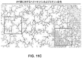
ここで、オルガノイド培養および単一細胞トランスクリプトーム研究を使用して、AEC2とAEC1との間の移行を包含する以前には未知であった別個の細胞状態が発見された。さらに、傷害-修復モデルとカップリングさせたマウス系列追跡は、in vivoでの類似の移行状態の存在を明らかにしている。この研究は、これらの移行状態を制御するシグナル伝達経路を明らかにする。本明細書で記載されるように、これらの移行状態は、DNA損傷応答を示し、AEC1に向かう途中に老化関連遺伝子を発現する。機構的に、遺伝的機能喪失、薬理学的機能獲得およびゲノム結合アッセイの使用は、TP53シグナル伝達によるPATSの直接的な転写制御を明らかにした。重要なことに、これらの移行状態は、進行性肺線維症を有するヒト肺における欠損した線維化病巣に関連する異常な上皮細胞と相関する。 Here, using organoid culture and single-cell transcriptome studies, a previously unknown distinct cellular state was discovered involving the transition between AEC2 and AEC1. Furthermore, mouse lineage tracing coupled with injury-repair models reveals the existence of similar transitional states in vivo. This study reveals the signaling pathways that control these transitional states. As described herein, these transitional states exhibit a DNA damage response and express senescence-associated genes on their way to AEC1. Mechanistically, the use of genetic loss-of-function, pharmacological gain-of-function and genomic binding assays revealed direct transcriptional regulation of PATS by TP53 signaling. Importantly, these transitional states correlate with abnormal epithelial cells associated with defective fibrotic foci in human lungs with advanced pulmonary fibrosis.
本開示の概要
本開示は、肺疾患の理解、薬物発見および/または薬物スクリーニングのための使用のためのモデルに役立つ移行細胞状態が肺胞再生において存在するという、本発明者らによる発見に一部基づく。
SUMMARY OF THE DISCLOSURE The present disclosure is consistent with the discovery by the inventors that a transitional cell state exists in alveolar regeneration that serves as a model for use in understanding lung disease, drug discovery and/or drug screening. Department based.
本開示の一態様は、肺傷害オルガノイドモデルを生成する方法であって、i)細胞の共培養物を、前記細胞の培養のために適合させた培養培地中に提供するステップであって、共培養物が、プレ1型肺胞移行細胞状態(PATS)細胞および肺胞線維芽細胞を含む、ステップ;ii)培養培地を、1種または複数の作用物質と接触させるステップ;ならびにiii)作用物質と接触されていない対照細胞培養培地と比較した、PATS細胞および/または肺胞線維芽細胞の少なくとも1つの発現マーカーに対する作用物質の生物学的影響を分析するステップ、を含む方法を提供する。 One aspect of the present disclosure is a method of generating a lung injury organoid model comprising: i) providing a co-culture of cells in a culture medium adapted for culturing said cells, comprising: ii) contacting the culture medium with one or more agents; and iii) acting analyzing the biological effect of the agent on at least one expression marker of PATS cells and/or alveolar fibroblasts compared to a control cell culture medium not contacted with the agent.
本開示の一部の実施形態では、PATS細胞は、罹患組織から単離される。本開示の一部の実施形態では、罹患組織は、慢性閉塞性肺疾患(COPD)肺組織、肺線維症肺組織、特発性肺線維症肺組織、肺気腫肺組織、肺がん組織、サルコイドーシス肺組織、間質性肺炎肺組織、敗血症肺組織、ウイルス感染および細菌感染を有する肺組織、急性呼吸促迫症候群肺組織、または気管支肺異形成症肺組織である。 In some embodiments of the present disclosure, PATS cells are isolated from diseased tissue. In some embodiments of the present disclosure, the diseased tissue is chronic obstructive pulmonary disease (COPD) lung tissue, pulmonary fibrosis lung tissue, idiopathic pulmonary fibrosis lung tissue, emphysema lung tissue, lung cancer tissue, sarcoidosis lung tissue, Interstitial pneumonia lung tissue, sepsis lung tissue, lung tissue with viral and bacterial infections, acute respiratory distress syndrome lung tissue, or bronchopulmonary dysplasia lung tissue.
本開示の一部の実施形態では、PATS細胞は、肺上皮細胞をin vivoまたはin vitroで傷害を引き起こす作用物質(例えば、ブレオマイシン、ジフテリア毒素(DT)、タモキシフェン、照射、ウイルス、細菌または真菌)に曝露させることによって生成される。 In some embodiments of the present disclosure, PATS cells are treated with agents that cause injury to lung epithelial cells in vivo or in vitro (e.g., bleomycin, diphtheria toxin (DT), tamoxifen, irradiation, viruses, bacteria or fungi). produced by exposure to
本開示の一部の実施形態では、肺胞線維芽細胞は、リポ線維芽細胞(lipofibroblast)である。 In some embodiments of the disclosure, the alveolar fibroblast is a lipofibroblast.
本開示の他の実施形態では、肺胞線維芽細胞は、疾患状態特徴を有する。本開示の一部の実施形態では、疾患状態特徴は、ACTA2を発現するリポ線維芽細胞、筋線維芽細胞の存在、および/または星状形状から帯状形状への細胞変化を含む。 In other embodiments of the disclosure, the alveolar fibroblasts have disease state characteristics. In some embodiments of the present disclosure, the disease state characteristic comprises the presence of ACTA2-expressing lipofibroblasts, myofibroblasts, and/or cellular changes from star-shaped to band-shaped.
本開示の一部の実施形態では、培養培地は、血小板由来増殖因子受容体(PDGFR)リガンドを含む。 In some embodiments of the disclosure, the culture medium comprises a platelet-derived growth factor receptor (PDGFR) ligand.
一部の実施形態では、培養培地は、PDGFR発現を維持またはモジュレートすることが可能な作用物質を含む。 In some embodiments, the culture medium comprises agents capable of maintaining or modulating PDGFR expression.
本開示の別の態様は、肺線維症のためのex vivoモデルを生成する方法であって、i)in vivoでアブレーションを受けたAEC1細胞の培養物を、前記細胞の培養のために適合させた培養培地中に提供するステップ;ii)培養培地を、1種または複数の作用物質と接触させるステップ;ならびにiii)作用物質と接触されていない対照細胞培養培地と比較した、AEC1細胞の少なくとも1つの発現マーカーに対する作用物質の生物学的影響を分析するステップ、を含む方法を提供する。 Another aspect of the present disclosure is a method of generating an ex vivo model for pulmonary fibrosis comprising: i) adapting a culture of in vivo ablated AEC1 cells for culturing said cells; ii) contacting the culture medium with one or more agents; and iii) at least one of AEC1 cells compared to a control cell culture medium not contacted with the agent analyzing the biological effects of the agent on the two expression markers.
一部の実施形態では、AEC1細胞は、少なくとも1ラウンドのアブレーションを受けている。他の実施形態では、AEC1細胞は、単独のまたは他の傷害モデルと組み合わせたアブレーションを受けている。一部の実施形態では、AEC1細胞は、例えば、約3日間~約2週間の過程にわたる反復アブレーションを受けている。 In some embodiments, the AEC1 cells have undergone at least one round of ablation. In other embodiments, AEC1 cells have undergone ablation alone or in combination with other models of injury. In some embodiments, the AEC1 cells have undergone repeated ablation over the course of, eg, about 3 days to about 2 weeks.
本開示の一部の実施形態では、AEC1細胞は、ブレオマイシン、ジフテリア毒素(DT)、アスベスト、化学的毒性作用物質、タモキシフェン、ウイルス、細菌または真菌に曝露されることによるアブレーションを受けている。 In some embodiments of the present disclosure, AEC1 cells are ablated by exposure to bleomycin, diphtheria toxin (DT), asbestos, chemotoxic agents, tamoxifen, viruses, bacteria or fungi.
一部の実施形態では、AEC1細胞は、aSMAおよび/またはACTA2を発現する。一部の実施形態では、AEC1細胞は、線維筋細胞(fibromyocyte cell)の特徴を示す。 In some embodiments, AEC1 cells express aSMA and/or ACTA2. In some embodiments, the AEC1 cells exhibit fibromyocyte cell characteristics.
本開示のさらに別の態様は、プレ1型肺胞移行細胞状態(PATS)細胞またはPATS様細胞を同定する方法であって、i)肺胞細胞を得るステップ;およびii)細胞を、1つまたは複数のマーカーについてスクリーニングするステップ、を含み、1つまたは複数のマーカーの存在が、PATS細胞またはPATS様細胞の存在を示す、方法を提供する。 Yet another aspect of the present disclosure is a method of identifying pre-type 1 alveolar transitional state (PATS) cells or PATS-like cells, comprising: i) obtaining alveolar cells; or screening for a plurality of markers, wherein the presence of one or more markers indicates the presence of PATS cells or PATS-like cells.
本開示の一部の実施形態では、肺胞細胞は、気管基底細胞、細気管支分泌細胞(クラブ細胞またはクララ細胞としても公知)、クラブバリアント細胞、肺胞上皮前駆(AEP)細胞、クララバリアント細胞、遠位肺前駆体(distal lung progenitor)、p63+Krt5-気道細胞、系列陰性上皮前駆体、気管支肺胞上皮幹細胞(BASC)、Sox9+p63+細胞、神経内分泌前駆細胞、遠位気道幹細胞、粘膜下腺管細胞(submucosal gland duct cell)、誘導多能性幹細胞由来肺幹細胞および2型肺胞上皮(AEC2)細胞からなる群より選択される。
In some embodiments of the present disclosure, alveolar cells are tracheal basal cells, bronchiolar secretory cells (also known as club cells or clara cells), club variant cells, alveolar epithelial progenitor (AEP) cells, clara variant cells , distal lung progenitor, p63+Krt5− airway cells, lineage-negative epithelial progenitors, bronchoalveolar epithelial stem cells (BASC), Sox9+p63+ cells, neuroendocrine progenitor cells, distal airway stem cells, submucosal ductal cells (submucosal ground duct cell), induced pluripotent stem cell-derived pulmonary stem cells and
本開示の一部の実施形態では、肺胞細胞は、慢性閉塞性肺疾患(COPD)、肺線維症、特発性肺線維症、肺気腫、肺がん、サルコイドーシス、間質性肺炎、敗血症、ウイルス感染、細菌感染、真菌感染、急性呼吸促迫症候群および/または気管支肺異形成症を患う患者の肺から得られる。 In some embodiments of the present disclosure, alveolar cells are used for chronic obstructive pulmonary disease (COPD), pulmonary fibrosis, idiopathic pulmonary fibrosis, emphysema, lung cancer, sarcoidosis, interstitial pneumonia, sepsis, viral infection, It is obtained from the lungs of patients suffering from bacterial infections, fungal infections, acute respiratory distress syndrome and/or bronchopulmonary dysplasia.
本開示の一部の実施形態では、プレ1型肺胞移行細胞状態(PATS)細胞またはPATS様細胞を同定する方法は、肺胞細胞を作用物質(例えば、ブレオマイシン、ジフテリア毒素(DT)、タモキシフェン、ウイルス、細菌または真菌)と接触させるステップをさらに含む。 In some embodiments of the present disclosure, a method of identifying pre-type 1 alveolar transitional cell state (PATS) cells or PATS-like cells comprises treating alveolar cells with an agent (e.g., bleomycin, diphtheria toxin (DT), tamoxifen). , virus, bacteria or fungi).
本開示のさらに別の態様は、in vivo組織線維症モデルを生成する方法であって、i)被験体においてPDGFリガンドおよび/もしくはPDGF受容体(PDGFR)シグナル伝達の遺伝的喪失を導入するステップ;ならびに/あるいはii)PDGF-PDGFRシグナル伝達をモジュレートすることができる作用物質を被験体に投与するステップ;ならびにiii)被験体由来の細胞を分析して、対照被験体と比較した、PDGFリガンドおよび/もしくはPDGF受容体(PDGFR)シグナル伝達の遺伝的喪失の生物学的影響ならびに/またはPDGF-PDGFRシグナル伝達をモジュレートすることができる作用物質の生物学的影響を決定するステップ、を含む方法を提供する。 Yet another aspect of the present disclosure is a method of generating an in vivo tissue fibrosis model comprising the steps of: i) introducing a genetic loss of PDGF ligand and/or PDGF receptor (PDGFR) signaling in a subject; and/or ii) administering to the subject an agent capable of modulating PDGF-PDGFR signaling; and iii) analyzing cells from the subject and comparing PDGF ligand and /or determining the biological effects of genetic loss of PDGF receptor (PDGFR) signaling and/or agents capable of modulating PDGF-PDGFR signaling. offer.
一部の実施形態では、PDGF-PDGFRシグナル伝達をモジュレートすることができる作用物質は、PDGFR阻害剤、PDGFRアゴニスト、PDGF阻害剤またはPDGFアゴニストを含む。 In some embodiments, agents capable of modulating PDGF-PDGFR signaling include PDGFR inhibitors, PDGFR agonists, PDGF inhibitors or PDGF agonists.
本開示のさらに別の態様は、in vivo組織線維症モデルを生成する方法であって、i)被験体においてRUNX1、RUNX2、RUNX3もしくはCBFbの遺伝的喪失を導入するステップ;ならびに/あるいはii)RUNX1阻害剤、RUNX2阻害剤、RUNX3阻害剤もしくはCBFb阻害剤を被験体に投与するステップ;ならびにiii)被験体由来の細胞を分析して、対照被験体と比較した、RUNX1、RUNX2、RUNX3もしくはCBFβの遺伝的喪失の生物学的影響および/またはRUNX1阻害剤、RUNX2阻害剤、RUNX3阻害剤もしくはCBFβ阻害剤の生物学的影響を決定するステップ、を含む方法を提供する。 Yet another aspect of the present disclosure is a method of generating an in vivo tissue fibrosis model comprising the steps of: i) introducing genetic loss of RUNX1, RUNX2, RUNX3 or CBFb in a subject; and/or ii) RUNX1 administering an inhibitor, RUNX2 inhibitor, RUNX3 inhibitor or CBFb inhibitor to the subject; Determining the biological impact of genetic loss and/or the biological impact of a RUNX1 inhibitor, a RUNX2 inhibitor, a RUNX3 inhibitor or a CBFβ inhibitor.
一部の実施形態では、阻害剤は、Ro 5-3335、AI-10-49、またはRUNX1、RUNX2、RUNX3および/もしくはCBFβに対する中和抗体である。 In some embodiments, the inhibitor is Ro 5-3335, AI-10-49, or a neutralizing antibody against RUNX1, RUNX2, RUNX3 and/or CBFβ.
一部の実施形態では、in vivoモデルは、マウスモデルである。 In some embodiments, the in vivo model is a mouse model.
一部の実施形態では、in vivoモデルは、肺、肝臓、腎臓、皮膚、腸または骨髄における線維症のための疾患モデルである。 In some embodiments, the in vivo model is a disease model for fibrosis in the lung, liver, kidney, skin, intestine or bone marrow.
詳細な説明
本開示の原理の理解を促進することを目的として、ここで、好ましい実施形態に対して参照がなされ、特定の言語がそれを記載するために使用される。それにもかかわらず、本開示の範囲の限定はそれによって意図されないことが理解され、本明細書で示される本開示のかかる変更およびさらなる改変は、本開示が関連する分野の当業者に通常想起されることが企図される。
DETAILED DESCRIPTION For the purposes of promoting an understanding of the principles of the disclosure, reference will now be made to preferred embodiments and specific language will be used to describe the same. It will nevertheless be understood that no limitation of the scope of the disclosure is thereby intended, and such and further modifications of the disclosure shown herein will generally occur to those skilled in the art to which this disclosure pertains. It is contemplated that
定義 definition
冠詞「1つの(a)」および「1つの(an)」は、冠詞の文法的対象のうち1つまたは1つよりも多く(即ち、少なくとも1つ)を指すために本明細書で使用される。例として、「1つの(an)要素」は、少なくとも1つの要素を意味し、1つよりも多くの要素を含み得る。 The articles "a" and "an" are used herein to refer to one or more than one (i.e., at least one) of the grammatical objects of the article. be. By way of example, "an element" means at least one element and may include more than one element.
「約」は、所望の結果に影響を与えることなしに所与の値が端点を「僅かに上回り」得るまたは「僅かに下回り」得ることを定めることによって、数的範囲の端点に柔軟性を提供するために使用される。 "About" allows flexibility in the endpoints of a numerical range by specifying that a given value can be "slightly above" or "slightly below" the endpoint without affecting the desired result. used to provide.
用語「含む(including)」、「含む(comprising)」または「有する(having)」、およびそれらの変形形態の本明細書での使用は、その後に列挙された要素およびそれらの等価物ならびにさらなる要素を包含する意味である。本明細書で使用される場合、「および/または」は、関連する列挙された項目のうち1つまたは複数の、任意のおよび全ての可能な組合せ、ならびに選択的に解釈される場合(「または」)には組合せの欠如を指し、それを包含する。 Use herein of the terms "including," "comprising," or "having," and variations thereof, applies to the elements listed thereafter and their equivalents as well as further elements. It means to include As used herein, "and/or" refers to any and all possible combinations of one or more of the associated listed items, and when interpreted alternatively ("or ”) refers to and includes the lack of combination.
本明細書で使用される場合、移行句「~から本質的になる」(および文法的異形)は、列挙された材料またはステップ、および特許請求された本発明の「基本的かつ新規な特徴(複数可)に実質的に影響を与えないもの」を包含すると解釈すべきである。したがって、用語「~から本質的になる」は、本明細書で使用される場合、「含む(comprising)」と等価であると解釈すべきではない。 As used herein, the transitional phrase “consisting essentially of” (and grammatical variants) refers to the recited material or step and the “fundamental and novel features (fundamental and novel features) of the claimed invention. It should be construed to include "that does not materially affect the Thus, the term "consisting essentially of", as used herein, should not be interpreted as equivalent to "comprising."
さらに、本開示は、一部の実施形態では、本明細書に示された任意の特色または特色の組合せが排除または除外され得ることもまた企図する。説明のために、本明細書が、複合体が構成要素A、BおよびCを含むと述べる場合、A、BもしくはCのいずれか、またはそれらの組合せが、単独でまたは任意の組合せで除外および権利放棄され得ることが、具体的に意図される。 Further, the present disclosure also contemplates that in some embodiments any feature or combination of features shown herein may be excluded or omitted. For purposes of illustration, when the specification states that a complex comprises components A, B and C, any of A, B or C, or combinations thereof, alone or in any combination, are excluded and It is specifically contemplated that waivers may be made.
本明細書の値の範囲の列挙は、本明細書で他に示されない限り、その範囲内に入る各別々の値に個々に言及する簡略化された方法として機能することのみを意図し、各別々の値は、それが本明細書で個々に列挙されたかのように、本明細書に組み込まれる。例えば、濃度範囲が1%~50%と述べられる場合、例えば、2%~40%、10%~30%または1%~3%などの値が、本明細書で明示的に列挙されている意図である。これらは、具体的に意図されているものの例に過ぎず、列挙された最低値と最高値との間でありかつそれらの値を含む数値の全ての可能な組合せが、本開示で明示的に述べられているとみなすべきである。 Recitation of ranges of values herein is intended only to serve as a shorthand method of referring individually to each separate value falling within the range, unless indicated otherwise herein; Individual values are incorporated herein as if they were individually recited herein. For example, when a concentration range is stated as 1% to 50%, values such as 2% to 40%, 10% to 30% or 1% to 3% are explicitly recited herein. is the intention. These are only examples of what is specifically contemplated, and all possible combinations of numerical values between and including the lowest and highest values recited are expressly included in this disclosure. should be considered as stated.
用語「疾患」は、本明細書で使用される場合、生物の一部に影響を与える構造または機能の任意の異常な状態および/または障害を含むがこれらに限定されない。これは、外部要因、例えば、感染性疾患によって、または内部機能不全、例えば、がん、がん転移などによって引き起こされ得る。 The term "disease" as used herein includes, but is not limited to, any abnormal condition and/or disorder of structure or function affecting a part of an organism. This can be caused by external factors, such as infectious diseases, or by internal dysfunctions, such as cancer, cancer metastasis, and the like.
用語「有効量」または「治療有効量」は、有益なまたは望ましい生物学的および/または臨床的結果をもたらすのに十分な量を指す。 The terms "effective amount" or "therapeutically effective amount" refer to an amount sufficient to effect beneficial or desired biological and/or clinical results.
本明細書で使用される場合、「処置」または「処置する」は、患者によって呈されるまたは患者がそれに対して感受性であり得る疾患、障害または病原体感染に応答して行われる臨床的介入を指す。処置の目的は、症状の軽減もしくは予防、進行の緩徐化もしくは停止、または疾患、障害、疾患の原因となる作用物質(例えば、細菌、ウイルス、化学療法剤、放射線)もしくは状態の悪化、および/あるいは疾患、障害または状態の緩解を含む。 As used herein, "treatment" or "treating" refers to a clinical intervention performed by a patient or in response to a disease, disorder or pathogenic infection to which the patient may be susceptible. Point. The purpose of treatment is to alleviate or prevent symptoms, slow or stop progression, or exacerbate a disease, disorder, disease-causing agent (e.g., bacteria, virus, chemotherapeutic agents, radiation) or condition, and/or Alternatively, including alleviation of a disease, disorder or condition.
他に定義しない限り、本明細書で使用される全ての技術用語は、本開示が属する分野の当業者によって一般に理解されるものと同じ意味を有する。 Unless defined otherwise, all technical terms used herein have the same meaning as commonly understood by one of ordinary skill in the art to which this disclosure belongs.
PATS PATS
幹細胞は、傷害に応答して動的な変化を受けて、失われた細胞を再生させる。しかし、移行状態の正体、およびそれらのトラジェクトリー(trajectory)を駆動する機構は、依然研究中である。肺オルガノイド、複数のin vivo修復モデル、単一細胞トランスクリプトミクスおよび系列追跡を使用して、本発明者らは、1型細胞への分化を受けている2型肺胞上皮細胞が、最終的な成熟化に向かう途中にプレ1型肺胞移行細胞状態(PATS)を獲得することを見出した。本発明者らは、移行細胞が、分化の間に広範な伸展を受けて、DNA損傷に対して脆弱になることをさらに見出した。本発明者らは、PATSにある細胞が、TP53、TGFβ、DNA損傷応答シグナル伝達の富化および細胞老化を示すことを決定した。実施例に記載されるように、機能獲得および機能喪失ならびにゲノム結合アッセイは、TP53シグナル伝達によるPATSの直接的な転写制御を明らかにした。本発明者らは、ヒト線維化肺におけるPATS様細胞の蓄積が観察されたこともまた見出しており、これは、線維症における移行状態の持続を示唆している。したがって、本明細書の実施例のセクションに記載される結果は、正常な上皮組織修復における老化に関連する一過的な状態および疾患状態におけるその異常な持続を暗示している。
Stem cells undergo dynamic changes in response to injury to regenerate lost cells. However, the identity of the transitional states and the mechanisms that drive their trajectories are still under investigation. Using lung organoids, multiple in vivo repair models, single-cell transcriptomics and lineage tracing, we found that
新規肺胞上皮I型細胞(肺細胞I型としても公知)アブレーションモデルを使用して、本発明者らは、肺線維症を発症する最初の遺伝的モデルを樹立した。さらに、本発明者らは、上記新規移行細胞状態(PATS)および肺胞線維芽細胞の共培養を使用するex vivo肺線維症モデルを開発した。これらの新規in vivoおよびex vivo線維症モデルは、拡張可能であり、疾患モデリングおよび薬物スクリーニングを可能にする。これらの特色は、進行性肺疾患において見出される場合が多く、ヒト線維化肺においてPATS様状態にある細胞の富化が観察され得、これは、線維症における移行状態の持続を示す。したがって、本明細書で提供される研究は、正常な上皮組織修復における老化に関連する一過的な状態および疾患状態におけるその異常な持続を暗示している。 Using a novel alveolar epithelial type I cell (also known as pulmonary cell type I) ablation model, we established the first genetic model to develop pulmonary fibrosis. In addition, we have developed an ex vivo pulmonary fibrosis model using the novel transitional cell state (PATS) and co-culture of alveolar fibroblasts. These novel in vivo and ex vivo fibrosis models are scalable and enable disease modeling and drug screening. These features are often found in progressive lung disease, and an enrichment of cells in a PATS-like state can be observed in human fibrotic lungs, indicating a persistence of a transitional state in fibrosis. Thus, the studies presented herein imply an aging-associated transient in normal epithelial tissue repair and its abnormal persistence in disease states.
本明細書で使用される場合、用語「オルガノイド」は、培養物中で成長させた幹細胞に由来する自己組織化された三次元(3D)構造または実体を指す。オルガノイド培養物は、臓器の複雑性を再現でき、または、例えば、ある特定の型の細胞のみを産生することによって、臓器の選択された状況を表現できる。あるいは、分化前のある特定の段階では、これらは、幹細胞のみから構成され得る。 As used herein, the term "organoid" refers to a self-assembled three-dimensional (3D) structure or entity derived from stem cells grown in culture. Organoid cultures can recapitulate the complexity of the organ or represent selected aspects of the organ, eg, by producing only certain types of cells. Alternatively, at a certain stage prior to differentiation, they may consist solely of stem cells.
幹細胞は、それ自身を複製(自己更新)しかつ他の細胞型を生じる能力を有する細胞である。幹細胞が分裂する場合、娘細胞は、幹細胞のままであり得るか、またはより特殊化した型の細胞になり得、または1つもしくは複数の特殊化した細胞型へと分化する他の娘を生じ得る。2つの型の哺乳動物幹細胞は、以下である:胚盤胞または着床前胚中に存在する未分化の細胞に由来する多能性胚性幹細胞、および成体の組織または臓器において見出される成体幹細胞。成体幹細胞は、組織または臓器の正常なターンオーバーまたは再生を維持することができ、損傷後に組織または臓器において細胞を修復および補充することができる。 Stem cells are cells that have the ability to replicate (self-renew) themselves and give rise to other cell types. When a stem cell divides, daughter cells may remain stem cells, may become more specialized types of cells, or give rise to other daughters that differentiate into one or more specialized cell types. obtain. Two types of mammalian stem cells are: pluripotent embryonic stem cells derived from undifferentiated cells present in the blastocyst or preimplantation embryo, and adult stem cells found in adult tissues or organs. . Adult stem cells can maintain normal turnover or regeneration of tissues or organs, and can repair and replenish cells in tissues or organs after injury.
本明細書で使用される場合、用語「幹細胞」は、増殖および自己更新が可能であり、1つもしくは複数の他の細胞型を生成する能力を有する前駆細胞を生じることが可能な未分化の細胞、または分化した細胞を生じ得る先駆体を指す。ある特定の場合には、娘細胞または前駆細胞もしくは先駆細胞は、分化した細胞を生じ得る。ある特定の場合には、娘細胞または前駆細胞もしくは先駆細胞は、それ自身で増殖し自己更新することができるだけでなく、1つまたは複数の成熟細胞型へと引き続いて分化する子孫を生じることができる。 As used herein, the term “stem cell” refers to an undifferentiated stem cell capable of proliferation and self-renewal and capable of giving rise to progenitor cells that have the potential to generate one or more other cell types. Refers to a cell or progenitor that can give rise to a differentiated cell. In certain cases, daughter cells or progenitor or pioneer cells may give rise to differentiated cells. In certain instances, daughter cells or progenitor or precursor cells can not only proliferate and self-renew themselves, but can also give rise to progeny that subsequently differentiate into one or more mature cell types. can.
前駆細胞は、自己更新することができるかまたは分化した細胞型へと分化することができるという点で幹細胞と類似した細胞を指すが、前駆細胞は既に、幹細胞よりも特殊化しているかまたは規定されている。 Progenitor cells refer to cells that are similar to stem cells in that they can self-renew or differentiate into differentiated cell types, although progenitor cells are already more specialized or defined than stem cells. ing.
本開示の幹細胞は、ヒト、マウス、ラット、ウサギ、イヌ、ブタ、ヒツジ、ヤギおよび非ヒト霊長類を含むがこれらに限定されない任意の動物に由来し得る。 Stem cells of the present disclosure can be derived from any animal including, but not limited to, humans, mice, rats, rabbits, dogs, pigs, sheep, goats, and non-human primates.
本開示の方法およびモデルにおいて使用され得る幹細胞は、正常な細胞(例えば、被験体の健康な組織由来の細胞)または異常な細胞(例えば、形質転換された細胞、樹立された細胞、または罹患組織試料に由来する細胞)であり得る。 Stem cells that can be used in the methods and models of the present disclosure can be normal cells (e.g., cells from healthy tissue of a subject) or abnormal cells (e.g., transformed cells, established cells, or diseased tissue cells derived from the sample).
一部の実施形態では、本開示の方法およびモデルにおいて使用される細胞は、肺幹細胞に由来し得る。肺幹細胞の分裂は、肺の構造の更新を促進し得る。肺幹細胞の例は、気管基底細胞、細気管支分泌細胞(クラブ細胞またはクララ細胞としても公知)、クラブバリアント細胞、肺胞上皮前駆(AEP)細胞、クララバリアント細胞、遠位肺前駆体、p63+Krt5-気道細胞、系列陰性上皮前駆体、気管支肺胞上皮幹細胞(BASC)、Sox9+p63+細胞、神経内分泌前駆細胞、遠位気道幹細胞、粘膜下腺管細胞、誘導多能性幹細胞由来肺幹細胞および2型肺胞上皮(本明細書でAEC2またはAT2と呼ばれる)細胞を含むがこれらに限定されない。
In some embodiments, cells used in the methods and models of the present disclosure may be derived from pulmonary stem cells. Dividing pulmonary stem cells can promote structural renewal of the lung. Examples of pulmonary stem cells include tracheal basal cells, bronchiolar secretory cells (also known as club cells or clara cells), club variant cells, alveolar epithelial progenitor (AEP) cells, clara variant cells, distal lung progenitors, p63+Krt5− airway cells, lineage-negative epithelial progenitors, bronchoalveolar epithelial stem cells (BASC), Sox9+p63+ cells, neuroendocrine progenitor cells, distal airway stem cells, submucosal ductal cells, induced pluripotent stem cell-derived pulmonary stem cells and
一部の実施形態では、本開示の方法およびモデルにおいて使用される細胞は、皮膚、乳腺、食道、膀胱、前立腺、卵巣および唾液腺を含む臓器由来の基底幹細胞に由来し得る。 In some embodiments, cells used in the methods and models of the present disclosure can be derived from basal stem cells from organs including skin, mammary gland, esophagus, bladder, prostate, ovary and salivary gland.
方法およびモデル Method and model
したがって、本開示の一態様は、肺胞再生における新たに同定された移行細胞状態(PATS)の同定を提供する。この移行細胞状態は、病理学的線維芽細胞(筋線維芽細胞)への肺線維芽細胞変換を誘導する。さらに、この移行細胞状態は、ヒト線維化(例えば、特発性肺線維症および間質性肺疾患)肺においても見出される。 Accordingly, one aspect of the present disclosure provides the identification of a newly identified transitional cell state (PATS) in alveolar regeneration. This transitional cell state induces pulmonary fibroblast conversion to pathological fibroblasts (myofibroblasts). In addition, this transitional cell state is also found in human fibrotic (eg, idiopathic pulmonary fibrosis and interstitial lung disease) lungs.
本開示の別の態様は、肺線維症、肺気腫(ヒト気腫合併肺線維症症候群(CPFE)において観察されるものと類似)をもたらす肺胞I型細胞(肺細胞I型としても公知)をアブレートするためのモデル系を提供する。 Another aspect of the disclosure is alveolar type I cells (also known as pulmonary cell type I) that cause pulmonary fibrosis, emphysema (similar to that observed in human emphysema-combined pulmonary fibrosis syndrome (CPFE)). Provide a model system for ablation.
一部の実施形態では、モデル系は、動物モデルを含む。一実施形態では、動物は、マウスを含む。一部の実施形態では、動物は、肺を有する任意の動物を含む。 In some embodiments, model systems include animal models. In one embodiment the animal comprises a mouse. In some embodiments, animals include any animal that has lungs.
本開示の別の態様は、肺傷害オルガノイドモデルを生成する方法であって、i)細胞の共培養物を、前記細胞の培養のために適合させた培養培地中に提供するステップであって、共培養物が、プレ1型肺胞移行細胞状態(PATS)細胞および肺胞線維芽細胞を含む、ステップ;ii)培養培地を、1種または複数の作用物質と接触させるステップ;iii)作用物質と接触されていない対照細胞培養培地と比較した、PATS細胞および/または肺胞線維芽細胞の少なくとも1つの発現マーカーに対する作用物質の生物学的影響を分析するステップ、を含む、それからなる、あるいはそれから本質的になる方法を提供する。 Another aspect of the present disclosure is a method of generating a lung injury organoid model comprising: i) providing a co-culture of cells in a culture medium adapted for culturing said cells; ii) contacting the culture medium with one or more agents; iii) acting analyzing the biological effect of the agent on at least one expression marker of PATS cells and/or alveolar fibroblasts compared to a control cell culture medium not contacted with the agent, or It then provides a way to become essential.
肺胞線維芽細胞は、肺における肺修復/線維症プロセスに関与する細胞である。肺胞線維芽細胞は、細胞外マトリックスおよびコラーゲンを合成することができ、動物組織において間質を産生することができる。肺胞線維芽細胞の例は、ヒト胎児肺線維芽細胞(例えば、HFLl、MRC-5、2BSおよびWI38)、チャイニーズハムスター肺線維芽細胞(V-97)、C57BL/6マウス初代肺線維芽細胞およびCC-2512細胞を含むがこれらに限定されない。 Alveolar fibroblasts are cells involved in lung repair/fibrosis processes in the lung. Alveolar fibroblasts can synthesize extracellular matrix and collagen and can produce stroma in animal tissues. Examples of alveolar fibroblasts are human fetal lung fibroblasts (e.g. HFLl, MRC-5, 2BS and WI38), Chinese hamster lung fibroblasts (V-97), C57BL/6 mouse primary lung fibroblasts and CC-2512 cells.
一部の実施形態では、肺胞線維芽細胞は、リポ線維芽細胞である。一部の実施形態では、肺胞線維芽細胞は、疾患状態特徴を有する。 In some embodiments, alveolar fibroblasts are lipofibroblasts. In some embodiments, the alveolar fibroblasts have disease state characteristics.
用語「疾患状態特徴」は、本明細書で使用される場合、その細胞が疾患細胞であることを示す、細胞の表現型特性または遺伝子型特性を指す。これらの特徴は、疾患状態を示す細胞におけるある特定のマーカーの発現または形態学的変化であり得る。一部の実施形態では、肺胞線維芽細胞の疾患状態特徴は、ACTA2を発現するリポ線維芽細胞、筋線維芽細胞の存在、および/または星状形状から帯状形状への細胞変化を含む。 The term "disease state characteristic" as used herein refers to a phenotypic or genotypic characteristic of a cell that indicates that the cell is a diseased cell. These characteristics can be the expression of certain markers or morphological changes in cells indicative of disease states. In some embodiments, the disease state characteristic of alveolar fibroblasts comprises the presence of ACTA2-expressing lipofibroblasts, myofibroblasts, and/or cellular changes from stellate to zonal shape.
一部の実施形態では、PATS細胞は、罹患組織から単離される。 In some embodiments, PATS cells are isolated from diseased tissue.
用語「罹患組織」は、健康な組織と比較して異常である、被験体(例えば、ヒトまたは動物モデル)の組織を指す。罹患組織は、生きたまたは死んだ被験体の組織から得ることができる。罹患組織の例は、慢性閉塞性肺疾患(COPD)肺組織、肺線維症肺組織、特発性肺線維症肺組織、肺気腫肺組織、肺がん組織、サルコイドーシス肺組織、間質性肺炎肺組織、敗血症肺組織、ウイルス感染および細菌感染を有する肺組織、急性呼吸促迫症候群肺組織、ならびに気管支肺異形成症肺組織を含むがこれらに限定されない。一部の実施形態では、PATS細胞は、肺線維症肺組織または特発性肺線維症肺組織から単離される。 The term "diseased tissue" refers to tissue of a subject (eg, a human or animal model) that is abnormal compared to healthy tissue. Diseased tissue can be obtained from the tissue of a living or dead subject. Examples of affected tissues include chronic obstructive pulmonary disease (COPD) lung tissue, pulmonary fibrosis lung tissue, idiopathic pulmonary fibrosis lung tissue, emphysema lung tissue, lung cancer tissue, sarcoidosis lung tissue, interstitial pneumonia lung tissue, sepsis. Including, but not limited to, lung tissue, lung tissue with viral and bacterial infections, acute respiratory distress syndrome lung tissue, and bronchopulmonary dysplasia lung tissue. In some embodiments, the PATS cells are isolated from pulmonary fibrosis lung tissue or idiopathic pulmonary fibrosis lung tissue.
肺がん細胞は、肺がんを患う被験体から単離され得る。肺がん細胞は、原発性肺腫瘍または二次肺腫瘍(例えば、別の組織において始まり、肺に転移するがん)から単離され得る。肺がん細胞の例は、小細胞癌、混合型小細胞癌、腺癌、扁平上皮癌、大細胞癌、パンコースト腫瘍細胞、神経内分泌腫瘍または肺カルチノイド腫瘍細胞を含むがこれらに限定されない小細胞肺がん細胞または非小細胞肺がん細胞を含むがこれらに限定されない。樹立された肺がん細胞系もまた、本開示の方法およびモデルにおいて使用され得る。本開示の方法およびモデルにおいて使用され得る肺がん細胞系は、ATCCウェブサイト上で見出すことができる。肺がん細胞系の例は、EML4-ALK Fusion-A549同質遺伝子細胞系、NCI-H838[H838]、HCC827、SK-LU-1、HCC2935、HCC4006、NCI-H1819[H1819]、NCI-H676B[H676B]、Hs 618.T、HBE4-E6/E7[NBE4-E6/E7]、NCI-H1666[H1666、H1666]、NCI-H23[H23]、NCI-H1435[H1435]、NCI-H1563[H1563]、703D4、およびNCI-H1688[H1688]、NCI-H187[H187]、NCI-H661[H661]、NCI-H460[H460]、NCI-H1299、NCI-H1155[H1155]、DMS 114、NCI-H69[H69]、DMS 79、DMS 53、SW 1271[SW1271、SW1271]、SHP-77、NCI-H209[H209]、NCI-H146[H146]、NCI-H345[H345]、NCI-H1341[H1341]、DMS 153、NCI-H82[H82]、NCI-H1048[H1048]、NCI-H128[H128]、NCI-H446[H446]、NCI-H128[H128]、NCI-H510A[H510A、NCI-H510]、H69AR.HLF-a、Hs 913T、GCT[巨細胞腫瘍]、SW 900[SW-900、SW900]、LL/2(LLC1)、HBE135-E6E7、Tera-2、NCI-H292[H292]、sNF02.2、NCI-H1703[H1703]、NCI-H2172[H2172]、NCI-H2444[H2444]、NCI-H2110[H2110]、NCI-H2135[H2135]、NCI-H2347[H2347]、NCI-H810[H810]、NCI-H1993[H1993]、およびNCI-H1792[H1792]を含むがこれらに限定されない。 Lung cancer cells can be isolated from a subject with lung cancer. Lung cancer cells can be isolated from a primary lung tumor or a secondary lung tumor (eg, cancer that begins in another tissue and metastasizes to the lung). Examples of lung cancer cells include, but are not limited to, small cell carcinoma, mixed small cell carcinoma, adenocarcinoma, squamous cell carcinoma, large cell carcinoma, Pancoast tumor cells, neuroendocrine tumors or lung carcinoid tumor cells. cells or non-small cell lung cancer cells. Established lung cancer cell lines can also be used in the methods and models of the present disclosure. Lung cancer cell lines that can be used in the disclosed methods and models can be found on the ATCC website. Examples of lung cancer cell lines are the EML4-ALK Fusion-A549 isogenic cell line, NCI-H838 [H838], HCC827, SK-LU-1, HCC2935, HCC4006, NCI-H1819 [H1819], NCI-H676B [H676B] , Hs 618. T, HBE4-E6/E7 [NBE4-E6/E7], NCI-H1666 [H1666, H1666], NCI-H23 [H23], NCI-H1435 [H1435], NCI-H1563 [H1563], 703D4, and NCI- H1688 [H1688], NCI-H187 [H187], NCI-H661 [H661], NCI-H460 [H460], NCI-H1299, NCI-H1155 [H1155], DMS 114, NCI-H69 [H69], DMS 79, DMS 53, SW 1271 [SW1271, SW1271], SHP-77, NCI-H209 [H209], NCI-H146 [H146], NCI-H345 [H345], NCI-H1341 [H1341], DMS 153, NCI-H82 [ H82], NCI-H1048 [H1048], NCI-H128 [H128], NCI-H446 [H446], NCI-H128 [H128], NCI-H510A [H510A, NCI-H510], H69AR. HLF-a, Hs 913T, GCT [giant cell tumor], SW 900 [SW-900, SW900], LL/2 (LLC1), HBE135-E6E7, Tera-2, NCI-H292 [H292], sNF02.2, NCI-H1703 [H1703], NCI-H2172 [H2172], NCI-H2444 [H2444], NCI-H2110 [H2110], NCI-H2135 [H2135], NCI-H2347 [H2347], NCI-H810 [H810], NCI -H1993 [H1993], and NCI-H1792 [H1792].
一部の実施形態では、PATS細胞は、健康な組織(例えば、正常な肺組織)から単離される。 In some embodiments, PATS cells are isolated from healthy tissue (eg, normal lung tissue).
用語「組織」は、本明細書で使用される場合、組織が得られる被験体の構造材料を形成する、細胞間物質と一緒になった、特定の種類のものであり得る細胞の凝集体を指す。本開示の一部の実施形態では、PATS細胞は、組織から単離および精製され得る。 The term "tissue" as used herein refers to an aggregate of cells, which may be of a particular type, together with intercellular material that form the structural material of the subject from which the tissue is obtained. Point. In some embodiments of the present disclosure, PATS cells may be isolated and purified from tissue.
本開示の一部の実施形態では、PATS細胞は、肺上皮細胞(例えば、AEC2またはAEC1)をin vivoまたはin vitroで傷害を引き起こす作用物質に曝露させることによって生成され得る。 In some embodiments of the present disclosure, PATS cells may be generated by exposing lung epithelial cells (eg, AEC2 or AEC1) to agents that cause injury in vivo or in vitro.
用語「傷害を引き起こす作用物質」は、本明細書で使用される場合、健康な細胞を罹患細胞状態に変換または移行させることが可能な作用物質を指す。用語「傷害を引き起こす作用物質」は、本明細書で使用される場合、疾患状態細胞が生じるように正常な細胞をアブレートすることが可能な作用物質もまた指す。 The term "injury-causing agent" as used herein refers to an agent capable of transforming or transitioning a healthy cell to a diseased cell state. The term "damaging agent" as used herein also refers to an agent capable of ablating normal cells to produce disease state cells.
傷害を引き起こす作用物質は、例えば、化学療法剤(例えば、ブレオマイシン、タモキシフェン、アクチノマイシン-D、マイトマイシン、タキサンおよびゲムシタビン)、外毒素(例えば、ジフテリア毒素(DT))、ウイルス、細菌、真菌、アスベスト、化学的毒性作用物質または照射であり得る。 Injury-causing agents include, for example, chemotherapeutic agents (such as bleomycin, tamoxifen, actinomycin-D, mitomycin, taxanes and gemcitabine), exotoxins (such as diphtheria toxin (DT)), viruses, bacteria, fungi, asbestos , chemical toxic agents or irradiation.
一部の実施形態では、傷害を引き起こす作用物質は、ヒトまたは肺を有する任意の動物の肺組織に感染することが可能な病原体(例えば、細菌、ウイルスまたは真菌)であり得る。 In some embodiments, the agent causing injury may be a pathogen (eg, bacteria, virus or fungus) capable of infecting lung tissue of humans or any animal having lungs.
肺に感染することができる細菌は、Bordetella pertussis、Streptococcus pneumonia、Haemophilus influenza、Staphylococcusaureus、Moraxellacatarrhalis、Streptococcuspyogenes、Pseudomonas aeruginosa、Neisseriameningitidis、Klebsiellapneumoniaeを含むがこれらに限定されない。 肺に感染することができる細菌は、Bordetella pertussis、Streptococcus pneumonia、Haemophilus influenza、Staphylococcusaureus、Moraxellacatarrhalis、Streptococcuspyogenes、Pseudomonas aeruginosa、Neisseriameningitidis、Klebsiellapneumoniaeを含むがこれらに限定されない。
肺に感染することができるウイルスは、229E(アルファコロナウイルス)、NL63(アルファコロナウイルス)、OC43(ベータコロナウイルス)、HKU1(ベータコロナウイルス)、MERS-CoV(中東呼吸器症候群、即ちMERSを引き起こすベータコロナウイルス)、SARS-CoV(重症急性呼吸器症候群、即ちSARSを引き起こすベータコロナウイルス)もしくはSARS-CoV-2(コロナウイルス疾患2019、即ちCOVID-19を引き起こす新型コロナウイルス)、インフルエンザ-Aウイルス(例えば、H1N1、H7N9、低病原性鳥インフルエンザ、高病原性鳥インフルエンザまたはH5N1)、インフルエンザ-Bウイルス、呼吸器多核体ウイルス(RSV)、またはエンテロウイルス(例えば、エンテロウイルス71)を含むがこれらに限定されない。一部の実施形態では、ウイルスは、SARS-CoV-2である。 Viruses that can infect the lungs include 229E (alphacoronavirus), NL63 (alphacoronavirus), OC43 (betacoronavirus), HKU1 (betacoronavirus), MERS-CoV (Middle East Respiratory Syndrome, or MERS). SARS-CoV (betacoronavirus that causes severe acute respiratory syndrome, i.e. SARS) or SARS-CoV-2 (novel coronavirus that causes coronavirus disease 2019, i.e. COVID-19), influenza-A viruses (e.g. H1N1, H7N9, low pathogenic avian influenza, highly pathogenic avian influenza or H5N1), influenza-B virus, respiratory syncytial virus (RSV), or enteroviruses (e.g. enterovirus 71) Not limited. In some embodiments, the virus is SARS-CoV-2.
肺に感染することができる真菌は、Aspergillosisを含むがこれらに限定されない。 Fungi that can infect the lungs include, but are not limited to, Aspergillosis.
一部の実施形態では、本開示の方法およびモデルにおいて使用される培養培地および/または被験体(例えば、マウス)は、血小板由来増殖因子受容体(例えば、血小板由来増殖因子受容体-A、血小板由来増殖因子受容体-Bまたは血小板由来増殖因子受容体-AB)を含む。 In some embodiments, the culture medium and/or subject (eg, a mouse) used in the methods and models of the present disclosure is a platelet-derived growth factor receptor (eg, platelet-derived growth factor receptor-A, platelet platelet-derived growth factor receptor-B or platelet-derived growth factor receptor-AB).
一部の実施形態では、本開示の方法およびモデルにおいて使用される培養培地および/または被験体(例えば、マウス)は、血小板由来増殖因子受容体(PDGFR)リガンドを含む。一部の実施形態では、PDGFRリガンドは、血小板由来増殖因子(PDGF)である。PDGFは、細胞の成長および分裂を調節することができる増殖因子である。一部の実施形態では、PDGFRリガンドは、血小板由来増殖因子サブユニットA(PDGF-A)、血小板由来増殖因子サブユニットB(PDGF-B)、血小板由来増殖因子サブユニットC(PDGF-C)、血小板由来増殖因子サブユニットD(PDGF-D)および/または血小板由来増殖因子サブユニットAB(PDGF-AB)である。PDGFファミリーに属するタンパク質をコードするヒト遺伝子は、FIGF、PDGFA、PDGFB、PDGFC、PDGFd、PGF、VEGF、VEGF41、VEGFBおよびVEGFCを含む。 In some embodiments, the culture medium and/or subject (eg, mouse) used in the disclosed methods and models comprises a platelet-derived growth factor receptor (PDGFR) ligand. In some embodiments, the PDGFR ligand is platelet-derived growth factor (PDGF). PDGF is a growth factor that can regulate cell growth and division. In some embodiments, the PDGFR ligand is platelet derived growth factor subunit A (PDGF-A), platelet derived growth factor subunit B (PDGF-B), platelet derived growth factor subunit C (PDGF-C), platelet-derived growth factor subunit D (PDGF-D) and/or platelet-derived growth factor subunit AB (PDGF-AB). Human genes encoding proteins belonging to the PDGF family include FIGF, PDGFA, PDGFB, PDGFC, PDGFd, PGF, VEGF, VEGF41, VEGFB and VEGFC.
一部の実施形態では、本開示の方法およびモデルは、PDGFシグナル伝達ペプチド、PDGF受容体、PDGFアゴニスト、PDGFRアゴニスト、PDGFアンタゴニスト、PDGFRアンタゴニスト、またはPDGF-PDGFRシグナル伝達経路をモジュレートすることが可能な任意の作用物質の使用を含む。他の実施形態では、本開示の方法およびモデルは、PDGFリガンドおよび/またはPDGFR受容体の薬理学的遮断の使用を含む。 In some embodiments, the disclosed methods and models are capable of modulating PDGF signaling peptides, PDGF receptors, PDGF agonists, PDGFR agonists, PDGF antagonists, PDGFR antagonists, or PDGF-PDGFR signaling pathways. including the use of any agent. In other embodiments, the methods and models of the present disclosure involve the use of pharmacological blockade of PDGF ligands and/or PDGFR receptors.
PDGF-PDGFRシグナル伝達経路のモジュレーターは、基質、阻害剤、活性化因子、神経伝達物質、アゴニスト、アンタゴニスト、インバースアゴニスト、インバースアンタゴニスト、部分アゴニストおよび部分アンタゴニストを含む。PDGF-PDGFRシグナル伝達経路のモジュレーターは、PDGFアイソフォームアンタゴニスト、PDGFR活性化遮断剤、細胞におけるPDGFおよび/またはPDGFRの発現を増加または減少させることが可能な作用物質、化学化合物、例えば、内因性代謝物、非内因性代謝物、合成化学化合物、ポリペプチド、アミノ酸残基、核酸、siRNA、ならびに抗体(例えば、中和抗体)を含むがこれらに限定されない。 Modulators of the PDGF-PDGFR signaling pathway include substrates, inhibitors, activators, neurotransmitters, agonists, antagonists, inverse agonists, inverse antagonists, partial agonists and partial antagonists. Modulators of the PDGF-PDGFR signaling pathway include PDGF isoform antagonists, PDGFR activation blockers, agents capable of increasing or decreasing the expression of PDGF and/or PDGFR in cells, chemical compounds such as endogenous metabolic non-endogenous metabolites, synthetic chemical compounds, polypeptides, amino acid residues, nucleic acids, siRNA, and antibodies (eg, neutralizing antibodies).
PDGF-PDGFRシグナル伝達経路のモジュレーターの例は、フィブロネクチンペプチド、Ras-GAP、チロシンホスファターゼ(例えば、PTP1B、TC-PTP、PTPRJ/DEP-1)、SHP-2、インテグリン、低密度リポタンパク質受容体関連タンパク質、ヒアルロナン受容体CD44、ソラフェニブ、ニロチニブ、パゾパニブ、レゴラフェニブ、AG1295およびAG1296、セレコキシブ、エトリコキシブおよびDFU、シクロオキシゲナーゼ-2(COX-2)阻害剤(コキシブ)、パゾパニブHCl、パゾパニブ、ラドチニブ(radotinib)、AZD2932、トセラニブリン酸塩、TAK-593、クレノラニブ、トセラニブ、SU14813、SU14813マレイン酸塩、オランチニブ(orantinib)、SU 4312、スニチニブリンゴ酸塩、リニファニブ、KG 5、N-デセチルスニチニブ、CP-673451、VEGFR2キナーゼ阻害剤II、VEGFR2キナーゼ阻害剤II、AC710メシル酸塩、AC710、DMPQ二塩酸塩、リプレチニブ、レンバチニブ、ニンテダニブ、レゴラフェニブ、スニチニブ、ポナチニブ(pnatibib)、スニチニブ(sunitnib)リンゴ酸塩、TG 100572、イロラセルチブ(ilorasertib)、イマチニブ、アキシチニブ、イマチニブメシル酸塩、パゾパニブ、クレノラニブ、SU 5402、アバプリチニブ、リプレチニブ、セジラニブ、CP 673451、ドビチニブ、TAK 593、トセラニブ、PDGFRaキナーゼ阻害剤-1、SU16f、PDGFRαを標的化するアゴニスト性ヒトモノクローナル自己抗体、およびPDGFRαを標的化する非アゴニスト性ヒトモノクローナル自己抗体、ならびにまたはそれらの誘導体を含むがこれらに限定されない。 Examples of modulators of the PDGF-PDGFR signaling pathway include fibronectin peptides, Ras-GAP, tyrosine phosphatases (e.g. PTP1B, TC-PTP, PTPRJ/DEP-1), SHP-2, integrins, low density lipoprotein receptor related Proteins, Hyaluronan Receptor CD44, Sorafenib, Nilotinib, Pazopanib, Regorafenib, AG1295 and AG1296, Celecoxib, Etoricoxib and DFU, Cyclooxygenase-2 (COX-2) Inhibitors (COX-2), Pazopanib HCl, Pazopanib, Radotinib, AZD2932 , tocerani phosphate, TAK-593, clenolanib, toceranib, SU14813, SU14813 maleate, orantinib, SU 4312, sunitinib malate, linifanib, KG 5, N-decetylsunitinib, CP-673451, VEGFR2 kinase Inhibitor II, VEGFR2 Kinase Inhibitor II, AC710 mesylate, AC710, DMPQ dihydrochloride, ripretinib, lenvatinib, nintedanib, regorafenib, sunitinib, ponatinib (pnatibib), sunitnib malate, TG 100572, ilorasertib ( ilorasertib), imatinib, axitinib, imatinib mesylate, pazopanib, crenolanib, SU 5402, avapritinib, lipretinib, cediranib, CP 673451, dovitinib, TAK 593, toceranib, PDGFRa kinase inhibitor-1, SU16f, agonist targeting PDGFRα and non-agonistic human monoclonal autoantibodies that target PDGFRα, and or derivatives thereof.
本明細書で使用される場合、用語「アゴニスト」は、受容体(例えば、PDGFR)またはタンパク質(例えば、PDGF)に結合し、その受容体またはタンパク質を活性化して生物学的応答を生じさせるモジュレーターを指す。 As used herein, the term "agonist" is a modulator that binds to a receptor (e.g., PDGFR) or protein (e.g., PDGF) and activates that receptor or protein to produce a biological response. point to
本明細書で使用される場合、用語「アンタゴニスト」は、受容体またはタンパク質に結合し、それを活性化するのではなく遮断することによって、生物学的応答を遮断する、妨げるまたは弱める、モジュレーターまたはリガンドを指す。 As used herein, the term "antagonist" blocks, prevents or attenuates a biological response by binding to a receptor or protein and blocking rather than activating it, modulator or refers to the ligand.
本明細書で使用される場合、用語「インバースアゴニスト」は、アゴニストと同じ受容体に結合するが、そのアゴニストと反対の生物学的応答を誘導する、モジュレーターまたはリガンドを指す。 As used herein, the term "inverse agonist" refers to a modulator or ligand that binds to the same receptor as an agonist, but induces a biological response opposite that agonist.
本明細書で使用される場合、用語「部分アンタゴニスト」は、受容体に結合できるがその受容体の効果を完全には遮断せず、むしろ、受容体の最大潜在力を減少させる、モジュレーターまたはリガンドを指す。 As used herein, the term "partial antagonist" refers to a modulator or ligand capable of binding to a receptor but not completely blocking the effect of that receptor, but rather reducing the maximal potency of the receptor. point to
一部の実施形態では、方法およびモデルにおいて使用される培養培地は、細胞におけるPDGFR(例えば、PDGFR-A、PDGFR-BまたはPDGFR-AB)発現を維持することが可能な作用物質を含む。一部の実施形態では、細胞におけるPDGFR-A発現を維持することが可能な作用物質は、Ro 5-3335またはAI-10-49である。 In some embodiments, the culture medium used in the methods and models comprises agents capable of maintaining PDGFR (eg, PDGFR-A, PDGFR-B or PDGFR-AB) expression in cells. In some embodiments, the agent capable of maintaining PDGFR-A expression in cells is Ro 5-3335 or AI-10-49.
一部の実施形態では、細胞におけるPDGFRA発現を維持することが可能な作用物質は、RUNX(例えば、RUNX1、RUNX2またはRUNX3)またはコア結合因子(CBF)関連ファミリーのタンパク質(例えば、CBF-β)をモジュレートすることができる任意の分子である。 In some embodiments, the agent capable of maintaining PDGFRA expression in cells is RUNX (eg, RUNX1, RUNX2 or RUNX3) or core binding factor (CBF)-related family proteins (eg, CBF-β) is any molecule that can modulate
RUNXまたはCBF関連タンパク質を調節する分子の例は、RUNXおよび/またはCBFの基質、阻害剤、活性化因子、神経伝達物質、アゴニスト、アンタゴニスト、インバースアゴニスト、インバースアンタゴニスト、部分アゴニストおよび部分アンタゴニストであり得る。 Examples of molecules that modulate RUNX or CBF-related proteins can be RUNX and/or CBF substrates, inhibitors, activators, neurotransmitters, agonists, antagonists, inverse agonists, inverse antagonists, partial agonists and partial antagonists. .
RUNXおよび/またはCBFをモジュレートする分子は、RUNXとCBFとの間のタンパク質-タンパク質相互作用を妨害する分子であり得る。これらのモジュレーターは、RUNX(例えば、RUNX1、RUNX2またはRUNX3)の阻害剤またはCBF-βの阻害剤であり得る。RUNXまたはCBFモジュレーターの例は、Ro 5-3335、AI-10-49、CADD522、ムラミルジペプチド、L-ケブラキトール(quebrachitol)、L-アスコルビン酸2-リン酸、アスコルビン酸2-リン酸マグネシウム、2-ピリジルベンズイミダゾールAI-4-57、RUNX1/MTG8融合タンパク質、ならびにRUNX1、RUNX2、RUNX3および/もしくはCBFbに対する中和抗体、ならびに/またはそれらの誘導体を含むがこれらに限定されない。 A molecule that modulates RUNX and/or CBF can be a molecule that interferes with the protein-protein interaction between RUNX and CBF. These modulators can be inhibitors of RUNX (eg, RUNX1, RUNX2 or RUNX3) or inhibitors of CBF-β. Examples of RUNX or CBF modulators are Ro 5-3335, AI-10-49, CADD522, muramyl dipeptide, L-quebrachitol, L-ascorbic acid 2-phosphate, magnesium ascorbic acid 2-phosphate, 2 - pyridylbenzimidazole AI-4-57, RUNX1/MTG8 fusion protein, and neutralizing antibodies against RUNX1, RUNX2, RUNX3 and/or CBFb, and/or derivatives thereof, including but not limited to.
「作用物質」は、本明細書で使用される場合、小分子、タンパク質、ペプチド、遺伝子、化合物、または他の薬学的に活性な成分を指す。一部の実施形態では、作用物質は、疾患(例えば、肺線維症)の処置、予防または緩和のために使用され得る。 "Agent," as used herein, refers to a small molecule, protein, peptide, gene, compound, or other pharmaceutically active ingredient. In some embodiments, agents may be used for the treatment, prevention or amelioration of diseases such as pulmonary fibrosis.
PATS細胞発現マーカーは、PATS細胞またはPATS様細胞上で発現される遺伝子転写物を指す。一部の実施形態では、PATSマーカーは、PATS細胞状態に独自であり、関連する細胞状態(例えば、AEC2またはAEC1)上では典型的には発現されない。PATS細胞マーカーは、CLDN4、KRT19、SFN、LGALS3、SOX4、S100A2、PTGS2、KRT17、KRT8、CALS1、MMP7、PRSS2、IGFBP7、COL1A1、MDK、TAGLN、GDF15、TM4SF1 TP63および/またはCTSEを含み得る。 PATS cell expression markers refer to gene transcripts that are expressed on PATS cells or PATS-like cells. In some embodiments, the PATS marker is unique to the PATS cell state and is typically not expressed on related cell states (eg, AEC2 or AEC1). PATS cell markers may include CLDN4, KRT19, SFN, LGALS3, SOX4, S100A2, PTGS2, KRT17, KRT8, CALS1, MMP7, PRSS2, IGFBP7, COL1A1, MDK, TAGLN, GDF15, TM4SF1 TP63 and/or CTSE.
一部の実施形態では、PATSマーカーは、共発現される2つまたはそれよりも多くのマーカーのマーカーシグネチャーを含み得る。一部の実施形態では、PATSマーカーシグネチャーは、CLDN4、LGALS3およびLGALS3である。一部の実施形態では、PATSマーカーシグネチャーは、CLDN4、KRT19およびSFNである。一部の実施形態では、PATSマーカーシグネチャーは、CALD1、PRSS2、MMP7およびS100A2である。 In some embodiments, a PATS marker may comprise a marker signature of two or more markers that are co-expressed. In some embodiments, the PATS marker signatures are CLDN4, LGALS3 and LGALS3. In some embodiments, the PATS marker signatures are CLDN4, KRT19 and SFN. In some embodiments, the PATS marker signature is CALD1, PRSS2, MMP7 and S100A2.
一部の実施形態では、PATSマーカーシグネチャーは、TP63、KRT17およびCOL1A1である。 In some embodiments, the PATS marker signature is TP63, KRT17 and COL1A1.
一部の実施形態では、PATSマーカーシグネチャーは、AREG、TGFB1、TGFB2およびTIMP1である。 In some embodiments, the PATS marker signatures are AREG, TGFB1, TGFB2 and TIMP1.
肺胞線維芽細胞発現マーカーは、肺胞線維芽細胞上で発現される遺伝子転写物を指す。肺胞線維芽細胞は、Pdgfraを含み得る。 Alveolar fibroblast expression markers refer to gene transcripts that are expressed on alveolar fibroblasts. Alveolar fibroblasts may contain Pdgfra.
本開示の別の態様は、肺線維症のためのex vivoモデルを生成する方法であって、i)in vivoでアブレーションを受けた肺胞細胞(例えば、AEC1細胞)の培養物を、前記細胞の培養のために適合させた培養培地中に提供するステップ;ii)培養培地を、1種または複数の作用物質と接触させるステップ;iii)作用物質と接触されていない対照細胞培養培地と比較した、肺胞細胞(例えば、AEC1細胞)の少なくとも1つの発現マーカーに対する作用物質の生物学的影響を分析するステップ、を含む方法を提供する。 Another aspect of the disclosure is a method of generating an ex vivo model for pulmonary fibrosis, comprising: i) forming a culture of in vivo ablated alveolar cells (e.g., AEC1 cells); ii) contacting the culture medium with one or more agents; iii) comparing to a control cell culture medium not contacted with the agent , analyzing the biological effect of the agent on at least one expression marker of alveolar cells (eg, AEC1 cells).
用語「アブレーション」は、本明細書で使用される場合、組織もしくは細胞、またはそれらの機能の除去または破壊を指す。アブレーションは、手術、ホルモン、薬物、高周波、熱、または組織もしくは細胞の破壊を引き起こす他の方法によって引き起こされ得る。 The term "ablation" as used herein refers to the removal or destruction of tissue or cells or their functions. Ablation can be caused by surgery, hormones, drugs, radio frequency, heat, or other methods that cause destruction of tissue or cells.
一部の実施形態では、肺胞細胞(例えば、AEC1細胞)は、少なくとも1ラウンドのアブレーション(例えば、少なくとも1、2、3、4、5、6、7、8、9または10ラウンドのアブレーション)を受けている。一部の実施形態では、肺胞細胞(例えば、AEC1細胞)は、1ラウンドと5ラウンドとの間のアブレーションを受けている。一部の実施形態では、細胞は、単独のまたは本明細書で記載される他の傷害モデルと組み合わせたアブレーションを受けている。 In some embodiments, the alveolar cells (eg, AEC1 cells) have been subjected to at least one round of ablation (eg, at least 1, 2, 3, 4, 5, 6, 7, 8, 9, or 10 rounds of ablation). Is receiving. In some embodiments, alveolar cells (eg, AEC1 cells) have undergone between 1 and 5 rounds of ablation. In some embodiments, cells have undergone ablation alone or in combination with other injury models described herein.
一部の実施形態では、肺胞細胞(例えば、AEC1細胞)は、反復アブレーションを受けている。一部の実施形態では、反復アブレーションは、約3日間~約2週間の過程にわたって行われ得る。一部の実施形態では、反復アブレーションは、約3日間~約1年間の過程にわたって行われ得る。 In some embodiments, alveolar cells (eg, AEC1 cells) have undergone repeated ablation. In some embodiments, repeated ablation can be performed over the course of about 3 days to about 2 weeks. In some embodiments, repeated ablation can be performed over the course of about 3 days to about 1 year.
一部の実施形態では、肺胞細胞(例えば、AEC1細胞)は、傷害を引き起こす作用物質に曝露されることによるアブレーションを受けている。一部の実施形態では、傷害を引き起こす作用物質は、ブレオマイシン、ジフテリア毒素(DT)、タモキシフェン、ウイルス、細菌または真菌である。 In some embodiments, alveolar cells (eg, AEC1 cells) are ablated by exposure to an agent that causes injury. In some embodiments, the injury-causing agent is bleomycin, diphtheria toxin (DT), tamoxifen, virus, bacteria or fungus.
一部の実施形態では、アブレートされた細胞は、aSMAおよび/またはACTA2を発現する。一部の実施形態では、アブレートされた細胞は、線維筋細胞の特徴を示す。 In some embodiments, the ablated cells express aSMA and/or ACTA2. In some embodiments, the ablated cells exhibit fibromyocyte characteristics.
本開示のさらに別の態様は、プレ1型肺胞移行細胞状態(PATS)細胞を同定する方法であって、i)肺細胞を得るステップ;およびii)細胞を、1つまたは複数のマーカーについてスクリーニングするステップ、を含み、1つまたは複数のマーカーの存在が、PATS細胞の存在を示す、方法を提供する。
Yet another aspect of the present disclosure is a method of identifying
一部の実施形態では、肺細胞は、気管基底細胞、細気管支分泌細胞(クラブ細胞またはクララ細胞としても公知)、クラブバリアント細胞、肺胞上皮前駆(AEP)細胞、クララバリアント細胞、遠位肺前駆体、p63+Krt5-気道細胞、系列陰性上皮前駆体、気管支肺胞上皮幹細胞(BASC)、Sox9+p63+細胞、神経内分泌前駆細胞、遠位気道幹細胞、粘膜下腺管細胞、誘導多能性幹細胞由来肺幹細胞および2型肺胞上皮(AEC2)細胞からなる群より選択され得る。
In some embodiments, the lung cells are tracheal basal cells, bronchiolar secretory cells (also known as club cells or clara cells), club variant cells, alveolar epithelial progenitor (AEP) cells, clara variant cells, distal lung Progenitors, p63+Krt5- airway cells, lineage-negative epithelial progenitors, bronchoalveolar epithelial stem cells (BASC), Sox9+p63+ cells, neuroendocrine progenitor cells, distal airway stem cells, submucosal ductal cells, induced pluripotent stem cell-derived pulmonary stem cells and
一部の実施形態では、肺細胞は、AEC2細胞、AEC1細胞または肺胞マクロファージからなる群より選択され得る。 In some embodiments, lung cells may be selected from the group consisting of AEC2 cells, AEC1 cells or alveolar macrophages.
一部の実施形態では、肺細胞は、慢性閉塞性肺疾患(COPD)、肺線維症、特発性肺線維症、肺気腫、肺がん、サルコイドーシス、間質性肺炎、敗血症、ウイルス感染、細菌感染、真菌感染、急性呼吸促迫症候群および/または気管支肺異形成症を患う患者の肺から得られる。 In some embodiments, pulmonary cells are used for chronic obstructive pulmonary disease (COPD), pulmonary fibrosis, idiopathic pulmonary fibrosis, emphysema, lung cancer, sarcoidosis, interstitial pneumonia, sepsis, viral infections, bacterial infections, fungi It is obtained from the lungs of patients with infections, acute respiratory distress syndrome and/or bronchopulmonary dysplasia.
一部の実施形態では、プレ1型肺胞移行細胞状態(PATS)細胞を同定する方法は、肺胞細胞を傷害を引き起こす作用物質と接触させるステップをさらに含む。一部の実施形態では、作用物質は、ブレオマイシン、ジフテリア毒素(DT)、タモキシフェン、ウイルス、細菌または真菌である。
In some embodiments, the method of identifying
本開示の別の態様は、拡張可能であり、疾患モデリングおよび薬物スクリーニングを可能にする、上記新規移行細胞状態(PATS)および肺胞線維芽細胞の共培養を使用するex vivo肺線維症モデルを提供する。 Another aspect of the present disclosure is an ex vivo pulmonary fibrosis model using co-culture of the novel transitional cell state (PATS) and alveolar fibroblasts, which is scalable and allows for disease modeling and drug screening. offer.
本開示の別の態様は、肺および他の組織において線維症を生成するin vivoモデルを生成する方法を提供する。本開示のin vitroおよびex vivoモデルと同様、in vivoモデルは、例えば、治療剤について疾患を処置または予防する能力を試験するために使用され得る。本開示のin vivoモデルは、種々の動物被験体(例えば、マウス、ラット、ウサギ、サルもしくはブタ、または線維症をモデリングすることが可能な任意の動物)を利用し得る。 Another aspect of the present disclosure provides methods of generating in vivo models of producing fibrosis in lung and other tissues. In vivo models, as well as the in vitro and ex vivo models of this disclosure, can be used, for example, to test the ability of therapeutic agents to treat or prevent disease. In vivo models of the present disclosure may utilize a variety of animal subjects, such as mice, rats, rabbits, monkeys or pigs, or any animal capable of modeling fibrosis.
in vivoモデルは、作用物質の効果を調査するためおよび疾患(例えば、肺線維症)の生物学的プロセスを研究するための、生きた動物全体の使用を指す。 In vivo models refer to the use of living whole animals to investigate the effects of agents and to study the biological processes of disease (eg, pulmonary fibrosis).
本開示の別の態様は、肺および他の組織において線維症を生成するin vivoモデルを生成する方法であって、i)被験体においてPDGFリガンドおよび/もしくはPDGF受容体(PDGFR)シグナル伝達の遺伝的喪失を導入するステップ;ならびに/あるいはii)PDGF-PDGFRシグナル伝達をモジュレート(例えば、遮断、増強、増加または減少)することができる作用物質を被験体に投与するステップ;ならびにiii)被験体由来の細胞を分析して、正常な対照被験体と比較した、PDGFリガンドおよび/もしくはPDGF受容体(PDGFR)シグナル伝達の遺伝的喪失の生物学的影響ならびに/またはPDGF-PDGFRシグナル伝達をモジュレート(例えば、遮断、増強、増加または減少)することができる作用物質の生物学的影響を決定するステップ、を含む方法を提供する。 Another aspect of the disclosure is a method of generating an in vivo model of producing fibrosis in the lung and other tissues comprising: i) inheriting PDGF ligand and/or PDGF receptor (PDGFR) signaling in a subject; and/or ii) administering to the subject an agent capable of modulating (e.g., blocking, enhancing, increasing or decreasing) PDGF-PDGFR signaling; and iii) the subject Analyze the derived cells to modulate the biological effects of genetic loss of PDGF ligand and/or PDGF receptor (PDGFR) signaling and/or PDGF-PDGFR signaling compared to normal control subjects Determining the biological effect of an agent that can be (eg, blocked, enhanced, increased or decreased) is provided.
一部の実施形態では、PDGF-PDGFRシグナル伝達を遮断することができる作用物質は、PDGFR阻害剤、PDGFリガンドおよび/またはPDGF受容体(PDGFR)に対する中和抗体を含む。 In some embodiments, agents capable of blocking PDGF-PDGFR signaling include PDGFR inhibitors, PDGF ligands and/or neutralizing antibodies against PDGF receptors (PDGFR).
本開示の別の態様は、肺および他の組織において線維症を生成するin vivoモデルを生成する方法であって、i)被験体においてRUNX1、RUNX2、RUNX3もしくはCBFβの遺伝的喪失を導入するステップ;ならびに/あるいはii)RUNX1、RUNX2、RUNX3もしくはCBFβをモジュレート(例えば、阻害)することができる作用物質を被験体に投与するステップ;ならびにiii)被験体由来の細胞を分析して、正常な対照被験体と比較した、RUNX1、RUNX2、RUNX3もしくはCBFβの遺伝的喪失の生物学的影響および/またはRUNX1、RUNX2、RUNX3もしくはCBFβをモジュレートすることの生物学的影響を決定するステップ、を含む方法を提供する。 Another aspect of the present disclosure is a method of generating an in vivo model of producing fibrosis in the lung and other tissues comprising i) introducing a genetic loss of RUNX1, RUNX2, RUNX3 or CBFβ in a subject. and/or ii) administering to the subject an agent capable of modulating (e.g., inhibiting) RUNX1, RUNX2, RUNX3 or CBFβ; and iii) analyzing cells from the subject to determine whether normal determining the biological effects of genetic loss of RUNX1, RUNX2, RUNX3 or CBFβ and/or of modulating RUNX1, RUNX2, RUNX3 or CBFβ compared to control subjects. provide a way.
一部の実施形態では、RUNX1モジュレーター、RUNX2モジュレーター、RUNX3モジュレーターまたはCBFβモジュレーターは、RUNX1、RUNX2、RUNX3および/またはCBFβに対する中和抗体である小分子阻害剤である。 In some embodiments, the RUNX1 modulator, RUNX2 modulator, RUNX3 modulator, or CBFβ modulator is a small molecule inhibitor that is a neutralizing antibody against RUNX1, RUNX2, RUNX3 and/or CBFβ.
一部の実施形態では、本開示のモデルは、肺線維症、心線維症、肝臓線維症、腎臓線維症、腸線維症、皮膚線維症および/または骨髄線維症の線維症モデルである。一部の実施形態では、本開示のモデルは、身体中の他の組織、例えば、心、肝臓、腎臓、腸、皮膚および/または骨髄組織に対する肺線維症の影響をモデリングすることができる肺線維症モデルである。 In some embodiments, the model of the disclosure is a fibrosis model of pulmonary fibrosis, cardiac fibrosis, liver fibrosis, renal fibrosis, intestinal fibrosis, skin fibrosis and/or myelofibrosis. In some embodiments, the models of the present disclosure are capable of modeling the effects of pulmonary fibrosis on other tissues in the body, such as heart, liver, kidney, intestine, skin and/or bone marrow tissue. disease model.
一部の実施形態では、本開示のモデルの細胞は、肺、心臓、腎臓、肝臓、腸、皮膚または骨髄である。 In some embodiments, the cells of the models of the present disclosure are lung, heart, kidney, liver, intestine, skin, or bone marrow.
以下の実施例は、限定ではなく例示として提供される。 The following examples are provided by way of illustration and not limitation.
実施例
材料および方法
Examples Materials and Methods
マウス。 mouse.
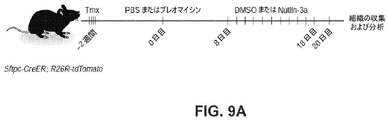
8~16週齢の間の雄性および雌性の両方のマウスを実験に使用した。全てのマウスは、他に示されない限り、C57BL/6であった。以下のマウスを実験に使用した:Sftpctm1(cre/ERT2)Blh(Sftpc-CreER)、Krt19tm1(cre/ERT)Ggu/J(Krt19-CreER)、Rosa26R-CAG-lsl-tdTomato(Sftpc-CreERと交雑させた)、B6.Cg-Gt(ROSA)26Sortm14(CAG-tdTomato)Hze/J(R26R-tdTomato)(Krt19-CreERと交雑させた)、Tg(SFTPC-GFP)#Heat(Sftpc-GFP)51、B6.Cg-Agertm2.1(cre/ERT2)Blh/J(Ager-CreER)、B6-Gt(ROSA)26Sortm1(HBEGF)Awai/J(R26R-DTR)、Mki67tm1.1Cle/J(Mki67-RFP)、Tg(Ctgf-EGFP)FX156Gsat(Ctgf-GFP)15およびTP53fl/fl(混合バックグラウンド)。Sftpc-CreER;R26R-tdTomatoマウスを用いた系列追跡のために、3~5用量の、体重20g当たり2mgのタモキシフェン(Sigma-Aldrich)を、経口胃管栄養法または腹腔内注射を介して与えた。Krt19-CreER;R26R-tdTomatoを使用する系列追跡のために、1用量の、体重20g当たり1mgのタモキシフェンを、ブレオマイシン傷害またはPBS投与の7日後に、腹腔内注射を介して与えた。動物実験は、US National Institutes of Healthガイドラインに従って、Duke University Institutional Animal Care and Use Committeeが承認した。 Both male and female mice between the ages of 8-16 weeks were used in the experiments. All mice were C57BL/6 unless otherwise indicated. The following mice were used for the experiments: Sftpctm1 (cre/ERT2) Blh (Sftpc-CreER), Krt19tm1 (cre/ERT) Ggu/J (Krt19-CreER), Rosa26R-CAG-lsl-tdTomato (crossed with Sftpc-CreER) ), B6. Cg-Gt(ROSA)26Sortm14(CAG-tdTomato)Hze/J(R26R-tdTomato) (crossed with Krt19-CreER), Tg(SFTPC-GFP)#Heat(Sftpc-GFP)51, B6. Cg-Agertm2.1 (cre/ERT2) Blh/J (Ager-CreER), B6-Gt (ROSA) 26Sortm1 (HBEGF) Awai/J (R26R-DTR), Mki67tm1.1Cle/J (Mki67-RFP), Tg (Ctgf-EGFP) FX156Gsat (Ctgf-GFP)15 and TP53 fl/fl (mixed background). For lineage tracing with Sftpc-CreER;R26R-tdTomato mice, 3-5 doses of 2 mg tamoxifen (Sigma-Aldrich) per 20 g body weight were given via oral gavage or intraperitoneal injection. . For lineage tracing using Krt19-CreER; R26R-tdTomato, one dose of tamoxifen, 1 mg/20 g body weight, was given via intraperitoneal injection 7 days after bleomycin injury or PBS administration. Animal studies were approved by the Duke University Institutional Animal Care and Use Committee according to US National Institutes of Health guidelines.
ブレオマイシン傷害。 Bleomycin injury.
ブレオマイシンで誘導された肺傷害のために、2.5U/kgのブレオマイシンを、タモキシフェン注射の2週間後に鼻腔内投与し、マウスを毎日モニタリングした。PBSを投与したマウスは、対照として機能した。マウスを、ブレオマイシン傷害後の異なる時点において屠殺した。 For bleomycin-induced lung injury, 2.5 U/kg of bleomycin was administered intranasally 2 weeks after tamoxifen injection and mice were monitored daily. Mice administered PBS served as controls. Mice were sacrificed at different time points after bleomycin injury.
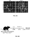
DTの投与。 Administration of DT.
DT投与の2週間前に、Ager-CreER;R26R-DTRマウスは、腹腔内注射を介してタモキシフェンを受けた。1用量の3μgのDT(Millipore、322326)を腹腔内注射を介して投与し、マウスを、組織の収集および分析のために、6日後に屠殺した。 Two weeks prior to DT administration, Ager-CreER;R26R-DTR mice received tamoxifen via intraperitoneal injection. A dose of 3 μg DT (Millipore, 322326) was administered via intraperitoneal injection and mice were sacrificed 6 days later for tissue collection and analysis.
マウス肺の解離およびFACS。 Mouse lung dissection and FACS.
肺の解離およびFACSを、以前に記載されたように実施した11。簡潔に述べると、肺を、DMEM/F12培地中の酵素溶液(dispase、5U/ml、Corning、354235;DNase I、0.33U/ml;およびI型コラゲナーゼ、450U/ml、Gibco、17100-017)1mlで気管内膨張させた。分離した肺葉をさいの目に切り、回転させながら37℃で25分間、3mlの酵素溶液と共にインキュベートした。反応を、10%胎仔ウシ血清(FBS)を含む等量の培地でクエンチし、100μm濾過器を介して濾過した。細胞ペレットを、赤血球溶解緩衝液(155mMのNH4Cl、12mMのNaHCO3および0.1mMのEDTA)中に再懸濁し、2分間インキュベートし、次いで、40μm濾過器を介して濾過した。細胞ペレットを、DMEM/F12+2%BSA中に再懸濁し、以前に記載された56ように、以下の抗体を用いて染色した:EpCAM(eBioscience、G8.8)、PDGFRα(BioLegend、APA5)およびLysotracker(Thermo Fisher、L7526)。選別を、BD FACS Vantage SE、SONY SH800SまたはBeckman Coulter MoFlo Astrios EQシステムを使用して実施した。 Lung dissection and FACS were performed as previously described 11 . Briefly, lungs were treated with enzyme solutions (dispase, 5 U/ml, Corning, 354235; DNase I, 0.33 U/ml; and type I collagenase, 450 U/ml, Gibco, 17100-017) in DMEM/F12 medium. ) for intratracheal distension with 1 ml. Separated lung lobes were diced and incubated with 3 ml of enzyme solution for 25 min at 37° C. with rotation. Reactions were quenched with an equal volume of medium containing 10% fetal bovine serum (FBS) and filtered through a 100 μm filter. Cell pellets were resuspended in red blood cell lysis buffer (155 mM NH 4 Cl, 12 mM NaHCO 3 and 0.1 mM EDTA), incubated for 2 minutes, and then filtered through a 40 μm filter. Cell pellets were resuspended in DMEM/F12 + 2% BSA and stained with the following antibodies as previously described56: EpCAM (eBioscience, G8.8), PDGFRα (BioLegend, APA5) and Lysotracker. (Thermo Fisher, L7526). Sorting was performed using a BD FACS Vantage SE, SONY SH800S or Beckman Coulter MoFlo Astrios EQ system.
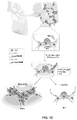
肺胞オルガノイド培養。 Alveolar organoid culture.
肺胞オルガノイド培養を、以前に記載されたように実施した(Barkauskas et al., 2013, J. Clin. Invest. 123:1118-1123)。簡潔に述べると、Tmxで処置したSftpc-GFPまたはSftpc-CreER;R26R-tdTomatoマウス由来の系列標識されたAEC2(1~3×10³)を、FACSによって選別し、PDGFRα+(5×10⁴)線維芽細胞を、MTEC/Plus中に再懸濁し、等量の増殖因子低減Matrigel(Corning、354230)と混合した。培地を1日おきに交換した。 Alveolar organoid cultures were performed as previously described (Barkauskas et al., 2013, J. Clin. Invest. 123:1118-1123). Briefly, lineage-marked AEC2 (1-3×10 3 ) from Tmx-treated Sftpc-GFP or Sftpc-CreER;R26R-tdTomato mice were sorted by FACS and PDGFRα+ (5×10 4 ) Fibroblasts were resuspended in MTEC/Plus and mixed with an equal volume of growth factor-reduced Matrigel (Corning, 354230). Medium was changed every other day.
Nutlin-3a処置。 Nutlin-3a treatment.
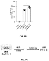
in vivo研究のために、Sftpc-CreER;R26R-tdTomatoマウスに、1用量のタモキシフェンを注射し、2週間休養させ、その後、ブレオマイシンまたはPBSを投与した。Nutlin-3a(Selleckchem、S8059)またはDMSO(対照)を、10日間連日20mg/kg/dの濃度で、傷害の8日後に腹腔内注射によって投与し、試料を、ブレオマイシン投与の20日後に収集した。ex vivo研究のために、肺胞オルガノイドを、7日間培養し、その後、Nutlin-3a(2μM)で8日間処置し、その後回収した。
For in vivo studies, Sftpc-CreER;R26R-tdTomato mice were injected with one dose of tamoxifen, rested for 2 weeks, and then administered bleomycin or PBS. Nutlin-3a (Selleckchem, S8059) or DMSO (control) were administered by intraperitoneal injection at a concentration of 20 mg/kg/d daily for 10
液滴ベースのscRNA-seq。 Droplet-based scRNA-seq.
Matrigel中に包埋したオルガノイドを、Accutase溶液(Sigma、A6964)と共に37℃で20分間インキュベートし、その後、0.25%のトリプシン-EDTAと共に37℃で10分間インキュベートした。トリプシンを、10%のFBSを追加補充したDMEM/F12 Ham培地を使用して不活性化し、次いで、細胞を、0.01%のBSAを追加補充したPBS中に再懸濁した。40μm濾過器を介した濾過の後、細胞を、3,000μl/hのmRNA捕捉ビーズおよび13,000μl/hの液滴生成オイルと共に、100細胞μl/hの濃度で、微少流体チャネルを介して3,000μl/hで流した。事前増幅ステップ(95℃で3分間を1サイクル;98℃で15秒間、65℃で30秒間および68℃で4分間を15~17サイクル;および72℃で10分間を1サイクル)のためのDNAポリメラーゼを、Terra PCR directポリメラーゼ(Takara、639271)によって置き換えた。他のプロセスを、元の液滴ベースのscRNA-seqプロトコールに記載されるように実施した。ライブラリーを、150bpペアードエンド配列決定を用いてHiSeq Xシステムを使用して配列決定した。 Organoids embedded in Matrigel were incubated with Accutase solution (Sigma, A6964) for 20 minutes at 37°C, followed by incubation with 0.25% trypsin-EDTA for 10 minutes at 37°C. Trypsin was inactivated using DMEM/F12 Ham medium supplemented with 10% FBS, then cells were resuspended in PBS supplemented with 0.01% BSA. After filtration through a 40 μm filter, the cells were passed through the microfluidic channel at a concentration of 100 cells μl/h with 3,000 μl/h of mRNA capture beads and 13,000 μl/h of droplet forming oil. Flowed at 3,000 μl/h. DNA for the pre-amplification step (1 cycle at 95°C for 3 minutes; 15-17 cycles at 98°C for 15 seconds, 65°C for 30 seconds and 68°C for 4 minutes; and 1 cycle at 72°C for 10 minutes). The polymerase was replaced by Terra PCR direct polymerase (Takara, 639271). Other processes were performed as described in the original droplet-based scRNA-seq protocol. The library was sequenced using the HiSeq X system with 150bp paired-end sequencing.
scRNA-seqの計算的分析。 Computational analysis of scRNA-seq.
肺胞オルガノイドのscRNA-seqの分析を、dropSeqPipe v0.3(https://hoohm.github.io/dropSeqPipe)を使用してFASTQファイルを処理することによって実施し、アノテーションバージョンを用いてGRCm38ゲノム参照上にマッピングした91。次いで、独自分子識別子(UMI)計数を、RパッケージSeurat v3.1.1を使用してさらに分析した(参考文献58)。LPSで処置したマウス肺のUMI計数行列(GSE130148)14をGene Expression Omnibus(GEO)から得た。UMI計数を、SCTransformを使用して正規化した。目的のクラスターについての細胞バーコードを抽出し、velocyto.py v0.17.15におけるvelocyto実行命令(参考文献18)のため、ならびにRパッケージSeuratWrappers v0.1.0(https://github.com/satijalab/seurat-wrappers)と組み合わせてvelocyto.R v0.6を使用してRNA速度プロットを生成するために利用した。傾き計算スムージング(slope calculation smoothing)における25個の最近傍のものを、RunVelocity命令に使用した。二重項(duplet)を排除した後、特定の細胞クラスターを、UMAPプロットにおける、Sftpc、Sftpa1、Sftpa2、Sftpb、Lamp3、Abca3、Hopx、Ager、Akap5、Epcam、Cdh1、Krt7、Krt8、Krt18、Krt19、Scgb1a1およびScgb3a1についての富化、ならびにVim、Acta2、PdgfraおよびPdgfrbの陰性発現に基づいて単離した。対照肺および特発性肺線維症(IPF)肺についてのRdsファイルを、GEOから得た(GSE135893)29。AEC2、AEC1、移行AEC2およびKRT5-KRT17+細胞の細胞クラスターを抽出し、分析した。SeuratにおいてFindAllMarkers命令を使用して得た各クラスターについてのマーカーを、Enrichrを介して特異的シグナル伝達経路および遺伝子オントロジーを同定するために利用した59。Z-スコアを、Kyoto Encyclopedia of Genes and Genomes(KEGG)において、合わされたスコアに基づいて計算して、異なる細胞クラスターにわたってシグナル伝達の富化およびオントロジーを比較した。結果を、Rパッケージpheatmap v1.0.12を使用して生成したヒートマップ形式で示した。Seuratオブジェクト中の調整されたデータを抽出し、各経路中の遺伝子メンバーの調整されたスコアの平均値を計算し、シグナル伝達経路の富化としてUMAPで示した。利用した経路の遺伝子メンバーリストを、KEGG pathways60およびAmiGO61から得た。ウィルコクソンの順位和検定を用いてSeuratにおいてFindMarkers命令を使用して抽出した各遺伝子についてのlog2倍数変化およびP値を、RパッケージEnhancedVolcano v1.3.1(https://github.com/kevinblighe/EnhancedVolcano)を使用してボルケーノプロットで示して、Ctgf+細胞についての特異的マーカーを示した。 Analysis of scRNA-seq of alveolar organoids was performed by processing FASTQ files using dropSeqPipe v0.3 (https://hoohm.github.io/dropSeqPipe) and referencing the GRCm38 genome using the annotation version. 91 mapped above. Unique molecular identifier (UMI) counts were then further analyzed using the R package Seurat v3.1.1 (ref. 58). UMI count matrix (GSE130148) 14 of mouse lungs treated with LPS was obtained from Gene Expression Omnibus (GEO). UMI counts were normalized using SCTransform. Extract the cell barcode for the cluster of interest, velocyto. velocyto.py for the velocyto execution instruction (ref. 18) in py v0.17.15 and in combination with the R package SeuratWrappers v0.1.0 (https://github.com/satijalab/seurat-wrappers). Utilized to generate RNA kinetic plots using R v0.6. The 25 nearest neighbors in slope calculation smoothing were used for the RunVelocity instruction. After eliminating duplicates, specific cell clusters were identified as Sftpc, Sftpal, Sftpa2, Sftpb, Lamp3, Abca3, Hopx, Ager, Akap5, Epcam, Cdh1, Krt7, Krt8, Krt18, Krt19 in UMAP plots. , enrichment for Scgb1a1 and Scgb3a1, and negative expression of Vim, Acta2, Pdgfra and Pdgfrb. Rds files for control and idiopathic pulmonary fibrosis (IPF) lungs were obtained from GEO (GSE135893)29. Cell clusters of AEC2, AEC1, transitional AEC2 and KRT5-KRT17+ cells were extracted and analyzed. Markers for each cluster obtained using the FindAllMarkers command in Seurat were utilized to identify specific signaling pathways and gene ontology via Enrichr59. Z-scores were calculated based on combined scores in the Kyoto Encyclopedia of Genes and Genomes (KEGG) to compare signaling enrichment and ontology across different cell clusters. Results were presented in heatmap format generated using the R package pheatmap v1.0.12. We extracted the adjusted data in the Seurat object and calculated the average adjusted score of the gene members in each pathway, denoted by UMAP as signaling pathway enrichment. Gene member lists for utilized pathways were obtained from KEGG pathways60 and AmiGO61. The log2 fold change and P-value for each gene extracted using the FindMarkers command in Seurat using the Wilcoxon rank sum test was analyzed using the R package EnhancedVolcano v1.3.1 (https://github.com/kevinblighe/EnhancedVolcano ) was used to indicate specific markers for Ctgf+ cells in volcano plots.
Mint-ChIP-seq。 Mint-ChIP-seq.
ヒストンマークのChIP分析のために、PATS(CD31-CD45-CD140a-CD326+Ctgf-GFP+)細胞を、ブレオマイシンで誘導された肺傷害後の12日目に、Ctgf-GFPマウスから選別した。AEC2(CD31-CD45-CD326+Lysotracker+Mki67-RFP-細胞)を、Mki67-RFPホメオスタシスマウスから選別した。Mki67マウスを使用して、RFP+細胞をネガティブ選択として(negatively)ゲートで除くことによって、細胞分裂中のあらゆる細胞を除外した。Mint-ChIPプロトコールは、以前に記載された通りであった62。TP53 ChIP配列決定(ChIP-seq)のために、PATS(CD31-CD45-Sftpc-tdTomato+Ctgf-GFP+細胞)を、ブレオマイシン投与後の8日目に、Sftpc-CreER;R26R-tdTomato;Ctgf-GFPマウスから選別した。この場合、本発明者らは、以前に記載された改変型Mint-ChIP(バージョン3)プロトコール(https://tinyurl.com/udqksct)を使用した。細胞溶解の後、クロマチンを、300単位のMNase(New England Biolabs、M0247S)で37℃で10分間消化した。T7アダプターライゲーションを2時間実施し、次いで、試料を分けて、ヒストン-改変ChIP-seqのために、抗体1つ当たりおよそ7,000細胞にした。TP53について、本発明者らは、各複製試料のためにおよそ40,000細胞を使用した。試料を、ヒストンH3抗体(H3;1μl;Active Motif、39763)、ヒストンH3リシン36トリメチル化抗体(H3K36me3;1μl;Active Motif、61101)、ヒストンH3リシン4トリメチル化抗体(H3K4me3;1μl;Abcam、ab8580)、ヒストンH3リシン27アセチル化抗体(H3K27ac;1μl;Active Motif、39133)またはTP53抗体(5μl;Cell Signaling Technology、2524T)と共に4℃で一晩インキュベートした。DNAを精製し、その後、37℃で3時間、T7-RNAポリメラーゼ媒介性のin vitro転写を行った。逆転写を、元のプロトコールに記載されたように実施し、その後、Terra direct PCRポリメラーゼ(TaKaRa、639271)を使用してライブラリーを調製した。2つの実験的複製を、各細胞集団について実施した。ライブラリーを、Hiseq XまたはNovaSeq 6000システムを使用して配列決定した(試料1つ当たり少なくとも5×10⁶読み取りの150bpペアードエンド)。
For ChIP analysis of histone marks, PATS (CD31-CD45-CD140a-CD326+Ctgf-GFP+) cells were sorted from Ctgf-GFP mice on
Mint-ChIPの計算的分析。 Computational analysis of Mint-ChIP.
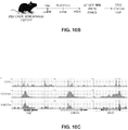
FASTQファイルを、Bcl2fastqを使用して生成した。Mint-ChIP FASTQファイルについてのさらなる逆多重化を、Jeを使用して実施した63。低品質読み取りを、trimmomatic v0.38を使用して、FASTQファイルからトリミングして除いた。読み取りを、BWAを使用してmm10ゲノム参照上にマッピングした65。パッケージを、MintChIPと呼ばれるパイプライン(https://github.com/jianhong/MintChIP)を介して実行した。HOMER66を使用して、bedGraphファイルを生成し、Integrative Genomics Viewer(IGV)においてピークを可視化した。H3K4me3についてのピークコールを、H3による正規化と共に、HOMERの関数getDifferentialPeaksReplicates.pl -region -size 1000 -minDist 2000 -C 0 -L 50を使用して実施した。モチーフ分析を、HOMERのfindMotifsGenome.pl関数を使用して実施した。H3K4me3についてコールされたピークのチャートを、deepToolsを使用して生成した68。異なる細胞集団由来の各ゲノム遺伝子座についてコールされたピークを、Affinity Designerで作成した。
FASTQ files were generated using Bcl2fastq. Further demultiplexing on the Mint-ChIP FASTQ files was performed using Je63. Low quality reads were trimmed out from the FASTQ files using trimmomatic v0.38. Reads were mapped onto the mm10 genome reference using BWA65. The package was run through a pipeline called MintChIP (https://github.com/jianhong/MintChIP). HOMER66 was used to generate bedGraph files and visualize peaks in the Integrative Genomics Viewer (IGV). Peak calls for H3K4me3, along with normalization by H3, were processed by HOMER's function getDifferentialPeaksReplicates. Performed using pl -region -size 1000 -minDist 2000 -C 0 -
ヒト肺組織。 human lung tissue.
既存の慢性肺疾患および外植された線維化ヒト肺を有さないドナーから切除された移植品質未満のヒト肺組織を、機関の手順(Duke University Pro00082379-「Human Lung Stem Cells」;45CFR164.514に記載されるPrivacy Ruleを満たす、45 CFR 46.102(f)、21 CFR 56.102(e)および21 CFR 812.3(p)に記載される研究は免除する)に従って、BioRepository and Precision Pathology Center at Duke Universityを介して入手した。scRNA-seqに使用したIPF組織の試料は、2か所の肺移植センター(VUMCおよびNTI)において肺移植の時点で取り出された肺から得た。非線維化対照組織試料は、臓器提供を拒否された肺から得た。IPF肺について、診断を、ATS/ERSコンセンサス基準に従って決定した。全ての研究は、現地の施設内審査委員会(IRB;Vanderbilt IRB番号060165および171657、ならびにWestern IRB番号20181836)が承認した。IPFの診断を、外科病理学チームが評価した。検体を、膨張および4%のパラホルムアルデヒド中での4℃で一晩の浸漬前に、PBS中で完全に洗浄した。検体を、血液の出現が最小限になるまでPBSで引き続いて洗浄し、その後、30%のスクロース中で4℃でインキュベートした。次いで、試料を、1:1の比でOCTコンパウンドと共に4℃で1時間インキュベートし、その後、OCT中に包埋した。7~9μmの厚さを有する切片を、組織学的分析に使用した。 Sub-transplant quality human lung tissue resected from donors without pre-existing chronic lung disease and explanted fibrotic human lungs was treated according to institutional procedures (Duke University Pro 00082379 - "Human Lung Stem Cells"; 45 CFR 164.514). BioRepository and Precision Pathology in accordance with 45 CFR 46.102(f), 21 CFR 56.102(e) and 21 CFR 812.3(p) exempt research that meets the Privacy Rule described in Obtained through the Center at Duke University. The IPF tissue samples used for scRNA-seq were obtained from lungs removed at the time of lung transplantation at two lung transplant centers (VUMC and NTI). Nonfibrotic control tissue samples were obtained from lungs that had been rejected for organ donation. For IPF lung, diagnosis was determined according to ATS/ERS consensus criteria. All studies were approved by the local Institutional Review Board (IRB; Vanderbilt IRB numbers 060165 and 171657, and Western IRB number 20181836). A diagnosis of IPF was evaluated by a surgical pathology team. Specimens were washed thoroughly in PBS prior to swelling and soaking in 4% paraformaldehyde overnight at 4°C. Specimens were washed successively with PBS until the appearance of blood was minimal and then incubated in 30% sucrose at 4°C. Samples were then incubated with OCT compound at a 1:1 ratio for 1 hour at 4° C. before embedding in OCT. Sections with a thickness of 7-9 μm were used for histological analysis.
免疫染色。 Immunostaining.
肺および肺胞オルガノイドを、以前に記載されたように調製した。簡潔に述べると、組織を、4%のパラホルムアルデヒドで、4℃で4時間(肺について)または室温で30分間(オルガノイドについて)固定し、次いで、OCTコンパウンドまたはパラフィン中に包埋した。切片化した試料(10μm)を、10mMクエン酸ナトリウムを使用する抗原賦活化のための95℃で10~15分間のインキュベーション後に染色のために利用した。一次抗体は、以下の通りであった:プロサーファクタントタンパク質C(Millipore、ab3786、1:500)、AGER(R&D Systems、MAB1179、1:250)、KRT8(DSHB、TROMA-I、1:50)、KRT17(NSJ、V2176、1:250)、KRT19(DSHB、TROMA-III、1:50)、tdTomato(ORIGENE、AB8181-200、1:500)、CLDN4(Invitrogen、36-4800、1:200;Proteintech、16165-1-AR、1:500)、GFP(Novus Biologicals、NB100-1770、1:500)、LGALS3(Cedarlane、CL8942AP、1:500)、SOX4(Invitrogen、MA5-31424、1:250)、SFN(Invitrogen、PA5-95056、1:250;Proteintech、66251-1-Ig、1:500;Abcam、ab77187、1:200)、ACTA2(Sigma、C6198、1:500)、γ-H2AX(R&D、4418-APC、1:500;Novus Biologicals、NB100-74435、1:250)、CDKN1A(Sigma、ZRB1141、1:200;BD Biosciences、556430、1:200)、COL1A1(Proteintech、67288-Ig、1:1,000)、YAP(Cell Signaling Technology、4912S、1:250)。 Lung and alveolar organoids were prepared as previously described. Briefly, tissues were fixed with 4% paraformaldehyde at 4° C. for 4 hours (for lungs) or at room temperature for 30 minutes (for organoids) and then embedded in OCT compound or paraffin. Sectioned samples (10 μm) were utilized for staining after 10-15 min incubation at 95° C. for antigen retrieval using 10 mM sodium citrate. Primary antibodies were as follows: prosurfactant protein C (Millipore, ab3786, 1:500), AGER (R&D Systems, MAB1179, 1:250), KRT8 (DSHB, TROMA-I, 1:50), KRT17 (NSJ, V2176, 1:250), KRT19 (DSHB, TROMA-III, 1:50), tdTomato (ORIGENE, AB8181-200, 1:500), CLDN4 (Invitrogen, 36-4800, 1:200; Proteintech , 16165-1-AR, 1:500), GFP (Novus Biologicals, NB100-1770, 1:500), LGALS3 (Cedarlane, CL8942AP, 1:500), SOX4 (Invitrogen, MA5-31424, 1:250), SFN (Invitrogen, PA5-95056, 1:250; Proteintech, 66251-1-Ig, 1:500; Abcam, ab77187, 1:200), ACTA2 (Sigma, C6198, 1:500), γ-H2AX (R&D, 4418-APC, 1:500; Novus Biologicals, NB100-74435, 1:250), CDKN1A (Sigma, ZRB1141, 1:200; BD Biosciences, 556430, 1:200), COL1A1 (Proteintech, 67288-Ig, 1:200). 1,000), YAP (Cell Signaling Technology, 4912S, 1:250).
X-galならびにヘマトキシリンおよびエオシン染色。 X-gal and hematoxylin and eosin staining.
パラホルムアルデヒド固定された凍結切片を、1mg/mlのX-gal(Thermo、R0941)、5mMのK3Fe(CN)6、5mMのK4Fe(CN)6、2mMのMgCl2、0.01%のデオキシコール酸ナトリウムおよび0.02%のNP-40を含むX-gal染色緩衝液と共に、37℃で一晩インキュベートした。切片を、PBS中で3回洗浄し、マウントした。ヘマトキシリンおよびエオシン染色のために、10μmのパラフィン切片を、Histo-clearおよび一連のエタノール中に浸した。マイヤーヘマトキシリンを使用して核を染色し、その後、1%のエオシンYを使用して染色した。 Paraformaldehyde-fixed cryosections were treated with 1 mg/ml X-gal (Thermo, R0941), 5 mM K 3 Fe(CN) 6 , 5 mM K 4 Fe(CN) 6 , 2 mM MgCl 2 , 0.01. % sodium deoxycholate and 0.02% NP-40 in X-gal staining buffer overnight at 37°C. Sections were washed three times in PBS and mounted. For hematoxylin and eosin staining, 10 μm paraffin sections were submerged in Histo-clear and ethanol series. Nuclei were stained using Mayer's hematoxylin, followed by staining using 1% eosin Y.
近接ライゲーションin situハイブリダイゼーション。 Proximity ligation in situ hybridization.
近接ライゲーションin situハイブリダイゼーションを、以前に記載されたように実施した69。簡潔に述べると、凍結マウス肺切片を、4.0%のパラホルムアルデヒドで20分間固定し、20μg/mlのプロテイナーゼKで37℃で9分間処置し、漸増する一連の(up-series)エタノールで脱水した。切片を、ハイブリダイゼーション緩衝液(1Mのトリクロロ酢酸ナトリウム、50mMのTris pH7.4、5mMのEDTAおよび0.2mg/mlのヘパリン)中で、遺伝子特異的オリゴと共に、37℃で2時間インキュベートした。コモンブリッジ(common-bridge)プローブおよびサークル(circle)プローブを切片に添加し、1時間インキュベートし、その後、2時間のT4 DNAリガーゼ反応を行った。ローリングサークル増幅を、phi29ポリメラーゼ(Lucigen、30221)を使用して、37℃で12時間実施した。フルオロフォアコンジュゲートした検出プローブを適用し、37℃で30分間インキュベートし、その後、4,6-ジアミジノ-2-フェニルインドール(DAPI)を含む媒体中でマウントした。 Proximity ligation in situ hybridization was performed as previously described 69 . Briefly, frozen mouse lung sections were fixed with 4.0% paraformaldehyde for 20 min, treated with 20 μg/ml proteinase K for 9 min at 37° C., and treated with an up-series of ethanol. Dehydrated. Sections were incubated with gene-specific oligos in hybridization buffer (1 M sodium trichloroacetate, 50 mM Tris pH 7.4, 5 mM EDTA and 0.2 mg/ml heparin) for 2 hours at 37°C. Common-bridge and circle probes were added to the sections and incubated for 1 hour, followed by a 2 hour T4 DNA ligase reaction. Rolling circle amplification was performed at 37° C. for 12 hours using phi29 polymerase (Lucigen, 30221). A fluorophore-conjugated detection probe was applied and incubated at 37° C. for 30 min before mounting in medium containing 4,6-diamidino-2-phenylindole (DAPI).
画像の獲得、処理および定量化。 Image acquisition, processing and quantification.
画像を、×20、×40または×60対物レンズを備えたOlympus FV3000共焦点顕微鏡、Zeiss広視野蛍光顕微鏡(X-gal染色)およびZeiss Axio Imager広視野蛍光顕微鏡(ヘマトキシリンおよびエオシン)を使用して捕捉した。細胞を、免疫組織化学マーカーおよびDAPIに基づいて手動で計数した。直線距離当たりの平均交差の決定のために、平均線形切片分析を、目的の単一チャネル免疫蛍光染色に対して、以前に記載されたように実施した11。画像を、Olympus CellSensアプリケーションまたはImageJを使用して処理し、図面を、Affinity Designerを使用して作成した。 Images were obtained using an Olympus FV3000 confocal microscope with ×20, ×40 or ×60 objectives, a Zeiss widefield fluorescence microscope (X-gal staining) and a Zeiss Axio Imager widefield fluorescence microscope (hematoxylin and eosin). Captured. Cells were manually counted based on immunohistochemical markers and DAPI. For determination of mean crossover per linear distance, mean linear intercept analysis was performed as previously described for single-channel immunofluorescence staining of interest 11 . Images were processed using the Olympus CellSens application or ImageJ and drawings were created using Affinity Designer.
統計学および再現性。 Statistics and reproducibility.
実験を、少なくとも3つの生物学的複製に対して実施した(オルガノイドscRNA-seqおよびChIP-seqを除く)。試料サイズは事前に決定しなかった。データは、各実験内の変動を示すために、s.e.m.を伴った平均として示される。統計分析を、GraphPad Prismで実施した。対応のない両側スチューデントt検定を、2つの実験的条件間の比較に使用した。マン・ホイットニー片側検定を、非正規分布を示した2つの条件間の比較に使用した。 Experiments were performed on at least three biological replicates (excluding organoid scRNA-seq and ChIP-seq). Sample size was not pre-determined. Data are expressed in s.d. to show variation within each experiment. e. m. is shown as the mean with . Statistical analysis was performed with GraphPad Prism. An unpaired two-tailed Student's t-test was used for comparisons between two experimental conditions. A one-tailed Mann-Whitney test was used for comparisons between two conditions that exhibited non-normal distributions.
(実施例1)
単一細胞トランスクリプトミクスは、ex vivoオルガノイドにおける以前には未知であった肺胞上皮細胞状態を明らかにする
最近の研究は、AEC2が、肺傷害に応答して増殖し、AEC1を生じることを示している。さらに、AEC2は、肺胞オルガノイドにおいてAEC1を自発的に生成する。しかし、AEC2のAEC1への分化の根底にある分子機構および移行細胞状態は、あまり理解されていない。これらの問題に対処するために、本発明者らは、AEC2およびPDGFRα+線維芽細胞を精製して、肺胞オルガノイドを樹立した。単一細胞トランスクリプトーム分析を、10日目のオルガノイド培養物から単離した細胞に対して実施した(図1A)。均一マニホールド近似投影(uniform manifold approximation and projection)(UMAP)は、Epcam+上皮細胞およびVim+Pdgfra+線維芽細胞からなる2つの主要なクラスターを同定した(図1B)。次に、本発明者らは、上皮細胞集団をさらにデコンボリューションおよび可視化し、それらを、リポポリサッカリド(LPS)で傷害された肺胞上皮細胞の公に入手可能な単一細胞RNA配列決定(scRNA-seq)データセットと比較した14。オルガノイド由来上皮細胞は、それらのin vivo対応物と重複し、これは、転写的類似性および細胞状態類似性を明らかにしている(図1C)。本発明者らは、オルガノイド由来上皮細胞において、以下の複数のサブクラスターを観察した:Sftpc(AEC2のマーカー)を発現する細胞、Ager(AEC1のマーカー)を発現する細胞およびSftpc+Mki67+増殖性AEC2(図1D)。
(Example 1)
Single-cell transcriptomics reveals a previously unknown alveolar epithelial cell state in ex vivo organoids Recent studies show that AEC2 proliferates and gives rise to AEC1 in response to lung injury showing. In addition, AEC2 spontaneously generates AEC1 in alveolar organoids. However, the molecular mechanisms and transitional cell states underlying the differentiation of AEC2 to AEC1 are poorly understood. To address these issues, we purified AEC2 and PDGFRα+ fibroblasts to establish alveolar organoids. Single-cell transcriptome analysis was performed on cells isolated from
さらに、本発明者らは、Cldn4、Krt19およびSfnを発現する肺胞上皮細胞の集団を同定した(図1D、図1Eおよび図1F)。このクラスターに独自のマーカー遺伝子は、UMAPおよびボルケーノプロットで可視化した場合、2つの別個のパターンを示した(図1b~d)。1つのサブセット(Ctgf+細胞)は、Ctgf、Clu、Sox4およびActn1について富化されたが、他(Lgals3+細胞)は、Lgals3、Csrp1、S100a14およびCldn18について富化された。Ager、Emp2およびHopx(AEC1のマーカー)を含む、Lgals3+サブクラスターにおいて富化されたさらなる転写物は、Lgals3+細胞とAEC1との間での類似点および潜在的系列ヒエラルキーを示唆している(図1G)。これらのデータは、Cldn4+Krt19+Sfn+集団が、AEC2とAEC1との間の中間体であることを示唆している。この集団を、「プレ1型肺胞移行細胞状態」(PATS)と称した。本発明者らは、肺胞オルガノイドに対してPATSマーカーについての免疫蛍光を実施して、単一細胞データを検証した(図1H)。画像は、3匹のマウス由来の30個のオルガノイドの代表であった。これにより、アルベオロスフィア(alveolosphere)中の、CLDN4、LGALS3およびSOX4を発現する細胞の存在が確認された。合わせると、これらのデータにより、オルガノイド培養において肺胞上皮幹細胞分化の間の独自の細胞状態が同定された。
Furthermore, we identified a population of alveolar epithelial cells that expressed Cldn4, Krt19 and Sfn (Fig. 1D, 1E and 1F). The marker genes unique to this cluster showed two distinct patterns when visualized with UMAP and volcano plots (Fig. 1b-d). One subset (Ctgf+ cells) was enriched for Ctgf, Clu, Sox4 and Actn1, while the other (Lgals3+ cells) was enriched for Lgals3, Csrp1, S100a14 and Cldn18. Additional transcripts enriched in the Lgals3+ subcluster, including Ager, Emp2 and Hopx (markers for AEC1), suggest similarities and potential lineage hierarchies between Lgals3+ cells and AEC1 (Fig. 1G). ). These data suggest that the Cldn4+Krt19+Sfn+ population is an intermediate between AEC2 and AEC1. This population was termed the "
(実施例2)
PATS細胞は、肺胞傷害後にin vivoで出現する。
次いで、PATS細胞が、ホメオスタシスおよび再生中の肺胞組織においてin vivoで観察され得るかどうかを調査した。これを試験するために、本発明者らは、LPSで処置したマウス肺および対照マウス肺からのscRNA-seqデータをUMAPプロットでレンダリングし、LPS傷害に独自の集団を見出した。UMAPプロットは、肺胞オルガノイドscRNA-seqデータセット(4,573細胞)およびLPSで処置した肺または対照肺(13,204細胞)における、AEC2(LampおよびSftpb)、AEC2増殖性(Mki67およびTop2a)、AEC1(AgerおよびAkap5)および新規肺胞上皮細胞状態における遺伝子の発現を示した。重要なことに、この集団は、Cldn4、Sox4、Lgals3、S100a14およびFn1を含む、PATSにおいて発現される遺伝子について富化されている。
(Example 2)
PATS cells emerge in vivo after alveolar injury.
We then investigated whether PATS cells could be observed in homeostatic and regenerating alveolar tissue in vivo. To test this, we rendered scRNA-seq data from LPS-treated and control mouse lungs in UMAP plots and found a population unique to LPS injury. UMAP plots show AEC2 (Lamp and Sftpb), AEC2 proliferation (Mki67 and Top2a) in the alveolar organoid scRNA-seq dataset (4,573 cells) and LPS-treated or control lungs (13,204 cells). , showed gene expression in AEC1 (Ager and Akap5) and the novel alveolar epithelial cell state. Importantly, this population is enriched for genes expressed in PATS, including Cldn4, Sox4, Lgals3, S100a14 and Fn1.
PATSをさらに特徴付けるために、Ctgf-緑色蛍光タンパク質(GFP)トランスジェニックマウス系を利用した(Hall-Glennet al., 2012, PLoS ONE 7:e30562)。マウスを、一過的な線維症を引き起こす薬物であるブレオマイシンに曝露させ、傷害後の12日目にマウスの肺を収集した(図2)。未傷害のCtgf-GFPマウスにおけるGFPについての免疫蛍光は、線維芽細胞において特異的に、GFPシグナルを明らかにしたが、肺胞上皮細胞においては明らかにしなかった。対照的に、ブレオマイシンで傷害された肺では、GFP発現は、CLDN4、LGALS3およびSFNを含むPATSマーカーで共標識された上皮細胞において見出された。低レベルのAEC2マーカーを共発現する僅かなGFP+細胞が観察され、これは、PATSへのAEC2移行の際のSFTPCタンパク質の永続に恐らくは起因する。 To further characterize PATS, a Ctgf-green fluorescent protein (GFP) transgenic mouse line was utilized (Hall-Glennet et al., 2012, PLoS ONE 7:e30562). Mice were exposed to bleomycin, a drug that causes transient fibrosis, and mouse lungs were harvested 12 days after injury (Fig. 2). Immunofluorescence for GFP in uninjured Ctgf-GFP mice revealed a GFP signal specifically in fibroblasts, but not in alveolar epithelial cells. In contrast, in bleomycin-injured lungs, GFP expression was found in epithelial cells co-labeled with PATS markers, including CLDN4, LGALS3 and SFN. Few GFP+ cells were observed co-expressing low levels of the AEC2 marker, presumably due to the persistence of the SFTPC protein upon AEC2 translocation to the PATS.
ブレオマイシンで傷害された肺におけるKRT8の発現を試験した。KRT8発現細胞とCtgf-GFP発現細胞との間の重複における有意な増加が、12日目に、対照肺およびブレオマイシンで処置した肺において、Ctgf-GFP、KRT8およびSFTPCについての免疫染色によって見出された。これらのデータは、オルガノイドおよびLPS傷害からのscRNA-seqデータにおけるKrt8発現の上昇したレベルをさらに裏付ける(図3)。合わせると、これらのデータにより、PATS細胞が、ブレオマイシン傷害後にin vivoで肺胞において出現することが明らかである。
Expression of KRT8 in bleomycin-injured lungs was examined. A significant increase in overlap between KRT8 and Ctgf-GFP expressing cells was found by immunostaining for Ctgf-GFP, KRT8 and SFTPC in control and bleomycin-treated lungs on
次いで、PATS細胞がブレオマイシン傷害に特異的であるのか他の傷害モデルにおいても出現するのかを試験した。これを試験するために、Ager-CreER;R26R-DTRマウスに、タモキシフェンを投与し、その後、AEC1の選択的アブレーションを生じるジフテリア毒素(DT)を投与した。本発明者らは、DT注射後の6日目および28日目に試料を収集し、免疫染色を実施した(図4A)。興味深いことに、6日目のAEC1をアブレートされた肺中に、細長い形態を有する、CLDN4、SFN、KRT19およびLGALS3を発現する細胞が観察されたが、対照中では観察されなかった(図4B)。PATSマーカーを共発現した細長い細胞における低レベルのSFTPC発現もまた、免疫染色によって観察され、これは、PATSへのAEC2の移行の間のSFTPCの永続に恐らくは起因する。28日目には、AEC1をアブレートされた肺において、免疫染色によってPATSマーカーが観察されなかったことに留意されたい。これは、PATS細胞が、完全に分化したAEC1へと成熟化したことを示唆している。これらのデータは、PATSの出現が、肺胞再生における一般的な機構であることを示唆している。
We then tested whether PATS cells were specific for bleomycin injury or appeared in other injury models. To test this, Ager-CreER;R26R-DTR mice were administered tamoxifen followed by diphtheria toxin (DT), which causes selective ablation of AEC1. We collected samples on
(実施例3)
系列追跡は、PATS細胞がAEC2に起源することを明らかにする。
オルガノイドおよび傷害モデルから先に提示されたデータは、PATS細胞がAEC2に起源することを示唆した。この仮説を実験的に試験するために、本発明者らは、タモキシフェンの投与が、AEC2およびその子孫において特異的に、tdTomato発現を恒久的に誘導する、Sftpc-CreER;R26R-tdTomato(本明細書で以後Sftpc-tdT+と呼ぶ)マウスモデルを使用した(図5A)。ブレオマイシン投与後の10日目に、損傷した肺胞領域において、LGALS3、SFNおよびCLDN4を共発現する系列標識された(tdTomato+、tdt+)細胞が観察されたが、対照肺では観察されなかった。AGERを共発現するtdt+細胞は、後の時点では、平たく薄く細長い形態を示し、AEC1へのAEC2移行が確認された。興味深いことに、SFN、CLDN4およびLGALS3を含むPATSマーカーを共発現する系列標識された細胞は、修復プロセスの間に、別個の形態学的特色(円形の、「伸展した」、および細長い)を有した(図5B)。これを定量化するために、本発明者らは、CLDN4+および/またはLGALS3+発現ならびにそれらの形状に従って、系列陽性細胞をスコア付けした(図5C)。3つの別個のパターンのマーカー発現、CLDN4+、CLDN4+LGALS3+およびLGALS3+が観察され、これらがそれぞれ、円形、伸展したおよび細長い形態と相関したことが見出された(図5Cおよび図5D)。さらに、本発明者らは、ブレオマイシンで誘導された傷害の後、8日目のCLDN4+細胞の、10日目のCLDN4+LGALS3+細胞の、および12日目のLGALS3+細胞の、実質的な富化を観察した(図2l)。scRNA-seqデータセットにおけるCLDN4+およびCLDN4+LGALS3+細胞状態が注目されたが、別個のLGALS3+集団は注目されなかった。類似のPATSマーカー発現が、肺切除術で誘導された「生理学的」再生の間に、AEC2系列標識されたマウスにおいて検出された(図5E)。scRNA-seqにおけるLGALS3の発現 対 免疫染色におけるLGALS3の発現との間の食い違いは、最終AEC1におけるLGALS3タンパク質の永続に起因し得る。合わせると、この分析により、AEC2のAEC1への分化が、2つの別個の状態を通過することが明らかになった(図5F)。
(Example 3)
Lineage tracing reveals that PATS cells originated from AEC2.
Previously presented data from organoid and injury models suggested that PATS cells originated from AEC2. To test this hypothesis experimentally, we found that administration of tamoxifen permanently induces tdTomato expression specifically in AEC2 and its progeny, Sftpc-CreER; R26R-tdTomato (herein A mouse model, hereinafter referred to as Sftpc-tdT+, was used (Fig. 5A). Ten days after bleomycin administration, lineage-marked (tdTomato+, tdt+) cells co-expressing LGALS3, SFN and CLDN4 were observed in the injured alveolar area, but not in control lungs. tdt+ cells co-expressing AGER displayed a flattened, thin and elongated morphology at later time points, confirming AEC2 to AEC1 translocation. Interestingly, lineage-marked cells co-expressing PATS markers, including SFN, CLDN4 and LGALS3, have distinct morphological features (rounded, 'stretched' and elongated) during the repair process. (Fig. 5B). To quantify this, we scored lineage-positive cells according to CLDN4+ and/or LGALS3+ expression and their shape (Fig. 5C). Three distinct patterns of marker expression, CLDN4+, CLDN4+LGALS3+ and LGALS3+, were observed and found to correlate with rounded, elongated and elongated morphologies, respectively (Figs. 5C and 5D). Furthermore, we observed substantial enrichment of CLDN4+ cells on
(実施例4)
系列追跡分析は、PATS細胞がAEC1を生成することを明らかにする
先に示された実験からの複数ラインのデータは、PATS細胞がAEC1に向かう途中であることを示唆している。これを試験するために、スプライシングされたメッセンジャーRNAとスプライシングされていないメッセンジャーRNAとの間の比率に基づいた細胞分化トラジェクトリーの予測を可能にする、Velocytoと呼ばれるアルゴリズムツール18を使用した。PATS1 Ctgf+集団に起源して、PATS2 Lgals3+集団を介してAEC1への、強いRNA速度(トラジェクトリー)が観察された(図6Aおよび図6B)。次いで、R26R-tdTomatoと組み合わせた、PATSにおいて富化される遺伝子であるKrt19のCreER対立遺伝子(Krt19-tdt)を使用して、ブレオマイシン傷害後の系列追跡を実施して、同じトラジェクトリーが、再生中の肺胞においてin vivoで生じるかどうかを試験した。最初に、マウスを、ブレオマイシンで傷害し、タモキシフェンを7日後に投与して、Krt19発現細胞およびそれらの子孫を標識した(図6C)。次いで、AEC2、AEC1およびPATSマーカーについての免疫蛍光を実施した。
(Example 4)
Lineage Tracing Analysis Reveals that PATS Cells Generate AEC1 Multiple lines of data from the experiments presented earlier suggest that PATS cells are on the way to AEC1. To test this, we used an algorithmic tool called Velocyto that allows the prediction of cell differentiation trajectories based on the ratio between spliced and unspliced messenger RNA. A strong RNA trajectory originating from the PATS1 Ctgf+ population and through the PATS2 Lgals3+ population to AEC1 was observed (FIGS. 6A and 6B). Lineage tracing after bleomycin injury was then performed using the CreER allele of Krt19 (Krt19-tdt), a gene enriched in PATS, in combination with R26R-tdTomato to show that the same trajectory was regenerated. It was tested whether it occurs in vivo in the middle alveoli. First, mice were injured with bleomycin and tamoxifen was administered 7 days later to label Krt19-expressing cells and their progeny (Fig. 6C). Immunofluorescence for AEC2, AEC1 and PATS markers was then performed.
AEC1のマーカーであるAGERを共発現する多数のKrt19-tdt+細胞が、ブレオマイシン投与後の12日目に見出された(図6D、図6E、図6F)。未傷害の肺では、本発明者らは、AEC1ではまれな、一部のLGALS3+マクロファージにおけるtdt発現を観察した(図6G)。本発明者らは、ブレオマイシンで処置したマウスの未傷害の領域において、SFTPCを共発現するKrt19-tdt+細胞を見出さず、これは、Krt19発現が、損傷した領域において傷害に応答して特異的に活性化されること、ならびに本発明者らが試験した条件がPATSを標識および追跡するために十分に適していることを示している。共免疫染色により、ブレオマイシンで処置した肺における、SFN、CLDN4およびLGALS3を含むPATSマーカーを共発現する、伸展したtdTomato標識された細胞が明らかになったが、対照では明らかにならなかった(図6D~6Gおよび図6H)。したがって、組み合わされたRNA速度および系列追跡分析は、新たに同定されたPATSがAEC2とAEC1との間を横断することを明らかにしている。 A large number of Krt19-tdt+ cells co-expressing AGER, a marker of AEC1, were found 12 days after bleomycin administration (FIGS. 6D, 6E, 6F). In uninjured lungs, we observed tdt expression in some LGALS3+ macrophages, which was rare in AEC1 (Fig. 6G). We did not find Krt19-tdt+ cells co-expressing SFTPC in the uninjured area of mice treated with bleomycin, suggesting that Krt19 expression is specifically in the injured area in response to injury. activated, and that the conditions we tested are well suited for labeling and tracking PATS. Co-immunostaining revealed spread tdTomato-labeled cells co-expressing PATS markers, including SFN, CLDN4 and LGALS3, in bleomycin-treated lungs but not controls (Fig. 6D). ˜6G and FIG. 6H). Thus, combined RNA velocity and lineage tracing analyzes reveal that the newly identified PATS crosses between AEC2 and AEC1.
(実施例5)
PATS中の保存された転写プログラムおよび経路。
AEC2のAEC1への分化は、立方体状から平たく薄いものへの、細胞の形状および構造における劇的な変化に関連する。かかる移行には、典型的には、シグナル伝達タンパク質および構造タンパク質の発現における多くの変化が伴う。したがって、これらの変化がPATS細胞において生じると考えられた。これらの変化がPATS細胞において生じるかどうかを決定するために、オルガノイド培養物(4,573細胞)およびLPSで誘導された傷害モデル(13,204細胞)からのscRNA-seqデータを分析した。以下を含む多数の遺伝子が、PATS細胞間で保存されていた:TGFβシグナル伝達経路中の遺伝子(Cdkn2b、Mapk3、Myc、Nbl1、Ppp2r1a、Rbx1、Rhoa、Skp1a、Tgfb1、Tgif1およびThbs1)、p53シグナル伝達経路中の遺伝子(Bax、Casp8、Ccnd1、Ccnd2、Ccng1、Cdk4、Cdkn1a、Cycs、Ei24、Gadd45a、Gadd45b、Sesn3、Sfn、Shisa5、Thbs1およびTrp53)、細胞老化に関与する遺伝子(Calm1、Calm2、Capn2、Ccnd1、Ccnd2、Cdk4、Cdkn1a、Cdkn2b、Eif4ebp1、Ets1、Gadd45b、H2-D1、H2-K1、h2-Q4、H2-Q6、H2-Q7、H2-T22、H2-T23、Kras、Map2k2、Mapk13、Mapk3、Myc、Ppid、Ppp1ca、Ppp3ca、Rbbp4、Rheb、Rras、Sqstm1、Slc25a4、Tgfb2、Trp53、Vdac1、Vdac2、Vdac3およびZfp36l1)、タイトジャンクションシグナル伝達遺伝子(Actb、Actg1、Actn1、Actn4、Actr3、Ccnd1、Cdc42、Cdk4、Cldn3、Cldn4、Cldn7、Cldrn18、cttn、Ezr、F11r、Hspa4、Itgb1、Jun、Msn、Myh9、Myl6、Myl9、Myl12a、Myl12b、Myn12a、Nedd4、Ocln、Ppp2r1a、Rac1、Rap1a、Rhoa、Slc9a3r1、Tuba1a、Tuba1b、Tuba1cおよびVasp)、およびDNA損傷チェックポイント遺伝子(Ccnd1、Ccng1、Cdkn1a、Ddx39b、ler3、Prpf19、Rpl26、Rps27l、Sox4、Syf2およびTrp53)。
(Example 5)
Conserved transcriptional programs and pathways in PATS.
Differentiation of AEC2 to AEC1 is associated with dramatic changes in cell shape and structure, from cuboidal to flattened and thin. Such transitions are typically accompanied by a number of changes in the expression of signaling and structural proteins. Therefore, these changes were thought to occur in PATS cells. To determine whether these changes occur in PATS cells, scRNA-seq data from organoid cultures (4,573 cells) and an LPS-induced injury model (13,204 cells) were analyzed. A number of genes were conserved among PATS cells, including: genes in the TGFβ signaling pathway (Cdkn2b, Mapk3, Myc, Nbl1, Ppp2r1a, Rbx1, Rhoa, Skp1a, Tgfb1, Tgif1 and Thbs1), p53 signaling Genes in the transduction pathway (Bax, Casp8, Ccnd1, Ccnd2, Ccng1, Cdk4, Cdkn1a, Cycs, Ei24, Gadd45a, Gadd45b, Sesn3, Sfn, Shisa5, Thbs1 and Trp53), genes involved in cellular senescence (Calm1, Calm2, Capn2, Ccnd1, Ccnd2, Cdk4, Cdkn1a, Cdkn2b, Eif4ebp1, Ets1, Gadd45b, H2-D1, H2-K1, h2-Q4, H2-Q6, H2-Q7, H2-T22, H2-T23, Kras, Map2k2, Mapk13, Mapk3, Myc, Ppid, Ppp1ca, Ppp3ca, Rbbp4, Rheb, Rras, Sqstm1, Slc25a4, Tgfb2, Trp53, Vdac1, Vdac2, Vdac3 and Zfp36l1), tight junction signaling genes (Actb, Actg1, Act4Actnctn1, Acttnctn1) , Ccnd1, Cdc42, Cdk4, Cldn3, Cldn4, Cldn7, Cldrn18, cttn, Ezr, F11r, Hspa4, Itgb1, Jun, Msn, Myh9, Myl6, Myl9, Myl12a, Myl2b, Myn12a, Nedd4, Ocln, Rpp2r1 , Rhoa, Slc9a3r1, Tuba1a, Tuba1b, Tuba1c and Vasp), and DNA damage checkpoint genes (Ccnd1, Ccng1, Cdkn1a, Ddx39b, ler3, Prpf19, Rpl26, Rps27l, Sox4, Syf2 and Trp53).
経路富化分析により、TP53、TNF-NF-κB、ErbB、HIF1、Hippo-YAP、細胞周期停止、細胞骨格動態、タイトジャンクションおよびTGFβシグナル伝達が、他の集団と比較して、PATS細胞において活性化されることが明らかになった(図7Aおよび図7B)。細胞老化およびDNA損傷応答経路を代表する遺伝子についての実質的な富化が、予想外に見出された(図7Cおよび図7B)。 Pathway enrichment analysis showed that TP53, TNF-NF-κB, ErbB, HIF1, Hippo-YAP, cell cycle arrest, cytoskeletal dynamics, tight junctions and TGFβ signaling were active in PATS cells compared to other populations. (Figs. 7A and 7B). Substantial enrichment for genes representing cellular senescence and DNA damage response pathways was unexpectedly found (FIGS. 7C and 7B).
(実施例6)
PATS細胞は、in vivoでの肺胞再生の間に、DNA損傷および老化を天然に示す。
これらのデータは、DNA損傷関連遺伝子および老化関連遺伝子が、PATSにおいて高度に富化されることを示した。これらの知見を検証するために、ブレオマイシンで処置したマウスおよびPBSで処置したマウスの肺を、老化細胞のバイオマーカーとして機能するβ-ガラクトシダーゼ活性およびDNA損傷のマーカーであるγH2AXについて、10日後にアッセイした。興味深いことに、ブレオマイシンで傷害された肺胞において、5-ブロモ-4-クロロ-3-インドリル-β-d-ガラクトピラノシド(X-gal)由来の青色の沈着に基づいて、多数のβ-ガラクトシダーゼ活性細胞が検出されたが、対照肺胞では検出されなかった(図8A)。これは、ブレオマイシンで処置した肺において特異的な、老化マーカーCDKN1A(p21)の発現によってさらに支持された(図8B)。CDKN1A発現は、伸展したtdt+細胞において観察されたが、AGER+細胞では観察されず、これは、老化がAEC1において観察されないことを示している(図8B)。同様に、γH2AX斑の蓄積もまた、ブレオマイシンで処置した肺において、伸展したSftpc-tdt+細胞の核において検出されたが、AGER+細胞では検出されなかった(図8C)。さらなる分析により、ブレオマイシンで傷害された肺において、LGALS3を共発現するγH2AX+Sftpc-tdt+細胞の顕著な数が明らかになったが、対照では明らかにならず、これは、PATS細胞が肺胞再生の間にDNA損傷を受けることを示している(図8D)。
(Example 6)
PATS cells naturally exhibit DNA damage and senescence during alveolar regeneration in vivo.
These data indicated that DNA damage- and senescence-related genes were highly enriched in PATS. To validate these findings, lungs of bleomycin- and PBS-treated mice were assayed after 10 days for β-galactosidase activity, which serves as a biomarker of senescent cells, and γH2AX, a marker of DNA damage. did. Interestingly, due to 5-bromo-4-chloro-3-indolyl-β-d-galactopyranoside (X-gal)-derived blue deposits, numerous β- - Galactosidase activity cells were detected but not in control alveoli (Fig. 8A). This was further supported by the specific expression of the senescence marker CDKN1A (p21) in bleomycin-treated lungs (Fig. 8B). CDKN1A expression was observed in stretched tdt+ cells, but not in AGER+ cells, indicating that senescence is not observed in AEC1 (FIG. 8B). Similarly, accumulation of γH2AX plaques was also detected in the nuclei of spread Sftpc-tdt+ cells, but not AGER+ cells, in bleomycin-treated lungs (Fig. 8C). Further analysis revealed a significant number of γH2AX + Sftpc-tdt + cells co-expressing LGALS3 in bleomycin-injured lungs, but not in controls, suggesting that PATS cells are active during alveolar regeneration. (Fig. 8D).
ブレオマイシンは、細胞においてDNA損傷を誘導することが公知である。しかし、scRNA-seq分析および免疫染色は共に、PATS細胞におけるDNA損傷修復シグナル伝達の強い富化を明らかにした。老化およびDNA損傷に対するブレオマイシンからのあらゆる潜在的な影響を回避するために、マーカー分析を、AEC1特異的アブレーションモデルからの肺に対して実施した(図4A)。AEC1をアブレートされた肺において、多数のX-gal+細胞が検出され、LGALS3+細胞におけるCDKN1AまたはγH2AXの発現が観察されたが、対照肺胞では観察されなかった(図8E、図8Fおよび図8G)。 Bleomycin is known to induce DNA damage in cells. However, both scRNA-seq analysis and immunostaining revealed a strong enrichment of DNA damage repair signaling in PATS cells. To avoid any potential effects from bleomycin on aging and DNA damage, marker analysis was performed on lungs from an AEC1-specific ablation model (Fig. 4A). Numerous X-gal+ cells were detected in AEC1-ablated lungs and CDKN1A or γH2AX expression was observed in LGALS3+ cells, but not in control alveoli (FIGS. 8E, 8F and 8G). .
(実施例7)
PATS細胞は、機械的伸展で誘導されたDNA損傷に対して脆弱である
以前の研究は、電離放射線、酸化ストレス、ならびに細胞の遊走および伸展の間に生じる機械的な力が、全てDNA損傷を引き起こし得ることを示している。経路分析により、酸化ストレスではなく細胞骨格変化に関与する遺伝子についての富化が明らかになった。AEC1へのAEC2分化が広範な細胞の広がりおよび細胞骨格動態を要求することを考慮して、PATS細胞が、DNA損傷をもたらし得る機械的伸展を経験すると仮説を立てた。これを試験するために、AEC2を、SFTPC-GFPマウスから精製し、AEC2が広がりAEC1へと分化する条件であるプラスチック表面上でそれらを培養した(二次元(2D)培養)(図8Hb)。プレーティング後5日以内に、AEC2のほとんどは伸展し、GFP発現は喪失した(GFP-)または下方調節された(GFPlo)。これらの細胞は、伸展の間中、その表面積を増加させ、AEC1マーカーを発現し始めた。興味深いことに、AGER+細胞は、DNA損傷マーカーγH2AXについても陽性であった(図8Hおよび図8I)。プレーティング後の2日目には、細胞においてDNA損傷マーカーが観察されなかったことに留意されたい(図8I)。合わせると、これらのデータは、大きく薄いAEC1へと分化する立方体状AEC2が、DNA損傷修復を天然に経験し、一過的老化を受けることを示している。
(Example 7)
PATS cells are vulnerable to DNA damage induced by mechanical stretching. It shows that it can cause Pathway analysis revealed enrichment for genes involved in cytoskeletal changes rather than oxidative stress. Given that AEC2 differentiation to AEC1 requires extensive cell spreading and cytoskeletal dynamics, it was hypothesized that PATS cells undergo mechanical stretching that could lead to DNA damage. To test this, AEC2 were purified from SFTPC-GFP mice and we cultured them on a plastic surface (two-dimensional (2D) culture), conditions in which AEC2 spread and differentiate into AEC1 (Fig. 8Hb). Within 5 days after plating, most of AEC2 was expanded and GFP expression was either lost (GFP−) or downregulated (GFPlo). These cells increased their surface area throughout spreading and began to express the AEC1 marker. Interestingly, AGER+ cells were also positive for the DNA damage marker γH2AX (FIGS. 8H and 8I). Note that no DNA damage markers were observed in the cells two days after plating (Fig. 8I). Together, these data indicate that cuboidal AEC2 that differentiate into large thin AEC1 naturally undergoes DNA damage repair and undergoes transient senescence.
(実施例8)
TP53シグナル伝達の遺伝的機能喪失および薬理学的活性化は、肺胞傷害-修復の間のPATSを介したAEC2~AEC1の分化を異常調節する
これらのデータは、TP53シグナル伝達の活性化が、傷害された肺におけるPATSに関連することを示唆している。他の組織における以前の研究は、TP53シグナル伝達が分化を促進し、幹細胞自己更新を抑制することを示唆している。TP53シグナル伝達の増強された活性化が、傷害後のPATSが関与するAEC1へのAEC2分化を増加させるかどうかを決定するために、Sftpc-CreER;R26-tdTomatoマウスにおいてブレオマイシンで誘導された傷害を実施し、その後、傷害後の8日目に開始するNutlin-3a(TP53活性化因子)またはジメチルスルホキシド(DMSO;対照)投与を実施し、20日目に組織収集した(図9A)。AGERについての免疫染色により、Nultin-3a処置が、DMSO処置と比較して、Sftpc-tdt+細胞のAEC1への有意に高い分化をもたらしたことが明らかになった(図9B)。Nutlin-3aを受けた未傷害の肺では、AGER+Sftpc-tdt+細胞の存在は観察されず、これは、Nutlin-3aで誘導されたTP53活性化単独では、AEC2のAEC1への分化を誘導するのに不十分であることを示唆している(図9B)。さらに、肺胞オルガノイド中のAEC2に対するNutlin-3aの影響を試験した。オルガノイドを、7日目に開始して、15日目の回収まで、Nutlin-3aまたはDMSOで処置した(図9C)。Nutlin-3aで処置したオルガノイドは、対照と比較して、AGER+細胞の数および強度における増加、ならびにKi67+増殖性細胞の数における減少を示した(図9D)。これらのデータは、TP53シグナル伝達の異所的活性化が、AEC2分化をin vivoおよびex vivoで増強することをさらに支持している。
(Example 8)
Genetic loss-of-function and pharmacological activation of TP53 signaling dysregulates AEC2-AEC1 differentiation through PATS during alveolar injury-repair. It has been suggested that it is associated with PATS in injured lungs. Previous studies in other tissues suggest that TP53 signaling promotes differentiation and suppresses stem cell self-renewal. To determine whether enhanced activation of TP53 signaling increases post-injury PATS-engaged AEC2 differentiation to AEC1, we performed bleomycin-induced injury in Sftpc-CreER;R26-tdTomato mice. followed by Nutlin-3a (TP53 activator) or dimethylsulfoxide (DMSO; control) administration starting on
データは、TP53シグナル伝達の活性化がAEC2分化を増強することを示した。TP53がAEC2のAEC1への分化に必要かどうかを試験するために、タモキシフェン投与が、AEC2において特異的に、TP53の欠失およびtdTomatoの発現を可能にする、Sftpc-CreER;R26-tdTomato;Trp53fl/flマウスモデルを利用した。ブレオマイシン傷害を実施し、その後、12日目に組織収集した(図9E)。Sftpc-CreER;R26-tdTomato;Trp53+/+マウスを対照として使用した。免疫染色により、ブレオマイシンで処置した対照マウスにおける、AGERを共発現する多数の伸展した系列標識された細胞が明らかになった(図9F)。対照的に、TP53欠損肺におけるtdt+細胞は、ブレオマイシン処置後にAGER発現を示さず、異常な形態を有した(図9F)。TP53欠損AEC2が、ブレオマイシンで誘導された傷害を有さない領域において正常に見えたことに留意されたい。免疫染色および定量化により、傷害後の対照と比較して、TP53-変異体マウスの肺において、CLDN4+Sftpc-tdt+細胞の数における有意な増加およびより少ないLGALS3+Sftpc-tdt+細胞が明らかになった(図9G)。 The data showed that activation of TP53 signaling enhanced AEC2 differentiation. To test whether TP53 is required for the differentiation of AEC2 to AEC1, tamoxifen administration allows deletion of TP53 and expression of tdTomato, Sftpc-CreER; R26-tdTomato; Trp53fl, specifically in AEC2. /fl mouse model was utilized. Bleomycin injury was performed, followed by tissue harvest on day 12 (Fig. 9E). Sftpc-CreER; R26-tdTomato; Trp53+/+ mice were used as controls. Immunostaining revealed numerous extended lineage-marked cells co-expressing AGER in bleomycin-treated control mice (Fig. 9F). In contrast, tdt+ cells in TP53-deficient lungs showed no AGER expression after bleomycin treatment and had abnormal morphology (Fig. 9F). Note that TP53-deficient AEC2 appeared normal in areas without bleomycin-induced injury. Immunostaining and quantification revealed a significant increase in the number of CLDN4 Sftpc-tdt cells and fewer LGALS3 Sftpc-tdt cells in the lungs of TP53-mutant mice compared to controls after injury (Fig. 9G). ).
発現分析により、対照と比較して、TP53fl/fl肺において、CDKN1A(TP53の公知の標的)のレベルにおける有意な減少が明らかになった(図9H)。細胞死マーカーについての免疫染色により、TP53変異体と対照との間に差異が存在しないことが明らかになり、これは、TP53の喪失が細胞死に影響を与えなかったことを示している。合わせると、これらのデータは、TP53が、AEC2のAEC1への分化にとって必須であり、TP53欠損AEC2がPATSにはまり込み、AEC1に進行することができないことが明らかになった。 Expression analysis revealed a significant decrease in the levels of CDKN1A (a known target of TP53) in TP53 fl/fl lungs compared to controls (Fig. 9H). Immunostaining for cell death markers revealed no differences between TP53 mutants and controls, indicating that loss of TP53 did not affect cell death. Together, these data reveal that TP53 is essential for the differentiation of AEC2 to AEC1, and that TP53-deficient AEC2 is stuck in PATS and unable to progress to AEC1.
(実施例9)
TP53は、PATS関連遺伝子座に結合し、それらの発現を制御する
PATS細胞において潜在的に活性な転写因子モジュールを同定するために、クロマチン免疫沈降(ChIP)分析を、ヒストンH3K4me3、H3K36me3およびH3K27acについて実施して、それぞれ、活性なプロモーター、転写している遺伝子およびエンハンサーを同定した。ブレオマイシンで処置したCtgf-GFPおよび対照マウス由来のPATS細胞およびAEC2をそれぞれ単離し、ヒストンマーク分析に使用した。H3K4me3富化は、細胞型特異的プロモーターに対応する遺伝子(AEC2ではSftpc、PATSではFn1)において見出された。さらなる分析により、対照由来のAEC2と比較して、ブレオマイシン損傷で誘導された肺由来のPATS細胞に特異的な転写物の転写開始部位およびプロモーターと重複する多数のH3K4me3ピークの富化が明らかになった(図10A)。H3K4me3ピークのモチーフ分析は、TP53、ETS1、NF1、ATF3およびSOX4 - これらは全て、PATSで富化された経路に関与している - を含む転写因子についての結合部位の富化を予測した。AEC2と比較したPATSにおける、公知のTP53標的遺伝子座(FasおよびMdm2)におけるH3K4me3、H3K27acおよびH3K36me3についての富化。TP53について、予測された結合部位が、Mdm2エンハンサーおよびFasプロモーター中に見出され、これらは、TP53の2つの周知の直接的標的である。合わせると、これらのデータは、PATS特異的遺伝子の転写制御におけるTP53の直接的な役割を暗示している。
(Example 9)
TP53 binds to and regulates their expression at PATS-associated loci was performed to identify active promoters, transcribing genes and enhancers, respectively. PATS cells and AEC2 from bleomycin-treated Ctgf-GFP and control mice, respectively, were isolated and used for histone mark analysis. H3K4me3 enrichment was found in genes corresponding to cell-type specific promoters (Sftpc for AEC2, Fn1 for PATS). Further analysis revealed an enrichment of multiple H3K4me3 peaks overlapping transcription start sites and promoters of transcripts specific to PATS cells from bleomycin injury-induced lungs compared to AEC2 from controls. (Fig. 10A). Motif analysis of the H3K4me3 peak predicted enrichment of binding sites for transcription factors including TP53, ETS1, NF1, ATF3 and SOX4—all of which are involved in the PATS-enriched pathway. Enrichment for H3K4me3, H3K27ac and H3K36me3 at known TP53 target loci (Fas and Mdm2) in PATS compared to AEC2. For TP53, predicted binding sites were found in the Mdm2 enhancer and the Fas promoter, two well-known direct targets of TP53. Together, these data imply a direct role for TP53 in the transcriptional regulation of PATS-specific genes.
ChIP分析を、PATS細胞におけるTP53について実施して、TP53が、PATSに関連する転写プログラムを直接的に調節するかどうかを実験的に試験した。scRNA-seq分析が、TP53シグナル伝達がPATSのCtgf+サブセットにおいて高度に富化されることを明らかにしたことを考慮して、Sftpc-CreER;R26R-tdTomato;Ctgf-GFPマウスモデルを生成して、ChIP分析のためにAEC2系列由来PATS細胞を精製した。PATS細胞(tdt+GFP+)を、ブレオマイシン投与後の8日目に、Sftpc-CreER;R26R-tdTomato;Ctgf-GFPマウスの肺から精製し、TP53について多重インデックス化T7 ChIP(Mint-ChIP)分析を実施した(図10B)。Integrative Genome Viewer(IGV)トラック上でのデータ可視化により、Mdm2、Trp53およびBasp1を含む以前に記載された標的上でのTP53の強い富化が明らかになった。より重要なことに、TP53の強い富化は、Fn1、Sfn、Actn1、Nupr1、Myl12a、Calm1、Ctgf、Cdkn1aおよびKrt19を含む、本発明者らが分析した多く(28のうち22)のPATS標的遺伝子上で観察された(図10C)。TP53富化がAEC1遺伝子座でもAEC2遺伝子座でも観察されなかったことに留意されたい(図10Dおよび図10E)。さらに、PATS関連遺伝子のプロモーターおよびエンハンサー上でのTP53結合と一致して、TP53富化領域とH3K4me3またはH3K27acマークとの間での重複もまた観察された(図10C)。合わせると、これらのデータにより、TP53が、PATSで富化された遺伝子のプロモーターおよびエンハンサーに直接的に結合することが明らかになった。
A ChIP assay was performed on TP53 in PATS cells to experimentally test whether TP53 directly regulates the transcriptional program associated with PATS. Given that scRNA-seq analysis revealed that TP53 signaling was highly enriched in the Ctgf+ subset of PATS, a Sftpc-CreER;R26R-tdTomato;Ctgf-GFP mouse model was generated to AEC2 lineage-derived PATS cells were purified for ChIP analysis. PATS cells (tdt+GFP+) were purified from the lungs of Sftpc-CreER; R26R-tdTomato; Ctgf-GFP mice on
(実施例10)
PATS関連遺伝子発現シグネチャーおよびシグナル伝達経路は、ヒト線維化肺において富化される
最近の研究は、特発性肺線維症(IPF)において線維化病巣を裏打ちする肺胞上皮細胞が、老化、成長停止および分化遮断の特色を示すことを示唆している。肺胞前駆体が、非許容的な病理学的微小環境に応答してその分化プロセスをPATS様状態で留め置くかどうかを試験するために、IPFを有するヒト患者の肺からの、最近報告されたscRNA-seqデータセット29を分析した。最初に、全ての非上皮および気道細胞を、さらなる分析から排除した。健康な肺およびIPF肺の両方において、UMAPプロットにおける見かけの重複を伴って、多数のAEC2およびAEC1が観察された(図11A)。対照的に、IPF試料において高度に富化され、AEC1またはAEC2のいずれとも重複しなかった、別個の細胞クラスターが見出された(図11B)。これらの細胞は、それらのマーカー遺伝子発現によって、KRT5-KRT17+と以前にアノテーションされた。特に、差次的遺伝子発現の分析により、マウスオルガノイドおよび傷害モデル由来のIPFで富化された細胞クラスターとPATS細胞との間で、Sfn、Sox4およびFn1、ならびにまたS100A2、PTGS2、KRT17、KRT8、CALS1、CLDN4、MMP7、PRSS2、IGFBP7、COL1A1、MDK、TAGLN、GDF15、TM4SF1およびCTSEを含む転写物の、著しい類似点が明らかになった。したがって、ヒトIPF特異的クラスターを「PATS様」細胞と命名した(図11Aおよび図11B)。PATS様集団において高度に富化された他の転写物は、CALD1、PRSS2、MMP7およびS100A2である。scRNA-seqデータを検証するために、免疫蛍光分析を、健康なヒト、ならびにIPFを有する肺の線維化領域および非線維化領域に対して実施した(図11C)。PATS様細胞(SFN、CLDN4およびKRT17)、AEC2(SFTPCおよびHTII-280)、AEC1(AGER)および筋線維芽細胞(ACTA2)の共免疫蛍光分析により、IPF肺の線維化領域中において特異的な、PATS様マーカーの発現が明らかになったが、健康な対照中および健康な外観を有するIPF肺の領域中では明らかにならなかった(図11D、図11E、図11F、図11G、図11H、図11I)。興味深いことに、KRT17は、正常な肺の基底細胞およびIPF肺中の基底様細胞において発現されることが最近示された(Habermann et al., 2019, doi.org/10.1101/753806;Adams et al., 2019, doi.org/10.1101/759902)。IPF肺の線維化領域において特異的な、基底細胞の別のマーカーであるTP63の類似の発現パターンもまた見出されたが、非線維化領域では見出されなかった(図11H、図11I)。PATS様マーカーは、高レベルのCOL1A1、および筋線維芽細胞の蓄積(図7D)と一致して、重症線維症の領域において高度に富化されることが見出された(図11D~図11I)。PATSマーカー(SFN、CLDN4、KRT17およびTP63)の定量化が、これらの知見をさらに支持した(図11E、図11Gおよび図11I)。さらに、PATS細胞は、健康な肺における立方体状AEC2とは対照的に、IPF肺において細長い形態を獲得したことが観察された(図11D、図11Fおよび図11G)。AGER発現が、多くのPATS様細胞を有するIPF肺中の領域には存在しなかったことに留意されたい(図11F)。遺伝子オントロジーおよび経路分析により、p53シグナル伝達(CDKN1A、CDKN2AおよびMDM2、ならびにCCND1、CCND1、SNF、GADD45A、BID、BAX、PMAIP1、BBC3、BCL2L1、PERP、ZMAT3、IGFBP3、CD82、THBS1、SERPINB5、RRM2B、SESN3、STEAP3、TP73)、DNA損傷チェックポイント(RPS27LおよびPLK3、ならびにBRSK1、HMGA2、PEA15、PLK2、PRKDC、SOX4、TRIAP1)および細胞老化(TGFB2およびHIPK2、ならびにCDKN2B、CXCL8、EIF4EBP1、ES1、NFATC1、SERPINE1、TGFB1、ZFP36L1)の構成要素についての富化が明らかになった(図11J)。さらに、PATS様クラスターは、接着斑、タイトジャンクション、およびアクチン細胞骨格の調節の構成要素について富化され、これは、IPFにおけるPATS様細胞とオルガノイドおよび再生中の肺胞におけるPATS様細胞との間の著しい類似点を示している。線維症の公知の調節因子であるAREG、TGFB1、TGFB2およびTIMP1についての有意な富化が、PATS様細胞において見出された(図11K)。次に、細胞老化(β-ガラクトシダーゼ活性およびCDKN1A)およびDNA損傷応答(γH2AX)のマーカーを、IPF肺および健康な肺において分析した。このデータにより、IPF中のPATS様集団におけるこれらのマーカーの特異的発現が明らかになったが、健康な肺では明らかにならなかった(図11Lおよび図11M)。合わせると、この分析により、再生中の組織由来のPATS細胞と病理学的線維化肺に特異的なPATS様細胞との間での類似性が明らかになった。
(Example 10)
PATS-associated gene expression signatures and signaling pathways are enriched in human fibrotic lung Recent studies show that alveolar epithelial cells lining fibrotic foci in idiopathic pulmonary fibrosis (IPF) undergo senescence, growth arrest and suggest that it exhibits features of differentiation blockade. To test whether alveolar progenitors arrest their differentiation process in a PATS-like state in response to a non-permissive pathological microenvironment, recently reported The scRNA-seq dataset 29 was analyzed. First, all non-epithelial and airway cells were excluded from further analysis. Numerous AEC2 and AEC1 were observed in both healthy and IPF lungs, with apparent overlap in the UMAP plots (Fig. 11A). In contrast, distinct cell clusters were found that were highly enriched in the IPF samples and did not overlap with either AEC1 or AEC2 (FIG. 11B). These cells were previously annotated as KRT5-KRT17+ by their marker gene expression. In particular, analysis of differential gene expression showed that Sfn, Sox4 and Fn1, and also S100A2, PTGS2, KRT17, KRT8, between IPF-enriched cell clusters and PATS cells from mouse organoid and injury models. , CALS1, CLDN4, MMP7, PRSS2, IGFBP7, COL1A1, MDK, TAGLN, GDF15, TM4SF1 and CTSE reveal striking similarities in transcripts. Therefore, human IPF-specific clusters were termed 'PATS-like' cells (FIGS. 11A and 11B). Other transcripts highly enriched in the PATS-like population are CALD1, PRSS2, MMP7 and S100A2. To validate the scRNA-seq data, immunofluorescence analysis was performed on healthy human and fibrotic and nonfibrotic regions of the lung with IPF (Fig. 11C). Co-immunofluorescence analysis of PATS-like cells (SFN, CLDN4 and KRT17), AEC2 (SFTPC and HTII-280), AEC1 (AGER) and myofibroblasts (ACTA2) revealed specific , revealed expression of PATS-like markers in healthy controls and not in areas of IPF lungs with healthy appearance (Figs. 11D, 11E, 11F, 11G, 11H, FIG. 11I). Interestingly, KRT17 was recently shown to be expressed in normal lung basal cells and basal-like cells in IPF lungs (Habermann et al., 2019, doi.org/10.1101/753806; Adams et al., 2019, doi.org/10.1101/759902). A similar expression pattern of TP63, another marker of basal cells, was also found, specific in fibrotic areas of IPF lungs, but not in non-fibrotic areas (Fig. 11H, 11I). . PATS-like markers were found to be highly enriched in areas of severe fibrosis (FIGS. 11D-11I), consistent with high levels of COL1A1 and accumulation of myofibroblasts (FIG. 7D). ). Quantification of PATS markers (SFN, CLDN4, KRT17 and TP63) further supported these findings (FIGS. 11E, 11G and 11I). Furthermore, it was observed that PATS cells acquired an elongated morphology in IPF lungs, in contrast to cuboidal AEC2 in healthy lungs (FIGS. 11D, 11F and 11G). Note that AGER expression was absent in regions in IPF lungs with many PATS-like cells (Fig. 11F). Gene ontology and pathway analysis revealed that p53 signaling (CDKN1A, CDKN2A and MDM2, as well as CCND1, CCND1, SNF, GADD45A, BID, BAX, PMAIP1, BBC3, BCL2L1, PERP, ZMAT3, IGFBP3, CD82, THBS1, SERPINB5, RRM2B, SESN3, STEAP3, TP73), DNA damage checkpoints (RPS27L and PLK3, and BRSK1, HMGA2, PEA15, PLK2, PRKDC, SOX4, TRIAP1) and cellular senescence (TGFB2 and HIPK2, and CDKN2B, CXCL8, EIF4EBP1, ES1, NFATC1, SERPINE1, TGFB1, ZFP36L1) components were revealed (FIG. 11J). In addition, PATS-like clusters were enriched for focal adhesions, tight junctions, and regulatory components of the actin cytoskeleton, which were found between PATS-like cells in IPF and in organoids and regenerating alveoli. shows striking similarities between A significant enrichment for AREG, TGFB1, TGFB2 and TIMP1, known regulators of fibrosis, was found in PATS-like cells (FIG. 11K). Next, markers of cellular senescence (β-galactosidase activity and CDKN1A) and DNA damage response (γH2AX) were analyzed in IPF lungs and healthy lungs. The data revealed specific expression of these markers in the PATS-like population in IPF, but not in healthy lungs (FIGS. 11L and 11M). Together, this analysis revealed similarities between PATS cells from regenerating tissue and PATS-like cells specific for pathological fibrotic lung.
考察 consideration
この研究は、AEC2とAEC1との間を横断し、特異的遺伝子発現シグネチャーを有する、以前には未知であった移行状態(PATS)を発見した。運命マッピング研究は、AEC2からPATSからAEC1への明らかな系列関係を実証している。したがって、これらの移行細胞状態の追加により、本研究は、肺胞上皮細胞のヒエラルキーを訂正する。特異的遺伝子発現シグネチャーは、PATSが、AEC2特徴の段階的な喪失を受けているだけでなく、独自の移行集団を表すことを示している(図12)。これらの細胞は、肺再生のために重要であることが以前に示された、TP53、NF-κB、YAP、TGFβおよびHIF1を含む経路について富化される(LaCanna, et al., 2019, J. Clin. Invest. 129, 2107-2122;Riemondy et al., 2019, JCI Insight 5, e123637;Cheng et al., 2007, J. Immunol. 178, 6504-6513;McConnell et al., 2016, Cell Rep. 17, 2173-2182)。上皮-間葉移行および他の組織における細胞接着の公知の調節因子である、細胞周期停止、老化およびSOX4に関連する転写物についての富化が観察された33。遺伝学的および薬理学的モジュレーションを通じて、これらのデータは、TP53シグナル伝達が、再生中の組織において、PATSが関与するAEC1へのAEC2分化を促進するのに必要かつ十分であることを実証している。ChIPによる分析により、TP53によるPATS遺伝子の直接的制御が明らかになった。この研究は、多数の細胞骨格遺伝子へのTP53の結合の直接的な証拠を提供する。さらに、TP53結合は、DNA損傷経路に関与する遺伝子(Nupr1およびSox4)上で観察された。 This study discovered a previously unknown transitional state (PATS) that traverses between AEC2 and AEC1 and has a specific gene expression signature. Fate-mapping studies demonstrate a clear lineage relationship from AEC2 to PATS to AEC1. Thus, by adding these transitional cell states, the present study corrects the hierarchy of alveolar epithelial cells. A specific gene expression signature indicates that PATS not only undergoes a gradual loss of AEC2 features, but represent a unique transitional population (Fig. 12). These cells are enriched for pathways previously shown to be important for lung regeneration, including TP53, NF-κB, YAP, TGFβ and HIF1 (LaCanna, et al., 2019, J. Clin.Invest.129, 2107-2122;Riemondy et al., 2019, JCI Insight 5, e123637;Cheng et al., 2007, J. Immunol.178, 6504-6513; 17, 2173-2182). Enrichment was observed for transcripts associated with cell cycle arrest, senescence and SOX4, known regulators of epithelial-mesenchymal transition and cell adhesion in other tissues33. Through genetic and pharmacological modulation, these data demonstrate that TP53 signaling is both necessary and sufficient to promote PATS-engaged AEC2 differentiation into AEC1 in regenerating tissues. there is Analysis by ChIP revealed direct regulation of the PATS gene by TP53. This study provides direct evidence for the binding of TP53 to multiple cytoskeletal genes. In addition, TP53 binding was observed on genes involved in DNA damage pathways (Nupr1 and Sox4).
ヒト肺の上記分析は、IPF肺の線維化領域中に特異的に存在するPATS様細胞を同定した。マウスPATS細胞と同様に、ヒト肺中のPATS様細胞は、細胞老化、TP53シグナル伝達およびTGFβによる調節される遺伝子に関連する遺伝子 - これらは全て、肺を含む複数の臓器における線維症に関与することが公知である - についての富化によって特徴付けられる(Zhang et al., 2015, Cell Death Dis. 6, e1847;Lipson et al., 2012, Fibrogenesis Tissue Repair 5, S24)。対照的に、一部の差異が、マウスIPF特異的PATS様細胞とヒトIPF特異的PATS様細胞との間で、遺伝子発現シグネチャーにおいて見出された。これらは、ヒトPATS様細胞においてのみ見出されるTP63、KRT17およびCOL1A1を含む。ヒトscRNA-seqデータからのRNA速度投影は、これらのKRT17+TP63+細胞がAEC2に起源することを示唆している。KRT17およびTP63についての免疫蛍光は、これらの細胞が同じ肺胞内でPATS様細胞によって取り囲まれていることをさらに示唆した。 The above analysis of human lungs identified PATS-like cells specifically present in fibrotic regions of IPF lungs. Similar to murine PATS cells, PATS-like cells in human lungs reveal genes associated with cellular senescence, TP53 signaling and genes regulated by TGFβ—all of which are involved in fibrosis in multiple organs, including the lung. (Zhang et al., 2015, Cell Death Dis. 6, e1847; Lipson et al., 2012, Fibrogenesis Tissue Repair 5, S24). In contrast, some differences were found in gene expression signatures between mouse IPF-specific PATS-like cells and human IPF-specific PATS-like cells. These include TP63, KRT17 and COL1A1 found only in human PATS-like cells. RNA velocity projections from human scRNA-seq data suggest that these KRT17+TP63+ cells originate from AEC2. Immunofluorescence for KRT17 and TP63 further suggested that these cells were surrounded by PATS-like cells within the same alveoli.
これらの知見は、PATS細胞が、薄く大きいAEC1へのAEC2分化の間に広範な伸展を受け、ほとんどの変性性肺疾患、特に、肺線維症およびがんに関連する特色であるDNA損傷に対して脆弱になることを示した(Kropski et al., 2013, Dis. Models Mechanisms 6, 9-17;da Silva, et al., 2013, BMC Med. Genet. 14, 93)。以前の研究により、細胞が遊走するまたは狭い空間を押し分けて進む場合、細胞伸展がDNA損傷を引き起こすことが明らかになっている(McGregor et al., 2016, Curr. Opin. Cell Biol. 40, 32-40)。傷害モデルおよび2D培養は実際に、AEC2が、AEC1への分化の間に広範な伸展を受けることを示唆している。したがって、移行細胞状態は、特に、DNA損傷に関連する肺疾患において、臨床的関与を有する(da Silva, et al., 2013, BMC Med. Genet. 14, 93;Sauler et al., 2018, Eur. Respir. J. 52, 1701994)。興味深いことに、PATS集団は、DNA損傷後に誘導され、TP53の安定化および機能において重要なSOX4(細胞骨格遺伝子を調節することが公知)について富化される。合わせると、これらのデータは、細胞が伸展を受ける場合に生じ得るDNA損傷と戦うために細胞が共転写プログラムを進化させたモデルを支持する。さらに、ゲノムワイド研究は、DNA損傷修復構成要素、例えば、XRCCファミリー遺伝子、LIG4、TERC、PARPおよびRTEL1における変異を、肺気腫および肺線維症と共に同定している40。したがって、新たに同定された移行状態は、肺病理発生における肺胞幹細胞分化に伴う細胞形状変化および関連の脆弱性を暗示する。 These findings suggest that PATS cells undergo extensive expansion during AEC2 differentiation into thin and large AEC1s, a hallmark associated with most degenerative lung diseases, particularly lung fibrosis and cancer, against DNA damage. (Kropski et al., 2013, Dis. Models Mechanisms 6, 9-17; da Silva, et al., 2013, BMC Med. Genet. 14, 93). Previous studies have shown that cell spreading causes DNA damage when cells migrate or squeeze through tight spaces (McGregor et al., 2016, Curr. Opin. Cell Biol. 40, 32 -40). Injury models and 2D cultures indeed suggest that AEC2 undergoes extensive extension during differentiation to AEC1. Thus, the transitional cell state has clinical implications, especially in lung diseases associated with DNA damage (da Silva, et al., 2013, BMC Med. Genet. 14, 93; Sauler et al., 2018, Eur (Respir. J. 52, 1701994). Interestingly, the PATS population is induced after DNA damage and enriched for SOX4 (known to regulate the cytoskeletal gene), which is important in TP53 stabilization and function. Together, these data support a model in which cells have evolved co-transcriptional programs to combat DNA damage that can occur when cells undergo stretching. In addition, genome-wide studies have identified mutations in DNA damage repair components such as XRCC family genes, LIG4, TERC, PARP and RTEL1, along with emphysema and pulmonary fibrosis40. Thus, the newly identified transitional state implies cell shape changes and associated vulnerabilities associated with alveolar stem cell differentiation in pulmonary pathogenesis.
老化は、細胞が異常で不可逆的な状態を獲得する、加齢関連の病理学的状態とみなされる場合が多い。本明細書で記載されるように、肺胞幹細胞分化には、正常な組織再生における老化の主要な特色を示す移行状態が関与することが見出された。以前の研究は、発生中の肢芽組織において、老化細胞を実際に見出している(Storer et al., 2013, Cell, 115, 1119-1130)。したがって、老化は、加齢組織において排他的に生じるわけでは必ずしもない場合があるが、組織再生に伴う可逆的な一過的状態であり得る。したがって、この研究は、老化を、正常な組織維持プログラムの一部として生じ得、ヒト疾患において進行を阻止され得る状態として再定義する。 Senescence is often regarded as an age-related pathological condition in which cells acquire an abnormal and irreversible state. As described herein, alveolar stem cell differentiation was found to involve a transitional state that represents a major feature of aging in normal tissue regeneration. Previous studies have indeed found senescent cells in the developing limb bud tissue (Storer et al., 2013, Cell, 115, 1119-1130). Thus, senescence may not necessarily occur exclusively in aging tissue, but may be a reversible transient condition associated with tissue regeneration. This study thus redefines aging as a condition that can occur as part of a normal tissue maintenance program and whose progression can be arrested in human disease.
結論として、肺胞オルガノイドおよびin vivo傷害-修復モデルを使用して、肺再生におけるプレAEC1移行状態が同定された。この独自の状態は、細胞老化、および欠損した肺胞再生経路についての富化に関連する(図12)。これらの結果は、移行細胞状態における長期老化およびストレス媒介性経路が、線維症などの疾患をもたらし得ることを強く示唆している。 In conclusion, using alveolar organoids and in vivo injury-repair models, a pre-AEC1 transitional state in lung regeneration was identified. This unique condition is associated with cellular senescence and enrichment for defective alveolar regeneration pathways (Fig. 12). These results strongly suggest that long-term aging and stress-mediated pathways in transitional cell states can lead to diseases such as fibrosis.
(実施例11)
新規の調節可能な肺傷害モデルは、一過的な線維症をin vivoで再現する
肺線維症は、劇的な細胞外マトリックス変化および組織リモデリングを全てが次に生じる、上皮、間葉および免疫集団における変更が関与する、複雑な疾患である。これらの複雑な相互作用の結果として、線維症の開始はあまり理解されておらず、これらの個々の因子の全ての組合せの結果であると仮定されている。今のところ、単一の細胞型への損傷が線維化開始を生じ得るかどうかについては、いまだ未知である。
(Example 11)
A novel tunable lung injury model recapitulates transient fibrosis in vivo It is a complex disease involving alterations in the immune population. As a result of these complex interactions, the initiation of fibrosis is poorly understood and postulated to be the result of the combination of all these individual factors. At present, it is still unknown whether damage to a single cell type can result in fibrosis initiation.
これに対処するために、細胞型特異的上皮アブレーションモデルを開発し(図13A)、単一の細胞型であるAEC1のアブレーションが、リポ線維芽細胞に特徴的なPdgfraの発現および星型形状を維持しつつ、常在性Pdgfra+リポ線維芽細胞においてアルファ平滑筋アクチン(aSMAまたはACTA2、組織線維症のマーカー)の迅速な活性化を生じることが見出された。これらの細胞は、筋線維芽細胞において通常見られる形態学的変化を示さないので、「線維筋細胞」と称される(図13B)。特に、単一ラウンドのAEC1アブレーションの後、線維筋細胞は一過的であり、上皮ホメオスタシスが回復されるにつれ、ACTA2発現は徐々に減少する。さらに、AEC1細胞の特異的アブレーションは、3日目までの迅速な活性化を伴い、6日目にピークに達する、中間のPATS状態を生じた。興味深いことに、PATSおよび線維筋細胞は同時に存在し、PATSは、線維筋細胞が変換した間葉と直接的に重なる。PATSは、10日目までには顕著な数で存在せず、これは、上皮組織が、この細胞型特異的傷害後に迅速に回復することを示唆している。
To address this, we developed a cell type-specific epithelial ablation model (Fig. 13A) in which ablation of a single cell type, AEC1, resulted in Pdgfra expression and a star-shaped morphology characteristic of lipofibroblasts. While maintaining, it was found to cause rapid activation of alpha smooth muscle actin (aSMA or ACTA2, a marker of tissue fibrosis) in resident Pdgfra+ lipofibroblasts. These cells are termed "fibromuscular cells" because they do not exhibit the morphological changes normally seen in myofibroblasts (Fig. 13B). Notably, after a single round of AEC1 ablation, fibromuscular cells are transient and ACTA2 expression gradually decreases as epithelial homeostasis is restored. Moreover, specific ablation of AEC1 cells produced an intermediate PATS state, with rapid activation by
(実施例12)
PATSの持続は、リポ線維芽細胞をin vivoで筋線維芽細胞へと駆動する
単一ラウンドのAEC1アブレーションが、一過的で可逆的な線維症、線維筋細胞移行のみを生じ、筋線維芽細胞を生じなかったことを考慮すると、反復AEC1アブレーション(反復肺胞傷害の代用物)が、線維症をさらに駆動し得ると考えられた(図14A)。反復AEC1アブレーション後のマウス肺の予備的組織学的分析により、実際に、肺胞における増加したACTA2発現、ならびに星型から「帯状」形態への、ACTA2+細胞における形態学的変化が明らかになった(図14B、図14Cおよび図14D)。さらに、反復アブレーションは、線維化病巣の同定を可能にし、マッソントリクローム分析は、これらの肺におけるECM産生における増加を実証した。
(Example 12)
Persistence of PATS drives lipofibroblasts into myofibroblasts in vivo. Given that the cells did not yield, it was thought that repeated AEC1 ablation (a surrogate for repeated alveolar injury) could further drive fibrosis (FIG. 14A). Preliminary histological analysis of mouse lungs after repeated AEC1 ablation indeed revealed increased ACTA2 expression in the alveoli, as well as morphological changes in ACTA2+ cells, from a star-shaped to a 'zonal' morphology. (FIGS. 14B, 14C and 14D). Furthermore, repeated ablation allowed identification of fibrotic foci and Masson's Trichrome analysis demonstrated an increase in ECM production in these lungs.
反復アブレーションは、分化の反復性の必要性、およびしたがって、PATS蓄積の結果として、PATSが存在する時間の量を増加させるが、単一の重度に損傷する傷害が、増加したPATSおよび線維症を同様に生じるとも仮説を立てた。これを実証するために、2つの別個の傷害モデル、肺切除術およびAEC1アブレーションを組み合わせた。重要なことに、これら2つの傷害は、PATSがこれら2つの傷害において同時に蓄積するように、タイミングを合わせた。これらの条件下で、重症線維症および筋線維形成(myofibrogenesis)は、aSMA染色によって示されるように、肺の末梢から肺全体に拡大した(図14E)。特に、この二重傷害モデルでは、幅が広い筋線維芽細胞のバンド、線維化病巣、ならびに残留PATSの存在が、これらの筋線維芽細胞が支配的な領域において観察された。 Repeated ablation increases the amount of time that PATS is present as a result of the recurrent need for differentiation and thus PATS accumulation, whereas a single severely damaging injury results in increased PATS and fibrosis. I hypothesized that the same would occur. To demonstrate this, we combined two separate injury models, pneumonectomy and AEC1 ablation. Importantly, these two lesions were timed so that PATS accumulated simultaneously in these two lesions. Under these conditions, severe fibrosis and myofibrogenesis expanded from the lung periphery to the entire lung as shown by aSMA staining (FIG. 14E). Notably, in this dual injury model, broad myofibroblast bands, fibrotic foci, as well as the presence of residual PATS were observed in areas dominated by these myofibroblasts.
(実施例13)
拡張可能な共培養モデルは、肺線維症をex vivoで再現する
集合的に、in vivoデータは、PATSが、筋線維芽細胞変換において直接的な役割を果たすことを示唆した。この細胞間コミュニケーションを決定的に実証するために、上皮およびリポ線維芽細胞の両方の成長を支持し、それらの正常な表現型を維持する培養条件を確立した。次いで、リポ線維芽細胞を、AEC2細胞(SFTPC-GFPマウスに由来する)またはPATS(ブレオマイシン傷害後にCTGF-GFPマウス(GFP+/EpCAM+)細胞から単離した)と共に7日間共培養し、免疫染色によって分析した。これらの条件下で、リポ線維芽細胞は、PATSの存在下でのみACTA2を自発的に発現したが、AEC2とでは発現しなかった。これらのデータは、PATSが、線維化促進性因子を産生し、リポ線維芽細胞の筋線維芽細胞への変換を誘導することをさらに示唆している(図15)。
(Example 13)
A Scalable Co-Culture Model Recapitulates Lung Fibrosis Ex Vivo Collectively, the in vivo data suggested that PATS plays a direct role in myofibroblast conversion. To definitively demonstrate this intercellular communication, we established culture conditions that support the growth of both epithelial and lipofibroblasts and maintain their normal phenotype. Lipofibroblasts were then co-cultured with AEC2 cells (derived from SFTPC-GFP mice) or PATS (isolated from CTGF-GFP mouse (GFP + /EpCAM + ) cells after bleomycin injury) for 7 days and immunized. Analyzed by staining. Under these conditions, lipofibroblasts spontaneously expressed ACTA2 only in the presence of PATS, but not with AEC2. These data further suggest that PATS produces profibrotic factors and induces conversion of lipofibroblasts to myofibroblasts (Fig. 15).
(実施例14)
リポ線維芽細胞の正体は、強直性のAEC1由来PDGFAシグナル伝達によって維持される
しかし、これら3つの傷害モデルからのデータは、PATSの存在がさらなる線維形成を駆動することを示唆しているが、単回AEC1アブレーションでは、ネイティブ間葉におけるaSMA発現がPATSの出現に先行することが観察された。これは、AEC1の孤立した喪失が、リポ線維芽細胞の筋線維芽細胞への移行を誘導するのに十分であることを示唆しており、したがって、AEC1が、細胞間コミュニケーションを介してリポ線維芽細胞の正体を維持すると仮説を立てた。
(Example 14)
Lipofibroblast identity is maintained by tonic AEC1-derived PDGFA signaling. However, although data from these three injury models suggest that the presence of PATS drives further fibrogenesis, With a single AEC1 ablation, aSMA expression in native mesenchyme was observed to precede the appearance of PATS. This suggests that isolated loss of AEC1 is sufficient to induce lipofibroblast-to-myofibroblast transition, thus suggesting that AEC1 is able to regulate lipofibers through cell-to-cell communication. It was hypothesized that it would maintain the identity of the blast cell.
AEC1と線維芽細胞との間の相互作用をさらに探索するために、ホメオスタシス単一細胞データからの上皮-間葉リガンド-受容体発現データを、Cellphone DBを使用して試験した。特に、ホメオスタシスで見出される2つの上皮細胞のうち、AEC2と比較してAEC1は、リポ線維芽細胞との、より多数のより強いコミュニケーション相互作用を有すると予測された。興味深いことに、それに対する同族受容体PDGFRAが肺間葉においてリポ線維芽細胞によってもっぱら発現されるPDGFAリガンドをAEC1が直接的に分泌することが見出された。重要なことに、PDGFA発現は、AEC2から、PATS状態を介し、最終的にはAEC1への細胞移行と共に増加し、これは、PDGFAが、上皮-間葉ホメオスタシス維持にとって重要なシグナルであることを示唆している(図16)。したがって、PDGFAは、リポ線維芽細胞の正体を維持し、AEC1の喪失、および結果として生じるPDGFAの喪失が、リポ線維芽細胞から筋線維芽細胞への開始の合図であると仮説を立てた。AEC2がAEC1へと分化するにつれて、PDGFA発現は回復し、これは引き続いて、線維筋細胞をそのネイティブのリポ線維芽細胞状態に戻す。 To further explore the interaction between AEC1 and fibroblasts, epithelial-mesenchymal ligand-receptor expression data from homeostatic single-cell data were examined using Cellphone DB. In particular, of the two epithelial cells found in homeostasis, AEC1 was predicted to have a greater number and stronger communication interactions with lipofibroblasts compared to AEC2. Interestingly, AEC1 was found to directly secrete a PDGFA ligand whose cognate receptor PDGFRA is expressed exclusively by lipofibroblasts in lung mesenchyme. Importantly, PDGFA expression increased with cell transition from AEC2 through the PATS state and finally to AEC1, indicating that PDGFA is an important signal for maintaining epithelial-mesenchymal homeostasis. (Fig. 16). Thus, we hypothesized that PDGFA maintains lipofibroblast identity and that loss of AEC1, and consequent loss of PDGFA, is the initiation signal from lipofibroblasts to myofibroblasts. PDGFA expression is restored as AEC2 differentiates to AEC1, which subsequently reverts fibromuscular cells to their native lipofibroblastic state.
(実施例15)
PDGFAシグナル伝達の崩壊は、筋線維形成の開始に十分である
PDGFAシグナル伝達軸の喪失が筋線維芽細胞変換に十分であるかどうかを試験するために、PDGFRA、PDGFRA-CreER/PDGFRAfloxの細胞型特異的条件付きノックアウトモデルを生成した。このマウス系は、PDGFRAの単一の機能的対立遺伝子のみを有し、タモキシフェン投与の際に、PDGFRA+細胞において排他的に、PDGFRA発現の完全な喪失を生じる。特に、PDGFRA欠失のたった2日後に、これらのマウスは、肺間葉においてaSMAを発現し始め、これは、PDGFRAシグナル伝達軸の喪失の際の迅速な筋線維芽細胞変換を示す(図17)。これらのデータに基づいて、PDGFAがリポ線維芽細胞の維持のために必要かつ十分であると仮説を立てた。
(Example 15)
Disruption of PDGFA signaling is sufficient for initiation of myofiber formation To test whether loss of the PDGFA signaling axis is sufficient for myofibroblast conversion, PDGFRA, PDGFRA-CreER/PDGFRA floxed cells A type-specific conditional knockout model was generated. This mouse strain has only a single functional allele of PDGFRA and upon tamoxifen administration results in a complete loss of PDGFRA expression exclusively in PDGFRA+ cells. Notably, as early as 2 days after PDGFRA deletion, these mice began to express aSMA in the lung mesenchyme, indicating rapid myofibroblast conversion upon loss of the PDGFRA signaling axis (Fig. 17). ). Based on these data, we hypothesized that PDGFA is necessary and sufficient for lipofibroblast maintenance.
(実施例16)
Runx1は、筋線維形成(myofibrogenic)運命のマスター転写調節因子である
リポ線維芽細胞から筋線維芽細胞への移行をもたらすマスター調節因子を決定するために、傷害後条件において優先的に上方調節される転写因子を調べた。複数の臓器における線維化進行に関連付けられることが公知の転写因子であるRunx1は、AEC1アブレーション後およびブレオマイシン傷害後に強く上方調節されることが見出された(図18)。重要なことに、Runx1発現は、AEC1アブレーション、反復AEC1アブレーションおよびPDGFRA欠失を含む、本明細書に記載される線維化傷害モデルの全てのモデルにわたって増加する(図19)。
(Example 16)
Runx1 is preferentially upregulated in post-injury conditions to determine the master transcriptional regulator of myofibrogenic fate, the master regulator that mediates the transition from lipofibroblasts to myofibroblasts. We investigated the transcription factors that Runx1, a transcription factor known to be associated with fibrosis progression in multiple organs, was found to be strongly upregulated after AEC1 ablation and after bleomycin injury (FIG. 18). Importantly, Runx1 expression is increased across all of the fibrotic injury models described herein, including AEC1 ablation, recurrent AEC1 ablation and PDGFRA deletion (FIG. 19).
(実施例17)
RUNX1の遺伝学的および薬理学的阻害は、TGFbにより誘導される線維形成を遮断する
この培養システムを使用して、筋線維形成におけるRunx1の直接的な役割を決定した。培養物中のTGF-Bで処置した野生型リポ線維芽細胞は、aSMAを発現し、PDGFRAを下方調節する。Runx1flox/floxマウス由来のリポ線維芽細胞を培養し、Adeno-Cre-GFPウイルスを投与したところ、培養物における効率的なRunx1欠失が生じた(図20A)。Runx1欠失の後、TGF-Bを使用して筋線維形成を誘導した。しかし、野生型線維芽細胞とは異なり、Runx1が欠失した線維芽細胞は、aSMAを発現せず、PDGFRA発現を維持し、これは、Runx1が筋線維形成に必要であることを示している(図20B)。遺伝的欠失に加えて、これらの実験を、薬物Ro 5-3335によるRunx1の薬理学的阻害を用いて反復した。リポ線維芽細胞を、Ro 5-3335およびTGF-Bの存在下で48時間培養した。Runx1欠失と同様に、Ro 5-3335で処置したリポ線維芽細胞は、PDGFRA発現およびそれらの星型形状を維持し、aSMA発現および筋線維化(myofibrotic)変換は事実上存在しなかった。
(Example 17)
Genetic and Pharmacological Inhibition of RUNX1 Blocks TGFb-Induced Fibrogenesis This culture system was used to determine the direct role of Runx1 in muscle fiber formation. Wild-type lipofibroblasts treated with TGF-B in culture express aSMA and down-regulate PDGFRA. Lipofibroblasts from Runx1 flox/flox mice were cultured and treated with Adeno-Cre-GFP virus resulting in efficient Runx1 deletion in culture (FIG. 20A). After Runx1 deletion, TGF-B was used to induce myofiber formation. However, unlike wild-type fibroblasts, Runx1-deficient fibroblasts did not express aSMA and maintained PDGFRA expression, indicating that Runx1 is required for myofiber formation. (Fig. 20B). In addition to genetic deletions, these experiments were repeated using pharmacological inhibition of Runx1 by the drug Ro 5-3335. Lipofibroblasts were cultured in the presence of Ro 5-3335 and TGF-B for 48 hours. Similar to Runx1 deletion, lipofibroblasts treated with Ro 5-3335 maintained PDGFRA expression and their star-shaped shape, with aSMA expression and myofibrotic transformation virtually absent.
当業者は、本開示が、目的を実行し、言及された目標および利点ならびにその中の固有のものを得るために十分に適合されていることを容易に理解する。本明細書で記載される開示は、好ましい実施形態を目下代表しており、例示的であり、本開示の範囲に対する限定として意図されない。特許請求の範囲によって定義される本開示の精神の範囲内に包含されるその中の変化および他の使用は、当業者に想起される。 One skilled in the art will readily appreciate that the present disclosure is well adapted to carry out the objects and obtain the ends and advantages mentioned, as well as those inherent therein. The disclosure set forth herein is presently representative of preferred embodiments, is exemplary, and is not intended as a limitation on the scope of the disclosure. Changes therein and other uses encompassed within the spirit of this disclosure as defined by the claims will occur to those skilled in the art.
本明細書で引用される任意の非特許文書または特許文書を含む任意の参照文献が先行技術を構成するという承認は行わない。特に、他に述べられない限り、本明細書の任意の文書に対する言及は、これらの文書のいずれも、米国でも任意の他の国でも、当該分野の共通の一般知識の一部を形成するという承認を構成しないことが理解される。参考文献の任意の議論は、その著者らが何を主張しているかを述べており、出願人は、本明細書で引用された文書のいずれかの正確さおよび適切性に対して異議を申し立てる権利を留保している。本明細書で引用される全ての参考文献は、他が明示的に示されない限り、参照により本明細書に完全に組み込まれる。 No admission is made that any reference, including any non-patent or patent document, cited in this specification constitutes prior art. In particular, unless otherwise stated, reference to any document herein indicates that any of these documents, whether in the United States or any other country, form part of the common general knowledge in the art. It is understood that it does not constitute endorsement. Any discussion of a reference states what its authors claim, and applicant challenges the accuracy and pertinence of any of the documents cited herein. Rights reserved. All references cited herein are hereby fully incorporated by reference unless otherwise expressly indicated.
引用された参考文献中に見出される任意の定義および/または記載間に相違が存在する場合には、本開示が支配するものとする。 In case of discrepancies between any definitions and/or statements found in the cited references, this disclosure shall control.
Claims (37)
i)細胞の共培養物を、前記細胞の培養のために適合させた培養培地中に提供するステップであって、前記共培養物が、プレ1型肺胞移行細胞状態(PATS)細胞および肺胞線維芽細胞を含む、ステップ;
ii)前記培養培地を、1種または複数の作用物質と接触させるステップ;ならびに
iii)前記作用物質と接触されていない対照細胞培養培地と比較した、前記PATS細胞および/または前記肺胞線維芽細胞の少なくとも1つの発現マーカーに対する前記作用物質の生物学的影響を分析するステップ
を含む、方法。 A method of generating a lung injury organoid model comprising:
i) providing a co-culture of cells in a culture medium adapted for culturing said cells, said co-culture comprising pre-type 1 alveolar transitional cell state (PATS) cells and lung comprising vesicular fibroblasts;
ii) contacting said culture medium with one or more agents; and iii) said PATS cells and/or said alveolar fibroblasts compared to a control cell culture medium not contacted with said agents. analyzing the biological effect of said agent on at least one expression marker of
i)in vivoでアブレーションを受けたAEC1細胞の培養物を、前記細胞の培養のために適合させた培養培地中に提供するステップ;
ii)前記培養培地を、1種または複数の作用物質と接触させるステップ;ならびに
iii)前記作用物質と接触されていない対照細胞培養培地と比較した、前記AEC1細胞の少なくとも1つの発現マーカーに対する前記作用物質の生物学的影響を分析するステップ
を含む、方法。 A method of generating an ex vivo model for pulmonary fibrosis comprising:
i) providing a culture of in vivo ablated AEC1 cells in a culture medium adapted for culturing said cells;
ii) contacting said culture medium with one or more agents; and iii) said effect on at least one expression marker of said AEC1 cells compared to a control cell culture medium not contacted with said agents. A method comprising analyzing the biological effects of a substance.
i)肺胞細胞を得るステップ;および
ii)前記細胞を、1つまたは複数のマーカーについてスクリーニングするステップ
を含み、前記1つまたは複数のマーカーの存在が、PATS細胞またはPATS様細胞の存在を示す、方法。 A method of identifying pre-type 1 alveolar transitional cell state (PATS) cells or PATS-like cells, comprising:
i) obtaining alveolar cells; and ii) screening said cells for one or more markers, the presence of said one or more markers indicating the presence of PATS cells or PATS-like cells. ,Method.
i)被験体においてPDGFリガンドおよび/もしくはPDGF受容体(PDGFR)シグナル伝達の遺伝的喪失を導入するステップ;ならびに/あるいは
ii)PDGF-PDGFRシグナル伝達をモジュレートすることができる作用物質を被験体に投与するステップ;ならびに
iii)前記被験体由来の細胞を分析して、対照被験体と比較した、PDGFリガンドおよび/もしくはPDGF受容体(PDGFR)シグナル伝達の遺伝的喪失の生物学的影響ならびに/またはPDGF-PDGFRシグナル伝達をモジュレートすることができる前記作用物質の生物学的影響を決定するステップ
を含む、方法。 A method of generating an in vivo tissue fibrosis model comprising:
i) introducing a genetic loss of PDGF ligand and/or PDGF receptor (PDGFR) signaling in the subject; and/or ii) introducing an agent capable of modulating PDGF-PDGFR signaling into the subject. and iii) analyzing cells from said subject to determine the biological effects of genetic loss of PDGF ligand and/or PDGF receptor (PDGFR) signaling and/or compared to a control subject. A method comprising determining the biological effect of said agent capable of modulating PDGF-PDGFR signaling.
i)被験体においてRUNX1、RUNX2、RUNX3もしくはCBFbの遺伝的喪失を導入するステップ;ならびに/あるいは
ii)RUNX1阻害剤、RUNX2阻害剤、RUNX3阻害剤もしくはCBFb阻害剤を前記被験体に投与するステップ;ならびに
iii)前記被験体由来の細胞を分析して、対照被験体と比較した、RUNX1、RUNX2、RUNX3もしくはCBFβの前記遺伝的喪失の生物学的影響および/または前記RUNX1阻害剤、前記RUNX2阻害剤、前記RUNX3阻害剤もしくは前記CBFβ阻害剤の生物学的影響を決定するステップ
を含む、方法。 A method of generating an in vivo tissue fibrosis model comprising:
i) introducing genetic loss of RUNX1, RUNX2, RUNX3 or CBFb in a subject; and/or ii) administering a RUNX1 inhibitor, RUNX2 inhibitor, RUNX3 inhibitor or CBFb inhibitor to said subject; and iii) the biological impact of said genetic loss of RUNX1, RUNX2, RUNX3 or CBFβ and/or said RUNX1 inhibitor, said RUNX2 inhibitor, in which cells from said subject are analyzed and compared to a control subject. , determining the biological effects of said RUNX3 inhibitor or said CBFβ inhibitor.
Applications Claiming Priority (5)
| Application Number | Priority Date | Filing Date | Title |
|---|---|---|---|
| US201962906241P | 2019-09-26 | 2019-09-26 | |
| US62/906,241 | 2019-09-26 | ||
| US202062975294P | 2020-02-12 | 2020-02-12 | |
| US62/975,294 | 2020-02-12 | ||
| PCT/US2020/053164 WO2021062412A1 (en) | 2019-09-26 | 2020-09-28 | Lung fibrosis model and methods of using the same |
Publications (2)
| Publication Number | Publication Date |
|---|---|
| JP2022550141A true JP2022550141A (en) | 2022-11-30 |
| JPWO2021062412A5 JPWO2021062412A5 (en) | 2023-10-05 |
Family
ID=75166322
Family Applications (1)
| Application Number | Title | Priority Date | Filing Date |
|---|---|---|---|
| JP2022519551A Pending JP2022550141A (en) | 2019-09-26 | 2020-09-28 | Pulmonary Fibrosis Model and Methods of Using The Same |
Country Status (5)
| Country | Link |
|---|---|
| US (1) | US20220341915A1 (en) |
| EP (1) | EP4034142A4 (en) |
| JP (1) | JP2022550141A (en) |
| CA (1) | CA3156163A1 (en) |
| WO (1) | WO2021062412A1 (en) |
Families Citing this family (4)
| Publication number | Priority date | Publication date | Assignee | Title |
|---|---|---|---|---|
| WO2019099560A1 (en) | 2017-11-14 | 2019-05-23 | The Schepens Eye Research Institute, Inc. | Runx1 inhibition for treatment of proliferative vitreoretinopathy and conditions associated with epithelial to mesenchymal transition |
| CA3180984A1 (en) * | 2020-04-23 | 2021-10-28 | The Schepens Eye Research Institute, Inc. | Methods and materials for treatment of fibrosis |
| WO2023073738A1 (en) * | 2021-10-29 | 2023-05-04 | Universita' Degli Studi Di Trento | Genetic construct for tracking and/or ablating quiescent cells |
| CN115772492B (en) * | 2023-02-13 | 2023-05-02 | 淇嘉科技(天津)有限公司 | Method for constructing pulmonary fibrosis disease model in vitro by using bleomycin |
-
2020
- 2020-09-28 WO PCT/US2020/053164 patent/WO2021062412A1/en unknown
- 2020-09-28 CA CA3156163A patent/CA3156163A1/en active Pending
- 2020-09-28 US US17/763,991 patent/US20220341915A1/en active Pending
- 2020-09-28 JP JP2022519551A patent/JP2022550141A/en active Pending
- 2020-09-28 EP EP20868029.8A patent/EP4034142A4/en active Pending
Also Published As
| Publication number | Publication date |
|---|---|
| EP4034142A4 (en) | 2023-11-15 |
| CA3156163A1 (en) | 2021-04-01 |
| WO2021062412A1 (en) | 2021-04-01 |
| US20220341915A1 (en) | 2022-10-27 |
| EP4034142A1 (en) | 2022-08-03 |
Similar Documents
| Publication | Publication Date | Title |
|---|---|---|
| Kobayashi et al. | Persistence of a regeneration-associated, transitional alveolar epithelial cell state in pulmonary fibrosis | |
| Lynch et al. | Submucosal gland myoepithelial cells are reserve stem cells that can regenerate mouse tracheal epithelium | |
| JP2022550141A (en) | Pulmonary Fibrosis Model and Methods of Using The Same | |
| Peng et al. | Inflammatory cytokine TNFα promotes the long-term expansion of primary hepatocytes in 3D culture | |
| Schutgens et al. | Tubuloids derived from human adult kidney and urine for personalized disease modeling | |
| Paris et al. | STAT3–BDNF–TrkB signalling promotes alveolar epithelial regeneration after lung injury | |
| Shah et al. | Pathogenic LMNA variants disrupt cardiac lamina-chromatin interactions and de-repress alternative fate genes | |
| Wang et al. | Single-cell analysis of murine fibroblasts identifies neonatal to adult switching that regulates cardiomyocyte maturation | |
| Willis et al. | TGF-β-induced EMT: mechanisms and implications for fibrotic lung disease | |
| Ramalingam et al. | Distinct levels of Sox9 expression mark colon epithelial stem cells that form colonoids in culture | |
| Tsukui et al. | Qualitative rather than quantitative changes are hallmarks of fibroblasts in bleomycin-induced pulmonary fibrosis | |
| Schneider et al. | A novel P2X2‐dependent purinergic mechanism of enteric gliosis in intestinal inflammation | |
| Choi et al. | Release of Notch activity coordinated by IL-1β signalling confers differentiation plasticity of airway progenitors via Fosl2 during alveolar regeneration | |
| Biermann et al. | Epigenetic priming of human pluripotent stem cell-derived cardiac progenitor cells accelerates cardiomyocyte maturation | |
| Moghieb et al. | Time-resolved proteome profiling of normal lung development | |
| Li et al. | Acquisition of cholangiocarcinoma traits during advanced hepatocellular carcinoma development in mice | |
| Ruan et al. | Capillarized liver sinusoidal endothelial cells undergo partial endothelial-mesenchymal transition to actively deposit sinusoidal ECM in liver fibrosis | |
| Inagaki et al. | Use of mouse liver mesothelial cells to prevent postoperative adhesion and promote liver regeneration after hepatectomy | |
| Pu et al. | Bipotent transitional liver progenitor cells contribute to liver regeneration | |
| Ó hAinmhire et al. | A conditionally immortalized Gli1-positive kidney mesenchymal cell line models myofibroblast transition | |
| Chen et al. | Hepatic progenitor cells contribute to the progression of 2-acetylaminofluorene/carbon tetrachloride-induced cirrhosis via the non-canonical Wnt pathway | |
| Brownfield et al. | Alveolar cell fate selection and lifelong maintenance of AT2 cells by FGF signaling | |
| Summers et al. | Resident mesenchymal vascular progenitors modulate adaptive angiogenesis and pulmonary remodeling via regulation of canonical Wnt signaling | |
| Kobayashi et al. | Collagen type I-mediated mechanotransduction controls epithelial cell fate conversion during intestinal inflammation | |
| Yeo et al. | Chromatin accessibility dynamics of neurogenic niche cells reveal defects in neural stem cell adhesion and migration during aging |
Legal Events
| Date | Code | Title | Description |
|---|---|---|---|
| A521 | Request for written amendment filed |
Free format text: JAPANESE INTERMEDIATE CODE: A523 Effective date: 20220809 |
|
| A521 | Request for written amendment filed |
Free format text: JAPANESE INTERMEDIATE CODE: A523 Effective date: 20230927 |
|
| A621 | Written request for application examination |
Free format text: JAPANESE INTERMEDIATE CODE: A621 Effective date: 20230927 |