JP2021534783A - Method for producing chimeric antigen receptor-expressing cells - Google Patents
Method for producing chimeric antigen receptor-expressing cells Download PDFInfo
- Publication number
- JP2021534783A JP2021534783A JP2021510690A JP2021510690A JP2021534783A JP 2021534783 A JP2021534783 A JP 2021534783A JP 2021510690 A JP2021510690 A JP 2021510690A JP 2021510690 A JP2021510690 A JP 2021510690A JP 2021534783 A JP2021534783 A JP 2021534783A
- Authority
- JP
- Japan
- Prior art keywords
- cells
- population
- car
- cell
- iii
- Prior art date
- Legal status (The legal status is an assumption and is not a legal conclusion. Google has not performed a legal analysis and makes no representation as to the accuracy of the status listed.)
- Pending
Links
- 108010019670 Chimeric Antigen Receptors Proteins 0.000 title claims abstract description 260
- 238000004519 manufacturing process Methods 0.000 title claims abstract description 14
- 210000004027 cell Anatomy 0.000 claims abstract description 937
- 210000001744 T-lymphocyte Anatomy 0.000 claims abstract description 364
- 238000000034 method Methods 0.000 claims abstract description 228
- 210000000822 natural killer cell Anatomy 0.000 claims abstract description 12
- 210000003071 memory t lymphocyte Anatomy 0.000 claims description 133
- 206010028980 Neoplasm Diseases 0.000 claims description 112
- 108090000623 proteins and genes Proteins 0.000 claims description 107
- 150000007523 nucleic acids Chemical class 0.000 claims description 95
- 230000014509 gene expression Effects 0.000 claims description 83
- 239000000427 antigen Substances 0.000 claims description 82
- 102000036639 antigens Human genes 0.000 claims description 82
- 108091007433 antigens Proteins 0.000 claims description 82
- 102100036301 C-C chemokine receptor type 7 Human genes 0.000 claims description 80
- 101000716065 Homo sapiens C-C chemokine receptor type 7 Proteins 0.000 claims description 80
- 108090000765 processed proteins & peptides Proteins 0.000 claims description 76
- 101000611023 Homo sapiens Tumor necrosis factor receptor superfamily member 6 Proteins 0.000 claims description 68
- 102100040403 Tumor necrosis factor receptor superfamily member 6 Human genes 0.000 claims description 68
- 102000003812 Interleukin-15 Human genes 0.000 claims description 66
- 108090000172 Interleukin-15 Proteins 0.000 claims description 66
- 102000039446 nucleic acids Human genes 0.000 claims description 55
- 108020004707 nucleic acids Proteins 0.000 claims description 55
- 230000011664 signaling Effects 0.000 claims description 55
- 125000003275 alpha amino acid group Chemical group 0.000 claims description 53
- -1 ICOS Proteins 0.000 claims description 49
- 230000027455 binding Effects 0.000 claims description 47
- 239000003795 chemical substances by application Substances 0.000 claims description 46
- 238000000338 in vitro Methods 0.000 claims description 46
- 230000001965 increasing effect Effects 0.000 claims description 46
- 108091028043 Nucleic acid sequence Proteins 0.000 claims description 44
- 101001018097 Homo sapiens L-selectin Proteins 0.000 claims description 43
- 102100033467 L-selectin Human genes 0.000 claims description 43
- 201000011510 cancer Diseases 0.000 claims description 43
- 102000004127 Cytokines Human genes 0.000 claims description 41
- 108090000695 Cytokines Proteins 0.000 claims description 41
- 230000004068 intracellular signaling Effects 0.000 claims description 41
- 108010038453 Interleukin-2 Receptors Proteins 0.000 claims description 38
- 102000010789 Interleukin-2 Receptors Human genes 0.000 claims description 38
- 108010002350 Interleukin-2 Proteins 0.000 claims description 35
- 102000000588 Interleukin-2 Human genes 0.000 claims description 35
- 108091008874 T cell receptors Proteins 0.000 claims description 35
- 102000016266 T-Cell Antigen Receptors Human genes 0.000 claims description 35
- 102000004169 proteins and genes Human genes 0.000 claims description 34
- 239000003446 ligand Substances 0.000 claims description 32
- 108010002586 Interleukin-7 Proteins 0.000 claims description 27
- 102100021592 Interleukin-7 Human genes 0.000 claims description 26
- 239000006143 cell culture medium Substances 0.000 claims description 25
- 230000004900 autophagic degradation Effects 0.000 claims description 23
- 206010025323 Lymphomas Diseases 0.000 claims description 22
- 210000001185 bone marrow Anatomy 0.000 claims description 21
- 208000037265 diseases, disorders, signs and symptoms Diseases 0.000 claims description 21
- 239000003814 drug Substances 0.000 claims description 21
- 210000000265 leukocyte Anatomy 0.000 claims description 21
- 230000004936 stimulating effect Effects 0.000 claims description 21
- 239000011324 bead Substances 0.000 claims description 20
- 108010074108 interleukin-21 Proteins 0.000 claims description 20
- 102100030704 Interleukin-21 Human genes 0.000 claims description 19
- 210000001519 tissue Anatomy 0.000 claims description 19
- 108020004414 DNA Proteins 0.000 claims description 18
- 238000001574 biopsy Methods 0.000 claims description 18
- 210000000056 organ Anatomy 0.000 claims description 18
- 210000002966 serum Anatomy 0.000 claims description 18
- 101000914514 Homo sapiens T-cell-specific surface glycoprotein CD28 Proteins 0.000 claims description 17
- 102100027213 T-cell-specific surface glycoprotein CD28 Human genes 0.000 claims description 17
- 230000000139 costimulatory effect Effects 0.000 claims description 17
- 238000001727 in vivo Methods 0.000 claims description 17
- 239000013603 viral vector Substances 0.000 claims description 17
- 102100036856 Tumor necrosis factor receptor superfamily member 9 Human genes 0.000 claims description 16
- 102100024222 B-lymphocyte antigen CD19 Human genes 0.000 claims description 15
- 101000980825 Homo sapiens B-lymphocyte antigen CD19 Proteins 0.000 claims description 15
- 101000851370 Homo sapiens Tumor necrosis factor receptor superfamily member 9 Proteins 0.000 claims description 15
- 230000002992 thymic effect Effects 0.000 claims description 15
- 229940079593 drug Drugs 0.000 claims description 14
- 230000003394 haemopoietic effect Effects 0.000 claims description 14
- 108010008014 B-Cell Maturation Antigen Proteins 0.000 claims description 13
- 102000006942 B-Cell Maturation Antigen Human genes 0.000 claims description 13
- 208000003950 B-cell lymphoma Diseases 0.000 claims description 13
- 206010021143 Hypoxia Diseases 0.000 claims description 13
- 238000002617 apheresis Methods 0.000 claims description 13
- 239000010836 blood and blood product Substances 0.000 claims description 13
- 229940125691 blood product Drugs 0.000 claims description 13
- 230000028993 immune response Effects 0.000 claims description 13
- 230000002829 reductive effect Effects 0.000 claims description 13
- 102100027207 CD27 antigen Human genes 0.000 claims description 12
- 101000914511 Homo sapiens CD27 antigen Proteins 0.000 claims description 12
- 102100029215 Signaling lymphocytic activation molecule Human genes 0.000 claims description 12
- 230000004913 activation Effects 0.000 claims description 12
- 201000010099 disease Diseases 0.000 claims description 12
- TWJGQZBSEMDPQP-UHFFFAOYSA-N 2-chloro-n-[4-[5-(3,4-dichlorophenyl)-3-(2-methoxyethoxy)-1,2,4-triazol-1-yl]phenyl]acetamide Chemical compound C=1C=C(NC(=O)CCl)C=CC=1N1N=C(OCCOC)N=C1C1=CC=C(Cl)C(Cl)=C1 TWJGQZBSEMDPQP-UHFFFAOYSA-N 0.000 claims description 11
- 229940123628 Lysine (K)-specific demethylase 1A inhibitor Drugs 0.000 claims description 11
- 229940122339 MALT1 inhibitor Drugs 0.000 claims description 11
- 206010035226 Plasma cell myeloma Diseases 0.000 claims description 11
- 238000002679 ablation Methods 0.000 claims description 11
- 102100038077 CD226 antigen Human genes 0.000 claims description 10
- 101000884298 Homo sapiens CD226 antigen Proteins 0.000 claims description 10
- 210000004369 blood Anatomy 0.000 claims description 10
- 239000008280 blood Substances 0.000 claims description 10
- 230000007954 hypoxia Effects 0.000 claims description 10
- 230000002062 proliferating effect Effects 0.000 claims description 10
- 102100038080 B-cell receptor CD22 Human genes 0.000 claims description 9
- 101000884305 Homo sapiens B-cell receptor CD22 Proteins 0.000 claims description 9
- 101000633786 Homo sapiens SLAM family member 6 Proteins 0.000 claims description 9
- 208000034578 Multiple myelomas Diseases 0.000 claims description 9
- 102100029197 SLAM family member 6 Human genes 0.000 claims description 9
- 108010074687 Signaling Lymphocytic Activation Molecule Family Member 1 Proteins 0.000 claims description 9
- 108010003723 Single-Domain Antibodies Proteins 0.000 claims description 9
- 102100022153 Tumor necrosis factor receptor superfamily member 4 Human genes 0.000 claims description 9
- 239000008194 pharmaceutical composition Substances 0.000 claims description 9
- 102100032816 Integrin alpha-6 Human genes 0.000 claims description 8
- 102100022339 Integrin alpha-L Human genes 0.000 claims description 8
- 102100028785 Tumor necrosis factor receptor superfamily member 14 Human genes 0.000 claims description 8
- 230000000259 anti-tumor effect Effects 0.000 claims description 8
- 208000032839 leukemia Diseases 0.000 claims description 8
- 229920000642 polymer Polymers 0.000 claims description 8
- 230000000644 propagated effect Effects 0.000 claims description 8
- 238000010257 thawing Methods 0.000 claims description 8
- 229940124597 therapeutic agent Drugs 0.000 claims description 8
- 101150013553 CD40 gene Proteins 0.000 claims description 7
- 101000994375 Homo sapiens Integrin alpha-4 Proteins 0.000 claims description 7
- 101000935040 Homo sapiens Integrin beta-2 Proteins 0.000 claims description 7
- 101000971538 Homo sapiens Killer cell lectin-like receptor subfamily F member 1 Proteins 0.000 claims description 7
- 101000934346 Homo sapiens T-cell surface antigen CD2 Proteins 0.000 claims description 7
- 101000801234 Homo sapiens Tumor necrosis factor receptor superfamily member 18 Proteins 0.000 claims description 7
- 101000851376 Homo sapiens Tumor necrosis factor receptor superfamily member 8 Proteins 0.000 claims description 7
- 102100032818 Integrin alpha-4 Human genes 0.000 claims description 7
- 102100025390 Integrin beta-2 Human genes 0.000 claims description 7
- 102100021458 Killer cell lectin-like receptor subfamily F member 1 Human genes 0.000 claims description 7
- 108010061593 Member 14 Tumor Necrosis Factor Receptors Proteins 0.000 claims description 7
- 102100025237 T-cell surface antigen CD2 Human genes 0.000 claims description 7
- 102100033728 Tumor necrosis factor receptor superfamily member 18 Human genes 0.000 claims description 7
- 101710165473 Tumor necrosis factor receptor superfamily member 4 Proteins 0.000 claims description 7
- 102100040245 Tumor necrosis factor receptor superfamily member 5 Human genes 0.000 claims description 7
- 102100036857 Tumor necrosis factor receptor superfamily member 8 Human genes 0.000 claims description 7
- 210000003719 b-lymphocyte Anatomy 0.000 claims description 7
- 230000007423 decrease Effects 0.000 claims description 7
- 239000002609 medium Substances 0.000 claims description 7
- 150000003384 small molecules Chemical class 0.000 claims description 7
- 102100024263 CD160 antigen Human genes 0.000 claims description 6
- 101000761938 Homo sapiens CD160 antigen Proteins 0.000 claims description 6
- 101001035237 Homo sapiens Integrin alpha-D Proteins 0.000 claims description 6
- 101001046687 Homo sapiens Integrin alpha-E Proteins 0.000 claims description 6
- 101000935043 Homo sapiens Integrin beta-1 Proteins 0.000 claims description 6
- 101000633780 Homo sapiens Signaling lymphocytic activation molecule Proteins 0.000 claims description 6
- 101000946860 Homo sapiens T-cell surface glycoprotein CD3 epsilon chain Proteins 0.000 claims description 6
- 101000934341 Homo sapiens T-cell surface glycoprotein CD5 Proteins 0.000 claims description 6
- 108010054477 Immunoglobulin Fab Fragments Proteins 0.000 claims description 6
- 102000001706 Immunoglobulin Fab Fragments Human genes 0.000 claims description 6
- 102100039904 Integrin alpha-D Human genes 0.000 claims description 6
- 102100022341 Integrin alpha-E Human genes 0.000 claims description 6
- 102100022338 Integrin alpha-M Human genes 0.000 claims description 6
- 102100022297 Integrin alpha-X Human genes 0.000 claims description 6
- 102100025304 Integrin beta-1 Human genes 0.000 claims description 6
- 208000008839 Kidney Neoplasms Diseases 0.000 claims description 6
- 102100038082 Natural killer cell receptor 2B4 Human genes 0.000 claims description 6
- 206010038389 Renal cancer Diseases 0.000 claims description 6
- 102100035794 T-cell surface glycoprotein CD3 epsilon chain Human genes 0.000 claims description 6
- 102100025244 T-cell surface glycoprotein CD5 Human genes 0.000 claims description 6
- 230000000735 allogeneic effect Effects 0.000 claims description 6
- 201000003444 follicular lymphoma Diseases 0.000 claims description 6
- 238000003306 harvesting Methods 0.000 claims description 6
- 201000010982 kidney cancer Diseases 0.000 claims description 6
- 230000019491 signal transduction Effects 0.000 claims description 6
- 208000010839 B-cell chronic lymphocytic leukemia Diseases 0.000 claims description 5
- 201000009030 Carcinoma Diseases 0.000 claims description 5
- 101000994365 Homo sapiens Integrin alpha-6 Proteins 0.000 claims description 5
- 108010064548 Lymphocyte Function-Associated Antigen-1 Proteins 0.000 claims description 5
- 206010060862 Prostate cancer Diseases 0.000 claims description 5
- 208000000236 Prostatic Neoplasms Diseases 0.000 claims description 5
- 230000001154 acute effect Effects 0.000 claims description 5
- 102000005962 receptors Human genes 0.000 claims description 5
- 108020003175 receptors Proteins 0.000 claims description 5
- 239000012679 serum free medium Substances 0.000 claims description 5
- 102100033793 ALK tyrosine kinase receptor Human genes 0.000 claims description 4
- 206010006187 Breast cancer Diseases 0.000 claims description 4
- 208000026310 Breast neoplasm Diseases 0.000 claims description 4
- 102100025466 Carcinoembryonic antigen-related cell adhesion molecule 3 Human genes 0.000 claims description 4
- 206010008342 Cervix carcinoma Diseases 0.000 claims description 4
- 102100025137 Early activation antigen CD69 Human genes 0.000 claims description 4
- 102100038083 Endosialin Human genes 0.000 claims description 4
- 102100029360 Hematopoietic cell signal transducer Human genes 0.000 claims description 4
- 208000017604 Hodgkin disease Diseases 0.000 claims description 4
- 208000021519 Hodgkin lymphoma Diseases 0.000 claims description 4
- 208000010747 Hodgkins lymphoma Diseases 0.000 claims description 4
- 101000914337 Homo sapiens Carcinoembryonic antigen-related cell adhesion molecule 3 Proteins 0.000 claims description 4
- 101000934374 Homo sapiens Early activation antigen CD69 Proteins 0.000 claims description 4
- 101000884275 Homo sapiens Endosialin Proteins 0.000 claims description 4
- 101000990188 Homo sapiens Hematopoietic cell signal transducer Proteins 0.000 claims description 4
- 101001068136 Homo sapiens Hepatitis A virus cellular receptor 1 Proteins 0.000 claims description 4
- 101001078158 Homo sapiens Integrin alpha-1 Proteins 0.000 claims description 4
- 101000599048 Homo sapiens Interleukin-6 receptor subunit alpha Proteins 0.000 claims description 4
- 101000873418 Homo sapiens P-selectin glycoprotein ligand 1 Proteins 0.000 claims description 4
- 101000831286 Homo sapiens Protein timeless homolog Proteins 0.000 claims description 4
- 101000752245 Homo sapiens Rho guanine nucleotide exchange factor 5 Proteins 0.000 claims description 4
- 101000809875 Homo sapiens TYRO protein tyrosine kinase-binding protein Proteins 0.000 claims description 4
- 101000679903 Homo sapiens Tumor necrosis factor receptor superfamily member 25 Proteins 0.000 claims description 4
- 108010073816 IgE Receptors Proteins 0.000 claims description 4
- 102000009438 IgE Receptors Human genes 0.000 claims description 4
- 102100025323 Integrin alpha-1 Human genes 0.000 claims description 4
- 108010064593 Intercellular Adhesion Molecule-1 Proteins 0.000 claims description 4
- 102100037877 Intercellular adhesion molecule 1 Human genes 0.000 claims description 4
- 102100037792 Interleukin-6 receptor subunit alpha Human genes 0.000 claims description 4
- 208000025205 Mantle-Cell Lymphoma Diseases 0.000 claims description 4
- 206010029461 Nodal marginal zone B-cell lymphomas Diseases 0.000 claims description 4
- 208000015914 Non-Hodgkin lymphomas Diseases 0.000 claims description 4
- 206010033128 Ovarian cancer Diseases 0.000 claims description 4
- 206010061535 Ovarian neoplasm Diseases 0.000 claims description 4
- 102100034925 P-selectin glycoprotein ligand 1 Human genes 0.000 claims description 4
- 206010061902 Pancreatic neoplasm Diseases 0.000 claims description 4
- 108010025832 RANK Ligand Proteins 0.000 claims description 4
- 102100027744 Semaphorin-4D Human genes 0.000 claims description 4
- 102100038717 TYRO protein tyrosine kinase-binding protein Human genes 0.000 claims description 4
- 102100022203 Tumor necrosis factor receptor superfamily member 25 Human genes 0.000 claims description 4
- 208000006105 Uterine Cervical Neoplasms Diseases 0.000 claims description 4
- 208000035269 cancer or benign tumor Diseases 0.000 claims description 4
- 201000010881 cervical cancer Diseases 0.000 claims description 4
- 102000003675 cytokine receptors Human genes 0.000 claims description 4
- 108010057085 cytokine receptors Proteins 0.000 claims description 4
- 230000003325 follicular Effects 0.000 claims description 4
- 239000012595 freezing medium Substances 0.000 claims description 4
- 201000009277 hairy cell leukemia Diseases 0.000 claims description 4
- 201000007270 liver cancer Diseases 0.000 claims description 4
- 208000014018 liver neoplasm Diseases 0.000 claims description 4
- 230000003211 malignant effect Effects 0.000 claims description 4
- 208000015486 malignant pancreatic neoplasm Diseases 0.000 claims description 4
- 201000007924 marginal zone B-cell lymphoma Diseases 0.000 claims description 4
- 208000021937 marginal zone lymphoma Diseases 0.000 claims description 4
- 201000006039 nodal marginal zone lymphoma Diseases 0.000 claims description 4
- 201000002528 pancreatic cancer Diseases 0.000 claims description 4
- 208000008443 pancreatic carcinoma Diseases 0.000 claims description 4
- 238000003860 storage Methods 0.000 claims description 4
- 230000002463 transducing effect Effects 0.000 claims description 4
- 208000003200 Adenoma Diseases 0.000 claims description 3
- 206010001233 Adenoma benign Diseases 0.000 claims description 3
- 102100029822 B- and T-lymphocyte attenuator Human genes 0.000 claims description 3
- 102100022005 B-lymphocyte antigen CD20 Human genes 0.000 claims description 3
- 206010005003 Bladder cancer Diseases 0.000 claims description 3
- 208000003174 Brain Neoplasms Diseases 0.000 claims description 3
- 108010017009 CD11b Antigen Proteins 0.000 claims description 3
- 102100038078 CD276 antigen Human genes 0.000 claims description 3
- 101710185679 CD276 antigen Proteins 0.000 claims description 3
- 108010062802 CD66 antigens Proteins 0.000 claims description 3
- 102100035793 CD83 antigen Human genes 0.000 claims description 3
- 102100024533 Carcinoembryonic antigen-related cell adhesion molecule 1 Human genes 0.000 claims description 3
- 206010009944 Colon cancer Diseases 0.000 claims description 3
- 102100027816 Cytotoxic and regulatory T-cell molecule Human genes 0.000 claims description 3
- RWSOTUBLDIXVET-UHFFFAOYSA-N Dihydrogen sulfide Chemical compound S RWSOTUBLDIXVET-UHFFFAOYSA-N 0.000 claims description 3
- 101000585551 Equus caballus Pregnancy-associated glycoprotein Proteins 0.000 claims description 3
- 102100022086 GRB2-related adapter protein 2 Human genes 0.000 claims description 3
- 102100028976 HLA class I histocompatibility antigen, B alpha chain Human genes 0.000 claims description 3
- 108010088652 Histocompatibility Antigens Class I Proteins 0.000 claims description 3
- 101000864344 Homo sapiens B- and T-lymphocyte attenuator Proteins 0.000 claims description 3
- 101000897405 Homo sapiens B-lymphocyte antigen CD20 Proteins 0.000 claims description 3
- 101000946856 Homo sapiens CD83 antigen Proteins 0.000 claims description 3
- 101000900690 Homo sapiens GRB2-related adapter protein 2 Proteins 0.000 claims description 3
- 101001046683 Homo sapiens Integrin alpha-L Proteins 0.000 claims description 3
- 101001046686 Homo sapiens Integrin alpha-M Proteins 0.000 claims description 3
- 101001046668 Homo sapiens Integrin alpha-X Proteins 0.000 claims description 3
- 101001015037 Homo sapiens Integrin beta-7 Proteins 0.000 claims description 3
- 101001047640 Homo sapiens Linker for activation of T-cells family member 1 Proteins 0.000 claims description 3
- 101000917839 Homo sapiens Low affinity immunoglobulin gamma Fc region receptor III-B Proteins 0.000 claims description 3
- 101001090688 Homo sapiens Lymphocyte cytosolic protein 2 Proteins 0.000 claims description 3
- 101001109503 Homo sapiens NKG2-C type II integral membrane protein Proteins 0.000 claims description 3
- 101001109501 Homo sapiens NKG2-D type II integral membrane protein Proteins 0.000 claims description 3
- 101000589305 Homo sapiens Natural cytotoxicity triggering receptor 2 Proteins 0.000 claims description 3
- 101001124867 Homo sapiens Peroxiredoxin-1 Proteins 0.000 claims description 3
- 101000692259 Homo sapiens Phosphoprotein associated with glycosphingolipid-enriched microdomains 1 Proteins 0.000 claims description 3
- 101000702132 Homo sapiens Protein spinster homolog 1 Proteins 0.000 claims description 3
- 101000738771 Homo sapiens Receptor-type tyrosine-protein phosphatase C Proteins 0.000 claims description 3
- 101000633778 Homo sapiens SLAM family member 5 Proteins 0.000 claims description 3
- 101000633784 Homo sapiens SLAM family member 7 Proteins 0.000 claims description 3
- 101000633782 Homo sapiens SLAM family member 8 Proteins 0.000 claims description 3
- 101000914496 Homo sapiens T-cell antigen CD7 Proteins 0.000 claims description 3
- 101000596234 Homo sapiens T-cell surface protein tactile Proteins 0.000 claims description 3
- 101000795169 Homo sapiens Tumor necrosis factor receptor superfamily member 13C Proteins 0.000 claims description 3
- 101000679857 Homo sapiens Tumor necrosis factor receptor superfamily member 3 Proteins 0.000 claims description 3
- 108010041100 Integrin alpha6 Proteins 0.000 claims description 3
- 108010030465 Integrin alpha6beta1 Proteins 0.000 claims description 3
- 102100033016 Integrin beta-7 Human genes 0.000 claims description 3
- 102100024032 Linker for activation of T-cells family member 1 Human genes 0.000 claims description 3
- 102100029185 Low affinity immunoglobulin gamma Fc region receptor III-B Human genes 0.000 claims description 3
- 102100034709 Lymphocyte cytosolic protein 2 Human genes 0.000 claims description 3
- 206010027476 Metastases Diseases 0.000 claims description 3
- 101100236305 Mus musculus Ly9 gene Proteins 0.000 claims description 3
- 102100022683 NKG2-C type II integral membrane protein Human genes 0.000 claims description 3
- 102100022680 NKG2-D type II integral membrane protein Human genes 0.000 claims description 3
- 108010004217 Natural Cytotoxicity Triggering Receptor 1 Proteins 0.000 claims description 3
- 108010004222 Natural Cytotoxicity Triggering Receptor 3 Proteins 0.000 claims description 3
- 102100032870 Natural cytotoxicity triggering receptor 1 Human genes 0.000 claims description 3
- 102100032851 Natural cytotoxicity triggering receptor 2 Human genes 0.000 claims description 3
- 102100032852 Natural cytotoxicity triggering receptor 3 Human genes 0.000 claims description 3
- 101710141230 Natural killer cell receptor 2B4 Proteins 0.000 claims description 3
- 102100026066 Phosphoprotein associated with glycosphingolipid-enriched microdomains 1 Human genes 0.000 claims description 3
- 208000007452 Plasmacytoma Diseases 0.000 claims description 3
- 102100037422 Receptor-type tyrosine-protein phosphatase C Human genes 0.000 claims description 3
- 102100029216 SLAM family member 5 Human genes 0.000 claims description 3
- 102100029198 SLAM family member 7 Human genes 0.000 claims description 3
- 102100029214 SLAM family member 8 Human genes 0.000 claims description 3
- 206010039491 Sarcoma Diseases 0.000 claims description 3
- 102000008115 Signaling Lymphocytic Activation Molecule Family Member 1 Human genes 0.000 claims description 3
- 208000000453 Skin Neoplasms Diseases 0.000 claims description 3
- 102100027208 T-cell antigen CD7 Human genes 0.000 claims description 3
- 102100035268 T-cell surface protein tactile Human genes 0.000 claims description 3
- 102100029337 Thyrotropin receptor Human genes 0.000 claims description 3
- 108060008683 Tumor Necrosis Factor Receptor Proteins 0.000 claims description 3
- 102100029690 Tumor necrosis factor receptor superfamily member 13C Human genes 0.000 claims description 3
- 102100033733 Tumor necrosis factor receptor superfamily member 1B Human genes 0.000 claims description 3
- 101710187830 Tumor necrosis factor receptor superfamily member 1B Proteins 0.000 claims description 3
- 102100022156 Tumor necrosis factor receptor superfamily member 3 Human genes 0.000 claims description 3
- 208000007097 Urinary Bladder Neoplasms Diseases 0.000 claims description 3
- 208000002495 Uterine Neoplasms Diseases 0.000 claims description 3
- 208000033559 Waldenström macroglobulinemia Diseases 0.000 claims description 3
- 101001038499 Yarrowia lipolytica (strain CLIB 122 / E 150) Lysine acetyltransferase Proteins 0.000 claims description 3
- 108010072917 class-I restricted T cell-associated molecule Proteins 0.000 claims description 3
- 210000004443 dendritic cell Anatomy 0.000 claims description 3
- 201000006569 extramedullary plasmacytoma Diseases 0.000 claims description 3
- 102000006495 integrins Human genes 0.000 claims description 3
- 108010044426 integrins Proteins 0.000 claims description 3
- 230000007170 pathology Effects 0.000 claims description 3
- 230000009467 reduction Effects 0.000 claims description 3
- 201000000849 skin cancer Diseases 0.000 claims description 3
- 239000007787 solid Substances 0.000 claims description 3
- 210000000115 thoracic cavity Anatomy 0.000 claims description 3
- 102000003298 tumor necrosis factor receptor Human genes 0.000 claims description 3
- 201000005112 urinary bladder cancer Diseases 0.000 claims description 3
- 206010046766 uterine cancer Diseases 0.000 claims description 3
- FWMNVWWHGCHHJJ-SKKKGAJSSA-N 4-amino-1-[(2r)-6-amino-2-[[(2r)-2-[[(2r)-2-[[(2r)-2-amino-3-phenylpropanoyl]amino]-3-phenylpropanoyl]amino]-4-methylpentanoyl]amino]hexanoyl]piperidine-4-carboxylic acid Chemical compound C([C@H](C(=O)N[C@H](CC(C)C)C(=O)N[C@H](CCCCN)C(=O)N1CCC(N)(CC1)C(O)=O)NC(=O)[C@H](N)CC=1C=CC=CC=1)C1=CC=CC=C1 FWMNVWWHGCHHJJ-SKKKGAJSSA-N 0.000 claims description 2
- 102000017918 ADRB3 Human genes 0.000 claims description 2
- 108060003355 ADRB3 Proteins 0.000 claims description 2
- 208000031261 Acute myeloid leukaemia Diseases 0.000 claims description 2
- 208000036764 Adenocarcinoma of the esophagus Diseases 0.000 claims description 2
- 102100026423 Adhesion G protein-coupled receptor E5 Human genes 0.000 claims description 2
- 102100023003 Ankyrin repeat domain-containing protein 30A Human genes 0.000 claims description 2
- 208000004736 B-Cell Leukemia Diseases 0.000 claims description 2
- 208000036170 B-Cell Marginal Zone Lymphoma Diseases 0.000 claims description 2
- 208000032791 BCR-ABL1 positive chronic myelogenous leukemia Diseases 0.000 claims description 2
- 108010056102 CD100 antigen Proteins 0.000 claims description 2
- 102100032937 CD40 ligand Human genes 0.000 claims description 2
- 108010058905 CD44v6 antigen Proteins 0.000 claims description 2
- 102100027217 CD82 antigen Human genes 0.000 claims description 2
- 101710139831 CD82 antigen Proteins 0.000 claims description 2
- 102100037904 CD9 antigen Human genes 0.000 claims description 2
- 102000013392 Carboxylesterase Human genes 0.000 claims description 2
- 108010051152 Carboxylesterase Proteins 0.000 claims description 2
- 208000010833 Chronic myeloid leukaemia Diseases 0.000 claims description 2
- 102100038449 Claudin-6 Human genes 0.000 claims description 2
- 102100035167 Coiled-coil domain-containing protein 54 Human genes 0.000 claims description 2
- 102000001301 EGF receptor Human genes 0.000 claims description 2
- 101150029707 ERBB2 gene Proteins 0.000 claims description 2
- 208000000461 Esophageal Neoplasms Diseases 0.000 claims description 2
- 206010061850 Extranodal marginal zone B-cell lymphoma (MALT type) Diseases 0.000 claims description 2
- 102000010449 Folate receptor beta Human genes 0.000 claims description 2
- 108050001930 Folate receptor beta Proteins 0.000 claims description 2
- 102100036939 G-protein coupled receptor 20 Human genes 0.000 claims description 2
- 102100021197 G-protein coupled receptor family C group 5 member D Human genes 0.000 claims description 2
- 206010017993 Gastrointestinal neoplasms Diseases 0.000 claims description 2
- 102100041003 Glutamate carboxypeptidase 2 Human genes 0.000 claims description 2
- 102100026122 High affinity immunoglobulin gamma Fc receptor I Human genes 0.000 claims description 2
- 101000779641 Homo sapiens ALK tyrosine kinase receptor Proteins 0.000 claims description 2
- 101000718243 Homo sapiens Adhesion G protein-coupled receptor E5 Proteins 0.000 claims description 2
- 101000757191 Homo sapiens Ankyrin repeat domain-containing protein 30A Proteins 0.000 claims description 2
- 101000738354 Homo sapiens CD9 antigen Proteins 0.000 claims description 2
- 101000737052 Homo sapiens Coiled-coil domain-containing protein 54 Proteins 0.000 claims description 2
- 101000851181 Homo sapiens Epidermal growth factor receptor Proteins 0.000 claims description 2
- 101001071355 Homo sapiens G-protein coupled receptor 20 Proteins 0.000 claims description 2
- 101001040713 Homo sapiens G-protein coupled receptor family C group 5 member D Proteins 0.000 claims description 2
- 101000892862 Homo sapiens Glutamate carboxypeptidase 2 Proteins 0.000 claims description 2
- 101000913074 Homo sapiens High affinity immunoglobulin gamma Fc receptor I Proteins 0.000 claims description 2
- 101000777628 Homo sapiens Leukocyte antigen CD37 Proteins 0.000 claims description 2
- 101000917858 Homo sapiens Low affinity immunoglobulin gamma Fc region receptor III-A Proteins 0.000 claims description 2
- 101001014223 Homo sapiens MAPK/MAK/MRK overlapping kinase Proteins 0.000 claims description 2
- 101000934338 Homo sapiens Myeloid cell surface antigen CD33 Proteins 0.000 claims description 2
- 101001136981 Homo sapiens Proteasome subunit beta type-9 Proteins 0.000 claims description 2
- 101001012157 Homo sapiens Receptor tyrosine-protein kinase erbB-2 Proteins 0.000 claims description 2
- 101000884271 Homo sapiens Signal transducer CD24 Proteins 0.000 claims description 2
- 101000824971 Homo sapiens Sperm surface protein Sp17 Proteins 0.000 claims description 2
- 101000914484 Homo sapiens T-lymphocyte activation antigen CD80 Proteins 0.000 claims description 2
- 241001502974 Human gammaherpesvirus 8 Species 0.000 claims description 2
- 102100031413 L-dopachrome tautomerase Human genes 0.000 claims description 2
- 206010023774 Large cell lung cancer Diseases 0.000 claims description 2
- 108010028275 Leukocyte Elastase Proteins 0.000 claims description 2
- 102100031586 Leukocyte antigen CD37 Human genes 0.000 claims description 2
- 208000028018 Lymphocytic leukaemia Diseases 0.000 claims description 2
- 201000003791 MALT lymphoma Diseases 0.000 claims description 2
- 102100031520 MAPK/MAK/MRK overlapping kinase Human genes 0.000 claims description 2
- 102000003735 Mesothelin Human genes 0.000 claims description 2
- 108090000015 Mesothelin Proteins 0.000 claims description 2
- 101100182730 Mus musculus Ly6k gene Proteins 0.000 claims description 2
- 208000033761 Myelogenous Chronic BCR-ABL Positive Leukemia Diseases 0.000 claims description 2
- 102100025243 Myeloid cell surface antigen CD33 Human genes 0.000 claims description 2
- 108091008877 NK cell receptors Proteins 0.000 claims description 2
- 102100027347 Neural cell adhesion molecule 1 Human genes 0.000 claims description 2
- 102100024964 Neural cell adhesion molecule L1 Human genes 0.000 claims description 2
- KUIFHYPNNRVEKZ-VIJRYAKMSA-N O-(N-acetyl-alpha-D-galactosaminyl)-L-threonine Chemical compound OC(=O)[C@@H](N)[C@@H](C)O[C@H]1O[C@H](CO)[C@H](O)[C@H](O)[C@H]1NC(C)=O KUIFHYPNNRVEKZ-VIJRYAKMSA-N 0.000 claims description 2
- 206010030137 Oesophageal adenocarcinoma Diseases 0.000 claims description 2
- 206010030155 Oesophageal carcinoma Diseases 0.000 claims description 2
- 102100025128 Olfactory receptor 51E2 Human genes 0.000 claims description 2
- 108010051742 Platelet-Derived Growth Factor beta Receptor Proteins 0.000 claims description 2
- 102100026547 Platelet-derived growth factor receptor beta Human genes 0.000 claims description 2
- 102100023832 Prolyl endopeptidase FAP Human genes 0.000 claims description 2
- 102100036735 Prostate stem cell antigen Human genes 0.000 claims description 2
- 102100035764 Proteasome subunit beta type-9 Human genes 0.000 claims description 2
- 108010071563 Proto-Oncogene Proteins c-fos Proteins 0.000 claims description 2
- 102000007568 Proto-Oncogene Proteins c-fos Human genes 0.000 claims description 2
- 102100030086 Receptor tyrosine-protein kinase erbB-2 Human genes 0.000 claims description 2
- 208000015634 Rectal Neoplasms Diseases 0.000 claims description 2
- 102100038081 Signal transducer CD24 Human genes 0.000 claims description 2
- 206010041067 Small cell lung cancer Diseases 0.000 claims description 2
- 102100027222 T-lymphocyte activation antigen CD80 Human genes 0.000 claims description 2
- 108010017842 Telomerase Proteins 0.000 claims description 2
- 102100033177 Vascular endothelial growth factor receptor 2 Human genes 0.000 claims description 2
- 239000002253 acid Substances 0.000 claims description 2
- 210000000038 chest Anatomy 0.000 claims description 2
- 208000029742 colonic neoplasm Diseases 0.000 claims description 2
- 208000010963 diffuse large B-cell lymphoma with chronic inflammation Diseases 0.000 claims description 2
- 239000003937 drug carrier Substances 0.000 claims description 2
- 108010087914 epidermal growth factor receptor VIII Proteins 0.000 claims description 2
- 208000028653 esophageal adenocarcinoma Diseases 0.000 claims description 2
- 201000004101 esophageal cancer Diseases 0.000 claims description 2
- 102000006815 folate receptor Human genes 0.000 claims description 2
- 108020005243 folate receptor Proteins 0.000 claims description 2
- 210000004907 gland Anatomy 0.000 claims description 2
- 208000005017 glioblastoma Diseases 0.000 claims description 2
- 201000010536 head and neck cancer Diseases 0.000 claims description 2
- 208000014829 head and neck neoplasm Diseases 0.000 claims description 2
- 230000000968 intestinal effect Effects 0.000 claims description 2
- 201000009546 lung large cell carcinoma Diseases 0.000 claims description 2
- 210000004698 lymphocyte Anatomy 0.000 claims description 2
- 208000003747 lymphoid leukemia Diseases 0.000 claims description 2
- 210000003563 lymphoid tissue Anatomy 0.000 claims description 2
- 201000001441 melanoma Diseases 0.000 claims description 2
- 230000009401 metastasis Effects 0.000 claims description 2
- 208000015325 multicentric Castleman disease Diseases 0.000 claims description 2
- 208000002154 non-small cell lung carcinoma Diseases 0.000 claims description 2
- 201000008129 pancreatic ductal adenocarcinoma Diseases 0.000 claims description 2
- 208000007525 plasmablastic lymphoma Diseases 0.000 claims description 2
- 206010038038 rectal cancer Diseases 0.000 claims description 2
- 201000001275 rectum cancer Diseases 0.000 claims description 2
- 230000002441 reversible effect Effects 0.000 claims description 2
- 208000000587 small cell lung carcinoma Diseases 0.000 claims description 2
- 210000000952 spleen Anatomy 0.000 claims description 2
- 230000003393 splenic effect Effects 0.000 claims description 2
- 206010062113 splenic marginal zone lymphoma Diseases 0.000 claims description 2
- 208000011580 syndromic disease Diseases 0.000 claims description 2
- 210000001541 thymus gland Anatomy 0.000 claims description 2
- 208000029729 tumor suppressor gene on chromosome 11 Diseases 0.000 claims description 2
- 101000716102 Homo sapiens T-cell surface glycoprotein CD4 Proteins 0.000 claims 4
- 239000002177 L01XE27 - Ibrutinib Substances 0.000 claims 4
- 208000031671 Large B-Cell Diffuse Lymphoma Diseases 0.000 claims 4
- 102100036011 T-cell surface glycoprotein CD4 Human genes 0.000 claims 4
- 102100034922 T-cell surface glycoprotein CD8 alpha chain Human genes 0.000 claims 4
- 206010012818 diffuse large B-cell lymphoma Diseases 0.000 claims 4
- XYFPWWZEPKGCCK-GOSISDBHSA-N ibrutinib Chemical group C1=2C(N)=NC=NC=2N([C@H]2CN(CCC2)C(=O)C=C)N=C1C(C=C1)=CC=C1OC1=CC=CC=C1 XYFPWWZEPKGCCK-GOSISDBHSA-N 0.000 claims 4
- 229960001507 ibrutinib Drugs 0.000 claims 4
- 201000003445 large cell neuroendocrine carcinoma Diseases 0.000 claims 4
- BGFTWECWAICPDG-UHFFFAOYSA-N 2-[bis(4-chlorophenyl)methyl]-4-n-[3-[bis(4-chlorophenyl)methyl]-4-(dimethylamino)phenyl]-1-n,1-n-dimethylbenzene-1,4-diamine Chemical compound C1=C(C(C=2C=CC(Cl)=CC=2)C=2C=CC(Cl)=CC=2)C(N(C)C)=CC=C1NC(C=1)=CC=C(N(C)C)C=1C(C=1C=CC(Cl)=CC=1)C1=CC=C(Cl)C=C1 BGFTWECWAICPDG-UHFFFAOYSA-N 0.000 claims 3
- 102100034459 Hepatitis A virus cellular receptor 1 Human genes 0.000 claims 3
- 208000031422 Lymphocytic Chronic B-Cell Leukemia Diseases 0.000 claims 2
- 102000014128 RANK Ligand Human genes 0.000 claims 2
- LKKMLIBUAXYLOY-UHFFFAOYSA-N 3-Amino-1-methyl-5H-pyrido[4,3-b]indole Chemical compound N1C2=CC=CC=C2C2=C1C=C(N)N=C2C LKKMLIBUAXYLOY-UHFFFAOYSA-N 0.000 claims 1
- 102100027522 Baculoviral IAP repeat-containing protein 7 Human genes 0.000 claims 1
- 108700012439 CA9 Proteins 0.000 claims 1
- 102100024423 Carbonic anhydrase 9 Human genes 0.000 claims 1
- 102100027417 Cytochrome P450 1B1 Human genes 0.000 claims 1
- 241000255925 Diptera Species 0.000 claims 1
- 101100095895 Drosophila melanogaster sle gene Proteins 0.000 claims 1
- 102000012804 EPCAM Human genes 0.000 claims 1
- 101150084967 EPCAM gene Proteins 0.000 claims 1
- 101710088083 Glomulin Proteins 0.000 claims 1
- 108010007712 Hepatitis A Virus Cellular Receptor 1 Proteins 0.000 claims 1
- 101000936083 Homo sapiens Baculoviral IAP repeat-containing protein 7 Proteins 0.000 claims 1
- 101000914324 Homo sapiens Carcinoembryonic antigen-related cell adhesion molecule 5 Proteins 0.000 claims 1
- 101000914321 Homo sapiens Carcinoembryonic antigen-related cell adhesion molecule 7 Proteins 0.000 claims 1
- 101000882898 Homo sapiens Claudin-6 Proteins 0.000 claims 1
- 101000725164 Homo sapiens Cytochrome P450 1B1 Proteins 0.000 claims 1
- 101001133056 Homo sapiens Mucin-1 Proteins 0.000 claims 1
- 101000721757 Homo sapiens Olfactory receptor 51E2 Proteins 0.000 claims 1
- 101000589399 Homo sapiens Pannexin-3 Proteins 0.000 claims 1
- 101000691463 Homo sapiens Placenta-specific protein 1 Proteins 0.000 claims 1
- 101001064779 Homo sapiens Plexin domain-containing protein 2 Proteins 0.000 claims 1
- 101000617725 Homo sapiens Pregnancy-specific beta-1-glycoprotein 2 Proteins 0.000 claims 1
- 101001136592 Homo sapiens Prostate stem cell antigen Proteins 0.000 claims 1
- 101000946843 Homo sapiens T-cell surface glycoprotein CD8 alpha chain Proteins 0.000 claims 1
- 101000772267 Homo sapiens Thyrotropin receptor Proteins 0.000 claims 1
- 101000808105 Homo sapiens Uroplakin-2 Proteins 0.000 claims 1
- 101000851007 Homo sapiens Vascular endothelial growth factor receptor 2 Proteins 0.000 claims 1
- 102100030703 Interleukin-22 Human genes 0.000 claims 1
- 102000000704 Interleukin-7 Human genes 0.000 claims 1
- 101710093778 L-dopachrome tautomerase Proteins 0.000 claims 1
- 102000016799 Leukocyte elastase Human genes 0.000 claims 1
- 102100034256 Mucin-1 Human genes 0.000 claims 1
- 102100021462 Natural killer cells antigen CD94 Human genes 0.000 claims 1
- 108010069196 Neural Cell Adhesion Molecules Proteins 0.000 claims 1
- 102100032364 Pannexin-3 Human genes 0.000 claims 1
- 102100026181 Placenta-specific protein 1 Human genes 0.000 claims 1
- 102100031889 Plexin domain-containing protein 2 Human genes 0.000 claims 1
- 101710173694 Short transient receptor potential channel 2 Proteins 0.000 claims 1
- 101150057140 TACSTD1 gene Proteins 0.000 claims 1
- 108010032166 TARP Proteins 0.000 claims 1
- 102100038851 Uroplakin-2 Human genes 0.000 claims 1
- 210000000988 bone and bone Anatomy 0.000 claims 1
- 208000032852 chronic lymphocytic leukemia Diseases 0.000 claims 1
- 238000005138 cryopreservation Methods 0.000 claims 1
- 238000012258 culturing Methods 0.000 claims 1
- 102000052116 epidermal growth factor receptor activity proteins Human genes 0.000 claims 1
- 108700015053 epidermal growth factor receptor activity proteins Proteins 0.000 claims 1
- 238000007710 freezing Methods 0.000 claims 1
- 230000008014 freezing Effects 0.000 claims 1
- 229940084986 human chorionic gonadotropin Drugs 0.000 claims 1
- 230000031146 intracellular signal transduction Effects 0.000 claims 1
- 230000001589 lymphoproliferative effect Effects 0.000 claims 1
- YOHYSYJDKVYCJI-UHFFFAOYSA-N n-[3-[[6-[3-(trifluoromethyl)anilino]pyrimidin-4-yl]amino]phenyl]cyclopropanecarboxamide Chemical compound FC(F)(F)C1=CC=CC(NC=2N=CN=C(NC=3C=C(NC(=O)C4CC4)C=CC=3)C=2)=C1 YOHYSYJDKVYCJI-UHFFFAOYSA-N 0.000 claims 1
- 210000003800 pharynx Anatomy 0.000 claims 1
- 210000005134 plasmacytoid dendritic cell Anatomy 0.000 claims 1
- 229920001481 poly(stearyl methacrylate) Polymers 0.000 claims 1
- 238000004321 preservation Methods 0.000 claims 1
- 230000000306 recurrent effect Effects 0.000 claims 1
- 206010041823 squamous cell carcinoma Diseases 0.000 claims 1
- 230000002459 sustained effect Effects 0.000 claims 1
- 101150047061 tag-72 gene Proteins 0.000 claims 1
- 230000002485 urinary effect Effects 0.000 claims 1
- 239000000203 mixture Substances 0.000 abstract description 18
- 239000012642 immune effector Substances 0.000 abstract description 17
- 229940121354 immunomodulator Drugs 0.000 abstract description 17
- 238000010586 diagram Methods 0.000 abstract 1
- 102000004196 processed proteins & peptides Human genes 0.000 description 58
- 229920001184 polypeptide Polymers 0.000 description 50
- 239000000047 product Substances 0.000 description 48
- 208000009115 Anorectal Malformations Diseases 0.000 description 47
- 102100035932 Cocaine- and amphetamine-regulated transcript protein Human genes 0.000 description 37
- 101000715592 Homo sapiens Cocaine- and amphetamine-regulated transcript protein Proteins 0.000 description 37
- 235000018102 proteins Nutrition 0.000 description 32
- 238000011282 treatment Methods 0.000 description 31
- 239000013598 vector Substances 0.000 description 26
- 102000008394 Immunoglobulin Fragments Human genes 0.000 description 24
- 239000002773 nucleotide Substances 0.000 description 24
- 125000003729 nucleotide group Chemical group 0.000 description 24
- 108010021625 Immunoglobulin Fragments Proteins 0.000 description 23
- 230000008569 process Effects 0.000 description 22
- 108060003951 Immunoglobulin Proteins 0.000 description 20
- 102000018358 immunoglobulin Human genes 0.000 description 20
- 108020004999 messenger RNA Proteins 0.000 description 20
- 241000699670 Mus sp. Species 0.000 description 18
- 239000012634 fragment Substances 0.000 description 17
- 230000006870 function Effects 0.000 description 17
- 229920002477 rna polymer Polymers 0.000 description 17
- 102000053602 DNA Human genes 0.000 description 16
- 238000000684 flow cytometry Methods 0.000 description 16
- 230000000694 effects Effects 0.000 description 14
- 230000004044 response Effects 0.000 description 14
- 238000010361 transduction Methods 0.000 description 14
- 230000026683 transduction Effects 0.000 description 14
- 238000001994 activation Methods 0.000 description 13
- 239000012636 effector Substances 0.000 description 13
- 102100037850 Interferon gamma Human genes 0.000 description 12
- 108010074328 Interferon-gamma Proteins 0.000 description 12
- 230000035772 mutation Effects 0.000 description 12
- 102000040430 polynucleotide Human genes 0.000 description 12
- 108091033319 polynucleotide Proteins 0.000 description 12
- 239000002157 polynucleotide Substances 0.000 description 12
- 238000003559 RNA-seq method Methods 0.000 description 11
- 235000001014 amino acid Nutrition 0.000 description 11
- 239000003550 marker Substances 0.000 description 11
- 230000035755 proliferation Effects 0.000 description 11
- 230000000284 resting effect Effects 0.000 description 11
- 210000000130 stem cell Anatomy 0.000 description 11
- 238000012360 testing method Methods 0.000 description 11
- 229940024606 amino acid Drugs 0.000 description 10
- 230000001086 cytosolic effect Effects 0.000 description 10
- 238000012217 deletion Methods 0.000 description 10
- 230000037430 deletion Effects 0.000 description 10
- 238000006471 dimerization reaction Methods 0.000 description 10
- 230000003834 intracellular effect Effects 0.000 description 10
- 239000000463 material Substances 0.000 description 10
- 239000000523 sample Substances 0.000 description 10
- 238000013518 transcription Methods 0.000 description 10
- 230000035897 transcription Effects 0.000 description 10
- 108700018351 Major Histocompatibility Complex Proteins 0.000 description 9
- 125000000539 amino acid group Chemical group 0.000 description 9
- 208000035475 disorder Diseases 0.000 description 9
- 230000020382 suppression by virus of host antigen processing and presentation of peptide antigen via MHC class I Effects 0.000 description 9
- 108010047041 Complementarity Determining Regions Proteins 0.000 description 8
- 102100034535 Histone H3.1 Human genes 0.000 description 8
- 101001067844 Homo sapiens Histone H3.1 Proteins 0.000 description 8
- 150000001413 amino acids Chemical class 0.000 description 8
- 108020001507 fusion proteins Proteins 0.000 description 8
- 102000037865 fusion proteins Human genes 0.000 description 8
- 229920002521 macromolecule Polymers 0.000 description 8
- 230000000638 stimulation Effects 0.000 description 8
- 108091034057 RNA (poly(A)) Proteins 0.000 description 7
- 210000000662 T-lymphocyte subset Anatomy 0.000 description 7
- 241000700605 Viruses Species 0.000 description 7
- 229940124302 mTOR inhibitor Drugs 0.000 description 7
- 239000003628 mammalian target of rapamycin inhibitor Substances 0.000 description 7
- 239000013612 plasmid Substances 0.000 description 7
- 230000001105 regulatory effect Effects 0.000 description 7
- 238000006467 substitution reaction Methods 0.000 description 7
- 230000004083 survival effect Effects 0.000 description 7
- 150000001875 compounds Chemical class 0.000 description 6
- 238000002474 experimental method Methods 0.000 description 6
- 238000011081 inoculation Methods 0.000 description 6
- 230000004048 modification Effects 0.000 description 6
- 238000012986 modification Methods 0.000 description 6
- 230000036961 partial effect Effects 0.000 description 6
- 208000024891 symptom Diseases 0.000 description 6
- 238000012546 transfer Methods 0.000 description 6
- 241000713666 Lentivirus Species 0.000 description 5
- 230000006872 improvement Effects 0.000 description 5
- 238000003780 insertion Methods 0.000 description 5
- 230000037431 insertion Effects 0.000 description 5
- 230000008488 polyadenylation Effects 0.000 description 5
- 239000002243 precursor Substances 0.000 description 5
- 230000028327 secretion Effects 0.000 description 5
- 239000006228 supernatant Substances 0.000 description 5
- 238000013519 translation Methods 0.000 description 5
- FHVDTGUDJYJELY-UHFFFAOYSA-N 6-{[2-carboxy-4,5-dihydroxy-6-(phosphanyloxy)oxan-3-yl]oxy}-4,5-dihydroxy-3-phosphanyloxane-2-carboxylic acid Chemical compound O1C(C(O)=O)C(P)C(O)C(O)C1OC1C(C(O)=O)OC(OP)C(O)C1O FHVDTGUDJYJELY-UHFFFAOYSA-N 0.000 description 4
- DHMQDGOQFOQNFH-UHFFFAOYSA-N Glycine Chemical compound NCC(O)=O DHMQDGOQFOQNFH-UHFFFAOYSA-N 0.000 description 4
- 238000007792 addition Methods 0.000 description 4
- OIRDTQYFTABQOQ-KQYNXXCUSA-N adenosine Chemical compound C1=NC=2C(N)=NC=NC=2N1[C@@H]1O[C@H](CO)[C@@H](O)[C@H]1O OIRDTQYFTABQOQ-KQYNXXCUSA-N 0.000 description 4
- 239000002671 adjuvant Substances 0.000 description 4
- 229940072056 alginate Drugs 0.000 description 4
- 235000010443 alginic acid Nutrition 0.000 description 4
- 229920000615 alginic acid Polymers 0.000 description 4
- 238000004113 cell culture Methods 0.000 description 4
- 239000003153 chemical reaction reagent Substances 0.000 description 4
- 230000000875 corresponding effect Effects 0.000 description 4
- 230000004069 differentiation Effects 0.000 description 4
- 239000000017 hydrogel Substances 0.000 description 4
- 230000003463 hyperproliferative effect Effects 0.000 description 4
- 230000002434 immunopotentiative effect Effects 0.000 description 4
- 239000007924 injection Substances 0.000 description 4
- 238000002347 injection Methods 0.000 description 4
- 238000002372 labelling Methods 0.000 description 4
- 230000002045 lasting effect Effects 0.000 description 4
- 239000012528 membrane Substances 0.000 description 4
- 239000013642 negative control Substances 0.000 description 4
- 208000016691 refractory malignant neoplasm Diseases 0.000 description 4
- 210000003289 regulatory T cell Anatomy 0.000 description 4
- 210000004881 tumor cell Anatomy 0.000 description 4
- 239000003981 vehicle Substances 0.000 description 4
- MTCFGRXMJLQNBG-REOHCLBHSA-N (2S)-2-Amino-3-hydroxypropansäure Chemical compound OC[C@H](N)C(O)=O MTCFGRXMJLQNBG-REOHCLBHSA-N 0.000 description 3
- 102100034283 Annexin A5 Human genes 0.000 description 3
- 108010005327 CD19-specific chimeric antigen receptor Proteins 0.000 description 3
- 108020004705 Codon Proteins 0.000 description 3
- NYHBQMYGNKIUIF-UUOKFMHZSA-N Guanosine Chemical compound C1=NC=2C(=O)NC(N)=NC=2N1[C@@H]1O[C@H](CO)[C@@H](O)[C@H]1O NYHBQMYGNKIUIF-UUOKFMHZSA-N 0.000 description 3
- 101000780122 Homo sapiens Annexin A5 Proteins 0.000 description 3
- 101001124388 Homo sapiens NPC intracellular cholesterol transporter 1 Proteins 0.000 description 3
- 101000655246 Homo sapiens Neutral amino acid transporter A Proteins 0.000 description 3
- 102100021593 Interleukin-7 receptor subunit alpha Human genes 0.000 description 3
- OUYCCCASQSFEME-QMMMGPOBSA-N L-tyrosine Chemical compound OC(=O)[C@@H](N)CC1=CC=C(O)C=C1 OUYCCCASQSFEME-QMMMGPOBSA-N 0.000 description 3
- 241001465754 Metazoa Species 0.000 description 3
- 102100029565 NPC intracellular cholesterol transporter 1 Human genes 0.000 description 3
- 206010053869 POEMS syndrome Diseases 0.000 description 3
- 108091036407 Polyadenylation Proteins 0.000 description 3
- 108020004511 Recombinant DNA Proteins 0.000 description 3
- 206010070308 Refractory cancer Diseases 0.000 description 3
- 102000012978 SLC1A4 Human genes 0.000 description 3
- 239000000654 additive Substances 0.000 description 3
- 230000000996 additive effect Effects 0.000 description 3
- 230000015572 biosynthetic process Effects 0.000 description 3
- 210000000170 cell membrane Anatomy 0.000 description 3
- 230000004663 cell proliferation Effects 0.000 description 3
- 238000003776 cleavage reaction Methods 0.000 description 3
- 239000002299 complementary DNA Substances 0.000 description 3
- 239000012228 culture supernatant Substances 0.000 description 3
- 210000003162 effector t lymphocyte Anatomy 0.000 description 3
- 238000005516 engineering process Methods 0.000 description 3
- 239000013604 expression vector Substances 0.000 description 3
- 239000007789 gas Substances 0.000 description 3
- 230000001146 hypoxic effect Effects 0.000 description 3
- 229940072221 immunoglobulins Drugs 0.000 description 3
- 238000011534 incubation Methods 0.000 description 3
- 230000005764 inhibitory process Effects 0.000 description 3
- 230000000670 limiting effect Effects 0.000 description 3
- 238000011068 loading method Methods 0.000 description 3
- 210000004180 plasmocyte Anatomy 0.000 description 3
- 230000001177 retroviral effect Effects 0.000 description 3
- 230000007017 scission Effects 0.000 description 3
- 208000010721 smoldering plasma cell myeloma Diseases 0.000 description 3
- 241000894007 species Species 0.000 description 3
- 238000010186 staining Methods 0.000 description 3
- 239000007858 starting material Substances 0.000 description 3
- 238000003786 synthesis reaction Methods 0.000 description 3
- 230000001225 therapeutic effect Effects 0.000 description 3
- 238000002560 therapeutic procedure Methods 0.000 description 3
- OUYCCCASQSFEME-UHFFFAOYSA-N tyrosine Natural products OC(=O)C(N)CC1=CC=C(O)C=C1 OUYCCCASQSFEME-UHFFFAOYSA-N 0.000 description 3
- 241001430294 unidentified retrovirus Species 0.000 description 3
- 238000005406 washing Methods 0.000 description 3
- 238000012447 xenograft mouse model Methods 0.000 description 3
- NFGXHKASABOEEW-UHFFFAOYSA-N 1-methylethyl 11-methoxy-3,7,11-trimethyl-2,4-dodecadienoate Chemical compound COC(C)(C)CCCC(C)CC=CC(C)=CC(=O)OC(C)C NFGXHKASABOEEW-UHFFFAOYSA-N 0.000 description 2
- 102100022464 5'-nucleotidase Human genes 0.000 description 2
- 108010013238 70-kDa Ribosomal Protein S6 Kinases Proteins 0.000 description 2
- GFFGJBXGBJISGV-UHFFFAOYSA-N Adenine Chemical compound NC1=NC=NC2=C1N=CN2 GFFGJBXGBJISGV-UHFFFAOYSA-N 0.000 description 2
- 102100034613 Annexin A2 Human genes 0.000 description 2
- 102100021569 Apoptosis regulator Bcl-2 Human genes 0.000 description 2
- 108010032769 Autophagy-Related Protein 8 Family Proteins 0.000 description 2
- 108091012583 BCL2 Proteins 0.000 description 2
- 102100035656 BCL2/adenovirus E1B 19 kDa protein-interacting protein 3 Human genes 0.000 description 2
- 102100026324 Beclin 1-associated autophagy-related key regulator Human genes 0.000 description 2
- 239000002126 C01EB10 - Adenosine Substances 0.000 description 2
- 101100228196 Caenorhabditis elegans gly-4 gene Proteins 0.000 description 2
- 108091026890 Coding region Proteins 0.000 description 2
- 108091033380 Coding strand Proteins 0.000 description 2
- MIKUYHXYGGJMLM-GIMIYPNGSA-N Crotonoside Natural products C1=NC2=C(N)NC(=O)N=C2N1[C@H]1O[C@@H](CO)[C@H](O)[C@@H]1O MIKUYHXYGGJMLM-GIMIYPNGSA-N 0.000 description 2
- 108010009392 Cyclin-Dependent Kinase Inhibitor p16 Proteins 0.000 description 2
- 102100024458 Cyclin-dependent kinase inhibitor 2A Human genes 0.000 description 2
- NYHBQMYGNKIUIF-UHFFFAOYSA-N D-guanosine Natural products C1=2NC(N)=NC(=O)C=2N=CN1C1OC(CO)C(O)C1O NYHBQMYGNKIUIF-UHFFFAOYSA-N 0.000 description 2
- 108090000626 DNA-directed RNA polymerases Proteins 0.000 description 2
- 102000004163 DNA-directed RNA polymerases Human genes 0.000 description 2
- 241000702421 Dependoparvovirus Species 0.000 description 2
- 102100037569 Dual specificity protein phosphatase 10 Human genes 0.000 description 2
- 241000206602 Eukaryota Species 0.000 description 2
- HKVAMNSJSFKALM-GKUWKFKPSA-N Everolimus Chemical compound C1C[C@@H](OCCO)[C@H](OC)C[C@@H]1C[C@@H](C)[C@H]1OC(=O)[C@@H]2CCCCN2C(=O)C(=O)[C@](O)(O2)[C@H](C)CC[C@H]2C[C@H](OC)/C(C)=C/C=C/C=C/[C@@H](C)C[C@@H](C)C(=O)[C@H](OC)[C@H](O)/C(C)=C/[C@@H](C)C(=O)C1 HKVAMNSJSFKALM-GKUWKFKPSA-N 0.000 description 2
- 102100037362 Fibronectin Human genes 0.000 description 2
- 108010001498 Galectin 1 Proteins 0.000 description 2
- 102100021736 Galectin-1 Human genes 0.000 description 2
- 102100033296 Gamma-aminobutyric acid receptor-associated protein-like 1 Human genes 0.000 description 2
- 102100022510 Gamma-aminobutyric acid receptor-associated protein-like 2 Human genes 0.000 description 2
- 239000004471 Glycine Substances 0.000 description 2
- 108010050568 HLA-DM antigens Proteins 0.000 description 2
- 102100038720 Histone deacetylase 9 Human genes 0.000 description 2
- 101000678236 Homo sapiens 5'-nucleotidase Proteins 0.000 description 2
- 101000924474 Homo sapiens Annexin A2 Proteins 0.000 description 2
- 101000803294 Homo sapiens BCL2/adenovirus E1B 19 kDa protein-interacting protein 3 Proteins 0.000 description 2
- 101000766227 Homo sapiens Beclin 1-associated autophagy-related key regulator Proteins 0.000 description 2
- 101000881127 Homo sapiens Dual specificity protein phosphatase 10 Proteins 0.000 description 2
- 101000866286 Homo sapiens Excitatory amino acid transporter 1 Proteins 0.000 description 2
- 101000926844 Homo sapiens Gamma-aminobutyric acid receptor-associated protein-like 1 Proteins 0.000 description 2
- 101000840551 Homo sapiens Hexokinase-2 Proteins 0.000 description 2
- 101001032092 Homo sapiens Histone deacetylase 9 Proteins 0.000 description 2
- 101000971533 Homo sapiens Killer cell lectin-like receptor subfamily G member 1 Proteins 0.000 description 2
- 101001050577 Homo sapiens Kinesin-like protein KIF2A Proteins 0.000 description 2
- 101001043996 Homo sapiens LIM and cysteine-rich domains protein 1 Proteins 0.000 description 2
- 101001064870 Homo sapiens Lon protease homolog, mitochondrial Proteins 0.000 description 2
- 101001063392 Homo sapiens Lymphocyte function-associated antigen 3 Proteins 0.000 description 2
- 101001052506 Homo sapiens Microtubule-associated proteins 1A/1B light chain 3A Proteins 0.000 description 2
- 101001052512 Homo sapiens Microtubule-associated proteins 1A/1B light chain 3B Proteins 0.000 description 2
- 101000585693 Homo sapiens Mitochondrial 2-oxodicarboxylate carrier Proteins 0.000 description 2
- 101001109700 Homo sapiens Nuclear receptor subfamily 4 group A member 1 Proteins 0.000 description 2
- 101001041245 Homo sapiens Ornithine decarboxylase Proteins 0.000 description 2
- 101001026214 Homo sapiens Potassium voltage-gated channel subfamily A member 5 Proteins 0.000 description 2
- 101000945496 Homo sapiens Proliferation marker protein Ki-67 Proteins 0.000 description 2
- 101001048456 Homo sapiens Protein Hook homolog 2 Proteins 0.000 description 2
- 101000936510 Homo sapiens Putative annexin A2-like protein Proteins 0.000 description 2
- 101000828739 Homo sapiens SPATS2-like protein Proteins 0.000 description 2
- 101000648012 Homo sapiens Signal transducing adapter molecule 1 Proteins 0.000 description 2
- 101000653590 Homo sapiens TBC1 domain family member 17 Proteins 0.000 description 2
- 101000788513 Homo sapiens TBC1 domain family member 25 Proteins 0.000 description 2
- 101000659879 Homo sapiens Thrombospondin-1 Proteins 0.000 description 2
- 101001000119 Homo sapiens Unconventional myosin-If Proteins 0.000 description 2
- 108091092195 Intron Proteins 0.000 description 2
- 102100021457 Killer cell lectin-like receptor subfamily G member 1 Human genes 0.000 description 2
- 102100023426 Kinesin-like protein KIF2A Human genes 0.000 description 2
- 102000015335 Ku Autoantigen Human genes 0.000 description 2
- 108010025026 Ku Autoantigen Proteins 0.000 description 2
- HNDVDQJCIGZPNO-YFKPBYRVSA-N L-histidine Chemical compound OC(=O)[C@@H](N)CC1=CN=CN1 HNDVDQJCIGZPNO-YFKPBYRVSA-N 0.000 description 2
- AGPKZVBTJJNPAG-WHFBIAKZSA-N L-isoleucine Chemical compound CC[C@H](C)[C@H](N)C(O)=O AGPKZVBTJJNPAG-WHFBIAKZSA-N 0.000 description 2
- COLNVLDHVKWLRT-QMMMGPOBSA-N L-phenylalanine Chemical compound OC(=O)[C@@H](N)CC1=CC=CC=C1 COLNVLDHVKWLRT-QMMMGPOBSA-N 0.000 description 2
- AYFVYJQAPQTCCC-GBXIJSLDSA-N L-threonine Chemical compound C[C@@H](O)[C@H](N)C(O)=O AYFVYJQAPQTCCC-GBXIJSLDSA-N 0.000 description 2
- QIVBCDIJIAJPQS-VIFPVBQESA-N L-tryptophane Chemical compound C1=CC=C2C(C[C@H](N)C(O)=O)=CNC2=C1 QIVBCDIJIAJPQS-VIFPVBQESA-N 0.000 description 2
- KZSNJWFQEVHDMF-BYPYZUCNSA-N L-valine Chemical compound CC(C)[C@H](N)C(O)=O KZSNJWFQEVHDMF-BYPYZUCNSA-N 0.000 description 2
- 102100021620 LIM and cysteine-rich domains protein 1 Human genes 0.000 description 2
- 102100031955 Lon protease homolog, mitochondrial Human genes 0.000 description 2
- 108060001084 Luciferase Proteins 0.000 description 2
- 239000005089 Luciferase Substances 0.000 description 2
- 206010058467 Lung neoplasm malignant Diseases 0.000 description 2
- 102100030984 Lymphocyte function-associated antigen 3 Human genes 0.000 description 2
- 102100024178 Microtubule-associated proteins 1A/1B light chain 3A Human genes 0.000 description 2
- 102100024177 Microtubule-associated proteins 1A/1B light chain 3B Human genes 0.000 description 2
- 241000699666 Mus <mouse, genus> Species 0.000 description 2
- 102100022679 Nuclear receptor subfamily 4 group A member 1 Human genes 0.000 description 2
- 102000015636 Oligopeptides Human genes 0.000 description 2
- 108010038807 Oligopeptides Proteins 0.000 description 2
- 102100021079 Ornithine decarboxylase Human genes 0.000 description 2
- LGMXPVXJSFPPTQ-DJUJBXLVSA-N PGK2 Chemical compound CCCCC[C@H](O)\C=C\[C@@H]1[C@@H](C\C=C/CCCC(O)=O)C(=O)CC1=O LGMXPVXJSFPPTQ-DJUJBXLVSA-N 0.000 description 2
- 102100037392 Phosphoglycerate kinase 2 Human genes 0.000 description 2
- 208000009052 Precursor T-Cell Lymphoblastic Leukemia-Lymphoma Diseases 0.000 description 2
- 102100033237 Pro-epidermal growth factor Human genes 0.000 description 2
- 101710098940 Pro-epidermal growth factor Proteins 0.000 description 2
- 102100034836 Proliferation marker protein Ki-67 Human genes 0.000 description 2
- 102100027469 Putative annexin A2-like protein Human genes 0.000 description 2
- 108700008625 Reporter Genes Proteins 0.000 description 2
- 208000007660 Residual Neoplasm Diseases 0.000 description 2
- 102100021688 Rho guanine nucleotide exchange factor 5 Human genes 0.000 description 2
- 102000012977 SLC1A3 Human genes 0.000 description 2
- 102100023521 SPATS2-like protein Human genes 0.000 description 2
- 102100039988 Serine/threonine-protein kinase ULK1 Human genes 0.000 description 2
- 102100025245 Signal transducing adapter molecule 1 Human genes 0.000 description 2
- 208000029052 T-cell acute lymphoblastic leukemia Diseases 0.000 description 2
- 102100029868 TBC1 domain family member 17 Human genes 0.000 description 2
- 102100025231 TBC1 domain family member 25 Human genes 0.000 description 2
- 108010065917 TOR Serine-Threonine Kinases Proteins 0.000 description 2
- 102000013530 TOR Serine-Threonine Kinases Human genes 0.000 description 2
- AYFVYJQAPQTCCC-UHFFFAOYSA-N Threonine Natural products CC(O)C(N)C(O)=O AYFVYJQAPQTCCC-UHFFFAOYSA-N 0.000 description 2
- 239000004473 Threonine Substances 0.000 description 2
- 102100036034 Thrombospondin-1 Human genes 0.000 description 2
- IQFYYKKMVGJFEH-XLPZGREQSA-N Thymidine Chemical compound O=C1NC(=O)C(C)=CN1[C@@H]1O[C@H](CO)[C@@H](O)C1 IQFYYKKMVGJFEH-XLPZGREQSA-N 0.000 description 2
- 108090000253 Thyrotropin Receptors Proteins 0.000 description 2
- QIVBCDIJIAJPQS-UHFFFAOYSA-N Tryptophan Natural products C1=CC=C2C(CC(N)C(O)=O)=CNC2=C1 QIVBCDIJIAJPQS-UHFFFAOYSA-N 0.000 description 2
- 102100024568 Tumor necrosis factor ligand superfamily member 11 Human genes 0.000 description 2
- 102100035825 Unconventional myosin-If Human genes 0.000 description 2
- DRTQHJPVMGBUCF-XVFCMESISA-N Uridine Chemical compound O[C@@H]1[C@H](O)[C@@H](CO)O[C@H]1N1C(=O)NC(=O)C=C1 DRTQHJPVMGBUCF-XVFCMESISA-N 0.000 description 2
- KZSNJWFQEVHDMF-UHFFFAOYSA-N Valine Natural products CC(C)C(N)C(O)=O KZSNJWFQEVHDMF-UHFFFAOYSA-N 0.000 description 2
- 230000009471 action Effects 0.000 description 2
- 229960005305 adenosine Drugs 0.000 description 2
- 230000001093 anti-cancer Effects 0.000 description 2
- 239000002246 antineoplastic agent Substances 0.000 description 2
- 238000003556 assay Methods 0.000 description 2
- 230000004071 biological effect Effects 0.000 description 2
- 239000012472 biological sample Substances 0.000 description 2
- 150000001720 carbohydrates Chemical class 0.000 description 2
- 235000014633 carbohydrates Nutrition 0.000 description 2
- 239000000306 component Substances 0.000 description 2
- 230000001461 cytolytic effect Effects 0.000 description 2
- OPTASPLRGRRNAP-UHFFFAOYSA-N cytosine Chemical compound NC=1C=CNC(=O)N=1 OPTASPLRGRRNAP-UHFFFAOYSA-N 0.000 description 2
- 230000003247 decreasing effect Effects 0.000 description 2
- 230000001419 dependent effect Effects 0.000 description 2
- 238000009826 distribution Methods 0.000 description 2
- 238000011156 evaluation Methods 0.000 description 2
- 229960005167 everolimus Drugs 0.000 description 2
- 230000005714 functional activity Effects 0.000 description 2
- 238000001476 gene delivery Methods 0.000 description 2
- 238000010209 gene set analysis Methods 0.000 description 2
- 230000007274 generation of a signal involved in cell-cell signaling Effects 0.000 description 2
- 238000010362 genome editing Methods 0.000 description 2
- 230000012010 growth Effects 0.000 description 2
- 229940029575 guanosine Drugs 0.000 description 2
- HNDVDQJCIGZPNO-UHFFFAOYSA-N histidine Natural products OC(=O)C(N)CC1=CN=CN1 HNDVDQJCIGZPNO-UHFFFAOYSA-N 0.000 description 2
- 230000002519 immonomodulatory effect Effects 0.000 description 2
- 108040002039 interleukin-15 receptor activity proteins Proteins 0.000 description 2
- 102000008616 interleukin-15 receptor activity proteins Human genes 0.000 description 2
- AGPKZVBTJJNPAG-UHFFFAOYSA-N isoleucine Natural products CCC(C)C(N)C(O)=O AGPKZVBTJJNPAG-UHFFFAOYSA-N 0.000 description 2
- 229960000310 isoleucine Drugs 0.000 description 2
- 150000002632 lipids Chemical class 0.000 description 2
- 239000002502 liposome Substances 0.000 description 2
- 230000004807 localization Effects 0.000 description 2
- 238000004020 luminiscence type Methods 0.000 description 2
- 201000005202 lung cancer Diseases 0.000 description 2
- 208000020816 lung neoplasm Diseases 0.000 description 2
- 201000000564 macroglobulinemia Diseases 0.000 description 2
- 239000013028 medium composition Substances 0.000 description 2
- QCAWEPFNJXQPAN-UHFFFAOYSA-N methoxyfenozide Chemical compound COC1=CC=CC(C(=O)NN(C(=O)C=2C=C(C)C=C(C)C=2)C(C)(C)C)=C1C QCAWEPFNJXQPAN-UHFFFAOYSA-N 0.000 description 2
- 201000000050 myeloid neoplasm Diseases 0.000 description 2
- 210000005259 peripheral blood Anatomy 0.000 description 2
- 239000011886 peripheral blood Substances 0.000 description 2
- COLNVLDHVKWLRT-UHFFFAOYSA-N phenylalanine Natural products OC(=O)C(N)CC1=CC=CC=C1 COLNVLDHVKWLRT-UHFFFAOYSA-N 0.000 description 2
- 239000013641 positive control Substances 0.000 description 2
- 230000002265 prevention Effects 0.000 description 2
- 125000002924 primary amino group Chemical group [H]N([H])* 0.000 description 2
- 230000000750 progressive effect Effects 0.000 description 2
- 230000001737 promoting effect Effects 0.000 description 2
- LFULEKSKNZEWOE-UHFFFAOYSA-N propanil Chemical compound CCC(=O)NC1=CC=C(Cl)C(Cl)=C1 LFULEKSKNZEWOE-UHFFFAOYSA-N 0.000 description 2
- ZAHRKKWIAAJSAO-UHFFFAOYSA-N rapamycin Natural products COCC(O)C(=C/C(C)C(=O)CC(OC(=O)C1CCCCN1C(=O)C(=O)C2(O)OC(CC(OC)C(=CC=CC=CC(C)CC(C)C(=O)C)C)CCC2C)C(C)CC3CCC(O)C(C3)OC)C ZAHRKKWIAAJSAO-UHFFFAOYSA-N 0.000 description 2
- 230000004043 responsiveness Effects 0.000 description 2
- 238000000926 separation method Methods 0.000 description 2
- 229960002930 sirolimus Drugs 0.000 description 2
- QFJCIRLUMZQUOT-HPLJOQBZSA-N sirolimus Chemical compound C1C[C@@H](O)[C@H](OC)C[C@@H]1C[C@@H](C)[C@H]1OC(=O)[C@@H]2CCCCN2C(=O)C(=O)[C@](O)(O2)[C@H](C)CC[C@H]2C[C@H](OC)/C(C)=C/C=C/C=C/[C@@H](C)C[C@@H](C)C(=O)[C@H](OC)[C@H](O)/C(C)=C/[C@@H](C)C(=O)C1 QFJCIRLUMZQUOT-HPLJOQBZSA-N 0.000 description 2
- 230000000087 stabilizing effect Effects 0.000 description 2
- 238000007920 subcutaneous administration Methods 0.000 description 2
- 239000000126 substance Substances 0.000 description 2
- 230000008685 targeting Effects 0.000 description 2
- 108010008054 testis specific phosphoglycerate kinase Proteins 0.000 description 2
- 230000001052 transient effect Effects 0.000 description 2
- 239000004474 valine Substances 0.000 description 2
- 230000035899 viability Effects 0.000 description 2
- HBOMLICNUCNMMY-XLPZGREQSA-N zidovudine Chemical compound O=C1NC(=O)C(C)=CN1[C@@H]1O[C@H](CO)[C@@H](N=[N+]=[N-])C1 HBOMLICNUCNMMY-XLPZGREQSA-N 0.000 description 2
- RJBDSRWGVYNDHL-XNJNKMBASA-N (2S,4R,5S,6S)-2-[(2S,3R,4R,5S,6R)-5-[(2S,3R,4R,5R,6R)-3-acetamido-4,5-dihydroxy-6-(hydroxymethyl)oxan-2-yl]oxy-2-[(2R,3S,4R,5R,6R)-4,5-dihydroxy-2-(hydroxymethyl)-6-[(E,2R,3S)-3-hydroxy-2-(octadecanoylamino)octadec-4-enoxy]oxan-3-yl]oxy-3-hydroxy-6-(hydroxymethyl)oxan-4-yl]oxy-5-amino-6-[(1S,2R)-2-[(2S,4R,5S,6S)-5-amino-2-carboxy-4-hydroxy-6-[(1R,2R)-1,2,3-trihydroxypropyl]oxan-2-yl]oxy-1,3-dihydroxypropyl]-4-hydroxyoxane-2-carboxylic acid Chemical compound CCCCCCCCCCCCCCCCCC(=O)N[C@H](CO[C@@H]1O[C@H](CO)[C@@H](O[C@@H]2O[C@H](CO)[C@H](O[C@@H]3O[C@H](CO)[C@H](O)[C@H](O)[C@H]3NC(C)=O)[C@H](O[C@@]3(C[C@@H](O)[C@H](N)[C@H](O3)[C@H](O)[C@@H](CO)O[C@@]3(C[C@@H](O)[C@H](N)[C@H](O3)[C@H](O)[C@H](O)CO)C(O)=O)C(O)=O)[C@H]2O)[C@H](O)[C@H]1O)[C@@H](O)\C=C\CCCCCCCCCCCCC RJBDSRWGVYNDHL-XNJNKMBASA-N 0.000 description 1
- HONKEGXLWUDTCF-YFKPBYRVSA-N (2s)-2-amino-2-methyl-4-phosphonobutanoic acid Chemical compound OC(=O)[C@](N)(C)CCP(O)(O)=O HONKEGXLWUDTCF-YFKPBYRVSA-N 0.000 description 1
- 108091032973 (ribonucleotides)n+m Proteins 0.000 description 1
- 102000040650 (ribonucleotides)n+m Human genes 0.000 description 1
- 102100032386 1,5-anhydro-D-fructose reductase Human genes 0.000 description 1
- JRRDISHSXWGFRF-UHFFFAOYSA-N 1-[2-(2-ethoxyethoxy)ethoxy]-2-methoxyethane Chemical compound CCOCCOCCOCCOC JRRDISHSXWGFRF-UHFFFAOYSA-N 0.000 description 1
- VGONTNSXDCQUGY-RRKCRQDMSA-N 2'-deoxyinosine Chemical group C1[C@H](O)[C@@H](CO)O[C@H]1N1C(N=CNC2=O)=C2N=C1 VGONTNSXDCQUGY-RRKCRQDMSA-N 0.000 description 1
- APHFXDBDLKPMTA-UHFFFAOYSA-N 2-(3-decanoyl-4,5,7-trihydroxynaphthalen-2-yl)acetic acid Chemical compound CCCCCCCCCC(=O)c1c(CC(O)=O)cc2cc(O)cc(O)c2c1O APHFXDBDLKPMTA-UHFFFAOYSA-N 0.000 description 1
- 102100036659 26S proteasome non-ATPase regulatory subunit 9 Human genes 0.000 description 1
- 102100029444 28S ribosomal protein S10, mitochondrial Human genes 0.000 description 1
- 102100030799 28S ribosomal protein S2, mitochondrial Human genes 0.000 description 1
- 102100033400 4F2 cell-surface antigen heavy chain Human genes 0.000 description 1
- 102100038074 5'-AMP-activated protein kinase subunit beta-1 Human genes 0.000 description 1
- 102100038222 60 kDa heat shock protein, mitochondrial Human genes 0.000 description 1
- 102100032639 A disintegrin and metalloproteinase with thrombospondin motifs 7 Human genes 0.000 description 1
- 102100031901 A-kinase anchor protein 2 Human genes 0.000 description 1
- 108091005667 ADAMTS7 Proteins 0.000 description 1
- 102100034119 ADP-ribosylhydrolase ARH1 Human genes 0.000 description 1
- 102000017919 ADRB2 Human genes 0.000 description 1
- 208000023761 AL amyloidosis Diseases 0.000 description 1
- 101150054149 ANGPTL4 gene Proteins 0.000 description 1
- 102100022655 ATP-binding cassette sub-family F member 3 Human genes 0.000 description 1
- 102100032921 ATP-dependent 6-phosphofructokinase, liver type Human genes 0.000 description 1
- 102100025514 ATP-dependent 6-phosphofructokinase, platelet type Human genes 0.000 description 1
- 102100033350 ATP-dependent translocase ABCB1 Human genes 0.000 description 1
- 102100022936 ATPase inhibitor, mitochondrial Human genes 0.000 description 1
- 102100022451 Actin-related protein T1 Human genes 0.000 description 1
- 102100027166 Activating molecule in BECN1-regulated autophagy protein 1 Human genes 0.000 description 1
- 102100030865 Activating transcription factor 7-interacting protein 2 Human genes 0.000 description 1
- 102100035919 Acyl-CoA-binding domain-containing protein 5 Human genes 0.000 description 1
- 102100025845 Acyl-coenzyme A thioesterase 9, mitochondrial Human genes 0.000 description 1
- 229930024421 Adenine Natural products 0.000 description 1
- 102100035984 Adenosine receptor A2b Human genes 0.000 description 1
- 102100040069 Aldehyde dehydrogenase 1A1 Human genes 0.000 description 1
- 102100039075 Aldehyde dehydrogenase family 1 member A3 Human genes 0.000 description 1
- 108700028369 Alleles Proteins 0.000 description 1
- 102100021787 Alpha-2,8-sialyltransferase 8F Human genes 0.000 description 1
- 102100032959 Alpha-actinin-4 Human genes 0.000 description 1
- 102100038910 Alpha-enolase Human genes 0.000 description 1
- 102100024581 Alpha-taxilin Human genes 0.000 description 1
- 102100039338 Aminomethyltransferase, mitochondrial Human genes 0.000 description 1
- 102100034608 Angiopoietin-2 Human genes 0.000 description 1
- 102000045205 Angiopoietin-Like Protein 4 Human genes 0.000 description 1
- 108700042530 Angiopoietin-Like Protein 4 Proteins 0.000 description 1
- 102100030988 Angiotensin-converting enzyme Human genes 0.000 description 1
- 101710185050 Angiotensin-converting enzyme Proteins 0.000 description 1
- 102100021626 Ankyrin repeat and SOCS box protein 2 Human genes 0.000 description 1
- 102100040006 Annexin A1 Human genes 0.000 description 1
- 102100034273 Annexin A7 Human genes 0.000 description 1
- 102000006306 Antigen Receptors Human genes 0.000 description 1
- 108010083359 Antigen Receptors Proteins 0.000 description 1
- 102100021253 Antileukoproteinase Human genes 0.000 description 1
- 102100035769 Apoptotic chromatin condensation inducer in the nucleus Human genes 0.000 description 1
- 101000693933 Arabidopsis thaliana Fructose-bisphosphate aldolase 8, cytosolic Proteins 0.000 description 1
- 101100339431 Arabidopsis thaliana HMGB2 gene Proteins 0.000 description 1
- 102100028225 Arf-GAP with coiled-coil, ANK repeat and PH domain-containing protein 2 Human genes 0.000 description 1
- 239000004475 Arginine Substances 0.000 description 1
- 102100023943 Arylsulfatase L Human genes 0.000 description 1
- DCXYFEDJOCDNAF-UHFFFAOYSA-N Asparagine Natural products OC(=O)C(N)CC(N)=O DCXYFEDJOCDNAF-UHFFFAOYSA-N 0.000 description 1
- 102000007368 Ataxin-7 Human genes 0.000 description 1
- 108010032953 Ataxin-7 Proteins 0.000 description 1
- 102100022512 Ataxin-7-like protein 3B Human genes 0.000 description 1
- 102100022718 Atypical chemokine receptor 2 Human genes 0.000 description 1
- 102100022716 Atypical chemokine receptor 3 Human genes 0.000 description 1
- 108010061408 Autophagy-Related Protein 12 Proteins 0.000 description 1
- 102000012035 Autophagy-Related Protein 12 Human genes 0.000 description 1
- 108010092776 Autophagy-Related Protein 5 Proteins 0.000 description 1
- 102000016614 Autophagy-Related Protein 5 Human genes 0.000 description 1
- 108010092778 Autophagy-Related Protein 7 Proteins 0.000 description 1
- 108010014380 Autophagy-Related Protein-1 Homolog Proteins 0.000 description 1
- 102100023898 Autophagy-related protein 101 Human genes 0.000 description 1
- 102100020689 Autophagy-related protein 13 Human genes 0.000 description 1
- 102100040355 Autophagy-related protein 16-1 Human genes 0.000 description 1
- 102100023579 Autophagy-related protein 2 homolog A Human genes 0.000 description 1
- 102100023610 Autophagy-related protein 2 homolog B Human genes 0.000 description 1
- 102100020823 Autophagy-related protein 9A Human genes 0.000 description 1
- 238000012935 Averaging Methods 0.000 description 1
- 102100021630 B-cell CLL/lymphoma 7 protein family member A Human genes 0.000 description 1
- 208000025321 B-lymphoblastic leukemia/lymphoma Diseases 0.000 description 1
- 101150050047 BHLHE40 gene Proteins 0.000 description 1
- 108091007065 BIRCs Proteins 0.000 description 1
- 102100024808 BSD domain-containing protein 1 Human genes 0.000 description 1
- 102100022970 Basic leucine zipper transcriptional factor ATF-like Human genes 0.000 description 1
- 102100023013 Basic leucine zipper transcriptional factor ATF-like 3 Human genes 0.000 description 1
- 102100022440 Battenin Human genes 0.000 description 1
- 102100021971 Bcl-2-interacting killer Human genes 0.000 description 1
- 102100026596 Bcl-2-like protein 1 Human genes 0.000 description 1
- 102100023932 Bcl-2-like protein 2 Human genes 0.000 description 1
- 101150008012 Bcl2l1 gene Proteins 0.000 description 1
- 102100021251 Beclin-1 Human genes 0.000 description 1
- 102100032482 Beclin-2 Human genes 0.000 description 1
- DWRXFEITVBNRMK-UHFFFAOYSA-N Beta-D-1-Arabinofuranosylthymine Natural products O=C1NC(=O)C(C)=CN1C1C(O)C(O)C(CO)O1 DWRXFEITVBNRMK-UHFFFAOYSA-N 0.000 description 1
- 102100035388 Beta-enolase Human genes 0.000 description 1
- 102100021277 Beta-secretase 2 Human genes 0.000 description 1
- 102100035645 Biogenesis of lysosome-related organelles complex 1 subunit 1 Human genes 0.000 description 1
- 101000964894 Bos taurus 14-3-3 protein zeta/delta Proteins 0.000 description 1
- 101000944273 Bos taurus Inward rectifier potassium channel 2 Proteins 0.000 description 1
- 102100028243 Breast carcinoma-amplified sequence 1 Human genes 0.000 description 1
- CPELXLSAUQHCOX-UHFFFAOYSA-M Bromide Chemical compound [Br-] CPELXLSAUQHCOX-UHFFFAOYSA-M 0.000 description 1
- 102100025441 Brother of CDO Human genes 0.000 description 1
- 102100027138 Butyrophilin subfamily 3 member A1 Human genes 0.000 description 1
- 102100024167 C-C chemokine receptor type 3 Human genes 0.000 description 1
- 101710149862 C-C chemokine receptor type 3 Proteins 0.000 description 1
- 101710149863 C-C chemokine receptor type 4 Proteins 0.000 description 1
- 102100035875 C-C chemokine receptor type 5 Human genes 0.000 description 1
- 101710149870 C-C chemokine receptor type 5 Proteins 0.000 description 1
- 102100031650 C-X-C chemokine receptor type 4 Human genes 0.000 description 1
- 102100032976 CCR4-NOT transcription complex subunit 6 Human genes 0.000 description 1
- 102100032985 CCR4-NOT transcription complex subunit 7 Human genes 0.000 description 1
- 102100032912 CD44 antigen Human genes 0.000 description 1
- 108060001253 CD99 Proteins 0.000 description 1
- 102000024905 CD99 Human genes 0.000 description 1
- 102100029348 CDGSH iron-sulfur domain-containing protein 2 Human genes 0.000 description 1
- 101150108055 CHMP2B gene Proteins 0.000 description 1
- 101150109687 CHMP4A gene Proteins 0.000 description 1
- 101150028732 CHMP4B gene Proteins 0.000 description 1
- 102100025227 Calcium/calmodulin-dependent protein kinase type II subunit gamma Human genes 0.000 description 1
- 102100024436 Caldesmon Human genes 0.000 description 1
- 108010052500 Calgranulin A Proteins 0.000 description 1
- 108010052495 Calgranulin B Proteins 0.000 description 1
- 241000282836 Camelus dromedarius Species 0.000 description 1
- 102100025570 Cancer/testis antigen 1 Human genes 0.000 description 1
- 102100038768 Carbohydrate sulfotransferase 3 Human genes 0.000 description 1
- 102100025472 Carcinoembryonic antigen-related cell adhesion molecule 4 Human genes 0.000 description 1
- 102100026548 Caspase-8 Human genes 0.000 description 1
- 102100032219 Cathepsin D Human genes 0.000 description 1
- 102100033471 Cbp/p300-interacting transactivator 2 Human genes 0.000 description 1
- 102000000844 Cell Surface Receptors Human genes 0.000 description 1
- 108010001857 Cell Surface Receptors Proteins 0.000 description 1
- 102100024485 Cell division cycle-associated protein 7 Human genes 0.000 description 1
- 102100025064 Cellular tumor antigen p53 Human genes 0.000 description 1
- 102100023310 Centrosomal protein of 128 kDa Human genes 0.000 description 1
- 102100031219 Centrosomal protein of 55 kDa Human genes 0.000 description 1
- 101710092479 Centrosomal protein of 55 kDa Proteins 0.000 description 1
- 102100032401 Charged multivesicular body protein 2a Human genes 0.000 description 1
- 102100038279 Charged multivesicular body protein 2b Human genes 0.000 description 1
- 102100029295 Charged multivesicular body protein 3 Human genes 0.000 description 1
- 102100038275 Charged multivesicular body protein 4a Human genes 0.000 description 1
- 102100038274 Charged multivesicular body protein 4b Human genes 0.000 description 1
- 102100038216 Charged multivesicular body protein 4c Human genes 0.000 description 1
- 102100026314 Charged multivesicular body protein 6 Human genes 0.000 description 1
- 108091007741 Chimeric antigen receptor T cells Proteins 0.000 description 1
- 102100034667 Chloride intracellular channel protein 1 Human genes 0.000 description 1
- 101150076092 Chmp2a gene Proteins 0.000 description 1
- 101150055427 Chmp4c gene Proteins 0.000 description 1
- 102100021585 Chromatin assembly factor 1 subunit B Human genes 0.000 description 1
- 102100031265 Chromodomain-helicase-DNA-binding protein 2 Human genes 0.000 description 1
- 102100024297 Cilia- and flagella-associated protein 410 Human genes 0.000 description 1
- 108091062157 Cis-regulatory element Proteins 0.000 description 1
- 102100026191 Class E basic helix-loop-helix protein 40 Human genes 0.000 description 1
- 102100026190 Class E basic helix-loop-helix protein 41 Human genes 0.000 description 1
- 108090000229 Claudin-6 Proteins 0.000 description 1
- 102100026098 Claudin-7 Human genes 0.000 description 1
- 102100023168 Clavesin-1 Human genes 0.000 description 1
- 102100031457 Collagen alpha-1(V) chain Human genes 0.000 description 1
- 102100031502 Collagen alpha-2(V) chain Human genes 0.000 description 1
- 102100031501 Collagen alpha-3(V) chain Human genes 0.000 description 1
- 102100033775 Collagen alpha-5(IV) chain Human genes 0.000 description 1
- 208000001333 Colorectal Neoplasms Diseases 0.000 description 1
- 102100025877 Complement component C1q receptor Human genes 0.000 description 1
- 108020004635 Complementary DNA Proteins 0.000 description 1
- 108010069176 Connexin 30 Proteins 0.000 description 1
- 102000001051 Connexin 30 Human genes 0.000 description 1
- 108010058546 Cyclin D1 Proteins 0.000 description 1
- 108010025468 Cyclin-Dependent Kinase 6 Proteins 0.000 description 1
- 108010016788 Cyclin-Dependent Kinase Inhibitor p21 Proteins 0.000 description 1
- 108010016777 Cyclin-Dependent Kinase Inhibitor p27 Proteins 0.000 description 1
- 102000000577 Cyclin-Dependent Kinase Inhibitor p27 Human genes 0.000 description 1
- 102100038250 Cyclin-G2 Human genes 0.000 description 1
- 102100032857 Cyclin-dependent kinase 1 Human genes 0.000 description 1
- 101710106279 Cyclin-dependent kinase 1 Proteins 0.000 description 1
- 102100026804 Cyclin-dependent kinase 6 Human genes 0.000 description 1
- 102100033270 Cyclin-dependent kinase inhibitor 1 Human genes 0.000 description 1
- 108010037462 Cyclooxygenase 2 Proteins 0.000 description 1
- 102100021901 Cysteine protease ATG4A Human genes 0.000 description 1
- 102100021903 Cysteine protease ATG4B Human genes 0.000 description 1
- 102100021902 Cysteine protease ATG4C Human genes 0.000 description 1
- 102100027713 Cysteine protease ATG4D Human genes 0.000 description 1
- 102000012466 Cytochrome P450 1B1 Human genes 0.000 description 1
- 108050002014 Cytochrome P450 1B1 Proteins 0.000 description 1
- 102100039259 Cytochrome c oxidase subunit 8A, mitochondrial Human genes 0.000 description 1
- 102100034032 Cytohesin-3 Human genes 0.000 description 1
- 102100026234 Cytokine receptor common subunit gamma Human genes 0.000 description 1
- 102100021246 DDIT3 upstream open reading frame protein Human genes 0.000 description 1
- 102100040264 DNA dC->dU-editing enzyme APOBEC-3D Human genes 0.000 description 1
- 102100020800 DNA damage-regulated autophagy modulator protein 1 Human genes 0.000 description 1
- 102100040489 DNA damage-regulated autophagy modulator protein 2 Human genes 0.000 description 1
- 102100037373 DNA-(apurinic or apyrimidinic site) endonuclease Human genes 0.000 description 1
- 102100029764 DNA-directed DNA/RNA polymerase mu Human genes 0.000 description 1
- 102100039303 DNA-directed RNA polymerase II subunit RPB2 Human genes 0.000 description 1
- 101100327868 Danio rerio chmp4c gene Proteins 0.000 description 1
- 102100029636 Death domain-containing protein 1 Human genes 0.000 description 1
- 102100037458 Dephospho-CoA kinase Human genes 0.000 description 1
- 102100034274 Diamine acetyltransferase 1 Human genes 0.000 description 1
- 102100037981 Dickkopf-like protein 1 Human genes 0.000 description 1
- 101000836723 Dictyostelium discoideum Aldose reductase B Proteins 0.000 description 1
- 101001046554 Dictyostelium discoideum Thymidine kinase 1 Proteins 0.000 description 1
- 101100198428 Dictyostelium discoideum ddiA gene Proteins 0.000 description 1
- 101100226017 Dictyostelium discoideum repD gene Proteins 0.000 description 1
- BWGNESOTFCXPMA-UHFFFAOYSA-N Dihydrogen disulfide Chemical compound SS BWGNESOTFCXPMA-UHFFFAOYSA-N 0.000 description 1
- MYMOFIZGZYHOMD-UHFFFAOYSA-N Dioxygen Chemical compound O=O MYMOFIZGZYHOMD-UHFFFAOYSA-N 0.000 description 1
- 102100024364 Disintegrin and metalloproteinase domain-containing protein 8 Human genes 0.000 description 1
- 102100032246 Double zinc ribbon and ankyrin repeat-containing protein 1 Human genes 0.000 description 1
- 102100040862 Dual specificity protein kinase CLK1 Human genes 0.000 description 1
- 102100040858 Dual specificity protein kinase CLK4 Human genes 0.000 description 1
- 102100027085 Dual specificity protein phosphatase 4 Human genes 0.000 description 1
- 102100024827 Dynamin-1-like protein Human genes 0.000 description 1
- 102100040565 Dynein light chain 1, cytoplasmic Human genes 0.000 description 1
- 102100024741 Dynein light chain 2, cytoplasmic Human genes 0.000 description 1
- 108010036466 E2F2 Transcription Factor Proteins 0.000 description 1
- 102000012078 E2F2 Transcription Factor Human genes 0.000 description 1
- 102100032049 E3 ubiquitin-protein ligase LRSAM1 Human genes 0.000 description 1
- 102100022554 E3 ubiquitin-protein ligase NHLRC1 Human genes 0.000 description 1
- 102100026246 E3 ubiquitin-protein ligase NRDP1 Human genes 0.000 description 1
- 102100040325 E3 ubiquitin-protein ligase RNF185 Human genes 0.000 description 1
- 102100031748 E3 ubiquitin-protein ligase SIAH2 Human genes 0.000 description 1
- 102100038631 E3 ubiquitin-protein ligase SMURF1 Human genes 0.000 description 1
- 102100022207 E3 ubiquitin-protein ligase parkin Human genes 0.000 description 1
- 102100031856 ERBB receptor feedback inhibitor 1 Human genes 0.000 description 1
- 101150105460 ERCC2 gene Proteins 0.000 description 1
- 102100035079 ETS-related transcription factor Elf-3 Human genes 0.000 description 1
- 102100023226 Early growth response protein 1 Human genes 0.000 description 1
- 102100030281 Ectopic P granules protein 5 homolog Human genes 0.000 description 1
- 102100037241 Endoglin Human genes 0.000 description 1
- 102100031780 Endonuclease Human genes 0.000 description 1
- 108010042407 Endonucleases Proteins 0.000 description 1
- 102100032670 Endophilin-B1 Human genes 0.000 description 1
- 108700041152 Endoplasmic Reticulum Chaperone BiP Proteins 0.000 description 1
- 102100021451 Endoplasmic reticulum chaperone BiP Human genes 0.000 description 1
- 102100030013 Endoribonuclease Human genes 0.000 description 1
- 102100029110 Endothelin-2 Human genes 0.000 description 1
- 102000004190 Enzymes Human genes 0.000 description 1
- 108090000790 Enzymes Proteins 0.000 description 1
- 102100030751 Eomesodermin homolog Human genes 0.000 description 1
- 108010092408 Eosinophil Peroxidase Proteins 0.000 description 1
- 102100028471 Eosinophil peroxidase Human genes 0.000 description 1
- 108010043945 Ephrin-A1 Proteins 0.000 description 1
- 102000020086 Ephrin-A1 Human genes 0.000 description 1
- 108010066687 Epithelial Cell Adhesion Molecule Proteins 0.000 description 1
- 102100031940 Epithelial cell adhesion molecule Human genes 0.000 description 1
- 102100027270 Etoposide-induced protein 2.4 homolog Human genes 0.000 description 1
- 102100022461 Eukaryotic initiation factor 4A-III Human genes 0.000 description 1
- 102100035549 Eukaryotic translation initiation factor 2 subunit 1 Human genes 0.000 description 1
- 108060002716 Exonuclease Proteins 0.000 description 1
- 102100029525 F-box only protein 7 Human genes 0.000 description 1
- 102100029328 FERM domain-containing protein 4B Human genes 0.000 description 1
- 102100037673 FHF complex subunit HOOK interacting protein 2A Human genes 0.000 description 1
- 102100030478 FUN14 domain-containing protein 1 Human genes 0.000 description 1
- 102100040543 FUN14 domain-containing protein 2 Human genes 0.000 description 1
- 102100030421 Fatty acid-binding protein 5 Human genes 0.000 description 1
- 102100026745 Fatty acid-binding protein, liver Human genes 0.000 description 1
- 102100031381 Fc receptor-like A Human genes 0.000 description 1
- 102100031512 Fc receptor-like protein 3 Human genes 0.000 description 1
- 101150051800 Fcrl1 gene Proteins 0.000 description 1
- 101150032412 Fcrla gene Proteins 0.000 description 1
- 102100035292 Fibroblast growth factor 14 Human genes 0.000 description 1
- 102100028043 Fibroblast growth factor 3 Human genes 0.000 description 1
- 102100028071 Fibroblast growth factor 7 Human genes 0.000 description 1
- 102100023590 Fibroblast growth factor-binding protein 1 Human genes 0.000 description 1
- 102100038647 Fibroleukin Human genes 0.000 description 1
- 102100027634 Fibronectin type 3 and ankyrin repeat domains protein 1 Human genes 0.000 description 1
- 102000002090 Fibronectin type III Human genes 0.000 description 1
- 108050009401 Fibronectin type III Proteins 0.000 description 1
- 108010067306 Fibronectins Proteins 0.000 description 1
- 101710088570 Flagellar hook-associated protein 1 Proteins 0.000 description 1
- 101001067614 Flaveria pringlei Serine hydroxymethyltransferase 2, mitochondrial Proteins 0.000 description 1
- 238000012413 Fluorescence activated cell sorting analysis Methods 0.000 description 1
- 108010009306 Forkhead Box Protein O1 Proteins 0.000 description 1
- 102100035427 Forkhead box protein O1 Human genes 0.000 description 1
- 102100027581 Forkhead box protein P3 Human genes 0.000 description 1
- 102100031389 Formin-binding protein 1-like Human genes 0.000 description 1
- 102100036336 Fragile X mental retardation syndrome-related protein 2 Human genes 0.000 description 1
- 102100022627 Fructose-2,6-bisphosphatase Human genes 0.000 description 1
- 102100022629 Fructose-2,6-bisphosphatase Human genes 0.000 description 1
- 102100027681 Fructose-2,6-bisphosphatase TIGAR Human genes 0.000 description 1
- 102100022277 Fructose-bisphosphate aldolase A Human genes 0.000 description 1
- 102100027269 Fructose-bisphosphate aldolase C Human genes 0.000 description 1
- 102100033061 G-protein coupled receptor 55 Human genes 0.000 description 1
- 102100024165 G1/S-specific cyclin-D1 Human genes 0.000 description 1
- 102100037858 G1/S-specific cyclin-E1 Human genes 0.000 description 1
- 102000017692 GABRA5 Human genes 0.000 description 1
- 102100024405 GPI-linked NAD(P)(+)-arginine ADP-ribosyltransferase 1 Human genes 0.000 description 1
- 102100033512 GTP:AMP phosphotransferase AK3, mitochondrial Human genes 0.000 description 1
- 102100021260 Galactosylgalactosylxylosylprotein 3-beta-glucuronosyltransferase 1 Human genes 0.000 description 1
- 102000000802 Galectin 3 Human genes 0.000 description 1
- 108010001517 Galectin 3 Proteins 0.000 description 1
- 102100039554 Galectin-8 Human genes 0.000 description 1
- 102100032844 Gamma-1-syntrophin Human genes 0.000 description 1
- 102100035695 Gamma-aminobutyric acid receptor-associated protein Human genes 0.000 description 1
- 102100022503 Gamma-aminobutyric acid receptor-associated protein-like 3 Human genes 0.000 description 1
- 102100039928 Gamma-interferon-inducible protein 16 Human genes 0.000 description 1
- 102100035184 General transcription and DNA repair factor IIH helicase subunit XPD Human genes 0.000 description 1
- 108700028146 Genetic Enhancer Elements Proteins 0.000 description 1
- 102100033417 Glucocorticoid receptor Human genes 0.000 description 1
- 102100028689 Glucocorticoid-induced transcript 1 protein Human genes 0.000 description 1
- 102100031132 Glucose-6-phosphate isomerase Human genes 0.000 description 1
- 108010070600 Glucose-6-phosphate isomerase Proteins 0.000 description 1
- WHUUTDBJXJRKMK-UHFFFAOYSA-N Glutamic acid Natural products OC(=O)C(N)CCC(O)=O WHUUTDBJXJRKMK-UHFFFAOYSA-N 0.000 description 1
- 102100033424 Glutamine-fructose-6-phosphate aminotransferase [isomerizing] 2 Human genes 0.000 description 1
- 102100025783 Glutamyl aminopeptidase Human genes 0.000 description 1
- 102100025945 Glutaredoxin-1 Human genes 0.000 description 1
- BCCRXDTUTZHDEU-VKHMYHEASA-N Gly-Ser Chemical compound NCC(=O)N[C@@H](CO)C(O)=O BCCRXDTUTZHDEU-VKHMYHEASA-N 0.000 description 1
- 102100031181 Glyceraldehyde-3-phosphate dehydrogenase Human genes 0.000 description 1
- 108010000445 Glycerate dehydrogenase Proteins 0.000 description 1
- 102100040782 Glycerophosphodiester phosphodiesterase domain-containing protein 5 Human genes 0.000 description 1
- 102000006771 Gonadotropins Human genes 0.000 description 1
- 108010086677 Gonadotropins Proteins 0.000 description 1
- 206010018691 Granuloma Diseases 0.000 description 1
- 102100021186 Granulysin Human genes 0.000 description 1
- 102100039939 Growth/differentiation factor 8 Human genes 0.000 description 1
- 108050006583 Growth/differentiation factor 8 Proteins 0.000 description 1
- 102100025296 Guanine nucleotide-binding protein G(o) subunit alpha Human genes 0.000 description 1
- 102100028539 Guanylate-binding protein 5 Human genes 0.000 description 1
- 102100033079 HLA class II histocompatibility antigen, DM alpha chain Human genes 0.000 description 1
- 102100031258 HLA class II histocompatibility antigen, DM beta chain Human genes 0.000 description 1
- 102100029966 HLA class II histocompatibility antigen, DP alpha 1 chain Human genes 0.000 description 1
- 102100031618 HLA class II histocompatibility antigen, DP beta 1 chain Human genes 0.000 description 1
- 102100036241 HLA class II histocompatibility antigen, DQ beta 1 chain Human genes 0.000 description 1
- 102100036117 HLA class II histocompatibility antigen, DQ beta 2 chain Human genes 0.000 description 1
- 102100040505 HLA class II histocompatibility antigen, DR alpha chain Human genes 0.000 description 1
- 102100028636 HLA class II histocompatibility antigen, DR beta 4 chain Human genes 0.000 description 1
- 102100040485 HLA class II histocompatibility antigen, DRB1 beta chain Human genes 0.000 description 1
- 102000025850 HLA-A2 Antigen Human genes 0.000 description 1
- 108010074032 HLA-A2 Antigen Proteins 0.000 description 1
- 108010093061 HLA-DPA1 antigen Proteins 0.000 description 1
- 108010045483 HLA-DPB1 antigen Proteins 0.000 description 1
- 108010065026 HLA-DQB1 antigen Proteins 0.000 description 1
- 108010067802 HLA-DR alpha-Chains Proteins 0.000 description 1
- 108010039343 HLA-DRB1 Chains Proteins 0.000 description 1
- 108010040960 HLA-DRB4 Chains Proteins 0.000 description 1
- 102100039330 HMG box-containing protein 1 Human genes 0.000 description 1
- 108700010013 HMGB1 Proteins 0.000 description 1
- 101150021904 HMGB1 gene Proteins 0.000 description 1
- 101150112743 HSPA5 gene Proteins 0.000 description 1
- 102100027772 Haptoglobin-related protein Human genes 0.000 description 1
- 102100027421 Heat shock cognate 71 kDa protein Human genes 0.000 description 1
- 102100034048 Heat shock factor 2-binding protein Human genes 0.000 description 1
- 102100031624 Heat shock protein 105 kDa Human genes 0.000 description 1
- 102100034051 Heat shock protein HSP 90-alpha Human genes 0.000 description 1
- 102100028006 Heme oxygenase 1 Human genes 0.000 description 1
- 102100028008 Heme oxygenase 2 Human genes 0.000 description 1
- 101800000637 Hemokinin Proteins 0.000 description 1
- 102100021866 Hepatocyte growth factor Human genes 0.000 description 1
- 229920000209 Hexadimethrine bromide Polymers 0.000 description 1
- 102100037907 High mobility group protein B1 Human genes 0.000 description 1
- 102100039996 Histone deacetylase 1 Human genes 0.000 description 1
- 102100028404 Homeobox protein Hox-B4 Human genes 0.000 description 1
- 102100037099 Homeobox protein MOX-1 Human genes 0.000 description 1
- 241000282412 Homo Species 0.000 description 1
- 101000797917 Homo sapiens 1,5-anhydro-D-fructose reductase Proteins 0.000 description 1
- 101001136710 Homo sapiens 26S proteasome non-ATPase regulatory subunit 9 Proteins 0.000 description 1
- 101000699882 Homo sapiens 28S ribosomal protein S10, mitochondrial Proteins 0.000 description 1
- 101000636137 Homo sapiens 28S ribosomal protein S2, mitochondrial Proteins 0.000 description 1
- 101000600756 Homo sapiens 3-phosphoinositide-dependent protein kinase 1 Proteins 0.000 description 1
- 101000742701 Homo sapiens 5'-AMP-activated protein kinase subunit beta-1 Proteins 0.000 description 1
- 101000883686 Homo sapiens 60 kDa heat shock protein, mitochondrial Proteins 0.000 description 1
- 101000774738 Homo sapiens A-kinase anchor protein 2 Proteins 0.000 description 1
- 101000780532 Homo sapiens ADP-ribosylhydrolase ARH1 Proteins 0.000 description 1
- 101000823284 Homo sapiens ATP-binding cassette sub-family F member 3 Proteins 0.000 description 1
- 101000730830 Homo sapiens ATP-dependent 6-phosphofructokinase, liver type Proteins 0.000 description 1
- 101000693765 Homo sapiens ATP-dependent 6-phosphofructokinase, platelet type Proteins 0.000 description 1
- 101000902767 Homo sapiens ATPase inhibitor, mitochondrial Proteins 0.000 description 1
- 101000598552 Homo sapiens Acetyl-CoA acetyltransferase, mitochondrial Proteins 0.000 description 1
- 101000678396 Homo sapiens Actin-related protein T1 Proteins 0.000 description 1
- 101000836967 Homo sapiens Activating molecule in BECN1-regulated autophagy protein 1 Proteins 0.000 description 1
- 101000583789 Homo sapiens Activating transcription factor 7-interacting protein 2 Proteins 0.000 description 1
- 101000782705 Homo sapiens Acyl-CoA-binding domain-containing protein 5 Proteins 0.000 description 1
- 101000824278 Homo sapiens Acyl-[acyl-carrier-protein] hydrolase Proteins 0.000 description 1
- 101000720385 Homo sapiens Acyl-coenzyme A thioesterase 9, mitochondrial Proteins 0.000 description 1
- 101000783756 Homo sapiens Adenosine receptor A2b Proteins 0.000 description 1
- 101000614487 Homo sapiens Adenylate kinase 4, mitochondrial Proteins 0.000 description 1
- 101000890570 Homo sapiens Aldehyde dehydrogenase 1A1 Proteins 0.000 description 1
- 101000959046 Homo sapiens Aldehyde dehydrogenase family 1 member A3 Proteins 0.000 description 1
- 101000616701 Homo sapiens Alpha-2,8-sialyltransferase 8F Proteins 0.000 description 1
- 101000797282 Homo sapiens Alpha-actinin-4 Proteins 0.000 description 1
- 101000882335 Homo sapiens Alpha-enolase Proteins 0.000 description 1
- 101000760787 Homo sapiens Alpha-taxilin Proteins 0.000 description 1
- 101000887804 Homo sapiens Aminomethyltransferase, mitochondrial Proteins 0.000 description 1
- 101000924533 Homo sapiens Angiopoietin-2 Proteins 0.000 description 1
- 101000754299 Homo sapiens Ankyrin repeat and SOCS box protein 2 Proteins 0.000 description 1
- 101000959738 Homo sapiens Annexin A1 Proteins 0.000 description 1
- 101000780144 Homo sapiens Annexin A7 Proteins 0.000 description 1
- 101000615334 Homo sapiens Antileukoproteinase Proteins 0.000 description 1
- 101000929927 Homo sapiens Apoptotic chromatin condensation inducer in the nucleus Proteins 0.000 description 1
- 101000724279 Homo sapiens Arf-GAP with coiled-coil, ANK repeat and PH domain-containing protein 2 Proteins 0.000 description 1
- 101000975827 Homo sapiens Arylsulfatase L Proteins 0.000 description 1
- 101000678094 Homo sapiens Ataxin-7-like protein 3B Proteins 0.000 description 1
- 101000678892 Homo sapiens Atypical chemokine receptor 2 Proteins 0.000 description 1
- 101000678890 Homo sapiens Atypical chemokine receptor 3 Proteins 0.000 description 1
- 101000905423 Homo sapiens Autophagy-related protein 101 Proteins 0.000 description 1
- 101000785138 Homo sapiens Autophagy-related protein 13 Proteins 0.000 description 1
- 101000964092 Homo sapiens Autophagy-related protein 16-1 Proteins 0.000 description 1
- 101000905707 Homo sapiens Autophagy-related protein 2 homolog A Proteins 0.000 description 1
- 101000905703 Homo sapiens Autophagy-related protein 2 homolog B Proteins 0.000 description 1
- 101000785057 Homo sapiens Autophagy-related protein 9A Proteins 0.000 description 1
- 101000971230 Homo sapiens B-cell CLL/lymphoma 7 protein family member A Proteins 0.000 description 1
- 101100218714 Homo sapiens BHLHE41 gene Proteins 0.000 description 1
- 101100437786 Homo sapiens BOC gene Proteins 0.000 description 1
- 101000761810 Homo sapiens BSD domain-containing protein 1 Proteins 0.000 description 1
- 101000903742 Homo sapiens Basic leucine zipper transcriptional factor ATF-like Proteins 0.000 description 1
- 101000903609 Homo sapiens Basic leucine zipper transcriptional factor ATF-like 3 Proteins 0.000 description 1
- 101000901683 Homo sapiens Battenin Proteins 0.000 description 1
- 101000970576 Homo sapiens Bcl-2-interacting killer Proteins 0.000 description 1
- 101000904691 Homo sapiens Bcl-2-like protein 2 Proteins 0.000 description 1
- 101000894649 Homo sapiens Beclin-1 Proteins 0.000 description 1
- 101000798498 Homo sapiens Beclin-2 Proteins 0.000 description 1
- 101000959437 Homo sapiens Beta-2 adrenergic receptor Proteins 0.000 description 1
- 101000877537 Homo sapiens Beta-enolase Proteins 0.000 description 1
- 101000894883 Homo sapiens Beta-secretase 2 Proteins 0.000 description 1
- 101000803232 Homo sapiens Biogenesis of lysosome-related organelles complex 1 subunit 1 Proteins 0.000 description 1
- 101000935635 Homo sapiens Breast carcinoma-amplified sequence 1 Proteins 0.000 description 1
- 101000984934 Homo sapiens Butyrophilin subfamily 3 member A1 Proteins 0.000 description 1
- 101000777558 Homo sapiens C-C chemokine receptor type 10 Proteins 0.000 description 1
- 101000922348 Homo sapiens C-X-C chemokine receptor type 4 Proteins 0.000 description 1
- 101000942580 Homo sapiens CCR4-NOT transcription complex subunit 7 Proteins 0.000 description 1
- 101000868273 Homo sapiens CD44 antigen Proteins 0.000 description 1
- 101000989662 Homo sapiens CDGSH iron-sulfur domain-containing protein 2 Proteins 0.000 description 1
- 101001077334 Homo sapiens Calcium/calmodulin-dependent protein kinase type II subunit gamma Proteins 0.000 description 1
- 101000910297 Homo sapiens Caldesmon Proteins 0.000 description 1
- 101000856237 Homo sapiens Cancer/testis antigen 1 Proteins 0.000 description 1
- 101000882992 Homo sapiens Carbohydrate sulfotransferase 3 Proteins 0.000 description 1
- 101000914325 Homo sapiens Carcinoembryonic antigen-related cell adhesion molecule 4 Proteins 0.000 description 1
- 101000983528 Homo sapiens Caspase-8 Proteins 0.000 description 1
- 101000869010 Homo sapiens Cathepsin D Proteins 0.000 description 1
- 101000944098 Homo sapiens Cbp/p300-interacting transactivator 2 Proteins 0.000 description 1
- 101000980893 Homo sapiens Cell division cycle-associated protein 7 Proteins 0.000 description 1
- 101000908172 Homo sapiens Centrosomal protein of 128 kDa Proteins 0.000 description 1
- 101000989626 Homo sapiens Charged multivesicular body protein 3 Proteins 0.000 description 1
- 101000914024 Homo sapiens Charged multivesicular body protein 6 Proteins 0.000 description 1
- 101000946430 Homo sapiens Chloride intracellular channel protein 1 Proteins 0.000 description 1
- 101000898225 Homo sapiens Chromatin assembly factor 1 subunit B Proteins 0.000 description 1
- 101000777079 Homo sapiens Chromodomain-helicase-DNA-binding protein 2 Proteins 0.000 description 1
- 101000980066 Homo sapiens Cilia- and flagella-associated protein 410 Proteins 0.000 description 1
- 101000912652 Homo sapiens Claudin-7 Proteins 0.000 description 1
- 101000907123 Homo sapiens Clavesin-1 Proteins 0.000 description 1
- 101000941708 Homo sapiens Collagen alpha-1(V) chain Proteins 0.000 description 1
- 101000941594 Homo sapiens Collagen alpha-2(V) chain Proteins 0.000 description 1
- 101000941596 Homo sapiens Collagen alpha-3(V) chain Proteins 0.000 description 1
- 101000710886 Homo sapiens Collagen alpha-5(IV) chain Proteins 0.000 description 1
- 101000933665 Homo sapiens Complement component C1q receptor Proteins 0.000 description 1
- 101000884216 Homo sapiens Cyclin-G2 Proteins 0.000 description 1
- 101000753414 Homo sapiens Cysteine protease ATG4A Proteins 0.000 description 1
- 101000753468 Homo sapiens Cysteine protease ATG4B Proteins 0.000 description 1
- 101000753453 Homo sapiens Cysteine protease ATG4C Proteins 0.000 description 1
- 101000936854 Homo sapiens Cysteine protease ATG4D Proteins 0.000 description 1
- 101000745956 Homo sapiens Cytochrome c oxidase subunit 8A, mitochondrial Proteins 0.000 description 1
- 101000870123 Homo sapiens Cytohesin-3 Proteins 0.000 description 1
- 101000964382 Homo sapiens DNA dC->dU-editing enzyme APOBEC-3D Proteins 0.000 description 1
- 101000931929 Homo sapiens DNA damage-regulated autophagy modulator protein 1 Proteins 0.000 description 1
- 101000968012 Homo sapiens DNA damage-regulated autophagy modulator protein 2 Proteins 0.000 description 1
- 101000865099 Homo sapiens DNA-directed DNA/RNA polymerase mu Proteins 0.000 description 1
- 101000669831 Homo sapiens DNA-directed RNA polymerase II subunit RPB2 Proteins 0.000 description 1
- 101000865821 Homo sapiens Death domain-containing protein 1 Proteins 0.000 description 1
- 101000641077 Homo sapiens Diamine acetyltransferase 1 Proteins 0.000 description 1
- 101000951345 Homo sapiens Dickkopf-like protein 1 Proteins 0.000 description 1
- 101000832767 Homo sapiens Disintegrin and metalloproteinase domain-containing protein 8 Proteins 0.000 description 1
- 101001016174 Homo sapiens Double zinc ribbon and ankyrin repeat-containing protein 1 Proteins 0.000 description 1
- 101000880945 Homo sapiens Down syndrome cell adhesion molecule Proteins 0.000 description 1
- 101000749294 Homo sapiens Dual specificity protein kinase CLK1 Proteins 0.000 description 1
- 101000749298 Homo sapiens Dual specificity protein kinase CLK4 Proteins 0.000 description 1
- 101001057621 Homo sapiens Dual specificity protein phosphatase 4 Proteins 0.000 description 1
- 101000909218 Homo sapiens Dynamin-1-like protein Proteins 0.000 description 1
- 101000966403 Homo sapiens Dynein light chain 1, cytoplasmic Proteins 0.000 description 1
- 101000908706 Homo sapiens Dynein light chain 2, cytoplasmic Proteins 0.000 description 1
- 101001065747 Homo sapiens E3 ubiquitin-protein ligase LRSAM1 Proteins 0.000 description 1
- 101000973111 Homo sapiens E3 ubiquitin-protein ligase NHLRC1 Proteins 0.000 description 1
- 101000692706 Homo sapiens E3 ubiquitin-protein ligase NRDP1 Proteins 0.000 description 1
- 101001104290 Homo sapiens E3 ubiquitin-protein ligase RNF185 Proteins 0.000 description 1
- 101000707245 Homo sapiens E3 ubiquitin-protein ligase SIAH2 Proteins 0.000 description 1
- 101000664993 Homo sapiens E3 ubiquitin-protein ligase SMURF1 Proteins 0.000 description 1
- 101000619542 Homo sapiens E3 ubiquitin-protein ligase parkin Proteins 0.000 description 1
- 101000920812 Homo sapiens ERBB receptor feedback inhibitor 1 Proteins 0.000 description 1
- 101000877379 Homo sapiens ETS-related transcription factor Elf-3 Proteins 0.000 description 1
- 101001049697 Homo sapiens Early growth response protein 1 Proteins 0.000 description 1
- 101000938359 Homo sapiens Ectopic P granules protein 5 homolog Proteins 0.000 description 1
- 101000881679 Homo sapiens Endoglin Proteins 0.000 description 1
- 101000654648 Homo sapiens Endophilin-B1 Proteins 0.000 description 1
- 101000841197 Homo sapiens Endothelin-2 Proteins 0.000 description 1
- 101001064167 Homo sapiens Eomesodermin homolog Proteins 0.000 description 1
- 101001057564 Homo sapiens Etoposide-induced protein 2.4 homolog Proteins 0.000 description 1
- 101001044466 Homo sapiens Eukaryotic initiation factor 4A-III Proteins 0.000 description 1
- 101001020112 Homo sapiens Eukaryotic translation initiation factor 2 subunit 1 Proteins 0.000 description 1
- 101000917536 Homo sapiens F-box only protein 7 Proteins 0.000 description 1
- 101001062452 Homo sapiens FERM domain-containing protein 4B Proteins 0.000 description 1
- 101001027519 Homo sapiens FHF complex subunit HOOK interacting protein 2A Proteins 0.000 description 1
- 101001062715 Homo sapiens FUN14 domain-containing protein 1 Proteins 0.000 description 1
- 101000893764 Homo sapiens FUN14 domain-containing protein 2 Proteins 0.000 description 1
- 101001062855 Homo sapiens Fatty acid-binding protein 5 Proteins 0.000 description 1
- 101000911317 Homo sapiens Fatty acid-binding protein, liver Proteins 0.000 description 1
- 101000846910 Homo sapiens Fc receptor-like protein 3 Proteins 0.000 description 1
- 101000878181 Homo sapiens Fibroblast growth factor 14 Proteins 0.000 description 1
- 101001060280 Homo sapiens Fibroblast growth factor 3 Proteins 0.000 description 1
- 101001060261 Homo sapiens Fibroblast growth factor 7 Proteins 0.000 description 1
- 101000827725 Homo sapiens Fibroblast growth factor-binding protein 1 Proteins 0.000 description 1
- 101001031613 Homo sapiens Fibroleukin Proteins 0.000 description 1
- 101001027128 Homo sapiens Fibronectin Proteins 0.000 description 1
- 101000937169 Homo sapiens Fibronectin type 3 and ankyrin repeat domains protein 1 Proteins 0.000 description 1
- 101000861452 Homo sapiens Forkhead box protein P3 Proteins 0.000 description 1
- 101000846884 Homo sapiens Formin-binding protein 1-like Proteins 0.000 description 1
- 101000930952 Homo sapiens Fragile X mental retardation syndrome-related protein 2 Proteins 0.000 description 1
- 101000823456 Homo sapiens Fructose-2,6-bisphosphatase Proteins 0.000 description 1
- 101000823463 Homo sapiens Fructose-2,6-bisphosphatase Proteins 0.000 description 1
- 101000651314 Homo sapiens Fructose-2,6-bisphosphatase TIGAR Proteins 0.000 description 1
- 101000755879 Homo sapiens Fructose-bisphosphate aldolase A Proteins 0.000 description 1
- 101000836545 Homo sapiens Fructose-bisphosphate aldolase C Proteins 0.000 description 1
- 101000871151 Homo sapiens G-protein coupled receptor 55 Proteins 0.000 description 1
- 101000738568 Homo sapiens G1/S-specific cyclin-E1 Proteins 0.000 description 1
- 101000868643 Homo sapiens G2/mitotic-specific cyclin-B1 Proteins 0.000 description 1
- 101000981252 Homo sapiens GPI-linked NAD(P)(+)-arginine ADP-ribosyltransferase 1 Proteins 0.000 description 1
- 101000998053 Homo sapiens GTP:AMP phosphotransferase AK3, mitochondrial Proteins 0.000 description 1
- 101000894906 Homo sapiens Galactosylgalactosylxylosylprotein 3-beta-glucuronosyltransferase 1 Proteins 0.000 description 1
- 101000608769 Homo sapiens Galectin-8 Proteins 0.000 description 1
- 101000868448 Homo sapiens Gamma-1-syntrophin Proteins 0.000 description 1
- 101001001388 Homo sapiens Gamma-aminobutyric acid receptor subunit alpha-5 Proteins 0.000 description 1
- 101001001372 Homo sapiens Gamma-aminobutyric acid receptor-associated protein Proteins 0.000 description 1
- 101000822390 Homo sapiens Gamma-aminobutyric acid receptor-associated protein-like 3 Proteins 0.000 description 1
- 101000960209 Homo sapiens Gamma-interferon-inducible protein 16 Proteins 0.000 description 1
- 101000926939 Homo sapiens Glucocorticoid receptor Proteins 0.000 description 1
- 101001058426 Homo sapiens Glucocorticoid-induced transcript 1 protein Proteins 0.000 description 1
- 101000997966 Homo sapiens Glutamine-fructose-6-phosphate aminotransferase [isomerizing] 2 Proteins 0.000 description 1
- 101000719019 Homo sapiens Glutamyl aminopeptidase Proteins 0.000 description 1
- 101000856983 Homo sapiens Glutaredoxin-1 Proteins 0.000 description 1
- 101001038855 Homo sapiens Glycerophosphodiester phosphodiesterase domain-containing protein 5 Proteins 0.000 description 1
- 101001040751 Homo sapiens Granulysin Proteins 0.000 description 1
- 101000857837 Homo sapiens Guanine nucleotide-binding protein G(o) subunit alpha Proteins 0.000 description 1
- 101001058850 Homo sapiens Guanylate-binding protein 5 Proteins 0.000 description 1
- 101000930799 Homo sapiens HLA class II histocompatibility antigen, DQ beta 2 chain Proteins 0.000 description 1
- 101001035846 Homo sapiens HMG box-containing protein 1 Proteins 0.000 description 1
- 101001080568 Homo sapiens Heat shock cognate 71 kDa protein Proteins 0.000 description 1
- 101001016882 Homo sapiens Heat shock factor 2-binding protein Proteins 0.000 description 1
- 101000866478 Homo sapiens Heat shock protein 105 kDa Proteins 0.000 description 1
- 101001016865 Homo sapiens Heat shock protein HSP 90-alpha Proteins 0.000 description 1
- 101001079623 Homo sapiens Heme oxygenase 1 Proteins 0.000 description 1
- 101001079615 Homo sapiens Heme oxygenase 2 Proteins 0.000 description 1
- 101000898034 Homo sapiens Hepatocyte growth factor Proteins 0.000 description 1
- 101001035024 Homo sapiens Histone deacetylase 1 Proteins 0.000 description 1
- 101000839788 Homo sapiens Homeobox protein Hox-B4 Proteins 0.000 description 1
- 101000955035 Homo sapiens Homeobox protein MOX-1 Proteins 0.000 description 1
- 101001035137 Homo sapiens Homocysteine-responsive endoplasmic reticulum-resident ubiquitin-like domain member 1 protein Proteins 0.000 description 1
- 101000777670 Homo sapiens Hsp90 co-chaperone Cdc37 Proteins 0.000 description 1
- 101001046870 Homo sapiens Hypoxia-inducible factor 1-alpha Proteins 0.000 description 1
- 101001050487 Homo sapiens IST1 homolog Proteins 0.000 description 1
- 101001032334 Homo sapiens Immunity-related GTPase family M protein Proteins 0.000 description 1
- 101000993973 Homo sapiens Inositol-pentakisphosphate 2-kinase Proteins 0.000 description 1
- 101000852591 Homo sapiens Inositol-trisphosphate 3-kinase C Proteins 0.000 description 1
- 101001076680 Homo sapiens Insulin-induced gene 1 protein Proteins 0.000 description 1
- 101001044940 Homo sapiens Insulin-like growth factor-binding protein 2 Proteins 0.000 description 1
- 101001044927 Homo sapiens Insulin-like growth factor-binding protein 3 Proteins 0.000 description 1
- 101000840566 Homo sapiens Insulin-like growth factor-binding protein 5 Proteins 0.000 description 1
- 101000994369 Homo sapiens Integrin alpha-5 Proteins 0.000 description 1
- 101001015006 Homo sapiens Integrin beta-4 Proteins 0.000 description 1
- 101001011446 Homo sapiens Interferon regulatory factor 6 Proteins 0.000 description 1
- 101001057504 Homo sapiens Interferon-stimulated gene 20 kDa protein Proteins 0.000 description 1
- 101001055144 Homo sapiens Interleukin-2 receptor subunit alpha Proteins 0.000 description 1
- 101000998139 Homo sapiens Interleukin-32 Proteins 0.000 description 1
- 101001076408 Homo sapiens Interleukin-6 Proteins 0.000 description 1
- 101001044432 Homo sapiens Intraflagellar transport protein 46 homolog Proteins 0.000 description 1
- 101001056627 Homo sapiens Janus kinase and microtubule-interacting protein 1 Proteins 0.000 description 1
- 101000614436 Homo sapiens Keratin, type I cytoskeletal 14 Proteins 0.000 description 1
- 101000998020 Homo sapiens Keratin, type I cytoskeletal 18 Proteins 0.000 description 1
- 101000998011 Homo sapiens Keratin, type I cytoskeletal 19 Proteins 0.000 description 1
- 101001090713 Homo sapiens L-lactate dehydrogenase A chain Proteins 0.000 description 1
- 101001051207 Homo sapiens L-lactate dehydrogenase B chain Proteins 0.000 description 1
- 101001005196 Homo sapiens LIX1-like protein Proteins 0.000 description 1
- 101001010163 Homo sapiens La-related protein 1B Proteins 0.000 description 1
- 101000876418 Homo sapiens Laforin Proteins 0.000 description 1
- 101000882389 Homo sapiens Laforin, isoform 9 Proteins 0.000 description 1
- 101001051272 Homo sapiens Layilin Proteins 0.000 description 1
- 101001063991 Homo sapiens Leptin Proteins 0.000 description 1
- 101001007397 Homo sapiens Leukocyte receptor cluster member 9 Proteins 0.000 description 1
- 101001138059 Homo sapiens Leukocyte-associated immunoglobulin-like receptor 2 Proteins 0.000 description 1
- 101001051053 Homo sapiens Mannose-6-phosphate isomerase Proteins 0.000 description 1
- 101000958390 Homo sapiens Mannosyl-oligosaccharide 1,2-alpha-mannosidase IA Proteins 0.000 description 1
- 101000990912 Homo sapiens Matrilysin Proteins 0.000 description 1
- 101000636210 Homo sapiens Matrix-remodeling-associated protein 7 Proteins 0.000 description 1
- 101001000302 Homo sapiens Max-interacting protein 1 Proteins 0.000 description 1
- 101001059535 Homo sapiens Megakaryocyte-associated tyrosine-protein kinase Proteins 0.000 description 1
- 101001013797 Homo sapiens Metallothionein-1L Proteins 0.000 description 1
- 101000578830 Homo sapiens Methionine aminopeptidase 1 Proteins 0.000 description 1
- 101001057324 Homo sapiens Microtubule-associated protein 1A Proteins 0.000 description 1
- 101000616438 Homo sapiens Microtubule-associated protein 4 Proteins 0.000 description 1
- 101000624613 Homo sapiens Microtubule-associated proteins 1A/1B light chain 3 beta 2 Proteins 0.000 description 1
- 101000629088 Homo sapiens Mitochondria-eating protein Proteins 0.000 description 1
- 101000827338 Homo sapiens Mitochondrial fission 1 protein Proteins 0.000 description 1
- 101000598416 Homo sapiens Mitochondrial import inner membrane translocase subunit Tim17-A Proteins 0.000 description 1
- 101001018717 Homo sapiens Mitofusin-2 Proteins 0.000 description 1
- 101000590830 Homo sapiens Monocarboxylate transporter 1 Proteins 0.000 description 1
- 101000957346 Homo sapiens Multivesicular body subunit 12A Proteins 0.000 description 1
- 101000593405 Homo sapiens Myb-related protein B Proteins 0.000 description 1
- 101000589010 Homo sapiens Myomesin-1 Proteins 0.000 description 1
- 101001022780 Homo sapiens Myosin light chain kinase, smooth muscle Proteins 0.000 description 1
- 101001000104 Homo sapiens Myosin-11 Proteins 0.000 description 1
- 101000594120 Homo sapiens Myotubularin-related protein 14 Proteins 0.000 description 1
- 101000966881 Homo sapiens Myotubularin-related protein 3 Proteins 0.000 description 1
- 101000636582 Homo sapiens N-alpha-acetyltransferase 50 Proteins 0.000 description 1
- 101000979731 Homo sapiens NADH dehydrogenase [ubiquinone] 1 beta subcomplex subunit 9 Proteins 0.000 description 1
- 101001128158 Homo sapiens Nanos homolog 2 Proteins 0.000 description 1
- 101001128156 Homo sapiens Nanos homolog 3 Proteins 0.000 description 1
- 101000581981 Homo sapiens Neural cell adhesion molecule 1 Proteins 0.000 description 1
- 101000979321 Homo sapiens Neurofilament medium polypeptide Proteins 0.000 description 1
- 101000596404 Homo sapiens Neuronal vesicle trafficking-associated protein 1 Proteins 0.000 description 1
- 101001024605 Homo sapiens Next to BRCA1 gene 1 protein Proteins 0.000 description 1
- 101001124309 Homo sapiens Nitric oxide synthase, endothelial Proteins 0.000 description 1
- 101001124991 Homo sapiens Nitric oxide synthase, inducible Proteins 0.000 description 1
- 101000979338 Homo sapiens Nuclear factor NF-kappa-B p100 subunit Proteins 0.000 description 1
- 101000979342 Homo sapiens Nuclear factor NF-kappa-B p105 subunit Proteins 0.000 description 1
- 101000973177 Homo sapiens Nuclear factor interleukin-3-regulated protein Proteins 0.000 description 1
- 101000896414 Homo sapiens Nuclear nucleic acid-binding protein C1D Proteins 0.000 description 1
- 101000991410 Homo sapiens Nucleolar and spindle-associated protein 1 Proteins 0.000 description 1
- 101000979623 Homo sapiens Nucleoside diphosphate kinase B Proteins 0.000 description 1
- 101001122162 Homo sapiens Overexpressed in colon carcinoma 1 protein Proteins 0.000 description 1
- 101000622137 Homo sapiens P-selectin Proteins 0.000 description 1
- 101001098172 Homo sapiens P2X purinoceptor 5 Proteins 0.000 description 1
- 101000585555 Homo sapiens PCNA-associated factor Proteins 0.000 description 1
- 101001071227 Homo sapiens PHD finger protein 23 Proteins 0.000 description 1
- 101000651906 Homo sapiens Paired amphipathic helix protein Sin3a Proteins 0.000 description 1
- 101001125854 Homo sapiens Peptidase inhibitor 16 Proteins 0.000 description 1
- 101000987700 Homo sapiens Peroxisomal biogenesis factor 3 Proteins 0.000 description 1
- 101000579352 Homo sapiens Peroxisomal membrane protein PEX13 Proteins 0.000 description 1
- 101000741788 Homo sapiens Peroxisome proliferator-activated receptor alpha Proteins 0.000 description 1
- 101000873719 Homo sapiens Phakinin Proteins 0.000 description 1
- 101000733743 Homo sapiens Phorbol-12-myristate-13-acetate-induced protein 1 Proteins 0.000 description 1
- 101000702718 Homo sapiens Phosphatidylcholine:ceramide cholinephosphotransferase 1 Proteins 0.000 description 1
- 101000605630 Homo sapiens Phosphatidylinositol 3-kinase catalytic subunit type 3 Proteins 0.000 description 1
- 101000605639 Homo sapiens Phosphatidylinositol 4,5-bisphosphate 3-kinase catalytic subunit alpha isoform Proteins 0.000 description 1
- 101000595741 Homo sapiens Phosphatidylinositol 4,5-bisphosphate 3-kinase catalytic subunit beta isoform Proteins 0.000 description 1
- 101000983253 Homo sapiens Phosphatidylinositol 4-kinase type 2-alpha Proteins 0.000 description 1
- 101001133637 Homo sapiens Phosphofurin acidic cluster sorting protein 2 Proteins 0.000 description 1
- 101000583553 Homo sapiens Phosphoglucomutase-1 Proteins 0.000 description 1
- 101000579123 Homo sapiens Phosphoglycerate kinase 1 Proteins 0.000 description 1
- 101000600387 Homo sapiens Phosphoglycerate mutase 1 Proteins 0.000 description 1
- 101000692672 Homo sapiens Phosphoinositide 3-kinase regulatory subunit 4 Proteins 0.000 description 1
- 101000929663 Homo sapiens Phospholipid-transporting ATPase ABCA7 Proteins 0.000 description 1
- 101001074964 Homo sapiens Phytanoyl-CoA hydroxylase-interacting protein Proteins 0.000 description 1
- 101000605827 Homo sapiens Pinin Proteins 0.000 description 1
- 101000595923 Homo sapiens Placenta growth factor Proteins 0.000 description 1
- 101000609261 Homo sapiens Plasminogen activator inhibitor 2 Proteins 0.000 description 1
- 101001096178 Homo sapiens Pleckstrin homology domain-containing family A member 5 Proteins 0.000 description 1
- 101001094868 Homo sapiens Plexin-D1 Proteins 0.000 description 1
- 101000772905 Homo sapiens Polyubiquitin-B Proteins 0.000 description 1
- 101000662049 Homo sapiens Polyubiquitin-C Proteins 0.000 description 1
- 101001049835 Homo sapiens Potassium channel subfamily K member 3 Proteins 0.000 description 1
- 101001018494 Homo sapiens Pro-MCH Proteins 0.000 description 1
- 101000690940 Homo sapiens Pro-adrenomedullin Proteins 0.000 description 1
- 101001035260 Homo sapiens Probable E3 ubiquitin-protein ligase HERC3 Proteins 0.000 description 1
- 101000600395 Homo sapiens Probable phosphoglycerate mutase 4 Proteins 0.000 description 1
- 101000983583 Homo sapiens Procathepsin L Proteins 0.000 description 1
- 101000595907 Homo sapiens Procollagen-lysine,2-oxoglutarate 5-dioxygenase 2 Proteins 0.000 description 1
- 101000928339 Homo sapiens Progressive ankylosis protein homolog Proteins 0.000 description 1
- 101000610537 Homo sapiens Prokineticin-1 Proteins 0.000 description 1
- 101000619112 Homo sapiens Proline-rich protein 11 Proteins 0.000 description 1
- 101001043564 Homo sapiens Prolow-density lipoprotein receptor-related protein 1 Proteins 0.000 description 1
- 101000605122 Homo sapiens Prostaglandin G/H synthase 1 Proteins 0.000 description 1
- 101001136888 Homo sapiens Proteasome subunit alpha type-3 Proteins 0.000 description 1
- 101000964086 Homo sapiens Protein Atg16l2 Proteins 0.000 description 1
- 101000933255 Homo sapiens Protein BEX4 Proteins 0.000 description 1
- 101000933601 Homo sapiens Protein BTG1 Proteins 0.000 description 1
- 101000933604 Homo sapiens Protein BTG2 Proteins 0.000 description 1
- 101000876829 Homo sapiens Protein C-ets-1 Proteins 0.000 description 1
- 101000946275 Homo sapiens Protein CLEC16A Proteins 0.000 description 1
- 101000882133 Homo sapiens Protein FAM131B Proteins 0.000 description 1
- 101001062758 Homo sapiens Protein FAM13B Proteins 0.000 description 1
- 101000998434 Homo sapiens Protein ILRUN Proteins 0.000 description 1
- 101000986265 Homo sapiens Protein MTSS 1 Proteins 0.000 description 1
- 101000979748 Homo sapiens Protein NDRG1 Proteins 0.000 description 1
- 101000788757 Homo sapiens Protein ZNF365 Proteins 0.000 description 1
- 101000690460 Homo sapiens Protein argonaute-4 Proteins 0.000 description 1
- 101000972637 Homo sapiens Protein kintoun Proteins 0.000 description 1
- 101001005139 Homo sapiens Protein limb expression 1 homolog Proteins 0.000 description 1
- 101000666171 Homo sapiens Protein-glutamine gamma-glutamyltransferase 2 Proteins 0.000 description 1
- 101000655540 Homo sapiens Protransforming growth factor alpha Proteins 0.000 description 1
- 101000935579 Homo sapiens Putative BMP-2-inducible kinase-like protein Proteins 0.000 description 1
- 101001087362 Homo sapiens Putative pituitary tumor-transforming gene 3 protein Proteins 0.000 description 1
- 101000710894 Homo sapiens Putative uncharacterized protein encoded by LINC02694 Proteins 0.000 description 1
- 101001082184 Homo sapiens Pyrin and HIN domain-containing protein 1 Proteins 0.000 description 1
- 101001091538 Homo sapiens Pyruvate kinase PKM Proteins 0.000 description 1
- 101000743776 Homo sapiens R3H domain-containing protein 4 Proteins 0.000 description 1
- 101000870953 Homo sapiens RAS guanyl-releasing protein 4 Proteins 0.000 description 1
- 101000959153 Homo sapiens RNA demethylase ALKBH5 Proteins 0.000 description 1
- 101001048702 Homo sapiens RNA polymerase II elongation factor ELL2 Proteins 0.000 description 1
- 101001106821 Homo sapiens Rab11 family-interacting protein 1 Proteins 0.000 description 1
- 101000619497 Homo sapiens Ragulator complex protein LAMTOR1 Proteins 0.000 description 1
- 101000619506 Homo sapiens Ragulator complex protein LAMTOR2 Proteins 0.000 description 1
- 101000965929 Homo sapiens Ragulator complex protein LAMTOR3 Proteins 0.000 description 1
- 101000686153 Homo sapiens Ras-related GTP-binding protein A Proteins 0.000 description 1
- 101000686233 Homo sapiens Ras-related GTP-binding protein B Proteins 0.000 description 1
- 101000686231 Homo sapiens Ras-related GTP-binding protein C Proteins 0.000 description 1
- 101000686225 Homo sapiens Ras-related GTP-binding protein D Proteins 0.000 description 1
- 101000744536 Homo sapiens Ras-related protein Rab-27B Proteins 0.000 description 1
- 101000591205 Homo sapiens Receptor-type tyrosine-protein phosphatase mu Proteins 0.000 description 1
- 101000584743 Homo sapiens Recombining binding protein suppressor of hairless Proteins 0.000 description 1
- 101000686903 Homo sapiens Reticulophagy regulator 1 Proteins 0.000 description 1
- 101001100101 Homo sapiens Retinoic acid-induced protein 3 Proteins 0.000 description 1
- 101000581153 Homo sapiens Rho GTPase-activating protein 10 Proteins 0.000 description 1
- 101001092004 Homo sapiens Rho GTPase-activating protein 21 Proteins 0.000 description 1
- 101001106395 Homo sapiens Rho GTPase-activating protein 5 Proteins 0.000 description 1
- 101000927774 Homo sapiens Rho guanine nucleotide exchange factor 12 Proteins 0.000 description 1
- 101001094554 Homo sapiens Rhotekin-2 Proteins 0.000 description 1
- 101000575639 Homo sapiens Ribonucleoside-diphosphate reductase subunit M2 Proteins 0.000 description 1
- 101000707215 Homo sapiens SH2 domain-containing protein 2A Proteins 0.000 description 1
- 101000633076 Homo sapiens SNARE-associated protein Snapin Proteins 0.000 description 1
- 101000983888 Homo sapiens Scavenger receptor cysteine-rich type 1 protein M160 Proteins 0.000 description 1
- 101000835839 Homo sapiens Schlafen family member 12-like Proteins 0.000 description 1
- 101000864743 Homo sapiens Secreted frizzled-related protein 1 Proteins 0.000 description 1
- 101000864793 Homo sapiens Secreted frizzled-related protein 4 Proteins 0.000 description 1
- 101001087372 Homo sapiens Securin Proteins 0.000 description 1
- 101000644537 Homo sapiens Sequestosome-1 Proteins 0.000 description 1
- 101001067604 Homo sapiens Serine hydroxymethyltransferase, mitochondrial Proteins 0.000 description 1
- 101000697591 Homo sapiens Serine/threonine-protein kinase 32A Proteins 0.000 description 1
- 101000605835 Homo sapiens Serine/threonine-protein kinase PINK1, mitochondrial Proteins 0.000 description 1
- 101000754911 Homo sapiens Serine/threonine-protein kinase RIO3 Proteins 0.000 description 1
- 101000628562 Homo sapiens Serine/threonine-protein kinase STK11 Proteins 0.000 description 1
- 101000607335 Homo sapiens Serine/threonine-protein kinase ULK1 Proteins 0.000 description 1
- 101000607332 Homo sapiens Serine/threonine-protein kinase ULK2 Proteins 0.000 description 1
- 101000607339 Homo sapiens Serine/threonine-protein kinase ULK3 Proteins 0.000 description 1
- 101000595531 Homo sapiens Serine/threonine-protein kinase pim-1 Proteins 0.000 description 1
- 101001001648 Homo sapiens Serine/threonine-protein kinase pim-2 Proteins 0.000 description 1
- 101000915806 Homo sapiens Serine/threonine-protein phosphatase 2A 55 kDa regulatory subunit B beta isoform Proteins 0.000 description 1
- 101000711475 Homo sapiens Serpin B10 Proteins 0.000 description 1
- 101000739905 Homo sapiens Sestrin-2 Proteins 0.000 description 1
- 101000648038 Homo sapiens Signal transducing adapter molecule 2 Proteins 0.000 description 1
- 101000739212 Homo sapiens Small G protein signaling modulator 2 Proteins 0.000 description 1
- 101000713494 Homo sapiens Small nuclear ribonucleoprotein F Proteins 0.000 description 1
- 101000665150 Homo sapiens Small nuclear ribonucleoprotein Sm D1 Proteins 0.000 description 1
- 101000665250 Homo sapiens Small nuclear ribonucleoprotein Sm D2 Proteins 0.000 description 1
- 101000657845 Homo sapiens Small nuclear ribonucleoprotein-associated proteins B and B' Proteins 0.000 description 1
- 101000631760 Homo sapiens Sodium channel protein type 1 subunit alpha Proteins 0.000 description 1
- 101000713305 Homo sapiens Sodium-coupled neutral amino acid transporter 1 Proteins 0.000 description 1
- 101000701334 Homo sapiens Sodium/potassium-transporting ATPase subunit alpha-1 Proteins 0.000 description 1
- 101000789523 Homo sapiens Sodium/potassium-transporting ATPase subunit beta-1 Proteins 0.000 description 1
- 101000868152 Homo sapiens Son of sevenless homolog 1 Proteins 0.000 description 1
- 101000633169 Homo sapiens Sorting nexin-14 Proteins 0.000 description 1
- 101000868917 Homo sapiens Spermatogenesis-defective protein 39 homolog Proteins 0.000 description 1
- 101000633708 Homo sapiens Src kinase-associated phosphoprotein 2 Proteins 0.000 description 1
- 101000661522 Homo sapiens Starch-binding domain-containing protein 1 Proteins 0.000 description 1
- 101000617830 Homo sapiens Sterol O-acyltransferase 1 Proteins 0.000 description 1
- 101000820460 Homo sapiens Stomatin Proteins 0.000 description 1
- 101001131204 Homo sapiens Sulfhydryl oxidase 1 Proteins 0.000 description 1
- 101000880098 Homo sapiens Sushi repeat-containing protein SRPX Proteins 0.000 description 1
- 101000820700 Homo sapiens Switch-associated protein 70 Proteins 0.000 description 1
- 101000687633 Homo sapiens Synaptosomal-associated protein 29 Proteins 0.000 description 1
- 101000626379 Homo sapiens Synaptotagmin-11 Proteins 0.000 description 1
- 101000706152 Homo sapiens Syntaxin-12 Proteins 0.000 description 1
- 101000706175 Homo sapiens Syntaxin-17 Proteins 0.000 description 1
- 101000891113 Homo sapiens T-cell acute lymphocytic leukemia protein 1 Proteins 0.000 description 1
- 101000831007 Homo sapiens T-cell immunoreceptor with Ig and ITIM domains Proteins 0.000 description 1
- 101000738335 Homo sapiens T-cell surface glycoprotein CD3 zeta chain Proteins 0.000 description 1
- 101000653469 Homo sapiens T-complex protein 1 subunit zeta Proteins 0.000 description 1
- 101000625818 Homo sapiens TBC1 domain family member 2B Proteins 0.000 description 1
- 101000657265 Homo sapiens Talanin Proteins 0.000 description 1
- 101000666429 Homo sapiens Terminal nucleotidyltransferase 5C Proteins 0.000 description 1
- 101000852559 Homo sapiens Thioredoxin Proteins 0.000 description 1
- 101000796022 Homo sapiens Thioredoxin-interacting protein Proteins 0.000 description 1
- 101000633605 Homo sapiens Thrombospondin-2 Proteins 0.000 description 1
- 101000945477 Homo sapiens Thymidine kinase, cytosolic Proteins 0.000 description 1
- 101000648265 Homo sapiens Thymocyte selection-associated high mobility group box protein TOX Proteins 0.000 description 1
- 101000891649 Homo sapiens Transcription elongation factor A protein-like 1 Proteins 0.000 description 1
- 101000837841 Homo sapiens Transcription factor EB Proteins 0.000 description 1
- 101000868887 Homo sapiens Transcription factor Sp7 Proteins 0.000 description 1
- 101000698001 Homo sapiens Transcription initiation protein SPT3 homolog Proteins 0.000 description 1
- 101000594302 Homo sapiens Transcription termination factor 3, mitochondrial Proteins 0.000 description 1
- 101000835093 Homo sapiens Transferrin receptor protein 1 Proteins 0.000 description 1
- 101000635938 Homo sapiens Transforming growth factor beta-1 proprotein Proteins 0.000 description 1
- 101000894525 Homo sapiens Transforming growth factor-beta-induced protein ig-h3 Proteins 0.000 description 1
- 101000798717 Homo sapiens Transmembrane 9 superfamily member 1 Proteins 0.000 description 1
- 101000766332 Homo sapiens Tribbles homolog 1 Proteins 0.000 description 1
- 101000801742 Homo sapiens Triosephosphate isomerase Proteins 0.000 description 1
- 101000801433 Homo sapiens Trophoblast glycoprotein Proteins 0.000 description 1
- 101000648507 Homo sapiens Tumor necrosis factor receptor superfamily member 14 Proteins 0.000 description 1
- 101000610980 Homo sapiens Tumor protein D52 Proteins 0.000 description 1
- 101000836174 Homo sapiens Tumor protein p53-inducible nuclear protein 1 Proteins 0.000 description 1
- 101000830843 Homo sapiens Tumor protein p63-regulated gene 1 protein Proteins 0.000 description 1
- 101000613251 Homo sapiens Tumor susceptibility gene 101 protein Proteins 0.000 description 1
- 101000714654 Homo sapiens Type III endosome membrane protein TEMP Proteins 0.000 description 1
- 101000823316 Homo sapiens Tyrosine-protein kinase ABL1 Proteins 0.000 description 1
- 101000863873 Homo sapiens Tyrosine-protein phosphatase non-receptor type substrate 1 Proteins 0.000 description 1
- 101000579604 Homo sapiens U6 snRNA-associated Sm-like protein LSm4 Proteins 0.000 description 1
- 101000662009 Homo sapiens UDP-N-acetylglucosamine pyrophosphorylase Proteins 0.000 description 1
- 101001046918 Homo sapiens UPF0606 protein KIAA1549L Proteins 0.000 description 1
- 101000777263 Homo sapiens UV radiation resistance-associated gene protein Proteins 0.000 description 1
- 101000607626 Homo sapiens Ubiquilin-1 Proteins 0.000 description 1
- 101000607639 Homo sapiens Ubiquilin-2 Proteins 0.000 description 1
- 101000607645 Homo sapiens Ubiquilin-4 Proteins 0.000 description 1
- 101000809243 Homo sapiens Ubiquitin carboxyl-terminal hydrolase 10 Proteins 0.000 description 1
- 101000760229 Homo sapiens Ubiquitin carboxyl-terminal hydrolase 13 Proteins 0.000 description 1
- 101000748132 Homo sapiens Ubiquitin carboxyl-terminal hydrolase 30 Proteins 0.000 description 1
- 101000748141 Homo sapiens Ubiquitin carboxyl-terminal hydrolase 32 Proteins 0.000 description 1
- 101001115218 Homo sapiens Ubiquitin-40S ribosomal protein S27a Proteins 0.000 description 1
- 101000840051 Homo sapiens Ubiquitin-60S ribosomal protein L40 Proteins 0.000 description 1
- 101000789849 Homo sapiens Ubiquitin-like-conjugating enzyme ATG10 Proteins 0.000 description 1
- 101000887051 Homo sapiens Ubiquitin-like-conjugating enzyme ATG3 Proteins 0.000 description 1
- 101000888382 Homo sapiens Uncharacterized protein C11orf21 Proteins 0.000 description 1
- 101000760337 Homo sapiens Urokinase plasminogen activator surface receptor Proteins 0.000 description 1
- 101000638886 Homo sapiens Urokinase-type plasminogen activator Proteins 0.000 description 1
- 101000743353 Homo sapiens Vacuolar protein sorting-associated protein 11 homolog Proteins 0.000 description 1
- 101000782453 Homo sapiens Vacuolar protein sorting-associated protein 18 homolog Proteins 0.000 description 1
- 101000854700 Homo sapiens Vacuolar protein sorting-associated protein 33B Proteins 0.000 description 1
- 101000805791 Homo sapiens Vacuolar protein sorting-associated protein 37C Proteins 0.000 description 1
- 101000805790 Homo sapiens Vacuolar protein sorting-associated protein 37D Proteins 0.000 description 1
- 101000771974 Homo sapiens Vacuolar protein sorting-associated protein 41 homolog Proteins 0.000 description 1
- 101000803685 Homo sapiens Vacuolar protein sorting-associated protein 4A Proteins 0.000 description 1
- 101000803689 Homo sapiens Vacuolar protein sorting-associated protein 4B Proteins 0.000 description 1
- 101000955962 Homo sapiens Vacuolar protein sorting-associated protein 51 homolog Proteins 0.000 description 1
- 101000649979 Homo sapiens Vacuolar protein sorting-associated protein VTA1 homolog Proteins 0.000 description 1
- 101000621529 Homo sapiens Vacuolar protein-sorting-associated protein 36 Proteins 0.000 description 1
- 101000825841 Homo sapiens Vacuolar-sorting protein SNF8 Proteins 0.000 description 1
- 101000805613 Homo sapiens Vacuole membrane protein 1 Proteins 0.000 description 1
- 101000621540 Homo sapiens Vam6/Vps39-like protein Proteins 0.000 description 1
- 101000851018 Homo sapiens Vascular endothelial growth factor receptor 1 Proteins 0.000 description 1
- 101000871912 Homo sapiens Very-long-chain (3R)-3-hydroxyacyl-CoA dehydratase 1 Proteins 0.000 description 1
- 101000873111 Homo sapiens Vesicle transport protein SEC20 Proteins 0.000 description 1
- 101000621459 Homo sapiens Vesicle transport through interaction with t-SNAREs homolog 1A Proteins 0.000 description 1
- 101000621456 Homo sapiens Vesicle transport through interaction with t-SNAREs homolog 1B Proteins 0.000 description 1
- 101000852166 Homo sapiens Vesicle-associated membrane protein 7 Proteins 0.000 description 1
- 101000852161 Homo sapiens Vesicle-associated membrane protein 8 Proteins 0.000 description 1
- 101000803403 Homo sapiens Vimentin Proteins 0.000 description 1
- 101000983936 Homo sapiens Voltage-dependent L-type calcium channel subunit beta-3 Proteins 0.000 description 1
- 101000771662 Homo sapiens WD repeat and FYVE domain-containing protein 3 Proteins 0.000 description 1
- 101000954798 Homo sapiens WD repeat domain phosphoinositide-interacting protein 2 Proteins 0.000 description 1
- 101000954800 Homo sapiens WD repeat domain phosphoinositide-interacting protein 3 Proteins 0.000 description 1
- 101000954820 Homo sapiens WD repeat domain phosphoinositide-interacting protein 4 Proteins 0.000 description 1
- 101000621390 Homo sapiens Wee1-like protein kinase Proteins 0.000 description 1
- 101000666295 Homo sapiens X-box-binding protein 1 Proteins 0.000 description 1
- 101000976201 Homo sapiens Zinc finger C2HC domain-containing protein 1A Proteins 0.000 description 1
- 101000743810 Homo sapiens Zinc finger protein 681 Proteins 0.000 description 1
- 101000931048 Homo sapiens Zinc finger protein DPF3 Proteins 0.000 description 1
- 101000599046 Homo sapiens Zinc finger protein Eos Proteins 0.000 description 1
- 101000599037 Homo sapiens Zinc finger protein Helios Proteins 0.000 description 1
- 101001117146 Homo sapiens [Pyruvate dehydrogenase (acetyl-transferring)] kinase isozyme 1, mitochondrial Proteins 0.000 description 1
- 101000734339 Homo sapiens [Pyruvate dehydrogenase (acetyl-transferring)] kinase isozyme 4, mitochondrial Proteins 0.000 description 1
- 101001098805 Homo sapiens cAMP-specific 3',5'-cyclic phosphodiesterase 4A Proteins 0.000 description 1
- 102100039923 Homocysteine-responsive endoplasmic reticulum-resident ubiquitin-like domain member 1 protein Human genes 0.000 description 1
- 102100031568 Hsp90 co-chaperone Cdc37 Human genes 0.000 description 1
- 241000725303 Human immunodeficiency virus Species 0.000 description 1
- 206010020631 Hypergammaglobulinaemia benign monoclonal Diseases 0.000 description 1
- 208000019758 Hypergammaglobulinemia Diseases 0.000 description 1
- 102100022875 Hypoxia-inducible factor 1-alpha Human genes 0.000 description 1
- 101150106931 IFNG gene Proteins 0.000 description 1
- 102000026633 IL6 Human genes 0.000 description 1
- 108091058560 IL8 Proteins 0.000 description 1
- 102100023423 IST1 homolog Human genes 0.000 description 1
- 102100038249 Immunity-related GTPase family M protein Human genes 0.000 description 1
- 102000009786 Immunoglobulin Constant Regions Human genes 0.000 description 1
- 108010009817 Immunoglobulin Constant Regions Proteins 0.000 description 1
- 108010067060 Immunoglobulin Variable Region Proteins 0.000 description 1
- 102000017727 Immunoglobulin Variable Region Human genes 0.000 description 1
- 206010062016 Immunosuppression Diseases 0.000 description 1
- 102000055031 Inhibitor of Apoptosis Proteins Human genes 0.000 description 1
- 102100031525 Inositol-pentakisphosphate 2-kinase Human genes 0.000 description 1
- 108091006081 Inositol-requiring enzyme-1 Proteins 0.000 description 1
- 102100036403 Inositol-trisphosphate 3-kinase C Human genes 0.000 description 1
- 102100025887 Insulin-induced gene 1 protein Human genes 0.000 description 1
- 102100022710 Insulin-like growth factor-binding protein 2 Human genes 0.000 description 1
- 102100022708 Insulin-like growth factor-binding protein 3 Human genes 0.000 description 1
- 102100029225 Insulin-like growth factor-binding protein 5 Human genes 0.000 description 1
- 102100032817 Integrin alpha-5 Human genes 0.000 description 1
- 102100033000 Integrin beta-4 Human genes 0.000 description 1
- 102100030130 Interferon regulatory factor 6 Human genes 0.000 description 1
- 108010066719 Interleukin Receptor Common gamma Subunit Proteins 0.000 description 1
- 102000018682 Interleukin Receptor Common gamma Subunit Human genes 0.000 description 1
- 102100026878 Interleukin-2 receptor subunit alpha Human genes 0.000 description 1
- 102100033501 Interleukin-32 Human genes 0.000 description 1
- 108090001005 Interleukin-6 Proteins 0.000 description 1
- 108010038501 Interleukin-6 Receptors Proteins 0.000 description 1
- 102000010781 Interleukin-6 Receptors Human genes 0.000 description 1
- 108010038498 Interleukin-7 Receptors Proteins 0.000 description 1
- 102000010782 Interleukin-7 Receptors Human genes 0.000 description 1
- 108090001007 Interleukin-8 Proteins 0.000 description 1
- 102000004890 Interleukin-8 Human genes 0.000 description 1
- 102100022489 Intraflagellar transport protein 46 homolog Human genes 0.000 description 1
- 108010041872 Islet Amyloid Polypeptide Proteins 0.000 description 1
- 102100027670 Islet amyloid polypeptide Human genes 0.000 description 1
- 102100025834 Janus kinase and microtubule-interacting protein 1 Human genes 0.000 description 1
- 108010011185 KCNQ1 Potassium Channel Proteins 0.000 description 1
- 102100040445 Keratin, type I cytoskeletal 14 Human genes 0.000 description 1
- 102100033421 Keratin, type I cytoskeletal 18 Human genes 0.000 description 1
- 102100033420 Keratin, type I cytoskeletal 19 Human genes 0.000 description 1
- XUJNEKJLAYXESH-REOHCLBHSA-N L-Cysteine Chemical compound SC[C@H](N)C(O)=O XUJNEKJLAYXESH-REOHCLBHSA-N 0.000 description 1
- ONIBWKKTOPOVIA-BYPYZUCNSA-N L-Proline Chemical compound OC(=O)[C@@H]1CCCN1 ONIBWKKTOPOVIA-BYPYZUCNSA-N 0.000 description 1
- QNAYBMKLOCPYGJ-REOHCLBHSA-N L-alanine Chemical compound C[C@H](N)C(O)=O QNAYBMKLOCPYGJ-REOHCLBHSA-N 0.000 description 1
- ODKSFYDXXFIFQN-BYPYZUCNSA-P L-argininium(2+) Chemical compound NC(=[NH2+])NCCC[C@H]([NH3+])C(O)=O ODKSFYDXXFIFQN-BYPYZUCNSA-P 0.000 description 1
- DCXYFEDJOCDNAF-REOHCLBHSA-N L-asparagine Chemical compound OC(=O)[C@@H](N)CC(N)=O DCXYFEDJOCDNAF-REOHCLBHSA-N 0.000 description 1
- CKLJMWTZIZZHCS-REOHCLBHSA-N L-aspartic acid Chemical compound OC(=O)[C@@H](N)CC(O)=O CKLJMWTZIZZHCS-REOHCLBHSA-N 0.000 description 1
- WHUUTDBJXJRKMK-VKHMYHEASA-N L-glutamic acid Chemical compound OC(=O)[C@@H](N)CCC(O)=O WHUUTDBJXJRKMK-VKHMYHEASA-N 0.000 description 1
- ZDXPYRJPNDTMRX-VKHMYHEASA-N L-glutamine Chemical compound OC(=O)[C@@H](N)CCC(N)=O ZDXPYRJPNDTMRX-VKHMYHEASA-N 0.000 description 1
- 102100034671 L-lactate dehydrogenase A chain Human genes 0.000 description 1
- 102100024580 L-lactate dehydrogenase B chain Human genes 0.000 description 1
- 101710159002 L-lactate oxidase Proteins 0.000 description 1
- ROHFNLRQFUQHCH-YFKPBYRVSA-N L-leucine Chemical compound CC(C)C[C@H](N)C(O)=O ROHFNLRQFUQHCH-YFKPBYRVSA-N 0.000 description 1
- KDXKERNSBIXSRK-YFKPBYRVSA-N L-lysine Chemical compound NCCCC[C@H](N)C(O)=O KDXKERNSBIXSRK-YFKPBYRVSA-N 0.000 description 1
- FFEARJCKVFRZRR-BYPYZUCNSA-N L-methionine Chemical compound CSCC[C@H](N)C(O)=O FFEARJCKVFRZRR-BYPYZUCNSA-N 0.000 description 1
- 102100025850 LIX1-like protein Human genes 0.000 description 1
- 102100030947 La-related protein 1B Human genes 0.000 description 1
- 102100035192 Laforin Human genes 0.000 description 1
- 102100024621 Layilin Human genes 0.000 description 1
- 102100030874 Leptin Human genes 0.000 description 1
- ROHFNLRQFUQHCH-UHFFFAOYSA-N Leucine Natural products CC(C)CC(N)C(O)=O ROHFNLRQFUQHCH-UHFFFAOYSA-N 0.000 description 1
- 108010020246 Leucine-Rich Repeat Serine-Threonine Protein Kinase-2 Proteins 0.000 description 1
- 102100032693 Leucine-rich repeat serine/threonine-protein kinase 2 Human genes 0.000 description 1
- 102100028296 Leukocyte receptor cluster member 9 Human genes 0.000 description 1
- 102100020858 Leukocyte-associated immunoglobulin-like receptor 2 Human genes 0.000 description 1
- KDXKERNSBIXSRK-UHFFFAOYSA-N Lysine Natural products NCCCCC(N)C(O)=O KDXKERNSBIXSRK-UHFFFAOYSA-N 0.000 description 1
- 239000004472 Lysine Substances 0.000 description 1
- 108091054437 MHC class I family Proteins 0.000 description 1
- 102000043129 MHC class I family Human genes 0.000 description 1
- 101150088406 MLST8 gene Proteins 0.000 description 1
- 241000124008 Mammalia Species 0.000 description 1
- 102100038245 Mannosyl-oligosaccharide 1,2-alpha-mannosidase IA Human genes 0.000 description 1
- 102100030417 Matrilysin Human genes 0.000 description 1
- 102100030775 Matrix-remodeling-associated protein 7 Human genes 0.000 description 1
- 102100035880 Max-interacting protein 1 Human genes 0.000 description 1
- 102100028905 Megakaryocyte-associated tyrosine-protein kinase Human genes 0.000 description 1
- 108010047230 Member 1 Subfamily B ATP Binding Cassette Transporter Proteins 0.000 description 1
- 102100031782 Metallothionein-1L Human genes 0.000 description 1
- 206010027480 Metastatic malignant melanoma Diseases 0.000 description 1
- 102100028379 Methionine aminopeptidase 1 Human genes 0.000 description 1
- 102100021794 Microtubule-associated protein 4 Human genes 0.000 description 1
- 102100023333 Microtubule-associated proteins 1A/1B light chain 3 beta 2 Human genes 0.000 description 1
- 102100027034 Mitochondria-eating protein Human genes 0.000 description 1
- 102100023845 Mitochondrial fission 1 protein Human genes 0.000 description 1
- 102100037824 Mitochondrial import inner membrane translocase subunit Tim17-A Human genes 0.000 description 1
- 102100033703 Mitofusin-2 Human genes 0.000 description 1
- 102100034068 Monocarboxylate transporter 1 Human genes 0.000 description 1
- 102100027871 Monocarboxylate transporter 8 Human genes 0.000 description 1
- 101150097381 Mtor gene Proteins 0.000 description 1
- 102100038747 Multivesicular body subunit 12A Human genes 0.000 description 1
- 102100034670 Myb-related protein B Human genes 0.000 description 1
- 102100032971 Myomesin-1 Human genes 0.000 description 1
- 102100035044 Myosin light chain kinase, smooth muscle Human genes 0.000 description 1
- 102100036639 Myosin-11 Human genes 0.000 description 1
- 102100035739 Myotubularin-related protein 14 Human genes 0.000 description 1
- 102100040600 Myotubularin-related protein 3 Human genes 0.000 description 1
- 102100031957 N-alpha-acetyltransferase 50 Human genes 0.000 description 1
- 102100022913 NAD-dependent protein deacetylase sirtuin-2 Human genes 0.000 description 1
- 102100024978 NADH dehydrogenase [ubiquinone] 1 beta subcomplex subunit 9 Human genes 0.000 description 1
- 102000010648 Natural Killer Cell Receptors Human genes 0.000 description 1
- 102000048850 Neoplasm Genes Human genes 0.000 description 1
- 108700019961 Neoplasm Genes Proteins 0.000 description 1
- 102100023055 Neurofilament medium polypeptide Human genes 0.000 description 1
- 102100035072 Neuronal vesicle trafficking-associated protein 1 Human genes 0.000 description 1
- 102100023617 Neutrophil cytosol factor 4 Human genes 0.000 description 1
- 102100033174 Neutrophil elastase Human genes 0.000 description 1
- 102100037001 Next to BRCA1 gene 1 protein Human genes 0.000 description 1
- 108010008858 Nitric Oxide Synthase Type I Proteins 0.000 description 1
- 102100022397 Nitric oxide synthase, brain Human genes 0.000 description 1
- 102100028452 Nitric oxide synthase, endothelial Human genes 0.000 description 1
- 102100029438 Nitric oxide synthase, inducible Human genes 0.000 description 1
- 102100023059 Nuclear factor NF-kappa-B p100 subunit Human genes 0.000 description 1
- 102100023050 Nuclear factor NF-kappa-B p105 subunit Human genes 0.000 description 1
- 102100022163 Nuclear factor interleukin-3-regulated protein Human genes 0.000 description 1
- 102100021713 Nuclear nucleic acid-binding protein C1D Human genes 0.000 description 1
- 102100028448 Nuclear receptor subfamily 2 group C member 2 Human genes 0.000 description 1
- 108091005461 Nucleic proteins Proteins 0.000 description 1
- 102100030991 Nucleolar and spindle-associated protein 1 Human genes 0.000 description 1
- 102100023258 Nucleoside diphosphate kinase B Human genes 0.000 description 1
- 208000008589 Obesity Diseases 0.000 description 1
- 101710187841 Olfactory receptor 51E2 Proteins 0.000 description 1
- 102220487048 Olfactory receptor 8H1_G2S_mutation Human genes 0.000 description 1
- 108700026244 Open Reading Frames Proteins 0.000 description 1
- 241000283973 Oryctolagus cuniculus Species 0.000 description 1
- 102100027063 Overexpressed in colon carcinoma 1 protein Human genes 0.000 description 1
- 102100023472 P-selectin Human genes 0.000 description 1
- 102100037603 P2X purinoceptor 5 Human genes 0.000 description 1
- 102100029879 PCNA-associated factor Human genes 0.000 description 1
- KJWZYMMLVHIVSU-IYCNHOCDSA-N PGK1 Chemical compound CCCCC[C@H](O)\C=C\[C@@H]1[C@@H](CCCCCCC(O)=O)C(=O)CC1=O KJWZYMMLVHIVSU-IYCNHOCDSA-N 0.000 description 1
- 102100036877 PHD finger protein 23 Human genes 0.000 description 1
- 102100027334 Paired amphipathic helix protein Sin3a Human genes 0.000 description 1
- 102100037499 Parkinson disease protein 7 Human genes 0.000 description 1
- 102100029324 Peptidase inhibitor 16 Human genes 0.000 description 1
- 102100020739 Peptidyl-prolyl cis-trans isomerase FKBP4 Human genes 0.000 description 1
- 108010067163 Perilipin-2 Proteins 0.000 description 1
- 102000017794 Perilipin-2 Human genes 0.000 description 1
- 102100029577 Peroxisomal biogenesis factor 3 Human genes 0.000 description 1
- 102100028223 Peroxisomal membrane protein PEX13 Human genes 0.000 description 1
- 102100038831 Peroxisome proliferator-activated receptor alpha Human genes 0.000 description 1
- 102100035832 Phakinin Human genes 0.000 description 1
- 208000009565 Pharyngeal Neoplasms Diseases 0.000 description 1
- 206010034811 Pharyngeal cancer Diseases 0.000 description 1
- 102100033716 Phorbol-12-myristate-13-acetate-induced protein 1 Human genes 0.000 description 1
- 102100030919 Phosphatidylcholine:ceramide cholinephosphotransferase 1 Human genes 0.000 description 1
- 102100038329 Phosphatidylinositol 3-kinase catalytic subunit type 3 Human genes 0.000 description 1
- 102100038332 Phosphatidylinositol 4,5-bisphosphate 3-kinase catalytic subunit alpha isoform Human genes 0.000 description 1
- 102100036061 Phosphatidylinositol 4,5-bisphosphate 3-kinase catalytic subunit beta isoform Human genes 0.000 description 1
- 102100026876 Phosphatidylinositol 4-kinase type 2-alpha Human genes 0.000 description 1
- 102100034077 Phosphofurin acidic cluster sorting protein 2 Human genes 0.000 description 1
- 102100030999 Phosphoglucomutase-1 Human genes 0.000 description 1
- 102100028251 Phosphoglycerate kinase 1 Human genes 0.000 description 1
- 102100037389 Phosphoglycerate mutase 1 Human genes 0.000 description 1
- 102100026481 Phosphoinositide 3-kinase regulatory subunit 4 Human genes 0.000 description 1
- 102100036620 Phospholipid-transporting ATPase ABCA7 Human genes 0.000 description 1
- 102100035962 Phytanoyl-CoA hydroxylase-interacting protein Human genes 0.000 description 1
- 102100038374 Pinin Human genes 0.000 description 1
- 102100035194 Placenta growth factor Human genes 0.000 description 1
- 108010022233 Plasminogen Activator Inhibitor 1 Proteins 0.000 description 1
- 102100039418 Plasminogen activator inhibitor 1 Human genes 0.000 description 1
- 102100039419 Plasminogen activator inhibitor 2 Human genes 0.000 description 1
- 102100037866 Pleckstrin homology domain-containing family A member 5 Human genes 0.000 description 1
- 102100035380 Plexin-D1 Human genes 0.000 description 1
- 101000616469 Polybia paulista Mastoparan-1 Proteins 0.000 description 1
- 108010039918 Polylysine Proteins 0.000 description 1
- 102000015623 Polynucleotide Adenylyltransferase Human genes 0.000 description 1
- 108010024055 Polynucleotide adenylyltransferase Proteins 0.000 description 1
- 102100030432 Polyubiquitin-B Human genes 0.000 description 1
- 102100037935 Polyubiquitin-C Human genes 0.000 description 1
- 102100023207 Potassium channel subfamily K member 3 Human genes 0.000 description 1
- 102100037444 Potassium voltage-gated channel subfamily KQT member 1 Human genes 0.000 description 1
- 208000008691 Precursor B-Cell Lymphoblastic Leukemia-Lymphoma Diseases 0.000 description 1
- 102100033721 Pro-MCH Human genes 0.000 description 1
- 102100026651 Pro-adrenomedullin Human genes 0.000 description 1
- 102100039910 Probable E3 ubiquitin-protein ligase HERC3 Human genes 0.000 description 1
- 102100026534 Procathepsin L Human genes 0.000 description 1
- 102100035198 Procollagen-lysine,2-oxoglutarate 5-dioxygenase 2 Human genes 0.000 description 1
- 102100040678 Programmed cell death protein 1 Human genes 0.000 description 1
- 101710089372 Programmed cell death protein 1 Proteins 0.000 description 1
- 102100036812 Progressive ankylosis protein homolog Human genes 0.000 description 1
- 102100040126 Prokineticin-1 Human genes 0.000 description 1
- ONIBWKKTOPOVIA-UHFFFAOYSA-N Proline Natural products OC(=O)C1CCCN1 ONIBWKKTOPOVIA-UHFFFAOYSA-N 0.000 description 1
- 102100022566 Proline-rich protein 11 Human genes 0.000 description 1
- 102100038277 Prostaglandin G/H synthase 1 Human genes 0.000 description 1
- 102100038280 Prostaglandin G/H synthase 2 Human genes 0.000 description 1
- 101710120463 Prostate stem cell antigen Proteins 0.000 description 1
- 102100035908 Proteasome subunit alpha type-3 Human genes 0.000 description 1
- 102100040354 Protein Atg16l2 Human genes 0.000 description 1
- 102100026003 Protein BEX4 Human genes 0.000 description 1
- 102100026036 Protein BTG1 Human genes 0.000 description 1
- 102100026034 Protein BTG2 Human genes 0.000 description 1
- 102100035251 Protein C-ets-1 Human genes 0.000 description 1
- 102100034718 Protein CLEC16A Human genes 0.000 description 1
- 108010032428 Protein Deglycase DJ-1 Proteins 0.000 description 1
- 102100038972 Protein FAM131B Human genes 0.000 description 1
- 102100030558 Protein FAM13B Human genes 0.000 description 1
- 102100023602 Protein Hook homolog 1 Human genes 0.000 description 1
- 102100033275 Protein ILRUN Human genes 0.000 description 1
- 108010029485 Protein Isoforms Proteins 0.000 description 1
- 102000001708 Protein Isoforms Human genes 0.000 description 1
- 102100028951 Protein MTSS 1 Human genes 0.000 description 1
- 102100024980 Protein NDRG1 Human genes 0.000 description 1
- 102100023087 Protein S100-A4 Human genes 0.000 description 1
- 102100032442 Protein S100-A8 Human genes 0.000 description 1
- 102100032420 Protein S100-A9 Human genes 0.000 description 1
- 102100025428 Protein ZNF365 Human genes 0.000 description 1
- 102100026800 Protein argonaute-4 Human genes 0.000 description 1
- 102100022660 Protein kintoun Human genes 0.000 description 1
- 102100026042 Protein limb expression 1 homolog Human genes 0.000 description 1
- 102100038095 Protein-glutamine gamma-glutamyltransferase 2 Human genes 0.000 description 1
- 102100026858 Protein-lysine 6-oxidase Human genes 0.000 description 1
- 102100032350 Protransforming growth factor alpha Human genes 0.000 description 1
- 102100027945 Putative BMP-2-inducible kinase-like protein Human genes 0.000 description 1
- 102100033003 Putative pituitary tumor-transforming gene 3 protein Human genes 0.000 description 1
- 102100033871 Putative uncharacterized protein encoded by LINC02694 Human genes 0.000 description 1
- 102100027365 Pyrin and HIN domain-containing protein 1 Human genes 0.000 description 1
- 102100034911 Pyruvate kinase PKM Human genes 0.000 description 1
- 102100038383 R3H domain-containing protein 4 Human genes 0.000 description 1
- 102100033445 RAS guanyl-releasing protein 4 Human genes 0.000 description 1
- 108020005161 RNA Caps Proteins 0.000 description 1
- 102100039083 RNA demethylase ALKBH5 Human genes 0.000 description 1
- 102100023750 RNA polymerase II elongation factor ELL2 Human genes 0.000 description 1
- 102100021315 Rab11 family-interacting protein 1 Human genes 0.000 description 1
- 102100022151 Ragulator complex protein LAMTOR1 Human genes 0.000 description 1
- 102100022154 Ragulator complex protein LAMTOR2 Human genes 0.000 description 1
- 102100040885 Ragulator complex protein LAMTOR3 Human genes 0.000 description 1
- 101000962158 Ralstonia sp Maleylpyruvate isomerase Proteins 0.000 description 1
- 102100025001 Ras-related GTP-binding protein A Human genes 0.000 description 1
- 102100025006 Ras-related GTP-binding protein B Human genes 0.000 description 1
- 102100025009 Ras-related GTP-binding protein C Human genes 0.000 description 1
- 102100025002 Ras-related GTP-binding protein D Human genes 0.000 description 1
- 102100039765 Ras-related protein Rab-27B Human genes 0.000 description 1
- 241000700159 Rattus Species 0.000 description 1
- 102100034090 Receptor-type tyrosine-protein phosphatase mu Human genes 0.000 description 1
- 102100030000 Recombining binding protein suppressor of hairless Human genes 0.000 description 1
- 102100021269 Regulator of G-protein signaling 1 Human genes 0.000 description 1
- 101710140408 Regulator of G-protein signaling 1 Proteins 0.000 description 1
- 108010029031 Regulatory-Associated Protein of mTOR Proteins 0.000 description 1
- 102100040969 Regulatory-associated protein of mTOR Human genes 0.000 description 1
- 102100024734 Reticulophagy regulator 1 Human genes 0.000 description 1
- 102100038453 Retinoic acid-induced protein 3 Human genes 0.000 description 1
- 102100027657 Rho GTPase-activating protein 10 Human genes 0.000 description 1
- 102100021428 Rho GTPase-activating protein 5 Human genes 0.000 description 1
- 102100033193 Rho guanine nucleotide exchange factor 12 Human genes 0.000 description 1
- 102100035117 Rhotekin-2 Human genes 0.000 description 1
- 102100026006 Ribonucleoside-diphosphate reductase subunit M2 Human genes 0.000 description 1
- 108010085149 S100 Calcium-Binding Protein A4 Proteins 0.000 description 1
- 102100031779 SH2 domain-containing protein 2A Human genes 0.000 description 1
- 108091006584 SLC14A1 Proteins 0.000 description 1
- 108091006599 SLC16A2 Proteins 0.000 description 1
- 108091006788 SLC20A1 Proteins 0.000 description 1
- 108091006296 SLC2A1 Proteins 0.000 description 1
- 108091006298 SLC2A3 Proteins 0.000 description 1
- 108091006308 SLC2A8 Proteins 0.000 description 1
- 108091006313 SLC3A2 Proteins 0.000 description 1
- 108091006258 SLC6A10P Proteins 0.000 description 1
- 102000005034 SLC6A16 Human genes 0.000 description 1
- 108060007755 SLC6A16 Proteins 0.000 description 1
- 102000005039 SLC6A6 Human genes 0.000 description 1
- 108060007765 SLC6A6 Proteins 0.000 description 1
- 108010081691 STAT2 Transcription Factor Proteins 0.000 description 1
- 240000004808 Saccharomyces cerevisiae Species 0.000 description 1
- 102100025830 Scavenger receptor cysteine-rich type 1 protein M160 Human genes 0.000 description 1
- 101000702553 Schistosoma mansoni Antigen Sm21.7 Proteins 0.000 description 1
- 101000714192 Schistosoma mansoni Tegument antigen Proteins 0.000 description 1
- 102100025645 Schlafen family member 12-like Human genes 0.000 description 1
- 102100030058 Secreted frizzled-related protein 1 Human genes 0.000 description 1
- 102100030052 Secreted frizzled-related protein 4 Human genes 0.000 description 1
- 102100033004 Securin Human genes 0.000 description 1
- 102100020814 Sequestosome-1 Human genes 0.000 description 1
- MTCFGRXMJLQNBG-UHFFFAOYSA-N Serine Natural products OCC(N)C(O)=O MTCFGRXMJLQNBG-UHFFFAOYSA-N 0.000 description 1
- 102100034606 Serine hydroxymethyltransferase, mitochondrial Human genes 0.000 description 1
- 102100028032 Serine/threonine-protein kinase 32A Human genes 0.000 description 1
- 102100038376 Serine/threonine-protein kinase PINK1, mitochondrial Human genes 0.000 description 1
- 102100031463 Serine/threonine-protein kinase PLK1 Human genes 0.000 description 1
- 102100022109 Serine/threonine-protein kinase RIO3 Human genes 0.000 description 1
- 102100026715 Serine/threonine-protein kinase STK11 Human genes 0.000 description 1
- 102100039987 Serine/threonine-protein kinase ULK2 Human genes 0.000 description 1
- 102100039985 Serine/threonine-protein kinase ULK3 Human genes 0.000 description 1
- 102100023085 Serine/threonine-protein kinase mTOR Human genes 0.000 description 1
- 102100036120 Serine/threonine-protein kinase pim-2 Human genes 0.000 description 1
- 102100029014 Serine/threonine-protein phosphatase 2A 55 kDa regulatory subunit B beta isoform Human genes 0.000 description 1
- 102100034012 Serpin B10 Human genes 0.000 description 1
- 108010042291 Serum Response Factor Proteins 0.000 description 1
- 102100022056 Serum response factor Human genes 0.000 description 1
- 102100037576 Sestrin-2 Human genes 0.000 description 1
- 102100023978 Signal transducer and activator of transcription 2 Human genes 0.000 description 1
- 102100025265 Signal transducing adapter molecule 2 Human genes 0.000 description 1
- 101000873420 Simian virus 40 SV40 early leader protein Proteins 0.000 description 1
- 108010041216 Sirtuin 2 Proteins 0.000 description 1
- 102100037274 Small G protein signaling modulator 2 Human genes 0.000 description 1
- 102100036758 Small nuclear ribonucleoprotein F Human genes 0.000 description 1
- 102100038685 Small nuclear ribonucleoprotein Sm D2 Human genes 0.000 description 1
- 102100034683 Small nuclear ribonucleoprotein-associated proteins B and B' Human genes 0.000 description 1
- 208000004346 Smoldering Multiple Myeloma Diseases 0.000 description 1
- 102000008736 Snapin Human genes 0.000 description 1
- 102100028910 Sodium channel protein type 1 subunit alpha Human genes 0.000 description 1
- 102100029797 Sodium-dependent phosphate transporter 1 Human genes 0.000 description 1
- 102100030458 Sodium/potassium-transporting ATPase subunit alpha-1 Human genes 0.000 description 1
- 102100028844 Sodium/potassium-transporting ATPase subunit beta-1 Human genes 0.000 description 1
- 102100023536 Solute carrier family 2, facilitated glucose transporter member 1 Human genes 0.000 description 1
- 102100022722 Solute carrier family 2, facilitated glucose transporter member 3 Human genes 0.000 description 1
- 102100030936 Solute carrier family 2, facilitated glucose transporter member 8 Human genes 0.000 description 1
- 102100029598 Sorting nexin-14 Human genes 0.000 description 1
- 102100032313 Spermatogenesis-defective protein 39 homolog Human genes 0.000 description 1
- 102100029213 Src kinase-associated phosphoprotein 2 Human genes 0.000 description 1
- 102100038035 Starch-binding domain-containing protein 1 Human genes 0.000 description 1
- 102100021993 Sterol O-acyltransferase 1 Human genes 0.000 description 1
- 102100021685 Stomatin Human genes 0.000 description 1
- 102100034371 Sulfhydryl oxidase 1 Human genes 0.000 description 1
- 102100037352 Sushi repeat-containing protein SRPX Human genes 0.000 description 1
- 102100021701 Switch-associated protein 70 Human genes 0.000 description 1
- 102100024836 Synaptosomal-associated protein 29 Human genes 0.000 description 1
- 102100024609 Synaptotagmin-11 Human genes 0.000 description 1
- 102100031117 Syntaxin-12 Human genes 0.000 description 1
- 102100031101 Syntaxin-17 Human genes 0.000 description 1
- 230000005867 T cell response Effects 0.000 description 1
- 102100040365 T-cell acute lymphocytic leukemia protein 1 Human genes 0.000 description 1
- 102100024834 T-cell immunoreceptor with Ig and ITIM domains Human genes 0.000 description 1
- 102100037906 T-cell surface glycoprotein CD3 zeta chain Human genes 0.000 description 1
- 102100030664 T-complex protein 1 subunit zeta Human genes 0.000 description 1
- 102100024766 TBC1 domain family member 2B Human genes 0.000 description 1
- 108010027179 Tacrolimus Binding Proteins Proteins 0.000 description 1
- 102000018679 Tacrolimus Binding Proteins Human genes 0.000 description 1
- 102100027802 Target of rapamycin complex subunit LST8 Human genes 0.000 description 1
- 102100038305 Terminal nucleotidyltransferase 5C Human genes 0.000 description 1
- 102100036407 Thioredoxin Human genes 0.000 description 1
- 102100031344 Thioredoxin-interacting protein Human genes 0.000 description 1
- 102100029529 Thrombospondin-2 Human genes 0.000 description 1
- 102100034838 Thymidine kinase, cytosolic Human genes 0.000 description 1
- 102100028788 Thymocyte selection-associated high mobility group box protein TOX Human genes 0.000 description 1
- 108010057666 Transcription Factor CHOP Proteins 0.000 description 1
- 102100040250 Transcription elongation factor A protein-like 1 Human genes 0.000 description 1
- 102100028502 Transcription factor EB Human genes 0.000 description 1
- 102100032317 Transcription factor Sp7 Human genes 0.000 description 1
- 102100027912 Transcription initiation protein SPT3 homolog Human genes 0.000 description 1
- 102100035551 Transcription termination factor 3, mitochondrial Human genes 0.000 description 1
- 102100026144 Transferrin receptor protein 1 Human genes 0.000 description 1
- 102100030742 Transforming growth factor beta-1 proprotein Human genes 0.000 description 1
- 102000056172 Transforming growth factor beta-3 Human genes 0.000 description 1
- 108090000097 Transforming growth factor beta-3 Proteins 0.000 description 1
- 102100021398 Transforming growth factor-beta-induced protein ig-h3 Human genes 0.000 description 1
- 108700019146 Transgenes Proteins 0.000 description 1
- 102100032463 Transmembrane 9 superfamily member 1 Human genes 0.000 description 1
- 108010078184 Trefoil Factor-3 Proteins 0.000 description 1
- 102100039145 Trefoil factor 3 Human genes 0.000 description 1
- 102100026387 Tribbles homolog 1 Human genes 0.000 description 1
- 102100033598 Triosephosphate isomerase Human genes 0.000 description 1
- 102100033579 Trophoblast glycoprotein Human genes 0.000 description 1
- 108060008682 Tumor Necrosis Factor Proteins 0.000 description 1
- 108010047933 Tumor Necrosis Factor alpha-Induced Protein 3 Proteins 0.000 description 1
- 102000000852 Tumor Necrosis Factor-alpha Human genes 0.000 description 1
- 108010078814 Tumor Suppressor Protein p53 Proteins 0.000 description 1
- 102100024596 Tumor necrosis factor alpha-induced protein 3 Human genes 0.000 description 1
- 101710165434 Tumor necrosis factor receptor superfamily member 9 Proteins 0.000 description 1
- 102100040418 Tumor protein D52 Human genes 0.000 description 1
- 102100027224 Tumor protein p53-inducible nuclear protein 1 Human genes 0.000 description 1
- 102100024934 Tumor protein p63-regulated gene 1 protein Human genes 0.000 description 1
- 102100040879 Tumor susceptibility gene 101 protein Human genes 0.000 description 1
- 102100036349 Type III endosome membrane protein TEMP Human genes 0.000 description 1
- 102100022596 Tyrosine-protein kinase ABL1 Human genes 0.000 description 1
- 102100029948 Tyrosine-protein phosphatase non-receptor type substrate 1 Human genes 0.000 description 1
- 102100028262 U6 snRNA-associated Sm-like protein LSm4 Human genes 0.000 description 1
- 102100037921 UDP-N-acetylglucosamine pyrophosphorylase Human genes 0.000 description 1
- 102100022864 UPF0606 protein KIAA1549L Human genes 0.000 description 1
- 102100031275 UV radiation resistance-associated gene protein Human genes 0.000 description 1
- 102100039934 Ubiquilin-1 Human genes 0.000 description 1
- 102100039933 Ubiquilin-2 Human genes 0.000 description 1
- 102100039932 Ubiquilin-4 Human genes 0.000 description 1
- 102100038426 Ubiquitin carboxyl-terminal hydrolase 10 Human genes 0.000 description 1
- 102100024720 Ubiquitin carboxyl-terminal hydrolase 13 Human genes 0.000 description 1
- 102100040052 Ubiquitin carboxyl-terminal hydrolase 30 Human genes 0.000 description 1
- 102100023341 Ubiquitin-40S ribosomal protein S27a Human genes 0.000 description 1
- 102100028462 Ubiquitin-60S ribosomal protein L40 Human genes 0.000 description 1
- 102100022979 Ubiquitin-like modifier-activating enzyme ATG7 Human genes 0.000 description 1
- 102100028060 Ubiquitin-like-conjugating enzyme ATG10 Human genes 0.000 description 1
- 102100039930 Ubiquitin-like-conjugating enzyme ATG3 Human genes 0.000 description 1
- 102100039296 Uncharacterized protein C11orf21 Human genes 0.000 description 1
- 102100040076 Urea transporter 1 Human genes 0.000 description 1
- 102100024689 Urokinase plasminogen activator surface receptor Human genes 0.000 description 1
- 102100031358 Urokinase-type plasminogen activator Human genes 0.000 description 1
- 101000803348 Ustilago maydis (strain 521 / FGSC 9021) Virulence-associated membrane protein 1 Proteins 0.000 description 1
- 101710075829 VPS37A Proteins 0.000 description 1
- 101710075830 VPS37B Proteins 0.000 description 1
- 102100038309 Vacuolar protein sorting-associated protein 11 homolog Human genes 0.000 description 1
- 102100035870 Vacuolar protein sorting-associated protein 18 homolog Human genes 0.000 description 1
- 102100020776 Vacuolar protein sorting-associated protein 33B Human genes 0.000 description 1
- 102100034324 Vacuolar protein sorting-associated protein 37A Human genes 0.000 description 1
- 102100037940 Vacuolar protein sorting-associated protein 37B Human genes 0.000 description 1
- 102100037954 Vacuolar protein sorting-associated protein 37C Human genes 0.000 description 1
- 102100037958 Vacuolar protein sorting-associated protein 37D Human genes 0.000 description 1
- 102100029496 Vacuolar protein sorting-associated protein 41 homolog Human genes 0.000 description 1
- 102100035085 Vacuolar protein sorting-associated protein 4A Human genes 0.000 description 1
- 102100035086 Vacuolar protein sorting-associated protein 4B Human genes 0.000 description 1
- 102100028298 Vacuolar protein sorting-associated protein VTA1 homolog Human genes 0.000 description 1
- 102100022960 Vacuolar protein-sorting-associated protein 36 Human genes 0.000 description 1
- 102100022787 Vacuolar-sorting protein SNF8 Human genes 0.000 description 1
- 102100038001 Vacuole membrane protein 1 Human genes 0.000 description 1
- 102100022962 Vam6/Vps39-like protein Human genes 0.000 description 1
- 108010053099 Vascular Endothelial Growth Factor Receptor-2 Proteins 0.000 description 1
- 102100033178 Vascular endothelial growth factor receptor 1 Human genes 0.000 description 1
- 102100033637 Very-long-chain (3R)-3-hydroxyacyl-CoA dehydratase 1 Human genes 0.000 description 1
- 102100035030 Vesicle transport protein SEC20 Human genes 0.000 description 1
- 102100023019 Vesicle transport through interaction with t-SNAREs homolog 1A Human genes 0.000 description 1
- 102100023018 Vesicle transport through interaction with t-SNAREs homolog 1B Human genes 0.000 description 1
- 102100036499 Vesicle-associated membrane protein 7 Human genes 0.000 description 1
- 102100036505 Vesicle-associated membrane protein 8 Human genes 0.000 description 1
- 102100035071 Vimentin Human genes 0.000 description 1
- 108010022133 Voltage-Dependent Anion Channel 1 Proteins 0.000 description 1
- 102100025838 Voltage-dependent L-type calcium channel subunit beta-3 Human genes 0.000 description 1
- 102100037820 Voltage-dependent anion-selective channel protein 1 Human genes 0.000 description 1
- 102100029465 WD repeat and FYVE domain-containing protein 3 Human genes 0.000 description 1
- 102100037050 WD repeat domain phosphoinositide-interacting protein 2 Human genes 0.000 description 1
- 102100037049 WD repeat domain phosphoinositide-interacting protein 3 Human genes 0.000 description 1
- 102100037048 WD repeat domain phosphoinositide-interacting protein 4 Human genes 0.000 description 1
- 102100023037 Wee1-like protein kinase Human genes 0.000 description 1
- 102100038151 X-box-binding protein 1 Human genes 0.000 description 1
- 108700031763 Xeroderma Pigmentosum Group D Proteins 0.000 description 1
- 102100023878 Zinc finger C2HC domain-containing protein 1A Human genes 0.000 description 1
- 102100039053 Zinc finger protein 681 Human genes 0.000 description 1
- 102100036296 Zinc finger protein DPF3 Human genes 0.000 description 1
- 102100037793 Zinc finger protein Eos Human genes 0.000 description 1
- 102100037796 Zinc finger protein Helios Human genes 0.000 description 1
- 102100024148 [Pyruvate dehydrogenase (acetyl-transferring)] kinase isozyme 1, mitochondrial Human genes 0.000 description 1
- 102100034825 [Pyruvate dehydrogenase (acetyl-transferring)] kinase isozyme 4, mitochondrial Human genes 0.000 description 1
- 230000002159 abnormal effect Effects 0.000 description 1
- 230000002378 acidificating effect Effects 0.000 description 1
- 230000003213 activating effect Effects 0.000 description 1
- 229960000643 adenine Drugs 0.000 description 1
- 150000003838 adenosines Chemical class 0.000 description 1
- 235000004279 alanine Nutrition 0.000 description 1
- 230000003281 allosteric effect Effects 0.000 description 1
- 210000000612 antigen-presenting cell Anatomy 0.000 description 1
- ODKSFYDXXFIFQN-UHFFFAOYSA-N arginine Natural products OC(=O)C(N)CCCNC(N)=N ODKSFYDXXFIFQN-UHFFFAOYSA-N 0.000 description 1
- 125000003118 aryl group Chemical group 0.000 description 1
- 235000009582 asparagine Nutrition 0.000 description 1
- 229960001230 asparagine Drugs 0.000 description 1
- 235000003704 aspartic acid Nutrition 0.000 description 1
- 108700000711 bcl-X Proteins 0.000 description 1
- 230000008901 benefit Effects 0.000 description 1
- IQFYYKKMVGJFEH-UHFFFAOYSA-N beta-L-thymidine Natural products O=C1NC(=O)C(C)=CN1C1OC(CO)C(O)C1 IQFYYKKMVGJFEH-UHFFFAOYSA-N 0.000 description 1
- DRTQHJPVMGBUCF-PSQAKQOGSA-N beta-L-uridine Natural products O[C@H]1[C@@H](O)[C@H](CO)O[C@@H]1N1C(=O)NC(=O)C=C1 DRTQHJPVMGBUCF-PSQAKQOGSA-N 0.000 description 1
- OQFSQFPPLPISGP-UHFFFAOYSA-N beta-carboxyaspartic acid Natural products OC(=O)C(N)C(C(O)=O)C(O)=O OQFSQFPPLPISGP-UHFFFAOYSA-N 0.000 description 1
- 238000005842 biochemical reaction Methods 0.000 description 1
- 230000031018 biological processes and functions Effects 0.000 description 1
- 230000005540 biological transmission Effects 0.000 description 1
- 238000005415 bioluminescence Methods 0.000 description 1
- 230000029918 bioluminescence Effects 0.000 description 1
- 210000001124 body fluid Anatomy 0.000 description 1
- 239000010839 body fluid Substances 0.000 description 1
- 230000037396 body weight Effects 0.000 description 1
- 210000004899 c-terminal region Anatomy 0.000 description 1
- 102100037092 cAMP-specific 3',5'-cyclic phosphodiesterase 4A Human genes 0.000 description 1
- 230000009702 cancer cell proliferation Effects 0.000 description 1
- 125000003178 carboxy group Chemical group [H]OC(*)=O 0.000 description 1
- 230000015556 catabolic process Effects 0.000 description 1
- 230000003197 catalytic effect Effects 0.000 description 1
- 229920006317 cationic polymer Polymers 0.000 description 1
- 210000004970 cd4 cell Anatomy 0.000 description 1
- 230000024245 cell differentiation Effects 0.000 description 1
- 238000002659 cell therapy Methods 0.000 description 1
- 230000001413 cellular effect Effects 0.000 description 1
- 230000005754 cellular signaling Effects 0.000 description 1
- 230000008859 change Effects 0.000 description 1
- 238000006243 chemical reaction Methods 0.000 description 1
- 238000007621 cluster analysis Methods 0.000 description 1
- 238000003501 co-culture Methods 0.000 description 1
- 230000000295 complement effect Effects 0.000 description 1
- 239000012141 concentrate Substances 0.000 description 1
- 239000013256 coordination polymer Substances 0.000 description 1
- 230000008878 coupling Effects 0.000 description 1
- 238000010168 coupling process Methods 0.000 description 1
- 238000005859 coupling reaction Methods 0.000 description 1
- 235000018417 cysteine Nutrition 0.000 description 1
- XUJNEKJLAYXESH-UHFFFAOYSA-N cysteine Natural products SCC(N)C(O)=O XUJNEKJLAYXESH-UHFFFAOYSA-N 0.000 description 1
- 230000002548 cytokinetic effect Effects 0.000 description 1
- 210000000805 cytoplasm Anatomy 0.000 description 1
- 229940104302 cytosine Drugs 0.000 description 1
- 230000003436 cytoskeletal effect Effects 0.000 description 1
- 229940127089 cytotoxic agent Drugs 0.000 description 1
- 230000003013 cytotoxicity Effects 0.000 description 1
- 231100000135 cytotoxicity Toxicity 0.000 description 1
- 230000034994 death Effects 0.000 description 1
- 238000006731 degradation reaction Methods 0.000 description 1
- 108010049285 dephospho-CoA kinase Proteins 0.000 description 1
- 230000000779 depleting effect Effects 0.000 description 1
- 238000001514 detection method Methods 0.000 description 1
- 238000011161 development Methods 0.000 description 1
- 230000018109 developmental process Effects 0.000 description 1
- 230000009429 distress Effects 0.000 description 1
- 108010051081 dopachrome isomerase Proteins 0.000 description 1
- HQPMKSGTIOYHJT-UHFFFAOYSA-N ethane-1,2-diol;propane-1,2-diol Chemical compound OCCO.CC(O)CO HQPMKSGTIOYHJT-UHFFFAOYSA-N 0.000 description 1
- 102000013165 exonuclease Human genes 0.000 description 1
- VLMZMRDOMOGGFA-WDBKCZKBSA-N festuclavine Chemical compound C1=CC([C@H]2C[C@H](CN(C)[C@@H]2C2)C)=C3C2=CNC3=C1 VLMZMRDOMOGGFA-WDBKCZKBSA-N 0.000 description 1
- 108010072257 fibroblast activation protein alpha Proteins 0.000 description 1
- 230000004907 flux Effects 0.000 description 1
- 238000009472 formulation Methods 0.000 description 1
- 238000002825 functional assay Methods 0.000 description 1
- 108091008053 gene clusters Proteins 0.000 description 1
- 230000002068 genetic effect Effects 0.000 description 1
- 235000013922 glutamic acid Nutrition 0.000 description 1
- 239000004220 glutamic acid Substances 0.000 description 1
- ZDXPYRJPNDTMRX-UHFFFAOYSA-N glutamine Natural products OC(=O)C(N)CCC(N)=O ZDXPYRJPNDTMRX-UHFFFAOYSA-N 0.000 description 1
- 108020004445 glyceraldehyde-3-phosphate dehydrogenase Proteins 0.000 description 1
- 108010033706 glycylserine Proteins 0.000 description 1
- 239000002622 gonadotropin Substances 0.000 description 1
- 208000024908 graft versus host disease Diseases 0.000 description 1
- 230000009036 growth inhibition Effects 0.000 description 1
- UYTPUPDQBNUYGX-UHFFFAOYSA-N guanine Chemical group O=C1NC(N)=NC2=C1N=CN2 UYTPUPDQBNUYGX-UHFFFAOYSA-N 0.000 description 1
- 230000036541 health Effects 0.000 description 1
- 230000002489 hematologic effect Effects 0.000 description 1
- 210000003958 hematopoietic stem cell Anatomy 0.000 description 1
- 230000008004 immune attack Effects 0.000 description 1
- 210000002865 immune cell Anatomy 0.000 description 1
- 230000001900 immune effect Effects 0.000 description 1
- 210000000987 immune system Anatomy 0.000 description 1
- 230000002163 immunogen Effects 0.000 description 1
- 230000016784 immunoglobulin production Effects 0.000 description 1
- 230000001506 immunosuppresive effect Effects 0.000 description 1
- 210000005008 immunosuppressive cell Anatomy 0.000 description 1
- 238000009169 immunotherapy Methods 0.000 description 1
- 238000002513 implantation Methods 0.000 description 1
- 239000000411 inducer Substances 0.000 description 1
- 230000001939 inductive effect Effects 0.000 description 1
- 238000001802 infusion Methods 0.000 description 1
- 230000002401 inhibitory effect Effects 0.000 description 1
- 108040006849 interleukin-2 receptor activity proteins Proteins 0.000 description 1
- 238000007918 intramuscular administration Methods 0.000 description 1
- 238000001990 intravenous administration Methods 0.000 description 1
- 210000004324 lymphatic system Anatomy 0.000 description 1
- 238000012423 maintenance Methods 0.000 description 1
- 238000005259 measurement Methods 0.000 description 1
- 230000007246 mechanism Effects 0.000 description 1
- 230000001404 mediated effect Effects 0.000 description 1
- 230000006609 metabolic stress Effects 0.000 description 1
- 208000021039 metastatic melanoma Diseases 0.000 description 1
- 208000010658 metastatic prostate carcinoma Diseases 0.000 description 1
- 229930182817 methionine Natural products 0.000 description 1
- 230000003278 mimic effect Effects 0.000 description 1
- 201000005328 monoclonal gammopathy of uncertain significance Diseases 0.000 description 1
- 210000001616 monocyte Anatomy 0.000 description 1
- 238000010172 mouse model Methods 0.000 description 1
- 238000002703 mutagenesis Methods 0.000 description 1
- 231100000350 mutagenesis Toxicity 0.000 description 1
- 210000003887 myelocyte Anatomy 0.000 description 1
- 125000001421 myristyl group Chemical group [H]C([*])([H])C([H])([H])C([H])([H])C([H])([H])C([H])([H])C([H])([H])C([H])([H])C([H])([H])C([H])([H])C([H])([H])C([H])([H])C([H])([H])C([H])([H])C([H])([H])[H] 0.000 description 1
- 108010086154 neutrophil cytosol factor 40K Proteins 0.000 description 1
- 150000003833 nucleoside derivatives Chemical class 0.000 description 1
- 210000004940 nucleus Anatomy 0.000 description 1
- 235000015097 nutrients Nutrition 0.000 description 1
- 235000020824 obesity Nutrition 0.000 description 1
- 230000002018 overexpression Effects 0.000 description 1
- 230000037361 pathway Effects 0.000 description 1
- 238000002823 phage display Methods 0.000 description 1
- 210000001539 phagocyte Anatomy 0.000 description 1
- 239000002831 pharmacologic agent Substances 0.000 description 1
- 238000009520 phase I clinical trial Methods 0.000 description 1
- 230000004962 physiological condition Effects 0.000 description 1
- 206010035485 plasmacytosis Diseases 0.000 description 1
- 108010056274 polo-like kinase 1 Proteins 0.000 description 1
- 229920001993 poloxamer 188 Polymers 0.000 description 1
- 229920001992 poloxamer 407 Polymers 0.000 description 1
- 229920000120 polyethyl acrylate Polymers 0.000 description 1
- 229920000656 polylysine Polymers 0.000 description 1
- 238000003752 polymerase chain reaction Methods 0.000 description 1
- 208000017426 precursor B-cell acute lymphoblastic leukemia Diseases 0.000 description 1
- 238000012545 processing Methods 0.000 description 1
- 238000011321 prophylaxis Methods 0.000 description 1
- 230000005180 public health Effects 0.000 description 1
- 238000001959 radiotherapy Methods 0.000 description 1
- 238000011084 recovery Methods 0.000 description 1
- 230000022532 regulation of transcription, DNA-dependent Effects 0.000 description 1
- 230000008521 reorganization Effects 0.000 description 1
- 238000002271 resection Methods 0.000 description 1
- 102220191892 rs199825512 Human genes 0.000 description 1
- 238000012216 screening Methods 0.000 description 1
- 238000012163 sequencing technique Methods 0.000 description 1
- 125000003607 serino group Chemical group [H]N([H])[C@]([H])(C(=O)[*])C(O[H])([H])[H] 0.000 description 1
- 230000001568 sexual effect Effects 0.000 description 1
- 230000008054 signal transmission Effects 0.000 description 1
- 230000007781 signaling event Effects 0.000 description 1
- 238000002741 site-directed mutagenesis Methods 0.000 description 1
- 230000009870 specific binding Effects 0.000 description 1
- 230000006641 stabilisation Effects 0.000 description 1
- 238000011105 stabilization Methods 0.000 description 1
- 238000010561 standard procedure Methods 0.000 description 1
- 210000002536 stromal cell Anatomy 0.000 description 1
- 230000008093 supporting effect Effects 0.000 description 1
- 230000002194 synthesizing effect Effects 0.000 description 1
- 230000009885 systemic effect Effects 0.000 description 1
- 108010067247 tacrolimus binding protein 4 Proteins 0.000 description 1
- 108091008743 testicular receptors 4 Proteins 0.000 description 1
- 229940104230 thymidine Drugs 0.000 description 1
- 238000004448 titration Methods 0.000 description 1
- 230000005030 transcription termination Effects 0.000 description 1
- 238000001890 transfection Methods 0.000 description 1
- 230000010474 transient expression Effects 0.000 description 1
- 230000005945 translocation Effects 0.000 description 1
- 238000002054 transplantation Methods 0.000 description 1
- 230000004614 tumor growth Effects 0.000 description 1
- 239000000439 tumor marker Substances 0.000 description 1
- 125000001493 tyrosinyl group Chemical group [H]OC1=C([H])C([H])=C(C([H])=C1[H])C([H])([H])C([H])(N([H])[H])C(*)=O 0.000 description 1
- 230000004222 uncontrolled growth Effects 0.000 description 1
- 241000701161 unidentified adenovirus Species 0.000 description 1
- 241001515965 unidentified phage Species 0.000 description 1
- DRTQHJPVMGBUCF-UHFFFAOYSA-N uracil arabinoside Natural products OC1C(O)C(CO)OC1N1C(=O)NC(=O)C=C1 DRTQHJPVMGBUCF-UHFFFAOYSA-N 0.000 description 1
- 229940045145 uridine Drugs 0.000 description 1
- 210000001635 urinary tract Anatomy 0.000 description 1
- 230000003612 virological effect Effects 0.000 description 1
- 230000004580 weight loss Effects 0.000 description 1
Images


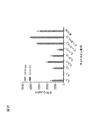


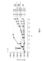


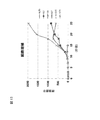
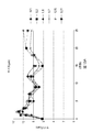


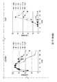


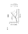


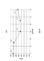
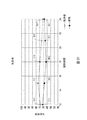

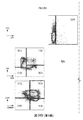


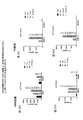


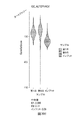


Classifications
-
- C—CHEMISTRY; METALLURGY
- C12—BIOCHEMISTRY; BEER; SPIRITS; WINE; VINEGAR; MICROBIOLOGY; ENZYMOLOGY; MUTATION OR GENETIC ENGINEERING
- C12N—MICROORGANISMS OR ENZYMES; COMPOSITIONS THEREOF; PROPAGATING, PRESERVING, OR MAINTAINING MICROORGANISMS; MUTATION OR GENETIC ENGINEERING; CULTURE MEDIA
- C12N5/00—Undifferentiated human, animal or plant cells, e.g. cell lines; Tissues; Cultivation or maintenance thereof; Culture media therefor
- C12N5/06—Animal cells or tissues; Human cells or tissues
- C12N5/0602—Vertebrate cells
- C12N5/0634—Cells from the blood or the immune system
- C12N5/0636—T lymphocytes
- C12N5/0638—Cytotoxic T lymphocytes [CTL] or lymphokine activated killer cells [LAK]
-
- C—CHEMISTRY; METALLURGY
- C12—BIOCHEMISTRY; BEER; SPIRITS; WINE; VINEGAR; MICROBIOLOGY; ENZYMOLOGY; MUTATION OR GENETIC ENGINEERING
- C12N—MICROORGANISMS OR ENZYMES; COMPOSITIONS THEREOF; PROPAGATING, PRESERVING, OR MAINTAINING MICROORGANISMS; MUTATION OR GENETIC ENGINEERING; CULTURE MEDIA
- C12N5/00—Undifferentiated human, animal or plant cells, e.g. cell lines; Tissues; Cultivation or maintenance thereof; Culture media therefor
- C12N5/06—Animal cells or tissues; Human cells or tissues
- C12N5/0602—Vertebrate cells
- C12N5/0634—Cells from the blood or the immune system
- C12N5/0636—T lymphocytes
-
- A—HUMAN NECESSITIES
- A61—MEDICAL OR VETERINARY SCIENCE; HYGIENE
- A61K—PREPARATIONS FOR MEDICAL, DENTAL OR TOILETRY PURPOSES
- A61K31/00—Medicinal preparations containing organic active ingredients
- A61K31/33—Heterocyclic compounds
- A61K31/395—Heterocyclic compounds having nitrogen as a ring hetero atom, e.g. guanethidine or rifamycins
- A61K31/495—Heterocyclic compounds having nitrogen as a ring hetero atom, e.g. guanethidine or rifamycins having six-membered rings with two or more nitrogen atoms as the only ring heteroatoms, e.g. piperazine or tetrazines
- A61K31/505—Pyrimidines; Hydrogenated pyrimidines, e.g. trimethoprim
- A61K31/519—Pyrimidines; Hydrogenated pyrimidines, e.g. trimethoprim ortho- or peri-condensed with heterocyclic rings
-
- A—HUMAN NECESSITIES
- A61—MEDICAL OR VETERINARY SCIENCE; HYGIENE
- A61K—PREPARATIONS FOR MEDICAL, DENTAL OR TOILETRY PURPOSES
- A61K35/00—Medicinal preparations containing materials or reaction products thereof with undetermined constitution
- A61K35/12—Materials from mammals; Compositions comprising non-specified tissues or cells; Compositions comprising non-embryonic stem cells; Genetically modified cells
- A61K35/14—Blood; Artificial blood
- A61K35/17—Lymphocytes; B-cells; T-cells; Natural killer cells; Interferon-activated or cytokine-activated lymphocytes
-
- A—HUMAN NECESSITIES
- A61—MEDICAL OR VETERINARY SCIENCE; HYGIENE
- A61K—PREPARATIONS FOR MEDICAL, DENTAL OR TOILETRY PURPOSES
- A61K38/00—Medicinal preparations containing peptides
- A61K38/16—Peptides having more than 20 amino acids; Gastrins; Somatostatins; Melanotropins; Derivatives thereof
- A61K38/17—Peptides having more than 20 amino acids; Gastrins; Somatostatins; Melanotropins; Derivatives thereof from animals; from humans
- A61K38/19—Cytokines; Lymphokines; Interferons
- A61K38/20—Interleukins [IL]
- A61K38/2086—IL-13 to IL-16
-
- A—HUMAN NECESSITIES
- A61—MEDICAL OR VETERINARY SCIENCE; HYGIENE
- A61K—PREPARATIONS FOR MEDICAL, DENTAL OR TOILETRY PURPOSES
- A61K38/00—Medicinal preparations containing peptides
- A61K38/16—Peptides having more than 20 amino acids; Gastrins; Somatostatins; Melanotropins; Derivatives thereof
- A61K38/17—Peptides having more than 20 amino acids; Gastrins; Somatostatins; Melanotropins; Derivatives thereof from animals; from humans
- A61K38/19—Cytokines; Lymphokines; Interferons
- A61K38/20—Interleukins [IL]
- A61K38/2093—Leukaemia inhibitory factor [LIF]
-
- A—HUMAN NECESSITIES
- A61—MEDICAL OR VETERINARY SCIENCE; HYGIENE
- A61K—PREPARATIONS FOR MEDICAL, DENTAL OR TOILETRY PURPOSES
- A61K39/00—Medicinal preparations containing antigens or antibodies
- A61K39/46—Cellular immunotherapy
- A61K39/461—Cellular immunotherapy characterised by the cell type used
- A61K39/4611—T-cells, e.g. tumor infiltrating lymphocytes [TIL], lymphokine-activated killer cells [LAK] or regulatory T cells [Treg]
-
- A—HUMAN NECESSITIES
- A61—MEDICAL OR VETERINARY SCIENCE; HYGIENE
- A61K—PREPARATIONS FOR MEDICAL, DENTAL OR TOILETRY PURPOSES
- A61K39/00—Medicinal preparations containing antigens or antibodies
- A61K39/46—Cellular immunotherapy
- A61K39/463—Cellular immunotherapy characterised by recombinant expression
- A61K39/4631—Chimeric Antigen Receptors [CAR]
-
- A—HUMAN NECESSITIES
- A61—MEDICAL OR VETERINARY SCIENCE; HYGIENE
- A61K—PREPARATIONS FOR MEDICAL, DENTAL OR TOILETRY PURPOSES
- A61K39/00—Medicinal preparations containing antigens or antibodies
- A61K39/46—Cellular immunotherapy
- A61K39/464—Cellular immunotherapy characterised by the antigen targeted or presented
- A61K39/4643—Vertebrate antigens
- A61K39/4644—Cancer antigens
- A61K39/464402—Receptors, cell surface antigens or cell surface determinants
- A61K39/464411—Immunoglobulin superfamily
- A61K39/464412—CD19 or B4
-
- A—HUMAN NECESSITIES
- A61—MEDICAL OR VETERINARY SCIENCE; HYGIENE
- A61K—PREPARATIONS FOR MEDICAL, DENTAL OR TOILETRY PURPOSES
- A61K39/00—Medicinal preparations containing antigens or antibodies
- A61K39/46—Cellular immunotherapy
- A61K39/464—Cellular immunotherapy characterised by the antigen targeted or presented
- A61K39/4643—Vertebrate antigens
- A61K39/4644—Cancer antigens
- A61K39/464402—Receptors, cell surface antigens or cell surface determinants
- A61K39/464416—Receptors for cytokines
- A61K39/464417—Receptors for tumor necrosis factors [TNF], e.g. lymphotoxin receptor [LTR], CD30
-
- A—HUMAN NECESSITIES
- A61—MEDICAL OR VETERINARY SCIENCE; HYGIENE
- A61P—SPECIFIC THERAPEUTIC ACTIVITY OF CHEMICAL COMPOUNDS OR MEDICINAL PREPARATIONS
- A61P35/00—Antineoplastic agents
-
- C—CHEMISTRY; METALLURGY
- C07—ORGANIC CHEMISTRY
- C07K—PEPTIDES
- C07K14/00—Peptides having more than 20 amino acids; Gastrins; Somatostatins; Melanotropins; Derivatives thereof
- C07K14/435—Peptides having more than 20 amino acids; Gastrins; Somatostatins; Melanotropins; Derivatives thereof from animals; from humans
- C07K14/705—Receptors; Cell surface antigens; Cell surface determinants
- C07K14/70503—Immunoglobulin superfamily
- C07K14/7051—T-cell receptor (TcR)-CD3 complex
-
- C—CHEMISTRY; METALLURGY
- C07—ORGANIC CHEMISTRY
- C07K—PEPTIDES
- C07K14/00—Peptides having more than 20 amino acids; Gastrins; Somatostatins; Melanotropins; Derivatives thereof
- C07K14/435—Peptides having more than 20 amino acids; Gastrins; Somatostatins; Melanotropins; Derivatives thereof from animals; from humans
- C07K14/705—Receptors; Cell surface antigens; Cell surface determinants
- C07K14/70503—Immunoglobulin superfamily
- C07K14/70517—CD8
-
- C—CHEMISTRY; METALLURGY
- C07—ORGANIC CHEMISTRY
- C07K—PEPTIDES
- C07K14/00—Peptides having more than 20 amino acids; Gastrins; Somatostatins; Melanotropins; Derivatives thereof
- C07K14/435—Peptides having more than 20 amino acids; Gastrins; Somatostatins; Melanotropins; Derivatives thereof from animals; from humans
- C07K14/705—Receptors; Cell surface antigens; Cell surface determinants
- C07K14/70578—NGF-receptor/TNF-receptor superfamily, e.g. CD27, CD30, CD40, CD95
-
- C—CHEMISTRY; METALLURGY
- C07—ORGANIC CHEMISTRY
- C07K—PEPTIDES
- C07K16/00—Immunoglobulins [IGs], e.g. monoclonal or polyclonal antibodies
- C07K16/18—Immunoglobulins [IGs], e.g. monoclonal or polyclonal antibodies against material from animals or humans
- C07K16/28—Immunoglobulins [IGs], e.g. monoclonal or polyclonal antibodies against material from animals or humans against receptors, cell surface antigens or cell surface determinants
- C07K16/2803—Immunoglobulins [IGs], e.g. monoclonal or polyclonal antibodies against material from animals or humans against receptors, cell surface antigens or cell surface determinants against the immunoglobulin superfamily
- C07K16/2809—Immunoglobulins [IGs], e.g. monoclonal or polyclonal antibodies against material from animals or humans against receptors, cell surface antigens or cell surface determinants against the immunoglobulin superfamily against the T-cell receptor (TcR)-CD3 complex
-
- C—CHEMISTRY; METALLURGY
- C07—ORGANIC CHEMISTRY
- C07K—PEPTIDES
- C07K16/00—Immunoglobulins [IGs], e.g. monoclonal or polyclonal antibodies
- C07K16/18—Immunoglobulins [IGs], e.g. monoclonal or polyclonal antibodies against material from animals or humans
- C07K16/28—Immunoglobulins [IGs], e.g. monoclonal or polyclonal antibodies against material from animals or humans against receptors, cell surface antigens or cell surface determinants
- C07K16/2878—Immunoglobulins [IGs], e.g. monoclonal or polyclonal antibodies against material from animals or humans against receptors, cell surface antigens or cell surface determinants against the NGF-receptor/TNF-receptor superfamily, e.g. CD27, CD30, CD40, CD95
-
- C—CHEMISTRY; METALLURGY
- C07—ORGANIC CHEMISTRY
- C07K—PEPTIDES
- C07K16/00—Immunoglobulins [IGs], e.g. monoclonal or polyclonal antibodies
- C07K16/18—Immunoglobulins [IGs], e.g. monoclonal or polyclonal antibodies against material from animals or humans
- C07K16/28—Immunoglobulins [IGs], e.g. monoclonal or polyclonal antibodies against material from animals or humans against receptors, cell surface antigens or cell surface determinants
- C07K16/2887—Immunoglobulins [IGs], e.g. monoclonal or polyclonal antibodies against material from animals or humans against receptors, cell surface antigens or cell surface determinants against CD20
-
- G—PHYSICS
- G01—MEASURING; TESTING
- G01N—INVESTIGATING OR ANALYSING MATERIALS BY DETERMINING THEIR CHEMICAL OR PHYSICAL PROPERTIES
- G01N33/00—Investigating or analysing materials by specific methods not covered by groups G01N1/00 - G01N31/00
- G01N33/48—Biological material, e.g. blood, urine; Haemocytometers
- G01N33/50—Chemical analysis of biological material, e.g. blood, urine; Testing involving biospecific ligand binding methods; Immunological testing
- G01N33/53—Immunoassay; Biospecific binding assay; Materials therefor
- G01N33/569—Immunoassay; Biospecific binding assay; Materials therefor for microorganisms, e.g. protozoa, bacteria, viruses
- G01N33/56966—Animal cells
- G01N33/56972—White blood cells
-
- A—HUMAN NECESSITIES
- A61—MEDICAL OR VETERINARY SCIENCE; HYGIENE
- A61K—PREPARATIONS FOR MEDICAL, DENTAL OR TOILETRY PURPOSES
- A61K39/00—Medicinal preparations containing antigens or antibodies
- A61K2039/505—Medicinal preparations containing antigens or antibodies comprising antibodies
-
- A—HUMAN NECESSITIES
- A61—MEDICAL OR VETERINARY SCIENCE; HYGIENE
- A61K—PREPARATIONS FOR MEDICAL, DENTAL OR TOILETRY PURPOSES
- A61K39/00—Medicinal preparations containing antigens or antibodies
- A61K2039/545—Medicinal preparations containing antigens or antibodies characterised by the dose, timing or administration schedule
-
- A—HUMAN NECESSITIES
- A61—MEDICAL OR VETERINARY SCIENCE; HYGIENE
- A61K—PREPARATIONS FOR MEDICAL, DENTAL OR TOILETRY PURPOSES
- A61K2239/00—Indexing codes associated with cellular immunotherapy of group A61K39/46
- A61K2239/10—Indexing codes associated with cellular immunotherapy of group A61K39/46 characterized by the structure of the chimeric antigen receptor [CAR]
- A61K2239/11—Antigen recognition domain
- A61K2239/13—Antibody-based
-
- A—HUMAN NECESSITIES
- A61—MEDICAL OR VETERINARY SCIENCE; HYGIENE
- A61K—PREPARATIONS FOR MEDICAL, DENTAL OR TOILETRY PURPOSES
- A61K2239/00—Indexing codes associated with cellular immunotherapy of group A61K39/46
- A61K2239/31—Indexing codes associated with cellular immunotherapy of group A61K39/46 characterized by the route of administration
-
- A—HUMAN NECESSITIES
- A61—MEDICAL OR VETERINARY SCIENCE; HYGIENE
- A61K—PREPARATIONS FOR MEDICAL, DENTAL OR TOILETRY PURPOSES
- A61K2239/00—Indexing codes associated with cellular immunotherapy of group A61K39/46
- A61K2239/38—Indexing codes associated with cellular immunotherapy of group A61K39/46 characterised by the dose, timing or administration schedule
-
- C—CHEMISTRY; METALLURGY
- C07—ORGANIC CHEMISTRY
- C07K—PEPTIDES
- C07K2317/00—Immunoglobulins specific features
- C07K2317/50—Immunoglobulins specific features characterized by immunoglobulin fragments
- C07K2317/52—Constant or Fc region; Isotype
- C07K2317/53—Hinge
-
- C—CHEMISTRY; METALLURGY
- C07—ORGANIC CHEMISTRY
- C07K—PEPTIDES
- C07K2317/00—Immunoglobulins specific features
- C07K2317/50—Immunoglobulins specific features characterized by immunoglobulin fragments
- C07K2317/56—Immunoglobulins specific features characterized by immunoglobulin fragments variable (Fv) region, i.e. VH and/or VL
- C07K2317/565—Complementarity determining region [CDR]
-
- C—CHEMISTRY; METALLURGY
- C07—ORGANIC CHEMISTRY
- C07K—PEPTIDES
- C07K2317/00—Immunoglobulins specific features
- C07K2317/60—Immunoglobulins specific features characterized by non-natural combinations of immunoglobulin fragments
- C07K2317/62—Immunoglobulins specific features characterized by non-natural combinations of immunoglobulin fragments comprising only variable region components
- C07K2317/622—Single chain antibody (scFv)
-
- C—CHEMISTRY; METALLURGY
- C07—ORGANIC CHEMISTRY
- C07K—PEPTIDES
- C07K2319/00—Fusion polypeptide
- C07K2319/01—Fusion polypeptide containing a localisation/targetting motif
- C07K2319/02—Fusion polypeptide containing a localisation/targetting motif containing a signal sequence
-
- C—CHEMISTRY; METALLURGY
- C07—ORGANIC CHEMISTRY
- C07K—PEPTIDES
- C07K2319/00—Fusion polypeptide
- C07K2319/01—Fusion polypeptide containing a localisation/targetting motif
- C07K2319/03—Fusion polypeptide containing a localisation/targetting motif containing a transmembrane segment
-
- C—CHEMISTRY; METALLURGY
- C12—BIOCHEMISTRY; BEER; SPIRITS; WINE; VINEGAR; MICROBIOLOGY; ENZYMOLOGY; MUTATION OR GENETIC ENGINEERING
- C12N—MICROORGANISMS OR ENZYMES; COMPOSITIONS THEREOF; PROPAGATING, PRESERVING, OR MAINTAINING MICROORGANISMS; MUTATION OR GENETIC ENGINEERING; CULTURE MEDIA
- C12N2501/00—Active agents used in cell culture processes, e.g. differentation
- C12N2501/20—Cytokines; Chemokines
- C12N2501/23—Interleukins [IL]
- C12N2501/2302—Interleukin-2 (IL-2)
-
- C—CHEMISTRY; METALLURGY
- C12—BIOCHEMISTRY; BEER; SPIRITS; WINE; VINEGAR; MICROBIOLOGY; ENZYMOLOGY; MUTATION OR GENETIC ENGINEERING
- C12N—MICROORGANISMS OR ENZYMES; COMPOSITIONS THEREOF; PROPAGATING, PRESERVING, OR MAINTAINING MICROORGANISMS; MUTATION OR GENETIC ENGINEERING; CULTURE MEDIA
- C12N2501/00—Active agents used in cell culture processes, e.g. differentation
- C12N2501/20—Cytokines; Chemokines
- C12N2501/23—Interleukins [IL]
- C12N2501/2306—Interleukin-6 (IL-6)
-
- C—CHEMISTRY; METALLURGY
- C12—BIOCHEMISTRY; BEER; SPIRITS; WINE; VINEGAR; MICROBIOLOGY; ENZYMOLOGY; MUTATION OR GENETIC ENGINEERING
- C12N—MICROORGANISMS OR ENZYMES; COMPOSITIONS THEREOF; PROPAGATING, PRESERVING, OR MAINTAINING MICROORGANISMS; MUTATION OR GENETIC ENGINEERING; CULTURE MEDIA
- C12N2501/00—Active agents used in cell culture processes, e.g. differentation
- C12N2501/20—Cytokines; Chemokines
- C12N2501/23—Interleukins [IL]
- C12N2501/2307—Interleukin-7 (IL-7)
-
- C—CHEMISTRY; METALLURGY
- C12—BIOCHEMISTRY; BEER; SPIRITS; WINE; VINEGAR; MICROBIOLOGY; ENZYMOLOGY; MUTATION OR GENETIC ENGINEERING
- C12N—MICROORGANISMS OR ENZYMES; COMPOSITIONS THEREOF; PROPAGATING, PRESERVING, OR MAINTAINING MICROORGANISMS; MUTATION OR GENETIC ENGINEERING; CULTURE MEDIA
- C12N2501/00—Active agents used in cell culture processes, e.g. differentation
- C12N2501/20—Cytokines; Chemokines
- C12N2501/23—Interleukins [IL]
- C12N2501/2315—Interleukin-15 (IL-15)
-
- C—CHEMISTRY; METALLURGY
- C12—BIOCHEMISTRY; BEER; SPIRITS; WINE; VINEGAR; MICROBIOLOGY; ENZYMOLOGY; MUTATION OR GENETIC ENGINEERING
- C12N—MICROORGANISMS OR ENZYMES; COMPOSITIONS THEREOF; PROPAGATING, PRESERVING, OR MAINTAINING MICROORGANISMS; MUTATION OR GENETIC ENGINEERING; CULTURE MEDIA
- C12N2501/00—Active agents used in cell culture processes, e.g. differentation
- C12N2501/20—Cytokines; Chemokines
- C12N2501/23—Interleukins [IL]
- C12N2501/2321—Interleukin-21 (IL-21)
-
- C—CHEMISTRY; METALLURGY
- C12—BIOCHEMISTRY; BEER; SPIRITS; WINE; VINEGAR; MICROBIOLOGY; ENZYMOLOGY; MUTATION OR GENETIC ENGINEERING
- C12N—MICROORGANISMS OR ENZYMES; COMPOSITIONS THEREOF; PROPAGATING, PRESERVING, OR MAINTAINING MICROORGANISMS; MUTATION OR GENETIC ENGINEERING; CULTURE MEDIA
- C12N2501/00—Active agents used in cell culture processes, e.g. differentation
- C12N2501/50—Cell markers; Cell surface determinants
- C12N2501/505—CD4; CD8
-
- C—CHEMISTRY; METALLURGY
- C12—BIOCHEMISTRY; BEER; SPIRITS; WINE; VINEGAR; MICROBIOLOGY; ENZYMOLOGY; MUTATION OR GENETIC ENGINEERING
- C12N—MICROORGANISMS OR ENZYMES; COMPOSITIONS THEREOF; PROPAGATING, PRESERVING, OR MAINTAINING MICROORGANISMS; MUTATION OR GENETIC ENGINEERING; CULTURE MEDIA
- C12N2501/00—Active agents used in cell culture processes, e.g. differentation
- C12N2501/50—Cell markers; Cell surface determinants
- C12N2501/515—CD3, T-cell receptor complex
-
- C—CHEMISTRY; METALLURGY
- C12—BIOCHEMISTRY; BEER; SPIRITS; WINE; VINEGAR; MICROBIOLOGY; ENZYMOLOGY; MUTATION OR GENETIC ENGINEERING
- C12N—MICROORGANISMS OR ENZYMES; COMPOSITIONS THEREOF; PROPAGATING, PRESERVING, OR MAINTAINING MICROORGANISMS; MUTATION OR GENETIC ENGINEERING; CULTURE MEDIA
- C12N2501/00—Active agents used in cell culture processes, e.g. differentation
- C12N2501/998—Proteins not provided for elsewhere
-
- C—CHEMISTRY; METALLURGY
- C12—BIOCHEMISTRY; BEER; SPIRITS; WINE; VINEGAR; MICROBIOLOGY; ENZYMOLOGY; MUTATION OR GENETIC ENGINEERING
- C12N—MICROORGANISMS OR ENZYMES; COMPOSITIONS THEREOF; PROPAGATING, PRESERVING, OR MAINTAINING MICROORGANISMS; MUTATION OR GENETIC ENGINEERING; CULTURE MEDIA
- C12N2510/00—Genetically modified cells
-
- Y—GENERAL TAGGING OF NEW TECHNOLOGICAL DEVELOPMENTS; GENERAL TAGGING OF CROSS-SECTIONAL TECHNOLOGIES SPANNING OVER SEVERAL SECTIONS OF THE IPC; TECHNICAL SUBJECTS COVERED BY FORMER USPC CROSS-REFERENCE ART COLLECTIONS [XRACs] AND DIGESTS
- Y02—TECHNOLOGIES OR APPLICATIONS FOR MITIGATION OR ADAPTATION AGAINST CLIMATE CHANGE
- Y02A—TECHNOLOGIES FOR ADAPTATION TO CLIMATE CHANGE
- Y02A50/00—TECHNOLOGIES FOR ADAPTATION TO CLIMATE CHANGE in human health protection, e.g. against extreme weather
- Y02A50/30—Against vector-borne diseases, e.g. mosquito-borne, fly-borne, tick-borne or waterborne diseases whose impact is exacerbated by climate change
Abstract
本発明は、キメラ抗原受容体(CAR)を発現する免疫エフェクター細胞(例えば、T細胞、NK細胞)を作製する方法及びそうした方法によって生成される組成物を提供する。【選択図】なしThe present invention provides a method for producing an immune effector cell (eg, T cell, NK cell) expressing a chimeric antigen receptor (CAR) and a composition produced by such method. [Selection diagram] None
Description
関連出願
本願は、2018年8月31日に提出された米国仮特許出願第62/726,155号明細書、2018年11月30日に提出された米国仮特許出願第62/773,679号明細書及び2019年6月7日に提出された米国仮特許出願第62/858,482号明細書(これらの各々の内容全体が参照により本明細書に組み込まれる)に対する優先権を主張する。
Related Applications This application is a US provisional patent application No. 62 / 726,155 filed on August 31, 2018, and a US provisional patent application No. 62 / 773,679 filed on November 30, 2018. Claims priority over the specification and US Provisional Patent Application No. 62 / 858,482 filed June 7, 2019, the entire contents of each of which are incorporated herein by reference.
配列表
本願は、ASCIIフォーマットで電子出願されており、その全体が参照により本明細書に組み込まれる配列表を含む。前記ASCIIコピーは、2019年8月16日に作成され、名称がN2067−7153WO_SL.txtであり、サイズが260,532バイトである。
Sequence Listing This application is electronically filed in ASCII format and includes a sequence listing, which is incorporated herein by reference in its entirety. The ASCII copy was made on August 16, 2019 and is named N2067-7153WO_SL. It is txt and has a size of 260,532 bytes.
本発明は、概して、キメラ抗原受容体(CAR)を発現するように操作された免疫エフェクター細胞(例えば、T細胞又はNK細胞)を作製する方法及びそれを含む組成物に関する。 The invention generally relates to methods of making immune effector cells (eg, T cells or NK cells) engineered to express a chimeric antigen receptor (CAR) and compositions containing them.
T細胞、特にキメラ抗原受容体(CAR)を形質導入したT細胞での養子細胞移入(ACT)療法は、いくつかの血液癌の試験で有望性が示されている。遺伝子改変T細胞の製造は、現在、複雑なプロセスである。CAR発現細胞療法製品の生産を改善し、製品の品質を高めると共に製品の治療効果を最大化する方法及びプロセスが必要とされている。 Adoptive cell transfer (ACT) therapy with T cells, especially chimeric antigen receptor (CAR) transduced T cells, has shown promise in several hematological cancer trials. The production of genetically modified T cells is currently a complex process. There is a need for methods and processes to improve the production of CAR-expressing cell therapy products, improve the quality of the products and maximize the therapeutic effects of the products.
本開示は、CARを発現するように操作された免疫エフェクター細胞(例えば、T細胞又はNK細胞)を作製する方法及びそうした方法を使用して生成される組成物に関する。また、対象の疾患、例えば癌を処置するためにそのような組成物を使用する方法も開示される。 The present disclosure relates to methods of making immune effector cells (eg, T cells or NK cells) engineered to express CAR and compositions produced using such methods. Also disclosed are methods of using such compositions to treat a disease of interest, such as cancer.
一部の実施形態では、本発明は、キメラ抗原受容体(CAR)を発現する細胞(例えば、T細胞)の集団を作製する方法を特徴とし、本方法は、(i)細胞(例えば、T細胞、例えば凍結された又は新鮮な白血球アフェレーシス産物から単離されたT細胞)の集団を、CD3/TCR複合体を刺激する薬剤及び/又は細胞の表面上の共刺激分子を刺激する薬剤と接触(例えば、結合)させるステップ;(ii)細胞(例えば、T細胞)の集団を、CARをコードする核酸分子(例えば、DNA又はRNA分子)と接触させ、それにより核酸分子を含む細胞(例えば、T細胞)の集団を提供するステップ、及び(iii)保存(例えば、凍結保存培地中の細胞の集団を再製剤化すること)又は投与のために細胞(例えば、T細胞)の集団を採取するステップを含み、(a)ステップ(ii)は、ステップ(i)と一緒に又はステップ(i)の開始後の20時間以内、例えばステップ(i)の開始後の12、13、14、15、16、17若しくは18時間以内、例えばステップ(i)の開始後の18時間以内に実施され、及びステップ(iii)は、ステップ(i)の開始後の26時間以内、例えばステップ(i)の開始後の22、23、24又は25時間以内、例えばステップ(i)の開始後の24時間以内に実施されるか;(b)ステップ(ii)は、ステップ(i)と一緒に又はステップ(i)の開始後の20時間以内、例えばステップ(i)の開始後の12、13、14、15、16、17若しくは18時間以内、例えばステップ(i)の開始後の18時間以内に実施され、及びステップ(iii)は、ステップ(ii)の開始後の30時間以内、例えばステップ(ii)の開始後の22、23、24、25、26、27、28、29又は30時間以内に実施されるか;又は(c)ステップ(iii)からの細胞の集団は、例えば、ステップ(i)の開始時の細胞の集団と比べて、生存細胞の数によって評価されて増殖されないか又は5、6、7、8、9、10、11、12、13、14、15、16、17、18、19、20、25、30、35若しくは40%以下、例えば10%以下だけ増殖される。一部の実施形態では、ステップ(ii)における核酸分子は、DNA分子である。一部の実施形態では、ステップ(ii)における核酸分子は、RNA分子である。一部の実施形態では、ステップ(ii)における核酸分子は、ウイルスベクター、例えばレンチウイルスベクター、アデノウイルスベクター又はレトロウイルスベクターから選択されるウイルスベクター上にある。一部の実施形態では、ステップ(ii)における核酸分子は、非ウイルスベクター上にある。一部の実施形態では、ステップ(ii)における核酸分子は、プラスミド上にある。一部の実施形態では、ステップ(ii)における核酸分子は、いずれのベクター上にもない。一部の実施形態では、ステップ(ii)は、細胞(例えば、T細胞)の集団を、CARをコードする核酸分子を含むウイルスベクターで形質導入することを含む。一部の実施形態では、ステップ(ii)は、ステップ(i)と一緒に実施される。一部の実施形態では、ステップ(ii)は、ステップ(i)の開始後の20時間以内に実施される。一部の実施形態では、ステップ(ii)は、ステップ(i)の開始後の12、13、14、15、16、17又は18時間以内に実施される。一部の実施形態では、ステップ(ii)は、ステップ(i)の開始後の18時間以内に実施される。一部の実施形態では、ステップ(iii)は、ステップ(i)の開始後の26時間以内に実施される。一部の実施形態では、ステップ(iii)は、ステップ(i)の開始後の22、23、24又は25時間以内に実施される。一部の実施形態では、ステップ(iii)は、ステップ(i)の開始後の24時間以内に実施される。一部の実施形態では、ステップ(iii)は、ステップ(ii)の開始後の30時間以内に実施される。一部の実施形態では、ステップ(iii)は、ステップ(ii)の開始後の22、23、24、25、26、27、28、29又は30時間以内に実施される。 In some embodiments, the invention features a method of making a population of cells (eg, T cells) expressing a chimeric antigen receptor (CAR), wherein the method is (i) cells (eg, T). Populations of cells (eg, T cells isolated from frozen or fresh leukocyte afferesis products) are contacted with agents that stimulate the CD3 / TCR complex and / or agents that stimulate costimulatory molecules on the surface of the cells. Steps to (eg, bind); (ii) A population of cells (eg, T cells) is contacted with a CAR-encoding nucleic acid molecule (eg, DNA or RNA molecule), thereby containing a cell containing the nucleic acid molecule (eg, eg, RNA molecule). A step of providing a population of cells (eg, T cells), and (iii) harvesting a population of cells (eg, T cells) for storage (eg, repreparing the population of cells in a cryopreservation medium) or administration. Including steps, (a) step (ii) may be performed with step (i) or within 20 hours after the start of step (i), eg, 12, 13, 14, 15, after the start of step (i). It is performed within 16, 17 or 18 hours, eg, within 18 hours after the start of step (i), and step (iii) is within 26 hours after the start of step (i), eg, the start of step (i). Will it be performed within the next 22, 23, 24 or 25 hours, eg, within 24 hours after the start of step (i); (b) step (ii) with or with step (i) or step (i). ) Within 20 hours after the start, eg, within 12, 13, 14, 15, 16, 17 or 18 hours after the start of step (i), eg, within 18 hours after the start of step (i). And step (iii) is performed within 30 hours after the start of step (ii), eg, within 22, 23, 24, 25, 26, 27, 28, 29 or 30 hours after the start of step (ii). Or (c) the population of cells from step (iii) is not proliferated as assessed by the number of viable cells compared to, for example, the population of cells at the beginning of step (i), or 5,6. , 7, 8, 9, 10, 11, 12, 13, 14, 15, 16, 17, 18, 19, 20, 25, 30, 35 or 40% or less, for example 10% or less. In some embodiments, the nucleic acid molecule in step (ii) is a DNA molecule. In some embodiments, the nucleic acid molecule in step (ii) is an RNA molecule. In some embodiments, the nucleic acid molecule in step (ii) is on a viral vector selected from viral vectors such as lentiviral vectors, adenoviral vectors or retroviral vectors. In some embodiments, the nucleic acid molecule in step (ii) is on a non-viral vector. In some embodiments, the nucleic acid molecule in step (ii) is on a plasmid. In some embodiments, the nucleic acid molecule in step (ii) is not on any of the vectors. In some embodiments, step (ii) involves transducing a population of cells (eg, T cells) with a viral vector containing a nucleic acid molecule encoding CAR. In some embodiments, step (ii) is performed in conjunction with step (i). In some embodiments, step (ii) is performed within 20 hours after the start of step (i). In some embodiments, step (ii) is performed within 12, 13, 14, 15, 16, 17 or 18 hours after the start of step (i). In some embodiments, step (ii) is performed within 18 hours after the start of step (i). In some embodiments, the step (iii) is performed within 26 hours after the start of step (i). In some embodiments, the step (iii) is performed within 22, 23, 24 or 25 hours after the start of step (i). In some embodiments, the step (iii) is performed within 24 hours after the start of step (i). In some embodiments, the step (iii) is performed within 30 hours after the start of the step (iii). In some embodiments, the step (iii) is performed within 22, 23, 24, 25, 26, 27, 28, 29 or 30 hours after the start of the step (iii).
一部の実施形態では、ステップ(iii)からの細胞の集団は、増殖されない。一部の実施形態では、ステップ(iii)からの細胞の集団は、例えば、ステップ(i)の開始時の細胞の集団と比べて、生存細胞の数によって評価されて5、6、7、8、9、10、11、12、13、14、15、16、17、18、19、20、25、30、35又は40%以下だけ増殖される。一部の実施形態では、ステップ(iii)からの細胞の集団は、例えば、ステップ(i)の開始時の細胞の集団と比べて、生存細胞の数によって評価されて10%以下だけ増殖される。 In some embodiments, the population of cells from step (iii) is not proliferated. In some embodiments, the population of cells from step (iii) is assessed by the number of viable cells, eg, 5, 6, 7, 8 as compared to the population of cells at the beginning of step (i). , 9, 10, 11, 12, 13, 14, 15, 16, 17, 18, 19, 20, 25, 30, 35 or 40% or less. In some embodiments, the population of cells from step (iii) is proliferated by 10% or less, as assessed by the number of viable cells, as compared to, for example, the population of cells at the beginning of step (i). ..
一部の実施形態では、CD3/TCR複合体を刺激する薬剤は、CD3を刺激する薬剤である。一部の実施形態では、共刺激分子を刺激する薬剤は、CD28、ICOS、CD27、HVEM、LIGHT、CD40、4−1BB、OX40、DR3、GITR、CD30、TIM1、CD2、CD226又はそれらの任意の組み合わせを刺激する薬剤である。一部の実施形態では、共刺激分子を刺激する薬剤は、CD28を刺激する薬剤である。一部の実施形態では、CD3/TCR複合体を刺激する薬剤は、抗体(例えば、単一ドメイン抗体(例えば、重鎖可変ドメイン抗体)、ペプチボディ、Fabフラグメント若しくはscFv)、小分子又はリガンド(例えば、天然に存在するリガンド、組換えリガンド若しくはキメラリガンド)から選択される。一部の実施形態では、共刺激分子を刺激する薬剤は、抗体(例えば、単一ドメイン抗体(例えば、重鎖可変ドメイン抗体)、ペプチボディ、Fabフラグメント若しくはscFv)、小分子又はリガンド(例えば、天然に存在するリガンド、組換えリガンド若しくはキメラリガンド)から選択される。一部の実施形態では、CD3/TCR複合体を刺激する薬剤は、ビーズを含まない。一部の実施形態では、共刺激分子を刺激する薬剤は、ビーズを含まない。一部の実施形態では、CD3/TCR複合体を刺激する薬剤は、抗CD3抗体を含む。一部の実施形態では、共刺激分子を刺激する薬剤は、抗CD28抗体を含む。一部の実施形態では、CD3/TCR複合体を刺激する薬剤は、コロイドポリマーナノマトリックスに共有結合された抗CD3抗体を含む。一部の実施形態では、共刺激分子を刺激する薬剤は、コロイドポリマーナノマトリックスに共有結合された抗CD28抗体を含む。一部の実施形態では、CD3/TCR複合体を刺激する薬剤及び共刺激分子を刺激する薬剤は、T Cell TransAct(商標)を含む。 In some embodiments, the agent that stimulates the CD3 / TCR complex is an agent that stimulates CD3. In some embodiments, the agent stimulating the co-stimulator molecule is CD28, ICOS, CD27, HVEM, LIGHT, CD40, 4-1BB, OX40, DR3, GITR, CD30, TIM1, CD2, CD226 or any of them. It is a drug that stimulates the combination. In some embodiments, the agent that stimulates the co-stimulator molecule is an agent that stimulates CD28. In some embodiments, the agent stimulating the CD3 / TCR complex is an antibody (eg, single domain antibody (eg, heavy chain variable domain antibody), peptide body, Fab fragment or scFv), small molecule or ligand (eg, eg). , Naturally occurring ligands, recombinant ligands or chimeric ligands). In some embodiments, the agent stimulating the costimulatory molecule is an antibody (eg, a single domain antibody (eg, a heavy chain variable domain antibody), a peptide body, a Fab fragment or scFv), a small molecule or a ligand (eg, natural). It is selected from the ligands, recombinant ligands or chimeric ligands present in. In some embodiments, the agent that stimulates the CD3 / TCR complex does not include beads. In some embodiments, the agent that stimulates the co-stimulator molecule does not include beads. In some embodiments, the agent that stimulates the CD3 / TCR complex comprises an anti-CD3 antibody. In some embodiments, the agent stimulating the co-stimulator molecule comprises an anti-CD28 antibody. In some embodiments, the agent stimulating the CD3 / TCR complex comprises an anti-CD3 antibody covalently attached to a colloidal polymer nanomatrix. In some embodiments, the agent stimulating the co-stimulator molecule comprises an anti-CD28 antibody covalently attached to a colloidal polymer nanomatrix. In some embodiments, agents that stimulate the CD3 / TCR complex and agents that stimulate co-stimulatory molecules include T Cell TransAct ™.
一部の実施形態では、CD3/TCR複合体を刺激する薬剤は、ハイドロゲルを含まない。一部の実施形態では、共刺激分子を刺激する薬剤は、ハイドロゲルを含まない。一部の実施形態では、CD3/TCR複合体を刺激する薬剤は、アルギン酸塩を含まない。一部の実施形態では、共刺激分子を刺激する薬剤は、アルギン酸塩を含まない。 In some embodiments, the agent that stimulates the CD3 / TCR complex does not include hydrogel. In some embodiments, the agent that stimulates the co-stimulator molecule does not include hydrogel. In some embodiments, the agent that stimulates the CD3 / TCR complex does not contain alginate. In some embodiments, the agent that stimulates the co-stimulator molecule is free of alginate.
一部の実施形態では、CD3/TCR複合体を刺激する薬剤は、ハイドロゲルを含む。一部の実施形態では、共刺激分子を刺激する薬剤は、ハイドロゲルを含む。一部の実施形態では、CD3/TCR複合体を刺激する薬剤は、アルギン酸塩を含む。一部の実施形態では、共刺激分子を刺激する薬剤は、アルギン酸塩を含む。一部の実施形態では、CD3/TCR複合体を刺激する薬剤又は共刺激分子を刺激する薬剤は、Quad Technologies製のMagCloudz(商標)を含む。 In some embodiments, the agent that stimulates the CD3 / TCR complex comprises a hydrogel. In some embodiments, the agent that stimulates the co-stimulator molecule comprises a hydrogel. In some embodiments, the agent that stimulates the CD3 / TCR complex comprises alginate. In some embodiments, the agent that stimulates the co-stimulator molecule comprises alginate. In some embodiments, agents that stimulate the CD3 / TCR complex or agents that stimulate co-stimulatory molecules include MagCloodz ™ from Quad Technologies.
一部の実施形態では、ステップ(i)は、ステップ(iii)からの細胞の集団中のCAR発現細胞のパーセンテージを増加させ、例えば、ステップ(iii)からの細胞の集団は、ステップ(i)なしである以外には同様の方法によって作製された細胞と比べて、CAR発現細胞のより高いパーセンテージ(例えば、少なくとも10、20、30、40、50又は60%高い)を示す。 In some embodiments, step (i) increases the percentage of CAR-expressing cells in the population of cells from step (iii), eg, the population of cells from step (iii) is step (i). It shows a higher percentage of CAR-expressing cells (eg, at least 10, 20, 30, 40, 50 or 60% higher) compared to cells produced by similar methods other than none.
一部の実施形態では、ステップ(iii)からの細胞の集団中のナイーブ細胞、例えばナイーブT細胞、例えばCD45RA+CD45RO−CCR7+T細胞のパーセンテージは、ステップ(i)の開始時の細胞の集団中のナイーブ細胞、例えばナイーブT細胞、例えばCD45RA+CD45RO−CCR7+細胞のパーセンテージと同じである。一部の実施形態では、ステップ(iii)からの細胞の集団中のナイーブ細胞、例えばナイーブT細胞、例えばCD45RA+CD45RO−CCR7+T細胞のパーセンテージは、ステップ(i)の開始時の細胞の集団中のナイーブ細胞、例えばナイーブT細胞、例えばCD45RA+CD45RO−CCR7+細胞のパーセンテージと3、4、5、6、7、8、9、10、11又は12%以下だけ異なる。一部の実施形態では、ステップ(iii)からの細胞の集団中のナイーブ細胞、例えばナイーブT細胞、例えばCD45RA+CD45RO−CCR7+T細胞のパーセンテージは、ステップ(i)の開始時の細胞の集団中のナイーブ細胞、例えばナイーブT細胞、例えばCD45RA+CD45RO−CCR7+細胞のパーセンテージと5又は10%以下だけ異なる。 In some embodiments, the percentage of naive cells in the cell population from step (iii), such as naive T cells, such as CD45RA + CD45RO-CCR7 + T cells, is the percentage of naive cells in the cell population at the start of step (i). , For example the percentage of naive T cells, eg CD45RA + CD45RO-CCR7 + cells. In some embodiments, the percentage of naive cells in the cell population from step (iii), such as naive T cells, such as CD45RA + CD45RO-CCR7 + T cells, is the percentage of naive cells in the cell population at the start of step (i). For example, the percentage of naive T cells, such as CD45RA + CD45RO-CCR7 + cells, differs by 3, 4, 5, 6, 7, 8, 9, 10, 11 or 12% or less. In some embodiments, the percentage of naive cells in the cell population from step (iii), such as naive T cells, such as CD45RA + CD45RO-CCR7 + T cells, is the percentage of naive cells in the cell population at the start of step (i). , For example, differ by 5 or 10% or less from the percentage of naive T cells, such as CD45RA + CD45RO-CCR7 + cells.
一部の実施形態では、ステップ(iii)からの細胞の集団は、ステップ(iii)が、ステップ(i)の開始後、26時間を超えて、例えばステップ(i)の開始後、5、6、7、8、9、10、11又は12日を超えてから実施される以外には同様の方法によって作製された細胞と比べて、ナイーブ細胞、例えばナイーブT細胞、例えばCD45RA+CD45RO−CCR7+T細胞のより高いパーセンテージ(例えば、少なくとも10、11、12、13、14、15、16、17、18、19、20、25、30、35又は40%高い)を示す。一部の実施形態では、ステップ(iii)からの細胞の集団は、ステップ(ii)後且つステップ(iii)前に細胞(例えば、T細胞)の集団をインビトロで3日超、例えば5、6、7、8又は9日にわたって増殖させるステップをさらに含む以外には同様の方法によって作製された細胞と比べて、ナイーブ細胞、例えばナイーブT細胞、例えばCD45RA+CD45RO−CCR7+T細胞のより高いパーセンテージ(例えば、少なくとも10、11、12、13、14、15、16、17、18、19、20、25、30、35又は40%高い)を示す。
In some embodiments, the population of cells from step (iii) is such that step (iii) exceeds 26 hours after the start of step (i), eg, 5, 6 after the start of step (i). , 7, 8, 9, 10, 11 or 12 days from naive cells, such as naive T cells, such as CD45RA + CD45RO-CCR7 + T cells, as compared to cells produced by similar methods except that performed after more than 12 days. Shows high percentages (eg, at least 10,11,12,13,14,15,16,17,18,19,20,25,30,35 or 40% higher). In some embodiments, the population of cells from step (iii) is a population of cells (eg, T cells) in vitro after step (iii) and before step (iii) for more than 3 days,
一部の実施形態では、ステップ(iii)からの細胞の集団中のセントラルメモリー細胞、例えばセントラルメモリーT細胞、例えばCD95+セントラルメモリーT細胞のパーセンテージは、ステップ(i)の開始時の細胞の集団中のセントラルメモリー細胞、例えばセントラルメモリーT細胞、例えばCD95+セントラルメモリーT細胞のパーセンテージと同じである。一部の実施形態では、ステップ(iii)からの細胞の集団中のセントラルメモリー細胞、例えばセントラルメモリーT細胞、例えばCD95+セントラルメモリーT細胞のパーセンテージは、ステップ(i)の開始時の細胞の集団中のセントラルメモリー細胞、例えばセントラルメモリーT細胞、例えばCD95+セントラルメモリーT細胞のパーセンテージと3、4、5、6、7、8、9、10、11又は12%以下だけ異なる。一部の実施形態では、ステップ(iii)からの細胞の集団中のセントラルメモリー細胞、例えばセントラルメモリーT細胞、例えばCD95+セントラルメモリーT細胞のパーセンテージは、ステップ(i)の開始時の細胞の集団中のセントラルメモリー細胞、例えばセントラルメモリーT細胞、例えばCD95+セントラルメモリーT細胞のパーセンテージと5又は10%以下だけ異なる。 In some embodiments, the percentage of central memory cells in the population of cells from step (iii), such as central memory T cells, such as CD95 + central memory T cells, is in the population of cells at the start of step (i). It is the same as the percentage of central memory cells, such as central memory T cells, such as CD95 + central memory T cells. In some embodiments, the percentage of central memory cells in the population of cells from step (iii), such as central memory T cells, such as CD95 + central memory T cells, is in the population of cells at the start of step (i). Central memory cells, such as Central Memory T cells, such as CD95 + Central Memory T cells, differ from the percentage of Central Memory T cells by 3, 4, 5, 6, 7, 8, 9, 10, 11 or 12% or less. In some embodiments, the percentage of central memory cells in the population of cells from step (iii), such as central memory T cells, such as CD95 + central memory T cells, is in the population of cells at the start of step (i). It differs from the percentage of central memory cells, such as central memory T cells, such as CD95 + central memory T cells, by 5 or 10% or less.
一部の実施形態では、ステップ(iii)からの細胞の集団は、ステップ(iii)が、ステップ(i)の開始後、26時間を超えて、例えばステップ(i)の開始後、5、6、7、8、9、10、11又は12日を超えてから実施される以外には同様の方法によって作製された細胞と比べて、セントラルメモリー細胞、例えばセントラルメモリーT細胞、例えばCD95+セントラルメモリーT細胞のより低いパーセンテージ(例えば、少なくとも10、11、12、13、14、15、16、17、18、19、20、25、30、35又は40%低い)を示す。一部の実施形態では、ステップ(iii)からの細胞の集団は、ステップ(ii)後且つステップ(iii)前に細胞(例えば、T細胞)の集団をインビトロで3日超、例えば5、6、7、8又は9日にわたって増殖させるステップをさらに含む以外には同様の方法によって作製された細胞と比べて、セントラルメモリー細胞、例えばセントラルメモリーT細胞、例えばCD95+セントラルメモリーT細胞のより低いパーセンテージ(例えば、少なくとも10、11、12、13、14、15、16、17、18、19、20、25、30、35又は40%低い)を示す。
In some embodiments, the population of cells from step (iii) is such that step (iii) exceeds 26 hours after the start of step (i), eg, after the start of step (i) 5,6. Central memory cells, such as central memory T cells, such as CD95 + central memory T, as compared to cells produced by similar methods except that they are performed after more than 7, 8, 9, 10, 11 or 12 days. Shows a lower percentage of cells (eg, at least 10, 11, 12, 13, 14, 15, 16, 17, 18, 19, 20, 25, 30, 35 or 40% lower). In some embodiments, the population of cells from step (iii) is a population of cells (eg, T cells) in vitro after step (iii) and before step (iii) for more than 3 days,
一部の実施形態では、ステップ(iii)からの細胞の集団中のステムメモリーT細胞、例えばCD45RA+CD95+IL−2受容体β+CCR7+CD62L+T細胞のパーセンテージは、ステップ(i)の開始時の細胞の集団中のステムメモリーT細胞、例えばCD45RA+CD95+IL−2受容体β+CCR7+CD62L+T細胞のパーセンテージと比べて増加される。一部の実施形態では、ステップ(iii)からの細胞の集団中のCAR発現ステムメモリーT細胞、例えばCAR発現CD45RA+CD95+IL−2受容体β+CCR7+CD62L+T細胞のパーセンテージは、ステップ(i)の開始時の細胞の集団中のCAR発現ステムメモリーT細胞、例えばCAR発現CD45RA+CD95+IL−2受容体β+CCR7+CD62L+T細胞のパーセンテージと比べて増加される。一部の実施形態では、ステップ(iii)からの細胞の集団中のステムメモリーT細胞、例えばCD45RA+CD95+IL−2受容体β+CCR7+CD62L+T細胞のパーセンテージは、ステップ(iii)が、ステップ(i)の開始後、26時間を超えて、例えばステップ(i)の開始後、5、6、7、8、9、10、11又は12日を超えてから実施される以外には同様の方法によって作製された細胞の集団中のステムメモリーT細胞、例えばCD45RA+CD95+IL−2受容体β+CCR7+CD62L+T細胞のパーセンテージより高い。一部の実施形態では、ステップ(iii)からの細胞の集団中のCAR発現ステムメモリーT細胞、例えばCAR発現CD45RA+CD95+IL−2受容体β+CCR7+CD62L+T細胞のパーセンテージは、ステップ(iii)が、ステップ(i)の開始後、26時間を超えて、例えばステップ(i)の開始後、5、6、7、8、9、10、11又は12日を超えてから実施される以外には同様の方法によって作製された細胞中のCAR発現ステムメモリーT細胞、例えばCAR発現CD45RA+CD95+IL−2受容体β+CCR7+CD62L+T細胞のパーセンテージより高い。一部の実施形態では、ステップ(iii)からの細胞の集団中のステムメモリーT細胞、例えばCD45RA+CD95+IL−2受容体β+CCR7+CD62L+T細胞のパーセンテージは、ステップ(ii)後且つステップ(iii)前に細胞(例えば、T細胞)の集団をインビトロで3日超、例えば5、6、7、8又は9日にわたって増殖させるステップをさらに含む以外には同様の方法によって作製された細胞の集団中のステムメモリーT細胞、例えばCD45RA+CD95+IL−2受容体β+CCR7+CD62L+T細胞のパーセンテージより高い。一部の実施形態では、ステップ(iii)からの細胞の集団中のCAR発現ステムメモリーT細胞、例えばCAR発現CD45RA+CD95+IL−2受容体β+CCR7+CD62L+T細胞のパーセンテージは、ステップ(ii)後且つステップ(iii)前に細胞(例えば、T細胞)の集団をインビトロで3日超、例えば5、6、7、8又は9日にわたって増殖させるステップをさらに含む以外には同様の方法によって作製された細胞の集団中のCAR発現ステムメモリーT細胞、例えばCAR発現CD45RA+CD95+IL−2受容体β+CCR7+CD62L+T細胞のパーセンテージより高い。 In some embodiments, the percentage of stem memory T cells in the cell population from step (iii), such as CD45RA + CD95 + IL-2 receptor β + CCR7 + CD62L + T cells, is the stem memory in the cell population at the start of step (i). It is increased compared to the percentage of T cells, eg CD45RA + CD95 + IL-2 receptor β + CCR7 + CD62L + T cells. In some embodiments, the percentage of CAR-expressing stem memory T cells in the cell population from step (iii), such as CAR-expressing CD45RA + CD95 + IL-2 receptor β + CCR7 + CD62L + T cells, is the population of cells at the start of step (i). Increased relative to the percentage of CAR-expressing stem memory T cells in, eg, CAR-expressing CD45RA + CD95 + IL-2 receptor β + CCR7 + CD62L + T cells. In some embodiments, the percentage of stem memory T cells in the cell population from step (iii), such as CD45RA + CD95 + IL-2 receptor β + CCR7 + CD62L + T cells, is 26 after step (iii) begins. A population of cells produced by a similar method except that it is carried out over time, eg, after the start of step (i), more than 5, 6, 7, 8, 9, 10, 11 or 12 days. Higher than the percentage of stem memory T cells in, eg CD45RA + CD95 + IL-2 receptor β + CCR7 + CD62L + T cells. In some embodiments, the percentage of CAR-expressing stem memory T cells in a population of cells from step (iii), such as CAR-expressing CD45RA + CD95 + IL-2 receptor β + CCR7 + CD62L + T cells, is such that step (iii) is in step (i). It is made by the same method except that it is carried out more than 26 hours after the start, for example, more than 5, 6, 7, 8, 9, 10, 11 or 12 days after the start of step (i). It is higher than the percentage of CAR expression stem memory T cells in the cells, such as CAR expression CD45RA + CD95 + IL-2 receptor β + CCR7 + CD62L + T cells. In some embodiments, the percentage of stem memory T cells in a population of cells from step (iii), such as CD45RA + CD95 + IL-2 receptor β + CCR7 + CD62L + T cells, is the percentage of cells (eg, iii) after and before step (iii). , T cells) stem memory T cells in a population of cells produced by a similar method except further comprising the step of growing the population in vitro for more than 3 days, eg, 5, 6, 7, 8 or 9 days. For example, higher than the percentage of CD45RA + CD95 + IL-2 receptor β + CCR7 + CD62L + T cells. In some embodiments, the percentage of CAR-expressing stem memory T cells in a population of cells from step (iii), such as CAR-expressing CD45RA + CD95 + IL-2 receptor β + CCR7 + CD62L + T cells, is post-step (ii) and pre-step (iii). In a population of cells produced by a similar method except further comprising the step of growing the population of cells (eg, T cells) in vitro for more than 3 days, eg, 5, 6, 7, 8 or 9 days. Higher than the percentage of CAR-expressing stem memory T cells, such as CAR-expressing CD45RA + CD95 + IL-2 receptor β + CCR7 + CD62L + T cells.
一部の実施形態では、ステップ(iii)からの細胞の集団のGeneSetScore中央値(Up TEM vs.Down TSCM)は、ステップ(i)の開始時の細胞の集団のGeneSetScore中央値(Up TEM vs.Down TSCM)とほぼ同じであるか又は約25、50、75、100若しくは125%以下だけ異なる(例えば、それ以下だけ増加される)。一部の実施形態では、ステップ(iii)からの細胞の集団のGeneSetScore中央値(Up TEM vs.Down TSCM)は、ステップ(iii)が、ステップ(i)の開始後、26時間を超えて、例えばステップ(i)の開始後、5、6、7、8、9、10、11又は12日を超えてから実施される以外には同様の方法によって作製された細胞の集団のGeneSetScore中央値(Up TEM vs.Down TSCM)より低い(例えば、少なくとも約100、150、200、250又は300%低い)。一部の実施形態では、ステップ(iii)からの細胞の集団のGeneSetScore中央値(Up TEM vs.Down TSCM)は、ステップ(ii)後且つステップ(iii)前に細胞(例えば、T細胞)の集団をインビトロで3日超、例えば5、6、7、8又は9日にわたって増殖させるステップをさらに含む以外には同様の方法によって作製された細胞の集団のGeneSetScore中央値(Up TEM vs.Down TSCM)より低い(例えば、少なくとも約100、150、200、250又は300%低い)。一部の実施形態では、ステップ(iii)からの細胞の集団のGeneSetScore中央値(Up Treg vs.Down Teff)は、ステップ(i)の開始時の細胞の集団のGeneSetScore中央値(Up Treg vs.Down Teff)とほぼ同じであるか又は約25、50、100、150若しくは200%以下だけ異なる(例えば、それ以下だけ増加される)。一部の実施形態では、ステップ(iii)からの細胞の集団のGeneSetScore中央値(Up Treg vs.Down Teff)は、ステップ(iii)が、ステップ(i)の開始後、26時間を超えて、例えばステップ(i)の開始後、5、6、7、8、9、10、11又は12日を超えてから実施される以外には同様の方法によって作製された細胞の集団のGeneSetScore中央値(Up Treg vs.Down Teff)より低い(例えば、少なくとも約50、100、125、150又は175%低い)。一部の実施形態では、ステップ(iii)からの細胞の集団のGeneSetScore中央値(Up Treg vs.Down Teff)は、ステップ(ii)後且つステップ(iii)前に細胞(例えば、T細胞)の集団をインビトロで3日超、例えば5、6、7、8又は9日にわたって増殖させるステップをさらに含む以外には同様の方法によって作製された細胞の集団のGeneSetScore中央値(Up Treg vs.Down Teff)より低い(例えば、少なくとも約50、100、150又は175%低い)。一部の実施形態では、ステップ(iii)からの細胞の集団のGeneSetScore中央値(Down stemness)は、ステップ(i)の開始時の細胞の集団のGeneSetScore中央値(Down stemness)とほぼ同じであるか又は約25、50、100、150、200若しくは250%以下だけ異なる(例えば、それ以下だけ増加される)。一部の実施形態では、ステップ(iii)からの細胞の集団のGeneSetScore中央値(Down stemness)は、ステップ(iii)が、ステップ(i)の開始後、26時間を超えて、例えばステップ(i)の開始後、5、6、7、8、9、10、11又は12日を超えてから実施される以外には同様の方法によって作製された細胞の集団のGeneSetScore中央値(Down stemness)より低い(例えば、少なくとも約50、100又は125%低い)。一部の実施形態では、ステップ(iii)からの細胞の集団のGeneSetScore中央値(Down stemness)は、ステップ(ii)後且つステップ(iii)前に細胞(例えば、T細胞)の集団をインビトロで3日超、例えば5、6、7、8又は9日にわたって増殖させるステップをさらに含む以外には同様の方法によって作製された細胞の集団のGeneSetScore中央値(Down stemness)より低い(例えば、少なくとも約50、100又は125%低い)。一部の実施形態では、ステップ(iii)からの細胞の集団のGeneSetScore中央値(Up hypoxia)は、ステップ(i)の開始時の細胞の集団のGeneSetScore中央値(Up hypoxia)とほぼ同じであるか又は約125、150、175若しくは200%以下だけ異なる(例えば、それ以下だけ増加される)。一部の実施形態では、ステップ(iii)からの細胞の集団のGeneSetScore中央値(Up hypoxia)は、ステップ(iii)が、ステップ(i)の開始後、26時間を超えて、例えばステップ(i)の開始後、5、6、7、8、9、10、11又は12日を超えてから実施される以外には同様の方法によって作製された細胞の集団のGeneSetScore中央値(Up hypoxia)より低い(例えば、少なくとも約40、50、60、70又は80%低い)。一部の実施形態では、ステップ(iii)からの細胞の集団のGeneSetScore中央値(Up hypoxia)は、ステップ(ii)後且つステップ(iii)前に細胞(例えば、T細胞)の集団をインビトロで3日超、例えば5、6、7、8又は9日にわたって増殖させるステップをさらに含む以外には同様の方法によって作製された細胞の集団のGeneSetScore中央値(Up hypoxia)より低い(例えば、少なくとも約40、50、60、70又は80%低い)。一部の実施形態では、ステップ(iii)からの細胞の集団のGeneSetScore中央値(Up autophagy)は、ステップ(i)の開始時の細胞の集団のGeneSetScore中央値(Up autophagy)とほぼ同じであるか又は約180、190、200若しくは210%以下だけ異なる(例えば、それ以下だけ増加される)。一部の実施形態では、ステップ(iii)からの細胞の集団のGeneSetScore中央値(Up autophagy)は、ステップ(iii)が、ステップ(i)の開始後、26時間を超えて、例えばステップ(i)の開始後、5、6、7、8、9、10、11又は12日を超えてから実施される以外には同様の方法によって作製された細胞の集団のGeneSetScore中央値(Up autophagy)より低い(例えば、少なくとも約20、30又は40%低い)。一部の実施形態では、ステップ(iii)からの細胞の集団のGeneSetScore中央値(Up autophagy)は、ステップ(ii)後且つステップ(iii)前に細胞(例えば、T細胞)の集団をインビトロで3日超、例えば5、6、7、8又は9日にわたって増殖させるステップをさらに含む以外には同様の方法によって作製された細胞の集団のGeneSetScore中央値(Up autophagy)より低い(例えば、少なくとも約20、30又は40%低い)。 In some embodiments, the median GeneSetScore (Up TEM vs. Down TSCM) of the population of cells from step (iii) is the median GeneSetScore (Up TEM vs. Down TSCM) of the population of cells at the beginning of step (i). It is approximately the same as (Down TSCM) or differs by about 25, 50, 75, 100 or 125% or less (eg, increased by less than that). In some embodiments, the Median GeneSetScore (Up TEM vs. Down TSCM) of the population of cells from step (iii) is such that step (iii) exceeds 26 hours after the start of step (i). Median GeneSetScore of a population of cells produced by a similar method, eg, more than 5, 6, 7, 8, 9, 10, 11 or 12 days after the start of step (i). Lower than Up TEM vs. Down TSCM (eg, at least about 100, 150, 200, 250 or 300% lower). In some embodiments, the Median GeneSetScore (Up TEM vs. Down TSCM) of the population of cells from step (iii) is that of the cells (eg, T cells) after and before step (iii). Median GeneSetScore (Up TEM vs. Down TSCM) of a population of cells produced by a similar method except that the population is further grown in vitro for more than 3 days, eg, 5, 6, 7, 8 or 9 days. ) Less than (eg, at least about 100, 150, 200, 250 or 300% lower). In some embodiments, the Median GeneSetScore (Up Treg vs. Down Teff) of the population of cells from step (iii) is the median GeneSetScore (Up Treg vs. Down Teff) of the population of cells at the beginning of step (i). It is approximately the same as Down Teff) or differs by about 25, 50, 100, 150 or 200% or less (eg, increased by less than that). In some embodiments, the Median GeneSetScore (Up Treg vs. Down Teff) of the population of cells from step (iii) is such that step (iii) exceeds 26 hours after the start of step (i). Median GeneSetScore of a population of cells produced by a similar method, eg, except performed more than 5, 6, 7, 8, 9, 10, 11 or 12 days after the start of step (i). Lower than Up Treg vs. Down Teff (eg, at least about 50, 100, 125, 150 or 175% lower). In some embodiments, the Median GeneSetScore (Up Treg vs. Down Teff) of the population of cells from step (iii) is that of the cells (eg, T cells) after and before step (iii). Median GeneSetScore (Up Treg vs. Down Teff) of cell populations produced by similar methods except that the population is further grown in vitro for more than 3 days, eg, 5, 6, 7, 8 or 9 days. ) Is lower (eg, at least about 50, 100, 150 or 175% lower). In some embodiments, the Median GeneSetScore (Down stemness) of the population of cells from step (ii) is approximately the same as the median GeneSetScore (Down stemness) of the population of cells at the start of step (i). Or differ by about 25, 50, 100, 150, 200 or 250% or less (eg, increase by less than that). In some embodiments, the GeneSetScore median (Down stemness) of the population of cells from step (iii) is such that step (iii) exceeds 26 hours after the start of step (i), eg, step (i). ) From the Median GeneSetScore (Down stemness) of a population of cells produced by a similar method except that it is performed more than 5, 6, 7, 8, 9, 10, 11 or 12 days. Low (eg, at least about 50, 100 or 125% lower). In some embodiments, the GeneSetScore median (Down stemness) of a population of cells from step (iii) is an in vitro population of cells (eg, T cells) after and before step (iii). It is lower than the GeneSetScore median (eg, at least about) of a population of cells produced by a similar method except that it further comprises a step of growing over 3 days, eg, 5, 6, 7, 8 or 9 days. 50, 100 or 125% lower). In some embodiments, the median GeneSetScore (Uphypoxia) of the population of cells from step (iii) is approximately the same as the median GeneSetScore (Uphypoxia) of the population of cells at the start of step (i). Or differ by about 125, 150, 175 or 200% or less (eg, increase by less than that). In some embodiments, the Median GeneSetScore (Uphypoxia) of the population of cells from step (iii) is such that step (iii) exceeds 26 hours after the start of step (i), eg, step (i). ) From the Median GeneSetScore (Up hypoxia) of a population of cells produced by a similar method except that it is performed more than 5, 6, 7, 8, 9, 10, 11 or 12 days. Low (eg, at least about 40, 50, 60, 70 or 80% lower). In some embodiments, the Median GeneSetScore (Uphypoxia) of a population of cells from step (iii) is an in vitro population of cells (eg, T cells) after step (ii) and before step (iii). It is lower than the Median GeneSetScore (Uphypoxia) of a population of cells produced by a similar method except that it further comprises a step of growing over 3 days, eg, 5, 6, 7, 8 or 9 days (eg, at least about). 40, 50, 60, 70 or 80% lower). In some embodiments, the Median GeneSetScore (Up autophagy) of the population of cells from step (iii) is approximately the same as the median GeneSetScore (Up autophagy) of the population of cells at the start of step (i). Or differ by about 180, 190, 200 or 210% or less (eg, increase by less than that). In some embodiments, the Median GeneSetScore (Up autophagy) of the population of cells from step (iii) is such that step (iii) exceeds 26 hours after the start of step (i), eg, step (i). ) From the Median GeneSetScore (Up autophagy) of a population of cells produced by a similar method except that it is performed more than 5, 6, 7, 8, 9, 10, 11 or 12 days. Low (eg, at least about 20, 30 or 40% lower). In some embodiments, the Median GeneSetScore (Up autophagy) of a population of cells from step (iii) is an in vitro population of cells (eg, T cells) after step (ii) and before step (iii). It is lower than the Median GeneSetScore (Up autophagy) of a population of cells produced by a similar method except that it further comprises a step of growing over 3 days, eg, 5, 6, 7, 8 or 9 days (eg, at least about). 20, 30 or 40% lower).
一部の実施形態では、ステップ(iii)からの細胞の集団は、CARによって認識される抗原を発現する細胞と一緒にインキュベートされた後、例えば図29C〜29Dに関して実施例8に記載される方法を用いて評価されるように、ステップ(iii)が、ステップ(i)の開始後、26時間を超えて、例えばステップ(i)の開始後、5、6、7、8、9、10、11若しくは12日を超えてから実施される以外には同様の方法によって作製された細胞又はステップ(ii)後且つステップ(iii)前に細胞(例えば、T細胞)の集団をインビトロで3日超、例えば5、6、7、8若しくは9日にわたって増殖させるステップをさらに含む以外には同様の方法によって作製された細胞よりも高いレベル(例えば、少なくとも2、4、6、8、10、12又は14倍高い)でIL−2を分泌する。 In some embodiments, the cell population from step (iii) is incubated with cells expressing the antigen recognized by CAR and then described in Example 8 with respect to, for example, FIGS. 29C-29D. The step (iii) is more than 26 hours after the start of step (i), eg, 5, 6, 7, 8, 9, 10, after the start of step (i), as assessed using. A population of cells (eg, T cells) produced by similar methods except after more than 11 or 12 days or after step (ii) and before step (iii) for more than 3 days in vitro. Higher levels than cells produced by similar methods, eg, at least 2, 4, 6, 8, 10, 12 or, except that they further include a step of growing over 5, 6, 7, 8 or 9 days. It secretes IL-2 at 14 times higher).
一部の実施形態では、ステップ(iii)からの細胞の集団は、インビボで投与された後、ステップ(iii)が、ステップ(i)の開始後、26時間を超えて、例えばステップ(i)の開始後、5、6、7、8、9、10、11又は12日を超えてから実施される以外には同様の方法によって作製された細胞と比べてより長く持続するか又はより高い(例えば、少なくとも20、25、30、35、40、45、50、55、60、65、70、75、80、85若しくは90%高い)レベルで増殖する(例えば、図4Cを参照にして実施例1に記載される方法を用いて評価されるように)。一部の実施形態では、ステップ(iii)からの細胞の集団は、インビボで投与された後、ステップ(ii)後且つステップ(iii)前に細胞(例えば、T細胞)の集団をインビトロで3日超、例えば5、6、7、8又は9日にわたって増殖させるステップをさらに含む以外には同様の方法によって作製された細胞と比べてより長く持続するか又はより高い(例えば、少なくとも20、25、30、35、40、45、50、55、60、65、70、75、80、85若しくは90%高い)レベルで増殖する(例えば、図4Cを参照にして実施例1に記載される方法を用いて評価されるように)。 In some embodiments, the population of cells from step (iii) is administered in vivo, after which step (iii) exceeds 26 hours after the start of step (i), eg step (i). Longer lasting or higher than cells produced by similar methods except after the start of 5, 6, 7, 8, 9, 10, 11 or 12 days. For example, it grows at a level of at least 20, 25, 30, 35, 40, 45, 50, 55, 60, 65, 70, 75, 80, 85 or 90% higher (eg, with reference to FIG. 4C). As evaluated using the method described in 1.). In some embodiments, the population of cells from step (iii) is a population of cells (eg, T cells) in vitro after being administered in vivo, after step (iii) and before step (iii). Longer lasting or higher (eg, at least 20, 25) compared to cells produced by similar methods except that further comprises the step of growing over a day, eg 5, 6, 7, 8 or 9 days. , 30, 35, 40, 45, 50, 55, 60, 65, 70, 75, 80, 85 or 90% higher) (eg, the method described in Example 1 with reference to FIG. 4C). To be evaluated using).
一部の実施形態では、ステップ(iii)からの細胞の集団は、インビボで投与された後、ステップ(iii)が、ステップ(i)の開始後、26時間を超えて、例えばステップ(i)の開始後、5、6、7、8、9、10、11若しくは12日を超えてから実施される以外には同様の方法によって作製された細胞又はステップ(ii)後且つステップ(iii)前に細胞(例えば、T細胞)の集団をインビトロで3日超、例えば5、6、7、8若しくは9日にわたって増殖させるステップをさらに含む以外には同様の方法によって作製された細胞よりも強力な抗腫瘍活性(例えば、低用量、例えば0.15×10⁶、0.2×10⁶、0.25×10⁶又は0.3×10⁶個の生存CAR発現細胞でより強力な抗腫瘍活性)を示す。 In some embodiments, the population of cells from step (iii) is administered in vivo, after which step (iii) exceeds 26 hours after the start of step (i), eg step (i). Cells or cells produced by a similar method except after more than 5, 6, 7, 8, 9, 10, 11 or 12 days after the start of or after step (ii) and before step (iii). Stronger than cells produced by similar methods except that it further comprises the step of growing a population of cells (eg, T cells) in vitro for more than 3 days, eg, 5, 6, 7, 8 or 9 days. Antitumor activity (eg, stronger antitumor activity at low doses, eg 0.15 × 10 6 , 0.2 × 10 6 , 0.25 × 10 6 or 0.3 × 10 6 viable CAR-expressing cells ) Is shown.
一部の実施形態では、ステップ(iii)からの細胞の集団は、ステップ(i)の開始時の細胞の集団と比べて、例えば生存細胞の数によって評価されて増殖されない。一部の実施形態では、ステップ(iii)からの細胞の集団は、例えば、生存細胞の数によって評価されて、ステップ(i)の開始時の細胞の集団中の生存細胞の数から減少する。一部の実施形態では、ステップ(iii)からの細胞の集団は、ステップ(i)の開始時の細胞の集団と比べて、例えば生存細胞の数によって評価されて5、6、7、8、9、10、11、12、13、14、15、16、17、18、19、20、25、30、35又は40%以下だけ増殖される。一部の実施形態では、ステップ(iii)からの細胞の集団は、ステップ(i)の開始時の細胞の集団と比べて増殖されないか又は0.5、1、1.5若しくは2時間未満、例えば1若しくは1.5時間未満だけ増殖される。 In some embodiments, the population of cells from step (iii) is evaluated and not proliferated, eg, by the number of viable cells, as compared to the population of cells at the beginning of step (i). In some embodiments, the population of cells from step (iii) is assessed, for example, by the number of viable cells and is reduced from the number of viable cells in the population of cells at the beginning of step (i). In some embodiments, the population of cells from step (iii) is evaluated, for example, by the number of viable cells, compared to the population of cells at the beginning of step (i) 5, 6, 7, 8, ,. Only 9, 10, 11, 12, 13, 14, 15, 16, 17, 18, 19, 20, 25, 30, 35 or 40% or less are propagated. In some embodiments, the population of cells from step (iii) is not proliferated compared to the population of cells at the beginning of step (i) or is less than 0.5, 1, 1.5 or 2 hours. For example, it is propagated for less than 1 or 1.5 hours.
一部の実施形態では、ステップ(i)及び(ii)は、IL−2、IL−15(例えば、hetIL−15(IL15/sIL−15Ra))、IL−6(例えば、IL−6/sIL−6Ra))、LSD1阻害剤又はMALT1阻害剤を含む細胞培地中で実施される。一部の実施形態では、ステップ(i)及び(ii)は、IL−7、IL−21又はそれらの組み合わせを含む細胞培地(例えば、無血清培地)中で実施される。一部の実施形態では、ステップ(i)及び(ii)は、IL−2、IL−15(例えば、hetIL−15(IL15/sIL−15Ra))、IL−21、IL−7、IL−6(例えば、IL−6/sIL−6Ra)、LSD1阻害剤、MALT1阻害剤又はそれらの組み合わせを含む細胞培地中で実施される。一部の実施形態では、ステップ(i)は、IL−2、IL−15(例えば、hetIL−15(IL15/sIL−15Ra))、IL−6(例えば、IL−6/sIL−6Ra)、LSD1阻害剤又はMALT1阻害剤を含む細胞培地中で実施される。一部の実施形態では、ステップ(ii)は、IL−2、IL−15(例えば、hetIL−15(IL15/sIL−15Ra))、IL−6(例えば、IL−6/sIL−6Ra)、LSD1阻害剤又はMALT1阻害剤を含む細胞培地中で実施される。一部の実施形態では、ステップ(i)は、IL−7、IL−21又はそれらの組み合わせを含む細胞培地(例えば、無血清培地)中で実施される。一部の実施形態では、ステップ(ii)は、IL−7、IL−21又はそれらの組み合わせを含む細胞培地(例えば、無血清培地)中で実施される。一部の実施形態では、ステップ(i)は、IL−2、IL−15(例えば、hetIL−15(IL15/sIL−15Ra))、IL−21、IL−7、IL−6(例えば、IL−6/sIL−6Ra)、LSD1阻害剤、MALT1阻害剤又はそれらの組み合わせを含む細胞培地中で実施される。一部の実施形態では、ステップ(ii)は、IL−2、IL−15(例えば、hetIL−15(IL15/sIL−15Ra))、IL−21、IL−7、IL−6(例えば、IL−6/sIL−6Ra)、LSD1阻害剤、MALT1阻害剤又はそれらの組み合わせを含む細胞培地中で実施される。一部の実施形態では、細胞培地は、血清代替品を含む無血清培地である。一部の実施形態では、血清代替品は、CTS(商標)Immune Cell Serum Replacement(ICSR)である。 In some embodiments, steps (i) and (ii) are IL-2, IL-15 (eg, hetIL-15 (IL15 / sIL-15Ra)), IL-6 (eg, IL-6 / sIL). -6Ra)), performed in cell culture medium containing LSD1 inhibitor or MALT1 inhibitor. In some embodiments, steps (i) and (ii) are performed in a cell culture medium (eg, serum-free medium) containing IL-7, IL-21 or a combination thereof. In some embodiments, steps (i) and (ii) are IL-2, IL-15 (eg, hetIL-15 (IL15 / sIL-15Ra)), IL-21, IL-7, IL-6. It is performed in a cell medium containing (eg, IL-6 / sIL-6Ra), an LSD1 inhibitor, a MALT1 inhibitor or a combination thereof. In some embodiments, step (i) comprises IL-2, IL-15 (eg, hetIL-15 (IL15 / sIL-15Ra)), IL-6 (eg, IL-6 / sIL-6Ra), and the like. It is performed in cell culture medium containing an LSD1 inhibitor or a MALT1 inhibitor. In some embodiments, step (ii) comprises IL-2, IL-15 (eg, hetIL-15 (IL15 / sIL-15Ra)), IL-6 (eg, IL-6 / sIL-6Ra), and the like. It is performed in cell culture medium containing an LSD1 inhibitor or a MALT1 inhibitor. In some embodiments, step (i) is performed in a cell culture medium (eg, serum-free medium) containing IL-7, IL-21 or a combination thereof. In some embodiments, step (ii) is performed in a cell culture medium (eg, serum-free medium) containing IL-7, IL-21 or a combination thereof. In some embodiments, step (i) comprises IL-2, IL-15 (eg, hetIL-15 (IL15 / sIL-15Ra)), IL-21, IL-7, IL-6 (eg, IL). It is carried out in a cell medium containing -6 / sIL-6Ra), an LSD1 inhibitor, a MALT1 inhibitor or a combination thereof. In some embodiments, step (ii) is IL-2, IL-15 (eg, hetIL-15 (IL15 / sIL-15Ra)), IL-21, IL-7, IL-6 (eg, IL). It is carried out in a cell medium containing -6 / sIL-6Ra), an LSD1 inhibitor, a MALT1 inhibitor or a combination thereof. In some embodiments, the cell culture medium is a serum-free medium containing a serum substitute. In some embodiments, the serum substitute is CTS ™ Immuno Cell Serum Replacement (ICSR).
一部の実施形態では、前述の方法は、ステップ(i)前に、(iv)実体、例えば研究室、病院又は医療提供者から新鮮な白血球アフェレーシス産物(或いは新鮮な全血液産物、新鮮な骨髄産物又は新鮮な腫瘍若しくは臓器生検若しくは切除(例えば、胸腺除去からの新鮮な産物)などの造血組織の代替供給源)を受け取るステップをさらに含む。 In some embodiments, the aforementioned method is performed prior to step (i) by (i) an entity, eg, a fresh leukocyte apheresis product (or fresh whole blood product, fresh bone marrow) from a laboratory, hospital or healthcare provider. Further includes the step of receiving the product or an alternative source of hematopoietic tissue such as a fresh tumor or organ biopsy or excision (eg, a fresh product from thymic ablation).
一部の実施形態では、前述の方法は、ステップ(i)前に、(v)新鮮な白血球アフェレーシス産物(或いは新鮮な全血液産物、新鮮な骨髄産物又は新鮮な腫瘍若しくは臓器生検若しくは切除(例えば、胸腺除去からの新鮮な産物)などの代替供給源)から、ステップ(i)で接触された細胞(例えば、T細胞、例えばCD8+及び/又はCD4+T細胞)の集団を単離するステップをさらに含む。一部の実施形態では、ステップ(iii)は、ステップ(v)の開始後の35時間以内、例えばステップ(v)の開始後の27、28、29、30、31、32、33、34又は35時間以内、例えばステップ(v)の開始後の30時間以内に実施される。一部の実施形態では、ステップ(iii)からの細胞の集団は、ステップ(v)の終了時の細胞の集団と比べて、生存細胞の数によって評価されて増殖されないか又は5、10、15、20、25、30、35若しくは40%以下、例えば10%以下だけ増殖される。 In some embodiments, the aforementioned method is: (i) prior to step (i), (v) fresh leukocyte apheresis product (or fresh whole blood product, fresh bone marrow product or fresh tumor or organ biopsy or excision). Further steps to isolate a population of cells contacted in step (i) (eg, T cells, eg, CD8 + and / or CD4 + T cells) from an alternative source (eg, a fresh product from thymic ablation). include. In some embodiments, the step (iii) is within 35 hours after the start of step (v), eg 27, 28, 29, 30, 31, 32, 33, 34 or 34 after the start of step (v). It is carried out within 35 hours, for example within 30 hours after the start of step (v). In some embodiments, the population of cells from step (iii) is assessed by the number of viable cells and is not proliferated compared to the population of cells at the end of step (v) or 5, 10, 15. , 20, 25, 30, 35 or 40% or less, for example 10% or less.
一部の実施形態では、前述の方法は、ステップ(i)前に、実体、例えば研究室、病院又は医療提供者から、白血球アフェレーシス産物(或いは全血液、骨髄又は腫瘍若しくは臓器生検若しくは切除(例えば、胸腺除去)から単離された凍結保存T細胞などの造血組織の代替供給源)から単離された凍結保存T細胞を受け取るステップをさらに含む。 In some embodiments, the aforementioned method is a leukocyte apheresis product (or whole blood, bone marrow or tumor or organ biopsy or excision (or whole blood, bone marrow or tumor or organ biopsy or excision) from the entity, eg, a laboratory, hospital or medical provider, prior to step (i). For example, it further comprises the step of receiving cryopreserved T cells isolated from an alternative source of hematopoietic tissue, such as cryopreserved T cells isolated from thymic ablation).
一部の実施形態では、前述の方法は、ステップ(i)前に、(iv)実体、例えば研究室、病院又は医療提供者から凍結保存白血球アフェレーシス産物(或いは凍結保存全血液産物、凍結保存骨髄産物又は凍結保存腫瘍若しくは臓器生検若しくは切除(例えば、胸腺除去からの凍結保存産物)などの造血組織の代替供給源)を受け取るステップをさらに含む。 In some embodiments, the aforementioned method is performed prior to step (i) by (i) an entity, eg, a cryopreserved leukocyte apheresis product (or cryopreserved whole blood product, cryopreserved bone marrow) from a laboratory, hospital or healthcare provider. Further includes the step of receiving the product or an alternative source of hematopoietic tissue such as a cryopreserved tumor or organ biopsy or excision (eg, cryopreserved product from thymic ablation).
一部の実施形態では、前述の方法は、ステップ(i)前に、(v)凍結保存白血球アフェレーシス産物(或いは凍結保存全血液産物、凍結保存骨髄産物又は凍結保存腫瘍若しくは臓器生検若しくは切除(例えば、胸腺除去からの凍結保存産物)などの造血組織の代替供給源)から、ステップ(i)で接触された細胞(例えば、T細胞、例えばCD8+及び/又はCD4+T細胞)の集団を単離するステップをさらに含む。一部の実施形態では、ステップ(iii)は、ステップ(v)の開始後の35時間以内、例えばステップ(v)の開始後の27、28、29、30、31、32、33、34又は35時間以内、例えばステップ(v)の開始後の30時間以内に実施される。一部の実施形態では、ステップ(iii)からの細胞の集団は、ステップ(v)の終了時の細胞の集団と比べて、生存細胞の数によって評価されて増殖されないか又は5、10、15、20、25、30、35若しくは40%以下、例えば10%以下だけ増殖される。 In some embodiments, the methods described above are: (v) cryopreserved leukocyte affinity products (or cryopreserved whole blood products, cryopreserved bone marrow products or cryopreserved tumors or organ biopsy or excision) prior to step (i). Isolate populations of cells contacted in step (i) (eg, T cells, eg, CD8 + and / or CD4 + T cells) from alternative sources of hematopoietic tissue, such as, for example, cryopreserved products from thymic ablation). Includes more steps. In some embodiments, the step (iii) is within 35 hours after the start of step (v), eg 27, 28, 29, 30, 31, 32, 33, 34 or 34 after the start of step (v). It is carried out within 35 hours, for example within 30 hours after the start of step (v). In some embodiments, the population of cells from step (iii) is assessed by the number of viable cells and is not proliferated compared to the population of cells at the end of step (v) or 5, 10, 15. , 20, 25, 30, 35 or 40% or less, for example 10% or less.
一部の実施形態では、本発明は、キメラ抗原受容体(CAR)を発現する細胞(例えば、T細胞)の集団を作製する方法を特徴とし、本方法は、(1)細胞(例えば、T細胞、例えば凍結白血球アフェレーシス産物から単離されたT細胞)の集団を、IL−2、IL−7、IL−15、IL−21、IL−6又はそれらの組み合わせから選択されるサイトカインと接触させるステップ、(2)細胞(例えば、T細胞)の集団を、CARをコードする核酸分子(例えば、DNA又はRNA分子)と接触させ、それにより核酸分子を含む細胞(例えば、T細胞)の集団を提供するステップ、及び(3)保存(例えば、凍結保存培地中の細胞の集団を再製剤化すること)又は投与のために細胞(例えば、T細胞)の集団を採取するステップを含み、(a)ステップ(2)は、ステップ(1)と一緒に又はステップ(1)の開始後の5時間以内、例えばステップ(1)の開始後の1、2、3、4又は5時間以内に実施され;及びステップ(3)は、ステップ(1)の開始後の26時間以内、例えばステップ(1)の開始後の22、23、24又は25時間以内、例えばステップ(1)の開始後の24時間以内に実施されるか;又は(b)ステップ(3)からの細胞の集団は、例えば、ステップ(1)の開始時の細胞の集団と比べて、生存細胞の数によって評価されて増殖されないか又は5、10、15、20、25、30、35若しくは40%以下、例えば10%以下だけ増殖される。一部の実施形態では、ステップ(2)における核酸分子は、DNA分子である。一部の実施形態では、ステップ(2)における核酸分子は、RNA分子である。一部の実施形態では、ステップ(2)における核酸分子は、ウイルスベクター、例えばレンチウイルスベクター、アデノウイルスベクター又はレトロウイルスベクターから選択されるウイルスベクター上にある。一部の実施形態では、ステップ(2)における核酸分子は、非ウイルスベクター上にある。一部の実施形態では、ステップ(2)における核酸分子は、プラスミド上にある。一部の実施形態では、ステップ(2)における核酸分子は、いずれのベクター上にもない。一部の実施形態では、ステップ(2)は、細胞(例えば、T細胞)の集団を、CARをコードする核酸分子を含むウイルスベクターで形質導入することを含む。 In some embodiments, the invention features a method of making a population of cells (eg, T cells) expressing a chimeric antigen receptor (CAR), wherein the method is (1) cells (eg, T). Populations of cells (eg, T cells isolated from frozen leukocyte aferesis products) are contacted with cytokines selected from IL-2, IL-7, IL-15, IL-21, IL-6 or combinations thereof. Step, (2) Contact a population of cells (eg, T cells) with a nucleic acid molecule encoding CAR (eg, DNA or RNA molecule), thereby creating a population of cells (eg, T cells) containing the nucleic acid molecule. It comprises the steps of providing and (3) harvesting a population of cells (eg, T cells) for storage (eg, repreparing a population of cells in a cryopreservation medium) or administration (a). ) Step (2) is performed with step (1) or within 5 hours after the start of step (1), eg, within 1, 2, 3, 4 or 5 hours after the start of step (1). And step (3) is within 26 hours after the start of step (1), eg, within 22, 23, 24 or 25 hours after the start of step (1), eg 24 hours after the start of step (1). Is it performed within; or (b) is the population of cells from step (3) assessed and proliferated by the number of viable cells compared to, for example, the population of cells at the beginning of step (1)? Or it is grown by 5, 10, 15, 20, 25, 30, 35 or 40% or less, for example 10% or less. In some embodiments, the nucleic acid molecule in step (2) is a DNA molecule. In some embodiments, the nucleic acid molecule in step (2) is an RNA molecule. In some embodiments, the nucleic acid molecule in step (2) is on a viral vector selected from viral vectors such as lentiviral vectors, adenoviral vectors or retroviral vectors. In some embodiments, the nucleic acid molecule in step (2) is on a non-viral vector. In some embodiments, the nucleic acid molecule in step (2) is on a plasmid. In some embodiments, the nucleic acid molecule in step (2) is not on any of the vectors. In some embodiments, step (2) involves transducing a population of cells (eg, T cells) with a viral vector containing a nucleic acid molecule encoding CAR.
一部の実施形態では、ステップ(2)は、ステップ(1)と一緒に実施される。一部の実施形態では、ステップ(2)は、ステップ(1)の開始後の5時間以内に実施される。一部の実施形態では、ステップ(2)は、ステップ(1)の開始後の1、2、3、4又は5時間以内に実施される。一部の実施形態では、ステップ(3)は、ステップ(1)の開始後の26時間以内に実施される。一部の実施形態では、ステップ(3)は、ステップ(1)の開始後の22、23、24又は25時間以内に実施される。一部の実施形態では、ステップ(3)は、ステップ(1)の開始後の24時間以内に実施される。 In some embodiments, step (2) is performed in conjunction with step (1). In some embodiments, step (2) is performed within 5 hours after the start of step (1). In some embodiments, step (2) is performed within 1, 2, 3, 4 or 5 hours after the start of step (1). In some embodiments, step (3) is performed within 26 hours after the start of step (1). In some embodiments, step (3) is performed within 22, 23, 24 or 25 hours after the start of step (1). In some embodiments, step (3) is performed within 24 hours after the start of step (1).
一部の実施形態では、ステップ(3)からの細胞の集団は、例えば、ステップ(1)の開始時の細胞の集団と比べて、生存細胞の数によって評価されて増殖されない。一部の実施形態では、ステップ(3)からの細胞の集団は、例えば、ステップ(1)の開始時の細胞の集団と比べて、生存細胞の数によって評価されて5、6、7、8、9、10、11、12、13、14、15、16、17、18、19、20、25、30、35又は40%以下だけ増殖される。一部の実施形態では、ステップ(3)からの細胞の集団は、例えば、ステップ(1)の開始時の細胞の集団と比べて、生存細胞の数によって評価されて10%以下だけ増殖される。 In some embodiments, the population of cells from step (3) is evaluated and not proliferated by the number of viable cells, eg, compared to the population of cells at the beginning of step (1). In some embodiments, the population of cells from step (3) is evaluated by the number of viable cells, eg, compared to the population of cells at the beginning of step (1), 5, 6, 7, 8. , 9, 10, 11, 12, 13, 14, 15, 16, 17, 18, 19, 20, 25, 30, 35 or 40% or less. In some embodiments, the cell population from step (3) is proliferated by 10% or less, as assessed by the number of viable cells, as compared to, for example, the cell population at the beginning of step (1). ..
一部の実施形態では、ステップ(1)は、細胞(例えば、T細胞)の集団をIL−2と接触させることを含む。一部の実施形態では、ステップ(1)は、細胞(例えば、T細胞)の集団をIL−7と接触させることを含む。一部の実施形態では、ステップ(1)は、細胞(例えば、T細胞)の集団をIL−15(例えば、hetIL−15(IL15/sIL−15Ra))と接触させることを含む。一部の実施形態では、ステップ(1)は、細胞(例えば、T細胞)の集団をIL−21と接触させることを含む。一部の実施形態では、ステップ(1)は、細胞(例えば、T細胞)の集団をIL−6(例えば、IL−6/sIL−6Ra)と接触させることを含む。一部の実施形態では、ステップ(1)は、細胞(例えば、T細胞)の集団をIL−2及びIL−7と接触させることを含む。一部の実施形態では、ステップ(1)は、細胞(例えば、T細胞)の集団をIL−2及びIL15(例えば、hetIL−15(IL15/sIL−15Ra))と接触させることを含む。一部の実施形態では、ステップ(1)は、細胞(例えば、T細胞)の集団をIL−2及びIL−21と接触させることを含む。一部の実施形態では、ステップ(1)は、細胞(例えば、T細胞)の集団をIL−2及びIL−6(例えば、IL−6/sIL−6Ra)と接触させることを含む。一部の実施形態では、ステップ(1)は、細胞(例えば、T細胞)の集団をIL−7及びIL−15(例えば、hetIL−15(IL15/sIL−15Ra))と接触させることを含む。一部の実施形態では、ステップ(1)は、細胞(例えば、T細胞)の集団をIL−7及びIL−21と接触させることを含む。一部の実施形態では、ステップ(1)は、細胞(例えば、T細胞)の集団をIL−7及びIL−6(例えば、IL−6/sIL−6Ra)と接触させることを含む。一部の実施形態では、ステップ(1)は、細胞(例えば、T細胞)の集団をIL−15(例えば、hetIL−15(IL15/sIL−15Ra))及びIL−21と接触させることを含む。一部の実施形態では、ステップ(1)は、細胞(例えば、T細胞)の集団をIL−15(例えば、hetIL−15(IL15/sIL−15Ra))及びIL−6(例えば、IL−6/sIL−6Ra)と接触させることを含む。一部の実施形態では、ステップ(1)は、細胞(例えば、T細胞)の集団をIL−21及びIL−6(例えば、IL−6/sIL−6Ra)と接触させることを含む。一部の実施形態では、ステップ(1)は、細胞(例えば、T細胞)の集団をIL−7、IL15(例えば、hetIL−15(IL15/sIL−15Ra))及びIL−21と接触させることを含む。 In some embodiments, step (1) involves contacting a population of cells (eg, T cells) with IL-2. In some embodiments, step (1) involves contacting a population of cells (eg, T cells) with IL-7. In some embodiments, step (1) involves contacting a population of cells (eg, T cells) with IL-15 (eg, hetIL-15 (IL15 / sIL-15Ra)). In some embodiments, step (1) involves contacting a population of cells (eg, T cells) with IL-21. In some embodiments, step (1) comprises contacting a population of cells (eg, T cells) with IL-6 (eg, IL-6 / sIL-6Ra). In some embodiments, step (1) involves contacting a population of cells (eg, T cells) with IL-2 and IL-7. In some embodiments, step (1) involves contacting a population of cells (eg, T cells) with IL-2 and IL15 (eg, hetIL-15 (IL15 / sIL-15Ra)). In some embodiments, step (1) involves contacting a population of cells (eg, T cells) with IL-2 and IL-21. In some embodiments, step (1) involves contacting a population of cells (eg, T cells) with IL-2 and IL-6 (eg, IL-6 / sIL-6Ra). In some embodiments, step (1) comprises contacting a population of cells (eg, T cells) with IL-7 and IL-15 (eg, he IL-15 (IL15 / sIL-15Ra)). .. In some embodiments, step (1) involves contacting a population of cells (eg, T cells) with IL-7 and IL-21. In some embodiments, step (1) involves contacting a population of cells (eg, T cells) with IL-7 and IL-6 (eg, IL-6 / sIL-6Ra). In some embodiments, step (1) comprises contacting a population of cells (eg, T cells) with IL-15 (eg, hetIL-15 (IL15 / sIL-15Ra)) and IL-21. .. In some embodiments, step (1) sets the population of cells (eg, T cells) to IL-15 (eg, hetIL-15 (IL15 / sIL-15Ra)) and IL-6 (eg, IL-6). / SIL-6Ra) involves contacting. In some embodiments, step (1) involves contacting a population of cells (eg, T cells) with IL-21 and IL-6 (eg, IL-6 / sIL-6Ra). In some embodiments, step (1) contacts a population of cells (eg, T cells) with IL-7, IL15 (eg, hetIL-15 (IL15 / sIL-15Ra)) and IL-21. including.
一部の実施形態では、ステップ(3)からの細胞の集団は、細胞の集団を例えば抗CD3抗体と接触させるステップをさらに含む以外には同様の方法によって作製された細胞と比べて、CAR発現細胞の中でナイーブ細胞のより高いパーセンテージ(例えば、少なくとも10、11、12、13、14、15、16、17、18、19、20、25、30、35又は40%高い)を示す。 In some embodiments, the cell population from step (3) expresses CAR as compared to cells produced by similar methods except further comprising contacting the cell population with, for example, an anti-CD3 antibody. It shows a higher percentage of naive cells among the cells (eg, at least 10,11,12,13,14,15,16,17,18,19,20,25,30,35 or 40% higher).
一部の実施形態では、ステップ(3)からの細胞の集団中のナイーブ細胞、例えばナイーブT細胞、例えばCD45RA+CD45RO−CCR7+T細胞のパーセンテージは、ステップ(1)の開始時の細胞の集団中のナイーブ細胞、例えばナイーブT細胞、例えばCD45RA+CD45RO−CCR7+細胞のパーセンテージと同じである。一部の実施形態では、ステップ(3)からの細胞の集団中のナイーブ細胞、例えばナイーブT細胞、例えばCD45RA+CD45RO−CCR7+T細胞のパーセンテージは、ステップ(1)の開始時の細胞の集団中のナイーブ細胞、例えばナイーブT細胞、例えばCD45RA+CD45RO−CCR7+細胞のパーセンテージと3、4、5、6、7、8、9、10、11又は12%以下だけ異なる。一部の実施形態では、ステップ(3)からの細胞の集団中のナイーブ細胞、例えばナイーブT細胞、例えばCD45RA+CD45RO−CCR7+T細胞のパーセンテージは、ステップ(1)の開始時の細胞の集団中のナイーブ細胞、例えばナイーブT細胞、例えばCD45RA+CD45RO−CCR7+細胞のパーセンテージと5又は10%以下だけ異なる。一部の実施形態では、ステップ(3)からの細胞の集団中のナイーブ細胞、例えばナイーブT細胞、例えばCD45RA+CD45RO−CCR7+T細胞のパーセンテージは、ステップ(1)の開始時の細胞の集団中のナイーブ細胞、例えばナイーブT細胞、例えばCD45RA+CD45RO−CCR7+細胞のパーセンテージと比べて増加される。一部の実施形態では、ステップ(3)からの細胞の集団中のナイーブ細胞、例えばナイーブT細胞、例えばCD45RA+CD45RO−CCR7+T細胞のパーセンテージは、ステップ(1)の開始時の細胞の集団中のナイーブ細胞、例えばナイーブT細胞、例えばCD45RA+CD45RO−CCR7+細胞のパーセンテージと比べて少なくとも10、11、12、13、14、15、16、17、18、19又は20%だけ増加される。一部の実施形態では、ステップ(3)からの細胞の集団中のナイーブ細胞、例えばナイーブT細胞、例えばCD45RA+CD45RO−CCR7+T細胞のパーセンテージは、ステップ(1)の開始時の細胞の集団中のナイーブ細胞、例えばナイーブT細胞、例えばCD45RA+CD45RO−CCR7+細胞のパーセンテージと比べて少なくとも10又は20%だけ増加される。 In some embodiments, the percentage of naive cells in the cell population from step (3), such as naive T cells, such as CD45RA + CD45RO-CCR7 + T cells, is the percentage of naive cells in the cell population at the beginning of step (1). , For example the percentage of naive T cells, eg CD45RA + CD45RO-CCR7 + cells. In some embodiments, the percentage of naive cells in the cell population from step (3), such as naive T cells, such as CD45RA + CD45RO-CCR7 + T cells, is the percentage of naive cells in the cell population at the beginning of step (1). For example, the percentage of naive T cells, such as CD45RA + CD45RO-CCR7 + cells, differs by 3, 4, 5, 6, 7, 8, 9, 10, 11 or 12% or less. In some embodiments, the percentage of naive cells in the cell population from step (3), such as naive T cells, such as CD45RA + CD45RO-CCR7 + T cells, is the percentage of naive cells in the cell population at the beginning of step (1). , For example, differ by 5 or 10% or less from the percentage of naive T cells, such as CD45RA + CD45RO-CCR7 + cells. In some embodiments, the percentage of naive cells in the cell population from step (3), such as naive T cells, such as CD45RA + CD45RO-CCR7 + T cells, is the percentage of naive cells in the cell population at the beginning of step (1). For example, it is increased compared to the percentage of naive T cells, such as CD45RA + CD45RO-CCR7 + cells. In some embodiments, the percentage of naive cells in the cell population from step (3), such as naive T cells, such as CD45RA + CD45RO-CCR7 + T cells, is the percentage of naive cells in the cell population at the beginning of step (1). For example, the percentage of naive T cells, eg CD45RA + CD45RO-CCR7 + cells, is increased by at least 10,11,12,13,14,15,16,17,18,19 or 20%. In some embodiments, the percentage of naive cells in the cell population from step (3), such as naive T cells, such as CD45RA + CD45RO-CCR7 + T cells, is the percentage of naive cells in the cell population at the beginning of step (1). For example, it is increased by at least 10 or 20% compared to the percentage of naive T cells, such as CD45RA + CD45RO-CCR7 + cells.
一部の実施形態では、ステップ(3)からの細胞の集団は、ステップ(3)が、ステップ(1)の開始後、26時間を超えて、例えばステップ(1)の開始後、5、6、7、8、9、10、11又は12日を超えてから実施される以外には同様の方法によって作製された細胞と比べて、ナイーブ細胞、例えばナイーブT細胞、例えばCD45RA+CD45RO−CCR7+T細胞のより高いパーセンテージ(例えば、少なくとも10、11、12、13、14、15、16、17、18、19、20、25、30、35又は40%高い)を示す。一部の実施形態では、ステップ(3)からの細胞の集団は、ステップ(2)後且つステップ(3)前に細胞(例えば、T細胞)の集団をインビトロで3日超、例えば5、6、7、8又は9日にわたって増殖させるステップをさらに含む以外には同様の方法によって作製された細胞と比べて、ナイーブ細胞、例えばナイーブT細胞、例えばCD45RA+CD45RO−CCR7+T細胞のより高いパーセンテージ(例えば、少なくとも10、11、12、13、14、15、16、17、18、19、20、25、30、35又は40%高い)を示す。
In some embodiments, the population of cells from step (3) is such that step (3) exceeds 26 hours after the start of step (1), eg, 5, 6 after the start of step (1). , 7, 8, 9, 10, 11 or 12 days from naive cells, such as naive T cells, such as CD45RA + CD45RO-CCR7 + T cells, as compared to cells produced by similar methods except that performed after more than 12 days. Shows high percentages (eg, at least 10,11,12,13,14,15,16,17,18,19,20,25,30,35 or 40% higher). In some embodiments, the population of cells from step (3) is a population of cells (eg, T cells) in vitro after step (2) and before step (3) for more than 3 days,
一部の実施形態では、ステップ(3)からの細胞の集団中のセントラルメモリー細胞、例えばセントラルメモリーT細胞、例えばCD95+セントラルメモリーT細胞のパーセンテージは、ステップ(i)の開始時の細胞の集団中のセントラルメモリー細胞、例えばセントラルメモリーT細胞、例えばCD95+セントラルメモリーT細胞のパーセンテージと同じである。一部の実施形態では、ステップ(3)からの細胞の集団中のセントラルメモリー細胞、例えばセントラルメモリーT細胞、例えばCD95+セントラルメモリーT細胞のパーセンテージは、ステップ(i)の開始時の細胞の集団中のセントラルメモリー細胞、例えばセントラルメモリーT細胞、例えばCD95+セントラルメモリーT細胞のパーセンテージと3、4、5、6、7、8、9、10、11又は12%以下だけ異なる。一部の実施形態では、ステップ(3)からの細胞の集団中のセントラルメモリー細胞、例えばセントラルメモリーT細胞、例えばCD95+セントラルメモリーT細胞のパーセンテージは、ステップ(i)の開始時の細胞の集団中のセントラルメモリー細胞、例えばセントラルメモリーT細胞、例えばCD95+セントラルメモリーT細胞のパーセンテージと5又は10%以下だけ異なる。一部の実施形態では、ステップ(3)からの細胞の集団中のセントラルメモリー細胞、例えばセントラルメモリーT細胞、例えばCD95+セントラルメモリーT細胞のパーセンテージは、ステップ(1)の開始時の細胞の集団中のセントラルメモリー細胞、例えばセントラルメモリーT細胞、例えばCD95+セントラルメモリーT細胞のパーセンテージと比べて減少される。一部の実施形態では、ステップ(3)からの細胞の集団中のセントラルメモリー細胞、例えばセントラルメモリーT細胞、例えばCD95+セントラルメモリーT細胞のパーセンテージは、ステップ(1)の開始時の細胞の集団中のセントラルメモリー細胞、例えばセントラルメモリーT細胞、例えばCD95+セントラルメモリーT細胞のパーセンテージと比べて少なくとも10又は20%だけ減少される。一部の実施形態では、ステップ(3)からの細胞の集団中のセントラルメモリー細胞、例えばセントラルメモリーT細胞、例えばCD95+セントラルメモリーT細胞のパーセンテージは、ステップ(1)の開始時の細胞の集団中のセントラルメモリー細胞、例えばセントラルメモリーT細胞、例えばCD95+セントラルメモリーT細胞のパーセンテージと比べて少なくとも10、11、12、13、14、15、16、17、18、19又は20%だけ減少される。 In some embodiments, the percentage of central memory cells in the population of cells from step (3), such as central memory T cells, such as CD95 + central memory T cells, is in the population of cells at the start of step (i). It is the same as the percentage of central memory cells, such as central memory T cells, such as CD95 + central memory T cells. In some embodiments, the percentage of central memory cells in the population of cells from step (3), such as central memory T cells, such as CD95 + central memory T cells, is in the population of cells at the start of step (i). Central memory cells, such as Central Memory T cells, such as CD95 + Central Memory T cells, differ from the percentage of Central Memory T cells by 3, 4, 5, 6, 7, 8, 9, 10, 11 or 12% or less. In some embodiments, the percentage of central memory cells in the population of cells from step (3), such as central memory T cells, such as CD95 + central memory T cells, is in the population of cells at the start of step (i). It differs from the percentage of central memory cells, such as central memory T cells, such as CD95 + central memory T cells, by 5 or 10% or less. In some embodiments, the percentage of central memory cells in the population of cells from step (3), such as central memory T cells, such as CD95 + central memory T cells, is in the population of cells at the start of step (1). Compared to the percentage of central memory cells, eg central memory T cells, eg CD95 + central memory T cells. In some embodiments, the percentage of central memory cells in the population of cells from step (3), such as central memory T cells, such as CD95 + central memory T cells, is in the population of cells at the start of step (1). Central memory cells, such as central memory T cells, such as CD95 + central memory T cells, are reduced by at least 10 or 20% compared to the percentage. In some embodiments, the percentage of central memory cells in the population of cells from step (3), such as central memory T cells, such as CD95 + central memory T cells, is in the population of cells at the start of step (1). Central memory cells, such as central memory T cells, such as CD95 + central memory T cells, are reduced by at least 10, 11, 12, 13, 14, 15, 16, 17, 18, 19 or 20%.
一部の実施形態では、ステップ(3)からの細胞の集団は、ステップ(3)が、ステップ(1)の開始後、26時間を超えて、例えばステップ(1)の開始後、5、6、7、8、9、10、11又は12日を超えてから実施される以外には同様の方法によって作製された細胞と比べて、セントラルメモリー細胞、例えばセントラルメモリーT細胞、例えばCD95+セントラルメモリーT細胞のより低いパーセンテージ(例えば、少なくとも10、11、12、13、14、15、16、17、18、19、20、25、30、35又は40%低い)を示す。一部の実施形態では、ステップ(3)からの細胞の集団は、ステップ(2)後且つステップ(3)前に細胞(例えば、T細胞)の集団をインビトロで3日超、例えば5、6、7、8又は9日にわたって増殖させるステップをさらに含む以外には同様の方法によって作製された細胞と比べて、セントラルメモリー細胞、例えばセントラルメモリーT細胞、例えばCD95+セントラルメモリーT細胞のより低いパーセンテージ(例えば、少なくとも10、11、12、13、14、15、16、17、18、19、20、25、30、35又は40%低い)を示す。
In some embodiments, the population of cells from step (3) is such that step (3) exceeds 26 hours after the start of step (1), eg, 5, 6 after the start of step (1). Central memory cells, such as central memory T cells, such as CD95 + central memory T, as compared to cells produced by similar methods except that they are performed after more than 7, 8, 9, 10, 11 or 12 days. Shows a lower percentage of cells (eg, at least 10, 11, 12, 13, 14, 15, 16, 17, 18, 19, 20, 25, 30, 35 or 40% lower). In some embodiments, the population of cells from step (3) is a population of cells (eg, T cells) in vitro after step (2) and before step (3) for more than 3 days,
一部の実施形態では、ステップ(3)からの細胞の集団は、インビボで投与された後、ステップ(3)が、ステップ(1)の開始後、26時間を超えて、例えばステップ(1)の開始後、5、6、7、8、9、10、11又は12日を超えてから実施される以外には同様の方法によって作製された細胞と比べて、より長く持続するか又はより高い(例えば、少なくとも20、25、30、35、40、45、50、55、60、65、70、75、80、85若しくは90%高い)レベルで増殖する(例えば、図4Cに関して実施例1に記載される方法を用いて評価されるように)。一部の実施形態では、ステップ(3)からの細胞の集団は、インビボで投与された後、ステップ(2)後且つステップ(3)前に細胞(例えば、T細胞)の集団をインビトロで3日超、例えば5、6、7、8又は9日にわたって増殖させるステップをさらに含む以外には同様の方法によって作製された細胞と比べて、より長く持続するか又はより高い(例えば、少なくとも20、25、30、35、40、45、50、55、60、65、70、75、80、85若しくは90%高い)レベルで増殖する(例えば、図4Cに関して実施例1に記載される方法を用いて評価されるように)。 In some embodiments, the population of cells from step (3) is administered in vivo, after which step (3) exceeds 26 hours after the start of step (1), eg step (1). Longer lasting or higher than cells produced by similar methods except after the start of 5, 6, 7, 8, 9, 10, 11 or 12 days. Proliferate at levels (eg, at least 20, 25, 30, 35, 40, 45, 50, 55, 60, 65, 70, 75, 80, 85 or 90% higher) (eg, in Example 1 with respect to FIG. 4C). As assessed using the method described). In some embodiments, the population of cells from step (3) is a population of cells (eg, T cells) in vitro after being administered in vivo, after step (2) and before step (3). Longer lasting or higher (eg, at least 20,) compared to cells produced by similar methods except that they further include a step of growing over a day, eg, 5, 6, 7, 8 or 9 days. Proliferate at levels of 25, 30, 35, 40, 45, 50, 55, 60, 65, 70, 75, 80, 85 or 90% higher (eg, using the method described in Example 1 with respect to FIG. 4C). To be evaluated).
一部の実施形態では、ステップ(3)からの細胞の集団は、ステップ(1)の開始時の細胞の集団と比べて、例えば生存細胞の数によって評価されて増殖されない。一部の実施形態では、ステップ(3)からの細胞の集団は、ステップ(1)の開始時の細胞の集団と比べて、例えば生存細胞の数によって評価されて5、6、7、8、9、10、11、12、13、14、15、16、17、18、19、20、25、30、35又は40%以下だけ増殖される。一部の実施形態では、ステップ(3)からの細胞の集団は、ステップ(1)の開始時の細胞の集団と比べて、例えば生存細胞の数によって評価されて10%以下だけ増殖される。一部の実施形態では、ステップ(3)からの細胞の集団中の生存細胞の数は、例えば、生存細胞の数によって評価されて、ステップ(1)の開始時の細胞中の生存細胞の数から減少する。 In some embodiments, the population of cells from step (3) is evaluated and not proliferated, eg, by the number of viable cells, as compared to the population of cells at the beginning of step (1). In some embodiments, the population of cells from step (3) is evaluated, for example, by the number of viable cells, compared to the population of cells at the beginning of step (1), 5, 6, 7, 8, ,. Only 9, 10, 11, 12, 13, 14, 15, 16, 17, 18, 19, 20, 25, 30, 35 or 40% or less are propagated. In some embodiments, the population of cells from step (3) grows by 10% or less, eg, as assessed by the number of viable cells, compared to the population of cells at the beginning of step (1). In some embodiments, the number of viable cells in the cell population from step (3) is assessed, for example, by the number of viable cells, the number of viable cells in the cells at the start of step (1). Decreases from.
一部の実施形態では、ステップ(3)からの細胞の集団は、ステップ(1)の開始時の細胞の集団と比べて、例えば生存細胞の数によって評価されて増殖されない。一部の実施形態では、ステップ(3)からの細胞の集団は、ステップ(1)の開始時の細胞の集団と比べて、0.5、1、1.5又は2時間未満、例えば1又は1.5時間未満だけ増殖される。 In some embodiments, the population of cells from step (3) is evaluated and not proliferated, eg, by the number of viable cells, as compared to the population of cells at the beginning of step (1). In some embodiments, the population of cells from step (3) is less than 0.5, 1, 1.5 or 2 hours, eg, 1 or, compared to the population of cells at the beginning of step (1). It grows for less than 1.5 hours.
一部の実施形態では、細胞の集団は、CD3/TCR複合体を刺激する薬剤及び/又はそれらの細胞の表面上の共刺激分子を刺激する薬剤とインビトロで接触されないか、又は接触される場合、接触させるステップは、2時間未満、例えば1若しくは1.5時間以下である。一部の実施形態では、CD3/TCR複合体を刺激する薬剤は、CD3(例えば、抗CD3抗体)を刺激する薬剤である。一部の実施形態では、共刺激分子を刺激する薬剤は、CD28、ICOS、CD27、HVEM、LIGHT、CD40、4−1BB、OX40、DR3、GITR、CD30、TIM1、CD2、CD226又はそれらの任意の組み合わせを刺激する薬剤である。一部の実施形態では、共刺激分子を刺激する薬剤は、CD28を刺激する薬剤である。一部の実施形態では、CD3/TCR複合体を刺激する薬剤は、抗体(例えば、単一ドメイン抗体(例えば、重鎖可変ドメイン抗体)、ペプチボディ、Fabフラグメント若しくはscFv)、小分子又はリガンド(例えば、天然に存在するリガンド、組換えリガンド若しくはキメラリガンド)から選択される。 In some embodiments, the cell population is not or is contacted in vitro with agents that stimulate the CD3 / TCR complex and / or agents that stimulate co-stimulatory molecules on the surface of those cells. The contacting step is less than 2 hours, eg 1 or 1.5 hours or less. In some embodiments, the agent that stimulates the CD3 / TCR complex is an agent that stimulates CD3 (eg, an anti-CD3 antibody). In some embodiments, the agent stimulating the co-stimulator molecule is CD28, ICOS, CD27, HVEM, LIGHT, CD40, 4-1BB, OX40, DR3, GITR, CD30, TIM1, CD2, CD226 or any of them. It is a drug that stimulates the combination. In some embodiments, the agent that stimulates the co-stimulator molecule is an agent that stimulates CD28. In some embodiments, the agent stimulating the CD3 / TCR complex is an antibody (eg, single domain antibody (eg, heavy chain variable domain antibody), peptide body, Fab fragment or scFv), small molecule or ligand (eg, eg). , Naturally occurring ligands, recombinant ligands or chimeric ligands).
一部の実施形態では、ステップ(1)及び/又は(2)は、5、4、3、2、1又は0%以下の血清を含む細胞培地中で実施される。一部の実施形態では、ステップ(1)及び/又は(2)は、2%以下の血清を含む細胞培地中で実施される。一部の実施形態では、ステップ(1)及び/又は(2)は、約2%の血清を含む細胞培地中で実施される。一部の実施形態では、ステップ(1)及び/又は(2)は、LSD1阻害剤又はMALT1阻害剤を含む細胞培地中で実施される。一部の実施形態では、ステップ(1)は、5、4、3、2、1又は0%以下の血清を含む細胞培地中で実施される。一部の実施形態では、ステップ(1)は、2%以下の血清を含む細胞培地中で実施される。一部の実施形態では、ステップ(1)は、約2%の血清を含む細胞培地中で実施される。一部の実施形態では、ステップ(2)は、5、4、3、2、1又は0%以下の血清を含む細胞培地中で実施される。一部の実施形態では、ステップ(2)は、2%以下の血清を含む細胞培地中で実施される。一部の実施形態では、ステップ(2)は、約2%の血清を含む細胞培地中で実施される。一部の実施形態では、ステップ(1)は、LSD1阻害剤又はMALT1阻害剤を含む細胞培地中で実施される。一部の実施形態では、ステップ(2)は、LSD1阻害剤又はMALT1阻害剤を含む細胞培地中で実施される。 In some embodiments, steps (1) and / or (2) are performed in cell culture medium containing 5, 4, 3, 2, 1 or 0% or less serum. In some embodiments, steps (1) and / or (2) are performed in cell culture medium containing 2% or less serum. In some embodiments, steps (1) and / or (2) are performed in cell culture medium containing about 2% serum. In some embodiments, steps (1) and / or (2) are performed in cell culture medium containing an LSD1 inhibitor or a MALT1 inhibitor. In some embodiments, step (1) is performed in cell culture medium containing 5, 4, 3, 2, 1 or 0% or less serum. In some embodiments, step (1) is performed in cell culture medium containing 2% or less serum. In some embodiments, step (1) is performed in cell culture medium containing about 2% serum. In some embodiments, step (2) is performed in cell culture medium containing 5, 4, 3, 2, 1 or 0% or less serum. In some embodiments, step (2) is performed in cell culture medium containing 2% or less serum. In some embodiments, step (2) is performed in cell culture medium containing about 2% serum. In some embodiments, step (1) is performed in cell culture medium containing an LSD1 inhibitor or MALT1 inhibitor. In some embodiments, step (2) is performed in cell culture medium containing an LSD1 inhibitor or MALT1 inhibitor.
一部の実施形態では、前述の方法は、ステップ(i)前に、(iv)実体、例えば研究室、病院又は医療提供者から新鮮な白血球アフェレーシス産物(或いは新鮮な全血液産物、新鮮な骨髄産物又は新鮮な腫瘍若しくは臓器生検若しくは切除(例えば、胸腺除去からの新鮮な産物)などの造血組織の代替供給源)を受け取るステップをさらに含む。 In some embodiments, the aforementioned method is performed prior to step (i) by (i) an entity, eg, a fresh leukocyte apheresis product (or fresh whole blood product, fresh bone marrow) from a laboratory, hospital or healthcare provider. Further includes the step of receiving the product or an alternative source of hematopoietic tissue such as a fresh tumor or organ biopsy or excision (eg, a fresh product from thymic ablation).
一部の実施形態では、前述の方法は、ステップ(i)前に、(v)新鮮な白血球アフェレーシス産物(或いは新鮮な全血液産物、新鮮な骨髄産物又は新鮮な腫瘍若しくは臓器生検若しくは切除(例えば、胸腺除去からの新鮮な産物)などの造血組織の代替供給源)から、ステップ(i)で接触された細胞(例えば、T細胞、例えばCD8+及び/又はCD4+T細胞)の集団を単離するステップをさらに含む。一部の実施形態では、ステップ(iii)は、ステップ(v)の開始後の35時間以内、例えばステップ(v)の開始後の27、28、29、30、31、32、33、34又は35時間以内、例えばステップ(v)の開始後の30時間以内に実施される。一部の実施形態では、ステップ(iii)からの細胞の集団は、ステップ(v)の終了時の細胞の集団と比べて、生存細胞の数によって評価されて増殖されないか又は5、10、15、20、25、30、35若しくは40%以下、例えば10%以下だけ増殖される。 In some embodiments, the methods described above are: (i) prior to step (i), (v) fresh leukocyte affinity product (or fresh whole blood product, fresh bone marrow product or fresh tumor or organ biopsy or excision). Isolate populations of cells contacted in step (i) (eg, T cells, eg, CD8 + and / or CD4 + T cells) from an alternative source of hematopoietic tissue (eg, fresh product from thymic ablation). Includes more steps. In some embodiments, the step (iii) is within 35 hours after the start of step (v), eg 27, 28, 29, 30, 31, 32, 33, 34 or 34 after the start of step (v). It is carried out within 35 hours, for example within 30 hours after the start of step (v). In some embodiments, the population of cells from step (iii) is assessed by the number of viable cells and is not proliferated compared to the population of cells at the end of step (v) or 5, 10, 15. , 20, 25, 30, 35 or 40% or less, for example 10% or less.
一部の実施形態では、前述の方法は、ステップ(i)前に、実体、例えば研究室、病院又は医療提供者から、白血球アフェレーシス産物(或いは全血液、骨髄又は腫瘍若しくは臓器生検若しくは切除(例えば、胸腺除去)から単離された凍結保存T細胞などの代替供給源)から単離された凍結保存T細胞を受け取るステップをさらに含む。 In some embodiments, the aforementioned method is a leukocyte apheresis product (or whole blood, bone marrow or tumor or organ biopsy or resection (or whole blood, bone marrow or tumor or organ biopsy or excision) from the entity, eg, a laboratory, hospital or healthcare provider, prior to step (i). For example, it further comprises the step of receiving cryopreserved T cells isolated from an alternative source) such as cryopreserved T cells isolated from thymic ablation).
一部の実施形態では、前述の方法は、ステップ(i)前に、(iv)実体、例えば研究室、病院又は医療提供者から凍結保存白血球アフェレーシス産物(或いは凍結保存全血液産物、凍結保存骨髄産物又は凍結保存腫瘍若しくは臓器生検若しくは切除(例えば、胸腺除去からの凍結保存産物)などの造血組織の代替供給源)を受け取るステップをさらに含む。 In some embodiments, the aforementioned method is performed prior to step (i) by (i) an entity, eg, a cryopreserved leukocyte apheresis product (or cryopreserved whole blood product, cryopreserved bone marrow) from a laboratory, hospital or healthcare provider. Further includes the step of receiving the product or an alternative source of hematopoietic tissue such as a cryopreserved tumor or organ biopsy or excision (eg, cryopreserved product from thymic ablation).
一部の実施形態では、前述の方法は、ステップ(i)前に、(v)凍結保存白血球アフェレーシス産物(或いは凍結保存全血液産物、凍結保存骨髄産物又は凍結保存腫瘍若しくは臓器生検若しくは切除(例えば、胸腺除去からの凍結保存産物)などの造血組織の代替供給源)から、ステップ(i)で接触された細胞(例えば、T細胞、例えばCD8+及び/又はCD4+T細胞)の集団を単離するステップをさらに含む。一部の実施形態では、ステップ(iii)は、ステップ(v)の開始後の35時間以内、例えばステップ(v)の開始後の27、28、29、30、31、32、33、34又は35時間以内、例えばステップ(v)の開始後の30時間以内に実施される。一部の実施形態では、ステップ(iii)からの細胞の集団は、ステップ(v)の終了時の細胞の集団と比べて、生存細胞の数によって評価されて増殖されないか又は5、10、15、20、25、30、35若しくは40%以下、例えば10%以下だけ増殖される。 In some embodiments, the methods described above are: (v) cryopreserved leukocyte affinity products (or cryopreserved whole blood products, cryopreserved bone marrow products or cryopreserved tumors or organ biopsy or excision) prior to step (i). Isolate populations of cells contacted in step (i) (eg, T cells, eg, CD8 + and / or CD4 + T cells) from alternative sources of hematopoietic tissue, such as, for example, cryopreserved products from thymic ablation). Includes more steps. In some embodiments, the step (iii) is within 35 hours after the start of step (v), eg 27, 28, 29, 30, 31, 32, 33, 34 or 34 after the start of step (v). It is carried out within 35 hours, for example within 30 hours after the start of step (v). In some embodiments, the population of cells from step (iii) is assessed by the number of viable cells and is not proliferated compared to the population of cells at the end of step (v) or 5, 10, 15. , 20, 25, 30, 35 or 40% or less, for example 10% or less.
一部の実施形態では、ステップ(i)又はステップ(1)の開始時の細胞の集団は、IL6R発現細胞(例えば、IL6Rα及び/又はIL6Rβについて陽性の細胞)について濃縮されている。一部の実施形態では、ステップ(i)又はステップ(1)の開始時の細胞の集団は、40、45、50、55、60、65若しくは70%以上のIL6R発現細胞(例えば、IL6Rα及び/又はIL6Rβについて陽性の細胞)を含む。 In some embodiments, the cell population at the beginning of step (i) or step (1) is enriched for IL6R expressing cells (eg, cells positive for IL6Rα and / or IL6Rβ). In some embodiments, the cell population at the beginning of step (i) or step (1) is 40, 45, 50, 55, 60, 65 or 70% or more IL6R expressing cells (eg, IL6Rα and /). Or cells positive for IL6Rβ).
一部の実施形態では、ステップ(i)及び(ii)又はステップ(1)及び(2)は、IL−15(例えば、hetIL−15(IL15/sIL−15Ra))を含む細胞培地中で実施される。一部の実施形態では、IL−15は、細胞の集団が例えば10、15、20又は25日後に増殖する能力を増加させる。一部の実施形態では、IL−15は、細胞の集団中のIL6Rβ発現細胞のパーセンテージを増加させる。 In some embodiments, steps (i) and (ii) or steps (1) and (2) are performed in cell culture medium containing IL-15 (eg, he IL-15 (IL15 / sIL-15Ra)). Will be done. In some embodiments, IL-15 increases the ability of a cell population to proliferate, for example, after 10, 15, 20 or 25 days. In some embodiments, IL-15 increases the percentage of IL6Rβ-expressing cells in the cell population.
前述した方法の一部の実施形態では、本方法は、閉鎖系において実施される。一部の実施形態では、T細胞分離、活性化、形質導入、インキュベーション及び洗浄は、全て閉鎖系において実施される。前述した方法の一部の実施形態では、本方法は、個別の装置内で実施される。一部の実施形態では、T細胞分離、活性化及び形質導入、インキュベーション及び洗浄は、個別の装置内で実施される。 In some embodiments of the method described above, the method is performed in a closed system. In some embodiments, T cell separation, activation, transduction, incubation and washing are all performed in a closed system. In some embodiments of the methods described above, the method is performed within a separate device. In some embodiments, T cell separation, activation and transduction, incubation and washing are performed in a separate device.
前述した方法の一部の実施形態では、本方法は、形質導入効率を高めるためにアジュバント又は形質導入促進試薬を細胞培地に添加するステップをさらに含む。一部の実施形態では、アジュバント又は形質導入試薬は、カチオン性ポリマーを含む。一部の実施形態では、アジュバント又は形質導入促進試薬は、LentiBOOST(商標)(Sirion Biotech)、ベクトフシン(vectofusin)−1、F108、臭化ヘキサジメトリン(Polybrene)、PEA、Pluronic F68、Pluronic F127、Synperonic又はLentiTrans(商標)から選択される。一部の実施形態では、アジュバントは、LentiBOOST(商標)(Sirion Biotech)である。 In some embodiments of the aforementioned method, the method further comprises adding an adjuvant or transduction-promoting reagent to the cell culture medium to increase transduction efficiency. In some embodiments, the adjuvant or transduction reagent comprises a cationic polymer. In some embodiments, the adjuvant or transduction-promoting reagent is LentiBOOST ™ (Sirion Biotech), vectorofsin-1, F108, hexadimethrin bromide (Polybrene), PEA, Pluronic F68, Pluronic F127, Synper. Selected from LentiTranss ™. In some embodiments, the adjuvant is LentiBOOST ™ (Sirion Biotech).
前述した方法の一部の実施形態では、細胞(例えば、T細胞)の集団をウイルスベクターで形質導入するステップは、形質導入効率が高められるような条件下で細胞の集団及びウイルスベクターを遠心力に付すことを含む。一実施形態では、細胞は、スピノキュレーションによって形質導入される。 In some embodiments of the method described above, the step of transducing a population of cells (eg, T cells) with a viral vector centrifuges the population of cells and the viral vector under conditions that enhance transduction efficiency. Including attaching to. In one embodiment, cells are transduced by spinocuration.
前述した方法の一部の実施形態では、細胞(例えば、T細胞)は、底部にガス透過膜を含む細胞培養フラスコ内で活性化され、形質導入され、このガス透過膜は、ガス交換を実質的に損なうことなく大きい培地量を支持する。一部の実施形態では、細胞増殖は、対流による栄養素へのアクセス、例えば中断しないアクセスによって達成される。 In some embodiments of the method described above, cells (eg, T cells) are activated and transfected in a cell culture flask containing a gas permeable membrane at the bottom, the gas permeable membrane substantially exchanging gas. Supports large amounts of medium without compromising. In some embodiments, cell proliferation is achieved by convective access to nutrients, such as uninterrupted access.
前述した方法の一部の実施形態では、CARは、抗原結合ドメイン、膜貫通ドメイン及び細胞内シグナル伝達ドメインを含む。 In some embodiments of the methods described above, CAR comprises an antigen binding domain, a transmembrane domain and an intracellular signaling domain.
一部の実施形態では、抗原結合ドメインは、CD19、CD20、CD22、BCMA、メソテリン、EGFRvIII、GD2、Tn抗原、sTn抗原、Tn−O−グリコペプチド、sTn−O−グリコペプチド、PSMA、CD97、TAG72、CD44v6、CEA、EPCAM、KIT、IL−13Ra2、レグマン、GD3、CD171、IL−11Ra、PSCA、MAD−CT−1、MAD−CT−2、VEGFR2、ルイスY、CD24、PDGFR−β、SSEA−4、葉酸受容体α、ERBB(例えば、ERBB2)、Her2/neu、MUC1、EGFR、NCAM、エフリンB2、CAIX、LMP2、sLe、HMWMAA、o−アセチル−GD2、葉酸受容体β、TEM1/CD248、TEM7R、FAP、レグマイン、HPV E6若しくはE7、ML−IAP、CLDN6、TSHR、GPRC5D、ALK、ポリシアル酸、Fos関連抗原、好中球エラスターゼ、TRP−2、CYP1B1、精子タンパク質17、βヒト絨毛性ゴナドトロピン、AFP、チログロブリン、PLAC1、グロボH、RAGE1、MN−CA IX、ヒトテロメラーゼ逆転写酵素、腸カルボキシエステラーゼ、mut hsp 70−2、NA−17、NY−BR−1、UPK2、HAVCR1、ADRB3、PANX3、NY−ESO−1、GPR20、Ly6k、OR51E2、TARP、GFRα4又はMHCに提示されるこれらの抗原のいずれかのペプチドから選択される抗原に結合する。一部の実施形態では、抗原結合ドメインは、本明細書に開示されるCDR、VH、VL、scFv又はCAR配列を含む。一部の実施形態では、抗原結合ドメインは、VH及びVLを含み、VH及びVLは、リンカーによって連結され、任意選択で、リンカーは、配列番号63又は104のアミノ酸配列を含む。 In some embodiments, the antigen binding domain is CD19, CD20, CD22, BCMA, mesothelin, EGFRvIII, GD2, Tn antigen, sTn antigen, Tn-O-glycopeptide, sTn-O-glycopeptide, PSMA, CD97, TAG72, CD44v6, CEA, EPCAM, KIT, IL-13Ra2, Legman, GD3, CD171, IL-11Ra, PSCA, MAD-CT-1, MAD-CT-2, VEGFR2, Lewis Y, CD24, PDGFR-β, SSEA -4, folic acid receptor α, ERBB (eg, ERBB2), Her2 / neu, MUC1, EGFR, NCAM, efrin B2, CAIX, LMP2, sLe, HMWMAA, o-acetyl-GD2, folic acid receptor β, TEM1 / CD248 , TEM7R, FAP, Regmine, HPV E6 or E7, ML-IAP, CLDN6, TSHR, GPRC5D, ALK, Polysialic acid, Fos-related antigens, neutrophil elastase, TRP-2, CYP1B1, sperm protein 17, β-human villous Gonadotropin, AFP, tyroglobulin, PLAC1, Globo H, RAGE1, MN-CAIX, human telomerase reverse transcript, intestinal carboxyesterase, mut hsp 70-2, NA-17, NY-BR-1, UPK2, HAVCR1, ADRB3 , PANX3, NY-ESO-1, GPR20, Ly6k, OR51E2, TARP, GFRα4 or binds to an antigen selected from any peptide of these antigens presented on MHC. In some embodiments, the antigen binding domain comprises the CDR, VH, VL, scFv or CAR sequences disclosed herein. In some embodiments, the antigen binding domain comprises VH and VL, the VH and VL are linked by a linker, and optionally, the linker comprises the amino acid sequence of SEQ ID NO: 63 or 104.
一部の実施形態では、膜貫通ドメインは、T細胞受容体のα、β又はζ鎖、CD28、CD3ε、CD45、CD4、CD5、CD8、CD9、CD16、CD22、CD33、CD37、CD64、CD80、CD86、CD134、CD137及びCD154から選択されるタンパク質の膜貫通ドメインを含む。一部の実施形態では、膜貫通ドメインは、CD8の膜貫通ドメインを含む。一部の実施形態では、膜貫通ドメインは、配列番号6のアミノ酸配列又はそれと少なくとも約85%、90%、95%若しくは99%の配列同一性を有するアミノ酸配列を含む。一部の実施形態では、核酸分子は、膜貫通ドメインをコードする核酸配列を含み、核酸配列は、配列番号17の核酸配列又はそれと少なくとも約85%、90%、95%若しくは99%の配列同一性を有する核酸配列を含む。 In some embodiments, the transmembrane domain is the α, β or ζ chain of the T cell receptor, CD28, CD3ε, CD45, CD4, CD5, CD8, CD9, CD16, CD22, CD33, CD37, CD64, CD80, It contains a transmembrane domain of a protein selected from CD86, CD134, CD137 and CD154. In some embodiments, the transmembrane domain comprises the transmembrane domain of CD8. In some embodiments, the transmembrane domain comprises the amino acid sequence of SEQ ID NO: 6 or an amino acid sequence having at least about 85%, 90%, 95% or 99% sequence identity with it. In some embodiments, the nucleic acid molecule comprises a nucleic acid sequence encoding a transmembrane domain, wherein the nucleic acid sequence is the nucleic acid sequence of SEQ ID NO: 17 or at least about 85%, 90%, 95% or 99% sequence identical thereto. Contains a nucleic acid sequence having sex.
一部の実施形態では、抗原結合ドメインは、ヒンジ領域によって膜貫通ドメインに連結される。一部の実施形態では、ヒンジ領域は、配列番号2、3若しくは4のアミノ酸配列又はそれと少なくとも約85%、90%、95%若しくは99%の配列同一性を有するアミノ酸配列を含む。一部の実施形態では、核酸分子は、ヒンジ領域をコードする核酸配列を含み、核酸配列は、配列番号13、14若しくは15の核酸配列又はそれと少なくとも約85%、90%、95%若しくは99%の配列同一性を有する核酸配列を含む。 In some embodiments, the antigen binding domain is linked to the transmembrane domain by a hinge region. In some embodiments, the hinge region comprises the amino acid sequence of SEQ ID NO: 2, 3 or 4 or an amino acid sequence having at least about 85%, 90%, 95% or 99% sequence identity thereof. In some embodiments, the nucleic acid molecule comprises a nucleic acid sequence encoding a hinge region, wherein the nucleic acid sequence is the nucleic acid sequence of SEQ ID NO: 13, 14 or 15 or at least about 85%, 90%, 95% or 99% thereof. Includes nucleic acid sequences with sequence identity.
一部の実施形態では、細胞内シグナル伝達ドメインは、一次シグナル伝達ドメインを含む。一部の実施形態では、一次シグナル伝達ドメインは、CD3ζ、TCRζ、FcRγ、FcRβ、CD3γ、CD3δ、CD3ε、CD5、CD22、CD79a、CD79b、CD278(ICOS)、FcεRI、DAP10、DAP12又はCD66dに由来する機能性シグナル伝達ドメインを含む。一部の実施形態では、一次シグナル伝達ドメインは、CD3ζに由来する機能性シグナル伝達ドメインを含む。一部の実施形態では、一次シグナル伝達ドメインは、配列番号9又は10のアミノ酸配列又はそれと少なくとも約85%、90%、95%若しくは99%の配列同一性を有するアミノ酸配列を含む。一部の実施形態では、核酸分子は、一次シグナル伝達ドメインをコードする核酸配列を含み、核酸配列は、配列番号20若しくは21の核酸配列又はそれと少なくとも約85%、90%、95%若しくは99%の配列同一性を有する核酸配列を含む。 In some embodiments, the intracellular signaling domain comprises a primary signaling domain. In some embodiments, the primary signaling domain is derived from CD3ζ, TCRζ, FcRγ, FcRβ, CD3γ, CD3δ, CD3ε, CD5, CD22, CD79a, CD79b, CD278 (ICOS), FcεRI, DAP10, DAP12 or CD66d. Includes functional signaling domain. In some embodiments, the primary signaling domain comprises a functional signaling domain derived from CD3ζ. In some embodiments, the primary signaling domain comprises the amino acid sequence of SEQ ID NO: 9 or 10 or an amino acid sequence having at least about 85%, 90%, 95% or 99% sequence identity with it. In some embodiments, the nucleic acid molecule comprises a nucleic acid sequence encoding a primary signaling domain, wherein the nucleic acid sequence is the nucleic acid sequence of SEQ ID NO: 20 or 21 or at least about 85%, 90%, 95% or 99% thereof. Includes nucleic acid sequences with sequence identity.
一部の実施形態では、細胞内シグナル伝達ドメインは、共刺激シグナル伝達ドメインを含む。一部の実施形態では、共刺激シグナル伝達ドメインは、MHCクラスI分子、TNF受容体タンパク質、免疫グロブリン様タンパク質、サイトカイン受容体、インテグリン、シグナル伝達リンパ球活性化分子(SLAMタンパク質)、活性化NK細胞受容体、BTLA、Tollリガンド受容体、OX40、CD2、CD7、CD27、CD28、CD30、CD40、CDS、ICAM−1、4−1BB(CD137)、B7−H3、ICOS(CD278)、GITR、BAFFR、LIGHT、HVEM(LIGHTR)、KIRDS2、SLAMF7、NKp80(KLRF1)、NKp44、NKp30、NKp46、CD19、CD4、CD8α、CD8β、IL2Rβ、IL2Rγ、IL7Rα、ITGA4、VLA1、CD49a、ITGA4、IA4、CD49D、ITGA6、VLA−6、CD49f、ITGAD、CD11d、ITGAE、CD103、ITGAL、CD11a、LFA−1、ITGAM、CD11b、ITGAX、CD11c、ITGB1、CD29、ITGB2、CD18、ITGB7、NKG2D、NKG2C、TNFR2、TRANCE/RANKL、DNAM1(CD226)、SLAMF4(CD244、2B4)、CD84、CD96(Tactile)、CEACAM1、CRTAM、Ly9(CD229)、CD160(BY55)、PSGL1、CD100(SEMA4D)、CD69、SLAMF6(NTB−A、Ly108)、SLAM(SLAMF1、CD150、IPO−3)、BLAME(SLAMF8)、SELPLG(CD162)、LTBR、LAT、GADS、SLP−76、PAG/Cbp、CD19a、CD28−OX40、CD28−4−1BB又はCD83と特異的に結合するリガンドを含む。一部の実施形態では、共刺激シグナル伝達ドメインは、4−1BBに由来する機能性シグナル伝達ドメインを含む。一部の実施形態では、共刺激シグナル伝達ドメインは、配列番号7のアミノ酸配列又はそれと少なくとも約85%、90%、95%若しくは99%の配列同一性を有するアミノ酸配列を含む。一部の実施形態では、核酸分子は、共刺激シグナル伝達ドメインをコードする核酸配列を含み、核酸配列は、配列番号18の核酸配列又はそれと少なくとも約85%、90%、95%若しくは99%の配列同一性を有する核酸配列を含む。 In some embodiments, the intracellular signaling domain comprises a co-stimulating signaling domain. In some embodiments, the costimulatory signaling domain is MHC class I molecule, TNF receptor protein, immunoglobulin-like protein, cytokine receptor, integrin, signaling lymphocyte activation molecule (SLAM protein), activated NK. Cell receptor, BTLA, Toll ligand receptor, OX40, CD2, CD7, CD27, CD28, CD30, CD40, CDS, ICAM-1, 4-1BB (CD137), B7-H3, ICOS (CD278), GITR, BAFFR , LIGHT, HVEM (LIGHTR), KIRDS2, SLAMF7, NKp80 (KLRF1), NKp44, NKp30, NKp46, CD19, CD4, CD8α, CD8β, IL2Rβ, IL2Rγ, IL7Rα, ITGA4, VLA1, CD49a, ITGA4, , VLA-6, CD49f, ITGAD, CD11d, ITGAE, CD103, ITGAL, CD11a, LFA-1, ITGAM, CD11b, ITGAX, CD11c, ITGB1, CD29, ITGB2, CD18, ITGB7, NKG2D, NKG2C, TNFR2, TRANC , DNAM1 (CD226), SLAMF4 (CD244, 2B4), CD84, CD96 (Tactile), CEACAM1, CRTAM, Ly9 (CD229), CD160 (BY55), PSGL1, CD100 (SEMA4D), CD69, SLAMF6 (NTB-A, Ly108). ), SLAM (SLAMF1, CD150, IPO-3), BLAME (SLAMF8), SELPLG (CD162), LTBR, LAT, GADS, SLP-76, PAG / Cbp, CD19a, CD28-OX40, CD28-4-1BB or CD83. Contains a ligand that specifically binds to. In some embodiments, the co-stimulation signaling domain comprises a functional signaling domain derived from 4-1BB. In some embodiments, the co-stimulation signaling domain comprises the amino acid sequence of SEQ ID NO: 7 or an amino acid sequence having at least about 85%, 90%, 95% or 99% sequence identity with it. In some embodiments, the nucleic acid molecule comprises a nucleic acid sequence encoding a costimulatory signaling domain, wherein the nucleic acid sequence is the nucleic acid sequence of SEQ ID NO: 18 or at least about 85%, 90%, 95% or 99% thereof. Includes nucleic acid sequences with sequence identity.
一部の実施形態では、細胞内シグナル伝達ドメインは、4−1BBに由来する機能性シグナル伝達ドメインと、CD3ζに由来する機能性シグナル伝達ドメインとを含む。一部の実施形態では、細胞内シグナル伝達ドメインは、配列番号7のアミノ酸配列(又はそれと少なくとも約85%、90%、95%若しくは99%の配列同一性を有するアミノ酸配列)と、配列番号9若しくは10のアミノ酸配列(又はそれと少なくとも約85%、90%、95%若しくは99%の配列同一性を有するアミノ酸配列)とを含む。一部の実施形態では、細胞内シグナル伝達ドメインは、配列番号7のアミノ酸配列と、配列番号9又は10のアミノ酸配列とを含む。 In some embodiments, the intracellular signaling domain comprises a functional signaling domain derived from 4-1BB and a functional signaling domain derived from CD3ζ. In some embodiments, the intracellular signaling domain is the amino acid sequence of SEQ ID NO: 7 (or an amino acid sequence having at least about 85%, 90%, 95% or 99% sequence identity with it) and SEQ ID NO: 9. Alternatively, it comprises 10 amino acid sequences (or amino acid sequences having at least about 85%, 90%, 95% or 99% sequence identity thereof). In some embodiments, the intracellular signaling domain comprises the amino acid sequence of SEQ ID NO: 7 and the amino acid sequence of SEQ ID NO: 9 or 10.
一部の実施形態では、CARは、配列番号1のアミノ酸配列を含むリーダー配列をさらに含む。 In some embodiments, the CAR further comprises a leader sequence comprising the amino acid sequence of SEQ ID NO: 1.
一部の実施形態では、本発明は、前述した方法のいずれか又は本明細書に開示されるいずれか他の方法によって作製されるCAR発現細胞(例えば、自家又は同種異系CAR発現T細胞又はNK細胞)の集団を特徴とする。一部の実施形態では、本明細書に開示のCAR発現細胞の集団と、薬学的に許容される担体とを含む医薬組成物が本明細書において提供される。 In some embodiments, the invention is a CAR-expressing cell (eg, autologous or allogeneic CAR-expressing T cell or) produced by any of the methods described above or any other method disclosed herein. It is characterized by a population of NK cells). In some embodiments, pharmaceutical compositions comprising a population of CAR-expressing cells disclosed herein and a pharmaceutically acceptable carrier are provided herein.
一部の実施形態では、本明細書記載の方法を用いて製造される最終CAR細胞産物において、ビーズ(例えば、CD4ビーズ、CD8ビーズ及び/又はTransACTビーズ)の総量は、製造プロセス中に添加されるビーズの総量の0.04、0.05、0.06、0.07、0.08、0.09、0.1、0.2、0.3、0.4又は0.5%以下である。 In some embodiments, in the final CAR cell product produced using the methods described herein, the total amount of beads (eg, CD4 beads, CD8 beads and / or TransACT beads) is added during the production process. 0.04, 0.05, 0.06, 0.07, 0.08, 0.09, 0.1, 0.2, 0.3, 0.4 or 0.5% or less of the total amount of beads Is.
一部の実施形態では、本発明は、以下の特徴の1つ以上を含むCAR発現細胞(例えば、自家又は同種異系CAR発現T細胞又はNK細胞)の集団を特徴とする:(a)CARを発現するように操作される前の同じ細胞の集団中のナイーブ細胞、例えばナイーブT細胞、例えばCD45RO−CCR7+細胞のパーセンテージと比べて、ほぼ同じパーセンテージのナイーブ細胞、例えばナイーブT細胞、例えばCD45RO−CCR7+細胞;(b)CARを発現するように操作される前の同じ細胞の集団中のナイーブ細胞、例えばナイーブT細胞、例えばCD45RO−CCR7+細胞のパーセンテージと比べて、ナイーブ細胞、例えばナイーブT細胞、例えばCD45RO−CCR7+細胞の約5%〜約10%以内の変化;(c)CARを発現するように操作される前の同じ細胞の集団中のナイーブ細胞、例えばナイーブT細胞、例えばCD45RO−CCR7+細胞のパーセンテージと比べて、増加された、例えば少なくとも1.2、1.4、1.6、1.8、2.0、2.2、2.4、2.6、2.8又は3倍増加されたパーセンテージのナイーブ細胞、例えばナイーブT細胞、例えばCD45RO−CCR7+細胞;(d)CARを発現するように操作される前の同じ細胞の集団中のセントラルメモリー細胞、例えばセントラルメモリーT細胞、例えばCCR7+CD45RO+T細胞のパーセンテージと比べて、ほぼ同じパーセンテージのセントラルメモリー細胞、例えばセントラルメモリーT細胞、例えばCCR7+CD45RO+T細胞;(e)CARを発現するように操作される前の同じ細胞の集団中のセントラルメモリー細胞、例えばセントラルメモリーT細胞、例えばCCR7+CD45RO+T細胞のパーセンテージと比べて、セントラルメモリー細胞、例えばセントラルメモリーT細胞、例えばCCR7+CD45RO+T細胞の約5%〜約10%以内の変化;(f)CARを発現するように操作される前の同じ細胞の集団中のセントラルメモリー細胞、例えばセントラルメモリーT細胞、例えばCCR7+CD45RO+T細胞のパーセンテージと比べて、減少された、例えば少なくとも20、25、30、35、40、45又は50%減少されたパーセンテージのセントラルメモリー細胞、例えばセントラルメモリーT細胞、例えばCCR7+CD45RO+T細胞;(g)CARを発現するように操作される前の同じ細胞の集団中のステムメモリーT細胞、例えばCD45RA+CD95+IL−2受容体β+CCR7+CD62L+T細胞のパーセンテージと比べて、ほぼ同じパーセンテージのステムメモリーT細胞、例えばCD45RA+CD95+IL−2受容体β+CCR7+CD62L+T細胞;(h)CARを発現するように操作される前の同じ細胞の集団中のステムメモリーT細胞、例えばCD45RA+CD95+IL−2受容体β+CCR7+CD62L+T細胞のパーセンテージと比べて、ステムメモリーT細胞、例えばCD45RA+CD95+IL−2受容体β+CCR7+CD62L+T細胞の約5%〜約10%以内の変化;又は(i)CARを発現するように操作される前の同じ細胞の集団中のステムメモリーT細胞、例えばCD45RA+CD95+IL−2受容体β+CCR7+CD62L+T細胞のパーセンテージと比べて、増加されたパーセンテージのステムメモリーT細胞、例えばCD45RA+CD95+IL−2受容体β+CCR7+CD62L+T細胞。 In some embodiments, the invention features a population of CAR-expressing cells (eg, autologous or allogeneic CAR-expressing T cells or NK cells) comprising one or more of the following characteristics: (a) CAR. Approximately the same percentage of naive cells, such as naive T cells, such as CD45RO-, as compared to the percentage of naive cells in the same population of cells prior to being engineered to express. CCR7 + cells; (b) naive cells, eg, naive T cells, as compared to the percentage of naive cells in the same population of cells prior to being engineered to express CAR, eg, naive T cells, eg CD45RO-CCR7 + cells. Changes within about 5% to about 10% of, for example, CD45RO-CCR7 + cells; (c) naive cells in the same population of cells prior to being engineered to express CAR, such as naive T cells, such as CD45RO-CCR7 + cells. Increased, eg, at least 1.2, 1.4, 1.6, 1.8, 2.0, 2.2, 2.4, 2.6, 2.8, or 3 times the percentage of An increased percentage of naive cells, such as naive T cells, such as CD45RO-CCR7 + cells; (d) central memory cells, such as central memory T cells, in the same population of cells prior to being engineered to express CAR. Approximately the same percentage of central memory cells, eg, central memory T cells, eg, CCR7 + CD45RO + T cells; (e) central memory cells in the same cell population prior to being engineered to express CAR, as compared to the percentage of CCR7 + CD45RO + T cells. Changes within about 5% to about 10% of central memory cells such as central memory T cells such as CCR7 + CD45RO + T cells as compared to the percentage of central memory T cells such as CCR7 + CD45RO + T cells; (f) engineered to express CAR. Reduced, eg, at least 20, 25, 30, 35, 40, 45 or 50% reduction compared to the percentage of central memory cells, such as central memory T cells, such as CCR7 + CD45RO + T cells, in the same cell population prior to The percentage of central memory cells, such as central memory T cells, such as CCR7 + CD45RO + T cells; (g) CAR is expressed. Approximately the same percentage of stem memory T cells, eg CD45RA + CD95 + IL-2 receptor β + CCR7 + CD62L + T cells, compared to the percentage of stem memory T cells in the same cell population prior to being engineered, eg CD45RA + CD95 + IL-2 receptor β + CCR7 + CD62L + T cells. (H) Stem memory T cells, eg CD45RA + CD95 + IL-2 receptors, compared to the percentage of stem memory T cells in the same cell population prior to being engineered to express CAR, eg CD45RA + CD95 + IL-2 receptor β + CCR7 + CD62L + T cells. Changes within about 5% to about 10% of body β + CCR7 + CD62L + T cells; or (i) stem memory T cells in the same cell population prior to being engineered to express CAR, such as CD45RA + CD95 + IL-2 receptor β + CCR7 + CD62L + T cells. Increased percentage of stem memory T cells compared to the percentage, eg CD45RA + CD95 + IL-2 receptor β + CCR7 + CD62L + T cells.
一部の実施形態では、本発明は、CAR発現細胞(例えば、自家又は同種異系CAR発現T細胞又はNK細胞)の集団を特徴とし、ここで、(a)細胞の集団のGeneSetScore中央値(Up TEM vs.Down TSCM)は、CARを発現するように操作される前の同じ細胞の集団のGeneSetScore中央値(Up TEM vs.Down TSCM)とほぼ同じであるか又は約25、50、75、100若しくは125%以下だけ異なる(例えば、それ以下だけ増加される)か;(b)細胞の集団のGeneSetScore中央値(Up Treg vs.Down Teff)は、CARを発現するように操作される前の細胞の集団のGeneSetScore中央値(Up Treg vs.Down Teff)とほぼ同じであるか又は約25、50、100、150若しくは200%以下だけ異なる(例えば、それ以下だけ増加される)か;(c)細胞の集団のGeneSetScore中央値(Down stemness)は、CARを発現するように操作される前の細胞の集団のGeneSetScore中央値(Down stemness)とほぼ同じであるか又は約25、50、100、150、200若しくは250%以下だけ異なる(例えば、それ以下だけ増加される)か;(d)細胞の集団のGeneSetScore中央値(Up hypoxia)は、CARを発現するように操作される前の細胞の集団のGeneSetScore中央値(Up hypoxia)とほぼ同じであるか又は約125、150、175若しくは200%以下だけ異なる(例えば、それ以下だけ増加される)か;又は(e)細胞の集団のGeneSetScore中央値(Up autophagy)は、CARを発現するように操作される前の細胞の集団のGeneSetScore中央値(Up autophagy)とほぼ同じであるか又は約180、190、200若しくは210%以下だけ異なる(例えば、それ以下だけ増加される)。 In some embodiments, the invention features a population of CAR-expressing cells (eg, autologous or allogeneic CAR-expressing T cells or NK cells), wherein (a) the median GeneSetScore of the cell population (a) Up TEM vs. Down TSCM) is approximately the same as or approximately 25, 50, 75, GeneSetScore median (Up TEM vs. Down TSCM) of the same cell population prior to being engineered to express CAR. Is it different by 100 or 125% or less (eg, increased by less than that); (b) the GeneSetScore median (Up Treg vs. Down Teff) of the cell population is before being engineered to express CAR. Is it approximately the same as or less than about 25, 50, 100, 150 or 200% different (eg, increased by less) from the median GeneSetScore (Up Treg vs. Down Teff) of the cell population; (c). ) The GeneSetScore median (Down stemness) of the cell population is approximately the same as or about 25, 50, 100, the GeneSetScore median (Down stemness) of the cell population prior to being engineered to express CAR. Is it different by 150, 200 or 250% or less (eg, increased by less than that); (d) the GeneSetScore median (Uphypoxia) of the cell population is that of the cells before being engineered to express CAR. Is it approximately the same as or less than about 125, 150, 175 or 200% different (eg, increased by less) from the GeneSetScore center of the population; or (e) the GeneSetScore center of the cell population? The value (Up autophagy) is approximately the same as or by about 180, 190, 200 or 210% or less (eg, Up autophagy) of the population of cells prior to being engineered to express CAR. , Only less than that).
一部の実施形態では、本発明は、対象の免疫応答を増加させる方法を特徴とし、これは、本明細書に開示されるCAR発現細胞の集団又は本明細書に開示される医薬組成物を対象に投与し、それにより対象の免疫応答を増加させるステップを含む。 In some embodiments, the invention features a method of increasing an immune response of a subject, which comprises a population of CAR-expressing cells disclosed herein or a pharmaceutical composition disclosed herein. It comprises the steps of administering to a subject, thereby increasing the subject's immune response.
一部の実施形態では、対象の癌を処置する方法が開示され、これは、本明細書に開示されるCAR発現細胞の集団又は本明細書に開示される医薬組成物を対象に投与し、それにより対象の癌を処置するステップを含む。一部の実施形態では、癌は、例えば、中皮腫、悪性胸膜中皮腫、非小細胞肺癌、小細胞肺癌、扁平上皮癌、大細胞肺癌、膵臓癌、膵管腺癌、食道腺癌、乳癌、膠芽腫、卵巣癌、大腸癌、前立腺癌、子宮頸癌、皮膚癌、黒色腫、腎癌、肝臓癌、脳腫瘍、胸腺腫、肉腫、癌腫、子宮癌、腎臓癌、消化管癌、尿路上皮癌、咽頭癌、頭部及び頸部癌、直腸癌、食道癌若しくは膀胱癌の1つ以上から選択される固形癌又はその転移である。一部の実施形態では、癌は、例えば、慢性リンパ性白血病(CLL)、マントル細胞リンパ腫(MCL)、多発性骨髄腫、急性リンパ性白血病(ALL)、ホジキンリンパ腫、B細胞急性リンパ性白血病(BALL)、T細胞急性リンパ性白血病(TALL)、小リンパ球性白血病(SLL)、B細胞前リンパ球性白血病、芽球性形質細胞様樹状細胞新生物、バーキットリンパ腫、びまん性大細胞型B細胞リンパ腫(DLBCL)、慢性炎症を伴うDLBCL、慢性骨髄性白血病、骨髄増殖性腫瘍、濾胞性リンパ腫、小児性濾胞性リンパ腫、ヘアリー細胞白血病、小細胞型若しくは大細胞型濾胞性リンパ腫、悪性リンパ球増殖性病態、MALTリンパ腫(粘膜関連リンパ組織型節外性濾胞辺縁帯リンパ腫)、辺縁帯リンパ腫、骨髄形成異常、骨髄異形成症候群、非ホジキンリンパ腫、形質芽球性リンパ腫、形質細胞様樹状細胞新生物、ワルデンシュトレームマクログロブリン血症、脾辺縁帯リンパ腫、脾リンパ腫/白血病、脾臓びまん性赤脾髄小細胞型B細胞リンパ腫、ヘアリー細胞白血病−変異型、リンパ形質細胞性リンパ腫、H鎖病、プラズマ細胞骨髄腫、孤立性骨形質細胞腫、骨外性形質細胞腫、節性辺縁帯リンパ腫、小児性節性辺縁帯リンパ腫、原発性皮膚濾胞中心リンパ腫、リンパ腫様肉芽腫症、原発性縦隔(胸腺)大細胞型B細胞リンパ腫、血管内大細胞型B細胞リンパ腫、ALK+大細胞型B細胞リンパ腫、HHV8関連多中心性キャッスルマン病に発生する大細胞型B細胞リンパ腫、原発性滲出性リンパ腫、B細胞リンパ腫、急性骨髄性白血病(AML)又は分類不能なリンパ腫から選択される液性癌である。 In some embodiments, a method of treating a cancer of interest is disclosed, wherein the population of CAR-expressing cells disclosed herein or the pharmaceutical composition disclosed herein is administered to the subject. It involves the step of treating the cancer of interest. In some embodiments, the cancer is, for example, mesopharyngeal carcinoma, malignant pleural mesotheloma, non-small cell lung cancer, small cell lung cancer, squamous epithelial cancer, large cell lung cancer, pancreatic cancer, pancreatic ductal adenocarcinoma, esophageal adenocarcinoma, Breast cancer, glioblastoma, ovarian cancer, colon cancer, prostate cancer, cervical cancer, skin cancer, melanoma, renal cancer, liver cancer, brain tumor, thoracic adenoma, sarcoma, cancer, uterine cancer, kidney cancer, gastrointestinal cancer, A solid cancer or metastasis thereof selected from one or more of urinary tract epithelial cancer, pharyngeal cancer, head and neck cancer, rectal cancer, esophageal cancer or bladder cancer. In some embodiments, the cancer is, for example, chronic lymphocytic leukemia (CLL), mantle cell lymphoma (MCL), multiple myeloma, acute lymphoma leukemia (ALL), Hodgkin's lymphoma, B-cell acute lymphoma. BALL), T-cell acute lymphocytic leukemia (TALL), small lymphocytic leukemia (SLL), pre-B-cell lymphocytic leukemia, blastic plasmocytosis-like dendritic cell neoplasm, Berkit lymphoma, diffuse large cells Type B cell lymphoma (DLBCL), DLBCL with chronic inflammation, chronic myeloid leukemia, myeloid proliferative tumor, follicular lymphoma, childhood follicular lymphoma, hairy cell leukemia, small or large cell follicular lymphoma, malignant Lymphoma proliferative pathology, MALT lymphoma (mucosal-related lymphatic tissue type extranodal follicular marginal zone lymphoma), marginal zone lymphoma, myelodystrophy, myelodystrophy syndrome, non-hodgkin lymphoma, plasmablastic lymphoma, plasma cells Sacral cell neoplasm, Waldenstrem macroglobulinemia, splenic marginal zone lymphoma, splenic lymphoma / leukemia, spleen diffuse red splenic small cell type B cell lymphoma, hairy cell leukemia-mutant type, lymphoplasmic cells Sexual lymphoma, H chain disease, plasma cell myeloma, isolated osteoplasmic cell tumor, extraosseous plasmacytoma, nodal marginal zone lymphoma, pediatric nodal marginal zone lymphoma, primary cutaneous follicular central lymphoma, lymphoma Granulomatous disease, primary mediastinal (chest gland) large cell type B cell lymphoma, intravascular large cell type B cell lymphoma, ALK + large cell type B cell lymphoma, large cell type that occurs in HHV8 related multicentric Castleman's disease A humoral cancer selected from B-cell lymphoma, primary exudative lymphoma, B-cell lymphoma, acute myeloid leukemia (AML) or unclassifiable lymphoma.
一部の実施形態では、本方法は、第2の治療薬を対象に投与するステップをさらに含む。一部の実施形態では、第2の治療薬は、抗癌治療薬、例えば化学療法薬、放射線療法又は免疫調節療法である。一部の実施形態では、第2の治療薬は、IL−15(例えば、hetIL−15(IL15/sIL−15Ra))である。 In some embodiments, the method further comprises the step of administering a second therapeutic agent to the subject. In some embodiments, the second therapeutic agent is an anti-cancer therapeutic agent, such as a chemotherapeutic agent, radiation therapy or immunomodulatory therapy. In some embodiments, the second therapeutic agent is IL-15 (eg, heIL-15 (IL15 / sIL-15Ra)).
本明細書に記載のものと同様の又は均等な方法及び材料を本発明の実施又は試験に使用することができるが、適切な方法及び材料を以下に記載する。本明細書に記載の全ての刊行物、特許出願、特許及び他の参照文献(例えば、配列データベース参照番号)は、その全体が参照により本明細書に組み込まれる。例えば、本明細書で(例えば、本明細書の任意の表において)言及される全てのGenBank、Unigene及びEntrez配列は、参照により組み込まれる。1つの遺伝子又はタンパク質が複数の配列受託番号を参照する場合、全ての配列変異体が包含される。 Similar or equivalent methods and materials as described herein can be used in the practice or testing of the invention, but suitable methods and materials are described below. All publications, patent applications, patents and other references described herein (eg, sequence database reference numbers) are incorporated herein by reference in their entirety. For example, all GenBank, Unigene and Entrez sequences referred to herein (eg, in any table herein) are incorporated by reference. When a gene or protein references multiple sequence accession numbers, all sequence variants are included.
さらに、材料、方法及び例は、単なる例示であり、限定を意図するものではない。見出し、副見出し又は番号若しくは文字が振られた要素、例えば(a)、(b)、(i)等は、単に読み易いように提供される。本明細書で見出し又は番号若しくは文字が振られた要素を使用しても、ステップ若しくは要素がアルファベット順に実施される必要はないか、又はステップ若しくは要素が必ず互いに別個のものである必要はない。本発明の他の特徴、目的及び利点は、説明及び図面並びに特許請求の範囲から明らかとなるであろう。 Moreover, the materials, methods and examples are merely exemplary and are not intended to be limiting. Headings, subheadings or numbered or lettered elements such as (a), (b), (i), etc. are provided simply for readability. The use of heading or numbered or lettered elements herein does not require that the steps or elements be performed in alphabetical order, or that the steps or elements are not necessarily separate from each other. Other features, objects and advantages of the invention will become apparent from the description and drawings as well as the claims.
定義
特に定義されない限り、本明細書で使用される全ての科学技術用語は、本発明が関係する技術分野の当業者が一般に理解するのと同じ意味を有する。
Definitions Unless otherwise defined, all scientific and technological terms used herein have the same meaning as commonly understood by one of ordinary skill in the art to which the invention pertains.
用語「1つの(a)」及び「1つの(an)」は、その冠詞の文法上の指示対象の1つ又は1つより多い(すなわち少なくとも1つ)を指す。例として、「要素」は、1つの要素又は1つより多い要素を意味する。 The terms "one (a)" and "one (an)" refer to one or more (ie, at least one) of the grammatical referents of the article. As an example, "element" means one element or more than one element.
用語「約」は、量、時間的な継続期間などの計測可能な値を参照するとき、指定される値から±20%又は一部の例では±10%、又は一部の例では±5%、又は一部の例では±1%、又は一部の例では±0.1%の変動が、かかる変動が本開示の方法の実施に適切であるものとして包含されることを意味する。 The term "about" refers to a measurable value such as quantity, duration of time, etc., ± 20% from the specified value, ± 10% in some cases, or ± 5 in some cases. %, Or ± 1% in some cases, or ± 0.1% in some cases, means that such variations are included as appropriate for the practice of the methods of the present disclosure.
本発明の組成物及び方法は、指定された配列を有するポリペプチド及び核酸又はそれと実質的に同一若しくは類似の配列、例えば指定された配列と少なくとも85%、90%若しくは95%以上同一の配列を包含する。アミノ酸配列に関連して、用語「実質的に同一の」は、本明細書において、第1及び第2アミノ酸配列が共通の構造ドメイン及び/若しくは共通の機能活性を有し得るように、第2アミノ酸配列内のアラインメントされたアミノ酸残基とi)同一であるか、又はii)その保存的置換である十分な若しくは最小数のアミノ酸残基を含有する第1アミノ酸配列、例えば参照配列、例えば本明細書に記載の配列に対して少なくとも約85%、90%、91%、92%、93%、94%、95%、96%、97%、98%若しくは99%の同一性を有する共通の構造ドメインを含有するアミノ酸配列を指すために使用される。 The compositions and methods of the invention include polypeptides and nucleic acids having the specified sequence or sequences that are substantially identical or similar thereto, eg, at least 85%, 90% or 95% or more identical to the specified sequence. Include. In connection with the amino acid sequence, the term "substantially identical" is used herein so that the first and second amino acid sequences may have a common structural domain and / or a common functional activity. A first amino acid sequence containing a sufficient or minimum number of amino acid residues that are i) identical or ii) a conservative substitution thereof with the aligned amino acid residues in the amino acid sequence, eg, reference sequence, eg book. Common with at least about 85%, 90%, 91%, 92%, 93%, 94%, 95%, 96%, 97%, 98% or 99% identity to the sequences described herein. Used to refer to an amino acid sequence containing a structural domain.
ヌクレオチド配列に関連して、用語「実質的に同一の」は、本明細書において、第1及び第2ヌクレオチド配列が共通の機能活性を有するポリペプチドをコードするか、又は共通の構造ポリペプチドドメイン若しくは共通の機能性ポリペプチド活性をコードするように、第2核酸配列内のアラインメントされたヌクレオチドと同一である十分な若しくは最小数のヌクレオチドを含有する第1核酸配列、例えば参照配列、例えば本明細書に記載の配列に対して少なくとも約75%、80%、85%、90%、91%、92%、93%、94%、95%、96%、97%、98%若しくは99%の同一性を有するヌクレオチド配列を指すために使用される。 In connection with a nucleotide sequence, the term "substantially identical", as used herein, encodes a polypeptide in which the first and second nucleotide sequences have a common functional activity, or a common structural polypeptide domain. Alternatively, a first nucleic acid sequence containing a sufficient or minimum number of nucleotides that are identical to the aligned nucleotides within the second nucleic acid sequence, eg, a reference sequence, eg, the present specification, so as to encode a common functional polypeptide activity. At least about 75%, 80%, 85%, 90%, 91%, 92%, 93%, 94%, 95%, 96%, 97%, 98% or 99% identical to the sequences described in the book. Used to refer to a nucleotide sequence that has sex.
用語「変異体」は、参照アミノ酸配列と実質的に同一のアミノ酸配列を有するか、又は実質的に同一のヌクレオチド配列によりコードされるポリペプチドを指す。一部の実施形態では、変異体は、機能性変異体である。 The term "variant" refers to a polypeptide that has substantially the same amino acid sequence as the reference amino acid sequence or is encoded by substantially the same nucleotide sequence. In some embodiments, the variant is a functional variant.
用語「機能性変異体」は、参照アミノ酸配列と実質的に同一のアミノ酸配列を有するか、又は実質的に同一のヌクレオチド配列によりコードされるポリペプチドで、参照アミノ酸配列の1つ以上の活性を有し得るポリペプチドを指す。 The term "functional variant" is a polypeptide that has substantially the same amino acid sequence as the reference amino acid sequence or is encoded by a substantially identical nucleotide sequence and has the activity of one or more of the reference amino acid sequences. Refers to a possible polypeptide.
用語サイトカイン(例えば、IL−2、IL−7、IL−15、IL−21又はIL−6)は、完全長の天然に存在するサイトカイン、断片又は変異体、例えば機能性変異体(天然に存在するサイトカインの活性、例えば免疫調節活性の少なくとも10%、30%、50%若しくは80%を有するその断片及び機能性変異体を含む)を包含する。一部の実施形態では、サイトカインは、天然に存在するサイトカインと実質的に同一(例えば、少なくとも約85%、90%、91%、92%、93%、94%、95%、96%、97%、98%若しくは99%の同一性)であるか、又はサイトカインをコードする天然に存在するヌクレオチド配列と実質的に同一(例えば、少なくとも約75%、80%、85%、90%、91%、92%、93%、94%、95%、96%、97%、98%若しくは99%の同一性)であるヌクレオチド配列によりコードされるアミノ酸配列を有する。一部の実施形態では、文脈から理解される通り、サイトカインは、受容体ドメイン、例えばサイトカイン受容体ドメイン(例えば、IL−15/IL−15R)をさらに含む。 The term cytokine (eg, IL-2, IL-7, IL-15, IL-21 or IL-6) is a full-length naturally occurring cytokine, fragment or variant, such as a functional variant (naturally occurring). Includes activity of the cytokine (including fragments and functional variants thereof having at least 10%, 30%, 50% or 80% of immunomodulatory activity). In some embodiments, the cytokines are substantially identical to naturally occurring cytokines (eg, at least about 85%, 90%, 91%, 92%, 93%, 94%, 95%, 96%, 97). %, 98% or 99% identity) or substantially identical to the naturally occurring nucleotide sequence encoding the cytokine (eg, at least about 75%, 80%, 85%, 90%, 91%). , 92%, 93%, 94%, 95%, 96%, 97%, 98% or 99% identity). In some embodiments, as understood from the context, the cytokine further comprises a receptor domain, eg, a cytokine receptor domain (eg, IL-15 / IL-15R).
用語「キメラ抗原受容体」又は代替的に「CAR」は、下に定義されるような、少なくとも細胞外抗原結合ドメイン、膜貫通ドメイン及び刺激性分子由来の機能的シグナル伝達ドメインを含む細胞質シグナル伝達ドメイン(本明細書では「細胞内シグナル伝達ドメイン」とも称する)を含む組換えポリペプチド構築物を指す。一部の実施形態では、CARポリペプチド構築物中のドメインは、同じポリペプチド鎖内に例えばキメラ融合タンパク質を含む。一部の実施形態では、CARポリペプチド構築物内のドメインは、互いに隣接しておらず、例えば異なるポリペプチド鎖内、例えば本明細書に記載される通りRCAR内に提供される。 The term "chimeric antigen receptor" or alternatively "CAR" is a cytoplasmic signal transduction that includes at least extracellular antigen binding domains, transmembrane domains and functional signaling domains derived from stimulatory molecules, as defined below. Refers to a recombinant polypeptide construct comprising a domain (also referred to herein as an "intracellular signaling domain"). In some embodiments, the domain in the CAR polypeptide construct comprises, for example, a chimeric fusion protein within the same polypeptide chain. In some embodiments, the domains within the CAR polypeptide construct are not adjacent to each other and are provided, for example, within different polypeptide chains, eg, within RCAR as described herein.
一部の実施形態において、細胞質シグナル伝達ドメインは、一次シグナル伝達ドメイン(例えば、CD3ζの一次シグナル伝達ドメイン)を含む。一部の実施形態では、細胞質シグナル伝達ドメインは、下に定義するような少なくとも1つの共刺激分子由来の1つ以上の機能的シグナル伝達ドメインをさらに含む。一部の実施形態では、共刺激分子は、41BB(すなわちCD137)、CD27、ICOS及び/又はCD28から選択される。一部の実施形態では、CARは、細胞外抗原認識ドメインを含むキメラ融合タンパク質、膜貫通ドメイン及び刺激分子由来の機能的シグナル伝達ドメインを含む細胞内シグナル伝達ドメインを含む。一部の実施形態では、CARは、細胞外抗原認識ドメインを含むキメラ融合タンパク質、膜貫通ドメイン並びに共刺激分子由来の機能的シグナル伝達ドメイン及び刺激分子由来の機能的シグナル伝達ドメインを含む細胞内シグナル伝達ドメインを含む。一部の実施形態では、CARは、細胞外抗原認識ドメインを含むキメラ融合タンパク質、膜貫通ドメイン並びに1つ以上の共刺激分子由来の2つの機能的シグナル伝達ドメイン及び刺激分子由来の機能的シグナル伝達ドメインを含む細胞内シグナル伝達ドメインを含む。一部の実施形態では、CARは、細胞外抗原認識ドメインを含むキメラ融合タンパク質、膜貫通ドメイン並びに1つ以上の共刺激分子由来の少なくとも2つの機能的シグナル伝達ドメイン及び刺激分子由来の機能的シグナル伝達ドメインを含む細胞内シグナル伝達ドメインを含む。一部の実施形態では、CARは、CAR融合タンパク質のアミノ末端(N末端)に任意選択のリーダー配列を含む。一部の実施形態では、CARは、細胞外抗原認識ドメインのN末端のリーダー配列をさらに含み、このリーダー配列は、細胞プロセシング及びCARの細胞膜への局在化中に抗原認識ドメイン(例えば、scFv)から任意選択により切断される。 In some embodiments, the cytoplasmic signaling domain comprises a primary signaling domain (eg, CD3ζ primary signaling domain). In some embodiments, the cytoplasmic signaling domain further comprises one or more functional signaling domains derived from at least one co-stimulating molecule as defined below. In some embodiments, the co-stimulator molecule is selected from 41BB (ie, CD137), CD27, ICOS and / or CD28. In some embodiments, CAR comprises a chimeric fusion protein comprising an extracellular antigen recognition domain, an intracellular signaling domain comprising a transmembrane domain and a functional signaling domain derived from a stimulatory molecule. In some embodiments, CAR comprises an intracellular signal comprising a chimeric fusion protein comprising an extracellular antigen recognition domain, a transmembrane domain and a functional signaling domain derived from a co-stimulating molecule and a functional signaling domain derived from a stimulating molecule. Includes transmission domain. In some embodiments, CAR is a chimeric fusion protein containing an extracellular antigen recognition domain, a transmembrane domain and two functional signaling domains from one or more co-stimulatory molecules and functional signaling from a stimulatory molecule. Includes an intracellular signaling domain containing a domain. In some embodiments, CAR is a chimeric fusion protein comprising an extracellular antigen recognition domain, a transmembrane domain and at least two functional signaling domains from one or more co-stimulatory molecules and a functional signal from the stimulatory molecule. Includes an intracellular signaling domain containing a transmembrane domain. In some embodiments, CAR comprises an optional leader sequence at the amino-terminus (N-terminus) of the CAR fusion protein. In some embodiments, the CAR further comprises an N-terminal leader sequence of the extracellular antigen recognition domain, which leader sequence is the antigen recognition domain (eg, scFv) during cell processing and localization of CAR to the cell membrane. ) Is disconnected by arbitrary selection.
本明細書に記載のものなどの特定の腫瘍抗原X(Xは、本明細書に記載されるような腫瘍マーカーであり得る)を標的とする抗原結合ドメイン(例えば、scFv、単一ドメイン抗体又はTCR(例えば、TCRα結合ドメイン若しくはTCRβ結合ドメイン))を含むCARは、XCARとも称される。例えば、BCMAを標的とする抗原結合ドメインを含むCARは、BCMA CARと称される。CARは、任意の細胞、例えば本明細書に記載される免疫エフェクター細胞(例えば、T細胞若しくはNK細胞)に発現され得る。 An antigen binding domain (eg, scFv, single domain antibody or, for example) that targets a particular tumor antigen X, such as that described herein, where X can be a tumor marker as described herein. CARs containing TCRs (eg, TCRα binding domains or TCRβ binding domains) are also referred to as XCARs. For example, a CAR containing an antigen binding domain that targets BCMA is referred to as BCMA CAR. CAR can be expressed on any cell, eg, immune effector cells described herein (eg, T cells or NK cells).
「シグナル伝達ドメイン」という用語は、セカンドメッセンジャーを生成することにより、又はそのようなメッセンジャーに応答することによりエフェクターとして機能することにより、特定されたシグナル伝達経路を介して細胞活性を調節するために細胞内で情報を伝達することにより作用するタンパク質の機能的部分を指す。 The term "signal transduction domain" is used to regulate cell activity via identified signaling pathways by producing a second messenger or by acting as an effector in response to such a messenger. It refers to the functional part of a protein that acts by transmitting information within the cell.
用語「抗体」は、本明細書で使用されるとき、抗原と特異的に結合する免疫グロブリン分子に由来するタンパク質又はポリペプチド配列を指す。抗体は、ポリクローナル又はモノクローナル、多重鎖又は単鎖又はインタクトな免疫グロブリンであり得、及び天然の供給源又は組換え供給源に由来し得る。抗体は、免疫グロブリン分子の四量体であり得る。 As used herein, the term "antibody" refers to a protein or polypeptide sequence derived from an immunoglobulin molecule that specifically binds to an antigen. Antibodies can be polyclonal or monoclonal, multi-chain or single-chain or intact immunoglobulins, and can be derived from natural or recombinant sources. The antibody can be a tetramer of an immunoglobulin molecule.
用語「抗体フラグメント」は、インタクトな抗体の少なくとも一部又はその組換え変異体を指し、また標的、例えば抗原に対し、抗体フラグメントの認識及び特異的結合を付与する上で十分な抗原結合ドメイン、例えばインタクトな抗体の抗原決定可変領域を指す。抗体フラグメントの例は、Fab、Fab’、F(ab’)2及びFvフラグメント、scFv抗体フラグメント、線形抗体、sdAbなどの単一ドメイン抗体(VL又はVHのいずれか)、ラクダ科VHHドメイン並びにヒンジ領域でジスルフィド架橋により連結された2つ以上、例えば2つのFabフラグメント又は連結された抗体の2つ以上、例えば2つの単離CDR若しくは他のエピトープフラグメントを含む二価フラグメントなどの抗体フラグメントから形成される多特異性抗体を含むが、これらに限定されない。抗体フラグメントは、シングルドメイン抗体、マキシボディ、ミニボディ、ナノボディ、イントラボディ、ダイアボディ、トリアボディ、テトラボディ、v−NAR及びビス−scFvにも取り込まれ得る(例えば、Hollinger and Hudson,Nature Biotechnology 23:1126−1136,2005を参照されたい)。抗体フラグメントは、フィブロネクチンIII型(Fn3)などのポリペプチドをベースとする足場にもグラフトされ得る(フィブロネクチンポリペプチドミニボディについて記載している米国特許第6,703,199号明細書を参照されたい)。用語「scFv」は、軽鎖の可変領域を含む少なくとも1つの抗体フラグメントと、重鎖の可変領域を含む少なくとも1つの抗体フラグメントとを含む融合タンパク質を指し、軽鎖及び重鎖可変領域は、短い可動性ポリペプチドリンカーによって近接して連結されており、単鎖ポリペプチドとして発現することが可能であり、及びscFvは、その由来であるインタクトな抗体の特異性を保持している。指定されない限り、本明細書で使用されるとき、scFvは、VL及びVH可変領域を例えばポリペプチドのN端側及びC端側末端に対していずれの順序でも有し得、scFvは、VL−リンカー−VHを含み得るか又はVH−リンカー−VLを含み得る。 The term "antibody fragment" refers to at least a portion of an intact antibody or a recombinant variant thereof, and an antigen-binding domain sufficient to confer recognition and specific binding of an antibody fragment to a target, eg, an antigen. For example, it refers to the antigen-determining variable region of an intact antibody. Examples of antibody fragments are single domain antibodies (either VL or VH) such as Fab, Fab', F (ab') 2 and Fv fragments, scFv antibody fragments, linear antibodies, sdAb, camel family VHH domains and hinges. Formed from antibody fragments such as two or more linked by disulfide crosslinks in a region, eg two Fab fragments or two or more linked antibodies, eg bivalent fragments containing two isolated CDRs or other epitope fragments. Including, but not limited to, multispecific antibodies. Antibody fragments can also be incorporated into single domain antibodies, maxibody, minibody, nanobody, intrabody, diabody, triabody, tetrabody, v-NAR and bis-scFv (eg, Hollinger and Hoodson, Nature Biotechnology 23). See 1126-1136, 2005). Antibody fragments can also be grafted onto scaffolds based on polypeptides such as fibronectin type III (Fn3) (see US Pat. No. 6,703,199, which describes the fibronectin polypeptide minibody). .. The term "scFv" refers to a fusion protein comprising at least one antibody fragment comprising a light chain variable region and at least one antibody fragment comprising a heavy chain variable region, wherein the light chain and heavy chain variable regions are short. It is closely linked by a mobile polypeptide linker and can be expressed as a single chain polypeptide, and scFv retains the specificity of the intact antibody from which it is derived. Unless specified, as used herein, scFv can have VL and VH variable regions in any order, eg, with respect to the N-terminal and C-terminal ends of the polypeptide, and scFv is VL-. It may contain a linker-VH or it may contain a VH-linker-VL.
一部の実施形態では、scFvは、NH2−VL−リンカー−VH−COOH又はNH2−VH−リンカー−VL−COOHの構造を含み得る。 In some embodiments, the scFv may comprise the structure of NH 2 - VL -linker-V H- COOH or NH 2- V H -linker- VL-COOH.
本明細書で使用される「相補性決定領域」又は「CDR」という用語は、抗原特異性及び結合親和性を与える抗体可変領域内のアミノ酸の配列を指す。例えば、一般に、各重鎖可変領域(例えば、HCDR1、HCDR2及びHCDR3)には3つのCDRがあり、各軽鎖可変領域(LCDR1、LCDR2及びLCDR3)には3つのCDRがある。ある所与のCDRの厳密なアミノ酸配列境界は、Kabat et al.(1991),“Sequences of Proteins of Immunological Interest”,5th Ed.Public Health Service,National Institutes of Health,Bethesda,MD(「Kabat」番号付けスキーム)、Al−Lazikani et al.,(1997)JMB 273,927−948(「Chothia」番号付けスキーム)により記載のもの又はその組み合わせを含む、多くの周知のスキームのいずれかを使用して決定できる。KabatとChothiaを組み合わせた番号付けスキームでは、一部の実施形態において、CDRは、Kabat CDRの一部、Chothia CDRの一部又はその両方であるアミノ酸残基に対応する。 As used herein, the terms "complementarity determining regions" or "CDRs" refer to sequences of amino acids within antibody variable regions that confer antigen specificity and binding affinity. For example, in general, each heavy chain variable region (eg, HCDR1, HCDR2, and HCDR3) has three CDRs, and each light chain variable region (LCDR1, LCDR2, and LCDR3) has three CDRs. The exact amino acid sequence boundaries of a given CDR are described in Kabat et al. (1991), "Sequences of Proteins of Immunological Interest", 5th Ed. Public Health Service, National Institutes of Health, Bethesda, MD (“Kabat” numbering scheme), Al-Lazikani et al. , (1997) JMB 273,927-948 (“Chothia” numbering scheme), can be determined using any of the many well-known schemes, including those described or combinations thereof. In a numbering scheme that combines Kabat and Chothia, in some embodiments, the CDR corresponds to an amino acid residue that is part of the Kabat CDR, part of the Chothia CDR, or both.
抗体又はその抗体フラグメントを含む本発明のCAR組成物の部分は、種々の形態で存在し得、例えば、ここで、抗原結合ドメインは、例えば、単一ドメイン抗体フラグメント(sdAb)、単鎖抗体(scFv)又は例えばヒト型若しくはヒト化抗体を含め、ポリペプチド鎖の一部として発現する(Harlow et al.,1999,Using Antibodies:A Laboratory Manual,Cold Spring Harbor Laboratory Press,NY;Harlow et al.,1989,Antibodies:A Laboratory Manual,Cold Spring Harbor,New York;Houston et al.,1988,Proc.Natl.Acad.Sci.USA 85:5879−5883;Bird et al.,1988,Science 242:423−426)。一部の実施形態では、本発明のCARの抗原結合ドメインは、抗体フラグメントを含む。一部の実施形態では、CARは、scFvを含む抗体フラグメントを含む。 The portion of the CAR composition of the invention comprising an antibody or antibody fragment thereof can be present in various forms, eg, where the antigen binding domain is, for example, a single domain antibody fragment (sdAb), a single chain antibody (sdAb). scFv) or expressed as part of a polypeptide chain, including, for example, human or humanized antibodies (Harrow et al., 1999, Using Antibodies: A Laboratory Manual, Cold Spring Harbor Laboratory Press, NY; Har. 1989, Antibodies: A Laboratory Manual, Cold Spring Harbor, New York; Houseton et al., 1988, Proc. Natl. Acad. Sci. USA 85: 5879-5883; Bird26 ). In some embodiments, the antigen binding domain of the CAR of the invention comprises an antibody fragment. In some embodiments, the CAR comprises an antibody fragment comprising scFv.
本明細書で使用されるとき、用語「結合ドメイン」又は「抗体分子」(本明細書では「抗標的結合ドメイン」とも称される)は、少なくとも1つの免疫グロブリン可変ドメイン配列を含むタンパク質、例えば免疫グロブリン鎖又はそのフラグメントを指す。用語「結合ドメイン」又は「抗体分子」は、抗体及び抗体フラグメントを包含する。一部の実施形態では、抗体分子は、多重特異性抗体分子であり、例えば、これは、複数個の免疫グロブリン可変ドメイン配列を含み、ここで、複数のうちの第1の免疫グロブリン可変ドメイン配列が第1のエピトープに対する結合特異性を有し、複数のうちの第2の免疫グロブリン可変ドメイン配列が第2のエピトープに対する結合特異性を有する。ある実施形態において、多重特異性抗体分子は、二重特異性抗体分子である。二重特異性抗体は、2つ以下の抗原に対して特異性を有する。二重特異性抗体分子は、第1のエピトープに対して結合特異性を有する第1の免疫グロブリン可変ドメイン配列と、第2のエピトープに対して結合特異性を有する第2の免疫グロブリン可変ドメイン配列を特徴とする。 As used herein, the term "binding domain" or "antibody molecule" (also referred to herein as "antitarget binding domain") is a protein comprising at least one immunoglobulin variable domain sequence, eg. Refers to an immunoglobulin chain or a fragment thereof. The term "binding domain" or "antibody molecule" includes antibodies and antibody fragments. In some embodiments, the antibody molecule is a multispecific antibody molecule, eg, it comprises a plurality of immunoglobulin variable domain sequences, wherein the first immunoglobulin variable domain sequence of the plurality. Has binding specificity for the first epitope, and the second immunoglobulin variable domain sequence of the plurality has binding specificity for the second epitope. In certain embodiments, the multispecific antibody molecule is a bispecific antibody molecule. Bispecific antibodies have specificity for two or less antigens. The bispecific antibody molecule is a first immunoglobulin variable domain sequence having binding specificity for a first epitope and a second immunoglobulin variable domain sequence having binding specificity for a second epitope. It is characterized by.
用語「二重特異性抗体」及び「複数の二重特異性抗体」は、単一分子内の2つの抗体の抗原結合部位を結合させる分子を指す。従って、二重特異性抗体は、同時又は順次2つの異なる抗体に結合することができる。二重特異性抗体を作製する方法は、当技術分野で公知である。2つの抗体を結合させる様々なフォーマットも当技術分野で公知である。本発明の二重特異性抗体の形態としては、当業者には周知のように、ダイアボディ、単鎖抗体、Fab二量体化(Fab−Fab)、Fab−scFv及びタンデム抗体が挙げられるが、これらに限定されない。 The terms "bispecific antibody" and "multiple bispecific antibodies" refer to a molecule that binds the antigen binding sites of two antibodies within a single molecule. Thus, bispecific antibodies can bind to two different antibodies simultaneously or sequentially. Methods of making bispecific antibodies are known in the art. Various formats for binding the two antibodies are also known in the art. Forms of bispecific antibodies of the invention include, as are well known to those of skill in the art, diabodies, single chain antibodies, Fab dimerization (Fab-Fab), Fab-scFv and tandem antibodies. , Not limited to these.
用語「抗体重鎖」は、その天然に存在するコンホメーションで抗体分子に存在する2種類のポリペプチド鎖の大きい方を指し、通常、これが抗体の属するクラスを決定する。 The term "antibody heavy chain" refers to the larger of the two polypeptide chains present in an antibody molecule in its naturally occurring conformation, which usually determines the class to which the antibody belongs.
用語「抗体軽鎖」は、その天然に存在するコンホメーションで抗体分子に存在する2種類のポリペプチド鎖の小さい方を指す。カッパ(κ)及びラムダ(λ)軽鎖が2つの主要な抗体軽鎖アイソタイプを指す。 The term "antibody light chain" refers to the smaller of the two polypeptide chains present in an antibody molecule in its naturally occurring conformation. Kappa (κ) and lambda (λ) light chains refer to the two major antibody light chain isotypes.
用語「組換え抗体」は、例えば、バクテリオファージ又は酵母発現システムによって発現される抗体など、組換えDNA技術を用いて作成される抗体を指す。この用語は、抗体をコードするDNA分子であって、抗体タンパク質を発現するDNA分子又はその抗体を指定するアミノ酸配列の合成によって作成された抗体を意味するとも解釈されるべきであり、DNA又はアミノ酸配列は、当技術分野において利用可能な周知の組換えDNA又はアミノ酸配列技術を用いて得られたものである。 The term "recombinant antibody" refers to an antibody produced using recombinant DNA technology, such as, for example, an antibody expressed by a bacteriophage or yeast expression system. The term should also be construed to mean a DNA molecule that encodes an antibody and is made by synthesizing a DNA molecule that expresses an antibody protein or an amino acid sequence that specifies the antibody thereof, and DNA or amino acids. The sequence is obtained using a well-known recombinant DNA or amino acid sequence technique available in the art.
用語「抗原」又は「Ag」は、免疫応答を引き起こす分子を指す。この免疫応答には、抗体産生又は特異的免疫適格細胞の活性化のいずれか又は両方が含まれ得る。当業者は、任意の巨大分子が、事実上あらゆるタンパク質又はペプチドを含め、抗原となり得ることを理解するであろう。さらに、抗原は、組換えDNA又はゲノムDNAに由来し得る。当業者は、従って、免疫応答を生じさせるタンパク質をコードするヌクレオチド配列又は部分的ヌクレオチド配列を含む任意のDNAが、その用語が本明細書において使用される通りの「抗原」をコードすることを理解するであろう。さらに、当業者は、抗原がある遺伝子の完全長ヌクレオチド配列によってのみコードされる必要はないことを理解するであろう。本発明には、限定されないが、2つ以上の遺伝子の部分的ヌクレオチド配列の使用が含まれること、及びそれらのヌクレオチド配列が所望の免疫応答を生じさせるポリペプチドをコードするように様々な組み合わせで配列されることが容易に明らかである。さらに、当業者は、抗原が「遺伝子」によってコードされる必要は全くないことを理解するであろう。抗原は、合成して作成され得るか、又は生体試料から得られ得るか、又はポリペプチド以外の巨大分子である可能性もあることが容易に明らかである。かかる生体試料には、限定されないが、組織試料、腫瘍試料、細胞又は体液が他の生物学的成分と共に含まれ得る。 The term "antigen" or "Ag" refers to a molecule that triggers an immune response. This immune response may include antibody production or activation of specific immune-qualified cells, or both. One of skill in the art will appreciate that any macromolecule can be an antigen, including virtually any protein or peptide. In addition, the antigen can be derived from recombinant DNA or genomic DNA. Those of skill in the art will therefore appreciate that any DNA containing a nucleotide sequence or partial nucleotide sequence that encodes a protein that gives rise to an immune response encodes an "antigen" as the term is used herein. Will do. Moreover, one of skill in the art will appreciate that the antigen does not need to be encoded only by the full-length nucleotide sequence of a gene. The invention includes, but is not limited to, the use of partial nucleotide sequences of two or more genes, and in various combinations such that those nucleotide sequences encode polypeptides that give rise to the desired immune response. It is easy to see that they are arranged. Moreover, one of ordinary skill in the art will appreciate that the antigen does not need to be encoded by a "gene" at all. It is readily apparent that the antigen can be made synthetically, can be obtained from a biological sample, or can be a macromolecule other than a polypeptide. Such biological samples may include, but are not limited to, tissue samples, tumor samples, cells or body fluids, along with other biological components.
用語「抗腫瘍効果」及び「抗癌効果」は、互換的に使用され、限定されないが、例えば腫瘍体積若しくは癌体積の減少、腫瘍細胞若しくは癌細胞の数の減少、転移の数の減少、平均余命の増加、腫瘍細胞増殖若しくは癌細胞増殖の低減、腫瘍細胞生存若しくは癌細胞生存の低減又は癌性病態に関連する多様な生理学的症状の改善を含む様々な手段によって現れ得る生物学的効果を指す。「抗腫瘍効果」又は「抗癌効果」は、本発明のペプチド、ポリヌクレオチド、細胞及び抗体が腫瘍又は癌の発生を予防する能力によってもまず現れ得る。 The terms "anti-tumor effect" and "anti-cancer effect" are used interchangeably and without limitation, for example, reducing tumor volume or cancer volume, reducing the number of tumor cells or cancer cells, reducing the number of metastases, averaging. Biological effects that can be manifested by a variety of means, including increased lifespan, reduced tumor cell proliferation or cancer cell proliferation, reduced tumor cell survival or cancer cell survival, or improvement of various physiological symptoms associated with cancerous pathology. Point to. An "anti-tumor effect" or "anti-cancer effect" can also be first manifested by the ability of the peptides, polynucleotides, cells and antibodies of the invention to prevent the development of a tumor or cancer.
用語「自己」は、同じ個体に由来する任意の材料であって、後にその個体に再導入されることになる材料を指す。 The term "self" refers to any material derived from the same individual that will later be reintroduced into that individual.
用語「同種異系」は、材料が導入される個体と同じ種の異なる動物に由来する任意の材料を指す。2つ以上の個体は、1つ以上の遺伝子座の遺伝子が同一でないとき、互いに同種異系であると言われる。一部の実施形態において、同じ種の個体からの同種異系材料は、抗原的に相互作用するのに十分に遺伝的に異なり得る。 The term "homologous" refers to any material derived from a different animal of the same species as the individual into which the material is introduced. Two or more individuals are said to be allogeneic to each other when the genes at one or more loci are not identical. In some embodiments, allogeneic materials from individuals of the same species can be genetically distinct enough to interact antigenically.
用語「異種」は、異なる種の動物に由来するグラフトを指す。 The term "heterogeneous" refers to grafts derived from animals of different species.
本明細書で使用される用語「アフェレーシス」は、ドナー又は患者の血液をドナー又は患者から採取して、選択した特定の成分を分離する装置に通過させた後、例えば再輸血によって残りをドナー又は患者に戻すという、当技術分野で承認されている体外処置を指す。従って、「アフェレーシスサンプル」に関連して、アフェレーシスを用いて得られたサンプルを指す。 As used herein, the term "apheresis" means that blood from a donor or patient is taken from the donor or patient, passed through a device that separates certain components of choice, and then the rest is donor or patient, eg, by retransfusion. Refers to an in vitro procedure approved in the art of returning to a patient. Therefore, in relation to the "apheresis sample", it refers to a sample obtained using apheresis.
用語「癌」は、異常細胞の急速且つ無制御の成長によって特徴付けられる疾患を指す。癌細胞は、局所的に又は血流及びリンパ系を通じて体の他の部位に広がり得る。様々な癌の例が本明細書に記載され、限定されないが、乳癌、前立腺癌、卵巣癌、子宮頸癌、皮膚癌、膵癌、結腸直腸癌、腎癌、肝癌、脳癌、リンパ腫、白血病、肺癌などが挙げられる。一部の実施形態では、本明細書に記載の方法で処置される癌は、多発性骨髄腫、ホジキンリンパ腫又は非ホジキンリンパ腫を含む。 The term "cancer" refers to a disease characterized by the rapid and uncontrolled growth of abnormal cells. Cancer cells can spread locally or through the bloodstream and lymphatic system to other parts of the body. Examples of various cancers are described herein and are not limited to breast cancer, prostate cancer, ovarian cancer, cervical cancer, skin cancer, pancreatic cancer, colorectal cancer, renal cancer, liver cancer, brain cancer, lymphoma, leukemia, Examples include lung cancer. In some embodiments, cancers treated by the methods described herein include multiple myeloma, Hodgkin lymphoma or non-Hodgkin lymphoma.
用語「腫瘍」及び「癌」は、本明細書では同義的に使用され、例えば、両方の用語とも固形及び液性、例えばびまん性又は循環性腫瘍を包含する。本明細書で使用されるとき、用語「癌」又は「腫瘍」は、前癌性並びに悪性の癌及び腫瘍を含む。 The terms "tumor" and "cancer" are used interchangeably herein, eg, both terms include solid and humoral, eg diffuse or circulating tumors. As used herein, the term "cancer" or "tumor" includes precancerous and malignant cancers and tumors.
「由来する」は、この用語が本明細書で使用される場合、第1の分子と第2の分子との間の関係を指す。これは、概して、第1の分子と第2の分子との間の構造的類似性に言及するものであり、第2の分子に由来する第1の分子に関する方法又は供給源を限定することを含意又は包含しない。例えば、CD3ζ分子に由来する細胞内シグナル伝達ドメインの場合、細胞内シグナル伝達ドメインは、求められる機能、すなわち適切な条件下でシグナルを生成する能力を有するような十分なCD3ζ構造を保持している。これは、細胞内シグナル伝達ドメインの特定の作製方法に限定することを含意又は包含せず、例えば細胞内シグナル伝達ドメインを提供するためにCD3ζ配列で開始して不要な配列を欠失させるか、又は変異を付与して細胞内シグナル伝達ドメインに至らせなければならないことを意味しない。 "Derived" as used herein refers to the relationship between a first molecule and a second molecule. This generally refers to structural similarities between the first molecule and the second molecule, limiting methods or sources for the first molecule derived from the second molecule. Implications or no inclusion. For example, in the case of an intracellular signaling domain derived from the CD3ζ molecule, the intracellular signaling domain retains sufficient CD3ζ structure such that it has the required function, that is, the ability to generate signals under appropriate conditions. .. This does not imply or imply limiting to specific methods of making intracellular signaling domains, eg starting with the CD3ζ sequence to provide an intracellular signaling domain and deleting unwanted sequences. Or it does not mean that mutations must be applied to reach the intracellular signaling domain.
用語「保存的配列改変」は、そのアミノ酸配列を含む抗体又は抗体フラグメントの結合特性が大きい影響又は変化を被ることのないアミノ酸改変を指す。かかる保存的改変には、アミノ酸置換、付加及び欠失が含まれる。改変は、部位特異的突然変異誘発及びPCR媒介性突然変異誘発など、当技術分野において公知の標準技法によって本発明の抗体又は抗体フラグメントに導入することができる。保存的置換は、アミノ酸残基が同様の側鎖を有するアミノ酸残基に置き換えられるものである。同様の側鎖を有するアミノ酸残基のファミリーは、当技術分野において定義されている。これらのファミリーには、塩基性側鎖(例えば、リジン、アルギニン、ヒスチジン)、酸性側鎖(例えば、アスパラギン酸、グルタミン酸)、非荷電極性側鎖(例えば、グリシン、アスパラギン、グルタミン、セリン、スレオニン、チロシン、システイン、トリプトファン)、非極性側鎖(例えば、アラニン、バリン、ロイシン、イソロイシン、プロリン、フェニルアラニン、メチオニン)、β分枝側鎖(例えば、スレオニン、バリン、イソロイシン)及び芳香族側鎖(例えば、チロシン、フェニルアラニン、トリプトファン、ヒスチジン)を有するアミノ酸が含まれる。従って、本発明のCAR内の1つ以上のアミノ酸残基は、同じ側鎖ファミリーからの他のアミノ酸残基に置き換えることができ、及び変化したCARは、本明細書に記載される機能アッセイを用いて試験することができる。 The term "conservative sequence modification" refers to an amino acid modification in which the binding properties of the antibody or antibody fragment containing the amino acid sequence are not significantly affected or altered. Such conservative modifications include amino acid substitutions, additions and deletions. Modifications can be introduced into the antibodies or antibody fragments of the invention by standard techniques known in the art, such as site-specific mutagenesis and PCR-mediated mutagenesis. Conservative substitutions are those in which an amino acid residue is replaced with an amino acid residue having a similar side chain. A family of amino acid residues with similar side chains is defined in the art. These families include basic side chains (eg, lysine, arginine, histidine), acidic side chains (eg, aspartic acid, glutamic acid), uncharged polar side chains (eg, glycine, asparagine, glutamine, serine, threonine, etc.). Tyrosine, cysteine, tryptophan), non-polar side chains (eg, alanine, valine, leucine, isoleucine, proline, phenylalanine, methionine), β-branched side chains (eg, threonine, valine, isoleucine) and aromatic side chains (eg) , Tyrosine, phenylalanine, tryptophan, histidine). Thus, one or more amino acid residues within the CAR of the present invention can be replaced with other amino acid residues from the same side chain family, and altered CARs are functional assays described herein. Can be tested using.
刺激及び/又は共刺激分子による刺激に関連して、用語「刺激」は、刺激性分子(例えば、TCR/CD3複合体)及び/又は共刺激分子(例えば、C28若しくは4−1BB)とその同族のリガンドとの結合によって誘導される応答、例えば一次若しくは二次応答を指し、それにより、限定されないが、TCR/CD3複合体を介したシグナル伝達などのシグナル伝達事象を媒介する。刺激は、特定の分子の発現変化及び/又は細胞骨格構造の再組織化などを媒介することができる。 In connection with stimulation by stimuli and / or co-stimulatory molecules, the term "stimulation" refers to stimulatory molecules (eg, TCR / CD3 complex) and / or co-stimulatory molecules (eg, C28 or 4-1BB) and their homologues. Refers to a response induced by binding to a ligand of, eg, a primary or secondary response, thereby mediating signaling events such as, but not limited to, signaling via the TCR / CD3 complex. Stimulation can mediate changes in the expression of specific molecules and / or reorganization of cytoskeletal structures.
用語「刺激性分子」は、T細胞によって発現され、T細胞シグナル伝達経路の少なくともいくつかの態様について、TCR複合体の一次活性化を調節する一次細胞質シグナル伝達配列を提供する分子を指す。一部の実施形態では、CAR内のITAM含有ドメインは、内因性TCR複合体とは独立に一次TCRのシグナル伝達を反復する。一部の実施形態では、一次シグナルは、例えば、TCR/CD3複合体と、ペプチドを載せたMHC分子との結合により開始される一次シグナルであり、これは、限定されないが、増殖、活性化、分化などを含むT細胞応答の媒介をもたらす。刺激性様式で作用する初代細胞質シグナル伝達配列(「一次シグナル伝達ドメイン」とも称される)は、免疫受容体チロシンベースの活性化モチーフ又はITAMとして知られるシグナル伝達モチーフを含み得る。本発明において特に有用であるITAM含有初代細胞質シグナル伝達配列の例としては、TCRζ、FcRγ、FcRβ、CD3γ、CD3δ、CD3ε、CD5、CD22、CD79a、CD79b、CD278(「ICOS」としても知られる)、FcεRI及びCD66d、DAP10及びDAP12由来のものが挙げられるが、これらに限定されるわけではない。本発明の具体的なCARにおいて、本発明の任意の1つ以上のCARSの細胞内シグナル伝達ドメインは、細胞内シグナル伝達配列、例えばCD3−ζの一次シグナル伝達配列を含む。用語「抗原提示細胞」又は「APC」は、その表面上に主要組織適合遺伝子複合体(MHC)と複合体化した外来抗原を提示するアクセサリー細胞などの免疫系細胞(例えば、B細胞、樹状細胞など)を指す。T細胞は、そのT細胞受容体(TCR)を用いてこうした複合体を認識し得る。APCは、抗原をプロセシングしてそれをT細胞に提示する。 The term "stimulating molecule" refers to a molecule that is expressed by T cells and provides a primary cytoplasmic signaling sequence that regulates primary activation of the TCR complex for at least some aspects of the T cell signaling pathway. In some embodiments, the ITAM-containing domain within the CAR repeats signaling of the primary TCR independently of the endogenous TCR complex. In some embodiments, the primary signal is, for example, a primary signal initiated by the binding of the TCR / CD3 complex to a peptide-laden MHC molecule, which is, but is not limited to, proliferation, activation, and so on. It mediates T cell responses, including differentiation. Primary cytoplasmic signaling sequences that act in a stimulatory manner (also referred to as the "primary signaling domain") can include immunoreceptor tyrosine-based activation motifs or signaling motifs known as ITAMs. Examples of ITAM-containing primary cytoplasmic signaling sequences that are particularly useful in the present invention include TCRζ, FcRγ, FcRβ, CD3γ, CD3δ, CD3ε, CD5, CD22, CD79a, CD79b, CD278 (also known as “ICOS”). Examples include, but are not limited to, those derived from FcεRI and CD66d, DAP10 and DAP12. In the specific CAR of the invention, any one or more CARS intracellular signaling domains of the invention comprise an intracellular signaling sequence, eg, a primary signaling sequence of CD3-ζ. The term "antigen-presenting cell" or "APC" refers to immune system cells (eg, B cells, dendritic cells) such as accessory cells that present foreign antigens complexed with major histocompatibility complex (MHC) on their surface. (Cells, etc.). T cells can recognize these complexes using their T cell receptor (TCR). APC processes the antigen and presents it to T cells.
「細胞内シグナル伝達ドメイン」は、この用語が本明細書で使用されるとき、分子の細胞内部分を指す。実施形態において、細胞内シグナルドメインは、エフェクター機能シグナルを形質導入し、細胞に特殊な機能を果たすよう指示する。細胞内シグナル伝達ドメイン全体を用いることができるが、多くの場合、鎖全体を使用する必要はない。細胞内シグナル伝達ドメインの切断された一部を使用する範囲内で、このような切断された一部を、それがエフェクター機能シグナルを伝達する限り、完全鎖の代わりに使用し得る。用語細胞内シグナル伝達ドメインは、そのため、エフェクター機能シグナルの伝達に十分な細胞内シグナル伝達ドメインのあらゆる切断された一部を含むことを意図する。 "Intracellular signaling domain" as used herein refers to the intracellular portion of a molecule. In embodiments, the intracellular signal domain transduces an effector function signal and directs the cell to perform a special function. The entire intracellular signaling domain can be used, but in many cases it is not necessary to use the entire chain. To the extent that the truncated portion of the intracellular signaling domain is used, such truncated portion can be used in place of the complete strand as long as it transmits an effector function signal. The term intracellular signaling domain is therefore intended to include any truncated portion of the intracellular signaling domain sufficient to transmit effector function signals.
細胞内シグナル伝達ドメインは、CAR含有細胞、例えばCART細胞の免疫エフェクター機能を促進するシグナルを生成する。例えば、CART細胞の免疫エフェクター機能の例として、細胞溶解活性及びサイトカインの分泌を含めたヘルパー活性が挙げられる。 The intracellular signaling domain produces signals that promote the immune effector function of CAR-containing cells, such as CART cells. For example, examples of the immune effector function of CART cells include helper activity including cytolytic activity and cytokine secretion.
一部の実施形態において、細胞内シグナル伝達ドメインは、一次細胞内シグナル伝達ドメインを含み得る。例示的一次細胞内シグナル伝達ドメインには、一次刺激又は抗原依存性刺激に関与する分子に由来するものが含まれる。一部の実施形態において、細胞内シグナル伝達ドメインは、共刺激細胞内ドメインを含み得る。例示的共刺激細胞内シグナル伝達ドメインには、共刺激シグナル又は抗原非依存性刺激に関与する分子に由来するものが含まれる。例えば、CARTの場合、一次細胞内シグナル伝達ドメインがT細胞受容体の細胞質配列を含むことができ、及び共刺激細胞内シグナル伝達ドメインが共受容体又は共刺激分子からの細胞質配列を含むことができる。 In some embodiments, the intracellular signaling domain may comprise a primary intracellular signaling domain. Exemplary primary intracellular signaling domains include those derived from molecules involved in primary or antigen-dependent stimulation. In some embodiments, the intracellular signaling domain may include a co-stimulating intracellular domain. Exemplary co-stimulatory intracellular signaling domains include those derived from molecules involved in co-stimulation signals or antigen-independent stimulation. For example, in the case of CART, the primary intracellular signaling domain can contain the cytoplasmic sequence of the T cell receptor, and the co-stimulating intracellular signaling domain can contain the cytoplasmic sequence from the co-receptor or co-stimulating molecule. can.
一次細胞内シグナル伝達ドメインは、免疫受容活性化チロシンモチーフ又はITAMとして知られるシグナル伝達モチーフを含むことができる。ITAMを含む一次細胞質シグナル伝達配列の例には、限定されないが、CD3ζ、FcRγ、FcRβ、CD3γ、CD3δ、CD3ε、CD5、CD22、CD79a、CD79b、CD278(「ICOS」としても知られる)、FcεRI、CD66d、DAP10及びDAP12に由来するものが含まれる。 The primary intracellular signaling domain can include an immunoreceptor-activating tyrosine motif or a signaling motif known as ITAM. Examples of primary cytoplasmic signaling sequences, including ITAM, include, but are not limited to, CD3ζ, FcRγ, FcRβ, CD3γ, CD3δ, CD3ε, CD5, CD22, CD79a, CD79b, CD278 (also known as “ICOS”), FcεRI, Those derived from CD66d, DAP10 and DAP12 are included.
用語「ζ」若しくは代替的に「ζ鎖」、「CD3−ζ」又は「TCR−ζ」は、CD247を指す。Swiss−Prot受託番号P20963は、例示的なヒトCD3ζアミノ酸配列を提供する。「ζ刺激ドメイン」若しくは代替的に「CD3−ζ刺激ドメイン」又は「TCR−ζ刺激ドメイン」は、CD3ζ又はその変異体(例えば、突然変異、例えば点突然変異、フラグメント、挿入若しくは欠失を有する分子)の刺激ドメインを指す。一部の実施形態では、ζの細胞質ドメインは、GenBank受託番号BAG36664.1の残基52〜164又はその変異体(例えば、突然変異、例えば点突然変異、フラグメント、挿入若しくは欠失を有する分子)を含む。一部の実施形態では、「ζ刺激ドメイン」又は「CD3−ζ刺激ドメイン」は、配列番号9若しくは10として提供される配列又はその変異体(例えば、突然変異、例えば点突然変異、フラグメント、挿入若しくは欠失を有する分子)である。 The term "ζ" or alternative "ζ chain", "CD3-ζ" or "TCR-ζ" refers to CD247. Swiss-Prot accession number P20963 provides an exemplary human CD3ζ amino acid sequence. The "ζ-stimulated domain" or alternative "CD3-ζ-stimulated domain" or "TCR-ζ-stimulated domain" has a CD3ζ or variant thereof (eg, a mutation, eg, a point mutation, fragment, insertion or deletion). Refers to the stimulation domain of the molecule). In some embodiments, the cytoplasmic domain of ζ is residues 52-164 of GenBank Accession No. BAG3666.1 or variants thereof (eg, molecules with mutations such as point mutations, fragments, insertions or deletions). including. In some embodiments, the "ζ-stimulated domain" or "CD3-ζ-stimulated domain" is the sequence provided as SEQ ID NO: 9 or 10 or a variant thereof (eg, a mutation, eg, a point mutation, fragment, insertion). Or a molecule with a deletion).
「共刺激分子」という用語は、共刺激リガンドと特異的に結合し、それにより、T細胞の、限定されないが、増殖などによる共刺激応答を媒介する、T細胞上の同族結合パートナーを指す。共刺激分子は、効率的な免疫応答に必要な抗原受容体又はそのリガンド以外の細胞表面分子である。共刺激分子は、MHCクラスI分子、TNF受容体タンパク質、免疫グロブリン様タンパク質、サイトカイン受容体、インテグリン、シグナル伝達リンパ性活性化分子(SLAMタンパク質)、活性化NK細胞受容体、BTLA、Tollリガンド受容体、OX40、CD2、CD7、CD27、CD28、CD30、CD40、CDS、ICAM−1、LFA−1(CD11a/CD18)、4−1BB(CD137)、B7−H3、CDS、ICAM−1、ICOS(CD278)、GITR、BAFFR、LIGHT、HVEM(LIGHTR)、KIRDS2、SLAMF7、NKp80(KLRF1)、NKp44、NKp30、NKp46、CD19、CD4、CD8α、CD8β、IL2Rβ、IL2Rγ、IL7Rα、ITGA4、VLA1、CD49a、ITGA4、IA4、CD49D、ITGA6、VLA−6、CD49f、ITGAD、CD11d、ITGAE、CD103、ITGAL、CD11a、LFA−1、ITGAM、CD11b、ITGAX、CD11c、ITGB1、CD29、ITGB2、CD18、LFA−1、ITGB7、NKG2D、NKG2C、TNFR2、TRANCE/RANKL、DNAM1(CD226)、SLAMF4(CD244、2B4)、CD84、CD96(Tactile)、CEACAM1、CRTAM、Ly9(CD229)、CD160(BY55)、PSGL1、CD100(SEMA4D)、CD69、SLAMF6(NTB−A、Ly108)、SLAM(SLAMF1、CD150、IPO−3)、BLAME(SLAMF8)、SELPLG(CD162)、LTBR、LAT、GADS、SLP−76、PAG/Cbp、CD19a、CD28−OX40、CD28−4−1BB及びCD83と特異的に結合するリガンドを含むが、これらに限定されるものではない。 The term "co-stimulatory molecule" refers to a co-stimulatory partner on a T cell that specifically binds to a co-stimulatory ligand, thereby mediating a co-stimulatory response of the T cell, such as, but not limited to, proliferation. Co-stimulatory molecules are cell surface molecules other than antigen receptors or ligands thereof required for an efficient immune response. The costimulatory molecules are MHC class I molecule, TNF receptor protein, immunoglobulin-like protein, cytokine receptor, integrin, signaling lymphoid activation molecule (SLAM protein), activated NK cell receptor, BTLA, Toll ligand receptor. Body, OX40, CD2, CD7, CD27, CD28, CD30, CD40, CDS, ICAM-1, LFA-1 (CD11a / CD18), 4-1BB (CD137), B7-H3, CDS, ICAM-1, ICOS ( CD278), GITR, BAFFR, LIGHT, HVEM (LIGHT), KIRDS2, SLAMF7, NKp80 (KLRF1), NKp44, NKp30, NKp46, CD19, CD4, CD8α, CD8β, IL2Rβ, IL2Rγ, IL7Rα, ITGA4 , IA4, CD49D, ITGA6, VLA-6, CD49f, ITGAD, CD11d, ITGAE, CD103, ITGAL, CD11a, LFA-1, ITGAM, CD11b, ITGAX, CD11c, ITGB1, CD29, ITGB2, CD18, LFA-1, ITGB7 , NKG2D, NKG2C, TNFR2, TRANCE / RANKL, DNAM1 (CD226), SLAMF4 (CD244, 2B4), CD84, CD96 (Tactile), CEACAM1, CRTAM, Ly9 (CD229), CD160 (BY55), PSGL1 , CD69, SLAMF6 (NTB-A, Ly108), SLAM (SLAMF1, CD150, IPO-3), BLAME (SLAMF8), SELPLG (CD162), LTBR, LAT, GADS, SLP-76, PAG / Cbp, CD19a, CD28 Includes, but is not limited to, ligands that specifically bind to -OX40, CD28-4-1BB and CD83.
共刺激細胞内シグナル伝達ドメインは、共刺激分子の細胞内部分を指す。 The intracellular signaling domain of a co-stimulator refers to the intracellular portion of a co-stimulator molecule.
細胞内シグナル伝達ドメインは、それが由来する分子の細胞内部分全体若しくは天然の細胞内シグナル伝達ドメイン全体又はその機能的フラグメントを含み得る。 The intracellular signaling domain can include the entire intracellular portion of the molecule from which it is derived, the entire natural intracellular signaling domain, or a functional fragment thereof.
用語「4−1BB」は、CDR137又は腫瘍壊死因子受容体スーパーファミリーメンバー9を指す。Swiss−Prot受託番号P20963は、例示的なヒト4−1BBアミノ酸配列を提供する。「4−1BB共刺激ドメイン」は、4−1BBの共刺激ドメイン又はその変異体(例えば、突然変異、例えば点突然変異、フラグメント、挿入若しくは欠失を有する分子)を指す。一部の実施形態では、「4−1BB共刺激ドメイン」は、配列番号7として提供される配列又はその変異体(例えば、突然変異、例えば点突然変異、フラグメント、挿入若しくは欠失を有する分子)である。
The term "4-1BB" refers to CDR137 or tumor necrosis factor
「免疫エフェクター細胞」は、その用語が本明細書で使用される場合、免疫応答、例えば、免疫エフェクター応答の促進に関与する細胞を指す。免疫エフェクター細胞の例には、T細胞、例えばα/βT細胞及びγ/δT細胞、B細胞、ナチュラルキラー(NK)細胞、ナチュラルキラーT(NKT)細胞、肥満細胞及び骨髄球由来食細胞が含まれる。 "Immune effector cell", as the term is used herein, refers to a cell involved in promoting an immune response, eg, an immune effector response. Examples of immune effector cells include T cells such as α / βT cells and γ / δT cells, B cells, natural killer (NK) cells, natural killer T (NKT) cells, obesity cells and myelocyte-derived phagocytic cells. Is done.
「免疫エフェクター機能又は免疫エフェクター応答」は、この用語が本明細書で使用されるとき、標的細胞の免疫攻撃を亢進させる又は促進する例えば免疫エフェクター細胞の機能又は応答を指す。例えば、免疫エフェクター機能又は応答は、標的細胞の死滅又は成長若しくは増殖阻害を促進するT細胞又はNK細胞の特性を指す。T細胞の場合、一次刺激及び共刺激が免疫エフェクター機能又は応答の例である。 "Immune effector function or immune effector response" as used herein refers to, for example, the function or response of an immune effector cell that enhances or promotes the immune attack of the target cell. For example, immune effector function or response refers to the properties of T cells or NK cells that promote the death or growth or growth inhibition of target cells. For T cells, primary and co-stimulation are examples of immune effector function or response.
用語「エフェクター機能」は、細胞の特殊化された機能を指す。T細胞のエフェクター機能は、例えば、細胞溶解活性又はサイトカインの分泌を含むヘルパー活性であり得る。 The term "effector function" refers to the specialized function of a cell. The effector function of T cells can be, for example, cytolytic activity or helper activity including the secretion of cytokines.
用語「コードする」は、定義付けられたヌクレオチド配列(例えば、rRNA、tRNA及びmRNA)又は定義付けられたアミノ酸配列のいずれかを有する生物学的過程における他のポリマー及び巨大分子の合成の鋳型としての役割を果たす、遺伝子、cDNA又はmRNAなど、ポリヌクレオチド内の特異的ヌクレオチド配列の固有の特性及びそれによってもたらされる生物学的特性を指す。従って、遺伝子、cDNA又はRNAは、その遺伝子に対応するmRNAの転写及び翻訳によって細胞又は他の生体系のタンパク質が産生される場合にタンパク質をコードする。そのヌクレオチド配列がmRNA配列と同一の且つ通常配列表に提供されるものであるコード鎖及び遺伝子又はcDNAの転写鋳型として使用される非コード鎖の両方は、その遺伝子のcDNAのタンパク質又は他の産物をコードすると称することができる。 The term "encoding" is used as a template for the synthesis of other polymers and macromolecules in biological processes having either a defined nucleotide sequence (eg, rRNA, tRNA and mRNA) or a defined amino acid sequence. Refers to the unique properties of specific nucleotide sequences within a polynucleotide, such as genes, cDNAs or mRNAs, and the biological properties resulting thereof. Thus, a gene, cDNA or RNA encodes a protein when the transcription and translation of the mRNA corresponding to that gene produces a protein in a cell or other biological system. Both the coding strand and the non-coding strand used as a transcription template for the gene or cDNA, whose nucleotide sequence is identical to the mRNA sequence and is usually provided in the sequence table, are proteins or other products of the cDNA of the gene. Can be referred to as coding.
特記されない限り、「アミノ酸配列をコードするヌクレオチド配列」は、互いの縮重バージョンであり、同じアミノ酸配列をコードするヌクレオチド配列の全てを含む。タンパク質又はRNAをコードするヌクレオチド配列という語句には、そのタンパク質をコードするヌクレオチド配列が何らかのバージョンで1つ又は複数のイントロンを含み得る限りにおいて、イントロンも含まれ得る。 Unless otherwise stated, "nucleotide sequences encoding amino acid sequences" are degenerate versions of each other and include all of the nucleotide sequences encoding the same amino acid sequence. The phrase nucleotide sequence encoding a protein or RNA may also include introns, as long as the nucleotide sequence encoding the protein may contain one or more introns in any version.
用語「有効量」又は「治療有効量」は、本明細書において互換的に使用され、特定の生物学的結果を達成する上で有効である、本明細書に記載の化合物、製剤、材料若しくは組成物の量を指す。 The terms "effective amount" or "therapeutically effective amount" are used interchangeably herein and are the compounds, formulations, materials or compounds described herein that are effective in achieving a particular biological result. Refers to the amount of composition.
「内因性」という用語は、生物体、細胞、組織若しくは系からの又はその内部で生成された任意の物質を指す。 The term "endogenous" refers to any substance produced from or within an organism, cell, tissue or system.
用語「外因性」は、生物、細胞、組織又は系の外側から導入されるか又はその外側で産生される任意の材料を指す。 The term "exogenous" refers to any material introduced or produced outside an organism, cell, tissue or system.
用語「発現」は、特定のヌクレオチド配列の転写及び/又は翻訳を指す。一部の実施形態では、発現は、細胞に導入されたmRNAの翻訳を含む。 The term "expression" refers to the transcription and / or translation of a particular nucleotide sequence. In some embodiments, expression comprises translating the mRNA introduced into the cell.
用語「トランスファーベクター」は、単離核酸を含む組成物であって、細胞内部への単離核酸の送達に使用し得る組成物を指す。当技術分野では、限定されないが、線状ポリヌクレオチド、イオン性又は両親媒性化合物に関連するポリヌクレオチド、プラスミド及びウイルスを含め、多くのベクターが公知である。従って、用語「トランスファーベクター」には自己複製プラスミド又はウイルスが含まれる。この用語は、例えば、ポリリジン化合物、リポソームなど、細胞への核酸のトランスファーを促進する非プラスミド及び非ウイルス化合物もさらに含むものと解釈されなければならない。ウイルストランスファーベクターの例としては、限定されないが、アデノウイルスベクター、アデノ随伴ウイルスベクター、レトロウイルスベクター、レンチウイルスベクターなどが挙げられる。 The term "transfer vector" refers to a composition comprising an isolated nucleic acid that can be used to deliver the isolated nucleic acid into a cell. Many vectors are known in the art, including but not limited to linear polynucleotides, polynucleotides associated with ionic or amphipathic compounds, plasmids and viruses. Thus, the term "transfer vector" includes self-replicating plasmids or viruses. The term should be construed to further include non-plasmid and non-viral compounds that facilitate the transfer of nucleic acids to cells, such as polylysine compounds, liposomes. Examples of virus transfer vectors include, but are not limited to, adenoviral vectors, adeno-associated virus vectors, retroviral vectors, lentiviral vectors, and the like.
用語「発現ベクター」は、発現させるヌクレオチド配列に作動可能に連結した発現制御配列を含む組換えポリヌクレオチドを含むベクターを指す。発現ベクターは、発現に十分なシス作用エレメントを含み、他の発現用エレメントは、宿主細胞によって供給されるか、又はインビトロ発現系に供給され得る。発現ベクターには、組換えポリヌクレオチドを組み込むコスミド、プラスミド(例えば、ネイキッド又はリポソームに含まれる)及びウイルス(例えば、レンチウイルス、レトロウイルス、アデノウイルス及びアデノ随伴ウイルス)を含め、当技術分野において公知のもの全てが含まれる。 The term "expression vector" refers to a vector comprising a recombinant polynucleotide comprising an expression control sequence operably linked to an expression nucleotide sequence. The expression vector contains sufficient cis-acting elements for expression, and other expression elements can be supplied by the host cell or to the in vitro expression system. Expression vectors are known in the art including cosmids incorporating recombinant polynucleotides, plasmids (eg, contained in naked or liposomes) and viruses (eg, lentivirus, retrovirus, adenovirus and adeno-associated virus). All of them are included.
用語「レンチウイルス」は、レトロウイルス科(Retroviridae)の属を指す。レンチウイルスは、非分裂細胞を感染させることができる点でレトロウイルスの中でユニークであり、レンチウイルスは、宿主細胞のDNAに多量の遺伝情報を送達することができ、そのため、遺伝子デリバリーベクターの最も効率的な方法の1つである。HIV、SIV及びFIVは、全てレンチウイルスの例である。 The term "lentivirus" refers to the genus of the family Retrovirus. Lentiviruses are unique among retroviruses in that they can infect non-dividing cells, and lentiviruses can deliver large amounts of genetic information to the DNA of host cells, thus the gene delivery vector. It is one of the most efficient methods. HIV, SIV and FIV are all examples of lentiviruses.
用語「レンチウイルスベクター」は、特に、Milone et al.,Mol.Ther.17(8):1453−1464(2009)に提供される通りの自己不活性化レンチウイルスベクターを含め、レンチウイルスゲノムの少なくとも一部分に由来するベクターを指す。臨床で使用し得るレンチウイルスベクターの他の例としては、限定されないが、例えばOxford BioMedicaからのLENTIVECTOR(登録商標)遺伝子デリバリー技術、LentigenからのLENTIMAX(商標)ベクターシステムなどが挙げられる。非臨床タイプのレンチウイルスベクターも利用可能であり、当業者に公知であろう。 The term "wrench viral vector" is particularly used in Milone et al. , Mol. The. 17 (8): A vector derived from at least a portion of the lentiviral genome, including the self-inactivating lentiviral vector as provided in 1453-1464 (2009). Other examples of clinically usable lentiviral vectors include, but are not limited to, LENTIVECTOR® gene delivery technology from Oxford BioMedica, LENTIMAX ™ vector system from Lentien, and the like. Non-clinical type lentiviral vectors are also available and will be known to those of skill in the art.
用語「相同」又は「同一性」は、2つのポリマー分子間、例えば2つのDNA分子又は2つのRNA分子など、2つの核酸分子間又は2つのポリペプチド分子間におけるサブユニット配列同一性を指す。2つの分子の両方におけるサブユニット位置が同じ単量体サブユニットによって占有されるとき、例えば、2つのDNA分子の各々の位置がアデニンによって占有される場合、それらは、その位置で相同又は同一である。2つの配列間の相同性は、一致する位置又は相同な位置の数の直接の関数である。例えば、2つの配列における位置の半分(例えば、10サブユニット長のポリマーにおける5個の位置)が相同である場合、それらの2つの配列は、50%相同である。位置の90%(例えば、10個中9個)が一致するか又は相同である場合、それらの2つの配列は、90%相同である。 The term "homology" or "identity" refers to subunit sequence identity between two polymer molecules, eg, between two nucleic acid molecules or between two polypeptide molecules, such as two DNA molecules or two RNA molecules. When the subunit positions in both of the two molecules are occupied by the same monomeric subunit, for example, if the position of each of the two DNA molecules is occupied by adenine, they are homologous or identical at that position. be. Homology between two sequences is a direct function of the number of matching or homologous positions. For example, if half of the positions in the two sequences (eg, five positions in a polymer of 10 subunit length) are homologous, then the two sequences are 50% homologous. If 90% of the positions (eg, 9 out of 10) are matched or homologous, then the two sequences are 90% homologous.
「ヒト化」形態の非ヒト(例えば、マウス)抗体は、非ヒト免疫グロブリンに由来する最小配列を含むキメラ免疫グロブリン、免疫グロブリン鎖又はそのフラグメント(抗体のFv、Fab、Fab’、F(ab’)2又は他の抗原結合部分配列など)である。ほとんどの場合、ヒト化抗体及びその抗体フラグメントは、ヒト免疫グロブリン(レシピエント抗体又は抗体フラグメント)においてレシピエントの相補決定領域(CDR)からの残基が所望の特異性、親和性及び能力を有するマウス、ラット又はウサギなどの非ヒト種(ドナー抗体)のCDRからの残基に置き換えられているものである。一部の例では、ヒト免疫グロブリンのFvフレームワーク領域(FR)残基が対応する非ヒト残基に置き換えられる。さらに、ヒト化抗体/抗体フラグメントは、レシピエント抗体にも、移入されるCDR又はフレームワーク配列にも見られない残基を含むことができる。これらの改変は抗体又は抗体フラグメントの性能をさらに精緻化及び最適化し得る。一般に、ヒト化抗体又はその抗体フラグメントは、少なくとも1つ及び典型的には2つの可変ドメインの実質的に全てを含むことになり、CDR領域の全て又は実質的に全てが非ヒト免疫グロブリンのものに対応し、及びFR領域の全て又は大部分がヒト免疫グロブリン配列のものである。ヒト化抗体又は抗体フラグメントは、免疫グロブリン定常領域(Fc)、典型的にはヒト免疫グロブリンのFcの少なくとも一部分も含むことができる。さらなる詳細については、Jones et al.,Nature,321:522−525,1986;Reichmann et al.,Nature,332:323−329,1988;Presta,Curr.Op.Struct.Biol.,2:593−596,1992を参照されたい。 A "humanized" form of a non-human (eg, mouse) antibody is a chimeric immunoglobulin, immunoglobulin chain or fragment thereof (Fv, Fab, Fab', F (ab) containing the smallest sequence derived from a non-human immunoglobulin. ') 2 or other antigen-binding partial sequences, etc.). In most cases, in humanized antibodies and antibody fragments thereof, residues from the complementarity determining regions (CDRs) of the recipient in human immunoglobulins (recipient antibodies or antibody fragments) have the desired specificity, affinity and capacity. It has been replaced with residues from CDRs of non-human species (donor antibodies) such as mice, rats or rabbits. In some examples, the Fv framework region (FR) residue of human immunoglobulin is replaced with the corresponding non-human residue. In addition, humanized antibodies / antibody fragments can contain residues that are not found in either the recipient antibody or the transferred CDR or framework sequence. These modifications can further refine and optimize the performance of the antibody or antibody fragment. In general, a humanized antibody or antibody fragment thereof will contain substantially all of at least one and typically two variable domains, of which all or substantially all of the CDR regions are non-human immunoglobulins. And all or most of the FR regions are of human immunoglobulin sequences. The humanized antibody or antibody fragment can also include at least a portion of an immunoglobulin constant region (Fc), typically a human immunoglobulin Fc. For more details, see Jones et al. , Nature, 321: 522-525, 1986; Reichmann et al. , Nature, 332: 323-329, 1988; Presta, Curr. Op. Struct. Biol. , 2: 593-596, 1992.
「完全にヒト」は、分子全体がヒト起源であるか、又はヒト形態の抗体又は免疫グロブリンと同一のアミノ酸配列からなる抗体又は抗体フラグメントなどの免疫グロブリンを指す。 "Completely human" refers to an immunoglobulin such as an antibody or antibody fragment that is entirely human in origin or has the same amino acid sequence as an antibody or immunoglobulin in human form.
用語「単離されている」は、自然状態から改変されているか又は取り出されていることを意味する。例えば、生きている動物に天然に存在する核酸又はペプチドは、「単離されている」のではないが、その自然状態の共存する材料から部分的又は完全に分離された同じ核酸又はペプチドは、「単離されている」。単離核酸又はタンパク質は、実質的に精製された形態で存在することができるか、又は例えば宿主細胞など、非天然環境中に存在することができる。 The term "isolated" means that it has been modified or removed from its natural state. For example, a nucleic acid or peptide that is naturally occurring in a living animal is not "isolated", but the same nucleic acid or peptide that is partially or completely separated from the coexisting material in its natural state. "Isolated". The isolated nucleic acid or protein can be present in a substantially purified form or can be present in a non-natural environment, such as a host cell.
本発明との関連において、一般的に存在する核酸塩基に関して以下の略称が使用される。「A」は、アデノシンを指し、「C」は、シトシンを指し、「G」は、グアノシンを指し、「T」は、チミジンを指し、及び「U」は、ウリジンを指す。 In the context of the present invention, the following abbreviations are used for commonly present nucleobases. "A" refers to adenosine, "C" refers to cytosine, "G" refers to guanosine, "T" refers to thymidine, and "U" refers to uridine.
用語「作動可能に連結された」又は「転写制御」は、調節配列と異種核酸配列との間における後者の発現をもたらす機能的な連結を指す。例えば、第1の核酸配列が第2の核酸配列と機能的に関係した状態に置かれているとき、第1の核酸配列は、第2の核酸配列と作動可能に連結されている。例えば、プロモーターがコード配列の転写又は発現に影響を及ぼす場合、プロモーターは、コード配列に作動可能に連結されている。作動可能に連結されるDNA配列は、互いに連続し得、例えば2つのタンパク質コード領域をつなぎ合わせる必要がある場合、同じリーディングフレーム内にある。 The term "operably linked" or "transcriptional control" refers to a functional link that results in the expression of the latter between a regulatory sequence and a heterologous nucleic acid sequence. For example, when the first nucleic acid sequence is placed in a state functionally related to the second nucleic acid sequence, the first nucleic acid sequence is operably linked to the second nucleic acid sequence. For example, if the promoter affects the transcription or expression of the coding sequence, the promoter is operably linked to the coding sequence. The operably linked DNA sequences can be contiguous with each other and are within the same reading frame, for example if two protein coding regions need to be joined together.
免疫原性組成物の「非経口」投与という用語は、例えば、皮下(s.c.)、静脈内(i.v.)、筋肉内(i.m.)、又は胸骨内注射、腫瘍内、又は輸注技法を含む。 The term "parenteral" administration of immunogenic compositions refers to, for example, subcutaneous (s.c.), intravenous (iv), intramuscular (im), or intrasternal injection, intratumor. , Or including infusion techniques.
用語「核酸」、「核酸分子」、「ポリペプチド」又は「ポリヌクレオチド分子」は、一本鎖又は二本鎖のいずれかの形態のデオキシリボ核酸(DNA)又はリボ核酸(RNA)及びそれらのポリマーを指す。具体的に限定しない限り、この用語には、参照核酸と同様の結合特性を有し、且つ天然に存在するヌクレオチドと同じように代謝される、天然ヌクレオチドの公知の類似体を含む核酸が包含される。一部の実施形態では、「核酸」、「核酸分子」、「ポリペプチド」又は「ポリヌクレオチド分子」は、ヌクレオチド/ヌクレオシド誘導体又は類似体を含む。別に指示のない限り、特定の核酸配列は、黙示的に、その保存的に改変された変異体(例えば、縮重コドン置換、例えば保存的置換)、アレル、オルソログ、SNP及び相補配列並びに明示的に指示される配列も包含する。具体的には、縮重コドン置換、例えば保存的置換は、1つ以上の選択された(又は全ての)コドンの3番目の位置が、混合塩基及び/又はデオキシイノシン残基で置換されている配列を作製することによって達成し得る(Batzer et al.,Nucleic Acid Res.19:5081(1991);Ohtsuka et al.,J.Biol.Chem.260:2605−2608(1985);及びRossolini et al.,Mol.Cell.Probes 8:91−98(1994))。 The terms "nucleic acid," "nucleic acid molecule," "polypeptide," or "polynucleotide molecule" are deoxyribonucleic acid (DNA) or ribonucleic acid (RNA) in either single-stranded or double-stranded form and their polymers. Point to. Unless specifically limited, the term includes nucleic acids containing known analogs of naturally occurring nucleotides that have similar binding properties to reference nucleic acids and are metabolized in the same manner as naturally occurring nucleotides. To. In some embodiments, the "nucleic acid", "nucleic acid molecule", "polypeptide" or "polynucleotide molecule" comprises a nucleotide / nucleoside derivative or analog. Unless otherwise indicated, certain nucleic acid sequences are implicitly conservatively modified variants (eg, degenerate codon substitutions, eg conservative substitutions), alleles, orthologs, SNPs and complementary sequences as well as explicit. Also includes the sequences indicated by. Specifically, degenerate codon substitutions, such as conservative substitutions, have the third position of one or more selected (or all) codons substituted with a mixed base and / or a deoxyinosine residue. It can be achieved by making sequences (Batzer et al., Nucleic Acid Res. 19: 5081 (1991); Ohtsuka et al., J. Biol. Chem. 260: 2605-2608 (1985); and Rosalini et al. ., Mol. Cell. Probes 8: 91-98 (1994)).
用語「ペプチド」、「ポリペプチド」及び「タンパク質」は、同義的に使用され、ペプチド結合によって共有結合的に連結したアミノ酸残基で構成される化合物を指す。タンパク質又はペプチドは、少なくとも2つのアミノ酸を含有しなければならず、タンパク質の配列又はペプチドの配列を含むことのできるアミノ酸の最大数に制限はない。ポリペプチドは、互いにペプチド結合によってつなぎ合わされた2つ以上のアミノ酸を含む任意のペプチド又はタンパク質を含む。本明細書で使用されるとき、この用語は、当技術分野では一般に例えばペプチド、オリゴペプチド及びオリゴマーとも称される短鎖と、当技術分野では概してタンパク質と称される、多数の種類があるより長い鎖との両方を指す。「ポリペプチド」には、例えば、生物学的に活性なフラグメント、実質的に相同なポリペプチド、オリゴペプチド、ホモ二量体、ヘテロ二量体、ポリペプチドの変異体、修飾ポリペプチド、誘導体、類似体、融合タンパク質が特に含まれる。ポリペプチドには、天然ペプチド、組換えペプチド又はこれらの組み合わせが含まれる。 The terms "peptide", "polypeptide" and "protein" are used synonymously to refer to compounds composed of amino acid residues covalently linked by peptide bonds. The protein or peptide must contain at least two amino acids and there is no limit to the maximum number of amino acids that can contain a sequence of proteins or sequences of peptides. Polypeptides include any peptide or protein containing two or more amino acids linked together by peptide bonds. As used herein, the term refers to short chains, commonly referred to in the art as, for example, peptides, oligopeptides and oligomers, and many types, commonly referred to in the art as proteins. Refers to both with long chains. "Polypeptides" include, for example, biologically active fragments, substantially homologous polypeptides, oligopeptides, homodimers, heterodimers, polypeptide variants, modified polypeptides, derivatives, etc. Equivalents, fusion proteins in particular are included. Polypeptides include natural peptides, recombinant peptides or combinations thereof.
用語「プロモーター」は、ポリヌクレオチド配列の特異的転写を開始させるのに必要である、細胞の合成機構又は導入された合成機構によって認識されるDNA配列を指す。 The term "promoter" refers to a DNA sequence recognized by a cellular or introduced synthetic mechanism that is required to initiate specific transcription of a polynucleotide sequence.
用語「プロモーター/調節配列」は、そのプロモーター/調節配列に作動可能に連結された遺伝子産物の発現に必要な核酸配列を指す。一部の例では、この配列は、コアプロモーター配列であり得、他の例では、この配列は、エンハンサー配列及び遺伝子産物の発現に必要な他の調節エレメントも含み得る。プロモーター/調節配列は、例えば、遺伝子産物を組織特異的に発現するものであり得る。 The term "promoter / regulatory sequence" refers to a nucleic acid sequence required for the expression of a gene product operably linked to that promoter / regulatory sequence. In some examples, this sequence may be a core promoter sequence, in other examples, this sequence may also contain enhancer sequences and other regulatory elements required for expression of the gene product. The promoter / regulatory sequence can be, for example, tissue-specific expression of the gene product.
用語「構成的」プロモーターは、遺伝子産物をコードするか又はそれを指定するポリヌクレオチドと作動可能に連結されているとき、細胞のほとんど又は全ての生理条件下で細胞において遺伝子産物の産生を生じさせるヌクレオチド配列を指す。 The term "constitutive" promoter, when operably linked to a polynucleotide encoding or designating a gene product, causes the production of the gene product in the cell under almost or all physiological conditions of the cell. Refers to a nucleotide sequence.
用語「誘導性」プロモーターは、遺伝子産物をコードするか又はそれを指定するポリヌクレオチドと作動可能に連結されているとき、実質的にプロモーターに対応する誘発物質が細胞に存在する場合に限り細胞において遺伝子産物の産生を生じさせるヌクレオチド配列を指す。 The term "inducible" promoter, when operably linked to a polynucleotide encoding or designating a gene product, is in the cell only if the inducer substantially corresponding to the promoter is present in the cell. Refers to the nucleotide sequence that results in the production of a gene product.
用語「組織特異的」プロモーターは、遺伝子をコードするか又はそれによって指定されるポリヌクレオチドと作動可能に連結されているとき、実質的に細胞がプロモーターに対応する組織型の細胞である場合に限り細胞において遺伝子産物の産生を生じさせるヌクレオチド配列を指す。 The term "tissue-specific" promoter is used only if the cell is substantially a tissue-type cell corresponding to the promoter when it encodes a gene or is operably linked to a polynucleotide designated by it. Refers to a nucleotide sequence that causes the production of a gene product in a cell.
用語「癌関連抗原」、「腫瘍抗原」、「過剰増殖性障害抗原」及び「過剰増殖性障害に関連する抗原」は、互換的に、特定の過剰増殖性障害に共通の抗原を指す。一部の実施形態では、これらの用語は、癌細胞の表面上に、完全に又はフラグメント(例えば、MHC/ペプチド)として、発現する分子(典型的にはタンパク質、炭水化物若しくは脂質)であって、癌細胞に対する薬理学的薬剤の優先的な標的化に有用な分子を指す。一部の実施形態において、腫瘍抗原は、正常細胞及び癌細胞の両方が発現するマーカー、例えば系列マーカー、例えばB細胞上のCD19である。一部の実施形態では、腫瘍抗原は、正常細胞と比べて癌細胞で過剰発現(例えば、正常細胞と比べて1倍の過剰発現、2倍の過剰発現、3倍以上の過剰発現)する細胞表面分子である。一部の実施形態では、腫瘍抗原は、癌細胞で不適切に合成される細胞表面分子、例えば正常細胞に発現する分子と比べて欠失、付加又は変異を含む分子である。一部の実施形態では、腫瘍抗原は、癌細胞の細胞表面にのみ、完全に又はフラグメント(例えば、MHC/ペプチド)として発現することになり、正常細胞の表面には合成されないか又は発現しない。一部の実施形態では、本発明の過剰増殖性障害抗原は、原発性若しくは転移性黒色腫、胸腺腫、リンパ腫、肉腫、肺癌、肝臓癌、非ホジキンリンパ腫、ホジキンリンパ腫、白血病、子宮癌、子宮頸癌、膀胱癌、腎臓癌並びに乳癌、前立腺癌(例えば、去勢抵抗性若しくは治療抵抗性前立腺癌又は転移性前立腺癌)、卵巣癌、膵臓癌など、又は形質細胞増殖性障害、例えば無症候性骨髄腫(くすぶり型多発性骨髄腫若しくは無症候性骨髄腫)、意義不明の単クローン性ガンマグロブリン血症(MGUS)、ワルデンシュトレームマクログロブリン血症、形質細胞腫(例えば、プラズマ細胞増殖症、孤立性骨髄腫、孤立性形質細胞腫、髄外性形質細胞腫及び多発性形質細胞腫)、全身性軽鎖アミロイドーシス及びPOEMS症候群(また、クロウ・深瀬症候群、高月病(Takatsuki disease)及びPEP症候群としても知られる)に由来する。一部の実施形態では、本発明のCARは、MHC提示ペプチドに結合する抗原結合ドメイン(例えば、抗体又は抗体フラグメント)を含むCARを含む。通常、内因性タンパク質に由来するペプチドは、主要組織適合遺伝子複合体(MHC)クラスI分子のポケットに嵌まり、CD8+Tリンパ球上のT細胞受容体(TCR)によって認識される。MHCクラスI複合体は、あらゆる有核細胞によって構成的に発現される。癌では、ウイルス特異的及び/又は腫瘍特異的ペプチド/MHC複合体が免疫療法用のユニークなクラスの細胞表面標的となる。ヒト白血球抗原(HLA)−A1又はHLA−A2のコンテクストでウイルス又は腫瘍抗原由来のペプチドを標的とするTCR様抗体が記載されている(例えば、Sastry et al.,J Virol.2011 85(5):1935−1942;Sergeeva et al.,Blood,2011 117(16):4262−4272;Verma et al.,J Immunol 2010 184(4):2156−2165;Willemsen et al.,Gene Ther 2001 8(21):1601−1608;Dao et al.,Sci Transl Med 2013 5(176):176ra33;Tassev et al.,Cancer Gene Ther 2012 19(2):84−100を参照されたい)。例えば、TCR様抗体は、ヒトscFvファージディスプレイライブラリなど、ライブラリーのスクリーニングによって同定することができる。 The terms "cancer-related antigen", "tumor antigen", "hyperproliferative disorder antigen" and "antigens associated with hyperproliferative disorder" interchangeably refer to antigens common to a particular hyperproliferative disorder. In some embodiments, these terms are molecules (typically proteins, carbohydrates or lipids) that are expressed entirely or as fragments (eg, MHC / peptides) on the surface of cancer cells. Refers to molecules useful for preferential targeting of pharmacological agents to cancer cells. In some embodiments, the tumor antigen is a marker expressed by both normal cells and cancer cells, such as a lineage marker, such as CD19 on B cells. In some embodiments, the tumor antigen is overexpressed in cancer cells compared to normal cells (eg, 1x overexpressed, 2x overexpressed, 3x or more overexpressed compared to normal cells). It is a surface molecule. In some embodiments, the tumor antigen is a cell surface molecule that is improperly synthesized in cancer cells, eg, a molecule that contains a deletion, addition, or mutation as compared to a molecule expressed in normal cells. In some embodiments, the tumor antigen will be expressed entirely or as a fragment (eg, MHC / peptide) only on the cell surface of cancer cells and will not be synthesized or expressed on the surface of normal cells. In some embodiments, the hyperproliferative disorder antigens of the invention are primary or metastatic melanoma, thoracic adenoma, lymphoma, sarcoma, lung cancer, liver cancer, non-hodgkin lymphoma, hodgkin lymphoma, leukemia, uterine cancer, offspring. Miya cervical cancer, bladder cancer, kidney cancer and breast cancer, prostate cancer (eg, cast-resistant or treatment-resistant prostate cancer or metastatic prostate cancer), ovarian cancer, pancreatic cancer, etc., or plasma cell proliferative disorders, such as asymptomatic Myeloma (smoldering multiple myeloma or asymptomatic myeloma), unclear monoclonal gamma globulinemia (MGUS), Waldenstrem macroglobulinemia, plasmacytoma (eg, plasma cell proliferation) , Isolated myeloma, isolated plasmacytoma, extramedullary plasmacytoma and multiple myeloma), systemic light chain amyloidosis and POEMS syndrome (also Crow-Fukase syndrome, Takatsuki disease) and Also known as PEP syndrome). In some embodiments, the CAR of the invention comprises a CAR comprising an antigen binding domain (eg, an antibody or antibody fragment) that binds to an MHC-presenting peptide. Normally, peptides derived from endogenous proteins fit into the pockets of major histocompatibility complex (MHC) class I molecules and are recognized by the T cell receptor (TCR) on CD8 + T lymphocytes. The MHC class I complex is constitutively expressed by all nucleated cells. In cancer, virus-specific and / or tumor-specific peptide / MHC complexes are a unique class of cell surface targets for immunotherapy. TCR-like antibodies targeting peptides derived from viruses or tumor antigens in the context of human leukocyte antigen (HLA) -A1 or HLA-A2 have been described (eg, Sastry et al., J Virol. 2011 85 (5)). 1935-1942; Sergeeva et al., Blood, 2011 117 (16): 4262-4272; Verma et al., J Immunol 2010 184 (4): 2156-2165; Willemsen et al., Gene Ther 2001 ): 1601-1608; Dao et al., Sci Transl Med 2013 5 (176): 176ra33; Tassev et al., Cancer Gene Ther 2012 19 (2): 84-100). For example, TCR-like antibodies can be identified by screening a library, such as the human scFv phage display library.
用語「腫瘍支持抗原」又は「癌支持抗原」は、互換的に、それ自体癌性ではない細胞の表面に発現するが、例えばそれらの増殖若しくは生存、例えば免疫細胞に対する抵抗性を促進することによって癌細胞を支持する分子(典型的にはタンパク質、炭水化物若しくは脂質)を指す。このタイプの例示的な細胞として、間質細胞及び骨髄由来免疫抑制細胞(MDSC)が挙げられる。腫瘍支持抗原自体は、癌細胞を支持する細胞に抗原が存在する限り、腫瘍細胞を支持する役割を果たす必要はない。 The terms "tumor-supporting antigen" or "cancer-supporting antigen" are compatiblely expressed on the surface of cells that are not cancerous in their own right, but by promoting their proliferation or survival, eg resistance to immune cells. Refers to molecules that support cancer cells (typically proteins, carbohydrates, or lipids). Exemplary cells of this type include stromal cells and myeloid-derived immunosuppressive cells (MDSCs). The tumor-supporting antigen itself does not have to play a role in supporting tumor cells as long as the antigen is present in the cells that support the cancer cells.
用語「可動性ポリペプチドリンカー」又は「リンカー」は、scFvとの関連で使用されるとき、可変重鎖領域と可変軽鎖領域とを共に連結するために単独又は組み合わせで使用されるグリシン及び/又はセリン残基などのアミノ酸からなるペプチドリンカーを指す。一部の実施形態において、可動性ポリペプチドリンカーは、Gly/Serリンカーであり、アミノ酸配列(Gly−Gly−Gly−Ser)n(式中、nは、1以上の正の整数、例えばn=1、n=2、n=3、n=4、n=5及びn=6、n=7、n=8、n=9及びn=10である)(配列番号41)を含む。一部の実施形態において、可動性ポリペプチドリンカーには、限定されないが、(Gly4 Ser)4(配列番号27)又は(Gly4 Ser)3(配列番号28)が含まれる。一部の実施形態において、リンカーは、(Gly2Ser)、(GlySer)又は(Gly3Ser)(配列番号25)の複数の繰り返しを含む。また、国際公開第2012/138475号パンフレット(参照により本明細書に援用される)に記載されるリンカーも本発明の範囲内に含まれる。 The term "mobile polypeptide linker" or "linker", when used in the context of scFv, is used alone or in combination to link a variable heavy chain region and a variable light chain region together with glycine and / or. Refers to a peptide linker consisting of amino acids such as serine residues. In some embodiments, the mobile polypeptide linker is a Gly / Ser linker and the amino acid sequence (Gly-Gly-Gly-Ser) n (where n is a positive integer greater than or equal to 1 such as n = 1). , N = 2, n = 3, n = 4, n = 5 and n = 6, n = 7, n = 8, n = 9 and n = 10) (SEQ ID NO: 41). In some embodiments, the mobile polypeptide linker includes, but is not limited to, (Gly4 Ser) 4 (SEQ ID NO: 27) or (Gly4 Ser) 3 (SEQ ID NO: 28). In some embodiments, the linker comprises multiple iterations of (Gly2Ser), (GlySer) or (Gly3Ser) (SEQ ID NO: 25). Also included within the scope of the invention are the linkers described in WO 2012/138475 (incorporated herein by reference).
本明細書で使用されるとき、5’キャップ(RNAキャップ、RNA7−メチルグアノシンキャップ又はRNA m7Gキャップとも称される)は、転写開始直後に真核生物メッセンジャーRNAの「前」又は5’末端に付加された修飾グアニンヌクレオチドである。5’キャップは、1番目の転写ヌクレオチドに連結される末端基からなる。その存在は、リボソームによる認識及びRNアーゼからの保護に重要である。キャップ付加は転写と結び付いており、それぞれが他方に影響を与えるようにして同時転写的に起こる。転写開始直後、合成されているmRNAの5’末端に、RNAポリメラーゼに関連するキャップ合成複合体が結合する。この酵素複合体は、mRNAキャッピングに必要な化学反応を触媒する。合成は、多段階生化学反応として進行する。キャッピング部分は、mRNAのその安定性又は翻訳効率などの機能を調整するために改変することができる。 As used herein, 5 'cap (RNA cap, RNA7- also referred methyl guanosine cap or RNA m 7 G cap) is of eukaryotic messenger RNA immediately after the start transfer "front" or 5' It is a modified guanine nucleotide added to the terminal. The 5'cap consists of a terminal group linked to the first transcribed nucleotide. Its presence is important for ribosome recognition and protection from RNases. Cap addition is associated with transcription and occurs simultaneously, with each affecting the other. Immediately after the start of transcription, the cap synthesis complex associated with RNA polymerase binds to the 5'end of the synthesized mRNA. This enzyme complex catalyzes the chemical reactions required for mRNA capping. The synthesis proceeds as a multi-step biochemical reaction. The capping moiety can be modified to regulate functions such as its stability or translation efficiency of mRNA.
本明細書で使用されるとき、「インビトロ転写RNA」は、インビトロ合成されたRNAを指す。一部の実施形態では、RNAは、mRNAである。概して、インビトロ転写RNAは、インビトロ転写ベクターから作成される。インビトロ転写ベクターは、インビトロ転写RNAの作成に使用される鋳型を含む。 As used herein, "in vitro transcribed RNA" refers to RNA synthesized in vitro. In some embodiments, the RNA is mRNA. Generally, in vitro transcribed RNA is made from in vitro transcribed vectors. The in vitro transcription vector contains a template used to generate in vitro transcribed RNA.
本明細書で使用されるとき、「ポリ(A)」は、ポリアデニル化によってmRNAに結合される一連のアデノシンである。一過性発現用の構築物の一部の実施形態において、ポリ(A)は、50〜5000である。一部の実施形態では、ポリ(A)は、64より多い。一部の実施形態では、ポリ(A)は、100より多い。一部の実施形態では、ポリ(A)は、300より多い。一部の実施形態では、ポリ(A)は、400より多い。ポリ(A)配列は、局在性、安定性又は翻訳効率などのmRNA機能を調整するために化学的又は酵素的に改変することができる。 As used herein, "poly (A)" is a series of adenosine that is bound to mRNA by polyadenylation. In some embodiments of the construct for transient expression, the poly (A) is 50-5000. In some embodiments, the poly (A) is greater than 64. In some embodiments, the poly (A) is greater than 100. In some embodiments, the poly (A) is greater than 300. In some embodiments, the poly (A) is greater than 400. The poly (A) sequence can be chemically or enzymatically modified to regulate mRNA function such as localization, stability or translation efficiency.
本明細書で使用されるとき、「ポリアデニル化」は、メッセンジャーRNA分子へのポリアデニリル部分又はその改変変異体の共有結合を指す。真核生物では、ほとんどのメッセンジャーRNA(mRNA)分子が3’末端でポリアデニル化される。3’ポリ(A)テールは、酵素、ポリアデニル酸ポリメラーゼの作用によってmRNA前駆体に付加されるアデニンヌクレオチドの長い配列(多くの場合に数百個)である。高等真核生物では、ポリ(A)テールは、特異的配列、ポリアデニル化シグナルを含む転写物に付加される。ポリ(A)テール及びそれに結合したタンパク質は、mRNAをエキソヌクレアーゼによる分解から保護するのに役立つ。ポリアデニル化は、転写終結、mRNAの核外移行及び翻訳にも重要である。ポリアデニル化は、DNAからRNAへの転写直後に核内で起こるが、さらに後に細胞質でも起こり得る。転写が終結した後、RNAポリメラーゼに関連するエンドヌクレアーゼ複合体の作用によってmRNA鎖が切断される。切断部位は、通常、切断部位近傍の塩基配列AAUAAAの存在によって特徴付けられる。mRNAが切断された後、切断部位の遊離3’末端にアデノシン残基が付加される。 As used herein, "polyadenylation" refers to the covalent binding of a polyadenylyl moiety or a modified variant thereof to a messenger RNA molecule. In eukaryotes, most messenger RNA (mRNA) molecules are polyadenylated at the 3'end. The 3'poly (A) tail is a long sequence (often hundreds) of adenine nucleotides added to the pre-mRNA by the action of the enzyme polyadenylate polymerase. In higher eukaryotes, the poly (A) tail is added to a transcript containing a specific sequence, a polyadenylation signal. The poly (A) tail and the proteins associated with it help protect the mRNA from degradation by exonucleases. Polyadenylation is also important for transcription termination, transnuclear translocation and translation of mRNA. Polyadenylation occurs in the nucleus shortly after transcription from DNA to RNA, but can also occur later in the cytoplasm. After transcription is terminated, the mRNA strand is cleaved by the action of the endonuclease complex associated with RNA polymerase. The cleavage site is usually characterized by the presence of the nucleotide sequence AAUAAA near the cleavage site. After the mRNA is cleaved, an adenosine residue is added to the free 3'end of the cleavage site.
本明細書で使用されるとき、「一過性」は、数時間、数日間又は数週間の期間にわたる組み込まれないトランス遺伝子の発現を指し、この発現期間は、宿主細胞においてゲノムに組み込まれた場合又は安定プラスミドレプリコン中に含まれている場合の遺伝子の発現期間よりも短い。 As used herein, "transient" refers to the expression of a trans-gene that is not integrated over a period of hours, days or weeks, which period of expression is integrated into the genome in the host cell. Shorter than the gene expression period if or when contained in a stable plasmid replicon.
本明細書で使用される場合、用語「処置する」、「処置」及び「処置している」は、1つ以上の療法(例えば、本発明のCARなどの1つ以上の療法剤)の投与によってもたらされる増殖性障害の進行、重症度及び/若しくは持続期間の低減若しくは改善又は増殖性障害の1つ以上の症状(好ましくは1つ以上の認識し得る症状)の改善を指す。具体的な実施形態において、用語「処置する」、「処置」及び「処置している」は、患者には必ずしも認識できない、腫瘍の増殖などの増殖性障害の少なくとも1つの計測可能な理学的パラメーターの改善を指す。他の実施形態において、用語「処置する」、「処置」及び「処置している」は、増殖性障害の進行の物理的な、例えば認識し得る症状の安定化による阻害、生理学的な、例えば理学的パラメーターの安定化による阻害のいずれか又は両方を指す。他の実施形態において、用語「処置する」、「処置」及び「処置している」は、腫瘍サイズ又は癌性細胞数の低減又は安定化を指す。 As used herein, the terms "treating," "treating," and "treating" refer to the administration of one or more therapies (eg, one or more therapeutic agents, such as CAR of the invention). Refers to the reduction or improvement of the progression, severity and / or duration of the proliferative disorder caused by or the improvement of one or more symptoms (preferably one or more recognizable symptoms) of the proliferative disorder. In a specific embodiment, the terms "treat," "treat," and "treating" are at least one measurable physical parameter of a proliferative disorder, such as tumor growth, that is not always recognizable to the patient. Refers to the improvement of. In other embodiments, the terms "treating," "treating," and "treating" are physical, eg, recognizable, inhibition of the progression of proliferative disorders by stabilizing, physiological, eg. Refers to either or both inhibitions due to stabilization of physical parameters. In other embodiments, the terms "treating," "treating," and "treating" refer to reducing or stabilizing tumor size or cancerous cell count.
用語「シグナル伝達経路」は、細胞のある部分から細胞の別の部分へのシグナルの伝達において役割を果たす種々のシグナル伝達分子間の生化学的関係を指す。語句「細胞表面受容体」は、シグナルを受け取り、且つ細胞の膜を越えてシグナルを伝える能力を有する分子及び分子複合体を含む。 The term "signal transduction pathway" refers to the biochemical relationship between various signaling molecules that play a role in the transmission of signals from one part of a cell to another. The phrase "cell surface receptor" includes molecules and molecular complexes capable of receiving signals and transmitting signals across cell membranes.
用語「対象」は、免疫応答を生じさせることのできる生きている生物(例えば、哺乳類、例えばヒト)を含むことが意図される。 The term "subject" is intended to include a living organism (eg, a mammal, eg, a human) capable of producing an immune response.
用語の「実質的に精製された」細胞は、他の細胞型を本質的に含まない細胞を指す。実質的に精製された細胞とは、その天然に存在する状態でそれが通常結び付いている他の細胞型と分離されている細胞も指す。一部の例では、実質的に精製された細胞集団とは、均質な細胞集団を指す。他の例では、この用語は、単に、その自然状態でそれが天然に結び付いている細胞から分離されている細胞を指す。一部の実施形態において、これらの細胞は、インビトロで培養される。一部の実施形態において、これら細胞は、インビトロで培養されない。 The term "substantially purified" cell refers to a cell that is essentially free of other cell types. Substantially purified cells also refer to cells that are isolated from other cell types with which they are normally associated in their naturally occurring state. In some examples, a substantially purified cell population refers to a homogeneous cell population. In another example, the term simply refers to cells that are isolated from the cells to which they are naturally associated in their natural state. In some embodiments, these cells are cultured in vitro. In some embodiments, these cells are not cultured in vitro.
本明細書で使用される用語「治療的」とは、処置を意味する。治療効果は、病態の低減、抑制、寛解又は根絶によって達成される。 As used herein, the term "therapeutic" means treatment. The therapeutic effect is achieved by reducing, suppressing, ameliorating or eradicating the condition.
本明細書で使用される用語「予防」とは、疾患若しくは病態の予防又は予防的処置を意味する。 As used herein, the term "prevention" means the prevention or prophylactic treatment of a disease or condition.
用語「トランスフェクトされた」、又は「形質転換された」、又は「形質導入された」は、外因性核酸を宿主細胞に移入させるか又は導入するプロセスを指す。「トランスフェクトされた」、又は「形質転換された」、又は「形質導入された」細胞は、外因性核酸をトランスフェクト、形質転換又は形質導入されたものである。細胞には初代対象細胞及びその子孫が含まれる。 The term "transfected," or "transformed," or "transduced" refers to the process of transferring or introducing an exogenous nucleic acid into a host cell. A "transfected", "transformed", or "transduced" cell is one that has been transfected, transformed or transduced with an exogenous nucleic acid. The cells include primary target cells and their progeny.
用語「特異的に結合する」は、試料中に存在するコグネイト結合パートナー(例えば、T細胞に存在する刺激及び/若しくは共刺激分子)タンパク質を認識して、それと結合するが、試料中の他の分子は実質的に認識しないか又はそれらに結合しない抗体又はリガンドを指す。 The term "specifically binds" recognizes and binds to a cognate-binding partner (eg, a stimulatory and / or co-stimulatory molecule present in a T cell) protein present in a sample, but other in the sample. Molecules refer to antibodies or ligands that are substantially unrecognized or do not bind to them.
本明細書で使用される「調節可能なキメラ抗原受容体(RCAR)」という用語は、免疫エフェクター細胞中にあるとき、標的細胞、典型的には癌細胞に対する特異性及び細胞内シグナル生成を細胞に賦与する一連のポリペプチド、典型的には最も単純な実施形態では2つのポリペプチドを指す。一部の実施形態では、RCARは、少なくとも1つの細胞外抗原結合ドメイン、膜貫通ドメイン並びにCAR分子に関連して本明細書で定義されるような刺激分子及び/又は共刺激分子由来の機能性シグナル伝達ドメインを含む細胞腫シグナル伝達ドメイン(また、本明細書では「細胞内シグナル伝達ドメイン」とも称される)を含む。一部の実施形態では、RCAR中のこれら一連のポリペプチドは、互いに隣接しておらず、例えば異なるポリペプチド鎖内にある。一部の実施形態では、RCARは、二量体化スイッチを含み、これは、二量体化分子が存在すると、ポリペプチドを互いに結合させることができ、例えば細胞内シグナル伝達ドメインに抗原結合ドメインを結合させることができる。一部の実施形態では、RCARは、本明細書に記載される細胞(例えば、免疫エフェクター細胞)、例えばRCAR発現細胞(また、「RCARX細胞」とも称される)に発現される。一部の実施形態では、RCARX細胞は、T細胞であり、RCART細胞と称される。一部の実施形態では、RCARXは、NK細胞であり、RCARN細胞と称される。RCARは、RCAR発現細胞に、標的細胞、典型的には癌細胞に対する特異性及び調節可能な細胞内シグナル生成若しくは増殖を賦与することができ、これらは、RCAR発現細胞の免疫エフェクター特性を最適化し得る。複数の実施形態では、RCAR細胞は、抗原結合ドメインに結合した抗原を含む標的細胞に対する特異性を取得するために、少なくとも部分的に、抗原結合ドメインに依存する。 As used herein, the term "regulated chimeric antigen receptor (RCAR)" refers to cell specificity and intracellular signal generation for target cells, typically cancer cells, when present in immune effector cells. A series of polypeptides conferred on, typically two polypeptides in the simplest embodiment. In some embodiments, RCARs are functional from stimulatory and / or costimulatory molecules as defined herein in connection with at least one extracellular antigen binding domain, transmembrane domain and CAR molecule. Includes a cytoma signaling domain comprising a signaling domain (also also referred to herein as an "intracellular signaling domain"). In some embodiments, these series of polypeptides in RPAR are not adjacent to each other, eg, within different polypeptide chains. In some embodiments, the RCAR comprises a dimerization switch, which, in the presence of the dimerization molecule, can bind the polypeptides to each other, eg, an antigen binding domain to an intracellular signaling domain. Can be combined. In some embodiments, RPAR is expressed on the cells described herein (eg, immune effector cells), such as RPAR-expressing cells (also referred to as "R CARX cells"). In some embodiments, the RCARX cells are T cells and are referred to as RCART cells. In some embodiments, RCARXs are NK cells and are referred to as RCARN cells. RPARs can confer specificity and regulatory intracellular signal generation or proliferation on target cells, typically cancer cells, to RPAR-expressing cells, which optimize the immune effector properties of RCA-expressing cells. obtain. In multiple embodiments, RPAR cells are at least partially dependent on the antigen binding domain in order to acquire specificity for the target cell containing the antigen bound to the antigen binding domain.
「膜アンカー」又は「膜テザリングドメイン」は、この用語が本明細書で使用されるとき、細胞外若しくは細胞内ドメインを原形質膜に結合させる上で十分なポリペプチド若しくは部分、例えばミリストイル基を指す。 "Membrane anchor" or "membrane tethering domain", as the term is used herein, is a polypeptide or moiety sufficient to bind the extracellular or intracellular domain to the plasma membrane, such as the myristyl group. Point to.
「スイッチドメイン」は、この用語が本明細書で例えばRCARを指す場合に使用されるとき、二量体化分子の存在下で別のスイッチドメインと結合する実体、典型的には、ポリペプチドベースの実体を指す。この結合により、第1のスイッチドメインに連結、例えば融合した第1の実体と、第2のスイッチドメインに連結、例えば融合した第2の実体との機能性カップリングが起こる。第1及び第2のスイッチドメインは、集合的に二量体化スイッチと呼ばれる。複数の実施形態において、第1及び第2のスイッチドメインは、互いに同じであり、例えば、両者は、同じ一次アミノ酸配列を有するポリペプチドであり、集合的にホモ二量体化スイッチと呼ばれる。複数の実施形態において、第1及び第2のスイッチドメインは、互いに異なり、例えば、それらは、異なる一次アミノ酸配列を有するポリペプチドであり、集合的にヘテロ二量体化スイッチと呼ばれる。複数の実施形態において、スイッチは、細胞内である。複数の実施形態では、スイッチは、細胞外である。複数の実施形態において、スイッチドメインは、ポリペプチドベースの実体、例えばFKBP又はFRBベースであり、二量体化分子は、小分子、例えばラパログである。複数の実施形態において、スイッチドメインは、ポリペプチドベースの実体、例えばmycペプチドに結合するscFvであり、二量体化分子は、ポリペプチド、その断片又はポリペプチドの多量体、例えばmycリガンド若しくは1つ以上のmyc scFvに結合するmycリガンドの多量体である。複数の実施形態において、スイッチドメインは、ポリペプチドベースの実体、例えばmyc受容体であり、二量体化分子は、抗体又はその断片、例えばmyc抗体である。 "Switch domain" is an entity that binds to another switch domain in the presence of a dimerized molecule, typically polypeptide-based, when the term is used herein to refer to, for example, RPAR. Refers to the substance of. This binding results in a functional coupling between the first entity linked to the first switch domain, eg fused, and the second entity linked, eg fused, to the second switch domain. The first and second switch domains are collectively referred to as dimerization switches. In a plurality of embodiments, the first and second switch domains are identical to each other, eg, both are polypeptides having the same primary amino acid sequence and are collectively referred to as a homodimerized switch. In a plurality of embodiments, the first and second switch domains are different from each other, eg, they are polypeptides having different primary amino acid sequences and are collectively referred to as heterodimerized switches. In a plurality of embodiments, the switch is intracellular. In some embodiments, the switch is extracellular. In a plurality of embodiments, the switch domain is a polypeptide-based entity, such as FKBP or FRB-based, and the dimerization molecule is a small molecule, such as Lapalog. In a plurality of embodiments, the switch domain is a polypeptide-based entity, such as scFv, which binds to a myc peptide, and the dimerization molecule is a polypeptide, a fragment thereof or a multimer of the polypeptide, such as a myc ligand or 1. It is a multimer of myc ligands that bind to one or more myc scFv. In a plurality of embodiments, the switch domain is a polypeptide-based entity, such as a myc receptor, and the dimerization molecule is an antibody or fragment thereof, such as a myc antibody.
「二量体化分子」という用語は、この用語が本明細書で例えばRCARを指す場合に使用されるとき、第1のスイッチドメインと第2のスイッチドメインの結合を促進する分子を指す。複数の実施形態において、二量体化分子は、対象に天然に存在しないか、又は有意な二量体化を起こす濃度では存在しない。複数の実施形態では、二量体化分子は、小分子、例えばラパマイシン若しくはラパログ、例えばRAD001である。 The term "dimerized molecule" refers to a molecule that facilitates the binding of a first switch domain to a second switch domain, when the term is used herein, for example, to refer to RPAR. In some embodiments, the dimerization molecule is not naturally present in the subject or is not present at a concentration that causes significant dimerization. In a plurality of embodiments, the dimerization molecule is a small molecule, such as rapamycin or rapalog, such as RAD001.
用量「低い免疫増強用量」は、mTOR阻害剤、例えばアロステリックmTOR阻害剤、例えばRAD001若しくはラパマイシン又は触媒mTOR阻害剤と一緒に使用されるとき、例えばP70 S6キナーゼ活性の阻害により測定される通り、mTOR活性を部分的に、しかし完全にではなく、阻害するmTOR阻害剤の用量を指す。例えば、P70 S6キナーゼ活性の阻害によりmTOR活性を評価する方法は、本明細書に詳述される。この用量は、完全な免疫抑制をもたらすのに不十分であるが、免疫応答を増強するには十分である。一部の実施形態では、mTOR阻害剤の低い免疫増強用量は、PD−1陽性T細胞の数の減少及び/若しくはPD−1陰性T細胞の数の増加又はPD−1陰性T細胞/PD−1陽性T細胞の比の増加をもたらす。一部の実施形態では、mTOR阻害剤の低い免疫増強用量は、ナイーブT細胞の数の増加をもたらす。一部の実施形態では、mTOR阻害剤の低い免疫増強用量は、
例えば、メモリーT細胞、例えばメモリーT細胞前駆体での下記マーカー:CD62Lhigh、CD127high、CD27+及びBCL2の1つ以上の発現の増大;
例えば、メモリーT細胞、例えばメモリーT細胞前駆体でのKLRG1の発現の減少;並びに
例えば、下記の特徴:CD62Lhighの増加、CD127highの増加、CD27+の増加、KLRG1の減少及びBCL2の増加のいずれか1つ又はそれらの組み合わせを備えるメモリーT細胞前駆体の数の増加
の1つ以上をもたらし、ここで、前述した変化のいずれかは、例えば、非処置対象と比較して、少なくとも一時的に起こる。
Dosage "Low immunopotentiating dose" is mTOR as measured by inhibition of P70 S6 kinase activity when used with an mTOR inhibitor, such as an allosteric mTOR inhibitor, such as RAD001 or rapamycin or a catalytic mTOR inhibitor. Refers to the dose of mTOR inhibitor that inhibits activity partially, but not completely. For example, a method for assessing mTOR activity by inhibiting P70 S6 kinase activity is described in detail herein. This dose is insufficient to result in complete immunosuppression, but sufficient to enhance the immune response. In some embodiments, low immunopotentiating doses of mTOR inhibitors reduce the number of PD-1-positive T cells and / or increase the number of PD-1-negative T cells or PD-1-negative T cells / PD-. It results in an increase in the proportion of 1-positive T cells. In some embodiments, low immunopotentiating doses of mTOR inhibitors result in an increase in the number of naive T cells. In some embodiments, low immunopotentiating doses of mTOR inhibitors are
For example, the following markers on memory T cells, such as memory T cell precursors: increased expression of one or more of CD62L high , CD127 high , CD27 + and BCL2;
For example, decreased expression of KLRG1 in memory T cells, such as memory T cell precursors; and, for example, the following features: increased CD62L high , increased CD127 high , increased CD27 + , decreased KLRG1 and increased BCL2. Any one or a combination thereof results in an increase in the number of memory T cell precursors, wherein any of the aforementioned changes are at least transient compared to, for example, untreated subjects. Happens to.
「難治性」は、本明細書で使用される場合、処置に応答しない疾患、例えば癌を指す。複数の実施形態において、難治性癌は、処置の開始前又は開始時点で処置に抵抗性であり得る。他の実施形態において、難治性癌は、処置中に抵抗性になり得る。難治性癌は、抵抗性癌とも称される。 As used herein, "refractory" refers to a disease that does not respond to treatment, such as cancer. In multiple embodiments, the refractory cancer may be refractory to treatment before or at the beginning of treatment. In other embodiments, the refractory cancer can become resistant during the procedure. Refractory cancer is also referred to as resistant cancer.
本明細書で使用される「再発した」又は「再発」は、改善若しくは応答性期間後、例えばある治療(例えば、癌治療)による以前の処置後の疾患(例えば、癌)の又は癌などの疾患の兆候及び症状の再発又は再出現を指す。初期の応答性期間は、癌細胞のレベルの、ある閾値未満、例えば20%、1%、10%、5%、4%、3%、2%又は1%未満への低下を含み得る。再出現は、癌細胞のレベルの、ある閾値を越える、例えば20%、1%、10%、5%、4%、3%、2%又は1%を超える増加を含み得る。例えば、再出現は、例えば、B−ALLに関連して、完全応答後の、例えば血液、骨髄(>5%)又は任意の髄外部位における芽球の再出現を含み得る。これに関連して、完全応答は、<5%のBM芽球を含み得る。より一般的には、一部の実施形態において、応答(例えば、完全応答又は部分応答)は、検出可能なMRD(微小残存病変)の非存在を含み得る。一実施形態において、応答性の初期期間は、少なくとも1、2、3、4、5又は6日間;少なくとも1、2、3又は4週間;少なくとも1、2、3、4、6、8、10又は12ヶ月間;又は少なくとも1、2、3、4又は5年間続く。 As used herein, "recurrence" or "recurrence" refers to a disease (eg, cancer) or cancer after a period of improvement or responsiveness, eg, after previous treatment with a treatment (eg, cancer treatment). Refers to the recurrence or reappearance of signs and symptoms of the disease. The initial responsive period may include a decrease in the level of cancer cells below a certain threshold, eg, 20%, 1%, 10%, 5%, 4%, 3%, 2% or less than 1%. Reappearance can include an increase in the level of cancer cells above a certain threshold, eg, 20%, 1%, 10%, 5%, 4%, 3%, 2% or more than 1%. For example, reappearance can include, for example, the reappearance of precursor cells in relation to B-ALL, eg, in blood, bone marrow (> 5%) or any extramedullary position after a complete response. In this regard, the complete response may include <5% BM blasts. More generally, in some embodiments, the response (eg, complete or partial response) may include the absence of detectable MRD (minimal residual disease). In one embodiment, the initial duration of responsiveness is at least 1, 2, 3, 4, 5 or 6 days; at least 1, 2, 3 or 4 weeks; at least 1, 2, 3, 4, 6, 8, 10 Or for 12 months; or for at least 1, 2, 3, 4 or 5 years.
範囲:本開示全体を通じて、本発明の様々な実施形態が範囲の形式で提示され得る。範囲の形式での記載は、単に便宜上及び簡潔にするためのものであり、本発明の範囲に対する確固たる限定と解釈されてはならないことが理解されるべきである。従って、範囲の記載は、具体的に開示される全ての可能な部分範囲並びにその範囲内にある個々の数値を有すると考えられなければならない。例えば、1〜6などの範囲の記載は、1〜3、1〜4、1〜5、2〜4、2〜6、3〜6などの具体的に開示される部分範囲並びにその範囲内にある個々の数値、例えば1、2、2.7、3、4、5、5.3及び6を有すると考えられなければならない。別の例として、95〜99%の同一性などの範囲は、95%、96%、97%、98%又は99%の同一性を有するものを含み、及び96〜99%、96〜98%、96〜97%、97〜99%、97〜98%及び98〜99%の同一性などの部分範囲を含む。これは、範囲の幅に関わらず適用される。 Scope: Throughout the disclosure, various embodiments of the invention may be presented in the form of scope. It should be understood that the description in the form of a range is for convenience and brevity only and should not be construed as a firm limitation of the scope of the invention. Therefore, the description of the range should be considered to have all possible subranges specifically disclosed as well as the individual numbers within that range. For example, the description of the range such as 1 to 6 is included in the specifically disclosed partial range such as 1-3, 1 to 4, 1 to 5, 2 to 4, 2 to 6, 3 to 6 and the range thereof. It must be considered to have certain individual numbers, such as 1, 2, 2.7, 3, 4, 5, 5.3 and 6. As another example, ranges such as 95-99% identity include those with 95%, 96%, 97%, 98% or 99% identity, and 96-99%, 96-98%. , 96-97%, 97-99%, 97-98% and 98-99% identity and the like. This applies regardless of the width of the range.
「遺伝子編集システム」は、この用語が本明細書で使用されるとき、前記システムにより標的化されるゲノムDNAの部位若しくはその付近の1つ以上の核酸の改変、例えば欠失を指令し、実行するシステム、例えば1つ以上の分子を指す。遺伝子編集システムは、当技術分野では公知であり、以下により詳しく記載する。 A "gene editing system", when used herein, directs and performs modification, eg, deletion, of one or more nucleic acids at or near the site of genomic DNA targeted by said system. Refers to a system, eg, one or more molecules. Gene editing systems are well known in the art and are described in more detail below.
「組み合わせて」投与されるは、本明細書で使用される場合、疾患による対象の苦痛が継続する過程において2種(以上の)異なる処置が対象に送達されることを意味し、例えば対象が疾患を有すると診断された後且つ疾患が治癒若しくは排除されるか、又は処置が他の理由で中止される前に、2種以上の処置が送達される。一部の実施形態では、1つの処置の送達は、第2の処置の送達が開始するとき依然として続いており、これにより、投与に関して重複が存在する。これは、本明細書において、「同時」又は「同時送達」と呼ばれる場合がある。他の実施形態では、1つの処置の送達は、他の処置の送達が開始する前に終了する。いずれかの場合の一部の実施形態において、処置は、併用投与によってより有効である。例えば、第2の処置の方が有効であり、例えばより少ない第2の処置で同等の効果がみられるか、又は第2の処置は、第1の処置を行わずに第2の処置が投与される場合にみられるものより優れた程度まで症状を軽減するか若しくは第1の処置と類似の状況がみられる。一部の実施形態では、送達は、症状の軽減又は障害に関する他のパラメーターが、一方が他方なしで送達された場合に認められるものより高くなるようにする。2つの処置の効果は、部分的に相加的、完全に相加的であるか又は相加的を上回り得る。送達は、送達される第1の処置の効果が、第2の処置が送達されるときにも依然として検出可能であるようにすることができる。 Administered "in combination", as used herein, means that two (or more) different treatments are delivered to the subject in the course of continued distress of the subject due to the disease, eg, the subject. Two or more treatments are delivered after being diagnosed with the disease and before the disease is cured or eliminated, or the treatment is discontinued for other reasons. In some embodiments, delivery of one treatment is still continued when delivery of the second treatment is initiated, thereby presenting overlap with respect to administration. This may be referred to herein as "simultaneous" or "simultaneous delivery." In other embodiments, delivery of one treatment ends before delivery of the other treatment begins. In some embodiments of any case, the treatment is more effective with concomitant administration. For example, the second treatment is more effective and, for example, lesser second treatment has the same effect, or the second treatment is administered by the second treatment without the first treatment. Symptoms are alleviated to a greater extent than what is seen when they are done, or a situation similar to that of the first treatment is seen. In some embodiments, delivery ensures that other parameters of symptom relief or disability are higher than would be found if one was delivered without the other. The effects of the two treatments can be partially additive, fully additive, or greater than additive. Delivery can ensure that the effect of the first treatment delivered is still detectable when the second treatment is delivered.
本明細書で互換的に使用される「枯渇(欠失)」又は「枯渇する(欠失させる)こと」という用語は、プロセス、例えば選択ステップ、例えばネガティブ選択が実施された後の、サンプル中の細胞、タンパク質若しくは高分子のレベル又は量の減少又は低下を指す。枯渇は、細胞、タンパク質若しくは高分子の完全又は部分的な枯渇であり得る。一部の実施形態において、枯渇は、プロセスが実施される前のサンプル中の細胞、タンパク質若しくは高分子のレベル又は量と比べて、細胞、タンパク質若しくは高分子のレベル又は量の少なくとも1%、2%、5%、10%、15%、20%、25%、30%、35%、40%、45%、50%、55%、60%、65%、70%、75%、80%、85%、90%、95%若しくは99%の減少又は低下である。 The terms "depleted (deletion)" or "depleting (deletion)" used interchangeably herein are used in a sample after a process, eg, a selection step, eg, a negative selection, has been performed. Refers to a decrease or decrease in the level or amount of cells, proteins or macromolecules in a cell, protein or macromolecule. Depletion can be complete or partial depletion of cells, proteins or macromolecules. In some embodiments, depletion is at least 1% of the level or amount of cells, protein or macromolecules compared to the level or amount of cells, proteins or macromolecules in the sample before the process was performed, 2 % 5%, 10%, 15%, 20%, 25%, 30%, 35%, 40%, 45%, 50%, 55%, 60%, 65%, 70%, 75%, 80%, A decrease or decrease of 85%, 90%, 95% or 99%.
本明細書で使用される場合、「ナイーブT細胞」は、抗原未経験のT細胞を指す。一部の実施形態では、抗原未経験T細胞は、胸腺中でそのコグネイト抗原に遭遇しているが、末梢では遭遇していない。一部の実施形態では、ナイーブT細胞は、メモリーT細胞の前駆体である。一部の実施形態では、ナイーブT細胞は、CD45RA及びCCR7の両方を発現するが、CD45ROを発現しない。一部の実施形態では、ナイーブT細胞は、CD62L、CD27、CCR7、CD45RA、CD28及びCD127の発現並びにCD95又はCD45ROアイソフォームの非存在を特徴とし得る。一部の実施形態では、ナイーブT細胞は、CD62L、IL−7受容体α、IL−6受容体及びCD132を発現するが、CD25、CD44、CD69又はCD45ROを発現しない。一部の実施形態では、ナイーブT細胞は、CD45RA、CCR7及びCD62Lを発現するが、CD95又はIL−2受容体βを発現しない。一部の実施形態では、マーカーの表面発現レベルは、フローサイトメトリーを用いて評価される。 As used herein, "naive T cell" refers to an antigen-inexperienced T cell. In some embodiments, antigen-inexperienced T cells encounter their cognate antigen in the thymus but not in the periphery. In some embodiments, naive T cells are precursors of memory T cells. In some embodiments, naive T cells express both CD45RA and CCR7, but not CD45RO. In some embodiments, naive T cells may be characterized by expression of CD62L, CD27, CCR7, CD45RA, CD28 and CD127 and the absence of CD95 or CD45RO isoforms. In some embodiments, naive T cells express CD62L, IL-7 receptor α, IL-6 receptor and CD132, but not CD25, CD44, CD69 or CD45RO. In some embodiments, naive T cells express CD45RA, CCR7 and CD62L, but not CD95 or IL-2 receptor β. In some embodiments, the surface expression level of the marker is assessed using flow cytometry.
用語「セントラルメモリーT細胞」は、ヒトにおいてCD45RO陽性であり、CCR7を発現するT細胞のサブセットを指す。一部の実施形態では、セントラルメモリーT細胞は、CD95を発現する。一部の実施形態では、セントラルメモリーT細胞は、IL−2R、IL−7R及び/又はIL−15Rを発現する。一部の実施形態では、セントラルメモリーT細胞は、CD45RO、CD95、IL−2受容体β、CCR7及びCD62Lを発現する。一部の実施形態では、マーカーの表面発現レベルは、フローサイトメトリーを用いて評価される。 The term "central memory T cell" refers to a subset of T cells that are CD45RO positive and express CCR7 in humans. In some embodiments, central memory T cells express CD95. In some embodiments, central memory T cells express IL-2R, IL-7R and / or IL-15R. In some embodiments, central memory T cells express CD45RO, CD95, IL-2 receptor β, CCR7 and CD62L. In some embodiments, the surface expression level of the marker is assessed using flow cytometry.
用語「ステムメモリーT細胞」、「幹細胞メモリーT細胞」、「幹細胞様メモリーT細胞」、「メモリーステムT細胞」、「Tメモリー幹細胞」、「Tステムセルメモリー細胞」又は「TSCM細胞」は、幹細胞様能力、例えば自己再生する能力並びに/又はメモリー及び/若しくはエフェクターT細胞サブセットを再構成する多分化能を備えたメモリーT細胞のサブセットを指す。一部の実施形態では、ステムメモリーT細胞は、CD45RA、CD95、IL−2受容体β、CCR7及びCD62Lを発現する。一部の実施形態では、マーカーの表面発現レベルは、フローサイトメトリーを用いて評価される。一部の実施形態では、例示的なステムメモリーT細胞は、Gattinoni et al.,Nat Med.2017 January 06;23(1):18−27に開示されており、その内容全体が参照により本明細書に組み込まれる。 The terms "stem memory T cell", "stem cell memory T cell", "stem cell-like memory T cell", "memory stem T cell", "T memory stem cell", "T stem cell memory cell" or "TSCM cell" are stem cells. Refers to a subset of memory T cells with similar abilities, such as the ability to self-regenerate and / or the pluripotency to reconstitute a memory and / or effector T cell subset. In some embodiments, stem memory T cells express CD45RA, CD95, IL-2 receptor β, CCR7 and CD62L. In some embodiments, the surface expression level of the marker is assessed using flow cytometry. In some embodiments, exemplary stem memory T cells are described by Gattinoni et al. , Nat Med. 2017 January 06; 23 (1): 18-27, the entire contents of which are incorporated herein by reference.
明瞭化の目的で、別に注記されない限り、特定のマーカーを「発現しない」又は「それが存在しない」又は「それについて陰性である」ものとして細胞若しくは細胞の集団を分類するのは、必ずしもマーカーの非存在を意味するわけではない。当業者は、容易に、陽性及び/若しくは陰性対照に対して細胞を比較し、及び/又は所定の閾値を設定し、且つ従来の検出方法を用いて、例えばフローサイトメトリーを用いて、例えば本明細書の実施例に記載されるように、細胞が、所定の閾値に満たない発現レベルを有するか、又は細胞の集団が、所定の閾値に満たない過剰発現レベルを有するとき、マーカーを発現しないか、若しくはマーカーについて陰性であるものとして細胞若しくは細胞の集団を分類することができる。例えば、代表的なゲーティング戦略を図1に示す。例えば、CCR7陽性、CD45RO陰性細胞は、図1Gの左上象限に示される。 For clarity purposes, unless otherwise noted, it is not always the marker's classification of a cell or cell population as "not expressing" or "not present" or "negative about it" for a particular marker. It does not mean non-existence. Those skilled in the art can easily compare cells to positive and / or negative controls and / or set predetermined thresholds and use conventional detection methods, such as flow cytometry, eg, the present. As described in the examples of the specification, when a cell has an expression level below a predetermined threshold or a population of cells has an overexpression level below a predetermined threshold, the marker is not expressed. Alternatively, the cell or cell population can be classified as negative for the marker. For example, a typical gating strategy is shown in FIG. For example, CCR7 positive and CD45RO negative cells are shown in the upper left quadrant of FIG. 1G.
本明細書で使用される場合、細胞の「GeneSetScore(Up TEM vs.Down TSCM)」という用語は、細胞が、幹細胞メモリーT細胞(TSCM)表現型に対してエフェクターメモリーT細胞(TEM)表現型を示す程度を表すスコアを指す。より高いGeneSetScore(Up TEM vs.Down TSCM)は、TEM表現型の増加を示すのに対し、より低いGeneSetScore(Up TEM vs.Down TSCM)は、TSCM表現型の増加を示す。一部の実施形態では、GeneSetScore(Up TEM vs.Down TSCM)は、TEM細胞で上方制御される及び/又はTSCMで下方制御される1つ以上の遺伝子、例えばMXRA7、CLIC1、NAT13、TBC1D2B、GLCCI1、DUSP10、APOBEC3D、CACNB3、ANXA2P2、TPRG1、EOMES、MATK、ARHGAP10、ADAM8、MAN1A1、SLFN12L、SH2D2A、EIF2C4、CD58、MYO1F、RAB27B、ERN1、NPC1、NBEAL2、APOBEC3G、SYTL2、SLC4A4、PIK3AP1、PTGDR、MAF、PLEKHA5、ADRB2、PLXND1、GNAO1、THBS1、PPP2R2B、CYTH3、KLRF1、FLJ16686、AUTS2、PTPRM、GNLY及びGFPT2からなる群から選択される1つ以上の遺伝子の発現を測定することによって決定される。一部の実施形態では、GeneSetScore(Up TEM vs.Down TSCM)は、例えば、図39Aを参照にして実施例10に例示される通り、RNA−seq、例えば単一細胞RNA−seq(scRNA−seq)を用いて、各細胞について決定される。一部の実施形態では、GeneSetScore(Up TEM vs.Down TSCM)は、遺伝子セット中の遺伝子全ての平均log正規化遺伝子発現値を取得することにより計算される。 As used herein, the term "GeneSetScore (Up TEM vs. Down TSCM)" for cells refers to the effector memory T cell (TEM) phenotype as opposed to the stem cell memory T cell (TSCM) phenotype. Refers to a score that indicates the degree of. Higher GeneSetScore (Up TEM vs. Down TSCM) shows an increase in TEM phenotype, while lower GeneSetScore (Up TEM vs. Down TSCM) shows an increase in TSCM phenotype. In some embodiments, GeneSetScore (Up TEM vs. Down TSCM) is one or more genes that are upregulated and / or downregulated by TSCM, such as MXRA7, CLIC1, NAT13, TBC1D2B, GLCCI1. , DUSP10, APOBEC3D, CACNB3, ANXA2P2, TPRG1, EOMES, MATK, ARHGAP10, ADAM8, MAN1A1, SLFN12L, SH2D2A, EIF2C4, CD58, MYO1F, RAB27B, ERN1, NPC1 , PLEKHA5, ADRB2, PLXND1, GNAO1, THBS1, PPP2R2B, CYTH3, KLRF1, FLJ16686, AUTOS2, PTPRM, GNLY and GFPT2 by measuring the expression of one or more genes selected from the group. In some embodiments, GeneSetScore (Up TEM vs. Down TSCM) is RNA-seq, eg, single cell RNA-seq (scRNA-seq), as exemplified in Example 10 with reference to FIG. 39A, for example. ) Is used to determine for each cell. In some embodiments, GeneSetScore (Up TEM vs. Down TSCM) is calculated by obtaining the average log-normalized gene expression value of all genes in the gene set.
本明細書で使用されるとき、細胞の「GeneSetScore(Up Treg vs.Down Teff)」という用語は、細胞が、エフェクターT細胞(Teff)表現型に対して調節T細胞(Treg)を示す程度を表すスコアを指す。より高いGeneSetScore(Up Treg vs.Down Teff)は、Treg表現型の増加を示すのに対し、より低いGeneSetScore(Up Treg vs.Down Teff)は、Teff表現型の増加を示す。一部の実施形態では、GeneSetScore(Up Treg vs.Down Teff)は、Treg細胞で上方制御される及び/又はTeff細胞で下方制御される1つ以上の遺伝子、例えばC12orf75、SELPLG、SWAP70、RGS1、PRR11、SPATS2L、SPATS2L、TSHR、C14orf145、CASP8、SYT11、ACTN4、ANXA5、GLRX、HLA−DMB、PMCH、RAB11FIP1、IL32、FAM160B1、SHMT2、FRMD4B、CCR3、TNFRSF13B、NTNG2、CLDND1、BARD1、FCER1G、TYMS、ATP1B1、GJB6、FGL2、TK1、SLC2A8、CDKN2A、SKAP2、GPR55、CDCA7、S100A4、GDPD5、PMAIP1、ACOT9、CEP55、SGMS1、ADPRH、AKAP2、HDAC9、IKZF4、CARD17、VAV3、OBFC2A、ITGB1、CIITA、SETD7、HLA−DMA、CCR10、KIAA0101、SLC14A1、PTTG3P、DUSP10、FAM164A、PYHIN1、MYO1F、SLC1A4、MYBL2、PTTG1、RRM2、TP53INP1、CCR5、ST8SIA6、TOX、BFSP2、ITPRIPL1、NCAPH、HLA−DPB2、SYT4、NINJ2、FAM46C、CCR4、GBP5、C15orf53、LMCD1、MKI67、NUSAP1、PDE4A、E2F2、CD58、ARHGEF12、LOC100188949、FAS、HLA−DPB1、SELP、WEE1、HLA−DPA1、FCRL1、ICA1、CNTNAP1、OAS1、METTL7A、CCR6、HLA−DRB4、ANXA2P3、STAM、HLA−DQB2、LGALS1、ANXA2、PI16、DUSP4、LAYN、ANXA2P2、PTPLA、ANXA2P1、ZNF365、LAIR2、LOC541471、RASGRP4、BCAS1、UTS2、MIAT、PRDM1、SEMA3G、FAM129A、HPGD、NCF4、LGALS3、CEACAM4、JAKMIP1、TIGIT、HLA−DRA、IKZF2、HLA−DRB1、FANK1、RTKN2、TRIB1、FCRL3及びFOXP3からなる群から選択される1つ以上の遺伝子の発現を測定することによって決定される。一部の実施形態において、GeneSetScore(Up Treg vs.Down Teff)は、例えば、図39Bを参照にして実施例10に例示される通り、RNA−seq、例えば単一細胞RNA−seq(scRNA−seq)を用いて決定される。一部の実施形態では、GeneSetScore(Up Treg vs.Down Teff)は、遺伝子セット中の遺伝子全ての平均log正規化遺伝子発現値を取得することにより計算される。 As used herein, the term "GeneSetScore (Up Treg vs. Down Teff)" of a cell refers to the extent to which the cell exhibits regulatory T cells (Tregs) relative to the effector T cell (Teff) phenotype. Refers to the representing score. Higher GeneSetScore (Up Treg vs. Down Teff) shows an increase in the Treg phenotype, while lower GeneSetScore (Up Treg vs. Down Teff) shows an increase in the Teff phenotype. In some embodiments, GeneSetScore (Up Treg vs. Down Teff) is one or more genes that are upregulated and / or downregulated by Teff cells, such as C12orf75, SELPLG, SWAP70, RGS1, PRR11, SPATS2L, SPATS2L, TSHR, C14orf145, CASP8, SYT11, ACTN4, ANXA5, GLRX, HLA-DMB, PMCH, RAB11FIP1, IL32, FAM160B1, SHMT2, FRMD4B, CCR3, TNDF1 ATP1B1, GJB6, FGL2, TK1, SLC2A8, CDKN2A, SKAP2, GPR55, CDCA7, S100A4, GDPD5, PMAIP1, ACOT9, CEP55, SGMS1, ADPRH, AKAP2, HDAC9, IKZF4 HLA-DMA, CCR10, KIAA0101, SLC14A1, PTTG3P, DUSP10, FAM164A, PYHIN1, MYO1F, SLC1A4, MYBL2, PTTG1, RRM2, TP53INP1, CCR5, ST8SIA6, TOX, BFSP2 FAM46C, CCR4, GBP5, C15orf53, LMCD1, MKI67, NUSAP1, PDE4A, E2F2, CD58, ARHGEF12, LOC100188949, FAS, HLA-DPB1, SELP, WEE1, HLA-DPA1, FCRL1 HLA-DRB4, ANXA2P3, STAM, HLA-DQB2, LGALS1, ANXA2, PI16, DUSP4, LAYN, ANXA2P2, PTPLA, ANXA2P1, ZNF365, LAIR2, LOC541471, RASGRP4, BCAS1 1 selected from the group consisting of NCF4, LGALS3, CEACAM4, JAKMIP1, TIGIT, HLA-DRA, IKZF2, HLA-DRB1, FANK1, RTKN2, TRIB1, FCRL3 and FOXP3 1 Determined by measuring the expression of one or more genes. In some embodiments, GeneSetScore (Up Treg vs. Down Teff) is RNA-seq, eg, single cell RNA-seq (scRNA-seq), as exemplified in Example 10 with reference to FIG. 39B, for example. ) Is used. In some embodiments, GeneSetScore (Up Treg vs. Down Teff) is calculated by obtaining the average log-normalized gene expression value of all genes in the gene set.
本明細書で使用されるとき、細胞の「GeneSetScore(Down stemness)」という用語は、細胞が、幹細胞性表現型を示す程度を表すスコアを指す。より低いGeneSetScore(Down stemness)は、幹細胞性表現型の増加を示す。一部の実施形態では、GeneSetScore(Down stemness)は、造血幹細胞中で下方制御されるのに対して、分化幹細胞中で上方制御される1つ以上の遺伝子、例えばACE、BATF、CDK6、CHD2、ERCC2、HOXB4、MEOX1、SFRP1、SP7、SRF、TAL1及びXRCC5からなる群から選択される1つ以上の遺伝子の発現を測定することにより測定される。一部の実施形態では、GeneSetScore(Down stemness)は、例えば、図39Cを参照にして実施例10に例示される通り、RNA−seq、例えば単一細胞RNA−seq(scRNA−seq)を用いて決定される。一部の実施形態では、GeneSetScore(Down stemness)は、遺伝子セット中の遺伝子全ての平均log正規化遺伝子発現値を取得することにより計算される。 As used herein, the term "GeneSetScore (Down stemness)" of a cell refers to a score that describes the extent to which the cell exhibits a stem cell phenotype. Lower GeneSetScore (Down stemness) indicates an increase in stem cell phenotype. In some embodiments, GeneSetScore (Down stemness) is downregulated in hematopoietic stem cells, whereas one or more genes that are downregulated in differentiated stem cells, such as ACE, BATF, CDK6, CHD2, It is measured by measuring the expression of one or more genes selected from the group consisting of ERCC2, HOXB4, MEOX1, SFRP1, SP7, SRF, TAL1 and XRCC5. In some embodiments, GeneSetScore (Down stemness) uses RNA-seq, eg, single-cell RNA-seq (scRNA-seq), as exemplified in Example 10 with reference to FIG. 39C, for example. It is determined. In some embodiments, GeneSetScore (Down stemness) is calculated by obtaining the average log-normalized gene expression value of all genes in the gene set.
本明細書で使用されるとき、細胞の「GeneSetScore(UP hypoxia)」という用語は、細胞が低酸素表現型を示す程度を表すスコアを示す。より高いGeneSetScore(UP hypoxia)は、低酸素表現型の増加を示す。一部の実施形態では、GeneSetScore(UP hypoxia)は、低酸素を被っている細胞において上方制御される1つ以上の遺伝子、例えばABCB1、ACAT1、ADM、ADORA2B、AK2、AK3、ALDH1A1、ALDH1A3、ALDOA、ALDOC、ANGPT2、ANGPTL4、ANXA1、ANXA2、ANXA5、ARHGAP5、ARSE、ART1、BACE2、BATF3、BCL2L1、BCL2L2、BHLHE40、BHLHE41、BIK、BIRC2、BNIP3、BNIP3L、BPI、BTG1、C11orf2、C7orf68、CA12、CA9、CALD1、CCNG2、CCT6A、CD99、CDK1、CDKN1A、CDKN1B、CITED2、CLK1、CNOT7、COL4A5、COL5A1、COL5A2、COL5A3、CP、CTSD、CXCR4、D4S234E、DDIT3、DDIT4、1−Dec、DKC1、DR1、EDN1、EDN2、EFNA1、EGF、EGR1、EIF4A3、ELF3、ELL2、ENG、ENO1、ENO3、ENPEP、EPO、ERRFI1、ETS1、F3、FABP5、FGF3、FKBP4、FLT1、FN1、FOS、FTL、GAPDH、GBE1、GLRX、GPI、GPRC5A、HAP1、HBP1、HDAC1、HDAC9、HERC3、HERPUD1、HGF、HIF1A、HK1、HK2、HLA−DQB1、HMOX1、HMOX2、HSPA5、HSPD1、HSPH1、HYOU1、ICAM1、ID2、IFI27、IGF2、IGFBP1、IGFBP2、IGFBP3、IGFBP5、IL6、IL8、INSIG1、IRF6、ITGA5、JUN、KDR、KRT14、KRT18、KRT19、LDHA、LDHB、LEP、LGALS1、LONP1、LOX、LRP1、MAP4、MET、MIF、MMP13、MMP2、MMP7、MPI、MT1L、MTL3P、MUC1、MXI1、NDRG1、NFIL3、NFKB1、NFKB2、NOS1、NOS2、NOS2P1、NOS2P2、NOS3、NR3C1、NR4A1、NT5E、ODC1、P4HA1、P4HA2、PAICS、PDGFB、PDK3、PFKFB1、PFKFB3、PFKFB4、PFKL、PGAM1、PGF、PGK1、PGK2、PGM1、PIM1、PIM2、PKM2、PLAU、PLAUR、PLIN2、PLOD2、PNN、PNP、POLM、PPARA、PPAT、PROK1、PSMA3、PSMD9、PTGS1、PTGS2、QSOX1、RBPJ、RELA、RIOK3、RNASEL、RPL36A、RRP9、SAT1、SERPINB2、SERPINE1、SGSM2、SIAH2、SIN3A、SIRPA、SLC16A1、SLC16A2、SLC20A1、SLC2A1、SLC2A3、SLC3A2、SLC6A10P、SLC6A16、SLC6A6、SLC6A8、SORL1、SPP1、SRSF6、SSSCA1、STC2、STRA13、SYT7、TBPL1、TCEAL1、TEK、TF、TFF3、TFRC、TGFA、TGFB1、TGFB3、TGFBI、TGM2、TH、THBS1、THBS2、TIMM17A、TNFAIP3、TP53、TPBG、TPD52、TPI1、TXN、TXNIP、UMPS、VEGFA、VEGFB、VEGFC、VIM、VPS11及びXRCC6からなる群から選択される1つ以上の遺伝子の発現を測定することにより決定される。一部の実施形態では、GeneSetScore(UP hypoxia)は、例えば、図39Dを参照にして実施例10に例示される通り、RNA−seq、例えば単一細胞RNA−seq(scRNA−seq)を用いて決定される。一部の実施形態では、GeneSetScore(UP hypoxia)は、遺伝子セット中の遺伝子全ての平均log正規化遺伝子発現値を取得することにより計算される。 As used herein, the term "GeneSetScore (UP hypoxia)" for a cell refers to a score that describes the extent to which the cell exhibits a hypoxic phenotype. Higher GeneSetScore (UP hypoxia) indicates an increase in hypoxic phenotype. In some embodiments, GeneSetScore (UP hypoxia) is one or more genes that are upregulated in cells suffering from hypoxia, such as ABCB1, ACAT1, ADM, ADORA2B, AK2, AK3, ALDH1A1, ALDH1A3, ALDOA. , ALDOC, ANGPT2, ANGPTL4, ANXA1, ANXA2, ANXA5, ARHGAP5, ARSE, ART1, BACE2, BATF3, BCL2L1, BCL2L2, BHLHE40, BHLHE41, BIK, BIRC2, BNIP3 , CALD1, CCNG2, CCT6A, CD99, CDK1, CDKN1A, CDKN1B, CITED2, CLK1, CNOT7, COL4A5, COL5A1, COL5A2, COL5A3, CP, CTSD, CXCR4, D4S234E, DDIT3, DDI , EDN2, EFNA1, EGF, EGR1, EIF4A3, ELF3, ELL2, ENG, ENO1, ENO3, ENPEP, EPO, ERRFI1, ETS1, F3, FABP5, FGF3, FKBP4, FLT1, FN1, FOS, FTL, GAPDH , GPI, GPRC5A, HAP1, HBP1, HDAC1, HDAC9, HERC3, HERPUD1, HGF, HIF1A, HK1, HK2, HLA-DQB1, HMOX1, HMOX2, HSPA5, HSPD1, HSPH1, HYOUB1 , IGFBP2, IGFBP3, IGFBP5, IL6, IL8, INSIG1, IRF6, ITGA5, JUN, KDR, KRT14, KRT18, KRT19, LDHA, LDHB, LEP, LGALS1, LONP1, LOX, LRP1, MAP4, MET, MIF , MMP7, MPI, MT1L, MTL3P, MUC1, MXI1, NDRG1, NFIL3, NFKB1, NFKB2, NOS1, NOS2, NOS2P1, NOS2P2, NOS3, NR3C1, NR4A1, NT5E, ODC1, NR4A1, NT5E, ODC1, , PFKFB3, PFKFB4, PFKL, PGAM1, PGF, PGK1, PGK2, PGM1, PIM1, PIM2, PKM2, PLAU, PLAUR, PLIN2, PLOD2, PNN, PNP, POLM, PPARA, PPAT, PROK1, PSMA3, PSMD9, PTGS1, PTGS2, QSOX1, RBPJ, RELA, RIOK3, RNAS SAT1, SERPINB2, SERPINE1, SGSM2, SIAH2, SIN3A, SIRPA, SLC16A1, SLC16A2, SLC20A1, SLC2A1, SLC2A3, SLC3A2, SLC6A10P, SLC6A16, SLC6A6, SLC6SSR TCEAL1, TEK, TF, TFF3, TFRC, TGFA, TGFB1, TGFB3, TGFBI, TGM2, TH, THBS1, THBS2, TIMM17A, TNFAIP3, TP53, TPBG, TPD52, TPI1, TXN, TXNIP, EGF Determined by measuring the expression of one or more genes selected from the group consisting of VIM, VPS11 and XRCC6. In some embodiments, GeneSetScore (UP hypoxia) uses RNA-seq, such as single cell RNA-seq (scRNA-seq), as exemplified in Example 10 with reference to FIG. 39D, for example. It is determined. In some embodiments, GeneSetScore (UP hypoxia) is calculated by obtaining the average log-normalized gene expression value of all genes in the gene set.
本明細書で使用されるとき、細胞の「GeneSetScore(UP autophagy)」という用語は、細胞がオートファジー表現型を示す程度を表すスコアを示す。より高いGeneSetScore(UP autophagy)は、オートファジー表現型の増加を示す。一部の実施形態では、GeneSetScore(UP autophagy)は、オートファジーを被っている細胞において上方制御される1つ以上の遺伝子、例えばABL1、ACBD5、ACIN1、ACTRT1、ADAMTS7、AKR1E2、ALKBH5、ALPK1、AMBRA1、ANXA5、ANXA7、ARSB、ASB2、ATG10、ATG12、ATG13、ATG14、ATG16L1、ATG16L2、ATG2A、ATG2B、ATG3、ATG4A、ATG4B、ATG4C、ATG4D、ATG5、ATG7、ATG9A、ATG9B、ATP13A2、ATP1B1、ATPAF1−AS1、ATPIF1、BECN1、BECN1P1、BLOC1S1、BMP2KL、BNIP1、BNIP3、BOC、C11orf2、C11orf41、C12orf44、C12orf5、C14orf133、C1orf210、C5、C6orf106、C7orf59、C7orf68、C8orf59、C9orf72、CA7、CALCB、CALCOCO2、CAPS、CCDC36、CD163L1、CD93、CDC37、CDKN2A、CHAF1B、CHMP2A、CHMP2B、CHMP3、CHMP4A、CHMP4B、CHMP4C、CHMP6、CHST3、CISD2、CLDN7、CLEC16A、CLN3、CLVS1、COX8A、CPA3、CRNKL1、CSPG5、CTSA、CTSB、CTSD、CXCR7、DAP、DKKL1、DNAAF2、DPF3、DRAM1、DRAM2、DYNLL1、DYNLL2、DZANK1、EI24、EIF2S1、EPG5、EPM2A、FABP1、FAM125A、FAM131B、FAM134B、FAM13B、FAM176A、FAM176B、FAM48A、FANCC、FANCF、FANCL、FBXO7、FCGR3B、FGF14、FGF7、FGFBP1、FIS1、FNBP1L、FOXO1、FUNDC1、FUNDC2、FXR2、GABARAP、GABARAPL1、GABARAPL2、GABARAPL3、GABRA5、GDF5、GMIP、HAP1、HAPLN1、HBXIP、HCAR1、HDAC6、HGS、HIST1H3A、HIST1H3B、HIST1H3C、HIST1H3D、HIST1H3E、HIST1H3F、HIST1H3G、HIST1H3H、HIST1H3I、HIST1H3J、HK2、HMGB1、HPR、HSF2BP、HSP90AA1、HSPA8、IFI16、IPPK、IRGM、IST1、ITGB4、ITPKC、KCNK3、KCNQ1、KIAA0226、KIAA1324、KRCC1、KRT15、KRT73、LAMP1、LAMP2、LAMTOR1、LAMTOR2、LAMTOR3、LARP1B、LENG9、LGALS8、LIX1、LIX1L、LMCD1、LRRK2、LRSAM1、LSM4、MAP1A、MAP1LC3A、MAP1LC3B、MAP1LC3B2、MAP1LC3C、MAP1S、MAP2K1、MAP3K12、MARK2、MBD5、MDH1、MEX3C、MFN1、MFN2、MLST8、MRPS10、MRPS2、MSTN、MTERFD1、MTMR14、MTMR3、MTOR、MTSS1、MYH11、MYLK、MYOM1、NBR1、NDUFB9、NEFM、NHLRC1、NME2、NPC1、NR2C2、NRBF2、NTHL1、NUP93、OBSCN、OPTN、P2RX5、PACS2、PARK2、PARK7、PDK1、PDK4、PEX13、PEX3、PFKP、PGK2、PHF23、PHYHIP、PI4K2A、PIK3C3、PIK3CA、PIK3CB、PIK3R4、PINK1、PLEKHM1、PLOD2、PNPO、PPARGC1A、PPY、PRKAA1、PRKAA2、PRKAB1、PRKAB2、PRKAG1、PRKAG2、PRKAG3、PRKD2、PRKG1、PSEN1、PTPN22、RAB12、RAB1A、RAB1B、RAB23、RAB24、RAB33B、RAB39、RAB7A、RB1CC1、RBM18、REEP2、REP15、RFWD3、RGS19、RHEB、RIMS3、RNF185、RNF41、RPS27A、RPTOR、RRAGA、RRAGB、RRAGC、RRAGD、S100A8、S100A9、SCN1A、SERPINB10、SESN2、SFRP4、SH3GLB1、SIRT2、SLC1A3、SLC1A4、SLC22A3、SLC25A19、SLC35B3、SLC35C1、SLC37A4、SLC6A1、SLCO1A2、SMURF1、SNAP29、SNAPIN、SNF8、SNRPB、SNRPB2、SNRPD1、SNRPF、SNTG1、SNX14、SPATA18、SQSTM1、SRPX、STAM、STAM2、STAT2、STBD1、STK11、STK32A、STOM、STX12、STX17、SUPT3H、TBC1D17、TBC1D25、TBC1D5、TCIRG1、TEAD4、TECPR1、TECPR2、TFEB、TM9SF1、TMBIM6、TMEM203、TMEM208、TMEM39A、TMEM39B、TMEM59、TMEM74、TMEM93、TNIK、TOLLIP、TOMM20、TOMM22、TOMM40、TOMM5、TOMM6、TOMM7、TOMM70A、TP53INP1、TP53INP2、TRAPPC8、TREM1、TRIM17、TRIM5、TSG101、TXLNA、UBA52、UBB、UBC、UBQLN1、UBQLN2、UBQLN4、ULK1、ULK2、ULK3、USP10、USP13、USP30、UVRAG、VAMP7、VAMP8、VDAC1、VMP1、VPS11、VPS16、VPS18、VPS25、VPS28、VPS33A、VPS33B、VPS36、VPS37A、VPS37B、VPS37C、VPS37D、VPS39、VPS41、VPS4A、VPS4B、VTA1、VTI1A、VTI1B、WDFY3、WDR45、WDR45L、WIPI1、WIPI2、XBP1、YIPF1、ZCCHC17、ZFYVE1、ZKSCAN3、ZNF189、ZNF593及びZNF681からなる群から選択される1つ以上の遺伝子の発現を測定することにより決定される。一部の実施形態では、GeneSetScore(UP autophagy)は、例えば、図39Eを参照にして実施例10に例示される通り、RNA−seq、例えば単一細胞RNA−seq(scRNA−seq)を用いて決定される。一部の実施形態では、GeneSetScore(UP autophagy)は、遺伝子セット中の遺伝子全ての平均log正規化遺伝子発現値を取得することにより計算される。 As used herein, the term "GeneSetScore (UP autophagy)" for a cell refers to a score that describes the extent to which the cell exhibits an autophagy phenotype. Higher GeneSetScore (UP autophagy) indicates an increase in the autophagy phenotype. In some embodiments, the GeneSetScore (UP autophagy) is one or more genes that are upregulated in cells suffering from autophagy, such as ABL1, ACBD5, ACIN1, ACTRT1, ADAMTS7, AKR1E2, ALKBH5, ALPHA1, AMBRA1. , ANXA5, ANXA7, ARB, ASB2, ATG10, ATG12, ATG13, ATG14, ATG16L1, ATG16L2, ATG2A, ATG2B, ATG3, ATG4A, ATG4B, ATG4C, ATG4D, ATG5, ATG7, ATG9A, ATG1 , ATPIF1, BECN1, BECN1P1, BLOC1S1, BMP2KL, BNIP1, BNIP3, BOC, C11orf2, C11orf41, C12orf44, C12orf5, C14orf133, C1orf210, C5, C6orf106, , CD163L1, CD93, CDC37, CDKN2A, CHAF1B, CHMP2A, CHMP2B, CHMP3, CHMP4A, CHMP4B, CHMP4C, CHMP6, CHST3, CISD2, CLDN7, CLEC16A, CLN3, CLVS1, COX8A, CPAC3 , CXCR7, DAP, DKKL1, DNAAF2, DPF3, DRAM1, DRAM2, DYNLL1, DYNLL2, DZANK1, EI24, EIF2S1, EPG5, EPM2A, FABP1, FAM125A, FAM131B, FAM134B, FAM13B , FBXO7, FCGR3B, FGF14, FGF7, FGFBP1, FIS1, FNBP1L, FOXO1, FUNDC1, FUNDC2, FXR2, GABARAP, GABARAPL1, GABARAPL2, GABARAPL1, GABARAPL2, GABARAPL3, GABRA5, , HIST1H3B, HIST1H3C, HIST1H3D, HIST1H3E, HIST1H3F, HIST 1H3G, HIST1H3H, HIST1H3I, HIST1H3J, HK2, HMGB1, HPR, HSF2BP, HSP90AA1, HSPA8, IFI16, IPPK, IRGM, IST1, ITGB4, ITPKC, KCNK3, KCNQ1 LAMTOR1, LAMTOR2, LAMTOR3, LARP1B, LENG9, LGALS8, LIX1, LIX1L, LMCD1, LRRK2, LRSAM1, LSM4, MAP1A, MAP1LC3A, MAP1LC3B, MAP1LC3A, MAP1LC3B, MAP1LC3B2, MAP MFN2, MLST8, MRPS10, MRPS2, MSTN, MTERFD1, MTMR14, MTMR3, MTOR, MTSS1, MYH11, MYLK, MYOM1, NBR1, NDUFB9, NEFM, NHLRC1, NME2, NPC1, NR2C2NTP2, NPC1, NR2C2 P2RX5, PACS2, PARK2, PARK7, PDK1, PDK4, PEX13, PEX3, PFKP, PGK2, PHF23, PHYHIP, PI4K2A, PIK3C3, PIK3CA, PIK3CB, PIK3R4, PINK1, PLEKHPM1 PRKAB1 RNF185, RNF41, RPS27A, RPTOR, RRAGA, RRAGB, RRAGC, RRAGD, S100A8, S100A9, SCN1A, SERPINB10, SESN2, SFRP4, SH3GLB1, SIRT2, SLC1A3, SLC1A4, SLC1A3, SLC1A4 SMURF1, SNAP29, SNAPIN, SNF8, SNRPB, SN RPB2, SNRPD1, SNRPF, SNTG1, SNX14, SPATA18, SQSTM1, SRPX, STAM, STAM2, STAT2, STBD1, STK11, STK32A, STOM, STX12, STX17, SUPT3H, TBC1D17, TBC1D25, TBC1D17, TBC1D25 TFEB, TM9SF1 TSG101, TXLNA, UBA52, UBB, UBC, UBQLN1, UBQLN2, UBQLN4, ULK1, ULK2, ULK3, USP10, USP13, USP30, UVRAG, VAMP7, VAMP8, VDAC1, VMP1, VPS18 VPS33B, VPS36, VPS37A, VPS37B, VPS37C, VPS37D, VPS39, VPS41, VPS4A, VPS4B, VTA1, VTI1A, VTI1B, WDFY3, WDR45, WDR45L, WIFI1, WIPI2, XBP1, VPS1 It is determined by measuring the expression of one or more genes selected from the group consisting of ZNF681. In some embodiments, GeneSetScore (UP autophagy) uses RNA-seq, eg, single-cell RNA-seq (scRNA-seq), as exemplified in Example 10 with reference to FIG. 39E, for example. It is determined. In some embodiments, GeneSetScore (UP autophagy) is calculated by obtaining the average log-normalized gene expression value of all genes in the gene set.
本明細書で使用されるとき、細胞の「GeneSetScore(Up resting vs.Down activated)」という用語は、細胞が、活性化T細胞表現型に対して休止T細胞表現型を示す程度を表すスコアを指す。より高いGeneSetScore(Up resting vs.Down activated)は、休止T細胞表現型の増加を示すのに対し、より低いGeneSetScore(Up resting vs.Down activated)は、活性化T細胞表現型の増加を示す。一部の実施形態では、GeneSetScore(Up resting vs.Down activated)は、休止T細胞で上方制御され、且つ/又は活性化T細胞で下方制御される1つ以上の遺伝子、例えばABCA7、ABCF3、ACAP2、AMT、ANKH、ATF7IP2、ATG14、ATP1A1、ATXN7、ATXN7L3B、BCL7A、BEX4、BSDC1、BTG1、BTG2、BTN3A1、C11orf21、C19orf22、C21orf2、CAMK2G、CARS2、CCNL2、CD248、CD5、CD55、CEP164、CHKB、CLK1、CLK4、CTSL1、DBP、DCUN1D2、DENND1C、DGKD、DLG1、DUSP1、EAPP、ECE1、ECHDC2、ERBB2IP、FAM117A、FAM134B、FAM134C、FAM169A、FAM190B、FAU、FLJ10038、FOXJ2、FOXJ3、FOXL1、FOXO1、FXYD5、FYB、HLA−E、HSPA1L、HYAL2、ICAM2、IFIT5、IFITM1、IKBKB、IQSEC1、IRS4、KIAA0664L3、KIAA0748、KLF3、KLF9、KRT18、LEF1、LINC00342、LIPA、LIPT1、LLGL2、LMBR1L、LPAR2、LTBP3、LYPD3、LZTFL1、MANBA、MAP2K6、MAP3K1、MARCH8、MAU2、MGEA5、MMP8、MPO、MSL1、MSL3、MYH3、MYLIP、NAGPA、NDST2、NISCH、NKTR、NLRP1、NOSIP、NPIP、NUMA1、PAIP2B、PAPD7、PBXIP1、PCIF1、PI4KA、PLCL2、PLEKHA1、PLEKHF2、PNISR、PPFIBP2、PRKCA、PRKCZ、PRKD3、PRMT2、PTP4A3、PXN、RASA2、RASA3、RASGRP2、RBM38、REPIN1、RNF38、RNF44、ROR1、RPL30、RPL32、RPLP1、RPS20、RPS24、RPS27、RPS6、RPS9、RXRA、RYK、SCAND2、SEMA4C、SETD1B、SETD6、SETX、SF3B1、SH2B1、SLC2A4RG、SLC35E2B、SLC46A3、SMAGP、SMARCE1、SMPD1、SNPH、SP140L、SPATA6、SPG7、SREK1IP1、SRSF5、STAT5B、SVIL、SYF2、SYNJ2BP、TAF1C、TBC1D4、TCF20、TECTA、TES、TMEM127、TMEM159、TMEM30B、TMEM66、TMEM8B、TP53TG1、TPCN1、TRIM22、TRIM44、TSC1、TSC22D1、TSC22D3、TSPYL2、TTC9、TTN、UBE2G2、USP33、USP34、VAMP1、VILL、VIPR1、VPS13C、ZBED5、ZBTB25、ZBTB40、ZC3H3、ZFP161、ZFP36L1、ZFP36L2、ZHX2、ZMYM5、ZNF136、ZNF148、ZNF318、ZNF350、ZNF512B、ZNF609、ZNF652、ZNF83、ZNF862及びZNF91からなる群から選択される1つ以上の遺伝子の発現を測定することにより決定される。一部の実施形態では、GeneSetScore(Up resting vs.Down activated)は、例えば、図38Dを参照にして実施例10に例示される通り、RNA−seq、例えば単一細胞RNA−seq(scRNA−seq)を用いて決定される。一部の実施形態では、GeneSetScore(Up resting vs.Down activated)は、遺伝子セット中の遺伝子全ての平均log正規化遺伝子発現値を取得することにより計算される。 As used herein, the term "GeneSetScore (Up resting vs. Down activated)" of a cell refers to a score that describes the extent to which the cell exhibits a resting T cell phenotype relative to an activated T cell phenotype. Point to. Higher GeneSetScore (Up resting vs. Down activated) shows an increase in resting T cell phenotype, while lower GeneSetScore (Up resting vs. Down activated) shows an increase in activated T cell phenotype. In some embodiments, GeneSetScore (Up resting vs. Down activated) is one or more genes that are upregulated in resting T cells and / or downregulated in activated T cells, such as ABCA7, ABCF3, ACAP2. , AMT, ANKH, ATF7IP2, ATG14, ATP1A1, ATXN7, ATXN7L3B, BCL7A, BEX4, BSDC1, BTG1, BTG2, BTN3A1, C11orf21, C19orf22, C21orf2, CAMK2G , CLK4, CTSL1, DBP, DCUN1D2, DENND1C, DGKD, DLG1, DUSP1, EAPP, ECE1, ECHDC2, ERBB2IP, FAM117A, FAM134B, FAM134C, FAM169A, FAM190B, FAU, FLJ1FLEX , HLA-E, HSPA1L, HYAL2, ICAM2, IFIT5, IFITM1, IKBKB, IQSEC1, IRS4, KIAA0664L3, KIAA0748, KLF3, KLF9, KRT18, LEF1, LINK00342, LIPL2 , MANBA, MAP2K6, MAP3K1, MARCH8, MAU2, MGEA5, MMP8, MPO, MSL1, MSL3, MYH3, MYLIP, NAGPA, NDST2, NISCH, NKTR, NLRP1, NOSIP, NPIP, NUMA1, PAIP2 , PLCL2, PLEKHA1, PLEKHF2, PNISR, PPFIBP2, PRKCA, PRKCZ, PRKD3, PRMT2, PTP4A3, PXN, RASA2, RASA3, RASGRP2, RBM38, REPIN1, RNF38, RNF44, RRP2 , RPS6, RPS9, RXRA, RYK, SCAND2, SEMA4C, SETD1B, SETD6, SETX, SF3B1, SH2B1, SLC2A4RG, SLC35E2B, SLC46A3, SMAGP, SMARCE1, SMPD 1, SNPH, SP140L, SPATA6, SPG7, SREK1IP1, SRSF5, STAT5B, SVCL, SYF2, SYNJ2BP, TAF1C, TBC1D4, TCF20, TECTA, TES, TMEM127, TMEM159, TMEM30B, TMEM66, TMEM TSC1, TSC22D1, TSC22D3, TSPYL2, TTC9, TTN, UBE2G2, USP33, USP34, VAMP1, VILL, VIPR1, VPS13C, ZBED5, ZBTB25, ZBTB40, ZC3H3, ZFP161, ZF1 It is determined by measuring the expression of one or more genes selected from the group consisting of ZNF350, ZNF512B, ZNF609, ZNF652, ZNF83, ZNF862 and ZNF91. In some embodiments, GeneSetScore (Up resting vs. Down activated) is RNA-seq, eg, single cell RNA-seq (scRNA-seq), as exemplified in Example 10 with reference to FIG. 38D, for example. ) Is used. In some embodiments, GeneSetScore (Upresting vs. Down activated) is calculated by obtaining the average log-normalized gene expression value of all genes in the gene set.
本明細書で使用されるとき、細胞の「GeneSetScore(Progressively up in memory differentiation)」は、メモリー分化における細胞の段階を表すスコアを指す。より高いGeneSetScore(Progressively up in memory differentiation)は、後期メモリーT細胞表現型の増加を示すのに対し、より低いGeneSetScore(Progressively up in memory differentiation)は、早期メモリーT細胞表現型の増加を示す。一部の実施形態では、GeneSetScore(UP autophagy)は、メモリー分化中に上方制御される1つ以上の遺伝子、例えばMTCH2、RAB6C、KIAA0195、SETD2、C2orf24、NRD1、GNA13、COPA、SELT、TNIP1、CBFA2T2、LRP10、PRKCI、BRE、ANKS1A、PNPLA6、ARL6IP1、WDFY1、MAPK1、GPR153、SHKBP1、MAP1LC3B2、PIP4K2A、HCN3、GTPBP1、TLN1、C4orf34、KIF3B、TCIRG1、PPP3CA、ATG4D、TYMP、TRAF6、C17orf76、WIPF1、FAM108A1、MYL6、NRM、SPCS2、GGT3P、GALK1、CLIP4、ARL4C、YWHAQ、LPCAT4、ATG2A、IDS、TBC1D5、DMPK、ST6GALNAC6、REEP5、ABHD6、KIAA0247、EMB、TSEN54、SPIRE2、PIWIL4、ZSCAN22、ICAM1、CHD9、LPIN2、SETD8、ZC3H12A、ULBP3、IL15RA、HLA−DQB2、LCP1、CHP、RUNX3、TMEM43、REEP4、MEF2D、ABL1、TMEM39A、PCBP4、PLCD1、CHST12、RASGRP1、C1orf58、C11orf63、C6orf129、FHOD1、DKFZp434F142、PIK3CG、ITPR3、BTG3、C4orf50、CNNM3、IFI16、AK1、CDK2AP1、REL、BCL2L1、MVD、TTC39C、PLEKHA2、FKBP11、EML4、FANCA、CDCA4、FUCA2、MFSD10、TBCD、CAPN2、IQGAP1、CHST11、PIK3R1、MYO5A、KIR2DL3、DLG3、MXD4、RALGDS、S1PR5、WSB2、CCR3、TIPARP、SP140、CD151、SOX13、KRTAP5−2、NF1、PEA15、PARP8、RNF166、UEVLD、LIMK1、CACNB1、TMX4、SLC6A6、LBA1、SV2A、LLGL2、IRF1、PPP2R5C、CD99、RAPGEF1、PPP4R1、OSBPL7、FOXP4、SLA2、TBC1D2B、ST7、JAZF1、GGA2、PI4K2A、CD68、LPGAT1、STX11、ZAK、FAM160B1、RORA、C8orf80、APOBEC3F、TGFBI、DNAJC1、GPR114、LRP8、CD69、CMIP、NAT13、TGFB1、FLJ00049、ANTXR2、NR4A3、IL12RB1、NTNG2、RDX、MLLT4、GPRIN3、ADCY9、CD300A、SCD5、ABI3、PTPN22、LGALS1、SYTL3、BMPR1A、TBK1、PMAIP1、RASGEF1A、GCNT1、GABARAPL1、STOM、CALHM2、ABCA2、PPP1R16B、SYNE2、PAM、C12orf75、CLCF1、MXRA7、APOBEC3C、CLSTN3、ACOT9、HIP1、LAG3、TNFAIP3、DCBLD1、KLF6、CACNB3、RNF19A、RAB27A、FADS3、DLG5、APOBEC3D、TNFRSF1B、ACTN4、TBKBP1、ATXN1、ARAP2、ARHGEF12、FAM53B、MAN1A1、FAM38A、PLXNC1、GRLF1、SRGN、HLA−DRB5、B4GALT5、WIPI1、PTPRJ、SLFN11、DUSP2、ANXA5、AHNAK、NEO1、CLIC1、EIF2C4、MAP3K5、IL2RB、PLEKHG1、MYO6、GTDC1、EDARADD、GALM、TARP、ADAM8、MSC、HNRPLL、SYT11、ATP2B4、NHSL2、MATK、ARHGAP18、SLFN12L、SPATS2L、RAB27B、PIK3R3、TP53INP1、MBOAT1、GYG1、KATNAL1、FAM46C、ZC3HAV1L、ANXA2P2、CTNNA1、NPC1、C3AR1、CRIM1、SH2D2A、ERN1、YPEL1、TBX21、SLC1A4、FASLG、PHACTR2、GALNT3、ADRB2、PIK3AP1、TLR3、PLEKHA5、DUSP10、GNAO1、PTGDR、FRMD4B、ANXA2、EOMES、CADM1、MAF、TPRG1、NBEAL2、PPP2R2B、PELO、SLC4A4、KLRF1、FOSL2、RGS2、TGFBR3、PRF1、MYO1F、GAB3、C17orf66、MICAL2、CYTH3、TOX、HLA−DRA、SYNE1、WEE1、PYHIN1、F2R、PLD1、THBS1、CD58、FAS、NETO2、CXCR6、ST6GALNAC2、DUSP4、AUTS2、C1orf21、KLRG1、TNIP3、GZMA、PRR5L、PRDM1、ST8SIA6、PLXND1、PTPRM、GFPT2、MYBL1、SLAMF7、FLJ16686、GNLY、ZEB2、CST7、IL18RAP、CCL5、KLRD1及びKLRB1からなる群から選択される1つ以上の遺伝子の発現を測定することにより決定される。一部の実施形態では、GeneSetScore(Progressively up in memory differentiation)は、例えば、図40Bを参照にして実施例10に例示される通り、RNA−seq、例えば単一細胞RNA−seq(scRNA−seq)を用いて決定される。一部の実施形態では、GeneSetScore(Progressively up in memory differentiation)は、遺伝子セット中の遺伝子全ての平均log正規化遺伝子発現値を取得することにより計算される。 As used herein, a cell's "GeneSetScore (Progressive up in memory differentiation)" refers to a score that represents a stage of the cell in memory differentiation. Higher GeneSetScore (Progressive up in memory differentiation) shows an increase in late memory T cell phenotype, while lower GeneSetScore (Progressive up in memory phenotype) shows an increase in memory T cell. In some embodiments, GeneSetScore (UP autophagy) is one or more genes that are upregulated during memory differentiation, such as MTCH2, RAB6C, KIAA0195, SETD2, C2orf24, NRD1, GNA13, COPA, SELECT, TNIP1, CBFA2T2. , LRP10, PRKCI, BRE, ANKS1A, PNPLA6, ARL6IP1, WDFY1, MAPK1, GPR153, SHKBP1, MAP1LC3B2, PIP4K2A, HCN3, GTPBP1, TLN1, C4orf34, KIF3B , MYL6, NRM, SPCS2, GGT3P, GALK1 , SETD8, ZC3H12A, ULBP3, IL15RA, HLA-DQB2, LCP1, CHP, RUNX3, TMEM43, REEP4, MEF2D, ABL1, TMEM39A, PCBP4, PLCD1, CHST12, RASGRP1, C1orf58 , BTG3, C4orf50, CNNM3, IFI16, AK1, CDK2AP1, REL, BCL2L1, MVD, TTC39C, PLEKHA2, FKBP11, EML4, FANCA, CDCA4, FUCA2, MFSD10, TBCD, CAPN2, RQ1 , MXD4, RALGDS, S1PR5, WSB2, CCR3, TIPARP, SP140, CD151, SOX13, KRTAP5-2, NF1, PEA15, PARP8, RNF166, UEVLD, LIMM1, CACNB1, TMX4, SLC6A6, LBA1 , CD99, RASPGEF1, PPP4R1, OSBPL7, FOXP4, SLA2, TBC1D2B, ST7, JAZF1, GGA2, PI4K2A, CD68 , LPGAT1, STX11, ZAK, FAM160B1, RORA, C8orf80, APOBEC3F, TGFBI, DNAJC1, GPR114, LRP8, CD69, CMIP, NAT13, TGFB1, FLJ00049, ANTXR2, NR4A3, IL12RB1, NTL2 , SCD5, ABI3, PTPN22, LGALS1, SYTL3, BMPR1A, TBK1, PMAIP1, RASGEF1A, GCNT1, GABARAPL1, STOM, CALHM2, ABCA2, PPP1R16B, SYNCA2, PPP1R16B, SYNE2, PAM , TNFAIP3, DCBLD1, KLF6, CACNB3, RNF19A, RAB27A, FADS3, DLG5, APOBEC3D, TNFRSF1B, ACTN4, TBKBP1, ATXN1, ARAP2, ARHGEF12, FAM53B, MAN1AL1, FAM53B, MAN1AL , PTPRJ, SLFN11, DUSP2, ANXA5, AHNAK, NEO1, CLIC1, EIF2C4, MAP3K5, IL2RB, PLEKHG1, MYO6, GTDC1, EDARADD, GALM, TARP, ADAM8, MSC, HNRPLL , SPATS2L, RAB27B, PIK3R3, TP53INP1, MBOAT1, GYG1, KATNAL1, FAM46C, ZC3HAV1L, ANXA2P2, CTNNA1, NPC1, C3AR1, CRIMM1, SH2D2A, ERN1, YPEL1 , PLEKHA5, DUSP10, GNAO1, PTGDR, FRMD4B, ANXA2, EOMES, CADM1, MAF, TPRG1, NBEAL2, PPP2R2B, PELO, SLC4A4, KLRF1, FOSL2, RGS2, TGFBR3, PRF1 , HLA-DRA, SYNE1, WEE1, PYHIN1, F2R, PLD1, THBS1, CD58, FAS, NETO2, CXCR6, ST6GALNAC2, DUSP4, AUTOS2, C1orf21, KLRG1, TNIP3, GZMA, PRR5L, PRDM1, ST8SIA6, PLXND1, PTPRM6685 It is determined by measuring the expression of one or more genes selected from the group consisting of CST7, IL18RAP, CCL5, KLRD1 and KLRB1. In some embodiments, the GeneSetScore (Progressive up in memory differentiation) is RNA-seq, eg, single cell RNA-seq (scRNA-seq), as exemplified in Example 10 with reference to FIG. 40B, for example. Is determined using. In some embodiments, GeneSetScore (Progressive up in memory differentiation) is calculated by obtaining the average log-normalized gene expression values of all the genes in the gene set.
本明細書で使用されるとき、細胞の「GeneSetScore(Up TEM vs.Down TN)」という用語は、細胞が、ナイーブT細胞(TN)表現型に対してエフェクターメモリーT細胞(TEM)表現型を示す程度を表すスコアを指す。より高いGeneSetScore(Up TEM vs.Down TN)は、TEM表現型の増加を示すのに対し、より低いGeneSetScore(Up TEM vs.Down TN)は、TN表現型の増加を示す。一部の実施形態では、GeneSetScore(Up TEM vs.Down TN)は、TEM細胞で上方制御され、且つ/又はTN細胞で下方制御される1つ以上の遺伝子、例えばMYO5A、MXD4、STK3、S1PR5、GLCCI1、CCR3、SOX13、KRTAP5−2、PEA15、PARP8、RNF166、UEVLD、LIMK1、SLC6A6、SV2A、KPNA2、OSBPL7、ST7、GGA2、PI4K2A、CD68、ZAK、RORA、TGFBI、DNAJC1、JOSD1、ZFYVE28、LRP8、OSBPL3、CMIP、NAT13、TGFB1、ANTXR2、NR4A3、RDX、ADCY9、CHN1、CD300A、SCD5、PTPN22、LGALS1、RASGEF1A、GCNT1、GLUL、ABCA2、CLDND1、PAM、CLCF1、MXRA7、CLSTN3、ACOT9、METRNL、BMPR1A、LRIG1、APOBEC3G、CACNB3、RNF19A、RAB27A、FADS3、ACTN4、TBKBP1、FAM53B、MAN1A1、FAM38A、GRLF1、B4GALT5、WIPI1、DUSP2、ANXA5、AHNAK、CLIC1、MAP3K5、ST8SIA1、TARP、ADAM8、MATK、SLFN12L、PIK3R3、FAM46C、ANXA2P2、CTNNA1、NPC1、SH2D2A、ERN1、YPEL1、TBX21、STOM、PHACTR2、GBP5、ADRB2、PIK3AP1、DUSP10、PTGDR、EOMES、MAF、TPRG1、NBEAL2、NCAPH、SLC4A4、FOSL2、RGS2、TGFBR3、MYO1F、C17orf66、CYTH3、WEE1、PYHIN1、F2R、THBS1、CD58、AUTS2、FAM129A、TNIP3、GZMA、PRR5L、PRDM1、PLXND1、PTPRM、GFPT2、MYBL1、SLAMF7、ZEB2、CST7、CCL5、GZMK及びKLRB1からなる群から選択される1つ以上の遺伝子の発現を測定することにより決定される。一部の実施形態では、GeneSetScore(Up TEM vs.Down TN)は、例えば、図40Cを参照にして実施例10に例示される通り、RNA−seq、例えば単一細胞RNA−seq(scRNA−seq)を用いて決定される。一部の実施形態では、GeneSetScore(Up TEM vs.Down TN)は、遺伝子セット中の遺伝子全ての平均log正規化遺伝子発現値を取得することにより計算される。 As used herein, the term "GeneSetScore (Up TEM vs. Down TN)" for cells refers to the effector memory T cell (TEM) phenotype as opposed to the naive T cell (TN) phenotype. Refers to the score that indicates the degree of indication. A higher GeneSetScore (Up TEM vs. Down TN) shows an increase in the TEM phenotype, while a lower GeneSetScore (Up TEM vs. Down TN) shows an increase in the TN phenotype. In some embodiments, GeneSetScore (Up TEM vs. Down TN) is one or more genes that are upregulated in TEM cells and / or downregulated in TN cells, such as MYO5A, MXD4, STK3, S1PR5, GLCCI1, CCR3, SOX13, KRTAP5-2, PEA15, PARP8, RNF166, UEVLD, LIMMK1, SLC6A6, SV2A, KPNA2, OSBPL7, ST7, GGA2, PI4K2A, CD68, ZAK, RORA, TGFBI, DNAJ1 OSBPL3, CMIP, NAT13, TGFB1, ANTXR2, NR4A3, RDX, ADCY9, CHN1, CD300A, SCD5, PTPN22, LGALS1, RASGEF1A, GCNT1, GLUL, ABCA2, CLDND1, PAM, CLCF1 LRIG1, APOBEC3G, CACNB3, RNF19A, RAB27A, FADS3, ACTN4, TBKBP1, FAM53B, MAN1A1, FAM38A, GRLF1, B4GALT5, WIPI1, DUSP2, ANXA5, ANHANT8 FAM46C, ANXA2P2, CTNNA1, NPC1, SH2D2A, ERN1, YPEL1, TBX21, STOM, PHACTR2, GBP5, ADRB2, PIK3AP1, DUSP10, PTGDR, EOMES, MAF, TPRG1, NBLES2 C17orf66, CYTH3, WEE1, PYHIN1, F2R, THBS1, CD58, AUTOS2, FAM129A, TNIP3, GZMA, PRR5L, PRDM1, PLXND1, PTPRM, GFPT2, MYBL1, SLAMF7, ZEB2 It is determined by measuring the expression of one or more genes. In some embodiments, GeneSetScore (Up TEM vs. Down TN) is RNA-seq, eg, single cell RNA-seq (scRNA-seq), as exemplified in Example 10 with reference to FIG. 40C, for example. ) Is used. In some embodiments, GeneSetScore (Up TEM vs. Down TN) is calculated by obtaining the average log-normalized gene expression value of all genes in the gene set.
GeneSetScore値(例えば、GeneSetScore中央値)に関連して、正のGeneSetScoreが100%減少するとき、この値は0になる。負のGeneSetScoreが100%増加するとき、この値は0になる。例えば、図39Aにおいて、第1日サンプルのGeneSetScore中央値は、−0.084であり;第9日サンプルのGeneSetScore中央値は、0.035であり;入力サンプルのGeneSetScore中央値は、−0.1である。図39Aにおいて、入力サンプルのGeneSetScore中央値の100%の増加により、0のGeneSetScore値が得られ;入力サンプルのGeneSetScore中央値の200%の増加により、0.1のGeneSetScore値が得られる。図39Aにおいて、第9日サンプルのGeneSetScore中央値の100%の減少により、0のGeneSetScore値が得られ;第9日サンプルのGeneSetScore中央値の200%の減少により、−0.035のGeneSetScore値が得られる。
This value becomes 0 when the positive GeneSetScore is reduced by 100% in relation to the GeneSetScore value (eg, Median GeneSetScore). This value becomes 0 when the negative GeneSetScore increases by 100%. For example, in FIG. 39A, the median GeneSetScore of the
本明細書で使用されるとき、用語「ビーズ」は、直径約0.1μm〜数ミリメートルのサイズの固体表面を備える個別の粒子を指す。ビーズは、球形(例えば、ミクロスフィア)であり得るか、又は不規則な形状を有し得る。ビーズは、限定されないが、常磁性材料、セラミック、プラスチック、ガラス、ポリスチレン、メチルスチレン、アクリルポリマー、チタン、ラテックス、Sepharose(商標)、セルロース、ナイロンなどを含め、多様な材料を含み得る。一部の実施形態では、ビーズは、比較的均一で、直径約4.5μmの、球形、超常磁性ポリスチレンビーズであり、例えばCD3(例えば、CD3ε)及びCD28に対する抗体の混合物でコーティングされている、例えばそれと結合している。一部の実施形態では、ビーズは、Dynabeads(登録商標)である。一部の実施形態では、抗CD3及び抗CD28抗体の双方が同じビーズに結合されて、抗原提示細胞によるT細胞の刺激を模倣する。Dynabeads(登録商標)の特性並びに細胞単離及び増殖のためのDynabeads(登録商標)の使用は当技術分野で公知であり、例えばNeurauter et al.,Cell isolation and expansion using Dynabeads,Adv Biochem Eng Biotechnol 2007;106:41−73を参照されたく、その内容全体が参照により本明細書に組み込まれる。 As used herein, the term "beads" refers to individual particles with a solid surface sized from about 0.1 μm to a few millimeters in diameter. The beads can be spherical (eg, microspheres) or have an irregular shape. The beads may include a variety of materials including, but not limited to, paramagnetic materials, ceramics, plastics, glass, polystyrene, methylstyrene, acrylic polymers, titanium, latex, Sepharose ™, cellulose, nylon and the like. In some embodiments, the beads are relatively uniform, spherical, superparamagnetic polystyrene beads, about 4.5 μm in diameter, coated with a mixture of antibodies against, for example, CD3 (eg, CD3ε) and CD28. For example, it is combined with it. In some embodiments, the beads are Dynabeads®. In some embodiments, both anti-CD3 and anti-CD28 antibodies are bound to the same bead to mimic the stimulation of T cells by antigen presenting cells. The properties of Dynabeds® and the use of Dynabeds® for cell isolation and proliferation are known in the art and are described, for example, in Neuroter et al. , Cell isolation and expansion using Dynabeds, Adventure Biochem Eng Biotechnol 2007; 106: 41-73, the entire contents of which are incorporated herein by reference.
本明細書で使用されるとき、用語「ナノマトリックス」は、可動性ポリマー鎖のマトリックスを含むナノ構造を指す。ナノマトリックスは、1〜500nm、例えば10〜200nmのサイズである。一部の実施形態では、可動性ポリマー鎖のマトリックスは、T細胞に活性化シグナルを提供する1つ以上のアゴニスト、例えばアゴニスト抗CD3及び/又は抗CD28抗体に結合される。一部の実施形態では、ナノマトリックスは、1つ以上の刺激分子のアゴニスト及び/又は1つ以上の共刺激分子のアゴニストに結合、例えば共有結合したコロイドポリマーナノマトリックスを含む。一部の実施形態では、1つ以上の刺激分子のアゴニストは、CD3アゴニスト(例えば、抗CD3アゴニスト抗体)である。一部の実施形態では、1つ以上の刺激分子のアゴニストは、CD28アゴニスト(例えば、抗CD28アゴニスト抗体)である。一部の実施形態では、ナノマトリックスは、例えば、抗CD3及び/又は抗CD28抗体などのアゴニストの結合点として、固体表面の非存在を特徴とする。一部の実施形態では、ナノマトリックスは、国際公開第2014/048920A1号パンフレットに開示されるナノマトリックス又はMiltenyi Biotcc GmbH製のMACS(登録商標)GMP T Cell TransAct(商標)キットに提供されるようなナノマトリックスであり、その内容全体が参照により本明細書に組み込まれる。MACS(登録商標)GMP T Cell TransAct(商標)は、ヒトCD3及びCD28に対するヒト化組換えアゴニスト抗体に共有結合されたコロイドポリマーナノマトリックスから構成される。 As used herein, the term "nanomatrix" refers to nanostructures that include a matrix of mobile polymer chains. The nanomatrix is 1 to 500 nm in size, for example 10 to 200 nm. In some embodiments, the matrix of mobile polymer chains is attached to one or more agonists that provide activation signals to T cells, such as agonist anti-CD3 and / or anti-CD28 antibodies. In some embodiments, the nanomatrix comprises a colloidal polymer nanomatrix that is bound, eg, covalently bound, to an agonist of one or more stimulatory molecules and / or an agonist of one or more costimulatory molecules. In some embodiments, the agonist of one or more stimulatory molecules is a CD3 agonist (eg, an anti-CD3 agonist antibody). In some embodiments, the agonist of one or more stimulatory molecules is a CD28 agonist (eg, an anti-CD28 agonist antibody). In some embodiments, the nanomatrix is characterized by the absence of a solid surface, for example as a binding point for agonists such as anti-CD3 and / or anti-CD28 antibodies. In some embodiments, the nanomatrix is as provided in the Nanomatrix disclosed in WO 2014/048920A1 or the MACS® GMP T Cell TransAct® kit from Miltenyi Biotec GmbH. It is a nanomatrix, the entire contents of which are incorporated herein by reference. MACS® GMP T Cell TransAct ™ consists of a colloidal polymer nanomatrix covalently attached to humanized recombinant agonist antibodies against human CD3 and CD28.
本明細書に記載の組成物及び方法の様々な実施形態について以下でさらに詳細に説明する。追加的定義は、本明細書全体を通して記載される。 Various embodiments of the compositions and methods described herein will be described in more detail below. Additional definitions are given throughout the specification.
本明細書には、CAR、例えば本明細書に記載のCARを発現するように操作された免疫エフェクター細胞(例えば、T細胞若しくはNK細胞)を製造する方法、そうした細胞を含む組成物及び対象の疾患、例えば癌を処置するためのそうした細胞の使用方法が提供される。一部の実施形態では、本明細書に開示される方法は、24時間未満でCARを発現するように操作された免疫エフェクター細胞を製造することができる。理論に縛られることを意図しないが、本明細書に記載の方法は、製造プロセス中、ナイーブT細胞などのT細胞の未分化表現型を保存する。未分化表現型を有するこれらのCAR発現細胞は、注入後にインビボで、より長く持続し、且つ/又はより良く増殖し得る。一部の実施形態では、本明細書に記載の製造方法により生産されるCART細胞は、例えば、scRNA−seqを用いて測定される通り(例えば、図39Aを参照にして実施例10に記載される方法を用いて測定される通り)、伝統的な製造プロセスにより生産されるCART細胞と比べて、より高いパーセンテージの幹細胞メモリーT細胞を含む。一部の実施形態では、本明細書に記載の製造方法により生産されるCART細胞は、例えば、scRNA−seqを用いて測定される通り(例えば、図39Bを参照にして実施例10に記載される方法を用いて測定される通り)、伝統的な製造プロセスにより生産されるCART細胞と比べて、より高いパーセンテージのエフェクターT細胞を含む。一部の実施形態では、本明細書に記載の製造方法により生産されるCART細胞は、例えば、scRNA−seqを用いて測定される通り(例えば、図39Cを参照にして実施例10に記載される方法を用いて測定される通り)、伝統的な製造プロセスにより生産されるCART細胞と比べて、T細胞の幹細胞性をより良く保存する。一部の実施形態では、本明細書に記載の製造方法により生産されるCART細胞は、例えば、scRNA−seqを用いて測定される通り(例えば、図39Dを参照にして実施例10に記載される方法を用いて測定される通り)、伝統的な製造プロセスにより生産されるCART細胞と比べて、低いレベルの低酸素を示す。一部の実施形態では、本明細書に記載の製造方法により生産されるCART細胞は、例えば、scRNA−seqを用いて測定される通り(例えば、図39Eを参照にして実施例10に記載される方法を用いて測定される通り)、伝統的な製造プロセスにより生産されるCART細胞と比べて、低いレベルのオートファジーを示す。 The present specification describes a method for producing a CAR, eg, an immune effector cell (eg, a T cell or an NK cell) engineered to express the CAR described herein, a composition comprising such cells and a subject. A method of using such cells for treating a disease, such as cancer, is provided. In some embodiments, the methods disclosed herein can produce immune effector cells engineered to express CAR in less than 24 hours. Although not intended to be bound by theory, the methods described herein preserve an undifferentiated phenotype of T cells, such as naive T cells, during the manufacturing process. These CAR-expressing cells with an undifferentiated phenotype may persist longer and / or proliferate better in vivo after infusion. In some embodiments, CART cells produced by the production methods described herein are described in Example 10 as measured, for example, with scRNA-seq (eg, with reference to FIG. 39A). As measured using these methods), it contains a higher percentage of stem cell memory T cells compared to the CART cells produced by traditional manufacturing processes. In some embodiments, CART cells produced by the production methods described herein are described in Example 10 as measured, for example, with scRNA-seq (eg, with reference to FIG. 39B). As measured using these methods), it contains a higher percentage of effector T cells compared to the CART cells produced by traditional manufacturing processes. In some embodiments, CART cells produced by the production methods described herein are described in Example 10 as measured using, for example, scRNA-seq (eg, with reference to FIG. 39C). As measured using these methods), it better preserves the stem cell properties of T cells compared to CART cells produced by traditional manufacturing processes. In some embodiments, CART cells produced by the production methods described herein are described in Example 10 as measured, for example, with scRNA-seq (eg, with reference to FIG. 39D). As measured using these methods), they exhibit lower levels of low oxygen compared to CART cells produced by traditional manufacturing processes. In some embodiments, CART cells produced by the production methods described herein are described in Example 10 as measured, for example, with scRNA-seq (eg, with reference to FIG. 39E). As measured using these methods), they exhibit lower levels of autophagy compared to CART cells produced by traditional manufacturing processes.
一部の実施形態では、本明細書に開示される方法は、Dynabeads(登録商標)(例えば、CD3/CD28Dynabeads(登録商標))などのビーズの使用を含まず、脱ビーズステップも含まない。一部の実施形態では、本明細書に開示の方法により製造されるCART細胞は、最小エクスビボ拡大、例えば1日未満、12時間未満、8時間未満、6時間未満、4時間未満、3時間未満、2時間未満、1時間未満又はエクスビボ拡大なしで対象に投与することができる。従って、本明細書に記載の方法は、対象の疾患の処置に使用するための改善されたCAR発現細胞産物を作製する高速製造プロセスを提供する。 In some embodiments, the methods disclosed herein do not include the use of beads such as Dynabeads® (eg, CD3 / CD28 Dynabeds®) and do not include debeading steps. In some embodiments, CART cells produced by the methods disclosed herein have minimal exvivo expansion, eg, less than 1 day, less than 12 hours, less than 8 hours, less than 6 hours, less than 4 hours, less than 3 hours. Can be administered to a subject for less than 2 hours, less than 1 hour or without Exvivo expansion. Accordingly, the methods described herein provide a fast production process for producing improved CAR-expressing cell products for use in the treatment of a disease of interest.
サイトカインプロセス
一部の実施形態では、本開示は、キメラ抗原受容体(CAR)を発現する細胞(例えば、T細胞)の集団を作製する方法を提供し、これは、(1)細胞の集団を、IL−2、IL−7、IL−15、IL−21、IL−6又はそれらの組み合わせから選択されるサイトカインと接触させるステップ、(2)細胞(例えば、T細胞)の集団を、CARをコードする核酸分子(例えば、DNA又はRNA分子)と接触させ、それにより核酸分子を含む細胞(例えば、T細胞)の集団を取得するステップ、及び(3)保存(例えば、凍結保存媒体中で細胞の集団の再製剤化すること)又は投与のために細胞(例えば、T細胞)の集団を採取するステップを含み、(a)ステップ(2)は、ステップ(1)と一緒に又はステップ(1)の開始後の5時間以内、例えばステップ(1)の開始後の1、2、3、4又は5時間以内に実施し、ステップ(3)は、ステップ(1)の開始後の26時間以内、例えばステップ(1)の開始後の22、23又は24時間以内、例えばステップ(1)の開始後の24時間以内に実施するか、又は(b)ステップ(3)からの細胞の集団は、増殖されないか、又は例えばステップ(1)の開始時の細胞の集団と比べて、生存細胞の数によって評価されて5、10、15、20、25、30、35又は40%以下、例えば10%以下だけ増殖される。一部の実施形態では、ステップ(2)における核酸分子は、DNA分子である。一部の実施形態では、ステップ(2)における核酸分子は、RNA分子である。一部の実施形態では、ステップ(2)における核酸分子は、ウイルスベクター、例えばレンチウイルスベクター、アデノウイルスベクター又はレトロウイルスベクターから選択されるウイルスベクター上にある。一部の実施形態では、ステップ(2)における核酸分子は、非ウイルスベクター上にある。一部の実施形態では、ステップ(2)における核酸分子は、プラスミド上にある。一部の実施形態では、ステップ(2)における核酸分子は、いずれのベクター上にもない。一部の実施形態では、ステップ(2)は、細胞(例えば、T細胞)の集団を、CARをコードする核酸分子を含むウイルスベクターで形質導入することを含む。
Cytokine Process In some embodiments, the present disclosure provides a method of making a population of cells (eg, T cells) expressing a chimeric antigen receptor (CAR), which comprises (1) a population of cells. , IL-2, IL-7, IL-15, IL-21, IL-6 or a combination thereof to contact with cytokines, (2) a population of cells (eg, T cells), CAR. The steps of contacting with the encoding nucleic acid molecule (eg, DNA or RNA molecule) thereby obtaining a population of cells (eg, T cells) containing the nucleic acid molecule, and (3) storage (eg, cells in a cryopreservation medium). (A) Step (2) includes step (1) or step (1), comprising harvesting a population of cells (eg, T cells) for (eg, T cell) or administration. ) Within 5 hours after the start, for example, within 1, 2, 3, 4 or 5 hours after the start of step (1), and step (3) within 26 hours after the start of step (1). For example, within 22, 23 or 24 hours after the start of step (1), eg, within 24 hours after the start of step (1), or (b) a population of cells from step (3). Not proliferated or, for example, 5, 10, 15, 20, 25, 30, 35 or 40% or less, eg 10%, as assessed by the number of viable cells compared to the population of cells at the beginning of step (1). Only the following are propagated. In some embodiments, the nucleic acid molecule in step (2) is a DNA molecule. In some embodiments, the nucleic acid molecule in step (2) is an RNA molecule. In some embodiments, the nucleic acid molecule in step (2) is on a viral vector selected from viral vectors such as lentiviral vectors, adenoviral vectors or retroviral vectors. In some embodiments, the nucleic acid molecule in step (2) is on a non-viral vector. In some embodiments, the nucleic acid molecule in step (2) is on a plasmid. In some embodiments, the nucleic acid molecule in step (2) is not on any of the vectors. In some embodiments, step (2) involves transducing a population of cells (eg, T cells) with a viral vector containing a nucleic acid molecule encoding CAR.
一部の実施形態では、細胞(例えば、T細胞)の集団は、対象からのアフェレーシスサンプル(例えば、白血球アフェレーシスサンプル)から収集される。 In some embodiments, a population of cells (eg, T cells) is collected from an apheresis sample from a subject (eg, a leukocyte apheresis sample).
一部の実施形態では、アフェレーシスサンプル(例えば、白血球アフェレーシスサンプル)を対象から採取し、凍結サンプル(例えば、凍結保存サンプル)として細胞製造施設に輸送する。次に、凍結アフェレーシスサンプルを解凍した後、例えばセルソーティング機(例えば、CliniMACS(登録商標)Prodigy(登録商標)装置)を用いて、アフェレーシスサンプルからT細胞(例えば、CD4+T細胞及び/又はCD8+T細胞)を選択する。続いて、選択されたT細胞(例えば、CD4+T細胞及び/又はCD8+T細胞)を、本明細書に記載のサイトカインプロセスを用いるCART製造のために接種する。一部の実施形態では、サイトカインプロセスの終了時、CART細胞を凍結保存し、後に解凍して、対象に投与する。一部の実施形態では、選択されたT細胞(例えば、CD4+T細胞及び/又はCD8+T細胞)は、CART製造のために接種する前に、1ラウンド以上の凍結−解凍工程に付す。 In some embodiments, an apheresis sample (eg, leukocyte apheresis sample) is taken from the subject and transported as a frozen sample (eg, cryopreserved sample) to a cell production facility. The frozen apheresis sample is then thawed and then T cells (eg, CD4 + T cells and / or CD8 + T cells) from the apheresis sample using, for example, a cell sorting machine (eg, a CliniMACS® Prodigy® device). Select. Subsequently, selected T cells (eg, CD4 + T cells and / or CD8 + T cells) are inoculated for CART production using the cytokine process described herein. In some embodiments, at the end of the cytokine process, the CART cells are cryopreserved, later thawed and administered to the subject. In some embodiments, selected T cells (eg, CD4 + T cells and / or CD8 + T cells) are subjected to one or more rounds of freeze-thaw steps prior to inoculation for CART production.
一部の実施形態では、アフェレーシスサンプル(例えば、白血球アフェレーシスサンプル)を対象から採取し、新鮮な産物(例えば、凍結していない産物)として細胞製造施設に輸送する。例えば、セルソーティング機(例えば、CliniMACS(登録商標)Prodigy(登録商標)装置)を用いて、アフェレーシスサンプルからT細胞(例えば、CD4+T細胞及び/又はCD8+T細胞)を選択する。続いて、選択されたT細胞(例えば、CD4+T細胞及び/又はCD8+T細胞)を、本明細書に記載のサイトカインプロセスを用いるCART製造のために接種する。一部の実施形態では、選択されたT細胞(例えば、CD4+T細胞及び/又はCD8+T細胞)は、CART製造のために接種する前に、1ラウンド以上の凍結−解凍工程に付す。 In some embodiments, an apheresis sample (eg, a leukocyte apheresis sample) is taken from a subject and transported to a cell production facility as a fresh product (eg, a non-frozen product). For example, a cell sorting machine (eg, a CliniMACS® Prodigy® device) is used to select T cells (eg, CD4 + T cells and / or CD8 + T cells) from the apheresis sample. Subsequently, selected T cells (eg, CD4 + T cells and / or CD8 + T cells) are inoculated for CART production using the cytokine process described herein. In some embodiments, selected T cells (eg, CD4 + T cells and / or CD8 + T cells) are subjected to one or more rounds of freeze-thaw steps prior to inoculation for CART production.
一部の実施形態では、アフェレーシスサンプル(例えば、白血球アフェレーシスサンプル)を対象から採取する。例えば、セルソーティング機(例えば、CliniMACS(登録商標)Prodigy(登録商標)装置)を用いて、アフェレーシスサンプルからT細胞(例えば、CD4+T細胞及び/又はCD8+T細胞)を選択する。続いて、選択されたT細胞(例えば、CD4+T細胞及び/又はCD8+T細胞)を凍結サンプル(例えば、凍結保存サンプル)として細胞製造施設に輸送する。選択されたT細胞(例えば、CD4+T細胞及び/又はCD8+T細胞)を後に解凍し、本明細書に記載のサイトカインプロセスを用いるCART製造のために接種する。 In some embodiments, an apheresis sample (eg, a leukocyte apheresis sample) is taken from the subject. For example, a cell sorting machine (eg, a CliniMACS® Prodigy® device) is used to select T cells (eg, CD4 + T cells and / or CD8 + T cells) from the apheresis sample. Subsequently, the selected T cells (eg, CD4 + T cells and / or CD8 + T cells) are transported to the cell production facility as frozen samples (eg, cryopreserved samples). Selected T cells (eg, CD4 + T cells and / or CD8 + T cells) are later thawed and inoculated for CART production using the cytokine process described herein.
一部の実施形態では、細胞(例えば、T細胞)を接種し、1つ以上のサイトカイン(例えば、IL−2、IL−7、IL−15(例えば、hetIL−15(IL15/sIL−15Ra))、IL−21若しくはIL−6(例えば、IL−6/sIL−6R)から選択される1つ以上のサイトカイン)並びにCARをコードするベクター(例えば、レンチウイルスベクター)を細胞に添加する。20〜24時間のインキュベーション後、細胞を洗浄してから、保存又は投与のために製剤化する。 In some embodiments, cells (eg, T cells) are inoculated and one or more cytokines (eg, IL-2, IL-7, IL-15 (eg, hetIL-15 (eg, IL15 / sIL-15Ra)) are inoculated. ), IL-21 or IL-6 (eg, one or more cytokines selected from IL-6 / sIL-6R) as well as CAR-encoding vectors (eg, lentivirus vectors) are added to the cells. After 20-24 hours of incubation, cells are washed and then formulated for storage or administration.
伝統的なCART製造アプローチと異なり、本明細書に提供されるサイトカインプロセスは、CD3及び/又はCD28刺激又はエクスビボT細胞拡大を伴わない。抗CD3及び抗CD28抗体と接触させて、エクスビボで広範囲に増殖するT細胞は、セントラルメモリー表現型に向かう分化を示す傾向がある。理論に縛られることを意図しないが、本明細書に提供されるサイトカインプロセスは、CART製造中、T細胞の未分化表現型を保存するか又は増加させて、対象に注入された後長期に持続し得るCART産物を生成する。 Unlike traditional CART manufacturing approaches, the cytokine processes provided herein do not involve CD3 and / or CD28 stimulation or Exvivo T cell enlargement. T cells that proliferate extensively in Exvivo in contact with anti-CD3 and anti-CD28 antibodies tend to exhibit differentiation towards a central memory phenotype. Although not intended to be bound by theory, the cytokine processes provided herein conserve or increase the undifferentiated phenotype of T cells during CART production and persist long after infusion into the subject. Produce a possible CART product.
一部の実施形態では、細胞の集団を1つ以上のサイトカイン(例えば、IL−2、IL−7、IL−15(例えば、hetIL−15(IL15/sIL−15Ra))、IL−21又はIL−6(例えば、IL−6/sIL−6Ra)から選択される1つ以上のサイトカイン)と接触させる。 In some embodiments, a population of cells is populated with one or more cytokines (eg, IL-2, IL-7, IL-15 (eg, hetIL-15 (IL15 / sIL-15Ra)), IL-21 or IL. Contact with -6 (eg, one or more cytokines selected from IL-6 / sIL-6Ra).
一部の実施形態では、細胞の集団をIL−2と接触させる。一部の実施形態では、細胞の集団をIL−7と接触させる。一部の実施形態では、細胞の集団をIL−15(例えば、hetIL−15(IL15/sIL−15Ra))と接触させる。一部の実施形態では、細胞の集団をIL−21と接触させる。一部の実施形態では、細胞の集団をIL−6(例えば、IL−6/sIL−6Ra)と接触させる。一部の実施形態では、細胞の集団をIL−2及びIL−7と接触させる。一部の実施形態では、細胞の集団をIL−2及びIL−15(例えば、hetIL−15(IL15/sIL−15Ra))と接触させる。一部の実施形態では、細胞の集団をIL−2及びIL−21と接触させる。一部の実施形態では、細胞の集団をIL−2及びIL−6(例えば、IL−6/sIL−6Ra)と接触させる。一部の実施形態では、細胞の集団をIL−7及びIL−15(例えば、hetIL−15(IL15/sIL−15Ra))と接触させる。一部の実施形態では、細胞の集団をIL−7及びIL−21と接触させる。一部の実施形態では、細胞の集団をIL−7及びIL−6(例えば、IL−6/sIL−6Ra)と接触させる。一部の実施形態では、細胞の集団をIL−15(例えば、hetIL−15(IL15/sIL−15Ra))及びIL−21と接触させる。一部の実施形態では、細胞の集団をIL−15(例えば、hetIL−15(IL15/sIL−15Ra))及びIL−6(例えば、IL−6/sIL−6Ra)と接触させる。一部の実施形態では、細胞の集団をIL−21及びIL−6(例えば、IL−6/sIL−6Ra)と接触させる。一部の実施形態では、細胞の集団をIL−7、IL−15(例えば、hetIL−15(IL15/sIL−15Ra))及びIL−21と接触させる。一部の実施形態では、細胞の集団をLSD1阻害剤とさらに接触させる。一部の実施形態では、細胞の集団をMALT1阻害剤とさらに接触させる。 In some embodiments, a population of cells is contacted with IL-2. In some embodiments, a population of cells is contacted with IL-7. In some embodiments, the cell population is contacted with IL-15 (eg, hetIL-15 (IL15 / sIL-15Ra)). In some embodiments, a population of cells is contacted with IL-21. In some embodiments, a population of cells is contacted with IL-6 (eg, IL-6 / sIL-6Ra). In some embodiments, the cell population is contacted with IL-2 and IL-7. In some embodiments, the cell population is contacted with IL-2 and IL-15 (eg, hetIL-15 (IL15 / sIL-15Ra)). In some embodiments, the cell population is contacted with IL-2 and IL-21. In some embodiments, the cell population is contacted with IL-2 and IL-6 (eg, IL-6 / sIL-6Ra). In some embodiments, the cell population is contacted with IL-7 and IL-15 (eg, hetIL-15 (IL15 / sIL-15Ra)). In some embodiments, the cell population is contacted with IL-7 and IL-21. In some embodiments, the cell population is contacted with IL-7 and IL-6 (eg, IL-6 / sIL-6Ra). In some embodiments, a population of cells is contacted with IL-15 (eg, hetIL-15 (IL15 / sIL-15Ra)) and IL-21. In some embodiments, the cell population is contacted with IL-15 (eg, hetIL-15 (IL15 / sIL-15Ra)) and IL-6 (eg, IL-6 / sIL-6Ra). In some embodiments, the cell population is contacted with IL-21 and IL-6 (eg, IL-6 / sIL-6Ra). In some embodiments, the cell population is contacted with IL-7, IL-15 (eg, hetIL-15 (IL15 / sIL-15Ra)) and IL-21. In some embodiments, the cell population is further contacted with the LSD1 inhibitor. In some embodiments, the cell population is further contacted with the MALT1 inhibitor.
一部の実施形態では、細胞の集団を20、30、40、50、60、70、80、90、100、110、120、130、140、150、160、170、180、190、200、210、220、230、240、250、260、270、280、290又は300U/mlのIL−2と接触させる。一部の実施形態では、細胞の集団を1、2、3、4、5、6、7、8、9、10、11、12、13、14、15、16、17、18、19又は20ng/mlのIL−7と接触させる。一部の実施形態では、細胞の集団を1、2、3、4、5、6、7、8、9、10、11、12、13、14、15、16、17、18、19又は20ng/mlのIL−15と接触させる。 In some embodiments, the cell population is 20, 30, 40, 50, 60, 70, 80, 90, 100, 110, 120, 130, 140, 150, 160, 170, 180, 190, 200, 210. , 220, 230, 240, 250, 260, 270, 280, 290 or 300 U / ml of IL-2. In some embodiments, the cell population is 1, 2, 3, 4, 5, 6, 7, 8, 9, 10, 11, 12, 13, 14, 15, 16, 17, 18, 19 or 20 ng. Contact with / ml IL-7. In some embodiments, the cell population is 1, 2, 3, 4, 5, 6, 7, 8, 9, 10, 11, 12, 13, 14, 15, 16, 17, 18, 19 or 20 ng. Contact with / ml IL-15.
一部の実施形態では、細胞の集団を、CARをコードする核酸分子と接触させる。一部の実施形態では、細胞の集団を、CARをコードするDNA分子で形質導入する。 In some embodiments, a population of cells is contacted with a nucleic acid molecule encoding CAR. In some embodiments, a population of cells is transduced with a DNA molecule encoding CAR.
一部の実施形態において、細胞の集団を、CARをコードする核酸分子と接触させるステップは、細胞の集団を前述した1つ以上のサイトカインと接触させるステップと同時に行う。一部の実施形態では、細胞の集団を、CARをコードする核酸分子と接触させるステップは、細胞の集団を前述した1つ以上のサイトカインと接触させるステップの開始後の1、1.5、2、2.5、3、3.5、4、4.5、5、5.5、6、6.5、7、7.5、8、8.5、9、9.5又は10時間以内に行う。一部の実施形態では、細胞の集団を、CARをコードする核酸分子と接触させるステップは、細胞の集団を前述した1つ以上のサイトカインと接触させるステップの開始後の5時間以内に行う。一部の実施形態では、細胞の集団を、CARをコードする核酸分子と接触させるステップは、細胞の集団を前述した1つ以上のサイトカインと接触させるステップの開始後の4時間以内に行う。一部の実施形態では、細胞の集団を、CARをコードする核酸分子と接触させるステップは、細胞の集団を前述した1つ以上のサイトカインと接触させるステップの開始後の3時間以内に行う。一部の実施形態では、細胞の集団を、CARをコードする核酸分子と接触させるステップは、細胞の集団を前述した1つ以上のサイトカインと接触させるステップの開始後の2時間以内に行う。一部の実施形態では、細胞の集団を、CARをコードする核酸分子と接触させるステップは、細胞の集団を前述した1つ以上のサイトカインと接触させるステップの開始後の1時間以内に行う。 In some embodiments, the step of contacting the cell population with the nucleic acid molecule encoding CAR is performed at the same time as the step of contacting the cell population with one or more of the aforementioned cytokines. In some embodiments, the step of contacting the cell population with the nucleic acid molecule encoding CAR is 1, 1.5, 2 after the initiation of the step of contacting the cell population with one or more of the aforementioned cytokines. , 2.5, 3, 3.5, 4, 4.5, 5, 5.5, 6, 6.5, 7, 7.5, 8, 8.5, 9, 9.5 or within 10 hours To do. In some embodiments, the step of contacting the cell population with the nucleic acid molecule encoding CAR is performed within 5 hours after the start of the step of contacting the cell population with one or more of the aforementioned cytokines. In some embodiments, the step of contacting the cell population with the nucleic acid molecule encoding CAR is performed within 4 hours after the start of the step of contacting the cell population with one or more of the aforementioned cytokines. In some embodiments, the step of contacting the cell population with the nucleic acid molecule encoding CAR is performed within 3 hours after the start of the step of contacting the cell population with one or more of the aforementioned cytokines. In some embodiments, the step of contacting the cell population with the nucleic acid molecule encoding CAR is performed within 2 hours after the start of the step of contacting the cell population with one or more of the aforementioned cytokines. In some embodiments, the step of contacting the cell population with the nucleic acid molecule encoding CAR is performed within 1 hour after the start of the step of contacting the cell population with one or more of the aforementioned cytokines.
一部の実施形態では、細胞の集団を保存又は投与のために採取する。 In some embodiments, a population of cells is harvested for storage or administration.
一部の実施形態では、細胞の集団を前述した1つ以上のサイトカインと接触させるステップの開始後の72、60、48、36、32、31、30、29、28、27、26、25、24、23、22、21、20、19又は18時間以内に細胞の集団を保存又は投与のために採取する。一部の実施形態では、細胞の集団を前述した1つ以上のサイトカインと接触させるステップの開始後の26時間以内に細胞の集団を保存又は投与のために採取する。一部の実施形態では、細胞の集団を前述した1つ以上のサイトカインと接触させるステップの開始後の25時間以内に細胞の集団を保存又は投与のために採取する。一部の実施形態では、細胞の集団を前述した1つ以上のサイトカインと接触させるステップの開始後の24時間以内に細胞の集団を保存又は投与のために採取する。一部の実施形態では、細胞の集団を前述した1つ以上のサイトカインと接触させるステップの開始後の23時間以内に細胞の集団を保存又は投与のために採取する。一部の実施形態では、細胞の集団を前述した1つ以上のサイトカインと接触させるステップの開始後の22時間以内に細胞の集団を保存又は投与のために採取する。 In some embodiments, 72, 60, 48, 36, 32, 31, 30, 29, 28, 27, 26, 25, after initiation of the step of contacting a population of cells with one or more of the aforementioned cytokines, Populations of cells are harvested for storage or administration within 24, 23, 22, 21, 20, 19 or 18 hours. In some embodiments, the cell population is harvested for storage or administration within 26 hours after the initiation of the step of contacting the cell population with one or more of the aforementioned cytokines. In some embodiments, the cell population is harvested for storage or administration within 25 hours after the start of the step of contacting the cell population with one or more of the aforementioned cytokines. In some embodiments, the cell population is harvested for storage or administration within 24 hours after the initiation of the step of contacting the cell population with one or more of the aforementioned cytokines. In some embodiments, the cell population is harvested for storage or administration within 23 hours after the initiation of the step of contacting the cell population with one or more of the aforementioned cytokines. In some embodiments, the cell population is harvested for storage or administration within 22 hours after the start of the step of contacting the cell population with one or more of the aforementioned cytokines.
一部の実施形態では、細胞の集団は、エクスビボで増殖されない。 In some embodiments, the cell population is not proliferated in Exvivo.
一部の実施形態では、細胞の集団は、例えば、前述した1つ以上のサイトカインと接触させる前の細胞の集団と比べて、生存細胞の数によって評価されて5、6、7、8、9、10、11、12、13、14、15、16、17、18、19、20、25、30、35、40、45、50、55又は60%以下だけ増殖される。一部の実施形態では、細胞の集団は、例えば、前述した1つ以上のサイトカインと接触させる前の細胞の集団と比べて、生存細胞の数によって評価されて5%以下だけ増殖される。一部の実施形態では、細胞の集団は、例えば、前述した1つ以上のサイトカインと接触させる前の細胞の集団と比べて、生存細胞の数によって評価されて10%以下だけ増殖される。一部の実施形態では、細胞の集団は、例えば、前述した1つ以上のサイトカインと接触させる前の細胞の集団と比べて、生存細胞の数によって評価されて15%以下だけ増殖される。一部の実施形態では、細胞の集団は、例えば、前述した1つ以上のサイトカインと接触させる前の細胞の集団と比べて、生存細胞の数によって評価されて20%以下だけ増殖される。一部の実施形態では、細胞の集団は、例えば、前述した1つ以上のサイトカインと接触させる前の細胞の集団と比べて、生存細胞の数によって評価されて25%以下だけ増殖される。一部の実施形態では、細胞の集団は、例えば、前述した1つ以上のサイトカインと接触させる前の細胞の集団と比べて、生存細胞の数によって評価されて30%以下だけ増殖される。一部の実施形態では、細胞の集団は、例えば、前述した1つ以上のサイトカインと接触させる前の細胞の集団と比べて、生存細胞の数によって評価されて35%以下だけ増殖される。一部の実施形態では、細胞の集団は、例えば、前述した1つ以上のサイトカインと接触させる前の細胞の集団と比べて、生存細胞の数によって評価されて40%以下だけ増殖される。 In some embodiments, the cell population is assessed by the number of viable cells, eg, 5, 6, 7, 8, 9 as compared to the cell population prior to contact with one or more cytokines described above. Only 10, 11, 12, 13, 14, 15, 16, 17, 18, 19, 20, 25, 30, 35, 40, 45, 50, 55 or 60% or less are propagated. In some embodiments, the cell population is proliferated by 5% or less, as assessed by the number of viable cells, as compared to, for example, the cell population prior to contact with one or more cytokines described above. In some embodiments, the cell population is proliferated by 10% or less, as assessed by the number of viable cells, as compared to, for example, the population of cells prior to contact with one or more cytokines described above. In some embodiments, the cell population is proliferated by 15% or less, as assessed by the number of viable cells, as compared to, for example, the population of cells prior to contact with one or more cytokines described above. In some embodiments, the cell population is proliferated by 20% or less, as assessed by the number of viable cells, as compared to, for example, the cell population prior to contact with one or more cytokines described above. In some embodiments, the cell population is proliferated by 25% or less, as assessed by the number of viable cells, as compared to, for example, the cell population prior to contact with one or more cytokines described above. In some embodiments, the cell population is proliferated by 30% or less, as assessed by the number of viable cells, as compared to, for example, the cell population prior to contact with one or more cytokines described above. In some embodiments, the cell population is proliferated by 35% or less, as assessed by the number of viable cells, as compared to, for example, the cell population prior to contact with one or more cytokines described above. In some embodiments, the cell population is proliferated by 40% or less, as assessed by the number of viable cells, as compared to, for example, the cell population prior to contact with one or more cytokines described above.
一部の実施形態において、細胞の集団は、例えば、前述した1つ以上のサイトカインと接触させる前の細胞の集団と比べて、生存細胞の数によって評価されて1、1.5、2、2.5、3、3.5、4、4.5、5、6、7、8、9、10、11、12、16、20、24、36又は48時間以下だけ増殖される。 In some embodiments, the cell population is assessed by the number of viable cells, eg, 1, 1.5, 2, 2 as compared to the cell population prior to contact with one or more cytokines described above. .5, 3, 3.5, 4, 4.5, 5, 6, 7, 8, 9, 10, 11, 12, 16, 20, 24, 36 or 48 hours or less.
一部の実施形態において、細胞の集団は、CD3/TCR複合体(例えば、抗CD3抗体)を刺激する薬剤及び/又は細胞の表面上の共刺激分子(例えば、抗CD28抗体)を刺激する薬剤とインビトロで接触されないか、又は接触される場合、接触させるステップは、1、1.5、2、2.5、3、3.5、4、4.5若しくは5時間未満である。 In some embodiments, the cell population is an agent that stimulates a CD3 / TCR complex (eg, an anti-CD3 antibody) and / or an agent that stimulates a costimulatory molecule (eg, an anti-CD28 antibody) on the surface of the cell. If not contacted or contacted in vitro, the contacting step is 1, 1.5, 2, 2.5, 3, 3.5, 4, 4.5 or less than 5 hours.
一部の実施形態において、細胞の集団は、CD3/TCR複合体(例えば、抗CD3抗体)を刺激する薬剤及び/又は細胞の表面上の共刺激分子(例えば、抗CD28抗体)を刺激する薬剤と20、21、22、23、24、25、26、27又は28時間インビトロで接触される。 In some embodiments, the cell population is an agent that stimulates a CD3 / TCR complex (eg, an anti-CD3 antibody) and / or an agent that stimulates a costimulatory molecule (eg, an anti-CD28 antibody) on the surface of the cell. And 20, 21, 22, 23, 24, 25, 26, 27 or 28 hours in vitro contact.
一部の実施形態において、本明細書に記載のサイトカインプロセスを用いて製造される細胞の集団は、細胞の集団を、例えばCD3/TCR複合体(例えば、抗CD3抗体)と結合する薬剤及び/又は細胞の表面上の共刺激分子(例えば、抗CD28抗体)と結合する薬剤と接触させるステップをさらに含む以外には類似の方法によって作製された細胞と比べて、CAR発現細胞の中でナイーブ細胞のより高いパーセンテージ(例えば、少なくとも5、6、7、8、9、10、11、12、13、14、15、16、17、18、19、20、25、30、35、40、45、50、55又は60%高い)を示す。 In some embodiments, the cell population produced using the cytokine processes described herein is an agent that binds the cell population to, for example, a CD3 / TCR complex (eg, an anti-CD3 antibody) and /. Or naive cells among CAR-expressing cells as compared to cells produced by similar methods except that they further include the step of contacting with a drug that binds to a cytokine (eg, anti-CD28 antibody) on the surface of the cell. Higher percentages of (eg, at least 5,6,7,8,9,10,11,12,13,14,15,16,17,18,19,20,25,30,35,40,45, 50, 55 or 60% higher).
一部の実施形態において、本明細書に記載のサイトカインプロセスは、0、0.5、1、1.5、2、2.5、3、3.5、4、4.5、5、5.5、6、6.5、7、7.5又は8%以下の血清を含む細胞培地中で実施される。一部の実施形態において、本明細書に記載のサイトカインプロセスは、LSD1阻害剤、MALT1阻害剤又はそれらの組み合わせを含む細胞培地中で実施される。 In some embodiments, the cytokine processes described herein are 0, 0.5, 1, 1.5, 2, 2.5, 3, 3.5, 4, 4.5, 5, 5 Performed in cell culture medium containing no more than 5.5, 6, 6.5, 7, 7.5 or 8% serum. In some embodiments, the cytokine processes described herein are performed in cell culture media comprising LSD1 inhibitors, MALT1 inhibitors or combinations thereof.
活性化プロセス
一部の実施形態において、本開示は、キメラ抗原受容体(CAR)を発現する細胞(例えば、T細胞)の集団を作製する方法を提供し、これは、(i)細胞(例えば、T細胞、例えば凍結された又は新鮮な白血球アフェレーシス産物から単離されたT細胞)の集団を、CD3/TCR複合体を刺激する薬剤及び/又は細胞の表面上の共刺激分子を刺激する薬剤と接触させるステップ;(ii)細胞(例えば、T細胞)の集団を、CARをコードする核酸分子(例えば、DNA又はRNA分子)と接触させ、それにより核酸分子を含む細胞(例えば、T細胞)の集団を提供するステップ、及び(iii)保存(例えば、凍結保存培地中の細胞の集団を再製剤化すること)又は投与のために細胞(例えば、T細胞)の集団を採取するステップを含み、(a)ステップ(ii)は、ステップ(i)と一緒に又はステップ(i)の開始後の20時間以内、例えばステップ(i)の開始後の12、13、14、15、16、17若しくは18時間以内、例えばステップ(i)の開始後の18時間以内に実施され、及びステップ(iii)は、ステップ(i)の開始後の26時間以内、例えばステップ(i)の開始後の22、23又は24時間以内、例えばステップ(i)の開始後の24時間以内に実施されるか;(b)ステップ(ii)は、ステップ(i)と一緒に又はステップ(i)の開始後の20時間以内、例えばステップ(i)の開始後の12、13、14、15、16、17若しくは18時間以内、例えばステップ(i)の開始後の18時間後に実施され、及びステップ(iii)は、ステップ(ii)の開始後の30時間以内、例えばステップ(ii)の開始後の22、23、24、25、26、27、28、29又は30時間以内に実施されるか;又は(c)ステップ(iii)からの細胞の集団は、例えば、ステップ(i)の開始時の細胞の集団と比べて、生存細胞の数によって評価されて増殖されないか又は5、10、15、20、25、30、35若しくは40%以下、例えば10%以下だけ増殖される。一部の実施形態では、ステップ(ii)における核酸分子は、DNA分子である。一部の実施形態では、ステップ(ii)における核酸分子は、RNA分子である。一部の実施形態では、ステップ(ii)における核酸分子は、ウイルスベクター、例えばレンチウイルスベクター、アデノウイルスベクター又はレトロウイルスベクターから選択されるウイルスベクター上にある。一部の実施形態では、ステップ(ii)における核酸分子は、非ウイルスベクター上にある。一部の実施形態では、ステップ(ii)における核酸分子は、プラスミド上にある。一部の実施形態では、ステップ(ii)における核酸分子は、いずれのベクター上にもない。一部の実施形態では、ステップ(ii)は、細胞(例えば、T細胞)の集団を、CARをコードする核酸分子を含むウイルスベクターで形質導入することを含む。
Activation Process In some embodiments, the present disclosure provides a method of creating a population of cells (eg, T cells) expressing a chimeric antigen receptor (CAR), which is (i) cells (eg, T cells). , T cells, eg, T cells isolated from frozen or fresh leukocyte aferesis products), agents that stimulate the CD3 / TCR complex and / or agents that stimulate costimulatory molecules on the surface of the cells. (Ii) A population of cells (eg, T cells) is contacted with a CAR-encoding nucleic acid molecule (eg, DNA or RNA molecule), thereby containing a cell containing the nucleic acid molecule (eg, T cells). Includes a step of providing a population of cells (eg, repreparing a population of cells in a cryopreservation medium) or collecting a population of cells (eg, T cells) for administration. , (A) step (ii) with step (i) or within 20 hours after the start of step (i), eg 12, 13, 14, 15, 16, 17 after the start of step (i). Alternatively, it is performed within 18 hours, eg, within 18 hours after the start of step (i), and step (iii) is performed within 26 hours after the start of step (i), eg, 22 after the start of step (i). , 23 or within 24 hours, eg, within 24 hours after the start of step (i); (b) step (ii) with or after the start of step (i). It is performed within 20 hours, eg, within 12, 13, 14, 15, 16, 17 or 18 hours after the start of step (i), eg 18 hours after the start of step (i), and step (iii). , Within 30 hours after the start of step (ii), eg, within 22, 23, 24, 25, 26, 27, 28, 29 or 30 hours after the start of step (ii); or (c). ) The population of cells from step (iii) is evaluated and proliferated by the number of viable cells, eg, compared to the population of cells at the beginning of step (i), or 5, 10, 15, 20, 25. , 30, 35 or 40% or less, for example 10% or less. In some embodiments, the nucleic acid molecule in step (ii) is a DNA molecule. In some embodiments, the nucleic acid molecule in step (ii) is an RNA molecule. In some embodiments, the nucleic acid molecule in step (ii) is on a viral vector selected from viral vectors such as lentiviral vectors, adenoviral vectors or retroviral vectors. In some embodiments, the nucleic acid molecule in step (ii) is on a non-viral vector. In some embodiments, the nucleic acid molecule in step (ii) is on a plasmid. In some embodiments, the nucleic acid molecule in step (ii) is not on any of the vectors. In some embodiments, step (ii) involves transducing a population of cells (eg, T cells) with a viral vector containing a nucleic acid molecule encoding CAR.
一部の実施形態において、細胞(例えば、T細胞)の集団は、対象からのアフェレーシスサンプル(例えば、白血球アフェレーシスサンプル)から収集される。 In some embodiments, a population of cells (eg, T cells) is collected from an apheresis sample from a subject (eg, a leukocyte apheresis sample).
一部の実施形態では、アフェレーシスサンプル(例えば、白血球アフェレーシスサンプル)を対象から採取し、凍結サンプル(例えば、凍結保存サンプル)として細胞製造施設に輸送する。次に、凍結アフェレーシスサンプルを解凍した後、例えばセルソーティング機(例えば、CliniMACS(登録商標)Prodigy(登録商標)装置)を用いて、アフェレーシスサンプルからT細胞(例えば、CD4+T細胞及び/又はCD8+T細胞)を選択する。続いて、選択されたT細胞(例えば、CD4+T細胞及び/又はCD8+T細胞)を、本明細書に記載の活性化プロセスを用いるCART製造のために接種する。一部の実施形態では、選択されたT細胞(例えば、CD4+T細胞及び/又はCD8+T細胞)は、CART製造のために接種する前に、1ラウンド以上の凍結−解凍工程に付す。 In some embodiments, an apheresis sample (eg, leukocyte apheresis sample) is taken from the subject and transported as a frozen sample (eg, cryopreserved sample) to a cell production facility. The frozen apheresis sample is then thawed and then T cells (eg, CD4 + T cells and / or CD8 + T cells) from the apheresis sample using, for example, a cell sorting machine (eg, a CliniMACS® Prodigy® device). Select. Subsequently, selected T cells (eg, CD4 + T cells and / or CD8 + T cells) are inoculated for CART production using the activation process described herein. In some embodiments, selected T cells (eg, CD4 + T cells and / or CD8 + T cells) are subjected to one or more rounds of freeze-thaw steps prior to inoculation for CART production.
一部の実施形態では、アフェレーシスサンプル(例えば、白血球アフェレーシスサンプル)を対象から採取し、新鮮な産物(例えば、凍結していない産物)として細胞製造施設に輸送する。例えば、セルソーティング機(例えば、CliniMACS(登録商標)Prodigy(登録商標)装置)を用いて、アフェレーシスサンプルからT細胞(例えば、CD4+T細胞及び/又はCD8+T細胞)を選択する。続いて、選択されたT細胞(例えば、CD4+T細胞及び/又はCD8+T細胞)を、本明細書に記載の活性化プロセスを用いるCART製造のために接種する。一部の実施形態では、選択されたT細胞(例えば、CD4+T細胞及び/又はCD8+T細胞)は、CART製造のために接種する前に、1ラウンド以上の凍結−解凍工程に付す。 In some embodiments, an apheresis sample (eg, a leukocyte apheresis sample) is taken from a subject and transported to a cell production facility as a fresh product (eg, a non-frozen product). For example, a cell sorting machine (eg, a CliniMACS® Prodigy® device) is used to select T cells (eg, CD4 + T cells and / or CD8 + T cells) from the apheresis sample. Subsequently, selected T cells (eg, CD4 + T cells and / or CD8 + T cells) are inoculated for CART production using the activation process described herein. In some embodiments, selected T cells (eg, CD4 + T cells and / or CD8 + T cells) are subjected to one or more rounds of freeze-thaw steps prior to inoculation for CART production.
一部の実施形態では、アフェレーシスサンプル(例えば、白血球アフェレーシスサンプル)を対象から採取する。例えば、セルソーティング機(例えば、CliniMACS(登録商標)Prodigy(登録商標)装置)を用いて、アフェレーシスサンプルからT細胞(例えば、CD4+T細胞及び/又はCD8+T細胞)を選択する。続いて、選択されたT細胞(例えば、CD4+T細胞及び/又はCD8+T細胞)を凍結サンプル(例えば、凍結保存サンプル)として細胞製造施設に輸送する。選択されたT細胞(例えば、CD4+T細胞及び/又はCD8+T細胞)を後に解凍し、本明細書に記載の活性化プロセスを用いるCART製造のために接種する。 In some embodiments, an apheresis sample (eg, a leukocyte apheresis sample) is taken from the subject. For example, a cell sorting machine (eg, a CliniMACS® Prodigy® device) is used to select T cells (eg, CD4 + T cells and / or CD8 + T cells) from the apheresis sample. Subsequently, the selected T cells (eg, CD4 + T cells and / or CD8 + T cells) are transported to the cell production facility as frozen samples (eg, cryopreserved samples). Selected T cells (eg, CD4 + T cells and / or CD8 + T cells) are later thawed and inoculated for CART production using the activation process described herein.
一部の実施形態では、細胞(例えば、T細胞)を抗CD3及び抗CD28抗体と例えば12時間接触させた後、CARをコードするベクター(例えば、レンチウイルスベクター)による形質導入を実施する。培養開始から24時間後、細胞を洗浄し、保存又は投与のために製剤化する。 In some embodiments, cells (eg, T cells) are contacted with anti-CD3 and anti-CD28 antibodies for, for example, 12 hours, followed by transduction with a CAR-encoding vector (eg, lentiviral vector). Twenty-four hours after the start of culture, the cells are washed and formulated for storage or administration.
理論に縛られることを意図しないが、短時間のCD3及び抗CD28刺激によって自己再生T細胞の効率的な形質導入が促進され得る。伝統的なCART製造アプローチと比較して、本明細書に提供される活性化プロセスは、長時間のエクスビボ増殖を伴わない。サイトカインプロセスと同様に、本明細書に提供される活性化プロセスも、CART製造中、未分化T細胞を保存する。 Although not intended to be bound by theory, short-term CD3 and anti-CD28 stimulation can promote efficient transduction of self-regenerating T cells. Compared to traditional CART manufacturing approaches, the activation process provided herein does not involve prolonged exvivo proliferation. Similar to the cytokine process, the activation process provided herein also preserves undifferentiated T cells during CART production.
一部の実施形態では、細胞の集団を、CD3/TCR複合体を刺激する薬剤及び/又は細胞の表面上の共刺激分子を刺激する薬剤と接触させる。 In some embodiments, the cell population is contacted with an agent that stimulates the CD3 / TCR complex and / or an agent that stimulates co-stimulatory molecules on the surface of the cell.
一部の実施形態において、CD3/TCR複合体を刺激する薬剤は、CD3を刺激する薬剤である。一部の実施形態において、共刺激分子を刺激する薬剤は、CD28、ICOS、CD27、HVEM、LIGHT、CD40、4−1BB、OX40、DR3、GITR、CD30、TIM1、CD2、CD226又はそれらの任意の組み合わせを刺激する薬剤である。一部の実施形態では、共刺激分子を刺激する薬剤は、CD28を刺激する薬剤である。一部の実施形態では、CD3/TCR複合体を刺激する薬剤は、抗体(例えば、単一ドメイン抗体(例えば、重鎖可変ドメイン抗体)、ペプチボディ、Fabフラグメント若しくはscFv)、小分子又はリガンド(例えば、天然に存在するリガンド、組換えリガンド若しくはキメラリガンド)から選択される。一部の実施形態では、CD3/TCR複合体を刺激する薬剤は、抗体である。一部の実施形態では、CD3/TCR複合体を刺激する薬剤は、抗CD3抗体である。一部の実施形態では、共刺激分子を刺激する薬剤は、抗体(例えば、単一ドメイン抗体(例えば、重鎖可変ドメイン抗体)、ペプチボディ、Fabフラグメント若しくはscFv)、小分子又はリガンド(例えば、天然に存在するリガンド、組換えリガンド若しくはキメラリガンド)から選択される。一部の実施形態では、共刺激分子を刺激する薬剤は、抗体である。一部の実施形態では、共刺激分子を刺激する薬剤は、抗CD28抗体である。一部の実施形態では、CD3/TCR複合体を刺激する薬剤又は共刺激分子を刺激する薬剤は、ビーズを含まない。一部の実施形態では、CD3/TCR複合体を刺激する薬剤は、コロイドポリマーナノマトリックスに共有結合された抗CD3抗体を含む。一部の実施形態では、共刺激分子を刺激する薬剤は、コロイドポリマーナノマトリックスに共有結合された抗CD28抗体を含む。一部の実施形態では、CD3/TCR複合体を刺激する薬剤及び共刺激分子を刺激する薬剤は、T細胞TransAct(商標)を含む。 In some embodiments, the agent that stimulates the CD3 / TCR complex is an agent that stimulates CD3. In some embodiments, the agent stimulating the co-stimulator molecule is CD28, ICOS, CD27, HVEM, LIGHT, CD40, 4-1BB, OX40, DR3, GITR, CD30, TIM1, CD2, CD226 or any of them. It is a drug that stimulates the combination. In some embodiments, the agent that stimulates the co-stimulator molecule is an agent that stimulates CD28. In some embodiments, the agent stimulating the CD3 / TCR complex is an antibody (eg, single domain antibody (eg, heavy chain variable domain antibody), peptide body, Fab fragment or scFv), small molecule or ligand (eg, eg). , Naturally occurring ligands, recombinant ligands or chimeric ligands). In some embodiments, the agent that stimulates the CD3 / TCR complex is an antibody. In some embodiments, the agent that stimulates the CD3 / TCR complex is an anti-CD3 antibody. In some embodiments, the agent stimulating the costimulatory molecule is an antibody (eg, single domain antibody (eg, heavy chain variable domain antibody), peptide body, Fab fragment or scFv), small molecule or ligand (eg, natural). It is selected from the ligands, recombinant ligands or chimeric ligands present in. In some embodiments, the agent that stimulates the co-stimulator molecule is an antibody. In some embodiments, the agent that stimulates the co-stimulator molecule is an anti-CD28 antibody. In some embodiments, the agent that stimulates the CD3 / TCR complex or the agent that stimulates the co-stimulatory molecule does not include beads. In some embodiments, the agent stimulating the CD3 / TCR complex comprises an anti-CD3 antibody covalently attached to a colloidal polymer nanomatrix. In some embodiments, the agent stimulating the co-stimulator molecule comprises an anti-CD28 antibody covalently attached to a colloidal polymer nanomatrix. In some embodiments, agents that stimulate the CD3 / TCR complex and agents that stimulate co-stimulatory molecules include T cell TransAct ™.
一部の実施形態において、マトリックスは、例えば、細胞に対して非毒性であるポリマー、例えば生分解性若しくは生体適合性の不活性材料を含むか又はそれから構成される。一部の実施形態では、マトリックスは、親水性ポリマー鎖から構成され、これは、鎖の水和によって水溶液中で最大可動性を取得する。一部の実施形態において、可動性マトリックスは、コラーゲン、精製タンパク質、精製ペプチド、多糖、グリコサミノグリカン又は細胞外マトリックス組成物からなるものであり得る。多糖としては、例えば、セルロースエーテル、デンプン、アラビアガム、アガロース、デキストラン、キトサン、ヒアルロン酸、ペクチン、キサンタン、グアーガム又はアルギン酸塩を挙げることができる。他のポリマーとしては、ポリエステル、ポリエーテル、ポリアクリレート、ポリアクリルアミド、ポリアミン、ポリエチレンイミン、ポリクオタニウムポリマー、ポリホスファゼン、ポリビニルアルコール、ポリ酢酸ビニル、ポリビニルピロリドン、ブロックコポリマー又はポリウレタンが挙げられる。一部の実施形態において、可動性マトリックスは、デキストランのポリマーである。 In some embodiments, the matrix comprises or is composed of, for example, a polymer that is non-toxic to cells, such as a biodegradable or biocompatible inert material. In some embodiments, the matrix is composed of hydrophilic polymer chains, which gain maximum mobility in aqueous solution by hydration of the chains. In some embodiments, the mobile matrix can consist of collagen, purified protein, purified peptide, polysaccharide, glycosaminoglycan or extracellular matrix composition. Examples of the polysaccharide include cellulose ether, starch, arabic gum, agarose, dextran, chitosan, hyaluronic acid, pectin, xanthan, guar gum or alginate. Other polymers include polyester, polyether, polyacrylate, polyacrylamide, polyamine, polyethyleneimine, polyquaternium polymer, polyphosphazene, polyvinyl alcohol, polyvinyl acetate, polyvinylpyrrolidone, block copolymers or polyurethane. In some embodiments, the mobile matrix is a polymer of dextran.
一部の実施形態では、細胞の集団を、CARをコードする核酸分子と接触させる。一部の実施形態では、細胞の集団を、CARをコードするDNAで形質導入する。 In some embodiments, a population of cells is contacted with a nucleic acid molecule encoding CAR. In some embodiments, a population of cells is transduced with DNA encoding CAR.
一部の実施形態において、細胞の集団を、CARをコードする核酸分子と接触させるステップは、細胞の集団を、CD3/TCR複合体を刺激する薬剤及び/又は上記細胞の表面上の共刺激分子を刺激する薬剤と接触させるステップと同時に行う。一部の実施形態において、細胞の集団を、CARをコードする核酸分子と接触させるステップは、細胞の集団を、CD3/TCR複合体を刺激する薬剤及び/又は上記細胞の表面上の共刺激分子を刺激する薬剤と接触させるステップの開始後の30、29、28、27、26、25、24、23、22、21、20、19、18、17、16、15、14、13、12、11、10、9、8、7、6、5、4、3、2、1又は0.5時間以内に実施する。一部の実施形態において、細胞の集団を、CARをコードする核酸分子と接触させるステップは、細胞の集団を、CD3/TCR複合体を刺激する薬剤及び/又は上記細胞の表面上の共刺激分子を刺激する薬剤と接触させるステップの開始後の20時間以内に実施する。一部の実施形態において、細胞の集団を、CARをコードする核酸分子と接触させるステップは、細胞の集団を、CD3/TCR複合体を刺激する薬剤及び/又は上記細胞の表面上の共刺激分子を刺激する薬剤と接触させるステップの開始後の19時間以内に実施する。一部の実施形態において、細胞の集団を、CARをコードする核酸分子と接触させるステップは、細胞の集団を、CD3/TCR複合体を刺激する薬剤及び/又は上記細胞の表面上の共刺激分子を刺激する薬剤と接触させるステップの開始後の18時間以内に実施する。一部の実施形態において、細胞の集団を、CARをコードする核酸分子と接触させるステップは、細胞の集団を、CD3/TCR複合体を刺激する薬剤及び/又は上記細胞の表面上の共刺激分子を刺激する薬剤と接触させるステップの開始後の17時間以内に実施する。一部の実施形態において、細胞の集団を、CARをコードする核酸分子と接触させるステップは、細胞の集団を、CD3/TCR複合体を刺激する薬剤及び/又は上記細胞の表面上の共刺激分子を刺激する薬剤と接触させるステップの開始後の16時間以内に実施する。一部の実施形態において、細胞の集団を、CARをコードする核酸分子と接触させるステップは、細胞の集団を、CD3/TCR複合体を刺激する薬剤及び/又は上記細胞の表面上の共刺激分子を刺激する薬剤と接触させるステップの開始後の15時間以内に実施する。一部の実施形態において、細胞の集団を、CARをコードする核酸分子と接触させるステップは、細胞の集団を、CD3/TCR複合体を刺激する薬剤及び/又は上記細胞の表面上の共刺激分子を刺激する薬剤と接触させるステップの開始後の14時間以内に実施する。一部の実施形態において、細胞の集団を、CARをコードする核酸分子と接触させるステップは、細胞の集団を、CD3/TCR複合体を刺激する薬剤及び/又は上記細胞の表面上の共刺激分子を刺激する薬剤と接触させるステップの開始後の14時間以内に実施する。一部の実施形態において、細胞の集団を、CARをコードする核酸分子と接触させるステップは、細胞の集団を、CD3/TCR複合体を刺激する薬剤及び/又は上記細胞の表面上の共刺激分子を刺激する薬剤と接触させるステップの開始後の13時間以内に実施する。一部の実施形態において、細胞の集団を、CARをコードする核酸分子と接触させるステップは、細胞の集団を、CD3/TCR複合体を刺激する薬剤及び/又は上記細胞の表面上の共刺激分子を刺激する薬剤と接触させるステップの開始後の12時間以内に実施する。一部の実施形態において、細胞の集団を、CARをコードする核酸分子と接触させるステップは、細胞の集団を、CD3/TCR複合体を刺激する薬剤及び/又は上記細胞の表面上の共刺激分子を刺激する薬剤と接触させるステップの開始後の11時間以内に実施する。一部の実施形態において、細胞の集団を、CARをコードする核酸分子と接触させるステップは、細胞の集団を、CD3/TCR複合体を刺激する薬剤及び/又は上記細胞の表面上の共刺激分子を刺激する薬剤と接触させるステップの開始後の10時間以内に実施する。一部の実施形態において、細胞の集団を、CARをコードする核酸分子と接触させるステップは、細胞の集団を、CD3/TCR複合体を刺激する薬剤及び/又は上記細胞の表面上の共刺激分子を刺激する薬剤と接触させるステップの開始後の9時間以内に実施する。一部の実施形態において、細胞の集団を、CARをコードする核酸分子と接触させるステップは、細胞の集団を、CD3/TCR複合体を刺激する薬剤及び/又は上記細胞の表面上の共刺激分子を刺激する薬剤と接触させるステップの開始後の8時間以内に実施する。一部の実施形態において、細胞の集団を、CARをコードする核酸分子と接触させるステップは、細胞の集団を、CD3/TCR複合体を刺激する薬剤及び/又は上記細胞の表面上の共刺激分子を刺激する薬剤と接触させるステップの開始後の7時間以内に実施する。一部の実施形態において、細胞の集団を、CARをコードする核酸分子と接触させるステップは、細胞の集団を、CD3/TCR複合体を刺激する薬剤及び/又は上記細胞の表面上の共刺激分子を刺激する薬剤と接触させるステップの開始後の6時間以内に実施する。一部の実施形態において、細胞の集団を、CARをコードする核酸分子と接触させるステップは、細胞の集団を、CD3/TCR複合体を刺激する薬剤及び/又は上記細胞の表面上の共刺激分子を刺激する薬剤と接触させるステップの開始後の5時間以内に実施する。一部の実施形態において、細胞の集団を、CARをコードする核酸分子と接触させるステップは、細胞の集団を、CD3/TCR複合体を刺激する薬剤及び/又は上記細胞の表面上の共刺激分子を刺激する薬剤と接触させるステップの開始後の4時間以内に実施する。一部の実施形態において、細胞の集団を、CARをコードする核酸分子と接触させるステップは、細胞の集団を、CD3/TCR複合体を刺激する薬剤及び/又は上記細胞の表面上の共刺激分子を刺激する薬剤と接触させるステップの開始後の3時間以内に実施する。一部の実施形態において、細胞の集団を、CARをコードする核酸分子と接触させるステップは、細胞の集団を、CD3/TCR複合体を刺激する薬剤及び/又は上記細胞の表面上の共刺激分子を刺激する薬剤と接触させるステップの開始後の2時間以内に実施する。一部の実施形態において、細胞の集団を、CARをコードする核酸分子と接触させるステップは、細胞の集団を、CD3/TCR複合体を刺激する薬剤及び/又は上記細胞の表面上の共刺激分子を刺激する薬剤と接触させるステップの開始後の1時間以内に実施する。一部の実施形態において、細胞の集団を、CARをコードする核酸分子と接触させるステップは、細胞の集団を、CD3/TCR複合体を刺激する薬剤及び/又は上記細胞の表面上の共刺激分子を刺激する薬剤と接触させるステップの開始後の30分以内に実施する。 In some embodiments, the step of contacting the cell population with a nucleic acid molecule encoding CAR is a drug that stimulates the CD3 / TCR complex and / or a co-stimulatory molecule on the surface of the cell. Simultaneously with the step of contact with the stimulating drug. In some embodiments, the step of contacting the cell population with a nucleic acid molecule encoding CAR is a drug that stimulates the CD3 / TCR complex and / or a co-stimulatory molecule on the surface of the cell. 30, 29, 28, 27, 26, 25, 24, 23, 22, 21, 20, 19, 18, 17, 16, 15, 14, 13, 12, after the start of the step of contacting the stimulating agent. Perform within 11, 10, 9, 8, 7, 6, 5, 4, 3, 2, 1 or 0.5 hours. In some embodiments, the step of contacting the cell population with a nucleic acid molecule encoding CAR is a drug that stimulates the CD3 / TCR complex and / or a co-stimulatory molecule on the surface of the cell. Perform within 20 hours after the start of the step of contact with the stimulating agent. In some embodiments, the step of contacting the cell population with a nucleic acid molecule encoding CAR is a drug that stimulates the CD3 / TCR complex and / or a co-stimulatory molecule on the surface of the cell. Perform within 19 hours after the start of the step of contact with the stimulating agent. In some embodiments, the step of contacting the cell population with a nucleic acid molecule encoding CAR is a drug that stimulates the CD3 / TCR complex and / or a co-stimulatory molecule on the surface of the cell. Perform within 18 hours after the start of the step of contact with the stimulating agent. In some embodiments, the step of contacting the cell population with a nucleic acid molecule encoding CAR is a drug that stimulates the CD3 / TCR complex and / or a co-stimulatory molecule on the surface of the cell. Perform within 17 hours after the start of the step of contact with the stimulating agent. In some embodiments, the step of contacting the cell population with a nucleic acid molecule encoding CAR is a drug that stimulates the CD3 / TCR complex and / or a co-stimulatory molecule on the surface of the cell. Perform within 16 hours after the start of the step of contact with the stimulating agent. In some embodiments, the step of contacting the cell population with a nucleic acid molecule encoding CAR is a drug that stimulates the CD3 / TCR complex and / or a co-stimulatory molecule on the surface of the cell. Perform within 15 hours after the start of the step of contact with the stimulating agent. In some embodiments, the step of contacting the cell population with a nucleic acid molecule encoding CAR is a drug that stimulates the CD3 / TCR complex and / or a co-stimulatory molecule on the surface of the cell. Perform within 14 hours after the start of the step of contact with the stimulating agent. In some embodiments, the step of contacting the cell population with a nucleic acid molecule encoding CAR is a drug that stimulates the CD3 / TCR complex and / or a co-stimulatory molecule on the surface of the cell. Perform within 14 hours after the start of the step of contact with the stimulating agent. In some embodiments, the step of contacting the cell population with a nucleic acid molecule encoding CAR is a drug that stimulates the CD3 / TCR complex and / or a co-stimulatory molecule on the surface of the cell. Perform within 13 hours after the start of the step of contact with the stimulating agent. In some embodiments, the step of contacting the cell population with a nucleic acid molecule encoding CAR is a drug that stimulates the CD3 / TCR complex and / or a co-stimulatory molecule on the surface of the cell. Perform within 12 hours after the start of the step of contact with the stimulating agent. In some embodiments, the step of contacting the cell population with a nucleic acid molecule encoding CAR is a drug that stimulates the CD3 / TCR complex and / or a co-stimulatory molecule on the surface of the cell. Perform within 11 hours after the start of the step of contact with the stimulating agent. In some embodiments, the step of contacting the cell population with a nucleic acid molecule encoding CAR is a drug that stimulates the CD3 / TCR complex and / or a co-stimulatory molecule on the surface of the cell. Perform within 10 hours after the start of the step of contact with the stimulating agent. In some embodiments, the step of contacting the cell population with a nucleic acid molecule encoding CAR is a drug that stimulates the CD3 / TCR complex and / or a co-stimulatory molecule on the surface of the cell. Perform within 9 hours after the start of the step of contact with the stimulating agent. In some embodiments, the step of contacting the cell population with a nucleic acid molecule encoding CAR is a drug that stimulates the CD3 / TCR complex and / or a co-stimulatory molecule on the surface of the cell. Perform within 8 hours after the start of the step of contact with the stimulating agent. In some embodiments, the step of contacting the cell population with a nucleic acid molecule encoding CAR is a drug that stimulates the CD3 / TCR complex and / or a co-stimulatory molecule on the surface of the cell. Perform within 7 hours after the start of the step of contact with the stimulating agent. In some embodiments, the step of contacting the cell population with a nucleic acid molecule encoding CAR is a drug that stimulates the CD3 / TCR complex and / or a co-stimulatory molecule on the surface of the cell. Perform within 6 hours after the start of the step of contact with the stimulating agent. In some embodiments, the step of contacting the cell population with a nucleic acid molecule encoding CAR is a drug that stimulates the CD3 / TCR complex and / or a co-stimulatory molecule on the surface of the cell. Perform within 5 hours after the start of the step of contact with the stimulating agent. In some embodiments, the step of contacting the cell population with a nucleic acid molecule encoding CAR is a drug that stimulates the CD3 / TCR complex and / or a co-stimulatory molecule on the surface of the cell. Perform within 4 hours after the start of the step of contact with the stimulating agent. In some embodiments, the step of contacting the cell population with a nucleic acid molecule encoding CAR is a drug that stimulates the CD3 / TCR complex and / or a co-stimulatory molecule on the surface of the cell. Perform within 3 hours after the start of the step of contact with the stimulating agent. In some embodiments, the step of contacting the cell population with a nucleic acid molecule encoding CAR is a drug that stimulates the CD3 / TCR complex and / or a co-stimulatory molecule on the surface of the cell. Perform within 2 hours after the start of the step of contact with the stimulating agent. In some embodiments, the step of contacting the cell population with a nucleic acid molecule encoding CAR is a drug that stimulates the CD3 / TCR complex and / or a co-stimulatory molecule on the surface of the cell. Perform within 1 hour after the start of the step of contact with the stimulating agent. In some embodiments, the step of contacting the cell population with a nucleic acid molecule encoding the CAR is to bring the cell population to a drug that stimulates the CD3 / TCR complex and / or a co-stimulatory molecule on the surface of the cell. Perform within 30 minutes after the start of the step of contact with the stimulating agent.
一部の実施形態では、保存又は投与のために細胞の集団を採取する。 In some embodiments, a population of cells is harvested for storage or administration.
一部の実施形態において、細胞の集団を、CD3/TCR複合体を刺激する薬剤及び/又は上記細胞の表面上の共刺激分子を刺激する薬剤と接触させるステップの開始後の72、60、48、36、32、31、30、29、28、27、26、25、24、23、22、21、20、19又は18時間以内に保存又は投与の目的で細胞の集団を採取する。一部の実施形態において、細胞の集団を、CD3/TCR複合体を刺激する薬剤及び/又は上記細胞の表面上の共刺激分子を刺激する薬剤と接触させるステップの開始後の26時間以内に保存又は投与の目的で細胞の集団を採取する。一部の実施形態において、細胞の集団を、CD3/TCR複合体を刺激する薬剤及び/又は上記細胞の表面上の共刺激分子を刺激する薬剤と接触させるステップの開始後の25時間以内に保存又は投与の目的で細胞の集団を採取する。一部の実施形態において、細胞の集団を、CD3/TCR複合体を刺激する薬剤及び/又は上記細胞の表面上の共刺激分子を刺激する薬剤と接触させるステップの開始後の24時間以内に保存又は投与の目的で細胞の集団を採取する。一部の実施形態において、細胞の集団を、CD3/TCR複合体を刺激する薬剤及び/又は上記細胞の表面上の共刺激分子を刺激する薬剤と接触させるステップの開始後の23時間以内に保存又は投与の目的で細胞の集団を採取する。一部の実施形態において、細胞の集団を、CD3/TCR複合体を刺激する薬剤及び/又は上記細胞の表面上の共刺激分子を刺激する薬剤と接触させるステップの開始後の22時間以内に保存又は投与の目的で細胞の集団を採取する。
In some
一部の実施形態において、細胞の集団はエクスビボで増殖されない。 In some embodiments, the cell population is not proliferated in Exvivo.
一部の実施形態において、細胞の集団は、例えば、CD3/TCR複合体を刺激する薬剤及び/又は上記細胞の表面上の共刺激分子を刺激する薬剤と接触させる前の細胞の集団と比べて、生存細胞の数によって評価されて5、6、7、8、9、10、11、12、13、14、15、16、17、18、19、20、25、30、35、40、45、50、55又は60%以下だけ増殖される。一部の実施形態では、CD3/TCR複合体を刺激する薬剤及び/又は上記細胞の表面上の共刺激分子を刺激する薬剤と接触させる前の細胞の集団と比べて、生存細胞の数によって評価されて5%以下だけ増殖される。一部の実施形態では、細胞の集団は、例えば、CD3/TCR複合体を刺激する薬剤及び/又は上記細胞の表面上の共刺激分子を刺激する薬剤と接触させる前の細胞の集団と比べて、生存細胞の数によって評価されて10%以下だけ増殖される。一部の実施形態では、細胞の集団は、例えば、CD3/TCR複合体を刺激する薬剤及び/又は上記細胞の表面上の共刺激分子を刺激する薬剤と接触させる前の細胞の集団と比べて、生存細胞の数によって評価されて15%以下だけ増殖される。一部の実施形態では、細胞の集団は、例えば、CD3/TCR複合体を刺激する薬剤及び/又は上記細胞の表面上の共刺激分子を刺激する薬剤と接触させる前の細胞の集団と比べて、生存細胞の数によって評価されて20%以下だけ増殖される。一部の実施形態では、細胞の集団は、例えば、CD3/TCR複合体を刺激する薬剤及び/又は上記細胞の表面上の共刺激分子を刺激する薬剤と接触させる前の細胞の集団と比べて、生存細胞の数によって評価されて25%以下だけ増殖される。一部の実施形態では、細胞の集団は、例えば、CD3/TCR複合体を刺激する薬剤及び/又は上記細胞の表面上の共刺激分子を刺激する薬剤と接触させる前の細胞の集団と比べて、生存細胞の数によって評価されて30%以下だけ増殖される。一部の実施形態では、細胞の集団は、例えば、CD3/TCR複合体を刺激する薬剤及び/又は上記細胞の表面上の共刺激分子を刺激する薬剤と接触させる前の細胞の集団と比べて、生存細胞の数によって評価されて35%以下だけ増殖される。一部の実施形態では、細胞の集団は、例えば、CD3/TCR複合体を刺激する薬剤及び/又は上記細胞の表面上の共刺激分子を刺激する薬剤と接触させる前の細胞の集団と比べて、生存細胞の数によって評価されて40%以下だけ増殖される。 In some embodiments, the cell population is compared to, for example, a population of cells prior to contact with an agent that stimulates the CD3 / TCR complex and / or an agent that stimulates a co-stimulatory molecule on the surface of the cell. , 5,6,7,8,9,10,11,12,13,14,15,16,17,18,19,20,25,30,35,40,45 as assessed by the number of viable cells , 50, 55 or 60% or less. In some embodiments, the number of surviving cells is assessed as compared to the population of cells prior to contact with the agent stimulating the CD3 / TCR complex and / or the agent stimulating the co-stimulatory molecule on the surface of the cells. It is propagated by 5% or less. In some embodiments, the cell population is compared to, for example, a population of cells prior to contact with an agent that stimulates the CD3 / TCR complex and / or an agent that stimulates a co-stimulatory molecule on the surface of the cell. , Evaluated by the number of viable cells, proliferates by 10% or less. In some embodiments, the cell population is compared to, for example, a population of cells prior to contact with an agent that stimulates the CD3 / TCR complex and / or an agent that stimulates a co-stimulatory molecule on the surface of the cell. , Evaluated by the number of viable cells, proliferates by 15% or less. In some embodiments, the cell population is compared to, for example, a population of cells prior to contact with an agent that stimulates the CD3 / TCR complex and / or an agent that stimulates a co-stimulatory molecule on the surface of the cell. , Evaluated by the number of viable cells, proliferates by 20% or less. In some embodiments, the cell population is compared to, for example, a population of cells prior to contact with an agent that stimulates the CD3 / TCR complex and / or an agent that stimulates a co-stimulatory molecule on the surface of the cell. , Evaluated by the number of viable cells, proliferates by 25% or less. In some embodiments, the cell population is compared to, for example, a population of cells prior to contact with an agent that stimulates the CD3 / TCR complex and / or an agent that stimulates a co-stimulatory molecule on the surface of the cell. , Evaluated by the number of viable cells, proliferates by 30% or less. In some embodiments, the cell population is compared to, for example, a population of cells prior to contact with an agent that stimulates the CD3 / TCR complex and / or an agent that stimulates a co-stimulatory molecule on the surface of the cell. , Evaluated by the number of viable cells, proliferates by 35% or less. In some embodiments, the cell population is compared to, for example, a population of cells prior to contact with an agent that stimulates the CD3 / TCR complex and / or an agent that stimulates a co-stimulatory molecule on the surface of the cell. , Evaluated by the number of viable cells, proliferates by 40% or less.
一部の実施形態において、細胞の集団は、例えば、前述した1つ以上のサイトカインと接触させる前の細胞の集団と比べて、生存細胞の数によって評価されて1、1.5、2、2.5、3、3.5、4、4.5、5、6、7、8、9、10、11、12、16、20、24、36又は48時間以下だけ増殖される。 In some embodiments, the cell population is assessed by the number of viable cells, eg, 1, 1.5, 2, 2 as compared to the cell population prior to contact with one or more cytokines described above. .5, 3, 3.5, 4, 4.5, 5, 6, 7, 8, 9, 10, 11, 12, 16, 20, 24, 36 or 48 hours or less.
一部の実施形態において、活性化プロセスは、無血清細胞培地中で実施される。一部の実施形態において、活性化プロセスは、IL−2、IL−15(例えば、hetIL−15(IL15/sIL−15Ra))又はIL−6(例えば、IL−6/sIL−6Ra))から選択される1つ以上のサイトカインを含む細胞培地中で実施される。一部の実施形態において、hetIL−15は、

のアミノ酸配列を含む。一部の実施形態において、hetIL−15は、配列番号309と少なくとも約70、75、80、85、90、95又は99%の同一性を有するアミノ酸配列を含む。一部の実施形態において、活性化プロセスは、LSD1阻害剤を含む細胞培地中で実施される。一部の実施形態において、活性化プロセスは、MALT1阻害剤を含む細胞培地中で実施される。一部の実施形態において、無血清細胞培地は、血性代替品を含む。一部の実施形態において、血清代替品は、CTS(商標)Immune Cell Serum Replacement(ICSR)である。一部の実施形態において、ICSRのレベルは、例えば、最大5%、例えば約1%、2%、3%、4%又は5%であり得る。理論に縛られることを意図しないが、ICSR、例えば2%ICSRを含有する細胞培地、例えば表21若しくは表25に示すラピッドメディア(Rapid Media)を使用すれば、本明細書に記載の製造プロセス中の細胞生存能力を向上させることができる。
In some embodiments, the activation process is performed in serum-free cell culture medium. In some embodiments, the activation process is from IL-2, IL-15 (eg, hetIL-15 (IL15 / sIL-15Ra)) or IL-6 (eg, IL-6 / sIL-6Ra)). It is performed in cell culture medium containing one or more cytokines of choice. In some embodiments, heIL-15
Contains the amino acid sequence of. In some embodiments, heIL-15 comprises an amino acid sequence having at least about 70, 75, 80, 85, 90, 95 or 99% identity with SEQ ID NO: 309. In some embodiments, the activation process is performed in cell culture medium containing an LSD1 inhibitor. In some embodiments, the activation process is performed in cell culture medium containing a MALT1 inhibitor. In some embodiments, the serum-free cell culture medium comprises a blood substitute. In some embodiments, the serum substitute is CTS ™ Immuno Cell Serum Replacement (ICSR). In some embodiments, the level of ICSR can be, for example, up to 5%, such as about 1%, 2%, 3%, 4% or 5%. Although not intended to be bound by theory, ICSRs, such as cell culture media containing 2% ICSR, such as Rapid Media shown in Table 21 or Table 25, can be used during the manufacturing process described herein. Cell viability can be improved.
一部の実施形態において、本開示は、キメラ抗原受容体(CAR)を発現する細胞(例えば、T細胞)の集団を作製する方法を提供し、これは、(a)対象から採取したアフェレーシスサンプル(例えば、新鮮な又は凍結保存された白血球アフェレーシスサンプル)を用意するステップ;(b)アフェレーシスサンプルからT細胞を選択する(例えば、ネガティブ選択、ポジティブ選択又はビーズなしの選択を用いて)ステップ;(c)単離したT細胞を例えば1×10⁶〜1×10⁷細胞/mLで接種するステップ;(d)T細胞を、T細胞を刺激する薬剤、例えばCD3/TCR複合体を刺激する薬剤及び/又は細胞の表面上の共刺激分子を刺激する薬剤と接触させる(例えば、T細胞を抗CD3及び/又は抗CD28抗体と接触させる、例えばT細胞をTransActと接触させる)ステップ;(e)T細胞を、CARをコードする核酸分子(例えば、DNA又はRNA分子)と例えば6〜48時間、例えば20〜28時間接触させる(例えば、T細胞を、CARをコードする核酸分子を含むウイルスと接触させる)ステップ;及び(f)保存(例えば、凍結保存培地中でT細胞を製剤化すること)又は投与のためにT細胞を洗浄及び採取するステップを含む。一部の実施形態において、ステップ(f)は、ステップ(d)又は(e)の開始後の30時間以内、例えばステップ(d)又は(e)の開始後の22、23、24、25、26、27、28、29又は30時間以内に実施される。 In some embodiments, the present disclosure provides a method of making a population of cells (eg, T cells) expressing a chimeric antigen receptor (CAR), which is (a) an affinity sample taken from a subject. Steps to prepare (eg, fresh or cryopreserved leukocyte aferesis samples); (b) T cells are selected from the aferesis samples (eg, using negative selection, positive selection or beadless selection); ( c) Steps of inoculating isolated T cells at, for example, 1 × 10 6 to 1 × 10 7 cells / mL; (d) Agents that stimulate T cells, eg, agents that stimulate the CD3 / TCR complex. And / or contacting an agent that stimulates a costimulatory molecule on the surface of the cell (eg, contacting T cells with anti-CD3 and / or anti-CD28 antibodies, eg contacting T cells with TransAct); (e). Contact T cells with a CAR-encoding nucleic acid molecule (eg, DNA or RNA molecule) for, for example, 6-48 hours, eg, 20-28 hours (eg, contact T cells with a virus containing a CAR-encoding nucleic acid molecule). Includes steps; and (f) washing and harvesting T cells for storage (eg, formulation of T cells in cryopreservation medium) or administration. In some embodiments, step (f) is within 30 hours after the start of step (d) or (e), eg, 22, 23, 24, 25, after the start of step (d) or (e). Performed within 26, 27, 28, 29 or 30 hours.
本明細書に開示されるプロセスにより製造されるCAR発現細胞の集団
一部の実施形態において、本開示は、例えば、本明細書に記載の製造方法のいずれかにより作製され、CARを発現するように操作された免疫エフェクター細胞(例えば、T細胞又はNK細胞)を特徴とし、ここで、操作された免疫エフェクター細胞は、抗腫瘍特性を呈示する。一部の実施形態において、CARは、抗原結合ドメイン、膜貫通ドメイン及び細胞内シグナル伝達ドメインを含む。例示的な抗原は、本明細書に記載の癌関連抗原である。一部の実施形態において、細胞(例えば、T細胞又はNK細胞)は、CARで形質転換され、CARは、細胞表面に発現される。一部の実施形態において、細胞(例えば、T細胞又はNK細胞)は、CARをコードするウイルスベクターで形質導入される。一部の実施形態において、ウイルスベクターは、レトロウイルスベクターである。一部の実施形態において、ウイルスベクターは、レンチウイルスベクターである。一部の実施形態において、細胞は、CARを安定して発現することができる。一部の実施形態において、細胞(例えば、T細胞又はNK細胞)は、CARをコードする核酸、例えばmRNA、cDNA又はDNAでトランスフェクトされる。一部のこうした実施形態において、細胞は、CARを一過性発現し得る。
Population of CAR-expressing cells produced by the processes disclosed herein In some embodiments, the disclosure is made, for example, by any of the production methods described herein to express CAR. The engineered immune effector cells (eg, T cells or NK cells) are characterized, where the engineered immune effector cells exhibit antitumor properties. In some embodiments, CAR comprises an antigen binding domain, a transmembrane domain and an intracellular signaling domain. Exemplary antigens are the cancer-related antigens described herein. In some embodiments, cells (eg, T cells or NK cells) are transformed with CAR and CAR is expressed on the cell surface. In some embodiments, cells (eg, T cells or NK cells) are transduced with a viral vector encoding CAR. In some embodiments, the viral vector is a retroviral vector. In some embodiments, the viral vector is a lentiviral vector. In some embodiments, cells are capable of stable expression of CAR. In some embodiments, cells (eg, T cells or NK cells) are transfected with a nucleic acid encoding CAR, such as mRNA, cDNA or DNA. In some of these embodiments, cells can transiently express CAR.
一部の実施形態では、本明細書に記載される製造方法(例えば、本明細書に記載のサイトカインプロセス又は活性化プロセス)のいずれかにより作製され、CARを発現するように操作された細胞(例えば、免疫エフェクター細胞、例えばT細胞又はNK細胞)の集団が提供される。 In some embodiments, cells made by any of the production methods described herein (eg, cytokine processes or activation processes described herein) and engineered to express CAR (eg,). For example, a population of immune effector cells (eg, T cells or NK cells) is provided.
一部の実施形態において、製造プロセスの終了時(例えば、本明細書に記載のサイトカインプロセス又は活性化プロセスの終了時)の細胞の集団中のナイーブ細胞、例えばナイーブT細胞、例えばCD45RA+CD45RO−CCR7+T細胞のパーセンテージは、製造プロセスの開始時(例えば、本明細書に記載のサイトカインプロセス又は活性化プロセスの開始時)の細胞の集団中のナイーブ細胞、例えばナイーブT細胞、例えばCD45RA+CD45RO−CCR7+T細胞のパーセンテージと比べて、(1)同じであるか、(2)例えば4、5、6、7、8、9、10、11、12、13、14若しくは15%以下だけ異なるか、又は(3)少なくとも5、6、7、8、9、10、11、12、13、14、15、16、17、18、19、20、21、22、23、24若しくは25%だけ増加される。一部の実施形態において、製造プロセスの終了時(例えば、本明細書に記載のサイトカインプロセス又は活性化プロセスの終了時)の細胞の集団は、26時間超継続する(例えば、5、6、7、8、9、10、11若しくは12日超継続する)か、又は3日超(例えば、3、4、5、6、7、8、9、10、11、12、13、14若しくは15日にわたりインビトロで細胞の集団を増殖させるステップを含む以外には類似の方法によって作製された細胞と比べて、ナイーブ細胞、例えばナイーブT細胞、例えばCD45RA+CD45RO−CCR7+T細胞のより高いパーセンテージ(例えば、少なくとも5、6、7、8、9、10、11、12、13、14、15、16、17、18、19、20、25、30、35、40、45又は50%高い)を示す。 In some embodiments, naive cells in a population of cells at the end of the manufacturing process (eg, at the end of the cytokine or activation process described herein), such as naive T cells, such as CD45RA + CD45RO-CCR7 + T cells. The percentage of naive cells, such as naive T cells, such as CD45RA + CD45RO-CCR7 + T cells, in the population of cells at the start of the manufacturing process (eg, at the start of the cytokine or activation process described herein). By comparison, (1) are the same, (2) are, for example, 4, 5, 6, 7, 8, 9, 10, 11, 12, 13, 14 or 15% or less different, or (3) at least 5 , 6, 7, 8, 9, 10, 11, 12, 13, 14, 15, 16, 17, 18, 19, 20, 21, 22, 23, 24 or 25%. In some embodiments, the cell population at the end of the manufacturing process (eg, at the end of the cytokine or activation process described herein) lasts for more than 26 hours (eg, 5, 6, 7). , 8, 9, 10, 11 or more than 12 days) or more than 3 days (eg, 3, 4, 5, 6, 7, 8, 9, 10, 11, 12, 13, 14 or 15 days) A higher percentage of naive cells, such as naive T cells, such as CD45RA + CD45RO-CCR7 + T cells (eg, at least 5,) compared to cells produced by similar methods except including the step of growing a population of cells in vitro. 6, 7, 8, 9, 10, 11, 12, 13, 14, 15, 16, 17, 18, 19, 20, 25, 30, 35, 40, 45 or 50% higher).
一部の実施形態において、製造プロセスの終了時(例えば、本明細書に記載のサイトカインプロセス又は活性化プロセスの終了時)の細胞の集団中のナイーブ細胞、例えばナイーブT細胞、例えばCD45RA+CD45RO−CCR7+T細胞のパーセンテージは、20、25、30、35、40、45、50、55若しくは60%以上である。 In some embodiments, naive cells in a population of cells at the end of the manufacturing process (eg, at the end of the cytokine or activation process described herein), such as naive T cells, such as CD45RA + CD45RO-CCR7 + T cells. The percentage of is 20, 25, 30, 35, 40, 45, 50, 55 or 60% or more.
一部の実施形態において、製造プロセスの終了時(例えば、本明細書に記載のサイトカインプロセス又は活性化プロセスの終了時)の細胞の集団中のセントラルメモリー細胞、例えばセントラルメモリーT細胞、例えばCD95+セントラルメモリーT細胞のパーセンテージは、製造プロセスの開始時(例えば、本明細書に記載のサイトカインプロセス又は活性化プロセスの開始時)の細胞の集団中のセントラルメモリー細胞、例えばセントラルメモリーT細胞、例えばCD95+セントラルメモリーT細胞のパーセンテージと比べて、(1)同じであるか、(2)例えば4、5、6、7、8、9、10、11、12、13、14若しくは15%以下だけ異なるか、又は(3)少なくとも5、6、7、8、9、10、11、12、13、14、15、16、17、18、19、20、21、22、23、24若しくは25%だけ減少される。一部の実施形態において、製造プロセスの終了時(例えば、本明細書に記載のサイトカインプロセス又は活性化プロセスの終了時)の細胞の集団は、例えば26時間超持続する(例えば、5、6、7、8、9、10、11若しくは12日超持続する)か、又は例えば3日超にわたりインビトロで細胞の集団を増殖させる(例えば、3、4、5、6、7、8、9、10、11、12、13、14若しくは15日にわたりインビトロで細胞の集団を増殖させる)ステップを含む以外には類似の方法によって作製された細胞と比べて、ナイーブ細胞、例えばセントラルメモリー細胞、例えばセントラルメモリーT細胞、例えばCD95+セントラルメモリーT細胞のより低いパーセンテージ(例えば、少なくとも5、6、7、8、9、10、11、12、13、14、15、16、17、18、19、20、25、30、35、40、45又は50%低い)を示す。 In some embodiments, central memory cells in a population of cells at the end of the manufacturing process (eg, at the end of the cytokine or activation process described herein), such as central memory T cells, such as CD95 + central. The percentage of memory T cells is the central memory cells in the cell population at the start of the manufacturing process (eg, at the start of the cytokine or activation process described herein), eg central memory T cells, eg CD95 + central. Is it (1) the same or (2) different by, for example, 4, 5, 6, 7, 8, 9, 10, 11, 12, 13, 14 or 15% or less compared to the percentage of memory T cells? Or (3) reduced by at least 5,6,7,8,9,10,11,12,13,14,15,16,17,18,19,20,21,22,23,24 or 25% To. In some embodiments, the cell population at the end of the manufacturing process (eg, at the end of the cytokine or activation process described herein) lasts, for example, over 26 hours (eg, 5, 6, Proliferate a population of cells in vitro (eg, 3, 4, 5, 6, 7, 8, 9, 10) or (lasting for more than 7, 8, 9, 10, 11 or 12 days) or, for example, more than 3 days. , 11, 12, 13, 14 or 15 days to grow a population of cells in vitro) Compared to cells produced by similar methods except that they include naive cells, eg central memory cells, eg central memory. Lower percentages of T cells, eg CD95 + central memory T cells (eg, at least 5,6,7,8,9,10,11,12,13,14,15,16,17,18,19,20,25 , 30, 35, 40, 45 or 50% lower).
一部の実施形態において、製造プロセスの終了時(例えば、本明細書に記載のサイトカインプロセス又は活性化プロセスの終了時)の細胞の集団中のセントラルメモリー細胞、例えばセントラルメモリーT細胞、例えばCD95+セントラルメモリーT細胞のパーセンテージは、40、45、50、55、60、65、70、75又は80%以下である。 In some embodiments, central memory cells in a population of cells at the end of the manufacturing process (eg, at the end of the cytokine or activation process described herein), such as Central Memory T cells, such as CD95 + Central. The percentage of memory T cells is 40, 45, 50, 55, 60, 65, 70, 75 or 80% or less.
一部の実施形態において、製造プロセスの終了時(例えば、本明細書に記載のサイトカインプロセス又は活性化プロセスの終了時)の細胞の集団は、インビボで投与された後、例えば26時間超持続する(例えば、5、6、7、8、9、10、11若しくは12日超持続する)か、又は例えば3日超にわたりインビトロで細胞の集団を増殖させる(例えば、3、4、5、6、7、8、9、10、11、12、13、14若しくは15日にわたりインビトロで細胞の集団を増殖させる)ステップを含む以外には類似の方法によって作製された細胞と比べて、長時間持続するか又は高い(例えば、少なくとも20、25、30、35、40、45、50、55、60、70、75、80、85若しくは90%高い)レベルで増殖する(例えば、図4Cを参照にして実施例1に記載される方法を用いて評価されるように)。 In some embodiments, the cell population at the end of the manufacturing process (eg, at the end of the cytokine or activation process described herein) lasts, for example, over 26 hours after being administered in vivo. Proliferate a population of cells in vitro (eg, lasting over 5, 6, 7, 8, 9, 10, 11 or 12 days) or, for example, over 3 days (eg, 3, 4, 5, 6, Proliferates a population of cells in vitro over 7, 8, 9, 10, 11, 12, 13, 14 or 15 days) Long-lasting compared to cells produced by similar methods except including the step. Or proliferate at high levels (eg, at least 20, 25, 30, 35, 40, 45, 50, 55, 60, 70, 75, 80, 85 or 90% higher) (eg, see FIG. 4C). As evaluated using the method described in Example 1).
一部の実施形態において、細胞の集団は、製造プロセスの開始前(例えば、本明細書に記載のサイトカインプロセス又は活性化プロセスの開始前)にIL6R発現細胞(例えば、IL6Rα及び/又はIL6Rβ)について濃縮されている。一部の実施形態において、細胞の集団は、例えば、製造プロセスの開始時(例えば、本明細書に記載のサイトカインプロセス又は活性化プロセスの開始時)に、例えば30、35、40、45、50、55、60、65、70、75又は80%以下のIL6R発現細胞(例えば、IL6Rα及び/又はIL6Rβについて陽性の細胞)を含む。 In some embodiments, the cell population is for IL6R expressing cells (eg, IL6Rα and / or IL6Rβ) prior to the initiation of the manufacturing process (eg, prior to the initiation of the cytokine or activation process described herein). It is concentrated. In some embodiments, the cell population is, for example, at the beginning of the manufacturing process (eg, at the beginning of the cytokine or activation process described herein), eg, 30, 35, 40, 45, 50. , 55, 60, 65, 70, 75 or 80% or less IL6R expressing cells (eg, cells positive for IL6Rα and / or IL6Rβ).
医薬組成物
さらに、本開示は、CAR発現細胞組成物並びに疾患の中でも、とりわけ癌又は本明細書に記載の腫瘍抗原を発現する細胞若しくは組織を含むあらゆる悪性若しくは自己免疫疾患を処置するための薬剤又は方法でのそれらの使用を提供する。一部の実施形態では、1種以上の薬学的に又は生理的に許容される担体、希釈剤又は賦形剤と組み合わせて、本明細書に記載の製造プロセス(例えば、本明細書に記載のサイトカインプロセス又は活性化プロセス)により作製される、CAR発現細胞、例えば複数のCAR発現細胞を含む医薬組成物が提供される。
Pharmaceutical Compositions Further, the present disclosure comprises CAR-expressing cell compositions and agents for treating any malignant or autoimmune disease, including, among other diseases, cancer or cells or tissues expressing the tumor antigens described herein. Or provide their use in a method. In some embodiments, the manufacturing process described herein (eg, described herein) in combination with one or more pharmaceutically or physiologically acceptable carriers, diluents or excipients. Provided is a pharmaceutical composition comprising CAR-expressing cells, eg, a plurality of CAR-expressing cells, produced by a cytokine process or activation process).
キメラ抗原受容体(CAR)
本発明は、免疫エフェクター細胞を、癌に指向させる、1つ以上のCARを含有するように操作された免疫エフェクター細胞(例えば、T細胞又はNK細胞)を提供する。これは、癌関連抗原に対して特異的であるCAR上の抗原結合ドメインを通して達成される。本明細書に記載のCARにより標的化され得る2つのクラスの癌関連抗原(腫瘍抗原)が存在する:(1)癌細胞の表面に発現される癌関連抗原;及び(2)それ自体は細胞内であるが、そのような抗原のフラグメント(ペプチド)がMHC(主要組織適合複合体)により癌細胞の表面に提示される、癌関連抗原。
Chimeric antigen receptor (CAR)
The present invention provides immune effector cells (eg, T cells or NK cells) engineered to contain one or more CARs that direct immune effector cells to cancer. This is achieved through an antigen-binding domain on CAR that is specific for cancer-related antigens. There are two classes of cancer-related antigens (tumor antigens) that can be targeted by the CARs described herein: (1) cancer-related antigens expressed on the surface of cancer cells; and (2) cells themselves. Within, a cancer-related antigen in which a fragment (peptide) of such an antigen is presented on the surface of a cancer cell by MHC (major histocompatibility complex).
従って、免疫エフェクター細胞(例えば、本明細書に記載の方法によって得られるもの)は、以下の癌関連抗原(腫瘍抗原)の1つを標的とするCARを含有するように操作することができる:CD19、CD123、CD22、CD30、CD171、CS−1、CLL−1、CD33、EGFRvIII、GD2、GD3、BCMA、Tn Ag、PSMA、ROR1、FLT3、FAP、TAG72、CD38、CD44v6、CEA、EPCAM、B7H3、KIT、IL−13Ra2、メソテリン、IL−11Ra、PSCA、VEGFR2、ルイスY、CD24、PDGFR−β、PRSS21、SSEA−4、CD20、葉酸受容体α、ERBB2(Her2/neu)、MUC1、EGFR、NCAM、Prostase、PAP、ELF2M、エフリンB2、IGF−I受容体、CAIX、LMP2、gp100、bcr−abl、チロシナーゼ、EphA2、フコシルGM1、sLe、GM3、TGS5、HMWMAA、o−アセチル−GD2、葉酸受容体β、TEM1/CD248、TEM7R、CLDN6、TSHR、GPRC5D、CXORF61、CD97、CD179a、ALK、ポリシアル酸、PLAC1、GloboH、NY−BR−1、UPK2、HAVCR1、ADRB3、PANX3、GPR20、LY6K、OR51E2、TARP、WT1、NY−ESO−1、LAGE−1a、レグマイン、HPV E6、E7、MAGE−A1、MAGE A1、ETV6−AML、精子タンパク質17、XAGE1、Tie 2、MAD−CT−1、MAD−CT−2、Fos関連抗原1、p53、p53変異体、プロステイン、サバイビン及びテロメラーゼ、PCTA−1/ガレクチン8、MelanA/MART1、Ras変異体、hTERT、肉腫転座切断点、ML−IAP、ERG(TMPRSS2 ETS融合遺伝子)、NA17、PAX3、アンドロゲン受容体、サイクリンB1、MYCN、RhoC、TRP−2、CYP1B1、BORIS、SART3、PAX5、OY−TES1、LCK、AKAP−4、SSX2、RAGE−1、ヒトテロメラーゼ逆転写酵素、RU1、RU2、腸カルボキシルエステラーゼ及びmut hsp70−2。 Thus, immune effector cells (eg, those obtained by the methods described herein) can be engineered to contain CAR that targets one of the following cancer-related antigens (tumor antigens): CD19, CD123, CD22, CD30, CD171, CS-1, CLL-1, CD33, EGFRvIII, GD2, GD3, BCMA, Tn Ag, PSMA, ROR1, FLT3, FAP, TAG72, CD38, CD44v6, CEA, EPCAM, B7H3 , KIT, IL-13Ra2, Mesotelomerase, IL-11Ra, PSCA, VEGFR2, Lewis Y, CD24, PDGFR-β, PRSS21, SSEA-4, CD20, Folic Acid Receptor α, ERBB2 (Her2 / neu), MUC1, EGFR, NCAM, Protein, PAP, ELF2M, Efrin B2, IGF-I receptor, CAIX, LMP2, gp100, bcr-abl, telomerase, EphA2, fucosyl GM1, sLe, GM3, TGS5, HMWMAA, o-acetyl-GD2, folic acid Body β, TEM1 / CD248, TEM7R, CLDN6, TSHR, GPRC5D, CXORF61, CD97, CD179a, ALK, polysialic acid, PLAC1, GloboH, NY-BR-1, UPK2, HAVCR1, ADRB3, PANX3, GPR20, LY6 TARP, WT1, NY-ESO-1, LAGE-1a, Legmine, HPV E6, E7, MAGE-A1, MAGE A1, ETV6-AML, sperm protein 17, XAGE1, Tie 2, MAD-CT-1, MAD-CT -2, Fos-related antigens 1, p53, p53 variants, prostain, survivin and telomerase, PCTA-1 / galectin8, MelanA / MART1, Ras variants, hTERT, sarcoma translocation breakpoint, ML-IAP, ERG ( TMPRSS2 ETS fusion gene), NA17, PAX3, androgen receptor, cyclin B1, MYCN, RhoC, TRP-2, CYP1B1, BORIS, SART3, PAX5, OY-TES1, LCK, AKAP-4, SSX2, RAGE-1, human Telomerase reverse transcriptase, RU1, RU2, intestinal carboxylesterase and mut hsp70-2.
本明細書に記載されるCAR分子の一部となり得る様々な成分の非限定的な例の配列を表1に列挙し、ここで、「aa」は、アミノ酸を表し、「na」は、対応するペプチドをコードする核酸を表す。 A non-limiting example sequence of various components that can be part of the CAR molecule described herein is listed in Table 1, where "aa" represents an amino acid and "na" corresponds. Represents a nucleic acid encoding a peptide.
二特異性CAR
一部の実施形態において、多特異性抗体分子は二特異性抗体分子である。二特異性抗体は、2以下の抗原に特異性を有する。二特異性抗体分子は、第1のエピトープに対する結合特異性を有する第1の免疫グロブリン可変ドメイン配列及び第2のエピトープに対する結合特異性を有する第2の免疫グロブリン可変ドメイン配列により特徴付けられる。一部の実施形態において、第1及び第2のエピトープは同じ抗原、例えば、同じタンパク質(又は多量体タンパク質のサブユニット)である。一部の実施形態において、第1及び第2のエピトープは重複する。一部の実施形態において、第1及び第2のエピトープは重複しない。一部の実施形態において、第1及び第2のエピトープは、異なる抗原、例えば、異なるタンパク質(又は多量体タンパク質の異なるサブユニット)にある。一部の実施形態において、二特異性抗体分子は、第1のエピトープに結合特異性を有する重鎖可変ドメイン配列及び軽鎖可変ドメイン配列並びに第2のエピトープに結合特異性を有する重鎖可変ドメイン配列及び軽鎖可変ドメイン配列を含む。一部の実施形態において、二特異性抗体分子は、第1のエピトープに結合特異性を有する半抗体及び第2のエピトープに結合特異性を有する半抗体を含む。一部の実施形態において、二特異性抗体分子は、第1のエピトープに結合特異性を有する半抗体又はそのフラグメント及び第2のエピトープに結合特異性を有する半抗体又はそのフラグメントを含む。一部の実施形態において、二特異性抗体分子は、第1のエピトープに結合特異性を有するscFv又はそのフラグメント及び第2のエピトープに結合特異性を有するscFv又はそのフラグメントを含む。
Bispecific CAR
In some embodiments, the multispecific antibody molecule is a bispecific antibody molecule. Bispecific antibodies have specificity for 2 or less antigens. A bispecific antibody molecule is characterized by a first immunoglobulin variable domain sequence having binding specificity for a first epitope and a second immunoglobulin variable domain sequence having binding specificity for a second epitope. In some embodiments, the first and second epitopes are the same antigen, eg, the same protein (or subunit of a multimeric protein). In some embodiments, the first and second epitopes overlap. In some embodiments, the first and second epitopes do not overlap. In some embodiments, the first and second epitopes are on different antigens, eg, different proteins (or different subunits of a multimeric protein). In some embodiments, the bispecific antibody molecule is a heavy chain variable domain sequence and a light chain variable domain sequence having binding specificity to the first epitope and a heavy chain variable domain having binding specificity to the second epitope. Includes sequences and light chain variable domain sequences. In some embodiments, the bispecific antibody molecule comprises a semi-antibody having binding specificity to a first epitope and a semi-antibody having binding specificity to a second epitope. In some embodiments, the bispecific antibody molecule comprises a half-antibody or fragment thereof having binding specificity to a first epitope and a half-antibody or fragment thereof having binding specificity to a second epitope. In some embodiments, the bispecific antibody molecule comprises a scFv or fragment thereof having binding specificity to a first epitope and a scFv or fragment thereof having binding specificity to a second epitope.
特定の実施形態において、抗体分子は、多特異性(例えば、二特異性又は三特異性)抗体分子である。二重特異性又はヘテロ二量体性抗体分子を産生するためのプロトコル及び二重特異性抗体分子の様々な構成は、例えば、2015年3月13日に出願された国際公開第2015/142675号パンフレットの段落455〜458に記載されており、その全体が参照により組み込まれる。 In certain embodiments, the antibody molecule is a multispecific (eg, bispecific or trispecific) antibody molecule. Protocols for producing bispecific or heterodimeric antibody molecules and various configurations of bispecific antibody molecules are described, for example, in WO 2015/142675, filed March 13, 2015. It is described in paragraphs 455-458 of the brochure, which is incorporated by reference in its entirety.
一部の実施形態において、二特異性抗体分子は、第1の免疫グロブリン可変ドメイン配列、例えば、scFvにより特徴付けられ、これは、CD19に結合特異性を有し、例えば、本明細書に記載されるようなscFvを含むか、又は本明細書に記載のscFvからの軽鎖CDR及び/又は重鎖CDR及び第2のエピトープに対する結合特異性を有する第2の免疫グロブリン可変ドメイン配列を異なる抗原に含む。 In some embodiments, the bispecific antibody molecule is characterized by a first immunoglobulin variable domain sequence, eg scFv, which has binding specificity for CD19 and is described herein, eg. A second immunoglobulin variable domain sequence containing a scFv as such or having binding specificity for a light chain CDR and / or a heavy chain CDR and a second epitope from the scFv described herein. Included in.
キメラTCR
一部の実施形態において、本発明の抗原及び抗原フラグメント(例えば、CD19抗体及びフラグメント)を、キメラTCRを作るために、T細胞受容体(「TCR」)鎖の1つ以上の定常ドメイン、例えば、TCRα又はTCRβ鎖に移植できる。理論に縛られないが、キメラTCRは、抗原結合によりTCR複合体を経てシグナル伝達すると考えられる。例えば、本明細書に記載するようなscFvを、TCR鎖、例えば、TCRα鎖及び/又はTCRβ鎖の定常ドメイン、例えば、少なくとも細胞外定常ドメイン、膜貫通ドメイン及び細胞質ドメインの部分に移植できる。別の例として、抗体フラグメント、例えば本明細書に記載されるようなVLドメインを、TCRα鎖の定常ドメインに移植でき、抗体フラグメント、例えば本明細書に記載されるようなVHドメインを、TCRβ鎖の定常ドメインに移植できる(又は代わりにVLドメインをTCRβ鎖の定常ドメインに移植し得、VHドメインを、TCRα鎖に移植し得る)。別の例として、抗体又は抗体フラグメントのCDRは、キメラTCRを作るために、TCRα鎖及び/又はβ鎖に移植され得る。例えば、本明細書に記載のLCDRをTCRα鎖の可変ドメインに移植し得、本明細書に記載のHCDRをTCRβ鎖の可変ドメインに移植し得るか又はその逆も可能である。このようなキメラTCRは、例えば、当技術分野で公知の方法により産生し得る(例えば、Willemsen RA et al,Gene Therapy 2000;7:1369−1377;Zhang T et al,Cancer Gene Ther 2004;11:487−496;Aggen et al,Gene Ther.2012 Apr;19(4):365−74)。
Chimera TCR
In some embodiments, the antigens and antigen fragments of the invention (eg, CD19 antibodies and fragments) are subjected to one or more constant domains of the T cell receptor (“TCR”) chain, eg, to make a chimeric TCR. , TCRα or TCRβ chain. Without being bound by theory, chimeric TCRs are thought to signal via the TCR complex by antigen binding. For example, scFv as described herein can be transplanted into a constant domain of a TCR chain, eg, a TCRα chain and / or a TCRβ chain, eg, at least a portion of the extracellular constant domain, transmembrane domain and cytoplasmic domain. As another example, an antibody fragment, eg, a VL domain as described herein, can be transplanted into a constant domain of the TCRα chain, and an antibody fragment, eg, a VH domain as described herein, is a TCRβ chain. Can be transplanted into the constant domain of (or instead the VL domain can be transplanted into the constant domain of the TCRβ chain and the VH domain can be transplanted into the TCRα chain). As another example, the CDRs of an antibody or antibody fragment can be transplanted into the TCR α and / or β chains to make a chimeric TCR. For example, the LCDR described herein can be transplanted into the variable domain of the TCRα chain, the HCDR described herein can be transplanted into the variable domain of the TCRβ chain, and vice versa. Such chimeric TCRs can be produced, for example, by methods known in the art (eg, Willemsen RA et al,
非抗体足場
実施形態において、抗原結合ドメインは、非抗体足場、例えば、フィブロネクチン、アンキリン、ドメイン抗体、リポカリン、小モジュラー免疫医薬品、マキシボディ、プロテインA又はアフィリンを含む。非抗体足場は、細胞上の標的抗原に結合する能力を有する。実施形態において、抗原結合ドメインは、細胞上で発現される天然に存在するタンパク質のポリペプチド又はそのフラグメントである。一部の実施形態において、抗原結合ドメインは、非抗体足場を含む。得られるポリペプチドが標的細胞上の標的抗原に特異的に結合する少なくとも1つの結合領域を含む限り、多様な非抗体足場を使用することができる。
Non-antibody scaffold In embodiments, the antigen-binding domain comprises a non-antibody scaffold, such as fibronectin, ankyrin, domain antibody, lipocalin, small modular immunopharmaceutical, maxibody, protein A or aphyllin. Non-antibody scaffolds have the ability to bind to target antigens on cells. In embodiments, the antigen binding domain is a polypeptide or fragment thereof of a naturally occurring protein expressed on a cell. In some embodiments, the antigen binding domain comprises a non-antibody scaffold. A variety of non-antibody scaffolds can be used as long as the resulting polypeptide comprises at least one binding region that specifically binds to the target antigen on the target cell.
非抗体足場は、フィブロネクチン(Novartis、MA)、アンキリン(Molecular Partners AG、Zurich、スイス)、ドメイン抗体(Domantis,Ltd.、Cambridge、MA及びAblynx nv、Zwijnaarde、ベルギー)、リポカリン(Pieris Proteolab AG、Freising、ドイツ)、小モジュラー免疫医薬品(Trubion Pharmaceuticals Inc.、Seattle、WA)、マキシボディ(Avidia,Inc.、Mountain View、CA)、プロテインA(Affibody AG、スウェーデン)及びアフィリン(γクリスタリン又はユビキチン)(Scil Proteins GmbH、Halle、ドイツ)を含む。 Non-antibody scaffolds include fibronectin (Novartis, MA), ankyrin (Molecular Partners AG, Zurich, Switzerland), domain antibodies (Domantis, Ltd., Cambridge, MA and Ablynx nv, Zwijnaarde, Belgium), Lipocalin (Belgium), Lipocalin (Belgium), Lipocalin (Belgium). , Germany), Small Modular Immunopharmaceuticals (Trubion Pharmaceuticals Inc., Seattle, WA), Maximobody (Avidia, Inc., Mountain View, CA), Protein A (Affibody AG, Sweden) and Aphyllin (γ-crystalin or ubiquitin). Includes Scil Proteins GmbH, Halle, Germany).
一部の実施形態において、抗原結合ドメインは、標的細胞の表面上の対リガンドに結合する分子の細胞外ドメイン又はその対リガンド結合フラグメントを含む。 In some embodiments, the antigen binding domain comprises an extracellular domain of a molecule that binds to a pair ligand on the surface of a target cell or a pair ligand binding fragment thereof.
免疫エフェクター細胞は、CARをコードする配列を含む組換えDNA構築物を含むことができ、CARは、腫瘍抗原、例えば本明細書に記載の腫瘍抗原及び細胞内シグナル伝達ドメインに特異的に結合する抗原結合ドメイン(例えば、抗体又は抗体フラグメント、TCR又はTCRフラグメント)を含む。細胞内シグナル伝達ドメインは共刺激シグナル伝達ドメイン及び/又は一次シグナル伝達ドメイン、例えば、ζ鎖を含み得る。他の箇所に記載されるように、本明細書に記載される方法は、細胞(例えば、T制御性枯渇細胞の集団からのもの)を、CAR、例えば、本明細書に記載されるCARをコードする核酸で形質導入することを含み得る。 Immunoeffector cells can include recombinant DNA constructs containing sequences encoding CAR, where CAR is an antigen that specifically binds to a tumor antigen, eg, a tumor antigen described herein and an intracellular signaling domain. Includes binding domain (eg, antibody or antibody fragment, TCR or TCR fragment). The intracellular signaling domain may include a co-stimulating signaling domain and / or a primary signaling domain, eg, a ζ chain. As described elsewhere, the methods described herein are cells (eg, from a population of T-regulatory depleted cells), CAR, eg, CAR described herein. It may include transduction with the encoding nucleic acid.
一部の実施形態において、CARはscFvドメインを含み、scFvの前には、配列番号1に提供されるような任意選択のリーダー配列があり得、後ろには、配列番号2又は配列番号36又は配列番号38に提供されるような任意選択のヒンジ配列、配列番号6に提供されるような膜貫通領域、配列番号7又は配列番号16を含む細胞内シグナル伝達ドメイン及び配列番号9又は配列番号10を含むCD3ζ配列があり得、例えば、これらのドメインは、連続しており、同じリーディングフレーム内にあり、単一の融合タンパク質を形成する。 In some embodiments, CAR comprises the scFv domain, which may be preceded by an optional reader sequence as provided in SEQ ID NO: 1, followed by SEQ ID NO: 2 or SEQ ID NO: 36 or An optional hinge sequence as provided in SEQ ID NO: 38, a transmembrane region as provided in SEQ ID NO: 6, an intracellular signaling domain comprising SEQ ID NO: 7 or SEQ ID NO: 16 and SEQ ID NO: 9 or SEQ ID NO: 10. There can be CD3ζ sequences containing, for example, these domains are contiguous, within the same reading frame, and form a single fusion protein.
一部の実施形態において、例示的CAR構築物は、任意選択のリーダー配列(例えば、本明細書に記載されるリーダー配列)、細胞外抗原結合ドメイン(例えば、本明細書に記載される抗原結合ドメイン)、ヒンジ(例えば、本明細書に記載されるヒンジ領域)、膜貫通ドメイン(例えば、本明細書に記載される膜貫通ドメイン)及び細胞内刺激ドメイン(例えば、本明細書に記載される細胞内刺激ドメイン)を含む。一部の実施形態において、例示的CAR構築物は、任意選択のリーダー配列(例えば、本明細書に記載されるリーダー配列)、細胞外抗原結合ドメイン(例えば、本明細書に記載される抗原結合ドメイン)、ヒンジ(例えば、本明細書に記載されるヒンジ領域)、膜貫通ドメイン(例えば、本明細書に記載される膜貫通ドメイン)、細胞内共刺激シグナル伝達ドメイン(例えば、本明細書に記載される共刺激シグナル伝達ドメイン)及び/又は細胞内一次シグナル伝達ドメイン(例えば、本明細書に記載される一次シグナル伝達ドメイン)を含む。 In some embodiments, the exemplary CAR construct is an optional leader sequence (eg, a leader sequence described herein), an extracellular antigen binding domain (eg, an antigen binding domain described herein). ), A hinge (eg, a hinge region described herein), a transmembrane domain (eg, a transmembrane domain described herein) and an intracellular stimulation domain (eg, a cell described herein). Internal stimulation domain) is included. In some embodiments, the exemplary CAR construct is an optional leader sequence (eg, a leader sequence described herein), an extracellular antigen binding domain (eg, an antigen binding domain described herein). ), A hinge (eg, a hinge region described herein), a transmembrane domain (eg, a transmembrane domain described herein), an intracellular costimulatory signaling domain (eg, described herein). Co-stimulation signaling domains) and / or intracellular primary signaling domains (eg, primary signaling domains described herein).
例示的なリーダー配列は、配列番号1として提供される。ヒンジ/スペーサー配列の例は、配列番号2又は配列番号36又は配列番号38として提供される。例示的な膜貫通ドメイン配列は、配列番号6として提供される。例示的な4−1BBタンパク質の細胞内シグナル伝達ドメインの配列は、配列番号7として提供される。例示的なCD27の細胞内シグナル伝達ドメインの配列の例は、配列番号16として提供される。例示的なCD3ζドメイン配列は、配列番号9又は配列番号10として提供される。 An exemplary reader sequence is provided as SEQ ID NO: 1. Examples of hinge / spacer sequences are provided as SEQ ID NO: 2 or SEQ ID NO: 36 or SEQ ID NO: 38. An exemplary transmembrane domain sequence is provided as SEQ ID NO: 6. The sequence of the intracellular signaling domain of the exemplary 4-1BB protein is provided as SEQ ID NO: 7. An exemplary sequence of the intracellular signaling domain of CD27 is provided as SEQ ID NO: 16. An exemplary CD3ζ domain sequence is provided as SEQ ID NO: 9 or SEQ ID NO: 10.
一部の実施形態において、免疫エフェクター細胞は、CARをコードする核酸分子を含む組換え核酸構築物を含み、核酸分子は、抗原結合ドメインをコードする核酸配列を含み、この配列は、細胞内シグナル伝達ドメインをコードする核酸配列に連続しており、それと同じリーディングフレーム内にある。CARに使用し得る例示的細胞内シグナル伝達ドメインには、限定されないが、例えばCD3−ζ、CD28、4−1BBなどの1つ以上の細胞内シグナル伝達ドメインが含まれる。一部の例では、CARは、CD3−ζ、CD28、CD27、4−1BBなどの任意の組み合わせを含み得る。 In some embodiments, the immune effector cell comprises a recombinant nucleic acid construct comprising a nucleic acid molecule encoding CAR, the nucleic acid molecule comprising a nucleic acid sequence encoding an antigen-binding domain, which sequence is intracellular signaling. It is contiguous to the nucleic acid sequence that encodes the domain and is in the same reading frame. Exemplary intracellular signaling domains that can be used for CAR include, but are not limited to, one or more intracellular signaling domains such as, for example, CD3-ζ, CD28, 4-1BB. In some examples, CAR may include any combination such as CD3-ζ, CD28, CD27, 4-1BB.
所望の分子をコードする核酸配列は、標準的な技法を用いて、例えば核酸分子を発現する細胞からのライブラリーをスクリーニングすることによるか、それを含むことが知られるベクターから核酸分子を誘導することによるか、又はそれを含む細胞及び組織から直接単離することによるなど、当技術分野において公知の組換え方法を用いて得ることができる。代わりに、目的の核酸はクローニングでなく、むしろ合成的に作製することができる。 Nucleic acid sequences encoding the desired molecule can be derived using standard techniques, eg, by screening a library from cells expressing the nucleic acid molecule, or from a vector known to contain it. It can be obtained by using recombinant methods known in the art, such as by or by directly isolating from cells and tissues containing it. Alternatively, the nucleic acid of interest can be made synthetically rather than cloned.
CARをコードする核酸は、例えばレトロウイルス又はレンチウイルスベクター構築物を使用して、免疫エフェクター細胞に導入することができる。 The nucleic acid encoding CAR can be introduced into immune effector cells using, for example, a retrovirus or lentiviral vector construct.
CARをコードする核酸は、例えば、細胞に直接トランスフェクトされ得るRNA構築物を使用しても免疫エフェクター細胞に導入され得る。トランスフェクションに使用するためのmRNAの作成方法には、特別に設計したプライマーによるテンプレートのインビトロ転写(IVT)と、続くポリ(A)付加が含まれ、それにより、3’及び5’非翻訳配列(「UTR」)(例えば、本明細書に記載の3’及び/又は5’UTR)、5’キャップ(例えば、本明細書に記載の5’キャップ)及び/又は配列内リボソーム進入部位(IRES)(例えば、本明細書に記載のIRES)、発現させようとする核酸及びポリ(A)テールを含む構築物、典型的には50〜2000塩基長(例えば、実施例に記載されるもの、例えば、配列番号35)が産生される。このように産生されたRNAは、異なる種の細胞において効率的にトランスフェクトされ得る。一部の実施形態において、テンプレートは、CARの配列を含む。一部の実施形態において、RNA CARベクターは、電気穿孔によって細胞、例えばT細胞に、形質導入される。 Nucleic acids encoding CAR can also be introduced into immune effector cells using, for example, RNA constructs that can be directly transfected into cells. Methods of making mRNA for use in transfection include in vitro transcription (IVT) of the template with specially designed primers, followed by poly (A) addition, thereby 3'and 5'untranslated sequences. ("UTR") (eg, 3'and / or 5'UTR described herein), 5'caps (eg, 5'caps described herein) and / or intrasequence ribosome entry sites (IRES). ) (Eg, IRES described herein), a construct comprising the nucleic acid to be expressed and a poly (A) tail, typically 50-2000 base length (eg, those described in the Examples, eg. , SEQ ID NO: 35) is produced. RNA thus produced can be efficiently transfected in cells of different species. In some embodiments, the template comprises a sequence of CARs. In some embodiments, the RNA CAR vector is transduced into cells, such as T cells, by electroporation.
抗原結合ドメイン
一部の実施形態において、複数の免疫エフェクター細胞、例えばT制御性枯渇細胞の集団は、抗原結合ドメインとも称される標的特異的結合要素を含むCARをコードする核酸を含む。結合要素の選択は、標的細胞の表面を定義するリガンドのタイプ及び数に依存する。例えば、抗原結合ドメインは、特定の病状に関連する標的細胞上の細胞表面マーカーとして作用するリガンドを認識するように選択され得る。従って、本明細書に記載のCARにおいて抗原結合ドメインのリガンドとして作用し得る細胞表面マーカーの例には、ウイルス、細菌及び寄生虫感染、自己免疫疾患並びに癌細胞に関連するものが含まれる。
Antigen-binding domain In some embodiments, a population of multiple immune effector cells, such as T-regulated depleted cells, comprises a nucleic acid encoding CAR containing a target-specific binding element, also referred to as an antigen-binding domain. The choice of binding element depends on the type and number of ligands that define the surface of the target cell. For example, the antigen binding domain may be selected to recognize a ligand that acts as a cell surface marker on target cells associated with a particular medical condition. Thus, examples of cell surface markers that can act as ligands for antigen-binding domains in the CARs described herein include those associated with viral, bacterial and parasitic infections, autoimmune diseases and cancer cells.
一部の実施形態において、抗原結合ドメインを含むCARの一部分は、腫瘍抗原、例えば本明細書に記載される腫瘍抗原を標的とする抗原結合ドメインを含む。 In some embodiments, a portion of the CAR comprising an antigen binding domain comprises a tumor antigen, eg, an antigen binding domain that targets the tumor antigens described herein.
抗原結合ドメインは、限定されないが、モノクローナル抗体、ポリクローナル抗体、組換え抗体、ヒト抗体、ヒト化抗体及びその機能性フラグメント、例えば、限定されないが、重鎖可変ドメイン(VH)、軽鎖可変ドメイン(VL)及びラクダ科動物由来ナノボディの可変ドメイン(VHH)などのシングルドメイン抗体及び組換えフィブロネクチンドメイン、T細胞受容体(TCR)又はそのフラグメント、例えば、単鎖TCRなど、抗原結合ドメインとして機能することが当技術分野において公知の代替的足場を含め、抗原に結合する任意のドメインであり得る。一部の例において、抗原結合ドメインは、CARが最終的に使用されることになる同じ種に由来することが有益である。例えば、ヒトでの使用には、CARの抗原結合ドメインが抗体又は抗体フラグメントの抗原結合ドメインにヒト又はヒト化残基を含むことが有益であり得る。 Antigen-binding domains are, but are not limited to, monoclonal antibodies, polyclonal antibodies, recombinant antibodies, human antibodies, humanized antibodies and functional fragments thereof, such as, but not limited to, heavy chain variable domains (VHs), light chain variable domains ( Acting as an antigen-binding domain such as single domain antibodies and recombinant fibronectin domains, T cell receptors (TCRs) or fragments thereof, such as single chain TCRs, such as variable domains (VHH) of nanobodies derived from VL) and camels. Can be any domain that binds to an antigen, including alternative scaffolds known in the art. In some examples, it is beneficial that the antigen binding domain is derived from the same species in which CAR will eventually be used. For example, for human use, it may be beneficial for the antigen-binding domain of CAR to include human or humanized residues in the antigen-binding domain of an antibody or antibody fragment.
CD19CAR
一部の実施形態において、本明細書に記載されるCAR発現細胞は、CD19CAR発現細胞(例えば、ヒトCD19に結合するCARを発現する細胞)である。
CD19CAR
In some embodiments, the CAR-expressing cells described herein are CD19 CAR-expressing cells (eg, cells expressing CAR that bind to human CD19).
一部の実施形態において、CD19CARの抗原結合ドメインは、Nicholson et al.Mol.Immun.34(16−17):1157−1165(1997)に記載されるFMC63scFvフラグメントと同じであるか又は類似の結合特異性を有する。一部の実施形態において、CD19CARの抗原結合ドメインは、Nicholson et al.Mol.Immun.34(16−17):1157−1165(1997)に記載されるscFvフラグメントを含む。 In some embodiments, the antigen binding domain of CD19CAR is described by Nicholson et al. Mol. Immun. 34 (16-17): It has the same or similar binding specificity as the FMC63scFv fragment described in 1157-1165 (1997). In some embodiments, the antigen binding domain of CD19CAR is described by Nicholson et al. Mol. Immun. 34 (16-17): Includes the scFv fragment described in 1157-1165 (1997).
一部の実施形態において、CD19CARは、国際公開第2014/153270号パンフレット(参照により本明細書に組み込まれる)の表3に従う抗原結合ドメイン(例えば、ヒト化抗原結合ドメイン)を含む。国際公開第2014/153270号パンフレットは、様々なCAR構築物の結合及び有効性をアッセイする方法も記載する。 In some embodiments, the CD19 CAR comprises an antigen binding domain (eg, a humanized antigen binding domain) according to Table 3 of WO 2014/153270 (incorporated herein by reference). WO 2014/153270 also describes methods for assaying the binding and efficacy of various CAR constructs.
一部の実施形態において、親マウスscFv配列は、国際公開第2012/079000号パンフレット(参照により本明細書に組み込まれる)に記載されるCAR19構築物である。一部の実施形態において、抗CD19結合ドメインは、国際公開第2012/079000号パンフレットに記載されるscFvである。 In some embodiments, the parent mouse scFv sequence is a CAR19 construct described in WO 2012/079000 (incorporated herein by reference). In some embodiments, the anti-CD19 binding domain is scFv described in International Publication No. 2012/079000.
一部の実施形態において、CAR分子は、国際公開第2012/079000号パンフレットの配列番号12に記載される融合ポリペプチド配列を含み、これは、ヒトCD19に特異的に結合するマウス由来のscFvフラグメントを提供する。 In some embodiments, the CAR molecule comprises the fusion polypeptide sequence set forth in SEQ ID NO: 12 of WO 2012/079000, which is a mouse-derived scFv fragment that specifically binds to human CD19. I will provide a.
一部の実施形態において、CD19CARは、国際公開第2012/079000号パンフレットの配列番号12に記載されるアミノ酸配列を含む。 In some embodiments, the CD19C AR comprises the amino acid sequence set forth in SEQ ID NO: 12 of International Publication No. 2012/079000.
一部の実施形態において、アミノ酸配列は、
又はそれに対して実質的に相動性の配列である。
In some embodiments, the amino acid sequence is
Or a sequence that is substantially kinetic to it.
一部の実施形態において、CD19CARは、USAN名TISAGENLECLEUCEL−Tを有する。実施形態において、CTL019は、T細胞の遺伝子改変によって作製され、この改変は、EF−1αプロモーターの制御下のCTL019導入遺伝子を含有する自己不活性化、複製欠損レンチウイルス(LV)ベクターでの形質導入による安定的挿入によって媒介される。CTL019は、パーセント導入遺伝子陽性T細胞に基づいて対象に送達される導入遺伝子陽性T細胞と導入遺伝子陰性T細胞の混合物であり得る。 In some embodiments, the CD19CAR has the USAN name TISAGENLECLEUCEL-T. In embodiments, CTL019 is made by genetic modification of T cells, which is transduced with a self-inactivating, replication-deficient lentivirus (LV) vector containing the CTL019 transgene under the control of the EF-1α promoter. It is mediated by stable insertion by introduction. CTL019 can be a mixture of transgene-positive T cells and transgene-negative T cells delivered to a subject on the basis of percent transgene-positive T cells.
他の実施形態において、CD19CARは、参照により本明細書に組み込まれる国際公開第2014/153270号パンフレットの表3に記載の抗原結合ドメイン(例えば、ヒト化抗原結合ドメイン)を含む。 In another embodiment, the CD19 CAR comprises the antigen binding domain (eg, humanized antigen binding domain) described in Table 3 of International Publication No. 2014/153270, which is incorporated herein by reference.
マウスCD19抗体のヒト化は、マウス特異的残基が、CART19処置、すなわちCAR19構築物で形質導入されたT細胞による処置を受ける患者にヒト−抗−マウス抗原(HAMA)応答を誘導し得る臨床現場において望ましい。ヒト化CD19CAR配列の生成、特性決定及び有効性について、国際公開第2014/153270号パンフレット(その内容全体が参照により本明細書に組み込まれる)に、実施例1〜5(p.115〜159)を含め、記載されている。 Humanization of mouse CD19 antibody is a clinical setting in which mouse-specific residues can induce a human-anti-mouse antigen (HAMA) response in patients undergoing CART19 treatment, ie, treatment with T cells transduced with the CAR19 construct. Desirable in. The generation, characterization and efficacy of the humanized CD19CAR sequence is described in WO 2014/153270, the entire contents of which are incorporated herein by reference, in Examples 1-5 (p.115-159). Is described.
一部の実施形態において、CAR分子は、
のアミノ酸配列を含むヒト化CD19CARである。
In some embodiments, the CAR molecule is
Is a humanized CD19CAR containing the amino acid sequence of.
一部の実施形態において、CAR分子は、
のアミノ酸配列を含むヒト化CD19CARである。
In some embodiments, the CAR molecule is
Is a humanized CD19CAR containing the amino acid sequence of.
いずれか既知のCD19CAR、例えばいずれか既知のCD19CARのCD19抗原結合ドメインを当技術分野において本開示に従って使用することができる。例えば、LG−740;米国特許第8,399,645号明細書;米国特許第7,446,190号明細書;Xu et al.,Leuk Lymphoma.2013 54(2):255−260(2012);Cruz et al.,Blood 122(17):2965−2973(2013);Brentjens et al.,Blood,118(18):4817−4828(2011);Kochenderfer et al.,Blood 116(20):4099−102(2010);Kochenderfer et al.,Blood 122(25):4129−39(2013);及び16th Annu Meet Am Soc Gen Cell Ther(ASGCT)(May 15−18,Salt Lake City)2013,Abst 10に記載のCD19CARである。
The CD19 antigen binding domain of any known CD19CAR, eg, any known CD19CAR, can be used in the art in accordance with the present disclosure. For example, LG-740; US Pat. No. 8,399,645; US Pat. No. 7,446,190; Xu et al. , Leuk Lymphoma. 2013 54 (2): 255-260 (2012); Cruz et al. , Blood 122 (17): 2965-2973 (2013); Brentgens et al. , Blood, 118 (18): 4817-4828 (2011); Kochenderfer et al. , Blood 116 (20): 4099-102 (2010); Kochenderfer et al. , Blood 122 (25): 4129-39 (2013); and CD19C AR described in 16th Annu Meet Am Soc Gen Cell Ther (ASGCT) (May 15-18, Salt Lake City) 2013,
例示的なCD19CARは、本明細書に記載されるCD19CAR又はXu et al.Blood 123.24(2014):3750−9;Kochenderfer et al.Blood 122.25(2013):4129−39,Cruz et al.Blood 122.17(2013):2965−73、NCT00586391、NCT01087294、NCT02456350、NCT00840853、NCT02659943、NCT02650999、NCT02640209、NCT01747486、NCT02546739、NCT02656147、NCT02772198、NCT00709033、NCT02081937、NCT00924326、NCT02735083、NCT02794246、NCT02746952、NCT01593696、NCT02134262、NCT01853631、NCT02443831、NCT02277522、NCT02348216、NCT02614066、NCT02030834、NCT02624258、NCT02625480、NCT02030847、NCT02644655、NCT02349698、NCT02813837、NCT02050347、NCT01683279、NCT02529813、NCT02537977、NCT02799550、NCT02672501、NCT02819583、NCT02028455、NCT01840566、NCT01318317、NCT01864889、NCT02706405、NCT01475058、NCT01430390、NCT02146924、NCT02051257、NCT02431988、NCT01815749、NCT02153580、NCT01865617、NCT02208362、NCT02685670、NCT02535364、NCT02631044、NCT02728882、NCT02735291、NCT01860937、NCT02822326、NCT02737085、NCT02465983、NCT02132624、NCT02782351、NCT01493453、NCT02652910、NCT02247609、NCT01029366、NCT01626495、NCT02721407、NCT01044069、NCT00422383、NCT01680991、NCT02794961又はNCT02456207に記載される抗CD19CARを含み、それぞれその全体が参照により本明細書に組み込まれる。 Exemplary CD19CARs are described herein by CD19CAR or Xu et al. Blood 123.24 (2014): 3750-9; Kochenderfer et al. Blood 122.25 (2013): 4129-39, Cruz et al. Blood 122.17 (2013): 2965-73, NCT00586391, NCT01087294, NCT02456350, NCT00840853, NCT02659943, NCT02650999, NCT02640209, NCT01747486, NCT02546739, NCT02656147, NCT02772198, NCT00709033, NCT02081937, NCT00924326, NCT02735083, NCT02794246, NCT02746952, NCT01593696, NCT02134262, NCT01853631, NCT02443831, NCT02277522, NCT02348216, NCT02614066, NCT02030834, NCT02624258, NCT02625480, NCT02030847, NCT02644655, NCT02349698, NCT02813837, NCT02050347, NCT01683279, NCT02529813, NCT02537977, NCT02799550, NCT02672501, NCT02819583, NCT02028455, NCT01840566, NCT01318317, NCT01864889, NCT02706405, NCT01475058, NCT01430390, NCT02146924, NCT02051257, NCT02431988, NCT01815749, NCT02153580, NCT01865617, NCT02208362, NCT02685670, NCT02535364, NCT02631044, NCT02728882, NCT02735291, NCT01860937, NCT02822326, NCT02737085, NCT02465983, NCT02132624, NCT02782351, NCT01493453, NCT02652910, NCT02247609, NCT01029366, NCT01626495, NCT02721407, Includes the anti-CD19CAR described in NCT01044069, NCT00422383, NCT01680991, NCT027949461 or NCT02456207, each of which is incorporated herein by reference in its entirety.
一部の実施形態において、CD19CARは、例えば、表2に開示されるCDR、VH、VL、scFv若しくは完全CAR配列又はそれと少なくとも80%、85%、90%、95%若しくは99%の同一性を有する配列を含む。 In some embodiments, the CD19 CAR has, for example, the CDR, VH, VL, scFv or complete CAR sequences disclosed in Table 2 or at least 80%, 85%, 90%, 95% or 99% identity thereof. Includes sequences with.
BCMA CAR
一部の実施形態において、本明細書に記載されるCAR発現細胞は、BCMA CAR発現細胞(例えば、ヒトBCMAに結合するCARを発現する細胞)である。例示的なBCMA CARは、国際公開第2016/014565号パンフレット(参照により本明細書に組み込まれる)の表1又は16に開示される配列を含み得る。BCMA CAR構築物は、任意選択のリーダー配列;任意選択ヒンジドメイン、例えばCD8ヒンジドメイン;膜貫通ドメイン、例えばCD8膜貫通ドメイン;細胞内ドメイン、例えば4−1BB細胞内ドメイン;並びに機能的シグナル伝達ドメイン、例えばCD3ζドメインを含み得る。特定の実施形態において、ドメインは、隣接しており、同じリーディングフレーム内で単一の融合タンパク質を形成する。他の実施形態において、ドメインは、例えば、本明細書に記載されるRCAR分子と同様に、個別のポリペプチド内に存在する。
BCMA CAR
In some embodiments, the CAR-expressing cells described herein are BCMA CAR-expressing cells (eg, cells expressing CAR that bind to human BCMA). An exemplary BCMA CAR may include the sequences disclosed in Table 1 or 16 of WO 2016/014565 (incorporated herein by reference). BCMA CAR constructs are optional leader sequences; optional hinge domains such as CD8 hinge domains; transmembrane domains such as CD8 transmembrane domains; intracellular domains such as 4-1BB intracellular domains; as well as functional signaling domains. For example, it may include a CD3ζ domain. In certain embodiments, the domains are flanking and form a single fusion protein within the same reading frame. In other embodiments, the domain resides within a separate polypeptide, eg, like the RPAR molecules described herein.
一部の実施形態において、BCMA CAR分子は、1つ以上のCDR、VH、VL、scFv又は国際公開第2016/014565号パンフレットに開示されるBCMA−1、BCMA−2、BCMA−3、BCMA−4、BCMA−5、BCMA−6、BCMA−7、BCMA−8、BCMA−9、BCMA−10、BCMA−11、BCMA−12、BCMA−13、BCMA−14、BCMA−15、149362、149363、149364、149365、149366、149367、149368、149369、BCMA_EBB−C1978−A4、BCMA_EBB−C1978−G1、BCMA_EBB−C1979−C1、BCMA_EBB−C1978−C7、BCMA_EBB−C1978−D10、BCMA_EBB−C1979−C12、BCMA_EBB−C1980−G4、BCMA_EBB−C1980−D2、BCMA_EBB−C1978−A10、BCMA_EBB−C1978−D4、BCMA_EBB−C1980−A2、BCMA_EBB−C1981−C3、BCMA_EBB−C1978−G4、A7D12.2、C11D5.3、C12A3.2若しくはC13F12.1の完全長配列又はそれと実質的に(例えば、95〜99%)同一の配列を含む。 In some embodiments, the BCMA CAR molecule is one or more CDR, VH, VL, scFv or BCMA-1, BCMA-2, BCMA-3, BCMA- disclosed in WO 2016/014565. 4, BCMA-5, BCMA-6, BCMA-7, BCMA-8, BCMA-9, BCMA-10, BCMA-11, BCMA-12, BCMA-13, BCMA-14, BCMA-15, 149362, 149363, 149364, 149365, 149366, 149637, 149368, 149369, BCMA_EBB-C1978-A4, BCMA_EBB-C1978-G1, BCMA_EBB-C1979-C1, BCMA_EBB-C1978-C7, BCMA_EBB-CB1979 C1980-G4, BCMA_EBB-C1980-D2, BCMA_EBB-C1978-A10, BCMA_EBB-C1978-D4, BCMA_EBB-C1980-A2, BCMA_EBB-C1981-C3, BCMA_EBB-C1978-G4, BCMA_EBB-C1978-. 2 or contains a full-length sequence of C13F12.1, or a sequence substantially identical to it (eg, 95-99%).
抗BCMA CAR構築物に使用することができる別の例示的なBCMA標的化配列は、国際公開第2017/021450号、同第2017/011804号、同第2017/025038号、同第2016/090327号、同第2016/130598号、同第2016/210293号、同第2016/090320号、同第2016/014789号、同第2016/094304号、同第2016/154055号、同第2015/166073号、同第2015/188119号、同第2015/158671号パンフレット、米国特許第9,243,058号、同第8,920,776号、同第9,273,141号、同第7,083,785号、同第9,034,324号明細書、米国特許出願第2007/0049735号、同第2015/0284467号、同第2015/0051266号、同第2015/0344844号、同第2016/0131655号、同第2016/0297884号、同第2016/0297885号、同第2017/0051308号、同第2017/0051252号、同第2017/0051252号、同第2016/020332号、同第2016/087531号明細書、国際公開第2016/079177号、同第2015/172800号、同第2017/008169号パンフレット、米国特許第9,340,621号、米国特許出願第2013/0273055号、同第2016/0176973号、同第2015/0368351号、同第2017/0051068号、同第2016/0368988号及び同第2015/0232557号明細書に開示されており、これらは、その全体が参照により本明細書に組み込まれる。一部の実施形態において、別の例示的なBCMA CAR構築物は、国際公開第2012/0163805号パンフレット(その全体が参照により本明細書に組み込まれる)からのVH及びVL配列を用いて作製される。 Other exemplary BCMA targeting sequences that can be used in anti-BCMA CAR constructs are WO 2017/021450, 2017/011804, 2017/0205038, 2016/090327, et al. No. 2016/130598, No. 2016/210293, No. 2016/090320, No. 2016/014789, No. 2016/094304, No. 2016/154055, No. 2015/166073, No. 2015/188119, 2015/158671 pamphlet, US patents 9,243,058, 8,920,776, 9,273,141, 7,083,785. , 9,034,324, US Patent Application No. 2007/0049735, 2015/0284467, 2015/0051266, 2015/0344444, 2016/0131655, the same. 2016/0297884, 2016/0297885, 2017/0051308, 2017/0051252, 2017/0051252, 2016/20332, 2016/087531, International Publication No. 2016/07917, No. 2015/172800, No. 2017/008169, US Patent No. 9,340,621, US Patent Application No. 2013/0273055, 2016/0176973, same No. 2015/0368351, No. 2017/0051068, No. 2016/0368988 and No. 2015/02232557, which are incorporated herein by reference in their entirety. In some embodiments, another exemplary BCMA CAR construct is made using the VH and VL sequences from International Publication No. 2012/0163805, which is incorporated herein by reference in its entirety. ..
一部の実施形態において、BCMA CARは、配列、例えば表3〜15に開示されるCDR、VH、VL、scFv若しくは完全CAR配列又はそれに対して少なくとも80%、85%、90%、95%若しくは99%の同一性を有する配列を含む。一部の実施形態において、抗原結合ドメインは、ヒト抗体又はヒト抗体フラグメントを含む。一部の実施形態において、ヒト抗BCMA結合ドメインは、本明細書に(例えば、表3〜10及び12〜15に)記載されるヒト抗BCMA結合ドメインの1つ以上(例えば、全3つ)のLC CDR1、LC、CDR2及びLC CDR3並びに/又は本明細書に(例えば、表3〜10及び12〜15に)記載されるヒト抗BCMA結合ドメインの1つ以上(例えば、全3つ)のHC CDR1、HC、CDR2及びHC CDR3を含む。一部の実施形態において、ヒト抗BCMA結合ドメインは、本明細書に(例えば、表3、7及び12)に記載のヒトVL並びに/又は本明細書に(例えば、表3、7及び12)に記載のヒトVHを含む。一部の実施形態において、抗BCMA結合ドメインは、表3、7及び12のアミノ酸配列のVL及びVHを含むscFvである。一部の実施形態において、抗BCMA結合ドメイン(例えば、scFv)は、以下を含む:表3、7及び12に記載のアミノ酸配列の少なくとも1つ、2つ若しくは3つの修飾(例えば、置換、例えば保存的置換)、しかし30、20若しくは10以下の修飾(例えば、置換、例えば保存的置換)を有するアミノ酸配列又は表3、7及び12のアミノ酸配列と95〜99%の同一性を有する配列を含むVL並びに/或いは表3、7及び12に記載のアミノ酸配列の少なくとも1つ、2つ若しくは3つの修飾(例えば、置換、例えば保存的置換)、しかし30、20若しくは10以下の修飾(例えば、置換、例えば保存的置換)を有するアミノ酸配列又は表3、7及び12のアミノ酸配列と95〜99%の同一性を有する配列を含むVH。 In some embodiments, the BCMA CAR is a sequence, eg, a CDR, VH, VL, scFv or complete CAR sequence disclosed in Tables 3-15 or at least 80%, 85%, 90%, 95% or relative to it. Contains sequences with 99% identity. In some embodiments, the antigen binding domain comprises a human antibody or human antibody fragment. In some embodiments, the human anti-BCMA binding domain is one or more (eg, all three) of the human anti-BCMA binding domains described herein (eg, Tables 3-10 and 12-15). LC CDR1, LC, CDR2 and LC CDR3 and / or one or more of the human anti-BCMA binding domains described herein (eg, Tables 3-10 and 12-15) (eg, all three). Includes HC CDR1, HC, CDR2 and HC CDR3. In some embodiments, human anti-BCMA binding domains are described herein (eg, Tables 3, 7 and 12) for human VL and / or herein (eg, Tables 3, 7 and 12). Includes the human VH described in. In some embodiments, the anti-BCMA binding domain is a scFv containing the VL and VH of the amino acid sequences of Tables 3, 7 and 12. In some embodiments, the anti-BCMA binding domain (eg, scFv) comprises: at least one, two or three modifications (eg, substitutions, eg, substitutions) of the amino acid sequences set forth in Tables 3, 7 and 12. Conservative substitutions), but amino acid sequences with no more than 30, 20 or 10 modifications (eg, substitutions, such as conservative substitutions) or sequences having 95-99% identity with the amino acid sequences of Tables 3, 7 and 12. VL and / or at least one, two or three modifications (eg, substitutions, such as conservative substitutions) of the amino acid sequences set forth in Tables 3, 7 and 12 but no more than 30, 20 or 10 modifications (eg, eg). A VH comprising an amino acid sequence having a substitution (eg, conservative substitution) or a sequence having 95-99% identity with the amino acid sequences of Tables 3, 7 and 12.
一部の実施形態において、ヒト抗BCMA結合ドメインは、HC CDR1、HC CDR2、HC CDR3、LC CDR1、LC CDR2及びLC CDR3を含む。 In some embodiments, the human anti-BCMA binding domain comprises HC CDR1, HC CDR2, HC CDR3, LC CDR1, LC CDR2 and LC CDR3.
特定の実施形態において、本明細書に記載のCAR分子又は本明細書に記載の抗BCMA結合ドメインは、以下を含む:
(1)以下から選択される1つ、2つ若しくは3つの軽鎖(LC)CDR:
(i)配列番号54のLC CDR1、配列番号55のLC CDR2及び配列番号56のLC CDR3;及び/又は
(2)以下の1つからの1つ、2つ若しくは3つの重鎖(HC)CDR:
(i)配列番号44のHC CDR1、配列番号45のHC CDR2及び配列番号84のHC CDR3;(ii)配列番号44のHC CDR1、配列番号45のHC CDR2及び配列番号46のHC CDR3;(iii)配列番号44のHC CDR1、配列番号45のHC CDR2及び配列番号68のHC CDR3;又は(iv)配列番号44のHC CDR1、配列番号45のHC CDR2及び配列番号76のHC CDR3。
In certain embodiments, the CAR molecules described herein or the anti-BCMA binding domains described herein include:
(1) One, two or three light chain (LC) CDRs selected from the following:
(I) LC CDR1 of SEQ ID NO: 54, LC CDR2 of SEQ ID NO: 55 and LC CDR3 of SEQ ID NO: 56; and / or (2) one, two or three heavy chain (HC) CDRs from one of the following: :
(I) HC CDR1 of SEQ ID NO: 44, HC CDR2 of SEQ ID NO: 45 and HC CDR3 of SEQ ID NO: 84; (ii) HC CDR1 of SEQ ID NO: 44, HC CDR2 of SEQ ID NO: 45 and HC CDR3 of SEQ ID NO: 46; (iii). ) HC CDR1 of SEQ ID NO: 44, HC CDR2 of SEQ ID NO: 45 and HC CDR3 of SEQ ID NO: 68; or (iv) HC CDR1 of SEQ ID NO: 44, HC CDR2 of SEQ ID NO: 45 and HC CDR3 of SEQ ID NO: 76.
特定の実施形態において、本明細書に記載のCAR分子又は本明細書に記載の抗BCMA結合ドメインは、以下を含む:
(1)以下の1つからの1つ、2つ若しくは3つの軽鎖(LC)CDR:
(i)配列番号95のLC CDR1、配列番号131のLC CDR2及び配列番号132のLC CDR3;(ii)配列番号95のLC CDR1、配列番号96のLC CDR2及び配列番号97のLC CDR3;(iii)配列番号95のLC CDR1、配列番号114のLC CDR2及び配列番号115のLC CDR3;又は(iv)配列番号95のLC CDR1、配列番号114のLC CDR2及び配列番号97のLC CDR3;並びに/又は
(2)以下の1つからの1つ、2つ若しくは3つの重鎖(HC)CDR:
(i)配列番号86のHC CDR1、配列番号130のHC CDR2及び配列番号88のHC CDR3;(ii)配列番号86のHC CDR1、配列番号87のHC CDR2及び配列番号88のHC CDR3;又は(iii)配列番号86のHC CDR1、配列番号109のHC CDR2及び配列番号88のHC CDR3。
In certain embodiments, the CAR molecules described herein or the anti-BCMA binding domains described herein include:
(1) One, two or three light chain (LC) CDRs from one of the following:
(I) LC CDR1 of SEQ ID NO: 95, LC CDR2 of SEQ ID NO: 131 and LC CDR3 of SEQ ID NO: 132; (ii) LC CDR1 of SEQ ID NO: 95, LC CDR2 of SEQ ID NO: 96 and LC CDR3 of SEQ ID NO: 97; (iii). ) LC CDR1 of SEQ ID NO: 95, LC CDR2 of SEQ ID NO: 114 and LC CDR3 of SEQ ID NO: 115; or (iv) LC CDR1 of SEQ ID NO: 95, LC CDR2 of SEQ ID NO: 114 and LC CDR3 of SEQ ID NO: 97; and / or (2) One, two or three heavy chain (HC) CDRs from one of the following:
(I) HC CDR1 of SEQ ID NO: 86, HC CDR2 of SEQ ID NO: 130 and HC CDR3 of SEQ ID NO: 88; (ii) HC CDR1 of SEQ ID NO: 86, HC CDR2 of SEQ ID NO: 87 and HC CDR3 of SEQ ID NO: 88; or (i). iii) HC CDR1 of SEQ ID NO: 86, HC CDR2 of SEQ ID NO: 109 and HC CDR3 of SEQ ID NO: 88.
特定の実施形態において、本明細書に記載のCAR分子又は本明細書に記載の抗BCMA結合ドメインは、以下を含む:
(1)以下の1つからの1つ、2つ若しくは3つの軽鎖(LC)CDR:
(i)配列番号147のLC CDR1、配列番号182のLC CDR2及び配列番号183のLC CDR3;(ii)配列番号147のLC CDR1、配列番号148のLC CDR2及び配列番号149のLC CDR3;又は(iii)配列番号147のLC CDR1、配列番号170のLC CDR2及び配列番号171のLC CDR3;並びに/又は
(2)以下の1つからの1つ、2つ若しくは3つの重鎖(HC)CDR:
(i)配列番号179のHC CDR1、配列番号180のHC CDR2及び配列番号181のHC CDR3;(ii)配列番号137のHC CDR1、配列番号138のHC CDR2及び配列番号139のHC CDR3;又は(iii)配列番号160のHC CDR1、配列番号161のHC CDR2及び配列番号162のHC CDR3。
In certain embodiments, the CAR molecules described herein or the anti-BCMA binding domains described herein include:
(1) One, two or three light chain (LC) CDRs from one of the following:
(I) LC CDR1 of SEQ ID NO: 147, LC CDR2 of SEQ ID NO: 182 and LC CDR3 of SEQ ID NO: 183; (ii) LC CDR1 of SEQ ID NO: 147, LC CDR2 of SEQ ID NO: 148 and LC CDR3 of SEQ ID NO: 149; or (i). iii) LC CDR1 of SEQ ID NO: 147, LC CDR2 of SEQ ID NO: 170 and LC CDR3 of SEQ ID NO: 171; and / or (2) one, two or three heavy chain (HC) CDRs from one of the following:
(I) HC CDR1 of SEQ ID NO: 179, HC CDR2 of SEQ ID NO: 180 and HC CDR3 of SEQ ID NO: 181; (ii) HC CDR1 of SEQ ID NO: 137, HC CDR2 of SEQ ID NO: 138 and HC CDR3 of SEQ ID NO: 139; or (i). iii) HC CDR1 of SEQ ID NO: 160, HC CDR2 of SEQ ID NO: 161 and HC CDR3 of SEQ ID NO: 162.
一部の実施形態において、HC CDR1、HC CDR2、HC CDR3、LC CDR1、LC CDR2及びLC CDR3は、それぞれ、配列番号44、45、84、54、55及び56のアミノ酸配列を含む。一部の実施形態において、HC CDR1、HC CDR2、HC CDR3、LC CDR1、LC CDR2及びLC CDR3は、それぞれ、配列番号44、45、46、54、55及び56のアミノ酸配列を含む。一部の実施形態において、HC CDR1、HC CDR2、HC CDR3、LC CDR1、LC CDR2及びLC CDR3は、それぞれ、配列番号44、45、68、54、55及び56のアミノ酸配列を含む。一部の実施形態において、HC CDR1、HC CDR2、HC CDR3、LC CDR1、LC CDR2及びLC CDR3は、それぞれ、配列番号44、45、76、54、55及び56のアミノ酸配列を含む。 In some embodiments, HC CDR1, HC CDR2, HC CDR3, LC CDR1, LC CDR2 and LC CDR3 contain the amino acid sequences of SEQ ID NOs: 44, 45, 84, 54, 55 and 56, respectively. In some embodiments, HC CDR1, HC CDR2, HC CDR3, LC CDR1, LC CDR2 and LC CDR3 contain the amino acid sequences of SEQ ID NOs: 44, 45, 46, 54, 55 and 56, respectively. In some embodiments, HC CDR1, HC CDR2, HC CDR3, LC CDR1, LC CDR2 and LC CDR3 contain the amino acid sequences of SEQ ID NOs: 44, 45, 68, 54, 55 and 56, respectively. In some embodiments, HC CDR1, HC CDR2, HC CDR3, LC CDR1, LC CDR2 and LC CDR3 contain the amino acid sequences of SEQ ID NOs: 44, 45, 76, 54, 55 and 56, respectively.
一部の実施形態において、HC CDR1、HC CDR2、HC CDR3、LC CDR1、LC CDR2及びLC CDR3は、それぞれ、配列番号47、48、84、57、58及び59のアミノ酸配列を含む。一部の実施形態において、HC CDR1、HC CDR2、HC CDR3、LC CDR1、LC CDR2及びLC CDR3は、それぞれ、配列番号47、48、46、57、58及び59のアミノ酸配列を含む。一部の実施形態において、HC CDR1、HC CDR2、HC CDR3、LC CDR1、LC CDR2及びLC CDR3は、それぞれ、配列番号47、48、68、57、58及び59のアミノ酸配列を含む。一部の実施形態において、HC CDR1、HC CDR2、HC CDR3、LC CDR1、LC CDR2及びLC CDR3は、それぞれ、配列番号47、48、76、57、58及び59のアミノ酸配列を含む。 In some embodiments, HC CDR1, HC CDR2, HC CDR3, LC CDR1, LC CDR2 and LC CDR3 contain the amino acid sequences of SEQ ID NOs: 47, 48, 84, 57, 58 and 59, respectively. In some embodiments, HC CDR1, HC CDR2, HC CDR3, LC CDR1, LC CDR2 and LC CDR3 contain the amino acid sequences of SEQ ID NOs: 47, 48, 46, 57, 58 and 59, respectively. In some embodiments, HC CDR1, HC CDR2, HC CDR3, LC CDR1, LC CDR2 and LC CDR3 contain the amino acid sequences of SEQ ID NOs: 47, 48, 68, 57, 58 and 59, respectively. In some embodiments, HC CDR1, HC CDR2, HC CDR3, LC CDR1, LC CDR2 and LC CDR3 contain the amino acid sequences of SEQ ID NOs: 47, 48, 76, 57, 58 and 59, respectively.
一部の実施形態において、HC CDR1、HC CDR2、HC CDR3、LC CDR1、LC CDR2及びLC CDR3は、それぞれ、配列番号49、50、85、60、58及び56のアミノ酸配列を含む。一部の実施形態において、HC CDR1、HC CDR2、HC CDR3、LC CDR1、LC CDR2及びLC CDR3は、それぞれ、配列番号49、50、51、60、58及び56のアミノ酸配列を含む。一部の実施形態において、HC CDR1、HC CDR2、HC CDR3、LC CDR1、LC CDR2及びLC CDR3は、それぞれ、配列番号49、50、69、60、58及び56のアミノ酸配列を含む。一部の実施形態において、HC CDR1、HC CDR2、HC CDR3、LC CDR1、LC CDR2及びLC CDR3は、それぞれ、配列番号49、50、77、60、58及び56のアミノ酸配列を含む。 In some embodiments, HC CDR1, HC CDR2, HC CDR3, LC CDR1, LC CDR2 and LC CDR3 contain the amino acid sequences of SEQ ID NOs: 49, 50, 85, 60, 58 and 56, respectively. In some embodiments, HC CDR1, HC CDR2, HC CDR3, LC CDR1, LC CDR2 and LC CDR3 contain the amino acid sequences of SEQ ID NOs: 49, 50, 51, 60, 58 and 56, respectively. In some embodiments, HC CDR1, HC CDR2, HC CDR3, LC CDR1, LC CDR2 and LC CDR3 contain the amino acid sequences of SEQ ID NOs: 49, 50, 69, 60, 58 and 56, respectively. In some embodiments, HC CDR1, HC CDR2, HC CDR3, LC CDR1, LC CDR2 and LC CDR3 contain the amino acid sequences of SEQ ID NOs: 49, 50, 77, 60, 58 and 56, respectively.
一部の実施形態において、ヒト抗BCMA結合ドメインは、VH(例えば、本明細書に記載のVH)及びVL(例えば、本明細書に記載のVL)を含むscFvを含む。一部の実施形態では、VHは、リンカー、例えば本明細書に記載されるリンカー、例えば表1に記載のリンカーを介してVLに結合される。一部の実施形態において、ヒト抗BCMA結合ドメインは、(Gly4−Ser)nリンカーを含み、ここで、nは、1、2、3、4、5若しくは6、好ましくは3又は4である(配列番号26)。scFvの軽鎖可変領域及び重鎖可変領域は、例えば、以下の配向:軽鎖可変領域−リンカー−重鎖可変領域又は重鎖可変領域−リンカー−軽鎖可変領域のいずれでもあり得る。 In some embodiments, the human anti-BCMA binding domain comprises scFv comprising VH (eg, VH described herein) and VL (eg, VL described herein). In some embodiments, the VH is attached to the VL via a linker, eg, a linker described herein, eg, a linker set forth in Table 1. In some embodiments, the human anti-BCMA binding domain comprises a (Gly 4- Ser) n linker, where n is 1, 2, 3, 4, 5 or 6, preferably 3 or 4. (SEQ ID NO: 26). The light chain variable region and heavy chain variable region of scFv can be, for example, any of the following orientations: light chain variable region-linker-heavy chain variable region or heavy chain variable region-linker-light chain variable region.
一部の実施形態では、抗BCMA結合ドメインは、断片、例えば単鎖可変フラグメント(scFv)である。一部の実施形態では、抗BCMA結合ドメインは、Fv、Fab、(Fab’)2又は二機能性(例えば、二重特異性)ハイブリッドである(例えば、Lanzavecchia et al.,Eur.J.Immunol.17,105(1987))。一部の実施形態では、本発明の抗体及びその断片は、野生型又は増大した親和性でBCMAタンパク質と結合する。 In some embodiments, the anti-BCMA binding domain is a fragment, eg, a single chain variable fragment (scFv). In some embodiments, the anti-BCMA binding domain is Fv, Fab, (Fab') 2 or a bifunctional (eg, bispecific) hybrid (eg, Lanzaveccia et al., Eur. J. Immunol). .17, 105 (1987)). In some embodiments, the antibodies of the invention and fragments thereof bind to BCMA proteins in wild-type or with increased affinity.
一部の実施形態では、scFvは、当技術分野で公知の方法に従って調製することができる(例えば、Bird et al.,(1988)Science 242:423−426及びHuston et al.,(1988)Proc.Natl.Acad.Sci.USA 85:5879−5883を参照)。scFv分子は、柔軟なポリペプチドリンカーを用いてVH及びVL領域を互いに連結することによって生成することができる。scFv分子は、最適化された長さ及び/又はアミノ酸組成物を備えるリンカー(例えば、Ser−Glyリンカー)を含む。リンカー長さは、どのようにscFv分子の可変領域がフォールドし、相互作用するかに大きく影響し得る。実際、短いポリペプチドリンカー(例えば、5〜10アミノ酸)を使用すると、鎖内フォールディングが妨げられる。また、鎖間フォールディングも、2つの可変領域を一緒にして、機能性エピトープ結合部位を形成するために必要である。例えば、リンカー配向及びサイズについて、例えばHollinger et al.1993 Proc Natl Acad.Sci.U.S.A.90:6444−6448、米国特許出願第2005/0100543号、同第2005/0175606号、同第2007/0014794号明細書並びに国際公開第2006/020258号パンフレット及び同第2007/024715号パンフレット(参照により本明細書に組み込まれる)を参照されたい。 In some embodiments, scFv can be prepared according to methods known in the art (eg, Bird et al., (1988) Science 242: 423-426 and Huston et al., (1988) Proc. . Natl. Acad. Sci. USA 85: 5879-5883). The scFv molecule can be generated by linking the VH and VL regions together using a flexible polypeptide linker. The scFv molecule comprises a linker with an optimized length and / or amino acid composition (eg, a Ser-Gly linker). The linker length can greatly affect how the variable regions of the scFv molecule fold and interact. In fact, the use of short polypeptide linkers (eg, 5-10 amino acids) interferes with intrachain folding. Interchain folding is also required to combine the two variable regions to form a functional epitope binding site. For example, regarding linker orientation and size, for example, Hollinger et al. 1993 Proc Natl Acad. Sci. U. S. A. 90: 6444-6448, U.S. Patent Application Nos. 2005/0100543, 2005/0175606, 2007/0014794, and International Publication Nos. 2006/20258 and 2007/024715 (see reference). Incorporated herein).
scFvは、そのVL及びVH領域間に少なくとも1、2、3、4、5、6、7、8、9、10、11、12、13、14、15、16、17、18、19、20、25、30、35、40、45、50若しくはそれを超えるアミノ酸残基のリンカーを含み得る。リンカー配列は、いずれの天然に存在するアミノ酸を含み得る。一部の実施形態では、リンカー配列は、アミノ酸グリシン及びセリンを含む。一部の実施形態では、リンカー配列は、一連のグリシン及びセリン反復、例えば(Gly4Ser)n(ここで、nは、1以上の正の整数である)(配列番号25)を含む。一部の実施形態では、リンカーは、(Gly4Ser)4(配列番号27)又は(Gly4Ser)3(配列番号28)であり得る。リンカー長さの変化は、活性を保持又は増大して、活性試験で優れた有効性をもたらし得る。 The scFv is at least 1, 2, 3, 4, 5, 6, 7, 8, 9, 10, 11, 12, 13, 14, 15, 16, 17, 18, 19, 20 between its VL and VH regions. , 25, 30, 35, 40, 45, 50 or more may contain a linker of amino acid residues. The linker sequence can contain any naturally occurring amino acid. In some embodiments, the linker sequence comprises the amino acids glycine and serine. In some embodiments, the linker sequence comprises a series of glycine and serine iterations, eg, (Gly 4 Ser) n (where n is a positive integer greater than or equal to 1) (SEQ ID NO: 25). In some embodiments, the linker can be (Gly 4 Ser) 4 (SEQ ID NO: 27) or (Gly 4 Ser) 3 (SEQ ID NO: 28). Changes in linker length may retain or increase activity, resulting in excellent efficacy in activity testing.
CD20 CAR
一部の実施形態において、本明細書に記載のCAR発現細胞は、CD20 CAR発現細胞(例えば、ヒトCD20に結合するCARを発現する細胞)である。一部の実施形態では、CD20 CAR発現細胞は、本明細書に参照により組み込まれる国際公開第2016164731号パンフレット及び同第2018067992号パンフレットに従う抗原結合ドメインを含む。例示的なCD20結合配列又はCD20 CAR配列は、例えば、国際公開第2018067992号パンフレットの表1〜5に開示されている。一部の実施形態では、CD20 CARは、国際公開第2018067992号パンフレット又は同第2016164731号パンフレットに開示されるCDR、可変領域、scFv又は完全長配列を含む。
CD20 CAR
In some embodiments, the CAR-expressing cells described herein are CD20 CAR-expressing cells (eg, cells expressing CAR that bind to human CD20). In some embodiments, the CD20 CAR expressing cell comprises an antigen binding domain according to International Publication No. 2016164731 and No. 2018067992, which is incorporated herein by reference. Exemplary CD20 binding sequences or CD20 CAR sequences are disclosed, for example, in Tables 1-5 of WO 2018867992. In some embodiments, the CD20 CAR comprises a CDR, variable region, scFv or full length sequence disclosed in International Publication No. 2018067992 or No. 2016164731.
CD22 CAR
一部の実施形態において、本明細書に記載のCAR発現細胞は、CD22 CAR発現細胞(例えば、ヒトCD20に結合するCARを発現する細胞)である。一部の実施形態では、CD22 CAR発現細胞は、本明細書に参照により組み込まれる国際公開第2016164731号パンフレット及び同第2018067992号パンフレットに従う抗原結合ドメインを含む。例示的なCD22結合配列又はCD22 CAR配列は、例えば、国際公開第2016164731号パンフレットの表6A、6B、7A、7B、7C、8A、8B、9A、9B、10A及び10B並びに国際公開第2018067992号パンフレットの表6〜10に開示されている。一部の実施形態では、国際公開第2018067992号パンフレット又は同第2016164731号パンフレットに開示されるCD22 CARは、CDR、可変領域、scFv又は完全長配列を含む。
CD22 CAR
In some embodiments, the CAR-expressing cells described herein are CD22 CAR-expressing cells (eg, cells expressing CAR that bind to human CD20). In some embodiments, the CD22 CAR expressing cells comprise an antigen binding domain according to WO 2016164731 and Pamphlet 20188067992, which is incorporated herein by reference. Exemplary CD22 binding sequences or CD22 CAR sequences can be found, for example, in Tables 6A, 6B, 7A, 7B, 7C, 8A, 8B, 9A, 9B, 10A and 10B of International Publication No. 2016164731 and International Publication No. 2018867992. It is disclosed in Tables 6 to 10. In some embodiments, the CD22 CAR disclosed in International Publication No. 2018867992 or 20161647331 comprises a CDR, variable region, scFv or full length sequence.
一部の実施形態において、CAR分子は、CD22に結合する抗原を含む(CD22 CAR)。一部の実施形態では、抗原結合ドメインは、ヒトCD22を標的にする。一部の実施形態では、抗原結合ドメインは、本明細書に記載の単鎖Fv配列を含む。 In some embodiments, the CAR molecule comprises an antigen that binds to CD22 (CD22 CAR). In some embodiments, the antigen binding domain targets human CD22. In some embodiments, the antigen binding domain comprises the single chain Fv sequence described herein.
ヒトCD CARの配列を以下に記載する。一部の実施形態では、ヒトCD22 CARは、CAR22−65である。 The sequences of human CD CAR are described below. In some embodiments, the human CD22 CAR is CAR22-65.
ヒトCD22 CARscFv配列
ヒトCD22 CAR重鎖可変領域
ヒトCD22 CAR軽鎖可変領域
一部の実施形態において、抗原結合ドメインは、表16に挙げる任意の重鎖結合ドメインアミノ酸配列のHC CDR1、HC CDR2及びHC CDR3を含む。複数の実施形態では、抗原結合ドメインは、LC CDR1、LC CDR2及びLC CDR3をさらに含む。一部の実施形態において、抗原結合ドメインは、表17に挙げるLC CDR1、LC CDR2及びLC CDR3アミノ酸配列を含む。 In some embodiments, the antigen binding domain comprises HC CDR1, HC CDR2 and HC CDR3 of any of the heavy chain binding domain amino acid sequences listed in Table 16. In multiple embodiments, the antigen binding domain further comprises LC CDR1, LC CDR2 and LC CDR3. In some embodiments, the antigen binding domain comprises the LC CDR1, LC CDR2 and LC CDR3 amino acid sequences listed in Table 17.
一部の実施形態において、抗原結合ドメインは、表17に挙げる任意の重鎖結合ドメインアミノ酸配列のLC CDR1、LC CDR2及びLC CDR3の1つ、2つ若しくは全部と、表16に挙げる任意の重鎖結合ドメインアミノ酸配列のHC CDR1、HC CDR2及びHC CDR3の1つ、2つ若しくは全部を含む。 In some embodiments, the antigen binding domain is one, two or all of LC CDR1, LC CDR2 and LC CDR3 of any heavy chain binding domain amino acid sequence listed in Table 17, and any weight listed in Table 16. Includes one, two or all of the chain binding domain amino acid sequences HC CDR1, HC CDR2 and HC CDR3.
一部の実施形態において、CDRは、Kabatナンバーリングスキーム、Chothiaナンバーリングスキーム又はそれらの組み合わせに従って定義される。 In some embodiments, CDRs are defined according to Kabat numbering schemes, Chothia numbering schemes or combinations thereof.
VL及びVHドメインがscFv内に出現する順序は変動する可能性があり(すなわちVL−VH若しくはVH−VL配向)、また各サブユニットが配列GGGGS(配列番号25)(例えば、(G4S)3(配列番号28)又は(G4S)4(配列番号27)を含む「G4S」サブユニット(配列番号25)の1つ、2つ、3つ若しくは4つのコピーのいずれかは、可変ドメインを連結して、完全なscFvドメインを形成することができる。代わりに、CAR構築物は、例えば、配列GSTSGSGKPGSGEGSTKG(配列番号43)を含むリンカーを含み得る。代わりに、CAR構築物は、例えば、配列LAEAAAK(配列番号308)を含むリンカーを含み得る。一部の実施形態では、CAR構築物は、VL及びVHドメイン間にリンカーを含まない。 The order in which the VL and VH domains appear within scFv can vary (ie, VL-VH or VH-VL orientation), and each subunit is sequenced GGGGS (SEQ ID NO: 25) (eg, (G4S) 3 (eg, (G4S) 3). Any one, two, three or four copies of the "G4S" subunit (SEQ ID NO: 25), including SEQ ID NO: 28) or (G4S) 4 (SEQ ID NO: 27), concatenate variable domains. Alternatively, the CAR construct may comprise a linker comprising, for example, the sequence GSTSGSGKPGSGEGSTKG (SEQ ID NO: 43). Alternatively, the CAR construct may comprise, for example, the sequence LAEAAAK (SEQ ID NO: 308). ) Can be included. In some embodiments, the CAR construct does not include a linker between the VL and VH domains.
これらのクローン、全てCD3ζ鎖由来の共刺激ドメインのシグナルドメインにQ/K残基変化を含んだ。 All of these clones contained Q / K residue changes in the signal domain of the co-stimulation domain derived from the CD3ζ chain.
EGFR CAR
一部の実施形態では、本明細書に記載されるCAR発現細胞は、EGFR CAR発現細胞(例えば、ヒトEGFRに結合するCARを発現する細胞)である。一部の実施形態では、本明細書に記載のCAR発現細胞は、EGFRvIII CAR発現細胞(例えば、ヒトEGFRvIIIに結合するCARを発現する細胞)である。例示的なEGFRvIII CARは、本明細書に参照により組み込まれる国際公開第2014/130657号パンフレット、例えば国際公開第2014/130657号パンフレットの表2に開示される配列を含む。
EGFR CAR
In some embodiments, the CAR-expressing cells described herein are EGFR CAR-expressing cells (eg, cells expressing CAR that bind to human EGFR). In some embodiments, the CAR-expressing cells described herein are EGFRvIII CAR-expressing cells (eg, cells expressing CAR that bind to human EGFRvIII). An exemplary EGFRvIII CAR includes the sequences disclosed in Table 2 of International Publication No. 2014/130657, eg, International Publication No. 2014/130657, which is incorporated herein by reference.
例示的なEGFRvIII結合配列又はEGFR CAR配列は、国際公開第2014/130657号パンフレットに開示されるEGFR CARのCDR、可変領域、scFv又は完全長CAR配列を含み得る。 An exemplary EGFR vIII binding sequence or EGFR CAR sequence may comprise the CDR, variable region, scFv or full length CAR sequence of EGFR CAR disclosed in WO 2014/130657.
メソテリンCAR
一部の実施形態では、本明細書に記載されるCAR発現細胞は、メソテリンCAR発現細胞(例えば、ヒトメソテリンに結合するCARを発現する細胞)である。例示的なメソテリンCARは、本明細書に参照により組み込まれる国際公開第2015090230号パンフレット及び同第2017112741号パンフレット、例えば国際公開第2017112741号パンフレットの表2、3、4及び5に開示される配列を含み得る。
Mesoterin CAR
In some embodiments, the CAR-expressing cells described herein are mesothelin CAR-expressing cells (eg, cells expressing CAR that bind to human mesothelin). Exemplary Mesoterin CARs are the sequences disclosed in Tables 2, 3, 4 and 5 of International Publication No. 2015090230 and International Publication No. 20171124741 which are incorporated herein by reference, eg, International Publication No. 20171124741. Can include.
他の例示的なCAR
他の実施形態では、CAR発現細胞は、CD123に特異的に結合することができ、例えば本明細書に参照により組み込まれる国際公開第2014/130635号パンフレットの表1〜2のCAR分子(例えば、CAR1〜CAR8)又は抗原結合ドメインを含み得る。CD123 CAR分子及び抗原結合ドメイン(例えば、Kabat若しくはChothiaに従う1つ、2つ、3つのVH CDR;及び1つ、2つ、3つのVL CDR)をコードするアミノ酸及びヌクレオチド配列は、国際公開第2014/130635号パンフレットに明示されている。他の実施形態では、CAR発現細胞は、CD123に特異的に結合することができ、例えば本明細書に参照により組み込まれる国際公開第2016/028896号パンフレットの表2、6及び9に従うCAR分子(例えば、CAR123−1〜CAR123−4及びhzCAR123−1〜hzCAR123−32のいずれか)又は抗原結合ドメインを含み得る。CD123 CAR分子及び抗原結合ドメイン(例えば、Kabat若しくはChothiaに従う1つ、2つ、3つのVH CDR;及び1つ、2つ、3つのVL CDR)をコードするアミノ酸及びヌクレオチド配列は、国際公開第2016/028896号パンフレットに明示されている。
Other exemplary CAR
In other embodiments, CAR-expressing cells are capable of specifically binding to CD123, eg, CAR molecules in Tables 1-2 of WO 2014/130635, which are incorporated by reference herein (eg, eg. It may contain CAR1 to CAR8) or an antigen binding domain. Amino acid and nucleotide sequences encoding the CD123 CAR molecule and antigen binding domain (eg, one, two, three VH CDRs according to Kabat or Chothia; and one, two, three VL CDRs) are available in WO 2014. It is specified in the pamphlet No. 130635. In other embodiments, CAR-expressing cells are capable of specifically binding to CD123, eg, CAR molecules according to Tables 2, 6 and 9 of WO 2016/0288896, which are incorporated by reference herein. For example, it may include any of CAR123-1 to CAR123-4 and hzCAR123-1 to hzCAR123-32) or an antigen binding domain. Amino acid and nucleotide sequences encoding the CD123 CAR molecule and antigen binding domain (eg, one, two, three VH CDRs according to Kabat or Chothia; and one, two, three VL CDRs) are available in WO 2016. / 028896 It is specified in the pamphlet.
一部の実施形態において、CAR分子は、本明細書に記載のCLL1 CAR、例えば本明細書に参照により組み込まれる米国特許出願第2016/0051651A1号明細書に記載されるCLL1 CARを含む。複数の実施形態では、CLL1 CARは、アミノ酸を含むか、又は本明細書に参照により組み込まれる米国特許出願第2016/0051651A1号明細書に表示されるヌクレオチド配列を有する。他の実施形態では、CAR発現細胞は、CLL−1に特異的に結合し、例えば本明細書に参照により組み込まれる国際公開第2016/014535号パンフレットの表2に従うCAR分子又は抗原結合ドメインを含み得る。CLL−1 CAR分子及び抗原結合ドメイン(例えば、Kabat若しくはChothiaに従う1つ、2つ、3つのVH CDR;及び1つ、2つ、3つのVL CDR)をコードするアミノ酸及びヌクレオチド配列は、国際公開第2016/014535号パンフレットに明示されている。 In some embodiments, the CAR molecule comprises the CLL1 CAR described herein, eg, the CLL1 CAR described in US Patent Application No. 2016/0051651A1 incorporated by reference herein. In multiple embodiments, the CLL1 CAR comprises a nucleotide sequence that comprises an amino acid or is indicated in US Patent Application No. 2016/0051651A1 that is incorporated herein by reference. In other embodiments, CAR-expressing cells comprise a CAR molecule or antigen-binding domain according to Table 2 of WO 2016/014535, which specifically binds to CLL-1, eg, incorporated by reference herein. obtain. Amino acid and nucleotide sequences encoding the CLL-1 CAR molecule and antigen binding domain (eg, one, two, three VH CDRs according to Kabat or Chothia; and one, two, three VL CDRs) are published internationally. It is specified in the 2016/014535 pamphlet.
一部の実施形態において、CAR分子は、本明細書に記載のCD33 CAR、例えば本明細書に参照により組み込まれる米国特許出願第2016/0096892A1号明細書に記載されるCD33 CARを含む。複数の実施形態では、CD33 CARは、アミノ酸を含むか、又は本明細書に参照により組み込まれる米国特許出願第2016/0096892A1号明細書に表示されるヌクレオチド配列を有する。他の実施形態では、CAR発現細胞は、CD33に特異的に結合し、例えば本明細書に参照により組み込まれる国際公開第2016/014576号パンフレットの表2若しくは9に従うCAR分子(例えば、CAR33−1〜CD33−9のいずれか)又は抗原結合ドメインを含み得る。CD33 CAR分子及び抗原結合ドメイン(例えば、Kabat若しくはChothiaに従う1つ、2つ、3つのVH CDR;及び1つ、2つ、3つのVL CDR)をコードするアミノ酸及びヌクレオチド配列は、国際公開第2016/014576号パンフレットに明示されている。 In some embodiments, the CAR molecule comprises the CD33 CAR described herein, eg, the CD33 CAR described in US Patent Application No. 2016/096892A1 incorporated by reference herein. In multiple embodiments, the CD33 CAR has a nucleotide sequence that contains an amino acid or is indicated in US Patent Application No. 2016/096892A1 that is incorporated herein by reference. In another embodiment, the CAR-expressing cell specifically binds to CD33 and is a CAR molecule according to Table 2 or 9 of WO 2016/014576, eg, incorporated by reference herein (eg, CAR33-1). ~ CD33-9) or can comprise an antigen binding domain. Amino acid and nucleotide sequences encoding the CD33 CAR molecule and antigen binding domain (eg, one, two, three VH CDRs according to Kabat or Chothia; and one, two, three VL CDRs) are available in WO 2016. / 0145676 It is specified in the pamphlet.
一部の実施形態において、抗原結合ドメインは、本明細書に記載の抗体からの1つ、2つ、3つ(例えば、3つ全て)の重鎖CDR、HC CDR1、HC CDR2及びHC CDR3(例えば、参照により本明細書に組み込まれる国際公開第2015/142675号パンフレット、米国特許出願公開第2015−0283178−A1号明細書、2016−0046724−A1号明細書、米国特許出願公開第2014/0322212A1号明細書、米国特許出願公開第2016/0068601A1号明細書、米国特許出願公開第2016/0051651A1号明細書、米国特許出願公開第2016/0096892A1号明細書、米国特許出願公開第2014/0322275A1号明細書又は国際公開第2015/090230号パンフレットに記載の抗体)及び/又は本明細書に記載の抗体からの1つ、2つ、3つ(例えば、3つ全て)の軽鎖CDR、LC CDR1、LC CDR2及びLC CDR3(例えば、参照により本明細書に組み込まれる国際公開第2015/142675号パンフレット、米国特許出願公開第2015−0283178−A1号明細書、2016−0046724−A1号明細書、米国特許出願公開第2014/0322212A1号明細書、米国特許出願公開第2016/0068601A1号明細書、米国特許出願公開第2016/0051651A1号明細書、米国特許出願公開第2016/0096892A1号明細書、米国特許出願公開第2014/0322275A1号明細書又は国際公開第2015/090230号パンフレットに記載の抗体)を含む。一部の実施形態において、抗原結合ドメインは、上に列挙した抗体の重鎖可変領域及び/又は軽鎖可変領域を含む。 In some embodiments, the antigen binding domain is one, two, three (eg, all three) heavy chain CDRs, HC CDR1, HC CDR2, and HC CDR3 (eg, all three) from the antibodies described herein. For example, International Publication No. 2015/142675, US Patent Application Publication No. 2015-0283178-A1, 2016-0046724-A1, US Patent Application Publication No. 2014/0322212A1 incorporated herein by reference. No., US Patent Application Publication No. 2016/0068601A1, US Patent Application Publication No. 2016/0051651A1, US Patent Application Publication No. 2016/9096892A1, US Patent Application Publication No. 2014/03222275A1. One, two, three (eg, all three) light chain CDRs, LC CDR1, from the antibodies described in Japanese or International Publication No. 2015/090230) and / or the antibodies described herein. LC CDR2 and LC CDR3 (eg, International Publication No. 2015/142675, US Patent Application Publication No. 2015-0283178-A1, 2016-0046724-A1, US Patents, incorporated herein by reference. Publication of Application Publication No. 2014/0322212A1, US Patent Application Publication No. 2016/0068601A1, US Patent Application Publication No. 2016/0051651A1, US Patent Application Publication No. 2016/9096892A1, US Patent Application Publication The antibodies described in 2014/03222275A1 or International Publication 2015/090230 pamphlet) are included. In some embodiments, the antigen binding domain comprises the heavy and / or light chain variable regions of the antibodies listed above.
実施形態において、抗原結合ドメインは、参照により本明細書に組み込まれる国際公開第2015/142675号パンフレット、米国特許出願公開第2015−0283178−A1号明細書、2016−0046724−A1号明細書、米国特許出願公開第2014/0322212A1号明細書、米国特許出願公開第2016/0068601A1号明細書、米国特許出願公開第2016/0051651A1号明細書、米国特許出願公開第2016/0096892A1号明細書、米国特許出願公開第2014/0322275A1号明細書又は国際公開第2015/090230号パンフレットに記載の抗原結合ドメインである。 In embodiments, the antigen binding domain is incorporated herein by reference in WO 2015/142675, US Patent Application Publication No. 2015-0283178-A1, 2016-0046724-A1, USA. Patent Application Publication No. 2014/0322212A1, US Patent Application Publication No. 2016/0068601A1, US Patent Application Publication No. 2016/0051651A1, US Patent Application Publication No. 2016/096892A1, US Patent Application It is an antigen-binding domain described in Publication No. 2014/0322275A1 or International Publication No. 2015/090230.
実施形態において、抗原結合ドメインはBCMAを標的とし、米国特許出願公開第2016−0046724−A1号明細書に記載されている。実施形態において、抗原結合ドメインはCD19を標的とし、米国特許出願公開第2015−0283178−A1号明細書に記載されている。実施形態において、抗原結合ドメインはCD123を標的とし、米国特許出願公開第2014/0322212A1号明細書、米国特許出願公開第2016/0068601A1号明細書に記載されている。実施形態において、抗原結合ドメインはCLL1を標的とし、米国特許出願公開第2016/0051651A1号明細書に記載されている。実施形態において、抗原結合ドメインはCD33を標的とし、米国特許出願公開第2016/0096892A1号明細書に記載されている。 In embodiments, the antigen binding domain targets BCMA and is described in US Patent Application Publication No. 2016-0046724-A1. In embodiments, the antigen binding domain targets CD19 and is described in US Patent Application Publication No. 2015-0283178-A1. In embodiments, the antigen-binding domain targets CD123 and is described in US Patent Application Publication No. 2014/0322212A1 and US Patent Application Publication No. 2016/0068601A1. In embodiments, the antigen binding domain targets CLL1 and is described in US Patent Application Publication No. 2016/0051651A1. In embodiments, the antigen binding domain targets CD33 and is described in US Patent Application Publication No. 2016/096892A1.
CAR発現細胞を使用して標的とすることができる例示的な標的抗原には、例えば、それぞれ参照によりその全体が本明細書に組み込まれる、国際公開第2014/153270号パンフレット、国際公開第2014/130635号パンフレット、国際公開第2016/028896号パンフレット、国際公開第2014/130657号パンフレット、国際公開第2016/014576号パンフレット、国際公開第2015/090230号パンフレット、国際公開第2016/014565号パンフレット、国際公開第2016/014535号パンフレット及び国際公開第2016/025880号パンフレットに記載されるように、とりわけ、CD19、CD123、EGFRvIII、CD33、メソテリン、BCMA及びGFR ALPHA−4が含まれるが、これらに限定されるものではない。 Exemplary target antigens that can be targeted using CAR-expressing cells include, for example, WO 2014/153270, WO 2014/, of which all are incorporated herein by reference. 130635 Pamphlet, International Publication No. 2016/0288896 Pamphlet, International Publication No. 2014/130657 Pamphlet, International Publication No. 2016/014576 Pamphlet, International Publication No. 2015/090230 Pamphlet, International Publication No. 2016/014565 Pamphlet, International As described in Public Publication No. 2016/014535 and International Publication No. 2016/0258080, among others, CD19, CD123, EGFRvIII, CD33, Mesoterin, BCMA and GFR ALPHA-4 are included, but are limited thereto. It's not something.
他の実施形態において、CAR発現細胞は、GFR ALPHA−4に特異的に結合することができ、例えば、参照により本明細書に組み込まれる国際公開第2016/025880号パンフレットの表2に記載のCAR分子又は抗原結合ドメインを含み得る。GFR ALPHA−4 CAR分子及び抗原結合ドメインをコードするアミノ酸及びヌクレオチド配列(例えば、1つ、2つ、3つのVH CDR及びKabat又はChothiaによる1つ、2つ、3つのVL CDRを含む)は、国際公開第2016/025880号パンフレットで特定されている。 In other embodiments, CAR-expressing cells can specifically bind to GFR ALPHA-4, eg, CAR described in Table 2 of WO 2016/025880, which is incorporated herein by reference. It may contain a molecule or antigen binding domain. The amino acid and nucleotide sequences encoding the GFR ALPHA-4 CAR molecule and antigen binding domain (including, for example, one, two, three VH CDRs and one, two, three VL CDRs by Kabat or Chothia). It is specified in the International Publication No. 2016/0258080 pamphlet.
一部の実施形態において、本明細書に記載されるCAR分子のいずれかの抗原結合ドメイン(例えば、CD19、CD123、EGFRvIII、CD33、メソテリン、BCMA及びGFR ALPHA−4)は、上に挙げた抗体由来の1つ、2つ、3つ(例えば、全3つの)重鎖CDR、HC CDR1、HC CDR2及びHC CDR3及び/又は上に挙げた抗原結合ドメイン由来の1つ、2つ、3つ(例えば、全3つの)軽鎖CDR、LC CDR1、LC CDR2及びLC CDR3を含む。一部の実施形態では、抗原結合ドメインは、上に列挙若しくは記載した抗体の重鎖可変領域及び/又は可変軽鎖領域を含む。 In some embodiments, the antigen binding domain of any of the CAR molecules described herein (eg, CD19, CD123, EGFRvIII, CD33, mesothelin, BCMA and GFR ALPHA-4) is an antibody listed above. One, two, three (eg, all three) heavy chain CDRs, HC CDR1, HC CDR2 and HC CDR3 and / or one, two, three (eg, one, two, three) derived from the antigen-binding domains listed above. For example, all three) light chain CDRs, LC CDR1, LC CDR2 and LC CDR3 are included. In some embodiments, the antigen binding domain comprises a heavy chain variable region and / or a variable light chain region of an antibody listed or described above.
一部の実施形態において、抗原結合ドメインは、上に挙げた抗体由来の1つ、2つ、3つ(例えば、全3つの)重鎖CDR、HC CDR1、HC CDR2及びHC CDR3及び/又は上に挙げた抗原結合ドメイン由来の1つ、2つ、3つ(例えば、全3つの)軽鎖CDR、LC CDR1、LC CDR2及びLC CDR3を含む。一部の実施形態では、抗原結合ドメインは、上に列挙若しくは記載した抗体の重鎖可変領域及び/又は可変軽鎖領域を含む。 In some embodiments, the antigen binding domain is one, two, three (eg, all three) heavy chain CDRs, HC CDR1, HC CDR2 and HC CDR3 and / or above derived from the antibodies listed above. Includes one, two, three (eg, all three) light chain CDRs, LC CDR1, LC CDR2, and LC CDR3 from the antigen-binding domains listed in. In some embodiments, the antigen binding domain comprises a heavy chain variable region and / or a variable light chain region of an antibody listed or described above.
一部の実施形態において、腫瘍抗原は、その全体が参照により本明細書に組み込まれる2015年3月13日出願の国際公開第2015/142675号パンフレットに記載の腫瘍抗原である。一部の実施形態において、腫瘍抗原は、CD19;CD123;CD22;CD30;CD171;CS−1(CD2サブセット1、CRACC、SLAMF7、CD319、19A24とも称される);C型レクチン様分子1(CLL−1又はCLECL1);CD33;上皮増殖因子受容体変異体III(EGFRvIII);ガングリオシドG2(GD2);ガングリオシドGD3(aNeu5Ac(2−8)aNeu5Ac(2−3)bDGalp(1−4)bDGlcp(1−1)Cer);TNF受容体ファミリーメンバーB細胞成熟(BCMA);Tn抗原((Tn Ag)又は(GalNAcα−Ser/Thr));前立腺特異的膜抗原(PSMA);受容体チロシンキナーゼ様オーファン受容体1(ROR1);Fms様チロシンキナーゼ3(FLT3);腫瘍関連糖タンパク質72(TAG72);CD38;CD44v6;癌胎児性抗原(CEA);上皮細胞接着分子(EPCAM);B7H3(CD276);KIT(CD117);インターロイキン13受容体サブユニットα2(IL−13Ra2又はCD213A2);メソテリン;インターロイキン11受容体α(IL−11Ra);前立腺幹細胞抗原(PSCA);プロテアーゼセリン21(テスティシン又はPRSS21);血管内皮増殖因子受容体2(VEGFR2);ルイス(Y)抗原;CD24;血小板由来増殖因子受容体β(PDGFR−β);ステージ特異的胚性抗原−4(SSEA−4);CD20;葉酸受容体α;受容体チロシンタンパク質キナーゼERBB2(Her2/neu);ムチン1、細胞表面関連(MUC1);上皮増殖因子受容体(EGFR);神経細胞接着分子(NCAM);プロテアーゼ;前立腺酸性ホスファターゼ(PAP);伸長因子2変異体(ELF2M);エフリンB2;線維芽細胞活性化タンパク質α(FAP);インスリン様増殖因子1受容体(IGF−I受容体)、炭酸脱水酵素IX(CAIX);プロテアソーム(プロソーム、マクロペイン)サブユニット、βタイプ、9(LMP2);糖タンパク質100(gp100);切断点クラスター領域(BCR)及びアベルソンマウス白血病ウイルス癌遺伝子ホモログ1(Abl)(bcr−abl);チロシナーゼ;エフリンA型受容体2(EphA2);フコシルGM1;シアリルルイス接着分子(sLe);ガングリオシドGM3(aNeu5Ac(2−3)bDGalp(1−4)bDGlcp(1−1)Cer);トランスグルタミナーゼ5(TGS5);高分子量黒色腫関連抗原(HMWMAA);o−アセチル−GD2ガングリオシド(OAcGD2);葉酸受容体β;腫瘍内皮マーカー1(TEM1/CD248);腫瘍内皮マーカー7関連(TEM7R);クローディン6(CLDN6);甲状腺刺激ホルモン受容体(TSHR);Gタンパク質共役受容体クラスCグループ5、メンバーD(GPRC5D);染色体Xオープンリーディングフレーム61(CXORF61);CD97;CD179a;未分化リンパ腫キナーゼ(ALK);ポリシアル酸;胎盤特異的1(PLAC1);globoHグリコセラミド(GloboH)の六糖部分;乳腺分化抗原(NY−BR−1);ウロプラキン2(UPK2);A型肝炎ウイルス細胞受容体1(HAVCR1);アドレナリン受容体β3(ADRB3);パネキシン3(PANX3);Gタンパク質共役受容体20(GPR20);リンパ球抗原6複合体、遺伝子座K9(LY6K);嗅覚受容体51E2(OR51E2);TCRγ代替オープンリーディングフレームタンパク質(TARP);ウィルムス腫瘍タンパク質(WT1);癌/精巣抗原1(NY−ESO−1);癌/精巣抗原2(LAGE−1a);黒色腫関連抗原1(MAGE−A1);染色体12p上に位置するETS転座変異体遺伝子6(ETV6−AML);精子タンパク質17(SPA17);X抗原ファミリー、メンバー1A(XAGE1);アンジオポエチン結合細胞表面受容体2(Tie2);黒色腫癌精巣抗原−1(MAD−CT−1);黒色腫癌精巣抗原−2(MAD−CT−2);Fos関連抗原1;腫瘍タンパク質p53(p53);p53変異体;プロステイン(prostein);生存;テロメラーゼ;前立腺癌腫瘍抗原−1(PCTA−1又はガレクチン8)、T細胞によって認識される黒色腫抗原1(MelanA又はMART1);ラット肉腫(Ras)変異体;ヒトテロメラーゼ逆転写酵素(hTERT);肉腫転座切断点;黒色腫由来アポトーシス阻害剤(ML−IAP);ERG(膜貫通型プロテーゼ、セリン2(TMPRSS2)ETS融合遺伝子);N−アセチルグルコサミニルトランスフェラーゼV(NA17);ペアードボックスタンパク質Pax−3(PAX3);アンドロゲン受容体;サイクリンB1;v−mycトリ骨髄球症ウイルス癌遺伝子神経芽腫由来ホモログ(MYCN);RasホモログファミリーメンバーC(RhoC);チロシナーゼ関連タンパク質2(TRP−2);チトクロムP450 1B1(CYP1B1);CCCTC結合因子(ジンクフィンガータンパク質)様(BORIS、すなわちBrother of the Regulator of Imprinted Sites);T細胞により認識される扁平上皮癌抗原3(SART3);ペアードボックスタンパク質Pax−5(PAX5);プロアクロシン結合タンパク質sp32(OY−TES1);リンパ球特異的プロテインチロシンキナーゼ(LCK);Aキナーゼアンカータンパク質4(AKAP−4);滑膜肉腫、X染色体切断点2(SSX2);終末糖化産物受容体(RAGE−1);腎ユビキタス1(RU1);腎ユビキタス2(RU2);レグマイン;ヒトパピローマウイルスE6(HPV E6);ヒトパピローマウイルスE7(HPV E7);腸カルボキシルエステラーゼ;熱ショックタンパク質70−2変異体(mut hsp70−2);CD79a;CD79b;CD72;白血球関連免疫グロブリン様受容体1(LAIR1);IgA受容体のFcフラグメント(FCAR又はCD89);白血球関連免疫グロブリン様受容体サブファミリーAメンバー2(LILRA2);CD300分子様ファミリーメンバーf(CD300LF);C型レクチンドメインファミリー12メンバーA(CLEC12A);骨髄間質細胞抗原2(BST2);EGF様モジュール含有ムチン様ホルモン受容体様2(EMR2);リンパ球抗原75(LY75);グリピカン3(GPC3);Fc受容体様5(FCRL5);並びに免疫グロブリンラムダ様ポリペプチド1(IGLL1)の1つ以上から選択される。
In some embodiments, the tumor antigen is the tumor antigen described in WO 2015/142675, filed March 13, 2015, which is incorporated herein by reference in its entirety. In some embodiments, the tumor antigen is CD19; CD123; CD22; CD30; CD171; CS-1 (also referred to as
一部の実施形態において、抗原結合ドメインは、上に列挙した抗体からの1つ、2つ、3つ(例えば、3つ全て)の重鎖CDR、HC CDR1、HC CDR2及びHC CDR3並びに/又は上に列挙した抗体からの1つ、2つ、3つ(例えば、3つ全て)の軽鎖CDR、LC CDR1、LC CDR2及びLC CDR3を含む。一部の実施形態において、抗原結合ドメインは、上に列挙又は記載した抗体の重鎖可変領域及び/又は軽鎖可変領域を含む。 In some embodiments, the antigen binding domain is one, two, three (eg, all three) heavy chain CDRs, HC CDR1, HC CDR2 and HC CDR3 and / or from the antibodies listed above. Includes one, two, three (eg, all three) light chain CDRs, LC CDR1, LC CDR2, and LC CDR3 from the antibodies listed above. In some embodiments, the antigen binding domain comprises the heavy and / or light chain variable regions of the antibodies listed or described above.
一部の実施形態において、抗腫瘍抗原結合ドメインは、フラグメント、例えば、単鎖可変フラグメント(scFv)である。一部の実施形態において、本明細書に記載されるような抗癌関連抗原結合ドメインは、Fv、Fab、(Fab’)2又は二機能性(例えば、二特異性)ハイブリッド抗体(例えば、Lanzavecchia et al.,Eur.J.Immunol.17,105(1987))である。一部の実施形態において、本発明の抗体及びそのフラグメントは、野生型又は増強した親和性で本明細書に記載されるような癌関連抗原に結合する。 In some embodiments, the anti-tumor antigen binding domain is a fragment, eg, a single chain variable fragment (scFv). In some embodiments, the anti-cancer-related antigen binding domain as described herein is Fv, Fab, (Fab') 2 or a bifunctional (eg, bispecific) hybrid antibody (eg, Lanzaveccia). et al., Eur. J. Immunol. 17, 105 (1987)). In some embodiments, the antibodies of the invention and fragments thereof bind to cancer-related antigens as described herein with wild-type or enhanced affinity.
ある例において、scFvsは、当技術分野で公知の方法により製造できる(例えば、Bird et al.,(1988)Science 242:423−426及びHuston et al.,(1988)Proc.Natl.Acad.Sci.USA 85:5879−5883を参照されたい)。ScFv分子は、可動性ポリペプチドリンカーを使用して、VH領域とVL領域を一緒に連結することにより産生できる。scFv分子は、最適長及び/又はアミノ酸組成を有するリンカー(例えば、Ser−Glyリンカー)を含む。リンカー長は、scFvの可変領域がどのように折りたたまれ、相互作用するかに大きく影響し得る。実際、短ポリペプチドリンカーを用いる場合、(例えば、5〜10アミノ酸)、鎖内折りたたみは阻止される。鎖内折りたたみは、2可変領域が一体となって機能的エピトープ結合部位を形成させるためにも必要である。リンカー方向及びサイズの例は、例えば、参照により本明細書に組み込まれる、Hollinger et al.1993 Proc Natl Acad.Sci.U.S.A.90:6444−6448、米国特許出願公開第2005/0100543号、同第2005/0175606号、同第2007/0014794号明細書及び国際公開第2006/020258号パンフレット及び国際公開第2007/024715号パンフレットを参照されたい。 In one example, scFvs can be produced by methods known in the art (eg, Bird et al., (1988) Science 242: 423-426 and Huston et al., (1988) Proc. Natl. Acad. Sci. . USA 85: 5879-5883). ScFv molecules can be produced by linking the VH and VL regions together using a mobile polypeptide linker. The scFv molecule comprises a linker having an optimum length and / or amino acid composition (eg, a Ser-Gly linker). The linker length can have a significant effect on how the variable regions of scFv are collapsed and interact. In fact, when a short polypeptide linker is used (eg, 5-10 amino acids), intrachain folding is blocked. Intrachain folding is also required for the two variable regions to unite to form a functional epitope binding site. Examples of linker orientations and sizes are, for example, incorporated herein by reference, Hollinger et al. 1993 Proc Natl Acad. Sci. U. S. A. 90: 6444-6448, U.S. Patent Application Publication Nos. 2005/0100543, 2005/017506, 2007/0014794, International Publication No. 2006/20258 and International Publication No. 2007/024715. Please refer.
scFvは、そのVL領域とVH領域との間に少なくとも1、2、3、4、5、6、7、8、9、10、11、12、13、14、15、16、17、18、19、20、25、30、35、40、45、50又はそれを超えるアミノ酸残基のリンカーを含み得る。リンカー配列は、あらゆる天然に存在するアミノ酸を含み得る。一実施形態において、リンカー配列は、アミノ酸グリシン及びセリンを含む。一部の実施形態において、リンカー配列は、(Gly4Ser)n(ここで、nは1つ以上の正の整数である)など、一連のグリシン及びセリン反復を含む(配列番号25)。一部の実施形態において、リンカーは(Gly4Ser)4(配列番号27)又は(Gly4Ser)3(配列番号28)であり得る。リンカー長の変動は、活性を維持又は増強し、活性試験において優れた有効性を生じ得る。
The scFv is at least 1, 2, 3, 4, 5, 6, 7, 8, 9, 10, 11, 12, 13, 14, 15, 16, 17, 18, between the VL region and the VH region. It may contain a linker of
一部の実施形態において、抗原結合ドメインは、T細胞受容体(「TCR」)又はそのフラグメント、例えば単鎖TCR(scTCR)である。そのようなTCRを作製する方法は当技術分野で公知である。例えば、illemsen RA et al,Gene Therapy 7:1369−1377(2000);Zhang T et al,Cancer Gene Ther 11:487−496(2004);Aggen et al,Gene Ther.19(4):365−74(2012)(参考文献はその全体が本明細書に組み込まれる)を参照されたい。例えば、リンカー(例えば、柔軟なペプチド)によって連結されたT細胞クローン由来のVα及びVβ遺伝子を含むscTCRを操作することができる。このアプローチは、それ自体が細胞内にある癌関連標的にとって非常に有用であるが、そのような抗原(ペプチド)のフラグメントは、MHCによって癌細胞の表面に提示される。 In some embodiments, the antigen binding domain is a T cell receptor (“TCR”) or fragment thereof, such as a single chain TCR (scTCR). Methods of making such TCRs are known in the art. For example, illemsen RA et al, Gene Therapy 7: 1369-1377 (2000); Zhang T et al, Cancer Gene Ther 11: 487-496 (2004); Agen et al, Gene Ther. 19 (4): 365-74 (2012) (references are incorporated herein by reference in their entirety). For example, scTCRs containing Vα and Vβ genes from T cell clones linked by a linker (eg, a flexible peptide) can be engineered. Although this approach is very useful for cancer-related targets that are themselves intracellular, fragments of such antigens (peptides) are presented by MHC on the surface of cancer cells.
膜貫通ドメイン
膜貫通ドメインに関して、種々の実施形態において、CARは、CARの細胞外ドメインに結合する膜貫通ドメインを含むように設計できる。膜貫通ドメインは、膜貫通領域に隣接した1つ以上のさらなるアミノ酸、例えば膜貫通が由来するタンパク質の細胞外領域と関係する1つ以上のアミノ酸(例えば、細胞外領域の1、2、3、4、5、6、7、8、9、10〜最大で15のアミノ酸)及び/又は膜貫通タンパク質が由来するタンパク質の細胞内領域と関係する1つ以上のアミノ酸(例えば、細胞内領域の1、2、3、4、5、6、7、8、9、10〜最大で15のアミノ酸)を含み得る。一部の実施形態において、膜貫通ドメインは、CARの他のドメインの1つに関連して使用されるものである。ある例において、膜貫通ドメインは、このようなドメインが、同一又は異なる表面膜タンパク質の膜貫通ドメインと結合するのを避けるように、例えば受容体複合体の他のメンバーとの相互作用を最小化するように選択され又はアミノ酸置換による改変ができる。一部の実施形態において、膜貫通ドメインは、CAR発現細胞、例えばCART細胞表面上の別のCARとホモ二量体化できる。一部の実施形態において、膜貫通ドメインのアミノ酸配列は、同じCAR発現細胞、例えばCARTに存在する天然結合パートナーの結合ドメインとの相互作用を最小化するように改変又は置換され得る。
Transmembrane Domains With respect to transmembrane domains, in various embodiments, the CAR can be designed to include a transmembrane domain that binds to the extracellular domain of the CAR. A transmembrane domain is one or more additional amino acids flanking the transmembrane region, eg, one or more amino acids associated with the extracellular region of the protein from which the transmembrane is derived (eg, 1, 2, 3, of the extracellular region, 4, 5, 6, 7, 8, 9, 10-up to 15 amino acids) and / or one or more amino acids associated with the intracellular region of the protein from which the transmembrane protein is derived (eg, 1 of the intracellular region). , 2, 3, 4, 5, 6, 7, 8, 9, 10-up to 15 amino acids). In some embodiments, the transmembrane domain is used in connection with one of the other domains of CAR. In one example, the transmembrane domain minimizes interaction with, for example, other members of the receptor complex so that such a domain avoids binding to the transmembrane domain of the same or different surface membrane proteins. Can be selected to or modified by amino acid substitution. In some embodiments, the transmembrane domain can be homodimerized with a CAR-expressing cell, eg, another CAR on the surface of a CART cell. In some embodiments, the amino acid sequence of the transmembrane domain can be modified or replaced to minimize interaction with the binding domain of the naturally binding partner present in the same CAR expressing cell, eg, CART.
膜貫通ドメインは、天然由来又は組換え源由来であり得る。源が天然であるとき、ドメインは、あらゆる膜結合又は膜貫通タンパク質由来であり得る。一部の実施形態において、膜貫通ドメインは、CARが標的に結合したときは常に細胞内ドメインにシグナル伝達できる。本発明において特に有用な膜貫通ドメインは、例えば、T細胞受容体のα、β又はζ鎖、CD28、CD3ε、CD45、CD4、CD5、CD8(例えば、CD8α、CD8β)、CD9、CD16、CD22、CD33、CD37、CD64、CD80、CD86、CD134、CD137、CD154の少なくとも膜貫通領域を含み得る。一部の実施形態において、膜貫通ドメインは、少なくとも、共刺激分子、例えばMHCクラスI分子、TNF受容体タンパク質、免疫グロブリン様タンパク質、サイトカイン受容体、インテグリン、シグナル伝達リンパ球活性化分子(SLAMタンパク質)、活性化NK細胞受容体、BTLA、Tollリガンド受容体、OX40、CD2、CD7、CD27、CD28、CD30、CD40、CDS、ICAM−1、LFA−1 (CD11a/CD18)、4−1BB(CD137)、B7−H3、CDS、ICAM−1、ICOS(CD278)、GITR、BAFFR、LIGHT、HVEM(LIGHTR)、KIRDS2、SLAMF7、NKp80(KLRF1)、NKp44、NKp30、NKp46、CD19、CD4、CD8α、CD8β、IL2Rβ、IL2Rγ、IL7Rα、ITGA4、VLA1、CD49a、ITGA4、IA4、CD49D、ITGA6、VLA−6、CD49f、ITGAD、CD11d、ITGAE、CD103、ITGAL、CD11a、LFA−1、ITGAM、CD11b、ITGAX、CD11c、ITGB1、CD29、ITGB2、CD18、LFA−1、ITGB7、NKG2D、NKG2C、TNFR2、TRANCE/RANKL、DNAM1(CD226)、SLAMF4(CD244、2B4)、CD84、CD96(Tactile)、CEACAM1、CRTAM、Ly9(CD229)、CD160(BY55)、PSGL1、CD100(SEMA4D)、CD69、SLAMF6(NTB−A、Ly108)、SLAM(SLAMF1、CD150、IPO−3)、BLAME(SLAMF8)、SELPLG(CD162)、LTBR、LAT、GADS、SLP−76、PAG/Cbp、CD19a及びCD83と特異的に結合するリガンドの膜貫通領域を含み得る。 The transmembrane domain can be of natural or recombinant source origin. When the source is natural, the domain can be derived from any membrane binding or transmembrane protein. In some embodiments, the transmembrane domain can signal the intracellular domain whenever CAR binds to the target. Transmembrane domains that are particularly useful in the present invention are, for example, α, β or ζ chains of T cell receptors, CD28, CD3ε, CD45, CD4, CD5, CD8 (eg, CD8α, CD8β), CD9, CD16, CD22, It may include at least a transmembrane region of CD33, CD37, CD64, CD80, CD86, CD134, CD137, CD154. In some embodiments, the transmembrane domain is at least a costimulatory molecule, such as an MHC class I molecule, TNF receptor protein, immunoglobulin-like protein, cytokine receptor, integrin, signaling lymphocyte activation molecule (SLAM protein). ), Activated NK cell receptor, BTLA, Toll ligand receptor, OX40, CD2, CD7, CD27, CD28, CD30, CD40, CDS, ICAM-1, LFA-1 (CD11a / CD18), 4-1BB (CD137). ), B7-H3, CDS, ICAM-1, ICOS (CD278), GITR, BAFFR, LIGHT, HVEM (LIGHT), KIRDS2, SLAMF7, NKp80 (KLRF1), NKp44, NKp30, NKp46, CD19, CD4, CD8α, CD8β. , IL2Rβ, IL2Rγ, IL7Rα, ITGA4, VLA1, CD49a, ITGA4, IA4, CD49D, ITGA6, VLA-6, CD49f, ITGAD, CD11d, ITGAE, CD103, ITGAL, CD11a, LFA-1, ITGAM, CD11b, ITGAX, CD11 , ITGB1, CD29, ITGB2, CD18, LFA-1, ITGB7, NKG2D, NKG2C, TNFR2, TRANCE / RANKL, DNAM1 (CD226), SLAMF4 (CD244, 2B4), CD84, CD96 (Tactile), CEACAM1 CD229), CD160 (BY55), PSGL1, CD100 (SEMA4D), CD69, SLAMF6 (NTB-A, Ly108), SLAM (SLAMF1, CD150, IPO-3), BLAME (SLAMF8), SELPLG (CD162), LTBR, LAT , GADS, SLP-76, PAG / Cbp, CD19a and CD83 may include transmembrane regions of ligands that specifically bind.
一部の事例において、膜貫通ドメインは、ヒンジ、例えばヒトタンパク質からのヒンジを介して、CARの細胞外領域、例えばCARの抗原結合ドメインに結合することができる。例えば、一部の実施形態において、ヒンジは、ヒトIg(免疫グロブリン)ヒンジ、例えばIgG4ヒンジ又はCD8aヒンジであり得る。一部の実施形態において、ヒンジ又はスペーサーは、配列番号2のアミノ酸配列を含む(例えば、これから構成される)。一部の実施形態では、膜貫通ドメインは、配列番号6の膜貫通ドメインを含む(例えば、これから構成される)。 In some cases, the transmembrane domain can bind to the extracellular space of CAR, eg, the antigen binding domain of CAR, via a hinge, eg, a hinge from a human protein. For example, in some embodiments, the hinge can be a human Ig (immunoglobulin) hinge, such as an IgG4 hinge or a CD8a hinge. In some embodiments, the hinge or spacer comprises (eg, is composed of) the amino acid sequence of SEQ ID NO: 2. In some embodiments, the transmembrane domain comprises (eg, is composed of) the transmembrane domain of SEQ ID NO: 6.
一部の実施形態において、ヒンジ又はスペーサーは、IgG4ヒンジを含む。例えば、一部の実施形態において、ヒンジ又はスペーサーは、配列番号3のヒンジを含む。一部の実施形態において、ヒンジ又はスペーサーは、配列番号14のヌクレオチド配列によってコードされるヒンジを含む。 In some embodiments, the hinge or spacer comprises an IgG4 hinge. For example, in some embodiments, the hinge or spacer comprises the hinge of SEQ ID NO: 3. In some embodiments, the hinge or spacer comprises a hinge encoded by the nucleotide sequence of SEQ ID NO: 14.
一部の実施形態において、ヒンジ又はスペーサーは、IgDヒンジを含む。例えば、一部の実施形態において、ヒンジ又はスペーサーは、配列番号4のアミノ酸配列のヒンジを含む。一部の実施形態において、ヒンジ又はスペーサーは、配列番号15のヌクレオチド配列によってコードされるヒンジを含む。 In some embodiments, the hinge or spacer comprises an IgD hinge. For example, in some embodiments, the hinge or spacer comprises a hinge of the amino acid sequence of SEQ ID NO: 4. In some embodiments, the hinge or spacer comprises a hinge encoded by the nucleotide sequence of SEQ ID NO: 15.
一部の実施形態において、膜貫通ドメインは組換えであり得、その場合、ロイシン及びバリンなどの疎水性残基を優勢に含む。一部の実施形態において、フェニルアラニン、トリプトファン及びバリンのトリプレットを、組換え膜貫通ドメインの各末端に見ることができる。 In some embodiments, the transmembrane domain can be recombinant, in which case it predominantly contains hydrophobic residues such as leucine and valine. In some embodiments, triplets of phenylalanine, tryptophan and valine can be found at each end of the recombinant transmembrane domain.
任意選択により、2〜10アミノ酸長の短オリゴ又はポリペプチドリンカーが、CARの膜貫通ドメインと細胞質領域との間に結合を形成し得る。グリシン−セリンダブレットは特に適当なリンカーを提供する。例えば、一部の実施形態において、リンカーは、配列番号5のアミノ酸配列を含む。一実施形態において、リンカーは、配列番号16のヌクレオチド配列によりコードされる。 Optionally, a short oligo or polypeptide linker 2-10 amino acids long may form a bond between the transmembrane domain of CAR and the cytoplasmic region. Glycine-serine doublets provide a particularly suitable linker. For example, in some embodiments, the linker comprises the amino acid sequence of SEQ ID NO: 5. In one embodiment, the linker is encoded by the nucleotide sequence of SEQ ID NO: 16.
一部の実施形態において、ヒンジ又はスペーサーは、KIR2DS2ヒンジを含む。 In some embodiments, the hinge or spacer comprises a KIR2DS2 hinge.
細胞質ドメイン
本発明のCARの細胞質ドメイン又は領域は、細胞内シグナル伝達ドメインを含む。細胞内シグナル伝達ドメインは、一般的に、CARが導入されている免疫細胞の正常エフェクター機能の少なくとも1つの活性化を担う。
Cytoplasmic Domain The cytoplasmic domain or region of the CAR of the present invention comprises an intracellular signaling domain. The intracellular signaling domain is generally responsible for the activation of at least one of the normal effector functions of the immune cell into which CAR has been introduced.
本発明のCARにおいて使用する細胞内シグナル伝達ドメインの例は、T細胞受容体(TCR)及び抗原受容体結合後にシグナル伝達を開始するために協調して機能する共受容体の細胞質配列並びにこれらのあらゆる誘導体又はバリアント及び同じ機能的能力を有するあらゆる組換え配列を含む。 Examples of intracellular signaling domains used in the CARs of the invention are the cytoplasmic sequences of T cell receptors (TCRs) and co-receptors that function in concert to initiate signaling after antigen receptor binding. Includes any derivative or variant and any recombinant sequence with the same functional capacity.
TCR単独により産生されたシグナルはT細胞の完全活性化には不十分であり、二次及び/又は共刺激シグナルも必要であることが知られている。そのため、T細胞活性化は、2つの異なるクラスの細胞質シグナル伝達配列が介在するということができる:TCR(初代細胞内シグナル伝達ドメイン)を介して抗原依存性一次活性化を開始するもの及び二次又は共刺激シグナル(二次細胞質ドメイン、例えば共刺激ドメイン)を提供するように抗原非依存的方法で作用するもの。 It is known that the signals produced by TCR alone are insufficient for complete activation of T cells and that secondary and / or co-stimulation signals are also required. Therefore, it can be said that T cell activation is mediated by two different classes of cytoplasmic signaling sequences: those that initiate antigen-dependent primary activation via the TCR (primary intracellular signaling domain) and secondary. Or those that act in an antigen-independent manner to provide a co-stimulation signal (secondary cytoplasmic domain, eg, co-stimulation domain).
一次シグナル伝達ドメインは、TCR複合体の一次活性化を刺激性方向又は阻害性方向のいずれかで制御する。刺激性方向で作用する初代細胞内シグナル伝達ドメインは、免疫受容体チロシンベースの活性化モチーフ又はITAMとして知られるシグナル伝達モチーフを含み得る。 The primary signaling domain regulates the primary activation of the TCR complex in either the stimulatory or inhibitory direction. Primary intracellular signaling domains that act in a stimulatory direction may include immunoreceptor tyrosine-based activation motifs or signaling motifs known as ITAM.
本発明において特に有用であるITAM含有初代細胞内シグナル伝達ドメインの例は、TCRζ、FcRγ、FcRβ、CD3γ、CD3δ、CD3ε、CD5、CD22、CD79a、CD79b、CD278(「ICOS」としても知られる)、FcεRI、DAP10、DAP12及びCD66dのものを含む。一部の実施形態において、本発明のCARは、細胞内シグナル伝達ドメイン、例えばCD3ζの一次シグナル伝達ドメインを含む。 Examples of ITAM-containing primary intracellular signaling domains that are particularly useful in the present invention are TCRζ, FcRγ, FcRβ, CD3γ, CD3δ, CD3ε, CD5, CD22, CD79a, CD79b, CD278 (also known as “ICOS”). Includes those of FcεRI, DAP10, DAP12 and CD66d. In some embodiments, the CAR of the invention comprises an intracellular signaling domain, such as the primary signaling domain of CD3ζ.
一部の実施形態において、一次シグナル伝達ドメインは、天然ITAMドメインと比較して改変された(例えば、増加又は減少した)活性を有する、修飾ITAMドメイン、例えば変異ITAMドメインを含む。一部の実施形態において、一次シグナル伝達ドメインは、修飾ITAM含有初代細胞内シグナル伝達ドメイン、例えば最適化及び/又は切断されたITAM含有初代細胞内シグナル伝達ドメインを含む。一部の実施形態において、一次シグナル伝達ドメインは、1つ、2つ、3つ、4つ又はそれを超えるITAMモチーフを含む。 In some embodiments, the primary signaling domain comprises a modified ITAM domain, eg, a mutant ITAM domain, having modified (eg, increased or decreased) activity as compared to the native ITAM domain. In some embodiments, the primary signaling domain comprises a modified ITAM-containing primary intracellular signaling domain, such as an optimized and / or cleaved ITAM-containing primary intracellular signaling domain. In some embodiments, the primary signaling domain comprises one, two, three, four, or more ITAM motifs.
本発明で特に有用な一次細胞内シグナル伝達ドメインを含む分子のさらに別の例としては、DAP10、DAP12及びCD32の分子が挙げられる。 Yet another example of a molecule containing a primary intracellular signaling domain that is particularly useful in the present invention is the molecules of DAP10, DAP12 and CD32.
CARの細胞内シグナル伝達ドメインは、一次シグナル伝達ドメイン、例えばCD3−ζシグナル伝達ドメインをそれ自体で含み得るか、又は本発明のCARに関連して有用ないずれか他の所望の細胞内シグナル伝達ドメインと組み合わされ得る。例えば、CARの細胞内シグナル伝達ドメインは、一次シグナル伝達ドメイン、例えばCD3ζ鎖部分並びに共刺激分子伝達ドメインを含み得る。共刺激分子シグナル伝達ドメインは、共刺激分子の細胞内シグナル伝達ドメインを含むCARの一部を指す。共刺激分子は、抗原に対するリンパ球の効率的な応答に必要な抗原受容体又はそのリガンド以外の細胞表面分子である。こうした分子の例として、MHCクラスI分子、TNF受容体タンパク質、免疫グロブリン様タンパク質、サイトカイン受容体、インテグリン、シグナル伝達リンパ球活性化分子(SLAMタンパク質)、活性化NK細胞受容体、BTLA、Tollリガンド受容体、OX40、CD2、CD7、CD27、CD28、CD30、CD40、CDS、ICAM−1、ICOS(CD278)、GITR、BAFFR、LIGHT、HVEM(LIGHTR)、KIRDS2、SLAMF7、NKp80(KLRF1)、NKp44、NKp30、NKp46、CD19、CD4、CD8α、CD8β、IL2Rβ、IL2Rγ、IL7Rα、ITGA4、VLA1、CD49a、ITGA4、IA4、CD49D、ITGA6、VLA−6、CD49f、ITGAD、CD11d、ITGAE、CD103、ITGAL、CD11a、LFA−1、ITGAM、CD11b、ITGAX、CD11c、ITGB1、CD29、ITGB2、CD18、LFA−1、ITGB7、NKG2D、NKG2C、TNFR2、TRANCE/RANKL、DNAM1(CD226)、SLAMF4(CD244、2B4)、CD84、CD96(Tactile)、CEACAM1、CRTAM、Ly9(CD229)、CD160(BY55)、PSGL1、CD100(SEMA4D)、CD69、SLAMF6(NTB−A、Ly108)、SLAM(SLAMF1、CD150、IPO−3)、BLAME(SLAMF8)、SELPLG(CD162)、LTBR、LAT、GADS、SLP−76、PAG/Cbp、CD19a及びCD83と特異的に結合するリガンドなどを含む。例えば、CD27共刺激は、インビトロでヒトCART細胞の拡大、エフェクター機能及び生存を増大し、ヒトT細胞持続性及び抗腫瘍活性をインビボで高めることが実証されている(Song et al.Blood.2012;119(3):696−706)。本発明のCARの細胞質部分内の細胞内シグナル伝達配列を、ランダムに又は指定された順序で互いに連結させることもできる。任意選択で、2〜10アミノ酸(例えば、2、3、4、5、6、7、8、9若しくは10アミノ酸)長の短いオリゴ又はポリペプチドリンカーは、細胞内シグナル伝達配列同士の連結を形成し得る。一部の実施形態では、グリシン−セリンダブレットを好適なリンカーとして使用することができる。一部の実施形態では、単一アミノ酸、例えばアラニン、グリシンを好適なリンカーとして使用することができる。 The intracellular signaling domain of CAR may comprise a primary signaling domain, such as the CD3-ζ signaling domain, by itself, or any other desired intracellular signaling useful in connection with the CAR of the invention. Can be combined with a domain. For example, the intracellular signaling domain of CAR may include a primary signaling domain, such as a CD3ζ chain moiety as well as a co-stimulating molecule transduction domain. The co-stimulatory molecule signaling domain refers to a portion of the CAR containing the intracellular signaling domain of the co-stimulating molecule. Co-stimulatory molecules are cell surface molecules other than antigen receptors or ligands thereof that are required for the efficient response of lymphocytes to antigens. Examples of such molecules are MHC class I molecules, TNF receptor proteins, immunoglobulin-like proteins, cytokine receptors, integrin, signaling lymphocyte activation molecules (SLAM proteins), activated NK cell receptors, BTLA, Toll ligands. Receptors, OX40, CD2, CD7, CD27, CD28, CD30, CD40, CDS, ICAM-1, ICOS (CD278), GITR, BAFFR, LIGHT, HVEM (LIGHTR), KIRDS2, SLAMF7, NKp80 (KLRF1), NKp44, NKp30, NKp46, CD19, CD4, CD8α, CD8β, IL2Rβ, IL2Rγ, IL7Rα, ITGA4, VLA1, CD49a, ITGA4, IA4, CD49D, ITGA6, VLA-6, CD49f, ITGAD, CD11d, ITGAE, CD103, ITGA LFA-1, ITGAM, CD11b, ITGAX, CD11c, ITGB1, CD29, ITGB2, CD18, LFA-1, ITGB7, NKG2D, NKG2C, TNFR2, TRANCE / RANKL, DNAM1 (CD226), SLAMF4 (CD244, 2B4), CD84 CD96 (Protein), CEACAM1, CRTAM, Ly9 (CD229), CD160 (BY55), PSGL1, CD100 (SEMA4D), CD69, SLAMF6 (NTB-A, Ly108), SLAM (SLAMF1, CD150, IPO-3), BLAME ( SLAMF8), SELPLG (CD162), LTBR, LAT, GADS, SLP-76, PAG / Cbp, CD19a and ligands that specifically bind to CD83 and the like. For example, CD27 co-stimulation has been demonstrated in vitro to increase human CART cell expansion, effector function and survival, and enhance human T cell persistence and antitumor activity in vivo (Song et al. Blood. 2012). 119 (3): 696-706). Intracellular signaling sequences within the cytoplasmic portion of the CAR of the present invention can also be linked to each other randomly or in a specified order. Optionally, short oligos or polypeptide linkers of 2 to 10 amino acids (eg, 2, 3, 4, 5, 6, 7, 8, 9 or 10 amino acids) form linkages between intracellular signaling sequences. obtain. In some embodiments, the glycine-serine doublet can be used as the preferred linker. In some embodiments, a single amino acid, such as alanine, glycine, can be used as the preferred linker.
一部の実施形態では、細胞内シグナル伝達ドメインは、2つ以上、例えば2、3、4、5つ又はそれを超える共刺激シグナル伝達ドメインを含むように設計される。一部の実施形態では、2つ以上、例えば2、3、4、5つ又はそれを超える共刺激シグナル伝達ドメインは、リンカー分子、例えば本明細書に記載のリンカー分子によって隔てられている。一部の実施形態では、細胞内シグナル伝達ドメインは、2つの共刺激シグナル伝達ドメインを含む。一部の実施形態では、リンカー分子は、グリシン残基である。一部の実施形態では、リンカーは、アラニン残基である。 In some embodiments, the intracellular signaling domain is designed to include more than one, eg, 2, 3, 4, 5 or more co-stimulating signaling domains. In some embodiments, two or more, eg, 2, 3, 4, 5 or more co-stimulation signaling domains are separated by a linker molecule, eg, a linker molecule described herein. In some embodiments, the intracellular signaling domain comprises two co-stimulating signaling domains. In some embodiments, the linker molecule is a glycine residue. In some embodiments, the linker is an alanine residue.
一部の実施形態では、細胞内シグナル伝達ドメインは、CD3ζのシグナル伝達ドメインとCD28のシグナル伝達ドメインを含むように設計される。一部の実施形態では、細胞内シグナル伝達ドメインは、CD3ζのシグナル伝達ドメインと4−1BBのシグナル伝達ドメインを含むように設計される。一部の実施形態では、4−1BBのシグナル伝達ドメインは、配列番号7のシグナル伝達ドメインである。一部の実施形態では、CD3ζのシグナル伝達ドメインは、配列番号9(突然変異型CD3ζ)又は配列番号10(野生型ヒトCD3ζ)のシグナル伝達ドメインである。 In some embodiments, the intracellular signaling domain is designed to include the signaling domain of CD3ζ and the signaling domain of CD28. In some embodiments, the intracellular signaling domain is designed to include the signaling domain of CD3ζ and the signaling domain of 4-1BB. In some embodiments, the signaling domain of 4-1BB is the signaling domain of SEQ ID NO: 7. In some embodiments, the signaling domain of CD3ζ is the signaling domain of SEQ ID NO: 9 (mutant CD3ζ) or SEQ ID NO: 10 (wild-type human CD3ζ).
一部の実施形態では、細胞内シグナル伝達ドメインは、CD3ζのシグナル伝達ドメインとCD27のシグナル伝達ドメインを含むように設計される。一部の実施形態では、CD27のシグナル伝達ドメインは、配列番号8のアミノ酸配列を含む。一部の実施形態では、CD27のシグナル伝達ドメインは、配列番号19の核酸配列によってコードされる。 In some embodiments, the intracellular signaling domain is designed to include the signaling domain of CD3ζ and the signaling domain of CD27. In some embodiments, the signaling domain of CD27 comprises the amino acid sequence of SEQ ID NO: 8. In some embodiments, the signaling domain of CD27 is encoded by the nucleic acid sequence of SEQ ID NO: 19.
一部の実施形態では、細胞内シグナル伝達ドメインは、CD3ζのシグナル伝達ドメインとCD28のシグナル伝達ドメインを含むように設計される。一部の実施形態では、CD28のシグナル伝達ドメインは、配列番号36のアミノ酸配列を含む。一部の実施形態では、CD28のシグナル伝達ドメインは、配列番号37の核酸配列によってコードされる。一部の実施形態では、細胞内シグナル伝達ドメインは、CD3ζのシグナル伝達ドメインとICOSのシグナル伝達ドメインを含むように設計される。一部の実施形態では、ICOSのシグナル伝達ドメインは、配列番号38のアミノ酸配列を含む。一部の実施形態では、ICOSのシグナル伝達ドメインは、配列番号39の核酸配列によってコードされる。 In some embodiments, the intracellular signaling domain is designed to include the signaling domain of CD3ζ and the signaling domain of CD28. In some embodiments, the signaling domain of CD28 comprises the amino acid sequence of SEQ ID NO: 36. In some embodiments, the signaling domain of CD28 is encoded by the nucleic acid sequence of SEQ ID NO: 37. In some embodiments, the intracellular signaling domain is designed to include the signaling domain of CD3ζ and the signaling domain of ICOS. In some embodiments, the signaling domain of ICOS comprises the amino acid sequence of SEQ ID NO: 38. In some embodiments, the signaling domain of ICOS is encoded by the nucleic acid sequence of SEQ ID NO: 39.
CARと他の分子又は薬剤との共発現
第2のCARの共発現
一部の実施形態において、本明細書に記載のCAR発現細胞は、第2のCAR、例えば同じ標的(例えば、CD19)又は異なる標的(例えば、CD19以外の標的、例えば、本明細書に記載の標的)に対する、例えば第2のCARの異なる抗原結合ドメインをさらに含み得る。一部の実施形態において、CAR発現細胞は、第1の抗原を標的とし、共刺激シグナル伝達ドメインを有するが、一次シグナル伝達ドメインを有しない細胞内シグナル伝達ドメインを含む第1のCAR及び第2の、異なる、抗原を標的とし、一次シグナル伝達ドメインを有するが、共刺激シグナル伝達ドメインを有しない細胞内シグナル伝達ドメインを含む第2のCARを含む。第1のCARへの共刺激シグナル伝達ドメイン、例えば4−1BB、CD28、CD27、OX−40又はICOS及び一次シグナル伝達ドメイン、例えばCD3ζの第2のCARへの配置は、両標的が発現される細胞に対してCAR活性を制限する。一部の実施形態において、CAR発現細胞は、抗原結合ドメイン、膜貫通ドメイン及び共刺激ドメインを含む第1のCAR及び他の抗原を標的とし、抗原結合ドメイン、膜貫通ドメイン及び一次シグナル伝達ドメインを含む第2のCARを含む。一部の実施形態において、CAR発現細胞は、抗原結合ドメイン、膜貫通ドメイン及び一次シグナル伝達ドメインを含む第1のCAR及び他の抗原を標的とし、抗原に対する抗原結合ドメイン、膜貫通ドメイン及び共刺激シグナル伝達ドメインを含む第2のCARを含む。
Co-expression of CAR with other molecules or agents Co-expression of second CAR In some embodiments, the CAR-expressing cells described herein are the second CAR, eg, the same target (eg, CD19) or It may further comprise a different antigen binding domain of, for example, a second CAR for a different target (eg, a target other than CD19, eg, a target described herein). In some embodiments, CAR-expressing cells target a first antigen and include a first CAR and a second CAR that include an intracellular signaling domain that has a costimulatory signaling domain but no primary signaling domain. Includes a second CAR that contains an intracellular signaling domain that targets different antigens and has a primary signaling domain but no co-stimulating signaling domain. Placement of a co-stimulating signaling domain to the first CAR, such as 4-1BB, CD28, CD27, OX-40 or ICOS and a primary signaling domain, such as CD3ζ, to the second CAR expresses both targets. Limit CAR activity to cells. In some embodiments, CAR-expressing cells target a first CAR and other antigens, including antigen-binding domains, transmembrane domains and co-stimulation domains, and target antigen-binding domains, transmembrane domains and primary signaling domains. Includes a second CAR. In some embodiments, CAR-expressing cells target a primary CAR and other antigens, including an antigen-binding domain, a transmembrane domain and a primary signaling domain, and the antigen-binding domain, transmembrane domain and co-stimulation to the antigen. Includes a second CAR containing a signaling domain.
一部の実施形態において、CAR発現細胞は、本明細書に記載のXCAR及び阻害性CARを含む。一部の実施形態において、阻害性CARは、正常細胞、例えばXも発現する正常細胞で見られるが、癌細胞で見られない抗原と結合する抗原結合ドメインを含む。一部の実施形態において、阻害性CARは、抗原結合ドメイン、膜貫通ドメイン及び阻害分子の細胞内ドメインを含む。例えば、阻害性CARの細胞内ドメインは、PD1、PD−L1、PD−L2、CTLA4、TIM3、CEACAM(CEACAM−1、CEACAM−3及び/又はCEACAM−5)、LAG3、VISTA、BTLA、TIGIT、LAIR1、CD160、2B4、CD80、CD86、B7−H3(CD276)、B7−H4(VTCN1)、HVEM(TNFRSF14又はCD270)、KIR、A2aR、MHCクラスI、MHCクラスII、GAL9、アデノシン及びTGF(例えば、TGFβ)の細胞内ドメインであり得る。 In some embodiments, CAR-expressing cells include the XCARs and inhibitory CARs described herein. In some embodiments, the inhibitory CAR comprises an antigen binding domain that binds to an antigen that is found in normal cells, eg, normal cells that also express X, but not in cancer cells. In some embodiments, the inhibitory CAR comprises an antigen binding domain, a transmembrane domain and an intracellular domain of the inhibitory molecule. For example, the intracellular domains of inhibitory CAR are PD1, PD-L1, PD-L2, CTLA4, TIM3, CEACAM (CEACAM-1, CEACAM-3 and / or CEACAM-5), LAG3, VISTA, BTLA, TIGIT, LAIR1, CD160, 2B4, CD80, CD86, B7-H3 (CD276), B7-H4 (VTCN1), HVEM (TNFRSF14 or CD270), KIR, A2aR, MHC class I, MHC class II, GAL9, adenosine and TGF (eg , TGFβ) can be an intracellular domain.
一部の実施形態において、CAR発現細胞が2以上の異なるCARを含むとき、異なるCARの抗原結合ドメインは、抗原結合ドメインが互いに相互作用しないようなものである。例えば、第1及び第2のCARを発現する細胞は、第2のCARの抗原結合ドメイン、例えばVHHである第2のCARの抗原結合ドメインと結合を形成しない、例えばフラグメント、例えばscFvとしての第1のCARの抗原結合ドメインを有し得る。 In some embodiments, when CAR-expressing cells contain two or more different CARs, the antigen-binding domains of the different CARs are such that the antigen-binding domains do not interact with each other. For example, cells expressing the first and second CARs do not form a binding with the antigen binding domain of the second CAR, eg, the antigen binding domain of the second CAR, which is VHH, eg, a fragment, eg, scFv. It may have one CAR antigen binding domain.
一部の実施形態において、抗原結合ドメインは、単一ドメイン抗原結合(SDAB)分子を含み、相補的決定領域が単一ドメインポリペプチドの一部である分子を含む。例は、重鎖可変ドメイン、軽鎖を天然に欠く結合分子、慣用の4鎖抗体由来の単一ドメイン、操作されたドメイン及び抗体由来のもの以外の単一ドメインスキャフォールドを含むが、これらに限定されない。SDAB分子は、当技術分野の又は将来的な単一ドメイン分子であり得る。SDAB分子は、マウス、ヒト、ラクダ、ラマ、ヤツメウナギ、魚類、サメ、ヤギ、ウサギ及びウシを含むが、これらに限定されない任意の種由来であり得る。この用語は、ラクダ科(Camelidae)及びサメ以外の種由来の天然に存在する単一ドメイン抗体分子も含む。 In some embodiments, the antigen binding domain comprises a single domain antigen binding (SDAB) molecule, comprising molecules whose complementary determining regions are part of a single domain polypeptide. Examples include heavy chain variable domains, binding molecules that naturally lack a light chain, single domains from conventional 4-chain antibodies, engineered domains and single domain scaffolds other than those derived from antibodies. Not limited. The SDAB molecule can be a single domain molecule in the art or in the future. SDAB molecules can be derived from any species including, but not limited to, mice, humans, camels, llamas, lampreys, fish, sharks, goats, rabbits and cows. The term also includes naturally occurring single domain antibody molecules from species other than Camelidae and sharks.
一部の実施形態において、SDAB分子は、例えば、サメの血清に見られる新規抗原受容体(NAR)として知られる免疫グロブリンアイソタイプ由来であるような、魚類で見られる免疫グロブリンの可変領域に由来し得る。NAR(「IgNAR」)の可変領域由来の単一ドメイン分子を製造する方法は、国際公開第03/014161号パンフレット及びStreltsov(2005)Protein Sci.14:2901−2909に記載されている。 In some embodiments, the SDAB molecule is derived from a variable region of immunoglobulin found in fish, eg, derived from an immunoglobulin isotype known as a novel antigen receptor (NAR) found in shark serum. obtain. Methods for producing single domain molecules derived from the variable region of NAR (“IgNAR”) are described in International Publication No. 03/014161 Pamphlet and Streltsov (2005) Protein Sci. 14: 2901-2909.
一部の実施形態において、SDAB分子は、軽鎖を欠く重鎖として知られる天然に存在する単一ドメイン抗原結合分子である。このような単一ドメイン分子は、例えば、国際公開第9404678号パンフレット及びHamers−Casterman,C.et al.(1993)Nature 363:446−448に記載されている。明確化するために、天然に軽鎖を欠く重鎖分子由来のこの可変ドメインは、ここでは、4鎖免疫グロブリンの慣用のVHと区別するためにVHH又はナノボディとして知られている。このようなVHH分子は、ラクダ科(Camelidae)種、例えばラクダ、ラマ、ヒトコブラクダ、アルパカ及びグアナコ由来であり得る。ラクダ科(Camelidae)以外の他の種は、天然に軽鎖を欠く重鎖分子を産生し得、このようなVHHは、本発明の範囲内である。 In some embodiments, the SDAB molecule is a naturally occurring single domain antigen binding molecule known as a heavy chain lacking a light chain. Such single domain molecules are described, for example, in International Publication No. 9404678 and Hamers-Casterman, C.I. et al. (1993) Nature 363: 446-448. For clarity, this variable domain, derived from a heavy chain molecule that naturally lacks a light chain, is here known as VHH or Nanobody to distinguish it from the conventional VH of 4-chain immunoglobulins. Such VHH molecules can be derived from Camelidaceae species such as camelids, llamas, dromedaries, alpaca and guanaco. Other species than Camelidae can naturally produce heavy chain molecules lacking a light chain, such VHHs are within the scope of the invention.
SDAB分子は、組換え、CDR移植、ヒト化、ラクダ化、脱免疫化及び/又はインビトロ産生され得る(例えば、ファージディスプレイにより選択)。 The SDAB molecule can be recombinant, CDR transplanted, humanized, camelized, deimmunized and / or produced in vitro (eg, selected by phage display).
受容体の抗原結合ドメイン間を相互作用する抗原結合ドメインを含む複数のキメラ膜包埋受容体を有する細胞は、例えば、抗原結合ドメインの1つ以上がその同族抗原に結合することを阻止するため、望ましくない可能性があることも判明した。従って、本明細書に開示されるのは、このような相互作用を最小化する、抗原結合ドメインを含む第1及び第2の天然に存在しないキメラ膜包埋受容体を有する細胞である。また、本明細書に開示されるのは、このような相互作用を最小化する抗原結合ドメインを含む第1及び第2の天然に存在しないキメラ膜包埋受容体をコードする核酸並びにこのような細胞及び核酸を製造及び使用する方法である。一部の実施形態において、第1及び第2の天然に存在しないキメラ膜包埋受容体の抗原結合ドメインの一方は、scFvを含み、他方は、単一VHドメイン、例えばラクダ科、サメ若しくはヤツメウナギ単一VHドメイン又はヒト若しくはマウス配列由来の単一VHドメインを含む。 Cells with multiple chimeric membrane-embedded receptors containing antigen-binding domains that interact between the antigen-binding domains of the receptor, for example, to prevent one or more of the antigen-binding domains from binding to their cognate antigens. It also turned out to be potentially undesirable. Accordingly, disclosed herein are cells with first and second non-naturally occurring chimeric membrane-embedded receptors containing antigen-binding domains that minimize such interactions. Also disclosed herein are nucleic acids encoding first and second non-naturally occurring chimeric membrane-embedded receptors, including antigen-binding domains that minimize such interactions, as well as such. A method for producing and using cells and nucleic acids. In some embodiments, one of the antigen-binding domains of the first and second non-naturally occurring chimeric membrane-embedded receptors comprises scFv and the other is a single VH domain, such as a camelid, shark or lamprey. Includes a single VH domain or a single VH domain from a human or mouse sequence.
一部の実施形態において、本明細書における組成物は、第1及び第2のCARを含み、ここで、第1及び第2のCARの一方の抗原結合ドメインは、可変軽ドメイン及び可変重ドメインを含まない。一実施形態において、第1及び第2のCARの一方の抗原結合ドメインは、scFvであり、他方は、scFvではない。一実施形態において、第1及び第2のCARの一方の抗原結合ドメインは、単一VHドメイン、例えばラクダ科、サメ若しくはヤツメウナギ単一VHドメイン又はヒト若しくはマウス配列由来の単一VHドメインを含む。一実施形態において、第1及び第2のCARの一方の抗原結合ドメインは、ナノボディを含む。一実施形態において、第1及び第2のCARの一方の抗原結合ドメインは、ラクダ科VHHドメインを含む。 In some embodiments, the compositions herein comprise a first and second CAR, where one of the first and second CAR antigen binding domains is a variable light domain and a variable heavy domain. Does not include. In one embodiment, one of the first and second CAR antigen-binding domains is scFv and the other is not scFv. In one embodiment, one of the first and second CAR antigen binding domains comprises a single VH domain, eg, a single VH domain from a camelid, shark or lamprey or a single VH domain from a human or mouse sequence. In one embodiment, one of the first and second CAR antigen binding domains comprises Nanobodies. In one embodiment, one of the first and second CAR antigen binding domains comprises a Camelid VHH domain.
一実施形態において、第1及び第2のCARの一方の抗原結合ドメインは、scFvを含み、他方は、単一VHドメイン、例えばラクダ科、サメ若しくはヤツメウナギ単一VHドメイン又はヒト若しくはマウス配列由来の単一VHドメインを含む。一実施形態において、第1及び第2のCARの一方の抗原結合ドメインは、scFvを含み、他方は、ナノボディを含む。一実施形態において、第1及び第2のCARの一方の抗原結合ドメインは、scFvを含み、他方は、ラクダ科VHHドメインを含む。 In one embodiment, one of the first and second CAR antigen-binding domains comprises scFv and the other is derived from a single VH domain, such as a camelid, shark or lamprey single VH domain or a human or mouse sequence. Includes a single VH domain. In one embodiment, one antigen binding domain of the first and second CARs comprises scFv and the other comprises Nanobodies. In one embodiment, one of the first and second CAR antigen binding domains comprises scFv and the other comprises a Camelid VHH domain.
一実施形態において、細胞の表面上に存在するとき、第1のCARの抗原結合ドメインのその同族抗原への結合は、第2のCARの存在により実質的に低減されない。一実施形態において、第2のCARの存在下の第1のCARの抗原結合ドメインのその同族抗原への結合は、第2のCARの非存在下の第1のCARの抗原結合ドメインのその同族抗原への結合の少なくとも85%、90%、95%、96%、97%、98%又は99%、例えば85%、90%、95%、96%、97%、98%又は99%である。 In one embodiment, when present on the surface of a cell, the binding of the antigen-binding domain of the first CAR to its cognate antigen is not substantially reduced by the presence of the second CAR. In one embodiment, the binding of the antigen-binding domain of the first CAR to its cognate antigen in the presence of the second CAR is its cognate of the antigen-binding domain of the first CAR in the absence of the second CAR. At least 85%, 90%, 95%, 96%, 97%, 98% or 99% of binding to the antigen, eg 85%, 90%, 95%, 96%, 97%, 98% or 99%. ..
一実施形態において、細胞の表面上に存在するとき、第1及び第2のCARの抗原結合ドメインは、両方がscFv抗原結合ドメインである場合より少なく互いに結合する。一実施形態において、第1及び第2のCARの抗原結合ドメインは、両方がscFv抗原結合ドメインである場合より少なくとも85%、90%、95%、96%、97%、98%又は99%少なく、例えば85%、90%、95%、96%、97%、98%又は99%少なく互いに結合する。 In one embodiment, when present on the surface of a cell, the antigen-binding domains of the first and second CARs bind less to each other than if both were scFv antigen-binding domains. In one embodiment, the antigen-binding domains of the first and second CARs are at least 85%, 90%, 95%, 96%, 97%, 98% or 99% less than if both were scFv antigen-binding domains. For example, 85%, 90%, 95%, 96%, 97%, 98% or 99% less to bind to each other.
CAR活性を強化する薬剤の共発現
一部の実施形態において、本明細書に記載のCAR発現細胞は、別の薬剤、例えば、CAR発現細胞の活性又は適応度を増強する薬剤をさらに発現できる。
Co-expression of agents that enhance CAR activity In some embodiments, the CAR-expressing cells described herein can further express other agents, such as agents that enhance the activity or fitness of CAR-expressing cells.
例えば、一部の実施形態において、薬剤は、T細胞機能を調節又は制御する、例えば、阻害する分子を阻害する薬剤であり得る。一部の実施形態において、T細胞機能を調節又は制御する分子は、阻害分子である。阻害分子、例えば、PD1は、一部の実施形態において、CAR発現細胞が免疫エフェクター応答を開始する能力を減少させ得る。阻害分子の例は、PD1、PD−L1、CTLA4、TIM3、LAG3、VISTA、BTLA、TIGIT、LAIR1、CD160、2B4、CD80、CD86、B7−H3(CD276)、B7−H4(VTCN1)、HVEM(TNFRSF14又はCD270)、KIR、A2aR、MHCクラスI、MHCクラスII、GAL9、アデノシン又はTGFβを含む。 For example, in some embodiments, the agent can be an agent that inhibits, for example, an inhibitory molecule that regulates or controls T cell function. In some embodiments, the molecule that regulates or regulates T cell function is an inhibitory molecule. Inhibitor molecules, such as PD1, may, in some embodiments, reduce the ability of CAR-expressing cells to initiate an immune effector response. Examples of inhibitory molecules are PD1, PD-L1, CTLA4, TIM3, LAG3, VISTA, BTLA, TIGIT, LAIR1, CD160, 2B4, CD80, CD86, B7-H3 (CD276), B7-H4 (VTCN1), HVEM ( TNFRSF14 or CD270), KIR, A2aR, MHC class I, MHC class II, GAL9, adenosine or TGFβ.
一部の実施形態において、例えば、本明細書に記載されるような、薬剤、例えば阻害性核酸、例えばdsRNA、例えばsiRNA若しくはshRNA;又は例えば阻害性タンパク質若しくはシステム、例えば群生性等間隔短回文反復配列(CRISPR)、転写−アクティベーター様エフェクターヌクレアーゼ(TALEN)又は亜鉛フィンガーエンドヌクレアーゼ(ZFN)を使用して、CAR発現細胞においてT細胞の機能を調節又は制御する、例えば阻害する、分子の発現を阻害することができる。一部の実施形態において、薬剤は、shRNA、例えば本明細書に記載のshRNAである。一部の実施形態において、T細胞機能を調節又は制御する、例えば阻害する薬剤は、CAR発現細胞内で阻害される。例えば、T細胞機能を調節又は制御する、例えば阻害する分子の発現を阻害するdsRNA分子は、CARの成分、例えば全ての成分をコードする核酸に連結される。 In some embodiments, eg, agents such as those described herein, eg, inhibitory nucleic acids, eg dsRNAs, eg siRNA or shRNA; or eg, inhibitory proteins or systems, eg clustered equidistant short sequences. Repeated sequences (CRISPRs), transcription-activator-like effector nucleases (TALENs) or zinc finger endonucleases (ZFNs) are used to regulate or regulate, eg, inhibit, T cell function in CAR-expressing cells. Can be inhibited. In some embodiments, the agent is a shRNA, eg, a shRNA described herein. In some embodiments, agents that regulate or control T cell function, eg, inhibit, are inhibited within CAR-expressing cells. For example, a dsRNA molecule that regulates or regulates T cell function, eg, inhibits the expression of an inhibitory molecule, is linked to a component of CAR, eg, a nucleic acid encoding all components.
一部の実施形態において、薬剤は、例えば、PD1、PD−L1、CTLA4、TIM3、LAG3、VISTA、BTLA、TIGIT、LAIR1、CD160、2B4、CD80、CD86、B7−H3(CD276)、B7−H4(VTCN1)、HVEM(TNFRSF14又はCD270)、KIR、A2aR、MHCクラスI、MHCクラスII、GAL9、アデノシン若しくはTGFβ又はこれらのいずれかのフラグメント(例えば、これらのいずれかの少なくとも細胞外ドメインの部分)などの阻害分子の第1のポリペプチド及び本明細書に記載の細胞内シグナル伝達ドメインである第2のポリペプチド(例えば、共刺激ドメイン(例えば、本明細書に記載のような例えば41BB、CD27又はCD28)及び/又は一次シグナル伝達ドメイン(例えば、本明細書に記載のCD3ζシグナル伝達ドメインを含む)である第2のポリペプチドを含む。一部の実施形態において、薬剤は、PD1又はそのフラグメント(例えば、PD1の少なくとも細胞外ドメインの部分)の第1のポリペプチド並びに本明細書に記載の細胞内シグナル伝達ドメイン(例えば、本明細書に記載のCD28シグナル伝達ドメイン及び/又は本明細書に記載のCD3ζシグナル伝達ドメイン)の第2のポリペプチドを含む。PD1は、CD28、CTLA−4、ICOS及びBTLAも含む受容体のCD28ファミリーの阻害性メンバーである。PD−1は、活性化B細胞、T細胞及び骨髄細胞に発現される(Agata et al.1996 Int.Immunol 8:765−75)。PD1に対する2リガンド、PD−L1及びPD−L2は、PD1への結合によりT細胞活性化を下方制御することが示されている(Freeman et al.2000 J Exp Med 192:1027−34;Latchman et al.2001 Nat Immunol 2:261−8;Carter et al.2002 Eur J Immunol 32:634−43)。PD−L1は、ヒト癌において豊富である(Dong et al.2003 J Mol Med 81:281−7;Blank et al.2005 Cancer Immunol.Immunother 54:307−314;Konishi et al.2004 Clin Cancer Res 10:5094)。免疫抑制は、PD1とPD−L1との局所相互作用の阻害により逆転できる。 In some embodiments, the agent is, for example, PD1, PD-L1, CTLA4, TIM3, LAG3, VISTA, BTLA, TIGIT, LAIR1, CD160, 2B4, CD80, CD86, B7-H3 (CD276), B7-H4. (VTCN1), HVEM (TNFRSF14 or CD270), KIR, A2aR, MHC Class I, MHC Class II, GAL9, adenosine or TGFβ or fragments thereof (eg, at least a portion of any of these extracellular domains). A first polypeptide of an inhibitory molecule such as and a second polypeptide which is an intracellular signaling domain described herein (eg, a costimulatory domain (eg, 41BB, CD27 as described herein). Or CD28) and / or a second polypeptide that is a primary signaling domain (eg, including the CD3ζ signaling domain described herein). In some embodiments, the agent is PD1 or a fragment thereof. The first polypeptide (eg, at least a portion of the extracellular domain of PD1) and the intracellular signaling domain described herein (eg, the CD28 signaling domain described herein and / or the present specification. Contains a second polypeptide of the CD3ζ signaling domain described. PD1 is an inhibitory member of the CD28 family of receptors, including CD28, CTLA-4, ICOS and BTLA. PD-1 is activated B. Expressed in cells, T cells and bone marrow cells (Agata et al. 1996 Int. Immunol 8: 765-75). Two ligands for PD1, PD-L1 and PD-L2, activate T cells by binding to PD1. (Freeman et al. 2000 J Exp Med 192: 1027-34; Ratchman et al. 2001 Nat Immunol 2: 261-8; Carter et al. 2002 Eur J Immunol 32: 634- 43) PD-L1 is abundant in human cancer (Dong et al. 2003 J Mol Med 81: 281-7; Blank et al. 2005 Cancer Immunol. Immunother 54: 307-314; Koni shi et al. 2004 Clin Cancer Res 10:5094). Immunosuppression can be reversed by inhibiting the local interaction between PD1 and PD-L1.
一部の実施形態において、薬剤は、阻害分子、例えばプログラム細胞死1(PD1)(本明細書ではPD1 CARと称する)の細胞外ドメイン(ECD)を含み、膜貫通ドメイン及び41BB及びCD3ζなどの細胞内シグナル伝達ドメインに融合され得る。一部の実施形態において、PD1 CARは、本明細書に記載のXCARと組み合わせで使用したとき、T細胞の残留性を改善する。一部の実施形態において、CARは、配列番号24において下線で示すPD1の細胞外ドメインを含むPD1 CARである。一部の実施形態において、PD1 CARは、配列番号24のアミノ酸配列を含む。 In some embodiments, the agent comprises an inhibitory molecule, eg, the extracellular domain (ECD) of programmed cell death 1 (PD1) (referred to herein as PD1 CAR), such as the transmembrane domain and 41BB and CD3ζ. It can be fused to the intracellular signaling domain. In some embodiments, PD1 CAR improves T cell persistence when used in combination with the XCAR described herein. In some embodiments, the CAR is a PD1 CAR comprising the extracellular domain of PD1 underlined in SEQ ID NO: 24. In some embodiments, PD1 CAR comprises the amino acid sequence of SEQ ID NO: 24.
一部の実施形態において、PD1 CARは、配列番号22のアミノ酸配列を含む。 In some embodiments, PD1 CAR comprises the amino acid sequence of SEQ ID NO: 22.
一部の実施形態において、薬剤は、PD1 CAR、例えば本明細書に記載のPD1 CARをコードする核酸配列を含む。一部の実施形態において、PD1 CARの核酸配列は、PD1 ECDに下線を施した配列番号23として提示される。 In some embodiments, the agent comprises a PD1 CAR, eg, a nucleic acid sequence encoding the PD1 CAR described herein. In some embodiments, the nucleic acid sequence of PD1 CAR is presented as SEQ ID NO: 23 with underlined PD1 ECD.
別の例において、一部の実施形態において、CAR発現細胞の活性を増強する薬剤は、共刺激分子又は共刺激分子リガンドであり得る。共刺激分子の例には、MHCクラスI分子、BTLA及びTollリガンド受容体並びにOX40、CD27、CD28、CDS、ICAM−1、LFA−1(CD11a/CD18)、ICOS(CD278)及び4−1BB(CD137)が含まれる。そのような共刺激分子のさらなる例は、CDS、ICAM−1、GITR、BAFFR、HVEM(LIGHTR)、SLAMF7、NKp80(KLRF1)、NKp44、NKp30、NKp46、CD160、CD19、CD4、CD8α、CD8β、IL2Rβ、IL2Rγ、IL7Rα、ITGA4、VLA1、CD49a、ITGA4、IA4、CD49D、ITGA6、VLA−6、CD49f、ITGAD、CD11d、ITGAE、CD103、ITGAL、CD11a、LFA−1、ITGAM、CD11b、ITGAX、CD11c、ITGB1、CD29、ITGB2、CD18、LFA−1、ITGB7、NKG2D、NKG2C、TNFR2、TRANCE/RANKL、DNAM1(CD226)、SLAMF4(CD244、2B4)、CD84、CD96(Tactile)、CEACAM1、CRTAM、Ly9(CD229)、CD160(BY55)、PSGL1、CD100(SEMA4D)、CD69、SLAMF6(NTB−A、Ly108)、SLAM(SLAMF1、CD150、IPO−3)、BLAME(SLAMF8)、SELPLG(CD162)、LTBR、LAT、GADS、SLP−76、PAG/Cbp、CD19a及びCD83と特異的に結合するリガンド、例えば本明細書に記載されるようなものを含む。共刺激分子リガンドの例には、CD80、CD86、CD40L、ICOSL、CD70、OX40L、4−1BBL、GITRL及びLIGHTが含まれる。実施形態において、共刺激分子リガンドは、CARの共刺激分子ドメインと異なる共刺激分子のリガンドである。実施形態において、共刺激分子リガンドは、CARの共刺激分子ドメインと同一の共刺激分子のリガンドである。一部の実施形態において、共刺激分子リガンドは4−1BBLである。一部の実施形態において、共刺激リガンドは、CD80又はCD86である。一部の実施形態において、共刺激分子リガンドはCD70である。実施形態において、本明細書に記載されるCAR発現免疫エフェクター細胞は、1つ以上の追加の共刺激分子又は共刺激分子リガンドを発現するようにさらに操作され得る。 In another example, in some embodiments, the agent that enhances the activity of CAR-expressing cells can be a co-stimulator molecule or a co-stimulator molecule ligand. Examples of co-stimulatory molecules include MHC class I molecules, BTLA and Toll ligand receptors and OX40, CD27, CD28, CDS, ICAM-1, LFA-1 (CD11a / CD18), ICOS (CD278) and 4-1BB ( CD137) is included. Further examples of such co-stimulatory molecules are CDS, ICAM-1, GITR, BAFFR, HVEM (LIGHTR), SLAMF7, NKp80 (KLRF1), NKp44, NKp30, NKp46, CD160, CD19, CD4, CD8α, CD8β, IL2Rβ. , IL2Rγ, IL7Rα, ITGA4, VLA1, CD49a, ITGA4, IA4, CD49D, ITGA6, VLA-6, CD49f, ITGAD, CD11d, ITGAE, CD103, ITGAL, CD11a, LFA-1, ITGAM, CD11b, ITGAX, CD11c, ITG1 , CD29, ITGB2, CD18, LFA-1, ITGB7, NKG2D, NKG2C, TNFR2, TRANCE / RANKL, DNAM1 (CD226), SLAMF4 (CD244, 2B4), CD84, CD96 (Tactile), CEACAM1, CRTAM2, Ly9. , CD160 (BY55), PSGL1, CD100 (SEMA4D), CD69, SLAMF6 (NTB-A, Ly108), SLAM (SLAMF1, CD150, IPO-3), BLAME (SLAMF8), SELPLG (CD162), LTBR, LAT, GADS , SLP-76, PAG / Cbp, CD19a and CD83 specifically binding ligands, such as those described herein. Examples of costimulatory molecular ligands include CD80, CD86, CD40L, ICOSL, CD70, OX40L, 4-1BBL, GITRL and LIGHT. In embodiments, the co-stimulatory molecular ligand is a ligand for a co-stimulatory molecule that is different from the co-stimulatory molecular domain of CAR. In embodiments, the co-stimulatory molecule ligand is the same co-stimulatory molecule ligand as the co-stimulatory molecule domain of CAR. In some embodiments, the co-stimulatory molecular ligand is 4-1BBL. In some embodiments, the co-stimulating ligand is CD80 or CD86. In some embodiments, the co-stimulatory molecular ligand is CD70. In embodiments, the CAR-expressing immune effector cells described herein can be further engineered to express one or more additional co-stimulatory molecules or co-stimulatory molecular ligands.
CARとケモカイン受容体の共発現
実施形態において、本明細書に記載のCAR発現細胞、例えばCD19CAR発現細胞は、さらに、ケモカイン受容体分子を含む。T細胞におけるケモカイン受容体CCR2b又はCXCR2のトランスジェニック発現は、黒色腫及び神経芽腫を含むCCL2−又はCXCL1分泌固形腫瘍への輸送を促進する(Craddock et al.,J Immunother.2010 Oct;33(8):780−8及びKershaw et al.,Hum Gene Ther.2002 Nov 1;13(16):1971−80)。そのため、理論に拘束されることを望むものではないが、腫瘍、例えば、固形腫瘍により分泌されるケモカインを認識するCAR発現細胞で発現されるケモカイン受容体は、CAR発現細胞の腫瘍への帰巣を改善し、CAR発現細胞の腫瘍への浸潤を促進し、及びCAR発現細胞の抗腫瘍効果を増強すると考えられる。ケモカイン受容体分子は、天然に存在する又は組換えケモカイン受容体又はそのケモカイン結合フラグメントを含み得る。本明細書に記載のCAR発現細胞(例えば、CAR−Tx)における発現に適するケモカイン受容体分子は、CXCケモカイン受容体(例えば、CXCR1、CXCR2、CXCR3、CXCR4、CXCR5、CXCR6又はCXCR7)、CCケモカイン受容体(例えば、CCR1、CCR2、CCR3、CCR4、CCR5、CCR6、CCR7、CCR8、CCR9、CCR10又はCCR11)、CX3Cケモカイン受容体(例えば、CX3CR1)、XCケモカイン受容体(例えば、XCR1)又はそのケモカイン結合フラグメントを含む。一部の実施形態において、本明細書に記載のCARと共に発現するケモカイン受容体分子は、腫瘍により分泌されるケモカインに基づき選択する。一部の実施形態において、本明細書に記載のCAR発現細胞は、さらに、CCR2b受容体又はCXCR2受容体を含む、例えば、発現する。一部の実施形態において、本明細書に記載のCAR及びケモカイン受容体分子は、同じベクターにある又は2つの異なるベクターにある。本明細書に記載のCAR及びケモカイン受容体分子が同じベクターに実施形態において、CAR及びケモカイン受容体分子は、各々、2つの異なるプロモーターの制御下にあるか又は同じプロモーターの制御下にある。
Co-expression of CAR and chemokine receptor In embodiments, the CAR-expressing cells described herein, eg, CD19 CAR-expressing cells, further comprise a chemokine receptor molecule. Transgenic expression of chemokine receptors CCR2b or CXCR2 in T cells facilitates transport to CCL2- or CXCL1 secreted solid tumors, including melanoma and neuroblastoma (Craddoc et al., J Immunother. 2010 Oct; 33 ( 8): 780-8 and Kersaw et al., Hum Gene Ther. 2002
CARをコードする核酸構築物
本発明は、本明細書に記載される1つ以上のCAR構築物をコードする核酸分子を含む免疫エフェクター細胞、例えば本明細書に記載の方法によって作製されたものも提供する。一部の実施形態において、核酸分子は、メッセンジャーRNA転写物として提供される。一部の実施形態において、核酸分子は、DNA構築物として提供される。
Nucleic Acid Constructs Encoding CARs The invention also provides immune effector cells containing nucleic acid molecules encoding one or more CAR constructs described herein, eg, those made by the methods described herein. .. In some embodiments, the nucleic acid molecule is provided as a messenger RNA transcript. In some embodiments, the nucleic acid molecule is provided as a DNA construct.
本明細書に記載される核酸分子は、DNA分子、RNA分子又はそれらの組み合わせであり得る。一部の実施形態において、核酸分子は、本明細書に記載されるようなCARポリペプチドをコードするmRNAである。他の実施形態において、核酸分子は、前述の核酸分子のいずれかを含むベクターである。 The nucleic acid molecules described herein can be DNA molecules, RNA molecules or combinations thereof. In some embodiments, the nucleic acid molecule is an mRNA encoding a CAR polypeptide as described herein. In another embodiment, the nucleic acid molecule is a vector comprising any of the nucleic acid molecules described above.
一部の実施形態において、本発明のCARの抗原結合ドメイン(例えば、scFv)は、その配列が哺乳動物細胞における発現のためにコドン最適化されている核酸分子によってコードされる。一部の実施形態において、本発明のCAR構築物全体は、その配列全体が哺乳動物細胞での発現のためにコドン最適化されている核酸分子によってコードされる。コドン最適化とは、コーディングDNAにおける同義コドン(すなわち同じアミノ酸をコードするコドン)の出現頻度が異なる種に偏っているという発見を指す。そのようなコドン縮重は、同一のポリペプチドが様々なヌクレオチド配列によってコードされることを可能にする。様々なコドン最適化方法が当技術分野で知られており、例えば、少なくとも米国特許第5,786,464号明細書及び同第6,114,148号明細書に開示される方法が含まれる。 In some embodiments, the antigen-binding domain of the CAR of the invention (eg, scFv) is encoded by a nucleic acid molecule whose sequence is codon-optimized for expression in mammalian cells. In some embodiments, the entire CAR construct of the invention is encoded by a nucleic acid molecule whose entire sequence is codon-optimized for expression in mammalian cells. Codon optimization refers to the discovery that the frequency of occurrence of synonymous codons (ie, codons encoding the same amino acid) in coding DNA is biased towards different species. Such codon decompression allows the same polypeptide to be encoded by various nucleotide sequences. Various codon optimization methods are known in the art and include, for example, at least those disclosed in US Pat. Nos. 5,786,464 and 6,114,148.
従って、一部の実施形態において、免疫エフェクター細胞、例えば本発明の方法により作製されたものは、キメラ抗原受容体(CAR)をコードする核酸分子を含み、CARは、本明細書に記載の腫瘍抗原に結合する抗原結合ドメイン、膜貫通ドメイン(例えば、本明細書に記載の膜貫通ドメイン)並びに刺激ドメイン、例えば、共刺激シグナル伝達ドメイン(例えば、本明細書に記載の共刺激シグナル伝達ドメイン)及び/又は一次シグナル伝達ドメイン(例えば、本明細書に記載の一次シグナル伝達ドメイン、例えば、本明細書に記載のζ鎖)を含む細胞内シグナル伝達ドメイン(例えば、本明細書に記載の細胞内シグナル伝達ドメイン)を含む。 Thus, in some embodiments, immune effector cells, such as those made by the methods of the invention, comprise a nucleic acid molecule encoding a chimeric antigen receptor (CAR), where the CAR is a tumor described herein. An antigen-binding domain that binds to an antigen, a transmembrane domain (eg, a transmembrane domain described herein) and a stimulating domain, eg, a costimulatory signaling domain (eg, a costimulatory signaling domain described herein). And / or an intracellular signaling domain comprising a primary signaling domain (eg, a primary signaling domain described herein, eg, a ζ chain described herein) (eg, intracellularly described herein). Signal transduction domain) is included.
本発明は、CARをコードする核酸分子、例えば、本明細書に記載の核酸分子が挿入されているベクターも提供する。レンチウイルスなどのレトロウイルス由来のベクターは、それらが導入遺伝子の長期の安定した組み込み及び娘細胞への伝播を可能にするため、長期遺伝子導入を達成するのに適するツールである。レンチウイルスベクターは、肝細胞などの非増殖細胞を形質導入できる点で、マウス白血病ウイルスなどのオンコ−レトロウイルス由来のベクターを超えるさらなる利点を有する。さらに、それらは、低免疫原性であるという付加的利点も有する。レトロウイルスベクターは、例えば、γレトロウイルスベクターでもあり得る。γレトロウイルスベクターは、例えば、プロモーター、パッケージングシグナル(ψ)、プライマー結合部位(PBS)、1つ以上の(例えば、2)末端反復配列(LTR)及び目的の導入遺伝子、例えば、CARをコードする遺伝子を含み得る。γレトロウイルスベクターは、gag、pol及びenvなどのウイルス構造的遺伝子を欠き得る。例示的なγレトロウイルスベクターは、マウス白血病ウイルス(MLV)、脾フォーカス形成ウイルス(SFFV)及び骨髄増殖性肉腫ウイルス(MPSV)及びそれら由来のベクターを含む。他のγレトロウイルスベクターは、例えば、Tobias Maetzig et al.,“Gammaretroviral Vectors:Biology,Technology and Application”Viruses.2011 Jun;3(6):677−713に記載される。 The present invention also provides a nucleic acid molecule encoding a CAR, eg, a vector into which the nucleic acid molecule described herein is inserted. Vectors derived from retroviruses such as lentiviruses are suitable tools for achieving long-term gene transfer because they allow long-term stable integration and transmission of transgenes into daughter cells. Lentiviral vectors have additional advantages over onco-retrovirus-derived vectors such as murine leukemia virus in that they can transduce non-proliferative cells such as hepatocytes. In addition, they have the additional advantage of being hypoimmunogenic. The retroviral vector can also be, for example, a gamma retroviral vector. The gamma retroviral vector encodes, for example, a promoter, a packaging signal (ψ), a primer binding site (PBS), one or more (eg, 2) terminal repeat sequences (LTR) and a transgene of interest, eg, CAR. May contain a gene to be used. γ-retroviral vectors can lack viral structural genes such as gag, pol and env. Exemplary gammaretrovirus vectors include murine leukemia virus (MLV), splenic focus-forming virus (SFFV) and myeloid proliferative sarcoma virus (MPSV) and vectors derived thereof. Other gamma retroviral vectors are described, for example, by Tobias Matzig et al. , "Gammaretrolial Vectors: Biology, Technology and Application" Viruses. 2011 Jun; 3 (6): 677-713.
一部の実施形態において、所望のCARをコードする核酸を含むベクターは、アデノウイルスベクター(A5/35)である。一部の実施形態において、CARをコードする核酸の発現は、sleeping beauty、crisper、CAS9及び亜鉛フィンガーヌクレアーゼなどのトランスポゾンを使用して達成できる。参照により本明細書に包含される下記のJune et al.2009 Nature Reviews Immunology 9.10:704−716を参照されたい。 In some embodiments, the vector containing the nucleic acid encoding the desired CAR is an adenovirus vector (A5 / 35). In some embodiments, expression of the nucleic acid encoding CAR can be achieved using transposons such as sleeping beauty, CRISPR, CAS9 and zinc finger nucleases. The following June et al., Which are incorporated herein by reference. See 2009 Nature Reviews Immunology 9.10: 704-716.
概説すると、CARをコードする天然又は合成核酸の発現は、典型的には、CARポリペプチドをコードする核酸又はその一部をプロモーターに操作可能に連結させ、その構築物を発現ベクターに取り込むことにより達成される。ベクターは、真核生物での複製及び組込みに適し得る。典型的クローニングベクターは、所望の核酸配列の発現の制御に有用な転写及び翻訳ターミネーター、開始配列及びプロモーターを含む。 In general, expression of a natural or synthetic nucleic acid encoding CAR is typically achieved by operably linking the nucleic acid encoding CAR polypeptide or a portion thereof to a promoter and incorporating its construct into an expression vector. Will be done. Vectors may be suitable for replication and integration in eukaryotes. A typical cloning vector contains a transcription and translation terminator, initiation sequence and promoter useful for controlling the expression of the desired nucleic acid sequence.
核酸は、多数のタイプのベクターにクローン化できる。例えば、核酸は、プラスミド、ファージミド、ファージ誘導体、動物ウイルス及びコスミドを含むが、これらに限定されないベクターにクローン化できる。特に興味深いベクターは、発現ベクター、複製ベクター、プローブ産生ベクター及びシークエンシングベクターを含む。 Nucleic acids can be cloned into many types of vectors. For example, nucleic acids can be cloned into vectors including, but not limited to, plasmids, phagemids, phage derivatives, animal viruses and cosmids. Particularly interesting vectors include expression vectors, replication vectors, probe production vectors and sequencing vectors.
さらに、発現ベクターは、ウイルスベクターの形で細胞に提供され得る。ウイルスベクターテクノロジーは当分野で周知であり、例えば、Sambrook et al.,2012,MOLECULAR CLONING:A LABORATORY MANUAL,volumes 1 −4,Cold Spring Harbor Press,NY及び他のウイルス学及び分子生物学マニュアルに記載されている。ベクターとして有用なウイルスは、レトロウイルス、アデノウイルス、アデノ随伴ウイルス、ヘルペスウイルス及びレンチウイルスを含むが、これらに限定されない。一般に、適当なベクターは、少なくとも1生物で機能的な複製起点、プロモーター配列、便利な制限エンドヌクレアーゼ部位及び1つ以上の選択可能マーカーを含む(例えば、国際公開第01/96584号パンフレット;国際公開第01/29058号パンフレット;及び米国特許第6,326,193号明細書)。 In addition, expression vectors can be provided to cells in the form of viral vectors. Viral vector technology is well known in the art and is described, for example, in Sambrook et al. , 2012, MOLECULAR Cloning: A LABORATORY MANUAL, volumes 1-4, Cold Spring Harbor Press, NY and other virology and molecular biology manuals. Viruses useful as vectors include, but are not limited to, retroviruses, adenoviruses, adeno-associated viruses, herpesviruses and lentiviruses. In general, a suitable vector comprises a functional origin of replication in at least one organism, a promoter sequence, a convenient restriction endonuclease site and one or more selectable markers (eg, WO 01/96584; WO). No. 01/29058; and US Pat. No. 6,326,193).
多数のウイルスベースの系が、哺乳動物細胞への遺伝子移入のために開発されている。例えば、レトロウイルスは、遺伝子送達系のための簡便なプラットフォームを提供する。選択した遺伝子を、当分野で知られる技術を使用して、ベクターに挿入し、レトロウイルス粒子に包装できる。次いで、組換えウイルスが単離され、対象の細胞にインビボ又はエクスビボで送達され得る。多数のレトロウイルス系が当分野で知られる。一実施形態において、アデノウイルスベクターを使用する。多数のアデノウイルスベクターが当分野で知られる。一部の実施形態において、レンチウイルスベクターを使用する。 Numerous virus-based systems have been developed for introgression into mammalian cells. For example, retroviruses provide a convenient platform for gene delivery systems. The selected gene can be inserted into a vector and packaged in retroviral particles using techniques known in the art. The recombinant virus can then be isolated and delivered in vivo or ex vivo to the cells of interest. Numerous retroviral systems are known in the art. In one embodiment, an adenovirus vector is used. Numerous adenoviral vectors are known in the art. In some embodiments, lentiviral vectors are used.
さらなるプロモーター要素、例えば、エンハンサーは、転写開始の頻度を制御する。典型的には、これらは、開始部位の上流30〜110bpの領域に位置するが、多数のプロモーターが、同様に開始部位の下流に機能的要素を含むことが示されている。プロモーター要素間の空間はしばしば可動性であり、そのため、要素が互いに逆になったとき又は移動したときにプロモーター機能が保持される。チミジンキナーゼ(tk)プロモーターにおいて、プロモーター要素間の空間は、活性が落ち始める前、50bp離れるまで増加し得る。プロモーターにより、個々の要素は、転写を活性化するために協調的又は独立的に機能できるように見える。代表的プロモーターは、CMV IE遺伝子、EF−1α、ユビキチンC又はホスホグリセロキナーゼ(PGK)プロモーターを含む。 Additional promoter elements, such as enhancers, control the frequency of transcription initiation. Typically, they are located in the region 30-110 bp upstream of the initiation site, but it has been shown that numerous promoters also contain functional elements downstream of the initiation site. The space between promoter elements is often mobile, so that promoter function is retained when the elements are reversed or moved from each other. In the thymidine kinase (tk) promoter, the space between promoter elements can increase up to 50 bp apart before activity begins to decline. By the promoter, the individual elements appear to be able to function cooperatively or independently to activate transcription. Representative promoters include the CMVIE gene, EF-1α, ubiquitin C or phosphoglycerokinase (PGK) promoters.
哺乳動物T細胞においてCARコード核酸分子を発現することができるプロモーターの例は、EF1aプロモーターである。天然EF1aプロモーターは、アミノアシルtRNAのリボソームへの酵素送達を担う伸長因子−1複合体のαサブユニットの発現を駆動する。EF1aプロモーターは、哺乳動物発現プラスミドで広く使用されており、レンチウイルスベクターにクローン化された核酸分子からCAR発現を駆動するのに有効であることが示されている。例えば、Milone et al.,Mol.Ther.17(8):1453−1464(2009)を参照されたい。一部の実施形態において、EF1aプロモーターは、実施例で提供される配列を含む。 An example of a promoter capable of expressing a CAR-encoding nucleic acid molecule in mammalian T cells is the EF1a promoter. The native EF1a promoter drives the expression of the α subunit of the elongation factor-1 complex responsible for the enzymatic delivery of aminoacyl-tRNA to the ribosome. The EF1a promoter has been widely used in mammalian expression plasmids and has been shown to be effective in driving CAR expression from nucleic acid molecules cloned into lentiviral vectors. For example, Milone et al. , Mol. The. 17 (8): 1453-1464 (2009). In some embodiments, the EF1a promoter comprises the sequences provided in the Examples.
プロモーターの別の例は、最初期サイトメガロウイルス(CMV)プロモーター配列である。このプロモーター配列は、それに操作可能に連結したあらゆるポリヌクレオチド配列の高レベルの発現を駆動できる、強い構成的プロモーター配列である。しかしながら、サルウイルス40(SV40)初期プロモーター、マウス乳房腫瘍ウイルス(MMTV)、ヒト免疫不全ウイルス(HIV)末端反復配列(LTR)プロモーター、MoMuLVプロモーター、トリ白血病ウイルスプロモーター、エプスタイン−バーウイルス最初期プロモーター、ラウス肉腫ウイルスプロモーターを含むが、これらに限定されない他の構成的プロモーター配列並びにアクチンプロモーター、ミオシンプロモーター、伸長因子−1αプロモーター、ヘモグロビンプロモーター及びクレアチンキナーゼプロモーターなど、しかし、これらに限定されないヒト遺伝子プロモーターも使用し得る。さらに、本発明は、構成的プロモーターの使用に限定すべきではない。誘導性プロモーターも本発明の一部であるとして意図される。誘導性プロモーターの使用は、操作可能に連結したポリヌクレオチド配列の発現を、発現が望まれるときに発現を活性化し、発現が望まれないときに発現を遮断することができる分子スイッチを提供する。誘導性プロモーターの例は、メタロチオネインプロモーター、グルココルチコイドプロモーター、プロゲステロンプロモーター及びテトラサイクリンプロモーターを含むが、これらに限定されない。 Another example of a promoter is the earliest cytomegalovirus (CMV) promoter sequence. This promoter sequence is a strong constitutive promoter sequence that can drive high levels of expression of any polynucleotide sequence operably linked to it. However, the monkey virus 40 (SV40) early promoter, mouse breast tumor virus (MMTV), human immunodeficiency virus (HIV) terminal repeat sequence (LTR) promoter, MoMuLV promoter, trileukemia virus promoter, Epstein-Bar virus earliest promoter, Other constitutive promoter sequences including, but not limited to, the Raus sarcoma virus promoter, as well as human gene promoters such as, but not limited to, the actin promoter, the myosin promoter, the elongation factor-1α promoter, the hemoglobin promoter and the creatin kinase promoter are also used. Can be. Moreover, the invention should not be limited to the use of constitutive promoters. Inducible promoters are also intended as part of the present invention. The use of an inducible promoter provides a molecular switch capable of activating expression of an operably linked polynucleotide sequence when expression is desired and blocking expression when expression is not desired. Examples of inducible promoters include, but are not limited to, metallothionein promoters, glucocorticoid promoters, progesterone promoters and tetracycline promoters.
プロモーターの別の例は、ホスホグリセラートキナーゼ(PGK)プロモーターである。実施形態において、切断PGKプロモーター(例えば、野生型PGKプロモーター配列と比較したとき、1つ以上の、例えば、1、2、5、10、100、200、300又は400ヌクレオチド欠失を有するPGKプロモーター)が望ましいことがある。 Another example of a promoter is the phosphoglycerate kinase (PGK) promoter. In embodiments, a truncated PGK promoter (eg, a PGK promoter having one or more, eg, 1, 2, 5, 10, 100, 200, 300 or 400 nucleotide deletions when compared to a wild-type PGK promoter sequence). May be desirable.
PGKプロモーター例のヌクレオチド配列を下に提供する。
WT PGKプロモーター:
例示的な切断型PGKプロモーター:
PGK100:
PGK200:
PGK300:
PGK400:
WT PGK promoter:
An exemplary truncated PGK promoter:
PGK100:
PGK200:
PGK300:
PGK400:
ベクターは、例えば、分泌を促進するためのシグナル配列、ポリアデニル化シグナル及び転写ターミネーター(例えば、ウシ成長ホルモン(BGH)遺伝子から)、エピソーム複製及び原核生物における複製を可能にする要素(例えばSV40起源及びColE1又は当分野で知られるその他)及び/又は選択を可能にする要素(例えば、アンピシリン耐性遺伝子及び/又はゼオシンマーカー)も含み得る。 Vectors are, for example, signal sequences for promoting secretion, polyadenylation signals and transcription terminators (eg, from the bovine growth hormone (BGH) gene), elements that allow episomal replication and replication in prokaryotes (eg, SV40 origin and). It may also include ColE1 or others known in the art) and / or elements that allow selection (eg, ampicillin resistance genes and / or zeosin markers).
CARポリペプチド又はその一部の発現を評価するために、細胞に導入する発現ベクター遺伝子導入又はウイルスベクターによる感染が探求される細胞の集団から発現細胞から発現細胞の同定及び選択を容易にするために、選択可能マーカー遺伝子又はレポーター遺伝子又は両者も含み得る。一部の実施形態において、選択可能マーカーは、DNAの別の断面に担持され、共トランスフェクション法において使用される。選択可能マーカー及びレポーター遺伝子のいずれも、宿主細胞における発現が可能であるように、適切な制御配列が隣接し得る。有用な選択可能マーカーは、例えば、neoなどの抗生物質耐性遺伝子を含む。 To facilitate the identification and selection of expression cells from expression cells from a population of cells for which an expression vector gene transfer or viral vector to be introduced into the cell is sought to evaluate the expression of the CAR polypeptide or a portion thereof. May also include a selectable marker gene, a reporter gene, or both. In some embodiments, the selectable marker is carried on another cross section of DNA and is used in co-transfection methods. Both the selectable marker and the reporter gene may be flanked by suitable regulatory sequences such that they can be expressed in the host cell. Useful selectable markers include, for example, antibiotic resistance genes such as neo.
レポーター遺伝子は、恐らく遺伝子導入されている細胞を同定し、制御配列の機能性を評価するために使用される。一般に、レポーター遺伝子は、レシピエント生物又は組織に存在又は発現されず、発現がある容易に検出可能な特性、例えば、酵素活性により顕在化されるポリペプチドをコードする、遺伝子である。レポーター遺伝子の発現を、DNAがレシピエント細胞に導入されてから適当な時間後にアッセイする。適当なレポーター遺伝子は、ルシフェラーゼ、β−ガラクトシダーゼ、クロラムフェニコールアセチルトランスフェラーゼ、分泌型アルカリホスファターゼ又は緑色蛍光タンパク質遺伝子をコードする遺伝子を含み得る(例えば、Ui−Tei et al.,2000 FEBS Letters 479:79−82)。適当な発現系は周知であり、既知技術により製造でき又は商業的に入手できる。一般に、レポーター遺伝子の最高レベルの発現を示す最小5’フランキング領域を有する構築物をプロモーターとして同定する。このようなプロモーター領域は、レポーター遺伝子に連結し、プロモーター駆動転写を調節する能力について薬剤を評価するのに使用し得る。 Reporter genes are probably used to identify transgenic cells and evaluate the functionality of regulatory sequences. In general, a reporter gene is a gene that encodes a polypeptide that is not present or expressed in a recipient organism or tissue and is expressed with readily detectable properties, eg, manifested by enzymatic activity. Expression of the reporter gene is assayed after a suitable time after the DNA is introduced into the recipient cells. Suitable reporter genes can include genes encoding luciferase, β-galactosidase, chloramphenicol acetyltransferase, secretory alkaline phosphatase or the green fluorescent protein gene (eg, Ui-Tei et al., 2000 FEBS Letters 479: 79-82). Suitable expression systems are well known and can be manufactured or commercially available by known techniques. In general, constructs with a minimum of 5'flanking regions showing the highest levels of expression of the reporter gene are identified as promoters. Such promoter regions can be used to ligate reporter genes and evaluate drugs for their ability to regulate promoter-driven transcription.
実施形態において、ベクターは、CAR、例えば本明細書に記載のCAR、例えばCD19CAR及び第2のCAR、例えば阻害性CAR又はCD19以外の抗原に特異的に結合するCARをコードする2つ以上の核酸配列を含み得る。このような実施形態において、2以上のCARをコードする核酸配列は、同じフレームにおいて単一核分子により且つ単一ポリペプチド鎖としてコード化される。一部の実施形態において、2以上のCARは、例えば、1つ以上のペプチド開裂部位により分けられ得る(例えば、自己開裂部位又は細胞内プロテアーゼのための基質)。ペプチド切断部位の例には、T2A、P2A、E2A又はF2A部位が含まれる。 In embodiments, the vector is a CAR, eg, a CAR described herein, eg, a CD19 CAR and a second CAR, eg, an inhibitory CAR or two or more nucleic acids encoding a CAR that specifically binds to an antigen other than CD19. Can include sequences. In such embodiments, the nucleic acid sequences encoding two or more CARs are encoded by a mononuclear molecule and as a single polypeptide chain in the same frame. In some embodiments, the two or more CARs can be separated, for example, by one or more peptide cleavage sites (eg, self-cleaving sites or substrates for intracellular proteases). Examples of peptide cleavage sites include T2A, P2A, E2A or F2A sites.
細胞に遺伝子を導入し、発現させる方法は当分野で知られる。発現ベクターに関連して、ベクターは、任意の方法、例えば当技術分野で知られたものにより、宿主細胞、例えば、哺乳動物、細菌、酵母又は昆虫細胞に容易に導入できる。例えば、例えば、発現ベクターは、物理的、化学的又は生物学的手段により、宿主細胞に移入される。 Methods of introducing and expressing genes into cells are known in the art. In connection with the expression vector, the vector can be readily introduced into host cells such as mammalian, bacterial, yeast or insect cells by any method, such as those known in the art. For example, the expression vector is transferred to the host cell by physical, chemical or biological means.
宿主細胞にポリヌクレオチドを導入するための物理的方法は、リン酸カルシウム沈殿、リポフェクション、微粒子銃、微量注入、エレクトロポレーションなどを含む。ベクター及び/又は外来性核酸を含む細胞を産生する方法は、当分野で周知である。例えば、Sambrook et al.,2012,MOLECULAR CLONING:A LABORATORY MANUAL,volumes 1 −4,Cold Spring Harbor Press,NYを参照されたい)。宿主細胞へのポリヌクレオチドの導入するための適切な方法は、リン酸カルシウムトランスフェクションである。 Physical methods for introducing polynucleotides into host cells include calcium phosphate precipitation, lipofection, microscopic guns, microinjection, electroporation and the like. Methods of producing cells containing vectors and / or exogenous nucleic acids are well known in the art. For example, Sambrook et al. , 2012, MOLECULAR Cloning: A LABORATORY MANUAL, volumes 1-4, Cold Spring Harbor Press, NY). A suitable method for introducing a polynucleotide into a host cell is calcium phosphate transfection.
宿主細胞に目的のポリヌクレオチドを導入するための生物学的方法は、DNA及びRNAベクターの使用を含む。ウイルスベクター、特にレトロウイルスベクターは、哺乳動物、例えば、ヒト細胞への遺伝子挿入に最も広く使用される方法となってきている。他のウイルスベクターは、レンチウイルス、ポックスウイルス、単純ヘルペスウイルスI、アデノウイルス及びアデノ関連ウイルスなどに由来し得る。例えば、米国特許第5,350,674号明細書及び同第5,585,362号明細書を参照されたい。 Biological methods for introducing a polynucleotide of interest into a host cell include the use of DNA and RNA vectors. Viral vectors, especially retroviral vectors, have become the most widely used method for gene insertion into mammals, such as human cells. Other viral vectors can be derived from lentivirus, poxvirus, herpes simplex virus I, adenovirus and adeno-related viruses and the like. See, for example, US Pat. Nos. 5,350,674 and 5,585,362.
宿主細胞にポリヌクレオチドを導入するための化学的手段は、巨大分子複合体、ナノカプセル、マイクロスフェア、ビーズ及び水中油型エマルジョン、ミセル、混合ミセル及びリポソームを含む脂質ベースの系などのコロイド分散体系を含む。インビトロ及びインビボで送達媒体として使用するための代表的コロイド系は、リポソーム(例えば、人工膜小胞)である。標的化ナノ粒子又は他の適当なサブミクロンサイズの送達系を用いるポリヌクレオチドの送達など、最新式の核酸の標的化送達の他の方法が利用可能である。 Chemical means for introducing polynucleotides into host cells are colloidal dispersion systems such as macromolecular complexes, nanocapsules, microspheres, beads and oil-in-water emulsions, micelles, mixed micelles and lipid-based systems including liposomes. including. A typical colloidal system for use as a delivery medium in vitro and in vivo is a liposome (eg, artificial membrane vesicle). Other methods of targeted delivery of modern nucleic acids are available, such as delivery of polynucleotides using targeted nanoparticles or other suitable submicron size delivery systems.
非ウイルス送達系を利用する場合、代表的送達媒体はリポソームである。脂質製剤の使用は、宿主細胞への核酸の導入のために意図される(インビトロ、エクスビボ又はインビボ)。一部の実施形態において、核酸は、脂質と結合され得る。脂質と結合した核酸は、リポソームの水性内部への被包、リポソームの脂質二重層内への分散、リポソームとオリゴヌクレオチドの両者と結合する連結分子を介するリポソームへの結合、リポソームへの封入、リポソームとの複合体化、脂質含有溶液への分散、脂質との混合、脂質との組み合わせ、脂質中の懸濁液としての包含、ミセル内への包含又は複合体化又は他の方法で脂質と結合する。脂質、脂質/DNA又は脂質/発現ベクター結合組成物は、溶液中の何らかの特定の構造に限定されない。例えば、それらは、二重層構造において、ミセルとして又は「崩壊」構造を有して存在し得る。それらはまた、単に溶液に分散され、恐らくサイズ又は形が均一ではない凝集体を形成し得る。脂質は、天然に存在する又は合成脂質であり得る脂肪物質である。例えば、脂質は、細胞質に天然に生じる脂肪滴並びに脂肪酸、アルコール、アミン、アミノアルコール及びアルデヒドなどの長鎖脂肪族炭化水素を含む化合物群及びその誘導体を含む。 When utilizing a non-viral delivery system, the typical delivery medium is liposomes. The use of lipid formulations is intended for the introduction of nucleic acids into host cells (in vitro, ex vivo or in vivo). In some embodiments, the nucleic acid can be bound to a lipid. Lipid-bound nucleic acids are encapsulated inside the aqueous solution of liposomes, dispersed within the lipid bilayer of liposomes, bound to liposomes via linking molecules that bind to both liposomes and oligonucleotides, encapsulated in liposomes, and liposomes. Complexing with, dispersion in lipid-containing solutions, mixing with lipids, combination with lipids, inclusion as suspensions in lipids, inclusion in micelles or complexing or otherwise binding to lipids. do. The lipid, lipid / DNA or lipid / expression vector binding composition is not limited to any particular structure in solution. For example, they may exist as micelles or with a "collapse" structure in a double layer structure. They can also simply be dispersed in solution to form aggregates that are probably non-uniform in size or shape. Lipids are fatty substances that can be naturally occurring or synthetic lipids. For example, lipids include naturally occurring lipid droplets in the cytoplasm and compounds and derivatives thereof containing long chain aliphatic hydrocarbons such as fatty acids, alcohols, amines, amino alcohols and aldehydes.
使用に適する脂質は、商業的供給源から得ることができる。例えば、ジミリスチルホスファチジルコリン(「DMPC」)は、Sigma,St.Louis,MOから得ることができ、リン酸ジセチル(「DCP」)はK&K Laboratories(Plainview,NY)から得ることができ;コレステロール(「Choi」)はCalbiochem−Behringから得ることができ、ジミリスチルホスファチジルグリセロール(「DMPG」)及び他の脂質は、Avanti Polar Lipids,Inc.(Birmingham,AL.)から得ることができる。クロロホルム又はクロロホルム/メタノール中の脂質の原液を、約−20℃で保存できる。クロロホルムを、メタノールよりも容易に揮発するために、唯一の溶媒として使用する。「リポソーム」は、封入された脂質二重層又は凝集体の産生により形成される多様な単及び多層状脂質媒体を包含する一般的用語である。リポソームは、リン脂質二重層膜及び内部水性媒体を伴う小胞構造を有することにより特徴付けられる。多層状リポソームは、水性媒体により分離された、複数の脂質層を有する。それらは、リン脂質が過剰の水溶液に懸濁されたときに自然に形成される。脂質要素は、閉構造形成前に自己凝集し、水及び溶解した溶質を脂質二重層間に封入する(Ghosh et al.,1991 Glycobiology 5:505−10)。しかしながら、通常の小胞構造と異なる構造を溶液で有する組成物も包含される。例えば、脂質は、ミセル構造であるか又は単に脂質分子の不均一凝集体として存在すると考えられ得る。リポフェクタミン−核酸複合体も考慮される。 Lipids suitable for use can be obtained from commercial sources. For example, dimyristylphosphatidylcholine (“DMPC”) can be found in Sigma, St. It can be obtained from Louis, MO, disetyl phosphate (“DCP”) can be obtained from K & K Laboratories (Plainview, NY); cholesterol (“Choi”) can be obtained from Calbiochem-Behring, dimyristylphosphatidyl. Glycerol (“DMPG”) and other lipids are available from Avanti Polar Lipids, Inc. It can be obtained from (Birmingham, AL.). Stock solutions of lipids in chloroform or chloroform / methanol can be stored at about −20 ° C. Chloroform is used as the only solvent because it volatilizes more easily than methanol. "Liposome" is a general term that includes a variety of mono and multilayer lipid media formed by the production of encapsulated lipid bilayers or aggregates. Liposomes are characterized by having a vesicle structure with a phospholipid bilayer membrane and an internal aqueous medium. Multilayer liposomes have multiple lipid layers separated by an aqueous medium. They form naturally when phospholipids are suspended in excess aqueous solution. Lipid elements self-aggregate prior to closed structure formation and encapsulate water and dissolved solutes between lipid bilayers (Ghosh et al., 1991 Glycobiology 5: 505-10). However, compositions having a structure different from the usual vesicle structure in solution are also included. For example, lipids can be thought of as micellar structures or simply as heterogeneous aggregates of lipid molecules. Lipofectamine-nucleic acid complexes are also considered.
外来性核酸を宿主細胞に導入する又はそうでなければ細胞を本発明の阻害剤に曝す方法に関係なく、宿主細胞における組換え核酸配列の存在を確認するために、多様なアッセイを行い得る。このようなアッセイは、例えば、サザン及びノーザンブロッティング、RT−PCR及びPCRなどの当業者に周知の「分子生物学的」アッセイ、薬剤の同定に使用するための、例えば、免疫学的手段(ELISA及びウェスタンブロット)により、特定のペプチドの存在又は不在を検出するような「生化学的」アッセイ又は本発明の範囲内に入る本明細書に記載のアッセイを含む。 A variety of assays can be performed to confirm the presence of recombinant nucleic acid sequences in a host cell, regardless of how the exogenous nucleic acid is introduced into the host cell or otherwise the cell is exposed to the inhibitors of the invention. Such assays are, for example, "molecular biological" assays well known to those of skill in the art such as Southern and Northern Blotting, RT-PCR and PCR, eg, immunological means (ELISA) for use in drug identification. And Western blots) include "biochemical" assays that detect the presence or absence of a particular peptide or assays described herein that fall within the scope of the invention.
ナチュラルキラー細胞受容体(NKR)CAR
一部の実施形態において、本明細書に記載のCAR分子はナチュラルキラー細胞受容体(NKR)の1つ以上の成分を含み、それによりNKR−CARを形成する。NKR成分は、次のナチュラルキラー細胞受容体のいずれか由来の膜貫通ドメイン、ヒンジドメイン又は細胞質ドメインであり得る:キラー細胞免疫グロブリン様受容体(KIR)、例えば、KIR2DL1、KIR2DL2/L3、KIR2DL4、KIR2DL5A、KIR2DL5B、KIR2DS1、KIR2DS2、KIR2DS3、KIR2DS4、DIR2DS5、KIR3DL1/S1、KIR3DL2、KIR3DL3、KIR2DP1及びKIR3DP1;天然細胞傷害性受容体(NCR)、例えば、NKp30、NKp44、NKp46;免疫細胞受容体のシグナル伝達リンパ球活性化分子(SLAM)ファミリー、例えばCD48、CD229、2B4、CD84、NTB−A、CRACC、BLAME及びCD2F−10;Fc受容体(FcR)、例えばCD16及びCD64;及びLy49受容体、例えばLY49A、LY49C。本明細書に記載のNKR−CAR分子は、アダプター分子又は細胞内シグナル伝達ドメイン、例えばDAP12と相互作用し得る。NKR要素を含むCAR分子の例示的な配置及び配列は、国際公開第2014/145252号パンフレットに記載され、その内容が参照により本明細書に援用される。
Natural Killer Cell Receptor (NKR) CAR
In some embodiments, the CAR molecules described herein contain one or more components of a natural killer cell receptor (NKR), thereby forming an NKR-CAR. The NKR component can be a transmembrane domain, hinge domain or cytoplasmic domain from any of the following natural killer cell receptors: killer cell immunoglobulin-like receptors (KIRs) such as KIR2DL1, KIR2DL2 / L3, KIR2DL4, KIR2DL5A, KIR2DL5B, KIR2DS1, KIR2DS2, KIR2DS3, KIR2DS4, DIR2DS5, KIR3DL1 / S1, KIR3DL2, KIR3DL3, KIR2DP1 and KIR3DP1; Transmission lymphocyte activating molecule (SLAM) family, eg CD48, CD229, 2B4, CD84, NTB-A, CRACC, BLAME and CD2F-10; Fc receptors (FcR), eg CD16 and CD64; and Ly49 receptors, eg. LY49A, LY49C. The NKR-CAR molecules described herein can interact with adapter molecules or intracellular signaling domains such as DAP12. Exemplary arrangements and sequences of CAR molecules containing NKR elements are described in WO 2014/145252, the contents of which are incorporated herein by reference.
スプリットCAR
一実施形態において、CAR発現細胞は、スプリットCARを使用する。スプリットCARアプローチは、国際公開第2014/055442号パンフレット及び国際公開第2014/055657号パンフレットにより詳細に記載されている。簡潔には、スプリットCAR系は、第1の抗原結合ドメイン及び共刺激ドメイン(例えば、41BB)を有する第1のCARを発現する細胞を含み、細胞は、第2の抗原結合ドメイン及び細胞内シグナル伝達ドメイン(例えば、CD3ζ)を有する第2のCARも発現する。細胞が第1の抗原に遭遇したとき、共刺激ドメインが活性化され、細胞が増殖する。細胞が第2の抗原に遭遇したとき、細胞内シグナル伝達ドメインが活性化され、殺細胞活性が開始される。そのため、CAR発現細胞は、両方の抗原の存在下でのみ完全活性化される。
Split CAR
In one embodiment, CAR expressing cells use split CAR. The split CAR approach is described in more detail in International Publication No. 2014/055442 and International Publication No. 2014/055657. Briefly, the split CAR system comprises cells expressing the first CAR having a first antigen binding domain and a costimulatory domain (eg, 41BB), where the cells are the second antigen binding domain and intracellular signaling. A second CAR with a transduction domain (eg, CD3ζ) is also expressed. When the cell encounters the first antigen, the co-stimulation domain is activated and the cell proliferates. When a cell encounters a second antigen, the intracellular signaling domain is activated and cell-killing activity is initiated. Therefore, CAR-expressing cells are fully activated only in the presence of both antigens.
キメラ抗原受容体を制御するための戦略
一部の実施形態において、CAR活性が制御できる制御可能CAR(RCAR)は、CAR治療の安全性及び有効性を最適化するために望ましい。CAR活性が制御され得る多くの方法がある。例えば、誘導性アポトーシス(例えば二量体化ドメインに融合したカスパーゼを使用するもの)(例えば、Di Stasa et al.,N Engl.J.Med.2011 Nov.3;365(18):1673−1683を参照されたい)を本発明のCAR治療における安全スイッチとして使用できる。一部の実施形態において、本発明のCARを発現する細胞(例えば、T細胞又はNK細胞)は、誘導性アポトーシススイッチをさらに含み、ヒトカスパーゼ(例えば、カスパーゼ9)又は改変バージョンは、条件付き二量体化を可能にするヒトFKBタンパク質の改変に融合される。ラパログ(例えば、AP1903、AP20187)などの小分子の存在下では、誘導性カスパーゼ(例えば、カスパーゼ9)が活性化され、本発明のCARを発現する細胞(例えば、T細胞又はNK細胞)の急速なアポトーシス及び死をもたらす。カスパーゼベースの誘導性アポトーシススイッチ(又はそのようなスイッチの1つ以上の態様)の例は、例えば、米国特許出願公開第2004040047号明細書;米国特許出願公開第20110286980号明細書;米国特許出願公開第20140255360号明細書;国際公開第1997031899号パンフレット;国際公開第2014151960号パンフレット;国際公開第2014164348号パンフレット;国際公開第2014197638号パンフレット;国際公開第2014197638号パンフレットに記載され、参照により全て本明細書に組み込まれる。
Strategies for Controlling Chimeric Antigen Receptors In some embodiments, controllable CAR (RCAR) with controllable CAR activity is desirable for optimizing the safety and efficacy of CAR treatment. There are many ways in which CAR activity can be controlled. For example, inducible apoptosis (eg, using caspase fused to a dimerization domain) (eg, Di Stasasa et al., N Engl. J. Med. 2011 Nov. 3; 365 (18): 1673-1683. Can be used as a safety switch in the CAR treatment of the present invention. In some embodiments, the cells expressing the CAR of the invention (eg, T cells or NK cells) further comprise an inducible apoptosis switch, the human caspase (eg, caspase-9) or a modified version is conditionally two. It is fused to a modification of the human FKB protein that allows for quantification. In the presence of small molecules such as rapalogs (eg AP1903, AP20187), inducible caspases (eg caspase-9) are activated and rapidly express the CAR of the invention (eg T cells or NK cells). Produces apoptosis and death. Examples of caspase-based inducible apoptosis switches (or one or more embodiments of such switches) are, for example, US Patent Application Publication No. 2004040047; US Patent Application Publication No. 201110286980; US Patent Application Publication. 20140255360; International Publication No. 1997031899; International Publication No. 2014151960; International Publication No. 2014164348; International Publication No. 20141974638; International Publication No. 20141976638; all herein by reference. Will be incorporated into.
別の例において、CAR発現細胞は、誘導性カスパーゼ−9(iCaspase−9)分子も発現でき、これは、二量体化因子薬物(例えば、rimiducid(AP1903(Bellicum Pharmaceuticals)又はAP20187(Ariad)とも呼ばれる)の投与により、細胞のカスパーゼ−9活性化及びアポトーシスに至る。iCaspase−9分子は、二量体化(CID)結合ドメインの化学誘導因子を含み、これは、CIDの存在下で二量体化に介在する。これは、CAR発現細胞の誘導的及び選択的枯渇に至る。ある場合、iCaspase−9分子は、CARコードベクターと別の核酸分子によりコードされる。ある場合、iCaspase−9分子は、CARコードベクターと同じ核酸分子によりコードされる。iCaspase−9は、CAR発現細胞の何らかの毒性を避けるための安全スイッチを提供できる。例えば、Song et al.Cancer Gene Ther.2008;15(10):667−75;Clinical Trial Id.No.NCT02107963;及びDi Stasi et al.N.Engl.J.Med.2011;365:1673−83を参照されたい。 In another example, CAR-expressing cells can also express an inducible caspase-9 (iCaspase-9) molecule, which can also be expressed with a dimerization factor drug (eg, rimiducid (AP1903 (Bellicum Pharmaceuticals) or AP20187 (Arid)). Administration of (called) leads to cellular caspase-9 activation and apoptosis. The iCaspace-9 molecule contains a chemical inducer of the dimerization (CID) binding domain, which is dimeric in the presence of CID. Intervenes in somatization. This leads to inducible and selective depletion of CAR-expressing cells. In some cases, the iCaspase-9 molecule is encoded by a different nucleic acid molecule from the CAR coding vector. In some cases, iCaspase-9. The molecule is encoded by the same nucleic acid molecule as the CAR coding vector. The iCaspase-9 can provide a safety switch to avoid any toxicity of CAR-expressing cells, eg, Song et al. Cancer Gene Ther. 2008; 15 ( 10): 667-75; Clinical Trial Id. No. NCT02107963; and Di Stasi et al. N. Engl. J. Med. 2011; 365: 1673-83.
本発明のCAR治療を制御するための代替的戦略は、例えば、抗体依存性細胞傷害(ADCC)の誘発により、例えばCAR発現細胞を消失させることにより、CAR活性の脱活性化又は遮断する小分子又は抗体の利用を含む。例えば、本明細書に記載のCAR発現細胞は、細胞死、例えばADCC又は補体誘導性細胞死を誘発できる分子により認識される抗原も発現し得る。例えば、本明細書に記載のCAR発現細胞は、抗体又は抗体フラグメントにより標的化され得る受容体も発現し得る。このような受容体の例は、EpCAM、VEGFR、インテグリン(例えば、インテグリンανβ3、α4、αΙ3/4β3、α4β7、α5β1、ανβ3、αν)、TNF受容体スーパーファミリーのメンバー(例えば、TRAIL−R1、TRAIL−R2)、PDGF受容体、インターフェロン受容体、葉酸受容体、GPNMB、ICAM−1、HLA−DR、CEA、CA−125、MUC1、TAG−72、IL−6受容体、5T4、GD2、GD3、CD2、CD3、CD4、CD5、CD11、CD11a/LFA−1、CD15、CD18/ITGB2、CD19、CD20、CD22、CD23/lgE受容体、CD25、CD28、CD30、CD33、CD38、CD40、CD41、CD44、CD51、CD52、CD62L、CD74、CD80、CD125、CD147/ベイシジン、CD152/CTLA−4、CD154/CD40L、CD195/CCR5、CD319/SLAMF7及びEGFR並びにその切断バージョン(例えば、1つ以上の細胞外エピトープを保持するが、細胞質ドメイン内の1つ以上の領域を欠くバージョン)を含む。 Alternative strategies for controlling CAR treatment of the invention are small molecules that deactivate or block CAR activity, for example by inducing antibody-dependent cellular cytotoxicity (ADCC), eg, by eliminating CAR-expressing cells. Or the use of antibodies. For example, the CAR-expressing cells described herein can also express antigens recognized by molecules capable of inducing cell death, eg ADCC or complement-induced cell death. For example, the CAR-expressing cells described herein can also express receptors that can be targeted by an antibody or antibody fragment. Examples of such receptors are EpCAM, VEGFR, integrin (eg, integrin ανβ3, α4, αΙ3 / 4β3, α4β7, α5β1, ανβ3, αν), members of the TNF receptor superfamily (eg, TRAIL-R1, TRAIL). -R2), PDGF receptor, interferon receptor, folic acid receptor, GPNMB, ICAM-1, HLA-DR, CEA, CA-125, MUC1, TAG-72, IL-6 receptor, 5T4, GD2, GD3, CD2, CD3, CD4, CD5, CD11, CD11a / LFA-1, CD15, CD18 / ITGB2, CD19, CD20, CD22, CD23 / lgE receptor, CD25, CD28, CD30, CD33, CD38, CD40, CD41, CD44, CD51, CD52, CD62L, CD74, CD80, CD125, CD147 / Basidine, CD152 / CTLA-4, CD154 / CD40L, CD195 / CCR5, CD319 / SLAMF7 and EGFR and cleavage versions thereof (eg, one or more extracellular epitopes). A version that retains but lacks one or more regions within the cytoplasmic domain).
例えば、本明細書に記載のCAR発現細胞は、シグナル伝達能力を欠くが、ADCCを誘発できる分子、例えばセツキシマブ(アービタックス(登録商標))により認識されるエピトープを保持する切断型上皮細胞増殖因子受容体(EGFR)も発現し、その結果、セツキシマブの投与がADCC及び続くCAR発現細胞の枯渇を誘発する(例えば、国際公開第2011/056894号パンフレット及びJonnalagadda et al.,Gene Ther.2013;20(8)853−860を参照されたい)。別の戦略は、例えば、ADCCによりCAR発現細胞の選択的枯渇をもたらす、リツキシマブに結合する、本明細書に記載のCAR発現細胞におけるCD32及びCD20抗原の両方からの標的エピトープをあわせる、高度に小型のマーカー/自殺遺伝子の発現を含む(例えば、Philip et al.,Blood.2014;124(8)1277−1287を参照されたい)。本明細書に記載のCAR発現細胞を枯渇させる他の方法は、例えば、ADCC誘発により、破壊するために成熟リンパ球、例えばCAR発現細胞に選択的に結合し、標的とするモノクローナル抗CD52抗体であるCAMPATH(登録商標)の投与を含む。他の実施形態において、CAR発現細胞は、CARリガンド、例えば抗イディオタイプ抗体を使用して選択的に標的化され得る。一実施形態において、抗イディオタイプ抗体は、エフェクター細胞活性、例えばADCC又はADC活性を引き起こし、それによりCAR発現細胞数を減らすことができる。他の実施形態において、CARリガンド、例えば抗イディオタイプ抗体は、細胞致死を誘発する薬剤、例えばトキシンに結合させ、それによりCAR発現細胞数を減らすことができる。代わりに、CAR分子自体、下記のように、活性が制御され得る、例えば活性化又は遮断され得るように配置できる。 For example, the CAR-expressing cells described herein are cleavage-type epidermal growth factor receptors that lack signaling capacity but retain an epitope recognized by a molecule capable of inducing ADCC, such as cetuximab (Arbitux®). The body (EGFR) is also expressed, so that administration of cetuximab induces depletion of ADCC and subsequent CAR-expressing cells (eg, WO 2011/056894 and Jonnalagada et al., Gene Ther. 2013; 20 (eg). 8) See 853-860). Another strategy is to combine target epitopes from both the CD32 and CD20 antigens in the CAR-expressing cells described herein, which bind to rituximab, which results in selective depletion of CAR-expressing cells, eg, by ADCC, and are highly compact. Includes expression of the marker / suicide gene (see, eg, Philippet al., Blood. 2014; 124 (8) 1277-1287). Another method of depleting CAR-expressing cells described herein is, for example, with a monoclonal anti-CD52 antibody that selectively binds to and targets mature lymphocytes, such as CAR-expressing cells, for destruction by ADCC induction. Includes administration of certain CAMPATH®. In other embodiments, CAR-expressing cells can be selectively targeted using CAR ligands, such as anti-idiotype antibodies. In one embodiment, the anti-idiotype antibody can induce effector cell activity, such as ADCC or ADC activity, thereby reducing the number of CAR expressing cells. In other embodiments, CAR ligands, such as anti-idiotype antibodies, can be bound to cell lethal agents, such as toxins, thereby reducing the number of CAR-expressing cells. Alternatively, the CAR molecule itself can be arranged such that its activity can be controlled, eg, activated or blocked, as described below.
他の実施形態において、本明細書に記載のCAR発現細胞は、T細胞除去剤により認識される標的タンパク質も発現し得る。一部の実施形態において、標的タンパク質は、CD20であり、T細胞除去剤は、抗CD20抗体、例えばリツキシマブである。一部の実施形態において、CAR発現細胞を減少又は除去する、例えばCAR誘発毒性を軽減することが望まれるときにT細胞除去剤を投与する。他の実施形態において、T細胞除去剤は、本明細書の実施例に記載されるように、抗CD52抗体、例えばアレムツズマブである。 In other embodiments, the CAR-expressing cells described herein can also express the target protein recognized by the T cell scavenger. In some embodiments, the target protein is CD20 and the T cell scavenger is an anti-CD20 antibody, eg rituximab. In some embodiments, T cell depleting agents are administered when it is desired to reduce or eliminate CAR-expressing cells, eg, reduce CAR-induced toxicity. In another embodiment, the T cell scavenger is an anti-CD52 antibody, eg alemtuzumab, as described in the examples herein.
他の実施形態において、RCARは、本明細書に記載の標準的CARの要素、例えば抗原結合ドメイン及び細胞内シグナル伝達ドメインが個別のポリペプチド又はメンバーに配置された、一連の典型的には最も単純な実施形態において2つのポリペプチドを含む。一部の実施形態において、一連のポリペプチドは、二量体化分子の存在下で互いにポリペプチドを結合できる、例えば抗原結合ドメインを細胞内シグナル伝達ドメインに結合できる二量体化スイッチを含む。一部の実施形態において、本発明のCARは、例えば、参照により本明細書に組み込まれる国際公開第2014127261号パンフレットに記載されるような二量体化スイッチを利用する。このような制御可能CARのさらなる記載及び例示定期配置は、本明細書及び2015年3月13日に出願された国際公開第2015/090229号パンフレット(例えば、段落527〜551)に提供され、その全体が参照により本明細書に組み込まれる。一部の実施形態において、RCARは、スイッチドメイン、例えば、配列番号275に記載されるようなFKBPスイッチドメインを含むか、又は例えば配列番号276に示されるような、FRBと結合する能力を有するFKBPのフラグメントを含む。一部の実施形態において、RCARは、例えば配列番号277に示されるような、FRB配列を含むスイッチドメイン又は例えば配列番号278〜283のいずれかに記載されるような、変異型FRB配列を含む。
![]()
![]()
RNAトランスフェクション
インビトロで転写されたRNA CARを産生する方法が本明細書に開示される。RNA CAR及びそれを使用する方法は、例えば、2015年3月13日に出願された国際公開第2015/142675号パンフレットの段落553〜570に記載され、その全体が参照により本明細書に組み込まれる。
RNA Transfection Disclosed herein is a method of producing transcribed RNA CAR in vitro. RNA CAR and methods of using it are described, for example, in paragraphs 553-570 of Pamphlet International Publication No. 2015/142675 filed March 13, 2015, which is incorporated herein by reference in its entirety. ..
免疫エフェクター細胞は、メッセンジャーRNA(mRNA)によってコードされたCARを含み得る。一部の実施形態において、本明細書に記載のCARをコードするmRNAは、CAR発現細胞の産生のために、例えば本明細書に記載される方法により作製される、免疫エフェクター細胞に導入される。 Immune effector cells may contain CAR encoded by messenger RNA (mRNA). In some embodiments, the CAR-encoding mRNA described herein is introduced into immune effector cells produced for the production of CAR-expressing cells, eg, by the methods described herein. ..
一部の実施形態において、インビトロ転写RNA CARを一過性トランスフェクションの形態として細胞に導入できる。RNAは、ポリメラーゼ連鎖反応(PCR)産生鋳型を使用するインビトロ転写により産生される。任意の供給源からの目的のDNAを、適切なプライマー及びRNAポリメラーゼを使用してインビトロmRNA合成のための鋳型にPCRにより直接変換できる。DNAの供給源は、例えば、ゲノムDNA、プラスミドDNA、ファージDNA、cDNA、合成DNA配列又はDNAのあらゆる他の適切な供給源であり得る。インビトロ転写のための所望の鋳型は本明細書中に記載されるCARである。例えば、RNA CARの鋳型は、本明細書に記載の腫瘍関連抗原に対する抗体の単鎖可変ドメインを含む細胞外領域;ヒンジ領域(例えば、本明細書に記載のヒンジ領域)、膜貫通ドメイン(例えば、CD8aの膜貫通ドメインなどの本明細書に記載の膜貫通ドメイン);及び細胞内シグナル伝達ドメイン、例えば本明細書に記載の細胞内シグナル伝達ドメイン、例えばCD3−ζのシグナル伝達ドメイン及び4−1BBのシグナル伝達ドメインを含むものを含む細胞質領域を含む。 In some embodiments, in vitro transcribed RNA CAR can be introduced into cells as a form of transient transfection. RNA is produced by in vitro transcription using a polymerase chain reaction (PCR) production template. DNA of interest from any source can be directly converted by PCR into a template for in vitro mRNA synthesis using appropriate primers and RNA polymerase. The source of DNA can be, for example, genomic DNA, plasmid DNA, phage DNA, cDNA, synthetic DNA sequences or any other suitable source of DNA. The desired template for in vitro transcription is the CAR described herein. For example, the template for RNA CAR is an extracellular region containing a single chain variable domain of an antibody against a tumor-related antigen described herein; a hinge region (eg, a hinge region described herein), a transmembrane domain (eg, eg). , Transmembrane domains described herein, such as the transmembrane domain of CD8a); and intracellular signaling domains, eg, intracellular signaling domains described herein, eg, CD3-ζ signal transduction domains and 4-. 1 Includes a cytoplasmic region containing one containing a BB signaling domain.
一部の実施形態において、PCRに使用するDNAは、オープンリーディングフレームを含む。DNAは、生物のゲノムからの天然に存在するDNA配列由来であり得る。一部の実施形態において、核酸は、5’及び/又は3’非翻訳領域(UTR)のいくらか又は全てを含み得る。核酸は、エクソン及びイントロンを含み得る。一部の実施形態において、PCRに使用するDNAは、ヒト核酸配列である。一部の実施形態において、PCRに使用するDNAは、5’及び3’UTRを含むヒト核酸配列である。DNAは、代わりに、天然に存在する生物で通常発現されない人工DNA配列であり得る。例示的な人工DNA配列は、融合タンパク質をコードするオープンリーディングフレームを形成するようにライゲートされた遺伝子の部分を含むものである。ライゲートされたDNAの部分は、単一生物由来又は1つを超える生物由来であり得る。 In some embodiments, the DNA used for PCR comprises an open reading frame. DNA can be derived from naturally occurring DNA sequences from the genome of an organism. In some embodiments, the nucleic acid may contain some or all of the 5'and / or 3'untranslated regions (UTRs). Nucleic acids can include exons and introns. In some embodiments, the DNA used for PCR is a human nucleic acid sequence. In some embodiments, the DNA used for PCR is a human nucleic acid sequence containing 5'and 3'UTR. The DNA may instead be an artificial DNA sequence that is not normally expressed in naturally occurring organisms. An exemplary artificial DNA sequence comprises a portion of a gene that is ligated to form an open reading frame encoding a fusion protein. The portion of ligated DNA can be from a single organism or from more than one organism.
PCRを使用して、トランスフェクションに使用するmRNAのインビトロ転写のための鋳型を産生する。PCRを実施する方法は、当技術分野で周知である。PCRにおいて使用するプライマーは、PCRのための鋳型として使用するDNAの領域に実質的に相補的である領域を有するように設計する。本明細書で使用する「実質的に相補的」は、プライマー配列の残基の大部分又は全てが相補的であるか又は1つ以上の塩基が非相補的又はミスマッチである、ヌクレオチドの配列を指す。実質的に相補的な配列は、PCRに使用するアニーリング条件下においてDNA標的とアニール又はハイブリダイズできる。プライマーは、DNA鋳型の任意の部分と実質的に相補的であるように設計できる。例えば、プライマーは、5’及び3’UTRを含む、細胞において通常転写される(オープンリーディングフレーム)核酸の部分を増幅するように設計できる。プライマーは、目的の特定のドメインをコードする核酸の部分を増幅するようにも設計できる。一部の実施形態において、プライマーは、5’及び3’UTRの全て又は一部を含む、ヒトcDNAのコーディング領域を増幅するように設計される。PCRに有用なプライマーは、当技術分野で周知の合成法により産生できる。「順方向プライマー」は、増幅するDNA配列の上流であるDNA鋳型上にヌクレオチドに対して実質的に相補的であるヌクレオチドの領域を含むプライマーである。「上流」は、コード鎖に対して、増幅するDNA配列に対する5位を指すために本明細書で使用する。「逆方向プライマー」は、増幅するDNA配列の下流である、二本鎖DNA鋳型に対して実質的に相補的なヌクレオチドの領域を含むプライマーである。「下流」は、コード鎖に対して、増幅するDNA配列に対する3’位を指すために本明細書で使用する。 PCR is used to produce a template for in vitro transcription of mRNA used for transfection. Methods of performing PCR are well known in the art. Primers used in PCR are designed to have regions that are substantially complementary to the region of DNA used as a template for PCR. As used herein, "substantially complementary" refers to a sequence of nucleotides in which most or all of the residues in the primer sequence are complementary or one or more bases are non-complementary or mismatched. Point to. Substantially complementary sequences can be annealed or hybridized to the DNA target under the annealing conditions used for PCR. Primers can be designed to be substantially complementary to any portion of the DNA template. For example, the primer can be designed to amplify the portion of the nucleic acid normally transcribed (open reading frame) in the cell, including the 5'and 3'UTRs. Primers can also be designed to amplify the portion of nucleic acid that encodes a particular domain of interest. In some embodiments, the primer is designed to amplify the coding region of the human cDNA, including all or part of the 5'and 3'UTR. Primers useful for PCR can be produced by synthetic methods well known in the art. A "forward primer" is a primer that contains a region of a nucleotide that is substantially complementary to the nucleotide on a DNA template that is upstream of the DNA sequence to be amplified. "Upstream" is used herein to refer to the 5th position with respect to the DNA sequence to be amplified with respect to the coding strand. A "reverse primer" is a primer containing a region of nucleotides that is substantially complementary to a double-stranded DNA template, downstream of the DNA sequence to be amplified. "Downstream" is used herein to refer to the 3'position with respect to the DNA sequence to be amplified with respect to the coding strand.
PCRに有用なあらゆるDNAポリメラーゼを本明細書に開示する方法で使用できる。試薬及びポリメラーゼは、多数の供給元から市販されている。 Any DNA polymerase useful for PCR can be used in the methods disclosed herein. Reagents and polymerases are commercially available from a number of sources.
安定性及び/又は翻訳効率を促進する能力を有する化学構造も使用し得る。実施形態において、RNAは、5’及び3’UTRを有する。一部の実施形態において、5’UTRは、1〜3000ヌクレオチド長である。コーディング領域に付加する5’及び3’UTR配列の長さは、UTRの異なる領域をアニールするPCRのためのプライマーの設計を含むが、これに限定されない異なる方法により変わり得る。このアプローチを使用して、当業者は、転写RNAのトランスフェクション後、最適翻訳効率を達成するのに必要な5’及び3’UTR長を修飾できる。 Chemical structures capable of promoting stability and / or translation efficiency may also be used. In embodiments, RNA has 5'and 3'UTRs. In some embodiments, the 5'UTR is 1 to 3000 nucleotides in length. The length of the 5'and 3'UTR sequences added to the coding region may vary by different methods, including but not limited to the design of primers for PCR to anneal different regions of the UTR. Using this approach, one of skill in the art can modify the 5'and 3'UTR lengths required to achieve optimal translation efficiency after transfection of the transcribed RNA.
5’及び3’UTRは、目的の核酸のための天然に存在する内在性5’及び3’UTRであり得る。代わりに、目的の核酸に対して内在性ではないUTR配列を順方向及び逆方向プライマーへのUTR配列の取り込みにより又は鋳型の任意の他の修飾により付加できる。目的の核酸に対して内在性ではないUTR配列の使用は、RNAの安定性及び/又は翻訳効率の修飾のために有用であり得る。例えば、3’UTR配列のAUリッチ要素は、mRNAの安定性を低下させ得ることが知られる。従って、3’UTRを、当技術分野で周知であるUTRの特性に基づき、転写されたRNAの安定性を増加するように選択及び設計できる。 The 5'and 3'UTRs can be naturally occurring endogenous 5'and 3'UTRs for the nucleic acid of interest. Alternatively, a UTR sequence that is not endogenous to the nucleic acid of interest can be added by incorporating the UTR sequence into forward and reverse primers or by any other modification of the template. The use of UTR sequences that are not endogenous to the nucleic acid of interest may be useful for modifying RNA stability and / or translation efficiency. For example, it is known that the AU-rich element of the 3'UTR sequence can reduce the stability of mRNA. Therefore, 3'UTRs can be selected and designed to increase the stability of transcribed RNA based on the characteristics of UTRs well known in the art.
一部の実施形態において、5’UTRは、内在性核酸のKozak配列を含み得る。代わりに、目的の核酸に対して内在性ではない5’UTRを上記のようにPCRにより付加するとき、コンセンサスKozak配列を5’UTR配列の付加により再設計できる。Kozak配列は、あるRNA転写物の翻訳効率を上げることができるが、効率的翻訳を可能とするために全RNAで必要であるように見えない。多くのmRNAについてのKozak配列に対する必要性は、当技術分野で公知である。他の実施形態において、5’UTRは、RNAゲノムが細胞で安定であるRNAウイルスの5’UTRであり得る。他の実施形態において、種々のヌクレオチドアナログを、mRNAのエキソヌクレアーゼ分解を妨害するために3’又は5’UTRにおいて使用できる。 In some embodiments, the 5'UTR may comprise the Kozak sequence of the endogenous nucleic acid. Alternatively, when a non-endogenous 5'UTR is added to the nucleic acid of interest by PCR as described above, the consensus Kozak sequence can be redesigned by adding the 5'UTR sequence. The Kozak sequence can increase the translation efficiency of certain RNA transcripts, but does not appear to be required for total RNA to allow efficient translation. The need for Kozak sequences for many mRNAs is known in the art. In other embodiments, the 5'UTR can be the 5'UTR of an RNA virus in which the RNA genome is stable in cells. In other embodiments, various nucleotide analogs can be used in the 3'or 5'UTR to interfere with the exonuclease degradation of mRNA.
遺伝子クローニングの必要性なしにDNA鋳型からのRNAの合成を可能にするために、転写のプロモーターは、転写する配列の上流のDNA鋳型に結合すべきである。RNAポリメラーゼのプロモーターとして機能する配列が順方向プライマーの5’末端に追加されるとき、RNAポリメラーゼプロモーターは、転写されるオープンリーディングフレームの上流のPCR産物に取り込まれる。一部の実施形態において、プロモーターは、本明細書の他の箇所に記載するような、T7ポリメラーゼプロモーターである。他の有用なプロモーターは、T3及びSP6 RNAポリメラーゼプロモーターを含むが、これらに限定されない。T7、T3及びSP6プロモーターのコンセンサスヌクレオチド配列が当技術分野で公知である。 To allow the synthesis of RNA from the DNA template without the need for gene cloning, the transcriptional promoter should bind to the DNA template upstream of the sequence to be transcribed. When a sequence that acts as the promoter of RNA polymerase is added to the 5'end of the forward primer, the RNA polymerase promoter is incorporated into the PCR product upstream of the transcribed open reading frame. In some embodiments, the promoter is a T7 polymerase promoter as described elsewhere herein. Other useful promoters include, but are not limited to, the T3 and SP6 RNA polymerase promoters. Consensus nucleotide sequences of the T7, T3 and SP6 promoters are known in the art.
一部の実施形態において、mRNAは、リボソーム結合、翻訳開始及び細胞におけるmRNAの安定性を決定する、5’末端及び3’ポリ(A)テールの両者にキャップを有する。環状DNA鋳型、例えばプラスミドDNA上において、RNAポリメラーゼは、真核細胞における発現に適しない長いコンカテマー産物を産生する。3’UTRの末端で直線化されたプラスミドDNAの転写は、転写後ポリアデニル化されても、真核トランスフェクションに有効ではない正常サイズのmRNAを生じる。 In some embodiments, the mRNA has caps on both the 5'end and the 3'poly (A) tail, which determines ribosome binding, translation initiation and stability of the mRNA in cells. On a circular DNA template, such as plasmid DNA, RNA polymerase produces long concatemer products that are unsuitable for expression in eukaryotic cells. Transcription of plasmid DNA linearized at the ends of the 3'UTR yields normal-sized mRNA that is not effective for eukaryotic transfection, even if post-transcriptional polyadenylation.
直鎖状DNA鋳型で、ファージT7 RNAポリメラーゼは、鋳型の最後の塩基を越えて転写物の3’末端を伸長できる(Schenborn and Mierendorf,Nuc Acids Res.,13:6223−36(1985);Nacheva and Berzal−Herranz,Eur.J.Biochem.,270:1485−65(2003))。 In a linear DNA template, phage T7 RNA polymerase can extend the 3'end of the transcript beyond the last base of the template (Schenborn and Mierendorf, Nuc Acids Res., 13: 6223-36 (1985); Nacheva. and Verzal-Herranz, Eur. J. Biochem., 270: 1485-65 (2003)).
DNA鋳型に伸びるポリ(A)/Tの組込みの慣用法は、分子クローニングである。しかしながら、プラスミドDNAに統合されたポリ(A)/T配列は、プラスミド不安定性を生じ得、これは、細菌細胞から得たプラスミドDNA鋳型が多くの場合に欠失及び他の異常で高度に汚染されている理由である。これにより、クローニング工程は、困難で時間がかかるだけでなく、多くの場合に信頼できないものとなる。これが、クローニングを伴わないポリ(A)/T 3’ストレッチを有するDNA鋳型の産生を可能にする方法が非常に望ましい理由である。 A conventional method for incorporating poly (A) / T into a DNA template is molecular cloning. However, poly (A) / T sequences integrated into plasmid DNA can result in plasmid instability, which is that plasmid DNA templates obtained from bacterial cells are often highly contaminated with deletions and other abnormalities. That's why it's been done. This not only makes the cloning process difficult and time consuming, but also often makes it unreliable. This is why a method that allows the production of DNA templates with poly (A) / T 3'stretch without cloning is highly desirable.
転写DNA鋳型のポリ(A)/Tセグメントは、100TテールなどのポリTテールを含む逆方向プライマーを使用するPCR中(サイズは、50〜5000Tであり得る(配列番号32))又はDNAライゲーション若しくはインビトロ組換えを含むが、これらに限定されない任意の他の方法によりPCR後に産生できる。ポリ(A)テールはまた、RNAに安定性を提供し、その分解を減らす。一般に、ポリ(A)テールの長さは、転写されたRNAの安定性と正に相関する。一部の実施形態において、ポリ(A)テールは、100〜5000アデノシンである(例えば、配列番号33)。 The poly (A) / T segment of the transcribed DNA template can be in PCR (size can be 50-5000T (SEQ ID NO: 32)) or DNA ligation using a reverse primer containing a poly T tail such as 100T tail. It can be produced after PCR by any other method including, but not limited to, in vitro recombination. The poly (A) tail also provides stability to RNA and reduces its degradation. In general, the length of the poly (A) tail positively correlates with the stability of the transcribed RNA. In some embodiments, the poly (A) tail is 100-5000 adenosine (eg, SEQ ID NO: 33).
RNAのポリ(A)テールは、大腸菌(E.coli)ポリ(A)ポリメラーゼ(E−PAP)のなどのポリ(A)ポリメラーゼの使用により、インビトロ転写後にさらに伸長できる。一部の実施形態において、ポリ(A)テールの100ヌクレオチドから300〜400ヌクレオチドへの長さの延長(配列番号34)は、RNAの翻訳効率の約2倍増加を生じる。さらに、3’末端への異なる化学基の付加は、mRNA安定性を増加させ得る。このような結合は、修飾/人工ヌクレオチド、アプタマー及び他の化合物を含み得る。例えば、ATPアナログは、ポリ(A)ポリメラーゼを使用してポリ(A)テールに取り込まれ得る。ATPアナログは、RNAの安定性をさらに高め得る。 The poly (A) tail of RNA can be further extended after in vitro transcription by the use of a poly (A) polymerase such as E. coli poly (A) polymerase (E-PAP). In some embodiments, extending the length of the poly (A) tail from 100 nucleotides to 300-400 nucleotides (SEQ ID NO: 34) results in an approximately 2-fold increase in RNA translation efficiency. In addition, the addition of different chemical groups to the 3'end can increase mRNA stability. Such bonds may include modified / artificial nucleotides, aptamers and other compounds. For example, ATP analogs can be incorporated into the poly (A) tail using a poly (A) polymerase. ATP analogs can further enhance RNA stability.
5’キャップもRNA分子に安定性を提供する。一部の実施形態において、本明細書に開示する方法により産生されたRNAは、5’キャップを含む。5’キャップは、当技術分野で公知であり、本明細書に記載の技術を使用して提供される(Cougot,et al.,Trends in Biochem.Sci.,29:436−444(2001);Stepinski,et al.,RNA,7:1468−95(2001);Elango,et al.,Biochim.Biophys.Res.Commun.,330:958−966(2005))。 The 5'cap also provides stability to the RNA molecule. In some embodiments, RNA produced by the methods disclosed herein comprises a 5'cap. 5'caps are known in the art and are provided using the techniques described herein (Cougot, et al., Trends in Biochem. Sci., 29: 436-444 (2001); Stepinski, et al., RNA, 7: 1468-95 (2001); Elango, et al., Biochim. Biophyss. Res. Commun., 330: 958-966 (2005)).
本明細書に開示する方法により産生されたRNAは、配列内リボソーム進入部位(IRES)配列も含み得る。IRES配列は、mRNAへのキャップ非依存的リボソーム結合を開始し、翻訳開始を促進するあらゆるイルス、染色体又は人工設計配列であり得る。糖、ペプチド、脂質、タンパク質、抗酸化剤及び界面活性剤などの細胞透過性及び生存能を促進する因子を含有し得る、細胞電気穿孔に適した任意の溶質が含まれ得る。 RNA produced by the methods disclosed herein may also include an internal ribosome entry site (IRES) sequence. The IRES sequence can be any illus, chromosome or artificially designed sequence that initiates cap-independent ribosome binding to mRNA and promotes translation initiation. Any solute suitable for cell electrical perforation may be included, which may contain factors that promote cell permeability and viability, such as sugars, peptides, lipids, proteins, antioxidants and detergents.
RNAを、多数の異なる方法、例えば商業的に利用可能な方法のいずれかを使用して標的細胞に導入でき、これは、電気穿孔(Amaxa Nucleofector−II(Amaxa Biosystems,Cologne,Germany))、(ECM 830(BTX)(Harvard Instruments,Boston,Mass.)又はGene Pulser II(BioRad,Denver,Colo.)、Multiporator(Eppendort,Hamburg Germany)、リポフェクションを使用するカチオン性リポソーム介在トランスフェクション、ポリマー封入、ペプチド介在トランスフェクション又は「遺伝子銃」などの微粒子銃粒子送達系(例えば、Nishikawa,et al.Hum Gene Ther.,12(8):861−70(2001)を参照されたい)を含むが、これらに限定されない。 RNA can be introduced into target cells using any of a number of different methods, eg, commercially available methods, which include electroporation (Amaxa Nucleofector-II (Amaxa Biosystems, Cologne, Germany)), ( ECM 830 (BTX) (Harvard Instruments, Boston, Mass.) Or Gene Pulser II (BioRad, Denver, Colo.), Multiportor (Eppendort, Hambulg Germany), transfection-mediated, transfection, transfection, transfection, transfection, transfection, transfection, transfection These include, but include, intervening transfection or microparticulate gun particle delivery systems such as "gene guns" (see, eg, Nishikawa, et al. Hum Gene Ther., 12 (8): 861-70 (2001)). Not limited.
非ウイルス送達方法
一部の実施形態において、非ウイルス方法を使用して、本明細書に記載のCARをコードする核酸を細胞又は組織又は対象に送達できる。
Non-viral delivery method In some embodiments, the non-viral delivery method can be used to deliver the CAR-encoding nucleic acid described herein to a cell or tissue or subject.
一部の実施形態において、非ウイルス方法は、トランスポゾン(転位因子とも呼ばれる)の使用を含む。一部の実施形態において、トランスポゾンは、ゲノムの位置にそれ自体挿入できるDNAのフラグメント、例えば自己複製性であり、そのコピーをゲノムに挿入することができるDNAのフラグメント又は長い核酸の外にスプライシングされ、ゲノムの別の場所に挿入されることができるDNAのフラグメントである。例えば、トランスポゾンは、転位のための逆位反復フランキング遺伝子からなるDNA配列を含む。 In some embodiments, the non-viral method comprises the use of a transposon (also referred to as a transposable element). In some embodiments, the transposon is spliced out of a fragment of DNA that can itself be inserted into a position in the genome, eg, a fragment of DNA that is self-replicating and a copy of which can be inserted into the genome. , A fragment of DNA that can be inserted elsewhere in the genome. For example, a transposon contains a DNA sequence consisting of an inverted repeating flanking gene for transposition.
例示的なトランスポゾンを使用する核酸送達法は、Sleeping Beautyトランスポゾン系(SBTS)及びpiggyBac(商標)(PB)トランスポゾン系を含む。例えば、Aronovich et al.Hum.Mol.Genet.20.R1(2011):R14−20;Singh et al.Cancer Res.15(2008):2961−2971;Huang et al.Mol.Ther.16(2008):580−589;Grabundzija et al.Mol.Ther.18(2010):1200−1209;Kebriaei et al.Blood.122.21(2013):166;Williams.Molecular Therapy 16.9(2008):1515−16;Bell et al.Nat.Protoc.2.12(2007):3153−65;及びDing et al.Cell.122.3(2005):473−83(これらの全ては、参照により本明細書に組み込まれる)を参照されたい。 Nucleic acid delivery methods using exemplary transposons include the Sleeping Beauty transposon system (SBTS) and the piggyBac ™ (PB) transposon system. For example, Aronovic et al. Hum. Mol. Genet. 20. R1 (2011): R14-20; Singh et al. Cancer Res. 15 (2008): 2961-2971; Huang et al. Mol. The. 16 (2008): 580-589; Grabundzija et al. Mol. The. 18 (2010): 1200-1209; Kebriaei et al. Blood. 122.21 (2013): 166; Williams. Molecular Therapy 16.9 (2008): 1515-16; Bell et al. Nat. Protocol. 2.12 (2007): 3153-65; and Ding et al. Cell. 122.3 (2005): 473-83, all of which are incorporated herein by reference.
SBTSは2つの成分:1)導入遺伝子を含むトランスポゾン、及び2)トランスポサーゼ酵素の供給源を含む。トランスポサーゼは、担体プラスミド(又は他のドナーDNA)から、宿主細胞染色体/ゲノムなどの標的DNAにトランスポゾンを転位できる。例えば、トランスポサーゼは、担体プラスミド/ドナーDNAに結合し、トランスポゾン(導入遺伝子含有)をプラスミドの外に切り出し、宿主細胞のゲノムに入れる。例えば、Aronovich et al.上掲を参照されたい。 SBTS contains two components: 1) a transposon containing a transgene, and 2) a source of transposase enzyme. Transposases can transpose transposons from carrier plasmids (or other donor DNAs) to target DNAs such as host cell chromosomes / genomes. For example, the transposase binds to the carrier plasmid / donor DNA, excises the transposon (containing the transgene) out of the plasmid, and puts it into the genome of the host cell. For example, Aronovic et al. See above.
例示的なトランスポゾンは、pT2ベースのトランスポゾンを含む。例えば、全て参照により本明細書に組み込まれるGrabundzija et al.Nucleic Acids Res.41.3(2013):1829−47;及びSingh et al.Cancer Res.68.8(2008):2961−2971を参照されたい。例示的なトランスポサーゼは、Tc1/mariner型トランスポサーゼ、例えばSB10トランスポサーゼ又はSB11トランスポサーゼを含む(例えば、サイトメガロウイルスプロモーターから発現できる機能亢進トランスポサーゼ)。例えば、全て参照により本明細書に組み込まれるAronovich et al.;Kebriaei et al.;及びGrabundzija et al.を参照されたい。 Exemplary transposons include pT2-based transposons. For example, Grabundzija et al., Which is incorporated herein by reference in its entirety. Nucleic Acids Res. 41.3 (2013): 1829-47; and Singh et al. Cancer Res. 68.8 (2008): 2961-2971. Exemplary transposases include Tc1 / mariner type transposases such as SB10 transposase or SB11 transposase (eg, hyperactive transposases that can be expressed from the cytomegalovirus promoter). For example, Aronovic et al., All incorporated herein by reference. Kebriaei et al. And Grabundzija et al. Please refer to.
SBTSの使用は、導入遺伝子、例えば本明細書に記載のCARをコードする核酸の効率的組込み及び発現を可能にする。例えば、SBTSなどのトランスポゾン系を使用する、本明細書に記載のCARを安定に発現する細胞、例えばT細胞又はNK細胞を産生する方法が本明細書に提供される。 The use of SBTS allows for efficient integration and expression of transgenes, eg, nucleic acids encoding the CARs described herein. For example, a method for producing cells stably expressing CAR described herein, such as T cells or NK cells, using a transposon system such as SBTS is provided herein.
本明細書に記載の方法により、一部の実施形態において、SBTS成分を含む1つ以上の核酸、例えばプラスミドが細胞に送達される(例えば、T細胞又はNK細胞)。例えば、核酸は、核酸(例えば、プラスミドDNA)送達の標準法、例えば本明細書に記載の方法、例えば電気穿孔、トランスフェクション又はリポフェクションにより送達される。一部の実施形態において、核酸は、導入遺伝子、例えば本明細書に記載のCARをコードする核酸を含むトランスポゾンを含む。一部の実施形態において、核酸は、導入遺伝子(例えば、本明細書に記載のCARをコードする核酸)を含むトランスポゾン並びにトランスポサーゼ酵素をコードする核酸配列を含む。他の実施形態において、例えば、デュアルプラスミド系、例えば第1のプラスミドが導入遺伝子を含むトランスポゾンを含み、第2のプラスミドがトランスポサーゼ酵素をコードする核酸配列を含む、2核酸の系が提供される。例えば、第1及び第2の核酸は、宿主細胞に共送達される。 By the methods described herein, in some embodiments, one or more nucleic acids containing SBTS components, such as plasmids, are delivered to the cells (eg, T cells or NK cells). For example, nucleic acids are delivered by standard methods of nucleic acid (eg, plasmid DNA) delivery, such as the methods described herein, such as electroporation, transfection or lipofection. In some embodiments, the nucleic acid comprises a transposon containing a transgene, eg, a nucleic acid encoding the CAR described herein. In some embodiments, the nucleic acid comprises a transposon containing a transgene (eg, a nucleic acid encoding the CAR described herein) as well as a nucleic acid sequence encoding a transposase enzyme. In other embodiments, for example, a dual plasmid system, eg, a two-nucleic acid system is provided in which the first plasmid contains a transposon containing a transgene and the second plasmid contains a nucleic acid sequence encoding a transposase enzyme. The plasmid. For example, the first and second nucleic acids are co-delivered to the host cell.
一部の実施形態において、本明細書に記載のCARを発現する細胞、例えば、T細胞又はNK細胞は、ヌクレアーゼ(例えば、亜鉛フィンガーヌクレアーゼ(ZFN)、転写アクティベーター様エフェクターヌクレアーゼ(TALEN)、CRISPR/Cas系又は操作されたメガヌクレアーゼ再操作ホーミングエンドヌクレアーゼ)を使用するSBTS及び遺伝子編集の遺伝子挿入の組み合わせを使用して産生される。 In some embodiments, the CAR-expressing cells described herein, eg, T cells or NK cells, are nucleases (eg, zinc finger nucleases (ZFNs), transcriptional activator-like effector nucleases (TALENs), CRISPR /. It is produced using a combination of SBTS and gene editing gene insertions using Cass or engineered meganuclease re-engineered homing endonucleases.
一部の実施形態において、非ウイルス送達方法の使用は、細胞、例えばT細胞又はNK細胞の再プログラミング及び対照への細胞の直接注入を可能にする。非ウイルスベクターの利点は、患者集団に見合うために必要な十分量の産生が容易且つ比較的安価であること、保存中の安定性及び免疫原性の欠如を含むが、これらに限定されない。 In some embodiments, the use of non-viral delivery methods allows reprogramming of cells, such as T cells or NK cells, and direct injection of cells into controls. Advantages of non-viral vectors include, but are not limited to, easy and relatively inexpensive production of sufficient amounts required to fit the patient population, lack of stability and immunogenicity during storage.
製造/生産の方法
一部の実施形態において、本明細書に開示される方法は、細胞(例えば、本明細書に記載されるような免疫エフェクター細胞)での処置後にT細胞枯渇剤を投与すること、それにより、CAR発現細胞(例えば、CD19CAR発現細胞)を減少させる(例えば、枯渇させる)ことをさらに含む。そのようなT細胞枯渇剤は、CAR発現細胞(例えば、CD19CAR発現細胞)を効果的に枯渇させて、毒性を緩和するために使用することができる。一部の実施形態において、CAR発現細胞は、本明細書の方法に従って製造され、例えば、本明細書の方法に従ってアッセイされた(例えば、トランスフェクション又は形質導入前又は後)。
Methods of Production / Production In some embodiments, the methods disclosed herein administer a T cell depleting agent after treatment with cells (eg, immune effector cells as described herein). That, thereby further including reducing (eg, depleting) CAR-expressing cells (eg, CD19 CAR-expressing cells). Such T cell depleting agents can be used to effectively deplete CAR-expressing cells (eg, CD19 CAR-expressing cells) and reduce toxicity. In some embodiments, CAR-expressing cells were produced according to the methods herein and assayed, for example, according to the methods herein (eg, before or after transfection or transduction).
一部の実施形態において、T細胞枯渇剤は、本明細書に記載の細胞、例えば免疫エフェクター細胞の集団の投与の1、2、3、4又は5週間後に投与される。 In some embodiments, the T cell depleting agent is administered 1, 2, 3, 4 or 5 weeks after administration of a population of cells described herein, eg, immune effector cells.
一部の実施形態において、T細胞枯渇剤は、例えば、抗体依存性細胞媒介性細胞毒性(ADCC)及び/又は補体誘導性細胞死を誘導することにより、CAR発現細胞を枯渇させる薬剤である。例えば、本明細書に記載のCAR発現細胞は、細胞死、例えば、ADCC又は補体誘導性細胞死を誘発できる分子により認識される抗原(例えば、標的抗原)も発現し得る。例えば、本明細書に記載のCAR発現細胞は、抗体又は抗体フラグメントにより標的化され得る標的タンパク質(例えば、受容体)も発現し得る。このような標的タンパク質の例は、EpCAM、VEGFR、インテグリン(例えば、インテグリンανβ3、α4、αΙ3/4β3、α4β7、α5β1、ανβ3、αν)、TNF受容体スーパーファミリーのメンバー(例えば、TRAIL−R1、TRAIL−R2)、PDGF受容体、インターフェロン受容体、葉酸受容体、GPNMB、ICAM−1、HLA−DR、CEA、CA−125、MUC1、TAG−72、IL−6受容体、5T4、GD2、GD3、CD2、CD3、CD4、CD5、CD11、CD11a/LFA−1、CD15、CD18/ITGB2、CD19、CD20、CD22、CD23/lgE受容体、CD25、CD28、CD30、CD33、CD38、CD40、CD41、CD44、CD51、CD52、CD62L、CD74、CD80、CD125、CD147/ベイシジン、CD152/CTLA−4、CD154/CD40L、CD195/CCR5、CD319/SLAMF7及びEGFR及びその切断バージョン(例えば、1つ以上の細胞外エピトープを保持するが、細胞質ドメイン内の1つ以上の領域を欠くバージョン)を含むが、これらに限定されるものではない。 In some embodiments, the T cell depleting agent is an agent that depletes CAR-expressing cells, for example, by inducing antibody-dependent cellular cytotoxicity (ADCC) and / or complement-induced cell death. .. For example, the CAR-expressing cells described herein can also express antigens recognized by molecules capable of inducing cell death, eg ADCC or complement-induced cell death (eg, target antigens). For example, the CAR-expressing cells described herein can also express a target protein (eg, a receptor) that can be targeted by an antibody or antibody fragment. Examples of such target proteins are EpCAM, VEGFR, integrin (eg, integrin ανβ3, α4, αΙ3 / 4β3, α4β7, α5β1, ανβ3, αν), members of the TNF receptor superfamily (eg, TRAIL-R1, TRAIL). -R2), PDGF receptor, interferon receptor, folic acid receptor, GPNMB, ICAM-1, HLA-DR, CEA, CA-125, MUC1, TAG-72, IL-6 receptor, 5T4, GD2, GD3, CD2, CD3, CD4, CD5, CD11, CD11a / LFA-1, CD15, CD18 / ITGB2, CD19, CD20, CD22, CD23 / lgE receptor, CD25, CD28, CD30, CD33, CD38, CD40, CD41, CD44, CD51, CD52, CD62L, CD74, CD80, CD125, CD147 / Basidine, CD152 / CTLA-4, CD154 / CD40L, CD195 / CCR5, CD319 / SLAMF7 and EGFR and cleavage versions thereof (eg, one or more extracellular epitopes). A version that retains but lacks one or more regions within the cytoplasmic domain), but is not limited to these.
一部の実施形態において、CAR発現細胞は、CAR及び標的タンパク質を共発現し、例えば、天然で標的タンパク質を発現するか、又は標的タンパク質を発現するように操作される。例えば、細胞、例えば、免疫エフェクター細胞の集団は、CAR核酸(例えば、本明細書に記載されるようなCAR核酸)及び標的タンパク質をコードする核酸を含む核酸(例えば、ベクター)を含み得る。 In some embodiments, CAR-expressing cells co-express CAR and the target protein, eg, naturally express the target protein, or are engineered to express the target protein. For example, a population of cells, eg, immune effector cells, may include a nucleic acid (eg, a vector) containing a CAR nucleic acid (eg, a CAR nucleic acid as described herein) and a nucleic acid encoding a target protein.
一部の実施形態において、T細胞枯渇剤は、CD52阻害剤、例えば、抗CD52抗体分子、例えば、アレムツズマブである。 In some embodiments, the T cell depleting agent is a CD52 inhibitor, eg, an anti-CD52 antibody molecule, eg, alemtuzumab.
他の実施形態において、細胞、例えば、免疫エフェクター細胞の集団は、本明細書に記載されるようなCAR分子(例えば、CD19CAR)及びT細胞枯渇剤によって認識される標的タンパク質を発現する。一部の実施形態において、標的タンパク質はCD20である。標的タンパク質がCD20である実施形態において、T細胞除去剤は抗CD20抗体、例えば、リツキシマブである。 In another embodiment, the cell, eg, a population of immune effector cells, expresses a CAR molecule (eg, CD19CAR) as described herein and a target protein recognized by a T cell depleting agent. In some embodiments, the target protein is CD20. In embodiments where the target protein is CD20, the T cell scavenger is an anti-CD20 antibody, such as rituximab.
前述の方法のいずれかのさらなる実施形態において、方法は、細胞、例えば造血幹細胞又は骨髄を、哺乳動物に移植することをさらに含む。 In a further embodiment of any of the aforementioned methods, the method further comprises transplanting cells, such as hematopoietic stem cells or bone marrow, into a mammal.
一部の実施形態において、本発明は、細胞移植前に哺乳動物をコンディショニングする方法を特徴とする。方法は、哺乳動物に、CAR核酸又はポリペプチド、例えば、CD19CAR核酸又はポリペプチドを含む、有効量の細胞を投与することを含む。一部の実施形態において、細胞移植は、幹細胞移植、例えば、造血幹細胞移植又は骨髄移植である。他の実施形態において、細胞移植前に対象をコンディショニングすることは、対象における標的発現細胞、例えば、CD19発現正常細胞又はCD19発現癌細胞の数を減少させることを含む。 In some embodiments, the invention features a method of conditioning a mammal prior to cell transplantation. The method comprises administering to the mammal an effective amount of cells comprising a CAR nucleic acid or polypeptide, eg, a CD19 CAR nucleic acid or polypeptide. In some embodiments, the cell transplant is a stem cell transplant, eg, a hematopoietic stem cell transplant or a bone marrow transplant. In other embodiments, conditioning the subject prior to cell transplantation comprises reducing the number of target-expressing cells, such as CD19-expressing normal cells or CD19-expressing cancer cells, in the subject.
水簸
一部の実施形態において、本明細書に記載の方法は、不要な細胞、例えば単球及び芽細胞を除去し、これにより、CAR発現に好適な所望の免疫エフェクター細胞の濃縮改善を達成する水簸法を特徴とする。一部の実施形態では、本明細書に記載の水簸法は、事前に凍結したサンプル、例えば解凍サンプルからのCAR発現に好適な所望の免疫エフェクター細胞の濃縮のために最適化される。一部の実施形態では、本明細書に記載の水簸法は、当技術分野で公知の水簸プロトコルから収集される細胞の調製物と比較して、純度が向上した細胞の調製物を提供する。一部の実施形態では、本明細書に記載の水簸法は、特定の等張液(例えば、PBS)を用いた希釈による出発サンプル、例えば細胞サンプル、例えば解凍した細胞サンプルの最適化された粘度の使用並びに水簸装置により収集される各画分の流量及び収集量の最適化された組み合わせの使用を含む。本発明に適用することができる例示的な水簸法は、国際公開第2017/117112号パンフレット(その内容全体が参照により本明細書に組み込まれる)の48〜51ページに記載されている。
Elutriation In some embodiments, the methods described herein remove unwanted cells such as monocytes and blasts, thereby achieving improved enrichment of the desired immune effector cells suitable for CAR expression. It is characterized by the elutriation method. In some embodiments, the elutriation method described herein is optimized for enrichment of the desired immune effector cells suitable for CAR expression from pre-frozen samples, such as thawed samples. In some embodiments, the elutriation method described herein provides a cell preparation with improved purity as compared to a cell preparation collected from an elutriation protocol known in the art. do. In some embodiments, the elutriation method described herein has been optimized for starting samples by dilution with a particular isotonic solution (eg PBS), eg cell samples, eg thawed cell samples. Includes the use of viscosity and the use of optimized combinations of flow rate and collection volume for each fraction collected by the elutriation device. Exemplary elutriation methods applicable to the present invention are described on pages 48-51 of International Publication No. 2017/117112, the entire contents of which are incorporated herein by reference.
密度勾配遠心分離
養子細胞治療製品の製造は、末梢血アフェレーシス出発材料中に存在する血液細胞及び血液成分の複合混合物から離れて、所望の細胞、例えば免疫エフェクター細胞をプロセシングすることを必要とする。末梢血由来のリンパ球サンプルは、フィコール溶液による密度勾配遠心分離を用いた分離に成功している。しかし、フィコールは、臨床使用に適格ではないため、フィコールは、治療用途で細胞を分離するのに好ましい試薬ではない。さらに、フィコールは、グリコールを含有し、これは、細胞に対して毒性を有する可能性がある。さらに、凍結保存後の解凍アフェレーシス産物のフィコール密度勾配遠心分離は、例えば、本明細書の実施例に記載されるように、準最適なT細胞産物をもたらす。例えば、フィコール溶液による密度勾配遠心分離によって分離された細胞調製物では、非T細胞、特に望ましくないB細胞、芽細胞及び単球の相対的な増加と共に、最終産物でのT細胞の喪失が観察された。
The production of density gradient centrifugation adoptive cell therapy products requires processing the desired cells, such as immune effector cells, away from the complex mixture of blood cells and blood components present in the peripheral blood apheresis starting material. Lymphocyte samples derived from peripheral blood have been successfully separated using density gradient centrifugation with Ficoll solution. However, Ficoll is not a preferred reagent for cell isolation in therapeutic applications because it is not eligible for clinical use. In addition, Ficoll contains glycol, which can be toxic to cells. In addition, Ficoll density gradient centrifugation of thawed apheresis products after cryopreservation results in suboptimal T cell products, eg, as described in the examples herein. For example, in cell preparations separated by density gradient centrifugation with Ficoll solution, loss of T cells in the end product is observed, with a relative increase in non-T cells, especially unwanted B cells, blasts and monocytes. Was done.
理論に縛られることを意図しないが、免疫エフェクター細胞、例えばT細胞は、凍結保存中に脱水して、新鮮な細胞よりも密度が高くなると考えられる。理論に縛られることを意図しないが、免疫エフェクター細胞、例えばT細胞は、他の血液細胞よりも長い間密度が高いままであるため、他の細胞よりも容易にフィコール密度勾配分離中に失われることも考えられる。従って、理論に縛られることを意図しないが、フィコールより大きい密度の培地は、フィコール又はフィコールと同じ密度、例えば1.077g/mLを有する他の培地と比較して、所望の免疫エフェクター細胞の分離の改善を提供すると考えられる。 Not intended to be bound by theory, it is believed that immune effector cells, such as T cells, dehydrate during cryopreservation and become denser than fresh cells. Although not intended to be bound by theory, immune effector cells, such as T cells, remain denser for longer than other blood cells and are therefore more easily lost during Ficoll density gradient separation than other cells. It is also possible. Therefore, although not intended to be bound by theory, media with a density greater than Ficoll will separate the desired immune effector cells compared to Ficoll or other media with the same density as Ficoll, eg 1.077 g / mL. It is believed to provide improvements in.
一部の実施形態において、本明細書に記載の密度勾配遠心分離法は、イオジキサノールを含む密度勾配媒体の使用を含む。一部の実施形態において、密度勾配媒体は、水中に約60%のイオジキサノールを含む。 In some embodiments, the density gradient centrifugation method described herein involves the use of a density gradient medium containing iodixanol. In some embodiments, the density gradient medium contains about 60% iodixanol in water.
一部の実施形態において、本明細書に記載の密度勾配遠心分離法は、フィコールより大きい密度を有する密度勾配媒体の使用を含む。一部の実施形態において、本明細書に記載の密度勾配遠心分離法は、1.077g/mL超、例えば1.077g/mL超、1.1g/mL超、1.15g/mL超、1.2g/mL超、1.25g/mL超、1.3g/mL超、1.31g/mL超の密度を有する密度勾配媒体の使用を含む。一部の実施形態では、密度勾配媒体は、約1.32g/mLの密度を有する。 In some embodiments, the density gradient centrifugation method described herein involves the use of a density gradient medium having a density greater than Ficoll. In some embodiments, the density gradient centrifugation method described herein is greater than 1.077 g / mL, such as greater than 1.077 g / mL, greater than 1.1 g / mL, greater than 1.15 g / mL, 1 Includes the use of density gradient media with densities greater than .2 g / mL, greater than 1.25 g / mL, greater than 1.3 g / mL, and greater than 1.31 g / mL. In some embodiments, the density gradient medium has a density of about 1.32 g / mL.
密度勾配遠心分離の別の実施形態は、国際公開第2017/117112号パンフレット(その全体が参照により本明細書に組み込まれる)の51〜53ページに記載されている。 Another embodiment of density gradient centrifugation is described on pages 51-53 of WO 2017/117112, which is incorporated herein by reference in its entirety.
選択による濃縮
CAR発現に適した所望の免疫エフェクター細胞の濃縮を改善するための特定の細胞の選択方法が本明細書に提供される。一部の実施形態において、選択は、ポジティブ選択、例えば所望の免疫エフェクター細胞の選択を含む。一部の実施形態では、選択は、ネガティブ選択、例えば望ましくない細胞の選択、例えば望ましくない細胞の除去を含む。複数の実施形態において、本明細書に記載されるポジティブ又はネガティブ選択方法は、例えば、フロースルー装置、例えば本明細書に記載のフロースルー装置の使用により、流動条件下で実施される。例示的なポジティブ及びネガティブ選択は、国際公開第2017/117112号パンフレット(その全体が参照により本明細書に組み込まれる)の53〜57ページに記載されている。選択方法は、細胞プロセシングシステムとも呼ばれるフロースルー装置を使用することにより流動条件下で実施して、CAR発現に好適な所望の免疫エフェクター細胞、例えばT細胞の細胞調製物をさらに濃縮することができる。例示的なフロースルー装置は、国際公開第2017/117112号パンフレット(その全体が参照により本明細書に組み込まれる)の57〜70ページに記載されている。例示的な細胞分離及び脱ビーズ(debeading)方法は、国際公開第2017/117112号パンフレット(その全体が参照により本明細書に組み込まれる)の70〜78ページに記載されている。
Concentration by Selection A method for selecting specific cells to improve the enrichment of desired immune effector cells suitable for CAR expression is provided herein. In some embodiments, the selection comprises a positive selection, eg, selection of the desired immune effector cell. In some embodiments, selection comprises negative selection, eg, selection of unwanted cells, eg, removal of unwanted cells. In a plurality of embodiments, the positive or negative selection method described herein is performed under flow conditions, for example by using a flow-through device, eg, a flow-through device described herein. Exemplary positive and negative selections are described on pages 53-57 of International Publication No. 2017/117112, which is incorporated herein by reference in its entirety. The selection method can be performed under fluid conditions by using a flow-through device, also referred to as a cell processing system, to further concentrate the cell preparation of the desired immune effector cells, eg T cells, suitable for CAR expression. .. An exemplary flow-through device is described on pages 57-70 of International Publication No. 2017/117112, which is incorporated herein by reference in its entirety. Exemplary cell separation and debeading methods are described on pages 70-78 of WO 2017/117112, which is incorporated herein by reference in its entirety.
選択手順は、国際公開第2017/117112号パンフレットの57〜70ページに記載されているものに限定されない。カラム技術(CliniMACS(登録商標)Plus又はCliniMACS(登録商標)Prodigy(登録商標))と組み合わせてCD19、CD14及びCD26 Miltenyiビーズを用いる不要な細胞の除去によるネガティブT細胞選択又はCD4及びCD8 Miltenyiビーズ及びカラム技術(CliniMACS(登録商標)Plus又はCliniMACS(登録商標)Prodigy(登録商標))の組み合わせを用いたポジティブT細胞選択を使用することができる。代わりに、放出可能なCD3ビーズ(GE Healthcare)を用いたカラムフリー技術を使用することもできる。 The selection procedure is not limited to that described on pages 57-70 of International Publication No. 2017/117112. Negative T cell selection by removal of unwanted cells using CD19, CD14 and CD26 Miltenyi beads in combination with column technology (CliniMACS® Plus or CliniMACS® Prodigy®) or CD4 and CD8 Miltenyi beads and Positive T cell selection using a combination of column techniques (CliniMACS® Plus or CliniMACS® Prodigy®) can be used. Alternatively, a column-free technique using releasable CD3 beads (GE Healthcare) can be used.
加えて、ThermoGenesis X−シリーズ装置などのビーズフリー技術も使用することができる。 In addition, bead-free technology such as ThermoGenesis X-series equipment can also be used.
臨床用途
本明細書に記載のプロセス、全て臨床医薬品製造(cGMP)基準に従って実施することができる。
Clinical Use All of the processes described herein can be performed according to Good Manufacturing Practices (cGMP).
これらのプロセスは、特に製造プロセス開始時の未処理の出発材料(特に細胞)の収集並びに細胞療法用の細胞の選択若しくは拡大のための製造プロセス中の細胞精製、富化、採取、洗浄、濃縮又は細胞培地交換のために使用され得る。 These processes include cell purification, enrichment, harvesting, washing and enrichment during the manufacturing process, especially for the collection of untreated starting materials (especially cells) at the beginning of the manufacturing process and for the selection or expansion of cells for cell therapy. Alternatively, it can be used for cell culture exchange.
細胞は、任意の複数の細胞を含み得る。細胞は、同じ細胞型又は混合された細胞型のものであり得る。加えて、細胞は、一人のドナー、例えば細胞療法のための自己ドナー又は単一同種異系ドナーに由来するものであり得る。細胞は、患者から、例えば白血球アフェレーシス又はアフェレーシスから取得され得る。細胞は、T細胞を含み得、例えば50%超のT細胞、60%超のT細胞、70%超のT細胞、80%超のT細胞又は90%超のT細胞を有する集団を含み得る。 The cell may include any number of cells. The cells can be of the same cell type or of mixed cell types. In addition, cells can be derived from a single donor, eg, a self-donor for cell therapy or a single allogeneic donor. Cells can be obtained from the patient, eg, leukocyte apheresis or apheresis. The cells can include T cells, eg, a population having more than 50% T cells, more than 60% T cells, more than 70% T cells, more than 80% T cells or more than 90% T cells. ..
選択プロセスは、培養及び拡大前に、細胞を選択する上で特に有用となり得る。例えば、抗CD3及び/又は抗CD28でコーティングされた常磁性粒子を使用して、キメラ抗原受容体(CAR)若しくは他のタンパク質をコードする核酸の伸長又は導入のためにT細胞を選択することができる。こうしたプロセスを使用して、急性リンパ性白血病(ALL)の治療のためにCTL019 T細胞を生産する。 The selection process can be particularly useful in selecting cells prior to culture and expansion. For example, paramagnetic particles coated with anti-CD3 and / or anti-CD28 can be used to select T cells for elongation or introduction of nucleic acids encoding chimeric antigen receptors (CARs) or other proteins. can. These processes are used to produce CTL019 T cells for the treatment of acute lymphocytic leukemia (ALL).
本明細書に開示される脱ビーズプロセス及びモジュールは、特に、細胞療法用の細胞の製造、例えば培養及び増殖前又は後の細胞の精製に有用であり得る。例えば、抗CD3及び/又は抗CD28抗体でコーティングされた常磁性粒子を使用して、T細胞、例えばキメラ抗原受容体(CAR)又は他のタンパク質をコードする核酸の導入によって修飾された若しくは修飾されるであろうT細胞を、CARがT細胞により発現されるように、選択的に増殖させることができる。こうしたT細胞の製造中、脱ビーズプロセス又はモジュールを用いて、T細胞を常磁性粒子から分離することができる。このような脱ビーズプロセス又はモジュールを用いて、例えば急性リンパ芽球性白血病(ALL)の処置のためのCTL019 T細胞を生産する。 The debeading processes and modules disclosed herein may be particularly useful for the production of cells for cell therapy, such as culture and purification of cells before or after proliferation. For example, using paramagnetic particles coated with anti-CD3 and / or anti-CD28 antibody, modified or modified by the introduction of a nucleic acid encoding a T cell, such as a chimeric antigen receptor (CAR) or other protein. The T cells that will be there can be selectively grown so that the CAR is expressed by the T cells. During the production of such T cells, a debeading process or module can be used to separate the T cells from the paramagnetic particles. Such debeading processes or modules are used to produce, for example, CTL019 T cells for the treatment of acute lymphoblastic leukemia (ALL).
例として示される1つのこうしたプロセスでは、細胞、例えばT細胞をドナー(例えば、自己キメラ抗原受容体T細胞産物で処置しようとする患者)からアフェレーシス(例えば、白血球アフェレーシス)により収集する。続いて、収集した細胞を任意選択で例えば水簸ステップ又は標的細胞(例えば、T細胞)のポジティブ若しくはネガティブ選択によって精製し得る。次に、常磁性粒子、例えば抗CD3/抗CD28コーティング常磁性粒子を細胞集団に添加して、T細胞を増殖させることができる。このプロセスは、形質導入ステップも含み得、このステップにおいて、1つ以上の所望のタンパク質、例えばCAR、例えばCD19を標的化するCARをコードする核酸が細胞に導入される。核酸は、レンチウイルスベクター中に導入され得る。細胞、例えばレンチウイルスにより形質導入された細胞を例えば好適な培地の存在下で例えば1、2、3、4、5、6、7、8、9、10若しくはそれを超える日数の期間にわたって増殖することができる。増殖の後、本明細書に開示される脱ビーズプロセス/モジュールを用いて、常磁性粒子から所望のT細胞を分離することができる。このプロセスは、本開示のプロセスに従う1つ以上の脱ビーズステップを含み得る。次に、脱ビーズした細胞を、患者への投与のために製剤化することができる。CAR T細胞及びそれらの製造の例は、例えば、国際公開第2012/079000号パンフレット(その全体が参照により本明細書に組み込まれる)にさらに記載されている。本開示のシステム及び方法は、国際公開第2012/079000号パンフレットに記載されるか又はそれに関連する任意の細胞分離/精製/脱ビーズプロセスに使用され得る。別のCAR T製造プロセスは、例えば、国際公開第2016109410号パンフレット及び同第2017117112号パンフレット(それらの全体が参照により本明細書に組み込まれる)に記載されている。 In one such process, shown as an example, cells, such as T cells, are collected by apheresis (eg, leukocyte apheresis) from a donor (eg, a patient seeking treatment with a self-chimeric antigen receptor T cell product). The collected cells can then be optionally purified, for example by elutriation step or positive or negative selection of target cells (eg, T cells). Next, paramagnetic particles, such as anti-CD3 / anti-CD28 coated paramagnetic particles, can be added to the cell population to proliferate T cells. This process may also include a transduction step, in which a nucleic acid encoding a CAR that targets one or more desired proteins, such as CAR, such as CD19, is introduced into the cell. Nucleic acid can be introduced into the lentiviral vector. Cells, such as cells transduced with a lentivirus, grow in the presence of, for example, suitable media for a period of, for example, 1, 2, 3, 4, 5, 6, 7, 8, 9, 10 or more. be able to. After proliferation, the debeading process / module disclosed herein can be used to separate the desired T cells from the paramagnetic particles. This process may include one or more debeading steps according to the process of the present disclosure. The debeaded cells can then be formulated for administration to a patient. Examples of CAR T cells and their production are further described, for example, in WO 2012/079000, which is incorporated herein by reference in its entirety. The systems and methods of the present disclosure can be used in any cell separation / purification / debeading process described in or related to International Publication No. 2012/079000. Another CAR T manufacturing process is described, for example, in International Publication No. 2016109410 and No. 2017117112 (all of which are incorporated herein by reference).
本明細書に記載のシステム及び方法は、従来のシステム及び方法と比較して、化学物質との曝露が少ないか又は曝露せずに、望ましい細胞の無駄を少なくし、細胞外傷を低減すると共に、細胞からの磁気及びあらゆる非常磁性粒子をより確実に除去することにより、他の細胞療法製品にも同様に利益をもたらし得る。 The systems and methods described herein have less or no exposure to chemicals compared to conventional systems and methods, reducing desirable cell waste, reducing cell trauma, and at the same time. By more reliably removing magnetism and any non-magnetic particles from cells, other cell therapy products may benefit as well.
上には本開示の例示的な実施形態のみが具体的に記載されているが、これらの例の修正及び変更は、本開示の趣旨及び意図される範囲から逸脱することなく、可能であることは理解されよう。例えば、磁気モジュール及びそれらを含むシステムは、記載されるものに加えて様々な配置に構成及び使用され得る。それ以外にも、非磁気モジュールを同様に使用することができる。加えて、システム及び方法は、本明細書に具体的に記載されていない別の成分及びステップを含み得る。例えば、方法は、プライミングを含み得、この場合、流体をまず1成分に導入して、気泡を除去し、細胞懸濁液又はバッファー運動に対する抵抗を低減する。さらに、複数の実施形態は、本明細書に記載の方法で使用するための本明細書に記載のシステムの一部のみを含む場合もある。例えば、複数の実施形態は、細胞を分離若しくは脱ビーズして細胞産物を生産することができる完全なシステムを形成するために、非使い捨て装置内で使用可能な使い捨てモジュール、ホースなどに関連し得る。 Although only exemplary embodiments of the present disclosure are specifically described above, modifications and modifications of these examples may be made without departing from the spirit and intended scope of the present disclosure. Will be understood. For example, magnetic modules and systems containing them can be configured and used in various arrangements in addition to those described. Other than that, non-magnetic modules can be used as well. In addition, systems and methods may include other components and steps not specifically described herein. For example, the method may include priming, in which case the fluid is first introduced into one component to remove air bubbles and reduce resistance to cell suspension or buffer movement. Further, the plurality of embodiments may include only a part of the system described herein for use in the manner described herein. For example, the embodiments may relate to disposable modules, hoses, etc. that can be used in non-disposable devices to form a complete system capable of separating or bleeding cells to produce cell products. ..
本発明と組み合わせることができる追加の製造方法及びプロセスは、当技術分野で記載されている。例えば、国際公開第2017/117112号パンフレットの86〜91ページには、改善された洗浄ステップ及び改善された製造プロセスが記載されている。 Additional manufacturing methods and processes that can be combined with the present invention are described in the art. For example, pages 86-91 of International Publication No. 2017/117112 describe improved cleaning steps and improved manufacturing processes.
免疫エフェクター細胞の供給源
この項では、所望の免疫エフェクター細胞を含むインプットサンプルを取得し、所望の免疫エフェクター細胞、例えばT細胞を分離及びプロセシングし、且つ不要な物質、例えば不要な細胞を除去するための追加の方法又はステップを提供する。この項に記載される追加の方法又はステップは、先行項に記載される水簸、密度勾配遠心分離、流動条件下での選択又は改善された洗浄ステップのいずれかと組み合わせて使用することができる。
Sources of Immune Effector Cells In this section, input samples containing the desired immune effector cells are taken, the desired immune effector cells, such as T cells, are isolated and processed, and unwanted substances, such as unwanted cells, are removed. Provides additional methods or steps for. The additional methods or steps described in this section can be used in combination with any of the elutriation, density gradient centrifugation, selection under fluid conditions or improved washing steps described in the preceding section.
細胞の供給源、例えばT細胞若しくはナチュラルキラー(NK)細胞は、対象から取得することができる。対象の例として、ヒト、サル、チンパンジー、イヌ、ネコ、マウス、ラット及びそれらのトランスジェニック種が挙げられる。T細胞は、末梢血単核細胞、骨髄、リンパ節組織、臍帯血、胸腺組織、感染部位からの組織、腹水、胸水、脾臓組織及び腫瘍を含む、多数の供給源から得ることができる。 Sources of cells, such as T cells or natural killer (NK) cells, can be obtained from the subject. Examples of subjects include humans, monkeys, chimpanzees, dogs, cats, mice, rats and transgenic species thereof. T cells can be obtained from a number of sources, including peripheral blood mononuclear cells, bone marrow, lymph node tissue, cord blood, thymic tissue, tissue from the site of infection, ascites, pleural effusion, spleen tissue and tumors.
本開示の一部の実施形態において、免疫エフェクター細胞、例えばT細胞は、当業者に周知の任意の数の技術及び本明細書に開示される方法のいずれかを使用して、それらのステップの任意の組み合わせで、対象から収集した血液の単位から得ることができる。一部の実施形態では、個体の循環血液からの細胞をアフェレーシスにより取得する。アフェレーシス産物は、典型的にはT細胞、単球、顆粒球、B細胞、他の有核白血球を含めたリンパ球、赤血球及び血小板を含む。一部の実施形態では、アフェレーシスにより収集された細胞は、血漿画分を除去するために洗浄し、任意選択で、続くプロセシングステップのために細胞を適切なバッファー又は培地に入れ得る。一部の実施形態では、細胞をリン酸緩衝生理食塩水(PBS)で洗浄する。一部の実施形態では、洗浄溶液はカルシウムを欠き、マグネシウムを欠き得るか、又は全てではないとしても多くの二価カチオンを欠き得る。一部の実施形態において、細胞は、本明細書に記載の改善された洗浄ステップを使用して洗浄される。 In some embodiments of the present disclosure, immune effector cells, such as T cells, of those steps using any number of techniques well known to those of skill in the art and any of the methods disclosed herein. Any combination can be obtained from units of blood collected from the subject. In some embodiments, cells from the individual's circulating blood are obtained by apheresis. Apheresis products typically include T cells, monocytes, granulocytes, B cells, lymphocytes including other nucleated leukocytes, erythrocytes and platelets. In some embodiments, the cells collected by apheresis can be washed to remove the plasma fraction and optionally put the cells in a suitable buffer or medium for subsequent processing steps. In some embodiments, the cells are washed with phosphate buffered saline (PBS). In some embodiments, the wash solution lacks calcium and may lack magnesium, or many, if not all, divalent cations. In some embodiments, cells are washed using the improved washing steps described herein.
カルシウム非存在下での初期活性化ステップは、活性化の増強をもたらすことができる。当業者には容易に認識されるように、洗浄ステップは、当業者に公知の方法により、例えば製造業者の指示に従う半自動化「フロースルー」遠心分離(例えば、Cobe 2991細胞処理装置、Baxter CytoMate(商標)又はHaemonetics Cell Saver 5)、Haemonetics Cell Saver Elite(GE Healthcare Sepax若しくはSefia)又はスピニングメンブレンろ過技術を利用する装置(Fresenius Kabi LOVO)を使用することにより達成され得る。洗浄後、細胞を、例えば無Ca、無Mg PBS、PlasmaLyte A、ヒト血清アルブミン(HSA)を補充したPBS−EDTA又はバッファーを含む又は含まない他の食塩水溶液など、多様な生体適合性バッファーに再懸濁し得る。代わりに、アフェレーシスサンプルの望ましくない成分を除去した後、細胞を培養培地に直接再懸濁し得る。 The initial activation step in the absence of calcium can result in enhanced activation. As will be readily recognized by those of skill in the art, the washing step is carried out by methods known to those of skill in the art, eg, semi-automated "flow-through" centrifugation according to the manufacturer's instructions (eg, Cobe 2991 cell processor, Baxter CytoMate (eg, Baxter CytoMate). Achieved by using a device (Fresenius Kabi LOVO) that utilizes (trademark) or Haemonetics Cell Saver 5), Haemonetics Cell Saver Elite (GE Healthcare Sepax or Sefia) or spinning membrane filtration technology. After washing, the cells are reconstituted in a variety of biocompatible buffers, such as PBS-EDTA without Ca, Mg without PBS, PlasmaLite A, PBS-EDTA supplemented with human serum albumin (HSA) or other aqueous saline solution with or without buffer. Can be suspended. Alternatively, the cells may be resuspended directly in culture medium after removing unwanted components of the apheresis sample.
一部の実施形態において、所望の免疫エフェクター細胞、例えばT細胞は、赤血球を溶解し、例えばPERCOLL(商標)勾配による遠心分離又は向流遠心水簸により単球を枯渇させることにより、末梢血リンパ球から単離する。 In some embodiments, the desired immune effector cells, such as T cells, lyse red blood cells and deplete monocytes, for example by centrifugation by a PERCOLL ™ gradient or by countercurrent centrifugal eczema, to peripheral blood lymph. Isolate from spheres.
本明細書に記載の方法は、例えば、本明細書に記載の、例えばネガティブ選択技術を使用する、例えばT制御性細胞枯渇集団、例えばCD25+枯渇細胞又はCD25high枯渇細胞である、免疫エフェクター細胞、例えばT細胞の、例えば特定の亜集団の選択を含み得る。一部の実施形態において、T制御性枯渇細胞の集団は、30%、25%、20%、15%、10%、5%、4%、3%、2%、1%未満のCD25+細胞又はCD25high細胞を含む。 The methods described herein are, for example, immune effector cells, eg, using the negative selection techniques described herein, eg, T-regulatory cell depleted populations, eg CD25 + depleted cells or CD25 high depleted cells. For example, it may include selection of T cells, eg, a particular subpopulation. In some embodiments, the population of T-regulatory depleted cells is 30%, 25%, 20%, 15%, 10%, 5%, 4%, 3%, 2%, less than 1% CD25 + cells or Contains CD25 high cells.
一部の実施形態において、T制御性細胞、例えばCD25+T細胞又はCD25highT細胞を、抗CD25抗体若しくはそのフラグメント又はCD25結合リガンド、例えばIL−2を使用して集団から除去する。一部の実施形態では、抗CD25抗体若しくはそのフラグメント又はCD25結合リガンドを基質、例えばビーズにコンジュゲートするか、或いはそうでなければ基質、例えばビーズ上に被覆する。一部の実施形態では、抗CD25抗体又はそのフラグメントを、本明細書に記載されるような基質にコンジュゲートさせる。 In some embodiments, T-regulatory cells, such as CD25 + T cells or CD25 high T cells, are removed from the population using an anti-CD25 antibody or fragment thereof or a CD25 binding ligand, such as IL-2. In some embodiments, an anti-CD25 antibody or fragment thereof or a CD25 binding ligand is conjugated to a substrate, such as a bead, or otherwise coated on a substrate, such as a bead. In some embodiments, the anti-CD25 antibody or fragment thereof is conjugated to a substrate as described herein.
一部の実施形態において、T制御性細胞、例えばCD25+T細胞又はCD25highT細胞は、Miltenyi(商標)からのCD25枯渇剤を使用して集団から除去する。一部の実施形態では、細胞対CD25枯渇剤の比は1e7細胞対20μL、又は1e7細胞対15μL、又は1e7細胞対10μL、又は1e7細胞対5μL、又は1e7細胞対2.5μL、又は1e7細胞対1.25μLである。一部の実施形態では、例えばT制御性細胞の場合、5億細胞/ml超を使用する。一部の実施形態では、6億、7億、8億又は9億細胞/mlの細胞濃度を使用する。 In some embodiments, T-regulatory cells, such as CD25 + T cells or CD25 high T cells, are removed from the population using a CD25 depleting agent from Miltenyi ™. In some embodiments, the cell-to-CD25 depleting agent ratio is 1e7 cells vs. 20 μL, or 1e7 cells vs. 15 μL, or 1e7 cells vs. 10 μL, or 1e7 cells vs. 5 μL, or 1e7 cells vs. 2.5 μL, or 1e7 cell pairs. It is 1.25 μL. In some embodiments, for example in the case of T-regulatory cells, more than 500 million cells / ml are used. In some embodiments, cell concentrations of 600 million, 700 million, 800 million or 900 million cells / ml are used.
一部の実施形態において、枯渇させようとする免疫エフェクター細胞の集団は、約6×10⁹のCD25+T細胞を含む。一部の実施形態では、枯渇させようとする免疫エフェクター細胞の集団は、約1×10⁹〜1×10¹⁰(及びその間の任意の整数値)のCD25+T細胞を含む。一部の実施形態では、得られるT制御性枯渇細胞集団は、2×10⁹のT制御性細胞、例えばCD25+細胞若しくはCD25high細胞以下(例えば、1×10⁹、5×10⁸、1×10⁸、5×10⁷、1×10⁷以下のT制御性細胞)を有する。 In some embodiments, the population of immune effector cells to be depleted comprises approximately 6 × 10 9 CD25 + T cells. In some embodiments, the population of immune effector cells to be depleted comprises about 1 × 10 9 to 1 × 10 10 (and any integer value in between) CD25 + T cells. In some embodiments, the resulting T-regulatory depleted cell population is 2 × 10 9 T-regulatory cells, such as CD25 + cells or CD25 high cells or less (eg, 1 × 10 9 , 5 × 10 8 , 1 ×). It has 10 8 , 5 × 10 7 , 1 × 10 7 or less T-regulatory cells).
一部の実施形態において、T制御性細胞、例えばCD25+細胞又はCD25high細胞を、例えばチュービング162−01など、枯渇チュービングセットと共にCliniMAC系を使用して集団から除去する。一部の実施形態では、CliniMAC系を例えばDEPLETION2.1などの枯渇設定で作動させる。 In some embodiments, T-regulatory cells, such as CD25 + cells or CD25 high cells, are removed from the population using a CliniMAC system with a depleted tubing set, such as tubing 162-01. In some embodiments, the CliniMAC system is operated in a depletion setting such as DEPLETION 2.1.
特定の理論に縛られることを意図しないが、アフェレーシス前又はCAR発現細胞産物の製造工程中の対象における免疫細胞の負のレギュレーターのレベルの減少(例えば、不要な免疫細胞、例えばTreg細胞の数の減少)は、対象の再発リスクを有意に低下させる。例えば、Treg細胞を枯渇させる方法は当技術分野で知られている。Treg細胞を減少させる方法は、シクロホスファミド、抗GITR抗体(本明細書に記載の抗GITR抗体)、CD25枯渇及びそれらの組み合わせを含むが、これらに限定されない。 Not intended to be bound by a particular theory, but a decrease in the level of negative regulators of immune cells in a subject before apheresis or during the process of producing CAR-expressing cell products (eg, the number of unwanted immune cells, eg Treg cells). Decrease) significantly reduces the subject's risk of recurrence. For example, methods of depleting Treg cells are known in the art. Methods of reducing Treg cells include, but are not limited to, cyclophosphamide, anti-GITR antibodies (anti-GITR antibodies described herein), CD25 depletion and combinations thereof.
一部の実施形態において、製造方法は、CAR発現細胞の製造前のTreg細胞の数の減少(例えば、枯渇)を含む。例えば、製造方法は、例えば、サンプル、例えばアフェレーシスサンプルと、抗GITR抗体及び/又は抗CD25抗体(若しくはそのフラグメント又はCD25結合リガンド)を接触させ、CAR発現細胞(例えば、T細胞、NK細胞)産物の製造前にTreg細胞を枯渇させることを含む。 In some embodiments, the production method comprises reducing (eg, depleting) the number of Treg cells prior to production of CAR-expressing cells. For example, the production method involves contacting a sample, such as an apheresis sample, with an anti-GITR antibody and / or an anti-CD25 antibody (or fragment thereof or a CD25 binding ligand) to produce a CAR-expressing cell (eg, T cell, NK cell) product. Includes depleting Treg cells prior to production.
理論に縛られることを意図しないが、アフェレーシス前又はCAR発現細胞産物の製造工程中の対象における免疫細胞の負のレギュレーターのレベルの減少(例えば、不要な免疫細胞、例えばTreg細胞の数の減少)は、対象の再発リスクを低下させることができる。一部の実施形態において、対象は、CAR発現細胞産物製造のための細胞採取前にTreg細胞を減らす1つ以上の治療で前処置されており、それにより対象がCAR発現細胞処置を再び必要とするリスクを低減する。一部の実施形態では、Treg細胞を減少させる方法は、シクロホスファミド、抗GITR抗体、CD25枯渇又はそれらの組み合わせの1つ以上の対象への投与を含むが、これに限定されない。一部の実施形態では、Treg細胞を減少させる方法は、シクロホスファミド、抗GITR抗体、CD25枯渇又はそれらの組み合わせの1つ以上の対象への投与を含むが、これに限定されない。シクロホスファミド、抗GITR抗体、CD25枯渇又はそれらの組み合わせの1つ以上の投与は、CAR発現細胞産物注入前、注入中又は注入後に行うことができる。シクロホスファミド、抗GITR抗体、CD25枯渇又はそれらの組み合わせの1つ以上の投与は、CAR発現細胞産物注入前、注入中又は注入後に行うことができる。 Not intended to be bound by theory, but a decrease in the level of negative regulators of immune cells in a subject before apheresis or during the manufacturing process of CAR-expressing cell products (eg, reduction in the number of unwanted immune cells, eg Treg cells). Can reduce the risk of recurrence of the subject. In some embodiments, the subject is pretreated with one or more treatments that reduce Treg cells prior to cell harvesting for the production of CAR-expressing cell products, thereby requiring the subject to re-treat CAR-expressing cell treatment. Reduce the risk of doing so. In some embodiments, methods of reducing Treg cells include, but are not limited to, administration to one or more subjects of cyclophosphamide, anti-GITR antibody, CD25 depletion or a combination thereof. In some embodiments, methods of reducing Treg cells include, but are not limited to, administration to one or more subjects of cyclophosphamide, anti-GITR antibody, CD25 depletion or a combination thereof. Administration of one or more of cyclophosphamide, anti-GITR antibody, CD25 depletion or a combination thereof can be performed before, during or after infusion of CAR-expressing cellular products. Administration of one or more of cyclophosphamide, anti-GITR antibody, CD25 depletion or a combination thereof can be performed before, during or after infusion of CAR-expressing cellular products.
一部の実施形態において、製造方法は、CAR発現細胞製造前のTreg細胞の数の減少(例えば、枯渇)を含む。例えば、製造方法は、例えば、サンプル、例えばアフェレーシスサンプルと抗GITR抗体及び/又は抗CD25抗体(若しくはそのフラグメント又はCD25結合リガンド)を接触させ、CAR発現細胞(例えば、T細胞、NK細胞)産物の製造前にTreg細胞を枯渇させることを含む。 In some embodiments, the production method comprises reducing the number of Treg cells (eg, depletion) prior to the production of CAR-expressing cells. For example, the production method comprises contacting a sample, eg, an apheresis sample, with an anti-GITR antibody and / or an anti-CD25 antibody (or fragment thereof or CD25 binding ligand) to produce a CAR-expressing cell (eg, T cell, NK cell) product. Includes depleting Treg cells prior to production.
一部の実施形態において、対象は、CAR発現細胞産物製造のための細胞の採取前にシクロホスファミドで前処置され、それにより対象がCAR発現細胞処置(例えば、CTL019処置)を再び必要とするリスクを低減する。一部の実施形態では、対象は、CAR発現細胞(例えば、T細胞又はNK細胞)産物製造のための細胞の採取前に抗GITR抗体で前処置され、それにより対象がCAR発現細胞処置を再び必要とするリスクを低減する。 In some embodiments, the subject is pretreated with cyclophosphamide prior to harvesting cells for the production of CAR-expressing cell products, thereby requiring the subject to be treated with CAR-expressing cells (eg, CTL019 treatment) again. Reduce the risk of In some embodiments, the subject is pretreated with an anti-GITR antibody prior to harvesting the cells for the production of CAR-expressing cells (eg, T cells or NK cells), whereby the subject is re-treated with CAR-expressing cells. Reduce the risks you need.
一部の実施形態において、CAR発現細胞(例えば、T細胞、NK細胞)製造プロセスは、CAR発現細胞(例えば、T細胞、NK細胞)産物(例えば、CTL019産物)の製造前にTreg細胞を枯渇させるように修飾される。一部の実施形態では、CD25枯渇を使用して、CAR発現細胞(例えば、T細胞、NK細胞)産物(例えば、CTL019産物)の製造前に、Treg細胞を枯渇させる。 In some embodiments, the CAR-expressing cell (eg, T cell, NK cell) production process depletes Treg cells prior to the production of CAR-expressing cell (eg, T cell, NK cell) product (eg, CTL019 product). It is modified to make it. In some embodiments, CD25 depletion is used to deplete Treg cells prior to the production of CAR-expressing cell (eg, T cells, NK cells) products (eg, CTL019 products).
一部の実施形態において、除去すべき細胞の集団は、制御性T細胞又は腫瘍細胞ではなく、CART細胞の増殖及び/又は機能に他にマイナスに影響する細胞、例えばCD14、CD11b、CD33、CD15又は潜在的免疫抑制性細胞により発現される他のマーカーを発現する細胞である。一部の実施形態では、このような細胞は、制御性T細胞及び/若しくは腫瘍細胞と同時に、又は前記枯渇後、又は別の順番で除去することが意図される。 In some embodiments, the population of cells to be removed is not regulatory T cells or tumor cells, but cells that have other negative effects on the proliferation and / or function of CART cells, such as CD14, CD11b, CD33, CD15. Or cells that express other markers expressed by potential immunosuppressive cells. In some embodiments, such cells are intended to be removed simultaneously with regulatory T cells and / or tumor cells, or after said depletion, or in another order.
本明細書に記載の方法は、2つ以上の選択ステップ、例えば2つ以上の枯渇ステップを含み得る。ネガティブ選択によるT細胞集団の濃縮は、例えば、ネガティブ選択される細胞に独特な表面マーカーを指向する抗体の組み合わせにより達成することができる。1つの方法は、ネガティブ選択される細胞に存在する細胞表面マーカーを指向するモノクローナル抗体のカクテルを使用する陰性磁気免疫粘着又はフローサイトメトリーによる細胞選別及び/又は選択である。例えば、ネガティブ選択によりCD4+細胞を富化するために、モノクローナル抗体カクテルは、CD14、CD20、CD11b、CD16、HLA−DR及びCD8に対する抗体を含み得る。 The methods described herein may include two or more selection steps, eg, two or more depletion steps. Concentration of the T cell population by negative selection can be achieved, for example, by a combination of antibodies that direct a surface marker unique to the negatively selected cells. One method is cell selection and / or selection by negative magnetic immunoadhesion or flow cytometry using a cocktail of monoclonal antibodies directed at cell surface markers present in negatively selected cells. For example, to enrich CD4 + cells by negative selection, the monoclonal antibody cocktail may include antibodies against CD14, CD20, CD11b, CD16, HLA-DR and CD8.
本明細書に記載の方法は、腫瘍抗原、例えばCD25を含まない腫瘍抗原、例えばCD19、CD30、CD38、CD123、CD20、CD14又はCD11bを発現する細胞の集団からの除去をさらに含み、それによりCAR、例えば本明細書に記載のCARの発現に好適なT制御性枯渇、例えばCD25+枯渇又はCD25high枯渇及び腫瘍抗原枯渇細胞の集団を提供する。一部の実施形態では、腫瘍抗原発現細胞は、T制御性、例えばCD25+細胞又はCD25high細胞と同時に除去される。例えば、抗CD25抗体又はそのフラグメント及び抗腫瘍抗原抗体又はそのフラグメントを同じ基質、例えばビーズに結合することができ、これを細胞の除去に使用することができるか、或いは抗CD25抗体若しくはそのフラグメント又は抗腫瘍抗原抗体若しくはそのフラグメントを別のビーズに結合することができ、その混合物を細胞の除去に使用することができる。他の実施形態では、T制御性細胞、例えばCD25+細胞又はCD25high細胞の除去及び腫瘍抗原発現細胞の除去は逐次的であり、例えばいずれの順番でも起こり得る。 The method described herein further comprises removal from a population of cells expressing a tumor antigen, eg, a tumor antigen free of CD25, such as CD19, CD30, CD38, CD123, CD20, CD14 or CD11b, thereby CAR. Provided, for example, a population of T-regulated depletion, eg, CD25 + depletion or CD25 high depletion and tumor antigen depleted cells suitable for the expression of CAR described herein. In some embodiments, tumor antigen-expressing cells are eliminated simultaneously with T-regulatory, eg, CD25 + cells or CD25 high cells. For example, an anti-CD25 antibody or fragment thereof and an anti-tumor antigen antibody or fragment thereof can be bound to the same substrate, eg, beads, which can be used for cell removal, or an anti-CD25 antibody or fragment thereof or The anti-tumor antigen antibody or fragment thereof can be bound to another bead and the mixture can be used for cell removal. In other embodiments, the removal of T-regulatory cells, such as CD25 + cells or CD25 high cells, and the removal of tumor antigen-expressing cells are sequential, eg, in any order.
チェックポイント阻害剤、例えば本明細書に記載のチェックポイント阻害剤を発現する細胞、例えばPD1+細胞、LAG3+細胞及びTIM3+細胞の1つ以上の集団からの除去を含み、それによりT制御性枯渇、例えばCD25+枯渇細胞及びチェックポイント阻害剤枯渇細胞、例えばPD1+、LAG3+及び/又はTIM3+枯渇細胞の集団を提供する方法も提供される。例示的なチェックポイント阻害剤としては、PD1、PD−L1、PD−L2、CTLA4、TIM3、CEACAM(例えば、CEACAM−1、CEACAM−3及び/又はCEACAM−5)、LAG3、VISTA、BTLA、TIGIT、LAIR1、CD160、2B4、CD80、CD86、B7−H3(CD276)、B7−H4(VTCN1)、HVEM(TNFRSF14又はCD270)、KIR、A2aR、MHCクラスI、MHCクラスII、GAL9、アデノシン及びTGF(例えば、TGFβ)、例えば本明細書に記載されるようなものが挙げられる。一実施形態において、チェックポイント阻害剤発現細胞は、T制御性、例えばCD25+細胞又はCD25high細胞と同時に除去される。例えば、抗CD25抗体又はそのフラグメント及び抗チェックポイント阻害剤抗体又はそのフラグメントを同じビーズに結合することができ、それを細胞の除去に使用することができるか、又は抗CD25抗体若しくはそのフラグメント及び抗チェックポイント阻害剤抗体若しくはそのフラグメントを別のビーズに結合することができ、その混合物を細胞の除去に使用することができる。他の実施形態では、T制御性細胞、例えばCD25+細胞又はCD25high細胞の除去及びチェックポイント阻害剤発現細胞の除去は逐次的であり、例えばいずれの順番でも起こり得る。 It involves removal from one or more populations of checkpoint inhibitors, such as cells expressing the checkpoint inhibitors described herein, such as PD1 + cells, LAG3 + cells and TIM3 + cells, thereby T-regulated depletion, eg. Also provided are methods of providing a population of CD25 + depleted cells and checkpoint inhibitor depleted cells such as PD1 +, LAG3 + and / or TIM3 + depleted cells. Exemplary checkpoint inhibitors include PD1, PD-L1, PD-L2, CTLA4, TIM3, CEACAM (eg CEACAM-1, CEACAM-3 and / or CEACAM-5), LAG3, VISTA, BTLA, TIGIT. , LAIR1, CD160, 2B4, CD80, CD86, B7-H3 (CD276), B7-H4 (VTCN1), HVEM (TNFRSF14 or CD270), KIR, A2aR, MHC class I, MHC class II, GAL9, adenosine and TGF ( For example, TGFβ), such as those described herein. In one embodiment, checkpoint inhibitor-expressing cells are eliminated simultaneously with T-regulatory, eg, CD25 + cells or CD25 high cells. For example, an anti-CD25 antibody or fragment thereof and an anti-checkpoint inhibitor antibody or fragment thereof can be bound to the same beads and used for cell removal, or an anti-CD25 antibody or fragment thereof and anti. The checkpoint inhibitor antibody or fragment thereof can be bound to another bead and the mixture can be used for cell removal. In other embodiments, removal of T-regulatory cells such as CD25 + cells or CD25 high cells and removal of checkpoint inhibitor-expressing cells are sequential and can occur, for example, in any order.
本明細書に記載の方法は、ポジティブ選択ステップを含み得る。例えば、T細胞を、所望のT細胞のポジティブ選択に十分な時間、Dynabeads(登録商標)M−450 CD3/CD28 Tなどの抗CD3/抗CD28(例えば、3x28)コンジュゲートビーズと一緒にインキュベーションすることにより単離することができる。一部の実施形態では、時間は、約30分である。一部の実施形態では、時間は、30分〜36時間又はそれより長い範囲及びその間の全部の整数値である。一部の実施形態では、時間は、少なくとも1時間、2時間、3時間、4時間、5時間又は6時間である。一部の実施形態では、時間は、10〜24時間、例えば24時間である。腫瘍組織又は免疫不全状態の個体から腫瘍浸潤性リンパ球(TIL)を単離する場合のように、他の細胞型と比較してT細胞が少ないあらゆる状況において、T細胞を単離するために長いインキュベーション時間が使用され得る。さらに、長いインキュベーション時間の使用は、CD8+T細胞の捕獲効率を高め得る。そのため、単にT細胞をCD3/CD28ビーズと結合させる時間を短縮若しくは延長し、且つ/又はビーズ対T細胞比を増加又は減少させる(本明細書に詳しく記載される通り)ことにより、培養開始時又はプロセス中の他の時点でT細胞の亜集団を優先的に又はその反対に選択することができる。加えて、ビーズ若しくは他の表面上の抗CD3及び/又は抗CD28抗体比を増加又は減少させることにより、培養開始時又は他の所望の時点でT細胞の亜集団を優先的に又はその反対に選択することができる。 The methods described herein may include a positive selection step. For example, T cells are incubated with anti-CD3 / anti-CD28 (eg, 3x28) conjugated beads such as Dynabeads® M-450 CD3 / CD28 T for sufficient time for positive selection of the desired T cells. Can be isolated. In some embodiments, the time is about 30 minutes. In some embodiments, the time is in the range of 30 minutes to 36 hours or longer and all integer values in between. In some embodiments, the time is at least 1 hour, 2 hours, 3 hours, 4 hours, 5 hours or 6 hours. In some embodiments, the time is 10 to 24 hours, eg 24 hours. To isolate T cells in all situations where there are fewer T cells compared to other cell types, such as when isolating tumor-infiltrating lymphocytes (TIL) from tumor tissue or immunocompromised individuals. Long incubation times can be used. In addition, the use of long incubation times can increase the efficiency of CD8 + T cell capture. Therefore, at the start of culture, simply by shortening or prolonging the time to bind T cells to CD3 / CD28 beads and / or increasing or decreasing the bead-to-T cell ratio (as described in detail herein). Alternatively, subpopulations of T cells can be preferentially selected or vice versa at other times during the process. In addition, by increasing or decreasing the anti-CD3 and / or anti-CD28 antibody ratio on the beads or other surface, preferentially or vice versa the subpopulation of T cells at the start of culture or at other desired time points. You can choose.
一実施形態において、IFN−γ、TNFα、IL−17A、IL−2、IL−3、IL−4、GM−CSF、IL−10、IL−13、グランザイムB及びパーフォリン又は他の適切な分子、例えば他のサイトカインの1つ以上を発現するT細胞集団を選択することができる。細胞発現についてスクリーニングする方法は、例えば、国際公開第2013/126712号パンフレットに記載される方法により決定することができる。 In one embodiment, IFN-γ, TNFα, IL-17A, IL-2, IL-3, IL-4, GM-CSF, IL-10, IL-13, Granzyme B and perforin or other suitable molecules. For example, a T cell population expressing one or more of other cytokines can be selected. The method for screening for cell expression can be determined, for example, by the method described in WO 2013/126712.
ポジティブ又はネガティブ選択により所望の細胞の集団を単離するために、細胞及び表面(例えば、ビーズなどの粒子)の濃度は変わり得る。一部の実施形態において、細胞とビーズの最大接触を確実にするために、ビーズと細胞を一緒に混合する体積を有意に低下させる(例えば、細胞の濃度を増加させる)ことが望ましい場合もある。例えば、一部の実施形態では、100億細胞/ml、90億/ml、80億/ml、70億/ml、60億/ml又は50億/mlの濃度が使用される。一部の実施形態では、10億細胞/mlの濃度が使用される。一部の実施形態では、7500万、8000万、8500万、9000万、9500万又は1億細胞/mlの細胞濃度が使用される。一部の実施形態では、1億2500万又は1億5000万細胞/mlの濃度が使用され得る。 The concentration of cells and surfaces (eg, particles such as beads) can vary in order to isolate the desired population of cells by positive or negative selection. In some embodiments, it may be desirable to significantly reduce the volume at which the beads and cells are mixed together (eg, increase the concentration of cells) to ensure maximum cell-to-bead contact. .. For example, in some embodiments, concentrations of 10 billion cells / ml, 9 billion / ml, 8 billion / ml, 7 billion / ml, 6 billion / ml or 5 billion / ml are used. In some embodiments, a concentration of 1 billion cells / ml is used. In some embodiments, cell concentrations of 75 million, 80 million, 85 million, 90 million, 95 million or 100 million cells / ml are used. In some embodiments, a concentration of 125 million or 150 million cells / ml may be used.
高濃度を使用して、細胞収率、細胞活性化及び細胞増殖を増加することができる。さらに、高細胞濃度の使用は、CD28陰性T細胞などの目的の標的抗原を弱く発現し得る細胞の又は多くの腫瘍細胞が存在するサンプル(例えば、白血病性血液、腫瘍組織など)からのより効率的な捕獲を可能にする。このような細胞集団は、治療価値を有し得るため、取得することが望ましい。例えば、高濃度細胞の使用は、通常CD28発現が弱いCD8+T細胞のより効率的な選択を可能にする。 High concentrations can be used to increase cell yield, cell activation and cell proliferation. In addition, the use of high cell concentrations is more efficient from cells that can weakly express the target antigen of interest, such as CD28-negative T cells, or from samples in which many tumor cells are present (eg, leukemic blood, tumor tissue, etc.). Enables targeted capture. Such cell populations may have therapeutic value and are therefore desirable to obtain. For example, the use of high concentration cells allows for more efficient selection of CD8 + T cells, which normally have weak CD28 expression.
一部の実施形態において、低濃度の細胞を使用することが望ましい場合がある。T細胞と表面(例えば、ビーズなどの粒子)の混合物を有意に希釈することにより、粒子と細胞の相互作用が最小化される。これは、粒子に結合する所望の抗原を高量で発現する細胞について選択する。例えば、CD4+T細胞は、高レベルのCD28を発現し、希釈濃度でCD8+T細胞より効率的に捕獲される。一部の実施形態では、使用する細胞の濃度は、5×10⁶/mlである。一部の実施形態では、使用する濃度は、約1×105/ml〜1×106/ml及びその間の任意の整数値であり得る。 In some embodiments, it may be desirable to use low concentrations of cells. Significant dilution of the mixture of T cells with the surface (eg, particles such as beads) minimizes particle-cell interactions. It is selected for cells that express high amounts of the desired antigen that binds to the particles. For example, CD4 + T cells express high levels of CD28 and are more efficiently captured than CD8 + T cells at diluted concentrations. In some embodiments, the concentration of cells used is 5 × 10 6 / ml. In some embodiments, the concentration used can be from about 1 × 10 5 / ml to 1 × 10 6 / ml and any integer value in between.
一部の実施形態において、2〜10℃又は室温のいずれかにおいて様々な長さの時間にわたり様々な速度のローテータ上で細胞をインキュベートし得る。 In some embodiments, cells can be incubated on rotators at different rates for different lengths of time at either 2-10 ° C. or room temperature.
一部の実施形態において、集団の複数の免疫エフェクター細胞は、ジアグリセロールキナーゼ(DGK)を発現せず、例えばDGK欠損性である。一部の実施形態では、集団の複数の免疫エフェクター細胞は、Ikarosを発現せず、例えばIkaros欠損性である。一部の実施形態では、集団の複数の免疫エフェクター細胞は、DGK及びIkarosを発現せず、例えばDGK及びIkaros両方の欠損性である。 In some embodiments, multiple immune effector cells in the population do not express diglyceride kinase (DGK) and are, for example, DGK deficient. In some embodiments, multiple immune effector cells in the population do not express Ikaros and are, for example, Ikaros deficient. In some embodiments, multiple immune effector cells in the population do not express DGK and Ikaros, eg, are deficient in both DGK and Ikaros.
刺激のためのT細胞は、洗浄ステップ後に凍結させることもできる。理論に縛られることを意図しないが、凍結及びそれに続く解凍ステップは、細胞集団から顆粒球を、またある程度まで単球を除去することにより、より均一な産物を取得する。血漿及び血小板を除去する洗浄ステップ後、細胞を凍結溶液に懸濁することができる。多くの凍結溶液及びパラメーターが当技術分野で知られ、これに関連して有用であり、ある方法は、20%DMSO及び8%ヒト血清アルブミンを含むPBS又は10%Dextran 40及び5%デキストロース、20%ヒト血清アルブミン及び7.5%DMSO又は31.25%Plasmalyte−A、31.25%デキストロース5%、0.45%NaCl、10%Dextran 40及び5%デキストロース、20%ヒト血清アルブミン及び7.5%DMSOを含む培養培地又は例えばHespan及びPlasmaLyte Aを含む他の適当な細胞凍結媒体を使用することを伴い、次いで細胞を1°/分の速度で−80℃に凍結し、液体窒素貯蔵タンクの気相に保存する。制御凍結の他の方法も、直ちに−20℃又は液体窒素の非制御凍結と同様に使用することができる。
T cells for irritation can also be frozen after the wash step. Although not intended to be bound by theory, the freezing and subsequent thawing steps obtain a more uniform product by removing granulocytes from the cell population and, to some extent, monocytes. After a wash step to remove plasma and platelets, the cells can be suspended in a frozen solution. Many frozen solutions and parameters are known in the art and are useful in this context, one method is PBS containing 20% DMSO and 8% human serum albumin or 10
一部の実施形態において、凍結保存細胞を本明細書に記載されるように解凍し、洗浄し、本発明の方法を使用する活性化前に室温で1時間休息させる。 In some embodiments, cryopreserved cells are thawed and washed as described herein and rested at room temperature for 1 hour prior to activation using the methods of the invention.
本発明に関連して、本明細書に記載されるような増殖細胞が必要となり得る時点前の期間における、対象からの血液サンプル又はアフェレーシス産物の採取も企図されている。従って、増殖させようとする細胞の供給源は、必要な任意の時点で採取することができ、T細胞などの所望の細胞は、本明細書に記載のものなど、免疫エフェクター細胞治療からの利益があるであろう任意の数の疾患又は状態について、免疫エフェクター細胞治療におけるその後の使用のために単離及び凍結することができる。一部の実施形態では、血液サンプル又はアフェレーシスは、概して健常な対象から採取する。一部の実施形態では、血液サンプル又はアフェレーシスは、疾患を発症するリスクにあるが、依然として疾患を発症していない、概して健常な対象から採取し、目的の細胞をその後の使用のために単離及び凍結する。一部の実施形態では、T細胞を増殖、凍結し、後の時点で使用することができる。一部の実施形態では、サンプルは、本明細書に記載されるような特定の疾患の診断の直後且つ何らかの処置前に患者から採取する。一部の実施形態では、任意の数の関連する処置モダリティー前に対象からの血液サンプル又はアフェレーシスから細胞を単離し、こうした処置モダリティーとして、限定されないが、ナタリズマブ、エファリズマブ、抗ウイルス剤、化学療法剤、放射線、シクロスポリン、アザチオプリン、メトトレキサート、ミコフェノレート及びFK506などの免疫抑制剤、抗体又は他の免疫除去剤、例えばCAMPATH、抗CD3抗体、シトキサン、フルダラビン、シクロスポリン、FK506、ラパマイシン、ミコフェノール酸、ステロイド、FR901228及び照射などの薬剤による処置が挙げられる。 In connection with the present invention, it is also contemplated to collect a blood sample or apheresis product from a subject during the period prior to the time when proliferating cells as described herein may be required. Thus, the source of cells to be grown can be harvested at any time required and the desired cells, such as T cells, may benefit from immune effector cell therapy, such as those described herein. For any number of diseases or conditions that may be, it can be isolated and frozen for subsequent use in immune effector cell therapy. In some embodiments, blood samples or apheresis are generally taken from healthy subjects. In some embodiments, the blood sample or apheresis is taken from a generally healthy subject who is at risk of developing the disease but has not yet developed the disease, and the cells of interest are isolated for subsequent use. And freeze. In some embodiments, T cells can be grown, frozen and used at a later point in time. In some embodiments, the sample is taken from the patient immediately after diagnosis of a particular disease as described herein and prior to any treatment. In some embodiments, cells are isolated from a blood sample or aferesis from a subject prior to any number of relevant treatment modalities and such treatment modalities include, but are not limited to, natalizumab, efarizumab, antiviral agents, chemotherapeutic agents. , Radiation, cyclosporine, azathioprine, methotrexate, mycophenolate and FK506 and other immunosuppressive agents, antibodies or other immunosuppressive agents such as CAMPATH, anti-CD3 antibody, cytoxan, fludarabine, cyclosporine, FK506, rapamycin, mycophenolic acid, steroids. , FR901228 and treatment with agents such as irradiation.
本発明の一部の実施形態において、T細胞は、対象に機能的T細胞を残す処置後、患者から直接取得する。この点について、特定の癌処置、特に免疫系を損傷させる薬物での処置後、処置後間もなく、患者が通常その処置から回復するまでの間、得られるT細胞の品質がエクスビボで増殖する能力について最適であるか又は改善され得ることが観察されている。同様に、本明細書に記載の方法を使用するエクスビボ操作後、これらの細胞は、生着及びインビボ増殖増強について好ましい状態であり得る。このように、本発明に関連して、T細胞、樹状細胞又は造血細胞系統の他の細胞を含む血液細胞をこの回復期に採取することが企図される。さらに、一部の実施形態では、可動化(例えば、GM−CSFでの可動化)及びコンディショニングレジメンを使用して、特定の細胞型の再増殖、再循環、再生及び/又は増殖に好都合である条件を、特に、治療後の定められた時間枠中に対象において形成することができる。細胞型の例としては、T細胞、B細胞、樹状細胞及び免疫系の他の細胞が挙げられる。 In some embodiments of the invention, T cells are obtained directly from the patient after treatment leaving functional T cells in the subject. In this regard, the ability of the resulting T cell quality to proliferate in Exvivo after certain cancer treatments, especially after treatment with drugs that damage the immune system, shortly after treatment, until the patient usually recovers from the treatment. It has been observed that it is optimal or can be improved. Similarly, after ex vivo manipulation using the methods described herein, these cells may be in a favorable state for engraftment and in vivo proliferation enhancement. Thus, in connection with the present invention, it is contemplated to collect blood cells, including T cells, dendritic cells or other cells of the hematopoietic cell lineage, during this recovery phase. In addition, in some embodiments, mobilization (eg, mobilization with GM-CSF) and conditioning regimens are used to favor the reproliferation, recirculation, regeneration and / or proliferation of specific cell types. Conditions can be formed in the subject, in particular during a defined time frame after treatment. Examples of cell types include T cells, B cells, dendritic cells and other cells of the immune system.
一部の実施形態において、CAR分子、例えば本明細書に記載のCAR分子を発現する免疫エフェクター細胞は、低い、免疫増強用量のmTOR阻害剤を受けた対象から取得する。一部の実施形態において、CARを発現するように操作しようとする免疫エフェクター細胞の集団、例えばT細胞は、対象中の又は対象から採取したPD1陰性免疫エフェクター細胞、例えばT細胞のレベル又はPD1陰性免疫エフェクター細胞、例えばT細胞/PD1陽性免疫エフェクター細胞、例えばT細胞の比が少なくとも一過性に増加するように、低い、免疫増強用量のmTOR阻害剤の投薬から十分な時間後又は十分な投薬後に採取する。 In some embodiments, immunoeffector cells expressing CAR molecules, eg, CAR molecules described herein, are obtained from subjects receiving low, immunopotentiating doses of mTOR inhibitors. In some embodiments, a population of immune effector cells that are to be engineered to express CAR, such as T cells, is a PD1-negative immune effector cell in or from a subject, such as T cell levels or PD1 negative. Sufficient dosing after sufficient time from dosing a low, immunopotentiating dose of mTOR inhibitor such that the ratio of immune effector cells, such as T cells / PD1-positive immune effector cells, such as T cells, increases at least transiently. Collect later.
他の実施形態では、CARを発現するように操作されているか又は操作される免疫エフェクター細胞、例えばT細胞の集団を、PD1陰性免疫エフェクター細胞、例えばT細胞の数を増加させるか、又はPD1陰性免疫エフェクター細胞、例えばT細胞/PD1陽性免疫エフェクター細胞、例えばT細胞の比を増大する量のmTOR阻害剤と接触させることにより、エクスビボで処理することができる。 In other embodiments, a population of immune effector cells that are engineered or engineered to express CAR, such as T cells, is increased in number of PD1-negative immune effector cells, such as T cells, or PD1 negative. It can be treated with Exvivo by contact with immune effector cells, such as T cells / PD1-positive immune effector cells, eg, an amount of mTOR inhibitor that increases the ratio of T cells.
本願の方法は、5%以下、例えば2%の、ヒトAB血清を含む培養培地条件を利用することができ、既知培養培地条件及び組成物、例えばSmith et al.,“Ex vivo expansion of human T cells for adoptive immunotherapy using the novel Xeno−free CTS(商標)Immune Cell Serum Replacement”Clinical&Translational Immunology(2015)4,e31;doi:10.1038/cti.2014.31に記載のものを用いることができることは認識される。 The methods of the present application can utilize culture medium conditions containing 5% or less, eg 2%, human AB serum, and known culture medium conditions and compositions such as Smith et al. , "Ex vivo expansion of human T cells for immunotherapy using the novel Xeno-free CTS ™ (Trademark) Immuno Cell Serum Replenseent4, It is recognized that those described in 2014.31 can be used.
一部の実施形態において、本願の方法は、少なくとも約0.1%、0.5%、1.5%、2%、2.5%、3%、3.5%、4%、4.5%、5%、6%、7%、8%、9%又は10%の血清を含む培地条件を利用することができる。一部の実施形態では、培地は、約0.5%〜5%、約0.5%〜4.5%、約0.5%〜4%、約0.5%〜3.5%、約0.5%〜3%、約0.5%〜2.5%、約0.5%〜2%、約0.5%〜1.5%、約0.5%〜1.0%、約1.0%〜5%、約1.5%〜5%、約2%〜5%、約2.5%〜5%、約3%〜5%、約3.5%〜5%、約4%〜5%又は約4.5%〜5%の血清を含む。一部の実施形態では、培地は、約0.5%の血清を含む。一部の実施形態では、培地は、約0.5%の血清を含む。一部の実施形態では、培地は、約1%の血清を含む。一部の実施形態では、培地は、約1.5%の血清を含む。一部の実施形態では、培地は、約2%の血清を含む。一部の実施形態では、培地は、約2.5%の血清を含む。一部の実施形態では、培地は、約3%の血清を含む。一部の実施形態では、培地は、約3.5%の血清を含む。一部の実施形態では、培地は、約4%の血清を含む。一部の実施形態では、培地は、約4.5%の血清を含む。一部の実施形態では、培地は、約5%の血清を含む。一部の実施形態では、血清は、ヒト血清、例えばヒトAB血清を含む。一部の実施形態では、血清は、採取後に自然凝固させたヒト血清、例えばoff−the−clot(OTC)血清である。一部の実施形態では、血清は、血漿由来血清ヒト血清である。血漿由来血清は、抗凝固剤、例えばクエン酸ナトリウムの存在下で採取されて、プールされたヒト血漿の脱線維によって生成することができる。 In some embodiments, the methods of the present application are at least about 0.1%, 0.5%, 1.5%, 2%, 2.5%, 3%, 3.5%, 4%, 4. Medium conditions containing 5%, 5%, 6%, 7%, 8%, 9% or 10% serum are available. In some embodiments, the medium is about 0.5% to 5%, about 0.5% to 4.5%, about 0.5% to 4%, about 0.5% to 3.5%, About 0.5% to 3%, about 0.5% to 2.5%, about 0.5% to 2%, about 0.5% to 1.5%, about 0.5% to 1.0% , About 1.0% -5%, about 1.5% -5%, about 2% -5%, about 2.5% -5%, about 3% -5%, about 3.5% -5% , About 4% -5% or about 4.5% -5% serum. In some embodiments, the medium comprises about 0.5% serum. In some embodiments, the medium comprises about 0.5% serum. In some embodiments, the medium comprises about 1% serum. In some embodiments, the medium comprises about 1.5% serum. In some embodiments, the medium comprises about 2% serum. In some embodiments, the medium comprises about 2.5% serum. In some embodiments, the medium comprises about 3% serum. In some embodiments, the medium comprises about 3.5% serum. In some embodiments, the medium comprises about 4% serum. In some embodiments, the medium comprises about 4.5% serum. In some embodiments, the medium comprises about 5% serum. In some embodiments, the serum comprises human serum, eg, human AB serum. In some embodiments, the serum is a human serum that has been spontaneously coagulated after collection, such as an off-the-clot (OTC) serum. In some embodiments, the serum is plasma-derived serum human serum. Plasma-derived sera can be produced by derailment of pooled human plasma collected in the presence of anticoagulants such as sodium citrate.
一部の実施形態において、本願の方法は、無血清培地を含む培地条件を利用することができる。一部の実施形態において、無血清培地は、OpTmizer(商標)CTS(商標)(LifeTech)、Immunocult(商標)XF(Stemcell technologies)、CellGro(商標)(CellGenix)、TexMacs(商標)(Miltenyi)、Stemline(商標)(Sigma)、Xvivo15(商標)(Lonza)、PrimeXV(商標登録)(Irvine Scientific)又はStemXVivo(商標登録)(RandD systems)である。無血清培地には、LifeTechのICSR(免疫細胞血清代替品)などの血清代替品を添加することができる。血清代替品(例えば、ICSR)のレベルは、例えば、最大5%、例えば約1%、2%、3%、4%又は5%であり得る。一部の実施形態において、無血清培地は、血清、例えばヒト血清、例えばヒトAB血清を添加し得る。一部の実施形態では、血清は、採取後に自然凝固させたヒト血清、例えばoff−the−clot(OTC)血清である。一部の実施形態では、血清は、血漿由来ヒト血清である。血漿由来血清は、抗凝固剤、例えばクエン酸ナトリウムの存在下で採取されて、プールされたヒト血漿の脱線維によって生成することができる。 In some embodiments, the methods of the present application can utilize media conditions comprising serum-free media. In some embodiments, serum-free media are OpTmizer ™ CTS ™ (LifeTech), Immunoquart ™ XF (Stemcell technologies), CellGro ™ (CellGenix), TexMacs ™ (Miltenyi). Stemline ™ (Sigma), Xvivo15 ™ (Lonza), PrimeXV (Trademark Registration) (Irvine Scientific) or StemXVivo (Trademark Registration) (RandD systems). Serum substitutes such as LifeTech's ICSR (Immune Cell Serum Substitute) can be added to the serum-free medium. Levels of serum substitutes (eg, ICSR) can be, for example, up to 5%, such as about 1%, 2%, 3%, 4% or 5%. In some embodiments, the serum-free medium may be supplemented with serum, such as human serum, such as human AB serum. In some embodiments, the serum is a human serum that has been spontaneously coagulated after collection, such as an off-the-clot (OTC) serum. In some embodiments, the serum is plasma-derived human serum. Plasma-derived sera can be produced by derailment of pooled human plasma collected in the presence of anticoagulants such as sodium citrate.
一部の実施形態において、T細胞集団は、ジアグリセロールキナーゼ(DGK)欠損である。DGK欠損細胞は、DGK RNA又はタンパク質を発現しないか、又はDGK活性が低減若しくは阻害された細胞を含む。DGK欠損細胞は、DGK発現を低減又は阻止するための遺伝的手法、例えばRNA干渉剤、例えばsiRNA、shRNA、miRNAの投与により作製することができる。代わりに、DGK欠損細胞は、本明細書に記載のDGK阻害剤での処理により作製することもできる。 In some embodiments, the T cell population is deficient in diglyceride kinase (DGK). DGK-deficient cells include cells that do not express DGK RNA or protein, or have reduced or inhibited DGK activity. DGK-deficient cells can be produced by administration of genetic techniques for reducing or blocking DGK expression, such as administration of RNA interfering agents such as siRNA, shRNA, miRNA. Alternatively, DGK-deficient cells can also be produced by treatment with the DGK inhibitors described herein.
一部の実施形態において、T細胞集団は、イカロス欠損である。イカロス欠損細胞は、イカロスRNA又はタンパク質を発現しないか、又はイカロス活性が低下した又は阻害された細胞を含み、イカロス欠損細胞は、イカロス発現を低減又は阻止するための遺伝的手法、例えばRNA干渉剤、例えばsiRNA、shRNA、miRNAの投与により作製することができる。代わりに、イカロス欠損細胞は、イカロス阻害剤、例えばレナリドマイドでの処理により作製することができる。 In some embodiments, the T cell population is squid deficient. Icarus-deficient cells include cells that do not express Icarus RNA or protein, or have reduced or inhibited Icarus activity, and Icarus-deficient cells are genetic techniques for reducing or blocking Icarus expression, such as RNA interfering agents. For example, it can be prepared by administration of siRNA, shRNA, or miRNA. Alternatively, Icarus-deficient cells can be produced by treatment with an Icarus inhibitor, such as lenalidomide.
複数の実施形態において、T細胞集団は、DGK欠損及びイカロス欠損であり、例えばDGK及びイカロスを発現しないか、又はDGK及びイカロス活性が低減若しくは阻害されている。このようなDGK及びイカロス欠損細胞は、本明細書に記載の方法のいずれかにより作製することができる。 In multiple embodiments, the T cell population is DGK deficient and squid deficient, eg, does not express DGK and squid, or has reduced or inhibited DGK and squid activity. Such DGK and Icarus-deficient cells can be produced by any of the methods described herein.
一部の実施形態では、NK細胞を対象から取得する。一部の実施形態では、NK細胞は、NK細胞株、例えばNK−92細胞株(Conkwest)である。 In some embodiments, NK cells are obtained from the subject. In some embodiments, the NK cell is an NK cell line, eg, an NK-92 cell line (Conkwest).
同種CAR発現細胞
本明細書に記載の複数の実施形態において、免疫エフェクター細胞は、同種免疫エフェクター細胞、例えばT細胞又はNK細胞であり得る。例えば、細胞は、同種T細胞、例えば機能的T細胞受容体(TCR)及び/又はヒト白血球抗原(HLA)、例えばHLAクラスI及び/又はHLAクラスIIの発現を欠く同種T細胞であり得る。
Allogeneic CAR-expressing cells In the plurality of embodiments described herein, the immune effector cells can be alloimmunic immune effector cells, such as T cells or NK cells. For example, the cell can be an allogeneic T cell, eg, an allogeneic T cell lacking expression of a functional T cell receptor (TCR) and / or a human leukocyte antigen (HLA), eg, HLA class I and / or HLA class II.
機能的TCRを欠くT細胞は、例えば、その表面に何ら機能的TCRを発現しないように操作され、機能的TCRを含む1つ以上のサブユニットを発現しないように操作される(例えば、TCRα、TCRβ、TCRγ、TCRδ、TCRε及び/又はTCRζの発現がない(又は発現の減少を示す)ように操作される)か、又はその表面にごくわずかな機能的TCRを産生するように操作され得る。代わりに、T細胞は、例えば、TCRのサブユニットの1つ以上の変異型又は切断型の発現により、実質的に障害されたTCRを発現し得る。用語「実質的に障害されたTCR」とは、このTCRが、宿主で有害な免疫反応を惹起しないことを意味する。 T cells lacking a functional TCR are, for example, engineered to not express any functional TCR on their surface and not to express one or more subunits containing the functional TCR (eg, TCRα, The expression of TCRβ, TCRγ, TCRδ, TCRε and / or TCRζ can be engineered to be absent (or show reduced expression) or to produce very little functional TCR on its surface. Alternatively, T cells can express a substantially impaired TCR, for example by expression of one or more variants or cleavages of subunits of the TCR. The term "substantially impaired TCR" means that this TCR does not elicit a harmful immune response in the host.
本明細書に記載のT細胞は、例えば、その表面に機能的HLAを発現しないように操作され得る。例えば、本明細書に記載のT細胞は、細胞表面発現HLA、例えばHLAクラス1及び/又はHLAクラスIIが下方制御されるように操作され得る。一部の実施形態において、HLAの下方制御は、β−2ミクログロブリン(B2M)の発現の減少又は排除を伴い得る。
The T cells described herein can be engineered, for example, to not express functional HLA on their surface. For example, the T cells described herein can be engineered to downregulate cell surface expression HLA, such as
一部の実施形態において、T細胞は、機能的TCR及び機能的HLA、例えばHLAクラスI及び/又はHLAクラスIIを欠き得る。 In some embodiments, T cells may lack functional TCRs and functional HLAs such as HLA class I and / or HLA class II.
機能的TCR及び/又はHLAの発現を欠く修飾T細胞は、TCR又はHLAの1つ以上のサブユニットのノックアウト又はノックダウンを含む、任意の適当な手段により得ることができる。例えば、T細胞は、siRNA、shRNA、群生性等間隔短回文反復配列(CRISPR)、転写アクティベーター様エフェクターヌクレアーゼ(TALEN)又は亜鉛フィンガーエンドヌクレアーゼ(ZFN)を使用して、TCR及び/又はHLAのノックダウンを含み得る。 Modified T cells lacking expression of functional TCR and / or HLA can be obtained by any suitable means, including knockout or knockdown of one or more subunits of TCR or HLA. For example, T cells use siRNA, shRNA, clustered evenly spaced short palindromic repeats (CRISPR), transcriptional activator-like effector nucleases (TALEN) or zinc finger endonucleases (ZFNs) to make TCRs and / or HLAs. Can include knockdown.
一部の実施形態において、同種細胞は、例えば、本明細書に記載の方法のいずれかにより、阻害性分子を発現しないか、又は低レベルで発現する細胞であり得る。例えば、細胞は、例えば、CAR発現細胞が免疫エフェクター応答を開始する能力を低下し得る、阻害分子を発現しないか、又は低レベルで発現する細胞であり得る。阻害分子の例は、PD1、PD−L1、PD−L2、CTLA4、TIM3、CEACAM(例えば、CEACAM−1、CEACAM−3及び/又はCEACAM−5)、LAG3、VISTA、BTLA、TIGIT、LAIR1、CD160、2B4、CD80、CD86、B7−H3(CD276)、B7−H4(VTCN1)、HVEM(TNFRSF14又はCD270)、KIR、A2aR、MHCクラスI、MHCクラスII、GAL9、アデノシン及びTGF(例えば、TGFβ)を含む。例えば、DNA、RNA又はタンパク質レベルでの阻害による、阻害分子の阻害は、CAR発現細胞性能を最適化できる。複数の実施形態において、例えば本明細書に記載されるような、阻害性核酸、例えば阻害性核酸、例えばdsRNA、例えばsiRNA若しくはshRNA、群生性等間隔短回文反復配列(CRISPR)、転写アクティベーター様エフェクターヌクレアーゼ(TALEN)又は亜鉛フィンガーエンドヌクレアーゼ(ZFN)を使用することができる。 In some embodiments, the allogeneic cell can be a cell that does not express or expresses at a low level of the inhibitory molecule, eg, by any of the methods described herein. For example, the cell can be, for example, a cell that does not express an inhibitory molecule or expresses at a low level, which may reduce the ability of CAR expressing cells to initiate an immune effector response. Examples of inhibitory molecules are PD1, PD-L1, PD-L2, CTLA4, TIM3, CEACAM (eg CEACAM-1, CEACAM-3 and / or CEACAM-5), LAG3, VISTA, BTLA, TIGIT, LAIR1, CD160. , 2B4, CD80, CD86, B7-H3 (CD276), B7-H4 (VTCN1), HVEM (TNFRSF14 or CD270), KIR, A2aR, MHC class I, MHC class II, GAL9, adenosine and TGF (eg, TGFβ). including. Inhibition of the inhibitory molecule, for example by inhibition at the DNA, RNA or protein level, can optimize CAR-expressing cell performance. In multiple embodiments, an inhibitory nucleic acid, such as, as described herein, such as an inhibitory nucleic acid, such as a dsRNA, such as siRNA or shRNA, a clustered evenly spaced short repetitive sequence (CRISPR), transcriptional activator-like. Effector nucleases (TALENs) or zinc finger endonucleases (ZFNs) can be used.
TCR又はHLAを阻害するためのsiRNA及びshRNA
一部の実施形態において、TCR発現及び/又はHLA発現を、細胞、例えばT細胞における、TCR及び/若しくはHLA並びに/又は本明細書に記載の阻害分子(例えば、PD1、PD−L1、PD−L2、CTLA4、TIM3、CEACAM(例えば、CEACAM−1、CEACAM−3及び/又はCEACAM−5)、LAG3、VISTA、BTLA、TIGIT、LAIR1、CD160、2B4、CD80、CD86、B7−H3(CD276)、B7−H4(VTCN1)、HVEM(TNFRSF14又はCD270)、KIR、A2aR、MHCクラスI、MHCクラスII、GAL9、アデノシン及びTGFβ)をコードする核酸を標的とするsiRNA又はshRNAを使用して、阻害することができる。
SiRNA and shRNA for inhibiting TCR or HLA
In some embodiments, TCR expression and / or HLA expression is expressed in cells such as T cells, TCR and / or HLA and / or inhibitory molecules described herein (eg, PD1, PD-L1, PD-). L2, CTLA4, TIM3, CEACAM (eg CEACAM-1, CEACAM-3 and / or CEACAM-5), LAG3, VISTA, BTLA, TIGIT, LAIR1, CD160, 2B4, CD80, CD86, B7-H3 (CD276), Inhibits using siRNA or shRNA targeting nucleic acids encoding B7-H4 (VTCN1), HVEM (TNFRSF14 or CD270), KIR, A2aR, MHC class I, MHC class II, GAL9, adenosine and TGFβ). be able to.
siRNA及びshRNAの発現系及び例示的なshRNAのための発現系は、例えば、2015年3月13日に出願された国際公開第2015/142675号パンフレットの段落649及び650に記載され、その全体が参照により本明細書に組み込まれる。 Expression systems for siRNA and shRNA and expression systems for exemplary shRNA are described, for example, in paragraphs 649 and 650 of WO 2015/142675, filed March 13, 2015, in whole. Incorporated herein by reference.
TCR又はHLAを阻害するためのCRISPR
本明細書で使用する「CRISPR」又は「TCR及び/又はHLAに対するCRISPR」又は「TCR及び/又はHLAを阻害するためのCRISPR」は、一連の群生性等間隔短回文反復配列又はこのような一連の反復を含む系を指す。本明細書で使用する「Cas」は、CRISPR関連タンパク質を指す。「CRISPR/Cas」系は、細胞、例えばT細胞における、TCR及び/若しくはHLA遺伝子並びに/又は本明細書に記載の阻害分子(例えば、PD1、PD−L1、PD−L2、CTLA4、TIM3、CEACAM(例えば、CEACAM−1、CEACAM−3及び/又はCEACAM−5)、LAG3、VISTA、BTLA、TIGIT、LAIR1、CD160、2B4、CD80、CD86、B7−H3(CD276)、B7−H4(VTCN1)、HVEM(TNFRSF14又はCD270)、KIR、A2aR、MHCクラスI、MHCクラスII、GAL9、アデノシン及びTGFβ)の発現抑制又は変異に使用することができる、CRISPR及びCas由来の系を指す。
CRISPR to inhibit TCR or HLA
As used herein, "CRISPR" or "CRISPR for TCR and / or HLA" or "CRISPR for inhibiting TCR and / or HLA" is a series of clustered evenly spaced short palindromic repeats or such. Refers to a system containing a series of iterations. As used herein, "Cas" refers to a CRISPR-related protein. The "CRISPR / Cas" system is a cell, eg, a T cell, with the TCR and / or HLA gene and / or the inhibitory molecules described herein (eg, PD1, PD-L1, PD-L2, CTLA4, TIM3, CEACAM). (For example, CEACAM-1, CEACAM-3 and / or CEACAM-5), LAG3, VISTA, BTLA, TIGIT, LAIR1, CD160, 2B4, CD80, CD86, B7-H3 (CD276), B7-H4 (VTCN1), Refers to a system derived from CRISPR and Cas that can be used for expression suppression or mutation of HVEM (TNFRSF14 or CD270), KIR, A2aR, MHC class I, MHC class II, GAL9, adenosine and TGFβ).
CRISPR/Casシステム及びその使用は、例えば、2015年3月13日に出願された国際公開第2015/142675号パンフレットの段落651〜658に記載され、その全体は参照により本明細書に組み込まれる。 The CRISPR / Cas system and its use are described, for example, in paragraphs 651-658 of Pamphlet International Publication No. 2015/142675 filed March 13, 2015, which is incorporated herein by reference in its entirety.
TCR及び/又はHLAを阻害するためのTALEN
「TALEN」又は「HLA及び/又はTCRに対するTALEN」又は「HLA及び/又はTCRを阻害するためのTALEN」は、細胞、例えばT細胞における、HLA及び/若しくはTCR遺伝子並びに/又は本明細書に記載の阻害分子(例えば、PD1、PD−L1、PD−L2、CTLA4、TIM3、CEACAM(例えば、CEACAM−1、CEACAM−3及び/又はCEACAM−5)、LAG3、VISTA、BTLA、TIGIT、LAIR1、CD160、2B4、CD80、CD86、B7−H3(CD276)、B7−H4(VTCN1)、HVEM(TNFRSF14又はCD270)、KIR、A2aR、MHCクラスI、MHCクラスII、GAL9、アデノシン及びTGFβ)の編集に使用することができる人工ヌクレアーゼである、転写アクティベーター様エフェクターヌクレアーゼを指す。
TALEN to inhibit TCR and / or HLA
"TALEN" or "TALEN for HLA and / or TCR" or "TALEN for inhibiting HLA and / or TCR" are described herein in cells such as the HLA and / or TCR gene and / or T cells. Inhibitor molecules of (eg, PD1, PD-L1, PD-L2, CTLA4, TIM3, CEACAM (eg, CEACAM-1, CEACAM-3 and / or CEACAM-5), LAG3, VISTA, BTLA, TIGIT, LAIR1, CD160. , 2B4, CD80, CD86, B7-H3 (CD276), B7-H4 (VTCN1), HVEM (TNFRSF14 or CD270), KIR, A2aR, MHC class I, MHC class II, GAL9, adenosine and TGFβ) Refers to a transcriptional activator-like effector nuclease, which is an artificial nuclease that can be produced.
TALEN及びその使用は、例えば、2015年3月13日に出願された国際公開第2015/142675号パンフレットの段落659〜665に記載されており、その全体が参照により本明細書に組み込まれる。 TALEN and its use are described, for example, in paragraphs 659-665 of Pamphlet International Publication No. 2015/142675 filed March 13, 2015, which is incorporated herein by reference in its entirety.
HLA及び/又はTCRを阻害するための亜鉛フィンガーヌクレアーゼ
「ZFN」又は「亜鉛フィンガーヌクレアーゼ」又は「HLA及び/又はTCRに対するZFN」又は「HLA及び/又はTCRを阻害するためのZFN」は、細胞、例えばT細胞における、HLA及び/若しくはTCR遺伝子並びに/又は本明細書に記載の阻害分子(例えば、PD1、PD−L1、PD−L2、CTLA4、TIM3、CEACAM(例えば、CEACAM−1、CEACAM−3及び/又はCEACAM−5)、LAG3、VISTA、BTLA、TIGIT、LAIR1、CD160、2B4、CD80、CD86、B7−H3(CD276)、B7−H4(VTCN1)、HVEM(TNFRSF14又はCD270)、KIR、A2aR、MHCクラスI、MHCクラスII、GAL9、アデノシン及びTGFβ)の編集に使用することができる人工ヌクレアーゼである、亜鉛フィンガーヌクレアーゼを指す。
Zinc finger nuclease to inhibit HLA and / or TCR "ZFN" or "zinc finger nuclease" or "ZFN to HLA and / or TCR" or "ZFN to inhibit HLA and / or TCR" is a cell, For example, in T cells, the HLA and / or TCR genes and / or the inhibitory molecules described herein (eg, PD1, PD-L1, PD-L2, CTLA4, TIM3, CEACAM (eg, CEACAM-1, CEACAM-3). And / or CEACAM-5), LAG3, VISTA, BTLA, TIGIT, LAIR1, CD160, 2B4, CD80, CD86, B7-H3 (CD276), B7-H4 (VTCN1), HVEM (TNFRSF14 or CD270), KIR, A2aR , MHC Class I, MHC Class II, GAL9, Adenosine and TGFβ), a zinc finger nuclease, an artificial nuclease that can be used for editing.
ZFN及びその使用は、例えば、2015年3月13日に出願された国際公開第2015/142675号パンフレットの段落666〜671に記載されており、その全体が参照により本明細書に組み込まれる。 ZFNs and their uses are described, for example, in paragraphs 666-671 of Pamphlet International Publication No. 2015/142675, filed March 13, 2015, which is incorporated herein by reference in its entirety.
テロメラーゼ発現
テロメアは、体細胞の持続に重要な役割を果たし、その長さはテロメラーゼ(TERT)によって維持される。CLL細胞のテロメア長は非常に短い可能性があり(Roth et al.,“Significantly shorter telomeres in T−cells of patients with ZAP−70+/CD38 chronic lymphocytic leukaemia”British Journal of Haematology,143,383−386.,August 28 2008)、製造されたCAR発現細胞、例えばCART19細胞ではさらに短くなる可能性があるため、患者への養子移入後にそれが増殖する可能性が制限される。テロメラーゼの発現は、CAR発現細胞を複製疲弊から救済することができる。
Telomerase Expression Telomeres play an important role in the sustainability of somatic cells and their length is maintained by telomerase (TERT). The telomere length of CLL cells can be very short (Roth et al., “Significantly shorter telomeres in T-cells of patients with ZAP-70 + / CD38 chronic lymphocytic 3-physiologically” , August 28 2008), produced CAR-expressing cells, such as CART19 cells, can be even shorter, limiting their potential for proliferation after adoption into a patient. Expression of telomerase can rescue CAR-expressing cells from replication exhaustion.
いずれか特定の理論に縛られることを意図しないが、一部の実施形態において、治療用T細胞は、T細胞中の短縮されたテロメアのために、患者における持続性が短期であり;従って、テロメラーゼ遺伝子を用いたトランスフェクションにより、T細胞のテロメアを延長し、患者におけるT細胞の持続性を改善することができる。Carl June,“Adoptive T cell therapy for cancer in the clinic”,Journal of Clinical Investigation,117:1466−1476(2007)を参照されたい。そのため、一部の実施形態では、免疫エフェクター細胞、例えばT細胞は、異所性にテロメラーゼサブユニットを、例えばテロメラーゼの触媒的サブユニット、例えばTERT、例えばhTERTを発現する。一部の実施形態では、本開示は、細胞と、テロメラーゼサブユニット、例えばテロメラーゼの触媒的サブユニット、例えばTERT、例えばhTERTをコードする核酸を接触させることを含む、CAR発現細胞を生産する方法を提供する。細胞は、CARをコードする構築物と接触させる前、それと同時又はその後に核酸と接触させることができる。 Although not intended to be bound by any particular theory, in some embodiments the therapeutic T cells are short-lived in the patient due to the shortened telomeres in the T cells; therefore. Transfection with the telomerase gene can prolong T cell telomeres and improve T cell persistence in patients. See Carl June, "Adaptive T cell therapy for cancer in the clinic", Journal of Clinical Investment, 117: 1466-1476 (2007). Thus, in some embodiments, immune effector cells, such as T cells, ectopically express telomerase subunits, such as telomerase catalytic subunits, such as TERT, such as hTERT. In some embodiments, the present disclosure comprises contacting a cell with a telomerase subunit, eg, a catalytic subunit of telomerase, eg, a TERT, eg, a nucleic acid encoding hTERT, to produce a CAR-expressing cell. offer. Cells can be contacted with nucleic acid before, simultaneously with, or after contact with the CAR-encoding construct.
テロメラーゼの発現は安定しているか(例えば、核酸が細胞のゲノムに組み込まれ得る)又は一過性である(例えば、核酸が組み込まれず、一定の時間、例えば数日後に発現が低下する)可能性がある。安定した発現は、テロメラーゼサブユニット及び選択可能なマーカーをコードするDNAで細胞をトランスフェクト又は形質導入し、安定な組み込み体を選択することによって達成され得る。これに代わり又はこれと組み合わせて、安定した発現は、例えば、Cre/Lox又はFLP/FRTシステムを使用した、部位特異的組換えによって達成され得る。 Expression of telomerase can be stable (eg, nucleic acid can be integrated into the cell's genome) or transient (eg, nucleic acid is not integrated and expression is reduced after a period of time, eg, days). There is. Stable expression can be achieved by transfecting or transducing cells with DNA encoding the telomerase subunit and selectable markers and selecting stable integrations. Stable expression can be achieved by site-specific recombination using, for example, the Cre / Lux or FLP / FRT system, with or in combination with this.
一過性の発現は、核酸、例えばDNA又はmRNAなどのRNAによるトランスフェクション又は形質導入を含み得る。一部の実施形態において、一過性のmRNAトランスフェクションは、TERTによる安定したトランスフェクションに関連する場合のある遺伝的不安定性を回避する。外因性テロメラーゼ活性の一過性発現は、例えば、国際公開第2014/130909号パンフレットに記載されており、その全体が参照により本明細書に組み込まれる。複数の実施形態において、テロメラーゼサブユニットのmRNAベースのトランスフェクションは、Moderna Therapeuticsによって商品化されたメッセンジャーRNA Therapeutics(商標)プラットフォームに従って実施される。例えば、この方法は、米国特許第8710200号明細書、同第8822663号明細書、同第8680069号明細書、同第8754062号明細書、同第8664194号明細書又は同第8680069号明細書に記載の方法であり得る。 Transient expression can include transfection or transduction with nucleic acids such as DNA or RNA such as mRNA. In some embodiments, transient mRNA transfection avoids genetic instability that may be associated with stable transfection by TERT. Transient expression of exogenous telomerase activity is described, for example, in WO 2014/130909, which is incorporated herein by reference in its entirety. In multiple embodiments, mRNA-based transfection of the telomerase subunit is performed according to the Messenger RNA Therapeutics ™ platform commercialized by Moderna Therapeutics. For example, this method is described in U.S. Pat. Nos. 8710200, 8822663, 8680069, 8754062, 8664194 or 8680069. Can be the method of.
一部の実施形態において、hTERTは、GenBankタンパク質ID AAC51724.1のアミノ酸配列を有する(Meyerson et al.,“hEST2,the Putative Human Telomerase Catalytic Subunit Gene,Is Up−Regulated in Tumor Cells and during Immortalization”Cell Volume 90,Issue 4,22 August 1997,Pages 785−795)。
一部の実施形態において、hTERTは、配列番号284の配列と少なくとも80%、85%、90%、95%、96%、97%、98%又は99%同一の配列を有する。一部の実施形態において、hTERTは、配列番号284の配列を有する。一実施形態において、hTERTは、N末端、C末端又は両方に欠失(例えば、5、10、15、20又は30アミノ酸以下)を含む。一部の実施形態において、hTERTは、N末端、C末端又は両方にトランスジェニックアミノ酸配列(例えば、5、10、15、20又は30アミノ酸以下)を含む。 In some embodiments, hTERT has at least 80%, 85%, 90%, 95%, 96%, 97%, 98% or 99% identical sequence to the sequence of SEQ ID NO: 284. In some embodiments, hTERT has the sequence of SEQ ID NO: 284. In one embodiment, hTERT comprises a deletion at the N-terminus, C-terminus or both (eg, 5, 10, 15, 20 or 30 amino acids or less). In some embodiments, hTERT comprises a transgenic amino acid sequence (eg, 5, 10, 15, 20 or 30 amino acids or less) at the N-terminus, C-terminus or both.
一部の実施形態において、hTERTは、GenBank受託番号AF018167の核酸配列によりコードされる(Meyerson et al.,“hEST2,the Putative Human Telomerase Catalytic Subunit Gene,Is Up−Regulated in Tumor Cells and during Immortalization”Cell Volume 90,Issue 4,22 August 1997,Pages 785−795)。
In some embodiments, the hTERT is encoded by the nucleic acid sequence of GenBank Accession No. AF018167 (Meyerson et al., "HEST2, the Putative Human Telomerase Catalyst Gene, Is Up-Regulated
免疫エフェクター細胞(例えばT細胞)の活性化及び増殖
本明細書に記載の方法により生成又は濃縮されたT細胞などの免疫エフェクター細胞は、例えば、米国特許第6,352,694号明細書;同第6,534,055号明細書;同第6,905,680号明細書;同第6,692,964号明細書;同第5,858,358号明細書;同第6,887,466号明細書;同第6,905,681号明細書;同第7,144,575号明細書;同第7,067,318号明細書;同第7,172,869号明細書;同第7,232,566号明細書;同第7,175,843号明細書;同第5,883,223号明細書;同第6,905,874号明細書;同第6,797,514号明細書;同第6,867,041号明細書;及び米国特許出願公開第20060121005号明細書に記載する方法を使用して、一般的に活性化及び増殖され得る。
Activation and Proliferation of Immune Effector Cells (eg, T Cells) Immune effector cells, such as T cells produced or enriched by the methods described herein, are described, for example, in US Pat. No. 6,352,694; No. 6,534,055; No. 6,905,680; No. 6,692,964; No. 5,858,358; No. 6,887,466. No. 6,905,681; 7,144,575; 7,067,318; 7,172,869; No. 7,172,869; 7,223,566; 7,175,843; 5,883,223; 6,905,874; 6,797,514; It can generally be activated and propagated using the methods described in the specification; 6,867,041; and US Patent Application Publication No. 20060121005.
一般に、免疫エフェクター細胞の集団を、CD3/TCR複合体結合シグナルを刺激する薬剤が結合したその表面と、T細胞の表面上の共刺激分子を刺激するリガンドを接触させることにより増殖できる。特に、T細胞集団は、表面上に固定された抗CD3抗体若しくはその抗原結合フラグメント又は抗CD2抗体との接触又はカルシウムイオノフォアと組み合わせたタンパク質キナーゼCアクティベーター(例えば、ブリオスタチン)との接触により、本明細書に記載のように刺激し得る。T細胞表面上のアクセサリー分子の共刺激のために、アクセサリー分子と結合するリガンドである。例えば、T細胞集団を、T細胞の増殖刺激に適する条件下で抗CD3抗体及び抗CD28抗体と接触させ得る。CD4+T細胞又はCD8+T細胞の増殖を刺激するために、抗CD3抗体及び抗CD28抗体を使用できる。抗CD28抗体の例は、9.3、B−T3、XR−CD28を含み(Diaclone,Besancon,France)、当技術分野で一般に知られる他の方法のように使用できる(Berg et al.,Transplant Proc.30(8):3975−3977,1998;Haanen et al.,J.Exp.Med.190(9):13191328,1999;Garland et al.,J.Immunol Meth.227(1−2):53−63,1999)。 Generally, a population of immune effector cells can be grown by contacting the surface to which a drug that stimulates a CD3 / TCR complex binding signal binds with a ligand that stimulates a co-stimulator molecule on the surface of the T cell. In particular, the T cell population is subjected to contact with an anti-CD3 antibody or an antigen-binding fragment thereof or an anti-CD2 antibody immobilized on the surface or with a protein kinase C activator (eg, bryostatin) combined with calcium ionophore. Can be irritating as described herein. A ligand that binds to the accessory molecule for co-stimulation of the accessory molecule on the surface of the T cell. For example, a T cell population can be contacted with anti-CD3 and anti-CD28 antibodies under conditions suitable for stimulating T cell proliferation. Anti-CD3 and anti-CD28 antibodies can be used to stimulate the proliferation of CD4 + T cells or CD8 + T cells. Examples of anti-CD28 antibodies include 9.3, B-T3, XR-CD28 (Diaclone, Besancon, France) and can be used like other methods commonly known in the art (Berg et al., Transrant). Proc. 30 (8): 3975-3977, 1998; Haanen et al., J. Exp. Med. 190 (9): 13191328, 1999; Garland et al., J. Immunol Meth. 227 (1-2) :. 53-63, 1999).
一部の実施形態において、T細胞のための一次刺激シグナル及び共刺激シグナルは、異なるプロトコルにより提供され得る。例えば、各シグナルを提供する薬剤は、溶液中でも表面と結合し得る。表面に結合しているとき、薬剤は、同じ表面(すなわち「シス」形態)又は別の表面(すなわち「トランス」形態)に結合し得る。代わりに、一方の薬剤が表面に結合し、他方が溶液にあり得る。一部の実施形態において、共刺激シグナルを提供する薬剤は、細胞表面に結合し、一次活性化シグナルを提供する薬剤は、溶液中であるか又は表面と結合する。一部の実施形態において、両剤は、溶液中にあり得る。一部の実施形態において、薬剤は、不溶性形態であり、Fc受容体又は抗体又は薬剤と結合する他の結合剤を発現する細胞などの表面に架橋され得る。この点に関して、例えば本発明におけるT細胞の活性化及び増殖のために意図される人工抗原提示細胞(aAPC)について、米国特許出願公開第20040101519号明細書及び同第20060034810号明細書を参照されたい。 In some embodiments, the primary and co-stimulating signals for T cells can be provided by different protocols. For example, the agent providing each signal can bind to the surface even in solution. When attached to a surface, the agent may bind to the same surface (ie, "cis" form) or another surface (ie, "trans" form). Instead, one drug may bind to the surface and the other may be in solution. In some embodiments, the agent providing the co-stimulation signal binds to the cell surface and the agent providing the primary activation signal is in solution or binds to the surface. In some embodiments, both agents can be in solution. In some embodiments, the agent is insoluble and can be cross-linked to a surface such as a cell expressing an Fc receptor or antibody or other binder that binds to the agent. In this regard, see, for example, US Patent Application Publication Nos. 20040101519 and 20060034810 for artificial antigen presenting cells (aAPC) intended for T cell activation and proliferation in the present invention. ..
一部の実施形態において、2剤は、ビーズに、同じビーズ、すなわち「シス」又は別のビーズ、すなわち「トランス」で固定化される。例として、一次活性化シグナルを提供する薬剤は、抗CD3抗体又はその抗原結合フラグメントであり、共刺激シグナルを提供する薬剤は、抗CD28抗体又はその抗原結合フラグメントであり、両剤は、均等な分子量で同じビーズに共固定化される。一部の実施形態において、CD4+T細胞増殖及びT細胞増殖のためのビーズに結合した1:1比の各抗体を使用する。本発明の一部の実施形態において、1:1比を使用して観察された増殖と比較して、T細胞増殖の増殖が観察されるように、ビーズに結合した抗CD3:CD28抗体の比を使用する。一部の実施形態において、1:1比を使用して観察された増殖と比較して約1〜約3倍増加が観察される。一部の実施形態において、ビーズに結合したCD3:CD28抗体比は、100:1〜1:100及びその間の全ての整数値の範囲である。一部の実施形態において、抗CD3抗体より多くの抗CD28抗体が粒子に結合しており、すなわちCD3:CD28比が1未満である。一部の実施形態において、ビーズに結合した抗CD28抗体対抗CD3抗体比は、2:1より大きい。一部の実施形態において、ビーズに結合した1:100 CD3:CD28比の抗体を使用する。一部の実施形態において、ビーズに結合した1:75 CD3:CD28比の抗体を使用する。一部の実施形態において、ビーズに結合した1:50 CD3:CD28比の抗体を使用する。一部の実施形態において、ビーズに結合した1:30 CD3:CD28比の抗体を使用する。一部の実施形態において、ビーズに結合した1:10 CD3:CD28比の抗体を使用する。一部の実施形態において、ビーズに結合した1:3 CD3:CD28比の抗体を使用する。一部の実施形態において、ビーズに結合した3:1 CD3:CD28比の抗体を使用する。 In some embodiments, the two agents are immobilized on the beads with the same beads, i.e. "cis" or another bead, i.e. "trans". As an example, the agent providing the primary activation signal is an anti-CD3 antibody or an antigen-binding fragment thereof, the agent providing a co-stimulation signal is an anti-CD28 antibody or an antigen-binding fragment thereof, and both agents have equal molecular weights. Is co-immobilized to the same bead. In some embodiments, 1: 1 ratio antibodies bound to beads for CD4 + T cell proliferation and T cell proliferation are used. In some embodiments of the invention, the ratio of anti-CD3: CD28 antibody bound to the beads so that proliferation of T cell proliferation is observed as compared to proliferation observed using a 1: 1 ratio. To use. In some embodiments, an increase of about 1 to about 3 times is observed compared to the growth observed using the 1: 1 ratio. In some embodiments, the CD3: CD28 antibody ratio bound to the beads is in the range of 100: 1-1: 100 and all integer values in between. In some embodiments, more anti-CD28 antibody than anti-CD3 antibody is bound to the particles, i.e., the CD3: CD28 ratio is less than 1. In some embodiments, the anti-CD28 antibody-to-CD3 antibody ratio bound to the beads is greater than 2: 1. In some embodiments, a 1: 100 CD3: CD28 ratio antibody bound to the beads is used. In some embodiments, a 1:75 CD3: CD28 ratio antibody bound to the beads is used. In some embodiments, a 1:50 CD3: CD28 ratio antibody bound to the beads is used. In some embodiments, a 1:30 CD3: CD28 ratio antibody bound to the beads is used. In some embodiments, a 1:10 CD3: CD28 ratio antibody bound to the beads is used. In some embodiments, a 1: 3 CD3: CD28 ratio antibody bound to the beads is used. In some embodiments, a 3: 1 CD3: CD28 ratio antibody bound to the beads is used.
1:500〜500:1及びその間の任意の整数値の粒子対細胞比をT細胞又は他の標的細胞の刺激のために使用し得る。当業者に容易に認識され得るように、粒子対細胞比は、標的細胞に対する粒子径に依存し得る。例えば、小さいサイズのビーズは、少ない細胞にのみ結合でき、大きいビーズは、多く結合できる。一部の実施形態において、細胞対粒子比は、1:100〜100:1及びその間の任意の整数値の範囲であり、一部の実施形態において1:9〜9:1及びその間の任意の整数値からなる比をT細胞刺激に使用し得る。T細胞刺激をもたらす抗CD3及び抗CD28結合粒子対T細胞の比は、上記のように変わり得るが、しかしながら、ある好適な値は、1:100、1:50、1:40、1:30、1:20、1:10、1:9、1:8、1:7、1:6、1:5、1:4、1:3、1:2、1:1、2:1、3:1、4:1、5:1、6:1、7:1、8:1、9:1、10:1及び15:1を含み、1つの好適な比は少なくとも1:1粒子対T細胞である。一部の実施形態において、1:1以下の粒子対細胞比を使用する。一部の実施形態において、好適な粒子:細胞比は、1:5である。一部の実施形態において、粒子対細胞比は、刺激の日により変わり得る。例えば、一部の実施形態において、粒子対細胞比は、1日目は1:1〜10:1であり、さらなる粒子をその後10日目まで連日又は隔日で細胞に添加し、最終比1:1〜1:10である(添加の比の細胞数に基づく)。一部の実施形態において、粒子対細胞比は、刺激1日目に1:1であり、刺激3日目及び5日目に1:5に調節する。一部の実施形態において、粒子を、1日目の1:1及び刺激3日目及び5日目の1:5の最終比に基づき、連日又は隔日で添加する。一部の実施形態において、粒子対細胞比は、刺激1日目は2:1であり、刺激3日目及び5日目に1:10に調節する。一部の実施形態において、粒子を、1日目の1:1及び3日目及び5日目の1:10の最終比に基づき、連日又は隔日で添加する。当業者は、多様な他の比が本発明における使用に適し得ることを認識する。特に、比は、粒子径並びに細胞サイズ及びタイプによる。一部の実施形態において、使用するための最も典型的な比は、1日目の1:1、2:1及び3:1付近である。
Particle-to-cell ratios of 1: 500 to 500: 1 and any integer value in between can be used for stimulation of T cells or other target cells. As will be readily recognized by those of skill in the art, the particle-to-cell ratio may depend on the particle size relative to the target cell. For example, small size beads can bind only to a small number of cells, and large beads can bind to many. In some embodiments, the cell-to-particle ratio ranges from 1: 100 to 100: 1 and any integer value in between, and in some embodiments 1: 9 to 9: 1 and any in between. Ratios consisting of integer values can be used for T cell stimulation. The ratio of anti-CD3 and anti-CD28 binding particles to T cells that results in T cell stimulation can vary as described above, however, certain suitable values are 1: 100, 1:50, 1:40, 1:30. , 1:20, 1:10, 1: 9, 1: 8, 1: 7, 1: 6, 1: 5, 1: 4, 1: 3, 1: 2, 1: 1, 2: 1, 3 One suitable ratio is at least 1: 1 particle to T It is a cell. In some embodiments, a particle-to-cell ratio of 1: 1 or less is used. In some embodiments, the preferred particle: cell ratio is 1: 5. In some embodiments, the particle-to-cell ratio can vary from day to day of stimulation. For example, in some embodiments, the particle-to-cell ratio is 1: 1-10: 1 on
一部の実施形態において、T細胞などの細胞を薬剤被覆ビーズと組み合わせ、ビーズ及び細胞をその後分離し、次いで細胞を培養する。一部の実施形態において、培養前に薬剤被覆ビーズ及び細胞を分離するが、一緒に培養する。一部の実施形態において、ビーズ及び細胞を磁気力などの力の適用によりまず濃縮し、細胞表面マーカーのライゲーションを増加させ、それにより細胞刺激を誘導する。 In some embodiments, cells such as T cells are combined with drug-coated beads, the beads and cells are subsequently separated, and then the cells are cultured. In some embodiments, the drug-coated beads and cells are separated but cultured together before culturing. In some embodiments, beads and cells are first enriched by application of forces such as magnetic force to increase ligation of cell surface markers, thereby inducing cell stimulation.
例として、細胞表面タンパク質を、抗CD3及び抗CD28が結合した常磁性ビーズ(3x28ビーズ)とT細胞を接触させることにより、ライゲートし得る。一部の実施形態において、細胞(例えば、104〜109T細胞)及びビーズ(例えば、DynaビーズS(登録商標)M−450 CD3/CD28 T常磁性ビーズを1:1比)を緩衝液、例えばPBS(カルシウム及びマグネシウムなどの二価カチオンなし)中で合わせる。再び、任意の細胞濃度を使用し得ることが当業者に容易に認識され得る。例えば、標的細胞は、試料中で極めて稀であり、試料の0.01%のみが含まれるか又は試料全体(すなわち100%)が目的の標的細胞で構成され得る。従って、あらゆる細胞数が本発明に関連して一体となる。一部の実施形態において、細胞と粒子との最大接触を確実にするために、粒子及び細胞を混合する体積を有意に減少させる(すなわち細胞の濃度を高める)ことが望ましいことがある。例えば、一部の実施形態において、約100億細胞/ml、90億細胞/ml、80億細胞/ml、70億細胞/ml、60億細胞/ml、50億細胞/ml又は20億細胞/mlの濃度を使用する。一部の実施形態において、1億細胞/ml超を使用する。一部の実施形態において、1000万、1500万、2000万、2500万、3000万、3500万、4000万、4500万又は5000万細胞/mlの細胞濃度を使用する。一部の実施形態において、細胞の7500万、8000万、8500万、9000万、9500万又は1億細胞/mlの濃度を使用する。一部の実施形態において、1億2500万又は1億5000万細胞/mlの濃度を使用できる。高濃度を使用して、細胞収率、細胞活性化及び細胞増殖を増加できる。さらに、高細胞濃度の使用は、CD28陰性T細胞などの目的の標的抗原を弱く発現し得る細胞のより効率的な捕獲を可能にする。このような細胞集団は、治療的価値を有し得、及び特定の態様では得ることが望ましいであろう。例えば、高濃度細胞の使用は、通常、CD28発現が弱いCD8+T細胞のより効率的な選択を可能にする。
As an example, cell surface proteins can be ligated by contacting T cells with paramagnetic beads (3x28 beads) to which anti-CD3 and anti-CD28 are bound. In some embodiments,
一部の実施形態において、CAR、例えば本明細書に記載のCAR、例えば本明細書に記載のCD19CARをコードする核酸が形質導入された細胞を例えば本明細書に記載の方法により増殖する。一部の実施形態において、細胞を数時間(例えば、約2時間、3時間、4時間、5時間、6時間、7時間、8時間、9時間、10時間、15時間、18時間、21時間)〜約14日(例えば、1日、2日、3日、4日、5日、6日、7日、8日、9日、10日、11日、12日、13日又は14日)の期間の培養により増殖する。一部の実施形態において、細胞は、4〜9日の期間増殖される。一部の実施形態において、細胞は、8日以下、例えば7日、6日又は5日の期間増殖される。一部の実施形態において、細胞は、5日の培養により増殖され、得られた細胞は、同じ培養条件において9日間培養で増殖された同じ細胞よりも強力である。効力は、例えば、多様なT細胞機能、例えば増殖、標的細胞死、サイトカイン産生、活性化、遊走、表面CAR発現、CAR定量的PCR又はそれらの組み合わせにより規定され得る。一部の実施形態において、5日増殖された細胞、例えば本明細書に記載のCD19CAR細胞は、同じ培養条件下において9日間培養で増殖された同じ細胞と比較して、抗原刺激による細胞倍加の少なくとも1倍、2倍、3倍又は4倍増加を示す。一部の実施形態において、細胞、例えば本明細書に記載のCD19CAR発現細胞を発現する細胞は、5日の培養により増殖され、得られた細胞は、同じ培養条件下において9日間培養で増殖された同じ細胞と比較して、高い炎症誘発性サイトカイン産生、例えばIFN−γ及び/又はGM−CSFレベルを示す。一部の実施形態において、5日増殖された細胞、例えば本明細書に記載のCD19CAR細胞は、同じ培養条件下において9日間培養で増殖された同じ細胞と比較して、炎症誘発性サイトカイン産生、例えばIFN−γ及び/又はGM−CSFレベルのpg/mlで少なくとも1倍、2倍、3倍、4倍、5倍、10倍又はそれを超えて増加する。 In some embodiments, cells transduced with a CAR, eg, a CAR described herein, eg, a nucleic acid encoding a CD19 CAR described herein, are transduced, eg, by the methods described herein. In some embodiments, the cells are allowed to grow for several hours (eg, about 2 hours, 3 hours, 4 hours, 5 hours, 6 hours, 7 hours, 8 hours, 9 hours, 10 hours, 15 hours, 18 hours, 21 hours. ) ~ About 14 days (for example, 1st, 2nd, 3rd, 4th, 5th, 6th, 7th, 8th, 9th, 10th, 11th, 12th, 13th or 14th) Proliferate by culturing for the period of. In some embodiments, cells are grown for a period of 4-9 days. In some embodiments, cells are grown for a period of 8 days or less, eg, 7 days, 6 days or 5 days. In some embodiments, the cells are grown in 5 days of culture and the resulting cells are more potent than the same cells grown in 9 days of culture under the same culture conditions. Efficacy can be defined, for example, by a variety of T cell functions such as proliferation, target cell death, cytokine production, activation, migration, surface CAR expression, CAR quantitative PCR or combinations thereof. In some embodiments, cells grown for 5 days, such as the CD19CAR cells described herein, are subject to antigen-stimulated cell doubling as compared to the same cells grown in 9-day culture under the same culture conditions. It shows an increase of at least 1-fold, 2-fold, 3-fold or 4-fold. In some embodiments, cells expressing cells, eg, CD19CAR-expressing cells described herein, are grown in a 5-day culture and the resulting cells are grown in a 9-day culture under the same culture conditions. It exhibits high pro-inflammatory cytokine production, eg IFN-γ and / or GM-CSF levels, as compared to the same cells. In some embodiments, cells grown for 5 days, eg, CD19CAR cells described herein, produce pro-inflammatory cytokines, as compared to the same cells grown in 9 days culture under the same culture conditions. For example, pg / ml of IFN-γ and / or GM-CSF levels increase at least 1-fold, 2-fold, 3-fold, 4-fold, 5-fold, 10-fold, or more.
T細胞の培養期間が60日以上となり得るような数サイクルの刺激も望まれ得る。T細胞培養に適する条件は、血清(例えば、胎児ウシ又はヒト血清)、インターロイキン−2(IL−2)、インスリン、IFN−γ、IL−4、IL−7、GM−CSF、IL−10、IL−12、IL−15、TGFβ及びTNF−α又は当業者に知られる細胞増殖のための任意の他の添加剤を含む、増殖及び生存能に必要な因子を含み得る、適切な培地(例えば、最小必須培地、α−MEM、RPMI培地1640、AIM−V、DMEM、F−12又はX−vivo15(Lonza)、X−Vivo20、OpTmizer及びIMDM)を含む。細胞の増殖のための他の添加剤は、界面活性剤、プラスマネート、及びN−アセチル−システイン、及び2−メルカプトエタノールなどの還元剤を含むが、これらに限定されない。培地は、限定されないが、無血清又は適切な量の血清(又は血漿)が添加されたか、又は規定されたセットのホルモン及び/又はT細胞の増殖及び拡大に十分な量のサイトカインが添加されたRPMI 1640、AIM−V、DMEM、MEM、α−MEM、F−12、X−Vivo15、X−Vivo20、OpTmizer及びIMDMを含み得る。抗生物質、例えばペニシリン及びストレプトマイシンは、実験的培養においてのみ包含され、対象に注入すべき細胞の培養に含まれない。標的細胞は、増殖を支持するのに必要な条件、例えば適切な温度(例えば、37℃)及び雰囲気(例えば、空気+5%CO2)で維持される。 Several cycles of stimulation may also be desired such that the T cell culture period can be 60 days or longer. Suitable conditions for T cell culture are serum (eg, fetal bovine or human serum), interleukin-2 (IL-2), insulin, IFN-γ, IL-4, IL-7, GM-CSF, IL-10. , IL-12, IL-15, TGFβ and TNF-α or any other additive for cell proliferation known to those of skill in the art, which may contain the factors necessary for proliferation and viability. For example, minimum essential medium, α-MEM, RPMI medium 1640, AIM-V, DMEM, F-12 or X-vivo15 (Lonza), X-Vivo20, OpTmizer and IMDM) are included. Other additives for cell proliferation include, but are not limited to, detergents, plasmanates, and reducing agents such as N-acetyl-cysteine, and 2-mercaptoethanol. The medium was serum-free or supplemented with an appropriate amount of serum (or plasma), or supplemented with a defined set of hormones and / or cytokines sufficient for T cell growth and expansion. RPMI 1640, AIM-V, DMEM, MEM, α-MEM, F-12, X-Vivo15, X-Vivo20, OpTmizer and IMDM may be included. Antibiotics such as penicillin and streptomycin are included only in experimental cultures and not in cultures of cells to be injected into the subject. Target cells are maintained under the conditions necessary to support proliferation, such as the appropriate temperature (eg, 37 ° C.) and atmosphere (eg, air + 5% CO 2 ).
一部の実施形態において、細胞は、例えば、フローサイトメトリーなどの本明細書に記載の方法により測定して、14日の増殖期間にわたり、細胞の少なくとも200倍(例えば、200倍、250倍、300倍、350倍)増加をもたらす1つ以上のインターロイキンを含む適切な培地(例えば、本明細書に記載の培地)で増殖させる。一部の実施形態において、細胞は、IL−15及び/又はIL−7(例えば、IL−15及びIL−7)の存在下で増殖させる。 In some embodiments, cells are at least 200-fold (eg, 200-fold, 250-fold,, for example, 200-fold, 250-fold, over a 14-day growth period, as measured by methods described herein, such as flow cytometry. It is grown on a suitable medium containing one or more interleukins (eg, the medium described herein) that results in a 300-fold, 350-fold increase. In some embodiments, cells are grown in the presence of IL-15 and / or IL-7 (eg, IL-15 and IL-7).
実施形態において、本明細書に記載の方法、例えばCAR発現細胞製造法は、T制御性細胞、例えばCD25+T細胞又はCD25highT細胞を、例えば抗CD25抗体又はそのフラグメント又はCD25結合リガンド、IL−2を使用して細胞集団から除去することを含む。細胞集団からT制御性細胞、例えばCD25+T細胞又はCD25highT細胞を除去する方法は、本明細書に記載される。実施形態において、方法、例えば製造法は、細胞集団(例えば、CD25+T細胞又はCD25highT細胞などのT制御性細胞が枯渇されている細胞集団;又は抗CD25抗体、そのフラグメント又はCD25結合リガンドと前に接触された細胞集団)とIL−15及び/又はIL−7との接触をさらに含む。例えば、細胞集団(例えば、抗CD25抗体、そのフラグメント又はCD25結合リガンドと先に接触されている)をIL−15及び/又はIL−7の存在下で増殖させる。 In embodiments, the methods described herein, eg, CAR-expressing cell production methods, are T-regulatory cells, such as CD25 + T cells or CD25 high T cells, such as anti-CD25 antibodies or fragments thereof or CD25 binding ligands, IL-2. Includes removal from the cell population using. Methods for removing T-regulatory cells, such as CD25 + T cells or CD25 high T cells, from a cell population are described herein. In embodiments, the method, eg, the production method, is a cell population depleted of T-regulatory cells such as CD25 + T cells or CD25 high T cells; or anti-CD25 antibody, fragment thereof or CD25 binding ligand and predecessor. Contact with IL-15 and / or IL-7 is further included. For example, a cell population (eg, previously contacted with an anti-CD25 antibody, fragment thereof or CD25 binding ligand) is grown in the presence of IL-15 and / or IL-7.
一部の実施形態において、本明細書に記載のCAR発現細胞を、インターロイキン−15(IL−15)ポリペプチド、インターロイキン−15受容体α(IL−15Ra)ポリペプチド又はIL−15ポリペプチドとIL−15Raポリペプチドの組み合わせ、例えばhetIL−15を含む組成物とCAR発現細胞の製造中に例えばエクスビボで接触させることを含む。実施形態において、本明細書に記載のCAR発現細胞をCAR発現細胞の製造中に例えばエクスビボで、IL−15ポリペプチドを含む組成物と接触させる。実施形態において、本明細書に記載のCAR発現細胞をCAR発現細胞の製造中に例えばエクスビボで、IL−15ポリペプチド及びIL−15Raポリペプチドの両方の組み合わせを含む組成物と接触させる。実施形態において、本明細書に記載のCAR発現細胞をCAR発現細胞の製造中に例えばエクスビボで、hetIL−15を含む組成物と接触させる。 In some embodiments, the CAR-expressing cells described herein are the interleukin-15 (IL-15) polypeptide, the interleukin-15 receptor α (IL-15Ra) polypeptide or the IL-15 polypeptide. And an IL-15Ra polypeptide combination, eg, a composition comprising hetIL-15, comprises contacting with, for example, Exvivo during the production of CAR-expressing cells. In embodiments, the CAR-expressing cells described herein are contacted with a composition comprising an IL-15 polypeptide, eg, at Exvivo, during the production of CAR-expressing cells. In embodiments, the CAR-expressing cells described herein are contacted with a composition comprising a combination of both IL-15 and IL-15Ra polypeptides, eg, in Exvivo, during the production of CAR-expressing cells. In embodiments, the CAR-expressing cells described herein are contacted with a composition containing hetIL-15, eg, at Exvivo, during the production of CAR-expressing cells.
一部の実施形態において、本明細書に記載のCAR発現細胞を、エクスビボ増殖中、hetIL−15を含む組成物と接触させる。一部の実施形態において、本明細書に記載のCAR発現細胞を、エクスビボ増殖中、IL−15ポリペプチドを含む組成物と接触させる。一部の実施形態において、本明細書に記載のCAR発現細胞を、エクスビボ増殖中、IL−15ポリペプチド及びIL−15Raポリペプチドの両方を含む組成物と接触させる。一部の実施形態において、接触は、リンパ球亜集団、例えばCD8+T細胞の生存及び増殖をもたらす。 In some embodiments, the CAR-expressing cells described herein are contacted with a composition comprising he IL-15 during Exvivo proliferation. In some embodiments, the CAR-expressing cells described herein are contacted with a composition comprising an IL-15 polypeptide during Exvivo proliferation. In some embodiments, the CAR-expressing cells described herein are contacted with a composition comprising both the IL-15 and IL-15Ra polypeptides during Exvivo proliferation. In some embodiments, contact results in survival and proliferation of lymphocyte subpopulations such as CD8 + T cells.
種々の刺激時間に曝されているT細胞は、異なる特徴を示し得る。例えば、典型的な血液又はアフェレーシスした末梢血単核細胞産物は、細胞毒性又はサプレッサーT細胞集団(TC、CD8+)より大きいヘルパーT細胞集団(TH、CD4+)を有する。CD3及びCD28受容体での刺激によるT細胞のエクスビボ増殖は、約8〜9日目前まで主にTH細胞からなるT細胞集団を産生するが、約8〜9日目後、T細胞集団は、徐々に大きくなるTC細胞集団を含む。従って、処置の目的により、主にTH細胞を含むT細胞集団を対象に注入することは有利であり得る。同様に、TC細胞の抗原特異的サブセットが単離されている場合、このサブセットを大きい程度まで増殖させることが有利であり得る。 T cells exposed to different stimulation times can exhibit different characteristics. For example, a typical blood or apheretic peripheral blood mononuclear cell product has a helper T cell population (TH, CD4 +) that is larger than the cytotoxic or suppressor T cell population (TC, CD8 +). Exvibo proliferation of T cells stimulated with CD3 and CD28 receptors produces a T cell population consisting predominantly TH cells until about 8-9 days, but after about 8-9 days, the T cell population Includes a gradually growing TC cell population. Therefore, depending on the purpose of treatment, it may be advantageous to inject a T cell population predominantly containing TH cells into the subject. Similarly, if an antigen-specific subset of TC cells has been isolated, it may be advantageous to grow this subset to a large extent.
さらに、CD4及びCD8マーカーに加えて、他の表現型マーカーは、細胞増殖過程の経過中、有意に、しかし、主に再現性よく変化する。そうして、このような再現性は、特定の目的のために活性化T細胞産物をもたらす能力を可能とする。 Moreover, in addition to the CD4 and CD8 markers, other phenotypic markers change significantly, but predominantly, reproducibly during the cell proliferation process. Thus, such reproducibility enables the ability to deliver activated T cell products for specific purposes.
本明細書に記載のCARが構築されると、適切なインビトロ及び動物モデルにおける抗原刺激後にT細胞を増殖させる能力、再刺激の非存在下でのT細胞増殖の持続及び抗癌活性など、しかし、これらに限定されない分子の活性を評価するために種々のアッセイが使用され得る。本発明のCARの効果を評価するためのアッセイは、以下にさらに詳細に記載される。 Once the CARs described herein are constructed, the ability to proliferate T cells after antigen stimulation in appropriate in vitro and animal models, sustained T cell proliferation and anticancer activity in the absence of restimulation, etc., but Various assays can be used to assess the activity of molecules, but not limited to these. Assays for assessing the efficacy of CARs of the invention are described in more detail below.
初代T細胞におけるCAR発現のウェスタンブロット分析は、例えば、その全体が参照により本明細書に組み込まれる2015年3月13日に出願された国際公開第2015/142675号パンフレットの段落695に記載されるように、単量体及び二量体の存在を検出するために使用できる。 A Western blot analysis of CAR expression in primary T cells is described, for example, in paragraph 695 of WO 2015/142675, filed March 13, 2015, which is incorporated herein by reference in its entirety. As such, it can be used to detect the presence of monomers and dimers.
抗原刺激後のCAR+T細胞のインビトロ増殖をフローサイトメトリーにより測定できる。例えば、CD4+及びCD8+T細胞の混合物をαCD3/αCD28 aAPCで刺激し、続いて分析すべきプロモーターの制御下において、GFPを発現するレンチウイルスベクターで形質導入する。代表的プロモーターは、CMV IE遺伝子、EF−1α、ユビキチンC又はホスホグリセロキナーゼ(PGK)プロモーターを含む。GFP蛍光を、CD4+及び/又はCD8+T細胞サブセットの培養6日目にフローサイトメトリーにより評価する。例えば、Milone et al.,Molecular Therapy 17(8):1453−1464(2009)を参照されたい。代わりに、CD4+とCD8+T細胞の混合物を、0日目にαCD3/αCD28被覆磁気ビーズで刺激し、2Aリボソームスキッピング配列を使用するCARとeGFPを発現するバイシストロニックレンチウイルスベクターを使用して、1日目にCARを形質導入する。培養物を本明細書に記載されるような癌関連抗原+K562細胞(本明細書に記載されるような癌関連抗原を発現するK562)、野生型K562細胞(K562野生型)又は抗CD3及び抗CD28抗体存在下でhCD32及び4−1BBLを発現するK562細胞(K562−BBL−3/28)でのいずれかで再刺激する。外来性IL−2を、100IU/mlを隔日で培養物に添加する。GFP+T細胞を、ビーズベースの計数を使用するフローサイトメトリーにより数える。例えば、Milone et al.,Molecular Therapy 17(8):1453−1464(2009)を参照されたい。
In vitro proliferation of CAR + T cells after antigen stimulation can be measured by flow cytometry. For example, a mixture of CD4 + and CD8 + T cells is stimulated with αCD3 / αCD28 aAPC and then transduced with a GFP-expressing lentiviral vector under the control of a promoter to be analyzed. Representative promoters include the CMVIE gene, EF-1α, ubiquitin C or phosphoglycerokinase (PGK) promoters. GFP fluorescence is evaluated by flow cytometry on
再刺激非存在下の持続するCAR+T細胞増殖も測定できる。例えば、Milone et al.,Molecular Therapy 17(8):1453−1464(2009)を参照されたい。簡潔には、平均T細胞体積(fl)を、0日目のαCD3/αCD28被覆磁気ビーズでの刺激及び1日目の記載するCARの形質導入後、培養8日目にCoulter Multisizer III粒子カウンター若しくはより高いバージョン、Nexcelom Cellometer Vision、Millipore Scepter又は他の細胞カウンターを使用して測定する。
Persistent CAR + T cell proliferation in the absence of restimulation can also be measured. For example, Milone et al. , Molecular Therapy 17 (8): 1453-1464 (2009). Briefly, the average T cell volume (fl) was measured on
動物モデルも、例えば、その全体が参照により本明細書に組み込まれる2015年3月13日に出願された国際公開第2015/142675号パンフレットの段落698に記載されるように、CAR発現細胞活性の測定に使用できる。 Animal models are also described, for example, in paragraph 698 of International Publication No. 2015/142675, filed March 13, 2015, which is incorporated herein by reference in its entirety, for CAR-expressing cell activity. Can be used for measurement.
用量依存的CAR処置応答は、例えば、その全体が参照により本明細書に組み込まれる2015年3月13日に出願された国際公開第2015/142675号パンフレットの段落699に記載されるように、評価できる。 Dose-dependent CAR treatment responses are evaluated, for example, as described in paragraph 699 of WO 2015/142675, filed March 13, 2015, which is incorporated herein by reference in its entirety. can.
細胞増殖及びサイトカイン産生の評価は、その全体が参照により本明細書に組み込まれる2015年3月13日に出願された国際公開第2015/142675号パンフレットの段落700に記載されるように、以前に説明されている。 The assessment of cell proliferation and cytokine production is previously described in paragraph 700 of WO 2015/142675, filed March 13, 2015, which is incorporated herein by reference in its entirety. Explained.
細胞毒性は、例えば、その全体が参照により本明細書に組み込まれる2015年3月13日に出願された国際公開第2015/142675号パンフレットの段落701に記載されるように、標準的51Cr放出アッセイにより評価できる。代替的な非放射性方法も利用することができる。 Cytotoxicity is a standard 51Cr release assay, as described, for example, in paragraph 701 of WO 2015/142675, filed March 13, 2015, which is incorporated herein by reference in its entirety. Can be evaluated by. Alternative non-radioactive methods are also available.
細胞毒性は、例えば、xCELLigenceリアルタイム細胞アナライザー(RTCA)を使用して、付着細胞の電気インピーダンスの変化を測定することによっても評価できる。一部の実施形態において、細胞毒性は複数の時点で測定される。 Cytotoxicity can also be assessed, for example, by measuring changes in the electrical impedance of adherent cells using a xCELLLense real-time cell analyzer (RTCA). In some embodiments, cytotoxicity is measured at multiple time points.
造影テクノロジーは、例えば、その全体が参照により本明細書に組み込まれる2015年3月13日に出願された国際公開第2015/142675号パンフレットの段落702に記載されるように、腫瘍担持動物モデルにおけるCARの特異的輸送及び増殖を評価するために使用することができる。 Contrast-enhancing technology is used in tumor-carrying animal models, for example, as described in paragraph 702 of WO 2015/142675, filed March 13, 2015, which is incorporated herein by reference in its entirety. It can be used to assess the specific transport and proliferation of CAR.
本明細書の実施例部分に記載のもの並びに当分野で知られるものを含む、他のアッセイも、本明細書に記載のCARの評価に使用できる。 Other assays, including those described in the Examples section herein as well as those known in the art, can also be used to assess the CARs described herein.
ここに開示する方法の代わりに又はこれと組み合わせて、CARリガンドの使用を含む、CAR発現細胞(例えば、インビトロ又はインビボ(例えば、臨床モニタリング))、免疫細胞増殖及び/又は活性化及び/又はCAR特異的選択の1つ以上の検出及び/又は定量のための方法及び組成物が開示される。一部の実施形態において、CARリガンドは、CAR分子に結合する、例えば、CARの細胞外抗原結合ドメインに結合する抗体である(例えば、抗原結合ドメインに結合する抗体、例えば、抗イディオタイプ抗体;又は細胞外結合ドメインの定常領域に結合する抗体)。他の実施形態において、CARリガンドは、CAR抗原分子である(例えば、本明細書に記載されるようなCAR抗原分子)。 CAR-expressing cells (eg, in vitro or in vivo (eg, clinical monitoring)), immune cell proliferation and / or activation and / or CAR, comprising the use of CAR ligands, in place of or in combination with the methods disclosed herein. Methods and compositions for the detection and / or quantification of one or more specific selections are disclosed. In some embodiments, the CAR ligand is an antibody that binds to a CAR molecule, eg, an antibody that binds to the extracellular antigen-binding domain of CAR (eg, an antibody that binds to the antigen-binding domain, eg, an anti-idiotype antibody; Or an antibody that binds to the constant region of the extracellular binding domain). In other embodiments, the CAR ligand is a CAR antigen molecule (eg, a CAR antigen molecule as described herein).
一部の実施形態において、CAR発現細胞を検出及び/又は定量する方法が開示される。例えば、CARリガンドは、インビトロ又はインビボでCAR発現細胞の検出及び/又は定量に使用できる(例えば、患者におけるCAR発現細胞の臨床モニタリング又は患者への投薬)。方法は、
CARリガンド(任意選択により、標識CARリガンド、例えば、タグ、ビーズ、放射活性又は蛍光標識を含むCARリガンド)を提供し;
CAR発現細胞を獲得し(例えば、製造サンプル又は臨床サンプルなどのCAR発現細胞含有サンプルの獲得);
CAR発現細胞とCARリガンドを、結合が生じる条件下で接触させ、それにより、存在するCAR発現細胞のレベル(例えば、量)を検出することを含む。CAR発現細胞とCARリガンドの結合は、FACS、ELISAなどのような標準技術を使用して検出できる。
In some embodiments, methods for detecting and / or quantifying CAR-expressing cells are disclosed. For example, CAR ligands can be used for detection and / or quantification of CAR-expressing cells in vitro or in vivo (eg, clinical monitoring of CAR-expressing cells in a patient or dosing to a patient). The method is
CAR ligands (optionally labeled CAR ligands, eg, CAR ligands comprising tags, beads, radioactive or fluorescent labels) are provided;
Acquire CAR-expressing cells (eg, acquire CAR-expressing cell-containing samples such as production or clinical samples);
It involves contacting CAR-expressing cells with a CAR ligand under conditions where binding occurs, thereby detecting the level (eg, amount) of CAR-expressing cells present. Binding of CAR-expressing cells to CAR ligand can be detected using standard techniques such as FACS, ELISA and the like.
一部の実施形態において、増殖及び/又は活性化細胞(例えば、免疫エフェクター細胞)を増殖する方法が開示される。方法は、
CAR発現細胞(例えば、第1のCAR発現細胞又は一過性に発現されるCAR細胞)を提供し);
前記CAR発現細胞とCARリガンド、例えば、本明細書に記載されるようなCARリガンドを、免疫細胞増殖及び/又は増殖が生じる条件下で接触させ、それにより、活性化及び/又は増殖集団を産生することを含む。
In some embodiments, methods of proliferating and / or activating cells (eg, immune effector cells) are disclosed. The method is
A CAR-expressing cell (eg, a first CAR-expressing cell or a transiently expressed CAR cell) is provided);
The CAR-expressing cells are contacted with a CAR ligand, eg, a CAR ligand as described herein, under conditions that result in immune cell proliferation and / or proliferation, thereby producing an activated and / or proliferation population. Including doing.
特定の実施形態において、CARリガンドは基質上に存在する(例えば、基質、例えば、天然に存在しない基質に固定化又は結合される)。一部の実施形態において、基質は、非細胞基質である。非細胞基質は、例えば、プレート(例えば、マイクロタイタープレート)、膜(例えば、ニトロセルロース膜)、マトリックス、チップ又はビーズから選択される固体支持体であり得る。実施形態において、CARリガンドは、基質に存在する(例えば、基質表面上)。CARリガンドを、基質に共有又は非共有(例えば、架橋)により固定、結合又は会合させ得る。一部の実施形態において、CARリガンドは、ビーズに結合(例えば、共有結合)する。前記実施形態において、免疫細胞集団をインビトロ又はエクスビボで増殖できる。方法は、例えば、本明細書に記載の方法のいずれかを使用して、CAR分子のリガンド存在下、免疫細胞の集団を培養することをさらに含む。 In certain embodiments, the CAR ligand is present on a substrate (eg, immobilized or bound to a substrate, eg, a non-naturally occurring substrate). In some embodiments, the substrate is a non-cellular substrate. The non-cellular substrate can be, for example, a solid support selected from plates (eg, microtiter plates), membranes (eg, nitrocellulose membranes), matrices, chips or beads. In embodiments, the CAR ligand is present on the substrate (eg, on the surface of the substrate). The CAR ligand can be immobilized, bound or associated with the substrate by co- or non-co-sharing (eg, cross-linking). In some embodiments, the CAR ligand binds to the beads (eg, covalently). In the embodiment, the immune cell population can be grown in vitro or exvivo. The method further comprises culturing a population of immune cells in the presence of a ligand for the CAR molecule, eg, using any of the methods described herein.
他の実施形態において、細胞を増殖及び/又は活性化する方法は、さらに第2の刺激性分子、例えば、CD28の添加を含む。例えば、CARリガンド及び第2の刺激性分子を基質、例えば、1つ以上のビーズに固定し、それにより細胞増殖及び/又は活性化を増加させ得る。 In other embodiments, the method of proliferating and / or activating cells further comprises the addition of a second stimulating molecule, such as CD28. For example, a CAR ligand and a second stimulating molecule can be immobilized on a substrate, eg, one or more beads, thereby increasing cell proliferation and / or activation.
一部の実施形態において、CAR発現細胞を選択又は富化する方法を提供する。方法は、CAR発現細胞と本明細書に記載されるようなCARリガンドを接触させ、CARリガンドの結合に基づき細胞を選択することを含む。 In some embodiments, a method of selecting or enriching CAR-expressing cells is provided. The method comprises contacting a CAR-expressing cell with a CAR ligand as described herein and selecting the cell based on the binding of the CAR ligand.
さらに他の実施形態において、CAR発現細胞を枯渇、減少及び/又は死滅する方法が提供される。方法は、CAR発現細胞と本明細書に記載されるようなCARリガンドを接触させ;及び細胞を、CARリガンドの結合に基づき標的化し、それによりCAR発現細胞の数を減らす及び/又は殺傷することを含む。一部の実施形態において、CARリガンドは、毒性剤と結合する(例えば、トキシン又は細胞除去剤)。一部の実施形態において、抗イディオタイプ抗体は、エフェクター細胞活性、例えば、ADCC又はADC活性を生じ得る。 In yet another embodiment, a method of depleting, reducing and / or killing CAR-expressing cells is provided. The method is to contact CAR-expressing cells with a CAR ligand as described herein; and target the cells based on the binding of the CAR ligand, thereby reducing and / or killing the number of CAR-expressing cells. including. In some embodiments, the CAR ligand binds to a toxic agent (eg, a toxin or cell scavenger). In some embodiments, the anti-idiotype antibody can produce effector cell activity, such as ADCC or ADC activity.
本明細書に開示する方法において使用できる例示的な抗CAR抗体は、例えば、国際公開第2014/190273号パンフレット及びJena et al.,“Chimeric Antigen Receptor(CAR)−Specific Monoclonal Antibody to Detect CD19−Specific T cells in Clinical Trials”,PLOS March 2013 8:3 e57838に記載され、その内容は、参照により本明細書に組み込まれる。 Exemplary anti-CAR antibodies that can be used in the methods disclosed herein are, for example, WO 2014/190273 and Jena et al. , "Chimeric Antigen Receptor (CAR) -Special Monoclonal Antibodies to Detector CD19-Special T cells in Clinical Trials", PLOS March 2013, p. 38: PLOS March 2013, p.
一部の実施形態において、本明細書における組成物及び方法は、例えば、その内容が全体として参照により本明細書に組み込まれる、2015年7月31日出願の米国特許出願PCT/米国特許出願公開第2015/043219号明細書に記載のような、T細胞の特定のサブセットのために最適化される。一部の実施形態において、T細胞の最適化サブセットは、対照T細胞、例えば、同じ構築物を発現する異なるタイプ(例えば、CD8+又はCD4+)のT細胞と比較して、残留性の増強を示す。 In some embodiments, the compositions and methods herein are, for example, US patent application PCT / US patent application publication filed July 31, 2015, the contents of which are incorporated herein by reference in their entirety. Optimized for a particular subset of T cells, as described in 2015/043219. In some embodiments, optimized subsets of T cells exhibit enhanced persistence compared to control T cells, eg, T cells of different types (eg, CD8 + or CD4 +) expressing the same construct.
一部の実施形態において、CD4+T細胞は、本明細書に記載のCARを含み、そのCARは、CD4+T細胞に適する(例えば、最適化、例えば、残留性の増強に至る)細胞内シグナル伝達ドメイン、例えば、ICOSドメインを含む。一部の実施形態において、CD8+T細胞は、本明細書に記載のCARを含み、そのCARは、CD8+T細胞に適する(例えば、最適化、例えば、残留性の増強に至る)細胞内シグナル伝達ドメイン、例えば、4−1BBドメイン、CD28ドメイン又はICOSドメイン以外の共刺激ドメインを含む。一部の実施形態において、本明細書に記載のCARは、本明細書に記載の抗原結合ドメイン、例えば、抗原結合ドメインを含むCARを含む。 In some embodiments, CD4 + T cells include the CARs described herein, which CARs are suitable for CD4 + T cells (eg, optimizations, eg, leading to enhanced persistence), intracellular signaling domains. For example, it includes an ICOS domain. In some embodiments, CD8 + T cells include the CARs described herein, which CARs are suitable for CD8 + T cells (eg, optimizations, eg, leading to enhanced persistence), intracellular signaling domains. For example, it includes co-stimulation domains other than the 4-1BB domain, CD28 domain or ICOS domain. In some embodiments, the CAR described herein includes a CAR comprising an antigen binding domain described herein, eg, an antigen binding domain.
一部の実施形態において、本明細書に記載されるのは、対象、例えば、癌を有する対象を処置する方法である。方法は、前記対象に、有効量の:
1)抗原結合ドメイン、例えば、本明細書に記載の抗原結合ドメイン;
膜貫通ドメイン;及び
細胞内シグナル伝達ドメイン、例えば第1の共刺激ドメイン、例えばICOSドメイン
を含む、CARを含むCD4+T細胞(CARCD4+);並びに
2)CARを含むCD8+T細胞(CARCD8+)であって、
抗原結合ドメイン、例えば、本明細書に記載の抗原結合ドメイン;
膜貫通ドメイン;及び
細胞内シグナル伝達ドメイン、例えば第2の共刺激ドメイン、例えば4−1BBドメイン、CD28ドメイン又はICOSドメイン以外の別の共刺激ドメイン
を含むもの
を投与することを含み、ここで、CARCD4+とCARCD8+は互いに異なる。任意選択により、方法は、さらに、
3)CARを含む第2のCD8+T細胞(第2のCARCD8+)であって、
抗原結合ドメイン、例えば、本明細書に記載の抗原結合ドメイン;
膜貫通ドメイン;及び
第2のCARCD8+が、CARCD8+上に存在しない細胞内シグナル伝達ドメイン、例えば共刺激シグナル伝達ドメインを含み、任意選択により、ICOSシグナル伝達ドメインを含まない、細胞内シグナル伝達ドメイン
を含む。
In some embodiments, described herein is a method of treating a subject, eg, a subject having cancer. The method is to the subject in an effective amount of:
1) Antigen-binding domain, eg, the antigen-binding domain described herein;
A transmembrane domain; and a CAR-containing CD4 + T cell (CARCD4 +); and 2) a CAR-containing CD8 + T cell (CARCD8 +), including an intracellular signaling domain, eg, a first costimulatory domain, eg, an ICOS domain.
Antigen-binding domain, eg, the antigen-binding domain described herein;
Transmembrane domain; and comprising administering an intracellular signaling domain, such as a second co-stimulating domain, such as a 4-1BB domain, a CD28 domain or another co-stimulating domain other than the ICOS domain, comprising administering here. CARCD4 + and CARCD8 + are different from each other. By arbitrary choice, the method is further
3) A second CD8 + T cell containing CAR (second CARCD8 +).
Antigen-binding domain, eg, the antigen-binding domain described herein;
Transmembrane domain; and a second CARCD8 + comprises an intracellular signaling domain that is not present on CARCD8 +, eg, a co-stimulation signaling domain, optionally free of an ICS signaling domain, including an intracellular signaling domain. ..
バイオポリマー送達方法
一部の実施形態において、本明細書に開示する1つ以上のCAR発現細胞を、バイオポリマー足場、例えば、バイオポリマーインプラントにより対象に投与又は送達し得る。バイオポリマー足場は、本明細書に記載のCAR発現細胞の送達、増殖及び/分散を支持又は強化することができる。バイオポリマー足場は、生体適合性(例えば、炎症性又は免疫応答を実質的に誘発しない)及び/又は天然に存在し得るか又は合成である生分解性ポリマーを含む。例示的なバイオポリマーは、例えば、2015年3月13日に出願された国際公開第2015/142675号パンフレットの段落1004〜1006に記載され、その全体が参照により本明細書に組み込まれる。
Biopolymer Delivery Methods In some embodiments, one or more CAR-expressing cells disclosed herein can be administered or delivered to a subject by a biopolymer scaffold, eg, a biopolymer implant. Biopolymer scaffolds can support or enhance delivery, proliferation and / dispersion of CAR-expressing cells as described herein. Biopolymer scaffolds include biocompatible (eg, substantially non-inducing inflammatory or immune responses) and / or biodegradable polymers that may be naturally occurring or are synthetic. Exemplary biopolymers are described, for example, in paragraphs 1004-1006 of International Publication No. 2015/142675, filed March 13, 2015, which are incorporated herein by reference in their entirety.
医薬組成物及び処置
一部の実施形態において、本開示は、本明細書に記載されるように産生されたCAR発現細胞を、任意選択により1つ以上の他の治療と組み合わせて、投与することを含む、患者を処置する方法を提供する。一部の実施形態において、本開示は、本明細書に記載されるようなCAR発現細胞を含む反応混合物を、任意選択により1つ以上の他の治療と組み合わせて、投与することを含む、患者を処置する方法を提供する。一部の実施形態において、本開示は、本明細書に記載されるようなCAR発現細胞を含む反応混合物を発送又は受領する方法を提供する。一部の実施形態において、本開示は、本明細書に記載されるように産生されたCAR発現細胞を受領することを含み、CAR発現細胞を、任意選択により1つ以上の他の治療と組み合わせて、患者に投与することをさらに含む、患者を処置する方法を提供する。一部の実施形態において、本開示は、本明細書に記載されるようなCAR発現細胞を産生することを含み、CAR発現細胞を、任意選択により1つ以上の他の治療と組み合わせて、患者に投与することをさらに含む、患者を処置する方法を提供する。他の療法は、例えば、化学療法などの癌療法であり得る。
Pharmaceutical Compositions and Treatments In some embodiments, the present disclosure comprises administering CAR-expressing cells produced as described herein in combination with one or more other therapies, optionally in combination. Provide methods for treating patients, including. In some embodiments, the disclosure comprises administering a reaction mixture comprising CAR-expressing cells as described herein, optionally in combination with one or more other therapies. Provide a method of treating. In some embodiments, the disclosure provides a method of delivering or receiving a reaction mixture comprising CAR-expressing cells as described herein. In some embodiments, the disclosure comprises receiving CAR-expressing cells produced as described herein, combining CAR-expressing cells with one or more other therapies, optionally. To provide a method of treating a patient, further comprising administering to the patient. In some embodiments, the disclosure comprises producing CAR-expressing cells as described herein, in which CAR-expressing cells are optionally combined with one or more other therapies for a patient. Provided are methods of treating a patient, further comprising administering to. Other therapies can be cancer therapies, for example, chemotherapy.
一部の実施形態において、本明細書に記載のCARを発現する細胞を、Treg細胞集団を減少させる分子と組み合わせて対象に投与する。Treg細胞数を減らす(例えば、枯渇させる)方法は当分野で知られ、例えば、CD25枯渇、シクロホスファミド投与、GITR機能修飾を含む。理論に拘束されることを望むものではないが、アフェレーシス前又は本明細書に記載のCAR発現細胞投与前に対象におけるTreg細胞数を減らすことは、腫瘍微小環境における望まない免疫細胞(例えば、Treg)の数を減らし、対象の再発のリスクを減らすと考えられる。 In some embodiments, cells expressing the CARs described herein are administered to a subject in combination with a molecule that reduces the Treg cell population. Methods for reducing (eg, depleting) the number of Treg cells are known in the art and include, for example, CD25 depletion, cyclophosphamide administration, GITR functional modification. Although not bound by theory, reducing the number of Treg cells in a subject prior to apheresis or prior to the administration of CAR-expressing cells as described herein is an unwanted immune cell in the tumor microenvironment (eg, Treg). ) Will be reduced to reduce the risk of recurrence of the subject.
一部の実施形態において、本明細書に記載の治療、例えば、CAR発現細胞は、制御性T細胞(Treg)を枯渇させるGITRアゴニスト及び/又はGITR抗体などのGITRを標的とする及び/又はGITR機能を調節する分子と組み合わせて対象に投与される。実施形態において、本明細書に記載のCARを発現する細胞を、対象にシクロホスファミドと組み合わせて投与する。一部の実施形態において、GITR結合分子及び/又はGITR機能を調節する分子(例えば、GITRアゴニスト及び/又はTreg枯渇GITR抗体)を、CAR発現細胞の前に投与する。例えば、一部の実施形態において、GITRアゴニストを細胞のアフェレーシス前に投与できる。実施形態において、シクロホスファミドを、対象にCAR発現細胞の投与(例えば、注入又は再注入)前又は細胞のアフェレーシス前に投与する。実施形態において、シクロホスファミド及び抗GITR抗体を、対象にCAR発現細胞の投与(例えば、注入又は再注入)前又は細胞のアフェレーシス前に投与する。一部の実施形態において、対象は癌(例えば、固形癌又はALL又はCLLなどの血液癌)を有する。一部の実施形態において、対象は、CLLを有する。実施形態において、対象はALLを有する。実施形態において、対象は固形癌、例えば、本明細書に記載の固形癌を有する。例示的なGITRアゴニストは、例えば、米国特許第6,111,090号明細書、欧州特許第090505B1号明細書、米国特許第8,586,023号明細書、国際公開第2010/003118号パンフレット及び同2011/090754号パンフレットに記載のGITR融合タンパク質又は例えば米国特許第7,025,962号明細書、欧州特許第1947183B1号明細書、米国特許第7,812,135号明細書、米国特許第8,388,967号明細書、米国特許第8,591,886号明細書、欧州特許出願公開第EP1866339号明細書、国際公開第2011/028683号パンフレット、国際公開第2013/039954号パンフレット、国際公開第2005/007190号パンフレット、国際公開第2007/133822号パンフレット、国際公開第2005/055808号パンフレット、国際公開第99/40196号パンフレット、国際公開第2001/03720号パンフレット、国際公開第99/20758号パンフレット、国際公開第2006/083289号パンフレット、国際公開第2005/115451号パンフレット、米国特許第7,618,632号明細書及び国際公開第2011/051726号パンフレットに記載される抗GITR抗体など、例えば、GITR融合タンパク質及び抗GITR抗体(例えば、二価抗GITR抗体)を含む。 In some embodiments, the therapies described herein, eg, CAR-expressing cells, target GITR such as GITR agonists and / or GITR antibodies that deplete regulatory T cells (Treg) and / or GITR. It is administered to a subject in combination with a molecule that regulates function. In embodiments, cells expressing the CARs described herein are administered to a subject in combination with cyclophosphamide. In some embodiments, a GITR-binding molecule and / or a molecule that regulates GITR function (eg, a GITR agonist and / or a Treg-depleted GITR antibody) is administered prior to CAR-expressing cells. For example, in some embodiments, the GITR agonist can be administered prior to cell apheresis. In embodiments, cyclophosphamide is administered to the subject prior to administration of CAR-expressing cells (eg, infusion or reinjection) or prior to cell apheresis. In embodiments, cyclophosphamide and anti-GITR antibodies are administered to the subject prior to administration of CAR-expressing cells (eg, infusion or reinjection) or prior to cell apheresis. In some embodiments, the subject has cancer (eg, solid cancer or blood cancer such as ALL or CLL). In some embodiments, the subject has CLL. In an embodiment, the subject has ALL. In embodiments, the subject has a solid cancer, eg, the solid cancer described herein. Exemplary GITR agonists are, for example, US Pat. No. 6,111,090, European Patent No. 090505B1, US Pat. No. 8,586,023, International Publication No. 2010/003118 and The GITR fusion protein described in the 2011/090754 pamphlet or, for example, US Pat. No. 7,025,962, European Patent No. 1947183B1, US Pat. No. 7,812,135, US Pat. No. 8, , 388,967, US Patent No. 8,591,886, European Patent Application Publication No. EP18663339, International Publication No. 2011/0288683, International Publication No. 2013/039954, International Publication No. 2005/007190, International Publication 2007/133822, International Publication 2005-055808, International Publication 99/40196, International Publication 2001/03720, International Publication 99/20758 For example, the anti-GITR antibody described in the pamphlet, WO 2006/083289, WO 2005/115451, US Pat. No. 7,618,632 and WO 2011/051726. , GITR fusion protein and anti-GITR antibody (eg, bivalent anti-GITR antibody).
一部の実施形態において、本明細書に記載のCAR発現細胞を、GITRアゴニスト、例えば、本明細書に記載のGITRアゴニストと組み合わせて対象に投与する。一部の実施形態において、GITRアゴニストを、CAR発現細胞の前に投与する。例えば、一部の実施形態において、GITRアゴニストを細胞のアフェレーシス前に投与できる。一部の実施形態において、対象は、CLLを有する。 In some embodiments, CAR-expressing cells described herein are administered to a subject in combination with a GITR agonist, eg, a GITR agonist described herein. In some embodiments, the GITR agonist is administered prior to CAR-expressing cells. For example, in some embodiments, the GITR agonist can be administered prior to cell apheresis. In some embodiments, the subject has CLL.
本明細書に記載の方法は、医薬組成物中にCAR発現細胞を製剤化することをさらに含み得る。医薬組成物は、本明細書に記載のように、CAR発現細胞、例えば複数のCAR発現細胞を1つ以上の薬学的に又は生理学的に許容される担体、希釈剤又は添加物と組み合わせて含む。このような組成物は、中性緩衝化食塩水、リン酸緩衝化食塩水などの緩衝液;グルコース、マンノース、スクロース又はデキストラン、マンニトールなどの炭水化物;タンパク質;グリシンなどのポリペプチド又はアミノ酸;抗酸化剤;EDTA又はグルタチオンなどのキレート剤;アジュバント(例えば、水酸化アルミニウム);及び防腐剤を含み得る。組成物は、例えば静脈内投与のために、製剤化することができる。 The methods described herein may further comprise formulating CAR-expressing cells into a pharmaceutical composition. The pharmaceutical composition comprises CAR-expressing cells, eg, a plurality of CAR-expressing cells, in combination with one or more pharmaceutically or physiologically acceptable carriers, diluents or additives, as described herein. .. Such compositions are buffers such as neutral buffered saline, phosphate buffered saline; carbohydrates such as glucose, mannose, sucrose or dextran, mannitol; proteins; polypeptides or amino acids such as glycine; antioxidants. Agents; chelating agents such as EDTA or glutathione; adjuvants (eg, aluminum hydroxide); and preservatives may be included. The composition can be formulated, for example, for intravenous administration.
一部の実施形態において、医薬組成物は、例えば、エンドトキシン、マイコプラズマ、複製可能レンチウイルス(RCL)、p24、VSV−G核酸、HIV gag、残留抗CD3/抗CD28被覆ビーズ、マウス抗体、貯留ヒト血清、ウシ血清アルブミン、ウシ血清、培養培地要素、ベクターパッケージング細胞又はプラスミド成分、細菌及び真菌からなる群から選択される汚染物が実質的になく、例えば検出可能なレベルで存在しない。一部の実施形態において、細菌は、アルガリゲネス・フェカリス(Alcaligenes faecalis)、カンジダ・アルビカンス(Candida albicans)、エシェリキア・コリ(Escherichia coli)、ヘモフィルス・インフルエンザ(Haemophilus influenza)、ナイセリア・メニンギティディス(Neisseria meningitides)、シュードモナス・エルジノーサ(Pseudomonas aeruginosa)、スタフィロコッカス・アウレウス(Staphylococcus aureus)、ストレプトコッカス・ニューモニエ(Streptococcus pneumonia)及びストレプトコッカス・ピオゲネス(Streptococcus pyogenes)A群からなる群から選択される少なくとも1つである。 In some embodiments, the pharmaceutical composition is, for example, endotoxin, mycoplasma, replicable lentivirus (RCL), p24, VSV-G nucleic acid, HIV gag, residual anti-CD3 / anti-CD28 coated beads, mouse antibody, stored human. Substantially free of contaminants selected from the group consisting of serum, bovine serum albumin, bovine serum, culture medium elements, vector packaging cells or plasmid components, bacteria and fungi, eg, not present at detectable levels. In some embodiments, the bacterium is Alcaligenes faecalis, Candida albicans, Escherichia coli, Haemophilus neisseria infilus influse. Mengingitides), Pseudomonas aeruginosa, Staphylococcus aureus, Streptococcus pneus ..
「免疫学的に有効量」、「抗癌有効量」、「癌阻害有効量」又は「治療量」が示されるとき、組成物の投与すべき正確な量は、年齢、体重、腫瘍サイズ、感染の程度又は転移及び患者(対象)の状態の個体差を考慮して医師が決定できる。本明細書に記載の免疫エフェクター細胞(例えば、T細胞、NK細胞)を含む医薬組成物を、104〜109細胞/kg体重、いくつかの例において105〜106細胞/kg体重の範囲の用量(これらの範囲内の全整数値を含む)で投与し得ると一般に述べることができる。T細胞組成物をまたこの用量で複数回投与し得る。細胞を、免疫療法において一般に知られる注入技術を使用して投与できる(例えば、Rosenberg et al.,New Eng.J.of Med.319:1676,1988を参照されたい)。 When an "immunologically effective amount", "anticancer effective amount", "cancer inhibitory effective amount" or "therapeutic amount" is indicated, the exact amount to administer the composition is age, body weight, tumor size, It can be determined by the doctor in consideration of the degree of infection or metastasis and individual differences in the condition of the patient (subject). Pharmaceutical compositions comprising the immune effector cells described herein (eg, T cells, NK cells) are used in 10 4 to 10 9 cells / kg body weight, in some examples 10 5 to 10 6 cells / kg body weight. It can generally be stated that doses in the range (including all integer values within these ranges) can be administered. The T cell composition may also be administered multiple times at this dose. Cells can be administered using infusion techniques commonly known in immunotherapy (see, eg, Rosenberg et al., New Eng. J. of Med. 319: 1676, 1988).
一部の実施形態において、CAR細胞(例えば、CD19CAR細胞)の用量は、約1×106、1.1×106、2×106、3.6×106、5×106、1×107、1.8×107、2×107、5×107、1×108、2×108又は5×108細胞/kgを含む。一部の実施形態において、CAR細胞(例えば、CD19CAR細胞)の用量は、少なくとも約1×106、1.1×106、2×106、3.6×106、5×106、1×107、1.8×107、2×107、5×107、1×108、2×108又は5×108細胞/kgを含む。一部の実施形態において、CAR細胞(例えば、CD19CAR細胞)の用量は、最大約1×106、1.1×106、2×106、3.6×106、5×106、1×107、1.8×107、2×107、5×107、1×108、2×108又は5×108細胞/kgを含む。一部の実施形態において、CAR細胞(例えば、CD19CAR細胞)の用量は、約1.1×106〜1.8×107細胞/kgを含む。一部の実施形態において、CAR細胞(例えば、CD19CAR細胞)の用量は、約1×107、2×107、5×107、1×108、2×108、5×108、1×109、2×109又は5×109細胞を含む。一部の実施形態において、CAR細胞(例えば、CD19CAR細胞)の用量は、少なくとも約1×107、2×107、5×107、1×108、2×108、5×108、1×109、2×109又は5×109細胞を含む。一部の実施形態において、CAR細胞(例えば、CD19CAR細胞)の用量は、最大約1×107、2×107、5×107、1×108、2×108、5×108、1×109、2×109又は5×109細胞を含む。 In some embodiments, the dose of CAR cells (eg, CD19 CAR cells) is about 1 × 10 6 , 1.1 × 10 6 , 2 × 10 6 , 3.6 × 10 6 , 5 × 10 6 , 1, Includes × 10 7 , 1.8 × 10 7 , 2 × 10 7 , 5 × 10 7 , 1 × 10 8 , 2 × 10 8 or 5 × 10 8 cells / kg. In some embodiments, the dose of CAR cells (eg, CD19 CAR cells) is at least about 1 × 10 6 , 1.1 × 10 6 , 2 × 10 6 , 3.6 × 10 6 , 5 × 10 6 . Includes 1 × 10 7 , 1.8 × 10 7 , 2 × 10 7 , 5 × 10 7 , 1 × 10 8 , 2 × 10 8 or 5 × 10 8 cells / kg. In some embodiments, CAR cells (e.g., CD19CAR cells) dose of up to about 1 × 10 6, 1.1 × 10 6, 2 × 10 6, 3.6 × 10 6, 5 × 10 6, Includes 1 × 10 7 , 1.8 × 10 7 , 2 × 10 7 , 5 × 10 7 , 1 × 10 8 , 2 × 10 8 or 5 × 10 8 cells / kg. In some embodiments, the dose of CAR cells (eg, CD19 CAR cells) comprises from about 1.1 × 10 6 to 1.8 × 10 7 cells / kg. In some embodiments, the dose of CAR cells (eg, CD19 CAR cells) is about 1 × 10 7 , 2 × 10 7 , 5 × 10 7 , 1 × 10 8 , 2 × 10 8 , 5 × 10 8 , Includes 1 × 10 9 or 2 × 10 9 or 5 × 10 9 cells. In some embodiments, the dose of CAR cells (eg, CD19 CAR cells) is at least about 1 × 10 7 , 2 × 10 7 , 5 × 10 7 , 1 × 10 8 , 2 × 10 8 , 5 × 10 8. Includes 1x10 9 , 2x10 9 or 5x10 9 cells. In some embodiments, the dose of CAR cells (eg, CD19 CAR cells) is up to about 1 × 10 7 , 2 × 10 7 , 5 × 10 7 , 1 × 10 8 , 2 × 10 8 , 5 × 10 8. Includes 1x10 9 , 2x10 9 or 5x10 9 cells.
一部の実施形態において、活性化された免疫エフェクター細胞(例えば、T細胞、NK細胞)を対象に投与し、その後、血液を再採取し(又はアフェレーシスを実施し)、そこから免疫エフェクター細胞(例えば、T細胞、NK細胞)を活性化し、これらの活性化及び増殖した免疫エフェクター細胞(例えば、T細胞、NK細胞)を患者に再注入することが望ましい場合がある。この過程を数週間毎に複数回実施できる。一部の実施形態において、免疫エフェクター細胞(例えば、T細胞、NK細胞)は、10cc〜400ccの採血した血液から活性化することができる。一部の実施形態において、免疫エフェクター細胞(例えば、T細胞、NK細胞)は、20cc、30cc、40cc、50cc、60cc、70cc、80cc、90cc又は100ccの採血した血液から活性化される。 In some embodiments, activated immune effector cells (eg, T cells, NK cells) are administered to the subject, after which blood is recollected (or aferesis is performed) from which the immune effector cells (eg, T cells, NK cells) are administered. For example, it may be desirable to activate T cells, NK cells) and reinject these activated and proliferated immune effector cells (eg, T cells, NK cells) into the patient. This process can be performed multiple times every few weeks. In some embodiments, immune effector cells (eg, T cells, NK cells) can be activated from 10 cc to 400 cc of collected blood. In some embodiments, immune effector cells (eg, T cells, NK cells) are activated from 20 cc, 30 cc, 40 cc, 50 cc, 60 cc, 70 cc, 80 cc, 90 cc or 100 cc of collected blood.
対象組成物の投与は、任意の簡便な方法により実施し得る。本明細書に記載の組成物は、経動脈的に、皮下に、皮内に、腫瘍内に、節内に、髄内に、筋肉内に、静脈内(i.v.)注射により又は腹腔内に、例えば、皮内又は皮下注射により患者に投与し得る。免疫エフェクター細胞(例えば、T細胞、NK細胞)の組成物を、腫瘍、リンパ節又は感染部位に直接注射し得る。 Administration of the subject composition can be carried out by any convenient method. The compositions described herein are transarterial, subcutaneous, intradermal, intratumoral, intrathecal, intramedullary, intramuscular, by intravenous (iv) injection or peritoneal. It can be administered intradermally, for example, by intradermal or subcutaneous injection to the patient. Compositions of immune effector cells (eg, T cells, NK cells) can be injected directly into the tumor, lymph node or site of infection.
投薬レジメン
一部の実施形態では、1用量の生存CAR発現細胞(例えば、生存CD19、BCMA、CD20又はCD22 CAR発現細胞)は、約0.5×10⁶個の生存CAR発現細胞〜約1.25×10⁹個の生存CAR発現細胞(例えば、0.5×10⁶個の生存CAR発現細胞〜1.25×10⁹個の生存CAR発現細胞)を含む。一部の実施形態では、1用量の生存CAR発現細胞(例えば、生存CD19、BCMA、CD20又はCD22 CAR発現細胞)は、約1×10⁶、約2.5×10⁶、約5×10⁶、約1.25×10⁷、約2.5×10⁷、約5×10⁷、約5.75×10⁷又は約8×10⁷個の生存CAR発現細胞を含む。
Dosing regimen In some embodiments, one dose of viable CAR-expressing cells (eg, viable CD19, BCMA, CD20 or CD22 CAR-expressing cells) is from about 0.5 × 10 6 viable CAR-expressing cells to about 1. Includes 25 × 10 9 viable CAR-expressing cells (eg, 0.5 × 10 6 viable CAR-expressing cells to 1.25 × 10 9 surviving CAR-expressing cells). In some embodiments, one dose of viable CAR-expressing cells (eg, viable CD19, BCMA, CD20 or CD22 CAR-expressing cells) is about 1 × 10 6 , about 2.5 × 10 6 , about 5 × 10 6. , Approximately 1.25 × 10 7 , about 2.5 × 10 7 , about 5 × 10 7 , about 5.75 × 10 7 or about 8 × 10 7 living CAR-expressing cells.
患者の選択
対象を処置する方法又は本明細書に開示される使用のための組成物のいずれかの一部の実施形態では、対象は、癌、例えば血液癌を有する。一部の実施形態では、癌は、リンパ性白血病(CLL)、マントル細胞リンパ腫(MCL)、多発性骨髄腫、急性リンパ性白血病(ALL)、ホジキンリンパ腫、B細胞急性リンパ性白血病(BALL)、T細胞急性リンパ性白血病(TALL)、小リンパ球性白血病(SLL)、B細胞前リンパ球性白血病、芽球性形質細胞様樹状細胞新生物、バーキットリンパ腫、びまん性大細胞型B細胞リンパ腫(DLBCL)、慢性炎症を伴うDLBCL、慢性骨髄性白血病、骨髄増殖性腫瘍、濾胞性リンパ腫、小児性濾胞性リンパ腫、ヘアリー細胞白血病、小細胞型又は大細胞型濾胞性リンパ腫、悪性リンパ球増殖性病態、MALTリンパ腫(粘膜関連リンパ組織型節外性濾胞辺縁帯リンパ腫)、辺縁帯リンパ腫、骨髄形成異常、骨髄異形成症候群、非ホジキンリンパ腫、形質芽球性リンパ腫、形質細胞様樹状細胞新生物、ワルデンシュトレームマクログロブリン血症、脾辺縁帯リンパ腫、脾リンパ腫/白血病、脾臓びまん性赤脾髄小細胞型B細胞リンパ腫、ヘアリー細胞白血病−変異型、リンパ形質細胞性リンパ腫、H鎖病、プラズマ細胞骨髄腫、孤立性骨形質細胞腫、骨外性形質細胞腫、節性辺縁帯リンパ腫、小児性節性辺縁帯リンパ腫、原発性皮膚濾胞中心リンパ腫、リンパ腫様肉芽腫症、原発性縦隔(胸腺)大細胞型B細胞リンパ腫、血管内大細胞型B細胞リンパ腫、ALK+大細胞型B細胞リンパ腫、HHV8関連多中心性キャッスルマン病に発生する大細胞型B細胞リンパ腫、原発性滲出性リンパ腫、B細胞リンパ腫、急性骨髄性白血病(AML)又は分類不能なリンパ腫から選択される。一部の実施形態では、癌は、再発及び/又は難治性癌である。
Patient Selection In some embodiments of any of the methods of treating a subject or the compositions for use disclosed herein, the subject has a cancer, eg, a blood cancer. In some embodiments, the cancer is lymphocytic leukemia (CLL), mantle cell lymphoma (MCL), multiple myeloma, acute lymphoma leukemia (ALL), hodgkin lymphoma, B cell acute lymphoma leukemia (BALL), T-cell acute lymphocytic leukemia (TALL), small lymphocytic leukemia (SLL), pre-B-cell lymphocytic leukemia, blastic plasmacyto-like dendritic cell neoplasm, Berkit lymphoma, diffuse large-cell B-cell Lymphoma (DLBCL), DLBCL with chronic inflammation, chronic myeloid leukemia, myeloid proliferative tumor, follicular lymphoma, childhood follicular lymphoma, hairy cell leukemia, small or large cell follicular lymphoma, malignant lymphocyte proliferation Sexual pathology, MALT lymphoma (mucosal-related lymphoma-type extranodal follicular marginal zone lymphoma), marginal zone lymphoma, myelodystrophy, myelodystrophy syndrome, non-hodgkin lymphoma, plasmablastic lymphoma, plasmacytoid dendritic Cell neoplasm, Waldenstrem macroglobulinemia, splenic marginal zone lymphoma, splenic lymphoma / leukemia, spleen diffuse red splenic small cell type B cell lymphoma, hairy cell leukemia-mutant, lymphoplasmic cell lymphoma, H-chain disease, plasma cell myeloma, isolated bone plasmacytoma, extraosseous plasmacytoma, nodal marginal zone lymphoma, pediatric nodal marginal zone lymphoma, primary cutaneous follicular central lymphoma, lymphoma-like granuloma Disease, primary mediastinal (chest gland) large cell type B cell lymphoma, intravascular large cell type B cell lymphoma, ALK + large cell type B cell lymphoma, large cell type B cell lymphoma occurring in HHV8 related multicentric Castleman's disease , Primary exudative lymphoma, B-cell lymphoma, acute myeloid leukemia (AML) or unclassifiable lymphoma. In some embodiments, the cancer is a relapsed and / or refractory cancer.
対象を処置する方法又は本明細書に開示される使用のための組成物のいずれかの一部の実施形態では、対象は、CLL又はSLLを有する。一部の実施形態では、CLL又はSLLを有する対象は、以前、BTK阻害剤療法、例えばイブルチニブを少なくとも1〜12ヶ月、例えば6カ月にわたって投与されている。一部の実施形態では、BTK阻害剤療法、例えばイブルチニブ療法は、第二選択療法である。一部の実施形態では、対象は、部分的応答を有したか、又はBTK阻害剤療法に対する応答が安定した疾患を有した。一部の実施形態では、対象は、BTK阻害剤療法に対して応答しなかった。一部の実施形態では、対象は、耐性を発生し、例えばイブルチニブ耐性突然変異を発生した。一部の実施形態では、イブルチニブ耐性突然変異は、BTKをコードする遺伝子及び/又はPLCg2をコードする遺伝子の突然変異を含む。一部の実施形態では、対象は、成人、例えば少なくとも18歳である。 In some embodiments of any of the methods of treating a subject or the compositions for use disclosed herein, the subject has CLL or SLL. In some embodiments, subjects with CLL or SLL have previously been administered BTK inhibitor therapy, such as ibrutinib, for at least 1-12 months, such as 6 months. In some embodiments, BTK inhibitor therapy, such as ibrutinib therapy, is second-line therapy. In some embodiments, the subject had a disease that had a partial response or a stable response to BTK inhibitor therapy. In some embodiments, the subject did not respond to BTK inhibitor therapy. In some embodiments, the subject developed resistance, eg, an ibrutinib resistance mutation. In some embodiments, the ibrutinib resistance mutation comprises a mutation in the gene encoding BTK and / or the gene encoding PLCg2. In some embodiments, the subject is an adult, eg, at least 18 years of age.
対象を処置する方法又は本明細書に開示される使用のための組成物のいずれかの一部の実施形態では、対象は、DLBCL、例えば再発及び/又は難治性DLBCLを有する。一部の実施形態では、DLBCL、例えば再発及び/又は難治性DLBCLを有する対象は、以前、少なくとも2つの選択化学療法、例えば抗CD20療法及び/又はアントラリンに基づく化学療法を実施されている。一部の実施形態では、対象は、以前、幹細胞療法、例えば自己幹細胞療法を受けており、前記幹細胞療法に対して応答しなかった。一部の実施形態では、対象は、幹細胞療法、例えば自己幹細胞療法に適格ではない。一部の実施形態では、対象は、成人、例えば少なくとも18歳である。 In some embodiments of any of the methods for treating a subject or the compositions for use disclosed herein, the subject has a DLBCL, such as a relapsed and / or refractory DLBCL. In some embodiments, subjects with DLBCL, such as relapsed and / or refractory DLBCL, have previously been given at least two selective chemotherapies, such as anti-CD20 therapy and / or anthranol-based chemotherapies. In some embodiments, the subject had previously received stem cell therapy, such as autologous stem cell therapy, and did not respond to said stem cell therapy. In some embodiments, the subject is not eligible for stem cell therapy, such as autologous stem cell therapy. In some embodiments, the subject is an adult, eg, at least 18 years of age.
CAR有効性を評価するためのバイオマーカー
一部の実施形態では、対象(例えば、癌、例えば血液癌を有する対象)において、CAR発現細胞療法(例えば、CD19若しくはBCMA CAR療法)の有効性を評価又はモニターする方法が本明細書に開示される。この方法は、CAR療法に対する有効性の値を取得することを含み、ここで、前記値は、CAR発現細胞療法の有効性又は適合性を示す。
Biomarkers for assessing CAR efficacy In some embodiments, assess the efficacy of CAR-expressing cell therapy (eg, CD19 or BCMA CAR therapy) in a subject (eg, a subject with cancer, eg, hematological cancer). Alternatively, the method of monitoring is disclosed herein. The method comprises obtaining a value of efficacy for CAR therapy, wherein the value indicates the efficacy or suitability of CAR-expressing cell therapy.
複数の実施形態では、CLL若しくはSLLを有する対象におけるCAR療法に対する有効性の値は、下記パラメーターの1つ、2つ、3つ若しくは全部の尺度を含む:
(i)サンプル(例えば、アフェレーシスサンプル若しくは製造されたCAR発現細胞産物サンプル)中のBTKをコードする遺伝子の突然変異;
(ii)サンプル(例えば、アフェレーシスサンプル若しくは製造されたCAR発現細胞産物サンプル)中のPLCg2をコードする遺伝子の突然変異;
(iii)サンプル(例えば、対象からのアフェレーシスサンプル若しくは腫瘍サンプル)中の;例えばCD8、CD4、CD3、CD5、CD19、CD20、CD22、CD43、CD79b、CD27、CD45RO、CD45RA、CCR7、CD95、Lag3、PD−1、Tim−3及び/若しくはCD81のレベル及び/若しくは活性により評価される;又は免疫グロブリンディープシーケンシングにより評価されるような微小残存病変;又は
(iv)サンプル、例えば対象からのアフェレーシスサンプル中の、IFN−g、IL−2、IL−4、IL−6、IL−8、IL−10、IL−15、TNF−a、IP−10、MCP1,MIP1aから選択される1、2、3、4、5、6、7、8、9、10若しくは全部のレベル又は活性。
In multiple embodiments, the value of efficacy for CAR therapy in subjects with CLL or SLL includes one, two, three or all measures of the following parameters:
(I) Mutations in the gene encoding BTK in the sample (eg, apheresis sample or produced CAR-expressing cell product sample);
(Ii) Mutations in the gene encoding PLCg2 in the sample (eg, apheresis sample or produced CAR-expressing cell product sample);
(Iii) In a sample (eg, an afferesis sample or tumor sample from a subject); eg, CD8, CD4, CD3, CD5, CD19, CD20, CD22, CD43, CD79b, CD27, CD45RO, CD45RA, CCR7, CD95, Lag3, Microremaining lesions as assessed by PD-1, Tim-3 and / or CD81 levels and / or activity; or as assessed by immunoglobulin deep sequencing; or (iv) samples, eg, afferesis samples from a subject. 1, 2, selected from IFN-g, IL-2, IL-4, IL-6, IL-8, IL-10, IL-15, TNF-a, IP-10, MCP1, MIP1a. 3, 4, 5, 6, 7, 8, 9, 10 or all levels or activity.
複数の実施形態では、DLBCL、例えば再発及び/又は難治性DLBCLを有する対象のCAR療法に対する有効性の値は、下記パラメーターの1つ又は両方の尺度を含む:
(i)サンプル(例えば、対象からのアフェレーシスサンプル若しくは腫瘍サンプル)中の;例えばCD8、CD4、CAR19、CD3、CD27、CD45RO、CD45RA、CCR7、CD95、Lag3、PD−1及び/若しくはTim−3のレベル及び/若しくは活性により評価される;又は免疫グロブリンディープシーケンシングにより評価されるような微小残存病変;又は
(ii)サンプル(例えば、対象からアフェレーシスサンプル)中の、IFN−g、IL−2、IL−4、IL−6、IL−8、IL−10、IL−15、TNF−a、IP−10、MCP1,MIP1aから選択される1、2、3、4、5、6、7、8、9、10若しくは全部のレベル又は活性。
In multiple embodiments, the value of efficacy for CAR therapy in subjects with DLBCL, eg, relapsed and / or refractory DLBCL, includes one or both measures of the following parameters:
(I) In a sample (eg, an afferesis sample or tumor sample from a subject); eg, CD8, CD4, CAR19, CD3, CD27, CD45RO, CD45RA, CCR7, CD95, Lag3, PD-1 and / or Tim-3. IFN-g, IL-2, in (ii) samples (eg, subject-to-afferesis samples), as assessed by level and / or activity; or as assessed by immunoglobulin deep sequencing. 1, 2, 3, 4, 5, 6, 7, 8 selected from IL-4, IL-6, IL-8, IL-10, IL-15, TNF-a, IP-10, MCP1, MIP1a , 9, 10 or all levels or activity.
他の実施形態では、CAR療法に対する有効性の値は、下記パラメーターの1、2、3、4、5、6つ若しくはそれを超える(全部)の尺度をさらに含む:
(i)サンプル(例えば、アフェレーシスサンプル若しくは製造されたCAR発現細胞産物サンプル)中の、休止TEFF細胞、休止TREG細胞、より若いT細胞(例えば、ナイーブT細胞(例えば、ナイーブCD4若しくはCD8 T細胞、ナイーブγ/δ T細胞)、若しくはステムメモリーT細胞(例えば、ステムメモリーCD4若しくはCD8T細胞若しくはステムメモリーγ/δ T細胞)、若しくは早期メモリーT細胞又はそれらの組み合わせの1、2、3つ若しくはそれを超えるもの(例えば、全部)のレベル又は活性;
(ii)サンプル(例えば、アフェレーシスサンプル若しくは製造されたCAR発現細胞産物サンプル)中の、活性化TEFF細胞、活性化TREG細胞、より経時的なT細胞(例えば、より経時的なCD4若しくはCD8細胞)若しくは後期メモリーT細胞又はそれらの組み合わせの1、2、3つ若しくはそれを超えるもの(例えば、全部)のレベル又は活性;
(iii)サンプル(例えば、アフェレーシスサンプル若しくは製造されたCAR発現細胞産物サンプル)中の、免疫細胞疲弊マーカー、例えば1、2つ若しくはそれを超えるものの免疫チェックポイント阻害因子(例えば、PD−1、PD−L1、TIM−3、TIGIT及び/又はLAG−3)のレベル又は活性。一部の実施形態において、免疫細胞は、疲弊表現型を有し、例えば少なくとも2つの疲弊マーカー、例えばPD−1及びTIM−3を同時発現する。他の実施形態では、免疫細胞は、疲弊表現型を有し、例えば少なくとも2つの疲弊マーカー、例えばPD−1及びLAG−3を同時発現する;
(iv)サンプル(例えば、アフェレーシスサンプル若しくは製造されたCAR発現細胞産物サンプル)中の、例えばCD4+若しくはCD8+T細胞集団における、CD27及び/又はCD45RO−(例えば、CD27+CD45RO−)免疫エフェクター細胞のレベル又は活性;
(v)CCL20、IL−17a、IL−6、PD−1、PD−L1、LAG−3、TIM−3、CD57、CD27、CD122、CD62L、KLRG1から選択されるバイオマーカーの1、2、3、4、5、6、7、8、9、10、11若しくは全部のレベル又は活性;
(vi)CAR発現細胞産物サンプル、例えばCLL−1発現細胞産物サンプル中のサイトカインレベル又は活性(例えば、サイトカインレパトアの品質);又は
(vii)製造されたCAR発現細胞産物サンプル中のCAR発現細胞の形質導入効率。
In other embodiments, the value of efficacy for CAR therapy further comprises (all) measures of 1, 2, 3, 4, 5, 6 or more of the following parameters:
(I) Resting TEFF cells, resting T REG cells, younger T cells (eg, naive T cells (eg, naive CD4 or CD8 T)) in a sample (eg, an afferesis sample or a produced CAR-expressing cell product sample). Cells, naive γ / δ T cells), or stem memory T cells (eg, stem memory CD4 or CD8 T cells or stem memory γ / δ T cells), or early memory T cells or combinations thereof 1, 2, 3 Or above (eg, all) levels or activity;
(Ii) Activated TEFF cells, activated T REG cells, and more chronological T cells (eg, more chronological CD4 or CD8) in a sample (eg, an afferesis sample or a produced CAR-expressing cell product sample). (Cells) or late memory T cells or combinations thereof at the level or activity of one, two, three or more (eg, all);
(Iii) Immune checkpoint inhibitors (eg, PD-1, PD) of immune cell exhaustion markers, such as one, two or more, in a sample (eg, an apheresis sample or a produced CAR-expressing cell product sample). -Levels or activity of L1, TIM-3, TIGIT and / or LAG-3). In some embodiments, immune cells have an exhaustion phenotype and co-express, for example, at least two exhaustion markers, such as PD-1 and TIM-3. In other embodiments, immune cells have an exhaustion phenotype and co-express, for example, at least two exhaustion markers such as PD-1 and LAG-3;
(Iv) Levels or activity of CD27 and / or CD45RO- (eg, CD27 + CD45RO-) immune effector cells in a sample (eg, an apheresis sample or a produced CAR-expressing cell product sample), eg, in a CD4 + or CD8 + T cell population;
(V)
(Vi) Cytokine levels or activity in a CAR-expressing cell product sample, such as a CLL-1-expressing cell product sample (eg, the quality of a cytokine repertoire); or (vi) of a CAR-expressing cell in a produced CAR-expressing cell product sample. Transduction efficiency.
本明細書に開示される方法のいずれかの一部の実施形態において、CAR発現細胞療法は、複数のCAR発現免疫エフェクター細胞(例えば、その集団)、例えば複数のT細胞若しくはNK細胞(例えば、その集団)又はそれらの組み合わせを含む。一部の実施形態において、CAR発現細胞療法は、CD19CAR療法である。 In some embodiments of any of the methods disclosed herein, CAR-expressing cell therapy comprises a plurality of CAR-expressing immune effector cells (eg, a population thereof), such as multiple T cells or NK cells (eg, the population). The population) or a combination thereof. In some embodiments, the CAR expression cell therapy is CD19 CAR therapy.
本明細書に開示される方法のいずれかの一部の実施形態において、本明細書に開示される1つ以上のパラメーターの尺度は、対象から得たアフェレーシスサンプルから取得される。アフェレーシスサンプルは、注入又は再注入前に評価することができる。 In some embodiments of any of the methods disclosed herein, a measure of one or more parameters disclosed herein is obtained from an apheresis sample obtained from a subject. Apheresis samples can be evaluated prior to infusion or reinjection.
本明細書に開示される方法のいずれかの一部の実施形態において、本明細書に開示される1つ以上のパラメーターの尺度は、対象から得た腫瘍サンプルから取得される。 In some embodiments of any of the methods disclosed herein, a measure of one or more parameters disclosed herein is obtained from a tumor sample obtained from a subject.
本明細書に開示される方法のいずれかの一部の実施形態において、本明細書に開示される1つ以上のパラメーターの尺度は、製造されたCAR発現細胞産物サンプル、例えばCD19CAR発現細胞産物サンプルから取得される。製造されたCAR発現細胞産物は、注入又は再注入前に評価することができる。 In some embodiments of any of the methods disclosed herein, the measure of one or more parameters disclosed herein is a manufactured CAR-expressing cell product sample, such as a CD19 CAR-expressing cell product sample. Obtained from. The CAR-expressing cell products produced can be evaluated prior to infusion or reinjection.
本明細書に開示される方法のいずれかの一部の実施形態において、対象は、CAR発現細胞療法を受ける前、療法の過程で又はそれを受けた後に評価される。 In some embodiments of any of the methods disclosed herein, a subject is evaluated before, during, or after receiving CAR-expressing cell therapy.
本明細書に開示される方法のいずれかの一部の実施形態において、本明細書に開示される1つ以上のパラメーターの尺度により、1つ以上の遺伝子発現、フローサイトメトリー又はタンパク質発現についてのプロフィールを評価する。 In some embodiments of any of the methods disclosed herein, for one or more gene expression, flow cytometry or protein expression by the scale of one or more parameters disclosed herein. Rate your profile.
本明細書に開示される方法のいずれかの一部の実施形態において、本方法は、本明細書に開示される1つ以上のパラメーターの尺度に基づいて、対象を応答者、非応答者、再発者又は非再発者として識別することをさらに含む。 In some embodiments of any of the methods disclosed herein, the method targets respondents, non-responders, based on the scale of one or more parameters disclosed herein. It further includes identifying as a relapsed or non-relapsed person.
本明細書に開示される方法のいずれかの一部の実施形態において、応答者、例えば完全応答者は、参照値、例えばCD8+T細胞の非応答者パーセンテージと比べて、高い、例えば統計的に有意に高いパーセンテージのCD8+T細胞を有するか又は有する者として識別される。 In some embodiments of any of the methods disclosed herein, responders, eg, complete responders, are high, eg, statistically significant, relative to a reference value, eg, the percentage of non-responders of CD8 + T cells. Has or is identified as having a high percentage of CD8 + T cells.
本明細書に開示される方法のいずれかの一部の実施形態において、応答者、例えば完全応答者は、参照値、例えばCD27+CD45RO−免疫エフェクター細胞の非応答者数と比べて、例えばCD8+集団において、高いパーセンテージのCD27+CD45RO−免疫エフェクター細胞を有するか又は有する者として識別される。 In some embodiments of any of the methods disclosed herein, responders, eg, complete responders, are compared to reference values, eg, the number of non-responders of CD27 + CD45RO-immune effector cells, eg, in the CD8 + population. , Have or are identified as having a high percentage of CD27 + CD45RO-immune effector cells.
本明細書に開示される方法のいずれかの一部の実施形態において、応答者、例えば完全応答者若しくは部分応答者は、参照値、例えばCD4+T細胞の非応答者パーセンテージと比べて、高い、例えば統計的に有意に高いパーセンテージのCD4+T細胞を有するか又は有する者として識別される。 In some embodiments of any of the methods disclosed herein, responders, such as complete responders or partial responders, are high, eg, relative to a reference value, eg, the percentage of non-responders of CD4 + T cells. It is identified as having or having a statistically significantly higher percentage of CD4 + T cells.
本明細書に開示される方法のいずれかの一部の実施形態において、応答者、例えば完全応答者は、参照値、例えば休止TEFF細胞、休止TREG細胞、より若いT細胞若しくは早期メモリーT細胞の非応答者数と比べて、高いパーセンテージで休止TEFF細胞、休止TREG細胞、より若いT細胞若しくは早期メモリーT細胞又はそれらの組み合わせの1、2、3つ若しくはそれを超えるもの(例えば、全部)を有するか又は有する者として識別される。 In some embodiments of any of the methods disclosed herein, the responder, eg, a complete responder, refers to reference values such as resting TEFF cells, resting T REG cells, younger T cells or early memory T cells. High percentage of resting TEFF cells, resting T REG cells, younger T cells or early memory T cells or combinations thereof, one, two, three or more (eg, compared to the number of non-responders of cells). , All) or are identified as having.
本明細書に開示される方法のいずれかの一部の実施形態において、非応答者は、参照値、例えば活性化TEFF細胞、活性化TREG細胞、より経時的なT細胞(例えば、より経時的なCD4若しくはCD8細胞)又は後期メモリーT細胞の応答者数と比べて、高いパーセンテージで、活性化TEFF細胞、活性化TREG細胞、より経時的なT細胞(例えば、より経時的なCD4若しくはCD8細胞)若しくは後期メモリーT細胞又はそれらの組み合わせの1、2、3つ若しくはそれを超えるもの(例えば、全部)を有するか又は有する者として識別される。 In some embodiments of any of the methods disclosed herein, non-responders refer to reference values such as activated TEFF cells, activated T REG cells, and more time-dependent T cells (eg, more). Activated TEFF cells, activated TREG cells, and more time-lapse T cells (eg, more time-lapse) in higher percentages compared to the number of responders to CD4 or CD8 cells over time) or late memory T cells. CD4 or CD8 cells) or late memory T cells or combinations thereof are identified as having or having one, two, three or more (eg, all).
本明細書に開示される方法のいずれかの一部の実施形態において、非応答者は、より高いパーセンテージの免疫細胞疲弊マーカー、例えば1、2つ又はそれを超える免疫チェックポイント阻害因子(例えば、PD−1、PD−L1、TIM−3、TIGIT及び/若しくはLAG−3)を有するか又は有する者として識別される。一部の実施形態では、非応答者は、応答者からのPD−1若しくはLAG−3発現免疫エフェクター細胞パーセンテージと比べて、高いパーセンテージのPD−1、PD−L1若しくはLAG−3発現免疫エフェクター細胞(例えば、CD4+T細胞及び/若しくはCD8+T細胞)(例えば、CAR発現CD4+T細胞及び/若しくはCD8+T細胞)を有するか又は有する者として識別される。 In some embodiments of any of the methods disclosed herein, non-responders are associated with a higher percentage of immune cell exhaustion markers such as one, two or more immune checkpoint inhibitors (eg, eg). PD-1, PD-L1, TIM-3, TIGIT and / or LAG-3) are identified as having or having. In some embodiments, the non-responder is a higher percentage of PD-1, PD-L1 or LAG-3 expressing immune effector cells compared to the percentage of PD-1 or LAG-3 expressing immune effector cells from the responder. (Eg, CD4 + T cells and / or CD8 + T cells) (eg, CAR-expressing CD4 + T cells and / or CD8 + T cells) are identified as having or having.
一部の実施形態では、非応答者は、より高いパーセンテージで、疲弊表現型を有する免疫細胞、例えば少なくとも2つの疲弊マーカーを同時発現する、例えばPD−1、PD−L1及び/若しくはTIM−3を同時発現する免疫細胞を有するか又は有する者として識別される。他の実施形態では、非応答者は、より高いパーセンテージで、疲弊表現型を有する免疫細胞、例えば少なくとも2つの疲弊マーカーを同時発現する、例えばPD−1及びLAG−3を同時発現する免疫細胞を有するか又は有する者として識別される。 In some embodiments, non-responders co-express immune cells with an exhaustion phenotype, eg, at least two exhaustion markers, at a higher percentage, eg, PD-1, PD-L1 and / or TIM-3. Is identified as having or having immune cells that co-express. In other embodiments, non-responders have a higher percentage of immune cells with an exhaustion phenotype, eg, immune cells that co-express at least two exhaustion markers, eg, PD-1 and LAG-3. Identified as having or having.
本明細書に開示される方法のいずれかの一部の実施形態において、非応答者は、CAR発現細胞療法に対する応答者(例えば、完全応答者)と比べて、高いパーセンテージで、CAR発現細胞集団(例えば、CLL−1 CAR+細胞集団)中にPD−1/PD−L1+/LAG−3+細胞を有するか又は有する者として識別される。 In some embodiments of any of the methods disclosed herein, non-responders have a higher percentage of CAR-expressing cell populations compared to responders to CAR-expressing cell therapy (eg, complete responders). It is identified as having or having PD-1 / PD-L1 + / LAG-3 + cells in (eg, CLL-1 CAR + cell population).
本明細書に開示される方法のいずれかの一部の実施形態において、応答者(例えば、完全又は部分応答者)は、下記プロフィールの1、2、3つ若しくはそれを超えるもの(又は全部)を有する:
(i)参照値、例えばCD27+免疫エフェクター細胞の非応答者数と比べて、高い数のCD27+免疫エフェクター細胞を有する;
(ii)参照値、例えばCD8+T細胞の非応答者数と比べて、高い数のCD8+T細胞を有する;
(iii)参照値、例えば1つ以上のチェックポイント阻害因子を発現する細胞の非応答者数と比べて、少数の、1つ以上のチェックポイント阻害因子、例えばPD−1、PD−L1、LAG−3、TIM−3若しくはKLRG−1又はそれらの組み合わせから選択されるチェックポイント阻害因子を発現する免疫細胞を有する;又は
(iv)参照値、例えば休止TEFF細胞、休止TREG細胞、ナイーブCD4細胞、非刺激メモリーT細胞若しくは早期メモリーT細胞の非応答者数と比べて、高い数で、休止TEFF細胞、休止TREG細胞、ナイーブCD4細胞、非刺激メモリーT細胞若しくは早期メモリーT細胞又はそれらの組み合わせの1つ、2つ、3つ、4つ若しくはそれを超えるもの(全部)を有する。
In some embodiments of any of the methods disclosed herein, a respondent (eg, a complete or partial responder) is one, two, three or more (or all) of the profile below. Have:
(I) Have a higher number of CD27 + immune effector cells compared to the reference value, eg, the number of non-responders of CD27 + immune effector cells;
(Ii) Have a high number of CD8 + T cells compared to the reference value, eg, the number of non-responders of CD8 + T cells;
(Iii) A small number of one or more checkpoint inhibitors, such as PD-1, PD-L1, LAG, compared to the reference value, eg, the number of non-responders in cells expressing one or more checkpoint inhibitors. -3, TIM-3 or KLRG-1 or a combination thereof having immune cells expressing a checkpoint inhibitor; or (iv) reference values such as dormant T EFF cells, dormant T REG cells, naive CD4. In high numbers compared to the number of non-responders of cells, non-stimulated memory T cells or early memory T cells, resting TEFF cells, resting T REG cells, naive CD4 cells, non-stimulated memory T cells or early memory T cells or It has one, two, three, four or more combinations (all) of them.
複数の実施形態では、本明細書の方法によって特定された応答者、非応答者、再発者又は非再発者である対象は、臨床基準に従ってさらに評価することができる。例えば、完全応答者は、疾患、例えば癌を有するか又は有する者で、処置に対して完全応答、例えば完全寛解を呈示する対象として識別される。完全応答は、例えば、NCCN Guidelines(登録商標)又はその全体が参照により本明細書に組み込まれるHallek M et al.,Blood(2018)131:2745−2760“iwCLL guidelines for diagnosis,indications for treatment,response assessment,and supportive management of CLL,”に開示される通りのInternational Workshop on Chronic Lymphocytic Leukemia(iwCLL)2018を用いて識別され得る。部分応答者は、疾患、例えば癌を有するか又は有する者で、処置に対して部分応答、例えば部分寛解を呈示する対象として識別される。部分応答は、例えば、NCCN Guidelines(登録商標)又は本明細書に記載される通りのiwCLL 2018基準を用いて識別され得る。非応答者は、疾患、例えば癌を有するか又は有する者として識別され、その対象は、処置に対して応答を呈示せず、例えば、患者は、安定又は進行性疾患を有する。非応答者は、例えば、NCCN Guidelines(登録商標)又は本明細書に記載される通りのiwCLL 2018基準を用いて識別され得る。 In multiple embodiments, subjects who are responders, non-responders, relapsers or non-recurrences identified by the methods herein can be further evaluated according to clinical criteria. For example, a complete responder is identified as having or having a disease, eg, cancer, as a subject who exhibits a complete response, eg, complete remission, to treatment. The complete response is, for example, NCCN Guidelines® or Hallek M et al., Which is incorporated herein by reference in its entirety. , Blood (2018) 131: 2745-2760 "iwCLL guidelines for diagnosis, indications for treatment, response assessment, and supportive management of CLL," International Workshop on as disclosed in Chronic Lymphocytic Leukemia (iwCLL) identified using a 2018 Can be done. Partial responders are those who have or have a disease, such as cancer, and are identified as subjects who present a partial response, eg, partial remission, to treatment. Partial responses can be identified using, for example, NCCN Guidelines® or the iwCLL 2018 criteria as described herein. Non-responders are identified as having or having a disease, eg, cancer, the subject does not respond to treatment, eg, the patient has a stable or progressive disease. Non-responders can be identified using, for example, NCCN Guidelines® or the iwCLL 2018 criteria as described herein.
上記に代わり又は本明細書に開示の方法と組み合わせて、前記の値に応答して、以下のステップの1つ、2つ、3つ、4つ若しくはそれを超えるものを実施する:
例えば、応答者又は非応答者に、CAR発現細胞療法を投与する;
変更された用量設定のCAR発現細胞療法を投与する;
CAR発現細胞療法のスケジュール又はタイムコースを変更する;
例えば、非応答者又は部分応答者に、CAR発現細胞療法と組み合わせて、追加薬剤、例えばチェックポイント阻害因子、例えば本明細書に記載のチェックポイント阻害因子を投与する;
非応答者又は部分応答者に、CAR発現細胞療法による処置前に対象中の若いT細胞の数を増加させる療法を実施する;
例えば、非応答者又は部分応答者として識別された対象の場合、CAR発現細胞療法の製造プロセスを改変する、例えばCARをコードする核酸の導入前に若いT細胞を濃縮するか又は形質導入効率を高める;
例えば、非応答者若しくは部分応答者又は再発者の場合、代替療法を実施する;又は
対象が、非応答者又は再発者であるか又はそれとして識別される場合、例えばCD25枯渇、シクロホスファミド、抗GITR抗体の投与の1つ以上又はそれらの組み合わせにより、TREG細胞集団及び/又はTREG遺伝子シグネチャーを低減する。
Perform one, two, three, four or more of the following steps in response to the above values, either as above or in combination with the methods disclosed herein:
For example, respondents or non-responders receive CAR-expressing cell therapy;
Administer CAR-expressing cell therapy with modified dose settings;
Change the schedule or time course of CAR expression cell therapy;
For example, non-responders or partial responders are administered additional agents, such as checkpoint inhibitors, such as the checkpoint inhibitors described herein, in combination with CAR-expressing cell therapy;
Non-responders or partial responders are given therapy to increase the number of young T cells in the subject prior to treatment with CAR-expressing cell therapy;
For example, for subjects identified as non-responders or partial responders, the manufacturing process of CAR-expressing cell therapy is modified, eg, young T cells are enriched or transduced efficiently prior to introduction of the nucleic acid encoding CAR. Increase;
For example, in the case of non-responders or partial responders or relapsers, alternative therapy is given; or if the subject is or is identified as a non-responder or relapser, eg CD25 depletion, cyclophosphamide. , One or more administrations of anti-GITR antibody, or a combination thereof, reduces the TREG cell population and / or the TREG gene signature.
本発明を、次の実験的実施例を参照してさらに詳細に記載する。これらの実施例は、説明のみを目的として提供し、特に断らない限り限定を意図しない。従って、本発明は、決して次の実施例に限定されると解釈してはならず、むしろ本明細書に提供する教示の結果として明らかとなる任意且つ全ての変形形態を含むと解釈すべきである。 The present invention will be described in more detail with reference to the following experimental examples. These examples are provided for illustration purposes only and are not intended to be limiting unless otherwise noted. Accordingly, the present invention should by no means be construed as being limited to the following embodiments, but rather should be construed as including any and all variants that become apparent as a result of the teachings provided herein. be.
実施例1:サイトカイン刺激によるCARTの産生
概要
この実施例では、「サイトカインプロセス」と呼ばれるCART製造プロセスを説明する。一部の実施形態では、細胞(例えば、T細胞)を培地(例えば、血清含有培地、例えば2%血清を含有する培地)に接種する。例えば、IL−2、IL−7、IL−15(例えば、hetIL−15(IL15/sIL−15Ra))、IL−21又はIL−6(例えば、IL−6/sIL−6Ra)から選択される1つ以上のサイトカイン並びにCARをコードするベクター(例えば、レンチウイルスベクター)を細胞に添加する。20〜24時間のインキュベーション後、細胞を洗浄し、製剤化した後、凍結保存する。例示的なサイトカインプロセスを図1Aに示す。
Example 1: Overview of production of CART by cytokine stimulation In this example, a CART production process called "cytokine process" will be described. In some embodiments, cells (eg, T cells) are inoculated into a medium (eg, a serum-containing medium, eg, a medium containing 2% serum). For example, it is selected from IL-2, IL-7, IL-15 (eg, hetIL-15 (IL15 / sIL-15Ra)), IL-21 or IL-6 (eg, IL-6 / sIL-6Ra). One or more cytokines as well as CAR-encoding vectors (eg, lentiviral vectors) are added to the cells. After incubation for 20-24 hours, cells are washed, formulated and then cryopreserved. An exemplary cytokine process is shown in FIG. 1A.
伝統的なCART製造プロセスと比較して、この修正プロセスは、CD3/CD28刺激並びにエクスビボT細胞増殖を排除する。理論に縛られることを意図しないが、抗CD3/抗CD28ビーズは、セントラルメモリーT細胞への分化を駆動し;これと対照的に、IL−15、IL−21及びIL−7などのサイトカインは、形質導入されたCD3+T細胞の未分化表現型の保存を助け得る。結論として、CD3/CD28活性化を伴わないサイトカインプロセスは、伝統的な手法を用いて作製されるCART細胞と比べて、高いパーセンテージのナイーブ/ステムT細胞を含むCART細胞を作製することができる。 Compared to the traditional CART manufacturing process, this modification process eliminates CD3 / CD28 stimulation as well as Exvivo T cell proliferation. Although not intended to be bound by theory, anti-CD3 / anti-CD28 beads drive differentiation into central memory T cells; in contrast, cytokines such as IL-15, IL-21 and IL-7 , Can help preserve the undifferentiated phenotype of transfected CD3 + T cells. In conclusion, cytokine processes without CD3 / CD28 activation can generate CART cells containing a higher percentage of naive / stem T cells compared to CART cells produced using traditional techniques.
方法
採取から24時間以内にアフェレーシスを取得した後、T細胞を精製し、得られたT細胞の純度をフローサイトメトリーで評価する。T細胞を凍結した後、使用の必要があるまで液体窒素中に配置する。
Method After obtaining apheresis within 24 hours of collection, T cells are purified and the purity of the obtained T cells is evaluated by flow cytometry. After freezing the T cells, place them in liquid nitrogen until they need to be used.
代わりに、凍結保存アフェレーシスサンプルを調製した後、Prodigy(登録商標)機を用いて、CD4+T細胞及び/又はCD8+T細胞について濃縮する。 Instead, cryopreserved apheresis samples are prepared and then concentrated for CD4 + T cells and / or CD8 + T cells using a Prodigy® machine.
必要な最終濃度の1,000倍で、IL−7及びIL−15を調製した。IL−2は、培地中での10倍希釈により調製した。 IL-7 and IL-15 were prepared at 1,000 times the required final concentration. IL-2 was prepared by 10-fold dilution in medium.
エキスパンダービーズ刺激条件では、計算を実施して、ビーズ対細胞比が3:1の最終濃度で細胞を平板培養した。Dynamag(登録商標)を用いて、Dynabeads(登録商標)磁気ビーズを洗浄し、実験に必要な量の培地中に再懸濁させた。洗浄したビーズを、特定のサイトカイン及び細胞の入ったチューブに添加した。 Under expander bead stimulation conditions, calculations were performed to plate culture cells at a final concentration of bead-to-cell ratio of 3: 1. Dynabeads® magnetic beads were washed with Dynamag® and resuspended in the amount of medium required for the experiment. Washed beads were added to tubes containing specific cytokines and cells.
平板培養時、1の多重感染度(MOI)で、レンチウイルスベクターにより細胞を形質導入した。形質導入しようとするベクターの具体的な量は、使用中のベクターロットの多重感染度(MOI)及び濃度(力価)に基づいて計算した。力価及びMOIは、初代T細胞株に基づいて測定した。 During plate culture, cells were transduced with a lentiviral vector at a multi-infection degree (MOI) of 1. The specific amount of vector to be transduced was calculated based on the multi-infection degree (MOI) and concentration (titer) of the vector lot in use. Titer and MOI were measured based on the primary T cell line.
刺激のためにサイトカインのみを使用する条件では、細胞を洗浄後1E7/mlの濃度で再懸濁させ、条件(表19)に応じてサイトカインを既に含有するコニカルチューブに添加した。細胞及びサイトカインを添加した後、レンチウイルスベクター、次いで培地を添加した。 Under conditions where only cytokines were used for irritation, cells were washed and resuspended at a concentration of 1E7 / ml and added to conical tubes already containing cytokines according to conditions (Table 19). After adding cells and cytokines, lentiviral vector and then medium were added.
全ての条件において、細胞を混合し、24ウェルプレートの14ウェルに1mlを平板培養した。細胞をインキュベーター内に配置したが、これは37℃及び5%CO2であった。 Under all conditions, cells were mixed and 1 ml was plate cultured in 14 wells of a 24-well plate. The cells were placed in an incubator, which was 37 ° C. and 5% CO 2 .
翌日、細胞を採取し、細胞の濃度及び生存性を記録した。それらの機能は、細胞毒性及び増殖(EDU)合同アッセイを用いて測定した。これらの細胞は、「第1日CART」と称される。
The next day, cells were harvested and the concentration and viability of the cells were recorded. Their function was measured using a cytotoxicity and proliferation (EDU) joint assay. These cells are referred to as "
T細胞分化状態について細胞の免疫表現型を決定した後、フローサイトメトリーを用いてCARの形質導入を評価した。細胞を洗浄し、生死判定試薬、続いて抗体カクテルを添加し(表20)、プレートを室温で20分間インキュベートした。インキュベーション後、細胞を2回洗浄し、固定した後、BD fortessaで解析した。 After determining the immune phenotype of the cells for T cell differentiation status, flow cytometry was used to evaluate CAR transduction. The cells were washed, alive / death reagent was added, followed by an antibody cocktail (Table 20), and the plates were incubated at room temperature for 20 minutes. After incubation, cells were washed twice, fixed and then analyzed by BD forestessa.
第1日CARTが、採取後に増殖する能力を依然として維持しているか否かを決定するために、T25フラスコにおいて、3:1(ビーズ対細胞)の比でCD3/CD28ビーズを用いて、5e6細胞/条件を増殖させた。Dynabeads(登録商標)磁気ビーズを前述のように洗浄した。培地は、サイトカインを含まなかった。37℃及び5%CO2のインキュベーター内に細胞を配置した。
5e6 cells using CD3 / CD28 beads in a 3: 1 (bead-to-cell) ratio in a T25 flask to determine if
2日毎にCD3/CD28ビーズを用いて増殖させるT細胞の場合、最大10日の培養で細胞をカウントし、溢流(spilt)させた。第10日に、細胞を回収し、カウントしてから、分化パネル(表20)を用いて免疫表現型を決定した後、Cryostor 10(商標)を用いて凍結した。機能性アッセイのために細胞を解凍したが、これらのアッセイは、細胞毒性アッセイ、増殖アッセイ及びサイトカイン分泌アッセイを含んだ。
For T cells grown with CD3 / CD28 beads every 2 days, the cells were counted and spilted in cultures for up to 10 days. On
CD3/CD28ビーズの存在下、インビトロで10日間増殖させた細胞は、「第10日CART」と称される。
Cells grown in vitro for 10 days in the presence of CD3 / CD28 beads are referred to as "
精製T細胞を他の活性化刺激の非存在下でサイトカインと一緒にインキュベートしたとき、第1日から第4日まで形質導入の増加があった(図1B)。時点及びサイトカイン条件とは独立に、CAR陽性集団内で優勢な集団は、ナイーブであった(図1D、1E及び1F)。活性化因子の排除により、初期集団による形質導入の増強が起こった。特に、IL−2又はIL−15に対する曝露は、インビトロでの自己再生T細胞を維持した(図1G)。試験した他のサイトカイン処理(IL−7;IL2+IL7;IL−7+IL−15;及びIL2+IL−15)でも、同様の現象が観察された(データは示していない)。サイトカインプロセス(この具体的な実施例では、IL2又はIL−15を用いた)は、CD45RO−CCR7+細胞のパーセンテージを維持するか又はやや増加した(図1G)。IL−2、IL−15及びIL−7とIL−15の組み合わせについて、類似のデータを図1H及び1Iに示す。表示のサイトカインと一緒にT細胞を24時間培養すると、CD3+T細胞のナイーブ表現型が維持されると共に、セントラルメモリーT細胞のパーセンテージが低下した(図1H及び1I)。
When purified T cells were incubated with cytokines in the absence of other activation stimuli, there was an increase in transduction from
24時間以内に観察された形質導入が安定していることを確実にするために、24時間以内に産生されたCARTを洗浄して、残留ウイルスを全て除去してから、CD3/D28増殖ビーズを用いて10日にわたって増殖させた。増殖した細胞は、第1日CARTとほぼ同等の形質導入を示し、これは、形質導入が安定していることを示した(図2A)。
To ensure that the transduction observed within 24 hours is stable, the CART produced within 24 hours is washed to remove any residual virus before CD3 / D28 proliferation beads. It was used and grown for 10 days. Proliferated cells showed approximately the same transduction as
細胞毒性、サイトカイン放出及び増殖アッセイを用いて、第1日CART及び第10日CARTの機能性を検定した。標的細胞は、Nalm6細胞、すなわちCD19を発現するB細胞ALL細胞であった。細胞毒性アッセイから、増殖後第1日CARTは、第1日CARTがはるかに少ない形質導入細胞を含有したとしても、第10日CART(図2B)と比較して、殺傷では同等であることが証明された。増殖させた同じ第1日CARTをIFN−γの分泌について比較すると、第10日CART(図2C)と比較して、低いIFN−γ分泌を有することが判明し、これは、恐らく形質導入細胞の数の差によるものであろう。第1日CARTが、より高レベルの形質導入を有する別の試験では、それらは、より高レベルのIFN−γを分泌した(データは示していない)。さらに、IL7単独の条件を除く全ての処理条件からの第1日CARTは、第10日CARTと同等であるか又はそれより高い増殖を示した(図2D)。図2Dに示すデータは、形質導入レベルについて正規化していない。
Cytotoxicity, cytokine release and proliferation assays were used to test the functionality of
第10日CARTには安定な形質導入が観察されたが、効率は一貫して低かった。レンチウイルスベクターの多重感染度(MOI)増加の力価測定を4つのサイトカイン条件で検定したところ、試験した全ての条件で、形質導入との線形関係が観察された(図3A)。
Stable transduction was observed on
さらに、様々な培地組成物(主として、5%から2%、2%から無血清までの血清濃度の低減)を比較して、それらが形質導入効率に影響を及ぼすか否かを決定した。2%ヒト血清までの血清の低減は、最も高い形質導入効率をもたらした(図3B)。グルタマックス(Glutamax)のみの添加も、形質導入効率に有意な影響を及ぼすと考えられた。 In addition, various media compositions (mainly reduced serum concentrations from 5% to 2%, 2% to serum-free) were compared to determine if they affected transduction efficiency. Serum reduction to 2% human serum resulted in the highest transduction efficiency (Fig. 3B). The addition of Glutamax alone was also considered to have a significant effect on transduction efficiency.
次に、第1日CART及び第10日CARTを、インビボでのそれらの抗腫瘍活性について、マウスALLモデルを用いて検定した。手短には、80%を超える生存率で、前述のように第1日CART及び第10日CARTを製造した(図4A及び4B)。CARTを腫瘍担持マウスに投与し、インビボでの増殖についてモニターした。図4Cに示す通り、第1日CARTは、それらの第10日CART対応物よりも高いレベルのインビボ増殖を示した。特に、IL−2の存在下で製造されたCARTが、最も高いレベルのインビボ増殖を示した(図4C)。試験したCART、全てインビボで腫瘍増殖を阻害したが、第1日CARTは、第10日CARTと比較して動態の遅れを示した(図4D)。この特定のドナーにおいて、IL2条件は、インビボで腫瘍を排除する最も高い能力を明らかにした(図4D)。
さらに、この製造プロセスがスケーラブルであるかどうかも調べた。IL2又はhetIL−15(IL15/sIL−15Ra)いずれかの存在下、24ウェルプレート又は濃縮後のPL30バッグのいずれかにおいて、凍結アフェレーシスサンプルからの精製T細胞をCAR19で形質導入した。hetIL−15は、国際公開第2014/066527号パンフレット(その全体が参照により本明細書に組み込まれる)に記載されており、これは、ヒトIL−15Raの可溶性形態と複合体化したヒトIL−15を含む。細胞を24時間後に回収し、CARの発現について検定した。図5Bに示すように、プロセスを、IL2又はhetIL−15の存在下において、24ウェルプレートからPL30バッグにスケーリングしたとき、観察される形質導入に影響はなかった。 We also investigated whether this manufacturing process was scalable. Purified T cells from frozen apheresis samples were transduced with CAR19 in either a 24-well plate or a enriched PL30 bag in the presence of either IL2 or hetIL-15 (IL15 / sIL-15Ra). HetIL-15 is described in WO 2014/06627, which is incorporated herein by reference in its entirety, which is a human IL-15 complexed with a soluble form of human IL-15Ra. Includes 15. Cells were harvested after 24 hours and tested for CAR expression. As shown in FIG. 5B, when the process was scaled from a 24-well plate to a PL30 bag in the presence of IL2 or hetIL-15, there was no effect on the observed transduction.
実施例2:TCR刺激によるCARTの産生
概要
この実施例では、「活性化プロセス」と呼ばれるCART製造プロセスを説明する。一部の実施形態では、細胞(例えば、T細胞)を、IL−2を含有する培地(例えば、無血清培地、例えばOpTmizer(商標)培地)(例えば、OpTmizer(商標)サプリメント、Glutamax及び100IU/mlのIL−2を含有するOpTmizer(商標)培地)に接種し、細胞培養装置内に配置して、抗CD3/抗CD28(例えば、TransAct)と接触させる。12時間後、CARをコードするベクター(例えば、レンチウイルスベクター)を細胞に添加し、細胞をインキュベーターに戻す。細胞培養の開始から24hで、細胞を採取し、サンプリングした後、製剤化した。理論に縛られることを意図しないが、例えば抗CD3/抗CD28(例えば、TransAct)を用いた短時間のCD3及びCD28活性化により、自己再生T細胞の効率的な形質導入を促進する。
Example 2: Outline of CART production by TCR stimulation In this example, a CART production process called "activation process" will be described. In some embodiments, the cells (eg, T cells) are placed in a medium containing IL-2 (eg, serum-free medium, eg, OpTmizer ™ medium) (eg, OpTmizer ™ supplement, Glutamax and 100 IU /). It is inoculated into OpTmizer ™ medium containing ml IL-2, placed in a cell culture device and contacted with anti-CD3 / anti-CD28 (eg, TransAct). After 12 hours, a vector encoding CAR (eg, lentiviral vector) is added to the cells and the cells are returned to the incubator. 24 hours after the start of cell culture, cells were collected, sampled and then formulated. Although not intended to be bound by theory, short-term CD3 and CD28 activation with, for example, anti-CD3 / anti-CD28 (eg, TransAct) promotes efficient transduction of self-regenerating T cells.
この実施例及び他の実施例では、「伝統的製造(TM)」プロセスと呼ばれるCART製造プロセスを対照として使用した。一部の実施形態では、T細胞を新鮮な又は凍結保存された白血球アフェレーシスサンプルから選択し(例えば、ポジティブ又はネガティブ選択を用いて)、活性化し(例えば、抗CD3/抗CD28抗体被覆Dynabeads(登録商標)を用いて)、CAR分子をコードする核酸分子と接触させて(例えば、CAR分子をコードする核酸分子を含むレンチウイルスベクターで形質導入する)、例えば7、8、9、10又は11日間インビトロで増殖させた。例示的なTMプロセスは、d9対照アームからのCAR細胞を製造するために使用される方法としてこの実施例に提供される。 In this and other examples, a CART manufacturing process called the "Traditional Manufacturing (TM)" process was used as a control. In some embodiments, T cells are selected from fresh or cryopreserved leukocyte affinity samples (eg, using positive or negative selection) and activated (eg, anti-CD3 / anti-CD28 antibody-coated Dynabeds (eg, enrollment). Using (trademark)) and in contact with the nucleic acid molecule encoding the CAR molecule (eg, transduced with a lentiviral vector containing the nucleic acid molecule encoding the CAR molecule), eg, for 7, 8, 9, 10 or 11 days. It was grown in vitro. An exemplary TM process is provided in this example as a method used to produce CAR cells from a d9 control arm.
方法
一部の実施形態では、本明細書に提供される活性化プロセスは、凍結された又は新鮮な白血球アフェレーシス産物で開始する。カウント及びQCのためのサンプルを取得した後、産物を細胞分別機(例えば、取り付けたCliniMACS(登録商標)Prodigy(登録商標)デバイスキット)に付着させると、プログラムが開始する。細胞を洗浄し、1つ又は複数の所望の表面マーカー(例えば、CD3、CD4、CD8、CD27、CD28、CD45RO、CCR7、CD62L、CD14、CD34、CD95、CD19、CD20、CD22及び/又はCD56)に結合するマイクロビーズと一緒にインキュベートする。ビーズ標識細胞は、細胞を磁気カラムに通過させることによって選択する。所望であれば、第2セットの表面マーカー(例えば、CD3、CD4、CD8、CD27、CD28、CD45RO、CCR7、CD62L、CD14、CD34、CD95、CD19、CD20、CD22及び/又はCD56)に結合するビーズと一緒に陰性画分をインキュベートした後、細胞を磁気分離カラムに再度通過させることにより、細胞をさらに分離することもできる。単離した細胞を再度洗浄してから、分離バッファーを細胞培地と交換する。次に、精製した細胞を培養に進めるか又は後の使用のために凍結保存する。凍結保存した細胞は、解凍し、予め温めた細胞培地で洗浄した後、細胞培地に再懸濁させることができる。新鮮な細胞を培養物に直接添加することができる。細胞を0.4〜1.2e6細胞/cm2膜で膜分離バイオリアクターに接種し、抗CD3/抗CD28ビーズ/ポリマー、ナノ粒子又はナノコロイド(及び/又は下記共活性化因子の単独若しくは組み合わせのいずれか:ICOS、CD27、HVEM、LIGHT、CD40、4−1BB、OX40、DR3、GITR、CD30、TIM1、CD2若しくはCD226を刺激する試薬)などの活性化試薬を添加した後、細胞培地を0.25〜2ml/cm2膜の最終量まで添加する。CARをコードするベクター(例えば、レンチウイルスベクター)を培養開始直後又は18時間以内に添加する。培養開始から計24時間にわたって、ベクター及び前述の活性化試薬と一緒に細胞をインキュベートする。培養を24時間進行させたら、回転若しくはピペット操作又は他の攪拌によって機械的に細胞を再懸濁させてから、刺激試薬スカホールドを適切なバッファーと一緒に溶解させる。細胞を洗浄して、不必要な試薬を除去し、凍結保存培地中に再配合する。細胞は、投与のために必要になるまで凍結保存する。
Methods In some embodiments, the activation process provided herein is initiated with a frozen or fresh leukocyte apheresis product. After obtaining samples for counting and QC, the product is attached to a cell separator (eg, an attached CliniMACS® Prodigy® device kit) to initiate the program. The cells are washed to one or more desired surface markers (eg, CD3, CD4, CD8, CD27, CD28, CD45RO, CCR7, CD62L, CD14, CD34, CD95, CD19, CD20, CD22 and / or CD56). Incubate with microbeads to bind. Bead-labeled cells are selected by passing the cells through a magnetic column. Beads that bind to a second set of surface markers (eg, CD3, CD4, CD8, CD27, CD28, CD45RO, CCR7, CD62L, CD14, CD34, CD95, CD19, CD20, CD22 and / or CD56) if desired. The cells can also be further separated by incubating the negative fraction with and then passing the cells back through the magnetic separation column. The isolated cells are washed again and then the separation buffer is replaced with cell culture medium. The purified cells are then either proceeded to culture or cryopreserved for later use. The cryopreserved cells can be thawed, washed with pre-warmed cell culture medium, and then resuspended in the cell culture medium. Fresh cells can be added directly to the culture. Cells are inoculated into a membrane separation bioreactor with 0.4-1.2 e6 cells / cm 2 membranes and anti-CD3 / anti-CD28 beads / polymer, nanoparticles or nanocolloids (and / or the following co-activating factors alone or in combination). After adding an activation reagent such as ICOS, CD27, HVEM, LIGHT, CD40, 4-1BB, OX40, DR3, GITR, CD30, TIM1, CD2 or CD226), the cell medium is 0. .25-2 ml / cm Add up to the final amount of 2 membranes. A vector encoding CAR (eg, lentiviral vector) is added immediately after the start of culture or within 18 hours. Cells are incubated with the vector and the activation reagents described above for a total of 24 hours from the start of culture. After the culture has been allowed to proceed for 24 hours, the cells are mechanically resuspended by rotation or pipette operation or other agitation, and then the stimulant Skahold is lysed with the appropriate buffer. The cells are washed to remove unnecessary reagents and reconstituted in cryopreservation medium. Cells are cryopreserved until needed for administration.
図6A〜6Cに関連する試験のために、以下のプロトコルを使用した。 The following protocols were used for the tests associated with FIGS. 6A-6C.
自動フィコール(Sepax 2、BioSafe)を用いて、新鮮な1/4ロイコパック(leukopack)から細胞を精製して、末梢血単核細胞(PBMC)を作製した。これらのPBMCを、免疫磁気ネガティブ選択(PanT Negative Selection Kit,Miltenyi)を用いてさらに精製することにより、高純度(98〜100%)のCD3 T細胞を生成した。これらの細胞を、膜分離バイオリアクター内の、OpTmizer(商標)(Thermo)完全培地(添付文書に従って配合され、100IU/mlのIL−2(Proleukin,Prometheus)が添加されている)及び推奨用量の抗CD3/CD28活性化試薬(TransAct,Milenyi)と一緒に培養させた。次に、細胞を活性化のために37℃、5%CO2で12時間インキュベートした。細胞をインキュベーターから取り出し、新しく解凍したレンチウイルスベクターを2.5tu/細胞の多重感染度(MOI)で培養物に添加した。細胞を形質導入のためにさらに12時間インキュベーターに戻した。細胞を回収し、培地で2回洗浄してから、滅菌PBS(Invitrogen)に直接配合した後、NSGマウスに尾静脈から注射した。d9対照アームからの細胞を、10%ウシ胎仔血清(Seradigm)(完全培地、別名「R10」)及びT細胞当たり3ビーズの抗CD3/28Expander Dynabeads(登録商標)(Thermo)を添加したRPMI培地(Thermo)を用いて、フラスコ内(T25−T225,Corning)で増殖させた。次に、活性化のために、細胞を37℃、5%CO2で24時間インキュベートした。細胞をインキュベーターから取り出し、新しく解凍したレンチウイルスベクターを2.5tu/細胞のMOIで培養物に添加した。細胞をさらに7日間インキュベーターに戻すが、5e5細胞/mlの濃度を維持するために2日毎に分割した。増殖した細胞を50ml遠心分離管(Corning)に移し、自立型磁石(Dynamag−50、Thermo)を用いて、2ラウンドのビーズ除去に付した。次に、脱ビーズした細胞を培地で2回洗浄し、CryoStor10 cryomedia(STEMCELL Technologies)に配合し、CoolCell device(BioCision)を用いて凍結保存した後、少なくとも48時間にわたって気相液体窒素中で維持した。細胞を予め温めたR10培地中で解凍し、培地で2回洗浄してから、滅菌PBS(Invitrogen)に配合した後、NSGマウスに尾静脈から注射した。
Peripheral blood mononuclear cells (PBMC) were generated by purifying cells from fresh 1/4 leukopack using automatic Ficoll (
6〜8週齢のNSGマウス(NOD.Cg−PrkdcscidIl2rgtm1Wjl/SzJl,Jackson Labs)に、前準備なしでCART注射の4日前に、1e6細胞/マウスでルシフェラーゼ処理NALM6腫瘍細胞(ATCC CRL−3273,ATCC)を注射した。PBS配合CART細胞を、NSG一匹当たり2e6、5e5若しくは2e5CAR+細胞又は対応用量の非形質導入増殖T細胞若しくはPBSビヒクル対照を注射した。マウスは、毎週の採血、2週間毎のルシフェラーゼイメージング(Xenogen IVIS,PerkinElmer)及び2週間毎の体重測定によりモニターした。動物、全て毒性の兆候(体重減少、瀕死)についてモニターし、症候が現れたら安楽死させた。全ての生存マウスを試験終了時(第5週)に安楽死させて、終末血液、骨髄及び脾臓サンプルを取得した。試験は、IACUC及びその他全ての適用可能なガイドラインに従って実施した。 6-8 week old NSG mice (NOD.Cg-PrkdcscidIl2rgtm1Wjl / SzJl, Jackson Labs), 4 days prior to CART injection without preparation, 1e6 cells / mouse treated with luciferase NALM6 tumor cells (ATCC CRL-3273, ATCC) ) Was injected. PBS-containing CART cells were injected with 2e6, 5e5 or 2e5 CAR + cells per NSG or corresponding doses of non-transduced proliferating T cells or PBS vehicle controls. Mice were monitored by weekly blood draws, biweekly luciferase imaging (Xenogen IVIS, PerkinElmer) and biweekly weight measurements. All animals were monitored for signs of toxicity (weight loss, moribund) and euthanized when symptoms appeared. All surviving mice were euthanized at the end of the study (week 5) to obtain terminal blood, bone marrow and spleen samples. The test was performed according to IACUC and all other applicable guidelines.
結果
前述の活性化プロセスを用いて、CART細胞を作製し、マウスALLモデルにおけるそれらのインビボ抗腫瘍活性について特性決定した。図6A〜6Cに示す通り、活性化プロセスを用いて製造したCART細胞は、インビボで強力な抗腫瘍活性を示した。
Results Using the activation process described above, CART cells were generated and characterized for their in vivo antitumor activity in a mouse ALL model. As shown in FIGS. 6A-6C, CART cells produced using the activation process showed strong antitumor activity in vivo.
実施例3:T細胞でのIL6R発現及びT細胞増殖に対するサイトカインの作用
材料及び方法
T細胞培養
以前凍結したT細胞を解凍し、第0日に、表示のサイトカインの存在下でαCD3/αCD28 dynalビーズ(1:3の細胞:ビーズ比)と接触させた。第3日から、第3、5、6、9、12、15及び18日に、2倍のT細胞増殖培地(RPMI1640、10%FBS、2mM L−グルタミン、100μM非必須アミノ酸、1mMピルビン酸ナトリウム、10mM Hepes、55μMβ−メルカプトエタノール、10%FBS及び100U/mlのペニシリン−ストレプトマイシン)を、表示のサイトカイン(サイトカインなし、rhIL2(50IU/ml、Novartis)、IL6(10ng/ml、R&D Systems)、IL7(10ng/ml、Peprotech)、IL15(10ng/ml、Peprotech)及びIL21(10ng/ml、Peprotech))を含むプレートに添加した。サイトカイン、IL6又はIL21なしで処理した細胞は、第18日まで培養し、IL2、IL7又はIL15で処理した細胞は、第25日まで培養した。
Example 3: Action of Cytokines on IL6R Expression and T Cell Proliferation in T Cells Materials and Methods T Cell Culture Previously frozen T cells were thawed and on
細胞表面染色
細胞を表示時点で回収してから、生死判定試薬(eFluro780,eBioscience)、CD3(BioLegend、クローン♯:OKT3)、CD4(BioLegend、クローン♯:OKT4)、CD8(BD Bioscience、クローン♯:RPA−T8)、CD45RO(BioLegend、クローン♯:UCHL1)、CCR7(BioLegend、クローン♯:G043H7)、CD27(BD Horizon、クローン♯:L128)、CD127(BioLegend、クローン♯:A019D5)、CD57(BioLegend、クローン♯:HCD57)、CD126(BioLegend、クローン♯:UV4)及びCD130(R&D Systems、クローン♯:28126)抗体で染色した。細胞をFACS Fortessaにより取得した後、FlowJoプログラムを使用して、データ解析を実施した。
Cell surface staining After collecting cells at the time of display, life / death determination reagent (eFluoro780, eBioscience), CD3 (BioLegend, clone #: OKT3), CD4 (BioLegend, clone #: OKT4), CD8 (BD Bioscience, clone #: RPA-T8), CD45RO (BioLegend, Clone #: UCHL1), CCR7 (BioLegend, Clone #: G043H7), CD27 (BD Horizon, Clone #: L128), CD127 (BioLegend, Clone #: A019D5), CD57B Staining with clone #: HCD57), CD126 (BioLegend, clone #: UV4) and CD130 (R & D Systems, clone #: 28126) antibodies. After the cells were acquired by FACS Fortessa, data analysis was performed using the FlowJo program.
細胞内サイトカイン染色
第25日に、サイトカイン産生細胞の割合(%)を調べるために、T細胞を採取し、37℃のインキュベーターにおいてBrefeldin A(BioLegend)の存在下、PMA(50ng/ml、Sigma−Aldrich)及びイオノマイシン(1μM、Sigma−Aldrich)で4時間簡単に活性化させた。次に、T細胞を生死判定試薬(eFluro780,eBioscience)、CD3(BioLegend、クローン♯:OKT3)、CD4(BioLegend、クローン♯:OKT4)、CD8(BD Bioscience、クローン♯:RPA−T8)抗体で染色した後、固定及び透過処理した。続いて、T細胞をIFN−γ(BioLegend、クローン♯:4S.B3)、IL−2(BioLegend、MQ1−17H12)及びTNF−a(BioLegend、Mab11)に対する抗体でさらに染色した。細胞をFACS Fortessaにより取得した後、FlowJoプログラムを使用して、データ解析を実施した。
Intracellular Cytokine Staining On the 25th day, T cells were collected to examine the percentage of cytokine-producing cells and PMA (50 ng / ml, Sigma-) in the presence of Brefeldin A (BioLegend) in an incubator at 37 ° C. Aldrich) and ionomycin (1 μM, Sigma-Aldrich) were readily activated for 4 hours. Next, T cells are stained with a lifetime determination reagent (eFluoro780, eBioscience), CD3 (BioLegend, clone #: OKT3), CD4 (BioLegend, clone #: OKT4), CD8 (BD Bioscience, clone #: RPA-T8) antibody. After that, fixing and transmission treatment were performed. Subsequently, T cells were further stained with antibodies against IFN-γ (BioLegend, clone #: 4S.B3), IL-2 (BioLegend, MQ1-17H12) and TNF-a (BioLegend, Mab11). After the cells were acquired by FACS Fortessa, data analysis was performed using the FlowJo program.
結果
IL6Rα及び/又はIL6Rβ発現細胞を、CD4及びCD8T細胞の双方で、より低い分化T細胞サブセット中で濃縮した。図7A及び7Bに示す通り、ナイーブCD4及びCD8T細胞は、対応するメモリーT細胞よりも高いレベルのIL6Rα及びIL6Rβを発現した。IL6Rα及びIL6Rβを発現したT細胞は、主としてCD45RA+CD45RO−CD27+CD28+細胞であった(図8A及び8B)。TCR刺激時、IL6Rα発現が下方制御されたが、IL6Rβ発現はそうではなかった(図11)。
Results IL6Rα and / or IL6Rβ expressing cells were enriched in both CD4 and CD8 T cells in a lower differentiated T cell subset. As shown in FIGS. 7A and 7B, naive CD4 and CD8 T cells expressed higher levels of IL6Rα and IL6Rβ than the corresponding memory T cells. The T cells expressing IL6Rα and IL6Rβ were mainly CD45RA + CD45RO-CD27 + CD28 + cells (FIGS. 8A and 8B). IL6Rα expression was downregulated during TCR stimulation, but IL6Rβ expression was not (FIG. 11).
次に、様々なサイトカインを、T細胞増殖に対するそれらの影響について比較した。試験したサイトカインのうち、IL15、IL2及びIL7は、T細胞増殖を増大し、IL15が最大の増大を示した(図12)。サイトカイン処置は、細胞のサイズ(図13A)又は生存率(図13B)に影響しなかった。IL15処理は、IL6Rβ発現細胞の増殖も増大した(図14)。IL6Rβ発現細胞は、主に、TCR結合後15日のCD4及びCD8の双方で、CD27+(図16)又はCD57−(図17)T細胞サブセットに存在し、TCR活性化後25日で、IL2、IFNγ及びTNFαサイトカインを産生した(図18)。
Next, various cytokines were compared for their effects on T cell proliferation. Of the cytokines tested, IL15, IL2 and IL7 increased T cell proliferation and IL15 showed the greatest increase (FIG. 12). Cytokine treatment did not affect cell size (FIG. 13A) or viability (FIG. 13B). IL15 treatment also increased the proliferation of IL6Rβ-expressing cells (FIG. 14). IL6Rβ-expressing cells are predominantly present on the CD27 + (FIG. 16) or CD57- (FIG. 17) T cell subset, both CD4 and
実施例4:前臨床試験のためのTCR刺激によるCARTの産生
前臨床試験のためのエンジニアリングランの第0日単位操作は、第0日に使用する培地ラピッドバッファー(Rapid Buffer)及びラピッドメディア(表21)の製造から開始した。ラピッドバッファー(RB)は、0.5%HSAと共にCliniMACS(登録商標)バッファー(Miltenyi)を含有した。ラビットメディア(表21)は、製造第0日に調製され、基本培地は、OpTmizer(商標)と呼ばれる既製品培地を含むが、この培地は、Glutamax、IL−2、CTS(商標)サプリメント及びICSRを含有する。Prodigy(登録商標)機を第0日での使用のためにプライミングした。
Example 4: Production of CART Stimulated by TCR for Preclinical Trials The 0th day unit operation of the engineering run for preclinical trials is the medium Rapid Buffer and Rapid Media (Table) used on
Prodigy(登録商標)機は、第0日にプライミングし、健常なドナー白血球アフェレーシス材料を解凍し、アフェレーシス材料を合わせて600mLトランスファーバッグに導入したが、これは後にProdigy(登録商標)に密着させることができる。IPCサンプルを600mLトランスファーバッグから抽出し、NC200により測定して、出発アフェレーシス材料について生存細胞数及び生存率パーセンテージを取得した。Prodigy(登録商標)のプライミングを終了した後、アフェレーシス材料をアプリケーションバッグに移した。TCTプログラムの開始後にアフェレーシスがProdigy(登録商標)機に進入すると、それがどの程度多くのポジティブ選択分離を実施したかに応じて、プログラムは3時間45分から4時間15分まで作動した。第0日のTCTプログラムは、ラピッドバッファーによりCentricult中のDMSOを洗い流し、血小板洗浄、減容、Centricult中でのCD4及びCD8マイクロビーズとアフェレーシスのインキュベーションに続いて、Prodigy(登録商標)上の磁石を用いたポジティブ選択により、マイクロビーズでのT細胞の選択を実施する。CD4及びCD8試薬で選択されたT細胞を、ラピッドメディアを含むリアプリケーションバッグ中に溶出した。インプロセスコントロール(IPC)サンプルをリアプリケーションバッグから取得して、培養容器(G−Rex500MCS)中の接種に利用可能な総生存細胞数を決定した。
The Prodigy® machine was primed on
G−Rex培養装置をまず、ラピッドメディアでプライミングした後、リアプリケーションバッグからの標的細胞容量を培養容器に添加した。次に、活性化試薬(TransACT)を培養容器に添加した。続いて、TransACTの導入後にレンチウイルスベクターを培養容器に添加し、1.0のMOIを用いてベクター添加を実施した。次に、G−Rex500MCS培養容器を、250mLの最終培地容量までのラピッドメディア、さらにそのベクター添加容量でフラッシングした。続いて、G−Rex培養容器をインキュベーター中に配置して、20〜28時間の範囲の目標24hにわたって培養物をインキュベートした。 The G-Rex culture apparatus was first primed with rapid media and then the target cell volume from the reapplication bag was added to the culture vessel. Next, an activation reagent (TransACT) was added to the culture vessel. Subsequently, after the introduction of TransACT, a lentiviral vector was added to the culture vessel, and the vector was added using an MOI of 1.0. The G-Rex500MCS culture vessel was then flushed with rapid media up to a final medium volume of 250 mL, as well as its vector addition volume. Subsequently, G-Rex culture vessels were placed in the incubator and the culture was incubated over a target of 24 hours in the range of 20-28 hours.
標的24hインキュベーション後、CART培養物をインキュベーターから取り出し、サンプルを抽出して、ハーベストウォッシュ(Harvest Wash)前の細胞培養物の生存細胞数及び生存率を取得した。プレハーベスト(Pre−Harvest)に取得したサンプルはIPCであり、LOVO洗浄装置へのインプットとして使用することにより、回転濾過膜への細胞の流量を決定した。LOVOは、IPCとして生存WBC濃度を使用した。CART製造プロセスのために使用されるプログラムは、1溶液による4回の洗浄(Wash)として記載され、ハーベストバッファー(PBS+2.0%HSA)を使用した。LOVO洗浄中、減容及びハーベストバッファーによる細胞の洗浄の双方にIPCバッグを使用した後、それを最終的にアウトプットバッグ中に溶出した。次に、LOVO洗浄からのアウトプットバッグをサンプリングして、sanisureボトルを用いた手動の遠心分離の実施と共に、凍結培地を用いた最終製剤化の最終ステップを実施するために、生存細胞数及び生存率を取得した。 After 24 h incubation of the target, the CART culture was removed from the incubator and a sample was extracted to obtain the viable cell number and viability of the cell culture before Harvest Wash. The sample obtained in Pre-Harvest was IPC and was used as an input to the LOVO washer to determine the flow rate of cells to the rotary filtration membrane. LOVO used the viable WBC concentration as the IPC. The program used for the CART manufacturing process was described as 4 washes with 1 solution and harvest buffer (PBS + 2.0% HSA) was used. After using the IPC bag for both volume reduction and cell washing with harvest buffer during LOVO washing, it was finally eluted in the output bag. The output bag from the LOVO wash is then sampled and the viable cell count and viability to perform the final step of final formulation with frozen medium, along with manual centrifugation using sanisure bottles. Was obtained.
実施例5:活性化高速製造(ARM)プロセスを用いたBCMA CARTの産生
概要
この実施例では、「活性化高速製造(ARM)」と呼ばれるCART製造プロセスを説明する。一部の実施形態では、細胞(例えば、T細胞)を、培地(例えば、無血清培地、例えばOpTmizer(商標)培地)、組換えヒトIL−2培地(例えば、OpTmizer(商標)サプリメント、Glutamax及び100IU/mlのIL−2を含有するOpTmizer(商標)培地)、抗CD3/抗CD28(例えば、TransACT)及びBCMA CARをコードするベクター(例えば、レンチウイルスベクター)を含有する細胞培養装置中で培養する。24時間後、「第1日CART産物」と称される細胞を採取し、サンプリングした後、製剤化した。理論に縛られることを意図しないが、抗CD3/抗CD28(例えば、TransACT)を用いた簡単なCD3及びCD28の活性化は、自己再生T細胞の効率的な形質導入を促進する。一部の事例では、培養から48h、72h及び96h又は7日後にいくつかの細胞を採取して、インビトロでのBCMA CAR発現動態を測定する。第1日CART応答は、限定されないが、インビボ細胞溶解活性及び増殖を含む。
Example 5: Overview of production of BCMA CART using an activated fast production (ARM) process In this example, a CART manufacturing process called "activated fast production (ARM)" will be described. In some embodiments, the cells (eg, T cells) are medium (eg, serum-free medium, eg, OpTmizer ™ medium), recombinant human IL-2 medium (eg, OpTmizer ™ supplement, Glutamax and). Cultured in cell culture medium containing OpTmizer ™ medium containing 100 IU / ml IL-2), anti-CD3 / anti-CD28 (eg TransACT) and vector encoding BCMA CAR (eg lentiviral vector). do. Twenty-four hours later, cells called "
ARMプロセスを用いた第1日BCMA CARTの産生
一部の実施形態では、本明細書に提供される活性化プロセスは、凍結された又は新鮮な白血球アフェレーシス産物で開始する。カウント及びQCのためのサンプルを取得した後、産物を細胞分別機(例えば、取り付けたCliniMACS(登録商標)Prodigy(登録商標)デバイスキット)に付着させると、プログラムが開始する。細胞を洗浄し、所望の表面マーカー、例えばCD4及びCD8などに結合するマイクロビーズと一緒にインキュベートする。ビーズ標識細胞は、細胞を磁気カラムに通過させることによって選択する。単離した細胞を再度洗浄してから、分離バッファーを細胞培地と交換する。次に、精製したT細胞を培養に進めるか又は後の使用のために凍結保存する。単離したT細胞の純度は、フローサイトメトリー評価によるQCステップを通過することになる。凍結保存した細胞は、解凍し、予め温めた細胞培地で洗浄し、細胞培地に再懸濁させることができる。新鮮な細胞を培養物に直接添加することができる。細胞を0.4〜1.2e6細胞/cm2膜で膜分離バイオリアクターに接種し、抗CD3/抗CD28ビーズ/ポリマー、ナノ粒子又はナノコロイドなどの活性化試薬を添加した後、細胞培地を0.25〜2ml/cm2膜の最終量まで添加する。平板培養時、様々な多重感染度(MOI)の、BCMA CARをコードするレンチウイルスベクターで細胞を形質導入する。SupT1などの細胞株に基づいて力価及びMOIを測定する。24時間の時点で、細胞を洗浄して不要な試薬を除去した後、染色して、フローサイトメトリーによりCAR発現を測定し、インビボ試験のための「第1日CART産物」として凍結保存培地に再製剤化する。
Production of
この実施例には、ARMプロセスを用いて製造される、BCMA CAR R1B6、R1F2、R1G5、PI61、B61−02、B61−10又はHy03を発現するT細胞の産生及び特性決定が記載される。R1B6、R1F2及びR1G5の配列は表3〜6に開示する。PI61、B61−02及びB61−10の配列は、表7〜11に開示する。Hy03の配列は、表12〜15に開示する。 This example describes the production and characterization of T cells expressing BCMA CAR R1B6, R1F2, R1G5, PI61, B61-02, B61-10 or Hy03 produced using the ARM process. The sequences of R1B6, R1F2 and R1G5 are disclosed in Tables 3-6. The sequences of PI61, B61-02 and B61-10 are disclosed in Tables 7-11. The sequences of Hy03 are disclosed in Tables 12-15.
2.5のMOIでBCMA CARをコードするレンチウイルスベクターを用いて、T細胞を形質導入してから24時間後、rBCMA_Fcを用いたフローサイトメトリーによりCARの発現を測定した。図19Aに示すように、生存CD3+T細胞の集団全体が異なる度合いで右にシフトしたことが認められた。R1G5、R1B6又はPI61を発現するように形質導入された細胞が、最も高いCAR発現を示した(図19A)。フローサイトメトリーにより測定されるこの発現のパターンは、CARを発現するように形質導入された細胞の典型的なフローサイトメトリーヒストグラム(この場合、CAR陽性集団は、陰性集団から明瞭に分離される)と異なっていた。図19Aは、rBCMA_Fcにより検出される「偽形質導入又は一過性発現」が存在し得ることを示しており、これは、必ずしも実際の遺伝子発現を示すわけではない。レンチウイルス偽形質導入が観察され、これはベクターの添加時点から開始し、CD34+細胞では最大24時間及び293細胞では最大72時間持続することがこれまでに報告されている(Haas DL,et Al.Mol Ther.2000.291:71−80)。インテグラーゼ欠損型レンチウイルスベクターは、CD34+細胞において最大10日及び293細胞では、最大14日にわたって一過性eGFP発現を引き起こした。レンチウイルス偽形質導入はT細胞において広範に研究されていないが、こうした短時間での一過性発現の可能性は、無視することができない。そのため、インビトロ動態試験を実施して、以下に示すようにARMを用いて製造した細胞のCAR発現を測定した。 24 hours after transduction of T cells using a lentiviral vector encoding BCMA CAR with a MOI of 2.5, CAR expression was measured by flow cytometry using rBCMA_Fc. As shown in FIG. 19A, it was observed that the entire population of viable CD3 + T cells shifted to the right to a different degree. Cells transduced to express R1G5, R1B6 or PI61 showed the highest CAR expression (FIG. 19A). This pattern of expression as measured by flow cytometry is a typical flow cytometry histogram of cells transduced to express CAR (in this case, the CAR positive population is clearly separated from the negative population). Was different. FIG. 19A shows that there may be "pseudo-transduction or transient expression" detected by rBCMA_Fc, which does not necessarily indicate actual gene expression. Lentivirus pseudotransduction has been observed, starting at the time of vector addition and previously reported to last up to 24 hours in CD34 + cells and up to 72 hours in 293 cells (Haas DL, et Al. Mol The. 2000.291: 71-80). The integrase-deficient lentiviral vector caused transient eGFP expression in CD34 + cells for up to 10 days and in 293 cells for up to 14 days. Although lentiviral pseudotransduction has not been extensively studied in T cells, the possibility of such transient expression in a short period of time cannot be ignored. Therefore, an in vitro kinetic test was performed to measure CAR expression in cells produced using ARM as shown below.
ARMプロセスを用いて製造した細胞のインビトロCAR発現動態試験
本明細書に記載の試験により、ARMプロセスを用いて製造した細胞が、どのようにしてCAR分子を経時的に発現するかを調べる。手短には、健常なドナーからのT細胞を、1のMOIのARMプロセスを用いてBCMA CARを発現するように製造し、様々な時間にわたって培養を維持し、24h、48h、72h、96h及び7日に採取して、AF647標識rBCMA_Fcを用いたフローサイトメトリーによりCAR発現動態を評価した。CAR発現動態を理解することは、インビボでトリアージ又は臨床用量設定戦略のためのリアル且つ安定な発現のための代替時点を見出す上で役立つ。
In vitro CAR expression kinetics test of cells produced using the ARM process The tests described herein will be used to investigate how cells produced using the ARM process express CAR molecules over time. Briefly, T cells from healthy donors were produced to express BCMA CAR using the ARM process of 1 MOI and maintained in culture for various times at 24 h, 48 h, 72 h, 96 h and 7 Collected daily, CAR expression kinetics was assessed by flow cytometry using AF647-labeled rBCMA_Fc. Understanding CAR expression kinetics helps in finding alternative time points for realistic and stable expression for triage or clinical dose setting strategies in vivo.
第1日に、1のMOIで形質導入された細胞のCAR発現パターン(図20A)は、2.5のMOIで形質導入された細胞のそれと類似している(図19A)。いずれのMOI条件も、第1日に偽又は一過性発現パターンを示した(図19A及び20A)。しかし、第2日に、rBCMA_Fc陽性集団は、UTD陰性対照群から分離し始めた(図20A)。第3及び第4日に、rBCMA_Fc陽性集団(BCMA CAR発現細胞を呈示し、UTD群には存在しない)が、細胞がBCMA CARを発現するように形質導入された全ての群に明確に現れた。第3日から第4日まで、CAR+%は、各CAR構築物について比較的安定しており(図20B)、最も高いMFIは第3日に観察された(図20C)(細胞は、この時点で最大であった)。図19Aに示すデータと一致して、PI61、R1G5及びR1B6を発現するように形質導入された細胞は、最も高いCAR発現細胞であった(図20A)。特に、R1F2又はHy03をコードするベクターで形質導入された細胞は、第1日に一過性CAR発現を示さなかったが、第3日及び第4日後にBCMA CAR分子を明らかに発現した(図20A)。結論として、様々なCARをコードするベクターは、経時的に様々なCAR発現動態を有すると考えられ、CAR発現の代替時点として、第3日が選択された。
On
インビボでの第1日のARMで処理したBCMA CARTの機能性の評価
播種性KMS−11−luc多発性骨髄腫異種移植マウスモデルを用いて、第1日CARTをそれらのインビボでの抗腫瘍活性について調べた。ルシフェラーゼリポーターは、定量的生物発光イメージング(BLI)により疾患負荷のモニタリングを可能にする。手短には、前述のように製造した第1日CARTを腫瘍担持マウスに投与した。最初のインビボ試験(図21A及び21B)において、各マウスは、1.5E6細胞の用量で最終CART産物を受けた。CAR発現を第1日及び第7日に分析した(図21A)。インビボ有効性試験では、PI61、R1G5又はR1B6を発現する細胞は、強力な抗腫瘍活性を示した(図21B)。R1F2を発現する細胞は、有効性の遅延を示した(図21B)。UTD群も、CART注射から14日後に部分的抗腫瘍活性を示したが、これはアロ反応によるものと考えられる(図21B)。2回目のインビボ試験では、CAR+T細胞の用量漸増法を試験した。CAR+T細胞の用量は、第3日のCAR+%に基づいて決定した(図22A)。腫瘍取込み動態をBLI測定により週2回モニターした。図22Aは、第1日及び第3日に検出されたCAR発現を示す。インビボ結果は、図22Bに示すように、1.5e5 CAR+T細胞及び5e4 CAR+T細胞の両用量で3つのクローンPI61、R1B6及びR1G5の全てが、腫瘍を拒絶し、排除できたことを示す。図22Cは、この試験の過程での体重変化を示すが、GVHDの前兆を呈示していない。
Evaluation of the functionality of BCMA CART treated with ARM on
実施例6:12〜24時間で採取される高速CARTの動態
概論
高速CART産物を24時間未満で産生できるか否かを決定するために、培養物で12〜24時間後に生成された高速CARTの採取についての動態を特性決定した。この評価は、凍結保存した健常なドナーフェレーシスから濃縮したT細胞と、接種時のTransACT活性化試薬及び技術グレードCTL019ベクターの同時添加とを使用して、スモールスケールで実施した。一次読出しは、新しく採取したCART産物についての生存率、増殖後の生存細胞回収、白血球及びT細胞サブセット組成並びに形質導入効率(表面免疫表現型決定により決定される通り)であった。
Example 6: Dynamics of Fast CART Collected at 12-24 Hours Introduction of fast CART produced after 12-24 hours in culture to determine if a fast CART product can be produced in less than 24 hours. The dynamics for harvesting were characterized. This evaluation was performed on a small scale using T cells concentrated from cryopreserved healthy donor feresis and co-addition of TransACT activation reagent and technical grade CTL019 vector at the time of inoculation. The primary readout was survival for freshly harvested CART products, recovery of viable cells after proliferation, leukocyte and T cell subset composition and transduction efficiency (as determined by surface immune phenotyping).
方法
レンチウイルス産生及び力価決定:4.7×10⁷TU/mLのHEK293TベースqPCR力価及び1.88×107TU/mLの概算T細胞ベース力価を用いて、CTL019をコードするレンチウイルスベクターを調製した。
METHODS: Lentiviral production and titer determination: Prepare a lentiviral vector encoding CTL019 using a HEK293T-based qPCR titer of 4.7 × 10⁷ TU / mL and an approximate T cell-based titer of 1.88 × 10⁷ TU / mL. did.
T細胞単離:健常なドナーフェレーシスの凍結保存ロイコパック(LKPK)をHemacareから取得し、必要になるまで液体窒素中で保存した。第0日に、アフェレーシスを小さい氷の結晶が残るまで解凍した後、Prodigy(登録商標)プロセスバッファーで希釈した。次に、TS 520チュービングセット及びT細胞形質導入(TCT)プログラムソフトウエアバージョン1.0を備えるCliniMACS(登録商標)Prodigy(登録商標)で、自動化CD4/CD8ポジティブ選択を実施した。最終的Prodigy(登録商標)産物をOpTmizer(商標)完全T細胞培地中に溶出し、Cellometer Vision(Nexcelom)により列挙されるように、AO/PI染色により細胞濃度及び生存率を決定した。
T cell isolation: Hypostored Leukopack (LKPK) from healthy donor feresis was obtained from Hemacare and stored in liquid nitrogen until needed. On
培養開始及び形質導入:Prodigy(登録商標)産物からの細胞を計7つの容器(形質導入培養物用の5つの容器と、非形質導入(UTD)培養物用の2つの容器)に直ちに接種した。時点0で、各容器に膜1cm2当たり0.6×10⁶生存細胞の密度で、GMPグレードTransActと共に接種してから、IL−2を含有するOpTmizer(商標)完全T細胞培地で1.2×10⁶生存細胞/mLの最終濃度に到達させた。ベクターを室温で解凍し、概算T細胞力価に基づく0.45のMOIで各形質導入培養物に添加した。UTD対照にはウイルスを添加しなかった。一旦接種したら、採取の準備ができるまで、培養物を37℃及び5%CO2でインキュベートした。
Culture initiation and transduction: Cells from Prodigy® products were immediately inoculated into a total of 7 containers (5 for transduced cultures and 2 for non-transduced (UTD) cultures). .. At
採取:培養開始から12〜24時間の各時点で、採取のために1つの形質導入培養物を選択した。容器を回転させて、膜から離して細胞を穏やかに再懸濁させることにより細胞を採取し、次に、全培養容量を再懸濁させた後、セロロジカルピペットによりコニカルチューブに移した。洗浄前カウント、生存率決定及びフロー染色のために、小さいアリコートを取り出した。各培養物の残りを50mLで2回(UTD容器の場合、100mLで2回)洗浄し、再懸濁させてから、洗浄後アリコートを取り出して、カウント及び生存率を検定した。 Collection: At each time point 12-24 hours from the start of culture, one transduced culture was selected for collection. Cells were harvested by rotating the vessel and gently resuspending the cells off the membrane, then resuspending the entire culture volume and then transferring to a conical tube with a serologic pipette. Small aliquots were removed for pre-wash count, viability determination and flow staining. The rest of each culture was washed twice with 50 mL (twice with 100 mL for UTD containers) and resuspended, then the aliquots were removed after washing and the count and viability were tested.
CART製造中の白血球組成及びCD19−CAR発現のフローサイトメトリー:白血球組成、T細胞表現型及び必要に応じてCAR発現について、培養前後のインプロセスサンプルを染色した。特別注文の蛍光色素標識抗イディオタイプ抗体(eBioscience)を用いて、形質導入細胞でのCTL019−CAR発現を評価した。各採取時点で、培養物のアリコートを生死判定試薬(Biolegend)で直ちに染色し、洗浄した後、いずれもCD3染色及び抗イディオタイプ抗体を含有する2つのフローパネルで染色し、獲得のためにパラホルムアルデヒド中に固定した。サンプルをフローサイトメーター(BD LSRFortessa;補正のために単一色の対照を使用した)で測定し、データをFlowJoソフトウエアで解析した。解析のために、白血球組成について染色したサンプル、全て生存CD45+一重項事象に基づいてプリゲートし、T細胞サブセットについて染色したサンプル、全て生存CD3+一重項事象に基づいてプリゲートした。CD45RO及びCCR7についてのゲートは、蛍光マイナスワン(FMO)対照を用いて設定した。 Flow cytometry of leukocyte composition and CD19-CAR expression during CART production: In-process samples before and after culture were stained for leukocyte composition, T cell phenotype and optionally CAR expression. CTL019-CAR expression in transduced cells was evaluated using a custom fluorescent dye-labeled anti-idiotype antibody (eBioscience). At each collection point, the aliquots of the culture were immediately stained with BioLegend, washed, and then stained with two flow panels containing CD3 staining and anti-idiotype antibody, both of which were paralyzed for acquisition. Fixed in formaldehyde. Samples were measured with a flow cytometer (BD LSRFortessa; a single color control was used for correction) and the data were analyzed with FlowJo software. For analysis, samples stained for leukocyte composition, all preserved CD45 + singlet events, and samples stained for T cell subsets, all survived CD3 + singlet events were prefixed. Gates for CD45RO and CCR7 were set using a fluorescence minus one (FMO) control.
結果
LKPKの白血球組成、培養前のProdigy(登録商標)産物及び培養後のCART産物を第0日及び各採取時点でフローサイトメトリーにより特性決定した。同定された細胞型は、T細胞(CD3+)、単球(CD14+)、B細胞(CD19+)、ナチュラルキラー(NK)細胞(CD3−56+)及び他の細胞であった(表22)。Prodigy(登録商標)濃縮により、生存率が高く(92.9%)且つT細胞について濃縮された(48%から92%に)第0日出発材料が産生されたのに対し、混入B細胞(6%から0.10%に)並びに単球及びNK細胞は各々4%未満まで減少した。12〜24時間の培養後、生存細胞の純度はさらに3〜4.4%増加し、これは、12時間時点までの単球及びB細胞の即時減少並びに12から24時間時点までのNK細胞の漸減と対応する。フローサイトメトリーにより細胞外CARを発現する白血球のうち、3%未満が混入細胞(すなわちT細胞ではない)であり、接種後15時間〜19時間でCAR純度の最大の上昇(96.6%から99.2%に)がみられた。
Results The leukocyte composition of LKPK, the Prodigy® product before culture and the CART product after culture were characterized by flow cytometry on
培養して18時間後のCAR発現細胞の純度の上昇(表22)は、CAR表面発現を有するT細胞のパーセンテージの増加と一致する(図22A及び23C)。24時間の培養後にフローサイトメトリーにより評価した高速CART産物で上に観察された通り(実施例5を参照)、CAR表面発現は、明確な陽性及び陰性集団をもたらさなかった。従って、CAR陽性についてのゲーティングは、下限としてのUTDサンプルを用いて設定した。細胞外CARを発現するCD3+細胞の割合は、接種から15時間後まで1%未満のままであり;その後、CAR発現は、3時間毎に3〜4%ずつ増加し、飽和することなく最大11.8%に達した(図23A)。MFIにより測定されるCAR発現の強度も、>18時間の培養後やや増加したが、24時間を通して弱いままであった(図23B)。 An increase in the purity of CAR-expressing cells after 18 hours of culture (Table 22) is consistent with an increase in the percentage of T cells with CAR surface expression (FIGS. 22A and 23C). CAR surface expression did not result in clear positive and negative populations, as observed above for fast CART products evaluated by flow cytometry after 24 hours of culture (see Example 5). Therefore, gating for CAR positivity was set using the UTD sample as the lower limit. The percentage of CD3 + cells expressing extracellular CAR remains less than 1% until 15 hours after inoculation; then CAR expression increases by 3-4% every 3 hours, up to 11 without saturation. It reached 0.8% (Fig. 23A). The intensity of CAR expression measured by MFI also increased slightly after> 18 hours of culture, but remained weak throughout 24 hours (FIG. 23B).
さらに、CD4、CD8、CD45RO及びCCR7の組み合わせを用いて各時点(表24A及び24B)でT細胞サブセット(CD4:CD8比及びメモリーサブセット組成)も評価し;ここで、非分化ネイティブ様T細胞はCCR7+CD45RO−として、セントラルメモリー細胞はCCR7+CD45RO+として、エフェクターメモリー細胞はCCR7−CD45RO+として、且つ高度分化エフェクターT細胞はCCR7−CD45RO−として定義された。UTDを含め、評価した全ての時点で、培養物は、初期出発材料(それぞれ23%及び52%)よりも、高い割合のナイーブ細胞(40〜47%)及び低い割合のセントラルメモリー細胞(33〜39%)を含有した。興味深いことに、バルク組成物中のナイーブ又はセントラルメモリーT細胞の頻度は、12〜24時間で変化しなかったが、その後の採取物は、より高い頻度の細胞外CAR発現細胞(ナイーブであった)及びより低い頻度の細胞外CAR発現細胞(セントラルメモリー細胞であった)と相関した(18時間時点でのCAR発現細胞中16%ナイーブ/63%セントラルメモリー細胞及び24時間時点でのCAR発現細胞中24%ナイーブ/54%セントラルメモリー細胞)。同様に、バルクCD4:CD8比は有意に変化しなかったが、CAR+細胞のCD4画分は、18〜24時間で10%減少した(66%から56%)。総細胞数へのこれらの頻度の変換(図25)から、CARを最も早期に発現するとみられたT細胞のサブセットは、培養の15〜18時間にわたり、大部分がナイーブCD4細胞であり;その後、ナイーブCD8 CAR及びセントラルメモリー細胞CD8 CARの頻度が急速に増加することが明らかである。
In addition, T cell subsets (CD4: CD8 ratio and memory subset composition) were also evaluated at each time point (Tables 24A and 24B) using combinations of CD4, CD8, CD45RO and CCR7; where undifferentiated native-like T cells were Central memory cells were defined as CCR7 + CD45RO-, effector memory cells as CCR7-CD45RO +, and highly differentiated effector T cells as CCR7-CD45RO-. At all time points evaluated, including UTD, the cultures had a higher percentage of naive cells (40-47%) and a lower percentage of central memory cells (33-) than the initial starting material (23% and 52%, respectively). 39%) was contained. Interestingly, the frequency of naive or central memory T cells in the bulk composition did not change between 12 and 24 hours, but subsequent harvests were more frequent extracellular CAR-expressing cells (naive). ) And less frequently correlated with extracellular CAR-expressing cells (which were central memory cells) (16% naive / 63% central memory cells in CAR-expressing cells at 18 hours and CAR-expressing cells at 24 hours.
生存細胞回復(又は増殖倍率)並びに洗浄前及び洗浄後生存率を各採取時点で決定した(図26及び27)。生存細胞の回復は、接種から18時間後(最低46%、細胞外CAR発現の速度増加と一致)まで13%低下し、その後の時点で採取した培養物について52%までやや増加した(図26)。産物生存率は、洗浄後71〜77%まで増加し、採取について、生存率は、5〜24時間で低下した(図27)。 Survival cell recovery (or proliferation ratio) and pre- and post-wash viability were determined at each harvest time point (FIGS. 26 and 27). Recovery of viable cells decreased by 13% up to 18 hours after inoculation (minimum 46%, consistent with increased rates of extracellular CAR expression) and slightly increased by 52% for cultures harvested at that time (FIG. 26). ). Product viability increased to 71-77% after washing, and for harvesting, viability decreased in 5-24 hours (FIG. 27).
結論
12〜24時間に試験した時点のうち、TransAct及び技術グレードCTL019ベクターで同時に接種した高速CARTは、24時間の時点で最も高いCAR表面発現を示す。非常に少数の細胞は、接種後15時間までCAR+(採取の時間に測定)であり、その後、%CARはより急速に増加する。CAR発現の強度は弱いが、接種から18時間後にゆっくりと増加する。
CONCLUSIONS: Of the 12-24 hours tested, fast CART co-inoculated with TransAct and technical grade CTL019 vector shows the highest CAR surface expression at 24 hours. Very few cells are CAR + (measured at harvest time) up to 15 hours after inoculation, after which% CAR increases more rapidly. The intensity of CAR expression is weak, but slowly increases 18 hours after inoculation.
高速CART産物は、接種後12〜24時間の全ての時点で、最初の12時間での単球減少のために出発材料より純度が高くなり(より高い%T細胞)、続いてNK細胞がわずかに減少し、残留B細胞はProdigy(登録商標)濃縮により全く除去されなかった。 The fast CART product is purer than the starting material (higher% T cells) at all time points 12-24 hours after inoculation due to monocyte depletion in the first 12 hours, followed by a few NK cells. No residual B cells were removed by Prodigy® enrichment.
接種後18時間で採取したとき、全体的な細胞回復は最も低かった(24時間までにやや改善する)が、全体的なT細胞組成は、接種後12〜24時間で変化しない。細胞外CARを最初に発現するT細胞は、接種後15〜18時間にわたり、大部分がセントラルメモリーCD4であり、その後、ナイーブ及びセントラルメモリーCD8がCAR発現を示す。 Overall cell recovery was lowest (slightly improved by 24 hours) when harvested 18 hours after inoculation, but overall T cell composition remained unchanged 12-24 hours after inoculation. The T cells that first express extracellular CAR are mostly central memory CD4 for 15-18 hours after inoculation, after which naive and central memory CD8 show CAR expression.
実施例7:活性化高速製造(ARM)プロセスの説明
一部の実施形態では、連続的活性化高速製造(ARM)プロセスを用いて、約2日かけてCART細胞を製造するが、これにより恐らくより多数の低分化T細胞(Tナイーブ及びTSCM(ステムセントラルメモリーT)細胞)をインビボ細胞増殖のために患者に戻すことが可能になるであろう。短い製造期間により、早期分化T細胞プロフィールは、その所望の最終分化状態に向けて、エクスビボ培養容器内ではなく、体内で増殖することが可能になる。
Example 7: Description of Rapid Activation (ARM) Process In some embodiments, a continuous fast fabrication (ARM) process is used to produce CART cells over a period of about 2 days, which is likely. It will be possible to return a larger number of poorly differentiated T cells (T naive and TSCM (Stem Central Memory T) cells) to the patient for in vivo cell proliferation. The short production period allows the prematurely differentiated T cell profile to grow in the body, rather than in the Exvivo culture vessel, towards its desired final differentiated state.
一部の実施形態では、CART細胞は、凍結保存白血球アフェレーシス源材料、例えば非動員自己末梢血白血球アフェレーシス(LKPK)材料を用いて製造される。凍結保存材料は、抗CD4/抗CD8免疫磁気システムを用いて、生産の1日目(第0日)にT細胞濃縮のためのプロセシングステップに付す。次に、陽性画分をG−rex培養容器に接種し、抗CD3/CD28系(TransACT)で活性化し、同じ日に、CARをコードするレンチウイルスベクター(LV)で形質導入する。翌日、形質導入の20〜28時間後、T細胞を採取し、4回洗浄し、凍結培地に製剤化した後、コントロールレートフリーザー(Controlled Rate Freezer)(CRF)により凍結する。第0日のプロセスの開始から翌日の採取の開始まで、第0日の接種後24時間を目標にして、細胞を20〜28時間培養する。 In some embodiments, CART cells are produced using cryopreserved leukocyte apheresis source material, such as non-mobilized autologous peripheral blood leukocyte apheresis (LKPK) material. The cryopreservation material is subjected to a processing step for T cell enrichment on day 1 (day 0) of production using an anti-CD4 / anti-CD8 immunomagnetic system. The positive fraction is then inoculated into a G-lex culture vessel, activated with an anti-CD3 / CD28 system (TransACT) and transduced with a CAR-encoding lentiviral vector (LV) on the same day. The next day, 20-28 hours after transduction, T cells are harvested, washed 4 times, formulated in frozen medium and then frozen in a controlled rate freezer (CRF). From the start of the process on the 0th day to the start of the next day's collection, the cells are cultured for 20 to 28 hours with a target of 24 hours after the inoculation on the 0th day.
第0日の培地を表21に従って調製した。凍結白血球アフェレーシス材料を解凍する。解凍した細胞をラピッドバッファー(表21)で希釈し、CliniMACS(登録商標)Prodigy(登録商標)装置で洗浄する。T細胞をCliniMACS(登録商標)CD4及びCD8マイクロビーズにより選択する。T細胞選択(約3時間40分〜4時間40分)のためのプログラムが終了したら、ラピッドメディア(表21)に懸濁させた細胞を含むリアプリケーションバッグをトランスファーパックに移す。生存率及び細胞カウントのためにサンプルを取得した。培養容器を活性化及びベクター形質導入に接種する際、陽性画分バッグからの細胞数及び生存率データを用いて、細胞濃度を決定する。
CliniMACS(登録商標)マイクロビーズ(CD4及びCD8)を用いたT細胞のポジティブ選択後、細胞を培養容器、G−Rexに接種する。細胞を接種したら、活性化試薬(TransACT)を培養容器に添加する。次に、1.0(0.8〜1.2)の目標MOIで、CARをコードするレンチウイルスベクターにより細胞を形質導入する。ベクター添加後、培養容器をインキュベーターに移し、そこで、公称5%CO2(作動範囲4.5〜5.5%)、37℃の公称温度(作動範囲36〜38℃)で目標の24時間にわたり(作動範囲20〜28時間)それをインキュベートする。インキュベーション後、細胞をハーベスト洗浄液(Harvest Wash Solution)(表21)で4回洗浄して、非組込みベクター及び残留ウイルス粒子並びに他のプロセス関連不純物を全て除去する。次に、細胞を溶出し、細胞数及び生存率のためのサンプルを取得して試験し、それらの結果を用いて、CryoStor(登録商標)CS10を用いた最終製剤化のために、細胞を再懸濁させるのに必要な量を決定する。続いて、細胞を遠心分離して、ハーベスト洗浄液を除去し、凍結保存を実施する。
After positive selection of T cells using CliniMACS® microbeads (CD4 and CD8), the cells are inoculated into a culture vessel, G-Rex. After inoculating the cells, the activation reagent (TransACT) is added to the culture vessel. The cells are then transduced with a CAR-encoding lentiviral vector at a target MOI of 1.0 (0.8-1.2). After adding the vector, the culture vessel was transferred to an incubator where it was nominally 5% CO 2 (operating range 4.5-5.5%) and at a nominal temperature of 37 ° C (operating range 36-38 ° C) over the
一部の実施形態では、CART細胞中に発現したCARがCD19に結合する。一部の実施形態では、ラピッドメディア(RM)(表21)に使用したIL−2の代わりに、IL−15、hetIL−15(IL−15/sIL−15Ra)、IL−6又はIL−6/sIL−6Raを使用することができる。 In some embodiments, CAR expressed in CART cells binds to CD19. In some embodiments, instead of IL-2 used for Rapid Media (RM) (Table 21), IL-15, hetIL-15 (IL-15 / sIL-15Ra), IL-6 or IL-6 / SIL-6Ra can be used.
一部の実施形態では、CART細胞中に発現したCARはBCMAに結合する。一部の実施形態では、ラピッドメディア(RM)(表21)に使用したIL−2の代わりに、IL−15、hetIL−15(IL−15/sIL−15Ra)、IL−6又はIL−6/sIL−6Raを使用することができる。 In some embodiments, CAR expressed in CART cells binds to BCMA. In some embodiments, instead of IL-2 used for Rapid Media (RM) (Table 21), IL-15, hetIL-15 (IL-15 / sIL-15Ra), IL-6 or IL-6 / SIL-6Ra can be used.
実施例8:活性化高速製造(ARM)プロセスを用いて製造されるCD19CART細胞の特性決定
本明細書には、活性化高速製造(ARM)プロセスを用いて製造される抗CD19CAR−T細胞産物が開示される。ARMプロセスは、伝統的製造(TM)プロセスと比較して、ターンアラウンドタイムを短縮し、先を見越して、患者に対する抗CD19CAR−T細胞産物の適時注入を可能にする。さらに、ARMプロセスは、改善した抗腫瘍効果に関連する細胞サブセットである推定ステムメモリーT(Tstem)細胞も保存する。製造の主な違いは、TMプロセスが、抗CD19CART細胞を、製剤化前にインターロイキン(IL−)2と一緒に9日間インビトロで培養する増殖期を含むのに対し、ARMプロセスは、わずか24時間の培養後に製剤化を可能にする点である。これは、CD3及びCD28に対するアゴニスト活性を有するモノクローナル抗体(mAb)と結合した完全に生体適合性のナノマトリックスの使用により可能になるものであり、こうしたビーズは、TMプロセスで使用されるCD3/CD28常磁性ビーズと異なり、形質導入の直後に残留レンチウイルスベクターと一緒に洗い流すことができる。異種移植マウスモデルからの結果並びにTstem細胞の最終産物濃縮、持続性が増加し、且つ長期抗腫瘍効果に関連する亜集団は、TMプロセスを用いて製造される抗CD19CART細胞と比較して、ARMプロセスを用いて製造される抗CD19CART細胞の治療可能性の全体的向上を示唆している。異種移植マウスモデルにより明らかにされた別の重要な相違は、TMプロセスを用いて製造される対応物と比較して、ARMプロセスを用いて製造される抗CD19CART細胞増殖の細胞動態の約1週間の遅延の可能性である。この遅延は、約1週間であると推定され、これにより、TMプロセスで製造される抗CD19CART細胞を用いた場合の3週間という潜在的毒性の慎重なモニタリング枠から4週間への相応延期がもたらされる。これに対し、インビトロサイトカイン放出モデルからの非臨床安全性データは、ARMプロセスを用いて製造される抗CD19CART細胞とTMプロセスを用いて製造されるものが、IL−6産生をインビボで誘導する同様の可能性を有し、従って、同様のサイトカイン放出症候群(CRS)リスクを保有し得ることを示している。このエビデンスに基づき、ARMプロセスを用いて製造される抗CD19CART細胞は、進行性小リンパ球性リンパ腫(SLL)/慢性リンパ球性白血病(CLL)を有する患者の第I相、オープンラベル臨床試験で、この適用で既に承認された薬剤であるブルトン型キナーゼ阻害剤(BTKi)イブルチニブ(Imbruvica)と併用して且つDLBCLでの単剤として調査されることになる。
Example 8: characterization of CD19CART cells produced using the activated fast production (ARM) process The anti-CD19CAR-T cell products produced using the activated fast production (ARM) process are described herein. Will be disclosed. The ARM process shortens turnaround time and proactively allows timely injection of anti-CD19 CAR-T cell products into patients compared to traditional manufacturing (TM) processes. In addition, the ARM process also preserves putative stem memory T (T stem ) cells, which are a cellular subset associated with improved antitumor effects. The main difference in production is that the TM process involves a growth phase in which anti-CD19CART cells are cultured in vitro with interleukin (IL-) 2 for 9 days prior to formulation, whereas the ARM process contains only 24. The point is that it enables formulation after time culturing. This is made possible by the use of fully biocompatible nanomatrix bound to a monoclonal antibody (mAb) with agonistic activity against CD3 and CD28, such beads being used in the TM process for CD3 / CD28. Unlike normal magnetic beads, it can be washed away with the residual lentiviral vector immediately after transduction. Results from xenograft mouse models as well as subpopulations associated with increased end product enrichment, persistence and long-term antitumor effects of T stem cells were compared to anti-CD19CART cells produced using the TM process. It suggests an overall improvement in the therapeutic potential of anti-CD19CART cells produced using the ARM process. Another important difference revealed by the xenograft mouse model is about one week of cytokinetics of anti-CD19CART cell proliferation produced using the ARM process compared to the counterpart produced using the TM process. The possibility of delay. This delay is estimated to be approximately 1 week, resulting in a corresponding postponement from a careful monitoring frame of potential toxicity of 3 weeks when using anti-CD19CART cells manufactured by the TM process to 4 weeks. Is done. In contrast, non-clinical safety data from in vitro cytokine release models show that anti-CD19CART cells produced using the ARM process and those produced using the TM process induce IL-6 production in vivo. Therefore, it has been shown that they may carry a similar risk of cytokine release syndrome (CRS). Based on this evidence, anti-CD19CART cells produced using the ARM process will be used in Phase I, open-label clinical trials in patients with advanced small lymphocytic lymphoma (SLL) / chronic lymphocytic leukemia (CLL). Will be investigated in combination with the Breton-type kinase inhibitor (BTKi) ibruvica, a drug already approved for this application, and as a single agent in DLBCL.
作製及びインビトロ分析
臨床スケールでの抗CD19CART細胞製造を目的とするARMプロセスを試験するために、代表的な例として、図28Aに記載する出発材料として、凍結した健常なドナー白血球アフェレーシス産物(Leukopak,LKPK)を使用した。LKPKは、37%T細胞、4%NK細胞、37%単球及び15%B細胞を含有した(図28A)。解凍後、抗CD4及び抗CD8マイクロビーズを用いて、T細胞をポジティブ選択した。陽性T細胞選択後の産物の組成は、95.4%T細胞、1.9%NK細胞、1.7%単球及び0.1%B細胞であった(図28A)。
Fabrication and In Vitro Analysis To test ARM processes aimed at producing anti-CD19CART cells on a clinical scale, as a representative example, as the starting material shown in FIG. 28A, a frozen healthy donor leukocyte apheresis product (Leukopak, LKPK) was used. LKPK contained 37% T cells, 4% NK cells, 37% monocytes and 15% B cells (FIG. 28A). After thawing, T cells were positively selected using anti-CD4 and anti-CD8 microbeads. The composition of the product after selection of positive T cells was 95.4% T cells, 1.9% NK cells, 1.7% monocytes and 0.1% B cells (FIG. 28A).
ポジティブ選択されたT細胞は、抗CD3及び抗CD28アゴニストモノクローナル抗体に共役したポリマーナノマトリックスを用いて活性化してから、抗CD19CARをコードするレンチウイルスベクターで形質導入した。24時間の培養後、細胞を採取し、凍結保存した(こうした細胞をこの実施例では「ARM−CD19CAR」と称する)。並行して、同じドナーT細胞及びレンチウイルスベクターを使用し、伝統的製造(TM)プロセスを用いてCAR−T細胞を作製した(こうした細胞をこの実施例では「TM−CD19CAR」と称する)。TMプロセスは、抗CD3及び抗CD28抗体に結合させた常磁性ビーズを使用し、組織培養フラスコ内の9日培養期間の後、同じ採取及び凍結手順を行った。各プロセスにより作製したCAR−T細胞をフローサイトメトリーにより分析して、解凍後のCAR発現並びにT細胞表現型を評価した(図28B〜28D)。T細胞表現型の分析から、ARMプロセスは、CD8及びCD4コンパートメントの両方でナイーブ様T細胞を保持した(45.1%CD45RO−/CCR7+)のに対し、TMプロセスは、主としてセントラルメモリーT(TCM)細胞をもたらした(ARM−CD19CARの43.6%に対して68.6%CD45RO+/CCR7+)ことが明らかとなった(図28C及び図28D)。重要なことには、ARMプロセスは、TMプロセスと比較して、CAR+集団でも、初期ナイーブ様CD45RO−/CCR7+T細胞集団を良好に維持した(出発材料中28.6%、ARM−CD19CARで37.5%及びTM−CD19CARで4.5%)(図28C及び図28D)。このT細胞集団は、Fraietta,et al(2018)Nat Med,24(5);563−571により記載され、且つCLL第I相臨床試験で持続的寛解を伴うCD45RO−/CD27+Tstem細胞と大きく重なっている。 Positively selected T cells were activated with a polymer nanomatrix coupled to anti-CD3 and anti-CD28 agonist monoclonal antibodies and then transduced with a lentiviral vector encoding anti-CD19C AR. After culturing for 24 hours, cells were harvested and cryopreserved (these cells are referred to as "ARM-CD19CAR" in this example). In parallel, the same donor T cells and lentiviral vectors were used to generate CAR-T cells using a traditional manufacturing (TM) process (these cells are referred to as "TM-CD19CAR" in this example). The TM process used paramagnetic beads bound to anti-CD3 and anti-CD28 antibodies and performed the same collection and freezing procedure after a 9-day culture period in a tissue culture flask. CAR-T cells prepared by each process were analyzed by flow cytometry to evaluate CAR expression and T cell phenotype after thawing (FIGS. 28B-28D). From T cell phenotypic analysis, the ARM process retained naive-like T cells in both the CD8 and CD4 compartments (45.1% CD45RO- / CCR7 +), whereas the TM process was predominantly central memory T (T). It was revealed that (CM ) cells were produced (68.6% CD45RO + / CCR7 + vs. 43.6% of ARM-CD19CAR) (FIGS. 28C and 28D). Importantly, the ARM process maintained a good initial naive-like CD45RO- / CCR7 + T cell population in the CAR + population as compared to the TM process (28.6% in starting material, 37. in ARM-CD19CAR. 5% and 4.5% with TM-CD19C AR) (FIGS. 28C and 28D). This T cell population was described by Fraietta, et al (2018) Nat Med, 24 (5); 563-571, and heavily overlapped with CD45RO- / CD27 + Tsem cells with sustained remission in CLL phase I clinical trials. There is.
その表現型以外に、最終ARM−CD19CAR細胞産物をインビトロでのその機能についても評価した。ARM−CD19CAR及びTM−CD19CARを解凍し、CD19発現細胞株:NALM6(ALL)又はTMD−8(DLBCL)と一緒に共培養した。共培養から48時間後の上清中のサイトカインレベルの比較から、刺激癌細胞(NALM6又はTMD−8、図29A及び29C)に応じて、TM−CD19CARと比較して、ARM−CD19CARにより分泌されるIFN−γの11〜17倍の増加及びIL−2のレベルの3.5〜10倍の増加が明らかにされた。ARM若しくはTMプロセスを経た非形質導入(UTD)細胞(図29C)又はCD19陰性NALM6(NALM6−19KO)標的細胞(図29D)を用いた実験により、ARM−CD19CAR及びTM−CD19CARによるCD19特異的認識を確認した。CD19特異刺激の非存在下でのARM−UTD及びARM−CD19CAR(それぞれ、図29A及び29B)によるIFN−γ分泌のより高いバックグラウンドは、これらの産物の活性化された性質に起因する可能性がある。このバックグラウンド分泌は、48時間の共培養によち減少した(図28B及び29D)。標的細胞との最初の24時間の共培養後の細胞の中間洗浄後、さらに24時間(24h+24h)共培養すると、バックグラウンドとCD19特異的サイトカイン分泌の差はさらに増大した。24h+24h条件は、ARM−CD19CARによるバックグラウンドIFN−γ分泌が最初の24時間後に衰えることを明示している。 In addition to its phenotype, the final ARM-CD19CAR cell product was also evaluated for its function in vitro. ARM-CD19CAR and TM-CD19CAR were thawed and co-cultured with CD19 expressing cell line: NALM6 (ALL) or TMD-8 (DLBCL). From the comparison of cytokine levels in the supernatant 48 hours after co-culture, it is secreted by ARM-CD19CAR as compared to TM-CD19CAR, depending on the stimulating cancer cells (NALM6 or TMD-8, FIGS. 29A and 29C). It was revealed that the IFN-γ was increased by 11 to 17 times and the level of IL-2 was increased by 3.5 to 10 times. CD19-specific recognition by ARM-CD19CAR and TM-CD19CAR by experiments with non-transduced (UTD) cells (FIG. 29C) via ARM or TM process or CD19-negative NALM6 (NALM6-19KO) target cells (FIG. 29D). It was confirmed. The higher background of IFN-γ secretion by ARM-UTD and ARM-CD19CAR (FIGS. 29A and 29B, respectively) in the absence of CD19-specific stimuli may be due to the activated nature of these products. There is. This background secretion was reduced by co-culture for 48 hours (FIGS. 28B and 29D). After an intermediate wash of cells after the first 24 hours of co-culture with the target cells, further 24 hours (24h + 24h) of co-culture further increased the difference between background and CD19-specific cytokine secretion. The 24h + 24h condition demonstrates that background IFN-γ secretion by ARM-CD19CAR diminishes after the first 24 hours.
要約すると、ARM−CD19CARを作製するために用いたARMプロセスは、TM−CD19CARのそれと同等若しくはそれより高いCAR発現を有するT細胞をもたらす。重要なことには、ARMプロセスは、インプット材料と同様のT細胞表現型を維持する。ARM−CD19CARは、インビトロでのCD19特異的活性化を証明し、TM−CD19CARと比較して、より高いレベルのIL−2を分泌し、これは、そのTstem表現型と相関する。 In summary, the ARM process used to make ARM-CD19CAR results in T cells with CAR expression equal to or higher than that of TM-CD19CAR. Importantly, the ARM process maintains a T cell phenotype similar to the input material. ARM-CD19CAR demonstrates CD19-specific activation in vitro and secretes higher levels of IL-2 compared to TM-CD19CAR, which correlates with its T stem phenotype.
インビボ有効性
インビボでの有効性試験を用いて、ARMプロセスの開発の指針とし、最終的に、臨床抗CD19CART細胞製造に使用されるプロセスが得られた。本明細書に記載の実験のために、ARM−CD19CARを臨床スケールで作製した。並行して、同じレンチウイルスベクター及び同じドナーからのT細胞を用いて、TM−CD19CARを作製した。異なるプロセスを用いて作製されたCAR−T細胞の有効性を、プレB ALL細胞株NALM6と一緒に接種した免疫欠損型NSGマウス(NOD−scid IL2Rg−ヌル)で評価した。この腫瘍細胞株は骨髄中に生着するが、高い腫瘍負荷の場合、循環中でも検出され得る。白血病接種から7日後、マウスのコホートは、CAR+T細胞の1回の注入を受けた(図30A)。第0日にTM−CD19CAR及びARM−CD19CARの解凍後フローアッセイに基づいて、0.2×10⁶、0.5×10⁶及び2×10⁶生存CAR+T細胞という計画用量を決定した。
In vivo Efficacy In vivo efficacy tests were used to guide the development of the ARM process and ultimately yield the process used for clinical anti-CD19CART cell production. ARM-CD19CAR was made on a clinical scale for the experiments described herein. In parallel, TM-CD19CAR was made using the same lentiviral vector and T cells from the same donor. The efficacy of CAR-T cells generated using different processes was evaluated in immunodeficient NSG mice (NOD-scid IL2Rg-null) inoculated with pre-B ALL cell line NALM6. This tumor cell line engrafts in the bone marrow, but can also be detected in the circulation under high tumor loading. Seven days after leukemia inoculation, a cohort of mice received a single injection of CAR + T cells (FIG. 30A). Based on post-thaw flow assay of TM-CD19CAR and ARM-CD19CAR on
解凍後0日に、ARM−CD19CARの偽形質導入の懸念の理由で、センチネルバイアルを解凍し、最大5日間培養した後、様々な時点でフローサイトメトリーによりCAR発現(パーセンテージ及び平均蛍光強度)を分析した(図30B)。その後の時点での陽性細胞のパーセンテージは、解凍後0日サンプルと比較して低かった。同時に、CAR平均蛍光強度は、各細胞で高く、これは、安定に形質導入されたCAR−T細胞を表している。第3日の測定を用いて、ARM−CD19CARの実際用量を決定したところ、0.1×10⁶、0.25×10⁶及び1×10⁶生存CAR+T細胞であることが判明した。休止させ、完全に組み込まれたCART細胞に対して解凍後サンプルのフロー分析を実施したため、TM−CD19CAR用量は不変のままであった(0.2×10⁶、0.5×10⁶及び2×10⁶生存CAR+T細胞)。
On 0 days after thawing, sentinel vials were thawed and cultured for up to 5 days due to concerns about the introduction of pseudotransduction of ARM-CD19C AR, and then CAR expression (percentage and average fluorescence intensity) was measured by flow cytometry at various time points. It was analyzed (Fig. 30B). The percentage of positive cells at subsequent time points was lower compared to the 0 day sample after thawing. At the same time, the average CAR fluorescence intensity was high in each cell, representing stably transduced CAR-T cells. The actual dose of ARM-CD19C AR was determined using
ARM−CD19CAR及びTM−CD19CARは、用量依存的に腫瘍退縮を誘導した(図30C)。0.5×10⁶若しくは2×10⁶TM−CD19CAR細胞又は0.25×10⁶及び1×10⁶ARM−CD19CAR細胞で処置したマウスは、持続的な腫瘍退縮を経験した。興味深いことに、試験した各々の最低用量(2×10⁶TM−CD19CAR細胞又は0.1×10⁶ARM−CD19CAR細胞)で、TM−CD19CARに対する応答は持続せず、初期の部分的白血病制御後、全てのマウスが最終的に再発した。対照的に、最低用量(0.1×10⁶)ARM−CD19CAR処置マウスは、腫瘍負荷の安定的減少を示し、これは試験終了まで持続した。腫瘍退縮の動態は、約1週間のARM−CD19CARの活性化遅延を示し、これは、Tstem細胞が、その抗腫瘍活性を及ぼすために、増殖して、エフェクターメモリー細胞に分化する必要があることを示唆している。 ARM-CD19CAR and TM-CD19CAR induced tumor regression in a dose-dependent manner (Fig. 30C). Mice treated with 0.5 × 10 6 or 2 × 10 6 TM-CD19CAR cells or 0.25 × 10 6 and 1 × 10 6 ARM-CD19CAR cells experienced persistent tumor regression. Interestingly, at each lowest dose tested (2 × 10 6 TM-CD19CAR cells or 0.1 × 10 6 ARM-CD19CAR cells), the response to TM-CD19CAR was not sustained and after early partial leukemia control. , All mice eventually relapsed. In contrast, the lowest dose (0.1 × 10 6) ARM- CD19CAR treated mice showed a stable decrease in the tumor burden, which lasted until the end of the study. Tumor regression kinetics show delayed activation of ARM-CD19C AR for approximately 1 week, which requires T stem cells to proliferate and differentiate into effector memory cells in order to exert their antitumor activity. It suggests that.
2つの製造プロセスにより作製されたCAR−T細胞及びUTD細胞で処置したマウスは、週2回採血して、サイトカインレベルを測定した(図31A〜31D)。ARM−CD19CAR又はTM−CD19CARいずれかのCAR−T細胞を注入したマウスの循環IFN−γレベルは、二相パターンを示した(図31A)。CAR−T細胞注入から4〜7日後に早期IFN−γピークが観察され、これは、TM−UTD又はARM−UTDを注入したマウスでは明瞭でなかったことから、腫瘍認識後のCD19特異的活性化に関連する可能性がある。早期CD19媒介活性化は、インビボIL−2レベルの同時上昇(図31C)により確認されたが、これは、後の時点で衰えた。 Mice treated with CAR-T cells and UTD cells produced by the two manufacturing processes were bled twice weekly to measure cytokine levels (FIGS. 31A-31D). Circulating IFN-γ levels in mice injected with CAR-T cells of either ARM-CD19CAR or TM-CD19CAR showed a biphasic pattern (FIG. 31A). Early IFN-γ peaks were observed 4-7 days after CAR-T cell infusion, which was not apparent in TM-UTD or ARM-UTD-injected mice, so CD19-specific activity after tumor recognition. May be related to conversion. Early CD19-mediated activation was confirmed by co-elevation of in vivo IL-2 levels (FIG. 31C), which declined at a later point in time.
インビボ細胞動態
NSGマウスにおけるARM−CD19CARの有効性を評価するための薬理学試験の一環として、CAR+T細胞の増殖をインビボで評価した(図32)。注入から最大4週間後、CD3+/CAR+T細胞の血中濃度をフローサイトメトリーにより解析した。CAR−T細胞増殖は推定することができる。しかし、X−GVHDの発症に影響される試験時間の制限のために、長期持続性は評価することができない。0.2×106細胞の最低用量のTM−CD19CARを除いて、ARM−CD19CAR及びTM−CD19CARの両方について細胞増殖を全ての用量で観察した。曝露(細胞注射後21日以内のCmax及びAUC)は、TM−CD19CAR及びARM−CD19CARのいずれについても用量の増加と共に増大した。同じ用量レベルのTM−CD19CARに対してARM−CD19CAR及びの増殖を比較するために、TM−CD19CARの曝露を、同等用量のARM−CD19CAR(0.25×106及び1×106細胞)に対して内挿した。0.25×106及び1×106細胞の用量のTM−CD19CARと比較して、Cmaxは、24〜46倍高く、AUV0−21dは、18〜33倍高かった。ARM−CD19CARピーク増殖までの時間(Tmax)は、TM−CD19CARと比較して、少なくとも1週間遅れた。
In vivo Cell Dynamics CAR + T cell proliferation was assessed in vivo as part of a pharmacological study to assess the efficacy of ARM-CD19CAR in NSG mice (FIG. 32). Up to 4 weeks after injection, blood concentrations of CD3 + / CAR + T cells were analyzed by flow cytometry. CAR-T cell proliferation can be estimated. However, long-term persistence cannot be assessed due to the limitation of test time affected by the onset of X-GVHD. Cell proliferation was observed at all doses for both ARM-CD19CAR and TM-CD19CAR, except for the lowest dose of 0.2 × 10 6 cells, TM-CD19CAR. Exposure (Cmax and AUC within 21 days after cell injection) increased with increasing dose for both TM-CD19CAR and ARM-CD19CAR. To compare growth with ARM-CD19CAR to the same dose level of TM-CD19CAR, exposure of TM-CD19CAR to equivalent doses of ARM-CD19CAR (0.25 × 10 6 and 1 × 10 6 cells). It was inserted against it. Cmax was 24-46 times higher and AUV0-21d was 18-33 times higher compared to TM-CD19CAR at doses of 0.25 × 10 6 and 1 × 10 6 cells. The time to peak proliferation of ARM-CD19CAR (Tmax) was delayed by at least one week compared to TM-CD19CAR.
要約すると、ARM−CD19CARをインビトロで評価する薬理学試験は、ARM−CD19CARが、早期分化表現型を有し、IFN−γ及びIL−2をより分泌する可能性を有することを示す。インビボで、ARM−CD19CARは、TM−CD19CARと比較して、遅れるが、より高い細胞増殖を示し、より多くのIL−2分泌を誘導すると共に、より低用量で腫瘍増殖を制御した。記述されるARM−CD19CARの他の特徴、例えばより遅い時点での高レベルの血漿IFN−γ及びより早期のX−GVHDの発生が、ARM−CD19CAR並びにARM−UTDの両方で認められ、これらは、ここで使用される異種移植マウスモデルの制限について根拠を示している。総合すると、これらの結果は、ARM−CD19CARが、より多くの幹細胞性特徴を備えるT細胞を含有し、それによりARM−CD19CARが効果的に生着し、増殖して、腫瘍を拒絶することができるという仮定を支持するものである。 In summary, pharmacological studies evaluating ARM-CD19CAR in vitro show that ARM-CD19CAR has an early differentiation phenotype and has the potential to secrete more IFN-γ and IL-2. In vivo, ARM-CD19CAR showed delayed but higher cell proliferation compared to TM-CD19CAR, induced more IL-2 secretion and regulated tumor proliferation at lower doses. Other features of ARM-CD19CAR described, such as the development of high levels of plasma IFN-γ at a later time point and earlier X-GVHD, have been observed in both ARM-CD19CAR and ARM-UTD. , Justifies the limitations of the xenograft mouse model used here. Taken together, these results indicate that ARM-CD19CAR contains T cells with more stem cell characteristics, whereby ARM-CD19CAR effectively engrafts, proliferates and rejects tumors. It supports the assumption that it can be done.
インビトロIL−6放出アッセイ
CART細胞によるIL−6誘導可能性のインビトロ検証のための三者(three party)共培養モデルは、Norelli,et al(2018)Nat Med.,Jun;24(6);739−748により最初に公開されており、一部を改変して本明細書において適用した。このモデルは、最大化IL−6産生のための骨髄細胞供給源として、CAR−T細胞、白血病標的細胞及びバイスタンダーTHP−1単球細胞から構成される。このインビトロ細胞モデルでは、CD19発現標的及びTHP−1細胞との共培養により、ARM−CD19CAR又はTM−CD19CARのいずれか単独によるIL−6分泌が増加された(図33A及び33B)。重要なことには、ARM−CD19CARにより誘導された時間依存性CD19特異的IL−6分泌は、TM−CD19CARにより誘導されたものと重ね合わせることができた。同じインビトロ細胞モデルにおいて、ARM−CD19CAR条件でのCD19特異的IFN−γ分泌は、TM−CD19CAR条件よりも10倍高かった(データは示していない)。
In vitro IL-6 release assay A three party co-culture model for in vitro validation of IL-6 inducibility by CART cells is described by Norelli, et al (2018) Nat Med. , Jun; 24 (6); 739-748, with some modifications applied herein. This model consists of CAR-T cells, leukemia target cells and bystander THP-1 monocyte cells as a source of bone marrow cells for maximized IL-6 production. In this in vitro cell model, co-culture with CD19 expression targets and THP-1 cells increased IL-6 secretion by either ARM-CD19CAR or TM-CD19CAR alone (FIGS. 33A and 33B). Importantly, the time-dependent CD19-specific IL-6 secretion induced by ARM-CD19CAR could be superimposed on that induced by TM-CD19CAR. In the same in vitro cell model, CD19-specific IFN-γ secretion under ARM-CD19CAR conditions was 10-fold higher than under TM-CD19CAR conditions (data not shown).
まとめ
これらの結果は、ARM−CD19CARが、TM−CD19CARと比較して、優れた抗腫瘍能力と、類似の安全性プロフィールを有し得ることを示唆するものである。最も低い試験用量での優れた腫瘍制御と、高いインビボ細胞増殖とによって優れた抗腫瘍能力が推測される。しかしながら、こうした計算は、ARM−CD19CARが初めに検証されることになる2つの疾患適用(CLL及びDLBCL)よりも攻撃的なALLモデル(NALM6)でそれが検定されたことから、ARM−CD19CARの全体的治療能力の過小評価である可能性がある。CLLでは、とりわけ、インビボCAR−T細胞増殖が、腫瘍退縮と頑健に相関する場合(Mueller, et al(2017)Blood.130(21);2317−2325;Fraietta,et al(2018)Nat Med,24(5);563−571)、ARM−CD19CARの有意に高い増殖能(最大20倍)により、TM−CD19CARに比べ有意義に優れた有効性をもたらし得る。
Summary These results suggest that ARM-CD19CAR may have superior antitumor capacity and a similar safety profile compared to TM-CD19CAR. Excellent tumor control at the lowest test doses and high in vivo cell proliferation presume excellent antitumor capacity. However, since these calculations were tested in the ALL model (NALM6), which is more aggressive than the two disease applications (CLL and DLBCL) for which ARM-CD19CAR will be initially validated, ARM-CD19CAR It may be an underestimation of overall therapeutic capacity. In CLL, in particular, when in vivo CAR-T cell proliferation correlates robustly with tumor regression (Mueller, et al (2017) Blood. 130 (21); 2317-2325; Fraietta, et al (2018) Nat Med, 24 (5); 563-571), the significantly higher proliferative capacity of ARM-CD19CAR (up to 20-fold) can result in significantly better efficacy than TM-CD19CAR.
マウスでは、CAR媒介性腫瘍退縮と関連するARM−CD19CARによるIFN−γ及びIL−2の早期全身放出は、伝統的に製造されたCAR−T細胞により誘導される放出のそれぞれ3倍及び10倍であった。この株では、機能性骨髄細胞の欠如によって炎症性サイトカインを産生することができないため、IL−6レベルはインビボで検定されなかった(Norelli,et al(2018)Nat Med.,Jun;24(6);739−748;Giavridis,et al(2018)Nat Med.,Jun;24(6);731−738)。これを回避し、ARM−CD19CARにより誘導されるインビボIL−6放出の可能性を評価するために、インビトロ三者共培養系を使用したが、この場合、バイスタンダー単球細胞を炎症性サイトカインの供給源として添加した(Norelli,et al(2018)Nat Med.,Jun;24(6);739−748)。この系では、IL−6産生は、ARM−CD19CARと、伝統的に製造されたCAR−T細胞との間で類似しており、これは、CRSに対する同様のリスクを示唆している。反対に、ARM−CD19CAR細胞増殖の動態遅延により、TM−CD19CARに典型的な3週間から、4週間へのCRSモニタリング期間の延長が必要となる。ARM−CD19CARを用いたインビトロ実験から、解凍後最初の3日間の培養中のARM−CD19CARによる一過性、非CAR媒介性IFN−γ及びIL−2分泌の可能性も明らかにされた。組換えヒトIL−2(Proleukin)及び組換えヒトIFN−γ(ACTIMMUNE)を受ける患者からのデータに基づく包括的リスク評価により、さらにARM−CD19CAR注入後に予測される曝露を考慮に入れて、これらの患者に、記載されるような全身症状(熱、悪寒、紅斑)のリスクが非常に低くなることを示す。このリスクをさらに軽減するために、ARM−CD19CARを受ける患者は、細胞産物の注入から少なくとも72時間にわたって入院することになる。 In mice, the early systemic release of IFN-γ and IL-2 by ARM-CD19C AR, which is associated with CAR-mediated tumor regression, is 3-fold and 10-fold, respectively, the release induced by traditionally produced CAR-T cells. Met. In this strain, IL-6 levels were not assayed in vivo because of the inability to produce inflammatory cytokines due to the lack of functional bone marrow cells (Norelli, et al (2018) Nat Med., Jun; 24 (6). ); 739-748; Giavridis, et al (2018) Nat Med., Jun; 24 (6); 731-738). To avoid this and to assess the potential for in vivo IL-6 release induced by ARM-CD19CAR, an in vitro tripartite co-culture system was used, in which bystander monocyte cells were used for inflammatory cytokines. Added as a source (Norelli, et al (2018) Nat Med., Jun; 24 (6); 739-748). In this system, IL-6 production is similar between ARM-CD19CAR and traditionally produced CAR-T cells, suggesting a similar risk to CRS. Conversely, delayed kinetic proliferation of ARM-CD19CAR cells requires an extension of the CRS monitoring period from the 3 weeks typical of TM-CD19CAR to 4 weeks. In vitro experiments with ARM-CD19CAR also revealed the possibility of transient, non-CAR-mediated IFN-γ and IL-2 secretion by ARM-CD19CAR during culture for the first 3 days after thawing. Comprehensive risk assessment based on data from patients undergoing recombinant human IL-2 (Proleukin) and recombinant human IFN-γ (ACTIMMUNE), further taking into account expected exposure after ARM-CD19CAR infusion. Shows that the risk of systemic symptoms (fever, cold, erythema) as described is very low for patients with. To further mitigate this risk, patients receiving ARM-CD19CAR will be hospitalized for at least 72 hours after infusion of cell products.
最終的に、非GLP準拠毒性試験では、ARM−CD19CARを移植したNSGマウスは、血液又はリンパ器官免疫表現型決定並びに関連する一連の器官の組織学的評価により評価したところ、伝統的に製造されたCAR−T細胞及びARMプロセスを経た非形質導入細胞と比較して予想外の挙動を示さなかった。 Finally, in non-GLP-compliant toxicity studies, NSG mice transplanted with ARM-CD19C AR were traditionally produced when evaluated by blood or lymphatic immune typology and histological evaluation of a series of related organs. No unexpected behavior was shown compared to CAR-T cells and non-transduced cells that had undergone the ARM process.
実施例9:ARMプロセスを用いて製造されるBCMA CART細胞
方法
T細胞単離
健常なドナーアフェレーシスの新鮮なロイコパックをHemacareから取得し、必要になるまで気相液体窒素(LN2)中で保存した。第0日に、2/4ロイコパックをLN2から取り出し、Plasmatherm(Barkey,Leopoldshoehe,Germany)内で小さい氷の結晶が残るまで解凍した後、Prodigy(登録商標)プロセスバッファーで希釈した。次に、TS 520チュービングセット及びT細胞形質導入(TCT)プログラムソフトウエアバージョン1.0を備えるCliniMACS(登録商標)Prodigy(登録商標)で、自動化CD4/CD8ポジティブ選択を実施した。各Prodigy(登録商標)アウトプット(産物、廃棄物及び非標的細胞)の細胞数及び生存率を、Cellometer Vision(Nexcelom,Lawrence,MA)により列挙されるように、AO/PI染色により決定して、総細胞回復及びT細胞回復を評価した。CD4/CD8濃縮産物をOpTmizer(商標)完全T細胞培地中に溶出し、24h又は伝統的9日プロセス(TM)のいずれかを用いたさらなる培養のために分割した。残ったT細胞はLNタンク内で凍結した。T細胞純度はフローサイトメトリー解析により評価した。
Example 9: BCMA CART Cell Method T Cell Isolation Produced Using ARM Process Fresh leucopacks of healthy donor apheresis were obtained from Hemacare and stored in vapor phase liquid nitrogen (LN2) until needed. .. On
ARMプロセスを用いたCAR−T細胞産生
Prodigy(登録商標)により精製したT細胞をプレート、フラスコ、G−REX容器などの様々なスケールの容器又は本格的な臨床スケールのcentricultに接種した。接種時、臨床グレードレンチウイルスベクターに加えて、TransAct(Miltenyi Biotec)、抗CD3及び抗CD28アゴニストと共役したポリマーナノマトリックスを添加した。100IU/mLのヒト組換えIL−2(Prometheus,San Diego,CA)、2%ICRS(Life Technologies)を含有するOpTmizer(商標)完全T細胞培地中で細胞を24時間インキュベートして、採取及び凍結保存した。
CAR-T cell production using the ARM process T cells purified by Producty® were inoculated into containers of various scales such as plates, flasks, G-REX containers or full-scale clinical scale centricults. At the time of inoculation, in addition to the clinical grade lentiviral vector, a polymer nanomatrix coupled with TransAct (Miltenyi Biotec), anti-CD3 and anti-CD28 agonists was added. Cells are incubated for 24 hours in OpTmizer ™ complete T cell medium containing 100 IU / mL human recombinant IL-2 (Prometheus, San Diego, CA), 2% ICRS (Life Technologies), harvested and frozen. saved.
凍結保存CAR−T細胞のアリコートを予め温めたOpTmizer(商標)完全培地中で解凍し、20×容量の予め温めた培地で2回洗浄した後、培養し、フローサイトメトリー解析に付して、解凍後の様々な時点でBCMA−CAR発現及び幹細胞性特徴を評価した。細胞産物のアリコートを標的細胞株と共培養して、特定の抗原刺激に応答するサイトカイン放出を評価した。 Aliquots of cryopreserved CAR-T cells were thawed in pre-warmed OpTmizer ™ complete medium, washed twice in 20 x volume pre-warmed medium, then cultured and subjected to flow cytometric analysis. BCMA-CAR expression and stem cell characteristics were evaluated at various time points after thawing. Aliquots of cell products were co-cultured with target cell lines to assess cytokine release in response to specific antigen stimuli.
TMプロセスを用いたCAR−T細胞産生
Prodigy(登録商標)処理T細胞を温かいRPMI完全T細胞培地に再懸濁させてから、24ウェルプレート内で平板培養した。3:1のビーズ:細胞比で、ヒトT−エキスパンダーCD3/CD28ビーズと一緒にT細胞を37℃で一晩インキュベートした。
CAR-T cell-producing Producty®-treated T cells using the TM process were resuspended in warm RPMI complete T cell medium and then plate-cultured in 24-well plates. T cells were incubated overnight at 37 ° C. with human T-expander CD3 / CD28 beads in a 3: 1 bead: cell ratio.
第1日に、SUP−T1力価に基づき、2のMOIでレンチウイルスを添加した。非形質導入対照(UTD)にはウイルスを添加しなかった。T細胞を37℃で一晩インキュベートした後、1ウェル当たり1mLの完全T細胞培地をさらに添加してから、37℃で一晩インキュベートした。残る7日間の培養物増殖のために、T細胞を組織培養フラスコに移し、2日毎に完全T細胞培地で希釈した。
On
第8日〜9日において、T細胞を脱ビーズし、採取した後、CryoStor CS10凍結保存溶液中に凍結保存し、CoolCell Cell Freezing Containers(Biocision)内に−80℃で凍結し、翌日LN2に移した。T細胞の小アリコートをCAR発現について染色した。補正のために単色対照を加えた。サンプルをフローサイトメーター(BD LSRFortessa)で測定し、データをFlowJoソフトウエアで解析した。 On the 8th to 9th days, T cells were debeaded, collected, cryopreserved in CryoStor CS10 cryopreservation solution, frozen in CoolCell Cell Freezing Controllers (Biocision) at -80 ° C, and transferred to LN2 the next day. did. Small aliquots of T cells were stained for CAR expression. A monochromatic contrast was added for correction. Samples were measured with a flow cytometer (BD LSRFortessa) and the data were analyzed with FlowJo software.
標的細胞株及び培養
Nalm6細胞をレンチウイルスホタルルシフェラーゼリポーター構築物でトランスフェクトして、Nalm6−luc細胞株を作製した。細胞を37℃、5%CO2のインキュベーター内で増殖させた。使用前の腫瘍抗原BCMA発現の検出のために、細胞のアリコートを使用した。
Target cell lines and cultured Nalm6 cells were transfected with the lentivirus firefly luciferase reporter construct to generate the Nalm6-luc cell line. Cells were grown in an incubator at 37 ° C. and 5% CO 2. Cell aliquots were used to detect tumor antigen BCMA expression prior to use.
インビトロサイトカイン分泌アッセイ
BCMA発現標的細胞に応答する抗BCMA CAR−T(エフェクター細胞と称する)のサイトカイン分泌を、96ウェル平底プレート内でCAR−T細胞を標的細胞と2.5倍E:T比で20hインキュベートすることにより評価した。エフェクター細胞は、ARM又はTMプロセスのいずれかを用いて作製されるPI61、R1G5及びBCMA10CART細胞であった。ARMプロセスを用いて製造されたCART細胞を、細胞の休止及び非特異的活性の最小化を可能にする24hウォッシュアウト条件で平板培養した。標的細胞は、BCMA陽性KMS11−luc又はBCMA陰性NALM6−lucを含む。これらの標的細胞は、新しく平板培養したT細胞又は24hウォッシュアウト条件(ARM細胞のみ)からのT細胞に添加した。このアッセイのために、BCMA CAR−TへのUTDの添加によりCAR−T細胞の%形質導入を正規化した。これにより、各サンプル中の同じ数のCAR−T細胞と同じ総T細胞数の比較が可能になった。標的に対するエフェクターの20時間共培養時点からの上清を各ウェルから回収し、MSDサイトカイン分析に使用するために−20℃で凍結した。カスタムMSD V−PLEX Human IFN−γ、IL−2Kit(♯K151A0H−4A)を使用して、各々の上清サンプル中の分泌サイトカインを定量した。
In vitro cytokine secretion assay BCMA expression Anti-BCMA CAR-T (referred to as effector cells) cytokine secretion in response to target cells, CAR-T cells in a 96-well flat-bottomed plate at 2.5-fold E: T ratio with target cells. Evaluated by incubating for 20 hours. The effector cells were PI61, R1G5 and BCMA10CART cells produced using either ARM or TM processes. CART cells produced using the ARM process were plate cultured under 24h washout conditions to allow cell quiescence and minimization of non-specific activity. Target cells include BCMA-positive KMS11-luc or BCMA-negative NALM6-luc. These target cells were added to freshly plate-cultured T cells or T cells from 24h washout conditions (ARM cells only). For this assay, the addition of UTD to BCMA CAR-T normalized% transduction of CAR-T cells. This made it possible to compare the same number of CAR-T cells and the same total number of T cells in each sample. The supernatant from the 20-hour co-culture of the effector against the target was collected from each well and frozen at −20 ° C. for use in MSD cytokine analysis. Custom MSD V-PLEX Human IFN-γ, IL-2Kit (# K151A0H-4A) was used to quantify secretory cytokines in each supernatant sample.
結果
ARMプロセスは、T細胞の幹細胞性を保存する
ARMプロセスを用いて製造されるCAR−T細胞をフローサイトメトリーにより分析して、解凍時及び解凍後48h時点のそれらのCAR発現並びにT細胞表現型を評価した(図34A、34B及び34C)。TMプロセスを用いて製造されるCAR−T細胞の場合、CAR発現を採取の9日前に評価した(図35A)。BCMA−CARは、図34Aに示す解凍時にほとんど検出不能であった。しかし、解凍後48hの時点で、BCMA−CARは、PI61で32.9%、R1G5で35.9%及びBCMA10で17.4%のように明確に発現された。TMプロセスを用いて作製された第9日細胞は、PI61で36%、R1G5で40%及びBCMA10で7%のBCMA−CAR発現を示す(図35A)。CAR+T細胞表現型の解析から、ARMプロセスは、ナイーブ様T細胞(PI61及びR1G5について約60%のCD45RO−/CCR7+、BCMA10について32%のCD45RO−/CCR7+)を保持したことが明らかとなった(図34C)。TMプロセスは、主としてセントラルメモリーT細胞(TCM)(全3つのBCMA CAR−Tについて72〜81%のCD45RO+/CCR7+)をもたらしたが、TMプロセスを用いて製造されたCAR+T細胞ではナイーブ様T細胞集団がほぼ消失した(図35B)。全体として、ナイーブT細胞集団は、以前の報告(Cohen AD,et al(2019).J Clin Invest.130.pii:126397.doi:10.1172/JCI126397;Fraietta,JA,et al(2018).Nat Med,24(5);563−571)に記載されているCD45RO−/CD27+Tstem細胞と大きく重なり、応答及びCAR−T発現と関連する。
Results The ARM process analyzes CAR-T cells produced using the ARM process, which preserves the stem cell nature of T cells, by flow cytometry and their CAR expression and T cell phenotype at thawing and 48 hours after thawing. Phenotypes were evaluated (FIGS. 34A, 34B and 34C). For CAR-T cells produced using the TM process, CAR expression was evaluated 9 days prior to harvesting (FIG. 35A). BCMA-CAR was almost undetectable at the time of thawing shown in FIG. 34A. However, at 48 h after thawing, BCMA-CAR was clearly expressed as 32.9% with PI61, 35.9% with R1G5 and 17.4% with BCMA10.
その表現型に加えて、最終的PI61、R1G5及びBCMA10CART細胞産物をそれらの機能についてもインビトロで評価した。PI61、R1G5及びBCMA10細胞産物を解凍し、1:1比でBCMA発現細胞株KMS−11と一緒に共培養した。解凍後のARM処理細胞は、共培養を確立する前に24h休止させた。共培養後24時間の上清中のサイトカインレベルを比較すると、図36A〜36Dに示す通り、TM産物と比較して、ARM産物により分泌されるIL−2の約5〜25倍の増加及びIFN−γの約3〜7倍のレベル増加が明らかにされた。ARM又はTMプロセスを経た非形質導入(UTD)細胞による実験で、PI61、R1G5及びBCMA10によるBCMA特異的認識が確認された。 In addition to their phenotypes, the final PI61, R1G5 and BCMA10CART cell products were also evaluated in vitro for their function. PI61, R1G5 and BCMA10 cell products were thawed and co-cultured with BCMA expressing cell line KMS-11 in a 1: 1 ratio. ARM-treated cells after thawing were rested for 24 hours before establishing co-culture. Comparing the cytokine levels in the supernatant 24 hours after co-culture, as shown in FIGS. 36A-36D, about 5-25 fold increase in IL-2 secreted by the ARM product and IFN compared to the TM product. A level increase of about 3 to 7 times that of -γ was revealed. Experiments with non-transduced (UTD) cells via ARM or TM processes confirmed BCMA-specific recognition by PI61, R1G5 and BCMA10.
要約すると、ARMプロセスを用いて産生されたPI61、R1G5及びBCMA10CART細胞は、インビトロでのBCMA特異的活性化を示し、TM処理産物と比較して、高いレベルのIL−2及びIFN−γを分泌するが、これは、ARMプロセスを用いて産生されるCART細胞のTstem表現型と相関する。 In summary, PI61, R1G5 and BCMA10CART cells produced using the ARM process show BCMA-specific activation in vitro and secrete higher levels of IL-2 and IFN-γ compared to TM treated products. However, this correlates with the Tsem phenotype of CART cells produced using the ARM process.
実施例10:ARMプロセスを用いて製造されるCART細胞の遺伝子シグネチャー分析
方法
単一細胞RNAseq
10X Genomics Chromium Controller instrument及び支持ライブラリー構築キットを用いて、単一細胞RNAseqライブラリーを作製した。
Example 10: Gene signature analysis method for CAR T cells produced using the ARM process Single cell RNAseq
A single-cell RNAseq library was generated using the 10X Genomics Chromium Controller instrument and a support library construction kit.
凍結保存細胞を解凍し、カウント及びフロー分別(試験課題の必要に応じて)した後、10X Genomics Instrumentにロードした。個別の細胞を液滴にロードし、GemCodeビーズによって個別の液滴中のRNAにバーコードを付けた。バーコード付きRNAを液滴から放出させ、全トランスクリプトームIllumina適合性シーケンシングライブラリーに変換した。 The cryopreserved cells were thawed, counted and flow separated (as needed for the test task) and then loaded into the 10X Genomics Instrument. Individual cells were loaded into the droplets and the RNA in the individual droplets was barcoded with GemCode beads. Barcoded RNA was released from the droplets and converted into a full transcriptome Illumina compatible sequencing library.
作製したライブラリーをIllumina HiSeq Instrumentで配列決定し、10X Genomics解析パイプライン及びLoupe Cell Browserソフトウエアを用いて解析した。 The resulting library was sequenced on the Illumina HiSeq Instrument and analyzed using the 10X Genomics analysis pipeline and Loope Cell Browser software.
単一細胞免疫細胞プロファイリング
全トランスクリプトーム10X Genomics単一細胞ライブラリーを鋳型材料として使用して、免疫細胞プロファイリング及びレパトア解析を作成した。T細胞受容体配列をChromium Single Cell 5‘LibrariesからPCR増幅した後、Illuminaシーケンシング装置で解析した。
Single-cell immune cell profiling All transcriptome 10X Genomics single-cell library was used as a template material to create immune cell profiling and repertoire analysis. T cell receptor sequences were PCR amplified from Chromium Single Cell 5'Libraries and then analyzed on an Illumina sequencing device.
解析パイプライン
単一細胞RNAseqデータを、FASTQファイルで開始するCell Ranger解析パイプラインで処理した。Cell Ranger解析パイプラインの詳細な説明は、https://support.10xgenomics.com/single−cell−gene−expression/software/pipelines/latest/what−is−cell−rangerに見出すことができる。一般的パイプラインは、アラインメント、フィルタリング、バーコードカウント及びUMIカウントを含んだ。細胞バーコードを用いて、遺伝子バーコードマトリックスを作成し、クラスターを決定して、遺伝子発現解析を実施した。遺伝子発現カウントデータは、Seurat Bioconductorパッケージを用いて正規化した。200未満の発現遺伝子を有する細胞を解析から廃棄した。2つ以下の細胞にのみ発現された遺伝子を解析から廃棄した。残ったデータを、10,000のスケール因子を用いてSeurat log正規化法により正規化した。1細胞当たりの検出分子の数に対する回帰によりデータをスケーリングした。遺伝子セット内の全遺伝子の平均log正規化遺伝子発現値を取得することにより、遺伝子セットスコア(GeneSetScore)を算出した。各遺伝子は、サンプル全体の遺伝子の平均発現が0であり、標準偏差が1であるように正規化されたz−スコアである。次に、遺伝子セットスコアを、遺伝子セット内の遺伝子の正規化値の平均として計算する。遺伝子セットスコア計算例を以下に説明する。
Analysis pipeline Single-cell RNAseq data was processed in the Cell Ranger analysis pipeline starting with a FASTQ file. A detailed description of the Cell Ranger analysis pipeline can be found at https://support.com. 10x genomics. It can be found in com / single-cell-gene-expression / software / piperines / latest / what-is-cell-ranger. The general pipeline included alignment, filtering, barcode counting and UMI counting. Using cell barcodes, a gene barcode matrix was created, clusters were determined, and gene expression analysis was performed. Gene expression count data was normalized using the Seurat Bioconductor package. Cells with less than 200 expressed genes were discarded from analysis. Genes expressed in only two or less cells were discarded from the analysis. The remaining data were normalized by the Seurat log normalization method using 10,000 scale factors. Data were scaled by regression on the number of detected molecules per cell. The gene set score (GeneSetScore) was calculated by acquiring the average log-normalized gene expression value of all the genes in the gene set. Each gene is a z-score normalized so that the mean expression of the genes in the entire sample is 0 and the standard deviation is 1. The gene set score is then calculated as the average of the normalized values of the genes in the gene set. An example of gene set score calculation will be described below.
本実施例の遺伝子セットスコア計算の場合、6つの遺伝子についての2つのサンプルの正規化遺伝子発現を表23に記載する。この計算例を目的として、遺伝子セットは、遺伝子1〜4から構成される。従って、サンプル1及び2の両方が、0の遺伝子セットスコアを有する。
For the gene set score calculation of this example, the normalized gene expression of the two samples for the six genes is shown in Table 23. For the purpose of this calculation example, the gene set is composed of
遺伝子セット「Up TEM vs.Down TSCM」は、以下の遺伝子:MXRA7、CLIC1、NAT13、TBC1D2B、GLCCI1、DUSP10、APOBEC3D、CACNB3、ANXA2P2、TPRG1、EOMES、MATK、ARHGAP10、ADAM8、MAN1A1、SLFN12L、SH2D2A、EIF2C4、CD58、MYO1F、RAB27B、ERN1、NPC1、NBEAL2、APOBEC3G、SYTL2、SLC4A4、PIK3AP1、PTGDR、MAF、PLEKHA5、ADRB2、PLXND1、GNAO1、THBS1、PPP2R2B、CYTH3、KLRF1、FLJ16686、AUTS2、PTPRM、GNLY及びGFPT2を含む。 The gene set "Up TEM vs. Down TSCM" includes the following genes: MXRA7, CLIC1, NAT13, TBC1D2B, GLCCI1, DUSP10, APOBEC3D, CACNB3, ANXA2P2, TPRG1, EOMES, MATK, ARHGSLAP10. EIF2C4, CD58, MYO1F, RAB27B, ERN1, NPC1, NBEAL2, APOBEC3G, SYTL2, SLC4A4, PIK3AP1, PTGDR, MAF, PLEKHA5, ADRB2, PLXND1, GNAO1, THBS1 Contains GFPT2.
遺伝子セット「Up Treg vs.Down Teff」は、以下の遺伝子:C12orf75、SELPLG、SWAP70、RGS1、PRR11、SPATS2L、SPATS2L、TSHR、C14orf145、CASP8、SYT11、ACTN4、ANXA5、GLRX、HLA−DMB、PMCH、RAB11FIP1、IL32、FAM160B1、SHMT2、FRMD4B、CCR3、TNFRSF13B、NTNG2、CLDND1、BARD1、FCER1G、TYMS、ATP1B1、GJB6、FGL2、TK1、SLC2A8、CDKN2A、SKAP2、GPR55、CDCA7、S100A4、GDPD5、PMAIP1、ACOT9、CEP55、SGMS1、ADPRH、AKAP2、HDAC9、IKZF4、CARD17、VAV3、OBFC2A、ITGB1、CIITA、SETD7、HLA−DMA、CCR10、KIAA0101、SLC14A1、PTTG3P、DUSP10、FAM164A、PYHIN1、MYO1F、SLC1A4、MYBL2、PTTG1、RRM2、TP53INP1、CCR5、ST8SIA6、TOX、BFSP2、ITPRIPL1、NCAPH、HLA−DPB2、SYT4、NINJ2、FAM46C、CCR4、GBP5、C15orf53、LMCD1、MKI67、NUSAP1、PDE4A、E2F2、CD58、ARHGEF12、LOC100188949、FAS、HLA−DPB1、SELP、WEE1、HLA−DPA1、FCRL1、ICA1、CNTNAP1、OAS1、METTL7A、CCR6、HLA−DRB4、ANXA2P3、STAM、HLA−DQB2、LGALS1、ANXA2、PI16、DUSP4、LAYN、ANXA2P2、PTPLA、ANXA2P1、ZNF365、LAIR2、LOC541471、RASGRP4、BCAS1、UTS2、MIAT、PRDM1、SEMA3G、FAM129A、HPGD、NCF4、LGALS3、CEACAM4、JAKMIP1、TIGIT、HLA−DRA、IKZF2、HLA−DRB1、FANK1、RTKN2、TRIB1、FCRL3及びFOXP3を含む。 The gene set "Up Treg vs. Down Teff" includes the following genes: C12orf75, SELPLG, SWAP70, RGS1, PRR11, SPATS2L, SPATS2L, TSHR, C14orf145, CASP8, SYT11, ACTN4, ANXA5, GL RAB11FIP1, IL32, FAM160B1, SHMT2, FRMD4B, CCR3, TNFRSF13B, NTNG2, CLDND1, BARD1, FFER1G, TYMS, ATP1B1, GJB6, FGL2, TK1, SLC2A8, CDKNA2A8, CDKNA2 CEP55, SGMS1, ADPRH, AKAP2, HDAC9, IKZF4, CARD17, VAV3, OBFC2A, ITGB1, CIITA, SETD7, HLA-DMA, CCR10, KIAA0101, SLC14A1, PTTG3P, DUSP10, FAM1P RRM2, TP53INP1, CCR5, ST8SIA6, TOX, BFSP2, ITPRIPL1, NCAPH, HLA-DPB2, SYT4, NINJ2, FAM46C, CCR4, GBP5, C15orf53, LMCD1, MKI67, NUSAP1, PDE4 HLA-DPB1, SELP, WEE1, HLA-DPA1, FCRL1, ICA1, CNTNAP1, OAS1, METTL7A, CCR6, HLA-DRB4, ANXA2P3, STAM, HLA-DQB2, LGALS1, ANXA2, PI16, ANXA2P1, ZNF365, LAIR2, LOC541471, RASGRP4, BCAS1, UTS2, MIAT, PRDM1, SEMA3G, FAM129A, HPGD, NCF4, LGALS3, CEACAM4, JAKMIP1, TIGT, HLA-DRA, TIGIT, HLA-DRA Includes FCRL3 and FOXP3.
遺伝子セット「Down stemness」は、以下の遺伝子:ACE、BATF、CDK6、CHD2、ERCC2、HOXB4、MEOX1、SFRP1、SP7、SRF、TAL1及びXRCC5を含む。 The gene set "Down stemness" contains the following genes: ACE, BATF, CDK6, CHD2, ERCC2, HOXB4, MEOX1, SFRP1, SP7, SRF, TAL1 and XRCC5.
遺伝子セット「Up hypoxia」は、以下の遺伝子:ABCB1、ACAT1、ADM、ADORA2B、AK2、AK3、ALDH1A1、ALDH1A3、ALDOA、ALDOC、ANGPT2、ANGPTL4、ANXA1、ANXA2、ANXA5、ARHGAP5、ARSE、ART1、BACE2、BATF3、BCL2L1、BCL2L2、BHLHE40、BHLHE41、BIK、BIRC2、BNIP3、BNIP3L、BPI、BTG1、C11orf2、C7orf68、CA12、CA9、CALD1、CCNG2、CCT6A、CD99、CDK1、CDKN1A、CDKN1B、CITED2、CLK1、CNOT7、COL4A5、COL5A1、COL5A2、COL5A3、CP、CTSD、CXCR4、D4S234E、DDIT3、DDIT4、1−Dec、DKC1、DR1、EDN1、EDN2、EFNA1、EGF、EGR1、EIF4A3、ELF3、ELL2、ENG、ENO1、ENO3、ENPEP、EPO、ERRFI1、ETS1、F3、FABP5、FGF3、FKBP4、FLT1、FN1、FOS、FTL、GAPDH、GBE1、GLRX、GPI、GPRC5A、HAP1、HBP1、HDAC1、HDAC9、HERC3、HERPUD1、HGF、HIF1A、HK1、HK2、HLA−DQB1、HMOX1、HMOX2、HSPA5、HSPD1、HSPH1、HYOU1、ICAM1、ID2、IFI27、IGF2、IGFBP1、IGFBP2、IGFBP3、IGFBP5、IL6、IL8、INSIG1、IRF6、ITGA5、JUN、KDR、KRT14、KRT18、KRT19、LDHA、LDHB、LEP、LGALS1、LONP1、LOX、LRP1、MAP4、MET、MIF、MMP13、MMP2、MMP7、MPI、MT1L、MTL3P、MUC1、MXI1、NDRG1、NFIL3、NFKB1、NFKB2、NOS1、NOS2、NOS2P1、NOS2P2、NOS3、NR3C1、NR4A1、NT5E、ODC1、P4HA1、P4HA2、PAICS、PDGFB、PDK3、PFKFB1、PFKFB3、PFKFB4、PFKL、PGAM1、PGF、PGK1、PGK2、PGM1、PIM1、PIM2、PKM2、PLAU、PLAUR、PLIN2、PLOD2、PNN、PNP、POLM、PPARA、PPAT、PROK1、PSMA3、PSMD9、PTGS1、PTGS2、QSOX1、RBPJ、RELA、RIOK3、RNASEL、RPL36A、RRP9、SAT1、SERPINB2、SERPINE1、SGSM2、SIAH2、SIN3A、SIRPA、SLC16A1、SLC16A2、SLC20A1、SLC2A1、SLC2A3、SLC3A2、SLC6A10P、SLC6A16、SLC6A6、SLC6A8、SORL1、SPP1、SRSF6、SSSCA1、STC2、STRA13、SYT7、TBPL1、TCEAL1、TEK、TF、TFF3、TFRC、TGFA、TGFB1、TGFB3、TGFBI、TGM2、TH、THBS1、THBS2、TIMM17A、TNFAIP3、TP53、TPBG、TPD52、TPI1、TXN、TXNIP、UMPS、VEGFA、VEGFB、VEGFC、VIM、VPS11及びXRCC6を含む。 The gene set "Up hypoxia" includes the following genes: ABCB1, ACAT1, ADM, ADORA2B, AK2, AK3, ALDH1A1, ALDH1A3, ALDOA, ALDOC, ANGPT2, ANGPTL4, ANXA1, ANXA2, ANXA5, ANXA2, ANXA5. BATF3, BCL2L1, BCL2L2, BHLHE40, BHLHE41, BIK, BIRC2, BNIP3, BNIP3L, BPI, BTG1, C11orf2, C7orf68, CA12, CA9, CALD1, CCNG2, CCT6A, CDN1 COL4A5, COL5A1, COL5A2, COL5A3, CP, CTSD, CXCR4, D4S234E, DDIT3, DDIT4, 1-Dec, DKC1, DR1, EDN1, EDN2, EFNA1, EGF, EGR1, EIF4A3, ELF3, EL ENPEP, EPO, ERRFI1, ETS1, F3, FABP5, FGF3, FKBP4, FLT1, FN1, FOS, FTL, GAPDH, GBE1, GLRX, GPI, GPRC5A, HAP1, HBP1, HDAC1, HDAC9, HERC3 HK1, HK2, HLA-DQB1, HMOX1, HMOX2, HSPA5, HSPD1, HSPH1, HYOU1, ICAM1, ID2, IFI27, IGF2, IGFBP1, IGFBP2, IGFBP3, IGFBP5, IL6, IL8, KRT14, KRT18, KRT19, LDHA, LDHB, LEP, LGALS1, LONP1, LOX, LRP1, MAP4, MET, MIF, MMP13, MMP2, MMP7, MPI, MT1L, MTL3P, MUC1, MXI1, NDRG1, NFIL3 NOS1, NOS2, NOS2P1, NOS2P2, NOS3, NR3C1, NR4A1, NT5E, ODC1, P4HA1, P4HA2, PAICS, PDGFB, PDK3, PFKFB1, PFKFB3, PFKFB4, PFKL, PGAM1 PKM2, PLAU, PLAUR, PLIN2, PLOD2, PNN, PNP, POLM, PPARA, PPAT, PROK1, PSMA3, PSMD9, PTGS1, PTGS2, QSOX1, RBPJ, RELA, RIOK3, RNASEL, RPL36A, RRP9, SAT1, SERPINB2, SERPINE1, SGSM2, SIA SLC16A2, SLC20A1, SLC2A1, SLC2A3, SLC3A2, SLC6A10P, SLC6A16, SLC6A6, SLC6A8, SORL1, SPP1, SRSF6, SSSCA1, STC2, STRA13, TYPE7, TBPL1, TFT Includes TGFBI, TGM2, TH, THBS1, THBS2, TIMM17A, TNFAIP3, TP53, TPBG, TPD52, TPI1, TXN, TXNIP, UMPS, VEGFA, VEGFB, VEGFC, VIM, VPS11 and XRCC6.
遺伝子セット「Up autophagy」は、以下の遺伝子:ABL1、ACBD5、ACIN1、ACTRT1、ADAMTS7、AKR1E2、ALKBH5、ALPK1、AMBRA1、ANXA5、ANXA7、ARSB、ASB2、ATG10、ATG12、ATG13、ATG14、ATG16L1、ATG16L2、ATG2A、ATG2B、ATG3、ATG4A、ATG4B、ATG4C、ATG4D、ATG5、ATG7、ATG9A、ATG9B、ATP13A2、ATP1B1、ATPAF1−AS1、ATPIF1、BECN1、BECN1P1、BLOC1S1、BMP2KL、BNIP1、BNIP3、BOC、C11orf2、C11orf41、C12orf44、C12orf5、C14orf133、C1orf210、C5、C6orf106、C7orf59、C7orf68、C8orf59、C9orf72、CA7、CALCB、CALCOCO2、CAPS、CCDC36、CD163L1、CD93、CDC37、CDKN2A、CHAF1B、CHMP2A、CHMP2B、CHMP3、CHMP4A、CHMP4B、CHMP4C、CHMP6、CHST3、CISD2、CLDN7、CLEC16A、CLN3、CLVS1、COX8A、CPA3、CRNKL1、CSPG5、CTSA、CTSB、CTSD、CXCR7、DAP、DKKL1、DNAAF2、DPF3、DRAM1、DRAM2、DYNLL1、DYNLL2、DZANK1、EI24、EIF2S1、EPG5、EPM2A、FABP1、FAM125A、FAM131B、FAM134B、FAM13B、FAM176A、FAM176B、FAM48A、FANCC、FANCF、FANCL、FBXO7、FCGR3B、FGF14、FGF7、FGFBP1、FIS1、FNBP1L、FOXO1、FUNDC1、FUNDC2、FXR2、GABARAP、GABARAPL1、GABARAPL2、GABARAPL3、GABRA5、GDF5、GMIP、HAP1、HAPLN1、HBXIP、HCAR1、HDAC6、HGS、HIST1H3A、HIST1H3B、HIST1H3C、HIST1H3D、HIST1H3E、HIST1H3F、HIST1H3G、HIST1H3H、HIST1H3I、HIST1H3J、HK2、HMGB1、HPR、HSF2BP、HSP90AA1、HSPA8、IFI16、IPPK、IRGM、IST1、ITGB4、ITPKC、KCNK3、KCNQ1、KIAA0226、KIAA1324、KRCC1、KRT15、KRT73、LAMP1、LAMP2、LAMTOR1、LAMTOR2、LAMTOR3、LARP1B、LENG9、LGALS8、LIX1、LIX1L、LMCD1、LRRK2、LRSAM1、LSM4、MAP1A、MAP1LC3A、MAP1LC3B、MAP1LC3B2、MAP1LC3C、MAP1S、MAP2K1、MAP3K12、MARK2、MBD5、MDH1、MEX3C、MFN1、MFN2、MLST8、MRPS10、MRPS2、MSTN、MTERFD1、MTMR14、MTMR3、MTOR、MTSS1、MYH11、MYLK、MYOM1、NBR1、NDUFB9、NEFM、NHLRC1、NME2、NPC1、NR2C2、NRBF2、NTHL1、NUP93、OBSCN、OPTN、P2RX5、PACS2、PARK2、PARK7、PDK1、PDK4、PEX13、PEX3、PFKP、PGK2、PHF23、PHYHIP、PI4K2A、PIK3C3、PIK3CA、PIK3CB、PIK3R4、PINK1、PLEKHM1、PLOD2、PNPO、PPARGC1A、PPY、PRKAA1、PRKAA2、PRKAB1、PRKAB2、PRKAG1、PRKAG2、PRKAG3、PRKD2、PRKG1、PSEN1、PTPN22、RAB12、RAB1A、RAB1B、RAB23、RAB24、RAB33B、RAB39、RAB7A、RB1CC1、RBM18、REEP2、REP15、RFWD3、RGS19、RHEB、RIMS3、RNF185、RNF41、RPS27A、RPTOR、RRAGA、RRAGB、RRAGC、RRAGD、S100A8、S100A9、SCN1A、SERPINB10、SESN2、SFRP4、SH3GLB1、SIRT2、SLC1A3、SLC1A4、SLC22A3、SLC25A19、SLC35B3、SLC35C1、SLC37A4、SLC6A1、SLCO1A2、SMURF1、SNAP29、SNAPIN、SNF8、SNRPB、SNRPB2、SNRPD1、SNRPF、SNTG1、SNX14、SPATA18、SQSTM1、SRPX、STAM、STAM2、STAT2、STBD1、STK11、STK32A、STOM、STX12、STX17、SUPT3H、TBC1D17、TBC1D25、TBC1D5、TCIRG1、TEAD4、TECPR1、TECPR2、TFEB、TM9SF1、TMBIM6、TMEM203、TMEM208、TMEM39A、TMEM39B、TMEM59、TMEM74、TMEM93、TNIK、TOLLIP、TOMM20、TOMM22、TOMM40、TOMM5、TOMM6、TOMM7、TOMM70A、TP53INP1、TP53INP2、TRAPPC8、TREM1、TRIM17、TRIM5、TSG101、TXLNA、UBA52、UBB、UBC、UBQLN1、UBQLN2、UBQLN4、ULK1、ULK2、ULK3、USP10、USP13、USP30、UVRAG、VAMP7、VAMP8、VDAC1、VMP1、VPS11、VPS16、VPS18、VPS25、VPS28、VPS33A、VPS33B、VPS36、VPS37A、VPS37B、VPS37C、VPS37D、VPS39、VPS41、VPS4A、VPS4B、VTA1、VTI1A、VTI1B、WDFY3、WDR45、WDR45L、WIPI1、WIPI2、XBP1、YIPF1、ZCCHC17、ZFYVE1、ZKSCAN3、ZNF189、ZNF593及びZNF681を含む。 The gene set "Up autophagy" includes the following genes: ABL1, ACBD5, ACIN1, ACTRT1, ADAMTS7, AKR1E2, ALKBH5, ALPHA5, AMBRA1, ANXA5, ANXA7, ARSB, ASB2, ATG10, ATG12, ATG16, ATG16, ATG16. ATG2A, ATG2B, ATG3, ATG4A, ATG4B, ATG4C, ATG4D, ATG5, ATG7, ATG9A, ATG9B, ATP13A2, ATP1B1, ATPAF1-AS1, ATPIF1, BECN1, BECN1P1, BLOC1B1, BLOC1 C12orf44, C12orf5, C14orf133, C1orf210, C5, C6orf106, C7orf59, C7orf68, C8orf59, C9orf72, CA7, CALCB, CALCOCO2, CAPS, CCDC36, CD163L1, CD93, CDC37, CDKNBCH3 CHMP4C, CHMP6, CHST3, CISD2, CLDN7, CLEC16A, CLN3, CLVS1, COX8A, CPA3, CRNKL1, CSPG5, CTSA, CTSB, CTSD, CXCR7, DAP, DKKL1, DNAAF2, DPF3, DRAM1, DRAM2 EI24, EIF2S1, EPG5, EPM2A, FABP1, FAM125A, FAM131B, FAM134B, FAM13B, FAM176A, FAM176B, FAM48A, FANCC, FANCF, FANCL, FBXO7, FCGR3B, FGF14, FGF7, FCGR3B, FGF14, FGF7 FXR2, GABARAP, GABARAPL1, GABARAPL2, GABARAPL3, GABRA5, GDF5, GMIP, HAP1, HAPLN1, HBXIP, HCAR1, HDAC6, HGS, HIST1H3A, HIST1H3B, HIST1H3C, HIST1H3D, HIST1H3E, HIST1H3F, HIST1H3G, HIST1H3H, HIST1H3I, HIST1H3J, HK2, HMGB1, HPR, H SF2BP, HSP90AA1, HSPA8, IFI16, IPPK, IRGM, IST1, ITGB4, ITOKC, KCNK3, KCNQ1, KIAA0226, KIAA1324, KRCC1, KRT15, KRT73, LAMP1, LAMP2, LAMTOR1 LIX1L, LMCD1, LRRK2, LRSAM1, LSM4, MAP1A, MAP1LC3A, MAP1LC3B, MAP1LC3B2, MAP1LC3C, MAP1S, MAP2K1, MAP3K12, MARK2, MBD5, MDH1, MEX3M MTMR3, MTOR, MTSS1, MYH11, MYLK, MYOM1, NBR1, NDUFB9, NEFM, NHLRC1, NME2, NPC1, NR2C2, NRBF2, NTHL1, NUP93, OBSCN, OPTN, P2RX5, PACS2, OPTN, P2RX5, PACS2, PEX3, PFKP, PGK2, PHF23, PHYHIP, PI4K2A, PIK3C3, PIK3CA, PIK3CB, PIK3R4, PINK1, PLEKHM1, PLOD2, PNPO, PPARGC1A, PPY, PRKAA1, PRKA2, PRKA1 PSEN1, PTPN22, RAB12, RAB1A, RAB1B, RAB23, RAB24, RAB33B, RAB39, RAB7A, RB1CC1, RBM18, REEP2, REP15, RFWD3, RGS19, RHEB, RIMS3, RNF185, RNFRARRA RRAGD, S100A8, S100A9, SCN1A, SERPINB10, SESN2, SFRP4, SH3GLB1, SIRT2, SLC1A3, SLC1A4, SLC22A3, SLC25A19, SLC35B3, SLC35C1, SLC37A4, SLC6A1 SNRPF, SNTG1, SNX14, SPATA18, SQSTM1, SR PX, STAM, STAM2, STAT2, STBD1, STK11, STK32A, STOM, STX12, STX17, SUPT3H, TBC1D17, TBC1D25, TBC1D5, TCIRG1, TEAD4, TECPR1, TECPR2, TFEB39TM6TM TMEM59, TMEM74, TMEM93, TNIK, TOLLIP, TOMM20, TOMM22, TOMM40, TOMM5, TOMM6, TOMM7, TOMM70A, TP53INP1, TP53INP2, TRAPPC8, TREM1, TRIM17, TRIM5, TSG101, TXLNA, UBUB2, UB1 UBQLN4, ULK1, ULK2, ULK3, USP10, USP13, USP30, UVRAG, VAMP7, VAMP8, VDAC1, VMP1, VPS11, VPS16, VPS18, VPS25, VPS28, VPS33A, VPS33B, VPS36, VPS37VPS37VPS37VPS37 VPS41, VPS4A, VPS4B, VTA1, VTI1A, VTI1B, WDFY3, WDR45, WDR45L, WIPI1, WIPI2, XBP1, YIPF1, ZCCHC17, ZFYVE1, ZKSCAN3, ZNF189, ZNF593 and Z.
遺伝子セット「Up resting vs.Down activated」は、以下の遺伝子:ABCA7、ABCF3、ACAP2、AMT、ANKH、ATF7IP2、ATG14、ATP1A1、ATXN7、ATXN7L3B、BCL7A、BEX4、BSDC1、BTG1、BTG2、BTN3A1、C11orf21、C19orf22、C21orf2、CAMK2G、CARS2、CCNL2、CD248、CD5、CD55、CEP164、CHKB、CLK1、CLK4、CTSL1、DBP、DCUN1D2、DENND1C、DGKD、DLG1、DUSP1、EAPP、ECE1、ECHDC2、ERBB2IP、FAM117A、FAM134B、FAM134C、FAM169A、FAM190B、FAU、FLJ10038、FOXJ2、FOXJ3、FOXL1、FOXO1、FXYD5、FYB、HLA−E、HSPA1L、HYAL2、ICAM2、IFIT5、IFITM1、IKBKB、IQSEC1、IRS4、KIAA0664L3、KIAA0748、KLF3、KLF9、KRT18、LEF1、LINC00342、LIPA、LIPT1、LLGL2、LMBR1L、LPAR2、LTBP3、LYPD3、LZTFL1、MANBA、MAP2K6、MAP3K1、MARCH8、MAU2、MGEA5、MMP8、MPO、MSL1、MSL3、MYH3、MYLIP、NAGPA、NDST2、NISCH、NKTR、NLRP1、NOSIP、NPIP、NUMA1、PAIP2B、PAPD7、PBXIP1、PCIF1、PI4KA、PLCL2、PLEKHA1、PLEKHF2、PNISR、PPFIBP2、PRKCA、PRKCZ、PRKD3、PRMT2、PTP4A3、PXN、RASA2、RASA3、RASGRP2、RBM38、REPIN1、RNF38、RNF44、ROR1、RPL30、RPL32、RPLP1、RPS20、RPS24、RPS27、RPS6、RPS9、RXRA、RYK、SCAND2、SEMA4C、SETD1B、SETD6、SETX、SF3B1、SH2B1、SLC2A4RG、SLC35E2B、SLC46A3、SMAGP、SMARCE1、SMPD1、SNPH、SP140L、SPATA6、SPG7、SREK1IP1、SRSF5、STAT5B、SVIL、SYF2、SYNJ2BP、TAF1C、TBC1D4、TCF20、TECTA、TES、TMEM127、TMEM159、TMEM30B、TMEM66、TMEM8B、TP53TG1、TPCN1、TRIM22、TRIM44、TSC1、TSC22D1、TSC22D3、TSPYL2、TTC9、TTN、UBE2G2、USP33、USP34、VAMP1、VILL、VIPR1、VPS13C、ZBED5、ZBTB25、ZBTB40、ZC3H3、ZFP161、ZFP36L1、ZFP36L2、ZHX2、ZMYM5、ZNF136、ZNF148、ZNF318、ZNF350、ZNF512B、ZNF609、ZNF652、ZNF83、ZNF862及びZNF91を含む。 The gene set "Upresting vs. Down activated" includes the following genes: ABCA7, ABCF3, ACAP2, AMT, ANKH, ATF7IP2, ATG14, ATP1A1, ATXN7, ATXN7L3B, BCL7A, BEX4, BSDC1, BCL7A, BEX4, BSDC1 C19orf22, C21orf2, CAMK2G, CARS2, CCNL2, CD248, CD5, CD55, CEP164, CHKB, CLK1, CLK4, STATS1, DBP, DCUN1D2, DENND1C, DGKD, DLG1, DUSP1, EAPP, ECE1 FAM134C, FAM169A, FAM190B, FAU, FLJ10038, FOXJ2, FOXJ3, FOXL1, FOXO1, FXYD5, FYB, HLA-E, HSPA1L, HYAL2, ICAM2, IFIT5, IFITM1, IKB3K KRT18, LEF1, LINK00342, LIPA, LIPT1, LLGL2, LMBR1L, LPAR2, LTBP3, LYPD3, LZTFL1, MANBA, MAP2K6, MAP3K1, MARCH8, MAU2, MGEA5, MMP8 NISCH, NKTR, NLRP1, NOSIP, NPIP, NUMA1, PAIP2B, PAPD7, PBXIP1, PCIF1, PI4KA, PLCL2, PLEKHA1, PLEKHF2, PNISR, PPFIBP2, PRKCA, PRKCZ, PRKD3 RBM38, REPIN1, RNF38, RNF44, ROR1, RPL30, RPL32, RPLP1, RPS20, RPS24, RPS27, RPS6, RPS9, RXRA, RYK, SCAND2, SEMA4C, SETD1B, SETD6, SETX, SF3B1, SH2 SMAGP, SMARCE1, SMPD1, SNPH, SP140L, SPATA6, SPG7, SREK1IP1, SRSF5, STAT5B, SVIL, SYF2, SYNJ2BP, TAF1C, TBC1D4, TCF20, TECTA, TES, TMEM127, TMEM159, TMEM30B, TMEM66, TMEM8B, TP53TG1, TPCN1, TRIM22, TRIM44, TSC1, TSC22D1, TSC22T3 VAMP1, VILL, VIPR1, VPS13C, ZBED5, ZBTB25, ZBTB40, ZC3H3, ZFP161, ZFP36L1, ZFP36L2, ZHX2, ZMYM5, ZNF136, ZNF148, ZNF318, ZNF350,
遺伝子セット「Progressively up in memory differentiation」は、以下の遺伝子:MTCH2、RAB6C、KIAA0195、SETD2、C2orf24、NRD1、GNA13、COPA、SELT、TNIP1、CBFA2T2、LRP10、PRKCI、BRE、ANKS1A、PNPLA6、ARL6IP1、WDFY1、MAPK1、GPR153、SHKBP1、MAP1LC3B2、PIP4K2A、HCN3、GTPBP1、TLN1、C4orf34、KIF3B、TCIRG1、PPP3CA、ATG4D、TYMP、TRAF6、C17orf76、WIPF1、FAM108A1、MYL6、NRM、SPCS2、GGT3P、GALK1、CLIP4、ARL4C、YWHAQ、LPCAT4、ATG2A、IDS、TBC1D5、DMPK、ST6GALNAC6、REEP5、ABHD6、KIAA0247、EMB、TSEN54、SPIRE2、PIWIL4、ZSCAN22、ICAM1、CHD9、LPIN2、SETD8、ZC3H12A、ULBP3、IL15RA、HLA−DQB2、LCP1、CHP、RUNX3、TMEM43、REEP4、MEF2D、ABL1、TMEM39A、PCBP4、PLCD1、CHST12、RASGRP1、C1orf58、C11orf63、C6orf129、FHOD1、DKFZp434F142、PIK3CG、ITPR3、BTG3、C4orf50、CNNM3、IFI16、AK1、CDK2AP1、REL、BCL2L1、MVD、TTC39C、PLEKHA2、FKBP11、EML4、FANCA、CDCA4、FUCA2、MFSD10、TBCD、CAPN2、IQGAP1、CHST11、PIK3R1、MYO5A、KIR2DL3、DLG3、MXD4、RALGDS、S1PR5、WSB2、CCR3、TIPARP、SP140、CD151、SOX13、KRTAP5−2、NF1、PEA15、PARP8、RNF166、UEVLD、LIMK1、CACNB1、TMX4、SLC6A6、LBA1、SV2A、LLGL2、IRF1、PPP2R5C、CD99、RAPGEF1、PPP4R1、OSBPL7、FOXP4、SLA2、TBC1D2B、ST7、JAZF1、GGA2、PI4K2A、CD68、LPGAT1、STX11、ZAK、FAM160B1、RORA、C8orf80、APOBEC3F、TGFBI、DNAJC1、GPR114、LRP8、CD69、CMIP、NAT13、TGFB1、FLJ00049、ANTXR2、NR4A3、IL12RB1、NTNG2、RDX、MLLT4、GPRIN3、ADCY9、CD300A、SCD5、ABI3、PTPN22、LGALS1、SYTL3、BMPR1A、TBK1、PMAIP1、RASGEF1A、GCNT1、GABARAPL1、STOM、CALHM2、ABCA2、PPP1R16B、SYNE2、PAM、C12orf75、CLCF1、MXRA7、APOBEC3C、CLSTN3、ACOT9、HIP1、LAG3、TNFAIP3、DCBLD1、KLF6、CACNB3、RNF19A、RAB27A、FADS3、DLG5、APOBEC3D、TNFRSF1B、ACTN4、TBKBP1、ATXN1、ARAP2、ARHGEF12、FAM53B、MAN1A1、FAM38A、PLXNC1、GRLF1、SRGN、HLA−DRB5、B4GALT5、WIPI1、PTPRJ、SLFN11、DUSP2、ANXA5、AHNAK、NEO1、CLIC1、EIF2C4、MAP3K5、IL2RB、PLEKHG1、MYO6、GTDC1、EDARADD、GALM、TARP、ADAM8、MSC、HNRPLL、SYT11、ATP2B4、NHSL2、MATK、ARHGAP18、SLFN12L、SPATS2L、RAB27B、PIK3R3、TP53INP1、MBOAT1、GYG1、KATNAL1、FAM46C、ZC3HAV1L、ANXA2P2、CTNNA1、NPC1、C3AR1、CRIM1、SH2D2A、ERN1、YPEL1、TBX21、SLC1A4、FASLG、PHACTR2、GALNT3、ADRB2、PIK3AP1、TLR3、PLEKHA5、DUSP10、GNAO1、PTGDR、FRMD4B、ANXA2、EOMES、CADM1、MAF、TPRG1、NBEAL2、PPP2R2B、PELO、SLC4A4、KLRF1、FOSL2、RGS2、TGFBR3、PRF1、MYO1F、GAB3、C17orf66、MICAL2、CYTH3、TOX、HLA−DRA、SYNE1、WEE1、PYHIN1、F2R、PLD1、THBS1、CD58、FAS、NETO2、CXCR6、ST6GALNAC2、DUSP4、AUTS2、C1orf21、KLRG1、TNIP3、GZMA、PRR5L、PRDM1、ST8SIA6、PLXND1、PTPRM、GFPT2、MYBL1、SLAMF7、FLJ16686、GNLY、ZEB2、CST7、IL18RAP、CCL5、KLRD1及びKLRB1を含む。 The gene set "Progressive up in memory differentiation" includes the following genes: MTCH2, RAB6C, KIAA0195, SETD2, C2orf24, NRD1, GNA13, COPA, SELT, TNIP1, CBFA2T2, LRP4LRP1 , MAPK1, GPR153, SHKBP1, MAP1LC3B2, PIP4K2A, HCN3, GTPBP1, TLN1, C4orf34, KIF3B, TCIRG1, PPP3CA, ATG4D, TYMP, TRAF6, C17orf76, WIPF1 , YWHAQ, LPCAT4, ATG2A, IDS, TBC1D5, DMPK, ST6GALNAC6, REEP5, ABHD6, KIAA0247, EMB, TSEN54, SPIRE2, PIWIL4, ZSCAN22, ICAM1, CHD9, LPIN2, SETD8, ZLC3 , CHP, RUNX3, TMEM43, REEP4, MEF2D, ABL1, TMEM39A, PCBP4, PLCD1, CHST12, RASGRP1, C1orf58, C11orf63, C6orf129, FHOD1, DKFZp434F142, PIK3CG, ITPR3B , BCL2L1 , CD151, SOX13, KRTAP5-2, NF1, PEA15, PARP8, RNF166, UEVLD, LIMMK1, CACNB1, TMX4, SLC6A6, LBA1, SV2A, LLGL2, IRF1, PPP2R5C, CD99, RAPGEF1 , ST7, JAZF1, GGA2, PI4K2A, CD68, LP GAT1, STX11, ZAK, FAM160B1, RORA, C8orf80, APOBEC3F, TGFBI, DNAJC1, GPR114, LRP8, CD69, CMIP, NAT13, TGFB1, FLJ00049, ANTXR2, NR4A3, IL12RB1, NTNG3 SCD5, ABI3, PTPN22, LGALS1, SYNTL3, BMPR1A, TBK1, PMAIP1, RASGEF1A, GCNT1, GABARAPL1, STOM, CALHM2, ABCA2, PPP1R16B, SYNCA2, PPP1R16B, SYNE2, PAM, CLF TNFAIP3, DCBLD1, KLF6, CACNB3, RNF19A, RAB27A, FADS3, DLG5, APOBEC3D, TNFRSF1B, ACTN4, TBKBP1, ATXN1, ARAP2, ARHGEF12, FAM53B, MAN1AL1, FAM1 PTPRJ, SLFN11, DUSP2, ANXA5, AHNAK, NEO1, CLIC1, EIF2C4, MAP3K5, IL2RB, PLEKHG1, MYO6, GTDC1, EDARADD, GALM, TARP, ADAM8, MSC, HNRPLL, TYPE18 SPATS2L, RAB27B, PIK3R3, TP53INP1, MBOAT1, GYG1, KANTAL1, FAM46C, ZC3HAV1L, ANXA2P2, CTNNA1, NPC1, C3AR1, CRIMM1, SH2D2A, ERN1, YPEL1, TRL1, PLEKHA5, DUSP10, GNAO1, PTGDR, FRMD4B, ANXA2, EOMES, CADM1, MAF, TPRG1, NBEAL2, PPP2R2B, PELO, SLC4A4, KLRF1, FOSL2, RGS2, TGFBR3, PRF1 HLA-DRA, SYNE1, WEE1, PYHIN1, F2R , PLD1, THBS1, CD58, FAS, NETO2, CXCR6, ST6GALNAC2, DUSP4, AUTOS2, C1orf21, KLRG1, TNIP3, GZMA, PRR5L, PRDM1, ST8SIA6, PLXND1, PTPRM, GFP2 , IL18RAP, CCL5, KLRD1 and KLRB1.
遺伝子セット「Up TEM vs.Down TN」は、以下の遺伝子:MYO5A、MXD4、STK3、S1PR5、GLCCI1、CCR3、SOX13、KRTAP5−2、PEA15、PARP8、RNF166、UEVLD、LIMK1、SLC6A6、SV2A、KPNA2、OSBPL7、ST7、GGA2、PI4K2A、CD68、ZAK、RORA、TGFBI、DNAJC1、JOSD1、ZFYVE28、LRP8、OSBPL3、CMIP、NAT13、TGFB1、ANTXR2、NR4A3、RDX、ADCY9、CHN1、CD300A、SCD5、PTPN22、LGALS1、RASGEF1A、GCNT1、GLUL、ABCA2、CLDND1、PAM、CLCF1、MXRA7、CLSTN3、ACOT9、METRNL、BMPR1A、LRIG1、APOBEC3G、CACNB3、RNF19A、RAB27A、FADS3、ACTN4、TBKBP1、FAM53B、MAN1A1、FAM38A、GRLF1、B4GALT5、WIPI1、DUSP2、ANXA5、AHNAK、CLIC1、MAP3K5、ST8SIA1、TARP、ADAM8、MATK、SLFN12L、PIK3R3、FAM46C、ANXA2P2、CTNNA1、NPC1、SH2D2A、ERN1、YPEL1、TBX21、STOM、PHACTR2、GBP5、ADRB2、PIK3AP1、DUSP10、PTGDR、EOMES、MAF、TPRG1、NBEAL2、NCAPH、SLC4A4、FOSL2、RGS2、TGFBR3、MYO1F、C17orf66、CYTH3、WEE1、PYHIN1、F2R、THBS1、CD58、AUTS2、FAM129A、TNIP3、GZMA、PRR5L、PRDM1、PLXND1、PTPRM、GFPT2、MYBL1、SLAMF7、ZEB2、CST7、CCL5、GZMK及びKLRB1を含む。 The gene set "Up TEM vs. Down TN" includes the following genes: MYO5A, MXD4, STK3, S1PR5, GLCCI1, CCR3, SOX13, KRTAP5-2, PEA15, PARP8, RNF166, UEVLD, LIMMK1, SLC6A6, SLC6A6. OSBPL7, ST7, GGA2, PI4K2A, CD68, ZAK, RORA, TGFBI, DNAJC1, JOSD1, ZFYVE28, LRP8, OSBPL3, CMIP, NAT13, TGFB1, ANTXR2, NR4A3, RDX, ADCY9, CHN1 RASGEF1A, GCNT1, GLUL, ABCA2, CLDND1, PAM, CLCF1, MXRA7, CLSTN3, ACOT9, METRNL, BMPR1A, LRIG1, APOBEC3G, CACNB3, RNF19A, RAB27A, FADS3, TAB1 WIPI1, DUSP2, ANXA5, AHANAK, CLIC1, MAP3K5, ST8SIA1, TARP, ADAM8, MATK, SLFN12L, PIK3R3, FAM46C, ANXA2P2, CTNNA1, NPC1, SH2D2A, ERN1, YPEL1 DUSP10, PTGDR, EOMES, MAF, TPRG1, NBEAL2, NCAPH, SLC4A4, FOSL2, RGS2, TGFBR3, MYO1F, C17orf66, CYTH3, WEE1, PYHIN1, F2R, THBS1, PRYHIN1, F2R, THBS1, CD58 Includes PLXND1, PTPRM, GFPT2, MYBL1, SLAMF7, ZEB2, CST7, CCL5, GZMK and KLRB1.
また、類似のプロセス及び/又は特徴を記述する他の遺伝子セットを用いて、前述した細胞表現型を特性決定することもできる。 Other gene sets that describe similar processes and / or characteristics can also be used to characterize the aforementioned cell phenotypes.
Cell Ranger VDJを使用して、単一細胞VDJ配列及び各単一細胞5‘ライブラリーのアノテーションを作成した。データ解析及び視覚化のために、Loupe Cell Browserソフトウエア及びBioconductorパッケージを用いた。 Cell Ranger VDJ was used to annotate single-cell VDJ sequences and each single-cell 5'library. The Loope Cell Browser software and Bioconductor package were used for data analysis and visualization.
結果
この実施例は、インプット細胞として使用される精製T細胞、ARMプロセスを用いて製造されるCART細胞(「第1日」細胞と標識される)及びTMプロセスを用いて製造されるCART細胞(「第9日」細胞と標識される)間のT細胞状態を単一細胞RNA−seq(scRNA−seq)により比較することを目標とする。加えて、単一細胞TCR−seq(scTCR−seq)を実施して、クローン性を調べ、インプットから製造後材料への細胞分化を追跡する。
Results This example includes purified T cells used as input cells, CART cells produced using the ARM process (labeled "
図37A〜37Cに示すように、インプット細胞は、最も少ない発現遺伝子及びUMIを有したが、これは、これらの細胞が、転写的に活性ではなく、休止状態であることを示唆している。第1日及び第9日細胞は、より多くの遺伝子を発現し、第9日細胞が転写的に最も活性であった。同様の結果を図38A〜38Dに示す。インプット細胞は、増殖遺伝子を発現していない(図38A及び38D)。
As shown in FIGS. 37A-37C, the input cells had the least expressed genes and UMI, suggesting that these cells are not transcriptionally active and are dormant.
追加の遺伝子セット解析を図39A〜39Eに示す。メディアン遺伝子セットスコア中央値を用いて、様々な細胞集団を比較した。第1日細胞及びインプット細胞は、より若く、よりステム様メモリー状態であった(図39A〜39C)。図39Aでは、第1日細胞、第9日細胞及びインプット細胞のGeneSetScore(Up TEM vs.Down TSCM)中央値は、それぞれ−0.084、0.035及び−0.1である。図39Bでは、第1日細胞、第9日細胞及びインプット細胞のGeneSetScore(Up TEM vs.Down TSCM)中央値は、それぞれ−0.082、0.087及び−0.071である。図39Cでは、第1日細胞、第9日細胞及びインプット細胞のGeneSetScore(Down Stemness)中央値は、それぞれ−0.062、0.14及び−0.081である。
Additional gene set analysis is shown in Figures 39A-39E. Median median gene set scores were used to compare different cell populations.
加えて、第1日細胞は、第9日細胞と比較して、より理想的代謝状態にあった(図39D及び39E)。図39Dでは、第1日細胞、第9日細胞及びインプット細胞のGeneSetScore(Up hypoxia)中央値は、それぞれ0.019、0.11及び−0.096である。図39Eでは、第1日細胞、第9日細胞及びインプット細胞のGeneSetScore(Up autophagy)中央値は、それぞれ0.066、0.11及び−0.09である。
In addition,
遺伝子発現に基づいて、インプット細胞は、4つのクラスターを含有する。クラスター0は、LMNA、S100A4などの高度発現を特徴とする。クラスター1は、RP913、PRKCQ−AS1などの高度発現を特徴とする。クラスター2は、PR11−291B21.2、CD8Bなどの高度発現を特徴とする。クラスター3は、NKG7、GZMH、CCL5、CST7、GNLY、FGFBP2、GZMA、CCL4、CTSW、CD8Aなどの高度発現を特徴とする。インプット細胞のT分布型確率的近傍埋め込み法(T−Distributed Stochastic Neighbor Embedding)(TSNE)プロットにおいて、クラスター3は他の細胞から突出しており、クラスター1及び2は、識別するのが難しい。
Based on gene expression, the input cell contains four clusters.
図40A〜40Cに示す遺伝子セット解析によれば、クラスター0及びクラスター3は、Tエフェクター表現型が濃縮されたクラスター1及びクラスター2と比較して、T調節表現型が濃縮されていた。クラスター3は、後期メモリー/エフェクターメモリー(TEM)細胞により占められ、クラスター1及びクラスター2は、早期メモリー及びナイーブ細胞であり、クラスター0は、中間にある。インプット細胞の大部分は、早期メモリー、ナイーブ状態にあった。理論に縛られることを意図しないが、これらの細胞が、製造工程中、最良に機能し得る。
According to the gene set analysis shown in FIGS. 40A to 40C, the T-regulated phenotype was enriched in the
第1日細胞及び第9日細胞に、より低い転写不均一性が認められた(データは示していない)。
Lower transcriptional heterogeneity was observed in
インプット集団と同様に、第1日細胞は、インプット細胞のクラスター3にみられるものと類似して、TSNEプロットにおいて早期メモリー細胞の大きいクラスターと、後期メモリー細胞のより小さいクラスターを示した。対照的に、第9日細胞は、TSNEプロットに早期メモリー細胞の明確なクラスターは示さなかった。これは、第9日までに、細胞がより均質になったことを意味する。
Similar to the input population,
TCRを配列決定し、クロノタイプ多様性を測定した。全体として、3つのクロノタイププロフィールは、非常に平坦であった(ほとんどのクローンは、1回のみ選び出された)(図41A〜41C及び表24)。表24のシャノンエントロピーは、分布の平坦性を測定する。インプット細胞中の優勢なクローンは、後期メモリー細胞であった。第1日細胞は、インプット細胞と類似しているようにみえたが、均一になり始めた。9日までに、優勢なクローンは実質的に均一になり、分布がはるかに平坦になった。多様性測定は、インプット細胞又は第1日細胞よりも第9日にはるかに均一且つ平坦な分布が存在したため、第9日で最も高かった。
TCR was sequenced and chronotype diversity was measured. Overall, the three chronotype profiles were very flat (most clones were picked out only once) (FIGS. 41A-41C and Table 24). The Shannon entropy in Table 24 measures the flatness of the distribution. The predominant clone in the input cells was late memory cells.
まとめ
第1日及び第9日産物間に有意なT細胞状態の差があった。第1日細胞は、インプット細胞とはるかによく類似し、幹細胞性シグネチャーが豊富であったが、これは、より有効な産物を示す。
Summary There was a significant difference in T cell status between the 1st and 9th day products.
実施例11:再発性及び/又は難治性多発性骨髄腫(MM)を有する成人患者におけるB細胞成熟抗原(BCMA)指向性CAR−T細胞の第I相、オープンラベル試験。
この試験では、利用可能であれば、IMiD(例えば、レナリドミド若しくはポマリドミド)、プロテアソーム阻害剤(例えば、ボルテゾミブ、カルフィルゾミブ)及び承認された抗CD38抗体(ダラツムマブ)を含め、少なくとも2種の過去の治療レジメンに対して再発性及び/又は難治性であり、且つ疾患進行のエビデンス(IMWG基準)が記録されている成人MM対象における抗BCMA CAR−T細胞療法の安全性及び耐容性を評価する。
Example 11: Phase I, open-label study of B cell maturation antigen (BCMA) -directed CAR-T cells in an adult patient with relapsed and / or refractory multiple myeloma (MM).
In this study, at least two previous therapeutic regimens, including IMiD (eg, lenalidomide or pomalidomide), proteasome inhibitors (eg, bortezomib, carfilzomib) and approved anti-CD38 antibodies (daratumumab), if available. To evaluate the safety and tolerability of anti-BCMA CAR-T cell therapy in adult MM subjects who are relapsed and / or refractory to and have recorded evidence of disease progression (IMWG criteria).
抗BCMA CARは、PI61抗BCMA scFv、CD8ヒンジ及び膜貫通領域、4−1BB共刺激ドメイン及びCD3ζシグナル伝達ドメインを含む。この試験では、活性化高速製造(ARM)プロセスを用いて、抗BCMA
CAR−T細胞産物を製造する。こうした細胞は、「ARM−BCMA CAR」と呼ばれる。具体的に、T細胞表面上のCD4及びCD8共受容体を捕獲する市販の磁気ビーズを用いて、被験者の白血球アフェレーシス単位からT細胞を濃縮する。次に、濃縮したT細胞を、ヒトCD3及びCD8に対するヒト化組換えアゴニスト抗体に共有結合したコロイドポリマーナノマトリックスで刺激する。接種、活性化及び形質導入から24時間後、CAR−T細胞を採取し、洗浄して、残留する非組込みベクター及び非結合活性化マトリックスを除去する。洗浄後、BCMA CART細胞療法薬を濃縮及び凍結保存する。投与用の産物の放出前に放出試験手順の結果が必要となる。
Anti-BCMA CAR includes PI61 anti-BCMA scFv, CD8 hinge and transmembrane domain, 4-1BB co-stimulation domain and CD3ζ signaling domain. This test uses an activated fast manufacturing (ARM) process to anti-BCMA.
Produce CAR-T cell products. These cells are called "ARM-BCMA CAR". Specifically, T cells are enriched from the leukocyte apheresis unit of the subject using commercially available magnetic beads that capture the CD4 and CD8 co-receptors on the surface of the T cells. Concentrated T cells are then stimulated with a colloidal polymer nanomatrix covalently attached to humanized recombinant agonist antibodies against human CD3 and CD8. Twenty-four hours after inoculation, activation and transduction, CAR-T cells are harvested and washed to remove residual non-integration vector and non-binding activation matrix. After washing, the BCMA CART cell therapy agent is concentrated and cryopreserved. Results of the release test procedure are required prior to release of the product for administration.
レンチウイルスベクターによる形質導入後7〜8日持続するエクスビボT細胞増殖に依存するCAR−T細胞のTMプロセスと比較して、ARMプロセスは、エクスビボT細胞増殖を含まない。対照的に、ARMで生産されたT細胞は、遺伝子移入から24時間後に採取されるため、それらを患者内でインビボ増殖させることができる。ARMプロセスで達成される優れたインビボT細胞増殖は、低分化T細胞表現型をもたらし、最終細胞産物中により大きい画分のメモリーステムT細胞を保存することが予測される。低分化のメモリーCAR−T細胞の存在は、臨床試験で改善された抗腫瘍効果に関連している(Fraietta JA,et al.,(2018)Nat Med 24(5);563−71)。理論に縛られることを意図しないが、より大きい画分のメモリーT細胞を含むBCMA CART細胞は、患者内でCAR−T細胞増殖の増大をもたらし、これによりエフェクターT細胞枯渇を克服して、伝統的な製造プロセスにより生産されたBCMA CART細胞と比較して、MM患者により持続的な有効性を達成する。 Compared to the CAR-T cell TM process, which relies on Exvivo T cell proliferation that lasts 7-8 days after transduction with a lentiviral vector, the ARM process does not include Exvivo T cell proliferation. In contrast, ARM-produced T cells are harvested 24 hours after introgression, allowing them to grow in vivo in patients. The excellent in vivo T cell proliferation achieved by the ARM process results in poorly differentiated T cell phenotypes and is expected to preserve larger fractions of memory stem T cells in the final cell product. The presence of poorly differentiated memory CAR-T cells is associated with improved antitumor effects in clinical trials (Fraietta JA, et al., (2018) Nat Med 24 (5); 563-71). Although not intended to be bound by theory, BCMA CART cells containing larger fractions of memory T cells result in increased CAR-T cell proliferation in the patient, thereby overcoming effector T cell depletion and tradition. Achieve more sustained efficacy in MM patients compared to BCMA CART cells produced by a conventional manufacturing process.
ARMプロセスは、伝統的製造(TM)プロセスを用いて製造されるCART細胞と比較して、産物全体及びCAR−陽性画分の両方において、有意に高い割合のナイーブ様メモリーT細胞(CCR7+/CD45RO−)を含むCAR−T細胞を生産する。ARM−BCMA CAR細胞は、用量応答性様式で、前臨床MMモデルにおける腫瘍根絶を証明している。ARM−BCMA CARは、TMプロセスによって産生されたBCMA CAR−T細胞と比較して、少なくとも5倍強力であり、より高レベルの全身性サイトカインと共に、インビボで持続的なCAR−T細胞増殖をもたらす。総合すると、これらの結果は、ARMプロセスを用いて製造される抗BCMA CAR−T細胞産物が、顕著なメモリー幹細胞表現型を有するT細胞を含み、その結果、増強された生着、増殖及び抗MM特性を備えたBCMA CAR−T細胞産物が達成されるという仮定を支持する。 The ARM process has a significantly higher proportion of naive-like memory T cells (CCR7 + / CD45RO) in both the overall product and the CAR-positive fraction compared to CART cells produced using traditional production (TM) processes. Produce CAR-T cells containing-). ARM-BCMA CAR cells demonstrate tumor eradication in preclinical MM models in a dose-responsive manner. ARM-BCMA CAR is at least 5-fold more potent than BCMA CAR-T cells produced by the TM process, resulting in sustained CAR-T cell proliferation in vivo with higher levels of systemic cytokines. .. Taken together, these results show that the anti-BCMA CAR-T cell product produced using the ARM process contains T cells with a pronounced memory stem cell phenotype, resulting in enhanced engraftment, proliferation and anti. We support the assumption that BCMA CAR-T cell products with MM properties are achieved.
この第I相試験では、まず、スクリーニング中に各対象を臨床適格性について評価する。本試験への参加に適格である対象は、以下の基準を満たさなければならない:(1)ICF署名時の年齢≧18歳;(2)スクリーニング時のECOGパフォーマンスステータスが0又は1いずれかである;(3)MMを有する対象であって、IMiD(例えば、レナリドミド若しくはポマリドミド)、プロテアソーム阻害剤(例えば、ボルテゾミブ、カルフィルゾミブ)及び承認済抗CD38抗体(例えば、ダラツムマブ)(利用可能であれば)を含め、少なくとも2種の過去の治療レジメンに対して再発性及び/又は難治性であり、且つ疾患進行のエビデンス(IMWG基準)が記録されている者;(4)対象は、次の3つの測定値の少なくとも1つにより確定される測定可能な疾患を有していなければならない:血清M−タンパク質≧1.0g/dL、尿M−タンパク質≧200mg/24時間又は血清遊離軽鎖(sFLC)>100mg/L関連FLC;(5)全ての患者は、施設のガイドラインに従い連続的骨髄生検及び/又は吸引採取について適性であり、且つ本試験について記載の通り、反復手順を受ける意思がなければならない;(6)対象は、スクリーニング時に次の血液学的値を満たさなければならない:試験の7日以内に増殖因子支持なしで、絶対好中球数(ANC)≧1,000/mm3(≧1×109/L)、7日以内の輸血サポートなしのCD3+T細胞の絶対数>150/mm3(>0.15×109/L)、血小板≧50,000/mm3(≧50×109/L)及びヘモグロビン≧8.0g/dl(≧4.9mmol/L);(7)患者は、臨床責任医師により、フルダラビン/シクロホスファミドLDレジメンを受けるのに適性であるとみなされなければならない;及び(8)製造が承認される非動員細胞の白血球アフェレーシス材料を有していなければならない。適合性であれば、対象は、白血球アフェレーシス産物を採取され、CAR−T製造に付される。対象は、それらの白血球アフェレーシス産物の製造開始の承認と共に登録される。 In this Phase I trial, each subject is first evaluated for clinical eligibility during screening. Subjects eligible for this study must meet the following criteria: (1) age at ICF signing ≥ 18 years; (2) ECOG performance status at screening is either 0 or 1. (3) Subjects with MM, IMiD (eg, renalidemid or pomalidemid), proteasome inhibitors (eg, voltezomib, calfilzomib) and approved anti-CD38 antibodies (eg, daratumumab) (if available). Those who are relapsed and / or refractory to at least two past treatment regimens, including, and have recorded evidence of disease progression (IMWG criteria); (4) Subjects are: Must have measurable disease determined by at least one of the values: serum M-protein ≧ 1.0 g / dL, urine M-protein ≧ 200 mg / 24 hours or serum free light chain (sFLC)>. 100 mg / L-related FLC; (5) All patients must be eligible for continuous bone marrow biopsy and / or aspiration harvesting according to institutional guidelines and willing to undergo repetitive procedures as described for this study. (6) Subjects must meet the following hematological values at screening: Absolute neutrophil count (ANC) ≥ 1,000 / mm 3 (≥ 1,000 / mm 3) without growth factor support within 7 days of the study. 1 × 10 9 / L), absolute number of CD3 + T cells without transfusion support within 7 days> 150 / mm 3 (> 0.15 × 10⁹ / L), leukocyte ≧ 50,000 / mm 3 (≧ 50 × 10) 9 / L) and hemoglobin ≧ 8.0 g / dl (≧ 4.9 mmol / L); (7) Patients are considered eligible by the clinically responsible physician to receive the fludalabine / cyclophosphamide LD regimen. Must have; and (8) leukocyte affinity material for non-mobilized cells approved for production. If compatible, the subject is harvested leukocyte apheresis product and submitted for CAR-T production. Subjects are enrolled with approval to start production of their leukocyte apheresis products.
対象は、最終産物が利用可能であることが確認されて初めて、リンパ球枯渇(LD)化学療法を受ける。LD化学療法の後、解凍から90分以内に、抗BCMA CAR−T細胞産物を静脈内(i.v.)注射により患者に投与する(図42)。ARM−BCMA CARの出発用量は、1×10⁷生存CAR陽性T細胞である。また、5×10⁷生存CAR陽性T細胞も試験する。各対象は各々、抗BCMA CAR−T細胞投与から最初の72時間入院する。 Subjects will receive lymphocyte depletion (LD) chemotherapy only after the end product is confirmed to be available. Within 90 minutes of thawing after LD chemotherapy, the anti-BCMA CAR-T cell product is administered to the patient by intravenous (iv) injection (FIG. 42). The starting dose of ARM-BCMA CAR is 1 × 10 7 viable CAR positive T cells. Also tested 5 × 10 7 viable CAR positive T cells. Each subject is hospitalized for the first 72 hours after administration of anti-BCMA CAR-T cells.
薬物動態分析のために、異なる時点で、連続的血液サンプルを採取して、フローサイトメトリー及びqPCRにより末梢血中の、またフローサイトメトリー及びqPCRにより骨髄中のARM−BCMA CAR細胞動態を測定し、ELISAにより末梢血中のsBCMA、BAFF及びAPRILといった潜在的薬力学マーカーを測定する。特に、対象は、末梢血、骨髄又は他の関連組織中のCARトランスジーンの量;末梢血又は骨髄中のCAR陽性T細胞の表面発現;血清中の抗mCAR抗体;PBMC中のIFN−γ陽性CD4/CD8T細胞のパーセンテージ;免疫細胞活性化のマーカー;可溶性免疫因子及びサイトカイン(例えば、sBCMA、IFN−γ、IL−2、IL−4、IL−6、IL−8、IL−10、IL−15、TNF−α)、CAR−Tクローナリティ;並びに血清中の可溶性BCMA、APRIL及びBAFFについて分析される。 For pharmacokinetic analysis, continuous blood samples were taken at different time points to measure ARM-BCMA CAR cell kinetics in peripheral blood by flow cytometry and qPCR and in bone marrow by flow cytometry and qPCR. , ELISA measures potential pharmacodynamic markers such as sBCMA, BAFF and APLIL in peripheral blood. In particular, subjects are the amount of CAR transgene in peripheral blood, bone marrow or other related tissues; surface expression of CAR-positive T cells in peripheral blood or bone marrow; anti-mCAR antibody in serum; IFN-γ positive in PBMC. Percentage of CD4 / CD8 T cells; Markers of immune cell activation; Soluble immune factors and cytokines (eg, sBCMA, IFN-γ, IL-2, IL-4, IL-6, IL-8, IL-10, IL- 15, TNF-α), CAR-T clonality; and soluble BCMA, APLIL and BAFF in serum are analyzed.
実施例12:活性化高速製造(ARM)プロセスを用いるBCMA CART細胞の製造
BCMA CART細胞のARMプロセスは、表25に概略を示すように、培地の調製から開始する。
Example 12: Production of BCMA CART Cells Using the Activated Fast Production (ARM) Process The ARM process for BCMA CART cells begins with the preparation of medium, as outlined in Table 25.
凍結保存した白血球アフェレーシス産物を出発材料として使用し、これをT細胞濃縮のために処理する。利用可能であれば、アフェレーシスペーパーワークを利用して、T細胞パーセンテージを決定する。アフェレーシスペーパーワークに基づくT細胞パーセンテージデータがない場合、到着した凍結保存白血球アフェレーシス産物にセンチネルバイアル試験を実施して、アフェレーシスのT細胞パーセンテージ目標を取得する。T細胞パーセンテージについての結果により、ARMプロセスの第0日にどの程度の個数のバッグを解凍するかを決定する。 The cryopreserved leukocyte apheresis product is used as a starting material and processed for T cell enrichment. If available, apheresis paperwork is used to determine the T cell percentage. In the absence of T cell percentage data based on apheresis paperwork, a sentinel vial test is performed on the arriving cryopreserved leukocyte apheresis product to obtain an apheresis T cell percentage target. The results for the T cell percentage determine how many bags are thawed on the 0th day of the ARM process.
凍結保存白血球アフェレーシスを解凍、洗浄した後、CliniMACS(登録商標)マイクロビーズ技術を用いて、T細胞選択及び濃縮を実施する。生存核細胞(VNC)をTransACT(Miltenyi)で活性化し、CARをコードするレンチウイルスベクターで形質導入する。Miltenyiマイクロビーズで選択された生存細胞を、非加湿インキュベーションチャンバーであるProdigy(登録商標)上のcentricult中に接種する。培養中、CTS(商標)サプリメント(ThermoFisher)、グルタマックス(Glutamax)、IL−2及び2%免疫細胞血清代替品をその成分として含むOpTmizer(商標)CTS(商標)基材の培地であるラピッドメディアに細胞を懸濁させて、T細胞活性化及び形質導入を促進する。レンチウイルス形質導入は、TransACTを培地中の希釈された細胞に添加した後、接種の日に1回実施する。レンチウイルスベクターは、接種日の使用直前に、室温で最大30分かけて解凍する。 After thawing and washing the cryopreserved leukocyte apheresis, T cell selection and enrichment is performed using CliniMACS® microbead technology. Live nuclear cells (VNC) are activated by TransACT (Miltenyi) and transduced with a CAR-encoding lentiviral vector. Surviving cells selected with Miltenyi microbeads are inoculated into centricults on Prodigy®, a non-humidifying incubation chamber. Rapid media, a medium of OpTmizer ™ CTS ™ substrate containing CTS ™ supplements (ThermoFiser), Glutamax, IL-2 and 2% immune cell serum substitutes as components during culture. The cells are suspended in the cell to promote T cell activation and sera transfer. Lentivirus transduction is performed once on the day of inoculation after TransACT is added to the diluted cells in the medium. The lentiviral vector is thawed at room temperature for up to 30 minutes immediately prior to use on the day of inoculation.
第0日のプロセス開始から、培養物洗浄及び採取の開始まで、BCMA CART細胞を接種から20〜28時間培養する。培養後、細胞懸濁液をcentricultチャンバー(Miltenyi Biotech)中で2回の培地洗浄及び1回の採取物洗浄に付す。
From the start of the process on
第1日のCliniMACS(登録商標)Prodigy(登録商標)での採取物洗浄後、細胞懸濁液をサンプリングして、生存細胞数及び生存率を決定する。次に、細胞懸濁液を遠心分離機に移し、手動でペレット化する。上清を除去し、細胞ペレットをCS10(BioLife Solution)に再懸濁させることにより、約10.0%の最終DMSO濃度を有する産物製剤が得られる。生存細胞数は、用量設定のために採取後に公式化する。次に、用量を個別のクライオバッグに分配し、クライオバイアル中での分析サンプリングを行う。
After washing the sample with CliniMACS® Prodigy® on
凍結保存産物は、安全な進入制限エリア内のモニター付きLN2保存タンク内に最終出荷及び発送まで保存する。 Cryopreserved products are stored in a monitored LN2 storage tank within a safe restricted access area until final shipment and shipment.
実施例13:活性化高速製造(ARM)プロセスを用いて製造されたBCMA CART細胞の特性決定
まとめ
この実施例では、ARMプロセスを用いて製造されるBCMA CART細胞の特性決定を説明する。ARMプロセスは、伝統的製造(TM)産物と比較して、有意に高い割合のナイーブ様メモリーT細胞(CCR7+/CD45RO−)から構成されるCAR−T細胞を生産する。多発性骨髄腫(MM)の前臨床モデルにおいて、ARMプロセスを用いて製造されたBCMA CART細胞は、用量依存的に腫瘍退縮を誘導し、これは、TMプロセスを用いて製造されるBCMA CART細胞と比較して、腫瘍殺傷が最大5倍有効であった。さらに、ARM製造細胞は、TM製造細胞と比較して、インビボで長期のCART増殖(最大3倍高いCmax及びAUC0−21d)を示し、より高い全身性サイトカイン(IFN−γ、約3.5倍)を誘導した。総合すると、これらの結果は、ARMプロセスを用いて製造されるBCMA CART細胞が、顕著なメモリー幹細胞表現型及びインビボで増大した増殖能力を備えたT細胞を有するという仮定を支持するものである。
Example 13: Summary of characterization of BCMA CART cells produced using the activated fast production (ARM) process In this example, characterization of BCMA CART cells produced using the ARM process will be described. The ARM process produces CAR-T cells composed of a significantly higher proportion of naive-like memory T cells (CCR7 + / CD45RO-) compared to traditional production (TM) products. In a preclinical model of multiple myeloma (MM), BCMA CART cells produced using the ARM process induce dose-dependent tumor regression, which is a BCMA CART cell produced using the TM process. Tumor killing was up to 5 times more effective. In addition, ARM-producing cells show long-term CART proliferation (up to 3-fold higher Cmax and AUC0-21d) in vivo and higher systemic cytokines (IFN-γ, approximately 3.5-fold) compared to TM-producing cells. ) Was induced. Taken together, these results support the assumption that BCMA CART cells produced using the ARM process have T cells with a pronounced memory stem cell phenotype and increased proliferative capacity in vivo.
ARMプロセスを用いて、CARは、ウイルス添加後96h(また、産物の解凍後72hとも称される)で、安定して発現させることができた。従って、ウイルス添加後96h又は解凍後72hは、インビトロ及びインビボ活性のためのCAR発現の代替時点と考えられる。ARMプロセスを用いて製造されたBCMA CART細胞は、TMプロセスで製造されたBCMA CART細胞と比較して、低分化細胞集団を保存し、インビトロでより高い標的特異的サイトカイン産生を示す。 Using the ARM process, CAR was able to be stably expressed 96 hours after virus addition (also referred to as 72 hours after thawing of the product). Therefore, 96 h after virus addition or 72 h after thawing is considered an alternative time point for CAR expression for in vitro and in vivo activity. BCMA CART cells produced using the ARM process conserve poorly differentiated cell populations and show higher target-specific cytokine production in vitro compared to BCMA CART cells produced by the TM process.
ARMプロセスを用いて製造されたBCMA CART細胞は、市販のヒト血漿膜タンパク質アレイを用いてBCMAに対する高い特異性を示した。このアッセイでは、BCMA(TNFRSF17)との結合は検出されたが、他の強度、中度若しくは弱い結合は検出されなかった。スクリーンは、BCMA CART産物に発現される抗ヒトBCMA単鎖抗体可変フラグメント(scFv)(PI61)の交差反応タンパク質の存在を高い信頼性で見出さなかった。標的分布試験を実施して、潜在的なオフ腫瘍オンターゲット(off−tumor on−target)毒性を決定した。免疫組織化学(IHC)、インサイチュハイブリダイゼーション(ISH)及びポリメラーゼ連鎖反応(PCR)アッセイを使用して、正常ヒト組織中のBCMAの分布を調べた。これらの分析から、二次リンパ器官、骨髄及び粘膜関連リンパ組織などの正常形質細胞(PC)を含有する部位に限定されたことが示された。中枢神経系(CNS)神経毒性は、他の細胞療法に関して懸念事項であったため、脳での発現を調べた。BCMAに特異的であることが証明された市販の抗体を用いる免疫組織化学によっても、またBCMAターゲティングscFvを含有するヒト−ウサギキメラツール抗体を用いた結合アッセイによっても、CNS中に染色は、観察されなかった。これらの知見は、インサイチュハイブリダイゼーション及びPCRに基づくスプライス変異体解析によって測定されるように、上記の組織中にBCMA mRNAが存在しないことにより確認された。正常PC及びBCMA発現形質細胞容樹状細胞のBCMA CART標的化は、それらの枯渇を起こすと考えられるが;他の細胞型の標的化は、予想されない。 BCMA CART cells produced using the ARM process showed high specificity for BCMA using commercially available human plasma membrane protein arrays. Binding to BCMA (TNFRSF17) was detected in this assay, but no other strong, moderate or weak binding was detected. The screen did not reliably find the presence of a cross-reactive protein of the anti-human BCMA single chain antibody variable fragment (scFv) (PI61) expressed in the BCMA CART product. Target distribution tests were performed to determine potential off-tumor on-target toxicity. Immunohistochemistry (IHC), in situ hybridization (ISH) and polymerase chain reaction (PCR) assays were used to examine the distribution of BCMA in normal human tissue. These analyzes showed that they were confined to sites containing normal plasma cells (PCs) such as secondary lymphoid organs, bone marrow and mucosa-associated lymphoid tissues. Central nervous system (CNS) neurotoxicity was a concern for other cell therapies, so its expression in the brain was investigated. Staining during CNS is observed by immunohistochemistry with commercially available antibodies proven to be specific to BCMA and by binding assay with human-rabbit chimeric tool antibody containing BCMA targeting scFv. Was not done. These findings were confirmed by the absence of BCMA mRNA in the tissues described above, as measured by in situ hybridization and PCR-based splice variant analysis. BCMA CART targeting of normal PC and BCMA-expressing plasma cell dendritic cells is thought to cause their depletion; targeting of other cell types is not expected.
結果
以下に記載する試験では、ARMプロセスを用いて製造されたBCMA CART細胞(「ARM−BCMA CAR」と称される)を、TMプロセスを用いて製造されたBCMA CART細胞(「TM−BCMA CAR」又は「TM−BCMA CAR*」と称される)と比較した。ARM−BCMA CARに発現されたCAR及びTM−BCMA CAR*に発現されたCARは、PI61 scFv、CD8ヒンジ及び膜貫通領域、4−1BB共刺激ドメイン並びにCD3ζシグナル伝達ドメインを含む同じ配列を有する。TM−BCMA CARに発現されたCARは、BCMA10 scFv、CD8ヒンジ及び膜貫通領域、4−1BB共刺激ドメイン及びCD3ζシグナル伝達ドメインを含む。
Results In the tests described below, BCMA CART cells produced using the ARM process (referred to as "ARM-BCMA CAR") were replaced with BCMA CART cells produced using the TM process ("TM-BCMA CAR"). Or "TM-BCMA CAR *"). The CAR expressed on ARM-BCMA CAR and the CAR expressed on TM-BCMA CAR * have the same sequence including PI61 scFv, CD8 hinge and transmembrane region, 4-1BB co-stimulation domain and CD3ζ signaling domain. CARs expressed on TM-BCMA CARs include BCMA10 scFv, CD8 hinges and transmembrane regions, 4-1BB co-stimulation domains and CD3ζ signaling domains.
インビトロでのARM−BCMA CAR発現動態
8〜9日後にCARトランスジーンのレンチウイルス組込みを測定するTMとは対照的に、ARMプロセスでは、レンチウイルス偽形質導入が起こり得ることから、レンチウイルス添加後24h以内にレンチウイルストランスジーンが完全に組み込まれ、真に発現されてはいない可能性がある(Haas DL,et al.,(2000)Mol Ther;2(1):71−80;Galla M,et al.,(2004)Mol Cell;16(2):309−15)。そのため、3’−アジド−3’−デオキシチミジン(AZT)の存在又は非存在下において、ARM−BCMA CARの長期培養により、BCMA−CAR発現パターンを経時的に評価して、CARトランスジーンの安定した組込み及び発現に対して偽形質導入の可能性を評価した。流量サイトメトリー(FACS)解析を実施して、T細胞活性化及びレンチウイルスによる形質導入後24h、48h、72h、96h及び168hでのCAR表面発現を検出した。一部の事例では、ARM−BCMA CAR及びこの産物のアリコートを、他のアッセイでのさらに別の特性決定のために、採取後直接凍結した。
In vitro ARM-BCMA CAR expression kinetics In contrast to TM, which measures the lentivirus integration of the CAR transgene after 8-9 days, the ARM process can result in lentivirus pseudotransduction, so after lentivirus addition. Lentivirus transgene may be fully integrated and not truly expressed within 24 hours (Haas DL, et al., (2000) Mol Ther; 2 (1): 71-80; Galla M, et al., (2004) Mol Cell; 16 (2): 309-15). Therefore, the BCMA-CAR expression pattern is evaluated over time by long-term culture of ARM-BCMA CAR in the presence or absence of 3'-azido-3'-deoxythymidine (AZT) to stabilize the CAR transgene. The possibility of pseudotransduction was evaluated for the integration and expression of the thymidine. Flow cytometry (FACS) analysis was performed to detect CAR surface expression at 24 h, 48 h, 72 h, 96 h and 168 h after T cell activation and transduction with lentivirus. In some cases, ARM-BCMA CAR and aliquots of this product were frozen directly after harvesting for yet another characterization in other assays.
図43に示すように、FACS解析は、レンチウイルスベクターの添加後24hで、BCMA−CARが、実際に発現を明らかにしなかったことを示している。しかし、CAR+集団は、初め、48hで出現した。CAR+集団は、ウイルス添加後48h〜168hの各時点でやや増加した。CARは、96hから安定して発現されるとみられた。これは、非形質導入(UTD)及びAZT処理サンプルとは対照的であり、これらは、48hからのいずれの時点でもCAR+集団を示さなかった(図43)。AZTは、30μM及び100μM用量の双方でCAR+集団を有効に阻害することができ、これは、BCMA CAR発現が宿主細胞ゲノムへのウイルス遺伝子組込みによるものであり、レンチウイルス偽形質導入に起因するものではないようであることを示唆している。
As shown in FIG. 43, FACS analysis shows that BCMA-CAR did not actually reveal
ARM−BCMA CARは、T細胞幹細胞性を保存する
ARM−BCMA CAR及びTM−BCMA CARをFACSにより解析して、解凍時のCAR発現並びに解凍後48hのT細胞表現型を評価した(図44A及び44B)。BCMA−CARは、二人のドナーにみられる解凍時にほとんど検出不能であった(図44A)が、これは、図43に示されるCAR発現動態試験の観察と一致する。しかし、解凍後48hで、BCMA−CAR発現は、ARM−BCMA CARについて32.9%であった。対照的に、TM−BCMA CARは、7%のBCMA−CAR発現を明らかにした(図44B)。CAR+T細胞表現型の解析から、ARMプロセスは、ナイーブ様T細胞(60%CD45RO−/CCR7+)を保持することが明らかにされ、ナイーブ様T細胞は、エフェクターT細胞集団(CD45RO+/CCR7−)の26倍であることが判明した。TMプロセスは、主として、CAR+T細胞内にセントラルメモリーT細胞(81%CD45RO+/CCR7+)もたらした。TMプロセスでは、ナイーブ様T細胞集団は、ほとんど存在しなかった。このナイーブT細胞集団は、CD45RO−/CCR7+Tstem細胞(Cohen AD,et al(2019)J Clin Invest;129(6):2210−21;及びFraietta,et al(2018).Nat Med,24(5);563−571により記載)と大きく重なり、増大したCAR−T発現及び臨床応答と関連する。
ARM-BCMA CAR analyzed ARM-BCMA CAR and TM-BCMA CAR that preserve T cell stem cell properties by FACS, and evaluated CAR expression during thawing and T cell phenotype 48 hours after thawing (FIG. 44A and FIG. 44A and). 44B). BCMA-CAR was almost undetectable at thaw seen in the two donors (FIG. 44A), consistent with the observations of the CAR expression kinetics test shown in FIG. 43. However, 48 hours after thawing, BCMA-CAR expression was 32.9% for ARM-BCMA CAR. In contrast, TM-BCMA CAR revealed 7% BCMA-CAR expression (FIG. 44B). Analysis of the CAR + T cell phenotype revealed that the ARM process retains naive-like T cells (60% CD45RO- / CCR7 +), where naive-like T cells are of the effector T cell population (CD45RO + / CCR7-). It turned out to be 26 times. The TM process primarily resulted in central memory T cells (81% CD45RO + / CCR7 +) within CAR + T cells. In the TM process, there were few naive-like T cell populations. This naive T cell population includes CD45RO- / CCR7 + Tsem cells (Cohen AD, et al (2019) J Clin Invest; 129 (6): 2210-21; and Frietta, et al (2018). Nat Med, 24 (5). (Described by 563-571), it overlaps significantly and is associated with increased CAR-T expression and clinical response.
その表現型に加えて、最終的ARM−BCMA CAR細胞産物をインビトロでのその活性化についても評価した。ARM−BCMA CAR及びTM−BCMA CARを解凍し、BCMA発現細胞株KMS−11と一緒に共培養した。解凍後のARM−BCMA CAR細胞は、共培養を確立する前に24h休ませた。共培養から24時間後の上清中のサイトカインレベルを比較すると、図45A及び45Bに示す通り、TM−BCMA CARと比較して、ARM−BCMA CARにより分泌されるIL−2の5倍の増加及びIFN−γの2倍のレベル増加が明らかにされた。ARM又はTMプロセスを経たUTD細胞による実験で、ARM−BCMA CAR及びTM−BCMA CARによるBCMA特異的認識が確認された。しかし、BCMA特異的刺激(図45B)の非存在下でのARM−UTDによるIFN−γ分泌のバッググランドが高いのは、ARM産物の活性化された性質に起因する可能性がある。 In addition to its phenotype, the final ARM-BCMA CAR cell product was also evaluated for its activation in vitro. ARM-BCMA CAR and TM-BCMA CAR were thawed and co-cultured with BCMA expressing cell line KMS-11. After thawing, ARM-BCMA CAR cells were rested for 24 hours before establishing co-culture. Comparing cytokine levels in the supernatant 24 hours after co-culture, as shown in FIGS. 45A and 45B, a 5-fold increase in IL-2 secreted by ARM-BCMA CAR compared to TM-BCMA CAR. And a double level increase of IFN-γ was revealed. Experiments with UTD cells that have undergone ARM or TM processes have confirmed BCMA-specific recognition by ARM-BCMA CAR and TM-BCMA CAR. However, the high background of IFN-γ secretion by ARM-UTD in the absence of BCMA-specific stimuli (FIG. 45B) may be due to the activated nature of ARM products.
要約すると、BCMA CART細胞を作製するために用いられるARMプロセスは、TMプロセスのそれよりも高いCAR発現を伴うT細胞をもたらす。ARM−BCMA CARは、インビトロでBCMA特異的活性化を示し、TM−BCMA CARと比較して、高いレベルのIL−2を分泌するが、これは、そのTstem表現型と相関する。 In summary, the ARM process used to generate BCMA CART cells results in T cells with higher CAR expression than that of the TM process. ARM-BCMA CAR exhibits BCMA-specific activation in vitro and secretes higher levels of IL-2 compared to TM-BCMA CAR, which correlates with its Tstem phenotype.
異種移植モデルにおけるARM−BCMA CAR及びTM−BCMA CARの有効性
インビボでの薬理学試験をARM−BCMA CARの開発の指針として使用した。図46に記載する実験では、GMP材料を用いてARM−BCMA CARを作製した。並行して、TM−BCMA CARは、同じバッチのT細胞を用いるが、TMにより作製した。ARM−BCMA CARを用いる用量計算には、産物の解凍後72hのCAR+(%)の測定を用いて、用量を計算し;TM−BCMA CARの場合、第9日TM産物のCAR+(%)の測定を用いて、用量を計算した。異なるプロセスを用いて製造されたCAR−T細胞の有効性を、MM細胞株KMS−11−Lucを接種した免疫欠損型NSGマウス(NOD−scid IL2Rg−ヌル)で評価した。この腫瘍細胞株は、骨髄に生着する。MM接種から8日後、マウスのコホートは、CAR+T細胞の単回注入を受けた。対応用量群の総CAR+T細胞に対して用量を正規化した。UTD T細胞を同様に調製し、これは、腫瘍に対する同種免疫反応について照合するための独立した群として提供された。UTD用量は、TM及びARMの両方について達成することができたそれぞれのプロセスの最も高い総T細胞用量を表すものであった。
Efficacy of ARM-BCMA CAR and TM-BCMA CAR in Xenograft Models In vivo pharmacological studies were used as a guide for the development of ARM-BCMA CAR. In the experiment shown in FIG. 46, ARM-BCMA CAR was made using GMP material. In parallel, TM-BCMA CAR was made with TM, using the same batch of T cells. For dose calculations using ARM-BCMA CAR, the dose was calculated using a measurement of CAR + (%) 72 hours after thawing of the product; in the case of TM-BCMA CAR, the CAR + (%) of the 9th day TM product. The dose was calculated using the measurements. The efficacy of CAR-T cells produced using different processes was evaluated in immunodeficient NSG mice (NOD-sid IL2Rg-null) inoculated with the MM cell line KMS-11-Luc. This tumor cell line engrafts in the bone marrow. Eight days after MM inoculation, a cohort of mice received a single injection of CAR + T cells. Dose was normalized for total CAR + T cells in the corresponding dose group. UTD T cells were similarly prepared and provided as an independent group for matching for allogeneic immune responses to tumors. The UTD dose represented the highest total T cell dose of each process that could be achieved for both TM and ARM.
図47は、全ての群の腫瘍退縮曲線である。両BCMA CAR+T産物(ARM−BCMA CAR及びTM−BCMA CAR)共に、試験した用量レベルで、最も低い用量群でも、腫瘍を排除することができた。腫瘍退縮は、用量依存的に誘導された。腫瘍殺傷効果の開始は、高い用量群に比べて低い用量群で約1週間遅れた。ARM−BCMA CARは、TM−BCMA CARの3〜5倍の用量で同様の退縮を誘導し、ARM−BCMA CARが、TM−BCMA CARに比べて3〜5倍強力であることを示している。 FIG. 47 is a tumor regression curve for all groups. Both BCMA CAR + T products (ARM-BCMA CAR and TM-BCMA CAR) were able to eliminate tumors at the dose levels tested, even at the lowest dose groups. Tumor regression was induced in a dose-dependent manner. The onset of tumor killing effect was delayed by about 1 week in the low dose group compared to the high dose group. ARM-BCMA CAR induces similar regression at doses 3-5 times that of TM-BCMA CAR, indicating that ARM-BCMA CAR is 3-5 times more potent than TM-BCMA CAR. ..
さらに、この試験では、TM−BCMA CAR*の有効性も評価した。TM−BCMA CAR*及びARM−BCMA CARは、同じ抗BCMA CARを発現したが、これらは、異なるプロセス:それぞれTMプロセス及びARMプロセスを用いて製造された。これらの結果から、ARM−BCMA CARが、TM−BCMA CAR*より1〜5倍低い用量で類似の腫瘍退縮を誘導することが実証された。 In addition, the study also evaluated the effectiveness of TM-BCMA CAR *. TM-BCMA CAR * and ARM-BCMA CAR expressed the same anti-BCMA CAR, but they were manufactured using different processes: TM process and ARM process, respectively. These results demonstrate that ARM-BCMA CAR induces similar tumor regression at doses 1-5 fold lower than TM-BCMA CAR *.
マウス、全てCAR+T療法後2、7、14及び21日に採血して、血漿IFN−γを測定した(図48A〜48C)。早期ピークは観察されず、全ての群が、2日時点で非常に低レベルの循環IFN−γ(<10pg/ml)を示した。全ての群についてピークは、CAR+T用量後14日以内に観察された。しかし、IFN−γレベルは、TM−BCMA CARと比較して、ARM−BCMA CARの方が3.5倍高かった。ARM−UTD群は、試験終了の2日前及び7日前IFN−γをほとんど又は全く産生していない。IFN−γは、ARM−BCMA CAR 1e4及びTM−BCMA CAR 5e4群と比較すると、これら2つの群ではCAR+T細胞が依然として増殖し、腫瘍阻害が遅れるため、第21日に、より高い用量群で下降した。
Plasma IFN-γ was measured in mice, all on 2, 7, 14 and 21 days after CAR + T therapy (FIGS. 48A-48C). No early peaks were observed and all groups showed very low levels of circulating IFN-γ (<10 pg / ml) at 2 days. Peaks were observed within 14 days after the CAR + T dose for all groups. However, the IFN-γ level was 3.5 times higher in ARM-BCMA CAR than in TM-BCMA CAR. The ARM-UTD group produced little or no IFN-
インビトロARM−BCMA CAR細胞動態
NSGマウスにおける有効性を評価するためのこの薬理学試験の一環として、注入後最大3週間まで末梢血CAR−T細胞の増殖をFACSにより解析した。CD3+T細胞及びCAR+T細胞増殖のいずれも、全CAR−T細胞処理群に観察された。Cmax及びAUC−21dに関して、ARM−BCMA CAR又はTM−BCMA CARの明らかな用量依存性増殖はなかった。ARM−BCMA CAR又はMTV273の細胞増殖のピークは、21日以内に達成されなかった。しかし、5e5の用量群のTM−BCMA CAR及び0.5e5の用量群のARM−BCMA CARは、第14日に明らかなピーク増殖を達成した(図49)。21日でのARM−BCMA CARの発現とTM−BCMA CARのそれとを比較すると、ARM−BCMA CARのCmax及びAUC0−21dはいずれも2〜3倍高かった。
In vitro ARM-BCMA CAR Cell Dynamics As part of this pharmacological study to evaluate efficacy in NSG mice, peripheral blood CAR-T cell proliferation was analyzed by FACS up to 3 weeks post-injection. Both CD3 + T cells and CAR + T cell proliferation were observed in all CAR-T cell treated groups. There was no apparent dose-dependent proliferation of ARM-BCMA CAR or TM-BCMA CAR for Cmax and AUC-21d. The peak cell proliferation of ARM-BCMA CAR or MTV273 was not achieved within 21 days. However, TM-BCMA CAR in the 5e5 dose group and ARM-BCMA CAR in the 0.5e5 dose group achieved clear peak growth on day 14 (FIG. 49). Comparing the expression of ARM-BCMA CAR on 21st with that of TM-BCMA CAR, both Cmax and AUC0-21d of ARM-BCMA CAR were 2-3 times higher.
実施例14:IL−15又はhetIL−15(IL−15/sIL−15Ra)を使用する活性化高速製造(ARM)プロセスを用いたBCMA CART細胞の製造
BCMA CART細胞のARMプロセスは、表25に概略を示すような培地の調製で開始する。
Example 14: Production of BCMA CART cells using an activated fast production (ARM) process using IL-15 or hetIL-15 (IL-15 / sIL-15Ra) The ARM process for BCMA CART cells is shown in Table 25. Start with the preparation of the medium as outlined.
凍結保存した白血球アフェレーシス産物を出発材料として使用し、これをT細胞濃縮のために処理する。利用可能であれば、アフェレーシスペーパーワークを利用して、T細胞パーセンテージを決定する。アフェレーシスペーパーワークに基づくT細胞パーセンテージデータがない場合、到着した凍結保存白血球アフェレーシス産物にセンチネルバイアル試験を実施して、アフェレーシスのT細胞パーセンテージ目標を取得する。T細胞パーセンテージについての結果により、ARMプロセスの第0日にどの程度の個数のバッグを解凍するかを決定する。 The cryopreserved leukocyte apheresis product is used as a starting material and processed for T cell enrichment. If available, apheresis paperwork is used to determine the T cell percentage. In the absence of T cell percentage data based on apheresis paperwork, a sentinel vial test is performed on the arriving cryopreserved leukocyte apheresis product to obtain an apheresis T cell percentage target. The results for the T cell percentage determine how many bags are thawed on the 0th day of the ARM process.
凍結保存白血球アフェレーシスを解凍、洗浄した後、CliniMACS(登録商標)マイクロビーズ技術を用いて、T細胞選択及び濃縮を実施する。生存核細胞(VNC)をTransACT(Miltenyi)で活性化し、CARをコードするレンチウイルスベクターで形質導入する。Miltenyiマイクロビーズで選択された生存細胞を、非加湿インキュベーションチャンバーであるProdigy(登録商標)上のcentricult中に接種する。培養中、CTS(商標)サプリメント(ThermoFisher)、グルタマックス(Glutamax)、IL−15又はhetIL−15(IL−15/sIL−15Ra)及び2%免疫細胞血清代替品をその成分として含むOpTmizer(商標)CTS(商標)基材の培地であるラピッドメディアに細胞を懸濁させて、T細胞活性化及び形質導入を促進する。レンチウイルス形質導入は、TransACTを培地中の希釈された細胞に添加した後、接種の日に1回実施する。レンチウイルスベクターは、接種日の使用直前に、室温で最大30分かけて解凍する。 After thawing and washing the cryopreserved leukocyte apheresis, T cell selection and enrichment is performed using CliniMACS® microbead technology. Live nuclear cells (VNC) are activated by TransACT (Miltenyi) and transduced with a CAR-encoding lentiviral vector. Surviving cells selected with Miltenyi microbeads are inoculated into centricults on Prodigy®, a non-humidifying incubation chamber. OpTmizer ™ containing CTS ™ supplements (ThermoFiser), Glutamax, IL-15 or hetIL-15 (IL-15 / sIL-15Ra) and 2% immune cell serum substitutes as components in culture. ) CTS ™ Substituent medium, rapid media, suspends cells to promote T cell activation and transfection. Lentivirus transduction is performed once on the day of inoculation after TransACT is added to the diluted cells in the medium. The lentiviral vector is thawed at room temperature for up to 30 minutes immediately prior to use on the day of inoculation.
第0日のプロセス開始から、培養物洗浄及び採取の開始まで、BCMA CART細胞を接種から20〜28時間培養する。培養後、細胞懸濁液をcentricultチャンバー(Miltenyi Biotech)中で2回の培養物洗浄及び1回の採取物洗浄に付す。
From the start of the process on
第1日のCliniMACS(登録商標)Prodigy(登録商標)での採取物洗浄後、細胞懸濁液をサンプリングして、生存細胞数及び生存率を決定する。次に、細胞懸濁液を遠心分離機に移し、手動でペレット化する。上清を除去し、細胞ペレットをCS10(BioLife Solution)に再懸濁させることにより、約10.0%の最終DMSO濃度を有する産物製剤が得られる。生存細胞数は、用量設定のために採取後に公式化する。次に、用量を個別のクライオバッグに分配し、クライオバイアル中での分析サンプリングを行う。
After washing the sample with CliniMACS® Prodigy® on
凍結保存産物は、安全な進入制限エリア内のモニター付きLN2保存タンク中に最終出荷及び発送まで保存する。 Cryopreserved products are stored in a monitored LN2 storage tank within a safe restricted access area until final shipment and shipment.
一部の実施形態では、OpTmizer(商標)CTS(商標)基材の培地に使用したIL−15又はhetIL−15の代わりにIL−6又はIL−6/sIL−6Raを使用することができる。 In some embodiments, IL-6 or IL-6 / sIL-6Ra can be used in place of IL-15 or hetIL-15 used in the medium of OpTmizer ™ CTS ™ substrate.
実施例15:活性化高速製造(ARM)プロセスを用いたCD19CART細胞の製造
CD19CART細胞のARMプロセスは、表25に概略を示すような培地の調製で開始する。
Example 15: Production of CD19CART cells using the Activated Rapid Production (ARM) process The ARM process for CD19CART cells begins with the preparation of media as outlined in Table 25.
凍結保存した白血球アフェレーシス産物を出発材料として使用し、これをT細胞濃縮のために処理する。利用可能であれば、アフェレーシスペーパーワークを利用して、T細胞パーセンテージを決定する。アフェレーシスペーパーワークに基づくT細胞パーセンテージデータがない場合、到着した凍結保存白血球アフェレーシス産物にセンチネルバイアル試験を実施して、アフェレーシスのT細胞パーセンテージ目標を取得する。T細胞パーセンテージについての結果により、ARMプロセスの第0日にどの程度の個数のバッグを解凍するかを決定する。 The cryopreserved leukocyte apheresis product is used as a starting material and processed for T cell enrichment. If available, apheresis paperwork is used to determine the T cell percentage. In the absence of T cell percentage data based on apheresis paperwork, a sentinel vial test is performed on the arriving cryopreserved leukocyte apheresis product to obtain an apheresis T cell percentage target. The results for the T cell percentage determine how many bags are thawed on the 0th day of the ARM process.
凍結保存白血球アフェレーシスを解凍、洗浄した後、CliniMACS(登録商標)マイクロビーズ技術を用いて、T細胞選択及び濃縮を実施する。生存核細胞(VNC)をTransACT(Miltenyi)で活性化し、CARをコードするレンチウイルスベクターで形質導入する。Miltenyiマイクロビーズで選択された生存細胞を、非加湿インキュベーションチャンバーであるProdigy(登録商標)上のcentricult中に接種する。培養中、CTS(商標)サプリメント(ThermoFisher)、グルタマックス(Glutamax)、IL−2及び2%免疫細胞血清代替品をその成分として含むOpTmizer(商標)CTS(商標)基材の培地であるラピッドメディアに細胞を懸濁させて、T細胞活性化及び形質導入を促進する。レンチウイルス形質導入は、TransACTを培地中の希釈された細胞に添加した後、接種の日に1回実施する。レンチウイルスベクターは、接種日の使用直前に、室温で最大30分かけて解凍する。 After thawing and washing the cryopreserved leukocyte apheresis, T cell selection and enrichment is performed using CliniMACS® microbead technology. Live nuclear cells (VNC) are activated by TransACT (Miltenyi) and transduced with a CAR-encoding lentiviral vector. Surviving cells selected with Miltenyi microbeads are inoculated into centricults on Prodigy®, a non-humidifying incubation chamber. Rapid media, a medium of OpTmizer ™ CTS ™ substrate containing CTS ™ supplements (ThermoFiser), Glutamax, IL-2 and 2% immune cell serum substitutes as components during culture. The cells are suspended in the cell to promote T cell activation and sera transfer. Lentivirus transduction is performed once on the day of inoculation after TransACT is added to the diluted cells in the medium. The lentiviral vector is thawed at room temperature for up to 30 minutes immediately prior to use on the day of inoculation.
第0日のプロセス開始から、培養物洗浄及び採取の開始まで、CD19CART細胞を接種から20〜28時間培養する。培養後、細胞懸濁液をcentricultチャンバー(Miltenyi Biotech)中で2回の培養物洗浄及び1回の採取物洗浄に付す。
From the start of the process on
第1日のCliniMACS(登録商標)Prodigy(登録商標)での採取物洗浄後、細胞懸濁液をサンプリングして、生存細胞数及び生存率を決定する。次に、細胞懸濁液を遠心分離機に移し、手動でペレット化する。上清を除去し、細胞ペレットをCS10(BioLife Solution)に再懸濁させることにより、約10.0%の最終DMSO濃度を有する産物製剤が得られる。生存細胞数は、用量設定のために採取後に公式化する。次に、用量を個別のクライオバッグに分配し、クライオバイアルへの分析サンプリングを行う。
After washing the sample with CliniMACS® Prodigy® on
凍結保存産物は、安全な進入制限エリア内のモニター付きLN2保存タンク中に最終出荷及び発送まで保存する。 Cryopreserved products are stored in a monitored LN2 storage tank within a safe restricted access area until final shipment and shipment.
一部の実施形態では、OpTmizer(商標)CTS(商標)基材の培地に使用したL−2の代わりに、IL−15、hetIL−15(IL−15/sIL−15Ra)、IL−6又はIL−6/sIL−16Raを使用することができる。 In some embodiments, IL-15, hetIL-15 (IL-15 / sIL-15Ra), IL-6 or, instead of L-2 used in the medium of OpTmizer ™ CTS ™ substrate. IL-6 / sIL-16Ra can be used.
均等物
本明細書に引用するそれぞれの及び全ての特許、特許出願及び刊行物の開示は、その全体が参照により本明細書に援用される。本発明は、特定の実施形態を参照して開示しているが、本発明のさらなる実施形態及び変形形態は、本発明の真の趣旨及び範囲から逸脱することなく他の当業者によって考案され得ることが明らかである。下記の特許請求の範囲は、全てのこのような実施形態及び均等な変形形態を含むと解釈されることを意図する。
Equivalents The disclosures of each and all patents, patent applications and publications cited herein are hereby incorporated by reference in their entirety. Although the present invention is disclosed with reference to specific embodiments, further embodiments and variations of the invention may be devised by those skilled in the art without departing from the true spirit and scope of the invention. It is clear that. The claims below are intended to be construed to include all such embodiments and even variants.
Claims (84)
(i)細胞(例えば、T細胞、例えば凍結された又は新鮮な白血球アフェレーシス産物から単離されたT細胞)の集団を、CD3/TCR複合体を刺激する薬剤及び/又は前記細胞の表面上の共刺激分子を刺激する薬剤と接触(例えば、結合)させるステップ;
(ii)前記細胞(例えば、T細胞)の集団を、前記CARをコードする核酸分子(例えば、DNA又はRNA分子)と接触させ、それにより前記核酸分子を含む細胞(例えば、T細胞)の集団を提供するステップ、及び
(iii)保存(例えば、凍結保存培地中の前記細胞の集団を再製剤化すること)又は投与のために前記細胞(例えば、T細胞)の集団を採取するステップ
を含み、
(a)ステップ(ii)は、ステップ(i)と一緒に又はステップ(i)の開始後の20時間以内、例えばステップ(i)の開始後の12、13、14、15、16、17若しくは18時間以内、例えばステップ(i)の開始後の18時間以内に実施され、及び
ステップ(iii)は、ステップ(i)の開始後の30(例えば、26)時間以内、例えばステップ(i)の開始後の22、23、24、25、26、27、28、29又は30時間以内、例えばステップ(i)の開始後の24時間以内に実施されるか、
(b)ステップ(ii)は、ステップ(i)と一緒に又はステップ(i)の開始後の20時間以内、例えばステップ(i)の開始後の12、13、14、15、16、17若しくは18時間以内、例えばステップ(i)の開始後の18時間以内に実施され、及び
ステップ(iii)は、ステップ(ii)の開始後の30時間以内、例えばステップ(ii)の開始後の22、23、24、25、26、27、28、29又は30時間以内に実施されるか、又は
(c)ステップ(iii)からの前記細胞の集団は、例えば、ステップ(i)の開始時の前記細胞の集団と比べて、生存細胞の数によって評価されて増殖されないか又は5、10、15、20、25、30、35若しくは40%以下、例えば10%以下だけ増殖され、
任意選択で、ステップ(ii)における前記核酸分子は、ウイルスベクター上にあり、任意選択で、ステップ(ii)における前記核酸分子は、ウイルスベクター上のRNA分子であり、任意選択で、ステップ(ii)は、前記細胞(例えば、T細胞)の集団を、前記CARをコードする核酸分子を含むウイルスベクターで形質導入することを含む、方法。 A method for producing a population of cells (eg, T cells) expressing a chimeric antigen receptor (CAR).
(I) A population of cells (eg, T cells, eg, T cells isolated from frozen or fresh leukocyte apheresis products), on the surface of an agent and / or said cells that stimulate the CD3 / TCR complex. The step of contacting (eg, binding) a co-stimulating molecule with a drug that stimulates it;
(Ii) A population of cells (eg, T cells) is contacted with a nucleic acid molecule (eg, DNA or RNA molecule) encoding the CAR, thereby a population of cells (eg, T cells) containing the nucleic acid molecule. And (iii) harvesting a population of the cells (eg, T cells) for storage (eg, repreparing the population of the cells in a cryopreservation medium) or administration. ,
(A) Step (ii) may be performed with step (i) or within 20 hours after the start of step (i), eg, 12, 13, 14, 15, 16, 17 or after the start of step (i). It is performed within 18 hours, eg, within 18 hours after the start of step (i), and step (iii) is within 30 (eg, 26) hours after the start of step (i), eg, of step (i). Whether performed within 22, 23, 24, 25, 26, 27, 28, 29 or 30 hours after the start, eg, within 24 hours after the start of step (i).
(B) Step (ii) may be performed with step (i) or within 20 hours after the start of step (i), eg, 12, 13, 14, 15, 16, 17 or after the start of step (i). It is performed within 18 hours, eg, within 18 hours after the start of step (i), and step (iii) is performed within 30 hours after the start of step (ii), eg, 22 after the start of step (ii). Performed within 23, 24, 25, 26, 27, 28, 29 or 30 hours, or (c) the population of cells from step (iii) is, for example, said at the start of step (i). Compared to a population of cells, it is evaluated by the number of viable cells and is not proliferated or is proliferated by 5, 10, 15, 20, 25, 30, 35 or 40% or less, for example 10% or less.
Optionally, the nucleic acid molecule in step (ii) is on the viral vector, and optionally, the nucleic acid molecule in step (ii) is an RNA molecule on the viral vector, optionally, step (ii). ) Contains transfecting a population of said cells (eg, T cells) with a viral vector containing the nucleic acid molecule encoding the CAR.
(b)ステップ(iii)からの前記細胞の集団中のナイーブ細胞、例えばナイーブT細胞、例えばCD45RA+CD45RO−CCR7+T細胞のパーセンテージは、例えば、ステップ(i)の開始時の前記細胞の集団中のナイーブ細胞、例えばナイーブT細胞、例えばCD45RA+CD45RO−CCR7+細胞のパーセンテージと比べて少なくとも1.2、1.4、1.6、1.8、2.0、2.2、2.4、2.6、2.8又は3倍だけ増加されるか;
(c)前記細胞の集団中のCAR発現ナイーブT細胞、例えばCAR発現CD45RA+CD45RO−CCR7+T細胞のパーセンテージは、ステップ(ii)の期間中に増加し、例えばステップ(ii)の開始後の18〜24時間で少なくとも30、35、40、45、50、55又は60%だけ増加するか;又は
(d)ステップ(iii)からの前記細胞の集団中のナイーブ細胞、例えばナイーブT細胞、例えばCD45RA+CD45RO−CCR7+T細胞のパーセンテージは、ステップ(i)の開始時の前記細胞の集団中のナイーブ細胞、例えばナイーブT細胞、例えばCD45RA+CD45RO−CCR7+細胞のパーセンテージと比べて減少しないか又は5若しくは10%以下だけ減少する、請求項1〜3のいずれか一項に記載の方法。 (A) The percentage of naive cells in the population of cells from step (iii), such as naive T cells, eg CD45RA + CD45RO-CCR7 + T cells, is the percentage of naive cells in the population of cells at the beginning of step (i), eg. Is it the same as or less than 5 or 10% different from the percentage of naive T cells, eg CD45RA + CD45RO-CCR7 + cells;
(B) The percentage of naive cells in the cell population from step (iii), such as naive T cells, such as CD45RA + CD45RO-CCR7 + T cells, is, for example, the percentage of naive cells in the cell population at the start of step (i). , For example at least 1.2, 1.4, 1.6, 1.8, 2.0, 2.2, 2.4, 2.6, 2 compared to the percentage of naive T cells, eg CD45RA + CD45RO-CCR7 + cells. Is it increased by 8.8 or 3 times;
(C) The percentage of CAR-expressing naive T cells, eg, CAR-expressing CD45RA + CD45RO-CCR7 + T cells, in the cell population increased during step (ii), eg, 18-24 hours after the start of step (ii). Is increased by at least 30, 35, 40, 45, 50, 55 or 60%; or (d) naive cells in the population of said cells from step (iii), such as naive T cells, such as CD45RA + CD45RO-CCR7 + T cells. The percentage of is not reduced or reduced by 5 or 10% or less compared to the percentage of naive cells in the population of said cells at the start of step (i), such as naive T cells, such as CD45RA + CD45RO-CCR7 + cells. Item 1. The method according to any one of Items 1 to 3.
(b)ステップ(iii)からの前記細胞の集団中のナイーブ細胞、例えばナイーブT細胞、例えばCD45RA+CD45RO−CCR7+T細胞のパーセンテージは、ステップ(iii)が、ステップ(i)の開始後、26時間を超えて、例えばステップ(i)の開始後、5、6、7、8、9、10、11又は12日を超えてから実施される以外には同様の方法によって作製された細胞中のナイーブ細胞、例えばナイーブT細胞、例えばCD45RA+CD45RO−CCR7+T細胞のパーセンテージより高い(例えば、少なくとも1.2、1.4、1.6、1.8、2.0、2.2、2.4、2.6、2.8又は3倍高い)か;
(c)ステップ(iii)からの前記細胞の集団中のCAR発現ナイーブT細胞、例えばCAR発現CD45RA+CD45RO−CCR7+T細胞のパーセンテージは、ステップ(iii)が、ステップ(i)の開始後、26時間を超えて、例えばステップ(i)の開始後、5、6、7、8、9、10、11又は12日を超えてから実施される以外には同様の方法によって作製された細胞中のCAR発現ナイーブT細胞、例えばCAR発現CD45RA+CD45RO−CCR7+T細胞のパーセンテージより高い(例えば、少なくとも4、6、8、10又は12倍高い)か;
(d)ステップ(iii)からの前記細胞の集団は、ステップ(ii)後且つステップ(iii)前に前記細胞(例えば、T細胞)の集団をインビトロで3日超、例えば5、6、7、8又は9日にわたって増殖させるステップをさらに含む以外には同様の方法によって作製された細胞と比べて、ナイーブ細胞、例えばナイーブT細胞、例えばCD45RA+CD45RO−CCR7+T細胞のより高いパーセンテージ(例えば、少なくとも10、20、30又は40%高い)を示すか;
(e)ステップ(iii)からの前記細胞の集団中のナイーブ細胞、例えばナイーブT細胞、例えばCD45RA+CD45RO−CCR7+T細胞のパーセンテージは、ステップ(ii)後且つステップ(iii)前に前記細胞(例えば、T細胞)の集団をインビトロで3日超、例えば5、6、7、8又は9日にわたって増殖させるステップをさらに含む以外には同様の方法によって作製された細胞中のナイーブ細胞、例えばナイーブT細胞、例えばCD45RA+CD45RO−CCR7+T細胞のパーセンテージより高い(例えば、少なくとも1.2、1.4、1.6、1.8、2.0、2.2、2.4、2.6、2.8又は3倍高い)か;又は
(f)ステップ(iii)からの前記細胞の集団中のCAR発現ナイーブT細胞、例えばCAR発現CD45RA+CD45RO−CCR7+T細胞のパーセンテージは、ステップ(ii)後且つステップ(iii)前に前記細胞(例えば、T細胞)の集団をインビトロで3日超、例えば5、6、7、8又は9日にわたって増殖させるステップをさらに含む以外には同様の方法によって作製された細胞中のCAR発現ナイーブT細胞、例えばCAR発現CD45RA+CD45RO−CCR7+T細胞のパーセンテージより高い(例えば、少なくとも4、6、8、10又は12倍高い)、請求項1〜4のいずれか一項に記載の方法。 (A) The population of cells from step (iii) is such that step (iii) exceeds 26 hours after the start of step (i), eg, 5, 6, 7, after the start of step (i). A higher percentage of naive cells, such as naive T cells, such as CD45RA + CD45RO-CCR7 + T cells, compared to cells produced by similar methods except that performed after more than 8, 9, 10, 11 or 12 days. For example, at least 10, 20, 30 or 40% higher);
(B) The percentage of naive cells, such as naive T cells, such as CD45RA + CD45RO-CCR7 + T cells, in the population of cells from step (iii) exceeds 26 hours after step (ii) begins. And, for example, naive cells in cells produced by the same method except that it is carried out more than 5, 6, 7, 8, 9, 10, 11 or 12 days after the start of step (i). For example higher than the percentage of naive T cells, eg CD45RA + CD45RO-CCR7 + T cells (eg at least 1.2, 1.4, 1.6, 1.8, 2.0, 2.2, 2.4, 2.6, 2.8 or 3 times higher);
(C) The percentage of CAR-expressing naive T cells in the cell population from step (iii), such as CAR-expressing CD45RA + CD45RO-CCR7 + T cells, is greater than 26 hours after step (ii) begins. And, for example, CAR expression naive in cells produced by the same method except that it is carried out more than 5, 6, 7, 8, 9, 10, 11 or 12 days after the start of step (i). Is it higher than the percentage of T cells, eg CAR-expressing CD45RA + CD45RO-CCR7 + T cells (eg, at least 4, 6, 8, 10 or 12-fold higher);
(D) The population of cells from step (iii) is a population of said cells (eg, T cells) in vitro after step (ii) and before step (iii) for more than 3 days, eg 5, 6, 7 Higher percentage of naive cells, such as naive T cells, such as CD45RA + CD45RO-CCR7 + T cells (eg, at least 10, Does it show 20, 30 or 40% higher);
(E) The percentage of naive cells in the population of the cells from step (iii), such as naive T cells, such as CD45RA + CD45RO-CCR7 + T cells, is such that the cells (eg, T) after step (ii) and before step (iii). Naive cells in cells produced in a similar manner, eg, naive T cells, except further comprising the step of growing the population of cells) in vitro for more than 3 days, eg, 5, 6, 7, 8 or 9 days. For example higher than the percentage of CD45RA + CD45RO-CCR7 + T cells (eg at least 1.2, 1.4, 1.6, 1.8, 2.0, 2.2, 2.4, 2.6, 2.8 or 3). (Twice higher); or (f) the percentage of CAR-expressing naive T cells in the cell population from step (iii), such as CAR-expressing CD45RA + CD45RO-CCR7 + T cells, is after step (ii) and before step (iii). CAR expression in cells produced by similar methods except further comprising the step of growing the population of the cells (eg, T cells) in vitro for more than 3 days, eg, 5, 6, 7, 8 or 9 days. The method according to any one of claims 1 to 4, which is higher than the percentage of naive T cells, eg, CAR-expressing CD45RA + CD45RO-CCR7 + T cells (eg, at least 4, 6, 8, 10 or 12-fold higher).
(b)ステップ(iii)からの前記細胞の集団中のセントラルメモリー細胞、例えばセントラルメモリーT細胞、例えばCCR7+CD45RO+T細胞のパーセンテージは、ステップ(i)の開始時の前記細胞の集団中のセントラルメモリー細胞、例えばセントラルメモリーT細胞、例えばCCR7+CD45RO+T細胞のパーセンテージと比べて少なくとも20、25、30、35、40、45又は50%だけ低下されるか;
(c)CAR発現セントラルメモリーT細胞、例えばCAR発現CCR7+CD45RO+細胞のパーセンテージは、ステップ(ii)の期間中に減少し、例えばステップ(ii)の開始後の18〜24時間で少なくとも8、10、12、14、16又は20%だけ減少するか;又は
(d)ステップ(iii)からの前記細胞の集団中のセントラルメモリー細胞、例えばセントラルメモリーT細胞、例えばCCR7+CD45RO+T細胞のパーセンテージは、ステップ(i)の開始時の前記細胞の集団中のセントラルメモリー細胞、例えばセントラルメモリーT細胞、例えばCCR7+CD45RO+T細胞のパーセンテージと比べて増加しないか又は5若しくは10%以下だけ増加する、請求項1〜5のいずれか一項に記載の方法。 (A) The percentage of central memory cells in the population of cells from step (iii), such as central memory T cells, eg CD95 + central memory T cells, is central in the population of cells at the start of step (i). Is it the same as or less than 5 or 10% different from the percentage of memory cells, such as central memory T cells, such as CD95 + central memory T cells;
(B) The percentage of central memory cells in the cell population from step (iii), such as central memory T cells, such as CCR7 + CD45RO + T cells, is the central memory cell in the cell population at the start of step (i). Is it reduced by at least 20, 25, 30, 35, 40, 45 or 50% compared to, for example, the percentage of central memory T cells such as CCR7 + CD45RO + T cells;
(C) The percentage of CAR-expressed central memory T cells, such as CAR-expressing CCR7 + CD45RO + cells, decreases during step (ii), eg, at least 8, 10, 12 in 18-24 hours after the start of step (ii). , 14, 16 or 20% reduction; or (d) the percentage of central memory cells in the population of said cells from step (iii), eg central memory T cells, eg CCR7 + CD45RO + T cells, of step (i). Any one of claims 1-5, which does not increase or increases by 5 or 10% or less compared to the percentage of central memory cells, eg, central memory T cells, eg, CCR7 + CD45RO + T cells, in the cell population at the start. The method described in.
(b)ステップ(iii)からの前記細胞の集団中のセントラルメモリー細胞、例えばセントラルメモリーT細胞、例えばCCR7+CD45RO+T細胞のパーセンテージは、ステップ(iii)が、ステップ(i)の開始後、26時間を超えて、例えばステップ(i)の開始後、5、6、7、8、9、10、11又は12日を超えてから実施される以外には同様の方法によって作製された細胞中のセントラルメモリー細胞、例えばセントラルメモリーT細胞、例えばCCR7+CD45RO+T細胞のパーセンテージより低い(例えば、少なくとも20、30、40又は50%低い)か;
(c)ステップ(iii)からの前記細胞の集団中のCAR発現セントラルメモリーT細胞、例えばCAR発現CCR7+CD45RO+T細胞のパーセンテージは、ステップ(iii)が、ステップ(i)の開始後、26時間を超えて、例えばステップ(i)の開始後、5、6、7、8、9、10、11又は12日を超えてから実施される以外には同様の方法によって作製された細胞中のCAR発現セントラルメモリーT細胞、例えばCAR発現CCR7+CD45RO+T細胞のパーセンテージより低い(例えば、少なくとも10、20、30又は40%低い)か;
(d)ステップ(iii)からの前記細胞の集団は、ステップ(ii)後且つステップ(iii)前に前記細胞(例えば、T細胞)の集団をインビトロで3日超、例えば5、6、7、8又は9日にわたって増殖させるステップをさらに含む以外には同様の方法によって作製された細胞と比べて、セントラルメモリー細胞、例えばセントラルメモリーT細胞、例えばCD95+セントラルメモリーT細胞のより低いパーセンテージ(例えば、少なくとも10、20、30又は40%低い)を示すか;
(e)ステップ(iii)からの前記細胞の集団中のセントラルメモリー細胞、例えばセントラルメモリーT細胞、例えばCCR7+CD45RO+T細胞のパーセンテージは、ステップ(ii)後且つステップ(iii)前に前記細胞(例えば、T細胞)の集団をインビトロで3日超、例えば5、6、7、8又は9日にわたって増殖させるステップをさらに含む以外には同様の方法によって作製された細胞中のセントラルメモリー細胞、例えばセントラルメモリーT細胞、例えばCCR7+CD45RO+T細胞のパーセンテージより低い(例えば、少なくとも20、30、40又は50%低い)か;又は
(f)ステップ(iii)からの前記細胞の集団中のCAR発現セントラルメモリーT細胞、例えばCAR発現CCR7+CD45RO+T細胞のパーセンテージは、ステップ(ii)後且つステップ(iii)前に前記細胞(例えば、T細胞)の集団をインビトロで3日超、例えば5、6、7、8又は9日にわたって増殖させるステップをさらに含む以外には同様の方法によって作製された細胞中のCAR発現セントラルメモリーT細胞、例えばCAR発現CCR7+CD45RO+T細胞のパーセンテージより低い(例えば、少なくとも10、20、30又は40%低い)、請求項1〜6のいずれか一項に記載の方法。 (A) The population of cells from step (iii) is such that step (iii) exceeds 26 hours after the start of step (i), eg, 5, 6, 7, after the start of step (i). More than central memory cells, such as central memory T cells, such as CD95 + central memory T cells, as compared to cells produced by similar methods except that performed after more than 8, 9, 10, 11 or 12 days. Do you show a low percentage (eg, at least 10, 20, 30 or 40% lower);
(B) The percentage of central memory cells, such as central memory T cells, such as CCR7 + CD45RO + T cells, in the population of cells from step (iii) is greater than 26 hours after step (ii) begins. Central memory cells in cells produced by the same method, for example, except that the procedure is performed more than 5, 6, 7, 8, 9, 10, 11 or 12 days after the start of step (i). Is it lower than the percentage of central memory T cells, eg CCR7 + CD45RO + T cells (eg, at least 20, 30, 40 or 50% lower);
(C) The percentage of CAR-expressed central memory T cells, eg, CAR-expressing CCR7 + CD45RO + T cells, in the population of cells from step (iii) is greater than 26 hours after step (ii) begins. , For example, CAR expression central memory in cells produced by a similar method except that it is carried out more than 5, 6, 7, 8, 9, 10, 11 or 12 days after the start of step (i). Is it lower than the percentage of T cells, eg CAR expression CCR7 + CD45RO + T cells (eg, at least 10, 20, 30 or 40% lower);
(D) The population of cells from step (iii) is a population of said cells (eg, T cells) in vitro after step (ii) and before step (iii) for more than 3 days, eg 5, 6, 7 A lower percentage of central memory cells, such as central memory T cells, such as CD95 + central memory T cells, as compared to cells produced by similar methods except that they further include a step of growing over 8 or 9 days (eg,). At least 10, 20, 30 or 40% lower);
(E) The percentage of central memory cells in the population of cells from step (iii), such as central memory T cells, such as CCR7 + CD45RO + T cells, is such that the cells (eg, T) after step (ii) and before step (iii). Central memory cells in cells produced by a similar method, except further comprising the step of growing a population of cells) in vitro for more than 3 days, eg, 5, 6, 7, 8 or 9 days, eg Central Memory T. Is it lower than the percentage of cells, eg CCR7 + CD45RO + T cells (eg, at least 20, 30, 40 or 50% lower); or (f) CAR expression central memory T cells in the population of cells from step (iii), eg CAR The percentage of expressed CCR7 + CD45RO + T cells allows the population of said cells (eg, T cells) to grow in vitro for more than 3 days, eg, 5, 6, 7, 8 or 9 days after step (ii) and before step (iii). Claimed to be lower (eg, at least 10, 20, 30 or 40% lower) than the percentage of CAR-expressed central memory T cells, eg CAR-expressing CCR7 + CD45RO + T cells, in cells produced by a similar method other than further comprising a step. The method according to any one of 1 to 6.
(b)ステップ(iii)からの前記細胞の集団中のCAR発現ステムメモリーT細胞、例えばCAR発現CD45RA+CD95+IL−2受容体β+CCR7+CD62L+T細胞のパーセンテージは、ステップ(i)の開始時の前記細胞の集団中のCAR発現ステムメモリーT細胞、例えばCAR発現CD45RA+CD95+IL−2受容体β+CCR7+CD62L+T細胞のパーセンテージと比べて増加されるか;
(c)ステップ(iii)からの前記細胞の集団中のステムメモリーT細胞、例えばCD45RA+CD95+IL−2受容体β+CCR7+CD62L+T細胞のパーセンテージは、ステップ(iii)が、ステップ(i)の開始後、26時間を超えて、例えばステップ(i)の開始後、5、6、7、8、9、10、11又は12日を超えてから実施される以外には同様の方法によって作製された細胞中のステムメモリーT細胞、例えばCD45RA+CD95+IL−2受容体β+CCR7+CD62L+T細胞のパーセンテージより高いか;又は
(d)ステップ(iii)からの前記細胞の集団中のCAR発現ステムメモリーT細胞、例えばCAR発現CD45RA+CD95+IL−2受容体β+CCR7+CD62L+T細胞のパーセンテージは、ステップ(iii)が、ステップ(i)の開始後、26時間を超えて、例えばステップ(i)の開始後、5、6、7、8、9、10、11又は12日を超えてから実施される以外には同様の方法によって作製された細胞中のCAR発現ステムメモリーT細胞、例えばCAR発現CD45RA+CD95+IL−2受容体β+CCR7+CD62L+T細胞のパーセンテージより高いか;
(e)ステップ(iii)からの前記細胞の集団中のステムメモリーT細胞、例えばCD45RA+CD95+IL−2受容体β+CCR7+CD62L+T細胞のパーセンテージは、ステップ(ii)後且つステップ(iii)前に前記細胞(例えば、T細胞)の集団をインビトロで3日超、例えば5、6、7、8又は9日にわたって増殖させるステップをさらに含む以外には同様の方法によって作製された細胞中のステムメモリーT細胞、例えばCD45RA+CD95+IL−2受容体β+CCR7+CD62L+T細胞のパーセンテージより高いか;又は
(f)ステップ(iii)からの前記細胞の集団中のCAR発現ステムメモリーT細胞、例えばCAR発現CD45RA+CD95+IL−2受容体β+CCR7+CD62L+T細胞のパーセンテージは、ステップ(ii)後且つステップ(iii)前に前記細胞(例えば、T細胞)の集団をインビトロで3日超、例えば5、6、7、8又は9日にわたって増殖させるステップをさらに含む以外には同様の方法によって作製された細胞中のCAR発現ステムメモリーT細胞、例えばCAR発現CD45RA+CD95+IL−2受容体β+CCR7+CD62L+T細胞のパーセンテージより高い、請求項1〜7のいずれか一項に記載の方法。 (A) The percentage of stem memory T cells in the cell population from step (iii), such as CD45RA + CD95 + IL-2 receptor β + CCR7 + CD62L + T cells, is the percentage of stem memory T cells in the cell population at the start of step (i). For example, is it increased compared to the percentage of CD45RA + CD95 + IL-2 receptor β + CCR7 + CD62L + T cells;
(B) The percentage of CAR expression stem memory T cells in the cell population from step (iii), such as CAR expression CD45RA + CD95 + IL-2 receptor β + CCR7 + CD62L + T cells, is in the cell population at the start of step (i). Is it increased compared to the percentage of CAR-expressing stem memory T cells, such as CAR-expressing CD45RA + CD95 + IL-2 receptor β + CCR7 + CD62L + T cells;
(C) The percentage of stem memory T cells in the cell population from step (iii), such as CD45RA + CD95 + IL-2 receptor β + CCR7 + CD62L + T cells, is greater than 26 hours after step (ii) begins. For example, the stem memory T in cells produced by the same method except that it is carried out more than 5, 6, 7, 8, 9, 10, 11 or 12 days after the start of step (i). Is it higher than the percentage of cells such as CD45RA + CD95 + IL-2 receptor β + CCR7 + CD62L + T cells; or (d) CAR expression stem memory T cells in the cell population from step (iii), eg CAR expression CD45RA + CD95 + IL-2 receptor β + CCR7 + CD62L + T cells. The percentage is that step (iii) exceeds 26 hours after the start of step (i), eg, 5, 6, 7, 8, 9, 10, 11 or 12 days after the start of step (i). Is it higher than the percentage of CAR-expressing stem memory T-cells, such as CAR-expressing CD45RA + CD95 + IL-2 receptor β + CCR7 + CD62L + T cells, in cells produced by similar methods other than those performed afterwards;
(E) The percentage of stem memory T cells in the cell population from step (iii), such as CD45RA + CD95 + IL-2 receptor β + CCR7 + CD62L + T cells, is such that the cells (eg, T) after step (ii) and before step (iii). Stem memory T cells in cells produced by similar methods except that further include the step of growing a population of cells) in vitro for more than 3 days, eg, 5, 6, 7, 8 or 9 days, eg CD45RA + CD95 + IL-. 2 Is the percentage of receptor β + CCR7 + CD62L + T cells higher; or (f) the percentage of CAR-expressing stem memory T cells in the cell population from step (iii), eg, CAR-expressing CD45RA + CD95 + IL-2 receptor β + CCR7 + CD62L + T cells, is step (f). ii) Similar except that it further comprises the step of growing the population of the cells (eg, T cells) in vitro for more than 3 days, eg, 5, 6, 7, 8 or 9 days after and before the step (iii). The method according to any one of claims 1 to 7, which is higher than the percentage of CAR expression stem memory T cells, such as CAR expression CD45RA + CD95 + IL-2 receptor β + CCR7 + CD62L + T cells, in the cells produced by the method.
(b)ステップ(iii)からの前記細胞の集団のGeneSetScore中央値(Up TEM vs.Down TSCM)は、
ステップ(iii)が、ステップ(i)の開始後、26時間を超えて、例えばステップ(i)の開始後、5、6、7、8、9、10、11又は12日を超えてから実施される以外には同様の方法によって作製された細胞、又は
ステップ(ii)後且つステップ(iii)前に前記細胞(例えば、T細胞)の集団をインビトロで3日超、例えば5、6、7、8又は9日にわたって増殖させるステップをさらに含む以外には同様の方法によって作製された細胞
のGeneSetScore中央値(Up TEM vs.Down TSCM)より低い(例えば、少なくとも約100、150、200、250又は300%低い)か;
(c)ステップ(iii)からの前記細胞の集団のGeneSetScore中央値(Up Treg vs.Down Teff)は、ステップ(i)の開始時の前記細胞の集団のGeneSetScore中央値(Up Treg vs.Down Teff)とほぼ同じであるか又は約25、50、100、150若しくは200%以下だけ異なる(例えば、それ以下だけ増加される)か;
(d)ステップ(iii)からの前記細胞の集団のGeneSetScore中央値(Up Treg vs.Down Teff)は、
ステップ(iii)が、ステップ(i)の開始後、26時間を超えて、例えばステップ(i)の開始後、5、6、7、8、9、10、11又は12日を超えてから実施される以外には同様の方法によって作製された細胞、又は
ステップ(ii)後且つステップ(iii)前に前記細胞(例えば、T細胞)の集団をインビトロで3日超、例えば5、6、7、8又は9日にわたって増殖させるステップをさらに含む以外には同様の方法によって作製された細胞
のGeneSetScore中央値(Up Treg vs.Down Teff)より低い(例えば、少なくとも約50、100、125、150又は175%低い)か;
(e)ステップ(iii)からの前記細胞の集団のGeneSetScore中央値(Down stemness)は、ステップ(i)の開始時の前記細胞の集団のGeneSetScore中央値(Down stemness)とほぼ同じであるか又は約25、50、100、150、200若しくは250%以下だけ異なる(例えば、それ以下だけ増加される)か;
(f)ステップ(iii)からの前記細胞の集団のGeneSetScore中央値(Down stemness)は、
ステップ(iii)が、ステップ(i)の開始後、26時間を超えて、例えばステップ(i)の開始後、5、6、7、8、9、10、11又は12日を超えてから実施される以外には同様の方法によって作製された細胞、又は
ステップ(ii)後且つステップ(iii)前に前記細胞(例えば、T細胞)の集団をインビトロで3日超、例えば5、6、7、8又は9日にわたって増殖させるステップをさらに含む以外には同様の方法によって作製された細胞
のGeneSetScore中央値(Down stemness)より低い(例えば、少なくとも約50、100又は125%低い)か;
(g)ステップ(iii)からの前記細胞の集団のGeneSetScore中央値(Up hypoxia)は、ステップ(i)の開始時の前記細胞の集団のGeneSetScore中央値(Up hypoxia)とほぼ同じであるか又は約125、150、175若しくは200%以下だけ異なる(例えば、それ以下だけ増加される)か;
(h)ステップ(iii)からの前記細胞の集団のGeneSetScore中央値(Up hypoxia)は、
ステップ(iii)が、ステップ(i)の開始後、26時間を超えて、例えばステップ(i)の開始後、5、6、7、8、9、10、11又は12日を超えてから実施される以外には同様の方法によって作製された細胞、又は
ステップ(ii)後且つステップ(iii)前に前記細胞(例えば、T細胞)の集団をインビトロで3日超、例えば5、6、7、8又は9日にわたって増殖させるステップをさらに含む以外には同様の方法によって作製された細胞
のGeneSetScore中央値(Up hypoxia)より低い(例えば、少なくとも約40、50、60、70又は80%低い)か;
(j)ステップ(iii)からの前記細胞の集団のGeneSetScore中央値(Up autophagy)は、ステップ(i)の開始時の前記細胞の集団のGeneSetScore中央値(Up autophagy)とほぼ同じであるか又は約180、190、200若しくは210%以下だけ異なる(例えば、それ以下だけ増加される)か;又は
(k)ステップ(iii)からの前記細胞の集団のGeneSetScore中央値(Up autophagy)は、
ステップ(iii)が、ステップ(i)の開始後、26時間を超えて、例えばステップ(i)の開始後、5、6、7、8、9、10、11又は12日を超えてから実施される以外には同様の方法によって作製された細胞、又は
ステップ(ii)後且つステップ(iii)前に前記細胞(例えば、T細胞)の集団をインビトロで3日超、例えば5、6、7、8又は9日にわたって増殖させるステップをさらに含む以外には同様の方法によって作製された細胞
のGeneSetScore中央値(Up autophagy)より低い(例えば、少なくとも約20、30又は40%低い)、請求項1〜8のいずれか一項に記載の方法。 (A) The median GeneSetScore (Up TEM vs. Down TSCM) of the population of cells from step (iii) is the median GeneSetScore (Up TEM vs. Down TSCM) of the population of cells at the start of step (i). ) Or about 25, 50, 75, 100 or 125% or less different (eg, increased by less than that);
(B) The Median GeneSetScore (Up TEM vs. Down TSCM) of the population of cells from step (iii) is:
Performed after step (iii) exceeds 26 hours after the start of step (i), eg, more than 5, 6, 7, 8, 9, 10, 11 or 12 days after the start of step (i). Cells produced by similar methods, or populations of said cells (eg, T cells) after step (ii) and before step (iii) in vitro for more than 3 days, eg 5, 6, 7 , Lower than Median GeneSetScore (Up TEM vs. Down TSCM) of cells produced by similar methods except that further include the step of growing over 8 or 9 days (eg, at least about 100, 150, 200, 250 or 300% lower);
(C) The Median GeneSetScore (Up Treg vs. Down Teff) of the population of cells from step (ii) is the median GeneSetScore (Up Treg vs. Down Teff) of the population of cells at the beginning of step (i). ), Or is it different by about 25, 50, 100, 150 or 200% or less (eg, increased by less than that);
(D) The Median GeneSetScore (Up Treg vs. Down Teff) of the population of cells from step (iii) is:
Performed after step (iii) exceeds 26 hours after the start of step (i), eg, more than 5, 6, 7, 8, 9, 10, 11 or 12 days after the start of step (i). Cells produced by similar methods, or populations of said cells (eg, T cells) after step (ii) and before step (iii) in vitro for more than 3 days, eg 5, 6, 7 , Lower than Median GeneSetScore (Up Treg vs. Down Teff) (eg, at least about 50, 100, 125, 150 or) of cells produced by similar methods except further comprising the step of growing over 8 or 9 days. 175% lower);
(E) Is the Median GeneSetScore (Down stemness) of the population of cells from step (iii) approximately the same as the Median GeneSetScore (Down stemness) of the population of cells at the start of step (i)? Is it different by about 25, 50, 100, 150, 200 or 250% or less (eg, increased by less than that);
(F) The Median GeneSetScore (Down stemness) of the population of the cells from step (iii) is
Performed after step (iii) exceeds 26 hours after the start of step (i), eg, more than 5, 6, 7, 8, 9, 10, 11 or 12 days after the start of step (i). Cells produced by similar methods, or populations of said cells (eg, T cells) after step (ii) and before step (iii) in vitro for more than 3 days, eg 5, 6, 7 Is it lower than the GeneSetScore median (eg, at least about 50, 100 or 125% lower) of cells produced by similar methods except that they further include a step of growing over 8 or 9 days;
(G) Is the Median GeneSetScore (Up hypoxia) of the population of cells from step (iii) approximately the same as the median GeneSetScore (Up hypoxia) of the population of cells at the start of step (i)? Is it different by about 125, 150, 175 or 200% or less (eg, increased by less than that);
(H) The Median GeneSetScore (Up hypoxia) of the population of cells from step (iii) is
Performed after step (iii) exceeds 26 hours after the start of step (i), eg, more than 5, 6, 7, 8, 9, 10, 11 or 12 days after the start of step (i). Cells produced by similar methods, or populations of said cells (eg, T cells) after step (ii) and before step (iii) in vitro for more than 3 days, eg 5, 6, 7 , Lower (eg, at least about 40, 50, 60, 70 or 80% lower) than the Median GeneSetScore (Uphypoxia) of cells produced by similar methods except further comprising the step of growing over 8 or 9 days. mosquito;
(J) Is the Median GeneSetScore (Up autophagy) of the population of cells from step (iii) approximately the same as the median GeneSetScore (Up autophagy) of the population of cells at the start of step (i)? Is it different by about 180, 190, 200 or 210% or less (eg, increased by less than that); or (k) Median GeneSetScore (Up autophagy) of the population of cells from step (iii) is.
Performed after step (iii) exceeds 26 hours after the start of step (i), eg, more than 5, 6, 7, 8, 9, 10, 11 or 12 days after the start of step (i). Cells produced by similar methods, or populations of said cells (eg, T cells) after step (ii) and before step (iii) in vitro for more than 3 days, eg 5, 6, 7 1 The method according to any one of 8 to 8.
(iv)(任意選択で)実体、例えば研究室、病院又は医療提供者から新鮮な白血球アフェレーシス産物(或いは新鮮な全血液産物、新鮮な骨髄産物又は新鮮な腫瘍若しくは臓器生検若しくは切除(例えば、胸腺除去からの新鮮な産物)などの造血組織の代替供給源)を受け取るステップ、及び
(v)新鮮な白血球アフェレーシス産物(或いは新鮮な全血液産物、新鮮な骨髄産物又は新鮮な腫瘍若しくは臓器生検若しくは切除(例えば、胸腺除去からの新鮮な産物)などの造血組織の代替供給源)から、ステップ(i)で接触された前記細胞(例えば、T細胞、例えばCD8+及び/又はCD4+T細胞)の集団を単離するステップ
をさらに含み、任意選択で、
ステップ(iii)は、ステップ(v)の開始後の35時間以内、例えばステップ(v)の開始後の27、28、29、30、31、32、33、34又は35時間以内、例えばステップ(v)の開始後の30時間以内に実施されるか、又は
ステップ(iii)からの前記細胞の集団は、ステップ(v)の終了時の前記細胞の集団と比べて、生存細胞の数によって評価されて増殖されないか又は5、10、15、20、25、30、35若しくは40%以下、例えば10%以下だけ増殖される、請求項1〜17のいずれか一項に記載の方法。 Before step (i)
(Iv) (optionally) from an entity, eg, a laboratory, hospital or care provider, a fresh leukocyte afferesis product (or a fresh whole blood product, a fresh bone marrow product or a fresh tumor or organ biopsy or excision (eg, eg). Steps to receive alternative sources of hematopoietic tissue, such as fresh products from thymic removal), and (v) fresh leukocyte affinity products (or fresh whole blood products, fresh bone marrow products or fresh tumor or organ biopsy). Or a population of said cells (eg, T cells, eg, CD8 + and / or CD4 + T cells) contacted in step (i) from an alternative source of hematopoietic tissue, such as excision (eg, a fresh product from thymic ablation). Including the step of isolating the bone marrow, optionally,
The step (iii) is within 35 hours after the start of step (v), eg 27, 28, 29, 30, 31, 32, 33, 34 or 35 hours after the start of step (v), eg step (iii). Performed within 30 hours after the start of v), or the population of cells from step (iii) is assessed by the number of viable cells compared to the population of cells at the end of step (v). The method according to any one of claims 1 to 17, wherein the method is not propagated or is propagated by 5, 10, 15, 20, 25, 30, 35 or 40% or less, for example, 10% or less.
(iv)(任意選択で)実体、例えば研究室、病院又は医療提供者から凍結保存白血球アフェレーシス産物(或いは凍結保存全血液産物、凍結保存骨髄産物又は凍結保存腫瘍若しくは臓器生検若しくは切除(例えば、胸腺除去からの凍結保存産物)などの造血組織の代替供給源)を受け取るステップ、及び
(v)凍結保存白血球アフェレーシス産物(或いは凍結保存全血液産物、凍結保存骨髄産物又は凍結保存腫瘍若しくは臓器生検若しくは切除(例えば、胸腺除去からの凍結保存産物)などの造血組織の代替供給源)から、ステップ(i)で接触された前記細胞(例えば、T細胞、例えばCD8+及び/又はCD4+T細胞)の集団を単離するステップ
をさらに含み、任意選択で、
ステップ(iii)は、ステップ(v)の開始後の35時間以内、例えばステップ(v)の開始後の27、28、29、30、31、32、33、34又は35時間以内、例えばステップ(v)の開始後の30時間以内に実施されるか、又は
ステップ(iii)からの前記細胞の集団は、ステップ(v)の終了時の前記細胞の集団と比べて、生存細胞の数によって評価されて増殖されないか又は5、10、15、20、25、30、35若しくは40%以下、例えば10%以下だけ増殖される、請求項1〜17のいずれか一項に記載の方法。 Before step (i)
(Iv) From an entity (optionally), eg, a laboratory, hospital or medical provider, a cryopreserved leukocyte affelesis product (or cryopreserved whole blood product, cryopreserved bone marrow product or cryopreserved tumor or organ biopsy or excision (eg, eg). Steps to receive alternative sources of hematopoietic tissue, such as cryopreserved products from thymic removal), and (v) cryopreserved leukocyte affinity products (or cryopreserved whole blood products, cryopreserved bone marrow products or cryopreserved tumor or organ biopsy). Or a population of said cells (eg, T cells, eg, CD8 + and / or CD4 + T cells) contacted in step (i) from an alternative source of hematopoietic tissue such as excision (eg, cryopreservation product from thymic removal). Including the step of isolating the bone marrow, optionally,
The step (iii) is within 35 hours after the start of step (v), eg 27, 28, 29, 30, 31, 32, 33, 34 or 35 hours after the start of step (v), eg step (iii). Performed within 30 hours after the start of v), or the population of cells from step (iii) is assessed by the number of viable cells compared to the population of cells at the end of step (v). The method according to any one of claims 1 to 17, wherein the method is not propagated or is propagated by 5, 10, 15, 20, 25, 30, 35 or 40% or less, for example, 10% or less.
をさらに含み、任意選択で、
ステップ(iii)は、前記細胞(例えば、T細胞)の集団を採取及び凍結することを含み、且つステップ(vi)は、ステップ(iii)からの前記細胞の集団の一部を解凍し、前記部分を少なくとも2、2.5、3、3.5、4、4.5、5.5、6、6.5又は7日、例えば少なくとも2日且つ7日以下にわたって培養し、及び前記一部中のCAR発現レベルを測定する(例えば、前記一部中の生存CAR発現細胞のパーセンテージを測定する)ことを含む、請求項1〜20のいずれか一項に記載の方法。 Step (vi): A portion of the cell population from step (iii) is at least 2, 2.5, 3, 3.5, 4, 4.5, 5.5.6, 6.5 or 7 days. , For example, culturing for at least 2 days and 7 days or less, and further comprising the step of measuring the CAR expression level in the portion (eg, measuring the percentage of viable CAR expression cells in the portion), optionally. and,
Step (iii) comprises harvesting and freezing a population of said cells (eg, T cells), and step (vi) thawing a portion of the population of said cells from step (iii) and said. The portions are cultured for at least 2, 2.5, 3, 3.5, 4, 4.5, 5.5, 6, 6.5 or 7 days, eg, at least 2 days and 7 days or less, and the said portion. The method of any one of claims 1-20, comprising measuring the level of CAR expression in (eg, measuring the percentage of viable CAR-expressing cells in the portion).
(1)細胞(例えば、T細胞、例えば凍結白血球アフェレーシス産物から単離されたT細胞)の集団を、IL−2、IL−7、IL−15、IL−21、IL−6又はそれらの組み合わせから選択されるサイトカインと接触させるステップ、
(2)前記細胞(例えば、T細胞)の集団を、前記CARをコードする核酸分子(例えば、DNA又はRNA分子)と接触させ、それにより前記核酸分子を含む細胞(例えば、T細胞)の集団を提供するステップ、及び
(3)保存(例えば、凍結保存培地中の前記細胞の集団を再製剤化すること)又は投与のために前記細胞(例えば、T細胞)の集団を採取するステップ
を含み、
(a)ステップ(2)は、ステップ(1)と一緒に又はステップ(1)の開始後の5時間以内、例えばステップ(1)の開始後の1、2、3、4又は5時間以内に実施され、及び
ステップ(3)は、ステップ(1)の開始後の26時間以内、例えばステップ(1)の開始後の22、23又は24時間以内、例えばステップ(1)の開始後の24時間以内に実施されるか、又は
(b)ステップ(3)からの前記細胞の集団は、例えば、ステップ(1)の開始時の前記細胞の集団と比べて、生存細胞の数によって評価されて増殖されないか又は5、10、15、20、25、30、35若しくは40%以下、例えば10%以下だけ増殖され、
任意選択で、ステップ(2)における前記核酸分子は、ウイルスベクター上にあり、任意選択で、ステップ(ii)における前記核酸分子は、ウイルスベクター上のRNA分子であり、任意選択で、ステップ(ii)は、前記細胞(例えば、T細胞)の集団を、前記CARをコードする核酸分子を含むウイルスベクターで形質導入することを含む、方法。 A method for producing a population of cells (eg, T cells) expressing a chimeric antigen receptor (CAR).
(1) A population of cells (eg, T cells, eg, T cells isolated from frozen leukocyte aferesis products), IL-2, IL-7, IL-15, IL-21, IL-6 or a combination thereof. Steps to contact with cytokines selected from,
(2) A population of cells (eg, T cells) is brought into contact with a nucleic acid molecule encoding the CAR (eg, DNA or RNA molecule), whereby a population of cells containing the nucleic acid molecule (eg, T cells). And (3) harvesting the population of the cells (eg, T cells) for storage (eg, repreparing the population of the cells in a cryopreservation medium) or administration. ,
(A) Step (2) is performed with step (1) or within 5 hours after the start of step (1), for example, within 1, 2, 3, 4 or 5 hours after the start of step (1). Performed and step (3) is within 26 hours after the start of step (1), eg, within 22, 23 or 24 hours after the start of step (1), eg 24 hours after the start of step (1). Performed within or (b) the population of cells from step (3) is assessed and proliferated by the number of viable cells, eg, compared to the population of cells at the start of step (1). Not or propagated by 5, 10, 15, 20, 25, 30, 35 or 40% or less, eg 10% or less.
Optionally, the nucleic acid molecule in step (2) is on the viral vector, and optionally, the nucleic acid molecule in step (ii) is an RNA molecule on the viral vector, optionally, step (ii). ) Contains transfecting a population of said cells (eg, T cells) with a viral vector containing the nucleic acid molecule encoding the CAR.
(a)ステップ(1)の開始時の前記細胞の集団中のナイーブ細胞、例えばナイーブT細胞、例えばCD45RA+CD45RO−CCR7+細胞のパーセンテージと同じであるか又は5若しくは10%以下だけ異なるか、又は
(b)ステップ(1)の開始時の前記細胞の集団中のナイーブ細胞、例えばナイーブT細胞、例えばCD45RA+CD45RO−CCR7+細胞のパーセンテージと比べて増加される、例えば少なくとも10又は20%だけ増加される、請求項22〜34のいずれか一項に記載の方法。 The percentage of naive cells in the population of cells from step (3), such as naive T cells, such as CD45RA + CD45RO-CCR7 + T cells, is
(A) The percentage of naive cells in the cell population at the start of step (1), such as naive T cells, such as CD45RA + CD45RO-CCR7 + cells, or 5 or 10% or less, or (b). ) Increased relative to the percentage of naive cells, eg, naive T cells, eg CD45RA + CD45RO-CCR7 + cells in the population of cells at the start of step (1), eg, increased by at least 10 or 20%, claim. The method according to any one of 22 to 34.
5、4、3、2、1又は0%以下の血清であって、任意選択で、ステップ(1)及び/又は(2)は、約2%の血清を含む細胞培地中で実施される、血清、又は
LSD1阻害剤又はMALT1阻害剤
を含む細胞培地中で実施される、請求項22〜43のいずれか一項に記載の方法。 Steps (1) and / or (2) are
5, 4, 3, 2, 1 or 0% or less serum, optionally, steps (1) and / or (2) are performed in cell culture medium containing about 2% serum. The method according to any one of claims 22 to 43, which is carried out in serum or a cell culture medium containing an LSD1 inhibitor or a MALT1 inhibitor.
(a)前記抗原結合ドメインは、BCMAに結合し、且つ表3〜15に開示されるCDR、VH、VL、scFv若しくはCAR配列又はそれと少なくとも80%、85%、90%、95%若しくは99%の同一性を有する配列を含むか;
(b)前記抗原結合ドメインは、CD19に結合し、且つ表2に開示されるCDR、VH、VL、scFv若しくはCAR配列又はそれと少なくとも80%、85%、90%、95%若しくは99%の同一性を有する配列を含むか;
(c)前記抗原結合ドメインは、CD20に結合し、且つ本明細書に開示されるCDR、VH、VL、scFv若しくはCAR配列又はそれと少なくとも80%、85%、90%、95%若しくは99%の同一性を有する配列を含むか;又は
(d)前記抗原結合ドメインは、CD22に結合し、且つ本明細書に開示されるCDR、VH、VL、scFv若しくはCAR配列又はそれと少なくとも80%、85%、90%、95%若しくは99%の同一性を有する配列を含む、請求項51又は52に記載の方法。 The antigen binding domain comprises the CDR, VH, VL, scFv or CAR sequences disclosed herein and is optional.
(A) The antigen-binding domain binds to BCMA and is a CDR, VH, VL, scFv or CAR sequence disclosed in Tables 3-15 or at least 80%, 85%, 90%, 95% or 99% thereof. Includes sequences with the same identity of;
(B) The antigen-binding domain binds to CD19 and is at least 80%, 85%, 90%, 95% or 99% identical to the CDR, VH, VL, scFv or CAR sequences disclosed in Table 2. Does it contain sex sequences;
(C) The antigen-binding domain binds to CD20 and is a CDR, VH, VL, scFv or CAR sequence disclosed herein or at least 80%, 85%, 90%, 95% or 99% thereof. Includes sequences of identity; or (d) said antigen binding domain binds to CD22 and is at least 80%, 85% with the CDR, VH, VL, scFv or CAR sequences disclosed herein. 51 or 52, the method of claim 51 or 52, comprising a sequence having 90%, 95% or 99% identity.
(b)前記膜貫通ドメインは、CD8の膜貫通ドメインを含むか、
(c)前記膜貫通ドメインは、配列番号6のアミノ酸配列又はそれと少なくとも約85%、90%、95%若しくは99%の配列同一性を有するアミノ酸配列を含むか、又は
(d)前記核酸分子は、前記膜貫通ドメインをコードする核酸配列を含み、前記核酸配列は、配列番号17の核酸配列又はそれと少なくとも約85%、90%、95%若しくは99%の配列同一性を有する核酸配列を含む、請求項51〜54のいずれか一項に記載の方法。 (A) The transmembrane domain is a T cell receptor α, β or ζ chain, CD28, CD3ε, CD45, CD4, CD5, CD8, CD9, CD16, CD22, CD33, CD37, CD64, CD80, CD86, CD134. , Contains a transmembrane domain of a protein selected from CD137 and CD154,
(B) Does the transmembrane domain include the transmembrane domain of CD8?
(C) The transmembrane domain comprises the amino acid sequence of SEQ ID NO: 6 or an amino acid sequence having at least about 85%, 90%, 95% or 99% sequence identity thereof, or (d) the nucleic acid molecule is Containing a nucleic acid sequence encoding the transmembrane domain, said nucleic acid sequence comprises the nucleic acid sequence of SEQ ID NO: 17 or a nucleic acid sequence having at least about 85%, 90%, 95% or 99% sequence identity with it. The method according to any one of claims 51 to 54.
(a)前記ヒンジ領域は、配列番号2、3若しくは4のアミノ酸配列又はそれと少なくとも約85%、90%、95%若しくは99%の配列同一性を有するアミノ酸配列を含むか、又は
(b)前記核酸分子は、前記ヒンジ領域をコードする核酸配列を含み、前記核酸配列は、配列番号13、14若しくは15の核酸配列又はそれと少なくとも約85%、90%、95%若しくは99%の配列同一性を有する核酸配列を含む、請求項51〜55のいずれか一項に記載の方法。 The antigen binding domain is linked to the transmembrane domain by a hinge region and, optionally,
(A) The hinge region comprises the amino acid sequence of SEQ ID NO: 2, 3 or 4 or an amino acid sequence having at least about 85%, 90%, 95% or 99% sequence identity with it, or (b) said. The nucleic acid molecule comprises a nucleic acid sequence encoding the hinge region, wherein the nucleic acid sequence has at least about 85%, 90%, 95% or 99% sequence identity with or at least about 85%, 90%, 95% or 99% of the nucleic acid sequence of SEQ ID NO: 13, 14 or 15. The method according to any one of claims 51 to 55, which comprises a nucleic acid sequence having.
(a)前記一次シグナル伝達ドメインは、CD3ζに由来する機能性シグナル伝達ドメインを含むか、
(b)前記一次シグナル伝達ドメインは、配列番号9若しくは10のアミノ酸配列又はそれと少なくとも約85%、90%、95%若しくは99%の配列同一性を有するアミノ酸配列を含むか、又は
(c)前記核酸分子は、前記一次シグナル伝達ドメインをコードする核酸配列を含み、前記核酸配列は、配列番号20若しくは21の核酸配列又はそれと少なくとも約85%、90%、95%若しくは99%の配列同一性を有する核酸配列を含む、請求項51〜56のいずれか一項に記載の方法。 The intracellular signaling domain comprises a primary signaling domain, and optionally, the primary signaling domain is CD3ζ, TCRζ, FcRγ, FcRβ, CD3γ, CD3δ, CD3ε, CD5, CD22, CD79a, CD79b, CD278 ( ICOS), FcεRI, DAP10, DAP12 or CD66d, including functional signaling domains, optionally,
(A) Does the primary signaling domain include a functional signaling domain derived from CD3ζ?
(B) The primary signaling domain comprises the amino acid sequence of SEQ ID NO: 9 or 10 or an amino acid sequence having at least about 85%, 90%, 95% or 99% sequence identity with it, or (c) said. The nucleic acid molecule comprises a nucleic acid sequence encoding the primary signaling domain, wherein the nucleic acid sequence has at least about 85%, 90%, 95% or 99% sequence identity with the nucleic acid sequence of SEQ ID NO: 20 or 21. The method according to any one of claims 51 to 56, comprising the nucleic acid sequence having.
(a)前記共刺激シグナル伝達ドメインは、4−1BBに由来する機能性シグナル伝達ドメインを含むか、
(b)前記共刺激シグナル伝達ドメインは、配列番号7のアミノ酸配列又はそれと少なくとも約85%、90%、95%若しくは99%の配列同一性を有するアミノ酸配列を含むか、又は
(c)前記核酸分子は、前記共刺激シグナル伝達ドメインをコードする核酸配列を含み、前記核酸配列は、配列番号18の核酸配列又はそれと少なくとも約85%、90%、95%若しくは99%の配列同一性を有する核酸配列を含む、請求項51〜57のいずれか一項に記載の方法。 The intracellular signaling domain comprises a costimulatory signal transduction domain, and optionally, the co-stimulatory signal transduction domain is an MHC class I molecule, a TNF receptor protein, an immunoglobulin-like protein, a cytokine receptor, an integrin, a signal. Transducing lymphocyte activation molecule (SLAM protein), activated NK cell receptor, BTLA, Toll ligand receptor, OX40, CD2, CD7, CD27, CD28, CD30, CD40, CDS, ICAM-1, 4-1BB (CD137) ), B7-H3, ICOS (CD278), GITR, BAFFR, LIGHT, HVEM (LIGHT), KIRDS2, SLAMF7, NKp80 (KLRF1), NKp44, NKp30, NKp46, CD19, CD4, CD8α, CD8β, IL2Rβ, IL2 , ITGA4, VLA1, CD49a, ITGA4, IA4, CD49D, ITGA6, VLA-6, CD49f, ITGAD, CD11d, ITGAE, CD103, ITGAL, CD11a, LFA-1, ITGAM, CD11b, ITGAX, CD11c, ITGB1, CD29, ITGB2 , CD18, ITGB7, NKG2D, NKG2C, TNFR2, TRANCE / RANKL, DNAM1 (CD226), SLAMF4 (CD244, 2B4), CD84, CD96 (Tactile), CEACAM1, CRTAM, Ly9 (CD229), CD160 (BY55) CD100 (SEMA4D), CD69, SLAMF6 (NTB-A, Ly108), SLAM (SLAMF1, CD150, IPO-3), BLAME (SLAMF8), SELPLG (CD162), LTBR, LAT, GADS, SLP-76, PAG / Cbp , CD19a, CD28-OX40, CD28-4-1BB or a ligand that specifically binds to CD83, optionally.
(A) Does the co-stimulation signaling domain include a functional signaling domain derived from 4-1BB?
(B) The costimulatory signaling domain comprises the amino acid sequence of SEQ ID NO: 7 or an amino acid sequence having at least about 85%, 90%, 95% or 99% sequence identity with it, or (c) the nucleic acid. The molecule comprises a nucleic acid sequence encoding the costimulatory signaling domain, wherein the nucleic acid sequence is the nucleic acid sequence of SEQ ID NO: 18 or a nucleic acid having at least about 85%, 90%, 95% or 99% sequence identity with it. The method of any one of claims 51-57, comprising an sequence.
(a)前記CARを発現するように操作される前の同じ細胞の集団中のナイーブ細胞、例えばナイーブT細胞、例えばCD45RO−CCR7+細胞のパーセンテージと比べて、ほぼ同じパーセンテージのナイーブ細胞、例えばナイーブT細胞、例えばCD45RO−CCR7+細胞;
(b)前記CARを発現するように操作される前の同じ細胞の集団中のナイーブ細胞、例えばナイーブT細胞、例えばCD45RO−CCR7+細胞のパーセンテージと比べて、ナイーブ細胞、例えばナイーブT細胞、例えばCD45RO−CCR7+細胞の約5%〜約10%以内の変化;
(c)前記CARを発現するように操作される前の同じ細胞の集団中のナイーブ細胞、例えばナイーブT細胞、例えばCD45RO−CCR7+細胞のパーセンテージと比べて、増加された、例えば少なくとも1.2、1.4、1.6、1.8、2.0、2.2、2.4、2.6、2.8又は3倍増加されたパーセンテージのナイーブ細胞、例えばナイーブT細胞、例えばCD45RO−CCR7+細胞;
(d)前記CARを発現するように操作される前の同じ細胞の集団中のセントラルメモリー細胞、例えばセントラルメモリーT細胞、例えばCCR7+CD45RO+T細胞のパーセンテージと比べて、ほぼ同じパーセンテージのセントラルメモリー細胞、例えばセントラルメモリーT細胞、例えばCCR7+CD45RO+T細胞;
(e)前記CARを発現するように操作される前の同じ細胞の集団中のセントラルメモリー細胞、例えばセントラルメモリーT細胞、例えばCCR7+CD45RO+T細胞のパーセンテージと比べて、セントラルメモリー細胞、例えばセントラルメモリーT細胞、例えばCCR7+CD45RO+T細胞の約5%〜約10%以内の変化;
(f)前記CARを発現するように操作される前の同じ細胞の集団中のセントラルメモリー細胞、例えばセントラルメモリーT細胞、例えばCCR7+CD45RO+T細胞のパーセンテージと比べて、減少された、例えば少なくとも20、25、30、35、40、45又は50%減少されたパーセンテージのセントラルメモリー細胞、例えばセントラルメモリーT細胞、例えばCCR7+CD45RO+T細胞;
(g)前記CARを発現するように操作される前の同じ細胞の集団中のステムメモリーT細胞、例えばCD45RA+CD95+IL−2受容体β+CCR7+CD62L+T細胞のパーセンテージと比べて、ほぼ同じパーセンテージのステムメモリーT細胞、例えばCD45RA+CD95+IL−2受容体β+CCR7+CD62L+T細胞;
(h)前記CARを発現するように操作される前の同じ細胞の集団中のステムメモリーT細胞、例えばCD45RA+CD95+IL−2受容体β+CCR7+CD62L+T細胞のパーセンテージと比べて、ステムメモリーT細胞、例えばCD45RA+CD95+IL−2受容体β+CCR7+CD62L+T細胞の約5%〜約10%以内の変化;又は
(i)前記CARを発現するように操作される前の同じ細胞の集団中のステムメモリーT細胞、例えばCD45RA+CD95+IL−2受容体β+CCR7+CD62L+T細胞のパーセンテージと比べて、増加されたパーセンテージのステムメモリーT細胞、例えばCD45RA+CD95+IL−2受容体β+CCR7+CD62L+T細胞
を含む細胞の集団。 A population of cells engineered to express CAR (“population of CAR-expressing cells”).
(A) Approximately the same percentage of naive cells, eg naive T, as compared to the percentage of naive cells in the same population of cells prior to being engineered to express the CAR, eg naive T cells, eg CD45RO-CCR7 + cells. Cells such as CD45RO-CCR7 + cells;
(B) Naive cells, such as naive T cells, such as CD45RO, as compared to the percentage of naive cells, such as naive T cells, such as CD45RO-CCR7 + cells, in the same population of cells prior to being engineered to express the CAR. -Changes within about 5% to about 10% of CCR7 + cells;
(C) Increased, eg, at least 1.2, compared to the percentage of naive cells, eg, naive T cells, eg CD45RO-CCR7 + cells, in the same cell population prior to being engineered to express the CAR. 1.4, 1.6, 1.8, 2.0, 2.2, 2.4, 2.6, 2.8 or a 3-fold increased percentage of naive cells, such as naive T cells, such as CD45RO- CCR7 + cells;
(D) Approximately the same percentage of central memory cells, eg central Memory T cells, such as CCR7 + CD45RO + T cells;
(E) Central memory cells, such as central memory T cells, as compared to the percentage of central memory cells, such as central memory T cells, such as CCR7 + CD45RO + T cells, in the same population of cells prior to being engineered to express the CAR. For example, changes within about 5% to about 10% of CCR7 + CD45RO + T cells;
(F) Reduced, eg, at least 20, 25, compared to the percentage of central memory cells, such as central memory T cells, such as CCR7 + CD45RO + T cells, in the same cell population prior to being engineered to express the CAR. 30, 35, 40, 45 or 50% reduced percentage of central memory cells, eg central memory T cells, eg CCR7 + CD45RO + T cells;
(G) Approximately the same percentage of stem memory T cells, eg, CD45RA + CD95 + IL-2 receptor β + CCR7 + CD62L + T cells, compared to the percentage of stem memory T cells in the same cell population prior to being engineered to express the CAR. CD45RA + CD95 + IL-2 receptor β + CCR7 + CD62L + T cells;
(H) Stem memory T cells, eg CD45RA + CD95 + IL-2 receptors, compared to the percentage of stem memory T cells in the same cell population prior to being engineered to express the CAR, eg CD45RA + CD95 + IL-2 receptor β + CCR7 + CD62L + T cells. Changes within about 5% to about 10% of body β + CCR7 + CD62L + T cells; or (i) stem memory T cells in the same cell population prior to being engineered to express the CAR, eg CD45RA + CD95 + IL-2 receptor β + CCR7 + CD62L + T cells. A population of cells comprising an increased percentage of stem memory T cells, eg CD45RA + CD95 + IL-2 receptor β + CCR7 + CD62L + T cells, as compared to the percentage of.
(a)前記細胞の集団のGeneSetScore中央値(Up TEM vs.Down TSCM)は、前記CARを発現するように操作される前の同じ細胞の集団のGeneSetScore中央値(Up TEM vs.Down TSCM)とほぼ同じであるか又は約25、50、75、100若しくは125%以下だけ異なる(例えば、それ以下だけ増加される)か;
(b)前記細胞の集団のGeneSetScore中央値(Up Treg vs.Down Teff)は、前記CARを発現するように操作される前の前記細胞の集団のGeneSetScore中央値(Up Treg vs.Down Teff)とほぼ同じであるか又は約25、50、100、150若しくは200%以下だけ異なる(例えば、それ以下だけ増加される)か;
(c)前記細胞の集団のGeneSetScore中央値(Down stemness)は、前記CARを発現するように操作される前の前記細胞の集団のGeneSetScore中央値(Down stemness)とほぼ同じであるか又は約25、50、100、150、200若しくは250%以下だけ異なる(例えば、それ以下だけ増加される)か;
(d)前記細胞の集団のGeneSetScore中央値(Up hypoxia)は、前記CARを発現するように操作される前の前記細胞の集団のGeneSetScore中央値(Up hypoxia)とほぼ同じであるか又は約125、150、175若しくは200%以下だけ異なる(例えば、それ以下だけ増加される)か;又は
(e)前記細胞の集団のGeneSetScore中央値(Up autophagy)は、前記CARを発現するように操作される前の前記細胞の集団のGeneSetScore中央値(Up autophagy)とほぼ同じであるか又は約180、190、200若しくは210%以下だけ異なる(例えば、それ以下だけ増加される)、細胞の集団。 A population of cells engineered to express CAR (“population of CAR-expressing cells”).
(A) The median GeneSetScore (Up TEM vs. Down TSCM) of the cell population is the same as the median GeneSetScore (Up TEM vs. Down TSCM) of the same cell population before being engineered to express the CAR. Are they approximately the same or differ by about 25, 50, 75, 100 or 125% or less (eg, increased by less than that);
(B) The Median GeneSetScore (Up Treg vs. Down Teff) of the cell population is the same as the Median GeneSetScore (Up Treg vs. Down Teff) of the cell population before being engineered to express the CAR. Are they approximately the same or differ by about 25, 50, 100, 150 or 200% or less (eg, increased by less than that);
(C) The median GeneSetScore (Down stemness) of the population of cells is approximately the same as or about 25 the median GeneSetScore (Down stemness) of the population of cells prior to being engineered to express the CAR. , 50, 100, 150, 200 or 250% or less (eg, increased by less than that);
(D) The median GeneSetScore (Uphypoxia) of the population of cells is approximately the same as or approximately 125 the median GeneSetScore (Uphypoxia) of the population of cells prior to being engineered to express the CAR. , 150, 175 or 200% or less (eg, increased by less than that); or (e) the Median GeneSetScore (Up autophagy) of the cell population is engineered to express the CAR. A population of cells that is approximately the same as or less than about 180, 190, 200, or 210% different (eg, increased by less) from the Median GeneSetScore (Up autophagy) of the previous population of cells.
Applications Claiming Priority (7)
| Application Number | Priority Date | Filing Date | Title |
|---|---|---|---|
| US201862726155P | 2018-08-31 | 2018-08-31 | |
| US62/726,155 | 2018-08-31 | ||
| US201862773679P | 2018-11-30 | 2018-11-30 | |
| US62/773,679 | 2018-11-30 | ||
| US201962858482P | 2019-06-07 | 2019-06-07 | |
| US62/858,482 | 2019-06-07 | ||
| PCT/US2019/049127 WO2020047452A2 (en) | 2018-08-31 | 2019-08-30 | Methods of making chimeric antigen receptor-expressing cells |
Publications (2)
| Publication Number | Publication Date |
|---|---|
| JP2021534783A true JP2021534783A (en) | 2021-12-16 |
| JPWO2020047452A5 JPWO2020047452A5 (en) | 2022-09-06 |
Family
ID=68084936
Family Applications (1)
| Application Number | Title | Priority Date | Filing Date |
|---|---|---|---|
| JP2021510690A Pending JP2021534783A (en) | 2018-08-31 | 2019-08-30 | Method for producing chimeric antigen receptor-expressing cells |
Country Status (15)
| Country | Link |
|---|---|
| US (1) | US20220364055A1 (en) |
| EP (1) | EP3844267A2 (en) |
| JP (1) | JP2021534783A (en) |
| KR (1) | KR20210055046A (en) |
| CN (1) | CN112639083A (en) |
| AU (1) | AU2019331496A1 (en) |
| BR (1) | BR112021003305A2 (en) |
| CA (1) | CA3109959A1 (en) |
| CL (1) | CL2021000479A1 (en) |
| IL (1) | IL281059A (en) |
| MX (1) | MX2021002393A (en) |
| PH (1) | PH12021550419A1 (en) |
| SG (1) | SG11202101825QA (en) |
| TW (1) | TW202030323A (en) |
| WO (1) | WO2020047452A2 (en) |
Families Citing this family (23)
| Publication number | Priority date | Publication date | Assignee | Title |
|---|---|---|---|---|
| CN111139256A (en) | 2013-02-20 | 2020-05-12 | 诺华股份有限公司 | Treatment of cancer using humanized anti-EGFRvIII chimeric antigen receptors |
| US9745368B2 (en) | 2013-03-15 | 2017-08-29 | The Trustees Of The University Of Pennsylvania | Targeting cytotoxic cells with chimeric receptors for adoptive immunotherapy |
| JP7114457B2 (en) | 2015-04-17 | 2022-08-08 | ザ トラスティーズ オブ ザ ユニバーシティ オブ ペンシルバニア | Methods for Improving Efficacy and Growth of Chimeric Antigen Receptor-Expressing Cells |
| US11747346B2 (en) | 2015-09-03 | 2023-09-05 | Novartis Ag | Biomarkers predictive of cytokine release syndrome |
| US10525083B2 (en) | 2016-10-07 | 2020-01-07 | Novartis Ag | Nucleic acid molecules encoding chimeric antigen receptors comprising a CD20 binding domain |
| BR112020025048A2 (en) | 2018-06-13 | 2021-04-06 | Novartis Ag | BCMA CHEMICAL ANTIGEN RECEPTORS AND USES OF THE SAME |
| EP3967329A4 (en) | 2019-05-07 | 2023-06-14 | Gracell Biotechnologies (Shanghai) Co., Ltd. | Engineered immune cell targeting bcma and use thereof |
| MX2022006391A (en) * | 2019-11-26 | 2022-06-24 | Novartis Ag | Chimeric antigen receptors binding bcma and cd19 and uses thereof. |
| IL295604A (en) * | 2020-02-27 | 2022-10-01 | Novartis Ag | Methods of making chimeric antigen receptor-expressing cells |
| EP4126962A1 (en) * | 2020-04-01 | 2023-02-08 | Medigene Immunotherapies GmbH | Cd3-fusion protein and uses thereof |
| CN111944758B (en) * | 2020-07-08 | 2023-04-28 | 华东师范大学 | Method for enhancing killing capacity of T cells on tumor cells, product and application |
| CN113416253B (en) * | 2021-05-24 | 2022-12-13 | 复旦大学 | Isolated antigen ITPRIPL1 binding proteins and uses thereof |
| EP4330381A1 (en) | 2021-04-27 | 2024-03-06 | Novartis AG | Viral vector production system |
| WO2022232797A2 (en) * | 2021-04-27 | 2022-11-03 | Baylor College Of Medicine | Virus-specific immune cells expressing chimeric antigen receptors |
| CN113527464A (en) * | 2021-07-19 | 2021-10-22 | 新景智源生物科技(苏州)有限公司 | TCR recognizing MBOAT2 |
| WO2023081894A2 (en) * | 2021-11-08 | 2023-05-11 | St. Jude Children's Research Hospital, Inc. | Pre-effector car-t cell gene signatures |
| CN116178562A (en) * | 2021-11-29 | 2023-05-30 | 四川大学华西医院 | Preparation and application of chimeric antigen receptor immune cells constructed based on EFNA1 |
| CN114578048B (en) * | 2021-12-22 | 2023-08-08 | 重庆医科大学附属儿童医院 | T lymphocyte development subgroup immunophenotyping method and kit |
| CN114752615B (en) * | 2022-04-21 | 2023-09-22 | 新乡医学院 | CD 19-targeted CAR-T cell overexpressing ATP5IF1 gene and application thereof |
| WO2023214325A1 (en) | 2022-05-05 | 2023-11-09 | Novartis Ag | Pyrazolopyrimidine derivatives and uses thereof as tet2 inhibitors |
| WO2024056809A1 (en) | 2022-09-15 | 2024-03-21 | Novartis Ag | Treatment of autoimmune disorders using chimeric antigen receptor therapy |
| CN116286828B (en) * | 2023-05-12 | 2023-08-18 | 中国人民解放军军事科学院军事医学研究院 | Oligonucleotide siRNA and application thereof in preparation of drugs for preventing and treating liver cancer |
| CN117349764B (en) * | 2023-12-05 | 2024-02-27 | 河北三臧生物科技有限公司 | Intelligent analysis method for stem cell induction data |
Family Cites Families (132)
| Publication number | Priority date | Publication date | Assignee | Title |
|---|---|---|---|---|
| FR901228A (en) | 1943-01-16 | 1945-07-20 | Deutsche Edelstahlwerke Ag | Ring gap magnet system |
| EP0090505B1 (en) | 1982-03-03 | 1990-08-08 | Genentech, Inc. | Human antithrombin iii, dna sequences therefor, expression vehicles and cloning vectors containing such sequences and cell cultures transformed thereby, a process for expressing human antithrombin iii, and pharmaceutical compositions comprising it |
| US6905680B2 (en) | 1988-11-23 | 2005-06-14 | Genetics Institute, Inc. | Methods of treating HIV infected subjects |
| US6352694B1 (en) | 1994-06-03 | 2002-03-05 | Genetics Institute, Inc. | Methods for inducing a population of T cells to proliferate using agents which recognize TCR/CD3 and ligands which stimulate an accessory molecule on the surface of the T cells |
| US5858358A (en) | 1992-04-07 | 1999-01-12 | The United States Of America As Represented By The Secretary Of The Navy | Methods for selectively stimulating proliferation of T cells |
| US6534055B1 (en) | 1988-11-23 | 2003-03-18 | Genetics Institute, Inc. | Methods for selectively stimulating proliferation of T cells |
| US5585362A (en) | 1989-08-22 | 1996-12-17 | The Regents Of The University Of Michigan | Adenovirus vectors for gene therapy |
| DE69330523D1 (en) | 1992-08-21 | 2001-09-06 | Vrije Universiteit Brussel Bru | IMMUNOGLOBULINE WITHOUT LIGHT CHAINS |
| US5350674A (en) | 1992-09-04 | 1994-09-27 | Becton, Dickinson And Company | Intrinsic factor - horse peroxidase conjugates and a method for increasing the stability thereof |
| US7175843B2 (en) | 1994-06-03 | 2007-02-13 | Genetics Institute, Llc | Methods for selectively stimulating proliferation of T cells |
| US5786464C1 (en) | 1994-09-19 | 2012-04-24 | Gen Hospital Corp | Overexpression of mammalian and viral proteins |
| US5731168A (en) | 1995-03-01 | 1998-03-24 | Genentech, Inc. | Method for making heteromultimeric polypeptides |
| US6692964B1 (en) | 1995-05-04 | 2004-02-17 | The United States Of America As Represented By The Secretary Of The Navy | Methods for transfecting T cells |
| US7067318B2 (en) | 1995-06-07 | 2006-06-27 | The Regents Of The University Of Michigan | Methods for transfecting T cells |
| DE69739853D1 (en) | 1996-02-28 | 2010-06-02 | Ariad Pharma Inc | SYNTHETIC RAPAMYCINE DERIVATIVES AS MULTIMIZING ACTIVE SUBSTANCES FOR CHIMERIC PROTEINS WITH DOMAINS DERIVED FROM IMMUNOPHILIN |
| US6111090A (en) | 1996-08-16 | 2000-08-29 | Schering Corporation | Mammalian cell surface antigens; related reagents |
| DK0920505T3 (en) | 1996-08-16 | 2008-09-08 | Schering Corp | Mammalian cell surface antigens and associated reagents |
| US6114148C1 (en) | 1996-09-20 | 2012-05-01 | Gen Hospital Corp | High level expression of proteins |
| EP2380906A2 (en) | 1997-06-12 | 2011-10-26 | Novartis International Pharmaceutical Ltd. | Artificial antibody polypeptides |
| US6509173B1 (en) | 1997-10-21 | 2003-01-21 | Human Genome Sciences, Inc. | Human tumor necrosis factor receptor-like proteins TR11, TR11SV1, and TR11SV2 |
| JP2002502607A (en) | 1998-02-09 | 2002-01-29 | ジェネンテク・インコーポレイテッド | Novel tumor necrosis factor receptor homologs and nucleic acids encoding the same |
| US20040040047A1 (en) | 1998-03-30 | 2004-02-26 | Spencer David M. | Regulated apoptosis using chemically induced dimerization of apoptosis factors |
| ES2295040T3 (en) | 1999-07-12 | 2008-04-16 | Genentech, Inc. | PROMOTION OR INHIBITION OF ANGIOGENESIS AND CARDIOVASCULARIZATION BY LEGEND / RECEIVER OF TUMOR NECROSIS FACTOR. |
| BRPI0013391B8 (en) | 1999-08-17 | 2021-05-25 | Apotech R&D S A | use of bcma polypeptides in the preparation of a pharmaceutical composition to treat an autoimmune disease or a b-cell lymphoproliferative disorder |
| WO2001029058A1 (en) | 1999-10-15 | 2001-04-26 | University Of Massachusetts | Rna interference pathway genes as tools for targeted genetic interference |
| US6326193B1 (en) | 1999-11-05 | 2001-12-04 | Cambria Biosciences, Llc | Insect control agent |
| US6797514B2 (en) | 2000-02-24 | 2004-09-28 | Xcyte Therapies, Inc. | Simultaneous stimulation and concentration of cells |
| AU4328801A (en) | 2000-02-24 | 2001-09-03 | Xcyte Therapies Inc | Simultaneous stimulation and concentration of cells |
| US7572631B2 (en) | 2000-02-24 | 2009-08-11 | Invitrogen Corporation | Activation and expansion of T cells |
| US6867041B2 (en) | 2000-02-24 | 2005-03-15 | Xcyte Therapies, Inc. | Simultaneous stimulation and concentration of cells |
| US20040002068A1 (en) | 2000-03-01 | 2004-01-01 | Corixa Corporation | Compositions and methods for the detection, diagnosis and therapy of hematological malignancies |
| WO2001096584A2 (en) | 2000-06-12 | 2001-12-20 | Akkadix Corporation | Materials and methods for the control of nematodes |
| WO2002066516A2 (en) | 2001-02-20 | 2002-08-29 | Zymogenetics, Inc. | Antibodies that bind both bcma and taci |
| CN1294148C (en) | 2001-04-11 | 2007-01-10 | 中国科学院遗传与发育生物学研究所 | Single-stranded cyctic trispecific antibody |
| ES2337986T3 (en) | 2001-08-10 | 2010-05-03 | Aberdeen University | FISHING ANTIGENS UNION DOMAINS. |
| US7745140B2 (en) | 2002-01-03 | 2010-06-29 | The Trustees Of The University Of Pennsylvania | Activation and expansion of T-cells using an engineered multivalent signaling platform as a research tool |
| US7446190B2 (en) | 2002-05-28 | 2008-11-04 | Sloan-Kettering Institute For Cancer Research | Nucleic acids encoding chimeric T cell receptors |
| BRPI0410785A (en) | 2003-05-23 | 2006-06-20 | Wyeth Corp | isolated nucleic acid molecule, host cell, non-human transgenic animal, isolated protein, antisense oligonucleotide, sirna molecule, isolated antibody, screening methods for test compounds capable of inhibiting, enhancing or mimicking gitrl interaction with gitr, to diagnose disease, to treat a patient at risk or diagnosed with a disease, to induce and to inhibit the proliferation of a cell population containing effector cells, to block the suppression and suppression of a cell population that includes t cells. effectors in the presence of cd4 + cd25 + regulatory t cells, and to treat a disease, pharmaceutical composition, and vaccine adjuvant |
| WO2005004809A2 (en) | 2003-07-01 | 2005-01-20 | Immunomedics, Inc. | Multivalent carriers of bi-specific antibodies |
| WO2005007190A1 (en) | 2003-07-11 | 2005-01-27 | Schering Corporation | Agonists or antagonists of the clucocorticoid-induced tumour necrosis factor receptor (gitr) or its ligand for the treatment of immune disorders, infections and cancer |
| US7435596B2 (en) | 2004-11-04 | 2008-10-14 | St. Jude Children's Research Hospital, Inc. | Modified cell line and method for expansion of NK cell |
| EP1692318A4 (en) | 2003-12-02 | 2008-04-02 | Genzyme Corp | Compositions and methods to diagnose and treat lung cancer |
| GB0409799D0 (en) | 2004-04-30 | 2004-06-09 | Isis Innovation | Method of generating improved immune response |
| WO2005118788A2 (en) | 2004-05-27 | 2005-12-15 | The Trustees Of The University Of Pennsylvania | Novel artificial antigen presenting cells and uses therefor |
| WO2006083289A2 (en) | 2004-06-04 | 2006-08-10 | Duke University | Methods and compositions for enhancement of immunity by in vivo depletion of immunosuppressive cell activity |
| EP1786918A4 (en) | 2004-07-17 | 2009-02-11 | Imclone Systems Inc | Novel tetravalent bispecific antibody |
| DK2343320T3 (en) | 2005-03-25 | 2018-01-29 | Gitr Inc | ANTI-GITR ANTIBODIES AND APPLICATIONS THEREOF |
| MY169746A (en) | 2005-08-19 | 2019-05-14 | Abbvie Inc | Dual variable domain immunoglobulin and uses thereof |
| WO2007133822A1 (en) | 2006-01-19 | 2007-11-22 | Genzyme Corporation | Gitr antibodies for the treatment of cancer |
| PT2175884T (en) | 2007-07-12 | 2016-09-21 | Gitr Inc | Combination therapies employing gitr binding molecules |
| WO2010003118A1 (en) | 2008-07-02 | 2010-01-07 | Trubion Pharmaceuticals, Inc. | Tgf-b antagonist multi-target binding proteins |
| CN102149820B (en) | 2008-09-12 | 2014-07-23 | 国立大学法人三重大学 | Cell capable of expressing exogenous GITR ligand |
| PL2406284T3 (en) | 2009-03-10 | 2017-09-29 | Biogen Ma Inc. | Anti-bcma antibodies |
| KR101790802B1 (en) | 2009-09-03 | 2017-10-27 | 머크 샤프 앤드 돔 코포레이션 | Anti-gitr antibodies |
| GB0919054D0 (en) | 2009-10-30 | 2009-12-16 | Isis Innovation | Treatment of obesity |
| ES2911246T3 (en) | 2009-11-03 | 2022-05-18 | Hope City | Epidermal growth factor receptor truncation (EGFRT) for selection of transduced T cells |
| EP2519544A1 (en) | 2009-12-29 | 2012-11-07 | Emergent Product Development Seattle, LLC | Polypeptide heterodimers and uses thereof |
| US9089520B2 (en) | 2010-05-21 | 2015-07-28 | Baylor College Of Medicine | Methods for inducing selective apoptosis |
| EP2600901B1 (en) | 2010-08-06 | 2019-03-27 | ModernaTX, Inc. | A pharmaceutical formulation comprising engineered nucleic acids and medical use thereof |
| JP2014500879A (en) | 2010-11-16 | 2014-01-16 | ベーリンガー インゲルハイム インターナショナル ゲゼルシャフト ミット ベシュレンクテル ハフツング | Factors and methods for treating diseases correlated with BCMA expression |
| SG10201510092QA (en) | 2010-12-09 | 2016-01-28 | Univ Pennsylvania | Use of chimeric antigen receptor-modified t cells to treat cancer |
| AU2012236099A1 (en) | 2011-03-31 | 2013-10-03 | Moderna Therapeutics, Inc. | Delivery and formulation of engineered nucleic acids |
| TR201815488T4 (en) | 2011-04-08 | 2018-11-21 | Health | Use of anti-epidermal growth factor receptor variant III for chimeric antigen receptors and cancer treatment. |
| US20130101599A1 (en) | 2011-04-21 | 2013-04-25 | Boehringer Ingelheim International Gmbh | Bcma-based stratification and therapy for multiple myeloma patients |
| UA112434C2 (en) | 2011-05-27 | 2016-09-12 | Ґлаксо Ґруп Лімітед | ANTIGENCY BINDING SPECIFICALLY Binds to ALL |
| PL3415531T3 (en) | 2011-05-27 | 2024-02-26 | Glaxo Group Limited | Bcma (cd269/tnfrsf17) - binding proteins |
| WO2013039954A1 (en) | 2011-09-14 | 2013-03-21 | Sanofi | Anti-gitr antibodies |
| TWI679212B (en) | 2011-11-15 | 2019-12-11 | 美商安進股份有限公司 | Binding molecules for e3 of bcma and cd3 |
| WO2013090648A1 (en) | 2011-12-16 | 2013-06-20 | modeRNA Therapeutics | Modified nucleoside, nucleotide, and nucleic acid compositions |
| MX2014010183A (en) | 2012-02-22 | 2015-03-20 | Univ Pennsylvania | Compositions and methods for generating a persisting population of t cells useful for the treatment of cancer. |
| EA201990959A1 (en) | 2012-04-11 | 2020-02-10 | Дзе Юнайтед Стейтс Оф Америка, Эз Репрезентед Бай Дзе Секретари, Департмент Оф Хелс Энд Хьюман Сёрвисез | CHIMERE ANTIGENIC RECEPTORS TARGETED ON B-CELL MATTERING ANTIGEN |
| SG11201406346SA (en) | 2012-04-20 | 2014-11-27 | Emergent Product Dev Seattle | Cd3 binding polypeptides |
| EP2711418B1 (en) | 2012-09-25 | 2017-08-23 | Miltenyi Biotec GmbH | Method for polyclonal stimulation of T cells by flexible nanomatrices |
| EP2904106A4 (en) | 2012-10-01 | 2016-05-11 | Univ Pennsylvania | Compositions and methods for targeting stromal cells for the treatment of cancer |
| WO2014055657A1 (en) | 2012-10-05 | 2014-04-10 | The Trustees Of The University Of Pennsylvania | Use of a trans-signaling approach in chimeric antigen receptors |
| CA2888896A1 (en) | 2012-10-24 | 2014-05-01 | Admune Therapeutics Llc | Il-15r alpha forms, cells expressing il-15r alpha forms, and therapeutic uses of il-15r alpha and il-15/il-15r alpha complexes |
| EP2914628A1 (en) | 2012-11-01 | 2015-09-09 | Max-Delbrück-Centrum für Molekulare Medizin | An antibody that binds cd269 (bcma) suitable for use in the treatment of plasma cell diseases such as multiple myeloma and autoimmune diseases |
| US9243058B2 (en) | 2012-12-07 | 2016-01-26 | Amgen, Inc. | BCMA antigen binding proteins |
| EP2953972B1 (en) | 2013-02-05 | 2020-07-08 | EngMab Sàrl | Method for the selection of antibodies against bcma |
| CN105142677B (en) | 2013-02-15 | 2019-08-30 | 加利福尼亚大学董事会 | Chimeric antigen receptor and its application method |
| CN111139256A (en) | 2013-02-20 | 2020-05-12 | 诺华股份有限公司 | Treatment of cancer using humanized anti-EGFRvIII chimeric antigen receptors |
| ES2814962T3 (en) | 2013-02-20 | 2021-03-29 | Novartis Ag | Efficient targeting of primary human leukemia using anti-CD123 chimeric antigen receptor modified T cells |
| PT2959005T (en) | 2013-02-22 | 2021-12-30 | Univ Leland Stanford Junior | Compounds, compositions, methods, and kits relating to telomere extension |
| US9434935B2 (en) | 2013-03-10 | 2016-09-06 | Bellicum Pharmaceuticals, Inc. | Modified caspase polypeptides and uses thereof |
| CA2905352A1 (en) | 2013-03-14 | 2014-09-25 | Bellicum Pharmaceuticals, Inc. | Methods for controlling t cell proliferation |
| US9745368B2 (en) | 2013-03-15 | 2017-08-29 | The Trustees Of The University Of Pennsylvania | Targeting cytotoxic cells with chimeric receptors for adoptive immunotherapy |
| AR095374A1 (en) | 2013-03-15 | 2015-10-14 | Amgen Res (Munich) Gmbh | UNION MOLECULES FOR BCMA AND CD3 |
| UY35468A (en) | 2013-03-16 | 2014-10-31 | Novartis Ag | CANCER TREATMENT USING AN ANTI-CD19 CHEMERIC ANTIGEN RECEIVER |
| AU2014268364A1 (en) | 2013-05-24 | 2015-12-10 | Board Of Regents, The University Of Texas System | Chimeric antigen receptor-targeting monoclonal antibodies |
| US9913882B2 (en) | 2013-06-05 | 2018-03-13 | Bellicum Pharmaceuticals, Inc. | Methods for inducing partial apoptosis using caspase polypeptides |
| JP6779785B2 (en) | 2013-12-19 | 2020-11-04 | ノバルティス アーゲー | Human mesothelin chimeric antigen receptor and its use |
| WO2015090229A1 (en) | 2013-12-20 | 2015-06-25 | Novartis Ag | Regulatable chimeric antigen receptor |
| JP2017505819A (en) | 2014-02-04 | 2017-02-23 | カイト ファーマ インコーポレイテッドKite Pharma, Inc | Method for producing autologous T cells useful for the treatment of B cell malignancies and other cancers, and compositions thereof |
| EP3119423B1 (en) | 2014-03-15 | 2022-12-14 | Novartis AG | Treatment of cancer using chimeric antigen receptor |
| RU2718542C2 (en) | 2014-04-07 | 2020-04-08 | Новартис Аг | Treating a malignant tumor using a chimeric antigen cd19 receptor |
| JP6698546B2 (en) | 2014-04-14 | 2020-05-27 | セレクティスCellectis | BCMA (CD269)-specific chimeric antigen receptor for cancer immunotherapy |
| MX370018B (en) | 2014-04-25 | 2019-11-28 | Bluebird Bio Inc | Improved methods for manufacturing adoptive cell therapies. |
| HRP20211910T1 (en) | 2014-04-25 | 2022-03-18 | 2Seventy Bio, Inc. | Mnd promoter chimeric antigen receptors |
| CA2944962A1 (en) | 2014-04-30 | 2015-11-05 | Felix ODEN | Humanized antibodies against cd269 (bcma) |
| EP3143045A1 (en) | 2014-05-12 | 2017-03-22 | Numab AG | Novel multispecific molecules and novel treatment methods based on such multispecific molecules |
| KR102485855B1 (en) | 2014-06-06 | 2023-01-09 | 2세븐티 바이오, 인코포레이티드 | Improved t cell compositions |
| ES2805475T3 (en) | 2014-07-21 | 2021-02-12 | Novartis Ag | Cancer treatment using a chimeric CD33 antigen receptor |
| KR20170037625A (en) | 2014-07-21 | 2017-04-04 | 노파르티스 아게 | Treatment of cancer using a cll-1 chimeric antigen receptor |
| EP3172237A2 (en) | 2014-07-21 | 2017-05-31 | Novartis AG | Treatment of cancer using humanized anti-bcma chimeric antigen receptor |
| EP3172231B1 (en) | 2014-07-24 | 2021-05-05 | Bluebird Bio, Inc. | Bcma chimeric antigen receptors |
| EP2982692A1 (en) | 2014-08-04 | 2016-02-10 | EngMab AG | Bispecific antibodies against CD3epsilon and BCMA |
| WO2016025880A1 (en) | 2014-08-14 | 2016-02-18 | Novartis Ag | Treatment of cancer using gfr alpha-4 chimeric antigen receptor |
| TW202140557A (en) | 2014-08-19 | 2021-11-01 | 瑞士商諾華公司 | Treatment of cancer using a cd123 chimeric antigen receptor |
| EP3023437A1 (en) | 2014-11-20 | 2016-05-25 | EngMab AG | Bispecific antibodies against CD3epsilon and BCMA |
| EP3029068A1 (en) | 2014-12-03 | 2016-06-08 | EngMab AG | Bispecific antibodies against CD3epsilon and BCMA for use in the treatment of diseases |
| CN107206076B (en) | 2014-12-05 | 2021-07-09 | 纪念斯隆-凯特琳癌症中心 | Antibodies targeting B-cell maturation antigens and uses thereof |
| MY191537A (en) | 2014-12-05 | 2022-06-30 | Memorial Sloan Kettering Cancer Center | Chimeric antigen receptors targeting b-cell maturation antigen and uses thereof |
| ES2750725T3 (en) | 2014-12-12 | 2020-03-26 | Bluebird Bio Inc | BCMA chimeric antigen receptors |
| AU2015374296B2 (en) | 2014-12-29 | 2021-09-02 | Novartis Ag | Methods of making chimeric antigen receptor-expressing cells |
| US10647778B2 (en) | 2015-02-09 | 2020-05-12 | University Of Florida Research Foundation, Incorporated | Bi-specific chimeric antigen receptor and uses thereof |
| CN107614008A (en) | 2015-03-20 | 2018-01-19 | 蓝鸟生物公司 | Carrier formulation |
| LT3280729T (en) | 2015-04-08 | 2022-08-10 | Novartis Ag | Cd20 therapies, cd22 therapies, and combination therapies with a cd19 chimeric antigen receptor (car) - expressing cell |
| ES2905315T3 (en) | 2015-04-13 | 2022-04-07 | Pfizer | Therapeutic antibodies and their uses |
| KR102528825B1 (en) | 2015-04-13 | 2023-05-08 | 화이자 인코포레이티드 | Chimeric antigen receptors targeting b-cell maturation antigen |
| JP6961497B2 (en) | 2015-06-25 | 2021-11-05 | アイセル・ジーン・セラピューティクス・エルエルシー | Composition of chimeric antibody receptors (CARs) and how to use them |
| KR20180030856A (en) | 2015-07-10 | 2018-03-26 | 메뤼스 엔.페. | Human CD3 binding antibody |
| MA42895A (en) | 2015-07-15 | 2018-05-23 | Juno Therapeutics Inc | MODIFIED CELLS FOR ADOPTIVE CELL THERAPY |
| EP3322735A4 (en) | 2015-07-15 | 2019-03-13 | Zymeworks Inc. | Drug-conjugated bi-specific antigen-binding constructs |
| KR20180042271A (en) | 2015-08-03 | 2018-04-25 | 잉맵 에스에이알엘 | Monoclonal antibody corresponding to BCMA |
| CN105384825B (en) | 2015-08-11 | 2018-06-01 | 南京传奇生物科技有限公司 | A kind of bispecific chimeric antigen receptor and its application based on single domain antibody |
| CN108350076B (en) | 2015-08-17 | 2022-06-07 | 詹森药业有限公司 | anti-BCMA antibodies, bispecific antigen binding molecules that bind BCMA and CD3, and uses thereof |
| MA45488A (en) * | 2015-10-22 | 2018-08-29 | Juno Therapeutics Gmbh | CELL CULTURE PROCESSES, KITS AND APPARATUS |
| EP3393504A1 (en) | 2015-12-22 | 2018-10-31 | Novartis AG | Mesothelin chimeric antigen receptor (car) and antibody against pd-l1 inhibitor for combined use in anticancer therapy |
| SG11201805449PA (en) | 2015-12-28 | 2018-07-30 | Novartis Ag | Methods of making chimeric antigen receptor -expressing cells |
| JP7108551B2 (en) * | 2016-03-19 | 2022-07-28 | エクスマ バイオテック コーポレイション | Methods and compositions for transducing lymphocytes and their controlled increase |
| US10525083B2 (en) * | 2016-10-07 | 2020-01-07 | Novartis Ag | Nucleic acid molecules encoding chimeric antigen receptors comprising a CD20 binding domain |
| CN110249046A (en) * | 2016-12-05 | 2019-09-17 | 朱诺治疗学股份有限公司 | The generation of engineering cell for adoptive cellular therapy |
-
2019
- 2019-08-30 JP JP2021510690A patent/JP2021534783A/en active Pending
- 2019-08-30 BR BR112021003305-0A patent/BR112021003305A2/en unknown
- 2019-08-30 CN CN201980056026.9A patent/CN112639083A/en active Pending
- 2019-08-30 KR KR1020217006797A patent/KR20210055046A/en active Search and Examination
- 2019-08-30 US US17/271,430 patent/US20220364055A1/en active Pending
- 2019-08-30 EP EP19779634.5A patent/EP3844267A2/en active Pending
- 2019-08-30 WO PCT/US2019/049127 patent/WO2020047452A2/en active Application Filing
- 2019-08-30 MX MX2021002393A patent/MX2021002393A/en unknown
- 2019-08-30 AU AU2019331496A patent/AU2019331496A1/en active Pending
- 2019-08-30 CA CA3109959A patent/CA3109959A1/en active Pending
- 2019-08-30 SG SG11202101825QA patent/SG11202101825QA/en unknown
- 2019-08-30 TW TW108131447A patent/TW202030323A/en unknown
-
2021
- 2021-02-23 IL IL281059A patent/IL281059A/en unknown
- 2021-02-25 CL CL2021000479A patent/CL2021000479A1/en unknown
- 2021-02-26 PH PH12021550419A patent/PH12021550419A1/en unknown
Also Published As
| Publication number | Publication date |
|---|---|
| TW202030323A (en) | 2020-08-16 |
| CL2021000479A1 (en) | 2021-09-24 |
| BR112021003305A2 (en) | 2021-05-25 |
| PH12021550419A1 (en) | 2021-09-27 |
| WO2020047452A8 (en) | 2020-11-19 |
| KR20210055046A (en) | 2021-05-14 |
| EP3844267A2 (en) | 2021-07-07 |
| WO2020047452A2 (en) | 2020-03-05 |
| US20220364055A1 (en) | 2022-11-17 |
| WO2020047452A3 (en) | 2020-04-09 |
| CN112639083A (en) | 2021-04-09 |
| MX2021002393A (en) | 2021-07-15 |
| AU2019331496A1 (en) | 2021-03-18 |
| CA3109959A1 (en) | 2020-03-05 |
| IL281059A (en) | 2021-04-29 |
| SG11202101825QA (en) | 2021-03-30 |
Similar Documents
| Publication | Publication Date | Title |
|---|---|---|
| US20210220404A1 (en) | Chimeric antigen receptors and uses thereof | |
| US20220364055A1 (en) | Methods of making chimeric antigen receptor-expressing cells | |
| US20200370012A1 (en) | Methods of making chimeric antigen receptor-expressing cells | |
| US20210171909A1 (en) | Methods of making chimeric antigen receptor?expressing cells | |
| CA2963935A1 (en) | Biomarkers predictive of therapeutic responsiveness to chimeric antigen receptor therapy and uses thereof | |
| US20230256017A1 (en) | Methods of making chimeric antigen receptor-expressing cells | |
| KR20210057730A (en) | Engineered cells and methods of producing compositions | |
| JP2023515211A (en) | Method for producing chimeric antigen receptor-expressing cells | |
| CA3090249A1 (en) | Combination therapy using a chimeric antigen receptor | |
| AU2022330406A1 (en) | Methods of making chimeric antigen receptor–expressing cells | |
| WO2024056809A1 (en) | Treatment of autoimmune disorders using chimeric antigen receptor therapy | |
| JP2023526278A (en) | Methods for Producing Donor Batch Cells Expressing Recombinant Receptors |
Legal Events
| Date | Code | Title | Description |
|---|---|---|---|
| A521 | Request for written amendment filed |
Free format text: JAPANESE INTERMEDIATE CODE: A523 Effective date: 20220829 |
|
| A621 | Written request for application examination |
Free format text: JAPANESE INTERMEDIATE CODE: A621 Effective date: 20220829 |
|
| A131 | Notification of reasons for refusal |
Free format text: JAPANESE INTERMEDIATE CODE: A131 Effective date: 20231003 |
|
| A601 | Written request for extension of time |
Free format text: JAPANESE INTERMEDIATE CODE: A601 Effective date: 20231222 |
|
| A521 | Request for written amendment filed |
Free format text: JAPANESE INTERMEDIATE CODE: A523 Effective date: 20240401 |