JP2021524247A - Immune checkpoint inhibitor co-expression vector - Google Patents
Immune checkpoint inhibitor co-expression vector Download PDFInfo
- Publication number
- JP2021524247A JP2021524247A JP2020565264A JP2020565264A JP2021524247A JP 2021524247 A JP2021524247 A JP 2021524247A JP 2020565264 A JP2020565264 A JP 2020565264A JP 2020565264 A JP2020565264 A JP 2020565264A JP 2021524247 A JP2021524247 A JP 2021524247A
- Authority
- JP
- Japan
- Prior art keywords
- sequence
- antigen
- nucleic acid
- vector
- acid sequence
- Prior art date
- Legal status (The legal status is an assumption and is not a legal conclusion. Google has not performed a legal analysis and makes no representation as to the accuracy of the status listed.)
- Withdrawn
Links
- 229940076838 Immune checkpoint inhibitor Drugs 0.000 title claims description 19
- 239000012274 immune-checkpoint protein inhibitor Substances 0.000 title claims description 19
- 102000037984 Inhibitory immune checkpoint proteins Human genes 0.000 title claims description 7
- 108091008026 Inhibitory immune checkpoint proteins Proteins 0.000 title claims description 7
- 239000013604 expression vector Substances 0.000 title description 19
- 230000004186 co-expression Effects 0.000 title description 2
- 239000013598 vector Substances 0.000 claims abstract description 317
- 150000007523 nucleic acids Chemical group 0.000 claims abstract description 260
- 108091028043 Nucleic acid sequence Proteins 0.000 claims abstract description 204
- 238000000034 method Methods 0.000 claims abstract description 144
- 229960005486 vaccine Drugs 0.000 claims abstract description 95
- 125000003729 nucleotide group Chemical group 0.000 claims abstract description 85
- 239000002773 nucleotide Substances 0.000 claims abstract description 84
- 239000000126 substance Substances 0.000 claims abstract description 45
- 230000002519 immonomodulatory effect Effects 0.000 claims abstract description 35
- 239000000427 antigen Substances 0.000 claims description 488
- 108091007433 antigens Proteins 0.000 claims description 486
- 102000036639 antigens Human genes 0.000 claims description 486
- 108090000765 processed proteins & peptides Proteins 0.000 claims description 254
- 206010028980 Neoplasm Diseases 0.000 claims description 196
- 210000004027 cell Anatomy 0.000 claims description 177
- 108090000623 proteins and genes Proteins 0.000 claims description 157
- 102000004196 processed proteins & peptides Human genes 0.000 claims description 123
- 239000000203 mixture Substances 0.000 claims description 95
- 241000700605 Viruses Species 0.000 claims description 78
- 230000014509 gene expression Effects 0.000 claims description 78
- 235000001014 amino acid Nutrition 0.000 claims description 69
- 230000035772 mutation Effects 0.000 claims description 65
- 229920001184 polypeptide Polymers 0.000 claims description 65
- 241001217856 Chimpanzee adenovirus Species 0.000 claims description 64
- 210000004881 tumor cell Anatomy 0.000 claims description 63
- 241000701161 unidentified adenovirus Species 0.000 claims description 59
- 238000012217 deletion Methods 0.000 claims description 58
- 230000037430 deletion Effects 0.000 claims description 58
- 108091054437 MHC class I family Proteins 0.000 claims description 57
- 102000043129 MHC class I family Human genes 0.000 claims description 56
- 210000001744 T-lymphocyte Anatomy 0.000 claims description 56
- 238000012163 sequencing technique Methods 0.000 claims description 56
- 230000027455 binding Effects 0.000 claims description 55
- 102000043131 MHC class II family Human genes 0.000 claims description 54
- 108091054438 MHC class II family Proteins 0.000 claims description 54
- 102000004169 proteins and genes Human genes 0.000 claims description 54
- 235000018102 proteins Nutrition 0.000 claims description 53
- 150000001413 amino acids Chemical class 0.000 claims description 51
- 210000001519 tissue Anatomy 0.000 claims description 50
- 239000012634 fragment Substances 0.000 claims description 46
- 108700028369 Alleles Proteins 0.000 claims description 45
- 108020004414 DNA Proteins 0.000 claims description 39
- 201000011510 cancer Diseases 0.000 claims description 32
- 241000282414 Homo sapiens Species 0.000 claims description 31
- 102100036242 HLA class II histocompatibility antigen, DQ alpha 2 chain Human genes 0.000 claims description 29
- 101000930801 Homo sapiens HLA class II histocompatibility antigen, DQ alpha 2 chain Proteins 0.000 claims description 25
- 230000028993 immune response Effects 0.000 claims description 25
- 230000001225 therapeutic effect Effects 0.000 claims description 25
- 239000002671 adjuvant Substances 0.000 claims description 24
- 238000004519 manufacturing process Methods 0.000 claims description 22
- 238000004458 analytical method Methods 0.000 claims description 20
- 210000004443 dendritic cell Anatomy 0.000 claims description 20
- 210000000612 antigen-presenting cell Anatomy 0.000 claims description 19
- 230000008685 targeting Effects 0.000 claims description 19
- 230000001939 inductive effect Effects 0.000 claims description 18
- 239000008194 pharmaceutical composition Substances 0.000 claims description 18
- 239000002955 immunomodulating agent Substances 0.000 claims description 17
- 229940121354 immunomodulator Drugs 0.000 claims description 17
- 230000001965 increasing effect Effects 0.000 claims description 17
- 239000013612 plasmid Substances 0.000 claims description 14
- 230000001105 regulatory effect Effects 0.000 claims description 14
- 238000001890 transfection Methods 0.000 claims description 14
- 230000008859 change Effects 0.000 claims description 13
- 238000003776 cleavage reaction Methods 0.000 claims description 13
- 230000002584 immunomodulator Effects 0.000 claims description 13
- 230000007017 scission Effects 0.000 claims description 13
- 102000004245 Proteasome Endopeptidase Complex Human genes 0.000 claims description 11
- 108090000708 Proteasome Endopeptidase Complex Proteins 0.000 claims description 11
- 230000037433 frameshift Effects 0.000 claims description 11
- 238000003780 insertion Methods 0.000 claims description 11
- 230000037431 insertion Effects 0.000 claims description 11
- 238000012545 processing Methods 0.000 claims description 11
- 238000006467 substitution reaction Methods 0.000 claims description 11
- 238000013519 translation Methods 0.000 claims description 11
- 108090000848 Ubiquitin Proteins 0.000 claims description 10
- 102000044159 Ubiquitin Human genes 0.000 claims description 10
- 210000001165 lymph node Anatomy 0.000 claims description 10
- 230000004048 modification Effects 0.000 claims description 10
- 238000012986 modification Methods 0.000 claims description 10
- 238000007481 next generation sequencing Methods 0.000 claims description 10
- 230000008488 polyadenylation Effects 0.000 claims description 10
- 229960000814 tetanus toxoid Drugs 0.000 claims description 10
- 230000003321 amplification Effects 0.000 claims description 9
- 201000001441 melanoma Diseases 0.000 claims description 9
- 208000002154 non-small cell lung carcinoma Diseases 0.000 claims description 9
- 238000003199 nucleic acid amplification method Methods 0.000 claims description 9
- 208000029729 tumor suppressor gene on chromosome 11 Diseases 0.000 claims description 9
- 206010014599 encephalitis Diseases 0.000 claims description 8
- 230000005847 immunogenicity Effects 0.000 claims description 8
- 238000013518 transcription Methods 0.000 claims description 8
- 230000035897 transcription Effects 0.000 claims description 8
- 102000004127 Cytokines Human genes 0.000 claims description 7
- 108090000695 Cytokines Proteins 0.000 claims description 7
- 230000001419 dependent effect Effects 0.000 claims description 7
- 108020004999 messenger RNA Proteins 0.000 claims description 7
- 125000003295 alanine group Chemical group N[C@@H](C)C(=O)* 0.000 claims description 6
- 125000000539 amino acid group Chemical group 0.000 claims description 6
- 230000006472 autoimmune response Effects 0.000 claims description 6
- 239000013592 cell lysate Substances 0.000 claims description 6
- 231100000221 frame shift mutation induction Toxicity 0.000 claims description 6
- 210000003712 lysosome Anatomy 0.000 claims description 6
- 230000001868 lysosomic effect Effects 0.000 claims description 6
- 230000002093 peripheral effect Effects 0.000 claims description 6
- 230000002829 reductive effect Effects 0.000 claims description 6
- 230000037455 tumor specific immune response Effects 0.000 claims description 6
- 208000010839 B-cell chronic lymphocytic leukemia Diseases 0.000 claims description 5
- 208000003950 B-cell lymphoma Diseases 0.000 claims description 5
- 208000032791 BCR-ABL1 positive chronic myelogenous leukemia Diseases 0.000 claims description 5
- 208000010833 Chronic myeloid leukaemia Diseases 0.000 claims description 5
- 102000004961 Furin Human genes 0.000 claims description 5
- 108090001126 Furin Proteins 0.000 claims description 5
- 108010027412 Histocompatibility Antigens Class II Proteins 0.000 claims description 5
- 102000018713 Histocompatibility Antigens Class II Human genes 0.000 claims description 5
- 102000037982 Immune checkpoint proteins Human genes 0.000 claims description 5
- 108091008036 Immune checkpoint proteins Proteins 0.000 claims description 5
- 206010058467 Lung neoplasm malignant Diseases 0.000 claims description 5
- 208000031422 Lymphocytic Chronic B-Cell Leukemia Diseases 0.000 claims description 5
- 208000033761 Myelogenous Chronic BCR-ABL Positive Leukemia Diseases 0.000 claims description 5
- 208000032852 chronic lymphocytic leukemia Diseases 0.000 claims description 5
- 239000002299 complementary DNA Substances 0.000 claims description 5
- 201000005202 lung cancer Diseases 0.000 claims description 5
- 208000020816 lung neoplasm Diseases 0.000 claims description 5
- 230000008707 rearrangement Effects 0.000 claims description 5
- 238000007920 subcutaneous administration Methods 0.000 claims description 5
- 230000032258 transport Effects 0.000 claims description 5
- 208000003174 Brain Neoplasms Diseases 0.000 claims description 4
- 206010006187 Breast cancer Diseases 0.000 claims description 4
- 208000026310 Breast neoplasm Diseases 0.000 claims description 4
- 206010009944 Colon cancer Diseases 0.000 claims description 4
- 208000008839 Kidney Neoplasms Diseases 0.000 claims description 4
- QNAYBMKLOCPYGJ-REOHCLBHSA-N L-alanine Chemical compound C[C@H](N)C(O)=O QNAYBMKLOCPYGJ-REOHCLBHSA-N 0.000 claims description 4
- 102000018697 Membrane Proteins Human genes 0.000 claims description 4
- 108010052285 Membrane Proteins Proteins 0.000 claims description 4
- 206010033128 Ovarian cancer Diseases 0.000 claims description 4
- 206010061535 Ovarian neoplasm Diseases 0.000 claims description 4
- 206010061902 Pancreatic neoplasm Diseases 0.000 claims description 4
- 206010060862 Prostate cancer Diseases 0.000 claims description 4
- 208000000236 Prostatic Neoplasms Diseases 0.000 claims description 4
- 206010038389 Renal cancer Diseases 0.000 claims description 4
- 206010041067 Small cell lung cancer Diseases 0.000 claims description 4
- 208000005718 Stomach Neoplasms Diseases 0.000 claims description 4
- 208000024313 Testicular Neoplasms Diseases 0.000 claims description 4
- 206010057644 Testis cancer Diseases 0.000 claims description 4
- 235000004279 alanine Nutrition 0.000 claims description 4
- 108010006025 bovine growth hormone Proteins 0.000 claims description 4
- 210000004899 c-terminal region Anatomy 0.000 claims description 4
- 208000029742 colonic neoplasm Diseases 0.000 claims description 4
- 206010017758 gastric cancer Diseases 0.000 claims description 4
- 125000003630 glycyl group Chemical group [H]N([H])C([H])([H])C(*)=O 0.000 claims description 4
- 201000010536 head and neck cancer Diseases 0.000 claims description 4
- 208000014829 head and neck neoplasm Diseases 0.000 claims description 4
- 229940126546 immune checkpoint molecule Drugs 0.000 claims description 4
- 201000010982 kidney cancer Diseases 0.000 claims description 4
- 208000015486 malignant pancreatic neoplasm Diseases 0.000 claims description 4
- 201000002528 pancreatic cancer Diseases 0.000 claims description 4
- 208000008443 pancreatic carcinoma Diseases 0.000 claims description 4
- 208000000587 small cell lung carcinoma Diseases 0.000 claims description 4
- 201000011549 stomach cancer Diseases 0.000 claims description 4
- 201000003120 testicular cancer Diseases 0.000 claims description 4
- 125000001433 C-terminal amino-acid group Chemical group 0.000 claims description 3
- 108060003951 Immunoglobulin Proteins 0.000 claims description 3
- 108010065805 Interleukin-12 Proteins 0.000 claims description 3
- OUYCCCASQSFEME-QMMMGPOBSA-N L-tyrosine Chemical compound OC(=O)[C@@H](N)CC1=CC=C(O)C=C1 OUYCCCASQSFEME-QMMMGPOBSA-N 0.000 claims description 3
- 125000001429 N-terminal alpha-amino-acid group Chemical group 0.000 claims description 3
- 108010029485 Protein Isoforms Proteins 0.000 claims description 3
- 102000001708 Protein Isoforms Human genes 0.000 claims description 3
- 108010076504 Protein Sorting Signals Proteins 0.000 claims description 3
- 230000001154 acute effect Effects 0.000 claims description 3
- 230000001580 bacterial effect Effects 0.000 claims description 3
- 238000005119 centrifugation Methods 0.000 claims description 3
- 239000003814 drug Substances 0.000 claims description 3
- 239000003937 drug carrier Substances 0.000 claims description 3
- 238000001914 filtration Methods 0.000 claims description 3
- 102000018358 immunoglobulin Human genes 0.000 claims description 3
- 238000002955 isolation Methods 0.000 claims description 3
- 208000025113 myeloid leukemia Diseases 0.000 claims description 3
- 238000001556 precipitation Methods 0.000 claims description 3
- 108091035707 Consensus sequence Proteins 0.000 claims description 2
- 241000283073 Equus caballus Species 0.000 claims description 2
- 108010054477 Immunoglobulin Fab Fragments Proteins 0.000 claims description 2
- 102000001706 Immunoglobulin Fab Fragments Human genes 0.000 claims description 2
- 108090000172 Interleukin-15 Proteins 0.000 claims description 2
- 108010002350 Interleukin-2 Proteins 0.000 claims description 2
- 108010002586 Interleukin-7 Proteins 0.000 claims description 2
- 108091092724 Noncoding DNA Proteins 0.000 claims description 2
- 208000000389 T-cell leukemia Diseases 0.000 claims description 2
- 241001492404 Woodchuck hepatitis virus Species 0.000 claims description 2
- 125000000637 arginyl group Chemical group N[C@@H](CCCNC(N)=N)C(=O)* 0.000 claims description 2
- 238000013375 chromatographic separation Methods 0.000 claims description 2
- 108010074108 interleukin-21 Proteins 0.000 claims description 2
- 101150063421 l5 gene Proteins 0.000 claims description 2
- 230000002934 lysing effect Effects 0.000 claims description 2
- 230000030147 nuclear export Effects 0.000 claims description 2
- 230000006798 recombination Effects 0.000 claims description 2
- 238000005215 recombination Methods 0.000 claims description 2
- OUYCCCASQSFEME-UHFFFAOYSA-N tyrosine Natural products OC(=O)C(N)CC1=CC=C(O)C=C1 OUYCCCASQSFEME-UHFFFAOYSA-N 0.000 claims description 2
- 235000002374 tyrosine Nutrition 0.000 claims description 2
- 230000006820 DNA synthesis Effects 0.000 claims 2
- 239000002243 precursor Substances 0.000 claims 2
- 241000282836 Camelus dromedarius Species 0.000 claims 1
- 238000012270 DNA recombination Methods 0.000 claims 1
- 108010003723 Single-Domain Antibodies Proteins 0.000 claims 1
- 108700015342 adenovirus terminal Proteins 0.000 claims 1
- 230000001323 posttranslational effect Effects 0.000 claims 1
- 238000010586 diagram Methods 0.000 abstract 1
- 150000002632 lipids Chemical class 0.000 description 82
- 108700018351 Major Histocompatibility Complex Proteins 0.000 description 54
- 125000005647 linker group Chemical group 0.000 description 53
- 230000020382 suppression by virus of host antigen processing and presentation of peptide antigen via MHC class I Effects 0.000 description 53
- 241000710929 Alphavirus Species 0.000 description 50
- 102100031726 Endoplasmic reticulum junction formation protein lunapark Human genes 0.000 description 49
- 101000941029 Homo sapiens Endoplasmic reticulum junction formation protein lunapark Proteins 0.000 description 49
- 108091032973 (ribonucleotides)n+m Proteins 0.000 description 40
- 229910052799 carbon Inorganic materials 0.000 description 40
- 241000699670 Mus sp. Species 0.000 description 39
- 230000003053 immunization Effects 0.000 description 34
- 238000002649 immunization Methods 0.000 description 34
- 102000039446 nucleic acids Human genes 0.000 description 34
- 108020004707 nucleic acids Proteins 0.000 description 34
- 230000003612 virological effect Effects 0.000 description 34
- -1 cationic lipid Chemical class 0.000 description 28
- 239000013603 viral vector Substances 0.000 description 27
- HVYWMOMLDIMFJA-DPAQBDIFSA-N cholesterol Chemical compound C1C=C2C[C@@H](O)CC[C@]2(C)[C@@H]2[C@@H]1[C@@H]1CC[C@H]([C@H](C)CCCC(C)C)[C@@]1(C)CC2 HVYWMOMLDIMFJA-DPAQBDIFSA-N 0.000 description 26
- 108010043121 Green Fluorescent Proteins Proteins 0.000 description 25
- 102000004144 Green Fluorescent Proteins Human genes 0.000 description 25
- 125000000217 alkyl group Chemical group 0.000 description 25
- 239000005090 green fluorescent protein Substances 0.000 description 25
- 230000005867 T cell response Effects 0.000 description 24
- 239000005089 Luciferase Substances 0.000 description 22
- 239000002245 particle Substances 0.000 description 22
- 229940045513 CTLA4 antagonist Drugs 0.000 description 21
- 208000015181 infectious disease Diseases 0.000 description 20
- 230000000890 antigenic effect Effects 0.000 description 18
- 241000282577 Pan troglodytes Species 0.000 description 17
- NRLNQCOGCKAESA-KWXKLSQISA-N [(6z,9z,28z,31z)-heptatriaconta-6,9,28,31-tetraen-19-yl] 4-(dimethylamino)butanoate Chemical compound CCCCC\C=C/C\C=C/CCCCCCCCC(OC(=O)CCCN(C)C)CCCCCCCC\C=C/C\C=C/CCCCC NRLNQCOGCKAESA-KWXKLSQISA-N 0.000 description 16
- 239000000523 sample Substances 0.000 description 16
- 230000000694 effects Effects 0.000 description 15
- 230000010076 replication Effects 0.000 description 15
- 230000004044 response Effects 0.000 description 15
- 239000002202 Polyethylene glycol Substances 0.000 description 14
- 150000001721 carbon Chemical group 0.000 description 14
- 239000002502 liposome Substances 0.000 description 14
- 239000002105 nanoparticle Substances 0.000 description 14
- 229920001223 polyethylene glycol Polymers 0.000 description 14
- 235000012000 cholesterol Nutrition 0.000 description 13
- 230000000295 complement effect Effects 0.000 description 13
- 230000036039 immunity Effects 0.000 description 13
- 230000001404 mediated effect Effects 0.000 description 13
- 238000004806 packaging method and process Methods 0.000 description 13
- 239000000047 product Substances 0.000 description 13
- 241000713666 Lentivirus Species 0.000 description 12
- 230000002950 deficient Effects 0.000 description 12
- 238000013461 design Methods 0.000 description 12
- 150000003904 phospholipids Chemical class 0.000 description 12
- 102100037850 Interferon gamma Human genes 0.000 description 11
- 108010074328 Interferon-gamma Proteins 0.000 description 11
- 108091008874 T cell receptors Proteins 0.000 description 11
- 102000016266 T-Cell Antigen Receptors Human genes 0.000 description 11
- 230000015572 biosynthetic process Effects 0.000 description 11
- 230000002163 immunogen Effects 0.000 description 11
- 238000001727 in vivo Methods 0.000 description 11
- 238000003556 assay Methods 0.000 description 10
- 238000002347 injection Methods 0.000 description 10
- 210000002966 serum Anatomy 0.000 description 10
- 125000006850 spacer group Chemical group 0.000 description 10
- 238000002255 vaccination Methods 0.000 description 10
- 108010021064 CTLA-4 Antigen Proteins 0.000 description 9
- 102000008203 CTLA-4 Antigen Human genes 0.000 description 9
- 241001465754 Metazoa Species 0.000 description 9
- 238000001514 detection method Methods 0.000 description 9
- 238000000338 in vitro Methods 0.000 description 9
- 230000002458 infectious effect Effects 0.000 description 9
- 239000007924 injection Substances 0.000 description 9
- 230000007935 neutral effect Effects 0.000 description 9
- 238000003752 polymerase chain reaction Methods 0.000 description 9
- 108091033319 polynucleotide Proteins 0.000 description 9
- 239000002157 polynucleotide Substances 0.000 description 9
- 102000040430 polynucleotide Human genes 0.000 description 9
- 238000012360 testing method Methods 0.000 description 9
- 238000012549 training Methods 0.000 description 9
- 238000011282 treatment Methods 0.000 description 9
- 238000011746 C57BL/6J (JAX™ mouse strain) Methods 0.000 description 8
- 241000282412 Homo Species 0.000 description 8
- 108091034117 Oligonucleotide Proteins 0.000 description 8
- 238000013459 approach Methods 0.000 description 8
- 239000011203 carbon fibre reinforced carbon Substances 0.000 description 8
- 201000010099 disease Diseases 0.000 description 8
- 208000037265 diseases, disorders, signs and symptoms Diseases 0.000 description 8
- 238000003114 enzyme-linked immunosorbent spot assay Methods 0.000 description 8
- 210000004962 mammalian cell Anatomy 0.000 description 8
- 210000000952 spleen Anatomy 0.000 description 8
- 230000014616 translation Effects 0.000 description 8
- 230000029812 viral genome replication Effects 0.000 description 8
- 241000701022 Cytomegalovirus Species 0.000 description 7
- 206010014611 Encephalitis venezuelan equine Diseases 0.000 description 7
- 241000699666 Mus <mouse, genus> Species 0.000 description 7
- 238000003559 RNA-seq method Methods 0.000 description 7
- 101710172711 Structural protein Proteins 0.000 description 7
- 108091027544 Subgenomic mRNA Proteins 0.000 description 7
- 208000002687 Venezuelan Equine Encephalomyelitis Diseases 0.000 description 7
- 201000009145 Venezuelan equine encephalitis Diseases 0.000 description 7
- 125000002091 cationic group Chemical group 0.000 description 7
- 238000007912 intraperitoneal administration Methods 0.000 description 7
- 239000010410 layer Substances 0.000 description 7
- 239000012528 membrane Substances 0.000 description 7
- 230000001613 neoplastic effect Effects 0.000 description 7
- 238000002360 preparation method Methods 0.000 description 7
- 210000004988 splenocyte Anatomy 0.000 description 7
- 238000003786 synthesis reaction Methods 0.000 description 7
- 206010069754 Acquired gene mutation Diseases 0.000 description 6
- 108091026890 Coding region Proteins 0.000 description 6
- 241000608292 Mayaro virus Species 0.000 description 6
- 102000035195 Peptidases Human genes 0.000 description 6
- 108091005804 Peptidases Proteins 0.000 description 6
- 108010026552 Proteome Proteins 0.000 description 6
- 230000002776 aggregation Effects 0.000 description 6
- 238000004220 aggregation Methods 0.000 description 6
- 230000000735 allogeneic effect Effects 0.000 description 6
- 230000008901 benefit Effects 0.000 description 6
- 210000000234 capsid Anatomy 0.000 description 6
- 238000002474 experimental method Methods 0.000 description 6
- 230000006870 function Effects 0.000 description 6
- 210000005260 human cell Anatomy 0.000 description 6
- 239000000463 material Substances 0.000 description 6
- 230000037439 somatic mutation Effects 0.000 description 6
- 238000010186 staining Methods 0.000 description 6
- 239000006228 supernatant Substances 0.000 description 6
- 108020004705 Codon Proteins 0.000 description 5
- 108060002716 Exonuclease Proteins 0.000 description 5
- DHMQDGOQFOQNFH-UHFFFAOYSA-N Glycine Chemical compound NCC(O)=O DHMQDGOQFOQNFH-UHFFFAOYSA-N 0.000 description 5
- 102100039620 Granulocyte-macrophage colony-stimulating factor Human genes 0.000 description 5
- 241000282560 Macaca mulatta Species 0.000 description 5
- 102100024216 Programmed cell death 1 ligand 1 Human genes 0.000 description 5
- 239000004365 Protease Substances 0.000 description 5
- 108700008625 Reporter Genes Proteins 0.000 description 5
- 241000710961 Semliki Forest virus Species 0.000 description 5
- 241000710960 Sindbis virus Species 0.000 description 5
- 241000710959 Venezuelan equine encephalitis virus Species 0.000 description 5
- 239000002253 acid Substances 0.000 description 5
- 125000003275 alpha amino acid group Chemical group 0.000 description 5
- 239000000969 carrier Substances 0.000 description 5
- 150000001875 compounds Chemical class 0.000 description 5
- 238000012258 culturing Methods 0.000 description 5
- 238000002405 diagnostic procedure Methods 0.000 description 5
- 238000005516 engineering process Methods 0.000 description 5
- 238000011156 evaluation Methods 0.000 description 5
- 102000013165 exonuclease Human genes 0.000 description 5
- 230000004927 fusion Effects 0.000 description 5
- 229960005386 ipilimumab Drugs 0.000 description 5
- 230000021633 leukocyte mediated immunity Effects 0.000 description 5
- 239000008188 pellet Substances 0.000 description 5
- 230000037452 priming Effects 0.000 description 5
- 235000002639 sodium chloride Nutrition 0.000 description 5
- 239000000243 solution Substances 0.000 description 5
- 238000001262 western blot Methods 0.000 description 5
- KILNVBDSWZSGLL-KXQOOQHDSA-N 1,2-dihexadecanoyl-sn-glycero-3-phosphocholine Chemical compound CCCCCCCCCCCCCCCC(=O)OC[C@H](COP([O-])(=O)OCC[N+](C)(C)C)OC(=O)CCCCCCCCCCCCCCC KILNVBDSWZSGLL-KXQOOQHDSA-N 0.000 description 4
- NRJAVPSFFCBXDT-HUESYALOSA-N 1,2-distearoyl-sn-glycero-3-phosphocholine Chemical compound CCCCCCCCCCCCCCCCCC(=O)OC[C@H](COP([O-])(=O)OCC[N+](C)(C)C)OC(=O)CCCCCCCCCCCCCCCCC NRJAVPSFFCBXDT-HUESYALOSA-N 0.000 description 4
- 108020003589 5' Untranslated Regions Proteins 0.000 description 4
- 241000178568 Aura virus Species 0.000 description 4
- 102100024263 CD160 antigen Human genes 0.000 description 4
- 102100038078 CD276 antigen Human genes 0.000 description 4
- 101100112922 Candida albicans CDR3 gene Proteins 0.000 description 4
- OKTJSMMVPCPJKN-UHFFFAOYSA-N Carbon Chemical compound [C] OKTJSMMVPCPJKN-UHFFFAOYSA-N 0.000 description 4
- 102000004163 DNA-directed RNA polymerases Human genes 0.000 description 4
- 108090000626 DNA-directed RNA polymerases Proteins 0.000 description 4
- 101150029662 E1 gene Proteins 0.000 description 4
- 241000231322 Fort Morgan virus Species 0.000 description 4
- 108010017213 Granulocyte-Macrophage Colony-Stimulating Factor Proteins 0.000 description 4
- 108010050568 HLA-DM antigens Proteins 0.000 description 4
- 102100034458 Hepatitis A virus cellular receptor 2 Human genes 0.000 description 4
- 102000008949 Histocompatibility Antigens Class I Human genes 0.000 description 4
- 101000761938 Homo sapiens CD160 antigen Proteins 0.000 description 4
- 101001068133 Homo sapiens Hepatitis A virus cellular receptor 2 Proteins 0.000 description 4
- 241000598171 Human adenovirus sp. Species 0.000 description 4
- 108060001084 Luciferase Proteins 0.000 description 4
- 108700026244 Open Reading Frames Proteins 0.000 description 4
- 108091034057 RNA (poly(A)) Proteins 0.000 description 4
- FAPWRFPIFSIZLT-UHFFFAOYSA-M Sodium chloride Chemical compound [Na+].[Cl-] FAPWRFPIFSIZLT-UHFFFAOYSA-M 0.000 description 4
- 102000002689 Toll-like receptor Human genes 0.000 description 4
- 102100038929 V-set domain-containing T-cell activation inhibitor 1 Human genes 0.000 description 4
- 206010046865 Vaccinia virus infection Diseases 0.000 description 4
- JLCPHMBAVCMARE-UHFFFAOYSA-N [3-[[3-[[3-[[3-[[3-[[3-[[3-[[3-[[3-[[3-[[3-[[5-(2-amino-6-oxo-1H-purin-9-yl)-3-[[3-[[3-[[3-[[3-[[3-[[5-(2-amino-6-oxo-1H-purin-9-yl)-3-[[5-(2-amino-6-oxo-1H-purin-9-yl)-3-hydroxyoxolan-2-yl]methoxy-hydroxyphosphoryl]oxyoxolan-2-yl]methoxy-hydroxyphosphoryl]oxy-5-(5-methyl-2,4-dioxopyrimidin-1-yl)oxolan-2-yl]methoxy-hydroxyphosphoryl]oxy-5-(6-aminopurin-9-yl)oxolan-2-yl]methoxy-hydroxyphosphoryl]oxy-5-(6-aminopurin-9-yl)oxolan-2-yl]methoxy-hydroxyphosphoryl]oxy-5-(6-aminopurin-9-yl)oxolan-2-yl]methoxy-hydroxyphosphoryl]oxy-5-(6-aminopurin-9-yl)oxolan-2-yl]methoxy-hydroxyphosphoryl]oxyoxolan-2-yl]methoxy-hydroxyphosphoryl]oxy-5-(5-methyl-2,4-dioxopyrimidin-1-yl)oxolan-2-yl]methoxy-hydroxyphosphoryl]oxy-5-(4-amino-2-oxopyrimidin-1-yl)oxolan-2-yl]methoxy-hydroxyphosphoryl]oxy-5-(5-methyl-2,4-dioxopyrimidin-1-yl)oxolan-2-yl]methoxy-hydroxyphosphoryl]oxy-5-(5-methyl-2,4-dioxopyrimidin-1-yl)oxolan-2-yl]methoxy-hydroxyphosphoryl]oxy-5-(6-aminopurin-9-yl)oxolan-2-yl]methoxy-hydroxyphosphoryl]oxy-5-(6-aminopurin-9-yl)oxolan-2-yl]methoxy-hydroxyphosphoryl]oxy-5-(4-amino-2-oxopyrimidin-1-yl)oxolan-2-yl]methoxy-hydroxyphosphoryl]oxy-5-(4-amino-2-oxopyrimidin-1-yl)oxolan-2-yl]methoxy-hydroxyphosphoryl]oxy-5-(4-amino-2-oxopyrimidin-1-yl)oxolan-2-yl]methoxy-hydroxyphosphoryl]oxy-5-(6-aminopurin-9-yl)oxolan-2-yl]methoxy-hydroxyphosphoryl]oxy-5-(4-amino-2-oxopyrimidin-1-yl)oxolan-2-yl]methyl [5-(6-aminopurin-9-yl)-2-(hydroxymethyl)oxolan-3-yl] hydrogen phosphate Polymers Cc1cn(C2CC(OP(O)(=O)OCC3OC(CC3OP(O)(=O)OCC3OC(CC3O)n3cnc4c3nc(N)[nH]c4=O)n3cnc4c3nc(N)[nH]c4=O)C(COP(O)(=O)OC3CC(OC3COP(O)(=O)OC3CC(OC3COP(O)(=O)OC3CC(OC3COP(O)(=O)OC3CC(OC3COP(O)(=O)OC3CC(OC3COP(O)(=O)OC3CC(OC3COP(O)(=O)OC3CC(OC3COP(O)(=O)OC3CC(OC3COP(O)(=O)OC3CC(OC3COP(O)(=O)OC3CC(OC3COP(O)(=O)OC3CC(OC3COP(O)(=O)OC3CC(OC3COP(O)(=O)OC3CC(OC3COP(O)(=O)OC3CC(OC3COP(O)(=O)OC3CC(OC3COP(O)(=O)OC3CC(OC3COP(O)(=O)OC3CC(OC3CO)n3cnc4c(N)ncnc34)n3ccc(N)nc3=O)n3cnc4c(N)ncnc34)n3ccc(N)nc3=O)n3ccc(N)nc3=O)n3ccc(N)nc3=O)n3cnc4c(N)ncnc34)n3cnc4c(N)ncnc34)n3cc(C)c(=O)[nH]c3=O)n3cc(C)c(=O)[nH]c3=O)n3ccc(N)nc3=O)n3cc(C)c(=O)[nH]c3=O)n3cnc4c3nc(N)[nH]c4=O)n3cnc4c(N)ncnc34)n3cnc4c(N)ncnc34)n3cnc4c(N)ncnc34)n3cnc4c(N)ncnc34)O2)c(=O)[nH]c1=O JLCPHMBAVCMARE-UHFFFAOYSA-N 0.000 description 4
- 125000000129 anionic group Chemical group 0.000 description 4
- 230000015556 catabolic process Effects 0.000 description 4
- 238000010367 cloning Methods 0.000 description 4
- 108091036078 conserved sequence Proteins 0.000 description 4
- 238000010276 construction Methods 0.000 description 4
- 238000007796 conventional method Methods 0.000 description 4
- 210000000172 cytosol Anatomy 0.000 description 4
- 231100000135 cytotoxicity Toxicity 0.000 description 4
- 230000003013 cytotoxicity Effects 0.000 description 4
- 238000006731 degradation reaction Methods 0.000 description 4
- 238000001962 electrophoresis Methods 0.000 description 4
- 238000001476 gene delivery Methods 0.000 description 4
- 210000004602 germ cell Anatomy 0.000 description 4
- 230000001976 improved effect Effects 0.000 description 4
- 230000006698 induction Effects 0.000 description 4
- 230000003834 intracellular effect Effects 0.000 description 4
- 210000000265 leukocyte Anatomy 0.000 description 4
- 210000004698 lymphocyte Anatomy 0.000 description 4
- 210000003819 peripheral blood mononuclear cell Anatomy 0.000 description 4
- 102000054765 polymorphisms of proteins Human genes 0.000 description 4
- 230000004481 post-translational protein modification Effects 0.000 description 4
- 102000005962 receptors Human genes 0.000 description 4
- 108020003175 receptors Proteins 0.000 description 4
- 238000010187 selection method Methods 0.000 description 4
- 208000024891 symptom Diseases 0.000 description 4
- 208000007089 vaccinia Diseases 0.000 description 4
- 239000003981 vehicle Substances 0.000 description 4
- 210000002845 virion Anatomy 0.000 description 4
- 108020005345 3' Untranslated Regions Proteins 0.000 description 3
- 108090000565 Capsid Proteins Proteins 0.000 description 3
- LFQSCWFLJHTTHZ-UHFFFAOYSA-N Ethanol Chemical compound CCO LFQSCWFLJHTTHZ-UHFFFAOYSA-N 0.000 description 3
- 108700028146 Genetic Enhancer Elements Proteins 0.000 description 3
- 102100028972 HLA class I histocompatibility antigen, A alpha chain Human genes 0.000 description 3
- 102100028971 HLA class I histocompatibility antigen, C alpha chain Human genes 0.000 description 3
- 108010075704 HLA-A Antigens Proteins 0.000 description 3
- 108010052199 HLA-C Antigens Proteins 0.000 description 3
- 108010010378 HLA-DP Antigens Proteins 0.000 description 3
- 102000015789 HLA-DP Antigens Human genes 0.000 description 3
- 108010062347 HLA-DQ Antigens Proteins 0.000 description 3
- 108010088652 Histocompatibility Antigens Class I Proteins 0.000 description 3
- 101001117317 Homo sapiens Programmed cell death 1 ligand 1 Proteins 0.000 description 3
- 101800000324 Immunoglobulin A1 protease translocator Proteins 0.000 description 3
- 108091092195 Intron Proteins 0.000 description 3
- 108020004485 Nonsense Codon Proteins 0.000 description 3
- 241000710942 Ross River virus Species 0.000 description 3
- 229930182558 Sterol Natural products 0.000 description 3
- 208000026651 T-cell prolymphocytic leukemia Diseases 0.000 description 3
- 238000010162 Tukey test Methods 0.000 description 3
- 150000007513 acids Chemical class 0.000 description 3
- 230000002146 bilateral effect Effects 0.000 description 3
- 230000004071 biological effect Effects 0.000 description 3
- 230000000903 blocking effect Effects 0.000 description 3
- 239000001506 calcium phosphate Substances 0.000 description 3
- 229910000389 calcium phosphate Inorganic materials 0.000 description 3
- 235000011010 calcium phosphates Nutrition 0.000 description 3
- 238000006243 chemical reaction Methods 0.000 description 3
- 210000001151 cytotoxic T lymphocyte Anatomy 0.000 description 3
- 238000013135 deep learning Methods 0.000 description 3
- 238000011161 development Methods 0.000 description 3
- 230000037437 driver mutation Effects 0.000 description 3
- 241001493065 dsRNA viruses Species 0.000 description 3
- 239000003623 enhancer Substances 0.000 description 3
- 102000054766 genetic haplotypes Human genes 0.000 description 3
- 230000036541 health Effects 0.000 description 3
- 210000002443 helper t lymphocyte Anatomy 0.000 description 3
- 230000006801 homologous recombination Effects 0.000 description 3
- 238000002744 homologous recombination Methods 0.000 description 3
- 238000003384 imaging method Methods 0.000 description 3
- 230000003993 interaction Effects 0.000 description 3
- 238000007918 intramuscular administration Methods 0.000 description 3
- 239000006166 lysate Substances 0.000 description 3
- 239000003550 marker Substances 0.000 description 3
- 238000004949 mass spectrometry Methods 0.000 description 3
- 238000010369 molecular cloning Methods 0.000 description 3
- 229910052757 nitrogen Inorganic materials 0.000 description 3
- 125000004433 nitrogen atom Chemical group N* 0.000 description 3
- 230000037434 nonsense mutation Effects 0.000 description 3
- 239000000546 pharmaceutical excipient Substances 0.000 description 3
- 125000002924 primary amino group Chemical group [H]N([H])* 0.000 description 3
- 230000008569 process Effects 0.000 description 3
- 210000003289 regulatory T cell Anatomy 0.000 description 3
- 150000003839 salts Chemical class 0.000 description 3
- 239000002356 single layer Substances 0.000 description 3
- 239000007787 solid Substances 0.000 description 3
- 150000003432 sterols Chemical class 0.000 description 3
- 235000003702 sterols Nutrition 0.000 description 3
- 239000000758 substrate Substances 0.000 description 3
- 230000002123 temporal effect Effects 0.000 description 3
- 230000007704 transition Effects 0.000 description 3
- QORWJWZARLRLPR-UHFFFAOYSA-H tricalcium bis(phosphate) Chemical compound [Ca+2].[Ca+2].[Ca+2].[O-]P([O-])([O-])=O.[O-]P([O-])([O-])=O QORWJWZARLRLPR-UHFFFAOYSA-H 0.000 description 3
- 239000001993 wax Substances 0.000 description 3
- AWNBSWDIOCXWJW-WTOYTKOKSA-N (2r)-n-[(2s)-1-[[(2s)-1-(2-aminoethylamino)-1-oxopropan-2-yl]amino]-3-naphthalen-2-yl-1-oxopropan-2-yl]-n'-hydroxy-2-(2-methylpropyl)butanediamide Chemical compound C1=CC=CC2=CC(C[C@H](NC(=O)[C@@H](CC(=O)NO)CC(C)C)C(=O)N[C@@H](C)C(=O)NCCN)=CC=C21 AWNBSWDIOCXWJW-WTOYTKOKSA-N 0.000 description 2
- JVJGCCBAOOWGEO-RUTPOYCXSA-N (2s)-2-[[(2s)-2-[[(2s)-2-[[(2s)-2-[[(2s)-4-amino-2-[[(2s,3s)-2-[[(2s,3s)-2-[[(2s)-2-azaniumyl-3-hydroxypropanoyl]amino]-3-methylpentanoyl]amino]-3-methylpentanoyl]amino]-4-oxobutanoyl]amino]-3-phenylpropanoyl]amino]-4-carboxylatobutanoyl]amino]-6-azaniumy Chemical compound OC[C@H](N)C(=O)N[C@@H]([C@@H](C)CC)C(=O)N[C@@H]([C@@H](C)CC)C(=O)N[C@@H](CC(N)=O)C(=O)N[C@H](C(=O)N[C@@H](CCC(O)=O)C(=O)N[C@@H](CCCCN)C(=O)N[C@@H](CC(C)C)C(O)=O)CC1=CC=CC=C1 JVJGCCBAOOWGEO-RUTPOYCXSA-N 0.000 description 2
- DRHZYJAUECRAJM-DWSYSWFDSA-N (2s,3s,4s,5r,6r)-6-[[(3s,4s,4ar,6ar,6bs,8r,8ar,12as,14ar,14br)-8a-[(2s,3r,4s,5r,6r)-3-[(2s,3r,4s,5r,6s)-5-[(2s,3r,4s,5r)-4-[(2s,3r,4r)-3,4-dihydroxy-4-(hydroxymethyl)oxolan-2-yl]oxy-3,5-dihydroxyoxan-2-yl]oxy-3,4-dihydroxy-6-methyloxan-2-yl]oxy-5-[(3s,5s, Chemical compound O([C@H]1[C@H](O)[C@H](O[C@H]([C@@H]1O[C@H]1[C@@H]([C@@H](O)[C@@H](O)[C@@H](CO)O1)O)O[C@H]1CC[C@]2(C)[C@H]3CC=C4[C@@H]5CC(C)(C)CC[C@@]5([C@@H](C[C@@]4(C)[C@]3(C)CC[C@H]2[C@@]1(C=O)C)O)C(=O)O[C@@H]1O[C@H](C)[C@@H]([C@@H]([C@H]1O[C@H]1[C@@H]([C@H](O)[C@@H](O[C@H]2[C@@H]([C@@H](O[C@H]3[C@@H]([C@@](O)(CO)CO3)O)[C@H](O)CO2)O)[C@H](C)O1)O)O)OC(=O)C[C@@H](O)C[C@H](OC(=O)C[C@@H](O)C[C@@H]([C@@H](C)CC)O[C@H]1[C@@H]([C@@H](O)[C@H](CO)O1)O)[C@@H](C)CC)C(O)=O)[C@@H]1OC[C@@H](O)[C@H](O)[C@H]1O DRHZYJAUECRAJM-DWSYSWFDSA-N 0.000 description 2
- KRQUFUKTQHISJB-YYADALCUSA-N 2-[(E)-N-[2-(4-chlorophenoxy)propoxy]-C-propylcarbonimidoyl]-3-hydroxy-5-(thian-3-yl)cyclohex-2-en-1-one Chemical compound CCC\C(=N/OCC(C)OC1=CC=C(Cl)C=C1)C1=C(O)CC(CC1=O)C1CCCSC1 KRQUFUKTQHISJB-YYADALCUSA-N 0.000 description 2
- OGHAROSJZRTIOK-KQYNXXCUSA-O 7-methylguanosine Chemical compound C1=2N=C(N)NC(=O)C=2[N+](C)=CN1[C@@H]1O[C@H](CO)[C@@H](O)[C@H]1O OGHAROSJZRTIOK-KQYNXXCUSA-O 0.000 description 2
- 208000031261 Acute myeloid leukaemia Diseases 0.000 description 2
- 108010024878 Adenovirus E1A Proteins Proteins 0.000 description 2
- 102100026882 Alpha-synuclein Human genes 0.000 description 2
- 241000272478 Aquila Species 0.000 description 2
- 102100029822 B- and T-lymphocyte attenuator Human genes 0.000 description 2
- 108010074708 B7-H1 Antigen Proteins 0.000 description 2
- 241000193830 Bacillus <bacterium> Species 0.000 description 2
- 102100027314 Beta-2-microglobulin Human genes 0.000 description 2
- 102000004506 Blood Proteins Human genes 0.000 description 2
- 108010017384 Blood Proteins Proteins 0.000 description 2
- 102100036301 C-C chemokine receptor type 7 Human genes 0.000 description 2
- 101710185679 CD276 antigen Proteins 0.000 description 2
- 241000282472 Canis lupus familiaris Species 0.000 description 2
- 102000014914 Carrier Proteins Human genes 0.000 description 2
- 102100023321 Ceruloplasmin Human genes 0.000 description 2
- 201000009182 Chikungunya Diseases 0.000 description 2
- 241001502567 Chikungunya virus Species 0.000 description 2
- 101710094648 Coat protein Proteins 0.000 description 2
- 208000035473 Communicable disease Diseases 0.000 description 2
- 241000702421 Dependoparvovirus Species 0.000 description 2
- 229920002307 Dextran Polymers 0.000 description 2
- 101150005585 E3 gene Proteins 0.000 description 2
- 101150066038 E4 gene Chemical group 0.000 description 2
- 241000710945 Eastern equine encephalitis virus Species 0.000 description 2
- 241000283074 Equus asinus Species 0.000 description 2
- 108700024394 Exon Proteins 0.000 description 2
- 241000282326 Felis catus Species 0.000 description 2
- 102100031351 Galectin-9 Human genes 0.000 description 2
- 101100229077 Gallus gallus GAL9 gene Proteins 0.000 description 2
- 102100028976 HLA class I histocompatibility antigen, B alpha chain Human genes 0.000 description 2
- 102100033079 HLA class II histocompatibility antigen, DM alpha chain Human genes 0.000 description 2
- 102100031258 HLA class II histocompatibility antigen, DM beta chain Human genes 0.000 description 2
- 102100031547 HLA class II histocompatibility antigen, DO alpha chain Human genes 0.000 description 2
- 102100031546 HLA class II histocompatibility antigen, DO beta chain Human genes 0.000 description 2
- 102100029966 HLA class II histocompatibility antigen, DP alpha 1 chain Human genes 0.000 description 2
- 102100031618 HLA class II histocompatibility antigen, DP beta 1 chain Human genes 0.000 description 2
- 102100036241 HLA class II histocompatibility antigen, DQ beta 1 chain Human genes 0.000 description 2
- 102100036117 HLA class II histocompatibility antigen, DQ beta 2 chain Human genes 0.000 description 2
- 102100040505 HLA class II histocompatibility antigen, DR alpha chain Human genes 0.000 description 2
- 102100040482 HLA class II histocompatibility antigen, DR beta 3 chain Human genes 0.000 description 2
- 102100028636 HLA class II histocompatibility antigen, DR beta 4 chain Human genes 0.000 description 2
- 102100040485 HLA class II histocompatibility antigen, DRB1 beta chain Human genes 0.000 description 2
- 108010058607 HLA-B Antigens Proteins 0.000 description 2
- 108010093061 HLA-DPA1 antigen Proteins 0.000 description 2
- 108010045483 HLA-DPB1 antigen Proteins 0.000 description 2
- 108010086786 HLA-DQA1 antigen Proteins 0.000 description 2
- 108010081606 HLA-DQA2 antigen Proteins 0.000 description 2
- 108010065026 HLA-DQB1 antigen Proteins 0.000 description 2
- 108010058597 HLA-DR Antigens Proteins 0.000 description 2
- 102000006354 HLA-DR Antigens Human genes 0.000 description 2
- 108010067802 HLA-DR alpha-Chains Proteins 0.000 description 2
- 108010039343 HLA-DRB1 Chains Proteins 0.000 description 2
- 108010061311 HLA-DRB3 Chains Proteins 0.000 description 2
- 108010040960 HLA-DRB4 Chains Proteins 0.000 description 2
- 101000834898 Homo sapiens Alpha-synuclein Proteins 0.000 description 2
- 101000864344 Homo sapiens B- and T-lymphocyte attenuator Proteins 0.000 description 2
- 101000716065 Homo sapiens C-C chemokine receptor type 7 Proteins 0.000 description 2
- 101000884279 Homo sapiens CD276 antigen Proteins 0.000 description 2
- 101000866278 Homo sapiens HLA class II histocompatibility antigen, DO alpha chain Proteins 0.000 description 2
- 101000866281 Homo sapiens HLA class II histocompatibility antigen, DO beta chain Proteins 0.000 description 2
- 101000930799 Homo sapiens HLA class II histocompatibility antigen, DQ beta 2 chain Proteins 0.000 description 2
- 101001137987 Homo sapiens Lymphocyte activation gene 3 protein Proteins 0.000 description 2
- 101001117312 Homo sapiens Programmed cell death 1 ligand 2 Proteins 0.000 description 2
- 101000611936 Homo sapiens Programmed cell death protein 1 Proteins 0.000 description 2
- 101000652359 Homo sapiens Spermatogenesis-associated protein 2 Proteins 0.000 description 2
- 101000649068 Homo sapiens Tapasin Proteins 0.000 description 2
- 101000955999 Homo sapiens V-set domain-containing T-cell activation inhibitor 1 Proteins 0.000 description 2
- 101000666896 Homo sapiens V-type immunoglobulin domain-containing suppressor of T-cell activation Proteins 0.000 description 2
- 101150106931 IFNG gene Proteins 0.000 description 2
- 101710128560 Initiator protein NS1 Proteins 0.000 description 2
- 101710125507 Integrase/recombinase Proteins 0.000 description 2
- 102000002698 KIR Receptors Human genes 0.000 description 2
- 108010043610 KIR Receptors Proteins 0.000 description 2
- 102000017578 LAG3 Human genes 0.000 description 2
- 241000124008 Mammalia Species 0.000 description 2
- 108010061593 Member 14 Tumor Necrosis Factor Receptors Proteins 0.000 description 2
- 102000011202 Member 2 Subfamily B ATP Binding Cassette Transporter Human genes 0.000 description 2
- 108010023335 Member 2 Subfamily B ATP Binding Cassette Transporter Proteins 0.000 description 2
- 208000033776 Myeloid Acute Leukemia Diseases 0.000 description 2
- 101710144127 Non-structural protein 1 Proteins 0.000 description 2
- 101800000515 Non-structural protein 3 Proteins 0.000 description 2
- 101150039088 PDIA3 gene Proteins 0.000 description 2
- WCUXLLCKKVVCTQ-UHFFFAOYSA-M Potassium chloride Chemical compound [Cl-].[K+] WCUXLLCKKVVCTQ-UHFFFAOYSA-M 0.000 description 2
- 102100024213 Programmed cell death 1 ligand 2 Human genes 0.000 description 2
- 101800000980 Protease nsP2 Proteins 0.000 description 2
- 102100037097 Protein disulfide-isomerase A3 Human genes 0.000 description 2
- 101710185720 Putative ethidium bromide resistance protein Proteins 0.000 description 2
- 101800001758 RNA-directed RNA polymerase nsP4 Proteins 0.000 description 2
- 101150006932 RTN1 gene Proteins 0.000 description 2
- 241000293871 Salmonella enterica subsp. enterica serovar Typhi Species 0.000 description 2
- 108091081024 Start codon Proteins 0.000 description 2
- 102100028082 Tapasin Human genes 0.000 description 2
- 206010043376 Tetanus Diseases 0.000 description 2
- 241000710924 Togaviridae Species 0.000 description 2
- 101710120037 Toxin CcdB Proteins 0.000 description 2
- 102100028785 Tumor necrosis factor receptor superfamily member 14 Human genes 0.000 description 2
- 108010079206 V-Set Domain-Containing T-Cell Activation Inhibitor 1 Proteins 0.000 description 2
- 102100038282 V-type immunoglobulin domain-containing suppressor of T-cell activation Human genes 0.000 description 2
- 108700005077 Viral Genes Proteins 0.000 description 2
- 108020000999 Viral RNA Proteins 0.000 description 2
- 230000005856 abnormality Effects 0.000 description 2
- 230000004913 activation Effects 0.000 description 2
- 229940037003 alum Drugs 0.000 description 2
- 238000000540 analysis of variance Methods 0.000 description 2
- 239000008365 aqueous carrier Substances 0.000 description 2
- 239000007864 aqueous solution Substances 0.000 description 2
- 125000004429 atom Chemical group 0.000 description 2
- 230000002238 attenuated effect Effects 0.000 description 2
- 230000005784 autoimmunity Effects 0.000 description 2
- 210000003719 b-lymphocyte Anatomy 0.000 description 2
- 239000011324 bead Substances 0.000 description 2
- 210000001124 body fluid Anatomy 0.000 description 2
- 239000010839 body fluid Substances 0.000 description 2
- 239000000872 buffer Substances 0.000 description 2
- 238000009566 cancer vaccine Methods 0.000 description 2
- 229940022399 cancer vaccine Drugs 0.000 description 2
- 125000004432 carbon atom Chemical group C* 0.000 description 2
- 230000001413 cellular effect Effects 0.000 description 2
- 230000036755 cellular response Effects 0.000 description 2
- 238000004587 chromatography analysis Methods 0.000 description 2
- 238000012761 co-transfection Methods 0.000 description 2
- 230000008878 coupling Effects 0.000 description 2
- 238000010168 coupling process Methods 0.000 description 2
- 238000005859 coupling reaction Methods 0.000 description 2
- 238000002716 delivery method Methods 0.000 description 2
- 230000004069 differentiation Effects 0.000 description 2
- 230000003292 diminished effect Effects 0.000 description 2
- LOKCTEFSRHRXRJ-UHFFFAOYSA-I dipotassium trisodium dihydrogen phosphate hydrogen phosphate dichloride Chemical compound P(=O)(O)(O)[O-].[K+].P(=O)(O)([O-])[O-].[Na+].[Na+].[Cl-].[K+].[Cl-].[Na+] LOKCTEFSRHRXRJ-UHFFFAOYSA-I 0.000 description 2
- 238000009826 distribution Methods 0.000 description 2
- 230000003828 downregulation Effects 0.000 description 2
- 229940000406 drug candidate Drugs 0.000 description 2
- 229950009791 durvalumab Drugs 0.000 description 2
- 239000012636 effector Substances 0.000 description 2
- 238000005538 encapsulation Methods 0.000 description 2
- 239000003925 fat Substances 0.000 description 2
- 238000000684 flow cytometry Methods 0.000 description 2
- IJJVMEJXYNJXOJ-UHFFFAOYSA-N fluquinconazole Chemical compound C=1C=C(Cl)C=C(Cl)C=1N1C(=O)C2=CC(F)=CC=C2N=C1N1C=NC=N1 IJJVMEJXYNJXOJ-UHFFFAOYSA-N 0.000 description 2
- 238000009472 formulation Methods 0.000 description 2
- 239000011521 glass Substances 0.000 description 2
- 125000005456 glyceride group Chemical group 0.000 description 2
- 125000000623 heterocyclic group Chemical group 0.000 description 2
- IPCSVZSSVZVIGE-UHFFFAOYSA-N hexadecanoic acid Chemical compound CCCCCCCCCCCCCCCC(O)=O IPCSVZSSVZVIGE-UHFFFAOYSA-N 0.000 description 2
- 238000009396 hybridization Methods 0.000 description 2
- 125000001165 hydrophobic group Chemical group 0.000 description 2
- 210000002865 immune cell Anatomy 0.000 description 2
- 230000001900 immune effect Effects 0.000 description 2
- 210000000987 immune system Anatomy 0.000 description 2
- 230000006058 immune tolerance Effects 0.000 description 2
- 238000003364 immunohistochemistry Methods 0.000 description 2
- 239000000568 immunological adjuvant Substances 0.000 description 2
- 238000001114 immunoprecipitation Methods 0.000 description 2
- 230000003308 immunostimulating effect Effects 0.000 description 2
- 239000000411 inducer Substances 0.000 description 2
- 238000011081 inoculation Methods 0.000 description 2
- NOESYZHRGYRDHS-UHFFFAOYSA-N insulin Chemical compound N1C(=O)C(NC(=O)C(CCC(N)=O)NC(=O)C(CCC(O)=O)NC(=O)C(C(C)C)NC(=O)C(NC(=O)CN)C(C)CC)CSSCC(C(NC(CO)C(=O)NC(CC(C)C)C(=O)NC(CC=2C=CC(O)=CC=2)C(=O)NC(CCC(N)=O)C(=O)NC(CC(C)C)C(=O)NC(CCC(O)=O)C(=O)NC(CC(N)=O)C(=O)NC(CC=2C=CC(O)=CC=2)C(=O)NC(CSSCC(NC(=O)C(C(C)C)NC(=O)C(CC(C)C)NC(=O)C(CC=2C=CC(O)=CC=2)NC(=O)C(CC(C)C)NC(=O)C(C)NC(=O)C(CCC(O)=O)NC(=O)C(C(C)C)NC(=O)C(CC(C)C)NC(=O)C(CC=2NC=NC=2)NC(=O)C(CO)NC(=O)CNC2=O)C(=O)NCC(=O)NC(CCC(O)=O)C(=O)NC(CCCNC(N)=N)C(=O)NCC(=O)NC(CC=3C=CC=CC=3)C(=O)NC(CC=3C=CC=CC=3)C(=O)NC(CC=3C=CC(O)=CC=3)C(=O)NC(C(C)O)C(=O)N3C(CCC3)C(=O)NC(CCCCN)C(=O)NC(C)C(O)=O)C(=O)NC(CC(N)=O)C(O)=O)=O)NC(=O)C(C(C)CC)NC(=O)C(CO)NC(=O)C(C(C)O)NC(=O)C1CSSCC2NC(=O)C(CC(C)C)NC(=O)C(NC(=O)C(CCC(N)=O)NC(=O)C(CC(N)=O)NC(=O)C(NC(=O)C(N)CC=1C=CC=CC=1)C(C)C)CC1=CN=CN1 NOESYZHRGYRDHS-UHFFFAOYSA-N 0.000 description 2
- 230000010354 integration Effects 0.000 description 2
- 239000003446 ligand Substances 0.000 description 2
- 230000000670 limiting effect Effects 0.000 description 2
- 210000003563 lymphoid tissue Anatomy 0.000 description 2
- 238000010801 machine learning Methods 0.000 description 2
- 230000007246 mechanism Effects 0.000 description 2
- 125000002496 methyl group Chemical group [H]C([H])([H])* 0.000 description 2
- 238000009126 molecular therapy Methods 0.000 description 2
- 239000002086 nanomaterial Substances 0.000 description 2
- 230000003472 neutralizing effect Effects 0.000 description 2
- 239000003921 oil Substances 0.000 description 2
- 230000003287 optical effect Effects 0.000 description 2
- 210000000056 organ Anatomy 0.000 description 2
- 210000001672 ovary Anatomy 0.000 description 2
- 230000037361 pathway Effects 0.000 description 2
- 238000010647 peptide synthesis reaction Methods 0.000 description 2
- 210000005259 peripheral blood Anatomy 0.000 description 2
- 239000011886 peripheral blood Substances 0.000 description 2
- 239000002953 phosphate buffered saline Substances 0.000 description 2
- 230000004962 physiological condition Effects 0.000 description 2
- 229920000642 polymer Polymers 0.000 description 2
- 244000144977 poultry Species 0.000 description 2
- 229940002612 prodrug Drugs 0.000 description 2
- 239000000651 prodrug Substances 0.000 description 2
- 230000035755 proliferation Effects 0.000 description 2
- 230000001737 promoting effect Effects 0.000 description 2
- 235000019419 proteases Nutrition 0.000 description 2
- 230000001681 protective effect Effects 0.000 description 2
- 238000000746 purification Methods 0.000 description 2
- 230000002441 reversible effect Effects 0.000 description 2
- 238000012552 review Methods 0.000 description 2
- 239000013605 shuttle vector Substances 0.000 description 2
- BNRNXUUZRGQAQC-UHFFFAOYSA-N sildenafil Chemical compound CCCC1=NN(C)C(C(N2)=O)=C1N=C2C(C(=CC=1)OCC)=CC=1S(=O)(=O)N1CCN(C)CC1 BNRNXUUZRGQAQC-UHFFFAOYSA-N 0.000 description 2
- 230000000391 smoking effect Effects 0.000 description 2
- 239000011780 sodium chloride Substances 0.000 description 2
- 238000002415 sodium dodecyl sulfate polyacrylamide gel electrophoresis Methods 0.000 description 2
- 239000002904 solvent Substances 0.000 description 2
- 230000000392 somatic effect Effects 0.000 description 2
- 241000894007 species Species 0.000 description 2
- 238000001228 spectrum Methods 0.000 description 2
- 150000003431 steroids Chemical class 0.000 description 2
- 239000004094 surface-active agent Substances 0.000 description 2
- 239000000725 suspension Substances 0.000 description 2
- 230000009466 transformation Effects 0.000 description 2
- 229950007217 tremelimumab Drugs 0.000 description 2
- 150000003626 triacylglycerols Chemical class 0.000 description 2
- UFTFJSFQGQCHQW-UHFFFAOYSA-N triformin Chemical compound O=COCC(OC=O)COC=O UFTFJSFQGQCHQW-UHFFFAOYSA-N 0.000 description 2
- 210000003171 tumor-infiltrating lymphocyte Anatomy 0.000 description 2
- 241001515965 unidentified phage Species 0.000 description 2
- 229940023147 viral vector vaccine Drugs 0.000 description 2
- 238000011179 visual inspection Methods 0.000 description 2
- 239000011782 vitamin Substances 0.000 description 2
- 229940088594 vitamin Drugs 0.000 description 2
- 235000013343 vitamin Nutrition 0.000 description 2
- 229930003231 vitamin Natural products 0.000 description 2
- XLYOFNOQVPJJNP-UHFFFAOYSA-N water Substances O XLYOFNOQVPJJNP-UHFFFAOYSA-N 0.000 description 2
- KIUKXJAPPMFGSW-DNGZLQJQSA-N (2S,3S,4S,5R,6R)-6-[(2S,3R,4R,5S,6R)-3-Acetamido-2-[(2S,3S,4R,5R,6R)-6-[(2R,3R,4R,5S,6R)-3-acetamido-2,5-dihydroxy-6-(hydroxymethyl)oxan-4-yl]oxy-2-carboxy-4,5-dihydroxyoxan-3-yl]oxy-5-hydroxy-6-(hydroxymethyl)oxan-4-yl]oxy-3,4,5-trihydroxyoxane-2-carboxylic acid Chemical compound CC(=O)N[C@H]1[C@H](O)O[C@H](CO)[C@@H](O)[C@@H]1O[C@H]1[C@H](O)[C@@H](O)[C@H](O[C@H]2[C@@H]([C@@H](O[C@H]3[C@@H]([C@@H](O)[C@H](O)[C@H](O3)C(O)=O)O)[C@H](O)[C@@H](CO)O2)NC(C)=O)[C@@H](C(O)=O)O1 KIUKXJAPPMFGSW-DNGZLQJQSA-N 0.000 description 1
- FYGDTMLNYKFZSV-URKRLVJHSA-N (2s,3r,4s,5s,6r)-2-[(2r,4r,5r,6s)-4,5-dihydroxy-2-(hydroxymethyl)-6-[(2r,4r,5r,6s)-4,5,6-trihydroxy-2-(hydroxymethyl)oxan-3-yl]oxyoxan-3-yl]oxy-6-(hydroxymethyl)oxane-3,4,5-triol Chemical compound O[C@@H]1[C@@H](O)[C@H](O)[C@@H](CO)O[C@H]1OC1[C@@H](CO)O[C@@H](OC2[C@H](O[C@H](O)[C@H](O)[C@H]2O)CO)[C@H](O)[C@H]1O FYGDTMLNYKFZSV-URKRLVJHSA-N 0.000 description 1
- MZOFCQQQCNRIBI-VMXHOPILSA-N (3s)-4-[[(2s)-1-[[(2s)-1-[[(1s)-1-carboxy-2-hydroxyethyl]amino]-4-methyl-1-oxopentan-2-yl]amino]-5-(diaminomethylideneamino)-1-oxopentan-2-yl]amino]-3-[[2-[[(2s)-2,6-diaminohexanoyl]amino]acetyl]amino]-4-oxobutanoic acid Chemical compound OC[C@@H](C(O)=O)NC(=O)[C@H](CC(C)C)NC(=O)[C@H](CCCN=C(N)N)NC(=O)[C@H](CC(O)=O)NC(=O)CNC(=O)[C@@H](N)CCCCN MZOFCQQQCNRIBI-VMXHOPILSA-N 0.000 description 1
- ICLYJLBTOGPLMC-KVVVOXFISA-N (z)-octadec-9-enoate;tris(2-hydroxyethyl)azanium Chemical compound OCCN(CCO)CCO.CCCCCCCC\C=C/CCCCCCCC(O)=O ICLYJLBTOGPLMC-KVVVOXFISA-N 0.000 description 1
- SNKAWJBJQDLSFF-NVKMUCNASA-N 1,2-dioleoyl-sn-glycero-3-phosphocholine Chemical compound CCCCCCCC\C=C/CCCCCCCC(=O)OC[C@H](COP([O-])(=O)OCC[N+](C)(C)C)OC(=O)CCCCCCC\C=C/CCCCCCCC SNKAWJBJQDLSFF-NVKMUCNASA-N 0.000 description 1
- IOJUJUOXKXMJNF-UHFFFAOYSA-N 2-acetyloxybenzoic acid [3-(nitrooxymethyl)phenyl] ester Chemical compound CC(=O)OC1=CC=CC=C1C(=O)OC1=CC=CC(CO[N+]([O-])=O)=C1 IOJUJUOXKXMJNF-UHFFFAOYSA-N 0.000 description 1
- CYDQOEWLBCCFJZ-UHFFFAOYSA-N 4-(4-fluorophenyl)oxane-4-carboxylic acid Chemical compound C=1C=C(F)C=CC=1C1(C(=O)O)CCOCC1 CYDQOEWLBCCFJZ-UHFFFAOYSA-N 0.000 description 1
- 102000002627 4-1BB Ligand Human genes 0.000 description 1
- 108010082808 4-1BB Ligand Proteins 0.000 description 1
- XXJWYDDUDKYVKI-UHFFFAOYSA-N 4-[(4-fluoro-2-methyl-1H-indol-5-yl)oxy]-6-methoxy-7-[3-(1-pyrrolidinyl)propoxy]quinazoline Chemical compound COC1=CC2=C(OC=3C(=C4C=C(C)NC4=CC=3)F)N=CN=C2C=C1OCCCN1CCCC1 XXJWYDDUDKYVKI-UHFFFAOYSA-N 0.000 description 1
- XZIIFPSPUDAGJM-UHFFFAOYSA-N 6-chloro-2-n,2-n-diethylpyrimidine-2,4-diamine Chemical compound CCN(CC)C1=NC(N)=CC(Cl)=N1 XZIIFPSPUDAGJM-UHFFFAOYSA-N 0.000 description 1
- 229920001817 Agar Polymers 0.000 description 1
- 102000002260 Alkaline Phosphatase Human genes 0.000 description 1
- 108020004774 Alkaline Phosphatase Proteins 0.000 description 1
- 102100035080 BDNF/NT-3 growth factors receptor Human genes 0.000 description 1
- 108020000946 Bacterial DNA Proteins 0.000 description 1
- 108020004513 Bacterial RNA Proteins 0.000 description 1
- 229920002498 Beta-glucan Polymers 0.000 description 1
- 241000283690 Bos taurus Species 0.000 description 1
- 108091003079 Bovine Serum Albumin Proteins 0.000 description 1
- 108700031361 Brachyury Proteins 0.000 description 1
- UXVMQQNJUSDDNG-UHFFFAOYSA-L Calcium chloride Chemical compound [Cl-].[Cl-].[Ca+2] UXVMQQNJUSDDNG-UHFFFAOYSA-L 0.000 description 1
- 241000282832 Camelidae Species 0.000 description 1
- KXDHJXZQYSOELW-UHFFFAOYSA-M Carbamate Chemical compound NC([O-])=O KXDHJXZQYSOELW-UHFFFAOYSA-M 0.000 description 1
- 108010078791 Carrier Proteins Proteins 0.000 description 1
- 102100025064 Cellular tumor antigen p53 Human genes 0.000 description 1
- 241000282693 Cercopithecidae Species 0.000 description 1
- 108700010070 Codon Usage Proteins 0.000 description 1
- 108010071942 Colony-Stimulating Factors Proteins 0.000 description 1
- 102000007644 Colony-Stimulating Factors Human genes 0.000 description 1
- 241000699802 Cricetulus griseus Species 0.000 description 1
- 102000009512 Cyclin-Dependent Kinase Inhibitor p15 Human genes 0.000 description 1
- 108010009356 Cyclin-Dependent Kinase Inhibitor p15 Proteins 0.000 description 1
- 108010009392 Cyclin-Dependent Kinase Inhibitor p16 Proteins 0.000 description 1
- 102100024458 Cyclin-dependent kinase inhibitor 2A Human genes 0.000 description 1
- CMSMOCZEIVJLDB-UHFFFAOYSA-N Cyclophosphamide Chemical compound ClCCN(CCCl)P1(=O)NCCCO1 CMSMOCZEIVJLDB-UHFFFAOYSA-N 0.000 description 1
- 102100039498 Cytotoxic T-lymphocyte protein 4 Human genes 0.000 description 1
- 150000008574 D-amino acids Chemical class 0.000 description 1
- YVGGHNCTFXOJCH-UHFFFAOYSA-N DDT Chemical compound C1=CC(Cl)=CC=C1C(C(Cl)(Cl)Cl)C1=CC=C(Cl)C=C1 YVGGHNCTFXOJCH-UHFFFAOYSA-N 0.000 description 1
- 102000012410 DNA Ligases Human genes 0.000 description 1
- 108010061982 DNA Ligases Proteins 0.000 description 1
- 101150082328 DRB5 gene Proteins 0.000 description 1
- 206010011968 Decreased immune responsiveness Diseases 0.000 description 1
- GZDFHIJNHHMENY-UHFFFAOYSA-N Dimethyl dicarbonate Chemical compound COC(=O)OC(=O)OC GZDFHIJNHHMENY-UHFFFAOYSA-N 0.000 description 1
- 238000001061 Dunnett's test Methods 0.000 description 1
- 238000011510 Elispot assay Methods 0.000 description 1
- 102000004190 Enzymes Human genes 0.000 description 1
- 108090000790 Enzymes Proteins 0.000 description 1
- 206010066919 Epidemic polyarthritis Diseases 0.000 description 1
- 241000283086 Equidae Species 0.000 description 1
- 241001125671 Eretmochelys imbricata Species 0.000 description 1
- 101000834253 Gallus gallus Actin, cytoplasmic 1 Proteins 0.000 description 1
- 206010064571 Gene mutation Diseases 0.000 description 1
- WHUUTDBJXJRKMK-UHFFFAOYSA-N Glutamic acid Natural products OC(=O)C(N)CCC(O)=O WHUUTDBJXJRKMK-UHFFFAOYSA-N 0.000 description 1
- 239000004471 Glycine Substances 0.000 description 1
- 229930186217 Glycolipid Natural products 0.000 description 1
- 102000003886 Glycoproteins Human genes 0.000 description 1
- 108090000288 Glycoproteins Proteins 0.000 description 1
- 102100028640 HLA class II histocompatibility antigen, DR beta 5 chain Human genes 0.000 description 1
- 102000025850 HLA-A2 Antigen Human genes 0.000 description 1
- 108010074032 HLA-A2 Antigen Proteins 0.000 description 1
- 108010016996 HLA-DRB5 Chains Proteins 0.000 description 1
- 102100035108 High affinity nerve growth factor receptor Human genes 0.000 description 1
- 101000779641 Homo sapiens ALK tyrosine kinase receptor Proteins 0.000 description 1
- 101000596896 Homo sapiens BDNF/NT-3 growth factors receptor Proteins 0.000 description 1
- 101000937544 Homo sapiens Beta-2-microglobulin Proteins 0.000 description 1
- 101000889276 Homo sapiens Cytotoxic T-lymphocyte protein 4 Proteins 0.000 description 1
- 101000584612 Homo sapiens GTPase KRas Proteins 0.000 description 1
- 101000596894 Homo sapiens High affinity nerve growth factor receptor Proteins 0.000 description 1
- 101001043209 Homo sapiens Leukemia NUP98 fusion partner 1 Proteins 0.000 description 1
- 101000686031 Homo sapiens Proto-oncogene tyrosine-protein kinase ROS Proteins 0.000 description 1
- 101000579425 Homo sapiens Proto-oncogene tyrosine-protein kinase receptor Ret Proteins 0.000 description 1
- 101000800133 Homo sapiens Thyroglobulin Proteins 0.000 description 1
- 108091006905 Human Serum Albumin Proteins 0.000 description 1
- 102000008100 Human Serum Albumin Human genes 0.000 description 1
- 101150032643 IVa2 gene Proteins 0.000 description 1
- 108090001061 Insulin Proteins 0.000 description 1
- 102100023915 Insulin Human genes 0.000 description 1
- 102100021244 Integral membrane protein GPR180 Human genes 0.000 description 1
- 108010002352 Interleukin-1 Proteins 0.000 description 1
- 108090000978 Interleukin-4 Proteins 0.000 description 1
- 241000725296 JDV virus Species 0.000 description 1
- 239000002147 L01XE04 - Sunitinib Substances 0.000 description 1
- 239000003798 L01XE11 - Pazopanib Substances 0.000 description 1
- 101150075239 L1 gene Chemical group 0.000 description 1
- 101150027802 L2 gene Chemical group 0.000 description 1
- 101150084684 L3 gene Chemical group 0.000 description 1
- 101150007425 L4 gene Chemical group 0.000 description 1
- 108010013709 Leukocyte Common Antigens Proteins 0.000 description 1
- 102000017095 Leukocyte Common Antigens Human genes 0.000 description 1
- 108091027974 Mature messenger RNA Proteins 0.000 description 1
- 241000713333 Mouse mammary tumor virus Species 0.000 description 1
- 241000699660 Mus musculus Species 0.000 description 1
- GUVMFDICMFQHSZ-UHFFFAOYSA-N N-(1-aminoethenyl)-1-[4-[[5-(4-amino-5-methyl-2-oxopyrimidin-1-yl)-3-[[5-(4-amino-5-methyl-2-oxopyrimidin-1-yl)-3-[[3-[[3-[[3-[[3-[[3-[[3-[[5-(4-amino-5-methyl-2-oxopyrimidin-1-yl)-3-[[5-(4-amino-5-methyl-2-oxopyrimidin-1-yl)-3-[[5-(4-amino-5-methyl-2-oxopyrimidin-1-yl)-3-[[5-(4-amino-5-methyl-2-oxopyrimidin-1-yl)-3-[[5-(2-amino-6-oxo-1H-purin-9-yl)-3-[[3-[[3-[[5-(2-amino-6-oxo-1H-purin-9-yl)-3-[hydroxy-[[3-[hydroxy-[[3-hydroxy-5-(5-methyl-2,4-dioxopyrimidin-1-yl)oxolan-2-yl]methoxy]phosphinothioyl]oxy-5-(5-methyl-2,4-dioxopyrimidin-1-yl)oxolan-2-yl]methoxy]phosphinothioyl]oxyoxolan-2-yl]methoxy-hydroxyphosphinothioyl]oxy-5-(4-amino-2-oxopyrimidin-1-yl)oxolan-2-yl]methoxy-hydroxyphosphinothioyl]oxy-5-(5-methyl-2,4-dioxopyrimidin-1-yl)oxolan-2-yl]methoxy-hydroxyphosphinothioyl]oxyoxolan-2-yl]methoxy-hydroxyphosphinothioyl]oxyoxolan-2-yl]methoxy-hydroxyphosphinothioyl]oxyoxolan-2-yl]methoxy-hydroxyphosphinothioyl]oxyoxolan-2-yl]methoxy-hydroxyphosphinothioyl]oxyoxolan-2-yl]methoxy-hydroxyphosphinothioyl]oxy-5-(2-amino-6-oxo-1H-purin-9-yl)oxolan-2-yl]methoxy-hydroxyphosphinothioyl]oxy-5-(4-amino-2-oxopyrimidin-1-yl)oxolan-2-yl]methoxy-hydroxyphosphinothioyl]oxy-5-(5-methyl-2,4-dioxopyrimidin-1-yl)oxolan-2-yl]methoxy-hydroxyphosphinothioyl]oxy-5-(2-amino-6-oxo-1H-purin-9-yl)oxolan-2-yl]methoxy-hydroxyphosphinothioyl]oxy-5-(5-methyl-2,4-dioxopyrimidin-1-yl)oxolan-2-yl]methoxy-hydroxyphosphinothioyl]oxy-5-(5-methyl-2,4-dioxopyrimidin-1-yl)oxolan-2-yl]methoxy-hydroxyphosphinothioyl]oxyoxolan-2-yl]methoxy-hydroxyphosphinothioyl]oxyoxolan-2-yl]methoxy-hydroxyphosphinothioyl]oxy-5-[[[2-[[[2-[[[5-(2-amino-6-oxo-1H-purin-9-yl)-2-[[[5-(4-amino-2-oxopyrimidin-1-yl)-2-[[hydroxy-[2-(hydroxymethyl)-5-(5-methyl-2,4-dioxopyrimidin-1-yl)oxolan-3-yl]oxyphosphinothioyl]oxymethyl]oxolan-3-yl]oxy-hydroxyphosphinothioyl]oxymethyl]oxolan-3-yl]oxy-hydroxyphosphinothioyl]oxymethyl]-5-(5-methyl-2,4-dioxopyrimidin-1-yl)oxolan-3-yl]oxy-hydroxyphosphinothioyl]oxymethyl]-5-(4-amino-2-oxopyrimidin-1-yl)oxolan-3-yl]oxy-hydroxyphosphinothioyl]oxymethyl]oxolan-2-yl]-5-methylimidazole-4-carboxamide Chemical compound CC1=C(C(=O)NC(N)=C)N=CN1C1OC(COP(O)(=S)OC2C(OC(C2)N2C(N=C(N)C=C2)=O)COP(O)(=S)OC2C(OC(C2)N2C(NC(=O)C(C)=C2)=O)COP(O)(=S)OC2C(OC(C2)N2C3=C(C(NC(N)=N3)=O)N=C2)COP(O)(=S)OC2C(OC(C2)N2C(N=C(N)C=C2)=O)COP(O)(=S)OC2C(OC(C2)N2C(NC(=O)C(C)=C2)=O)CO)C(OP(O)(=S)OCC2C(CC(O2)N2C(N=C(N)C(C)=C2)=O)OP(O)(=S)OCC2C(CC(O2)N2C(N=C(N)C(C)=C2)=O)OP(O)(=S)OCC2C(CC(O2)N2C(NC(=O)C(C)=C2)=O)OP(O)(=S)OCC2C(CC(O2)N2C(NC(=O)C(C)=C2)=O)OP(O)(=S)OCC2C(CC(O2)N2C3=C(C(NC(N)=N3)=O)N=C2)OP(O)(=S)OCC2C(CC(O2)N2C(NC(=O)C(C)=C2)=O)OP(O)(=S)OCC2C(CC(O2)N2C(N=C(N)C=C2)=O)OP(O)(=S)OCC2C(CC(O2)N2C3=C(C(NC(N)=N3)=O)N=C2)OP(O)(=S)OCC2C(CC(O2)N2C(N=C(N)C(C)=C2)=O)OP(O)(=S)OCC2C(CC(O2)N2C(N=C(N)C(C)=C2)=O)OP(O)(=S)OCC2C(CC(O2)N2C(N=C(N)C(C)=C2)=O)OP(O)(=S)OCC2C(CC(O2)N2C(N=C(N)C(C)=C2)=O)OP(O)(=S)OCC2C(CC(O2)N2C3=C(C(NC(N)=N3)=O)N=C2)OP(O)(=S)OCC2C(CC(O2)N2C(NC(=O)C(C)=C2)=O)OP(O)(=S)OCC2C(CC(O2)N2C(N=C(N)C=C2)=O)OP(O)(=S)OCC2C(CC(O2)N2C3=C(C(NC(N)=N3)=O)N=C2)OP(O)(=S)OCC2C(CC(O2)N2C(NC(=O)C(C)=C2)=O)OP(O)(=S)OCC2C(CC(O2)N2C(NC(=O)C(C)=C2)=O)O)C1 GUVMFDICMFQHSZ-UHFFFAOYSA-N 0.000 description 1
- 108010084333 N-palmitoyl-S-(2,3-bis(palmitoyloxy)propyl)cysteinyl-seryl-lysyl-lysyl-lysyl-lysine Proteins 0.000 description 1
- 102100029166 NT-3 growth factor receptor Human genes 0.000 description 1
- 108091061960 Naked DNA Proteins 0.000 description 1
- 101710163270 Nuclease Proteins 0.000 description 1
- 108020004711 Nucleic Acid Probes Proteins 0.000 description 1
- 241000233855 Orchidaceae Species 0.000 description 1
- 241000283973 Oryctolagus cuniculus Species 0.000 description 1
- 101100117569 Oryza sativa subsp. japonica DRB6 gene Proteins 0.000 description 1
- 108010058846 Ovalbumin Proteins 0.000 description 1
- 229910019142 PO4 Inorganic materials 0.000 description 1
- 235000021314 Palmitic acid Nutrition 0.000 description 1
- 241000282579 Pan Species 0.000 description 1
- 241001494479 Pecora Species 0.000 description 1
- 239000004698 Polyethylene Substances 0.000 description 1
- 241000288906 Primates Species 0.000 description 1
- 229940079156 Proteasome inhibitor Drugs 0.000 description 1
- 102100023347 Proto-oncogene tyrosine-protein kinase ROS Human genes 0.000 description 1
- 102100028286 Proto-oncogene tyrosine-protein kinase receptor Ret Human genes 0.000 description 1
- 239000013614 RNA sample Substances 0.000 description 1
- 206010037742 Rabies Diseases 0.000 description 1
- 108020004511 Recombinant DNA Proteins 0.000 description 1
- 108091081062 Repeated sequence (DNA) Proteins 0.000 description 1
- 235000001537 Ribes X gardonianum Nutrition 0.000 description 1
- 235000001535 Ribes X utile Nutrition 0.000 description 1
- 235000016919 Ribes petraeum Nutrition 0.000 description 1
- 244000281247 Ribes rubrum Species 0.000 description 1
- 235000002355 Ribes spicatum Nutrition 0.000 description 1
- 239000006146 Roswell Park Memorial Institute medium Substances 0.000 description 1
- 241000714474 Rous sarcoma virus Species 0.000 description 1
- 240000004808 Saccharomyces cerevisiae Species 0.000 description 1
- 229920002684 Sepharose Polymers 0.000 description 1
- VMHLLURERBWHNL-UHFFFAOYSA-M Sodium acetate Chemical compound [Na+].CC([O-])=O VMHLLURERBWHNL-UHFFFAOYSA-M 0.000 description 1
- 241000282887 Suidae Species 0.000 description 1
- 208000002847 Surgical Wound Diseases 0.000 description 1
- 206010042971 T-cell lymphoma Diseases 0.000 description 1
- WBWWGRHZICKQGZ-UHFFFAOYSA-N Taurocholic acid Natural products OC1CC2CC(O)CCC2(C)C2C1C1CCC(C(CCC(=O)NCCS(O)(=O)=O)C)C1(C)C(O)C2 WBWWGRHZICKQGZ-UHFFFAOYSA-N 0.000 description 1
- 108020005038 Terminator Codon Proteins 0.000 description 1
- RYYWUUFWQRZTIU-UHFFFAOYSA-N Thiophosphoric acid Chemical group OP(O)(S)=O RYYWUUFWQRZTIU-UHFFFAOYSA-N 0.000 description 1
- 102000009843 Thyroglobulin Human genes 0.000 description 1
- 108091023040 Transcription factor Proteins 0.000 description 1
- 102000040945 Transcription factor Human genes 0.000 description 1
- 102000004338 Transferrin Human genes 0.000 description 1
- 108090000901 Transferrin Proteins 0.000 description 1
- 108060008682 Tumor Necrosis Factor Proteins 0.000 description 1
- 102000000852 Tumor Necrosis Factor-alpha Human genes 0.000 description 1
- 108010078814 Tumor Suppressor Protein p53 Proteins 0.000 description 1
- 102000005789 Vascular Endothelial Growth Factors Human genes 0.000 description 1
- 108010019530 Vascular Endothelial Growth Factors Proteins 0.000 description 1
- HCHKCACWOHOZIP-UHFFFAOYSA-N Zinc Chemical compound [Zn] HCHKCACWOHOZIP-UHFFFAOYSA-N 0.000 description 1
- UYWNJNXEHSUWLE-HFWGUVFESA-N [(2R)-3-hexadecanoyloxy-2-[(Z)-octadec-9-enoxy]propyl] 2-(trimethylazaniumyl)ethyl phosphate Chemical compound CCCCCCCCCCCCCCCC(=O)OC[C@H](COP([O-])(=O)OCC[N+](C)(C)C)OCCCCCCCC\C=C/CCCCCCCC UYWNJNXEHSUWLE-HFWGUVFESA-N 0.000 description 1
- 230000002159 abnormal effect Effects 0.000 description 1
- 230000009471 action Effects 0.000 description 1
- 239000000654 additive Substances 0.000 description 1
- 229940021704 adenovirus vaccine Drugs 0.000 description 1
- 230000002411 adverse Effects 0.000 description 1
- 239000008272 agar Substances 0.000 description 1
- 125000002947 alkylene group Chemical group 0.000 description 1
- 239000013566 allergen Substances 0.000 description 1
- AZDRQVAHHNSJOQ-UHFFFAOYSA-N alumane Chemical class [AlH3] AZDRQVAHHNSJOQ-UHFFFAOYSA-N 0.000 description 1
- 230000001668 ameliorated effect Effects 0.000 description 1
- 230000000947 anti-immunosuppressive effect Effects 0.000 description 1
- 230000030741 antigen processing and presentation Effects 0.000 description 1
- 230000009464 antigen specific memory response Effects 0.000 description 1
- 239000002246 antineoplastic agent Substances 0.000 description 1
- 238000003491 array Methods 0.000 description 1
- 238000013528 artificial neural network Methods 0.000 description 1
- 229960000397 bevacizumab Drugs 0.000 description 1
- 239000003613 bile acid Substances 0.000 description 1
- 108091008324 binding proteins Proteins 0.000 description 1
- 230000008827 biological function Effects 0.000 description 1
- 238000010170 biological method Methods 0.000 description 1
- 230000033228 biological regulation Effects 0.000 description 1
- 238000001574 biopsy Methods 0.000 description 1
- 229960002685 biotin Drugs 0.000 description 1
- 239000011616 biotin Substances 0.000 description 1
- 210000004369 blood Anatomy 0.000 description 1
- 239000008280 blood Substances 0.000 description 1
- 229940098773 bovine serum albumin Drugs 0.000 description 1
- 210000004556 brain Anatomy 0.000 description 1
- 210000000481 breast Anatomy 0.000 description 1
- 239000001110 calcium chloride Substances 0.000 description 1
- 229910001628 calcium chloride Inorganic materials 0.000 description 1
- 229960002713 calcium chloride Drugs 0.000 description 1
- 235000011148 calcium chloride Nutrition 0.000 description 1
- 238000002619 cancer immunotherapy Methods 0.000 description 1
- 150000001767 cationic compounds Chemical class 0.000 description 1
- 229960002412 cediranib Drugs 0.000 description 1
- 229940047495 celebrex Drugs 0.000 description 1
- RZEKVGVHFLEQIL-UHFFFAOYSA-N celecoxib Chemical compound C1=CC(C)=CC=C1C1=CC(C(F)(F)F)=NN1C1=CC=C(S(N)(=O)=O)C=C1 RZEKVGVHFLEQIL-UHFFFAOYSA-N 0.000 description 1
- 230000034303 cell budding Effects 0.000 description 1
- 230000022131 cell cycle Effects 0.000 description 1
- 230000004663 cell proliferation Effects 0.000 description 1
- 238000002659 cell therapy Methods 0.000 description 1
- 210000002421 cell wall Anatomy 0.000 description 1
- 108091092328 cellular RNA Proteins 0.000 description 1
- 229940106189 ceramide Drugs 0.000 description 1
- 239000003795 chemical substances by application Substances 0.000 description 1
- 150000001841 cholesterols Chemical class 0.000 description 1
- 238000011098 chromatofocusing Methods 0.000 description 1
- 230000008711 chromosomal rearrangement Effects 0.000 description 1
- 238000004140 cleaning Methods 0.000 description 1
- 210000001072 colon Anatomy 0.000 description 1
- 229940047120 colony stimulating factors Drugs 0.000 description 1
- 239000012141 concentrate Substances 0.000 description 1
- 230000001276 controlling effect Effects 0.000 description 1
- 239000006059 cover glass Substances 0.000 description 1
- 125000000753 cycloalkyl group Chemical group 0.000 description 1
- 229960004397 cyclophosphamide Drugs 0.000 description 1
- 229940127089 cytotoxic agent Drugs 0.000 description 1
- 230000007402 cytotoxic response Effects 0.000 description 1
- UREBDLICKHMUKA-CXSFZGCWSA-N dexamethasone Chemical compound C1CC2=CC(=O)C=C[C@]2(C)[C@]2(F)[C@@H]1[C@@H]1C[C@@H](C)[C@@](C(=O)CO)(O)[C@@]1(C)C[C@@H]2O UREBDLICKHMUKA-CXSFZGCWSA-N 0.000 description 1
- 229960003957 dexamethasone Drugs 0.000 description 1
- 238000003745 diagnosis Methods 0.000 description 1
- 238000000502 dialysis Methods 0.000 description 1
- 239000005546 dideoxynucleotide Substances 0.000 description 1
- 235000014113 dietary fatty acids Nutrition 0.000 description 1
- 230000029087 digestion Effects 0.000 description 1
- 206010013023 diphtheria Diseases 0.000 description 1
- 239000006185 dispersion Substances 0.000 description 1
- 229940079593 drug Drugs 0.000 description 1
- 229940056913 eftilagimod alfa Drugs 0.000 description 1
- 238000004520 electroporation Methods 0.000 description 1
- 239000000839 emulsion Substances 0.000 description 1
- 210000002472 endoplasmic reticulum Anatomy 0.000 description 1
- 108700015053 epidermal growth factor receptor activity proteins Proteins 0.000 description 1
- 102000052116 epidermal growth factor receptor activity proteins Human genes 0.000 description 1
- 230000029142 excretion Effects 0.000 description 1
- 238000013401 experimental design Methods 0.000 description 1
- 238000010195 expression analysis Methods 0.000 description 1
- 239000000194 fatty acid Substances 0.000 description 1
- 229930195729 fatty acid Natural products 0.000 description 1
- 150000004665 fatty acids Chemical class 0.000 description 1
- 239000006260 foam Substances 0.000 description 1
- 230000003325 follicular Effects 0.000 description 1
- 125000000524 functional group Chemical group 0.000 description 1
- 108020001507 fusion proteins Proteins 0.000 description 1
- 102000037865 fusion proteins Human genes 0.000 description 1
- 238000001502 gel electrophoresis Methods 0.000 description 1
- 238000012224 gene deletion Methods 0.000 description 1
- 238000001415 gene therapy Methods 0.000 description 1
- 230000007274 generation of a signal involved in cell-cell signaling Effects 0.000 description 1
- 230000002068 genetic effect Effects 0.000 description 1
- 238000010353 genetic engineering Methods 0.000 description 1
- 238000003205 genotyping method Methods 0.000 description 1
- 239000003862 glucocorticoid Substances 0.000 description 1
- 150000004676 glycans Chemical class 0.000 description 1
- PCHJSUWPFVWCPO-UHFFFAOYSA-N gold Chemical compound [Au] PCHJSUWPFVWCPO-UHFFFAOYSA-N 0.000 description 1
- 229910052737 gold Inorganic materials 0.000 description 1
- 239000010931 gold Substances 0.000 description 1
- 210000003128 head Anatomy 0.000 description 1
- 230000003862 health status Effects 0.000 description 1
- 238000012165 high-throughput sequencing Methods 0.000 description 1
- 239000005556 hormone Substances 0.000 description 1
- 229940088597 hormone Drugs 0.000 description 1
- 230000004727 humoral immunity Effects 0.000 description 1
- 229920002674 hyaluronan Polymers 0.000 description 1
- 229960003160 hyaluronic acid Drugs 0.000 description 1
- 230000002209 hydrophobic effect Effects 0.000 description 1
- 230000008004 immune attack Effects 0.000 description 1
- 230000001571 immunoadjuvant effect Effects 0.000 description 1
- 238000003119 immunoblot Methods 0.000 description 1
- 229940072221 immunoglobulins Drugs 0.000 description 1
- 230000001506 immunosuppresive effect Effects 0.000 description 1
- 238000009169 immunotherapy Methods 0.000 description 1
- 238000010348 incorporation Methods 0.000 description 1
- 206010022000 influenza Diseases 0.000 description 1
- 230000004941 influx Effects 0.000 description 1
- 230000002401 inhibitory effect Effects 0.000 description 1
- 239000013546 insoluble monolayer Substances 0.000 description 1
- 229940125396 insulin Drugs 0.000 description 1
- 230000002452 interceptive effect Effects 0.000 description 1
- 238000010212 intracellular staining Methods 0.000 description 1
- 239000007927 intramuscular injection Substances 0.000 description 1
- 238000010255 intramuscular injection Methods 0.000 description 1
- 238000001990 intravenous administration Methods 0.000 description 1
- 125000001449 isopropyl group Chemical group [H]C([H])([H])C([H])(*)C([H])([H])[H] 0.000 description 1
- 108010045069 keyhole-limpet hemocyanin Proteins 0.000 description 1
- 210000003734 kidney Anatomy 0.000 description 1
- 208000032839 leukemia Diseases 0.000 description 1
- 238000012886 linear function Methods 0.000 description 1
- 230000004576 lipid-binding Effects 0.000 description 1
- 239000004973 liquid crystal related substance Substances 0.000 description 1
- 210000004185 liver Anatomy 0.000 description 1
- 230000007108 local immune response Effects 0.000 description 1
- 238000001325 log-rank test Methods 0.000 description 1
- 238000007477 logistic regression Methods 0.000 description 1
- 230000004777 loss-of-function mutation Effects 0.000 description 1
- 210000004072 lung Anatomy 0.000 description 1
- 108700021021 mRNA Vaccine Proteins 0.000 description 1
- 229940126582 mRNA vaccine Drugs 0.000 description 1
- 230000014759 maintenance of location Effects 0.000 description 1
- 201000004792 malaria Diseases 0.000 description 1
- 230000035800 maturation Effects 0.000 description 1
- 238000005259 measurement Methods 0.000 description 1
- 239000002609 medium Substances 0.000 description 1
- 239000000693 micelle Substances 0.000 description 1
- 239000011859 microparticle Substances 0.000 description 1
- 230000005012 migration Effects 0.000 description 1
- 238000013508 migration Methods 0.000 description 1
- 238000002156 mixing Methods 0.000 description 1
- 238000001823 molecular biology technique Methods 0.000 description 1
- 239000000178 monomer Substances 0.000 description 1
- 231100000219 mutagenic Toxicity 0.000 description 1
- 230000003505 mutagenic effect Effects 0.000 description 1
- WQEPLUUGTLDZJY-UHFFFAOYSA-N n-Pentadecanoic acid Natural products CCCCCCCCCCCCCCC(O)=O WQEPLUUGTLDZJY-UHFFFAOYSA-N 0.000 description 1
- YOHYSYJDKVYCJI-UHFFFAOYSA-N n-[3-[[6-[3-(trifluoromethyl)anilino]pyrimidin-4-yl]amino]phenyl]cyclopropanecarboxamide Chemical compound FC(F)(F)C1=CC=CC(NC=2N=CN=C(NC=3C=C(NC(=O)C4CC4)C=CC=3)C=2)=C1 YOHYSYJDKVYCJI-UHFFFAOYSA-N 0.000 description 1
- 125000004108 n-butyl group Chemical group [H]C([H])([H])C([H])([H])C([H])([H])C([H])([H])* 0.000 description 1
- 210000003739 neck Anatomy 0.000 description 1
- 239000013642 negative control Substances 0.000 description 1
- 229960003301 nivolumab Drugs 0.000 description 1
- 231100000252 nontoxic Toxicity 0.000 description 1
- 230000003000 nontoxic effect Effects 0.000 description 1
- 239000002853 nucleic acid probe Substances 0.000 description 1
- 238000011330 nucleic acid test Methods 0.000 description 1
- 230000009437 off-target effect Effects 0.000 description 1
- 238000001543 one-way ANOVA Methods 0.000 description 1
- 230000008520 organization Effects 0.000 description 1
- 229940092253 ovalbumin Drugs 0.000 description 1
- 230000003647 oxidation Effects 0.000 description 1
- 238000007254 oxidation reaction Methods 0.000 description 1
- 229940098695 palmitic acid Drugs 0.000 description 1
- 210000000496 pancreas Anatomy 0.000 description 1
- 239000012188 paraffin wax Substances 0.000 description 1
- 238000007911 parenteral administration Methods 0.000 description 1
- 230000036961 partial effect Effects 0.000 description 1
- 229960000639 pazopanib Drugs 0.000 description 1
- CUIHSIWYWATEQL-UHFFFAOYSA-N pazopanib Chemical compound C1=CC2=C(C)N(C)N=C2C=C1N(C)C(N=1)=CC=NC=1NC1=CC=C(C)C(S(N)(=O)=O)=C1 CUIHSIWYWATEQL-UHFFFAOYSA-N 0.000 description 1
- 229960002621 pembrolizumab Drugs 0.000 description 1
- 230000000144 pharmacologic effect Effects 0.000 description 1
- 239000012071 phase Substances 0.000 description 1
- 238000002205 phenol-chloroform extraction Methods 0.000 description 1
- 239000010452 phosphate Substances 0.000 description 1
- WTJKGGKOPKCXLL-RRHRGVEJSA-N phosphatidylcholine Chemical compound CCCCCCCCCCCCCCCC(=O)OC[C@H](COP([O-])(=O)OCC[N+](C)(C)C)OC(=O)CCCCCCCC=CCCCCCCCC WTJKGGKOPKCXLL-RRHRGVEJSA-N 0.000 description 1
- 230000026731 phosphorylation Effects 0.000 description 1
- 238000006366 phosphorylation reaction Methods 0.000 description 1
- 230000001766 physiological effect Effects 0.000 description 1
- 230000007505 plaque formation Effects 0.000 description 1
- 239000013600 plasmid vector Substances 0.000 description 1
- 238000002264 polyacrylamide gel electrophoresis Methods 0.000 description 1
- 108010054442 polyalanine Proteins 0.000 description 1
- 229920000573 polyethylene Polymers 0.000 description 1
- 229940115272 polyinosinic:polycytidylic acid Drugs 0.000 description 1
- 229920001282 polysaccharide Polymers 0.000 description 1
- 239000005017 polysaccharide Substances 0.000 description 1
- 230000001124 posttranscriptional effect Effects 0.000 description 1
- 239000001103 potassium chloride Substances 0.000 description 1
- 235000011164 potassium chloride Nutrition 0.000 description 1
- 229960002816 potassium chloride Drugs 0.000 description 1
- 230000002265 prevention Effects 0.000 description 1
- 238000012913 prioritisation Methods 0.000 description 1
- 230000002062 proliferating effect Effects 0.000 description 1
- 210000002307 prostate Anatomy 0.000 description 1
- 235000019833 protease Nutrition 0.000 description 1
- 239000003207 proteasome inhibitor Substances 0.000 description 1
- 238000012175 pyrosequencing Methods 0.000 description 1
- 239000001397 quillaja saponaria molina bark Substances 0.000 description 1
- 230000005855 radiation Effects 0.000 description 1
- 238000001959 radiotherapy Methods 0.000 description 1
- 238000010223 real-time analysis Methods 0.000 description 1
- 230000009467 reduction Effects 0.000 description 1
- 230000022532 regulation of transcription, DNA-dependent Effects 0.000 description 1
- 230000003362 replicative effect Effects 0.000 description 1
- 238000002271 resection Methods 0.000 description 1
- 238000003757 reverse transcription PCR Methods 0.000 description 1
- 238000004007 reversed phase HPLC Methods 0.000 description 1
- 238000005096 rolling process Methods 0.000 description 1
- 210000003296 saliva Anatomy 0.000 description 1
- 238000007480 sanger sequencing Methods 0.000 description 1
- 229930182490 saponin Natural products 0.000 description 1
- 150000007949 saponins Chemical class 0.000 description 1
- 108010038379 sargramostim Proteins 0.000 description 1
- 229960002530 sargramostim Drugs 0.000 description 1
- 229920006395 saturated elastomer Polymers 0.000 description 1
- 238000007790 scraping Methods 0.000 description 1
- 230000028327 secretion Effects 0.000 description 1
- 238000007560 sedimentation technique Methods 0.000 description 1
- 230000001568 sexual effect Effects 0.000 description 1
- 238000004904 shortening Methods 0.000 description 1
- 229960003310 sildenafil Drugs 0.000 description 1
- 238000004557 single molecule detection Methods 0.000 description 1
- 150000003384 small molecules Chemical class 0.000 description 1
- 239000001632 sodium acetate Substances 0.000 description 1
- 235000017281 sodium acetate Nutrition 0.000 description 1
- 229960004249 sodium acetate Drugs 0.000 description 1
- 229960002668 sodium chloride Drugs 0.000 description 1
- 239000001540 sodium lactate Substances 0.000 description 1
- 229940005581 sodium lactate Drugs 0.000 description 1
- 235000011088 sodium lactate Nutrition 0.000 description 1
- 239000007790 solid phase Substances 0.000 description 1
- 229940035044 sorbitan monolaurate Drugs 0.000 description 1
- 238000001179 sorption measurement Methods 0.000 description 1
- 210000004989 spleen cell Anatomy 0.000 description 1
- 230000037436 splice-site mutation Effects 0.000 description 1
- 210000003046 sporozoite Anatomy 0.000 description 1
- 239000003381 stabilizer Substances 0.000 description 1
- 238000010561 standard procedure Methods 0.000 description 1
- 239000008174 sterile solution Substances 0.000 description 1
- 230000001954 sterilising effect Effects 0.000 description 1
- 238000004659 sterilization and disinfection Methods 0.000 description 1
- 230000000638 stimulation Effects 0.000 description 1
- 210000002784 stomach Anatomy 0.000 description 1
- 229960001796 sunitinib Drugs 0.000 description 1
- WINHZLLDWRZWRT-ATVHPVEESA-N sunitinib Chemical compound CCN(CC)CCNC(=O)C1=C(C)NC(\C=C/2C3=CC(F)=CC=C3NC\2=O)=C1C WINHZLLDWRZWRT-ATVHPVEESA-N 0.000 description 1
- 238000001356 surgical procedure Methods 0.000 description 1
- 230000004083 survival effect Effects 0.000 description 1
- 230000002194 synthesizing effect Effects 0.000 description 1
- 238000010189 synthetic method Methods 0.000 description 1
- 230000009885 systemic effect Effects 0.000 description 1
- 229960000835 tadalafil Drugs 0.000 description 1
- IEHKWSGCTWLXFU-IIBYNOLFSA-N tadalafil Chemical compound C1=C2OCOC2=CC([C@@H]2C3=C([C]4C=CC=CC4=N3)C[C@H]3N2C(=O)CN(C3=O)C)=C1 IEHKWSGCTWLXFU-IIBYNOLFSA-N 0.000 description 1
- WBWWGRHZICKQGZ-GIHLXUJPSA-N taurocholic acid Chemical compound C([C@@H]1C[C@H]2O)[C@@H](O)CC[C@]1(C)[C@@H]1[C@@H]2[C@@H]2CC[C@@H]([C@@H](CCC(=O)NCCS(O)(=O)=O)C)[C@@]2(C)[C@H](O)C1 WBWWGRHZICKQGZ-GIHLXUJPSA-N 0.000 description 1
- 210000001550 testis Anatomy 0.000 description 1
- 229940124597 therapeutic agent Drugs 0.000 description 1
- 230000004797 therapeutic response Effects 0.000 description 1
- 229940021747 therapeutic vaccine Drugs 0.000 description 1
- 210000000115 thoracic cavity Anatomy 0.000 description 1
- 210000001541 thymus gland Anatomy 0.000 description 1
- 229960002175 thyroglobulin Drugs 0.000 description 1
- 239000003104 tissue culture media Substances 0.000 description 1
- 238000011200 topical administration Methods 0.000 description 1
- 230000002103 transcriptional effect Effects 0.000 description 1
- 238000003151 transfection method Methods 0.000 description 1
- 238000012546 transfer Methods 0.000 description 1
- 239000012581 transferrin Substances 0.000 description 1
- 238000011830 transgenic mouse model Methods 0.000 description 1
- 230000001052 transient effect Effects 0.000 description 1
- 238000011269 treatment regimen Methods 0.000 description 1
- YNJBWRMUSHSURL-UHFFFAOYSA-N trichloroacetic acid Chemical compound OC(=O)C(Cl)(Cl)Cl YNJBWRMUSHSURL-UHFFFAOYSA-N 0.000 description 1
- 229940117013 triethanolamine oleate Drugs 0.000 description 1
- 239000001226 triphosphate Substances 0.000 description 1
- 235000011178 triphosphate Nutrition 0.000 description 1
- 108010064892 trkC Receptor Proteins 0.000 description 1
- 230000004614 tumor growth Effects 0.000 description 1
- 230000007306 turnover Effects 0.000 description 1
- 239000000277 virosome Substances 0.000 description 1
- 230000003442 weekly effect Effects 0.000 description 1
- 239000000080 wetting agent Substances 0.000 description 1
- 229940055760 yervoy Drugs 0.000 description 1
- 239000011701 zinc Substances 0.000 description 1
- 229910052725 zinc Inorganic materials 0.000 description 1
Images


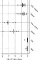


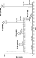


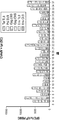


Classifications
-
- C—CHEMISTRY; METALLURGY
- C12—BIOCHEMISTRY; BEER; SPIRITS; WINE; VINEGAR; MICROBIOLOGY; ENZYMOLOGY; MUTATION OR GENETIC ENGINEERING
- C12N—MICROORGANISMS OR ENZYMES; COMPOSITIONS THEREOF; PROPAGATING, PRESERVING, OR MAINTAINING MICROORGANISMS; MUTATION OR GENETIC ENGINEERING; CULTURE MEDIA
- C12N15/00—Mutation or genetic engineering; DNA or RNA concerning genetic engineering, vectors, e.g. plasmids, or their isolation, preparation or purification; Use of hosts therefor
- C12N15/09—Recombinant DNA-technology
- C12N15/63—Introduction of foreign genetic material using vectors; Vectors; Use of hosts therefor; Regulation of expression
- C12N15/79—Vectors or expression systems specially adapted for eukaryotic hosts
- C12N15/85—Vectors or expression systems specially adapted for eukaryotic hosts for animal cells
- C12N15/86—Viral vectors
-
- A—HUMAN NECESSITIES
- A61—MEDICAL OR VETERINARY SCIENCE; HYGIENE
- A61K—PREPARATIONS FOR MEDICAL, DENTAL OR TOILETRY PURPOSES
- A61K39/00—Medicinal preparations containing antigens or antibodies
- A61K39/0005—Vertebrate antigens
- A61K39/0011—Cancer antigens
- A61K39/001184—Cancer testis antigens, e.g. SSX, BAGE, GAGE or SAGE
- A61K39/001188—NY-ESO
-
- A—HUMAN NECESSITIES
- A61—MEDICAL OR VETERINARY SCIENCE; HYGIENE
- A61K—PREPARATIONS FOR MEDICAL, DENTAL OR TOILETRY PURPOSES
- A61K39/00—Medicinal preparations containing antigens or antibodies
- A61K39/0005—Vertebrate antigens
- A61K39/0011—Cancer antigens
- A61K39/00119—Melanoma antigens
- A61K39/001191—Melan-A/MART
-
- A—HUMAN NECESSITIES
- A61—MEDICAL OR VETERINARY SCIENCE; HYGIENE
- A61K—PREPARATIONS FOR MEDICAL, DENTAL OR TOILETRY PURPOSES
- A61K39/00—Medicinal preparations containing antigens or antibodies
- A61K39/0005—Vertebrate antigens
- A61K39/0012—Lipids; Lipoproteins
-
- A—HUMAN NECESSITIES
- A61—MEDICAL OR VETERINARY SCIENCE; HYGIENE
- A61K—PREPARATIONS FOR MEDICAL, DENTAL OR TOILETRY PURPOSES
- A61K39/00—Medicinal preparations containing antigens or antibodies
- A61K39/12—Viral antigens
-
- A—HUMAN NECESSITIES
- A61—MEDICAL OR VETERINARY SCIENCE; HYGIENE
- A61P—SPECIFIC THERAPEUTIC ACTIVITY OF CHEMICAL COMPOUNDS OR MEDICINAL PREPARATIONS
- A61P31/00—Antiinfectives, i.e. antibiotics, antiseptics, chemotherapeutics
- A61P31/12—Antivirals
- A61P31/14—Antivirals for RNA viruses
- A61P31/16—Antivirals for RNA viruses for influenza or rhinoviruses
-
- A—HUMAN NECESSITIES
- A61—MEDICAL OR VETERINARY SCIENCE; HYGIENE
- A61P—SPECIFIC THERAPEUTIC ACTIVITY OF CHEMICAL COMPOUNDS OR MEDICINAL PREPARATIONS
- A61P31/00—Antiinfectives, i.e. antibiotics, antiseptics, chemotherapeutics
- A61P31/12—Antivirals
- A61P31/20—Antivirals for DNA viruses
-
- A—HUMAN NECESSITIES
- A61—MEDICAL OR VETERINARY SCIENCE; HYGIENE
- A61P—SPECIFIC THERAPEUTIC ACTIVITY OF CHEMICAL COMPOUNDS OR MEDICINAL PREPARATIONS
- A61P35/00—Antineoplastic agents
-
- C—CHEMISTRY; METALLURGY
- C07—ORGANIC CHEMISTRY
- C07K—PEPTIDES
- C07K16/00—Immunoglobulins [IGs], e.g. monoclonal or polyclonal antibodies
- C07K16/18—Immunoglobulins [IGs], e.g. monoclonal or polyclonal antibodies against material from animals or humans
- C07K16/28—Immunoglobulins [IGs], e.g. monoclonal or polyclonal antibodies against material from animals or humans against receptors, cell surface antigens or cell surface determinants
- C07K16/2803—Immunoglobulins [IGs], e.g. monoclonal or polyclonal antibodies against material from animals or humans against receptors, cell surface antigens or cell surface determinants against the immunoglobulin superfamily
- C07K16/2818—Immunoglobulins [IGs], e.g. monoclonal or polyclonal antibodies against material from animals or humans against receptors, cell surface antigens or cell surface determinants against the immunoglobulin superfamily against CD28 or CD152
-
- C—CHEMISTRY; METALLURGY
- C12—BIOCHEMISTRY; BEER; SPIRITS; WINE; VINEGAR; MICROBIOLOGY; ENZYMOLOGY; MUTATION OR GENETIC ENGINEERING
- C12Q—MEASURING OR TESTING PROCESSES INVOLVING ENZYMES, NUCLEIC ACIDS OR MICROORGANISMS; COMPOSITIONS OR TEST PAPERS THEREFOR; PROCESSES OF PREPARING SUCH COMPOSITIONS; CONDITION-RESPONSIVE CONTROL IN MICROBIOLOGICAL OR ENZYMOLOGICAL PROCESSES
- C12Q1/00—Measuring or testing processes involving enzymes, nucleic acids or microorganisms; Compositions therefor; Processes of preparing such compositions
- C12Q1/68—Measuring or testing processes involving enzymes, nucleic acids or microorganisms; Compositions therefor; Processes of preparing such compositions involving nucleic acids
- C12Q1/6813—Hybridisation assays
- C12Q1/6827—Hybridisation assays for detection of mutation or polymorphism
-
- A—HUMAN NECESSITIES
- A61—MEDICAL OR VETERINARY SCIENCE; HYGIENE
- A61K—PREPARATIONS FOR MEDICAL, DENTAL OR TOILETRY PURPOSES
- A61K39/00—Medicinal preparations containing antigens or antibodies
- A61K2039/505—Medicinal preparations containing antigens or antibodies comprising antibodies
-
- A—HUMAN NECESSITIES
- A61—MEDICAL OR VETERINARY SCIENCE; HYGIENE
- A61K—PREPARATIONS FOR MEDICAL, DENTAL OR TOILETRY PURPOSES
- A61K39/00—Medicinal preparations containing antigens or antibodies
- A61K2039/57—Medicinal preparations containing antigens or antibodies characterised by the type of response, e.g. Th1, Th2
- A61K2039/572—Medicinal preparations containing antigens or antibodies characterised by the type of response, e.g. Th1, Th2 cytotoxic response
-
- A—HUMAN NECESSITIES
- A61—MEDICAL OR VETERINARY SCIENCE; HYGIENE
- A61K—PREPARATIONS FOR MEDICAL, DENTAL OR TOILETRY PURPOSES
- A61K39/00—Medicinal preparations containing antigens or antibodies
- A61K2039/57—Medicinal preparations containing antigens or antibodies characterised by the type of response, e.g. Th1, Th2
- A61K2039/575—Medicinal preparations containing antigens or antibodies characterised by the type of response, e.g. Th1, Th2 humoral response
-
- A—HUMAN NECESSITIES
- A61—MEDICAL OR VETERINARY SCIENCE; HYGIENE
- A61K—PREPARATIONS FOR MEDICAL, DENTAL OR TOILETRY PURPOSES
- A61K39/00—Medicinal preparations containing antigens or antibodies
- A61K2039/60—Medicinal preparations containing antigens or antibodies characteristics by the carrier linked to the antigen
- A61K2039/6031—Proteins
- A61K2039/6037—Bacterial toxins, e.g. diphteria toxoid [DT], tetanus toxoid [TT]
-
- A—HUMAN NECESSITIES
- A61—MEDICAL OR VETERINARY SCIENCE; HYGIENE
- A61K—PREPARATIONS FOR MEDICAL, DENTAL OR TOILETRY PURPOSES
- A61K39/00—Medicinal preparations containing antigens or antibodies
- A61K2039/60—Medicinal preparations containing antigens or antibodies characteristics by the carrier linked to the antigen
- A61K2039/6031—Proteins
- A61K2039/605—MHC molecules or ligands thereof
-
- A—HUMAN NECESSITIES
- A61—MEDICAL OR VETERINARY SCIENCE; HYGIENE
- A61K—PREPARATIONS FOR MEDICAL, DENTAL OR TOILETRY PURPOSES
- A61K39/00—Medicinal preparations containing antigens or antibodies
- A61K2039/70—Multivalent vaccine
-
- C—CHEMISTRY; METALLURGY
- C07—ORGANIC CHEMISTRY
- C07K—PEPTIDES
- C07K14/00—Peptides having more than 20 amino acids; Gastrins; Somatostatins; Melanotropins; Derivatives thereof
- C07K14/435—Peptides having more than 20 amino acids; Gastrins; Somatostatins; Melanotropins; Derivatives thereof from animals; from humans
- C07K14/705—Receptors; Cell surface antigens; Cell surface determinants
- C07K14/70503—Immunoglobulin superfamily
- C07K14/70539—MHC-molecules, e.g. HLA-molecules
-
- C—CHEMISTRY; METALLURGY
- C07—ORGANIC CHEMISTRY
- C07K—PEPTIDES
- C07K14/00—Peptides having more than 20 amino acids; Gastrins; Somatostatins; Melanotropins; Derivatives thereof
- C07K14/435—Peptides having more than 20 amino acids; Gastrins; Somatostatins; Melanotropins; Derivatives thereof from animals; from humans
- C07K14/705—Receptors; Cell surface antigens; Cell surface determinants
- C07K14/70596—Molecules with a "CD"-designation not provided for elsewhere
-
- C—CHEMISTRY; METALLURGY
- C12—BIOCHEMISTRY; BEER; SPIRITS; WINE; VINEGAR; MICROBIOLOGY; ENZYMOLOGY; MUTATION OR GENETIC ENGINEERING
- C12N—MICROORGANISMS OR ENZYMES; COMPOSITIONS THEREOF; PROPAGATING, PRESERVING, OR MAINTAINING MICROORGANISMS; MUTATION OR GENETIC ENGINEERING; CULTURE MEDIA
- C12N2710/00—MICROORGANISMS OR ENZYMES; COMPOSITIONS THEREOF; PROPAGATING, PRESERVING, OR MAINTAINING MICROORGANISMS; MUTATION OR GENETIC ENGINEERING; CULTURE MEDIA dsDNA viruses
- C12N2710/00011—Details
- C12N2710/10011—Adenoviridae
- C12N2710/10311—Mastadenovirus, e.g. human or simian adenoviruses
- C12N2710/10341—Use of virus, viral particle or viral elements as a vector
- C12N2710/10343—Use of virus, viral particle or viral elements as a vector viral genome or elements thereof as genetic vector
-
- C—CHEMISTRY; METALLURGY
- C12—BIOCHEMISTRY; BEER; SPIRITS; WINE; VINEGAR; MICROBIOLOGY; ENZYMOLOGY; MUTATION OR GENETIC ENGINEERING
- C12N—MICROORGANISMS OR ENZYMES; COMPOSITIONS THEREOF; PROPAGATING, PRESERVING, OR MAINTAINING MICROORGANISMS; MUTATION OR GENETIC ENGINEERING; CULTURE MEDIA
- C12N2710/00—MICROORGANISMS OR ENZYMES; COMPOSITIONS THEREOF; PROPAGATING, PRESERVING, OR MAINTAINING MICROORGANISMS; MUTATION OR GENETIC ENGINEERING; CULTURE MEDIA dsDNA viruses
- C12N2710/00011—Details
- C12N2710/16011—Herpesviridae
- C12N2710/16111—Cytomegalovirus, e.g. human herpesvirus 5
- C12N2710/16134—Use of virus or viral component as vaccine, e.g. live-attenuated or inactivated virus, VLP, viral protein
-
- C—CHEMISTRY; METALLURGY
- C12—BIOCHEMISTRY; BEER; SPIRITS; WINE; VINEGAR; MICROBIOLOGY; ENZYMOLOGY; MUTATION OR GENETIC ENGINEERING
- C12N—MICROORGANISMS OR ENZYMES; COMPOSITIONS THEREOF; PROPAGATING, PRESERVING, OR MAINTAINING MICROORGANISMS; MUTATION OR GENETIC ENGINEERING; CULTURE MEDIA
- C12N2710/00—MICROORGANISMS OR ENZYMES; COMPOSITIONS THEREOF; PROPAGATING, PRESERVING, OR MAINTAINING MICROORGANISMS; MUTATION OR GENETIC ENGINEERING; CULTURE MEDIA dsDNA viruses
- C12N2710/00011—Details
- C12N2710/16011—Herpesviridae
- C12N2710/16211—Lymphocryptovirus, e.g. human herpesvirus 4, Epstein-Barr Virus
- C12N2710/16234—Use of virus or viral component as vaccine, e.g. live-attenuated or inactivated virus, VLP, viral protein
-
- C—CHEMISTRY; METALLURGY
- C12—BIOCHEMISTRY; BEER; SPIRITS; WINE; VINEGAR; MICROBIOLOGY; ENZYMOLOGY; MUTATION OR GENETIC ENGINEERING
- C12N—MICROORGANISMS OR ENZYMES; COMPOSITIONS THEREOF; PROPAGATING, PRESERVING, OR MAINTAINING MICROORGANISMS; MUTATION OR GENETIC ENGINEERING; CULTURE MEDIA
- C12N2710/00—MICROORGANISMS OR ENZYMES; COMPOSITIONS THEREOF; PROPAGATING, PRESERVING, OR MAINTAINING MICROORGANISMS; MUTATION OR GENETIC ENGINEERING; CULTURE MEDIA dsDNA viruses
- C12N2710/00011—Details
- C12N2710/20011—Papillomaviridae
- C12N2710/20034—Use of virus or viral component as vaccine, e.g. live-attenuated or inactivated virus, VLP, viral protein
-
- C—CHEMISTRY; METALLURGY
- C12—BIOCHEMISTRY; BEER; SPIRITS; WINE; VINEGAR; MICROBIOLOGY; ENZYMOLOGY; MUTATION OR GENETIC ENGINEERING
- C12N—MICROORGANISMS OR ENZYMES; COMPOSITIONS THEREOF; PROPAGATING, PRESERVING, OR MAINTAINING MICROORGANISMS; MUTATION OR GENETIC ENGINEERING; CULTURE MEDIA
- C12N2730/00—Reverse transcribing DNA viruses
- C12N2730/00011—Details
- C12N2730/10011—Hepadnaviridae
- C12N2730/10111—Orthohepadnavirus, e.g. hepatitis B virus
- C12N2730/10134—Use of virus or viral component as vaccine, e.g. live-attenuated or inactivated virus, VLP, viral protein
-
- C—CHEMISTRY; METALLURGY
- C12—BIOCHEMISTRY; BEER; SPIRITS; WINE; VINEGAR; MICROBIOLOGY; ENZYMOLOGY; MUTATION OR GENETIC ENGINEERING
- C12N—MICROORGANISMS OR ENZYMES; COMPOSITIONS THEREOF; PROPAGATING, PRESERVING, OR MAINTAINING MICROORGANISMS; MUTATION OR GENETIC ENGINEERING; CULTURE MEDIA
- C12N2740/00—Reverse transcribing RNA viruses
- C12N2740/00011—Details
- C12N2740/10011—Retroviridae
- C12N2740/14011—Deltaretrovirus, e.g. bovine leukeamia virus
- C12N2740/14034—Use of virus or viral component as vaccine, e.g. live-attenuated or inactivated virus, VLP, viral protein
-
- C—CHEMISTRY; METALLURGY
- C12—BIOCHEMISTRY; BEER; SPIRITS; WINE; VINEGAR; MICROBIOLOGY; ENZYMOLOGY; MUTATION OR GENETIC ENGINEERING
- C12N—MICROORGANISMS OR ENZYMES; COMPOSITIONS THEREOF; PROPAGATING, PRESERVING, OR MAINTAINING MICROORGANISMS; MUTATION OR GENETIC ENGINEERING; CULTURE MEDIA
- C12N2740/00—Reverse transcribing RNA viruses
- C12N2740/00011—Details
- C12N2740/10011—Retroviridae
- C12N2740/16011—Human Immunodeficiency Virus, HIV
- C12N2740/16111—Human Immunodeficiency Virus, HIV concerning HIV env
- C12N2740/16134—Use of virus or viral component as vaccine, e.g. live-attenuated or inactivated virus, VLP, viral protein
-
- C—CHEMISTRY; METALLURGY
- C12—BIOCHEMISTRY; BEER; SPIRITS; WINE; VINEGAR; MICROBIOLOGY; ENZYMOLOGY; MUTATION OR GENETIC ENGINEERING
- C12N—MICROORGANISMS OR ENZYMES; COMPOSITIONS THEREOF; PROPAGATING, PRESERVING, OR MAINTAINING MICROORGANISMS; MUTATION OR GENETIC ENGINEERING; CULTURE MEDIA
- C12N2740/00—Reverse transcribing RNA viruses
- C12N2740/00011—Details
- C12N2740/10011—Retroviridae
- C12N2740/16011—Human Immunodeficiency Virus, HIV
- C12N2740/16211—Human Immunodeficiency Virus, HIV concerning HIV gagpol
- C12N2740/16234—Use of virus or viral component as vaccine, e.g. live-attenuated or inactivated virus, VLP, viral protein
-
- C—CHEMISTRY; METALLURGY
- C12—BIOCHEMISTRY; BEER; SPIRITS; WINE; VINEGAR; MICROBIOLOGY; ENZYMOLOGY; MUTATION OR GENETIC ENGINEERING
- C12N—MICROORGANISMS OR ENZYMES; COMPOSITIONS THEREOF; PROPAGATING, PRESERVING, OR MAINTAINING MICROORGANISMS; MUTATION OR GENETIC ENGINEERING; CULTURE MEDIA
- C12N2740/00—Reverse transcribing RNA viruses
- C12N2740/00011—Details
- C12N2740/10011—Retroviridae
- C12N2740/16011—Human Immunodeficiency Virus, HIV
- C12N2740/16311—Human Immunodeficiency Virus, HIV concerning HIV regulatory proteins
- C12N2740/16334—Use of virus or viral component as vaccine, e.g. live-attenuated or inactivated virus, VLP, viral protein
-
- C—CHEMISTRY; METALLURGY
- C12—BIOCHEMISTRY; BEER; SPIRITS; WINE; VINEGAR; MICROBIOLOGY; ENZYMOLOGY; MUTATION OR GENETIC ENGINEERING
- C12N—MICROORGANISMS OR ENZYMES; COMPOSITIONS THEREOF; PROPAGATING, PRESERVING, OR MAINTAINING MICROORGANISMS; MUTATION OR GENETIC ENGINEERING; CULTURE MEDIA
- C12N2760/00—MICROORGANISMS OR ENZYMES; COMPOSITIONS THEREOF; PROPAGATING, PRESERVING, OR MAINTAINING MICROORGANISMS; MUTATION OR GENETIC ENGINEERING; CULTURE MEDIA ssRNA viruses negative-sense
- C12N2760/00011—Details
- C12N2760/16011—Orthomyxoviridae
- C12N2760/16111—Influenzavirus A, i.e. influenza A virus
- C12N2760/16134—Use of virus or viral component as vaccine, e.g. live-attenuated or inactivated virus, VLP, viral protein
-
- C—CHEMISTRY; METALLURGY
- C12—BIOCHEMISTRY; BEER; SPIRITS; WINE; VINEGAR; MICROBIOLOGY; ENZYMOLOGY; MUTATION OR GENETIC ENGINEERING
- C12N—MICROORGANISMS OR ENZYMES; COMPOSITIONS THEREOF; PROPAGATING, PRESERVING, OR MAINTAINING MICROORGANISMS; MUTATION OR GENETIC ENGINEERING; CULTURE MEDIA
- C12N2770/00—MICROORGANISMS OR ENZYMES; COMPOSITIONS THEREOF; PROPAGATING, PRESERVING, OR MAINTAINING MICROORGANISMS; MUTATION OR GENETIC ENGINEERING; CULTURE MEDIA ssRNA viruses positive-sense
- C12N2770/00011—Details
- C12N2770/24011—Flaviviridae
- C12N2770/24111—Flavivirus, e.g. yellow fever virus, dengue, JEV
- C12N2770/24141—Use of virus, viral particle or viral elements as a vector
- C12N2770/24143—Use of virus, viral particle or viral elements as a vector viral genome or elements thereof as genetic vector
-
- C—CHEMISTRY; METALLURGY
- C12—BIOCHEMISTRY; BEER; SPIRITS; WINE; VINEGAR; MICROBIOLOGY; ENZYMOLOGY; MUTATION OR GENETIC ENGINEERING
- C12N—MICROORGANISMS OR ENZYMES; COMPOSITIONS THEREOF; PROPAGATING, PRESERVING, OR MAINTAINING MICROORGANISMS; MUTATION OR GENETIC ENGINEERING; CULTURE MEDIA
- C12N2770/00—MICROORGANISMS OR ENZYMES; COMPOSITIONS THEREOF; PROPAGATING, PRESERVING, OR MAINTAINING MICROORGANISMS; MUTATION OR GENETIC ENGINEERING; CULTURE MEDIA ssRNA viruses positive-sense
- C12N2770/00011—Details
- C12N2770/24011—Flaviviridae
- C12N2770/24211—Hepacivirus, e.g. hepatitis C virus, hepatitis G virus
- C12N2770/24234—Use of virus or viral component as vaccine, e.g. live-attenuated or inactivated virus, VLP, viral protein
-
- C—CHEMISTRY; METALLURGY
- C12—BIOCHEMISTRY; BEER; SPIRITS; WINE; VINEGAR; MICROBIOLOGY; ENZYMOLOGY; MUTATION OR GENETIC ENGINEERING
- C12N—MICROORGANISMS OR ENZYMES; COMPOSITIONS THEREOF; PROPAGATING, PRESERVING, OR MAINTAINING MICROORGANISMS; MUTATION OR GENETIC ENGINEERING; CULTURE MEDIA
- C12N2770/00—MICROORGANISMS OR ENZYMES; COMPOSITIONS THEREOF; PROPAGATING, PRESERVING, OR MAINTAINING MICROORGANISMS; MUTATION OR GENETIC ENGINEERING; CULTURE MEDIA ssRNA viruses positive-sense
- C12N2770/00011—Details
- C12N2770/36011—Togaviridae
- C12N2770/36111—Alphavirus, e.g. Sindbis virus, VEE, EEE, WEE, Semliki
- C12N2770/36141—Use of virus, viral particle or viral elements as a vector
- C12N2770/36143—Use of virus, viral particle or viral elements as a vector viral genome or elements thereof as genetic vector
Abstract
抗原コード核酸配列を含み、かつ免疫調節物質を共発現するベクターが、本明細書において開示される。ワクチンとしてのそれらの使用を含む、該ベクターに関連するヌクレオチド、細胞、及び方法もまた開示される。【選択図】図1A vector containing an antigen-encoding nucleic acid sequence and co-expressing an immunomodulatory substance is disclosed herein. Nucleotides, cells, and methods associated with the vector, including their use as vaccines, are also disclosed. [Selection diagram] Fig. 1
Description
関連出願の相互参照
本出願は、あらゆる目的でその全容を参照により本明細書に援用するところの2018年5月23日出願の米国特許仮出願第62/675,624号の恩典を主張する。
Cross-reference to related applications This application claims the benefit of US Patent Provisional Application No. 62 / 675,624 filed May 23, 2018, which is hereby incorporated by reference in its entirety for all purposes.
配列表
本出願は、ASCIIフォーマットで電子的に提出され、その全容を参照によって本明細書に援用する配列表を含む。前記ASCIIコピーは、2019年5月22日に作成され、GSO_018_WO_Sequence_Listing.txtの名前が付けられており、そのサイズは422,185バイトである。
Sequence Listing This application contains a sequence listing that is submitted electronically in ASCII format and is incorporated herein by reference in its entirety. The ASCII copy was made on May 22, 2019, GSO_018_WO_Sequence_Listing. It is named txt and its size is 422,185 bytes.
背景
腫瘍特異的な抗原に基づいた治療用ワクチンは、次世代の個別化がん免疫療法として極めて有望である1〜3。例えば、非小細胞肺がん(NSCLC)及びメラノーマなどの高い遺伝子変異量を有するがんは、新生抗原を生じる可能性が比較的高いことから、かかる治療法の特に有望な標的である4,5。初期の証拠により、新生抗原に基づいたワクチン接種がT細胞応答を誘発し6、新生抗原を標的とした細胞療法が、選択された患者において腫瘍退縮を引き起こし得る7ことが示されている。
Background Tumor-specific antigen-based therapeutic vaccines are extremely promising as next-generation personalized cancer immunotherapy 1-3 . Cancers with high gene mutations, such as non-small cell lung cancer (NSCLC) and melanoma, are particularly promising targets for such treatments because of their relatively high likelihood of producing nascent antigens 4,5 . Early evidence vaccination based on nascent antigen induces T cell responses 6, cell therapy neoplastic antigen targeting has been shown 7 that can cause tumor regression in patients selected.
現行の新生抗原予測法の課題に加えて、その多くがヒト由来のものである新生抗原のヒトへの送達に使用することができる既存のベクター系にもやはり特定の課題が存在する。例えば、多くのヒトは、過去の自然の曝露の結果としてヒトウイルスに対する既存の免疫を有しており、この免疫が、がん治療のために新生抗原を送達するための組換えヒトウイルスの使用にとって、大きな障害となり得る。 In addition to the challenges of current nascent antigen prediction methods, there are also specific challenges with existing vector systems that can be used to deliver nascent antigens, many of which are of human origin, to humans. For example, many humans have existing immunity to human viruses as a result of past natural exposures, and this immunity uses recombinant human viruses to deliver neogenic antigens for the treatment of cancer. It can be a big obstacle for us.
免疫チェックポイント阻害剤の使用は、がんの治療における多大な有望性を示してきた。しかしながら、特にDNAまたはRNAベースのがんワクチンの場合では、改善された送達方法が依然として求められている。 The use of immune checkpoint inhibitors has shown great promise in the treatment of cancer. However, there is still a need for improved delivery methods, especially in the case of DNA or RNA-based cancer vaccines.
概要
本明細書では、抗原カセットを含むベクターシステムであって、抗原カセットが、(1)少なくとも1つの抗原コード核酸配列であって、a.コードされたペプチド配列を、野生型核酸配列によってコードされる対応するペプチド配列とは異なるものとする少なくとも1つの変化を任意で含む、エピトープコード核酸配列と、b.任意で5’リンカー配列と、c.任意で3’リンカー配列と、をそれぞれが含む、MHCクラスI抗原コード核酸配列を任意で含む、前記少なくとも1つの抗原コード核酸配列を含む、対象内に存在する腫瘍に関連した少なくとも1つの抗原コード核酸配列と、(2)少なくとも1つの抗原コード核酸に機能的に連結された少なくとも1つのプロモーター配列と、(3)任意で、少なくとも1つのMHCクラスII抗原コード核酸配列と、(4)任意で、少なくとも1つのGPGPGリンカー配列(SEQ ID NO:56)と、(5)任意で、少なくとも1つのポリアデニル化配列と、を含み、前記ベクターが、任意で前記カセット内に、少なくとも1つの免疫調節物質をコードする核酸配列をさらに含み、任意で、前記少なくとも1つの免疫調節物質をコードする前記核酸配列が、(1)前記少なくとも1つの免疫調節物質をコードする前記配列と、前記少なくとも1つの抗原コード核酸配列とを隔てる内部リボソーム進入配列(IRES)を有する、前記少なくとも1つの抗原コード核酸配列と同じ転写産物上、または(2)前記少なくとも1つの抗原コード核酸配列と異なる転写産物であって、少なくとも1つの第2のプロモーター配列が、前記少なくとも1つの免疫調節物質をコードする前記配列に機能的に連結されている、前記異なる転写産物上に転写される、ベクターシステムを開示する。
Summary In the present specification, a vector system including an antigen cassette, wherein the antigen cassette is (1) at least one antigen-encoding nucleic acid sequence, a. An epitope-encoding nucleic acid sequence and b. Optionally, a 5'linker sequence and c. At least one antigen code associated with a tumor present in a subject, each comprising at least one antigen coding nucleic acid sequence, optionally comprising an MHC class I antigen coding nucleic acid sequence, each comprising a 3'linker sequence. Nucleic acid sequences, (2) at least one promoter sequence functionally linked to at least one antigen-coding nucleic acid, (3) optionally at least one MHC class II antigen-coding nucleic acid sequence, and (4) optionally. Containing at least one GPPGG linker sequence (SEQ ID NO: 56) and (5) optionally at least one polyadenylated sequence, the vector optionally contains at least one immunomodulatory substance in the cassette. The nucleic acid sequence, which further comprises the nucleic acid sequence encoding the at least one immunomodulatory substance, optionally comprises the nucleic acid sequence encoding the at least one immunomodulatory substance: At least on the same transcript as the at least one antigen-encoding nucleic acid sequence, or (2) different from the at least one antigen-encoding nucleic acid sequence, having an internal ribosome entry sequence (IRES) that separates the nucleic acid sequence. Disclosed is a vector system in which one second promoter sequence is transcribed onto said different transcript, which is functionally linked to said sequence encoding said at least one immunomodulatory substance.
さらに、本明細書では、チンパンジーアデノウイルスベクターであって、a.E1(nt577〜3403)欠失及びE3(nt27,125〜31,825)欠失を有するSEQ ID NO:1の配列を含む改変ChAdV68配列と、b.CMVプロモーター配列と、c.SV40ポリアデニル化シグナルヌクレオチド配列と、d.免疫チェックポイント阻害剤をコードする核酸配列と、e.抗原カセットであって、以下:(1)対象内に存在する腫瘍に由来する少なくとも1つの抗原コード核酸配列であって、前記少なくとも1つの抗原コード核酸配列が、互いに直鎖状に連結された、少なくとも10個の腫瘍特異的及び対象特異的MHCクラスI抗原コード核酸配列であって、それぞれが、(A)コードされたペプチド配列を、野生型核酸配列によってコードされる対応する核酸配列とは異なるものとする少なくとも1つの変化を有し、アミノ酸7〜15個の長さのMHCクラスIエピトープをコードする、MHC Iエピトープコード核酸配列と、(B)前記MHCIエピトープの天然のN末端アミノ酸配列をコードし、少なくともアミノ酸3個の長さであるペプチドをコードする、5’リンカー配列と、(C)前記MHC Iエピトープの天然のC末端アミノ酸配列をコードし、少なくともアミノ酸3個の長さであるペプチドをコードする、3’リンカー配列と、を含み、前記MHCクラスI抗原コード核酸配列のそれぞれが、アミノ酸25個の長さのポリペプチドをコードし、抗原カセット内の最後のMHCクラスI抗原コード核酸配列を除いて、各MHCクラスI抗原コード核酸配列の各3’末端が、それに続くMHCクラスI抗原抗原コード核酸配列の5’末端に連結された、前記MHCクラスI抗原コード核酸配列を含む、前記少なくとも10個の腫瘍特異的及び対象特異的MHCクラスI抗原コード核酸配列を含む、前記少なくとも1つの抗原コード核酸配列と、(2)少なくとも2個のMHCクラスII抗原コード核酸配列であって、(A)PADRE MHCクラスII配列(SEQ ID NO:48)と、(B)破傷風トキソイドMHCクラスII配列(SEQ ID NO:46)と、(C)前記PADRE MHCクラスII配列と前記破傷風トキソイドMHCクラスII配列とを連結するGPGPGアミノ酸リンカー配列をコードする第1の核酸配列と、(D)前記少なくとも2個のMHCクラスII抗原コード核酸配列の5’末端と、前記少なくとも10個の腫瘍特異的かつ対象特異的なMHCクラスI新生抗原コード核酸配列とを連結するGPGPGアミノ酸リンカー配列をコードする第2の核酸配列と、(E)任意で、前記少なくとも2個のMHCクラスII抗原コード核酸配列の3’末端のGPGPGアミノ酸リンカー配列をコードする第3の核酸配列と、を含む、前記少なくとも2個のMHCクラスII抗原コード核酸配列と、を含む、前記抗原カセットと、を含み、前記抗原カセットが前記E1欠失内に挿入され、前記CMVプロモーター配列が前記抗原カセットに機能的に連結され、前記チェックポイント阻害剤をコードする前記核酸配列が、(1)前記チェックポイント阻害剤をコードする前記配列と、前記少なくとも1つの抗原コード核酸配列とを隔てる内部リボソーム進入配列(IRES)を有する、前記少なくとも1つの抗原コード核酸配列と同じ転写産物上、または(2)前記少なくとも1つの抗原コード核酸配列とは異なる転写産物であって、第2のCMVプロモーター配列が前記少なくとも1つの免疫調節物質をコードする前記配列に機能的に連結されているか、または任意で前記少なくとも1つの免疫調節物質が前記E3欠失内に挿入されている、前記異なる転写産物上に転写される、チンパンジーアデノウイルスベクターも開示する。 Further, in the present specification, the chimpanzee adenovirus vector is a. A modified ChAdV68 sequence comprising a SEQ ID NO: 1 sequence with an E1 (nt577-3403) deletion and an E3 (nt27,125-31,825) deletion, and b. CMV promoter sequence and c. The SV40 polyadenylation signal nucleotide sequence and d. Nucleic acid sequences encoding immune checkpoint inhibitors and e. An antigen cassette, the following: (1) At least one antigen-coding nucleic acid sequence derived from a tumor existing in a subject, wherein the at least one antigen-coding nucleic acid sequence is linearly linked to each other. At least 10 tumor-specific and target-specific MHC class I antigen-encoding nucleic acid sequences, each of which differs from the corresponding nucleic acid sequence encoded by the (A) -encoded nucleic acid sequence by the wild-type nucleic acid sequence. The MHC I epitope coding nucleic acid sequence having at least one variation and encoding an MHC class I epitope of 7 to 15 amino acids in length and (B) the natural N-terminal amino acid sequence of the MHCI epitope. It encodes a 5'linker sequence that encodes a peptide that is at least 3 amino acids long and (C) encodes the natural C-terminal amino acid sequence of the MHC I epitope, which is at least 3 amino acids long. Each of the MHC class I antigen coding nucleic acid sequences comprising a 3'linker sequence encoding a peptide encodes a polypeptide having a length of 25 amino acids and is the last MHC class I antigen coding in the antigen cassette. Except for the nucleic acid sequence, each 3'end of each MHC class I antigen coding nucleic acid sequence comprises the MHC class I antigen coding nucleic acid sequence linked to the 5'end of the subsequent MHC class I antigen antigen coding nucleic acid sequence. The at least one antigen-encoding nucleic acid sequence containing the at least 10 tumor-specific and target-specific MHC class I antigen-encoding nucleic acid sequences, and (2) at least two MHC class II antigen-encoding nucleic acid sequences. , (A) PADRE MHC class II sequence (SEQ ID NO: 48), (B) tetanus toxoid MHC class II sequence (SEQ ID NO: 46), (C) said PADRE MHC class II sequence and said tetanus toxoid MHC. A first nucleic acid sequence encoding a GPPGG amino acid linker sequence linking a class II sequence, (D) the 5'end of the at least two MHC class II antigen coding nucleic acid sequences, and at least 10 tumor specifics. A second nucleic acid sequence encoding a GPGPG amino acid linker sequence that links a target-specific MHC class I nascent antigen coding nucleic acid sequence, and (E) optionally, at least two MHC class II antigen coding nucleic acid sequences. A third nucleic acid sequence encoding a 3'-terminal GPGPG amino acid linker sequence The antigen cassette contains, and includes, the at least two MHC class II antigen coding nucleic acid sequences, the antigen cassette is inserted into the E1 deletion, and the CMV promoter sequence is the antigen. The internal ribosome entry that is functionally linked to the cassette and encodes the checkpoint inhibitor (1) separates the sequence encoding the checkpoint inhibitor from the at least one antigen-encoding nucleic acid sequence. The second CMV promoter sequence is said to be on the same transcript as the at least one antigen-encoding nucleic acid sequence having a sequence (IRES), or (2) a transcript different from the at least one antigen-encoding nucleic acid sequence. Transcribed onto the different transcripts that are functionally linked to the sequence encoding at least one immunomodulator or optionally the at least one immunomodulator is inserted within the E3 deletion. The chimpanzee adenovirus vector is also disclosed.
いくつかの態様において、ベクターの各要素の順序付けられた配列は、5’から3’にかけて、下式:
Pa−(L5b−Nc−L3d)X−(G5e−Uf)Y−G3g−Ah
[式中、Pは、前記少なくとも1つの抗原コード核酸配列のうちの少なくとも1つに機能的に連結された前記少なくとも1つのプロモーター配列を含み、ここでa=1であり、Nは、前記コードされたペプチド配列を、前記野生型核酸配列によってコードされる前記対応するペプチド配列とは異なるものとする少なくとも1つの変化を有する前記エピトープコード核酸配列の1つを含み、ここでc=1であり、L5は、前記5’リンカー配列を含み、ここでb=0または1であり、L3は、前記3’リンカー配列を含み、ここでd=0または1であり、G5は、GPGPGアミノ酸リンカーをコードする前記少なくとも1つの核酸配列のうちの1つを含み、ここでe=0または1であり、G3は、GPGPGアミノ酸リンカーをコードする前記少なくとも1つの核酸配列のうちの1つを含み、ここでg=0または1であり、Uは、前記少なくとも1つのMHCクラスII抗原コード核酸配列のうちの1つを含み、ここでf=1であり、Aは、前記少なくとも1つのポリアデニル化配列を含み、ここでh=0または1であり、X=2〜400であり、各Xについて、対応するNcは、エピトープコード核酸配列であり、任意で、各Xについて、対応するNcは異なるMHCクラスIエピトープコード核酸配列であり、Y=0〜2であり、各Yについて、対応するUfは、抗原コード核酸配列であり、任意で、各Yについて、対応するUfは異なるMHCクラスII抗原コード配列である]で記述される。特定の態様において、b=1、d=1、e=1、g=1、h=1、X=10、Y=2であり、Pは、CMVプロモーター配列であり、各Nは、アミノ酸7〜15個の長さのMHCクラスIエピトープをコードし、L5は、前記MHCIエピトープの天然のN末端アミノ酸配列をコードし、前記5’リンカー配列が、少なくともアミノ酸3個の長さであるペプチドをコードし、L3は、前記MHCIエピトープの天然のC末端アミノ酸配列をコードし、前記3’リンカー配列が、少なくともアミノ酸3個の長さであるペプチドをコードし、Uは、PADREクラスII配列及び破傷風トキソイドMHCクラスII配列のそれぞれであり、前記ベクターは、E1(nt577〜3403)欠失及びE3(nt27,125〜31,825)欠失を有するSEQ ID NO:1の配列を含む改変ChAdV68配列を含み、前記新生抗原カセットが前記E1欠失内に挿入されており、前記MHCクラスI抗原コード核酸配列のそれぞれが、アミノ酸25個の長さのポリペプチドをコードしている。
In some embodiments, the ordered sequence of each element of the vector is from 5'to 3', according to the following equation:
P a- (L5 b- N c- L3 d ) X- (G5 e- U f ) Y- G3 g- A h
[In the formula, P comprises the at least one promoter sequence operably linked to at least one of the at least one antigen coding nucleic acid sequence, where a = 1, where N is the coding. Contains one of the epitope-coding nucleic acid sequences having at least one change that makes the resulting peptide sequence different from the corresponding peptide sequence encoded by the wild-type nucleic acid sequence, where c = 1. , L5 comprises the 5'linker sequence, where b = 0 or 1, L3 comprises the 3'linker sequence, where d = 0 or 1, and G5 contains the GPPGG amino acid linker. Containing one of the at least one nucleic acid sequence encoding, where e = 0 or 1, G3 comprises one of the at least one nucleic acid sequence encoding the GPPGPG amino acid linker. In g = 0 or 1, U comprises one of the at least one MHC class II antigen-encoding nucleic acid sequence, where f = 1, and A contains the at least one polyadenylated sequence. Containing, where h = 0 or 1, X = 2-400, for each X the corresponding Nc is an epitope-coding nucleic acid sequence and optionally for each X the corresponding Nc is a different MHC class. I epitope-coding nucleic acid sequence, Y = 0-2, and for each Y, the corresponding Uf is an antigen-coding nucleic acid sequence, and optionally, for each Y, the corresponding Uf is a different MHC class II antigen-coding sequence. Is described]. In a particular embodiment, b = 1, d = 1, e = 1, g = 1, h = 1, X = 10, Y = 2, P is the CMV epitope sequence, and each N is
いくつかの態様において、抗原コード核酸配列のうちの少なくとも1つは、腫瘍細胞表面上のMHCクラスIによって提示されるポリペプチド配列またはその一部をコードする。いくつかの態様において、抗原コード核酸配列のうちの少なくとも1つ、2つ、または任意で3つが、腫瘍細胞表面上のMHCクラスIによって提示されるポリペプチド配列またはその一部をコードする。 In some embodiments, at least one of the antigen-encoding nucleic acid sequences encodes the polypeptide sequence or portion thereof presented by MHC class I on the surface of tumor cells. In some embodiments, at least one, two, or optionally three of the antigen-encoding nucleic acid sequences encode the polypeptide sequence or portion thereof presented by MHC class I on the surface of tumor cells.
いくつかの態様において、各抗原コード核酸配列内の各抗原コード核酸配列は互いに直接連結される。いくつかの態様において、少なくとも1つの抗原コード核酸配列のうちの少なくとも1つは、リンカーをコードする核酸配列によって、異なる抗原コード核酸配列と連結されている。いくつかの態様において、リンカーは、2個のMHCクラスI配列または1個のMHCクラスI配列をMHCクラスII配列に連結する。いくつかの態様において、リンカーは、(1)少なくとも残基2、3、4、5、6、7、8、9、または10個の長さの連続したグリシン残基、(2)少なくとも残基2、3、4、5、6、7、8、9、または10個の長さの連続したアラニン残基、(3)2個のアルギニン残基(RR)、(4)アラニン、アラニン、チロシン(AAY)、(5)哺乳動物プロテアソームによって効率的にプロセシングされる、少なくともアミノ酸残基2、3、4、5、6、7、8、9、または10個の長さのコンセンサス配列、及び(6)同族の起源タンパク質に由来する抗原に隣接し、少なくともアミノ酸残基2、3、4、5、6、7、8、9、10、11、12、13、14、15、16、17、18、19、20、または2〜20個の長さの1つ以上の天然配列からなる群から選択される。いくつかの態様において、リンカーは、2個のMHCクラスII配列または1個のMHCクラスII配列を1個のMHCクラスI配列と連結する。いくつかの態様において、リンカーは、配列GPGPGを含む。
In some embodiments, the antigen-encoding nucleic acid sequences within each antigen-encoding nucleic acid sequence are directly linked to each other. In some embodiments, at least one of at least one antigen-encoding nucleic acid sequence is linked to a different antigen-encoding nucleic acid sequence by a linker-encoding nucleic acid sequence. In some embodiments, the linker ligates two MHC class I sequences or one MHC class I sequence into an MHC class II sequence. In some embodiments, the linker comprises (1) at least
いくつかの態様において、少なくとも1つの抗原コード核酸配列のうちの少なくとも1つは、少なくとも1つの抗原コード核酸配列の発現、安定性、細胞トラフィッキング、プロセシング及び提示、及び/または免疫原性を高める、分離したまたは連続した配列に機能的または直接的に連結される。いくつかの態様において、分離したまたは連続した配列は、ユビキチン配列、プロテアソームターゲティング性を高めるように改変されたユビキチン配列(例えば、76位にGlyからAlaへの置換を有するユビキチン配列)、免疫グロブリンシグナル配列(例えばIgK)、主要組織適合性クラスI配列、リソソーム関連膜タンパク質(LAMP)−1、ヒト樹状細胞リソソーム関連膜タンパク質、及び主要組織適合性クラスII配列のうちの少なくとも1つを含み、任意でプロテアソームターゲティング性を高めるように改変された前記ユビキチン配列がA76である。 In some embodiments, at least one of the at least one antigen-encoding nucleic acid sequence enhances expression, stability, cell trafficking, processing and presentation, and / or immunogenicity of the at least one antigen-encoding nucleic acid sequence. Functionally or directly linked to a separate or contiguous sequence. In some embodiments, the separated or contiguous sequence is a ubiquitin sequence, a ubiquitin sequence modified to enhance proteasome targeting (eg, a ubiquitin sequence having a Gly to Ala substitution at position 76), an immunoglobulin signal. It comprises at least one of a sequence (eg IgK), a major tissue compatibility class I sequence, a lysosome-related membrane protein (LAMP) -1, a human dendritic cell lysosome-related membrane protein, and a major tissue compatibility class II sequence. The ubiquitin sequence optionally modified to enhance proteasome targeting is A76.
いくつかの態様において、抗原コード核酸配列のうちの少なくとも1つは、翻訳後の対応する野生型核酸配列と比べて、その対応するMHCアレルに対する増大した結合親和性を有するポリペプチド配列またはその一部をコードする。いくつかの態様において、抗原コード核酸配列のうちの少なくとも1つは、翻訳後の対応する野生型核酸配列と比べて、その対応するMHCアレルに対する増大した結合安定性を有するポリペプチド配列またはその一部をコードする。いくつかの態様において、抗原コード核酸配列のうちの少なくとも1つは、翻訳後の対応する野生型の親核酸配列と比べて、その対応するMHCアレル上への増大した提示尤度を有するポリペプチド配列またはその一部をコードする。 In some embodiments, at least one of the antigen-encoding nucleic acid sequences is a polypeptide sequence or one thereof that has increased binding affinity for its corresponding MHC allele as compared to the corresponding wild-type nucleic acid sequence after translation. Code the part. In some embodiments, at least one of the antigen-encoding nucleic acid sequences is a polypeptide sequence or one thereof that has increased binding stability to its corresponding MHC allele as compared to the corresponding wild-type nucleic acid sequence after translation. Code the part. In some embodiments, at least one of the antigen-encoding nucleic acid sequences is a polypeptide having increased presentation likelihood on its corresponding MHC allele as compared to the corresponding wild-type parent nucleic acid sequence after translation. Code an array or part of it.
いくつかの態様において、少なくとも1つの変化は、点変異、フレームシフト変異、非フレームシフト変異、欠失変異、挿入変異、スプライスバリアント、ゲノム再編成、またはプロテアソームにより生成されたスプライス抗原を含む。 In some embodiments, at least one change comprises a point mutation, a frameshift mutation, a non-frameshift mutation, a deletion mutation, an insertion mutation, a splice variant, a genome rearrangement, or a splice antigen produced by a proteasome.
いくつかの態様において、腫瘍は、肺がん、メラノーマ、乳がん、卵巣がん、前立腺がん、腎臓がん、胃がん、結腸がん、精巣がん、頭頸部がん、膵臓がん、脳がん、B細胞リンパ腫、急性骨髄性白血病、慢性骨髄性白血病、慢性リンパ球性白血病、T細胞リンパ球性白血病、非小細胞肺がん、及び小細胞肺がんからなる群から選択される。 In some embodiments, the tumor is lung cancer, melanoma, breast cancer, ovarian cancer, prostate cancer, kidney cancer, stomach cancer, colon cancer, testis cancer, head and neck cancer, pancreatic cancer, brain cancer, It is selected from the group consisting of B-cell lymphoma, acute myeloid leukemia, chronic myeloid leukemia, chronic lymphocytic leukemia, T-cell lymphocytic leukemia, non-small cell lung cancer, and small cell lung cancer.
いくつかの態様において、少なくとも1つの抗原コード核酸配列のそれぞれの発現は、少なくとも1つのプロモーターによって駆動される。 In some embodiments, each expression of at least one antigen-encoding nucleic acid sequence is driven by at least one promoter.
いくつかの態様において、少なくとも1つの抗原コード核酸配列は、少なくとも2個、3個、4個、5個、6個、7個、8個、9個、または10個の核酸配列を含む。いくつかの態様において、少なくとも1つの抗原コード核酸配列は、少なくとも11個、12個、13個、14個、15個、16個、17個、18個、19個、20個、または最大400個の核酸配列を含む。いくつかの態様において、少なくとも1つの抗原コード核酸配列は少なくとも2〜400個の核酸配列を含み、抗原コード核酸配列のうちの少なくとも1つは、腫瘍細胞表面上のMHC Iによって提示されるポリペプチド配列またはその一部をコードする。いくつかの態様において、少なくとも1つの抗原コード核酸配列は少なくとも2〜400個の核酸配列を含み、対象に投与されて翻訳された場合、抗原のうちの少なくとも1つは抗原提示細胞上に提示され、腫瘍細胞表面上の抗原のうちの少なくとも1つを標的とする免疫応答をもたらす。いくつかの態様において、少なくとも1つの抗原コード核酸配列は少なくとも2〜400個のMHCクラスI及び/またはMHCクラスII抗原コード核酸配列を含み、対象に投与されて翻訳された場合、MHCクラスIまたはMHCクラスII抗原のうちの少なくとも1つが、抗原提示細胞上に提示され、腫瘍細胞表面上の抗原のうちの少なくとも1つを標的とする免疫応答をもたらし、任意で、少なくとも2〜400個のMHCクラスIまたはMHCクラスII抗原コード核酸配列のそれぞれの発現は、前記少なくとも1つのプロモーターによって駆動される。 In some embodiments, the at least one antigen-encoding nucleic acid sequence comprises at least 2, 3, 4, 5, 6, 7, 7, 8, 9, or 10 nucleic acid sequences. In some embodiments, the at least one antigen-encoding nucleic acid sequence is at least 11, 12, 13, 14, 15, 16, 17, 18, 19, 20, or up to 400. Contains the nucleic acid sequence of. In some embodiments, the at least one antigen-encoding nucleic acid sequence comprises at least 2 to 400 nucleic acid sequences, and at least one of the antigen-encoding nucleic acid sequences is a polypeptide presented by MHC I on the surface of the tumor cell. Code an array or part of it. In some embodiments, the at least one antigen-encoding nucleic acid sequence comprises at least 2 to 400 nucleic acid sequences, and when administered to a subject and translated, at least one of the antigens is presented on an antigen-presenting cell. , Produces an immune response that targets at least one of the antigens on the surface of tumor cells. In some embodiments, the at least one antigen-encoding nucleic acid sequence comprises at least 2 to 400 MHC class I and / or MHC class II antigen-encoding nucleic acid sequences, and when administered to a subject and translated, MHC class I or At least one of the MHC class II antigens is presented on the antigen-presenting cells, resulting in an immune response that targets at least one of the antigens on the surface of the tumor cells, optionally at least 2 to 400 MHCs. Each expression of a class I or MHC class II antigen coding nucleic acid sequence is driven by at least one of the promoters.
いくつかの態様において、各MHCクラスI抗原コード核酸配列は、アミノ酸8〜35個の長さ、任意で、アミノ酸9〜17個、9〜25個、8、9、10、11、12、13、14、15、16、17、18、19、20、21、22、23、24、25、26、27、28、29、30、31、32、33、34または35個の長さのポリペプチド配列をコードする。 In some embodiments, each MHC class I antigen coding nucleic acid sequence has a length of 8 to 35 amino acids, optionally 9 to 17, 9 to 25, 8, 9, 10, 11, 12, 13 , 14, 15, 16, 17, 18, 19, 20, 21, 22, 23, 24, 25, 26, 27, 28, 29, 30, 31, 32, 33, 34 or 35 length poly Encodes a peptide sequence.
いくつかの態様において、少なくとも1つのMHCクラスII抗原コード核酸配列が存在する。いくつかの態様において、少なくとも1つのMHCクラスII抗原コード核酸配列が存在し、コードされたペプチド配列を、野生型核酸配列によってコードされる対応するペプチド配列とは異なるものとする少なくとも1つの変化を有する少なくとも1つのMHCクラスII新生抗原コード核酸配列を含む。いくつかの態様において、少なくとも1つのMHCクラスII抗原コード核酸配列は、アミノ酸12〜20個、12、13、14、15、16、17、18、19、20,または20〜40個の長さである。いくつかの態様において、少なくとも1つのMHCクラスII抗原コード核酸配列が存在し、少なくとも1つのユニバーサルMHCクラスII抗原コード核酸配列を含み、任意で、前記少なくとも1つのユニバーサル配列は、破傷風トキソイド及びPADREの少なくとも一方を含む。 In some embodiments, there is at least one MHC class II antigen coding nucleic acid sequence. In some embodiments, at least one change in which at least one MHC Class II antigen-encoding nucleic acid sequence is present and the encoded peptide sequence differs from the corresponding peptide sequence encoded by the wild-type nucleic acid sequence. Includes at least one MHC Class II nascent antigen encoding nucleic acid sequence having. In some embodiments, the at least one MHC class II antigen-encoding nucleic acid sequence is 12-20 amino acids, 12, 13, 14, 15, 16, 17, 18, 19, 20, or 20-40 amino acids in length. Is. In some embodiments, there is at least one MHC class II antigen coding nucleic acid sequence, comprising at least one universal MHC class II antigen coding nucleic acid sequence, optionally said at least one universal sequence of tetanus toxoid and PADRE. Includes at least one.
いくつかの態様において、前記少なくとも1つのプロモーター配列は、誘導性である。いくつかの態様において、前記少なくとも1つのプロモーター配列は、非誘導性である。いくつかの態様において、前記少なくとも1つのプロモーター配列は、CMV、SV40、EF−1、RSV、PGK、HSA、MCK、またはEBVプロモーター配列である。 In some embodiments, the at least one promoter sequence is inducible. In some embodiments, the at least one promoter sequence is non-inducible. In some embodiments, the at least one promoter sequence is a CMV, SV40, EF-1, RSV, PGK, HSA, MCK, or EBV promoter sequence.
いくつかの態様において、抗原カセットは、前記少なくとも1つの抗原コード核酸配列のうちの少なくとも1つに機能的に連結された少なくとも1つのポリアデニル化(ポリA)配列をさらに含み、任意で、前記ポリA配列が、前記少なくとも1つの抗原コード核酸配列の3’側に位置する。いくつかの態様において、ポリA配列は、SV40またはウシ成長ホルモン(BGH)ポリA配列を含む。いくつかの態様において、抗原カセットは、イントロン配列、ウッドチャック肝炎ウイルス転写後調節因子(WPRE)配列、内部リボソーム進入配列(IRES)配列、または、前記少なくとも1つの抗原コード核酸配列の少なくとも1つに機能的に連結された、mRNAの核外輸送、安定性、または翻訳効率を向上させることが知られている5’または3’末端の非コード領域内の配列のうちの少なくとも1つをさらに含む。いくつかの態様において、抗原カセットは、緑色蛍光タンパク質(GFP)、GFP変異体、分泌型アルカリホスファターゼ、ルシフェラーゼ、またはルシフェラーゼ変異体を含むがこれらに限定されないレポーター遺伝子をさらに含む。 In some embodiments, the antigen cassette further comprises at least one polyadenylation (poly A) sequence operably linked to at least one of the at least one antigen-encoding nucleic acid sequence, optionally said poly. The A sequence is located on the 3'side of the at least one antigen-encoding nucleic acid sequence. In some embodiments, the poly A sequence comprises an SV40 or bovine growth hormone (BGH) poly A sequence. In some embodiments, the antigen cassette is in an intron sequence, a Woodchuck hepatitis virus posttranscriptional regulator (WPRE) sequence, an internal ribosome entry sequence (IRES) sequence, or at least one of the at least one antigen-encoding nucleic acid sequence. It further comprises at least one of the sequences within the non-coding region at the 5'or 3'end that is functionally linked and is known to improve nuclear export, stability, or translation efficiency of the mRNA. .. In some embodiments, the antigen cassette further comprises a reporter gene including, but not limited to, green fluorescent protein (GFP), GFP variant, secreted alkaline phosphatase, luciferase, or luciferase variant.
いくつかの態様において、前記少なくとも1つの免疫調節物質は、免疫チェックポイント分子を阻害する。 In some embodiments, the at least one immunomodulatory substance inhibits an immune checkpoint molecule.
いくつかの態様において、免疫調節物質は、抗CTLA4抗体もしくはその抗原結合フラグメント、抗PD−1抗体もしくはその抗原結合フラグメント、抗PD−L1抗体もしくはその抗原結合フラグメント、抗4−1BB抗体もしくはその抗原結合フラグメント、または抗OX−40抗体もしくはその抗原結合フラグメントである。いくつかの態様において、抗体またはその抗原結合フラグメントは、Fabフラグメント、Fab’フラグメント、一本鎖Fv(scFv)、単一特異性抗体もしくは互いに連結された多重特異性抗体としての単一ドメイン抗体(sdAb)(例えば、ラクダ科動物の抗体ドメイン)、または完全長の一本鎖抗体(例えば、柔軟なリンカーによって重鎖と軽鎖が連結された完全長IgG)である。いくつかの態様において、抗体の重鎖配列と軽鎖配列とは、任意で自己切断配列の5’側にフーリン切断部位配列を有する2Aなどの自己切断配列、またはIRES配列によって隔てられた連続的配列であるか、または前記抗体の前記重鎖配列と前記軽鎖配列とが、連続したグリシン残基などの柔軟なリンカーによって連結されている。いくつかの態様において、抗CTLA4抗体は、SEQ ID NO:76〜78をそれぞれ有するVL CDR1、CDR2、及びCDR3配列と、SEQ ID NO:79〜81をそれぞれ有するVH CDR1、CDR2、及びCDR3配列とを含む。いくつかの態様において、抗CTLA4抗体は、SEQ ID NO:21〜23をそれぞれ有するVL CDR1、CDR2、及びCDR3配列と、SEQ ID NO:18〜20をそれぞれ有するVH CDR1、CDR2、及びCDR3配列とを含む。 In some embodiments, the immunomodulatory substance is an anti-CTLA4 antibody or antigen-binding fragment thereof, an anti-PD-1 antibody or antigen-binding fragment thereof, an anti-PD-L1 antibody or antigen-binding fragment thereof, an anti-4-1BB antibody or antigen thereof. A binding fragment, or an anti-OX-40 antibody or antigen-binding fragment thereof. In some embodiments, the antibody or antigen-binding fragment thereof is a Fab fragment, Fab'fragment, single chain Fv (scFv), monospecific antibody or monodomain antibody as a multispecific antibody linked to each other ( sdAb) (eg, antibody domain of camels), or full-length single-chain antibody (eg, full-length IgG in which heavy and light chains are linked by a flexible linker). In some embodiments, the heavy and light chain sequences of the antibody are optionally separated by a self-cleaving sequence, such as 2A, which has a furin cleavage site sequence on the 5'side of the self-cleaving sequence, or a continuous IRES sequence. The sequence or the heavy chain sequence of the antibody and the light chain sequence are linked by a flexible linker, such as a contiguous glycine residue. In some embodiments, the anti-CTLA4 antibody comprises VL CDR1, CDR2, and CDR3 sequences having SEQ ID NOs: 76-78, respectively, and VH CDR1, CDR2, and CDR3 sequences having SEQ ID NOs: 79-81, respectively. including. In some embodiments, the anti-CTLA4 antibody comprises a VL CDR1, CDR2, and CDR3 sequence having SEQ ID NOs: 21-23, respectively, and a VH CDR1, CDR2, and CDR3 sequence having SEQ ID NOs: 18-20, respectively. including.
いくつかの態様において、免疫調節物質はサイトカインである。いくつかの態様において、サイトカインは、IL−2、IL−7、IL−12、IL−15、もしくはIL−21の少なくとも1つ、またはそのそれぞれの変異体である。 In some embodiments, the immunomodulator is a cytokine. In some embodiments, the cytokine is at least one of IL-2, IL-7, IL-12, IL-15, or IL-21, or a variant thereof.
いくつかの態様において、ベクターは、チンパンジーアデノウイルスベクターである。いくつかの態様において、ベクターは、ChAdV68ベクターである。いくつかの態様において、ベクターは、SEQ ID NO:1に記載の配列を含む。いくつかの態様において、配列が、SEQ ID NO:1に記載の配列のチンパンジーアデノウイルスE1A、E1B、E2A、E2B、E3、E4、L1、L2、L3、L4、及びL5遺伝子からなる群から選択される少なくとも1つの遺伝子が完全に欠失されているかまたは機能的に欠失されている点を除いて、SEQ ID NO:1に記載の配列をベクターが含み、任意で、配列は、SEQ ID NO:1に記載の配列の(1)E1A及びE1B、(2)E1A、E1B、及びE3、または(3)E1A、E1B、E3、及びE4が完全に欠失されているかまたは機能的に欠失されている。 In some embodiments, the vector is a chimpanzee adenovirus vector. In some embodiments, the vector is a ChAdV68 vector. In some embodiments, the vector comprises the sequence described in SEQ ID NO: 1. In some embodiments, the sequence is selected from the group consisting of the chimpanzee adenovirus E1A, E1B, E2A, E2B, E3, E4, L1, L2, L3, L4, and L5 genes of the sequence set forth in SEQ ID NO: 1. The vector comprises the sequence described in SEQ ID NO: 1, except that at least one gene is completely or functionally deleted, and optionally the sequence is the SEQ ID. NO: 1 or (3) E1A and E1B, (2) E1A, E1B, and E3, or (3) E1A, E1B, E3, and E4 of the sequences described in NO: 1 are completely deleted or functionally deficient. Has been lost.
いくつかの態様において、SEQ ID NO:1の配列から得られる遺伝子または調節配列をベクターが含み、任意で、前記遺伝子は、SEQ ID NO:1に記載の配列のチンパンジーアデノウイルスの末端逆位繰り返し配列(ITR)、E1A、E1B、E2A、E2B、E3、E4、L1、L2、L3、L4、及びL5遺伝子からなる群から選択される。 In some embodiments, the vector comprises a gene or regulatory sequence derived from the sequence of SEQ ID NO: 1, and optionally, the gene is a terminal inversion repeat of the chimpanzee adenovirus of the sequence of SEQ ID NO: 1. It is selected from the group consisting of the sequence (ITR), E1A, E1B, E2A, E2B, E3, E4, L1, L2, L3, L4, and L5 genes.
いくつかの態様において、抗原カセットは、E1領域、E3領域、及び/または、抗原カセットの組み込みが可能な任意の欠失されたAdV領域においてベクターに挿入される。 In some embodiments, the antigen cassette is inserted into the vector in the E1 region, the E3 region, and / or any deleted AdV region in which the antigen cassette can be incorporated.
いくつかの態様において、ベクターは、第1世代、第2世代、またはヘルパー依存型のアデノウイルスベクターの1つから生成される。 In some embodiments, the vector is generated from one of the first generation, second generation, or helper dependent adenovirus vectors.
いくつかの態様において、アデノウイルスベクターは、SEQ ID NO:1に記載の配列の塩基対番号577〜3403、または塩基対456〜3014に1つ以上の欠失を有し、任意で、塩基対27,125〜31,825、または塩基対27,816〜31,333に1つ以上の欠失をさらに有する。いくつかの態様において、アデノウイルスベクターは、SEQ ID NO:1に記載の配列の塩基対番号3957〜10346、塩基対番号21787〜23370、及び塩基対番号33486〜36193に1つ以上の欠失をさらに有する。 In some embodiments, the adenovirus vector has one or more deletions in base pair numbers 577 to 3403, or base pairs 456 to 3014 of the sequence described in SEQ ID NO: 1, and optionally base pairs. It further has one or more deletions at 27,125-31,825, or base pairs 27,816-31,333. In some embodiments, the adenoviral vector has one or more deletions in base pair numbers 3957-10346, base pair numbers 21787-23370, and base pair numbers 33486-36193 in the sequences set forth in SEQ ID NO: 1. Have more.
いくつかの態様において、ベクターは、+鎖RNAベクターである。いくつかの態様において、+鎖RNAベクターは、5’7−メチルグアノシン(m7g)キャップを有する。いくつかの態様において、+鎖RNAベクターは、インビトロ転写によって生成される。いくつかの態様において、ベクターは、哺乳動物細胞内で自己複製する。 In some embodiments, the vector is a + strand RNA vector. In some embodiments, the + strand RNA vector has a 5'7-methylguanosine (m7g) cap. In some embodiments, the + strand RNA vector is produced by in vitro transcription. In some embodiments, the vector self-replicates within mammalian cells.
いくつかの態様において、ベクターはベクター骨格を含み、前記骨格は、(i)少なくとも1つのプロモーターヌクレオチド配列と、(ii)少なくとも1つのポリアデニル化(ポリ(A))配列とを含む。いくつかの態様において、骨格は、アウラウイルス、フォートモルガンウイルス、ベネズエラウマ脳炎ウイルス、ロスリバーウイルス、セムリキ森林ウイルス、シンドビスウイルス、またはマヤロウイルスの少なくとも1つのヌクレオチド配列を含む。いくつかの態様において、骨格は、ベネズエラウマ脳炎ウイルスの少なくとも1つのヌクレオチド配列を含む。いくつかの態様において、骨格は、少なくとも、アウラウイルス、フォートモルガンウイルス、ベネズエラウマ脳炎ウイルス、ロスリバーウイルス、セムリキ森林ウイルス、シンドビスウイルス、またはマヤロウイルスのヌクレオチド配列によってコードされた、非構造タンパク質媒介増幅のための配列、26Sプロモーター配列、ポリ(A)配列、非構造タンパク質1(nsP1)遺伝子、nsP2遺伝子、nsP3遺伝子、及びnsP4遺伝子を含む。いくつかの態様において、骨格は、少なくとも、アウラウイルス、フォートモルガンウイルス、ベネズエラウマ脳炎ウイルス、ロスリバーウイルス、セムリキ森林ウイルス、シンドビスウイルス、またはマヤロウイルスのヌクレオチド配列によってコードされた、非構造タンパク質媒介増幅のための配列、26Sプロモーター配列、及びポリ(A)配列を含む。いくつかの態様において、非構造タンパク質媒介増幅のための配列は、アルファウイルス5’ UTR、51ntのCSE、24ntのCSE、26Sサブゲノミックプロモーター配列、19ntのCSE、アルファウイルス3’ UTR、またはこれらの組み合わせからなる群から選択される。 In some embodiments, the vector comprises a vector backbone comprising (i) at least one promoter nucleotide sequence and (ii) at least one polyadenylation (poly (A)) sequence. In some embodiments, the skeleton comprises at least one nucleotide sequence of auravirus, fort Morgan virus, Venezuelan equine encephalitis virus, loss river virus, semliki forest virus, Sindbis virus, or mayarovirus. In some embodiments, the skeleton comprises at least one nucleotide sequence of Venezuelan equine encephalitis virus. In some embodiments, the skeleton is a nonstructural protein encoded by the nucleotide sequence of at least auravirus, Fort Morgan virus, Venezuelan encephalitis virus, Ross River virus, Semuliki forest virus, Sindbis virus, or Mayarovirus. It contains a sequence for mediated amplification, a 26S promoter sequence, a poly (A) sequence, a nonstructural protein 1 (nsP1) gene, an nsP2 gene, an nsP3 gene, and an nsP4 gene. In some embodiments, the skeleton is a nonstructural protein encoded by the nucleotide sequence of at least auravirus, fort Morgan virus, Venezuelan encephalitis virus, loss river virus, semliki forest virus, Sindbis virus, or mayarovirus. Includes a sequence for mediated amplification, a 26S promoter sequence, and a poly (A) sequence. In some embodiments, the sequences for nonstructural protein-mediated amplification are alphavirus 5'UTR, 51 nt CSE, 24 nt CSE, 26S subgenomic promoter sequence, 19 nt CSE, alphavirus 3'UTR, or these. Selected from a group of combinations.
いくつかの態様において、骨格は、構造ビリオンタンパク質カプシドE2及びE1をコードしていない。いくつかの態様において、新生抗原カセットは、アウラウイルス、フォートモルガンウイルス、ベネズエラウマ脳炎ウイルス、ロスリバーウイルス、セムリキ森林ウイルス、シンドビスウイルス、またはマヤロウイルスのヌクレオチド配列内の構造ビリオンタンパク質の代わりに挿入される。 In some embodiments, the skeleton does not encode the structural virion proteins capsids E2 and E1. In some embodiments, the neogenic antigen cassette replaces the structural virion protein within the nucleotide sequence of auravirus, fort Morgan virus, Venezuelan encephalitis virus, loss river virus, semliki forest virus, Sindbis virus, or mayarovirus. Will be inserted.
いくつかの態様において、ベネズエラウマ脳炎ウイルス(VEE)は、TC−83株を含む。いくつかの態様において、ベネズエラウマ脳炎ウイルスは、SEQ ID NO:3またはSEQ ID NO:5に記載の配列を含む。いくつかの態様において、ベネズエラウマ脳炎ウイルスは、さらに塩基対7544〜11175に欠失を有するSEQ ID NO:3またはSEQ ID NO:5の配列を含む。いくつかの態様において、骨格は、SEQ ID NO:6またはSEQ ID NO:7に記載の配列である。いくつかの態様において、新生抗原カセットは、SEQ ID NO:3またはSEQ ID NO:5の配列に記載される塩基対7544〜11175の前記欠失を置換するように挿入される。 In some embodiments, the Venezuelan Encephalitis Virus (VEE) comprises a TC-83 strain. In some embodiments, the Venezuelan equine encephalitis virus comprises the sequence described in SEQ ID NO: 3 or SEQ ID NO: 5. In some embodiments, the Venezuelan equine encephalitis virus further comprises a sequence of SEQ ID NO: 3 or SEQ ID NO: 5 having a deletion at base pairs 7544-11175. In some embodiments, the backbone is the sequence described in SEQ ID NO: 6 or SEQ ID NO: 7. In some embodiments, the nascent antigen cassette is inserted to replace the deletion of base pairs 7544-11175 described in the SEQ ID NO: 3 or SEQ ID NO: 5 sequence.
いくつかの態様において、新生抗原の挿入は、nsP1〜4遺伝子及び少なくとも1つの抗原コード核酸配列を含むポリシストロニックRNAの転写をもたらし、nsP1〜4遺伝子及び少なくとも1つの抗原コード核酸配列は別々のオープンリーディングフレーム内にある。 In some embodiments, insertion of the nascent antigen results in transcription of a polycistronic RNA containing the nsP1-4 genes and at least one antigen-encoding nucleic acid sequence, with the nsP1-4 genes and at least one antigen-encoding nucleic acid sequence being separate. Located in an open reading frame.
いくつかの態様において、少なくとも1つのプロモーターヌクレオチド配列は、骨格によってコードされた天然の26Sプロモーターヌクレオチド配列である。いくつかの態様において、少なくとも1つのプロモーターヌクレオチド配列は、外因性のRNAプロモーターである。いくつかの態様において、第2のプロモーターヌクレオチド配列は、26Sプロモーターヌクレオチド配列である。いくつかの態様において、第2のプロモーターヌクレオチド配列は、複数の26Sプロモーターヌクレオチド配列を含み、各26Sプロモーターヌクレオチド配列が、別々のオープンリーディングフレームのうちの1つ以上の転写をもたらす。 In some embodiments, the at least one promoter nucleotide sequence is a naturally occurring 26S promoter nucleotide sequence encoded by the backbone. In some embodiments, the at least one promoter nucleotide sequence is an exogenous RNA promoter. In some embodiments, the second promoter nucleotide sequence is the 26S promoter nucleotide sequence. In some embodiments, the second promoter nucleotide sequence comprises a plurality of 26S promoter nucleotide sequences, each 26S promoter nucleotide sequence resulting in one or more transcriptions of separate open reading frames.
いくつかの態様において、ベクターは、srRNAベクターである。いくつかの態様において、srRNAベクターは、ベネズエラウマ脳炎ウイルスsrRNAベクターである。 In some embodiments, the vector is an srRNA vector. In some embodiments, the srRNA vector is a Venezuelan equine encephalitis virus srRNA vector.
いくつかの態様において、少なくとも1つの抗原コード核酸配列は、腫瘍からエクソーム、トランスクリプトーム、及び全ゲノム腫瘍ヌクレオチドシークエンシングデータのうちの少なくとも1つを取得する工程であって、前記腫瘍ヌクレオチドシークエンシングデータが、抗原のセットのそれぞれのペプチド配列を表すデータを取得するために用いられる、工程と、各抗原のペプチド配列を提示モデルに入力することにより、抗原のそれぞれが腫瘍の腫瘍細胞表面上のMHCアレルのうちの1つ以上によって提示される数値的尤度のセットを生成する工程であって、前記数値的尤度のセットが、受け取られた質量分析データに少なくとも基づいて特定されたものである、工程と、前記抗原のセットのサブセットを、前記数値的尤度のセットに基づいて選択することにより、前記少なくとも1つの抗原コード核酸配列を生成するために用いられる選択された抗原のセットを生成する工程と、を行うことによって選択される。 In some embodiments, the at least one antigen-encoding nucleic acid sequence is the step of obtaining at least one of exome, transcriptome, and whole genome tumor nucleotide sequencing data from the tumor, said tumor nucleotide sequencing. Each of the antigens is on the tumor cell surface of the tumor by inputting the steps and the peptide sequence of each antigen into the presentation model, where the data are used to obtain data representing each peptide sequence of the set of antigens. A step of generating a set of numerical likelihoods presented by one or more of the MHC alleles, wherein the set of numerical likelihoods is identified at least based on the mass analysis data received. A selected set of antigens used to generate the at least one antigen-encoding nucleic acid sequence by selecting a step and a subset of the set of antigens based on the set of numerical likelihoods. It is selected by performing the process of producing and.
いくつかの態様において、エピトープコード核酸配列のそれぞれは、腫瘍からエクソーム、トランスクリプトーム、及び全ゲノム腫瘍ヌクレオチドシークエンシングデータのうちの少なくとも1つを取得する工程であって、前記腫瘍ヌクレオチドシークエンシングデータが、抗原のセットのそれぞれのペプチド配列を表すデータを取得するために用いられる、工程と、各抗原のペプチド配列を提示モデルに入力することにより、抗原のそれぞれが腫瘍の腫瘍細胞表面上のMHCアレルのうちの1つ以上によって提示される数値的尤度のセットを生成する工程であって、前記数値的尤度のセットが、受け取られた質量分析データに少なくとも基づいて特定されたものである、工程と、前記抗原のセットのサブセットを、前記数値的尤度のセットに基づいて選択することにより、前記少なくとも1つの抗原コード核酸配列を生成するために用いられる選択された抗原のセットを生成する工程と、を行うことによって選択される。 In some embodiments, each of the epitope-coding nucleic acid sequences is a step of obtaining at least one of exome, transcriptome, and whole-genome tumor nucleotide sequencing data from the tumor, wherein the tumor nucleotide sequencing data. Is used to obtain data representing each peptide sequence of a set of antigens, and by inputting the peptide sequence of each antigen into the presentation model, each of the antigens is MHC on the tumor cell surface of the tumor. A step of generating a set of numerical likelihoods presented by one or more of the alleles, wherein the set of numerical likelihoods has been identified based on at least the received mass analysis data. , And a subset of said antigen sets based on said set of numerical likelihoods to generate the selected set of antigens used to generate said at least one antigen-encoding nucleic acid sequence. It is selected by performing the steps to be performed and.
いくつかの態様において、選択された抗原のセットの数は、2〜20である。 In some embodiments, the number of selected antigen sets is 2-20.
いくつかの態様において、提示モデルは、MHCアレルのうちの特定の1つとペプチド配列の特定の位置の特定のアミノ酸とのペアの存在と、該ペアのMHCアレルのうちの特定の1つによる、特定の位置に特定のアミノ酸を含むそのようなペプチド配列の腫瘍細胞表面上の提示尤度との間の依存性を表す。 In some embodiments, the presentation model is based on the presence of a particular pair of MHC alleles with a particular amino acid at a particular position on the peptide sequence and a particular one of the paired MHC alleles. It represents a dependency between the presentation likelihood of such peptide sequences containing specific amino acids at specific positions on the tumor cell surface.
いくつかの態様において、選択された抗原のセットを選択することは、提示モデルに基づいて選択されない抗原と比べて、腫瘍細胞表面上に提示される尤度が増大している抗原を選択することを含む。いくつかの態様において、選択された抗原のセットを選択することは、提示モデルに基づいて選択されない抗原と比べて、対象に腫瘍特異的な免疫応答を誘導することができる尤度が増大している抗原を選択することを含む。いくつかの態様において、選択された抗原のセットを選択することは、提示モデルに基づいて選択されない抗原と比べて、プロフェッショナル抗原提示細胞(APC)によってナイーブT細胞に対して提示される尤度が増大している抗原を選択することを含み、任意で、APCは樹状細胞(DC)である。いくつかの態様において、選択された抗原のセットを選択することは、提示モデルに基づいて選択されない抗原と比べて、中枢性寛容または末梢性寛容によって阻害される尤度が減少している抗原を選択することを含む。いくつかの態様において、選択された抗原のセットを選択することは、提示モデルに基づいて選択されない抗原と比べて、対象の正常組織に対する自己免疫応答を誘導することができる尤度が減少している抗原を選択することを含む。いくつかの態様において、エクソームまたはトランスクリプトームのヌクレオチドシークエンシングデータは、腫瘍組織でシークエンシングを行うことによって取得される。いくつかの態様において、シークエンシングは、次世代シークエンシング(NGS)または任意の大規模並列処理シークエンシングアプローチである。 In some embodiments, selecting a selected set of antigens selects antigens that are more likely to be presented on the tumor cell surface compared to antigens that are not selected based on the presentation model. including. In some embodiments, selecting a selected set of antigens increases the likelihood that a tumor-specific immune response can be induced in the subject compared to antigens that are not selected based on the presentation model. Includes selecting an antigen that is present. In some embodiments, selecting a selected set of antigens has a greater likelihood of being presented to naive T cells by professional antigen presenting cells (APCs) compared to antigens that are not selected based on a presentation model. Optionally, the APC is a dendritic cell (DC), including selecting an increasing antigen. In some embodiments, selecting a selected set of antigens produces antigens that are less likely to be inhibited by central or peripheral tolerance compared to antigens that are not selected based on the presentation model. Includes selection. In some embodiments, selecting a selected set of antigens has a reduced likelihood of inducing an autoimmune response against normal tissue of interest compared to antigens that are not selected based on the presentation model. Includes selecting an antigen that is present. In some embodiments, exome or transcriptome nucleotide sequencing data is obtained by sequencing on tumor tissue. In some embodiments, sequencing is next-generation sequencing (NGS) or any massively parallel processing sequencing approach.
いくつかの態様において、抗原カセットは、抗原カセット内の隣接配列によって形成されたジャンクションエピトープ配列を含む。いくつかの態様において、少なくとも1つの、または各ジャンクションエピトープ配列は、MHCに対して500nMよりも高い親和性を有する。いくつかの態様において、各ジャンクションエピトープ配列は、非自己である。いくつかの態様において、抗原カセットは、翻訳後の野生型核酸配列を含む非治療的MHCクラスIまたはクラスIIエピトープ核酸配列をコードしておらず、非治療的エピトープが対象のMHCアレル上に提示されると予測される。いくつかの態様において、非治療的な予測されたMHCクラスIまたはクラスIIエピトープ配列は、抗原カセット内の隣接配列によって形成されたジャンクションエピトープ配列である。いくつかの態様において、予測は、前記非治療的エピトープの配列を提示モデルに入力することによって生成される提示尤度に基づいたものである。いくつかの態様において、抗原カセット内の少なくとも1つの抗原コード核酸配列の順序は、1.前記少なくとも1つの抗原コード核酸配列の様々な順序に対応した候補抗原カセット配列のセットを生成する工程と、2.前記各候補抗原カセット配列について、候補抗原カセット配列内の非治療的エピトープの提示に基づいた提示スコアを決定する工程と、3.所定の閾値を下回る提示スコアに関連する候補カセット配列を、抗原ワクチン用の抗原カセット配列として選択する工程と、を含む一連の工程によって決定される。 In some embodiments, the antigen cassette comprises a junction epitope sequence formed by adjacent sequences within the antigen cassette. In some embodiments, at least one or each junction epitope sequence has an affinity for MHC greater than 500 nM. In some embodiments, each junction epitope sequence is non-self. In some embodiments, the antigen cassette does not encode a non-therapeutic MHC class I or class II epitope nucleic acid sequence containing the translated wild-type nucleic acid sequence and the non-therapeutic epitope is presented on the MHC allele of interest. It is expected to be done. In some embodiments, the non-therapeutic predicted MHC class I or class II epitope sequence is a junction epitope sequence formed by adjacent sequences within the antigen cassette. In some embodiments, the prediction is based on the presentation likelihood generated by inputting the sequence of the non-therapeutic epitope into the presentation model. In some embodiments, the order of at least one antigen-encoding nucleic acid sequence in the antigen cassette is 1. 2. A step of generating a set of candidate antigen cassette sequences corresponding to various orders of the at least one antigen-encoding nucleic acid sequence. 2. For each candidate antigen cassette sequence, a step of determining a presentation score based on the presentation of a non-therapeutic epitope in the candidate antigen cassette sequence, and 3. It is determined by a series of steps including selecting a candidate cassette sequence associated with a presentation score below a predetermined threshold as an antigen cassette sequence for an antigen vaccine.
本明細書では、本明細書に開示されるベクター(本明細書に開示されるChAdに基づいたベクターなど)と、薬学的に許容される担体とを含む医薬組成物も開示される。いくつかの態様において、組成物は、アジュバントをさらに含む。いくつかの態様において、組成物は、免疫調節物質をさらに含む。いくつかの態様において、免疫調節物質は、抗CTLA4抗体もしくはその抗原結合フラグメント、抗PD−1抗体もしくはその抗原結合フラグメント、抗PD−L1抗体もしくはその抗原結合フラグメント、抗4−1BB抗体もしくはその抗原結合フラグメント、または抗OX−40抗体もしくはその抗原結合フラグメントである。 Also disclosed herein are pharmaceutical compositions comprising a vector disclosed herein (such as a ChAd-based vector disclosed herein) and a pharmaceutically acceptable carrier. In some embodiments, the composition further comprises an adjuvant. In some embodiments, the composition further comprises an immunomodulatory substance. In some embodiments, the immunomodulatory substance is an anti-CTLA4 antibody or antigen-binding fragment thereof, an anti-PD-1 antibody or antigen-binding fragment thereof, an anti-PD-L1 antibody or antigen-binding fragment thereof, an anti-4-1BB antibody or antigen thereof. A binding fragment, or an anti-OX-40 antibody or antigen-binding fragment thereof.
本明細書では、本明細書に開示される抗原カセットと、本明細書に開示される少なくとも1つのプロモーターとを含む単離ヌクレオチド配列も開示される。いくつかの態様において、単離ヌクレオチド配列は、ChAdに基づく遺伝子をさらに含む。いくつかの態様において、ChAdに基づく遺伝子は、SEQ ID NO:1の配列から得られ、任意で、前記遺伝子が、SEQ ID NO:1に記載の配列のチンパンジーアデノウイルスのITR、E1A、E1B、E2A、E2B、E3、E4、L1、L2、L3、L4、及びL5遺伝子からなる群から選択され、任意で、前記ヌクレオチド配列はcDNAである。 Also disclosed herein are isolated nucleotide sequences that include the antigen cassettes disclosed herein and at least one promoter disclosed herein. In some embodiments, the isolated nucleotide sequence further comprises a ChAd-based gene. In some embodiments, the ChAd-based gene is obtained from the sequence of SEQ ID NO: 1, and optionally the gene is the ITR, E1A, E1B, of the chimpanzee adenovirus of the sequence described in SEQ ID NO: 1. Selected from the group consisting of the E2A, E2B, E3, E4, L1, L2, L3, L4, and L5 genes, optionally the nucleotide sequence is cDNA.
本明細書ではまた、本明細書に開示される単離ヌクレオチド配列を含む単離細胞であって、任意で前記細胞が、CHO、HEK293もしくはその変異体、911、HeLa、A549、LP−293、PER.C6、またはAE1−2a細胞である、単離細胞も提供される。 Also herein, isolated cells comprising the isolated nucleotide sequences disclosed herein, optionally said cells are CHO, HEK293 or variants thereof, 911, HeLa, A549, LP-293, PER. Isolated cells, which are C6, or AE1-2a cells, are also provided.
本明細書ではまた、本明細書に開示される単離ヌクレオチド配列を含むベクターも開示される。 Also disclosed herein are vectors containing the isolated nucleotide sequences disclosed herein.
本明細書ではまた、本明細書に開示されるベクターと、使用説明書と、を含むキットも開示される。 Also disclosed herein are kits that include the vectors disclosed herein and instructions for use.
本明細書ではまた、がんを有する対象を治療するための方法であって、対象に、本明細書に開示されるベクター、または本明細書に開示される医薬組成物を投与することを含む方法も開示される。いくつかの態様において、ベクターまたは組成物は、筋肉内(IM)、皮内(ID)、または皮下(SC)に投与される。いくつかの態様において、方法は、対象に免疫調節物質を投与することをさらに含み、任意で、免疫調節物質はベクターまたは医薬組成物の投与前、投与と同時、または投与後に投与される。いくつかの態様において、免疫調節物質は、抗CTLA4抗体もしくはその抗原結合フラグメント、抗PD−1抗体もしくはその抗原結合フラグメント、抗PD−L1抗体もしくはその抗原結合フラグメント、抗4−1BB抗体もしくはその抗原結合フラグメント、または抗OX−40抗体もしくはその抗原結合フラグメントである。いくつかの態様において、免疫調節物質は、静脈内(IV)、筋肉内(IM)、皮内(ID)、または皮下(SC)に投与される。いくつかの態様において、皮下投与は、ベクターまたは組成物の投与部位の近くに、または1つ以上のベクターもしくは組成物の流入領域リンパ節に近接して行われる。 Also herein, a method for treating a subject having cancer, comprising administering to the subject the vector disclosed herein, or the pharmaceutical composition disclosed herein. The method is also disclosed. In some embodiments, the vector or composition is administered intramuscularly (IM), intradermally (ID), or subcutaneously (SC). In some embodiments, the method further comprises administering to the subject an immunomodulatory substance, optionally the immunomodulatory substance is administered before, at the same time as, or after administration of the vector or pharmaceutical composition. In some embodiments, the immunomodulatory substance is an anti-CTLA4 antibody or antigen-binding fragment thereof, an anti-PD-1 antibody or antigen-binding fragment thereof, an anti-PD-L1 antibody or antigen-binding fragment thereof, an anti-4-1BB antibody or antigen thereof. A binding fragment, or an anti-OX-40 antibody or antigen-binding fragment thereof. In some embodiments, the immunomodulatory substance is administered intravenously (IV), intramuscularly (IM), intradermally (ID), or subcutaneously (SC). In some embodiments, subcutaneous administration is performed near the site of administration of the vector or composition, or in close proximity to the influx region lymph nodes of one or more vectors or compositions.
いくつかの態様において、方法は、対象に第2のワクチン組成物を投与することをさらに含む。いくつかの態様において、第2のワクチン組成物は、上記のベクターまたは組成物のいずれかのベクターまたは医薬組成物の投与の前に投与される。いくつかの態様において、第2のワクチン組成物は、上記のベクターまたは組成物のいずれかのベクターまたは医薬組成物の投与の後に投与される。いくつかの態様において、第2のワクチン組成物は、上記のベクターまたは組成物のいずれかのベクターまたは医薬組成物と同じものである。いくつかの態様において、第2のワクチン組成物は、上記のベクターまたは組成物のいずれかのベクターまたは医薬組成物と異なるものである。いくつかの態様において、第2のワクチン組成物は、チンパンジーアデノウイルスベクターを含む。いくつかの態様において、チンパンジーアデノウイルスベクターは、ChAdV68ベクターである。いくつかの態様において、第2のワクチン組成物は、srRNAベクターを含む。いくつかの態様において、srRNAベクターは、ベネズエラウマ脳炎ウイルスベクターである。いくつかの態様において、チンパンジーアデノウイルスベクターまたはsrRNAベクターは、少なくとも1つの免疫調節物質をコードする核酸配列を含む。いくつかの態様において、チンパンジーアデノウイルスウイルスベクターまたはsrRNAベクターによってコードされる少なくとも1つの抗原コード核酸配列は、上記のベクターのいずれかの少なくとも1つの抗原コード核酸配列と同じである。いくつかの態様において、チンパンジーアデノウイルスウイルスベクターまたはsrRNAベクターによってコードされる少なくとも1つの免疫調節物質をコードする核酸配列は、上記のベクターのいずれかの少なくとも1つの免疫調節物質と同じである。 In some embodiments, the method further comprises administering to the subject a second vaccine composition. In some embodiments, the second vaccine composition is administered prior to administration of any vector or pharmaceutical composition of the above vector or composition. In some embodiments, the second vaccine composition is administered after administration of any vector or pharmaceutical composition of the above vector or composition. In some embodiments, the second vaccine composition is the same as any of the vectors or pharmaceutical compositions described above. In some embodiments, the second vaccine composition is different from any of the vectors or pharmaceutical compositions described above. In some embodiments, the second vaccine composition comprises a chimpanzee adenovirus vector. In some embodiments, the chimpanzee adenovirus vector is a ChAdV68 vector. In some embodiments, the second vaccine composition comprises an srRNA vector. In some embodiments, the srRNA vector is a Venezuelan equine encephalitis viral vector. In some embodiments, the chimpanzee adenovirus vector or srRNA vector comprises a nucleic acid sequence encoding at least one immunomodulatory substance. In some embodiments, the at least one antigen-encoding nucleic acid sequence encoded by the chimpanzee adenovirus viral vector or srRNA vector is the same as at least one antigen-encoding nucleic acid sequence of any of the above vectors. In some embodiments, the nucleic acid sequence encoding at least one immunomodulatory substance encoded by the chimpanzee adenovirus viral vector or srRNA vector is the same as at least one immunomodulatory substance in any of the above vectors.
いくつかの態様において、上記の組成物のいずれかは、ナノ粒子状の送達ビヒクルをさらに含む。いくつかの態様において、ナノ粒子状の送達ビヒクルは、脂質ナノ粒子(LNP)であってよい。いくつかの態様において、LNPは、イオン化可能なアミノ脂質を含む。いくつかの態様において、イオン化可能なアミノ脂質は、MC3様(ジリノレイルメチル−4−ジメチルアミノブチレート)分子を含む。いくつかの態様において、ナノ粒子送達ビヒクルは新生抗原発現系を封入する。 In some embodiments, any of the above compositions further comprises a nanoparticulate delivery vehicle. In some embodiments, the nanoparticulate delivery vehicle may be lipid nanoparticles (LNPs). In some embodiments, the LNP comprises an ionizable aminolipid. In some embodiments, the ionizable aminolipid comprises an MC3-like (dilinoleylmethyl-4-dimethylaminobutyrate) molecule. In some embodiments, the nanoparticle delivery vehicle encapsulates a nascent antigen expression system.
いくつかの態様において、上記の組成物のいずれかは、複数のLNPをさらに含み、LNPは、新生抗原発現系、カチオン性脂質、非カチオン性脂質、及び、LNPの凝集を阻害する複合脂質とを含み、前記複数のLNPのうち、少なくとも約95%のLNPは、非層状の形態を有するか、または高電子密度である。 In some embodiments, any of the above compositions further comprises a plurality of LNPs, the LNP comprising a neogenic antigen expression system, a cationic lipid, a non-cationic lipid, and a complex lipid that inhibits LNP aggregation. Of the plurality of LNPs, at least about 95% of the LNPs have a non-layered form or have a high electron density.
いくつかの態様において、非カチオン性脂質は、(1)リン脂質、及び(2)コレステロールまたはコレステロール誘導体の混合物である。 In some embodiments, the non-cationic lipid is a mixture of (1) phospholipids and (2) cholesterol or cholesterol derivatives.
いくつかの態様において、LNPの凝集を阻害する複合脂質は、ポリエチレングリコール(PEG)−脂質複合体である。いくつかの態様において、PEG−脂質複合体は、PEG−ジアシルグリセロール(PEG−DAG)複合体、PEG−ジアルキルオキシプロピル(PEG−DAA)複合体、PEG−リン脂質複合体、PEG−セラミド(PEG−Cer)複合体、及びこれらの混合物からなる群から選択される。いくつかの態様において、PEG−DAA複合体は、PEG−ジデシルオキシプロピル(C10)複合体、PEG−ジラウリルオキシプロピル(C12)複合体、PEG−ジミリスチルオキシプロピル(C14)複合体、PEG−ジパルミチルオキシプロピル(C16)複合体、PEG−ジステアリルオキシプロピル(C18)複合体、及びこれらの混合物からなる群から選択される構成員である。 In some embodiments, the complex lipid that inhibits LNP aggregation is a polyethylene glycol (PEG) -lipid complex. In some embodiments, the PEG-lipid complex is a PEG-diacylglycerol (PEG-DAG) complex, a PEG-dialkyloxypropyl (PEG-DAA) complex, a PEG-phospholipid complex, a PEG-ceramide (PEG). -Cer) Selected from the group consisting of complexes and mixtures thereof. In some embodiments, the PEG-DAA complex is a PEG-didesyloxypropyl (C 10 ) complex, a PEG-dilauryloxypropyl (C 12 ) complex, a PEG-dimyristyloxypropyl (C 14 ) complex. It is a member selected from the group consisting of the body, the PEG-dipalmityloxypropyl (C 16 ) complex, the PEG-distearyloxypropyl (C 18) complex, and mixtures thereof.
いくつかの態様において、新生抗原発現系は、LNP中に完全に封入される。 In some embodiments, the nascent antigen expression system is completely encapsulated in LNP.
いくつかの態様において、LNPの非層状の形態は、逆六方晶(HII)または立方晶相構造を含む。 In some embodiments, the non-layered form of LNP comprises an inverted hexagonal (H II ) or cubic phase structure.
いくつかの態様において、カチオン性脂質は、LNP中に存在する全脂質の約10mol%〜約50mol%を構成する。いくつかの態様において、カチオン性脂質は、LNP中に存在する全脂質の約20mol%〜約50mol%を構成する。いくつかの態様において、カチオン性脂質は、LNP中に存在する全脂質の約20mol%〜約40mol%を構成する。 In some embodiments, the cationic lipid constitutes from about 10 mol% to about 50 mol% of the total lipid present in the LNP. In some embodiments, the cationic lipid constitutes from about 20 mol% to about 50 mol% of the total lipid present in the LNP. In some embodiments, the cationic lipid constitutes from about 20 mol% to about 40 mol% of the total lipid present in the LNP.
いくつかの態様において、非カチオン性脂質は、LNP中に存在する全脂質の約10mol%〜約60mol%を構成する。いくつかの態様において、カチオン性脂質は、LNP中に存在する全脂質の約20mol%〜約55mol%を構成する。いくつかの態様において、カチオン性脂質は、LNP中に存在する全脂質の約25mol%〜約50mol%を構成する。 In some embodiments, the non-cationic lipid constitutes from about 10 mol% to about 60 mol% of the total lipid present in the LNP. In some embodiments, the cationic lipid constitutes from about 20 mol% to about 55 mol% of the total lipid present in the LNP. In some embodiments, the cationic lipid constitutes from about 25 mol% to about 50 mol% of the total lipid present in the LNP.
いくつかの態様において、複合脂質は、LNP中に存在する全脂質の約0.5mol%〜約20mol%を構成する。いくつかの態様において、複合脂質は、LNP中に存在する全脂質の約2mol%〜約20mol%を構成する。いくつかの態様において、複合脂質は、LNP中に存在する全脂質の約1.5mol%〜約18mol%を構成する。 In some embodiments, the complex lipid constitutes from about 0.5 mol% to about 20 mol% of the total lipid present in the LNP. In some embodiments, the complex lipid constitutes from about 2 mol% to about 20 mol% of the total lipid present in the LNP. In some embodiments, the complex lipid constitutes from about 1.5 mol% to about 18 mol% of the total lipid present in the LNP.
いくつかの態様において、LNPの95%超は、非層状の形態を有する。いくつかの態様において、LNPの95%超は、高電子密度である。 In some embodiments, more than 95% of LNPs have a non-layered morphology. In some embodiments, more than 95% of LNPs have high electron densities.
いくつかの態様において、上記の組成物のいずれかは、複数のLNPをさらに含み、前記LNPは、LNP中に存在する全脂質の50mol%〜65mol%を構成するカチオン性脂質と、LNP中に存在する全脂質の0.5mol%〜2mol%を構成する、LNPの凝集を阻害する複合脂質と、リン脂質とコレステロールまたはその誘導体との混合物であって、リン脂質が前記LNP中に存在する全脂質の4mol%〜10mol%を構成し、コレステロールまたはその誘導体がLNP中に存在する全脂質の30mol%〜40mol%を構成する、混合物、リン脂質とコレステロールまたはその誘導体との混合物であって、前記リン脂質が前記LNP中に存在する全脂質の3mol%〜15mol%を構成し、前記コレステロールまたはその誘導体が前記LNP中に存在する全脂質の30mol%〜40mol%を構成する、前記混合物、または、前記LNP中に存在する全脂質の49.5mol%以下であり、リン脂質とコレステロールまたはその誘導体との混合物を含み、前記コレステロールまたはその誘導体が前記LNP中に存在する全脂質の30mol%から40mol%を構成する前記混合物、のいずれかを含む、前記非カチオン性脂質と、を含む。 In some embodiments, any of the above compositions further comprises a plurality of LNPs, wherein the LNP comprises cationic lipids constituting 50 mol% to 65 mol% of the total lipids present in the LNP and in the LNP. A mixture of a complex lipid that inhibits LNP aggregation, which constitutes 0.5 mol% to 2 mol% of the total lipids present, and phospholipids and cholesterol or a derivative thereof, and all the phospholipids are present in the LNP. A mixture, a mixture of phospholipids and cholesterol or a derivative thereof, which comprises 4 mol% to 10 mol% of lipid and cholesterol or a derivative thereof constitutes 30 mol% to 40 mol% of all lipids present in LNP. The mixture, or the mixture, wherein the phospholipids make up 3 mol% to 15 mol% of the total lipids present in the LNP and the cholesterol or derivatives thereof make up 30 mol% to 40 mol% of the total lipids present in the LNP. It is 49.5 mol% or less of the total lipids present in the LNP, contains a mixture of phospholipids and cholesterol or a derivative thereof, and the cholesterol or a derivative thereof is 30 mol% to 40 mol% of the total lipids present in the LNP. Containing the non-cationic lipid, which comprises any of the above-mentioned mixtures constituting the above.
いくつかの態様において、上記の組成物のいずれかは、複数のLNPをさらに含み、LNPは、LNP中に存在する全脂質の50mol%〜85mol%を構成するカチオン性脂質と、LNP中に存在する全脂質の0.5mol%〜2mol%を構成する、LNPの凝集を阻害する複合脂質と、LNP中に存在する全脂質の13mol%〜49.5mol%を構成する非カチオン性脂質と、を含む。 In some embodiments, any of the above compositions further comprises a plurality of LNPs, which are present in the LNP with cationic lipids constituting 50 mol% to 85 mol% of the total lipids present in the LNP. A complex lipid that inhibits LNP aggregation, which constitutes 0.5 mol% to 2 mol% of the total lipid, and a non-cationic lipid that constitutes 13 mol% to 49.5 mol% of the total lipid present in the LNP. include.
いくつかの態様において、リン脂質は、ジパルミトイルホスファチジルコリン(DPPC)、ジステアロイルホスファチジルコリン(DSPC)、またはこれらの混合物を含む。 In some embodiments, the phospholipid comprises dipalmitoylphosphatidylcholine (DPPC), distearoylphosphatidylcholine (DSPC), or a mixture thereof.
いくつかの態様において、複合脂質は、ポリエチレングリコール(PEG)−脂質複合体を含む。いくつかの態様において、PEG−脂質複合体は、PEG−ジアシルグリセロール(PEG−DAG)複合体、PEG−ジアルキルオキシプロピル(PEG−DAA)複合体、またはこれらの混合物を含む。いくつかの態様において、PEG−DAA複合体は、PEG−ジミリスチルオキシプロピル(PEG−DMA)複合体、PEG−ジステアロイルオキシプロピル(PEG−DSA)複合体、またはこれらの混合物を含む。いくつかの態様において、複合体のPEG部分は、約2,000ダルトンの平均分子量を有する。 In some embodiments, the complex lipid comprises a polyethylene glycol (PEG) -lipid complex. In some embodiments, the PEG-lipid complex comprises a PEG-diacylglycerol (PEG-DAG) complex, a PEG-dialkyloxypropyl (PEG-DAA) complex, or a mixture thereof. In some embodiments, the PEG-DAA complex comprises a PEG-dimyristyloxypropyl (PEG-DMA) complex, a PEG-distearoyloxypropyl (PEG-DSA) complex, or a mixture thereof. In some embodiments, the PEG moiety of the complex has an average molecular weight of about 2,000 daltons.
いくつかの態様において、複合脂質は、LNP中に存在する全脂質の1mol%〜2mol%を構成する。 In some embodiments, the complex lipid constitutes 1 mol% to 2 mol% of the total lipid present in the LNP.
いくつかの態様において、LNPは、式Iの構造:

いくつかの態様において、LNPは、式IIの構造:
いくつかの態様において、上記の組成物のいずれかは、中性脂質、ステロイド、及びポリマー結合脂質を含む1つ以上の賦形剤をさらに含む。いくつかの態様において、中性脂質は、1,2−ジステアロイル−sn−グリセロ−3−ホスホコリン(DSPC)、1,2−ジパルミトイル−sn−グリセロ−3−ホスホコリン(DPPC)、1,2−ジミリストイル−sn−グリセロ−3−ホスホコリン(DMPC)、1−パルミトイル−2−オレイル−sn−グリセロ−3−ホスホコリン(POPC)、1,2−ジオレイル−sn−グリセロ−3−ホスホコリン(DOPC)、及び 1,2−ジオレイル−sn−グリセロ−3−ホスホエタノールアミン(DOPE)のうちの少なくとも1つを含む。いくつかの態様において、中性脂質はDSPCである。 In some embodiments, any of the above compositions further comprises one or more excipients, including triglycerides, steroids, and polymer-bound lipids. In some embodiments, the neutral lipids are 1,2-distearoyl-sn-glycero-3-phosphocholine (DSPC), 1,2-dipalmitoyl-sn-glycero-3-phosphocholine (DPPC), 1,2. -Dipalmitoyl-sn-glycero-3-phosphocholine (DMPC), 1-palmitoyl-2-oleyl-sn-glycero-3-phosphocholine (POPC), 1,2-diorail-sn-glycero-3-phosphocholine (DOPC) , And 1,2-diorail-sn-glycero-3-phosphoethanolamine (DOPE). In some embodiments, the triglyceride is DSPC.
いくつかの態様において、化合物と中性脂質とのモル比は、約2:1〜約8:1の範囲である。 In some embodiments, the molar ratio of compound to triglyceride ranges from about 2: 1 to about 8: 1.
いくつかの態様において、ステロイドはコレステロールである。いくつかの態様において、化合物とコレステロールとのモル比は、約2:1〜1:1の範囲である。 In some embodiments, the steroid is cholesterol. In some embodiments, the molar ratio of compound to cholesterol ranges from about 2: 1 to 1: 1.
いくつかの態様において、ポリマー結合脂質はPEG化脂質である。いくつかの態様において、化合物とPEG化脂質とのモル比は、約100:1〜約25:1の範囲である。いくつかの態様において、PEG化脂質は、PEG−DAG、PEGポリエチレン(PEG−PE)、PEG−スクシノイル−ジアシルグリセロール(PEG−S−DAG)、PEG−cer、またはPEGジアルキルオキシプロピルカルバメートである。いくつかの態様において、PEG化脂質は、下記構造III:
ここから開始
In some embodiments, the polymer binding lipid is a PEGylated lipid. In some embodiments, the molar ratio of compound to PEGylated lipid is in the range of about 100: 1 to about 25: 1. In some embodiments, the PEGylated lipid is PEG-DAG, PEG polyethylene (PEG-PE), PEG-succinoyl-diacylglycerol (PEG-S-DAG), PEG-cer, or PEG dialkyloxypropyl carbamate. In some embodiments, the PEGylated lipid has the following structure III:
Start here
いくつかの態様において、LNPは、ポリアニオン性の核酸と混合される際に非二重層構造に自己組織化する。いくつかの態様において、非二重層構造は、60nm〜120nmの直径を有する。いくつかの態様において、非二重層構造は、約70nm、約80nm、約90nm、または約100nmの直径を有する。いくつかの態様において、ナノ粒子状の送達ビヒクルは、約100nmの直径を有する。 In some embodiments, LNPs self-assemble into a non-double layer structure when mixed with polyanionic nucleic acids. In some embodiments, the non-double layer structure has a diameter of 60 nm to 120 nm. In some embodiments, the non-double layer structure has a diameter of about 70 nm, about 80 nm, about 90 nm, or about 100 nm. In some embodiments, the nanoparticulate delivery vehicle has a diameter of about 100 nm.
本明細書ではまた、本明細書に開示されるベクターを製造する方法であって、前記少なくとも1つのプロモーター配列と前記抗原カセットとを含むプラスミド配列を得ることと、前記プラスミド配列を1つ以上の宿主細胞にトランスフェクトすることと、前記1つ以上の宿主細胞から前記ベクターを単離することと、を含む方法も提供する。 Also herein is a method of producing a vector disclosed herein to obtain a plasmid sequence comprising the at least one promoter sequence and the antigen cassette and to obtain one or more of the plasmid sequences. Also provided is a method comprising transfecting a host cell and isolating the vector from one or more of the host cells.
いくつかの態様において、単離することは、前記宿主細胞を溶解して前記ベクターを含む細胞ライセートを得ることと、細胞ライセートから、また、任意で宿主細胞を培養するために用いた培地からもベクターを精製することと、を含む。 In some embodiments, isolation involves lysing the host cell to obtain a cell lysate containing the vector and optionally from the cell lysate and optionally from the medium used to culture the host cell. Including purifying the vector.
いくつかの態様において、プラスミド配列は、以下のもの、すなわち、DNA組換え、または細菌組換え、または全ゲノムDNA合成、または細菌細胞内での合成DNAの増幅を伴う全ゲノムDNA合成、のうちの1つを用いて生成される。いくつかの態様において、前記1つ以上の宿主細胞は、CHO、HEK293もしくはその変異体、911、HeLa、A549、LP−293、PER.C6、及びAE1−2a細胞のうちの少なくとも1つである。いくつかの態様において、細胞ライセートから前記ベクターを精製することは、クロマトグラフィー分離、遠心分離、ウイルス沈殿、及び濾過のうちの1つ以上を行う。 In some embodiments, the plasmid sequence comprises: It is generated using one of. In some embodiments, the one or more host cells are CHO, HEK293 or variants thereof, 911, HeLa, A549, LP-293, PER. At least one of C6 and AE1-2a cells. In some embodiments, purifying the vector from cell lysates involves one or more of chromatographic separation, centrifugation, viral precipitation, and filtration.
本発明のこれらの、ならびに他の特徴、態様、及び利点は、以下の説明文、及び添付図面を参照することでより深い理解がなされるであろう。 These, as well as other features, aspects, and advantages of the present invention will be better understood with reference to the following description and accompanying drawings.
詳細な説明
I.定義
概して、特許請求の範囲及び明細書において使用される用語は、当業者により理解される通常の意味を有するものとして解釈されるものとする。特定の用語を、さらなる明確性を与えるために下記に定義する。通常の意味と与えられる定義との間に矛盾が存在する場合、与えられる定義が用いられるものとする。
Detailed explanation I. Definitions In general, the terms used in the claims and specification shall be construed as having the usual meanings understood by those skilled in the art. Specific terms are defined below to give further clarity. If there is a contradiction between the usual meaning and the given definition, the given definition shall be used.
本明細書で使用するところの「抗原」という用語は、免疫反応を誘導する物質のことである。抗原は新生抗原であってよい。抗原は、特定の集団、例えば、がん患者の特定の集団間にみられる抗原である「共有抗原」であってよい。 As used herein, the term "antigen" refers to a substance that induces an immune response. The antigen may be a nascent antigen. The antigen may be a "shared antigen" that is an antigen found between a particular population, eg, a particular population of cancer patients.
本明細書で使用するところの「新生抗原」という用語は、例えば、腫瘍細胞の変異、または腫瘍細胞に特異的な翻訳後修飾によって、抗原を対応する野生型抗原とは異なるものとする少なくとも1つの変化を有する抗原のことである。新生抗原は、ポリペプチド配列またはヌクレオチド配列を含んでよい。変異は、フレームシフトもしくは非フレームシフト挿入欠失(indel)、ミスセンスもしくはナンセンス置換、スプライス部位変化、ゲノム再編成もしくは遺伝子融合、または、新生ORFを生じる任意のゲノム変化もしくは発現変化を含むことができる。変異はまた、スプライス変異体も含むことができる。腫瘍細胞に特異的な翻訳後修飾は、異常リン酸化を含むことができる。腫瘍細胞に特異的な翻訳後修飾はまた、プロテアソームによって生成されるスプライス抗原も含むことができる。Liepe et al.,A large fraction ofHLAclass I ligandsare proteasome− generated spliced peptides;Science.2016 Oct 21;354(6310):354−358を参照されたい。そのような共有新生抗原は、投与によって対象に免疫応答を誘導するうえで有用である。対象は、例えば下記にさらに述べる患者選択方法のような様々な診断法によって投与を行うために特定することができる。
As used herein, the term "neoplastic antigen" is at least one that makes an antigen different from the corresponding wild-type antigen, for example, by mutation of the tumor cell or post-translational modification specific to the tumor cell. An antigen that has two changes. The nascent antigen may include a polypeptide sequence or a nucleotide sequence. Mutations can include frameshift or nonframeshift insertion deletions (indels), missense or nonsense substitutions, splice site changes, genomic rearrangements or gene fusions, or any genomic or expression changes that result in a neoplastic ORF. .. Mutations can also include splice variants. Post-translational modifications specific for tumor cells can include abnormal phosphorylation. Tumor cell-specific post-translational modifications can also include splice antigens produced by the proteasome. Liepe et al. , A large fraction of HLAclass I ledsare proteasome-generated spliced peptides; Science. See 2016
本明細書で使用するところの「腫瘍抗原」という用語は、対象の腫瘍細胞もしくは組織中に存在するが、対象の対応する正常細胞もしくは組織中には存在しない抗原、または正常細胞もしくは組織と比較して腫瘍細胞もしくはがん性組織中で発現が変化していることが知られているかまたは見出されているポリペプチドに由来する抗原である。 As used herein, the term "tumor antigen" is compared to an antigen or normal cell or tissue that is present in a subject's tumor cells or tissues but not in the subject's corresponding normal cells or tissues. It is an antigen derived from a polypeptide known or found to be altered in expression in tumor cells or cancerous tissues.
本明細書において使用するところの「抗原ベースワクチン」という用語は、1つ以上の抗原、例えば複数の抗原に基づいたワクチン組成物のことである。ワクチンは、ヌクレオチドベース(例えば、ウイルスベース、RNAベース、またはDNAベース)、タンパク質ベース(例えば、ペプチドベース)、またはこれらの組み合わせであってよい。 As used herein, the term "antigen-based vaccine" refers to a vaccine composition based on one or more antigens, eg, multiple antigens. The vaccine may be nucleotide-based (eg, virus-based, RNA-based, or DNA-based), protein-based (eg, peptide-based), or a combination thereof.
本明細書において使用される場合、「候補抗原」という用語は、抗原を表し得る配列を生じる変異、または他の異常のことである。 As used herein, the term "candidate antigen" refers to a mutation or other abnormality that results in a sequence that can represent an antigen.
本明細書において使用される場合、「コード領域」という用語は、タンパク質をコードする遺伝子の部分のことである。 As used herein, the term "coding region" refers to the portion of a gene that encodes a protein.
本明細書において使用される場合、「コード変異」という用語は、コード領域で生じる変異のことである。 As used herein, the term "coding mutation" refers to a mutation that occurs in the coding region.
本明細書において使用される場合、「ORF」という用語は、オープンリーディングフレームを意味する。 As used herein, the term "ORF" means an open reading frame.
本明細書において使用される場合、「新生ORF」という用語は、変異またはスプライシングなどの他の異常により生じる腫瘍特異的なORFのことである。 As used herein, the term "new ORF" refers to a tumor-specific ORF caused by other abnormalities such as mutation or splicing.
本明細書において使用される場合、「ミスセンス変異」という用語は、1つのアミノ酸から別のアミノ酸への置換を引き起こす変異である。 As used herein, the term "missense mutation" is a mutation that causes a substitution from one amino acid to another.
本明細書において使用される場合、「ナンセンス変異」という用語は、アミノ酸から終止コドンへの置換を引き起こすか、または基準開始コドンの除去を引き起こす変異である。 As used herein, the term "nonsense mutation" is a mutation that causes the substitution of an amino acid with a stop codon or the removal of a reference start codon.
本明細書において使用される場合、「フレームシフト変異」という用語は、タンパク質のフレームに変更を引き起こす変異である。 As used herein, the term "frameshift mutation" is a mutation that causes a change in the frame of a protein.
本明細書において使用される場合、「挿入欠失」という用語は、1つ以上の核酸の挿入または欠失である。 As used herein, the term "insertion deletion" is the insertion or deletion of one or more nucleic acids.
本明細書において使用される場合、2つ以上の核酸またはポリペプチドの配列との関連での「同一率」(%)という用語は、下記の配列比較アルゴリズム(例えば、BLASTP及びBLASTN、または当業者が利用可能な他のアルゴリズム)のうちの1つを用いて、または目視検査により測定される、最大の一致について比較し、整列させた場合に、ヌクレオチドまたはアミノ酸残基の特定の比率(%)が同じである2つ以上の配列または部分配列のことを指す。用途に応じて、「同一率」(%)は、比較される配列の領域にわたって、例えば、機能ドメインにわたって存在するか、あるいは、比較される2つの配列の完全長にわたって存在することができる。 As used herein, the term "identity" (%) in the context of sequences of two or more nucleic acids or polypeptides refers to the following sequence comparison algorithms (eg, BLASTP and BLASTN, or those skilled in the art). Specific proportions (%) of nucleotide or amino acid residues when compared and aligned for maximum concordance, measured using one of the other algorithms available) or by visual inspection. Refers to two or more arrays or subarrays that have the same. Depending on the application, the "identity" (%) can be present over the region of the sequence being compared, eg, across the functional domain, or over the full length of the two sequences being compared.
配列比較では、一般的に、1つの配列が、試験配列が比較される参照配列として機能する。配列比較アルゴリズムを用いる場合、試験配列及び参照配列をコンピュータに入力し、必要な場合には部分配列座標を指定し、配列アルゴリズムプログラムのパラメータを指定する。次いで、配列比較アルゴリズムが、指定されたプログラムパラメータに基づいて、参照配列に対する試験配列の配列同一率(%)を算出する。あるいは、配列の類似性または相違性は、選択された配列位置(例えば、配列モチーフ)における特定のヌクレオチドの、または翻訳後の配列ではアミノ酸の有無の組み合わせによって確立することもできる。 In sequence comparison, one sequence generally serves as a reference sequence to which the test sequences are compared. When using the sequence comparison algorithm, the test sequence and the reference sequence are input to the computer, the partial sequence coordinates are specified if necessary, and the parameters of the sequence algorithm program are specified. The sequence comparison algorithm then calculates the sequence identity ratio (%) of the test sequence relative to the reference sequence based on the specified program parameters. Alternatively, sequence similarity or dissimilarity can be established by the combination of the presence or absence of amino acids in a particular nucleotide at a selected sequence position (eg, sequence motif), or in a translated sequence.
比較を行うための配列の最適なアラインメントは、例えば、Smith & Waterman,Adv.Appl.Math.2:482(1981)の局所相同性アルゴリズムによって、Needleman & Wunsch,J.Mol.Biol.48:443(1970)の相同性アラインメントアルゴリズムによって、Pearson & Lipman,Proc.Nat’l.Acad.Sci.USA 85:2444(1988)の類似性の探索法によって、これらのアルゴリズムのコンピュータ処理による実行(Wisconsin Genetics Software Package,Genetics Computer Group,575 Science Dr.,Madison,Wis.におけるGAP、BESTFIT、FASTA、及びTFASTA)によって、または目視検査によって実施することができる(一般的には、下記のAusubel et al.を参照)。 Optimal alignment of sequences for comparison is described, for example, in Smith & Waterman, Adv. Apple. Math. By the local homology algorithm of 2: 482 (1981), Needleman & Wunsch, J. Mol. Mol. Biol. According to the homology alignment algorithm of 48: 443 (1970), Pearson & Lipman, Proc. Nat'l. Acad. Sci. Computerized Execution of These Algorithms by the Similarity Search Method of USA 85: 2444 (1988) (WISconsin Genetics Software Package, Genetics Computer Group, 575 Science Dr. It can be performed by TFASTA) or by visual inspection (generally see Ausube et al. Below).
配列同一率(%)及び配列類似率(%)を決定するのに適したアルゴリズムの1つの例として、Altschul et al.,J.Mol.Biol.215:403−410(1990)に記載されるBLASTアルゴリズムがある。BLAST解析を行うためのソフトウェアは、National Center for Biotechnology Informationを通して公に入手可能である。 As an example of an algorithm suitable for determining sequence identity (%) and sequence similarity (%), Altschul et al. , J. Mol. Biol. 215: There is a BLAST algorithm described in 403-410 (1990). Software for performing BLAST analysis is publicly available through the National Center for Biotechnology Information.
本明細書において使用される場合、「ノンストップまたはリードスルー」という用語は、天然の終止コドンの除去を引き起こす変異のことである。 As used herein, the term "non-stop or read-through" refers to a mutation that causes the removal of a natural stop codon.
本明細書において使用される場合、「エピトープ」という用語は、抗体またはT細胞受容体が一般的に結合する、抗原の特異的な部分のことである。 As used herein, the term "epitope" refers to a specific portion of an antigen to which an antibody or T cell receptor generally binds.
本明細書において使用される場合、「免疫原性」という用語は、例えば、T細胞、B細胞、またはその両方を介して免疫応答を誘発する能力のことである。 As used herein, the term "immunogenicity" refers to the ability to elicit an immune response, eg, through T cells, B cells, or both.
本明細書において使用される場合、「HLA結合親和性」、「MHC結合親和性」という用語は、特異的な抗原と特異的なMHCアレルとの結合の親和性を意味する。 As used herein, the terms "HLA binding affinity" and "MHC binding affinity" mean the binding affinity of a specific antigen for a specific MHC allele.
本明細書において使用される場合、「ベイト」という用語は、DNAまたはRNAの特異的な配列を試料から濃縮するために使用される核酸プローブのことである。 As used herein, the term "bait" refers to a nucleic acid probe used to concentrate a specific sequence of DNA or RNA from a sample.
本明細書において使用される場合、「変異」という用語は、対象の核酸と、対照として使用される参照ヒトゲノムとの差である。 As used herein, the term "mutation" is the difference between the nucleic acid of interest and the reference human genome used as a control.
本明細書において使用される場合、「変異コール」という用語は、典型的にはシークエンシングからの、変異の存在のアルゴリズム的決定である。 As used herein, the term "mutant call" is an algorithmic determination of the presence of a mutation, typically from sequencing.
本明細書において使用される場合、「多型」という用語は、生殖細胞系列変異、すなわち、個体のすべてのDNA保有細胞において見出される変異である。 As used herein, the term "polymorphism" is a germline mutation, a mutation found in all DNA-bearing cells of an individual.
本明細書において使用される場合、「体細胞変異」という用語は、個体の非生殖系列細胞において生じる変異である。 As used herein, the term "somatic mutation" is a mutation that occurs in an individual's non-germline cells.
本明細書において使用される場合、「アレル」という用語は、遺伝子の1つのバージョンまたは遺伝子配列の1つのバージョンまたはタンパク質の1つのバージョンのことである。 As used herein, the term "allele" refers to one version of a gene or one version of a gene sequence or one version of a protein.
本明細書において使用される場合、「HLA型」という用語は、HLA遺伝子アレルの相補体のことである。 As used herein, the term "HLA type" refers to a complement to the HLA gene allele.
本明細書において使用される場合、「ナンセンス変異依存分解機構」または「NMD」という用語は、未成熟な終止コドンに起因する細胞によるmRNAの分解のことである。 As used herein, the term "nonsense-mediated decay mechanism" or "NMD" refers to the degradation of mRNA by cells due to immature stop codons.
本明細書において使用される場合、「トランカル変異(truncal mutation)」という用語は、腫瘍の発生の初期に生じ、腫瘍の細胞の大部分に存在する変異である。 As used herein, the term "truncal mutation" is a mutation that occurs early in the development of a tumor and is present in most of the cells of the tumor.
本明細書において使用される場合、「サブクローナル変異」という用語は、腫瘍の発生において後期に生じ、腫瘍の細胞の一部のみに存在する変異である。 As used herein, the term "subclonal mutation" is a mutation that occurs late in the development of a tumor and is present in only a portion of the cells of the tumor.
本明細書において使用される場合、「エクソーム」という用語は、タンパク質をコードするゲノムのサブセットである。エクソームは、ゲノムの集合的なエクソンであり得る。 As used herein, the term "exome" is a subset of the genome that encodes a protein. Exons can be collective exons of the genome.
本明細書において使用される場合、「ロジスティック回帰」という用語は、従属変数が1に等しい確率のロジットが従属変数の線形関数としてモデル化される、統計からのバイナリデータ用の回帰モデルである。 As used herein, the term "logistic regression" is a regression model for binary data from statistics in which a logit with a probability that the dependent variable is equal to 1 is modeled as a linear function of the dependent variable.
本明細書において使用される場合、「ニューラルネットワーク」という用語は、多層の線形変換に続いて一般的に確率的勾配降下法及び逆伝搬により訓練された要素ごとの非線形変換を行うことからなる分類または回帰のための機械学習モデルである。 As used herein, the term "neural network" is a classification consisting of a multi-layer linear transformation followed by an element-by-element non-linear transformation that is generally trained by stochastic gradient descent and regression propagation. Or a machine learning model for regression.
本明細書において使用される場合、「プロテオーム」という用語は、細胞、細胞の群、または個体によって発現される、及び/または翻訳されるすべてのタンパク質のセットのことである。 As used herein, the term "proteome" refers to a set of all proteins expressed and / or translated by a cell, group of cells, or individual.
本明細書において使用される場合、「ペプチドーム」という用語は、細胞表面上のMHC−IまたはMHC−IIによって提示されるすべてのペプチドのセットのことである。ペプチドームは、細胞または細胞の集合の性質を指す場合もある(例えば、腫瘍ペプチドームは、腫瘍を含むすべての細胞のペプチドームの和集合を意味する)。 As used herein, the term "peptideome" refers to the set of all peptides presented by MHC-I or MHC-II on the cell surface. Peptideome may also refer to the nature of a cell or cell assembly (eg, tumor peptideome means the union of peptideomes of all cells, including tumors).
本明細書において使用される場合、「ELISPOT」という用語は、ヒト及び動物において免疫応答を観察するための一般的な方法である、酵素結合免疫吸着スポットアッセイを意味する。 As used herein, the term "ELISPOT" means an enzyme-bound immune adsorption spot assay, which is a common method for observing immune responses in humans and animals.
本明細書において使用される場合、「デキサトラマー」という用語は、フローサイトメトリーにおいて抗原特異的T細胞染色に使用される、デキストランベースのペプチド−MHCマルチマーである。 As used herein, the term "dextranmer" is a dextran-based peptide-MHC multimer used for antigen-specific T cell staining in flow cytometry.
本明細書において使用される場合、「寛容または免疫寛容」という用語は、1つ以上の抗原、例えば、自己抗原に対する免疫不応答の状態のことである。 As used herein, the term "tolerant or immune tolerant" refers to a condition of immune unresponsiveness to one or more antigens, such as self-antigens.
本明細書において使用される場合、「中枢性寛容」という用語は、自己反応性T細胞クローンを欠失させること、または自己反応性T細胞クローンの免疫抑制性制御性T細胞(Treg)への分化を促進することのいずれかにより、胸腺において与えられる寛容である。 As used herein, the term "central tolerance" refers to the deletion of autoreactive T cell clones, or to immunosuppressive regulatory T cells (Tregs) of autoreactive T cell clones. Tolerance given in the thymus by either promoting differentiation.
本明細書において使用される場合、「末梢性寛容」という用語は、中枢性寛容を生き延びた自己反応性T細胞を下方制御もしくはアネルギー化すること、またはこれらのT細胞のTregへの分化を促進することにより、末梢系において与えられる寛容である。 As used herein, the term "peripheral tolerance" refers to downregulation or anergy of autoreactive T cells that survived central tolerance, or to promote the differentiation of these T cells into Tregs. By doing so, it is the tolerance given in the peripheral system.
「試料」という用語は、静脈穿刺、排泄、射精、マッサージ、生検、針吸引、洗浄試料、擦過、外科的切開、もしくは介入、または当技術分野において公知の他の手段を含む手段によって対象から採取された、単一細胞、または複数の細胞、または細胞の断片、または体液のアリコートを含むことができる。 The term "sample" is used from a subject by means including venipuncture, excretion, ejaculation, massage, biopsy, needle aspiration, cleaning sample, scraping, surgical incision, or intervention, or other means known in the art. It can include a single cell, or multiple cells, or a fragment of a cell, or an aliquot of body fluid that has been collected.
「対象」という用語は、インビボ、エクスビボ、またはインビトロ、雄または雌のいずれかの、細胞、組織、または生物体、ヒトまたは非ヒトを包含する。対象という用語は、ヒトを含む哺乳動物を含める。 The term "subject" includes cells, tissues, or organisms, humans or non-humans, in vivo, ex vivo, or in vitro, either male or female. The term subject includes mammals, including humans.
「哺乳動物」という用語は、ヒト及び非ヒトの両方を包含し、ヒト、非ヒト霊長類、イヌ、ネコ、マウス、ウシ、ウマ、及びブタを含むが、それらに限定されない。 The term "mammal" includes, but is not limited to, both human and non-human, including, but not limited to, human, non-human primates, dogs, cats, mice, cows, horses, and pigs.
「臨床的因子」という用語は、対象の状態、例えば、疾患の活性または重症度の測定を指す。「臨床的因子」は、非試料マーカーを含む、対象の健康状態のすべてのマーカー、ならびに/または、非限定的に年齢及び性別などの、対象の他の特徴を包含する。臨床的因子は、対象または所定の条件下の対象由来の試料(または試料の集団)の評定から取得され得るスコア、値、または値のセットであることができる。臨床的因子はまた、マーカー、及び/または遺伝子発現代替物などの他のパラメータによっても予測することができる。臨床的因子は、腫瘍タイプ、腫瘍サブタイプ、及び喫煙歴を含むことができる。 The term "clinical factor" refers to the measurement of a subject's condition, eg, the activity or severity of a disease. "Clinical factors" include all markers of a subject's health status, including non-sample markers, and / or other characteristics of the subject, such as, but not limited to, age and gender. A clinical factor can be a score, value, or set of values that can be obtained from a rating of a sample (or population of samples) from a subject or subject under certain conditions. Clinical factors can also be predicted by markers and / or other parameters such as gene expression substitutes. Clinical factors can include tumor type, tumor subtype, and smoking history.
「腫瘍由来の抗原コード核酸配列」とは、例えばRT−PCRによって腫瘍から直接抽出された核酸配列、または、腫瘍をシークエンシングした後、例えば当該技術分野では周知の様々な合成法もしくはPCRに基づく方法によりシークエンシングデータを利用して核酸配列を合成することによって得られた配列データのことを指す。 The "tumor-derived antigen-coding nucleic acid sequence" is, for example, a nucleic acid sequence extracted directly from a tumor by RT-PCR, or after sequencing a tumor, for example, based on various synthetic methods or PCR well known in the art. It refers to sequence data obtained by synthesizing a nucleic acid sequence using sequencing data by a method.
「アルファウイルス」という用語は、トガウイルス科のメンバーのことを指し、一本鎖プラス鎖RNAウイルスである。アルファウイルスは、一般的に、シンドビス、ロスリバー、マヤロ、チクングニア、及びセムリキ森林ウイルスなどの旧世界型、または、東部ウマ脳炎ウイルス、アウラ、フォートモルガン、もしくはベネズエラウマ脳炎ウイルス及びその誘導株TC−83などの新世界型に分類される。アルファウイルスは一般的には自己複製RNAウイルスである。 The term "alpha virus" refers to a member of the Togaviridae family and is a positive-strand RNA virus. Alphaviruses are generally older world types such as Sindbis, Ross River, Mayaro, Chikungunya, and Semliki Forest Virus, or Eastern Equine Encephalitis Virus, Aura, Fort Morgan, or Venezuelan Encephalitis Virus and its Induction Strain TC-83. It is classified into a new world type such as. Alphaviruses are generally self-replicating RNA viruses.
「アルファウイルス骨格」という用語は、ウイルスゲノムの自己複製を可能とするアルファウイルスの最小配列(複数可)のことを指す。最小配列としては、非構造タンパク質媒介増幅用の保存配列、非構造タンパク質1(nsP1)遺伝子、nsP2遺伝子、nsP3遺伝子、nsP4遺伝子、及びポリA配列、ならびに、26Sプロモーター因子をはじめとするサブゲノミックウイルスRNAの発現用の配列を挙げることができる。 The term "alphavirus skeleton" refers to the smallest sequence (s) of alphaviruses that allow self-renewal of the viral genome. The smallest sequences include conserved sequences for nonstructural protein-mediated amplification, nonstructural protein 1 (nsP1) gene, nsP2 gene, nsP3 gene, nsP4 gene, and polyA sequence, and subgenomic viruses including 26S promoter factor. Sequences for RNA expression can be mentioned.
「非構造タンパク質媒介増幅用の保存配列」という用語には、当該技術分野では周知のアルファウイルス保存配列因子(CSE)が含まれる。CSEとしては、これらに限定されるものではないが、アルファウイルス5’UTR、51−nt CSE、24−nt CSE、または他の26Sサブゲノミックプロモーター配列、19−nt CSE、及びアルファウイルス3’UTRが挙げられる。 The term "conserved sequence for nonstructural protein-mediated amplification" includes Alphavirus Conserved Sequence Factor (CSE), which is well known in the art. CSEs include, but are not limited to, alphavirus 5'UTR, 51-nt CSE, 24-nt CSE, or other 26S subgenomic promoter sequences, 19-nt CSE, and alphavirus 3'UTR. Can be mentioned.
「RNAポリメラーゼ」という用語には、DNA鋳型からのRNAポリヌクレオチドの生成を触媒するポリメラーゼが含まれる。RNAポリメラーゼとしては、これらに限定されるものではないが、T3、T7、及びSP6を含むバクテリオファージ由来ポリメラーゼが挙げられる。 The term "RNA polymerase" includes polymerases that catalyze the production of RNA polynucleotides from DNA templates. RNA polymerase includes, but is not limited to, bacteriophage-derived polymerases including T3, T7, and SP6.
「脂質」という用語には、疎水性及び/または両親媒性分子が含まれる。脂質は、カチオン性、アニオン性、または中性であってよい。脂質は、合成または天然由来のものであってよく、特定の例では生分解性であってよい。脂質は、コレステロール、リン脂質、ポリエチレングリコール(PEG)複合体(PEG化脂質)を含む(ただしこれに限定されない)脂質複合体、ワックス、油類、グリセリド、脂肪、及び脂溶性ビタミンを含み得る。脂質には、ジリノレイルメチル−4−ジメチルアミノブチレート(MC3)及びMC3様分子も含まれ得る。 The term "lipid" includes hydrophobic and / or amphipathic molecules. Lipids may be cationic, anionic, or neutral. Lipids may be of synthetic or naturally occurring origin and, in certain cases, may be biodegradable. Lipids can include, but are not limited to, cholesterol, phospholipids, polyethylene glycol (PEG) complexes (PEGylated lipids), lipid complexes, waxes, oils, glycerides, fats, and fat-soluble vitamins. Lipids may also include dilinoleyl methyl-4-dimethylaminobutyrate (MC3) and MC3-like molecules.
「脂質ナノ粒子」または「LNP」という用語には、リポソームとも呼ばれる、水性の内部を包囲する脂質含有膜を用いて形成された小胞様構造が含まれる。脂質ナノ粒子は、界面活性剤により安定化された固体脂質コアを有する脂質ベースの組成物を含む。コア脂質は、脂肪酸、アシルグリセロール、ワックス、及びこれらの界面活性剤の混合物であってよい。リン脂質、スフィンゴミエリン、胆汁酸(タウロコール酸)、及びステロール類(コレステロール)のような生体膜脂質を安定化剤として用いることができる。脂質ナノ粒子は、1種類以上のカチオン性、アニオン性、または中性脂質の規定の比率を含む(ただし、これに限定されない)、規定の比率の異なる脂質分子を用いて形成することができる。脂質ナノ粒子は、外膜シェル内に分子を封入することができ、その後、標的細胞と接触させて封入分子を宿主細胞のサイトゾルに送達することができる。脂質ナノ粒子は、それらの表面などを非脂質分子で修飾または官能化することができる。脂質ナノ粒子は、単一層(単層)または多層(複層)とすることができる。脂質ナノ粒子は、核酸と複合体化することができる。単層脂質ナノ粒子を核酸と複合体化することができ、その場合、核酸は水性の内部となる。複層脂質ナノ粒子を核酸と複合体化することができ、その場合、核酸は水性の内部となるか、またはその間を形成するかまたはその間に挟まれる。 The term "lipid nanoparticles" or "LNP" includes vesicle-like structures formed using lipid-containing membranes that surround the aqueous interior, also called liposomes. Lipid nanoparticles include a lipid-based composition having a solid lipid core stabilized by a surfactant. The core lipid may be a fatty acid, an acylglycerol, a wax, and a mixture of these surfactants. Biological membrane lipids such as phospholipids, sphingomyelin, bile acids (taurocholic acid), and sterols (cholesterol) can be used as stabilizers. Lipid nanoparticles can be formed using lipid molecules having a specified ratio of one or more cationic, anionic, or neutral lipids, including, but not limited to, different ratios. The lipid nanoparticles can encapsulate the molecule within the outer membrane shell and then contact the target cell to deliver the encapsulated molecule to the host cell's cytosol. Lipid nanoparticles can be modified or functionalized with non-lipid molecules such as their surfaces. Lipid nanoparticles can be single-layer (single-layer) or multi-layer (multi-layer). Lipid nanoparticles can be complexed with nucleic acids. The monolayer lipid nanoparticles can be complexed with the nucleic acid, in which case the nucleic acid becomes an aqueous interior. Multilayer lipid nanoparticles can be complexed with nucleic acids, where the nucleic acids either become an aqueous interior, or form between them, or are sandwiched between them.
略語:MHC:主要組織適合性複合体;HLA:ヒト白血球抗原、またはヒトMHC遺伝子座;NGS:次世代シークエンシング;PPV:陽性適中率;TSNA:腫瘍特異的新生抗原;FFPE:ホルマリン固定パラフィン包埋;NMD:ナンセンス変異依存分解機構;NSCLC:非小細胞肺がん;DC:樹状細胞。 Abbreviations: MHC: Major histocompatibility complex; HLA: Human leukocyte antigen or human MHC locus; NGS: Next-generation sequencing; PPV: Positive predictive value; TSNA: Tumor-specific neogenic antigen; FFPE: Formalin-fixed paraffin package Buried; NMD: nonsense mutation-dependent degradation mechanism; NSCLC: non-small cell lung cancer; DC: dendritic cells.
本明細書及び添付の特許請求の範囲において使用される場合、単数形「1つの(a)」、「1つの(an)」、及び「その(the)」は、文脈によってそうでない旨が明示されない限り、複数の指示物を含む点に留意されたい。 As used herein and in the appended claims, the singular forms "one (a)", "one (an)", and "the" are explicitly stated to be otherwise in context. Note that it contains multiple referents unless otherwise indicated.
特に断らない限り、または文脈から明らかでない限り、本明細書において使用される「約」という用語は、例えば、平均から標準偏差2つ分以内など、当該技術分野における公称公差の範囲内にあるものとして理解される。「約」は、記載される値の10%、9%、8%、7%、6%、5%、4%、3%、2%、1%、0.5%、0.1%、0.05%、または0.01%以内として理解することができる。文脈から明らかでない限り、本明細書に示されるすべての数値は「約」という語によって修飾されている。 Unless otherwise stated or otherwise clear from the context, the term "about" as used herein is within the nominal tolerances of the art, for example, within two standard deviations from the mean. Is understood as. "About" means 10%, 9%, 8%, 7%, 6%, 5%, 4%, 3%, 2%, 1%, 0.5%, 0.1%, etc. of the stated values. It can be understood as within 0.05% or 0.01%. Unless clear from the context, all numbers shown herein are modified by the word "about".
本明細書において直接定義されていない用語は、本発明の技術分野の範囲内で理解されるような、一般的にそれらに付随する意味を有するものとして理解されるべきである。本発明の態様の組成物、装置、方法など、ならびにそれらの製造または使用法を説明するうえで実施者にさらなる手引きを与える目的で特定の用語が本明細書で検討される。同じものについて複数の言い方がなされ得る点は認識されるであろう。したがって、代替的な語及び同義語が、本明細書で検討される用語の任意の1つ以上について用いられる場合がある。本明細書においてある用語が詳述または検討されているか否かに重きが置かれるべきではない。いくつかの同義語または代用可能な方法、材料などが提供される。1つまたは数個の同義語または均等物の記載は、明確に述べられない限り、他の同義語または均等物の使用を除外しない。用語の例を含む例の使用は、あくまで説明を目的としたものにすぎず、本明細書における発明の態様の範囲及び意味を限定しない。 Terms not directly defined herein should be understood as having meanings generally associated with them, as understood within the art of the invention. Specific terms are considered herein for the purpose of providing further guidance to the practitioner in describing the compositions, devices, methods, etc. of aspects of the invention, as well as their manufacture or use thereof. It will be recognized that multiple terms can be made about the same thing. Therefore, alternative terms and synonyms may be used for any one or more of the terms considered herein. No emphasis should be placed on whether a term is detailed or considered herein. Some synonyms or alternative methods, materials, etc. are provided. The description of one or several synonyms or equivalents does not exclude the use of other synonyms or equivalents unless explicitly stated. The use of examples, including examples of terms, is for illustrative purposes only and does not limit the scope and meaning of aspects of the invention herein.
本明細書の本文において引用されるすべての参照文献、発行特許、及び特許出願は、あらゆる目的でそれらの全容を参照により本明細書に援用するものである。 All references, issued patents, and patent applications cited in the text of this specification are incorporated herein by reference in their entirety for any purpose.
II.抗原を特定する方法
共有抗原(例えば、新生抗原)を特定するための方法は、対象の腫瘍から、樹状細胞などのプロフェッショナル抗原提示細胞を含む腫瘍または免疫細胞の細胞表面上に提示される可能性が高く、かつ/または免疫原性である可能性が高い抗原を特定することを含む。例として、かかる1つの方法は、対象の腫瘍細胞からエクソーム、トランスクリプトーム、及び全ゲノム腫瘍ヌクレオチドシークエンシング及び/または発現データのうちの少なくとも1つを取得する工程であって、腫瘍ヌクレオチドシークエンシング及び/または発現データが、抗原のセット(例えば、新生抗原の場合では、各新生抗原のペプチド配列がペプチド配列を対応する野生型の親ペプチド配列とは異なるものにする少なくとも1つの変化を含み、または変異のない共有抗原の場合では、ペプチドは正常細胞または組織と比較して腫瘍細胞またはがん性組織中で発現が変化していることが知られているかまたは見出されている任意のポリペプチドに由来する)のそれぞれのペプチド配列を表すデータを取得するために用いられる、工程と、各抗原のペプチド配列を、1つ以上の提示モデルに入力することにより、対象の腫瘍細胞の腫瘍細胞表面上の1つ以上のMHCアレルによって、または腫瘍内に存在する細胞によって抗原の各々が提示される数値的尤度のセットを生成する工程であって、前記数値的尤度のセットが、受け取った質量分析データに少なくとも基づいて特定されている、工程と、前記抗原のセットのサブセットを、前記数値的尤度のセットに基づいて選択することにより、選択された抗原のセットを生成する工程と、を含むことができる。
II. Methods for Identifying Antigens Methods for identifying shared antigens (eg, nascent antigens) can be presented from the tumor of interest on the cell surface of tumors or immune cells, including professional antigen-presenting cells such as dendritic cells. Includes identifying antigens that are highly likely and / or likely to be immunogenic. By way of example, such one method is the step of obtaining at least one of exome, transcriptome, and whole genome tumor nucleotide sequencing and / or expression data from a tumor cell of interest, wherein the tumor nucleotide sequencing. And / or the expression data comprises at least one change that makes the set of antigens (eg, in the case of nascent antigens, the peptide sequence of each nascent antigen different from the corresponding wild-type parent peptide sequence). Or in the case of a shared antigen without mutation, the peptide is any poly known or found to have altered expression in tumor cells or cancerous tissues compared to normal cells or tissues. By inputting the steps used to obtain data representing each peptide sequence (derived from peptides) and the peptide sequence of each antigen into one or more presentation models, the tumor cells of the target tumor cells The step of producing a set of numerical likelihoods in which each of the antigens is presented by one or more MHC alleles on the surface or by cells present in the tumor, said set of numerical likelihoods is received. A step of generating a selected set of antigens by selecting a subset of the set of antigens based on the set of numerical likelihoods, which is specified at least based on the mass analysis data. , Can be included.
提示モデルは、対応するラベルのセットを含む参照データのセット(訓練データセットとも呼ばれる)で訓練された、統計学的回帰または機械学習(例えば、ディープラーニング)モデルを含むことができ、前記参照データのセットは、任意で一部の対象が腫瘍を有し得る複数の別個の対象の各々から取得され、また、前記参照データのセットは、腫瘍組織由来のエクソームヌクレオチド配列を表すデータ、正常組織由来のエクソームヌクレオチド配列を表すデータ、腫瘍組織由来のトランスクリプトームヌクレオチド配列を表すデータ、腫瘍組織由来のプロテオーム配列を表すデータ、及び腫瘍組織由来のMHCペプチドーム配列を表すデータ、及び正常組織由来のMHCペプチドーム配列を表すデータのうちの少なくとも1つを含む。参照データは、合成タンパク質、正常及び腫瘍ヒト細胞株、ならびに新鮮な及び凍結された初代試料に対してその後曝露される所定のMHCアレルを発現するように操作された単一アレル細胞株の質量分析データ、シークエンシングデータ、RNAシークエンシングデータ、発現プロファイリングデータ、及びプロテオミクスデータ、ならびにT細胞アッセイ(例えば、ELISPOT)をさらに含むことができる。特定の態様では、参照データのセットは、参照データの各形態を含む。 The presentation model can include a statistical regression or machine learning (eg, deep learning) model trained with a set of reference data (also called a training data set) that includes a corresponding set of labels, said reference data. A set of data is optionally obtained from each of a plurality of separate subjects, some of which may have tumors, and the set of reference data is data representing exome nucleotide sequences from tumor tissue, normal tissue. Data representing the exome nucleotide sequence of origin, data representing the transcriptome nucleotide sequence derived from the tumor tissue, data representing the proteome sequence derived from the tumor tissue, and data representing the MHC peptideome sequence derived from the tumor tissue, and data derived from the normal tissue. Includes at least one of the data representing the MHC peptideome sequence. Reference data is mass analysis of synthetic proteins, normal and tumor human cell lines, and single-aller cell lines engineered to express certain MHC alleles subsequently exposed to fresh and frozen primary samples. Data, sequencing data, RNA sequencing data, expression profiling data, and proteomics data, as well as T cell assays (eg, ELISPOT) can be further included. In certain embodiments, the set of reference data includes each form of reference data.
提示モデルは、参照データのセットに少なくとも一部由来する特性のセットを含むことができ、前記特性のセットは、アレル依存的特性及びアレル非依存的特性のうちの少なくとも1つを含む。特定の態様では、各特性が含まれる。 The presentation model can include a set of properties that are at least partially derived from a set of reference data, said set of properties that includes at least one of an allele-dependent property and an allele-independent property. In a particular aspect, each property is included.
共有抗原を特定するための方法はまた、腫瘍細胞の表面上に提示される可能性の高い、対象の1つ以上の腫瘍細胞に由来する1つ以上の抗原を特定することによって個別化がんワクチンを構築するための出力を生成することも含む。例として、かかる1つの方法は、対象の腫瘍細胞及び正常細胞から、エクソーム、トランスクリプトーム、または全ゲノムのヌクレオチドシークエンシング及び/または発現データのうちの少なくとも1つを取得する工程であって、前記ヌクレオチドシークエンシング及び/または発現データが、腫瘍細胞からのヌクレオチドシークエンシング及び/または発現データと正常細胞からのヌクレオチドシークエンシング及び/または発現データ、対象の正常細胞から特定されたペプチド配列とを比較することによって特定された抗原のセット(例えば、新生抗原の場合では各新生抗原のペプチド配列がペプチド配列を対応する野生型の親ペプチド配列とは異なるものにする少なくとも1つの変化を含み、または変異のない共有抗原の場合では、ペプチドは正常細胞または組織と比較して腫瘍細胞またはがん性組織中で発現が変化していることが知られているかまたは見出されている任意のポリペプチドに由来する)のそれぞれのペプチド配列を表すデータを取得するために用いられる、前記工程と、前記抗原のそれぞれの前記ペプチド配列を対応する数値ベクトルにコード化する工程であって、各数値ベクトルが、前記ペプチド配列を構成する複数のアミノ酸及び前記ペプチド配列内における前記アミノ酸の位置のセットに関する情報を含む、工程と、コンピュータのプロセッサを使用して、前記数値ベクトルをディープラーニング提示モデルに入力して前記抗原のセットについて提示尤度のセットを生成する工程であって、前記セット内の各提示尤度が、対応する抗原が1つ以上のクラスII MHCアレルによって前記対象の前記腫瘍細胞の表面上に提示される尤度を表し、前記ディープラーニング提示モデルが、前記抗原のセットのサブセットを、前記提示尤度のセットに基づいて選択することにより、選択された抗原のセットを生成する工程と、前記選択された抗原のセットに基づいて前記個別化されたがんワクチンを構築するための前記出力を生成する工程と、を含むことができる。 Methods for identifying shared antigens are also personalized cancers by identifying one or more antigens derived from one or more tumor cells of interest that are likely to be presented on the surface of the tumor cells. It also includes producing output for building a vaccine. By way of example, such one method is the step of obtaining at least one of exome, transcriptome, or whole genome nucleotide sequencing and / or expression data from tumor cells and normal cells of interest. The nucleotide sequencing and / or expression data compares the nucleotide sequencing and / or expression data from tumor cells with the nucleotide sequencing and / or expression data from normal cells and the peptide sequence identified from the normal cells of interest. Contains or mutates at least one change that makes the set of antigens identified by (eg, in the case of nascent antigens, the peptide sequence of each nascent antigen different from the corresponding wild-type parent peptide sequence). In the case of a shared antigen without, the peptide is on any polypeptide known or found to have altered expression in tumor cells or cancerous tissues compared to normal cells or tissues. The step, which is used to obtain data representing each of the peptide sequences of (derived from), and the step of encoding each of the peptide sequences of the antigen into a corresponding numerical vector, wherein each numerical vector is Using a process and a computer processor, the numerical vector is input into a deep learning presentation model, including information about a plurality of antigens constituting the peptide sequence and a set of positions of the antigen within the peptide sequence. A step of generating a set of presentation likelihood for a set of antigens, where each presentation likelihood within the set is on the surface of said tumor cells of interest by a Class II MHC allele with one or more corresponding antigens. The step of generating a set of selected antigens by representing the presented likelihood and the deep learning presentation model selecting a subset of the set of antigens based on the set of presentation likelihood, and the said. A step of generating the output for constructing the personalized cancer vaccine based on the set of selected antigens can be included.
新生抗原を含む抗原を同定するための具体的な方法は当業者には周知のものであり、こうした方法は、例えば、あらゆる目的で本明細書に参照によりその全容をそれぞれ援用する国際特許出願公開第WO/2017/106638号、同第WO/2018/195357号、及び同第WO/2018/208856号により詳細に記載されている。 Specific methods for identifying antigens, including nascent antigens, are well known to those of skill in the art, and such methods are, for example, published international patent applications, each of which is incorporated herein by reference in its entirety for any purpose. It is described in detail by WO / 2017/106638, WO / 2018/195357, and WO / 2018/208856.
本明細書では、本明細書に記載される抗原の特定方法のいずれかの工程を行うことを含み、選択された抗原のセットを含む腫瘍ワクチンを得る工程と、腫瘍ワクチンを対象に投与する工程と、をさらに含む、腫瘍を有する対象を治療する方法が開示される。 The present specification includes performing any of the steps of the method for identifying an antigen described herein, a step of obtaining a tumor vaccine containing a selected set of antigens, and a step of administering the tumor vaccine to a subject. And, further, methods of treating a subject having a tumor are disclosed.
本明細書に開示される方法はまた、サブセットの中の抗原のうちの少なくとも1つに対して抗原特異的な1つ以上のT細胞を同定することをさらに含むことができる。いくつかの実施形態では、同定は、1つ以上の抗原特異的T細胞を増殖させる条件下で1つ以上のT細胞をサブセットの中の抗原のうちの1つ以上と共培養することを含む。さらなる実施形態では、同定は、1つ以上のT細胞を、サブセットの中の抗原のうちの1つ以上を含むテトラマーと、T細胞とテトラマーとの結合が可能な条件下で接触させることを含む。いっそうさらなる実施形態では、本明細書に開示される方法はまた、前記1つ以上の同定されたT細胞の1つ以上のT細胞受容体(TCR)を同定することをさらに含むことができる。特定の実施形態では、1つ以上のT細胞受容体を同定することは、前記1つ以上の同定されたT細胞のT細胞受容体配列をシークエンシングすることを含む。本明細書に開示される方法は、前記1つ以上の同定されたT細胞受容体のうちの少なくとも1つを発現するように複数のT細胞を遺伝子操作することと、前記複数のT細胞を増殖させる条件下で前記複数のT細胞を培養することと、増殖させたT細胞を対象に注入することと、をさらに含むことができる。いくつかの実施形態では、1つ以上の同定されたT細胞受容体の少なくとも1つを発現するように複数のT細胞を遺伝子操作することは、前記1つ以上の同定されたT細胞の前記T細胞受容体配列を発現ベクターにクローニングすることと、前記複数のT細胞のそれぞれに発現ベクターをトランスフェクトすることと、を含む。特定の実施形態では、本明細書に開示される方法は、さらに、前記1つ以上のT細胞を増殖させる条件下で前記1つ以上の同定されたT細胞を培養することと、増殖させたT細胞を対象に注入することと、をさらに含む。 The methods disclosed herein can also further include identifying one or more T cells that are antigen-specific for at least one of the antigens in the subset. In some embodiments, identification comprises co-culturing one or more T cells with one or more of the antigens in a subset under conditions that proliferate one or more antigen-specific T cells. .. In a further embodiment, identification comprises contacting one or more T cells with a tetramer containing one or more of the antigens in a subset under conditions that allow binding of the T cells to the tetramer. .. In a further further embodiment, the methods disclosed herein can also further include identifying one or more T cell receptors (TCRs) of the one or more identified T cells. In certain embodiments, identifying one or more T cell receptors comprises sequencing the T cell receptor sequences of the one or more identified T cells. The methods disclosed herein are to genetically engineer a plurality of T cells to express at least one of the one or more identified T cell receptors and to cultivate the plurality of T cells. It can further include culturing the plurality of T cells under conditions for proliferation and injecting the proliferated T cells into a subject. In some embodiments, genetic manipulation of a plurality of T cells to express at least one of the one or more identified T cell receptors is the said of the one or more identified T cells. It includes cloning the T cell receptor sequence into an expression vector and transfecting each of the plurality of T cells with the expression vector. In certain embodiments, the methods disclosed herein further include culturing and proliferating the one or more identified T cells under conditions that proliferate the one or more T cells. Further including injecting T cells into the subject.
本明細書ではまた、前記サブセットの中の少なくとも1つの選択された抗原に対して抗原特異的である単離T細胞も開示される。 Also disclosed herein are isolated T cells that are antigen specific for at least one selected antigen in the subset.
本明細書ではまた、腫瘍ワクチンを製造するための方法であって、対象の腫瘍細胞から、エクソーム、トランスクリプトーム、または全ゲノムの腫瘍ヌクレオチドシークエンシング及び/または発現データのうちの少なくとも1つを取得する工程であって、前記腫瘍ヌクレオチドシークエンシング及び/または発現データが、抗原のセット(例えば、新生抗原の場合では各新生抗原のペプチド配列がペプチド配列を対応する野生型の親ペプチド配列とは異なるものにする少なくとも1つの変化を含み、または変異のない共有抗原の場合では、ペプチドは正常細胞または組織と比較して腫瘍細胞またはがん性組織中で発現が変化していることが知られているかまたは見出されている任意のポリペプチドに由来する)のそれぞれのペプチド配列を表すデータを取得するために用いられる、前記工程と、各抗原のペプチド配列を1つ以上の提示モデルに入力することにより、前記抗原のそれぞれが前記対象の前記腫瘍細胞の前記腫瘍細胞表面上の1つ以上のMHCアレルによって提示される数値的尤度のセットを生成する工程であって、前記数値的尤度のセットが、受け取られた質量分析データに少なくとも基づいて特定されたものである、前記工程と、前記抗原のセットのサブセットを、前記数値的尤度のセットに基づいて選択することにより、選択された抗原のセットを生成する工程と、前記選択された抗原のセットを含む腫瘍ワクチンを生産するか、またはこれまでに生産している工程と、を含む方法も開示される。 Also herein is a method for producing a tumor vaccine from tumor cells of interest at least one of exome, transcriptome, or whole genome tumor nucleotide sequencing and / or expression data. In the step of obtaining, the tumor nucleotide sequencing and / or expression data is a set of antigens (for example, in the case of a nascent antigen, the peptide sequence of each nascent antigen corresponds to the peptide sequence of the wild-type parent peptide sequence. In the case of a shared antigen that contains at least one change that makes it different, or that is not mutated, the peptide is known to have altered expression in tumor cells or cancerous tissues compared to normal cells or tissues. The steps described above and the peptide sequence of each antigen used to obtain data representing each peptide sequence (derived from any polypeptide that is or has been found) are entered into one or more presentation models. By doing so, each of the antigens is a step of generating a set of numerical likelihoods presented by one or more MHC alleles on the tumor cell surface of the subject tumor cells, wherein the numerical likelihoods are generated. Selection by selecting a subset of the steps and the set of antigens based on the set of numerical likelihoods, wherein the set of degrees has been identified at least based on the mass analysis data received. Also disclosed is a method comprising the steps of producing the resulting set of antigens and the steps of producing or having produced a tumor vaccine containing the selected set of antigens.
本明細書ではまた、対象の腫瘍細胞からエクソーム、トランスクリプトーム、及び全ゲノム腫瘍ヌクレオチドシークエンシング及び/または発現データのうちの少なくとも1つを取得する工程であって、腫瘍ヌクレオチドシークエンシング及び/または発現データが、抗原のセットのそれぞれのペプチド配列を表すデータを取得するために用いられ、各抗原のペプチド配列(例えば、新生抗原の場合では、各新生抗原のペプチド配列がペプチド配列を対応する野生型の親ペプチド配列とは異なるものにする少なくとも1つの変化を含み、または変異のない共有抗原の場合では、ペプチドは正常細胞または組織と比較して腫瘍細胞またはがん性組織中で発現が変化していることが知られているかまたは見出されている任意のポリペプチドに由来する)、工程と、各抗原のペプチド配列を1つ以上の提示モデルに入力することにより、前記抗原のそれぞれが前記対象の前記腫瘍細胞の前記腫瘍細胞表面上の1つ以上のMHCアレルによって提示される数値的尤度のセットを生成する工程であって、前記数値的尤度のセットが、受け取られた質量分析データに少なくとも基づいて特定されたものである、前記工程と、前記抗原のセットのサブセットを、前記数値的尤度のセットに基づいて選択することにより、選択された抗原のセットを生成する工程と、前記選択された抗原のセットを含む腫瘍ワクチンを生産するか、またはこれまでに生産している工程と、を含む方法を実行することによって選択される、選択された抗原のセットを含む腫瘍ワクチンも開示される。 Also herein is the step of obtaining at least one of exome, transcriptome, and whole-genome tumor nucleotide sequencing and / or expression data from a tumor cell of interest, wherein the tumor nucleotide sequencing and / or Expression data is used to obtain data representing each peptide sequence of a set of antigens, the peptide sequence of each antigen (eg, in the case of a nascent antigen, the peptide sequence of each nascent antigen corresponds to the peptide sequence in the wild. In the case of a shared antigen that contains at least one change that makes it different from the parent peptide sequence of the type, or that is not mutated, the peptide is altered in expression in tumor cells or cancerous tissues compared to normal cells or tissues. By inputting the step and the peptide sequence of each antigen into one or more presentation models (from any polypeptide known or found to be), each of the antigens A step of generating a set of numerical likelihoods presented by one or more MHC alleles on the surface of said tumor cells of said said tumor cells of interest, wherein the set of numerical likelihoods is the mass received. The step of generating the selected set of antigens by selecting the step and a subset of the set of antigens based on the set of numerical likelihoods, which are identified at least based on the analytical data. And a tumor containing a selected set of antigens selected by performing a method comprising producing or previously producing a tumor vaccine containing the selected set of antigens. Antigens are also disclosed.
腫瘍ワクチンは、ヌクレオチド配列、ポリペプチド配列、RNA、DNA、細胞、プラスミド、またはベクターのうちの1つ以上を含んでもよい。 The tumor vaccine may contain one or more of nucleotide sequences, polypeptide sequences, RNA, DNA, cells, plasmids, or vectors.
腫瘍ワクチンは、腫瘍細胞表面上に提示された1つ以上の抗原を含んでもよい。 Tumor vaccines may contain one or more antigens presented on the surface of tumor cells.
腫瘍ワクチンは、対象において免疫原性を示す1つ以上の抗原を含んでもよい。 The tumor vaccine may contain one or more antigens that are immunogenic in the subject.
腫瘍ワクチンは、対象において正常組織に対する自己免疫応答を誘導する、1つ以上の抗原を含まなくともよい。 Tumor vaccines may be free of one or more antigens that induce an autoimmune response against normal tissue in a subject.
腫瘍ワクチンは、アジュバントを含んでもよい。 The tumor vaccine may include an adjuvant.
腫瘍ワクチンは、賦形剤を含んでもよい。 Tumor vaccines may include excipients.
本明細書に開示される方法はまた、提示モデルに基づいて選択されない抗原に対して腫瘍細胞表面上に提示される尤度が増大している抗原を選択することを含んでもよい。 The methods disclosed herein may also include selecting antigens that are presented on the surface of tumor cells with increased likelihood for antigens that are not selected based on the presentation model.
本明細書に開示される方法はまた、提示モデルに基づいて選択されない抗原に対して対象に腫瘍特異的な免疫応答を誘導することができる尤度が増大している抗原を選択することを含んでもよい。 The methods disclosed herein also include selecting antigens with increased likelihood of inducing a tumor-specific immune response in a subject against antigens that are not selected based on the presentation model. It may be.
本明細書に開示される方法はまた、提示モデルに基づいて選択されない抗原に対してプロフェッショナル抗原提示細胞(APC)によってナイーブT細胞に対して提示されることができる尤度が増大している抗原を選択することを含んでもよく、任意で、APCは樹状細胞(DC)である。 The methods disclosed herein also have an increased likelihood of being presented to naive T cells by professional antigen presenting cells (APCs) for antigens that are not selected based on the presentation model. May include selecting, optionally, the APC is a dendritic cell (DC).
本明細書に開示される方法はまた、提示モデルに基づいて選択されない抗原に対して中枢性寛容または末梢性寛容によって阻害される尤度が減少している抗原を選択することを含んでもよい。 The methods disclosed herein may also include selecting antigens that are less likely to be inhibited by central or peripheral tolerance for antigens that are not selected based on the presentation model.
本明細書に開示される方法はまた、提示モデルに基づいて選択されない抗原に対して対象に正常組織に対する自己免疫応答を誘導することができる尤度が減少している抗原を選択することを含んでもよい。 The methods disclosed herein also include selecting antigens with reduced likelihood of inducing an autoimmune response to normal tissue in a subject against antigens that are not selected based on a presentation model. It may be.
エクソームまたはトランスクリプトームのヌクレオチドシークエンシング及び/または発現データは、腫瘍組織でシークエンシングを行うことによって取得することができる。 Nucleotide sequencing and / or expression data for exome or transcriptome can be obtained by sequencing on tumor tissue.
シークエンシングは、次世代シークエンシング(NGS)または任意の大規模並列処理シークエンシングアプローチであってもよい。 Sequencing may be next generation sequencing (NGS) or any massively parallel processing sequencing approach.
数値的尤度のセットは、以下のうちの少なくとも1つを含む少なくともMHCアレル相互作用特性によってさらに特定することができる。すなわち、MHCアレルと抗原コード化ペプチドとが結合する予測親和性;抗原コード化ペプチド−MHC複合体の予測安定性;抗原コード化ペプチドの配列及び長さ;質量分析プロテオミクスまたは他の手段によって評価される、特定のMHCアレルを発現する他の個体由来の細胞の類似した配列を有する抗原コード化ペプチドの提示の確率;対象とされる対象の特定のMHCアレルの発現レベル(例えば、RNA−seqまたは質量分析によって測定される);特定のMHCアレルを発現する他の別個の個体における、特定のMHCアレルによる提示の、全体的な新生抗原コード化ペプチド配列とは独立した確率;他の別個の対象における、同じ分子のファミリー(例えば、HLA−A、HLA−B、HLA−C、HLA−DQ、HLA−DR、HLA−DP)のMHCアレルによる提示の、全体的な新生抗原コード化ペプチド配列とは独立した確率。 The set of numerical likelihoods can be further specified by at least MHC allele interaction properties, including at least one of the following: Predictive affinity for binding MHC alleles to antigen-encoding peptides; predicted stability of antigen-encoding peptide-MHC complex; sequence and length of antigen-encoding peptide; assessed by mass-analytical proteomics or other means. Probability of presentation of antigen-encoding peptides having similar sequences of cells from other individuals expressing a particular MHC allele; the expression level of the particular MHC allele of the subject of interest (eg, RNA-seq or (Measured by mass analysis); Probability of presentation by a particular MHC allele in another separate individual expressing a particular MHC allele, independent of the overall nascent antigen-encoding peptide sequence; another separate subject With the overall nascent antigen-encoding peptide sequence presented by the MHC allele of the same family of molecules (eg, HLA-A, HLA-B, HLA-C, HLA-DQ, HLA-DR, HLA-DP) in. Is an independent probability.
数値的尤度のセットは、以下のうちの少なくとも1つを含む少なくともMHCアレル非相互作用特性によってさらに特定される。すなわち、その由来源タンパク質配列内の、新生抗原コード化ペプチドに隣接するC末端及びN末端配列;任意で、腫瘍細胞内の対応するプロテアーゼの発現(RNA−seqまたは質量分析によって測定される)にしたがって重み付けされる、新生抗原コード化ペプチド内のプロテアーゼ切断モチーフの存在;適切な細胞タイプにおいて測定される由来源タンパク質の代謝回転速度;RNA−seqもしくはプロテオーム質量分析によって測定される、または、DNAもしくはRNA配列データにおいて検出される生殖細胞系列もしくは体細胞系列スプライシング変異のアノテーションから予測される、腫瘍細胞に最も高発現している特定のスプライス変異体(「アイソフォーム」)を任意で考慮した、由来源タンパク質の長さ;腫瘍細胞におけるプロテアソーム、イムノプロテアソーム、胸腺プロテアソーム、または他のプロテアーゼの発現のレベル(RNA−seq、プロテオーム質量分析、または免疫組織化学によって測定することができる);新生抗原コード化ペプチドの由来源遺伝子の発現(例えば、RNA−seqまたは質量分析によって測定される);細胞周期の異なる段階における新生抗原コード化ペプチドの由来源遺伝子の典型的な組織特異的発現;例えば、uniProtまたはPDB http://www.rcsb.org/pdb/home/home.doにみることができるような、由来源タンパク質及び/またはそのドメインの特性の包括的なカタログ;ペプチドを含む由来源タンパク質のドメインの性質を説明する特性、例えば、二次構造または三次構造(例えば、βシートに対するαヘリックス);選択的スプライシング;他の別個の対象における、対象とされる新生抗原コード化ペプチドの由来源タンパク質に由来するペプチドの提示の確率;ペプチドが、技術的バイアスのために質量分析によって検出されないか、または過剰に表現される確率;腫瘍細胞、間質、または腫瘍浸潤リンパ球(TIL)の状態について情報を与える、RNASeqによって測定される、種々の遺伝子モジュール/経路の発現(ペプチドの由来源タンパク質を含む必要はない);腫瘍細胞内の新生抗原コード化ペプチドの由来源遺伝子のコピー数;ペプチドがTAPに結合する確率、またはTAPに対するペプチドの測定または予測される結合親和性;腫瘍細胞におけるTAPの発現レベル(RNA−seq、プロテオーム質量分析、免疫組織化学によって測定することができる);以下を含むがただしこれらに限定されない、腫瘍変異の有無:EGFR、KRAS、ALK、RET、ROS1、TP53、CDKN2A、CDKN2B、NTRK1、NTRK2、NTRK3などの公知のがんドライバー遺伝子におけるドライバー変異、及び抗原提示マシナリーに関与するタンパク質をコードする遺伝子(例えば、B2M、HLA−A、HLA−B、HLA−C、TAP−1、TAP−2、TAPBP、CALR、CNX、ERP57、HLA−DM、HLA−DMA、HLA−DMB、HLA−DO、HLA−DOA、HLA−DOB、HLA−DP、HLA−DPA1、HLA−DPB1、HLA−DQ、HLA−DQA1、HLA−DQA2、HLA−DQB1、HLA−DQB2、HLA−DR、HLA−DRA、HLA−DRB1、HLA−DRB3、HLA−DRB4、HLA−DRB5、または、プロテアソームもしくはイムノプロテアソームの構成要素をコードする遺伝子のいずれか)における変異。その提示が、腫瘍において機能喪失変異を生ずる抗原提示マシナリーの構成要素に依存するペプチドは、提示の確率が低い;以下を含むがただしこれらに限定されない、機能的生殖細胞系列多型の有無:抗原提示マシナリーに関与するタンパク質をコードする遺伝子(例えば、B2M、HLA−A、HLA−B、HLA−C、TAP−1、TAP−2、TAPBP、CALR、CNX、ERP57、HLA−DM、HLA−DMA、HLA−DMB、HLA−DO、HLA−DOA、HLA−DOB、HLA−DP、HLA−DPA1、HLA−DPB1、HLA−DQ、HLA−DQA1、HLA−DQA2、HLA−DQB1、HLA−DQB2、HLA−DR、HLA−DRA、HLA−DRB1、HLA−DRB3、HLA−DRB4、HLA−DRB5、または、プロテアソームもしくはイムノプロテアソームの構成要素をコードする遺伝子のいずれか)における多型;腫瘍タイプ(例えば、NSCLC、メラノーマ);臨床的腫瘍サブタイプ(例えば、扁平上皮肺がん対非扁平上皮);喫煙歴;任意でドライバー変異によって層別化される、関連する腫瘍タイプまたは臨床的サブタイプにおけるペプチドの由来源遺伝子の典型的な発現。 The set of numerical likelihoods is further specified by at least MHC allelic non-interaction properties, including at least one of the following: That is, the C-terminal and N-terminal sequences adjacent to the proteome-encoding peptide in its source protein sequence; optionally for expression of the corresponding protease in tumor cells (as measured by RNA-seq or mass analysis). Therefore, the presence of a protease cleavage motif in the neogenic antigen-encoding peptide that is weighted; the rate of turnover of the source protein as measured in the appropriate cell type; measured by RNA-seq or proteome mass analysis, or DNA or The reason for the optional consideration of the specific splice variant (“isoform”) most highly expressed in tumor cells, as predicted from the annotation of the germline or somatic lineage splicing mutations detected in the RNA sequence data. Source protein length; levels of expression of proteasomes, immunoproteasomes, thoracic proteasomes, or other proteases in tumor cells (which can be measured by RNA-seq, proteome mass analysis, or immunohistochemistry); neogenic antigen coding Expression of the source gene of the peptide (eg, measured by RNA-seq or mass analysis); typical tissue-specific expression of the source gene of the nascent antigen-encoding peptide at different stages of the cell cycle; eg, uniProt or PDB http: // www. rcsb. org / pdb / home / home. A comprehensive catalog of the properties of the source protein and / or its domain, as seen in do; properties that describe the properties of the domain of the source protein, including peptides, such as secondary or tertiary structures (eg,). , Α-helix to β-sheet); Selective splicing; Probability of presentation of peptide from the source protein of the nascent antigen-encoding peptide of interest in another separate subject; Probability of not being detected or overexpressed by mass analysis; expression of various gene modules / pathways measured by RNASeq that informs about the status of tumor cells, stroma, or tumor infiltrating lymphocytes (TIL) (It does not have to contain the source protein of the peptide); the number of copies of the source gene of the nascent antigen-encoding peptide in the tumor cell; the probability that the peptide will bind to TAP, or the measured or predicted binding affinity of the peptide for TAP. Sex; Level of TAP expression in tumor cells (which can be measured by RNA-seq, proteome mass analysis, immunohistochemistry); with, but not limited to, tumor mutations: EGFR, KRAS, ALK, Genes encoding proteins involved in driver mutations and antigen-presenting machinery in known cancer driver genes such as RET, ROS1, TP53, CDKN2A, CDKN2B, NTRK1, NTRK2, NTRK3 (eg, B2M, HLA-A, HLA-). B, HLA-C, TAP-1, TAP-2, TAPBP, CALL, CNX, ERP57, HLA-DM, HLA-DMA, HLA-DMB, HLA-DO, HLA-DOA, HLA-DOB, HLA-DP, HLA-DPA1, HLA-DPB1, HLA-DQ, HLA-DQA1, HLA-DQA2, HLA-DQB1, HLA-DQB2, HLA-DR, HLA-DRA, HLA-DRB1, HLA-DRB3, HLA-DRB4 Mutations in DRB5, or genes encoding proteasomes or components of immunoproteasomes). Peptides whose presentation depends on a component of an antigen-presenting machine that causes a loss-of-function mutation in a tumor have a low probability of presentation; with, but not limited to, functional leukocyte lineage polymorphism: antigen. Genes encoding proteins involved in the presentation machinery (eg, B2M, HLA-A, HLA-B, HLA-C, TAP-1, TAP-2, TAPBP, CALL, CNX, ERP57, HLA-DM, HLA-DMA) , HLA-DMB, HLA-DO, HLA-DOA, HLA-DOB, HLA-DP, HLA-DPA1, HLA-DPB1, HLA-DQ, HLA-DQA1, HLA-DQA2, HLA-DQB1, HLA-DQB2, HLA Polymorphisms in -DR, HLA-DRA, HLA-DRB1, HLA-DRB3, HLA-DRB4, HLA-DRB5, or genes encoding proteasomes or immunoproteasome components; tumor types (eg, NSCLC) , Melanoma); Clinical tumor subtypes (eg, leukocyte lung cancer vs. non-leukocyte); Smoking history; Source genes of peptides in related tumor types or clinical subtypes, optionally stratified by driver mutations Typical expression of.
少なくとも1つの変異は、フレームシフトもしくは非フレームシフト挿入欠失、ミスセンスもしくはナンセンス置換、スプライス部位変化、ゲノム再編成もしくは遺伝子融合、または、新生ORFを生じる任意のゲノム変化もしくは発現変化であってよい。 The at least one mutation may be a frameshift or nonframeshift insertion deletion, a missense or nonsense substitution, a splice site change, a genomic rearrangement or gene fusion, or any genomic or expression change that results in a neoplastic ORF.
腫瘍細胞は、肺がん、メラノーマ、乳がん、卵巣がん、前立腺がん、腎臓がん、胃がん、結腸がん、精巣がん、頭頸部がん、膵臓がん、脳がん、B細胞リンパ腫、急性骨髄性白血病、慢性骨髄性白血病、慢性リンパ球性白血病、及びT細胞リンパ球性白血病、非小細胞肺がん、及び小細胞肺がんからなる群から選択することができる。 Tumor cells include lung cancer, melanoma, breast cancer, ovarian cancer, prostate cancer, kidney cancer, gastric cancer, colon cancer, testicular cancer, head and neck cancer, pancreatic cancer, brain cancer, B-cell lymphoma, acute It can be selected from the group consisting of myeloid leukemia, chronic myeloid leukemia, chronic lymphocytic leukemia, and T-cell lymphocytic leukemia, non-small cell lung cancer, and small cell lung cancer.
本明細書に開示される方法はまた、選択された新生抗原のセットまたはそのサブセットを含む腫瘍ワクチンを得ることを含んでもよく、任意で腫瘍ワクチンを対象に投与することをさらに含む。 The methods disclosed herein may also include obtaining a tumor vaccine comprising a selected set of neoplastic antigens or a subset thereof, further including optionally administering the tumor vaccine to a subject.
選択された新生抗原のセット内の新生抗原の少なくとも1つは、ポリペプチド形態である場合、以下のうちの少なくとも1つを含んでもよい:IC50値が1000nM未満のMHCとの結合親和性、MHCクラスIのポリペプチドではアミノ酸8〜15個、8、9、10、11、12、13、14、または15個の長さ、MHCクラスIIのポリペプチドではアミノ酸6〜30個、6、7、8、9、10、11、12、13、14、15、16、17、18,19、20、21、22、23、24、25、26、27、28、29、または30個の長さ、プロテアソーム切断を促進する、親タンパク質配列中のポリペプチド内またはその近くの配列モチーフの存在、及び、TAP輸送を促進する配列モチーフの存在、MHCクラスIIでは、細胞外もしくはリソソームプロテアーゼ(例えば、カテプシン)によるペプチド促進切断部位またはHLA−DMにより触媒されるHLA結合部位内またはその近くの配列モチーフの存在。 At least one of the nascent antigens in the selected nascent antigen set may include at least one of the following, if in polypeptide form: binding affinity for MHC with an IC50 value of less than 1000 nM, MHC. 8-15, 8, 9, 10, 11, 12, 13, 14, or 15 lengths for class I polypeptides, 6-30 amino acids, 6, 7, for MHC class II polypeptides. 8, 9, 10, 11, 12, 13, 14, 15, 16, 17, 18, 19, 20, 21, 22, 23, 24, 25, 26, 27, 28, 29, or 30 lengths , The presence of sequence motifs in or near the polypeptide in the parent protein sequence, which promotes proteasome cleavage, and the presence of sequence motifs, which promotes TAP transport, in MHC class II, extracellular or lysosome proteases (eg, catepsin). The presence of a sequence motif within or near the peptide-promoted cleavage site by) or the HLA binding site catalyzed by HLA-DM.
本明細書では、腫瘍細胞の腫瘍細胞表面上に提示される可能性が高い1つ以上の新生抗原を特定するための方法であって、複数の新鮮なまたは凍結得様試料に由来する主要組織適合性複合体(MHC)から溶出された複数の単離ペプチドに関連するデータを含む質量分析データを受け取る工程と、腫瘍試料中に存在し、各訓練ペプチド配列に関連する1つ以上のMHCアレル上に提示される訓練ペプチド配列のセットを少なくとも特定することにより、訓練データセットを取得する工程と、前記訓練ペプチド配列に基づいて、訓練タンパク質配列のセットを取得する工程と、前記訓練タンパク質配列及び前記訓練ペプチド配列を用いて、提示モデルの数値的パラメータのセットを訓練する工程であって、前記提示モデルが、腫瘍細胞表面上の1つ以上のMHCアレルによって腫瘍細胞由来のペプチド配列が提示される複数の数値的尤度を与える、工程と、を含む方法が開示される。 As used herein, a method for identifying one or more neoplastic antigens that are likely to be presented on the tumor cell surface of tumor cells, the major histocompatibility complex derived from multiple fresh or frozen protein-like samples. The step of receiving mass analysis data containing data related to multiple isolated peptides eluted from the compatibility complex (MHC) and one or more MHC alleles present in the tumor sample and associated with each training peptide sequence. A step of obtaining a training data set by at least specifying a set of training peptide sequences presented above, a step of obtaining a set of training protein sequences based on the training peptide sequence, and the training protein sequence and In the step of training a set of numerical parameters of a presentation model using the training peptide sequence, the presentation model is presented with a peptide sequence derived from the tumor cell by one or more MHC alleles on the surface of the tumor cell. Disclosed are methods, including steps, that give multiple numerical likelihoods.
提示モデルは、MHCアレルのうちの特定の1つとペプチド配列の特定の位置の特定のアミノ酸とのペアの存在と、前記ペアの前記MHCアレルのうちの特定の1つによる、前記特定の位置に前記特定のアミノ酸を含むそのようなペプチド配列の腫瘍細胞表面上での提示尤度と、の間の依存性を表すことができる。 The presentation model is at the specific position by the presence of a pair of a specific one of the MHC alleles and a specific amino acid at a specific position of the peptide sequence and the specific one of the MHC alleles of the pair. A dependence can be expressed between the presentation likelihood of such peptide sequences containing the particular amino acid on the surface of tumor cells.
本明細書に開示される方法はまた、新生抗原のサブセットを選択することを含んでもよく、新生抗原のサブセットは、それぞれが1つ以上の別個の腫瘍新生抗原に対して、腫瘍の細胞表面上に提示される尤度が増大していることから選択される。 The methods disclosed herein may also include selecting a subset of nascent antigens, each of which is on the cell surface of the tumor against one or more distinct tumor nascent antigens. It is selected because the likelihood presented in is increasing.
本明細書に開示される方法はまた、新生抗原のサブセットを選択することを含んでもよく、新生抗原のサブセットは、それぞれが1つ以上の別個の腫瘍新生抗原に対して、対象に腫瘍特異的な免疫応答を誘導することができる尤度が増大していることから選択される。 The methods disclosed herein may also include selecting a subset of nascent antigens, each of which is subject-specific to one or more distinct tumor nascent antigens. It is selected because of its increased likelihood of inducing a positive immune response.
本明細書に開示される方法はまた、新生抗原のサブセットを選択することを含んでもよく、新生抗原のサブセットは、それぞれが1つ以上の別個の腫瘍新生抗原に対して、プロフェッショナル抗原提示細胞(APC)によってナイーブT細胞に対して提示されることができる尤度が増大していることから選択され、任意で、APCは樹状細胞(DC)である。 The methods disclosed herein may also include selecting a subset of nascent antigens, each of which is a professional antigen presenting cell (for one or more distinct tumor nascent antigens). Selected from the increased likelihood of being presented to naive T cells by APC), optionally the APC is a dendritic cell (DC).
本明細書に開示される方法はまた、新生抗原のサブセットを選択することを含んでもよく、新生抗原のサブセットは、それぞれが1つ以上の別個の腫瘍新生抗原に対して、中枢性寛容または末梢性寛容により阻害される尤度が減少していることから選択される。 The methods disclosed herein may also include selecting a subset of nascent antigens, each of which is centrally tolerant or peripheral to one or more distinct tumor nascent antigens. It is selected because it has a reduced likelihood of being inhibited by sexual tolerance.
本明細書に開示する方法はまた、新生抗原のサブセットを選択することを含んでもよく、新生抗原のサブセットは、それぞれが1つ以上の別個の腫瘍新生抗原に対して、対象に正常組織に対する自己免疫応答を誘導することができる尤度が減少していることから選択される。 The methods disclosed herein may also include selecting a subset of nascent antigens, each of which is self against one or more distinct tumor nascent antigens, subject to normal tissue. It is selected because of its reduced likelihood of inducing an immune response.
本明細書に開示する方法はまた、新生抗原のサブセットを選択することを含んでもよく、新生抗原のサブセットは、それぞれがAPCに対して腫瘍細胞において差次的に翻訳後修飾される尤度が減少していることから選択され、任意で、APCは樹状細胞(DC)である。 The methods disclosed herein may also include selecting a subset of nascent antigens, each of which has a likelihood of being post-translationally modified in tumor cells relative to APC. Selected from diminished, optionally, the APC is a dendritic cell (DC).
本明細書における方法の実施においては、特に断らない限り、当該技術分野における技能の範囲内のタンパク質化学、生化学、組換えDNA技術及び薬理学の従来の方法を使用する。かかる技術は文献に充分な説明がなされている。例えば、T.E.Creighton,Proteins:Structures and Molecular Properties(W.H.Freeman and Company,1993);A.L.Lehninger,Biochemistry(Worth Publishers,Inc.,current addition);Sambrook,et al.,Molecular Cloning:A Laboratory Manual(2nd Edition,1989);Methods In Enzymology(S.Colowick and N.Kaplan eds.,Academic Press,Inc.);Remington’s Pharmaceutical Sciences,18th Edition(Easton,Pennsylvania:Mack Publishing Company,1990);Carey and Sundberg Advanced Organic Chemistry 3rd Ed.(Plenum Press)Vols A and B(1992)を参照されたい。 In carrying out the methods herein, conventional methods of protein chemistry, biochemistry, recombinant DNA technology and pharmacology within the skill of the art are used unless otherwise noted. Such techniques are well described in the literature. For example, T.I. E. Creighton, Proteins: Structures and Molecular Properties (WH Freeman and Company, 1993); L. Lehninger, Biochemistry (Worth Publishing, Inc., currant addition); Sambrook, et al. , Molecular Cloning: A Laboratory Manual (2nd Edition, 1989); Methods In Enzymology (.. S.Colowick and N.Kaplan eds, Academic Press, Inc); Remington's Pharmaceutical Sciences, 18th Edition (Easton, Pennsylvania: Mack Publishing Company, 1990); Carey and Sundberg Advanced Organic Chemistry 3rd Ed. (Plenum Press) Vols A and B (1992).
III.新生抗原における腫瘍特異的変異の特定
また、ある特定の変異(例えば、がん細胞中に存在する変異またはアレル)の特定のための方法も、本明細書に開示する。特に、これらの変異は、がんを有する対象のがん細胞のゲノム、トランスクリプトーム、プロテオーム、またはエクソーム中に存在し得るが、対象由来の正常組織には存在し得ない。腫瘍に特異的な、共有新生抗原を含む抗原を同定するための具体的な方法は当業者には周知のものであり、こうした方法は、例えば、あらゆる目的で本明細書に参照によりその全容をそれぞれ援用する国際特許出願公開第WO/2017/106638号、同第WO/2018/195357号、及び同第WO/2018/208856号により詳細に記載されている。
III. Identification of Tumor-Specific Mutations in Neogenic Antigens Also disclosed herein are methods for identifying certain mutations (eg, mutations or alleles present in cancer cells). In particular, these mutations can be present in the genome, transcriptome, proteome, or exome of cancer cells of a subject with cancer, but not in normal tissue from the subject. Specific methods for identifying tumor-specific antigens, including co-neoplastic antigens, are well known to those of skill in the art, and such methods are described in their entirety, for example, by reference herein for any purpose. It is described in detail in International Patent Application Publication Nos. WO / 2017/106638, WO / 2018/195357, and WO / 2018/208856, which are incorporated respectively.
腫瘍における遺伝子変異は、それらが腫瘍において排他的にタンパク質のアミノ酸配列における変更をもたらす場合、腫瘍の免疫学的ターゲティングに有用と考えることができる。有用な変異は、以下を含む:(1)タンパク質において異なるアミノ酸をもたらす非同義変異;(2)C末端に新規の腫瘍特異的配列を有する、より長いタンパク質の翻訳をもたらす、終止コドンが修飾されているかまたは欠失しているリードスルー変異;(3)成熟mRNAにおけるイントロンの包含、したがって固有の腫瘍特異的タンパク質配列をもたらす、スプライス部位変異;(4)2種類のタンパク質の接合部に腫瘍特異的配列を有するキメラタンパク質を生じる、染色体再編成(すなわち、遺伝子融合);(5)新規の腫瘍特異的タンパク質配列を有する新たなオープンリーディングフレームをもたらす、フレームシフト変異または欠失。変異はまた、非フレームシフト挿入欠失、ミスセンスもしくはナンセンス置換、スプライス部位変化、ゲノム再編成もしくは遺伝子融合、または、新生ORFを生じる任意のゲノム変化もしくは発現変化のうちの1つ以上も含むことができる。 Genetic mutations in tumors can be considered useful for immunological targeting of tumors if they result in changes in the amino acid sequence of proteins exclusively in the tumor. Useful mutations include: (1) non-synonymous mutations that result in different amino acids in the protein; (2) modified termination codons that result in longer protein translations with novel tumor-specific sequences at the C-terminal. Read-through mutations that are or are missing; (3) Splice site mutations that result in inclusion of introns in mature mRNAs and thus unique tumor-specific protein sequences; (4) Tumor-specific at the junction of two proteins Chromosomal rearrangement (ie, gene fusion) that yields a chimeric protein with a specific sequence; (5) a frame shift mutation or deletion that results in a new open reading frame with a novel tumor-specific protein sequence. Mutations may also include one or more of non-frameshift insertion deletions, missense or nonsense substitutions, splice site changes, genomic rearrangements or gene fusions, or any genomic or expression changes that result in a neoplastic ORF. can.
例えば、腫瘍細胞におけるスプライス部位、フレームシフト、リードスルー、または遺伝子融合の変異から生じた、変異を有するペプチドまたは変異したポリペプチドは、腫瘍対正常細胞において、DNA、RNA、またはタンパク質をシークエンシングすることによって特定することができる。 For example, mutated or mutated polypeptides resulting from mutations in splice sites, frameshifts, read-throughs, or gene fusions in tumor cells sequence DNA, RNA, or proteins in tumor vs. normal cells. It can be identified by.
また、変異は、以前に特定された腫瘍特異的変異を含むことができる。公知の腫瘍変異は、Catalogue of Somatic Mutations in Cancer(COSMIC)データベースで見出すことができる。 Mutations can also include previously identified tumor-specific mutations. Known tumor mutations can be found in the Catalog of Somatic Mutations in Cancer (COSMIC) database.
様々な方法を、個体のDNAまたはRNAにおいて特定の変異またはアレルの存在を検出するために利用可能である。この分野における進歩は、正確で、容易な、かつ安価な大規模SNP遺伝子型判定を提供している。例えば、動的アレル特異的ハイブリダイゼーション(DASH)、マイクロプレートアレイ対角線ゲル電気泳動(MADGE)、パイロシークエンシング、オリゴヌクレオチド特異的ライゲーション、TaqManシステム、及びAffymetrix SNPチップなどの種々のDNA「チップ」技術を含むいくつかの技法が、記載されている。これらの方法は、典型的にはPCRによる、標的遺伝子領域の増幅を利用する。さらに他の方法は、侵襲性切断による小さなシグナル分子の生成及びその後の質量分析、または、固定化されたパッドロックプローブ及びローリングサークル増幅に基づく。特異的な変異を検出するための、当技術分野において公知の方法のいくつかを、下記に要約する。 Various methods are available to detect the presence of specific mutations or alleles in an individual's DNA or RNA. Advances in this area have provided accurate, easy, and inexpensive large-scale SNP genotyping. Various DNA "chip" technologies such as dynamic allele-specific hybridization (DASH), microplate array diagonal gel electrophoresis (MADGE), pyrosequencing, oligonucleotide-specific ligation, TaqMan system, and Affymetrix SNP chips. Several techniques are described, including. These methods utilize amplification of the target gene region, typically by PCR. Yet other methods are based on the generation of small signal molecules by invasive cleavage and subsequent mass spectrometry, or immobilized padlock probes and rolling circle amplification. Some of the methods known in the art for detecting specific mutations are summarized below.
PCRベースの検出手段は、多数のマーカーの多重増幅を同時に含むことができる。例えば、サイズがオーバーラップせず、同時に解析することができるPCR産物を生成するようにPCRプライマーを選択することが、当技術分野において周知である。あるいは、差次的にラベル化され、したがって、各々を差次的に検出することができるプライマーで異なるマーカーを増幅することが可能である。当然、ハイブリダイゼーションベースの検出手段により、試料における複数のPCR産物の差次的な検出が可能になる。複数のマーカーの多重解析を可能にする他の技法が、当技術分野において公知である。 PCR-based detection means can simultaneously include multiple amplification of multiple markers. For example, it is well known in the art to select PCR primers to produce PCR products that do not overlap in size and can be analyzed simultaneously. Alternatively, it is possible to amplify different markers with primers that are differentially labeled and therefore each can be detected differentially. Of course, hybridization-based detection means allow for the differential detection of multiple PCR products in a sample. Other techniques that allow multiple analysis of multiple markers are known in the art.
いくつかの方法が、ゲノムDNAまたは細胞RNAにおける単一ヌクレオチド多型の解析を容易にするために開発されている。例えば、一塩基多型は、例えば、Mundy,C.R.(米国特許第4,656,127号)において開示されているような、特化されたエキソヌクレアーゼ抵抗性ヌクレオチドを用いることによって検出することができる。この方法にしたがって、多型部位のすぐ3’のアレル配列に対して相補的なプライマーを、特定の動物またはヒトから取得された標的分子に対してハイブリダイズさせる。標的分子上の多型部位が、存在する特定のエキソヌクレアーゼ抵抗性ヌクレオチド誘導体に対して相補的であるヌクレオチドを含有する場合、その誘導体は、ハイブリダイズされたプライマーの末端上に組み込まれる。そのような組み込みのために、プライマーはエキソヌクレアーゼに対して抵抗性になり、それによりその検出が可能になる。試料のエキソヌクレアーゼ抵抗性誘導体の同一性は既知であるため、プライマーがエキソヌクレアーゼに対して抵抗性になったという知見により、標的分子の多型部位に存在するヌクレオチドが、反応において使用されたヌクレオチド誘導体のものに対して相補的であることが明らかになる。この方法は、多量の外来性配列データの決定を必要としないという利点を有する。 Several methods have been developed to facilitate the analysis of single nucleotide polymorphisms in genomic DNA or cellular RNA. For example, single nucleotide polymorphisms are described in, for example, Mundy, C.I. R. It can be detected by using specialized exonuclease resistant nucleotides, as disclosed in US Pat. No. 4,656,127. According to this method, primers complementary to the allelic sequence immediately 3'at the polymorphic site are hybridized to the target molecule obtained from a particular animal or human. If the polymorphic site on the target molecule contains nucleotides that are complementary to the particular exonuclease resistant nucleotide derivative present, the derivative is incorporated on the end of the hybridized primer. Due to such integration, the primers become resistant to exonucleases, which allows their detection. Since the identity of the exonuclease resistant derivative of the sample is known, the finding that the primer became resistant to exonuclease led to the nucleotide present at the polymorphic site of the target molecule being the nucleotide used in the reaction. It becomes clear that it is complementary to that of the derivative. This method has the advantage that it does not require the determination of large amounts of exogenous sequence data.
多型部位のヌクレオチドの同一性を決定するために、溶液ベースの方法を使用することができる(Cohen,D.et al.(フランス国特許第2,650,840号;PCT出願第WO91/02087号)。米国特許第4,656,127号のMundyの方法におけるように、多型部位のすぐ3’のアレル配列に対して相補的であるプライマーを使用する。この方法は、多型部位のヌクレオチドに対して相補的である場合は、プライマーの末端上に組み込まれるようになる、ラベル化ジデオキシヌクレオチド誘導体を用いて、その部位のヌクレオチドの同一性を決定する。 Solution-based methods can be used to determine the nucleotide identity of the polymorphic site (Cohen, D. et al. (French Patent No. 2,650,840; PCT Application No. WO91 / 02087). No.). As in the method of Mundy in US Pat. No. 4,656,127, a primer that is complementary to the allergen sequence immediately 3'of the polymorphic site is used. This method uses the polymorphic site. If complementary to the nucleotide, a labeled dideoxynucleotide derivative that will be incorporated over the ends of the primer is used to determine the nucleotide identity of the site.
Genetic Bit AnalysisまたはGBAとして公知である代替的な方法が、Goelet,P.et al.(PCT出願第92/15712号)により記載されている。Goelet,P.et al.の方法は、ラベル化ターミネーターと、多型部位の3’の配列に対して相補的であるプライマーとの混合物を使用する。組み込まれたラベル化ターミネーターは、評価される標的分子の多型部位に存在するヌクレオチドによって決定され、かつそれに相補的である。Cohen et al.(フランス国特許第2,650,840号;PCT出願第WO91/02087号)の方法とは対照的に、Goelet,P.et al.の方法は、プライマーまたは標的分子が固相に固定化される、不均一相アッセイであることができる。 Alternative methods known as Genetic Bit Analysis or GBA are described in Goelet, P. et al. et al. (PCT Application No. 92/15712). Goelet, P.M. et al. The method uses a mixture of a labeling terminator and a primer that is complementary to the 3'sequence of the polymorphic site. The integrated labeling terminator is determined by, and complementary to, the nucleotides present at the polymorphic site of the target molecule being evaluated. Cohen et al. In contrast to the method of (French Patent No. 2,650,840; PCT Application No. WO91 / 02087), Goelet, P. et al. et al. The method can be a heterogeneous phase assay in which the primer or target molecule is immobilized on a solid phase.
DNAにおいて多型部位をアッセイするための、いくつかのプライマーガイドヌクレオチド組み込み手順が、記載されている(Komher,J.S.et al.,Nucl.Acids.Res.17:7779−7784(1989);Sokolov,B.P.,Nucl.Acids Res.18:3671(1990);Syvanen,A.−C.,et al.,Genomics 8:684−692(1990);Kuppuswamy,M.N.et al.,Proc.Natl.Acad.Sci.(U.S.A.)88:1143−1147(1991);Prezant,T.R.et al.,Hum.Mutat.1:159−164(1992);Ugozzoli,L.et al.,GATA 9:107−112(1992);Nyren,P.et al.,Anal.Biochem.208:171−175(1993))。これらの方法は、それらが、多型部位で塩基間を識別するためにラベル化デオキシヌクレオチドの組み込みを利用する点で、GBAとは異なる。そのような形式において、シグナルは、組み込まれたデオキシヌクレオチドの数に比例するため、同じヌクレオチドのランにおいて起こる多型は、ランの長さに比例するシグナルを結果としてもたらすことができる(Syvanen,A.−C.,et al.,Amer.J.Hum.Genet.52:46−59(1993))。 Several primer-guided nucleotide integration procedures for assaying polymorphic sites in DNA have been described (Komher, JS et al., Nucl. Acids. Res. 17: 7779-7784 (1989)). Sokolov, BP, Nucle. Acids Res. 18: 3671 (1990); Syvanen, AC, et al., Genemics 8: 648-692 (1990); Kuppuswamy, MN et al. , Proc. Natl. Acad. Sci. (USA) 88: 1143-1147 (1991); Prezant, TR et al., Hum. Mutat. 1: 159-164 (1992); Ugozzoli, L. et al., GATA 9: 107-112 (1992); Nyren, P. et al., Anal. Biochem. 208: 171-175 (1993)). These methods differ from GBA in that they utilize the incorporation of labeled deoxynucleotides to distinguish between bases at polymorphic sites. In such a form, the signal is proportional to the number of incorporated deoxynucleotides, so polymorphisms that occur in orchids of the same nucleotide can result in a signal that is proportional to the length of the run (Syvanen, A). .-C., et al., Amer. J. Hum. Genet. 52: 46-59 (1993)).
数多くのイニシアティブは、DNAまたはRNAの何百万もの個々の分子から並行して直接、配列情報を取得する。リアルタイムの単一分子の合成によるシークエンシング技術は、シークエンシングされる鋳型に対して相補的であるDNAの新生鎖の中に組み込まれる際の、蛍光ヌクレオチドの検出に依拠する。1つの方法において、長さが30〜50塩基のオリゴヌクレオチドを、ガラスのカバーガラスに、5’端で共有結合性に固着させる。これらの固着した鎖は、2つの機能を果たす。第1に、それらは、鋳型が、表面結合オリゴヌクレオチドに対して相補的な捕捉尾部を有して構成されている場合に、標的鋳型鎖の捕捉部位として作用する。それらはまた、配列読み取りの基礎を形成する、鋳型指向性プライマー伸長のためのプライマーとしても作用する。捕捉プライマーは、複数サイクルの合成、検出、及び、色素を除去するための色素−リンカーの化学的切断を用いた、配列決定のための、固定された位置部位として機能する。各サイクルは、ポリメラーゼ/ラベル化ヌクレオチド混合物の添加、リンス、画像化、及び色素の切断からなる。代替的な方法において、ポリメラーゼは、蛍光ドナー分子で修飾されてスライドガラス上に固定化され、他方、各ヌクレオチドは、γ−ホスファートに付着したアクセプター蛍光部分で色分けされている。ヌクレオチドが、新規の鎖の中に組み込まれるようになる際に、システムが、蛍光タグ付加されたポリメラーゼと蛍光修飾されたヌクレオチドとの間の相互作用を検出する。他の合成によるシークエンシング技術もまた、存在する。 Numerous initiatives obtain sequence information directly in parallel from millions of individual molecules of DNA or RNA. Sequencing techniques based on real-time single molecule synthesis rely on the detection of fluorescent nucleotides as they integrate into the nascent strand of DNA that is complementary to the template being sequenced. In one method, oligonucleotides of 30-50 bases in length are covalently attached to the glass cover glass at the 5'end. These anchored chains serve two functions. First, they act as capture sites for the target template strand when the template is configured with a capture tail that is complementary to the surface-bound oligonucleotide. They also act as primers for template-directed primer extension, which form the basis for sequence reading. Capturing primers serve as fixed position sites for sequencing using multiple cycles of synthesis, detection, and chemical cleavage of the dye-linker to remove the dye. Each cycle consists of addition of a polymerase / labeled nucleotide mixture, rinsing, imaging, and cleavage of the dye. In an alternative method, the polymerase is modified with a fluorescent donor molecule and immobilized on a glass slide, while each nucleotide is color coded by an acceptor fluorescent moiety attached to γ-phosphate. As the nucleotides become integrated into the new strand, the system detects the interaction between the fluorescently tagged polymerase and the fluorescently modified nucleotides. Other synthetic sequencing techniques also exist.
任意の適している合成によるシークエンシングプラットフォームを、変異を特定するために使用することができる。上記のように、4種類の主要な合成によるシークエンシングプラットフォームを、現在利用可能である:Roche/454 Life Sciencesより販売されるGenome Sequencer、Illumina/Solexaより販売される1G Analyzer、Applied BioSystemsより販売されるSOLiDシステム、及びHelicos Biosciencesより販売されるHeliscopeシステム。合成によるシークエンシングプラットフォームはまた、Pacific BioSciences及びVisiGen Biotechnologiesによっても記載されている。いくつかの実施形態において、シークエンシングされる多数の核酸分子は、支持体(例えば、固体支持体)に結合している。核酸を支持体上に固定化するために、捕捉配列/万能プライミング部位を、鋳型の3’端及び/または5’端に付加することができる。核酸は、支持体に共有結合性に付着した相補的配列に対して捕捉配列をハイブリダイズすることによって、支持体に結合させることができる。捕捉配列(万能捕捉配列とも呼ばれる)は、万能プライマーとして二重に働き得る、支持体に付着した配列に対して相補的な核酸配列である。 Any suitable synthetic sequencing platform can be used to identify mutations. As mentioned above, four major synthetic sequencing platforms are currently available: Genome Sequencer, sold by Roche / 454 Life Sequences, 1G Analyzer, sold by Illumina / Solexa, Applied Biosystems. SOLiD system and Heliscope system sold by Helicos Biosciences. Synthetic sequencing platforms have also been described by Pacific BioSciences and VisiGen Biotechnology. In some embodiments, a large number of nucleic acid molecules to be sequenced are attached to a support (eg, a solid support). A capture sequence / universal priming site can be added to the 3'end and / or 5'end of the template to immobilize the nucleic acid on the support. The nucleic acid can be attached to the support by hybridizing the capture sequence to a complementary sequence that is covalently attached to the support. A capture sequence (also called a universal capture sequence) is a nucleic acid sequence complementary to a support-attached sequence that can double as a universal primer.
捕捉配列に対する代替物として、カップリングペア(例えば、抗体/抗原、受容体/リガンド、または、例えば米国特許出願第2006/0252077号に記載されているようなアビジン−ビオチンペアなど)のメンバーを、各断片に連結させて、そのカップリングペアのそれぞれの第2のメンバーでコーティングされた表面上に捕捉させることができる。 As an alternative to the capture sequence, members of the coupling pair (eg, antibody / antigen, receptor / ligand, or, for example, the avidin-biotin pair as described in US Patent Application 2006/0252077), Each fragment can be linked and trapped on a surface coated with a second member of each of its coupling pairs.
捕捉に続いて、配列を、例えば、鋳型依存性の合成によるシークエンシングを含む、例えば、実施例及び米国特許第7,283,337号に記載されているような、単一分子検出/シークエンシングによって解析することができる。合成によるシークエンシングにおいて、表面に結合した分子は、ポリメラーゼの存在下で、多数のラベル化ヌクレオチド三リン酸に曝露される。鋳型の配列は、成長する鎖の3’端の中に組み込まれるラベル化ヌクレオチドの順序によって決定される。これは、リアルタイムで行うことができ、ステップ・アンド・リピートモードで行うことができる。リアルタイム解析のために、各ヌクレオチドに対して異なる光ラベルを組み込むことができ、複数のレーザーを、組み込まれたヌクレオチドの刺激のために利用することができる。 Following capture, sequences are sequenced, including, for example, sequencing by template-dependent synthesis, eg, single molecule detection / sequencing, as described in Examples and US Pat. No. 7,283,337. Can be analyzed by. In synthetic sequencing, surface-bound molecules are exposed to a large number of labeled nucleotide triphosphates in the presence of polymerase. The sequence of the template is determined by the order of labeled nucleotides incorporated into the 3'end of the growing strand. This can be done in real time and in step and repeat mode. Different optical labels can be incorporated for each nucleotide for real-time analysis, and multiple lasers can be utilized to stimulate the incorporated nucleotides.
シークエンシングはまた、他の大規模並列処理シークエンシング、または次世代シークエンシング(NGS)技法及びプラットフォームも含むことができる。大規模並列処理シークエンシング技法及びプラットフォームの追加的な例は、Illumina HiSeqまたはMiSeq、ThermoPGMまたはProton、Pac Bio RS IIまたはSequel、QiagenのGene Reader、及びOxford Nanopore MinIONである。追加的な類似した現在の大規模並列処理シークエンシング技術、及びこれらの技術の将来世代を、使用することができる。 Sequencing can also include other massively parallel processing sequencing, or next generation sequencing (NGS) techniques and platforms. Additional examples of massively parallel processing sequencing techniques and platforms are Illumina HiSeq or MiSeq, ThermoPGM or Proton, Pac Bio RS II or Sequel, Qiagen's Gene Reader, and Oxford Nanopore Minion. Additional similar current massively parallel processing sequencing technologies, and future generations of these technologies, can be used.
任意の細胞タイプまたは組織を利用して、本明細書に記載した方法における使用のための核酸試料を取得することができる。例えば、DNAまたはRNA試料を、腫瘍または体液、例えば、公知の技法(例えば、静脈穿刺)によって取得された血液、もしくは唾液から取得することができる。あるいは、核酸試験を、乾燥試料(例えば、髪または皮膚)に対して行うことができる。加えて、試料を、シークエンシングのために腫瘍から取得することができ、別の試料を、正常組織が腫瘍と同じ組織タイプのものである場合に、シークエンシングのために正常組織から取得することができる。試料を、シークエンシングのために腫瘍から取得することができ、別の試料を、正常試料が腫瘍とは別個の組織タイプのものである場合に、シークエンシングのために正常組織から取得することができる。 Any cell type or tissue can be utilized to obtain nucleic acid samples for use in the methods described herein. For example, DNA or RNA samples can be obtained from tumors or body fluids, such as blood or saliva obtained by known techniques (eg, venipuncture). Alternatively, the nucleic acid test can be performed on a dry sample (eg, hair or skin). In addition, a sample can be taken from the tumor for sequencing and another sample from the normal tissue for sequencing if the normal tissue is of the same tissue type as the tumor. Can be done. A sample can be taken from the tumor for sequencing and another sample can be taken from normal tissue for sequencing if the normal sample is of a tissue type different from the tumor. can.
腫瘍は、肺がん、黒色腫、乳がん、卵巣がん、前立腺がん、腎臓がん、胃がん、結腸がん、精巣がん、頭頸部がん、膵臓がん、脳がん、B細胞リンパ腫、急性骨髄性白血病、慢性骨髄性白血病、慢性リンパ球性白血病、及びT細胞リンパ球性白血病、非小細胞肺がん、及び小細胞肺がんのうちの1つ以上を含むことができる。 Tumors are lung cancer, melanoma, breast cancer, ovarian cancer, prostate cancer, kidney cancer, stomach cancer, colon cancer, testis cancer, head and neck cancer, pancreatic cancer, brain cancer, B-cell lymphoma, acute It can include one or more of myeloid leukemia, chronic myeloid leukemia, chronic lymphocytic leukemia, and T-cell lymphocytic leukemia, non-small cell lung cancer, and small cell lung cancer.
あるいは、タンパク質質量分析を使用して、腫瘍細胞上のMHCタンパク質に結合した変異したペプチドの存在を特定または実証することができる。ペプチドは、腫瘍細胞から、または腫瘍から免疫沈降させたHLA分子から酸溶出することができ、次いで、質量分析を用いて特定することができる。 Alternatively, protein mass spectrometry can be used to identify or demonstrate the presence of mutated peptides bound to MHC proteins on tumor cells. Peptides can be acid eluted from tumor cells or from HLA molecules immunoprecipitated from tumors and then identified using mass spectrometry.
IV.抗原
抗原は、ヌクレオチドまたはポリヌクレオチドを含むことができる。例えば、抗原は、ポリペプチド配列をコードするRNA配列であることができる。ワクチンにおいて有用な抗原は、したがって、ヌクレオチド配列またはポリペプチド配列を含むことができる。
IV. Antigens Antigens can include nucleotides or polynucleotides. For example, the antigen can be an RNA sequence encoding a polypeptide sequence. Antigens useful in vaccines can therefore include nucleotide sequences or polypeptide sequences.
本明細書に開示する方法によって特定された腫瘍特異的変異を含む単離されたペプチド、公知の腫瘍特異的変異を含むペプチド、及び、本明細書に開示する方法によって特定された変異ポリペプチドまたはその断片を、本明細書に開示する。新生抗原ペプチドは、新生抗原が関連するポリペプチド配列をコードするヌクレオチド配列(例えば、DNAまたはRNA)を含む場合に、それらのコード配列の文脈において記載することができる。 An isolated peptide containing a tumor-specific mutation identified by the methods disclosed herein, a peptide containing a known tumor-specific mutation, and a mutant polypeptide identified by the methods disclosed herein or The fragments are disclosed herein. A nascent antigen peptide can be described in the context of those coding sequences if the nascent antigen contains nucleotide sequences that encode the relevant polypeptide sequences (eg, DNA or RNA).
本明細書はまた、正常細胞または組織と比較して腫瘍細胞またはがん性組織中で発現が変化していることが知られているかまたは見出されている任意のポリペプチド、例えば、正常細胞または組織と比較して腫瘍細胞またはがん性組織中で異常に発現することが知られているかまたは見出されている任意のポリペプチドに由来するペプチドも開示する。抗原性ペプチドが由来し得る適当なポリペプチドは、例えば、COSMICデータベースに見出すことができる。COSMICは、ヒトのがんにおける体細胞変異に関する包括的な情報をキュレートしたものである。ペプチドは、腫瘍特異的変異を含む。 The present specification also describes any polypeptide known or found to be altered in tumor cells or cancerous tissues as compared to normal cells or tissues, such as normal cells. Alternatively, peptides derived from any polypeptide known or found to be abnormally expressed in tumor cells or cancerous tissue as compared to tissue are also disclosed. Suitable polypeptides from which the antigenic peptide can be derived can be found, for example, in the COSMIC database. COSMIC is a curation of comprehensive information on somatic mutations in human cancer. Peptides contain tumor-specific mutations.
抗原ヌクレオチド配列によってコードされる1つ以上のポリペプチドは、以下のうちの少なくとも1つを含むことができる:1000nM未満のIC50値でのMHCとの結合親和性、MHCクラスIペプチドについてはアミノ酸8〜15個、8、9、10、11、12、13、14、または15個の長さ、プロテアソーム切断を促進するペプチド内またはその近くの配列モチーフの存在、及び、TAP輸送を促進する配列モチーフの存在、MHCクラスIIのポリペプチドについてはアミノ酸6〜30個、6、7、8、9、10、11、12、13、14、15、16、17、18,19、20、21、22、23、24、25、26、27、28、29、または30個の長さ、細胞外もしくはリソソームプロテアーゼ(例えば、カテプシン)によるペプチド促進切断部位またはHLA−DMにより触媒されるHLA結合部位内またはその近くの配列モチーフの存在。
One or more polypeptides encoded by the antigen nucleotide sequence can include at least one of the following: binding affinity for MHC at IC50 values below 1000 nM,
1つ以上の抗原は、腫瘍の表面上に存在することができる。 One or more antigens can be present on the surface of the tumor.
1つ以上の抗原は、腫瘍を有する対象において免疫原性であることができ、例えば、対象においてT細胞応答またはB細胞応答を惹起することができ得る。 One or more antigens can be immunogenic in a subject with a tumor and, for example, can elicit a T cell or B cell response in the subject.
対象において自己免疫応答を誘導する1つ以上の抗原は、腫瘍を有する対象のためのワクチン生成の文脈において、考察から排除することができる。 One or more antigens that induce an autoimmune response in a subject can be excluded from consideration in the context of vaccine production for subjects with tumors.
少なくとも1つの抗原性ペプチド分子のサイズは、約5個、約6個、約7個、約8個、約9個、約10個、約11個、約12個、約13個、約14個、約15個、約16個、約17個、約18個、約19個、約20個、約21個、約22個、約23個、約24個、約25個、約26個、約27個、約28個、約29個、約30個、約31個、約32個、約33個、約34個、約35個、約36個、約37個、約38個、約39個、約40個、約41個、約42個、約43個、約44個、約45個、約46個、約47個、約48個、約49個、約50個、約60個、約70個、約80個、約90個、約100個、約110個、約120個、またはそれよりも多いアミノ分子残基、及びこれらの範囲から導出される任意の範囲を含むことができるが、それらに限定されない。具体的な実施形態において、抗原性ペプチド分子は、アミノ酸50個以下である。 The size of at least one antigenic peptide molecule is about 5, about 6, about 7, about 8, about 9, about 10, about 11, about 12, about 13, about 14, about 14. , About 15, about 16, about 17, about 18, about 19, about 19, about 20, about 21, about 22, about 23, about 24, about 25, about 26, about 27, about 28, about 29, about 30, about 31, about 32, about 33, about 34, about 35, about 36, about 37, about 38, about 39 , About 40, about 41, about 42, about 43, about 44, about 45, about 46, about 47, about 48, about 49, about 50, about 60, about It can include 70, about 80, about 90, about 100, about 110, about 120, or more amino molecular residues, and any range derived from these ranges. , Not limited to them. In a specific embodiment, the antigenic peptide molecule has 50 or less amino acids.
抗原性ペプチド及びポリペプチドは、MHCクラスIについては長さが15残基以下で、通常約8〜約11残基からなり、特に9または10残基であることができ;MHCクラスIIについては、6〜30残基であることができる。 Antigenic peptides and polypeptides are 15 residues or less in length for MHC class I, usually consisting of about 8 to about 11 residues, and can be 9 or 10 residues in particular; for MHC class II. , Can be 6-30 residues.
望ましい場合、より長いペプチドを、いくつかのやり方において設計することができる。1つの例において、HLAアレル上のペプチドの提示尤度が予測されるかまたは公知である場合、より長いペプチドは、(1)各々の対応する遺伝子産物のN末端及びC末端に向かって2〜5アミノ酸の伸長を有する個々の提示されるペプチド;(2)各々について伸長した配列を有する、提示されるペプチドのいくつかまたはすべての連鎖のいずれかからなることができる。別の例において、シークエンシングにより、腫瘍中に存在する長い(10残基より長い)新生エピトープ配列(例えば、新規のペプチド配列をもたらすフレームシフト、リードスルー、またはイントロンの包含による)が明らかになる場合、より長いペプチドは、(3)新規の腫瘍特異的アミノ酸のストレッチ全体からなることになり、したがって、最強のHLAに提示されるより短いペプチドの計算的なまたはインビトロ試験ベースの選択の必要を回避する。いずれの例においても、より長いペプチドの使用によって、患者細胞による内因性のプロセシングが可能になり、より有効な抗原提示及びT細胞応答の誘導がもたらされ得る。 If desired, longer peptides can be designed in several ways. In one example, if the presentation likelihood of a peptide on an HLA allele is predicted or known, the longer peptide will be (1) 2 to the N-terminus and C-terminus of each corresponding gene product. Individual presented peptides with an extension of 5 amino acids; (2) can consist of any or all chains of some or all of the presented peptides, each having an extended sequence. In another example, sequencing reveals long (longer than 10 residues) nascent epitope sequences present in the tumor (eg, by frameshift, read-through, or inclusion of introns that result in a novel peptide sequence). If the longer peptide would consist of (3) an entire stretch of novel tumor-specific amino acids, therefore the need for computational or in vitro test-based selection of shorter peptides presented in the strongest HLA. To avoid. In either example, the use of longer peptides may allow endogenous processing by patient cells, leading to more effective antigen presentation and induction of T cell responses.
抗原性ペプチド及びポリペプチドは、HLAタンパク質上に提示されることができる。いくつかの態様において、抗原性ペプチド及びポリペプチドは、野生型ペプチドよりも強い親和性でHLAタンパク質上に提示される。いくつかの態様において、新生抗原性ペプチドまたはポリペプチドは、少なくとも5000nM未満、少なくとも1000nM未満、少なくとも500nM未満、少なくとも250nM未満、少なくとも200nM未満、少なくとも150nM未満、少なくとも100nM未満、少なくとも50nM未満、またはそれよりも小さいIC50を有することができる。 Antigenic peptides and polypeptides can be presented on HLA proteins. In some embodiments, the antigenic peptide and polypeptide are presented on the HLA protein with a stronger affinity than the wild-type peptide. In some embodiments, the nascent antigenic peptide or polypeptide is at least less than 5000 nM, at least less than 1000 nM, at least less than 500 nM, at least less than 250 nM, at least less than 200 nM, at least less than 150 nM, at least less than 100 nM, at least less than 50 nM, or more. Can also have a small IC50.
いくつかの態様において、抗原性ペプチド及びポリペプチドは、対象に投与された場合に、自己免疫応答を誘導せず、及び/または免疫寛容を引き起こさない。 In some embodiments, antigenic peptides and polypeptides do not elicit an autoimmune response and / or cause immune tolerance when administered to a subject.
また、少なくとも2種類以上の抗原性ペプチドを含む組成物も提供する。いくつかの実施形態において、組成物は、少なくとも2種類の異なるペプチドを含有する。少なくとも2種類の異なるペプチドは、同じポリペプチドに由来することができる。異なるポリペプチドとは、ペプチドが、長さ、アミノ酸配列、またはその両方において異なることを意味する。ペプチドは、正常細胞または組織と比較して腫瘍細胞またはがん性組織中で発現が変化していることが知られているかまたは見出されている任意のポリペプチド、例えば、正常細胞または組織と比較して腫瘍細胞またはがん性組織中で異常に発現することが知られているかまたは見出されている任意のポリペプチドに由来する腫瘍特異的変異またはペプチドを含むことが知られているかまたは見出されている任意のポリペプチドに由来する。抗原性ペプチドが由来し得る適当なポリペプチドは、例えば、COSMICデータベースまたはAACR GENIE(Genomics Evidence Neoplasia Information Exchange)データベースに見出すことができる。COSMICは、ヒトのがんにおける体細胞変異に関する包括的な情報をキュレートしたものである。AACR GENIEは、数万人のがん患者からの臨床成績を用いた臨床グレードのがんゲノムデータを集約及びリンクしたものである。ペプチドは、腫瘍特異的変異を含有する。いくつかの態様において、腫瘍特異的変異は、特定のがんタイプについてのドライバー変異である。 Also provided are compositions containing at least two or more antigenic peptides. In some embodiments, the composition contains at least two different peptides. At least two different peptides can be derived from the same polypeptide. Different polypeptides mean that the peptides differ in length, amino acid sequence, or both. Peptides can be associated with any polypeptide known or found to be altered in tumor cells or cancerous tissues as compared to normal cells or tissues, such as normal cells or tissues. In comparison, it is known to contain tumor-specific variants or peptides derived from any polypeptide known or found to be abnormally expressed in tumor cells or cancerous tissues. Derived from any polypeptide found. Suitable polypeptides from which the antigenic peptide can be derived can be found, for example, in the COSMIC database or the AACR GENIE (Genomics Evidence Neoplasmia Information) database. COSMIC is a curation of comprehensive information on somatic mutations in human cancer. AACR GENIE is an aggregation and linking of clinical grade cancer genomic data using clinical results from tens of thousands of cancer patients. Peptides contain tumor-specific mutations. In some embodiments, the tumor-specific mutation is a driver mutation for a particular cancer type.
望ましい活性または性質を有する抗原性ペプチド及びポリペプチドは、望ましいMHC分子に結合して適切なT細胞を活性化する非改変ペプチドの生物学的活性を増大させるかまたは実質的にそのすべてを少なくとも保持しつつ、特定の望ましい属性、例えば、改善された薬理学的特徴を与えるように改変することができる。例として、抗原性ペプチド及びポリペプチドを、保存的または非保存的のいずれかの置換などの、種々の改変にさらに供することができ、そのような改変は、改善されたMHC結合、安定性、または提示などの、それらの使用におけるある特定の利点を提供し得る。保存的置換とは、アミノ酸残基を、生物学的及び/または化学的に類似している別のもので、例えば、1つの疎水性残基を別の疎水性残基、または1つの極性残基を別の極性残基で置き換えることを意味する。置換は、Gly、Ala;Val、Ile、Leu、Met;Asp、Glu;Asn、Gln;Ser、Thr;Lys、Arg;及びPhe、Tyrなどの組み合わせを含む。単一アミノ酸置換の効果はまた、D−アミノ酸を用いて探査してもよい。そのような改変は、例えば、Merrifield,Science 232:341−347(1986),Barany & Merrifield,The Peptides,Gross & Meienhofer,eds.(N.Y.,Academic Press),pp.1−284(1979);及びStewart & Young,Solid Phase Peptide Synthesis,(Rockford,Ill.,Pierce),2d Ed.(1984)に記載されているように、周知のペプチド合成手順を用いて行うことができる。 Antigenic peptides and polypeptides with the desired activity or properties increase or retain at least substantially all of the biological activity of the unmodified peptide that binds to the desired MHC molecule and activates the appropriate T cells. However, it can be modified to give certain desired attributes, such as improved pharmacological features. By way of example, antigenic peptides and polypeptides can be further subjected to various modifications, such as either conservative or non-conservative substitutions, such modifications of improved MHC binding, stability, etc. Or it may provide certain advantages in their use, such as presentation. A conservative substitution is another amino acid residue that is biologically and / or chemically similar, eg, one hydrophobic residue to another hydrophobic residue, or one polar residue. It means replacing the group with another polar residue. Substitutions include combinations such as Gly, Ala; Val, Ile, Leu, Met; Asp, Glu; Asn, Gln; Ser, Thr; Lys, Arg; and Ph, Tyr. The effects of single amino acid substitutions may also be explored using D-amino acids. Such modifications are described, for example, in Merrifield, Science 232: 341-347 (1986), Barany & Merrifield, The Peptides, Gross & Meienhofer, eds. (NY, Academic Press), pp. 1-284 (1979); and Stewart & Young, Solid Phase Peptide Synthesis, (Rockford, Ill., Pierce), 2d Ed. As described in (1984), it can be carried out using well-known peptide synthesis procedures.
種々のアミノ酸模倣物または非天然アミノ酸でのペプチド及びポリペプチドの改変は、インビボでのペプチド及びポリペプチドの安定性の増大に特に有用である場合がある。安定性は多くの方法でアッセイすることができる。例として、ペプチダーゼ、ならびに、ヒト血漿及び血清などの種々の生物学的媒質が、安定性を試験するために使用されている。例えば、Verhoef et al.,Eur.J.Drug Metab Pharmacokin.11:291−302(1986)を参照されたい。ペプチドの半減期は、25%ヒト血清(v/v)アッセイを用いて好都合に決定することができる。プロトコールは、概して以下のようなものである。プールしたヒト血清(タイプAB、非熱不活性化)を、使用前に遠心分離によって脱脂する。次いで、血清を、RPMI組織培養培地で25%に希釈し、ペプチド安定性を試験するために使用する。あらかじめ決定された時間間隔で、少量の反応溶液を取り出して、6%水性トリクロロ酢酸またはエタノールのいずれかに添加する。濁った反応試料を15分間冷却(4℃)し、次いで、スピンして沈降血清タンパク質を沈殿させる。次いで、ペプチドの存在を、安定性特異的クロマトグラフィー条件を用いた逆相HPLCによって決定する。 Modifications of peptides and polypeptides with various amino acid mimetics or unnatural amino acids may be particularly useful for increasing the stability of peptides and polypeptides in vivo. Stability can be assayed in many ways. As an example, peptidases and various biological media such as human plasma and serum have been used to test stability. For example, Verhoef et al. , Euro. J. Drug Metab Pharmacokin. 11: 291-302 (1986). The half-life of the peptide can be conveniently determined using a 25% human serum (v / v) assay. The protocol is generally as follows. Pooled human serum (Type AB, non-heat inactivated) is degreased by centrifugation prior to use. Serum is then diluted to 25% in RPMI tissue culture medium and used to test peptide stability. At predetermined time intervals, a small amount of the reaction solution is removed and added to either 6% aqueous trichloroacetic acid or ethanol. The turbid reaction sample is cooled (4 ° C.) for 15 minutes and then spun to precipitate the precipitated serum protein. The presence of the peptide is then determined by reverse phase HPLC using stability-specific chromatography conditions.
ペプチド及びポリペプチドを、改善された血清半減期以外の望ましい属性を提供するために修飾することができる。例として、CTL活性を誘導するペプチドの能力を、Tヘルパー細胞応答を誘導することができる少なくとも1つのエピトープを含有する配列への連結によって増強することができる。免疫原性ペプチド/Tヘルパーコンジュゲートは、スペーサー分子によって連結することができる。スペーサーは、典型的には、生理学的条件下で実質的に無電荷である、アミノ酸またはアミノ酸模倣物などの相対的に小さな中性分子から構成される。スペーサーは、典型的には、例えば、Ala、Gly、または、非極性アミノ酸もしくは中性極性アミノ酸の他の中性スペーサーから選択される。任意で存在するスペーサーは、同じ残基から構成される必要はなく、したがって、ヘテロオリゴマーまたはホモオリゴマーであり得ることが、理解されるであろう。存在する場合、スペーサーは、通常、少なくとも1または2残基、より通常は、3〜6残基であろう。あるいは、ペプチドを、スペーサーなしでTヘルパーペプチドに連結することができる。 Peptides and polypeptides can be modified to provide desirable attributes other than improved serum half-life. As an example, the ability of a peptide to induce CTL activity can be enhanced by ligation to a sequence containing at least one epitope capable of inducing a T helper cell response. The immunogenic peptide / T helper conjugate can be linked by a spacer molecule. Spacers are typically composed of relatively small neutral molecules, such as amino acids or amino acid mimetics, that are substantially uncharged under physiological conditions. The spacer is typically selected from, for example, Ala, Gly, or other neutral spacers of non-polar or neutral polar amino acids. It will be appreciated that the spacers present optionally do not have to be composed of the same residues and can therefore be hetero-oligomers or homo-oligomers. If present, the spacer will usually be at least 1 or 2 residues, more usually 3-6 residues. Alternatively, the peptide can be linked to the T helper peptide without spacers.
抗原性ペプチドは、ペプチドのアミノ末端またはカルボキシ末端のいずれかで、直接またはスペーサーを介してのいずれかでTヘルパーペプチドに連結することができる。抗原性ペプチドまたはTヘルパーペプチドのいずれかのアミノ末端を、アシル化することができる。例示的なTヘルパーペプチドは、破傷風トキソイドの830〜843、インフルエンザの307〜319、マラリアスポロゾイトの周囲382〜398及び378〜389を含む。 The antigenic peptide can be linked to the T helper peptide either directly or via a spacer, either at the amino or carboxy terminus of the peptide. The amino terminus of either the antigenic peptide or the T helper peptide can be acylated. Exemplary T-helper peptides include 830-843 of tetanus toxoid, 307-319 of influenza, 382-398 and 378-389 around malaria sporozoite.
タンパク質またはペプチドは、標準的な分子生物学的技法を通したタンパク質、ポリペプチド、もしくはペプチドの発現、天然由来源からのタンパク質もしくはペプチドの単離、またはタンパク質もしくはペプチドの化学合成を含む、当業者に公知の任意の技法によって作製することができる。種々の遺伝子に対応する、ヌクレオチドならびにタンパク質、ポリペプチド及びペプチドの配列は、以前に開示されており、当業者に公知のコンピュータ処理されたデータベースで見出すことができる。1つのそのようなデータベースは、National Institutes of Healthのウェブサイトに位置する、National Center for Biotechnology InformationのGenbank及びGenPeptデータベースである。公知の遺伝子のコード領域は、本明細書に開示する技法を用いて、または当業者に公知であるように、増幅及び/または発現させることができる。あるいは、タンパク質、ポリペプチド、及びペプチドの種々の商業的調製物が、当業者に公知である。 A protein or peptide comprises the expression of a protein, polypeptide, or peptide through standard molecular biological techniques, the isolation of a protein or peptide from a naturally occurring source, or the chemical synthesis of a protein or peptide. It can be produced by any technique known to. Sequences of nucleotides and proteins, polypeptides and peptides corresponding to various genes have been previously disclosed and can be found in computerized databases known to those of skill in the art. One such database is the National Center for Biotechnology Information's Genbank and GenPept databases, located on the National Institutes of Health website. The coding regions of known genes can be amplified and / or expressed using the techniques disclosed herein or as known to those of skill in the art. Alternatively, proteins, polypeptides, and various commercial preparations of peptides are known to those of skill in the art.
さらなる態様において、抗原は、抗原性ペプチドまたはその一部をコードする核酸(例えば、ポリヌクレオチド)を含む。ポリヌクレオチドは、例えば、DNA、cDNA、PNA、CNA、RNA(例えば、mRNA)、例えば、ホスホロチオアートバックボーンを有するポリヌクレオチドなどの、ポリヌクレオチドの一本鎖及び/もしくは二本鎖、または天然形態もしくは安定化形態のいずれか、または、それらの組み合わせであることができ、イントロンを含有してもよく、または含有しなくてもよい。またさらなる態様は、ポリペプチドまたはその一部を発現することができる発現ベクターを提供する。様々な細胞タイプ用の発現ベクターが、当技術分野において周知であり、過度の実験なしで選択することができる。概して、DNAを、プラスミドなどの発現ベクター中に、発現のための適正な方向及び正確なリーディングフレームで挿入する。必要な場合は、DNAを、望ましい宿主によって認識される適切な転写及び翻訳調節性制御ヌクレオチド配列に連結することができるが、そのような制御は、概して発現ベクターにおいて利用可能である。次いで、ベクターを、標準的な技法を通して宿主中に導入する。手引きは、例えば、Sambrook et al.(1989)Molecular Cloning,A Laboratory Manual,Cold Spring Harbor Laboratory,Cold Spring Harbor,N.Y.において見出すことができる。 In a further embodiment, the antigen comprises a nucleic acid (eg, a polynucleotide) encoding an antigenic peptide or a portion thereof. Polynucleotides are single and / or double-stranded polynucleotides, such as, for example, DNA, cDNA, PNA, CNA, RNA (eg, mRNA), eg, polynucleotides with a phosphorothioate backbone, or natural. It can be in either a form or a stabilized form, or a combination thereof, and may or may not contain an intron. A further aspect provides an expression vector capable of expressing the polypeptide or a portion thereof. Expression vectors for various cell types are well known in the art and can be selected without undue experimentation. In general, DNA is inserted into an expression vector, such as a plasmid, in the proper direction for expression and in the correct reading frame. If desired, the DNA can be ligated to a suitable transcriptional and translational regulatory regulatory nucleotide sequence recognized by the desired host, but such regulation is generally available in expression vectors. The vector is then introduced into the host through standard techniques. For guidance, see, for example, Sambrook et al. (1989) Molecular Cloning, A Laboratory Manual, Cold Spring Harbor Laboratory, Cold Spring Harbor, N. et al. Y. Can be found in.
V.ワクチン組成物
また、特異的な免疫応答、例えば、腫瘍特異的な免疫応答を生じることができる免疫原性組成物、例えば、ワクチン組成物も、本明細書に開示する。ワクチン組成物は、典型的に、例えば、本明細書に記載される方法を用いて選択された1つまたは多数の抗原を含む。ワクチン組成物はまた、ワクチンと呼ぶこともできる。
V. Vaccine Compositions Also disclosed herein are immunogenic compositions capable of producing a specific immune response, eg, a tumor-specific immune response, eg, a vaccine composition. Vaccine compositions typically include, for example, one or more antigens selected using the methods described herein. Vaccine compositions can also be referred to as vaccines.
ワクチンは、1〜30種類のペプチド、2、3、4、5、6、7、8、9、10、11、12、13、14、15、16、17、18、19、20、21、22、23、24、25、26、27、28、29、もしくは30種類の異なるペプチド、6、7、8、9、10、11、12、13、もしくは14種類の異なるペプチド、または12、13、もしくは14種類の異なるペプチドを含有することができる。ペプチドは、翻訳後修飾を含むことができる。ワクチンは、1〜100種類もしくはそれよりも多いヌクレオチド配列、2、3、4、5、6、7、8、9、10、11、12、13、14、15、16、17、18、19、20、21、22、23、24、25、26、27、28、29、30、31、32、33、34、35、36、37、38、39、40、41、42、43、44、45、46、47、48、49、50、51、52、53、54、55、56、57、58、59、60、61、62、63、64、65、66、67、68、69、70、71、72、73、74、75、76、77、78、79、80、81、82、83、84、85、86、87、88、89、90、91、92、93、94、95、96、97、98、99、100種類もしくはそれよりも多い異なるヌクレオチド配列、6、7、8、9、10、11、12、13、もしくは14種類の異なるヌクレオチド配列、または12、13、もしくは14種類の異なるヌクレオチド配列を含有することができる。ワクチンは、1〜30種類の抗原配列、2、3、4、5、6、7、8、9、10、11、12、13、14、15、16、17、18、19、20、21、22、23、24、25、26、27、28、29、30、31、32、33、34、35、36、37、38、39、40、41、42、43、44、45、46、47、48、49、50、51、52、53、54、55、56、57、58、59、60、61、62、63、64、65、66、67、68、69、70、71、72、73、74、75、76、77、78、79、80、81、82、83、84、85、86、87、88、89、90、91、92、93、94、95、96、97、98、99、100種類もしくはそれよりも多い異なる抗原配列、6、7、8、9、10、11、12、13、もしくは14種類の異なる抗原配列、または12、13、もしくは14種類の異なる抗原配列を含有することができる。 Vaccines are 1 to 30 peptides, 2, 3, 4, 5, 6, 7, 8, 9, 10, 11, 12, 13, 14, 15, 16, 17, 18, 19, 20, 21, 22, 23, 24, 25, 26, 27, 28, 29, or 30 different peptides, 6, 7, 8, 9, 10, 11, 12, 13, or 14 different peptides, or 12, 13 , Or 14 different peptides can be contained. Peptides can include post-translational modifications. Vaccines have 1 to 100 or more nucleotide sequences, 2, 3, 4, 5, 6, 7, 8, 9, 10, 11, 12, 13, 14, 15, 16, 17, 18, 19 , 20, 21, 22, 23, 24, 25, 26, 27, 28, 29, 30, 31, 32, 33, 34, 35, 36, 37, 38, 39, 40, 41, 42, 43, 44 , 45, 46, 47, 48, 49, 50, 51, 52, 53, 54, 55, 56, 57, 58, 59, 60, 61, 62, 63, 64, 65, 66, 67, 68, 69 , 70, 71, 72, 73, 74, 75, 76, 77, 78, 79, 80, 81, 82, 83, 84, 85, 86, 87, 88, 89, 90, 91, 92, 93, 94 , 95, 96, 97, 98, 99, 100 or more different nucleotide sequences, 6, 7, 8, 9, 10, 11, 12, 13, or 14 different nucleotide sequences, or 12, 13 Or can contain 14 different nucleotide sequences. Vaccines consist of 1 to 30 antigen sequences, 2, 3, 4, 5, 6, 7, 8, 9, 10, 11, 12, 13, 14, 15, 16, 17, 18, 19, 20, 21. , 22, 23, 24, 25, 26, 27, 28, 29, 30, 31, 32, 33, 34, 35, 36, 37, 38, 39, 40, 41, 42, 43, 44, 45, 46 , 47, 48, 49, 50, 51, 52, 53, 54, 55, 56, 57, 58, 59, 60, 61, 62, 63, 64, 65, 66, 67, 68, 69, 70, 71 , 72, 73, 74, 75, 76, 77, 78, 79, 80, 81, 82, 83, 84, 85, 86, 87, 88, 89, 90, 91, 92, 93, 94, 95, 96 , 97, 98, 99, 100 or more different antigen sequences, 6, 7, 8, 9, 10, 11, 12, 13, or 14 different antigen sequences, or 12, 13, or 14 different antigen sequences. Can contain different antigen sequences.
一実施形態では、異なるペプチド及び/もしくはポリペプチド、またはそれらをコードするヌクレオチド配列は、ペプチド及び/またはポリペプチドが、異なるMHCクラスI分子及び/または異なるMHCクラスII分子などの異なるMHC分子と結合することができるように選択される。いくつかの態様において、1つのワクチン組成物は、最も頻繁に存在するMHCクラスI分子及び/または異なるMHCクラスII分子と結合することができるペプチド及び/またはポリペプチドのコード配列を含む。したがって、ワクチン組成物は、少なくとも2種類の好ましい、少なくとも3種類の好ましい、または少なくとも4種類の好ましいMHCクラスI分子及び/または異なるMHCクラスII分子と結合することができる異なる断片を含むことができる。 In one embodiment, a different peptide and / or polypeptide, or nucleotide sequence encoding them, binds the peptide and / or polypeptide to a different MHC molecule, such as a different MHC class I molecule and / or a different MHC class II molecule. Selected to be able to. In some embodiments, one vaccine composition comprises coding sequences of peptides and / or polypeptides capable of binding to the most frequently present MHC class I molecules and / or different MHC class II molecules. Thus, the vaccine composition can include at least two preferred, at least three preferred, or at least four preferred MHC class I and / or different fragments capable of binding to different MHC class II molecules. ..
ワクチン組成物は、特異的な細胞傷害性T細胞応答、及び/または特異的なヘルパーT細胞応答を生じることができる。 Vaccine compositions can produce specific cytotoxic T cell responses and / or specific helper T cell responses.
ワクチン組成物は、アジュバント及び/または担体をさらに含むことができる。有用なアジュバント及び担体の例を、本明細書の下記に示す。組成物は、例えば、タンパク質などの担体、または、例えば、T細胞に対してペプチドを提示することができる樹状細胞(DC)などの抗原提示細胞と結合することができる。 The vaccine composition can further comprise an adjuvant and / or carrier. Examples of useful adjuvants and carriers are shown below herein. The composition can bind, for example, to a carrier such as a protein, or to an antigen presenting cell such as a dendritic cell (DC) capable of presenting the peptide to, for example, T cells.
アジュバントは、ワクチン組成物中へのその混合が、抗原に対する免疫応答を増大させるか、または別の方法で修飾する任意の物質である。担体は、抗原がそれに結合することができる足場構造、例えば、ポリペプチドまたは多糖であることができる。任意で、アジュバントは、共有結合性または非共有結合性にコンジュゲートされる。 An adjuvant is any substance whose inclusion in a vaccine composition increases or otherwise modifies an immune response to an antigen. The carrier can be a scaffold structure to which the antigen can bind, such as a polypeptide or polysaccharide. Optionally, the adjuvant is conjugated to covalent or non-covalent.
抗原に対する免疫応答を増大させるアジュバントの能力は、典型的に、免疫媒介性反応の有意なもしくは実質的な増大、または疾患症候の低減によって明示される。例えば、体液性免疫の増大は、典型的に、抗原に対して生じた抗体の力価の有意な増大によって明示され、T細胞活性の増大は、典型的に、細胞増殖、または細胞性細胞傷害、またはサイトカイン分泌の増大において明示される。アジュバントはまた、例えば、主として体液性またはTh応答を、主として細胞性またはTh応答へと変更することによって、免疫応答を変化させ得る。 The ability of an adjuvant to increase an immune response to an antigen is typically manifested by a significant or substantial increase in an immune-mediated response, or a reduction in disease symptoms. For example, an increase in humoral immunity is typically manifested by a significant increase in the titer of the antibody generated against the antigen, and an increase in T cell activity is typically cell proliferation or cytotoxicity. , Or in increased cytokine secretion. The adjuvant can also alter the immune response, for example, by altering the predominantly humoral or Th response to a predominantly cellular or Th response.
適しているアジュバントは、1018 ISS、アラム、アルミニウム塩、Amplivax、AS15、BCG、CP−870,893、CpG7909、CyaA、dSLIM、GM−CSF、IC30、IC31、イミキモド、ImuFact IMP321、IS Patch、ISS、ISCOMATRIX、JuvImmune、LipoVac、MF59、モノホスホリル脂質A、Montanide IMS 1312、MontanideISA206、Montanide ISA 50V、Montanide ISA−51、OK−432、OM−174、OM−197−MP−EC、ONTAK、PepTelベクターシステム、PLGマイクロ粒子、レシキモド、SRL172、ビロソーム及び他のウイルス様粒子、YF−17D、VEGFトラップ、R848、β−グルカン、Pam3Cys、サポニンに由来するAquila’s QS21 stimulon(Aquila Biotech、Worcester、Mass.、USA)、マイコバクテリア抽出物及び合成細菌細胞壁模倣物、及びRibi’s Detox.QuilまたはSuperfosなどの他の専売アジュバントを含むが、それらに限定されない。不完全フロインドまたはGM−CSFなどのアジュバントが、有用である。樹状細胞に特異的ないくつかの免疫学的アジュバント(例えば、MF59)及びそれらの調製物が、以前に記載されている(Dupuis M,et al.,Cell Immunol.1998;186(1):18−27;Allison A C;Dev Biol Stand.1998;92:3−11)。また、サイトカインを使用することもできる。いくつかのサイトカインは、リンパ組織に対する樹状細胞の遊走への影響(例えば、TNF−α)、Tリンパ球に対する効率的な抗原提示細胞への樹状細胞の成熟の加速化(例えば、GM−CSF、IL−1、及びIL−4)(具体的にその全体が参照により本明細書に組み入れられる、米国特許第5,849,589号)、及び免疫アジュバントとしての作用(例えば、IL−12)に直接結び付けられている(Gabrilovich D I,et al.,J ImmunotherEmphasis Tumor Immunol.1996(6):414−418)。 Suitable adjuvants are 1018 ISS, Alum, Aluminum Salt, Amplivax, AS15, BCG, CP-870,893, CpG7909, CyaA, dSLIM, GM-CSF, IC30, IC31, Imikimod, ImuFact IMP321, IS Patch, ISS, ISCOMATRIX, JuvImmune, LipoVac, MF59, Monophosphoryl Lip A, Montandide IMS 1312, Montandide ISA206, Montandide ISA 50V, Montandide ISA-51, OK-432, OM-174, OM-197-MP-EC, OM-197-MP-EC PLG microparticles, reshikimod, SRL172, virosomes and other virus-like particles, YF-17D, VEGF trap, R848, β-glucan, Pam3Cys, saponin-derived Aquila's QS21 stimulon (Aquila Biotech, Worcester, Worcester) ), Mycobacterial extract and synthetic bacterial cell wall mimics, and Ribi's Detox. Includes, but is not limited to, other proprietary adjuvants such as Quil or Superfos. An adjuvant such as incomplete Freund or GM-CSF is useful. Several immunological adjuvants specific for dendritic cells (eg, MF59) and their preparations have been previously described (Dupuis M, et al., Cell Immunol. 1998; 186 (1): 18-27; Allison AC; Dev Biol Stand. 1998; 92: 3-11). Cytokines can also be used. Some cytokines affect the migration of dendritic cells to lymphoid tissues (eg, TNF-α) and accelerate the maturation of dendritic cells into efficient antigen-presenting cells against T lymphocytes (eg, GM-). CSF, IL-1, and IL-4) (specifically, US Pat. No. 5,849,589, which is incorporated herein by reference in its entirety), and its action as an immunoadjuvant (eg, IL-12). ) (Gabrilovich DI, et al., J ImmunotherEmphasis Tumor Immunol. 1996 (6): 414-418).
CpG免疫刺激性オリゴヌクレオチドもまた、ワクチン設定においてアジュバントの効果を増強することが報告されている。TLR 7、TLR 8、及び/またはTLR 9に結合するRNAなどの他のTLR結合分子がまた、使用されてもよい。
CpG immunostimulatory oligonucleotides have also been reported to enhance the effectiveness of the adjuvant in vaccine setting. Other TLR-binding molecules, such as RNA that binds to
有用なアジュバントの他の例は、化学的に修飾されたCpG(例えば、CpR、Idera)、Poly(I:C)(例えば、polyi:CI2U)、非CpG細菌DNAまたはRNA、ならびに、治療的に及び/またはアジュバントとして作用し得る、シクロホスファミド、スニチニブ、ベバシズマブ、セレブレックス、NCX−4016、シルデナフィル、タダラフィル、バルデナフィル、ソラフィニブ、XL−999、CP−547632、パゾパニブ、ZD2171、AZD2171、イピリムマブ、トレメリムマブ、及びSC58175などの免疫活性小分子及び抗体を含むが、それらに限定されない。アジュバント及び添加物の量及び濃度は、当業者が過度の実験なしで容易に決定することができる。追加的なアジュバントは、顆粒球マクロファージコロニー刺激因子(GM−CSF、サルグラモスチム)などのコロニー刺激因子を含む。 Other examples of useful adjuvants are chemically modified CpG (eg, CpR, Idera), Poly (I: C) (eg, polyi: CI2U), non-CpG bacterial DNA or RNA, and therapeutically. And / or can act as an adjuvant, cyclophosphamide, sunitinib, bevacizumab, celebrex, NCX-4016, sildenafil, tadalafil, valdenafil, sorafinib, XL-999, CP-547632, pazopanib, ZD2171, AZD2171, ipilimumab, tremelimab , And immunoactive small molecules and antibodies such as SC58175, but not limited to them. The amount and concentration of the adjuvant and additives can be easily determined by those skilled in the art without undue experimentation. Additional adjuvants include colony stimulating factors such as granulocyte macrophage colony stimulating factor (GM-CSF, sargramostim).
ワクチン組成物は、1種類よりも多い異なるアジュバントを含むことができる。さらに、治療用組成物は、上記の任意またはそれらの組み合わせを含む、任意のアジュバント物質を含むことができる。ワクチン及びアジュバントを、任意の適切な配列において、一緒にまたは別々に投与できることもまた、企図される。 Vaccine compositions can include more than one different adjuvant. In addition, the therapeutic composition can include any adjuvant substance, including any of the above or combinations thereof. It is also contemplated that vaccines and adjuvants can be administered together or separately in any suitable sequence.
担体(または賦形剤)は、アジュバントから独立して存在することができる。担体の機能は、例えば、活性または免疫原性を増大させるため、安定性を与えるため、生物学的活性を増大させるため、または血清半減期を増大させるために、特に変異体の分子量を増大させることであり得る。さらに、担体は、T細胞に対してペプチドを提示するのを助けることができる。担体は、当業者に公知の任意の適している担体、例えば、タンパク質または抗原提示細胞であることができる。担体タンパク質は、キーホールリンペットヘモシアニン、トランスフェリンなどの血清タンパク質、ウシ血清アルブミン、ヒト血清アルブミン、サイログロブリンもしくはオボアルブミン、免疫グロブリン、またはインスリンなどのホルモン、またはパルミチン酸であることができるが、それらに限定されない。ヒトの免疫化のためには、担体は概して、ヒトに許容されかつ安全な、生理学的に許容される担体である。しかし、破傷風トキソイド及び/またはジフテリアトキソイドは、適している担体である。あるいは、担体は、デキストラン、例えばセファロースであることができる。 The carrier (or excipient) can be present independently of the adjuvant. The function of the carrier is, for example, to increase activity or immunogenicity, to provide stability, to increase biological activity, or to increase serum half-life, especially to increase the molecular weight of the mutant. It can be. In addition, the carrier can help present the peptide to T cells. The carrier can be any suitable carrier known to those of skill in the art, such as protein or antigen presenting cells. Carrier proteins can be serum proteins such as keyhole limpet hemocyanin, transferrin, hormones such as bovine serum albumin, human serum albumin, thyroglobulin or ovalbumin, immunoglobulins, or insulin, or palmitic acid. Not limited. For human immunization, carriers are generally human-acceptable, safe, and physiologically acceptable carriers. However, tetanus toxoids and / or diphtheria toxoids are suitable carriers. Alternatively, the carrier can be dextran, eg sepharose.
細胞傷害性T細胞(CTL)は、無傷の外来抗原自体よりも、MHC分子に結合したペプチドの形態において抗原を認識する。MHC分子自体は、抗原提示細胞の細胞表面に位置する。したがって、CTLの活性化は、ペプチド抗原、MHC分子、及びAPCのトリマー複合体が存在する場合に可能である。対応して、ペプチドがCTLの活性化のために使用される場合だけではなく、追加的にそれぞれのMHC分子を有するAPCが添加される場合に、それは免疫応答を増強し得る。したがって、いくつかの実施形態において、ワクチン組成物は、追加的に、少なくとも1つの抗原提示細胞を含有する。 Cytotoxic T cells (CTLs) recognize antigens in the form of peptides bound to MHC molecules rather than the intact foreign antigens themselves. The MHC molecule itself is located on the cell surface of antigen-presenting cells. Therefore, activation of CTL is possible in the presence of a trimmer complex of peptide antigen, MHC molecule, and APC. Correspondingly, it can enhance the immune response not only when the peptide is used for activation of CTL, but also when APCs with their respective MHC molecules are added. Therefore, in some embodiments, the vaccine composition additionally contains at least one antigen presenting cell.
抗原はまた、ワクシニア、鶏痘、自己複製アルファウイルス、マラバウイルス、アデノウイルス(例えば、Tatsis et al.,Adenoviruses,Molecular Therapy(2004)10,616−629を参照されたい)、または、第2、第3、もしくはハイブリッド第2/第3世代のレンチウイルス、及び特異的な細胞タイプもしくは受容体を標的とするように設計された任意の世代の組換えレンチウイルスを含むがそれらに限定されないレンチウイルス(例えば、Hu et al.,Immunization Delivered by Lentiviral Vectors for Cancer and Infectious Diseases,Immunol Rev.(2011)239(1):45−61、Sakuma et al.,Lentiviral vectors:basicto translational,Biochem J.(2012)443(3):603−18、Cooper et al.,Rescue of splicing−mediated intron loss maximizes expression in lentiviral vectors containing the human ubiquitin C promoter,Nucl.AcidsRes.(2015)43(1):682−690、Zufferey et al.,Self−Inactivating Lentivirus Vector for Safe and Efficient In Vivo Gene Delivery,J.Virol.(1998)72(12):9873−9880を参照されたい)などの、ウイルスベクターベースのワクチンプラットフォームに含めることもできる。上述のウイルスベクターベースのワクチンプラットフォームのパッケージング能力に依存して、このアプローチは、1つ以上の新生抗原ペプチドをコードする1つ以上のヌクレオチド配列を送達することができる。配列は、非変異配列が隣接していてもよく、リンカーによって隔てられていてもよく、または、細胞内区画を標的とする1つもしくは複数の配列が先行していてもよい(例えば、Gros et al.,Prospective identification of neoantigen−specific lymphocytes in the peripheral blood of melanoma patients,Nat Med.(2016)22(4):433−8、Stronen et al.,Targeting of cancer neoantigens with donor−derived T cell receptor repertoires,Science.(2016)352(6291):1337−41、Lu et al.,Efficient identification of mutated cancer antigens recognized by T cells associated with durable tumor regressions,Clin Cancer Res.(2014)20( 13):3401−10を参照されたい)。宿主中への導入時に、感染した細胞は、抗原を発現し、それにより、ペプチドに対する宿主免疫(例えば、CTL)応答を惹起する。免疫化プロトコールにおいて有用なワクシニアベクター及び方法は、例えば、米国特許第4,722,848号に記載されている。別のベクターは、BCG(カルメット・ゲラン桿菌)である。BCGベクターは、Stover et al.(Nature 351:456−460(1991))に記載されている。抗原の治療的投与または免疫化に有用な、多種多様の他のワクチンベクター、例えば、チフス菌(Salmonella typhi)ベクターなどが、本明細書における記載から当業者に明らかであろう。 The antigens are also vaccinia, poultry, self-replicating alphavirus, malabavirus, adenovirus (see, eg, Tatiss et al., Adenoviruses, Molecular Therapy (2004) 10,616-629), or second, Lentiviruses including, but not limited to, third or hybrid second / third generation lentiviruses, and any generation of recombinant lentiviruses designed to target specific cell types or receptors. (For example, Hu et al., Imaging Delivered by Lentivirus for Cancer for Cancer and Infectious Diseases, Immunol Rev. (2011) 239 (1): 45-61, Sakuma virus. ) 443 (3): 603-18, Cooper et al., Rescue of splicing-mediated intron loss expression in lentivirus virus Zuffery et al., Self-Inactive Lentivirus Vector for Safe and Effect In Vivo Gene Delivery, J. Virol. (1998) 72 (12): 9873-9880, etc. You can also do it. Depending on the packaging capabilities of the viral vector-based vaccine platform described above, this approach can deliver one or more nucleotide sequences encoding one or more nascent antigenic peptides. The sequences may be flanked by non-mutated sequences, separated by a linker, or preceded by one or more sequences targeting intracellular compartments (eg, Gros et). . al, Prospective identification of neoantigen-specific lymphocytes in the peripheral blood of melanoma patients, Nat Med (2016) 22 (4):.. 433-8, Stronen et al, Targeting of cancer neoantigens with donor-derived T cell receptor repertoires , Science. (2016) 352 (6291): 1337-41, Lu et al., Efficient identification of mutants recognized by T cells array See 10). Upon introduction into the host, the infected cell expresses an antigen, thereby eliciting a host immune (eg, CTL) response to the peptide. Vaccinia vectors and methods useful in immunization protocols are described, for example, in US Pat. No. 4,722,848. Another vector is BCG (Calmet Guerlain bacillus). The BCG vector is described in Stover et al. (Nature 351: 456-460 (1991)). A wide variety of other vaccine vectors useful for therapeutic administration or immunization of antigens, such as the Salmonella typhi vector, will be apparent to those of skill in the art from the description herein.
V.A.抗原カセット
1つ以上の抗原の選択に用いられる方法、「カセット」のクローニング及び構築、ならびにウイルスベクターへのその挿入は本明細書に与えられる教示を考慮すれば当該技術分野の範囲内である。「抗原カセット」とは、選択された抗原または複数の抗原と、抗原(複数可)を転写し、転写産物を発現するために必要とされる他の調節エレメントとの組み合わせを意味する。抗原または複数の抗原は、転写を可能とするような形で調節要素と機能的に連結することができる。かかる要素としては、ウイルスベクターをトランスフェクトした細胞内で抗原(複数可)の発現を推進することができる従来の調節エレメントが挙げられる。したがって、抗原カセットは、抗原(複数可)に連結され、組換えベクターの選択されたウイルス配列内に他の任意選択的な調節エレメントと共に配置された選択されたプロモーターも含むことができる。
V. A. Antigen Cassettes The methods used to select one or more antigens, the cloning and construction of "cassettes", and their insertion into viral vectors are within the scope of the art given the teachings given herein. "Antigen cassette" means a combination of a selected antigen or plurality of antigens with other regulatory elements required to transcribe the antigen (s) and express the transcript. The antigen or multiple antigens can be functionally linked to regulatory elements in a manner that allows transcription. Such elements include conventional regulatory elements capable of promoting expression of the antigen (s) in cells transfected with a viral vector. Thus, the antigen cassette can also include a selected promoter linked to the antigen (s) and placed with other optional regulatory elements within the selected viral sequence of the recombinant vector.
有用なプロモーターは、構成性プロモーター、または発現させる抗原(複数可)の量を制御することが可能な調節された(誘導性)プロモーターであってよい。例えば、望ましいプロモーターとして、サイトメガロウイルス最初期プロモーター/エンハンサーのものがある[例えば、Boshart et al,Cell,41:521−530(1985)を参照]。別の望ましいプロモーターとしては、ラウス肉腫ウイルスLTRプロモーター/エンハンサーが挙げられる。さらに別のプロモーター/エンハンサー配列は、ニワトリβアクチンプロモーターである[T.A.Kost et al,Nucl.Acids Res.,11(23):8287(1983)]。当業者であれば、他の適当な、または望ましいプロモーターも選択することができる。 A useful promoter may be a constitutive promoter or a regulated (inducible) promoter capable of controlling the amount of antigen (s) expressed. For example, desirable promoters include those of the earliest cytomegalovirus promoter / enhancer [see, eg, Boshard et al, Cell, 41: 521-530 (1985)]. Another desirable promoter is the Rous sarcoma virus LTR promoter / enhancer. Yet another promoter / enhancer sequence is the chicken β-actin promoter [T. A. Kost et al, Nucl. Acids Res. , 11 (23): 8287 (1983)]. Other suitable or desirable promoters can be selected by those skilled in the art.
抗原カセットは、転写産物の効率的なポリアデニル化(ポリ(A)、ポリAまたはpA)のためのシグナルを与える配列ならびに機能的スプライスドナー及びアクセプター部位を含むイントロンを含む、ウイルスベクター配列に対して異種の核酸配列も含み得る。本明細書の例示的なベクターに用いられる一般的なポリA配列は、パポバウイルスSV−40由来のものである。ポリA配列は、抗原ベースの配列の後で、ウイルスベクター配列の前にカセットに挿入することができる。一般的なイントロン配列もまたSV−40由来のものでよく、SV−40Tイントロン配列と呼ばれる。抗原カセットは、プロモーター/エンハンサー配列と抗原(複数可)との間に位置するイントロンを含んでもよい。これら及び他の一般的なベクターエレメントの選択は従来のものであり[例えば、Sambrook et al,“Molecular Cloning.A Laboratory Manual.”,2d edit.,Cold Spring Harbor Laboratory, New York(1989)及び本明細書に引用される参照文献を参照]、多くのかかる配列が商業的及び産業的供給元、ならびにGenbankより入手可能である。 Antigen cassettes are for viral vector sequences, including sequences that signal for efficient polyadenylation of transcripts (poly (A), poly A or pA) and introns containing functional splice donors and acceptor sites. Heterogeneous nucleic acid sequences may also be included. The common poly A sequence used in the exemplary vectors herein is from papovavirus SV-40. The poly A sequence can be inserted into the cassette after the antigen-based sequence and before the viral vector sequence. The general intron sequence may also be derived from SV-40 and is called the SV-40T intron sequence. The antigen cassette may include an intron located between the promoter / enhancer sequence and the antigen (s). The selection of these and other common vector elements is conventional [eg, Sambrook et al, “Molecular Cloning. A Laboratory Manual.”, 2d edit. , Cold Spring Harbor Laboratory, New York (1989) and references cited herein], many such sequences are available from commercial and industrial sources, as well as from Genbank.
抗原カセットは1つ以上の抗原を有することができる。例えば個、特定のカセットは、1〜10個、1〜20個、1〜30個、10〜20個、15〜25個、15〜20個、1個、2個、3個、4個、5個、6個、7個、8個、9個、10個、11個、12個、13個、14個、15個、16個、17個、18個、19個、20個、またはそれよりも多くの抗原を含むことができる。抗原は互いに直接連結されてもよい。抗原はリンカーによって互いに連結されてもよい。抗原は、N〜CまたはC〜Nを含む、互いに対して任意の方向とすることができる。 The antigen cassette can have one or more antigens. For example, the specific cassettes are 1 to 10, 1 to 20, 1 to 30, 10 to 20, 15 to 25, 15 to 20, 1, 2, 3, 4, and so on. 5, 6, 7, 8, 9, 10, 10, 11, 12, 13, 14, 14, 15, 16, 17, 18, 18, 19, 20, or it Can contain more antigens. The antigens may be linked directly to each other. The antigens may be linked to each other by a linker. The antigens can be in any direction with respect to each other, including N to C or C to N.
上記に述べたように、抗原カセットは、選択することができるものの中でもとりわけ、例えばE1遺伝子領域の欠失、またはE3遺伝子領域の欠失などのウイルスベクター内の任意の選択された欠失部位内に配置することができる。 As mentioned above, the antigen cassette is within any selected deletion site within the viral vector, such as a deletion of the E1 gene region or a deletion of the E3 gene region, among other things that can be selected. Can be placed in.
抗原カセットは、5’から3’にかけて各要素の順序付けられた配列を記述する下式を用いて記述することができる。
(Pa−(L5b−Nc−L3d)X)Z−(P2h−(G5e−Uf)Y)W−G3g
[式中、P及びP2は、プロモーターヌクレオチド配列を含み、Nは、MHCクラスIエピトープコード核酸配列を含み、L5は、5’リンカー配列を含み、L3は、3’リンカー配列を含み、G5は、アミノ酸リンカーをコードする核酸配列を含み、G3は、アミノ酸リンカーをコードする少なくとも1つの核酸配列のうちの1つを含み、Uは、MHCクラスII抗原コード核酸配列を含み、ただし、各Xについて、対応するNcは、エピトープコード核酸配列であり、各Yについて、対応するUfは、抗原コード核酸配列である]。組成物及び順序付けられた配列は、存在する要素の数を選択することによってさらに定義することができ、例えば、a=0または1である場合、b=0または1である場合、c=1である場合、d=0または1である場合、e=0または1である場合、f=1である場合、g=0または1である場合、h=0または1である場合、X=1〜400であり、Y=0、1、2、3、4または5であり、Z=1〜400であり、かつW=0、1、2、3、4または5である。
The antigen cassette can be described using the formula below, which describes the ordered sequence of each element from 5'to 3'.
(P a − (L5 b −N c − L3 d ) X ) Z − (P2 h − (G5 e −U f ) Y ) W −G3 g
[In the formula, P and P2 contain a promoter nucleotide sequence, N contains an MHC class I epitope-coding nucleic acid sequence, L5 contains a 5'linker sequence, L3 contains a 3'linker sequence, and G5 contains a 3'linker sequence. , Containing a nucleic acid sequence encoding an amino acid linker, G3 comprising one of at least one nucleic acid sequence encoding an amino acid linker, U comprising an MHC class II antigen coding nucleic acid sequence, provided that for each X , The corresponding Nc is an epitope-coding nucleic acid sequence, and for each Y, the corresponding Uf is an antigen-coding nucleic acid sequence]. The composition and the ordered sequence can be further defined by choosing the number of elements present, eg, if a = 0 or 1, if b = 0 or 1, then c = 1. When there is d = 0 or 1, when e = 0 or 1, when f = 1, when g = 0 or 1, when h = 0 or 1, X = 1 to 400, Y = 0, 1, 2, 3, 4 or 5, Z = 1 to 400, and W = 0, 1, 2, 3, 4 or 5.
1つの例では、存在する要素は、a=0、b=1、d=1、e=1、g=1、h=0、X=10、Y=2、Z=1、かつW=1であり、さらなるプロモーターが存在しない場合が記述される場合(すなわち、ベクター骨格(例えば、アルファウイルス骨格などのウイルス骨格)によって与えられるプロモーターヌクレオチド配列のみが存在する)、20個のMHCクラスIエピトープが存在し、各Nについて5’リンカーが存在し、各Nについて3’リンカーが存在し、2個のMHCクラスIIエピトープが存在し、2個のMHCクラスIIエピトープを連結するリンカーが存在し、2個のMHCクラスIIエピトープの5’末端を最後のMHCクラスIエピトープの3’リンカーに連結するリンカーが存在し、2個のMHCクラスIIエピトープの3’末端をベクター骨格(例えば、アルファウイルス骨格などのウイルス骨格)に連結するリンカーが存在する。抗原カセットの3’末端の、ベクター骨格(例えば、アルファウイルス骨格などのウイルス骨格)との連結の例としては、3’の19ntのCSEのような、ベクター骨格(例えば、アルファウイルス骨格などのウイルス骨格)によって与えられる3’UTR要素に直接連結することが挙げられる。抗原カセットの5’末端の、ベクター骨格(例えば、アルファウイルス骨格などのウイルス骨格)との連結の例としては、26Sプロモーター配列、アルファウイルスの5’UTR、51ntのCSE、または24ntのCSEに直接連結することが挙げられる。 In one example, the existing elements are a = 0, b = 1, d = 1, e = 1, g = 1, h = 0, X = 10, Y = 2, Z = 1, and W = 1. And if the absence of additional promoters is described (ie, only the promoter nucleotide sequences given by the vector skeleton (eg, viral skeletons such as alpha virus skeleton) are present), 20 MHC class I epitopes are present. There is a 5'linker for each N, a 3'linker for each N, two MHC class II epitopes, a linker that links two MHC class II epitopes, and 2 There is a linker that links the 5'end of one MHC class II epitope to the 3'linker of the last MHC class I epitope, and the 3'end of two MHC class II epitopes is a vector skeleton (eg, alphavirus skeleton, etc.) There is a linker that binds to the viral skeleton of. An example of linking the 3'end of the antigen cassette to a vector skeleton (eg, a virus skeleton such as an alphavirus skeleton) is a virus such as a 3'19nt CSE, a vector skeleton (eg, an alphavirus skeleton). Directly linked to the 3'UTR element given by the skeleton). An example of ligation at the 5'end of an antigen cassette with a vector skeleton (eg, a viral skeleton such as an alphavirus skeleton) is directly into the 26S promoter sequence, alphavirus 5'UTR, 51 nt CSE, or 24 nt CSE. Concatenation is mentioned.
他の例としては、a=1であり、ベクター骨格(例えば、アルファウイルス骨格などのウイルス骨格)によって与えられるプロモーターヌクレオチド配列以外のプロモーターが存在する場合が記述される場合;a=1かつZが1よりも大きく、それぞれが1つ以上の異なるMHCクラスIエピトープコード核酸配列の発現をもたらす、ベクター骨格(例えば、アルファウイルス骨格などのウイルス骨格)によって与えられるプロモーターヌクレオチド配列以外の複数のプロモーターが存在する場合;h=1であり、MHCクラスII抗原コード核酸配列の発現をもたらす別のプロモーターが存在することが記述される場合;及び、g=0であり、MHCクラスII抗原コード核酸配列(存在する場合)がベクター骨格(例えば、アルファウイルス骨格などのウイルス骨格)に直接連結される場合などが挙げられる。 Another example is when a = 1 and it is described that there is a promoter other than the promoter nucleotide sequence given by the vector skeleton (eg, a viral skeleton such as an alpha virus skeleton); a = 1 and Z There are multiple promoters other than the promoter nucleotide sequence provided by the vector skeleton (eg, a viral skeleton such as the alpha virus skeleton) that is greater than 1 and each results in the expression of one or more different MHC class I epitope-encoding nucleic acid sequences. When it is described that h = 1 and there is another promoter that results in the expression of the MHC class II antigen-encoding nucleic acid sequence; and g = 0 and the MHC class II antigen-encoding nucleic acid sequence (existence). This is the case where the vector skeleton (for example, a viral skeleton such as an alpha virus skeleton) is directly linked.
他の例としては、存在する各MHCクラスIエピトープが、5’リンカーを有する、3’リンカーを有する、どちらも有さない、または両方を有する場合が挙げられる。複数のMHCクラスIエピトープが同じ抗原カセット内に存在する例では、一部のMHCクラスIエピトープが5’リンカー及び3’リンカーの両方を有してよく、他のMHCクラスIエピトープが、5’リンカーまたは3’リンカーを有するか、またはどちらも有さなくてもよい。複数のMHCクラスIエピトープが同じ抗原カセット内に存在するその他の例では、一部のMHCクラスIエピトープが5’リンカーまたは3’リンカーのどちらかを有してよく、他のMHCクラスIエピトープが5’リンカーまたは3’リンカーを有するか、またはどちらも有さなくてもよい。 As another example, each MHC class I epitope present may have a 5'linker, a 3'linker, neither, or both. In examples where multiple MHC class I epitopes are present in the same antigen cassette, some MHC class I epitopes may have both 5'and 3'linkers, while other MHC class I epitopes may have 5'. It may have a linker or a 3'linker, or neither. In other examples where multiple MHC class I epitopes are present in the same antigen cassette, some MHC class I epitopes may have either a 5'linker or a 3'linker, while other MHC class I epitopes. It may have a 5'linker or a 3'linker, or neither.
複数のMHCクラスIIエピトープが同じ抗原カセット内に存在する例では、一部のMHCクラスIIエピトープが5’リンカー及び3’リンカーの両方を有してよく、他のMHCクラスIIエピトープが5’リンカーまたは3’リンカーを有するか、またはどちらも有さなくてもよい。複数のMHCクラスIIエピトープが同じ抗原カセット内に存在するその他の例では、一部のMHCクラスIIエピトープが5’リンカーまたは3’リンカーのどちらかを有してよく、他のMHCクラスIIエピトープが5’リンカーまたは3’リンカーを有するか、またはどちらも有さなくてもよい。 In examples where multiple MHC class II epitopes are present in the same antigen cassette, some MHC class II epitopes may have both 5'and 3'linkers, while other MHC class II epitopes may have 5'linkers. Alternatively, it may have a 3'linker or neither. In other examples where multiple MHC class II epitopes are present in the same antigen cassette, some MHC class II epitopes may have either a 5'linker or a 3'linker, while other MHC class II epitopes. It may have a 5'linker or a 3'linker, or neither.
プロモーターヌクレオチド配列P及び/またはP2は、ベクター骨格(例えば、アルファウイルス骨格などのウイルス骨格)によって与えられるプロモーターヌクレオチド配列と同じであってよい。例えば、ベクター骨格(例えば、アルファウイルス骨格などのウイルス骨格)によって与えられるプロモーター配列であるPn及びP2が、それぞれ26Sのサブゲノミックプロモーターを含むことができる。プロモーターヌクレオチド配列P及び/またはP2は、ベクター骨格(例えば、アルファウイルス骨格などのウイルス骨格)によって与えられるプロモーターヌクレオチド配列と異なっていてもよく、また、互いと異なっていてもよい。 The promoter nucleotide sequence P and / or P2 may be the same as the promoter nucleotide sequence given by the vector backbone (eg, a viral backbone such as an alphavirus backbone). For example, the promoter sequences Pn and P2 given by the vector skeleton (eg, a viral skeleton such as an alpha viral skeleton) can each contain a subgenomic promoter of 26S. The promoter nucleotide sequences P and / or P2 may differ from the promoter nucleotide sequences given by the vector backbone (eg, a viral backbone such as the alphavirus backbone) and may differ from each other.
5’リンカーL5は、天然配列または非天然配列であってよい。非天然配列としては、これらに限定されるものではないが、AAY、RR、及びDPPが挙げられる。3’リンカーL3もまた、天然配列または非天然配列であってよい。さらに、L5及びL3は、いずれも天然配列であってよく、いずれも非天然配列であってよく、または一方が天然で他方が非天然であってもよい。各Xについて、アミノ酸リンカーは、アミノ酸2個、3個、4個、5個、6個、7個、8個、9個、10個、11個、12個、13個、14個、15個、16個、17個、18個、19個、20個、21個、22個、23個、24個、25個、26個、27個、28個、29個、30個、31個、32個、33個、34個、35個、36個、37個、38個、39個、40個、41個、42個、43個、44個、45個、46個、47個、48個、49個、50個、51個、52個、53個、54個、55個、56個、57個、58個、59個、60個、61個、62個、63個、64個、65個、66個、67個、68個、69個、70個、71個、72個、73個、74個、75個、76個、77個、78個、79個、80個、81個、82個、83個、84個、85個、86個、87個、88個、89個、90個、91個、92個、93個、94個、95個、96個、97個、98個、99個、100個、またはそれ以上の長さであってよい。各Xについて、アミノ酸リンカーはまた、アミノ酸が少なくとも3個、少なくとも4個、少なくとも5個、少なくとも6個、少なくとも7個、少なくとも8個、少なくとも9個、少なくとも10個、少なくとも11個、少なくとも12個、少なくとも13個、少なくとも14個、少なくとも15個、少なくとも16個、少なくとも17個、少なくとも18個、少なくとも19個、少なくとも20個、少なくとも21個、少なくとも22個、少なくとも23個、少なくとも24個、少なくとも25個、少なくとも26個、少なくとも27個、少なくとも28個、少なくとも29個、または少なくとも30個の長さであってもよい。 The 5'linker L5 may be a natural sequence or a non-natural sequence. Non-natural sequences include, but are not limited to, AAY, RR, and DPP. The 3'linker L3 may also be of natural or non-natural sequence. Further, L5 and L3 may both have a natural sequence, both may have a non-natural sequence, or one may be natural and the other may be unnatural. For each X, the amino acid linkers are 2, 3, 4, 5, 6, 7, 8, 9, 10, 11, 12, 13, 14, 14, 15 amino acids. , 16, 17, 18, 19, 20, 20, 21, 22, 23, 24, 25, 26, 27, 28, 29, 30, 31, 32 33 pieces, 34 pieces, 35 pieces, 36 pieces, 37 pieces, 38 pieces, 39 pieces, 40 pieces, 41 pieces, 42 pieces, 43 pieces, 44 pieces, 45 pieces, 46 pieces, 47 pieces, 48 pieces, 49, 50, 51, 52, 53, 54, 55, 56, 57, 58, 59, 60, 61, 62, 63, 64, 65 , 66, 67, 68, 69, 70, 71, 72, 73, 74, 75, 76, 77, 78, 79, 80, 81, 82 83, 84, 85, 86, 87, 88, 89, 90, 91, 92, 93, 94, 95, 96, 97, 98, It may be 99, 100, or longer. For each X, the amino acid linker also contains at least 3, at least 4, at least 5, at least 6, at least 7, at least 8, at least 9, at least 10, at least 11, and at least 12 amino acids. , At least 13, at least 14, at least 15, at least 16, at least 17, at least 18, at least 19, at least 20, at least 21, at least 22, at least 23, at least 24, at least It may be 25, at least 26, at least 27, at least 28, at least 29, or at least 30 in length.
各Yについて、アミノ酸リンカーG5は、アミノ酸2個、3個、4個、5個、6個、7個、8個、9個、10個、11個、12個、13個、14個、15個、16個、17個、18個、19個、20個、21個、22個、23個、24個、25個、26個、27個、28個、29個、30個、31個、32個、33個、34個、35個、36個、37個、38個、39個、40個、41個、42個、43個、44個、45個、46個、47個、48個、49個、50個、51個、52個、53個、54個、55個、56個、57個、58個、59個、60個、61個、62個、63個、64個、65個、66個、67個、68個、69個、70個、71個、72個、73個、74個、75個、76個、77個、78個、79個、80個、81個、82個、83個、84個、85個、86個、87個、88個、89個、90個、91個、92個、93個、94個、95個、96個、97個、98個、99個、100個、またはそれ以上の長さであってよい。各Yについて、アミノ酸リンカーはまた、アミノ酸が少なくとも3個、少なくとも4個、少なくとも5個、少なくとも6個、少なくとも7個、少なくとも8個、少なくとも9個、少なくとも10個、少なくとも11個、少なくとも12個、少なくとも13個、少なくとも14個、少なくとも15個、少なくとも16個、少なくとも17個、少なくとも18個、少なくとも19個、少なくとも20個、少なくとも21個、少なくとも22個、少なくとも23個、少なくとも24個、少なくとも25個、少なくとも26個、少なくとも27個、少なくとも28個、少なくとも29個、または少なくとも30個の長さであってもよい。 For each Y, the amino acid linker G5 contains 2, 3, 4, 5, 6, 7, 8, 9, 10, 11, 12, 13, 14, 14, 15 amino acids. 16 pieces, 17 pieces, 18 pieces, 19 pieces, 20 pieces, 21 pieces, 22 pieces, 23 pieces, 24 pieces, 25 pieces, 26 pieces, 27 pieces, 28 pieces, 29 pieces, 30 pieces, 31 pieces, 32, 33, 34, 35, 36, 37, 38, 39, 40, 41, 42, 43, 44, 45, 46, 47, 48 , 49, 50, 51, 52, 53, 54, 55, 56, 57, 58, 59, 60, 61, 62, 63, 64, 65 66 pieces, 67 pieces, 68 pieces, 69 pieces, 70 pieces, 71 pieces, 72 pieces, 73 pieces, 74 pieces, 75 pieces, 76 pieces, 77 pieces, 78 pieces, 79 pieces, 80 pieces, 81 pieces, 82, 83, 84, 85, 86, 87, 88, 89, 90, 91, 92, 93, 94, 95, 96, 97, 98 , 99, 100, or longer. For each Y, the amino acid linker also contains at least 3, at least 4, at least 5, at least 6, at least 7, at least 8, at least 9, at least 10, at least 11, and at least 12 amino acids. , At least 13, at least 14, at least 15, at least 16, at least 17, at least 18, at least 19, at least 20, at least 21, at least 22, at least 23, at least 24, at least It may be 25, at least 26, at least 27, at least 28, at least 29, or at least 30 in length.
アミノ酸リンカーG3は、アミノ酸2個、3個、4個、5個、6個、7個、8個、9個、10個、11個、12個、13個、14個、15個、16個、17個、18個、19個、20個、21個、22個、23個、24個、25個、26個、27個、28個、29個、30個、31個、32個、33個、34個、35個、36個、37個、38個、39個、40個、41個、42個、43個、44個、45個、46個、47個、48個、49個、50個、51個、52個、53個、54個、55個、56個、57個、58個、59個、60個、61個、62個、63個、64個、65個、66個、67個、68個、69個、70個、71個、72個、73個、74個、75個、76個、77個、78個、79個、80個、81個、82個、83個、84個、85個、86個、87個、88個、89個、90個、91個、92個、93個、94個、95個、96個、97個、98個、99個、100個、またはそれ以上の長さであってよい。G3はまた、アミノ酸が少なくとも3個、少なくとも4個、少なくとも5個、少なくとも6個、少なくとも7個、少なくとも8個、少なくとも9個、少なくとも10個、少なくとも11個、少なくとも12個、少なくとも13個、少なくとも14個、少なくとも15個、少なくとも16個、少なくとも17個、少なくとも18個、少なくとも19個、少なくとも20個、少なくとも21個、少なくとも22個、少なくとも23個、少なくとも24個、少なくとも25個、少なくとも26個、少なくとも27個、少なくとも28個、少なくとも29個、または少なくとも30個の長さであってもよい。 Amino acid linker G3 contains 2, 3, 4, 5, 6, 7, 8, 9, 10, 11, 12, 13, 14, 14, 15, and 16 amino acids. , 17, 18, 19, 20, 21, 21, 22, 23, 24, 25, 26, 27, 28, 29, 30, 31, 32, 33 34 pieces, 35 pieces, 36 pieces, 37 pieces, 38 pieces, 39 pieces, 40 pieces, 41 pieces, 42 pieces, 43 pieces, 44 pieces, 45 pieces, 46 pieces, 47 pieces, 48 pieces, 49 pieces, 50, 51, 52, 53, 54, 55, 56, 57, 58, 59, 60, 61, 62, 63, 64, 65, 66 , 67, 68, 69, 70, 71, 72, 73, 74, 75, 76, 77, 78, 79, 80, 81, 82, 83 84 pieces, 85 pieces, 86 pieces, 87 pieces, 88 pieces, 89 pieces, 90 pieces, 91 pieces, 92 pieces, 93 pieces, 94 pieces, 95 pieces, 96 pieces, 97 pieces, 98 pieces, 99 pieces, It may be 100 pieces or longer. G3 also has at least 3, at least 4, at least 5, at least 6, at least 7, at least 8, at least 9, at least 10, at least 11, at least 12, at least 13, At least 14, at least 15, at least 16, at least 17, at least 18, at least 19, at least 20, at least 21, at least 22, at least 23, at least 24, at least 25, at least 26 It may be at least 27, at least 28, at least 29, or at least 30 in length.
各Xについて、各Nは、アミノ酸7〜15個の長さのMHCクラスIエピトープをコードすることができる。各Xについて、各Nは、アミノ酸5個、6個、7個、8個、9個、10個、11個、12個、13個、14個、15個、16個、17個、18個、19個、20個、21個、22個、23個、24個、25個、26個、27個、28個、29個、または30個の長さのMHCクラスIエピトープをコードしてもよい。各Xについて、各Nはまた、アミノ酸が、少なくとも5個、少なくとも6個、少なくとも7個、少なくとも8個、少なくとも9個、少なくとも10個、少なくとも11個、少なくとも12個、少なくとも13個、少なくとも14個、少なくとも15個、少なくとも16個、少なくとも17個、少なくとも18個、少なくとも19個、少なくとも20個、少なくとも21個、少なくとも22個、少なくとも23個、少なくとも24個、少なくとも25個、少なくとも26個、少なくとも27個、少なくとも28個、少なくとも29個、または少なくとも30個の長さのMHCクラスIエピトープをコードしてもよい。 For each X, each N can encode an MHC class I epitope of length 7-15 amino acids. For each X, each N contains 5, 6, 7, 8, 9, 10, 11, 12, 13, 14, 14, 15, 16, 17, and 18 amino acids. , 19, 20, 21, 22, 23, 24, 25, 26, 27, 28, 29, or even 30 lengths of MHC Class I epitopes can be encoded. good. For each X, each N also has at least 5, at least 6, at least 7, at least 8, at least 9, at least 10, at least 11, at least 12, at least 13, and at least 14 amino acids. At least 15, at least 16, at least 17, at least 18, at least 19, at least 20, at least 21, at least 22, at least 23, at least 24, at least 25, at least 26, MHC class I epitopes of at least 27, at least 28, at least 29, or at least 30 lengths may be encoded.
V.B.免疫チェックポイント
本明細書に記載されるベクター、例えば本明細書に記載されるC68ベクター、または本明細書に記載されるアルファウイルスベクターは少なくとも1つの抗原をコードする核酸を含むことができ、また、同じまたは別のベクターが、免疫チェックポイント分子に結合してその活性を遮断する少なくとも1つの免疫調節因子(例えばscFvなどの抗体)をコードする核酸を含むことができる。ベクターは、抗原カセットと、チェックポイント阻害剤をコードする1つ以上の核酸分子とを含むことができる。
V. B. Immune Checkpoint The vectors described herein, such as the C68 vector described herein, or the alpha viral vector described herein can contain nucleic acids encoding at least one antigen and can also contain nucleic acids. , The same or another vector can comprise a nucleic acid encoding at least one immunomodulator (eg, an antibody such as scFv) that binds to an immune checkpoint molecule and blocks its activity. The vector can include an antigen cassette and one or more nucleic acid molecules encoding a checkpoint inhibitor.
遮断または阻害するための標的となり得る例示的な免疫チェックポイント分子としては、これらに限定されるものではないが、CTLA−4、4−1BB(CD137)、4−1BBL(CD137L)、PDL1、PDL2、PD1、B7−H3、B7−H4、BTLA、HVEM、TIM3、GAL9、LAG3、TIM3、B7H3、B7H4、VISTA、KIR、2B4(CD2ファミリーの分子に属し、すべてのNK、γδ、及びメモリーCD8+(αβ)T細胞で発現する)、CD160(BY55とも呼ばれる)、及びCGEN−15049が挙げられる。免疫チェックポイント阻害剤には、CTLA−4、PDL1、PDL2、PD1、B7−H3、B7−H4、BTLA、HVEM、TIM3、GAL9、LAG3、TIM3、B7H3、B7H4、VISTA、KIR、2B4、CD160、及びCGEN−15049の1つ以上に結合してその活性を遮断または阻害する抗体、またはその抗原結合フラグメント、または他の結合タンパク質が挙げられる。例示的な免疫チェックポイント阻害剤としては、トレメリムマブ(CTLA−4遮断抗体)、抗OX40、PD−L1モノクローナル抗体(抗B7−H1;MEDI4736)、イピリムマブ、MK−3475(PD−1blocker)、ニボルマブ(抗PD1抗体)、CT−011(抗PD1抗体)、BY55モノクローナル抗体、AMP224(抗PDL1抗体)、BMS−936559(抗PDL1抗体)、MPLDL3280A(抗PDL1抗体)、MSB0010718C(抗PDL1抗体)、及びヤーボイ/イピリムマブ(抗CTLA−4チェックポイント阻害剤)が挙げられる。抗体をコードする配列は、当該技術分野における通常の技能を用いて、C68などのベクターに、操作により組み込むことができる。例示的な方法の1つが、あらゆる目的で参照により本明細書に援用する、Fang et al.,Stable antibody expression at therapeutic levels using the 2A peptide.Nat Biotechnol.2005 May;23(5):584−90.Epub 2005 Apr 17;に記載されている。
Exemplary immune checkpoint molecules that can be targets for blocking or inhibiting are, but are not limited to, CTLA-4, 4-1BB (CD137), 4-1BBL (CD137L), PDL1, PDL2. , PD1, B7-H3, B7-H4, BTLA, HVEM, TIM3, GAL9, LAG3, TIM3, B7H3, B7H4, VISTA, KIR, 2B4 (belonging to CD2 family molecules, all NK, γδ, and memory CD8 + ( αβ) expressed in T cells), CD160 (also called BY55), and CGEN-15549. Immune checkpoint inhibitors include CTLA-4, PDL1, PDL2, PD1, B7-H3, B7-H4, BTLA, HVEM, TIM3, GAL9, LAG3, TIM3, B7H3, B7H4, VISTA, KIR, 2B4, CD160, And antibodies that bind to or inhibit one or more of CGEN-15049 and block or inhibit its activity, or antigen-binding fragments thereof, or other binding proteins. Exemplary immune checkpoint inhibitors include tremerimumab (CTLA-4 blocking antibody), anti-OX40, PD-L1 monoclonal antibody (anti-B7-H1; MEDI4736), ipilimumab, MK-3475 (PD-1blocker), nibolumab (nivolumab). Anti-PD1 antibody), CT-011 (anti-PD1 antibody), BY55 monoclonal antibody, AMP224 (anti-PDL1 antibody), BMS-936559 (anti-PDL1 antibody), MPLDL3280A (anti-PDL1 antibody), MSB0010718C (anti-PDL1 antibody), and Yervoy / Ipyrimumab (anti-CTLA-4 checkpoint inhibitor) can be mentioned. The antibody-encoding sequence can be engineered into a vector such as C68 using conventional skills in the art. One of the exemplary methods is incorporated herein by reference for all purposes, Fang et al. , Table antibody expression at therapeutic levels using the 2A peptide. Nat Biotechnology. 2005 May; 23 (5): 584-90. Epub 2005
抗原コードカセットと同じベクターシステムにコードされる免疫調節物質(例えば、抗CTLA4抗体または抗PD1抗体などのチェックポイント阻害剤抗体)は、免疫調節物質をコードする核酸配列が抗原コード核酸配列(複数可)と同じ転写産物の一部として転写されるようにコードされてもよい。さらなる要素を、抗原及び免疫調節物質の両方の翻訳を可能とする核酸配列カセットに組み込むことができる。例えば、内部リボソーム進入配列(IRES)を用いて、抗原及び免疫調節物質(複数可)をコードする配列を隔てることができ、それにより抗原と免疫調節物質(複数可)とを別々に翻訳させることができる。別の例では、自己切断2Aペプチドをコードする配列を抗原と免疫調節物質(複数可)との間に組み込むことができ、それにより抗原及び免疫調節物質(複数可)を同じタンパク質の一部として翻訳させた後、2Aペプチドを切断することで抗原と免疫調節物質(複数可)の別々のタンパク質を得ることができる。これらの例は限定を意味するものではなく、IRES配列と2Aペプチドコード配列の両方を用いるなど、複数の要素を組み合わせて抗原と免疫調節物質(複数可)の共発現を促進することができる点も理解されよう。さらに、フーリン切断部位コード配列を2Aペプチドコード配列の5’側に組み込むことができる。フーリン切断部位により、自己切断後の2Aペプチドを除去することができる。 The immunomodulatory substance encoded by the same vector system as the antigen coding cassette (for example, a checkpoint inhibitor antibody such as an anti-CTLA4 antibody or an anti-PD1 antibody) has an antigen-encoding nucleic acid sequence (s) of the nucleic acid sequence encoding the immunomodulatory substance. ) May be encoded to be transcribed as part of the same transcript. Additional elements can be incorporated into nucleic acid sequence cassettes that allow translation of both antigens and immunomodulators. For example, an internal ribosome entry sequence (IRES) can be used to separate sequences encoding antigens and immunomodulators (s), thereby translating antigens and immunomodulators (s) separately. Can be done. In another example, a sequence encoding a self-cleaving 2A peptide can be integrated between the antigen and the immunomodulator (s), thereby making the antigen and the immunomodulator (s) part of the same protein. After translation, the 2A peptide can be cleaved to obtain separate proteins of antigen and immunomodulator (s). These examples do not mean limitation, and the point that a plurality of elements can be combined to promote co-expression of an antigen and an immunomodulator (s), such as using both an IRES sequence and a 2A peptide coding sequence. Will also be understood. In addition, the furin cleavage site coding sequence can be incorporated on the 5'side of the 2A peptide coding sequence. The furin cleavage site allows the self-cleaving 2A peptide to be removed.
抗原と免疫調節物質(複数可)が同じ転写産物にコードされる例では、抗原と免疫調節物質の順序は任意の順序とすることができる。例えば、抗原と免疫調節物質とを隔てるためにIRES配列を用いる場合では、5’から3’にかけての順序は、抗原−IRES−免疫調節物質の方向、または免疫調節物質−IRES−抗原の方向のいずれであってもよい。 In the example where the antigen and the immunomodulator (s) are encoded by the same transcript, the order of the antigen and the immunomodulator can be arbitrary. For example, when using an IRES sequence to separate an antigen from an immunomodulator, the order from 5'to 3'is in the direction of the antigen-IRES-immunogen or in the direction of the immunomodulator-IRES-antigen. It may be either.
さらに、抗原コードカセットと同じベクターシステムにコードされる免疫調節物質は、免疫調節物質をコードする核酸配列が、抗原コード核酸配列(複数可)と異なる転写産物上に転写されるようにコードすることもできる。例えば、免疫調節物質と抗原コード核酸配列(複数可)の転写を独立して駆動するために別々のプロモーターを組み込むことができる。別々のプロモーターは同じであっても異なるプロモーターであってもよく、それぞれ、誘導性または構成性のプロモーターとすることができる。例示的なプロモーター配列としては、これらに限定されるものではないが、CMV、SV40、EF−1、RSV、PGK、MCK、HSA、及びEBVプロモーター配列が挙げられる。別の例では、抗原コード核酸カセットと、免疫調節物質をコードする核酸配列とは、それぞれ独立して転写されるように、同じウイルスベクターの、欠失領域を含む異なる領域に挿入することができる。別の例では、E1/E3欠失ChAdV68ウイルスベクターの欠失させたE1領域に発現カセットを導入したベクターを設計し、欠失させたE3領域に免疫チェックポイント阻害剤を導入する。 In addition, the immunomodulatory substance encoded by the same vector system as the antigen coding cassette is encoded so that the nucleic acid sequence encoding the immunomodulatory substance is transcribed on a transcript different from the antigen coding nucleic acid sequence (s). You can also. For example, separate promoters can be incorporated to independently drive transcription of immunomodulators and antigen-encoding nucleic acid sequences (s). The different promoters may be the same or different promoters, which can be inducible or constitutive promoters, respectively. Exemplary promoter sequences include, but are not limited to, CMV, SV40, EF-1, RSV, PGK, MCK, HSA, and EBV promoter sequences. In another example, the antigen-encoding nucleic acid cassette and the nucleic acid sequence encoding the immunomodulatory substance can be inserted into different regions of the same viral vector, including the deletion region, so that they are transcribed independently. .. In another example, a vector in which an expression cassette is introduced into the deleted E1 region of an E1 / E3 deleted ChAdV68 viral vector is designed and an immune checkpoint inhibitor is introduced into the deleted E3 region.
V.C.ワクチン設計及び製造のさらなる考慮事項
V.C.1.すべての腫瘍サブクローンをカバーするペプチドのセットの決定
すべてのまたは大部分の腫瘍サブクローンによって提示されるものを意味するトランカルペプチド(truncal peptide)が、ワクチン中への包含について優先される53。任意で、高い確率で提示されかつ免疫原性であることが予測されるトランカルペプチドがない場合、または、高い確率で提示されかつ免疫原性であることが予測されるトランカルペプチドの数が、追加的な非トランカルペプチドをワクチンに含めることができるほど少ない場合には、腫瘍サブクローンの数及び同一性を推定すること、及びワクチンによってカバーされる腫瘍サブクローンの数を最大化するようにペプチドを選ぶことによって、さらなるペプチドを優先順位付けすることができる54。
V. C. Further considerations for vaccine design and production V. C. 1. 1. 53 All in all decisions tumor subclones set of peptides covering or most of Trang Cal peptide to mean what is presented by the tumor subclones (truncal peptide) is to be prioritized for inclusion into the vaccine. Optionally, if there is no truncal peptide that is likely to be presented and predicted to be immunogenic, or if there is a high probability of being presented and predicted to be immunogenic. To estimate the number and identity of tumor subclones, and to maximize the number of tumor subclones covered by the vaccine, if the number of additional non-trancal peptides is small enough to be included in the vaccine. Further peptides can be prioritized by choosing the peptides in 54 .
V.C.2.抗原の優先順位決定
上記の抗原フィルターのすべてを適用した後、ワクチン技術が対応できるよりも多くの候補抗原が、依然としてワクチン包含に利用可能である可能性がある。追加的に、抗原解析の種々の態様についての不確定度が残っている可能性があり、候補ワクチン抗原の様々な性状の間にトレードオフが存在する可能性がある。したがって、選択プロセスの各段階でのあらかじめ決定されたフィルターの代わりに、少なくとも以下の軸を有する空間に候補抗原を置き、積分アプローチを用いて選択を最適化する、積分多次元モデルを考えることができる。
1. 自己免疫または寛容のリスク(生殖細胞系列のリスク)(より低い自己免疫のリスクが、典型的に好ましい)
2. シークエンシングアーチファクトの確率(より低いアーチファクトの確率が、典型的に好ましい)
3. 免疫原性の確率(より高い免疫原性の確率が、典型的に好ましい)
4. 提示の確率(より高い提示の確率が、典型的に好ましい)
5. 遺伝子発現(より高い発現が、典型的に好ましい)
6. HLA遺伝子のカバレッジ(抗原のセットの提示に関与する、より多い数のHLA分子は、腫瘍が、HLA分子の下方制御または変異を介して免疫攻撃を回避するであろう確率を低くする可能性がある)
7.HLAクラスのカバレッジ(HLA−I及びHLA−IIの両方をカバーすることで、治療応答の確率が高まり、腫瘍の免疫回避の確率が低くなる可能性がある)
V. C. 2. Antigen Prioritization After applying all of the above antigen filters, more candidate antigens than vaccine technology can handle may still be available for vaccine inclusion. In addition, uncertainty about the various aspects of antigen analysis may remain, and there may be trade-offs between the various properties of the candidate vaccine antigen. Therefore, instead of a pre-determined filter at each stage of the selection process, consider an integral multidimensional model that places candidate antigens in a space with at least the following axes and uses an integral approach to optimize selection. can.
1. 1. Risk of autoimmunity or tolerance (germline risk) (lower autoimmunity risk is typically preferred)
2. Probability of sequencing artifacts (lower probability of artifacts is typically preferred)
3. 3. Immunogenicity Probability (Higher immunogenicity probability is typically preferred)
4. Probability of presentation (higher probability of presentation is typically preferred)
5. Gene expression (higher expression is typically preferred)
6. HLA gene coverage (a higher number of HLA molecules involved in the presentation of a set of antigens may reduce the likelihood that a tumor will avoid immune attack through downregulation or mutation of the HLA molecule. be)
7. HLA-class coverage (covering both HLA-I and HLA-II may increase the probability of therapeutic response and reduce the probability of tumor immune avoidance)
さらに、場合によっては、抗原が患者の腫瘍のすべてまたは一部において喪失するかまたは不活性化されたHLAアレルによって提示されることが予想される場合には、これらの新生抗原のワクチン接種における優先順位を下げる(例えば除外)することができる。HLAアレルの喪失は、体細胞変異、ヘテロ接合性の喪失、または遺伝子座のホモ接合欠失のいずれかによって生じ得る。HLAアレルの体細胞変異の検出方法は当該技術分野では周知のものである(例えば、Shukla et al.,2015)。体細胞LOH及びホモ接合欠失(HLA遺伝子座を含む)の検出方法についても同様に述べられている(Carter et al.,2012;McGranahan et al.,2017;Van Loo et al.,2010)。抗原は、質量分析データが、予測された抗原が予測されたHLAアレルによって提示されないことを示す場合には優先順位付けを外してもよい。 In addition, in some cases, priority in vaccination of these neogenic antigens if the antigens are expected to be lost or presented by inactivated HLA alleles in all or part of the patient's tumor. The ranking can be lowered (for example, excluded). Loss of the HLA allele can result from either a somatic mutation, loss of heterozygosity, or homozygous deletion of the locus. Methods for detecting somatic mutations in HLA alleles are well known in the art (eg, Shukla et al., 2015). Methods for detecting somatic LOH and homozygous deletions (including the HLA locus) have also been described (Carter et al., 2012; McGranahan et al., 2017; Van Loo et al., 2010). Antigens may be unprioritized if mass spectrometric data indicate that the predicted antigen is not presented by the predicted HLA allele.
V.D.アルファウイルス
V.D.1.アルファウイルスの生物学
アルファウイルスは、トガウイルス科のメンバーであり、一本鎖プラス鎖RNAウイルスである。メンバーは、一般的に、シンドビス、ロスリバー、マヤロ、チクングニア、及びセムリキ森林ウイルスなどの旧世界型、または、東部ウマ脳炎ウイルス、アウラ、フォートモルガン、もしくはベネズエラウマ脳炎ウイルス及びその誘導株TC−83などの新世界型に分類される(Strauss Microbrial Review 1994)。天然のアルファウイルスゲノムは、通常、長さ約12kbであり、その最初の2/3は、ウイルスゲノムを自己複製するためのRNA複製複合体を形成する非構造タンパク質(nsP)をコードする遺伝子を含んでおり、最後の1/3は、ビリオンを産生するための構造タンパク質をコードするサブゲノム発現カセットを含んでいる(Frolov RNA 2001)。
V. D. Alpha virus V. D. 1. 1. Biology of Alpha Virus Alpha virus is a member of the Togaviridae family and is a positive-strand RNA virus. Members are generally Old World types such as Sindvis, Ross River, Mayaro, Chikungunya, and Semliki Forest Virus, or Eastern Equine Encephalitis Virus, Aura, Fort Morgan, or Venezuelan Encephalitis Virus and its Inducer TC-83. It is classified as a new world type of virus (Strausus Microbrial Review 1994). The native alphavirus genome is typically about 12 kb in length, the first two-thirds of which contain genes encoding nonstructural proteins (nsPs) that form RNA replication complexes to self-replicate the viral genome. Included, the last one-third contains a subgenome expression cassette encoding a structural protein to produce the virion (Frolov RNA 2001).
アルファウイルスのモデル生活環は、複数の異なるステップを含む(Strauss Microbrial Review 1994,Jose Future Microbiol 2009)。宿主細胞にウイルスが吸着した後、ビリオンが細胞内区画内の膜と融合し、最終的にゲノムRNAをサイトゾル中に放出する。プラス鎖の向きを有し、5’末端のメチルグアニル酸キャップ及び3’末端のポリAテールを有するゲノムRNAは、翻訳されて非構造タンパク質nsP1−4を生成し、これが複製複合体を形成する。感染初期には、プラス鎖はこの複合体によってマイナス鎖の鋳型に複製される。現在のモデルでは、複製複合体は感染が進行するにつれてさらにプロセシングされ、生じたプロセス後の複合体はマイナス鎖の完全長プラス鎖ゲノムRNA及び構造遺伝子を含む26Sのサブゲノムプラス鎖RNAへの転写に切り替わる。アルファウイルスのいくつかの保存配列エレメント(CSE)が、マイナス鎖鋳型からのプラス鎖RNAの複製における5’UTRの相補体、ゲノム鋳型からのマイナス鎖合成の複製における51ntのCSE、マイナス鎖からのサブゲノムRNAの転写におけるnsPと26S RNAとのジャンクション領域内の24ntのCSE、及び、プラス鎖鋳型からのマイナス鎖合成における3’末端の19ntのCSEを含む様々なRNA複製ステップにおいて潜在的に一定の役割を担っているものとして同定されている。 The alpha virus model life cycle involves several different steps (Strauses Microbrial Review 1994, Jose Future Microbiol 2009). After the virus is adsorbed on the host cell, the virion fuses with the membrane within the intracellular compartment and finally releases the genomic RNA into the cytosol. Genomic RNA with a positive strand orientation and a 5'end methylguanylate cap and a 3'end poly A tail is translated to produce the nonstructural protein nsP1-4, which forms a replication complex. .. In the early stages of infection, the positive strand is replicated by this complex to the negative strand template. In the current model, the replication complex is further processed as the infection progresses, and the resulting post-process complex is transcribed into a 26S subgenome plus-strand RNA containing a minus-strand full-length plus-strand genomic RNA and a structural gene. Switch to. Several conserved sequence elements (CSEs) of alphavirus are 5'UTR complements in the replication of positive-strand RNA from the negative-strand template, 51 nt CSE in replication of negative-strand synthesis from the genomic template, from the negative strand. Potentially constant in various RNA replication steps, including 24 nt CSE in the junction region of nsP and 26S RNA in transcription of subgenomic RNA, and 19 nt CSE at the 3'end in minus strand synthesis from the plus strand template. It has been identified as having a role.
ウイルスの自然の生活環では、異なるRNA種の複製後、ウイルス粒子が通常、アセンブルされる。26S RNAは翻訳され、生じたタンパク質はさらにプロセシングされて、カプシドタンパク質、糖タンパク質E1及びE2、ならびに2種類の小ポリペプチドE3及び6Kを含む構造タンパク質を生成する(Strauss 1994)。ウイルスRNAのカプシド形成が生じ、通常はゲノムRNAのみに特異的なカプシドタンパク質がパッケージングされた後、ビリオンがアセンブルされ、膜表面に出芽する。 In the natural life cycle of a virus, viral particles are usually assembled after replication of different RNA species. The 26S RNA is translated and the resulting protein is further processed to produce a structural protein containing capsid proteins, glycoproteins E1 and E2, and two small polypeptides E3 and 6K (Strausus 1994). Capsid formation of viral RNA occurs, usually after packaging a capsid protein specific only for genomic RNA, the virion is assembled and budding on the membrane surface.
V.D.2.送達ベクターとしてのアルファウイルス
アルファウイルス(アルファウイルス配列、特徴、及び、他の要素)を使用してアルファウイルスベースの送達ベクター(アルファウイルスベクター、アルファウイルスウイルスベクター、アルファウイルスワクチンベクター、自己複製RNA(srRNA)ベクター、または自己増幅RNA(samRNA)ベクターとも称される)を作製することができる。アルファウイルスは、発現ベクター系として使用するために従来、遺伝子操作がなされている(Pushko 1997,Rheme 2004)。アルファウイルスは、異種抗原の発現が望ましい場合があるワクチン設定においていくつかの長所を有する。アルファウイルスは、宿主のサイトゾル中で自己複製するその能力のため、細胞内の発現カセットの高いコピー数を一般的に得ることができることから、高いレベルの異種抗原の産生を実現することができる。さらに、ベクターは一般的に一過性であるため、バイオセーフティーが高く、ベクターに対する免疫寛容の誘導は低い。また、一般公衆は、一般的にヒトアデノウイルスのような他の標準的ウイルスベクターと比較してアルファウイルスに対する既存の免疫を有していない。アルファウイルスに基づくベクターはまた、感染細胞に対する細胞毒性反応を一般的に生じる。細胞毒性は、発現された異種抗原に対して免疫応答を適性に誘発するためにワクチン設定においてある程度の重要性を有し得る。しかしながら、所望の細胞毒性の程度はバランスの問題であり、そのため、VEEのTC−83株をはじめとするいくつかの弱毒化されたアルファウイルスが開発されている。したがって、本明細書に記載される抗原発現ベクターの一例では、高いレベルの抗原発現を可能とし、抗原に対する強い免疫応答を誘発し、ベクター自体に対する免疫応答は誘発せず、安全に使用することができるアルファウイルス骨格を用いることができる。さらに、抗原発現カセットは、ベクターが、VEEまたはその弱毒化誘導株TC−83に由来する配列を含む(ただしこれらに限定されない)どのアルファウイルス配列を用いるかを最適化することを通じて異なるレベルの免疫応答を誘発するように設計することができる。
V. D. 2. Alphavirus as a delivery vector Alphavirus-based delivery vector (alphavirus vector, alphavirusvirus vector, alphavirus vaccine vector, self-replicating RNA) using alphavirus (alphavirus sequence, characteristics, and other elements) An srRNA) vector, or a self-amplified RNA (sa mRNA) vector) can be prepared. Alphaviruses have conventionally been genetically engineered for use as an expression vector system (Pushko 1997, Rheme 2004). Alphaviruses have several advantages in vaccine settings where expression of heterologous antigens may be desirable. Due to its ability to self-replicate in the host cytosol, alphaviruses can generally obtain high copy numbers of intracellular expression cassettes, thus achieving high levels of heterologous antigen production. .. Moreover, because vectors are generally transient, biosafety is high and induction of immune tolerance to the vector is low. Also, the general public generally does not have pre-existing immunity to alphaviruses compared to other standard viral vectors such as human adenovirus. Alphavirus-based vectors also generally result in a cytotoxic response to infected cells. Cytotoxicity may have some importance in vaccine configuration to properly elicit an immune response against the expressed heterologous antigen. However, the desired degree of cytotoxicity is a matter of balance, so several attenuated alphaviruses have been developed, including the TC-83 strain of VEE. Therefore, an example of an antigen expression vector described herein allows for high levels of antigen expression, elicits a strong immune response against the antigen, does not elicit an immune response against the vector itself, and can be used safely. An alpha virus skeleton that can be used can be used. In addition, the antigen expression cassette contains different levels of immunity through optimizing which alphavirus sequence the vector uses, including, but not limited to, sequences derived from VEE or its attenuated inducer TC-83. It can be designed to elicit a response.
アルファウイルス配列を用いたいくつかの発現ベクターの設計戦略が開発されている(Pushko 1997)。1つの戦略では、アルファウイルスベクターの設計は、構造タンパク質遺伝子の下流に26Sプロモーター配列因子の第2のコピーを挿入した後、異種遺伝子を挿入することを含む(Frolov 1993)。これにより、天然の非構造及び構造タンパク質以外に、さらなる異種タンパク質を発現するサブゲノムRNAが産生される。このシステムでは、感染性ビリオンを産生するためのすべての因子が存在し、したがって、非感染細胞において発現ベクターの繰り返しの感染のラウンドが行われ得る。 Several expression vector design strategies using alphavirus sequences have been developed (Pushko 1997). In one strategy, the design of the alpha viral vector involves inserting a second copy of the 26S promoter sequence factor downstream of the structural protein gene, followed by the insertion of a heterologous gene (Florov 1993). This produces subgenomic RNA that expresses additional heterologous proteins in addition to the naturally occurring non-structural and structural proteins. In this system, all factors for producing infectious virions are present and therefore repeated rounds of infection of the expression vector can occur in uninfected cells.
別の発現ベクターの設計では、ヘルパーウイルスシステムを利用する(Pushko 1997)。この戦略では、構造タンパク質は異種遺伝子によって置換される。したがって、依然としてインタクトな非構造遺伝子によって媒介されるウイルスRNAの自己複製の後、26SサブゲノムRNAが異種タンパク質の発現をもたらす。従来、構造タンパク質を発現するさらなるベクターが、例えば、細胞株の同時トランスフェクションなどによってイン・トランスで与えられることで感染性ウイルスを生じる。1つのシステムが米国特許第8,093,021号に記載されており、当該特許の全容をあらゆる目的で参照によって本明細書に援用する。ヘルパーベクターシステムは、感染性粒子を形成する可能性を制限するという長所をもたらし、したがってバイオセーフティーを改善する。さらに、ヘルパーベクターシステムは、全ベクター長を短縮し、複製及び発現の効率を改善する可能性がある。したがって、本明細書に記載される抗原発現ベクターの一例では、構造タンパク質が抗原カセットで置換されたアルファウイルス骨格を用いることができ、得られるベクターはバイオセーフティーの問題が低減されるのと同時に全体的な発現ベクターのサイズの減少により、効率的な発現を促進する。 Another expression vector design utilizes a helper virus system (Pushko 1997). In this strategy, structural proteins are replaced by heterologous genes. Thus, 26S subgenomic RNA results in heterologous protein expression after self-renewal of viral RNA, which is still mediated by intact unstructured genes. Traditionally, additional vectors expressing structural proteins are given in-trans by, for example, co-transfection of cell lines to give rise to infectious viruses. One system is described in US Pat. No. 8,093,021 and the entire contents of that patent are incorporated herein by reference in its entirety. The helper vector system has the advantage of limiting the possibility of forming infectious particles and thus improves biosafety. In addition, the helper vector system has the potential to shorten the total vector length and improve the efficiency of replication and expression. Therefore, in one example of the antigen expression vector described herein, an alphavirus skeleton in which the structural protein has been replaced with an antigen cassette can be used, and the resulting vector is overall while reducing biosafety issues. Promotes efficient expression by reducing the size of the expression vector.
V.D.3.インビトロでのアルファウイルスの生成
アルファウイルス送達ベクターは、一般的に、プラス鎖のRNAポリヌクレオチドである。RNA生成のための当該技術分野では周知の従来の方法として、インビトロ翻訳IVTがある。この方法では、所望のベクターのDNA鋳型が、クローニング、制限消化、ライゲーション、遺伝子合成、及びポリメラーゼ連鎖反応(PCR)などの標準的な分子生物学的方法を含む当該技術分野では周知の方法によって最初に生成される。このDNA鋳型は、RNAに転写されることが望ましい配列の5’末端にRNAポリメラーゼのプロモーターを有している。プロモーターとしては、これらに限定されるものではないが、T3、T7、またはSP6などのバクテリオファージポリメラーゼのプロモーターが挙げられる。次に、DNA鋳型は、適当なRNAポリメラーゼ酵素、バッファー剤、及びヌクレオチド(NTP)とインキュベートされる。得られたRNAポリヌクレオチドは、7−メチルグアノシンまたは関連する構造などの5’キャップ構造の付加、及び任意でポリアデニル化(ポリA)テールを有するように3’末端を改変することを含む(ただしこれらに限定されない)方法によって、任意でさらに改変することができる。次に、RNAをフェノールクロロホルム抽出などの当該技術分野では周知の方法を用いて精製することができる。
V. D. 3. 3. Generation of alphavirus in vitro Alphavirus delivery vectors are generally positive-stranded RNA polynucleotides. A well-known conventional method in the art for RNA production is in vitro translation IVT. In this method, the DNA template of the desired vector is first produced by methods well known in the art, including standard molecular biological methods such as cloning, restriction digestion, ligation, gene synthesis, and polymerase chain reaction (PCR). Is generated in. This DNA template has an RNA polymerase promoter at the 5'end of a sequence that should be transcribed into RNA. Promoters include, but are not limited to, promoters of bacteriophage polymerases such as T3, T7, or SP6. The DNA template is then incubated with the appropriate RNA polymerase enzyme, buffer, and nucleotide (NTP). The resulting RNA polynucleotide comprises the addition of a 5'cap structure such as 7-methylguanosine or an associated structure, and optionally the modification of the 3'end to have a polyadenylation (poly A) tail (provided that it has a polyadenylation (poly A) tail). Further modifications can be optionally made by methods (but not limited to these). RNA can then be purified using methods well known in the art such as phenol-chloroform extraction.
V.D.4.脂質ナノ粒子による送達
ワクチンベクターの設計において考慮すべき重要な側面の1つとして、ベクター自体に対する免疫がある(Riley 2017)。これは、例えば特定のヒトアデノウイルス系などのベクター自体に対する既存の免疫の形である場合もあり、またはワクチンの投与後に生じるベクターに対する免疫の形である場合もある。後者は、例えば別々のプライミング及びブースター投与のように同じワクチンの複数回の投与が行われる場合、または異なる抗原カセットを送達するために同じワクチンベクターシステムが用いられるような場合に重要な考慮事項となる。
V. D. 4. Delivery by Lipid Nanoparticles One of the important aspects to consider in the design of vaccine vectors is immunity to the vector itself (Riley 2017). This may be a form of existing immunity to the vector itself, for example a particular human adenovirus system, or it may be a form of immunity to a vector that arises after vaccination. The latter is an important consideration when multiple doses of the same vaccine are made, for example separate priming and booster doses, or when the same vaccine vector system is used to deliver different antigen cassettes. Become.
アルファウイルスベクターの場合では、標準的な送達方法は、カプシド、E1、及びE2タンパク質をイン・トランスで与えることによって感染性のウイルス粒子を生じる上記に述べたヘルパーウイルスシステムである。しかしながら、E1及びE2タンパク質は、しばしば中和抗体の主要な標的である点に留意することが重要である(Strauss 1994)。したがって、対象とする抗原を標的細胞に送達するためにアルファウイルスベクターを使用することの有効性は、感染性粒子が中和抗体の標的とされる場合に低下する可能性がある。 In the case of alpha viral vectors, the standard delivery method is the helper virus system described above, which yields infectious viral particles by feeding the capsid, E1, and E2 proteins in-trans. However, it is important to note that the E1 and E2 proteins are often the primary targets of neutralizing antibodies (Strausus 1994). Therefore, the effectiveness of using an alpha viral vector to deliver the antigen of interest to the target cells may be diminished if the infectious particles are targeted by neutralizing antibodies.
ウイルス粒子を媒介とする遺伝子送達に代わる代替的手法として、ナノ粒子を用いた発現ベクターの送達がある(Riley 2017)。重要な点として、ナノ材料担体は、非免疫原性材料で形成することができ、送達ベクター自体に対する免疫の誘発を一般的に回避することができる。これらの材料としては、これらに限定されるものではないが、脂質、無機ナノ材料、及び他のポリマー材料を挙げることができる。脂質は、カチオン性、アニオン性、または中性であってよい。かかる材料は、合成または天然由来のものであってよく、特定の例では生分解性であってよい。脂質は、脂肪、コレステロール、リン脂質、ポリエチレングリコール(PEG)複合体(PEG化脂質)を含む(ただしこれに限定されない)脂質複合体、ワックス、油類、グリセリド、及び脂溶性ビタミンを含み得る。 As an alternative to viral particle-mediated gene delivery, nanoparticle-based expression vector delivery is available (Riley 2017). Importantly, nanomaterial carriers can be formed from non-immunogenic materials and can generally avoid the induction of immunity to the delivery vector itself. These materials include, but are not limited to, lipids, inorganic nanomaterials, and other polymeric materials. Lipids may be cationic, anionic, or neutral. Such materials may be of synthetic or natural origin and, in certain cases, may be biodegradable. Lipids can include, but are not limited to, fats, cholesterol, phospholipids, polyethylene glycol (PEG) complexes (PEGylated lipids), lipid complexes, waxes, oils, glycerides, and fat-soluble vitamins.
脂質ナノ粒子(LNP)は、膜及び小胞状構造の形成を可能とする脂質の両親媒性の性質のために魅力的な送達システムである(Riley 2017)。一般的にこれらの小胞は、標的細胞の膜内に吸収され、サイトゾル中に核酸を放出することによって発現ベクターを送達する。さらに、LNPは特定の細胞種のターゲティングを促すようにさらに改変または官能化することができる。LNPの設計における別の考慮事項は、ターゲティングの効率と細胞毒性との間のバランスである。脂質の組成は一般的に、カチオン性、中性、アニオン性、及び両親媒性脂質の規定の混合物を含む。いくつかの例では、LNPの凝集を防止するか、脂質の酸化を防止するか、またはさらなる部分の付着を促す化学官能基を与えるために特定の脂質が含まれる。脂質の組成は、全体のLNPのサイズ及び安定性に影響し得る。1つの例では、脂質の組成は、ジリノレイルメチル−4−ジメチルアミノブチレート(MC3)またはMC3様分子を含む。MC3及びMC3様脂質の組成物は、例えばPEGまたはPEG複合化脂質、ステロール、または中性脂質などの1種類以上の他の脂質を含むように製剤化することができる。 Lipid nanoparticles (LNPs) are attractive delivery systems due to the amphipathic nature of lipids that allow the formation of membranes and follicular structures (Riley 2017). Generally, these vesicles are absorbed into the membrane of the target cell and deliver the expression vector by releasing the nucleic acid into the cytosol. In addition, LNPs can be further modified or functionalized to facilitate targeting of specific cell types. Another consideration in LNP design is the balance between targeting efficiency and cytotoxicity. The composition of lipids generally comprises a defined mixture of cationic, neutral, anionic, and amphipathic lipids. In some examples, certain lipids are included to prevent LNP aggregation, prevent lipid oxidation, or provide chemical functional groups that facilitate the attachment of additional moieties. The composition of the lipid can affect the size and stability of the overall LNP. In one example, the composition of the lipid comprises dilinoleylmethyl-4-dimethylaminobutyrate (MC3) or MC3-like molecule. The composition of MC3 and MC3-like lipids can be formulated to include one or more other lipids such as, for example, PEG or PEG-conjugated lipids, sterols, or triglycerides.
血清に直接曝露された発現ベクターなどの核酸ベクターは、血清中のヌクレアーゼによる核酸の分解、または遊離核酸による免疫系のオフターゲットの刺激を含むいくつかの望ましくない影響を有し得る。したがって、アルファウイルスベクターの封入を利用して分解を防止する一方で、潜在的なオフターゲット効果も防止することができる。特定の例では、アルファウイルスベクターは、LNPの水性の内部など、送達担体内に完全に封入される。LNP内へのアルファウイルスベクターの封入は、微小流体液滴生成装置で行われる微小流体混合及び液滴生成などの当該技術分野では周知の方法によって実施することができる。かかる装置としては、これらに限定されるものではないが、標準的なTジャンクション装置またはフローフォーカシング装置が挙げられる。1つの例では、MC3またはMC3様分子含有組成物などの所望の脂質製剤を、アルファウイルス送達ベクター及び他の所望の物質と並行して液滴生成装置に供給することで、送達ベクター及び所望の物質がMC3またはMC3様分子ベースのLNPの内部に完全に封入される。1つの例では、液滴生成装置は、生成されたLNPの粒径範囲及び粒度分布を制御することができる。例えば、LNPは、直径1〜1000nmの範囲の粒径、例えば、1、10、50、100、500、または1000nmの粒径を有することができる。液滴生成の後、発現ベクターを封入した送達ベクターを、投与に備えてさらに処理または改変することができる。 Nucleic acid vectors, such as expression vectors exposed directly to serum, can have several undesired effects, including degradation of nucleic acids by nucleases in serum, or off-target stimulation of the immune system by free nucleic acids. Therefore, while encapsulation of the alpha viral vector can be utilized to prevent degradation, it can also prevent potential off-target effects. In certain examples, the alpha viral vector is completely encapsulated within the delivery carrier, such as inside the aqueous solution of LNP. Encapsulation of the alphavirus vector in the LNP can be performed by methods well known in the art such as microfluidic mixing and droplet generation performed by the microfluidic droplet generator. Such devices include, but are not limited to, standard T-junction devices or flow focusing devices. In one example, the delivery vector and desired by feeding the desired lipid formulation, such as MC3 or MC3-like molecule-containing composition, in parallel with the alphavirus delivery vector and other desired substances to the droplet generator. The material is completely encapsulated inside the MC3- or MC3-like molecule-based LNP. In one example, the droplet generator can control the particle size range and particle size distribution of the LNP produced. For example, LNPs can have a particle size in the range of 1 to 1000 nm in diameter, such as 1, 10, 50, 100, 500, or 1000 nm. After droplet formation, the delivery vector encapsulating the expression vector can be further processed or modified for administration.
V.E.チンパンジーアデノウイルス(ChAd)
V.E.1.チンパンジーアデノウイルスによるウイルス送達
1つ以上の抗原を送達するためのワクチン組成物(例えば、抗原カセットを介し、1つ以上の新生抗原を含む)は、チンパンジー由来のアデノウイルスヌクレオチド配列、各種の新規ベクター、及びチンパンジーアデノウイルス遺伝子を発現する細胞株を与えることにより作出することができる。チンパンジーC68アデノウイルス(本明細書ではChAdV68とも呼ぶ)のヌクレオチド配列を、抗原を送達するためのワクチン組成物中に使用することができる(SEQ ID NO:1を参照)。C68アデノウイルス由来ベクターの使用については米国特許第6,083,716号にさらに詳細に記載されており、当該特許の全容をあらゆる目的で参照によって本明細書に援用する。
V. E. Chimpanzee adenovirus (ChAd)
V. E. 1. 1. Viral Delivery by Chimpanzee Adenovirus Vaccine compositions for delivering one or more antigens (eg, including one or more nascent antigens via an antigen cassette) are chimpanzee-derived adenovirus nucleotide sequences, various novel vectors. , And can be produced by feeding a cell line expressing the chimpanzee adenovirus gene. The nucleotide sequence of chimpanzee C68 adenovirus (also referred to herein as ChAdV68) can be used in vaccine compositions to deliver the antigen (see SEQ ID NO: 1). The use of the C68 adenovirus-derived vector is described in more detail in US Pat. No. 6,083,716, the entire contents of which is incorporated herein by reference in its entirety.
さらなる態様において、本明細書では、C68などのチンパンジーアデノウイルスのDNA配列と、発現を誘導する調節配列に機能的に連結された抗原カセットとを含む組換えアデノウイルスが提供される。この組換えウイルスは、哺乳動物、好ましくはヒトの細胞に感染させることが可能であり、細胞内で新生抗原カセットの産物を発現することが可能である。このベクターでは、天然のチンパンジーE1遺伝子、及び/またはE3遺伝子、及び/またはE4遺伝子を欠失させることができる。抗原カセットを、これらの遺伝子欠失部位のいずれに挿入することもできる。抗原カセットは、それに対するプライミングされた免疫応答が望ましい抗原を含むことができる。 In a further aspect, there is provided recombinant adenovirus comprising a DNA sequence of a chimpanzee adenovirus such as C68 and an antigen cassette operably linked to a regulatory sequence that induces expression. The recombinant virus can infect mammalian, preferably human cells, and can express the product of the neogenic antigen cassette within the cells. This vector can delete the native chimpanzee E1 and / or E3 and / or E4 genes. Antigen cassettes can be inserted into any of these gene deletion sites. The antigen cassette can contain an antigen for which a primed immune response is desired.
別の態様において、本明細書では、C68などのチンパンジーアデノウイルスを感染させた哺乳動物細胞が提供される。 In another aspect, herein provided mammalian cells infected with a chimpanzee adenovirus such as C68.
さらなる別の態様では、チンパンジーアデノウイルス遺伝子(例えばC68由来の)またはその機能的フラグメントを発現する新規な哺乳動物細胞株が提供される。 In yet another embodiment, a novel mammalian cell line expressing a chimpanzee adenovirus gene (eg, from C68) or a functional fragment thereof is provided.
いっそうさらなる態様において、本明細書では、哺乳動物細胞内に抗原カセットを送達するための方法であって、細胞内に、抗原カセットを発現するように操作された、有効量のC68などのチンパンジーアデノウイルスを導入する工程を含む方法が提供される。 In an even further embodiment, herein, a method for delivering an antigen cassette into a mammalian cell, an effective amount of a chimpanzee adeno such as C68 engineered to express the antigen cassette intracellularly. A method comprising the step of introducing a virus is provided.
さらなる別の態様は、哺乳動物宿主に免疫応答を誘発してがんを治療するための方法を提供する。この方法は、免疫応答が標的とする腫瘍に由来する1種類以上の抗原をコードする抗原カセットを含む、有効量のC68などの組換えチンパンジーアデノウイルスを宿主に投与する工程を含むことができる。 Yet another aspect provides a method for eliciting an immune response in a mammalian host to treat cancer. The method can include administering to the host an effective amount of a recombinant chimpanzee adenovirus, such as C68, comprising an antigen cassette encoding one or more antigens derived from the tumor targeted by the immune response.
さらに、SEQ ID NO:1の配列から得られるチンパンジーアデノウイルス遺伝子を発現する非サル哺乳動物細胞も開示される。この遺伝子は、SEQ ID NO:1のアデノウイルスE1A、E1B、E2A、E2B、E3、E4、L1、L2、L3、L4及びL5からなる群から選択することができる。 In addition, non-monkey mammalian cells expressing the chimpanzee adenovirus gene obtained from the SEQ ID NO: 1 sequence are also disclosed. This gene can be selected from the group consisting of SEQ ID NO: 1 adenoviruses E1A, E1B, E2A, E2B, E3, E4, L1, L2, L3, L4 and L5.
さらに、SEQ ID NO:1の配列から得られる遺伝子を含むチンパンジーアデノウイルスのDNA配列を含む核酸分子も開示される。この遺伝子は、SEQ ID NO:1の前記チンパンジーアデノウイルスE1A、E1B、E2A、E2B、E3、E4、L1、L2、L3、L4及びL5遺伝子からなる群から選択することができる。いくつかの態様において、核酸分子は、SEQ ID NO:1を含む。いくつかの態様において、核酸分子は、SEQ ID NO:1のE1A、E1B、E2A、E2B、E3、E4、L1、L2、L3、L4及びL5遺伝子からなる群から選択される少なくとも1つの遺伝子が欠失した、SEQ ID NO:1の配列を含む。 Further disclosed is a nucleic acid molecule comprising a chimpanzee adenovirus DNA sequence containing a gene obtained from the SEQ ID NO: 1 sequence. This gene can be selected from the group consisting of the Chimpanzee adenovirus E1A, E1B, E2A, E2B, E3, E4, L1, L2, L3, L4 and L5 genes with SEQ ID NO: 1. In some embodiments, the nucleic acid molecule comprises SEQ ID NO: 1. In some embodiments, the nucleic acid molecule comprises at least one gene selected from the group consisting of the E1A, E1B, E2A, E2B, E3, E4, L1, L2, L3, L4 and L5 genes of SEQ ID NO: 1. Contains the deleted SEQ ID NO: 1 sequence.
さらに、SEQ ID NO:1から得られるチンパンジーアデノウイルスDNA配列と、異種宿主細胞内でカセットの発現を誘導する1つ以上の調節配列に機能的に連結された抗原カセットとを含むベクターであって、任意で、チンパンジーアデノウイルスDNA配列が、複製及びカプシド形成に必要とされる少なくともシスエレメントを含み、シスエレメントが抗原カセット及び調節配列に隣接している、ベクターも開示される。いくつかの態様において、チンパンジーアデノウイルスDNA配列は、SEQ ID NO:1のE1A、E1B、E2A、E2B、E3、E4、L1、L2、L3、L4及びL5遺伝子配列から選択される遺伝子を含む。いくつかの態様において、ベクターは、E1A及び/またはE1B遺伝子を欠失していてもよい。 Furthermore, it is a vector containing a chimpanzee adenovirus DNA sequence obtained from SEQ ID NO: 1 and an antigen cassette functionally linked to one or more regulatory sequences that induce the expression of the cassette in a heterologous host cell. Optionally, a vector is also disclosed in which the chimpanzee adenovirus DNA sequence comprises at least the cis element required for replication and capsid formation, the cis element flanking the antigen cassette and regulatory sequence. In some embodiments, the chimpanzee adenovirus DNA sequence comprises a gene selected from the EQ ID NO: 1 E1A, E1B, E2A, E2B, E3, E4, L1, L2, L3, L4 and L5 gene sequences. In some embodiments, the vector may be deficient in the E1A and / or E1B genes.
さらに、抗原カセットを発現するように操作されたC68ベクターなどの本明細書に開示されるベクターをトランスフェクトした宿主細胞も開示される。さらに、細胞内に本明細書に開示されるベクターを導入することによって細胞内に導入された選択された遺伝子を発現するヒト細胞も開示される。 In addition, host cells transfected with the vectors disclosed herein, such as the C68 vector engineered to express an antigen cassette, are also disclosed. Furthermore, human cells expressing the selected gene introduced into the cell by introducing the vector disclosed herein into the cell are also disclosed.
さらに、哺乳動物細胞に抗原カセットを送達するための方法であって、前記細胞内に、抗原カセットを発現するように操作された有効量のC68ベクターなどの本明細書に開示されるベクターを導入することを含む方法も提供される。 Further, a method for delivering an antigen cassette to a mammalian cell, the vector disclosed herein, such as an effective amount of C68 vector engineered to express the antigen cassette, is introduced into the cell. Methods including doing are also provided.
さらに、哺乳動物細胞内に本明細書に開示されるベクターを導入することと、適当な条件下で細胞を培養することと、抗原を産生することと、を含む、抗原を産生するための方法も開示される。 Further, a method for producing an antigen, which comprises introducing a vector disclosed herein into a mammalian cell, culturing the cell under appropriate conditions, and producing an antigen. Will also be disclosed.
V.E.2.E1を発現する相補性細胞株
本明細書に記載される遺伝子のいずれかにおいて欠失を有する組換えチンパンジーアデノウイルス(Ad)を作製するため、欠失させた遺伝子領域の機能(ウイルスの複製及び感染性に不可欠である場合)をヘルパーウイルスまたは細胞株(すなわち、相補性またはパッケージング細胞株)によって組換えウイルスに供給することができる。例えば、複製欠損チンパンジーアデノウイルスベクターを作製するには、ヒトまたはチンパンジーアデノウイルスのE1遺伝子産物を発現する細胞株を使用することができ、そのような細胞株にはHEK293またはその変異体が含まれ得る。チンパンジーE1遺伝子を発現する細胞株の作製のプロトコール(米国特許第6,083,716号の実施例3及び4)にしたがって任意の選択されたチンパンジーアデノウイルス遺伝子を発現する細胞株を作製することができる。
V. E. 2. Complementary cell line expressing E1 To generate recombinant chimpanze adenovirus (Ad) having a deletion in any of the genes described herein, the function of the deleted gene region (virus replication and virus replication and If essential for infectivity) can be supplied to the recombinant virus by a helper virus or cell line (ie, complementary or packaging cell line). For example, to make a replication-deficient chimpanzee adenovirus vector, cell lines expressing the E1 gene product of human or chimpanzee adenovirus can be used, such cell lines include HEK293 or variants thereof. obtain. A cell line expressing any selected chimpanzee adenovirus gene can be prepared according to a protocol for producing a cell line expressing the chimpanzee E1 gene (Examples 3 and 4 of US Pat. No. 6,083,716). can.
AAV強化アッセイを用いてチンパンジーアデノウイルスE1発現細胞株を同定することができる。このアッセイは、他の特性評価されていないアデノウイルス(例えば他の種由来)のE1遺伝子を使用して作製された細胞株においてE1の機能を同定するうえで有用である。このアッセイは米国特許第6,083,716号の実施例4Bに記載されている。 Chimpanzee adenovirus E1-expressing cell lines can be identified using the AAV-enhanced assay. This assay is useful in identifying the function of E1 in cell lines made using the E1 gene of other uncharacterized adenoviruses (eg, from other species). This assay is described in Example 4B of US Pat. No. 6,083,716.
選択されたチンパンジーアデノウイルス遺伝子(例えばE1)は、選択された親細胞株で発現するためのプロモーターの転写制御下にある可能性がある。この目的で誘導性または構成性プロモーターを用いることができる。誘導性プロモーターには、亜鉛によって誘導されるヒツジメタロチオニンプロモーター、または糖質コルチコイド、特にデキサメタゾンによって誘導されるマウス哺乳動物腫瘍ウイルス(MMTV)がある。本明細書に参照により援用するところの国際出願第WO95/13392号において特定されるものなどの他の誘導性プロモーターもパッケージング細胞株の作製に使用することができる。チンパンジーアデノウイルス遺伝子の発現を制御する構成性プロモーターも用いることができる。 The selected chimpanzee adenovirus gene (eg E1) may be under the transcriptional control of a promoter for expression in the selected parental cell line. Inducible or constitutive promoters can be used for this purpose. Inducible promoters include the zinc-induced sheep metallothionin promoter, or the glucocorticoid, especially mouse mammary tumor virus (MMTV), which is induced by dexamethasone. Other inducible promoters, such as those specified in International Application WO 95/13392, which is incorporated herein by reference, can also be used to generate packaging cell lines. Constitutive promoters that control the expression of the chimpanzee adenovirus gene can also be used.
任意の所望のC68遺伝子を発現する新規な細胞株を作製するために親細胞を選択することができる。限定されることなく、かかる親細胞株は、HeLa[ATCC寄託番号CCL2]、A549[ATCC寄託番号CCL185]、KB[CCL17]、Detroit[例えばDetroit510、CCL72]、及びWI−38[CCL75]細胞であってよい。他の適当な親細胞株を他の供給元から入手することができる。親細胞株としては、CHO、HEK293またはその変異体、911、HeLa、A549、LP−293、PER.C6、またはAE1−2aを挙げることができる。 Parent cells can be selected to generate novel cell lines expressing any desired C68 gene. Without limitation, such parental cell lines are HeLa [ATCC deposit number CCL2], A549 [ATCC deposit number CCL185], KB [CCL17], Detroit [eg Detroit 510, CCL72], and WI-38 [CCL75] cells. It may be there. Other suitable parental cell lines can be obtained from other sources. Parent cell lines include CHO, HEK293 or variants thereof, 911, HeLa, A549, LP-293, PER. C6 or AE1-2a can be mentioned.
E1発現細胞株は、組換えチンパンジーアデノウイルスE1欠失ベクターの作製において有用となり得る。1つ以上の他のチンパンジーアデノウイルス遺伝子産物を発現する基本的に同じ手順を用いて構築された細胞株は、これらの産物をコードする遺伝子に欠失を有する組換えチンパンジーアデノウイルスベクターの作製において有用である。さらに、他のヒトAdE1遺伝子産物を発現する細胞株も、チンパンジー組換えAdを作製するうえで有用である。 E1-expressing cell lines can be useful in the preparation of recombinant chimpanzee adenovirus E1 deletion vectors. Cell lines constructed using essentially the same procedure to express one or more other chimpanzee adenovirus gene products in the preparation of recombinant chimpanzee adenovirus vectors with deletions in the genes encoding these products. It is useful. In addition, cell lines expressing other human AdE1 gene products are also useful in producing chimpanzee recombinant Ad.
V.E.3.ベクターとしての組換えウイルス粒子
本明細書に開示される組成物は、少なくとも1種類の抗原を細胞に送達するウイルスベクターを含むことができる。かかるベクターは、C68のようなチンパンジーアデノウイルスDNA配列と、カセットを直接発現するための調節配列に機能的に連結された抗原カセットとを含む。C68ベクターは、感染した哺乳動物細胞内でカセットを発現することが可能である。C68ベクターは1つ以上のウイルス遺伝子に機能的欠失を有することができる。抗原カセットは、プロモーターなどの1つ以上の調節配列の制御下にある少なくとも1つの抗原を含む。任意選択的なヘルパーウイルス及び/またはパッケージング細胞株によって、チンパンジーウイルスベクターに、欠失させたアデノウイルス遺伝子の任意の必要な産物を供給することができる。
V. E. 3. 3. Recombinant Viral Particles as Vectors The compositions disclosed herein can include viral vectors that deliver at least one antigen to cells. Such vectors include a chimpanzee adenovirus DNA sequence such as C68 and an antigen cassette operably linked to a regulatory sequence for direct expression of the cassette. The C68 vector is capable of expressing cassettes in infected mammalian cells. The C68 vector can have a functional deletion in one or more viral genes. The antigen cassette contains at least one antigen under the control of one or more regulatory sequences, such as a promoter. The optional helper virus and / or packaging cell line can supply the chimpanzee virus vector with any required product of the deleted adenovirus gene.
「機能的に欠失された」という用語は、その遺伝子領域の充分な量が除去または例えば変異もしくは改変により他の形で変化させられていることにより、その遺伝子領域が遺伝子発現の1つ以上の機能的産物を産生できなくなっていることを意味する。機能的欠失につながり得る変異または改変としては、これらに限定されるものではないが、未成熟終止コドンの導入及び基準及び非基準開始コドンの除去のようなナンセンス変異、mRNAスプライシングまたは他の転写プロセシングを変化させる変異、またはこれらの組み合わせが挙げられる。必要な場合、遺伝子領域全体を除去することができる。 The term "functionally deleted" means that a gene region is one or more of gene expression due to a sufficient amount of the gene region being altered in other ways by removal or, for example, mutation or modification. It means that it is unable to produce the functional products of. Mutations or modifications that can lead to functional deletions include, but are not limited to, nonsense mutations such as the introduction of immature stop codons and the removal of reference and non-reference start codons, mRNA splicing or other transcriptions. Mutations that alter processing, or combinations thereof, can be mentioned. If necessary, the entire gene region can be removed.
配列の欠失、挿入、及び他の変異を含む、本明細書に開示されるベクターを形成する核酸配列の改変は、標準的な分子生物学的手法を用いて生成することができるものであり、本明細書の範囲内である。 Modifications of the nucleic acid sequences that form the vectors disclosed herein, including sequence deletions, insertions, and other mutations, can be generated using standard molecular biology techniques. , Within the scope of this specification.
V.E.4.ウイルスプラスミドベクターの構築
本発明において有用なチンパンジーアデノウイルスC68ベクターには、組換え欠損アデノウイルス、すなわち、E1aまたはE1b遺伝子に機能的欠失を有し、任意で例えば温度感受性変異または他の遺伝子における欠失などの他の変異を有するチンパンジーアデノウイルス配列が含まれる。これらのチンパンジー配列は、他のアデノウイルス及び/またはアデノ随伴ウイルス配列からハイブリッドベクターを形成するうえでも有用であると予想される。ヒトアデノウイルスから調製された同種アデノウイルスベクターについては、刊行文献に記載されている[例えば、上記に引用のKozarsky I及びII、ならびに同文献に引用された参照文献、米国特許第5,240,846号を参照]。
V. E. 4. Construction of viral plasmid vector The chimpanzee adenovirus C68 vector useful in the present invention has a functional deletion in a recombinant deficient adenovirus, i.e. the E1a or E1b gene, optionally in, for example, a temperature sensitive mutation or other gene. Includes chimpanzee adenovirus sequences with other mutations such as deletions. These chimpanzee sequences are also expected to be useful in forming hybrid vectors from other adenovirus and / or adeno-associated virus sequences. Allogeneic adenovirus vectors prepared from human adenovirus are described in publications [eg, Kozarsky I and II cited above, and references cited herein, US Pat. No. 5,240, See No. 846].
抗原カセットをヒト(または他の哺乳動物)細胞に送達するための有用なチンパンジーアデノウイルスC68ベクターの構築において、広範囲のアデノウイルス核酸配列をベクターに用いることができる。最小のチンパンジーC68アデノウイルス配列を含むベクターをヘルパーウイルスと共に使用して感染性の組換えウイルス粒子を作製することができる。ヘルパーウイルスは、最小のチンパンジーアデノウイルスベクターのウイルス感染性及び増殖に必要な基本的な遺伝子産物を提供する。チンパンジーアデノウイルス遺伝子の1つ以上の選択された欠失のみが、欠失がなければ機能性のウイルスベクターに導入される場合、欠失された遺伝子産物は、欠失された遺伝子機能をイン・トランスで与えるウイルスを選択されたパッケージング細胞株内で増殖させることによるウイルスベクター作製プロセスで供給することができる。 A wide range of adenovirus nucleic acid sequences can be used in the construction of useful chimpanzee adenovirus C68 vectors for delivering antigen cassettes to human (or other mammalian) cells. A vector containing the smallest chimpanzee C68 adenovirus sequence can be used with helper virus to generate infectious recombinant virus particles. Helper viruses provide the basic gene products required for viral infectivity and proliferation of minimal chimpanzee adenovirus vectors. If only one or more selected deletions of the chimpanzee adenovirus gene are introduced into a functional viral vector without the deletion, the deleted gene product will incorporate the deleted gene function. The trans-fed virus can be fed by a viral vector production process by growing it in a selected packaging cell line.
V.E.5.組換え最小アデノウイルス
最小チンパンジーAd C68ウイルスとしては、複製及びビリオンのカプシド形成に必要なアデノウイルスのシスエレメントのみを含むウイルス粒子がある。すなわち、このベクターは、アデノウイルスのシス作用性の5’及び3’の末端逆位繰り返し配列(ITR)(複製起点として機能する)と、天然の5’パッケージング/エンハンサードメイン(直鎖状のAdのゲノム及びE1プロモーターのエンハンサーエレメントをパッケージングするために必要な配列を含む)とを含む。例えば、国際出願第WO96/13597号において「最小」ヒトAdベクターの調製について述べられ、本明細書に参照によって援用する方法を参照されたい。
V. E. 5. Recombinant Minimal Adenovirus Minimal Chimpanzee Ad C68 virus includes viral particles containing only the adenovirus cis element required for replication and virion capsid formation. That is, the vector contains adenovirus cis-acting 5'and 3'terminal inversion repeat sequences (ITRs) (which serve as the origin of replication) and the natural 5'packaging / enhancer domain (linear). Includes the sequences required to package the Ad genome and enhancer elements of the E1 promoter). For example, international application WO 96/13597 describes the preparation of a "minimal" human Ad vector, see methods incorporated herein by reference.
V.E.6.他の欠損アデノウイルス
組換え複製不全アデノウイルスは、最小チンパンジーアデノウイルス配列以上のものを含んでもよい。これらの他のAdベクターは、ウイルスの遺伝子領域の異なる部分の欠失、ならびに、必要に応じたヘルパーウイルス及び/またはパッケージング細胞株の使用によって形成される感染性ウイルス粒子によって特徴づけることができる。
V. E. 6. Other Deficient Adenovirus Recombinant Recombinant Adenovirus may include more than the smallest chimpanzee adenovirus sequence. These other Ad vectors can be characterized by deletions of different parts of the viral gene region and infectious viral particles formed by the use of helper virus and / or packaging cell lines as needed. ..
1つの例として、適当なベクターは、C68アデノウイルスの最初期遺伝子E1a及び後初期遺伝子E1bの全体または充分な部分を欠失させることにより、それらの正常な生物学的機能を失わせることによって形成することができる。複製不全E1欠失ウイルスは、対応する遺伝子産物をイン・トランスで与える機能的アデノウイルスE1a及びE1b遺伝子を含むチンパンジーのアデノウイルス形質転換相補性細胞株で増殖させた場合に感染性のウイルスを複製及び生成することが可能である。既知のアデノウイルス配列に対する相同性に基づけば、得られる組換えチンパンジーアデノウイルスは、当該技術分野のヒト組換えE1欠失アデノウイルスでそうであるように多くの細胞種に感染することが可能であり、抗原(複数可)を発現することができるが、チンパンジーE1領域DNAを有していない多くの細胞では、細胞が極めて高い感染多重度で感染していない限りは複製できないものと予想される。 As an example, suitable vectors are formed by deleting the entire or sufficient portion of the early gene E1a and the late early gene E1b of C68 adenovirus, thereby impairing their normal biological function. can do. Replication-deficient E1-deficient virus replicates infectious virus when grown on adenovirus-transformed complementary cell lines of chimpanzees containing the functional adenoviruses E1a and E1b genes that provide the corresponding gene products in-trans. And can be generated. Based on homology to known adenovirus sequences, the resulting recombinant chimpanzee adenovirus can infect many cell types, as is the case with human recombinant E1-deficient adenoviruses in the art. Yes, it is capable of expressing the antigen (s), but it is expected that many cells that do not have the chimpanzee E1 region DNA will not be able to replicate unless the cells are infected with a very high multiplicity of infection. ..
別の例として、C68アデノウイルス後初期遺伝子E3の全体または一部を、組換えウイルスの一部を形成するチンパンジーアデノウイルス配列から除去することができる。 As another example, all or part of the early post-C68 adenovirus gene E3 can be removed from the chimpanzee adenovirus sequence that forms part of the recombinant virus.
チンパンジーアデノウイルスC68ベクターは、E4遺伝子の欠失を有するように構築することもできる。さらに別のベクターは、後初期遺伝子E2aに欠失を有することができる。 The chimpanzee adenovirus C68 vector can also be constructed to have a deletion of the E4 gene. Yet another vector can have a deletion in the late early gene E2a.
欠失は、チンパンジーC68アデノウイルスゲノムの後期遺伝子L1〜L5のいずれに導入することもできる。同様に、中期遺伝子IX及びIVa2内の欠失も特定の目的では有用となり得る。他の欠失を他の構造または非構造アデノウイルス遺伝子に導入することもできる。 The deletion can be introduced into any of the late genes L1-L5 of the chimpanzee C68 adenovirus genome. Similarly, deletions within the mid-term genes IX and IVa2 can also be useful for specific purposes. Other deletions can also be introduced into other structured or unstructured adenovirus genes.
上記に述べた欠失は、個々に用いることもできる。すなわち、アデノウイルス配列はE1のみの欠失を有してもよい。また、それらの生物学的活性を破壊または低減するうえで効果的な遺伝子全体またはその一部を任意の組み合わせで用いることもできる。例えば、1つの例示的なベクターでは、アデノウイルスC68配列は、E1遺伝子及びE4遺伝子、またはE1、E2a、及びE3遺伝子、またはE1及びE3遺伝子、または、E3の欠失を伴うかまたは伴わない、E1、E2a、及びE4遺伝子の欠失を有することができる。上記に述べたように、かかる欠失は、所望の結果を得るために温度感受性変異などの他の変異と組み合わせて用いることができる。 The deletions mentioned above can also be used individually. That is, the adenovirus sequence may have a deletion of E1 only. In addition, all or a part of genes effective in destroying or reducing their biological activities can be used in any combination. For example, in one exemplary vector, the adenovirus C68 sequence is with or without a deletion of the E1 and E4 genes, or the E1, E2a, and E3 genes, or the E1 and E3 genes, or E3. It can have deletions of the E1, E2a, and E4 genes. As mentioned above, such deletions can be used in combination with other mutations, such as temperature sensitive mutations, to obtain the desired results.
抗原(複数可)を含むカセットを任意でチンパンジーC68Adウイルスの任意の欠失領域に挿入することができる。また、必要に応じて既存の遺伝子領域内にカセットを挿入することでその領域の機能を破壊することもできる。 A cassette containing the antigen (s) can optionally be inserted into any deletion region of the chimpanzee C68Ad virus. Further, if necessary, the function of the region can be destroyed by inserting the cassette into the existing gene region.
V.E.7.ヘルパーウイルス
抗原カセットを送達するために用いられるウイルスベクターのチンパンジーアデノウイルス遺伝子の含量に応じて、ヘルパーアデノウイルスまたは非複製ウイルスフラグメントを用いて、カセットを含む感染性の組換えウイルス粒子を生成するのに充分なチンパンジーアデノウイルス遺伝子配列を与えることができる。
V. E. 7. Depending on the content of the chimpanzee adenovirus gene in the viral vector used to deliver the helper virus antigen cassette, helper adenovirus or non-replicating virus fragments are used to generate infectious recombinant viral particles containing the cassette. Can be given a sufficient chimpanzee adenovirus gene sequence.
有用なヘルパーウイルスは、アデノウイルスベクターコンストラクト中に存在しない、及び/またはベクターをトランスフェクトしたパッケージング細胞株によって発現されない選択されたアデノウイルス遺伝子配列を含む。ヘルパーウイルスは複製不全であってよく、上記に述べた配列以外の様々なアデノウイルス遺伝子を含むことができる。ヘルパーウイルスは、本明細書に記載されるE1発現細胞株と組み合わせて用いることができる。 Useful helper viruses include selected adenovirus gene sequences that are not present in the adenovirus vector construct and / or are not expressed by the vector-transfected packaging cell line. Helper viruses can be replication deficient and can contain a variety of adenovirus genes other than the sequences mentioned above. Helper viruses can be used in combination with the E1-expressing cell lines described herein.
C68では、「ヘルパー」ウイルスは、C68ゲノムのC末端を、ウイルスの左端から約1300bpを除去するSspIによって短縮することによって形成されるフラグメントとすることができる。次に、この短縮されたウイルスをプラスミドDNAと共にE1発現細胞株内に同時トランスフェクトすることにより、プラスミド内のC68配列との相同組換えによって組換えウイルスを形成する。 In C68, the "helper" virus can be a fragment formed by shortening the C-terminus of the C68 genome by SspI, which removes about 1300 bp from the left edge of the virus. The shortened virus is then co-transfected into an E1-expressing cell line with plasmid DNA to form a recombinant virus by homologous recombination with the C68 sequence in the plasmid.
ヘルパーウイルスは、Wu et al,J.Biol.Chem.,264:16985−16987 (1989);K.J.Fisher and J.M.Wilson,Biochem.J.,299:49(Apr.1,1994)に記載されるようなポリカチオン複合体として形成することもできる。ヘルパーウイルスは、任意でレポーター遺伝子を含んでもよい。多くのかかるレポーター遺伝子が当該技術分野で知られている。アデノウイルスベクター上の抗原カセットとは異なるヘルパーウイルス上のレポーター遺伝子の存在によって、Adベクターとヘルパーウイルスを独立して観察することが可能となる。この第2のレポーター遺伝子を用いることで、精製時に得られた組換えウイルスとヘルパーウイルスとを分離することが可能である。 Helper viruses are described in Wu et al, J. et al. Biol. Chem. , 264: 16985-16987 (1989); K.K. J. Fisher and J. M. Wilson, Biochem. J. , 299: 49 (Apr. 1, 1994). The helper virus may optionally contain a reporter gene. Many such reporter genes are known in the art. The presence of a reporter gene on the helper virus, which is different from the antigen cassette on the adenovirus vector, allows the Ad vector and helper virus to be observed independently. By using this second reporter gene, it is possible to separate the recombinant virus and the helper virus obtained at the time of purification.
V.E.8.ウイルス粒子のアセンブリと細胞株の感染
アデノウイルス、抗原カセット、及び他のベクター因子の選択されたDNA配列の様々な中間プラスミド及びシャトルベクターへのアセンブリ、ならびに組換えウイルス粒子を作製するためのプラスミド及びシャトルベクターの使用は、従来の手法を用いてすべて実現することができる。かかる手法としては、従来のcDNAのクローニング法、インビトロ組換え法(例えば、ギブソンアセンブリ)、アデノウイルスゲノムの重複するオリゴヌクレオチド配列の使用、ポリメラーゼ連鎖反応、及び所望のヌクレオチド配列を与える任意の適当な方法が挙げられる。標準的なトランスフェクション及び同時トランスフェクションの手法、例えば、CaPO4沈殿法またはリポフェクタミンなどのリポソーム媒介トランスフェクション法が用いられる。用いられる他の従来の方法としては、ウイルスゲノムの相同組換え、アガーオーバーレイ中でのウイルスのプラーク形成、シグナル発生測定の方法などが挙げられる。
V. E. 8. Assembly of viral particles and infection of cell lines Assembly of selected DNA sequences of adenovirus, antigen cassettes, and other vector factors into various intermediate and shuttle vectors, as well as plasmids and plasmids for making recombinant viral particles. The use of shuttle vectors can all be achieved using conventional techniques. Such techniques include conventional cDNA cloning methods, in vitro recombination methods (eg, Gibson assembly), the use of overlapping oligonucleotide sequences in the adenovirus genome, polymerase chain reaction, and any suitable method to provide the desired nucleotide sequence. The method can be mentioned. Standard transfection and co-transfection techniques are used, such as CaPO4 precipitation or liposome-mediated transfection methods such as lipofectamine. Other conventional methods used include homologous recombination of the viral genome, viral plaque formation in agar overlays, methods of measuring signal generation, and the like.
例えば、所望の抗原カセットを含むウイルスベクターの構築及びアセンブリの後、ベクターをインビトロでヘルパーウイルスの存在下でパッケージング細胞株にトランスフェクトすることができる。ヘルパーとベクター配列との間で相同組換えが起こり、これによりベクター内のアデノウイルス−抗原配列が複製されてビリオンカプシド内にパッケージングされ、組換えウイルスベクター粒子が得られる。 For example, after construction and assembly of a viral vector containing the desired antigen cassette, the vector can be transfected into a packaging cell line in vitro in the presence of helper virus. Homologous recombination occurs between the helper and the vector sequence, which replicates the adenovirus-antigen sequence within the vector and packages it within the virion capsid to give the recombinant viral vector particles.
得られた組換えチンパンジーC68アデノウイルスは、抗原カセットを選択された細胞に導入するうえで有用である。パッケージング細胞株内で増殖させた組換えウイルスを用いたインビボ実験において、E1欠失組換えチンパンジーアデノウイルスが、カセットを非チンパンジー細胞、好ましくはヒト細胞に導入するうえで実用性を有することが実証されている。 The obtained recombinant chimpanzee C68 adenovirus is useful for introducing an antigen cassette into selected cells. In in vivo experiments with recombinant viruses grown in packaging cell lines, E1-deficient recombinant chimpanzee adenovirus may have utility in introducing cassettes into non-chimpanzee cells, preferably human cells. It has been proven.
V.E.9.組換えウイルスベクターの使用
したがって、抗原カセットを含む得られた組換えチンパンジーC68アデノウイルス(上記に述べたように、アデノウイルスベクターとヘルパーウイルスとの協同、またはアデノウイルスベクターとパッケージング細胞株との協同により作製される)は、抗原(複数可)をインビボまたはエクスビボで対象に送達することができる効率的な遺伝子導入担体を与えるものである。
V. E. 9. Use of Recombinant Viral Vector Therefore, the resulting recombinant chimpanzee C68 adenovirus containing an antigen cassette (as mentioned above, cooperation with adenovirus vector and helper virus, or with adenovirus vector and packaging cell line). Co-produced) provides an efficient gene transfer carrier capable of delivering the antigen (s) to the subject in vivo or exvivo.
上記に述べた組換えベクターは、遺伝子治療について公開されている方法にしたがってヒトに投与される。抗原カセットを有するチンパンジーウイルスベクターは、好ましくは生体適合性溶液または薬学的に許容される送達溶媒中に懸濁させて患者に投与することができる。適当な溶媒としては滅菌生理食塩水が挙げられる。薬学的に許容される担体として知られ、当業者には周知のものである他の水性及び非水性等張滅菌注射溶液、ならびに水性及び非水性滅菌懸濁液をこの目的で使用することもできる。 The recombinant vectors described above are administered to humans according to methods published for gene therapy. The chimpanzee virus vector with the antigen cassette can be administered to the patient, preferably suspended in a biocompatible solution or a pharmaceutically acceptable delivery solvent. Suitable solvents include sterile saline. Other aqueous and non-aqueous isotonic sterile injectable solutions, known as pharmaceutically acceptable carriers and well known to those of skill in the art, as well as aqueous and non-aqueous sterile suspensions can also be used for this purpose. ..
チンパンジーアデノウイルスベクターは、ヒト細胞を形質転換し、医療分野の当業者によって判定することが可能な有害作用を供わずに、または医学的に許容される生理学的作用を伴って、治療効果を与えるうえで充分なレベルの抗原の導入及び発現をもたらすのに充分な量で投与される。従来の薬学的に許容される投与経路としては、これらに限定されるものではないが、肝臓、鼻腔内、静脈内、筋肉内、皮下、皮内、経口、及び他の非経口の投与経路が挙げられる。投与経路は必要に応じて組み合わせることができる。 Chimpanzee adenovirus vectors transform human cells to produce therapeutic effects without adverse effects that can be determined by those skilled in the medical field, or with medically acceptable physiological effects. It is administered in an amount sufficient to result in the introduction and expression of an antigen at a sufficient level for feeding. Conventional pharmaceutically acceptable routes of administration include, but are not limited to, liver, intranasal, intravenous, intramuscular, subcutaneous, intradermal, oral, and other parenteral routes of administration. Can be mentioned. Routes of administration can be combined as needed.
ウイルスベクターの用量は、治療される状態、患者の年齢、体重、及び健康状態などの因子に主として依存し、したがって患者間で異なり得る。用量は、あらゆる副作用に対して治療効果のバランスが取れるように調節され、かかる用量は、組換えベクターが用いられる治療用途に応じて異なり得る。抗原(複数可)の発現レベルを観察することにより、投与頻度を決定することができる。 The dose of viral vector depends primarily on factors such as the condition being treated, the patient's age, weight, and health, and can therefore vary from patient to patient. The dose is adjusted to balance the therapeutic effect against any side effects, such dose may vary depending on the therapeutic application in which the recombinant vector is used. The frequency of administration can be determined by observing the expression level of the antigen (s).
組換え複製不全アデノウイルスは、「薬学的有効量」、すなわち、所望の細胞をトランスフェクトし、ワクチン効果、すなわち一定の測定可能なレベルの防御免疫をもたらすような選択された遺伝子の充分な発現レベルを与えるのにある投与経路で有効な組換えアデノウイルスの量で投与することができる。抗原を含むC68ベクターは、アジュバントと同時投与することができる。アジュバントは、ベクターとは別のモノマーの出会ってもよく(例えばミョウバン)または特にアジュバントがタンパク質である場合にはベクター内にコードされてもよい。アジュバントは当該技術分野では周知のものである。 Recombinant replication deficient adenovirus is a "pharmaceutically effective amount", i.e., sufficient expression of selected genes that transfect the desired cells and provide a vaccine effect, i.e. a measurable level of protective immunity. It can be administered in an amount of recombinant adenovirus that is effective in the route of administration that gives the level. The C68 vector containing the antigen can be co-administered with an adjuvant. The adjuvant may encounter a monomer other than the vector (eg, alum) or may be encoded within the vector, especially if the adjuvant is a protein. Aggregates are well known in the art.
従来の薬学的に許容される投与経路としては、これらに限定されるものではないが、鼻腔内、筋肉内、気管内、皮下、皮内、直腸内、経口、及び他の非経口の投与経路が挙げられる。投与経路は必要に応じて組み合わせるか、または免疫原もしくは疾患に応じて調節することができる。例えば、狂犬病の予防では、皮下、気管内、及び鼻腔内経路が好ましい。投与経路は、主として治療される疾患の性質によって決められる。 Conventional pharmaceutically acceptable routes of administration are, but are not limited to, intranasal, intramuscular, intratracheal, subcutaneous, intradermal, intrarectal, oral, and other parenteral routes of administration. Can be mentioned. Routes of administration can be combined as needed or adjusted according to the immunogen or disease. For example, subcutaneous, intratracheal, and intranasal routes are preferred for the prevention of rabies. The route of administration is largely determined by the nature of the disease being treated.
抗原(複数可)の発現レベルを観察することにより、ブースターの必要性(ある場合)を決定することができる。例えば、血清中の抗体力価の評価の後、必要に応じてブースター免疫が望ましい場合がある。 The need for boosters (if any) can be determined by observing the expression level of the antigen (s). For example, booster immunization may be desirable as needed after assessment of antibody titers in serum.
VI.治療及び製造方法
本明細書に開示する方法を用いて特定された複数の抗原などの1つ以上の抗原を対象に投与することにより、対象に腫瘍特異的な免疫応答を誘導し、腫瘍に対するワクチン接種を行い、対象のがんの症状を治療及び/または緩和する方法も提供される。
VI. Treatment and Production Methods By administering to a subject one or more antigens, such as multiple antigens identified using the methods disclosed herein, a tumor-specific immune response is induced in the subject and a vaccine against the tumor. Methods of inoculation to treat and / or alleviate the symptoms of the cancer of interest are also provided.
いくつかの態様において、対象は、がんと診断されているか、またはがんを発症するリスクにある。対象は、ヒト、イヌ、ネコ、ウマ、または、腫瘍特異的な免疫応答が望ましい任意の動物であることができる。腫瘍は、乳、卵巣、前立腺、肺、腎臓、胃、結腸、精巣、頭頸部、膵臓、脳、黒色腫、及び他の組織器官の腫瘍などの、任意の固形腫瘍、ならびに、急性骨髄性白血病、慢性骨髄性白血病、慢性リンパ球性白血病、T細胞リンパ球性白血病、及びB細胞リンパ腫を含むリンパ腫及び白血病などの、血液腫瘍であることができる。 In some embodiments, the subject has been diagnosed with cancer or is at risk of developing cancer. The subject can be a human, dog, cat, horse, or any animal for which a tumor-specific immune response is desired. Tumors can be any solid tumor, such as breast, ovary, prostate, lung, kidney, stomach, colon, testis, head and neck, pancreas, brain, melanoma, and tumors of other tissue organs, as well as acute myeloid leukemia. , Chronic myeloid leukemia, chronic lymphocytic leukemia, T-cell lymphocytic leukemia, and lymphomas and leukemias, including B-cell lymphoma.
抗原は、CTL応答を誘導するのに充分な量で投与することができる。 The antigen can be administered in an amount sufficient to induce a CTL response.
抗原は、単独で、または他の治療用物質との組み合わせで投与することができる。治療用物質は、例えば、化学療法剤、放射線、または免疫療法である。特定のがんのための任意の適している治療的処置を、施すことができる。 The antigen can be administered alone or in combination with other therapeutic agents. Therapeutic substances are, for example, chemotherapeutic agents, radiation, or immunotherapy. Any suitable therapeutic treatment for a particular cancer can be given.
加えて、対象に、チェックポイント阻害因子などの抗免疫抑制性/免疫刺激性物質をさらに投与することができる。例えば、対象に、抗CTLA抗体または抗PD−1または抗PD−L1をさらに投与することができる。抗体によるCTLA−4またはPD−L1の遮断は、患者においてがん性細胞に対する免疫応答を増強することができる。特に、CTLA−4遮断は、ワクチン接種プロトコールを採用した場合に有効であることが示されている。 In addition, subjects can be further administered with anti-immunosuppressive / immunostimulatory substances such as checkpoint inhibitors. For example, the subject can be further administered with anti-CTLA antibody or anti-PD-1 or anti-PD-L1. Blocking CTLA-4 or PD-L1 with antibodies can enhance the immune response against cancerous cells in patients. In particular, CTLA-4 blockade has been shown to be effective when a vaccination protocol is adopted.
ワクチン組成物に含まれるべき各抗原の最適量、及び最適投薬レジメンを、決定することができる。例えば、抗原またはその変異体は、静脈内(i.v.)注射、皮下(s.c.)注射、皮内(i.d.)注射、腹腔内(i.p.)注射、筋肉内(i.m.)注射のために調製することができる。注射の方法は、s.c.、i.d.、i.p.、i.m.、及びi.v.を含む。DNAまたはRNA注射の方法は、i.d.、i.m.、s.c.、i.p.、及びi.v.を含む。ワクチン組成物の投与の他の方法は、当業者に公知である。 The optimal amount of each antigen to be included in the vaccine composition and the optimal dosage regimen can be determined. For example, antigens or variants thereof can be injected intravenously (iv), subcutaneously (sc), intradermally (id), intraperitoneally (ip), intramuscularly. (Im) Can be prepared for injection. The method of injection is as follows. c. , I. d. , I. p. , I. m. , And i. v. including. The method of DNA or RNA injection is described in i. d. , I. m. , S. c. , I. p. , And i. v. including. Other methods of administration of the vaccine composition are known to those of skill in the art.
ワクチンは、組成物中に存在する抗原の選択、数、及び/または量が、組織、がん、及び/または患者に特異的であるように編集することができる。例として、ペプチドの厳密な選択は、所定の組織における親タンパク質の発現パターンによって手引きされるか、または患者の変異状態によって手引きされ得る。選択は、がんの特異的なタイプ、疾患の状態、より早期の処置レジメン、患者の免疫状態、及び当然、患者のHLAハロタイプに依存し得る。さらに、ワクチンは、特定の患者の個人的な必要にしたがって、個別化された構成要素を含有することができる。例は、特定の患者における抗原の発現にしたがって抗原の選択を変えること、または、処置の第1のラウンドまたはスキームの後の二次的処置についての調整を含む。 The vaccine can be edited so that the selection, number, and / or amount of antigen present in the composition is tissue, cancer, and / or patient specific. As an example, the exact selection of peptides can be guided by the expression pattern of the parent protein in a given tissue or by the mutagenic state of the patient. The choice may depend on the specific type of cancer, the condition of the disease, the earlier treatment regimen, the patient's immune status, and, of course, the patient's HLA halotype. In addition, the vaccine can contain individualized components according to the personal needs of the particular patient. Examples include altering antigen selection according to antigen expression in a particular patient, or adjusting for secondary treatment after the first round of treatment or scheme.
抗原ワクチンの投与を行うための患者は、様々な診断方法、例えば、下記でさらに述べる患者選択方法の使用により特定することができる。患者選択では、1つ以上の遺伝子の変異または発現パターンを特定することを含むことができる。任意で、患者選択では、患者のハプロタイプを特定することを含む。様々な患者選択方法を並行して行うことができ、例えば、シークエンシング診断によって患者の変異及びハプロタイプの両方を特定することができる。様々な患者選択方法を順次行うこともでき、例えば、1つの診断検査で変異を特定し、別の診断検査で患者のハプロタイプを特定し、その場合、各検査は、同じ(例えば、両方ともハイスループットシークエンシング)か、または異なる(例えば、一方がハイスループットシークエンシングで他方がサンガーシークエンシング)診断方法であってよい。 Patients for administration of the antigen vaccine can be identified by the use of various diagnostic methods, such as the patient selection methods further described below. Patient selection can include identifying mutations or expression patterns of one or more genes. Optionally, patient selection involves identifying the patient's haplotype. Various patient selection methods can be performed in parallel, for example, sequencing diagnosis can identify both patient mutations and haplotypes. Various patient selection methods can also be performed sequentially, eg, one diagnostic test identifies the mutation and another diagnostic test identifies the patient's haplotype, in which case each test is the same (eg, both high). Diagnostic methods may be either (throughput sequencing) or different (eg, one high throughput sequencing and the other Sanger sequencing).
がんのためのワクチンとして使用されるべき組成物について、正常組織において多量に発現している類似した正常な自己ペプチドを有する抗原は、本明細書に記載した組成物において、避けられるか、または少量で存在することができる。他方で、患者の腫瘍が、多量のある特定の抗原を発現することが公知である場合、このがんの処置のためのそれぞれの薬学的組成物は、多量に存在することができ、及び/または、この特定の抗原もしくはこの抗原の経路に特異的な1種類よりも多い抗原を含めることができる。 For compositions to be used as vaccines for cancer, antigens with similar normal self-peptides that are abundantly expressed in normal tissues are avoided or avoided in the compositions described herein. Can be present in small amounts. On the other hand, if the patient's tumor is known to express a large amount of a particular antigen, then each pharmaceutical composition for the treatment of this cancer can be present in large quantities and / Alternatively, this particular antigen or more than one antigen specific to the pathway of this antigen can be included.
抗原を含む組成物を、既にがんを患っている個体に投与することができる。治療的適用において、組成物は、腫瘍抗原に対する有効なCTL応答を惹起し、かつ、症候及び/または合併症を治癒するかまたは少なくとも部分的に停止するのに充分な量で、患者に投与される。これを達成するのに妥当な量を、「治療的有効用量」として定義する。この用途のために有効な量は、例えば、組成物、投与の様式、処置される疾患の病期及び重症度、患者の体重及び健康の全身状態、ならびに処方医の判断に依存するであろう。組成物は、概して、重篤な疾患状態、すなわち、命に関わるか、または潜在的に命に関わる状況、特にがんが転移している場合に使用できることを、心に留めるべきである。そのような例において、外来性物質の最小化、及び抗原の相対的な非毒性の性質を考慮して、実質的過剰量のこれらの組成物を投与することが、可能であり、かつ処置する医師が望ましいと感じることができる。 The composition containing the antigen can be administered to an individual already suffering from cancer. In therapeutic applications, the composition is administered to the patient in an amount sufficient to elicit an effective CTL response to the tumor antigen and cure or at least partially arrest the symptoms and / or complications. NS. A reasonable amount to achieve this is defined as a "therapeutically effective dose". The amount effective for this application will depend, for example, on the composition, mode of administration, stage and severity of the disease to be treated, general condition of the patient's weight and health, and the judgment of the prescribing physician. .. It should be kept in mind that the composition can generally be used in serious disease states, i.e., life-threatening or potentially life-threatening situations, especially when the cancer has metastasized. In such cases, it is possible and treated to administer a substantial excess of these compositions, taking into account the minimization of foreign substances and the relative non-toxic nature of the antigen. Doctors can feel desirable.
治療的用途のために、投与は、腫瘍の検出または外科的除去時に始めることができる。これに、少なくとも症候が実質的に減ずるまで、及びその後ある期間にわたって、ブースト用量が続く。 For therapeutic use, administration can be initiated at the time of tumor detection or surgical removal. This is followed by a boost dose, at least until the symptoms are substantially reduced, and for a period of time thereafter.
治療的処置のための薬学的組成物(例えば、ワクチン組成物)は、非経口、局部、経鼻、経口、または局所投与について意図される。薬学的組成物は、非経口的に、例えば、静脈内、皮下、皮内、または筋肉内に投与することができる。組成物は、腫瘍に対する局所免疫応答を誘導するために、外科的切除の部位に投与することができる。抗原の溶液を含む非経口投与用の組成物を、本明細書に開示し、ワクチン組成物は、許容される担体、例えば、水性担体に溶解または懸濁される。様々な水性担体、例えば、水、緩衝水、0.9%食塩水、0.3%グリシン、ヒアルロン酸などを使用することができる。これらの組成物は、従来の周知の滅菌技法によって滅菌することができ、または滅菌濾過することができる。結果として生じた水溶液を、そのままで使用のためにパッケージングするか、または凍結乾燥することができ、凍結乾燥調製物は、投与前に滅菌溶液と組み合わされる。組成物は、pH調整剤及び緩衝剤、等張化剤、湿潤剤など、例えば、酢酸ナトリウム、乳酸ナトリウム、塩化ナトリウム、塩化カリウム、塩化カルシウム、ソルビタンモノラウラート、トリエタノールアミンオレアートなどのような、生理学的条件に近づけるために必要とされる、薬学的に許容される補助物質を含有してもよい。 Pharmaceutical compositions for therapeutic treatment (eg, vaccine compositions) are intended for parenteral, local, nasal, oral, or topical administration. The pharmaceutical composition can be administered parenterally, eg, intravenously, subcutaneously, intradermally, or intramuscularly. The composition can be administered to the site of surgical resection to induce a local immune response against the tumor. Compositions for parenteral administration containing a solution of the antigen are disclosed herein and the vaccine composition is dissolved or suspended on an acceptable carrier, such as an aqueous carrier. Various aqueous carriers such as water, buffered water, 0.9% saline solution, 0.3% glycine, hyaluronic acid and the like can be used. These compositions can be sterilized by conventional well-known sterilization techniques or sterilized and filtered. The resulting aqueous solution can be packaged as is for use or lyophilized, and the lyophilized preparation is combined with a sterile solution prior to administration. The compositions include pH regulators and buffers, tonicity agents, wetting agents and the like, such as sodium acetate, sodium lactate, sodium chloride, potassium chloride, calcium chloride, sorbitan monolaurate, triethanolamine oleate and the like. However, it may contain a pharmaceutically acceptable auxiliary substance required to approach physiological conditions.
抗原はまた、それらをリンパ組織などの特定の細胞組織にターゲティングする、リポソームを介して投与することもできる。リポソームはまた、半減期を増大させるのにも有用である。リポソームは、エマルジョン、フォーム、ミセル、不溶性単層、液晶、リン脂質分散物、ラメラ層などを含む。これらの調製物において、送達されるべき抗原は、単独で、または、CD45抗原に結合するモノクローナル抗体などの、例えば、リンパ系細胞の間で優性な受容体に結合する分子、または他の治療用組成物もしくは免疫原性組成物と共に、リポソームの一部として組み込まれる。したがって、所望の抗原で満たされたリポソームは、リンパ系細胞の部位へ方向付けられることができ、そこで、リポソームは次いで、選択された治療用/免疫原性組成物を送達する。リポソームは、概して、中性及び負電荷を有するリン脂質、及びコレステロールなどのステロールを含む、標準的な小胞形成脂質から形成され得る。脂質の選択は、概して、例えば、リポソームサイズ、酸不安定性、及び血流におけるリポソームの安定性の考慮により手引きされる。例えば、Szoka et al., Ann.Rev.Biophys.Bioeng.9;467 (1980)、米国特許第4,235,871号、第4,501,728号、第4,501,728号、第4,837,028号、及び第5,019,369号に記載されているように、様々な方法を、リポソームを調製するために利用可能である。 Antigens can also be administered via liposomes, which target them to specific cellular tissues such as lymphoid tissue. Liposomes are also useful for increasing half-life. Liposomes include emulsions, foams, micelles, insoluble monolayers, liquid crystals, phospholipid dispersions, lamellar layers and the like. In these preparations, the antigen to be delivered may be a molecule that binds to a receptor that is dominant among lymphoid cells, such as a monoclonal antibody that binds to the CD45 antigen alone, or for other therapeutic purposes. It is incorporated as part of the liposome with the composition or immunogenic composition. Thus, liposomes filled with the desired antigen can be directed to the site of lymphoid cells, where the liposomes then deliver the selected therapeutic / immunogenic composition. Liposomes can generally be formed from standard vesicle-forming lipids, including neutral and negatively charged phospholipids, and sterols such as cholesterol. Lipid selection is generally guided by considerations such as liposome size, acid instability, and liposome stability in the bloodstream. For example, Szoka et al. , Ann. Rev. Biophyss. Bioeng. 9; 467 (1980), U.S. Pat. Nos. 4,235,871, 4,501,728, 4,501,728, 4,837,028, and 5,019,369. As described, various methods are available for preparing liposomes.
免疫細胞へのターゲティングのために、リポソーム中に組み込まれるべきリガンドは、例えば、所望の免疫系細胞の細胞表面決定基に特異的な抗体またはその断片を含むことができる。リポソーム懸濁液は、とりわけ、投与の様式、送達されるペプチド、及び処置される疾患の病期にしたがって変動する用量で、静脈内、局所、局部などに投与することができる。 For targeting to immune cells, the ligand to be integrated into the liposome can include, for example, an antibody or fragment thereof specific for the cell surface determinant of the desired immune system cell. Liposomal suspensions can be administered intravenously, locally, locally, etc., in particular, at doses that vary depending on the mode of administration, the peptides delivered, and the stage of the disease to be treated.
治療目的または免疫化目的で、本明細書に記載したペプチド、及び任意でペプチドの1つ以上をコードする核酸をまた、患者に投与することもできる。数多くの方法が、核酸を患者に送達するために好都合に使用される。例として、核酸を、「裸のDNA」として直接送達することができる。このアプローチは、例として、Wolff et al., Science 247:1465−1468 (1990)、ならびに米国特許第5,580,859号及び第5,589,466号に記載されている。核酸はまた、例として、米国特許第5,204,253号に記載されているような弾道送達を用いて投与することもできる。単にDNAからなる粒子を、投与することができる。あるいは、DNAを、金粒子などの粒子に接着させることができる。核酸配列を送達するためのアプローチは、エレクトロポレーションを伴うかまたは伴わない、ウイルスベクター、mRNAベクター、及びDNAベクターを含むことができる。 For therapeutic or immunization purposes, the peptides described herein, and optionally nucleic acids encoding one or more of the peptides, can also be administered to the patient. Numerous methods are conveniently used to deliver nucleic acids to patients. As an example, nucleic acids can be delivered directly as "naked DNA". This approach is described, for example, by Wolff et al. , Science 247: 1465-1468 (1990), and US Pat. Nos. 5,580,859 and 5,589,466. Nucleic acids can also be administered, for example, using ballistic delivery as described in US Pat. No. 5,204,253. Particles simply consisting of DNA can be administered. Alternatively, the DNA can be adhered to particles such as gold particles. Approaches for delivering nucleic acid sequences can include viral vectors, mRNA vectors, and DNA vectors with or without electroporation.
核酸はまた、カチオン性脂質などのカチオン性化合物に複合体化させて送達することもできる。脂質媒介性遺伝子送達法は、例として、9618372WOAWO 96/18372;9324640WOAWO 93/24640;Mannino & Gould−Fogerite, BioTechniques 6(7): 682−691 (1988);米国特許第5,279,833号 Rose、米国特許第5,279,833号;9106309WOAWO 91/06309;及びFelgner et al., Proc.Natl.Acad.Sci.USA 84: 7413−7414 (1987)に記載されている。 Nucleic acids can also be delivered complexed with cationic compounds such as cationic lipids. Lipid-mediated gene delivery methods include, for example, 9618372 WOAWO 96/18372; 9324640 WOAWO 93/24640; Mannino & Gold-Fogerite, BioTechniques 6 (7): 682-691 (1988); US Pat. No. 5,279,33. , U.S. Pat. No. 5,279,833; 9106309 WOAWO 91/06309; and Fergner et al. , Proc. Natl. Acad. Sci. USA 84: 7413-7414 (1987).
抗原はまた、ワクシニア、鶏痘、自己複製アルファウイルス、マラバウイルス、アデノウイルス(例えば、Tatsis et al., Adenoviruses, Molecular Therapy (2004) 10, 616−629を参照されたい)、または、第2、第3、もしくはハイブリッド第2/第3世代のレンチウイルス、及び特異的な細胞タイプもしくは受容体を標的とするように設計された任意の世代の組換えレンチウイルスを含むがそれらに限定されないレンチウイルス(例えば、Hu et al., Immunization Delivered by Lentiviral Vectors for Cancer and Infectious Diseases, Immunol Rev.(2011) 239(1): 45−61、Sakuma et al., Lentiviral vectors:basicto translational, Biochem J.(2012) 443(3):603−18、Cooper et al., Rescue of splicing−mediated intron loss maximizes expression in lentiviral vectors containing the human ubiquitin C promoter, Nucl.AcidsRes.(2015) 43 (1): 682−690、Zufferey et al., Self−Inactivating Lentivirus Vector for Safe and Efficient In Vivo Gene Delivery,J.Virol.(1998) 72 (12): 9873−9880を参照されたい)などの、ウイルスベクターベースのワクチンプラットフォームに含めることもできる。上述のウイルスベクターベースのワクチンプラットフォームのパッケージング能力に依存して、このアプローチは、1つ以上の抗原ペプチドをコードする1つ以上のヌクレオチド配列を送達することができる。配列は、非変異配列が隣接していてもよく、リンカーによって隔てられていてもよく、または、細胞内区画を標的とする1つもしくは複数の配列が先行していてもよい(例えば、Gros et al.,Prospective identification of neoantigen−specific lymphocytes in the peripheral blood of melanoma patients,Nat Med.(2016) 22 (4):433−8、Stronen et al.,Targeting of cancer neoantigens with donor−derived T cell receptor repertoires,Science.(2016) 352 (6291):1337−41、Lu et al.,Efficient identification of mutated cancer antigens recognized by T cells associated with durable tumor regressions,Clin Cancer Res.(2014) 20( 13):3401−10を参照されたい)。宿主中への導入時に、感染した細胞は、抗原を発現し、それにより、ペプチドに対する宿主免疫(例えば、CTL)応答を惹起する。免疫化プロトコールにおいて有用なワクシニアベクター及び方法は、例えば、米国特許第4,722,848号に記載されている。別のベクターは、BCG(カルメット・ゲラン桿菌)である。BCGベクターは、Stover et al.(Nature 351:456−460 (1991))に記載されている。抗原の治療的投与または免疫化に有用な、多種多様の他のワクチンベクター、例えば、チフス菌ベクターなどが、本明細書における記載から当業者に明らかであろう。 Antigens can also be vaccinia, poultry, self-replicating alphavirus, malabavirus, adenovirus (see, eg, Tatisse et al., Adenovirus, Molecular Therapy (2004) 10, 616-629), or second, Lentiviruses including, but not limited to, third or hybrid second / third generation lentiviruses, and any generation of recombinant lentiviruses designed to target specific cell types or receptors. (For example, Hu et al., Imaging Delivered by Lentivirus for Cancer for Cancer and Infectious Diseases, Immunol Rev. (2011) 239 (1): 45-61, Sake virus. 443 (3): 603-18, Cooper et al., Rescue of splicing-mediated intron loss maximizes expression in lentivirus virus Zuffery et al., Self-Inactive Lentivirus Vector for Safe and Effective In Vivo Gene Delivery, J. Virus. (1998) Included in the virus, Included in the virus, Included in the virus, Included in the virus, Included in the virus, Included in the virus. You can also do it. Depending on the packaging capabilities of the viral vector-based vaccine platform described above, this approach can deliver one or more nucleotide sequences encoding one or more antigenic peptides. The sequences may be flanked by non-mutated sequences, separated by a linker, or preceded by one or more sequences targeting intracellular compartments (eg, Gros et). . al, Prospective identification of neoantigen-specific lymphocytes in the peripheral blood of melanoma patients, Nat Med (2016) 22 (4):.. 433-8, Stronen et al, Targeting of cancer neoantigens with donor-derived T cell receptor repertoires , Science. (2016) 352 (6291): 1337-41, Lu et al., Efficient identification of mutant candidate array See 10). Upon introduction into the host, the infected cell expresses an antigen, thereby eliciting a host immune (eg, CTL) response to the peptide. Vaccinia vectors and methods useful in immunization protocols are described, for example, in US Pat. No. 4,722,848. Another vector is BCG (Calmet Guerlain bacillus). The BCG vector is described in Stover et al. (Nature 351: 456-460 (1991)). A wide variety of other vaccine vectors useful for therapeutic administration or immunization of antigens, such as the Salmonella typhi vector, will be apparent to those of skill in the art from the description herein.
核酸を投与する手段は、1つまたは複数のエピトープをコードするミニ遺伝子構築物を使用する。ヒト細胞における発現のための、選択されたCTLエピトープをコードするDNA配列(ミニ遺伝子)を作製するために、エピトープのアミノ酸配列を逆翻訳する。各アミノ酸に対するコドン選択を手引きするために、ヒトコドン使用頻度表を使用する。これらのエピトープをコードするDNA配列を、直接隣り合わせて、連続的なポリペプチド配列を作製する。発現及び/または免疫原性を最適化するために、追加の要素を、ミニ遺伝子設計中に組み入れることができる。逆翻訳して、ミニ遺伝子配列に含めることができるアミノ酸配列の例は、ヘルパーTリンパ球エピトープ、リーダー(シグナル)配列、及び小胞体保持シグナルを含む。加えて、CTLエピトープのMHC提示は、CTLエピトープに近接した合成の(例えば、ポリアラニン)または天然に存在する隣接配列を含むことによって、改善することができる。ミニ遺伝子配列は、ミニ遺伝子のプラス鎖及びマイナス鎖をコードするオリゴヌクレオチドをアセンブルすることによって、DNAに変換される。オーバーラップするオリゴヌクレオチド(30〜100塩基長)を、周知の技法を用いて適切な条件下で、合成し、リン酸化し、精製し、アニーリングする。オリゴヌクレオチドの端は、T4DNAリガーゼを用いて連結する。CTLエピトープポリペプチドをコードするこの合成ミニ遺伝子を、次いで、望ましい発現ベクター中にクローニングすることができる。 Means for administering nucleic acids use minigene constructs that encode one or more epitopes. The amino acid sequence of the epitope is reverse translated to generate a DNA sequence (minigene) encoding the selected CTL epitope for expression in human cells. A human codon usage frequency table is used to guide codon selection for each amino acid. DNA sequences encoding these epitopes are placed directly next to each other to create a continuous polypeptide sequence. Additional elements can be incorporated into the minigene design to optimize expression and / or immunogenicity. Examples of amino acid sequences that can be reverse translated and included in the minigene sequence include helper T lymphocyte epitopes, leader (signal) sequences, and endoplasmic reticulum retention signals. In addition, MHC presentation of CTL epitopes can be ameliorated by including synthetic (eg, polyalanine) or naturally occurring flanking sequences in close proximity to CTL epitopes. The minigene sequence is converted to DNA by assembling the oligonucleotides encoding the plus and minus strands of the minigene. Overlapping oligonucleotides (30-100 base lengths) are synthesized, phosphorylated, purified and annealed using well-known techniques under appropriate conditions. The ends of the oligonucleotide are ligated with T4 DNA ligase. This synthetic minigene encoding the CTL epitope polypeptide can then be cloned into the desired expression vector.
精製プラスミドDNAは、様々な製剤を用いて、注射のために調製することができる。これらのうちで最も単純なものは、滅菌リン酸緩衝食塩水(PBS)における凍結乾燥DNAの再構成である。様々な方法が記載されており、新たな技法が利用可能になり得る。上記で言及したように、核酸は、カチオン性脂質で好都合に製剤化される。加えて、糖脂質、融合性リポソーム、ペプチド、及び保護的、相互作用的、非縮合性(PINC)と集合的に呼ばれる化合物もまた、精製プラスミドDNAと複合体化させて、安定性、筋肉内分散、または特異的な器官もしくは細胞タイプへの輸送などの変数に影響を及ぼすことができる。 Purified plasmid DNA can be prepared for injection using various formulations. The simplest of these is the reconstruction of lyophilized DNA in sterile phosphate buffered saline (PBS). Various methods have been described and new techniques may be available. As mentioned above, the nucleic acid is conveniently formulated with a cationic lipid. In addition, glycolipids, fused liposomes, peptides, and compounds collectively referred to as protective, interactive, non-condensable (PINC) are also complexed with purified plasmid DNA for stability, intramuscular. It can affect variables such as dispersal, or transport to specific organs or cell types.
また、本明細書に開示する方法の工程を行うこと;及び、多数の抗原または多数の抗原のサブセットを含む腫瘍ワクチンを生産する工程を含む、腫瘍ワクチンを製造する方法も、本明細書に開示する。 Also disclosed herein are methods of making a tumor vaccine, comprising performing the steps of the methods disclosed herein; and producing a tumor vaccine comprising a large number of antigens or a subset of a large number of antigens. do.
本明細書に開示する抗原は、当技術分野において公知の方法を用いて製造することができる。例えば、本明細書に開示する抗原またはベクター(例えば、1つ以上の抗原をコードする少なくとも1つの配列を含むベクター)を生産する方法は、抗原またはベクターを発現するのに適している条件下で宿主細胞を培養する工程であって、宿主細胞が、抗原またはベクターをコードする少なくとも1つのポリヌクレオチドを含む工程、及び、抗原またはベクターを精製する工程を含むことができる。標準的な精製法は、クロマトグラフィー技法、電気泳動技法、免疫学的技法、沈降技法、透析技法、濾過技法、濃縮技法、及びクロマトフォーカシング技法を含む。 The antigens disclosed herein can be prepared using methods known in the art. For example, the method of producing an antigen or vector disclosed herein (eg, a vector containing at least one sequence encoding one or more antigens) is under conditions suitable for expressing the antigen or vector. The step of culturing the host cell can include a step of the host cell containing at least one polynucleotide encoding an antigen or vector, and a step of purifying the antigen or vector. Standard purification methods include chromatographic techniques, electrophoresis techniques, immunological techniques, sedimentation techniques, dialysis techniques, filtration techniques, concentration techniques, and chromatofocusing techniques.
宿主細胞は、チャイニーズハムスター卵巣(CHO)細胞、NS0細胞、酵母、またはHEK293細胞を含むことができる。宿主細胞は、本明細書に開示する抗原またはベクターをコードする少なくとも1つの核酸配列を含む、1つ以上のポリヌクレオチドで形質転換することができ、任意で、単離されたポリヌクレオチドは、抗原またはベクターをコードする少なくとも1つの核酸配列に機能的に連結されたプロモーター配列をさらに含む。ある特定の実施形態において、単離されたポリヌクレオチドは、cDNAであることができる。 Host cells can include Chinese hamster ovary (CHO) cells, NS0 cells, yeast, or HEK293 cells. Host cells can be transformed with one or more polynucleotides, including at least one nucleic acid sequence encoding an antigen or vector disclosed herein, and optionally isolated polynucleotides are antigens. Alternatively, it further comprises a promoter sequence operably linked to at least one nucleic acid sequence encoding the vector. In certain embodiments, the isolated polynucleotide can be a cDNA.
VII.抗原の使用及び投与
ワクチン接種プロトコールを用いて対象に1つ以上の抗原を投与することができる。プライミングワクチン及びブースターワクチンを用いて対象への投与を行うことができる。プライミングワクチンは、C68(例えば、SEQ ID NO:1または2に示される配列)またはsrRNA(例えば、SEQ ID NO:3または4に示される配列)に基づいたものとすることができ、ブースターワクチンは、C68(例えば、SEQ ID NO:1または2に示される配列)またはsrRNA(例えば、SEQ ID NO:3または4に示される配列)に基づいたものとすることができる。各ベクターは、通常、抗原を含むカセットを含んでいる。カセットは、各抗原を通常取り囲む天然の配列、またはAAYなどの他の非天然のスペーサー配列などのスペーサーによって隔てられた約20個の抗原を含むことができる。カセットは、破傷風トキソイド抗原などのMHCII抗原、及びユニバーサルクラスII抗原とみなされるPADRE抗原を含んでもよい。カセットは、ユビキチンターゲティング配列などのターゲティング配列を含んでもよい。さらに、各ワクチン用量は、チェックポイント阻害剤(CPI)と組み合わせて(例えば、同時、その前、またはその後で)対象に投与することができる。CPIは、抗体またはその抗原結合部分など、CTLA4、PD1、及び/またはPDL1を阻害するものを含むことができる。かかる抗体としては、トレメリムマブまたはデュルバルマブを挙げることができる。
VII. Use and Administration of Antigens One or more antigens can be administered to a subject using a vaccination protocol. It can be administered to a subject using a priming vaccine and a booster vaccine. The priming vaccine can be based on C68 (eg, the sequence shown in SEQ ID NO: 1 or 2) or srRNA (eg, the sequence shown in SEQ ID NO: 3 or 4), and the booster vaccine can be based on , C68 (eg, the sequence shown in SEQ ID NO: 1 or 2) or srRNA (eg, the sequence shown in SEQ ID NO: 3 or 4). Each vector usually contains a cassette containing the antigen. The cassette can contain about 20 antigens separated by spacers, such as the natural sequences that normally surround each antigen, or other non-natural spacer sequences such as AAY. The cassette may contain MHCII antigens, such as tetanus toxoid antigens, and PADRE antigens, which are considered universal class II antigens. The cassette may include a targeting sequence, such as a ubiquitin targeting sequence. In addition, each vaccine dose can be administered to the subject in combination with a checkpoint inhibitor (CPI) (eg, simultaneously, before, or after). The CPI can include those that inhibit CTLA4, PD1, and / or PDL1, such as antibodies or antigen-binding moieties thereof. Such antibodies may include tremelimumab or durvalumab.
プライミングワクチンは、対象に注射(例えば筋肉内に)することができる。用量ごとに両側性注射を用いることができる。例えば、ChAdV68(C68)の1回以上の注射を用いることができ(例えば、総用量1×10¹²個のウイルス粒子)、0.001〜1ugのRNAの範囲から選択される低ワクチン用量、詳細には0.1もしくは1ugの自己複製RNA(srRNA)の1回以上の注射を用いることができ、または、1〜100ugのRNAの範囲から選択される高ワクチン用量、詳細には10もしくは100ugのsrRNAの1回以上の注射を用いることができる。
The priming vaccine can be injected (eg, intramuscularly) into the subject. Bilateral injections can be used for each dose. For example, one or more injections of ChAdV68 (C68) can be used (eg,
プライムワクチン接種後に、ワクチンブースト(ブースターワクチン)を注射(例えば筋肉内に)することができる。ブースターワクチンは、プライム後の約1、2、3、4、5、6、7、8、9、または10週間ごと、例えば、4週間ごと、及び/または8週間ごとに投与することができる。用量ごとに両側性注射を用いることができる。例えば、ChAdV68(C68)の1回以上の注射を用いることができ(例えば、総用量1×1012個のウイルス粒子)、0.001〜1ugのRNAの範囲から選択される低ワクチン用量、詳細には0.1もしくは1ugの自己複製RNA(srRNA)の1回以上の注射を用いることができ、または、1〜100ugのRNAの範囲から選択される高ワクチン用量、詳細には10もしくは100ugのsrRNAの1回以上の注射を用いることができる。
After prime vaccination, a vaccine boost (booster vaccine) can be injected (eg, intramuscularly). The booster vaccine can be administered approximately every 1, 2, 3, 4, 5, 6, 7, 8, 9, or 10 weeks after prime, eg, every 4 weeks and / or every 8 weeks. Bilateral injections can be used for each dose. For example, one or more injections of ChAdV68 (C68) can be used (eg,
抗CTLA−4(例えばトレメリムマブ)を対象に投与することもできる。例えば、抗CTLA4を筋肉内ワクチン注射(ChAdV68プライムまたはsrRNA低用量)の部位の近くに皮下投与することで同じリンパ節内に確実に送り込むことができる。トレメリムマブは、CTLA−4の選択的ヒトIgG2mAb阻害剤である。標的抗CTLA−4(トレメリムマブ)皮下用量は、通常、70〜75mg(詳細には75mg)であり、例えば、1〜100mgまたは5〜420mgの用量範囲である。 Anti-CTLA-4 (eg, tremelimumab) can also be administered to the subject. For example, anti-CTLA4 can be reliably delivered into the same lymph node by subcutaneous administration near the site of intramuscular vaccination (ChAdV68 prime or low dose of srRNA). Tremelimumab is a selective human IgG2 mAb inhibitor of CTLA-4. The target anti-CTLA-4 (tremelimumab) subcutaneous dose is typically 70-75 mg (specifically 75 mg), eg, a dose range of 1-100 mg or 5-420 mg.
特定の場合では、デュルバルマブ(MEDI4736)などの抗PD−L1抗体を使用することができる。デュルバルマブは、PD−1及びCD80へのPD−L1の結合を阻害する選択的な高親和性のヒトIgG1 mAbである。デュルバルマブは一般的に4週間ごとに20mg/kgが静脈内投与される。 In certain cases, anti-PD-L1 antibodies such as durvalumab (MEDI4736) can be used. Durvalumab is a selective high affinity human IgG1 mAb that inhibits the binding of PD-L1 to PD-1 and CD80. Durvalumab is generally administered intravenously at 20 mg / kg every 4 weeks.
免疫モニタリングを、ワクチン投与の前、その間、及び/またはその後に行うことができる。かかるモニタリングは、他のパラメータの中でもとりわけ、安全性及び有効性についての情報を与えることができる。 Immune monitoring can be performed before, during, and / or after vaccination. Such monitoring can provide information on safety and efficacy, among other parameters.
免疫モニタリングを行うには、PBMCが一般的に用いられる。PBMCは、プライムワクチン接種の前、及びプライムワクチン接種の後(例えば、4週間及び8週間)に単離することができる。PBMCは、ブーストワクチン接種の直前、及び各ブーストワクチン接種の後(例えば、4週間及び8週間)に採取することができる。 PBMCs are commonly used for immunological monitoring. PBMCs can be isolated before and after prime vaccination (eg, 4 and 8 weeks). PBMCs can be collected immediately prior to boost vaccination and after each boost vaccination (eg, 4 and 8 weeks).
T細胞応答を、免疫モニタリングプロトコールの一環として評価することができる。T細胞応答は、ELISpot、細胞内サイトカイン染色、サイトカイン分泌、及び細胞表面捕捉、T細胞増殖、MHCマルチマー染色、または細胞毒性アッセイなどの当業者には周知の1つ以上の方法を用いて測定することができる。ワクチンにコードされたエピトープに対するT細胞応答は、ELISpotアッセイを用いて、IFN−γなどのサイトカインの誘導を測定することによりPBMCから監視することができる。ワクチンにコードされたエピトープに対する特異的なCD4またはCD8 T細胞応答は、フローサイトメトリーを用いて、IFN−γなどの細胞内または細胞外で捕捉されたサイトカインの誘導を測定することによりPBMCから監視することができる。ワクチンにコードされたエピトープに対する特異的なCD4またはCD8 T細胞応答は、MHCマルチマー染色を用い、エピトープ/MHCクラスI複合体に対して特異的なT細胞受容体を発現するT細胞集団を測定することによりPBMCから監視することができる。ワクチンにコードされたエピトープに対する特異的なCD4またはCD8 T細胞応答は、3Hチミジン、ブロモデオキシウリジン、及びカルボキシフルオロセインジアセテートスクシンイミジルエステル(CFSE)の取り込み後に、T細胞集団のエクスビボ増殖を測定することによりPBMCから監視することができる。ワクチンにコードされたエピトープに特異的なPBMC由来T細胞の抗原認識能及び溶解活性は、クロム放出アッセイまたは代替的な比色細胞毒性アッセイにより機能的に評価することができる。 T cell responses can be evaluated as part of an immune monitoring protocol. T cell responses are measured using one or more methods well known to those of skill in the art, such as ELISpot, intracellular cytokine staining, cytokine secretion, and cell surface capture, T cell proliferation, MHC multimer staining, or cytotoxicity assays. be able to. The T cell response to the vaccine-encoded epitope can be monitored from PBMCs by measuring the induction of cytokines such as IFN-γ using the ELISpot assay. Specific CD4 or CD8 T cell responses to vaccine-encoded epitopes are monitored from PBMC by measuring the induction of intracellular or extracellularly captured cytokines such as IFN-γ using flow cytometry. can do. A specific CD4 or CD8 T cell response to a vaccine-encoded epitope measures a T cell population expressing a T cell receptor specific for an epitope / MHC class I complex using MHC multimer staining. This allows monitoring from the PBMC. Specific CD4 or CD8 T cell responses to vaccine-encoded epitopes measure exvivo proliferation of T cell populations after uptake of 3H thymidine, bromodeoxyuridine, and carboxyfluorosain diacetate succinimidyl ester (CFSE). By doing so, it can be monitored from PBMC. The antigen recognition and lytic activity of PBMC-derived T cells specific for the vaccine-encoded epitope can be functionally assessed by a chromium release assay or an alternative colorigenic cytotoxicity assay.
VIII.抗原の特定
VIII.A.抗原候補の特定
腫瘍及び正常のエクソーム及びトランスクリプトームのNGS解析のための研究法を、抗原の特定のスペースに記載し、適用している6,14,15。臨床設定における抗原の特定において、より大きな感度及び特異性のためのある特定の最適化を考慮することができる。これらの最適化は、実験室プロセスに関連するもの及びNGSデータ解析に関連するものの、2つの区域にグループ化することができる。最適化の例は当業者には周知のものであり、例えば、そのような方法が、あらゆる目的で本明細書に参照によりその全容をそれぞれ援用する国際特許出願公開第WO/2017/106638号、同第WO/2018/195357号、及び同第WO/2018/208856号により詳細に記載されている。
VIII. Identification of antigen VIII. A. The research methods for NGS analysis of specific tumor and normal Ekusomu and transcriptome antigen candidates, described in specific spaces antigen, are applied 6,14,15. In identifying antigens in clinical settings, certain optimizations for greater sensitivity and specificity can be considered. These optimizations can be grouped into two areas, one related to laboratory processes and one related to NGS data analysis. Examples of optimization are well known to those of skill in the art, for example, International Patent Application Publication No. WO / 2017/106638, wherein such methods are incorporated herein by reference in their entirety for all purposes. It is described in detail by WO/2018/195357 and WO/2018/208856.
VIII.B.HLAペプチドの単離及び検出
HLAペプチド分子の単離は、組織試料の溶解及び可溶化後に、古典的な免疫沈降(IP)法を用いて行った(55〜58)。清澄化した溶解物を、HLA特異的IPに使用した。
VIII. B. Isolation and Detection of HLA Peptides Isolation of HLA peptide molecules was performed using classical immunoprecipitation (IP) methods after lysis and solubilization of tissue samples (55-58). The clarified lysate was used for HLA-specific IP.
免疫沈降は、抗体がHLA分子に特異的である、ビーズにカップリングした抗体を用いて行った。汎クラスI HLA免疫沈降のためには、汎クラスI CR抗体を使用し、クラスII HLA−DRのためには、HLA−DR抗体を使用する。抗体を、一晩インキュベーション中に、NHS−セファロースビーズに共有結合で付着させる。共有結合性の付着後、ビーズを洗浄して、IPのために等分した(59、60)。免疫沈降は、ビーズに共有結合されていない抗体を用いて行うこともできる。一般的に、これは、抗体をカラムに保持するためにProteinA及び/またはProteinGでコーティングしたセファロースまたは磁気ビーズを使用して行われる。MHC/ペプチド複合体を選択的に濃縮するために使用することができるいくつかの抗体を下記に示す。
清澄化した組織溶解物を、免疫沈降のために抗体ビーズに添加する。免疫沈降後、ビーズを溶解物から除去し、追加的なIPを含む追加的な実験のために、溶解物を保存する。標準的な技法を用いて、IPビーズを洗浄して非特異的結合を除去し、HLA/ペプチド複合体をビーズから溶出する。分子量スピンカラムまたはC18分画を用いて、タンパク質構成要素をペプチドから除去する。結果として生じたペプチドを、SpeedVac蒸発によって乾燥させ、いくつかの場合には、MS解析の前に−20℃で保存する。 The clarified tissue lysate is added to the antibody beads for immunoprecipitation. After immunoprecipitation, the beads are removed from the lysate and the lysate is stored for additional experiments involving additional IP. Using standard techniques, the IP beads are washed to remove non-specific binding and the HLA / peptide complex is eluted from the beads. Protein components are removed from the peptide using a molecular weight spin column or C18 fraction. The resulting peptides are dried by SpeedVac evaporation and, in some cases, stored at −20 ° C. prior to MS analysis.
乾燥したペプチドを、逆相クロマトグラフィーに適しているHPLC緩衝液において再構成し、Fusion Lumos質量分析計(Thermo)における勾配溶出のために、C−18マイクロキャピラリーHPLCカラム上にロードする。ペプチド質量/電荷(m/z)のMS1スペクトルを、Orbitrap検出器において高解像度で収集し、その後、MS2低解像度スキャンを、選択イオンのHCDフラグメンテーション後にイオントラップ検出器において収集した。追加的に、MS2スペクトルは、CIDもしくはETDフラグメンテーション法、または、ペプチドのより大きなアミノ酸カバレッジを獲得するための3つの技法の任意の組み合わせのいずれかを用いて、取得することができる。MS2スペクトルはまた、Orbitrap検出器において高解像度質量精度で測定することもできる。 The dried peptides are reconstituted in HPLC buffer suitable for reverse phase chromatography and loaded onto a C-18 microcapillary HPLC column for gradient elution on a Fusion Lumos mass spectrometer (Thermo). MS1 spectra of peptide mass / charge (m / z) were collected at high resolution in the Orbitrap detector and then MS2 low resolution scans were collected in the ion trap detector after HCD fragmentation of the selected ions. Additionally, MS2 spectra can be obtained using either the CID or ETD fragmentation method, or any combination of three techniques for obtaining greater amino acid coverage of the peptide. The MS2 spectrum can also be measured with high resolution mass accuracy in an Orbitrap detector.
各解析由来のMS2スペクトルを、Comet(61、62)を用いてタンパク質データベースに対して検索し、ペプチド特定を、Percolator(63〜65)を用いてスコア化する。PEAKS studio(Bioinformatics Solutions Inc.)及び他のサーチエンジンを用いてさらなるシークエンシングを行うか、またはスペクトルマッチング及びデノボシークエンシング(97)を含むシークエンシング法を用いることができる。 MS2 spectra from each analysis are searched against a protein database using Commet (61, 62) and peptide identifications are scored using Percolator (63-65). Further sequencing can be performed using PEAKS studio (Bioinformatics Solutions Inc.) and other search engines, or sequencing methods including spectral matching and de novo sequencing (97) can be used.
VIII.B.1.総合的HLAペプチドシークエンシングのためのMS検出限界の研究
ペプチドYVYVADVAAKを用いて、何が検出の限界かを、LCカラム上にロードした様々な量のペプチドを用いて決定した。試験したペプチドの量は、1pmol、100fmol、10fmol、1fmol、及び100amolであった。(表1)結果を図24A及び24Bに示す。これらの結果は、検出の最低限界(LoD)がアトモルの範囲(10−18)にあること、ダイナミックレンジが5桁に及ぶこと、及び、シグナル対ノイズが、低いフェムトモル範囲(10−15)でシークエンシングに充分であるように見えることを示す。質量分析を本明細書に記載される予測アルゴリズムと組み合わせて用いてHLAの提示を検証することができる。例えば、質量分析を用いてEDGE提示モデルによって生成されたエピトープ候補を検証することができる(国際特許出願公開第WO/2017/106638号、同第WO/2018/195357号、及び同第WO/2018/208856号に記載される、MS/MSによりシークエンシングされたHLA提示ペプチド上で訓練されたディープラーニングモデル)。標的化MSによるEDGEスコアと候補共有新生抗原ペプチドの検出確率との間の相関の例を図25に示す。
VIII. B. 1. 1. Study of MS Detection Limits for Comprehensive HLA Peptide Sequencing Using the peptide YVYVADVAAK, what was the detection limit was determined using various amounts of peptides loaded onto LC columns. The amounts of peptides tested were 1 pmol, 100 fmol, 10 fmol, 1 fmol, and 100 amol. (Table 1) The results are shown in FIGS. 24A and 24B. These results show that the minimum detection limit (LoD) is in the atomol range ( 10-18 ), the dynamic range is up to 5 orders of magnitude, and the signal vs. noise is in the low femtomole range ( 10-15 ). Indicates that it appears to be sufficient for sequencing. Mass spectrometry can be used in combination with the prediction algorithms described herein to verify the HLA presentation. For example, mass spectrometry can be used to validate the epitope candidates generated by the EDGE presentation model (International Patent Application Publication Nos. WO / 2017/106638, WO / 2018/195357, and WO / 2018). / 208856, a deep learning model trained on MS / MS-sequenced HLA-presented peptides). An example of the correlation between the EDGE score by targeted MS and the probability of detection of a candidate-shared nascent antigen peptide is shown in FIG.
(表1)
IX.提示モデル
提示モデルを用いて、患者におけるペプチド提示尤度を特定することができる。異なる提示モデルは当業者には周知のものであり、例えば、そのような提示モデルは、あらゆる目的で本明細書に参照によりその全容をそれぞれ援用する国際特許出願公開第WO/2017/106638号、同第WO/2018/195357号、同第WO/2018/208856号、及び同第WO2016187508号、ならびに米国特許出願第US20110293637号により詳細に記載されている。
IX. Presentation model The presentation model can be used to identify the peptide presentation likelihood in a patient. Different presentation models are well known to those of skill in the art, eg, International Patent Application Publication No. WO / 2017/106638, which is incorporated herein by reference in its entirety for all purposes. It is described in detail by WO / 2018/195357, WO / 2018/208856, and WO2016187508, and US patent application US201110293637.
X.訓練モジュール
訓練モジュールを用いて、ペプチド配列がそのペプチド配列に関連したMHCアレルによって提示される尤度を生成する1つ以上の提示モデルを訓練データセットに基づいて構築することができる。様々な訓練モジュールが当業者には周知であり、例えば、そのような提示モデルは、あらゆる目的で本明細書に参照によりその全容をそれぞれ援用する国際特許出願公開第WO/2017/106638号、同第WO/2018/195357号、及び同第WO/2018/208856号により詳細に記載されている。訓練モジュールは、アレル毎ベースでペプチドの提示尤度を予測するための予測モデルを構築することができる。訓練モジュールは、2つ以上のMHCアレルが存在する複数アレル場面においてペプチドの提示尤度を予測するための提示モデルも構築することができる。
X. Training Module A training module can be used to build one or more presentation models based on a training dataset that generate the likelihood that a peptide sequence will be presented by the MHC allele associated with that peptide sequence. Various training modules are well known to those of skill in the art, for example, such presentation models are incorporated herein by reference in their entirety, International Patent Application Publication No. WO / 2017/106638. It is described in detail by WO / 2018/195357 and WO/2018/208856. The training module can build a predictive model for predicting the presentation likelihood of peptides on an allele-by-allele basis. The training module can also build a presentation model for predicting the presentation likelihood of a peptide in multiple allele scenes in which two or more MHC alleles are present.
XI.予測モジュール
予測モジュールを用いて、配列データを受け取って、提示モデルを用いて配列データ中の候補抗原を選択することができる。具体的には、配列データは、患者の腫瘍組織細胞から抽出されたDNA配列、RNA配列、及び/またはタンパク質配列であってよい。予測モジュールは、患者の正常組織細胞から抽出された配列データをその患者の腫瘍組織細胞から抽出された配列データと比較して1つ以上の変異を有する部分を特定することによって、変異したペプチド配列である候補新生抗原を特定することができる。予測モジュールは、患者の正常組織細胞から抽出された配列データをその患者の腫瘍組織細胞から抽出された配列データと比較して不適切に発現している候補抗原を特定することにより、正常細胞または組織と比較して腫瘍細胞またはがん性組織で発現が変化している候補抗原を特定することができる。
XI. Prediction module The prediction module can be used to receive sequence data and use a presentation model to select candidate antigens in the sequence data. Specifically, the sequence data may be a DNA sequence, an RNA sequence, and / or a protein sequence extracted from a patient's tumor histiocyte. The prediction module compares sequence data extracted from a patient's normal histiocytes with sequence data extracted from the patient's tumor histiocytes to identify sites with one or more mutations, thereby mutated peptide sequences. Candidate neogenic antigens can be identified. The prediction module compares sequence data extracted from a patient's normal tissue cells with sequence data extracted from the patient's tumor histiocytes to identify candidate antigens that are inappropriately expressed in normal cells or. Candidate antigens whose expression is altered in tumor cells or cancerous tissues as compared to tissues can be identified.
提示モジュールは、1つ以上の提示モデルを処理されたペプチド配列に適用してペプチド配列の提示尤度を推定することができる。具体的には、予測モジュールは、提示モデルを候補抗原に適用することによって、腫瘍HLA分子上に提示される可能性が高い1つ以上の候補抗原ペプチド配列を選択することができる。一実現形態では、提示モジュールは、あらかじめ決定された閾値を上回る推定提示尤度を有する候補抗原配列を選択する。別の実現形態では、提示モデルは、最も高い推定提示尤度を有するN個の候補抗原配列を選択する(Nは、一般的に、ワクチン中で送達することができるエピトープの最大数である)。所定の患者について選択された候補抗原を含むワクチンを患者に注射して免疫応答を誘導することができる。 The presentation module can apply one or more presentation models to the processed peptide sequence to estimate the presentation likelihood of the peptide sequence. Specifically, the prediction module can select one or more candidate antigen peptide sequences that are likely to be presented on the tumor HLA molecule by applying the presentation model to the candidate antigen. In one embodiment, the presentation module selects a candidate antigen sequence with an estimated presentation likelihood above a predetermined threshold. In another embodiment, the presentation model selects the N candidate antigen sequences with the highest estimated presentation likelihood (N is generally the maximum number of epitopes that can be delivered in the vaccine). .. A vaccine containing a candidate antigen selected for a given patient can be injected into the patient to induce an immune response.
XI.B. カセット設計モジュール
XI.B.1 概要
カセット設計モジュールを用いて、患者に注射するために選択された候補ペプチドに基づいてワクチンカセット配列を生成することができる。様々なカセット設計モジュールが当業者に周知であり、例えば、そのようなカセット設計モジュールは、あらゆる目的で本明細書に参照によりその全容をそれぞれ援用する国際特許出願公開第WO/2017/106638号、同第WO/2018/195357号、及び同第WO/2018/208856号により詳細に記載されている。
XI. B. Cassette design module XI. B. 1 Overview The cassette design module can be used to generate vaccine cassette sequences based on candidate peptides selected for injection into a patient. Various cassette design modules are well known to those of skill in the art, for example, such cassette design modules are incorporated herein by reference in their entirety, International Patent Application Publication No. WO / 2017/106638, respectively. It is described in detail by WO/2018/195357 and WO/2018/208856.
治療エピトープのセットは、所定の閾値を上回る提示尤度(提示尤度は、提示モデルにより決定される)に関連付けられた予測モジュールによって決定された選択されたペプチドに基づいて生成することができる。しかしながら、他の実施形態では、治療エピトープのセットは、多くの方法の任意の1つ以上(単独または組み合わせ)に基づいて、例えば、患者のHLAクラスIまたはクラスIIアレルに対する結合親和性もしくは予測される結合親和性、患者のHLAクラスIまたはクラスIIアレルに対する結合安定性もしくは予測される結合安定性、ランダムサンプリングなどに基づいて、生成することができる。 A set of therapeutic epitopes can be generated based on selected peptides determined by a predictive module associated with a presentation likelihood above a predetermined threshold (the presentation likelihood is determined by the presentation model). However, in other embodiments, the set of therapeutic epitopes is based on any one or more (alone or combination) of many methods, eg, binding affinity or prediction for a patient's HLA class I or class II allele. It can be generated based on the binding affinity of the patient, binding stability or expected binding stability to the patient's HLA class I or class II allele, random sampling, and the like.
治療エピトープはそれ自体が選択されたペプチドに相当してもよい。治療エピトープは、選択されたペプチド以外にC及び/またはN末端フランキング配列を含むこともできる。N及びC末端フランキング配列は、その由来源タンパク質との関連において治療ワクチンエピトープの天然のN及びC末端フランキング配列であってよい。治療エピトープは固定長のエピトープを表してもよい。治療エピトープは、可変長のエピトープを表してもよく、エピトープの長さは例えばCまたはN末端フランキング配列の長さによって異なり得る。例えば、C末端フランキング配列及びN末端フランキング配列は、それぞれ2〜5残基の異なる長さを有してよく、これによりエピトープの16種類の可能な選択肢が与えられる。 The therapeutic epitope may itself correspond to the selected peptide. Therapeutic epitopes can also include C and / or N-terminal flanking sequences in addition to the selected peptides. The N- and C-terminal flanking sequences may be the native N- and C-terminal flanking sequences of a therapeutic vaccine epitope in the context of their source protein. The therapeutic epitope may represent a fixed length epitope. The therapeutic epitope may represent a variable length epitope, and the length of the epitope can vary, for example, depending on the length of the C or N-terminal flanking sequence. For example, the C-terminal flanking sequence and the N-terminal flanking sequence may each have a different length of 2-5 residues, which gives 16 possible choices of epitopes.
カセット設計モジュールは、カセット内の2個の治療エピトープ間の連結部にまたがるジャンクションエピトープの提示を考慮することによってカセット配列を生成することもできる。ジャンクションエピトープは、カセット内で治療エピトープ及びリンカー配列を連結するプロセスによってカセット内に生じる新規な非自己であるが無関係なエピトープ配列である。ジャンクションエピトープの新規な配列は、カセットの治療エピトープ自体とは異なる。 The cassette design module can also generate a cassette sequence by considering the presentation of junction epitopes that span the junction between two therapeutic epitopes within the cassette. Junction epitopes are novel, non-self, but irrelevant epitope sequences that arise within the cassette by the process of linking therapeutic epitopes and linker sequences within the cassette. The novel sequence of junction epitopes is different from the therapeutic epitope of the cassette itself.
カセット設計モジュールは、ジャンクションエピトープがその患者において提示される尤度を低下させるカセット配列を生成することができる。具体的には、カセットが患者に注射される際、ジャンクションエピトープは、患者のHLAクラスIまたはHLAクラスIIアレルによって提示される可能性を有し、それぞれCD8またはCD4 T細胞応答を刺激する。かかる応答は、ジャンクションエピトープに対する反応性を有するT細胞は治療効果を有さないことから往々にして望ましくなく、抗原競合によりカセット内の選択された治療エピトープに対する免疫応答を消失させる可能性がある76。 The cassette design module can generate a cassette sequence that reduces the likelihood that the junction epitope will be presented in the patient. Specifically, when the cassette is injected into the patient, the junction epitope has the potential to be presented by the patient's HLA class I or HLA class II alleles, stimulating the CD8 or CD4 T cell response, respectively. Such responses, T cells with reactivity to the junction epitope is undesirable and often since no therapeutic effect, there is a possibility to eliminate the immune response to selected therapeutic epitopes within a cassette by an antigen competition 76 ..
カセット設計モジュールは、1つ以上の候補カセットについて繰り返し処理を行って、そのカセット配列に関連付けられたジャンクションエピトープの提示スコアが数値閾値を下回るカセット配列を決定することができる。ジャンクションエピトープ提示スコアは、カセット内のジャンクションエピトープの提示尤度に関連付けられた量であり、ジャンクションエピトープ提示スコアの値が高いほど、カセットのジャンクションエピトープがHLAクラスIまたはHLAクラスIIまたはその両方によって提示されやすいことを示す。 The cassette design module can iterate over one or more candidate cassettes to determine a cassette sequence in which the presentation score of the junction epitope associated with that cassette sequence is below the numerical threshold. The junction epitope presentation score is the amount associated with the presentation likelihood of the junction epitope in the cassette, and the higher the value of the junction epitope presentation score, the more the junction epitope of the cassette is presented by HLA class I and / or HLA class II. Indicates that it is easy to be done.
一実施形態では、カセット設計モジュールは、候補カセット配列間で最も低いジャンクションエピトープ提示スコアに関連付けられたカセット配列を決定することができる。 In one embodiment, the cassette design module can determine the cassette sequence associated with the lowest junction epitope presentation score among the candidate cassette sequences.
カセット設計モジュールは、1つ以上の候補カセット配列について繰り返し処理を行い、各候補カセットのジャンクションエピトープ提示スコアを決定し、閾値を下回るジャンクションエピトープ提示スコアに関連付けられた最適なカセット配列を特定することができる。 The cassette design module can iterate over one or more candidate cassette sequences to determine the junction epitope presentation score for each candidate cassette and identify the optimal cassette sequence associated with a subthreshold junction epitope presentation score. can.
カセット設計モジュールは、候補カセット配列内のジャンクションエピトープのいずれかが、ワクチンを設計しようとする特定の患者の自己エピトープであるかどうかを識別するために1つ以上の候補カセット配列をさらに確認することができる。これを行うには、カセット設計モジュールは、BLASTなどの既知のデータベースに対してジャンクションエピトープを確認する。一実施形態では、カセット設計モジュールは、ジャンクション自己エピトープを防止するカセットを設計するように構成することができる。 The cassette design module further identifies one or more candidate cassette sequences to identify whether any of the junction epitopes within the candidate cassette sequence is the autoepitope of the particular patient for whom the vaccine is to be designed. Can be done. To do this, the cassette design module checks for junction epitopes against known databases such as BLAST. In one embodiment, the cassette design module can be configured to design a cassette that prevents junction self-epitope.
カセット設計モジュールは、ブルートフォースアプローチを実行して、すべての、または大部分の可能な候補カセット配列について繰り返し処理を行うことで最小のジャンクションエピトープ提示スコアを有する配列を選択することができる。しかしながら、かかる候補カセットの数は、ワクチンの容量が大きくなるにしたがって途方もなく大きくなり得る。例えば、20個のエピトープのワクチン容量では、カセット設計モジュールは、最小のエピトープ提示スコアを有するカセットを決定するために約1018個の可能な候補カセットについて繰り返し処理を行わなければならない。この決定は、カセット設計モジュールが妥当な長さの時間内で患者に対するワクチンを生成するには計算の負荷(必要とされる計算処理リソースの点で)が大きくなり、時として処理不能となり得る。さらに、各候補カセットについて可能なジャンクションエピトープを処理することはよりいっそうの負荷となり得る。したがって、カセット設計モジュールは、ブルートフォースアプローチにおける候補カセット配列の数よりも大幅に小さい候補カセット配列の数について繰り返し処理を行う方法に基づいてカセット配列を選択することができる。 The cassette design module can perform a brute force approach to iterate over all or most possible candidate cassette sequences to select the sequence with the lowest junction epitope presentation score. However, the number of such candidate cassettes can grow tremendously as the volume of vaccine increases. For example, the vaccine volume of 20 epitopes, cassette design module must repeatedly perform the process for about 10 18 possible candidates cassette to determine the cassette with a minimal epitope presentation score. This decision can be computationally intensive (in terms of computational processing resources required) and sometimes inoperable for the cassette design module to produce a vaccine against the patient in a reasonable length of time. Moreover, processing possible junction epitopes for each candidate cassette can be even more burdensome. Therefore, the cassette design module can select cassette sequences based on a method of iterating over a number of candidate cassette sequences that is significantly smaller than the number of candidate cassette sequences in the brute force approach.
カセット設計モジュールは、ランダムに、または少なくとも疑似ランダムに生成された候補カセットを生成し、所定の閾値を下回るジャンクションエピトープ提示スコアに関連付けられた候補カセットをカセット配列として選択することができる。さらに、カセット設計モジュールは、最小のジャンクションエピトープ提示スコアを有するサブセットからの候補カセットをカセット配列として選択することができる。例えば、カセット設計モジュールは、20個の選択されたエピトープのセットについて約100万の候補カセットのサブセットを生成し、最小のジャンクションエピトープ提示スコアを有する候補カセットを選択することができる。ランダムカセット配列のサブセットを生成し、このサブセットからジャンクションエピトープ提示スコアの低いカセット配列を選択することはブルートフォースアプローチと比べて最適とはいえないが、これは、必要な計算リソースが大幅に少なく、そのため、その実施が技術的に可能である。さらに、このより効率的な手法に対してブルートフォース法を行うことは、ジャンクションエピトープ提示スコアのわずかな、またはさらには無視される程度の改善しかもたらされない可能性があるため、リソースの配分の観点からはあまり価値がない。カセット設計モジュールは、カセットのエピトープ配列を非対称巡回セールスマン問題(TSP)として定式化することにより、改善されたカセット構成を決定することができる。ノードのリスト、及び各ノードのペア間の距離が与えられた場合、TSPは、各ノードをちょうど1回ずつ訪問して元のノードに戻るための最小の総距離に関連付けられたノードの配列を決定する。例えば、互いの間の距離が既知である都市A、B、及びCが与えられた場合、TSPの解は、可能な巡回路のうちで各都市をちょうど1回ずつ訪問するのに移動する総距離が最小となるような都市の閉じた配列を生成する。TSPの非対称バージョンは、ノードのペア間の距離が非対象である場合の最適なノードの配列を決定する。例えば、ノードAからノードBに移動するための「距離」は、ノードBからノードAに移動するための「距離」と異なる場合がある。非対称TSPを用いて改善された最適カセットについて解くことにより、カセット設計モジュールは、カセットの各エピトープ間のジャンクションにわたって低い提示スコアを与えるカセット配列を見つけることができる。非対称TSPの解は、カセットの各ジャンクションにわたったジャンクションエピトープ提示スコアを最小とするために各エピトープが連結されなければならない順序に対応した治療エピトープの配列を示す。このアプローチによって決定されたカセット配列は、ジャンクションエピトープの提示が大幅に低い配列を与える一方で、特に生成される候補カセット配列の数が大きい場合に、必要とされる計算リソースがランダムサンプリングアプローチよりも大幅に少なくなる可能性がある。異なる計算手法及び最適化カセット設計の比較の例示的な例が、あらゆる目的で本明細書に参照によりその全容をそれぞれ援用する国際特許出願公開第WO/2017/106638号、同第WO/2018/195357号、及び同第WO/2018/208856号により詳細に記載されている。 The cassette design module can generate randomly generated candidate cassettes, or at least pseudo-randomly, and select candidate cassettes associated with junction epitope presentation scores below a predetermined threshold as cassette sequences. In addition, the cassette design module can select candidate cassettes from the subset with the lowest junction epitope presentation score as the cassette sequence. For example, the cassette design module can generate a subset of about 1 million candidate cassettes for a set of 20 selected epitopes and select the candidate cassette with the lowest junction epitope presentation score. Generating a subset of random cassette sequences and selecting cassette sequences with low junction epitope presentation scores from this subset is less optimal than the brute force approach, but it requires significantly less computational resources. Therefore, its implementation is technically possible. In addition, the brute force method for this more efficient method may result in only a slight or even negligible improvement in the junction epitope presentation score, so a resource allocation perspective. Not very valuable from. The cassette design module can determine an improved cassette configuration by formulating the epitope sequence of the cassette as the asymmetric traveling salesman problem (TSP). Given a list of nodes and the distance between each pair of nodes, the TSP will visit each node exactly once and an array of nodes associated with the minimum total distance to return to the original node. decide. For example, given cities A, B, and C whose distances between each other are known, the solution of the TSP is the total movement to visit each city exactly once in a possible circuit. Generate a closed array of cities that minimizes the distance. The asymmetric version of the TSP determines the optimal array of nodes when the distance between pairs of nodes is asymmetric. For example, the "distance" for moving from node A to node B may be different from the "distance" for moving from node B to node A. By solving for improved optimal cassettes using asymmetric TSPs, cassette design modules can find cassette sequences that give low presentation scores across junctions between each epitope of the cassette. The solution of the asymmetric TSP shows the sequence of therapeutic epitopes corresponding to the order in which each epitope must be linked to minimize the junction epitope presentation score across each junction of the cassette. The cassette sequences determined by this approach give sequences with significantly lower junction epitope presentation, while requiring more computational resources than the random sampling approach, especially when the number of candidate cassette sequences generated is large. It can be significantly reduced. Illustrative examples of comparisons of different computational methods and optimized cassette designs are incorporated herein by reference in their entirety, respectively, International Patent Application Publication Nos. WO / 2017/106638, WO / 2018 /. It is described in detail by No. 195357 and No. WO / 2018/208856.
XIII.例示的なコンピュータ
本明細書に記載される計算方法のいずれにおいてもコンピュータを使用することができる。当業者には、コンピュータは異なるアーキテクチャを有し得る点が認識されよう。当業者には周知のコンピュータの例は、例えば、あらゆる目的で本明細書に参照によりその全容をそれぞれ援用する国際特許出願公開第WO/2017/106638号、同第WO/2018/195357号、及び同第WO/2018/208856号により詳細に記載されている。
XIII. An exemplary computer A computer can be used in any of the calculation methods described herein. Those skilled in the art will recognize that computers can have different architectures. Examples of computers well known to those skilled in the art are, for example, International Patent Application Publication Nos. WO / 2017/106638, WO / 2018/195357, and WO / 2018/195357, which are incorporated herein by reference in their entirety for all purposes. It is described in detail by WO/2018/208856.
XIV.抗原送達ベクターの例
以下は、本明細書を実施するための具体的な実施形態の例である。これらの例はあくまで例示の目的で示されるものにすぎず、本発明の範囲をいかなる意味においても限定しようとするものではない。用いられる数値(例えば、量、温度など)に関して精度を確実とするべく努力に努めてはいるが、ある程度の実験的誤差及び偏差は無論のこと許容されなければならない。
XIV. Examples of Antigen Delivery Vectors The following are examples of specific embodiments for carrying out this specification. These examples are provided for illustrative purposes only and are not intended to limit the scope of the invention in any way. Although efforts are made to ensure accuracy with respect to the numbers used (eg, quantity, temperature, etc.), some experimental error and deviation must, of course, be tolerated.
本発明の実施では、特に断らない限りは、当該技術分野の技術の範囲内で、タンパク質化学、生化学、組換えDNA技術、及び薬理学の従来の方法を用いている。かかる技術は文献に完全に説明されている(例えば、T.E.Creighton,Proteins:Structures and Molecular Properties(W.H.Freeman and Company,1993);A.L.Lehninger,Biochemistry(Worth Publishers,Inc.,current addition);Sambrook,et al.,Molecular Cloning: A Laboratory Manual(2nd Edition,1989);Methods In Enzymology(S.Colowick and N.Kaplan eds.,Academic Press,Inc.);Remington’s Pharmaceutical Sciences,18th Edition(Easton,Pennsylvania:Mack Publishing Company,1990);Carey and Sundberg Advanced Organic Chemistry 3rd Ed.(Plenum Press) Vols A and B(1992)を参照されたい)。 In carrying out the present invention, unless otherwise specified, conventional methods of protein chemistry, biochemistry, recombinant DNA technology, and pharmacology are used within the scope of the technology in the art. Such a technique is fully described in the literature (eg, TE Creativeton, Proteins: Structures and Molecular Properties (WH Freeman and Company, 1993); AL Lehninger, Biochemistry). , Current addition); Sambrook, et al., Molecular Cloning: A Laboratory Manual (2nd Edition, 1989); Methods In EnzymePory (S. Color See Sciences, 18th Edition (Easton, Pennsylvania: Mack Publishing Company, 1990); Carey and Sundberg Advanced Organic Chemistry 3rd Ed (Ple).
XIV.A.新生抗原カセットの設計
ワクチン接種によって、対応する細胞免疫応答(複数可)を刺激するクラスI MHCに制限された複数の腫瘍特異的新生抗原(TSNA)を送達することができる。1つの例では、複数のエピトープを単一の遺伝子産物をしてコードするようにワクチンカセットを操作しているが、ここで各エピトープはそれらの天然の包囲ペプチド配列内に埋め込まれるか、または非天然リンカー配列によって隔てられている。抗原のプロセシング及び提示、ひいてはTSNA特異的CD8 T細胞応答の程度及び幅に潜在的に影響を及ぼし得るいくつかの設計パラメータが特定されている。本例では、いくつかのモデルカセットを設計及び構築して以下を評価した。すなわち、(1)1個の発現カセットに組み込まれた複数のエピトープに対する強いT細胞応答を生じることができるかどうか、(2)どのような条件が、すべてのエピトープの最適なプロセシング及び提示につながる、発現カセット内のTSNA間に配置される最適リンカーを作るか、(3)カセット内の各エピトープの相対位置がT細胞応答に影響するか、(4)カセット内のエピトープの数が個々のエピトープに対するT細胞応答の程度または質に影響するかどうか、(5)細胞ターゲティング配列の付加がT細胞応答を向上させるか。
XIV. A. Design of nascent antigen cassette Vaccination can deliver multiple tumor-specific nascent antigens (TSNAs) restricted to class I MHC that stimulate the corresponding cellular immune response (s). In one example, the vaccine cassette is engineered to encode multiple epitopes as a single gene product, where each epitope is embedded or non-embedded within their native surrounding peptide sequence. Separated by a natural linker sequence. Several design parameters have been identified that can potentially affect the processing and presentation of antigens, and thus the extent and breadth of TSNA-specific CD8 T cell responses. In this example, several model cassettes were designed and constructed and the following were evaluated. That is, (1) whether a strong T cell response to multiple epitopes integrated in one expression cassette can be produced, and (2) what conditions lead to optimal processing and presentation of all epitopes. Whether to make an optimal linker placed between TSNAs in the expression cassette, (3) the relative position of each epitope in the cassette affects the T cell response, or (4) the number of epitopes in the cassette is the individual epitope. Whether it affects the degree or quality of T cell response to (5) Does the addition of cell targeting sequences improve T cell response?
抗原提示及びモデルカセット内のマーカーエピトープに特異的なT細胞応答を評価するために以下の2つの指標が開発された。すなわち、(1)特殊な操作を行ったレポーターT細胞の活性化によって測定される抗原提示の評価を可能としたインビトロ細胞ベーススクリーン(Aarnoudse et al.,2002;Nagai et al.,2012)、及び(2)対応するエピトープ特異的T細胞応答によるヒト由来のカセットから誘導されたエピトープのワクチン接種後の免疫原性を評価するためにHLA−A2トランスジェニックマウス(Vitiello et al.,1991)を使用したインビボアッセイ(Cornet et al.,2006;Depla et al.,2008;Ishioka et al.,1999)。 The following two indicators have been developed to assess T cell response specific to antigen presentation and marker epitopes in model cassettes. That is, (1) an in vitro cell-based screen (Arnoodse et al., 2002; Nagai et al., 2012), which enables evaluation of antigen presentation measured by activation of reporter T cells subjected to special manipulations, and (2) HLA-A2 transgenic mice (Vitiello et al., 1991) were used to assess post-vampion immunogenicity of epitopes derived from human-derived cassettes by the corresponding epitope-specific T cell responses. In vivo assay (Cornet et al., 2006; Depra et al., 2008; Ishioka et al., 1999).
XIV.B.抗原カセット設計の評価
XIV.B.1.方法及び材料
TCR及びカセット設計及びクローニング
選択されたTCRは、A*0201により提示される場合にペプチドNLVPMVATV(PDB番号5D2N)、CLGGLLTMV(PDB番号3REV)、GILGFVFTL(PDB番号1OGA)LLFGYPVYV(PDB番号1AO7)を認識する。2Aペプチド連結TCRサブユニット(βに続きα)、EMCVIRES、及び2A連結CD8サブユニット(βに続きα及びプロマイシン耐性遺伝子)を含むトランスファーベクターを構築した。オープンリーディングフレーム配列は、コドン最適化され、GeneArt社により合成されたものである。
XIV. B. Evaluation of antigen cassette design XIV. B. 1. 1. Methods and Materials TCR and Cassette Design and Cloning The selected TCRs are peptides NLVPMVATV (PDB number 5D2N), CLGGLLTMV (PDB number 3REV), GILGFVFTL (PDB number 1OGA) LLFGYPVYV (PDB number 1AO7) when presented by A * 0201. ) Is recognized. A transfer vector containing a 2A peptide-linked TCR subunit (β followed by α), EMCVIRES, and a 2A linked CD8 subunit (β followed by α and promycin resistance gene) was constructed. The open reading frame sequence is codon-optimized and synthesized by GeneArt.
インビトロエピトーププロセシング及び提示実験用の細胞株の作製
ペプチドは、ProImmune社またはGenscript社より購入し、水/DMSO(2:8,v/v)に加えた10mM tris(2−カルボキシルエチル)ホスフィン(TCEP)で10mg/mLに希釈した。細胞培地及び補助添加物質は特に断らない限りはGibco社より入手した。熱不活化ウシ胎児血清(FBShi)はSeradigm社より入手した。QUANTI−Luc基質、ゼオシン、及びプロマイシンはInvivoGen社より入手した。Jurkat−Lucia NFAT細胞(InvivoGen社)を10% FBShi、ピルビン酸ナトリウム、及び100μg/mLのゼオシンを添加したRPMI1640中で維持した。形質導入した後、これらの細胞にさらに0.3μg/mLのプロマイシンを加えた。T2細胞(ATCC CRL−1992)をIscove培地(IMDM)+20%FBShi中で培養した。U−87 MG(ATCC HTB−14)細胞を、10%FBShiを添加したMEM Eagles培地中で維持した。
Preparation of Cell Lines for In vitro Epitope Processing and Presentation Experiments Peptides were purchased from ProImmune or Genscript and added to water / DMSO (2: 8, v / v) for 10 mM tris (2-carboxyethyl) phosphine (TCEP). ) Was diluted to 10 mg / mL. Cell media and co-additives were obtained from Gibco unless otherwise noted. Heat-inactivated fetal bovine serum (FBShi) was obtained from Seradig. The QUANTI-Luc substrate, zeocin, and zeocin were obtained from InvivoGen. Jurkat-Lucia NFAT cells (InvivoGen) were maintained in RPMI 1640 supplemented with 10% FBShi, sodium pyruvate, and 100 μg / mL zeocin. After transduction, an additional 0.3 μg / mL promycin was added to these cells. T2 cells (ATCC CRL-1992) were cultured in Iscover medium (IMDM) + 20% FBShi. U-87 MG (ATCC HTB-14) cells were maintained in MEM Eagles medium supplemented with 10% FBSi.
Jurkat−Lucia NFAT細胞は、NFAT誘導性Luciaレポーターコンストラクトを含んでいる。Lucia遺伝子は、T細胞受容体(TCR)の結合によって活性化されると、セレンテラジンを利用するルシフェラーゼを培地中に分泌させる。このルシフェラーゼは、QUANTI−Lucルシフェラーゼ検出試薬を用いて測定することができる。Jurkat−Lucia細胞をレンチウイルスで形質導入して抗原特異的TCRを発現させた。HIV由来レンチウイルストランスファーベクターをGeneCopoeia社より入手し、VSV−G(pCMV−VsvG)、Rev(pRSV−Rev)及びGag−pol(pCgpV)を発現するレンチウイルス支持プラスミドをCell Design Labs社より入手した。 Jurkat-Lucia NFAT cells contain an NFAT-induced Lucia reporter construct. When activated by binding to the T cell receptor (TCR), the Lucia gene causes coelenterazine-utilizing luciferase to be secreted into the medium. This luciferase can be measured using a QUANTI-Luc luciferase detection reagent. Jurkat-Lucia cells were transduced with lentivirus to express antigen-specific TCR. The HIV-derived lentivirus transfer vector was obtained from GeneCopoia, and the lentivirus-supporting plasmid expressing VSV-G (pCMV-VsvG), Rev (pRSV-Rev) and Gag-pol (pCgpV) was obtained from Cell Design Labs. ..
レンチウイルスを、40μlのリポフェクタミン及び20μgのDNAミクスチャー(重量比4:2:1:1のトランスファープラスミド:pCgpV:pRSV−Rev:pCMV−VsvG)を使用し、HEK293細胞の50〜80%コンフルエンスにあるT75フラスコをリポフェクタミン2000(Thermo Fisher社)でトランスフェクトすることにより調製した。8〜10mLのウイルス含有培地をLenti−Xシステム(Clontech社)を用いて濃縮し、ウイルスを100〜200μlの新鮮培地中に再懸濁した。この体積を用いて等しい体積のJurkat−Lucia細胞(5×10E4〜1×10E6個の細胞を異なる実験で使用した)にオーバーレイした。0.3μg/mlのプロマイシン含有培地中での培養後、細胞をソートしてクロナリティーを得た。これらのJurkat−Lucia TCRクローンを、ペプチドを取り込ませたT2細胞を用いて活性及び選択性について試験した。
Lentivirus is at 50-80% confluence of HEK293 cells using 40 μl lipofectamine and 20 μg of DNA mixture (transfer plasmid: pCgpV: pRSV-Rev: pCMV-VsvG with a weight ratio of 4: 2: 1: 1). T75 flasks were prepared by transfection with Lipofectamine 2000 (Thermo Fisher). 8-10 mL of virus-containing medium was concentrated using the Lenti-X system (Clontech) and the virus was resuspended in 100-200 μl of fresh medium. This volume was used to overlay equal volumes of Jurkat-Lucia cells (5 × 10E 4-1 ×
インビトロエピトーププロセシング及び提示アッセイ
T2細胞はTCRによる抗原認識を調べる目的で日常的に使用されている。T2細胞は、抗原プロセシング用のペプチドトランスポーターを欠失しており(TAP欠損)、内因性のペプチドをMHC上に提示するために小胞体に取り込むことができない。しかしながら、T2細胞には外因性のペプチドを容易に取り込ませることができる。5種類のマーカーペプチド(NLVPMVATV、CLGGLLTMV、GLCTLVAML、LLFGYPVYV、GILGFVFTL)及び2種類の無関係のペプチド(WLSLLVPFV、FLLTRICT)をT2細胞に取り込ませた。簡単に述べると、T2細胞をカウントし、IMDM+1%FBShiで1×10⁶細胞/mLに希釈した。各ペプチドは10μgペプチド/1×106細胞となるように加えた。次いで細胞を37℃で90分間インキュベートした。細胞をIMDM+20%FBShiで2回洗浄し、5×10E5細胞/mLに希釈し、100μLを96ウェルCostar組織培養プレートにプレーティングした。Jurkat−Lucia TCRクローンをカウントし、RPMI1640+10%FBShi中で5×10E5細胞/mLに希釈し、100μLをT2細胞に加えた。プレートを37℃、5%CO2で一晩インキュベートした。次いでプレートを400gで3分間遠心し、20μLの上清を白色平底Greinerプレートに取った。指示にしたがってQUANTI−Luc基質を調製し、50μL/ウェルで加えた。ルシフェラーゼ発現をMolecular Devices SpectraMax iE3xで読み取った。
In Vitro Epitope Processing and Presentation Assays T2 cells are routinely used to study antigen recognition by the TCR. T2 cells are deficient in the peptide transporter for antigen processing (TAP deficiency) and cannot be incorporated into the endoplasmic reticulum to present endogenous peptides on MHC. However, T2 cells can easily incorporate exogenous peptides. Five marker peptides (NLVPMVATV, CLGGLLTMV, GLCTLVAML, LLFGYPVYV, GILGFVFTL) and two unrelated peptides (WLSLLVPFV, FLLTRICT) were incorporated into T2 cells. Briefly, counting the T2 cells were diluted to 1 × 10 6 cells / mL in IMDM + 1% FBShi. Each peptide was added so as to be 10 μg peptide / 1 × 10 6 cells. The cells were then incubated at 37 ° C. for 90 minutes. Cells were washed twice with IMDM + 20% FBSi, diluted to 5 × 10E5 cells / mL and 100 μL plated on 96-well Costar tissue culture plates. Jurkat-Lucia TCR clones were counted, diluted to 5 × 10E5 cells / mL in RPMI1640 + 10% FBShi, and 100 μL was added to T2 cells. The plates were incubated overnight at 37 ° C. and 5% CO 2. The plate was then centrifuged at 400 g for 3 minutes and 20 μL of supernatant was taken on a white flat bottom Greener plate. The QUANTI-Luc substrate was prepared according to the instructions and added at 50 μL / well. Luciferase expression was read with Molecular Devices SpectraMax iE3x.
アデノウイルスカセットによるマーカーエピトープ提示を試験するため、U−87 MG細胞を代理抗原提示細胞(APC)として用い、アデノウイルスベクターで形質導入した。U−87 MG細胞を収集し、96ウェルCostar組織培養プレート中で培地中に5×10E5細胞/100μlでプレーティングした。プレートを37℃で約2時間インキュベートした。アデノウイルスカセットをMEM+10%FBShiでMOI100、50、10、5、1及び0に希釈し、U−87 MG細胞に5μl/ウェルで加えた。プレートを再び37℃で約2時間インキュベートした。Jurkat−Lucia TCRクローンをカウントし、RPMI+10%FBShi中で5×10E5細胞/mLに希釈し、U−87 MG細胞に100μL/ウェルで加えた。次いでプレートを37℃、5%CO2で約24時間インキュベートした。プレートを400gで3分間遠心し、20μLの上清を白色平底Greinerプレートに取った。指示にしたがってQUANTI−Luc基質を調製し、50μL/ウェルで加えた。ルシフェラーゼ発現をMolecular Devices SpectraMax iE3xで読み取った。
To test marker epitope presentation by adenovirus cassette, U-87 MG cells were used as surrogate antigen presenting cells (APCs) and transduced with an adenovirus vector. U-87 MG cells were collected and plated in medium in 96-well Costar tissue culture plates at 5 × 10 E5 cells / 100 μl. The plates were incubated at 37 ° C. for about 2 hours. Adenovirus cassettes were diluted with MEM + 10% FBShi to
免疫原性実験用のマウス系統
トランスジェニックHLA−A2.1(HLA−A2 Tg)マウスをTaconic Labs,Inc社より入手した。これらのマウスは、ヒトHLA−A2.1リーダードメイン、α1ドメイン、及びα2ドメインと、マウスH2−Kb α3ドメイン、膜貫通ドメイン、及び細胞質ドメインで構成されるキメラクラスI分子からなる導入遺伝子を有するものである(Vitiello et al.,1991)。これらの実験で使用したマウスは、C57Bl/6バックグラウンドの野生型BALB/cAnNTacの雌及びホモ接合型HLA−A2.1Tgの雌の第1世代子孫(F1)である。
Mouse strains for immunogenicity experiments Transgenic HLA-A2.1 (HLA-A2 Tg) mice were obtained from Taconic Labs, Inc. These mice have a transgene consisting of a human HLA-A2.1 leader domain, α1 domain, and α2 domain, and a chimeric class I molecule composed of a mouse H2-Kb α3 domain, a transmembrane domain, and a cytoplasmic domain. (Vitiello et al., 1991). The mice used in these experiments were first-generation offspring (F1) of wild-type BALB / cAnNTac females with a C57Bl / 6 background and homozygous HLA-A2.1Tg females.
アデノウイルスベクター(Ad5v)による免疫化
HLA−A2 Tgマウスを、前脛骨筋の両側性の筋肉内注射により1×10¹⁰〜1×10⁶個のアデノウイルスベクターのウイルス粒子で免疫化した。免疫応答を免疫化の12日後に測定した。
Immunization with Adenovirus Vector (Ad5v) HLA-A2 Tg mice were immunized with 1 × 10 10 to 1 × 10 6 viral particles of adenovirus vector by bilateral intramuscular injection of the tibialis anterior muscle. The immune response was measured 12 days after immunization.
リンパ球の単離
免疫化したマウスの新しく収集した脾臓及びリンパ節からリンパ球を単離した。GentleMACS組織解離装置を製造者の指示にしたがって使用して、10%ウシ胎児血清をペニシリン及びストレプトマイシンと共に含むRPMI(完全RPMI)中で組織を解離させた。
Isolation of lymphocytes Lymphocytes were isolated from newly collected spleens and lymph nodes of immunized mice. Tissues were dissociated in RPMI (Complete RPMI) containing 10% fetal bovine serum with penicillin and streptomycin using a GentleMACS tissue dissociator as directed by the manufacturer.
エクスビボ酵素結合免疫スポット(ELISPOT)分析
マウスIFNg ELISpotPLUSキット(MABTECH社)を使用し、ELISPOTハーモナイゼーションガイドライン(Janetzki et al.,2015)にしたがってELISPOT分析を行った。1×10⁵個の脾細胞を、96ウェルIFNg抗体コーティングプレート中で、10uMの示したペプチドと16時間インキュベートした。スポットをアルカリホスファターゼを用いて現像した。反応時間を10分間計り、プレートに水道水を流して反応を停止させた。スポットをAID vSpot Reader Spectrumを用いてカウントした。ELISPOT分析を行うため、飽和度が50%よりも高いウェルを「多すぎてカウント不能」として記録した。複製ウェルの偏差が10%よりも大きい試料は分析から除外した。次いでスポットのカウントを、式:スポットカウント+2×(スポットカウント×コンフルエンス(%)/[100%−コンフルエンス(%)])を用いてウェルのコンフルエンシーについて補正した。陰性ペプチド刺激ウェル中のスポットカウントを抗原刺激したウェルから引くことによって陰性バックグラウンドを補正した。最後に、多すぎてカウント不能として示したウェルを、最も高い観察された補正値に設定し、100の位までの概数に四捨五入した。
Exvivo Enzyme-Binding Immune Spot (ELISPOT) Analysis Using a mouse IFNg ELISpotPLUS kit (MABTECH), ELISPOT analysis was performed according to the ELISPOT harmonization guidelines (Janetzki et al., 2015). 1 × 10 5 splenocytes were incubated with 10 uM of peptide for 16 hours in a 96-well IFNg antibody coated plate. Spots were developed with alkaline phosphatase. The reaction time was measured for 10 minutes, and tap water was flowed through the plate to stop the reaction. Spots were counted using the AID vSpot Reader Spectrum. Wells with a saturation of more than 50% were recorded as "too many to count" for ELISPOT analysis. Samples with a replication well deviation greater than 10% were excluded from the analysis. The spot count was then corrected for well confluence using the formula: spot count + 2x (spot count x confluence (%) / [100% -confluence (%)]). Negative background was corrected by subtracting the spot counts in the negative peptide-stimulated wells from the antigen-stimulated wells. Finally, wells that were shown as too many to count were set to the highest observed correction value and rounded to the nearest hundreds.
エクスビボ細胞内サイトカイン染色(ICS)及びフローサイトメトリー分析
新しく単離したリンパ球を2〜5×10⁶細胞/mLの密度で10uMの示したペプチドと2時間インキュベートした。2時間後、ブレフェルジンAを5ug/mlの濃度にまで加え、細胞を刺激物質とさらに4時間インキュベートした。刺激後、生細胞を製造者のプロトコールにしたがって固定可能な生存率解析用色素eFluor780で標識し、抗CD8 APC(クローン53−6.7,BioLegend社)により1:400の希釈率で染色した。細胞内染色には抗IFNg PE(クローンXMG1.2,BioLegend社)を1:100で使用した。試料をAttune NxT Flow Cytometer(Thermo Scientific社)で収集した。FlowJoを使用してフローサイトメトリーデータをプロットし、分析を行った。抗原特異的応答の程度を評価するため、CD8+細胞のIFNg+の割合(%)及び全IFNg+細胞数/1×106個の生細胞の両方を、各ペプチド刺激物質に対して計算した。
Exvivo intracellular cytokine staining (ICS) and flow cytometric analysis Freshly isolated lymphocytes were incubated with 10 uM peptides at a density of 2-5 × 10 6 cells / mL for 2 hours. After 2 hours, Breferdin A was added to a concentration of 5 ug / ml and the cells were incubated with the stimulant for an additional 4 hours. After stimulation, live cells were labeled with the viability analysis dye eFluor780, which could be immobilized according to the manufacturer's protocol, and stained with anti-CD8 APC (clone 53-6.7, BioLegend) at a dilution of 1: 400. Anti-IFNg PE (clone XMG1.2, BioLegend) was used for intracellular staining at a ratio of 1: 100. Samples were collected by Attune NxT Flow Cytometry (Thermo Scientific). Flow Cytometry data were plotted and analyzed using FlowJo. To assess the extent of the antigen-specific response, both the IFNg + proportion (%) of CD8 + cells and the total IFNg + cell count / 1 × 10 6 live cells were calculated for each peptide stimulant.
XIV.B.2.抗原カセット設計のインビトロ評価
抗原カセットの設計の評価の一例として、インビトロの細胞ベースのアッセイを開発し、モデルワクチンカセット内の選択されたヒトエピトープが抗原提示細胞によって発現、プロセシング、及び提示されるかどうかを評価した(図1)。認識後、特性がよく知られているペプチド−HLAの組み合わせに特異的な5種類のTCRのうちの1つを発現するように操作されたJurkat−LuciaレポーターT細胞が活性化され、活性化T細胞の核因子(NFAT)を核内に移行させると、ルシフェラーゼレポーター遺伝子の転写が活性化される。個々のレポーターCD8 T細胞株の抗原刺激をバイオルミネッセンスによって定量した。
XIV. B. 2. In vitro evaluation of antigen cassette design As an example of evaluation of antigen cassette design, an in vitro cell-based assay was developed to show whether selected human epitopes in model vaccine cassettes are expressed, processed, and presented by antigen presenting cells. We evaluated whether or not (Fig. 1). After recognition, Jurkat-Lucia reporter T cells engineered to express one of five TCRs specific for the well-known peptide-HLA combination are activated and activated T. Translocation of a cell's nuclear factor (NFAT) into the nucleus activates transcription of the luciferase reporter gene. Antigen stimulation of individual reporter CD8 T cell lines was quantified by bioluminescence.
個々のJurkat−Luciaレポーター株は、等モル量の翻訳産物が得られるようにP2Aリボソームスキップ配列によって隔てられた抗原特異的TCRβ鎖とTCRα鎖を含む発現コンストラクトによるレンチウイルス形質導入によって改変されたものである(Banu et al.,2014)。細胞表面上のCD8は標的pMHC分子に対する結合親和性にとって重要であり、その細胞質テールの結合を介してシグナル伝達を促進する(Lyons et al.,2006;Yachi et al.,2006)ことから、レンチウイルスコンストラクトへの第2のCD8β−P2A−CD8α因子の付加によって、親レポーター細胞株に欠損しているCD8共受容体の発現がもたらされる。 Individual Jurkat-Lucia reporter strains were modified by lentiviral transduction with an expression construct containing antigen-specific TCRβ and TCRα chains separated by P2A ribosome skip sequences to give equimolar amounts of translation products. (Banu et al., 2014). CD8 on the cell surface is important for binding affinity for the target pMHC molecule and promotes signal transduction through its cytoplasmic tail binding (Lyons et al., 2006; Yachi et al., 2006). Addition of a second CD8β-P2A-CD8α factor to the viral construct results in the expression of the CD8 co-receptor deficient in the parent reporter cell line.
レンチウイルスによる形質導入の後、Jurkat−Luciaレポーターをプロマイシン選択下で増殖させ、FACS(single cell fluorescence assisted cell sorting)(単一細胞蛍光支援細胞選別)に供し、モノクローナル集団をルシフェラーゼ発現について試験した。これにより、機能的細胞応答を有する特異的ペプチド抗原1、2、4、及び5について安定的に形質導入されたレポーター細胞株が得られた(表2)。
After transduction with a lentivirus, the Jurkat-Lucia reporter was grown under puromycin selection and subjected to FACS (single cell flow cytometry assisted cell sorting) (single cell fluorescence support cell selection), and the monoclonal population was tested for luciferase expression. .. This gave a reporter cell line that was stably transduced with
(表2)インビトロT細胞活性化アッセイの開発ルシフェラーゼの誘導によって測定されるペプチド特異的T細胞認識は、ワクチンカセット抗原の効果的なプロセシング及び提示を示す。
別の例では、一連の短いカセットにおいて、すべてのマーカーエピトープを同じ位置に組み込み(図2A)、HLA−A*0201制限エピトープ(図2B)を隔てるリンカーのみを変えた。レポーターT細胞を、これらの短いカセットを発現するアデノウイルスコンストラクトを感染させたU−87抗原提示細胞(APC)と個々に混合し、ルシフェラーゼ発現を非感染対照に対して測定した。モデルカセット内の4つすべての抗原が、一致するレポーターT細胞により認識され、複数の抗原が効率的にプロセシング及び提示されていることが示された。T細胞応答の程度は、天然及びAAYリンカーについて概ね同様の傾向にしたがった。RRリンカーベースのカセットから放出された抗原は、低いルシフェラーゼの誘導を示す(表3)。抗原プロセシングを阻害するように設計されたDPPリンカーは、エピトープ提示が低いワクチンカセットを生じた(表3)。 In another example, in a series of short cassettes, all marker epitopes were integrated at the same position (FIG. 2A) and only the linker separating the HLA-A * 0201 limiting epitope (FIG. 2B) was changed. Reporter T cells were individually mixed with U-87 antigen presenting cells (APCs) infected with an adenovirus construct expressing these short cassettes and luciferase expression was measured against a non-infected control. All four antigens in the model cassette were recognized by matching reporter T cells, indicating that multiple antigens were efficiently processed and presented. The degree of T cell response followed similar trends for native and AAY linkers. Antigens released from RR linker-based cassettes show low induction of luciferase (Table 3). DPP linkers designed to inhibit antigen processing resulted in vaccine cassettes with low epitope presentation (Table 3).
(表3)短いカセット内のリンカー配列の評価。インビトロT細胞活性化アッセイにおけるルシフェラーゼ誘導は、DPPベースのカセットと異なり、すべてのリンカーがカセット抗原の効率的な放出を促進したことを示した。T細胞エピトープのみ(リンカーなし)=9AA、片側に天然リンカー=17AA、両側に天然リンカー=25AA、非天然リンカー=AAY,RR,DPP
別の例では、ヒト及びマウスのエピトープ以外に、カセットのN末端またはC末端のいずれかに配置された、ユビキチン(Ub)、MHC及びIg−κシグナルペプチド(SP)、及び/またはMHC膜貫通(TM)モチーフなどのターゲティング配列を含むさらなる一連の短いカセットを構築した(図3)。アデノウイルスベクターによってU−87 APCに送達されると、レポーターT細胞は、複数のカセット由来の抗原の効率的なプロセシング及び提示を示した。しかしながら、T細胞応答の程度は、異なるターゲティング機構によって大きく影響されなかった(表4)。 In another example, in addition to human and mouse epitopes, ubiquitin (Ub), MHC and Ig-κ signal peptides (SP), and / or MHC transmembranes located at either the N-terminus or C-terminus of the cassette. A further series of short cassettes containing targeting sequences such as (TM) motifs was constructed (Fig. 3). When delivered to the U-87 APC by an adenovirus vector, reporter T cells demonstrated efficient processing and presentation of antigens from multiple cassettes. However, the degree of T cell response was not significantly affected by different targeting mechanisms (Table 4).
(表4)モデルワクチンカセットに付加された細胞ターゲティング配列の評価インビトロT細胞活性化アッセイを用いることにより、4つのHLA−A*0201制限マーカーエピトープがモデルカセットから効率的に放出され、ターゲティング配列がT細胞の認識及び活性化を大幅に向上させないことが示された。
XIV.B.3.抗原カセット設計のインビボ評価
抗原カセット設計の評価の別の例として、HLA−A*02:01に制限された形でCD8 T細胞を刺激することが知られている、特性がよく知られた5つのヒトクラスI MHCエピトープを含むようにワクチンカセットを設計した(図2A、3、5A)。それらのインビボ免疫原性の評価を行うため、これらのマーカーエピトープを含むワクチンカセットをアデノウイルスベクターに組み込み、HLA−A2トランスジェニックマウスに感染させるのに使用した(図4)。このマウスモデルは、ヒトHLA−A*0201及びマウスH2−Kbから一部が構成された導入遺伝子を保有しており、したがって、ヒトHLA−A2.1のリーダー、マウスα3に連結されたα1及びα2ドメイン、膜貫通及び細胞質H2−Kbドメインで構成されたキメラクラスI MHC分子をコードしている(Vitiello et al.,1991)。このキメラ分子は、HLA−A*02:01に制限された抗原提示を可能とする一方で、CD8共受容体とMHC上のα3ドメインとの種の一致した相互作用を維持する。
XIV. B. 3. 3. In vivo Evaluation of Antigen Cassette Design As another example of evaluation of antigen cassette design, it is known to stimulate CD8 T cells in a restricted manner to HLA-A * 02:01, well-known properties 5 Vaccine cassettes were designed to contain one human class I MHC epitope (FIGS. 2A, 3, 5A). To assess their in vivo immunogenicity, vaccine cassettes containing these marker epitopes were incorporated into adenoviral vectors and used to infect HLA-A2 transgenic mice (FIG. 4). This mouse model carries a transgene partially composed of human HLA-A * 0201 and mouse H2-Kb and is therefore the leader of human HLA-A2.1, α1 linked to mouse α3 and It encodes a chimeric class I MHC molecule composed of the α2 domain, transmembrane and cytoplasmic H2-Kb domains (Vitiello et al., 1991). This chimeric molecule allows for HLA-A * 02: 01 restricted antigen presentation while maintaining species-matched interactions between the CD8 co-receptor and the α3 domain on MHC.
短いカセットでは、IFN−γ ELISPOTで測定した場合に、すべてのマーカーエピトープが、一般的に報告されているもの(Cornet et al.,2006;Depla et al.,2008;Ishioka et al.,1999)よりも約10〜50倍強い、T細胞応答を生じた。評価を行ったすべてのリンカーのうち、それぞれにその天然のアミノ酸配列が隣接した最小エピトープを含む25mer配列のコンカテマーが、最大かつ最も広いT細胞応答を生じた(表5)。細胞内サイトカイン染色(ICS)及びフローサイトメトリーにより、抗原特異的T細胞応答がCD8 T細胞から誘導されることが示された。 In short cassettes, all marker epitopes are generally reported as measured by IFN-γ ELISPOT (Cornet et al., 2006; Depra et al., 2008; Ishioka et al., 1999). It produced a T cell response that was about 10 to 50 times stronger than. Of all the linkers evaluated, the 25mer sequence concatemer containing the smallest epitope adjacent to each of which its native amino acid sequence produced the largest and broadest T cell response (Table 5). Intracellular cytokine staining (ICS) and flow cytometry have shown that antigen-specific T cell responses are induced from CD8 T cells.
(表5)短いカセット内のリンカー配列のインビボ評価。ELISPOTデータは、HLA−A2トランスジェニックマウスが1e11個のアデノウイルス粒子による感染17日後にカセット内のすべてのクラスI MHC制限エピトープに対してT細胞応答を生じたことを示した。
別の例では、元の5つのマーカーエピトープの隣に、既知のCD8 T細胞反応性を有するさらに16個のHLA−A*02:01、A*03:01及びB*44:05エピトープを含む一連の長いワクチンカセットを構築し、アデノウイルスベクターに組み込んだ(図5A、B)。これらの長いカセットのサイズは、最終的な臨床用のカセット設計によく似たものとし、各エピトープの互いに対する位置のみを変えた。CD8 T細胞応答は、長いワクチンカセット及び短いワクチンカセットの両方でその程度及び幅において同等であり、(a)さらなるエピトープの追加が元のエピトープのセットに対する免疫応答の程度に大きく影響せず、また、(b)カセット内のエピトープの位置はそれに続くT細胞応答に大きく影響しないことを示すものである(表6)。 In another example, next to the original five markers epitope further 16 HLA-A having a known CD8 T cell reactivity * 02: containing 01, A * 03:01 and B * 44:05 epitope A series of long vaccine cassettes was constructed and incorporated into an adenovirus vector (FIGS. 5A, B). The size of these long cassettes closely resembled the final clinical cassette design, changing only the position of each epitope with respect to each other. The CD8 T cell response was comparable in degree and width for both long and short vaccine cassettes, and (a) the addition of additional epitopes did not significantly affect the degree of immune response to the original set of epitopes. , (B) Indicates that the position of the epitope in the cassette does not significantly affect the subsequent T cell response (Table 6).
(表6)長いカセット内のエピトープの位置の影響のインビボ評価。ELISPOTデータは、HLA−A2トランスジェニックマウスが5e10アデノウイルス粒子による感染17日後に長いワクチンカセット及び短いワクチンカセットの両方で同等の大きさのT細胞応答を生じたことを示した。
XIV.B.4.免疫原性及び毒性試験用の抗原カセットの設計
要約すると、モデルカセット評価による知見(図2〜5、表2〜6)によって、モデルワクチンカセットでは、アデノウイルスベースのベクターとの関連で約20個のエピトープをコードする「数珠つなぎ」アプローチを用いた場合に強い免疫原性が得られることが実証された。エピトープは、両側にその天然の周辺ペプチド配列(例えば、両側に8個のアミノ酸残基)が隣接した最小のCD8 T細胞エピトープ(例えば、9個のアミノ酸残基)をそれぞれが埋め込んだ25mer配列を連結することによって最も効果的にアセンブルされる。本明細書において使用される場合、「天然」または「自然」のフランキング配列とは、その由来源タンパク質内のそのエピトープの天然に存在するという文脈で特定のエピトープのN末端及び/またはC末端側のフランキング配列のことを指す。例えば、HCMV pp65 MHC IのエピトープNLVPMVATVは、その5’末端側に天然の5’配列WQAGILARが、その3’末端側に天然の3’配列QGQNLKYQが隣接し、それによりHCMV pp65由来源タンパク質内にみられる
![]()
![]()
さらなる例として、モデルカセットにより得られたインビトロ及びインビボデータ(図2〜5、表2〜6)に基づき、非ヒト霊長類(NHP)、マウス及びヒトで免疫原性を示すことが知られている、特性のよく知られたT細胞エピトープを交互に配したカセット設計を生成した。いずれもそれらの天然の25mer配列に埋め込まれた20個のエピトープの後に、評価を行ったすべてのモデルカセットに存在する2個のユニバーサルクラスII MHCエピトープを繋げた(図6)。このカセット設計を用いて、複数種における免疫原性を調べ、ならびに薬理学的及び毒物学的研究を行った。 As a further example, based on in vitro and in vivo data obtained from model cassettes (FIGS. 2-5, Tables 2-6), it is known to exhibit immunogenicity in non-human primates (NHP), mice and humans. Generated cassette designs with alternating well-known T cell epitopes. Twenty epitopes, each embedded in their native 25mer sequence, were followed by two universal class II MHC epitopes present in all model cassettes evaluated (FIG. 6). Using this cassette design, immunogenicity in multiple species was investigated, as well as pharmacological and toxicological studies.
XIV.B.5. 抗原カセットの設計ならびに30個、40個、及び50個の抗原の評価
それぞれがアミノ酸25個の長さである30個(L)、40個(XL)、または50個(XXL)のエピトープを有する大きな抗原カセットを設計した。これらのエピトープは、腫瘍抗原を含む疾患抗原をモデル化するためのヒト、NHP、及びマウスのエピトープの混合である。図29に、異なる種からのエピトープの一般的な編成を示す。使用したモデル抗原は、ヒト、霊長類、及びマウスモデルについて表37、38、及び39にそれぞれ記載されている。表37、38、及び39のそれぞれは、エピトープの位置、名前、最小エピトープの記述、及びMHCクラスを記載している。
XIV. B. 5. Antigen cassette design and evaluation of 30, 40, and 50 antigens Each has 30 (L), 40 (XL), or 50 (XXL) epitopes that are 25 amino acids in length. A large antigen cassette was designed. These epitopes are a mixture of human, NHP, and mouse epitopes for modeling disease antigens, including tumor antigens. FIG. 29 shows the general organization of epitopes from different species. The model antigens used are listed in Tables 37, 38, and 39 for human, primate, and mouse models, respectively. Each of Tables 37, 38, and 39 describes the position, name, description of the smallest epitope, and MHC class.
これらのカセットを記載したようにchAd68及びsrRNAにクローニングし、より長い複数エピトープのカセットの有効性の評価を行った。図30は、ウェスタンブロットによる、予想されたサイズの少なくとも1つの大きなバンドによって示されるように、大きな抗原カセットのそれぞれがChAdVベクターから発現されたことを示している。 These cassettes were cloned into chAd68 and srRNA as described and the effectiveness of longer multiple epitope cassettes was evaluated. FIG. 30 shows that each of the large antigen cassettes was expressed from the ChAdV vector, as indicated by Western blot by at least one large band of expected size.
マウスを記載したように免疫して大きなカセットの有効性の評価を行った。T細胞応答を、エピトープAH1(上のパネル)及びSINNFEKL(下のパネル)について、chAd68ベクターによる免疫後にICS及びテトラマー染色することにより(それぞれ図31/表40及び図32/表41)、また、srRNAベクターによる免疫後にICS染色することにより(図33/表42)分析した。30個(L)、40個(XL)、または50個(XXL)のエピトープを発現するchAd68及びsrRNAワクチンベクターを用いた免疫により、モデル疾患エピトープに対するCD8+免疫応答が誘導された。 Mice were immunized as described and the effectiveness of large cassettes was evaluated. T cell responses by ICS and tetramer staining of epitopes AH1 (upper panel) and SINNFEKL (lower panel) after immunization with the chAd68 vector (FIGS. 31/40 and 32 / 41, respectively). Analysis was performed by ICS staining after immunization with the srRNA vector (Fig. 33 / Table 42). Immunization with chAd68 and srRNA vaccine vectors expressing 30, 40 (XL), or 50 (XXL) epitopes induced a CD8 + immune response against model disease epitopes.
(表37)大きなカセット内のヒトエピトープ
(表38)大きなカセット内のNHPエピトープ
(表39)大きなカセット内のマウスエピトープ
(表40)ChAdの大きなカセットで処置したマウスにおけるAH1及びSIINFEKLペプチドに応答した平均のIFNg+細胞データは、全CD8細胞における%として示す。各グループ当たりの平均及び標準偏差、ならびにテューキーの検定を用いたANOVAによるp値を示す。すべてのp値をMAG20抗原カセットと比較した。
(表41)ChAdの大きなカセットで処置したマウスにおけるAH1及びSIINFEKL抗原に対する平均のテトラマー+細胞データは、全CD8細胞における%として示す。各グループ当たりの平均及び標準偏差、ならびにテューキーの検定を用いたANOVAによるp値を示す。すべてのp値をMAG20抗原カセットと比較した。
(表42)SAMの大きなカセットで処置したマウスにおけるAH1及びSIINFEKLペプチドに応答した平均のIFNg+細胞データは、全CD8細胞における%として示す。各グループ当たりの平均及び標準偏差、ならびにテューキーの検定を用いたANOVAによるp値を示す。すべてのp値をMAG20抗原カセットと比較した。
XV.ChAd抗原カセット送達ベクター
XV.A.ChAd抗原カセット送達ベクターの構築
1つの例では、チンパンジーアデノウイルス(ChAd)を操作して抗原カセットの送達ベクターとした。さらなる例では、完全長ChAdV68ベクターを、AC_000011.1(米国特許第6083716号に記載のSEQ ID NO:2)に基づいて合成し、E1(nt457〜3014)及びE3(nt27,816〜31,332)配列を欠失させた。CMVプロモーター/エンハンサーの制御下にあるレポーター遺伝子を欠失させたE1配列の代わりに挿入した。このクローンをHEK293細胞にトランスフェクトしたところ、感染性のウイルスは生成されなかった。野生型C68ウイルスの配列を確認するため、単離VR−594をATCCより入手して継代した後、個々に配列決定した(SEQ ID NO:10)。AC_000011.1配列を野生型ChAdV68ウイルスのATCC VR−594配列(SEQ ID NO:10)と比較したところ、6個のヌクレオチドの相違が特定された。1つの例では、改変ChAdV68ベクターを、AC_000011.1に基づいて作製し、対応するATCC VR−594ヌクレオチドを5つの位置で置換した(ChAdV68.5WTnt SEQ ID NO:1)。
XV. ChAd antigen cassette delivery vector XV. A. Construction of ChAd Antigen Cassette Delivery Vector In one example, chimpanzee adenovirus (ChAd) was engineered into an antigen cassette delivery vector. In a further example, a full-length ChAdV68 vector was synthesized based on AC_000011.1 (SEQ ID NO: 2 described in US Pat. No. 6,083,716) and E1 (nt 457-3014) and E3 (nt 27,816-31,332). ) The sequence was deleted. The reporter gene under the control of the CMV promoter / enhancer was inserted in place of the deleted E1 sequence. Transfection of this clone into HEK293 cells did not produce an infectious virus. In order to confirm the sequence of wild-type C68 virus, isolated VR-594 was obtained from ATCC, passaged, and then sequenced individually (SEQ ID NO: 10). When the AC_000011.1 sequence was compared to the ATCC VR-594 sequence (SEQ ID NO: 10) of the wild-type ChAdV68 virus, 6 nucleotide differences were identified. In one example, a modified ChAdV68 vector was made based on AC_000011.1 and the corresponding ATCC VR-594 nucleotides were replaced at 5 positions (ChAdV68.5WTnt SEQ ID NO: 1).
別の例では、改変ChAdV68ベクターを、AC_000011.1に基づいて作製し、E1(nt577〜3403)及びE3(nt27,816〜31,332)配列を欠失させ、対応するATCC VR−594ヌクレオチドを4つの位置で置換した。CMVプロモーター/エンハンサーの制御下にあるGFPレポーター(ChAdV68.4WTnt.GFP;SEQ ID NO:11)またはモデル新生抗原カセット(ChAdV68.4WTnt.MAG25mer;SEQ ID NO:12)を、欠失させたE1配列の代わりに挿入した。 In another example, a modified ChAdV68 vector was prepared based on AC_000011.1, the E1 (nt577-3403) and E3 (nt27,816-31,332) sequences were deleted to provide the corresponding ATCC VR-594 nucleotides. Replaced at 4 positions. An E1 sequence in which a GFP reporter (ChAdV68.4WTnt.GFP; SEQ ID NO: 11) or a model nascent antigen cassette (ChAdV68.4WTnt.MAG25mer; SEQ ID NO: 12) under the control of a CMV promoter / enhancer was deleted. Inserted instead of.
別の例では、改変ChAdV68ベクターを、AC_000011.1に基づいて作製し、E1(nt577〜3403)及びE3(nt27,125〜31,825)配列を欠失させ、対応するATCC VR−594ヌクレオチドを5つの位置で置換した。CMVプロモーター/エンハンサーの制御下にあるGFPレポーター(ChAdV68.5WTnt.GFP;SEQ ID NO:13)またはモデル新生抗原カセット(ChAdV68.5WTnt.MAG25mer;SEQ ID NO:2)を欠失させたE1配列の代わりに挿入した。 In another example, a modified ChAdV68 vector was prepared based on AC_000011.1, the E1 (nt577-3403) and E3 (nt27,125-31,825) sequences were deleted to provide the corresponding ATCC VR-594 nucleotides. Replaced at 5 positions. E1 sequences lacking the GFP reporter (ChAdV68.5WTnt.GFP; SEQ ID NO: 13) or model neogenic antigen cassette (ChAdV68.5WTnt.MAG25mer; SEQ ID NO: 2) under the control of the CMV promoter / enhancer Inserted instead.
関連するベクターを下記に示す。
−完全長ChAdVC68配列「ChAdV68.5WTnt」(SEQ ID NO:1);対応するATCC VR−594ヌクレオチドを5つの位置で置換したAC_000011.1配列。
−ATCC VR−594 C68 (SEQ ID NO:10);独立してシークエンシングしたもの;完全長C68。
−ChAdV68.4WTnt.GFP (SEQ ID NO:11);AC_000011.1のE1(nt 577〜3403)及びE3 (nt 27,816〜31,332)配列を欠失させたもの;対応するATCC VR−594ヌクレオチドを4つの位置で置換;GFPレポーターは、欠失させたE1の代わりに挿入されたCMVプロモーター/エンハンサーの制御下にある。
−ChAdV68.4WTnt.MAG25mer (SEQ ID NO:12);AC_000011.1のE1 (nt 577〜3403)及びE3 (nt 27,816〜31,332)配列を欠失させたもの;対応するATCC VR−594ヌクレオチドを4つの位置で置換;モデル新生抗原カセットは、欠失させたE1の代わりに挿入されたCMVプロモーター/エンハンサーの制御下にある。
−ChAdV68.5WTnt.GFP (SEQ ID NO:13);AC_000011.1のE1(nt 577〜3403)及びE3 (nt 27,125〜31,825)配列を欠失させたもの;対応するATCC VR−594ヌクレオチドを5つの位置で置換;GFPレポーターは、欠失させたE1の代わりに挿入されたCMVプロモーター/エンハンサーの制御下にある。
The relevant vectors are shown below.
-Full-length ChAdVC68 sequence "ChAdV68.5WTnt" (SEQ ID NO: 1); AC_000011.1 sequence with the corresponding ATCC VR-594 nucleotides substituted at 5 positions.
-ATCC VR-594 C68 (SEQ ID NO: 10); independently sequenced; full length C68.
-ChAdV68.4WTnt. GFP (SEQ ID NO: 11); deleted E1 (nt 577-3403) and E3 (nt 27,816-31,332) sequences of AC_000011. 1; four corresponding ATCC VR-594 nucleotides. Substitution at position; the GFP reporter is under the control of a CMV promoter / enhancer inserted in place of the deleted E1.
-ChAdV68.4WTnt. MAG25mer (SEQ ID NO: 12); AC_00000011.1, E1 (nt 577-3403) and E3 (nt 27,816-31,332) sequences deleted; four corresponding ATCC VR-594 nucleotides Substitution at position; the model nascent antigen cassette is under the control of a CMV promoter / enhancer inserted in place of the deleted E1.
-ChAdV68.5WTnt. GFP (SEQ ID NO: 13); deleted E1 (nt 577-3403) and E3 (nt 27,125-31,825) sequences of AC_000011.1; 5 corresponding ATCC VR-594 nucleotides Substitution at position; the GFP reporter is under the control of a CMV promoter / enhancer inserted in place of the deleted E1.
XV.B.ChAd抗原カセット送達ベクターの試験
XV.B.1.ChAdベクターの評価方法及び材料
リポフェクタミンを用いたHEK293A細胞のトランスフェクション
ChAdV68コンストラクト(ChAdV68.4WTnt.GFP、ChAdV68.5WTnt.GFP、ChAdV68.4WTnt.MAG25mer、及びChAdV68.5WTnt.MAG25mer)のDNAを調製し、以下のプロトコールを用いてHEK293A細胞にトランスフェクトした。
XV. B. Testing of ChAd antigen cassette delivery vector XV. B. 1. 1. Evaluation method and material of ChAd vector Transfection of HEK293A cells using lipofectamine ChAdV68 construct (ChAdV68.4WTnt.GFP, ChAdV68.5WTnt.GFP, ChAdV68.4WTnt.MAG25mer, and ChAdV68.5Wt) HEK293A cells were transfected using the following protocol.
10ugのプラスミドDNAをPacIで消化してウイルスゲノムを遊離させた。次いでGeneJet DNA cleanup Microカラム(Thermo Fisher社)を製造者の指示にしたがって使用してDNAを長いDNAフラグメントについて精製し、20ulのあらかじめ加熱した水中に溶出した。溶出工程の前にカラムを37℃で0.5〜1時間放置した。 10 ug of plasmid DNA was digested with PacI to release the viral genome. DNA was then purified for long DNA fragments using a GeneJet DNA clean Micro column (Thermo Fisher) according to the manufacturer's instructions and eluted in 20 ul of preheated water. Prior to the elution step, the column was left at 37 ° C. for 0.5-1 hour.
トランスフェクションの14〜18時間前にHEK293A細胞を106細胞/ウェルの細胞密度で6ウェルプレートに導入した。各ウェルごとに細胞に新鮮な培地(ペニシリン/ストレプトマイシン及びグルタミン酸を含む1mlのDMEM−10%hiFBS)を被せた。トランスフェクションでは、各ウェル当たり1〜2ugの精製したDNAを製造者のプロトコールにしたがってul体積の2倍(2〜4ul)のリポフェクタミン2000と共に用いた。トランスフェクションミックスを含む0.5mlのOPTI−MEM培地を、各ウェル内で1mlの通常の培地に加え、細胞上で一晩放置した。
The HEK293A cells 14-18 hours before transfection were introduced into 6-well plates at a cell density of 10 6 cells / well. Each well was covered with fresh medium (1 ml DMEM-10% hiFBS containing penicillin / streptomycin and glutamic acid). For transfection, 1-2 ug of purified DNA per well was used with 2-4 ul volumes of
トランスフェクトした細胞培養物を37℃で少なくとも5〜7日間インキュベートした。ウイルスプラークがトランスフェクションの7日後に視認されなかった場合、細胞を1:4または1:6に分割し、37℃でインキュベートしてプラーク生成について監視した。あるいは、トランスフェクトした細胞を収集し、3サイクルの凍結と解凍に供し、細胞ライセートを用いてHEK293A細胞に感染させ、ウイルスプラークが観察されるまで細胞をインキュベートしてもよい。 Transfected cell cultures were incubated at 37 ° C. for at least 5-7 days. If viral plaques were not visible 7 days after transfection, cells were divided 1: 4 or 1: 6 and incubated at 37 ° C. to monitor plaque formation. Alternatively, the transfected cells may be collected, subjected to 3 cycles of freezing and thawing, infected with HEK293A cells with cell lysates, and incubated until viral plaques are observed.
リン酸カルシウムを用いたHEK293A細胞へのChAdV68ベクターのトランスフェクション及び三次ウイルスストックの生成
ChAdV68コンストラクト(ChAdV68.4WTnt.GFP、ChAdV68.5WTnt.GFP、ChAdV68.4WTnt.MAG25mer、ChAdV68.5WTnt.MAG25mer)のDNAを調製し、以下のプロトコールを用いてHEK293A細胞にトランスフェクトした。
Transfection of ChAdV68 vector into HEK293A cells using calcium phosphate and generation of tertiary virus stock ChAdV68 construct (ChAdV68.4WTnt.GFP, ChAdV68.5WTnt.GFP, ChAdV68.4WTnt.MAG25mer, ChAdV68.MAG25mer, ChAdV68. Then, HEK293A cells were transfected using the following protocol.
トランスフェクションの1日前にHEK293A細胞を5%BS/DMEM/1XP/S,1×Glutamax中で、6ウェルプレートの各ウェル当たり106細胞で播種した。トランスフェクション当たり2個のウェルが必要とされる。トランスフェクションの2〜4時間前に培地を新鮮な培地に交換した。ChAdV68.4WTnt.GFPプラスミドをPacIで直鎖状とした。次いで、直鎖状とされたDNAをフェノールクロロホルム抽出し、1/10体積の3M酢酸ナトリウム、pH5.3、及び2体積の100%エタノールを用いて沈殿させた。沈殿したDNAを12,000×gで5分間遠心することによってペレット化した後、70%エタノールで1回洗浄した。ペレットを風乾し、50μLの滅菌水に再懸濁した。NanoDrop(商標)(ThermoFisher社)を使用してDNA濃度を決定し、体積を5μgのDNA/50μLに調整した。 The HEK293A cells one day prior to transfection at 5% BS / DMEM / 1XP / S, in 1 × Glutamax, were seeded at 10 6 cells per each well of a 6 well plate. Two wells are required per transfection. The medium was replaced with fresh medium 2-4 hours prior to transfection. ChAdV68.4WTnt. The GFP plasmid was linearized with PacI. The linearized DNA was then extracted with phenol-chloroform and precipitated with 1/10 volume of 3M sodium acetate, pH 5.3, and 2 volumes of 100% ethanol. The precipitated DNA was pelleted by centrifugation at 12,000 xg for 5 minutes and then washed once with 70% ethanol. The pellet was air dried and resuspended in 50 μL sterile water. DNA concentration was determined using NanoDrop ™ (Thermo Fisher) and the volume was adjusted to 5 μg DNA / 50 μL.
169μLの滅菌水をマイクロチューブに加えた。次いで5μLの2M CaCl2をこの水に加え、ピペッティングして静かに混合した。50μLのDNAをCaCl2溶液に滴下した。次いで26μLの2M CaCl2を加えてマイクロピペッターで2回ピペッティングして静かに混合した。この最終溶液は、250μLの0.25M CaCl2中の5μgのDNAからなるはずである。次いで250μLの2×HBS(Hepes緩衝液)の入った第2のチューブを用意した。ピペットエイドに取り付けた2mL滅菌ピペットを使用して2×HBS溶液に空気を徐々にバブリングした。同時に、0.25M CaCl2溶液中のDNA溶液を滴下した。最後のDNAの液滴の滴下後、約5秒間、バブリングを継続した。次いで溶液を室温で最大20分間インキュベートした後、293A細胞に加えた。6ウェルプレートの各ウェル当たり106細胞で1日前に播種した293A細胞の単層に250μLのDNA/リン酸カルシウム溶液を滴下した。細胞をインキュベーターに戻して一晩インキュベートした。24時間後に培地を交換した。72時間後に細胞を6ウェルプレート内に1:6に分割した。単層を細胞変性効果(CPE)の証拠について光学顕微鏡によって毎日監視した。トランスフェクションの7〜10日後にウイルスプラークを観察し、ウェル内の培地をピペッティングして細胞を浮かせることにより単層を収集した。収集した細胞及び培地を50mLの遠心チューブに移した後、凍結解凍を3ラウンド行った(−80℃及び37℃)。その後得られた、一次ウイルスストックと呼ばれるライセートを、ベンチトップ遠心分離器上で最大速度(4300×g)で遠心することにより清澄化させ、ライセートの一定の割合(10〜50%)を使用してT25フラスコ中で293A細胞に感染させた。感染細胞を48時間インキュベートした後、細胞及び培地を完全なCPEで収集した。細胞を再び収集し、凍結解凍して清澄化した後、この二次ウイルスストックを使用して、フラスコ当たり1.5×107細胞で播種されたT150フラスコに感染させた。72時間後に完全CPEが得られた時点で培地及び細胞を収集し、上記のウイルスストックと同様に処理して三次ストックを生成した。 169 μL of sterile water was added to the microtubes. 5 μL of 2M CaCl 2 was then added to the water, pipetted and gently mixed. 50 μL of DNA was added dropwise to the CaCl 2 solution. Then 26 μL of 2M CaCl 2 was added and pipetted twice with a micropipettor and mixed gently. This final solution should consist of 5 μg of DNA in 250 μL of 0.25M CaCl 2. A second tube containing 250 μL of 2 × HBS (Hepes buffer) was then prepared. Air was gradually bubbled into the 2 × HBS solution using a 2 mL sterile pipette attached to the pipette aid. At the same time, the DNA solution in the 0.25M CaCl 2 solution was added dropwise. Bubbling was continued for about 5 seconds after the last drop of DNA was dropped. The solution was then incubated at room temperature for up to 20 minutes and then added to 293A cells. Was added dropwise 250μL of DNA / calcium phosphate solution monolayers 293A cells seeded one day before each well per 10 6 cells in 6-well plates. The cells were returned to the incubator and incubated overnight. The medium was changed after 24 hours. After 72 hours, cells were divided 1: 6 into 6-well plates. The monolayer was monitored daily by light microscopy for evidence of cytopathic effect (CPE). Viral plaques were observed 7-10 days after transfection and monolayers were collected by pipetting the medium in the wells to float the cells. The collected cells and medium were transferred to a 50 mL centrifuge tube and then frozen and thawed for 3 rounds (-80 ° C and 37 ° C). The resulting lysate, called the primary virus stock, was clarified by centrifugation on a benchtop centrifuge at maximum speed (4300 xg) and a constant percentage of lysate (10-50%) was used. The 293A cells were infected in a T25 flask. After incubating infected cells for 48 hours, cells and medium were collected with complete CPE. The cells were again collected after clarification by freezing and thawing, use this secondary virus stock were infected with T150 flask were seeded at 1.5 × 10 7 cells per flask. When complete CPE was obtained after 72 hours, the medium and cells were collected and treated in the same manner as the virus stock described above to generate a tertiary stock.
293F細胞内での生成
8%CO2のインキュベーター内の293FreeStyle(商標)(ThermoFisher社)培地中で増殖させた293F細胞内でChAdV68ウイルス生成を行った。感染の当日、細胞を生存率98%で1mL当たり106細胞に希釈し、1LのShakeフラスコ(Corning社)中、1回の生成操作当たり400mLを使用した。1回の感染当たり目標MOIが3.3よりも高い4mLの三次ウイルスストックを使用した。トリパンブルーによって測定される生存率が70%を下回るまで48〜72時間にわたって細胞をインキュベートした。次いで感染細胞をベンチトップ遠心分離器で最大速度で遠心して収集し、1×PBS中で洗浄し、再び遠心してから20mLの10mM Tris pH7.4に再懸濁した。細胞ペレットを、凍結解凍を3回行って溶解し、4,300×gで5分間遠心して清澄化した。
Generation in 293F cells ChAdV68 virus production was performed in 293F cells grown in 293FreeStyle ™ (Thermo Fisher) medium in an 8% CO 2 incubator. On the day of infection, cells were diluted to 10 6 cells per 1mL of 98% viability, in 1L of Shake flask (Corning Co.), was used one generation operations per 400 mL. A 4 mL tertiary virus stock with a target MOI higher than 3.3 per infection was used. Cells were incubated for 48-72 hours until the viability measured by trypan blue fell below 70%. Infected cells were then collected by centrifugation at maximum speed in a benchtop centrifuge, washed in 1 x PBS, centrifuged again and resuspended in 20 mL of 10 mM Tris pH 7.4. The cell pellet was lysed by freeze-
CsCl遠心分離による精製
ウイルスDNAをCsCl遠心分離により精製した。2つの不連続な勾配の操作を行った。第1は、細胞成分からウイルスを精製するためのもので、第2は、細胞成分からの分離物をさらに精製し、感染性粒子から機能不全粒子を分離するためのものである。
Purification by CsCl Centrifugation Viral DNA was purified by CsCl centrifugation. Two discontinuous gradient operations were performed. The first is for purifying the virus from the cellular components, and the second is for further purifying the isolates from the cellular components and separating the dysfunctional particles from the infectious particles.
10mLの1.2(26.8gのCsClを92mLの10mM Tris pH8.0に溶かしたもの)CsClをポリアロマー製チューブに加えた。次いで、8mLの1.4CsCl(53gのCsClを87mLの10mM Tris pH8.0に溶かしたもの)をピペットを用いてチューブの底に届けるように慎重に加えた。清澄化したウイルスをこの1.2の層の上に層をなすように慎重に加えた。必要な場合、さらに10mM Trisを加えてチューブのバランスを取った。次いでチューブをSW−32Tiローターに入れて、10℃で2時間30分遠心した。次いでチューブを層流キャビネットに取り出して、18ゲージの針と10mLの注射器を使用してウイルスバンドを引き抜いた。夾雑した宿主細胞DNA及びタンパク質を除去しないように注意を払った。次いでバンドを10mM Tris pH8.0で少なくとも2回希釈し、上記に述べた不連続勾配で上記と同様に層をなすように加えた。今回は操作を一晩行った以外は上記と同様にして遠心操作を行った。翌日、機能不全の粒子バンドを引き抜かないように注意しながらバンドを引き抜いた。次いでSlide−a−Lyzer(商標)カセット(Pierce社)を使用してARMバッファー(20mM Tris pH8.0,25mM NaCl,2.5%グリセロール)に対してウイルスを透析した。これをバッファーを1回交換するごとに3回、1時間行った。次いでウイルスを一定分量に分けて−80℃で保存した。 10 mL of 1.2 (26.8 g of CsCl dissolved in 92 mL of 10 mM Tris pH 8.0) CsCl was added to a polyaromer tube. 8 mL of 1.4 CsCl (53 g of CsCl dissolved in 87 mL of 10 mM Tris pH 8.0) was then carefully added using a pipette to reach the bottom of the tube. The clarified virus was carefully added layering on top of this 1.2 layer. If necessary, an additional 10 mM Tris was added to balance the tubes. The tube was then placed in a SW-32Ti rotor and centrifuged at 10 ° C. for 2 hours and 30 minutes. The tube was then removed into a laminar flow cabinet and the virus band was withdrawn using an 18 gauge needle and a 10 mL syringe. Care was taken not to remove the contaminated host cell DNA and protein. The band was then diluted at least twice with 10 mM Tris pH 8.0 and added in layers as above with the discontinuous gradient described above. This time, the centrifugation operation was performed in the same manner as above except that the operation was performed overnight. The next day, the band was pulled out, being careful not to pull out the dysfunctional particle band. The virus was then dialyzed against ARM buffer (20 mM Tris pH 8.0, 25 mM NaCl, 2.5% glycerol) using a Slide-a-Liser ™ cassette (Pierce). This was done 3 times for 1 hour for each change of buffer. The virus was then divided into portions and stored at −80 ° C.
ウイルスアッセイ
1.1×10¹²個のウイルス粒子(VP)の消光係数はOD260nmの吸光度の値=1に相当することに基づき、OD260アッセイを用いてVP濃縮を行った。アデノウイルスの2つの希釈度(1:5と1:10)をウイルス溶解バッファー(0.1%SDS,10mM Tris pH7.4,1mM EDTA)中で作った。両方の希釈度でODを2重に測定し、OD260値×希釈係数×1.1×10¹²VPを掛けることによりVP濃度/mLを測定した。
Virus Assay 1.1 × 10 VP enrichment was performed using the OD260 assay based on the extinction coefficient of 12 virus particles (VP) corresponding to the absorbance value at OD260 nm = 1. Two dilutions of adenovirus (1: 5 and 1:10) were made in virus lysis buffer (0.1% SDS, 10 mM Tris pH 7.4, 1 mM EDTA). OD was measured twice at both dilutions and VP concentration / mL was measured by multiplying OD260 value x dilution factor x 1.1 x 10 12 VP.
感染単位(IU)力価を、ウイルスストックの限界希釈アッセイによって計算した。ウイルスを最初にDMEM/5%NS/1×PS中で100倍に希釈した後、10倍希釈率を用いて1×10−7にまで希釈した。次いで100μLのこれらの希釈液を、24ウェルプレートのウェル当たり3e5細胞で少なくとも1時間前に播種した293A細胞に加えた。これを2重に行った。プレートを48時間、37℃のCO2(5%)インキュベーター内でインキュベートした。次いで細胞を、1×PBSで洗浄した後、100%の冷却メタノール(−20℃)で固定した。次いでプレートは最小で20分間にわたって−20℃でインキュベートした。各ウェルを1×PBSで洗浄した後、1×PBS/0.1%BSA中で1時間、室温でブロッキングした。ウサギ抗Ad抗体(Abcam,Cambridge,MA)をブロッキングバッファー(ウェル当たり0.25mL)中に1:8,000の希釈率で加え、室温で1時間インキュベートした。各ウェルをウェル当たり0.5mLのPBSで4回洗浄した。1000倍に希釈したHRP結合ヤギ抗ウサギ抗体(Bethyl Labs,Montgomery Texas)を各ウェルに加え、最終回の洗浄の1時間前にインキュベートした。PBSでの洗浄を5回行い、0.01%H2O2を含むTris緩衝生理食塩水中、DAB(ジアミノベンジジンテトラヒドロクロリド)基質を使用して現像した(50mM Tris pH7.5,150mM NaCl中、0.67mg/mL DAB)。各ウェルをカウントの5分前に現像した。視野当たり4〜40個の染色された細胞を与えるような希釈率を用いて10倍の対物レンズ下で細胞をカウントした。使用した視野は0.32mm2の格子とし、24ウェルプレート上の視野当たり625個に相当した。1mL当たりの感染性ウイルスの数は、格子1個当たりの染色細胞の数×視野当たりの格子の数×希釈係数10によって求めることができる。同様に、GFP発現細胞を扱う場合には、カプシド染色の代わりに蛍光を用いて1mL当たりのGFP発現ビリオンの数を決定することができる。
Infectious unit (IU) titers were calculated by limiting dilution assay of virus stock. The virus was first diluted 100-fold in DMEM / 5% NS / 1xPS and then diluted to 1x10⁻⁷ using a 10-fold dilution. 100 μL of these dilutions were then added to 293A cells seeded at least 1 hour ago with 3e5 cells per well in a 24-well plate. This was done twice. The plates were incubated for 48 hours in a 37 ° C. CO2 (5%) incubator. The cells were then washed with 1 x PBS and then fixed with 100% cold methanol (-20 ° C). The plates were then incubated at -20 ° C for a minimum of 20 minutes. Each well was washed with 1 x PBS and then blocked in 1 x PBS / 0.1% BSA for 1 hour at room temperature. Rabbit anti-Ad antibodies (Abcam, Cambridge, MA) were added to blocking buffer (0.25 mL per well) at a dilution of 1: 8,000 and incubated for 1 hour at room temperature. Each well was washed 4 times with 0.5 mL PBS per well. HRP-conjugated goat anti-rabbit antibodies (Bethyl Labs, Montgomery Texas) diluted 1000-fold were added to each well and incubated 1 hour before the final wash. Washing with PBS for 5 times, in Tris-buffered saline, were developed using DAB (diaminobenzidine tetrahydrochloride) substrate (50 mM Tris pH 7.5, 150 mM NaCl for containing 0.01%
免疫化
C57BL/6J系の雌性マウス及びBalb/c系の雌性マウスに、1×108個のChAdV68.5WTnt.MAG25merのウイルス粒子(VP)を、100uL体積中で両側性の筋肉内注射(各脚50uL)により注射した。
1 × 10 8 ChAdV68.5WTnt. Immunized C57BL / 6J female mice and Balb / c female mice. MAG25mer virus particles (VP) were injected by bilateral intramuscular injection (50 uL for each leg) in a volume of 100 uL.
脾細胞の解離
各マウスの脾臓及びリンパ節を、3mLの完全RPMI(RPMI、10%FBS、ペニシリン/ストレプトマイシン)中にプールした。gentleMACS組織解離装置(Miltenyi Biotec社)を製造者の指示にしたがって使用して、機械的解離を行った。解離した細胞を40ミクロンのフィルターに通して濾過し、赤血球をACK溶解バッファー(150mM NH4Cl,10mM KHCO3,0.1mM Na2EDTA)で溶解した。細胞を30ミクロンのフィルターに通して再び濾過した後、完全RPMI中に再懸濁した。細胞を、死細胞及びアポトーシス細胞を除外するためのヨウ化プロピジウム染色を使用してAttune NxTフローサイトメーター(Thermo Fisher社)でカウントした。次に、その後の分析用に細胞を適当な生細胞の濃度に調整した。
Dissociation of splenocytes The spleen and lymph nodes of each mouse were pooled in 3 mL complete RPMI (RPMI, 10% FBS, penicillin / streptomycin). Mechanical dissociation was performed using a gentleMACS tissue dissociation device (Miltenyi Biotec) according to the manufacturer's instructions. The dissociated cells were filtered through a 40 micron filter and erythrocytes were lysed with an ACK lysis buffer (150 mM NH 4 Cl, 10 mM KHCO 3 , 0.1 mM Na 2 EDTA). The cells were filtered again through a 30 micron filter and then resuspended in complete RPMI. Cells were counted on an Attune NxT flow cytometer (Thermo Fisher) using propidium iodide staining to rule out dead and apoptotic cells. The cells were then adjusted to a suitable viable cell concentration for subsequent analysis.
エクスビボ酵素結合免疫スポット(ELISPOT)分析
マウスIFNg ELISpotPLUSキット(MABTECH社)を使用し、ELISPOTハーモナイゼーションガイドライン{DOI: 10.1038/nprot.2015.068}にしたがってELISPOT分析を行った。5×104個の脾細胞を、96ウェルIFNg抗体コーティングプレート中で、10uMの示したペプチドと16時間インキュベートした。スポットをアルカリホスファターゼを用いて現像した。反応時間を10分間計り、プレートに水道水を流して反応を停止させた。スポットをAID vSpot Reader Spectrumを用いてカウントした。ELISPOT分析を行うため、飽和度が50%よりも高いウェルを「多すぎてカウント不能」として記録した。複製ウェルの偏差が10%よりも大きい試料は分析から除外した。次いでスポットのカウントを、式:スポットカウント+2×(スポットカウント×コンフルエンス(%)/[100%−コンフルエンス(%)])を用いてウェルのコンフルエンシーについて補正した。陰性ペプチド刺激ウェル中のスポットカウントを抗原刺激したウェルから引くことによって陰性バックグラウンドを補正した。最後に、多すぎてカウント不能として示したウェルを、最も高い観察された補正値に設定し、100の位までの概数に四捨五入した。
Exvivo Enzyme Binding Immune Spot (ELISPOT) Analysis Using the mouse IFNg ELISpotPLUS kit (MABTECH), ELISPOT harmonization guidelines {DOI: 10.1038 / nprot. ELISPOT analysis was performed according to 2015.068}. 5 × 10 4 splenocytes were incubated with 10 uM of peptide for 16 hours in a 96-well IFNg antibody coated plate. Spots were developed with alkaline phosphatase. The reaction time was measured for 10 minutes, and tap water was flowed through the plate to stop the reaction. Spots were counted using the AID vSpot Reader Spectrum. Wells with a saturation of more than 50% were recorded as "too many to count" for ELISPOT analysis. Samples with a replication well deviation greater than 10% were excluded from the analysis. The spot count was then corrected for well confluence using the formula: spot count + 2x (spot count x confluence (%) / [100% -confluence (%)]). Negative background was corrected by subtracting the spot counts in the negative peptide-stimulated wells from the antigen-stimulated wells. Finally, wells that were shown as too many to count were set to the highest observed correction value and rounded to the nearest hundreds.
XV.B.2.DNAトランスフェクション後のChAdV68ウイルス送達粒子の生成
1つの例において、ChAdV68.4WTnt.GFP(図7)及びChAdV68.5WTnt.GFP(図8)のDNAを、HEK293A細胞にトランスフェクトし、ウイルス複製(ウイルスプラーク)をトランスフェクションの7〜10日後に観察した。ChAdV68ウイルスプラークを光学(図7A及び8A)及び蛍光顕微鏡法(図7B〜C、及び図8B〜C)を使用して可視化した。GFPは、増殖性ChAdV68ウイルス送達粒子の生成を示す。
XV. B. 2. Generation of ChAdV68 virus delivery particles after DNA transfection In one example, ChAdV68.4WTnt. GFP (Fig. 7) and ChAdV68.5WTnt. DNA from GFP (FIG. 8) was transfected into HEK293A cells and viral replication (viral plaque) was observed 7-10 days after transfection. ChAdV68 viral plaques were visualized using optics (FIGS. 7A and 8A) and fluorescence microscopy (FIGS. 7B-C and 8B-C). GFP indicates the production of proliferative ChAdV68 virus delivery particles.
XV.B.3.ChAdV68ウイルス送達粒子の増殖
1つの例において、ChAdV68.4WTnt.GFP、ChAdV68.5WTnt.GFP、及びChAdV68.5WTnt.MAG25merウイルスをHEK293F細胞内で増殖させ、精製ウイルスストックをトランスフェクションの18日後に生成した(図9)。精製ChAdV68ウイルスストック中のウイルス粒子を定量し、同じプロトコールを使用して生成されたアデノウイルス5型(Ad5)及びChAdVY25(近縁のChAdV;Dicks,2012,PloS ONE7,e40385)ウイルスストックと比較した。ChAdV68ウイルス力価は、Ad5及びChAdVY25と同等であった(表7)。
XV. B. 3. 3. Propagation of ChAdV68 virus delivery particles In one example, ChAdV68.4WTnt. GFP, ChAdV68.5WTnt. GFP and ChAdV68.5WTnt. The MAG25mer virus was grown in HEK293F cells and a purified virus stock was generated 18 days after transfection (FIG. 9). Virus particles in purified ChAdV68 virus stock were quantified and compared to adenovirus type 5 (Ad5) and ChAdVY25 (related ChAdV; Dicks, 2012, PloS ONE7, e40385) virus stocks produced using the same protocol. .. The ChAdV68 virus titer was comparable to Ad5 and ChAdVY25 (Table 7).
(表7)293F懸濁細胞内でのアデノウイルスベクターの生成
XV.B.4.腫瘍モデルにおける免疫原性の評価
マウス腫瘍抗原を発現するC68ベクターを、マウス免疫原性実験で評価して、C68ベクターがT細胞応答を誘発することを実証する。MHCクラスIエピトープSIINFEKLに対するT細胞応答をC57BL/6J系雌性マウスで測定し、MHCクラスIエピトープAH1−A5(Slansky et al.,2000,Immunity13:529−538)に対するT細胞応答をBalb/c系マウスで測定した。図15に示されるように、ChAdV68.5WTnt.MAG25merによるマウスの免疫後に対照に対して強いT細胞応答が測定された。脾細胞106個当たり、8957個及び4019個のスポット形成細胞(SFC)の平均の細胞性免疫応答が、ELISpotアッセイにおいて、C57BL/6J系またはBalb/c系マウスをそれぞれChAdV68.5WTnt.MAG25merで免疫した場合に免疫の10日後に観察された。
XV. B. 4. Evaluation of Immunogenicity in Tumor Models C68 vectors expressing mouse tumor antigens are evaluated in mouse immunogenicity experiments to demonstrate that the C68 vector elicits a T cell response. The T cell response to MHC class I epitope SIINFFEKL was measured in C57BL / 6J female mice, and the T cell response to MHC class I epitope AH1-A5 (Slansky et al., 2000, Immunoty 13: 529-538) was measured in Balb / c system. Measured with a mouse. As shown in FIG. 15, ChAdV68.5WTnt. After immunization of mice with MAG25mer, a strong T cell response to the control was measured. Splenocytes per 6, the average of the cellular immune response of 8957 pieces and 4019 pieces of spot-forming cells (SFC) is, in ELISpot assays, C57BL / 6J-based or Balb / c mice, respectively ChAdV68.5WTnt. It was observed 10 days after immunization when immunized with MAG25mer.
腫瘍浸潤リンパ球についても、ChAdV及び抗CTLA4抗体の共投与を評価するCT26腫瘍モデルにおいて評価した。マウスにCT26腫瘍細胞を移植し、移植7日後にChAdVワクチンで免疫し、抗CTLA4抗体(クローン9D9)または対照としてIgGで処置した。免疫12日後に腫瘍浸潤リンパ球を分析した。各マウスからの腫瘍をgentleMACS Dissociator (Miltenyi Biotec)及びマウス腫瘍解離キット(Miltenyi Biotec)を用いて解離させた。解離した細胞を30ミクロンのフィルターに通して濾過し、完全RPMI中に再懸濁した。細胞を、死細胞及びアポトーシス細胞を除外するためのヨウ化プロピジウム染色を使用してAttune NxTフローサイトメーター(Thermo Fisher社)でカウントした。次に、その後の分析用に細胞を適当な生細胞の濃度に調整した。抗原特異的細胞をMHC−テトラマー複合体によって同定し、抗CD8及び生細胞マーカーで共染色した。プライム免疫の12日後に腫瘍を回収した。
Tumor-infiltrating lymphocytes were also evaluated in the CT26 tumor model, which evaluates co-administration of ChAdV and anti-CTLA4 antibody. Mice were transplanted with CT26 tumor cells, immunized with
腫瘍内の抗原特異的CD8+T細胞は、それぞれ、ChAdV、抗CTLA4、及びChAdV+抗CTLA4処置群において、全生細胞集団の中央値で3.3%、2.2%、または8.1%を構成していた(図44及び表36)。活性ChAdV免疫と組み合わせた抗CTLAによる処置により、ChAdV単独及び抗CTLA4単独のいずれよりも抗原特異的CD8+T細胞の出現率が統計的に有意に増大し、抗CTLA4はchAd68ワクチンと共投与される場合に腫瘍内の浸潤T細胞の数を増大させることが示された。 Antigen-specific CD8 + T cells in the tumor make up 3.3%, 2.2%, or 8.1% of the median whole viable cell population in the ChAdV, anti-CTLA4, and ChAdV + anti-CTLA4 treatment groups, respectively. (Fig. 44 and Table 36). Treatment with anti-CTLA in combination with active ChAdV immunization statistically significantly increased the incidence of antigen-specific CD8 + T cells compared to either ChAdV alone or anti-CTLA4 alone, and anti-CTLA4 was co-administered with the chAd68 vaccine. Has been shown to increase the number of infiltrating T cells in the tumor.
(表36)CT26腫瘍中のテトラマー+浸潤CD8T細胞の出現率
XVI.アルファウイルス抗原送達ベクター
XVI.A.アルファウイルス送達ベクター評価の材料及び方法
RNAを生成するためのインビトロ転写
インビトロ試験を行うため、プラスミドDNAをPmeIによる制限消化によって直鎖状とし、カラムを製造者の指示にしたがって洗浄し(GeneJet DNA cleanup kit,Thermo社)、テンプレートとして使用した。RiboMAX Large Scale RNA production System(Promega社)をm7Gキャップアナログ(Promega社)と共に製造者の指示にしがって使用してインビトロ転写を行った。RNeasy kit(Qiagen社)を製造者の指示にしがって使用してmRNAを精製した。
XVI. Alphavirus antigen delivery vector XVI. A. Materials and Methods for Alpha Virus Delivery Vector Evaluation In vitro Transcription for RNA Generation For in vitro testing, plasmid DNA was linearized by restriction digestion with PmeI and the column was washed according to the manufacturer's instructions (GeneJet DNA cleanup). kit, Thermo), used as a template. RiboMAX Large Scale RNA production System (Promega Corporation) and used therefore to the manufacturer's instructions with the m 7 G cap analog (Promega Corp.) were subjected to in vitro transcription. MRNA was purified using RNeasy kit (Qiagen) according to the manufacturer's instructions.
インビボ実験を行うには、TriLInk Biotechnologies社によって生成され、精製されたRNAをEnzymatic Cap1でキャップした。 To perform in vivo experiments, RNA produced and purified by TriLINk Biotechnologies was capped with Enzymatic Cap1.
RNAのトランスフェクション
HEK293A細胞を、96ウェルのウェル当たり6e4細胞で、24ウェルのウェル当たり2e5細胞で、トランスフェクションの約16時間前に播種した。細胞にMessengerMAXリポフェクタミン(Invitrogen社)を製造者のプロトコールにしたがって使用してmRNAをトランスフェクションした。96ウェルでは、ウェル当たり0.15uLのリポフェクタミン及び10 ng のmRNAを使用し、24ウェルでは、ウェル当たり0.75uLのリポフェクタミン及び150ngのmRNAを使用した。GFPを発現するmRNA(TriLink Biotechnologies社)をトランスフェクションの対照として使用した。
RNA Transfection HEK293A cells were seeded at 6e4 cells per 96-well well and 2e5 cells per 24-well well approximately 16 hours prior to transfection. Cells were transfected with mRNA using MessengerMAX lipofectamine (Invitrogen) according to the manufacturer's protocol. In 96 wells, 0.15 uL of lipofectamine and 10 ng of mRNA were used per well, and in 24 wells, 0.75 uL of lipofectamine and 150 ng of mRNA were used. GFP-expressing mRNA (TriLink Biotechnologies) was used as a control for transfection.
ルシフェラーゼアッセイ
ルシフェラーゼレポーターアッセイを、白い壁の96ウェルプレートで、ONE−Gloルシフェラーゼアッセイ(Promega社)を製造者のプロトコールにしたがって使用して各条件を三重にして行った。発光度をSpectraMaxを使用して測定した。
Luciferase Assay A luciferase reporter assay was performed on a white-walled 96-well plate using the ONE-Glo luciferase assay (Promega) according to the manufacturer's protocol, with each condition tripled. Luminescence was measured using SpectraMax.
qRT−PCR
トランスフェクトした細胞をトランスフェクションの2時間後に新鮮な培地で洗い、新鮮な培地に交換してトランスフェクトしなかったmRNAをすべて除去した。次いで細胞を異なる時点でRLT plus lysis buffer(Qiagen社)中に収集し、いずれも製造者のプロトコールにしたがってQiaShredder(Qiagen社)を使用してホモジナイズし、RNeasy kit(Qiagen社)を使用してRNAを抽出した。Nanodrop(Thermo Scientific社)を使用して全RNAを定量した。製造者のプロトコールにしたがってqTower3 (Analytik Jena)でQuantitect Probe One−Step RT−PCR kit(Qiagen社)を使用し、反応当たり20ngの全RNAを使用してqRT−PCRを行った。各試料を各プローブについて三重に試験した。ActinまたはGusBを参照遺伝子として用いた。カスタムプライマー/プローブはIDT社により生成されたものである(表8)。
qRT-PCR
Transfected cells were washed with
(表8)qPCRプライマー/プローブ
B16−OVA腫瘍モデル
C57BL/6J系マウスの左下脇腹に105個のB16−OVA細胞/動物を注射した。腫瘍を免疫化の前、3日間にわたって増殖させた。
Lower left flank of B16-OVA tumor model C57BL / 6J mice were injected with 10 5 B16-OVA cells / animal. Tumors were grown for 3 days prior to immunization.
CT26腫瘍モデル
Balb/c系マウスの左下脇腹に106個/動物のCT26細胞を注射した。腫瘍を免疫化の前、7日間にわたって増殖させた。
Lower left flank of CT26 tumor model Balb / c mice were injected with
免疫化
srRNAワクチンについては、マウスに100uL体積中、10ugのRNAを、両側性に筋肉内注射(各脚50uL)により注射した。Ad5ワクチンについては、マウスに5×10¹⁰個のウイルス粒子(VP)を、100uL体積中で両側性に筋肉内注射(各脚50uL)により注射した。各動物に、抗CTLA−4(クローン9D9,BioXcell社)、抗PD−1(クローンRMP1−14,BioXcell社)、または抗IgG(クローンMPC−11,BioXcell社)を、用量250ugで、週2回、腹腔内注射により注射した。
For the immunized srRNA vaccine, mice were injected bilaterally with 10 ug of RNA in a volume of 100 uL by intramuscular injection (50 uL for each leg). For the Ad5 vaccine, mice were injected with 5 × 10 10 virus particles (VP) bilaterally in a 100 uL volume by intramuscular injection (50 uL in each leg). Each animal is given anti-CTLA-4 (clone 9D9, BioXcell), anti-PD-1 (clone RMP1-14, BioXcell), or anti-IgG (clone MPC-11, BioXcell) at a dose of 250 ug, 2 weeks per week. It was injected by intraperitoneal injection.
インビボ生物発光イメージング
各時点においてマウスに腹腔内注射により150mg/kgのルシフェリン基質を注射し、注射の10〜15分後にIVISインビボイメージングシステム(PerkinElmer社)を使用して生物発光を測定した。
In vivo Bioluminescence Imaging At each time point, mice were injected with 150 mg / kg of luciferin substrate by intraperitoneal injection and bioluminescence was measured 10 to 15 minutes after injection using the IVIS In vivo Imaging System (PerkinElmer).
脾細胞の解離
各マウスの脾臓及びリンパ節を、3mLの完全RPMI(RPMI、10%FBS、ペニシリン/ストレプトマイシン)中にプールした。gentleMACS組織解離装置(Miltenyi Biotec社)を製造者の指示にしたがって使用して、機械的解離を行った。解離した細胞を40ミクロンのフィルターに通して濾過し、赤血球をACK溶解バッファー(150mM NH4Cl,10mM KHCO3,0.1mM Na2EDTA)で溶解した。細胞を30ミクロンのフィルターに通して再び濾過した後、完全RPMI中に再懸濁した。細胞を、死細胞及びアポトーシス細胞を除外するためのヨウ化プロピジウム染色を使用してAttune NxTフローサイトメーター(Thermo Fisher社)でカウントした。次に、その後の分析用に細胞を適当な生細胞の濃度に調整した。
Dissociation of splenocytes The spleen and lymph nodes of each mouse were pooled in 3 mL complete RPMI (RPMI, 10% FBS, penicillin / streptomycin). Mechanical dissociation was performed using a gentleMACS tissue dissociation device (Miltenyi Biotec) according to the manufacturer's instructions. The dissociated cells were filtered through a 40 micron filter and erythrocytes were lysed with an ACK lysis buffer (150 mM NH 4 Cl, 10 mM KHCO 3 , 0.1 mM Na 2 EDTA). The cells were filtered again through a 30 micron filter and then resuspended in complete RPMI. Cells were counted on an Attune NxT flow cytometer (Thermo Fisher) using propidium iodide staining to rule out dead and apoptotic cells. The cells were then adjusted to a suitable viable cell concentration for subsequent analysis.
エクスビボ酵素結合免疫スポット(ELISPOT)分析
マウスIFNg ELISpotPLUSキット(MABTECH社)を使用し、ELISPOTハーモナイゼーションガイドライン{DOI:10.1038/nprot.2015.068}にしたがってELISPOT分析を行った。5×104個の脾細胞を、96ウェルIFNg抗体コーティングプレート中で、10uMの示したペプチドと16時間インキュベートした。スポットをアルカリホスファターゼを用いて現像した。反応時間を10分間計り、プレートに水道水を流して反応を停止させた。スポットをAID vSpot Reader Spectrumを用いてカウントした。ELISPOT分析を行うため、飽和度が50%よりも高いウェルを「多すぎてカウント不能」として記録した。複製ウェルの偏差が10%よりも大きい試料は分析から除外した。次いでスポットのカウントを、式:スポットカウント+2×(スポットカウント×コンフルエンス(%)/[100%−コンフルエンス(%)])を用いてウェルのコンフルエンシーについて補正した。陰性ペプチド刺激ウェル中のスポットカウントを抗原刺激したウェルから引くことによって陰性バックグラウンドを補正した。最後に、多すぎてカウント不能として示したウェルを、最も高い観察された補正値に設定し、100の位までの概数に四捨五入した。
Exvivo Enzyme Binding Immune Spot (ELISPOT) Analysis Using the mouse IFNg ELISpotPLUS kit (MABTECH), ELISPOT harmonization guidelines {DOI: 10.1038 / nprot. ELISPOT analysis was performed according to 2015.068}. 5 × 10 4 splenocytes were incubated with 10 uM of peptide for 16 hours in a 96-well IFNg antibody coated plate. Spots were developed with alkaline phosphatase. The reaction time was measured for 10 minutes, and tap water was flowed through the plate to stop the reaction. Spots were counted using the AID vSpot Reader Spectrum. Wells with a saturation of more than 50% were recorded as "too many to count" for ELISPOT analysis. Samples with a replication well deviation greater than 10% were excluded from the analysis. The spot count was then corrected for well confluence using the formula: spot count + 2x (spot count x confluence (%) / [100% -confluence (%)]). Negative background was corrected by subtracting the spot counts in the negative peptide-stimulated wells from the antigen-stimulated wells. Finally, wells that were shown as too many to count were set to the highest observed correction value and rounded to the nearest hundreds.
XVI.B.アルファウイルスベクター
XVI.B.1.アルファウイルスベクターのインビトロ評価
本明細書の一実現形態では、抗原発現システム用のRNAアルファウイルス骨格を、ベネズエラウマ脳炎ウイルス(VEE)(Venezuelan Equine Encephalitis(VEE)(Kinney,1986,Virology 152:400−413)ベースの自己複製RNA(srRNA)ベクターから生成した。1つの例では、26Sサブゲノムプロモーターの3’側に位置するVEEの構造タンパク質をコードする配列を欠失させ(VEE配列の7544〜11,175を欠失させた。番号付けはKinney et al 1986に基づく。SEQ ID NO:6)、抗原配列(SEQ ID NO:14及びSEQ ID NO:4)またはルシフェラーゼレポーター(例えばVEE−ルシフェラーゼ、SEQ ID NO:15)に置き換えた(図10)。RNAをインビトロでsrRNA DNAベクターから転写させ、HEK293A細胞にトランスフェクトしてルシフェラーゼレポーターの発現を測定した。さらに、ルシフェラーゼをコードする(非複製)mRNAを比較のためにトランスフェクトした。VEE−ルシフェラーゼのsrRNAでは、2時間の測定値を23時間の測定値と比較した場合にsrRNAレポーターシグナルの約30,000倍の増大が観察された(表9)。これに対して、同じ時間でのmRNAレポーターのシグナルの増大は10倍未満であった(表9)。
XVI. B. Alpha viral vector XVI. B. 1. 1. In vitro Evaluation of Alpha Viral Vectors In one embodiment of the specification, the RNA alpha viral skeleton for an antigen expression system is the Venezuelan Equine Encephalitis (VEE) (Kinney, 1986, Vilogy 152: 400-). 413) Generated from a base self-replicating RNA (srRNA) vector. In one example, the sequence encoding the VEE structural protein located on the 3'side of the 26S subgenome promoter was deleted (7544-111 of the VEE sequence). , 175 was deleted. Numbering is based on Kinney et al 1986. SEQ ID NO: 6), antigen sequence (SEQ ID NO: 14 and SEQ ID NO: 4) or luciferase reporter (eg, VEE-luciferase, SEQ). Replaced with ID NO: 15) (FIG. 10). RNA was transcribed from the srRNA DNA vector in vitro and transfected into HEK293A cells to measure luciferase reporter expression. In addition, luciferase-encoding (non-replicating) mRNA. Was transfected for comparison. In the srRNA of VEE-luciferase, an increase of approximately 30,000 times was observed in the srRNA reporter signal when the 2-hour measurement was compared to the 23-hour measurement (Table 9). In contrast, the increase in mRNA reporter signal over the same time was less than 10-fold (Table 9).
(表9)VEE自己複製ベクターからのルシフェラーゼの発現は経時的に増大する。96ウェル中、ウェル当たり10ngのVEE−ルシフェラーゼsrRNAまたは10ngの非複製ルシフェラーゼmRNA(TriLink L−6307)をHEK293A細胞にトランスフェクトした。トランスフェクト後の異なる時点で発光を測定した。ルシフェラーゼ発現を相対発光単位(RLU)として報告する。各データポイントは、3つのトランスフェクトしたウェルの平均±SDである。
別の例では、srRNAの複製を、定量的逆転写ポリメラーゼ連鎖反応(qRT−PCR)を使用して、ルシフェラーゼをコードしたsrRNA(VEE−ルシフェラーゼ)または複数のエピトープカセット(VEE−MAG25mer)をコードしたsrRNAのいずれかのトランスフェクション後のRNAレベルを測定することにより確認した。VEE−ルシフェラーゼsrRNAでは約150倍のRNAの増大が観察された(表10)のに対して、VEE−MAG25mer srRNAでは30〜50倍のRNAの増大が観察された(表11)。これらのデータは、VEE srRNAベクターが細胞にトランスフェクトされると複製されることを示すものである。 In another example, replication of srRNA was encoded by luciferase-encoding srRNA (VEE-luciferase) or multiple epitope cassettes (VEE-MAG25mer) using quantitative reverse transcription-polymerase chain reaction (qRT-PCR). Confirmed by measuring RNA levels after any transfection of srRNA. A approximately 150-fold increase in RNA was observed with VEE-luciferase srRNA (Table 10), whereas a 30-50-fold increase in RNA was observed with VEE-MAG25mer srRNA (Table 11). These data indicate that the VEE srRNA vector replicates when transfected into cells.
(表10)VEE−ルシフェラーゼsrRNAをトランスフェクトした細胞におけるRNA複製の直接測定。HEK293A細胞にVEE−ルシフェラーゼsrRNAをトランスフェクトし(24ウェルのウェル当たり150ng)、トランスフェクション後の異なる時間にqRT−PCRによりRNAレベルを定量した。各測定値はアクチン参照遺伝子に対して正規化し、2時間の時点に対する倍率変化を示す。
(表11)VEE−MAG25mer srRNAをトランスフェクトした細胞におけるRNA複製の直接測定。HEK293細胞にVEE−MAG25mer srRNAをトランスフェクトし(24ウェルのウェル当たり150ng)、トランスフェクション後の異なる時間にqRT−PCRによりRNAレベルを定量した。各測定値はGusB参照遺伝子に対して正規化し、2時間の時点に対する倍率変化を示す。グラフ上の異なる線は、いずれもsrRNAのエピトープカセット領域を検出する2つの異なるqPCRプライマー/プローブのセットを表す。
XVI.B.2.アルファウイルスベクターのインビボ評価
別の例において、VEE−ルシフェラーゼレポーターの発現をインビボで評価した。マウスに、脂質ナノ粒子(MC3)に封入された10ugのVEE−ルシフェラーゼsrRNAを注射し、注射の24及び48時間後、ならびに7及び14日後に撮影して生物発光シグナルを測定した。ルシフェラーゼシグナルが注射の24時間後に検出され、時間と共に増大し、srRNA注射の7日後にピークとなった(図11)。
XVI. B. 2. In vivo Evaluation of Alpha Viral Vector In another example, the expression of the VEE-luciferase reporter was evaluated in vivo. Mice were injected with 10 ug of VEE-luciferase srRNA encapsulated in lipid nanoparticles (MC3) and photographed 24 and 48 hours and 7 and 14 days after injection to measure bioluminescence signals. The luciferase signal was detected 24 hours after injection, increased over time and peaked 7 days after srRNA injection (FIG. 11).
XVI.B.3.アルファウイルスベクター腫瘍モデルの評価
1つの実現形態において、VEE srRNAベクターがインビボで抗原特異的免疫応答を誘導するかを調べるため、2つの異なるMHCクラスIマウス腫瘍エピトープであるSIINFEKL及びAH1−A5(Slansky et al.,2000,Immunity 13:529−538)を発現するVEE srRNAベクターを作製した(VEE−UbAAY,SEQ ID NO:14)。SFL(SIINFEKL)エピトープは、B16−OVAメラノーマ細胞株によって発現され、AH1−A5(SPSYAYHQF;Slansky et al.,2000,Immunity)エピトープは、CT26結腸がん細胞株によって発現される関連エピトープを標的とするT細胞を誘導する(AH1/SPSYVYHQF;Huang et al.,1996,Proc Natl Acad Sci USA 93:9730−9735)。1つの例では、インビボ実験において、VEE−UbAAY srRNA が、T7ポリメラーゼ(TriLink Biotechnologies)を使用したインビトロ転写によって生成され、脂質ナノ粒子(MC3)に封入された。
XVI. B. 3. 3. Evaluation of Alpha Viral Vector Tumor Models In one embodiment, to investigate whether the VEE srRNA vector induces an antigen-specific immune response in vivo, two different MHC class I mouse tumor epitopes, SIINFEKL and AH1-A5 (Slansky). A VEE srRNA vector expressing et al., 2000, Immunity 13: 529-538) was prepared (VEE-UbAAY, SEQ ID NO: 14). The SFL (SIINFEKL) epitope is expressed by the B16-OVA melanoma cell line, and the AH1-A5 (SPSYAYHQF; Slansky et al., 2000, Immunity) epitope targets the relevant epitope expressed by the CT26 colon cancer cell line. (AH1 / SPSYVYHQF; Hung et al., 1996, Probe Natl Acad Sci USA 93: 9730-9735). In one example, in an in vivo experiment, VEE-UbAAY srRNA was produced by in vitro transcription using T7 polymerase (TriLink Biotechnologies) and encapsulated in lipid nanoparticles (MC3).
SFLを標的とした、対照に対して強い抗原特異的T細胞応答が、MC3で製剤化したVEE−UbAAY srRNAによる、B16−OVA腫瘍を有するマウスの免疫化の2週間後に観察された。1つの例では、脾細胞106個当たり中央値で3835個のスポット形成細胞(SFC)が、ELISpotアッセイにおいてSFLペプチドによる刺激後に測定され(図12A、表12)、ペンタマー染色により測定した場合に、CD8 T細胞の1.8%(中央値)がSFL抗原特異的であった(図12B、表12)。別の例では、抗CTLA−4モノクローナル抗体(mAb)とVEE srRNAワクチンとの同時投与によって、全体のT細胞応答の中度の増大が認められ、脾細胞106個当たり中央値で4794.5個のSFCがELISpotアッセイで測定された(図12A、表12)。
A strong antigen-specific T cell response to controls targeting SFL was observed 2 weeks after immunization of mice with B16-OVA tumors with MC3-formulated VEE-UbAAY srRNA. In one example, 3835 spots forming cells in
(表12)B16−OVA腫瘍を有するC57BL/6J系マウスにおけるVEE srRNA免疫化の14日後のELISPOT及びMHCIペンタマー染色アッセイの結果。
別の実現形態では、臨床アプローチを反映するため、B16−OVA及びCT26マウス腫瘍モデルにおいて異種プライム/ブーストを行い、腫瘍を有するマウスを、同じ抗原カセット(Ad5−UbAAY)を発現するアデノウイルスベクターで最初に免疫した後、Ad5−UbAAYプライムの14日後にVEE−UbAAY srRNAワクチンでブースト免疫を行った。1つの例では、Ad5−UbAAYワクチンによって抗原特異的免疫応答が誘導され、脾細胞106個当たり7330個(中央値)のSFCがELISpotアッセイで測定され(図13A、表13)、ペンタマー染色により測定した場合にCD8 T細胞の2.9%(中央値)がSFL抗原を標的化していた(図13C、表13)。別の例では、T細胞応答は、B16−OVAモデルにおいてVEE−UbAAY srRNAによるブーストの2週間後に維持され、脾細胞106個当たり3960個(中央値)のSFL特異的SFCがELISpotアッセイで測定され(図13B、表13)、ペンタマー染色により測定した場合にCD8 T細胞の3.1%(中央値)がSFLを標的化していた(図13D、表13)。
In another embodiment, to reflect the clinical approach, heterologous prime / boost in B16-OVA and CT26 mouse tumor models is performed and tumor-bearing mice are tested with an adenovirus vector expressing the same antigen cassette (Ad5-UbAAY). After initial immunization, boost immunization with VEE-UbAAY srRNA vaccine was performed 14 days after Ad5-UbAAY prime. In one example, the induced antigen-specific immune response by Ad5-UbAAY vaccine, 7330 or
(表13)Ad5ワクチンプライム及びsrRNAブーストによる異種プライム/ブースト後のB16−OVAマウスの免疫モニタリング。
別の実現形態では、同様の結果が、CT26マウスモデルにおけるAd5−UbAAYによるプライム及びVEE−UbAAY srRNAによるブースト後に観察された。1つの例では、Ad5−UbAAYによるプライム後(14日目)にAH1抗原特異的応答が観察され、脾細胞106個当たり平均で5187個のSFCがELISpotアッセイで測定され(図14A、表14)、VEE−UbAAY srRNAによるブースト後(28日目)には脾細胞106個当たり3799個のSFCがELISpotアッセイで測定された(図14B、表14)。
In another embodiment, similar results were observed after priming with Ad5-UbAAY and boosting with VEE-UbAAY srRNA in a CT26 mouse model. In one example, Ad5-UbAAY after prime (day 14) to AH1 antigen-specific response is observed by, on
(表14)CT26腫瘍マウスモデルにおける異種プライム/ブースト後の免疫モニタリング。
XVII.ChAdV/srRNAの組み合わせの腫瘍モデル評価
ChAdV68及び自己複製RNA(srRNA)を用いた異なる投与プロトコールを、マウスCT26腫瘍モデルで評価した。
XVII. Tumor Model Evaluation of ChAdV / srRNA Combination Different dosing protocols using ChAdV68 and self-replicating RNA (srRNA) were evaluated in a mouse CT26 tumor model.
XVII.A ChAdV/srRNAの組み合わせの腫瘍モデル評価の方法及び材料
腫瘍の注入
Balb/c系マウスにCT26細胞株を注射した。腫瘍細胞注射の7日後にマウスを異なる実験アーム(各群当たりマウス28〜40匹)をランダムカセット配列し、処置を開始した。Balb/c系マウスの左下脇腹に106個/動物のCT26細胞を注射した。腫瘍を免疫化の前、7日間にわたって増殖させた。各実験アームは表15に詳細に記載したとおりである。
XVII. Method and Material for Tumor Model Evaluation of A ChAdV / srRNA Combination Tumor Injection Balb / c strain mice were injected with CT26 cell line. Seven days after tumor cell injection, mice were randomly cassette-arranged in different experimental arms (28-40 mice per group) and treatment was initiated. They were injected with
(表15)ChAdV/srRNAの組み合わせの腫瘍モデル評価の実験アーム
免疫化
srRNAワクチンについては、マウスに100uL体積中、10ugのVEE−MAG25mer srRNAを、両側性に筋肉内注射(各脚50uL)により注射した。C68ワクチンについては、マウスに1×10¹¹個のChAdV68.5WTnt.MAG25merのウイルス粒子(VP)を、100uL体積中で両側性に筋肉内注射(各脚50uL)により注射した。各動物に、抗PD−1(クローンRMP1−14,BioXcell社)、または抗IgG(クローンMPC−11,BioXcell社)を、用量250ugで、週2回、腹腔内注射により注射した。
For the immunized srRNA vaccine, mice were injected bilaterally by intramuscular injection (50 uL of each leg) with 10 ug of VEE-MAG 25mer srRNA in a volume of 100 uL. For the C68 vaccine, 1 × 10 11 ChAdV68.5WTnt. MAG25mer virus particles (VP) were injected bilaterally in a 100 uL volume by intramuscular injection (50 uL for each leg). Each animal was injected with anti-PD-1 (clone RMP1-14, BioXcell) or anti-IgG (clone MPC-11, BioXcell) at a dose of 250 ug twice weekly by intraperitoneal injection.
脾細胞の解離
各マウスの脾臓及びリンパ節を、3mLの完全RPMI(RPMI、10%FBS、ペニシリン/ストレプトマイシン)中にプールした。gentleMACS組織解離装置(Miltenyi Biotec社)を製造者の指示にしたがって使用して、機械的解離を行った。解離した細胞を40ミクロンのフィルターに通して濾過し、赤血球をACK溶解バッファー(150mM NH4Cl,10mM KHCO3,0.1mM Na2EDTA)で溶解した。細胞を30ミクロンのフィルターに通して再び濾過した後、完全RPMI中に再懸濁した。細胞を、死細胞及びアポトーシス細胞を除外するためのヨウ化プロピジウム染色を使用してAttune NxTフローサイトメーター(Thermo Fisher社)でカウントした。次に、その後の分析用に細胞を適当な生細胞の濃度に調整した。
Dissociation of splenocytes The spleen and lymph nodes of each mouse were pooled in 3 mL complete RPMI (RPMI, 10% FBS, penicillin / streptomycin). Mechanical dissociation was performed using a gentleMACS tissue dissociation device (Miltenyi Biotec) according to the manufacturer's instructions. The dissociated cells were filtered through a 40 micron filter and erythrocytes were lysed with an ACK lysis buffer (150 mM NH 4 Cl, 10 mM KHCO 3 , 0.1 mM Na 2 EDTA). The cells were filtered again through a 30 micron filter and then resuspended in complete RPMI. Cells were counted on an Attune NxT flow cytometer (Thermo Fisher) using propidium iodide staining to rule out dead and apoptotic cells. The cells were then adjusted to a suitable viable cell concentration for subsequent analysis.
エクスビボ酵素結合免疫スポット(ELISPOT)分析
マウスIFNg ELISpotPLUSキット(MABTECH社)を使用し、ELISPOTハーモナイゼーションガイドライン{DOI: 10.1038/nprot.2015.068}にしたがってELISPOT分析を行った。5×104個の脾細胞を、96ウェルIFNg抗体コーティングプレート中で、10uMの示したペプチドと16時間インキュベートした。スポットをアルカリホスファターゼを用いて現像した。反応時間を10分間計り、プレートに水道水を流して反応を停止させた。スポットをAID vSpot Reader Spectrumを用いてカウントした。ELISPOT分析を行うため、飽和度が50%よりも高いウェルを「多すぎてカウント不能」として記録した。複製ウェルの偏差が10%よりも大きい試料は分析から除外した。次いでスポットのカウントを、式:スポットカウント+2×(スポットカウント×コンフルエンス(%)/[100%−コンフルエンス(%)])を用いてウェルのコンフルエンシーについて補正した。陰性ペプチド刺激ウェル中のスポットカウントを抗原刺激したウェルから引くことによって陰性バックグラウンドを補正した。最後に、多すぎてカウント不能として示したウェルを、最も高い観察された補正値に設定し、100の位までの概数に四捨五入した。
Exvivo Enzyme Binding Immune Spot (ELISPOT) Analysis Using the mouse IFNg ELISpotPLUS kit (MABTECH), ELISPOT harmonization guidelines {DOI: 10.1038 / nprot. ELISPOT analysis was performed according to 2015.068}. 5 × 10 4 splenocytes were incubated with 10 uM of peptide for 16 hours in a 96-well IFNg antibody coated plate. Spots were developed with alkaline phosphatase. The reaction time was measured for 10 minutes, and tap water was flowed through the plate to stop the reaction. Spots were counted using the AID vSpot Reader Spectrum. Wells with a saturation of more than 50% were recorded as "too many to count" for ELISPOT analysis. Samples with a replication well deviation greater than 10% were excluded from the analysis. The spot count was then corrected for well confluence using the formula: spot count + 2x (spot count x confluence (%) / [100% -confluence (%)]). Negative background was corrected by subtracting the spot counts in the negative peptide-stimulated wells from the antigen-stimulated wells. Finally, wells that were shown as too many to count were set to the highest observed correction value and rounded to the nearest hundreds.
XVII.B CT26腫瘍モデルにおけるChAdV/srRNAの組み合わせの評価
ChAdV68.5WTnt.MAG25mer/VEE−MAG25mer srRNAの異種プライム/ブースト、またはVEE−MAG25mer srRNAの同種プライム/ブーストワクチンの免疫原性及び有効性をCT26マウス腫瘍モデルで評価した。Balb/c系マウスにCT26細胞株を注射した。腫瘍細胞注射の7日後にマウスを異なる実験アームをランダムカセット配列し、処置を開始した。各実験アームは表15に詳細に、また表16により一般的に記載したとおりである。
XVII. Evaluation of ChAdV / srRNA Combination in B CT26 Tumor Model ChAdV68.5WTnt. The immunogenicity and efficacy of the heterologous prime / boost of MAG25mer / VEE-MAG25mer srRNA or the allogeneic prime / boost vaccine of VEE-MAG25mer srRNA was evaluated in a CT26 mouse tumor model. Balb / c mice were injected with the CT26 cell line. Seven days after tumor cell injection, mice were randomly cassette-arranged with different experimental arms and treatment was initiated. Each experimental arm is as described in detail in Table 15 and generally by Table 16.
(表16)プライム/ブースト実験アーム
脾臓をプライムワクチン接種の14日後に収集して免疫モニタリングを行った。腫瘍及び体重測定値を週2回測定し、生存率を監視した。すべての活性ワクチン群で対照に対する強い免疫応答が観察された。 The spleen was collected 14 days after prime vaccination for immunomonitoring. Tumor and body weight measurements were measured twice weekly to monitor survival. A strong immune response to the control was observed in all active vaccine groups.
それぞれ、ChAdV68.5WTnt.MAG25mer(ChAdV/グループ3)、ChAdV68.5WTnt.MAG25mer+抗PD−1(ChAdV+PD−1/グループ4)、VEE−MAG25mer srRNA(srRNA/グループ5と7を合わせた中央値)、またはVEE−MAG25mer srRNA+抗PD−1(srRNA+PD−1/グループ6と8を合わせた中央値)で免疫したマウスのELISpotアッセイにおいて、最初の免疫の14日後に、脾細胞106個当たり、中央値で10,630個、12,976個、3319個、または3745個のスポット形成細胞(SFC)の細胞性免疫応答が観察された(図16及び表17)。これに対して、ワクチン対照(グループ1)または抗PD−1を伴うワクチン対照(グループ2)は、それぞれ、脾細胞106個当たり中央値で296個または285個のSFCの細胞性免疫応答を示した。
ChAdV68.5WTnt. MAG25mer (ChAdV / Group 3), ChAdV68.5WTnt. MAG25mer + anti-PD-1 (ChAdV + PD-1 / group 4), VEE-MAG25mer srRNA (median of srRNA /
(表17)CT26腫瘍モデルにおける細胞性免疫応答
ELISpotのデータと一致して、それぞれ、ChAdV68.5WTnt.MAG25mer(ChAdV/グループ3)、ChAdV68.5WTnt.MAG25mer+抗PD−1(ChAdV+PD−1/グループ4)、VEE−MAG25mer srRNA(srRNA/グループ5と7を合わせた中央値)、またはVEE−MAG25mer srRNA+抗PD−1(srRNA+PD−1/グループ6と8を合わせた中央値)で免疫したマウスの細胞内サイトカイン染色(ICS)分析において、最初の免疫の14日後にCD8 T細胞の5.6、7.8、1.8、または1.9%(中央値)が抗原特異的応答を示した(図17及び表18)。ワクチン対照、またはワクチン対照と抗PD−1との組み合わせで免疫したマウスは、それぞれ、0.2及び0.1%の抗原特異的CD8応答を示した。
Consistent with the ELISpot data, ChAdV68.5WTnt. MAG25mer (ChAdV / Group 3), ChAdV68.5WTnt. MAG25mer + anti-PD-1 (ChAdV + PD-1 / group 4), VEE-MAG25mer srRNA (median of srRNA /
(表18)CT26腫瘍モデルにおけるCD8 T細胞応答
CT26結腸腫瘍モデルのすべてのグループで腫瘍増殖が測定され、腫瘍増殖は処置開始の21日後(CT26腫瘍細胞の注射の28日後)までみられる。大きな腫瘍サイズ(>2500mm3)に基づいて処置開始の21日後にマウスを屠殺したため、分析のバイアスを避けるために最初の21日間のみを示している。21日目における平均腫瘍体積は、ChAdV68.5WTnt.MAG25merプライム/VEE−MAG25mer srRNAブースト(グループ3)、ChAdV68.5WTnt.MAG25merプライム/VEE−MAG25mer srRNAブースト+抗PD−1(グループ4)、VEE−MAG25mer srRNAプライム/ChAdV68.5WTnt.MAG25merブースト(グループ5)、VEE−MAG25mer srRNAプライム/ChAdV68.5WTnt.MAG25merブースト+抗PD−1(グループ6)、VEE−MAG25mer srRNAプライム/VEE−MAG25mer srRNAブースト(グループ7)、及びVEE−MAG25mer srRNAプライム/VEE−MAG25mer srRNAブースト+抗PD−1(グループ8)でそれぞれ、1129、848、2142、1418、2198及び1606 mm3であった(図18及び表19)。ワクチン対照、またはワクチン対照と抗PD−1との組み合わせにおける平均腫瘍体積は、それぞれ、2361または2067mm3であった。これらのデータに基づけば、ChAdV68.5WTnt.MAG25mer/VEE−MAG25mer srRNA(グループ3)、ChAdV68.5WTnt.MAG25mer/VEE−MAG25mer srRNA+抗PD−1(グループ4)、VEE−MAG25mer srRNA/ChAdV68.5WTnt.MAG25mer+抗PD−1(グループ6)、及びVEE−MAG25mer srRNA/VEE−MAG25mer srRNA+抗PD−1(グループ8)は、対照(グループ1)と有意に異なる腫瘍増殖の低下を21日目にもたらした。
Tumor growth was measured in all groups of CT26 colon tumor models, and tumor growth was seen up to 21 days after the start of treatment (28 days after injection of CT26 tumor cells). Mice were sacrificed 21 days after the start of treatment based on large tumor size (> 2500 mm 3 ), so only the first 21 days are shown to avoid analytical bias. The average tumor volume on
(表19)CT26モデルで測定された21日目の腫瘍サイズ
CT26腫瘍モデルにおいて処置開始後35日間(CT26腫瘍細胞の注射後42日間)にわたって生存率を監視した。改善された生存率が、試験した組み合わせのうちの4つでマウスをワクチン接種した後に観察された。ワクチン接種後、ChAdV68.5WTnt.MAG25merプライム/VEE−MAG25mer srRNAブーストと抗PD−1との組み合わせ(グループ4;対照グループ1に対してP<0.0001)、VEE−MAG25mer srRNAプライム/VEE−MAG25mer srRNAブーストと抗PD−1との組み合わせ(グループ8;対照グループ1に対してP=0.0006)、ChAdV68.5WTnt.MAG25merプライム/VEE−MAG25mer srRNAブースト(グループ3;対照グループ1に対してP=0.0003)、及び、VEE−MAG25mer srRNAプライム/ChAdV68.5WTnt.MAG25merブーストと抗PD−1との組み合わせ(グループ6;対照グループ1に対してP=0.0016)を用いたマウスの、それぞれ、64%,46%,41%及び36%が生存した(図19及び表20)。生存率は、残りの処置群[VEE−MAG25mer srRNAプライム/ChAdV68.5WTnt.MAG25merブースト(グループ5)、VEE−MAG25mer srRNAプライム/VEE−MAG25mer srRNA(グループ7)、及び抗PD−1単独(グループ2)]では対照グループ1(≦14%)と有意差は認められなかった。
Survival was monitored in the CT26 tumor model for 35 days after the start of treatment (42 days after injection of CT26 tumor cells). Improved survival was observed after vaccination of mice with 4 of the combinations tested. After vaccination, ChAdV68.5WTnt. Combination of MAG25mer prime / VEE-MAG25mer srRNA boost and anti-PD-1 (
(表20)CT26モデルにおける生存率
結論として、ChAdV68.5WTnt.MAG25mer及びVEE−MAG25mer srRNAは、対照に対して、各ワクチンによりコードされたマウス腫瘍抗原に対する強いT細胞応答を誘発した。腫瘍を有するマウスへの、抗PD−1の同時投与を伴う、もしくは伴わないChAdV68.5WTnt.MAG25merプライム及びVEE−MAG25mer srRNAブーストの投与、VEE−MAG25mer srRNAプライム及びChAdV68.5WTnt.MAG25merと抗PD−1との組み合わせの投与、または、同種プライムブースト免疫としてのVEE−MAG25mer srRNAと抗PD−1との組み合わせの投与は、高い生存率につながった。 In conclusion, ChAdV68.5WTnt. MAG25mer and VEE-MAG25mer srRNA elicited a strong T cell response to the mouse tumor antigen encoded by each vaccine against the control. ChAdV68.5WTnt. With or without co-administration of anti-PD-1 to mice with tumors. Administration of MAG25mer prime and VEE-MAG25mer srRNA boost, VEE-MAG25mer srRNA prime and ChAdV68.5WTnt. Administration of a combination of MAG25mer and anti-PD-1 or a combination of VEE-MAG25mer srRNA and anti-PD-1 as allogeneic prime boost immunity led to high survival rates.
XVIII.非ヒト霊長類実験
ChAdV68及び自己複製RNA(srRNA)を用いた異なる投与プロトコールを、非ヒト霊長類(NHP)において評価した。
XVIII. Non-human primate experiments Different dosing protocols using ChAdV68 and self-replicating RNA (srRNA) were evaluated in non-human primates (NHP).
材料及び方法
プライミングワクチンを各NHPで筋肉内(IM)注射して実験を開始した(ワクチンプライム)。1回以上のブースターワクチン(ワクチンブースト)も各NHPに筋肉内注射した。下記表に概略を示し、下記に要約した各グループにしたがって、投与ごとに両側性注射で投与した。
Materials and Methods Intramuscular (IM) injection of priming vaccine at each NHP was started (vaccine prime). One or more booster vaccines (vaccine boosts) were also injected intramuscularly into each NHP. Outlined in the table below, each dose was given by bilateral injection according to each group summarized below.
免疫化
Mamu−A*01インドアカゲザルを、LNP−1またはLNP−2中で製剤化した1×10¹²個のウイルス粒子(注射1回当たり5×10¹¹個のウイルス粒子)のChAdV68.5WTnt.MAG25mer、30ugのVEE−MAG25mer srRNA、100ugのVEE−MAG25mer srRNA、または300ugのVEE−MAG25mer srRNAで両側性に免疫した。30ug、100ugまたは300ugのVEE−MAG25mer srRNAのワクチンブーストをプライムワクチン接種後の示した時間に筋肉内投与した。
Immunized Mamu-
免疫モニタリング
PBMCを、プライムワクチン接種後の示した時間にLymphocyte Separation Medium(LSM,MP Biomedicals社)及びLeucoSep分離チューブ(Greiner Bio−One社)を使用して単離し、10%FBS及びペニシリン/ストレプトマイシンを含んだRPMIに再懸濁した。細胞を、死細胞及びアポトーシス細胞を除外するためのヨウ化プロピジウム染色を使用してAttune NxTフローサイトメーター(Thermo Fisher社)でカウントした。次に、その後の分析用に細胞を適当な生細胞の濃度に調整した。実験に用いたそれぞれのサルについて、ELISpotまたはフローサイトメトリー法を用いてT細胞応答を測定した。ワクチンにコードされた6種類の異なるアカゲザルMamu−A*01クラスIエピトープに対するT細胞応答を、ELISpot(ex vivo enzyme−linked immunospot)(エクスビボ酵素結合免疫スポット)分析を用いてIFN−γなどのサイトカインの誘導を測定することにより、PBMCから観測した。サルIFNg ELISpotPLUSキット(MABTECH社)を使用し、ELISPOTハーモナイゼーションガイドライン{DOI: 10.1038/nprot.2015.068}にしたがってELISpot分析を行った。200,000個のPBMCを、96ウェルIFNg抗体コーティングプレート中で、10uMの示したペプチドと16時間インキュベートした。スポットをアルカリホスファターゼを用いて現像した。反応時間を10分間計り、プレートに水道水を流して反応を停止させた。スポットをAID vSpot Reader Spectrumを用いてカウントした。ELISPOT分析を行うため、飽和度が50%よりも高いウェルを「多すぎてカウント不能」として記録した。複製ウェルの偏差が10%よりも大きい試料は分析から除外した。次に、スポットのカウントを、式:スポットカウント+2×(スポットカウント×コンフルエンス(%)/[100%−コンフルエンス(%)])を用いてウェルのコンフルエンシーについて補正した。陰性ペプチド刺激ウェル中のスポットカウントを抗原刺激したウェルから引くことによって陰性バックグラウンドを補正した。最後に、多すぎてカウント不能として示したウェルを、最も高い観察された補正値に設定し、100の位までの概数に四捨五入した。
Immunomonitoring PBMCs were isolated using Lymphocyte Suspension Medium (LSM, MP Biomedicals) and LeucoSep Separation Tubes (Greener Bio-One) at the indicated times after prime vaccination and 10% FBS and penicillin / streptomycin. It was resuspended in the containing RPMI. Cells were counted on an Attune NxT flow cytometer (Thermo Fisher) using propidium iodide staining to rule out dead and apoptotic cells. The cells were then adjusted to a suitable viable cell concentration for subsequent analysis. For each monkey used in the experiment, the T cell response was measured using ELISpot or flow cytometry. Cytokines such as IFN-γ using ELISA (exvivo enzyme-linked immunospot) analysis of T cell responses to 6 different vaccine-encoded red-bellied monkey Mamu-A * 01 class I epitopes. It was observed from PBMC by measuring the induction of. Using the monkey IFNg ELISpotPLUS kit (MABTECH), ELISPOT harmonization guidelines {DOI: 10.1038 / nprot. ELISpot analysis was performed according to 2015.068}. 200,000 PBMCs were incubated with 10 uM of peptide for 16 hours in a 96-well IFNg antibody coated plate. Spots were developed with alkaline phosphatase. The reaction time was measured for 10 minutes, and tap water was flowed through the plate to stop the reaction. Spots were counted using the AID vSpot Reader Spectrum. Wells with a saturation of more than 50% were recorded as "too many to count" for ELISPOT analysis. Samples with a replication well deviation greater than 10% were excluded from the analysis. The spot count was then corrected for well confluence using the formula: spot count + 2x (spot count x confluence (%) / [100% -confluence (%)]). Negative background was corrected by subtracting the spot counts in the negative peptide-stimulated wells from the antigen-stimulated wells. Finally, wells that were shown as too many to count were set to the highest observed correction value and rounded to the nearest hundreds.
ワクチンにコードされた6種類の異なるアカゲザルMamu−A*01クラスIエピトープに対する特異的CD4及びCD8 T細胞応答を、フローサイトメトリーを用いてIFN−γなどの細胞内サイトカインの誘導を測定することにより、PBMCから観測した。いずれの方法の結果も、サイトカインが、エピトープに対して抗原特異的な形で誘導したことを示している。
Specific CD4 and CD8 T cell responses to 6 different vaccine-encoded red-spotted monkey Mamu-
アカゲザルにおける免疫原性
この実験は、(a)同種のプライム/ブーストまたは異種のプライム/ブーストとしてのVEE−MAG25mer srRNAの30μg及び100μgの用量とChAdV68.5WTnt.MAG25merとの組み合わせの免疫原性及び予備的安全性を評価し、(b)LNP2に対してLNP1を使用した脂質ナノ粒子中のVEE−MAG25mer srRNAの免疫応答を比較し、(c)VEE−MAG25mer srRNA及びChAdV68.5WTnt.MAG25merによる免疫化に対するT細胞応答の速度論を評価するように設計した。
Immunogenicity in rhesus monkeys This experiment was conducted with (a) doses of 30 μg and 100 μg of VEE-MAG 25mer srRNA as homologous prime / boost or heterologous prime / boost and ChAdV68.5WTnt. The immunogenicity and preliminary safety of the combination with MAG25mer were evaluated, (b) the immune response of VEE-MAG25mer srRNA in lipid nanoparticles using LNP1 to LNP2 was compared, and (c) VEE-MAG25mer. srRNA and ChAdV68.5WTnt. It was designed to evaluate the kinetics of the T cell response to immunization by MAG25mer.
この実験アームを、免疫原性を実証するためにMamu−A*01インドアカゲザルで行った。この実験で使用される選択抗原は、アカゲザル、具体的にはMamu−A*01 MHCクラスIハプロタイプを有するものにおいてのみ認識される。Mamu−A*01インドアカゲザルを異なる実験アーム(各群6匹のアカゲザル)にランダム化し、複数のMamu−A*01制限エピトープを含むモデル抗原をコードしたChAdV68.5WTnt.MAG25merまたはVEE−MAG25mer srRNAベクターのいずれかを筋肉内注射により両側性に投与した。各実験アームは下記に記載したとおりである。 This experimental arm was performed on Mamu-A * 01 Indian rhesus monkeys to demonstrate immunogenicity. The selected antigen used in this experiment is recognized only in rhesus monkeys, specifically those with the Mamu-A * 01 MHC class I haplotype. ChAdV68.5WTnt., Which randomized Mamu-A * 01 Indian rhesus monkeys to different experimental arms (6 rhesus monkeys in each group) and encoded a model antigen containing multiple Mamu-A * 01 restriction epitopes. Either MAG25mer or VEE-MAG25mer srRNA vector was administered bilaterally by intramuscular injection. Each experimental arm is as described below.
(表21)インドアカゲザルにおける非GLP免疫原性の実験
免疫化の前及び初期免疫化の1、2、3、4、5、6、8、9、及び10週後にPBMCを採取して免疫モニタリングを行った。 PBMCs were collected before immunization and 1, 2, 3, 4, 5, 6, 8, 9, and 10 weeks after initial immunization for immunomonitoring.
結果
6種類の異なるMamu−A*01制限エピトープに対する末梢血単核細胞(PBMC)中の抗原特異的細胞性免疫応答を、免疫化の前、及び初期免疫化の1、2、3、4、5、6、8、9、及び10週後に測定した。表21に示すように、各動物に、4及び8週目に、LNP1またはLNP2のいずれかと製剤化した用量30μgまたは100μgのVEE−MAG25mer srRNAによるブースト免疫を行った。6種類のエピトープすべてに対する複合的な免疫応答を、それぞれの免疫モニタリング時点についてプロットした(図20A〜D及び表22〜25)。
Results Antigen-specific cell-mediated immune responses in peripheral blood mononuclear cells (PBMCs) to six different Mamu-A * 01 restriction epitopes, pre-immunization and
複合的な抗原特異的免疫応答が、最初のVEE−MAG25mer srRNA−LNP1(30μg)によるプライム免疫化の1、2、3、4、5、6、8、9、または10週後にそれぞれ、PBMC106個当たり170、14、15、11、7、8、14、17、12SFC(6つのエピトープの合計)ですべての測定において観察された(図20A)。複合的な抗原特異的免疫応答が、最初のVEE−MAG25mer srRNA−LNP1(100μg)によるプライム免疫化の1、2、3、4、5、6、8、9、または10週後にそれぞれ、PBMC106個当たり108、−3、14、1、37、4、105、17、25SFC(6つのエピトープの合計)ですべての測定において観察された(図20B)。複合的な抗原特異的免疫応答が、最初のVEE−MAG25mer srRNA−LNP2(100μg)によるプライム免疫化の1、2、3、4、5、6、8、9、または10週後にそれぞれ、PBMC106個当たり−17、38、14、−2、87、21、104、129、89SFC(6つのエピトープの合計)ですべての測定において観察された(図20C)。負の値は、各エピトープ/動物のプレブリード値に対して正規化した結果である。
Complex antigen-specific immune response, the first VEE-MAG25mer srRNA-LNP1 (30μg ) 1,2,3,4,5,6,8,9 prime immunization with or respectively after 10 weeks,,
複合的な抗原特異的免疫応答が、最初のChAdV68.5WTnt.MAG25merによるプライム免疫化の1、2、3、4、5、6、8、9、または10週後にそれぞれ、PBMC106個当たり1218、1784、1866、973、1813、747、797、1249、及び547SFC(6つのエピトープの合計)ですべての測定において観察された(図20D)。免疫応答は、予想されたプロファイルを示し、ピーク免疫応答がプライム免疫化の約2〜3週後に測定され、4週後に免疫応答が退縮した。PBMC106個当たり1813SFC(6つのエピトープの合計)の複合的な抗原特異的免疫応答が、ChAdV68.5WTnt.MAG25merによる最初の免疫化の5週後に測定された(すなわち、VEE−MAG25mer srRNAによる最初のブーストの1週後)。VEE−MAG25mer srRNAによる最初のブーストの1週後(5週目)に測定された免疫応答は、ChAdV68.5WTnt.MAG25merによるプライム免疫化(3週目)について測定されたピーク免疫応答と同等であった(図20D)。PBMC106個当たり1249SFC(6つのエピトープの合計)の複合的な抗原特異的免疫応答がそれぞれ、ChAdV68.5WTnt.MAG25merによる最初の免疫化の9週後に測定された(すなわち、VEE−MAG25mer srRNAによる2度目のブーストの1週後)。VEE−MAG25mer srRNAによる2度目のブーストの1週後(9週目)に測定された免疫応答は、ブースト免疫化の直前に測定された免疫応答よりも約2倍高かった(図20D)。
The complex antigen-specific immune response was the first ChAdV68.5WTnt. 1218, 1784, 1866, 973, 1813, 747, 797, 1249, and 547 SFC per 6
(表22)VEE−MAG25mer srRNA−LNP1(30μg)(グループ1)についての各エピトープに対するPBMC106個当たりの平均スポット形成細胞(SFC)±SEM
(表23)VEE−MAG25mer srRNA−LNP1(100μg)(グループ2)についての各エピトープに対するPBMC106個当たりの平均スポット形成細胞(SFC)±SEM
(表24)VEE−MAG25mer srRNA−LNP2(100μg)(グループ3)についての各エピトープに対するPBMC106個当たりの平均スポット形成細胞(SFC)±SEM
(表25)ChAdV68.5WTnt.MAG25mer プライムについての各エピトープに対するPBMC106個当たりの平均スポット形成細胞(SFC)±SEM
インドアカゲザルにおける非GLP RNA用量範囲実験(より高い用量)
この実験は、(a)同種のプライム/ブーストまたは異種のプライム/ブーストとしてのVEE−MAG25mer srRNAの300μgの用量とChAdV68.5WTnt.MAG25merとの組み合わせの免疫原性を評価し、(b)用量300μgで、LNP2に対してLNP1を使用した脂質ナノ粒子中のVEE−MAG25mer srRNAの免疫応答を比較し、(c)VEE−MAG25mer srRNA及びChAdV68.5WTnt.MAG25merによる免疫化に対するT細胞応答の速度論を評価するように設計した。
Non-GLP RNA Dose Range Experiments in Indian Rhesus Monkeys (Higher Dose)
In this experiment, (a) a dose of 300 μg of VEE-MAG 25mer srRNA as homologous prime / boost or heterologous prime / boost and ChAdV68.5WTnt. The immunogenicity in combination with MAG25mer was evaluated, (b) the immune response of VEE-MAG25mer srRNA in lipid nanoparticles using LNP1 to LNP2 at a dose of 300 μg was compared, and (c) VEE-MAG25mer srRNA. And ChAdV68.5WTnt. It was designed to evaluate the kinetics of the T cell response to immunization by MAG25mer.
この実験アームを、免疫原性を実証するためにMamu−A*01インドアカゲザルで行った。アカゲザルなどの非ヒト霊長類におけるワクチン免疫原性は、ヒトにおけるワクチン効力の最良の予測因子である。さらに、この実験で使用される選択抗原は、アカゲザル、具体的にはMamu−A*01 MHCクラスIハプロタイプを有するものにおいてのみ認識される。Mamu−A*01インドアカゲザルを異なる実験アーム(各グループ6匹のアカゲザル)にランダム化し、複数のMamu−A*01制限抗原を含むモデル抗原をコードしたChAdV68.5−WTnt.MAG25merまたはVEE−MAG25mer srRNAのいずれかを筋肉内注射により両側性に投与した。各実験アームは下記に記載したとおりである。 This experimental arm was performed on Mamu-A * 01 Indian rhesus monkeys to demonstrate immunogenicity. Vaccine immunogenicity in non-human primates such as rhesus monkeys is the best predictor of vaccine efficacy in humans. In addition, the selected antigen used in this experiment is only recognized in rhesus monkeys, specifically those with the Mamu-A * 01 MHC class I haplotype. ChAdV68.5-WTnt., Which randomized Mamu-A * 01 Indian rhesus monkeys into different experimental arms (6 rhesus monkeys in each group) and encoded model antigens containing multiple Mamu-A * 01 restriction antigens. Either MAG25mer or VEE-MAG25mer srRNA was administered bilaterally by intramuscular injection. Each experimental arm is as described below.
グループ1については、免疫化の前及び初期免疫化の4、5、6、7、8、10、11、12、13、14、15、16、17、18、19、20、21、22、23または24週後にPBMCを採取して免疫モニタリングを行った(異種のプライム/ブースト)。グループ2及び3については、免疫化の前及び初期免疫化の4、5、7、8、10、11、12、13、14、または15週後にPBMCを採取して免疫モニタリングを行った(同種のプライム/ブースト)。
For
(表26)インドアカゲザルにおける非GLP免疫原性の実験
結果
Mamu−A*01インドアカゲザルを、ChAdV68.5−WTnt.MAG25merで免疫化した。6種類の異なるMamu−A*01制限エピトープに対する末梢血単核細胞(PBMC)中の抗原特異的細胞性免疫応答を、免疫化の前、及び初期免疫化の4、5、6、7、8、10、11、12、13、14、15、16、17、18、19、20、21、22、23及び24週後に測定した(図21及び表27)。動物に、4、12、及び20週目にLNP2製剤を用いてVEE−MAG25mer srRNAによる免疫化を行った。最初のChAdV68.5WTnt.MAG25merによる初期免疫化の4、5、6、7、8、10、11、12、13、14、15、16、17、18、19、20、21、22、23または24週後に、PBMC106個当たり1750、4225、1100、2529、3218、1915、1708、1561、5077、4543、4920、5820、3395、2728、1996、1465、4730、2984、2828、または3043SFC(6つのエピトープの合計)の複合的な抗原特異的免疫応答が観察された(図21)。VEE−MAG25mer srRNAによる2度目のブースト免疫化の1週後(13週目)に測定された免疫応答は、ブースト免疫化の直前(12週目)に測定された免疫応答よりも約3倍高かった。VEE−MAG25mer srRNAによる3度目のブースト免疫化の1週後(21週目)に測定された免疫応答は、2度目のブーストで観察された応答と同様、ブースト免疫化の直前(20週目)に測定された免疫応答よりも約3倍高かった。
Results Mamu-A * 01 Indian rhesus monkeys were subjected to ChAdV68.5-WTnt. Immunized with MAG25mer. Antigen-specific cell-mediated immune responses in peripheral blood mononuclear cells (PBMCs) to six different Mamu-A * 01 restriction epitopes, pre-immunization and
Mamu−A*01インドアカゲザルを、さらに、2種類の異なるLNP製剤(LNP1及びLNP2)を用いてVEE−MAG25mer srRNAで免疫化した。6種類の異なるMamu−A*01制限エピトープに対する末梢血単核細胞(PBMC)中の抗原特異的細胞性免疫応答を、免疫化の前、及び初期免疫化の4、5、6、7、8、10、11、12、13、14、または15週後に測定した(図22及び23、表28及び29)。動物に、4及び12週目にLNP1またはLNP2製剤を用いてVEE−MAG25mer srRNAによる免疫化を行った。複合的な抗原特異的免疫応答が、VEE−MAG25mer srRNA−LNP2による免疫化の4、5、7、8、10、11、13、14、15週後にそれぞれ、PBMC106個当たり168、204、103、126、140、145、330、203、及び162SFC(6つのエピトープの合計)ですべての測定において観察された(図22)。VEE−MAG25mer srRNA−LNP1による免疫化の4、5、7、8、10、11、12、13、14、15週後に、PBMC106個当たり189、185、349、437、492、570、233、886、369、及び381SFC(6つのエピトープの合計)の複合的な抗原特異的免疫応答が観察された(図23)。
Mamu-A * 01 Indian rhesus monkeys were further immunized with VEE-MAG25mer srRNA using two different LNP preparations (LNP1 and LNP2). Antigen-specific cell-mediated immune responses in peripheral blood mononuclear cells (PBMCs) to six different Mamu-A * 01 restriction epitopes, pre-immunization and
(表27)ChAdV68.5WTnt.MAG25mer(グループ1)によるプライミングワクチン接種における各エピトープに対するPBMC106個当たりの平均スポット形成細胞(SFC)±SEM
(表28)VEE−MAG25mer srRNA−LNP2(300μg)(グループ2)によるプライミングワクチン接種における各エピトープに対するPBMC106個当たりの平均スポット形成細胞(SFC)±SEM
(表29)VEE−MAG25mer srRNA−LNP1(300μg)(グループ3)によるプライミングワクチン接種における各エピトープに対するPBMC106個当たりの平均スポット形成細胞(SFC)±SEM
srRNAの用量範囲実験
本発明の一実現形態では、srRNAの用量範囲実験を、mamuA01インドアカゲザルで実施することによって、どのsrRNA用量をNHP免疫原性実験に進めるべきかを特定することができる。1つの例では、MamuA01インドアカゲザルに、複数のmamuA01制限エピトープを含むモデル抗原をコードしたsrRNAベクターを筋肉内注射により投与することができる。別の例では、抗CTLA−4モノクローナル抗体を、筋肉内ワクチン注射の部位の近位に皮下投与することで1つの動物群でワクチン流入領域のリンパ節をターゲティングすることができる。最初の免疫化後、2週間ごとにPBMCを採取して免疫モニタリングを行うことができる。各実験アームを下記に記載する(表30)。
SrRNA Dose Range Experiments In one embodiment of the invention, srRNA dose range experiments can be performed in mamuA01 Indian rhesus monkeys to identify which srRNA dose should be advanced to the NHP immunogenicity experiment. In one example, MamuA01 Indian rhesus monkeys can be administered by intramuscular injection an srRNA vector encoding a model antigen containing multiple mamuA01 restriction epitopes. In another example, an anti-CTLA-4 monoclonal antibody can be administered subcutaneously proximal to the site of intramuscular injection to target lymph nodes in the vaccine influx region in one animal population. After initial immunization, PBMCs can be collected every two weeks for immunomonitoring. Each experimental arm is described below (Table 30).
(表30)インドアカゲザルにおける非GLP RNA用量範囲実験
(Table 30) Non-GLP RNA dose range experiments in rhesus monkeys
インドアカゲザルにおける免疫原性の実験
抗原ベクターを用いて免疫原性を実証するため、mamuA01のインドアカゲザル(NHP)でワクチン実験を行った。図34は、ワクチン接種の手法を示す。NHPの3つのグループを、チェックポイント阻害剤として抗CTLA−4抗体であるイピリムマブ(グループ5及び6)と共に、またはチェックポイント阻害剤なし(グループ4)で、ChAdV68.5−WTnt.MAG25merで免疫した。抗体は、静脈内(グループ5)または皮下(グループ6)投与した。三角は、0週目及び32週目におけるchAd68によるワクチン接種(1e12vp/動物)を示す。丸は、0、4、12、20、28、及び32週目におけるアルファウイルスワクチン接種を示す。
Experiments on immunogenicity in rhesus monkeys In order to demonstrate immunogenicity using antigen vectors, vaccine experiments were conducted on rhesus monkeys (NHP) of mamuA01. FIG. 34 shows a vaccination technique. Three groups of NHP, with the anti-CTLA-4 antibody ipilimumab (
免疫したNHPにおけるCD8+抗エピトープ応答の時間的経過を、chAd−MAGによる免疫単独(図35及び表31A)、chAd−MAGによる免疫とチェックポイント阻害剤の静脈内(IV)投与(図36及び表31B)、及びchAd−MAGによる免疫とチェックポイント阻害剤の皮下(SC)投与(図37及び表31C)について示す。これらの結果は、chAd68ベクターが霊長類においてCD8+応答を効率的にプライミングし、アルファウイルスベクターがchAd68ワクチンのプライミング応答を効率的にブーストし、チェックポイント阻害剤は静脈内または皮下投与のいずれでもプライミング及びブースト応答の両方を増幅し、ワクチン接種後のchAdベクターの再投与が免疫応答を効果的にブーストしたことを示している。 The temporal course of the CD8 + anti-epitope response in immunized NHP was shown by chAd-MAG immunization alone (Fig. 35 and Table 31A), chAd-MAG immunization and intravenous (IV) administration of checkpoint inhibitors (Fig. 36 and Table). 31B), and immunization with chAd-MAG and subcutaneous (SC) administration of checkpoint inhibitors (FIGS. 37 and 31C). These results show that the chAd68 vector efficiently primes the CD8 + response in primates, the alpha viral vector efficiently boosts the chAd68 vaccine priming response, and the checkpoint inhibitor is primed either intravenously or subcutaneously. And boost response were amplified, indicating that re-administration of the chAd vector after vaccination effectively boosted the immune response.
(表31A)chAd−MAGを投与したアカゲザル(グループ4)におけるCD8+抗エピトープ応答。平均SFC/1e6脾細胞+/−標準誤差を示す。
(表31B)chAd−MAG及びIV投与による抗CTLA4抗体(イピリムマブ)を投与したアカゲザル(グループ5)におけるCD8+抗エピトープ応答。平均SFC/1e6脾細胞+/−標準誤差を示す。
(表31C)chAd−MAG及びSC投与により抗CTLA4抗体(イピリムマブ)を投与したアカゲザル(グループ6)におけるCD8+抗エピトープ応答。平均SFC/1e6脾細胞+/−標準誤差を示す。
インドアカゲザルにおけるメモリー表現型の判定
アカゲザルを、抗CTLA4と、または抗CTLA4なしで、ChAdV68.5WTnt.MAG25mer/VEE−MAG25merのsrRNA異種プライミング/ブーストレジメンで免疫してから、ChAdV68.5WTnt.MAG25merで再びブーストした。各グループを、最後のChAdV68の投与の11ヶ月後(実験18ヶ月目)に、記載されるようにELISpotを行って評価した。図38及び表43は、免疫前(左パネル)及び18ヶ月後(右パネル)にELISpotにより測定した6種類の異なるMamu−A*01制限エピトープに対する細胞応答を示す。制限エピトープに対する応答の検出は、ChAdV68/samRNAワクチンプロトコールによって抗原特異的メモリー応答が生じたことを示す。
Determining memory phenotype in rhesus monkeys ChAdV68.5WTnt. Rhesus monkeys with or without anti-CTLA4. After immunization with the srRNA heterologous priming / boost regimen of MAG25mer / VEE-MAG25mer, ChAdV68.5WTnt. Boosted again with MAG25mer. Each group was evaluated by performing ELISpot as described 11 months after the last administration of ChAdV68 (18 months of experiment). FIG. 38 and Table 43 show the cellular responses to 6 different Mamu-A * 01 restriction epitopes measured by ELISpot before immunization (left panel) and 18 months later (right panel). Detection of the response to the restricted epitope indicates that the ChAdV68 / sa mRNA vaccine protocol resulted in an antigen-specific memory response.
メモリーを評価するため、ワクチンにコードされた4種類の異なるアカゲザルMamu−A*01クラスIエピトープを認識するCD8+T細胞を、各抗原が固有のダブルポジティブの組み合わせによって示されることで、単一の試料中の4つの抗原特異的集団のすべてを特定することができる、2色Mamu−A*01テトラマー標識を用いて観測した。メモリー細胞の表現型判定を、細胞表面マーカーCD45RA及びCCR7で共染色することにより行った。図39及び表44は、4種類の異なるMamu−A*01制限エピトープを認識するメモリーT細胞についてコンビナトリアルテトラマー染色及びCD45RA/CCR7共染色の結果を示す。T細胞表現型をフローサイトメトリーによっても評価した。図40は、実験18ヶ月目における4種類のMamu−A*01テトラマー+CD8+T細胞集団の総和中のメモリー細胞タイプの分布を示す。メモリー細胞は以下のようにキャラクタライズした。CD45RA+CCR7+=ナイーブ、CD45RA+CCR7−=エフェクター(Teff)、CD45RA−CCR7+=セントラルメモリー(Tcm)、CD45RA−CCR7−=エフェクターメモリー(Tem)。まとめると、これらの結果は、最後のブーストの少なくとも1年後にメモリー応答が検出されたことを示し、エフェクター、セントラルメモリー、及びエフェクターメモリーを含む、長期的に持続する免疫を示すものである。 To assess memory, CD8 + T cells recognizing four different vaccine-encoded rhesus Mamu-A * 01 class I epitopes were shown in a single sample by showing each antigen in a unique double-positive combination. Observations were made using a two-color Mamu-A * 01 tetramer label capable of identifying all four antigen-specific populations. Phenotyping of memory cells was performed by co-staining with cell surface markers CD45RA and CCR7. FIG. 39 and Table 44 show the results of combinatorial tetramer staining and CD45RA / CCR7 co-staining for memory T cells that recognize four different Mamu-A * 01 restriction epitopes. The T cell phenotype was also evaluated by flow cytometry. FIG. 40 shows the distribution of memory cell types in the sum of the four Mamu-A * 01 tetramer + CD8 + T cell populations at the 18th month of the experiment. Memory cells were characterized as follows. CD45RA + CCR7 + = naive, CD45RA + CCR7- = effector (Teff), CD45RA-CCR7 + = central memory (Tcm), CD45RA-CCR7- = effector memory (Tem). Taken together, these results indicate that a memory response was detected at least 1 year after the last boost, indicating long-lasting immunity, including effector, central memory, and effector memory.
(表43)プライミング前及びメモリー評価の時点(18ヶ月)における各動物のPBMC106個当たりの平均のスポット形成細胞(SFC)。
(表44)生CD8+細胞に占めるMamu−A*01テトラマー陽性の割合(%)
XIX.抗CTLA4免疫チェックポイント阻害剤の共発現
抗原と免疫チェックポイント阻害剤を共発現するベクター(複数可)を遺伝子操作により作製する。
XIX. Co-expression of anti-CTLA4 immune checkpoint inhibitor A vector (s) co-expressing an antigen and an immune checkpoint inhibitor is genetically engineered.
材料及び方法
1つの例では、チンパンジーアデノウイルスベクターを、モデル抗原と、免疫チェックポイント阻害剤として抗CTLA4とを発現するように設計する。
Materials and Methods In one example, a chimpanzee adenovirus vector is designed to express a model antigen and anti-CTLA4 as an immune checkpoint inhibitor.
ベクターの設計
以下の5’から3’にかけての方向で以下のフォーマット(図26を参照):[CMV−モデル抗原/GFP−IRES−抗CTLA4−SV40]の発現カセットを、欠失させたE1領域に導入したE1/E3欠失ChAdV68ウイルスベクターを設計した。カセット発現は、モデル抗原カセット(またはGFPレポーター)の5’側に配置されたサイトメガロウイルス(CMV)プロモーター及び抗CTLA4抗体の3’側のSV−40ポリアデニル化シグナルによって誘導した。モデル抗原カセットは、セクションXIV.B.4(SEQ ID NO:34)で上記に述べたMAG25merカセットとした。抗原カセット(またはGFPレポーター)と抗CTLA4抗体とは、同じ転写産物からモデル抗原と抗CTLA4抗体との別々の翻訳を可能とするIRES配列によって分離した。抗CTLA4抗体ヌクレオチドは、Genbank(NCBI番号LQ222660.1及びLQ222658.1)より入手され、その教示のすべてを参照により本明細書に援用するところのPCT公開第WO2016025642号にさらに記載される抗CTLA4クローン9D9配列に基づいたものとした。抗CTLA4抗体の発現カセットは以下のフォーマットで設計した。[完全長の重鎖−フーリン切断部位−T2A部位−完全長の軽鎖]((Fang J,Qian JJ,Yi S,Harding TC,Tu GH,Van Roey M,Jooss K.(2005).Stable antibody expression at therapeutic levels using the 2A peptide.Nat Biotechnol.23(5):584−90)に記載されるもの)。ここで、T2A部位はThosea asignaウイルスの2Aペプチドである。可変領域は、9D9の元のアイソタイプに対応したマウスIgG2bの定常領域に付加した。これらのヌクレオチド配列はまた、Genbank配列に対して発現についてコドン最適化した。
Vector Design In the following 5'to 3'direction, the expression cassette of the following format (see FIG. 26): [CMV-model antigen / GFP-IRES-anti-CTLA4-SV40] was deleted from the E1 region. An E1 / E3-deficient ChAdV68 viral vector introduced into the drug was designed. Cassette expression was induced by a cytomegalovirus (CMV) promoter located on the 5'side of the model antigen cassette (or GFP reporter) and an SV-40 polyadenylation signal on the 3'side of the anti-CTLA4 antibody. The model antigen cassette is described in Section XIV. B. No. 4 (SEQ ID NO: 34) was used as the MAG25mer cassette described above. The antigen cassette (or GFP reporter) and anti-CTLA4 antibody were separated from the same transcript by an IRES sequence that allowed separate translation of the model antigen and anti-CTLA4 antibody. Anti-CTLA4 antibody nucleotides are obtained from Genbank (NCBI Nos. LQ222660.1 and LQ222658.1) and are further described in PCT Publication No. WO20160252642, further described herein by reference to all of its teachings. It was based on the 9D9 sequence. The expression cassette for the anti-CTLA4 antibody was designed in the following format. [Full-length heavy chain-Furin cleavage site-T2A site-Full-length light chain] ((Fang J, Qian JJ, Yi S, Harding TC, Tu GH, Van Roey M, Jooss K. (2005). Table antibody]. expression at therapeutic levels using the 2A pepper. Nat Biotechnology. 23 (5): 584-90)). Here, the T2A site is the 2A peptide of the Tosea asigna virus. The variable region was added to the constant region of mouse IgG2b corresponding to the original isotype of 9D9. These nucleotide sequences were also codon-optimized for expression against the Genbank sequence.
NCBI配列を用いた抗体リーダー配列を有するもの(g9D9、SEQ ID NO:60)と、IgBLASTツールによって元のマウス9D9ハイブリドーマに存在することが予測された配列に基づいたリーダー配列を有するもの(o9D9、SEQ ID NO:61)という2つのバージョンのGFPレポーター発現コンストラクトを作製した。 Those having an antibody leader sequence using the NCBI sequence (g9D9, SEQ ID NO: 60) and those having a leader sequence based on the sequence predicted to be present in the original mouse 9D9 hybridoma by the IgBLAST tool (o9D9, Two versions of the GFP reporter expression construct, SEQ ID NO: 61), were prepared.
モデル抗原発現コンストラクトを、上記に述べた「o9D9」抗体リーダー配列を含むように作製した。モデル抗原及び抗CTLA4を発現するチンパンジーC68アデノウイルスコンストラクト(「chAd−MAG−CTLA4」)の完全長配列を下記に示す(SEQ ID NO:57)。 A model antigen expression construct was made to include the "o9D9" antibody leader sequence described above. The full-length sequence of the chimpanzee C68 adenovirus construct (“chAd-MAG-CTLA4”) expressing the model antigen and anti-CTLA4 is shown below (SEQ ID NO: 57).
さらなるチェックポイント免疫チェックポイント阻害剤共発現ベクターを作製した。すなわち、GFPレポーターまたはモデル抗原カセットと、抗CTLA4抗体としてイピリムマブとを発現するチンパンジーC68アデノウイルスコンストラクト(chAd68−GFP−IRES−IPI(IPI−GFP)及びchAd68−MAG−IRES−IPI(IPI−MAG);それぞれ、SEQ ID NO:70及び71);モデル抗原カセットと抗CTLA4抗体としてトレメリムマブをコードする配列とを発現するチンパンジーC68アデノウイルスコンストラクト(chAd68−MAG−IRES−TREME;SEQ ID NO:72)。 Further checkpoints Immune checkpoint inhibitor co-expression vectors were generated. That is, the chimpanzee C68 adenovirus constructs (chAd68-GFP-IRES-IPI (IPI-GFP) and chAd68-MAG-IRES-IPI (IPI-MAG)) expressing GFP reporter or model antigen cassette and ipilimumab as an anti-CTLA4 antibody. SEQ ID NO: 70 and 71); a chimpanzee C68 adenovirus construct (chAd68-MAG-IRES-TREME; SEQ ID NO: 72) that expresses a model antigen cassette and a sequence encoding tremelimumab as an anti-CTLA4 antibody.
ベクターの産生
抗CTLA4発現ウイルスベクターを、PacI消化したpA68−MAG−o9D9(モデル抗原カセット及び抗CTLA4のo9D9バージョンを発現するChAdV68ベクターを含むプラスミド)、pA68−GFP−IRES−o9D9(GFPレポーター及び抗CTLA4のo9D9バージョンを発現するChAdV68ベクターを含むプラスミド)、及びpA68−GFP−IRES−g9D9(GFPレポーター及び抗CTLA4のg9D9バージョンを発現するChAdV68ベクターを含むプラスミド)を、Fugene6(Promega)を使用して293A細胞にトランスフェクトすることにより作製した。ウイルスベクターを293細胞内で増殖させた後、293F浮遊細胞中で大規模に生産した(400mLスケール)。感染48時間後に細胞を回収し、溶解し、2ラウンドのCsCl勾配によってウイルスを精製した。ウイルスを20mM Tris pH8.0、25mM NaCl、及び2.5%グリセロール中に透析した。精製したウイルスベクターをアリコートに分けて−80℃で保存した。精製したウイルスベクターの感染単位力価を抗カプシドアッセイにより決定した。インビトロ及びインビボ実験では、用量はIU単位に基づくものとした。
Vector production The anti-CTLA4-expressing viral vector was PacI-digested with pA68-MAG-o9D9 (a plasmid containing a model antigen cassette and a ChAdV68 vector expressing the o9D9 version of anti-CTLA4), pA68-GFP-IRES-o9D9 (GFP reporter and anti). A plasmid containing a ChAdV68 vector expressing the o9D9 version of CTLA4 and a pA68-GFP-IRES-g9D9 (a plasmid containing a GFP reporter and a ChAdV68 vector expressing the g9D9 version of anti-CTLA4) using Fuge6 (Promega). It was made by transfecting 293A cells. The viral vector was grown in 293 cells and then produced on a large scale in 293F floating cells (400 mL scale). Cells were harvested and lysed 48 hours after infection and the virus was purified by a 2-round CsCl gradient. The virus was dialyzed against 20 mM Tris pH 8.0, 25 mM NaCl, and 2.5% glycerol. The purified viral vector was divided into aliquots and stored at -80 ° C. The infectious unit titer of the purified viral vector was determined by anti-capsid assay. In vitro and in vivo experiments, doses were based on IU units.
インビトロ発現
293F細胞に後述するウイルスベクターをMOI=1で感染させた。感染後、上清を回収し、プロテインAビーズを用いて抗体を回収した。抗体をビーズから溶出し、4〜20%のSDS−PAGEゲル上で分離した。このゲルを、TBST−5%乾燥ミルク中、希釈率1:2,500でヤギ抗マウスHRP結合抗体(Millipore,AP124P)を用い、次いで化学発光検出試薬(Thermofisher,ECL Plus)を用いてウェスタンブロット分析にかけた。
In vitro expressed 293F cells were infected with a viral vector described below with MOI = 1. After infection, the supernatant was collected and the antibody was collected using protein A beads. The antibody was eluted from the beads and separated on a 4-20% SDS-PAGE gel. Western blot of this gel in TBST-5% dry milk at a dilution of 1: 2,500 using a goat anti-mouse HRP-binding antibody (Millipore, AP124P) and then using a chemiluminescent detection reagent (Thermorphisher, ECL Plus). It was analyzed.
インビボ評価
C56F1マウスに、後述するウイルスベクターを前脛骨筋に両側性に筋肉内(IM)注射で投与することでIU=1.5e7または1.5e6で投与した。抗CTLA4を同時投与したマウスに、週2回、250μgで腹腔内(IP)投与により抗体を投与した。投与グループ、用量、経路、及び注射のタイミングを下記に示す。抗原特異的T細胞を、ELISpot及び細胞内サイトカインの染色により測定した。異なる時点で血清を得て、抗CTLA4濃度をELISAにより測定した。
In vivo Evaluation C56F1 mice were administered at IU = 1.5e7 or 1.5e6 by bilateral intramuscular (IM) injection of the viral vector described below into the tibialis anterior muscle. Mice co-administered with anti-CTLA4 were administered the antibody twice a week at 250 μg by intraperitoneal (IP) administration. The dosing group, dose, route, and timing of injection are shown below. Antigen-specific T cells were measured by staining for ELISpot and intracellular cytokines. Serum was obtained at different time points and anti-CTLA4 concentrations were measured by ELISA.
結果
上記に述べたようにしてウイルスを産生し、定量した。インビボ実験で用いたchAd68−o9D9ロット番号CS110617Eを下記に示す。
Results Viruses were produced and quantified as described above. The chAd68-o9D9 lot number CS110617E used in the in vivo experiment is shown below.
(表32)抗CTLA4臨床用カセットウイルス(chAd68−MAG−o9D9)の力価データ
抗CTLA4のインビトロ発現
293F細胞に以下のウイルスベクター、ChAdVC68−MAG25mer−IRES−o9D9(「5WT−MAG−o9D9」)、ChAdVC68−GFP−IRES−o9D9、及びChAdVC68−GFP−IRES−g9D9を感染させた。上清を感染の7、20、30及び48時間後に回収し、プロテインAビーズを用いて抗体を回収した後、ウェスタンによって分析した。市販の9D9抗体を陽性対照(「(+)ctrl」)として用いた。GFPのみをコードし、抗CTLA 9D9抗体はコードしていないウイルスベクターからの上清を陰性対照(「(−)ctrl」)として用いた。図27に示されるように、9D9抗CTLA4陽性対照の重鎖及び軽鎖に関連するバンドが、感染20時間後から試験した3つのベクターの上清中に存在したのに対して、陰性対照のレーンにはタンパク質のバンドは存在せず、各ベクターが所望の抗CTLA4抗体を発現したことを示している。タンパク質は細胞のライセートのみのレーン(すなわち、タンパク質Aビーズを用いた抗体回収を行わなかった試料)(レーン2)でも認められず、発現された抗体の検出には抗体回収が必要であることを示している。
In vitro expression of
次に、chAd68−GFP−IRES−IPI及びchAd68−MAG−IRES−IPIについてインビトロ発現を評価した。図28Aは、イピリムマブの重鎖及び軽鎖の発現を示す。次に、chAd68−MAG−IRES−TREMEについてインビトロ発現を評価した。図28Bは、トレメリムマブの重鎖及び軽鎖の発現を示す。 Next, in vitro expression was evaluated for chAd68-GFP-IRES-IPI and chAd68-MAG-IRES-IPI. FIG. 28A shows the expression of the heavy and light chains of ipilimumab. Next, in vitro expression was evaluated for chAd68-MAG-IRES-TREME. FIG. 28B shows the expression of heavy and light chains of tremelimumab.
抗CTLA4を共発現するベクターのインビボ評価
モデル抗原カセット(MAG)と抗CTLA4抗体(o9D9)を共発現するChAdVC68−MAG25mer−IRES−o9D9 (「chAd−MAG−CTLA4」)を、マウスにおける細胞抗原特異的免疫応答及び血清中の抗CTLA4の発現について評価した。この共発現ベクターによる処置を、同じモデル抗原カセットを発現するが抗CTLA4抗体を発現しないベクター(「chAd−MAG」)を投与したマウス、または同じモデル抗原カセットを発現するが抗CTLA4抗体を発現しないベクターを、9D9抗CTLA4抗体(BioXcellから購入)と同時投与したマウスと比較した。chAdウイルスは、筋肉内(IM)経路で1.5e7または1.5e6のIUで投与した。同時投与される抗CTLA4抗体は用量250μgで腹腔内(IP)注射した。異なるグループを下記表に詳細に記載する。
In vivo Evaluation of Vectors Co-Expressing Anti-CTLA4 ChAdVC68-MAG25mer-IRES-o9D9 (“chAd-MAG-CTLA4”) co-expressing a model antigen cassette (MAG) and anti-CTLA4 antibody (o9D9) was used to specificize cell antigens in mice. The immune response and expression of anti-CTLA4 in serum were evaluated. Treatment with this co-expression vector is performed by mice receiving a vector (“chAd-MAG”) that expresses the same model antigen cassette but does not express the anti-CTLA4 antibody, or expresses the same model antigen cassette but does not express the anti-CTLA4 antibody. The vector was compared to mice co-administered with 9D9 anti-CTLA4 antibody (purchased from BioXcell). The chAd virus was administered at 1.5e7 or 1.5e6 IU via the intramuscular (IM) route. The co-administered anti-CTLA4 antibody was injected intraperitoneally (IP) at a dose of 250 μg. The different groups are listed in detail in the table below.
(表33)chAd−MAG−CTLA4の免疫原性及び発現のインビボ評価の設計
MHCクラスIエピトープAH1−A5に対する抗原特異的T細胞を、ワクチン投与の12日後にELISpot及び細胞内サイトカイン染色により測定した。図41に示されるように、免疫12日後にELISpotにより測定された抗原特異的免疫応答は、chAd−MAG−CTLA4、chAd−MAG単独、及び腹腔内(IP)投与される抗CTLA4抗体と同時投与したchAd−MAGと同等であった。IFNγ−ELISpotにより測定した抗原特異的T細胞の数は、chAd−MAG−CTLA4、chAd−MAG+抗CTLA4、及びchAd−MAGで同様であり、中央値は、IU=1.5e6の低用量でそれぞれ、11578個、12194個、及び12945個のSFC/1e6の脾臓細胞であり(図41、左側のパネル)、IU=1.5e7の高用量で18933個、17992個、及び18766個のSFC/1e6の脾臓細胞であった(図41、右側のパネル)。
Antigen-specific T cells for MHC class I epitope AH1-A5 were measured by ELISpot and intracellular cytokine staining 12 days after vaccination. As shown in FIG. 41, the antigen-specific immune response measured by
図42に示されるように、3つの処置は、ICSにより測定した場合にMHCクラスIエピトープAH1−A5に対して同等の数のCD8+IFNγ+の抗原特異的T細胞も生じ、IU=1.5e6で中央値3.7、3.5、及び3.9のIFNγ+(CD8+の%)(図42の左側のパネル)、及びIU=1.5e7で8.4、7.3、及び8.2であった(図42の右側のパネル)。 As shown in FIG. 42, the three treatments also produced an equivalent number of CD8 + IFNγ + antigen-specific T cells relative to the MHC class I epitope AH1-A5 as measured by ICS, with IU = 1. IFNγ + ( % of CD8 + ) with median 3.7, 3.5, and 3.9 at 5e6 (left panel in FIG. 42), and 8.4, 7.3, and IU = 1.5e7. It was 8.2 (right panel in FIG. 42).
ELISpotまたはICSについてANOVAにより決定した場合に各グループ間で統計的に有意な差は認められなかった。これらの結果は、chAd−MAG−aCTLA4ワクチンが機能し、少なくともこれらのアッセイによれば、マウスに抗原特異的T細胞応答を誘導するうえで同等であることを示すものである。 No statistically significant differences were observed between the groups when determined by ANOVA for ELISpot or ICS. These results indicate that the chAd-MAG-aCTLA4 vaccine is functional and, at least according to these assays, is equivalent in inducing an antigen-specific T cell response in mice.
マウスの血清中の抗CTLA4抗体の量を、chAd−MAG−CTLA4(グループ1及び2)、または腹腔内(IP)投与により抗CTLA4抗体を同時投与したchAd−MAGによる免疫の1、2、3、6、及び12日後にメソスケールディスカバリー(MSD)ELISAにより評価した。グループ3及び4では、抗CTLA4 mAb(250μg、IP)を0、3、6、及び9日目に投与した。
The amount of anti-CTLA4 antibody in mouse serum was measured by chAd-MAG-CTLA4 (
図43及び表34に示されるように、抗CTLA4の発現が、低(IU=1.5e6)及び高(IU=1.5e7)用量の両方のchAd−MAG−CTLA4で免疫したマウスの血清中で検出された。発現は、免疫後2日目にピークとなり、さらなる免疫を行うことなく12日目の最後の測定までベースライン値(0日目)よりも高く維持された。発現には用量反応が認められ、高用量のchAd−MAG−CTLA4による免疫のすべての時点で高いレベルの発現が検出された。抗CTLA4の血清レベルは、抗CTLA4 mAbの腹腔内(IP)投与では有意に低く、これは、投与後のすべての時点でアッセイの最大範囲よりも高いレベルであった。これらの結果は、chAd−MAG−CTLA4ワクチンは、抗CTLA4抗体を、繰り返しの腹腔内(IP)投与と比較して低いが持続的なレベルで用量依存的に発現することを示すものである。
As shown in FIG. 43 and Table 34, anti-CTLA4 expression in the sera of mice immunized with both low (IU = 1.5e6) and high (IU = 1.5e7) doses of chAd-MAG-CTLA4. Was detected in. Expression peaked 2 days after immunization and remained above baseline (day 0) until the final measurement on
(表34)マウスの血清中の抗CTLA4抗体レベル
配列
XX.抗CTLA4免疫チェックポイント阻害剤の共発現の腫瘍モデル評価
抗CTLA4の共発現を伴うかまたは伴わない、任意で自己複製RNA(srRNA)と組み合わせたChAdV68を用いて異なる投与プロトコールを、マウスCT26またはB16−OVA腫瘍モデルにおいて評価する。
XX. Tumor Model Assessment of Anti-CTLA4 Immune Checkpoint Inhibitor Co-Expression Different Dosing Protocols with ChAdV68, optionally in combination with self-replicating RNA (srRNA), with or without anti-CTLA4 co-expression, in mouse CT26 or B16. -Evaluate in an OVA tumor model.
方法及び材料
腫瘍の注入
B16−OVA腫瘍モデルでは、Balb/c系マウスの左下脇腹に105個/動物のB16−OVA細胞を注射する。腫瘍を免疫前の3日間にわたって増殖させる。
In the method and injection materials tumor B16-OVA tumor model injected with B16-
CT26腫瘍モデルでは、Balb/c系マウスの左下脇腹に106個/動物のCT26細胞を注射する。腫瘍を免疫前の7日間にわたって増殖させる。
The CT26 tumor model, injection of
免疫化
モデル抗原カセット(MAG)と抗CTLA4抗体(o9D9)を共発現するChAdVC68−MAG25mer−IRES−o9D9、同じモデル抗原カセットを発現するが抗CTLA4抗体は発現しないベクター、及び/または、9D9抗CTLA4抗体(BioXcellより購入したもの)を同時投与した、同じモデル抗原カセットを発現するが抗CTLA4抗体を発現しないベクターでマウスを免疫する。異なる実験群において、マウスに、同じモデル抗原を発現するアルファウイルスワクチンベクターによるブースト免疫を投与する。
ChAdVC68-MAG25mer-IRES-o9D9 co-expressing immunization model antigen cassette (MAG) and anti-CTLA4 antibody (o9D9), vector expressing the same model antigen cassette but not anti-CTLA4 antibody, and / or 9D9 anti-CTLA4 Mice are immunized with a vector that expresses the same model antigen cassette but does not express anti-CTLA4 antibody, co-administered with the antibody (purchased from BioXcell). In different experimental groups, mice receive boost immunization with an alphavirus vaccine vector expressing the same model antigen.
脾細胞の解離
各マウスの脾臓及びリンパ節を、3mLの完全RPMI(RPMI、10%FBS、ペニシリン/ストレプトマイシン)中にプールする。gentleMACS組織解離装置(Miltenyi Biotec社)を製造者の指示にしたがって使用して、機械的解離を行う。解離した細胞を40ミクロンのフィルターに通して濾過し、赤血球をACK溶解バッファー(150mM NH4Cl,10mM KHCO3,0.1mM Na2EDTA)で溶解する。細胞を30ミクロンのフィルターに通して再び濾過した後、完全RPMI中に再懸濁する。細胞を、死細胞及びアポトーシス細胞を除外するためのヨウ化プロピジウム染色を使用してAttune NxTフローサイトメーター(Thermo Fisher社)でカウントする。次に、その後の分析用に細胞を適当な生細胞の濃度に調整する。
Dissociation of splenocytes The spleen and lymph nodes of each mouse are pooled in 3 mL complete RPMI (RPMI, 10% FBS, penicillin / streptomycin). Mechanical dissociation is performed using a gentleMACS tissue dissociation device (Miltenyi Biotec) according to the manufacturer's instructions. The dissociated cells are filtered through a 40 micron filter and the red blood cells are lysed with an ACK lysis buffer (150 mM NH 4 Cl, 10 mM KHCO 3 , 0.1 mM Na 2 EDTA). The cells are filtered again through a 30 micron filter and then resuspended in complete RPMI. Cells are counted on an Attune NxT flow cytometer (Thermo Fisher) using propidium iodide staining to rule out dead and apoptotic cells. The cells are then adjusted to a suitable viable cell concentration for subsequent analysis.
エクスビボ酵素結合免疫スポット(ELISPOT)分析
マウスIFNg ELISpotPLUSキット(MABTECH社)を使用し、ELISPOTハーモナイゼーションガイドライン{DOI: 10.1038/nprot.2015.068}にしたがってELISPOT分析を行う。5×104個の脾細胞を、96ウェルIFNg抗体コーティングプレート中で、10uMの示したペプチドと16時間インキュベートする。スポットをアルカリホスファターゼを用いて現像する。反応時間を10分間計り、プレートに水道水を流して反応を停止させる。スポットをAID vSpot Reader Spectrumを用いてカウントする。ELISPOT分析を行うため、飽和度が50%よりも高いウェルを「多すぎてカウント不能」として記録した。複製ウェルの偏差が10%よりも大きい試料は分析から除外する。次いでスポットのカウントを、式:スポットカウント+2×(スポットカウント×コンフルエンス(%)/[100%−コンフルエンス(%)])を用いてウェルのコンフルエンシーについて補正する。陰性ペプチド刺激ウェル中のスポットカウントを抗原刺激したウェルから引くことによって陰性バックグラウンドを補正する。最後に、多すぎてカウント不能として示したウェルを、最も高い観察された補正値に設定し、100の位までの概数に四捨五入する。
Exvivo Enzyme Binding Immune Spot (ELISPOT) Analysis Using the mouse IFNg ELISpotPLUS kit (MABTECH), ELISPOT harmonization guidelines {DOI: 10.1038 / nprot. Perform ELISPOT analysis according to 2015.068}. 5 × 10 4 splenocytes are incubated with 10 uM of peptide for 16 hours in a 96-well IFNg antibody coated plate. Spots are developed with alkaline phosphatase. The reaction time is measured for 10 minutes, and tap water is run through the plate to stop the reaction. Spots are counted using the AID vSpot Reader Spectrum. Wells with a saturation of more than 50% were recorded as "too many to count" for ELISPOT analysis. Samples with a replication well deviation greater than 10% are excluded from the analysis. The spot count is then corrected for well confluence using the formula: spot count + 2x (spot count x confluence (%) / [100% -confluence (%)]). Negative background is corrected by subtracting the spot count in the negative peptide-stimulated well from the antigen-stimulated well. Finally, the wells shown as too many to count are set to the highest observed correction value and rounded to the nearest hundreds.
結果
異なる実験群のマウスを、ELISpot及びICSによる細胞抗原特異的免疫応答、血清中の抗CTLA4抗体レベル、腫瘍増殖、及び生存率について評価する。ChAdVC68−MAG25mer−IRES−o9D9による免疫は、改善された免疫応答を示す。
Results Different experimental groups of mice are evaluated for cell antigen-specific immune response by ELISpot and ICS, anti-CTLA4 antibody levels in serum, tumor growth, and viability. Immunization with ChAdVC68-MAG25mer-IRES-o9D9 shows an improved immune response.
XXI.免疫チェックポイント阻害剤の共発現
1つの例では、ウイルスベクターを、モデル抗原及び免疫チェックポイント阻害剤の両方を発現するように設計する。免疫チェックポイント阻害剤を共発現するウイルスベクターを、同じモデル抗原を発現するが免疫チェックポイント阻害剤は発現しないベクター、及び/または、免疫チェックポイント阻害剤を同時投与した、同じモデル抗原を発現するが免疫チェックポイント阻害剤は発現しないベクターと比較する。他の免疫調節物質も同じ要領で試験する。
XXI. Co-expression of immune checkpoint inhibitors In one example, the viral vector is designed to express both the model antigen and the immune checkpoint inhibitor. A viral vector that co-expresses an immune checkpoint inhibitor expresses the same model antigen but does not express an immune checkpoint inhibitor, and / or expresses the same model antigen that is co-administered with an immune checkpoint inhibitor. Compared to vectors that do not express immune checkpoint inhibitors. Test other immunomodulators in the same way.
ベクターの設計
1つの例では、以下の5’から3’にかけての方向で以下のフォーマット:[CMV−モデル抗原/GFP−IRES−免疫チェックポイント阻害剤−SV40]の発現カセットを、欠失させたE1領域に導入したE1/E3欠失ChAdV68ウイルスベクターを設計する。カセット発現は、モデル抗原カセット(またはGFPレポーター)の5’側に配置されたサイトメガロウイルス(CMV)プロモーター及び免疫チェックポイント阻害剤の3’側のSV−40ポリアデニル化シグナルによって誘導する。モデル抗原カセットは、セクションXIV.B.4(SEQ ID NO:34)で上記に述べたMAG25merカセットとする。抗原カセット(またはGFPレポーター)と免疫チェックポイント阻害剤とは、同じ転写産物からモデル抗原と免疫チェックポイント阻害剤との別々の翻訳を可能とするIRES配列によって隔てられた。免疫チェックポイント阻害剤または免疫調節物質は、抗CTLA4抗体もしくはその抗原結合フラグメント、抗PD−1抗体もしくはその抗原結合フラグメント、抗PD−L1抗体もしくはその抗原結合フラグメント、抗4−1BB抗体もしくはその抗原結合フラグメント、または抗OX−40抗体もしくはその抗原結合フラグメントについて、利用可能な配列、利用可能な抗体のペプチドシークエンシングにより得られる配列及びそれに続く発現用のヌクレオチド配列の設計、またはハイブリドーマのBCRシークエンシングにより得られる配列に基づいたものである。利用可能な免疫チェックポイント阻害剤及び免疫調節物質を下記表35に記載する。配列もまた、発現用に改変され、例えば、クローニングベクターに導入するためにコドン最適化または遺伝子操作される。ヌクレオチドをコードする抗体は、(Fang J,Qian JJ,Yi S,Harding TC,Tu GH,Van Roey M,Jooss K.(2005). Stable antibody expression at therapeutic levels using the 2A peptide.Nat Biotechnol.23(5):584−90)に記載される以下のフォーマット:[重鎖−フーリン切断部位−T2A部位−軽鎖]で設計される。ここで、T2A部位はThosea asignaウイルスの2Aペプチドである。可変領域は、マウスIgアイソタイプの定常領域に付加される。
Vector Design In one example, the expression cassette of the following format: [CMV-model antigen / GFP-IRES-immune checkpoint inhibitor-SV40] was deleted in the following 5'to 3'directions: Design an E1 / E3-deficient ChAdV68 viral vector introduced into the E1 region. Cassette expression is induced by a cytomegalovirus (CMV) promoter located on the 5'side of the model antigen cassette (or GFP reporter) and an SV-40 polyadenylation signal on the 3'side of the immune checkpoint inhibitor. The model antigen cassette is described in Section XIV. B. The MAG25mer cassette described above in 4 (SEQ ID NO: 34) is used. The antigen cassette (or GFP reporter) and the immune checkpoint inhibitor were separated by an IRES sequence that allowed separate translation of the model antigen and the immune checkpoint inhibitor from the same transcript. The immune checkpoint inhibitor or immunomodulator is an anti-CTLA4 antibody or an antigen-binding fragment thereof, an anti-PD-1 antibody or an antigen-binding fragment thereof, an anti-PD-L1 antibody or an antigen-binding fragment thereof, an anti-4-1BB antibody or an antigen thereof. Design of available sequences, sequences obtained by peptide sequencing of available antibodies and subsequent nucleotide sequences for expression, or BCR sequencing of hybridomas for binding fragments, or anti-OX-40 antibodies or antigen-binding fragments thereof. It is based on the sequence obtained by. The available immune checkpoint inhibitors and immunomodulators are listed in Table 35 below. Sequences are also modified for expression and are, for example, codon-optimized or genetically engineered for introduction into cloning vectors. Nucleotide-encoding antibodies are available from (Fang J, Qian JJ, Yi S, Harding TC, Tu GH, Van Roey M, Jooss K. (2005). 5): Designed in the following format described in 584-90): [heavy chain-furin cleavage site-T2A site-light chain]. Here, the T2A site is the 2A peptide of the Tosea asigna virus. The variable region is added to the constant region of the mouse Ig isotype.
(表35)利用可能な免疫チェックポイント阻害剤
別の例では、上記に述べたように、欠失させたE1領域に発現カセットを導入したE1/E3欠失ChAdV68ウイルスベクターを設計し、欠失させたE3領域に免疫チェックポイント阻害剤を導入する。 In another example, as described above, an E1 / E3-deleted ChAdV68 viral vector with an expression cassette introduced into the deleted E1 region was designed and an immune checkpoint inhibitor was introduced into the deleted E3 region. do.
別の例では、ウイルス構造タンパク質を欠失させたアルファウイルスベクターであって、以下の5’から3’にかけての方向で以下のフォーマット:[モデル抗原/GFP−IRES−免疫チェックポイント阻害剤]の発現カセットを、欠失させた領域に導入したアルファウイルスベクターを設計する。アルファウイルスベクターで試験されるこれらの異なる免疫チェックポイント阻害剤は、上記に述べたアデノウイルスベクターで試験したものと同じである。 Another example is an alpha viral vector lacking a viral structural protein in the following 5'to 3'direction with the following format: [model antigen / GFP-IRES-immunity checkpoint inhibitor]. Design an alpha viral vector with the expression cassette introduced into the deleted region. These different immune checkpoint inhibitors tested with the alpha viral vector are the same as those tested with the adenovirus vector described above.
ベクターの産生
免疫チェックポイント阻害剤発現ウイルスベクターを、Fugene6(Promega)を用いて線状ベクターを293A細胞にトランスフェクトすることにより生成する。ウイルスベクターを293細胞内で増殖させた後、293F浮遊細胞中で大規模に生産する(400mLスケール)。感染48時間後に細胞を回収し、溶解し、2ラウンドのCsCl勾配によってウイルスを精製する。ウイルスを20mM Tris pH8.0、25mM NaCl、及び2.5%グリセロール中に透析する。精製したウイルスベクターをアリコートに分けて−80℃で保存する。精製したウイルスベクターの感染単位力価を抗カプシドアッセイにより決定する。インビトロ及びインビボ実験では、用量はIU単位に基づくものとした。
Vector Production An immune checkpoint inhibitor-expressing viral vector is produced by transfecting 293A cells with a linear vector using Fugene6 (Promega). The viral vector is grown in 293 cells and then produced on a large scale in 293F floating cells (400 mL scale). Forty-eight hours after infection, cells are harvested, lysed and purified by a 2-round CsCl gradient. The virus is dialyzed against 20 mM Tris pH 8.0, 25 mM NaCl, and 2.5% glycerol. The purified viral vector is divided into aliquots and stored at -80 ° C. The infectious unit titer of the purified viral vector is determined by anti-capsid assay. In vitro and in vivo experiments, doses were based on IU units.
インビトロ発現
293F細胞に様々なウイルスベクターをMOI=1で感染させる。感染後、上清を回収し、プロテインAビーズを用いて抗体を回収する。抗体をビーズから溶出し、4〜20%のSDS−PAGEゲル上で分離する。このゲルを、TBST−5%乾燥ミルク中、希釈率1:2,500でヤギ抗マウスHRP結合抗体(Millipore,AP124P)を用い、次いで化学発光検出試薬(Thermofisher,ECL Plus)を用いてウェスタンブロット分析にかける。
In vitro
インビボ評価
マウスを、異なるウイルスベクターを前脛骨筋の両側性の筋肉内(IM)注射により投与して免疫する。免疫チェックポイント阻害剤を発現しないウイルスベクターを投与した選択されたグループのマウスに免疫チェックポイント阻害剤を同時投与した。抗原特異的T細胞を、ELISpot及び細胞内サイトカインの染色により測定する。異なる時点で血清を得て、免疫チェックポイント阻害剤濃度をELISAにより測定する。
In vivo Evaluation Mice are immunized by administering different viral vectors by bilateral intramuscular (IM) injection of the tibialis anterior muscle. Immune checkpoint inhibitors were co-administered to selected groups of mice that received a viral vector that did not express an immune checkpoint inhibitor. Antigen-specific T cells are measured by staining for ELISpot and intracellular cytokines. Serum is obtained at different time points and immune checkpoint inhibitor concentrations are measured by ELISA.
結果
免疫チェックポイント阻害剤を共発現するものを含む様々なウイルスベクターを作製し、定量する。免疫チェックポイント阻害剤をコードするウイルスベクターは、免疫チェックポイント阻害剤をインビトロ及び血清中で検出可能なレベルで発現する。
Results Various viral vectors, including those co-expressing immune checkpoint inhibitors, are prepared and quantified. Viral vectors encoding immune checkpoint inhibitors express immune checkpoint inhibitors at detectable levels in vitro and in serum.
免疫チェックポイント阻害剤を共発現する様々なウイルスベクターによって、チェックポイント阻害剤を伴わないベクターによって得られるものと同等またはそれよりも高い免疫活性化が生じる。 Various viral vectors that co-express immune checkpoint inhibitors result in immune activation equal to or greater than that obtained by vectors without checkpoint inhibitors.
特定の配列
本明細書で言及するベクター、カセット、及び抗体を以下に記載し、SEQ ID NO:で呼ぶ。
参考文献
さらに、本明細書では、チンパンジーアデノウイルスベクターであって、a.E1(nt577〜3403)欠失及びE3(nt27,125〜31,825)欠失を有するSEQ ID NO:1の配列を含む改変ChAdV68配列と、b.CMVプロモーター配列と、c.SV40ポリアデニル化シグナルヌクレオチド配列と、d.免疫チェックポイント阻害剤をコードする核酸配列と、e.抗原カセットであって、以下:(1)対象内に存在する腫瘍に由来する少なくとも1つの抗原コード核酸配列であって、前記少なくとも1つの抗原コード核酸配列が、互いに直鎖状に連結された、少なくとも10個の腫瘍特異的及び対象特異的MHCクラスI抗原コード核酸配列であって、それぞれが、(A)コードされたペプチド配列を、野生型核酸配列によってコードされる対応する核酸配列とは異なるものとする少なくとも1つの変化を有し、アミノ酸7〜15個の長さのMHCクラスIエピトープをコードする、MHC Iエピトープコード核酸配列と、(B)前記MHCIエピトープの天然のN末端アミノ酸配列をコードし、少なくともアミノ酸3個の長さであるペプチドをコードする、5’リンカー配列と、(C)前記MHC Iエピトープの天然のC末端アミノ酸配列をコードし、少なくともアミノ酸3個の長さであるペプチドをコードする、3’リンカー配列と、を含み、前記MHCクラスI抗原コード核酸配列のそれぞれが、アミノ酸25個の長さのポリペプチドをコードし、各MHCクラスI抗原コード核酸配列の各3’末端が、前記抗原カセット内の最後のMHCクラスI抗原コード核酸配列を除いて、それに続くMHCクラスI抗原抗原コード核酸配列の5’末端に連結された、前記MHCクラスI抗原コード核酸配列を含む、前記少なくとも10個の腫瘍特異的及び対象特異的MHCクラスI抗原コード核酸配列を含む、前記少なくとも1つの抗原コード核酸配列と、(2)少なくとも2個のMHCクラスII抗原コード核酸配列であって、(A)PADRE MHCクラスII配列(SEQ ID NO:48)と、(B)破傷風トキソイドMHCクラスII配列(SEQ ID NO:46)と、(C)前記PADRE MHCクラスII配列と前記破傷風トキソイドMHCクラスII配列とを連結するGPGPGアミノ酸リンカー配列(SEQ ID NO:56)をコードする第1の核酸配列と、(D)前記少なくとも2個のMHCクラスII抗原コード核酸配列の5’末端と、前記少なくとも10個の腫瘍特異的かつ対象特異的なMHCクラスI新生抗原コード核酸配列とを連結するGPGPGアミノ酸リンカー配列(SEQ ID NO:56)をコードする第2の核酸配列と、(E)任意で、前記少なくとも2個のMHCクラスII抗原コード核酸配列の3’末端のGPGPGアミノ酸リンカー配列(SEQ ID NO:56)をコードする第3の核酸配列と、を含む、前記少なくとも2個のMHCクラスII抗原コード核酸配列と、を含む、前記抗原カセットと、を含み、前記抗原カセットが前記E1欠失内に挿入され、前記CMVプロモーター配列が前記抗原カセットに機能的に連結され、前記チェックポイント阻害剤をコードする前記核酸配列が、(1)前記チェックポイント阻害剤をコードする前記配列と、前記少なくとも1つの抗原コード核酸配列とを分離する内部リボソーム進入配列(IRES)を有する、前記少なくとも1つの抗原コード核酸配列と同じ転写産物上、または(2)前記少なくとも1つの抗原コード核酸配列とは異なる転写産物であって、第2のCMVプロモーター配列が前記少なくとも1つの免疫調節物質をコードする前記配列に機能的に連結されているか、または任意で前記少なくとも1つの免疫調節物質が前記E3欠失内に挿入されている、前記異なる転写産物上に転写される、チンパンジーアデノウイルスベクターも開示する。
Further, in the present specification, the chimpanzee adenovirus vector is a. A modified ChAdV68 sequence comprising a SEQ ID NO: 1 sequence with an E1 (nt577-3403) deletion and an E3 (nt27,125-31,825) deletion, and b. CMV promoter sequence and c. The SV40 polyadenylation signal nucleotide sequence and d. Nucleic acid sequences encoding immune checkpoint inhibitors and e. An antigen cassette, the following: (1) At least one antigen-coding nucleic acid sequence derived from a tumor existing in a subject, wherein the at least one antigen-coding nucleic acid sequence is linearly linked to each other. At least 10 tumor-specific and target-specific MHC class I antigen-encoding nucleic acid sequences, each of which differs from the corresponding nucleic acid sequence encoded by the (A) -encoded nucleic acid sequence by the wild-type nucleic acid sequence. The MHC I epitope coding nucleic acid sequence having at least one variation and encoding an MHC class I epitope of 7 to 15 amino acids in length and (B) the natural N-terminal amino acid sequence of the MHCI epitope. It encodes a 5'linker sequence that encodes a peptide that is at least 3 amino acids long and (C) encodes the natural C-terminal amino acid sequence of the MHC I epitope, which is at least 3 amino acids long. Each of the MHC class I antigen coding nucleic acid sequences comprising a 3'linker sequence encoding a peptide encodes a polypeptide having a length of 25 amino acids, and each 3 of each MHC class I antigen coding nucleic acid sequence. The MHC class I antigen coding nucleic acid sequence whose end is ligated to the 5'end of the subsequent MHC class I antigen antigen coding nucleic acid sequence except for the last MHC class I antigen coding nucleic acid sequence in the antigen cassette. The at least one antigen-coding nucleic acid sequence comprising the at least 10 tumor-specific and target-specific MHC class I antigen-coding nucleic acid sequences, and (2) at least two MHC class II antigen-coding nucleic acid sequences. (A) PADRE MHC class II sequence (SEQ ID NO: 48), (B) tetanus toxoid MHC class II sequence (SEQ ID NO: 46), (C) the PADRE MHC class II sequence and the tetanus toxoid. A first nucleic acid sequence encoding a GPGPG amino acid linker sequence (SEQ ID NO: 56) linking an MHC class II sequence, and (D) the 5'end of the at least two MHC class II antigen coding nucleic acid sequences. A second nucleic acid sequence encoding a GPGPG amino acid linker sequence (SEQ ID NO: 56) linking at least 10 tumor-specific and target-specific MHC class I nascent antigen coding nucleic acid sequences, and (E) optional. Then, the GPGPG a at the 3'end of the at least two MHC class II antigen coding nucleic acid sequences. The antigen cassette comprising the at least two MHC class II antigen-encoding nucleic acid sequences comprising a third nucleic acid sequence encoding a minoic acid linker sequence (SEQ ID NO: 56), said The antigen cassette is inserted into the E1 deletion, the CMV promoter sequence is functionally linked to the antigen cassette, and the nucleic acid sequence encoding the checkpoint inhibitor (1) encodes the checkpoint inhibitor. On the same transcript as the at least one antigen-encoding nucleic acid sequence having an internal ribosome entry sequence (IRES) that separates the sequence from the at least one antigen-encoding nucleic acid sequence, or (2) the at least one antigen. A transcript different from the coding nucleic acid sequence, wherein the second CMV promoter sequence is functionally linked to the sequence encoding the at least one immunomodulator, or optionally the at least one immunomodulator. Also disclosed is a chimpanzee adenovirus vector that is transcribed on the different transcripts in which is inserted within the E3 deletion.
いくつかの態様において、ベクターの各要素の順序付けられた配列は、5’から3’にかけて、下式:
Pa−(L5b−Nc−L3d)X−(G5e−Uf)Y−G3g−Ah
[式中、Pは、前記少なくとも1つの抗原コード核酸配列のうちの少なくとも1つに機能的に連結された前記少なくとも1つのプロモーター配列を含み、ここでa=1であり、Nは、前記コードされたペプチド配列を、前記野生型核酸配列によってコードされる前記対応するペプチド配列とは異なるものとする少なくとも1つの変化を有する前記エピトープコード核酸配列の1つを含み、ここでc=1であり、L5は、前記5’リンカー配列を含み、ここでb=0または1であり、L3は、前記3’リンカー配列を含み、ここでd=0または1であり、G5は、GPGPGアミノ酸リンカー(SEQ ID NO:56)をコードする前記少なくとも1つの核酸配列のうちの1つを含み、ここでe=0または1であり、G3は、GPGPGアミノ酸リンカー(SEQ ID NO:56)をコードする前記少なくとも1つの核酸配列のうちの1つを含み、ここでg=0または1であり、Uは、前記少なくとも1つのMHCクラスII抗原コード核酸配列のうちの1つを含み、ここでf=1であり、Aは、前記少なくとも1つのポリアデニル化配列を含み、ここでh=0または1であり、X=2〜400であり、各Xについて、対応するNcは、エピトープコード核酸配列であり、任意で、各Xについて、対応するNcは異なるMHCクラスIエピトープコード核酸配列であり、Y=0〜2であり、各Yについて、対応するUfは、抗原コード核酸配列であり、任意で、各Yについて、対応するUfは異なるMHCクラスII抗原コード配列である]で記述される。特定の態様において、b=1、d=1、e=1、g=1、h=1、X=10、Y=2であり、Pは、CMVプロモーター配列であり、各Nは、アミノ酸7〜15個の長さのMHCクラスIエピトープをコードし、L5は、前記MHCIエピトープの天然のN末端アミノ酸配列をコードし、前記5’リンカー配列が、少なくともアミノ酸3個の長さであるペプチドをコードし、L3は、前記MHCIエピトープの天然のC末端アミノ酸配列をコードし、前記3’リンカー配列が、少なくともアミノ酸3個の長さであるペプチドをコードし、Uは、PADREクラスII配列及び破傷風トキソイドMHCクラスII配列のそれぞれであり、前記ベクターは、E1(nt577〜3403)欠失及びE3(nt27,125〜31,825)欠失を有するSEQ ID NO:1の配列を含む改変ChAdV68配列を含み、前記新生抗原カセットが前記E1欠失内に挿入されており、前記MHCクラスI抗原コード核酸配列のそれぞれが、アミノ酸25個の長さのポリペプチドをコードしている。
In some embodiments, the ordered sequence of each element of the vector is from 5'to 3', according to the following equation:
P a- (L5 b- N c- L3 d ) X- (G5 e- U f ) Y- G3 g- A h
[In the formula, P comprises the at least one promoter sequence operably linked to at least one of the at least one antigen coding nucleic acid sequence, where a = 1, where N is the coding. Contains one of the epitope-coding nucleic acid sequences having at least one change that makes the resulting peptide sequence different from the corresponding peptide sequence encoded by the wild-type nucleic acid sequence, where c = 1. , L5 comprises the 5'linker sequence, where b = 0 or 1, L3 comprises the 3'linker sequence, where d = 0 or 1, and G5 is a GPPGG amino acid linker ( Contains one of the at least one nucleic acid sequence encoding SEQ ID NO: 56) , where e = 0 or 1, where G3 encodes the GPPGG amino acid linker (SEQ ID NO: 56). It comprises at least one of the nucleic acid sequences, where g = 0 or 1, and U comprises one of the at least one MHC class II antigen coding nucleic acid sequence, where f = 1. A comprises the at least one polyadenylation sequence, where h = 0 or 1, X = 2-400, and for each X, the corresponding Nc is an epitope-coding nucleic acid sequence. Optionally, for each X, the corresponding Nc is a different MHC class I epitope-coding nucleic acid sequence, Y = 0-2, and for each Y, the corresponding Uf is an antigen-coding nucleic acid sequence, optionally each. For Y, the corresponding Uf is a different MHC class II antigen coding sequence]. In a particular embodiment, b = 1, d = 1, e = 1, g = 1, h = 1, X = 10, Y = 2, P is the CMV epitope sequence, and each N is
いくつかの態様において、各抗原コード核酸配列内の各抗原コード核酸配列は互いに直接連結される。いくつかの態様において、少なくとも1つの抗原コード核酸配列のうちの少なくとも1つは、リンカーをコードする核酸配列によって、異なる抗原コード核酸配列と連結されている。いくつかの態様において、リンカーは、2個のMHCクラスI配列または1個のMHCクラスI配列をMHCクラスII配列に連結する。いくつかの態様において、リンカーは、(1)少なくとも残基2、3、4、5、6、7、8、9、または10個の長さの連続したグリシン残基(SEQ ID NO:113)、(2)少なくとも残基2、3、4、5、6、7、8、9、または10個の長さの連続したアラニン残基(SEQ ID NO:114)、(3)2個のアルギニン残基(RR)、(4)アラニン、アラニン、チロシン(AAY)、(5)哺乳動物プロテアソームによって効率的にプロセシングされる、少なくともアミノ酸残基2、3、4、5、6、7、8、9、または10個の長さのコンセンサス配列、及び(6)同族の起源タンパク質に由来する抗原に隣接し、少なくともアミノ酸残基2、3、4、5、6、7、8、9、10、11、12、13、14、15、16、17、18、19、20、または2〜20個の長さの1つ以上の天然配列からなる群から選択される。いくつかの態様において、リンカーは、2個のMHCクラスII配列または1個のMHCクラスII配列を1個のMHCクラスI配列と連結する。いくつかの態様において、リンカーは、配列GPGPG(SEQ ID NO:56)を含む。
In some embodiments, the antigen-encoding nucleic acid sequences within each antigen-encoding nucleic acid sequence are directly linked to each other. In some embodiments, at least one of at least one antigen-encoding nucleic acid sequence is linked to a different antigen-encoding nucleic acid sequence by a linker-encoding nucleic acid sequence. In some embodiments, the linker ligates two MHC class I sequences or one MHC class I sequence into an MHC class II sequence. In some embodiments, the linker comprises (1) at
[本発明1001]
抗原カセットを含むベクターシステムであって、
前記抗原カセットが、
(1)少なくとも1つの抗原コード核酸配列であって、
a.コードされたペプチド配列を、野生型核酸配列によってコードされる対応するペプチド配列とは異なるものとする少なくとも1つの変化を任意で含む、エピトープコード核酸配列と、
b.任意で5’リンカー配列と、
c.任意で3’リンカー配列と、
をそれぞれが含む、MHCクラスI抗原コード核酸配列
を任意で含む、前記少なくとも1つの抗原コード核酸配列
を含む、対象内に存在する腫瘍に関連した少なくとも1つの抗原コード核酸配列と、
(2)前記少なくとも1つの抗原コード核酸配列に機能的に連結された少なくとも1つのプロモーター配列と、
(3)任意で、少なくとも1つのMHCクラスII抗原コード核酸配列と、
(4)任意で、少なくとも1つのGPGPGリンカー配列(SEQ ID NO:56)と、
(5)任意で、少なくとも1つのポリアデニル化配列と、
を含み、
前記ベクターが、任意で前記カセット内に、少なくとも1つの免疫調節物質をコードする核酸配列をさらに含み、
任意で、前記少なくとも1つの免疫調節物質をコードする前記核酸配列が、
(1)前記少なくとも1つの免疫調節物質をコードする前記配列と、前記少なくとも1つの抗原コード核酸配列とを隔てる内部リボソーム進入配列(IRES)配列を有する、前記少なくとも1つの抗原コード核酸配列と同じ転写産物上、または
(2)前記少なくとも1つの抗原コード核酸配列と異なる転写産物であって、少なくとも1つの第2のプロモーター配列が、前記少なくとも1つの免疫調節物質をコードする前記配列に機能的に連結されている、前記異なる転写産物上
に転写される、
前記ベクターシステム。
[本発明1002]
チンパンジーアデノウイルスベクターであって、
a.E1(nt577〜3403)欠失及びE3(nt27,125〜31,825)欠失を有するSEQ ID NO:1の配列を含む改変ChAdV68配列と、
b.CMVプロモーター配列と、
c.SV40ポリアデニル化シグナルヌクレオチド配列と、
d.免疫チェックポイント阻害剤をコードする核酸配列と、
e.抗原カセットであって、以下:
(1)対象内に存在する腫瘍に由来する少なくとも1つの抗原コード核酸配列であって、前記少なくとも1つの抗原コード核酸配列が、
互いに直鎖状に連結された、少なくとも10個の腫瘍特異的及び対象特異的MHCクラスI抗原コード核酸配列であって、それぞれが、
(A)コードされたペプチド配列を、野生型核酸配列によってコードされる対応するペプチド配列とは異なるものとする少なくとも1つの変化を有し、アミノ酸7〜15個の長さのMHCクラスIエピトープをコードする、MHCクラスIエピトープコード核酸配列と、
(B)前記MHC Iエピトープの天然のN末端核酸配列をコードし、少なくともアミノ酸3個の長さであるペプチドをコードする、5’リンカー配列と、
(C)前記MHC Iエピトープの天然のC末端核酸配列をコードし、少なくともアミノ酸3個の長さであるペプチドをコードする、3’リンカー配列と、
を含み、
前記MHCクラスI抗原コード核酸配列のそれぞれが、アミノ酸25個の長さのポリペプチドをコードし、最後のMHCクラスI抗原コード核酸配列を除いて、各MHCクラスI抗原コード核酸配列の各3’末端が、それに続くMHCクラスI抗原コード核酸配列の5’末端に連結されている、前記MHCクラスI抗原コード核酸配列
を含む、前記少なくとも10個の腫瘍特異的及び対象特異的MHCクラスI抗原コード核酸配列
を含む、前記少なくとも1つの抗原コード核酸配列と、
(2)少なくとも2個のMHCクラスII抗原コード核酸配列であって、
(A)PADRE MHCクラスII配列(SEQ ID NO:48)と、
(B)破傷風トキソイドMHCクラスII配列(SEQ ID NO:46)と、
(C)前記PADRE MHCクラスII配列と前記破傷風トキソイドMHCクラスII配列とを連結するGPGPGアミノ酸リンカー配列をコードする第1の核酸配列と、
(D)前記少なくとも2個のMHCクラスII抗原コード核酸配列の5’末端と、前記少なくとも10個の腫瘍特異的かつ対象特異的なMHCクラスI新生抗原コード核酸配列とを連結するGPGPGアミノ酸リンカー配列をコードする第2の核酸配列と、
(E)任意で、前記少なくとも2個のMHCクラスII抗原コード核酸配列の3’末端のGPGPGアミノ酸リンカー配列をコードする第3の核酸配列と、
を含む、前記少なくとも2個のMHCクラスII抗原コード核酸配列と、
を含む、前記抗原カセットと、
を含み、
前記抗原カセットが前記E1欠失内に挿入され、前記CMVプロモーター配列が前記抗原カセットに機能的に連結され、
前記チェックポイント阻害剤をコードする前記核酸配列が、
(1)前記チェックポイント阻害剤をコードする前記配列と、前記少なくとも1つの抗原コード核酸配列とを隔てる内部リボソーム進入配列(IRES)配列を有する、前記少なくとも1つの抗原コード核酸配列と同じ転写産物上、または
(2)前記少なくとも1つの抗原コード核酸配列とは異なる転写産物であって、任意で第2のCMVプロモーター配列が前記少なくとも1つの免疫調節物質をコードする前記配列に機能的に連結されているか、もしくは任意で前記少なくとも1つの免疫調節物質が前記E3欠失内に挿入されている、前記異なる転写産物上
に転写される、
前記チンパンジーアデノウイルスベクター。
[本発明1003]
前記ベクターの各要素の順序付けられた配列が、5’から3’にかけて、下式:
P a −(L5 b −N c −L3 d ) X −(G5 e −U f ) Y −G3 g −A h
で表され、
式中、
Pは、前記少なくとも1つの抗原コード核酸配列のうちの少なくとも1つに機能的に連結された前記少なくとも1つのプロモーター配列を含み、ここでa=1であり、
Nは、前記コードされたペプチド配列を、前記野生型核酸配列によってコードされる前記対応するペプチド配列とは異なるものとする少なくとも1つの変化を有する前記エピトープコード核酸配列の1つを含み、ここでc=1であり、
L5は、前記5’リンカー配列を含み、ここでb=0または1であり、
L3は、前記3’リンカー配列を含み、ここでd=0または1であり、
G5は、GPGPGアミノ酸リンカーをコードする前記少なくとも1つの核酸配列のうちの1つを含み、ここでe=0または1であり、
G3は、GPGPGアミノ酸リンカーをコードする前記少なくとも1つの核酸配列のうちの1つを含み、ここでg=0または1であり、
Uは、前記少なくとも1つのMHCクラスII抗原コード核酸配列のうちの1つを含み、ここでf=1であり、
Aは、前記少なくとも1つのポリアデニル化配列を含み、ここでh=0または1であり、
X=2〜400であり、ここで、各Xについて、対応するN c は、エピトープコード核酸配列であり、任意で、各Xについて、対応するN c は、異なるMHCクラスIエピトープコード核酸配列であり、
Y=0〜2であり、ここで、各Yについて、対応するU f は、抗原コード核酸配列であり、任意で、各Yについて、対応するU f は、異なるMHCクラスII抗原コード核酸配列である、
本発明1001のベクター。
[本発明1004]
b=1、d=1、e=1、g=1、h=1、X=10、Y=2であり、
Pが、CMVプロモーター配列であり、
各Nが、アミノ酸7〜15個の長さのMHCクラスIエピトープをコードし、
L5が、前記MHC Iエピトープの天然のN末端アミノ酸配列をコードし、前記5’リンカー配列が、少なくともアミノ酸3個の長さであるペプチドをコードし、
L3が、前記MHC Iエピトープの天然のC末端アミノ酸配列をコードし、前記3’リンカー配列が、少なくともアミノ酸3個の長さであるペプチドをコードし、
Uが、PADREクラスII配列及び破傷風トキソイドMHCクラスII配列のそれぞれであり、
前記ベクターが、E1(nt577〜3403)欠失及びE3(nt27,125〜31,825)欠失を有するSEQ ID NO:1の配列を含む改変ChAdV68配列を含み、前記新生抗原カセットが前記E1欠失内に挿入されており、
前記MHCクラスI抗原コード核酸配列のそれぞれが、アミノ酸25個の長さのポリペプチドをコードしている、
本発明1003のベクター。
[本発明1005]
前記抗原コード核酸配列のうちの少なくとも1つが、腫瘍細胞表面上のMHCクラスIによって提示されるポリペプチド配列またはその一部をコードしている、本発明1001〜1004のいずれかのベクター。
[本発明1006]
各抗原コード核酸配列が互いに直接連結されている、本発明1002または1004を除く、先行本発明のいずれかのベクター。
[本発明1007]
前記少なくとも1つの抗原コード核酸配列のうちの少なくとも1つが、リンカーをコードする核酸配列によって、異なる抗原コード核酸配列と連結されている、本発明1002または1004を除く、先行本発明のいずれかのベクター。
[本発明1008]
前記リンカーが、2個のMHCクラスI配列または1個のMHCクラスI配列をMHCクラスII配列に連結する、本発明1007のベクター。
[本発明1009]
前記リンカーが、
(1)少なくとも残基2、3、4、5、6、7、8、9、または10個の長さの連続したグリシン残基、
(2)少なくとも残基2、3、4、5、6、7、8、9、または10個の長さの連続したアラニン残基、
(3)2個のアルギニン残基(RR)、
(4)アラニン、アラニン、チロシン(AAY)、
(5)哺乳動物プロテアソームによって効率的にプロセシングされる、少なくともアミノ酸残基2、3、4、5、6、7、8、9、または10個の長さのコンセンサス配列、及び
(6)同族の起源タンパク質に由来する抗原に隣接し、少なくともアミノ酸残基2、3、4、5、6、7、8、9、10、11、12、13、14、15、16、17、18、19、20、または2〜20個の長さの1つ以上の天然配列
からなる群から選択される、本発明1008のベクター。
[本発明1010]
前記リンカーが、2個のMHCクラスII配列または1個のMHCクラスII配列をMHCクラスI配列に連結する、本発明1007のベクター。
[本発明1011]
前記リンカーが、配列GPGPGを含む、本発明1010のベクター。
[本発明1012]
前記少なくとも1つの抗原コード核酸配列のうちの少なくとも1つが、前記少なくとも1つの抗原コード核酸配列の発現、安定性、細胞トラフィッキング、プロセシング及び提示、ならびに/または免疫原性を高める分離したまたは連続した配列に、機能的または直接的に連結されている、本発明1002または1004を除く、先行本発明のいずれかのベクター。
[本発明1013]
前記分離したまたは連続した配列が、ユビキチン配列、プロテアソームターゲティング性を高めるように改変されたユビキチン配列(例えば、76位にGlyからAlaへの置換を含むユビキチン配列)、免疫グロブリンシグナル配列(例えばIgK)、主要組織適合性クラスI配列、リソソーム関連膜タンパク質(LAMP)−1、ヒト樹状細胞リソソーム関連膜タンパク質、及び主要組織適合性クラスII配列のうちの少なくとも1つを含み、任意で、プロテアソームターゲティング性を高めるように改変された前記ユビキチン配列がA76である、本発明1012のベクター。
[本発明1014]
前記抗原コード核酸配列のうちの少なくとも1つが、翻訳後の対応する野生型核酸配列と比べて、その対応するMHCアレルに対する増大した結合親和性を有するポリペプチド配列またはその一部をコードする、先行本発明のいずれかのベクター。
[本発明1015]
前記抗原コード核酸配列のうちの少なくとも1つが、翻訳後の対応する野生型核酸配列と比べて、その対応するMHCアレルに対する増大した結合安定性を有するポリペプチド配列またはその一部をコードする、先行本発明のいずれかのベクター。
[本発明1016]
前記抗原コード核酸配列のうちの少なくとも1つが、翻訳後の対応する野生型核酸配列と比べて、その対応するMHCアレル上への増大した提示尤度を有するポリペプチド配列またはその一部をコードする、先行本発明のいずれかのベクター。
[本発明1017]
前記少なくとも1つの変化が、点変異、フレームシフト変異、非フレームシフト変異、欠失変異、挿入変異、スプライスバリアント、ゲノム再編成、またはプロテアソームにより生成されたスプライス抗原を含む、先行本発明のいずれかのベクター。
[本発明1018]
前記腫瘍が、肺がん、メラノーマ、乳がん、卵巣がん、前立腺がん、腎臓がん、胃がん、結腸がん、精巣がん、頭頸部がん、膵臓がん、脳がん、B細胞リンパ腫、急性骨髄性白血病、慢性骨髄性白血病、慢性リンパ球性白血病、T細胞リンパ球性白血病、非小細胞肺がん、及び小細胞肺がんからなる群から選択される、先行本発明のいずれかのベクター。
[本発明1019]
前記少なくとも1つの抗原コード核酸配列のそれぞれの発現が、前記少なくとも1つのプロモーターによって駆動される、本発明1002または1004を除く、先行本発明のいずれかのベクター。
[本発明1020]
前記少なくとも1つの抗原コード核酸配列が、少なくとも2、3、4、5、6、7、8、9、または10個の核酸配列を含む、本発明1002または1004を除く、先行本発明のいずれかのベクター。
[本発明1021]
前記少なくとも1つの抗原コード核酸配列が、少なくとも11、12、13、4、15、16、17、18、19、20、または最大400個の核酸配列を含む、本発明1002または1004を除く、先行本発明のいずれかのベクター。
[本発明1022]
前記少なくとも1つの抗原コード核酸配列が少なくとも2〜400個の核酸配列を含み、前記抗原コード核酸配列のうちの少なくとも2個が、前記腫瘍細胞表面上のMHCクラスIによって提示されるポリペプチド配列またはその一部をコードする、本発明1002または1004を除く、先行本発明のいずれかのベクター。
[本発明1023]
前記少なくとも1つの抗原コード核酸配列が少なくとも2〜400個の核酸配列を含み、前記対象に投与されて翻訳された場合、前記抗原のうちの少なくとも1つが抗原提示細胞上に提示され、その結果、前記腫瘍細胞表面上の前記抗原のうちの少なくとも1つを標的とする免疫応答をもたらす、本発明1002または1004を除く、先行本発明のいずれかのベクター。
[本発明1024]
前記少なくとも1つの抗原コード核酸配列が少なくとも2〜400個のMHCクラスI及び/またはクラスII抗原コード核酸配列を含み、前記対象に投与されて翻訳された場合、前記MHCクラスIまたはクラスII抗原のうちの少なくとも1つが、抗原提示細胞上に提示され、その結果、前記腫瘍細胞表面上の前記抗原のうちの少なくとも1つを標的とする免疫応答をもたらし、任意で、前記少なくとも2〜400個のMHCクラスIまたはクラスII抗原コード核酸配列のそれぞれの発現が、前記少なくとも1つのプロモーターによって駆動される、本発明1002または1004を除く、先行本発明のいずれかのベクター。
[本発明1025]
各MHCクラスI抗原コード核酸配列が、アミノ酸8〜35個の長さ、任意で、アミノ酸9〜17個、9〜25個、8、9、10、11、12、13、14、15、16、17、18、19、20、21、22、23、24、25、26、27、28、29、30、31、32、33、34または35個の長さのポリペプチド配列をコードする、本発明1002または1004を除く、先行本発明のいずれかのベクター。
[本発明1026]
前記少なくとも1つのMHCクラスII抗原コード核酸配列が存在している、本発明1002または1004を除く、先行本発明のいずれかのベクター。
[本発明1027]
前記少なくとも1つのMHCクラスII抗原コード核酸配列が存在し、かつ、前記コードされたペプチド配列を、野生型核酸配列によってコードされる対応するペプチド配列とは異なるものとする少なくとも1つの変化を含む少なくとも1つのMHCクラスII抗原コード核酸配列を含む、本発明1002または1004を除く、先行本発明のいずれかのベクター。
[本発明1028]
前記少なくとも1つのMHCクラスII抗原コード核酸配列が、アミノ酸12〜20個、12、13、14、15、16、17、18、19、20、または20〜40個の長さである、本発明1002または1004を除く、先行本発明のいずれかのベクター。
[本発明1029]
前記少なくとも1つのMHCクラスII抗原コード核酸配列が存在し、かつ少なくとも1つのユニバーサルMHCクラスII抗原コード核酸配列を含み、
任意で、前記少なくとも1つのユニバーサル配列が、破傷風トキソイド及びPADREの少なくとも一方を含む、
本発明1002または1004を除く、先行本発明のいずれかのベクター。
[本発明1030]
前記少なくとも1つのプロモーター配列が、誘導性である、本発明1002または1004を除く、先行本発明のいずれかのベクター。
[本発明1031]
前記少なくとも1つのプロモーター配列が、非誘導性である、本発明1002または1004を除く、先行本発明のいずれかのベクター。
[本発明1032]
前記少なくとも1つのプロモーター配列が、CMV、SV40、EF−1、RSV、PGK、HSA、MCK、またはEBVプロモーター配列である、本発明1002または1004を除く、先行本発明のいずれかのベクター。
[本発明1033]
前記抗原カセットが、前記少なくとも1つの抗原コード核酸配列のうちの少なくとも1つに機能的に連結された少なくとも1つのポリアデニル化(ポリA)配列をさらに含み、任意で、前記ポリA配列が、前記少なくとも1つの抗原コード核酸配列の3’側に位置する、先行本発明のいずれかのベクター。
[本発明1034]
前記ポリA配列が、ウシ成長ホルモン(BGH)SV40のポリA配列を含む、本発明1033のベクター。
[本発明1035]
前記抗原カセットが、イントロン配列、ウッドチャック肝炎ウイルス転写後調節因子(WPRE)配列、内部リボソーム進入配列(IRES)配列、2A自己切断ペプチド配列をコードするヌクレオチド配列、フーリン切断部位をコードするヌクレオチド配列、または、前記少なくとも1つの抗原コード核酸配列のうちの少なくとも1つに機能的に連結された、mRNAの核外輸送、安定性、または翻訳効率を向上させることが知られている5’または3’末端の非コード領域内の配列のうちの少なくとも1つをさらに含む、先行本発明のいずれかのベクター。
[本発明1036]
前記抗原カセットが、緑色蛍光タンパク質(GFP)、GFP変異体、分泌型アルカリホスファターゼ、ルシフェラーゼ、またはルシフェラーゼ変異体を含むがこれらに限定されないレポーター遺伝子をさらに含む、先行本発明のいずれかのベクター。
[本発明1037]
前記少なくとも1つの免疫調節物質が、免疫チェックポイント分子を阻害する、先行本発明のいずれかのベクター。
[本発明1038]
前記免疫調節物質が、抗CTLA4抗体もしくはその抗原結合フラグメント、抗PD−1抗体もしくはその抗原結合フラグメント、抗PD−L1抗体もしくはその抗原結合フラグメント、抗4−1BB抗体もしくはその抗原結合フラグメント、または抗OX−40抗体もしくはその抗原結合フラグメントである、本発明1037のベクター。
[本発明1039]
前記抗体またはその抗原結合フラグメントが、Fabフラグメント、Fab’フラグメント、一本鎖Fv(scFv)、単一特異性のもしくは互いに連結された多重特異性としての単一ドメイン抗体(sdAb)(例えば、ラクダ科動物の抗体ドメイン)、または完全長の一本鎖抗体(例えば、柔軟なリンカーによって重鎖と軽鎖が連結された完全長IgG)である、本発明1038のベクター。
[本発明1040]
前記抗体の前記重鎖配列と前記軽鎖配列とが、任意で自己切断配列の5’側にフーリン切断部位配列を有する、2Aなどの自己切断配列によって、もしくはIRES配列によって隔てられた連続的配列であるか、または
前記抗体の前記重鎖配列と前記軽鎖配列とが、連続したグリシン残基などの柔軟なリンカーによって連結されている、
本発明1038または1039のベクター。
[本発明1041]
前記免疫調節物質がサイトカインである、本発明1037のベクター。
[本発明1042]
前記サイトカインが、IL−2、IL−7、IL−12、IL−15、もしくはIL−21の少なくとも1つ、またはそれぞれのその変異体である、本発明1041のベクター。
[本発明1043]
前記ベクターがチンパンジーアデノウイルスベクターであり、任意で前記チンパンジーアデノウイルスベクターがChAdV68ベクターであるか、または
前記ベクターがsrRNAベクターであり、任意で前記srRNAベクターがベネズエラウマ脳炎ウイルスsrRNAベクターである、
本発明1002または1004を除く、先行本発明のいずれかのベクター。
[本発明1044]
SEQ ID NO:1に記載の配列を含む、本発明1002または1004を除く、先行本発明のいずれかのベクター。
[本発明1045]
前記ベクターが、SEQ ID NO:1に記載の配列におけるチンパンジーアデノウイルスE1A、E1B、E2A、E2B、E3、E4、L1、L2、L3、L4、及びL5遺伝子からなる群から選択される少なくとも1つの遺伝子が完全に欠失されているかまたは機能的に欠失されている点を除いてSEQ ID NO:1に記載の配列を含み、
任意で、SEQ ID NO:1に記載の配列の(1)E1A及びE1B、(2)E1A、E1B、及びE3、または(3)E1A、E1B、E3、及びE4が完全に欠失されているかまたは機能的に欠失されている、
本発明1002または1004を除く、先行本発明のいずれかのベクター。
[本発明1046]
前記ベクターが、SEQ ID NO:1の配列から得られる遺伝子または調節配列を含み、
任意で、前記遺伝子が、SEQ ID NO:1に記載の配列のチンパンジーアデノウイルス末端逆位繰り返し配列(ITR)、E1A、E1B、E2A、E2B、E3、E4、L1、L2、L3、L4、及びL5遺伝子からなる群から選択される、
本発明1002または1004を除く、先行本発明のいずれかのベクター。
[本発明1047]
前記抗原カセットが、E1領域、E3領域、及び/または、前記抗原カセットの組み込みが可能な任意の欠失されたAdV領域において、ベクターに挿入されている、本発明1002または1004を除く、先行本発明のいずれかのベクター。
[本発明1048]
第1世代、第2世代、またはヘルパー依存型のアデノウイルスベクターの1つから生成される、本発明1002または1004を除く、先行本発明のいずれかのベクター。
[本発明1049]
SEQ ID NO:1に記載の配列の塩基対番号577〜3403、または塩基対456〜3014に1つ以上の欠失を含み、任意で、塩基対27,125〜31,825、または塩基対27,816〜31,333に1つ以上の欠失をさらに含む、本発明1002または1004を除く、先行本発明のいずれかのベクター。
[本発明1050]
SEQ ID NO:1に記載の配列の塩基対番号3957〜10346、塩基対番号21787〜23370、及び塩基対番号33486〜36193に1つ以上の欠失をさらに含む、本発明1002または1004を除く、先行本発明のいずれかのベクター。
[本発明1051]
前記少なくとも1つの抗原コード核酸配列が、以下の工程:
腫瘍からエクソーム、トランスクリプトーム、及び全ゲノム腫瘍ヌクレオチドシークエンシングデータのうちの少なくとも1つを取得する工程であって、前記腫瘍ヌクレオチドシークエンシングデータが、抗原のセットのそれぞれのペプチド配列を表すデータを取得するために用いられる、前記取得する工程;
抗原のそれぞれが腫瘍の腫瘍細胞表面上のMHCアレルのうちの1つ以上によって提示される数値的尤度のセットを生成するために、各抗原のペプチド配列を提示モデルに入力する工程であって、前記数値的尤度のセットが、受け取られた質量分析データに少なくとも基づいて特定されたものである、前記入力する工程;ならびに
前記少なくとも1つの抗原コード核酸配列を生成するために用いられる選択された抗原のセットを生成するために、前記抗原のセットのサブセットを、前記数値的尤度のセットに基づいて選択する工程
を行うことによって選択される、本発明1002または1004を除く、先行本発明のいずれかのベクター。
[本発明1052]
前記MHCクラスIエピトープコード核酸配列のそれぞれが、以下の工程:
腫瘍からエクソーム、トランスクリプトーム、及び全ゲノム腫瘍ヌクレオチドシークエンシングデータのうちの少なくとも1つを取得する工程であって、前記腫瘍ヌクレオチドシークエンシングデータが、抗原のセットのそれぞれのペプチド配列を表すデータを取得するために用いられる、前記取得する工程;
抗原のそれぞれが腫瘍の腫瘍細胞表面上のMHCアレルのうちの1つ以上によって提示される数値的尤度のセットを生成するために、各抗原のペプチド配列を提示モデルに入力する工程であって、前記数値的尤度のセットが、受け取られた質量分析データに少なくとも基づいて特定されたものである、前記入力する工程;ならびに
前記少なくとも2つのMHCクラスI抗原コード核酸配列を生成するために用いられる選択された抗原のセットを生成するために、前記抗原のセットのサブセットを、前記数値的尤度のセットに基づいて選択する工程
を行うことによって選択される、本発明1002のベクター。
[本発明1053]
前記選択された抗原のセットの数が、2〜20である、本発明1051のベクター。
[本発明1054]
前記提示モデルが、
前記MHCアレルのうちの特定の1つとペプチド配列の特定の位置の特定のアミノ酸とのペアの存在と、
前記ペアの前記MHCアレルのうちの前記特定の1つによる、前記特定の位置に前記特定のアミノ酸を含むそのようなペプチド配列の前記腫瘍細胞表面上での提示尤度と
の間の依存性を表す、本発明1051または1052のベクター。
[本発明1055]
前記選択された抗原のセットを選択することが、前記提示モデルに基づいて選択されない抗原と比べて、前記腫瘍細胞表面上に提示される尤度が増大している抗原を選択することを含む、本発明1051または1052のベクター。
[本発明1056]
前記選択された抗原のセットを選択することが、前記提示モデルに基づいて選択されない抗原と比べて、前記対象に腫瘍特異的な免疫応答を誘導することができる尤度が増大している抗原を選択することを含む、本発明1051または1052のベクター。
[本発明1057]
前記選択された抗原のセットを選択することが、前記提示モデルに基づいて選択されない抗原と比べて、プロフェッショナル抗原提示細胞(APC)によってナイーブT細胞に対して提示されることができる尤度が増大している抗原を選択することを含み、任意で、前記APCが樹状細胞(DC)である、本発明1051または1052のベクター。
[本発明1058]
前記選択された抗原のセットを選択することが、前記提示モデルに基づいて選択されない抗原と比べて、中枢性寛容または末梢性寛容によって阻害される尤度が減少している抗原を選択することを含む、本発明1051または1052のベクター。
[本発明1059]
前記選択された抗原のセットを選択することが、前記提示モデルに基づいて選択されない抗原と比べて、前記対象の正常組織に対する自己免疫応答を誘導することができる尤度が減少している抗原を選択することを含む、本発明1051または1052のベクター。
[本発明1060]
エクソームまたはトランスクリプトームのヌクレオチドシークエンシングデータが、腫瘍組織においてシークエンシングを行うことによって取得される、本発明1051または1052のベクター。
[本発明1061]
前記シークエンシングが、次世代シークエンシング(NGS)または任意の大規模並列処理シークエンシング手法である、本発明1051または1052のベクター。
[本発明1062]
前記抗原カセットが、前記抗原カセット内の隣接配列によって形成されたジャンクションエピトープ配列を含む、先行本発明のいずれかのベクター。
[本発明1063]
少なくとも1つの、または各ジャンクションエピトープ配列が、MHCに対して500nMよりも高い親和性を有する、本発明1062のベクター。
[本発明1064]
各ジャンクションエピトープ配列が、非自己である、本発明1062または1063のベクター。
[本発明1065]
前記抗原カセットが、翻訳後の野生型核酸配列を含む非治療的MHCクラスIまたはクラスIIエピトープ核酸配列をコードしておらず、前記非治療的エピトープが前記対象のMHCアレル上に提示されると予測される、先行本発明のいずれかのベクター。
[本発明1066]
前記非治療的な予測されたMHCクラスIまたはクラスIIエピトープ配列が、前記抗原カセット内の隣接配列によって形成されたジャンクションエピトープ配列である、本発明1065のベクター。
[本発明1067]
前記予測が、前記非治療的エピトープの配列を提示モデルに入力することによって生成される提示尤度に基づいたものである、本発明1062または1066のベクター。
[本発明1068]
前記抗原カセット内における前記少なくとも1つの抗原コード核酸配列の順序が、以下を含む一連の工程:
1.前記少なくとも1つの抗原コード核酸配列の様々な順序に対応した候補抗原カセット配列のセットを生成する工程;
2.前記各候補抗原カセット配列について、前記候補抗原カセット配列内の非治療的エピトープの提示に基づいた提示スコアを決定する工程;及び
3.所定の閾値を下回る提示スコアに関連する候補カセット配列を、抗原ワクチン用の抗原カセット配列として選択する工程
によって決定される、本発明1062〜1067のいずれかのベクター。
[本発明1069]
先行本発明のいずれかの前記ベクターと、薬学的に許容される担体とを含む、医薬組成物。
[本発明1070]
アジュバントをさらに含む、本発明1069の医薬組成物。
[本発明1071]
免疫調節物質をさらに含む、本発明1069または1070の医薬組成物。
[本発明1072]
前記免疫調節物質が、抗CTLA4抗体もしくはその抗原結合フラグメント、抗PD−1抗体もしくはその抗原結合フラグメント、抗PD−L1抗体もしくはその抗原結合フラグメント、抗4−1BB抗体もしくはその抗原結合フラグメント、または抗OX−40抗体もしくはその抗原結合フラグメントである、本発明1071の医薬組成物。
[本発明1073]
先行するベクターの発明のいずれかの抗原カセットと、SEQ ID NO:1の配列から得られる遺伝子とを含む、単離ヌクレオチド配列であって、任意で、前記遺伝子が、SEQ ID NO:1に記載の配列のチンパンジーアデノウイルスITR、E1A、E1B、E2A、E2B、E3、E4、L1、L2、L3、L4、及びL5遺伝子からなる群から選択され、任意で、前記ヌクレオチド配列がcDNAである、前記単離ヌクレオチド配列。
[本発明1074]
本発明1073のヌクレオチド配列を含む単離細胞であって、任意で前記細胞が、CHO、HEK293もしくはその変異体、911、HeLa、A549、LP−293、PER.C6、またはAE1−2a細胞である、前記単離細胞。
[本発明1075]
本発明1073のヌクレオチド配列を含む、ベクター。
[本発明1076]
先行するベクターの発明のいずれかのベクターと、使用説明書とを含む、キット。
[本発明1077]
がんを有する対象を治療するための方法であって、前記対象に、先行するベクターの発明のいずれかのベクター、または本発明1069〜1070のいずれかの医薬組成物を投与することを含む、前記方法。
[本発明1078]
前記ベクターまたは組成物が、筋肉内(IM)、皮内(ID)、または皮下(SC)に投与される、本発明1077の方法。
[本発明1079]
前記対象に免疫調節物質を投与することをさらに含み、任意で、前記免疫調節物質が前記ベクターまたは医薬組成物の投与前、投与と同時、または投与後に投与される、本発明1077または1078の方法。
[本発明1080]
前記免疫調節物質が、抗CTLA4抗体もしくはその抗原結合フラグメント、抗PD−1抗体もしくはその抗原結合フラグメント、抗PD−L1抗体もしくはその抗原結合フラグメント、抗4−1BB抗体もしくはその抗原結合フラグメント、または抗OX−40抗体もしくはその抗原結合フラグメントである、本発明1079の方法。
[本発明1081]
前記免疫調節物質が、静脈内(IV)、筋肉内(IM)、皮内(ID)、または皮下(SC)に投与される、本発明1079の方法。
[本発明1082]
前記皮下投与が、前記ベクターまたは組成物の投与部位の近くに、または1つ以上のベクターもしくは組成物の流入領域リンパ節に近接して行われる、本発明1081の方法。
[本発明1083]
前記対象に第2のワクチン組成物を投与することをさらに含む、本発明1077〜1082のいずれかの方法。
[本発明1084]
前記第2のワクチン組成物が、本発明1077〜1082のいずれかのベクターまたは医薬組成物の投与の前に投与される、本発明1083の方法。
[本発明1085]
前記第2のワクチン組成物が、本発明1077〜1082のいずれかのベクターまたは医薬組成物の投与の後に投与される、本発明1083の方法。
[本発明1086]
前記第2のワクチン組成物が、本発明1077〜1082のいずれかのベクターまたは医薬組成物と同じである、本発明1084または1085の方法。
[本発明1087]
前記第2のワクチン組成物が、本発明1077〜1082のいずれかのベクターまたは医薬組成物と異なる、本発明1084または1085の方法。
[本発明1088]
前記第2のワクチン組成物がチンパンジーアデノウイルスベクターを含み、任意で前記チンパンジーアデノウイルスベクターがChAdV68ベクターであるか、または
前記第2のワクチン組成物がsrRNAベクターを含み、任意で前記srRNAベクターがベネズエラウマ脳炎ウイルスベクターであり、
任意で前記チンパンジーアデノウイルスベクターまたは前記srRNAベクターが、少なくとも1つの免疫調節物質をコードする核酸配列を含む、本発明1087の方法。
[本発明1089]
前記チンパンジーアデノウイルスベクターまたは前記srRNAベクターによってコードされる前記少なくとも1つの抗原コード核酸配列が、先行するベクターの発明のいずれかの前記少なくとも1つの抗原コード核酸配列と同じであり、任意で、前記チンパンジーアデノウイルスベクターまたは前記srRNAベクターによってコードされる前記少なくとも1つの免疫調節物質をコードする核酸配列が、先行する本発明のいずれかの前記少なくとも1つの免疫調節物質と同じである、本発明1088の方法。
[本発明1090]
先行するベクターの発明のいずれかのベクターの製造方法であって、
前記少なくとも1つのプロモーター配列と前記抗原カセットとを含むプラスミド配列を得ることと、
前記プラスミド配列を1つ以上の宿主細胞にトランスフェクトすることと、
前記1つ以上の宿主細胞から前記ベクターを単離することと、
を含む、前記製造方法。
[本発明1091]
前記単離することが、
前記ベクターを含む細胞ライセートを得るために、前記1つ以上の宿主細胞を溶解することと、
前記細胞ライセートから、及び任意で、前記1つ以上の宿主細胞を培養するために用いた培地からも、前記ベクターを精製することと、
を含む、本発明の製造方法。
[本発明1092]
前記プラスミド配列が、以下のうちの1つ、すなわち、DNA組換え、または細菌組換え、または全ゲノムDNA合成、または細菌細胞内での合成DNAの増幅を伴う全ゲノムDNA合成、を用いて生成される、本発明1090または1091の製造方法。
[本発明1093]
前記1つ以上の宿主細胞が、CHO、HEK293またはその変異体、911、HeLa、A549、LP−293、PER.C6、及びAE1−2a細胞のうちの少なくとも1つである、本発明1090〜1092のいずれかの製造方法。
[本発明1094]
前記細胞ライセートから前記ベクターを精製することが、クロマトグラフィー分離、遠心分離、ウイルス沈殿、及び濾過のうちの1つ以上を含む、本発明1091〜1093のいずれかの製造方法。
本発明のこれらの、ならびに他の特徴、態様、及び利点は、以下の説明文、及び添付図面を参照することでより深い理解がなされるであろう。
[Invention 1001]
A vector system containing an antigen cassette
The antigen cassette
(1) At least one antigen-encoding nucleic acid sequence.
a. An epitope-encoding nucleic acid sequence, optionally comprising at least one change that makes the encoded peptide sequence different from the corresponding peptide sequence encoded by the wild-type nucleic acid sequence.
b. Optionally with a 5'linker sequence,
c. With an optional 3'linker sequence,
MHC class I antigen coding nucleic acid sequence, each containing
At least one antigen-encoding nucleic acid sequence, optionally comprising
At least one antigen-encoding nucleic acid sequence associated with a tumor present in the subject, including
(2) At least one promoter sequence functionally linked to the at least one antigen-encoding nucleic acid sequence, and
(3) Optionally, with at least one MHC class II antigen-encoding nucleic acid sequence,
(4) Optionally, with at least one GPPGG linker sequence (SEQ ID NO: 56),
(5) Optionally, with at least one polyadenylation sequence,
Including
The vector further comprises, optionally, within the cassette, a nucleic acid sequence encoding at least one immunomodulatory substance.
Optionally, the nucleic acid sequence encoding the at least one immunomodulatory substance
(1) The same transcription as the at least one antigen-encoding nucleic acid sequence having an internal ribosome entry sequence (IRES) sequence that separates the sequence encoding the at least one immunomodulatory substance from the at least one antigen-encoding nucleic acid sequence. On the product or
(2) A transcript different from the at least one antigen-encoding nucleic acid sequence, wherein at least one second promoter sequence is functionally linked to the sequence encoding the at least one immunomodulatory substance. On the different transcripts
Transferred to,
The vector system.
[Invention 1002]
Chimpanzee adenovirus vector
a. A modified ChAdV68 sequence containing a SEQ ID NO: 1 sequence with an E1 (nt577-3403) deletion and an E3 (nt27,125-31,825) deletion.
b. CMV promoter sequence and
c. SV40 polyadenylation signal nucleotide sequence and
d. The nucleic acid sequence encoding the immune checkpoint inhibitor,
e. Antigen cassette, below:
(1) At least one antigen-encoding nucleic acid sequence derived from a tumor existing in a subject, and the at least one antigen-encoding nucleic acid sequence is
At least 10 tumor-specific and target-specific MHC class I antigen-encoding nucleic acid sequences linearly linked to each other, each of which
(A) An MHC Class I epitope having at least one variation that makes the encoded peptide sequence different from the corresponding peptide sequence encoded by the wild-type nucleic acid sequence and having a length of 7-15 amino acids. MHC class I epitope coding nucleic acid sequence to encode,
(B) A 5'linker sequence encoding a naturally occurring N-terminal nucleic acid sequence of the MHC I epitope and a peptide having a length of at least 3 amino acids.
(C) A 3'linker sequence that encodes the native C-terminal nucleic acid sequence of the MHC I epitope and encodes a peptide that is at least 3 amino acids in length.
Including
Each of the MHC class I antigen-encoding nucleic acid sequences encodes a
At least 10 tumor-specific and target-specific MHC class I antigen-encoding nucleic acid sequences, including
With the at least one antigen-encoding nucleic acid sequence comprising
(2) At least two MHC class II antigen-encoding nucleic acid sequences,
(A) PADRE MHC class II sequence (SEQ ID NO: 48) and
(B) Tetanus toxoid MHC class II sequence (SEQ ID NO: 46) and
(C) A first nucleic acid sequence encoding a GPPGG amino acid linker sequence linking the PADRE MHC class II sequence and the tetanus toxoid MHC class II sequence.
(D) GPGPG amino acid linker sequence linking the 5'end of the at least two MHC class II antigen coding nucleic acid sequences with the at least 10 tumor-specific and target-specific MHC class I nascent antigen coding nucleic acid sequences. The second nucleic acid sequence that encodes
(E) Optionally, a third nucleic acid sequence encoding the GPGPG amino acid linker sequence at the 3'end of the at least two MHC class II antigen coding nucleic acid sequences.
The at least two MHC class II antigen-encoding nucleic acid sequences comprising
With the antigen cassette containing
Including
The antigen cassette is inserted into the E1 deletion and the CMV promoter sequence is functionally linked to the antigen cassette.
The nucleic acid sequence encoding the checkpoint inhibitor
(1) On the same transcript as the at least one antigen-encoding nucleic acid sequence having an internal ribosome entry sequence (IRES) sequence that separates the sequence encoding the checkpoint inhibitor from the at least one antigen-encoding nucleic acid sequence. ,or
(2) Whether the transcript is different from the at least one antigen-encoding nucleic acid sequence and optionally the second CMV promoter sequence is operably linked to the sequence encoding the at least one immunomodulatory substance. Alternatively, on the different transcripts, optionally the at least one immunomodulator is inserted within the E3 deletion.
Transferred to,
The chimpanzee adenovirus vector.
[Invention 1003]
The ordered sequence of each element of the vector is from 5'to 3', according to the following equation:
P a - (L5 b -N c -L3 d) X - (G5 e -U f) Y -G3 g -A h
Represented by
During the ceremony
P comprises the at least one promoter sequence operably linked to at least one of the at least one antigen-encoding nucleic acid sequence, where a = 1.
N comprises one of the epitope-coding nucleic acid sequences having at least one change that makes the encoded peptide sequence different from the corresponding peptide sequence encoded by the wild-type nucleic acid sequence. c = 1 and
L5 comprises the 5'linker sequence, where b = 0 or 1,
L3 comprises the 3'linker sequence, where d = 0 or 1,
G5 comprises one of the at least one nucleic acid sequence encoding the GPPGG amino acid linker, where e = 0 or 1.
G3 comprises one of the at least one nucleic acid sequence encoding the GPPGG amino acid linker, where g = 0 or 1.
U comprises one of the at least one MHC class II antigen coding nucleic acid sequence, where f = 1.
A comprises the at least one polyadenylation sequence, where h = 0 or 1.
X = 2-400, where for each X the corresponding N c is an epitope-coding nucleic acid sequence and optionally, for each X, the corresponding N c is a different MHC class I epitope-coding nucleic acid sequence. can be,
Y = 0 to 2, where, for each Y, the corresponding U f is an antigen-encoding nucleic acid sequence, and optionally, for each Y, the corresponding U f is a different MHC class II antigen-coding nucleic acid sequence. be,
The vector of the present invention 1001.
[Invention 1004]
b = 1, d = 1, e = 1, g = 1, h = 1, X = 10, Y = 2.
P is the CMV promoter sequence,
Each N encodes an MHC class I epitope of length 7-15 amino acids,
L5 encodes the natural N-terminal amino acid sequence of the MHC I epitope, and the 5'linker sequence encodes a peptide that is at least 3 amino acids in length.
L3 encodes the natural C-terminal amino acid sequence of the MHC I epitope, and the 3'linker sequence encodes a peptide that is at least 3 amino acids in length.
U is a PADRE class II sequence and a tetanus toxoid MHC class II sequence, respectively.
The vector contains a modified ChAdV68 sequence containing a SEQ ID NO: 1 sequence with an E1 (nt577-3403) deletion and an E3 (nt27,125-31,825) deletion, and the nascent antigen cassette lacks the E1. It has been inserted in the loss and
Each of the MHC class I antigen-encoding nucleic acid sequences encodes a polypeptide of 25 amino acids in length.
Vector of the present invention 1003.
[Invention 1005]
The vector according to any one of the present inventions 1001 to 1004, wherein at least one of the antigen-encoding nucleic acid sequences encodes the polypeptide sequence presented by MHC class I on the surface of tumor cells or a part thereof.
[Invention 1006]
Any vector of the preceding invention, with the exception of 1002 or 1004 of the invention, in which each antigen-encoding nucleic acid sequence is directly linked to each other.
[Invention 1007]
Any vector of the preceding invention, except for 1002 or 1004, wherein at least one of the at least one antigen-encoding nucleic acid sequence is linked to a different antigen-encoding nucleic acid sequence by a linker-encoding nucleic acid sequence. ..
[Invention 1008]
The vector of the invention 1007, wherein the linker links two MHC class I sequences or one MHC class I sequence to an MHC class II sequence.
[Invention 1009]
The linker
(1) At
(2) At
(3) Two arginine residues (RR),
(4) Alanine, alanine, tyrosine (AAY),
(5) Consensus sequences of at least 2, 3, 4, 5, 6, 7, 8, 9, or 10 lengths that are efficiently processed by the mammalian proteasome, and
(6) Adjacent to antigens derived from cognate proteins of origin, at least
The vector of the
[Invention 1010]
The vector of the present invention 1007, wherein the linker links two MHC class II sequences or one MHC class II sequence to an MHC class I sequence.
[Invention 1011]
The vector of the present invention 1010, wherein the linker comprises the sequence GPPGPG.
[Invention 1012]
At least one of the at least one antigen-encoding nucleic acid sequence is a separated or contiguous sequence that enhances expression, stability, cell trafficking, processing and presentation, and / or immunogenicity of the at least one antigen-encoding nucleic acid sequence. Any vector of the preceding invention, except for the invention 1002 or 1004, which is functionally or directly linked to.
[Invention 1013]
The separated or contiguous sequence is a ubiquitin sequence, a ubiquitin sequence modified to enhance proteasome targeting (for example, a ubiquitin sequence containing a Gly to Ala substitution at position 76), an immunoglobulin signal sequence (eg, IgK). , Major histocompatibility class I sequence, lysosome-related membrane protein (LAMP) -1, human dendritic cell lysosome-related membrane protein, and at least one of major histocompatibility class II sequences, optionally proteasome targeting The vector of the present invention 1012, wherein the ubiquitin sequence modified to enhance sex is A76.
[Invention 1014]
A precursor, in which at least one of the antigen-encoding nucleic acid sequences encodes a polypeptide sequence or portion thereof that has increased binding affinity for its corresponding MHC allele as compared to the corresponding wild-type nucleic acid sequence after translation. Any vector of the present invention.
[Invention 1015]
A precursor, in which at least one of the antigen-encoding nucleic acid sequences encodes a polypeptide sequence or portion thereof that has increased binding stability to its corresponding MHC allele as compared to the corresponding wild-type nucleic acid sequence after translation. Any vector of the present invention.
[Invention 1016]
At least one of the antigen-encoding nucleic acid sequences encodes a polypeptide sequence or portion thereof that has increased presentation likelihood on its corresponding MHC allele as compared to the corresponding wild-type nucleic acid sequence after translation. , Any vector of the preceding invention.
[Invention 1017]
Any of the preceding inventions, wherein the at least one change comprises a point mutation, a frameshift mutation, a non-frameshift mutation, a deletion mutation, an insertion mutation, a splice variant, a genome rearrangement, or a splice antigen produced by a proteasome. Vector.
[Invention 1018]
The tumors are lung cancer, melanoma, breast cancer, ovarian cancer, prostate cancer, kidney cancer, stomach cancer, colon cancer, testis cancer, head and neck cancer, pancreatic cancer, brain cancer, B-cell lymphoma, acute A vector of any of the preceding inventions selected from the group consisting of myeloid leukemia, chronic myeloid leukemia, chronic lymphocytic leukemia, T-cell lymphocytic leukemia, non-small cell lung cancer, and small cell lung cancer.
[Invention 1019]
Any vector of the preceding invention, except for 1002 or 1004 of the present invention, wherein each expression of the at least one antigen-encoding nucleic acid sequence is driven by said at least one promoter.
[Invention 1020]
Any of the preceding inventions, except for the invention 1002 or 1004, wherein the at least one antigen-encoding nucleic acid sequence comprises at least 2, 3, 4, 5, 6, 7, 8, 9, or 10 nucleic acid sequences. Vector.
[Invention 1021]
Except for the invention 1002 or 1004, wherein the at least one antigen-encoding nucleic acid sequence comprises at least 11, 12, 13, 4, 15, 16, 17, 18, 19, 20, or up to 400 nucleic acid sequences. Any vector of the present invention.
[Invention 1022]
The at least one antigen-encoding nucleic acid sequence comprises at least 2-400 nucleic acid sequences, and at least two of the antigen-encoding nucleic acid sequences are polypeptide sequences or sequences presented by MHC class I on the tumor cell surface. A vector of any of the preceding inventions, excluding 1002 or 1004 of the invention, which encodes a portion thereof.
[Invention 1023]
When the at least one antigen-encoding nucleic acid sequence comprises at least 2-400 nucleic acid sequences and is administered and translated to the subject, at least one of the antigens is presented on the antigen-presenting cells, resulting in A vector of any of the preceding inventions, except for the invention 1002 or 1004, which results in an immune response targeting at least one of the antigens on the surface of the tumor cell.
[1024 of the present invention]
When the at least one antigen-encoding nucleic acid sequence comprises at least 2-400 MHC class I and / or class II antigen-encoding nucleic acid sequences and is administered to and translated into the subject, the MHC class I or class II antigen. At least one of them is presented on an antigen-presenting cell, resulting in an immune response targeting at least one of the antigens on the surface of the tumor cell, optionally at least 2-400 of the said. Any vector of the preceding invention, except for 1002 or 1004 of the invention, wherein the expression of each of the MHC class I or class II antigen coding nucleic acid sequences is driven by at least one of the promoters.
[Invention 1025]
Each MHC class I antigen-encoding nucleic acid sequence has a length of 8 to 35 amino acids, optionally 9 to 17, 9 to 25, 8, 9, 10, 11, 12, 13, 14, 15, 16 , 17, 18, 19, 20, 21, 22, 23, 24, 25, 26, 27, 28, 29, 30, 31, 32, 33, 34 or encodes a polypeptide sequence of 35 lengths, Any vector of the preceding invention, excluding 1002 or 1004 of the present invention.
[Invention 1026]
The vector of any of the preceding inventions, except for 1002 or 1004 of the present invention, in which the at least one MHC class II antigen coding nucleic acid sequence is present.
[Invention 1027]
At least one modification in which the at least one MHC class II antigen-encoding nucleic acid sequence is present and which makes the encoded peptide sequence different from the corresponding peptide sequence encoded by the wild-type nucleic acid sequence. A vector of any of the preceding inventions, excluding 1002 or 1004 of the invention, comprising one MHC class II antigen coding nucleic acid sequence.
[Invention 1028]
The present invention, wherein the at least one MHC class II antigen-encoding nucleic acid sequence is 12 to 20, 12, 13, 14, 15, 16, 17, 18, 19, 20, or 20 to 40 amino acids in length. Vectors of any of the preceding inventions, except 1002 or 1004.
[Invention 1029]
The at least one MHC class II antigen-encoding nucleic acid sequence is present and comprises at least one universal MHC class II antigen-encoding nucleic acid sequence.
Optionally, said at least one universal sequence comprises at least one of tetanus toxoid and PADRE.
Vectors of any of the preceding inventions, excluding 1002 or 1004 of the present invention.
[Invention 1030]
Any vector of the preceding invention, except for 1002 or 1004 of the invention, wherein the at least one promoter sequence is inducible.
[Invention 1031]
A vector of any of the preceding inventions, except for 1002 or 1004 of the invention, wherein the at least one promoter sequence is non-inducible.
[Invention 1032]
The vector of any of the preceding inventions, except for 1002 or 1004 of the present invention, wherein the at least one promoter sequence is a CMV, SV40, EF-1, RSV, PGK, HSA, MCK, or EBV promoter sequence.
[Invention 1033]
The antigen cassette further comprises at least one polyadenylation (poly A) sequence operably linked to at least one of the at least one antigen coding nucleic acid sequence, optionally the poly A sequence is said to be said. Any vector of the preceding invention located on the 3'side of at least one antigen-encoding nucleic acid sequence.
[Invention 1034]
The vector of 1033 of the present invention, wherein the poly A sequence comprises the poly A sequence of bovine growth hormone (BGH) SV40.
[Invention 1035]
The antigen cassette contains an intron sequence, a Woodchuck hepatitis virus post-translational regulator (WPRE) sequence, an internal ribosome entry sequence (IRES) sequence, a nucleotide sequence encoding a 2A self-cleaving peptide sequence, a nucleotide sequence encoding a furin cleavage site, and the like. Alternatively, 5'or 3'which is functionally linked to at least one of the at least one antigen-coding nucleic acid sequence and is known to improve nuclear export, stability, or translation efficiency of mRNA. Any vector of the preceding invention further comprising at least one of the sequences in the terminal non-coding region.
[Invention 1036]
The vector of any of the preceding inventions, wherein the antigen cassette further comprises a reporter gene including, but not limited to, green fluorescent protein (GFP), GFP variant, secretory alkaline phosphatase, luciferase, or luciferase variant.
[Invention 1037]
The vector of any of the preceding inventions, wherein the at least one immunomodulatory substance inhibits an immune checkpoint molecule.
[Invention 1038]
The immunomodulatory substance is an anti-CTLA4 antibody or an antigen-binding fragment thereof, an anti-PD-1 antibody or an antigen-binding fragment thereof, an anti-PD-L1 antibody or an antigen-binding fragment thereof, an anti-4-1BB antibody or an antigen-binding fragment thereof, or an anti-PD-1 antibody or an antigen-binding fragment thereof. The vector of the present invention 1037, which is an OX-40 antibody or an antigen-binding fragment thereof.
[Invention 1039]
The antibody or antigen-binding fragment thereof is a Fab fragment, a Fab'fragment, a single chain Fv (scFv), a single domain antibody (sdAb) as a monospecific or interconnected multispecific antibody (eg, camel). The vector of the invention 1038, which is a family animal antibody domain) or a full-length single-chain antibody (eg, a full-length IgG in which the heavy and light chains are linked by a flexible linker).
[Invention 1040]
A contiguous sequence in which the heavy chain sequence and the light chain sequence of the antibody are separated by a self-cleaving sequence such as 2A or by an IRES sequence, optionally having a furin cleavage site sequence on the 5'side of the self-cleaving sequence. Or
The heavy chain sequence and the light chain sequence of the antibody are linked by a flexible linker such as a contiguous glycine residue.
Vector of the present invention 1038 or 1039.
[Invention 1041]
The vector of the present invention 1037, wherein the immunomodulator is a cytokine.
[Invention 1042]
The vector of 1041 of the present invention, wherein the cytokine is at least one of IL-2, IL-7, IL-12, IL-15, or IL-21, or a variant thereof.
[Invention 1043]
The vector is a chimpanzee adenovirus vector, and optionally the chimpanzee adenovirus vector is a ChAdV68 vector, or
The vector is an srRNA vector, and optionally the srRNA vector is a Venezuelan equine encephalitis virus srRNA vector.
Vectors of any of the preceding inventions, excluding 1002 or 1004 of the present invention.
[Invention 1044]
Any vector of the preceding invention, except for 1002 or 1004 of the present invention, comprising the sequence described in SEQ ID NO: 1.
[Invention 1045]
The vector is at least one selected from the group consisting of the chimpanzee adenovirus E1A, E1B, E2A, E2B, E3, E4, L1, L2, L3, L4, and L5 genes in the sequence described in SEQ ID NO: 1. Contains the sequence described in SEQ ID NO: 1 except that the gene is completely or functionally deleted.
Optionally, is (1) E1A and E1B, (2) E1A, E1B, and E3, or (3) E1A, E1B, E3, and E4 of the sequence described in SEQ ID NO: 1 completely deleted? Or functionally deleted,
Vectors of any of the preceding inventions, excluding 1002 or 1004 of the present invention.
[Invention 1046]
The vector comprises a gene or regulatory sequence derived from the SEQ ID NO: 1 sequence.
Optionally, the gene is the chimpanzee adenovirus terminal inversion repeat sequence (ITR), E1A, E1B, E2A, E2B, E3, E4, L1, L2, L3, L4, and the sequence described in SEQ ID NO: 1. Selected from the group consisting of the L5 gene,
Vectors of any of the preceding inventions, excluding 1002 or 1004 of the present invention.
[Invention 1047]
The precursor, except for 1002 or 1004 of the present invention, wherein the antigen cassette is inserted into a vector in the E1 region, the E3 region, and / or any deleted AdV region into which the antigen cassette can be incorporated. Any vector of the invention.
[Invention 1048]
Any vector of the preceding invention, excluding 1002 or 1004 of the present invention, generated from one of the first generation, second generation, or helper-dependent adenovirus vectors.
[Invention 1049]
Contains one or more deletions in base pair numbers 577 to 3403 or base pairs 456 to 3014 of the sequence described in SEQ ID NO: 1,
[Invention 1050]
Except for 1002 or 1004 of the present invention, which further comprises one or more deletions in base pair numbers 3957 to 10346, base pair numbers 21787 to 23370, and base pair numbers 33486 to 36193 in the sequences set forth in SEQ ID NO: 1. Any vector of the preceding invention.
[Invention 1051]
The at least one antigen-encoding nucleic acid sequence is described in the following steps:
A step of obtaining at least one of exome, transcriptome, and whole-genome tumor nucleotide sequencing data from a tumor, wherein the tumor nucleotide sequencing data represents data representing the respective peptide sequence of a set of antigens. The acquisition step used to acquire;
Each of the antigens is the step of inputting the peptide sequence of each antigen into the presentation model to generate a set of numerical likelihoods presented by one or more of the MHC alleles on the surface of the tumor cells of the tumor. , The input step;
A step of selecting a subset of the set of antigens based on the set of numerical likelihoods to generate the set of selected antigens used to generate the at least one antigen-encoding nucleic acid sequence.
Any vector of the preceding invention, excluding 1002 or 1004 of the invention, selected by performing.
[Invention 1052]
Each of the MHC class I epitope coding nucleic acid sequences has the following steps:
A step of obtaining at least one of exome, transcriptome, and whole-genome tumor nucleotide sequencing data from a tumor, wherein the tumor nucleotide sequencing data represents data representing the respective peptide sequence of a set of antigens. The acquisition step used to acquire;
Each of the antigens is the step of inputting the peptide sequence of each antigen into the presentation model to generate a set of numerical likelihoods presented by one or more of the MHC alleles on the surface of the tumor cells of the tumor. , The input step;
A subset of the set of antigens is selected based on the set of numerical likelihoods to generate the set of selected antigens used to generate the at least two MHC class I antigen coding nucleic acid sequences. Process
The vector of the present invention 1002 selected by performing.
[Invention 1053]
The vector of 1051 of the present invention, wherein the number of selected antigen sets is 2-20.
[Invention 1054]
The presentation model
The presence of a pair of a particular one of the MHC alleles with a particular amino acid at a particular position in the peptide sequence,
With the likelihood of presentation of such a peptide sequence containing the particular amino acid at the particular position on the tumor cell surface by the particular one of the pair of MHC alleles.
Vectors of the invention 1051 or 1052 that represent the dependencies between.
[Invention 1055]
Selecting the set of selected antigens comprises selecting antigens that are more likely to be presented on the surface of the tumor cells as compared to antigens that are not selected based on the presentation model. The vector of the present invention 1051 or 1052.
[Invention 1056]
Selection of the selected set of antigens has an increased likelihood of inducing a tumor-specific immune response in the subject as compared to antigens not selected based on the presentation model. The vector of the invention 1051 or 1052, comprising selecting.
[Invention 1057]
Selecting the set of selected antigens increases the likelihood that they can be presented to naive T cells by professional antigen presenting cells (APCs) compared to antigens that are not selected based on the presentation model. The vector of the present invention 1051 or 1052, which comprises selecting the antigen being used and optionally the APC is a dendritic cell (DC).
[Invention 1058]
Choosing the set of selected antigens is to select antigens that are less likely to be inhibited by central or peripheral tolerance compared to antigens that are not selected based on the presentation model. Included, the vector of the present invention 1051 or 1052.
[Invention 1059]
Selection of the selected set of antigens has a reduced likelihood of inducing an autoimmune response to the normal tissue of the subject as compared to antigens not selected based on the presentation model. The vector of the invention 1051 or 1052, comprising selecting.
[Invention 1060]
The vector of the present invention 1051 or 1052, wherein nucleotide sequencing data of the exome or transcriptome is obtained by sequencing in tumor tissue.
[Invention 1061]
The vector of the present invention 1051 or 1052, wherein said sequencing is next-generation sequencing (NGS) or any massively parallel processing sequencing technique.
[Invention 1062]
The vector of any of the preceding inventions, wherein the antigen cassette comprises a junction epitope sequence formed by adjacent sequences within the antigen cassette.
[Invention 1063]
The vector of the present invention 1062, wherein at least one or each junction epitope sequence has a higher affinity for MHC than 500 nM.
[Invention 1064]
The vector of the present invention 1062 or 1063, wherein each junction epitope sequence is non-self.
[Invention 1065]
When the antigen cassette does not encode a non-therapeutic MHC class I or class II epitope nucleic acid sequence containing the translated wild-type nucleic acid sequence and the non-therapeutic epitope is presented on the subject MHC allele. Expected, any vector of the prior invention.
[Invention 1066]
The vector of the invention 1065, wherein the non-therapeutic predicted MHC class I or class II epitope sequence is a junction epitope sequence formed by adjacent sequences within the antigen cassette.
[Invention 1067]
The vector of the present invention 1062 or 1066, wherein the prediction is based on the presentation likelihood generated by inputting the sequence of the non-therapeutic epitope into the presentation model.
[Invention 1068]
The sequence of the at least one antigen-encoding nucleic acid sequence in the antigen cassette comprises a series of steps including:
1. 1. The step of generating a set of candidate antigen cassette sequences corresponding to various sequences of the at least one antigen-encoding nucleic acid sequence;
2. For each candidate antigen cassette sequence, a step of determining a presentation score based on the presentation of a non-therapeutic epitope in the candidate antigen cassette sequence;
3. 3. The step of selecting a candidate cassette sequence associated with a presentation score below a predetermined threshold as an antigen cassette sequence for an antigen vaccine.
The vector of any of 1062-1067 of the present invention, as determined by.
[Invention 1069]
Prior A pharmaceutical composition comprising any of the vectors of the present invention and a pharmaceutically acceptable carrier.
[Invention 1070]
The pharmaceutical composition of the present invention 1069 further comprising an adjuvant.
[Invention 1071]
The pharmaceutical composition of the invention 1069 or 1070 further comprising an immunomodulator.
[Invention 1072]
The immunomodulatory substance is an anti-CTLA4 antibody or an antigen-binding fragment thereof, an anti-PD-1 antibody or an antigen-binding fragment thereof, an anti-PD-L1 antibody or an antigen-binding fragment thereof, an anti-4-1BB antibody or an antigen-binding fragment thereof, or an anti-PD-1 antibody or an antigen-binding fragment thereof. The pharmaceutical composition of the present invention 1071, which is an OX-40 antibody or an antigen-binding fragment thereof.
[Invention 1073]
An isolated nucleotide sequence comprising any of the antigen cassettes of the preceding vector invention and a gene obtained from the sequence of SEQ ID NO: 1, optionally said gene is described in SEQ ID NO: 1. The nucleotide sequence of the sequence is optionally selected from the group consisting of the chimpanzee adenovirus ITR, E1A, E1B, E2A, E2B, E3, E4, L1, L2, L3, L4, and L5 genes, wherein the nucleotide sequence is cDNA. Isolated nucleotide sequence.
[Invention 1074]
An isolated cell containing the nucleotide sequence of 1073 of the present invention, wherein the cell is optionally CHO, HEK293 or a variant thereof, 911, HeLa, A549, LP-293, PER. The isolated cell, which is a C6 or AE1-2a cell.
[Invention 1075]
A vector comprising the nucleotide sequence of 1073 of the present invention.
[Invention 1076]
A kit comprising any vector of the invention of the preceding vector and instructions for use.
[Invention 1077]
A method for treating a subject having cancer, comprising administering to the subject any vector of the invention of the preceding vector, or the pharmaceutical composition of any of 1069-1070 of the present invention. The method.
[Invention 1078]
The method of 1077 of the present invention, wherein the vector or composition is administered intramuscularly (IM), intradermally (ID), or subcutaneously (SC).
[Invention 1079]
The method of the invention 1077 or 1078, further comprising administering to the subject an immunomodulatory substance, optionally the immunomodulatory substance being administered before, at the same time as, or after administration of the vector or pharmaceutical composition. ..
[Invention 1080]
The immunomodulatory substance is an anti-CTLA4 antibody or an antigen-binding fragment thereof, an anti-PD-1 antibody or an antigen-binding fragment thereof, an anti-PD-L1 antibody or an antigen-binding fragment thereof, an anti-4-1BB antibody or an antigen-binding fragment thereof, or an anti-PD-1 antibody or an antigen-binding fragment thereof. The method of the present invention 1079, which is an OX-40 antibody or an antigen-binding fragment thereof.
[Invention 1081]
The method of 1079 of the present invention, wherein the immunomodulatory substance is administered intravenously (IV), intramuscularly (IM), intradermally (ID), or subcutaneously (SC).
[Invention 1082]
The method of 1081 of the present invention, wherein the subcutaneous administration is performed near the site of administration of the vector or composition, or close to the inflow region lymph nodes of one or more vectors or compositions.
[Invention 1083]
The method of any of 1077-1082 of the present invention, further comprising administering to the subject a second vaccine composition.
[Invention 1084]
The method of the present invention 1083, wherein the second vaccine composition is administered prior to administration of any vector or pharmaceutical composition of the present invention 1077-1082.
[Invention 1085]
The method of the present invention 1083, wherein the second vaccine composition is administered after administration of any vector or pharmaceutical composition of the present invention 1077-1082.
[Invention 1086]
The method of the invention 1084 or 1085, wherein the second vaccine composition is the same as any vector or pharmaceutical composition of the invention 1077-1082.
[Invention 1087]
The method of the invention 1084 or 1085, wherein the second vaccine composition is different from the vector or pharmaceutical composition of any of the inventions 1077-1082.
[Invention 1088]
The second vaccine composition comprises a chimpanzee adenovirus vector, optionally said the chimpanzee adenovirus vector is a ChAdV68 vector, or
The second vaccine composition comprises an srRNA vector, optionally said the srRNA vector is a Venezuelan equine encephalitis virus vector.
The method of 1087 of the present invention, wherein optionally the chimpanzee adenovirus vector or the srRNA vector comprises a nucleic acid sequence encoding at least one immunomodulatory substance.
[Invention 1089]
The chimpanzee adenovirus vector or the at least one antigen-encoding nucleic acid sequence encoded by the srRNA vector is the same as the at least one antigen-encoding nucleic acid sequence of any of the inventions of the preceding vector, and optionally the chimpanzee. The method of the present invention 1088, wherein the nucleic acid sequence encoding the at least one immunomodulatory substance encoded by the adenovirus vector or the srRNA vector is the same as the at least one immunomodulatory substance of any of the preceding inventions. ..
[Invention 1090]
A method for producing a vector of any of the preceding vector inventions.
Obtaining a plasmid sequence containing the at least one promoter sequence and the antigen cassette
Transfection of one or more host cells with the plasmid sequence and
Isolating the vector from one or more of the host cells
The production method.
[Invention 1091]
The isolation can be
To obtain a cell lysate containing the vector, lysing the one or more host cells and
Purifying the vector from the cell lysate and, optionally, from the medium used to culture the one or more host cells.
The production method of the present invention, including.
[Invention 1092]
The plasmid sequence is generated using one of the following: DNA recombination, or bacterial recombination, or whole-genome DNA synthesis, or whole-genome DNA synthesis with amplification of synthetic DNA in bacterial cells. The production method of the present invention 1090 or 1091.
[Invention 1093]
The one or more host cells are CHO, HEK293 or variants thereof, 911, HeLa, A549, LP-293, PER. The production method according to any one of the present inventions 109-1092, which is at least one of C6 and AE1-2a cells.
[Invention 1094]
The production method of any of 1091-1093 of the present invention, wherein purifying the vector from the cell lysates comprises one or more of chromatographic separation, centrifugation, viral precipitation, and filtration.
These, as well as other features, aspects, and advantages of the present invention will be better understood with reference to the following description and accompanying drawings.
VIII.B.1.総合的HLAペプチドシークエンシングのためのMS検出限界の研究
ペプチドYVYVADVAAK(SEQ ID NO:85)を用いて、何が検出の限界かを、LCカラム上にロードした様々な量のペプチドを用いて決定した。試験したペプチドの量は、1pmol、100fmol、10fmol、1fmol、及び100amolであった。(表1)結果を図24A及び24Bに示す。これらの結果は、検出の最低限界(LoD)がアトモルの範囲(10−18)にあること、ダイナミックレンジが5桁に及ぶこと、及び、シグナル対ノイズが、低いフェムトモル範囲(10−15)でシークエンシングに充分であるように見えることを示す。質量分析を本明細書に記載される予測アルゴリズムと組み合わせて用いてHLAの提示を検証することができる。例えば、質量分析を用いてEDGE提示モデルによって生成されたエピトープ候補を検証することができる(国際特許出願公開第WO/2017/106638号、同第WO/2018/195357号、及び同第WO/2018/208856号に記載される、MS/MSによりシークエンシングされたHLA提示ペプチド上で訓練されたディープラーニングモデル)。標的化MSによるEDGEスコアと候補共有新生抗原ペプチドの検出確率との間の相関の例を図25に示す。
VIII. B. 1. 1. Study of MS Detection Limits for Comprehensive HLA Peptide Sequencing Using the peptide YVYVADVAAK (SEQ ID NO: 85) , what is the detection limit is determined using various amounts of peptides loaded onto an LC column. bottom. The amounts of peptides tested were 1 pmol, 100 fmol, 10 fmol, 1 fmol, and 100 amol. (Table 1) The results are shown in FIGS. 24A and 24B. These results show that the minimum detection limit (LoD) is in the atomol range ( 10-18 ), the dynamic range is up to 5 orders of magnitude, and the signal vs. noise is in the low femtomole range ( 10-15 ). Indicates that it appears to be sufficient for sequencing. Mass spectrometry can be used in combination with the prediction algorithms described herein to verify the HLA presentation. For example, mass spectrometry can be used to validate the epitope candidates generated by the EDGE presentation model (International Patent Application Publication Nos. WO / 2017/106638, WO / 2018/195357, and WO / 2018). / 208856, a deep learning model trained on MS / MS-sequenced HLA-presented peptides). An example of the correlation between the EDGE score by targeted MS and the probability of detection of a candidate-shared nascent antigen peptide is shown in FIG.
XIV.B.抗原カセット設計の評価
XIV.B.1.方法及び材料
TCR及びカセット設計及びクローニング
選択されたTCRは、A*0201により提示される場合にペプチドNLVPMVATV(SEQ ID NO:86)(PDB番号5D2N)、CLGGLLTMV(SEQ ID NO:87)(PDB番号3REV)、GILGFVFTL(SEQ ID NO:88)(PDB番号1OGA)LLFGYPVYV(SEQ ID NO:89)(PDB番号1AO7)を認識する。2Aペプチド連結TCRサブユニット(βに続きα)、EMCVIRES、及び2A連結CD8サブユニット(βに続きα及びプロマイシン耐性遺伝子)を含むトランスファーベクターを構築した。オープンリーディングフレーム配列は、コドン最適化され、GeneArt社により合成されたものである。
XIV. B. Evaluation of antigen cassette design XIV. B. 1. 1. Methods and Materials TCR and Cassette Design and Cloning The selected TCR is the peptide NLVPMVATV (SEQ ID NO: 86) (PDB number 5D2N), CLGGLLTMV (SEQ ID NO: 87) (PDB number) when presented by A * 0201. 3REV), GILGFVFTL (SEQ ID NO: 88) (PDB number 1OGA) LLFGYPVYV (SEQ ID NO: 89) (PDB number 1AO7) is recognized. A transfer vector containing a 2A peptide-linked TCR subunit (β followed by α), EMCVIRES, and a 2A linked CD8 subunit (β followed by α and promycin resistance gene) was constructed. The open reading frame sequence is codon-optimized and synthesized by GeneArt.
インビトロエピトーププロセシング及び提示アッセイ
T2細胞はTCRによる抗原認識を調べる目的で日常的に使用されている。T2細胞は、抗原プロセシング用のペプチドトランスポーターを欠失しており(TAP欠損)、内因性のペプチドをMHC上に提示するために小胞体に取り込むことができない。しかしながら、T2細胞には外因性のペプチドを容易に取り込ませることができる。5種類のマーカーペプチド(NLVPMVATV(SEQ ID NO:86)、CLGGLLTMV(SEQ ID NO:87)、GLCTLVAML(SEQ ID NO:90)、LLFGYPVYV(SEQ ID NO:89)、GILGFVFTL(SEQ ID NO:88))及び2種類の無関係のペプチド(WLSLLVPFV(SEQ ID NO:91)、FLLTRICT(SEQ ID NO:92))をT2細胞に取り込ませた。簡単に述べると、T2細胞をカウントし、IMDM+1%FBShiで1×10⁶細胞/mLに希釈した。各ペプチドは10μgペプチド/1×106細胞となるように加えた。次いで細胞を37℃で90分間インキュベートした。細胞をIMDM+20%FBShiで2回洗浄し、5×10E5細胞/mLに希釈し、100μLを96ウェルCostar組織培養プレートにプレーティングした。Jurkat−Lucia TCRクローンをカウントし、RPMI1640+10%FBShi中で5×10E5細胞/mLに希釈し、100μLをT2細胞に加えた。プレートを37℃、5%CO2で一晩インキュベートした。次いでプレートを400gで3分間遠心し、20μLの上清を白色平底Greinerプレートに取った。指示にしたがってQUANTI−Luc基質を調製し、50μL/ウェルで加えた。ルシフェラーゼ発現をMolecular Devices SpectraMax iE3xで読み取った。
In Vitro Epitope Processing and Presentation Assays T2 cells are routinely used to study antigen recognition by the TCR. T2 cells are deficient in the peptide transporter for antigen processing (TAP deficiency) and cannot be incorporated into the endoplasmic reticulum to present endogenous peptides on MHC. However, T2 cells can easily incorporate exogenous peptides. Five types of marker peptides (NLVPMVATV (SEQ ID NO: 86) , CLGGLLTMV (SEQ ID NO: 87) , GLCTLVAML (SEQ ID NO: 90) , LLFGYPVYV (SEQ ID NO: 89) , GILGFVFTL (SEQ ID NO: 89), GILGFVFTL (SEQ ID NO: 89) ) And two unrelated peptides (WSLLLVPV (SEQ ID NO: 91) , FLLTRICT (SEQ ID NO: 92) ) were incorporated into T2 cells. Briefly, counting the T2 cells were diluted to 1 × 10 6 cells / mL in IMDM + 1% FBShi. Each peptide was added so as to be 10 μg peptide / 1 × 10 6 cells. The cells were then incubated at 37 ° C. for 90 minutes. Cells were washed twice with IMDM + 20% FBSi, diluted to 5 × 10E5 cells / mL and 100 μL plated on 96-well Costar tissue culture plates. Jurkat-Lucia TCR clones were counted, diluted to 5 × 10E5 cells / mL in RPMI1640 + 10% FBShi, and 100 μL was added to T2 cells. The plates were incubated overnight at 37 ° C. and 5% CO 2. The plate was then centrifuged at 400 g for 3 minutes and 20 μL of supernatant was taken on a white flat bottom Greener plate. The QUANTI-Luc substrate was prepared according to the instructions and added at 50 μL / well. Luciferase expression was read with Molecular Devices SpectraMax iE3x.
XIV.B.4.免疫原性及び毒性試験用の抗原カセットの設計
要約すると、モデルカセット評価による知見(図2〜5、表2〜6)によって、モデルワクチンカセットでは、アデノウイルスベースのベクターとの関連で約20個のエピトープをコードする「数珠つなぎ」アプローチを用いた場合に強い免疫原性が得られることが実証された。エピトープは、両側にその天然の周辺ペプチド配列(例えば、両側に8個のアミノ酸残基)が隣接した最小のCD8 T細胞エピトープ(例えば、9個のアミノ酸残基)をそれぞれが埋め込んだ25mer配列を連結することによって最も効果的にアセンブルされる。本明細書において使用される場合、「天然」または「自然」のフランキング配列とは、その由来源タンパク質内のそのエピトープの天然に存在するという文脈で特定のエピトープのN末端及び/またはC末端側のフランキング配列のことを指す。例えば、HCMV pp65 MHC IのエピトープNLVPMVATV(SEQ ID NO:86)は、その5’末端側に天然の5’配列WQAGILAR(SEQ ID NO:93)が、その3’末端側に天然の3’配列QGQNLKYQ(SEQ ID NO:94)が隣接し、それによりHCMV pp65由来源タンパク質内にみられる
![]()
XIV. B. 4. Design of Antigen Cassettes for Immunogenicity and Toxicity Testing In summary, based on findings from model cassette evaluations (Figures 2-5, Tables 2-6), model vaccine cassettes have approximately 20 in association with adenovirus-based vectors. It has been demonstrated that strong immunogenicity is obtained when using the "stringed" approach encoding the epitope of. The epitope is a 25 mer sequence each embedded with the smallest CD8 T cell epitope (eg, 9 amino acid residues) flanked by its natural peripheral peptide sequences (eg, 8 amino acid residues on both sides). It is most effectively assembled by concatenating. As used herein, a "natural" or "natural" flanking sequence is the N-terminus and / or C-terminus of a particular epitope in the context of being naturally occurring in that epitope within its source protein. Refers to the flanking array on the side. For example, the epitope NLVPMVATV (SEQ ID NO: 86) of HCMV pp65 MHC I has a natural 5'sequence WQAGILAR (SEQ ID NO: 93) on its 5'end and a natural 3'sequence on its 3'end. Adjacent to QGQNLKYQ (SEQ ID NO: 94) , thereby found in the source protein from HCMV pp65
![]()
マウスを記載したように免疫して大きなカセットの有効性の評価を行った。T細胞応答を、エピトープAH1(上のパネル)及びSINNFEKL(SEQ ID NO:96)(下のパネル)について、chAd68ベクターによる免疫後にICS及びテトラマー染色することにより(それぞれ図31/表40及び図32/表41)、また、srRNAベクターによる免疫後にICS染色することにより(図33/表42)分析した。30個(L)、40個(XL)、または50個(XXL)のエピトープを発現するchAd68及びsrRNAワクチンベクターを用いた免疫により、モデル疾患エピトープに対するCD8+免疫応答が誘導された。
Mice were immunized as described and the effectiveness of large cassettes was evaluated. T cell responses were stained with ICS and tetramer after immunization with the chAd68 vector for epitopes AH1 (upper panel) and SINNFEKL (SEQ ID NO: 96) (lower panel) (FIGS. 31/40 and 32, respectively). / Table 41), and analysis was performed by ICS staining after immunization with the srRNA vector (Fig. 33 / Table 42). Immunization with chAd68 and srRNA vaccine vectors expressing 30, 40 (XL), or 50 (XXL) epitopes induced a CD8 + immune response against model disease epitopes.
(表37)大きなカセット内のヒトエピトープ(列の順に、それぞれSEQ ID NO:88〜90、86〜87、115〜127、95、及び128〜138)
(表38)大きなカセット内のNHPエピトープ(列の順に、それぞれSEQ ID NO:139〜168)
(表39)大きなカセット内のマウスエピトープ(列の順に、それぞれSEQ ID NO:83、169〜171、84、及び172〜206)
(表40)ChAdの大きなカセットで処置したマウスにおけるAH1及びSIINFEKL(SEQ ID NO:83)ペプチドに応答した平均のIFNg+細胞データは、全CD8細胞における%として示す。各グループ当たりの平均及び標準偏差、ならびにテューキーの検定を用いたANOVAによるp値を示す。すべてのp値をMAG20抗原カセットと比較した。
(表41)ChAdの大きなカセットで処置したマウスにおけるAH1及びSIINFEKL(SEQ ID NO:83)抗原に対する平均のテトラマー+細胞データは、全CD8細胞における%として示す。各グループ当たりの平均及び標準偏差、ならびにテューキーの検定を用いたANOVAによるp値を示す。すべてのp値をMAG20抗原カセットと比較した。
(表42)SAMの大きなカセットで処置したマウスにおけるAH1及びSIINFEKL(SEQ ID NO:83)ペプチドに応答した平均のIFNg+細胞データは、全CD8細胞における%として示す。各グループ当たりの平均及び標準偏差、ならびにテューキーの検定を用いたANOVAによるp値を示す。すべてのp値をMAG20抗原カセットと比較した。
XV.B.4.腫瘍モデルにおける免疫原性の評価
マウス腫瘍抗原を発現するC68ベクターを、マウス免疫原性実験で評価して、C68ベクターがT細胞応答を誘発することを実証する。MHCクラスIエピトープSIINFEKL(SEQ ID NO:83)に対するT細胞応答をC57BL/6J系雌性マウスで測定し、MHCクラスIエピトープAH1−A5(Slansky et al.,2000,Immunity13:529−538)に対するT細胞応答をBalb/c系マウスで測定した。図15に示されるように、ChAdV68.5WTnt.MAG25merによるマウスの免疫後に対照に対して強いT細胞応答が測定された。脾細胞106個当たり、8957個及び4019個のスポット形成細胞(SFC)の平均の細胞性免疫応答が、ELISpotアッセイにおいて、C57BL/6J系またはBalb/c系マウスをそれぞれChAdV68.5WTnt.MAG25merで免疫した場合に免疫の10日後に観察された。
XV. B. 4. Evaluation of Immunogenicity in Tumor Models C68 vectors expressing mouse tumor antigens are evaluated in mouse immunogenicity experiments to demonstrate that the C68 vector elicits a T cell response. The T cell response to MHC class I epitope SIINFFEKL (SEQ ID NO: 83) was measured in C57BL / 6J female mice and T to MHC class I epitope AH1-A5 (Slansky et al., 2000, Immunoty 13: 529-538). Cellular responses were measured in Balb / c mice. As shown in FIG. 15, ChAdV68.5WTnt. After immunization of mice with MAG25mer, a strong T cell response to the control was measured. Splenocytes per 6, the average of the cellular immune response of 8957 pieces and 4019 pieces of spot-forming cells (SFC) is, in ELISpot assays, C57BL / 6J-based or Balb / c mice, respectively ChAdV68.5WTnt. It was observed 10 days after immunization when immunized with MAG25mer.
(表8)qPCRプライマー/プローブ
XVI.B.3.アルファウイルスベクター腫瘍モデルの評価
1つの実現形態において、VEE srRNAベクターがインビボで抗原特異的免疫応答を誘導するかを調べるため、2つの異なるMHCクラスIマウス腫瘍エピトープであるSIINFEKL(SEQ ID NO:83)及びAH1−A5(Slansky et al.,2000,Immunity 13:529−538)を発現するVEE srRNAベクターを作製した(VEE−UbAAY,SEQ ID NO:14)。SFL(SIINFEKL(SEQ ID NO:83))エピトープは、B16−OVAメラノーマ細胞株によって発現され、AH1−A5(SPSYAYHQF(SEQ ID NO:84);Slansky et al.,2000,Immunity)エピトープは、CT26結腸がん細胞株によって発現される関連エピトープを標的とするT細胞を誘導する(AH1/SPSYVYHQF(SEQ ID NO:112);Huang et al.,1996,Proc Natl Acad Sci USA 93:9730−9735)。1つの例では、インビボ実験において、VEE−UbAAY srRNA が、T7ポリメラーゼ(TriLink Biotechnologies)を使用したインビトロ転写によって生成され、脂質ナノ粒子(MC3)に封入された。
XVI. B. 3. 3. Evaluation of Alpha Viral Vector Tumor Model In one embodiment, to investigate whether the VEE srRNA vector induces an antigen-specific immune response in vivo, two different MHC class I mouse tumor epitopes, SIINFEKL (SEQ ID NO: 83). ) And AH1-A5 (Slansky et al., 2000, Immunity 13: 529-538) were prepared as VEE srRNA vectors (VEE-UbAAY, SEQ ID NO: 14). The SFL (SIINFEKL (SEQ ID NO: 83) ) epitope is expressed by the B16-OVA melanoma cell line, and the AH1-A5 (SPSYAYHQF (SEQ ID NO: 84) ; Slansky et al., 2000, Immunity) epitope is CT26. Induces T cells that target the relevant epitope expressed by the colon cancer cell line (AH1 / SPSYVYHQF (SEQ ID NO: 112) ; Huang et al., 1996, Proc Natl Acad Sci USA 93: 9730-9735). .. In one example, in an in vivo experiment, VEE-UbAAY srRNA was produced by in vitro transcription using T7 polymerase (TriLink Biotechnologies) and encapsulated in lipid nanoparticles (MC3).
配列
特定の配列
本明細書で言及するベクター、カセット、及び抗体を以下に記載し、SEQ ID NO:で呼ぶ。
Claims (94)
前記抗原カセットが、
(1)少なくとも1つの抗原コード核酸配列であって、
a.コードされたペプチド配列を、野生型核酸配列によってコードされる対応するペプチド配列とは異なるものとする少なくとも1つの変化を任意で含む、エピトープコード核酸配列と、
b.任意で5’リンカー配列と、
c.任意で3’リンカー配列と、
をそれぞれが含む、MHCクラスI抗原コード核酸配列
を任意で含む、前記少なくとも1つの抗原コード核酸配列
を含む、対象内に存在する腫瘍に関連した少なくとも1つの抗原コード核酸配列と、
(2)前記少なくとも1つの抗原コード核酸配列に機能的に連結された少なくとも1つのプロモーター配列と、
(3)任意で、少なくとも1つのMHCクラスII抗原コード核酸配列と、
(4)任意で、少なくとも1つのGPGPGリンカー配列(SEQ ID NO:56)と、
(5)任意で、少なくとも1つのポリアデニル化配列と、
を含み、
前記ベクターが、任意で前記カセット内に、少なくとも1つの免疫調節物質をコードする核酸配列をさらに含み、
任意で、前記少なくとも1つの免疫調節物質をコードする前記核酸配列が、
(1)前記少なくとも1つの免疫調節物質をコードする前記配列と、前記少なくとも1つの抗原コード核酸配列とを隔てる内部リボソーム進入配列(IRES)配列を有する、前記少なくとも1つの抗原コード核酸配列と同じ転写産物上、または
(2)前記少なくとも1つの抗原コード核酸配列と異なる転写産物であって、少なくとも1つの第2のプロモーター配列が、前記少なくとも1つの免疫調節物質をコードする前記配列に機能的に連結されている、前記異なる転写産物上
に転写される、
前記ベクターシステム。 A vector system containing an antigen cassette
The antigen cassette
(1) At least one antigen-encoding nucleic acid sequence.
a. An epitope-encoding nucleic acid sequence, optionally comprising at least one change that makes the encoded peptide sequence different from the corresponding peptide sequence encoded by the wild-type nucleic acid sequence.
b. Optionally with a 5'linker sequence,
c. Optionally with a 3'linker sequence,
And at least one antigen-encoding nucleic acid sequence associated with a tumor present in the subject, each comprising at least one of the antigen-encoding nucleic acid sequences, optionally comprising an MHC class I antigen-encoding nucleic acid sequence.
(2) At least one promoter sequence functionally linked to the at least one antigen-encoding nucleic acid sequence, and
(3) Optionally, with at least one MHC class II antigen-encoding nucleic acid sequence,
(4) Optionally, with at least one GPPGG linker sequence (SEQ ID NO: 56),
(5) Optionally, with at least one polyadenylation sequence,
Including
The vector further comprises, optionally, within the cassette, a nucleic acid sequence encoding at least one immunomodulatory substance.
Optionally, the nucleic acid sequence encoding the at least one immunomodulatory substance
(1) The same transcription as the at least one antigen-coding nucleic acid sequence having an internal ribosome entry sequence (IRES) sequence that separates the sequence encoding the at least one immunomodulatory substance from the at least one antigen-coding nucleic acid sequence. On the product, or (2) a transcript different from the at least one antigen-encoding nucleic acid sequence, at least one second promoter sequence operably linked to the sequence encoding the at least one immunomodulatory substance. Is transcribed on said different transcripts,
The vector system.
a.E1(nt577〜3403)欠失及びE3(nt27,125〜31,825)欠失を有するSEQ ID NO:1の配列を含む改変ChAdV68配列と、
b.CMVプロモーター配列と、
c.SV40ポリアデニル化シグナルヌクレオチド配列と、
d.免疫チェックポイント阻害剤をコードする核酸配列と、
e.抗原カセットであって、以下:
(1)対象内に存在する腫瘍に由来する少なくとも1つの抗原コード核酸配列であって、前記少なくとも1つの抗原コード核酸配列が、
互いに直鎖状に連結された、少なくとも10個の腫瘍特異的及び対象特異的MHCクラスI抗原コード核酸配列であって、それぞれが、
(A)コードされたペプチド配列を、野生型核酸配列によってコードされる対応するペプチド配列とは異なるものとする少なくとも1つの変化を有し、アミノ酸7〜15個の長さのMHCクラスIエピトープをコードする、MHCクラスIエピトープコード核酸配列と、
(B)前記MHC Iエピトープの天然のN末端核酸配列をコードし、少なくともアミノ酸3個の長さであるペプチドをコードする、5’リンカー配列と、
(C)前記MHC Iエピトープの天然のC末端核酸配列をコードし、少なくともアミノ酸3個の長さであるペプチドをコードする、3’リンカー配列と、
を含み、
前記MHCクラスI抗原コード核酸配列のそれぞれが、アミノ酸25個の長さのポリペプチドをコードし、最後のMHCクラスI抗原コード核酸配列を除いて、各MHCクラスI抗原コード核酸配列の各3’末端が、それに続くMHCクラスI抗原コード核酸配列の5’末端に連結されている、前記MHCクラスI抗原コード核酸配列
を含む、前記少なくとも10個の腫瘍特異的及び対象特異的MHCクラスI抗原コード核酸配列
を含む、前記少なくとも1つの抗原コード核酸配列と、
(2)少なくとも2個のMHCクラスII抗原コード核酸配列であって、
(A)PADRE MHCクラスII配列(SEQ ID NO:48)と、
(B)破傷風トキソイドMHCクラスII配列(SEQ ID NO:46)と、
(C)前記PADRE MHCクラスII配列と前記破傷風トキソイドMHCクラスII配列とを連結するGPGPGアミノ酸リンカー配列をコードする第1の核酸配列と、
(D)前記少なくとも2個のMHCクラスII抗原コード核酸配列の5’末端と、前記少なくとも10個の腫瘍特異的かつ対象特異的なMHCクラスI新生抗原コード核酸配列とを連結するGPGPGアミノ酸リンカー配列をコードする第2の核酸配列と、
(E)任意で、前記少なくとも2個のMHCクラスII抗原コード核酸配列の3’末端のGPGPGアミノ酸リンカー配列をコードする第3の核酸配列と、
を含む、前記少なくとも2個のMHCクラスII抗原コード核酸配列と、
を含む、前記抗原カセットと、
を含み、
前記抗原カセットが前記E1欠失内に挿入され、前記CMVプロモーター配列が前記抗原カセットに機能的に連結され、
前記チェックポイント阻害剤をコードする前記核酸配列が、
(1)前記チェックポイント阻害剤をコードする前記配列と、前記少なくとも1つの抗原コード核酸配列とを隔てる内部リボソーム進入配列(IRES)配列を有する、前記少なくとも1つの抗原コード核酸配列と同じ転写産物上、または
(2)前記少なくとも1つの抗原コード核酸配列とは異なる転写産物であって、任意で第2のCMVプロモーター配列が前記少なくとも1つの免疫調節物質をコードする前記配列に機能的に連結されているか、もしくは任意で前記少なくとも1つの免疫調節物質が前記E3欠失内に挿入されている、前記異なる転写産物上
に転写される、
前記チンパンジーアデノウイルスベクター。 Chimpanzee adenovirus vector
a. A modified ChAdV68 sequence comprising a SEQ ID NO: 1 sequence with an E1 (nt577-3403) deletion and an E3 (nt27,125-31,825) deletion.
b. CMV promoter sequence and
c. SV40 polyadenylation signal nucleotide sequence and
d. The nucleic acid sequence encoding the immune checkpoint inhibitor,
e. Antigen cassette, below:
(1) At least one antigen-encoding nucleic acid sequence derived from a tumor existing in a subject, and the at least one antigen-encoding nucleic acid sequence is
At least 10 tumor-specific and target-specific MHC class I antigen-encoding nucleic acid sequences linearly linked to each other, each of which
(A) An MHC Class I epitope having at least one variation that makes the encoded peptide sequence different from the corresponding peptide sequence encoded by the wild-type nucleic acid sequence and having a length of 7-15 amino acids. MHC class I epitope coding nucleic acid sequence to encode,
(B) A 5'linker sequence encoding a naturally occurring N-terminal nucleic acid sequence of the MHC I epitope and a peptide having a length of at least 3 amino acids.
(C) A 3'linker sequence encoding a naturally occurring C-terminal nucleic acid sequence of the MHC I epitope and a peptide having a length of at least 3 amino acids.
Including
Each of the MHC class I antigen-encoding nucleic acid sequences encodes a polypeptide 25 amino acids in length, and each 3'of each MHC class I antigen-encoding nucleic acid sequence, except for the last MHC class I antigen-encoding nucleic acid sequence. The at least 10 tumor-specific and target-specific MHC class I antigen codes comprising the MHC class I antigen coding nucleic acid sequence, the end of which is linked to the 5'end of the subsequent MHC class I antigen coding nucleic acid sequence. With the at least one antigen-encoding nucleic acid sequence comprising the nucleic acid sequence,
(2) At least two MHC class II antigen-encoding nucleic acid sequences.
(A) PADRE MHC class II sequence (SEQ ID NO: 48) and
(B) Tetanus toxoid MHC class II sequence (SEQ ID NO: 46) and
(C) A first nucleic acid sequence encoding a GPPGG amino acid linker sequence linking the PADRE MHC class II sequence and the tetanus toxoid MHC class II sequence.
(D) GPGPG amino acid linker sequence linking the 5'end of the at least two MHC class II antigen-encoding nucleic acid sequences with the at least 10 tumor-specific and target-specific MHC class I nascent antigen-encoding nucleic acid sequences. A second nucleic acid sequence encoding
(E) Optionally, a third nucleic acid sequence encoding the GPGPG amino acid linker sequence at the 3'end of the at least two MHC class II antigen coding nucleic acid sequences.
The at least two MHC class II antigen-encoding nucleic acid sequences comprising
With the antigen cassette containing
Including
The antigen cassette is inserted into the E1 deletion and the CMV promoter sequence is functionally linked to the antigen cassette.
The nucleic acid sequence encoding the checkpoint inhibitor
(1) On the same transcript as the at least one antigen-coding nucleic acid sequence having an internal ribosome entry sequence (IRES) sequence that separates the sequence encoding the checkpoint inhibitor from the at least one antigen-coding nucleic acid sequence. Or (2) a transcript different from the at least one antigen-encoding nucleic acid sequence, optionally with a second CMV promoter sequence operably linked to the sequence encoding the at least one immunomodulatory substance. Or, optionally, the at least one immunomodulator is inserted within the E3 deletion and is transcribed onto the different transcript.
The chimpanzee adenovirus vector.
Pa−(L5b−Nc−L3d)X−(G5e−Uf)Y−G3g−Ah
で表され、
式中、
Pは、前記少なくとも1つの抗原コード核酸配列のうちの少なくとも1つに機能的に連結された前記少なくとも1つのプロモーター配列を含み、ここでa=1であり、
Nは、前記コードされたペプチド配列を、前記野生型核酸配列によってコードされる前記対応するペプチド配列とは異なるものとする少なくとも1つの変化を有する前記エピトープコード核酸配列の1つを含み、ここでc=1であり、
L5は、前記5’リンカー配列を含み、ここでb=0または1であり、
L3は、前記3’リンカー配列を含み、ここでd=0または1であり、
G5は、GPGPGアミノ酸リンカーをコードする前記少なくとも1つの核酸配列のうちの1つを含み、ここでe=0または1であり、
G3は、GPGPGアミノ酸リンカーをコードする前記少なくとも1つの核酸配列のうちの1つを含み、ここでg=0または1であり、
Uは、前記少なくとも1つのMHCクラスII抗原コード核酸配列のうちの1つを含み、ここでf=1であり、
Aは、前記少なくとも1つのポリアデニル化配列を含み、ここでh=0または1であり、
X=2〜400であり、ここで、各Xについて、対応するNcは、エピトープコード核酸配列であり、任意で、各Xについて、対応するNcは、異なるMHCクラスIエピトープコード核酸配列であり、
Y=0〜2であり、ここで、各Yについて、対応するUfは、抗原コード核酸配列であり、任意で、各Yについて、対応するUfは、異なるMHCクラスII抗原コード核酸配列である、
請求項1に記載のベクター。 The ordered sequence of each element of the vector is from 5'to 3', according to the following equation:
P a- (L5 b- N c- L3 d ) X- (G5 e- U f ) Y- G3 g- A h
Represented by
During the ceremony
P comprises the at least one promoter sequence operably linked to at least one of the at least one antigen-encoding nucleic acid sequence, where a = 1.
N comprises one of the epitope-coding nucleic acid sequences having at least one change that makes the encoded peptide sequence different from the corresponding peptide sequence encoded by the wild-type nucleic acid sequence. c = 1 and
L5 comprises the 5'linker sequence, where b = 0 or 1.
L3 comprises the 3'linker sequence, where d = 0 or 1.
G5 comprises one of the at least one nucleic acid sequence encoding the GPPGG amino acid linker, where e = 0 or 1.
G3 comprises one of the at least one nucleic acid sequence encoding the GPPGG amino acid linker, where g = 0 or 1.
U comprises one of the at least one MHC class II antigen coding nucleic acid sequence, where f = 1.
A comprises the at least one polyadenylation sequence, where h = 0 or 1.
X = 2-400, where for each X the corresponding N c is an epitope-coding nucleic acid sequence and optionally, for each X, the corresponding N c is a different MHC class I epitope-coding nucleic acid sequence. can be,
Y = 0-2, where, for each Y, the corresponding U f is an antigen-encoding nucleic acid sequence, and optionally, for each Y, the corresponding U f is a different MHC class II antigen-coding nucleic acid sequence. be,
The vector according to claim 1.
Pが、CMVプロモーター配列であり、
各Nが、アミノ酸7〜15個の長さのMHCクラスIエピトープをコードし、
L5が、前記MHC Iエピトープの天然のN末端アミノ酸配列をコードし、前記5’リンカー配列が、少なくともアミノ酸3個の長さであるペプチドをコードし、
L3が、前記MHC Iエピトープの天然のC末端アミノ酸配列をコードし、前記3’リンカー配列が、少なくともアミノ酸3個の長さであるペプチドをコードし、
Uが、PADREクラスII配列及び破傷風トキソイドMHCクラスII配列のそれぞれであり、
前記ベクターが、E1(nt577〜3403)欠失及びE3(nt27,125〜31,825)欠失を有するSEQ ID NO:1の配列を含む改変ChAdV68配列を含み、前記新生抗原カセットが前記E1欠失内に挿入されており、
前記MHCクラスI抗原コード核酸配列のそれぞれが、アミノ酸25個の長さのポリペプチドをコードしている、
請求項3に記載のベクター。 b = 1, d = 1, e = 1, g = 1, h = 1, X = 10, Y = 2.
P is the CMV promoter sequence,
Each N encodes an MHC class I epitope of length 7-15 amino acids.
L5 encodes the natural N-terminal amino acid sequence of the MHC I epitope, and the 5'linker sequence encodes a peptide that is at least 3 amino acids in length.
L3 encodes the natural C-terminal amino acid sequence of the MHC I epitope, and the 3'linker sequence encodes a peptide that is at least 3 amino acids in length.
U is a PADRE class II sequence and a tetanus toxoid MHC class II sequence, respectively.
The vector contains a modified ChAdV68 sequence containing a SEQ ID NO: 1 sequence with an E1 (nt577-3403) deletion and an E3 (nt27,125-31,825) deletion, and the nascent antigen cassette lacks the E1. It has been inserted in the loss and
Each of the MHC class I antigen-encoding nucleic acid sequences encodes a polypeptide of 25 amino acids in length.
The vector according to claim 3.
(1)少なくとも残基2、3、4、5、6、7、8、9、または10個の長さの連続したグリシン残基、
(2)少なくとも残基2、3、4、5、6、7、8、9、または10個の長さの連続したアラニン残基、
(3)2個のアルギニン残基(RR)、
(4)アラニン、アラニン、チロシン(AAY)、
(5)哺乳動物プロテアソームによって効率的にプロセシングされる、少なくともアミノ酸残基2、3、4、5、6、7、8、9、または10個の長さのコンセンサス配列、及び
(6)同族の起源タンパク質に由来する抗原に隣接し、少なくともアミノ酸残基2、3、4、5、6、7、8、9、10、11、12、13、14、15、16、17、18、19、20、または2〜20個の長さの1つ以上の天然配列
からなる群から選択される、請求項8に記載のベクター。 The linker
(1) At least residues 2, 3, 4, 5, 6, 7, 8, 9, or 10 consecutive glycine residues of length,
(2) At least residues 2, 3, 4, 5, 6, 7, 8, 9, or 10 consecutive alanine residues of length,
(3) Two arginine residues (RR),
(4) Alanine, alanine, tyrosine (AAY),
(5) Consensus sequences of at least 2, 3, 4, 5, 6, 7, 8, 9, or 10 lengths of amino acid residues efficiently processed by the mammalian proteasome, and (6) homologous Adjacent to the antigen derived from the protein of origin, at least amino acid residues 2, 3, 4, 5, 6, 7, 8, 9, 10, 11, 12, 13, 14, 15, 16, 17, 18, 19, The vector according to claim 8, selected from the group consisting of 20 or one or more natural sequences of length 2 to 20.
任意で、前記少なくとも1つのユニバーサル配列が、破傷風トキソイド及びPADREの少なくとも一方を含む、
請求項2または4を除く、先行請求項のいずれか一項に記載のベクター。 The at least one MHC class II antigen-encoding nucleic acid sequence is present and comprises at least one universal MHC class II antigen-encoding nucleic acid sequence.
Optionally, said at least one universal sequence comprises at least one of tetanus toxoid and PADRE.
The vector according to any one of the preceding claims, excluding claims 2 or 4.
前記抗体の前記重鎖配列と前記軽鎖配列とが、連続したグリシン残基などの柔軟なリンカーによって連結されている、
請求項38または39に記載のベクター。 The heavy chain sequence and the light chain sequence of the antibody are contiguous sequences separated by a self-cleaving sequence such as 2A or by an IRES sequence, optionally having a furin cleavage site sequence on the 5'side of the self-cleaving sequence. Or the heavy chain sequence of the antibody and the light chain sequence are linked by a flexible linker, such as a contiguous glycine residue.
The vector according to claim 38 or 39.
前記ベクターがsrRNAベクターであり、任意で前記srRNAベクターがベネズエラウマ脳炎ウイルスsrRNAベクターである、
請求項2または4を除く、先行請求項のいずれか一項に記載のベクター。 The vector is a chimpanzee adenovirus vector and optionally the chimpanzee adenovirus vector is a ChAdV68 vector, or the vector is an srRNA vector and optionally the srRNA vector is a Venezuelan encephalitis virus srRNA vector.
The vector according to any one of the preceding claims, excluding claims 2 or 4.
任意で、SEQ ID NO:1に記載の配列の(1)E1A及びE1B、(2)E1A、E1B、及びE3、または(3)E1A、E1B、E3、及びE4が完全に欠失されているかまたは機能的に欠失されている、
請求項2または4を除く、先行請求項のいずれか一項に記載のベクター。 At least one selected from the group consisting of chimpanzee adenoviruses E1A, E1B, E2A, E2B, E3, E4, L1, L2, L3, L4, and L5 genes in the sequence described in SEQ ID NO: 1. Contains the sequences described in SEQ ID NO: 1, except that the gene is completely or functionally deleted.
Optionally, are (1) E1A and E1B, (2) E1A, E1B, and E3, or (3) E1A, E1B, E3, and E4 of the sequences listed in SEQ ID NO: 1 completely deleted? Or functionally deleted,
The vector according to any one of the preceding claims, excluding claims 2 or 4.
任意で、前記遺伝子が、SEQ ID NO:1に記載の配列のチンパンジーアデノウイルス末端逆位繰り返し配列(ITR)、E1A、E1B、E2A、E2B、E3、E4、L1、L2、L3、L4、及びL5遺伝子からなる群から選択される、
請求項2または4を除く、先行請求項のいずれか一項に記載のベクター。 The vector contains a gene or regulatory sequence derived from the sequence of SEQ ID NO: 1.
Optionally, the gene is the chimpanzee adenovirus terminal inversion repeat sequence (ITR), E1A, E1B, E2A, E2B, E3, E4, L1, L2, L3, L4, and the sequence described in SEQ ID NO: 1. Selected from the group consisting of the L5 gene,
The vector according to any one of the preceding claims, excluding claims 2 or 4.
腫瘍からエクソーム、トランスクリプトーム、及び全ゲノム腫瘍ヌクレオチドシークエンシングデータのうちの少なくとも1つを取得する工程であって、前記腫瘍ヌクレオチドシークエンシングデータが、抗原のセットのそれぞれのペプチド配列を表すデータを取得するために用いられる、前記取得する工程;
抗原のそれぞれが腫瘍の腫瘍細胞表面上のMHCアレルのうちの1つ以上によって提示される数値的尤度のセットを生成するために、各抗原のペプチド配列を提示モデルに入力する工程であって、前記数値的尤度のセットが、受け取られた質量分析データに少なくとも基づいて特定されたものである、前記入力する工程;ならびに
前記少なくとも1つの抗原コード核酸配列を生成するために用いられる選択された抗原のセットを生成するために、前記抗原のセットのサブセットを、前記数値的尤度のセットに基づいて選択する工程
を行うことによって選択される、請求項2または4を除く、先行請求項のいずれか一項に記載のベクター。 The at least one antigen-encoding nucleic acid sequence is described in the following steps:
A step of obtaining at least one of exome, transcriptome, and whole-genome tumor nucleotide sequencing data from a tumor, wherein the tumor nucleotide sequencing data represents data representing the respective peptide sequence of a set of antigens. The acquisition step used to acquire;
Each of the antigens is the step of inputting the peptide sequence of each antigen into the presentation model to generate a set of numerical likelihoods presented by one or more of the MHC alleles on the tumor cell surface of the tumor. , The set of numerical likelihoods is identified based on at least the received mass analysis data, said input steps; and selected used to generate said at least one antigen-encoding nucleic acid sequence. A prior claim, except claim 2 or 4, which is selected by performing a step of selecting a subset of the set of antigens based on the set of numerical likelihoods in order to generate the set of antigens. The vector according to any one of the above.
腫瘍からエクソーム、トランスクリプトーム、及び全ゲノム腫瘍ヌクレオチドシークエンシングデータのうちの少なくとも1つを取得する工程であって、前記腫瘍ヌクレオチドシークエンシングデータが、抗原のセットのそれぞれのペプチド配列を表すデータを取得するために用いられる、前記取得する工程;
抗原のそれぞれが腫瘍の腫瘍細胞表面上のMHCアレルのうちの1つ以上によって提示される数値的尤度のセットを生成するために、各抗原のペプチド配列を提示モデルに入力する工程であって、前記数値的尤度のセットが、受け取られた質量分析データに少なくとも基づいて特定されたものである、前記入力する工程;ならびに
前記少なくとも2つのMHCクラスI抗原コード核酸配列を生成するために用いられる選択された抗原のセットを生成するために、前記抗原のセットのサブセットを、前記数値的尤度のセットに基づいて選択する工程
を行うことによって選択される、請求項2に記載のベクター。 Each of the MHC class I epitope coding nucleic acid sequences has the following steps:
A step of obtaining at least one of exome, transcriptome, and whole-genome tumor nucleotide sequencing data from a tumor, wherein the tumor nucleotide sequencing data represents data representing the respective peptide sequence of a set of antigens. The acquisition step used to acquire;
Each of the antigens is the step of inputting the peptide sequence of each antigen into the presentation model to generate a set of numerical likelihoods presented by one or more of the MHC alleles on the tumor cell surface of the tumor. , The set of numerical likelihoods is identified based on at least the received mass analysis data, said input step; and used to generate said at least two MHC class I antigen coding nucleic acid sequences. The vector according to claim 2, wherein a subset of the set of antigens is selected by performing a step of selecting based on the set of numerical likelihoods in order to generate the set of selected antigens.
前記MHCアレルのうちの特定の1つとペプチド配列の特定の位置の特定のアミノ酸とのペアの存在と、
前記ペアの前記MHCアレルのうちの前記特定の1つによる、前記特定の位置に前記特定のアミノ酸を含むそのようなペプチド配列の前記腫瘍細胞表面上での提示尤度と
の間の依存性を表す、請求項51または52に記載のベクター。 The presentation model
The presence of a pair of a particular one of the MHC alleles with a particular amino acid at a particular position in the peptide sequence,
The dependence of the particular one of the pair of MHC alleles on the likelihood of presenting such a peptide sequence containing the particular amino acid at said particular position on the surface of the tumor cell. The vector according to claim 51 or 52.
1.前記少なくとも1つの抗原コード核酸配列の様々な順序に対応した候補抗原カセット配列のセットを生成する工程;
2.前記各候補抗原カセット配列について、前記候補抗原カセット配列内の非治療的エピトープの提示に基づいた提示スコアを決定する工程;及び
3.所定の閾値を下回る提示スコアに関連する候補カセット配列を、抗原ワクチン用の抗原カセット配列として選択する工程
によって決定される、請求項62〜67のいずれか1項に記載のベクター。 The sequence of the at least one antigen-encoding nucleic acid sequence in the antigen cassette comprises a series of steps including:
1. 1. The step of generating a set of candidate antigen cassette sequences corresponding to various sequences of the at least one antigen-encoding nucleic acid sequence;
2. For each candidate antigen cassette sequence, a step of determining a presentation score based on the presentation of a non-therapeutic epitope in the candidate antigen cassette sequence; and 3. The vector according to any one of claims 62 to 67, which is determined by the step of selecting a candidate cassette sequence associated with a presentation score below a predetermined threshold as an antigen cassette sequence for an antigen vaccine.
前記第2のワクチン組成物がsrRNAベクターを含み、任意で前記srRNAベクターがベネズエラウマ脳炎ウイルスベクターであり、
任意で前記チンパンジーアデノウイルスベクターまたは前記srRNAベクターが、少なくとも1つの免疫調節物質をコードする核酸配列を含む、請求項87に記載の方法。 The second vaccine composition comprises a chimpanzee adenovirus vector and optionally the chimpanzee adenovirus vector is a ChAdV68 vector, or the second vaccine composition comprises an srRNA vector and optionally the srRNA vector is Venezuela. Horse encephalitis virus vector
87. The method of claim 87, wherein the chimpanzee adenovirus vector or the srRNA vector optionally comprises a nucleic acid sequence encoding at least one immunomodulatory substance.
前記少なくとも1つのプロモーター配列と前記抗原カセットとを含むプラスミド配列を得ることと、
前記プラスミド配列を1つ以上の宿主細胞にトランスフェクトすることと、
前記1つ以上の宿主細胞から前記ベクターを単離することと、
を含む、前記製造方法。 The method for producing a vector according to any one of the preceding vector claims.
Obtaining a plasmid sequence containing the at least one promoter sequence and the antigen cassette,
Transfection of one or more host cells with the plasmid sequence and
Isolating the vector from one or more of the host cells
The production method.
前記ベクターを含む細胞ライセートを得るために、前記1つ以上の宿主細胞を溶解することと、
前記細胞ライセートから、及び任意で、前記1つ以上の宿主細胞を培養するために用いた培地からも、前記ベクターを精製することと、
を含む、請求項に記載の製造方法。 The isolation can be
To obtain a cell lysate containing the vector, lysing the one or more host cells and
Purifying the vector from the cell lysate and, optionally, from the medium used to culture the one or more host cells.
The manufacturing method according to claim.
Applications Claiming Priority (3)
| Application Number | Priority Date | Filing Date | Title |
|---|---|---|---|
| US201862675624P | 2018-05-23 | 2018-05-23 | |
| US62/675,624 | 2018-05-23 | ||
| PCT/US2019/033828 WO2019226939A1 (en) | 2018-05-23 | 2019-05-23 | Immune checkpoint inhibitor co-expression vectors |
Publications (2)
| Publication Number | Publication Date |
|---|---|
| JP2021524247A true JP2021524247A (en) | 2021-09-13 |
| JPWO2019226939A5 JPWO2019226939A5 (en) | 2022-05-27 |
Family
ID=68617211
Family Applications (1)
| Application Number | Title | Priority Date | Filing Date |
|---|---|---|---|
| JP2020565264A Withdrawn JP2021524247A (en) | 2018-05-23 | 2019-05-23 | Immune checkpoint inhibitor co-expression vector |
Country Status (9)
| Country | Link |
|---|---|
| US (1) | US20210213122A1 (en) |
| EP (1) | EP3796930A4 (en) |
| JP (1) | JP2021524247A (en) |
| KR (1) | KR20210013589A (en) |
| CN (1) | CN112351793A (en) |
| AU (1) | AU2019275070A1 (en) |
| CA (1) | CA3099642A1 (en) |
| TW (1) | TW202000906A (en) |
| WO (1) | WO2019226939A1 (en) |
Families Citing this family (6)
| Publication number | Priority date | Publication date | Assignee | Title |
|---|---|---|---|---|
| PE20191842A1 (en) | 2017-05-08 | 2019-12-31 | Gritstone Oncology Inc | ALFAVIRUS NEOANTIGEN VECTORS |
| JP2022512912A (en) * | 2018-11-07 | 2022-02-07 | グリットストーン バイオ インコーポレイテッド | Alpha virus nascent antigen vector and interferon inhibitor |
| TW202043256A (en) | 2019-01-10 | 2020-12-01 | 美商健生生物科技公司 | Prostate neoantigens and their uses |
| AU2020282369A1 (en) | 2019-05-30 | 2022-01-20 | Gritstone Bio, Inc. | Modified adenoviruses |
| EP4192496A2 (en) | 2020-08-06 | 2023-06-14 | Gritstone bio, Inc. | Multiepitope vaccine cassettes |
| CA3216276A1 (en) | 2021-04-29 | 2022-11-03 | Yardena Samuels | T cell receptors directed against ras-derived recurrent neoantigens and methods of identifying same |
Family Cites Families (13)
| Publication number | Priority date | Publication date | Assignee | Title |
|---|---|---|---|---|
| EP0803573A1 (en) * | 1996-04-25 | 1997-10-29 | Gesellschaft für Biotechnologische Forschung mbH (GBF) | Polycistronic expression construct with cytokines for multivalent vaccines |
| WO2002061113A2 (en) * | 2001-02-01 | 2002-08-08 | The Johns Hopkins University | Nucleic acid derived vaccine that encodes an antigen linked to a polypeptide that promotes antigen presentation |
| CN1561389A (en) * | 2001-07-25 | 2005-01-05 | 纽约大学 | Use of glycosylceramides as adjuvants for vaccines against infections and cancer |
| EP1571909A4 (en) * | 2002-10-03 | 2009-11-11 | Idm Pharma Inc | Optimized multi-epitope constructs and uses thereof |
| US9402888B2 (en) * | 2013-03-14 | 2016-08-02 | The Wistar Institute Of Anatomy And Biology | Methods and compositions for treating cancer |
| NZ721908A (en) * | 2013-12-20 | 2022-12-23 | Massachusetts Gen Hospital | Combination therapy with neoantigen vaccine |
| MX2017002890A (en) * | 2014-09-03 | 2017-11-13 | Bavarian Nordic As | Methods and compositions for enhancing immune responses. |
| CA3003302A1 (en) * | 2015-10-12 | 2017-04-20 | Nantomics, Llc | Compositions and methods for viral cancer neoepitopes |
| KR20180132070A (en) * | 2016-03-03 | 2018-12-11 | 큐 바이오파마, 인크. | T-cell modulated multimeric polypeptides and methods for their use |
| AU2017226199A1 (en) * | 2016-03-04 | 2018-09-13 | New York University | Virus vectors expressing multiple epitopes of tumor associated antigens for inducing antitumor immunity |
| US11723962B2 (en) * | 2016-05-04 | 2023-08-15 | Fred Hutchinson Cancer Center | Cell-based neoantigen vaccines and uses thereof |
| CN110545810A (en) * | 2016-11-23 | 2019-12-06 | 磨石肿瘤生物技术公司 | Viral delivery of novel antigens |
| EP3589312A1 (en) * | 2017-03-03 | 2020-01-08 | New York University | Induction and enhancement of antitumor immunity involving virus vectors expressing multiple epitopes of tumor associated antigens and immune checkpoint inhibitors or proteins |
-
2019
- 2019-05-23 TW TW108117922A patent/TW202000906A/en unknown
- 2019-05-23 CN CN201980044406.0A patent/CN112351793A/en active Pending
- 2019-05-23 US US17/058,113 patent/US20210213122A1/en not_active Abandoned
- 2019-05-23 CA CA3099642A patent/CA3099642A1/en active Pending
- 2019-05-23 WO PCT/US2019/033828 patent/WO2019226939A1/en unknown
- 2019-05-23 JP JP2020565264A patent/JP2021524247A/en not_active Withdrawn
- 2019-05-23 AU AU2019275070A patent/AU2019275070A1/en not_active Abandoned
- 2019-05-23 KR KR1020207036367A patent/KR20210013589A/en unknown
- 2019-05-23 EP EP19808512.8A patent/EP3796930A4/en active Pending
Also Published As
| Publication number | Publication date |
|---|---|
| US20210213122A1 (en) | 2021-07-15 |
| CN112351793A (en) | 2021-02-09 |
| WO2019226939A1 (en) | 2019-11-28 |
| AU2019275070A1 (en) | 2021-01-14 |
| KR20210013589A (en) | 2021-02-04 |
| EP3796930A4 (en) | 2022-05-04 |
| TW202000906A (en) | 2020-01-01 |
| EP3796930A1 (en) | 2021-03-31 |
| CA3099642A1 (en) | 2019-11-28 |
| WO2019226939A8 (en) | 2020-11-19 |
Similar Documents
| Publication | Publication Date | Title |
|---|---|---|
| US11510973B2 (en) | Alphavirus antigen vectors | |
| JP2021525076A (en) | Shared antigen | |
| US20240067985A1 (en) | Viral delivery of neoantigens | |
| JP7457733B2 (en) | modified adenovirus | |
| JP2021524247A (en) | Immune checkpoint inhibitor co-expression vector | |
| JP2022512912A (en) | Alpha virus nascent antigen vector and interferon inhibitor | |
| US20230020089A1 (en) | Shared neoantigen vaccines | |
| JP2022539417A (en) | HIV antigen and MHC complex | |
| WO2024026329A2 (en) | Egfr vaccine cassettes | |
| CA3231295A1 (en) | Neoantigen adjuvant and maintenance therapy | |
| WO2024031027A2 (en) | Cta vaccine cassettes |
Legal Events
| Date | Code | Title | Description |
|---|---|---|---|
| A521 | Request for written amendment filed |
Free format text: JAPANESE INTERMEDIATE CODE: A523 Effective date: 20210121 |
|
| RD04 | Notification of resignation of power of attorney |
Free format text: JAPANESE INTERMEDIATE CODE: A7424 Effective date: 20210728 |
|
| A521 | Request for written amendment filed |
Free format text: JAPANESE INTERMEDIATE CODE: A523 Effective date: 20220518 |
|
| A524 | Written submission of copy of amendment under article 19 pct |
Free format text: JAPANESE INTERMEDIATE CODE: A524 Effective date: 20220518 |
|
| A621 | Written request for application examination |
Free format text: JAPANESE INTERMEDIATE CODE: A621 Effective date: 20220518 |
|
| A761 | Written withdrawal of application |
Free format text: JAPANESE INTERMEDIATE CODE: A761 Effective date: 20230510 |