JP2020524540A - Method and system for image analysis of medical images - Google Patents
Method and system for image analysis of medical images Download PDFInfo
- Publication number
- JP2020524540A JP2020524540A JP2019567654A JP2019567654A JP2020524540A JP 2020524540 A JP2020524540 A JP 2020524540A JP 2019567654 A JP2019567654 A JP 2019567654A JP 2019567654 A JP2019567654 A JP 2019567654A JP 2020524540 A JP2020524540 A JP 2020524540A
- Authority
- JP
- Japan
- Prior art keywords
- roi
- medical image
- annotation
- viewing data
- display
- Prior art date
- Legal status (The legal status is an assumption and is not a legal conclusion. Google has not performed a legal analysis and makes no representation as to the accuracy of the status listed.)
- Pending
Links
Images


Classifications
-
- A—HUMAN NECESSITIES
- A61—MEDICAL OR VETERINARY SCIENCE; HYGIENE
- A61B—DIAGNOSIS; SURGERY; IDENTIFICATION
- A61B6/00—Apparatus for radiation diagnosis, e.g. combined with radiation therapy equipment
- A61B6/46—Apparatus for radiation diagnosis, e.g. combined with radiation therapy equipment with special arrangements for interfacing with the operator or the patient
- A61B6/467—Apparatus for radiation diagnosis, e.g. combined with radiation therapy equipment with special arrangements for interfacing with the operator or the patient characterised by special input means
- A61B6/469—Apparatus for radiation diagnosis, e.g. combined with radiation therapy equipment with special arrangements for interfacing with the operator or the patient characterised by special input means for selecting a region of interest [ROI]
-
- A—HUMAN NECESSITIES
- A61—MEDICAL OR VETERINARY SCIENCE; HYGIENE
- A61B—DIAGNOSIS; SURGERY; IDENTIFICATION
- A61B6/00—Apparatus for radiation diagnosis, e.g. combined with radiation therapy equipment
- A61B6/46—Apparatus for radiation diagnosis, e.g. combined with radiation therapy equipment with special arrangements for interfacing with the operator or the patient
- A61B6/461—Displaying means of special interest
-
- A—HUMAN NECESSITIES
- A61—MEDICAL OR VETERINARY SCIENCE; HYGIENE
- A61B—DIAGNOSIS; SURGERY; IDENTIFICATION
- A61B6/00—Apparatus for radiation diagnosis, e.g. combined with radiation therapy equipment
- A61B6/46—Apparatus for radiation diagnosis, e.g. combined with radiation therapy equipment with special arrangements for interfacing with the operator or the patient
- A61B6/467—Apparatus for radiation diagnosis, e.g. combined with radiation therapy equipment with special arrangements for interfacing with the operator or the patient characterised by special input means
- A61B6/468—Apparatus for radiation diagnosis, e.g. combined with radiation therapy equipment with special arrangements for interfacing with the operator or the patient characterised by special input means allowing annotation or message recording
-
- A—HUMAN NECESSITIES
- A61—MEDICAL OR VETERINARY SCIENCE; HYGIENE
- A61B—DIAGNOSIS; SURGERY; IDENTIFICATION
- A61B8/00—Diagnosis using ultrasonic, sonic or infrasonic waves
- A61B8/46—Ultrasonic, sonic or infrasonic diagnostic devices with special arrangements for interfacing with the operator or the patient
- A61B8/461—Displaying means of special interest
-
- A—HUMAN NECESSITIES
- A61—MEDICAL OR VETERINARY SCIENCE; HYGIENE
- A61B—DIAGNOSIS; SURGERY; IDENTIFICATION
- A61B8/00—Diagnosis using ultrasonic, sonic or infrasonic waves
- A61B8/46—Ultrasonic, sonic or infrasonic diagnostic devices with special arrangements for interfacing with the operator or the patient
- A61B8/467—Ultrasonic, sonic or infrasonic diagnostic devices with special arrangements for interfacing with the operator or the patient characterised by special input means
- A61B8/469—Ultrasonic, sonic or infrasonic diagnostic devices with special arrangements for interfacing with the operator or the patient characterised by special input means for selection of a region of interest
-
- G—PHYSICS
- G06—COMPUTING; CALCULATING OR COUNTING
- G06F—ELECTRIC DIGITAL DATA PROCESSING
- G06F18/00—Pattern recognition
- G06F18/20—Analysing
- G06F18/25—Fusion techniques
- G06F18/254—Fusion techniques of classification results, e.g. of results related to same input data
-
- G—PHYSICS
- G06—COMPUTING; CALCULATING OR COUNTING
- G06Q—INFORMATION AND COMMUNICATION TECHNOLOGY [ICT] SPECIALLY ADAPTED FOR ADMINISTRATIVE, COMMERCIAL, FINANCIAL, MANAGERIAL OR SUPERVISORY PURPOSES; SYSTEMS OR METHODS SPECIALLY ADAPTED FOR ADMINISTRATIVE, COMMERCIAL, FINANCIAL, MANAGERIAL OR SUPERVISORY PURPOSES, NOT OTHERWISE PROVIDED FOR
- G06Q10/00—Administration; Management
- G06Q10/10—Office automation; Time management
- G06Q10/101—Collaborative creation, e.g. joint development of products or services
-
- G—PHYSICS
- G06—COMPUTING; CALCULATING OR COUNTING
- G06T—IMAGE DATA PROCESSING OR GENERATION, IN GENERAL
- G06T11/00—2D [Two Dimensional] image generation
-
- G—PHYSICS
- G06—COMPUTING; CALCULATING OR COUNTING
- G06T—IMAGE DATA PROCESSING OR GENERATION, IN GENERAL
- G06T7/00—Image analysis
- G06T7/0002—Inspection of images, e.g. flaw detection
-
- G—PHYSICS
- G06—COMPUTING; CALCULATING OR COUNTING
- G06V—IMAGE OR VIDEO RECOGNITION OR UNDERSTANDING
- G06V10/00—Arrangements for image or video recognition or understanding
- G06V10/20—Image preprocessing
- G06V10/25—Determination of region of interest [ROI] or a volume of interest [VOI]
-
- G—PHYSICS
- G06—COMPUTING; CALCULATING OR COUNTING
- G06V—IMAGE OR VIDEO RECOGNITION OR UNDERSTANDING
- G06V10/00—Arrangements for image or video recognition or understanding
- G06V10/70—Arrangements for image or video recognition or understanding using pattern recognition or machine learning
- G06V10/77—Processing image or video features in feature spaces; using data integration or data reduction, e.g. principal component analysis [PCA] or independent component analysis [ICA] or self-organising maps [SOM]; Blind source separation
- G06V10/80—Fusion, i.e. combining data from various sources at the sensor level, preprocessing level, feature extraction level or classification level
- G06V10/809—Fusion, i.e. combining data from various sources at the sensor level, preprocessing level, feature extraction level or classification level of classification results, e.g. where the classifiers operate on the same input data
-
- G—PHYSICS
- G06—COMPUTING; CALCULATING OR COUNTING
- G06V—IMAGE OR VIDEO RECOGNITION OR UNDERSTANDING
- G06V10/00—Arrangements for image or video recognition or understanding
- G06V10/98—Detection or correction of errors, e.g. by rescanning the pattern or by human intervention; Evaluation of the quality of the acquired patterns
- G06V10/993—Evaluation of the quality of the acquired pattern
-
- G—PHYSICS
- G16—INFORMATION AND COMMUNICATION TECHNOLOGY [ICT] SPECIALLY ADAPTED FOR SPECIFIC APPLICATION FIELDS
- G16H—HEALTHCARE INFORMATICS, i.e. INFORMATION AND COMMUNICATION TECHNOLOGY [ICT] SPECIALLY ADAPTED FOR THE HANDLING OR PROCESSING OF MEDICAL OR HEALTHCARE DATA
- G16H30/00—ICT specially adapted for the handling or processing of medical images
- G16H30/20—ICT specially adapted for the handling or processing of medical images for handling medical images, e.g. DICOM, HL7 or PACS
-
- G—PHYSICS
- G16—INFORMATION AND COMMUNICATION TECHNOLOGY [ICT] SPECIALLY ADAPTED FOR SPECIFIC APPLICATION FIELDS
- G16H—HEALTHCARE INFORMATICS, i.e. INFORMATION AND COMMUNICATION TECHNOLOGY [ICT] SPECIALLY ADAPTED FOR THE HANDLING OR PROCESSING OF MEDICAL OR HEALTHCARE DATA
- G16H30/00—ICT specially adapted for the handling or processing of medical images
- G16H30/40—ICT specially adapted for the handling or processing of medical images for processing medical images, e.g. editing
-
- G—PHYSICS
- G16—INFORMATION AND COMMUNICATION TECHNOLOGY [ICT] SPECIALLY ADAPTED FOR SPECIFIC APPLICATION FIELDS
- G16H—HEALTHCARE INFORMATICS, i.e. INFORMATION AND COMMUNICATION TECHNOLOGY [ICT] SPECIALLY ADAPTED FOR THE HANDLING OR PROCESSING OF MEDICAL OR HEALTHCARE DATA
- G16H50/00—ICT specially adapted for medical diagnosis, medical simulation or medical data mining; ICT specially adapted for detecting, monitoring or modelling epidemics or pandemics
- G16H50/20—ICT specially adapted for medical diagnosis, medical simulation or medical data mining; ICT specially adapted for detecting, monitoring or modelling epidemics or pandemics for computer-aided diagnosis, e.g. based on medical expert systems
-
- G—PHYSICS
- G06—COMPUTING; CALCULATING OR COUNTING
- G06T—IMAGE DATA PROCESSING OR GENERATION, IN GENERAL
- G06T2200/00—Indexing scheme for image data processing or generation, in general
- G06T2200/24—Indexing scheme for image data processing or generation, in general involving graphical user interfaces [GUIs]
-
- G—PHYSICS
- G06—COMPUTING; CALCULATING OR COUNTING
- G06T—IMAGE DATA PROCESSING OR GENERATION, IN GENERAL
- G06T2207/00—Indexing scheme for image analysis or image enhancement
- G06T2207/30—Subject of image; Context of image processing
- G06T2207/30168—Image quality inspection
-
- G—PHYSICS
- G06—COMPUTING; CALCULATING OR COUNTING
- G06V—IMAGE OR VIDEO RECOGNITION OR UNDERSTANDING
- G06V2201/00—Indexing scheme relating to image or video recognition or understanding
- G06V2201/03—Recognition of patterns in medical or anatomical images
-
- G—PHYSICS
- G16—INFORMATION AND COMMUNICATION TECHNOLOGY [ICT] SPECIALLY ADAPTED FOR SPECIFIC APPLICATION FIELDS
- G16H—HEALTHCARE INFORMATICS, i.e. INFORMATION AND COMMUNICATION TECHNOLOGY [ICT] SPECIALLY ADAPTED FOR THE HANDLING OR PROCESSING OF MEDICAL OR HEALTHCARE DATA
- G16H50/00—ICT specially adapted for medical diagnosis, medical simulation or medical data mining; ICT specially adapted for detecting, monitoring or modelling epidemics or pandemics
- G16H50/70—ICT specially adapted for medical diagnosis, medical simulation or medical data mining; ICT specially adapted for detecting, monitoring or modelling epidemics or pandemics for mining of medical data, e.g. analysing previous cases of other patients
Abstract
本発明は、医用画像の画像解析のための方法及びシステムに関する。方法は、医用画像、前記医用画像の第1の注釈、及び第1のROI情報を受信し、前記医用画像の第2の注釈、第2のROI情報、及びビューイングデータを受信することを含み、第2の注釈は、前記ビューイングデータに基づいて作成されたものである。その後、方法は、第2のROIを第1のROI情報と比較すること及びビューイングデータをチェックすることにより、第2の注釈の品質をチェックすることを含む。The present invention relates to methods and systems for image analysis of medical images. The method includes receiving a medical image, a first annotation of the medical image, and first ROI information, and receiving a second annotation of the medical image, second ROI information, and viewing data. , The second annotation is created based on the viewing data. Then, the method includes checking the quality of the second annotation by comparing the second ROI with the first ROI information and checking the viewing data.
Description
本発明は、医用画像を画像解析する方法及びシステム、コンピュータプログラム、並びに医用画像の注釈を提供する方法及び装置に関する。 The present invention relates to a method and system for image analysis of medical images, a computer program, and a method and apparatus for providing annotation of medical images.
今日の医学において、医用画像は大きな進歩を遂げている。これは、臨床解析及び医療介入のために、体内の視覚的表現を収集するために使用される非侵襲的な技法である。更に、医用画像は、臓器又は組織の機能に関する視覚的表現を収集するためにも使用される。これは、皮膚及び骨によって隠された内部構造を明らかにするだけでなく、疾患を診断し治療することも追及する。 Medical imaging has made great strides in today's medicine. It is a non-invasive technique used to collect visual representations in the body for clinical analysis and medical intervention. In addition, medical images are also used to collect visual representations of organ or tissue function. This not only reveals internal structures hidden by skin and bone, but also seeks to diagnose and treat diseases.
医用画像は、従来の画像及びビデオのような動画の両方をさし、すなわち2次元画像だけでなく、3次元画像もさす。医用イメージング技法は、例えば、X線イメージング、特にコンピュータトモグラフィイメージング、超音波イメージング及び磁気共鳴イメージングを含む。 Medical images refer to both conventional images and moving images such as videos, ie, not only two-dimensional images, but also three-dimensional images. Medical imaging techniques include, for example, X-ray imaging, especially computed tomography imaging, ultrasound imaging and magnetic resonance imaging.
臨床の場面において、医用イメージングは、一般に放射線検査と同一視され、画像の取得及び解釈を担当する医師は、放射線科医である。 In the clinical setting, medical imaging is generally equated with radiological examination, and the doctor responsible for image acquisition and interpretation is a radiologist.
信頼性の高いイメージングは、放射線科医の意思決定にとって最も重要であり、不要なプロシージャを減らすことができる。例えば、X線イメージングのような画像イメージングサービスが利用可能である場合、外科的介入は、完全に回避されることができる。 Reliable imaging is paramount to radiologist decision making and can reduce unnecessary procedures. For example, surgical intervention can be avoided entirely if image imaging services such as X-ray imaging are available.
しかしながら、適切な意思決定は、高品質の医用画像を前提とする。上記の画像自体は高い品質を有しうるが、放射線科医又は他の医療専門家が適切なディスプレイにアクセスできないという問題が依然として存在しうる。このような状況は、例えば、遠隔放射線又は仲介サービスのセットアップで発生することがあり、この場合、複数の読影者が、モバイルやタブレットなどのそれぞれのディスプレイを使用して、特定の医用画像を互いに独立して解釈することを試みる。同じ状況は、医学的診断に関するセカンドオピニオンを提供するために、自身のスマートフォンに関連付けられた画像を取得する移動中の忙しい放射線科医にも当てはまる。 However, proper decision making presupposes high quality medical images. Although the images themselves may have high quality, there may still be the problem that radiologists or other medical professionals do not have access to a suitable display. Such a situation may occur, for example, in the setting up of teleradiology or an intermediary service, in which multiple readers use their respective displays, such as mobile or tablet, to bring specific medical images to each other. Try to interpret independently. The same situation applies to a busy radiologist on the move who acquires images associated with his smartphone to provide a second opinion on medical diagnosis.
これらの場合、医学的診断のデューデリジェンス(相当な注意)はもはや保証されることができない。その結果、医療過失が誘発され、最悪の場合は誤った治療が行われることがある。更に、緊急の医療処置が、延期されなければならないことも生じる。 In these cases, due diligence of medical diagnosis can no longer be guaranteed. As a result, malpractice may be triggered and, in the worst case, wrong treatment may be given. In addition, emergency medical procedures may have to be postponed.
本発明の目的は、対応する医学的診断のデューデリジェンスを保証する、医用画像の画像解析のための方法及びシステムを提供することである。 It is an object of the present invention to provide a method and system for image analysis of medical images, which guarantees due diligence of the corresponding medical diagnosis.
本発明の他の目的は、医用画像の注釈を提供する方法及び装置を提供することである。 Another object of the present invention is to provide a method and apparatus for providing annotation of medical images.
本発明の第1の態様において、医用画像を画像解析する方法であって、医用画像、前記医用画像の第1の注釈、及び前記医用画像内の第1の関心領域(ROI)を示す第1のROI情報を受信するステップと、前記医用画像の第2の注釈、前記医用画像内の第2のROIを示す第2のROI情報、並びにディスプレイデータ及び/又はビューイング時間を含むビューイングデータを受信するステップであって、前記第2のROIは、前記ビューイングデータを用いてディスプレイに表示されたものであり、前記第2の注釈は、前記ビューイングデータに基づいて作成されたものである、ステップと、第2のROI情報を第1のROI情報と比較すること及びビューイングデータをチェックすることにより、第2の注釈の品質をチェックするステップと、を有する方法が提示される。 In a first aspect of the present invention, a method for image analysis of a medical image, the method comprising: a first medical image, a first annotation of the medical image, and a first region of interest (ROI) in the medical image. Receiving ROI information of the medical image, a second annotation of the medical image, second ROI information indicating a second ROI in the medical image, and viewing data including display data and/or viewing time. The step of receiving, wherein the second ROI has been displayed on the display using the viewing data, and the second annotation has been created based on the viewing data. , And checking the quality of the second annotation by comparing the second ROI information with the first ROI information and checking the viewing data.
本発明の他の態様において、医用画像を画像解析するシステムであって、医用画像、前記医用画像の第1の注釈、及び前記医用画像内の第1の関心領域(ROI)を示す第1のROI情報を受信するように構成される第1の入力ユニットと、前記医用画像の第2の注釈、前記医用画像内の第2のROIを示す第2のROI情報、並びにディスプレイデータ及び/又はビューイング時間を含むビューイングデータを受信するように構成される第2の入力ユニットであって、第2のROIは、前記ビューイングデータを用いてディスプレイに表示されたものであり、第2の注釈は、前記ビューイングデータに基づいて作成されたものである、第2の入力ユニットと、第2ROI情報を第1ROI情報と比較すること及びビューイングデータをチェックすることにより、第2の注釈の品質をチェックするように構成される品質チェックユニットと、を有するシステムが提示される。 In another aspect of the invention, a system for image analysis of a medical image, the system comprising: a medical image, a first annotation of the medical image, and a first region of interest (ROI) within the medical image. A first input unit configured to receive ROI information, a second annotation of the medical image, second ROI information indicating a second ROI in the medical image, and display data and/or views A second input unit configured to receive viewing data including a viewing time, the second ROI being the one displayed on the display using the viewing data, the second annotation The quality of the second annotation by comparing the second ROI information with the first ROI information and checking the viewing data, the second input unit being created based on the viewing data. And a quality checking unit configured to check.
本発明の他の態様において、前記コンピュータプログラムが前記コンピュータ上で実行されるとき、前記画像解析方法の各ステップを実行するためのプログラムコード手段を有するコンピュータプログラムと、プロセッサによって実行されるとき、本明細書に開示された方法を実行させるコンピュータプログラム製品を記憶する非一時的コンピュータ可読記録媒体とが提供される。 In another aspect of the present invention, a computer program having program code means for executing each step of the image analysis method when the computer program is executed on the computer, and a book when executed by a processor A non-transitory computer readable storage medium storing a computer program product for performing the methods disclosed herein is provided.
本発明の好適な実施形態は、従属請求項に規定される。請求項に記載の画像解析のためのシステム及びコンピュータプログラム及び媒体は、請求項に記載の方法、特に従属請求項に規定されるもの、及び本願明細書に開示されるものと同様の及び/又は同一の好適な実施形態を有することが理解されるべきである。更に、請求項に記載の医用画像の注釈を提供する装置は、請求項に記載の方法と同様の及び/又は同一の好適な実施形態を有することが理解されるべきである。 Preferred embodiments of the invention are defined in the dependent claims. Systems and computer programs and media for image analysis as claimed are similar to and/or similar to the methods as defined in the claims, especially those defined in the dependent claims, and those disclosed herein. It should be understood that they have the same preferred embodiment. Furthermore, it should be understood that the apparatus for providing medical image annotations as claimed has similar and/or identical preferred embodiments to the claimed method.
本発明は、医用画像の解析に基づく医学的診断のデューデリジェンスを保証するという考えに基づく。
医用画像の解析中、読影者が常に適切なディスプレイにアクセスできるとは限らない。このような状況において、医療専門家は通常、低解像度で及び/又は小さな画面でしか表示されない画像のような、不適切に表示される画像のみに基づいて医学的診断を行うことを余儀なくされる。
The present invention is based on the idea of ensuring due diligence in medical diagnosis based on analysis of medical images.
Interpreters may not always have access to the proper display during the analysis of medical images. In such situations, medical professionals are typically forced to base their medical diagnosis on only improperly displayed images, such as images that are displayed at low resolution and/or on small screens. ..
この問題は、特に遠隔放射線又は仲介サービスのセットアップにおいて、及び診断に関するセカンドオピニオンが必要な場合、すなわち、臨床診断又は治療がすでに困難な場合に発生する。その結果、誤った診断、又は誤った治療及び治療遅延のリスクが大幅に増加する。 This problem occurs especially in the setting up of teleradiation or mediation services and when a second opinion on the diagnosis is required, ie when clinical diagnosis or treatment is already difficult. As a result, the risk of misdiagnosis or mistreatment and treatment delay is greatly increased.
本発明による画像解析方法はこれらの問題を解決することができる。特に、この方法は、ディスプレイ上の医用画像の解析に基づいた放射線科医の診断が慎重にかつ正確に評価されていることを保証する。例えば、以前の読影者により病変がすでに検出されている場合、画像の正しい領域であるROIが現在の読影者の表示装置のビューポートに表示されることが不可欠である。こうして初めて、以前の読影者の注釈が、正確に理解されることができる。更に、満足のいく検査のために、医用画像は、前の読影者から送信されたビューイングデータに従って表示される必要がありうる。そうでない場合、病変又はその他の医学的異常を検出する見込みは、良くてかもしれないが最適には及ばない。 The image analysis method according to the present invention can solve these problems. In particular, this method ensures that the radiologist's diagnosis based on the analysis of medical images on the display is carefully and accurately evaluated. For example, if a lesion has already been detected by a previous reader, it is essential that the correct region of the image, the ROI, be displayed in the viewport of the current reader's display. Only then can the previous reader's annotations be correctly understood. Moreover, for a satisfactory examination, the medical image may need to be displayed according to the viewing data sent by the previous reader. Otherwise, the likelihood of detecting a lesion or other medical abnormality may be good but suboptimal.
第2の読影者の診断の品質がチェックされる医用画像の画像解析方法を導入することにより、誤った診断が最小限に抑えられ、誤った治療又は治療遅延が回避されることができる。 By introducing an image analysis method of the medical image in which the quality of the diagnosis of the second image reader is checked, false diagnosis can be minimized and false treatment or treatment delay can be avoided.
画像解析方法の一実施形態において、ビューイングデータをチェックすることは、ビューイングデータがビューイングデータ要件を満たしているかどうかをチェックすることを含む。読影者の診断の品質をチェックするために、読影者が以前の読影者又は並行する読影者により見つけられたROIを選択したかどうかだけでなく、ビューイングデータがビューイングデータ要件を満たしているかどうかもチェックされる必要がある。通常、前述の要件は、前の読影者又は並行する読影者により指定される。ただし、ビューイングデータ要件は、画像を解析するコンピュータプログラムにより自動的に決定されることもできる。 In one embodiment of the image analysis method, checking the viewing data includes checking if the viewing data meets the viewing data requirements. Whether the viewing data meets the viewing data requirements, as well as whether the reader has selected an ROI found by a previous or parallel reader to check the diagnostic quality of the reader. Somehow it needs to be checked. Usually, the above requirements are specified by a previous reader or a parallel reader. However, the viewing data requirements can also be determined automatically by the computer program that analyzes the image.
画像解析方法の別の実施形態において、ビューイングデータは、ズームレベル及び/又はパンレベルを更に含みうる。他の実施形態において、ディスプレイデータは更に、解像度、及び/又はディスプレイサイズ、及び/又はスクリーン及び/又は第2のROI内のピクセル数を更に含むことができる。すなわち、放射線科医が彼/彼女の表示装置にどのように医用画像を表示するかが正確に監視される。更に、使用されるディスプレイの種類が解析される。例えば、表示の最初の5秒間は画像の上半分のみが観察され、使用されるディスプレイの対角が13インチ、解像度が1280x800で、ROIが1000ピクセルをカバーしていることが記録されうる。 In another embodiment of the image analysis method, the viewing data may further include zoom level and/or pan level. In other embodiments, the display data may further include resolution, and/or display size, and/or number of pixels in the screen and/or second ROI. That is, exactly how the radiologist displays the medical image on his/her display. In addition, the type of display used is analyzed. For example, it can be recorded that only the upper half of the image is observed during the first 5 seconds of display, the display used has a diagonal of 13 inches, a resolution of 1280x800 and a ROI covering 1000 pixels.
画像解析方法の実施形態において、ビューイングデータ要件は、ビューイング時間閾値、及び/又はズームレベル閾値、及び/又はパンレベル閾値、及び/又は解像度閾値、及び/又はピクセル数閾値、及び/又はディスプレイサイズの閾値を含む。表面的にのみ画像を観察する放射線科医は、信頼性の高い診断を確立する見込みが非常に低い。従って、放射線科医が十分な時間にわたって医用画像を観察することが保証される必要がある。複雑な病変の時間閾値は、単純な病変の時間閾値よりも高いことがある。癌組織の小片のような小さな異常のロケーションは、特定のズームレベルでのみ検出可能且つ評価可能でありうる。そのような領域を見落とさないようにするためには、特定のズーム閾値が遵守されることが重要である。2次元画像及び3次元画像の場合、最小限のパンが必要とされうる。そうでない場合、ROIが適切に表示されないことがある。例えば、心臓が肋骨で覆われているという観点から、心臓の機能不全が診断されないことがある。同様に、網膜疾患は、特定の観点からのみ見つけられることができる。更に、病変又は病気が適切に診断されることができるようにするような、画像を観察するための能力をまったくもたないディスプレイが存在しうる。例えば、結果の解像度が低すぎてROIを十分な品質で表示できない場合、すべてのズームは役に立たない。ディスプレイのピクセル数が少なすぎる、又はディスプレイのサイズが小さすぎる場合も同じことが当てはまる。 In an embodiment of the image analysis method, the viewing data requirement is a viewing time threshold, and/or a zoom level threshold, and/or a pan level threshold, and/or a resolution threshold, and/or a pixel count threshold, and/or a display. Contains a size threshold. Radiologists who only see images superficially are very unlikely to establish a reliable diagnosis. Therefore, it must be ensured that the radiologist views the medical image for a sufficient amount of time. The time threshold for complex lesions may be higher than the time threshold for simple lesions. The location of small anomalies, such as small pieces of cancer tissue, may be detectable and evaluable only at a particular zoom level. It is important that a certain zoom threshold is adhered to in order not to miss such areas. For 2D and 3D images, minimal pan may be required. Otherwise, the ROI may not be displayed properly. For example, heart dysfunction may not be diagnosed in terms of the heart being covered with ribs. Similarly, retinal disease can only be found from a particular perspective. Further, there may be displays that have no ability to view the images, so that the lesion or disease can be properly diagnosed. For example, all zooms are useless if the resulting resolution is too low to display the ROI with sufficient quality. The same applies if the display has too few pixels or the display is too small.
他の実施形態において、画像解析方法のROI情報は、ROIの位置及び/又はサイズを示す位置情報及び/又はサイズ情報を含む。ROI情報にはさまざまな形式がある。ROI情報は、例えば、想定される骨折の周りの赤い円など、医用画像内のマーキングによって直接与えられる。ROI情報は、座標の形で与えられることもできる。ROI情報は、患者の怪我又は病気に関する重要な情報を含む。とりわけ、患者の苦しみが発生した領域のロケーション及びサイズは、医師にとって不可欠な情報を提供する。 In another embodiment, the ROI information of the image analysis method includes position information and/or size information indicating the position and/or size of the ROI. There are various formats for ROI information. The ROI information is given directly by the markings in the medical image, eg the red circle around the supposed fracture. The ROI information can also be given in the form of coordinates. ROI information includes important information about a patient's injury or illness. Among other things, the location and size of the patient's suffering area provides essential information to the physician.
画像解析のための方法の別の実施形態において、第1の注釈は、1又は複数のユーザによって提供され、及び/又は自動的な画像解析によって生成される。それらは、事前に、又は別の読影者の第2の注釈と並行して提供される。 In another embodiment of the method for image analysis, the first annotation is provided by one or more users and/or generated by automatic image analysis. They are provided in advance or in parallel with another reader's second annotation.
更に別の実施形態において、医用画像を画像解析する方法は、品質チェックの結果を供給するステップ、及び/又は品質チェックに従って画像を適応させるステップを更に含む。第1のROI及び第2のROIが合致するか否か、及びビューイングデータが特定の要件を満たすかどうかは、その後の患者の治療にとって重要である。チェックが完全にパスされることを確実にするために、品質チェックの結果は、読影者に供給されることができる。例えば、結果はディスプレイに表示され、又は音声メッセージを介して提供されることができる。品質が不完全である場合、読影者は画像を適応させるか、又は別のディスプレイに切り替えることもできる。しかしながら、必要な適応は、自動化によって支援され、又は完全に自動的に実行されることもできる。 In yet another embodiment, the method of image analyzing a medical image further comprises providing the results of a quality check and/or adapting the image according to the quality check. Whether the first ROI and the second ROI match and whether the viewing data meet certain requirements are important for the subsequent treatment of the patient. The results of the quality check can be provided to the reader in order to ensure that the check is completely passed. For example, the results can be displayed on a display or provided via a voice message. If the quality is incomplete, the reader can adapt the image or switch to another display. However, the required adaptation may be assisted by automation or may be performed entirely automatically.
他の実施形態において、医用画像を画像解析する方法は、前記医用画像の他の注釈、前記医用画像内の1又は複数の他のROIを示す他のROI情報、及びディスプレイデータ及び/又はビューイング時間を含む他のビューイングデータを受け取るステップであって、前記他のROIは、前記他のビューイングデータを用いてディスプレイに表示されたものであり、前記他の注釈は、前記他のビューイングデータに基づいて作成されたものである、ステップと、他のROI情報を第1及び/又は第2のROI情報と比較すること及び他のビューイングデータをチェックすることにより、他の注釈の品質をチェックするステップと、を有する。 In another embodiment, a method of image analyzing a medical image includes other annotations of the medical image, other ROI information indicating one or more other ROIs in the medical image, and display data and/or viewing. Receiving other viewing data including time, the other ROI being displayed on the display using the other viewing data and the other annotation being the other viewing The quality of other annotations by comparing the other ROI information with the first and/or second ROI information and checking other viewing data, the steps being made on the basis of the data And a step of checking.
医用画像の評価において、可能性として世界中にいる医療専門家のより大きなグループを関与させる必要がありうる。各医師は、個々のROIを決定し、それぞれの注釈を作成する。次に、どの診断が正しいかを判断するために、すべてのROIが比較され、それぞれのビューイングデータが適宜チェックされる必要がある。例えば、表示されるすべての画像部分の位置が記録され、互いに比較される。更に、各ビューポート観察に対応する時間が記録され、予め決められた時間閾値と突き合わせされることができる。 It may be necessary to involve a potentially larger group of medical professionals around the world in the evaluation of medical images. Each physician determines an individual ROI and creates his own annotations. Then all ROIs need to be compared and their respective viewing data checked accordingly to determine which diagnosis is correct. For example, the positions of all displayed image portions are recorded and compared with each other. Further, the time corresponding to each viewport observation can be recorded and matched against a predetermined time threshold.
画像解析方法の実施形態において、第1の注釈及び第1のROI情報は、高解像度ディスプレイ上で医用画像を観察したことに基づいており、第2のROIは、低解像度ディスプレイ上に表示されたものである。すなわち、第2の注釈は、低解像度で表示された医用画像に基づく。治療の決定は、これらの第2の注釈のみに基づいている場合があるので、第2の注釈の品質チェックは特に重要である。 In an embodiment of the image analysis method, the first annotation and the first ROI information are based on observing a medical image on a high resolution display, and the second ROI is displayed on a low resolution display. It is a thing. That is, the second annotation is based on the medical image displayed in low resolution. The quality check of the second annotation is especially important because the treatment decision may be based solely on these second annotations.
医用画像を画像解析するシステムの実施形態において、システムは更に、品質チェックの結果を供給するように構成される出力ユニット、及び/又は品質チェックに従って画像を適応させるように構成される適応ユニットを更に有する。出力ユニットは、品質チェックの結果が画像に直接表示されるようにディスプレイに含められることもできる。同様に、出力ユニットは、オーディオ信号又はメッセージを介して結果を出力するように構成されることができる。適応ユニットは、観察される画像の部分を適応させるように構成されることができる。適応ユニットは、スクロール又は回転とは別に、ズームレベルを変更するように構成されることができる。3次元画像を表示するために、適応ユニットは、パン機能も備えることができる。 In an embodiment of the system for image analysis of medical images, the system further comprises an output unit configured to provide the result of the quality check, and/or an adaptation unit configured to adapt the image according to the quality check. Have. The output unit can also be included in the display so that the results of the quality check are displayed directly on the image. Similarly, the output unit can be configured to output the result via an audio signal or message. The adaptation unit can be configured to adapt the portion of the image to be viewed. The adaptation unit can be configured to change the zoom level apart from scrolling or rotating. To display a three-dimensional image, the adaptation unit can also be equipped with a pan function.
システムの別の実施形態において、第2の入力ユニットは更に、前記医用画像の他の注釈、前記医用画像内の1又は複数の他のROIを示す他のROI情報、並びにディスプレイデータ及び/又はビューイング時間を含む他のビューイングデータを受信し、他のROI情報を第1及び/又は第2のROI情報と比較すること及び他のビューイングデータをチェックすることにより、他の注釈の品質をチェックするように構成され、他のROIは、他のビューイングデータを用いてディスプレイに表示されたものであり、他の注釈は、他のビューイングデータに基づいて生成されたものである。前記システムは、3又はそれ以上の放射線科医又は他の医師がそれぞれの表示装置で医用画像を評価する遠隔放射線システム又は仲介サービスシステムでありうる。 In another embodiment of the system, the second input unit further comprises other annotations of the medical image, other ROI information indicating one or more other ROIs in the medical image, and display data and/or views. By receiving other viewing data including the viewing time, comparing the other ROI information with the first and/or second ROI information and checking the other viewing data, the quality of other annotations can be improved. The other ROIs configured to check are those displayed on the display with other viewing data, and the other annotations are generated based on the other viewing data. The system can be a teleradiology system or an intermediary service system in which three or more radiologists or other doctors evaluate medical images on their respective displays.
本発明の他の態様において、医用画像に関連する画像関連情報を記録する方法であって、医用画像を受信するステップと、ディスプレイに医用画像を表示するステップと、前記医用画像について生成された注釈を記録するステップと、前記医用画像内の関心領域(ROI)を示すROI情報を検出し記録するステップであって、前記医用画像が、前記ROI情報を用いて表示されたものであり、前記注釈が、前記ROI情報に基づいて生成されたものである、ステップと、ビューイング時間を含むビューイングデータを検出し記録するステップであって、前記ROIは、ビューイングデータを用いて表示されたものであり、前記注釈は、ビューイングデータに基づいて生成されたものである、ステップと、を有する方法が提示される。 In another aspect of the present invention, a method of recording image-related information related to a medical image, the step of receiving the medical image, the step of displaying the medical image on a display, and the annotation generated for the medical image. Recording ROI information indicating a region of interest (ROI) in the medical image and recording the ROI information, wherein the medical image is displayed using the ROI information. Is a step generated based on the ROI information, and a step of detecting and recording viewing data including a viewing time, wherein the ROI is displayed using the viewing data. And the annotation is generated based on viewing data.
本発明の更に別の態様において、医用画像に関連する画像関連情報を記録する装置であって、医用画像を受信するように構成される受信器と、医用画像を表示するように構成されるディスプレイと、検出及び記録ユニットであって、前記医用画像について生成された注釈を記録する処理と、前記医用画像内の関心領域(ROI)を示すROI情報を検出し記録する処理であって、前記医用画像は、前記ROI情報を用いて表示されたものであり、前記注釈は、前記ROI情報に基づいて生成されたものである、処理と、ビューイング時間を含むビューイングデータを検出し記録する処理であって、前記ROIは、前記ビューイングデータを用いて表示されたものであり、前記注釈は、前記ビューイングデータに基づいて生成されたものである、処理と、を行う検出及び記録ユニットと、を有する装置が提示される。 In yet another aspect of the present invention, an apparatus for recording image-related information related to a medical image, the receiver being configured to receive the medical image, and the display being configured to display the medical image. And a detection and recording unit for recording the annotation generated for the medical image, and for detecting and recording ROI information indicating a region of interest (ROI) in the medical image, The image is displayed using the ROI information, and the annotation is generated based on the ROI information. Processing and processing for detecting and recording viewing data including viewing time The ROI is displayed using the viewing data, and the annotation is generated based on the viewing data. , A device is presented.
画像関連情報を記録する方法及び装置により、医用画像を見ているユーザのビューイングパターンが監視されることができる。ROI情報、ディスプレイデータ及びビューイング時間データとは別に、ズームレベル及び/又はパンレベルデータも記録されることができる。生成された注釈は、監視されているビューイングパターンに照らして評価される。この装置は、特に、医用画像を画像解析するシステムの第2の入力ユニットとして使用されることができる。 The method and apparatus for recording image-related information allows the viewing pattern of a user viewing a medical image to be monitored. Apart from ROI information, display data and viewing time data, zoom level and/or pan level data can also be recorded. The annotations generated are evaluated against the viewing pattern being monitored. This device can be used in particular as the second input unit of a system for image analysis of medical images.
本発明のこれら及び他の態様は、以下に記述される実施形態から明らかになり、それらを参照して説明される。 These and other aspects of the invention will be apparent from and described with reference to the embodiments described below.
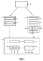
図1は、本発明による医用画像を画像解析する方法の第1の実施形態のフローチャートを示す。医用画像100が提供されると、第1の読影者1は、医用画像100の第1の注釈11を作成し、第1のROI情報12を記録する。第1の注釈11は、例えば、予備診断、患者情報、又は日時情報を表すことができる。前記医用画像100が同様に転送される第2の読影者2は、医用画像1の第2の注釈21を作成し、第2のROI情報22を記録する。更に、ビューイングデータ23が、第2の読影者2によって作成される。この実施形態において、ビューイングデータ23は、ズームレベルデータ231、パンレベルデータ232、及びディスプレイ解像度データ233を含む。次に、第1の読影者1及び第2の読影者2からのデータは、第2の注釈21の品質チェック4のために使用され、そこで、第2のROI情報22が、第1のROI情報12と比較される。2つのROIの情報が合致しない場合、品質チェック4にパスしない。更に、ビューイングデータ23が、予め決められたビューイングデータ要件40、すなわち予め決められたビューイングデータ閾値40を満たすかどうかが制御される。この実施形態において、ビューイングデータ要件40は、ズームレベル要件41、パンレベル要件42、及びディスプレイ解像度要件43を含む。
FIG. 1 shows a flow chart of a first embodiment of a method for image analysis of medical images according to the present invention. When the
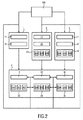
図2は、本発明による医用画像を画像解析のための方法の第2の実施形態のフローチャートを示す。2次元医用画像100が提供されると、第1の読影者1は、医用画像100の第1の注釈11を作成し、第1のROI情報12を記録する。遠隔放射線セットアップが与えられる場合、第2の読影者2が、同時に医用画像100の第2の注釈21を作成する。これとは別に、第2の読影者2は、第2のROI情報22及び第2のビューイングデータ23も記録し、第2のビューイングデータ23は、第2のビューイング時間データ234、第2のピクセル数データ235、及び第2のディスプレイサイズデータ236を含む。この実施形態において、同様にセットアップの一部で第3の読影者3は、第3の注釈31、第3のROI情報32、及び第3のビューイングデータ33を記録し、第3のビューイングデータ33は、第3のビューイング時間データ334、第3のピクセル数データ335、及び第3の読影者のディスプレイに対応する第3のディスプレイサイズデータ336を含む。その後、第1の読影者1、第2の読影者2、及び第3の読影者3からのデータは、第3の注釈31及び第2の注釈21の品質チェック4のために使用され、この場合、第3のROI情報32が、第2のROI情報22及び第1のROI情報12と比較される。更に、ビューイングデータ23及び33が予め決められたビューイングデータ要件40を満たすかどうか、及び/又はデータ23と33が合致するかどうかがチェックされる。特に、前記ビューイングデータ要件40は、ビューイング時間閾値44を含む。例えば、放射線科医が医用画像を少なくとも5分間観察することが要求される場合がある。この実施形態において、ビューイングデータ要件40は、ピクセル数閾値45及びディスプレイサイズ閾値46を更に有する。実際、ピクセル数の閾値45及びディスプレイサイズの閾値46は、ディスプレイ解像度の閾値46と組み合わされることができる。次いで、品質チェック4の結果は、すべての読影者1、2、3に供給され、それにより、品質チェック4をパスしなかった場合、読影者はビューポートを適応させることができる。
FIG. 2 shows a flowchart of a second embodiment of the method for image analysis of medical images according to the invention. When the two-dimensional
図3は、本発明による医用画像を画像解析するシステム5の第1の実施形態の概略図を示す。システム5は、第1の入力ユニット6、第2の入力ユニット7、及び品質チェックユニット8を備える。この実施形態において、品質チェックユニット8は、医用画像100を第1の入力ユニット6及び第2の入力ユニット7に提供する。次に、第1の入力ユニット6は、自動化された画像処理を使用して、医用画像上に第1の注釈61を作成する。これらの注釈及び第1のROI情報62は、この実施形態でも自動的に収集され、その後、品質チェックユニット8に転送される。品質チェックユニット8は、これらのデータを記憶し、それらを第2の入力ユニット7に渡す。次いで、放射線科医は、第2の入力ユニット7を使用して、診断を行うとともに、第1の入力ユニットの画像解析を評価することができる。そのために、放射線科医は、画像100、第1の注釈61、及び第1のROI情報62を品質チェックユニット8から受け取る。画像、第1注釈61及び第1のROI情報62を検討したのち、放射線科医は、彼/彼女がどんな病変を推定しているかと、疑わしい病変のサイズ及び位置をも示す。すなわち、放射線科医は、第2の注釈71及び第2のROI情報72を第2の入力ユニット7に入力する。第2の入力ユニット7は、放射線科医のビューイングデータ73も記録する。次に、これらのデータは、第2の注釈71及び第2のROI情報72とともに、品質チェックユニット8に送信され、品質チェックユニット8は、第2の注釈71の品質チェックを実行する。第2の注釈71が欠陥のあるレビューに基づいているとみなされる場合、第3の入力ユニットが必要とされることがある。
FIG. 3 shows a schematic diagram of a first embodiment of a system 5 for image analysis of medical images according to the invention. The system 5 comprises a first input unit 6, a
図4は、本発明による医用画像を画像解析するシステム5の第2の実施形態の概略図を示す。システム5は、第1の入力ユニット6、第2の入力ユニット7、品質チェックユニット8、出力ユニット9及び適応ユニット10を有する。この実施形態において、第2の入力ユニット7、品質チェックユニット8、出力ユニット9及び適応ユニット10は、単一の装置内に含まれ、前記装置は、スマートフォンである。第2の入力ユニット7は、第1の入力ユニット6から医用画像及び関連データを受信し、画像を出力ユニット9、すなわちスマートフォンのディスプレイ10に転送する。同時に、第2の入力ユニット7は、第2の注釈、第2のROI情報及びビューイングデータを収集し、これらを品質チェックユニット8に転送し、品質チェックユニット8は、続いて第1の入力ユニット6からのデータを使用して第2の注釈の品質をチェックする。この品質チェック4の結果は、ディスプレイ9によりユーザに直接提示される。表示された情報に従って、すなわち品質チェック4に従って、ユーザは、適応ユニット10を使用することにより医用画像を適応させることができる。適応後、品質チェックユニット8は、予め決められた要件に従って画像が表示されているかどうかを制御する。これらの予め規定された要件は、品質チェックユニット8自体から生じてもよいが、第1の入力ユニット6によって提供されることもできる。適応後のチェックの結果は、ディスプレイ9上でユーザに提示されることができる。
FIG. 4 shows a schematic diagram of a second embodiment of a system 5 for image analysis of medical images according to the invention. The system 5 comprises a first input unit 6, a
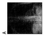
図5のA、B、Cは、さまざまなズームレベルでさまざまなディスプレイに表示される医用画像を示す。特に、図は、臨床的異常をもつ人間の胸部を示す。図5Aは、約25インチのスクリーン対角線を有する第1の装置でビューした画像を示しているが、図5A及び図5Bを表示するために使用されるモバイル5B及び5Cのディスプレイの対角線は、約8インチであ。すなわち、図5Aにおいて観察されるとき異常は容易に認識されうるが、図5Bに示されるような小さなビューポートは、異常を認識するには小さすぎる。従って、図5Bに示す画像を受け取った放射線科医は、適格な判断を行ことができない。しかしながら、図5Cに示されるようにズームレベルが呼応的に適応される場合、前記の小さなディスプレイサイズで十分である。すなわち、放射線科医が胸部画像を8インチディスプレイで観察する場合、彼/彼女は、ズームレベルが適切な場合にのみ異常を評価することができる。放射線科医が十分なズームレベルを使用することを確実にするために、使用されるズームレベルは、モバイルによって記録され、品質チェックユニットに送信されることができる。 5A, 5B, and 5C show medical images displayed on different displays at different zoom levels. In particular, the figure shows a human breast with clinical abnormalities. 5A shows an image viewed on the first device having a screen diagonal of about 25 inches, the diagonal of the mobile 5B and 5C displays used to display FIGS. 5A and 5B is about 8 inches. That is, while the anomaly can be easily recognized when viewed in FIG. 5A, the small viewport as shown in FIG. 5B is too small to recognize the anomaly. Therefore, the radiologist who received the image shown in FIG. 5B cannot make an appropriate judgment. However, said small display size is sufficient if the zoom level is adapted adaptively as shown in FIG. 5C. That is, when a radiologist views a chest image on an 8-inch display, he/she can only assess abnormalities if the zoom level is appropriate. To ensure that the radiologist uses a sufficient zoom level, the zoom level used can be recorded by the mobile and sent to the quality checking unit.
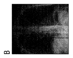
図6のA、B、及びCは、適切な表示のためのズームガイダンスを伴う医用画像を示す。特に、リンパ節腫脹を伴うヒト胸部が示されている。図6Aは、診断に適当でない形式の医用画像を示す。しかしながら、本発明に従って画像とともにROI情報が提供された場合、適切な診断が行われることができる。図6Bは、前記ROI情報がどのように伝えられることができるかを示し、すなわち、画像内の異常の周りをマーキングすることにより伝えられることを示す。このマーキングは、ズームのガイダンスを表する。適切なズームレベルを見つけるためにROIマーキングを使用することにより、リンパ節腫脹が、最適な態様で表示されることができる。対応する画像が図6Cに示される。 6A, 6B and 6C show medical images with zoom guidance for proper display. In particular, a human breast with lymphadenopathy is shown. FIG. 6A shows a medical image in a format that is not suitable for diagnosis. However, if ROI information is provided with the image in accordance with the present invention, appropriate diagnosis can be made. FIG. 6B shows how the ROI information can be conveyed, ie by marking around anomalies in the image. This marking represents guidance for zooming. By using ROI markings to find the proper zoom level, lymphadenopathy can be displayed in an optimal manner. The corresponding image is shown in Figure 6C.
図7は、医用画像100に関連する画像関連情報を記録するための装置70の実施形態の概略図を示す。装置70は、受信器75、ディスプレイ76、及び検出及び記録ユニット77を有する。画像100が受信器75によって受信された後、ディスプレイ76は画像100を表示する。その後、画像100に基づいて、放射線科医は注釈を作成する。これらは、検出及び記録ユニット77によって記録される。検出及び記録ユニット77は更に、可能性のある関心領域を検出し、座標及びサイズのような関連情報を記録する。これとは別に、放射線科医のビューイングデータが監視される。例えば、ディスプレイ76上で画像のどの領域がどのくらいの時間長さでどのズームレベルで観察されたかが記録される。特に、前記装置は、本発明によるシステム5の第2の入力ユニット7を表すことができる。
FIG. 7 shows a schematic diagram of an embodiment of an
本発明は、図面及び前述の記述において詳しく図示され説明されているが、そのような図面及び説明は説明的又は例示的であり、限定的なものと考えられるべきでない。本発明は、開示された実施形態に限定されない。開示された実施形態に対する他の変形が、図面、開示、及び添付の特許請求の範囲の検討から、請求項に記載の発明を実施する際に当業者によって理解され達成されることができる。 While the present invention has been illustrated and described in detail in the drawings and foregoing description, such drawings and description are to be considered illustrative or exemplary and not restrictive. The invention is not limited to the disclosed embodiments. Other variations to the disclosed embodiments can be understood and effected by those skilled in the art in practicing the claimed invention, from a study of the drawings, the disclosure, and the appended claims.
請求項において、「有する、含む(comprising)」という語は、他の較正要素又はステップを除外せず、不定冠詞「a」又は「an」は複数を除外しない。単一の較正要素又は他のユニットは、特許請求の範囲に記載されたいくつかのアイテムの機能を果たすことができる。特定の手段が相互に異なる従属請求項に記載されているという単なる事実は、これらの手段の組み合わせが有利に使用されることができないことを示すものではない。 In the claims, the word "comprising" does not exclude other calibration elements or steps, and the indefinite article "a" or "an" does not exclude a plurality. A single calibration element or other unit may fulfill the functions of several items recited in the claims. The mere fact that certain measures are recited in mutually different dependent claims does not indicate that a combination of these measures cannot be used to advantage.
コンピュータプログラムは、他のハードウェアとともに、又はその一部として提供される光学記憶媒体又はソリッドステート媒体のような適切な非一時媒体に記憶/配布されることができるが、他の形式で、例えばインターネット又はその他の有線又は無線通信システムを介して配布されることもできる。 The computer program may be stored/distributed on a suitable non-transitory medium such as an optical storage medium or a solid state medium provided with or as part of other hardware, but in other formats, for example It can also be distributed via the Internet or other wired or wireless communication system.
請求項における参照符号は、請求項の範囲を制限するものとして解釈されるべきでない。 Any reference signs in the claims shall not be construed as limiting the scope of the claims.
Claims (15)
医用画像、前記医用画像の第1の注釈、及び前記医用画像内の第1の関心領域(ROI)を示す第1のROI情報を受信するステップと、
前記医用画像の第2の注釈、前記医用画像内の第2のROIを示す第2のROI情報、並びにディスプレイデータ及び/又はビューイング時間を示すビューイングデータを受信するステップであって、前記第2のROIは前記ビューイングデータを用いてディスプレイに表示されたものであり、前記第2の注釈は前記ビューイングデータに基づいて作成されたものである、ステップと、
前記第2のROI情報を前記第1のROI情報と比較すること及び前記ビューイングデータをチェックすることにより、前記第2の注釈の品質をチェックするステップと、
を有する方法。 A method for image analysis of medical images,
Receiving a medical image, a first annotation of the medical image, and first ROI information indicating a first region of interest (ROI) in the medical image;
Receiving a second annotation of the medical image, second ROI information indicating a second ROI in the medical image, and viewing data indicating display data and/or viewing time, the method comprising: A ROI of 2 was displayed on the display using the viewing data, and the second annotation was created based on the viewing data;
Checking the quality of the second annotation by comparing the second ROI information with the first ROI information and checking the viewing data;
A method having.
前記ディスプレイデータが、特に前記ディスプレイ及び/又は前記第2のROI内における、解像度及び/又はディスプレイサイズ及び/又はピクセル数を含む、請求項1又は2に記載の方法。 The viewing data further comprises a zoom level and/or a pan level and/or
Method according to claim 1 or 2, wherein the display data comprises resolution and/or display size and/or number of pixels, in particular in the display and/or the second ROI.
前記品質チェックに従って前記画像を適応させるステップ、
を更に有する、請求項1乃至6のいずれか1項に記載の方法。 Providing the result of the quality check, and/or adapting the image according to the quality check,
The method according to any one of claims 1 to 6, further comprising:
前記他のROI情報を前記第1及び/又は第2のROI情報と比較すること及び前記他のビューイングデータをチェックすることにより、前記他の注釈の品質をチェックするステップと、
を有する、請求項1乃至7のいずれか1項に記載の方法。 Receiving other annotations of the medical image, other ROI information indicating one or more other ROIs in the medical image, and other viewing data including display data and/or viewing time. , The other ROI is displayed on a display using the other viewing data, and the other annotation is created based on the other viewing data,
Checking the quality of the other annotation by comparing the other ROI information with the first and/or second ROI information and checking the other viewing data;
The method according to any one of claims 1 to 7, comprising:
医用画像、前記医用画像の第1の注釈、及び前記医用画像内第1の関心領域(ROI)を示す第1のROIを示す情報を受信するように構成される第1の入力ユニットと、
前記医用画像の第2の注釈、前記医用画像内の第2のROIを示す第2のROI情報、並びにディスプレイデータ及び/又はビューイング時間を含むビューイングデータを受信するように構成される第2の入力ユニットであって、前記第2のROIは、前記ビューイングデータを用いてディスプレイに表示されたものであり、前記第2の注釈は、前記ビューイングデータに基づいて作成されたものである、入力ユニットと、
前記第1のROI情報を前記第2のROI情報と比較すること及び前記ビューイングデータをチェックすることにより、前記第2の注釈の品質をチェックするように構成される品質チェックユニットと、
を有するシステム。 A system for image analysis of medical images,
A first input unit configured to receive a medical image, a first annotation of the medical image, and information indicating a first ROI indicating a first region of interest (ROI) within the medical image;
A second annotation configured to receive a second annotation of the medical image, second ROI information indicating a second ROI in the medical image, and viewing data including display data and/or viewing time. The second ROI is displayed on the display using the viewing data, and the second annotation is created based on the viewing data. , Input unit,
A quality checking unit configured to check the quality of the second annotation by comparing the first ROI information with the second ROI information and checking the viewing data;
System with.
前記品質チェックに従って前記画像を適応させるように構成される適応ユニット、
を更に有する、請求項10に記載のシステム。 An output unit adapted to provide the result of the quality check, and/or an adaptation unit adapted to adapt the image according to the quality check,
The system of claim 10, further comprising:
医用画像を受信するステップと、
前記医用画像をディスプレイに表示するステップと、
前記医用画像について作成された注釈を記録するステップと、
前記医用画像内の関心領域(ROI)を示すROI情報を検出し記録するステップであって、前記医用画像は、前記ROI情報を用いて表示されたものであり、前記注釈は、前記ROI情報に基づいて作成されたものである、ステップと、
ビューイング時間を含むビューイングデータを検出し記録するステップであって、前記ROIは前記ビューイングデータを用いて表示されたものであり、前記注釈は前記ビューイングデータに基づいて作成されたものである、ステップと、
を有する方法。 A method of recording image-related information related to a medical image, comprising:
Receiving a medical image,
Displaying the medical image on a display,
Recording the annotations created for the medical image;
Detecting and recording ROI information indicating a region of interest (ROI) in the medical image, wherein the medical image is displayed using the ROI information, and the annotation is added to the ROI information. Based on the steps created,
Detecting and recording viewing data including viewing time, wherein the ROI is displayed using the viewing data, and the annotation is created based on the viewing data. There are steps,
A method having.
前記医用画像を受信するように構成される受信器と、
前記医用画像を表示するように構成されるディスプレイと、
検出及び記録ユニットであって、
前記医用画像について作成された注釈を記録する処理と、
前記医用画像内の関心領域(ROI)を示すROI情報を検出し記録する処理であって、前記医用画像は前記ROI情報を用いて表示されたものであり、前記注釈は前記ROI情報に基づいて作成されたものである、処理と、
ビューイング時間を含むビューイングデータを検出し記録する処理であって、前記ROIは、前記ビューイングデータを用いて表示されたものであり、前記注釈は、前記ビューイングデータに基づいて作成されたものである、処理と、
を行うように構成される検出及び記録ユニットと、
を有する装置。 A device for recording image-related information related to a medical image,
A receiver configured to receive the medical image,
A display configured to display the medical image,
A detection and recording unit,
Recording the annotations created for the medical image;
A process of detecting and recording ROI information indicating a region of interest (ROI) in the medical image, wherein the medical image is displayed using the ROI information, and the annotation is based on the ROI information. The processing that is created,
A process of detecting and recording viewing data including a viewing time, wherein the ROI is displayed using the viewing data, and the annotation is created based on the viewing data. What is processing,
A detection and recording unit configured to:
A device having.
Applications Claiming Priority (3)
| Application Number | Priority Date | Filing Date | Title |
|---|---|---|---|
| EP17177569.5 | 2017-06-23 | ||
| EP17177569.5A EP3417781A1 (en) | 2017-06-23 | 2017-06-23 | Method and system for image analysis of a medical image |
| PCT/EP2018/066621 WO2018234476A1 (en) | 2017-06-23 | 2018-06-21 | Method and system for image analysis of a medical image |
Publications (2)
| Publication Number | Publication Date |
|---|---|
| JP2020524540A true JP2020524540A (en) | 2020-08-20 |
| JP2020524540A5 JP2020524540A5 (en) | 2021-07-26 |
Family
ID=59239803
Family Applications (1)
| Application Number | Title | Priority Date | Filing Date |
|---|---|---|---|
| JP2019567654A Pending JP2020524540A (en) | 2017-06-23 | 2018-06-21 | Method and system for image analysis of medical images |
Country Status (5)
| Country | Link |
|---|---|
| US (1) | US20200126657A1 (en) |
| EP (2) | EP3417781A1 (en) |
| JP (1) | JP2020524540A (en) |
| CN (1) | CN110785125A (en) |
| WO (1) | WO2018234476A1 (en) |
Families Citing this family (1)
| Publication number | Priority date | Publication date | Assignee | Title |
|---|---|---|---|---|
| US11436579B2 (en) | 2020-05-04 | 2022-09-06 | Bank Of America Corporation | Performing enhanced deposit item processing using cognitive automation tools |
Citations (4)
| Publication number | Priority date | Publication date | Assignee | Title |
|---|---|---|---|---|
| US20100086182A1 (en) * | 2008-10-07 | 2010-04-08 | Hui Luo | Diagnostic image processing with automatic self image quality validation |
| US20100135562A1 (en) * | 2008-11-28 | 2010-06-03 | Siemens Computer Aided Diagnosis Ltd. | Computer-aided detection with enhanced workflow |
| JP2013041428A (en) * | 2011-08-16 | 2013-02-28 | Canon Inc | Medical diagnosis support device and medical diagnosis support method |
| JP2015198928A (en) * | 2014-03-31 | 2015-11-12 | 株式会社東芝 | Medical image processor, and medical image processing system |
Family Cites Families (8)
| Publication number | Priority date | Publication date | Assignee | Title |
|---|---|---|---|---|
| US5696531A (en) * | 1991-02-05 | 1997-12-09 | Minolta Camera Kabushiki Kaisha | Image display apparatus capable of combining image displayed with high resolution and image displayed with low resolution |
| US20090067700A1 (en) * | 2007-09-10 | 2009-03-12 | Riverain Medical Group, Llc | Presentation of computer-aided detection/diagnosis (CAD) results |
| US10354049B2 (en) * | 2012-08-22 | 2019-07-16 | Koninklijke Philips N.V. | Automatic detection and retrieval of prior annotations relevant for an imaging study for efficient viewing and reporting |
| US11224395B2 (en) * | 2012-10-05 | 2022-01-18 | Koninklijke Philips N.V. | Medical imaging system and method for providing an enhanced X-ray image |
| US20140324469A1 (en) * | 2013-04-30 | 2014-10-30 | Bruce Reiner | Customizable context and user-specific patient referenceable medical database |
| US9582170B2 (en) * | 2014-10-31 | 2017-02-28 | Mckesson Financial Holdings | Method and apparatus for managing a configurable display environment |
| KR102388130B1 (en) * | 2015-01-12 | 2022-04-19 | 삼성메디슨 주식회사 | Apparatus and method for displaying medical image |
| US10140421B1 (en) * | 2017-05-25 | 2018-11-27 | Enlitic, Inc. | Medical scan annotator system |
-
2017
- 2017-06-23 EP EP17177569.5A patent/EP3417781A1/en not_active Withdrawn
-
2018
- 2018-06-21 CN CN201880042018.4A patent/CN110785125A/en active Pending
- 2018-06-21 WO PCT/EP2018/066621 patent/WO2018234476A1/en active Application Filing
- 2018-06-21 US US16/623,421 patent/US20200126657A1/en not_active Abandoned
- 2018-06-21 JP JP2019567654A patent/JP2020524540A/en active Pending
- 2018-06-21 EP EP18733255.6A patent/EP3641650B1/en active Active
Patent Citations (4)
| Publication number | Priority date | Publication date | Assignee | Title |
|---|---|---|---|---|
| US20100086182A1 (en) * | 2008-10-07 | 2010-04-08 | Hui Luo | Diagnostic image processing with automatic self image quality validation |
| US20100135562A1 (en) * | 2008-11-28 | 2010-06-03 | Siemens Computer Aided Diagnosis Ltd. | Computer-aided detection with enhanced workflow |
| JP2013041428A (en) * | 2011-08-16 | 2013-02-28 | Canon Inc | Medical diagnosis support device and medical diagnosis support method |
| JP2015198928A (en) * | 2014-03-31 | 2015-11-12 | 株式会社東芝 | Medical image processor, and medical image processing system |
Also Published As
| Publication number | Publication date |
|---|---|
| CN110785125A (en) | 2020-02-11 |
| EP3641650B1 (en) | 2020-11-18 |
| EP3417781A1 (en) | 2018-12-26 |
| US20200126657A1 (en) | 2020-04-23 |
| EP3641650A1 (en) | 2020-04-29 |
| WO2018234476A1 (en) | 2018-12-27 |
Similar Documents
| Publication | Publication Date | Title |
|---|---|---|
| JP4104036B2 (en) | Abnormal shadow detection processing method and system | |
| US20120299818A1 (en) | Medical information display apparatus, operation method of the same and medical information display program | |
| KR101840350B1 (en) | Method and apparatus for aiding reading efficiency using eye tracking information in medical image reading processing | |
| JP6183177B2 (en) | Medical image system and program | |
| JP2006116313A (en) | Diagnostic method in three-dimensional imaging | |
| US10916010B2 (en) | Learning data creation support apparatus, learning data creation support method, and learning data creation support program | |
| JP6230708B2 (en) | Matching findings between imaging datasets | |
| KR101716422B1 (en) | Method and apparatus for providing medical information | |
| EP2878266A1 (en) | Medical imaging system and program | |
| KR101050769B1 (en) | Medical Image Processing System and Processing Method | |
| KR20130053587A (en) | Medical device and medical image displaying method using the same | |
| US20190125306A1 (en) | Method of transmitting a medical image, and a medical imaging apparatus performing the method | |
| CN112582057A (en) | Advanced medical imaging in a distributed setting | |
| EP3641650B1 (en) | Method and system for image analysis of a medical image | |
| JP2015104464A (en) | Medical image system and program | |
| WO2017064600A1 (en) | Systems and methods for generating correct radiological recommendations | |
| JP2015221141A (en) | Medical image diagnosis support apparatus, operation method for medical image diagnosis support apparatus, and medical image diagnosis support program | |
| JP2016209267A (en) | Medical image processor and program | |
| KR20160115269A (en) | System and application for providing information of health examinations | |
| US11003342B1 (en) | Smart scrolling system | |
| JP6966403B2 (en) | Diagnostic support device | |
| JP4617116B2 (en) | Instant medical video automatic search and contrast method and system | |
| JP7143747B2 (en) | Image display device, image display method and image display program | |
| JP7428055B2 (en) | Diagnostic support system, diagnostic support device and program | |
| EP4332984A1 (en) | Systems and methods for improving communication between local technologists within a radiology operations command center (rocc) framework |
Legal Events
| Date | Code | Title | Description |
|---|---|---|---|
| A521 | Request for written amendment filed |
Free format text: JAPANESE INTERMEDIATE CODE: A523 Effective date: 20210513 |
|
| A621 | Written request for application examination |
Free format text: JAPANESE INTERMEDIATE CODE: A621 Effective date: 20210513 |
|
| A977 | Report on retrieval |
Free format text: JAPANESE INTERMEDIATE CODE: A971007 Effective date: 20220525 |
|
| A131 | Notification of reasons for refusal |
Free format text: JAPANESE INTERMEDIATE CODE: A131 Effective date: 20220614 |
|
| A601 | Written request for extension of time |
Free format text: JAPANESE INTERMEDIATE CODE: A601 Effective date: 20220831 |
|
| A521 | Request for written amendment filed |
Free format text: JAPANESE INTERMEDIATE CODE: A523 Effective date: 20221118 |
|
| A02 | Decision of refusal |
Free format text: JAPANESE INTERMEDIATE CODE: A02 Effective date: 20230307 |