JP2020096834A - 強化されたマルチコアファイバ内視鏡 - Google Patents
強化されたマルチコアファイバ内視鏡 Download PDFInfo
- Publication number
- JP2020096834A JP2020096834A JP2019224599A JP2019224599A JP2020096834A JP 2020096834 A JP2020096834 A JP 2020096834A JP 2019224599 A JP2019224599 A JP 2019224599A JP 2019224599 A JP2019224599 A JP 2019224599A JP 2020096834 A JP2020096834 A JP 2020096834A
- Authority
- JP
- Japan
- Prior art keywords
- core
- endoscope
- fiber
- cores
- illumination
- Prior art date
- Legal status (The legal status is an assumption and is not a legal conclusion. Google has not performed a legal analysis and makes no representation as to the accuracy of the status listed.)
- Pending
Links
- 239000000835 fiber Substances 0.000 title claims abstract description 216
- 238000005286 illumination Methods 0.000 claims abstract description 126
- 230000003287 optical effect Effects 0.000 claims abstract description 91
- 230000001427 coherent effect Effects 0.000 claims abstract description 7
- 238000012545 processing Methods 0.000 claims abstract description 4
- 238000003384 imaging method Methods 0.000 claims description 109
- 238000000034 method Methods 0.000 claims description 57
- 238000007689 inspection Methods 0.000 claims description 36
- 238000001839 endoscopy Methods 0.000 claims description 17
- 238000013461 design Methods 0.000 claims description 14
- 239000000463 material Substances 0.000 claims description 14
- 239000000975 dye Substances 0.000 claims description 13
- 230000002093 peripheral effect Effects 0.000 claims description 12
- 230000005855 radiation Effects 0.000 claims description 11
- 238000004458 analytical method Methods 0.000 claims description 6
- 230000003321 amplification Effects 0.000 claims description 5
- 239000011248 coating agent Substances 0.000 claims description 5
- 238000000576 coating method Methods 0.000 claims description 5
- 230000000694 effects Effects 0.000 claims description 5
- 230000007246 mechanism Effects 0.000 claims description 5
- 238000003199 nucleic acid amplification method Methods 0.000 claims description 5
- 238000001069 Raman spectroscopy Methods 0.000 claims description 4
- 239000007850 fluorescent dye Substances 0.000 claims description 4
- 239000004038 photonic crystal Substances 0.000 claims description 4
- 238000001228 spectrum Methods 0.000 claims description 4
- 229910052751 metal Inorganic materials 0.000 claims description 3
- 239000002184 metal Substances 0.000 claims description 3
- 239000000203 mixture Substances 0.000 claims description 3
- 230000002123 temporal effect Effects 0.000 abstract 1
- 239000011162 core material Substances 0.000 description 202
- 238000005070 sampling Methods 0.000 description 9
- 230000010287 polarization Effects 0.000 description 7
- 230000008569 process Effects 0.000 description 7
- 230000005540 biological transmission Effects 0.000 description 5
- 230000008901 benefit Effects 0.000 description 4
- 238000005253 cladding Methods 0.000 description 4
- 238000004590 computer program Methods 0.000 description 4
- 239000013307 optical fiber Substances 0.000 description 4
- 238000001574 biopsy Methods 0.000 description 3
- 230000015572 biosynthetic process Effects 0.000 description 3
- 238000010276 construction Methods 0.000 description 3
- 238000009826 distribution Methods 0.000 description 3
- 238000004519 manufacturing process Methods 0.000 description 3
- 210000001747 pupil Anatomy 0.000 description 3
- 238000007493 shaping process Methods 0.000 description 3
- 238000003860 storage Methods 0.000 description 3
- 238000000701 chemical imaging Methods 0.000 description 2
- 239000012530 fluid Substances 0.000 description 2
- 230000010354 integration Effects 0.000 description 2
- 238000013532 laser treatment Methods 0.000 description 2
- 239000007788 liquid Substances 0.000 description 2
- 238000001465 metallisation Methods 0.000 description 2
- 238000012986 modification Methods 0.000 description 2
- 230000004048 modification Effects 0.000 description 2
- 238000012634 optical imaging Methods 0.000 description 2
- 230000009467 reduction Effects 0.000 description 2
- 238000000926 separation method Methods 0.000 description 2
- 238000012546 transfer Methods 0.000 description 2
- 229910052691 Erbium Inorganic materials 0.000 description 1
- 241000295146 Gallionellaceae Species 0.000 description 1
- 208000000913 Kidney Calculi Diseases 0.000 description 1
- 206010029148 Nephrolithiasis Diseases 0.000 description 1
- 230000008859 change Effects 0.000 description 1
- 238000006243 chemical reaction Methods 0.000 description 1
- 239000003086 colorant Substances 0.000 description 1
- 238000004891 communication Methods 0.000 description 1
- 230000000295 complement effect Effects 0.000 description 1
- 230000006835 compression Effects 0.000 description 1
- 238000007906 compression Methods 0.000 description 1
- 238000001514 detection method Methods 0.000 description 1
- 238000010586 diagram Methods 0.000 description 1
- 239000006185 dispersion Substances 0.000 description 1
- 238000012377 drug delivery Methods 0.000 description 1
- UYAHIZSMUZPPFV-UHFFFAOYSA-N erbium Chemical compound [Er] UYAHIZSMUZPPFV-UHFFFAOYSA-N 0.000 description 1
- 238000003780 insertion Methods 0.000 description 1
- 230000037431 insertion Effects 0.000 description 1
- 238000009434 installation Methods 0.000 description 1
- 238000013507 mapping Methods 0.000 description 1
- 239000011159 matrix material Substances 0.000 description 1
- 238000005457 optimization Methods 0.000 description 1
- 210000000056 organ Anatomy 0.000 description 1
- 238000001126 phototherapy Methods 0.000 description 1
- 229920000642 polymer Polymers 0.000 description 1
- 229920005594 polymer fiber Polymers 0.000 description 1
- 239000004065 semiconductor Substances 0.000 description 1
- 230000035945 sensitivity Effects 0.000 description 1
- 125000006850 spacer group Chemical group 0.000 description 1
- 230000003595 spectral effect Effects 0.000 description 1
- 238000006467 substitution reaction Methods 0.000 description 1
- 238000012360 testing method Methods 0.000 description 1
- 238000002560 therapeutic procedure Methods 0.000 description 1
- 238000011282 treatment Methods 0.000 description 1
- 210000000626 ureter Anatomy 0.000 description 1
- 239000011800 void material Substances 0.000 description 1
Images


Classifications
-
- A—HUMAN NECESSITIES
- A61—MEDICAL OR VETERINARY SCIENCE; HYGIENE
- A61B—DIAGNOSIS; SURGERY; IDENTIFICATION
- A61B1/00—Instruments for performing medical examinations of the interior of cavities or tubes of the body by visual or photographical inspection, e.g. endoscopes; Illuminating arrangements therefor
- A61B1/06—Instruments for performing medical examinations of the interior of cavities or tubes of the body by visual or photographical inspection, e.g. endoscopes; Illuminating arrangements therefor with illuminating arrangements
- A61B1/063—Instruments for performing medical examinations of the interior of cavities or tubes of the body by visual or photographical inspection, e.g. endoscopes; Illuminating arrangements therefor with illuminating arrangements for monochromatic or narrow-band illumination
-
- A—HUMAN NECESSITIES
- A61—MEDICAL OR VETERINARY SCIENCE; HYGIENE
- A61B—DIAGNOSIS; SURGERY; IDENTIFICATION
- A61B1/00—Instruments for performing medical examinations of the interior of cavities or tubes of the body by visual or photographical inspection, e.g. endoscopes; Illuminating arrangements therefor
- A61B1/00002—Operational features of endoscopes
- A61B1/00004—Operational features of endoscopes characterised by electronic signal processing
- A61B1/00009—Operational features of endoscopes characterised by electronic signal processing of image signals during a use of endoscope
- A61B1/000094—Operational features of endoscopes characterised by electronic signal processing of image signals during a use of endoscope extracting biological structures
-
- A—HUMAN NECESSITIES
- A61—MEDICAL OR VETERINARY SCIENCE; HYGIENE
- A61B—DIAGNOSIS; SURGERY; IDENTIFICATION
- A61B1/00—Instruments for performing medical examinations of the interior of cavities or tubes of the body by visual or photographical inspection, e.g. endoscopes; Illuminating arrangements therefor
- A61B1/00064—Constructional details of the endoscope body
- A61B1/00071—Insertion part of the endoscope body
- A61B1/0008—Insertion part of the endoscope body characterised by distal tip features
- A61B1/00096—Optical elements
-
- A—HUMAN NECESSITIES
- A61—MEDICAL OR VETERINARY SCIENCE; HYGIENE
- A61B—DIAGNOSIS; SURGERY; IDENTIFICATION
- A61B1/00—Instruments for performing medical examinations of the interior of cavities or tubes of the body by visual or photographical inspection, e.g. endoscopes; Illuminating arrangements therefor
- A61B1/00163—Optical arrangements
- A61B1/00165—Optical arrangements with light-conductive means, e.g. fibre optics
- A61B1/00167—Details of optical fibre bundles, e.g. shape or fibre distribution
-
- A—HUMAN NECESSITIES
- A61—MEDICAL OR VETERINARY SCIENCE; HYGIENE
- A61B—DIAGNOSIS; SURGERY; IDENTIFICATION
- A61B1/00—Instruments for performing medical examinations of the interior of cavities or tubes of the body by visual or photographical inspection, e.g. endoscopes; Illuminating arrangements therefor
- A61B1/00163—Optical arrangements
- A61B1/00186—Optical arrangements with imaging filters
-
- A—HUMAN NECESSITIES
- A61—MEDICAL OR VETERINARY SCIENCE; HYGIENE
- A61B—DIAGNOSIS; SURGERY; IDENTIFICATION
- A61B1/00—Instruments for performing medical examinations of the interior of cavities or tubes of the body by visual or photographical inspection, e.g. endoscopes; Illuminating arrangements therefor
- A61B1/00163—Optical arrangements
- A61B1/00188—Optical arrangements with focusing or zooming features
-
- A—HUMAN NECESSITIES
- A61—MEDICAL OR VETERINARY SCIENCE; HYGIENE
- A61B—DIAGNOSIS; SURGERY; IDENTIFICATION
- A61B1/00—Instruments for performing medical examinations of the interior of cavities or tubes of the body by visual or photographical inspection, e.g. endoscopes; Illuminating arrangements therefor
- A61B1/04—Instruments for performing medical examinations of the interior of cavities or tubes of the body by visual or photographical inspection, e.g. endoscopes; Illuminating arrangements therefor combined with photographic or television appliances
- A61B1/042—Instruments for performing medical examinations of the interior of cavities or tubes of the body by visual or photographical inspection, e.g. endoscopes; Illuminating arrangements therefor combined with photographic or television appliances characterised by a proximal camera, e.g. a CCD camera
-
- A—HUMAN NECESSITIES
- A61—MEDICAL OR VETERINARY SCIENCE; HYGIENE
- A61B—DIAGNOSIS; SURGERY; IDENTIFICATION
- A61B1/00—Instruments for performing medical examinations of the interior of cavities or tubes of the body by visual or photographical inspection, e.g. endoscopes; Illuminating arrangements therefor
- A61B1/06—Instruments for performing medical examinations of the interior of cavities or tubes of the body by visual or photographical inspection, e.g. endoscopes; Illuminating arrangements therefor with illuminating arrangements
- A61B1/0661—Endoscope light sources
- A61B1/0669—Endoscope light sources at proximal end of an endoscope
-
- A—HUMAN NECESSITIES
- A61—MEDICAL OR VETERINARY SCIENCE; HYGIENE
- A61B—DIAGNOSIS; SURGERY; IDENTIFICATION
- A61B1/00—Instruments for performing medical examinations of the interior of cavities or tubes of the body by visual or photographical inspection, e.g. endoscopes; Illuminating arrangements therefor
- A61B1/06—Instruments for performing medical examinations of the interior of cavities or tubes of the body by visual or photographical inspection, e.g. endoscopes; Illuminating arrangements therefor with illuminating arrangements
- A61B1/07—Instruments for performing medical examinations of the interior of cavities or tubes of the body by visual or photographical inspection, e.g. endoscopes; Illuminating arrangements therefor with illuminating arrangements using light-conductive means, e.g. optical fibres
-
- G—PHYSICS
- G02—OPTICS
- G02B—OPTICAL ELEMENTS, SYSTEMS OR APPARATUS
- G02B23/00—Telescopes, e.g. binoculars; Periscopes; Instruments for viewing the inside of hollow bodies; Viewfinders; Optical aiming or sighting devices
- G02B23/24—Instruments or systems for viewing the inside of hollow bodies, e.g. fibrescopes
- G02B23/2407—Optical details
- G02B23/2461—Illumination
- G02B23/2469—Illumination using optical fibres
-
- G—PHYSICS
- G02—OPTICS
- G02B—OPTICAL ELEMENTS, SYSTEMS OR APPARATUS
- G02B23/00—Telescopes, e.g. binoculars; Periscopes; Instruments for viewing the inside of hollow bodies; Viewfinders; Optical aiming or sighting devices
- G02B23/24—Instruments or systems for viewing the inside of hollow bodies, e.g. fibrescopes
- G02B23/26—Instruments or systems for viewing the inside of hollow bodies, e.g. fibrescopes using light guides
Abstract
Description
Claims (29)
- コヒーレントレーザ照明ビームを生成する照明源と、
光センサと、
マルチコアファイバであって、
検査すべき表面の照明のための少なくとも1つのコアであって、前記少なくとも1つのコアを通って前記照明源から前記ファイバの遠位端へ前記照明ビームを伝達する少なくとも1つのコア、および
前記表面から反射された光を前記光センサへ伝達する複数のコア
を備えるマルチコアファイバと、
前記照明ビームの鏡像を前記表面の画像から分離する時間変調シーケンサと、
前記光センサからの感知されたデータを処理して前記表面の前記画像を生成するプロセッサと
を備える内視鏡。 - 前記時間変調シーケンサが、2つのファラデー回転子を備え、前記ファラデー回転子がそれぞれ回転軸を有し、前記回転軸が実質上平行であるがずれている、請求項1に記載の内視鏡。
- 前記2つのファラデー回転子が、ロックイン増幅効果をもたらすように構成される、請求項1に記載の内視鏡。
- 複数のコアを有するマルチコアファイバを備える内視鏡であって、前記コアが、少なくとも2つの異なる断面形状または向きを有し、前記少なくとも2つの異なる断面形状または向きが、混合配置で配置される、内視鏡。
- 前記少なくとも2つの異なる断面形状または向きが、織り合わせたアレイで配置される、請求項4に記載の内視鏡。
- 前記少なくとも2つの異なる断面形状または向きのうちの一方のコアの断面の長軸が、前記少なくとも2つの異なる断面形状または向きのうちの他方の長軸に直交している、請求項4に記載の内視鏡。
- マルチコアファイバを備える内視鏡であって、
前記マルチコアファイバが、
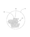
検査すべき表面の照明のための少なくとも1つの中空コアであって、前記少なくとも1つのコアを通って照明源から前記ファイバの遠位端へ照明ビームを伝達する少なくとも1つの中空コア、および前記少なくとも1つの中空コア内に前記照明ビームを閉じ込めるように前記少なくとも1つの中空コアのそれぞれを取り囲む複数のコアを含む照明領域と、
前記表面から反射された光を光センサへ伝達する複数のコアを含む撮像領域とを備える、内視鏡。 - 前記少なくとも1つの中空コアのそれぞれを取り囲む前記複数のコアが、前記中空コアの周りに複数のリングで配置される、請求項7に記載の内視鏡。
- 前記複数のリングが、少なくとも3つのリングを含む、請求項8に記載の内視鏡。
- 前記少なくとも1つの中空コアのそれぞれを取り囲む前記複数のコアが、フォトニック結晶ファイバ(PCF)を含む、請求項7に記載の内視鏡。
- 光センサと、
マルチコアファイバであって、
検査表面から反射された光を前記光センサへ伝達する複数のコア、
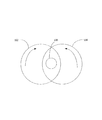
前記マルチコアファイバの遠位先端から所定の距離をあけて配置された、前記表面から反射された前記光でフーリエ変換を実行するレンズ、および
前記マルチコアファイバの近位先端から所定の距離をあけて配置された、前記マルチコアファイバの近位先端から出た後に前記表面から反射された前記光で逆フーリエ変換を実行するレンズ
を備えるマルチコアファイバと、
前記光センサからの感知されたデータを処理して前記表面の画像を生成するプロセッサと
を備える内視鏡。 - デジタル復号によって前記検査表面の画像の位相を構築するようにデジタルフーリエホログラフィック記録を実現するための参照照明ビームを生成するレーザ源をさらに備える、請求項11に記載の内視鏡。
- 前記検査表面から前記マルチコアファイバ内へ反射された前記光の光路上に配置された複数のPCFコアを含む光学素子をさらに備える、請求項11に記載の内視鏡。
- マルチコアファイバを備える内視鏡であって、
前記マルチコアファイバが、
検査すべき表面の照明のための少なくとも1つの撮像コアであって、前記少なくとも1つのコアを通って照明源から前記ファイバの遠位端へ照明ビームを伝達する少なくとも1つの撮像コアを含む照明領域と、
前記表面から反射された光を光センサへ伝達する複数のコアを含む撮像領域と、
蛍光染料を収容する染料容器であって、前記染料に照射して連続白色光照明スペクトルを生成することを可能にするように、前記1つの撮像コア内へ前記染料を供給するように構成された染料容器とを備える、内視鏡。 - 前記染料が溶液混合物である、請求項14に記載の内視鏡。
- マルチコアファイバを備える内視鏡であって、
前記マルチコアファイバが、
表面から反射された光を光センサへ伝達する複数の撮像コアを含む撮像領域と、
前記複数の撮像コアのそれぞれの周りに位置する複数の周辺コアとを備え、前記周辺コアのそれぞれの直径が、前記撮像コアを通過することが予想される反射光の波長より小さい、内視鏡。 - 前記周辺コアが、空気で充填された中空コアである、請求項16に記載の内視鏡。
- レーザ光を使用する内視鏡検査における向上された放射安全性のための方法であって、
内視鏡のマルチコアファイバの撮像コア内へ増幅材料を埋め込むことと、
検査表面から反射され前記撮像コアを通過する光を増幅するように構成された所定の波長のレーザ光によって前記撮像コアを励起することとを含む方法。 - 内視鏡検査における光学ズームのための方法であって、
レーザ源を使用して、内視鏡マルチコアファイバの遠位先端に位置するレンズの設計に関して事前定義された曲率の球形波面を有する、検査表面を照明するための照明ビームを生成することと、
前記検査表面の画像を光学的に拡大または縮小するように、前記レンズの前記設計に対する前記球形波長の前記曲率を変化させることとを含む方法。 - 検査表面を検査するために内視鏡を使用するときの光学ズームのための方法であって、前記内視鏡が、マルチコアファイバの遠位先端に焦点距離を有する第1のフーリエレンズを有し、前記方法が、
前記マルチコアファイバの近位先端と光検出器との間に配置された前記近位先端に調整可能な焦点距離を有する第2のフーリエレンズを提供することと、
前記検査表面の画像を光学的に拡大または縮小するように、前記第2のフーリエレンズの前記調整可能な焦点距離を調整することとを含む方法。 - マルチコアファイバを有する内視鏡を使用して得られる画像の解像度を改善する方法であって、
複数の異なる波長の赤色、複数の異なる波長の緑色、および複数の異なる波長の青色によって検査表面を照明することと、
光検出器によって、前記複数の異なる波長の赤色、前記複数の異なる波長の緑色、および前記複数の異なる波長の青色によって前記検査表面から反射された光の画像データを収集することと、
前記収集された画像データから画像を構築することとを含む方法。 - 内視鏡検査における画像解像度を改善する光学ズームのための方法であって、
検査表面から反射され、複数の撮像コアを含むマルチコアファイバを通過した光の光路上に2焦点レンズを提供することと、
前記2焦点レンズの焦点距離のそれぞれを較正に使用することと、
前記較正に基づいて、光検出器上で内視鏡によって得られた正規画像および拡大画像という2つの重畳画像を分離することと、
前記拡大画像をデジタルで縮小することと、
縮小された後に前記拡大画像からの画像データを前記正規画像に追加して、より高解像度の正規画像を得ることとを含む方法。 - マルチコアファイバを備える内視鏡であって、
前記マルチコアファイバが、
表面から反射された光を光センサへ伝達する複数の撮像コアを備え、各コアが金属被覆によって被覆される、内視鏡。 - 前記コアのピッチが2ミクロンより小さい、請求項23に記載の内視鏡。
- 前記ピッチが約1ミクロンである、請求項24に記載の内視鏡。
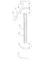
- 遠位端および近位端を有するマルチコアファイバであって、前記近位端からの光を前記遠位端から伝送し、観察すべき患者の内面を照明する1つまたは複数の照明コア、および前記内面から撮像コア内へ反射された光を前記遠位端から前記近位端へ伝達する1つまたは複数の撮像コアを有するマルチコアファイバと、
前記マルチコアファイバが挿入される遮蔽スリーブと、
前記マルチコアファイバの遠位先端を抜き出して前記表面に接触させ、分析のためのサンプルを収集し、前記遠位先端を前記遮蔽スリーブ内へ後退させることを可能にするように、前記遮蔽スリーブ内で前記マルチコアファイバを前進および後退させる機構と
を備える内視鏡。 - 前記機構がプランジャを備える、請求項26に記載の内視鏡。
- 前記反射された光を受け取る光センサを備える、請求項26に記載の内視鏡。
- 前記サンプルの分析を実行するラマン分光計である、請求項28に記載の光センサ。
Applications Claiming Priority (2)
| Application Number | Priority Date | Filing Date | Title |
|---|---|---|---|
| US16/221,593 US20200187766A1 (en) | 2018-12-17 | 2018-12-17 | Enhanced multicore fiber endoscopes |
| US16/221,593 | 2018-12-17 |
Publications (2)
| Publication Number | Publication Date |
|---|---|
| JP2020096834A true JP2020096834A (ja) | 2020-06-25 |
| JP2020096834A5 JP2020096834A5 (ja) | 2022-12-20 |
Family
ID=71071061
Family Applications (1)
| Application Number | Title | Priority Date | Filing Date |
|---|---|---|---|
| JP2019224599A Pending JP2020096834A (ja) | 2018-12-17 | 2019-12-12 | 強化されたマルチコアファイバ内視鏡 |
Country Status (2)
| Country | Link |
|---|---|
| US (2) | US20200187766A1 (ja) |
| JP (1) | JP2020096834A (ja) |
Cited By (1)
| Publication number | Priority date | Publication date | Assignee | Title |
|---|---|---|---|---|
| US11480317B2 (en) | 2020-09-29 | 2022-10-25 | Panasonic Intellectual Property Management Co., Ltd. | Light source device with sensor for detecting anomaly in wavelength converting member |
Families Citing this family (3)
| Publication number | Priority date | Publication date | Assignee | Title |
|---|---|---|---|---|
| EP3654824A4 (en) * | 2017-07-17 | 2021-04-21 | Z Square Ltd. | IMAGING IMPROVEMENT THROUGH MULTI-FIBER ENDOSCOPES |
| DE102020132454B3 (de) * | 2020-12-07 | 2021-11-25 | Karl Storz Se & Co. Kg | Endoskopische Vorrichtung und Verfahren zur Überprüfung einer Identität einer Komponente einer endoskopischen Vorrichtung |
| CN114176492A (zh) * | 2022-01-05 | 2022-03-15 | 深圳迈塔兰斯科技有限公司 | 内窥镜探头、内窥镜及其扫描控制方法 |
-
2018
- 2018-12-17 US US16/221,593 patent/US20200187766A1/en not_active Abandoned
-
2019
- 2019-12-12 JP JP2019224599A patent/JP2020096834A/ja active Pending
-
2022
- 2022-12-14 US US18/080,844 patent/US20230110978A1/en active Pending
Cited By (1)
| Publication number | Priority date | Publication date | Assignee | Title |
|---|---|---|---|---|
| US11480317B2 (en) | 2020-09-29 | 2022-10-25 | Panasonic Intellectual Property Management Co., Ltd. | Light source device with sensor for detecting anomaly in wavelength converting member |
Also Published As
| Publication number | Publication date |
|---|---|
| US20230110978A1 (en) | 2023-04-13 |
| US20200187766A1 (en) | 2020-06-18 |
Similar Documents
| Publication | Publication Date | Title |
|---|---|---|
| JP6770109B2 (ja) | 全方向視覚装置 | |
| US20240049956A1 (en) | Illumination sources for multicore fiber endoscopes | |
| JP2020096834A (ja) | 強化されたマルチコアファイバ内視鏡 | |
| US9871948B2 (en) | Methods and apparatus for imaging with multimode optical fibers | |
| CN101365375B (zh) | 用于经由谱编码进行光学成像的方法和设备 | |
| US8585587B2 (en) | Determining phase variation of light in an endoscope | |
| KR102588057B1 (ko) | 광학 시스템 및 방법 | |
| US8705184B2 (en) | Multi-path, multi-magnification, non-confocal fluorescence emission endoscopy apparatus and methods | |
| US8942530B2 (en) | Endoscope connector method and apparatus | |
| JP2023055872A (ja) | マルチコアファイバ内視鏡によるイメージングの強化 | |
| EP1244927B1 (en) | Methods and apparatus for imaging using a light guide bundle and a spatial light modulator | |
| US20130324858A1 (en) | Multi-path, multi-magnification, non-confocal fluorescence emission endoscopy apparatus and methods | |
| KR20120072757A (ko) | 광섬유 다발 기반의 내시경 타입 스펙트럼 영역 광학단층영상 시스템 | |
| KR20210093245A (ko) | 다중 모드 도파관 이미징 | |
| JP2020096834A5 (ja) | ||
| WO2019015437A1 (zh) | 层析内窥显微光谱成像装置 | |
| WO2018116302A1 (en) | Illumination sources for multicore fiber endoscopes | |
| Choi et al. | Fourier holographic endoscopy for label-free imaging through a narrow and curved passage | |
| WO2019015436A1 (zh) | 层析内窥显微成像装置 | |
| Oh et al. | Review of endomicroscopic imaging with coherent manipulation of light through an ultrathin probe | |
| Pacheco et al. | Optics of Biomedical Instrumentation | |
| Cha | FIBER-OPTIC BUNDLE FLUORESCENCE MICROSCOPY FOR FUNCTIONAL BRAIN ACTIVITY MAPPING |
Legal Events
| Date | Code | Title | Description |
|---|---|---|---|
| A521 | Request for written amendment filed |
Free format text: JAPANESE INTERMEDIATE CODE: A523 Effective date: 20221212 |
|
| A621 | Written request for application examination |
Free format text: JAPANESE INTERMEDIATE CODE: A621 Effective date: 20221212 |
|
| A977 | Report on retrieval |
Free format text: JAPANESE INTERMEDIATE CODE: A971007 Effective date: 20231030 |
|
| A131 | Notification of reasons for refusal |
Free format text: JAPANESE INTERMEDIATE CODE: A131 Effective date: 20231114 |
|
| A601 | Written request for extension of time |
Free format text: JAPANESE INTERMEDIATE CODE: A601 Effective date: 20240207 |