JP2018529475A - Apparatus, method and system for medical image annotation - Google Patents
Apparatus, method and system for medical image annotation Download PDFInfo
- Publication number
- JP2018529475A JP2018529475A JP2018517764A JP2018517764A JP2018529475A JP 2018529475 A JP2018529475 A JP 2018529475A JP 2018517764 A JP2018517764 A JP 2018517764A JP 2018517764 A JP2018517764 A JP 2018517764A JP 2018529475 A JP2018529475 A JP 2018529475A
- Authority
- JP
- Japan
- Prior art keywords
- annotation
- image
- imaging system
- medical imaging
- touch screen
- Prior art date
- Legal status (The legal status is an assumption and is not a legal conclusion. Google has not performed a legal analysis and makes no representation as to the accuracy of the status listed.)
- Pending
Links
Images


Classifications
-
- A—HUMAN NECESSITIES
- A61—MEDICAL OR VETERINARY SCIENCE; HYGIENE
- A61B—DIAGNOSIS; SURGERY; IDENTIFICATION
- A61B8/00—Diagnosis using ultrasonic, sonic or infrasonic waves
- A61B8/46—Ultrasonic, sonic or infrasonic diagnostic devices with special arrangements for interfacing with the operator or the patient
- A61B8/467—Ultrasonic, sonic or infrasonic diagnostic devices with special arrangements for interfacing with the operator or the patient characterised by special input means
- A61B8/468—Ultrasonic, sonic or infrasonic diagnostic devices with special arrangements for interfacing with the operator or the patient characterised by special input means allowing annotation or message recording
-
- G—PHYSICS
- G16—INFORMATION AND COMMUNICATION TECHNOLOGY [ICT] SPECIALLY ADAPTED FOR SPECIFIC APPLICATION FIELDS
- G16H—HEALTHCARE INFORMATICS, i.e. INFORMATION AND COMMUNICATION TECHNOLOGY [ICT] SPECIALLY ADAPTED FOR THE HANDLING OR PROCESSING OF MEDICAL OR HEALTHCARE DATA
- G16H30/00—ICT specially adapted for the handling or processing of medical images
- G16H30/20—ICT specially adapted for the handling or processing of medical images for handling medical images, e.g. DICOM, HL7 or PACS
-
- G—PHYSICS
- G16—INFORMATION AND COMMUNICATION TECHNOLOGY [ICT] SPECIALLY ADAPTED FOR SPECIFIC APPLICATION FIELDS
- G16H—HEALTHCARE INFORMATICS, i.e. INFORMATION AND COMMUNICATION TECHNOLOGY [ICT] SPECIALLY ADAPTED FOR THE HANDLING OR PROCESSING OF MEDICAL OR HEALTHCARE DATA
- G16H30/00—ICT specially adapted for the handling or processing of medical images
- G16H30/40—ICT specially adapted for the handling or processing of medical images for processing medical images, e.g. editing
-
- G—PHYSICS
- G16—INFORMATION AND COMMUNICATION TECHNOLOGY [ICT] SPECIALLY ADAPTED FOR SPECIFIC APPLICATION FIELDS
- G16Z—INFORMATION AND COMMUNICATION TECHNOLOGY [ICT] SPECIALLY ADAPTED FOR SPECIFIC APPLICATION FIELDS, NOT OTHERWISE PROVIDED FOR
- G16Z99/00—Subject matter not provided for in other main groups of this subclass
Abstract
画像にアノテーションを付すための方法及びシステムが開示される。ユーザは、医療イメージングシステムのタッチスクリーンを用いて画像にアノテーションを付け得る。アノテーションは、ユーザによるフリーフォーム描画であってもよい。アノテーションはタッチスクリーン、及び医療イメージングシステムの他のディスプレイ上に画像とともに表示されてもよい。アノテーションは、位置情報、診断情報、及び/又は他の情報を提供し得るメタデータと関連付けられてもよい。メタデータ、アノテーション、及び画像は、医療イメージングシステムのメモリ内に保存され得る。画像は、後にレビューするために、メタデータに基づいてメモリから取り出されてもよい。A method and system for annotating images is disclosed. The user can annotate the image using the touch screen of the medical imaging system. The annotation may be free form drawing by the user. Annotations may be displayed with images on touch screens and other displays of medical imaging systems. Annotations may be associated with metadata that may provide location information, diagnostic information, and / or other information. Metadata, annotations and images can be stored in the memory of the medical imaging system. The image may be retrieved from memory based on the metadata for later review.
Description
臨床検査の間、医療イメージングシステムのユーザは、しばしば、画像に注釈を追加することを望む。ユーザは、画面に表示されているライブ画像にアノテーションを追加したり、及び/又はレビュー中の既得画像にアノテーションを追加し得る。これらのアノテーションの目的は、個々の画像にタイトル又は説明を付与すること、画像の特定領域を強調すること、及び被写体中の撮像部分を指し示すことを含み得る。超音波イメージングのようなリアルタイムイメージングモダリティの場合、画像にアノテーションを付すだけでなく、イメージングの速度及び効率性も重要である。医療イメージングシステムによって撮像される患者の多様性を考えると、アノテーション方法はさらに、患者の特異な又は予期されない異常を捉えるためにフレキシブルである必要がある。例えば、手術中に、超音波技術者は、外科医が見るために流体ポケットを強調したい場合がある。 During clinical examination, users of medical imaging systems often want to add annotations to the image. The user may add annotations to the live image displayed on the screen and / or add annotations to the acquired image being reviewed. The purpose of these annotations can include giving titles or descriptions to individual images, highlighting specific areas of the images, and pointing to the imaged portion in the subject. For real-time imaging modalities such as ultrasound imaging, not only annotating images, but also imaging speed and efficiency are important. Given the variety of patients imaged by a medical imaging system, the annotation method further needs to be flexible to capture the patient's unique or unexpected abnormalities. For example, during surgery, an ultrasound technician may want to highlight a fluid pocket for the surgeon to see.
医療画像にアノテーションを入力するための既存の方法には、キーボードによる入力、制御パネル上のノブの押下及び/又は回転、タッチスクリーン及び/又はコントロールパネル上のボタンの押下、並びに/又はトラックボールの操作が含まれる。ユーザが所望のアノテーションを完了させるには、入力装置を用いた複数の動作、場合によっては多数の動きが必要であり、これは効率性を低下させる。 Existing methods for entering annotations on medical images include keyboard input, pressing and / or rotating knobs on the control panel, pressing buttons on the touch screen and / or control panel, and / or trackball Operations are included. In order for the user to complete the desired annotation, multiple operations using the input device, and possibly multiple movements, are required, which reduces efficiency.
通常のテキスト処理アプリケーションのように、キーボードを使用してフリーテキストアノテーションを画像に追加することができる。しかしながら、テキストのサイズ及びスタイルは、製造者の入力又は限られたカスタマイズ入力に基づく、イメージングシステム指定のものに限定される。製造者は、テキストアノテーションを固定の選択肢のセットに制限し得る。ユーザは、キーボード及び文字セットの制限のために、所望の文字及び/又は記号を全て入力することができない可能性がある。 Free text annotation can be added to an image using a keyboard, as in a normal text processing application. However, the size and style of the text is limited to those specified by the imaging system based on manufacturer input or limited customized input. The manufacturer may limit the text annotation to a fixed set of options. The user may not be able to enter all desired characters and / or symbols due to keyboard and character set limitations.
今日のイメージングシステムで利用可能なグラフィックベースのアノテーションは、しばしば、試験中に選択可能な固定の複数のアノテーション選択肢に限定される。さらに、ユーザは、イメージングシステムを用いて任意の又はカスタムのグラフィックスを画像に追加することができない。 Graphic-based annotations available in today's imaging systems are often limited to a fixed number of annotation options that can be selected during testing. Furthermore, the user cannot add any or custom graphics to the image using the imaging system.
臨床現場では、イメージングシステムが全てのタイプのアノテーションを異なるシステムモードに分割し、複数のタイプのアノテーションを追加するのに余分なステップを必要とする場合、画像にアノテーションを追加する際の非効率性が悪化する可能性がある。例えば、矢印とテキストラベルを用いて病理をマーキングするには、以下の5段階の処理が求められ得る:「矢印アノテーション」モードのアクティブ化、矢印の配置、「テキストラベル」モードのアクティブ化、テキストカーソルの配置、及びテキストアノテーションのタイピング。このワークフローは、ユーザがアノテーションを頻繁に変更したい場合や、一度の患者検査で多くの異なるアノテーションを使用する場合には、特に面倒である可能性がある。 In clinical settings, inefficiency in adding annotations to images when the imaging system divides all types of annotations into different system modes and requires extra steps to add multiple types of annotations Can get worse. For example, to mark a pathology using arrows and text labels, the following five steps may be required: activation of “arrow annotation” mode, placement of arrows, activation of “text label” mode, text Cursor placement and text annotation typing. This workflow can be particularly troublesome if the user wants to change the annotation frequently or uses many different annotations in a single patient exam.
本開示の一実施形態に係る医療イメージングシステムの例は、ユーザからのアノテーション入力を受け取り得るタッチスクリーンであって、前記アノテーション入力は、前記医療イメージングシステムによって取得された画像に関連付けられ得る、タッチスクリーンと、前記タッチスクリーンから前記アノテーション入力を受け取り、少なくとも部分的に前記アノテーション入力に基づき、グラフィックオーバーレイを生成し得るグラフィックスプロセッサであって、前記グラフィックオーバーレイは、アノテーションとして前記画像に関連付けられ得る、グラフィックスプロセッサと、前記画像及び前記アノテーションを表示し得るディスプレイと、前記画像及び前記アノテーションを保存し得るメモリとを備え得る。 An example of a medical imaging system according to an embodiment of the present disclosure is a touch screen that can receive annotation input from a user, wherein the annotation input can be associated with an image acquired by the medical imaging system. A graphics processor that can receive the annotation input from the touch screen and generate a graphic overlay based at least in part on the annotation input, wherein the graphic overlay can be associated with the image as an annotation. A processor, a display capable of displaying the image and the annotation, and a memory capable of storing the image and the annotation.
本開示の一実施形態に係る画像にアノテーションを付す方法の例は、タッチスクリーンからアノテーション入力を受け取るステップであって、前記アノテーション入力は前記画像に関連付けられる、ステップと、前記アノテーション入力に対応するグラフィックオーバーレイを生成するステップであって、前記アノテーション入力に対応する前記グラフィックオーバーレイはアノテーションである、ステップと、前記グラフィックオーバーレイをタッチスクリーンに提供するステップとを含み得る。 An example of a method for annotating an image according to an embodiment of the present disclosure includes receiving an annotation input from a touch screen, wherein the annotation input is associated with the image, and a graphic corresponding to the annotation input Generating an overlay, wherein the graphic overlay corresponding to the annotation input is an annotation, and providing the graphic overlay on a touch screen;
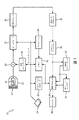
図1は、本開示の一実施形態に係る超音波イメージングシステムの機能的ブロック図である。 FIG. 1 is a functional block diagram of an ultrasound imaging system according to an embodiment of the present disclosure.
図2は、本開示の一実施形態に係る超音波イメージングシステムの概略図である。 FIG. 2 is a schematic diagram of an ultrasound imaging system according to an embodiment of the present disclosure.
図3は、本開示の一実施形態に係るタッチスクリーンのグラフィカルディスプレイである。 FIG. 3 is a graphical display of a touch screen according to one embodiment of the present disclosure.
図4は、本開示の一実施形態に係るディスプレイのグラフィカルディスプレイである。 FIG. 4 is a graphical display of a display according to an embodiment of the present disclosure.
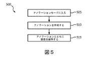
図5は、本開示の一実施形態に係る方法のフローチャートを示す。 FIG. 5 shows a flowchart of a method according to an embodiment of the present disclosure.
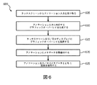
図6は、本開示の一実施形態に係る方法のフローチャートを示す。 FIG. 6 shows a flowchart of a method according to an embodiment of the present disclosure.
特定の例示的な実施形態に関する以下の説明は、単なる例示に過ぎず、本発明又は本発明の適用若しくは用途を限定するものではない。本発明に係るシステム及び方法の実施形態に関する以下の詳細な説明では、本明細書の一部を形成する添付図面が参照され、添付図面には、説明されるシステム及び方法が実施され得る具体的実施形態が例示されている。これらの実施形態は、当業者が本開示のシステム及び方法を実施することを可能にするのに十分に詳細に記載されており、他の実施形態も利用可能であること、及び本発明に係るシステムの趣旨及び範囲を逸脱することなく構造的及び論理的変更がなされ得ることを理解されたい。 The following description of particular exemplary embodiments is merely exemplary and is not intended to limit the invention or the application or use of the invention. In the following detailed description of embodiments of the systems and methods according to the invention, reference is made to the accompanying drawings that form a part hereof, and in which are shown by way of illustration specific systems and methods that can be implemented. Embodiments are illustrated. These embodiments are described in sufficient detail to enable those skilled in the art to practice the systems and methods of the present disclosure, other embodiments are available, and are consistent with the present invention. It should be understood that structural and logical changes can be made without departing from the spirit and scope of the system.
したがって、以下の詳細な説明は限定的な意味で解釈されるべきではなく、本発明に係るシステムの範囲は添付の特許請求の範囲によってのみ規定される。図中の参照番号の先頭の桁は、通常、図番に対応するが、複数の図中に現れる同一の構成要素が同じ参照番号によって識別される場合は除く。さらに、明瞭さを目的として、当業者には明らかであると考えられる場合、本発明に係るシステムの説明を不明瞭にしないために、特定の特徴の詳細な説明は省かれる。 The following detailed description is, therefore, not to be taken in a limiting sense, and the scope of the system according to the present invention is defined only by the appended claims. The first digit of the reference number in the figure usually corresponds to the figure number, except when the same component appearing in a plurality of figures is identified by the same reference number. Further, for purposes of clarity, detailed descriptions of specific features are omitted so as not to obscure the description of the system according to the present invention, where it will be apparent to those skilled in the art.
本開示の例示的な実施形態によれば、ユーザ(例えば、超音波技術者、臨床医)は、タッチスクリーンを入力装置として使用して、ライブ画像又は既得画像上にアノテーションを自由に描くことができる。ライブ画像は、医療イメージング装置によって現在取得及び表示されている画像であってもよい。例えば、超音波イメージングシステムは、超音波プローブによって受信されるエコー信号に基づいて画像を表示し得る。画像の例は、2D画像、3D画像の平面、及び/又は3D立体のレンダリングを含み得る。一部の実施形態では、ユーザは、指及び/又はスタイラスを使用してタッチスクリーンとインタラクトすることによって画像上にアノテーションを描くことができる。その後、ユーザは、後で見るためにアノテーションを画像の一部として保存し得る。アノテーションはタッチスクリーン、及び医療イメージング装置の他のディスプレイ上に表示されてもよい。タッチスクリーン及びディスプレイは、対応する画像及びアノテーションを表示し得る。これは、複数のユーザ間で情報を共有することを容易にし得る。例えば、超音波技術者は、血管に丸を付けることによって、医療イメージング装置のタッチスクリーンを用いてライブ画像にアノテーションを付すことができる。医療イメージング装置のディスプレイは、より見やすくするために医療処置を実行する臨床医に向けられてもよい。臨床医は、同じライブ画像及び超音波技術者のアノテーションをディスプレイ上で見ることができる。臨床医は、ディスプレイ上で、ライブ超音波画像のアノテーションを、超音波技術者によってタッチスクリーン上で入力されるとともに見ることができる。 According to exemplary embodiments of the present disclosure, a user (eg, an ultrasound technician, clinician) is free to draw annotations on a live or acquired image using a touch screen as an input device. it can. The live image may be an image that is currently acquired and displayed by a medical imaging device. For example, the ultrasound imaging system may display an image based on echo signals received by the ultrasound probe. Examples of images may include 2D images, 3D image planes, and / or 3D stereoscopic renderings. In some embodiments, the user can draw annotations on the image by interacting with the touch screen using a finger and / or stylus. The user can then save the annotation as part of the image for later viewing. Annotations may be displayed on touch screens and other displays of medical imaging devices. The touch screen and display may display corresponding images and annotations. This can facilitate sharing information among multiple users. For example, an ultrasound technician can annotate a live image using a touch screen of a medical imaging device by rounding a blood vessel. The display of the medical imaging device may be directed to a clinician performing a medical procedure for easier viewing. The clinician can see the same live image and ultrasound technician annotation on the display. A clinician can view and view live ultrasound image annotations on the display as they are entered on the touch screen by the ultrasound technician.
図1を参照すると、本開示の原理に従って構成された超音波イメージングシステム10がブロック図形式で示されている。図1の超音波診断イメージングシステムでは、超音波プローブ12は、超音波を送信し、エコー情報を受信するトランスデューサアレイ14を含む。様々なトランスデューサアレイ、例えばリニアアレイ、コンベックスアレイ、又はフェーズドアレイが当該技術分野においてよく知られている。トランスデューサアレイ14は、例えば、2D及び/又は3Dイメージングのために仰角次元及び方位角度次元の両方で走査可能な複数のトランスデューサ素子からなる2次元アレイを含むことができる(図示のように)。トランスデューサアレイ14は、アレイ内のトランスデューサ素子による信号の送受信を制御するプローブ12内のマイクロビームフォーマ16に結合される。この例では、マイクロビームフォーマは、送受信の切り替えを行い、メインビームフォーマ22を高エネルギー送信信号から保護する送信/受信(T/R)スイッチ18にプローブケーブルによって結合される。一部の実施形態では、システム内のT/Rスイッチ18及び他の要素は、別個の超音波システムベース内ではなく、トランスデューサプローブ内に含まれ得る。マイクロビームフォーマ16によって制御されるトランスデューサアレイ14からの超音波ビームの送信は、T/Rスイッチ18及びビームフォーマ22に結合される送信コントローラ20によって管理され、送信コントローラ20は、ユーザのユーザインターフェース及び/又はコントロールパネル24の操作から入力を受け取る。一部の実施形態では、ユーザインターフェース及び/又はコントロールパネル24は、タッチスクリーンを含み得る。送信コントローラ20によって制御される機能の1つは、ビームがステアリングされる方向である。ビームは、トランスデューサアレイから直進する(アレイに対して直交する)よう方向づけられてもよいし、又は、より大きな視野のために異なる角度に方向づけられてもよい。マイクロビームフォーマ16によって生成された部分的にビーム成形された信号は、メインビームフォーマ22に送られ、各トランスデューサ素子パッチからの部分的にビーム成形された信号が結合され、完全にビーム成形された信号が生成される。
Referring to FIG. 1, an
ビーム成形された信号は、信号プロセッサ26に結合される。信号プロセッサ26は、受信されたエコー信号を、バンドパスフィルタリング、デシメーション、I及びQ成分分離、及び高調波信号分離などの様々な方法で処理することができる。信号プロセッサ26はまた、スペックル低減、信号合成、及びノイズ除去などの追加の信号エンハンスメントを実行してもよい。処理された信号は、Bモードプロセッサ28に結合され、体内の構造のイメージングのために振幅検出が使用され得る。Bモードプロセッサによって生成された信号は、スキャンコンバータ30及び多断面(multiplanar)リフォーマッタ32に結合される。スキャンコンバータ30は、所望の画像フォーマットで、エコー信号が受信された空間的関係にエコー信号を配置する。例えば、スキャンコンバータ30は、エコー信号を2次元(2D)扇形フォーマット又はピラミッド3次元(3D)画像に配置し得る。多断面リフォーマッタ32は、米国特許第6,443,896号(Detmer)に記載されているように、人体の立体領域内の共通平面内の複数の点から受信されたエコーを、その平面の超音波画像に変換し得る。ボリュームレンダラー34は、例えば、米国特許第6,530,885号(Entrekinら)に記載されているようにして、3Dデータセットのエコー信号を、所与の基準点から見た投影3D画像に変換する。2D又は3D画像は、スキャンコンバータ30、多断面リフォーマッタ32、及びボリュームレンダラー34から画像プロセッサ36に結合され、画像ディスプレイ38上に表示するために、さらなるエンハンスメント、バッファリング、及び一時的保存が行われる。グラフィックスプロセッサ40は、超音波画像と共に表示されるグラフィックオーバーレイを生成し得る。グラフィックオーバーレイは、例えば、患者の名前、画像の日時、イメージングパラメータなどの標準的な識別情報を含み得る。これらの目的のために、グラフィックプロセッサは、タイピングされた患者の名前などの入力をユーザインターフェース24から受け取る。ユーザインターフェース24はまた、複数のMPR(multiplanar reformatted)画像の表示の選択及び制御のために多断面リフォーマッタ32に結合され得る。一部の実施形態では、グラフィックプロセッサ40は、複数のプロセッサとして実装されてもよい。
The beam shaped signal is coupled to a
本開示の少なくとも一部の実施形態によれば、グラフィックプロセッサ40によって生成されたグラフィックオーバーレイは、ユーザインターフェース24のタッチスクリーンを介してユーザによって入力されたアノテーションを含むことができる。グラフィックプロセッサ40及び/又は画像プロセッサ36は、画像及び/又はグラフィックオーバーレイをメモリ42に保存し得る。一部の実施形態では、ユーザは、ユーザインターフェース24を介して保存される画像を選択することができる。メモリ42に保存された画像及び/又はグラフィックオーバーレイは、後で見るためにディスプレイ38上で取り出することができ、かつ/又は、別のデバイス(例えば、USBドライブ、パーソナルコンピュータ、電子医療記録システム、データ保存サーバ)に後で見るために転送され得る。
According to at least some embodiments of the present disclosure, the graphic overlay generated by the
ユーザは、超音波プローブ12で画像を取得し、取得された画像を、ディスプレイ38及び/又はユーザインターフェース24に含まれるタッチスクリーン上で観察し得る。ユーザは、指、スタイラス、及び/又はタッチスクリーン上の他の入力装置を使用して画像上にアノテーションを付け得る。ユーザは、ライブ画像に対して、又は既にメモリ42に保存されている画像に対してアノテーションを付与し得る。アノテーションは、画像及びアノテーションが同時にディスプレイ38及びタッチスクリーン上に表示され得るように、グラフィックプロセッサ40によって動的に画像と統合されてもよい。一部の実施形態では、従来の方法(例えば、トラックボール、キーボード、及び/又はユーザインターフェース24の他の入力)を介して作成されるアノテーションが、タッチスクリーンを介してアノテーションが作成された画像と同じ画像上に作成され得る。画像は、全てのユーザ入力タイプを介して作成されたアノテーションとともにメモリ42に保存され得る。
A user may acquire an image with the
一部の実施形態では、アノテーション及び取得画像を含むグラフィックオーバーレイは、複数の「層」としてメモリ42に保存されてもよい。取得画像は、1つ以上の層であってもよく、グラフィックオーバーレイは、1つ以上の層であってもよい。例えば、患者情報がある層に含まれる一方、タッチスクリーンを介して入力されたアノテーションは別個の層として保存されてもよい。ユーザは、表示されるレイヤーを選択可能であってもよい。例えば、ユーザは、全ての層を見ることができ、この場合、取得画像がアノテーション及び患者情報とともに表示され得る。次いで、ユーザは、アノテーション層の表示を禁止して、アノテーションによって邪魔されない状態の取得画像を見ることを選択し得る。
In some embodiments, graphic overlays including annotations and acquired images may be stored in
一部の実施形態では、タッチスクリーンを介してユーザによって入力されたアノテーションは、メタデータと関連付けられてもよい。メタデータは、画像とともにメモリ42内の同じファイルに、及び/又は、メモリ42内の画像ファイルに関連付けられた別個のファイルとして保存され得る。一部の実施形態では、メタデータは、画像内のどこにアノテーションが位置するかを示し得る。一部の実施形態では、メタデータは、アノテーションが関連する解剖学的構造を示し得る。一部の実施形態では、メタデータは、時間が経っても画像を追跡できるよう、画像がアノテーション付きであることを示すフラグを画像に付けてもよい。例えば、研究者は、フラグに基づき、患者のために保存された過去の検査からの全ての画像のセットから、アノテーションを含む過去の検査からの画像を取り出し得る。これにより、研究者は、アノテーション付きの画像のみをレビューすることができる。これは、経時的にモニタリングされている患者の状態に関する関連画像を迅速に検索及びレビューすることを容易にし得る。
In some embodiments, annotations entered by a user via a touch screen may be associated with metadata. The metadata may be stored with the image in the same file in
メタデータは、例えばDICOM(Digital Imaging and Communications in Medicine)規格などの規格に準拠してもよい。DICOM規格は、診断結果のトラッキング又は医療費請求のための特定診断コードを可能にし得るメタタグを含む。一部の実施形態では、規格及び/又は他のデータは、アノテーションに関連付けられるメタデータとして使用するためにデータベース44に保存され得る。一部の実施形態では、グラフィックプロセッサ40は、アノテーションをメタデータと自動的にリンクし得る。一部の実施形態では、ユーザは、アノテーションに関連付けられるべきデータを選択することができる。例えば、ユーザは、アノテーションが関連付けられている特定の解剖学的構造、注釈されている特徴(例えば、流体ポケット、腫瘍、瘢痕組織)、及び/又はアノテーションのタイプ(例えば、測定、メモ、警告)を入力し得る。
The metadata may conform to a standard such as DICOM (Digital Imaging and Communications in Medicine) standard. The DICOM standard includes a meta tag that may allow specific diagnostic codes for diagnostic result tracking or medical billing. In some embodiments, standards and / or other data may be stored in the
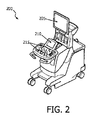
図2は、本開示の一実施形態に係る超音波イメージングシステム200の概略図を示す。超音波イメージングシステム200は、一部の実施形態では、図1に示す超音波イメージングシステム10の要素の一部又は全てを含み得る。超音波イメージングシステム200は、ユーザによって位置決めされ得るディスプレイ205を含み得る。ディスプレイ205は、図1のディスプレイ38を実装するために使用されてもよい。一部の実施形態では、ディスプレイ205はフラットパネルディスプレイであってもよい。ディスプレイ205は、広範囲の視野にわたって見ることができるよう、関節接続(articulated)されたものであってもよい。本開示の1つ又は複数の実施形態を実施するために使用され得る関節式フラットパネルディスプレイを有する超音波システムの例は、欧州特許EP1713396において見つけることができる。超音波イメージングシステム200は、タッチスクリーン210及びコントロールパネル215をさらに含み得る。タッチスクリーン210及び/又はコントロールパネル215は、図1のユーザインターフェース24を実装するために使用されてもよい。タッチスクリーン210及び/又はコントロールパネル215は、画像取得、イメージングパラメータ、データ保存、画像に対するアノテーションの付与、及び/又は他のイメージングパラメータを制御するために使用され得る。コントロールパネル215は、超音波イメージングシステム200を制御するための1つ又は複数の制御要素を含み得る。図2に示す実施形態では、コントロールパネル215は、キーボード、トラックボール、制御ノブ、及びスイッチを含む。コントロールパネル215の他の実施形態は、より多くの又はより少ない制御要素を含むことができる。コントロールパネル215の他の実施形態は、図2に示される要素とは異なる要素を含むことができる。例えば、コントロールパネル215は、トラックパッド、1つ以上のロッカースイッチ、及び/又はマイクロフォンを含み得る。一部の実施形態では、コントロールパネル215を使用して、ディスプレイ205及び/又はタッチスクリーン210上に表示されるものが制御され得る。一部の実施形態では、タッチスクリーン210を使用して、タッチスクリーン210及び/又はディスプレイ205上に表示されるものが制御され得る。
FIG. 2 shows a schematic diagram of an
ユーザは、超音波プローブで画像を取得し、取得された画像を、ディスプレイ及び/又はタッチスクリーン上で観察し得る。アノテーションを画像に追加するために、ユーザは、タッチスクリーン上のオプションを選択して、アノテーションモードに入り得る。ユーザはその後、指、スタイラス、及び/又はタッチスクリーン上の他の入力装置を使用して画像上にアノテーションを付け得る。ユーザは、ライブ画像に対して、又は既にメモリに保存されている画像に対してアノテーションを付与し得る。アノテーションは、画像及びアノテーションが同時に複数のディスプレイ上(例えば、イメージングシステム200のタッチスクリーン210及びディスプレイ205の両方)に表示され得るように、グラフィックプロセッサによって動的に画像と統合されてもよい。ユーザは、取得画像及びアノテーションをメモリに保存し、かつ/又は別の場所(例えば、電子メール、ネットワークドライブ)にエクスポートし得る。一部の実施形態では、従来の方法を介して作成されるアノテーション(例えば、トラックボール、キーボード、及び/又はユーザインターフェースの他の入力を介して選択されるアイコン、テキスト、及び/又は他の記号)が、タッチスクリーンを介してアノテーションが作成された画像と同じ画像上に作成され得る。画像は、全てのユーザ入力タイプを介して作成されたアノテーションとともに保存され得る。
A user can acquire an image with an ultrasound probe and observe the acquired image on a display and / or a touch screen. To add annotation to the image, the user can select an option on the touch screen to enter annotation mode. The user can then annotate the image using a finger, stylus, and / or other input device on the touch screen. The user can annotate a live image or an image already stored in memory. The annotation may be dynamically integrated with the image by the graphics processor so that the image and the annotation can be displayed on multiple displays simultaneously (eg, both
図3は、本開示の一実施形態に係る医療イメージングシステムのタッチスクリーンの例示的なグラフィックディスプレイ300である。グラフィカルディスプレイ300は、医療イメージングシステム10及び/又は200などの医療イメージングシステムを制御するためにユーザによって選択され得る1つ又は複数のボタン305を含み得る。例えば、これらのボタンは、ヘルプメニューを開くために、画像を保存するために、以前に保存された画像を開くために、及び/又は他の機能を行うために使用され得る。グラフィックディスプレイ300は、画像310をさらに含み得る。画像は、医療画像システムのプローブによって取得されたライブ画像又は以前に保存された画像であり得る。ユーザは、タッチスクリーンを使用して画像にアノテーションを付けることができる。一部の実施形態では、アノテーションはフリーフォーム描画であってもよい。図3のグラフィカルディスプレイ300は、タッチスクリーンを介してユーザによって入力されるいくつかのアノテーションの例315、320、325、及び330を示す。アノテーションの例は、日付315、丸及び矢印320、シェード領域325、及び特徴の輪郭330を含む。図3に示すアノテーションは、例示を目的とするものに過ぎず、ユーザが入力できるアノテーションは、図3に示されるものに限定されない。グラフィカルディスプレイ300は、ユーザによって入力されたアノテーションを制御するためのボタン335を含み得る。例えば、ユーザは以前に作成されたアノテーションを消去し、及び/又はアノテーションの色を選択することができる。他のオプションも提供され得る(例えば、線の太さ、記号、コピー/ペースト)。
FIG. 3 is an exemplary
一部の実施形態では、ユーザは、アノテーションを作成及び/又は調整するためにタッチスクリーン上でのジェスチャを使用することができる。例えば、ユーザは、ユーザが描いた図形をダブルタップすることにより、該図形を自動的に網掛けすることができる。他の例では、ユーザは、アノテーションを拡大又は縮小するために、タッチスクリーン上で2本の指をドラッグすることができる。 In some embodiments, the user can use gestures on the touch screen to create and / or adjust annotations. For example, the user can automatically shade the figure by double-tapping the figure drawn by the user. In another example, the user can drag two fingers on the touch screen to enlarge or reduce the annotation.
一部の実施形態では、ユーザは、アノテーションを特定の解剖学的構造に関連付けることができる。特定の解剖学的構造を含む全てのビューにアノテーションが現れるように、アノテーションが、1つ又は複数の画像にわたって伝搬されてもよい。一部の実施形態では、ユーザは、アノテーションをライブ画像内の特定の解剖学的構造に関連付けることができる。超音波プローブが移動するにつれて表示されるライブ画像が変化すると、ディスプレイ内において、アノテーションは特定の解剖学的構造を追跡し得る。一部の実施形態では、アノテーションは、イメージングシステムに含まれる解剖学的認識ソフトウェアの使用によって特定の解剖学的構造との関連を維持し、その例は、特許出願PCT/IB2011/053710、“Automated three dimensional aortic root measurement and modeling”において見つけることができる。一部の実施形態では、アノテーションと特定の解剖学的構造との間の関連付けを維持するために、PercuNavシステムなどの電磁トラッキング及びナビゲーションシステムが使用され得る。電磁トラッキングシステムは、撮像部位及び周辺空間を通過する電磁場を放射する電磁場発生器を含み得る。超音波プローブ、撮像対象(例えば、患者)、及び/又は他の物体(例えば、生検針)上にセンサが配置され得る。センサは電磁場と相互作用し、超音波プローブ、撮像対象、及び/又は物体の位置及び向きを計算するために使用される信号を生成する。電磁トラッキングシステムによって計算された位置は、ユーザによって選択された画像内の特定の解剖学的構造に対してアノテーションを調整するために、グラフィックスプロセッサ及び/又は画像プロセッサに提供され得る。 In some embodiments, the user can associate annotations with specific anatomical structures. Annotations may be propagated across one or more images so that the annotation appears in all views that include a particular anatomical structure. In some embodiments, the user can associate annotations with specific anatomical structures in the live image. As the live image displayed changes as the ultrasound probe moves, annotations can track specific anatomy in the display. In some embodiments, annotations remain associated with specific anatomical structures through the use of anatomical recognition software included in the imaging system, examples of which are described in patent application PCT / IB2011 / 0553710, “Automated. three dimensional authentic root measurement and modeling ". In some embodiments, an electromagnetic tracking and navigation system such as the PercuNav system can be used to maintain the association between annotations and specific anatomical structures. The electromagnetic tracking system may include an electromagnetic field generator that emits an electromagnetic field that passes through the imaging site and the surrounding space. Sensors can be placed on ultrasound probes, imaging subjects (eg, patients), and / or other objects (eg, biopsy needles). The sensor interacts with the electromagnetic field and generates a signal that is used to calculate the position and orientation of the ultrasound probe, imaging object, and / or object. The position calculated by the electromagnetic tracking system can be provided to a graphics processor and / or an image processor to adjust the annotation for a particular anatomy in the image selected by the user.
図4は、本開示の一実施形態に係る医療イメージングシステムのディスプレイの例であるグラフィックディスプレイ400である。ディスプレイ400は、医療イメージングシステムを制御するためにユーザによって選択され得る1つ又は複数のアイコン405を含み得る。例えば、これらのアイコンは、ヘルプメニューを開くために、画像を保存するために、以前に保存された画像を開くために、及び/又は他の機能を行うために使用され得る。アイコン405は、トラックボール、マウス、キーボード、及び/又は医療イメージングシステムのコントロールパネルの他の要素を使用することによってユーザによって選択され得る。グラフィックディスプレイ400は、画像310をさらに含み得る。画像310は、図3に示すタッチスクリーンのグラフィカルディスプレイ300に表示されるものと同じ画像であってもよい。グラフィカルディスプレイ400は、図3を参照して説明した、タッチスクリーンを介してユーザによって入力される同じアノテーション315、320、325、及び330を表示し得る。グラフィックディスプレイ400は、医療イメージングシステムを制御するためのオプション及びメニュー435を含み得る。例えば、ユーザは、異なるイメージングモードに入ること、測定を実行すること、画像解析ソフトウェアを実行すること、及び/又は他のアプリケーションを実行することが可能であり得る。
FIG. 4 is a
必ずしも示されていないが、グラフィカルディスプレイ300及び/又は400はまた、例えば、ユーザが必要に応じてスキャン、ファイル保存、プリント、画像転送(例えば、あるディスプレイから別のディスプレイへ)、ミュート、転記、及び/又はヘッドピースの使用をするために選択可能なユーザ選択肢、例えば、アイコンやメニュー項目を表示し得る。さらに、当該技術分野で知られている1つ又は複数のメニューが、ユーザの便宜のために提供されてもよい。表示画像及び関連付けられたデータは、画像取得中又は後の解析中の任意の時点で保存され得る。しかしながら、ユーザがオリジナル情報を参照し、並びに/又は、いつ及び/又は誰が情報に変更を加えたか(例えば、生成されたレポートに保存されていてもよい)を特定できるように、データが追加及び/又は編集された時間を示す情報を集める履歴モードがアクティブ化されてもよい。さらに、後の使用のための変更も保存されてもよい。
Although not necessarily shown, the
図5は、本開示の一実施形態に係る方法500のフローチャートを示す。方法500は、医療イメージングシステムによって取得された画像にアノテーションを付けるために使用され得る。一部の実施形態では、方法500は、ユーザが医療イメージングシステムのタッチスクリーンを操作することによって実行されてもよい。ユーザは、ステップ505においてアノテーションモードに入り得る。一部の実施形態では、ユーザは、アノテーションモードに入るためにタッチスクリーン上のボタンに触れることができる。ステップ505において、ユーザは画像に1つ又は複数のアノテーションを付けることができる。一部の実施形態では、アノテーションは、ユーザがタッチスクリーンとインタラクトすることによって作成され得る。ユーザは、指、スタイラス、及び/又は他の入力装置をタッチスクリーンに当てることによってタッチスクリーンとインタラクトし得る。ステップ515において、ユーザは画像をアノテーションとともに保存することができる。一部の実施形態では、アノテーション付きの画像は、医療イメージングシステムのメモリに保存され得る。一部の実施形態では、アノテーション付きの画像は、メモリに保存される際にメタデータと関連付けられてもよい。
FIG. 5 shows a flowchart of a
方法500は、一部の実施形態では追加のステップを含むことができる。例えば、ユーザは、ステップ510で作成されたアノテーションと関連付けられるメタデータ及び/又は他のデータを選択することができる。一部の実施形態では、アノテーションは、ステップ515でアノテーション付き画像を保存する前に又は保存中に、画像の特定の解剖学的構造に手動で又は自動的に関連付けられてもよい。
The
図6は、本開示の一実施形態に係る方法600のフローチャートを示す。方法600は、医療イメージングシステムによって取得された画像にアノテーションを付けるために使用され得る。一部の実施形態では、方法600は、医療イメージングシステムのグラフィックスプロセッサによって実行されてもよい。ステップ605において、グラフィックスプロセッサは、医療イメージングシステムによって取得された画像のためのアノテーションに対応する入力を受け取り得る。一部の実施形態では、入力は、医療イメージングシステムのタッチスクリーンから受け取られ得る。ステップ610において、グラフィックスプロセッサは、ステップ605で受け取られたアノテーション入力に対応するグラフィックオーバーレイを生成し得る。アノテーション入力に対応するグラフィックオーバーレイは、アノテーションと呼ばれ得る。ステップ615において、グラフィックオーバーレイは、タッチスクリーン及び/又は医療イメージングシステムのディスプレイに提供され得る。タッチスクリーン及び/又はディスプレイは、グラフィックスプロセッサから受け取った画像及びアノテーションを表示し得る。追加で、ステップ620において、アノテーションにメタデータが関連付けられてもよい。メタデータは、グラフィックスプロセッサによってアノテーションと自動的に関連付けられてもよく、かつ/又は、メタデータは、タッチスクリーン又はコントロールパネルなどの入力デバイスから受け取られてもよい。一部の実施形態では、メタデータは、グラフィックスプロセッサがアクセス可能なデータベースから取り出されてもよい。アノテーション及び/又はメタデータを有する画像は、ステップ625において保存され得る。一部の実施形態では、アノテーション及び/又はメタデータを有する画像は、グラフィックスプロセッサによって自動的に保存され得る。一部の実施形態では、アノテーション及び/又はメタデータを有する画像は、入力デバイスからの保存コマンドに応じて保存され得る。一部の実施形態では、アノテーション及び/又はメタデータを有する画像は、医療イメージングシステムのメモリ内に保存され得る。
FIG. 6 shows a flowchart of a
一部の実施形態では、方法600の1つ又は複数のステップが省略されてもよい。一部の実施形態では、アノテーション及び/又はメタデータを有する画像は保存されなくてもよく、この場合、ステップ625が排除される。例えば、超音波技術者は、外科手術中に外科医を補助するためにライブ画像にアノテーションを付けることができる。この処置中に作成された画像及びアノテーションは、将来の処理及び/又は検査には関連しない可能性があり、超音波技術者は、画像及び/又はアノテーションを保存しないことを選択し得る。一部の実施形態では、ステップ620が省略され、メタデータがアノテーションに関連付けられなくてもよい。例えば、医療イメージングシステムは、アノテーション付き画像を単純なグラフィック(例えば、スクリーンショット)として保存し得る。単純なグラフィックは、限定された解析及び/又は編集を可能にし得る。これは、データ記憶領域が小さい場合、及び/又は臨床医による定性的ビジュアルレビューのみが望まれる場合に望ましい可能性がある。
In some embodiments, one or more steps of
超音波イメージングシステムを参照して本発明に係るシステムを説明してきたが、本発明に係るシステムは、1つ又は複数の画像がシステマチックに取得される他の医療イメージングシステムにも拡張され得ることが想定される。本発明に係るシステムは、腎臓、精巣、乳房、卵巣、子宮、甲状腺、肝臓、肺、筋骨格、脾臓、心臓、動脈、及び血管系に関連する画像情報を取得及び/又は記録するために、並びに超音波誘導インターベンションに関連する他のイメージング用途に適用され得る。さらに、本発明に係るシステムは、従来のイメージングシステムとともに使用され、本発明に係るシステムの特徴及び利点を提供することを可能にする1つ又は複数のプログラムを含み得る。 Although the system according to the present invention has been described with reference to an ultrasound imaging system, the system according to the present invention can be extended to other medical imaging systems in which one or more images are acquired systematically. Is assumed. The system according to the present invention acquires and / or records image information related to the kidney, testis, breast, ovary, uterus, thyroid, liver, lung, musculoskeletal, spleen, heart, artery, and vasculature. As well as other imaging applications associated with ultrasound guided interventions. In addition, the system according to the present invention may include one or more programs that are used with conventional imaging systems and that provide the features and advantages of the system according to the present invention.
さらに、本発明に係るシステム、装置、及び方法は、明確なランドマークが定義及び再現可能な任意の小部分(small parts)イメージングにも拡張され得る。さらに、本発明に係る方法は、例えば超音波イメージングシステムのような既存のイメージングシステムに適用可能なプログラムコード内に埋め込まれ得る。好適な超音波イメージングシステムは、例えば、小部分イメージングに適し得る従来のブロードバンドリニアアレイトランスデューサをサポートし得るPhiliphs超音波システムを含み得る。さらに、例えば、QLAB(商標)などの解析技術が、撮像装置とともにオンカートで、又は検査室の外で実行され得る後処理プログラムとして利用され得る。さらに、複数の結節、濾胞などの解剖学的実体、又は他の検出可能な物体に、本発明に係るシステムを使用してアノテーションを付けることができる。さらに、本発明に係るシステムの方法は、例えば、X−matrix(商標)などの2Dアレイトランスデューサ又は機械的トランスデューサなどのトランスデューサを使用して取得されるボリュームに適用されてもよい。 Furthermore, the system, apparatus and method according to the present invention can be extended to any small parts imaging where a well-defined landmark can be defined and reproduced. Furthermore, the method according to the invention can be embedded in program code applicable to existing imaging systems, such as for example an ultrasound imaging system. Suitable ultrasound imaging systems may include, for example, Philips ultrasound systems that may support conventional broadband linear array transducers that may be suitable for small-part imaging. Further, for example, an analysis technique such as QLAB ™ can be used as a post-processing program that can be executed on-cart with the imaging device or outside the laboratory. In addition, anatomical entities such as nodules, follicles, or other detectable objects can be annotated using the system of the present invention. Furthermore, the method of the system according to the present invention may be applied to a volume obtained using a transducer such as a 2D array transducer such as X-matrix ™ or a mechanical transducer, for example.
本発明の追加の利点及び特徴が、本開示を研究した当業者に明らかとなり、又は本発明の新規システム及び方法を利用する者によって経験される可能性がある。その最たるものとして、よりユーザフレンドリーな画像アノテーションシステム及びその動作方法が提供されることが挙げられる。本発明に係るシステム及び方法の他の利点は、従来の医療画像システムを容易にアップグレードして、本発明に係るシステム、装置、及び方法の特徴及び利点を組み込むことができることである。 Additional advantages and features of the present invention will be apparent to those of ordinary skill in the art who have studied the present disclosure or may be experienced by those utilizing the novel systems and methods of the present invention. Most importantly, a more user-friendly image annotation system and its operation method are provided. Another advantage of the system and method according to the present invention is that a conventional medical imaging system can be easily upgraded to incorporate the features and advantages of the system, apparatus and method according to the present invention.
当然のことながら、上記の実施形態又はプロセスのいずれか1つが、1つ又は複数の他の実施形態及び/又はプロセスと組み合わせられてもよく、又は、本発明に係るシステム、装置、及び方法にしたがって別の装置又は装置の部分の間で分離及び/又は実行されてもよい。 Of course, any one of the above-described embodiments or processes may be combined with one or more other embodiments and / or processes, or in a system, apparatus and method according to the present invention. Thus, it may be separated and / or implemented between different devices or parts of devices.
最後に、上記の議論は、本発明に係るシステムの単なる例示であり、添付の特許請求の範囲をいずれかの特定の実施形態又は実施形態のグループに限定するものとして解釈されるべきではない。したがって、例示的な実施形態を参照して特に詳細に本発明に係るシステムを説明したが、以下の特許請求の範囲に示される本発明に係るシステムの意図される広範な趣旨及び範囲から逸脱することなく、多様な変更及び代替的実施形態が当業者によって考案され得ることも理解されたい。したがって、明細書及び図面は例示的であると解釈されるべきであり、添付の特許請求の範囲を限定するものではない。 Finally, the above discussion is merely illustrative of the system according to the present invention and should not be construed as limiting the appended claims to any particular embodiment or group of embodiments. Thus, while the system according to the present invention has been described in particular detail with reference to exemplary embodiments, it departs from the intended broad spirit and scope of the system according to the present invention as set forth in the following claims. It should also be understood that various modifications and alternative embodiments may be devised by those skilled in the art. The specification and drawings are accordingly to be regarded in an illustrative manner and are not intended to limit the scope of the appended claims.
Claims (20)
前記タッチスクリーンから前記アノテーション入力を受け取り、少なくとも部分的に前記アノテーション入力に基づき、アノテーションとして前記画像に関連付けられるグラフィックオーバーレイを生成するグラフィックスプロセッサと、
前記画像及び前記アノテーションを表示するディスプレイと、
前記画像及び前記アノテーションを保存するメモリとを備える、
医療イメージングシステム。 A touch screen that receives annotation input from a user associated with an image acquired by a medical imaging system;
A graphics processor that receives the annotation input from the touch screen and generates a graphic overlay associated with the image as annotation based at least in part on the annotation input;
A display for displaying the image and the annotation;
A memory for storing the image and the annotation;
Medical imaging system.
タッチスクリーンから前記画像に関連付けられるアノテーション入力を受け取るステップと、
前記アノテーション入力に対応するグラフィックオーバーレイを生成するステップであって、前記アノテーション入力に対応する前記グラフィックオーバーレイはアノテーションである、ステップと、
前記グラフィックオーバーレイを前記タッチスクリーンに提供するステップとを含む、方法。 A method for annotating an image, said method comprising:
Receiving annotation input associated with the image from a touch screen;
Generating a graphic overlay corresponding to the annotation input, wherein the graphic overlay corresponding to the annotation input is an annotation;
Providing the graphic overlay on the touch screen.
Priority Applications (1)
| Application Number | Priority Date | Filing Date | Title |
|---|---|---|---|
| JP2021136919A JP2021191429A (en) | 2015-10-08 | 2021-08-25 | Apparatuses, methods, and systems for annotation of medical images |
Applications Claiming Priority (3)
| Application Number | Priority Date | Filing Date | Title |
|---|---|---|---|
| US201562238758P | 2015-10-08 | 2015-10-08 | |
| US62/238,758 | 2015-10-08 | ||
| PCT/IB2016/055727 WO2017060791A1 (en) | 2015-10-08 | 2016-09-26 | Apparatuses, methods, and systems for annotation of medical images |
Related Child Applications (1)
| Application Number | Title | Priority Date | Filing Date |
|---|---|---|---|
| JP2021136919A Division JP2021191429A (en) | 2015-10-08 | 2021-08-25 | Apparatuses, methods, and systems for annotation of medical images |
Publications (2)
| Publication Number | Publication Date |
|---|---|
| JP2018529475A true JP2018529475A (en) | 2018-10-11 |
| JP2018529475A5 JP2018529475A5 (en) | 2019-11-07 |
Family
ID=57206330
Family Applications (2)
| Application Number | Title | Priority Date | Filing Date |
|---|---|---|---|
| JP2018517764A Pending JP2018529475A (en) | 2015-10-08 | 2016-09-26 | Apparatus, method and system for medical image annotation |
| JP2021136919A Pending JP2021191429A (en) | 2015-10-08 | 2021-08-25 | Apparatuses, methods, and systems for annotation of medical images |
Family Applications After (1)
| Application Number | Title | Priority Date | Filing Date |
|---|---|---|---|
| JP2021136919A Pending JP2021191429A (en) | 2015-10-08 | 2021-08-25 | Apparatuses, methods, and systems for annotation of medical images |
Country Status (3)
| Country | Link |
|---|---|
| US (1) | US20190076125A1 (en) |
| JP (2) | JP2018529475A (en) |
| WO (1) | WO2017060791A1 (en) |
Cited By (2)
| Publication number | Priority date | Publication date | Assignee | Title |
|---|---|---|---|---|
| WO2020106729A1 (en) * | 2018-11-20 | 2020-05-28 | Arterys Inc. | Cloud-based radiology commenting and workspace sharing |
| KR20210080285A (en) * | 2019-12-20 | 2021-06-30 | (주)클로버추얼패션 | Method to provide design information |
Families Citing this family (9)
| Publication number | Priority date | Publication date | Assignee | Title |
|---|---|---|---|---|
| US10276265B2 (en) * | 2016-08-31 | 2019-04-30 | International Business Machines Corporation | Automated anatomically-based reporting of medical images via image annotation |
| US10729396B2 (en) * | 2016-08-31 | 2020-08-04 | International Business Machines Corporation | Tracking anatomical findings within medical images |
| EP3416074A1 (en) * | 2017-06-16 | 2018-12-19 | Koninklijke Philips N.V. | Annotating fetal monitoring data |
| KR102489579B1 (en) * | 2017-08-17 | 2023-01-18 | 삼성전자주식회사 | Method and ultrasound apparatus for providing annotation related information |
| WO2019091807A1 (en) * | 2017-11-08 | 2019-05-16 | Koninklijke Philips N.V. | Ultrasound system and method for correlation between ultrasound breast images and breast images of other imaging modalities |
| CN108037891B (en) * | 2017-12-26 | 2022-04-01 | 深圳开立生物医疗科技股份有限公司 | Annotation method, system, equipment and computer storage medium |
| EP3790468A1 (en) | 2018-05-07 | 2021-03-17 | Hologic, Inc. | Breast ultrasound workflow application |
| CN109741397B (en) * | 2019-01-04 | 2022-06-07 | 京东方科技集团股份有限公司 | Picture marking method and device, computer equipment and readable storage medium |
| TWI779284B (en) * | 2020-05-06 | 2022-10-01 | 商之器科技股份有限公司 | Device for marking image data |
Citations (4)
| Publication number | Priority date | Publication date | Assignee | Title |
|---|---|---|---|---|
| JP2002263101A (en) * | 2001-03-06 | 2002-09-17 | Aloka Co Ltd | Ultrasonic diagnostic device |
| JP2013255541A (en) * | 2012-06-11 | 2013-12-26 | Toshiba Corp | X-ray diagnostic apparatus |
| JP2014113311A (en) * | 2012-12-10 | 2014-06-26 | Hitachi Medical Corp | Medical image display device and medical image diagnostic apparatus carrying the same |
| JP2015126820A (en) * | 2013-12-27 | 2015-07-09 | 株式会社アドバンス | Intraoral display system |
Family Cites Families (13)
| Publication number | Priority date | Publication date | Assignee | Title |
|---|---|---|---|---|
| ATE225964T1 (en) * | 1993-03-31 | 2002-10-15 | Luma Corp | INFORMATION MANAGEMENT IN AN ENDOSCOPY SYSTEM |
| US6530885B1 (en) | 2000-03-17 | 2003-03-11 | Atl Ultrasound, Inc. | Spatially compounded three dimensional ultrasonic images |
| US6443896B1 (en) | 2000-08-17 | 2002-09-03 | Koninklijke Philips Electronics N.V. | Method for creating multiplanar ultrasonic images of a three dimensional object |
| ATE517579T1 (en) | 2004-02-06 | 2011-08-15 | Koninkl Philips Electronics Nv | DIAGNOSTIC ULTRASONIC SYSTEM WITH ARTICULATED FLAT SCREEN DISPLAY |
| WO2006057911A2 (en) * | 2004-11-22 | 2006-06-01 | Civco Medical Instruments Co., Inc. | Real time ultrasound monitoring of the motion of internal structures during respiration for control of therapy delivery |
| US8069420B2 (en) * | 2004-12-29 | 2011-11-29 | Karl Storz Endoscopy-America, Inc. | System for controlling the communication of medical imaging data |
| US20080139896A1 (en) * | 2006-10-13 | 2008-06-12 | Siemens Medical Solutions Usa, Inc. | System and Method for Graphical Annotation of Anatomical Images Using a Touch Screen Display |
| US8816959B2 (en) * | 2007-04-03 | 2014-08-26 | General Electric Company | Method and apparatus for obtaining and/or analyzing anatomical images |
| CN103068318B (en) * | 2010-08-26 | 2016-02-03 | 皇家飞利浦电子股份有限公司 | Automatic three dimensional aortic root is measured and modeling |
| WO2013040498A1 (en) * | 2011-09-16 | 2013-03-21 | Translucent Medical, Inc. | System and method for virtually tracking a surgical tool on a movable display |
| US9239848B2 (en) * | 2012-02-06 | 2016-01-19 | Microsoft Technology Licensing, Llc | System and method for semantically annotating images |
| JP5924973B2 (en) * | 2012-02-17 | 2016-05-25 | 日立アロカメディカル株式会社 | Ultrasonic diagnostic equipment |
| US10409951B2 (en) * | 2012-12-28 | 2019-09-10 | Volcano Corporation | Multi-modality case management system and method |
-
2016
- 2016-09-26 WO PCT/IB2016/055727 patent/WO2017060791A1/en active Application Filing
- 2016-09-26 JP JP2018517764A patent/JP2018529475A/en active Pending
- 2016-09-26 US US15/765,610 patent/US20190076125A1/en not_active Abandoned
-
2021
- 2021-08-25 JP JP2021136919A patent/JP2021191429A/en active Pending
Patent Citations (4)
| Publication number | Priority date | Publication date | Assignee | Title |
|---|---|---|---|---|
| JP2002263101A (en) * | 2001-03-06 | 2002-09-17 | Aloka Co Ltd | Ultrasonic diagnostic device |
| JP2013255541A (en) * | 2012-06-11 | 2013-12-26 | Toshiba Corp | X-ray diagnostic apparatus |
| JP2014113311A (en) * | 2012-12-10 | 2014-06-26 | Hitachi Medical Corp | Medical image display device and medical image diagnostic apparatus carrying the same |
| JP2015126820A (en) * | 2013-12-27 | 2015-07-09 | 株式会社アドバンス | Intraoral display system |
Cited By (8)
| Publication number | Priority date | Publication date | Assignee | Title |
|---|---|---|---|---|
| WO2020106729A1 (en) * | 2018-11-20 | 2020-05-28 | Arterys Inc. | Cloud-based radiology commenting and workspace sharing |
| US11915821B2 (en) | 2018-11-20 | 2024-02-27 | Arterys Inc. | Cloud-based radiology commenting and workspace sharing |
| KR20210080285A (en) * | 2019-12-20 | 2021-06-30 | (주)클로버추얼패션 | Method to provide design information |
| KR102417152B1 (en) * | 2019-12-20 | 2022-07-06 | (주)클로버추얼패션 | Method to provide design information |
| KR20220097865A (en) * | 2019-12-20 | 2022-07-08 | (주)클로버추얼패션 | Method to provide design information |
| US11417039B2 (en) | 2019-12-20 | 2022-08-16 | Clo Virtual Fashion Inc. | Method to provide design information |
| US11625877B2 (en) | 2019-12-20 | 2023-04-11 | Clo Virtual Fashion Inc. | Method to provide design information |
| KR102600651B1 (en) * | 2019-12-20 | 2023-11-10 | (주)클로버추얼패션 | Method to provide design information |
Also Published As
| Publication number | Publication date |
|---|---|
| JP2021191429A (en) | 2021-12-16 |
| WO2017060791A1 (en) | 2017-04-13 |
| US20190076125A1 (en) | 2019-03-14 |
Similar Documents
| Publication | Publication Date | Title |
|---|---|---|
| JP2021191429A (en) | Apparatuses, methods, and systems for annotation of medical images | |
| US11094138B2 (en) | Systems for linking features in medical images to anatomical models and methods of operation thereof | |
| US11631495B2 (en) | Systems and methods for contextual imaging workflow | |
| US11464488B2 (en) | Methods and systems for a medical grading system | |
| CN111315301B (en) | Ultrasound system and method for correlating ultrasound breast images with breast images of other imaging modalities | |
| JP6559917B2 (en) | Ultrasound system and breast tissue imaging and breast ultrasound image annotation method | |
| US11594002B2 (en) | Overlay and manipulation of medical images in a virtual environment | |
| JP4634539B2 (en) | Image processing apparatus and method, and program | |
| US9652589B2 (en) | Systems and methods for using a touch-sensitive display unit to analyze a medical image | |
| US20110208052A1 (en) | Breast ultrasound annotation user interface | |
| US9773347B2 (en) | Interacting with a three-dimensional object dataset | |
| EP2636374A1 (en) | Method for providing ultrasound images and ultrasound apparatus | |
| US11344281B2 (en) | Ultrasound visual protocols | |
| US20220061811A1 (en) | Unified interface for visualizing 2d, 3d and 4d ultrasound images | |
| CN109069110A (en) | Ultrasonic image-forming system with simplified 3D imaging control | |
| JP2015112123A (en) | Medical image diagnostic apparatus | |
| RU2779836C2 (en) | Ultrasound system and method for correlation between ultrasound breast images and breast images of other imaging methods | |
| JP2017086726A (en) | Ultrasonic diagnostic equipment |
Legal Events
| Date | Code | Title | Description |
|---|---|---|---|
| A521 | Request for written amendment filed |
Free format text: JAPANESE INTERMEDIATE CODE: A523 Effective date: 20190925 |
|
| A621 | Written request for application examination |
Free format text: JAPANESE INTERMEDIATE CODE: A621 Effective date: 20190925 |
|
| A977 | Report on retrieval |
Free format text: JAPANESE INTERMEDIATE CODE: A971007 Effective date: 20200708 |
|
| A131 | Notification of reasons for refusal |
Free format text: JAPANESE INTERMEDIATE CODE: A131 Effective date: 20200818 |
|
| A601 | Written request for extension of time |
Free format text: JAPANESE INTERMEDIATE CODE: A601 Effective date: 20201113 |
|
| A02 | Decision of refusal |
Free format text: JAPANESE INTERMEDIATE CODE: A02 Effective date: 20210426 |
|
| A521 | Request for written amendment filed |
Free format text: JAPANESE INTERMEDIATE CODE: A523 Effective date: 20210825 |
|
| C60 | Trial request (containing other claim documents, opposition documents) |
Free format text: JAPANESE INTERMEDIATE CODE: C60 Effective date: 20210825 |
|
| A911 | Transfer to examiner for re-examination before appeal (zenchi) |
Free format text: JAPANESE INTERMEDIATE CODE: A911 Effective date: 20210906 |
|
| C21 | Notice of transfer of a case for reconsideration by examiners before appeal proceedings |
Free format text: JAPANESE INTERMEDIATE CODE: C21 Effective date: 20210913 |
|
| A912 | Re-examination (zenchi) completed and case transferred to appeal board |
Free format text: JAPANESE INTERMEDIATE CODE: A912 Effective date: 20211015 |
|
| C211 | Notice of termination of reconsideration by examiners before appeal proceedings |
Free format text: JAPANESE INTERMEDIATE CODE: C211 Effective date: 20211019 |
|
| C22 | Notice of designation (change) of administrative judge |
Free format text: JAPANESE INTERMEDIATE CODE: C22 Effective date: 20211110 |
|
| C23 | Notice of termination of proceedings |
Free format text: JAPANESE INTERMEDIATE CODE: C23 Effective date: 20220210 |
|
| C03 | Trial/appeal decision taken |
Free format text: JAPANESE INTERMEDIATE CODE: C03 Effective date: 20220315 |
|
| C30A | Notification sent |
Free format text: JAPANESE INTERMEDIATE CODE: C3012 Effective date: 20220315 |