JP2017522016A - Cultured mammalian limbal stem cells, production method thereof and use thereof - Google Patents
Cultured mammalian limbal stem cells, production method thereof and use thereof Download PDFInfo
- Publication number
- JP2017522016A JP2017522016A JP2016575401A JP2016575401A JP2017522016A JP 2017522016 A JP2017522016 A JP 2017522016A JP 2016575401 A JP2016575401 A JP 2016575401A JP 2016575401 A JP2016575401 A JP 2016575401A JP 2017522016 A JP2017522016 A JP 2017522016A
- Authority
- JP
- Japan
- Prior art keywords
- lsc
- cells
- sesc
- population
- pax6
- Prior art date
- Legal status (The legal status is an assumption and is not a legal conclusion. Google has not performed a legal analysis and makes no representation as to the accuracy of the status listed.)
- Pending
Links
Images


Classifications
-
- C—CHEMISTRY; METALLURGY
- C12—BIOCHEMISTRY; BEER; SPIRITS; WINE; VINEGAR; MICROBIOLOGY; ENZYMOLOGY; MUTATION OR GENETIC ENGINEERING
- C12N—MICROORGANISMS OR ENZYMES; COMPOSITIONS THEREOF; PROPAGATING, PRESERVING, OR MAINTAINING MICROORGANISMS; MUTATION OR GENETIC ENGINEERING; CULTURE MEDIA
- C12N5/00—Undifferentiated human, animal or plant cells, e.g. cell lines; Tissues; Cultivation or maintenance thereof; Culture media therefor
- C12N5/06—Animal cells or tissues; Human cells or tissues
- C12N5/0602—Vertebrate cells
- C12N5/0618—Cells of the nervous system
- C12N5/0621—Eye cells, e.g. cornea, iris pigmented cells
-
- A—HUMAN NECESSITIES
- A61—MEDICAL OR VETERINARY SCIENCE; HYGIENE
- A61K—PREPARATIONS FOR MEDICAL, DENTAL OR TOILETRY PURPOSES
- A61K35/00—Medicinal preparations containing materials or reaction products thereof with undetermined constitution
- A61K35/12—Materials from mammals; Compositions comprising non-specified tissues or cells; Compositions comprising non-embryonic stem cells; Genetically modified cells
- A61K35/30—Nerves; Brain; Eyes; Corneal cells; Cerebrospinal fluid; Neuronal stem cells; Neuronal precursor cells; Glial cells; Oligodendrocytes; Schwann cells; Astroglia; Astrocytes; Choroid plexus; Spinal cord tissue
-
- A—HUMAN NECESSITIES
- A61—MEDICAL OR VETERINARY SCIENCE; HYGIENE
- A61K—PREPARATIONS FOR MEDICAL, DENTAL OR TOILETRY PURPOSES
- A61K35/00—Medicinal preparations containing materials or reaction products thereof with undetermined constitution
- A61K35/12—Materials from mammals; Compositions comprising non-specified tissues or cells; Compositions comprising non-embryonic stem cells; Genetically modified cells
- A61K35/36—Skin; Hair; Nails; Sebaceous glands; Cerumen; Epidermis; Epithelial cells; Keratinocytes; Langerhans cells; Ectodermal cells
-
- A—HUMAN NECESSITIES
- A61—MEDICAL OR VETERINARY SCIENCE; HYGIENE
- A61P—SPECIFIC THERAPEUTIC ACTIVITY OF CHEMICAL COMPOUNDS OR MEDICINAL PREPARATIONS
- A61P17/00—Drugs for dermatological disorders
-
- A—HUMAN NECESSITIES
- A61—MEDICAL OR VETERINARY SCIENCE; HYGIENE
- A61P—SPECIFIC THERAPEUTIC ACTIVITY OF CHEMICAL COMPOUNDS OR MEDICINAL PREPARATIONS
- A61P17/00—Drugs for dermatological disorders
- A61P17/02—Drugs for dermatological disorders for treating wounds, ulcers, burns, scars, keloids, or the like
-
- A—HUMAN NECESSITIES
- A61—MEDICAL OR VETERINARY SCIENCE; HYGIENE
- A61P—SPECIFIC THERAPEUTIC ACTIVITY OF CHEMICAL COMPOUNDS OR MEDICINAL PREPARATIONS
- A61P27/00—Drugs for disorders of the senses
- A61P27/02—Ophthalmic agents
-
- A—HUMAN NECESSITIES
- A61—MEDICAL OR VETERINARY SCIENCE; HYGIENE
- A61P—SPECIFIC THERAPEUTIC ACTIVITY OF CHEMICAL COMPOUNDS OR MEDICINAL PREPARATIONS
- A61P27/00—Drugs for disorders of the senses
- A61P27/02—Ophthalmic agents
- A61P27/10—Ophthalmic agents for accommodation disorders, e.g. myopia
-
- A—HUMAN NECESSITIES
- A61—MEDICAL OR VETERINARY SCIENCE; HYGIENE
- A61P—SPECIFIC THERAPEUTIC ACTIVITY OF CHEMICAL COMPOUNDS OR MEDICINAL PREPARATIONS
- A61P31/00—Antiinfectives, i.e. antibiotics, antiseptics, chemotherapeutics
-
- A—HUMAN NECESSITIES
- A61—MEDICAL OR VETERINARY SCIENCE; HYGIENE
- A61P—SPECIFIC THERAPEUTIC ACTIVITY OF CHEMICAL COMPOUNDS OR MEDICINAL PREPARATIONS
- A61P35/00—Antineoplastic agents
-
- A—HUMAN NECESSITIES
- A61—MEDICAL OR VETERINARY SCIENCE; HYGIENE
- A61P—SPECIFIC THERAPEUTIC ACTIVITY OF CHEMICAL COMPOUNDS OR MEDICINAL PREPARATIONS
- A61P43/00—Drugs for specific purposes, not provided for in groups A61P1/00-A61P41/00
-
- C—CHEMISTRY; METALLURGY
- C12—BIOCHEMISTRY; BEER; SPIRITS; WINE; VINEGAR; MICROBIOLOGY; ENZYMOLOGY; MUTATION OR GENETIC ENGINEERING
- C12N—MICROORGANISMS OR ENZYMES; COMPOSITIONS THEREOF; PROPAGATING, PRESERVING, OR MAINTAINING MICROORGANISMS; MUTATION OR GENETIC ENGINEERING; CULTURE MEDIA
- C12N5/00—Undifferentiated human, animal or plant cells, e.g. cell lines; Tissues; Cultivation or maintenance thereof; Culture media therefor
- C12N5/06—Animal cells or tissues; Human cells or tissues
- C12N5/0602—Vertebrate cells
- C12N5/0625—Epidermal cells, skin cells; Cells of the oral mucosa
-
- C—CHEMISTRY; METALLURGY
- C12—BIOCHEMISTRY; BEER; SPIRITS; WINE; VINEGAR; MICROBIOLOGY; ENZYMOLOGY; MUTATION OR GENETIC ENGINEERING
- C12Q—MEASURING OR TESTING PROCESSES INVOLVING ENZYMES, NUCLEIC ACIDS OR MICROORGANISMS; COMPOSITIONS OR TEST PAPERS THEREFOR; PROCESSES OF PREPARING SUCH COMPOSITIONS; CONDITION-RESPONSIVE CONTROL IN MICROBIOLOGICAL OR ENZYMOLOGICAL PROCESSES
- C12Q1/00—Measuring or testing processes involving enzymes, nucleic acids or microorganisms; Compositions therefor; Processes of preparing such compositions
- C12Q1/68—Measuring or testing processes involving enzymes, nucleic acids or microorganisms; Compositions therefor; Processes of preparing such compositions involving nucleic acids
- C12Q1/6876—Nucleic acid products used in the analysis of nucleic acids, e.g. primers or probes
- C12Q1/6883—Nucleic acid products used in the analysis of nucleic acids, e.g. primers or probes for diseases caused by alterations of genetic material
-
- C—CHEMISTRY; METALLURGY
- C12—BIOCHEMISTRY; BEER; SPIRITS; WINE; VINEGAR; MICROBIOLOGY; ENZYMOLOGY; MUTATION OR GENETIC ENGINEERING
- C12N—MICROORGANISMS OR ENZYMES; COMPOSITIONS THEREOF; PROPAGATING, PRESERVING, OR MAINTAINING MICROORGANISMS; MUTATION OR GENETIC ENGINEERING; CULTURE MEDIA
- C12N2501/00—Active agents used in cell culture processes, e.g. differentation
- C12N2501/10—Growth factors
- C12N2501/11—Epidermal growth factor [EGF]
-
- C—CHEMISTRY; METALLURGY
- C12—BIOCHEMISTRY; BEER; SPIRITS; WINE; VINEGAR; MICROBIOLOGY; ENZYMOLOGY; MUTATION OR GENETIC ENGINEERING
- C12N—MICROORGANISMS OR ENZYMES; COMPOSITIONS THEREOF; PROPAGATING, PRESERVING, OR MAINTAINING MICROORGANISMS; MUTATION OR GENETIC ENGINEERING; CULTURE MEDIA
- C12N2501/00—Active agents used in cell culture processes, e.g. differentation
- C12N2501/20—Cytokines; Chemokines
- C12N2501/23—Interleukins [IL]
- C12N2501/235—Leukemia inhibitory factor [LIF]
-
- C—CHEMISTRY; METALLURGY
- C12—BIOCHEMISTRY; BEER; SPIRITS; WINE; VINEGAR; MICROBIOLOGY; ENZYMOLOGY; MUTATION OR GENETIC ENGINEERING
- C12N—MICROORGANISMS OR ENZYMES; COMPOSITIONS THEREOF; PROPAGATING, PRESERVING, OR MAINTAINING MICROORGANISMS; MUTATION OR GENETIC ENGINEERING; CULTURE MEDIA
- C12N2501/00—Active agents used in cell culture processes, e.g. differentation
- C12N2501/30—Hormones
- C12N2501/33—Insulin
-
- C—CHEMISTRY; METALLURGY
- C12—BIOCHEMISTRY; BEER; SPIRITS; WINE; VINEGAR; MICROBIOLOGY; ENZYMOLOGY; MUTATION OR GENETIC ENGINEERING
- C12N—MICROORGANISMS OR ENZYMES; COMPOSITIONS THEREOF; PROPAGATING, PRESERVING, OR MAINTAINING MICROORGANISMS; MUTATION OR GENETIC ENGINEERING; CULTURE MEDIA
- C12N2501/00—Active agents used in cell culture processes, e.g. differentation
- C12N2501/70—Enzymes
- C12N2501/72—Transferases (EC 2.)
- C12N2501/727—Kinases (EC 2.7.)
-
- C—CHEMISTRY; METALLURGY
- C12—BIOCHEMISTRY; BEER; SPIRITS; WINE; VINEGAR; MICROBIOLOGY; ENZYMOLOGY; MUTATION OR GENETIC ENGINEERING
- C12N—MICROORGANISMS OR ENZYMES; COMPOSITIONS THEREOF; PROPAGATING, PRESERVING, OR MAINTAINING MICROORGANISMS; MUTATION OR GENETIC ENGINEERING; CULTURE MEDIA
- C12N2501/00—Active agents used in cell culture processes, e.g. differentation
- C12N2501/998—Proteins not provided for elsewhere
-
- C—CHEMISTRY; METALLURGY
- C12—BIOCHEMISTRY; BEER; SPIRITS; WINE; VINEGAR; MICROBIOLOGY; ENZYMOLOGY; MUTATION OR GENETIC ENGINEERING
- C12N—MICROORGANISMS OR ENZYMES; COMPOSITIONS THEREOF; PROPAGATING, PRESERVING, OR MAINTAINING MICROORGANISMS; MUTATION OR GENETIC ENGINEERING; CULTURE MEDIA
- C12N2506/00—Differentiation of animal cells from one lineage to another; Differentiation of pluripotent cells
- C12N2506/03—Differentiation of animal cells from one lineage to another; Differentiation of pluripotent cells from non-embryonic pluripotent stem cells
-
- C—CHEMISTRY; METALLURGY
- C12—BIOCHEMISTRY; BEER; SPIRITS; WINE; VINEGAR; MICROBIOLOGY; ENZYMOLOGY; MUTATION OR GENETIC ENGINEERING
- C12N—MICROORGANISMS OR ENZYMES; COMPOSITIONS THEREOF; PROPAGATING, PRESERVING, OR MAINTAINING MICROORGANISMS; MUTATION OR GENETIC ENGINEERING; CULTURE MEDIA
- C12N2509/00—Methods for the dissociation of cells, e.g. specific use of enzymes
-
- C—CHEMISTRY; METALLURGY
- C12—BIOCHEMISTRY; BEER; SPIRITS; WINE; VINEGAR; MICROBIOLOGY; ENZYMOLOGY; MUTATION OR GENETIC ENGINEERING
- C12N—MICROORGANISMS OR ENZYMES; COMPOSITIONS THEREOF; PROPAGATING, PRESERVING, OR MAINTAINING MICROORGANISMS; MUTATION OR GENETIC ENGINEERING; CULTURE MEDIA
- C12N2510/00—Genetically modified cells
-
- C—CHEMISTRY; METALLURGY
- C12—BIOCHEMISTRY; BEER; SPIRITS; WINE; VINEGAR; MICROBIOLOGY; ENZYMOLOGY; MUTATION OR GENETIC ENGINEERING
- C12N—MICROORGANISMS OR ENZYMES; COMPOSITIONS THEREOF; PROPAGATING, PRESERVING, OR MAINTAINING MICROORGANISMS; MUTATION OR GENETIC ENGINEERING; CULTURE MEDIA
- C12N2513/00—3D culture
-
- C—CHEMISTRY; METALLURGY
- C12—BIOCHEMISTRY; BEER; SPIRITS; WINE; VINEGAR; MICROBIOLOGY; ENZYMOLOGY; MUTATION OR GENETIC ENGINEERING
- C12N—MICROORGANISMS OR ENZYMES; COMPOSITIONS THEREOF; PROPAGATING, PRESERVING, OR MAINTAINING MICROORGANISMS; MUTATION OR GENETIC ENGINEERING; CULTURE MEDIA
- C12N2533/00—Supports or coatings for cell culture, characterised by material
- C12N2533/90—Substrates of biological origin, e.g. extracellular matrix, decellularised tissue
-
- C—CHEMISTRY; METALLURGY
- C12—BIOCHEMISTRY; BEER; SPIRITS; WINE; VINEGAR; MICROBIOLOGY; ENZYMOLOGY; MUTATION OR GENETIC ENGINEERING
- C12Q—MEASURING OR TESTING PROCESSES INVOLVING ENZYMES, NUCLEIC ACIDS OR MICROORGANISMS; COMPOSITIONS OR TEST PAPERS THEREFOR; PROCESSES OF PREPARING SUCH COMPOSITIONS; CONDITION-RESPONSIVE CONTROL IN MICROBIOLOGICAL OR ENZYMOLOGICAL PROCESSES
- C12Q2600/00—Oligonucleotides characterized by their use
- C12Q2600/106—Pharmacogenomics, i.e. genetic variability in individual responses to drugs and drug metabolism
Abstract
本発明は、染色体に組み込まれた、あるいは組み込まれていない染色体外遺伝物質のままのPAX6をコードする、化学合成、組換え、又は単離核酸を含む、単離された輪部幹若しくは前駆細胞(LSC)集団又はLSC様集団であって、単離されたLSC集団は非LSC細胞を実質的に含まず、又はLSC様集団は非LSC様細胞を実質的に含まず、又は単離されたLSC又はLSC様集団は、非LSC細胞及び非LSC様細胞を実質的に含まない、LSC集団又はLSC様集団、及びそれらの使用を提供する。【選択図】図9eThe present invention relates to an isolated limbal stem or progenitor cell comprising a chemically synthesized, recombinant, or isolated nucleic acid that encodes PAX6 that remains chromosomally integrated or non-integrated extrachromosomal genetic material. (LSC) population or LSC-like population, wherein the isolated LSC population is substantially free of non-LSC cells, or the LSC-like population is substantially free of non-LSC-like cells or isolated An LSC or LSC-like population provides an LSC population or LSC-like population, and uses thereof that are substantially free of non-LSC cells and non-LSC-like cells. [Selection] Figure 9e
Description
本出願は、その全体が参照によって本明細書に組み入れられる2014年6月27日に出願した米国仮出願第62/018,396号の利益を主張する。 This application claims the benefit of US Provisional Application No. 62 / 018,396, filed June 27, 2014, which is incorporated herein by reference in its entirety.
本出願を通して様々な出版物が参照される。本出願が関係する技術分野の現状をより十分に説明するために、これらの出版物の開示は、参照によってそれら全体が本明細書により本出願に組み入れられる。 Throughout this application various publications are referenced. In order to more fully describe the state of the art to which this application pertains, the disclosures of these publications are hereby incorporated herein by reference in their entirety.
技術分野
本発明の分野は、眼科的障害、疾患及び傷害を処置するための方法及び組成物に関する。詳細には、本発明の分野は、角膜及び眼表面の障害、疾患、欠損及び傷害を処置するための方法、キット及び組成物に関する。本開示は、角膜輪部組織に由来する、培養哺乳動物輪部幹細胞の調製に関する。好ましい実施形態では、輪部幹細胞株は、自己再生性であり、角膜上皮組織に分化する能力がある。輪部幹細胞株を培養する方法、並びにそれらの使用の方法及び組成物も開示する。
TECHNICAL FIELD The field of the invention relates to methods and compositions for treating ophthalmic disorders, diseases and injuries. In particular, the field of the invention relates to methods, kits and compositions for treating corneal and ocular surface disorders, diseases, defects and injuries. The present disclosure relates to the preparation of cultured mammalian limbal stem cells derived from corneal limbal tissue. In a preferred embodiment, the limbal stem cell line is self-renewing and capable of differentiating into corneal epithelial tissue. Also disclosed are methods of culturing limbal stem cell lines, and methods and compositions for their use.
成熟幹細胞が眼の角強膜輪部に存在することは公知である。これらの細胞は、角膜表面の動的平衡に関与し、眼をまばたきしている間に流され、剥がれ落ちる表在性上皮細胞を置換する。化学熱傷若しくは温度熱傷、コンタクトレンズ、重症微生物感染症、複数回の外科手術、凍結療法、又は疾患、例えばスティーブンス・ジョンソン症候群若しくは眼瘢痕性類天疱瘡からの輪部幹細胞の重度損傷は、異常な角膜表面、羞明及び視力低下につながることがある、輪部幹細胞破壊及び輪部幹細胞欠損をもたらすことがある(Andersonら、(2001)Br. J. Opthalmol. 85:567-575)。輪部幹細胞源の再導入なしにこの損傷を修復することはできない(Tsengら、(1998)Arch.Opthalmol.116:431-41、Tsaiら、(2000)N. Engl. J. Med.343:86-93、Hendersonら、(2001)Br. J. Opthalmol.85:604-609)。それゆえ、増殖能力が高い輪部幹細胞は、角膜表面の平衡の維持に必要な角膜上皮細胞を途絶えることなく供給するので、生存眼表面の維持に明らかに重要である(Tseng、(1996)Mol.Biol.Rep.23:47-58)。 It is known that mature stem cells are present in the horn of the eye. These cells are involved in the dynamic balance of the corneal surface and are displaced while blinking the eye, replacing superficial epithelial cells that fall off. Severe damage to limbal stem cells from chemical or thermal burns, contact lenses, severe microbial infections, multiple surgeries, cryotherapy, or disease such as Stevens-Johnson syndrome or ocular scar pemphigoid is abnormal Can lead to limbal stem cell destruction and limbal stem cell deficiency, which can lead to corneal surface, photophobia and vision loss (Anderson et al. (2001) Br. J. Opthalmol. 85: 567-575). This damage cannot be repaired without reintroduction of the limbal stem cell source (Tseng et al. (1998) Arch. Opthalmol. 116: 431-41, Tsai et al. (2000) N. Engl. J. Med. 343: 86-93, Henderson et al. (2001) Br. J. Opthalmol. 85: 604-609). Therefore, limbal stem cells with a high proliferative capacity supply corneal epithelial cells necessary for maintaining the corneal surface balance without interruption, and are clearly important for the maintenance of the viable ocular surface (Tseng, (1996) Mol. Biol. Rep. 23: 47-58).
いくつかのアプローチを使用して角膜表面障害後に正常視力の回復が試みられてきたが、これらのアプローチは、一般に、輪部幹細胞の喪失に関連する又は起因する損傷を修復するのに十分なものではない。1つの従来のアプローチは、対象の眼の表面に羊膜を直接移植することによる損傷角膜表面の修復である。(Andersonら、(2001)Br J. Opthalmol.85:567-575)。羊膜移植は、上皮化を助長すること、正常上皮表現型を維持すること、炎症を低減させること、瘢痕化を低減させること、組織接着を低減させること、及び眼の血管新生を低減させることが判明している。しかし、羊膜移植には一様に成功しないという欠点があり、患者の出発点と大して差のない最終結果となることが多い(Prabhasawatら(1997)Arch.Ophthalmol 115:1360-67)。ヒト羊膜上皮細胞を単離し、それらを角膜表面上皮に分化させる方法もHuら(WO00/73421)によって開示されている。 Although several approaches have been used to restore normal vision after corneal surface injury, these approaches are generally sufficient to repair damage associated with or caused by limbal stem cell loss. is not. One conventional approach is the repair of the damaged corneal surface by implanting the amniotic membrane directly on the surface of the subject's eye. (Anderson et al. (2001) Br J. Opthalmol. 85: 567-575). Amnion transplantation can promote epithelialization, maintain a normal epithelial phenotype, reduce inflammation, reduce scarring, reduce tissue adhesion, and reduce ocular neovascularization It turns out. However, amnion transplantation has the disadvantage of being unsuccessfully uniform and often results in end results that are not significantly different from the patient's starting point (Prabhasawat et al. (1997) Arch. Ophthalmol 115: 1360-67). A method of isolating human amnion epithelial cells and differentiating them into corneal surface epithelium is also disclosed by Hu et al. (WO00 / 73421).
角膜損傷を処置するもう1つのアプローチは角膜移植である。(Lindstrom、(1986)N. Engl. J. Med.315:57-59)。輪部幹細胞欠損を処置するさらに別のアプローチは、ドナーの眼からレシピエントの眼へ輪部グラフトの移植である。 Another approach to treating corneal damage is corneal transplantation. (Lindstrom, (1986) N. Engl. J. Med. 315: 57-59). Yet another approach to treating limbal stem cell defects is transplantation of limbal grafts from the donor eye to the recipient eye.
生きているドナーから大きな輪部生検材料を除去することの不利益を考えて、健常な眼から輪部上皮のほんの小さな生検材料の採取に依存する、輪部幹細胞欠損を処置するための他の方法が開発された(Pellegriniら、(1997)Lancet 349:990-993)、(Koizumiら、(2000)Invest.Ophthalmol.Vis. Sci.41:2506-2513、Koizumiら、(2000)Cornea 19:65-71、Duaら、(2000)Surv.Ophthalmol.44:415-425、Jun Shimazakiら、(2002)Opthalmol.109:1285-1290)。羊膜を利用する別のアプローチが米国特許出願公開第2003/0208266号に開示されており、この特許文献には、特別に処置された羊膜を用いてエクスビボで培養される上皮幹細胞の移植が記載されている。 Given the disadvantages of removing large limbal biopsies from living donors, to treat limbal stem cell defects that rely on the collection of just a small biopsy of the limbal epithelium from a healthy eye Other methods have been developed (Pellegrini et al., (1997) Lancet 349: 990-993), (Koizumi et al., (2000) Invest. Ophthalmol. Vis. Sci. 41: 2506-2513, Koizumi et al., (2000) Cornea 19: 65-71, Dua et al. (2000) Surv. Ophthalmol. 44: 415-425, Jun Shimazaki et al. (2002) Opthalmol. 109: 1285-1290). Another approach utilizing amniotic membrane is disclosed in US 2003/0208266, which describes the transplantation of epithelial stem cells cultured ex vivo using specially treated amniotic membrane. ing.
グラフトを作成するための別のアプローチが欧州特許第0572364号に記述されており、この特許文献には、眼の輪部及び/若しくは輪部周辺領域又は眼の円蓋及び/若しくは結膜領域由来の生検材料を用いて、ヒト眼表面上皮生検材料をインビトロで増殖させる方法が開示されている。別の特許出願、WO03/030959には、輪部幹細胞を培養するために表面が修飾されたコンタクトレンズを使用する、角膜病変を処置するための角膜修復デバイスが開示されている。
Another approach for creating a graft is described in
同様に、米国特許出願公開第2002/0039788号には、損傷又は罹病した角膜上皮表面の処置用の生物工学によって作られた複合グラフトであって、分化した上皮細胞の多層上皮を含む複合グラフトが開示されている。米国特許第6,610,538号には、眼を損傷した患者にグラフトとして使用するためのヒト角膜上皮薄膜を輪部幹細胞の培養物からインビトロで再構築する方法が開示されている。WO03/093457には、膜タンパク質マーカーCD34又はCD133(これらは両方とも表面抗原分類(CD)に属する)を発現する幹細胞を選択することによって角膜組織から幹細胞を同定及び単離する方法も開示されている。 Similarly, US 2002/0039788 describes a biograft composite graft for the treatment of damaged or diseased corneal epithelial surfaces, comprising a multi-layer epithelium of differentiated epithelial cells. It is disclosed. US Pat. No. 6,610,538 discloses a method for reconstructing human corneal epithelial thin films in vitro from cultures of limbal stem cells for use as grafts in patients with eye injuries. WO03 / 093457 also discloses a method for identifying and isolating stem cells from corneal tissue by selecting stem cells that express the membrane protein marker CD34 or CD133, both of which belong to the surface antigen classification (CD). Yes.
あらゆる輪部細胞移植の安定性及び成功は、眼表面に再定着させるための生存輪部幹細胞を継続的に再生するその能力に依存する。輪部幹細胞欠損の処置に現在使用されている移植片又はグラフトは、限られた量でしか存在しないことがある輪部幹細胞ではなく、高い割合の分化角膜上皮細胞を一般に含有する。そのような移植片又はグラフト中のドナー上皮は、限られた輪部幹細胞供給量のため、一般にほんの短期間しか生存しないであろう。あるいは、移植片は澄んだ角膜上皮を生じさせることもあるが、十分な輪部幹細胞を欠くことに起因して異常な上皮表面及び治癒不良となり、その結果、眼表面を修復することができず、視力を向上することができない。したがって、輪部幹細胞が欠損している眼に輪部幹細胞を供給することを意図したこれらのアプローチには、移植片又はグラフト中に存在する自己再生能を有する未分化輪部幹細胞の限られた供給に起因しうる重大な制限がある。それゆえ、輪部幹細胞を再生して眼に継続的に供給することができる十分な輪部幹細胞集団を提供することによって眼表面障害の修復及び再構築がより成功することになる移植片又はグラフトを提供することが望ましい。 The stability and success of any limbal cell transplant depends on its ability to continuously regenerate viable limbal stem cells for repopulation on the ocular surface. Grafts or grafts currently used to treat limbal stem cell defects generally contain a high proportion of differentiated corneal epithelial cells, rather than limbal stem cells that may be present only in limited amounts. Donor epithelium in such grafts or grafts will generally only survive for a short period of time due to limited limbal stem cell supply. Alternatively, the implant may give rise to a clear corneal epithelium, but due to the lack of sufficient limbal stem cells, it becomes abnormal epithelial surface and poor healing, resulting in failure to repair the ocular surface Can't improve vision. Therefore, these approaches intended to supply limbal stem cells to eyes lacking limbal stem cells are limited to the undifferentiated limbal stem cells with self-renewal ability present in grafts or grafts. There are significant limitations that can be attributed to supply. Therefore, grafts or grafts that are more successful in repairing and reconstructing ocular surface damage by providing a sufficient population of limbal stem cells that can regenerate and continuously supply the limbal stem cells to the eye It is desirable to provide
今までのところ、角膜上皮損傷を完全に回復させることができる又は損傷若しくは死滅した細胞を置換するために新たな角膜上皮細胞若しくは輪部幹細胞の増殖及び発生を誘導することができる処置選択肢は存在せず、これらの処置選択肢のいずれか又はすべてが患者を正常な又はほぼ正常な視覚機能に戻すことに役立つことができるだろう。したがって、本発明の目的は、眼科的障害、疾患及び傷害、特に角膜障害、疾患及び傷害に罹患している患者にそのような処置選択肢を提供することである。 To date, there are treatment options that can completely restore corneal epithelial damage or induce the growth and development of new corneal epithelial cells or limbal stem cells to replace damaged or dead cells Without any, any or all of these treatment options could help return the patient to normal or near normal visual function. Accordingly, it is an object of the present invention to provide such treatment options for patients suffering from ophthalmic disorders, diseases and injuries, particularly corneal disorders, diseases and injuries.
本開示は、非胚性組織、好ましくは角膜輪部組織、に由来する哺乳動物輪部幹細胞(LSC)の培養を記載するものである。詳細には、本開示は、(i)角強膜輪部から単離される、(ii)インビトロ培養で拡大される、及び(iii)分化系統が決定されている角膜上皮細胞に分化する潜在力をインビトロ培養で又は治療的にそれを必要とする対象の眼において維持する、培養哺乳動物LSC、及び培養哺乳動物LSCの産生方法を提供する。 The present disclosure describes the culture of mammalian limbal stem cells (LSC) derived from non-embryonic tissues, preferably corneal limbal tissues. In particular, the disclosure provides the potential to differentiate into corneal epithelial cells (i) isolated from the horny sclera, (ii) expanded in vitro cultures, and (iii) a lineage of differentiation has been determined. Is maintained in vitro or in the eye of a subject in need thereof therapeutically, and a method of producing a cultured mammalian LSC is provided.
特定の実施形態において、LSCは、少なくとも最小必須培地に加えて任意選択の薬剤、例えば増殖因子、血清及び1つ以上の可溶性因子を含む細胞培地がさらに補充された、細胞外マトリックス上のフィーダーフリー培養培地で培養される培養細胞である。 In certain embodiments, the LSC is feeder-free on an extracellular matrix that is further supplemented with at least a minimum essential medium plus an optional agent, such as a cell medium containing growth factors, serum, and one or more soluble factors. A cultured cell cultured in a culture medium.
本発明の輪部幹細胞は、任意の適する哺乳動物から単離することができる。一部の実施形態では、本発明の輪部幹細胞をレシピエントでないドナーから単離することができる。そのようなドナーは、対象と生体適合性である死体又は臓器ドナーであることができる。一部の実施形態では、本発明の輪部幹細胞は、レシピエントでもあるドナーから単離することができ、したがって、レシピエント及びドナーは同じ個体又は対象であることができる。 The limbal stem cells of the present invention can be isolated from any suitable mammal. In some embodiments, limbal stem cells of the invention can be isolated from non-recipient donors. Such donors can be cadaver or organ donors that are biocompatible with the subject. In some embodiments, the limbal stem cells of the invention can be isolated from a donor who is also a recipient, and thus the recipient and donor can be the same individual or subject.
輪部幹細胞の単離は、培養及び拡大前、中又は後に行うことができる。好ましい実施形態では、解離させた組織を利用して本発明のLSCを単離することができ、その後、そのLSCを拡大させる。 Isolation of limbal stem cells can be performed before, during or after culturing and expansion. In a preferred embodiment, dissociated tissue can be utilized to isolate the LSCs of the present invention, which are then expanded.
本発明の輪部幹細胞は、輪部幹細胞の増殖及び拡大を支持する培養培地で培養することができる。単離されたLSCは、何継代にもわたってインビトロ培養で実質的に未分化のままである。 The limbal stem cells of the present invention can be cultured in a culture medium that supports the growth and expansion of the limbal stem cells. Isolated LSCs remain substantially undifferentiated in in vitro culture for many passages.
特定の実施形態において、本発明の輪部幹細胞は、細胞外マトリックス又はバイオコートされた表面、例えば細胞外マトリックス担体又はバイオコートされたレンズ、などの適切な支持材料上で培養される。表面材料は、本明細書に記載の1つ以上の付着因子でバイオコートされたいずれの支持体であってもよい。 In certain embodiments, the limbal stem cells of the invention are cultured on a suitable support material, such as an extracellular matrix or biocoated surface, such as an extracellular matrix carrier or biocoated lens. The surface material can be any support biocoated with one or more attachment factors described herein.
本発明の輪部幹細胞を様々な治療様式で用いることができ、例えば、それを必要とする患者の眼科的障害、疾患又は傷害を処置する方法であって、単離されたLSC細胞を含む治療有効量の1つ以上の組成物を前記患者に投与することを含む方法で用いることができる。例えば、本発明は、それを必要とする患者又は輪部幹細胞が欠損している患者において角膜上皮細胞の増殖又は再生を刺激する方法を企図している。 The limbal stem cells of the present invention can be used in a variety of therapeutic modalities, for example, a method of treating an ophthalmic disorder, disease or injury in a patient in need thereof, comprising an isolated LSC cell An effective amount of one or more compositions can be used in a method that includes administering to the patient. For example, the present invention contemplates a method of stimulating corneal epithelial cell proliferation or regeneration in a patient in need thereof or a patient lacking limbal stem cells.
本発明の一実施形態では、細胞外マトリックス又はバイオコートされた表面などの適切な支持材料上の本発明のLSC細胞が対象に提供される。支持材料は、新規のものであってもよく、又は本発明のLSCの培養に用いられる支持材料であってもよい。例えば、本発明の例となるものは、バイオコートされたレンズキットで培養されたLSCを含む。代替実施形態では、本発明のLSCは、培養培地から単離され、それを必要とする対象に細胞外マトリックス、培地及び他の材料とともに提供される。同様に、培地及び他の因子は培養培地に由来し得る。一例では、本発明のLSCは、他の薬剤又は処置法と併用で投与されることもある。より特異的な実施形態では、他の薬剤は活性薬剤である。そして最も特異的な実施形態では、活性薬剤は、増殖因子、サイトカイン、阻害剤、免疫抑制剤、ステロイド、ケモカイン、抗体、抗生物質、抗真菌薬、抗ウイルス薬、マイトマイシンC、又は他の細胞タイプを含む。別の特異的実施形態は、他の処置法がコンタクトレンズ、滴剤、及びLSCを送達するための他の眼科的手段を含むものである。 In one embodiment of the present invention, the subject is provided with LSC cells of the present invention on a suitable support material, such as an extracellular matrix or biocoated surface. The support material may be novel or may be a support material used for culturing the LSC of the present invention. For example, an example of the present invention includes LSCs cultured in a biocoated lens kit. In an alternative embodiment, the LSCs of the present invention are isolated from the culture medium and provided to the subject in need thereof with extracellular matrix, medium and other materials. Similarly, media and other factors can be derived from culture media. In one example, the LSCs of the invention may be administered in combination with other drugs or treatment methods. In more specific embodiments, the other agent is an active agent. And in the most specific embodiment, the active agent is a growth factor, cytokine, inhibitor, immunosuppressant, steroid, chemokine, antibody, antibiotic, antifungal agent, antiviral agent, mitomycin C, or other cell type including. Another specific embodiment is one in which other treatment modalities include contact lenses, drops, and other ophthalmic means for delivering LSCs.
本発明は、角膜を修復するための材料を調製するためのLSC及び材料であって、前記調製が、(1)輪部幹細胞浮遊液から輪部幹細胞を単離するステップ、(2)前記浮遊液から輪部幹細胞を得るステップ及び輪部幹細胞の角膜上皮細胞への分化を促進するための誘導培養培地中に配置された足場に前記輪部幹細胞を播種するステップを含む上記ステップ(1)を含む、上記LSC及び材料を提供する。 The present invention provides an LSC and a material for preparing a material for repairing the cornea, the preparation comprising (1) isolating limbal stem cells from limbal stem cell suspension, (2) the suspension The step (1) comprising the steps of obtaining limbal stem cells from the solution and seeding the limbal stem cells on a scaffold placed in an induction culture medium for promoting differentiation of the limbal stem cells into corneal epithelial cells. Including the above LSC and materials.
上記ステップ(1)において、一実施形態では、輪部幹細胞を次の方法によって得てもよい:新鮮な輪部組織を清浄化し、小片に切断し、37℃で、2〜4時間、0.2%コラゲナーゼIVで処置して細胞塊を消化してもよい。輪部組織を単一細胞浮遊液で10〜20分間、37℃で1〜3% Matrigel被覆培養皿において0.25%トリプシン及び1mM EDTAによってさらに消化してもよい。 In step (1) above, in one embodiment, limbal stem cells may be obtained by the following method: fresh limbal tissue is cleaned, cut into small pieces, and 37% at 2-4 hours, 0.2% The cell mass may be digested by treatment with collagenase IV. The limbal tissue may be further digested with 0.25% trypsin and 1 mM EDTA in a 1-3% Matrigel coated culture dish at 37 ° C. for 10-20 minutes in a single cell suspension.
好ましくは、別の実施形態では、コラゲナーゼIV消化時間は3時間であり、0.25%トリプシン及び1mM EDTA消化時間は15分間であり、Matrigel濃度は2%であった。 Preferably, in another embodiment, the collagenase IV digestion time was 3 hours, the 0.25% trypsin and 1 mM EDTA digestion time was 15 minutes, and the Matrigel concentration was 2%.
さらに、さらに別の態様では、輪部幹細胞浮遊液中の細胞濃度は、約2×10²〜8×10²/ulであってもよい。 In yet another embodiment, the cell concentration in the limbal stem cell suspension may be about 2 × 10 2 to 8 × 10 2 / ul.
加えて、本発明のある実施形態では、ステップ(2)での足場は、生物由来の材料であってもよく、又は無細胞レンズであってもよい。 In addition, in certain embodiments of the present invention, the scaffold in step (2) may be a biological material or a cell-free lens.
またさらなる実施形態では、ステップ(2)での生物材料は、細胞由来コラーゲンであってもよく、又はMatrigel羊膜であってもよい。 In still further embodiments, the biological material in step (2) may be cell-derived collagen or Matrigel amniotic membrane.
加えて、一実施形態では、ステップ(2)で誘導に使用される培地は、上皮細胞培養培地CnT-30である。 In addition, in one embodiment, the medium used for induction in step (2) is epithelial cell culture medium CnT-30.
さらなる実施形態では、ステップ(2)での誘導培養のための培養時間は、約3〜18日であってもよい。好ましくは、ステップ(2)での培養時間は、約14〜18日である。 In a further embodiment, the culture time for induction culture in step (2) may be about 3-18 days. Preferably, the culture time in step (2) is about 14-18 days.
本発明は、角膜傷害用の薬物を調製するための角膜修復材料の上記処置も提供する。 The present invention also provides the above treatment of a corneal repair material to prepare a drug for corneal injury.
本発明の他の特徴及び利点は、付随する説明、実施例及び特許請求の範囲から明らかになる。本出願を通して引用するすべての参考文献、係属特許出願及び発行特許の内容は、参照によって本明細書により明確に組み入れられる。 Other features and advantages of the invention will be apparent from the accompanying description, examples and claims. The contents of all references, pending patent applications and issued patents cited throughout this application are hereby expressly incorporated by reference.
定義
用語「培養培地」、「細胞培養培地」又は「細胞培地」は、細胞、例えば幹細胞、前駆細胞又は分化細胞を増殖させる細胞増殖培地を記述するために使用される。培養培地は当技術分野において公知であり、少なくとも最小必須培地に加えて任意選択の薬剤、例えば、増殖因子(例えば、線維芽細胞増殖因子、好ましくは塩基性線維芽細胞増殖因子(bFGF)、及び上皮増殖因子(EGF)を含む)、サイトカイン(例えば、白血病抑制因子(LIF))、ホルモン(例えば、グルココルチコイド(例えばヒドロコルチゾン)及び甲状腺ホルモン(例えば、3,3',5-トリヨード-L-チロニン)を含む)、グルコース、非必須アミノ酸、グルタミン、インスリン、トランスフェリン、ベータメルカプトエタノール、ROCK阻害剤、コレラ毒素、及び当技術分野において周知の他の薬剤を含む。そのような培地は、L-グルタミン、ノックアウト血清代替物(KSR)、ウシ胎仔血清(FBS)、非必須アミノ酸、白血病抑制因子(LIF)、上皮増殖因子(EGF)、ベータ-メルカプトエタノール、塩基性線維芽細胞増殖因子(bFGF)、ヒドロコルチゾン、3,3',5-トリヨード-L-チロニン、ROCK阻害剤、抗生物質、B27培地サプリメント及び/又は他の培地サプリメントのいずれか1つ以上が補足されていてもよい、DMEM/F12(1:1)などの市販の培地を含む。本発明に有用な細胞培地は市販されており、非常に多数の商業的供給源の中でもInvitrogen Corp.(GIBCO)及びBiological Industries、Beth HaEmek、Israelから入手できる市販の成分をそのような細胞培地に補足することができる。
Definitions The terms “culture medium”, “cell culture medium” or “cell culture medium” are used to describe a cell growth medium in which cells, such as stem cells, progenitor cells or differentiated cells are grown. Culture media are known in the art, and in addition to at least the minimum essential media, optional agents such as growth factors (e.g., fibroblast growth factor, preferably basic fibroblast growth factor (bFGF), and Including epidermal growth factor (EGF), cytokines (e.g. leukemia inhibitory factor (LIF)), hormones (e.g. glucocorticoids (e.g. hydrocortisone)) and thyroid hormones (e.g. 3,3 ', 5-triiodo-L-thyronine) ), Glucose, non-essential amino acids, glutamine, insulin, transferrin, beta mercaptoethanol, ROCK inhibitors, cholera toxin, and other agents well known in the art. Such media include L-glutamine, knockout serum replacement (KSR), fetal bovine serum (FBS), non-essential amino acids, leukemia inhibitory factor (LIF), epidermal growth factor (EGF), beta-mercaptoethanol, basic Supplemented with one or more of fibroblast growth factor (bFGF), hydrocortisone, 3,3 ', 5-triiodo-L-thyronine, ROCK inhibitor, antibiotics, B27 medium supplement and / or other medium supplements A commercially available medium such as DMEM / F12 (1: 1) may be included. Cell culture media useful for the present invention are commercially available, and commercially available ingredients available from Invitrogen Corp. (GIBCO) and Biological Industries, Beth HaEmek, Israel, among such numerous commercial sources, are included in such cell culture media. Can be supplemented.
「LSC培養培地」又は「LSC維持培地」は、輪部幹若しくは前駆細胞(LSC)又はLSC様細胞のインビトロでの安定した増殖用に処方される培養培地である。 An “LSC culture medium” or “LSC maintenance medium” is a culture medium that is formulated for stable in vitro growth of limbal stem or progenitor cells (LSC) or LSC-like cells.
「分化培地」は、特定の細胞系統の細胞への幹又は前駆細胞のインビトロ分化用に処方される培養培地又は細胞培養培地である。例えば、「LSC分化培地」は、角膜上皮細胞(CEC)又はCEC様細胞への輪部幹若しくは前駆細胞又はLSC様細胞のインビトロ分化用に処方される培養培地でありうる。 A “differentiation medium” is a culture medium or cell culture medium formulated for in vitro differentiation of stem or progenitor cells into cells of a specific cell lineage. For example, an “LSC differentiation medium” can be a culture medium formulated for in vitro differentiation of limbal stem or progenitor cells or LSC-like cells into corneal epithelial cells (CEC) or CEC-like cells.
「フィーダーフリー培養培地」は、目的の細胞、すなわちLSC又はSECSを培養するために使用される培養培地が、細胞の安定した増殖を可能にするためにフィーダー細胞を必要としないことを指す。例えば、フィーダー細胞層が培養物中に存在しない「フィーダーフリー培養培地」で培養されたLSC細胞は分裂することができ、そのような細胞をLSCとして維持することができる。 “Feeder-free culture medium” refers to the fact that the culture medium used to culture the cells of interest, ie LSC or SECS, does not require feeder cells to allow stable growth of the cells. For example, LSC cells cultured in a “feeder-free culture medium” in which no feeder cell layer is present in the culture can divide, and such cells can be maintained as LSCs.
「無血清」は、動物又はヒトから得られる精製血液産物である血清を有さないことを指す。 “Serum-free” refers to having no serum, which is a purified blood product obtained from an animal or human.
「無血清」培養培地は、血清を有さない培養培地である。これは、例えば、代用血清又は血清代替物を含むこともある。 A “serum-free” culture medium is a culture medium that does not have serum. This may include, for example, serum substitutes or serum replacements.
「既知組成」培地又は「既知組成」培養培地は、その成分が化学的に規定される培養培地である。したがって、これは既知組成でない成分である血清を有さない。血清を必要とする細胞又は組織培養のための「既知組成」培地は、通常、血清の代わりに代用血清又は血清代替物を含有する。「既知組成」培地は、動物由来産物を含有しない又は異質な動物由来産物を含有しない場合、「ゼノフリー」である。これは、ヒト由来産物不含であることもある。動物由来又はヒト由来産物のを、以前に動物との接触又は動物への曝露がない、組換え生産材料、化学合成材料又は酵素的に合成された材料に置換することが一般には望ましい。 A “known composition” medium or a “known composition” culture medium is a culture medium whose components are chemically defined. Therefore, it does not have serum that is a component that is not of known composition. “Known compositions” media for cell or tissue culture in need of serum usually contain serum substitutes or serum substitutes instead of serum. A “known composition” medium is “zeno-free” if it does not contain animal-derived products or does not contain foreign animal-derived products. This may be free of human-derived products. It is generally desirable to replace animal-derived or human-derived products with recombinant production materials, chemically synthesized materials, or enzymatically synthesized materials that have not been previously contacted or exposed to animals.
「幹細胞」は、自己再生を示し、前駆細胞を生じさせる細胞であり、前記細胞は、増殖することができ、有糸分裂後である最終分化細胞に分化することができる。例えば、輪部幹細胞は、分裂して輪部幹細胞はもちろん前駆細胞も生じさせることができる。前駆細胞を(例えば適切な条件下でのインビトロ培養によって)、例えば角膜上皮細胞(CED)に分化するように方向づけることができる。 A “stem cell” is a cell that exhibits self-renewal and gives rise to progenitor cells that can proliferate and differentiate into terminally differentiated cells that are post-mitotic. For example, limbal stem cells can divide to give rise to progenitor cells as well as limbal stem cells. Progenitor cells can be directed to differentiate into eg corneal epithelial cells (CED) (eg by in vitro culture under appropriate conditions).
「輪部幹若しくは前駆細胞」又は「LSC」は、例えば、眼の角膜と結膜の間の領域である輪部から得られる幹細胞を含む。LSCは、増殖し、分化して、角膜上皮細胞(CEC)を生じさせることができる。特に、LSCは、輪部内のLSCニッチに存在すると考えられる。LSCは、眼の角膜輪部を含む輪部領域、角膜と結膜の間の周縁部、角膜と強膜の境界、角強膜輪部、輪部上皮の基底層の陰窩領域、柵間(interpalisade)乳頭間突起を含む領域、又はVogt柵(Palisades)を含む領域から単離されうる。 “Ringular stem or progenitor cells” or “LSCs” include, for example, stem cells obtained from the limbus, the region between the cornea and conjunctiva of the eye. LSCs can proliferate and differentiate to give rise to corneal epithelial cells (CEC). In particular, LSCs are thought to exist in the LSC niche in the limbus. LSCs include the limbal region including the corneal limbus of the eye, the periphery between the cornea and the conjunctiva, the boundary between the cornea and the sclera, the horny sclera, the crypt region of the basal layer of the limbal epithelium, and between the fences ( It can be isolated from the area containing interpalisade) interpapillary process or the area containing Vogt palisades.
本明細書にて定義の「単離された」は、その元々の環境から取り出された材料を指し、それゆえ、その自然の状態から「人間の手によって」改変されている。 “Isolated” as defined herein refers to material that has been removed from its original environment and therefore has been “modified by the hand of man” from its natural state.
「単離された輪部幹又は前駆細胞」は、個体から単離され、エクスビボ又はインビトロ培養下に置かれたLSCを含む。通常、組織検体材料中の単離されたLSCを解離させて単一細胞を得ることができる。 “Isolated limbal stem or progenitor cells” include LSCs isolated from an individual and placed in ex vivo or in vitro culture. Normally, isolated LSCs in tissue specimen material can be dissociated to obtain single cells.
本明細書で使用する場合、「濃縮された」は、混合物からの望ましくない材料の除去又は望ましい材料の選択及び分離によって1つ以上の材料の濃度又は量を選択的に増す(すなわち、特異的細胞マーカーを有する細胞を、集団内のすべての細胞が前記マーカーを発現するとは限らない異種細胞集団から分離する)ことを意味する。 As used herein, “enriched” selectively increases the concentration or amount of one or more materials by removing unwanted materials from the mixture or selecting and separating desired materials (ie, specific Means that cells having a cell marker are separated from a heterogeneous cell population in which not all cells in the population express the marker).
本明細書で使用する場合、用語「全能性細胞」は、次の意味を有するものとする。哺乳動物において、全能性細胞は、成体内のあらゆる細胞タイプになる潜在力があり、胚体外膜(例えば胎盤)のあらゆる細胞タイプになる潜在力がある。全能性細胞は、受精卵、及びその分裂によって生ずる最初のおおよそ4個の細胞を含む。 As used herein, the term “totipotent cell” shall have the following meaning. In mammals, totipotent cells have the potential to become any cell type in the adult, and have the potential to become any cell type in the outer embryonic membrane (eg, placenta). Totipotent cells include the fertilized egg and the first approximately four cells that result from its division.
本明細書で使用する場合、用語「多能性幹細胞」は、次の意味を有するものとする。多能性幹細胞は、体内のあらゆる分化細胞を作る潜在力がある真の幹細胞であるが、栄養芽層に由来する胚体外膜の成分を作ることに寄与することはできない。羊膜は、栄養芽層ではなく、胚盤葉上層から発生する。これまでに、3タイプの多能性幹細胞:胚性幹(ES)細胞(霊長類では全能性であることもある)、胚性生殖(EG)細胞、及び胚性腫瘍(EC)細胞が確認されている。これらのEC細胞は、胎児の生殖腺において起こることもある腫瘍である奇形腫から単離することができる。それらは、他の2種とは異なり、通常は異数性である。 As used herein, the term “pluripotent stem cell” shall have the following meaning. Pluripotent stem cells are true stem cells that have the potential to make any differentiated cells in the body, but cannot contribute to making components of the outer embryonic membrane derived from the trophoblast. The amniotic membrane develops from the upper blastoderm, not the trophoblast. To date, three types of pluripotent stem cells have been identified: embryonic stem (ES) cells (which may be totipotent in primates), embryonic germ (EG) cells, and embryonic tumor (EC) cells Has been. These EC cells can be isolated from teratomas, tumors that can occur in the fetal gonads. They are usually aneuploid, unlike the other two.
本明細書で使用する場合、用語「複能性幹細胞」は、真の幹細胞であるが、限られた数のタイプにしか分化することができない。例えば、骨髄は、血液のすべての細胞を生じさせるが他の細胞タイプに分化できないことがある複能性幹細胞を含有する。 As used herein, the term “multipotent stem cell” is a true stem cell, but can only differentiate into a limited number of types. For example, bone marrow contains multipotent stem cells that give rise to all cells of the blood but may not be able to differentiate into other cell types.
本明細書に記載する特定の組成物、増殖条件、培養培地などに言及するときの用語「動物質を含まない」とは、ウシ血清、タンパク質、脂質、炭水化物、核酸、ビタミンなどの非ヒト動物由来でない材料を特定の組成物の調製、増殖、培養、拡大、保存若しくは製剤化又はプロセスに使用することを意味する。「非ヒト動物由来でない材料」とは、該材料が、非ヒト動物体若しくは物質内に存在したこと又は非ヒト動物体若しくは物質と接触していたことが決してなく、それゆえ、異物で汚染されていないことを意味する。 The term “non-animal” when referring to a particular composition, growth condition, culture medium, etc. described herein refers to non-human animals such as bovine serum, proteins, lipids, carbohydrates, nucleic acids, vitamins, etc. It is meant that non-derived materials are used for the preparation, growth, culture, expansion, storage or formulation or process of a particular composition. “Non-human animal-derived material” means that the material was never present in or in contact with a non-human animal body or substance and is therefore contaminated with foreign matter. Means not.
本明細書で使用する場合、用語「フィーダー細胞」は、補助物質としての役割を果たすさらなる細胞であって、例えば、増殖又は分化される標的多能性幹細胞のための培養条件を調整するために使用される細胞を意味することを意図したものである。例えば、フィーダー細胞、特に、動物フィーダー細胞、例えばマウス由来一次培養線維芽細胞は、細胞接着のための足場の提供及び幹細胞に必要とされる増殖因子の供給に関与する。したがって、「フィーダーフリー」又はフィーダー細胞が「ない」とは、フィーダー細胞が、特定の組成物の調製、増殖、培養、拡大、保存若しくは製剤化又はプロセスに使用されないことを意味する。 As used herein, the term “feeder cell” is an additional cell that serves as an adjunct, eg, to adjust culture conditions for a target pluripotent stem cell to be propagated or differentiated It is intended to mean the cell used. For example, feeder cells, particularly animal feeder cells, such as mouse-derived primary cultured fibroblasts, are involved in providing a scaffold for cell adhesion and supplying growth factors required for stem cells. Thus, “feeder-free” or “no feeder cells” means that the feeder cells are not used in the preparation, growth, culture, expansion, storage or formulation or process of a particular composition.
細胞組成物に関しての用語「拡大された」とは、細胞集団が、先の方法を使用して得られるものより有意に高い細胞濃度となることを意味する。例えば、「拡大された」集団は、組織のグラム当たりの細胞数が先の方法と比較して少なくとも2倍かつ最大10倍向上している。用語「拡大された」は、人が介在して細胞数を上昇させた状況のみを対象にすることを意図したものである。 The term “expanded” with respect to a cell composition means that the cell population has a significantly higher cell concentration than that obtained using the previous method. For example, an “expanded” population has an improvement in the number of cells per gram of tissue by at least 2 times and up to 10 times compared to the previous method. The term “expanded” is intended to cover only situations in which a person has intervened to increase the number of cells.
本明細書で使用する場合、用語「継代」は、組織培養容器内で集密に達した又は集密に近い培養で増殖中の細胞をその容器から除去し、新鮮培養培地で希釈し(例えば、1:5希釈し)、新たな組織培養容器に入れてそれらの連続的増殖及び生存能を可能ならしめる細胞培養技術を意味する。例えば、輪部から単離されたLSCを初代細胞と言う。そのような細胞を、本明細書に記載の増殖培地で増殖させることによって培養で拡大させる。そのような初代細胞を継代培養するときの各継代培養ラウンドを継代と言う。本明細書で使用する場合、「初代培養物」は、対象から新たに単離された細胞集団を意味する。 As used herein, the term `` passaging '' refers to removing cells growing in or near confluence in a tissue culture vessel from the vessel and diluting with fresh culture medium ( For example, a cell culture technique that is diluted 1: 5) and placed in a new tissue culture vessel to allow their continuous growth and viability. For example, LSCs isolated from the limbus are referred to as primary cells. Such cells are expanded in culture by growing in the growth media described herein. Each subculture round when subculturing such primary cells is called subculture. As used herein, “primary culture” means a population of cells newly isolated from a subject.
本明細書で使用する場合、用語「分化」は、細胞が進行性により特殊化されるプロセスを意味する。多能性幹細胞を記述するために本明細書で使用する場合、用語「分化」は、多能性幹細胞に、それらの分化多能性(すなわちすべての組織に分化する潜在能力)を失わせ、特定の組織を構成する細胞としての特徴を持たせる変化を意味することを意図したものである。 As used herein, the term “differentiation” refers to the process by which cells are specialized by their progressive nature. As used herein to describe pluripotent stem cells, the term `` differentiation '' causes pluripotent stem cells to lose their differentiation pluripotency (i.e. the potential to differentiate into all tissues) It is intended to mean a change that gives the characteristics of cells constituting a specific tissue.
本明細書で使用する場合の用語「生理的レベル」は、生物系において物質が見出されるレベルであって、生化学的及び/又は生物学的プロセスが正しく機能するのに適切であるレベルを意味する。 As used herein, the term “physiological level” means the level at which a substance is found in a biological system and is appropriate for the proper functioning of biochemical and / or biological processes. To do.
本明細書で使用する場合、用語「プールされた」は、プールされない組成物と比較してより一定した又は一貫した特徴を有する新たな組成物を作り出すために組み合わされた複数の組成物を意味する。 As used herein, the term “pooled” means a plurality of compositions combined to create a new composition with more consistent or consistent characteristics compared to a non-pooled composition. To do.
用語「治療有効量」は、所望の生理作用を達成する(例えば、修復する、又は角膜治癒を促進する)ために必要な治療薬の量を意味する。 The term “therapeutically effective amount” means the amount of therapeutic agent needed to achieve a desired physiological effect (eg, repair or promote corneal healing).
本明細書で使用する場合の用語「溶解物」は、細胞、例えばLSCを溶解し、場合によりその細胞片(例えば細胞膜)を除去したときに得られる組成物を指す。これを機械的手段によって、凍結及び融解によって、超音波処理によって、EDTAなどの界面活性剤の使用によって、又は例えばトリプシン、キモトリプシン、コラゲナーゼ、エラスターゼ、ヒアルロニダーゼ、ディスパーゼ及びヌクレアーゼ、並びにStem Pro Accutaseなどの商品を使用する酵素的消化によって果たすことができる。場合によっては、細胞を溶解し、細胞膜部分を保持し、溶解細胞の残部を廃棄することが望ましいこともある。 The term “lysate” as used herein refers to a composition obtained when a cell, eg, LSC, is lysed and optionally its cell debris (eg, cell membrane) is removed. This can be done by mechanical means, by freezing and thawing, by sonication, by the use of detergents such as EDTA, or by products such as trypsin, chymotrypsin, collagenase, elastase, hyaluronidase, dispase and nuclease, and Stem Pro Accutase Can be accomplished by enzymatic digestion. In some cases, it may be desirable to lyse the cells, retain the cell membrane portion, and discard the remainder of the lysed cells.
本明細書で使用する場合、用語「医薬的に許容される」は、治療薬に加えて、製剤を構成する成分が、本発明に従って処置される患者への投与に適していることを意味する。 As used herein, the term “pharmaceutically acceptable” means that in addition to the therapeutic agent, the ingredients that make up the formulation are suitable for administration to a patient to be treated according to the present invention. .
本明細書で使用する場合、用語「組織」は、一体となって特定の機能を遂行する、同様に特殊化された細胞の凝集体を指す。 As used herein, the term “tissue” refers to a similarly specialized aggregate of cells that together perform a specific function.
本明細書で使用する場合の用語「移植」は、未分化形態、部分的に分化された形態又は完全に分化された形態のいずれかである細胞(細胞浮遊液、又はマトリックス若しくは組織に組み込まれた細胞を含む)を含む組成物のヒト又は他の動物への投与を指す。 As used herein, the term `` transplant '' refers to cells (cell suspension, or incorporated into a matrix or tissue) that are either in an undifferentiated form, a partially differentiated form, or a fully differentiated form. Administration to a human or other animal.
本明細書で使用する場合、用語「付属の」は、連帯して、と一緒に、に加えて、とともになどを意味する。 As used herein, the term “attached” means jointly, together with, in addition to, and the like.
本明細書で使用する場合、用語「併用投与」は、2つ以上の薬剤の同時又は逐次投与を含むことができる。 As used herein, the term “co-administration” can include simultaneous or sequential administration of two or more agents.
用語「対象」及び「個体」は、交換可能に使用される。本明細書で使用する場合、両方の用語は、任意の動物、例えば、ヒト及び/又は非ヒトを含む哺乳動物を意味する。用語患者、対象及び個体は、交換可能に使用される。いずれの用語も、医療専門家(例えば、医者、看護師、医師助手、用務員、ホスピス職員)の監督を必要とすると解釈されないものとする。 The terms “subject” and “individual” are used interchangeably. As used herein, both terms mean any animal, eg, mammals including humans and / or non-humans. The terms patient, subject and individual are used interchangeably. Neither term shall be construed as requiring the supervision of a medical professional (eg, doctor, nurse, doctor assistant, janitor, hospice staff).
本明細書で使用する場合、用語「処置する」、「処置すること」又は「処置」は、予防的に若しくは治療的に、疾患及び/若しくは状態の症状を軽減、寛解及び/若しくは改善すること、さらなる症状を予防すること、症状の基礎をなす代謝原因を改善及び/若しくは予防すること、疾患及び/若しくは状態を抑制すること、例えば、疾患及び/若しくは状態の発症を抑止すること、疾患及び/若しくは状態を緩和すること、疾患及び/若しくは状態を退行させること、疾患及び/若しくは状態に起因する状態を緩和すること並びに/又は疾患及び/若しくは状態の症状を停止させることを含む。 As used herein, the term “treating”, “treating” or “treatment” is used to prophylactically or therapeutically reduce, ameliorate and / or ameliorate symptoms of a disease and / or condition. Preventing further symptoms, improving and / or preventing the metabolic causes underlying the symptoms, suppressing diseases and / or conditions, eg, preventing the onset of diseases and / or conditions, diseases and // alleviating the condition, regressing the disease and / or condition, alleviating the condition resulting from the disease and / or condition and / or stopping the symptoms of the disease and / or condition.
本明細書で使用する場合の製剤、組成物又は成分に関しての用語「眼科的に許容される」は、処置される眼若しくはその機能に又は処置される対象の全身の健康状態に対して実質的に有害である持続的作用を有さないことを意味する。一時的な作用、例えば、小さな刺激又は「刺すような」感覚は、薬の眼科的局所投与にはよくあることであり、そのような一時的作用の存在は、本明細書にて定義の「眼科的に許容される」問題の製剤、組成物又は成分に矛盾しないと考えられるであろう。しかし、好ましい製剤、組成物及び成分は、たとえ一時的性質のものであっても実質的な有害作用の原因とならないものである。 As used herein, the term “ophthalmically acceptable” with respect to a formulation, composition, or ingredient is substantially in terms of the eye being treated or its function or the general health of the subject being treated. Means that it has no lasting effects that are harmful to Temporary effects, such as small stimuli or “stinging” sensations, are common in topical ophthalmic administration of drugs, and the presence of such temporary effects is defined herein as “ It would be considered consistent with the formulation, composition or ingredient in question “ophthalmically acceptable”. However, preferred formulations, compositions and ingredients are those that do not cause substantial adverse effects, even if they are of a temporary nature.
本明細書で使用する場合、用語「マトリックス」は、輪部幹細胞及び/又はそれらの前駆細胞が接着できる、したがって、フィーダー細胞の細胞付着機能の代わりを務めることができる任意の物質、又はそれらの接着を支持する任意の物質、例えば付着因子を指す。基底膜に由来する細胞外マトリックス成分、又は接着分子受容体-リガンド結合の一部を形成する細胞外マトリックス成分は、本発明での使用に特に適している。本発明のこの態様の方法によって使用することができる、適するマトリックスの非限定的な例としては、哺乳動物羊膜(例えば、ヒト羊膜)、コラーゲン(例えば、コラーゲンIV)、フィブリノーゲン、パールカン、ラミニン、フィブロネクチン、プロテオグリカン、プロコラーゲン、ヒアルロン酸、エンタクチン、ヘパラン硫酸、テネイシン、ポリ-L-リシン、ゼラチン、ポリ-L-オルニチンなど、又はこれらの任意の組合せが挙げられる。あるいは、細胞外マトリックスは市販されている。市販細胞外マトリックスの例は、細胞外マトリックスタンパク質(Fischer又はLife Tech)、フィブリノーゲンとトロンビンシート(Reliance Life)、及びMatrigel(商標)(BD Biosciences)、並びにそれらの等価物である。完全に動物質を含まない培養条件が所望される場合、マトリックスは、ヒト源に由来する、又は組換え技術を使用して合成される。そのようなマトリックスは、例えば、ヒト羊膜、ヒト由来フィブロネクチン、Sigma、St. Louis、MO、USAから入手することができる又は公知の組換えDNA技術(例えば、米国特許第6,152,142号、及びTsengら、(1997)Am. J. Ophthalmol.124:765-774を参照されたく、これらの参考文献の各々が参照によって本明細書に組み入れられる)を使用して生産することができる組換えフィブロネクチンマトリックスを含む。 As used herein, the term “matrix” refers to any substance to which limbal stem cells and / or their progenitor cells can adhere, and thus can serve as a substitute for the cell attachment function of feeder cells, or their Refers to any material that supports adhesion, such as an adhesion factor. Extracellular matrix components derived from the basement membrane or extracellular matrix components that form part of adhesion molecule receptor-ligand binding are particularly suitable for use in the present invention. Non-limiting examples of suitable matrices that can be used by the method of this aspect of the invention include mammalian amnion (eg, human amniotic membrane), collagen (eg, collagen IV), fibrinogen, perlecan, laminin, fibronectin. , Proteoglycan, procollagen, hyaluronic acid, entactin, heparan sulfate, tenascin, poly-L-lysine, gelatin, poly-L-ornithine, etc., or any combination thereof. Alternatively, the extracellular matrix is commercially available. Examples of commercially available extracellular matrices are extracellular matrix proteins (Fischer or Life Tech), fibrinogen and thrombin sheets (Reliance Life), and Matrigel ™ (BD Biosciences), and their equivalents. If culture conditions that are completely free of animals are desired, the matrix is derived from a human source or synthesized using recombinant techniques. Such matrices are available, for example, from human amniotic membrane, human-derived fibronectin, Sigma, St. Louis, MO, USA or known recombinant DNA techniques (e.g., U.S. Patent No. 6,152,142, and Tseng et al., (1997) Am. J. Ophthalmol. 124: 765-774, each of these references including a recombinant fibronectin matrix that can be produced using .
本発明に従って、当技術分野の技能の範囲内の従来の分子生物学、微生物学及び組換えDNA技術を用いてもよい。そのような技術は、文献で十分に説明されている。例えば、Sambrookら、2001、「Molecular Cloning: A Laboratory Manual」; Ausubel編、1994、「Current Protocols in Molecular Biology」、I〜III巻; Celis編、1994、「Cell Biology: A Laboratory Handbook」、I〜III巻; Coligan編、1994、「Current Protocols in Immunology」、I〜III巻; Gait編、1984、「Oligonucleotide Synthesis」; Hames及びHiggins編、1985、「Nucleic Acid Hybridization」; Hames及びHiggins編、1984、「Transcription And Translation」; Freshney編、1986、「Animal Cell Culture」; IRL Press、1986、「Immobilized Cells And Enzymes」; Perbal、1984、「A Practical Guide To Molecular Cloning」を参照されたい。 In accordance with the present invention, conventional molecular biology, microbiology and recombinant DNA techniques within the skill of the art may be used. Such techniques are explained fully in the literature. For example, Sambrook et al., 2001, “Molecular Cloning: A Laboratory Manual”; Ausubel, 1994, “Current Protocols in Molecular Biology”, Volumes I to III; Celis, 1994, “Cell Biology: A Laboratory Handbook”, I— Volume III; Coligan, 1994, `` Current Protocols in Immunology '', Volumes I-III; Gait, 1984, `` Oligonucleotide Synthesis ''; Hames and Higgins, 1985, `` Nucleic Acid Hybridization ''; Hames and Higgins, 1984, See “Transcription And Translation”; Freshney ed., 1986, “Animal Cell Culture”; IRL Press, 1986, “Immobilized Cells And Enzymes”; Perbal, 1984, “A Practical Guide To Molecular Cloning”.
値の範囲が与えられている場合、その範囲の上限と下限の間の、文脈による明確な指示がない限り下限の単位の10分の1までの、介在する各値、及びその述べられている範囲内の任意の他の述べられている又は介在する値は、本発明に包含されると解される。より小さい範囲に独立して含まれることがある、これらのより小さい範囲の上限及び下限も、述べられている範囲内の任意の特に除外される限界を前提に、本発明の範囲に包含される。述べられている範囲が一方又は両方の限界を含む場合、それら両方の含まれる限界のいずれかを除外する範囲も本発明に含まれる。 Where a range of values is given, each intervening value between the upper and lower limits of the range, up to 1/10 of the lower limit unit, unless stated otherwise by context, and its stated Any other stated or intervening value within the range is understood to be encompassed by the present invention. The upper and lower limits of these smaller ranges, which may be independently included in the smaller ranges, are also included in the scope of the present invention, subject to any specifically excluded limits within the stated ranges. . Where the stated range includes one or both of the limits, ranges excluding either of those included limits are also included in the invention.
本明細書で使用する場合、範囲を含む数字表示、例えば、温度、時間、量、濃度及びその他の前に使用する用語「約」は、(+)又は(-)10%、5%又は1%変動することがある近似値を示す。 As used herein, numerical representations including ranges, such as temperature, time, amount, concentration and other terms used before `` about '' are (+) or (-) 10%, 5% or 1 % Approximate value that may fluctuate.
本明細書で使用する場合、用語「実質的にない」は、所与の物質若しくは細胞タイプがないこと、又はその物質若しくは細胞タイプがほぼないこと、例えば、所与の物質若しくは細胞タイプの約1%未満を有することを含む。 As used herein, the term “substantially free” refers to the absence of a given substance or cell type, or the absence of that substance or cell type, eg, about a given substance or cell type. Including having less than 1%.
本明細書及び添付の特許請求の範囲で使用する場合、単数形「1つの(a)」、「1つの(and)」及び「その(the)」は、文脈による別段の明白な指図がない限り、複数の言及対象を含む。 As used in this specification and the appended claims, the singular forms “a”, “and” and “the” are not expressly stated otherwise by context. As long as multiple references are included.
本明細書で使用する場合、用語「含むこと」又は「含む」は、組成物及び方法が述べられている要素を含むが、他のものを除外しないことを意味することを意図したものである。組成物及び方法を定義するために使用する場合の「から本質的になる」は、述べられている目的のためにその組合せにとってあらゆる本質的に有意なもの以外の要素を除外することを意味するものとする。したがって、本明細書にて定義の要素から本質的になる組成物は、本開示の基礎的及び新規特徴に実質的に影響を及ぼさない他の材料又はステップを除外しないことになる。「からなる」は、他の成分の微量要素及び実質的な方法ステップ以外のものを除外することを意味するものとする。これらの移行語の各々によって定義される実施形態は、本開示の範囲内である。 As used herein, the term “including” or “including” is intended to mean that the compositions and methods include the recited elements, but do not exclude others. . “Consisting essentially of” when used to define compositions and methods means excluding any elements other than those that are essentially significant to the combination for the stated purpose. Shall. Accordingly, a composition consisting essentially of the elements defined herein will not exclude other materials or steps that do not materially affect the basic and novel characteristics of the present disclosure. “Consisting of” shall mean excluding other than trace elements of other ingredients and substantial method steps. Embodiments defined by each of these transition terms are within the scope of this disclosure.
別段の定義がない限り、本明細書において用いるすべての専門及び科学用語は、本発明が属する技術分野の当業者によって一般に理解されているのと同じ意味を有する。本明細書に記載のものと同様又は等価の任意の方法及び材料も本発明の実施又は試験に使用することができるが、好ましい方法及び材料を次に説明する。 Unless defined otherwise, all technical and scientific terms used herein have the same meaning as commonly understood by one of ordinary skill in the art to which this invention belongs. Although any methods and materials similar or equivalent to those described herein can also be used in the practice or testing of the present invention, the preferred methods and materials are now described.
本発明の組成物
本発明は、染色体に組み込まれた、あるいは組み込まれていない染色体外遺伝物質のままのPAX6をコードする、化学合成、組換え、又は単離核酸を含む、単離された輪部幹若しくは前駆細胞(LSC)集団又はLSC様集団であって、前記単離されたLSC集団は非LSC細胞を実質的に含まず、又は前記LSC様集団は非LSC様細胞を実質的に含まず、又は前記単離されたLSC又はLSC様集団は、非LSC細胞及び非LSC様細胞を実質的に含まない、上記単離されたLSC集団又はLSC様集団を提供する。LSC集団又はLSC様集団は、ヒトなどの哺乳動物からのものであってもよい。LSC集団又はLSC様集団は、遺伝子改変されていてもよい。さらに、LSC集団又はLSC様集団は、LSC又はLSC様細胞運命のままであることもあり、又はLSC又はLSC様細胞運命を維持することもある。
Compositions of the invention The present invention relates to an isolated loop comprising a chemically synthesized, recombinant, or isolated nucleic acid encoding PAX6 that remains chromosomally integrated or non-integrated extrachromosomal genetic material. A stem or progenitor cell (LSC) population or LSC-like population, wherein the isolated LSC population is substantially free of non-LSC cells, or the LSC-like population is substantially free of non-LSC-like cells Or the isolated LSC or LSC-like population provides the isolated LSC population or LSC-like population substantially free of non-LSC cells and non-LSC-like cells. The LSC population or LSC-like population may be from a mammal such as a human. The LSC population or LSC-like population may be genetically modified. Further, the LSC population or LSC-like population may remain LSC or LSC-like cell fate or may maintain LSC or LSC-like cell fate.
本発明の一実施形態では、化学合成、組換え、又は単離核酸は、PAX6又はその断片を発現することができる。PAX6又はその断片は、LSC又はLSC様状態を維持することがあり、又は幹細胞又は前駆細胞をLSC又はLSC様状態へと方向づけることができる。さらに、LSC又はLSC様状態は、結果として角膜上皮細胞(CEC)に至る分化経路に細胞集団を制限することもできる。別の態様では、PAX6又はその断片は、LSC又はLSC様状態を維持し、又は幹細胞又は前駆細胞をLSC又はLSC様状態へと方向づけ、さらに、そのLSC又はLSC様状態が、結果として角膜上皮細胞に至る分化経路に細胞集団を制限する。 In one embodiment of the invention, the chemically synthesized, recombinant, or isolated nucleic acid can express PAX6 or a fragment thereof. PAX6 or a fragment thereof can maintain an LSC or LSC-like state or can direct stem or progenitor cells to an LSC or LSC-like state. Furthermore, an LSC or LSC-like state can also restrict the cell population to a differentiation pathway that results in corneal epithelial cells (CEC). In another aspect, PAX6 or a fragment thereof maintains an LSC or LSC-like state, or directs a stem cell or progenitor cell to an LSC or LSC-like state, and the LSC or LSC-like state results in corneal epithelial cells. Restrict cell populations to differentiation pathways leading to
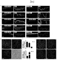
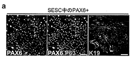
本発明のさらなる実施形態では、LSC集団又はLSC様集団の90〜95%は、p63、PAX6、K19及びKi67を発現する。別の実施形態では、LSC集団の5%未満がK5及びK14を発現する。さらに別の実施形態では、LSC集団の95%より多くがWNT7A及びFZD5を発現する。さらに別の実施形態では、LSC様集団の5%未満がWNT7Aを発現する。 In a further embodiment of the invention, 90-95% of the LSC population or LSC-like population express p63, PAX6, K19 and Ki67. In another embodiment, less than 5% of the LSC population expresses K5 and K14. In yet another embodiment, more than 95% of the LSC population expresses WNT7A and FZD5. In yet another embodiment, less than 5% of the LSC-like population expresses WNT7A.
本発明の別の実施形態では、角膜上皮細胞は、PAX6並びに角膜上皮マーカー、K3及びK12を発現する。 In another embodiment of the invention, the corneal epithelial cells express PAX6 and the corneal epithelial markers, K3 and K12.
一実施形態では、単離されたLSCは、WNT7A、FZD5、PAX6、p63、ケラチン5(K5)、ケラチン14(K14)、ケラチン19(K19)、及びKi67を含むマーカーのセットを発現する。別の実施形態では、LSCの90〜95%は、p63、PAX6、K19及びKi67を発現する。さらに別の実施形態では、LSCの95%より多くがWNT7A及びFZD5を発現する。別の実施形態では、LSCの5%未満がK5及びK14を発現する。さらに別の実施形態では、LSCの90〜95%はp63、PAX6、K19及びKi67を発現し、LSCの95%より多くがWNT7A及びFZD5を発現し、LSCの5%未満がK5及びK14を発現する。 In one embodiment, the isolated LSC expresses a set of markers comprising WNT7A, FZD5, PAX6, p63, keratin 5 (K5), keratin 14 (K14), keratin 19 (K19), and Ki67. In another embodiment, 90-95% of LSCs express p63, PAX6, K19 and Ki67. In yet another embodiment, greater than 95% of LSCs express WNT7A and FZD5. In another embodiment, less than 5% of LSCs express K5 and K14. In yet another embodiment, 90-95% of LSCs express p63, PAX6, K19 and Ki67, more than 95% of LSCs express WNT7A and FZD5, and less than 5% of LSCs express K5 and K14. To do.
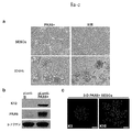
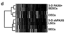
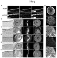
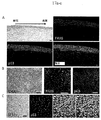
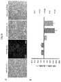
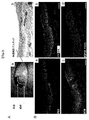
例えば、輪部幹若しくは前駆細胞様細胞又はLSC様細胞は、PAX6の十分な量での過剰発現時に「LSC様」状態に切り替わる又は採用する非LSC幹細胞、例えば皮膚上皮幹細胞(SESC)である。一実施形態では、「LSC様」状態に切り替えられた幹細胞は、核内でp63とPAX6両方の発現と同時に起きる誘導性K19発現を有する。別の実施形態では、「LSC様」細胞をLSC分化培地の存在下で3次元培養下に置く(増殖因子低減Matrigelに包埋する)と、「LSC様」細胞は、角膜K3及びK12発現増加と同時に皮膚K1及びK10発現減少を有する「CEC様」細胞に分化する。例えば、角膜K3発現は、(分化決定済みのSESCのままでいるのではなくLSC系統に転換される)PAX6過剰発現SESCの3D分化によって生産されたCEC様細胞でのほうが、PAX6を過剰発現しないが同様に処置されたSESCでより約9.4倍高いこともある。 For example, a limbal stem or progenitor cell-like cell or LSC-like cell is a non-LSC stem cell, eg, a skin epithelial stem cell (SESC) that switches or adopts an “LSC-like” state upon overexpression of PAX6 in sufficient amounts. In one embodiment, stem cells switched to an “LSC-like” state have inducible K19 expression that coincides with both p63 and PAX6 expression in the nucleus. In another embodiment, when “LSC-like” cells are placed in 3D culture in the presence of LSC differentiation media (embedded in growth factor-reduced Matrigel), the “LSC-like” cells have increased corneal K3 and K12 expression. At the same time, they differentiate into “CEC-like” cells with reduced expression of skin K1 and K10. For example, corneal K3 expression does not overexpress PAX6 in CEC-like cells produced by 3D differentiation of PAX6 overexpressing SESCs (converted to LSC lines rather than remain differentiated SESCs) May be about 9.4 times higher than in similarly treated SESCs.
本発明は、LSC集団又はLSC様集団の複数の細胞を含む被定義細胞集団をさらに提供する。この被定義細胞集団は、均一であることもあり、又は不均一であることもある。被定義細胞集団は、クローン性であることもあり、又は単一細胞に由来することもある。本発明は、角膜上皮細胞に発達するように決定された、LSC集団又はLSC様集団の子孫細胞をさらに提供する。加えて、本発明は、LSC集団又はLSC様集団の細胞を含む組織も提供する。 The present invention further provides a defined cell population comprising a plurality of cells of an LSC population or LSC-like population. This defined cell population may be homogeneous or heterogeneous. The defined cell population may be clonal or may be derived from a single cell. The present invention further provides progeny cells of the LSC population or LSC-like population determined to develop into corneal epithelial cells. In addition, the present invention provides a tissue comprising cells of an LSC population or LSC-like population.
本発明は、LSC集団又はLSC様集団及び適する担体を含む、医薬組成物をさらに提供する。本発明の一実施形態では、LSC集団又はLSC様集団は、CECに分化することなく、少なくとも17継代にわたって培養することができる。加えて、本発明の別の実施形態では、第3継代でPAX6及びp63を発現する細胞の割合は、第17継代での割合と同じである。本発明のさらなる実施形態では、第3継代でK19及びKi67を発現する細胞の割合は、第17継代以降での割合よりわずかに大きい。本発明のさらにさらなる実施形態では、本発明のLSC集団又はLSC様集団は、CECに分化することなく40〜60世代にわたって安定的に繁殖できる細胞を含む。さらに、本発明の一実施形態では、LSC集団又はLSC様集団は、角膜上皮細胞集団に分化することもある。
The present invention further provides a pharmaceutical composition comprising an LSC population or LSC-like population and a suitable carrier. In one embodiment of the invention, the LSC population or LSC-like population can be cultured for at least 17 passages without differentiation to CEC. In addition, in another embodiment of the invention, the proportion of cells expressing PAX6 and p63 at
加えて、本発明は、本発明のLSC集団又はLSC様集団の細胞を含む組織も提供する。さらに、本発明は、対象において組織を形成する方法であって、対象中又は対象上に、本発明のLSC集団又はLSC様集団の子孫細胞を、前記対象の角膜上皮細胞の形成に十分な量で導入することを含む方法をさらに提供する。 In addition, the present invention provides a tissue comprising cells of the LSC population or LSC-like population of the present invention. Furthermore, the present invention is a method of forming tissue in a subject, wherein the progeny cells of the LSC population or LSC-like population of the present invention are present in or on the subject in an amount sufficient to form the corneal epithelial cells of said subject. There is further provided a method comprising introducing in.
本発明は、染色体に組み込まれた、あるいは組み込まれていない染色体外遺伝物質のままのPAX6をコードする、化学合成、組換え、又は単離核酸を含む、単離された皮膚上皮幹細胞(SESC)集団又はSESC様集団であって、前記単離されたSESC集団は非SESC細胞を実質的に含まない、上記SESC集団又はSESC様集団をさらに提供する。さらに、SESC様集団は非SESC様細胞を実質的に含んでいなくてもよく、又は単離されたSESC又はSESC様集団は非SESC及び非SESC様細胞を実質的に含んでいなくてもよく、又はさらになお、単離されたSESC又はSESC様集団は、非SESC、非SESC様、非LSC及び非LSC様細胞を実質的に含んでいなくてもよい。加えて、本発明の一実施形態では、化学合成、組換え、又は単離核酸は、PAX6又はその断片を発現することができ、前記PAX6又はその断片は、LSC又はLSC様状態を維持することができ、又は幹細胞又は前駆細胞をLSC又はLSC様状態へと方向づけることができ、そしてまたそのLSC又はLSC様状態は、結果として角膜上皮細胞に至る分化経路に前記細胞集団を制限しうる。本発明の別の実施形態では、化学合成、組換え、又は単離核酸は、PAX6又はその断片を発現することができ、前記PAX6又はその断片は、SESC又はSESC様細胞をLSC又はLSC様状態へと方向づけ、そしてまた、結果として角膜上皮細胞に至る分化経路に前記細胞集団を制限しうる。SESC集団又はSESC様集団は、ヒトなどの哺乳動物からのものであってもよい。SESC集団又はSESC様集団は、遺伝子改変されていてもよい。さらに、SESC集団又はSESC様集団は、SESC又はSESC様細胞運命からLSC又はLSC様細胞運命に切り替えられることもある。 The present invention relates to an isolated skin epithelial stem cell (SESC) comprising a chemically synthesized, recombinant, or isolated nucleic acid that encodes PAX6 that remains chromosomally integrated or non-integrated extrachromosomal genetic material. Further provided is a SESC or SESC-like population, wherein the isolated SESC population is substantially free of non-SESC cells. Further, the SESC-like population may be substantially free of non-SESC-like cells, or the isolated SESC or SESC-like population may be substantially free of non-SESC and non-SESC-like cells. Alternatively, or still further, an isolated SESC or SESC-like population may be substantially free of non-SESC, non-SESC-like, non-LSC and non-LSC-like cells. In addition, in one embodiment of the invention, the chemically synthesized, recombinant, or isolated nucleic acid can express PAX6 or a fragment thereof, wherein the PAX6 or fragment thereof maintains an LSC or LSC-like state. Or stem cells or progenitor cells can be directed to an LSC or LSC-like state, and the LSC or LSC-like state can also restrict the cell population to a differentiation pathway that results in corneal epithelial cells. In another embodiment of the invention, the chemically synthesized, recombinant, or isolated nucleic acid can express PAX6 or a fragment thereof, wherein said PAX6 or fragment thereof is SESC or SESC-like cell in an LSC or LSC-like state. May also limit the cell population to the differentiation pathway leading to corneal epithelial cells. The SESC population or SESC-like population may be from a mammal such as a human. The SESC population or SESC-like population may be genetically modified. Furthermore, the SESC population or SESC-like population may be switched from SESC or SESC-like cell fate to LSC or LSC-like cell fate.
一実施形態では、細胞集団の約90〜95%は、SESC又はSESC様細胞運命のままで、p63、K5及びKi67を発現する。別の実施形態では、SESC又はSESC様細胞運命のままである細胞ではK3及びK12発現について検出されない。さらにさらなる実施形態では、WNT7Aは、SESC又はSESC様細胞運命のままである細胞において、LSC細胞におけるレベルより約4倍〜5倍低いレベルで発現される。さらに別の実施形態では、PAX6は、SESC又はSESC様細胞運命のままである細胞において発現されないか、又はLSC細胞におけるレベルの約8分の1未満のレベルで発現される。さらなる実施形態では、WNT7Aは、SESC様運命のままである細胞の70%より多くで発現される。さらにさらなる実施形態において、LSC又はLSC様細胞運命に切り替えられた集団の細胞の90〜95%は、p63、PAX6、K19及びKi67を発現する。加えて、本発明のある実施形態では、皮膚表皮細胞は、皮膚表皮分化マーカー、K1及びK10を発現する。 In one embodiment, about 90-95% of the cell population expresses p63, K5, and Ki67 while remaining SESC or SESC-like cell fate. In another embodiment, cells that remain SESC or SESC-like cell fate are not detected for K3 and K12 expression. In yet a further embodiment, WNT7A is expressed in cells that remain SESC or SESC-like cell fate at a level that is about 4 to 5 times lower than that in LSC cells. In yet another embodiment, PAX6 is not expressed in cells that remain SESC or SESC-like cell fate, or is expressed at a level that is less than about one-eighth the level in LSC cells. In a further embodiment, WNT7A is expressed in more than 70% of cells that remain SESC-like fate. In still further embodiments, 90-95% of the cells in the population switched to LSC or LSC-like cell fate express p63, PAX6, K19 and Ki67. In addition, in certain embodiments of the invention, the skin epidermal cells express skin epidermal differentiation markers, K1 and K10.
加えて、本発明は、SESC集団又はSESC様集団及び適する担体を含む、医薬組成物をさらに提供する。本発明の一実施形態では、SESC集団又はSESC様集団は、皮膚表皮細胞又は角膜上皮細胞に分化することなく、少なくとも17継代にわたって培養することができる。一実施形態では、SESC集団又はSESC様集団は、皮膚表皮細胞又は角膜上皮細胞に分化することなく40〜60世代にわたって安定的に繁殖できる細胞を含むことがある。さらなる実施形態では、SESC集団又はSESC様集団は、細胞運命がLSC又はLSC様細胞運命に切り替えられたSESC又はSESC様細胞を含むこともある。さらに、一実施形態では、SESC集団又はSESC様集団は、LSC又はLSC様細胞運命を採用し、SESC又はSESC様細胞運命は存在しない。SESC集団又はSESC様集団は、角膜上皮細胞に分化することもある。一実施形態では、SESC集団又はSESC様集団は、皮膚表皮細胞を実質的に含まない角膜上皮細胞に分化することもある。 In addition, the present invention further provides a pharmaceutical composition comprising a SESC population or SESC-like population and a suitable carrier. In one embodiment of the invention, the SESC population or SESC-like population can be cultured for at least 17 passages without differentiation into skin epidermal cells or corneal epithelial cells. In one embodiment, the SESC population or SESC-like population may comprise cells that can stably propagate over 40-60 generations without differentiation into skin epidermal cells or corneal epithelial cells. In further embodiments, the SESC population or SESC-like population may comprise SESC or SESC-like cells whose cell fate has been switched to LSC or LSC-like cell fate. Further, in one embodiment, the SESC population or SESC-like population employs an LSC or LSC-like cell fate and there is no SESC or SESC-like cell fate. The SESC population or SESC-like population may differentiate into corneal epithelial cells. In one embodiment, the SESC population or SESC-like population may differentiate into corneal epithelial cells that are substantially free of skin epidermal cells.
本発明は、SESC集団又はSESC様集団の複数の細胞を含む被定義細胞集団をさらに提供する。被定義細胞集団は、均一であることもあり、又は不均一であることもある。被定義細胞集団は、クローン性であることもあり、又は単一細胞に由来することもある。本発明は、角膜上皮細胞に発達するように決定された、SESC集団又はSESC様集団の子孫細胞をさらに提供する。 The present invention further provides a defined cell population comprising a plurality of cells of a SESC population or SESC-like population. The defined cell population may be uniform or heterogeneous. The defined cell population may be clonal or may be derived from a single cell. The present invention further provides progeny cells of a SESC population or SESC-like population determined to develop into corneal epithelial cells.
加えて、本発明は、本発明のSESC集団又はSESC様集団の細胞を含む組織も提供する。さらに、本発明は、対象において組織を形成する方法であって、対象中又は対象上に、本発明のSESC集団又はSESC様集団の子孫細胞を、前記対象の角膜上皮細胞の形成に十分な量で導入することを含む方法をさらに提供する。 In addition, the present invention also provides a tissue comprising cells of the SESC population or SESC-like population of the present invention. Furthermore, the present invention is a method for forming tissue in a subject, wherein the SESC population or SESC-like population progeny cells of the present invention are present in or on the subject in an amount sufficient to form corneal epithelial cells of said subject. There is further provided a method comprising introducing in.
本発明の方法
本発明の一実施形態では、本開示は、哺乳動物輪部幹細胞(LSC)の培養に関する。輪部幹細胞は、ヒトドナーからの角強膜又は角膜輪部組織に由来する。詳細には、本開示は、自己再生輪部幹細胞を有する系であり、LSC、例えば、少なくとも約70%、少なくとも約80%、又は少なくとも約90%輪部幹細胞の大集団を含むことができる。角膜輪部組織を単離するための典型的な手技は、ドナーの眼の角膜表面の上又は耳側四分円(temporal quadrant)からの、0.8〜3mm2の輪部組織からなる小さい検体材料の、外科的除去である。角膜輪部からそのような検体材料を例えば表層角膜切除術によって得るための手技は、当業者には公知である。輪部幹細胞を産生するために使用される輪部組織検体材料のドナーは、組織系移植片、インプラント又はグラフトのレシピエントであることもある(すなわち自家組織系)。あるいは、輪部組織検体材料のドナーがレシピエントでない場合、ドナーは、一例として、生体適合性ドナー、例えば移植片若しくはグラフトのレシピエントの近親者であり、又は生体適合性(例えば組織適合性)の死体であることもある(すなわち同種組織系)。移植される細胞又は組織は、組織拒絶反応に関する問題を回避するためにその移植片のレシピエントと遺伝学的に適合性又は同一であることが一般には望ましい。
Methods of the Invention In one embodiment of the invention, the present disclosure relates to culturing mammalian limbal stem cells (LSCs). The limbal stem cells are derived from horny sclera or limbal tissue from a human donor. In particular, the present disclosure is a system having self-renewing limbal stem cells and can include a large population of LSCs, eg, at least about 70%, at least about 80%, or at least about 90% limbal stem cells. A typical procedure for isolating limbal tissue is a small specimen of 0.8-3 mm 2 limbal tissue on the corneal surface of the donor's eye or from the temporal quadrant. Surgical removal. Techniques for obtaining such specimen material from the corneal limbus, for example by superficial keratotomy, are known to those skilled in the art. The donor of limbal tissue specimen material used to produce limbal stem cells may be a recipient of a tissue-based graft, implant or graft (ie, autologous tissue system). Alternatively, if the donor of the limbal tissue specimen material is not a recipient, the donor is, by way of example, a biocompatible donor, such as a close relative of a graft or graft recipient, or biocompatible (e.g., tissue compatible). It may be a dead body (ie allogeneic tissue system). It is generally desirable that the cells or tissues to be transplanted are genetically compatible or identical to the recipient of the graft to avoid problems with tissue rejection.
本開示のLSCは、角膜上皮細胞に分化する潜在力がある、未分化又は実質的に未分化の細胞である。未分化細胞の形態的特徴は、当業者に周知である。本発明の実施形態において有用な細胞、例えば、角膜上皮の輪部幹細胞を、多数の相補的要因、例えば、それらが得られるインビボ部位及び/又はそれらの形態若しくはサイズ(例えば平均直径)並びに、バイオマーカー、例えば、ATP結合カセットサブファミリーGメンバー2(ABCG2)、転写因子p63、Bmi-1、Notch-1、ステージ特異的胎児抗原-4(SSEA4)、ステージ特異的胎児抗原-3(SSEA3)、N-カドヘリン、CD73、CD105、CD54、CD117、Oct-4、Ki67、Nanog、Rex 1、Sox2、Tra-1-60、Tra-1-81、幹細胞因子、並びにサイトカイン(K)、例えばK1、K3、K5、K10、K12、K14又はK15、K19、及びデスモグレイン-3の存在、非存在及び/又は発現レベルによっても特徴づけることができることは、この技術分野の当業者には理解される(例えば、Nakatsuら、Investigative Ophthalmology & Visual Science 2011;52:4734-4741; Truongら、Invest Ophthalmol Vis Sci.2011;52:6315-6320; Duaら、Surv Ophthalmol.2000 Mar-Apr;44(5):415-25; Watsonら、Curr Eye Res.2013年4月10日; Meyer-Blazejewskaら、Invest Ophthalmol Vis Sci.2010 Feb;51(2):765-74;及びRamaら、N Engl J Med 2010;363: 147-55; Thomsonら、(Science 282:1145-1147, 1998)、Reubinoffら(Nature Biotech.18:399-403, 2000)を参照されたい)。本発明の実例的な実施形態では、ヒト輪部幹細胞は、ATP結合カセットサブファミリーGメンバー2(ABCG2)、-転写因子p63α、ステージ特異的胎児抗原-4(SSEA4)、N-カドヘリン、及びサイトカイン(K)、例えばK1、K3、K5、K10、K12、K14又はK15の1つ以上の発現を試験することによって特徴づけられる発現プロファイルを示す。本発明の実施形態では、ヒト輪部幹細胞又はフィーダー細胞の他の特徴、例えば、細胞のサイズ又は形態も同定又は特徴づけられる。 The LSCs of the present disclosure are undifferentiated or substantially undifferentiated cells that have the potential to differentiate into corneal epithelial cells. The morphological characteristics of undifferentiated cells are well known to those skilled in the art. Cells useful in embodiments of the present invention, such as limbal stem cells of the corneal epithelium, are transformed into a number of complementary factors, such as the in vivo site from which they are obtained and / or their form or size (e.g., average diameter) and bio Markers, such as ATP binding cassette subfamily G member 2 (ABCG2), transcription factor p63, Bmi-1, Notch-1, stage specific fetal antigen-4 (SSEA4), stage specific fetal antigen-3 (SSEA3), N-cadherin, CD73, CD105, CD54, CD117, Oct-4, Ki67, Nanog, Rex 1, Sox2, Tra-1-60, Tra-1-81, stem cell factor, and cytokine (K), such as K1, K3 Those skilled in the art will appreciate that can also be characterized by the presence, absence and / or expression level of K5, K10, K12, K14 or K15, K19, and desmoglein-3 (e.g. Nakatsu et al., Investigative Ophthalmology & Visual Science 2011; 52: 4734-4741; Truon g et al., Invest Ophthalmol Vis Sci. 2011; 52: 6315-6320; Dua et al., Surv Ophthalmol.2000 Mar-Apr; 44 (5): 415-25; Watson et al., Curr Eye Res. April 10, 2013; Meyer-Blazejewska et al., Invest Ophthalmol Vis Sci. 2010 Feb; 51 (2): 765-74; and Rama et al., N Engl J Med 2010; 363: 147-55; Thomson et al. (Science 282: 1145-1147, 1998 ), Reubinoff et al. (Nature Biotech. 18: 399-403, 2000)). In an exemplary embodiment of the invention, the human limbal stem cells comprise ATP binding cassette subfamily G member 2 (ABCG2), -transcription factor p63α, stage specific fetal antigen-4 (SSEA4), N-cadherin, and cytokines (K) shows an expression profile characterized by testing the expression of one or more of, for example, K1, K3, K5, K10, K12, K14 or K15. In embodiments of the invention, other characteristics of human limbal stem cells or feeder cells, such as cell size or morphology, are also identified or characterized.
例えば、検体材料をドナーから除去したら、輪部幹細胞の単離を可能にするために輪部組織生検材料の十分な部分が生存可能なままであるような仕方でそれを取り扱わなければならない。一実施形態では、輪部組織生検材料は、その生検材料の生存能を支援する培地内で輸送又は保存される。生検材料を保存又は輸送するための培地の例としては、ダルベッコ改変イーグル培地(DMEM)及びハムF-12(比率1:1)、DMSO(0.1〜0.5%)、組換えヒト上皮増殖因子(rhEGF、0.5〜2ng/ml)、インスリン(0.5〜5μg/ml)、トランスフェリン(0.5〜5μg/ml)、亜セレン酸ナトリウム(0.5〜5μg/ml)、ヒドロコルチゾン(0.1〜0.5μg/ml)、コレラ毒素A(0.01〜0.1μmol/l)、ゲンタマイシン(10〜50μg/ml)、及びアンホテリシンB(0.5〜1.25μg/ml)を挙げることができる。あるいは、前記培地の代わりに機能的に等価の成分又は異なる抗生物質を使用してもよい。培地にヒト臍帯血血清(3〜5%)をさらに補足することができる。輪部細胞生検材料をドナーからの外科的除去から48時間以内に培養下に置くことができる。 For example, once the specimen material is removed from the donor, it must be handled in such a way that a sufficient portion of the limbal tissue biopsy remains viable to allow isolation of the limbal stem cells. In one embodiment, the limbal tissue biopsy is transported or stored in a medium that supports the viability of the biopsy. Examples of media for storing or transporting biopsy material include Dulbecco's Modified Eagle Medium (DMEM) and Ham F-12 (ratio 1: 1), DMSO (0.1-0.5%), recombinant human epidermal growth factor ( rhEGF, 0.5-2 ng / ml), insulin (0.5-5 μg / ml), transferrin (0.5-5 μg / ml), sodium selenite (0.5-5 μg / ml), hydrocortisone (0.1-0.5 μg / ml), cholera Mention may be made of toxin A (0.01-0.1 μmol / l), gentamicin (10-50 μg / ml), and amphotericin B (0.5-1.25 μg / ml). Alternatively, functionally equivalent components or different antibiotics may be used in place of the medium. The medium can be further supplemented with human umbilical cord blood serum (3-5%). The limbal cell biopsy can be placed in culture within 48 hours of surgical removal from the donor.
輪部幹細胞を輸送前又は後に組織検体材料から直接精製してもよい。輪部組織生検材料をインタクト外植片として培養してもよいし、又は培養前に解離させて単個(又は希釈された)細胞浮遊液にしてもよい。例えば、生物活性が増加した特定の集団について組織を精製してもよい。当技術分野において公知の手段を使用して精製を行ってもよく、又は本明細書に記載のLSCマーカーについての陽性選択によって精製を果たしてもよい。本発明の一実施形態では、輪部幹細胞を無菌様式で機械的に分解し、酵素で処置して、回収組織から細胞を解離させる。そのような酵素としては、トリプシン、キモトリプシン、コラゲナーゼ、エラスターゼ、ヒアルロニダーゼ及び/又は商品、例えばStem Pro Accutase(Fischer)が挙げられるが、これらに限定されない。その後、輪部幹細胞の浮遊液を洗浄し、生存能について評価し、本発明の実施に直接使用してもよいし、又は拡大のために培養してもよい。一部の状況では、条件培地による産生に使用する前に細胞を拡大させることが望ましいであろう。拡大は、本明細書に記載の特定の因子とともにエクスビボで培養することによって行うことができる。 The limbal stem cells may be purified directly from the tissue specimen material before or after transport. The limbal tissue biopsy may be cultured as an intact explant or may be dissociated prior to culturing into a single (or diluted) cell suspension. For example, the tissue may be purified for a specific population with increased biological activity. Purification may be performed using means known in the art or may be accomplished by positive selection for the LSC markers described herein. In one embodiment of the invention, limbal stem cells are mechanically degraded in an aseptic manner and treated with enzymes to dissociate the cells from the recovered tissue. Such enzymes include, but are not limited to, trypsin, chymotrypsin, collagenase, elastase, hyaluronidase and / or commercial products such as Stem Pro Accutase (Fischer). The suspension of limbal stem cells can then be washed and evaluated for viability and used directly in the practice of the invention or cultured for expansion. In some situations, it may be desirable to expand the cells prior to use for production on conditioned media. Expansion can be performed by culturing ex vivo with certain factors described herein.
ドナーからの輪部組織の生検を行った後、それを培養培地での、及び一実施形態では適切な支持マトリックス、例えば細胞外マトリックス又はバイオコートされた表面、例えば細胞外マトリックス担体又はバイオコートされたペトリ皿での、培養下に置く。一実施形態では、支持マトリックスの存在は、生検材料中の輪部幹細胞の組織培養プレート又は容器への接着を助長し、その結果、輪部幹細胞の増殖を助長する。培養下に置く前に外植片を小片に切断することができる。 After performing a biopsy of the limbal tissue from the donor, it is placed in culture medium, and in one embodiment a suitable support matrix, such as an extracellular matrix or biocoated surface, such as an extracellular matrix carrier or biocoat. Place under culture in a Petri dish. In one embodiment, the presence of the support matrix promotes adherence of limbal stem cells in the biopsy to the tissue culture plate or container, and thus promotes proliferation of limbal stem cells. Explants can be cut into small pieces before being placed in culture.
ヒト羊膜を用意して、凍結融解、酵素的消化及び機械的擦過によって内在性羊膜上皮細胞を除去し、その後、増殖因子、細胞外マトリックス化合物及び/又は接着増進分子でその表面を処置することによって、輪部幹細胞の増殖を増進させてもよい。一実施形態では、基底膜又は角膜実質を上にして羊膜をLSC培養用培養プレートに平らに取り付ける。 By preparing human amniotic membrane and removing endogenous amniotic epithelial cells by freeze-thawing, enzymatic digestion and mechanical abrasion, and then treating the surface with growth factors, extracellular matrix compounds and / or adhesion promoting molecules The proliferation of limbal stem cells may be promoted. In one embodiment, the amniotic membrane is mounted flat on a culture plate for LSC culture with the basement membrane or corneal stroma facing up.
LSCをインビトロで支持することができる任意の培地を使用してLSCを培養してもよい。LSCの増殖を支持することができる培地処方としては、最小必須培地イーグル、ADC-1、LPM(ウシ血清アルブミン不含)、F10(HAM)、F12(HAM)、DCCM1、DCCM2、RPMI 1640、BGJ培地(フィットン-ジャクソン改変を伴う及び伴わない)、基本培地イーグル(BME-アール塩ベースを有する)、ダルベッコ改変イーグル培地(DMEM-血清なし)、山根、IMEM-20、グラスゴー改変イーグル培地(GMEM)、リーボヴィッツL-15培地、マッコイ5A培地、培地M199(M199E-アール塩ベースを有する)、培地M199(M199H-ハンクス塩ベースを有する)、最小必須培地イーグル(MEM-E-アール塩ベースを有する)、最小必須培地イーグル(MEM-H-ハンクス塩ベースを有する)及び最小必須培地イーグル(MEM-NAA、非必須アミノ酸を有する)、アルファ改変最小必須培地(αMEM)、及びロズウェルパーク記念研究所培地1640(RPMI培地1640)、上皮細胞用EPILIFE(登録商標)培養培地(Cascade Biologicals)、OPTI-PRO(商標)無血清培養培地、VP-SFM無血清培地、IMDM高濃縮基本培地、KNOCKOUT(商標)DMEM低浸透圧培地、293 SFM II限定無血清培地(すべてGibco; Invitrogen製)、HPGM造血前駆細胞増殖培地、Pro 293S-CDM無血清培地、Pro 293A-CDM無血清培地、UltraMDCK(商標)無血清培地(すべてCambrex製)、STEMLINE(登録商標)T細胞拡大培地及びSTEMLINE(登録商標)II造血幹細胞拡大培地(両方ともSigma-Aldrich製)、DMEM培養培地、DMEM/F-12栄養混合物増殖培地(両方ともGibco製)、ハムF-12栄養混合物増殖培地、M199基本培養培地(両方ともSigma-Aldrich製)、並びに他の同等の基本培地など(数ある他の多くのものの中でも、培地199、CMRL 1415、CMRL 1969、CMRL 1066、NCTC 135、MB 75261、MAB 8713.DM 145、ウィリアムズG、Neuman & Tytell、樋口、MCDB 301、MCDB 202、MCDB 501、MCDB 401、MCDB 411、MDBC 153を含む)が挙げられるがこれらに限定されない。本発明における使用に好ましい培地はDMEMである。これらの及び他の有用な培地は、数ある中でもGIBCO、Grand Island、N.Y.、USA及びBiological Industries、Bet HaEmek、Israelから入手可能である。多数のこれらの培地は、Academic Press, Inc.によって発行されたWilliam B. Jakoby及びIra H. Pastanによって編集されたMethods in Enzymology、LVIII巻、「Cell Culture」、62〜72頁に要約されている。 Any medium capable of supporting LSCs in vitro may be used to culture the LSCs. Medium formulations that can support the growth of LSC include minimal essential medium Eagle, ADC-1, LPM (without bovine serum albumin), F10 (HAM), F12 (HAM), DCCM1, DCCM2, RPMI 1640, BGJ Medium (with and without Fitton-Jackson modification), basal medium Eagle (with BME-Ear salt base), Dulbecco's modified Eagle medium (without DMEM-serum), Yamane, IMEM-20, Glasgow modified Eagle medium (GMEM ), Liebovitz L-15 medium, McCoy's 5A medium, medium M199 (with M199E-Earl salt base), medium M199 (with M199H-Hanks salt base), minimum essential medium Eagle (MEM-E-Earl salt base) Minimum essential medium eagle (with MEM-H-Hanks salt base) and minimum essential medium eagle (MEM-NAA, with non-essential amino acids), alpha modified minimum essential medium (αMEM), and Roswell Park Memorial Institute Medium 1640 (RPMI medium 1640), top EPILIFE (R) culture medium for cells (Cascade Biologicals), OPTI-PRO (TM) serum-free culture medium, VP-SFM serum-free medium, IMDM highly concentrated basal medium, KNOCKOUT (TM) DMEM hypotonic medium, 293 SFM II limited serum-free medium (all Gibco; manufactured by Invitrogen), HPGM hematopoietic progenitor cell growth medium, Pro 293S-CDM serum-free medium, Pro 293A-CDM serum-free medium, UltraMDCK (TM) serum-free medium (all manufactured by Cambrex), STEMLINE (Registered trademark) T cell expansion medium and STEMLINE (registered trademark) II hematopoietic stem cell expansion medium (both manufactured by Sigma-Aldrich), DMEM culture medium, DMEM / F-12 nutrient mixture growth medium (both manufactured by Gibco), Ham F -12 nutrient mixture growth medium, M199 basal culture medium (both from Sigma-Aldrich), as well as other equivalent basal media (among many others, medium 199, CMRL 1415, CMRL 1969, CMRL 1066, NCTC 135, MB 75261, MAB 8713.DM 145, Williams G, Neuman & Tytell, Higuchi, MCDB 301, MCDB 202, MCDB 501, MCDB 401, MCDB 4 11 and MDBC 153), but is not limited thereto. A preferred medium for use in the present invention is DMEM. These and other useful media are available from GIBCO, Grand Island, NY, USA and Biological Industries, Bet HaEmek, Israel, among others. A number of these media are summarized in Methods in Enzymology, Volume LVIII, “Cell Culture”, pages 62-72, edited by William B. Jakoby and Ira H. Pastan, published by Academic Press, Inc. .
本発明の方法において有用な培地のさらなる非限定的な例は、ウシ胎仔血清、ウシ血清又は他の種の血清を少なくとも1%〜約30%、少なくとも約5%〜15%、例えば約10%の濃度で含有することができる。特定の実施形態では、ヒト血清を使用することが好ましいことがある。 Further non-limiting examples of media useful in the methods of the present invention include fetal bovine serum, bovine serum or other species of serum at least 1% to about 30%, at least about 5% to 15%, such as about 10%. It can be contained at a concentration of In certain embodiments, it may be preferable to use human serum.
好ましい実施形態では、本発明において使用される培地は、フィーダーフリー、無血清及びゼノフリー培地である。例えば、輪部組織検体材料を輸送するために使用される培地、生検材料を培養するために使用される培地、輪部幹細胞を培養するために使用される培地、及び組織系を輸送するために使用される培地を含む、組織系を調製するために使用される培地は、動物由来のいずれの血清又は他の因子も含有しない。これは、異種成分での組織系の一切の汚染リスクを最小にすることによってその組織系をヒトへの投与に安全なものにするのに役立つことになる。最も好ましい実施形態では、フィーダーフリー、無血清及びゼノフリー培地は、培地中の全成分が既知組成であり、被定義の動物由来又はヒト由来産物を含有しない、既知組成培地である。 In a preferred embodiment, the medium used in the present invention is a feeder-free, serum-free and xeno-free medium. For example, to transport media used to transport limbal tissue specimen material, media used to culture biopsy material, media used to culture limbal stem cells, and tissue systems The medium used to prepare the tissue system, including the medium used for, does not contain any animal-derived serum or other factors. This will help to make the tissue system safe for administration to humans by minimizing the risk of any contamination of the tissue system with different components. In the most preferred embodiment, the feeder-free, serum-free and xeno-free medium is a known composition medium in which all components in the medium are of known composition and do not contain any defined animal or human derived products.
LSCの培養に利用することができる、細胞培養及び幹細胞の培養に関する一般技術について、実施者は、標準的な教科書及び総説、例えば、E. J. Robertson、「Teratocarcinomas and embryonic stem cells: A practical approach」、編集、IRL Press Ltd. 1987; Hu and Aunins(1997)、Curr.Opin.Biotechnol.8:148-153; Kitano(1991)、Biotechnology 17:73-106; Spier(1991)、Curr.Opin.Biotechnol.2:375-79; Birch and Arathoon(1990)、Bioprocess Technol.10:251-70; Xuら(2001)、Nat. Biotechnol.19(10):971-4;及びLebkowskiら(2001)Cancer J. 7 Suppl. 2:S83-93を参照することができ、これらの各々が参照によって本明細書に組み入れられる。 For general techniques related to cell culture and stem cell culture that can be used to culture LSCs, practitioners can use standard textbooks and reviews, such as EJ Robertson, “Teratocarcinomas and embryonic stem cells: A practical approach,” edited , IRL Press Ltd. 1987; Hu and Aunins (1997), Curr.Opin.Biotechnol. 8: 148-153; Kitano (1991), Biotechnology 17: 73-106; Spier (1991), Curr.Opin.Biotechnol.2. : 375-79; Birch and Arathoon (1990), Bioprocess Technol. 10: 251-70; Xu et al. (2001), Nat. Biotechnol. 19 (10): 971-4; and Lebkowski et al. (2001) Cancer J. 7 Reference can be made to Suppl. 2: S83-93, each of which is incorporated herein by reference.
本発明による方法のさらなる実施形態は、ROCK(Rho関連プロテインキナーゼ)阻害剤を含む培養培地を含む。ROCK阻害剤の添加は、特に幹細胞を培養する際、アノイキスを防止するために見出された。ROCK阻害剤は当技術分野において周知であり、一例では、(R)-(+)-トランス-4-(1-アミノエチル)-N-(4-ピリジル)シクロヘキサンカルボキサミド二塩酸塩一水和物(Y-27632、Sigma-Aldrich)、5-(1,4-ジアゼパン-1-イルスルホニル)イソキノリン(ファスジル又はHA1077、Cayman Chemical)、H-1152、H-1152P、(S)-(+)-2-メチル-1-[(4-メチル-5-イソキノリニル)スルホニル]ホモピペラジン、2HCl、ROCK阻害剤、ジメチルファスジル(diMF、H-1152P)、N-(4-ピリジル)-N'-(2,4,6-トリクロロフェニル)尿素、Y-39983、Wf-536、SNJ-1656、及び(S)-(+)-2-メチル-1-[(4-メチル-5-イソキノリニル)スルホニル]-ヘキサヒドロ-1H-1,4-ジアゼピン二塩酸塩(H-1152、Tocris Bioscience)、並びにその誘導体及び類似体から選択される。Rhoキナーゼ阻害剤を現在開発している会社としては、千寿製薬株式会社(大阪、日本)、Novartis(Basel、Switzerland)、興和創薬株式会社(名古屋、日本)、宇部興産株式会社とともに研究している参天製薬株式会社(東京、日本)、及びInspire Pharmaceuticals(Durham、N.C.)が挙げられる。オンラインジャーナルに関連したオンラインニュースレター、http://www.revophth.com/content/d/glaucoma_management/d/1222/p/23008/c/22947/#sthash.H16F1ABU.dpuf.のReview of Opthalmology Onlineで2009年3月20日に発表されたLama Al-AswadによるRhoキナーゼに関する総説においてより多くのものを参照されたい。さらなるROCK阻害剤としては、イミダゾール含有ベンゾジアゼピン及び類似体が挙げられる(例えばWO97/30992を参照されたい)。他のものとしては、例えば、国際出願公開番号WO01/56988、WO02/100833、WO03/059913、WO02/076976、WO04/029045、WO03/064397、WO04/039796、WO05/003101、WO02/085909、WO03/082808、WO03/080610、WO04/112719、WO03/062225、及びWO03/062227に記載のものが挙げられる。これらの事例の一部では、阻害剤中のモチーフは、インダゾールコア、2-アミノピリジン/ピリミジンコア、9-デアザグアニン誘導体、ベンズアミドを含むもの、アミノフラザンを含むもの、及び/又はそれらの組合せを含む。 A further embodiment of the method according to the invention comprises a culture medium comprising a ROCK (Rho-related protein kinase) inhibitor. The addition of ROCK inhibitors has been found to prevent anoikis, especially when culturing stem cells. ROCK inhibitors are well known in the art, and in one example, (R)-(+)-trans-4- (1-aminoethyl) -N- (4-pyridyl) cyclohexanecarboxamide dihydrochloride monohydrate (Y-27632, Sigma-Aldrich), 5- (1,4-diazepan-1-ylsulfonyl) isoquinoline (fasudil or HA1077, Cayman Chemical), H-1152, H-1152P, (S)-(+)- 2-Methyl-1-[(4-methyl-5-isoquinolinyl) sulfonyl] homopiperazine, 2HCl, ROCK inhibitor, dimethylfasudil (diMF, H-1152P), N- (4-pyridyl) -N '-(2 , 4,6-trichlorophenyl) urea, Y-39983, Wf-536, SNJ-1656, and (S)-(+)-2-methyl-1-[(4-methyl-5-isoquinolinyl) sulfonyl]- Hexahydro-1H-1,4-diazepine dihydrochloride (H-1152, Tocris Bioscience) and its derivatives and analogs. The companies currently developing Rho kinase inhibitors include Senju Pharmaceutical Co., Ltd. (Osaka, Japan), Novartis (Basel, Switzerland), Kowa Pharmaceutical Co., Ltd. (Nagoya, Japan), and Ube Industries, Ltd. Santen Pharmaceutical Co., Ltd. (Tokyo, Japan) and Inspire Pharmaceuticals (Durham, NC). Online newsletters related to online journals at the Review of Opthalmology Online at http://www.revophth.com/content/d/glaucoma_management/d/1222/p/23008/c/22947/#sthash.H16F1ABU.dpuf. See more in the review of Rho kinase by Lama Al-Aswad published March 20, 2009. Additional ROCK inhibitors include imidazole-containing benzodiazepines and analogs (see, eg, WO97 / 30992). Other examples include, for example, International Application Publication Numbers WO01 / 56988, WO02 / 100833, WO03 / 059913, WO02 / 076976, WO04 / 029045, WO03 / 064397, WO04 / 039796, WO05 / 003101, WO02 / 085909, WO03 / Examples are described in 082808, WO03 / 080610, WO04 / 112719, WO03 / 062225, and WO03 / 062227. In some of these cases, the motif in the inhibitor includes an indazole core, a 2-aminopyridine / pyrimidine core, a 9-deazaguanine derivative, a benzamide-containing one, an aminofurazan-containing one, and / or combinations thereof.
Rock阻害剤は、ROCK活性を減弱することができるROCK活性の負のレギュレーター、例えば、小GTP結合タンパク質(例えば、Gem、RhoE及びRad)も含む。本開示の特異的実施形態では、ROCK2ではなくROCK1を標的にし、例えば、WO03/080610は、ROCK阻害剤などのキナーゼ阻害剤としてのイミダゾピリジン誘導体、並びにROCK1及び/又はROCK2の作用を阻害する方法に関する。上で引用した出願の開示は、参照によって本明細書に組み入れられる。Rho阻害剤は、Rhoの阻害につながるROCK(Rho活性化キナーゼ)との相互作用によって下流で作用することもできる。そのような阻害剤は、米国特許第6,642,263号に記載されている(この特許文献の開示はその全体が参照によって本明細書に組み入れられる)。使用してもよい他のRho阻害剤は、米国特許第6,642,263号及び同第6,451,825号に記載されている。そのような阻害剤は、従来の細胞スクリーニングアッセイ、例えば、米国特許第6,620,591号に記載されているものを使用して同定することができる(これらの特許文献のすべては、それら全体が参照によって本明細書に組み入れられる)。 Rock inhibitors also include negative regulators of ROCK activity that can attenuate ROCK activity, such as small GTP binding proteins (eg, Gem, RhoE and Rad). In specific embodiments of the present disclosure, targeting ROCK1 but not ROCK2, for example, WO03 / 080610 describes imidazopyridine derivatives as kinase inhibitors such as ROCK inhibitors, and methods of inhibiting the action of ROCK1 and / or ROCK2. About. The disclosures of the above-cited applications are incorporated herein by reference. Rho inhibitors can also act downstream by interacting with ROCK (Rho activated kinase) leading to Rho inhibition. Such inhibitors are described in US Pat. No. 6,642,263 (the disclosure of this patent document is hereby incorporated by reference in its entirety). Other Rho inhibitors that may be used are described in US Pat. Nos. 6,642,263 and 6,451,825. Such inhibitors can be identified using conventional cell screening assays such as those described in U.S. Patent No. 6,620,591 (all of these patent documents are incorporated herein by reference in their entirety). Incorporated in the description).
培養培地は、培地中で適切な濃度でLSCを培養するために必要な増殖因子、サイトカイン及びホルモンも含むことができる。本発明の方法に有用な培地は、LSCの培養に有用な抗生物質、抗炎症、抗ウイルス、細胞分裂促進又は分化化合物を含むがこれらに限定されない、興味のある1つ以上の化合物も含有することがある。細胞を、非限定的な一実施形態では約27℃〜40℃の間の温度で増殖させてもよく、別の非限定的実施形態では約31℃〜37℃で増殖させてもよく、別の非限定的実施形態では加湿インキュベーターの中で増殖させてもよい。二酸化炭素含有量を約2%〜10%の間で維持してもよく、酸素含有量を約1%〜22%の間で維持してもよい、しかし、本発明がLSCを単離及び培養するためのいずれか1つの方法に限定されると決して解釈すべきではない。むしろ、LSCを単離及び培養するいずれの方法も本発明に含まれると解釈すべきである。 The culture medium can also contain growth factors, cytokines and hormones necessary for culturing the LSC at an appropriate concentration in the medium. Media useful for the methods of the present invention also contain one or more compounds of interest, including but not limited to antibiotics, anti-inflammatory, antiviral, mitogenic or differentiated compounds useful for LSC culture. Sometimes. The cells may be grown at a temperature between about 27 ° C. and 40 ° C. in one non-limiting embodiment, and may be grown at about 31 ° C. to 37 ° C. in another non-limiting embodiment. In a non-limiting embodiment, it may be grown in a humidified incubator. The carbon dioxide content may be maintained between about 2% and 10% and the oxygen content may be maintained between about 1% and 22%, but the present invention isolates and cultures LSCs Should in no way be construed as limited to any one method. Rather, any method of isolating and culturing LSC should be construed as included in the present invention.
培地に増殖因子を補足してもよい。本明細書で使用する場合、用語「増殖因子」は、細胞表面の受容体と結合し、主として細胞の増殖及び/又は分化を活性化する結果となるタンパク質を指す。輪部組織を培養するために使用される増殖因子は、例えば、上皮増殖因子(EGF)、塩基性線維芽細胞増殖因子(bFGF)、白血病抑制因子(LIF)、神経増殖因子(NGF)、インスリン増殖因子(IGF)、TGF-ベータ、肝細胞増殖因子、ケラチノサイト増殖因子、インスリン、亜セレン酸ナトリウム、ヒトトランスフェリン、又はヒト白血病抑制因子(hLIF)、ウシ下垂体抽出物など、及びこれらの組合せである。しかし、当業者に公知のいずれの適する培養培地を使用してもよい。特定の実施形態では、LSCを培養下で好ましく増殖させるサイトカイン又は他の増殖因子で輪部細胞を処置する。輪部細胞を培養するために使用される他の因子は、DMSO及びヒドロコルチゾン、グルコース、L-グルタミン、葉酸、重炭酸ナトリウム、アデニン、CaCl2、プロゲステロン、エタノールアミン、トリヨードチロニン、ホスホリルエタノールアミンなどから選択することができる。 The medium may be supplemented with growth factors. As used herein, the term “growth factor” refers to a protein that binds to cell surface receptors and results primarily in activating cell proliferation and / or differentiation. Growth factors used to culture limbal tissues include, for example, epidermal growth factor (EGF), basic fibroblast growth factor (bFGF), leukemia inhibitory factor (LIF), nerve growth factor (NGF), insulin Growth factor (IGF), TGF-beta, hepatocyte growth factor, keratinocyte growth factor, insulin, sodium selenite, human transferrin, or human leukemia inhibitory factor (hLIF), bovine pituitary extract, etc., and combinations thereof is there. However, any suitable culture medium known to those skilled in the art may be used. In certain embodiments, the limbal cells are treated with cytokines or other growth factors that favor the growth of LSCs in culture. Other factors used to culture limbal cells include DMSO and hydrocortisone, glucose, L-glutamine, folic acid, sodium bicarbonate, adenine, CaCl2, progesterone, ethanolamine, triiodothyronine, phosphorylethanolamine, etc. You can choose from.
特定の実施形態において、抗生物質は、マクロライド(例えば、トブラマイシ(TOBI(登録商標)))、セファロスポリン(例えば、セファレキシ(KEFLEX(登録商標)))、セフラジン(VELOSEF(登録商標)))、セフロキシム(CEFTIN(登録商標)、セフプロジル(CEFZIL(登録商標))、セファクロル(CECLOR(登録商標))、セフィキシム(SUPRAX(登録商標)又はセファドロキシル(DURICEF(登録商標))、クラリスロマイシン(例えば、クラリスロマイシン(Biaxin))、エリスロマイシン(例えば、エリスロマイシン(EMYCIN(登録商標)))、ペニシリン(例えば、ペニシリンV(V-CILLINK(登録商標)若しくはPEN VEEK(登録商標)))又はキノロン(例えば、オフロキサシン(FLOXIN(登録商標))、シプロフロキサシン(CIPRO(登録商標))若しくはノルフロキサシン(NOROXIN(登録商標)))、アミノグリコシド系抗生物質(例えば、アプラマイシン、アルベカシン、バンベルマイシン、ブチロシン、ジベカシン、ネオマイシン、ネオマイシン、ウンデシレン酸塩、ネチルマイシン、パロモマイシン、リボスタマイシン、シソマイシン及びスペクチノマイシン)、アンフェニコール系抗生物質(例えば、アジダムフェニコール、クロラムフェニコール、フロルフェニコール、及びチアンフェニコール)、アンサマイシン系抗生物質(例えば、リファミド及びリファンピン)、カルバセフェム(例えば、ロラカルベフ)、カルバペネム(例えば、ビアペネム及びイミペネム)、セファロスポリン(例えば、セファクロル、セファドロキシル、セファマンドール、セファトリジン、セファゼドン、セフォゾプラン、セフピミゾール、セフピラミド及びセフピロム)、セファマイシン(例えば、セフブペラゾン、セフメタゾール及びセフミノクス)、モノバクタム(例えば、アズトレオナム、カルモナム及びチゲモナム)、オキサセフェム(例えば、フロモキセフ及びモキサラクタム)、ペニシリン(例えば、アムジノシリン、アムジノシリンピボキシル、アモキシシリン、バカンピシリン、ベンジルペニシリン酸、ベンジルペニシリンナトリウム、エピシリン、フェンベニシリン、フロキサシリン、ペナメシリン、ペネタメート塩酸塩、ペニシリンo-ベネタミン、ペニシリン0、ペニシリンV、ペニシリンVベンザチン、ペニシリンVヒドラバミン、ペニメピサイクリン及びフェネチシリンカリウム)、リンコサミド(例えば、クリンダマイシン及びリンコマイシン)、マクロライド(例えば、アジスロマイシン、カルボマイシン、クラリスロマイシン、ジリスロマイシン、エリスロマイシン及びエリスロマイシンアシストラート)、アンホマイシン、バシトラシン、カプレオマイシン、コリスチン、エンデュラシジン、エンビオマイシン、テトラサイクリン(例えば、アピサイクリン、クロルテトラサイクリン、クロモサイクリン及びデメクロサイクリン)、2,4-ジアミノピリミジン(例えば、ブロジモプリム)、ニトロフラン(例えば、フラルタドン及び塩化フラゾリウム)、キノロン及びその類似体(例えば、シノキサシン、シプロフロキサシン、クリナフロキサシン、フルメキン及びグレパフロキサシン)、スルホンアミド(例えば、アセチルスルファメトキシピラジン、ベンジルスルファミド、ノプリルスルファミド、フタリルスルファセタミド、スルファクリソイジン及びスルファシチン)、スルホン(例えば、ジアチモスルホン、グルコスルホンナトリウム及びソラスルホン)、サイクロセリン、ムピロシン及びツベリンである。 In certain embodiments, the antibiotic is a macrolide (e.g., tobramysi (TOBI®)), cephalosporin (e.g., cephalex (KEFLEX®)), cefradine (VELOSEF®)). , Cefuroxime (CEFTIN®, cefprozil (CEFZIL®), cefaclor (CECLOR®), cefixime (SUPRAX® or cefadroxyl (DURICEF®)), clarithromycin (e.g. Clarithromycin (Biaxin)), erythromycin (e.g. erythromycin (EMYCIN®)), penicillin (e.g. penicillin V (V-CILLINK® or PEN VEEK®))) or quinolone (e.g. Ofloxacin (FLOXIN®), ciprofloxacin (CIPRO®) or norfloxacin (NOROXIN®)), aminoglycoside antibiotics (e.g. , Arbekacin, bambermycin, butyrosine, dibekacin, neomycin, neomycin, undecylenate, netilmicin, paromomycin, ribostamycin, sisomycin and spectinomycin), amphenicol antibiotics (e.g., azidamphenicol, clonomycin) Ramphenicol, florfenicol, and thiamphenicol), ansamycin antibiotics (e.g., rifamid and rifampin), carbacephem (e.g., loracarbef), carbapenem (e.g., biapenem and imipenem), cephalosporin (e.g., Cefaclor, cefadroxyl, cefamandol, cefatridine, cefazedon, cefozoplan, cefpimizole, cefpiramide and cefpirome), cefamycin (e.g. Cefmetazole and cefminox), monobactams (e.g. aztreonam, carmonam and tigemonam), oxacephem (e.g., flomoxef and moxalactam), penicillins (e.g. amidinocillin, amdinocillin pivoxil, amoxicillin, bacampicillin, benzylpenicillin acid, , Fenbenicillin, floxacillin, penamecillin, penetamate hydrochloride, penicillin o-benetamine, penicillin 0, penicillin V, penicillin V benzathine, penicillin V hydrabamine, penimepicyclin and pheneticillin potassium, lincosamide (e.g. clindamycin) And lincomycin), macrolides (e.g., azithromycin, carbomycin, clarithromycin, dirithromycin) Erythromycin and erythromycin assistate), amphomycin, bacitracin, capreomycin, colistin, enduracidin, enbiomycin, tetracycline (e.g., apycycline, chlortetracycline, chromocycline and demeclocycline), 2,4-diaminopyrimidine (E.g., brodimoprim), nitrofurans (e.g., furaltadone and furazolium chloride), quinolones and analogs thereof (e.g., sinoxacin, ciprofloxacin, clinfloxacin, flumequine and glepafloxacin), sulfonamides (e.g., Acetyl sulfamethoxypyrazine, benzylsulfamide, nopril sulfamide, phthalyl sulfacetamide, sulfacryosidine and sulfacitine), sulfones (e.g. Mosulfone, sodium glucosulfone and solasulfone), cycloserine, mupirocin and tuberine.
有用な抗炎症剤としては、非ステロイド性抗炎症薬(例えば、サリチル酸、アセチルサリチル酸、サリチル酸メチル、ジフルニサル、サルサラート、オルサラジン、スルファサラジン、アセトアミノフェン、インドメタシン、スリンダク、エトドラク、メフェナム酸、メクロフェナム酸ナトリウム、トルメチン、ケトロラク、ジクロフェナク、イブプロフェン、ナプロキセン、ナプロキセンナトリウム、フェノプロフェン、ケトプロフェン、フルルビプロフェン、オキサプロジン、ピロキシカム、メロキシカム、アンピロキシカム、ドロキシカム、ピボキシカム、テノキシカム、ナブメトン、フェニルブタゾン、オキシフェンブタゾン、アンチピリン、アミノピリン、アパゾン及びニメスリド)、ロイコトリエンアンタゴニスト(ジレウトン、オーロチオグルコース、金チオリンゴ酸ナトリウム及びオーラノフィンを含むがこれらに限定されない)、及び他の抗炎症剤(メトトレキサート、コルヒチン、アロプリノール、プロベネシド、スルフィンピラゾン及びベンズブロマロンを含むがこれらに限定されない)が挙げられるが、これらに限定されない。 Useful anti-inflammatory agents include non-steroidal anti-inflammatory drugs (e.g., salicylic acid, acetylsalicylic acid, methyl salicylate, diflunisal, salsalate, olsalazine, sulfasalazine, acetaminophen, indomethacin, sulindac, etodolac, mefenamic acid, meclofenamic acid, Tolmethine, ketorolac, diclofenac, ibuprofen, naproxen, naproxen sodium, fenoprofen, ketoprofen, flurbiprofen, oxaprozin, piroxicam, meloxicam, ampiroxicam, droxicam, pivoxicam, tenoxicam, nabumetone, phenylbutazone, oxyphenbutazone, Antipyrine, aminopyrine, apazone and nimesulide), leukotriene antagonists (zileuton, Including but not limited to rothioglucose, gold sodium thiomalate and auranofin), and other anti-inflammatory agents (including but not limited to methotrexate, colchicine, allopurinol, probenecid, sulfinpyrazone and benzbromarone) ), But is not limited thereto.
有用な抗ウイルス剤としては、ヌクレオシド類似体、例えばジドブジン、アシクロビル、ガンシクロビル、ビダラビン、イドクスウリジン、トリフルリジン及びリバビリン、並びにホスカルネット、アマンタジン、リマンタジン、サキナビル、インジナビル、リトナビル、及びアルファ-インターフェロンが挙げられるが、これらに限定されない。 Useful antiviral agents include nucleoside analogs such as zidovudine, acyclovir, ganciclovir, vidarabine, idoxuridine, trifluridine and ribavirin, and foscarnet, amantadine, rimantadine, saquinavir, indinavir, ritonavir, and alpha-interferon. For example, but not limited to.
一実施形態では、単離されたLSC集団は、実質的に分化することなく細胞を拡大させることが可能な培地で、例えば、不活性化ヒト胚性繊維芽細胞から得られる条件培地、ヒト白血病抑制因子で強化された培養培地、又はジメチルスルホキシド、組換えヒト上皮増殖因子、インスリン、亜セレン酸ナトリウム、トランスフェリン、プロゲステロン、プトレシン、亜セレン酸塩、ヒドロコルチゾン及び塩基性線維芽細胞増殖因子からなる群から選択される1つ以上の可溶性因子が補足された培養培地で培養される。あるいは、LSCは、例えば、特定の増殖因子又は培地、例えば角膜上皮培養培地CnT-30(CELLnTEC、Zen-Bio)又は既知組成ゼノフリー培養培地、RegES(Regea 06/015、Regea 07/046、及びRegea 08/013; Rajalaら、2010、PLOS One 5(4):e10246)を使用して、細胞を例えば角膜上皮細胞に分化させることになる培地で培養される。 In one embodiment, the isolated LSC population is a medium capable of expanding cells without substantial differentiation, eg, conditioned medium obtained from inactivated human embryonic fibroblasts, human leukemia Inhibitor-enhanced culture medium or group consisting of dimethyl sulfoxide, recombinant human epidermal growth factor, insulin, sodium selenite, transferrin, progesterone, putrescine, selenite, hydrocortisone and basic fibroblast growth factor Cultured in a culture medium supplemented with one or more soluble factors selected from Alternatively, LSC can be a specific growth factor or medium, such as corneal epithelial culture medium CnT-30 (CELLnTEC, Zen-Bio) or a known composition xenofree culture medium, RegES (Regea 06/015, Regea 07/046, and Regea 08/013; Rajala et al., 2010, PLOS One 5 (4): e10246) is used to culture cells in a medium that will differentiate them into, for example, corneal epithelial cells.
輪部組織検体材料を培養する例示的方法は、外植片を細胞外マトリックス又はバイオコートされた組織培養プレート上に配置する前又は配置した後、外植片を数分間、ドライインキュベーションに付す方法である。次いで、外植片が細胞外マトリックス又はバイオコートされた組織培養プレートに固着するように少量の培養培地をその外植片に添加する。数時間〜1日後、追加の培地を穏やかに添加し、1日おきに培地を交換しながら数日間、37℃で、CO2インキュベーターで外植片をインキュベートする。好ましくは、そのような例では、幹細胞を培養下で増殖し始めた後に元の輪部組織生検材料数片を培養物から除去する。 An exemplary method for culturing limbal tissue specimen material is a method of subjecting the explant to dry incubation for several minutes before or after placing the explant on the extracellular matrix or biocoated tissue culture plate. It is. A small amount of culture medium is then added to the explant so that the explant adheres to the extracellular matrix or biocoated tissue culture plate. After several hours to 1 day, incubate the explants in a CO 2 incubator at 37 ° C. for several days while gently adding additional medium and changing the medium every other day. Preferably, in such instances, the original limbal tissue biopsy pieces are removed from the culture after stem cells have begun to grow in culture.
他の実施形態では、拡大前に、輪部組織生検材料を使用して単一細胞浮遊液を生成し、その後、それを培養して、本明細書に開示の組織系を産生する。例えば、輪部組織生検材料を洗浄し、次いで、例えばトリプシン-EDTA(例えば、約0.25%、20〜30分間)又はディスパーゼ(例えば、4℃で一晩)で酵素的に処置して、LSCを含む単一細胞浮遊液を生成する。酵素的処置は上皮の分離を可能にするため、単一細胞浮遊液中の角膜実質又は間葉系細胞が低減されうる、又は非存在になりうる。 In other embodiments, before expansion, a limbal tissue biopsy is used to produce a single cell suspension, which is then cultured to produce the tissue system disclosed herein. For example, the limbal tissue biopsy is washed and then enzymatically treated with, for example, trypsin-EDTA (e.g., about 0.25%, 20-30 minutes) or dispase (e.g., overnight at 4 ° C) to produce LSC A single cell suspension containing Enzymatic treatment allows separation of the epithelium so that corneal parenchyma or mesenchymal cells in a single cell suspension can be reduced or absent.
輪部組織を数日間、例えば細胞が集密になるまで、培養した後、LSCを培養物から単離することができる。好ましくは、この例では、輪部組織培養物を、それが少なくとも約50%、60%、70%、80%、90%又は95%集密になるまで増殖させる。一実施形態では、輪部細胞を、細胞外マトリックス又はバイオコートされた組織培養プレートから、例えば酵素的消化、例えば、トリプシン-EDTA又はディスパーゼ溶液を使用する酵素的消化によって先ず解離させる。当業者に公知の様々な方法、例えば、免疫標識及び蛍光選別、例えば固相吸着、蛍光活性化細胞選別(FACS)、磁気親和性細胞選別(MACS)などを使用して、培養物中の他の細胞からLSCを単離することもできる。特定の実施形態では、LSCは、選別、例えば、特定の細胞表面マーカーの免疫蛍光選別によって単離される。当業者に周知の2つの好ましい選別方法がMACS及びFACSである。 After culturing the limbal tissue for several days, eg, until the cells are confluent, LSC can be isolated from the culture. Preferably, in this example, the limbal tissue culture is grown until it is at least about 50%, 60%, 70%, 80%, 90% or 95% confluent. In one embodiment, limbal cells are first dissociated from the extracellular matrix or biocoated tissue culture plate by, for example, enzymatic digestion, eg, enzymatic digestion using trypsin-EDTA or dispase solution. Other methods in the culture using various methods known to those skilled in the art, such as immunolabeling and fluorescence sorting, such as solid phase adsorption, fluorescence activated cell sorting (FACS), magnetic affinity cell sorting (MACS), etc. LSCs can also be isolated from these cells. In certain embodiments, LSCs are isolated by sorting, eg, immunofluorescent sorting of specific cell surface markers. Two preferred sorting methods well known to those skilled in the art are MACS and FACS.
選別技術は、培養下でLSCを他の細胞から分離するために適切な幹細胞マーカーの使用を含むことがある。1つ以上の特異的表面マーカーによって、そのマーカーと特異的に相互作用する1つ以上の標識抗体又は因子を使用してLSCを同定し、FACSの場合は蛍光標識又はMACSの場合は常磁性材料などの標識の存在に基づいて選別してもよい。培養輪部細胞からLSCを単離するために使用することができる適切な幹細胞特異的表面マーカーとしては、ABCG2、転写因子p63、SSEA4、SSEA3、N-カドヘリン、CD73、CD105、CD54、CD117、Oct-4、Nanog、TDGF、UTX-1、FGF-4、Rex1、Sox2、Tra-1-60、Tra-1-81、幹細胞因子、及びK1、K3、K10、K12、K14又はK15、K19が挙げられるが、これらに限定されない。このようにして、輪部組織生検材料から培養された細胞の混合集団から、LSCに陽性の細胞表面マーカーの濃縮集団が得られる。あるいは、LSC上で見出されない細胞表面マーカーについて選択することによって細胞を選別して望ましくない細胞を除去することができる。輪部組織から単離されたLSCの場合、LSCは、次の細胞表面マーカーについて陰性である: CD34、CD45、CD14、CD133、CD106、CD11c、CD123及びHLA-DR。 Sorting techniques may involve the use of appropriate stem cell markers to separate LSCs from other cells in culture. One or more specific surface markers identify LSCs using one or more labeled antibodies or factors that interact specifically with that marker, fluorescent labels in the case of FACS or paramagnetic materials in the case of MACS You may sort based on presence of labels, such as. Suitable stem cell-specific surface markers that can be used to isolate LSCs from cultured limbal cells include ABCG2, transcription factor p63, SSEA4, SSEA3, N-cadherin, CD73, CD105, CD54, CD117, Oct -4, Nanog, TDGF, UTX-1, FGF-4, Rex1, Sox2, Tra-1-60, Tra-1-81, stem cell factor, and K1, K3, K10, K12, K14 or K15, K19 However, it is not limited to these. In this way, an enriched population of LSC positive cell surface markers is obtained from a mixed population of cells cultured from limbal tissue biopsy material. Alternatively, cells can be sorted to remove unwanted cells by selecting for cell surface markers not found on LSC. In the case of LSCs isolated from limbal tissue, LSCs are negative for the following cell surface markers: CD34, CD45, CD14, CD133, CD106, CD11c, CD123 and HLA-DR.
選別によって得られる濃縮輪部細胞培養物は、少なくとも約10%、20%、30%、40%、50%、60%、70%、80%、90%、95%、98%又は99% LSCを有する。代替実施形態では、輪部細胞を含有する混合細胞培養物は、特定の遺伝子マーカーの発現についてのスクリーニングよってLSCの存在についてスクリーニングされる。混合輪部細胞培養の場合、遺伝子マーカー、例えば、ABCG2、転写因子p63、SSEA4、SSEA3、N-cadherin、CD73、CD105、CD54、CD117、Oct-4、Nanog、TDGF、UTX-1、FGF-4、Rex1、Sox2、Tra-1-60、Tra-1-81、幹細胞因子、及びK1、K3、K10、K12、K14若しくはK15、K19、並びに未分化細胞の他の遺伝子マーカー、又はそれらの組合せの発現によって、LSC集団を同定することができる。 Concentrated limbal cell cultures obtained by sorting are at least about 10%, 20%, 30%, 40%, 50%, 60%, 70%, 80%, 90%, 95%, 98% or 99% LSC Have In an alternative embodiment, mixed cell cultures containing limbal cells are screened for the presence of LSC by screening for expression of specific genetic markers. For mixed limbal cell culture, genetic markers such as ABCG2, transcription factor p63, SSEA4, SSEA3, N-cadherin, CD73, CD105, CD54, CD117, Oct-4, Nanog, TDGF, UTX-1, FGF-4 , Rex1, Sox2, Tra-1-60, Tra-1-81, stem cell factor, and K1, K3, K10, K12, K14 or K15, K19, and other genetic markers of undifferentiated cells, or combinations thereof Expression allows the LSC population to be identified.
上記方法の1つを使用してLSCが濃縮された輪部細胞の集団を単離した後、それらの単離された細胞を損傷又は罹病した眼に移植、内植又はグラフトするためにLSCの増殖及び組織系の発生を支持する条件下、支持する培地で培養することが好ましい。好ましくは、これらの条件下で培養される組織系は、少なくとも約30%、40%、50%、60%、70%、80%、90%又は95% LSCを含むことになる。ある実施形態では、単離されたLSCを、LSCを有する組織を発生させるために強化培地の存在下、組織ベースにおいて培養する。異なる因子を様々な増殖又は拡大段階で添加することができる。例えば、ROCK阻害剤を数継代の拡大後に添加することができる。組織ベースは、自然な眼の表面に近い特徴、例えば、澄んでいること、薄いこと、弾力性があること、生体適合性であること、非血管性であること及び非抗原性であることなどの特徴を有することができ、並びにまたLSC増殖はもちろん、移植、インプラント又はグラフト後の正常な分化も支持することができる。 After isolating a population of limbal cells enriched for LSC using one of the methods described above, the LSC can be used to transplant, implant or graft the isolated cells into a damaged or diseased eye. It is preferable to culture in a supporting medium under conditions that support growth and development of the tissue system. Preferably, tissue systems cultured under these conditions will comprise at least about 30%, 40%, 50%, 60%, 70%, 80%, 90% or 95% LSC. In certain embodiments, isolated LSCs are cultured in a tissue base in the presence of enriched media to generate tissues with LSCs. Different factors can be added at various stages of growth or expansion. For example, a ROCK inhibitor can be added after several passages of expansion. The tissue base is a feature close to the surface of the natural eye, such as being clear, thin, elastic, biocompatible, non-vascular and non-antigenic As well as LSC proliferation as well as normal differentiation after transplantation, implant or grafting.
LSCを含む輪部細胞は、LSCが実質的に未分化状態のままであることを可能にする適切な培地で培養又は継代される。集団内のLSCのコロニーは、分化される隣接細胞に隣接していることもあるが、その集団が適切な条件下で培養又は継代され、個々のLSCがその細胞集団の実質的な割合を構成する場合、LSCの培養物は、それにもかかわらず、実質的に未分化のままであるであろう。実質的に未分化である未分化幹細胞培養物は、少なくとも約20%未分化LSCを含有し、少なくとも約40%、60%、80%又は90% LSCを含有することもある。例えば、培養下のLSCを、分化を防止するために培養培地を頻繁に交換しながら約104/cm2の適切な細胞密度で保持して継代培養しなければならない。長期培養の場合、細胞を約70〜90%の集密度で継代すると、それらは分散されて小さなクラスター又は単一細胞浮遊液になりうる。典型的には、細胞の単一細胞浮遊液が得られ、その後、それを別の組織培養グレードプラスチック皿に播種して、継代後、約15〜20%の集密度に達する。 The limbal cells containing LSCs are cultured or passaged in a suitable medium that allows the LSCs to remain substantially undifferentiated. LSC colonies within a population may be adjacent to adjacent cells to be differentiated, but the population is cultured or passaged under appropriate conditions, and individual LSCs represent a substantial percentage of the cell population. When configured, LSC cultures will nevertheless remain substantially undifferentiated. An undifferentiated stem cell culture that is substantially undifferentiated contains at least about 20% undifferentiated LSC and may contain at least about 40%, 60%, 80%, or 90% LSC. For example, cultured LSCs must be subcultured while maintaining an appropriate cell density of about 10 4 / cm 2 with frequent changes of culture medium to prevent differentiation. For long-term culture, when cells are passaged at approximately 70-90% confluence, they can be dispersed into small clusters or single cell suspensions. Typically, a single cell suspension of cells is obtained, which is then seeded in another tissue culture grade plastic dish to reach approximately 15-20% confluence after passage.
培養物をLSCの実質的分化なしに少なくとも10、20、40、60、80、100継代以上にわたって連続的に継代することができる。LSCを含む輪部細胞培養物を、分化潜在力の喪失なくさらなる使用のために様々な時点で凍結保存することができ、好ましくは、そのような場合、ヒト臍帯血から採集した約10〜90%熱不活性化血清と約5〜10% DMSOとを有する培養培地を含む凍結培地で凍結保存することができる。あるいは、凍結培地は、無血清、ゼノフリー及びフィーダーフリーである既知組成のものでありうると予想される。輪部細胞培養物は、毎継代後、例えば凍結保存によって保存されたLSCを含むことができ、したがって、さらなる又は複数の組織系を単一の輪部組織生検材料から産生することができる。これらの凍結保存培養物は、任意の所定の時点での将来の使用のための未分化、自己再生及び生存輪部幹細胞のプールとしても役立つことになる。例えば、免疫抑制、以前の手術からの合併症、感染症などに起因してレシピエントの組織系が不全の場合には、これらの凍結保存培養物を使用して、自家使用のためのさらなる組織系を産生してもよい。また、これらの凍結保存培養物を使用して、生体適合性患者のためのさらなる組織系を産生してもよい。これらの保存培養物の有用性は、組織系が不全の場合にドナーからさらなる輪部組織を除去する必要をなくすことにもなり、その結果、将来、自家幹細胞源を使い果たす危険を防止することにもなる。 The culture can be passaged continuously for at least 10, 20, 40, 60, 80, 100 passages or more without substantial differentiation of LSCs. The limbal cell culture containing LSC can be cryopreserved at various time points for further use without loss of differentiation potential, and preferably in such cases, about 10-90 collected from human umbilical cord blood. It can be cryopreserved in a freezing medium containing a culture medium with% heat inactivated serum and about 5-10% DMSO. Alternatively, it is expected that the freezing medium may be of a known composition that is serum-free, xeno-free and feeder-free. The limbal cell culture can contain LSCs preserved after each passage, for example by cryopreservation, so that additional or multiple tissue systems can be produced from a single limbal tissue biopsy. . These cryopreserved cultures will also serve as a pool of undifferentiated, self-renewing and viable limbal stem cells for future use at any given time. These cryopreserved cultures can be used to further tissue for self-use if, for example, the recipient's tissue system is defective due to immunosuppression, complications from previous surgery, infections, etc. A system may be produced. These cryopreserved cultures may also be used to produce additional tissue systems for biocompatible patients. The usefulness of these stock cultures also eliminates the need to remove additional limbal tissue from the donor if the tissue system is defective, thus preventing the risk of running out of autologous stem cell sources in the future. Also become.
本明細書に開示の組織系を産生した後、移植片、インプラント又はグラフトのレシピエントの所在地にそれを輸送してもよい。組織系を輸送するために使用される手段は、輸送後に移植片、インプラント又はグラフトとしてなお有用である組織系の生存能を十分に維持することができる。組織系は、輸送培地が入っている容器で輸送され、前記輸送培地は、LSCを含む組織系を培養するために使用される強化培地、又は増殖因子を有さない輸送中に組織系を緩衝するのに十分な代替培地でありうる。あるいは、本明細書に開示の組織系を保持する施設にレシピエントを連れて行き、輸送培地の必要をなくしてもよい。 After producing the tissue system disclosed herein, it may be transported to the recipient location of the graft, implant or graft. The means used to transport the tissue system can sufficiently maintain the viability of the tissue system that is still useful as a graft, implant or graft after transport. The tissue system is transported in a container containing transport medium, which is a reinforced medium used to culture tissue systems containing LSC, or buffers the tissue system during transport without growth factors. There may be sufficient alternative media to do. Alternatively, the recipient may be taken to a facility that holds the tissue system disclosed herein, eliminating the need for a transport medium.
本明細書に開示のLSCを含む組織系は、治療応用に、例えば、片眼又は両眼の輪部幹細胞が欠損している対象の移植片、インプラント又はグラフトとして利用することができる。本開示の組織系を使用して、ヒト、霊長類、及び飼育動物、家畜、ペット、又は競技用動物、例えばイヌ、ウマ、ネコ、ヒツジ、ブタ、ウシ、ラット、マウスなどを含むがこれらに限定されない、処置を必要とする任意の対象を処置することができる。本明細書で使用する場合、用語「治療の」、「治療的に」、「処置すること」、「処置」又は「治療」は、治療的処置と予防的(prophylactic)又は予防(preventative)対策の両方を指す。治療的処置は、特定の疾患、状態、傷害若しくは障害の症状を低減させる若しくは除去すること、又は既存の疾患若しくは障害の進行を遅速若しくは減弱すること、又は既存の疾患若しくは障害を治癒させることを含むが、これらに限定されない。そのような治療を必要とする対象は、機能を回復又は再生するために組織系の治療有効量によって処置されることになる。本明細書で使用する場合、組織系の「治療有効量」は、輪部幹細胞の喪失、損傷、機能不全又は変性に起因する対象の生理作用を抑止又は改善するのに十分な量である。使用される細胞又は組織の治療有効量は、対象の必要、対象の年齢、生理状態及び健康状態、所望の治療効果、治療の標的となる組織領域のサイズ、内植部位、病状の程度、選択される送達経路、及び処置戦略に依存することになる。意図される部位への組織系のグラフトを可能にし、機能欠損領域の再構成又は再生を可能にする方法で、組織系を患者に投与することが好ましい。 A tissue system comprising an LSC disclosed herein can be utilized for therapeutic applications, for example, as a graft, implant or graft in a subject lacking unilateral or binocular limbal stem cells. Using the tissue system of the present disclosure, including humans, primates, and domestic animals, livestock, pets, or competition animals such as dogs, horses, cats, sheep, pigs, cows, rats, mice, etc. Any subject in need of treatment can be treated without limitation. As used herein, the terms “therapeutic”, “therapeutically”, “treating”, “treatment” or “treatment” refer to therapeutic treatment and prophylactic or preventative measures. Refers to both. Therapeutic treatment involves reducing or eliminating the symptoms of a particular disease, condition, injury or disorder, or slowing or attenuating the progression of an existing disease or disorder, or curing an existing disease or disorder. Including, but not limited to. A subject in need of such treatment will be treated with a therapeutically effective amount of the tissue system to restore or regenerate function. As used herein, a “therapeutically effective amount” of a tissue system is an amount sufficient to inhibit or ameliorate a subject's physiological effects due to loss, damage, dysfunction or degeneration of limbal stem cells. The therapeutically effective amount of cells or tissue used is the subject's need, subject's age, physiology and health, desired therapeutic effect, size of the tissue region targeted for treatment, site of implantation, degree of disease state, selection Will depend on the delivery route being used and the treatment strategy. It is preferred to administer the tissue system to the patient in a manner that allows the grafting of the tissue system to the intended site and allows the reconstruction or regeneration of the loss of function area.
好ましい実施形態では、本開示の組織系を使用して、眼の損傷又は疾患、特に眼表面障害を有する対象を治療的に処置する。あるいは、開示する組織系を使用して、輪部組織に由来する未分化幹細胞源から治療的利益を受けると予想される他の疾患又は損傷を処置、例えば、熱傷を負った皮膚領域を修復してもよい。開示する組織系は、遺伝性の状態に起因しうる原発性輪部幹細胞欠損、例えば、無虹彩、多発性内分泌欠損関連角膜炎、輪部炎、及び後天性の状態から起こりうる特発性又は二次的輪部幹細胞喪失、例えば、スティーブンス・ジョンソン症候群、感染症(例えば、重症細菌性角膜炎)、眼表面腫瘍、化学的若しくは熱的傷害又は紫外線放射、複数回の手術若しくは凍結療法への曝露に起因する外傷性輪部幹細胞破壊、角膜上皮内新生物、縁辺潰瘍性又は炎症性角膜炎、虚血性角膜炎、角膜症、コンタクトレンズ又はレンズ洗浄液によって誘導される毒性作用、免疫学的状態、眼瘢痕性類天疱瘡、翼状片、偽翼状片などを有する対象の処置に特によく適している。特定の実施形態では、組織系は対象に移植、内植又はグラフトされ、対象の眼損傷又は疾患を、例えば、対象の損傷又は罹病した眼に安定した輪部幹細胞集団を提供することによって修復することができる。特定の実施形態では、組織系の移植、内植又はグラフトは、眼における上皮化を助長し、正常な上皮表現型を維持し、炎症を低減させ、瘢痕化を低減させ、組織の接着を低減させ、血管新生を低減させ、及び視力を向上させる。 In a preferred embodiment, the tissue system of the present disclosure is used to therapeutically treat a subject having an eye injury or disease, particularly an ocular surface disorder. Alternatively, the disclosed tissue system can be used to treat other diseases or injuries expected to receive therapeutic benefit from undifferentiated stem cell sources derived from limbal tissue, for example to repair burned skin areas May be. The disclosed tissue system is a idiopathic or biliary that can arise from a primary limbal stem cell defect that can result from an inherited condition, such as an iris, multiple endocrine deficiency-associated keratitis, keratitis, and an acquired condition. Secondary limbal stem cell loss, e.g. Stevens-Johnson syndrome, infection (e.g. severe bacterial keratitis), ocular surface tumor, chemical or thermal injury or UV radiation, multiple surgery or cryotherapy Traumatic limbal stem cell destruction resulting from exposure, corneal intraepithelial neoplasia, marginal ulcerative or inflammatory keratitis, ischemic keratitis, keratopathy, toxic effects induced by contact lenses or lens washes, immunological status It is particularly well suited for the treatment of subjects with ocular scarring pemphigus, pterygium, pseudo pterygium, and the like. In certain embodiments, the tissue system is transplanted, implanted or grafted into the subject to repair the subject's eye damage or disease, for example, by providing a stable limbal stem cell population to the subject's damaged or diseased eye. be able to. In certain embodiments, tissue-based transplantation, ingrowth or grafting promotes epithelialization in the eye, maintains a normal epithelial phenotype, reduces inflammation, reduces scarring, and reduces tissue adhesion Reduce angiogenesis and improve visual acuity.
例えば、組織系を移植、内植又はグラフトして、損傷した角膜を修復することができる。多くのそのような方法は当業者に周知であるが、1つのそのような方法は、輪部での角膜周囲切開、続いて強膜を露出させるための輪部周囲結膜下瘢痕及び炎症組織の除去を含む。角膜の線維性血管性組織を表層角膜切除術によって除去してもよい。レシピエントの眼のサイズに従って組織系を拡大縮小し、対応するレシピエント輪部領域に移植又はグラフトすることができる。あるいは、組織系を全層状角膜組織として使用し、表層角膜移植術として移植又はグラフトして全領域を覆ってもよい。その後、その移植、内植又はグラフトされた組織系を、例えば縫合又は当業者に公知の任意の他の手段で、損傷部位に固定する。 For example, a tissue system can be transplanted, implanted or grafted to repair a damaged cornea. Many such methods are well known to those of skill in the art, but one such method is a pericorneal incision at the annulus, followed by periannular subconjunctival scars and inflammatory tissue to expose the sclera. Includes removal. The corneal fibrous vascular tissue may be removed by superficial keratotomy. The tissue system can be scaled according to the size of the recipient's eye and transplanted or grafted into the corresponding recipient annulus region. Alternatively, the tissue system may be used as a full-layer corneal tissue, and transplanted or grafted as a superficial corneal transplantation to cover the entire area. The implanted, implanted or grafted tissue system is then secured to the injury site, for example, by suturing or any other means known to those skilled in the art.
本発明は、対象において組織を再生又は修復する方法であって、対象中又は対象上に、本発明のLSC集団又はLSC様集団又はSESC集団又はSESC様集団の細胞を、組織の再生又は修復に十分な量で導入することを含む方法をさらに提供する。 The present invention is a method for regenerating or repairing tissue in a subject, wherein the cells of the LSC population or LSC-like population or SESC population or SESC-like population of the present invention are used for regeneration or repair of tissue in or on the subject. Further provided is a method comprising introducing in a sufficient amount.
一実施形態では、再生又は修復される組織は、輪部幹又は前駆細胞(LSC)と角膜上皮細胞とを含む角膜上皮細胞系統の組織を含む。 In one embodiment, the tissue that is regenerated or repaired includes tissue of the corneal epithelial cell lineage that includes limbal stem or progenitor cells (LSCs) and corneal epithelial cells.
本発明は、対象の皮膚上皮幹細胞(SESC)から輪部幹細胞又は前駆細胞(LSC)様細胞を得る方法をさらに提供する。本発明のある実施形態では、この方法は、SESCをLSC様細胞に転換するのに十分なレベルにSESC中のPAX6タンパク質を増加させることによって対象のSESCからLSC様細胞を得るための、SESCへのPAX6遺伝子の導入又はSESCにおけるPAX6遺伝子発現のアップレギュレーションを含む。一実施形態では、対象の皮膚上皮幹細胞(SESC)から輪部幹又は前駆細胞(LSC)様細胞を得るためのSESCへのPAX6遺伝子の導入又はSESCにおけるPAX6遺伝子発現のアップレギュレーションは、(a)SESCを対象から得るステップ、(b)前記SESCをインビトロ又はエクスビボでのフィーダーフリー細胞培養で培養するステップ、(c)前記SESCに少なくとも1つのPAX6遺伝子を導入して又は前記SESCにおけるPAX6遺伝子発現をアップレギュレートさせて、SESCを輪部幹細胞又は前駆細胞(LSC)様細胞に転換するのに十分なレベルにSESC中のPAX6タンパク質を増加させ、それによって対象からの皮膚上皮幹細胞(SESC)から哺乳動物輪部幹細胞又は前駆細胞(LSC)様細胞を得るステップを含む。 The present invention further provides a method of obtaining limbal stem cells or progenitor cells (LSC) -like cells from a subject's skin epithelial stem cells (SESC). In certain embodiments of the invention, the method includes: converting SESCs into LSC-like cells to SESCs to obtain LSC-like cells from the subject SESCs by increasing the PAX6 protein in the SESCs to a level sufficient to convert SESCs to LSC-like cells. Introduction of PAX6 gene or up-regulation of PAX6 gene expression in SESC. In one embodiment, introduction of the PAX6 gene into SESC or up-regulation of PAX6 gene expression in SESC to obtain limbal stem or progenitor cell (LSC) -like cells from a subject skin epithelial stem cell (SESC) comprises: Obtaining SESC from the subject; (b) culturing the SESC in in vitro or ex vivo feeder-free cell culture; (c) introducing at least one PAX6 gene into the SESC or expressing PAX6 gene in the SESC. Upregulate to increase the PAX6 protein in the SESC to a level sufficient to convert SESCs to limbal stem cells or progenitor cells (LSC) -like cells, thereby suckling from skin epithelial stem cells (SESC) from the subject Obtaining animal limbal stem cells or progenitor cells (LSC) -like cells.
本発明の実施に従って、前記方法は、インビトロの方法であってもよく、エクスビボ方法であってもよく、又は生体内原位置(in situ)で若しくは直接対象に適用してもよい。 In accordance with the practice of the invention, the method may be an in vitro method, an ex vivo method, or may be applied in situ or directly to a subject.
一実施形態では、PAX6タンパク質をコードする核酸を導入するか、PAX6遺伝子発現をアップレギュレートするか、又はPAX6活性を増加させる薬剤で、対照は処置される。前記薬剤の適する例としては、遺伝子治療ベクター、ウイルス粒子、レンチウイルス、アデノウイルス、アデノ随伴ウイルス、組換え核酸、組換えタンパク質、PAX6タンパク質、PAX6発現の小分子レギュレーター、PAX6発現の負のレギュレーターの阻害剤、PAX活性の負のレギュレーターの小分子阻害剤、PAX6活性の小分子エンハンサー、又はこれらの組合せが挙げられるが、これらに限定されない。 In one embodiment, the control is treated with an agent that introduces a nucleic acid encoding a PAX6 protein, upregulates PAX6 gene expression, or increases PAX6 activity. Suitable examples of such agents include gene therapy vectors, virions, lentiviruses, adenoviruses, adeno-associated viruses, recombinant nucleic acids, recombinant proteins, PAX6 proteins, small molecule regulators of PAX6 expression, negative regulators of PAX6 expression Inhibitors, small regulators of negative regulators of PAX activity, small molecule enhancers of PAX6 activity, or combinations thereof include, but are not limited to.
PAX6遺伝子の例としては、PAX6a遺伝子、PAX6b遺伝子、操作されたPAX6a遺伝子、操作されたPAXb遺伝子、PAX6遺伝子ファミリーの任意のメンバー、PAX6aタンパク質のすべて又は一部をコードする核酸、PAX6bタンパク質のすべて又は一部をコードする核酸、及びPAX6又はPAX6様活性を有するタンパク質をコードする任意の核酸が挙げられるが、これらに限定されない。本発明の一実施形態では、PAX6タンパク質は、PAX6aタンパク質、PAX6bタンパク質、タンパク質のPAX6ファミリーの任意のメンバー、及びPAX6又はPAX6様活性を有する任意のタンパク質のいずれかである。本発明の一実施形態では、PAX6又はPAX6様活性には内因性K19の発現増加を生じさせることができる任意のタンパク質が関係することがあり、K19がアップレギュレートされたSECSは、K3及びK12遺伝子発現増加並びにK1及びK10遺伝子発現減少を有する角膜上皮細胞(CEC)又はCEC様細胞に分化しうる。 Examples of PAX6 genes include PAX6a gene, PAX6b gene, engineered PAX6a gene, engineered PAXb gene, any member of PAX6 gene family, nucleic acid encoding all or part of PAX6a protein, all or part of PAX6b protein Examples include, but are not limited to, nucleic acids that encode a portion and any nucleic acid that encodes a protein having PAX6 or PAX6-like activity. In one embodiment of the invention, the PAX6 protein is any of the PAX6a protein, the PAX6b protein, any member of the PAX6 family of proteins, and any protein having PAX6 or PAX6-like activity. In one embodiment of the present invention, PAX6 or PAX6-like activity may involve any protein capable of causing increased expression of endogenous K19, and SECS with upregulated K19 is K3 and K12. Can differentiate into corneal epithelial cells (CEC) or CEC-like cells with increased gene expression and decreased K1 and K10 gene expression.
一実施形態では、本発明は、上記の本発明のLSC様細胞から角膜上皮細胞(CEC)様細胞を得る方法であって、フィーダーフリーLSC分化培地で(c)の細胞を分化させてLSC様細胞をCEC様細胞に転換するステップをさらに含む方法を提供する。 In one embodiment, the present invention provides a method for obtaining a corneal epithelial cell (CEC) -like cell from the LSC-like cell of the present invention as described above, comprising differentiating the cell of (c) in a feeder-free LSC differentiation medium. There is provided a method further comprising the step of converting the cell into a CEC-like cell.
本発明のある実施形態では、フィーダーフリーLSC分化培地は、既知組成であることがある。本発明の別の実施形態では、前記培地はゼノフリーであるか、培養細胞と同じ種に由来する成分以外の成分を含まない。さらに別の実施形態では、培地は無血清であることがある。さらなる実施形態では、培地は、一切の動物又はヒト産物を有さないことがある。 In certain embodiments of the invention, the feeder-free LSC differentiation medium may be of a known composition. In another embodiment of the present invention, the medium is xeno-free or does not contain components other than those derived from the same species as the cultured cells. In yet another embodiment, the medium may be serum free. In further embodiments, the medium may not have any animal or human products.
例えば、ゼノフリー培地又はゼノフリー培養培地は、培地が異質な動物由来産物又は材料を含有しないものである。詳細には、培養培地中のいずれの産物及び材料も、異質な動物細胞において生産されず、又は異質な動物細胞と接触していない。ウシ胎仔血清を含む培養培地は、ヒト細胞を培養するのに使用される場合又はウシ細胞以外の任意の動物細胞の培養に使用される場合、血清がウシに由来するのでゼノフリーと考えられないであろうが、一切の動物由来産物又は材料の非存在下でヒト血清を含む培養培地は、ヒト細胞を培養する場合、ゼノフリーと考えられるであろう。例えば、ゼノフリー培地は、細菌又は酵母などの微生物から生産された又は得られた産物又は材料を含有することがある。ゼノフリー培地は、「動物質を含まない」培地と考えられることもある。この例では、ゼノフリーは、異質な動物由来産物又は材料の非存在を指す。 For example, a xenofree medium or a xenofree culture medium is one in which the medium does not contain any foreign animal-derived products or materials. Specifically, none of the products and materials in the culture medium are produced in or in contact with foreign animal cells. A culture medium containing fetal bovine serum should not be considered xeno-free when used for culturing human cells or for culturing any animal cell other than bovine cells because the serum is derived from bovine. However, a culture medium containing human serum in the absence of any animal-derived products or materials would be considered xenofree when culturing human cells. For example, a xenofree medium may contain products or materials produced or obtained from microorganisms such as bacteria or yeast. A xenofree medium may be considered a "no animal substance" medium. In this example, xenofree refers to the absence of foreign animal-derived products or materials.
本発明は、皮膚上皮幹細胞(SESC)から輪部幹又は前駆細胞(LSC)様細胞を得る方法をさらに提供する。この方法は、(a)SESCを対象から得るステップ、(b)前記SESCをインビトロでのフィーダーフリー細胞培養で培養するステップ、(c)前記SESCにおけるPAX6遺伝子発現をアップレギュレートさせる薬剤と前記SESCを接触させて、SESCを輪部幹細胞又は前駆細胞(LSC)様細胞を転換するのに十分なレベルにSESC中のPAX6タンパク質を増加させ、それによって対象からの皮膚上皮幹細胞(SESC)から哺乳動物輪部幹細胞又は前駆細胞(LSC)様細胞を得るステップを含む。 The present invention further provides a method of obtaining limbal stem or progenitor cell (LSC) -like cells from skin epithelial stem cells (SESC). The method comprises: (a) obtaining SESC from a subject; (b) culturing the SESC in feeder-free cell culture in vitro; (c) an agent that up-regulates PAX6 gene expression in the SESC and the SESC To increase the PAX6 protein in the SESC to a level sufficient to transform SESCs into limbal stem cells or progenitor cells (LSC) -like cells, thereby from the skin epithelial stem cells (SESC) from the subject to the mammal Obtaining limbal stem cells or progenitor cells (LSC) -like cells.
適する薬剤の例としては、PAX6遺伝子を含む核酸、遺伝子治療ベクター、ウイルス粒子、レンチウイルス、アデノウイルス、アデノ随伴ウイルス、組換えタンパク質、PAX6タンパク質、PAX6発現の小分子レギュレーター、PAX6発現の負のレギュレーターの阻害剤、PAX活性の負のレギュレーターの小分子阻害剤、PAX6活性の小分子エンハンサー、及びこれらの組合せが挙げられるが、これらに限定されない。 Examples of suitable agents include nucleic acids containing the PAX6 gene, gene therapy vectors, virions, lentiviruses, adenoviruses, adeno-associated viruses, recombinant proteins, PAX6 proteins, small molecule regulators of PAX6 expression, negative regulators of PAX6 expression Inhibitors, small regulators of negative regulators of PAX activity, small molecule enhancers of PAX6 activity, and combinations thereof, but are not limited to these.
適するPAX6遺伝子の例としては、PAX6a遺伝子、PAX6b遺伝子、操作されたPAX6a遺伝子、操作されたPAXb遺伝子、PAX6遺伝子ファミリーの任意のメンバー、PAX6aタンパク質のすべて又は一部をコードする核酸、PAX6bタンパク質のすべて又は一部をコードする核酸、及びPAX6又はPAX6様活性を有するタンパク質をコードする任意の核酸が挙げられるが、これらに限定されない。 Examples of suitable PAX6 genes include: PAX6a gene, PAX6b gene, engineered PAX6a gene, engineered PAXb gene, any member of PAX6 gene family, nucleic acid encoding all or part of PAX6a protein, all of PAX6b protein Or a nucleic acid encoding a portion, and any nucleic acid encoding a protein having PAX6 or PAX6-like activity, but is not limited thereto.
本発明の実施に従って、PAX6タンパク質は、PAX6aタンパク質、PAX6bタンパク質、タンパク質のPAX6ファミリーの任意のメンバー、又はPAX6若しくはPAX6様活性を有する任意のタンパク質及びその断片のいずれであってもよい。 In accordance with the practice of the invention, the PAX6 protein may be any of the PAX6a protein, the PAX6b protein, any member of the PAX6 family of proteins, or any protein having PAX6 or PAX6-like activity and fragments thereof.
本発明は、対象から哺乳動物LSCを得るインビトロの方法をさらに提供する。一実施形態では、この方法は、(a)対象の眼の輪部領域から組織のサンプルを採取するステップ、(b)前記組織を解離させて単一細胞を得るステップ、及び(c)LSCの増殖を可能にするようにフィーダーフリー細胞培養培地で(b)の単一細胞を培養するステップであって、ここで増殖されたLSCは角膜上皮細胞(CEC)に分化する潜在力を有し、それによって対象からインビトロで哺乳動物LSCを得る、ステップを含む。一実施形態では、輪部領域は、眼の角膜輪部を含む。本発明のある実施形態では、(b)での組織の解離は、組織を小塊及び/又は単一細胞に機械的に解離させるための機器又はツール(例えばレーザー)による機械的又は物理的解離を含む。別の実施形態では、解離は、酵素、プロテアーゼ、化学薬品、金属キレート剤又はこれらの組合せなどの薬剤(単数又は複数)の使用を含む。当技術分野において公知のように、組織を解離させる他の方法を使用して単個LSCを得てもよい。 The invention further provides an in vitro method for obtaining mammalian LSCs from a subject. In one embodiment, the method comprises (a) taking a tissue sample from the limbal region of the subject's eye, (b) dissociating the tissue to obtain a single cell, and (c) LSC. Culturing a single cell of (b) in a feeder-free cell culture medium to allow proliferation, wherein the LSCs grown here have the potential to differentiate into corneal epithelial cells (CEC); Thereby obtaining a mammalian LSC from the subject in vitro. In one embodiment, the limbus region includes the corneal limbus of the eye. In certain embodiments of the invention, the tissue dissociation in (b) is performed by mechanical or physical dissociation with an instrument or tool (e.g., a laser) to mechanically dissociate the tissue into nodules and / or single cells. including. In another embodiment, dissociation includes the use of agent (s) such as enzymes, proteases, chemicals, metal chelators or combinations thereof. As is known in the art, other methods of dissociating tissue may be used to obtain single LSCs.
本発明のある実施形態では、フィーダーフリー細胞培養培地は、rho関連プロテインキナーゼ(ROCK)阻害剤若しくは白血病抑制因子(LIF)又は両方をさらに含む。ROCK阻害剤の一例は、Y-27632(4-[(1R)-1-アミノエチル]-N-4-ピリジニル-トランス-シクロヘキサンカルボキサミド二塩酸塩)である。 In certain embodiments of the invention, the feeder-free cell culture medium further comprises a rho associated protein kinase (ROCK) inhibitor or a leukemia inhibitory factor (LIF) or both. An example of a ROCK inhibitor is Y-27632 (4-[(1R) -1-aminoethyl] -N-4-pyridinyl-trans-cyclohexanecarboxamide dihydrochloride).
本発明の別の実施形態では、前記方法は、LSC又はLSC様細胞を、マトリックス又は細胞外マトリックス上で培養することによって、CEC様細胞に転換するステップをさらに含む。 In another embodiment of the invention, the method further comprises converting LSC or LSC-like cells into CEC-like cells by culturing on the matrix or extracellular matrix.
本発明は、フィーダーフリーLSC培養培地において対象から哺乳動物輪部幹又は前駆細胞(LSC)を得る及び/又はインビトロで拡大させる方法をさらに提供する。一実施形態では、この方法は、(a)対象の眼の輪部領域から組織のサンプルを採取するステップ、(b)前記組織を解離させて単一細胞を得るステップ、及び(c)LSCの増殖を可能にするようにフィーダーフリー細胞培養培地で(b)の単一細胞を培養するステップであって、ここで増殖されたLSCは角膜上皮細胞(CEC)に分化する潜在力を有し、それによって対象から哺乳動物輪部幹細胞を得てインビトロで拡大させる、ステップを含む。本発明のある実施形態では、前記方法のステップ(c)において単一細胞をマトリックス又は細胞外マトリックス上で培養する。 The invention further provides a method of obtaining and / or expanding in vitro mammalian limbal stem or progenitor cells (LSC) from a subject in a feeder-free LSC culture medium. In one embodiment, the method comprises (a) taking a tissue sample from the limbal region of the subject's eye, (b) dissociating the tissue to obtain a single cell, and (c) LSC. Culturing a single cell of (b) in a feeder-free cell culture medium to allow proliferation, wherein the LSCs grown here have the potential to differentiate into corneal epithelial cells (CEC); Thereby obtaining mammalian limbal stem cells from the subject and expanding in vitro. In one embodiment of the invention, in step (c) of the method, single cells are cultured on a matrix or extracellular matrix.
本発明の実施に従って、マトリックス又は細胞外マトリックスは、Matrigel(登録商標)又はその等価物、増殖因子低減Matrigel(登録商標)又はその等価物、コラーゲン、コラーゲンIV、コラーゲンIVシート、哺乳動物羊膜、ヒト羊膜、フィブリノーゲン、トロンビン、パールカン、ラミニン、フィブロネクチン、組換えフィブロネクチン、プロテオグリカン、プロコラーゲン、ヒアルロン酸、エンタクチン、ヘパラン硫酸、テネイシン、ポリ-L-リシン、ゼラチン、ポリ-L-オルニチン、細胞外マトリックスタンパク質(Fischer又はLife Tech)、トロンビンシート(Fibrin Sealant, Reliseal(商標、Reliance Life Sciences)、フィブリノーゲンとトロンビンのシート(Reliance Life)及びこれらの任意の組合せのいずれであってもよい。 In accordance with the practice of the invention, the matrix or extracellular matrix is Matrigel® or equivalent thereof, growth factor reduced Matrigel® or equivalent thereof, collagen, collagen IV, collagen IV sheet, mammalian amniotic membrane, human Amniotic membrane, fibrinogen, thrombin, perlecan, laminin, fibronectin, recombinant fibronectin, proteoglycan, procollagen, hyaluronic acid, entactin, heparan sulfate, tenascin, poly-L-lysine, gelatin, poly-L-ornithine, extracellular matrix protein ( Fischer or Life Tech), thrombin sheet (Fibrin Sealant, Reliseal (trademark, Reliance Life Sciences), fibrinogen and thrombin sheet (Reliance Life), and any combination thereof.
別の実施形態では、増殖したLSC又はLSC様細胞を継代前に約70〜90%の集密度及び継代後に約15〜20%の集密度で継代するステップ(d)をさらに含む。さらにさらなる実施形態では、増殖したLSC又はLSC様細胞をLSC又はLSC様細胞として安定的に継代することができる回数は、約17継代以上である。別の実施形態では、LSCは、約16〜20時間の世代時間で増殖しうる。さらに、LSC又はLSC様細胞は、CECに分化することなく約40〜60世代にわたって安定的に繁殖できる。本発明のある実施形態では、フィーダーフリーLSC培養培地を1日おきに交換することもある。 In another embodiment, the method further comprises passaging the expanded LSC or LSC-like cells at about 70-90% confluence prior to passage and at about 15-20% confluence after passage (d). In still further embodiments, the number of times that expanded LSC or LSC-like cells can be stably passaged as LSC or LSC-like cells is about 17 passages or more. In another embodiment, the LSC can grow for a generation time of about 16-20 hours. Furthermore, LSC or LSC-like cells can stably propagate for about 40-60 generations without differentiating into CEC. In certain embodiments of the invention, the feeder-free LSC culture medium may be changed every other day.
本発明の実施に従って、輪部領域は、眼の角膜輪部、角膜と結膜の間の周縁部、角膜と強膜の境界、角強膜輪部、柵間乳頭間突起(interpalisade rete ridge)を含む領域、又はVogt柵(Palisades of Vogt)を含む領域を含みうる。 In accordance with the practice of the present invention, the limbal region includes the corneal limbus of the eye, the peripheral edge between the cornea and the conjunctiva, the boundary between the cornea and the sclera, the horny sclera, the interpalisade rete ridge. An area including or an area including a Vogt fence (Palisades of Vogt) may be included.
ステップbにおいて、前記方法は、一実施形態では、組織を解離剤(単数若しくは複数)で処置することによって又は解離剤(単数若しくは複数)と接触させることによって組織を解離させるステップをさらに提供し、前記解離剤(単数又は複数)は、酵素、プロテアーゼ、化学薬品、金属キレート剤又はこれらの組合せである。別の実施形態では、組織をより小さい塊及び単一細胞に機械的に解離させるための機器又はツールによる機械的又は物理的破壊によって解離が達成される。 In step b, the method further provides, in one embodiment, dissociating the tissue by treating the tissue with the dissociating agent (s) or by contacting with the dissociating agent (s), The dissociator (s) is an enzyme, protease, chemical, metal chelator or a combination thereof. In another embodiment, dissociation is achieved by mechanical or physical disruption with an instrument or tool to mechanically dissociate tissue into smaller chunks and single cells.
一実施形態では、プロテアーゼは、トリプシン、コラゲナーゼIV、又はこれらの組合せを含むことがある。さらに、金属キレート剤は、EDTA、EGTA又はこれらの組合せを含むことがある。 In one embodiment, the protease may comprise trypsin, collagenase IV, or a combination thereof. Further, the metal chelator may include EDTA, EGTA, or combinations thereof.
本発明のある実施形態では、フィーダーフリー細胞培養培地は、最小必須培地、増殖因子、ホルモン及び可溶性因子を含むことがある。さらなる実施形態では、フィーダーフリー細胞培養培地は、好ましくは、LSCを得て拡大させる種からの血清、又は代用血清をさらに含むことがある。さらなる実施形態では、フィーダーフリー細胞培養培地は、その上さらにrho関連プロテインキナーゼ(ROCK)阻害剤を含むことがある。さらなる実施形態では、フィーダーフリー細胞培養培地は、白血病抑制因子(LIF)をさらに含む。 In certain embodiments of the invention, the feeder-free cell culture medium may include a minimal essential medium, growth factors, hormones and soluble factors. In further embodiments, the feeder-free cell culture medium may preferably further comprise serum from a species that obtains and expands LSCs, or serum substitutes. In further embodiments, the feeder-free cell culture medium may further comprise a rho-related protein kinase (ROCK) inhibitor. In a further embodiment, the feeder free cell culture medium further comprises leukemia inhibitory factor (LIF).
ROCK阻害剤の適する例としては、(R)-(+)-トランス-4-(1-アミノエチル)-N-(4-ピリジル)シクロヘキサンカルボキサミド二塩酸塩一水和物(Y-27632)、5-(1,4-ジアゼパン-1-イルスルホニル)イソキノリン(ファスジル又はHA1077)、H-1152、H-1152P、(S)-(+)-2-メチル-1-[(4-メチル-5-イソキノリニル)スルホニル]ホモピペラジン二塩酸塩、ジメチルファスジル(diMF、H-1152P)、N-(4-ピリジル)-N'-(2,4,6-トリクロロフェニル)尿素、Y-39983、Wf-536、SNJ-1656、及び(S)-(+)-2-メチル-1-[(4-メチル-5-イソキノリニル)スルホニル]-ヘキサヒドロ-1H-1,4-ジアゼピン二塩酸塩(H-1152)、イミダゾール含有ベンゾジアゼピン、イミダゾピリジン誘導体、インダゾールコア、2-アミノピリジン/ピリミジンコア、9-デアザグアニン誘導体、ベンズアミド又はアミノフラザンを含む化合物、並びにこれらの誘導体及び類似体、並びにそれらの組合せが挙げられるが、これらに限定されない。 Suitable examples of ROCK inhibitors include (R)-(+)-trans-4- (1-aminoethyl) -N- (4-pyridyl) cyclohexanecarboxamide dihydrochloride monohydrate (Y-27632), 5- (1,4-diazepan-1-ylsulfonyl) isoquinoline (fasudil or HA1077), H-1152, H-1152P, (S)-(+)-2-methyl-1-[(4-methyl-5 -Isoquinolinyl) sulfonyl] homopiperazine dihydrochloride, dimethylfasudil (diMF, H-1152P), N- (4-pyridyl) -N '-(2,4,6-trichlorophenyl) urea, Y-39983, Wf- 536, SNJ-1656, and (S)-(+)-2-methyl-1-[(4-methyl-5-isoquinolinyl) sulfonyl] -hexahydro-1H-1,4-diazepine dihydrochloride (H-1152 ), Imidazole-containing benzodiazepines, imidazopyridine derivatives, indazole cores, 2-aminopyridine / pyrimidine cores, 9-deazaguanine derivatives, benzamides or aminofurazan-containing compounds, and derivatives thereof Analogs, and combinations thereof, but is not limited thereto.
本発明のある実施形態では、対象からのLSCの単離後、LSCの約第4継代後にフィーダーフリー細胞培養培地にROCK阻害剤を添加する。 In certain embodiments of the invention, after isolation of LSC from the subject, a ROCK inhibitor is added to the feeder-free cell culture medium after about the fourth passage of LSC.
一実施形態では、対象からのLSCの単離後、LSCの約第4継代後にフィーダーフリー細胞培養培地にLIFを添加する。 In one embodiment, after isolation of LSC from the subject, LIF is added to the feeder-free cell culture medium after about the fourth passage of LSC.
本発明の特定の実施形態では、フィーダーフリー細胞培養培地は、DMEM/F12培地、DMEM、ペニシリン-ストレプトマイシン、血清、EGF、インスリン、ヒドロコルチゾン、コレラ毒素、3,3',5-トリヨード-L-チロニン、又はこれらの組合せを含む。血清は、ウシ胎仔血清、しかし好ましくは培養しようとするLSCと同じ種の血清、又は代用血清でありうる。フィーダーフリー細胞培養培地は、ROCK阻害剤、Y-27632をさらに含むことがある。ROCK阻害剤、Y-27632を細胞約第4継代後にフィーダーフリー細胞培養培地に添加してもよい。フィーダーフリー細胞培養培地は、白血病抑制因子(LIF)をさらに含む。対象からのLSCの単離後、LSCの約第4継代後にフィーダーフリー細胞培養培地にLIFを添加してもよい。 In certain embodiments of the invention, the feeder-free cell culture medium is DMEM / F12 medium, DMEM, penicillin-streptomycin, serum, EGF, insulin, hydrocortisone, cholera toxin, 3,3 ′, 5-triiodo-L-thyronine. Or a combination thereof. The serum can be fetal bovine serum, but preferably the same type of serum as the LSC to be cultured, or a surrogate serum. The feeder free cell culture medium may further comprise a ROCK inhibitor, Y-27632. A ROCK inhibitor, Y-27632, may be added to the feeder-free cell culture medium after about the fourth passage of cells. The feeder free cell culture medium further comprises leukemia inhibitory factor (LIF). After isolation of LSC from the subject, LIF may be added to the feeder-free cell culture medium after about the fourth passage of LSC.
本発明の一実施形態では、そのようにして得られた及び/又は拡大されたLSCは、WNT7A、FZD5、PAX6、p63、ケラチン5(K5)、ケラチン14(K14)、ケラチン19(K19)及びKi67を含むマーカーのセットを発現しうる。別の実施形態では、そのようにして得られた及び/又は拡大されたLSCの約90〜95%は、p63、PAX6、K19及びKi67を発現する。さらに別の実施形態では、LSCの約5%未満がK5及びK14を発現する。さらなる実施形態では、LSCの約95%より多くがWNT7A及びFZD5を発現する。さらなる実施形態では、CECは、WNT7A、FZD5、PAX6、ケラチン3(K3)及びケラチン12(K12)を含むマーカーのセットを発現し、ここでK3及びK12発現はLSCと比較して統計的に有意に高く、K19発現はLSCと比較して統計的に有意に低い。別の実施形態では、CECは、p63を発現しないか、又はLSCより有意に低いレベルのp63を発現し、ケラチン1(K1)及びケラチン10(K10)を発現しないか、又は皮膚又は表皮細胞より有意に低いレベルのケラチン1(K1)及びケラチン10(K10)を発現する。 In one embodiment of the invention, the LSC so obtained and / or expanded is WNT7A, FZD5, PAX6, p63, keratin 5 (K5), keratin 14 (K14), keratin 19 (K19) and A set of markers including Ki67 can be expressed. In another embodiment, about 90-95% of the LSCs so obtained and / or expanded express p63, PAX6, K19 and Ki67. In yet another embodiment, less than about 5% of LSCs express K5 and K14. In a further embodiment, greater than about 95% of LSCs express WNT7A and FZD5. In a further embodiment, the CEC expresses a set of markers comprising WNT7A, FZD5, PAX6, keratin 3 (K3) and keratin 12 (K12), where K3 and K12 expression is statistically significant compared to LSC. K19 expression is statistically significantly lower compared to LSC. In another embodiment, the CEC does not express p63, or expresses significantly lower levels of p63 than LSC, does not express keratin 1 (K1) and keratin 10 (K10), or from skin or epidermal cells. It expresses significantly lower levels of keratin 1 (K1) and keratin 10 (K10).
本発明は、単離されたLSCをインビトロで角膜上皮細胞(CEC)に分化させる方法も提供する。この方法は、(a)元々対象に由来した、好ましくは単一細胞状態に解離されている、事前に培養されたLSCを得るステップ、(b)単離されたLSCを、分化に適している3次元細胞培養物を形成する又は形成を可能にするように、マトリックス又は細胞外マトリックス中及び/又は上に配置するステップ、及び(c)単離されたLSCのCECへのインビトロでの分化を可能にするようにLSC分化培地でLSCを培養することによって、単離されたLSCをインビトロで角膜上皮細胞に分化させるステップを含む。 The present invention also provides a method of differentiating isolated LSCs into corneal epithelial cells (CEC) in vitro. The method is suitable for differentiation of (a) obtaining a pre-cultured LSC originally derived from a subject, preferably dissociated into a single cell state, (b) the isolated LSC. Placing in and / or on a matrix or extracellular matrix so as to form or allow formation of a three-dimensional cell culture, and (c) differentiation of isolated LSCs into CECs in vitro. Differentiating the isolated LSCs into corneal epithelial cells in vitro by culturing the LSCs in LSC differentiation medium to allow.
本発明の実施に従って、マトリックス又は細胞外マトリックスは、Matrigel(登録商標)又はその等価物、増殖因子低減Matrigel(登録商標)又はその等価物、コラーゲン、コラーゲンIV、コラーゲンIVシート、哺乳動物羊膜、ヒト羊膜、フィブリノーゲン、トロンビン、パールカン、ラミニン、フィブロネクチン、組換えフィブロネクチン、プロテオグリカン、プロコラーゲン、ヒアルロン酸、エンタクチン、ヘパラン硫酸、テネイシン、ポリ-L-リシン、ゼラチン、ポリ-L-オルニチン、細胞外マトリックスタンパク質(Fischer又はLife Tech)、トロンビンシート(Fibrin Sealant, Reliseal(商標、Reliance Life Sciences)、フィブリノーゲンとトロンビンのシート(Reliance Life)及びこれらの任意の組合せのいずれであってもよい。 In accordance with the practice of the invention, the matrix or extracellular matrix is Matrigel® or equivalent thereof, growth factor reduced Matrigel® or equivalent thereof, collagen, collagen IV, collagen IV sheet, mammalian amniotic membrane, human Amniotic membrane, fibrinogen, thrombin, perlecan, laminin, fibronectin, recombinant fibronectin, proteoglycan, procollagen, hyaluronic acid, entactin, heparan sulfate, tenascin, poly-L-lysine, gelatin, poly-L-ornithine, extracellular matrix protein ( Fischer or Life Tech), thrombin sheet (Fibrin Sealant, Reliseal (trademark, Reliance Life Sciences), fibrinogen and thrombin sheet (Reliance Life), and any combination thereof.
一実施形態では、マトリックス又は細胞外マトリックスは、増殖因子低減Matrigel(登録商標)若しくはその等価物、又はコラーゲンを含む。別の実施形態では、輪部幹細胞分化培地は、フィーダーフリーの既知組成培地である。例えば、一実施形態では、フィーダーフリーの既知組成培地は、CnT-30培地(Cellntec Advanced Cell Systems AG、Bern、Switzerland)、CnT-02(Cellntec)、CnT-02-3DP5(Cellntec)又は機能的等価物を含むことがあり、前記培地はLSCのCECへの分化を促進する。 In one embodiment, the matrix or extracellular matrix comprises growth factor reduced Matrigel® or its equivalent, or collagen. In another embodiment, the limbal stem cell differentiation medium is a feeder-free known composition medium. For example, in one embodiment, the feeder-free known composition medium is CnT-30 medium (Cellntec Advanced Cell Systems AG, Bern, Switzerland), CnT-02 (Cellntec), CnT-02-3DP5 (Cellntec) or functional equivalent. The medium promotes the differentiation of LSCs into CECs.
さらなる実施形態では、CECは、WNT7A、FZD5、PAX6、ケラチン3(K3)及びケラチン12(K12)を含むマーカーのセットを発現し、ここでK3及びK12発現はLSCと比較して統計的に有意に高く、K19発現はLSCと比較して統計的に有意に低い。 In a further embodiment, the CEC expresses a set of markers comprising WNT7A, FZD5, PAX6, keratin 3 (K3) and keratin 12 (K12), where K3 and K12 expression is statistically significant compared to LSC. K19 expression is statistically significantly lower compared to LSC.
さらにさらなる実施形態では、そのように生産された、得られた又は拡大されたCECは、p63を発現しないか、又はLSCより有意に低いレベルのp63を発現し、ケラチン1(K1)及びケラチン10(K10)を発現しないか、又は皮膚又は表皮細胞より有意に低いレベルのケラチン1(K1)及びケラチン10(K10)を発現する。
In still further embodiments, the resulting or expanded CEC so produced does not express p63 or expresses significantly lower levels of p63 than LSC, and keratin 1 (K1) and
本発明の方法の一実施形態では、LSC分化培地を1日おきに交換する。 In one embodiment of the method of the present invention, the LSC differentiation medium is changed every other day.
本発明は、単離されたSESCの皮膚を本発明の請求項に記載のフィーダーフリー細胞培養培地で培養することを含む、SESCを得てインビトロでフィーダーフリー細胞培養培地において拡大させる方法も提供する。一実施形態では、SESCを毛包間表皮から単離することがある。別の実施形態では、ヒト又は動物のSESCを有するSESCニッチからSESCを単離する。 The present invention also provides a method for obtaining SESCs and expanding in vitro in feeder-free cell culture media, comprising culturing isolated SESC skin in the feeder-free cell culture media of the claims of the present invention. . In one embodiment, SESCs may be isolated from the interfollicular epidermis. In another embodiment, SESCs are isolated from SESC niche with human or animal SESCs.
本発明は、輪部幹細胞(LSC)から上皮幹細胞(SESC)又はSESC様細胞を得る方法であって、(a)細胞運命をLSCからSESC運命に変えるために十分にWNT7A又はPAX6遺伝子又はタンパク質の発現又は活性をノックダウンするステップを含み、それによってLSC細胞からSESC又はSESC様細胞を得る方法をさらに提供する。一実施形態では、LSCを、WNT7A若しくはPAX6に対するshRNAとLSCを接触させて、あるいは、LSCが、WNT7A若しくはPAX6遺伝子に対するshRNA、RNAi若しくはアンチセンスRNAを生産する導入遺伝子を発現して、LSCをSESC又はSESC様細胞に転換するために十分にWNT7A又はPAX6の発現を低減させてもよい。 The present invention is a method for obtaining epithelial stem cells (SESC) or SESC-like cells from limbal stem cells (LSC), wherein (a) the WNT7A or PAX6 gene or protein is sufficient to change cell fate from LSC to SESC fate. Further provided is a method of obtaining SESC or SESC-like cells from LSC cells, comprising knocking down expression or activity. In one embodiment, the LSC is contacted with an shRNA against WNT7A or PAX6 and the LSC, or the LSC expresses a transgene that produces shRNA, RNAi or antisense RNA against the WNT7A or PAX6 gene, and the LSC is SESC. Alternatively, WNT7A or PAX6 expression may be reduced sufficiently to convert to SESC-like cells.
本発明は、対象から皮膚上皮幹細胞(SESC)を得て、フィーダーフリー細胞培養培地においてインビトロで拡大させる方法も提供する。一実施形態では、この方法は、(a)対象の毛包間表皮から又は対象のSESCを有する任意のSESC幹細胞ニッチから組織サンプルを採取するステップ、(b)前記組織を解離させて単一細胞を得るステップ、及び(c)SESCの増殖を可能にするようにフィーダーフリー細胞培養培地で(b)の単一細胞を培養するステップであって、ここで増殖したSESCは皮膚表皮細胞に分化する潜在力を有し、それによって対象から皮膚上皮幹細胞を得てインビトロで拡大させる、ステップを含む。別の実施形態では、SESCを本発明のフィーダーフリー細胞培養培地においてインビトロで培養することがある。 The present invention also provides a method of obtaining skin epithelial stem cells (SESC) from a subject and expanding in vitro in a feeder-free cell culture medium. In one embodiment, the method comprises the steps of: (a) taking a tissue sample from the subject's interfollicular epidermis or from any SESC stem cell niche that has the subject's SESC; (b) dissociating the tissue to produce a single cell. And (c) culturing the single cell of (b) in a feeder-free cell culture medium so as to allow SESCs to grow, wherein the grown SESCs differentiate into skin epidermal cells. Having potential, thereby obtaining skin epithelial stem cells from the subject and expanding in vitro. In another embodiment, SESCs may be cultured in vitro in the feeder-free cell culture media of the present invention.
本発明は、インビトロでSESC又はSESC様細胞から皮膚表皮細胞又は皮膚表皮様細胞を得る方法であって、SESC又はSESC様細胞の皮膚表皮細胞又は皮膚表皮様細胞への分化を支持する既知組成分化培地で、単離されたSESC又はSESC様細胞を培養することによって、インビトロでSESC又はSESC様細胞から皮膚表皮細胞又は皮膚表皮様細胞を得ることを含む方法をさらに提供する。一実施形態では、既知組成分化培地は、CnT-02(CellnTec)又は等価の培地でありうる。 The present invention is a method for obtaining skin epidermal cells or skin epidermis-like cells from SESCs or SESC-like cells in vitro, and known compositional differentiation that supports the differentiation of SESCs or SESC-like cells into skin epidermal cells or skin epidermis-like cells Further provided is a method comprising obtaining skin epidermal cells or skin epidermis-like cells from SESCs or SESC-like cells in vitro by culturing isolated SESCs or SESC-like cells in a medium. In one embodiment, the known composition differentiation medium may be CnT-02 (CellnTec) or an equivalent medium.
本発明は、新たな皮膚を必要とする対象を処置する方法であって、本発明の方法によって得た又は本発明の方法のいずれかによって得て拡大させたSESC細胞、SESC様細胞、皮膚表皮細胞又は皮膚表皮様細胞を対象に投与して、例えば、対象のSESC細胞集団又は皮膚表皮細胞集団を再定着させ、対象に新たな皮膚をもたらすことによって、新たな皮膚を必要とする対象を処置することを含む方法を提供する。 The present invention is a method of treating a subject in need of new skin, comprising SESC cells, SESC-like cells, skin epidermis obtained by or expanded by any of the methods of the present invention. Treating a subject in need of new skin by administering cells or skin epidermis-like cells to the subject, for example, to re-establish the subject's SESC cell population or skin epidermal cell population, resulting in new skin to the subject Providing a method comprising:
一実施形態では、皮膚を必要とする対象は、皮膚ジストロフィー、皮膚疾患、皮膚感染症、熱傷傷害、皮膚潰瘍、擦過創、黒色腫、癌腫、創傷、加齢、皮膚の遺伝障害、皮膚生検、手術、美容上の欠陥、又は皮膚に影響を与える再建術を患っていることがある。あるいは、皮膚を必要とする対象は、皮膚置換術を必要とする対象、又は皮膚に影響を与える美容整形若しくは再建術を受ける対象であることもある。 In one embodiment, the subject in need of skin is a skin dystrophy, skin disease, skin infection, burn injury, skin ulcer, abrasion, melanoma, carcinoma, wound, aging, skin genetic disorder, skin biopsy May have surgery, cosmetic defects, or reconstruction that affects the skin. Alternatively, the subject in need of skin may be a subject in need of skin replacement surgery or a subject undergoing cosmetic surgery or reconstruction that affects the skin.
本発明は、輪部幹若しくは前駆細胞(LSC)及び/又はその子孫をSESC又はSESC様細胞に変化させる方法をさらに提供する。この方法は、LSC及び/又はその子孫をSESC又はSESC様細胞に変化させるために十分にLSCにおけるWNT7A又はPAX6遺伝子の発現をダウンレギュレートさせることによって、LSC及び/又はその子孫をSESC又はSESC様細胞に変化させることを含む。 The present invention further provides a method of changing limbal stem or progenitor cells (LSCs) and / or their progeny to SESCs or SESC-like cells. This method allows LSC and / or its progeny to be SESC or SESC-like by down-regulating the expression of the WNT7A or PAX6 gene in LSC enough to change LSC and / or its progeny to SESC or SESC-like cells. Including changing to a cell.
本発明は、LSC様細胞及び/又はその子孫をSESC又はSESC様細胞に変化させる方法も提供する。この方法は、LSC様細胞及び/又はその子孫をSESC又はSESC様細胞に変化させるために十分にLSC様細胞におけるWNT7A又はPAX6遺伝子の発現をダウンレギュレートさせることによって、LSC様細胞及び/又はその子孫をSESC又はSESC様細胞に変化させることを含む。 The present invention also provides a method of changing LSC-like cells and / or their progeny to SESCs or SESC-like cells. This method involves down-regulating the expression of the WNT7A or PAX6 gene in LSC-like cells sufficiently to change LSC-like cells and / or their progeny to SESCs or SESC-like cells. Changing the progeny to SESC or SESC-like cells.
本発明は、皮膚上皮幹細胞(SESC)及び/又はその子孫をLSC又はLSC様細胞に変化させる方法を提供する。この方法は、SESC細胞及び/又はその子孫をLSC又はLSC様細胞に変化させるために十分にSESCにおいてPAX6又はWNT7Aをアップレギュレート又は過剰発現させることによって、SESC様及び/又はその子孫をLSC又はLSC様細胞に変化させることを含む。 The present invention provides a method of changing skin epithelial stem cells (SESC) and / or their progeny to LSC or LSC-like cells. This method can be used to up-regulate or overexpress PAX6 or WNT7A in SESC enough to change SESC cells and / or their progeny to LSC or LSC-like cells, thereby making SESC-like and / or progeny LSC or Including changing to LSC-like cells.
加えて、本発明は、SESC様細胞及び/又はその子孫をLSC又はLSC様細胞に変化させる方法を提供する。この方法は、SESC様細胞及び/又はその子孫をLSC又はLSC様細胞に変化させるために十分にSESC様細胞においてPAX6又はWNT7Aをアップレギュレート又は過剰発現させることによって、SESC様及び/又はその子孫をLSC又はLSC様細胞に変化させることを含む。 In addition, the present invention provides methods for changing SESC-like cells and / or their progeny to LSCs or LSC-like cells. This method involves SESC-like and / or its progeny by up-regulating or overexpressing PAX6 or WNT7A in the SESC-like cell sufficiently to change the SESC-like cell and / or its progeny to LSC or LSC-like cells. To LSC or LSC-like cells.
本発明は、本発明の方法のいずれかによって得られる又は生産される単離された輪部幹細胞(LSC)又はLSC様細胞の集団を提供する。一実施形態では、LSCは、WNT7A、FZD5、PAX6、p63、ケラチン5(K5)、ケラチン14(K14)、ケラチン19(K19)、及びKi67を含むマーカーのセットを発現する。 The present invention provides a population of isolated limbal stem cells (LSCs) or LSC-like cells obtained or produced by any of the methods of the invention. In one embodiment, the LSC expresses a set of markers including WNT7A, FZD5, PAX6, p63, keratin 5 (K5), keratin 14 (K14), keratin 19 (K19), and Ki67.
本発明は、本発明の方法のいずれかによって得られる単離された角膜上皮細胞(CEC)の集団を提供する。一実施形態では、CECは、(i)WNT7A、FZD5、PAX6、ケラチン3(K3)及びケラチン12(K12)を含むマーカーのセットを発現し、ここでK3及びK12発現はLSCと比較して統計的に有意に高く、K19発現はLSCと比較して統計的に有意に低く、(ii)p63を発現しないか、又はLSCより有意に低いレベルのp63を発現し、ケラチン1(K1)及びケラチン10(K10)を発現しないか、又は皮膚又は表皮細胞より有意に低いレベルのケラチン1(K1)及びケラチン10(K10)を発現する。 The present invention provides a population of isolated corneal epithelial cells (CEC) obtained by any of the methods of the present invention. In one embodiment, the CEC expresses a set of markers comprising (i) WNT7A, FZD5, PAX6, keratin 3 (K3) and keratin 12 (K12), wherein K3 and K12 expression is statistical compared to LSC. Significantly higher, K19 expression is statistically significantly lower than LSC, (ii) does not express p63, or expresses significantly lower levels of p63 than LSC, keratin 1 (K1) and keratin 10 (K10) is not expressed, or significantly lower levels of keratin 1 (K1) and keratin 10 (K10) are expressed than skin or epidermal cells.
本発明は、本発明の方法のいずれかによって生産されたLSC、LSC様細胞若しくはCEC、又は前記細胞の組合せ、包装材料及び使用説明書を含む、角膜組織修復用のキットをさらに提供する。一実施形態では、前記キットは、医薬的に許容される担体をさらに含む。医薬的に許容される担体は、ヒト又は動物の眼の湾曲部のような湾曲部での細胞付着又は増殖を支持するために使用されるコンタクトレンズ又はその等価物であってもよい。別の実施形態では、前記キットは、ヒト羊膜又は動物羊膜をさらに含む。 The invention further provides a kit for corneal tissue repair comprising LSC, LSC-like cells or CEC produced by any of the methods of the invention, or a combination of the cells, packaging materials and instructions for use. In one embodiment, the kit further comprises a pharmaceutically acceptable carrier. The pharmaceutically acceptable carrier may be a contact lens used to support cell attachment or proliferation at a bend, such as the bend of a human or animal eye, or an equivalent thereof. In another embodiment, the kit further comprises human amniotic membrane or animal amniotic membrane.
本発明は、本発明の方法のいずれかによって生産されたLSC、LSC様、CEC若しくはCEC様細胞又は前記細胞の組合せの有効量及び適する医薬担体を含む、医薬組成物をさらに提供する。 The present invention further provides a pharmaceutical composition comprising an effective amount of LSC, LSC-like, CEC- or CEC-like cells or a combination of said cells produced by any of the methods of the invention and a suitable pharmaceutical carrier.
本発明は、本発明のLSC若しくはLSC様集団又は本発明のSESC若しくはSESC様集団を増殖及び維持するためのキットであって、LSC集団を維持するための細胞培養培地を含み、前記培地が本質的にフィーダーフリーである、キットも提供する。一実施形態では、前記キットの成分は、本質的にゼノフリーである。さらに別の実施形態では、前記キットは、LSC又はLSC様集団をCEC又はCEC様集団に分化させるためのLSC分化培地をさらに含む。さらなる実施形態では、前記キットの成分は、本質的に無血清である。さらにさらなる実施形態では、前記成分は動物の産物を本質的に含まず、例えば、ヒトの産物を本質的に含まない。さらなる実施形態では、前記キットの成分は、既知組成である。 The present invention is a kit for growing and maintaining the LSC or LSC-like population of the present invention or the SESC or SESC-like population of the present invention, comprising a cell culture medium for maintaining the LSC population, the medium essentially We also provide kits that are feeder-free. In one embodiment, the components of the kit are essentially xenofree. In yet another embodiment, the kit further comprises an LSC differentiation medium for differentiating the LSC or LSC-like population into a CEC or CEC-like population. In a further embodiment, the components of the kit are essentially serum free. In still further embodiments, the component is essentially free of animal products, eg, is essentially free of human products. In a further embodiment, the components of the kit are of known composition.
本発明の方法のいずれかによって生産されたLSC、LSC様、CEC若しくはCEC様細胞、又は前記細胞の組合せ、包装材料及び使用説明書を含む、角膜組織修復用のキットも提供する。前記キットは、医薬的に許容される担体をさらに含む。医薬的に許容される担体は、ヒト又は動物の眼の湾曲部のような湾曲部での細胞付着又は増殖を支持するために使用されるコンタクトレンズ又はその等価物である。加えて、前記キットは、ヒト羊膜又は動物羊膜をさらに含むことがある。 Also provided is a kit for corneal tissue repair comprising LSC, LSC-like, CEC or CEC-like cells produced by any of the methods of the invention, or a combination of said cells, packaging materials and instructions for use. The kit further includes a pharmaceutically acceptable carrier. A pharmaceutically acceptable carrier is a contact lens or equivalent thereof used to support cell attachment or proliferation at a bend, such as the bend of a human or animal eye. In addition, the kit may further comprise human amniotic membrane or animal amniotic membrane.
治療用途
本発明は、輪部幹細胞又は角膜上皮細胞の機能不全に関連した疾患を有する対象を処置する方法を提供する。一実施形態では、この方法は、(a)本発明の、又は本発明の方法によって生産された、LSC、LSC様、CEC又はCEC様細胞を対象の罹患した眼に移植するステップを含み、移植された細胞が前記対象の罹患した眼の角膜又は輪部に定着し、正常な角膜透明性及び透明度を回復させ、それによって、輪部幹若しくは前駆細胞又は角膜上皮細胞の機能不全に関連した疾患を有する対象を処置する。
Therapeutic uses The present invention provides a method of treating a subject having a disease associated with dysfunction of limbal stem cells or corneal epithelial cells. In one embodiment, the method comprises the steps of (a) transplanting an LSC, LSC-like, CEC or CEC-like cell of the invention or produced by the method of the invention into a diseased eye of a subject, Disease associated with dysfunction of limbal stem or progenitor cells or corneal epithelial cells by the established cells established in the cornea or limbus of the affected eye of the subject and restoring normal corneal transparency and transparency Treat a subject with
前記疾患又は状態の例としては、輪部幹若しくは前駆細胞の欠損、角膜上皮細胞の欠損、角膜輪部の損傷、眼の角膜の損傷、輪部幹細胞の損傷、角膜上皮細胞の損傷、角膜の発生若しくは機能に影響を与える先天性欠損、角膜の発生若しくは機能に影響を与える後天性欠損、角膜が皮膚系統に切り替えられる細胞運命決定に影響を与える先天性欠損、角膜が皮膚系統に切り替えられる細胞運命決定に影響を与える後天性欠損、異常表皮分化、スティーブンス・ジョンソン症候群、無虹彩、再発性翼状片、角膜疾患、角膜上皮扁平上皮化生、炎症性角膜症、外傷、化学熱傷、アルカリ熱傷、半盲目、又は全盲が挙げられるが、これらに限定されない。 Examples of the diseases or conditions include limbal stem or progenitor cell defects, corneal epithelial cell defects, corneal limbal damage, ocular cornea damage, limbal stem cell damage, corneal epithelial cell damage, corneal epithelium Congenital defects that affect development or function, acquired defects that affect the development or function of the cornea, congenital defects that affect cell fate decisions that switch the cornea to the skin lineage, cells that can switch the cornea to the skin lineage Acquired defects affecting fate decisions, abnormal epidermal differentiation, Stevens-Johnson syndrome, iris, recurrent pterygium, corneal disease, corneal epithelial squamous metaplasia, inflammatory keratopathy, trauma, chemical burn, alkaline burn , Semi-blind, or full-blind, but not limited to.
一実施形態では、前記疾患又は状態は、半盲目、全盲、角膜表面疾患、角膜疾患、角膜上皮扁平上皮化生、炎症性角膜症、外傷又はアルカリ熱傷である。 In one embodiment, the disease or condition is semi-blind, totally blind, corneal surface disease, corneal disease, corneal epithelial squamous metaplasia, inflammatory keratopathy, trauma or alkaline burn.
本発明は、半盲目、全盲、角膜表面疾患、角膜疾患、角膜上皮扁平上皮化生、炎症性角膜症、外傷又はアルカリ熱傷を有する対象の正常な角膜透明性及び透明度を回復させる方法をさらに提供する。一実施形態では、この方法は、(a)本発明の、又は本発明の方法によって生産された、LSC、LSC様、CEC又はCEC様細胞を対象の罹患した眼に移植するステップを含み、移植された細胞が前記対象の罹患した眼の角膜又は輪部に定着し、正常な角膜透明性及び透明度を回復させ、それによって、半盲目、全盲、角膜表面疾患、角膜疾患、角膜上皮扁平上皮化生、炎症性角膜症、外傷又はアルカリ熱傷を有する対象の正常な角膜透明性及び透明度を回復させる。 The present invention further provides a method of restoring normal corneal transparency and transparency in a subject having hemi-blind, total blindness, corneal surface disease, corneal disease, corneal epithelial squamous metaplasia, inflammatory keratopathy, trauma or alkaline burn To do. In one embodiment, the method comprises the steps of (a) transplanting an LSC, LSC-like, CEC or CEC-like cell of the invention or produced by the method of the invention into a diseased eye of a subject, Cells settle in the cornea or limbus of the affected eye of the subject and restore normal corneal transparency and transparency, thereby semi-blind, total blindness, corneal surface disease, corneal disease, corneal epithelial squamous Restore normal corneal transparency and transparency in subjects with live, inflammatory keratopathy, trauma or alkaline burns.
本発明は、対象の角膜組織を再生又は修復する方法であって、本発明の、又は本発明の方法のいずれかによって生産された、単離されたLSC、LSC様、CEC又はCEC様細胞集団を、角膜組織の再生又は修復に十分な量で前記対象に導入することを含む方法も提供する。 The present invention relates to a method of regenerating or repairing a corneal tissue of a subject, wherein the isolated LSC, LSC-like, CEC or CEC-like cell population produced by or of any of the methods of the invention Is also provided to the subject in an amount sufficient to regenerate or repair corneal tissue.
一実施形態では、細胞は、対象以外の個体からのものである。別の実施形態では、細胞は、前記方法によって生産された細胞で処置される対象からのものである。 In one embodiment, the cell is from an individual other than the subject. In another embodiment, the cells are from a subject treated with cells produced by the method.
一実施形態では、対象は哺乳動物である。哺乳動物の例としては、ヒト、ラット、イヌ、ネコ、ブタ、ウマ、ウサギ、ウシ、サル又はマウスが挙げられるが、これらに限定されない。 In one embodiment, the subject is a mammal. Examples of mammals include, but are not limited to, humans, rats, dogs, cats, pigs, horses, rabbits, cows, monkeys or mice.
本発明は、眼化生を有する患者がPAX6遺伝子又は遺伝子産物での処置の利益を受け得るかどうかを判定する方法をさらに提供する。一実施形態では、この方法は、化生領域におけるWNT7A、PAX6、K3及び/又はK12の遺伝子発現又はタンパク質レベルを評価することを含み、化生領域におけるWNT7A、PAX6、K3及び/又はK12の非存在は、眼化生を有する患者がPAX6遺伝子若しくは遺伝子産物又は本発明の細胞での処置の利益を受け得ることを示す。 The invention further provides a method of determining whether a patient with an ocular metaplasia can benefit from treatment with a PAX6 gene or gene product. In one embodiment, the method comprises assessing WNT7A, PAX6, K3 and / or K12 gene expression or protein levels in the metamorphic region, wherein WNT7A, PAX6, K3 and / or K12 non-in the metamorphic region. The presence indicates that patients with ocular metaplasia can benefit from treatment with the PAX6 gene or gene product or cells of the invention.
加えて、本発明は、眼化生を有する患者がWNT7A遺伝子又は遺伝子産物での処置の利益を受け得るかどうかを判定する方法をさらに提供する。本発明のある実施形態では、この方法は、化生領域におけるWNT7A、PAX6、K3及び/又はK12の遺伝子発現又はタンパク質レベルを評価することを含み、化生領域におけるWNT7A、PAX6、K3及び/又はK12の非存在は、眼化生を有する患者がWNT7A遺伝子又は遺伝子産物での処置の利益を受け得ることを示す。 In addition, the present invention further provides a method for determining whether a patient with an ocular metaplasia can benefit from treatment with a WNT7A gene or gene product. In certain embodiments of the invention, the method comprises assessing WNT7A, PAX6, K3 and / or K12 gene expression or protein levels in the metamorphic region, wherein WNT7A, PAX6, K3 and / or in the metamorphic region. The absence of K12 indicates that patients with ocular metaplasia can benefit from treatment with the WNT7A gene or gene product.
また、本発明は、眼化生を有する患者がWNT7AとPAX6両方の遺伝子又は遺伝子産物での処置の利益を受け得るかどうかを判定する方法をさらに提供する。一実施形態では、この方法は、化生領域におけるWNT7A、PAX6、K3及び/又はK12の遺伝子発現又はタンパク質レベルを評価することを含み、化生領域におけるWNT7A、PAX6、K3及び/又はK12の非存在は、眼化生を有する患者がWNT7AとPAX6両方の遺伝子又は遺伝子産物での処置の利益を受け得ることを示す。 The present invention also provides a method of determining whether a patient with an ocular metaplasia can benefit from treatment with both WNT7A and PAX6 genes or gene products. In one embodiment, the method comprises assessing WNT7A, PAX6, K3 and / or K12 gene expression or protein levels in the metamorphic region, wherein WNT7A, PAX6, K3 and / or K12 non-in the metamorphic region. The presence indicates that patients with ocular metaplasia can benefit from treatment with both WNT7A and PAX6 genes or gene products.
さらに、本発明は、眼化生を有する患者がPAX6遺伝子若しくは遺伝子産物又は本発明の方法のいずれかによって生産された細胞での処置の利益を受け得るかどうかを判定する方法を提供し、この方法は、化生領域におけるWNT7A、PAX6、K3及び/又はK12の遺伝子発現又はタンパク質レベルを評価することを含み、化生領域におけるWNT7A、PAX6、K3及び/又はK12の非存在は、眼化生を有する患者が、PAX6遺伝子若しくは遺伝子産物又は本発明の方法のいずれかによって生産された細胞での処置の利益を受け得ることを示す。 Furthermore, the present invention provides a method for determining whether a patient with an ocular metaplasia can benefit from treatment with a PAX6 gene or gene product or cells produced by any of the methods of the present invention, The method comprises assessing gene expression or protein levels of WNT7A, PAX6, K3 and / or K12 in the metamorphic region, wherein the absence of WNT7A, PAX6, K3 and / or K12 in the metamorphic region is It is shown that patients with can benefit from treatment with cells produced by either the PAX6 gene or gene product or the method of the invention.
本発明は、対象の角膜組織を再生又は修復する方法であって、本発明の単離されたLSC集団を、角膜組織の再生又は修復に十分な量で前記対象に導入することを含む方法も提供する。対象は、哺乳動物対象でありうる。例としてはヒト、ラット、イヌ、ネコ、ブタ、ウマ、ウサギ、ウシ、サル又はマウスが挙げられるがこれらに限定されない。 The present invention also includes a method for regenerating or repairing a corneal tissue of a subject, the method comprising introducing the isolated LSC population of the present invention into the subject in an amount sufficient for the regeneration or repair of the corneal tissue. provide. The subject can be a mammalian subject. Examples include, but are not limited to, humans, rats, dogs, cats, pigs, horses, rabbits, cows, monkeys or mice.
さらに、本発明は、眼疾患を処置する方法であって、本発明の単離されたLSC若しくはLSC様集団又は本発明のSESC若しくはSESC様集団を、本発明の単離されたLSC又はLSC様集団が、CEC又はCEC様細胞への分化を可能にするLSC又はLSC様状態を生じさせる又は維持するのに十分な量のPAX6を生産又は過剰発現するのに十分な量で、対象に投与することによって、前記眼疾患を処置することを含む方法をさらに提供する。一実施形態では、眼疾患は、ヒト角膜疾患、角膜上皮扁平上皮化生、炎症性角膜症、外傷及びアルカリ熱傷である。本発明の実施に従って、眼疾患は、ヒト眼疾患でありうる。 Furthermore, the present invention provides a method for treating an eye disease, wherein the isolated LSC or LSC-like population of the present invention or the SESC or SESC-like population of the present invention is transformed into the isolated LSC or LSC-like population of the present invention. The population is administered to the subject in an amount sufficient to produce or overexpress a sufficient amount of PAX6 to cause or maintain an LSC or LSC-like condition that allows differentiation into CEC or CEC-like cells Further providing a method comprising treating said eye disease. In one embodiment, the eye disease is human corneal disease, corneal epithelial squamous metaplasia, inflammatory keratopathy, trauma and alkaline burns. In accordance with the practice of the invention, the eye disease can be a human eye disease.
対象の角膜組織を再生又は修復する方法であって、対象の角膜組織、角膜組織の推定位置又は眼の前面を角膜組織の再生又は修復に十分な量のPAX6と接触させることを含む方法を、さらに提供する。 A method of regenerating or repairing a corneal tissue of a subject, the method comprising contacting the subject's corneal tissue, an estimated position of the corneal tissue or the front surface of the eye with an amount of PAX6 sufficient to regenerate or repair the corneal tissue, Provide further.
対象の角膜組織を再生又は修復する方法であって、本発明の方法のいずれかによって生産されたLSC、LSC様、CEC若しくはCEC様細胞又はこれらの前記細胞の組合せを角膜組織の再生又は修復に十分な量で角膜又は眼の前面に投与することを含む方法を、さらに提供する。 A method of regenerating or repairing a corneal tissue of a subject, wherein LSC, LSC-like, CEC or CEC-like cells produced by any of the methods of the present invention or a combination of these cells are used for corneal tissue regeneration or repair. Further provided is a method comprising administering to the anterior surface of the cornea or eye in a sufficient amount.
対象の角膜組織を再生又は修復する方法であって、角膜組織、角膜組織の推定位置又は眼の前面に角膜組織の再生又は修復に十分な量のPAX6タンパク質を投与することを含む方法を、さらに提供する。 A method of regenerating or repairing a corneal tissue of a subject, the method further comprising administering to the corneal tissue, an estimated position of the corneal tissue or an anterior surface of the eye a sufficient amount of PAX6 protein for the regeneration or repair of the corneal tissue, provide.
対象の角膜又は角膜機能に影響を与える眼疾患を発症するリスクを評価する方法も提供する。一実施形態では、前記方法は、(a)LSC又は角膜上皮細胞におけるWNTZ7A、FZD5及びPAX6又はこれらの組合せの活性を評価するステップを含み、WNTZ7A、FZD5及びPAX6又はこれらの組合せの活性がより低い又はないことは、対象の角膜又は角膜機能に影響を与える眼疾患を発症するリスクがより高いことを示し、それによって対象の角膜又は角膜機能に影響を与える眼疾患を発症するリスクを評価する。 Also provided are methods for assessing the risk of developing an eye disease that affects a subject's cornea or corneal function. In one embodiment, the method comprises the step of (a) assessing the activity of WNTZ7A, FZD5 and PAX6 or a combination thereof in LSC or corneal epithelial cells, wherein WNTZ7A, FZD5 and PAX6 or a combination thereof are less active Or not indicates that the risk of developing an eye disease affecting the subject's cornea or corneal function is higher, thereby assessing the risk of developing an eye disease affecting the subject's cornea or corneal function.
本発明は、本発明の方法のいずれかによって生産されたLSC、LSC様、CEC若しくはCEC様細胞又はこれらの前記細胞の組合せでの細胞移植、あるいはPAX6タンパク質又はPAX6をコードする核酸での処置に適している患者集団であって、角膜の異常な皮膚表皮様細胞及び随伴角質化、並びにWNTZ7A、FZD5及びPAX6遺伝子又は遺伝子の組合せの発現喪失又は減少を有する患者集団を同定する方法をさらに提供する。 The invention relates to cell transplantation with LSC, LSC-like, CEC or CEC-like cells produced by any of the methods of the invention or a combination of these cells, or treatment with a PAX6 protein or a nucleic acid encoding PAX6. Further provided is a method of identifying a suitable patient population that has abnormal skin epidermis-like cells and associated keratinization of the cornea, and loss or reduction of expression of WNTZ7A, FZD5 and PAX6 genes or gene combinations .
本発明は、輪部幹細胞が欠損している対象の輪部にLSC又はLSC様細胞を再定着させる方法も提供する。一実施形態では、この方法は、本発明の方法のいずれかによって生産されたLSC又はLSC様細胞を対象の眼の前面に投与するステップ、及び細胞のLSCニッチへの遊走を可能にするステップであって、それによって前記対象の輪部にLSC又はLSC様細胞を再定着させる、ステップを含む。別の実施形態では、LSC又はLSC様細胞を投与するステップを含む前記方法は、LSC又はLSC様細胞のシート(単数又は複数)を対象の眼表面にグラフトするステップ、あるいは、LSC又はLSC様細胞を含む細胞若しくは組織浮遊液を投与するステップを含む。さらなる実施形態では、対象の眼へのLSC又はLSC様細胞の投与後、LSC又はLSC様細胞を羊膜、好ましくはヒト羊膜で覆うこともある。 The present invention also provides a method for re-establishing LSC or LSC-like cells in the limbal region of a subject lacking limbal stem cells. In one embodiment, the method comprises administering LSC or LSC-like cells produced by any of the methods of the invention to the front of the subject's eye and allowing the cells to migrate to the LSC niche. And thereby re-establishing LSC or LSC-like cells in the limbus of the subject. In another embodiment, said method comprising administering LSC or LSC-like cells comprises grafting a sheet or sheets of LSC or LSC-like cells to the eye surface of the subject, or LSC or LSC-like cells. Administering a cell or tissue suspension comprising In a further embodiment, after administration of LSC or LSC-like cells to the eye of the subject, the LSC or LSC-like cells may be covered with amniotic membrane, preferably human amniotic membrane.
本発明は、輪部幹細胞が欠損している対象の輪部にLSC又はLSC様細胞を再定着させる方法であって、結膜、推定角膜位置、眼又は眼瞼の皮膚上皮幹細胞(SESC)をLSC又はLSC様細胞に転換するために、対象の結膜、推定角膜位置、眼又は眼瞼の領域にPAX6活性又は発現を増加させる薬剤を投与することを含み、輪部に移動すると、前記対象の輪部にLSC又はLSC様細胞を再定着させ、それによって対象の輪部にLSC又はLSC様細胞を再定着させる方法をさらに提供する。 The present invention is a method for re-establishing LSC or LSC-like cells in a limbal portion of a subject lacking limbal stem cells, wherein the conjunctiva, estimated corneal position, eye or eyelid skin epithelial stem cells (SESC) are LSC or Administering a drug that increases PAX6 activity or expression to the conjunctiva, putative corneal location, eye or eyelid area of the subject to convert to LSC-like cells, Further provided is a method of re-establishing LSC or LSC-like cells, thereby re-establishing LSC or LSC-like cells in the limbus of a subject.
したがって、一態様では、本発明は、処置のために、例えば視覚系及び他の器官の傷害又は疾患を処置するために、本発明の輪部幹細胞を供給することに関する。本発明の治療用組成物を使用して処置することができる例示的視覚系傷害又は疾患は、次の通りである: 輪部幹細胞が欠損している患者の眼の表面の再建のために(Tsengら、1998)、一般に視覚系加齢性疾患の処置のために、角膜の持続性上皮欠損を有する患者の眼表面の再建のために(Tsengら、1998)、レーザー屈折矯正角膜切除術後の角膜上皮治癒のため並びに角膜実質リモデリング及び混濁形成を回避するために(Wooら、2001)、スティーブンス・ジョンソン症候群及びOCPに関連した眼表面損傷後の治癒プロセスを促進及び支援することができる物質として(Tsubotaら、1996)、ドライアイ、シェーグレン症候群、温度熱傷及び化学熱傷並びに急性及び慢性炎症を含む他の眼前面疾患に関する治癒サポート及び治療アプローチのために、並びに完全及び部分上皮幹細胞欠損の原因を処置することができる多目的化合物として。例示的完全上皮幹細胞欠損としては、化学的及び熱的傷害、スティーブンス・ジョンソン症候群、輪部領域の複数回の手術の影響、コンタクトレンズ長期装用及び重症微生物感染症が挙げられるが、これらに限定されない。例示的部分上皮幹細胞欠損としては、神経栄養性角膜炎、虚血性角膜炎、縁辺潰瘍性及び炎症性角膜炎、輪部炎、無虹彩、翼状片、偽翼状片並びに多発性内分泌欠損(Tsengら、1998; Uchidaら、2000)が挙げられるが、これらに限定されない。 Thus, in one aspect, the present invention relates to providing limbal stem cells of the present invention for treatment, eg, to treat visual system and other organ injuries or diseases. Exemplary visual system injuries or diseases that can be treated using the therapeutic compositions of the present invention are as follows: For reconstruction of the surface of the eye of a patient lacking limbal stem cells ( Tseng et al., 1998), generally for the treatment of visual age-related diseases, for reconstruction of the ocular surface of patients with persistent epithelial defects of the cornea (Tseng et al., 1998), after laser refractive keratotomy To promote and support the healing process after ocular surface injury associated with Stevens-Johnson syndrome and OCP to heal corneal epithelium and to avoid corneal parenchyma remodeling and turbidity formation (Woo et al., 2001) As a possible substance (Tsubota et al., 1996), for healing support and treatment approaches for dry eye, Sjogren's syndrome, thermal and chemical burns and other frontal eye diseases including acute and chronic inflammation, and complete and partial As multi-purpose compound which is capable of treating the cause of epithelial stem cell deficiency. Exemplary complete epithelial stem cell defects include, but are not limited to, chemical and thermal injuries, Stevens-Johnson syndrome, the effects of multiple surgical operations in the limbal region, long-term contact lens wear, and severe microbial infections. Not. Exemplary partial epithelial stem cell deficiencies include neurotrophic keratitis, ischemic keratitis, marginal ulcerative and inflammatory keratitis, keratitis, aniris, pterygium, pseudopterygium and multiple endocrine defects (Tseng et al. 1998; Uchida et al., 2000), but is not limited thereto.
本発明による組成物の他の例示的使用は、皮膚ジストロフィー、熱傷傷害及び皮膚潰瘍の処置剤としての使用(Trelfordら、1979)、化学療法性口内炎の治療薬としての使用、自己免疫疾患における免疫調節薬としての使用、自家、同種及び異種移植片の処置の際の寛容性を増加させるための使用、骨再生誘導法の骨誘導性物質(Gomesら、2001)としての使用、インビトロ又はインビボでの細菌及び他の単純生物培養に現在使用されている実際の機器に組み込むことができる物質としての使用、細胞培養専用の現在使用されているデバイス、例えば細胞培養皿、3次元マトリックス又はゲル(Uchidaら、2000)に組み込むことができる物質としての使用、ヒト細胞用の保存又は培養培地としての使用、羊膜のすべての有益作用の遠隔放出のための、到達可能な部位から必要としている部位に有効化合物を輸送することになる統合送達システムの一部としての使用、骨及び組織抗炎症薬としての使用、神経変性疾患又は炎症性疾患に使用するための因子及び受容体源としての使用、並びにグルコース輸送を媒介する受容体源としての使用である。 Other exemplary uses of the composition according to the invention include use as a treatment for cutaneous dystrophy, burn injury and skin ulcer (Trelford et al., 1979), use as a therapeutic agent for chemotherapeutic stomatitis, immunity in autoimmune diseases Use as a modulator, use to increase tolerance in the treatment of autologous, allogeneic and xenografts, use as an osteoinductive substance in osteogenesis (Gomes et al., 2001), in vitro or in vivo As a material that can be incorporated into actual equipment currently used for bacterial and other simple biological cultures, currently used devices dedicated to cell culture, such as cell culture dishes, three-dimensional matrices or gels (Uchida 2000), use as a substance that can be incorporated into, storage or culture medium for human cells, reachable parts for remote release of all beneficial effects of amnion Factors and receptors for use as part of an integrated delivery system that will deliver active compounds from the site to the site in need, as bone and tissue anti-inflammatory agents, neurodegenerative or inflammatory diseases Use as a source, as well as a receptor source that mediates glucose transport.
前記方法の別の実施形態では、前記眼の外科手術は、角膜の形状を変化させるか、屈折矯正手術である。特定の実施形態において、前記眼の外科手術は、レーザー屈折矯正角膜切除術(PRK)、レーザー角膜上皮切除術(LASEK)又はレーザー角膜切削形成術(LASIK)である。別の特定の実施形態では、前記眼の外科手術は、自動表層角膜移植術(ALK)、レーザー熱角膜形成術(LTK)、又は伝導式角膜形成術(CK)である。 In another embodiment of the method, the ocular surgery changes the shape of the cornea or is a refractive surgery. In certain embodiments, the ocular surgery is laser refractive keratotomy (PRK), laser corneal epitheliectomy (LASEK), or laser keratoplasty (LASIK). In another specific embodiment, the ocular surgery is automatic superficial keratoplasty (ALK), laser thermal keratoplasty (LTK), or conductive keratoplasty (CK).
D.標的疾患及び状態
本発明は、眼疾患又は状態を有する対象を処置する方法であって、本明細書に記載の輪部幹細胞の治療量を含む組成物を前記対象への送達に適している治療製剤で前記対象に投与することを含む方法を企図している。角膜損傷は、病態生理に関与することに関係づけられているいずれの傷害、状態又は疾患であってもよい。非限定的な例を下で述べる。
D. Target Diseases and Conditions The present invention is a method for treating a subject having an eye disease or condition, wherein a composition comprising a therapeutic amount of limbal stem cells as described herein is suitable for delivery to said subject. Contemplates a method comprising administering to the subject a therapeutic formulation. The corneal injury can be any injury, condition, or disease that is associated with involvement in pathophysiology. Non-limiting examples are described below.
1.眼の傷害
角膜創傷は、眼表面の傷害であり、熱的創傷(すなわち熱傷)、化学的(すなわち酸)創傷、物理的創傷(すなわち擦過創)若しくは手術(すなわち角膜移植)創傷又はこれらの組合せであることができる。本発明の組成物及び方法は、角膜創傷の処置に有効でありうる。
1. Eye injury Corneal wounds are ocular surface injuries, thermal (ie, burn), chemical (ie, acid), physical (ie, abrasion) or surgical (ie, corneal transplant) wounds or these Can be a combination of The compositions and methods of the present invention may be effective in the treatment of corneal wounds.
a.異物
眼の傷害全体の約25%は、角膜表面の異物に関わる。傷害が角膜上皮しか侵さない場合は瘢痕化は起こらないであろうが、ボーマンゾーンも侵す場合には瘢痕化が起こりうる。異物の除去後、眼をスルホンアミド又は抗生物質で処置し、毛様体うっ血及び羞明がある場合又は異物の除去が難しい場合には眼を約5%ホマトロピンなどの毛様体調節薬で処置する。場合によっては、本発明の治療用組成物は、異物に起因する傷害の治癒を促進するように、感染を予防するように、及び臨床成績を向上させるように設計される。
Foreign matter About 25% of all eye injuries are related to foreign matter on the corneal surface. Scarring will not occur if the injury only affects the corneal epithelium, but scarring can occur if it also affects the Bowman zone. After removing the foreign body, treat the eye with sulfonamide or antibiotics, and treat the eye with a ciliary body regulator such as about 5% homatropine if ciliary congestion and photophobia are present or if removal of the foreign body is difficult . In some cases, the therapeutic compositions of the present invention are designed to promote healing of injuries caused by foreign bodies, to prevent infection, and to improve clinical outcome.
b.化学熱傷
化学熱傷は、眼に液体を流すことによって化学薬品を先ず希釈し、その後、局所抗生物質の使用によって感染を予防することにより処置される。
b. Chemical burns Chemical burns are treated by first diluting the chemical by flowing liquid into the eye and then preventing infection by the use of topical antibiotics.
チモロール、エピネフリン、アセタゾラミド又は他の同様の薬剤を適用することによって眼圧を低下させることもある。1週間後に角膜の上皮化が不完全であれば、感染リスクに加えて角膜実質壊死の危険もある。したがって、これらのリスクを低下させるために治癒を促進することが重要である。 Intraocular pressure may be reduced by applying timolol, epinephrine, acetazolamide or other similar drugs. If the epithelialization of the cornea is incomplete after one week, there is a risk of corneal necrosis in addition to the risk of infection. It is therefore important to promote healing to reduce these risks.
重度瘢痕化は、化学熱傷のもう1つのよく見られる結果である。本発明の治療用組成物は、化学熱傷に起因する角膜びらんの治癒を促進するように、角膜実質壊死及び眼の感染を予防するように、並びに角膜瘢痕化を低減させ、それによって角膜の透明度を回復させる/保つように設計される。 Severe scarring is another common consequence of chemical burns. The therapeutic composition of the present invention promotes the healing of corneal erosions caused by chemical burns, prevents corneal necrosis and ocular infections, and reduces corneal scarring, thereby corneal transparency. Designed to recover / keep on.
意外にも、本発明の組成物は、瘢痕形成を防止又は低減させることができる上、同時に、眼の治癒、創傷修復、及び角膜透明度の維持を増進する。いかなる特定の作用機序によっても拘束されることを望まないが、本組成物の抗炎症作用に起因してこれらの有益作用を得ることができると思われる。場合によっては、本発明の組成物の抗炎症作用と抗アポトーシス作用の組合せに起因して有益作用を得ることができる。 Surprisingly, the compositions of the present invention can prevent or reduce scar formation while at the same time enhancing eye healing, wound repair, and maintenance of corneal transparency. Although not wishing to be bound by any particular mechanism of action, it is believed that these beneficial effects can be obtained due to the anti-inflammatory effects of the composition. In some cases, beneficial effects can be obtained due to the combination of anti-inflammatory and anti-apoptotic effects of the compositions of the present invention.
c.裂傷
角膜の裂傷後、損傷部を閉鎖する虹彩脱出が生ずる。すべての眼傷害の場合と同様、感染症のリスクがある。裂傷は強膜に及ぶこともあり、これは、よりいっそう重篤な傷害である。そのような場合、脱出したぶどう膜組織を傷害領域から除去するために手術が必要であり、強膜を縫合で閉じる。本発明の治療用組成物は、裂傷の治癒を促進するように及び感染症を予防するように設計される。
c. Rupture After corneal laceration, iris prolapse occurs that closes the damaged area. As with all eye injuries, there is a risk of infection. A laceration may extend to the sclera, an even more serious injury. In such cases, surgery is required to remove the prolapsed uveal tissue from the injury area, and the sclera is closed with a suture. The therapeutic composition of the present invention is designed to promote healing of lacerations and to prevent infections.
炎症状態
角膜炎は、角膜の炎症を指す。原因としては、アメーバ、細菌、真菌若しくはウイルス感染、光線角膜炎、被曝(眼瞼機能障害)、化学的傷害、外傷、手術(LASIK、PRK、白内障、角膜移植、翼状片手術)、又は先天的原因、例えば円錐角膜、フックスジストロフィー、若しくは乾性角結膜炎が挙げられるが、これらに限定されない。眼の他の炎症媒介状態としては、ぶどう膜炎、黄斑浮腫、加齢性黄斑変性、網膜剥離、眼腫瘍、多巣性脈絡膜炎、糖尿病性ぶどう膜炎、増殖性硝子体網膜症(PVR)、交感神経性眼炎、フォークト・小柳・原田(VKH)症候群、ヒストプラスマ症、及びぶどう膜滲出が挙げられるが、これらに限定されない。
Inflammatory State Keritis refers to inflammation of the cornea. Causes include amoeba, bacterial, fungal or viral infection, photokeratitis, exposure (eyelid dysfunction), chemical injury, trauma, surgery (LASIK, PRK, cataract, corneal transplant, pterygium surgery), or congenital causes Examples include, but are not limited to, keratoconus, Fuchs dystrophy, or dry keratoconjunctivitis. Other inflammatory conditions in the eye include uveitis, macular edema, age-related macular degeneration, retinal detachment, eye tumors, multifocal choroiditis, diabetic uveitis, proliferative vitreoretinopathy (PVR) Sympathetic ophthalmitis, Vogt-Koyanagi-Harada (VKH) syndrome, histoplasmosis, and uveal exudation.
角膜潰瘍は、角膜の表面が何らかの方法で損傷されたとき又は損なわれたとき形成する。潰瘍は、無菌性又は感染性であることがあり、処置コースを左右する。細菌感染性潰瘍は、極度の疼痛を伴う傾向があり、角膜の最外層である上皮の破壊に概して関連している。シュードモナスなどの特定のタイプの細菌は、極度に侵襲性であり、未処置で放置すると24〜48時間以内に重度の損傷及びさらには失明を引き起こしうる。無菌性潰瘍は、疼痛をあったとしてもわずかにしか生じさせない。それらは、多くの場合、角膜縁辺部付近に見られ、必ずしも角膜上皮層の破壊を伴うとは限らない。角膜潰瘍には多くの原因がある。コンタクトレンズ装用者は、彼らのレンズ及びケースをこまめにクリーニングし、取り扱い、消毒しないと角膜潰瘍のリスクが高まる。細菌感染性潰瘍は、生物が角膜を感染させる絶好の機会を作る、角膜表面を損なう疾患にも関連している。まばたきが困難である又は自分自身の面倒を見ることができない、重度のドライアイを有する患者にもリスクがある。潰瘍の他の原因は、単純ヘルペスウイルス感染症、炎症性疾患、角膜擦過創又は傷害、及び他の全身性疾患を含む。本発明の組成物及び方法は、角膜潰瘍の処置に有効でありうる。 Corneal ulcers form when the surface of the cornea is damaged or compromised in some way. Ulcers can be sterile or infectious and affect the course of treatment. Bacterial infectious ulcers tend to be extremely painful and are generally associated with destruction of the epithelium, the outermost layer of the cornea. Certain types of bacteria, such as Pseudomonas, are extremely invasive and can cause severe damage and even blindness within 24-48 hours if left untreated. Aseptic ulcers cause little if any pain. They are often found near the limbus and do not necessarily involve destruction of the corneal epithelial layer. There are many causes of corneal ulcers. Contact lens wearers increase the risk of corneal ulcers unless they clean, handle and disinfect their lenses and cases. Bacterial infectious ulcers are also associated with diseases that damage the corneal surface, creating a great opportunity for organisms to infect the cornea. There is also a risk for patients with severe dry eyes who have difficulty blinking or cannot take care of themselves. Other causes of ulcers include herpes simplex virus infections, inflammatory diseases, corneal abrasions or injuries, and other systemic diseases. The compositions and methods of the present invention may be effective in the treatment of corneal ulcers.
本発明の組成物及び方法は、角膜炎症の処置に有効でありうる。眼の抗炎症治療として、特に、眼の付属器、眼瞼又は眼球結膜、角膜及び眼球前部の炎症状態の処置に、マイクロスフェアの懸濁液が使用されることがある。マイクロスフェアの抗炎症性懸濁液の一般的治療応用としては、ウイルス、アレルギー生結膜炎、酒さ性ざ瘡、虹彩炎及び虹彩毛様体炎が挙げられる。マイクロスフェアは、化学熱傷若しくは温度熱傷、又は異物の侵入に起因する角膜傷害に関連した炎症を改善するために使用されることもある。そのような状態は、眼の手術、傷害、アレルギー又は感染の結果として生ずることがあり、重度の不快感の原因となりうる。 The compositions and methods of the present invention may be effective in the treatment of corneal inflammation. As an anti-inflammatory therapy for the eye, suspensions of microspheres may be used, particularly for the treatment of inflammatory conditions of the eye appendages, eyelids or eyeball conjunctiva, cornea and anterior eyeball. Common therapeutic applications of anti-inflammatory suspensions of microspheres include viruses, allergic conjunctivitis, rosacea acne, iritis, and iridocyclitis. Microspheres may be used to ameliorate inflammation associated with corneal injury resulting from chemical or thermal burns or foreign body invasion. Such conditions can occur as a result of eye surgery, injury, allergy or infection and can cause severe discomfort.
特に、マイクロスフェアには、NSAI剤及びコルチコステロイドの一般的な眼への局所使用と比較して炎症反応を低減させる点で相当な治療的利点がある。局所ステロイドの使用は、後嚢下白内障形成、眼圧上昇、二次的眼感染症、角膜創傷治癒遅延、ぶどう膜炎、散瞳、一次的な眼の不快感及び眼瞼下垂をはじめとする多数の合併症と関連づけられている。非常に多くの全身性合併症もコルチコステロイドの眼への局所適用から生じうる。これらの合併症としては、副腎不全、クッシング症候群、消化性潰瘍、骨粗鬆症、高血圧、筋力低下又は筋委縮症、成長阻害、糖尿病、感染の活性化、気分変動及び創傷治癒遅延が挙げられる。 In particular, microspheres have considerable therapeutic advantages in reducing the inflammatory response compared to general topical ophthalmic use of NSAI agents and corticosteroids. The use of topical steroids is numerous, including posterior subcapsular cataract formation, elevated intraocular pressure, secondary eye infections, delayed corneal wound healing, uveitis, mydriasis, primary eye discomfort, and drooping eyelids Associated with complications. A great many systemic complications can also arise from topical application of corticosteroids to the eye. These complications include adrenal insufficiency, Cushing's syndrome, peptic ulcer, osteoporosis, hypertension, muscle weakness or atrophy, growth inhibition, diabetes, activation of infection, mood swings and delayed wound healing.
3.眼の手術応用
本発明によるマイクロスフェアの組成物は、眼の手術に関連した炎症を改善するためにも使用されることがあり、これに関連して、予防的様式ではもちろん、上で詳述したような治癒の促進及び瘢痕化の低減にも特に有用である。
3. Ophthalmic Surgery Application The microsphere composition according to the present invention may also be used to ameliorate inflammation associated with ocular surgery, in this regard, in a prophylactic manner, as well as above. It is also particularly useful for promoting healing and reducing scarring as detailed.
本発明の組成物の使用は、レーザー屈折矯正角膜切除術(PRK)、レーザー角膜上皮切除術(LASEK)、レーザー角膜切削形成術(LASIK)、自動表層角膜移植術(ALK)、レーザー熱角膜形成術(LTK)、伝導式角膜形成術(CK)、線維柱帯切除術(濾過手術)後、翼状片手術後、付属器外傷及び手術後、眼内手術後、特に、水晶体切除術後、硝子体切除術後、網膜剥離手術後、並びに網膜上膜及び網膜下膜ピーリング後に、特に適している。 Use of the composition of the present invention includes laser refractive keratotomy (PRK), laser corneal epithelial resection (LASEK), laser keratoplasty (LASIK), automatic superficial keratoplasty (ALK), laser thermal keratoplasty (LTK), conduction keratoplasty (CK), trabeculectomy (filtering surgery), pterygium surgery, appendage trauma and surgery, intraocular surgery, especially after lensectomy, glass Particularly suitable after body resection, after retinal detachment surgery, and after epiretinal and subretinal peeling.
治療的光線角膜切除(別名、光線療法角膜切除(PTK))で処置することができる他の角膜病理としては、レイス・バックラースジストロフィー、グレノーの再発(Groenouw's palindromia)、角膜白斑、治療後角膜炎、翼状片、角膜異物、重症角膜症、角膜移植術後小結節、角膜びらん、アルカリ熱傷、放射状角膜形成術及びPRK後に起因する合併症、並びに角膜瘢痕、混濁、又は上皮層を超えて広がるジストロフィー(例えば、実質前部ジストロフィーからの持続性上皮欠損)に関係する視力障害又は刺激性症状、再発性角膜びらん、表在性角膜ジストロフィー、上皮膜ジストロフィー、角膜融解、角膜潰瘍、及びザルツマン結節変性又は円錐角膜結節に起因するでこぼこした角膜表面の処置における合併症が挙げられる。 Other corneal pathologies that can be treated with therapeutic photokeratotomy (also known as phototherapy keratotomy (PTK)) include Reis-Buckler dystrophy, Groenouw's palindromia, corneal vitiligo, post-treatment keratitis , Pterygium, corneal foreign body, severe keratopathy, keratoplasty nodule, corneal erosion, alkaline burns, complications after radial keratoplasty and PRK, and corneal scar, turbidity, or dystrophy that extends beyond the epithelial layer (E.g. persistent epithelial defect from parenchymal anterior dystrophy) visual impairment or irritating symptoms, recurrent corneal erosion, superficial corneal dystrophy, epithelial dystrophy, corneal melting, corneal ulcer, and Salzmann nodule Complications in the treatment of bumpy corneal surfaces due to keratoconus are included.
これらが、本発明の組成物及び方法が有用である一般的な外科手術の非限定的な例として役立つことを意図したものであることは、当業者には理解されるであろう。 Those skilled in the art will appreciate that these are intended to serve as non-limiting examples of common surgical procedures in which the compositions and methods of the present invention are useful.
4.他の前眼状態
前眼状態は、前眼(すなわち眼の前方)領域又は部位、例えば、眼周囲の筋肉、眼瞼若しくは眼球組織、又は水晶体嚢の前壁から後壁に位置する流体、又は毛様体筋を侵す又はそれらに関わる疾患、病気又は状態である。したがって、前眼状態は、結膜、角膜、前眼房、虹彩、後眼房(虹彩の後ろ、しかし水晶体嚢の後壁の前)、水晶体又は水晶体嚢、並びに前眼領域又は部位に血管形成する又は神経支配する血管及び神経を侵す、又はそれらに関わる。
4. Other anterior eye conditions An anterior eye condition is a region or site of the anterior eye (i.e., the front of the eye), e.g. muscle around the eye, eyelid or eyeball tissue, or fluid located on the front to back wall of the lens capsule, Or a disease, illness or condition that affects or is associated with ciliary muscle. Thus, an anterior ocular condition is vascularized in the conjunctiva, cornea, anterior chamber, iris, posterior chamber (behind the iris, but before the posterior wall of the lens capsule), lens or lens capsule, and anterior eye region or site. Or invade or involve innervating blood vessels and nerves.
したがって、前眼状態は、例えば、無水晶体、偽水晶体、乱視、眼瞼痙攣、白内障、結膜疾患、結膜炎(アトピー性角結膜炎を含むがこれに限定されない)、角膜傷害(角膜実質領域の傷害を含むがこれに限定されない)、角膜疾患、角膜潰瘍、ドライアイ症候群、眼瞼疾患、涙器疾患、涙道閉塞、近視、老視、瞳孔障害、屈折障害及び斜視などの、疾患、病気又は状態を含むことができる。緑内障処置の臨床上の目標は前眼房の房水の圧上昇を低下させることでありうるので、緑内障を前眼状態と考えることもできる。 Thus, anterior eye conditions include, for example, aphakic, pseudophakic, astigmatism, blepharospasm, cataract, conjunctival disease, conjunctivitis (including but not limited to atopic keratoconjunctivitis), corneal injury (including corneal parenchymal injury) Including, but not limited to), corneal disease, corneal ulcer, dry eye syndrome, eyelid disease, lacrimal disease, lacrimal passage obstruction, myopia, presbyopia, pupil disorders, refractive disorders and strabismus be able to. Because the clinical goal of glaucoma treatment can be to reduce the pressure increase in the aqueous humor of the anterior chamber, glaucoma can also be considered an anterior eye condition.
本発明に従って処置することができる眼の他の疾患又は障害としては、眼瘢痕性類天疱瘡(OCP)、スティーブンス・ジョンソン症候群及び白内障が挙げられるが、これらに限定されない。 Other diseases or disorders of the eye that can be treated according to the present invention include, but are not limited to, ocular scar pemphigoid (OCP), Stevens-Johnson syndrome, and cataracts.
ドライアイ症候群は、眼科医によって処置される最も頻度の高い問題の1つである。これは、眼を潤滑する涙膜の量に関する問題に通常は起因する。涙は三層を含む。筋肉層は、角膜を被覆して、涙膜が眼に接着することができるための基礎を形成し、中間水性層は、角膜に潤いを与え、酸素及び他の重要な栄養素を供給し、そして外側脂質層は油膜であり、これは眼上の涙膜を密封し、蒸発を防止するのに役立つ。涙は、眼の周囲の数本の腺によって作られる。水層は、上眼瞼の下に位置する涙腺で生産され、眼瞼の数本の小腺が油及び筋肉層を作る。まばたきするたびに、眼瞼が眼に涙を塗り広げる。過剰な涙は、鼻経由で眼の端の2つの小さい排液管に流れ込む。これらの管は、鼻道につながっている小さい導管に通じる。ドライアイ症候群には多くの原因がある。乾燥の最も一般的な理由の1つは、正常な老化プロセスである。熱い、乾燥した又は風の強い気候、高地、空調及びたばこの煙などの他の多くの要因もドライアイの原因となる。多くの人はまた、コンピュータでの読み取り又は作業時に眼が刺激されると感じる。コンタクトレンズ装用者も乾燥に苦しむことがある。コンタクトが涙膜を吸収し、それに起因して水晶体の表面でタンパク質を形成するからである。特定の薬物、甲状腺の状態、ビタミンA欠損、閉経、並びにパーキンソン病及びシェーグレン症候群などの疾患も乾燥の原因となりうる。本発明の組成物及び方法は、ドライアイ症候群の処置に有効でありうる。 Dry eye syndrome is one of the most common problems treated by ophthalmologists. This is usually due to problems with the amount of tear film that lubricates the eye. Tears include three layers. The muscle layer covers the cornea and forms the basis for the tear film to adhere to the eye, the middle aqueous layer moisturizes the cornea, supplies oxygen and other important nutrients, and The outer lipid layer is an oil film that helps seal the tear film on the eye and prevent evaporation. Tears are made by several glands around the eye. The aqueous layer is produced by the lacrimal gland located below the upper eyelid, and several small glands of the eyelid make up the oil and muscle layers. Each time you blink, your eyelids spread tears on your eyes. Excess tears flow through the nose into two small drains at the edge of the eye. These tubes lead to small conduits that lead to the nasal passages. There are many causes of dry eye syndrome. One of the most common reasons for drying is the normal aging process. Many other factors such as hot, dry or windy climate, high altitude, air conditioning and cigarette smoke also contribute to dry eye. Many people also feel that their eyes are stimulated when reading or working with a computer. Contact lens wearers may also suffer from drying. This is because the contact absorbs the tear film and thus forms a protein on the surface of the lens. Certain drugs, thyroid conditions, vitamin A deficiency, menopause, and diseases such as Parkinson's disease and Sjogren's syndrome can also cause dryness. The compositions and methods of the present invention may be effective in the treatment of dry eye syndrome.
製剤、投薬量及び投与
輪部幹細胞を含む組成物を対象に投与して、様々な細胞又は組織機能をもたらし、例えば、外傷、手術、遺伝的特徴、疾患などに起因する眼科障害を処置することができる。本明細書で使用する場合、「対象」は、ヒトを意味することもあり、非ヒト動物を意味することもある。
Formulation, Dosage and Administration A composition comprising limbal stem cells is administered to a subject to provide a variety of cell or tissue functions, for example to treat ophthalmic disorders resulting from trauma, surgery, genetic features, diseases, etc. Can do. As used herein, “subject” may mean a human or non-human animal.
そのような組成物は、1つ以上の生理的に許容される担体を使用して、賦形剤及び助剤を場合により含めて、任意の従来の方法で製剤化されうる。適切な製剤化は、選択される投与経路に依存する。前記組成物は、眼科障害の処置に又は治療的に重要な代謝機能の回復にそれらを使用するための書面での指示とともに包装されることもある。1つ以上の生理的に許容される担体中の前記組成物がレシピエントに投与されることもある。 Such compositions can be formulated in any conventional manner using one or more physiologically acceptable carriers, optionally including excipients and auxiliaries. Proper formulation is dependent upon the route of administration chosen. The compositions may be packaged with written instructions for using them to treat ophthalmic disorders or to restore therapeutically important metabolic functions. The composition in one or more physiologically acceptable carriers may be administered to the recipient.
処置キット−本発明は、包装材料と該包装材料内に収容された本発明の医薬組成物とを含む製品であって、前記医薬組成物がLSCを単独で又は担体との組合せで含むものである製品も提供する。包装材料は、LSCを眼科障害、例えば角膜障害/疾患/傷害の処置に使用することができることを示すラベル又は添付文書を含む。 Treatment Kit-The present invention is a product comprising a packaging material and a pharmaceutical composition of the invention contained in the packaging material, wherein the pharmaceutical composition comprises LSC alone or in combination with a carrier Also provide. The packaging material includes a label or package insert indicating that the LSC can be used for the treatment of ophthalmic disorders such as corneal disorders / diseases / injuries.
一実施形態では、本発明は、本発明のLSCが搭載された使い捨てソフトコンタクトレンズを含む、コンタクトレンズなどの担体を提供する。一実施形態では、コンタクトレンズは、生体適合性格子であることができる。本明細書で使用する場合、用語「生体適合性格子」は、組織発生のために伝導性の3次元構造になることを助長することができる基材を指すことを意図したものである。したがって、例えば、細胞外マトリックス材料を含むもの、又はLSC増殖又は接着を支持するようにバイオコートされたものなどの、そのような生体適合性格子上に細胞を培養又は播種することができる。前記格子を組織タイプの発生の助長に望ましい形状に成形することができる。また、細胞培養中の少なくとも初期段階に、培地及び/又は基材に、適切な組織タイプ及び構造の発生を助長する因子(例えば、増殖因子、サイトカイン、細胞外マトリックスなど)を補足する。レシピエントへの輸送前に、LSCをレンズ上で拡大させる又はレンズ上に配置することができる。 In one embodiment, the present invention provides a carrier, such as a contact lens, including a disposable soft contact lens on which the LSC of the present invention is mounted. In one embodiment, the contact lens can be a biocompatible grating. As used herein, the term “biocompatible lattice” is intended to refer to a substrate that can help to become a conductive three-dimensional structure for tissue development. Thus, cells can be cultured or seeded on such biocompatible grids, such as those comprising extracellular matrix material or biocoated to support LSC growth or adhesion. The lattice can be shaped into a desired shape to aid in the generation of tissue type. Also, at least in the early stages of cell culture, the medium and / or substrate is supplemented with factors (eg, growth factors, cytokines, extracellular matrix, etc.) that facilitate the development of appropriate tissue types and structures. Prior to transport to the recipient, the LSC can be magnified or placed on the lens.
本発明の方法を使用するソフトコンタクトレンズの形成に適する材料としては、限定ではないが、シリコーンエラストマー、シリコーン含有マクロマー(限定ではないが、米国特許第5,371,147号、同第5,314,960号及び同第5,057,578号に開示されているもの(前記特許文献はそれら全体が参照によって本明細書に組み入れられる))、ヒドロゲル、シリコーン含有ヒドロゲルなど、及びこれらの組合せが挙げられる。より好ましくは、シロキサン官能基を含有する材料(限定ではないが、ポリジメチルシロキサンマクロマー、メタクリルオキシプロピルポリアルキルシロキサン及びこれらの混合物を含む)、シリコーンヒドロゲル、又はヒドロキシ基、カルボキシル基若しくは両方を含有するモノマーから製造されたヒドロゲル、及びこれらの組合せからレンズを製造する。ソフトコンタクトレンズを製造するための材料は周知であり、市販されている。例えば、レンズ材料は、アクアフィルコン、エタフィルコン、ゲンフィルコン、レネフィルコン、バラフィルコン、ロトラフィルコン又はガリフィルコンである。パッキング溶液中の添加剤を使用することによってレンズをさらに強化してもよい。そのような添加剤の一例は、ポリビニルピロリドンである。適するレンズ材料は、ポリメチルメタクリレートである。しかし、ガス透過性材料(シリコーン、ポリメチルメタクリレートとシリコーンの組合せ、及び酢酸酪酸セルロースを含む)を使用してもよい。 Suitable materials for forming soft contact lenses using the method of the present invention include, but are not limited to, silicone elastomers, silicone-containing macromers (including but not limited to U.S. Pat.Nos. 5,371,147, 5,314,960 and 5,057,578). (The aforementioned patent documents are hereby incorporated by reference in their entirety), hydrogels, silicone-containing hydrogels, and the like, and combinations thereof. More preferably, materials containing siloxane functional groups (including but not limited to polydimethylsiloxane macromers, methacryloxypropyl polyalkylsiloxanes and mixtures thereof), silicone hydrogels, or containing hydroxy groups, carboxyl groups or both Lenses are made from hydrogels made from monomers, and combinations thereof. Materials for producing soft contact lenses are well known and commercially available. For example, the lens material is aquafilcon, etafilcon, genfilcon, renefilcon, barafilcon, lotrafilcon or galifilcon. The lens may be further strengthened by using additives in the packing solution. An example of such an additive is polyvinylpyrrolidone. A suitable lens material is polymethyl methacrylate. However, gas permeable materials (including silicone, a combination of polymethyl methacrylate and silicone, and cellulose acetate butyrate) may be used.
本開示のコンタクトレンズは、透明である(すなわち、少なくとも約20%の可視光透過率を有する)こともあり、不透明であることもあり、又は両方の組合せであることもある。例えば、特定の実施形態では、本開示のコンタクトレンズは透明であることがあり、この場合、前記コンタクトレンズは、片眼又は両眼の視力を矯正することもあり、しないこともある。 Contact lenses of the present disclosure may be transparent (ie, have a visible light transmission of at least about 20%), may be opaque, or a combination of both. For example, in certain embodiments, the contact lenses of the present disclosure may be transparent, in which case the contact lenses may or may not correct vision for one or both eyes.
本開示のコンタクトレンズは、軟質の可撓性材料を含むことがあり、又は硬質のガス透過性材料を含むこともある。軟質可撓性材料の例は、1つ以上のポリマー、例えば、シリコーンなどのポリマー、シリコーンヒドロゲル又は他のヒドロゲル材料(例えば、ホモポリマーを含有する材料、又は2つ以上のヒドロゲルモノマー、例えば2-ヒドロキシエチルメタクリレート、1-ビニル-2-ピロリドン、メタクリル酸など、のコポリマーを含有する材料)の組合せを使用して形成されることもある。硬質剤料の例としては、シリコーンアクリレート(S/A)コポリマー、フルオロシリコーンアクリレート(F-S/A)コポリマー、及びポリ(メチルメタクリレート)(PMMA)が挙げられる。 The contact lenses of the present disclosure may comprise a soft flexible material or may comprise a hard gas permeable material. Examples of soft flexible materials include one or more polymers, such as polymers such as silicone, silicone hydrogels or other hydrogel materials (e.g., materials that contain homopolymers, or two or more hydrogel monomers, such as 2- It may also be formed using a combination of materials containing copolymers of hydroxyethyl methacrylate, 1-vinyl-2-pyrrolidone, methacrylic acid, and the like. Examples of hardeners include silicone acrylate (S / A) copolymers, fluorosilicone acrylate (F-S / A) copolymers, and poly (methyl methacrylate) (PMMA).
角膜の自然な湾曲とマッチし、標準的はまり具合をもたらすように、レンズが湾曲部を有していてもよい。ソフトコンタクトレンズの場合、これは、1サイズであることがある。例えば、コンタクトレズは、患者にとって快適で安全なはまり具合をもたらす範囲の標準的角膜湾曲(例えば、数ある値の中でも、8mm〜10mmの範囲のベースカーブ)で提供されることもあり、患者にとって快適で安全なはまり具合をもたら範囲の直径(例えば、数ある値の中でも、8mm〜18mmの範囲の直径)を有することもある。ソフトコンタクトレンズの理想的なサイズは、数ある値の中でも、8.8mmのベースカーブ及び14mmの直径でありうる。 The lens may have a curvature to match the natural curvature of the cornea and provide a standard fit. For soft contact lenses, this may be one size. For example, contact lesbians may be provided with a range of standard corneal curvatures that provide a comfortable and safe fit for the patient (e.g., a base curve in the range of 8 mm to 10 mm, among other values) It may have a range of diameters that provide a comfortable and safe fit (e.g., a diameter in the range of 8 mm to 18 mm, among other values). The ideal size of a soft contact lens can be a base curve of 8.8 mm and a diameter of 14 mm, among other values.
本開示のさらにさらなる態様は、患者の処置に有用であるキットに関する。このキットは、本開示による患者の処置に有用なすべての成分を含むこともあり、又は前記すべての成分のサブセットを含むこともある。前記キットは、例えば、(a)本開示による1つ以上のコンタクトレンズ、(b)本発明の培養LSCの治療有効量、(c)コンタクトレンズにバイオコートされることもあり、されないこともある、本発明のLSCの増殖を支持するための製剤中の細胞外マトリックス、(d)任意選択的に、本発明のLSCの増殖を維持するための、又は輸送中のLSCを緩衝するための培地、(e)本発明の組成物を患者の眼に投与するための及び/又はコンタクトレンズをはめるための説示、並びに(f)細胞治療製品、医薬品及び/又は医療デバイスを規制する政府の規制当局による要求事項としての包装及び情報を含みうる。加えて、本発明のキットは、本発明のレンズから過剰な培地を洗い流すのに適している液体担体(例えば、注射用滅菌水、生理食塩水、リン酸緩衝液、リン酸緩衝食塩水など)の1つ以上の容器を含むことができる。 Yet a further aspect of the present disclosure relates to kits that are useful for treating patients. The kit may contain all components useful for treating a patient according to the present disclosure, or may include a subset of all the components. The kit may be, for example, (a) one or more contact lenses according to the present disclosure, (b) a therapeutically effective amount of cultured LSC of the present invention, (c) a contact lens may or may not be biocoated. An extracellular matrix in a formulation to support the growth of the LSCs of the invention, (d) optionally, a medium for maintaining the growth of the LSCs of the invention or buffering the LSCs in transit (E) instructions for administering the composition of the present invention to the patient's eye and / or for wearing contact lenses, and (f) government regulators regulating cell therapy products, pharmaceuticals and / or medical devices May include packaging and information as required by In addition, the kit of the present invention is a liquid carrier (for example, sterile water for injection, physiological saline, phosphate buffer, phosphate buffered saline, etc.) that is suitable for washing away excess medium from the lens of the present invention. One or more containers can be included.
特定の実施形態では、キットの成分は、医療専門家による適便な使用のために滅菌単個包装で提供される。 In certain embodiments, the components of the kit are provided in a sterile single package for convenient use by medical professionals.
好ましい実施形態では、マトリックスは、哺乳動物羊膜、Matrigel(商標)及びその等価物、ラミニン、テネイシン、エンタクチン、ヒアルロン、フィブリノーゲン、トロンビン、コラーゲン-IV、コラーゲン-IVシート、ポリ-L-リシン、ゼラチン、ポリ-L-オルニチン、フィブロネクチン、トロンビンシート(Fibrin Sealant、Reliseal(商標)、Reliance Life Sciences)など、又はこれらの組合せから選択される。 In a preferred embodiment, the matrix is mammalian amniotic membrane, MatrigelTM and equivalents, laminin, tenascin, entactin, hyaluron, fibrinogen, thrombin, collagen-IV, collagen-IV sheet, poly-L-lysine, gelatin, Selected from poly-L-ornithine, fibronectin, thrombin sheet (Fibrin Sealant, Reliseal ™, Reliance Life Sciences), etc., or combinations thereof.
本開示の組織系の産生に使用するための組織ベースの一例は、当業者に周知の方法を使用して調製されうるヒト羊膜である。ヒト羊膜をインタクトで上皮表面とともに使用してもよく、又は上皮細胞から剥ぎ取ってもよい。ヒト羊膜の好ましい調製方法は、以下の実施例で開示する。他の実施形態では、組織ベースは、さらなる支持材料、例えば、組織ベース上へのLSCの結合を助長する材料で、バイオコートされる。用いることができるさらなる支持材料は、フィブリノーゲン、ラミニン、コラーゲンIV、テネイシン、フィブロネクチン、コラーゲン、ウシ下垂体抽出物、EGF、肝細胞増殖因子、ケラチノサイト増殖因子、ヒドロコルチゾン、又はこれらの組合せから選択することができる。 An example of a tissue base for use in producing the tissue system of the present disclosure is human amniotic membrane that can be prepared using methods well known to those skilled in the art. Human amniotic membrane may be used intact with the epithelial surface or may be stripped from the epithelial cells. Preferred methods for preparing human amniotic membrane are disclosed in the following examples. In other embodiments, the tissue base is biocoated with an additional support material, eg, a material that facilitates binding of the LSC onto the tissue base. Additional support materials that can be used may be selected from fibrinogen, laminin, collagen IV, tenascin, fibronectin, collagen, bovine pituitary extract, EGF, hepatocyte growth factor, keratinocyte growth factor, hydrocortisone, or combinations thereof it can.
あるいは、非限定的な実施例では、本発明の組成物は、例えば点眼剤形態などで眼に局所投与されることもある。さらなる非限定的実施形態では、点眼剤は、角膜の疾患又は障害を処置するために角膜に投与することができる、本発明のLSCを含む。 Alternatively, in a non-limiting example, the composition of the present invention may be administered topically to the eye, for example in the form of eye drops. In a further non-limiting embodiment, the eye drop comprises an LSC of the invention that can be administered to the cornea to treat a corneal disease or disorder.
様々な実施形態では、本発明の組成物は、溶液中の、浮遊液中の、又は両方の、本発明のiLSCを含む液体を含むことができる。本明細書で使用する場合、液体組成物はゲルを含む。 In various embodiments, the composition of the invention can comprise a liquid comprising the iLSC of the invention, in solution, in suspension, or both. As used herein, a liquid composition includes a gel.
他の実施形態では、組織ベースは、コラーゲンゲル又はフィブリンゲルであり、前記ゲルは、組織系の産生に望ましい他の細胞タイプをさらに含むことがあり、前記細胞タイプは、線維芽細胞、例えば角膜実質線維芽細胞、間葉組織の派生物、及び上皮細胞、例えば角膜上皮細胞を含むが、これらに限定されない。さらに他の実施形態では、組織ベースは、ヒドロゲル、例えば、合成ヒドロゲル、ヒドロゲルソフトコンタクトレンズ、又はポリHEMAマトリックスである。特定の実施形態では、組織ベースは、組織系の移植、内植又はグラフト後、インビボで徐々に再吸収されることになる。加えて、組織ベースは、例示的には非抗原性であり、有意な線維血管増殖なしに上皮化を助長する。 In other embodiments, the tissue base is a collagen gel or fibrin gel, which may further comprise other cell types desirable for the production of tissue systems, wherein the cell type is a fibroblast, such as a cornea Examples include, but are not limited to, parenchymal fibroblasts, mesenchymal tissue derivatives, and epithelial cells such as corneal epithelial cells. In yet other embodiments, the tissue base is a hydrogel, such as a synthetic hydrogel, a hydrogel soft contact lens, or a poly HEMA matrix. In certain embodiments, the tissue base will be gradually resorbed in vivo after transplantation, implantation or grafting of the tissue system. In addition, the tissue base is illustratively non-antigenic and promotes epithelialization without significant fibrovascular growth.
本明細書での用語「浮遊液」は、本発明のLSCが細胞外マトリックス及び培地との組合せで溶液中に存在する液体組成物を含む。本発明は、本発明の様々な成分を分離することもでき、その場合、培地を有する浮遊液中のLSCを用意し、別途、細胞外マトリックスを有する第二の溶液を用意し、別々に導入又は組合せで(例えば送達前に予混して)導入される浮遊液と溶液の両方を処置の時点で併せる。 As used herein, the term “suspension” includes a liquid composition in which the LSCs of the invention are present in solution in combination with an extracellular matrix and media. The present invention can also separate the various components of the present invention, in which case the LSC in a suspension having a medium is prepared, and a second solution having an extracellular matrix is separately prepared and introduced separately. Alternatively, both suspension and solution introduced in combination (eg, premixed prior to delivery) are combined at the time of treatment.
好ましい実施形態では、マトリックスは、哺乳動物羊膜、Matrigel(商標)及びその等価物、ラミニン、テネイシン、エンタクチン、ヒアルロン、フィブリノーゲン、トロンビン、コラーゲン-IV、コラーゲン-IVシート、ポリ-L-リシン、ゼラチン、ポリ-L-オルニチン、フィブロネクチン、血小板由来増殖因子(PDGF)、トロンビンシート(Fibrin Sealant、Reliseal(商標)、Reliance Life Sciences)など、又はこれらの組合せから選択される。 In a preferred embodiment, the matrix is mammalian amniotic membrane, MatrigelTM and equivalents, laminin, tenascin, entactin, hyaluron, fibrinogen, thrombin, collagen-IV, collagen-IV sheet, poly-L-lysine, gelatin, Selected from poly-L-ornithine, fibronectin, platelet derived growth factor (PDGF), thrombin sheet (Fibrin Sealant, Reliseal ™, Reliance Life Sciences), etc., or combinations thereof.
本開示の組織系の産生に使用するための組織ベースの一例は、当業者に周知の方法を使用して調製されうるヒト羊膜である。ヒト羊膜をインタクトで上皮表面とともに使用してもよく、又は上皮細胞から剥ぎ取ってもよい。ヒト羊膜の例示的調製方法は、以下の実施例で開示する。他の実施形態では、組織ベースは、さらなる支持材料、例えば、組織ベース上へのLSCの結合を助長する材料で、バイオコートする。用いることができるさらなる支持材料は、好ましくは、フィブリノーゲン、ラミニン、コラーゲンIV、テネイシン、フィブロネクチン、コラーゲン、ウシ下垂体抽出物、EGF、肝細胞増殖因子、ケラチノサイト増殖因子、ヒドロコルチゾン、又はこれらの組合せから選択される。 An example of a tissue base for use in producing the tissue system of the present disclosure is human amniotic membrane that can be prepared using methods well known to those skilled in the art. Human amniotic membrane may be used intact with the epithelial surface or may be stripped from the epithelial cells. Exemplary methods for preparing human amniotic membrane are disclosed in the examples below. In other embodiments, the tissue base is biocoated with an additional support material, eg, a material that facilitates binding of LSC onto the tissue base. Additional support materials that can be used are preferably selected from fibrinogen, laminin, collagen IV, tenascin, fibronectin, collagen, bovine pituitary extract, EGF, hepatocyte growth factor, keratinocyte growth factor, hydrocortisone, or combinations thereof Is done.
好ましくは、液体組成物は水性である。あるいは、組成物は、軟膏の形態をとることができる。好ましい実施形態では、組成物は、例えば生体内原位(in situ)置ゲル化可能水溶液のような、生体内原位置(in situ)ゲル化可能水性組成物である。そのような組成物は、眼との又は眼の外面の涙液との接触時にゲル化を促進するのに有効な濃度でゲル化剤を含むことができる。 Preferably the liquid composition is aqueous. Alternatively, the composition can take the form of an ointment. In a preferred embodiment, the composition is an in situ gellable aqueous composition, such as, for example, an in situ gellable aqueous solution. Such compositions can include a gelling agent at a concentration effective to promote gelation upon contact with the eye or with tear fluid on the outer surface of the eye.
本発明の水性組成物は、眼科的に適合性のpH及び浸透圧を有する。これらの組成物は、例えば、無菌条件下での調製及び包装によって、並びに/又は眼科的に許容される保存薬の抗微生物有効量を含めることによって、微生物の増殖を阻害するための手段を取り入れることができる。適する保存薬は、非限定的に、水銀含有物質、例えば、フェニル水銀塩(例えば酢酸、ホウ酸及び硝酸フェニル水銀)及びチメロサール;安定化二酸化塩素;第四級アンモニウム化合物、例えば、塩化ベンザルコニウム、臭化セチルトリメチルアンモニウム及び塩化セチルピリジニウム;イミダゾリジニル尿素;パラベン、例えば、メチルパラベン、エチルパラベン、プロピルパラベン及びブチルパラベン、並びにこれらの塩;フェノキシエタノール;クロロフェノキシエタノール;フェノキシプロパノール;クロロブタノール;クロロクレゾール;フェニルエチルアルコール; EDTA二ナトリウム;並びにソルビン酸及びその塩を含む。 The aqueous composition of the present invention has an ophthalmically compatible pH and osmotic pressure. These compositions incorporate means for inhibiting microbial growth, for example, by preparation and packaging under aseptic conditions and / or by including an antimicrobial effective amount of an ophthalmically acceptable preservative. be able to. Suitable preservatives include, but are not limited to, mercury-containing materials such as phenylmercury salts (eg acetic acid, boric acid and phenylmercuric nitrate) and thimerosal; stabilized chlorine dioxide; quaternary ammonium compounds such as benzalkonium chloride , Cetyltrimethylammonium bromide and cetylpyridinium chloride; imidazolidinyl urea; parabens such as methylparaben, ethylparaben, propylparaben and butylparaben and their salts; phenoxyethanol; chlorophenoxyethanol; phenoxypropanol; chlorobutanol; chlorocresol; phenylethyl Alcohol; disodium EDTA; and sorbic acid and its salts.
適するゲル化剤は、非限定的に、熱硬化性ポリマー、例えば、エチレンオキシドとプロピレンオキシドの四置換エチレンジアミンブロックコポリマー(例えば、ポロキサミン1307);ポリカルボフィル;並びに多糖類、例えば、ゼラチン、カラゲナン(例えば、カッパ-カラゲナン及びイオタ-カラゲナン)、キトサン及びアルジネートゴムを含む。語句「生体内原位置(in situ)ゲル化可能」は、眼との又は眼の外側の涙液との接触時にゲルを形成することができる低粘度の液体のみならず、眼への又は眼周囲領域への投与時に粘度又はゲル剛性の実質的増加を示す半流体及びチキソトロピーゲルなどのより粘性の液体も含む。 Suitable gelling agents include, but are not limited to, thermosetting polymers such as tetrasubstituted ethylenediamine block copolymers of ethylene oxide and propylene oxide (e.g., poloxamine 1307); polycarbophils; and polysaccharides such as gelatin, carrageenan (e.g., , Kappa-carrageenan and iota-carrageenan), chitosan and alginate gum. The phrase “in situ gelable” means not only a low viscosity liquid that can form a gel upon contact with the eye or with tear fluid outside the eye, but also to the eye or to the eye. Also included are semi-fluids that exhibit a substantial increase in viscosity or gel stiffness upon administration to the surrounding area and more viscous liquids such as thixotropic gels.
当業者は、特定の目的のための本発明のLSCの適切な濃度及び用量を容易に決めることができるだろう。好ましい用量が、それを必要とする患者の角膜創傷治癒などの治療効果を生じさせるものであることは、当業者には理解されるであろう。もちろん、LSCの適正な用量は、処置される疾患、傷害、障害又は状態の重症度及びタイプ、患者の年齢、体重、性別、健康状態、患者に投与されている他の薬物及び処置などを含むがこれらに限定されないいくつかの可変要素に基づいて、使用時に経験的に決定する必要がありうる。適正な処置を確実なものにするために有意な量のLSCを与える。投与すべき用量数(投薬レジメン)も、例えば処置される疾患、傷害、障害又は状態の重症度又はタイプに基づいて経験的に決定する必要があることも、当業者には理解されるであろう。一実施形態では、1用量で十分である。他の実施形態には、2、3、4用量以上が企図される。他の実施形態では、本発明の組成物をコンタクトレンズの内面に塗布することがあり、その後、それを眼に配置することによって、眼表面へのLSCの直接送達を可能にする。コンタクトレンズに塗布するために使用されるLSCは、液体、ゲル又は他の適する眼科的に適合性のビヒクルで製剤化されうる。 One skilled in the art can readily determine the appropriate concentration and dose of the LSC of the invention for a particular purpose. It will be appreciated by those skilled in the art that a preferred dose is one that produces a therapeutic effect, such as corneal wound healing in a patient in need thereof. Of course, the appropriate dose of LSC includes the severity and type of the disease, injury, disorder or condition being treated, the patient's age, weight, sex, health status, other drugs and treatments being administered to the patient, etc. May need to be determined empirically at the time of use based on a number of variables that are not limited to these. A significant amount of LSC is given to ensure proper treatment. It will also be appreciated by those skilled in the art that the number of doses to be administered (medication regimen) must also be determined empirically based on, for example, the severity or type of the disease, injury, disorder or condition being treated. Let's go. In one embodiment, one dose is sufficient. Other embodiments contemplate more than 2, 3, 4 doses. In other embodiments, the composition of the present invention may be applied to the inner surface of a contact lens, which is then placed on the eye, allowing direct delivery of LSC to the ocular surface. The LSC used to apply to the contact lens can be formulated in a liquid, gel or other suitable ophthalmically compatible vehicle.
本発明の培地
本発明は、最小必須培地、増殖因子、ホルモン、可溶性因子及び血清又は代用血清を含む、LSC、LSC様、SESC又はSESC様の短期インビトロ培養のためのフィーダーフリー細胞培養培地も提供する。一実施形態では、短期インビトロ培養を使用して、約0〜約第4継代のLSC、LSC様、SESC又はSESC様細胞を継代する。
Medium of the Invention The present invention also provides a feeder-free cell culture medium for short-term in vitro culture of LSC, LSC-like, SESC or SESC, including minimal essential medium, growth factors, hormones, soluble factors and serum or serum substitute To do. In one embodiment, short-term in vitro culture is used to pass about 0 to about
本発明のある実施形態では、培地は、DMEM/F12培地、DMEM、ペニシリン-ストレプトマイシン、ウシ胎仔血清、EGF、インスリン、ヒドロコルチゾン、コレラ毒素及び3,3',5-トリヨード-L-チロニンを含み、前記ウシ胎仔血清はヒト血清又は代用血清に代替されてもよく、EGF及びインスリンは、それぞれ、組換えEGF及び/又は組換えインスリン、好ましくは組換えヒトEGF及び組換えヒトインスリンであってもよく、ここでLSC、LSC様、SESC又はSESC様細胞が増殖してCEC又は皮膚表皮細胞に分化しない限り、前記成分の各々は機能的に等価の成分に置換されてもよい。 In one embodiment of the invention, the medium comprises DMEM / F12 medium, DMEM, penicillin-streptomycin, fetal calf serum, EGF, insulin, hydrocortisone, cholera toxin and 3,3 ′, 5-triiodo-L-thyronine, The fetal bovine serum may be replaced with human serum or serum substitute, and EGF and insulin may be recombinant EGF and / or recombinant insulin, preferably recombinant human EGF and recombinant human insulin, respectively. As long as the LSC, LSC-like, SESC or SESC-like cells do not proliferate and differentiate into CEC or skin epidermal cells, each of the components may be replaced with a functionally equivalent component.
別の実施形態では、培地は、DMEM/F12及びDMEM(1:1)とともに、約10〜20%の範囲のウシ胎仔血清又は約10〜20%の範囲の前記血清の代用血清、10〜20ng/mlの範囲のEGF、5〜10μg/mlの範囲のインスリン、0.2〜0.8μg/mlの範囲のヒドロコルチゾン、5×10-11〜5×10-10Mの範囲のコレラ毒素及び10-9M〜4×10-9Mの範囲の3,3',5-トリヨード-L-チロニンを含み、EGF及びインスリンは、それぞれ、組換えEGF及び/又は組換えインスリン、好ましくは組換えヒトEGF及び組換えヒトインスリンであってもよく、ここでLSC、LSC様、SESC又はSESC様細胞が増殖してCEC又は皮膚表皮細胞に分化しない限り、前記成分の各々は機能的に等価の成分に置換されてもよい。
In another embodiment, the medium is DMEM / F12 and DMEM (1: 1), in the range of about 10-20% fetal bovine serum or in the range of about 10-20% serum substitute, 10-20 ng. EGF in the range of / ml, insulin in the range of 5-10 μg / ml, hydrocortisone in the range of 0.2-0.8 μg / ml, cholera toxin in the range of 5 × 10 −11 to 5 × 10 −10 M and 10 −9
さらにさらなる実施形態では、培地は、DMEM/F12及びDMEM(1:1)とともに、100U/mlペニシリン、100μg/mlストレプトマイシン、10%ウシ胎仔血清、10ng/ml EGF、5μg/mlインスリン、0.4μg/mlヒドロコルチゾン、10-10Mコレラ毒素及び2×10-9M 3,3',5-トリヨード-L-チロニンを含み、前記ウシ胎仔血清はヒト血清又は代用血清に代替されてもよく、EGF及びインスリンは、それぞれ、組換えEGF及び/又は組換えインスリン、好ましくは組換えヒトEGF及び組換えヒトインスリンであってもよく、ここでLSC、LSC様、SESC又はSESC様細胞が増殖してCEC又は皮膚表皮細胞に分化しない限り、前記成分の各々は機能的に等価の成分に置換されてもよい。
In yet a further embodiment, the medium is 100 U / ml penicillin, 100 μg / ml streptomycin, 10% fetal calf serum, 10 ng / ml EGF, 5 μg / ml insulin, 0.4 μg / ml with DMEM / F12 and DMEM (1: 1). ml hydrocortisone, 10 −10 M cholera toxin and 2 × 10 −9
本発明は、最小必須培地、増殖因子、ホルモン、可溶性因子、血清又は代用血清、及びrho関連プロテインキナーゼ(ROCK)阻害剤を含む、LSC、LSC様、SESC又はSESC様細胞の長期インビトロ培養のためのフィーダーフリー細胞培養培地も提供する。ある実施形態では、長期インビトロ培養を使用して、約17継代以上のLSC、LSC様、SESC又はSESC様細胞を継代する。 The present invention is for long-term in vitro culture of LSC, LSC-like, SESC or SESC-like cells comprising a minimum essential medium, growth factors, hormones, soluble factors, serum or serum substitutes, and rho-related protein kinase (ROCK) inhibitors. A feeder-free cell culture medium is also provided. In certain embodiments, long-term in vitro culture is used to passage more than about 17 passages of LSC, LSC-like, SESC or SESC-like cells.
一実施形態では、培地は、DMEM/F12培地、DMEM、ペニシリン-ストレプトマイシン、ウシ胎仔血清、EGF、インスリン、ヒドロコルチゾン、コレラ毒素及び3,3',5-トリヨード-L-チロニン、及びY-27632を含み、前記ウシ胎仔血清はヒト血清又は代用血清に代替されてもよく、EGF及びインスリンは、それぞれ、組換えEGF及び/又は組換えインスリン、好ましくは組換えヒトEGF及び組換えヒトインスリンであってもよく、Y-27632は別のROCK阻害剤に置換されてもよく、ここでLSC、LSC様、SESC又はSESC様細胞が増殖してCEC又は皮膚表皮細胞に分化しない限り、前記成分の各々は機能的に等価の成分に置換されてもよい。 In one embodiment, the medium comprises DMEM / F12 medium, DMEM, penicillin-streptomycin, fetal calf serum, EGF, insulin, hydrocortisone, cholera toxin and 3,3 ′, 5-triiodo-L-thyronine, and Y-27632. The fetal bovine serum may be replaced with human serum or serum substitute, and the EGF and insulin are recombinant EGF and / or recombinant insulin, preferably recombinant human EGF and recombinant human insulin, respectively. Alternatively, Y-27632 may be replaced with another ROCK inhibitor, where each of the above components is present unless LSC, LSC-like, SESC or SESC-like cells proliferate and differentiate into CEC or skin epidermal cells. A functionally equivalent component may be substituted.
別の実施形態では、培地は、DMEM/F12及びDMEM(1:1)とともに、約10〜20%の範囲のウシ胎仔血清又は約10〜20%の範囲の血清の代用血清、約10〜20ng/mlの範囲のEGF、約5〜10μg/mlの範囲のインスリン、約0.2〜0.8μg/mlの範囲のヒドロコルチゾン、約5×10-11〜5×10-10Mの範囲のコレラ毒素、約10-9M〜4×10-9Mの範囲の3,3',5-トリヨード-L-チロニン及び約1〜10μMの範囲のY-27632を含み、EGF及びインスリンは、それぞれ、組換えEGF及び/又は組換えインスリン、好ましくは組換えヒトEGF及び組換えヒトインスリンであってもよく、Y-27632は別のROCK阻害剤に置換されてもよく、ここでLSC、LSC様、SESC又はSESC様細胞が増殖してCEC又は皮膚表皮細胞に分化しない限り、前記成分の各々が機能的等価物に置換されてもよい。
In another embodiment, the medium is DMEM / F12 and DMEM (1: 1) with about 10-20% fetal calf serum in the range of about 10-20% or serum substitute in the range of about 10-20%, about 10-20 ng. EGF in the range of / ml, insulin in the range of about 5-10 μg / ml, hydrocortisone in the range of about 0.2-0.8 μg / ml, cholera toxin in the range of about 5 × 10 −11 to 5 × 10 −10 M, about 10 -9 M~4 × 10 -9 in the range of
さらに別の実施形態では、培地は、DMEM/F12及びDMEM(1:1)とともに、約100U/mlペニシリン、約100μg/mlストレプトマイシン、約10%ウシ胎仔血清、約10ng/ml EGF、約5μg/mlインスリン、約0.4μg/mlヒドロコルチゾン、約10-10Mコレラ毒素、約2×10-9M 3,3',5-トリヨード-L-チロニン及び約1μM Y-27632を含み、前記ウシ胎仔血清はヒト血清又は代用血清に代替されてもよく、EGF及びインスリンは、それぞれ、組換えEGF及び/又は組換えインスリン、好ましくは組換えヒトEGF及び組換えヒトインスリンであってもよい。Y-27632は別のROCK阻害剤に置換されてもよい。さらに、LSC、LSC様、SESC又はSESC様細胞が増殖してCEC又は皮膚表皮細胞に分化しない限り、前記成分の各々が機能的等価物に置換されてもよい。
In yet another embodiment, the medium is about 100 U / ml penicillin, about 100 μg / ml streptomycin, about 10% fetal calf serum, about 10 ng / ml EGF, about 5 μg / ml with DMEM / F12 and DMEM (1: 1). Fetal bovine serum comprising ml insulin, about 0.4 μg / ml hydrocortisone, about 10 −10 M cholera toxin, about 2 × 10 −9
本発明は、最小必須培地、増殖因子、ホルモン、可溶性因子、血清又は代用血清、rho関連プロテインキナーゼ(ROCK)阻害剤及び白血病抑制因子(LIF)を含む、LSC又はLSC様細胞の長期インビトロ培養のためのフィーダーフリー細胞培養培地をさらに提供する。前記長期インビトロ培養を使用して、約17継代以上のLSC又はLSC様細胞を継代することができる。 The present invention relates to long-term in vitro culture of LSC or LSC-like cells comprising a minimal essential medium, growth factors, hormones, soluble factors, serum or serum substitutes, rho-related protein kinase (ROCK) inhibitors and leukemia inhibitory factors (LIF). Further provided is a feeder-free cell culture medium. The long term in vitro culture can be used to passage more than about 17 passages of LSC or LSC-like cells.
一実施形態では、培地は、DMEM/F12培地、DMEM、ペニシリン-ストレプトマイシン、ウシ胎仔血清、EGF、インスリン、ヒドロコルチゾン、コレラ毒素及び3,3',5-トリヨード-L-チロニン、Y-27632及びLIFを含み、前記ウシ胎仔血清はヒト血清又は代用血清に代替されてもよく、EGF、インスリン及びLIFは、それぞれ、組換えEGF、組換えインスリン及び/又は組換えLIF、好ましくは組換えヒトEGF、組換えヒトインスリン及び組換えヒトLIFであってもよく、Y-27632は別のROCK阻害剤に置換されてもよく、ここでLSC又はLSC様細胞が増殖してCECに分化しない限り、前記成分の各々は機能的に等価の成分に置換されてもよい。 In one embodiment, the medium is DMEM / F12 medium, DMEM, penicillin-streptomycin, fetal calf serum, EGF, insulin, hydrocortisone, cholera toxin and 3,3 ′, 5-triiodo-L-thyronine, Y-27632 and LIF The fetal bovine serum may be replaced with human serum or serum substitute, and EGF, insulin and LIF are respectively recombinant EGF, recombinant insulin and / or recombinant LIF, preferably recombinant human EGF, Recombinant human insulin and recombinant human LIF may be used, and Y-27632 may be replaced with another ROCK inhibitor, as long as LSC or LSC-like cells proliferate and do not differentiate into CEC. Each may be replaced by a functionally equivalent component.
別の実施形態では、培地は、DMEM/F12及びDMEM(1:1)とともに、約10〜20%の範囲のウシ胎仔血清又は約10〜20%の範囲の血清の代用血清、約10〜20ng/mlの範囲のEGF、約5〜10μg/mlの範囲のインスリン、約0.2〜0.8μg/mlの範囲のヒドロコルチゾン、約5×10-11〜5×10-10Mの範囲のコレラ毒素、約10-9M〜4×10-9Mの範囲の3,3',5-トリヨード-L-チロニン、約1〜10μMの範囲のY-27632及び約5〜20ng/ml LIFを含む。さらに、EGF、インスリン及びLIFは、それぞれ、組換えEGF、組換えインスリン及び/又は組換えLIFであってもよい。好ましくは、組換えヒトEGF、組換ヒトインスリン及び組換えヒトLIFを使用する。さらに、Y-27632は別のROCK阻害剤に置換されてもよい。さらになお、LSC、LSC様、SESC又はSESC様細胞が増殖してCECに分化しない限り、前記成分の各々が機能的等価物に置換されてもよい。
In another embodiment, the medium is DMEM / F12 and DMEM (1: 1) with about 10-20% fetal calf serum in the range of about 10-20% or serum substitute in the range of about 10-20%, about 10-20 ng. EGF in the range of / ml, insulin in the range of about 5-10 μg / ml, hydrocortisone in the range of about 0.2-0.8 μg / ml, cholera toxin in the range of about 5 × 10 −11 to 5 × 10 −10 M, about 10 -9 M~4 × 10 -9 in the range of
さらに別の実施形態では、培地は、DMEM/F12及びDMEM(1:1)とともに、約100U/mlペニシリン、100μg/mlストレプトマイシン、10%ウシ胎仔血清、10ng/ml EGF、5μg/mlインスリン、0.4μg/mlヒドロコルチゾン、10-10Mコレラ毒素、2×10-9M 3,3',5-トリヨード-L-チロニン、1μM Y-27632及び10ng/ml LIFを含む。ウシ胎仔血清はヒト血清又は代用血清に代替されてもよく、EGF、インスリン及びLIFは、それぞれ、組換えEGF、組換えインスリン及び/又は組換えLIF、好ましくは組換えヒトEGF、組換えヒトインスリン及び組換えヒトLIFであってもよい。Y-27632の代わりに別のROCK阻害剤を代用してもよい。加えて、他の実施形態では、LSC又はLSC様細胞が増殖してCECに分化しない限り、前記成分の各々が機能的等価物に置換されてもよい。
In yet another embodiment, the medium is about 100 U / ml penicillin, 100 μg / ml streptomycin, 10% fetal calf serum, 10 ng / ml EGF, 5 μg / ml insulin, 0.4 with DMEM / F12 and DMEM (1: 1). Contains μg / ml hydrocortisone, 10 −10 M cholera toxin, 2 × 10 −9
適するROCK阻害剤の例としては、(R)-(+)-トランス-4-(1-アミノエチル)-N-(4-ピリジル)シクロヘキサンカルボキサミド二塩酸塩一水和物(Y-27632、Sigma-Aldrich)、5-(1,4-ジアゼパン-1-イルスルホニル)イソキノリン(ファスジル又はHA1077、Cayman Chemical)、H-1152、H-1152P、(S)-(+)-2-メチル-1-[(4-メチル-5-イソキノリニル)スルホニル]ホモピペラジン二塩酸塩、ジメチルファスジル(diMF、H-1152P)、N-(4-ピリジル)-N'-(2,4,6-トリクロロフェニル)尿素、Y-39983、Wf-536、SNJ-1656、及び(S)-(+)-2-メチル-1-[(4-メチル-5-イソキノリニル)スルホニル]-ヘキサヒドロ-1H-1,4-ジアゼピン二塩酸塩(H-1152、Tocris Bioscience)、イミダゾール含有ベンゾジアゼピン、イミダゾピリジン誘導体、インダゾールコア、2-アミノピリジン/ピリミジンコア、9-デアザグアニン誘導体、ベンズアミド又はアミノフラザンを含む化合物、並びにこれらの誘導体及び類似体、並びにそれらの組合せが挙げられるが、これらに限定されない。 Examples of suitable ROCK inhibitors include (R)-(+)-trans-4- (1-aminoethyl) -N- (4-pyridyl) cyclohexanecarboxamide dihydrochloride monohydrate (Y-27632, Sigma -Aldrich), 5- (1,4-diazepan-1-ylsulfonyl) isoquinoline (fasudil or HA1077, Cayman Chemical), H-1152, H-1152P, (S)-(+)-2-methyl-1- [(4-Methyl-5-isoquinolinyl) sulfonyl] homopiperazine dihydrochloride, dimethylfasudil (diMF, H-1152P), N- (4-pyridyl) -N '-(2,4,6-trichlorophenyl) urea Y-39983, Wf-536, SNJ-1656, and (S)-(+)-2-methyl-1-[(4-methyl-5-isoquinolinyl) sulfonyl] -hexahydro-1H-1,4-diazepine Dihydrochloride (H-1152, Tocris Bioscience), imidazole-containing benzodiazepines, imidazopyridine derivatives, indazole core, 2-aminopyridine / pyrimidine core, 9-deazaguanine derivatives, benzamide or aminofurazan Compounds containing, and derivatives and analogs, as well as combinations thereof, without limitation.
本発明の実施に従って、ウシ胎仔血清は、(そのようにして得られる及び/又は拡大される幹細胞の哺乳動物起源に依存して)ヒト血清又は別の代用血清を代替されてもよい。例えば、幹細胞は、ヒト、ラット、イヌ、ネコ、ブタ、ウマ、ウサギ、ウシ、サル又はマウスなどの哺乳動物からのものでありうる。 In accordance with the practice of the present invention, fetal bovine serum may be substituted for human serum or another surrogate serum (depending on the mammalian origin of the stem cells so obtained and / or expanded). For example, the stem cells can be from a mammal such as a human, rat, dog, cat, pig, horse, rabbit, cow, monkey or mouse.
一実施形態では、EGF、インスリン又はLIFは、それぞれ、組換えEGF、組換えインスリン又は組換えLIFである。例えば、EGFは、組換えヒトEGFであってもよい。単に例として、インスリンは、組換えヒトインスリンであってもよい。別の例では、LIFは、組換えヒトLIFであってもよい。 In one embodiment, the EGF, insulin or LIF is recombinant EGF, recombinant insulin or recombinant LIF, respectively. For example, the EGF may be a recombinant human EGF. Merely by way of example, the insulin may be recombinant human insulin. In another example, the LIF may be a recombinant human LIF.
ある実施形態では、本発明は、上記の実施形態のいずれかの成分を有するが、グリコーゲンシンターゼキナーゼ3(GSK3)の阻害剤及び/又はトランスフォーミング増殖因子β(TGF-β)を含有しない、本発明の培養培地を提供する。 In certain embodiments, the present invention includes a component of any of the above embodiments, but does not contain an inhibitor of glycogen synthase kinase 3 (GSK3) and / or transforming growth factor β (TGF-β). The culture medium of the invention is provided.
別の実施形態では、本発明は、上記実施形態のいずれかの成分から本質的になる本発明の培養培地を提供する。 In another embodiment, the present invention provides a culture medium of the present invention consisting essentially of the components of any of the above embodiments.
さらに別の実施形態では、本発明で使用する場合のホルモンは、グルココルチコイド、ヒドロコルチゾン、グルココルチコイド受容体アゴニスト、甲状腺ホルモン、3,3',5-トリヨード-L-チロニン又はT3、甲状腺受容体アゴニスト、及びインスリンを含む。ホルモンは、自然界に見られるホルモンの構造を反映して合成してもよく、又はグルココルチコイド受容体、甲状腺受容体若しくはインスリン受容体などの特定のホルモン受容体の活性を修飾することができるが自然界に見られない合成/人工ホルモンであってもよい。 In yet another embodiment, the hormone for use in the present invention is a glucocorticoid, hydrocortisone, glucocorticoid receptor agonist, thyroid hormone, 3,3 ′, 5-triiodo-L-thyronine or T3, thyroid receptor agonist And insulin. Hormones may be synthesized to reflect the structure of hormones found in nature, or they can modify the activity of certain hormone receptors such as glucocorticoid receptor, thyroid receptor or insulin receptor, but in nature Synthetic / artificial hormones not found in
以下の実施例は、本開示の好ましい実施形態を実証するために含めるものである。以下の実施例で開示する技術が、本発明の実施に当たりよく機能することを本発明者らが見出した技術の代表であること、したがって、その実施の好ましい様式を構成すると考えることができることは、当業者には理解されるものとする。しかし、本発明の趣旨及び範囲から逸脱することなく、開示する特定の実施形態に多くの変更を加え、なお同様の又は類似の結果を得ることができることは、本開示にかんがみて当業者には理解されるものとする。 The following examples are included to demonstrate preferred embodiments of the disclosure. The technology disclosed in the following examples is representative of the technology that the inventors have found to function well in the practice of the present invention, and therefore can be considered to constitute a preferred mode of implementation of the present invention. It should be understood by those skilled in the art. However, it will be apparent to those skilled in the art in view of this disclosure that many changes may be made in the particular embodiments disclosed and still obtain similar or similar results without departing from the spirit and scope of the invention. Shall be understood.
[実施例]
以下の実施例は、本発明の方法及び組成物の製造及び使用方法の完全な開示及び説明を当業者に与えるために提示するものであり、本発明者らが自らの発明と考える範囲を限定することを意図したものではない。使用する数値(例えば、量、温度など)に関して正確を期すように努めたが、多少の実験的誤差及び偏差を考慮すべきである。別段の指示がない限り、部は重量部であり、分子量は平均分子量であり、温度は摂氏度であり、圧力は大気圧又はほぼ大気圧である。
[Example]
The following examples are presented in order to provide those skilled in the art with a complete disclosure and explanation of how to make and use the methods and compositions of the present invention, and limit the scope of what the inventors regard as their invention. It is not intended to be. Efforts have been made to ensure accuracy with respect to numbers used (eg amounts, temperature, etc.) but some experimental errors and deviations should be accounted for. Unless indicated otherwise, parts are parts by weight, molecular weight is average molecular weight, temperature is in degrees Centigrade, and pressure is at or near atmospheric.
[実施例1]
材料及び方法
ヒト病理サンプル
角膜上皮扁平上皮化生及び他のすべての組織は、非特定化された手術標本として入手し、免疫蛍光研究のために5%ホルマリンで固定し、パラフィンに包埋し、薄片にし、染色した。
[Example 1]
Materials and Methods Human pathology samples Corneal epithelial squamous metaplasia and all other tissues are obtained as unspecified surgical specimens, fixed in 5% formalin for immunofluorescence studies, embedded in paraffin, It was sliced and stained.
輪部幹細胞及び皮膚表皮幹細胞の単離及び培養
死後ヒト眼球をアイバンクから入手し、輪部領域を採取し、100IUペニシリン及び100μg/mlストレプトマイシンを含有する冷PBSで洗浄し、小片に切断した。37℃で2時間の0.2%コラゲナーゼIV消化によって細胞クラスターを得、37℃で15分間の0.25%トリプシン-EDTAでのさらなる消化によって単一細胞を得た。2%増殖因子低減Matrigel(354230、BD Biosciences, Inc.)で被覆されたプラスチックプレートに初代細胞を播種した。GFP標識ラット及びウサギからの輪部幹細胞を単離し、ヒトLSCについての方法と同じ方法を使用して培養した。
Isolation and culture of limbal stem cells and skin epidermal stem cells Postmortem human eyeballs were obtained from iBank, and the limbal region was collected, washed with cold PBS containing 100 IU penicillin and 100 μg / ml streptomycin, and cut into small pieces. Cell clusters were obtained by digestion with 0.2% collagenase IV for 2 hours at 37 ° C, and single cells were obtained by further digestion with 0.25% trypsin-EDTA for 15 minutes at 37 ° C. Primary cells were seeded on plastic plates coated with 2% growth factor reduced Matrigel (354230, BD Biosciences, Inc.). Ring stem cells from GFP-labeled rats and rabbits were isolated and cultured using the same method as for human LSCs.
眼瞼のドナー皮膚検体材料からヒト表皮を採取し、顕微鏡下で毛包を除去した。ヒト輪部幹細胞について説明したのと同じ方法を使用して、初代ヒト及びウサギ表皮幹細胞を毛包間表皮から単離した。培養培地は次の通り:1/100Pen-Strep、10%ウシ胎仔血清、10ng/ml EGF、5μg/mlインスリン、0.4μg/mlヒドロコルチゾン、10-10Mコレラ毒素及び2×10-9M 3,3',5-トリヨード-L-チロニンを含有する、DMEM/F12及びDMEM(1:1)。
Human epidermis was collected from eyelid donor skin specimen material and hair follicles were removed under a microscope. Primary human and rabbit epidermal stem cells were isolated from interfollicular epidermis using the same method described for human limbal stem cells. The culture medium is as follows: 1/100 Pen-Strep, 10% fetal calf serum, 10 ng / ml EGF, 5 μg / ml insulin, 0.4 μg / ml hydrocortisone, 10 −10 M cholera toxin and 2 × 10 −9
この実施例1で使用したすべての細胞は、本発明者らの研究室で作った初代培養細胞からのものである。マイコプラズマ汚染試験をルーチンで行い、陰性であった。 All cells used in this Example 1 are from primary cultured cells made in our laboratory. A mycoplasma contamination test was routinely performed and was negative.
インビトロ3次元(3D)分化プロトコル
3D分化を24ウェルプレート又は8ウェルチャンバーで行った。簡単に言うと、解離させた単個幹細胞をMatrigel(登録商標)に2×10⁴細胞/50μlゲルで包埋した。分化培地CnT-30(輪部幹細胞分化)又はCnT-02(皮膚表皮幹細胞分化)(CellnTec, Inc.)で14〜18日培養後、3D構造が形成された。
In vitro three-dimensional (3D) differentiation protocol
3D differentiation was performed in 24-well plates or 8-well chambers. Briefly, dissociated single stem cells were embedded in Matrigel® with 2 × 10 4 cells / 50 μl gel. After culturing for 14-18 days in differentiation medium CnT-30 (ringed stem cell differentiation) or CnT-02 (skin epidermal stem cell differentiation) (CellnTec, Inc.), a 3D structure was formed.
免疫蛍光及びレーザー共焦点顕微鏡観察
培養細胞中のタンパク質の局在を検出するために、細胞を4%パラホルムアルデヒドで20分間固定し、次いで0.3%Trion X-100-PBSで5分間、2回、透過処理し、5%ウシ血清アルブミン及び0.3%Triton X-100を含有するPBSでブロックし、その後、一次抗体中、4℃で一晩インキュベートした。PBSでの3回の洗浄後、細胞を二次抗体とともにインキュベートした。細胞核をDAPIで対比染色した。パラフィン包埋組織切片の免疫蛍光のために、脱パラフィンを行い、その後、上で説明したのと同じ免疫蛍光プロトコルを行った。
Immunofluorescence and laser confocal microscopy To detect protein localization in cultured cells, cells were fixed with 4% paraformaldehyde for 20 minutes and then twice with 0.3% Trion X-100-PBS for 5 minutes. Permeabilized and blocked with PBS containing 5% bovine serum albumin and 0.3% Triton X-100 and then incubated overnight at 4 ° C. in primary antibody. After 3 washes with PBS, cells were incubated with secondary antibody. Cell nuclei were counterstained with DAPI. Deparaffinization was performed for immunofluorescence of paraffin-embedded tissue sections, followed by the same immunofluorescence protocol as described above.
次の抗体を使用した:マウス抗P63モノクローナル抗体、ウサギ抗K5モノクローナル抗体、マウス抗K10モノクローナル抗体、ビオチン標識されたマウス抗K14モノクローナル抗体、マウス抗K19モノクローナル抗体、(MA1-21871、RM2106S0、MS611P0、MS115B0、MS1902P0、Thermo Fisher Scientific, Inc.)、ウサギ抗PAX6ポリクローナル抗体(PRB-278P、Covance, Inc.)、マウス抗K1モノクローナル抗体(sc-376224、Santa Cruz, Inc.)、ウサギ抗WNT7Aポリクローナル抗体、マウス抗K3/12モノクローナル抗体、ウサギ抗K12モノクローナル抗体(ab100792、ab68260、ab124975、Abcam, Inc.)、マウス抗Ki67モノクローナル抗体(550609、BD Sciences, Inc)、抗GFPウサギモノクローナル抗体及び抗GFPマウスモノクローナル抗体(G10362、A11120、Invitrogen, Inc)。二次抗体、Alexa Fluor 488又は568結合抗マウス又はウサギ免疫グロブリンG(IgG)(Invitrogen, Inc.)を1:500希釈で使用した。Olympus FV1000共焦点顕微鏡を使用して画像を得た。 The following antibodies were used: mouse anti-P63 monoclonal antibody, rabbit anti-K5 monoclonal antibody, mouse anti-K10 monoclonal antibody, biotin-labeled mouse anti-K14 monoclonal antibody, mouse anti-K19 monoclonal antibody, (MA1-21871, RM2106S0, MS611P0, MS115B0, MS1902P0, Thermo Fisher Scientific, Inc.), rabbit anti-PAX6 polyclonal antibody (PRB-278P, Covance, Inc.), mouse anti-K1 monoclonal antibody (sc-376224, Santa Cruz, Inc.), rabbit anti-WNT7A polyclonal antibody Mouse anti-K3 / 12 monoclonal antibody, rabbit anti-K12 monoclonal antibody (ab100792, ab68260, ab124975, Abcam, Inc.), mouse anti-Ki67 monoclonal antibody (550609, BD Sciences, Inc), anti-GFP rabbit monoclonal antibody and anti-GFP mouse Monoclonal antibody (G10362, A11120, Invitrogen, Inc). Secondary antibodies, Alexa Fluor 488 or 568 conjugated anti-mouse or rabbit immunoglobulin G (IgG) (Invitrogen, Inc.) were used at a 1: 500 dilution. Images were obtained using an Olympus FV1000 confocal microscope.
リアルタイムPCR(Q-PCR)
RNeasyキット(Qiagen, Inc.)を使用してRNAを単離し、オンカラムDNアーゼ消化に付した。Superscript III逆転写酵素キットをその製造業者(Invitrogen, Inc.)の説示に従って使用してcDNA合成を行った。7500リアルタイムPCRシステム(Applied Biosystems, Inc.)で遺伝子特異的プライマー(表1)及びPower SYBR Green PCR Master Mixを使用して40サイクル増幅によって定量的PCRを行った。測定を3回ずつ行い、内因性GAPDHレベルに正規化した。ΔΔCT法(CT値<30)を使用して発現の相対倍数変化を計算した。データを3回反復に基づいて平均±SDとして示す。
Real-time PCR (Q-PCR)
RNA was isolated using the RNeasy kit (Qiagen, Inc.) and subjected to on-column DNase digestion. CDNA synthesis was performed using the Superscript III reverse transcriptase kit according to the manufacturer's instructions (Invitrogen, Inc.). Quantitative PCR was performed by 40-cycle amplification using gene-specific primers (Table 1) and Power SYBR Green PCR Master Mix on a 7500 real-time PCR system (Applied Biosystems, Inc.). Measurements were taken in triplicate and normalized to endogenous GAPDH levels. ΔΔCT method (CT value <30) was used to calculate the relative fold change in expression. Data are shown as mean ± SD based on 3 replicates.

ゲノムワイド遺伝子発現マイクロアレイ及びデータ解析
3D分化アッセイから、全RNAをLSC、SESC及び分化CECから単離した。Illuminaヒトゲノムマイクロアレイシステムを使用して、サンプルごとに生物学的反復実験で(群あたりn=2、Human HT-12 v4 Expression BeadChip、Illumina、San Diego、CA)、遺伝子発現マイクロアレイ解析を行った。生データを寄託番号GSE32145でGEOデータベースに寄託した。Illumina BeadStudioバージョン3.4.0によって発現レベルデータを生成し、四分位数正規化を使用して正規化した。少なくとも1つのサンプルで発現レベルが64の閾値を超えるプローブが検出されると考えた。閾値は、log2発現レベルの分布プロットから調査によって見つけた。検出されたプローブを、プローブが有意とみなされる最小偽陽性率(FDR)であるそれらのp値に従って選別した。マイクロアレイの有意性分析及び公式統計パッケージsam19でのその実装を用いてFDRを評価した。偽小分散に起因する偽陽性コールを回避するために、正則化t統計量において交換可能性因子s0に使用する標準偏差値の百分位数を50に設定した。本発明者らは、LESC及びCECサンプルを組み合わせて4サンプルの1群にし、この群とSESCサンプル間で差次的に発現される遺伝子を探した。この比較における上位100有意遺伝子を図4に提示する。この図中のすべての遺伝子は、0.01以下のFDRレベルで有意である。研究所内階層的クラスタリングソフトウェアを使用してヒートマップを生成した。色は倍数変化に定性的に対応する。
Genome-wide gene expression microarray and data analysis
From 3D differentiation assay, total RNA was isolated from LSC, SESC and differentiated CEC. Gene expression microarray analysis was performed for each sample in biological replicates (n = 2, Human HT-12 v4 Expression BeadChip, Illumina, San Diego, Calif.) Using the Illumina human genome microarray system. The raw data was deposited in the GEO database with deposit number GSE32145. Expression level data was generated by Illumina BeadStudio version 3.4.0 and normalized using quartile normalization. We thought that at least one sample would detect a probe whose expression level exceeded a threshold of 64. The threshold was found by investigation from a distribution plot of log2 expression levels. The detected probes were sorted according to their p-value, which is the minimum false positive rate (FDR) at which the probe is considered significant. FDR was assessed using microarray significance analysis and its implementation in the official statistical package sam 19 . To avoid false positive calls due to false small variance, the percentile of the standard deviation value used for the exchangeability factor s0 in the regularized t statistic was set to 50. The present inventors combined the LESC and CEC samples into a group of 4 samples, and searched for genes that are differentially expressed between this group and the SESC samples. The top 100 significant genes in this comparison are presented in FIG. All genes in this figure are significant at FDR levels below 0.01. Heat maps were generated using in-laboratory hierarchical clustering software. Colors qualitatively correspond to multiple changes.
RNA-seq及び階層的クラスター解析
全RNAをPicropure RNA単離キット(Life Technology)によって精製した。RNA-seqは、以前に記載された20ように行った。簡単に言うと、600ngの全RNAを、superscript IIIファーストストランド合成キットとプライマーBiotin-B-Tによって、先ずcDNAに変換した。そのcDNAをNucleoSpin Gel and PCR Clean-Up Kitカラム(Clontech)によって精製して、遊離プライマー及び酵素を除去した。次いで、末端トランスフェラーゼ(NEB)を利用してcDNA 3'末端の端部をブロックした。ストレプトアビジン被覆磁気ビーズ(Life Technology)をさらに利用してcDNAを単離した。水酸化ナトリウムによるRNA消化後、第二鎖cDNAをプライマーA-N8でのランダムプライミングによって合成した。その第二鎖cDNAを熱変性によってビーズから溶離した。次いで、そのcDNAをテンプレートとして使用して、バーコードプライマー及びプライマーPBでの増幅によりライブラリーを構築した。シークエンシングは、Hiseq2000システムで行った。
RNA-seq and hierarchical cluster analysis Total RNA was purified by Picropure RNA isolation kit (Life Technology). RNA-seq was performed as previously described 20 . Briefly, 600 ng of total RNA was first converted to cDNA with a superscript III first strand synthesis kit and primer Biotin-BT. The cDNA was purified by NucleoSpin Gel and PCR Clean-Up Kit column (Clontech) to remove free primer and enzyme. Next, the end of the
階層的クラスター解析は、クラスター及びJava TreeView21で行った。Clusterプログラムによって提供されたデフォルトパラメータを使用して生データを先ずフィルタリングし、フィルタリングされたデータをlog変換によって調整し、中央値を用いて遺伝子及びアレイと中心に揃え、その後、遺伝子とアレイの両方をユークリッド及び平均連結で階層的にクラスタリングした。階層樹及び遺伝子マトリックスをJava Treeviewによって可視化及び生成した。 Hierarchical cluster analysis was performed with clusters and Java TreeView 21 . The raw data is first filtered using the default parameters provided by the Cluster program, the filtered data is adjusted by log transformation and centered using the median and then both the gene and the array Are clustered hierarchically with Euclidean and average concatenation. Hierarchical trees and gene matrices were visualized and generated by Java Treeview.
レンチウイルスRNAi及びPAX6形質導入
PAX6、WNT7A及びFZD5遺伝子を標的にするレンチウイルスshRNAをAge IとEcoR I間のpLKO.1プラスミドにクローニングした、又はSigmaから直接購入した。遺伝子特異的ノックダウンのためのshRNA標的化配列は次の通りであった:PAX6、CGTCCATCTTTGCTTGGGAAA及びAGTTTGAGAGAACCCATTATC;WNT7A、CGTGCTCAAGGACAAGTACAA及びGCGTTCACCTACGCCATCATT;FZD5、CGCGAGCCCTTCGTGCCCATT及びTCCTAAGGTTGGCGTTGTAAT。本発明者らは、いずれの種からのいずれの既知遺伝子も標的にしないshRNAをコードするレンチウイルスpLKO.1-puro Non-Target shRNA対照プラスミド(Sigma, Inc.)を、すべての遺伝子ノックダウン実験において陰性対照として使用した。
Lentiviral RNAi and PAX6 transduction
Lentiviral shRNA targeting the PAX6, WNT7A and FZD5 genes were cloned into the pLKO.1 plasmid between Age I and EcoR I or purchased directly from Sigma. The shRNA targeting sequences for gene specific knockdown were as follows: PAX6, CGTCCATCTTTGCTTGGGAAA and AGTTTGAGAGAACCCATTATC; WNT7A, CGTGCTCAAGGACAAGTACAA and GCGTTCACCTACGCCATCATT; FZD5, CGCGAGCCCTTCGTGCCCATT and TCCTAAGGTTGGCGTTGAT. We used a lentiviral pLKO.1-puro Non-Target shRNA control plasmid (Sigma, Inc.) encoding an shRNA that does not target any known gene from any species in all gene knockdown experiments. Used as a negative control.
レンチウイルスshRNA粒子を以前に記載されたプロトコル22に従って調製した。簡単に言うと、複製能力のないレンチウイルス粒子を、shRNA構築物とパッケージングミックス(比率9:1でpCMV-dR8.2及びpCMV-VSVG)の共トランスフェクションによって、293T細胞にパッケージングした。ウイルスを2回、トランスフェクションの48時間及び72時間後に回収した。 Lentiviral shRNA particles were prepared according to previously described protocol 22 . Briefly, non-replicating lentiviral particles were packaged into 293T cells by co-transfection of shRNA constructs and packaging mix (pCMV-dR8.2 and pCMV-VSVG at a 9: 1 ratio). Virus was harvested twice, 48 and 72 hours after transfection.
形質導入のために、PAX6a ORFをThermo Scientificから購入したcDNA(MHS6278-202756612)からPCR増幅させ、pLenti CMV-GFP PuroベクターのBamH1とBsrG1の間に挿入した。PAX6bは、プライマーPAX6 InF及びPAX6 InR(表1)でのPCR媒介点突然変異戦略によって産生させた。GFP標識には、Addgeneから購入したpLenti CMV-GFP Hygro(656-4)を使用した。レンチウイルス粒子をパッケージングプラスミドpsPax2及びpMD2.Gと共トランスフェクションによってパッケージングした。 For transduction, PAX6a ORF was PCR amplified from cDNA purchased from Thermo Scientific (MHS6278-202756612) and inserted between BamH1 and BsrG1 of pLenti CMV-GFP Puro vector. PAX6b was generated by a PCR-mediated point mutation strategy with primers PAX6 InF and PAX6 InR (Table 1). For GFP labeling, pLenti CMV-GFP Hygro (656-4) purchased from Addgene was used. Lentiviral particles were packaged by co-transfection with packaging plasmids psPax2 and pMD2.G.
レンチウイルス感染については、個々のウイルス及びポリブレンを8μg/mlの最終濃度で含有する新鮮培地で細胞を16〜20時間感染させた。感染細胞を2μg/mlピューロマイシンによって48時間、又は200μg/mlハイグロマイシンによって72時間、さらに選択した。 For lentiviral infection, cells were infected for 16-20 hours with fresh medium containing individual viruses and polybrene at a final concentration of 8 μg / ml. Infected cells were further selected for 48 hours with 2 μg / ml puromycin or 72 hours with 200 μg / ml hygromycin.
ウェスタンブロット及び共免疫沈降(Co-IP)
ウェスタンブロッティングのために、細胞をPBSで1回洗浄し、その後、細胞溶解緩衝液(50mM Tris-HCl、pH6.8;2%SDS;10%グリセロール;100mM DTT)に回収した。タンパク質濃度をNanodropによって定量し、ブロモフェノールブルーを0.1%の最終濃度まで添加し、次いで25μgの全溶解物を4-12%NUPAGEゲル(Life Technology, Inc)で分画した。タンパク質を100Vで1時間、ニトロセルロース膜に転写した。その膜を5%ミルクでブロックし、関連抗体及びマウス抗β-アクチンモノクローナル抗体(A5316、Sigma, Inc.)でプローブした。
Western blot and co-immunoprecipitation (Co-IP)
For Western blotting, cells were washed once with PBS and then collected in cell lysis buffer (50 mM Tris-HCl, pH 6.8; 2% SDS; 10% glycerol; 100 mM DTT). Protein concentration was quantified by Nanodrop, bromophenol blue was added to a final concentration of 0.1%, and then 25 μg of total lysate was fractionated on a 4-12% NUPAGE gel (Life Technology, Inc). The protein was transferred to a nitrocellulose membrane at 100V for 1 hour. The membrane was blocked with 5% milk and probed with related antibody and mouse anti-β-actin monoclonal antibody (A5316, Sigma, Inc.).
FZD5とWNT7Aの相互作用を検出するために、90%の集密度の輪部幹細胞の10cm皿を回収し、細胞ペレットを700μlのCo-IP緩衝液(10mM Tris-HCl、pH7.4、100mM NaCl、2.5mM MgCl2、0.5%NP-40、1Xプロテイナーゼ阻害剤)に再浮遊させ、氷上で20分間インキュベートし、その後、4℃で20分間、13,000rpmで遠心分離した。600μlの上清を2本の予冷したエッペンドルフチューブに分取し、5μgのウサギ抗FZD5モノクローナル抗体(#5266、Cell signaling, Inc.)又はWNT7A抗体を各チューブに添加し、4℃で一晩インキュベートした。50μlのプロテインA/G磁気ビーズ(Thermo Fisher, Inc.)を各チューブに添加し、4℃で2時間インキュベートし、Co-IP緩衝液で洗浄し、70℃の1X SDSサンプル緩衝液緩衝液(Life Technology, Inc.)で溶離した。投入物及び溶離物を4-12%NUPAGEゲルで分画し、FZD5及びWNT7A抗体でブロットした。 To detect the interaction between FZD5 and WNT7A, a 10 cm dish of 90% confluent limbal stem cells was collected, and the cell pellet was recovered in 700 μl Co-IP buffer (10 mM Tris-HCl, pH 7.4, 100 mM NaCl). , 2.5 mM MgCl 2 , 0.5% NP-40, 1X proteinase inhibitor), incubated for 20 minutes on ice, and then centrifuged at 13,000 rpm for 20 minutes at 4 ° C. Dispense 600 μl of supernatant into two pre-chilled Eppendorf tubes and add 5 μg of rabbit anti-FZD5 monoclonal antibody (# 5266, Cell signaling, Inc.) or WNT7A antibody to each tube and incubate overnight at 4 ° C did. Add 50 μl protein A / G magnetic beads (Thermo Fisher, Inc.) to each tube, incubate for 2 hours at 4 ° C, wash with Co-IP buffer, and 1X SDS sample buffer buffer (70 ° C). Elution with Life Technology, Inc.). The input and eluate were fractionated on a 4-12% NUPAGE gel and blotted with FZD5 and WNT7A antibodies.
細胞移植
すべての動物研究は、眼科及び視覚研究における動物使用についてのARVO声明(ARVO statement for the Use of Animals in Ophthalmic and Vision Research)を完全に順守して行い、施設内実験動物管理委員会の承認を得た。
Cell transplantation All animal studies are conducted in full compliance with the ARVO statement for the Use of Animals in Ophthalmic and Vision Research (ARVO statement for the use of animals in Ophthalmic and Vision Research) and approved by the institutional laboratory animal management committee. Got.
ニュージーランド白ウサギ(2.0kg〜2.5kg、雄)を研究に使用した。ウサギをキシラジン塩酸塩(2.5mg/mL)及びケタミン塩酸塩(37.5mg/mL)の筋肉内注射で麻酔した。輪部幹細胞欠損モデル(図11f)を作るために、角膜及び輪部上皮を360度結膜角膜周囲切開及び表層切開によって除去して、角膜の中心に向かって輪部から2mm後の前強膜及び角膜実質組織を除去した。この切開によって、LSC及び全角膜内皮を確実に除去した。5×105ウサギGFP標識LSC、PAX6+ SESC又はshPAX6 LSC細胞をフィブリン(25mg/ml)及びトロンビン(25U/ml)と混合し、レシピエント角膜及び輪部領域の露出した実質層に播種し、その後、ヒト羊膜(Bio-tissue, Inc. USA)によってその表面を覆った。そのヒト羊膜をVICRYL縫合糸(ETHICON, USA)で固定する(表2)。 New Zealand white rabbits (2.0 kg-2.5 kg, male) were used for the study. Rabbits were anesthetized with an intramuscular injection of xylazine hydrochloride (2.5 mg / mL) and ketamine hydrochloride (37.5 mg / mL). To create a limbal stem cell defect model (Figure 11f), the cornea and limbal epithelium were removed by 360-degree conjunctival pericorneal and superficial incisions, and the anterior sclera and The corneal parenchyma was removed. This incision ensured removal of LSC and whole corneal endothelium. 5 × 10 5 rabbit GFP-labeled LSC, PAX6 + SESC or shPAX6 LSC cells are mixed with fibrin (25 mg / ml) and thrombin (25 U / ml) and seeded on the exposed stromal layer of the recipient cornea and limbus region, then The surface was covered with human amniotic membrane (Bio-tissue, Inc. USA). The human amniotic membrane is fixed with VICRYL suture (ETHICON, USA) (Table 2).
陰性対照として、羊膜のみを露出した角膜に適用した。抗生物質(レボフロキサシン)及びステロイド(ベタメタゾン)を細胞移植手技直後に両眼に適用し、2週間、1日3回投与した。動物を各実験群に無作為に割り当てた。細胞移植を行った研究者には使用した細胞の素性を知らせなかった。ウサギの角膜上皮修復効果の評価には別の研究者を用い、移植に使用した細胞の素性をその研究者に知らせなかった。分析については、本発明者らは、感染症などの術後合併症から死亡した動物のみを除外した。これらの動物は、細胞移植効果の評価のエンドポイントに達しなかったからであった。この基準は、先定されたものである。 As a negative control, only the amniotic membrane was applied to the exposed cornea. Antibiotics (levofloxacin) and steroids (betamethasone) were applied to both eyes immediately after the cell transplantation procedure and administered 3 times a day for 2 weeks. Animals were randomly assigned to each experimental group. The researchers who performed the cell transplants were not informed of the identity of the cells used. Another investigator was used to evaluate the corneal epithelial repair effect in rabbits, and the investigator did not know the identity of the cells used for transplantation. For analysis, we excluded only animals that died from post-operative complications such as infection. These animals did not reach the endpoint for assessing cell transplant effects. This standard is pre-determined.
結果及び考察
角膜及び皮膚上皮は、重層上皮の典型的な形態、並びに輪部及び表皮の基底細胞層のケラチン5/ケラチン14+(K5/K14)におけるp63によるそれらの幹細胞の維持を含む、多くの類似点を共有する4〜8(図1a、1b、2a及び2b)。しかし、それらには顕著な違いがある。皮膚上皮幹細胞(SESC)は、分化中に深部から基底上層へと上方に垂直に移動し9、10、そこでK5及びK14は皮膚特異的K1/K10によって置き換えられる(参考文献11、図2c及び2d)。対照的に、LSC(輪部でK19によって規定される12、図1a及び2eを参照されたい)は、分化を受けている間に中心角膜の方に求心的に数ミリメートル遊走し、K5/K14は角膜特異的K3及びK12によって置き換えられる(参考文献13及び4、図1c及び2f)。
Results and Discussion The cornea and skin epithelium, including the typical morphology of the stratified epithelium, and the maintenance of their stem cells by p63 in
CECによって維持される澄んだ透明な角膜は、視力に不可欠である。ケラチン発現の形態変化及び切り替え(例えば、角膜特異的K3/K12の皮膚特異的K1/K10とともに基底層のK5陽性細胞による置換、図1dを参照されたい)によって示されるような、CECの皮膚様上皮細胞への病的転換は、角膜の透明度の喪失をもたらし、世界中の何百万人もの人々を半盲目又は全盲で苦しめる原因となる3が、根底にある機序は、大部分が依然として不明である。 A clear and transparent cornea maintained by CEC is essential for vision. Skin-like CEC, as shown by morphological changes and switching of keratin expression (e.g. replacement of corneal-specific K3 / K12 with skin-specific K1 / K10 along with basal layer K5-positive cells, see FIG. pathological conversion to epithelial cells, resulted in a loss of transparency of the cornea, is 3 that causes the plague millions of people around the world in a semi-blind or blind, the underlying mechanisms are for the most part still It is unknown.
可能性のある疾患機序を解明するために、本発明者らは、ヒトドナーからのLSCを拡大させるフィーダーフリー細胞培養プロトコルの開発に成功し、その結果、本発明者らは、均一な細胞集団を産生させて、LSC細胞運命決定及びCEC分化の制御に関与する重要な因子を正確に説明することができるようになった。LSCの増殖は、陽性p63及びK19と高い割合の有糸分裂マーカーKi67を特徴とした(図2a及び3a)。次に、本発明者らは、3次元(3D)LSC分化プロトコルを確立して、CEC特異的マーカーK3/K12の強い発現(図3b)によって証明されるように単個LSCから14〜18日以内に3D CECスフェア構造を確立した。この3D分化スフェアは、LSCとCEC間の遺伝子発現の重要な差を特徴とし、後者(CEC)は、K3の発現増加(↑31.2倍)及びK12の発現増加(↑24.7倍)と同時にK19の発現減少(↓6.2倍、すべてp<0.01、図2hを参照されたい)を示した。本発明者らは、同様の戦略を使ってSESCを拡大させ、典型的なSESCマーカーP63及びK5の強い発現を培養SESCにおいて観察した(図3c)。予想通り、本発明者らは、3D分化皮膚上皮細胞(SEC)においてSESCと比較して表皮分化マーカーK1の発現増加(↑16.6倍)及びK10の発現増加(↑225.8倍)を検出した(図2i、2j及び3d)。 In order to elucidate possible disease mechanisms, we have successfully developed a feeder-free cell culture protocol that expands LSCs from human donors, so that we have developed a uniform cell population. Can now be used to accurately account for important factors involved in LSC cell fate decisions and control of CEC differentiation. LSC proliferation was characterized by positive p63 and K19 and a high proportion of mitotic markers Ki67 (FIGS. 2a and 3a). Next, the inventors established a three-dimensional (3D) LSC differentiation protocol, 14-18 days from a single LSC as evidenced by strong expression of the CEC-specific marker K3 / K12 (Figure 3b). Within 3D CEC sphere structure was established. This 3D differentiation sphere is characterized by a significant difference in gene expression between LSC and CEC, the latter (CEC) being increased in K3 expression (↑ 31.2 fold) and K12 expression (↑ 24.7 fold) simultaneously with K19 Decreased expression (↓ 6.2 times, all p <0.01, see FIG. 2h) was shown. We expanded SESCs using a similar strategy and observed strong expression of typical SESC markers P63 and K5 in cultured SESCs (FIG. 3c). As expected, the inventors detected increased expression of the epidermal differentiation marker K1 (↑ 16.6 fold) and increased expression of K10 (↑ 225.8 fold) compared to SESC in 3D differentiated skin epithelial cells (SEC) (FIG. 22). 2i, 2j and 3d).
LSC、CEC及びSESCにおいて独特に発現されるさらなる遺伝子を同定するために、本発明者らはゲノムワイド遺伝子発現解析を行った(図3e、4a及び4b)。差次的に発現された遺伝子の中で、本発明者らはシグナル伝達分子及び転写因子に焦点を当てた。細胞運命決定及び分化におけるそれらの中心的役割のためである。本発明者らは、SESCと比較してLSC及びCECにおいて高度に発現されるWNT7A及びPAX6を確認した(PAX6、LSCでは↑8.8倍及びCECでは↑12.3倍、p<0.001;WNT7A、LSCでは↑4.5倍、CECでは↑6.0倍、p<0.001)(図3e及び4c)。本発明者らは、WNT7A発現が、インビトロLSC及びCEC培養物において並びに乳児〜成人の角膜及び輪部のインビボ上皮層においてPAX6の発現パターンを正確に反映することを観察したが、これら両方の遺伝子は皮膚表皮では検出できなかった(図3f及び4d)。 To identify additional genes that are uniquely expressed in LSC, CEC and SESC, we performed genome-wide gene expression analysis (FIGS. 3e, 4a and 4b). Among the differentially expressed genes, we focused on signaling molecules and transcription factors. Because of their central role in cell fate determination and differentiation. We confirmed WNT7A and PAX6 that are highly expressed in LSC and CEC compared to SESC (↑ 8.8 times for PAX6, LSC and ↑ 12.3 times for CEC, p <0.001; ↑ for WNT7A, LSC) 4.5 times, ↑ 6.0 times in CEC, p <0.001) (Figures 3e and 4c). We have observed that WNT7A expression accurately reflects the expression pattern of PAX6 in in vitro LSC and CEC cultures and in the in vivo epithelial layers of infant and adult corneas and limbus. Was not detectable in the skin epidermis (FIGS. 3f and 4d).
LSC及びCECにおけるWNT7A及びPAX6発現の臨床的関連性を判定するために、本発明者らは、いくつかのタイプのヒト角膜疾患、角膜上皮扁平上皮化生、炎症性角膜症、外傷及びアルカリ熱傷を調査した。本発明者らは、基底層でのp63及びK5の局在発現(図5a及び6)、並びに基底上層におけるK10の発現(図1d及び6)を観察した。WNT7A/PAX6及びK3/12発現は、化生領域内での非存在が目立つが、周囲角膜上皮では陽性であることにも本発明者らは気づいた(図5a及び6)。これらの結果は、角膜上皮細胞がこれらの疾患状態を有する患者組織において皮膚様上皮細胞に切り替えられたことを示唆している。 To determine the clinical relevance of WNT7A and PAX6 expression in LSCs and CECs, we have determined that several types of human corneal disease, corneal epithelial squamous metaplasia, inflammatory keratopathy, trauma and alkaline burns investigated. We observed the localized expression of p63 and K5 in the basal layer (FIGS. 5a and 6) and the expression of K10 in the basal layer (FIGS. 1d and 6). We also noticed that WNT7A / PAX6 and K3 / 12 expression was notable in the metamorphic region but was positive in the surrounding corneal epithelium (FIGS. 5a and 6). These results suggest that corneal epithelial cells were switched to skin-like epithelial cells in patient tissues with these disease states.
Wnt分子は、細胞運命決定及び組織特異性の制御に重要な役割を果たす分泌型シグナル伝達タンパク質である15。PAX6もまた眼発生及び疾患の周知制御遺伝子である16。しかし、PAX6の喪失が眼表面疾患時の異常な皮膚表皮分化の原因又は結果であるかどうかは、依然として不明である。 The Wnt molecule is a secreted signaling protein that plays an important role in cell fate decisions and control of tissue specificity 15 . PAX6 is also a well-known regulatory gene for eye development and disease 16 . However, it remains unclear whether loss of PAX6 is the cause or result of abnormal skin epidermal differentiation during ocular surface disease.
WNT7A及びPAX6がLSC及びCEC細胞運命決定及び分化に必要であることを実証するために、本発明者らは、レンチウイルスshRNAを使用してLSC中のそれらを特異的にノックダウンした。WNT7A又はPAX6のいずれかがノックダウンされたLSCは増殖及び形態学的特徴を変化させなかった(図7a)が、これらの処置は、3D分化条件下で角膜K3/K12の発現を有意に減少させ(WNT7Aノックダウン:K3では↓24.7倍、K12では↓22.6倍;PAX6ノックダウン:K3では↓20.8倍、K12では↓21.4倍、すべてP<0.05)、そして同時に皮膚特異的K1/K10の発現がより顕著になった(WNT7Aノックダウン:K1では↑3.9倍及びK10では↑5.7倍;PAX6ノックダウン:K1では↑3.1倍及びK10では↑6.1倍、すべてP<0.05)。これは、皮膚様分化のほうが多いことを示している(図5b及び5c)。さらに、WNT7Aのノックダウンは、LSCにおけるPAX6発現を低減させた(↓1.8倍、p<0.001)。この抑制効果は、分化したCECでのほうがよりいっそう強かった(↓8.0倍、p<0.01)。対照的に、LSCにおいても、CECにおいても、PAX6をノックダウンしたときにWNT7A発現に有意な差はなかった(図5c、7b及び7c)。これらの結果は、WNT7AがCEC分化中にPAX6の上流で作用することを示唆している。 To demonstrate that WNT7A and PAX6 are required for LSC and CEC cell fate determination and differentiation, we specifically knocked down them in LSC using lentiviral shRNA. LSCs knocked down by either WNT7A or PAX6 did not alter proliferation and morphological characteristics (Figure 7a), but these treatments significantly reduced corneal K3 / K12 expression under 3D differentiation conditions (WNT7A knockdown: ↓ 24.7 times for K3, ↓ 22.6 times for K12; PAX6 knockdown: ↓ 20.8 times for K3, ↓ 21.4 times for K12, all P <0.05), and simultaneously expression of skin-specific K1 / K10 (WNT7A knockdown: ↑ 3.9 times for K1 and ↑ 5.7 times for K10; PAX6 knockdown: ↑ 3.1 times for K1 and ↑ 6.1 times for K10, all P <0.05). This indicates that there are more skin-like differentiations (FIGS. 5b and 5c). Furthermore, knockdown of WNT7A reduced PAX6 expression in LSC (↓ 1.8 fold, p <0.001). This inhibitory effect was even stronger with differentiated CEC (↓ 8.0 times, p <0.01). In contrast, there was no significant difference in WNT7A expression when PAX6 was knocked down in either LSC or CEC (FIGS. 5c, 7b and 7c). These results suggest that WNT7A acts upstream of PAX6 during CEC differentiation.
角膜運命決定及び分化におけるWntシグナル伝達経路の役割をさらに研究するために、本発明者らは、相互作用し、WNT7Aシグナルを伝達することが共免疫沈降に基づいて証明されている17、Frizzled受容体(FZD)の機能的要件を調査した。本発明者らは、WNT7AがLSC細胞においてFZD5と強く相互作用し(図7d及び7e)、予測通り、LSCにおけるFZD5のノックダウンがPAX6発現低減ももたらすこと(LSCでは↓1.7倍及び分化したCECでは↓3.0倍(p<0.001))を見出した(図7f)。総合して、これらのデータは、WNT7A又はPAX6の喪失が角膜上皮細胞の皮膚様表皮細胞への切り替えをもたらすこと、及びWNT7A/FZD5が角膜分化におけるPAX6発現の上流レギュレーターとして作用することを実証した。 To further study the role of Wnt signaling pathway in the cornea fate determination and differentiation, we interact, 17 communicating the WNT7A signals has been demonstrated on the basis of the co-immunoprecipitation, Frizzled receptor The functional requirements of the body (FZD) were investigated. We have shown that WNT7A interacts strongly with FZD5 in LSC cells (FIGS. 7d and 7e) and, as expected, knockdown of FZD5 in LSC also leads to reduced PAX6 expression (↓ 1.7-fold and differentiated CEC in LSC). Then, ↓ 3.0 times (p <0.001)) was found (FIG. 7f). Taken together, these data demonstrated that loss of WNT7A or PAX6 results in the switch of corneal epithelial cells to skin-like epidermal cells and that WNT7A / FZD5 acts as an upstream regulator of PAX6 expression in corneal differentiation .
眼発生におけるPAX6の中心的役割16を考慮して、本発明者らは、次に、PAX6の操作された発現がSESCをLSC様細胞に転換できる可能性を試験した(図8a)。実際、本発明者らは、核内でのP63とPAX6両方の発現と同時に起こる表面での誘導K19発現によって証明されるように、SESCをLSC様細胞に転換するのにSESCにおけるPAX6a又はPAX6bいずれかの発現で十分であることを見出した(図9a)。3D培養下に置いたとき、PAX6形質導入SESCは、角膜K3/K12発現の劇的増加(↑9.4倍及び↑72.7倍、すべてp<0.05)と同時に皮膚K1/K10発現の減少(↓20.8倍及び↓20.0倍、すべてp<0.01)を示した(図8b、8c、9b及び9c)。細胞運命転換成功の包括的証拠を得るために、本発明者らは、PAX6ノックダウン後のCEC、SEC及びLSCを用いて、並びに3D分化時にPAXが形質導入されたSESCを用いて、RAN-seqによる遺伝子発現プロファイリング18を行った。本発明者らは、各生体サンプルから3〜7百万リードを生成し、それらを独自にRefseqデータベースにマッピングした(図10a)。ペアワイズ比較は、同じサンプル群内ではデータに非常に再現性がある(図10b)が、対照的に、異なる運命を有する細胞間で比較すると、FDR<0.001の統計的カットオフに基づいてデータが顕著な差を示すことを実証した(図10c)。本発明者らは、誘導遺伝子(赤色)と抑制遺伝子(緑色)の両方が、CECとPAX6+ SESC間、並びにshPAX6処置LSCとshPAX6処置SEC間で明確に共分離されたことを示す、様々な細胞タイプの>10,000遺伝子の発現を記録した全データセットを表示した(図9d)。したがって、これらのデータによって、SESCからCECへの細胞運命転換におけるWNT7A/PAX6軸の役割の包括的証拠を得た。総合して、これらのデータは、WNT7A/PAX6軸の欠損が、ヒトの角膜疾患の際の角膜細胞の皮膚表皮様細胞への化生転換(図9eに図示)の原因となる可能性が高いことを示唆しているが、角膜上皮分化におけるWNT7及びPAX6軸の有意性を決定するためにさらなる研究を行う必要がある。 Given the central role 16 of PAX6 in eye development, we next examined the possibility that engineered expression of PAX6 could convert SESCs into LSC-like cells (FIG. 8a). Indeed, we have found that either PAX6a or PAX6b in SESCs can be used to convert SESCs to LSC-like cells, as evidenced by surface-induced K19 expression that coincides with the expression of both P63 and PAX6 in the nucleus. We found that the expression was sufficient (FIG. 9a). When placed in 3D culture, PAX6 transduced SESCs dramatically increased corneal K3 / K12 expression (↑ 9.4-fold and ↑ 72.7-fold, all p <0.05) and simultaneously decreased skin K1 / K10 expression (↓ 20.8-fold) And ↓ 20.0 times, all showed p <0.01) (FIGS. 8b, 8c, 9b and 9c). To obtain comprehensive evidence of successful cell fate transition, we used CEC, SEC and LSC after PAX6 knockdown, and SESC transduced with PAX during 3D differentiation. Gene expression profiling 18 by seq was performed. We generated 3-7 million reads from each biological sample and independently mapped them to the Refseq database (FIG. 10a). Pairwise comparisons show that the data are very reproducible within the same sample group (Figure 10b), but in contrast, when comparing between cells with different fate, the data is based on a statistical cutoff of FDR <0.001. It was demonstrated to show a significant difference (Figure 10c). We have shown that various cells show that both the inducible gene (red) and the suppressor gene (green) are clearly co-segregated between CEC and PAX6 + SESC, and between shPAX6-treated LSC and shPAX6-treated SEC. The entire data set recording the expression of> 10,000 genes of type was displayed (Figure 9d). Thus, these data provided comprehensive evidence for the role of the WNT7A / PAX6 axis in cell fate transition from SESC to CEC. Taken together, these data indicate that WNT7A / PAX6 axis deficiency is likely to cause metaplasia of corneal cells into skin epidermis-like cells (shown in Figure 9e) during human corneal disease However, further studies are needed to determine the significance of the WNT7 and PAX6 axes in corneal epithelial differentiation.
最後に、本発明者らは、ヒトによく見られる角膜疾患状態を模倣するウサギLSC欠損モデル(図11f)を用いて、PAX6発現を操作したSESC(図11a、11b及び11c)を角膜上皮欠損の処置及び修復に使用できる可能性を試験した。本発明者らは、PAX6を形質導入したウサギSESCが、上皮細胞の連続シートを形成して角膜特異的K3/12が陽性染色され(図14a)、全角膜表面の上皮欠損をうまく修復して、正常な角膜透明性及び透明度を回復させ、3カ月にわたって維持する(図14b〜14g及び12)ことを証明した。GFP標識PAX6+ SESCを使用することによって角膜上皮表面修復の時間経過を追跡することにより、本発明者らは、これらのPAX6再プログラムSESCが、最初は輪部領域のみに局在し、その後、次第に中心角膜の方に移動し、対応する領域の角膜透明性が回復されることを観察した(図13a)。重要なこととして、これらのグラフトされた細胞は、輪部領域からのPAX6+ SESCの培養及び再単離によって証明されるように、実際に輪部に再定着することができた(図13b)。顕著なこととして、これらの再プログラムSESCは、反復角膜上皮擦過後に大きい角膜上皮欠損を修復することができた(13c)。際だって対照的に、露出した角膜表面へのPAX6がノックダウンされたウサギLSC(図11a、11d及び11e)の移植は、混濁した血管新生を起こしたK10陽性皮膚様上皮をもたらした(図14f)。総合して、これらのデータは、PAX6発現を有するSESCが、角膜様上皮に分化転換でき、角膜上皮欠損を修復できることを実証する。 Finally, we used a rabbit LSC deficiency model (Fig.11f) that mimics a corneal disease state commonly found in humans, and used SESC (Figs.11a, 11b and 11c) to manipulate PAX6 expression. The possibility of using it for the treatment and repair of was tested. We found that rabbit SESCs transduced with PAX6 formed a continuous sheet of epithelial cells and were positively stained for corneal-specific K3 / 12 (Figure 14a), successfully repairing epithelial defects on the entire corneal surface. Normal corneal transparency and transparency were restored and maintained over 3 months (FIGS. 14b-14g and 12). By following the time course of corneal epithelial surface repair by using GFP-labeled PAX6 + SESCs, we have found that these PAX6 reprogrammed SESCs are initially localized only in the limbal region and then gradually. It was observed that the corneal transparency of the corresponding region was restored by moving toward the central cornea (FIG. 13a). Importantly, these grafted cells were able to actually re-establish in the limbus, as evidenced by culture and reisolation of PAX6 + SESC from the limbal region (FIG. 13b). Remarkably, these reprogrammed SESCs were able to repair large corneal epithelial defects after repeated corneal epithelial scratching (13c). In sharp contrast, transplantation of rabbit LSCs (FIGS. 11a, 11d, and 11e) with knockdown of PAX6 on the exposed corneal surface resulted in K10 positive skin-like epithelium with turbid angiogenesis (FIG. 14f). ). Taken together, these data demonstrate that SESCs with PAX6 expression can transdifferentiate into corneal epithelium and repair corneal epithelial defects.
要約すると、この研究は、フィーダーフリー条件下でのLSC拡大の実行可能性及びその治療的可能性を確証し、角膜系統特定におけるWNT7A及びPAX6の重要な役割を実証する。重要なこととして、PAX6発現によって角膜運命に転換されたSESC又は他の細胞は、特に完全LSC欠損を有する患者において、潜在的な角膜表面修復及び再生源として役立ちうる。これは、患者自身のLSCを移植に使用する上での主な実行可能性問題を打開し、したがって、ヒトによく見られる多くの角膜疾患を処置するための可能性のある治療戦略を提示することになる。
参考文献
References
[実施例2]
材料及び方法
動物
ROSAmT/mGマウスは以前に記載されており(PMID:17868096;28)、それをホモ接合体マウスとして飼育した。Pax6 P0エンハンサーの制御下でEGFP-Creリコンビナーゼ融合タンパク質を発現するP0-3.9-GFPCreマウスをFVBバックグラウンドで飼育し、記載されている(29)ようにPCR遺伝子型判定を行った。
[Example 2]
Materials and Methods Animals
ROSA mT / mG mice have been previously described (PMID: 17868096; 28) and were bred as homozygous mice. P0-3.9-GFPCre mice expressing EGFP-Cre recombinase fusion protein under the control of Pax6 P0 enhancer were bred in FVB background and PCR genotyping was performed as described (29).
系統追跡
ホモ接合体GFPレポーターマウス(ROSAmT/mG)と、Cre発現がマウスPax6外胚葉エンハンサーの制御下にある水晶体特異的Creトランスジェニックマウス(P0-3.9-GFPCre)とを交配させることによって、系統追跡実験を行った。生後第1日(P1)及びP60に眼を切開し、4%ホルムアルデヒドで一晩固定した。その後、組織を10%スクロース中でインキュベートし、凍結切片作成のために最適切断温度培地に包埋した。凍結切片をPBSで洗浄し、Zeiss Axio Imager蛍光顕微鏡で撮像した。
Lineage tracking By crossing a homozygous GFP reporter mouse (ROSA mT / mG ) with a lens-specific Cre transgenic mouse (P0-3.9-GFPCre) whose Cre expression is under the control of the mouse Pax6 ectoderm enhancer, A system tracking experiment was conducted. On the first day after birth (P1) and P60, the eyes were incised and fixed overnight with 4% formaldehyde. The tissue was then incubated in 10% sucrose and embedded in optimal cutting temperature medium for frozen sectioning. The frozen sections were washed with PBS and imaged with a Zeiss Axio Imager fluorescence microscope.
ヒトLSC及び皮膚表皮幹細胞(SESC)の単離及び培養
死後ヒト眼球は、アイバンクから入手し、ヒト表皮は、眼瞼のドナー皮膚生検から得た。輪部領域を切除し、100IU/mlペニシリン及び100μg/mlストレプトマイシンを含有する冷PBSで洗浄した。輪部領域を小片に切断した後、37℃で2時間の0.2%コラゲナーゼIV消化によって細胞クラスターを得た。この後、37℃で15分間、0.25%トリプシン/EDTAでさらに消化して単一細胞を得た。2%増殖因子低減Matrigel(登録商標)(354230、BD Biosciences, Inc.)で被覆されたプラスチックプレートに初代細胞を播種した。同じ処置を使用して毛包間表皮から初代ヒトSESCを単離した。
Isolation and culture of human LSCs and skin epidermal stem cells (SESC) Postmortem human eyeballs were obtained from Eye Bank, and human epidermis was obtained from a donor skin biopsy of the eyelids. The limbal region was excised and washed with cold PBS containing 100 IU / ml penicillin and 100 μg / ml streptomycin. After the limbal region was cut into small pieces, cell clusters were obtained by digestion with 0.2% collagenase IV at 37 ° C. for 2 hours. This was followed by further digestion with 0.25% trypsin / EDTA for 15 minutes at 37 ° C. to obtain single cells. Primary cells were seeded on plastic plates coated with 2% growth factor reduced Matrigel® (354230, BD Biosciences, Inc.). Primary human SESCs were isolated from the interfollicular epidermis using the same procedure.
輪部幹細胞(LSC)と皮膚表皮幹細胞(SESC)両方のための培養培地は、DMEM/栄養素混合物F-12及びDMEM(1:1)とともに100IU/mlペニシリン、100μg/mlストレプトマイシン、10%ウシ胎仔血清、10ng/ml上皮増殖因子(EGF)、5μg/mlインスリン、0.4μg/mlヒドロコルチゾン、10-10Mコレラ毒素及び2×10-9M 3,3',5-トリヨード-L-チロニンを含有した。LSCを分化させるために、細胞をCnT-30(Cellntec, Inc.)で8〜12日間培養した。
The culture medium for both limbal stem cells (LSC) and skin epidermal stem cells (SESC) is 100 IU / ml penicillin, 100 μg / ml streptomycin, 10% fetal bovine with DMEM / nutrient mixture F-12 and DMEM (1: 1) Contains serum, 10 ng / ml epidermal growth factor (EGF), 5 μg / ml insulin, 0.4 μg / ml hydrocortisone, 10 −10 M cholera toxin and 2 × 10 −9
リアルタイム定量的PCR(qPCR)
RNeasyキット(Qiagen, Inc.)を使用してRNAを単離した。オンカラムDNアーゼ消化を行った。Superscript III逆転写酵素キット(Invitrogen, Inc.)をその製造業者の説示に従ってcDNA合成に使用した。リアルタイムPCRシステム(Applied Biosystems, Inc.)を使用して定量的PCRを行った。遺伝子特異的プライマー(表3)及びPower SYBR Green PCR Master Mixを使用して40サイクルの増幅を行った。測定を3回ずつ行い、内因性GAPDHレベルに正規化した。ΔΔCT法(CT値<30)を使用して発現の相対倍数変化を計算した。データを3回反復に基づいて平均±S.D.として示す。
Real-time quantitative PCR (qPCR)
RNA was isolated using the RNeasy kit (Qiagen, Inc.). On-column DNase digestion was performed. Superscript III reverse transcriptase kit (Invitrogen, Inc.) was used for cDNA synthesis according to the manufacturer's instructions. Quantitative PCR was performed using a real-time PCR system (Applied Biosystems, Inc.). Forty cycles of amplification were performed using gene specific primers (Table 3) and Power SYBR Green PCR Master Mix. Measurements were taken in triplicate and normalized to endogenous GAPDH levels. The relative fold change in expression was calculated using the ΔΔC T method (C T value <30). Data are shown as mean ± SD based on 3 replicates.
レンチウイルスRNAi
pLKO.1プラスミドのAge IとEcoR Iの間にクローニングしたレンチウイルスshRNAを使用してPAX6遺伝子を標的化した。遺伝子特異的ノックダウンのためのshRNA標的化配列は、5'-CGTCCATCTTTGCTTGGGAAA-3'及び5'-AGTTTGAGAGAACCCATTATC-3'であった。すべての遺伝子ノックダウン実験において、本発明者らは、いずれの種からのいずれの既知遺伝子も標的にしないshRNAをコードするレンチウイルスpLKO.1-puro non-target shRNA対照プラスミド(Sigma, Inc.)を陰性対照として使用した。レンチウイルスshRNA粒子を調製するために、複製能力のないレンチウイルス粒子を、shRNA構築物とパッケージング混合物(比率9:1でpCMV-dR8.2及びpCMV-VSVG)の共トランスフェクションによって、293T細胞にパッケージングした。ウイルスを2回、トランスフェクションの48時間及び72時間後に回収した。個々のウイルス及びポリブレンを8μg/mlの最終濃度で含有する新鮮培地を用いて、16〜20時間、細胞をレンチウイルスに感染させた。感染細胞をさらに2μg/mlピューロマイシンによって48時間選択した。
Lentiviral RNAi
The PAX6 gene was targeted using a lentiviral shRNA cloned between Age I and EcoR I of the pLKO.1 plasmid. The shRNA targeting sequences for gene specific knockdown were 5'-CGTCCATCTTTGCTTGGGAAA-3 'and 5'-AGTTTGAGAGAACCCATTATC-3'. In all gene knockdown experiments, we have a lentiviral pLKO.1-puro non-target shRNA control plasmid (Sigma, Inc.) that encodes a shRNA that does not target any known gene from any species. Was used as a negative control. To prepare lentiviral shRNA particles, non-replicating lentiviral particles were transfected into 293T cells by co-transfection of shRNA construct and packaging mixture (pCMV-dR8.2 and pCMV-VSVG at a ratio of 9: 1). Packaged. Virus was harvested twice, 48 and 72 hours after transfection. Cells were infected with lentivirus for 16-20 hours using fresh medium containing individual viruses and polybrene at a final concentration of 8 μg / ml. Infected cells were further selected with 2 μg / ml puromycin for 48 hours.
免疫蛍光及び共焦点顕微鏡観察
細胞を4%パラホルムアルデヒドで15分間、室温で固定し、0.3%Triton X-100を含有するリン酸緩衝食塩水(PBS)で10分間透過処理し、5%ウシ血清アルブミンと0.3%Triton X-100とを含有するPBSでブロックした。それらの細胞を一次抗体とともに18時間、4℃でインキュベートし、PBSで3回洗浄し、二次抗体とともに1時間インキュベートした。細胞核をDAPIで対比染色した。パラフィン包埋組織切片の免疫蛍光を、標準的な脱パラフィン、続いての上で説明したのと同じ免疫蛍光プロトコルによって遂行した。
Immunofluorescence and confocal microscopy Cells were fixed with 4% paraformaldehyde for 15 minutes at room temperature, permeabilized with phosphate buffered saline (PBS) containing 0.3% Triton X-100 for 10 minutes, 5% bovine serum Blocked with PBS containing albumin and 0.3% Triton X-100. The cells were incubated with the primary antibody for 18 hours at 4 ° C., washed 3 times with PBS, and incubated with the secondary antibody for 1 hour. Cell nuclei were counterstained with DAPI. Immunofluorescence of paraffin-embedded tissue sections was performed by standard deparaffinization followed by the same immunofluorescence protocol as described above.
次の抗体を使用した:マウス抗p63モノクローナル抗体、ウサギ抗K5モノクローナル抗体、及びマウス抗K10モノクローナル抗体(MA1-21871、RM2106S0、及びMS611P0、Thermo Fisher Scientific, Inc.)、ウサギ抗PAX6ポリクローナル抗体(PRB-278P、Covance, Inc.)、マウス抗K1モノクローナル抗体(sc-376224、Santa Cruz Biotechnology, Inc.)、並びにマウス抗K3/12モノクローナル抗体及びウサギ抗K12モノクローナル抗体(ab68260及びab124975, Abcam, Inc.)。二次抗体、Alexa Fluor 488又は568結合抗マウス又はウサギ免疫グロブリンG(IgG)(Invitrogen, Inc.)を1:500希釈で使用した。Olympus FV1000共焦点顕微鏡を使用して画像を得た。 The following antibodies were used: mouse anti-p63 monoclonal antibody, rabbit anti-K5 monoclonal antibody, and mouse anti-K10 monoclonal antibody (MA1-21871, RM2106S0 and MS611P0, Thermo Fisher Scientific, Inc.), rabbit anti-PAX6 polyclonal antibody (PRB -278P, Covance, Inc.), mouse anti-K1 monoclonal antibody (sc-376224, Santa Cruz Biotechnology, Inc.), and mouse anti-K3 / 12 monoclonal antibody and rabbit anti-K12 monoclonal antibody (ab68260 and ab124975, Abcam, Inc. ). Secondary antibodies, Alexa Fluor 488 or 568 conjugated anti-mouse or rabbit immunoglobulin G (IgG) (Invitrogen, Inc.) were used at a 1: 500 dilution. Images were obtained using an Olympus FV1000 confocal microscope.
マイクロアレイデータ解析
本発明者らの以前の研究(15)の場合と同様に全RNAをLSC及びSESCから単離した。生データを寄託番号GSE32145でGene Expression Omnibus(GEO)データベースに寄託した。この研究で生成したマイクロアレイベースの遺伝子発現データを全サンプルにわたって正規化し、Cluster3.0/Tree Viewソフトウェアパッケージを使用する平均連結階層的クラスタリング(16)に付した。Wnt及びNotch経路に属する遺伝子の選択を以前の研究に基づいて行った。Reactomeネットワーク用のGeneSet解析ツール(17)を使用し、Cytoscape3用のReactome Functional Interaction(FI)プラグイン(18)を使用して生成したネットワークに発現値をオーバーレイした。
Microarray data analysis Total RNA was isolated from LSC and SESC as in our previous study (15). The raw data was deposited in the Gene Expression Omnibus (GEO) database with deposit number GSE32145. The microarray-based gene expression data generated in this study was normalized across all samples and subjected to average linked hierarchical clustering (16) using the Cluster3.0 / Tree View software package. Selection of genes belonging to the Wnt and Notch pathways was performed based on previous studies. Using GeneSet analysis tool (17) for Reactome network, expression values were overlaid on the network generated using Reactome Functional Interaction (FI) plug-in (18) for Cytoscape3.
結果
マウスにおける眼発生中のPAX6及びp63発現
角膜発生中のPAX6及びp63の機能を解明するために、本発明者らは、胎生12.5日(E12.5)〜E18.5のマウス胚におけるそれらの発現プロファイルを先ず研究した。E12.5で、強いPAX6発現が眼領域、特に初期角膜上皮において検出されたが、p63発現は陰性であった(図15A)。対照的に、p63陽性細胞は、眼瞼の発生及び融合後、E14.5で、眼表面に出現し、後に輪部及び角膜に拡大した(図15B)。PAX6発現は、眼発生中は眼領域に限定された(図15A〜D)。加えて、本発明者らは、GFPレポーターを駆動するPax6プロモーターを使用して系統追跡実験を行った。本発明者らは、ROSAmT/mGの角膜上皮において、P1及びP60のPAX6-GFPCreマウスにおいて強いGFP発現を観察した(図15E)。これらの結果は、初期発生期から成人期の輪部幹細胞及び角膜上皮におけるPAX6の中心的役割を示唆している。
Results PAX6 and p63 expression during ocular development in mice.To elucidate the function of PAX6 and p63 during corneal development, we determined their expression in embryonic embryos from 12.5 days (E12.5) to E18.5. The expression profile was first studied. At E12.5, strong PAX6 expression was detected in the ocular region, particularly in the early corneal epithelium, but p63 expression was negative (FIG. 15A). In contrast, p63 positive cells appeared on the ocular surface at E14.5 after eyelid development and fusion, and later expanded to the limbus and cornea (FIG. 15B). PAX6 expression was restricted to the eye area during eye development (FIGS. 15A-D). In addition, we performed lineage tracing experiments using the Pax6 promoter driving the GFP reporter. The present inventors observed strong GFP expression in P1 and P60 PAX6-GFPCre mice in the corneal epithelium of ROSA mT / mG (FIG. 15E). These results suggest a central role for PAX6 in limbal stem cells and corneal epithelium from early development to adulthood.
ヒト角膜及び皮膚上皮におけるPAX6及びP63発現
本発明者らは、扁平上皮細胞発生の主要レギュレーターが、輪部と皮膚表皮両方の基底層において主として発現されることを観察した。これは、これら2つの上皮細胞タイプの類似性を示唆している。しかし、PAX6は角膜の上皮層において高度に発現されたが、成人の皮膚表皮では検出不能であった(図16)。皮膚表皮上皮及び角膜上皮のさらなる特徴づけのために、本発明者らは、組織特異的ケラチンの免疫染色を行った。LSCは、K3及びK12を角膜特異的マーカーとして、分化すると中心角膜に遊走する(図16A)が、皮膚表皮特異的ケラチン、K1及びK10、は、表皮基底上層で発現され(図16B)、PAX6及びp63は、角膜輪部に共局在した(図17A)。本発明者らは、さらに、LSC及び皮膚表皮幹細胞(SESC)を単離し、インビトロで培養した。LSCを拡大させ、PAX6及びp63発現によって同定することができ(図17B)、SESCをp63及びK5発現によって同定することができた(図17C)。
PAX6 and P63 expression in human cornea and skin epithelium We observed that the major regulators of squamous cell development are mainly expressed in the basal layer of both limbal and skin epidermis. This suggests similarities between these two epithelial cell types. However, PAX6 was highly expressed in the corneal epithelial layer, but not detectable in the adult skin epidermis (FIG. 16). For further characterization of the skin epidermis and corneal epithelium, we performed tissue specific keratin immunostaining. LSCs migrate to the central cornea when differentiated using K3 and K12 as corneal-specific markers (Figure 16A), whereas skin epidermis-specific keratins, K1 and K10, are expressed in the upper epidermal layer (Figure 16B), PAX6 And p63 colocalized in the limbus (FIG. 17A). We further isolated and cultured LSC and skin epidermal stem cells (SESC) in vitro. LSCs could be expanded and identified by PAX6 and p63 expression (FIG. 17B) and SESCs could be identified by p63 and K5 expression (FIG. 17C).
PAX6は、角膜細胞運命決定に不可欠である
角膜細胞運命を決定する上でのPAX6の役割を調査するために、本発明者らは、ヒトLSCにおけるレンチウイルス媒介PAX6ノックダウンを使用した。本発明者らは、ピューロマイシン選択によってPAX6 shRNA LSCを精製した。RNAを幹細胞と分化細胞両方から抽出し、関連遺伝子の発現レベルを定量的PCRによって比較した。PAX6に2つの異なるshRNAを使用し、それらは同様の結果を示した。LSCでのPAX6の4.5倍ノックダウンは、活性Ki67発現に伴って増殖異常を生じさせなかった(図18A)が、角膜特異的マーカーK3及びK12は、分化時に、対照と比較してそれぞれ17.7倍及び14.5倍(p<0.05)、有意にダウンレギュレートされた。対照的に、皮膚特異的K1及びK10発現は、それぞれ4.1倍及び4.4倍(p<0.05)アップレギュレートされた(図18B)。これらの結果は、LSCにおけるPAX6の喪失が皮膚様分化につながることを示す。
PAX6 is essential for corneal cell fate determination To investigate the role of PAX6 in determining corneal cell fate, we used lentivirus-mediated PAX6 knockdown in human LSCs. We purified PAX6 shRNA LSC by puromycin selection. RNA was extracted from both stem and differentiated cells and the expression levels of related genes were compared by quantitative PCR. Two different shRNAs were used for PAX6 and they showed similar results. A 4.5-fold knockdown of PAX6 in LSC did not cause growth abnormalities with active Ki67 expression (FIG. 18A), whereas the corneal-specific markers K3 and K12 were 17.7-fold each during differentiation compared to controls. And 14.5 times (p <0.05), significantly down-regulated. In contrast, skin-specific K1 and K10 expression was up-regulated 4.1-fold and 4.4-fold (p <0.05), respectively (FIG. 18B). These results indicate that loss of PAX6 in LSC leads to skin-like differentiation.
ヒト先天性輪部デルモイド組織におけるPAX6の喪失
LSC及び角膜上皮細胞(CEC)におけるPAX6発現の臨床的関連性を判定するために、本発明者らは、角膜実質に血管新生及び無秩序な細胞を有する皮膚表皮病態(図19A)を示す、ヒト角膜輪部デルモイドを研究した。本発明者らは、PAX6発現が角膜デルモイド領域に完全に存在しないことを見出した(図19B)。さらに、本発明者らは、基底層におけるp63及びK5の局在発現(図19B)、並びに基底上層における皮膚特異的ケラチンK1及びK10の局在発現(図19B)を観察した。これらの結果は、発生中の患者組織における角膜上皮細胞の皮膚様上皮細胞への転換を示唆しており、角膜上皮細胞運命決定におけるPAX6の本質的役割を強く裏づける。
Loss of PAX6 in human congenital limbal delmoid tissue
To determine the clinical relevance of PAX6 expression in LSCs and corneal epithelial cells (CECs), we show human skin epidermis pathology with angiogenic and unregulated cells in the corneal stroma (Figure 19A). The corneal limbal delmoid was studied. The inventors have found that PAX6 expression is not completely present in the corneal delmoid region (FIG. 19B). Furthermore, the present inventors observed localized expression of p63 and K5 in the basal layer (FIG. 19B) and localized expression of skin-specific keratin K1 and K10 in the basal layer (FIG. 19B). These results suggest the conversion of corneal epithelial cells to skin-like epithelial cells in developing patient tissues, strongly supporting the essential role of PAX6 in determining corneal epithelial cell fate.
LSC運命決定におけるシグナル伝達経路
角膜細胞運命決定を制御する機能的特徴をさらに決定するために、本発明者らは、LSC及びSESCにおいて差次的に活性化されうるシグナル伝達経路を同定しようとした。本発明者らは、マイクロアレイを使用してLSC及びSESCでのRNA発現解析を行い、続いて、DAVIDを使用する遺伝子オントロジー及び経路解析(19、20)を行った。本発明者らは、LSCとSESC間で少なくとも2倍の発現差を示す遺伝子のサブセットを同定し、その結果、合計1185の遺伝子を得た。この解析によって、非常に多くのGO期と、多くの細胞及び代謝プロセスに影響を与えるシグナル経路とを同定した。しかし、特に、Notch、Wnt及びTGF-ベータ経路がこの解析から重要な経路として浮上した。これは、上皮を含む様々な組織からの幹細胞の自己再生及び分化系統決定におけるそれらの重要な役割(21〜23)と一致する。これらの経路の注目すべきメンバーについての発現変化のグラフィック表示を図20に提供する。
Signaling pathways in LSC fate decisions To further determine the functional features that control corneal cell fate decisions, we sought to identify signaling pathways that can be differentially activated in LSCs and SESCs. . The present inventors performed RNA expression analysis in LSC and SESC using a microarray, followed by gene ontology and pathway analysis using DAVID (19, 20). We identified a subset of genes that showed at least a 2-fold difference in expression between LSC and SESC, resulting in a total of 1185 genes. This analysis identified numerous GO phases and signal pathways that affect many cellular and metabolic processes. However, in particular, Notch, Wnt and TGF-beta pathways emerged as important pathways from this analysis. This is consistent with their important role in determining self-renewal and differentiation lineage of stem cells from various tissues including the epithelium (21-23). A graphical representation of expression changes for notable members of these pathways is provided in FIG.
考察
澄んだ透明な角膜は、LSCの自己再生及びCECへのそれらの分化によって維持される(24、25)。これらの2つのプロセスは、角膜上皮の完全性及び恒常性を維持するために高度に組織化されている。LSCの病的変化は、角膜の透明度の喪失をもたらすこともあり、半盲目又は全盲の原因となることもある(2、15)。この研究において、本発明者らは、PAX6が、LSCの特徴の維持及びCEC系統へのそれらのさらなる分化系統決定に不可欠であることを見出した。注目すべきこととして、p63は、一般的扁平上皮、例えば、角膜、表皮及び前立腺の扁平上皮の自己再生及び分化の主要遺伝子として文書で十分に裏づけられている(4〜6)が、本発明者らは、p63がPAX6後に発現することを観察した。これは、角膜細胞運命制御における中心事象としてのPAX6発現を示唆する。
Discussion Clear, transparent corneas are maintained by LSC self-renewal and their differentiation into CEC (24, 25). These two processes are highly organized to maintain the integrity and homeostasis of the corneal epithelium. Pathological changes in LSC can result in loss of corneal transparency and can cause half-blindness or total blindness (2,15). In this study, the inventors have found that PAX6 is essential for maintaining the characteristics of LSCs and determining their further differentiation lineage into CEC lines. Of note, p63 is well documented as a key gene for self-renewal and differentiation of common squamous epithelia, such as the squamous epithelium of the cornea, epidermis and prostate (4-6). They observed that p63 was expressed after PAX6. This suggests PAX6 expression as a central event in corneal cell fate control.
培養下のPAX6欠損LSCは、分化時のケラチン発現の際の転換、特に、皮膚特異的K1/K10による角膜特異的K3/K12の置換によって示されるように、皮膚様上皮細胞運命を示す。さらに、PAX6は、角膜が皮膚系統に切り替えられる先天性奇形腫である角膜デルモイド組織には存在しない。これは、スティーブンス・ジョンソン症候群、化学熱傷、無虹彩及び再発性翼状片の際に見られるものなどの、異常表皮分化の際のPAX6ダウンレギュレーションについての最近の所見(2)と一致する。 PAX6-deficient LSCs in culture show skin-like epithelial cell fate, as shown by conversion during keratin expression during differentiation, particularly replacement of corneal-specific K3 / K12 by skin-specific K1 / K10. Furthermore, PAX6 is absent in corneal delmoid tissue, a congenital teratoma where the cornea is switched to the skin lineage. This is consistent with recent findings on PAX6 down-regulation during abnormal epidermal differentiation (2), such as that seen in Stevens-Johnson syndrome, chemical burns, an iris and recurrent pterygium.
Wnt及びNotchシグナル伝達が胚形成時の上皮細胞及び様々な組織からの幹細胞の自己再生及び分化系統決定にとって重要であることは証明されている(21〜23)。両方のシグナル伝達経路が複雑であり、様々な受容体、リガンド、共活性化因子及び抑制タンパク質を伴う。例えば、Notch1は、傷害したマウス角膜の修復中、角膜上皮細胞運命を維持する(26)。眼表面発生に対するWntシグナル伝達の影響も広範に報告されている。Wnt4は、ヒト胎児角膜において及び成人基底LSCにおいて発現される(27)。正規Wntシグナル伝達のアンタゴニストであるDkk2は、Wnt/β-カテニン活性の調節によるマウスの眼表面上皮の正確な発達に必要である(14)。加えて、本発明者らの以前の研究は、PAX6の過剰発現によってSESCをLSC様細胞に転換することができる、角膜上皮細胞運命決定におけるWnt7A-PAX6軸の中心的役割を提案した(15)。現在の研究において、本発明者らは、LSCとSESC間の遺伝子発現プロファイルの比較によってWnt及びNotchシグナル伝達経路の重要性のさらなる証拠を提供している。角膜上皮特定におけるこれらの遺伝子の機能を定義するためのさらなる調査は、角膜疾患をよりよく理解し、操作することを可能にしうる。 Wnt and Notch signaling has been shown to be important for self-renewal and differentiation lineage determination of epithelial cells and stem cells from various tissues during embryogenesis (21-23). Both signaling pathways are complex and involve a variety of receptors, ligands, coactivators and inhibitory proteins. For example, Notch1 maintains corneal epithelial cell fate during repair of the damaged mouse cornea (26). The effect of Wnt signaling on ocular surface development has also been extensively reported. Wnt4 is expressed in human fetal cornea and in adult basal LSCs (27). Dkk2, an antagonist of canonical Wnt signaling, is required for the correct development of the ocular surface epithelium of mice by modulating Wnt / β-catenin activity (14). In addition, our previous studies proposed a central role for the Wnt7A-PAX6 axis in corneal epithelial cell fate decisions that can convert SESCs to LSC-like cells by overexpression of PAX6 (15). . In the current study, we provide further evidence of the importance of the Wnt and Notch signaling pathways by comparing gene expression profiles between LSC and SESC. Further research to define the function of these genes in corneal epithelial identification may allow a better understanding and manipulation of corneal disease.
まとめると、本発明者らのデータは、LSC及び角膜上皮運命決定におけるPAX6の重要な役割を示す。さらに、本発明者らは、角膜上皮表現型の決定におけるPAX6の肝要な役割を同定した。PAX6が角膜細胞運命をいかにして制御するのかを理解することは、角膜の恒常性及び疾患についての重要な洞察をもたらし、一般的な角膜疾患の処置の新たな治療戦略の開発に役立つであろう。
参考文献
References
[実施例3]
ドナー角膜修復材料の調製方法
1.材料
輪部幹細胞培養培地又は輪部幹細胞維持培地:次のものを補足したDMEM/栄養素混合物F-12(比率3:1での体積:体積のDMEM:F-12)基礎培地:10%ウシ胎仔血清、0.4μg/mlヒドロコルチゾン、10-10Mコレラ毒素、5μg/mlトランスフェリン、2×10-9M3,3',5-トリヨード-L-チロニン、5μg/mlインスリン、10ng/ml上皮増殖因子(EGF)、100U/mlペニシリン及び100μg/mlストレプトマイシン。細胞第4継代後、ROCK阻害剤を輪部幹細胞培養培地に添加して、例えば、1μM Y-27632の添加によって、LSCを増殖状態で維持する。上記成分、DMEM、F12培地及びウシ胎仔血清は、GIBCO(登録商標)(Life Technologies)から購入し、残りの成分は、Sigma, USAから購入する。
[Example 3]
Method for preparing donor cornea repair material
1.Material limbal stem cell culture medium or limbal stem cell maintenance medium: DMEM / nutrient mixture F-12 supplemented with the following (volume at a ratio of 3: 1: volume DMEM: F-12) basal medium: 10% Fetal calf serum, 0.4 μg / ml hydrocortisone, 10 −10 M cholera toxin, 5 μg / ml transferrin, 2 × 10 −9 M3,3 ′, 5-triiodo-L-thyronine, 5 μg / ml insulin, 10 ng / ml epithelial growth Factor (EGF), 100 U / ml penicillin and 100 μg / ml streptomycin. After
輪部幹細胞分化培地:CnT-30又は等価物(CellnTec Advanced Cell Systems AG、Bern、Switzerland)。CnT-30の代わりに使用することができるだろう他の輪部幹細胞分化培地は、CnT-02、CnT-02-3DP5、RegES(Regea06/015、Regea07/046及びRegea 08/013、Rajalaら、2010、PLOS One 5(4):e10246)である。
眼球:ドナーから。
Ring stem cell differentiation medium: CnT-30 or equivalent (CellnTec Advanced Cell Systems AG, Bern, Switzerland). Other limbal stem cell differentiation media that could be used in place of CnT-30 are CnT-02, CnT-02-3DP5, RegES (Regea06 / 015, Regea07 / 046 and Regea 08/013, Rajala et al., 2010, PLOS One 5 (4): e10246).
Eyeball: From the donor.
2.方法
輪部幹細胞単離及び拡大:輪部の標本を眼球から単離し、PBS中の100IU/mlペニシリン及び100μg/mlストレプトマイシンで洗浄し、小片(組織サイズ約2×2mm2)に刻み、37℃で1時間、0.2%コラゲナーゼIVで消化した。次いで、37℃で15分間の0.25%トリプシン及び1mMエチレンジアミン四酢酸(EDTA)でのさらなる処置によって単一細胞を得る。2%増殖因子低減(GFR)Matrigel(登録商標)マトリックス(BD Biosciencesカタログ番号354230)で被覆されたプラスチック皿に初代細胞を播種した。フィーダー細胞を含有しない(フィーダーフリー)輪部幹細胞培養培地で細胞を培養し、70〜90%の集密度から継代後に15〜20%の集密度で継代する。
2.Methods Ring stem cell isolation and expansion: A limbal specimen is isolated from the eyeball, washed with 100 IU / ml penicillin and 100 μg / ml streptomycin in PBS, and minced into small pieces (tissue size about 2 × 2 mm 2 ), Digested with 0.2% collagenase IV for 1 hour at 37 ° C. Single cells are then obtained by further treatment with 0.25% trypsin and 1 mM ethylenediaminetetraacetic acid (EDTA) for 15 minutes at 37 ° C. Primary cells were seeded in plastic dishes coated with 2% growth factor reduction (GFR) Matrigel® matrix (BD Biosciences catalog number 354230). Cells are cultured in limbal stem cell culture medium that does not contain feeder cells (feeder-free) and are passaged from 70-90% confluence to 15-20% confluence after passage.
移植用の角膜修復材料:第3継代(P3)世代の培養輪部幹細胞(ヒト角膜幹細胞とも呼ぶ)の細胞2×10⁴個での浮遊液を最終体積50μlでMatrigel(登録商標)と混合して3次元細胞培養物を形成し、Matrigel(登録商標)に包埋された角膜幹細胞を分化誘導培地CnT-30(CellnTec Advanced Cell Systems AG、Bern、Switzerland)で14日間培養して、移植用のドナー細胞としての角膜上皮細胞に分化させる。 Corneal repair material for transplantation: Suspension solution of 2 × 10 4 cells of third-generation (P3) generation limbal stem cells (also called human corneal stem cells) mixed with Matrigel® in a final volume of 50 μl To form a three-dimensional cell culture and cultivate corneal stem cells embedded in Matrigel (registered trademark) in differentiation-inducing medium CnT-30 (CellnTec Advanced Cell Systems AG, Bern, Switzerland) for 14 days. Differentiation into corneal epithelial cells as donor cells.
[実施例4]
ドナー角膜修復材料の調製方法
1.材料
実施例3の場合と同じ。
[Example 4]
Method for preparing donor cornea repair material
1. Materials Same as in Example 3.
2.方法
輪部幹細胞単離及び拡大:輪部の標本を眼球から単離し、PBS中の100IU/mlのペニシリン及び100μg/mlストレプトマイシンで洗浄し、小片(組織サイズ約2×2mm2)に刻み、37℃で2時間、0.2%コラゲナーゼIVで消化した。次いで、37℃で15分間の0.25%トリプシン及び1mM EDTAでのさらなる処置によって単一細胞を得る。2%増殖因子低減Matrigel(登録商標)マトリックス(BD Biosciencesカタログ番号354230)で被覆されたプラスチック皿に初代細胞を播種した。フィーダー細胞を含有しない(フィーダーフリー)輪部幹細胞培養培地で細胞を培養し、70〜90%の集密度から継代後に15〜20%の集密度で継代する。
2.Methods Isolation and expansion of limbal stem cells: A limbal specimen is isolated from the eyeball, washed with 100 IU / ml penicillin and 100 μg / ml streptomycin in PBS, and minced into small pieces (tissue size approximately 2 × 2 mm 2 ) Digested with 0.2% collagenase IV for 2 hours at 37 ° C. Single cells are then obtained by further treatment with 0.25% trypsin and 1 mM EDTA for 15 minutes at 37 ° C. Primary cells were seeded in plastic dishes coated with a 2% growth factor reduced Matrigel® matrix (BD Biosciences catalog number 354230). Cells are cultured in limbal stem cell culture medium that does not contain feeder cells (feeder-free) and are passaged from 70-90% confluence to 15-20% confluence after passage.
移植用の細胞源の調製:第3継代(P3)世代培養ヒトLSCの細胞4×10⁴個での浮遊液を最終体積50μlでMatrigel(登録商標)と混合し、その包埋されたLSCを角膜上皮分化誘導培地CnT-30(CellnTec Advanced Cell Systems AG、Bern、Switzerland)で3日間培養して、移植ドナー細胞を得る。 Preparation of cell source for transplantation: Suspension of 4 × 10 4 cells of 3rd generation (P3) generation cultured human LSC was mixed with Matrigel® in a final volume of 50 μl and the embedded LSC Is cultured in corneal epithelial differentiation induction medium CnT-30 (CellnTec Advanced Cell Systems AG, Bern, Switzerland) for 3 days to obtain transplanted donor cells.
[実施例5]
ドナー角膜修復材料の調製方法
1.材料
実施例3の場合と同じ。
[Example 5]
Method for preparing donor cornea repair material
1. Materials Same as in Example 3.
2.方法
輪部幹細胞単離及び拡大:輪部組織を眼球から切除し、PBS中の100IU/mlのペニシリン及び100μg/mlストレプトマイシンで洗浄する。輪部組織を2mm×2mm片に切断し、0.2%コラゲナーゼIVとともに37℃で2.5時間インキュベートして消化し、その後、37℃で20分間、0.25%トリプシン及び1mM EDTAで処置して単一細胞浮遊液を得る。その後、これらの消化された初代細胞をMatrigel(登録商標)被覆皿(増殖因子低減Matrigel(登録商標)、BD Biosciencesカタログ番号354230)に播種する。フィーダー細胞を含有しない(フィーダーフリー)輪部幹細胞培養培地で細胞を培養してLSCを拡大させ、70〜90%の集密度から継代後に15〜20%の集密度で継代する。
2. Method Isolation and expansion of limbal stem cells: The limbal tissue is excised from the eyeball and washed with 100 IU / ml penicillin and 100 μg / ml streptomycin in PBS. The limbal tissue is cut into 2 mm x 2 mm pieces, digested by incubation with 0.2% collagenase IV for 2.5 hours at 37 ° C, and then treated with 0.25% trypsin and 1 mM EDTA for 20 minutes at 37 ° C to float single cells Obtain a liquid. These digested primary cells are then seeded in Matrigel® coated dishes (Growth Factor Reduced Matrigel®, BD Biosciences catalog number 354230). Cells are cultured in limbal stem cell culture medium that does not contain feeder cells (feeder-free) to expand LSCs, and passage from 70-90% confluence to 15-20% confluence after passage.
移植用の細胞源の調製:1×10⁴個での第3継代培養ヒト角膜幹細胞の浮遊液を最終体積50μlでCnT-30培地(CellnTec Advanced Cell Systems AG、Bern、Switzerland)中のコラーゲンと混合し、そのコラーゲンに包埋された角膜幹細胞を分化誘導培地CnT-30(CellnTec Advanced Cell Systems AG、Bern、Switzerland)中で18日間インキュベートしてドナー角膜修復材料を得る。 Preparation of cell source for transplantation: Suspension of 3rd passage human corneal stem cells at 1 × 10 4 in a final volume of 50 μl with collagen in CnT-30 medium (CellnTec Advanced Cell Systems AG, Bern, Switzerland) The mixed corneal stem cells embedded in the collagen are incubated for 18 days in differentiation induction medium CnT-30 (CellnTec Advanced Cell Systems AG, Bern, Switzerland) to obtain a donor corneal repair material.
[実施例6]
ドナー角膜修復材料の調製方法
1.材料
実施例3の場合と同じ。
[Example 6]
Method for preparing donor cornea repair material
1. Materials Same as in Example 3.
2.方法
輪部幹細胞単離及び拡大:輪部の標本を眼球から単離し、PBS中の100IU/mlのペニシリン及び100μg/mlストレプトマイシンで洗浄し、小片(約2mm×2mm)に刻み、37℃で3.5時間、0.2%コラゲナーゼIVで消化した。次いで、細胞及び細胞塊を37℃で10分間、0.25%トリプシン及び1mM EDTAでさらに処置して単一細胞懸濁液を得る。その後、その初代単一細胞を、2%増殖因子低減Matrigel(登録商標)、BD Biosciencesカタログ番号354230)で被覆された皿で培養する。フィーダー細胞を含有しない(フィーダーフリー)輪部幹細胞培養培地で細胞を培養してLSCを拡大させ、70〜90%の集密度から継代後に15〜20%の集密度で継代する。典型的に、ROCK阻害剤、Y-27632をP4後にLSC培養培地に添加して、LSC増殖を維持する。
2.Methods Isolation and expansion of limbal stem cells: A limbal specimen is isolated from the eyeball, washed with 100 IU / ml penicillin and 100 μg / ml streptomycin in PBS, minced into small pieces (approximately 2 mm × 2 mm), 37 ° C. And digested with 0.2% collagenase IV for 3.5 hours. The cells and cell mass are then further treated with 0.25% trypsin and 1 mM EDTA for 10 minutes at 37 ° C. to obtain a single cell suspension. The primary single cells are then cultured in dishes coated with 2% growth factor reduced Matrigel®, BD Biosciences catalog number 354230). Cells are cultured in limbal stem cell culture medium that does not contain feeder cells (feeder-free) to expand LSCs, and passage from 70-90% confluence to 15-20% confluence after passage. Typically, a ROCK inhibitor, Y-27632, is added to the LSC culture medium after P4 to maintain LSC growth.
移植用の細胞源の調製:3×10⁴個での第3継代培養ヒト角膜幹細胞の浮遊液を最終体積50μlでCnT-30培地(CellnTec Advanced Cell Systems AG、Bern、Switzerland)中の羊膜と混合する。その羊膜処置ヒト角膜幹細胞混合物を14日間、分化誘導培地CnT-30培地中での培養下に置いて、ドナー角膜修復材料を得る。
Preparation of cell source for transplantation: 3 × 10 4
実施例3〜6では第3継代(P3)LSCを使用するが、他のLSC継代を使用してもよい。第4継代後に得たLSCについては、これらのLSCを、1μM Y-27632の濃度で使用したY-27632などのROCK阻害剤を補足したLSC培養培地又は維持培地で維持したことになる。 Examples 3-6 use the third passage (P3) LSC, but other LSC passages may be used. For LSCs obtained after the fourth passage, these LSCs were maintained in an LSC culture or maintenance medium supplemented with a ROCK inhibitor such as Y-27632 used at a concentration of 1 μM Y-27632.
培養LSCの3D分化
LSC細胞の播種前に、プラスチックプレートを30分間、37℃で2%増殖因子低減Matrigel(登録商標)(354230、BD Biosciences, Inc.)で被覆することによって処理する。LSC培養培地又は維持培地は、1/100 Pen-Strep、10%ウシ胎仔血清、10ng/ml EGF、5μg/mlインスリン、0.4μg/mlヒドロコルチゾン、10-10Mコレラ毒素及び2×10-9M 3,3',5-トリヨード-L-チロニンを含有する、DMEM/F12及びDMEM(1:1)であり、LSC細胞がゆっくり増殖している場合にのみそれに1μM Y-27632を添加する。細胞を70〜90%の集密度で継代し、増殖因子低減Matrigel(登録商標)被覆皿での連続培養についての継代直後の細胞は約15〜20%の集密度である。インビトロでLSCを分化させるために、LSCを解離させて単一細胞浮遊液を得、個々のLSCをMatrigel(登録商標)に2×10⁴細胞/50μlゲルで包埋する。分化培地CnT-30(CellnTec Advanced Cell Systems AG、Bern、Switzerland)又はLSCのCECへの分化を支持する等価の培地で14〜18日培養後、3D構造が形成された。
3D differentiation of cultured LSCs
Prior to seeding LSC cells, plastic plates are treated for 30 minutes at 37 ° C. by coating with 2% growth factor reduced Matrigel® (354230, BD Biosciences, Inc.). LSC culture medium or maintenance medium is 1/100 Pen-Strep, 10% fetal calf serum, 10 ng / ml EGF, 5 μg / ml insulin, 0.4 μg / ml hydrocortisone, 10 −10 M cholera toxin and 2 × 10 −9 M DMEM / F12 and DMEM (1: 1) containing 3,3 ′, 5-triiodo-L-thyronine, and 1 μM Y-27632 is added to it only when LSC cells are growing slowly. Cells are passaged at 70-90% confluence, and cells immediately after passage for continuous culture in growth factor-reduced Matrigel® coated dishes are approximately 15-20% confluence. To differentiate LSCs in vitro, LSCs are dissociated to obtain single cell suspensions, and individual LSCs are embedded in Matrigel® with 2 × 10 4 cells / 50 μl gel. After culturing for 14-18 days in differentiation medium CnT-30 (CellnTec Advanced Cell Systems AG, Bern, Switzerland) or equivalent medium supporting the differentiation of LSC to CEC, 3D structures were formed.
本発明の趣旨又は本質的特質を逸脱することなく他の特定の形態で本発明を実施してもよい。いずれの等価実施形態も本発明の範囲内であることを意図している。実際、本明細書中に示し、記載するものに加えて、本発明の様々な変更形態が、上述の説明から当業者に明らかになるだろう。そのような変更実施形態も添付の請求項の範囲に入ることを意図している。 The present invention may be implemented in other specific forms without departing from the spirit or essential characteristics of the invention. Any equivalent embodiments are intended to be within the scope of this invention. Indeed, in addition to those shown and described herein, various modifications of the present invention will become apparent to those skilled in the art from the foregoing description. Such modified embodiments are also intended to fall within the scope of the appended claims.
本明細書を通して様々な出版物が参照されている。各出版物は、参照によってその全体が本明細書に組み入れられることを意図している。 Various publications are referenced throughout this specification. Each publication is intended to be incorporated herein by reference in its entirety.
Claims (196)
(a)SESCを対象から得るステップ、
(b)SESCをインビトロ又はエクスビボでのフィーダーフリー細胞培養で培養するステップ、
(c)SESCに少なくとも1つのPAX6遺伝子を導入して又はSESCにおけるPAX6遺伝子発現をアップレギュレートさせて、SESCを輪部幹細胞又は前駆細胞(LSC)様細胞に転換するのに十分なレベルにSESC中のPAX6タンパク質を増加させ、それによって対象からの皮膚上皮幹細胞(SESC)から哺乳動物輪部幹細胞又は前駆細胞(LSC)様細胞を得るステップ
を含む、請求項47に記載の方法。 Introduction of PAX6 gene into SESC or upregulation of PAX6 gene expression in SESC to obtain limbal stem or progenitor cell (LSC) -like cells from the subject skin epithelial stem cells (SESC),
(a) obtaining a SESC from the subject;
(b) culturing SESCs in vitro or ex vivo in feeder-free cell culture;
(c) Introducing at least one PAX6 gene into SESC or up-regulating PAX6 gene expression in SESC to a level sufficient to convert SESC to limbal stem cells or progenitor cells (LSC) -like cells 48. The method of claim 47, comprising increasing PAX6 protein therein, thereby obtaining mammalian limbal stem cells or progenitor cells (LSC) -like cells from skin epithelial stem cells (SESC) from the subject.
(a)SESCを対象から得るステップ、
(b)SESCをインビトロでのフィーダーフリー細胞培養で培養するステップ、
(c)SESCにおけるPAX6遺伝子発現をアップレギュレートさせる薬剤とSESCを接触させて、SESCを輪部幹細胞又は前駆細胞(LSC)様細胞に転換するの十分なレベルにSESC中のPAX6タンパク質を増加させ、それによって対象からの皮膚上皮幹細胞(SESC)から哺乳動物輪部幹細胞又は前駆細胞(LSC)様細胞を得るステップ
を含む、方法。 A method of obtaining limbal stem or progenitor cells (LSC) -like cells from skin epithelial stem cells (SESC),
(a) obtaining a SESC from the subject;
(b) culturing SESCs in vitro in feeder-free cell culture;
(c) Contact SESC with an agent that up-regulates PAX6 gene expression in SESC to increase PAX6 protein in SESC to a level sufficient to convert SESC to limbal stem cells or progenitor cells (LSC) -like cells. And thereby obtaining mammalian limbal stem cells or progenitor cells (LSC) -like cells from skin epithelial stem cells (SESC) from the subject.
(a)対象の眼の輪部領域から組織のサンプルを採取するステップ、
(b)組織を解離させて単一細胞を得るステップ、及び
(c)LSCの増殖を可能にするようにフィーダーフリー細胞培養培地で(b)の単一細胞を培養するステップであって、ここで増殖したLSCは角膜上皮細胞(CEC)に分化する潜在力を有し、それによって対象からインビトロで哺乳動物LSCを得る、ステップ
を含む、方法。 An in vitro method for obtaining a mammalian LSC from a subject comprising:
(a) collecting a tissue sample from the limbal region of the subject's eye;
(b) dissociating the tissue to obtain a single cell; and
(c) culturing a single cell of (b) in a feeder-free cell culture medium so that LSCs can grow, wherein the LSCs grown here have the potential to differentiate into corneal epithelial cells (CEC) And obtaining a mammalian LSC in vitro from the subject.
(a)対象の眼の輪部領域から組織のサンプルを採取するステップ、
(b)組織を解離させて単一細胞を得るステップ、及び
(c)LSCの増殖を可能にするようにフィーダーフリー細胞培養培地で(b)の単一細胞を培養するステップであって、ここで増殖したLSCは角膜上皮細胞(CEC)に分化する潜在力を有し、それによって対象から哺乳動物輪部幹細胞を得てインビトロで拡大させる、ステップ
を含む、方法。 A method of obtaining mammalian limbal stem or progenitor cells (LSC) from a subject and expanding in vitro in a feeder-free LSC culture medium,
(a) collecting a tissue sample from the limbal region of the subject's eye;
(b) dissociating the tissue to obtain a single cell; and
(c) culturing a single cell of (b) in a feeder-free cell culture medium so that LSCs can grow, wherein the proliferated LSCs have the potential to differentiate into corneal epithelial cells (CEC) And thereby obtaining mammalian limbal stem cells from the subject and expanding in vitro.
(a)元々対象に由来した、好ましくは単一細胞状態に解離されている、事前に培養されたLSCを得るステップ、
(b)単離されたLSCを、分化に適している3次元細胞培養物を形成する又は形成を可能にするように、マトリックス又は細胞外マトリックス中及び/又は上に配置するステップ、及び
(c)単離されたLSCのCECへのインビトロでの分化を可能にするようにLSC分化培地でLSCを培養することによって、単離されたLSCをインビトロで角膜上皮細胞に分化させるステップ
を含む方法。 A method of differentiating isolated LSCs into corneal epithelial cells (CEC) in vitro, comprising:
(a) obtaining a pre-cultured LSC originally derived from the subject, preferably dissociated into a single cell state;
(b) placing the isolated LSC in and / or on a matrix or extracellular matrix so as to form or allow formation of a three-dimensional cell culture suitable for differentiation; and
(c) differentiating the isolated LSCs into corneal epithelial cells in vitro by culturing the LSCs in LSC differentiation medium to allow in vitro differentiation of the isolated LSCs into CEC Method.
a.請求項1、6、7又は8に記載のLSC、LSC様、CEC又はCEC様細胞を対象の罹患した眼に移植するステップを含み、移植された細胞が対象の罹患した眼の角膜又は輪部に定着し、正常な角膜透明性及び透明度を回復させ、
それによって、輪部幹若しくは前駆細胞又は角膜上皮細胞の機能不全に関連した疾患を有する対象を処置する方法。 A method of treating a subject having a disease associated with dysfunction of limbal stem cells or corneal epithelial cells, comprising:
a. transplanting the LSC, LSC-like, CEC or CEC-like cells of claim 1, 6, 7 or 8 into the affected eye of the subject, wherein the transplanted cells are cornea of the affected eye of the subject or Fixing to the limbus, restoring normal corneal transparency and transparency,
A method thereby treating a subject having a disease associated with dysfunction of limbal stem or progenitor cells or corneal epithelial cells.
(a)請求項47、48、55、60、64、70又は101に記載の方法によって生産されたLSC、LSC様、CEC又はCEC様細胞を対象の罹患した眼に移植するステップを含み、移植された細胞が対象の罹患した眼の角膜又は輪部に定着し、正常な角膜透明性及び透明度を回復させ、
それによって、輪部幹若しくは前駆細胞又は角膜上皮細胞の機能不全に関連した疾患を有する対象を処置する方法。 A method of treating a subject having a disease associated with dysfunction of limbal stem cells or corneal epithelial cells, comprising:
(a) transplanting LSC, LSC-like, CEC or CEC-like cells produced by the method of claim 47, 48, 55, 60, 64, 70 or 101 into a subject's affected eye, The established cells settle in the cornea or limbus of the affected eye of the subject, restore normal corneal transparency and transparency,
A method thereby treating a subject having a disease associated with dysfunction of limbal stem or progenitor cells or corneal epithelial cells.
(a)請求項1、6、7又は8に記載のLSC、LSC様、CEC又はCEC様細胞を対象の罹患した眼に移植するステップを含み、移植された細胞が対象の罹患した眼の角膜又は輪部に定着し、正常な角膜透明性及び透明度を回復させ、
それによって、半盲目、全盲、角膜表面疾患、角膜疾患、角膜上皮扁平上皮化生、炎症性角膜症、外傷又はアルカリ熱傷を有する対象の正常な角膜透明性及び透明度を回復させる方法。 A method for restoring normal corneal transparency and transparency in a subject having hemi-blind, total blindness, corneal surface disease, corneal disease, corneal epithelial squamous metaplasia, inflammatory keratopathy, trauma or alkaline burn,
(a) comprising the step of transplanting the LSC, LSC-like, CEC or CEC-like cell according to claim 1, 6, 7 or 8 into the affected eye of the subject, wherein the transplanted cell is a cornea of the affected eye of the subject Or fix to the limbus, restore normal corneal transparency and transparency,
A method thereby restoring normal corneal transparency and transparency of a subject with hemi-blind, total blindness, corneal surface disease, corneal disease, corneal epithelial squamous metaplasia, inflammatory keratopathy, trauma or alkaline burn.
(a)請求項47、48、55、60、64、70又は101に記載の方法によって生産されたLSC、LSC様、CEC又はCEC様細胞を対象の罹患した眼に移植するステップを含み、移植された細胞が対象の罹患した眼の角膜又は輪部に定着し、正常な角膜透明性及び透明度を回復させ、
それによって、半盲目、全盲、角膜表面疾患、角膜疾患、角膜上皮扁平上皮化生、炎症性角膜症、外傷又はアルカリ熱傷を有する対象の正常な角膜透明性及び透明度を回復させる方法。 A method for restoring normal corneal transparency and transparency in a subject having hemi-blind, total blindness, corneal surface disease, corneal disease, corneal epithelial squamous metaplasia, inflammatory keratopathy, trauma or alkaline burn,
(a) transplanting LSC, LSC-like, CEC or CEC-like cells produced by the method of claim 47, 48, 55, 60, 64, 70 or 101 into a subject's affected eye, The established cells settle in the cornea or limbus of the affected eye of the subject, restore normal corneal transparency and transparency,
A method thereby restoring normal corneal transparency and transparency of a subject with hemi-blind, total blindness, corneal surface disease, corneal disease, corneal epithelial squamous metaplasia, inflammatory keratopathy, trauma or alkaline burn.
(a)対象の毛包間表皮から又は対象のSESCを有する任意のSESC幹細胞ニッチから組織サンプルを採取するステップ、
(b)組織を解離させて単一細胞を得るステップ、及び
(c)SESCの増殖を可能にするようにフィーダーフリー細胞培養培地で(b)の単一細胞を培養するステップであって、ここで増殖したSESCは皮膚表皮細胞に分化する潜在力を有し、それによって対象から皮膚上皮幹細胞を得てインビトロで拡大させる、ステップ
を含む、方法。 A method of obtaining skin epithelial stem cells (SESC) from a subject and expanding in vitro in a feeder-free cell culture medium,
(a) collecting a tissue sample from the subject's interfollicular epidermis or from any SESC stem cell niche with the subject's SESC;
(b) dissociating the tissue to obtain a single cell; and
(c) culturing the single cell of (b) in a feeder-free cell culture medium so as to allow SESCs to grow, wherein the SESCs grown here have the potential to differentiate into skin epidermal cells. Thereby obtaining skin epithelial stem cells from the subject and expanding in vitro.
Applications Claiming Priority (3)
| Application Number | Priority Date | Filing Date | Title |
|---|---|---|---|
| US201462018396P | 2014-06-27 | 2014-06-27 | |
| US62/018,396 | 2014-06-27 | ||
| PCT/US2015/038384 WO2015200920A1 (en) | 2014-06-27 | 2015-06-29 | Cultured mammalian limbal stem cells, methods for generating the same, and uses thereof |
Publications (2)
| Publication Number | Publication Date |
|---|---|
| JP2017522016A true JP2017522016A (en) | 2017-08-10 |
| JP2017522016A5 JP2017522016A5 (en) | 2018-08-09 |
Family
ID=54938873
Family Applications (1)
| Application Number | Title | Priority Date | Filing Date |
|---|---|---|---|
| JP2016575401A Pending JP2017522016A (en) | 2014-06-27 | 2015-06-29 | Cultured mammalian limbal stem cells, production method thereof and use thereof |
Country Status (10)
| Country | Link |
|---|---|
| US (1) | US20170233698A1 (en) |
| EP (1) | EP3161127A4 (en) |
| JP (1) | JP2017522016A (en) |
| KR (1) | KR20170020527A (en) |
| CN (1) | CN107075469A (en) |
| CA (1) | CA2953524A1 (en) |
| IL (1) | IL249828A0 (en) |
| MX (1) | MX2017000181A (en) |
| SG (2) | SG10201806498RA (en) |
| WO (1) | WO2015200920A1 (en) |
Cited By (1)
| Publication number | Priority date | Publication date | Assignee | Title |
|---|---|---|---|---|
| WO2023182856A1 (en) * | 2022-03-24 | 2023-09-28 | 서울대학교산학협력단 | Pharmaceutical composition including rho-kinase inhibitor for prevention or treatment of cutaneous fibrotic disorders such as keloid, etc. and use thereof |
Families Citing this family (23)
| Publication number | Priority date | Publication date | Assignee | Title |
|---|---|---|---|---|
| CN108699542A (en) * | 2015-11-13 | 2018-10-23 | 阿维利诺美国实验室股份有限公司 | Method for treating corneal dystrophy |
| KR101645901B1 (en) * | 2015-12-31 | 2016-08-04 | 가톨릭대학교 산학협력단 | Method for culturing limbal stem cell using amniotic membrane slide scaffold |
| CN106011065B (en) * | 2016-07-15 | 2019-08-02 | 江苏大学 | Human eye bulbar conjunctiva triangular mass of mucous membrane growing from the inner corner of the eye stem cells strain |
| CN106591220A (en) * | 2016-12-29 | 2017-04-26 | 深圳市永生原代细胞生物科技有限公司 | Human normal skin epithelial cell and use thereof |
| US20210332324A1 (en) * | 2017-01-13 | 2021-10-28 | Osaka University | Method for producing a corneal epithelial cell population |
| MX2019011272A (en) | 2017-03-22 | 2019-10-24 | Novartis Ag | Compositions and methods for immunooncology. |
| WO2020018868A1 (en) * | 2018-07-19 | 2020-01-23 | Texas Tech University System | Corneal epithelial cells and their products for treating corneal diseases |
| CN109321599B (en) * | 2018-09-27 | 2023-08-22 | 同济大学 | Construction and application of lineage tracing system in human pluripotent stem cells |
| UY38427A (en) * | 2018-10-26 | 2020-05-29 | Novartis Ag | METHODS AND COMPOSITIONS FOR EYE CELL THERAPY |
| CN113597327A (en) * | 2019-01-29 | 2021-11-02 | 加利福尼亚大学董事会 | Novel treatment for glaucoma |
| CN109897823A (en) * | 2019-01-31 | 2019-06-18 | 华中科技大学 | A method of tissue engineering comea edge is prepared based on Matrigel |
| EP3947651A1 (en) * | 2019-04-01 | 2022-02-09 | Stemsight Oy | Method for obtaining or maintaining abcg2-positive corneal limbal stem cells |
| WO2021009778A2 (en) * | 2019-07-18 | 2021-01-21 | Pandorum Technologies Private Limited | Methods for culturing mesenchymal stem cells, products thereof, and applications thereof |
| US20220265659A1 (en) * | 2019-09-02 | 2022-08-25 | Inserm (Institut National De La Santé Et De Al Recherche Médicale) | Methods and compositions for treating pax6-deficiency related disease |
| EP4054595A4 (en) * | 2019-11-07 | 2023-11-15 | Massachusetts Eye and Ear Infirmary | Cultivated autologous limbal epithelial cell (calec) transplantation |
| CN111500526B (en) * | 2020-03-31 | 2022-02-18 | 厦门大学 | Composition for in-vitro culture of primary mouse corneal epithelial cells and application thereof |
| EP4142879A1 (en) * | 2020-04-27 | 2023-03-08 | Novartis AG | Methods and compositions for ocular cell therapy |
| CN113736735B (en) * | 2020-05-27 | 2024-02-20 | 深圳华大生命科学研究院 | Method and kit for in vitro induction of limbal-like stem cells |
| WO2022139573A1 (en) * | 2020-12-23 | 2022-06-30 | Universiti Kebangsaan Malaysia | A corneal-healing device and method for producing the same |
| CN112626019A (en) * | 2020-12-28 | 2021-04-09 | 武汉爱尔眼科医院有限公司 | Preparation method of single cell suspension of cornea and corneal limbus |
| CN115068504A (en) * | 2021-03-12 | 2022-09-20 | 广州康睿生物医药科技股份有限公司 | Cornea repairing material and preparation method and application thereof |
| CN115094038A (en) * | 2022-07-15 | 2022-09-23 | 中国医学科学院北京协和医院 | Primary culture method of corneal stromal cells |
| PL442250A1 (en) * | 2022-09-12 | 2024-03-18 | Acellmed Spółka Z Ograniczoną Odpowiedzialnością | Fibrin dressing containing corneal epithelial cells and method of producing the dressing |
Citations (8)
| Publication number | Priority date | Publication date | Assignee | Title |
|---|---|---|---|---|
| WO2003030959A1 (en) * | 2001-10-06 | 2003-04-17 | Btg International Limited | Corneal repair device |
| JP2007524411A (en) * | 2004-01-27 | 2007-08-30 | リライアンス ライフ サイエンシーズ ピーヴィーティー. リミテッド | Tissue system with undifferentiated stem cells derived from the limbus |
| JP2012516685A (en) * | 2009-02-03 | 2012-07-26 | コーニンクレッカ ネザーランド アカデミー ヴァン ウェテンシャッペン | Culture medium for epithelial stem cells and organoids containing the stem cells |
| WO2012168930A2 (en) * | 2011-06-10 | 2012-12-13 | Koninklijke Nederlandse Akademie Van Wetenschappen (Knaw) | Culture media for stem cells |
| JP2013000121A (en) * | 2011-06-16 | 2013-01-07 | Tokyo Women's Medical College | Epithelial cell culture medium, method for culturing epithelial cell using the same, and epithelial cell obtained therefrom |
| WO2013012087A1 (en) * | 2011-07-15 | 2013-01-24 | 国立大学法人大阪大学 | Method for preparing corneal endothelial cell |
| WO2013061608A1 (en) * | 2011-10-27 | 2013-05-02 | 国立大学法人東京医科歯科大学 | Technique for isolating/culturing colorectal epithelial stem cell, and technique for transplanting colorectal epithelium employing said technique |
| WO2013136372A1 (en) * | 2012-03-16 | 2013-09-19 | 株式会社日立製作所 | Cell sheet, cell culture method, and cell culture apparatus |
Family Cites Families (32)
| Publication number | Priority date | Publication date | Assignee | Title |
|---|---|---|---|---|
| US5057578A (en) | 1990-04-10 | 1991-10-15 | E. I. Du Pont De Nemours And Company | Silicone-containing block copolymers and macromonomers |
| US5314960A (en) | 1990-04-10 | 1994-05-24 | Permeable Technologies, Inc. | Silicone-containing polymers, oxygen permeable hydrophilic contact lenses and methods for making these lenses and treating patients with visual impairment |
| US5371147A (en) | 1990-10-11 | 1994-12-06 | Permeable Technologies, Inc. | Silicone-containing acrylic star polymers, block copolymers and macromonomers |
| US6011029A (en) | 1996-02-26 | 2000-01-04 | Bristol-Myers Squibb Company | Inhibitors of farnesyl protein transferase |
| US5989835A (en) | 1997-02-27 | 1999-11-23 | Cellomics, Inc. | System for cell-based screening |
| KR100576148B1 (en) | 1996-08-12 | 2006-05-03 | 미쯔비시 웰 파마 가부시키가이샤 | Medicines Comprising Rho Kinase Inhibitor |
| US6152142A (en) | 1997-02-28 | 2000-11-28 | Tseng; Scheffer C. G. | Grafts made from amniotic membrane; methods of separating, preserving, and using such grafts in surgeries |
| US7217722B2 (en) | 2000-02-01 | 2007-05-15 | Kirin Beer Kabushiki Kaisha | Nitrogen-containing compounds having kinase inhibitory activity and drugs containing the same |
| MXPA03008658A (en) | 2001-03-23 | 2005-04-11 | Bayer Ag | Rho-kinase inhibitors. |
| US20030096813A1 (en) | 2001-04-20 | 2003-05-22 | Jingrong Cao | Compositions useful as inhibitors of GSK-3 |
| US7199147B2 (en) | 2001-06-12 | 2007-04-03 | Dainippon Sumitomo Pharma Co., Ltd. | Rho kinase inhibitors |
| US6642263B2 (en) | 2001-11-19 | 2003-11-04 | Iconix Pharmaceuticals Inc. | Modulators of Rho C activity |
| DE60320933D1 (en) | 2002-01-10 | 2008-06-26 | Bayer Healthcare Ag | RHO-KINASE INHIBITORS |
| CA2473510A1 (en) | 2002-01-23 | 2003-07-31 | Bayer Pharmaceuticals Corporation | Pyrimidine derivatives as rho-kinase inhibitors |
| EP1470122B1 (en) | 2002-01-23 | 2007-12-19 | Bayer Pharmaceuticals Corporation | Rho-kinase inhibitors |
| TW200306819A (en) | 2002-01-25 | 2003-12-01 | Vertex Pharma | Indazole compounds useful as protein kinase inhibitors |
| GB0206860D0 (en) | 2002-03-22 | 2002-05-01 | Glaxo Group Ltd | Compounds |
| US7645878B2 (en) | 2002-03-22 | 2010-01-12 | Bayer Healthcare Llc | Process for preparing quinazoline Rho-kinase inhibitors and intermediates thereof |
| US20050182040A1 (en) | 2002-04-03 | 2005-08-18 | Naonori Imazaki | Benzamide derivatives |
| CA2503646C (en) | 2002-10-28 | 2011-09-27 | Bayer Healthcare Ag | Heteroaryloxy-substituted phenylaminopyrimidines as rho-kinase inhibitors |
| ATE532781T1 (en) | 2003-06-19 | 2011-11-15 | Glaxosmithkline Llc | 5-(ACYLAMINO)INDAZOLE DERIVATIVES AS KINASE INHIBITORS |
| EP1644365A2 (en) | 2003-07-02 | 2006-04-12 | Biofocus Discovery Ltd | Pyrazine and pyridine derivatives as rho kinase inhibitors |
| CN1289156C (en) * | 2003-07-25 | 2006-12-13 | 吕伟光 | Tissue engineering autologous cornea epithelium and its preparation method |
| CN1590541A (en) * | 2004-05-27 | 2005-03-09 | 天津医科大学眼科中心 | Cornea edge stem cell tissue engineering composite body and its preparation method |
| WO2008013557A1 (en) * | 2006-07-24 | 2008-01-31 | International Stem Cell Corporation | Synthetic cornea from retinal stem cells |
| CN101121926A (en) * | 2007-07-02 | 2008-02-13 | 西北农林科技大学 | Limbus corneae stem cell serum-free culture medium |
| US20110171180A1 (en) | 2009-03-19 | 2011-07-14 | Worcester Polytechnic Institute | Bioengineered skin substitutes |
| GB0908927D0 (en) * | 2009-05-22 | 2009-07-01 | Univ Reading The | Synthetic graft |
| WO2012065067A2 (en) | 2010-11-12 | 2012-05-18 | Georgetown University | Immortalization of epithelial cells and methods of use |
| US20140127803A1 (en) | 2011-04-20 | 2014-05-08 | Osaka Univeristy | iPS CELL HAVING DIFFERENTIATION PROPENSITY FOR CORNEAL EPITHELIUM |
| CN102952779B (en) * | 2012-11-30 | 2014-07-16 | 山东大学 | Cultural method for inducing human embryonic stem cell to directionally differentiate into corneal limbal stem cell |
| CN103053511A (en) * | 2012-12-10 | 2013-04-24 | 山东省眼科研究所 | Cornea metaphase preservation solution, and preparing and using methods thereof |
-
2015
- 2015-06-29 MX MX2017000181A patent/MX2017000181A/en unknown
- 2015-06-29 KR KR1020177002448A patent/KR20170020527A/en unknown
- 2015-06-29 EP EP15812593.0A patent/EP3161127A4/en not_active Withdrawn
- 2015-06-29 US US15/322,423 patent/US20170233698A1/en not_active Abandoned
- 2015-06-29 CN CN201580046185.2A patent/CN107075469A/en active Pending
- 2015-06-29 CA CA2953524A patent/CA2953524A1/en not_active Abandoned
- 2015-06-29 SG SG10201806498RA patent/SG10201806498RA/en unknown
- 2015-06-29 SG SG11201610857TA patent/SG11201610857TA/en unknown
- 2015-06-29 JP JP2016575401A patent/JP2017522016A/en active Pending
- 2015-06-29 WO PCT/US2015/038384 patent/WO2015200920A1/en active Application Filing
-
2016
- 2016-12-25 IL IL249828A patent/IL249828A0/en unknown
Patent Citations (8)
| Publication number | Priority date | Publication date | Assignee | Title |
|---|---|---|---|---|
| WO2003030959A1 (en) * | 2001-10-06 | 2003-04-17 | Btg International Limited | Corneal repair device |
| JP2007524411A (en) * | 2004-01-27 | 2007-08-30 | リライアンス ライフ サイエンシーズ ピーヴィーティー. リミテッド | Tissue system with undifferentiated stem cells derived from the limbus |
| JP2012516685A (en) * | 2009-02-03 | 2012-07-26 | コーニンクレッカ ネザーランド アカデミー ヴァン ウェテンシャッペン | Culture medium for epithelial stem cells and organoids containing the stem cells |
| WO2012168930A2 (en) * | 2011-06-10 | 2012-12-13 | Koninklijke Nederlandse Akademie Van Wetenschappen (Knaw) | Culture media for stem cells |
| JP2013000121A (en) * | 2011-06-16 | 2013-01-07 | Tokyo Women's Medical College | Epithelial cell culture medium, method for culturing epithelial cell using the same, and epithelial cell obtained therefrom |
| WO2013012087A1 (en) * | 2011-07-15 | 2013-01-24 | 国立大学法人大阪大学 | Method for preparing corneal endothelial cell |
| WO2013061608A1 (en) * | 2011-10-27 | 2013-05-02 | 国立大学法人東京医科歯科大学 | Technique for isolating/culturing colorectal epithelial stem cell, and technique for transplanting colorectal epithelium employing said technique |
| WO2013136372A1 (en) * | 2012-03-16 | 2013-09-19 | 株式会社日立製作所 | Cell sheet, cell culture method, and cell culture apparatus |
Non-Patent Citations (2)
| Title |
|---|
| MICROSCOPY RESEARCH AND TECHNIQUE, JPN6019034439, 2010, pages 1045 - 1052, ISSN: 0004474864 * |
| STEM CELLS, vol. 25, JPN6019034443, 2007, pages 1145 - 1155, ISSN: 0004474865 * |
Cited By (1)
| Publication number | Priority date | Publication date | Assignee | Title |
|---|---|---|---|---|
| WO2023182856A1 (en) * | 2022-03-24 | 2023-09-28 | 서울대학교산학협력단 | Pharmaceutical composition including rho-kinase inhibitor for prevention or treatment of cutaneous fibrotic disorders such as keloid, etc. and use thereof |
Also Published As
| Publication number | Publication date |
|---|---|
| MX2017000181A (en) | 2017-05-01 |
| CA2953524A1 (en) | 2015-12-30 |
| SG11201610857TA (en) | 2017-01-27 |
| KR20170020527A (en) | 2017-02-22 |
| EP3161127A4 (en) | 2018-06-06 |
| CN107075469A (en) | 2017-08-18 |
| WO2015200920A1 (en) | 2015-12-30 |
| SG10201806498RA (en) | 2018-08-30 |
| IL249828A0 (en) | 2017-02-28 |
| WO2015200920A8 (en) | 2017-04-06 |
| EP3161127A1 (en) | 2017-05-03 |
| US20170233698A1 (en) | 2017-08-17 |
Similar Documents
| Publication | Publication Date | Title |
|---|---|---|
| JP2017522016A (en) | Cultured mammalian limbal stem cells, production method thereof and use thereof | |
| ES2688019T3 (en) | Methods and compositions for accelerated production of retinal pigmented epithelial cells from pluripotent cells | |
| Shatos et al. | Isolation and characterization of progenitor cells in uninjured, adult rat lacrimal gland | |
| ES2716200T3 (en) | Targeted differentiation method that produces corneal endothelial cells | |
| JP4950660B2 (en) | Repair and regeneration of ocular tissue using postpartum cells | |
| Shen et al. | Therapy of corneal endothelial dysfunction with corneal endothelial cell-like cells derived from skin-derived precursors | |
| JP7162221B2 (en) | Cell aggregate containing retinal tissue and method for producing the same | |
| Soh et al. | Translational issues for human corneal endothelial tissue engineering | |
| Zhu et al. | Protective effects of human iPS-derived retinal pigmented epithelial cells on retinal degenerative disease | |
| TW201632620A (en) | Treatment of retinal degeneration using progenitor cells | |
| TW201908478A (en) | Method of modulating muller glia cells | |
| Limnios et al. | Efficient differentiation of human embryonic stem cells to retinal pigment epithelium under defined conditions | |
| TW201636032A (en) | Treatment of ocular conditions using progenitor cells | |
| WO2022110494A1 (en) | Method for reprogramming cell | |
| Higa et al. | Effects of Amniotic Membrane–Derived Fibroblast Supernatant on Corneal Epithelium | |
| TW201729819A (en) | Treatment of retinal degeneration using progenitor cells | |
| Mingo-Botín et al. | Corneal endothelium: isolation and cultivation methods | |
| JP2022513736A (en) | Preparation and cryopreservation of clinical-grade corneal endothelial cells derived from pluripotent stem cells | |
| WO2022239868A1 (en) | Method for producing retinal tissue | |
| Ho et al. | Corneal Endothelium Regeneration: Future Prospects | |
| JP2019524688A (en) | Treatment of retinal vascular disease using progenitor cells | |
| Hong | Induction and transplantation of corneal epithelial cells derived from human wharton's jelly mesenchymal stem cells into the limbal stem cell deficiency rabbit model | |
| Vattulainen | Human Pluripotent Stem Cells for Corneal Applications: Therapeutic potential of human pluripotent stem cell-derived corneal limbal stem cells | |
| WO2022049328A1 (en) | Method for differentiating corneal endothelial cell-like cells from pluripotent stem cells | |
| Ladan Espandar et al. | Adult corneal stem cells and alternative sources for regenerative therapy for the cornea: an update |
Legal Events
| Date | Code | Title | Description |
|---|---|---|---|
| A521 | Request for written amendment filed |
Free format text: JAPANESE INTERMEDIATE CODE: A523 Effective date: 20170510 |
|
| RD01 | Notification of change of attorney |
Free format text: JAPANESE INTERMEDIATE CODE: A7426 Effective date: 20170823 |
|
| A521 | Request for written amendment filed |
Free format text: JAPANESE INTERMEDIATE CODE: A821 Effective date: 20170823 |
|
| A521 | Request for written amendment filed |
Free format text: JAPANESE INTERMEDIATE CODE: A523 Effective date: 20180628 |
|
| A621 | Written request for application examination |
Free format text: JAPANESE INTERMEDIATE CODE: A621 Effective date: 20180628 |
|
| A131 | Notification of reasons for refusal |
Free format text: JAPANESE INTERMEDIATE CODE: A131 Effective date: 20190910 |
|
| A601 | Written request for extension of time |
Free format text: JAPANESE INTERMEDIATE CODE: A601 Effective date: 20191203 |
|
| A521 | Request for written amendment filed |
Free format text: JAPANESE INTERMEDIATE CODE: A523 Effective date: 20200310 |
|
| A131 | Notification of reasons for refusal |
Free format text: JAPANESE INTERMEDIATE CODE: A131 Effective date: 20200901 |
|
| A02 | Decision of refusal |
Free format text: JAPANESE INTERMEDIATE CODE: A02 Effective date: 20210330 |