JP2017511135A - Polypeptides, cells and methods involving modified CD16 - Google Patents
Polypeptides, cells and methods involving modified CD16 Download PDFInfo
- Publication number
- JP2017511135A JP2017511135A JP2016559573A JP2016559573A JP2017511135A JP 2017511135 A JP2017511135 A JP 2017511135A JP 2016559573 A JP2016559573 A JP 2016559573A JP 2016559573 A JP2016559573 A JP 2016559573A JP 2017511135 A JP2017511135 A JP 2017511135A
- Authority
- JP
- Japan
- Prior art keywords
- cell
- cd16a
- cells
- therapeutic
- polypeptide
- Prior art date
- Legal status (The legal status is an assumption and is not a legal conclusion. Google has not performed a legal analysis and makes no representation as to the accuracy of the status listed.)
- Granted
Links
- 101000917858 Homo sapiens Low affinity immunoglobulin gamma Fc region receptor III-A Proteins 0.000 title claims abstract description 238
- 101000917839 Homo sapiens Low affinity immunoglobulin gamma Fc region receptor III-B Proteins 0.000 title claims abstract description 114
- 102100029185 Low affinity immunoglobulin gamma Fc region receptor III-B Human genes 0.000 title claims abstract description 50
- 238000000034 method Methods 0.000 title claims abstract description 40
- 108090000765 processed proteins & peptides Proteins 0.000 title claims description 46
- 102000004196 processed proteins & peptides Human genes 0.000 title claims description 22
- 229920001184 polypeptide Polymers 0.000 title claims description 19
- 210000004027 cell Anatomy 0.000 claims abstract description 159
- 210000000822 natural killer cell Anatomy 0.000 claims abstract description 68
- 102000043279 ADAM17 Human genes 0.000 claims abstract description 25
- 108091007505 ADAM17 Proteins 0.000 claims abstract description 25
- 230000001404 mediated effect Effects 0.000 claims abstract description 14
- 230000000638 stimulation Effects 0.000 claims abstract description 12
- 230000035945 sensitivity Effects 0.000 claims abstract description 5
- 150000001413 amino acids Chemical class 0.000 claims description 35
- 238000003776 cleavage reaction Methods 0.000 claims description 29
- 230000007017 scission Effects 0.000 claims description 29
- 230000001225 therapeutic effect Effects 0.000 claims description 22
- 239000012528 membrane Substances 0.000 claims description 19
- 206010028980 Neoplasm Diseases 0.000 claims description 16
- 239000012636 effector Substances 0.000 claims description 15
- 210000000440 neutrophil Anatomy 0.000 claims description 14
- 239000003814 drug Substances 0.000 claims description 13
- 229960004641 rituximab Drugs 0.000 claims description 13
- 230000004048 modification Effects 0.000 claims description 11
- 238000012986 modification Methods 0.000 claims description 11
- 229940124597 therapeutic agent Drugs 0.000 claims description 11
- 229960000575 trastuzumab Drugs 0.000 claims description 11
- 238000011282 treatment Methods 0.000 claims description 10
- 239000000427 antigen Substances 0.000 claims description 9
- 102000036639 antigens Human genes 0.000 claims description 9
- 108091007433 antigens Proteins 0.000 claims description 9
- 238000006467 substitution reaction Methods 0.000 claims description 9
- 101001012157 Homo sapiens Receptor tyrosine-protein kinase erbB-2 Proteins 0.000 claims description 8
- 102100030086 Receptor tyrosine-protein kinase erbB-2 Human genes 0.000 claims description 8
- 210000001616 monocyte Anatomy 0.000 claims description 6
- 238000002560 therapeutic procedure Methods 0.000 claims description 6
- 102000008394 Immunoglobulin Fragments Human genes 0.000 claims description 5
- 108010021625 Immunoglobulin Fragments Proteins 0.000 claims description 5
- 125000003607 serino group Chemical group [H]N([H])[C@]([H])(C(=O)[*])C(O[H])([H])[H] 0.000 claims description 5
- 238000012217 deletion Methods 0.000 claims description 3
- 230000037430 deletion Effects 0.000 claims description 3
- 108091033319 polynucleotide Proteins 0.000 claims description 3
- 239000002157 polynucleotide Substances 0.000 claims description 3
- 102000040430 polynucleotide Human genes 0.000 claims description 3
- 230000003612 virological effect Effects 0.000 claims description 3
- 125000003275 alpha amino acid group Chemical group 0.000 claims 1
- 230000000259 anti-tumor effect Effects 0.000 abstract description 3
- 230000000840 anti-viral effect Effects 0.000 abstract description 3
- 102100029193 Low affinity immunoglobulin gamma Fc region receptor III-A Human genes 0.000 description 228
- 241000282414 Homo sapiens Species 0.000 description 32
- 235000001014 amino acid Nutrition 0.000 description 23
- 230000014509 gene expression Effects 0.000 description 22
- 229940024606 amino acid Drugs 0.000 description 21
- 230000000694 effects Effects 0.000 description 19
- 210000004263 induced pluripotent stem cell Anatomy 0.000 description 19
- 206010033128 Ovarian cancer Diseases 0.000 description 18
- 230000035772 mutation Effects 0.000 description 18
- 206010061535 Ovarian neoplasm Diseases 0.000 description 17
- 238000000684 flow cytometry Methods 0.000 description 13
- 241000699670 Mus sp. Species 0.000 description 11
- 210000004369 blood Anatomy 0.000 description 11
- 239000008280 blood Substances 0.000 description 11
- 210000000130 stem cell Anatomy 0.000 description 11
- 108010092694 L-Selectin Proteins 0.000 description 10
- 102000016551 L-selectin Human genes 0.000 description 10
- 238000002474 experimental method Methods 0.000 description 10
- 101001023379 Homo sapiens Lysosome-associated membrane glycoprotein 1 Proteins 0.000 description 9
- 102100035133 Lysosome-associated membrane glycoprotein 1 Human genes 0.000 description 9
- 230000001419 dependent effect Effects 0.000 description 9
- 239000013598 vector Substances 0.000 description 9
- 239000002299 complementary DNA Substances 0.000 description 8
- 230000003013 cytotoxicity Effects 0.000 description 8
- 231100000135 cytotoxicity Toxicity 0.000 description 8
- 238000001943 fluorescence-activated cell sorting Methods 0.000 description 8
- 238000002965 ELISA Methods 0.000 description 7
- 230000004913 activation Effects 0.000 description 7
- 102000005962 receptors Human genes 0.000 description 7
- 108020003175 receptors Proteins 0.000 description 7
- 238000010186 staining Methods 0.000 description 7
- ONIBWKKTOPOVIA-BYPYZUCNSA-N L-Proline Chemical compound OC(=O)[C@@H]1CCCN1 ONIBWKKTOPOVIA-BYPYZUCNSA-N 0.000 description 6
- 240000007019 Oxalis corniculata Species 0.000 description 6
- ONIBWKKTOPOVIA-UHFFFAOYSA-N Proline Natural products OC(=O)C1CCCN1 ONIBWKKTOPOVIA-UHFFFAOYSA-N 0.000 description 6
- 230000029918 bioluminescence Effects 0.000 description 6
- 238000005415 bioluminescence Methods 0.000 description 6
- 201000011510 cancer Diseases 0.000 description 6
- 238000001727 in vivo Methods 0.000 description 6
- 241000699666 Mus <mouse, genus> Species 0.000 description 5
- 230000020411 cell activation Effects 0.000 description 5
- 238000003384 imaging method Methods 0.000 description 5
- 230000001105 regulatory effect Effects 0.000 description 5
- 239000000243 solution Substances 0.000 description 5
- 238000012360 testing method Methods 0.000 description 5
- VYZAMTAEIAYCRO-UHFFFAOYSA-N Chromium Chemical compound [Cr] VYZAMTAEIAYCRO-UHFFFAOYSA-N 0.000 description 4
- 102100033467 L-selectin Human genes 0.000 description 4
- 238000004458 analytical method Methods 0.000 description 4
- 230000010056 antibody-dependent cellular cytotoxicity Effects 0.000 description 4
- 230000024245 cell differentiation Effects 0.000 description 4
- 239000011651 chromium Substances 0.000 description 4
- 229910052804 chromium Inorganic materials 0.000 description 4
- 239000012228 culture supernatant Substances 0.000 description 4
- 230000003828 downregulation Effects 0.000 description 4
- 210000001671 embryonic stem cell Anatomy 0.000 description 4
- 210000000265 leukocyte Anatomy 0.000 description 4
- 238000004949 mass spectrometry Methods 0.000 description 4
- 239000002609 medium Substances 0.000 description 4
- 108090000623 proteins and genes Proteins 0.000 description 4
- 239000006228 supernatant Substances 0.000 description 4
- 230000003442 weekly effect Effects 0.000 description 4
- FWMNVWWHGCHHJJ-SKKKGAJSSA-N 4-amino-1-[(2r)-6-amino-2-[[(2r)-2-[[(2r)-2-[[(2r)-2-amino-3-phenylpropanoyl]amino]-3-phenylpropanoyl]amino]-4-methylpentanoyl]amino]hexanoyl]piperidine-4-carboxylic acid Chemical compound C([C@H](C(=O)N[C@H](CC(C)C)C(=O)N[C@H](CCCCN)C(=O)N1CCC(N)(CC1)C(O)=O)NC(=O)[C@H](N)CC=1C=CC=CC=1)C1=CC=CC=C1 FWMNVWWHGCHHJJ-SKKKGAJSSA-N 0.000 description 3
- WEVYAHXRMPXWCK-UHFFFAOYSA-N Acetonitrile Chemical compound CC#N WEVYAHXRMPXWCK-UHFFFAOYSA-N 0.000 description 3
- 101000581981 Homo sapiens Neural cell adhesion molecule 1 Proteins 0.000 description 3
- 102000005741 Metalloproteases Human genes 0.000 description 3
- 108010006035 Metalloproteases Proteins 0.000 description 3
- 230000006051 NK cell activation Effects 0.000 description 3
- 102100027347 Neural cell adhesion molecule 1 Human genes 0.000 description 3
- 102000000447 Peptide-N4-(N-acetyl-beta-glucosaminyl) Asparagine Amidase Human genes 0.000 description 3
- 108010055817 Peptide-N4-(N-acetyl-beta-glucosaminyl) Asparagine Amidase Proteins 0.000 description 3
- 102000001708 Protein Isoforms Human genes 0.000 description 3
- 108010029485 Protein Isoforms Proteins 0.000 description 3
- 210000001744 T-lymphocyte Anatomy 0.000 description 3
- 102000004142 Trypsin Human genes 0.000 description 3
- 108090000631 Trypsin Proteins 0.000 description 3
- KZSNJWFQEVHDMF-UHFFFAOYSA-N Valine Natural products CC(C)C(N)C(O)=O KZSNJWFQEVHDMF-UHFFFAOYSA-N 0.000 description 3
- 108010084455 Zeocin Proteins 0.000 description 3
- 238000013459 approach Methods 0.000 description 3
- 206010003246 arthritis Diseases 0.000 description 3
- 230000001472 cytotoxic effect Effects 0.000 description 3
- 102000053350 human FCGR3B Human genes 0.000 description 3
- 238000009169 immunotherapy Methods 0.000 description 3
- 230000006698 induction Effects 0.000 description 3
- 239000003112 inhibitor Substances 0.000 description 3
- 210000003734 kidney Anatomy 0.000 description 3
- 239000000463 material Substances 0.000 description 3
- 210000001939 mature NK cell Anatomy 0.000 description 3
- 239000013642 negative control Substances 0.000 description 3
- 210000005259 peripheral blood Anatomy 0.000 description 3
- 239000011886 peripheral blood Substances 0.000 description 3
- CWCMIVBLVUHDHK-ZSNHEYEWSA-N phleomycin D1 Chemical compound N([C@H](C(=O)N[C@H](C)[C@@H](O)[C@H](C)C(=O)N[C@@H]([C@H](O)C)C(=O)NCCC=1SC[C@@H](N=1)C=1SC=C(N=1)C(=O)NCCCCNC(N)=N)[C@@H](O[C@H]1[C@H]([C@@H](O)[C@H](O)[C@H](CO)O1)O[C@@H]1[C@H]([C@@H](OC(N)=O)[C@H](O)[C@@H](CO)O1)O)C=1N=CNC=1)C(=O)C1=NC([C@H](CC(N)=O)NC[C@H](N)C(N)=O)=NC(N)=C1C CWCMIVBLVUHDHK-ZSNHEYEWSA-N 0.000 description 3
- 239000013612 plasmid Substances 0.000 description 3
- 235000018102 proteins Nutrition 0.000 description 3
- 102000004169 proteins and genes Human genes 0.000 description 3
- 230000004044 response Effects 0.000 description 3
- 108091008146 restriction endonucleases Proteins 0.000 description 3
- 238000002741 site-directed mutagenesis Methods 0.000 description 3
- 239000000758 substrate Substances 0.000 description 3
- 239000012588 trypsin Substances 0.000 description 3
- 210000004881 tumor cell Anatomy 0.000 description 3
- 230000004614 tumor growth Effects 0.000 description 3
- 230000003827 upregulation Effects 0.000 description 3
- 239000004474 valine Substances 0.000 description 3
- CIWBSHSKHKDKBQ-JLAZNSOCSA-N Ascorbic acid Chemical compound OC[C@H](O)[C@H]1OC(=O)C(O)=C1O CIWBSHSKHKDKBQ-JLAZNSOCSA-N 0.000 description 2
- 102100022005 B-lymphocyte antigen CD20 Human genes 0.000 description 2
- 102100024505 Bone morphogenetic protein 4 Human genes 0.000 description 2
- 208000011691 Burkitt lymphomas Diseases 0.000 description 2
- 102100024263 CD160 antigen Human genes 0.000 description 2
- 229920002101 Chitin Polymers 0.000 description 2
- 108091035707 Consensus sequence Proteins 0.000 description 2
- 102000004127 Cytokines Human genes 0.000 description 2
- 108090000695 Cytokines Proteins 0.000 description 2
- 101000897405 Homo sapiens B-lymphocyte antigen CD20 Proteins 0.000 description 2
- 101000761938 Homo sapiens CD160 antigen Proteins 0.000 description 2
- 101001018097 Homo sapiens L-selectin Proteins 0.000 description 2
- 108010002350 Interleukin-2 Proteins 0.000 description 2
- COLNVLDHVKWLRT-QMMMGPOBSA-N L-phenylalanine Chemical compound OC(=O)[C@@H](N)CC1=CC=CC=C1 COLNVLDHVKWLRT-QMMMGPOBSA-N 0.000 description 2
- KZSNJWFQEVHDMF-BYPYZUCNSA-N L-valine Chemical group CC(C)[C@H](N)C(O)=O KZSNJWFQEVHDMF-BYPYZUCNSA-N 0.000 description 2
- 101710099301 Low affinity immunoglobulin gamma Fc region receptor III-A Proteins 0.000 description 2
- MTCFGRXMJLQNBG-UHFFFAOYSA-N Serine Natural products OCC(N)C(O)=O MTCFGRXMJLQNBG-UHFFFAOYSA-N 0.000 description 2
- 102000005789 Vascular Endothelial Growth Factors Human genes 0.000 description 2
- 108010019530 Vascular Endothelial Growth Factors Proteins 0.000 description 2
- 230000032683 aging Effects 0.000 description 2
- 230000006229 amino acid addition Effects 0.000 description 2
- 238000003556 assay Methods 0.000 description 2
- 239000011324 bead Substances 0.000 description 2
- 230000005880 cancer cell killing Effects 0.000 description 2
- 238000000423 cell based assay Methods 0.000 description 2
- 238000004113 cell culture Methods 0.000 description 2
- 238000002659 cell therapy Methods 0.000 description 2
- 230000003833 cell viability Effects 0.000 description 2
- 238000010367 cloning Methods 0.000 description 2
- 238000007796 conventional method Methods 0.000 description 2
- 238000002651 drug therapy Methods 0.000 description 2
- 230000001605 fetal effect Effects 0.000 description 2
- 210000002950 fibroblast Anatomy 0.000 description 2
- 210000003958 hematopoietic stem cell Anatomy 0.000 description 2
- 238000013415 human tumor xenograft model Methods 0.000 description 2
- 238000001114 immunoprecipitation Methods 0.000 description 2
- 238000011534 incubation Methods 0.000 description 2
- 208000015181 infectious disease Diseases 0.000 description 2
- 210000003292 kidney cell Anatomy 0.000 description 2
- 210000004185 liver Anatomy 0.000 description 2
- 210000004072 lung Anatomy 0.000 description 2
- 238000004519 manufacturing process Methods 0.000 description 2
- 230000023881 membrane protein ectodomain proteolysis Effects 0.000 description 2
- 208000037819 metastatic cancer Diseases 0.000 description 2
- 208000011575 metastatic malignant neoplasm Diseases 0.000 description 2
- BDAGIHXWWSANSR-UHFFFAOYSA-N methanoic acid Natural products OC=O BDAGIHXWWSANSR-UHFFFAOYSA-N 0.000 description 2
- 239000000203 mixture Substances 0.000 description 2
- 230000020279 natural killer cell cytokine production Effects 0.000 description 2
- 230000001613 neoplastic effect Effects 0.000 description 2
- 210000001672 ovary Anatomy 0.000 description 2
- 230000008569 process Effects 0.000 description 2
- 230000017854 proteolysis Effects 0.000 description 2
- 238000010183 spectrum analysis Methods 0.000 description 2
- 210000000952 spleen Anatomy 0.000 description 2
- 230000010473 stable expression Effects 0.000 description 2
- 238000007619 statistical method Methods 0.000 description 2
- UCSJYZPVAKXKNQ-HZYVHMACSA-N streptomycin Chemical compound CN[C@H]1[C@H](O)[C@@H](O)[C@H](CO)O[C@H]1O[C@@H]1[C@](C=O)(O)[C@H](C)O[C@H]1O[C@@H]1[C@@H](NC(N)=N)[C@H](O)[C@@H](NC(N)=N)[C@H](O)[C@H]1O UCSJYZPVAKXKNQ-HZYVHMACSA-N 0.000 description 2
- 230000004083 survival effect Effects 0.000 description 2
- 238000013519 translation Methods 0.000 description 2
- 230000014616 translation Effects 0.000 description 2
- 210000001835 viscera Anatomy 0.000 description 2
- GOJUJUVQIVIZAV-UHFFFAOYSA-N 2-amino-4,6-dichloropyrimidine-5-carbaldehyde Chemical group NC1=NC(Cl)=C(C=O)C(Cl)=N1 GOJUJUVQIVIZAV-UHFFFAOYSA-N 0.000 description 1
- -1 20 ng / ml) Proteins 0.000 description 1
- OSWFIVFLDKOXQC-UHFFFAOYSA-N 4-(3-methoxyphenyl)aniline Chemical compound COC1=CC=CC(C=2C=CC(N)=CC=2)=C1 OSWFIVFLDKOXQC-UHFFFAOYSA-N 0.000 description 1
- UZOVYGYOLBIAJR-UHFFFAOYSA-N 4-isocyanato-4'-methyldiphenylmethane Chemical compound C1=CC(C)=CC=C1CC1=CC=C(N=C=O)C=C1 UZOVYGYOLBIAJR-UHFFFAOYSA-N 0.000 description 1
- 102000029791 ADAM Human genes 0.000 description 1
- 108091022885 ADAM Proteins 0.000 description 1
- ORILYTVJVMAKLC-UHFFFAOYSA-N Adamantane Natural products C1C(C2)CC3CC1CC2C3 ORILYTVJVMAKLC-UHFFFAOYSA-N 0.000 description 1
- HJCMDXDYPOUFDY-WHFBIAKZSA-N Ala-Gln Chemical compound C[C@H](N)C(=O)N[C@H](C(O)=O)CCC(N)=O HJCMDXDYPOUFDY-WHFBIAKZSA-N 0.000 description 1
- 102100024222 B-lymphocyte antigen CD19 Human genes 0.000 description 1
- 108010049955 Bone Morphogenetic Protein 4 Proteins 0.000 description 1
- 108091003079 Bovine Serum Albumin Proteins 0.000 description 1
- 101100275473 Caenorhabditis elegans ctc-3 gene Proteins 0.000 description 1
- 108020004635 Complementary DNA Proteins 0.000 description 1
- 108020004414 DNA Proteins 0.000 description 1
- 241000283074 Equus asinus Species 0.000 description 1
- 208000031637 Erythroblastic Acute Leukemia Diseases 0.000 description 1
- 208000036566 Erythroleukaemia Diseases 0.000 description 1
- 108010087819 Fc receptors Proteins 0.000 description 1
- 102000009109 Fc receptors Human genes 0.000 description 1
- 108090000331 Firefly luciferases Proteins 0.000 description 1
- 102100031573 Hematopoietic progenitor cell antigen CD34 Human genes 0.000 description 1
- 101000980825 Homo sapiens B-lymphocyte antigen CD19 Proteins 0.000 description 1
- 101000762379 Homo sapiens Bone morphogenetic protein 4 Proteins 0.000 description 1
- 101000777663 Homo sapiens Hematopoietic progenitor cell antigen CD34 Proteins 0.000 description 1
- 101000934338 Homo sapiens Myeloid cell surface antigen CD33 Proteins 0.000 description 1
- 101001109501 Homo sapiens NKG2-D type II integral membrane protein Proteins 0.000 description 1
- 101000738771 Homo sapiens Receptor-type tyrosine-protein phosphatase C Proteins 0.000 description 1
- 108010073807 IgG Receptors Proteins 0.000 description 1
- 239000007760 Iscove's Modified Dulbecco's Medium Substances 0.000 description 1
- FFEARJCKVFRZRR-BYPYZUCNSA-N L-methionine Chemical compound CSCC[C@H](N)C(O)=O FFEARJCKVFRZRR-BYPYZUCNSA-N 0.000 description 1
- OYHQOLUKZRVURQ-HZJYTTRNSA-N Linoleic acid Chemical compound CCCCC\C=C/C\C=C/CCCCCCCC(O)=O OYHQOLUKZRVURQ-HZJYTTRNSA-N 0.000 description 1
- 239000012097 Lipofectamine 2000 Substances 0.000 description 1
- 108060001084 Luciferase Proteins 0.000 description 1
- 239000005089 Luciferase Substances 0.000 description 1
- 101100066433 Mus musculus Fcgr3 gene Proteins 0.000 description 1
- 102100025243 Myeloid cell surface antigen CD33 Human genes 0.000 description 1
- 102100022680 NKG2-D type II integral membrane protein Human genes 0.000 description 1
- 208000007571 Ovarian Epithelial Carcinoma Diseases 0.000 description 1
- 238000012408 PCR amplification Methods 0.000 description 1
- 229930182555 Penicillin Natural products 0.000 description 1
- JGSARLDLIJGVTE-MBNYWOFBSA-N Penicillin G Chemical compound N([C@H]1[C@H]2SC([C@@H](N2C1=O)C(O)=O)(C)C)C(=O)CC1=CC=CC=C1 JGSARLDLIJGVTE-MBNYWOFBSA-N 0.000 description 1
- 239000004372 Polyvinyl alcohol Substances 0.000 description 1
- 108010076504 Protein Sorting Signals Proteins 0.000 description 1
- 239000012980 RPMI-1640 medium Substances 0.000 description 1
- 102100037422 Receptor-type tyrosine-protein phosphatase C Human genes 0.000 description 1
- 238000000692 Student's t-test Methods 0.000 description 1
- 102000008579 Transposases Human genes 0.000 description 1
- 108010020764 Transposases Proteins 0.000 description 1
- 108060008682 Tumor Necrosis Factor Proteins 0.000 description 1
- 102000000852 Tumor Necrosis Factor-alpha Human genes 0.000 description 1
- 206010064390 Tumour invasion Diseases 0.000 description 1
- 108010073929 Vascular Endothelial Growth Factor A Proteins 0.000 description 1
- 241000700605 Viruses Species 0.000 description 1
- 230000003213 activating effect Effects 0.000 description 1
- 208000021841 acute erythroid leukemia Diseases 0.000 description 1
- DTOSIQBPPRVQHS-PDBXOOCHSA-N alpha-linolenic acid Chemical compound CC\C=C/C\C=C/C\C=C/CCCCCCCC(O)=O DTOSIQBPPRVQHS-PDBXOOCHSA-N 0.000 description 1
- 235000020661 alpha-linolenic acid Nutrition 0.000 description 1
- 238000000540 analysis of variance Methods 0.000 description 1
- 230000001093 anti-cancer Effects 0.000 description 1
- 230000000692 anti-sense effect Effects 0.000 description 1
- 230000009833 antibody interaction Effects 0.000 description 1
- 235000010323 ascorbic acid Nutrition 0.000 description 1
- 229960005070 ascorbic acid Drugs 0.000 description 1
- 239000011668 ascorbic acid Substances 0.000 description 1
- 238000011190 asparagine deamidation Methods 0.000 description 1
- 230000000903 blocking effect Effects 0.000 description 1
- 229940098773 bovine serum albumin Drugs 0.000 description 1
- 210000004899 c-terminal region Anatomy 0.000 description 1
- 230000009400 cancer invasion Effects 0.000 description 1
- 230000003197 catalytic effect Effects 0.000 description 1
- 210000000170 cell membrane Anatomy 0.000 description 1
- 239000006285 cell suspension Substances 0.000 description 1
- 230000008614 cellular interaction Effects 0.000 description 1
- 239000007795 chemical reaction product Substances 0.000 description 1
- 239000000356 contaminant Substances 0.000 description 1
- 235000018417 cysteine Nutrition 0.000 description 1
- XUJNEKJLAYXESH-UHFFFAOYSA-N cysteine Natural products SCC(N)C(O)=O XUJNEKJLAYXESH-UHFFFAOYSA-N 0.000 description 1
- 238000004163 cytometry Methods 0.000 description 1
- 231100000433 cytotoxic Toxicity 0.000 description 1
- 230000002950 deficient Effects 0.000 description 1
- 230000003111 delayed effect Effects 0.000 description 1
- 230000029087 digestion Effects 0.000 description 1
- 239000000539 dimer Substances 0.000 description 1
- 231100000673 dose–response relationship Toxicity 0.000 description 1
- 230000009977 dual effect Effects 0.000 description 1
- 210000002242 embryoid body Anatomy 0.000 description 1
- 238000005516 engineering process Methods 0.000 description 1
- 108010048367 enhanced green fluorescent protein Proteins 0.000 description 1
- 239000013604 expression vector Substances 0.000 description 1
- 210000004700 fetal blood Anatomy 0.000 description 1
- 108091006047 fluorescent proteins Proteins 0.000 description 1
- 102000034287 fluorescent proteins Human genes 0.000 description 1
- 235000019253 formic acid Nutrition 0.000 description 1
- 239000012634 fragment Substances 0.000 description 1
- 108020001507 fusion proteins Proteins 0.000 description 1
- 102000037865 fusion proteins Human genes 0.000 description 1
- 238000010353 genetic engineering Methods 0.000 description 1
- 239000001963 growth medium Substances 0.000 description 1
- 210000004408 hybridoma Anatomy 0.000 description 1
- 238000003119 immunoblot Methods 0.000 description 1
- 238000003780 insertion Methods 0.000 description 1
- 230000037431 insertion Effects 0.000 description 1
- 230000003993 interaction Effects 0.000 description 1
- 230000002452 interceptive effect Effects 0.000 description 1
- 238000007912 intraperitoneal administration Methods 0.000 description 1
- 239000007928 intraperitoneal injection Substances 0.000 description 1
- PGLTVOMIXTUURA-UHFFFAOYSA-N iodoacetamide Chemical compound NC(=O)CI PGLTVOMIXTUURA-UHFFFAOYSA-N 0.000 description 1
- 238000002955 isolation Methods 0.000 description 1
- 229910052743 krypton Inorganic materials 0.000 description 1
- DNNSSWSSYDEUBZ-UHFFFAOYSA-N krypton atom Chemical compound [Kr] DNNSSWSSYDEUBZ-UHFFFAOYSA-N 0.000 description 1
- 208000032839 leukemia Diseases 0.000 description 1
- 235000020778 linoleic acid Nutrition 0.000 description 1
- OYHQOLUKZRVURQ-IXWMQOLASA-N linoleic acid Natural products CCCCC\C=C/C\C=C\CCCCCCCC(O)=O OYHQOLUKZRVURQ-IXWMQOLASA-N 0.000 description 1
- 229960004488 linolenic acid Drugs 0.000 description 1
- KQQKGWQCNNTQJW-UHFFFAOYSA-N linolenic acid Natural products CC=CCCC=CCC=CCCCCCCCC(O)=O KQQKGWQCNNTQJW-UHFFFAOYSA-N 0.000 description 1
- 238000004895 liquid chromatography mass spectrometry Methods 0.000 description 1
- 230000036210 malignancy Effects 0.000 description 1
- 239000003550 marker Substances 0.000 description 1
- 238000005259 measurement Methods 0.000 description 1
- 229930182817 methionine Natural products 0.000 description 1
- 239000002773 nucleotide Substances 0.000 description 1
- 125000003729 nucleotide group Chemical group 0.000 description 1
- 235000015097 nutrients Nutrition 0.000 description 1
- 230000002018 overexpression Effects 0.000 description 1
- 229940049954 penicillin Drugs 0.000 description 1
- COLNVLDHVKWLRT-UHFFFAOYSA-N phenylalanine Natural products OC(=O)C(N)CC1=CC=CC=C1 COLNVLDHVKWLRT-UHFFFAOYSA-N 0.000 description 1
- 210000001778 pluripotent stem cell Anatomy 0.000 description 1
- 229920002451 polyvinyl alcohol Polymers 0.000 description 1
- 239000011148 porous material Substances 0.000 description 1
- 230000003389 potentiating effect Effects 0.000 description 1
- 230000000770 proinflammatory effect Effects 0.000 description 1
- 125000001500 prolyl group Chemical group [H]N1C([H])(C(=O)[*])C([H])([H])C([H])([H])C1([H])[H] 0.000 description 1
- 230000001737 promoting effect Effects 0.000 description 1
- 238000000159 protein binding assay Methods 0.000 description 1
- 239000011546 protein dye Substances 0.000 description 1
- 230000002797 proteolythic effect Effects 0.000 description 1
- 238000011317 proteomic test Methods 0.000 description 1
- 230000000306 recurrent effect Effects 0.000 description 1
- 238000011160 research Methods 0.000 description 1
- 230000001177 retroviral effect Effects 0.000 description 1
- 230000002441 reversible effect Effects 0.000 description 1
- 238000012552 review Methods 0.000 description 1
- 239000011669 selenium Substances 0.000 description 1
- 229910052711 selenium Inorganic materials 0.000 description 1
- 238000012163 sequencing technique Methods 0.000 description 1
- 230000001568 sexual effect Effects 0.000 description 1
- 238000002415 sodium dodecyl sulfate polyacrylamide gel electrophoresis Methods 0.000 description 1
- 239000007787 solid Substances 0.000 description 1
- 238000005063 solubilization Methods 0.000 description 1
- 230000007928 solubilization Effects 0.000 description 1
- 230000002269 spontaneous effect Effects 0.000 description 1
- 238000011272 standard treatment Methods 0.000 description 1
- 229960005322 streptomycin Drugs 0.000 description 1
- 210000002536 stromal cell Anatomy 0.000 description 1
- 238000007801 sublethal irradiation Methods 0.000 description 1
- 238000002626 targeted therapy Methods 0.000 description 1
- 230000008685 targeting Effects 0.000 description 1
- 238000001890 transfection Methods 0.000 description 1
- 230000009261 transgenic effect Effects 0.000 description 1
- 238000011269 treatment regimen Methods 0.000 description 1
- 230000006433 tumor necrosis factor production Effects 0.000 description 1
- 238000002604 ultrasonography Methods 0.000 description 1
- 241001430294 unidentified retrovirus Species 0.000 description 1
- 125000002987 valine group Chemical group [H]N([H])C([H])(C(*)=O)C([H])(C([H])([H])[H])C([H])([H])[H] 0.000 description 1
- 238000005406 washing Methods 0.000 description 1
- XLYOFNOQVPJJNP-UHFFFAOYSA-N water Substances O XLYOFNOQVPJJNP-UHFFFAOYSA-N 0.000 description 1
- 238000005303 weighing Methods 0.000 description 1
Images


Classifications
-
- C—CHEMISTRY; METALLURGY
- C07—ORGANIC CHEMISTRY
- C07K—PEPTIDES
- C07K14/00—Peptides having more than 20 amino acids; Gastrins; Somatostatins; Melanotropins; Derivatives thereof
- C07K14/435—Peptides having more than 20 amino acids; Gastrins; Somatostatins; Melanotropins; Derivatives thereof from animals; from humans
- C07K14/705—Receptors; Cell surface antigens; Cell surface determinants
- C07K14/70503—Immunoglobulin superfamily
- C07K14/70535—Fc-receptors, e.g. CD16, CD32, CD64 (CD2314/705F)
-
- A—HUMAN NECESSITIES
- A61—MEDICAL OR VETERINARY SCIENCE; HYGIENE
- A61K—PREPARATIONS FOR MEDICAL, DENTAL OR TOILETRY PURPOSES
- A61K35/00—Medicinal preparations containing materials or reaction products thereof with undetermined constitution
- A61K35/12—Materials from mammals; Compositions comprising non-specified tissues or cells; Compositions comprising non-embryonic stem cells; Genetically modified cells
- A61K35/14—Blood; Artificial blood
- A61K35/17—Lymphocytes; B-cells; T-cells; Natural killer cells; Interferon-activated or cytokine-activated lymphocytes
-
- A—HUMAN NECESSITIES
- A61—MEDICAL OR VETERINARY SCIENCE; HYGIENE
- A61K—PREPARATIONS FOR MEDICAL, DENTAL OR TOILETRY PURPOSES
- A61K38/00—Medicinal preparations containing peptides
- A61K38/16—Peptides having more than 20 amino acids; Gastrins; Somatostatins; Melanotropins; Derivatives thereof
- A61K38/17—Peptides having more than 20 amino acids; Gastrins; Somatostatins; Melanotropins; Derivatives thereof from animals; from humans
- A61K38/177—Receptors; Cell surface antigens; Cell surface determinants
- A61K38/1774—Immunoglobulin superfamily (e.g. CD2, CD4, CD8, ICAM molecules, B7 molecules, Fc-receptors, MHC-molecules)
-
- A—HUMAN NECESSITIES
- A61—MEDICAL OR VETERINARY SCIENCE; HYGIENE
- A61K—PREPARATIONS FOR MEDICAL, DENTAL OR TOILETRY PURPOSES
- A61K39/00—Medicinal preparations containing antigens or antibodies
- A61K39/395—Antibodies; Immunoglobulins; Immune serum, e.g. antilymphocytic serum
- A61K39/39533—Antibodies; Immunoglobulins; Immune serum, e.g. antilymphocytic serum against materials from animals
- A61K39/39558—Antibodies; Immunoglobulins; Immune serum, e.g. antilymphocytic serum against materials from animals against tumor tissues, cells, antigens
-
- A—HUMAN NECESSITIES
- A61—MEDICAL OR VETERINARY SCIENCE; HYGIENE
- A61K—PREPARATIONS FOR MEDICAL, DENTAL OR TOILETRY PURPOSES
- A61K39/00—Medicinal preparations containing antigens or antibodies
- A61K39/46—Cellular immunotherapy
- A61K39/461—Cellular immunotherapy characterised by the cell type used
- A61K39/4613—Natural-killer cells [NK or NK-T]
-
- A—HUMAN NECESSITIES
- A61—MEDICAL OR VETERINARY SCIENCE; HYGIENE
- A61K—PREPARATIONS FOR MEDICAL, DENTAL OR TOILETRY PURPOSES
- A61K39/00—Medicinal preparations containing antigens or antibodies
- A61K39/46—Cellular immunotherapy
- A61K39/464—Cellular immunotherapy characterised by the antigen targeted or presented
- A61K39/4643—Vertebrate antigens
- A61K39/4644—Cancer antigens
- A61K39/464402—Receptors, cell surface antigens or cell surface determinants
- A61K39/464403—Receptors for growth factors
- A61K39/464406—Her-2/neu/ErbB2, Her-3/ErbB3 or Her 4/ ErbB4
-
- A—HUMAN NECESSITIES
- A61—MEDICAL OR VETERINARY SCIENCE; HYGIENE
- A61P—SPECIFIC THERAPEUTIC ACTIVITY OF CHEMICAL COMPOUNDS OR MEDICINAL PREPARATIONS
- A61P31/00—Antiinfectives, i.e. antibiotics, antiseptics, chemotherapeutics
- A61P31/12—Antivirals
-
- A—HUMAN NECESSITIES
- A61—MEDICAL OR VETERINARY SCIENCE; HYGIENE
- A61P—SPECIFIC THERAPEUTIC ACTIVITY OF CHEMICAL COMPOUNDS OR MEDICINAL PREPARATIONS
- A61P35/00—Antineoplastic agents
-
- C—CHEMISTRY; METALLURGY
- C07—ORGANIC CHEMISTRY
- C07K—PEPTIDES
- C07K16/00—Immunoglobulins [IGs], e.g. monoclonal or polyclonal antibodies
- C07K16/18—Immunoglobulins [IGs], e.g. monoclonal or polyclonal antibodies against material from animals or humans
- C07K16/32—Immunoglobulins [IGs], e.g. monoclonal or polyclonal antibodies against material from animals or humans against translation products of oncogenes
-
- C—CHEMISTRY; METALLURGY
- C12—BIOCHEMISTRY; BEER; SPIRITS; WINE; VINEGAR; MICROBIOLOGY; ENZYMOLOGY; MUTATION OR GENETIC ENGINEERING
- C12N—MICROORGANISMS OR ENZYMES; COMPOSITIONS THEREOF; PROPAGATING, PRESERVING, OR MAINTAINING MICROORGANISMS; MUTATION OR GENETIC ENGINEERING; CULTURE MEDIA
- C12N5/00—Undifferentiated human, animal or plant cells, e.g. cell lines; Tissues; Cultivation or maintenance thereof; Culture media therefor
- C12N5/06—Animal cells or tissues; Human cells or tissues
- C12N5/0602—Vertebrate cells
- C12N5/0634—Cells from the blood or the immune system
- C12N5/0642—Granulocytes, e.g. basopils, eosinophils, neutrophils, mast cells
-
- C—CHEMISTRY; METALLURGY
- C12—BIOCHEMISTRY; BEER; SPIRITS; WINE; VINEGAR; MICROBIOLOGY; ENZYMOLOGY; MUTATION OR GENETIC ENGINEERING
- C12N—MICROORGANISMS OR ENZYMES; COMPOSITIONS THEREOF; PROPAGATING, PRESERVING, OR MAINTAINING MICROORGANISMS; MUTATION OR GENETIC ENGINEERING; CULTURE MEDIA
- C12N5/00—Undifferentiated human, animal or plant cells, e.g. cell lines; Tissues; Cultivation or maintenance thereof; Culture media therefor
- C12N5/06—Animal cells or tissues; Human cells or tissues
- C12N5/0602—Vertebrate cells
- C12N5/0634—Cells from the blood or the immune system
- C12N5/0645—Macrophages, e.g. Kuepfer cells in the liver; Monocytes
-
- C—CHEMISTRY; METALLURGY
- C12—BIOCHEMISTRY; BEER; SPIRITS; WINE; VINEGAR; MICROBIOLOGY; ENZYMOLOGY; MUTATION OR GENETIC ENGINEERING
- C12N—MICROORGANISMS OR ENZYMES; COMPOSITIONS THEREOF; PROPAGATING, PRESERVING, OR MAINTAINING MICROORGANISMS; MUTATION OR GENETIC ENGINEERING; CULTURE MEDIA
- C12N5/00—Undifferentiated human, animal or plant cells, e.g. cell lines; Tissues; Cultivation or maintenance thereof; Culture media therefor
- C12N5/06—Animal cells or tissues; Human cells or tissues
- C12N5/0602—Vertebrate cells
- C12N5/0634—Cells from the blood or the immune system
- C12N5/0646—Natural killers cells [NK], NKT cells
-
- A—HUMAN NECESSITIES
- A61—MEDICAL OR VETERINARY SCIENCE; HYGIENE
- A61K—PREPARATIONS FOR MEDICAL, DENTAL OR TOILETRY PURPOSES
- A61K39/00—Medicinal preparations containing antigens or antibodies
- A61K2039/505—Medicinal preparations containing antigens or antibodies comprising antibodies
-
- A—HUMAN NECESSITIES
- A61—MEDICAL OR VETERINARY SCIENCE; HYGIENE
- A61K—PREPARATIONS FOR MEDICAL, DENTAL OR TOILETRY PURPOSES
- A61K2239/00—Indexing codes associated with cellular immunotherapy of group A61K39/46
- A61K2239/31—Indexing codes associated with cellular immunotherapy of group A61K39/46 characterized by the route of administration
-
- A—HUMAN NECESSITIES
- A61—MEDICAL OR VETERINARY SCIENCE; HYGIENE
- A61K—PREPARATIONS FOR MEDICAL, DENTAL OR TOILETRY PURPOSES
- A61K2239/00—Indexing codes associated with cellular immunotherapy of group A61K39/46
- A61K2239/38—Indexing codes associated with cellular immunotherapy of group A61K39/46 characterised by the dose, timing or administration schedule
-
- A—HUMAN NECESSITIES
- A61—MEDICAL OR VETERINARY SCIENCE; HYGIENE
- A61K—PREPARATIONS FOR MEDICAL, DENTAL OR TOILETRY PURPOSES
- A61K2239/00—Indexing codes associated with cellular immunotherapy of group A61K39/46
- A61K2239/46—Indexing codes associated with cellular immunotherapy of group A61K39/46 characterised by the cancer treated
- A61K2239/59—Reproductive system, e.g. uterus, ovaries, cervix or testes
-
- C—CHEMISTRY; METALLURGY
- C07—ORGANIC CHEMISTRY
- C07K—PEPTIDES
- C07K2317/00—Immunoglobulins specific features
- C07K2317/70—Immunoglobulins specific features characterized by effect upon binding to a cell or to an antigen
- C07K2317/76—Antagonist effect on antigen, e.g. neutralization or inhibition of binding
-
- C—CHEMISTRY; METALLURGY
- C12—BIOCHEMISTRY; BEER; SPIRITS; WINE; VINEGAR; MICROBIOLOGY; ENZYMOLOGY; MUTATION OR GENETIC ENGINEERING
- C12N—MICROORGANISMS OR ENZYMES; COMPOSITIONS THEREOF; PROPAGATING, PRESERVING, OR MAINTAINING MICROORGANISMS; MUTATION OR GENETIC ENGINEERING; CULTURE MEDIA
- C12N2501/00—Active agents used in cell culture processes, e.g. differentation
- C12N2501/50—Cell markers; Cell surface determinants
- C12N2501/599—Cell markers; Cell surface determinants with CD designations not provided for elsewhere
-
- C—CHEMISTRY; METALLURGY
- C12—BIOCHEMISTRY; BEER; SPIRITS; WINE; VINEGAR; MICROBIOLOGY; ENZYMOLOGY; MUTATION OR GENETIC ENGINEERING
- C12N—MICROORGANISMS OR ENZYMES; COMPOSITIONS THEREOF; PROPAGATING, PRESERVING, OR MAINTAINING MICROORGANISMS; MUTATION OR GENETIC ENGINEERING; CULTURE MEDIA
- C12N2510/00—Genetically modified cells
Abstract
この開示では概して、CD16の改変型、改変CD16を発現する遺伝子組み換え細胞、及び遺伝子組み換え細胞が関与する方法を説明する。CD16の改変型は、少なくとも一部で、NK細胞刺激時のADAM17媒介型シェディングに対する低い感受性により増強された抗腫瘍及び/又は抗ウイルス活性を示し得る。This disclosure generally describes modified forms of CD16, genetically modified cells that express modified CD16, and methods involving genetically modified cells. A modified form of CD16 may exhibit at least in part anti-tumor and / or anti-viral activity due to low sensitivity to ADAM17-mediated shedding upon NK cell stimulation.
Description
関連出願の相互参照
この出願は、2014年3月28日に出願された米国特許仮出願番号第61/971,996号に対する優先権を請求するものであり、そしてそれを参照により本明細書中に援用する。
This application claims priority to US Provisional Application No. 61 / 971,996, filed March 28, 2014, which is hereby incorporated by reference. Incorporated into.
この開示では概して、CD16の改変型、改変CD16を発現する遺伝子組み換え細胞、及び遺伝子組み換え細胞を伴う方法を説明する。CD16の改変型は、少なくとも一部で、NK細胞刺激時のメタロプロテアーゼ媒介型シェディング(shedding)に対する低い感受性により増強された抗腫瘍及び/又は抗ウイルス活性を示し得る。
そのため、一態様において、この開示では、膜近位領域及び該膜近位領域内のアミノ酸修飾を含んでいるCD16ポリペプチドを発現するように遺伝子組み換えされた細胞を説明する。
This disclosure generally describes modified forms of CD16, genetically modified cells that express modified CD16, and methods involving genetically modified cells. The modified form of CD16 may exhibit enhanced antitumor and / or antiviral activity due, at least in part, to low sensitivity to metalloprotease-mediated shedding upon NK cell stimulation.
Thus, in one aspect, this disclosure describes cells that have been genetically modified to express a CD16 polypeptide that includes a membrane proximal region and amino acid modifications within the membrane proximal region.
別の態様において、この開示では、膜近位領域及び該膜近位領域にアミノ酸修飾を有するCD16ポリペプチドをコードするポリヌクレオチドを含む細胞を説明する。
いずれかの態様において、アミノ酸薬療法は、CD16膜近位領域の野生型アミノ酸配列と比較した1若しくは複数のアミノ酸の付加、1若しくは複数のアミノ酸の欠失、又は1若しくは複数のアミノ酸の置換を反映する。これらの実施形態のいくつかにおいて、1若しくは複数のアミノ酸の置換は配列番号1の197位におけるセリン残基の置換を含む。
In another aspect, this disclosure describes a cell comprising a membrane proximal region and a polynucleotide encoding a CD16 polypeptide having an amino acid modification in the membrane proximal region.
In any embodiment, the amino acid drug therapy comprises one or more amino acid additions, one or more amino acid deletions, or one or more amino acid substitutions compared to the wild-type amino acid sequence of the CD16 membrane proximal region. reflect. In some of these embodiments, the substitution of one or more amino acids comprises a substitution of a serine residue at position 197 of SEQ ID NO: 1.
いずれかの態様において、細胞は、ナチュラルキラー(NK)細胞、好中球、単球、又はT細胞であり得る。
いずれかの態様において、改変CD16ポリペプチドは、野生型CD16ポリペプチドと比較して、ADAM17媒介型シェディングに対して低い感受性を示す。
いずれかの態様において、改変CD16ポリペプチドは、野生型CD1ポリペプチドと比較して、NK細胞刺激時の切断に対して低い感受性を示す。
別の態様において、この開示では、斯かる処置を必要としている患者に対して、(a)治療用NKエフェクターを該患者に投与し、そして(b)先に概説した実施形態の遺伝子組み換え細胞のいずれかを該患者に投与することを含む治療法を施すことを通常伴う方法を説明する。
In any embodiment, the cells can be natural killer (NK) cells, neutrophils, monocytes, or T cells.
In any embodiment, the modified CD16 polypeptide is less sensitive to ADAM17-mediated shedding as compared to the wild type CD16 polypeptide.
In either embodiment, the modified CD16 polypeptide is less susceptible to cleavage upon NK cell stimulation compared to the wild type CD1 polypeptide.
In another aspect, the disclosure provides for a patient in need of such treatment, (a) administering a therapeutic NK effector to the patient, and (b) of the genetically modified cells of the embodiments outlined above. Methods are described that usually involve administering a therapy that includes administering either to the patient.
いくつかの実施形態において、該治療用NKエフェクターとしては、治療薬が挙げられる。これらの実施形態のいくつかにおいて、該治療薬としては、抗体又は治療用抗体フラグメントを挙げることができる。これらの実施形態のいくつかにおいて、該抗体又は抗体フラグメントはウイルス抗原に特異的に結合する。他の実施形態において、該抗体又は抗体フラグメントは腫瘍抗原に特異的に結合する。
いくつかの実施形態において、該治療薬としては、二重特異性キラーエンゲージャー(BiKE)又は三重特異性キラー細胞エンゲージャー(TriKE)を挙げることができる。
In some embodiments, the therapeutic NK effector includes a therapeutic agent. In some of these embodiments, the therapeutic agent can include an antibody or therapeutic antibody fragment. In some of these embodiments, the antibody or antibody fragment specifically binds to a viral antigen. In other embodiments, the antibody or antibody fragment specifically binds to a tumor antigen.
In some embodiments, the therapeutic agent can include a bispecific killer engager (BiKE) or a trispecific killer cell engager (TriKE).
更に別の態様において、この開示では、患者に対する免疫療法を改善するための方法を説明し、そして該免疫療法では、治療用NKエフェクターを該患者に投与することを伴う。通常、該方法では、先に概説した実施形態の遺伝子組み換え細胞のいずれかを患者に投与することを更に含む。
本発明の先の概要は、開示した実施形態のそれぞれ又は本発明のあらゆる実施を記載することを意図したものではない。後に続く説明が、説明に役立つ実施形態をより具体的に例示する。当該出願中のいくつかの場所に、実施例の一覧によってガイダンスを提供するが、その実施例は様々な組み合わせで使用できる。どの場合であっても、取り上げた一覧は代表的な群としての役割しかなく、排他的な一覧として解釈されるべきではない。
In yet another aspect, this disclosure describes a method for improving immunotherapy for a patient, and the immunotherapy involves administering a therapeutic NK effector to the patient. In general, the method further comprises administering to the patient any of the genetically modified cells of the embodiments outlined above.
The above summary of the present invention is not intended to describe each disclosed embodiment or every implementation of the present invention. The description that follows illustrates more specifically the illustrative embodiments. In several places throughout the application, guidance is provided through lists of examples, which examples can be used in various combinations. In any case, the picked list only serves as a representative group and should not be interpreted as an exclusive list.
例示的実施形態の詳細な説明
この開示では概して、CD16aの改変型、改変CD16aを発現する遺伝子組み換え細胞、及び遺伝子組み換え細胞が関与する方法を説明する。CD16aの改変型は、少なくとも一部で、NK細胞刺激時のメタロプロテアーゼ媒介型シェディングに対する低い感受性により増強された抗腫瘍及び/又は抗ウイルス活性を示し得る。
多くの固形癌型とは対照的に、上皮性卵巣癌に罹患している女性の生存率はここ30年でわずかしか変化していない。そのうえ、再発卵巣癌の現行の標準的治療法は低い(<20%)奏功率である。卵巣癌サンプルによるHER2過剰発現にもかかわらず、抗HER2抗体トラスツズマブを用いた処置は、進行性卵巣癌に罹患している患者において限定された応答だけしか生じない。トラスツズマブに対するこの抵抗性は、機能不全のNK細胞媒介型抗体依存性細胞毒性から生じている可能性がある。よって、革新的な治療戦略が緊急に必要である。我々は治療法戦略を提供するための新規アプローチについて記載する。
Detailed Description of Exemplary Embodiments This disclosure generally describes modified forms of CD16a, genetically modified cells that express modified CD16a, and methods involving genetically modified cells. A modified form of CD16a may exhibit at least in part anti-tumor and / or anti-viral activity due to low sensitivity to metalloprotease-mediated shedding upon NK cell stimulation.
In contrast to many solid cancer types, the survival rate of women with epithelial ovarian cancer has changed only slightly in the last 30 years. Moreover, current standard treatments for recurrent ovarian cancer have a low (<20%) response rate. Despite HER2 overexpression by ovarian cancer samples, treatment with anti-HER2 antibody trastuzumab produces only a limited response in patients with advanced ovarian cancer. This resistance to trastuzumab may arise from dysfunctional NK cell-mediated antibody-dependent cytotoxicity. Therefore, innovative treatment strategies are urgently needed. We describe a novel approach for providing therapeutic strategies.
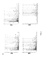
卵巣癌に対する一つの懸念は、腫瘍細胞が進行する環境が高度に炎症誘発性であり、その結果、浸潤性NK細胞に対してCD16a切断を促進し、これにより抗体依存性細胞毒性が減弱される傾向にあるということである。いくつかの抗体がヒト悪性腫瘍を処置するための有効な標的治療法として現れた。それらの有効性は一つには、ナチュラルキラー(NK)細胞のFcγRIIIa/CD16aとの抗体相互作用及び抗体依存性細胞毒性による癌細胞殺滅の誘導による。ヒトIgGのFc受容体CD16(FcγRIII)は2つのアイソフォーム:CD16a(FcγRIIIa)及びCD16b(FcγRIIIb)から成る。CD16aはナチュラルキラー(NK)細胞によって発現され、及びCD16bは好中球によって発現される。NK細胞活性化は、エクトドメイン・シェディング−メタロプロテアーゼADAM17が関与し、原形質膜に近い単一の細胞外領域で起こるタンパク分解性事象、と呼ばれるプロセスによるCD16の両アイソフォームの表面レベルの急速な下方調節をもたらす(図1A)。 One concern for ovarian cancer is that the environment in which the tumor cells progress is highly proinflammatory, thus promoting CD16a cleavage on infiltrating NK cells, which attenuates antibody-dependent cytotoxicity. It is a trend. Several antibodies have emerged as effective targeted therapies for treating human malignancies. Their effectiveness is due in part to antibody interaction of natural killer (NK) cells with FcγRIIIa / CD16a and induction of cancer cell killing by antibody-dependent cytotoxicity. The human IgG Fc receptor CD16 (FcγRIII) consists of two isoforms: CD16a (FcγRIIIa) and CD16b (FcγRIIIb). CD16a is expressed by natural killer (NK) cells and CD16b is expressed by neutrophils. NK cell activation involves the ectodomain shedding-metalloprotease ADAM17 at the surface level of both isoforms of CD16 by a process called a proteolytic event that occurs in a single extracellular region close to the plasma membrane. This results in rapid down regulation (FIG. 1A).
先に述べたように、卵巣癌患者はNK細胞媒介型免疫療法に対して抵抗性である−すなわち、腫瘍がNK細胞媒介型治療法に感受性でない、可能性がある。例えば、卵巣癌細胞は上皮成長因子受容体HER2を一般的に発現するが、しかし、治療用抗体トラスツズマブを用いたその標的化は限定された臨床反応しか提供しなかった。この抵抗性は、少なくとも一部には、エクトドメイン・シェディングの結果として生じ得る−すなわち、サイトカインによるNK細胞活性化、標的細胞相互作用、及び/又は腫瘍浸潤はCD16a切断及び欠陥のある抗体依存性細胞毒性をもたらす可能性がある。よって、エクトドメイン・シェディングのプロセスを妨げることは臨床的に意義がある。 As previously mentioned, ovarian cancer patients are resistant to NK cell mediated immunotherapy-that is, the tumor may not be sensitive to NK cell mediated therapy. For example, ovarian cancer cells generally express the epidermal growth factor receptor HER2, but its targeting with the therapeutic antibody trastuzumab provided only a limited clinical response. This resistance can occur, at least in part, as a result of ectodomain shedding--ie, NK cell activation by cytokines, target cell interactions, and / or tumor invasion is dependent on CD16a cleavage and defective antibody May cause sexual cytotoxicity. Thus, interfering with the ectodomain shedding process is clinically significant.
我々は、質量分析法を使用してCD16a及びCD16bの切断部位を決定し、そしてヒト白血球からCD16a及びCD16bのcDNAをクローニングした。各cDNAを定方向様式で変異させて、単一アミノ酸の変化を誘導した。197位のセリンがプロリンに変更された(図1B)。この突然変異はCD16a及びCD16bの切断を妨げ、細胞活性化によるそれらの下方調節を予防する。エクスビボにおける切断抵抗性CD16aの発現は、このIgG Fc受容体の高い表面レベルを維持するNK細胞を増加させ、そしてそれがNK細胞刺激、治療用抗体の有効性、及び癌細胞殺滅を促進した。
ADAM17は多くの細胞表面基質を有するが、CD16a切断の部位を予測するのに使用できるタンパク質分解のためのコンセンサス配列がない。そのため、我々はLCMS−MSを使用して、活性化ヒト末梢血白血球から放出された可溶性CD16内のC末端切断部位を決定した。
We used mass spectrometry to determine the cleavage site of CD16a and CD16b and cloned CD16a and CD16b cDNA from human leukocytes. Each cDNA was mutated in a directed manner to induce single amino acid changes. The serine at position 197 was changed to proline (FIG. 1B). This mutation prevents the cleavage of CD16a and CD16b and prevents their downregulation by cell activation. Ex vivo cleavage resistant CD16a expression increased NK cells that maintain high surface levels of this IgG Fc receptor, which promoted NK cell stimulation, therapeutic antibody efficacy, and cancer cell killing .
ADAM17 has many cell surface substrates, but there is no consensus sequence for proteolysis that can be used to predict the site of CD16a cleavage. Therefore, we used LCMS-MS to determine the C-terminal cleavage site in soluble CD16 released from activated human peripheral blood leukocytes.
我々はCD16の膜近位領域内に極めて近接した3つの推定切断位置を観察し(図2、くさび形)、1つの領域はCD16a及びCD16bの間で同一であった。ADAM17のタンパク質分解はコンセンサス配列を必要としないが、切断領域の二次構造は重要である。CD16a切断を妨げる試みでは、我々はセリン−197をプロリンで置換して(CD16a197P)、立体構造の変化を導入した。我々は、活性化されたNK細胞の培地上清から及び、別個に、好中球の培地上清からCD16を免疫沈降させることによってCD16切断の位置を同定した。免疫沈降させたCD16をPNGaseFで処理し、N−グリカンを取り除き、トリプシン消化し、そして生じたペプチドを質量分析にかけた。非トリプシン性C末端を含む高信頼度の4つの異なったペプチドパターンを同定した(図1A)。 We observed three putative cleavage positions in close proximity within the membrane proximal region of CD16 (FIG. 2, wedge shape), one region being identical between CD16a and CD16b. Proteolysis of ADAM17 does not require a consensus sequence, but the secondary structure of the cleavage region is important. In an attempt to prevent CD16a cleavage, we replaced serine-197 with proline (CD16a 197P ) to introduce conformational changes. We have identified the location of CD16 cleavage by immunoprecipitation of CD16 from activated NK cell culture supernatant and separately from neutrophil culture supernatant. Immunoprecipitated CD16 was treated with PNGaseF, N-glycans were removed, trypsin digested, and the resulting peptide was subjected to mass spectrometry. Four reliable peptide patterns were identified that contained a non-trypsinic C-terminus (FIG. 1A).
活性化されたNK細胞の培地上清から濃縮されたCD16に関して、我々は1つのペプチドパターンだけ観察し、それは配列番号1のグリシン−174からアラニン−195までのアミノ酸に相当する(ペプチド番号1、図1A)。CD16a及びCD16bの膜近位領域は第176残基を除いて同一のアミノ酸配列を有する。この位置のフェニルアラニンはCD16aを示唆し、それはペプチド番号1に存在した(図1A及びB)。このペプチドはアラニン−195/バリン−196における非トリプシン性P1/P1’切断位置を明らかにした(図1B)。 For CD16 enriched from activated NK cell culture supernatant, we observed only one peptide pattern, which corresponds to amino acids from glycine-174 to alanine-195 of SEQ ID NO: 1 (peptide no. 1, FIG. 1A). The membrane proximal regions of CD16a and CD16b have the same amino acid sequence except for residue 176. Phenylalanine at this position suggested CD16a, which was present in peptide number 1 (FIGS. 1A and B). This peptide revealed a non-tryptic P1 / P1 'cleavage site at alanine-195 / valine-196 (FIG. 1B).
活性化された好中球の培地上清から濃縮されたCD16に関して、我々は非トリプシン性C末端を有する3つの異なったペプチドパターンを検出した(ペプチド番号2〜4、図1A及び1B)。ペプチド番号2は配列番号2のグリシン−174からアラニン−195までのアミノ酸に相当し、ペプチド番号3は配列番号2のグリシン−174からバリン−196までのアミノ酸に相当し、及びペプチド番号4は配列番号2のアスパラギン−180からトレオニン−198までのアミノ酸に相当する。 For CD16 enriched from activated neutrophil medium supernatant, we detected three different peptide patterns with non-trypsinic C-terminus (peptide numbers 2-4, FIGS. 1A and 1B). Peptide No. 2 corresponds to amino acids from glycine-174 to alanine-195 of SEQ ID NO: 2, Peptide No. 3 corresponds to amino acids from glycine-174 to Valine-196 of SEQ ID NO: 2, and Peptide No. 4 is a sequence It corresponds to the number 2 amino acids from asparagine-180 to threonine-198.
ペプチド番号2及びペプチド番号3は、CD16bを示唆する、176位にバリンを含み、アラニン−195/バリン−196における及びバリン−196/セリン−197におけるP1/P1’位置が明らかになった(図1B)。ペプチド番号4はトレオニン−198/イソロイシン−199にP1/P1’位置を有していた(図1B)。このペプチドは濃縮された好中球からの可溶性CD16に由来するが、アイソフォームを同定するための176位のアミノ酸を含んでいない(図1B)。それとは関係なく、高信頼度ペプチドはCD16の第三の切断部位を明らかにした。総合すれば、これらの知見は、単一特異的切断部位よりむしろCD16の切断領域の存在を実証する。 Peptide number 2 and peptide number 3 contain valine at position 176, suggesting CD16b, revealing the P1 / P1 ′ positions in alanine-195 / valine-196 and valine-196 / serine-197 (FIG. 1B). Peptide number 4 had a P1 / P1 'position at threonine-198 / isoleucine-199 (FIG. 1B). This peptide is derived from soluble CD16 from enriched neutrophils but does not contain the amino acid at position 176 to identify the isoform (FIG. 1B). Regardless, the high confidence peptide revealed a third cleavage site for CD16. Taken together, these findings demonstrate the presence of a cleavage region of CD16 rather than a single specific cleavage site.
我々は、部位特異的突然変異誘発を使用することによってCD16の切断領域を更に調査して、CD16a及びCD16b切断が細胞ベースのアッセイにおいて妨害され得るか決定した。ADAM17は、その触媒部位と相互作用する基質領域におけるα−ヘリカル構造を選択する傾向がある。そのうえ、ADAM17切断部位特異性に関するプロテオミクス試験は、P1’、P2’、又はP3’位におけるプロリン残基の非常に低い優先度を明らかにした。そのため、我々は、CD16a及びCD16bの切断領域内のセリン−197をプロリンで置換した(S197P、図2に示したとおり)。 We further investigated the cleavage region of CD16 by using site-directed mutagenesis to determine if CD16a and CD16b cleavage could be disrupted in cell-based assays. ADAM17 tends to select an α-helical structure in the substrate region that interacts with its catalytic site. Moreover, proteomic tests for ADAM17 cleavage site specificity revealed a very low priority of proline residues at the P1 ', P2', or P3 'positions. Therefore, we replaced serine-197 in the cleavage region of CD16a and CD16b with proline (S197P, as shown in FIG. 2).
CD16b及びCD16b/S197Pを別々にヒト腎臓細胞株HEK293で発現させ、そして該HEK293は内因性CD16を発現していない。HEK293形質移入体は、その表面上において同じレベルでCD16b又はCD16b/S197Pを発現した(図3A)。高レベルのCD16bが形質移入HEK293から放出され、そしてそれは、ELISAによって測定されたように、PMAでのそれらの処理によって更に増強された(図3A)。しかしながら、未処理又はPMA処理されたHEK293細胞によって産生されたCD16b/S197Pの可溶性レベルはCD16bのものより顕著に低かった(図3A)。 CD16b and CD16b / S197P are separately expressed in the human kidney cell line HEK293 and the HEK293 does not express endogenous CD16. HEK293 transfectants expressed CD16b or CD16b / S197P at the same level on their surface (FIG. 3A). High levels of CD16b were released from transfected HEK293 and it was further enhanced by their treatment with PMA as measured by ELISA (FIG. 3A). However, the solubility level of CD16b / S197P produced by untreated or PMA-treated HEK293 cells was significantly lower than that of CD16b (FIG. 3A).
我々はまた、同じアプローチを使用してCD16a切断に対するS197P突然変異の効果も調べた。CD16aの表面発現にはγ鎖二量体との結合を必要とする。そのため、我々はヒトγ鎖を安定して発現するHEK293細胞を使用した。同等な表面レベルのCD16a又はCD16a/S197Pを発現するHEK293形質移入体と比較して(図3B)、我々は未処理及びPMA処理した細胞の培地上清中のそれぞれの受容体の可溶性レベルを測定した。CD16aと比較して、再び、可溶性CD16a/S197Pの有意に低いレベルを観察した(図3B)。 We also investigated the effect of the S197P mutation on CD16a cleavage using the same approach. Surface expression of CD16a requires binding to a γ chain dimer. Therefore, we used HEK293 cells that stably express human γ chain. Compared to HEK293 transfectants expressing comparable surface levels of CD16a or CD16a / S197P (FIG. 3B), we measure the soluble level of each receptor in the culture supernatant of untreated and PMA-treated cells. did. Again, significantly lower levels of soluble CD16a / S197P were observed compared to CD16a (FIG. 3B).
CD16における遺伝子操作S197P突然変異がADAM17活性を妨げ得るかどうか評価するために、我々はまた、CD16b/S197Pを発現するか又はそれを欠いているHEK293細胞に、白血球で通常発現される十分に記載されたADAM17基質であるL−セレクチンを用いて形質移入した。両方の形質移入体とも同等なレベルのL−セレクチンを発現し、そしてそれが、PMAを用いたそれらの活性化を受けて同様に下方調節され(図3C)、S197P突然変異がADAM17活性ではなく、CD16シェディングに影響したことを実証した。 To assess whether genetically engineered S197P mutations in CD16 can interfere with ADAM17 activity, we also fully describe HEK293 cells that express or lack CD16b / S197P and are normally expressed in leukocytes. The resulting ADAM17 substrate, L-selectin, was used for transfection. Both transfectants expressed comparable levels of L-selectin and it was similarly down-regulated following their activation with PMA (FIG. 3C), and the S197P mutation was not ADAM17 activity It was demonstrated that CD16 shedding was affected.
NK細胞におけるCD16aシェディングに対するS197P突然変異の効果を評価するために、我々はヒトNK細胞株NK92(Gong et al, 1994, Leukemia 8:652-658)を使用した。これらの細胞は内因性CD16aの発現を欠いているが、遺伝子組み換えCD16aは安定して発現され得る。我々は、CD16a及びCD16a/S197Pを別々に発現するようにNK92細胞に形質導入した。これらの受容体を同等なレベルで発現する細胞を、PMAで活性化し、そして細胞表面CD16レベルをフローサイトメトリーによって調べた。CD16a/S197Pではなく、CD16aが細胞表面発現に顕著な下方調節を受けた(図4A)。IL−12及びIL−18は、個別に又は組み合わせでCD16aシェディングを引き起こし得るNK細胞の生理的刺激である。IL−12及びIL−18で処理されたNK92細胞は、CD16aの細胞表面発現を大きく下方調節するが、CD16a/S197Pではそうではないことを実証した(図4B)。CD16aによる細胞結合IgGの直接的な関与もまたシェディングを引き起こす可能性があり、我々は、CD16a又はCD16a/S197Pを発現するNK92細胞を、抗CD20 mAbであるリツキシマブの存在又は不存在下、CD20陽性バーキットリンパ腫細胞株Rajiと共にインキュベートすることによってここでそれを調べた。リツキシマブで処理したRaji細胞は、CD16aの下方調整を誘発したが、CD16a/S197Pではそれがなかった(図4C)。 To evaluate the effect of the S197P mutation on CD16a shedding in NK cells, we used the human NK cell line NK92 (Gong et al, 1994, Leukemia 8: 652-658). Although these cells lack expression of endogenous CD16a, recombinant CD16a can be stably expressed. We transduced NK92 cells to express CD16a and CD16a / S197P separately. Cells expressing equivalent levels of these receptors were activated with PMA and cell surface CD16 levels were examined by flow cytometry. CD16a, but not CD16a / S197P, was significantly down-regulated in cell surface expression (FIG. 4A). IL-12 and IL-18 are NK cell physiological stimuli that can cause CD16a shedding individually or in combination. NK92 cells treated with IL-12 and IL-18 significantly down-regulated cell surface expression of CD16a, but demonstrated that CD16a / S197P did not (FIG. 4B). Direct involvement of cell-bound IgG by CD16a can also cause shedding, and we tested NK92 cells expressing CD16a or CD16a / S197P in the presence or absence of the anti-CD20 mAb rituximab, CD20 It was examined here by incubating with a positive Burkitt lymphoma cell line Raji. Raji cells treated with rituximab induced downregulation of CD16a but not with CD16a / S197P (FIG. 4C).
BMS566394は、他のメタロプロテアーゼよりADAM17に対して桁違いに高度に選択的なADAM17阻害剤である。BMS566394は、S197P突然変異と同様の効率でCD16aシェディングを妨げたが、CD16a/S197Pを発現する活性化されたNK92細胞に対して追加の遮断効果はなかった(図4D)。これらの知見は、ADAM17がその切断領域内でCD16aを切断する主なシェダーゼ(sheddase)であるさらなる証明を提供する。しかしながら、ADAM17発現レベルがCD16a又はCD16a/S197Pを発現するNK92細胞と同等でないので、それらの異なったシェディングの原因になった可能性はある。そのため、我々は、CD16a又はCD16a/S197Pを発現するNK92細胞を複数の抗ADAM17mAbsで染色し、そして同一の細胞表面レベルを観察した(図4E)。 BMS567394 is an ADAM17 inhibitor that is orders of magnitude more highly selective for ADAM17 than other metalloproteases. BMS567394 prevented CD16a shedding with similar efficiency as the S197P mutation, but had no additional blocking effect on activated NK92 cells expressing CD16a / S197P (FIG. 4D). These findings provide further evidence that ADAM17 is the major sheddase that cleaves CD16a within its cleavage region. However, it is possible that ADAM17 expression levels were not comparable to NK92 cells expressing CD16a or CD16a / S197P, thus causing their different shedding. Therefore, we stained NK92 cells expressing CD16a or CD16a / S197P with multiple anti-ADAM17 mAbs and observed the same cell surface level (FIG. 4E).
初代NK細胞によってCD16aシェディングに対するS197P突然変異の効果を確立するために、我々は、ヒトiPSCsを使用して、遺伝子操作したNK細胞を作出した。我々は以前に、iPSCsからの機能的NK細胞の誘導、及び末梢血NK細胞とそれらの類似性について報告した(Knorr et al., 2013 Stem Cells Transl Med. 2:274-283; Ni et al, 2014, Stem Cells 32: 1021-1031)。iPSC細胞における遺伝子挿入と安定発現のためにCD16a及びCD16a/S197P cDNAをSleeping Beautyトランスポゾンプラスミド内にクローニングし、続いて、それを成熟NK細胞に分化させた。 To establish the effect of the S197P mutation on CD16a shedding by primary NK cells, we created genetically engineered NK cells using human iPSCs. We have previously reported the induction of functional NK cells from iPSCs and their similarities with peripheral blood NK cells (Knorr et al., 2013 Stem Cells Transl Med. 2: 274-283; Ni et al, 2014, Stem Cells 32: 1021-1031). CD16a and CD16a / S197P cDNA was cloned into the Sleeping Beauty transposon plasmid for gene insertion and stable expression in iPSC cells, which were subsequently differentiated into mature NK cells.
モック形質導入iPSC細胞から得られたNK細胞は、低レベルの内因性CD16aを発現する一方で、形質導入したCD16a及びCD16a/S197Pはより高いレベルで発現された(図4F)。NK細胞の活性化は、BY55/CD160を含めたK562細胞とそれらの相互作用によって様々な受容体を通して起こり、ADAM17活性化及びCD16aシェディングをもたらした。我々は、K562細胞でiPSC誘導NK細胞を刺激して、CD16aが細胞表面発現の顕著な下方調整を受けた一方で、CD16a/S197Pの発現は安定した状態を保ったことを見出した(図4F)。 NK cells obtained from mock-transduced iPSC cells expressed low levels of endogenous CD16a, while transduced CD16a and CD16a / S197P were expressed at higher levels (FIG. 4F). Activation of NK cells occurred through various receptors by their interaction with K562 cells including BY55 / CD160, resulting in ADAM17 activation and CD16a shedding. We stimulated iPSC-induced NK cells with K562 cells and found that CD16a received a significant down-regulation of cell surface expression while CD16a / S197P expression remained stable (FIG. 4F). ).
内因性及び遺伝子組み換えCD16aは、単量体IgGを結合するのに十分な親和性を有する。CD16a機能に対するS197P突然変異の効果を調べるために、我々は、CD16aとCD16a/S197PのIgG結合能を比較した。同等なレベルでCD16a又はCD16a/S197Pを発現するNK92細胞は、同様の用量依存的様式でIgGを結合した(図5A)。対照は、CD16a又はCD16a/S197Pを発現するNK92細胞へのIgA結合、及びNK92親細胞へのIgG結合から成った。両方とも実質的にバックグラウンドレベルで起こった(図5A)。これらの知見は、CD16a及びCD16a/S197Pによる特異的で同等なIgG結合を実証する。 Endogenous and recombinant CD16a has sufficient affinity to bind monomeric IgG. To examine the effect of the S197P mutation on CD16a function, we compared the IgG binding capacity of CD16a and CD16a / S197P. NK92 cells expressing CD16a or CD16a / S197P at comparable levels bound IgG in a similar dose-dependent manner (FIG. 5A). Controls consisted of IgA binding to NK92 cells expressing CD16a or CD16a / S197P and IgG binding to NK92 parental cells. Both happened at substantially background levels (Figure 5A). These findings demonstrate specific and comparable IgG binding by CD16a and CD16a / S197P.
CD16aはNK細胞における強力な活性化受容体であるので、我々は、抗体で処理された腫瘍細胞の関与によって細胞活性化を引き起こすCD16aの能力に遺伝子操作S197P突然変異が影響を及ぼすか調べた。NK92細胞の活性化は、脱顆粒によって非常に急速に生じ、且つ、NK細胞活性化に関する高感度指標であるCD107aの上方調節を計測することによって評価された。リツキシマブで又はそれなしに処理にされたRaji細胞と共にインキュベートしたモック形質導入NK92細胞では、CD107aの低レベル且つ同様の上方調節が実証された(図5B)。Raji細胞単体と共にインキュベートした、同等なレベルでCD16a又はCD16a/S197Pを発現するNK92細胞もまたCD107aを限界的に上方調節したが、リツキシマブで処理したRaji細胞を伴ったそれらのインキュベーションもCD107aの大きな上方調節をもたらした(図5B)。総合すれば、先の知見は、CD16aにおける遺伝子操作S197P突然変異がその機能を損なうことはなかったことを示している。 Since CD16a is a potent activating receptor in NK cells, we investigated whether the engineered S197P mutation affects CD16a's ability to cause cell activation through the involvement of antibody-treated tumor cells. Activation of NK92 cells occurred very rapidly by degranulation and was assessed by measuring upregulation of CD107a, a sensitive indicator for NK cell activation. Mock-transduced NK92 cells incubated with Raji cells treated with or without rituximab demonstrated low levels of CD107a and similar upregulation (FIG. 5B). NK92 cells expressing CD16a or CD16a / S197P at comparable levels, incubated with Raji cells alone, also marginally upregulated CD107a, but their incubation with Raji cells treated with rituximab also greatly increased CD107a Adjustment was effected (FIG. 5B). Taken together, previous findings indicate that the genetically engineered S197P mutation in CD16a did not impair its function.
よって、我々は、CD16a及びCD16bにおける遺伝子操作S197P突然変異が天然ADAM17を伴った細胞ベースのアッセイにおいて効果的にそれらのシェディングを妨げたことを示す。CD16aにおけるS197P突然変異はまた、ヒトNK細胞株であるNK92において受容体のシェディングを妨げたが、受容体機能は損なわなかった。同等なレベルでCD16a又はCD16a/S197Pを発現するNK92細胞は、さまざまな抗体濃度範囲にわたって同様の効率で単量体IgGを結合した。加えて、CD16a又はCD16a/S197Pを発現するNK92細胞は、Raji細胞に結合したリツキシマブの関与によって類似した様式で活性化マーカーCD107aを上方調節した。 Thus, we show that genetically engineered S197P mutations in CD16a and CD16b effectively prevented their shedding in cell-based assays with native ADAM17. The S197P mutation in CD16a also prevented receptor shedding in NK92, a human NK cell line, but did not impair receptor function. NK92 cells expressing CD16a or CD16a / S197P at comparable levels bound monomeric IgG with similar efficiency over various antibody concentration ranges. In addition, NK92 cells expressing CD16a or CD16a / S197P up-regulated the activation marker CD107a in a manner similar to the involvement of rituximab bound to Raji cells.
多分化能性幹細胞は、遺伝子操作NK細胞を作出するための遺伝操作を可能にする。この開示では、野生型CD16a又はCD16a/S197Pを発現する形質導入iPSCsからの遺伝子操作NK細胞の作出を説明する。NK92細胞を用いた場合、CD16aはiPSCs誘導NK細胞においてシェディングを受け、細胞活性化時の正常なADAMが17活性を実証した一方で、CD16a/S197Pはシェディングを受けなかった。 Pluripotent stem cells allow genetic manipulation to create genetically engineered NK cells. This disclosure describes the generation of genetically engineered NK cells from transduced iPSCs that express wild type CD16a or CD16a / S197P. When NK92 cells were used, CD16a was sheshed in iPSCs-induced NK cells, and normal ADAM upon cell activation demonstrated 17 activity, while CD16a / S197P did not undergo shedding.
CD16a及びNK細胞の細胞傷害機能は癌患者において顕著な下方調節を受ける。CD16a/S197PをコードするcDNAsは、安定したヒト誘発多分化能性幹細胞(iPSCs)及び胚幹細胞(ESCs)を作出するのに使用できる。次に、これらの幹細胞はCD16a/S197Pを発現する初代NK細胞に分化させることができる。切断抵抗性CD16a/S197P(例えば、単球)又はCD16b/S197P(例えば、好中球)を発現する他の細胞集団もまた、hESCs/iPSCsから得られる。 The cytotoxic function of CD16a and NK cells is significantly down-regulated in cancer patients. CDNAs encoding CD16a / S197P can be used to generate stable human induced pluripotent stem cells (iPSCs) and embryonic stem cells (ESCs). These stem cells can then be differentiated into primary NK cells that express CD16a / S197P. Other cell populations that express cleavage resistant CD16a / S197P (eg, monocytes) or CD16b / S197P (eg, neutrophils) are also obtained from hESCs / iPSCs.
様々な形態の癌又は感染に対してヒト患者で使用されるNK細胞免疫療法を作り出すために、CD16a/S197P発現NK細胞は、増強された抗体依存性細胞毒性(ADCC)活性又は他のCD16a媒介型活性(例えば、IFNγ及びTNFα産生)を媒介し得る。例えば、CD16a/S197P発現NK細胞は、治療用抗体(例えば、トラスツズマブ又はリツキシマブ)、二重特異性キラーエンゲージャー(例えば、BiKE、CD16×CD33、CD16×CD19、又はCD16×EP−CAM二重特異性キラー細胞エンゲージャー)又は三重特異性キラー細胞エンゲージャー(TriKE)と組み合わせられてもよい。他の治療用細胞集団(例えば、好中球、単球、T細胞など)もまた、増強されたCD16媒介型活性を生じ得る。 To create NK cell immunotherapy used in human patients against various forms of cancer or infection, CD16a / S197P expressing NK cells have enhanced antibody dependent cellular cytotoxicity (ADCC) activity or other CD16a mediated Can mediate type activity (eg, IFNγ and TNFα production). For example, CD16a / S197P expressing NK cells can be treated with therapeutic antibodies (eg, trastuzumab or rituximab), bispecific killer engagers (eg, BiKE, CD16 × CD33, CD16 × CD19, or CD16 × EP-CAM bispecific). Killer cell engager) or trispecific killer cell engager (TriKE). Other therapeutic cell populations (eg, neutrophils, monocytes, T cells, etc.) can also produce enhanced CD16-mediated activity.
ヒトiPSCs又はヒトESCsにおけるCD16a/S197Pの発現は、例えばHER2卵巣癌などの腫瘍性病態に対して促進されたADCC活性を有するNK細胞集団を作出し得る。場合によっては、腫瘍性病態は、例えばトラスツズマブなどの治療用抗体で処置されてもよい。成熟NK細胞は、ヒト胚性幹細胞及びiPSCsに由来していてもよい。 Expression of CD16a / S197P in human iPSCs or human ESCs can generate NK cell populations with enhanced ADCC activity against neoplastic conditions such as HER2 ovarian cancer. In some cases, the neoplastic condition may be treated with a therapeutic antibody such as, for example, trastuzumab. Mature NK cells may be derived from human embryonic stem cells and iPSCs.
野生型CD16a及び/又はCD16a/S197Pは、個々のCD16a受容体を発現する安定したiPSC株又は安定したECS株を作出すためにクローニングされ得る。任意の好適なクローニング法が使用され得る。代表的なクローニング法としては、例えばウイルスベースの方法、トランスポゾンベクター(例えばSleeping Beauty)、又はヌクレオフェクション(nucleofection)が挙げられる。 Wild type CD16a and / or CD16a / S197P can be cloned to create stable iPSC strains or stable ECS strains that express individual CD16a receptors. Any suitable cloning method can be used. Representative cloning methods include, for example, virus-based methods, transposon vectors (eg, Sleeping Beauty), or nucleofection.
一例として、iPSCsは、Sleeping Beautyトランスポゾンベクターを使用して改変され得る。ベクターは、例えばGFP/ゼオシン抵抗性融合タンパク質などの選択系を含んでおり、そしてそれが二元的な選択系(ゼオシン抵抗性及びフローサイトメトリーセルソーティング)を可能にする。iPSCsは以前に記載されているように成熟NK細胞に分化させる(Ni et al, 2011, J. Virol. 85:43-50; Knorr et al. 2013, Stem Cells Transl Med 2:274-283; Woll et al, 2009, Blood 113:6094-6101)。iPSCsにおけるトランスジェニック受容体の発現は、誘導NK細胞における高い発現レベルにつながり得る。未分化iPSCsにおけるCD16発現は、NK細胞分化を中断し得る。こうした場合、CD16発現は、CD16発現が通常のNK細胞分化と同時に起こるほうが好都合なように、例えばCD56又は天然のCD16aプロモーターを使用して遅らせてもよい。 As an example, iPSCs can be modified using Sleeping Beauty transposon vectors. The vector contains a selection system such as, for example, a GFP / zeocin resistance fusion protein, which allows a dual selection system (zeocin resistance and flow cytometry cell sorting). iPSCs are differentiated into mature NK cells as previously described (Ni et al, 2011, J. Virol. 85: 43-50; Knorr et al. 2013, Stem Cells Transl Med 2: 274-283; Woll et al, 2009, Blood 113: 6094-6101). Expression of transgenic receptors in iPSCs can lead to high expression levels in induced NK cells. CD16 expression in undifferentiated iPSCs can disrupt NK cell differentiation. In such cases, CD16 expression may be delayed using, for example, the CD56 or native CD16a promoter, such that it is more convenient for CD16 expression to coincide with normal NK cell differentiation.
当業者は、野生型CD16aと対比してCD16a/S197Pを同等なレベルで発現するNK細胞を比較する。CD16構築物の発現レベルは、CD16構築物と比例した様式で現れるGFP発現に基づくFACSソーティングで対等にできる。対等にしたCD16aレベルはFACSによって確認できる。HER2発現卵巣癌細胞に対するNK細胞毒性は、例えばトラスツズマブなどの治療用抗体の存在又は不存在下での標準的なクロム遊離試験で評価できる。非クロム標識された卵巣癌細胞を用いた抗体依存性細胞毒性が評価され得る。当業者は、ELISAによってNK細胞のサイトカイン産生(例えば、IFNγ、TNF)及びCD16aの可溶性レベルを評価でき、及びFACSによってCD16a及び他の活性化マーカー(例えば、CD107a、CD62L)の細胞表面レベルを評価できる。 One skilled in the art compares NK cells that express CD16a / S197P at comparable levels versus wild-type CD16a. Expression levels of CD16 constructs can be equalized by FACS sorting based on GFP expression that appears in a manner proportional to the CD16 construct. The equalized CD16a level can be confirmed by FACS. NK cytotoxicity against HER2-expressing ovarian cancer cells can be assessed by standard chromium release tests in the presence or absence of therapeutic antibodies such as trastuzumab. Antibody-dependent cytotoxicity using non-chromium labeled ovarian cancer cells can be assessed. One skilled in the art can assess NK cell cytokine production (eg, IFNγ, TNF) and CD16a solubility levels by ELISA, and assess cell surface levels of CD16a and other activation markers (eg, CD107a, CD62L) by FACS. it can.
実施例3に記載のヒト腫瘍異種移植モデルは、インビボにおいて切断不可能なCD16aを発現するNK細胞の抗癌活性を評価するのに使用できる。ヒトCD16と異なって、マウスCD16は細胞刺激によってエクトドメイン・シェディングを受けないので、そのため、NK細胞媒介ADCCに対するCD16aシェディングの効果を測定することは、正常なマウスではモデル化できない。表1では、実験の群分けと処置の代表的な組み合わせを提供する。 The human tumor xenograft model described in Example 3 can be used to evaluate the anticancer activity of NK cells expressing CD16a that cannot be cleaved in vivo. Unlike human CD16, mouse CD16 does not undergo ectodomain shedding upon cell stimulation, so measuring the effect of CD16a shedding on NK cell-mediated ADCC cannot be modeled in normal mice. Table 1 provides representative combinations of experimental groupings and treatments.

腫瘍の増殖及び/又は退縮は、例えば生物発光造影、超音波、CT、MRI、別の画像技術、及び/又はマウスの計量を含めた従来の方法によって毎週観察され得る(Woll et al, 2009, Blood 113:6094-6101)。マウスもまた、ヒトNK細胞生存率を定量化するために(例えば毎週)採血され得る。様々なエフェクター機能マーカー(例えば、IFNγ、CD16a)の発現及び/又は細胞表面レベルは、例えばFACSによるなどの従来の手法を使用して評価され得る。マウスは、例えば60日間などの任意の好適な期間追跡検査され得る。屠殺時点で、内臓(例えば脾臓、肝臓、肺、腎臓、及び/又は卵巣)が、以前に記載されているように(Woll et al, 2009, Blood 113:6094-6101)、(例えば生物発光によって)転移癌の徴候がないか調査され得る。 Tumor growth and / or regression may be observed weekly by conventional methods including, for example, bioluminescence imaging, ultrasound, CT, MRI, other imaging techniques, and / or mouse metric (Woll et al, 2009, Blood 113: 6094-6101). Mice can also be bled (eg weekly) to quantify human NK cell viability. The expression and / or cell surface levels of various effector function markers (eg, IFNγ, CD16a) can be assessed using conventional techniques such as by FACS. The mice can be followed for any suitable period, such as 60 days. At the time of sacrifice, the viscera (eg, spleen, liver, lung, kidney, and / or ovary) are as previously described (Woll et al, 2009, Blood 113: 6094-6101) (eg, by bioluminescence). ) Can be investigated for signs of metastatic cancer.
我々の分析は、当業者が野生型CD16aと対比してCD16a/S197Pを発現するiPSC誘導NK細胞の抗体依存性細胞毒性活性及びインビボにおける効力を規定及び比較することを可能にする。よって、我々は、本明細書中にCD16aの改変型、改変CD16aを発現する遺伝子組み換え細胞、及び遺伝子組み換え細胞(例えばNK細胞、好中球、単球、T細胞など)を伴う方法を記載する。例えば、CD16aの改変型、CD16a/S197Pを発現するNK細胞は、少なくとも一部で、NK細胞刺激時のADAM17媒介型シェディングに対する低い感受性により増強された抗卵巣癌活性を呈する。これは、同様にして、例えば治療用抗体でタグを付与された癌細胞などの抗体でタグを付与された癌細胞の関与によって抗体依存性細胞毒性活性を増強する。そのうえ、NK細胞による抗体認識は、腫瘍細胞との接触安定性を増強し、NKG2Dなどの他の活性化受容体を通してNK細胞活性を強める。 Our analysis allows one skilled in the art to define and compare antibody-dependent cytotoxic activity and in vivo efficacy of iPSC-derived NK cells expressing CD16a / S197P versus wild-type CD16a. Thus, we describe herein a modified form of CD16a, a genetically modified cell that expresses the modified CD16a, and methods involving genetically modified cells (eg, NK cells, neutrophils, monocytes, T cells, etc.). . For example, NK cells expressing a modified version of CD16a, CD16a / S197P, exhibit at least some enhanced anti-ovarian cancer activity due to low susceptibility to ADAM17-mediated shedding upon NK cell stimulation. This similarly enhances antibody-dependent cytotoxic activity due to the involvement of cancer cells tagged with antibodies, such as cancer cells tagged with therapeutic antibodies. Moreover, antibody recognition by NK cells enhances contact stability with tumor cells and enhances NK cell activity through other activated receptors such as NKG2D.
用語「及び/又は」は、列挙した要素のうちの1若しくはすべて、又は列挙した要素のうちの任意の2以上の組み合わせを意味し;用語「含む」とその変化形には、これらの用語が明細書及び請求項に現れた場合に、限定的な意味合いはなく;別段の定めがない限り、「a」、「an」、「the」、及び「少なくとも1つ」は互換的に使用され、1若しくは複数を意味し;そして、終点による数値範囲の詳述は、その範囲内に包括されたすべての数値を含む(例えば1〜5は、1、1.5、2、2.75、3、3.80、4、5などを含む)。
The term “and / or” means one or all of the listed elements, or any combination of two or more of the listed elements; the term “comprising” and variations thereof includes the terms When appearing in the specification and claims, there is no limiting meaning; unless otherwise specified, “a”, “an”, “the”, and “at least one” are used interchangeably; And the recitation of numerical ranges by endpoints includes all numbers subsumed within that range (
先の説明において、特定の実施形態は明確さのために単独で記載され得る。特定の実施形態の特徴が別の実施形態の特徴と両立しないことについて別段の明白な定めがない限り、特定の実施形態は1若しくは複数の実施形態に関して本明細書中に記載した両立し得る特徴の組み合わせを含む。
別個のステップを含んでいる本明細書中に開示されたいずれの方法でも、該ステップは任意の実現可能な順序で実施されてもよい。そして、適宜、2以上のステップの任意の組み合わせが同時に実施されてもよい。
本発明は以下の実施例によって例示される。特定の例、材料、量、及び手順は本明細書中に記載した本発明の範囲及び要旨に従って広く解釈されるべきであることは、理解されるべきである。
In the foregoing description, specific embodiments may be described alone for clarity. Unless otherwise explicitly stated that a feature of a particular embodiment is incompatible with a feature of another embodiment, that particular embodiment is described as being compatible with the features described herein with respect to one or more embodiments. Including a combination of
In any of the methods disclosed herein that include separate steps, the steps may be performed in any feasible order. Then, any combination of two or more steps may be performed at the same time as appropriate.
The invention is illustrated by the following examples. It is to be understood that the specific examples, materials, amounts, and procedures are to be broadly construed in accordance with the scope and spirit of the invention described herein.
実施例1
質量分析法
プロトコール番号9708M00134によりミネソタ大学施設内倫理委員会によって承認されたプロトコールに従って、健常人からの末梢血採血を実施した。ヒト好中球及びNK細胞単離を以前に記載されているように実施した(Wang et al, 2013, Biochim Biophys Acta. 1833:680-685; Long et al, 2010, J Leukoc Biol. 87: 1097-1101; Long et al, 2012, J Leukoc Biol. 92:667-672)。濃縮した好中球又はNK細胞(PBS中に1×10⁷/ml;Mediatech, Inc. Manassas, VA)をPMA(それぞれ15ng/ml又は50ng/ml;Sigma-Aldrich, St. Louis, MO)を用いて37℃にて30分間活性化した。細胞上清を濾過し(0.45μmの細孔径)、そしてCD16を、製造業者の取扱説明書に従ってmAb 3G8(BioLegend, Inc., San Diego, CA)及びPierceダイレクト免疫沈降反応キット(Thermo Fisher Scientific, Rockford, IL)を使用して免疫沈降させた。精製したCD16を、製造業者の取扱説明書に従ってキチン質結合ドメインにタグを付与したRemove-iT PNGase F(New England BioLabs, Inc., Ipswich, MA)によって脱グリコシル化した。簡単に言えば、10〜20μgの精製CD16を40mMのDTTの存在下、55℃にて10分間変性させ、次に、3μlのREMOVE-IT PNGase F(New England BioLabs, Inc., Ipswich, MA)と一緒に37℃にて1時間インキュベートした。次に、反応物からキチン磁性ビーズを使用してREMOVE-IT PNGase Fを取り除いた。
Example 1
Mass Spectrometry Peripheral blood was collected from healthy individuals according to a protocol approved by the University of Minnesota Institutional Review Board according to protocol number 9708M00134. Human neutrophil and NK cell isolation was performed as previously described (Wang et al, 2013, Biochim Biophys Acta. 1833: 680-685; Long et al, 2010, J Leukoc Biol. 87: 1097 -1101; Long et al, 2012, J Leukoc Biol. 92: 667-672). Concentrated neutrophils or NK cells (1 × 10 7 / ml in PBS; Mediatech, Inc. Manassas, VA) and PMA (15 ng / ml or 50 ng / ml, respectively; Sigma-Aldrich, St. Louis, MO) And activated for 30 minutes at 37 ° C. Cell supernatants were filtered (0.45 μm pore size) and CD16 was analyzed according to manufacturer's instructions for mAb 3G8 (BioLegend, Inc., San Diego, Calif.) And Pierce direct immunoprecipitation kit (Thermo Fisher Scientific). , Rockford, IL). Purified CD16 was deglycosylated by Remove-iT PNGase F (New England BioLabs, Inc., Ipswich, Mass.) Tagged to the chitin binding domain according to the manufacturer's instructions. Briefly, 10-20 μg of purified CD16 was denatured for 10 minutes at 55 ° C. in the presence of 40 mM DTT and then 3 μl of REMOVE-IT PNGase F (New England BioLabs, Inc., Ipswich, Mass.) And incubated at 37 ° C. for 1 hour. Next, REMOVE-IT PNGase F was removed from the reaction product using chitin magnetic beads.
CD16をSDS−PAGEにかけ、可溶性CD16に相当するゲルバンドを、クリプトン蛍光性タンパク染料(Thermo Fisher Scientific, Rockford, IL)によって検出し、同じゲルの隣接したレーンのCD16イムノブロット分析によって確認し、次に、それを切り出し、トリプシンを用いた標準的なゲル内消化にかけた。ゲルから抽出した消化ペプチドをドライダウンし、液体クロマトグラフィー質量分析のために水:アセトニトリル:ギ酸、98:2:0.01中で再構成し、<1μgのアリコートを、以前に記載されているように、データ依存性走査モードで質量分析(VELOS ORBITRAP、Thermo Fisher Scientific, Rockford, IL)によって分析した(Lin-Moshier et al, 2013, J Biol Chem. 288:355-367)。データベース検索をProtein Pilot4.5(AB Sciex, Framingham, MA)を用いて実施し、それは、混入物データベース(thegpm.org/cRAP/指数、109のタンパク質)を備えたNCBI参照配列ホモサピエンスタンパク質FASTAデータベースに対してParagonスコアリングアルゴリズム(Shilov et al, 2007, Mol Cell Proteomics 6: 1638-1655)を使用する。検索パラメーターは:システインヨードアセトアミド;トリプシン;装置Orbi MS(1〜3ppm)Orbi MS/MS;生物学的改変IDフォーカス(アスパラギン脱アミド化を含む);検索成果全体;及び(逆進データベースを用いた)偽発見率分析である。 CD16 was subjected to SDS-PAGE and a gel band corresponding to soluble CD16 was detected by krypton fluorescent protein dye (Thermo Fisher Scientific, Rockford, IL) and confirmed by CD16 immunoblot analysis of adjacent lanes of the same gel, then It was excised and subjected to standard in-gel digestion with trypsin. Digested peptides extracted from the gel were dried down and reconstituted in water: acetonitrile: formic acid, 98: 2: 0.01 for liquid chromatography mass spectrometry, and <1 μg aliquots previously described As such, it was analyzed by mass spectrometry (VELOS ORBITRAP, Thermo Fisher Scientific, Rockford, IL) in a data dependent scanning mode (Lin-Moshier et al, 2013, J Biol Chem. 288: 355-367). A database search was performed using Protein Pilot 4.5 (AB Sciex, Framingham, Mass.), Which was a NCBI reference sequence homosapiens protein FASTA database with a contaminant database (theegpm.org/cRAP/index, 109 proteins). Paragon scoring algorithm (Shilov et al, 2007, Mol Cell Proteomics 6: 1638-1655) is used. Search parameters are: cysteine iodoacetamide; trypsin; instrument Orbi MS (1-3 ppm) Orbi MS / MS; biological modification ID focus (including asparagine deamidation); overall search results; and (using reverse database) ) Fake discovery rate analysis.
cDNA発現構築物の作製
CD16bは、その細胞外領域のN末端部分の4つのアミノ酸が異なるNA1及びNA2と呼ばれる2つの対立遺伝子変異体を生じる。CD16bの両方の対立遺伝子変異体が、同様の効率でADAM17によって切断される。この試験について、我々はNA1変異体だけを調べた。176位にバリン又はフェニルアラニン残基のいずれかを有するCD16aの2つの対立遺伝子変異体も存在する。CD16aのこれらの2つの対立遺伝子変異体は同様の効率でADAM17によって切断された。この試験について、我々はバリン対立遺伝子変異体CD16aだけを調べた。
Generation of cDNA Expression Constructs CD16b produces two allelic variants called NA1 and NA2 that differ in the four amino acids of the N-terminal part of their extracellular region. Both allelic variants of CD16b are cleaved by ADAM17 with similar efficiency. For this study we examined only the NA1 mutant. There are also two allelic variants of CD16a with either a valine or phenylalanine residue at position 176. These two allelic variants of CD16a were cleaved by ADAM17 with similar efficiency. For this study we examined only the valine allelic variant CD16a.
CD16a及びCD16bをヒト白血球cDNAから増幅し、別々に以前に記載されているように(Wang et al, 2013, Biochim Biophys Acta. 1833:680-685; Dong et al, 2014, Arthritis Rheumatol. 66: 1291-1299)、BamHI及びEcoRI制限酵素部位においてpcDNA3.1プラスミド(Invitrogen, Carlsbad, CA)内にクローニングした。次に、構築物を、製造業者の取扱説明書に従ってQuik- Change Site-directed Mutagenesis(Agilent Technologies, Santa Clara, CA)にかけて、CD16a及びCD16bの197位のセリンをプロリンに変換した。すべての構築物を配列決定して、計画的な突然変異の存在及びいずれかの自然突然変異の不存在を確認した。 CD16a and CD16b are amplified from human leukocyte cDNA and separately described previously (Wang et al, 2013, Biochim Biophys Acta. 1833: 680-685; Dong et al, 2014, Arthritis Rheumatol. 66: 1291 -1299), cloned into the pcDNA3.1 plasmid (Invitrogen, Carlsbad, Calif.) At the BamHI and EcoRI restriction enzyme sites. The construct was then subjected to Quik-Change Site-directed Mutagenesis (Agilent Technologies, Santa Clara, Calif.) According to the manufacturer's instructions to convert serine at position 197 of CD16a and CD16b to proline. All constructs were sequenced to confirm the presence of the planned mutation and the absence of any spontaneous mutation.
続いて、CD16a cDNAをBamHI及びEcoRI制限酵素部位において、Dr. G. Nolan(Stanford University, Stanford, CA)によって提供された2シストロン性レトロウイルス発現ベクターpBMN−IRES−EGFP内にクローニングした。CD16a構築物はまた、以前に記載されているように(Wilber et al, 2007, Stem Cells 25:2919-2927; Tian et al, 2009, Stem Cells 27:2675-2685)、2シストロン性Sleeping Beautyトランスポゾンプラスミド(pKT2−IRES−GFP:zeo)内にもクローニングした。 Subsequently, the CD16a cDNA was cloned into the bicistronic retroviral expression vector pBMN-IRES-EGFP provided by Dr. G. Nolan (Stanford University, Stanford, Calif.) At the BamHI and EcoRI restriction enzyme sites. The CD16a construct is also a bicistronic Sleeping Beauty transposon plasmid as previously described (Wilber et al, 2007, Stem Cells 25: 2919-2927; Tian et al, 2009, Stem Cells 27: 2675-2685). It was also cloned into (pKT2-IRES-GFP: zeo).
簡単に言えば、野生型CD16a及びCD16a/S197Pを、プライマー:5’−CCG GAA TTC CAG TGT GGC ATC ATG TGG CAG CTG CTC−3’(センス、配列番号XX)及び5’−CCG GAA TTC TCA TTT GTC TTG AGG GTC CTT TCT−3’(アンチセンス、配列番号YY)を使用してPCR増幅した。EcoRI部位に下線を引く。EcoRI消化CD16a及びCD16a/S197P PCR断片を、別々にpKT2−IRES−GFP:zeo内にクローニングした。正しいCD16a方向及び配列をPCR及び配列決定分析によって確認した。我々は、完全長ヒトL−セレクチン(CD62L)cDNAを以前クローニングし(Feehan et al, 1996, J Biol Chem. 271 :7019-7024; Matala et al, 2001, J Immunol. 167: 1617-1623)、そしてそれを、制限酵素部位Xba1においてpcDNA3.1ベクター内に移した)。完全長ヒトFcRγ cDNAを、pcDNA3.1ベクターを使用した修飾を伴って、以前に記載されているようにクローニングした(Dong et al. , 2014, Arthritis Rheumatol. 66: 1291-1299)。 Briefly, wild type CD16a and CD16a / S197P were prepared using primers: 5′-CCG GAA TTC CAG TGT GGC ATC ATG TGG CAG CTG CTC-3 ′ (sense, SEQ ID NO: XX) and 5′-CCG GAA TTC TCA TTT. PCR amplification was performed using GTC TTG AGG GTC CTT TCT-3 ′ (antisense, SEQ ID NO: YY). The EcoRI site is underlined. EcoRI digested CD16a and CD16a / S197P PCR fragments were cloned separately into pKT2-IRES-GFP: zeo. The correct CD16a orientation and sequence was confirmed by PCR and sequencing analysis. We have previously cloned full-length human L-selectin (CD62L) cDNA (Feehan et al, 1996, J Biol Chem. 271: 7019-7024; Matala et al, 2001, J Immunol. 167: 1617-1623) It was then transferred into the pcDNA3.1 vector at the restriction enzyme site Xba1). Full-length human FcRγ cDNA was cloned as previously described (Dong et al., 2014, Arthritis Rheumatol. 66: 1291-1299) with modification using pcDNA3.1 vector.
遺伝子組み換えL−セレクチン、CD16a、及びCD16bを発現する細胞株の作出
HEK293細胞(ヒト胎児腎臓細胞株)及びNK92細胞(ヒトNK細胞株)(ATCC, Manassas, VA)を受託所の取扱説明書に従って培養した。HEK293細胞を、製造業者の取扱説明書に従ってLipofectamine2000(Invitrogen, Carlsbad, CA)を使用して、CD16b、CD16b/S197P、及び/又はL−セレクチンを含む又は含まないpcDNA3.1を用いて一過性に形質移入した。ヒトFcRγを安定して発現するHEK293細胞を、同じアプローチによってCD16a又はCD16a/S197Pを含む又は含まないpcDNA3.1を用いて一過性に形質移入した。NK92細胞を、以前に記載されているレトロウイルス作出及び感染手順(Matala et al, 2001, J Immunol. 167: 1617-1623; Walcheck et al, 2003, JLeukoc Biol. 74:389-394; Wang et al, 2009, J Immunol. 182:2449-2457)によってCD16a又はCD16a/S197Pを含む又は含まないpBMN−IRES−EGFPを用いて安定的に形質導入した。構築物の発現を、EGFP蛍光によって、及びフローサイトメトリーによって測定する場合には、CD16染色によって評価した。ヒトiPSCs(UCBiPS7、臍帯血CD34細胞由来)をマウス胎児線維芽細胞(Knorr et al., 2013, Stem Cells Trans I Med. 2:274-283; Ni et al, 2014, Stem Cells 32: 1021-1031)上で維持した。CD16a又はCD16a/S197Pの安定発現を、以前に記載されているようにSleeping Beautyトランスポゾンシステム(Wilber et al, 2007, Stem Cells 25:2919-2927; Tian et al, 2009, Stem Cells 27:2675-2685)を使用して実現した。簡単に言えば、iPSCsを、プログラム設定B16を使用してヌクレオフェクター溶液V(Lonza Inc., Gaithersburg, MD)中でトランスポザーゼDNAと組み合わせたpKT2−IRES−GFP:zeoを用いてヌクレオフェクトした。ヌクレオフェクト細胞をすぐにゼオシン(50μg/ml)を含んでいるiPSC成長培地中に懸濁し、そしてマウス胎児線維芽細胞上に播種した。
Production of cell lines expressing recombinant L-selectin, CD16a and CD16b HEK293 cells (human embryonic kidney cell line) and NK92 cells (human NK cell line) (ATCC, Manassas, VA) according to the instructions of the trustee Cultured. HEK293 cells are transiently transferred using pcDNA3.1 with or without CD16b, CD16b / S197P, and / or L-selectin using Lipofectamine 2000 (Invitrogen, Carlsbad, CA) according to the manufacturer's instructions. Was transfected. HEK293 cells stably expressing human FcRγ were transiently transfected with pcDNA3.1 with or without CD16a or CD16a / S197P by the same approach. NK92 cells were isolated from previously described retrovirus production and infection procedures (Matala et al, 2001, J Immunol. 167: 1617-1623; Walcheck et al, 2003, JLeukoc Biol. 74: 389-394; Wang et al , 2009, J Immunol. 182: 2449-2457) and stably transduced with pBMN-IRES-EGFP with or without CD16a or CD16a / S197P. Construct expression was assessed by EGFP fluorescence and, if measured by flow cytometry, by CD16 staining. Human iPSCs (UCBiPS7, cord blood CD34 cell-derived) were obtained from mouse fetal fibroblasts (Knorr et al., 2013, Stem Cells Trans I Med. 2: 274-283; Ni et al, 2014, Stem Cells 32: 1021-1031 ) Maintained above. Stable expression of CD16a or CD16a / S197P can be achieved by the Sleeping Beauty transposon system as previously described (Wilber et al, 2007, Stem Cells 25: 2919-2927; Tian et al, 2009, Stem Cells 27: 2675-2685 ). Briefly, iPSCs were nucleofected with pKT2-IRES-GFP: zeo in combination with transposase DNA in Nucleofector Solution V (Lonza Inc., Gaithersburg, MD) using program setting B16. Nucleofect cells were immediately suspended in iPSC growth medium containing zeocin (50 μg / ml) and seeded onto mouse fetal fibroblasts.
CD16a−hESC及びCD16a−iPSC細胞からのNK細胞の誘導
hESCs及びiPSCsの造血細胞分化を以前に記載されているように実施した(Ng et al, 2005, Blood 106: 1601-1603; Ng et al, 2008, Nat Protoc 3:768-776; Le Garff-Tavernier et al, 2010, Aging Cell 9: 527-535)。簡単に言えば、96ウェル丸底プレートの1ウェル当たり3000個の単独細胞を、幹細胞因子(SCF、40ng/ml)、血管内皮増殖因子(VEGF、20ng/ml)、及び骨形態形成タンパク質4(BMP4、20ng/ml)を含んだBPEL培地中に播種した。BPEL培地は、Iscove’s Modified Dulbecco’s Medium(IMDM、86ml、Invitrogen, Thermo Fisher Scientific, Inc., Waltham. MA)、Glutmax Iを含んだF12 Nutrient Mixture(86mL、Invitrogen, Thermo Fisher Scientific, Inc., Waltham. MA)、10%の脱イオンBovine Serum Albumin(BSA、5ml、Sigma-Aldrich, St. Louis, MO)、5%のポリビニルアルコール(10ml、Sigma-Aldrich, St. Louis, MO)、リノレン酸(20μlの1グラム/ml溶液、Sigma-Aldrich, St. Louis, MO)、リノール酸(20μlの1グラム/ml溶液、Sigma)、SYNTHECOL 500×溶液(Sigma-Aldrich, St. Louis, MO)、a−モノチオグリセロール(3.9μl/100ml、Sigma-Aldrich, St. Louis, MO)、タンパク質不含ハイブリドーマミックスII(Invitrogen, Thermo Fisher Scientific, Inc., Waltham. MA)、アスコルビン酸(5mg/ml、Sigma)、GLUTAMAX I(Invitrogen, Thermo Fisher Scientific, Inc., Waltham. MA)、インスリン−トランスフェリン−セレニウム100×溶液(Invitrogen, Thermo Fisher Scientific, Inc., Waltham. MA)、ペニシリン/ストレプトマイシン(Invitrogen, Thermo Fisher Scientific, Inc., Waltham. MA)を含んでいた。
Induction of NK cells from CD16a-hESC and CD16a-iPSC cells Hematopoietic cell differentiation of hESCs and iPSCs was performed as previously described (Ng et al, 2005, Blood 106: 1601-1603; Ng et al, 2008, Nat Protoc 3: 768-776; Le Garff-Tavernier et al, 2010, Aging Cell 9: 527-535). Briefly, 3000 single cells per well of a 96 well round bottom plate were transformed into stem cell factor (SCF, 40 ng / ml), vascular endothelial growth factor (VEGF, 20 ng / ml), and bone morphogenetic protein 4 ( BMP4, 20 ng / ml) was seeded in BPEL medium. BPEL medium was Iscove's Modified Dulbecco's Medium (IMDM, 86 ml, Invitrogen, Thermo Fisher Scientific, Inc., Waltham. MA), F12 Nutrient Mixture with Glutmax I (86 mL, Invitrogen, Thermo Fisher Scientific, Inc., Waltham. MA) ) 10% deionized Bovine Serum Albumin (BSA, 5 ml, Sigma-Aldrich, St. Louis, MO), 5% polyvinyl alcohol (10 ml, Sigma-Aldrich, St. Louis, MO), linolenic acid (20
造血細胞分化の11日目に、サイトカインを供給したNK培地中でEL08−1D2ストロマ細胞と共に又はそれなしに、スピン胚様体を24ウェルプレートにそのまま移した(Le Garff-Tavernier et al, 2010, Aging Cell 9:527-535)。4〜5週間の培養後に、単独細胞懸濁液を、APC−、PE−、FITC−及びPerCP−cy5.5結合IgG又はヒト血表面抗原に対する特異的抗体:CD45−PE、CD56−APC、CD56−PE、CD16−PerCP−cy5.5、NKG2D−PE、NKp44−PE、NKp46−PE、CD158b−FITC、CD158e1/2−FITC(BD Pharmingen, San Jose, CA)、CD158a/h−PE、及びCD158i−PE(Beckman Coulter, Inc., Pasadena, CA)で染色した。抗体染色をフローサイトメトリーによって評価した。 On day 11 of hematopoietic cell differentiation, spin embryoid bodies were transferred directly to 24-well plates with or without EL08-1D2 stromal cells in NK medium supplemented with cytokines (Le Garff-Tavernier et al, 2010, Aging Cell 9: 527-535). After 4-5 weeks of culture, single cell suspensions were prepared from APC-, PE-, FITC- and PerCP-cy5.5-binding IgG or specific antibodies to human blood surface antigens: CD45-PE, CD56-APC, CD56. -PE, CD16-PerCP-cy5.5, NKG2D-PE, NKp44-PE, NKp46-PE, CD158b-FITC, CD158e1 / 2-FITC (BD Pharmingen, San Jose, CA), CD158a / h-PE, and CD158i -Stained with PE (Beckman Coulter, Inc., Pasadena, CA). Antibody staining was assessed by flow cytometry.
細胞刺激
RPMI1640培地(Mediatech, Inc., Manassas, VA)中のHEK293及びNK92細胞を、それぞれ15ng/ml及び100ng/mlのPMAを用いて37℃にて30分間活性化した。NK92細胞を、それぞれ100ng/ml及び400ng/mlのIL−12(PeproTech Inc, Rocky Hill, NJ)及びIL−18(R&D Systems, Inc., Minneapolis, MN)を用いて示した時点まで活性化した。CD16aを通じたNK92細胞活性化は、以前に記載されているように(Romee et al, 2013, Blood 121 :3599-3608)、抗CD20 mAbリツキシマブ(1μg/ml)(Genentech, Inc., South San Francisco, CA)で処理したCD20陽性バーキットリンパ腫細胞株Raji(ATCC、受託所の取扱説明書に従って培養)(1:1の比)を伴ったそれらのインキュベーションによって媒介した。余分なリツキシマブを、Raji細胞を洗浄することによって取り除いた。いくつかの実験では、NK92細胞を、選択的ADAM17阻害剤であるBMS566394(5μΜ)(Bristol- Myers Squibb Company, Princeton, NJ)と共に30分間プレインキュベートした。iPSCs由来のNK細胞を、以前に記載されているように(Romee et al, 2013, Blood 121 :3599-3608)、ヒト赤白血病細胞株K562(受託所の取扱説明書に従って育てられたATCC)を用いて刺激した。簡単に言えば、iPSC誘導NK細胞を、K562標的細胞(2:1の比)と共に37℃にて4時間インキュベートした。
Cell stimulation HEK293 and NK92 cells in RPMI 1640 medium (Mediatech, Inc., Manassas, VA) were activated with 15 ng / ml and 100 ng / ml PMA for 30 minutes at 37 ° C, respectively. NK92 cells were activated to the indicated time points with 100 ng / ml and 400 ng / ml IL-12 (PeproTech Inc, Rocky Hill, NJ) and IL-18 (R & D Systems, Inc., Minneapolis, Minn.), Respectively. . NK92 cell activation through CD16a has been described previously (Romee et al, 2013, Blood 121: 3599-3608), anti-CD20 mAb rituximab (1 μg / ml) (Genentech, Inc., South San Francisco). , CA) mediated by their incubation with CD20 positive Burkitt lymphoma cell line Raji (ATCC, cultured according to the trustee's instructions) (1: 1 ratio). Excess rituximab was removed by washing the Raji cells. In some experiments, NK92 cells were preincubated with the selective ADAM17 inhibitor BMS 567394 (5 μ () (Bristol-Myers Squibb Company, Princeton, NJ) for 30 minutes. NK cells derived from iPSCs were obtained as previously described (Romee et al, 2013, Blood 121: 3599-3608) using the human erythroleukemia cell line K562 (ATCC grown according to the trustee instructions). Stimulated with. Briefly, iPSC-derived NK cells were incubated with K562 target cells (2: 1 ratio) at 37 ° C. for 4 hours.
抗体結合アッセイ
単量体ヒトIgG及びIgA(Sigma-Aldrich, St. Louis, MO)に結合する細胞を、いくつかの修飾を用いて以前に記載されているように実現した(Dong et al, 2014, Arthritis Rheumatol. 66: 1291-1299)。PBS中、5×10⁶/mlにてCD16a又はCD16a/S197Pを発現するNK92親細胞又は形質導入細胞を、三連で指示した濃度のIgG又はIgAと共に4℃にて1時間インキュベートした。細胞をしっかり洗浄し、製造業者の取扱説明書に従ってAPC抱合型ロバ抗ヒトFc(重鎖及び軽鎖)抗体(Jackson Immunoresearch, West Grove, PA)と共にインキュベートした。細胞を洗浄し、その後すぐに、フローサイトメトリーによって分析した。
Antibody Binding Assay Cells that bind to monomeric human IgG and IgA (Sigma-Aldrich, St. Louis, MO) were realized with several modifications as previously described (Dong et al, 2014 , Arthritis Rheumatol. 66: 1291-1299). NK92 parental or transduced cells expressing CD16a or CD16a / S197P at 5 × 10 6 / ml in PBS were incubated for 1 hour at 4 ° C. with the indicated concentrations of IgG or IgA in triplicate. Cells were washed thoroughly and incubated with APC-conjugated donkey anti-human Fc (heavy and light chain) antibodies (Jackson Immunoresearch, West Grove, PA) according to the manufacturer's instructions. Cells were washed and immediately analyzed by flow cytometry.
フローサイトメトリー及びELISA
細胞染色のために、以前に記載されているように(Wang et al, 2013, Biochim Biophys Acta. 1833:680-685; Romee et al, 2013, Blood 121 :3599-3608)、非特異的抗体結合部位をブロックし、細胞を指示した抗体で染色し、そしてフローサイトメトリーによって調べた。フローサイトメトリー分析をFACSCanto及びLS RII装置(BD Biosciences, San Jose, CA)により実施した。ヒトCD16をmAbs 3G8(BioLegend, Inc., San Diego, CA)及びDJ130c(Santa Cruz Biotech, Santa Cruz, CA)によって検出した。CD107aをmAb H4A3(Biolegend, Inc., San Diego, CA)によって検出した。ADAM17をmAbs M220(Doedens et al, 2000, J Biol Chem. 275:14598-14607)、111633、及び111623(R&D Systems, Inc., Minneapolis, MN)によって検出した。ヒトL−セレクチンをmAb LAMl−116(Ancell Corp., Stillwater, MN)によって検出した。アイソタイプ対応陰性対照mAbsを、非特異的染色のレベルを評価するために使用した。CD16のELISAを、以前に記載されているように(Wang et al, 2013, Biochim Biophys Acta. 1833:680-685)カスタムサイトメトリービーズアッセイによって実施した。
Flow cytometry and ELISA
Non-specific antibody binding for cell staining as previously described (Wang et al, 2013, Biochim Biophys Acta. 1833: 680-685; Romee et al, 2013, Blood 121: 3599-3608) Sites were blocked, cells were stained with the indicated antibody, and examined by flow cytometry. Flow cytometric analysis was performed with a FACSCanto and LS RII instrument (BD Biosciences, San Jose, CA). Human CD16 was detected by mAbs 3G8 (BioLegend, Inc., San Diego, Calif.) And DJ130c (Santa Cruz Biotech, Santa Cruz, Calif.). CD107a was detected by mAb H4A3 (Biolegend, Inc., San Diego, CA). ADAM17 was detected by mAbs M220 (Doedens et al, 2000, J Biol Chem. 275: 14598-14607), 111633, and 111623 (R & D Systems, Inc., Minneapolis, MN). Human L-selectin was detected by mAb LAMl-116 (Ancell Corp., Stillwater, Minn.). Isotype matched negative control mAbs were used to assess the level of non-specific staining. CD16 ELISA was performed by custom cytometry bead assay as previously described (Wang et al, 2013, Biochim Biophys Acta. 1833: 680-685).
統計解析
統計解析を必要に応じて、ANOVA及びスチューデントt検定を使用したPrismソフトウェア(GraphPad, San Diego, CA)を使用して実施した。<0.05のp値を有意とみなした。
Statistical analysis Statistical analysis was performed using Prism software (GraphPad, San Diego, CA) using ANOVA and Student's t-test, as needed. A p value of <0.05 was considered significant.
実施例2
同等なレベルのWT CD16aとCD16a197P(CD16a/S197P)を発現するNK細胞の比較
Example 2
Comparison of NK cells expressing equivalent levels of WT CD16a and CD16a 197P (CD16a / S197P)
CD16構築物の発現レベルを、CD16構築物と比例した様式で生じるGFP発現(先に記載のNK92細胞についておこなったように、図2)に基づくFACSソーティングによって対等にした。対等にしたCD16aレベルを、すべてのアッセイについてFACSによって確認する。対照として、空のSleeping Beautyトランスポゾンベクター(GFPだけを発現する)で改変したiPSC誘導NK細胞を評価する。iPSC誘導NK細胞は、低レベルの内因性CD16aを発現する(データ未掲載)。HER2発現卵巣癌細胞に対するNK細胞毒性を、トラスツズマブの存在又は不存在下、標準的なクロム遊離試験によって評価する。非クロム標識卵巣癌細胞を用いた抗体依存性細胞毒性もまた実施する。NK細胞のサイトカイン産生(例えばIFNγ、TNFα)及びCD16aの可溶性レベルをELISAによって評価する。CD16a及び他の活性化マーカー(例えばCD107a、CD62L)の細胞表面レベルをFACSによって評価する。 The expression level of the CD16 construct was equalized by FACS sorting based on GFP expression that occurred in a manner proportional to the CD16 construct (FIG. 2 as done for the NK92 cells described above). Equalized CD16a levels are confirmed by FACS for all assays. As a control, iPSC-derived NK cells modified with an empty Sleeping Beauty transposon vector (expressing only GFP) are evaluated. iPSC-derived NK cells express low levels of endogenous CD16a (data not shown). NK cytotoxicity against HER2-expressing ovarian cancer cells is assessed by a standard chromium release test in the presence or absence of trastuzumab. Antibody-dependent cytotoxicity using non-chromium labeled ovarian cancer cells is also performed. NK cell cytokine production (eg, IFNγ, TNFα) and CD16a solubility levels are assessed by ELISA. Cell surface levels of CD16a and other activation markers (eg, CD107a, CD62L) are assessed by FACS.
実施例3
CD16a197P(CD16a/S197P)を発現するiPSC誘導NK細胞が、トラスツズマブの存在下、インビボにおいて増強された抗卵巣癌活性を有するか試験するためのヒト腫瘍異種移植モデル。
Example 3
A human tumor xenograft model for testing whether iPSC-derived NK cells expressing CD16a 197P (CD16a / S197P) have enhanced anti-ovarian cancer activity in vivo in the presence of trastuzumab.
生物発光造影(Geller et al., 2013, Cytotherapy 15: 1297-1306)のためにホタルルシフェラーゼを安定的に発現するように設計されたNOD/SCID/γc-/-(NSG)マウス及びヒト卵巣癌細胞株を使用した異種移植モデルを、卵巣癌細胞に対するNK細胞活性の腹腔内(ip)送達を試験するのに使用する。HER2を過剰発現するOVCAR3卵巣癌細胞株を、インビボ標的として使用する(Hellstrom et al, 2001, Cancer Res 61: 2420-2423)。亜致死的照射(225cGY)を受けたNSG雌マウスに、腫瘍の増殖又は退縮を定量化するための生物発光造影用にルシフェラーゼを発現するように作出したOVCAR3(2×10⁵細胞)を腹腔内に注射する(Geller et al, 2013, Cytotherapy 15: 1297-1306)。腫瘍を7日間増殖させ、その後、マウスに20×106のNK細胞を単回腹腔内注射する。次に、以前に記載されているように(Woll et al, 2009, Blood 113: 6094-6101)マウスに所定のIL−2(5μg/マウス)を一日おきに4週間与え、NK細胞のインビボにおける生存を促す。トラスツズマブを、このモデルで以前に使用されていた用量である、50μgの用量にて毎週、4週間にわたり腹腔内投与する(Warburton et al, 2004, Clinical cancer research 10:2512-2524)。同等なレベルのWT CD16又はCD16a197P(CDa6a/S197P)を発現するiPSC誘導NK細胞のインビボにおける効力を比較する。対照には、GFPのみを発現するiPSC誘導NK細胞(ベクターのみ)、及び卵巣癌細胞のみを与えられたマウスのコホートが含まれる。すべてのマウスに同じIL−2処置を与える。 NOD / SCID / γc − / − (NSG) mice and human ovarian cancer designed to stably express firefly luciferase for bioluminescence imaging (Geller et al., 2013, Cytotherapy 15: 1297-1306) A xenograft model using cell lines is used to test intraperitoneal (ip) delivery of NK cell activity against ovarian cancer cells. The OVCAR3 ovarian cancer cell line overexpressing HER2 is used as an in vivo target (Hellstrom et al, 2001, Cancer Res 61: 2420-2423). NSG female mice that received sublethal irradiation (225cGY) were intraperitoneally injected with OVCAR3 (2 × 10 5 cells) engineered to express luciferase for bioluminescence imaging to quantify tumor growth or regression. (Geller et al, 2013, Cytotherapy 15: 1297-1306). Tumors are allowed to grow for 7 days, after which time mice are given a single intraperitoneal injection of 20 × 10 6 NK cells. The mice are then given predetermined IL-2 (5 μg / mouse) every other day for 4 weeks as previously described (Woll et al, 2009, Blood 113: 6094-6101) and in vivo of NK cells. Promotes survival. Trastuzumab is administered intraperitoneally every week for 4 weeks at a dose of 50 μg, the dose previously used in this model (Warburton et al, 2004, Clinical cancer research 10: 2512-2524). In vivo potency of iPSC-derived NK cells expressing comparable levels of WT CD16 or CD16a 197P (CDa6a / S197P) is compared. Controls include iPSC-derived NK cells expressing only GFP (vector only) and a cohort of mice given only ovarian cancer cells. All mice receive the same IL-2 treatment.
腫瘍の増殖/退縮を、以前に記載されているように(Woll et al, 2009, Blood 113: 6094-6101)生物発光造影及びマウスの計量によって毎週観察する。マウスはまた、ヒトNK細胞生存率を定量化するために毎週採血される。様々なエフェクター機能マーカー(例えばIFNγ、CD16a)の発現/細胞表面レベルをFACSによって評価する。マウスを〜60日間追跡検査する。屠殺時点で、内臓(脾臓、肝臓、肺、腎臓、及び卵巣)を、以前に記載されているように(Blood113: Wollら、2009、6094−6101)生物発光によって転移癌の徴候がないか調査する。 Tumor growth / regression is monitored weekly by bioluminescence imaging and mouse weighing as previously described (Woll et al, 2009, Blood 113: 6094-6101). Mice are also bled weekly to quantify human NK cell viability. Expression / cell surface levels of various effector function markers (eg, IFNγ, CD16a) are assessed by FACS. Mice are followed up for ~ 60 days. At the time of sacrifice, the viscera (spleen, liver, lung, kidney, and ovary) were examined for signs of metastatic cancer by bioluminescence as previously described (Blood 113: Woll et al., 2009, 6094-6101). To do.
代表的な実施形態
実施形態1.膜近位領域及び該膜近位領域内のアミノ酸修飾を含んでいるCD16ポリペプチドを発現するように遺伝子組み換えされた細胞。
実施形態2.膜近位領域及び該膜近位領域内のアミノ酸修飾を含んでいるCD16ポリペプチドをコードするポリヌクレオチドを含む細胞。
実施形態3.アミノ酸薬療法が、CD16膜近位領域の野生型アミノ酸配列と比較した1若しくは複数のアミノ酸の付加、1若しくは複数のアミノ酸の欠失、又は1若しくは複数のアミノ酸の置換を反映する、実施形態1又は実施形態2に記載の細胞。
Exemplary Embodiments Embodiment 1 A cell that has been genetically modified to express a CD16 polypeptide comprising a membrane proximal region and an amino acid modification within the membrane proximal region.
Embodiment 2. FIG. A cell comprising a polynucleotide encoding a CD16 polypeptide comprising a membrane proximal region and an amino acid modification within the membrane proximal region.
Embodiment 3. FIG.
実施形態4.前記1若しくは複数のアミノ酸の置換が配列番号1の197位におけるセリン残基の置換を含む、実施形態3に記載の細胞。
実施形態5.前記細胞がナチュラルキラー(NK)細胞である、実施形態1〜4のいずれか1項に記載の細胞。
実施形態6.前記細胞が好中球である、実施形態1〜5のいずれか1項に記載の細胞。
実施形態7.前記細胞が単球である、実施形態1〜6のいずれか1項に記載の細胞。
実施形態8.前記改変CD16ポリペプチドが、野生型CD16ポリペプチドと比較して、ADAM17媒介型シェディングに対して低い感受性を示す、実施形態1〜7のいずれか1項に記載の細胞。
Embodiment 4 FIG. The cell of embodiment 3, wherein the substitution of one or more amino acids comprises a substitution of a serine residue at position 197 of SEQ ID NO: 1.
Embodiment 6. FIG. The cell according to any one of
Embodiment 7. FIG. The cell according to any one of
Embodiment 8. FIG. Embodiment 8. The cell of any one of embodiments 1-7, wherein the modified CD16 polypeptide is less sensitive to ADAM17-mediated shedding as compared to a wild type CD16 polypeptide.
実施形態9.前記改変CD16ポリペプチドが、野生型CD16ポリペプチドと比較して、NK細胞刺激時の切断に対して低い感受性を示す、実施形態1〜8のいずれか1項に記載の細胞。
実施形態10.斯かる処理を必要としている患者に対して、以下の:
治療用NKエフェクターを該患者に投与し;及び
請求項1〜9のいずれか1項に記載の細胞を該患者に投与すること、
を含む治療法を施すことを含む方法。
Embodiment 9. FIG. Embodiment 9. The cell of any one of embodiments 1-8, wherein the modified CD16 polypeptide exhibits a lower sensitivity to cleavage upon NK cell stimulation compared to a wild type CD16 polypeptide.
Administering a therapeutic NK effector to the patient; and administering the cell of any one of claims 1-9 to the patient;
Administering a therapy comprising:
実施形態11.前記治療用NKエフェクターが治療薬を含む、実施形態10に記載の方法。
実施形態12.前記治療薬が特異的に腫瘍抗原を認識する、実施形態11に記載の方法。
実施形態13.前記治療薬が、腫瘍抗原を特異的に認識する抗体又は抗体フラグメントを含む、実施形態12に記載の方法。
実施形態14.前記腫瘍抗原がHER2を含む、実施形態13に記載の方法。
実施形態15.前記抗体がトラスツズマブ又はリツキシマブを含む、実施形態13又は実施形態14に記載の方法。
Embodiment 11. FIG. The method of
Embodiment 13 FIG. Embodiment 13. The method of
Embodiment 14 FIG. Embodiment 14. The method of embodiment 13, wherein the tumor antigen comprises HER2.
実施形態16.前記治療用NKエフェクターが、二重特異性キラーエンゲージャー(BiKE)を含む、実施形態10に記載の方法。
実施形態17.前記BiKEが、CD16×CD33 BiKE、CD16×CD19 BiKE、又はCD16×EP−CAM BiKEを含む、実施形態16に記載の方法。
実施形態18.前記治療用NKエフェクターが三重特異性キラー細胞エンゲージャー(TriKE)を含む、実施形態10に記載の方法。
Embodiment 16. FIG. Embodiment 11. The method of
Embodiment 17. FIG. 17. The method of embodiment 16, wherein the BiKE comprises CD16xCD33 BiKE, CD16xCD19 BiKE, or CD16xEP-CAM BiKE.
実施形態19.前記治療薬がウイルス標的を特異的に認識する、実施形態11又は16〜18のいずれか1項に記載の方法。
実施形態20.治療用NKエフェクターを患者に投与することを伴う患者に対する治療法を改善する方法であって、以下の:
請求項1〜9のいずれか1項に記載の細胞を該患者に投与すること、
を含む方法。
Embodiment 19. FIG. Embodiment 19. The method of any one of embodiments 11 or 16-18, wherein the therapeutic agent specifically recognizes a viral target.
Administering the cell of any one of
Including methods.
明細書中に記載される全特許、特許出願、及び公報、並びに電子的に利用可能な資料(例えば, ヌクレオチド配列寄託、例えば、GenBank及びRefSeq、並びにアミノ酸配列寄託、例えば、SwissProt、PIR、PRF、PDB、並びに、GenBank及びRefSeqの注釈付翻訳領域からの翻訳など)の全ての開示内容は、参照によりそれら全体が援用される。本出願の開示内容と参照により明細書中に援用された任意の文献の開示内容との間に何らかの矛盾が存在する場合、本出願の開示が優先されるべきである。上述の詳細な説明及び実施例は、単に理解の明確化のために供したものであり、これらから不必要に限定解釈してはならない。本発明は、示され且つ記載される正確且つ詳細な説明に限定されず、特許請求の範囲で特許請求する本発明には当業者にとって明らかである変形が含まれる。 All patents, patent applications, and publications mentioned in the specification, as well as electronically available materials (eg, nucleotide sequence deposits such as GenBank and RefSeq, and amino acid sequence deposits such as SwissProt, PIR, PRF, All disclosures of the PDB, as well as translations from GenBank and RefSeq annotated translation regions, are incorporated by reference in their entirety. If there is any discrepancy between the disclosure content of this application and the disclosure content of any document incorporated herein by reference, the disclosure of this application should prevail. The foregoing detailed description and examples have been given for clarity of understanding only and should not be construed as unnecessarily limiting. The present invention is not limited to the precise and detailed description shown and described, and the invention as claimed in the claims includes modifications apparent to those skilled in the art.
特に記載しない限り、明細書及び特許請求の範囲の中で使用される成分、分子量、及びその他の量を表わす数値はいずれも、用語「約」により、全ての場合において修飾されるものと解すべきである。従って、これとは反対の記載が別途ない限り、明細書及び特許請求の範囲中で説明される数値パラメーターは概算値であり、本発明によって得ようとする目的の特性に応じて変化し得る。特許請求の範囲に関する均等論を制限するものではないが、最低限でも、各数値パラメーターを解釈する際には、少なくとも報告値の有効桁数を考慮し、従来の端数処理の手法(rounding techniques)を用いるべきである。 Unless stated otherwise, all numerical values representing components, molecular weights, and other amounts used in the specification and claims shall be understood to be modified in all cases by the term “about”. It is. Accordingly, unless stated to the contrary, the numerical parameters set forth in the specification and claims are approximate and may vary depending upon the target characteristics sought to be obtained by the present invention. Although it does not limit the doctrine of claims, at a minimum, when interpreting each numeric parameter, consider at least the number of significant digits in the reported value and use conventional rounding techniques. Should be used.
発明の広い範囲を規定する数値範囲及びパラメーターは概算値であるが、個々の実施例に記載した数値は、可能な限り正確に報告した。しかしながら、何れの数値も、対応する試験の測定値に見られる標準偏差に応じて、必然的にある範囲を内在することになる。 Although the numerical ranges and parameters defining the broad scope of the invention are approximate, the numerical values set forth in the individual examples are reported as accurately as possible. Any numerical value, however, will inherently contain some range depending on the standard deviation found in its corresponding testing measurements.
見出しは何れも読者の便宜のためのものであり、特に明示した場合を除いて、その見出しに続く本文の意味を限定するために用いるべきではない。 All headings are for the convenience of the reader and should not be used to limit the meaning of the text that follows the heading, unless explicitly stated otherwise.
Claims (20)
治療用NKエフェクターを該患者に投与し;及び
請求項1又は請求項2に記載の細胞を該患者に投与すること、
を含む治療法を施すことを含む方法。 For patients in need of treatment:
Administering a therapeutic NK effector to the patient; and administering the cell of claim 1 or claim 2 to the patient;
Administering a therapy comprising:
請求項1又は請求項2に記載の細胞を該患者に投与すること、
を含む方法。 A method for improving a therapy for a patient involving administering a therapeutic NK effector to the patient, comprising:
Administering the cell of claim 1 or claim 2 to the patient;
Including methods.
Applications Claiming Priority (3)
| Application Number | Priority Date | Filing Date | Title |
|---|---|---|---|
| US201461971996P | 2014-03-28 | 2014-03-28 | |
| US61/971,996 | 2014-03-28 | ||
| PCT/US2015/022998 WO2015148926A1 (en) | 2014-03-28 | 2015-03-27 | Polypeptides, cells, and methods involving engineered cd16 |
Related Child Applications (1)
| Application Number | Title | Priority Date | Filing Date |
|---|---|---|---|
| JP2020188582A Division JP7091423B2 (en) | 2014-03-28 | 2020-11-12 | Polypeptides, cells, and methods involving modified CD16 |
Publications (3)
| Publication Number | Publication Date |
|---|---|
| JP2017511135A true JP2017511135A (en) | 2017-04-20 |
| JP2017511135A5 JP2017511135A5 (en) | 2018-05-17 |
| JP6795399B2 JP6795399B2 (en) | 2020-12-02 |
Family
ID=52829414
Family Applications (3)
| Application Number | Title | Priority Date | Filing Date |
|---|---|---|---|
| JP2016559573A Active JP6795399B2 (en) | 2014-03-28 | 2015-03-27 | Polypeptides, cells, and methods involving modified CD16 |
| JP2020188582A Active JP7091423B2 (en) | 2014-03-28 | 2020-11-12 | Polypeptides, cells, and methods involving modified CD16 |
| JP2022096496A Pending JP2022121484A (en) | 2014-03-28 | 2022-06-15 | Polypeptide, cell, and method involving engineered cd16 |
Family Applications After (2)
| Application Number | Title | Priority Date | Filing Date |
|---|---|---|---|
| JP2020188582A Active JP7091423B2 (en) | 2014-03-28 | 2020-11-12 | Polypeptides, cells, and methods involving modified CD16 |
| JP2022096496A Pending JP2022121484A (en) | 2014-03-28 | 2022-06-15 | Polypeptide, cell, and method involving engineered cd16 |
Country Status (9)
| Country | Link |
|---|---|
| US (3) | US10464989B2 (en) |
| EP (2) | EP3122769B1 (en) |
| JP (3) | JP6795399B2 (en) |
| CN (2) | CN106715467B (en) |
| AU (3) | AU2015235852B2 (en) |
| CA (1) | CA2944199C (en) |
| ES (1) | ES2941679T3 (en) |
| PT (1) | PT3122769T (en) |
| WO (1) | WO2015148926A1 (en) |
Cited By (2)
| Publication number | Priority date | Publication date | Assignee | Title |
|---|---|---|---|---|
| JP2021506236A (en) * | 2017-12-22 | 2021-02-22 | フェイト セラピューティクス,インコーポレイテッド | Enhanced immune effector cells and their use |
| JP2022121484A (en) * | 2014-03-28 | 2022-08-19 | リージェンツ オブ ザ ユニバーシティ オブ ミネソタ | Polypeptide, cell, and method involving engineered cd16 |
Families Citing this family (19)
| Publication number | Priority date | Publication date | Assignee | Title |
|---|---|---|---|---|
| US10626372B1 (en) | 2015-01-26 | 2020-04-21 | Fate Therapeutics, Inc. | Methods and compositions for inducing hematopoietic cell differentiation |
| CA3003152A1 (en) | 2015-11-04 | 2017-05-11 | Fate Therapeutics, Inc. | Methods and compositions for inducing hematopoietic cell differentiation |
| CN108368520B (en) * | 2015-11-04 | 2023-01-17 | 菲特治疗公司 | Genome engineering of pluripotent cells |
| JP2018537536A (en) * | 2015-12-09 | 2018-12-20 | ナント ホールディングス アイピー エルエルシーNant Holdings IP, LLC | Compositions and methods for the treatment of HER2 positive metastatic breast cancer |
| CN116064678A (en) | 2016-01-15 | 2023-05-05 | 美国基因技术国际有限公司 | Methods and compositions for activating gamma-delta T cells |
| US10137144B2 (en) | 2016-01-15 | 2018-11-27 | American Gene Technologies International Inc. | Methods and compositions for the activation of gamma-delta T-cells |
| EA201991607A1 (en) * | 2016-12-30 | 2020-01-24 | Селулэрити, Инк. | GENETICALLY MODIFIED CELLS - NATURAL KILLERS |
| US20200283501A1 (en) * | 2017-10-26 | 2020-09-10 | Regents Of The University Of Minnesota | Recombinant immune cells, methods of making, and methods of use |
| EP3755349A4 (en) * | 2018-02-21 | 2021-11-17 | Board of Regents, The University of Texas System | Methods for activation and expansion of natural killer cells and uses therof |
| WO2019191495A1 (en) | 2018-03-29 | 2019-10-03 | Fate Therapeutics, Inc. | Engineered immune effector cells and use thereof |
| WO2021022229A1 (en) * | 2019-07-31 | 2021-02-04 | Celularity Inc. | Populations of natural killer cells comprising a cleavage resistant cd16 |
| WO2021113849A1 (en) * | 2019-12-05 | 2021-06-10 | Celularity Inc. | Her2+ cancer treatment with populations of natural killer cells comprising a cleavage resistant cd16 |
| US11661459B2 (en) | 2020-12-03 | 2023-05-30 | Century Therapeutics, Inc. | Artificial cell death polypeptide for chimeric antigen receptor and uses thereof |
| US11883432B2 (en) | 2020-12-18 | 2024-01-30 | Century Therapeutics, Inc. | Chimeric antigen receptor system with adaptable receptor specificity |
| EP4281548A1 (en) * | 2021-01-21 | 2023-11-29 | American Gene Technologies International Inc. | Engineered nk cells and methods of treating cancer |
| KR20230047014A (en) * | 2021-09-29 | 2023-04-06 | 카오티에스 주식회사 | Novel recombinant fc receptors and cells comprising the same |
| CN116948012A (en) * | 2022-04-13 | 2023-10-27 | 星奕昂(上海)生物科技有限公司 | CD16 anti-shear mutant for enhancing cell function |
| CN116716342A (en) * | 2022-06-22 | 2023-09-08 | 南京艾尔普再生医学科技有限公司 | Preparation method of NK cells with enhanced ADCC function, NK cells and composition thereof |
| WO2024067682A1 (en) * | 2022-09-29 | 2024-04-04 | 上海先博生物科技有限公司 | Engineered immune effector cell and composition and use thereof |
Citations (5)
| Publication number | Priority date | Publication date | Assignee | Title |
|---|---|---|---|---|
| JPH11511649A (en) * | 1995-05-03 | 1999-10-12 | アプライド リサーチ システムズ アーズ ホールディング エヌ.ヴィ. | CD16-II mutant |
| US20060292156A1 (en) * | 2004-07-10 | 2006-12-28 | Campbell Kerry S | Genetically modified human natural killer cell lines |
| JP2007528194A (en) * | 2003-07-26 | 2007-10-11 | トルビオン・ファーマシューティカルズ・インコーポレイテッド | Binding constructs and methods of use thereof |
| US20080003225A1 (en) * | 2006-06-29 | 2008-01-03 | Henri Vie | Method for enhancing the antibody-dependent cellular cytotoxicity (ADCC) and uses of T cells expressing CD16 receptors |
| WO2010040091A1 (en) * | 2008-10-03 | 2010-04-08 | Arizona Board of Regents, a body corporate acting for and on behalf of Arizona State University | Novel dna nanostructures that promote cell-cell interaction and use thereof |
Family Cites Families (6)
| Publication number | Priority date | Publication date | Assignee | Title |
|---|---|---|---|---|
| US121788A (en) * | 1871-12-12 | Improvement in match-boxes | ||
| BRPI0417990A (en) * | 2003-12-22 | 2007-04-27 | Chiron Corp | use of fc receptor polymorphisms as diagnostics for treatment strategies for immune response disorders |
| GB0510790D0 (en) * | 2005-05-26 | 2005-06-29 | Syngenta Crop Protection Ag | Anti-CD16 binding molecules |
| CN101627054A (en) * | 2005-07-11 | 2010-01-13 | 马克罗基因公司 | Method with the anti-CD16A Antybody therapy of humanization autoimmune disease |
| CN102858985A (en) * | 2009-07-24 | 2013-01-02 | 西格马-奥尔德里奇有限责任公司 | Method for genome editing |
| EP3122769B1 (en) | 2014-03-28 | 2023-02-22 | Regents of the University of Minnesota | Polypeptides, cells, and methods involving engineered cd16 |
-
2015
- 2015-03-27 EP EP15716303.1A patent/EP3122769B1/en active Active
- 2015-03-27 CN CN201580028172.2A patent/CN106715467B/en active Active
- 2015-03-27 ES ES15716303T patent/ES2941679T3/en active Active
- 2015-03-27 WO PCT/US2015/022998 patent/WO2015148926A1/en active Application Filing
- 2015-03-27 US US15/300,024 patent/US10464989B2/en active Active
- 2015-03-27 CA CA2944199A patent/CA2944199C/en active Active
- 2015-03-27 JP JP2016559573A patent/JP6795399B2/en active Active
- 2015-03-27 PT PT157163031T patent/PT3122769T/en unknown
- 2015-03-27 AU AU2015235852A patent/AU2015235852B2/en active Active
- 2015-03-27 CN CN202110757729.3A patent/CN113699159A/en active Pending
- 2015-03-27 EP EP22210531.4A patent/EP4227318A1/en active Pending
-
2019
- 2019-09-19 US US16/575,624 patent/US20200017570A1/en active Pending
- 2019-10-16 AU AU2019250155A patent/AU2019250155B2/en active Active
-
2020
- 2020-11-12 JP JP2020188582A patent/JP7091423B2/en active Active
-
2021
- 2021-04-19 US US17/233,979 patent/US11370825B2/en active Active
-
2022
- 2022-06-14 AU AU2022204134A patent/AU2022204134A1/en active Pending
- 2022-06-15 JP JP2022096496A patent/JP2022121484A/en active Pending
Patent Citations (5)
| Publication number | Priority date | Publication date | Assignee | Title |
|---|---|---|---|---|
| JPH11511649A (en) * | 1995-05-03 | 1999-10-12 | アプライド リサーチ システムズ アーズ ホールディング エヌ.ヴィ. | CD16-II mutant |
| JP2007528194A (en) * | 2003-07-26 | 2007-10-11 | トルビオン・ファーマシューティカルズ・インコーポレイテッド | Binding constructs and methods of use thereof |
| US20060292156A1 (en) * | 2004-07-10 | 2006-12-28 | Campbell Kerry S | Genetically modified human natural killer cell lines |
| US20080003225A1 (en) * | 2006-06-29 | 2008-01-03 | Henri Vie | Method for enhancing the antibody-dependent cellular cytotoxicity (ADCC) and uses of T cells expressing CD16 receptors |
| WO2010040091A1 (en) * | 2008-10-03 | 2010-04-08 | Arizona Board of Regents, a body corporate acting for and on behalf of Arizona State University | Novel dna nanostructures that promote cell-cell interaction and use thereof |
Non-Patent Citations (3)
| Title |
|---|
| BIOCHIM. BIOPHYS. ACTA, vol. 1833, JPN6019007691, 8 December 2012 (2012-12-08), pages 680 - 685, ISSN: 0004219160 * |
| BLOOD, vol. 121, no. 18, JPN6019007690, 13 March 2013 (2013-03-13), pages 3599 - 3608, ISSN: 0004219159 * |
| HEMATOLOGY, vol. 2013, no. 1, JPN6019007689, 6 December 2013 (2013-12-06), pages 247 - 253, ISSN: 0004219158 * |
Cited By (3)
| Publication number | Priority date | Publication date | Assignee | Title |
|---|---|---|---|---|
| JP2022121484A (en) * | 2014-03-28 | 2022-08-19 | リージェンツ オブ ザ ユニバーシティ オブ ミネソタ | Polypeptide, cell, and method involving engineered cd16 |
| JP2021506236A (en) * | 2017-12-22 | 2021-02-22 | フェイト セラピューティクス,インコーポレイテッド | Enhanced immune effector cells and their use |
| JP7467339B2 (en) | 2017-12-22 | 2024-04-15 | フェイト セラピューティクス,インコーポレイテッド | Enhanced immune effector cells and uses thereof |
Also Published As
| Publication number | Publication date |
|---|---|
| CN113699159A (en) | 2021-11-26 |
| ES2941679T3 (en) | 2023-05-24 |
| WO2015148926A9 (en) | 2016-11-10 |
| AU2022204134A1 (en) | 2022-06-30 |
| EP3122769A1 (en) | 2017-02-01 |
| US20170174743A1 (en) | 2017-06-22 |
| AU2015235852A1 (en) | 2016-10-20 |
| CN106715467B (en) | 2021-07-27 |
| JP2021035390A (en) | 2021-03-04 |
| AU2015235852B2 (en) | 2019-08-15 |
| JP7091423B2 (en) | 2022-06-27 |
| US11370825B2 (en) | 2022-06-28 |
| EP4227318A1 (en) | 2023-08-16 |
| AU2019250155A1 (en) | 2019-10-31 |
| JP2022121484A (en) | 2022-08-19 |
| WO2015148926A1 (en) | 2015-10-01 |
| PT3122769T (en) | 2023-04-24 |
| US10464989B2 (en) | 2019-11-05 |
| CA2944199A1 (en) | 2015-10-01 |
| US20200017570A1 (en) | 2020-01-16 |
| US20210332103A1 (en) | 2021-10-28 |
| CA2944199C (en) | 2023-08-15 |
| AU2019250155B2 (en) | 2022-03-17 |
| CN106715467A (en) | 2017-05-24 |
| EP3122769B1 (en) | 2023-02-22 |
| JP6795399B2 (en) | 2020-12-02 |
Similar Documents
| Publication | Publication Date | Title |
|---|---|---|
| US11370825B2 (en) | Polypeptides, cells, and methods involving engineered CD16 | |
| Jing et al. | Identification of an ADAM17 cleavage region in human CD16 (FcγRIII) and the engineering of a non-cleavable version of the receptor in NK cells | |
| Maruhashi et al. | Binding of LAG-3 to stable peptide-MHC class II limits T cell function and suppresses autoimmunity and anti-cancer immunity | |
| JP2021516996A (en) | Biologically related orthogonal cytokine / receptor pair | |
| US20240124550A1 (en) | Recombinant immune cells, methods of making, and methods of use | |
| JP2022516557A (en) | Polypeptides containing Modified IL-2 Polypeptides and Their Use | |
| WO2017219933A1 (en) | T cell for efficiently and stably expressing antibody and application thereof | |
| CN108300699A (en) | NK cells of modification and application thereof | |
| KR20230017840A (en) | Engineered Interleukin-10 Polypeptides and Uses Thereof | |
| CN112601546B (en) | PLAP-CAR-effector cells | |
| WO2023139360A1 (en) | Nucleic acid construct | |
| CN115867584A (en) | Methods and compositions for reducing chimeric antigen receptor tonic signaling | |
| CN116162168A (en) | Combination of molecular switch regulation type chimeric antigen receptor cell and antibody and application thereof | |
| Garcia et al. | SIRPo/CD172a Regulates Eosinophil Homeostasis | |
| CN115074331A (en) | Chimeric antigen receptor targeting PSCA |
Legal Events
| Date | Code | Title | Description |
|---|---|---|---|
| A521 | Request for written amendment filed |
Free format text: JAPANESE INTERMEDIATE CODE: A523 Effective date: 20180327 |
|
| A621 | Written request for application examination |
Free format text: JAPANESE INTERMEDIATE CODE: A621 Effective date: 20180327 |
|
| A131 | Notification of reasons for refusal |
Free format text: JAPANESE INTERMEDIATE CODE: A131 Effective date: 20190305 |
|
| A601 | Written request for extension of time |
Free format text: JAPANESE INTERMEDIATE CODE: A601 Effective date: 20190604 |
|
| A521 | Request for written amendment filed |
Free format text: JAPANESE INTERMEDIATE CODE: A523 Effective date: 20190905 |
|
| A131 | Notification of reasons for refusal |
Free format text: JAPANESE INTERMEDIATE CODE: A131 Effective date: 20200225 |
|
| A601 | Written request for extension of time |
Free format text: JAPANESE INTERMEDIATE CODE: A601 Effective date: 20200522 |
|
| A521 | Request for written amendment filed |
Free format text: JAPANESE INTERMEDIATE CODE: A523 Effective date: 20200611 |
|
| TRDD | Decision of grant or rejection written | ||
| A01 | Written decision to grant a patent or to grant a registration (utility model) |
Free format text: JAPANESE INTERMEDIATE CODE: A01 Effective date: 20201013 |
|
| A61 | First payment of annual fees (during grant procedure) |
Free format text: JAPANESE INTERMEDIATE CODE: A61 Effective date: 20201112 |
|
| R150 | Certificate of patent or registration of utility model |
Ref document number: 6795399 Country of ref document: JP Free format text: JAPANESE INTERMEDIATE CODE: R150 |
|
| R250 | Receipt of annual fees |
Free format text: JAPANESE INTERMEDIATE CODE: R250 |