JP2017505756A - Soluble high molecular weight (HMW) tau species and uses thereof - Google Patents
Soluble high molecular weight (HMW) tau species and uses thereof Download PDFInfo
- Publication number
- JP2017505756A JP2017505756A JP2016538663A JP2016538663A JP2017505756A JP 2017505756 A JP2017505756 A JP 2017505756A JP 2016538663 A JP2016538663 A JP 2016538663A JP 2016538663 A JP2016538663 A JP 2016538663A JP 2017505756 A JP2017505756 A JP 2017505756A
- Authority
- JP
- Japan
- Prior art keywords
- tau
- soluble
- species
- tau species
- hmw
- Prior art date
- Legal status (The legal status is an assumption and is not a legal conclusion. Google has not performed a legal analysis and makes no representation as to the accuracy of the status listed.)
- Pending
Links
Images


Classifications
-
- C—CHEMISTRY; METALLURGY
- C07—ORGANIC CHEMISTRY
- C07K—PEPTIDES
- C07K14/00—Peptides having more than 20 amino acids; Gastrins; Somatostatins; Melanotropins; Derivatives thereof
- C07K14/435—Peptides having more than 20 amino acids; Gastrins; Somatostatins; Melanotropins; Derivatives thereof from animals; from humans
- C07K14/46—Peptides having more than 20 amino acids; Gastrins; Somatostatins; Melanotropins; Derivatives thereof from animals; from humans from vertebrates
- C07K14/47—Peptides having more than 20 amino acids; Gastrins; Somatostatins; Melanotropins; Derivatives thereof from animals; from humans from vertebrates from mammals
-
- A—HUMAN NECESSITIES
- A61—MEDICAL OR VETERINARY SCIENCE; HYGIENE
- A61P—SPECIFIC THERAPEUTIC ACTIVITY OF CHEMICAL COMPOUNDS OR MEDICINAL PREPARATIONS
- A61P25/00—Drugs for disorders of the nervous system
- A61P25/14—Drugs for disorders of the nervous system for treating abnormal movements, e.g. chorea, dyskinesia
- A61P25/16—Anti-Parkinson drugs
-
- A—HUMAN NECESSITIES
- A61—MEDICAL OR VETERINARY SCIENCE; HYGIENE
- A61P—SPECIFIC THERAPEUTIC ACTIVITY OF CHEMICAL COMPOUNDS OR MEDICINAL PREPARATIONS
- A61P25/00—Drugs for disorders of the nervous system
- A61P25/28—Drugs for disorders of the nervous system for treating neurodegenerative disorders of the central nervous system, e.g. nootropic agents, cognition enhancers, drugs for treating Alzheimer's disease or other forms of dementia
-
- A—HUMAN NECESSITIES
- A61—MEDICAL OR VETERINARY SCIENCE; HYGIENE
- A61P—SPECIFIC THERAPEUTIC ACTIVITY OF CHEMICAL COMPOUNDS OR MEDICINAL PREPARATIONS
- A61P43/00—Drugs for specific purposes, not provided for in groups A61P1/00-A61P41/00
-
- C—CHEMISTRY; METALLURGY
- C07—ORGANIC CHEMISTRY
- C07K—PEPTIDES
- C07K14/00—Peptides having more than 20 amino acids; Gastrins; Somatostatins; Melanotropins; Derivatives thereof
- C07K14/435—Peptides having more than 20 amino acids; Gastrins; Somatostatins; Melanotropins; Derivatives thereof from animals; from humans
- C07K14/46—Peptides having more than 20 amino acids; Gastrins; Somatostatins; Melanotropins; Derivatives thereof from animals; from humans from vertebrates
- C07K14/47—Peptides having more than 20 amino acids; Gastrins; Somatostatins; Melanotropins; Derivatives thereof from animals; from humans from vertebrates from mammals
- C07K14/4701—Peptides having more than 20 amino acids; Gastrins; Somatostatins; Melanotropins; Derivatives thereof from animals; from humans from vertebrates from mammals not used
- C07K14/4711—Alzheimer's disease; Amyloid plaque core protein
-
- C—CHEMISTRY; METALLURGY
- C07—ORGANIC CHEMISTRY
- C07K—PEPTIDES
- C07K16/00—Immunoglobulins [IGs], e.g. monoclonal or polyclonal antibodies
- C07K16/18—Immunoglobulins [IGs], e.g. monoclonal or polyclonal antibodies against material from animals or humans
-
- G—PHYSICS
- G01—MEASURING; TESTING
- G01N—INVESTIGATING OR ANALYSING MATERIALS BY DETERMINING THEIR CHEMICAL OR PHYSICAL PROPERTIES
- G01N33/00—Investigating or analysing materials by specific methods not covered by groups G01N1/00 - G01N31/00
- G01N33/48—Biological material, e.g. blood, urine; Haemocytometers
- G01N33/50—Chemical analysis of biological material, e.g. blood, urine; Testing involving biospecific ligand binding methods; Immunological testing
- G01N33/5005—Chemical analysis of biological material, e.g. blood, urine; Testing involving biospecific ligand binding methods; Immunological testing involving human or animal cells
- G01N33/5008—Chemical analysis of biological material, e.g. blood, urine; Testing involving biospecific ligand binding methods; Immunological testing involving human or animal cells for testing or evaluating the effect of chemical or biological compounds, e.g. drugs, cosmetics
- G01N33/5044—Chemical analysis of biological material, e.g. blood, urine; Testing involving biospecific ligand binding methods; Immunological testing involving human or animal cells for testing or evaluating the effect of chemical or biological compounds, e.g. drugs, cosmetics involving specific cell types
- G01N33/5058—Neurological cells
-
- G—PHYSICS
- G01—MEASURING; TESTING
- G01N—INVESTIGATING OR ANALYSING MATERIALS BY DETERMINING THEIR CHEMICAL OR PHYSICAL PROPERTIES
- G01N33/00—Investigating or analysing materials by specific methods not covered by groups G01N1/00 - G01N31/00
- G01N33/48—Biological material, e.g. blood, urine; Haemocytometers
- G01N33/50—Chemical analysis of biological material, e.g. blood, urine; Testing involving biospecific ligand binding methods; Immunological testing
- G01N33/68—Chemical analysis of biological material, e.g. blood, urine; Testing involving biospecific ligand binding methods; Immunological testing involving proteins, peptides or amino acids
- G01N33/6893—Chemical analysis of biological material, e.g. blood, urine; Testing involving biospecific ligand binding methods; Immunological testing involving proteins, peptides or amino acids related to diseases not provided for elsewhere
- G01N33/6896—Neurological disorders, e.g. Alzheimer's disease
-
- G—PHYSICS
- G01—MEASURING; TESTING
- G01N—INVESTIGATING OR ANALYSING MATERIALS BY DETERMINING THEIR CHEMICAL OR PHYSICAL PROPERTIES
- G01N2333/00—Assays involving biological materials from specific organisms or of a specific nature
- G01N2333/435—Assays involving biological materials from specific organisms or of a specific nature from animals; from humans
- G01N2333/46—Assays involving biological materials from specific organisms or of a specific nature from animals; from humans from vertebrates
- G01N2333/47—Assays involving proteins of known structure or function as defined in the subgroups
- G01N2333/4701—Details
- G01N2333/4709—Amyloid plaque core protein
-
- G—PHYSICS
- G01—MEASURING; TESTING
- G01N—INVESTIGATING OR ANALYSING MATERIALS BY DETERMINING THEIR CHEMICAL OR PHYSICAL PROPERTIES
- G01N2800/00—Detection or diagnosis of diseases
- G01N2800/28—Neurological disorders
- G01N2800/2814—Dementia; Cognitive disorders
-
- G—PHYSICS
- G01—MEASURING; TESTING
- G01N—INVESTIGATING OR ANALYSING MATERIALS BY DETERMINING THEIR CHEMICAL OR PHYSICAL PROPERTIES
- G01N2800/00—Detection or diagnosis of diseases
- G01N2800/28—Neurological disorders
- G01N2800/2814—Dementia; Cognitive disorders
- G01N2800/2821—Alzheimer
-
- G—PHYSICS
- G01—MEASURING; TESTING
- G01N—INVESTIGATING OR ANALYSING MATERIALS BY DETERMINING THEIR CHEMICAL OR PHYSICAL PROPERTIES
- G01N2800/00—Detection or diagnosis of diseases
- G01N2800/28—Neurological disorders
- G01N2800/2835—Movement disorders, e.g. Parkinson, Huntington, Tourette
Abstract
本開示は、新規形態のタウ種およびその用途、ならびに、タウ関連神経変性を診断および/または処置する方法を提供する。本発明者らは、タウ欠損ニューロンが、ニューロンにおけるAP誘導ミトコンドリア内因性カスパーゼカスケードの活性化を欠いており、かつその次にA3誘導樹状突起棘消失および神経変性から保護されたことを示している。したがって、本明細書に記載の様々な局面の態様は、ニューロン間伝播を担う可溶性HMWタウ種を含む組成物、およびその用途に関する。対象におけるタウ関連神経変性を処置および診断する方法も本明細書において提供される。The present disclosure provides a novel form of tau species and uses thereof, as well as methods for diagnosing and / or treating tau-related neurodegeneration. We show that tau-deficient neurons lack activation of the AP-induced mitochondrial endogenous caspase cascade in neurons and are subsequently protected from A3-induced dendritic spine loss and neurodegeneration. Yes. Accordingly, embodiments of the various aspects described herein relate to compositions comprising soluble HMW tau species responsible for interneuron propagation, and uses thereof. Also provided herein are methods for treating and diagnosing tau-related neurodegeneration in a subject.
Description
関連出願の相互参照
本出願は、米国特許法第119(e)条の下で、2013年12月13日に提出された米国特許仮出願第61/915,762号、2014年7月30日に提出された米国特許仮出願第62/030,984号、および2014年9月3日に提出された米国特許仮出願第62/045,313号の恩典を主張し、これらの各内容は、それらの全体として参照により本明細書に組み入れられる。
Cross-reference to related applications This application is filed on US Patent Provisional Application No. 61 / 915,762, filed July 30, 2014, filed December 13, 2013, under section 119 (e) of the US Patent Act. Claims of US Provisional Patent Application No. 62 / 030,984, and US Provisional Application No. 62 / 045,313 filed September 3, 2014, each of which is incorporated by reference in its entirety. It is incorporated herein.
政府支援
本発明は、米国国立衛生研究所(National Institutes of Health)(NIH)によって授与された契約書番号AG026249の下で政府の支援を受けてなされた。政府は、本発明に一定の権利を有する。
Government Support This invention was made with government support under contract number AG026249 awarded by the National Institutes of Health (NIH). The government has certain rights in the invention.
技術分野
本明細書に記載の技法は、一般的に新規形態のタウ種およびその用途、ならびに対象におけるタウ関連神経変性を処置および/または診断する方法に関する。
TECHNICAL FIELD The techniques described herein generally relate to novel forms of tau species and uses thereof, and methods of treating and / or diagnosing tau-related neurodegeneration in a subject.
背景
神経原線維濃縮体(NFT)として公知の細胞内封入体としての微小管結合タンパク質タウの蓄積および凝集(Mandelkow et al. (1995) Neurobiology of aging, 16(3):355-362; discussion 362-353(非特許文献1))は、アルツハイマー病(AD)を含む神経変性疾患の病理学的特徴である(Iqbal et al. (2010) Current Alzheimer research, 7(8): 656-664(非特許文献2))。ADにおける認知障害は、嗅内皮質(EC)から始まり、疾患進行中に脳全体にわたって進展する、階層的パターンでのNFTの進行と最も密接に連結している(Hyman et al. (1984) Science, 225 (4667): 1168-1170(非特許文献3))。一理論は、タウの「プリオン様」蔓延を仮定しており:ミスフォールディングしたタウがニューロン間を移動し、レシピエントのニューロンにおいてナイーブな内因性タウが凝集するための鋳型を提供し、内因性タウは神経毒性をもつようになる。この特徴的なタウ病理の蔓延に関する正確なメカニズムは未知のままであるものの、タウの病理の蔓延が、ニューロン間のタウタンパク質の経シナプス移動によって起こる可能性があると、以前に論じられている(Pooler et al. (2013) Alzheimer's research & therapy, 5(5): 49(非特許文献4); Walker et al. (2013) JAMA neurology, 70(3): 304-310(非特許文献5))。しかし、ニューロン間伝播に関与するタウ種は不明なままである。
Background Accumulation and aggregation of microtubule-associated protein tau as intracellular inclusions known as neurofibrillary tangles (NFTs) (Mandelkow et al. (1995) Neurobiology of aging, 16 (3): 355-362; discussion 362 -353 (Non-Patent Document 1) is a pathological feature of neurodegenerative diseases including Alzheimer's disease (AD) (Iqbal et al. (2010) Current Alzheimer research, 7 (8): 656-664 Patent Document 2)). Cognitive impairment in AD is most closely linked to the progression of NFT in a hierarchical pattern that begins with the entorhinal cortex (EC) and progresses throughout the brain during disease progression (Hyman et al. (1984) Science , 225 (4667): 1168-1170 (Non-Patent Document 3)). One theory assumes a “prion-like” prevalence of tau: misfolded tau migrates between neurons, providing a template for the aggregation of naive endogenous tau in recipient neurons, and endogenous Tau becomes neurotoxic. Although the exact mechanism for the spread of this characteristic tau pathology remains unknown, it has previously been argued that the spread of tau pathology may be caused by transsynaptic translocation of tau proteins between neurons (Pooler et al. (2013) Alzheimer's research & therapy, 5 (5): 49 (non-patent document 4); Walker et al. (2013) JAMA neurology, 70 (3): 304-310 (non-patent document 5) ). However, the tau species involved in interneuron propagation remain unknown.
タウ伝播の分子的基盤のより良い理解は、初期の軽度記憶障害から完全な認知衰退および認知症への進行を予防可能にすることができる。したがって、治療的介入およびバイオマーカーの開発のためのより効果的な標的として使用できる、ニューロン間伝播を担う特異的タウ種を同定する必要がある。 A better understanding of the molecular basis of tau transmission can prevent the progression from early mild memory impairment to complete cognitive decline and dementia. Therefore, there is a need to identify specific tau species responsible for interneuron transmission that can be used as more effective targets for therapeutic intervention and biomarker development.
概要
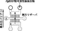
本明細書に記載の様々な局面は、少なくとも部分的に、アルツハイマー病(AD)または前頭側頭型認知症(FTD)を有する対象の脳内に低いレベルで存在する可溶性高分子量(HMW)タウ種(リン酸化型を含む)の発見、およびニューロンによって取り込まれかつニューロン間を伝播する可溶性HMWタウ種の能力の発見に由来する。特に、伝播を担う可溶性HMWタウ種を同定するために、本発明者らは、新規3チャンバーマイクロ流体デバイスを開発して2層ニューロンを形成させ、ニューロンのタウ取り込み、軸索輸送、およびシナプス伝達を調査した。例えば、覚醒行動タウトランスジェニックマウスの間質液および該マウスの皮質抽出物、ならびにヒトAD死後皮質由来の異なるタウ種の取り込み特性および伝播特性を特徴決定することによって、本発明者らは、一局面では、PBS可溶性リン酸化高分子量(HMW)タウ種が、存在度が非常に低いにもかかわらず、時間依存的および濃度依存的に取り込まれ、軸索輸送され、シナプス結合したニューロンに移ることを発見した。対照的に、低分子量(LMW)タウ種(例えば単量体/二量体サイズのタウ)は、存在度がずっと高いにもかかわらず、ニューロンによって効率的には取り込まれない。したがって、一局面では、ニューロン間伝播に関与する希少種の可溶性HMWリン酸化タウの発見は、治療的介入およびバイオマーカー開発のためのより効果的な標的を提供する。
SUMMARY Various aspects described herein include, at least in part, soluble high molecular weight (HMW) that is present at low levels in the brain of a subject having Alzheimer's disease (AD) or frontotemporal dementia (FTD). Derived from the discovery of tau species (including phosphorylated forms) and the ability of soluble HMW tau species to be taken up by and propagated between neurons. Specifically, to identify the soluble HMW tau species responsible for transmission, we developed a novel three-chamber microfluidic device to form bilayer neurons, neuronal tau uptake, axonal transport, and synaptic transmission investigated. For example, by characterizing the uptake and propagation characteristics of interstitial fluid of wakeful behavior tau transgenic mice and cortical extracts of the mice, and different tau species from human AD postmortem cortex, we have In an aspect, PBS soluble phosphorylated high molecular weight (HMW) tau species are taken up in a time-dependent and concentration-dependent manner, axon transported, and transferred to synaptic-connected neurons, despite their very low abundance I found In contrast, low molecular weight (LMW) tau species (eg, monomer / dimer size tau) are not efficiently taken up by neurons, despite their much higher abundance. Thus, in one aspect, the discovery of a rare species of soluble HMW phosphorylated tau involved in interneuronal transmission provides a more effective target for therapeutic intervention and biomarker development.
さらに、本発明者らは、ニューロン間のタウ伝播が、鋳型のミスフォールディングのため内因性細胞内タウを必要としないことを発見したが、タウ欠損マウスは、実質的により少ない病的ミスフォールディングおよびグリオーシスを有したので、一態様では、内因性タウまたはホストタウの完全除去は、神経保護効果を生じる。例えば、本発明者らは、タウ欠損ニューロンが、ニューロンにおけるAβ誘導ミトコンドリア内因性カスパーゼカスケードの活性化を欠いており、かつその次にAβ誘導樹状突起棘消失および神経変性から保護されたことを示している。したがって、本明細書に記載の様々な局面の態様は、ニューロン間伝播を担う可溶性HMWタウ種を含む組成物およびその用途に関する。対象におけるタウ関連神経変性を処置および診断する方法も、本明細書において提供される。 Furthermore, although we have discovered that tau propagation between neurons does not require endogenous intracellular tau due to template misfolding, tau-deficient mice have substantially less pathological misfolding and Because of having gliosis, in one aspect, complete removal of endogenous tau or host tau produces a neuroprotective effect. For example, we have shown that tau-deficient neurons lack activation of the Aβ-induced mitochondrial endogenous caspase cascade in neurons and are subsequently protected from Aβ-induced dendritic spine loss and neurodegeneration. Show. Accordingly, embodiments of the various aspects described herein relate to compositions comprising soluble HMW tau species responsible for interneuron propagation and uses thereof. Also provided herein are methods for treating and diagnosing tau-related neurodegeneration in a subject.
一局面では、可溶性高分子量(HMW)タウ種を含む組成物が本明細書において提供される。組成物中の可溶性HMWタウ種は、非原線維性であり、少なくとも約500kDaの分子量を有し、かつ組成物は可溶性低分子量(LMW)タウ種を実質的に含まない。 In one aspect, provided herein is a composition comprising a soluble high molecular weight (HMW) tau species. The soluble HMW tau species in the composition is non-fibrillar, has a molecular weight of at least about 500 kDa, and the composition is substantially free of soluble low molecular weight (LMW) tau species.
一部の態様では、可溶性HMWタウ種は、少なくとも約669kDaの分子量を有することができる。一部の態様では、可溶性HMWタウ種は、約669kDa〜約1000kDaの分子量を有することができる。 In some embodiments, the soluble HMW tau species can have a molecular weight of at least about 669 kDa. In some embodiments, the soluble HMW tau species can have a molecular weight of about 669 kDa to about 1000 kDa.
一部の態様では、非原線維性可溶性HMWタウ種は、粒子の形態であることができる。粒子のサイズは、タウ種の分子量により変動することができる。一部の態様では、粒子のサイズは、約10nm〜約30nmの範囲に及ぶことができる。 In some embodiments, the non-fibrillar soluble HMW tau species can be in the form of particles. The size of the particles can vary with the molecular weight of the tau species. In some embodiments, the particle size can range from about 10 nm to about 30 nm.
本発明者らは、ADを有さない対照脳と比較した場合、AD脳抽出物が、有意に高いレベルのリン酸化可溶性HMWタウ種を含有したことを発見した。したがって、一部の態様では、組成物中の可溶性HMWタウ種は、リン酸化されていることができる。一部の態様では、組成物中の可溶性HMWタウ種は、過剰リン酸化されていることができる。 The inventors have found that AD brain extracts contained significantly higher levels of phosphorylated soluble HMW tau species when compared to control brains without AD. Thus, in some embodiments, the soluble HMW tau species in the composition can be phosphorylated. In some embodiments, the soluble HMW tau species in the composition can be hyperphosphorylated.
可溶性HMWタウ種は、水溶液および/または緩衝溶液に可溶性であることができる。例えば、一部の態様では、可溶性HMWタウ種は、リン酸緩衝塩類溶液に可溶性であることができる。一部の態様では、可溶性HMWタウ種は、生体液、例えば脳間質液または脳脊髄液に可溶性であることができる。 Soluble HMW tau species can be soluble in aqueous solutions and / or buffered solutions. For example, in some embodiments, the soluble HMW tau species can be soluble in phosphate buffered saline. In some embodiments, the soluble HMW tau species can be soluble in biological fluids such as brain interstitial fluid or cerebrospinal fluid.
一部の態様では、可溶性LMWタウ種のニューロン取り込みおよびニューロン間輸送と比較した場合、可溶性HMWタウ種は、ニューロンによって優先的に取り込まれ、かつ、ニューロンからシナプス結合したニューロンに優先的に軸索輸送されることができる。可溶性LMWタウ種は、可溶性HMWタウ種よりも低い分子量を有する。一部の態様では、可溶性LMWタウ種は、200kDa以下の分子量を有することができる。 In some embodiments, soluble HMW tau species are preferentially taken up by neurons and axons preferentially from neurons to synapse-bound neurons when compared to neuronal uptake and transport between neurons of soluble LMW tau species Can be transported. Soluble LMW tau species have a lower molecular weight than soluble HMW tau species. In some embodiments, the soluble LMW tau species can have a molecular weight of 200 kDa or less.
一部の態様では、本明細書に記載の組成物は、選択された用途の必要性に適合させるために作用物質を含むことができる。例えば、HMWタウが、抗体を産生するための抗原として使用されるべきである場合、精製された可溶性HMWタウを塩類溶液またはリン酸緩衝塩類溶液と組み合わせることができる。代替的または追加的に、HMWタウ抗原をアジュバントまたは担体と混合またはコンジュゲートして、その抗原性を増強することができる。 In some aspects, the compositions described herein can include an agent to suit the needs of the selected application. For example, if HMW tau is to be used as an antigen to produce antibodies, purified soluble HMW tau can be combined with saline or phosphate buffered saline. Alternatively or additionally, the HMW tau antigen can be mixed or conjugated with an adjuvant or carrier to enhance its antigenicity.
したがって、本明細書に記載の別の局面は、可溶性高分子量(HMW)タウ種を特異的に結合させかつ可溶性低分子量(LMW)タウ種を結合させない、単離された抗体またはその抗原結合性部分を提供する。HMWタウ種は、非原線維性であり、少なくとも約500kDaの分子量を有し、かつLMWタウ種は、200kDa以下の分子量を有する。一部の態様では、可溶性HMWタウ種は、少なくとも約669kDaまたはそれ以上の分子量を有することができる。一部の態様では、可溶性HMWタウ種は、約669kDa〜約1000kDaの分子量を有することができる。 Accordingly, another aspect described herein is an isolated antibody or antigen-binding property thereof that specifically binds soluble high molecular weight (HMW) tau species and does not bind soluble low molecular weight (LMW) tau species. Provide part. HMW tau species are non-fibrillar and have a molecular weight of at least about 500 kDa, and LMW tau species have a molecular weight of 200 kDa or less. In some embodiments, the soluble HMW tau species can have a molecular weight of at least about 669 kDa or higher. In some embodiments, the soluble HMW tau species can have a molecular weight of about 669 kDa to about 1000 kDa.
一部の態様では、非原線維性可溶性HMWタウ種は、粒子の形態であることができる。粒子のサイズは、タウ種の分子量により変動することができる。一部の態様では、粒子のサイズは、約10nm〜約30nmの範囲に及ぶことができる。 In some embodiments, the non-fibrillar soluble HMW tau species can be in the form of particles. The size of the particles can vary with the molecular weight of the tau species. In some embodiments, the particle size can range from about 10 nm to about 30 nm.
一部の態様では、単離された抗体またはその抗原結合性部分は、リン酸化型の可溶性HMWタウ種を特異的に結合させることができる。 In some embodiments, the isolated antibody or antigen-binding portion thereof can specifically bind phosphorylated soluble HMW tau species.
一部の態様では、単離された抗体またはその抗原結合性部分は、水溶液および/または緩衝液に可溶性の可溶性HMWタウ種を特異的に結合させることができる。例えば、一部の態様では、単離された抗体またはその抗原結合性部分は、リン酸緩衝塩類溶液に可溶性の可溶性HMWタウ種を特異的に結合させることができる。一部の態様では、単離された抗体またはその抗原結合性部分は、生体液、例えば脳間質液または脳脊髄液に可溶性の可溶性HMWタウ種を特異的に結合させることができる。 In some embodiments, the isolated antibody or antigen-binding portion thereof can specifically bind soluble HMW tau species that are soluble in aqueous solutions and / or buffers. For example, in some embodiments, an isolated antibody or antigen-binding portion thereof can specifically bind soluble HMW tau species that are soluble in phosphate buffered saline. In some embodiments, the isolated antibody or antigen-binding portion thereof can specifically bind soluble HMW tau species that are soluble in biological fluids such as brain interstitial fluid or cerebrospinal fluid.
一部の態様では、単離された抗体またはその抗原結合性部分は、ニューロンによって取り込まれている可溶性HMWタウ種を少なくとも約10%またはそれ以上減少させるように設計することができる。 In some embodiments, an isolated antibody or antigen-binding portion thereof can be designed to reduce at least about 10% or more soluble HMW tau species that are taken up by neurons.
一部の態様では、単離された抗体またはその抗原結合性部分は、ニューロンからシナプス結合したニューロンに軸索輸送されている可溶性HMWタウ種を少なくとも約10%またはそれ以上減少させるように設計することができる。 In some embodiments, the isolated antibody or antigen-binding portion thereof is designed to reduce at least about 10% or more soluble HMW tau species that are axonally transported from neuron to synaptic neuron. be able to.
本発明者らは、ニューロンから比較的低レベルの可溶性HMWタウ種が放出され、脳間質液中に見出されたこと、および試料中の全タウのわずかな割合だけを占める可溶性HMWタウ種が、ニューロンによって強く取り込まれ、かつニューロン間伝播に関与した一方で、可溶性LMWタウ種(例えば単量体/二量体サイズ)の取り込みは非常に非効率的であったことを示した。したがって、シナプス結合したニューロン間の異常タウタンパク質の伝播を防止する方法も、本明細書において提供される。本方法は、シナプス結合したニューロンと接触している可溶性HMWタウ種の細胞外レベルを選択的に減少させる段階を含み、可溶性HMWタウ種は、非原線維性であり、少なくとも約500kDaの分子量を有する。可溶性HMWタウ種のレベルの減少は、シナプス結合したニューロン間の異常タウタンパク質の伝播の減少を招く。 We have released relatively low levels of soluble HMW tau species from neurons and found them in the brain interstitial fluid, and soluble HMW tau species that account for only a small percentage of the total tau in the sample Showed that uptake of soluble LMW tau species (eg monomer / dimer size) was very inefficient, while being strongly taken up by neurons and involved in interneuron propagation. Accordingly, a method for preventing the propagation of abnormal tau protein between synaptic connected neurons is also provided herein. The method includes selectively reducing extracellular levels of soluble HMW tau species that are in contact with synaptic neurons, wherein the soluble HMW tau species is non-fibrillar and has a molecular weight of at least about 500 kDa. Have. A decrease in the level of soluble HMW tau species results in a decrease in the transmission of abnormal tau protein between synaptic neurons.
一部の態様では、可溶性LMWタウ種の細胞外レベルは、前記選択的減少の最中に実質的に減少されない。 In some embodiments, the extracellular level of soluble LMW tau species is not substantially reduced during the selective reduction.
可溶性HMWタウ種の細胞外レベルを選択的に減少させるための方法は、物理的除去および/または可溶性HMWタウ種と妥当なアンタゴニストとの間の分子相互作用に基づくことができる。一部の態様では、可溶性HMWタウ種は、微小透析によって選択的に減少されることができる。一部の態様では、可溶性HMWタウ種は、シナプス結合したニューロンと接触している細胞外間隙または細胞外液を、可溶性HMWタウ種のアンタゴニストと接触させることによって、選択的に減少されることができる。可溶性HMWタウ種のアンタゴニストの例には、非限定的に、抗体、ヌクレアーゼ(例えば、非限定的に、ジンクフィンガーヌクレアーゼ(ZFN)、転写活性化因子様エフェクターヌクレアーゼ(TALEN)、CRISPR/Casシステム、転写抑制因子、核酸阻害物質(例えば、RNAi、siRNA、anti-miR、アンチセンスオリゴヌクレオチド、リボザイム、およびそれらの2つまたはそれ以上の組み合わせ)、小分子、アプタマー、およびそれらの2つまたはそれ以上の組み合わせが含まれる。 Methods for selectively reducing the extracellular level of soluble HMW tau species can be based on physical removal and / or molecular interactions between the soluble HMW tau species and a reasonable antagonist. In some embodiments, soluble HMW tau species can be selectively reduced by microdialysis. In some embodiments, soluble HMW tau species can be selectively reduced by contacting an extracellular space or fluid that is in contact with synaptic-associated neurons with an antagonist of soluble HMW tau species. it can. Examples of soluble HMW tau species antagonists include, but are not limited to, antibodies, nucleases (eg, but not limited to zinc finger nuclease (ZFN), transcription activator-like effector nuclease (TALEN), CRISPR / Cas system, Transcription repressor, nucleic acid inhibitor (eg, RNAi, siRNA, anti-miR, antisense oligonucleotide, ribozyme, and combinations of two or more thereof), small molecules, aptamers, and two or more thereof Is included.
タウの病理は、疾患が進行する際にアルツハイマー病(AD)の脳において、例えばニューロン間の経シナプスタウ移動によって、階層的なパターンで蔓延することが公知である。本明細書において、可溶性HMWタウ種がニューロン間伝播に関与すると同定されたので、そのようなHMWタウ種を枯渇させるための介入は、タウの伝播を、ゆえにタウオパチーにおける疾患進行を阻害することができる。したがって、対象におけるタウ関連神経変性を減少させる方法が、本明細書において提供される。タウ関連神経変性の例には、非限定的に、アルツハイマー病、パーキンソン病、または前頭側頭型認知症が含まれる。処置方法は、タウ関連神経変性を有すると判定された対象またはタウ関連神経変性のリスクを有すると判定された対象の脳内または脳脊髄液(CSF)中の可溶性HMWタウ種のレベルを選択的に減少させる段階を含み、可溶性HMWタウ種は、非原線維性であり、少なくとも約500kDaの分子量を有し、可溶性HMWタウ種のレベルは、タウ関連神経変性の減少を招く。 It is known that tau pathology spreads in a hierarchical pattern in the Alzheimer's disease (AD) brain as the disease progresses, for example by transsynaptic movement between neurons. As soluble HMW tau species have been identified herein as involved in interneuron transmission, intervention to deplete such HMW tau species may inhibit tau transmission and hence disease progression in tauopathy it can. Accordingly, provided herein are methods for reducing tau-related neurodegeneration in a subject. Examples of tau-related neurodegeneration include, but are not limited to, Alzheimer's disease, Parkinson's disease, or frontotemporal dementia. The treatment method selectively selects the level of soluble HMW tau species in the brain or cerebrospinal fluid (CSF) of a subject determined to have tau-related neurodegeneration or a subject determined to be at risk for tau-related neurodegeneration. The soluble HMW tau species is non-fibrillar and has a molecular weight of at least about 500 kDa, and the level of soluble HMW tau species leads to a reduction in tau-related neurodegeneration.
一部の態様では、対象における可溶性LMWタウ種のレベルは、処置の最中に実質的に減少されない。 In some embodiments, the level of soluble LMW tau species in the subject is not substantially reduced during treatment.
一部の態様では、対象の脳間質液中に存在する可溶性HMWタウ種の少なくとも一部は、除去されるかまたは不活性にされる。一部の態様では、対象の脳脊髄液中に存在する可溶性HMWタウ種の少なくとも一部は、除去されるかまたは不活性にされる。 In some embodiments, at least a portion of the soluble HMW tau species present in the subject's brain interstitial fluid is removed or inactivated. In some embodiments, at least a portion of the soluble HMW tau species present in the subject's cerebrospinal fluid is removed or inactivated.
対象の脳における可溶性HMWタウ種の細胞外レベルを選択的に減少させるための方法は、物理的除去に、および/または可溶性HMWタウ種と妥当なアンタゴニストとの間の分子相互作用に基づくことができる。一部の態様では、対象の脳間質液中および/または脳脊髄液中に存在する可溶性HMWタウ種は、脳の微小透析によって選択的に減少されることができる。一部の態様では、対象の脳間質液中および/または脳脊髄液中に存在する可溶性HMWタウ種は、対象の脳またはCSFに可溶性HMWタウ種のアンタゴニストを投与することによって選択的に減少させることができる。可溶性HMWタウ種のアンタゴニストの例には非限定的に、抗体、ヌクレアーゼ(例えば、非限定的に、ジンクフィンガーヌクレアーゼ(ZFN)、転写活性化因子様エフェクターヌクレアーゼ(TALEN)、CRISPR/Casシステム、転写抑制因子、核酸阻害物質(例えば、RNAi、siRNA、anti-miR、アンチセンスオリゴヌクレオチド、リボザイム、およびそれらの2つまたはそれ以上の組み合わせ)、小分子、アプタマー、ならびにそれらの2つまたはそれ以上の組み合わせが含まれる。 Methods for selectively reducing the extracellular level of soluble HMW tau species in a subject's brain may be based on physical removal and / or molecular interactions between the soluble HMW tau species and a reasonable antagonist. it can. In some embodiments, soluble HMW tau species present in a subject's brain interstitial fluid and / or cerebrospinal fluid can be selectively reduced by microdialysis of the brain. In some embodiments, soluble HMW tau species present in a subject's brain interstitial fluid and / or cerebrospinal fluid is selectively reduced by administering an antagonist of the soluble HMW tau species to the subject's brain or CSF. Can be made. Examples of soluble HMW tau species antagonists include, but are not limited to, antibodies, nucleases (eg, but not limited to zinc finger nuclease (ZFN), transcription activator-like effector nuclease (TALEN), CRISPR / Cas system, transcription Repressors, nucleic acid inhibitors (eg, RNAi, siRNA, anti-miR, antisense oligonucleotides, ribozymes, and combinations of two or more thereof), small molecules, aptamers, and two or more of them A combination is included.
一部の態様では、本方法は、参照レベルを上回るレベルで脳内またはCSF中に存在する可溶性HMWタウ種を有すると判定された対象を選択する段階をさらに含むことができる。参照レベルは、健康な対象に存在する可溶性HMWタウ種のレベルを表すことができる。 In some aspects, the method can further comprise selecting a subject determined to have a soluble HMW tau species present in the brain or in the CSF at a level above a reference level. The reference level can represent the level of soluble HMW tau species present in a healthy subject.
さらなる一局面では、可溶性HMWタウ種の存在および/またはレベルに基づきタウ関連神経変性を診断する方法も、本明細書において提供される。例示的なタウ関連神経変性には非限定的に、アルツハイマー病、パーキンソン病、または前頭側頭型認知症が含まれる。本発明者らは、ADおよび対照脳からの脳抽出物中の総タウレベルが有意には異ならなかった一方で、ABの脳抽出物が、対照脳と比較した場合に、有意に高いレベルの可溶性HMWタウ種またはリン酸化可溶性HMWタウ種を含有したことを示した。したがって、タウ関連神経変性を診断する方法は、(a)対象由来の脳間質液または脳脊髄液の試料を分画する段階;ならびに(b)可溶性HMWタウ種の存在が判定されかつ可溶性HMWタウ種の量が測定されるように、試料中の可溶性HMWタウ種を検出する段階であって、可溶性HMWタウ種が、非原線維性であり、少なくとも約500kDaの分子量を有する、段階;ならびに(c)試料中の可溶性HMWタウ種のレベルが参照レベルと同じであるかもしくは参照レベルを上回る場合、タウ関連神経変性を有するかもしくはタウ関連神経変性のリスクを有する対象を同定する段階、または可溶性HMWタウ種のレベルが参照レベルを下回る場合、タウ関連神経変性を有する可能性が低い対象を同定する段階を含むことができる。参照レベルは、健康な対象に存在する可溶性HMWタウ種のレベルを表すことができる。 In a further aspect, methods of diagnosing tau-related neurodegeneration based on the presence and / or level of soluble HMW tau species are also provided herein. Exemplary tau-related neurodegeneration includes, but is not limited to, Alzheimer's disease, Parkinson's disease, or frontotemporal dementia. We found that total tau levels in brain extracts from AD and control brains were not significantly different, while AB brain extracts had significantly higher levels of solubility when compared to control brains. It was shown to contain HMW tau species or phosphorylated soluble HMW tau species. Thus, a method of diagnosing tau-related neurodegeneration comprises (a) fractionating a sample of cerebral interstitial fluid or cerebrospinal fluid from a subject; and (b) the presence of a soluble HMW tau species is determined and soluble HMW Detecting soluble HMW tau species in the sample such that the amount of tau species is measured, wherein the soluble HMW tau species is non-fibrillar and has a molecular weight of at least about 500 kDa; and (C) identifying a subject having or at risk for tau-related neurodegeneration if the level of soluble HMW tau species in the sample is equal to or above the reference level, or If the level of soluble HMW tau species is below the reference level, identifying a subject less likely to have tau-related neurodegeneration can be included. The reference level can represent the level of soluble HMW tau species present in a healthy subject.
一部の態様では、(a)の分画する段階の前の試料は、可溶性LMWタウ種を実質的に含まないことができ、可溶性LMWタウ種は、200kDa以下の分子量を有する。例えば、脳間質液または脳脊髄液の試料は、微小透析によって、例えば、少なくとも約600kDaの分子量を有する分子だけを収集することを可能にする妥当なフィルター性分子量カットオフを使用して、診断されるべき対象から得ることができる。 In some embodiments, the sample prior to the fractionating step of (a) can be substantially free of soluble LMW tau species, wherein the soluble LMW tau species has a molecular weight of 200 kDa or less. For example, a sample of cerebral interstitial fluid or cerebrospinal fluid can be diagnosed by microdialysis, for example using a reasonable filter molecular weight cut-off that allows only molecules with a molecular weight of at least about 600 kDa to be collected. Can be obtained from the subject to be done.
代替的な態様では、(a)の分画する段階の前の試料は、可溶性LMWタウ種を含むことができ、可溶性LMWタウ種は、200kDa以下の分子量を有する。試料を分画する段階によって、可溶性HMWタウ種を残りの試料(例えば、可溶性LMWタウ種を含む一部)から単離して、診断レベルを判定することができる。非限定的に、分画する段階は、サイズ排除および/または抗体に基づく方法に基づくことができる。 In an alternative embodiment, the sample prior to the fractionating step of (a) can comprise a soluble LMW tau species, wherein the soluble LMW tau species has a molecular weight of 200 kDa or less. By fractionating the sample, soluble HMW tau species can be isolated from the remaining sample (eg, a portion containing soluble LMW tau species) to determine the diagnostic level. Non-limiting, the fractionating step can be based on size exclusion and / or antibody based methods.
可溶性LMWタウ種が試料中に存在する一部の態様では、本方法は、試料中の可溶性LMWタウ種の量を検出する段階をさらに含むことができる。これらの態様では、可溶性LMWタウ種に対する可溶性HMWタウ種の比が、上記参照レベル比と同じであるかもしくは参照レベル比を上回る場合に、対象が、タウ関連神経変性を有するかもしくはタウ関連神経変性のリスクを有すると同定されることができるか;または可溶性LMWタウ種に対する可溶性HMWタウ種の比が参照レベル比を下回る場合に、対象が、タウ関連神経変性を有する可能性が低いと同定される。参照レベル比は、健康な対象に存在する可溶性LMWタウ種に対する可溶性HMWタウ種のレベル比を表すことができる。 In some embodiments where soluble LMW tau species are present in the sample, the method can further comprise detecting the amount of soluble LMW tau species in the sample. In these embodiments, if the ratio of soluble HMW tau species to soluble LMW tau species is equal to or exceeds the reference level ratio, the subject has tau-related neurodegeneration or a tau-related neurodegeneration. Can be identified as at risk of degeneration; or a subject is less likely to have tau-related neurodegeneration if the ratio of soluble HMW tau species to soluble LMW tau species is below the reference level ratio Is done. The reference level ratio can represent the level ratio of soluble HMW tau species to soluble LMW tau species present in healthy subjects.
一部の態様では、検出する段階は、可溶性HMWタウ種のリン酸化を検出することをさらに含むことができる。 In some embodiments, the detecting step can further comprise detecting phosphorylation of soluble HMW tau species.
可溶性HMWタウ種の発見が、本明細書に記載のタウ関連神経変性のための治療標的およびバイオマーカーを提供するだけでなく、可溶性HMWタウ種はまた、神経変性における進行の表現型特徴であるニューロン間伝播を誘導するためにインビトロで使用することで、ミスフォールディングしたタウタンパク質のシナプス間蔓延を減少させ、したがってタウ関連神経変性を処置する有効な作用物質についてスクリーニングするためのインビトロモデルを開発することもできる。したがって、本明細書において提供されるさらなる一局面は、ミスフォールディングしたタウタンパク質のシナプス間蔓延を減少させるために有効な作用物質を同定する方法に関する。本方法は、(a)ニューロン培養デバイスの第1のチャンバー中の第1のニューロンを可溶性HMWタウ種と接触させる段階であって、第1のニューロンが、ニューロン培養デバイスの第2のチャンバー中の第2のニューロンと軸索で結合しており、かつ第2のニューロンが、可溶性HMWタウ種と接触していない、段階;(b)第1のチャンバー中の(a)からの第1のニューロンを候補作用物質と接触させる段階;および(c)第1のニューロンから第2のニューロンへの可溶性HMWタウ種の輸送を検出する段階を含む。ミスフォールディングしたタウタンパク質のシナプス間蔓延を減少させるために有効な作用物質は、第2のニューロンの軸索および/または細胞体における可溶性HMWタウ種の存在の検出に基づき同定することができる。 Not only does the discovery of soluble HMW tau species provide therapeutic targets and biomarkers for the tau-related neurodegeneration described herein, but soluble HMW tau species are also a phenotypic feature of progression in neurodegeneration Develop an in vitro model to screen for effective agents that reduce intersynaptic spread of misfolded tau proteins and thus treat tau-related neurodegeneration by using in vitro to induce interneuronal propagation You can also. Accordingly, a further aspect provided herein relates to a method of identifying an agent effective to reduce the intersynaptic spread of misfolded tau protein. The method comprises (a) contacting a first neuron in a first chamber of a neuron culture device with a soluble HMW tau species, wherein the first neuron is in a second chamber of the neuron culture device. Axons connected to the second neuron and the second neuron is not in contact with the soluble HMW tau species; (b) the first neuron from (a) in the first chamber Contacting with a candidate agent; and (c) detecting the transport of soluble HMW tau species from the first neuron to the second neuron. Agents effective to reduce the intersynaptic spread of misfolded tau protein can be identified based on detection of the presence of soluble HMW tau species in the axons and / or cell bodies of the second neuron.
軸索伸長および/または軸索輸送をモニタリングするために適切な任意のニューロン培養デバイスを本明細書に記載の方法において使用することができる一方で、一部の態様では、ニューロン培養デバイスは、マイクロ流体デバイスである。一部の態様では、マイクロ流体デバイスは、第1のニューロンを配置するための第1のチャンバー、および第2のニューロンを配置するための第2のチャンバーを備えることができ、第1のチャンバーおよび第2のチャンバーは、軸索成長だけを可能にするようなサイズに作製された少なくとも1つのマイクロチャネルによって相互に接続している。 While any neuronal culture device suitable for monitoring axonal outgrowth and / or axonal transport can be used in the methods described herein, in some embodiments, the neuronal culture device is It is a fluid device. In some aspects, the microfluidic device can comprise a first chamber for placing a first neuron, and a second chamber for placing a second neuron, wherein the first chamber and The second chambers are interconnected by at least one microchannel that is sized to allow only axonal growth.
本明細書に記載の可溶性HMWタウ種をまた、スクリーニングアッセイ法に使用して、HMWタウ種自体の形成または活性を(例えば、HMWタウ種の形成もしくは安定性を遮断することによって、または例えば、翻訳後修飾を遮断することによって、またはHMWタウ構造を不安定化することによって)モジュレートする作用物質を同定することができる。例えば、アプタマー、小分子、または他の作用物質をニューロン細胞培養に適用し、可溶性HMWタウの存在または量をモニタリングすることができる。そのように同定された、HMWタウの形成または蓄積を遮断する作用物質は、潜在的治療剤として関心対象であり得る。 The soluble HMW tau species described herein can also be used in screening assays to prevent the formation or activity of the HMW tau species itself (eg, by blocking the formation or stability of the HMW tau species, or Agents that modulate (by blocking post-translational modifications or by destabilizing the HMW tau structure) can be identified. For example, aptamers, small molecules, or other agents can be applied to neuronal cell cultures to monitor the presence or amount of soluble HMW tau. Agents so identified that block HMW tau formation or accumulation may be of interest as potential therapeutic agents.
一局面では、本発明者らは、タウ欠損動物が、神経原線維濃縮体(NFT)の存在下でさえも、タウ発現動物と比較してより少ない病的タウミスフォールディングおよびグリオーシスを示し、かつニューロンにおいてAβ誘導型ミトコンドリア内因性カスパーゼカスケードの活性化の減少も示したことを示した。したがって、内因性細胞内タウタンパク質を除去することは、例えば神経毒性を減少させ、かつ/またはニューロン生存期間を増加させて神経保護効果を提供することができる。以前の報告は、タウタンパク質の減少が、神経変性を改善できると論じていたものの、当業者は、かなりの量の内因性細胞内タウタンパク質を、すなわち微小管を安定化する必須タンパク質を、除去することが、ニューロンに対して他のいかなる有害作用も生じないとは予想しなかったであろう。しかし、本発明者らは、ここで、タウタンパク質を発現している健康なヒト対象のように、タウ欠損ヒト対象が正常なニューロン表現型を有することを発見した。このように、タウオパチーによって誘導された神経損傷または神経変性を減少させる方法も、本明細書において提供される。例示的なタウオパチーは、アルツハイマー病、パーキンソン病、または前頭側頭型認知症であることができる。 In one aspect, the inventors have shown that tau-deficient animals exhibit less pathological tau misfolding and gliosis, even in the presence of neurofibrillary tangles (NFT), compared to tau expressing animals, and We also showed that activation of Aβ-induced mitochondrial endogenous caspase cascade was also reduced in neurons. Thus, removing endogenous intracellular tau protein can provide, for example, neuroprotective effects by reducing neurotoxicity and / or increasing neuronal survival. Although previous reports have argued that a reduction in tau protein can improve neurodegeneration, those skilled in the art have removed significant amounts of endogenous intracellular tau protein, an essential protein that stabilizes microtubules. Would not have expected to have any other adverse effects on neurons. However, the inventors have now discovered that a tau-deficient human subject has a normal neuronal phenotype, such as a healthy human subject expressing tau protein. Thus, methods for reducing tauopathy-induced nerve damage or neurodegeneration are also provided herein. An exemplary tauopathy can be Alzheimer's disease, Parkinson's disease, or frontotemporal dementia.
タウオパチーによって誘導された神経損傷または神経変性を減少させる方法は、タウオパチーを有すると判定された対象の脳に、対象において内因性細胞内タウタンパク質の発現レベルを少なくとも約50%阻害する作用物質を投与し、それによって、神経原線維濃縮体の存在下で神経毒性を減少させる(かつ/またはニューロンの生存期間を増加させる)段階を含む。 A method of reducing tauopathy-induced neuronal damage or neurodegeneration comprises administering to a brain of a subject determined to have tauopathy an agent that inhibits the expression level of endogenous intracellular tau protein in the subject by at least about 50% Thereby reducing neurotoxicity (and / or increasing neuronal survival) in the presence of neurofibrillary tangles.
一部の態様では、前記作用物質は、対象におけるタウタンパク質の発現レベルを少なくとも約70%阻害することができる。一部の態様では、該作用物質は、対象におけるタウタンパク質の発現レベルを少なくとも約90%阻害することができる。一部の態様では、該作用物質は、対象におけるタウタンパク質の発現レベルを少なくとも約95%阻害することができる。一部の態様では、該作用物質は、対象におけるタウタンパク質の発現レベルを100%阻害することができる。 In some embodiments, the agent can inhibit tau protein expression levels in the subject by at least about 70%. In some embodiments, the agent can inhibit the expression level of tau protein in the subject by at least about 90%. In some embodiments, the agent can inhibit the expression level of tau protein in the subject by at least about 95%. In some aspects, the agent can inhibit 100% of the expression level of tau protein in the subject.
内因性細胞内タウタンパク質の発現レベルを阻害するために選択された作用物質は、MAPT(微小管結合タンパク質タウ)遺伝子の発現を妨害し、かつ/またはMAPT遺伝子の転写を阻害することができる。そのような作用物質には非限定的に、抗体、ヌクレアーゼ(例えば、非限定的に、ジンクフィンガーヌクレアーゼ(ZFN)、転写活性化因子様エフェクターヌクレアーゼ(TALEN)、CRISPR/Casシステム、転写抑制因子、核酸阻害物質(例えば、RNAi、siRNA、anti-miR、アンチセンスオリゴヌクレオチド、リボザイム、およびそれらの2つまたはそれ以上の組み合わせ)、小分子、アプタマー、ならびにそれらの2つまたはそれ以上の組み合わせが含まれることができる。 Agents selected to inhibit the expression level of endogenous intracellular tau protein can interfere with the expression of the MAPT (microtubule associated protein tau) gene and / or inhibit transcription of the MAPT gene. Such agents include, but are not limited to, antibodies, nucleases (eg, but not limited to zinc finger nuclease (ZFN), transcription activator-like effector nuclease (TALEN), CRISPR / Cas system, transcription repressor, Includes nucleic acid inhibitors (eg, RNAi, siRNA, anti-miR, antisense oligonucleotides, ribozymes, and combinations of two or more thereof), small molecules, aptamers, and combinations of two or more thereof Can be.
対象の脳に作用物質を効果的に送達することで公知の方法を使用して、本明細書に記載の方法を行うことができる。一部の態様では、作用物質は、担体を介して脳に投与することができる。例示的な担体は、アデノ随伴ウイルスであることができる。 The methods described herein can be performed using methods known in the art to effectively deliver an agent to the subject's brain. In some aspects, the agent can be administered to the brain via a carrier. An exemplary carrier can be an adeno-associated virus.
一部の態様では、対象の脳はさらに、アミロイドβ斑を有すると判定されることができ、かつ、投与は、アミロイドβの存在下で神経毒性を減少させること(および/またはニューロンの生存期間を増加させること)ができる。 In some aspects, the subject's brain can further be determined to have amyloid β plaques, and the administration reduces neurotoxicity in the presence of amyloid β (and / or neuronal survival time). Can be increased).
発明の詳細な説明
本明細書に記載の様々な局面は、少なくとも部分的に、アルツハイマー病(AD)または前頭側頭型認知症(FTD)を有する対象の脳内に低い細胞外レベルで存在する可溶性高分子量(HMW)タウ種(リン酸化型を含む)の発見、およびニューロンによって取り込まれかつニューロン間を伝播する可溶性HMWタウ種の能力の発見に由来する。特に、伝播を担う可溶性HMWタウ種を同定するために、本発明者らは、新規3チャンバーマイクロ流体デバイスを開発して2層ニューロンを形成させ、ニューロンのタウ取り込み、軸索輸送、およびシナプス伝達を調査した。例えば、覚醒行動タウトランスジェニックマウスの間質液および該マウスの皮質抽出物、ならびにヒトAD死後皮質由来の異なるタウ種の取り込み特性および伝播特性を特徴決定することによって、本発明者らは、一局面では、PBS可溶性リン酸化高分子量(HMW)タウ種が、存在度が非常に低いにもかかわらず、時間依存的および濃度依存的に取り込まれ、軸索輸送され、かつ、シナプス結合したニューロンに移ることを発見した。対照的に、低分子量(LMW)タウ種(例えば、単量体/二量体サイズのタウ)は、存在度がずっと高いにもかかわらず、ニューロンによって非効率的に取り込まれない。したがって、一局面では、ニューロン間伝播に関与する希少種の可溶性HMWリン酸化タウの発見は、治療的介入およびバイオマーカー開発のためのより効果的な標的を提供する。
DETAILED DESCRIPTION OF THE INVENTION Various aspects described herein are at least partially present at low extracellular levels in the brain of a subject with Alzheimer's disease (AD) or frontotemporal dementia (FTD). Derived from the discovery of soluble high molecular weight (HMW) tau species (including phosphorylated forms) and the ability of soluble HMW tau species to be taken up and propagated between neurons. Specifically, to identify the soluble HMW tau species responsible for transmission, we developed a novel three-chamber microfluidic device to form bilayer neurons, neuronal tau uptake, axonal transport, and synaptic transmission investigated. For example, by characterizing the uptake and propagation characteristics of interstitial fluid of wakeful behavior tau transgenic mice and cortical extracts of the mice, and different tau species from human AD postmortem cortex, we have In an aspect, PBS soluble phosphorylated high molecular weight (HMW) tau species are taken up in time- and concentration-dependent, axonal transported, and synaptic-associated neurons, despite their very low abundance I found it moving. In contrast, low molecular weight (LMW) tau species (eg, monomer / dimer size tau) are not inefficiently taken up by neurons, despite their much higher abundance. Thus, in one aspect, the discovery of a rare species of soluble HMW phosphorylated tau involved in interneuronal transmission provides a more effective target for therapeutic intervention and biomarker development.
さらに、本発明者らは、ニューロン間のタウ伝播が、鋳型のミスフォールディングのため内因性細胞内タウを必要としないことを発見したが、タウ欠損マウスは、実質的により少ない病的ミスフォールディングおよびグリオーシスを有したので、一態様では、内因性細胞内ホストタウの完全除去は、神経保護効果を生じる。例えば、本発明者らは、タウ欠損ニューロンが、ニューロンにおけるAβ誘導ミトコンドリア内因性カスパーゼカスケードの活性化を欠いており、かつその次にAβ誘導樹状突起棘消失および神経変性から保護されたことを示している。 Furthermore, although we have discovered that tau propagation between neurons does not require endogenous intracellular tau due to template misfolding, tau-deficient mice have substantially less pathological misfolding and Because of having gliosis, in one aspect, complete removal of endogenous intracellular host tau produces a neuroprotective effect. For example, we have shown that tau-deficient neurons lack activation of the Aβ-induced mitochondrial endogenous caspase cascade in neurons and are subsequently protected from Aβ-induced dendritic spine loss and neurodegeneration. Show.
したがって、本明細書に記載の様々な局面の態様は、ニューロン間伝播を担う可溶性HMWタウ種を含む組成物およびその適用に関する。対象におけるタウ関連神経変性を処置および診断する方法も、本明細書において提供される。 Accordingly, embodiments of the various aspects described herein relate to compositions comprising soluble HMW tau species responsible for interneuron propagation and applications thereof. Also provided herein are methods for treating and diagnosing tau-related neurodegeneration in a subject.
可溶性高分子量(HMW)タウ種を含む組成物
一局面では、可溶性高分子量(HMW)タウ種を含む組成物が、本明細書において提供される。組成物中の可溶性HMWタウ種は、非原線維性であり、少なくとも約500kDaの分子量を有する。
Compositions comprising soluble high molecular weight (HMW) tau species In one aspect, compositions comprising soluble high molecular weight (HMW) tau species are provided herein. The soluble HMW tau species in the composition is non-fibrillar and has a molecular weight of at least about 500 kDa.
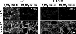
一部の態様では、組成物は、可溶性低分子量(LMW)タウ種を実質的に含まない。本明細書においておよび本明細書全体にわたり使用される「可溶性低分子量(LMW)タウ種を実質的に含まない」という用語には、LMWタウ種の完全非存在(すなわち0%)、および当技術分野において公知の方法、例えば、非限定的にサイズ排除クロマトグラム、ELISA、顕微鏡検査、原子間力顕微鏡検査、またはそれらの任意の組み合わせによって容易には検出できない微量のLMWタウ種が含まれる。一部の態様では、本明細書に記載の組成物中のLMWタウに対するHMWタウの比率は、これらのタンパク質形態がインビボで存在する比率と実質的に異なる。したがって、HMWタウが、インビボで、例えばヒトにおいて、通常存在する総タウのわずかな割合だけを占める場合、総タウタンパク質の少なくとも50%がHMW形態である調製物、例えば、少なくとも50%のHMWおよび50%もしくはそれ未満のLMWタウ、少なくとも60%のHMWタウおよび40%もしくはそれ未満のLMWタウ、少なくとも70%のHMWタウおよび30%もしくはそれ未満のLMWタウ、少なくとも80%のHMWタウおよび20%もしくはそれ未満のLMWタウ、少なくとも90%のHMWタウおよび10%もしくはそれ未満のLMWタウ、または95%のHMWタウおよび5%未満のLMWタウを有する調製物が、本明細書において記載される。一部の態様では、組成物は、例えば4%以下、3%以下、2%以下、1%以下、0.5%(w/w)以下の可溶性LMWタウ種を含む、5%(w/w)以下の可溶性LMWタウ種を含むことができる。本明細書において使用される「LMWタウを実質的に欠いている」という用語は、所与のHMWタウ調製物が、1重量%未満のLMWタウ、好ましくは0.1重量%未満のLMWタウ、0.01重量%未満の、またはそれより低いタウを有することを意味する。
In some embodiments, the composition is substantially free of soluble low molecular weight (LMW) tau species. As used herein and throughout the specification, the term “substantially free of soluble low molecular weight (LMW) tau species” includes the complete absence (
本明細書においておよび本明細書全体にわたり使用される「高分子量タウ種」または「HMWタウ種」という用語は、非原線維性およびオリゴマー性のタウ種分子の集団を表し、タウ種分子は、それぞれ少なくとも約500kDaの分子量を有する。HMWタウ種は、病的にミスフォールディングしたコンフォメーションを有し(例えば、Alz50抗体染色陽性)、オリゴマー構造として現れる(例えば、原子間力顕微鏡検査において)。HMWタウ種は、細胞内(例えばニューロン内部)または細胞外(例えば脳間質液中および/または脳脊髄液中)であることができる。一部の態様では、HMWタウ種は、タウトランスジェニック動物(例えば、タウトランスジェニックマウス)からの脳抽出物および/または脳間質液を、例えば、遠心分離(例えば、×3000gまたはそれ未満)および/または実施例記載のサイズ排除クロマトグラフィーによって分画し、少なくとも約500kDaまたはそれ以上の分子量を有する画分を選択することによって生成させることができる。一部の態様では、HMWタウ種は、組換えタウタンパク質を多量体化することによって生成させることができる。一部の態様では、HMWタウ種は、下にさらに詳細に記載されるようなリン酸化または過剰リン酸化されていることができる。 As used herein and throughout this specification, the term "high molecular weight tau species" or "HMW tau species" refers to a population of non-fibrillar and oligomeric tau species molecules, Each has a molecular weight of at least about 500 kDa. HMW tau species have a pathologically misfolded conformation (eg, positive Alz50 antibody staining) and appear as oligomeric structures (eg, in atomic force microscopy). The HMW tau species can be intracellular (eg, inside a neuron) or extracellular (eg, in brain interstitial fluid and / or cerebrospinal fluid). In some embodiments, the HMW tau species is a brain extract and / or brain interstitial fluid from a tau transgenic animal (eg, a tau transgenic mouse), eg, centrifuged (eg, x3000 g or less). And / or fractionated by size exclusion chromatography as described in the examples, and can be generated by selecting fractions having a molecular weight of at least about 500 kDa or higher. In some embodiments, HMW tau species can be generated by multimerizing recombinant tau protein. In some embodiments, the HMW tau species can be phosphorylated or hyperphosphorylated as described in more detail below.
本明細書において使用される「非原線維性」という用語は、神経原線維濃縮体(NFT)に凝集していないHMWタウ種を表す。NFTは一般的に、不溶性フィラメントに集合したタウタンパク質の過剰リン酸化からニューロン内部に形成される。 As used herein, the term “non-fibrillar” refers to an HMW tau species that has not aggregated into a neurofibrillary tangle (NFT). NFT is generally formed inside neurons from hyperphosphorylation of tau protein assembled in insoluble filaments.
本明細書に記載の可溶性HMWタウ種に関連して本明細書において使用される「オリゴマー性」または「オリゴマー」という用語は、有限数のタウ単量体または二量体サブユニットを含む複合体または凝集物を意味する。一部の態様では、本明細書に記載の可溶性HMWタウ種は、例えば、少なくとも3つ、少なくとも4つ、少なくとも5つ、少なくとも6つ、少なくとも7つ、少なくとも8つ、少なくとも9つ、少なくとも10個、少なくとも15個、少なくとも20個、少なくとも30個、少なくとも40個、またはそれ以上のタウ単量体または二量体サブユニットを含む、少なくとも2つまたはそれ以上のタウ単量体または二量体サブユニットを含むことができる。一部の態様では、本明細書に記載のHMWタウ種は、約3〜100個のタウ単量体または二量体サブユニット、約4〜90個のタウ単量体または二量体サブユニット、約5〜80個のタウ単量体または二量体サブユニット、約5〜70個のタウ単量体または二量体サブユニット、約5〜60個のタウ単量体または二量体サブユニット、約5〜50個のタウ単量体または二量体サブユニット、約5〜40個のタウ単量体または二量体サブユニット、約5〜30個のタウ単量体または二量体サブユニット、または約5〜20個のタウ単量体または二量体サブユニットを含むことができる。 The term “oligomeric” or “oligomer” as used herein in connection with the soluble HMW tau species described herein is a complex comprising a finite number of tau monomers or dimeric subunits. Or it means an aggregate. In some embodiments, the soluble HMW tau species described herein have, for example, at least 3, at least 4, at least 5, at least 6, at least 7, at least 8, at least 9, at least 10 At least two or more tau monomers or dimers comprising at least 15, at least 20, at least 30, at least 40, or more tau monomers or dimer subunits Subunits can be included. In some embodiments, the HMW tau species described herein have about 3-100 tau monomers or dimer subunits, about 4 to 90 tau monomers or dimer subunits. About 5 to 80 tau monomer or dimer subunit, about 5 to 70 tau monomer or dimer subunit, about 5 to 60 tau monomer or dimer subunit Unit, about 5-50 tau monomer or dimer subunit, about 5-40 tau monomer or dimer subunit, about 5-30 tau monomer or dimer Subunits, or about 5-20 tau monomers or dimeric subunits can be included.
タウタンパク質または微小管結合タンパク質タウ(MAPT):タウタンパク質は、微小管結合タンパク質(MAP)ファミリーに属する。それらは、主に、ニューロンに発現され、そこでチューブリン単量体が微小管に集合して、ニューロンの微小管ネットワークを構成することに重要な役割を果たす。とはいえ、非ニューロン細胞(例えば、心臓、腎臓、肺、筋肉、または膵臓細胞)が微量を有する可能性がある。微小管は、細胞形状を維持することに関与し、軸索輸送のための軌道として役立つ。タウタンパク質は、第17番染色体上に位置する単一遺伝子から翻訳される。それらの発現は、選択的スプライシングメカニズムによって発生的に調節され、ヒト成体脳において6つの異なるアイソフォームが存在する。Bueeら、Brain Research Reviews (2000) 33: 95-130。
Tau protein or microtubule-associated protein tau (MAPT): Tau protein belongs to the microtubule-associated protein (MAP) family. They are primarily expressed in neurons, where tubulin monomers assemble into microtubules and play an important role in constructing neuronal microtubule networks. Nonetheless, non-neuronal cells (eg, heart, kidney, lung, muscle, or pancreatic cells) can have trace amounts. Microtubules are involved in maintaining cell shape and serve as trajectories for axonal transport. Tau protein is translated from a single gene located on
タウは、4つの領域:N末端突出領域、プロリンリッチドメイン、微小管結合性ドメイン(MBD)、およびC末端領域に細分することができる。Morris et al., Neuron (2011) 70: 410-426。成体ヒト脳において、N末端領域およびMBD周辺の選択的スプライシングによって、352〜441個の範囲のアミノ酸を有する6つの主要アイソフォームが発生する(Goedert et al., Neuron (1989) 3: 519-526)。それらは、N末端部分(エクソン2および3)におけるアミノ酸29個の0、1、または2つのいずれかの挿入部と、エクソン10が欠失しているC末端部分における3または4つの繰り返し領域が異なる。したがって、CNSにおける最長アイソフォームは、4つの繰り返し(R1、R2、R3、およびR4)ならびに2つの挿入部(合計アミノ酸441個)を有し、一方で最短アイソフォームは、3つの繰り返し(R1、R3、およびR4)を有し、挿入部を有さない(合計アミノ酸352個)。タウアイソフォームは、微小管結合性繰り返し配列(Rと呼ばれる)がいくつ発現され、どのN末端エクソン(Nと呼ばれる)が含まれるかによって名付けられる。例えば、3Rタウは、3つの微小管結合性繰り返し配列を有し、一方で4Rタウは、エクソン10を含むことにより4つを有する。0Nタウは、N末端エクソンを含まず、1Nタウは、エクソン2を、2Nタウは、エクソン2および3を含む(Lee et al., Annu. Rev. Neurosci. (2001) 24: 1121-1159)。タウの変異は、4R2Nヒトタウにおけるその位置により番号付けされる(Lee et al., 2001)。エクソン6周辺の選択的スプライシングによって6つの追加的なアイソフォームが形成され、脳に発現するタウアイソフォームが合計で12種生じる(Wei and Andreadis, J. Neurochem (1998) 70: 1346-1356)。本明細書において引用される参照文献は、参照により本明細書に組み入れられる。
Tau can be subdivided into four regions: an N-terminal overhang region, a proline-rich domain, a microtubule binding domain (MBD), and a C-terminal region. Morris et al., Neuron (2011) 70: 410-426. In the adult human brain, alternative splicing around the N-terminal region and MBD generates six major isoforms with a range of 352-441 amino acids (Goedert et al., Neuron (1989) 3: 519-526 ). They have either an insertion of 29 0, 1, or 2 amino acids in the N-terminal part (
一部の態様では、可溶性HMWタウ種は、タウアイソフォーム1、タウアイソフォーム2、タウアイソフォーム3、タウアイソフォーム4、タウアイソフォーム5、およびタウアイソフォーム6からなる群より選択される少なくとも1つまたは複数(例えば、少なくとも2つまたはそれ以上)のタウアイソフォームが濃縮されたオリゴマーであることができる。一部の態様では、可溶性HMWタウ種は、(2-3-10-);(2+3-10-);(2+3+10-);(2-3-10+);(2+3-10+);(2+3+10+)からなる群より選択される少なくとも1つまたは複数(例えば、少なくとも2つまたはそれ以上)のタウアイソフォームが濃縮されたオリゴマーであることができる。全てのMAP(微小管結合タンパク質)タウタンパク質アイソフォームは、当技術分野において公知であり、例えばヒトを含む、それらのヌクレオチドおよびタンパク質配列は、NCBIからのワールドワイドウェブ上で入手可能である。下の表1に、NCBIで入手可能な異なるヒトタウアイソフォームのヌクレオチドおよびアミノ酸配列の例示的なアクセッション番号を示す。
In some embodiments, the soluble HMW tau species is at least selected from the group consisting of
(表1)異なるヒトタウアイソフォームのアミノ酸配列

本明細書においておよび本明細書全体にわたり使用される、HMWまたはLMWタウ種を呼ぶ場合の「可溶性」という用語は、HMWまたはLMWタウ種が生体液中、例えばCSF、脳間質液、血漿などに溶解し、実質的に均一な溶液を形成することを意味する。本明細書に記載のHMWタウ可溶性種はまた、リン酸緩衝塩類溶液中に溶解し、実質的に均一な溶液を形成する。 As used herein and throughout this specification, the term `` soluble '' when referring to an HMW or LMW tau species refers to an HMW or LMW tau species in a biological fluid such as CSF, brain interstitial fluid, plasma, etc. Is dissolved to form a substantially uniform solution. The HMW tau soluble species described herein also dissolves in phosphate buffered saline to form a substantially homogeneous solution.
本明細書において使用される「分子量」という用語は、所与の分子(例えば、HMWもしくはLMWタウ種分子)の質量または所与の分子集団(例えば、2つまたはそれ以上)(例えば、HMWタウ種分子もしくはLMWタウ種分子の集団)の平均質量を表す。適用される統計的方法に応じて、異なる平均値を定義することができる。一部の態様では、分子量は、数平均分子量である。一部の態様では、分子量は、質量平均分子量である。HMWまたはLMWタウ種の分子量は、一般的に、当技術分野において公知の任意の方法、例えば非限定的にゲル電気泳動、ゲルクロマトグラフィー、サイズ排除クロマトグラフィー、光散乱、および/または質量分析によって測定することができる。一部の態様では、HMWタウ種またはLMWタウ種の分子量は、サイズ排除クロマトグラフィーによって測定される。 As used herein, the term “molecular weight” refers to the mass of a given molecule (eg, HMW or LMW tau species molecule) or a given molecular population (eg, two or more) (eg, HMW tau The average mass of a seed molecule or LMW tau seed molecule population). Depending on the statistical method applied, different average values can be defined. In some aspects, the molecular weight is a number average molecular weight. In some aspects, the molecular weight is a mass average molecular weight. The molecular weight of the HMW or LMW tau species is generally determined by any method known in the art, such as, but not limited to, gel electrophoresis, gel chromatography, size exclusion chromatography, light scattering, and / or mass spectrometry. Can be measured. In some embodiments, the molecular weight of the HMW tau species or LMW tau species is measured by size exclusion chromatography.
本明細書に記載の組成物において、可溶性HMWタウ種は、少なくとも約500kDaまたはそれ以上の分子量を有する。一部の態様では、可溶性HMWタウ種は、少なくとも約550kDa、少なくとも約600kDa、少なくとも約650kDa、少なくとも約700kDa、少なくとも約750kDa、少なくとも約800kDa、少なくとも約850kDa、少なくとも約900kDa、少なくとも約950kDa、またはそれまたはそれ以上の分子量を有することができる。一部の態様では、可溶性HMWタウ種は、少なくとも約669kDaまたはそれ以上の分子量を有することができる。一部の態様では、可溶性HMWタウ種は、約500kDa〜約2000kDa、約550kDa〜約1500kDa、約600kDa〜約1000kDa、約650kDa〜約1000kDaまたは約669kDa〜約1000kDaの分子量を有することができる。一部の態様では、可溶性HMWタウ種は、分子量分布を形成することができる。一部の態様では、可溶性HMWタウ種は、全て、実質的に同じ分子量を有することができる。 In the compositions described herein, the soluble HMW tau species has a molecular weight of at least about 500 kDa or more. In some embodiments, the soluble HMW tau species is at least about 550 kDa, at least about 600 kDa, at least about 650 kDa, at least about 700 kDa, at least about 750 kDa, at least about 800 kDa, at least about 850 kDa, at least about 900 kDa, at least about 950 kDa, or more Or it can have a higher molecular weight. In some embodiments, the soluble HMW tau species can have a molecular weight of at least about 669 kDa or higher. In some embodiments, the soluble HMW tau species can have a molecular weight of about 500 kDa to about 2000 kDa, about 550 kDa to about 1500 kDa, about 600 kDa to about 1000 kDa, about 650 kDa to about 1000 kDa, or about 669 kDa to about 1000 kDa. In some embodiments, the soluble HMW tau species can form a molecular weight distribution. In some embodiments, all of the soluble HMW tau species can have substantially the same molecular weight.
一部の態様では、非原線維性可溶性HMWタウ種は、粒子の凝集物中に存在することができる。粒子は、任意の形状であることができ、非限定的に、球形または球状粒子である。可溶性HMWタウ種の粒子のサイズは、それらの分子量に応じて変動することができる。一部の態様では、可溶性HMWタウ種の粒子のサイズは、約1nm〜約50nm、約5nm〜約40nm、約10nm〜約30nm、または約15nm〜約25nmの範囲に及ぶことができる。一部の態様では、可溶性HMWタウ種は、粒子のサイズ分布を形成することができる。一部の態様では、可溶性HMWタウ種は、全て実質的に同じ粒子のサイズを有することができる。 In some embodiments, the non-fibrillar soluble HMW tau species can be present in an aggregate of particles. The particles can be of any shape, including but not limited to spherical or spherical particles. The size of the particles of soluble HMW tau species can vary depending on their molecular weight. In some embodiments, the size of the particles of soluble HMW tau species can range from about 1 nm to about 50 nm, from about 5 nm to about 40 nm, from about 10 nm to about 30 nm, or from about 15 nm to about 25 nm. In some embodiments, the soluble HMW tau species can form a particle size distribution. In some embodiments, the soluble HMW tau species can all have substantially the same particle size.
一部の態様では、可溶性HMWタウ種は、タウ単量体および/または二量体サブユニットから本質的になるかまたはそれからなることができる。 In some embodiments, the soluble HMW tau species can consist essentially of or consist of tau monomers and / or dimeric subunits.
一部の態様では、可溶性HMWタウ種は、他のタンパク質および脂質のような他の成分を含むことができる。 In some embodiments, the soluble HMW tau species can include other components such as other proteins and lipids.
本発明者らは、AD脳抽出物が、ADを有さない対照脳と比較した場合に、有意に高いレベルのリン酸化可溶性HMWタウ種を含有したことを発見した。したがって、一部の態様では、組成物中の可溶性HMWタウ種は、リン酸化されていることができる。一部の態様では、組成物中の可溶性HMWタウ種は、過剰リン酸化されていることができる。本明細書において使用される「過剰リン酸化されている」または「過剰リン酸化」という用語は、HMWタウ種上のリン酸化部位の数(すなわちリン酸エステル部分の数)が、LMWタウ種もしくは非凝集性正常タウタンパク質よりも大きいか;またはHMWタウ種上のリン酸化部位が、LMWタウ種または非凝集性正常タウタンパク質よりも高いレベルでリン酸化されている状況を表す。一部の態様では、HMWタウ種上の少なくとも1つまたは複数(例えば、1、2、3、4、5、6、7、8、9、10個、または全て)のリン酸化部位は、LMWタウ種または非凝集性正常タウタンパク質よりも少なくとも約50%(少なくとも約60%、少なくとも約70%、少なくとも約80%、少なくとも約90%、少なくとも約95%または最大100%を含む)高いレベルでリン酸化されている。一部の態様では、HMWタウ種上の少なくとも1つまたは複数(例えば、1、2、3、4、5、6、7、8、9、10個、または全てを含む)のリン酸化部位は、LMWタウ種または非凝集性正常タウタンパク質よりも少なくとも約1.5倍高い(少なくとも約2倍、少なくとも約2.5倍、少なくとも約3倍、少なくとも約3.5倍、少なくとも約4倍、少なくとも約4.5倍、少なくとも約5倍、またはそれより高いを含む)レベルでリン酸化されている。HMWタウ種において過剰リン酸化されていることができるリン酸化部位の例には、非限定的に、pS199、pT205、pS262、pS396、pS396/404、pS400、pS409、pS422、またはそれらの2つまたはそれ以上の組み合わせが含まれる。一部の態様では、HMWタウ種は、以下のリン酸化部位:pT205、pS262、pS400、pS404、pS409、およびpS422の少なくとも1つまたは複数(例えば、少なくとも2つ、少なくとも3つ、少なくとも4つ、少なくとも5つ、少なくとも6つ)で過剰リン酸化されていることができる。完全長タウアイソフォームは、例えば、NCBIアクセッション番号NP_005901.2によって表され、その情報は参照により本明細書に組み入れられる。 The inventors have discovered that AD brain extracts contained significantly higher levels of phosphorylated soluble HMW tau species when compared to control brains without AD. Thus, in some embodiments, the soluble HMW tau species in the composition can be phosphorylated. In some embodiments, the soluble HMW tau species in the composition can be hyperphosphorylated. As used herein, the term “hyperphosphorylated” or “hyperphosphorylated” means that the number of phosphorylation sites on the HMW tau species (ie the number of phosphate ester moieties) is LMW tau species or Represents a situation where the phosphorylation sites on the HMW tau species are phosphorylated at a higher level than LMW tau species or non-aggregated normal tau protein; In some embodiments, at least one or more (eg, 1, 2, 3, 4, 5, 6, 7, 8, 9, 10, or all) phosphorylation sites on the HMW tau species are LMW At a level that is at least about 50% higher (including at least about 60%, at least about 70%, at least about 80%, at least about 90%, at least about 95%, or up to 100%) than tau species or non-aggregating normal tau protein It is phosphorylated. In some embodiments, at least one or more (eg, including 1, 2, 3, 4, 5, 6, 7, 8, 9, 10, or all) phosphorylation sites on the HMW Tau species are , At least about 1.5 times higher than LMW tau species or non-aggregating normal tau protein (at least about 2 times, at least about 2.5 times, at least about 3 times, at least about 3.5 times, at least about 4 times, at least about 4.5 times, at least It is phosphorylated at a level (including about 5 times or higher). Examples of phosphorylation sites that can be hyperphosphorylated in HMW tau species include, but are not limited to, pS199, pT205, pS262, pS396, pS396 / 404, pS400, pS409, pS422, or two of them More combinations are included. In some embodiments, the HMW tau species has at least one or more of the following phosphorylation sites: pT205, pS262, pS400, pS404, pS409, and pS422 (eg, at least 2, at least 3, at least 4, At least 5, at least 6) may be hyperphosphorylated. The full-length tau isoform is represented, for example, by NCBI accession number NP_005901.2, the information of which is incorporated herein by reference.
一部の態様では、可溶性HMWタウ種は、ニューロンによって優先的に取り込まれることができる。本明細書において使用される「ニューロンによって優先的に取り込まれる」という用語は、可溶性LMWタウ種を取り込むよりも可溶性HMWタウ種を取り込む可能性の方が高いニューロンを表す。一部の態様では、ニューロンは、可溶性LMWタウ種を取り込むよりも可溶性HMWタウ種を取り込む可能性の方が、少なくとも約30%またはそれ以上(例えば、少なくとも約40%、少なくとも約50%、少なくとも約60%、少なくとも約70%、少なくとも約80%、少なくとも約90%、少なくとも約95%、またはそれより高いを含む)高い。一部の態様では、ニューロンは、可溶性LMWタウ種を取り込むよりも可溶性HMWタウ種を取り込む可能性の方が少なくとも約1.5倍またはそれ以上(例えば、少なくとも約2倍、少なくとも約2.5倍、少なくとも約3倍、少なくとも約3.5倍、少なくとも約4倍、少なくとも約5倍、少なくとも約10倍、またはそれより高いを含む)高い。 In some embodiments, soluble HMW tau species can be preferentially taken up by neurons. The term “preferentially taken up by neurons” as used herein refers to neurons that are more likely to take up soluble HMW tau species than uptake soluble LMW tau species. In some embodiments, the neuron is at least about 30% or more likely to take up soluble HMW tau species (e.g., at least about 40%, at least about 50%, at least more than to take up soluble LMW tau species). Including about 60%, at least about 70%, at least about 80%, at least about 90%, at least about 95%, or higher). In some embodiments, the neuron is at least about 1.5 times or more likely to take up soluble HMW tau species (eg, at least about 2 times, at least about 2.5 times, at least about at least about uptake of soluble LMW tau species). Including 3 times, at least about 3.5 times, at least about 4 times, at least about 5 times, at least about 10 times, or higher).
可溶性HMWタウ種がニューロンによって取り込まれると、可溶性HMWタウ種は、該ニューロンからシナプス結合したニューロンに軸索輸送されることができる。したがって、一部の態様では、ニューロンは、その細胞体からシナプス結合したニューロンに、可溶性HMWタウ種を優先的に軸索輸送することができる。本明細書において使用される「その細胞体からシナプス結合したニューロンに可溶性HMWタウ種を優先的に軸索輸送する」という用語は、シナプス結合したニューロン間の可溶性LMWタウ種の軸索輸送と比較した場合、その細胞体からシナプス結合したニューロンに可溶性HMWタウ種を軸索輸送する可能性が高いニューロンを表す。一部の態様では、ニューロンは、シナプス結合したニューロン間の可溶性LMWタウ種の軸索輸送と比較した場合、その細胞体からシナプス結合したニューロンに可溶性HMWタウ種を軸索輸送する可能性が、少なくとも約30%またはそれ以上高い(例えば、少なくとも約40%、少なくとも約50%、少なくとも約60%、少なくとも約70%、少なくとも約80%、少なくとも約90%、少なくとも約95%、またはそれより高いを含む)ことができる。一部の態様では、ニューロンは、シナプス結合したニューロン間の可溶性LMWタウ種の軸索輸送と比較した場合、その細胞体からシナプス結合したニューロンに可溶性HMWタウ種を軸索輸送する可能性が、少なくとも約1.5倍またはそれ以上高い(例えば、少なくとも約2倍、少なくとも約2.5倍、少なくとも約3倍、少なくとも約3.5倍、少なくとも約4倍、少なくとも約5倍、少なくとも約10倍、またはそれより高いを含む)ことができる。 When a soluble HMW tau species is taken up by neurons, the soluble HMW tau species can be axon transported from the neurons to synaptic neurons. Thus, in some embodiments, a neuron can preferentially axon transport soluble HMW tau species from its cell body to synaptic-connected neurons. As used herein, the term “preferentially axonal transport of soluble HMW tau species from its cell body to synaptic neurons” is compared to axonal transport of soluble LMW tau species between synaptic neurons. If so, it represents a neuron that is likely to axonally transport soluble HMW tau species from its cell body to synaptic-connected neurons. In some embodiments, the neuron may axonically transport soluble HMW tau species from its cell body to synapse-bound neurons when compared to axonal transport of soluble LMW tau species between synaptic neurons. At least about 30% or higher (eg, at least about 40%, at least about 50%, at least about 60%, at least about 70%, at least about 80%, at least about 90%, at least about 95%, or higher) Can be included). In some embodiments, the neuron may axonically transport soluble HMW tau species from its cell body to synapse-bound neurons when compared to axonal transport of soluble LMW tau species between synaptic neurons. At least about 1.5 times or higher (eg, at least about 2 times, at least about 2.5 times, at least about 3 times, at least about 3.5 times, at least about 4 times, at least about 5 times, at least about 10 times, or higher) Can be included).
「軸索輸送する」または「軸索輸送」という用語は、本明細書において、ニューロンの軸索に沿った分子および/またはオルガネラの定方向輸送を表すために使用される。「軸索輸送」は、「順行性」(細胞体から外向き)または「逆行性」(細胞体方向に戻る)であることができる。したがって、可溶性HMWタウ種の順行性輸送は、ニューロンによって取り込まれた可溶性HMWタウ種をその細胞体から外向きに遠隔シナプスに送達する。 The terms “axon transport” or “axon transport” are used herein to denote the directed transport of molecules and / or organelles along neuronal axons. "Axonal transport" can be "antegrade" (outward from the cell body) or "retrograde" (returning toward the cell body). Thus, anterograde transport of soluble HMW tau species delivers soluble HMW tau species taken up by neurons outward from their cell bodies to remote synapses.
本明細書においておよび本明細書全体にわたり使用される「シナプス結合した」という用語は、シナプスを介して互いにコミュニケーションしているニューロンを表す。シナプスは、シグナル伝達のために特殊化されたニューロンの区域である。シナプスは、それらがシグナル伝達領域として作用する能力によって、および2つのニューロン間のシナプスの物理的近接性によって特徴決定することができる。シグナル伝達は、電気的または化学的手段によることができる。 As used herein and throughout this specification, the term “synaptic connected” refers to neurons that communicate with each other through the synapse. Synapses are areas of neurons specialized for signal transduction. Synapses can be characterized by their ability to act as signaling regions and by the physical proximity of synapses between two neurons. Signaling can be by electrical or chemical means.
本明細書においておよび本明細書全体にわたり使用される「低分子量タウ種」または「LMWタウ種」という用語は、実質的にタウ単量体サブユニットおよび/またはタウ二量体サブユニットであるタウ種分子集団を表す。LMWタウ種は、細胞内(例えば、ニューロン内部)または細胞外(例えば、脳間質液中および/または脳脊髄液中)であることができる。一部の態様では、LMWタウ種は、例えば、より高い速度(例えば、×50000gまたはそれ以上)の遠心分離および/または実施例に記載のサイズ排除クロマトグラフィーによって、タウトランスジェニック動物(例えば、タウトランスジェニックマウス)からの脳抽出物および/または脳間質液を分画し、200kDa以下またはそれ未満の分子量の画分を選択することによって生成させることができる。一部の態様では、可溶性LMWタウ種は、例えば、150kDa以下、100kDa以下、50kDa以下、またはそれ未満を含む200kDa以下またはそれ未満の分子量を有することができる。 As used herein and throughout this specification, the term “low molecular weight tau species” or “LMW tau species” refers to a tau that is substantially a tau monomer subunit and / or a tau dimer subunit. Represents a species molecular population. The LMW tau species can be intracellular (eg, inside a neuron) or extracellular (eg, in brain interstitial fluid and / or cerebrospinal fluid). In some embodiments, the LMW tau species are transformed into a tau transgenic animal (eg, tau Brain extracts from (transgenic mice) and / or brain interstitial fluid can be fractionated and generated by selecting fractions with a molecular weight of 200 kDa or less or less. In some embodiments, the soluble LMW tau species can have a molecular weight of 200 kDa or less, including, for example, 150 kDa or less, 100 kDa or less, 50 kDa or less, or less.
一部の態様では、本明細書に記載の組成物は、選択された用途の必要性に適合させるための作用物質を含むことができる。例えば一部の態様では、本明細書に記載の組成物は、可溶性HMWタウ種に対する抗体を産生するように適応させることができる。したがって、これらの態様では、組成物は、可溶性HMWタウ種に対する抗体を産生するためのアジュバントをさらに含むことができる。本明細書において使用される「アジュバント」という用語は、個体または動物(例えばマウス)にインビトロ投与された場合に、投与された抗原(例えば可溶性HMWタウ種)に対する個体または動物の免疫応答を高める分子、化合物、および/または物質を表す。一部の抗原は、単独で投与された場合に弱免疫原性であり、または個体または動物において免疫応答を誘発する濃度で個体に有毒である。アジュバントは、抗原をより強く免疫原性にすることによって、抗原に対する個体または動物の免疫応答を増強することで、抗体生成を増強することができる。アジュバントの効果はまた、個体または動物における免疫応答を達成するために必要な抗原の用量を低下させることができる。通例使用されるアジュバントには、非限定的に、油中水型エマルションからなる不完全フロイントアジュバント、結核菌(Mycobacterium tuberculosis)およびミョウバンを添加された不完全フロイントアジュバントの成分を含む、フロイント完全アジュバントが含まれる。本明細書に記載のHMWタウ組成物を、アジュバントとコンジュゲートさせることができる。本明細書に記載のHMWタウ複合体の抗原性を高めるために、キーホールリンペットヘモシアニンのような抗原性担体とのコンジュゲーションも使用することができる。HMWタウとコンジュゲートさせることができる他の抗原性担体タンパク質には、例えば、コンコレパス コンコレパス(Concholepas concholepas)ヘモシアニン(「Blue Carrier」)、ウシ血清アルブミン、カチオン化ウシ血清、および卵白アルブミンが含まれる。HMWタウ複合体をニューロンからニューロンに優先的に伝達されるHMW形態に安定化する処理は、タウタンパク質のHMWまたはLMW形態のいずれにも特異的な抗体とは対照的に、HMWに特異的な抗体を産生するための抗原としてHMWタウが役立つ可能性をより高くすると、さらに考えられている。様々なアプローチを使用して、HMWタウ構造の安定化をもたらすことができる。例えば、HMWタウは、単離されたHMWタウを架橋することによってHMW/シナプス伝達可能な形態のタウの状態に安定化することができる。例えばアミンと反応するホモ二官能性およびヘテロ二官能性架橋剤(例えば、とりわけグルタル酸ジスクシンイミジル、スベリン酸ジスクシンイミジル、スベリン酸ビス[スルホスクシンイミジル]、プロピオン酸ジチオビス[スクシンイミジル]を含むN-ヒドロスクシンイミド(NHS)エステル架橋剤、アジプイミド酸ジメチル-2HCl、スベルイミド酸ジメチルを含むイミドエステルなど)またはスルフヒドリルと反応するもの(例えば、マレイミド系架橋剤、例えば、ビスマレイミドエタン、ビスマレイミドヘキサン、ジチオビスマレイミドエタンなど)を含む、いくつかの化学架橋試薬が公知であり、市販されている。架橋剤は、当技術分野において公知の反応条件を調整することによってモジュレートすることができる。一態様では、相対的に低度の架橋形成は、非架橋HMWと比較して実質的な安定化を提供することができ、それによって、例えば抗原として、またはスクリーニングアッセイ法用の標的として増強した活性を提供する。HMWタウはまた、例えば表面、例えばプラスチック、ニトロセルロース、またはナイロン膜と相互作用することによって安定化させることができる。この形態で、安定化されたHMWタウは、HMW複合体形態のタウタンパク質を特異的に結合させる作用物質についてスクリーニングするための基質または標的として役立つことができる。一例として、HMWタウで機能化された表面は、例えば結合性ポリペプチドをディスプレイしているバクテリオファージについてパニングするために使用することができる。構築物をディスプレイしているバクテリオファージのライブラリーは、当技術分野において周知である。ファージライブラリーを、表面上に固定化されたLMWタウと最初に接触させて、LMW-タウ結合性ポリペプチドを含むライブラリーメンバーをサブトラクトすることによって、実質的にLMWタウも結合させるわけではないファージディスプレイされたHMWタウ結合タンパク質を得る可能性を高めることができる。このようなサブトラクションの後に、ライブラリーは、次に別の表面上に固定化されたHMWタウと接触され、続いてHMWタウと固着するファージが単離および増殖される。固体支持体/表面は、非限定的に、とりわけニトロセルロースまたはナイロンメンブラン、アフィニティーカラムクロマトグラフィー用マトリックス、ナイロンまたは他のポリマービーズを含むことができる。 In some aspects, the compositions described herein can include an agent to tailor the selected use needs. For example, in some embodiments, the compositions described herein can be adapted to produce antibodies against soluble HMW tau species. Thus, in these embodiments, the composition can further comprise an adjuvant for producing antibodies against soluble HMW tau species. As used herein, the term “adjuvant” is a molecule that enhances an individual's or animal's immune response to an administered antigen (eg, a soluble HMW tau species) when administered in vitro to the individual or animal (eg, a mouse). Represents a compound and / or substance. Some antigens are weakly immunogenic when administered alone or are toxic to an individual at concentrations that elicit an immune response in the individual or animal. Adjuvants can enhance antibody production by enhancing the immune response of an individual or animal to the antigen by making the antigen more strongly immunogenic. The effect of an adjuvant can also reduce the dose of antigen required to achieve an immune response in an individual or animal. Commonly used adjuvants include, but are not limited to, Freund's complete adjuvant, which includes components of incomplete Freund's adjuvant consisting of a water-in-oil emulsion, Mycobacterium tuberculosis and incomplete Freund's adjuvant with alum added. included. The HMW tau composition described herein can be conjugated with an adjuvant. Conjugation with an antigenic carrier such as keyhole limpet hemocyanin can also be used to enhance the antigenicity of the HMW tau complex described herein. Other antigenic carrier proteins that can be conjugated to HMW tau include, for example, Concholepas concholepas hemocyanin (“Blue Carrier”), bovine serum albumin, cationized bovine serum, and ovalbumin. The process of stabilizing the HMW tau complex to the HMW form preferentially transmitted from neuron to neuron is specific for HMW, as opposed to antibodies specific for either the HMW or LMW form of tau protein. It is further believed that HMW tau is more likely to serve as an antigen for producing antibodies. Various approaches can be used to provide stabilization of the HMW tau structure. For example, HMW tau can be stabilized to a tau form capable of HMW / synaptic transmission by cross-linking isolated HMW tau. For example, homobifunctional and heterobifunctional crosslinkers that react with amines (eg, disuccinimidyl glutarate, disuccinimidyl suberate, bis [sulfosuccinimidyl suberate), dithiobis [succinimidyl propionate] N-hydrosuccinimide (NHS) ester crosslinkers, dimethyl adipimidate-2HCl, imide esters containing dimethyl suberimidate, etc.) or those that react with sulfhydryl (eg maleimide crosslinkers such as bismaleimide ethane, bis Several chemical crosslinking reagents are known and commercially available, including maleimidohexane, dithiobismaleimide ethane, and the like). Crosslinkers can be modulated by adjusting reaction conditions known in the art. In one aspect, the relatively low degree of cross-linking can provide substantial stabilization compared to non-cross-linked HMW, thereby enhancing, for example, as an antigen or as a target for a screening assay. Provides activity. HMW tau can also be stabilized, for example, by interacting with surfaces such as plastic, nitrocellulose, or nylon membranes. In this form, the stabilized HMW tau can serve as a substrate or target for screening for agents that specifically bind the tau protein in the HMW complex form. As an example, a surface functionalized with HMW tau can be used, for example, to pan for bacteriophages displaying binding polypeptides. Libraries of bacteriophage displaying constructs are well known in the art. By first contacting the phage library with the LMW tau immobilized on the surface and subtracting the library member containing the LMW-tau binding polypeptide, the LMW tau is not substantially bound. The possibility of obtaining phage-displayed HMW tau binding protein can be increased. After such subtraction, the library is then contacted with HMW tau immobilized on another surface, followed by isolation and growth of phage that adhere to the HMW tau. The solid support / surface can include, but is not limited to, among others, nitrocellulose or nylon membrane, affinity column chromatography matrix, nylon or other polymer beads.
抗可溶性HMWタウ種アンタゴニスト物質または可溶性HMWタウ種のアンタゴニスト
ニューロンにおける細胞内タンパクしての不溶性神経原線維濃縮体とは異なり、本明細書に記載の可溶性HMWタウ種は、細胞外間隙中に見出すことができる、例えば、脳間質液および/または脳脊髄液に可溶性の、新規可溶性非原線維性タウ凝集物である。前述のように、本発明者らは、可溶性HMWタウ種が、ニューロンによって優先的に取り込まれ、かつ、シナプス結合したニューロンに優先的に軸索輸送されることで、ニューロン間のタウ蔓延が進行することを示した。本明細書に記載の可溶性HMWタウ種のニューロン取り込みを減少または遮断することによって、タウ蔓延を防止することができる。したがって、様々な局面において、抗体またはその抗原結合性断片、核酸、および小分子のような可溶性HMWタウ種アンタゴニスト物質を含む、ニューロンによって取り込まれかつ/またはシナプス結合したニューロンに該ニューロンから軸索輸送されている可溶性HMWタウ種を阻害または減少させるための組成物、ならびに、可溶性HMWタウ種のニューロン取り込みおよびタウ伝播に関連する病理の阻害または減少のためのその使用方法が、本明細書において提供される。
Anti-soluble HMW tau species antagonist substances or antagonists of soluble HMW tau species Unlike the insoluble neurofibrillary concentrate as intracellular proteins in neurons, the soluble HMW tau species described herein are found in the extracellular space For example, novel soluble non-fibrillar tau aggregates that are soluble in cerebral interstitial fluid and / or cerebrospinal fluid. As described above, the present inventors proceeded with the spread of tau between neurons by allowing soluble HMW tau species to be preferentially taken up by neurons and preferentially transported to synaptic neurons. Showed that to do. By reducing or blocking neuronal uptake of the soluble HMW tau species described herein, tau spread can be prevented. Thus, in various aspects, axonal transport from neurons to and / or synapse-coupled neurons, including soluble HMW tau species antagonist substances such as antibodies or antigen-binding fragments thereof, nucleic acids, and small molecules Provided herein are compositions for inhibiting or reducing a soluble HMW tau species that have been treated, and methods of use thereof for inhibiting or reducing pathologies associated with neuronal uptake and tau transmission of soluble HMW tau species Is done.
本明細書において互換的に使用される「タウ伝播」および「タウ蔓延」という用語は、ミスフォールディングしたタウタンパク質のニューロン間輸送を表す。 The terms “tau propagation” and “tau spread”, used interchangeably herein, refer to the interneuronal transport of misfolded tau proteins.
本明細書において使用される「可溶性HMWタウ種アンタゴニスト物質」または「可溶性HMWタウ種のアンタゴニスト」は、可溶性HMWタウ種によって仲介される1つまたは複数の過程、メカニズム、効果、応答、機能、活性または経路における質的または量的な阻害、低下、または減少を阻害するかまたは引き起こすかまたは促進する、小分子、阻害性核酸、または可溶性HMWタウ種特異的抗体もしくはそれらの抗原結合性断片のような作用物質を表す。したがって、「可溶性HMWタウ種アンタゴニスト物質」という用語は、可溶性HMWタウ種の形成を阻害する作用物質、あるいは、可溶性HMWタウ種に結合し、可溶性HMWタウ種のニューロン取り込みを部分的もしくは完全に、例えば、少なくとも約10%またはそれ以上(例えば、少なくとも約20%、少なくとも約30%、少なくとも約40%、少なくとも約50%、少なくとも約60%、少なくとも約70%、少なくとも約80%、少なくとも約90%またはそれ以上、最大100%を含む)遮断、減少、もしくは防止し、かつ/またはそのニューロン取り込みの際に可溶性HMWタウ種のニューロン間伝播を、例えば少なくとも約10%またはそれ以上(例えば少なくとも約20%、少なくとも約30%、少なくとも約40%、少なくとも約50%、少なくとも約60%、少なくとも約70%、少なくとも約80%、少なくとも約90%またはそれ以上、最大100%を含む)遮断、減少、もしくは防止する、作用物質を表す。 As used herein, a “soluble HMW tau species antagonist substance” or “an antagonist of a soluble HMW tau species” is one or more processes, mechanisms, effects, responses, functions, activities mediated by soluble HMW tau species Or as a small molecule, inhibitory nucleic acid, or soluble HMW tau species-specific antibody or antigen-binding fragment thereof that inhibits, causes or promotes qualitative or quantitative inhibition, reduction or reduction in the pathway Represents active agents. Thus, the term “soluble HMW tau species antagonist agent” refers to an agent that inhibits the formation of soluble HMW tau species, or binds to soluble HMW tau species and partially or completely neuronal uptake of soluble HMW tau species, For example, at least about 10% or more (eg, at least about 20%, at least about 30%, at least about 40%, at least about 50%, at least about 60%, at least about 70%, at least about 80%, at least about 90 %, Or more, including up to 100%), block, reduce or prevent and / or propagate interneuron propagation of soluble HMW tau species upon neuronal uptake, such as at least about 10% or more (eg at least about 20%, at least about 30%, at least about 40%, at least about 50%, at least about 60%, at least about 70%, low Least about 80%, at least about 90% or more, including 100% maximal) blocking, decreasing, or preventing, representing agents.
これらの局面および本明細書に記載の全てのそのような局面の一部の態様では、可溶性HMWタウ種アンタゴニスト物質は、可溶性低分子量(LMW)タウ種を結合させない。例えば、一部の態様では、可溶性HMWタウ種アンタゴニスト物質は、例えば150kDa以下、100kDa以下、またはそれより低いを含む200kDa以下の分子量を有する可溶性LMWタウ種を結合させない。 In some embodiments of these aspects and all such aspects described herein, the soluble HMW tau species antagonist substance does not bind soluble low molecular weight (LMW) tau species. For example, in some embodiments, the soluble HMW tau species antagonist substance does not bind soluble LMW tau species having a molecular weight of 200 kDa or less, including, for example, 150 kDa or less, 100 kDa or less, or lower.
可溶性HMWタウ種アンタゴニストに関連して本明細書において使用される「作用物質」という用語は、非限定的に、小分子、核酸、ポリペプチド、ペプチド、薬物、イオンのような任意の化合物または物質を意味する。「作用物質」は、非限定的に、合成および天然のタンパク質性および非タンパク質性実体を含む、任意の化学物質、実体、または部位であることができる。一部の態様では、作用物質は、核酸、核酸類似体、タンパク質、抗体、ペプチド、アプタマー、核酸、アミノ酸、または糖質のオリゴマーであり、作用物質には、非限定的に、タンパク質、オリゴヌクレオチド、リボザイム、DNAザイム、糖タンパク質、siRNA、リポタンパク質、アプタマー、ならびにそれらの改変物および組み合わせなどが含まれる。一部の態様では、作用物質は、化学部位を有する小分子である。例えば、化学部位には、非置換または置換されたアルキル、芳香族、またはヘテロシクリル部位が含まれる。化合物は、所望の活性および/もしくは性質を有する、例えば、可溶性HMWタウ種のニューロン取り込みおよび/もしくは可溶性HMWタウ種の随意のニューロン間伝播を阻害することが公知であることができ、または例えばスクリーニング法を使用して、多様な化合物のライブラリーから選択することができる。 The term “agent” as used herein in connection with a soluble HMW tau species antagonist is any compound or substance such as, but not limited to, a small molecule, nucleic acid, polypeptide, peptide, drug, ion. Means. An “agent” can be any chemical entity, entity, or moiety including, but not limited to, synthetic and natural proteinaceous and non-proteinaceous entities. In some aspects, the agent is a nucleic acid, nucleic acid analog, protein, antibody, peptide, aptamer, nucleic acid, amino acid, or carbohydrate oligomer, including but not limited to a protein, oligonucleotide , Ribozymes, DNAzymes, glycoproteins, siRNAs, lipoproteins, aptamers, and modifications and combinations thereof. In some aspects, the agent is a small molecule having a chemical moiety. For example, chemical moieties include unsubstituted or substituted alkyl, aromatic, or heterocyclyl moieties. The compounds may be known to have the desired activity and / or properties, eg, inhibit neuronal uptake of soluble HMW tau species and / or optional interneuron propagation of soluble HMW tau species, or for example screening The method can be used to select from a library of diverse compounds.
一部の態様では、可溶性HMWタウ種アンタゴニスト物質は、例えば少なくとも約500kDaの分子量を有する、本明細書に記載の非原線維性可溶性HMWタウ種を特異的に結合させ、該非原線維性可溶性HMWタウ種のニューロン取り込みを減少または阻害することができる。一部の態様では、可溶性HMWタウ種は、少なくとも約669kDaまたはそれ以上の分子量を有することができる。一部の態様では、可溶性HMWタウ種は、約669kDa〜約1000kDaの分子量を有することができる。一部の態様では、非原線維性可溶性HMWタウ種は、粒子の形態であることができる。粒子のサイズは、タウ種の分子量に応じて変動することができる。一部の態様では、粒子のサイズは、約10nm〜約30nmの範囲に及ぶことができる。 In some embodiments, the soluble HMW tau species antagonist substance specifically binds a non-fibrillar soluble HMW tau species described herein, eg, having a molecular weight of at least about 500 kDa, and the non-fibrillar soluble HMW species It can reduce or inhibit neuronal uptake of tau species. In some embodiments, the soluble HMW tau species can have a molecular weight of at least about 669 kDa or higher. In some embodiments, the soluble HMW tau species can have a molecular weight of about 669 kDa to about 1000 kDa. In some embodiments, the non-fibrillar soluble HMW tau species can be in the form of particles. The size of the particles can vary depending on the molecular weight of the tau species. In some embodiments, the particle size can range from about 10 nm to about 30 nm.
一部の態様では、可溶性HMWタウ種アンタゴニスト物質は、本明細書に記載の可溶性HMWタウ種のリン酸化型の少なくとも1つまたは複数を特異的に結合させて、該リン酸化型の少なくとも1つまたは複数のニューロン取り込みを減少または阻害することができる。 In some embodiments, the soluble HMW tau species antagonist agent specifically binds at least one or more of the phosphorylated forms of the soluble HMW tau species described herein, such that at least one of the phosphorylated forms. Or multiple neuronal uptake can be reduced or inhibited.
一部の態様では、可溶性HMWタウ種アンタゴニスト物質は、水溶液および/または緩衝液に可溶性の本明細書に記載のHMWタウ種を特異的に結合させ、そのニューロン取り込みを減少または阻害することができる。例えば、一部の態様では、可溶性HMWタウ種アンタゴニスト物質は、リン酸緩衝塩類溶液に可溶性の本明細書に記載のHMWタウ種を特異的に結合させ、そのニューロン取り込みを減少または阻害することができる。一部の態様では、可溶性HMWタウ種アンタゴニスト物質は、生体液、例えば、脳間質液または脳脊髄液に可溶性の本明細書に記載のHMWタウ種を特異的に結合させ、そのニューロン取り込みを減少または阻害することができる。 In some embodiments, a soluble HMW tau species antagonist substance can specifically bind a HMW tau species described herein that is soluble in aqueous solution and / or buffer and reduce or inhibit its neuronal uptake. . For example, in some embodiments, a soluble HMW tau species antagonist substance can specifically bind a HMW tau species described herein that is soluble in phosphate buffered saline and reduce or inhibit its neuronal uptake. it can. In some embodiments, the soluble HMW tau species antagonist substance specifically binds a HMW tau species described herein that is soluble in biological fluids, such as cerebral interstitial fluid or cerebrospinal fluid, and inhibits its neuronal uptake. Can be reduced or inhibited.
可溶性HMWタウ種アンタゴニスト抗体およびその抗原結合性断片:一部の局面では、可溶性HMWタウ種を特異的に結合させ、かつ可溶性低分子量(LMW)タウ種を結合させない可溶性HMWタウ種アンタゴニスト抗体を含む組成物がまた、本明細書において提供される。 Soluble HMW tau species antagonist antibodies and antigen-binding fragments thereof: In some aspects, include soluble HMW tau species antagonist antibodies that specifically bind soluble HMW tau species and do not bind soluble low molecular weight (LMW) tau species Compositions are also provided herein.
本明細書に記載の組成物および方法における使用のための可溶性HMWタウ種抗体アンタゴニストには、完全免疫グロブリン、免疫グロブリンの抗原結合性断片、および免疫グロブリンの抗原結合性ドメインを含む抗原結合性断片が含まれる。本明細書において使用される免疫グロブリンの「抗原結合性断片」には、例えば、Fab、Fab'、F(ab')2、scFvおよびdAbが含まれる。結合特異性を保持するが、例えば、二重特異性、多価(2つまたはそれ以上の結合部位)、および小型サイズ(例えば結合性ドメインのみ)を含む、望ましいことができる他の特徴を有する、改変抗体形式が開発されている。 Soluble HMW tau species antibody antagonists for use in the compositions and methods described herein include antigen-binding fragments comprising a complete immunoglobulin, an antigen-binding fragment of an immunoglobulin, and an antigen-binding domain of an immunoglobulin. Is included. As used herein, “antigen-binding fragments” of immunoglobulins include, for example, Fab, Fab ′, F (ab ′) 2, scFv and dAb. Retains binding specificity but has other characteristics that may be desirable, including, for example, bispecificity, multivalent (two or more binding sites), and small size (eg, only binding domains) Modified antibody formats have been developed.
抗体をいくつかの方法で改変することができるので、「抗体」という用語は、可溶性HMWタウ種に対する所要の特異性を有する抗体結合性ドメインを有する、任意の特異的結合メンバーまたは物質に及ぶと解釈されるべきである。したがって、本用語は、天然であろうと完全もしくは部分合成であろうと、HMWタウ特異的免疫グロブリン結合性ドメインを含む任意のポリペプチドを含む、抗体断片、抗体の誘導体、機能的等価物、および相同体に及ぶ。したがって、別のポリペプチドと融合された免疫グロブリン結合性ドメインまたは等価物を含むキメラ分子が含まれる。キメラ抗体のクローニングおよび発現は、EP-A-0120694およびEP-A-0125023ならびに米国特許第4,816,397号および同第4,816,567号に記載されている。 The term “antibody” extends to any specific binding member or substance having an antibody binding domain with the required specificity for a soluble HMW tau species, since antibodies can be modified in several ways. Should be interpreted. Thus, the term includes antibody fragments, antibody derivatives, functional equivalents, and homologs, including any polypeptide comprising an HMW tau-specific immunoglobulin binding domain, whether natural or fully or partially synthetic. Covers the body. Thus, chimeric molecules comprising an immunoglobulin binding domain or equivalent fused to another polypeptide are included. Cloning and expression of chimeric antibodies are described in EP-A-0120694 and EP-A-0125023 and US Pat. Nos. 4,816,397 and 4,816,567.
したがって、一部の局面では、可溶性HMWタウ種に特異的な可溶性HMWタウ種アンタゴニスト抗体またはそらの抗体断片が、本明細書において提供され、可溶性HMWタウ種アンタゴニスト抗体またはその抗体断片は、可溶性HMWタウ種に特異的に結合し、例えばニューロンによって取り込まれ、かつ/またはニューロン間伝播を誘導して、可溶性HMWタウ種の生物学的活性を減少または阻害する。一部の態様では、可溶性HMWタウ種は、ヒト可溶性HMWタウ種である。 Accordingly, in some aspects, a soluble HMW tau species antagonist antibody or antibody fragment thereof specific for soluble HMW tau species is provided herein, wherein the soluble HMW tau species antagonist antibody or antibody fragment thereof is soluble HMW tau species. Specifically binds to a tau species, for example, is taken up by neurons and / or induces interneuron propagation to reduce or inhibit the biological activity of soluble HMW tau species. In some embodiments, the soluble HMW tau species is a human soluble HMW tau species.
本明細書において使用される「可溶性HMWタウ種抗体」は、実質的に可溶性LMWタウ種を結合させない十分な親和性および特異性で可溶性HMWタウ種に結合する抗体である。選択された抗体は、通常、可溶性HMWタウ種に対して結合親和性を有し、例えば、抗体は、10-5Mから10-10Mの間のKD値またはそれより低いKD値でヒト可溶性HMWタウ種を結合させることができる。抗体親和性は、例えば、表面プラズモン共鳴ベースのアッセイ法(例えば、PCT出願公報番号WO2005/012359に記載のBIAcoreアッセイ法);酵素結合免疫吸着アッセイ法(ELISA);および競合アッセイ法(例えばRIA)によって決定することができる。本明細書に記載のHMWタウ特異的抗体は、可溶性LMWタウタンパク質に対するそのKDよりも少なくとも100倍低い、好ましくは少なくとも103倍低い、104倍低い、またはさらに105倍低いKDで可溶性HMWタウタンパク質を結合させる。相対親和性はまた、例えば競合アッセイ法により評価することができる。 As used herein, a “soluble HMW tau species antibody” is an antibody that binds to a soluble HMW tau species with sufficient affinity and specificity that does not substantially bind the soluble LMW tau species. The antibody selected will normally have a binding affinity for soluble HMW tau species, e.g., antibodies, with a K D value or below the K D value of between 10 -5 M of 10 -10 M Human soluble HMW tau species can be bound. Antibody affinity can be determined, for example, by surface plasmon resonance based assays (eg, the BIAcore assay described in PCT application publication number WO2005 / 012359); enzyme-linked immunosorbent assays (ELISA); and competitive assays (eg, RIA). Can be determined by. HMW tau specific antibodies described herein, at least 100-fold less than its K D for the soluble LMW tau protein, preferably at least 10 3 times lower, 104-fold lower, or even 105-fold lower K D Binds soluble HMW tau protein. Relative affinity can also be assessed by, for example, competition assays.
本明細書に記載の特定の局面では、可溶性HMWタウ種抗体は、可溶性HMWタウ種の活性が関与する疾患または状態を標的化および妨害することにおける治療用物質として使用することができる。また、可溶性HMWタウ種抗体は、例えば、治療剤としてのその有効性または診断助剤としてのその有効性などを評価するために、他の生物学的活性アッセイ法に供することができる。そのようなアッセイ法は、当技術分野において公知であり、標的抗原および抗体について意図される使用に依存する。例には、実施例1に記載のニューロンによって取り込まれている可溶性HMWタウ種の測定;抗体依存性細胞傷害(ADCC)アッセイ法および補体依存性細胞傷害(CDC)アッセイ法(米国特許第5,500,362号);ならびにアゴニスト活性または造血のアッセイ法(WO95/27062を参照されたい)が含まれる。可溶性HMWタウ種抗体を査定するために使用することができる他の生物学的活性アッセイ法は、3チャンバーマイクロ流体デバイスを使用する可溶性HMWタウ種のニューロン間伝播を測定することのように、実施例の部に記載されている。 In certain aspects described herein, soluble HMW tau species antibodies can be used as therapeutic agents in targeting and interfering with diseases or conditions involving the activity of soluble HMW tau species. Soluble HMW tau species antibodies can also be subjected to other biological activity assays, for example, to assess their effectiveness as therapeutic agents or their effectiveness as diagnostic aids. Such assays are known in the art and depend on the intended use for the target antigen and antibody. Examples include measurement of soluble HMW tau species taken up by neurons described in Example 1; antibody-dependent cytotoxicity (ADCC) and complement-dependent cytotoxicity (CDC) assays (US Pat. No. 5,500,362). As well as assays for agonist activity or hematopoiesis (see WO95 / 27062). Other biological activity assays that can be used to assess soluble HMW tau species antibodies are performed, such as measuring interneuron propagation of soluble HMW tau species using a three-chamber microfluidic device. It is described in the example section.
本明細書において使用される「遮断」抗体または抗体「アンタゴニスト」は、それが結合する抗原の生物学的活性を阻害または減少させる抗体である。これに関連して、「減少させる」は、関連生物学的活性(例えば、HMWタウのニューロン間伝達)における少なくとも50%、例えば、少なくとも60%、少なくとも70%、少なくとも80%、少なくとも90%、またはそれ以上の減少を表す。例えば、可溶性HMWタウ種アンタゴニスト抗体は、可溶性HMWタウ種を結合させ、かつ、可溶性HMWタウ種が、例えば、ニューロンによって取り込まれかつ/またはニューロン間伝播に関わるようになる能力を阻害する。100%阻害は、治療上の利益を達成するために必ずしも必要でないものの、特定の態様では、遮断抗体またはアンタゴニスト抗体は、本明細書に記載の可溶性HMWタウ種の生物学的活性を完全に阻害する。 As used herein, a “blocking” antibody or antibody “antagonist” is an antibody that inhibits or reduces the biological activity of the antigen to which it binds. In this context, a “decrease” is at least 50%, eg, at least 60%, at least 70%, at least 80%, at least 90%, in an associated biological activity (eg, interneuron transmission of HMW tau) Or represents a further decrease. For example, a soluble HMW tau species antagonist antibody binds a soluble HMW tau species and inhibits the ability of the soluble HMW tau species to be taken up by and / or involved in interneuron propagation, for example. While 100% inhibition is not necessarily required to achieve a therapeutic benefit, in certain embodiments, blocking or antagonist antibodies completely inhibit the biological activity of the soluble HMW tau species described herein. To do.
したがって、本明細書に記載の組成物および方法に有用な可溶性HMWタウ種抗体またはその抗体断片には、可溶性HMWタウ種に十分な親和性および特異性で結合する、すなわち可溶性HMWタウ種に特異的である、任意の抗体またはその抗体断片が含まれ、それらは、可溶性HMWタウ種の生物学的活性を、具体的には、ニューロンによって取り込まれかつ/またはニューロン間伝播を誘導する、可溶性HMWタウ種の能力を、減少または阻害することができる。 Thus, soluble HMW tau species antibodies or antibody fragments thereof useful in the compositions and methods described herein bind with sufficient affinity and specificity to soluble HMW tau species, i.e. specific for soluble HMW tau species. Any antibody or antibody fragment thereof, which is a soluble HMW that induces biological activity of soluble HMW tau species, specifically, that is taken up by neurons and / or induces interneuron propagation The ability of a tau species can be reduced or inhibited.
本明細書に記載の「抗原」は、抗体またはその抗原結合性断片の超可変領域結合部位によって結合している分子である。典型的には、抗原は、抗体リガンドによって結合しており、インビボで抗体応答を産生する能力がある。抗原は、ポリペプチド、タンパク質、核酸または他の分子であることができる。従来の抗体およびその断片の場合、超可変ループ(L1、L2、L3およびH1、H2、H3)によって定義される抗原結合部位は、抗原に結合する能力がある。 An “antigen” as described herein is a molecule that is bound by the hypervariable region binding site of an antibody or antigen-binding fragment thereof. Typically, the antigen is bound by an antibody ligand and is capable of producing an antibody response in vivo. An antigen can be a polypeptide, protein, nucleic acid or other molecule. In the case of conventional antibodies and fragments thereof, the antigen binding site defined by the hypervariable loops (L1, L2, L3 and H1, H2, H3) is capable of binding to the antigen.
本明細書において使用される「エピトープ」は、タンパク質の三次フォールディングによって並んだ連続アミノ酸または非連続アミノ酸の両方から形成されることができる。連続アミノ酸から形成されたエピトープは、変性溶媒に曝露されると典型的には保持され、一方で三次フォールディングによって形成されたエピトープは、変性溶媒を用いた処理を受けると典型的には消失する。エピトープは、典型的には、独特な空間コンフォメーション中にアミノ酸を少なくとも3個、より通常にはアミノ酸を少なくとも5、約9、または約8〜10個を含む。「エピトープ」は、慣例的には免疫グロブリンVH/VL対によって結合している構造ユニットを含む。エピトープは、抗体に対する最小結合部位を定義しており、したがって、抗体の特異性の標的に相当する。単一ドメイン抗体の場合、エピトープは、孤立した可変ドメインによって結合している構造ユニットに相当する。「抗原決定基」および「エピトープ」という用語はまた、本明細書において互換的に使用することができる。 As used herein, an “epitope” can be formed from both contiguous or non-contiguous amino acids aligned by tertiary folding of a protein. Epitopes formed from contiguous amino acids are typically retained when exposed to denaturing solvents, while epitopes formed by tertiary folding typically disappear upon treatment with denaturing solvents. An epitope typically includes at least 3, and more usually, at least 5, about 9, or about 8-10 amino acids in a unique spatial conformation. An “epitope” comprises structural units that are conventionally joined by immunoglobulin V H / V L pairs. An epitope defines a minimal binding site for an antibody and thus represents a target for the specificity of the antibody. In the case of a single domain antibody, an epitope corresponds to a structural unit that is bound by isolated variable domains. The terms “antigenic determinant” and “epitope” can also be used interchangeably herein.
本明細書に記載の局面の一部の態様では、可溶性HMWタウ種アンタゴニスト抗体は、モノクローナル抗体である。本明細書において使用される「モノクローナル抗体」という用語は、実質的に均一な抗体集団から得られた抗体を表し、すなわち、少量で存在する可能性がある予想自然発生変異を除いて、集団を構成する個別の抗体は、同一である。モノクローナル抗体は、単一抗原に対するので、高度に特異的である。さらに、典型的には、異なる決定基(エピトープ)に対する異なる抗体を含むポリクローナル抗体調製物とは対照的に、各モノクローナル抗体は、抗原上の単一決定基に対するものである。修飾語「モノクローナル」は、任意の特定の方法による抗体の産生を要すると解釈されるべきではない。例えば、本明細書に記載の様々な局面により使用されるべきモノクローナル抗体は、Kohler et al., Nature 256:495 (1975)によって最初に記載されたハイブリドーマ法によって製造することができるか、または組換えDNA法によって製造することができる(例えば、米国特許第4,816,567号を参照されたい)。「モノクローナル抗体」はまた、例えばClackson et al., Nature 352:624-628 (1991)またはMarks et al., J. Mol. Biol. 222:581-597 (1991)に記載の技法を使用して、ファージ抗体ライブラリーから単離することができる。 In some embodiments of the aspects described herein, the soluble HMW tau species antagonist antibody is a monoclonal antibody. As used herein, the term "monoclonal antibody" refers to an antibody obtained from a substantially homogeneous antibody population, i.e., excluding anticipated spontaneous mutations that may be present in small amounts. The individual antibodies that make up are identical. Monoclonal antibodies are highly specific because they are against a single antigen. Furthermore, in contrast to polyclonal antibody preparations that typically include different antibodies directed against different determinants (epitopes), each monoclonal antibody is directed against a single determinant on the antigen. The modifier “monoclonal” is not to be construed as requiring production of the antibody by any particular method. For example, the monoclonal antibodies to be used according to various aspects described herein can be produced by the hybridoma method first described by Kohler et al., Nature 256: 495 (1975) or can be assembled. It can be produced by recombinant DNA methods (see, eg, US Pat. No. 4,816,567). “Monoclonal antibodies” can also be obtained using techniques described, for example, in Clackson et al., Nature 352: 624-628 (1991) or Marks et al., J. Mol. Biol. 222: 581-597 (1991). Can be isolated from a phage antibody library.
本明細書に記載の可溶性HMWタウ種アンタゴニストモノクローナル抗体には、具体的に、重鎖および/または軽鎖の一部が、特定の種由来または特定の抗体クラスもしくはサブクラスに属する抗体における対応する配列と同一または相同である一方で、鎖の残りが、別の種由来または別の抗体クラスもしくはサブクラスに属する抗体における対応する配列と同一または相同な、「キメラ」抗体(免疫グロブリン)、ならびに所望の生物学的活性を示す限り、そのような抗体の断片が含まれる(米国特許第4,816,567号;およびMorrison et al., Proc. Natl. Acad. Sci. USA 81:6851-6855 (1984))。 The soluble HMW tau species antagonist monoclonal antibodies described herein specifically include corresponding sequences in antibodies in which a portion of the heavy and / or light chain is derived from a particular species or belongs to a particular antibody class or subclass. A “chimeric” antibody (immunoglobulin) that is identical or homologous to the corresponding sequence in an antibody from another species or belonging to another antibody class or subclass, while Fragments of such antibodies are included so long as they exhibit biological activity (US Pat. No. 4,816,567; and Morrison et al., Proc. Natl. Acad. Sci. USA 81: 6851-6855 (1984)).
非ヒト(例えばマウス)抗体の「ヒト化」形態は、非ヒト免疫グロブリン由来の最小限の配列を含むキメラ抗体である。大部分で、ヒト化抗体は、レシピエントの超可変領域からの残基が、所望の特異性、親和性、および能力を有する、マウス、ラット、ウサギまたは非ヒト霊長類のような非ヒト種(ドナー抗体)の超可変領域からの残基によって置換されている、ヒト免疫グロブリン(レシピエント抗体)である。場合によっては、ヒト免疫グロブリンのFvフレームワーク領域(FR)残基は、対応する非ヒト残基によって置換されている。さらに、ヒト化抗体は、レシピエント抗体またはドナー抗体中から見出されない残基を含むことができる。これらの改変は、抗体の性能をさらに洗練するために行われる。一般的に、ヒト化抗体は、全てまたは実質的に全ての超可変ループが非ヒト免疫グロブリンの超可変ループに対応し、全てまたは実質的に全てのFR領域がヒト免疫グロブリン配列のFR領域である、少なくとも1つ、典型的には2つの可変ドメインの実質的に全てを含む。ヒト化抗体は、任意で、免疫グロブリン定常領域の少なくとも一部(Fc)、典型的にはヒト免疫グロブリンのFcも含む。さらなる詳細については、Jones et al., Nature 321:522-525 (1986); Riechmann et al., Nature 332:323-329 (1988);およびPresta, Curr. Op. Struct. Biol. 2:593-596 (1992)を参照されたい。 “Humanized” forms of non-human (eg, murine) antibodies are chimeric antibodies that contain minimal sequence derived from non-human immunoglobulin. For the most part, humanized antibodies are non-human species, such as mice, rats, rabbits or non-human primates, in which residues from the recipient's hypervariable region have the desired specificity, affinity, and ability. Human immunoglobulin (recipient antibody) substituted by residues from the hypervariable region of (donor antibody). In some cases, Fv framework region (FR) residues of the human immunoglobulin are replaced by corresponding non-human residues. Furthermore, humanized antibodies can comprise residues not found in the recipient antibody or donor antibody. These modifications are made to further refine antibody performance. In general, humanized antibodies have all or substantially all hypervariable loops corresponding to hypervariable loops of a non-human immunoglobulin, and all or substantially all FR regions are FR regions of a human immunoglobulin sequence. It contains substantially all of at least one, typically two variable domains. The humanized antibody optionally also comprises at least a portion of an immunoglobulin constant region (Fc), typically that of a human immunoglobulin. For further details, see Jones et al., Nature 321: 522-525 (1986); Riechmann et al., Nature 332: 323-329 (1988); and Presta, Curr. Op. Struct. Biol. 2: 593- See 596 (1992).
本明細書において使用される「ヒト抗体」は、ヒトによって生成した抗体のアミノ酸配列に対応するアミノ酸配列を有し、かつ/または本明細書に開示のヒト抗体を製造するための任意の技法を使用して製造されたヒト抗体である。ヒト抗体のこの定義は、具体的には、非ヒト抗原結合性残基を含むヒト化抗体を除外する。ヒト抗体は、当技術分野において公知の多様な技法を使用して生成することができる。一態様では、ヒト抗体は、ファージライブラリーより選択され、該ファージライブラリーは、ヒト抗体を発現する(Vaughan et al. Nature Biotechnology 14:309-314 (1996): Sheets et al. Proc. Natl. Acad. Sci. 95:6157-6162 (1998)); Hoogenboom and Winter, J. Mol. Biol., 227:381 (1991); Marks et al., J. Mol. Biol., 222:581 (1991))。ヒト抗体はまた、内因性マウス免疫グロブリン遺伝子が部分的または完全に不活性化されているトランスジェニック動物、例えばマウスにヒト免疫グロブリン遺伝子座を導入することによって製造することができる。抗原投与すると、ヒト抗体の生成が観察されるが、それは、遺伝子再編成、組み立て、および抗体レパートリーを含む全ての点で、ヒトに見られる生成と酷似する。このアプローチは、例えば、米国特許第5,545,807号;同第5,545,806号;同第5,569,825号;同第5,625,126号;同第5,633,425号;同第5,661,016号、および以下の科学刊行物:Marks et al., Bio/Technology 10: 779-783 (1992); Lonberg et al., Nature 368: 856-859 (1994); Morrison, Nature 368:812-13 (1994); Fishwild et al., Nature Biotechnology 14: 845-51 (1996); Neuberger, Nature Biotechnology 14: 826 (1996); Lonberg and Huszar, Intern. Rev. Immunol. 13:65-93 (1995)に記載されている。あるいは、ヒト抗体は、標的抗原に対する抗体を生成しているヒトBリンパ球の不死化を介して調製することができる(そのようなBリンパ球は、個体から回収することができ、またはインビトロで免疫処置されていることができる)。例えば、Cole et al., Monoclonal Antibodies and Cancer Therapy, Alan R. Liss, p. 77 (1985); Boerner et al., J. Immunol., 147 (1):86-95 (1991);および米国特許第5,750,373号を参照されたい。 As used herein, a “human antibody” has an amino acid sequence corresponding to the amino acid sequence of an antibody produced by a human and / or any technique for producing a human antibody disclosed herein. It is a human antibody produced by using. This definition of a human antibody specifically excludes humanized antibodies that contain non-human antigen binding residues. Human antibodies can be generated using various techniques known in the art. In one aspect, the human antibody is selected from a phage library that expresses a human antibody (Vaughan et al. Nature Biotechnology 14: 309-314 (1996): Sheets et al. Proc. Natl. Acad. Sci. 95: 6157-6162 (1998)); Hoogenboom and Winter, J. Mol. Biol., 227: 381 (1991); Marks et al., J. Mol. Biol., 222: 581 (1991) ). Human antibodies can also be produced by introducing human immunoglobulin loci into transgenic animals, such as mice, in which the endogenous mouse immunoglobulin genes are partially or completely inactivated. Upon challenge, human antibody production is observed, which closely resembles that seen in humans in all respects, including gene rearrangement, assembly, and antibody repertoire. This approach is described, for example, in US Pat. Nos. 5,545,807; 5,545,806; 5,569,825; 5,625,126; 5,633,425; 5,661,016, and the following scientific publications: Marks et al., Bio / Technology 10: 779-783 (1992); Lonberg et al., Nature 368: 856-859 (1994); Morrison, Nature 368: 812-13 (1994); Fishwild et al., Nature Biotechnology 14: 845-51 (1996); Neuberger, Nature Biotechnology 14: 826 (1996); Lonberg and Huszar, Intern. Rev. Immunol. 13: 65-93 (1995). Alternatively, human antibodies can be prepared via immortalization of human B lymphocytes producing antibodies to the target antigen (such B lymphocytes can be recovered from an individual or in vitro Can be immunized). For example, Cole et al., Monoclonal Antibodies and Cancer Therapy, Alan R. Liss, p. 77 (1985); Boerner et al., J. Immunol., 147 (1): 86-95 (1991); and US patents See 5,750,373.
これらの局面の他の態様では、可溶性HMWタウ種アンタゴニスト抗体は、可溶性HMWタウ種特異的抗体断片である。本明細書において使用される「抗体断片」という用語は、一般的に、無傷抗体の抗原結合部位を含むことで抗原を結合させる能力を保持する、無傷抗体の一部のみを含むタンパク質断片を表す。本定義によって包含される抗体断片の例には:(i)VL、CL、VHおよびCH1ドメインを有するFab断片;(ii)CH1ドメインのC末端に1つまたは複数のシステイン残基を有するFab断片であるFab'断片;(iii)VHおよびCH1ドメインを有するFd断片;(iv)VHおよびCH1ドメインならびにCH1ドメインのC末端に1つまたは複数のシステイン残基を有するFd'断片;(v)抗体の単腕のVLおよびVHドメインを有するFv断片;(vi)VHドメインからなるdAb断片(Ward et al., Nature 341, 544-546 (1989));(vii)単離されたCDR領域;(viii)F(ab')2断片、すなわちヒンジ領域でジスルフィド架橋により連結している2つのFab'断片を含む二価断片;(ix)単鎖抗体分子(例えば、単鎖Fv;scFv)(Bird et al., Science 242:423-426 (1988);およびHuston et al., PNAS (USA) 85:5879-5883 (1988));(x)同じポリペプチド鎖中に軽鎖可変ドメイン(VL)に結合した重鎖可変ドメイン(VH)を含む2つの抗原結合部位を有する「ダイアボディー」(例えば、EP404,097;WO93/11161;およびHollinger et al., Proc. Natl. Acad. Sci. USA, 90:6444-6448 (1993)を参照されたい);(xi)相補的軽鎖ポリペプチドと一緒になって抗原結合領域の対を形成する、縦列Fdセグメント対(VH-CH1-VH-CH1)を含む「線状抗体」(Zapata et al. Protein Eng. 8(10):1057-1062 (1995);および米国特許第5,641,870号)が含まれる。 In other embodiments of these aspects, the soluble HMW tau species antagonist antibody is a soluble HMW tau species specific antibody fragment. The term “antibody fragment” as used herein generally refers to a protein fragment that includes only a portion of an intact antibody that retains the ability to bind antigen by including the antigen binding site of the intact antibody. . Examples of antibody fragments encompassed by this definition include: (i) a Fab fragment having V L , C L , V H and C H 1 domains; (ii) one or more at the C-terminus of the C H 1 domain A Fab ′ fragment that is a Fab fragment having a cysteine residue; (iii) an Fd fragment having a V H and C H 1 domain; (iv) one or more at the C-terminus of the V H and C H 1 domains and the CH1 domain Fd ′ fragment with cysteine residues; (v) Fv fragment with single arm VL and V H domains of antibody; (vi) dAb fragment consisting of V H domains (Ward et al., Nature 341, 544-546 (Vii) isolated CDR regions; (viii) F (ab ′) 2 fragments, ie, bivalent fragments comprising two Fab ′ fragments linked by a disulfide bridge in the hinge region; ) Single chain antibody molecules (eg single chain Fv; scFv) (Bird et al., Science 242: 423-426 (1988); and Huston et al., PNAS (USA) 85: 5879-5883 (1988)); (X) Same point A light chain variable domain (V L) "diabodies" with two antigen binding site comprising a heavy chain variable domain joined (V H) (for example in the peptide chain, EP404,097; WO93 / 11161; and Hollinger et al., Proc. Natl. Acad. Sci. USA, 90: 6444-6448 (1993)); (xi) together with complementary light chain polypeptides to form a pair of antigen binding regions; “Linear antibodies” comprising tandem Fd segment pairs (V H —C H 1 —V H —C H 1) (Zapata et al. Protein Eng. 8 (10): 1057-1062 (1995); and US Pat. 5,641,870).
したがって、一部のそのような態様では、可溶性HMWタウ種アンタゴニスト抗体断片は、VL、CL、VHおよびCH1ドメインを含むFab断片である。一部の態様では、可溶性HMWタウ種アンタゴニスト抗体断片は、CH1ドメインのC末端に1つまたは複数のシステイン残基を有するFab断片であるFab'断片である。一部の態様では、可溶性HMWタウ種アンタゴニスト抗体断片は、VHおよびCH1ドメインを含むFd断片である。一部の態様では、可溶性HMWタウ種アンタゴニスト抗体は、VHおよびCH1ドメインならびにCH1ドメインのC末端に1つまたは複数のシステイン残基を含むFd'断片である。一部の態様では、可溶性HMWタウ種アンタゴニスト抗体断片は、抗体の単腕のVLおよびVHドメインを含むFv断片である。一部の態様では、可溶性HMWタウ種アンタゴニスト抗体断片は、VHドメインを含むdAb断片である。一部の態様では、可溶性HMWタウ種アンタゴニスト抗体断片は、単離されたCDR領域を含む。一部の態様では、ヒト可溶性HMWタウ種アンタゴニスト抗体断片は、ヒンジ領域でジスルフィド架橋により連結している2つのFab'断片を含む二価断片を含むF(ab')2断片である。一部の態様では、可溶性HMWタウ種アンタゴニスト抗体断片は、単鎖Fvのような単鎖抗体分子である。一部の態様では、可溶性HMWタウ種アンタゴニスト抗体断片は、同じポリペプチド鎖中に軽鎖可変ドメイン(VL)に結合した重鎖可変ドメイン(VH)を含む2つの抗原結合部位を含むダイアボディーである。一部の態様では、可溶性HMWタウ種アンタゴニスト抗体断片は、相補的軽鎖ポリペプチドと一緒になって抗原結合領域の対を形成する、縦列Fdセグメント対(VH-CH1-VH-CH1)を含む線状抗体である。
Thus, in some such embodiments, the soluble HMW tau species antagonist antibody fragment is a Fab fragment that includes the V L , C L , V H and
可溶性HMWタウ種に対する抗体は、周知の方法を使用して、当業者によって産生されることができる。抗体は、抗原(例えば可溶性HMWタウ種)またはその断片を免疫処置することによってウサギまたはマウスのような動物において容易に産生される。免疫処置されたマウスは、ハイブリドーマの製造のためにB細胞源を提供するために特に有用であり、今度はB細胞が培養されて大量のモノクローナル抗体を生成する。抗体の製造方法は、例えば、その全体として参照により本明細書に組み入れられるHarlow et al., Eds., Antibodies: A Laboratory Manual, Cold Spring Harbor Laboratory, New York (1988)に詳細に記載されている。可溶性HMWタウ種のポリクローナルアンタゴニスト抗体およびモノクローナルアンタゴニスト抗体の両方を、本明細書に記載の方法において使用することができる。一部の態様では、可溶性HMWタウ種のモノクローナルアンタゴニスト抗体は、状況が特定のタンパク質に対する増加した特異性を必要とする場合に使用される。 Antibodies against soluble HMW tau species can be produced by those skilled in the art using well-known methods. Antibodies are readily produced in animals such as rabbits or mice by immunizing antigen (eg, soluble HMW tau species) or fragments thereof. Immunized mice are particularly useful for providing a source of B cells for the production of hybridomas, which in turn are cultured to produce large amounts of monoclonal antibodies. Methods for producing antibodies are described in detail, for example, in Harlow et al., Eds., Antibodies: A Laboratory Manual, Cold Spring Harbor Laboratory, New York (1988), which is incorporated herein by reference in its entirety. . Both polyclonal and monoclonal antagonist antibodies of soluble HMW Tau species can be used in the methods described herein. In some embodiments, soluble HMW Tau species monoclonal antagonist antibodies are used where the situation requires increased specificity for a particular protein.
他の抗体改変。これらの局面の一部の態様では、本明細書に記載の可溶性HMWタウ種に特異的な抗体またはその抗体断片のアミノ酸配列改変が、考えられている。例えば、抗体の結合親和性および/または他の生物学的性質を改善することが望ましいことができる。抗体のアミノ酸配列変異体は、抗体核酸に適切なヌクレオチド変更を導入すること、またはペプチド合成によって調製される。そのような改変には、例えば、抗体のアミノ酸配列内からの欠失、および/または該配列内への残基の挿入、および/または該配列内の残基の置換が含まれる。最終構築物が所望の特徴を有するという条件で、欠失、挿入、および置換の任意の組み合わせが、最終構築物に到達するように行われる。アミノ酸変化はまた、グリコシル化部位の数または位置を変更することのような、抗体の翻訳後過程を変化させることができる。 Other antibody modifications. In some embodiments of these aspects, amino acid sequence modifications of the antibodies or antibody fragments thereof specific for the soluble HMW tau species described herein are contemplated. For example, it may be desirable to improve the binding affinity and / or other biological properties of the antibody. Amino acid sequence variants of the antibody are prepared by introducing appropriate nucleotide changes into the antibody nucleic acid, or by peptide synthesis. Such modifications include, for example, deletion from within the amino acid sequence of the antibody and / or insertion of residues within the sequence, and / or substitution of residues within the sequence. Any combination of deletions, insertions, and substitutions is made to arrive at the final construct, provided that the final construct has the desired characteristics. Amino acid changes can also alter antibody post-translational processes, such as changing the number or position of glycosylation sites.
アミノ酸配列挿入には、1残基長から、100残基長またはそれ以上を含むポリペプチドまでの範囲に及ぶアミノ末端および/またはカルボキシル末端融合物、ならびに1つまたは複数のアミノ酸残基の配列内挿入が含まれる。末端挿入の例には、N-末端メチオニル残基を有する抗体または細胞傷害性ポリペプチドと融合された抗体が含まれる。抗体分子の他の挿入変異体には、酵素への抗体のN末端またはC末端への融合(例えば抗体指向酵素プロドラッグ療法(ADEPT)のため)または抗体の血清半減期を増加させるポリペプチドが含まれる。 Amino acid sequence insertions include amino-terminal and / or carboxyl-terminal fusions ranging from one residue long to a polypeptide comprising 100 residues or more, and within sequences of one or more amino acid residues Includes insertion. Examples of terminal insertions include an antibody having an N-terminal methionyl residue or an antibody fused to a cytotoxic polypeptide. Other insertional variants of the antibody molecule include polypeptides that increase the antibody's serum half-life (eg, for antibody directed enzyme prodrug therapy (ADEPT)) or N-terminal or C-terminal fusion of the antibody to the enzyme. included.
別の種類の変異体、アミノ酸置換変異体である。これらの変異体は、抗体分子中に異なる残基によって置換された少なくとも1つのアミノ酸残基を有する。置換変異誘発のための最大関心対象の部位には、超可変領域が含まれるが、本明細書に記載の可溶性HMWタウ種アンタゴニスト抗体またはその抗体断片における使用のために、FR変更も考えられる。 Another type of variant, an amino acid substitution variant. These variants have at least one amino acid residue replaced by a different residue in the antibody molecule. The site of greatest interest for substitution mutagenesis includes the hypervariable region, although FR alterations are also contemplated for use in the soluble HMW tau species antagonist antibodies or antibody fragments thereof described herein.
可溶性HMWタウ種に特異的な抗体またはその抗体断片の生物学的性質における実質的な改変は、(a)置換領域におけるポリペプチド主鎖の、例えばシートまたはヘリカルコンフォメーションとしての構造、(b)標的部位での分子の電荷もしくは疎水性、または(c)側鎖のかさを、維持することに対するそれらの効果が有意に異なる置換を選択することによって達成される。アミノ酸は、それらの側鎖の性質における類似性に応じて(A. L. Lehninger, in Biochemistry, second ed., pp. 73-75, Worth Publishers, New York (1975)より):(1)無極性:Ala(A)、Val(V)、Leu(L)、Ile(I)、Pro(P)、Phe(F)、Trp(W)、Met(M);(2)非荷電極性:Gly(G)、Ser(S)、Thr(T)、Cys(C)、Tyr(Y)、Asn(N)、Gln(Q);(3)酸性:Asp(D)、Glu(E);(4)塩基性:Lys(K)、Arg(R)、His(H)に群分けすることができる。 Substantial alterations in the biological properties of an antibody specific to soluble HMW tau species or antibody fragments thereof include (a) the structure of the polypeptide backbone in the substitution region, eg as a sheet or helical conformation, (b) This is achieved by selecting substitutions that differ significantly in their effect on maintaining the charge or hydrophobicity of the molecule at the target site, or (c) the bulk of the side chain. Amino acids according to their similarity in side chain properties (from AL Lehninger, in Biochemistry, second ed., Pp. 73-75, Worth Publishers, New York (1975)): (1) Apolar: Ala (A), Val (V), Leu (L), Ile (I), Pro (P), Phe (F), Trp (W), Met (M); (2) Uncharged polarity: Gly (G) , Ser (S), Thr (T), Cys (C), Tyr (Y), Asn (N), Gln (Q); (3) Acidic: Asp (D), Glu (E); (4) Base Sex: Can be grouped into Lys (K), Arg (R), His (H).
あるいは、天然残基は、通例の側鎖性質に基づく群:(1)疎水性:ノルロイシン、Met、Ala、Val、Leu、Ile;(2)中性親水性:Cys、Ser、Thr、Asn、Gln;(3)酸性:Asp、Glu;(4)塩基性:His、Lys、Arg;(5)鎖の配向に影響する残基:Gly、Pro;(6)芳香族:Trp、Tyr、Pheに分類することができる。非保存的置換は、これらのクラスの1つのメンバーを別のクラスと交換することを伴う。 Alternatively, the natural residues are based on the usual side chain properties: (1) Hydrophobic: norleucine, Met, Ala, Val, Leu, Ile; (2) Neutral hydrophilicity: Cys, Ser, Thr, Asn, (3) Acidity: Asp, Glu; (4) Basicity: His, Lys, Arg; (5) Residues affecting chain orientation: Gly, Pro; (6) Aromatics: Trp, Tyr, Phe Can be classified. Non-conservative substitutions involve exchanging one member of these classes for another class.
可溶性HMWタウ種抗体またはその抗体断片の妥当なコンフォメーションを維持することに関与しない任意のシステイン残基を一般的にセリンと置換して、分子の酸化安定性を改善し、異常な架橋を防止することができる。反対に、システイン結合を抗体に付加して、その安定性を改善することができる(特に、抗体がFv断片などの抗体断片である場合)。 Any cysteine residue that is not involved in maintaining the proper conformation of a soluble HMW Tau species antibody or antibody fragment thereof is generally replaced with serine to improve the oxidative stability of the molecule and prevent abnormal crosslinking can do. Conversely, cysteine bonds can be added to an antibody to improve its stability (particularly where the antibody is an antibody fragment such as an Fv fragment).
特に好ましい種類の置換変異体は、親抗体の1つまたは複数の超可変領域残基を置換することを伴う。一般的に、さらなる開発のために選択された、結果として生じた変異体は、作製元の親抗体と比較して改善された生物学的性質を有する。そのような置換変異体を作製するための好都合な方法は、ファージディスプレイを使用する親和性成熟を伴う。 A particularly preferred type of substitutional variant involves substituting one or more hypervariable region residues of a parent antibody. In general, the resulting mutants selected for further development have improved biological properties compared to the parent antibody from which they were generated. A convenient method for generating such substitutional variants involves affinity maturation using phage display.
抗体の別の種類のアミノ酸変異体は、抗体の本来のグリコシル化パターンを変化させる。変化させることによって、抗体に見られる1つもしくは複数の糖質部位を欠失させること、および/または抗体に存在しない1つもしくは複数のグリコシル化部位を付加することが意味される。 Another type of amino acid variant of the antibody alters the original glycosylation pattern of the antibody. By altering is meant deleting one or more carbohydrate sites found in the antibody and / or adding one or more glycosylation sites that are not present in the antibody.
抗体のグリコシル化は、典型的には、N-結合型またはO-結合型のいずれかである。N-結合型は、アスパラギン残基の側鎖への糖質部位の結合(attachment)を表す。Xがプロリン以外の任意のアミノ酸である、トリペプチド配列のアスパラギン-X-セリンおよびアスパラギン-X-トレオニンは、アスパラギン側鎖への糖質部分の酵素的結合のための認識配列である。したがって、ポリペプチドにおけるこれらのトリペプチド配列のいずれかの存在は、潜在的グリコシル化部位を創出する。O-結合型グリコシル化は、ヒドロキシアミノ酸、最も通常にはセリンまたはトレオニンへの、糖N-アセチルガラクトサミン、ガラクトース、またはキシロースの1つの結合を表すが、とはいえ、5-ヒドロキシプロリンまたは5-ヒドロキシリシンも使用することができる。 Antibody glycosylation is typically either N-linked or O-linked. The N-linked form represents the attachment of a carbohydrate moiety to the side chain of an asparagine residue. The tripeptide sequences asparagine-X-serine and asparagine-X-threonine, where X is any amino acid other than proline, are recognition sequences for enzymatic attachment of the carbohydrate moiety to the asparagine side chain. Thus, the presence of any of these tripeptide sequences in the polypeptide creates a potential glycosylation site. O-linked glycosylation represents one linkage of the sugar N-acetylgalactosamine, galactose, or xylose to a hydroxy amino acid, most commonly serine or threonine, although 5-hydroxyproline or 5- Hydroxylysine can also be used.
可溶性HMWタウ種抗体またはその抗体断片へのグリコシル化部位の付加は、アミノ酸配列が(N-結合型グリコシル化部位について)1つまたは複数の上記トリペプチド配列を含むようにそれを変化させることによって達成される。変化はまた、本来の抗体の配列への(O-結合型グリコシル化部位について)1つまたは複数のセリンまたはトレオニン残基の付加またはそれによる置換によって行うことができる。 Addition of a glycosylation site to a soluble HMW Tau species antibody or antibody fragment thereof is accomplished by altering the amino acid sequence so that it contains one or more of the above tripeptide sequences (for N-linked glycosylation sites). Achieved. Changes can also be made by adding or substituting one or more serine or threonine residues (for O-linked glycosylation sites) to the native antibody sequence.
抗体が、Fc領域を含む場合、それに結合した糖質を変化させることができる。例えば、抗体のFc領域に結合したフコースを欠いている成熟糖質構造を有する抗体は、米国特許出願番号US2003/0157108A1、Presta, Lに記載されている。US2004/0093621 A1(Kyowa Hakko Kogyo Co., Ltd)も参照されたい。抗体のFc領域に結合した糖質にバイセクティング(bisecting)N-アセチルグルコサミン(GlcNAc)を有する抗体は、WO03/011878、Jean-Mairetら、および米国特許第6,602,684号、Umanaらに参照されている。抗体のFc領域に結合したオリゴ糖に少なくとも1つのガラクトース残基を有する抗体が、WO97/30087、Patelらに報告されている。変化した糖質が抗体のFc領域に結合したた抗体に関するWO98/58964(Raju, S.)およびWO99/22764(Raju, S.)も参照されたい。 If the antibody contains an Fc region, the carbohydrate attached to it can be altered. For example, an antibody having a mature carbohydrate structure lacking fucose attached to the Fc region of the antibody is described in US Patent Application No. US2003 / 0157108A1, Presta, L. See also US2004 / 0093621 A1 (Kyowa Hakko Kogyo Co., Ltd). Antibodies with bisecting N-acetylglucosamine (GlcNAc) on the carbohydrate bound to the Fc region of the antibody are referenced in WO03 / 011878, Jean-Mairet et al. And US Pat. No. 6,602,684, Umana et al. . An antibody having at least one galactose residue in an oligosaccharide bound to the Fc region of the antibody has been reported in WO97 / 30087, Patel et al. See also WO98 / 58964 (Raju, S.) and WO99 / 22764 (Raju, S.) for antibodies in which altered carbohydrates are bound to the Fc region of the antibody.
本明細書に記載の可溶性HMWタウ種抗体の血清半減期を増加させるために、例えば、米国特許第5,739,277号記載のように、抗体(特に抗体断片)中にサルベージ受容体結合性エピトープを組み入れることができる。本明細書に使用される「サルベージ受容体結合性エピトープ」という用語は、IgG分子のインビボ血清半減期を増加させることを担う、IgG分子(例えば、IgG1、IgG2、IgG3、またはIgG4)のFc領域のエピトープを表す。 In order to increase the serum half-life of the soluble HMW tau species antibodies described herein, for example, incorporation of salvage receptor binding epitopes in antibodies (particularly antibody fragments) as described in US Pat. No. 5,739,277. Can do. As used herein, the term “salvage receptor binding epitope” refers to the Fc region of an IgG molecule (eg, IgG1, IgG2, IgG3, or IgG4) that is responsible for increasing the in vivo serum half-life of the IgG molecule. Represents the epitope.
新生児型Fc受容体(FcRn)への改善された結合性および増加した半減期を有する抗体は、WO00/42072(Presta, L.)およびUS2005/0014934A1(Hinton et al.)に記載されている。これらの抗体は、FcRnへのFc領域の結合を改善する、その中に1つまたは複数の置換を有するFc領域を含む。 Antibodies with improved binding to the neonatal Fc receptor (FcRn) and increased half-life are described in WO00 / 42072 (Presta, L.) and US2005 / 0014934A1 (Hinton et al.). These antibodies include an Fc region having one or more substitutions therein that improve binding of the Fc region to FcRn.
抗体のアミノ酸配列変異体をコードする核酸分子は、当技術分野において公知の多様な方法によって調製される。これらの方法には、非限定的に、天然供給源からの単離(天然アミノ酸配列変異体の場合)、または抗体の以前に調製された抗体の変異体もしくは抗体の非変異体版のオリゴヌクレオチド介在性(または部位特異的)変異誘発、PCR変異誘発、およびカセット変異誘発による調製が含まれる。 Nucleic acid molecules encoding amino acid sequence variants of the antibody are prepared by a variety of methods known in the art. These methods include, but are not limited to, isolation from natural sources (in the case of natural amino acid sequence variants), or oligonucleotides of previously prepared antibody variants or non-mutated versions of antibodies. Preparation by mediated (or site-specific) mutagenesis, PCR mutagenesis, and cassette mutagenesis is included.
一部の態様では、本明細書に記載の可溶性HMWタウ種抗体およびその抗体断片はまた、免疫リポソームとして製剤化することができる。抗体を含有するリポソームは、Epstein et al., Proc. Natl. Acad. Sci. USA, 82:3688 (1985); Hwang et al., Proc. Natl. Acad. Sci. USA, 77:4030 (1980);ならびに米国特許第4,485,045号および同第4,544,545号に記載のような当技術分野において公知の方法によって調製される。循環時間が増強されたリポソームは、米国特許第5,013,556号に開示されている。 In some aspects, the soluble HMW tau species antibodies and antibody fragments described herein can also be formulated as immunoliposomes. Liposomes containing antibodies are described in Epstein et al., Proc. Natl. Acad. Sci. USA, 82: 3688 (1985); Hwang et al., Proc. Natl. Acad. Sci. USA, 77: 4030 (1980). And prepared by methods known in the art as described in US Pat. Nos. 4,485,045 and 4,544,545. Liposomes with enhanced circulation time are disclosed in US Pat. No. 5,013,556.
特に有用なリポソームは、例えば、ホスファチジルコリン、コレステロールおよびPEG-誘導体化ホスファチジルエタノールアミン(PEG-PE)を含む脂質組成物を用いる逆相蒸発法によって作製することができる。所望の直径を有するリポソームをもたらすために、リポソームは、規定の孔径のフィルターを通して押し出される。本明細書に記載の可溶性HMWタウ種抗体のFab'断片は、ジスルフィド交換反応を介して、Martin et al. J. Biol. Chem. 257: 286-288 (1982)記載のようにリポソームにコンジュゲートすることができる。例えばタウオパチーの処置のための治療用物質が、任意でリポソーム内に含有される。Gabizon et al. J. National Cancer Inst. 81(19)1484 (1989)を参照されたい。 Particularly useful liposomes can be made by the reverse phase evaporation method with a lipid composition comprising, for example, phosphatidylcholine, cholesterol and PEG-derivatized phosphatidylethanolamine (PEG-PE). To yield liposomes with the desired diameter, the liposomes are extruded through a filter with a defined pore size. The Fab ′ fragment of the soluble HMW Tau species antibody described herein is conjugated to liposomes via a disulfide exchange reaction as described by Martin et al. J. Biol. Chem. 257: 286-288 (1982). can do. For example, a therapeutic substance for the treatment of tauopathy is optionally contained within the liposome. See Gabizon et al. J. National Cancer Inst. 81 (19) 1484 (1989).
可溶性HMWタウ種の核酸阻害物質。本明細書に記載の組成物および方法の一部の態様では、可溶性HMWタウ種アンタゴニスト物質は、(微小管結合タンパク質タウ(MAPT)を特異的に標的化し、かつMAPTのインビボ発現阻害のために使用することができる、RNA干渉物質である。RNA干渉(RNAi)は、選択的分解のために標的ポリペプチドをコードするメッセンジャーRNAを標的化する低分子干渉RNA(siRNA)二重鎖を使用し、かつ選択された標的ポリペプチドの発現を阻害するための強力なアプローチである。遺伝子発現のsiRNA依存性転写後サイレンシングは、標的であるメッセンジャーRNA分子をsiRNAによってガイドされる部位で切断することを伴う。本明細書において使用される「RNA干渉(RNAi)」は、標的遺伝子と同一または高度に類似の配列のRNAの発現または導入が、その標的化された遺伝子から転写されたメッセンジャーRNA(mRNA)の配列特異的分解または特異的転写後遺伝子サイレンシング(PTGS)を招く(Coburn, G. and Cullen, B. (2002) J. of Virology 76(18):9225を参照されたい)ことによって、標的遺伝子の発現を阻害する、進化的に保存された過程を表す。一部の態様では、RNA干渉物質またはsiRNAは、2本鎖RNA(dsRNA)である。この過程は、植物、無脊椎動物、および哺乳動物細胞において記載されている。自然界で、RNAiは、siRNAと呼ばれる2本鎖断片への長鎖dsRNAの連続移動的切断を促進する、dsRNA特異的エンドヌクレアーゼであるダイサーによって開始される。siRNAは、標的mRNAを認識および切断するタンパク質複合体(「RNA誘導サイレンシング複合体」または「RISC」と呼ばれる)に組み入れられる。RNAiはまた、核酸分子、例えば合成siRNAまたはRNA干渉物質を導入することによって開始して、標的遺伝子の発現を阻害またはサイレンシングすることができる。本明細書において使用される「標的遺伝子発現の阻害」には、RNA干渉が誘導されていない状況と比較して、発現またはタンパク質活性または標的遺伝子もしくは標的遺伝子によりコードされるタンパク質のレベルにおける任意の低下が含まれる。該低下は、RNA干渉物質によって標的化されていない標的遺伝子の発現または標的遺伝子によりコードされるタンパク質の活性もしくはレベルと比較して、少なくとも10%、少なくとも20%、少なくとも30%、少なくとも40%、少なくとも50%、少なくとも60%、少なくとも70%、少なくとも80%、少なくとも90%、少なくとも95%、もしくは少なくとも99%、またはそれ以上である。
Nucleic acid inhibitors of soluble HMW tau species. In some embodiments of the compositions and methods described herein, the soluble HMW tau species antagonist substance (specifically targets the microtubule-associated protein tau (MAPT) and inhibits in vivo expression of MAPT). RNA interference (RNAi) uses small interfering RNA (siRNA) duplexes that target messenger RNA encoding the target polypeptide for selective degradation. And a powerful approach to inhibit the expression of selected target polypeptides: siRNA-dependent post-transcriptional silencing of gene expression cleaves the target messenger RNA molecule at a site guided by the siRNA As used herein, “RNA interference (RNAi)” refers to the expression or introduction of RNA of the same or a highly similar sequence to a target gene. Causes sequence-specific degradation or specific post-transcriptional gene silencing (PTGS) of messenger RNA (mRNA) transcribed from genes (Coburn, G. and Cullen, B. (2002) J. of Virology 76 (18) : 9225) represents an evolutionarily conserved process that inhibits expression of a target gene, hi some embodiments, the RNA interferent or siRNA is a double-stranded RNA (dsRNA) This process has been described in plant, invertebrate, and mammalian cells.In nature, RNAi promotes continuous migrating cleavage of long dsRNAs into double-stranded fragments called siRNAs. The siRNA is incorporated into a protein complex (called “RNA-induced silencing complex” or “RISC”) that recognizes and cleaves the target mRNA.RNAi is also a nucleic acid molecule E.g. synthetic siR Starting with the introduction of NA or RNA interfering substances, the expression of the target gene can be inhibited or silenced As used herein, “inhibition of target gene expression” induces RNA interference. This includes any reduction in expression or protein activity or the level of the target gene or protein encoded by the target gene, as compared to a situation where the expression is not targeted by an RNA interfering substance. Or at least 10%, at least 20%, at least 30%, at least 40%, at least 50%, at least 60%, at least 70%, at least 80%, at least compared to the activity or level of the protein encoded by the
本明細書に使用されるsiRNAにはまた、低分子ヘアピン型(ステムループとも呼ばれる)RNA(shRNA)が含まれる。一部の態様では、これらのshRNAは、短い(例えば、約19〜約25ヌクレオチド)アンチセンス鎖に続く、約5〜約9ヌクレオチドのヌクレオチドループおよび類似のセンス鎖から成り立っている。あるいは、他の態様では、センス鎖がヌクレオチドループ構造に先行することができ、アンチセンス鎖が後続することができる。これらのshRNAは、プラスミド、レトロウイルス、およびレンチウイルス中に含まれ、例えばpol III U6プロモーター、または別のプロモーターから発現されることができる(例えば、その全体として参照により本明細書に組み入れられるStewart, et al. (2003) RNA Apr;9(4):493-501を参照されたい)。RNA干渉物質の標的遺伝子または標的配列は、細胞性遺伝子またはゲノム配列、例えばヒトMAPTゲノム配列であることができる。siRNAは、標的遺伝子もしくはゲノム配列、またはその断片、すなわちMAPT遺伝子またはmRNAと実質的に相同であることができる。これに関連して使用される「相同な」という用語は、標的MAPTのRNA干渉をもたらすために、標的MAPT mRNA、またはその断片に実質的に同一、十分に相補的、または類似であると定義される。ネイティブなRNA分子に加えて、標的配列の発現を阻害または妨害するために適したRNAには、RNA誘導体および類似体が含まれる。好ましくは、siRNAは、その標的と同一である。siRNAは、好ましくは1つだけの配列を標的化する。 As used herein, siRNA also includes small hairpin (also called stem loop) RNA (shRNA). In some embodiments, these shRNAs consist of a nucleotide loop of about 5 to about 9 nucleotides followed by a short (eg, about 19 to about 25 nucleotides) antisense strand and a similar sense strand. Alternatively, in other embodiments, the sense strand can precede the nucleotide loop structure and the antisense strand can follow. These shRNAs are included in plasmids, retroviruses, and lentiviruses and can be expressed, for example, from the pol III U6 promoter, or another promoter (eg, Stewart, which is incorporated herein by reference in its entirety). , et al. (2003) RNA Apr; see 9 (4): 493-501). The target gene or target sequence of the RNA interfering agent can be a cellular gene or a genomic sequence, such as a human MAPT genomic sequence. The siRNA can be substantially homologous to the target gene or genomic sequence, or a fragment thereof, ie, the MAPT gene or mRNA. The term “homologous” as used in this context is defined as being substantially identical, sufficiently complementary, or similar to the target MAPT mRNA, or fragment thereof, to effect RNA interference of the target MAPT. Is done. In addition to native RNA molecules, RNA suitable for inhibiting or interfering with target sequence expression includes RNA derivatives and analogs. Preferably the siRNA is identical to its target. The siRNA preferably targets only one sequence.
siRNAのような各RNA干渉物質は、例えば発現プロファイリングによって潜在的なオフターゲット効果についてスクリーニングすることができる。そのような方法は、当業者に公知であり、かつ、例えば、Jackson et al. Nature Biotechnology 6:635-637, 2003に記載されている。発現プロファイリングに加えて、配列データベース中から類似の配列について潜在的標的配列もスクリーニングして、オフターゲット効果を有し得る潜在的配列を同定することができる。例えば、Jacksonら(同上)によると、配列が同一な15個またはおそらくわずか11個の連続ヌクレオチドが、標的化されていない転写物のサイレンシングを指令するために十分である。したがって、BLASTのような任意の公知の配列比較法による配列同一性解析を用いて、潜在的オフターゲットサイレンシングを回避するために、提案されたsiRNAを最初にスクリーニングすることができる。siRNA配列は、RISC内へのsiRNAのアンチセンス(ガイド)鎖の取り込みを最大化して、それによって、RISCがヒトGGT mRNAを分解のために標的化する能力を最大化するように選ばれる。これは、アンチセンス鎖の5'末端での最低の結合自由エネルギーを有する配列について走査することによって成し遂げられることができる。より低い自由エネルギーは、siRNA二重鎖のアンチセンス鎖の5'末端の巻き戻し増強に繋がることによって、アンチセンス鎖が、RISCによって取り込まれ、ヒトMAPT mRNAの配列特異的切断を指令することを確実にする。 Each RNA interfering agent, such as siRNA, can be screened for potential off-target effects, for example by expression profiling. Such methods are known to those skilled in the art and are described, for example, in Jackson et al. Nature Biotechnology 6: 635-637, 2003. In addition to expression profiling, potential target sequences can also be screened for similar sequences from sequence databases to identify potential sequences that may have off-target effects. For example, according to Jackson et al. (Id.), 15 or perhaps only 11 contiguous nucleotides of identical sequence are sufficient to direct silencing of untargeted transcripts. Thus, the proposed siRNA can be initially screened to avoid potential off-target silencing using sequence identity analysis by any known sequence comparison method such as BLAST. The siRNA sequence is chosen to maximize the uptake of the antisense (guide) strand of the siRNA into the RISC, thereby maximizing the ability of the RISC to target human GGT mRNA for degradation. This can be accomplished by scanning for sequences with the lowest binding free energy at the 5 ′ end of the antisense strand. The lower free energy leads to enhanced unwinding of the 5 ′ end of the antisense strand of the siRNA duplex, thereby allowing the antisense strand to be taken up by RISC and directing sequence-specific cleavage of human MAPT mRNA. to be certain.
siRNA分子は、RNAのみを含む分子に限定される必要はなく、例えば、化学改変ヌクレオチドおよび非ヌクレオチドをさらに包含し、リボース糖分子が別の糖分子に置換された分子または類似の機能を果たす分子も含む。そのうえ、ホスホロチオエート結合のような、ヌクレオチド残基間の非天然結合を使用することができる。RNA鎖は、フルオロフォアなどのレポーター基の反応性官能基で誘導体化することができる。特に有用な誘導体は、RNA鎖の1つまたは複数の末端、典型的にはセンス鎖の3'末端で改変される。例えば、3'末端での2'-ヒドロキシルは、多様な基で容易に選択的に誘導体化することができる。他の有用なRNA誘導体は、2'O-アルキル化残基または2'-O-メチルリボシル誘導体および2'-O-フルオロリボシル誘導体のような改変糖質部位を有するヌクレオチドを組み入れている。RNA塩基も改変することができる。標的配列の発現を阻害または妨害するために有用な任意の改変塩基が、使用され得る。例えば、5-ブロモウラシルおよび5-ヨードウラシルのようなハロゲン化塩基を組み入れることができる。塩基はまた、アルキル化することができ、例えば、7-メチルグアノシンをグアノシン残基の代わりに組み入れることができる。首尾よく阻害を生じる非天然塩基も組み入れることができる。最も好ましいsiRNA改変には、2'-デオキシ-2'-フルオロウリジンまたはロックド核酸(LAN)ヌクレオチド、およびホスホジエステル結合または様々な数のホスホロチオエート結合のいずれかを含むRNA二重鎖が含まれる。そのような改変は、当業者に公知であり、かつ、例えば、Braasch et al., Biochemistry, 42: 7967-7975, 2003に記載されている。siRNA分子への有用な改変の大部分は、アンチセンスオリゴヌクレオチド技法のために確立された化学反応を用いて導入することができる。好ましくは、改変は、最小限の2'-O-メチル改変を伴い、好ましくはそのような改変を除外する。改変はまた、好ましくはsiRNAの遊離5'-ヒドロキシル基の改変を除外する。 The siRNA molecule need not be limited to molecules that contain only RNA, for example, a molecule that further includes chemically modified nucleotides and non-nucleotides, wherein the ribose sugar molecule is replaced with another sugar molecule or a molecule that performs a similar function Including. Moreover, non-natural linkages between nucleotide residues, such as phosphorothioate linkages, can be used. The RNA strand can be derivatized with a reactive functional group of a reporter group such as a fluorophore. Particularly useful derivatives are modified at one or more ends of the RNA strand, typically at the 3 ′ end of the sense strand. For example, the 2′-hydroxyl at the 3 ′ end can be easily and selectively derivatized with a variety of groups. Other useful RNA derivatives incorporate nucleotides with modified carbohydrate moieties such as 2'O-alkylated residues or 2'-O-methylribosyl derivatives and 2'-O-fluororibosyl derivatives. RNA bases can also be modified. Any modified base useful for inhibiting or interfering with the expression of the target sequence can be used. For example, halogenated bases such as 5-bromouracil and 5-iodouracil can be incorporated. The base can also be alkylated, for example, 7-methylguanosine can be incorporated in place of a guanosine residue. Unnatural bases that produce successful inhibition can also be incorporated. The most preferred siRNA modifications include 2'-deoxy-2'-fluorouridine or locked nucleic acid (LAN) nucleotides and RNA duplexes containing either phosphodiester bonds or various numbers of phosphorothioate bonds. Such modifications are known to those skilled in the art and are described, for example, in Braasch et al., Biochemistry, 42: 7967-7975, 2003. Most of the useful modifications to siRNA molecules can be introduced using chemical reactions established for antisense oligonucleotide technology. Preferably, the modifications are accompanied by minimal 2′-O-methyl modifications and preferably exclude such modifications. The modification also preferably excludes modification of the free 5′-hydroxyl group of the siRNA.
一部の態様では、MAPTを標的化するRNA干渉物質は、薬学的に許容される担体中で送達または投与される。リポソームのような追加的な担体物質は、薬学的に許容される担体に添加することができる。別の態様では、RNA干渉物質は、薬学的に許容される担体中の低分子ヘアピンRNA(shRNA)をコードするベクターによって、個体の器官中の細胞に送達される。shRNAは、転写後に細胞によってMAPTを標的化する能力があるsiRNAに変換される。 In some embodiments, the RNA interference agent that targets MAPT is delivered or administered in a pharmaceutically acceptable carrier. Additional carrier materials such as liposomes can be added to the pharmaceutically acceptable carrier. In another embodiment, the RNA interfering agent is delivered to cells in an individual's organ by a vector encoding a small hairpin RNA (shRNA) in a pharmaceutically acceptable carrier. shRNA is converted to siRNA capable of targeting MAPT by the cell after transcription.
一部の態様では、ベクターは、テトラサイクリン誘導性ベクターのような調節可能なベクターである。例えば、pTet-Onベクター(BD Biosciences Clontech, Palo Alto, CA)を使用して、Wang et al. Proc. Natl. Acad. Sci. 100: 5103-5106に記載されている方法を用いることができる。一部の態様では、本明細書に記載の方法において使用されるRNA干渉物質は、ベクターを使用せずに、頭蓋内注射、例えば水力学的注射後に、インビボでニューロンによって能動的に取り込まれるが、これは、RNA干渉物質の効率的なインビボ送達を例証している。siRNAを送達するための一方法は、標的器官の血液供給血管のカテーテル挿入である。例えば、ベクター、例えばプラスミドまたはウイルスベクター、例えばレンチウイルスベクターおよび/またはアデノ随伴ウイルス(AAV)ベクターによる送達のような、本明細書に記載の方法において使用されるRNA干渉物質、例えばsiRNAまたはshRNAの送達のための他の戦略も、採用することができる。そのようなベクターは、例えば、Xiao-Feng Qin et al. Proc. Natl. Acad. Sci. U.S.A., 100: 183-188に記載のように使用することができる。他の送達方法には、RNA干渉物質を塩基性ペプチド、例えばTATペプチドの断片とコンジュゲートまたは混合することによって塩基性ペプチドを用いて、陽イオン性脂質と混合して、または粒子に製剤化して、RNA干渉物質、例えば本明細書に記載のMAPTを標的化するsiRNAの送達が含まれる。RNA干渉物質、例えばMAPT mRNAを標的化するsiRNAは、単独で、または他のRNA干渉物質、例えばsiRNA、例えば他の細胞性遺伝子に対するsiRNAと組み合わせて、送達することができる。 In some aspects, the vector is a regulatable vector such as a tetracycline inducible vector. For example, the method described in Wang et al. Proc. Natl. Acad. Sci. 100: 5103-5106 can be used using the pTet-On vector (BD Biosciences Clontech, Palo Alto, Calif.). In some aspects, the RNA interfering agent used in the methods described herein is actively taken up by neurons in vivo after intracranial injection, eg, hydrodynamic injection, without the use of a vector. This illustrates the efficient in vivo delivery of RNA interfering substances. One method for delivering siRNA is catheter insertion of the blood supply vessel of the target organ. For example, an RNA interfering agent, such as siRNA or shRNA, used in the methods described herein, such as delivery by a vector such as a plasmid or viral vector, such as a lentiviral vector and / or an adeno-associated virus (AAV) vector. Other strategies for delivery can also be employed. Such vectors can be used, for example, as described in Xiao-Feng Qin et al. Proc. Natl. Acad. Sci. U.S.A., 100: 183-188. Other delivery methods use basic peptides by conjugating or mixing RNA interfering substances with fragments of basic peptides such as TAT peptides, mixed with cationic lipids, or formulated into particles. , Delivery of an RNA interfering agent, eg, siRNA targeting the MAPT described herein. SiRNA targeting an RNA interfering agent, such as MAPT mRNA, can be delivered alone or in combination with other RNA interfering agents, such as siRNA, eg, siRNA against other cellular genes.
shRNA分子を含む合成siRNA分子は、当業者に公知のいくつかの技法を用いて作製することができる。例えば、siRNA分子は、適切に保護されたリボヌクレオシドホスホルアミダイトおよび従来型DNA/RNA合成機を使用することのような、当技術分野において公知の方法を用いて化学合成または組換え生成させることができる(例えば、Elbashir, S.M. et al. (2001) Nature 411:494-498; Elbashir, S.M., W. Lendeckel and T. Tuschl (2001) Genes & Development 15:188-200; Harborth, J. et al . (2001) J. Cell Science 114:4557-4565; Masters, J.R. et al. (2001) Proc. Natl. Acad. Sci., USA 98:8012-8017;およびTuschl, T. et al . (1999) Genes & Development 13:3191-3197参照)。あるいは、非限定的に、Proligo(Hamburg, Germany)、Dharmacon Research(Lafayette, CO, USA)、Pierce Chemical(Perbio Scienceの一部門、Rockford, IL, USA)、Glen Research(Sterling, VA, USA)、ChemGenes(Ashland, MA, USA)、およびCruachem(Glasgow, UK)を含むいくつかの商業的RNA合成供給業者が使用可能である。このように、siRNA分子は、合成が過度に困難ではなく、RNAiに適した品質で容易に提供される。加えて、dsRNAは、プラスミドベクター、レトロウイルスおよびレンチウイルスによってコードされるステムループ構造として発現させることができる(Paddison, P.J. et al. (2002) Genes Dev. 16:948-958; McManus, M.T. et al. (2002) RNA 8:842-850; Paul, C.P. et al. (2002) Nat. Biotechnol. 20:505-508; Miyagishi, M. et al. (2002) Nat. Biotechnol. 20:497-500; Sui, G. et al. (2002) Proc. Natl. Acad. Sci., USA 99:5515-5520; Brummelkamp, T. et al. (2002) Cancer Cell 2:243; Lee, N.S., et al. (2002) Nat. Biotechnol. 20:500-505; Yu, J.Y., et al. (2002) Proc. Natl. Acad. Sci., USA 99:6047-6052; Zeng, Y., et al. (2002) Mol. Cell 9:1327-1333; Rubinson, D.A., et al. (2003) Nat. Genet. 33:401-406; Stewart, S.A., et al. (2003) RNA 9:493-501)。これらのベクターは、一般的に、dsRNAの上流にpolIIIプロモーターを有し、かつ、別々におよび/またはヘアピン構造としてセンスおよびアンチセンスRNA鎖を発現することができる。細胞内で、ダイサーは、低分子ヘアピン型RNA(shRNA)を有効なsiRNAにプロセシングする。 Synthetic siRNA molecules, including shRNA molecules, can be made using a number of techniques known to those skilled in the art. For example, siRNA molecules can be chemically synthesized or recombinantly produced using methods known in the art, such as using an appropriately protected ribonucleoside phosphoramidite and a conventional DNA / RNA synthesizer. (Eg Elbashir, SM et al. (2001) Nature 411: 494-498; Elbashir, SM, W. Lendeckel and T. Tuschl (2001) Genes & Development 15: 188-200; Harborth, J. et al (2001) J. Cell Science 114: 4557-4565; Masters, JR et al. (2001) Proc. Natl. Acad. Sci., USA 98: 8012-8017; and Tuschl, T. et al. (1999). Genes & Development 13: 3191-3197). Alternatively, but not limited to, Proligo (Hamburg, Germany), Dharmacon Research (Lafayette, CO, USA), Pierce Chemical (a division of Perbio Science, Rockford, IL, USA), Glen Research (Sterling, VA, USA), Several commercial RNA synthesis suppliers are available including ChemGenes (Ashland, MA, USA), and Cruachem (Glasgow, UK). Thus, siRNA molecules are not overly difficult to synthesize and are readily provided in a quality suitable for RNAi. In addition, dsRNA can be expressed as plasmid loops, stem loop structures encoded by retroviruses and lentiviruses (Paddison, PJ et al. (2002) Genes Dev. 16: 948-958; McManus, MT et al. (2002) RNA 8: 842-850; Paul, CP et al. (2002) Nat. Biotechnol. 20: 505-508; Miyagishi, M. et al. (2002) Nat. Biotechnol. 20: 497-500 Sui, G. et al. (2002) Proc. Natl. Acad. Sci., USA 99: 5515-5520; Brummelkamp, T. et al. (2002) Cancer Cell 2: 243; Lee, NS, et al. (2002) Nat. Biotechnol. 20: 500-505; Yu, JY, et al. (2002) Proc. Natl. Acad. Sci., USA 99: 6047-6052; Zeng, Y., et al. (2002) Mol. Cell 9: 1327-1333; Rubinson, DA, et al. (2003) Nat. Genet. 33: 401-406; Stewart, SA, et al. (2003) RNA 9: 493-501). These vectors generally have a polIII promoter upstream of the dsRNA and can express sense and antisense RNA strands separately and / or as a hairpin structure. Within the cell, Dicer processes small hairpin RNA (shRNA) into effective siRNA.
本明細書に記載の組成物および方法における使用のためのsiRNA分子の標的化領域は、所与の標的遺伝子配列、例えばMAPTコード配列より、開始コドンの約25〜50ヌクレオチド、約50〜75ヌクレオチド、または約75〜100ヌクレオチド下流から開始して選択することができる。ヌクレオチド配列は、5'または3' UTRおよび開始コドン近くの領域を含むことができる。非限定的に、NCBI、BLAST、DerwentおよびGenSeqを含む配列データベースの解析、ならびにOLIGOENGINE(登録商標)のような商業的に使用可能なオリゴ合成会社も使用してESTライブラリーに対するsiRNA配列を選択して、遺伝子が1つだけ標的化されることを確実にすることができる。 The targeting region of the siRNA molecule for use in the compositions and methods described herein is about 25-50 nucleotides of the start codon, about 50-75 nucleotides from a given target gene sequence, eg, the MAPT coding sequence. Or starting from about 75-100 nucleotides downstream. The nucleotide sequence can include a 5 'or 3' UTR and a region near the start codon. Analyze sequence databases including, but not limited to, NCBI, BLAST, Derwent and GenSeq, and also use commercially available oligo synthesis companies such as OLIGOENGINE® to select siRNA sequences for EST libraries. Thus, it can be ensured that only one gene is targeted.
RNA干渉物質の送達。RNA干渉物質、例えばsiRNA、またはRNA干渉物質を含むベクターを、標的細胞、例えばリンパ球または他の所望の標的細胞に、取り込みのために送達する方法には、RNA干渉物質、例えばMAPTを標的化するsiRNAを含有する組成物の注射、または細胞、例えばリンパ球を、RNA干渉物質、例えばMAPTを標的化するsiRNAを含む組成物と直接接触させることが含まれる。他の態様では、RNA干渉物質、例えばMAPTを標的化するsiRNAは、対象の任意のニューロンまたは脳に直接、例えば水力学的注射またはカテーテル挿入を介して注射することができる。投与は、単回注射または2回もしくはそれ以上の注射によることができる。RNA干渉物質は、薬学的に許容される担体中で送達される。1種または複数種のRNA干渉物質を、同時に使用することができる。 Delivery of RNA interfering substances. For methods of delivering RNA interferents, such as siRNA, or vectors containing RNA interferents to target cells, such as lymphocytes or other desired target cells, for uptake, target RNA interferents, such as MAPT Injecting a composition containing siRNA, or directly contacting a cell, eg, a lymphocyte, with a composition comprising an siRNA that targets an RNA interfering agent, eg, MAPT. In other embodiments, RNA interfering agents, such as siRNA targeting MAPT, can be injected directly into any neuron or brain of the subject, eg, via hydraulic injection or catheterization. Administration can be by a single injection or by two or more injections. The RNA interference substance is delivered in a pharmaceutically acceptable carrier. One or more RNA interfering substances can be used simultaneously.
一部の態様では、特異的細胞は、RNA干渉の非特異的標的化によって引き起こされるRNA干渉の潜在的副作用を限定しながら、RNA干渉で標的化される。前記方法は、例えば、細胞標的化部位と、RNA干渉を細胞内に効果的に送達するために使用されるRNA干渉結合性部位とを含む複合体または融合分子を使用することができる。例えば、抗体-プロタミン融合タンパク質は、siRNAと混合されると、siRNAを結合させ、抗体によって認識される抗原を発現している細胞内にsiRNAを選択的に送達し、抗原を発現している細胞だけに遺伝子発現のサイレンシングを招く。siRNAまたはRNA干渉誘導分子結合性部位は、タンパク質もしくは核酸結合性ドメインまたはタンパク質の断片であり、該結合性部位は、標的化部位の一部に融合される。標的化部位の位置は、構築物のカルボキシル末端もしくはアミノ末端、または融合タンパク質の中間のいずれかであることができる。ウイルス介在性送達メカニズムもまた、siRNAを細胞にインビトロおよびインビボで、Xia, H. et al. (2002) Nat Biotechnol 20(10):1006)に記載のように送達するために採用することができる。shRNAのプラスミドまたはウイルス介在性送達メカニズムもまた、shRNAを細胞にインビトロおよびインビボで、Rubinson, D.A.ら((2003) Nat. Genet. 33:401-406)およびStewart, S.A.ら((2003) RNA 9:493-501)に記載のように送達するために採用することができる。MAPTを標的化するRNA干渉物質、例えば、siRNAまたはshRNAは、以下の作用の1つまたは複数を行う成分と共に導入することができる:ニューロンによるRNA干渉物質、例えばsiRNAの取り込みを増強すること;1本鎖のアニーリングを阻害すること;1本鎖を安定化すること;またはさもなければ標的ニューロンへの送達を容易にして標的MAPTの阻害を増加させること。特定のRNA干渉物質の用量は、特定の標的遺伝子のRNA干渉、例えば翻訳後遺伝子サイレンシング(PTGS)をもたらすことによって、標的遺伝子の発現の阻害または標的遺伝子によってコードされるタンパク質の活性もしくはレベルの阻害に繋げるために必要な量である。 In some embodiments, specific cells are targeted with RNA interference, limiting potential side effects of RNA interference caused by non-specific targeting of RNA interference. The method can use, for example, a complex or fusion molecule comprising a cell targeting site and an RNA interference binding site that is used to effectively deliver RNA interference into the cell. For example, an antibody-protamine fusion protein, when mixed with siRNA, binds siRNA and selectively delivers siRNA into cells expressing the antigen recognized by the antibody, and cells expressing the antigen Only leads to silencing of gene expression. The siRNA or RNA interference-inducing molecule binding site is a protein or nucleic acid binding domain or a fragment of a protein, and the binding site is fused to a part of the targeting site. The location of the targeting site can be either the carboxyl terminus or amino terminus of the construct, or the middle of the fusion protein. Viral-mediated delivery mechanisms can also be employed to deliver siRNA to cells in vitro and in vivo as described in Xia, H. et al. (2002) Nat Biotechnol 20 (10): 1006). . Plasmid or virus-mediated delivery mechanisms of shRNAs are also described in Rubinson, DA et al. ((2003) Nat. Genet. 33: 401-406) and Stewart, SA et al. ((2003) RNA 9 : 493-501) can be employed for delivery. RNA interferents that target MAPT, eg, siRNA or shRNA, can be introduced with components that perform one or more of the following actions: enhancing RNA uptake by neurons, eg, siRNA; Inhibiting single strand annealing; stabilizing single strands; or otherwise facilitating delivery to target neurons to increase inhibition of target MAPT. The dose of a particular RNA interfering agent can inhibit the expression of the target gene or increase the activity or level of the protein encoded by the target gene by causing RNA interference of the particular target gene, eg post-translational gene silencing (PTGS). This is the amount necessary to lead to inhibition.
可溶性HMWタウ種の小分子阻害物質。本明細書に記載の組成物および方法の一部の態様では、可溶性HMWタウ種アンタゴニスト物質は、可溶性HMWタウ種を特異的に標的化し、かつ、ニューロンによって取り込まれかつ/またはニューロン間伝播を誘導している可溶性HMWタウ種の阻害のために使用することができる、小分子アンタゴニストまたは作用物質である。 A small molecule inhibitor of soluble HMW Tau species. In some embodiments of the compositions and methods described herein, the soluble HMW tau species antagonist substance specifically targets the soluble HMW tau species and is taken up by and / or induces interneuron propagation Small molecule antagonists or agents that can be used for inhibition of soluble HMW tau species.
本明細書において使用される「小分子」という用語は、非限定的に、ペプチド、ペプチド模倣薬、アミノ酸、アミノ酸類似体、ポリヌクレオチド、ポリヌクレオチド類似体、アプタマー、ヌクレオチド、ヌクレオチド類似体、約10,000グラム/モル未満の分子量を有する有機または無機化合物(例えば、ヘテロ有機化合物および有機金属化合物を含む)、約5,000グラム/モル未満の分子量を有する有機または無機化合物、約1,000グラム/モル未満の分子量を有する有機または無機化合物、約500グラム/モル未満の分子量を有する有機または無機化合物、ならびにそのような化合物の塩、エステル、および他の薬学的に許容される形態を含むことができる化学的作用物質を表す。 The term “small molecule” as used herein includes, but is not limited to, peptides, peptidomimetics, amino acids, amino acid analogs, polynucleotides, polynucleotide analogs, aptamers, nucleotides, nucleotide analogs, about 10,000. Organic or inorganic compounds having a molecular weight of less than grams / mole (including hetero-organic compounds and organometallic compounds), organic or inorganic compounds having a molecular weight of less than about 5,000 grams / mole, a molecular weight of less than about 1,000 grams / mole Chemical agents that can include organic or inorganic compounds having, organic or inorganic compounds having a molecular weight of less than about 500 grams / mole, and salts, esters, and other pharmaceutically acceptable forms of such compounds Represents.
一部の態様では、可溶性HMWタウ種の小分子アンタゴニストは、可溶性HMWタウ種に選択的に結合し、可溶性低分子量(LMW)タウ種を実質的に結合させない。本明細書において使用される「選択的に結合する」または「特異的に結合する」は、本明細書に記載の可溶性HMWタウ種アンタゴニストが、可溶性HMWタウ種ポリペプチドに、10-5M(10000nM)またはそれ未満、例えば、10-6Mまたはそれ未満、10-7Mまたはそれ未満、10-8Mまたはそれ未満、10-9Mまたはそれ未満、10-10Mまたはそれ未満、10-11Mまたはそれ未満、または10-12Mまたはそれ未満のKDで結合する能力を表す。例えば、本明細書に記載のアンタゴニスト(小分子、抗体、または他のもの)が、可溶性HMWタウ種ポリペプチドに10-5Mのまたはそれより低いKDで結合するが、他の分子または関連相同体に実質的に結合しない場合には、作用物質は、可溶性HMWタウ種ポリペプチドを特異的に結合させると言われる。一部の態様では、HMWまたはLMWであろうと、タウを特異的に結合させる作用物質は、タウの病理の伝播を遮断することに及ぼすその効果について考えられている。しかし、本明細書に記載のように、作用物質またはアンタゴニスト(小分子、抗体またはその他)がHMWタウに結合し、LMWタウには実質的に結合しないことが好ましい。「実質的に〜ない」によって、例えば、競合アッセイ法によりまたは当技術分野において公知の他の手段により決定された、HMWタウに対するKDが、LMWタウに対するKDよりも少なくとも102倍低く、好ましくは少なくとも103倍、少なくとも104倍、105倍、またはそれ未満低いことが意味される。特異的結合は、例えば、アンタゴニストの親和性およびアビディティーならびに使用されるアンタゴニストの濃度によって影響されることができる。当業者は、本明細書の実施例に記載のアッセイ法などの、可溶性HMWタウ種のニューロン取り込みを測定する適切なアッセイ法における可溶性HMWタウ種アンタゴニストの力価測定のような任意の適切な方法を使用して、本明細書に記載のアンタゴニストが選択的に結合する適切な条件を決定することができる。 In some embodiments, the soluble HMW tau species small molecule antagonist selectively binds to soluble HMW tau species and does not substantially bind soluble low molecular weight (LMW) tau species. As used herein, “selectively binds” or “specifically binds” means that a soluble HMW tau species antagonist described herein binds a soluble HMW tau species polypeptide to 10 −5 M ( 10000 nM) or less, for example, 10 −6 M or less, 10 −7 M or less, 10 −8 M or less, 10 −9 M or less, 10 −10 M or less, 10 − It refers to the ability to bind at 11 M or less, or 10 -12 M or less K D. For example, an antagonist (small molecule, antibody, or other) described herein binds to a soluble HMW tau species polypeptide with a K D of 10 −5 M or less, but other molecules or related An agent is said to specifically bind a soluble HMW tau species polypeptide if it does not substantially bind to the homologue. In some embodiments, whether HMW or LMW, an agent that specifically binds tau is considered for its effect on blocking the propagation of tau pathology. However, as described herein, it is preferred that the agent or antagonist (small molecule, antibody or other) binds to HMW tau and not substantially to LMW tau. By “substantially to no”, for example, the K D for HMW tau, as determined by competitive assays or by other means known in the art, is at least 10 2 times lower than the K D for LMW tau, Preferably it means at least 10 3 fold, at least 10 4 fold, 10 5 fold, or less. Specific binding can be influenced, for example, by the affinity and avidity of the antagonist and the concentration of antagonist used. Those skilled in the art will recognize that any suitable method such as titration of soluble HMW tau species antagonists in a suitable assay to measure neuronal uptake of soluble HMW tau species, such as the assays described in the Examples herein. Can be used to determine appropriate conditions for selective binding of the antagonists described herein.
MAPT特異的ヌクレアーゼ:本明細書に記載の組成物および方法の一部の態様では、可溶性HMWタウ種アンタゴニスト物質は、MAPT遺伝子を特異的に標的化し、かつ、ニューロンによって取り込まれかつ/またはニューロン間伝播を誘導している可溶性HMWタウ種の阻害のために使用することができる、ヌクレアーゼである。 MAPT-specific nuclease: In some embodiments of the compositions and methods described herein, the soluble HMW tau species antagonist substance specifically targets the MAPT gene and is taken up by and / or between neurons It is a nuclease that can be used for inhibition of soluble HMW tau species that are inducing propagation.
本明細書において使用される「ヌクレアーゼ」という用語は、核酸配列において破壊を、例えば2本鎖DNA配列に1本鎖または2本鎖破壊を誘導する作用物質を表す。ヌクレアーゼには、あらかじめ選択された配列または特異的配列を結合させ、あらかじめ選択された配列または特異的配列で、またはその近くで切断するもの、例えば加工ジンクフィンガーヌクレアーゼ(ZFN)および加工TALエフェクターヌクレアーゼが含まれる。ヌクレアーゼは、ZFNおよびTAL(転写活性化因子様)エフェクターヌクレアーゼに限定されず、改善された標的化効率を達成するための標的化ベクターと一緒の使用に適した任意のヌクレアーゼであることができる。非限定的な例には、あらかじめ選択された配列または特異的配列(例えばMAPT)を切断する他のジンクフィンガーベースのヌクレアーゼおよび加工メガヌクレアーゼが含まれる。 The term “nuclease” as used herein refers to an agent that induces a break in a nucleic acid sequence, eg, a single or double stranded break in a double stranded DNA sequence. Nucleases include those that bind pre-selected or specific sequences and cleave at or near pre-selected or specific sequences, such as processed zinc finger nuclease (ZFN) and processed TAL effector nucleases included. The nuclease is not limited to ZFN and TAL (transcription activator-like) effector nucleases and can be any nuclease suitable for use with a targeting vector to achieve improved targeting efficiency. Non-limiting examples include other zinc finger-based nucleases and engineered meganucleases that cleave preselected sequences or specific sequences (eg, MAPT).
MAPTに特異的な活性ジンクフィンガーヌクレアーゼタンパク質、およびジンクフィンガータンパク質転写因子(ZFP-TF)またはジンクフィンガーヌクレアーゼ(ZFN)を含めた、これらのMAPT特異的ジンクフィンガータンパク質を含む融合タンパク質が、本明細書において具体的に考えられている。MAPT特異的ジンクフィンガータンパク質を含むタンパク質は、タウ関連神経変性またはタウオパチーの処置のためを含めた治療目的のために使用することができる。例えば、ニューロンにおけるMAPT遺伝子座のジンクフィンガーヌクレアーゼ標的化を使用して、MAPT配列を妨害または枯渇させることができる。ジンクフィンガーヌクレアーゼが、例えば、国際特許出願番号のWO2010/076939、WO2010/107493、およびWO2011/139336;米国特許出願番号US2011/0158957;ならびに米国特許第8,563,314号(それぞれ参照により本明細書に組み入れられる)に記載のように、異なる遺伝子を標的化するために使用されており、MAPT遺伝子の発現および/または活性を妨害または阻害するために適応させることができる。 An active zinc finger nuclease protein specific for MAPT and a fusion protein comprising these MAPT specific zinc finger proteins, including zinc finger protein transcription factor (ZFP-TF) or zinc finger nuclease (ZFN), are described herein. Is specifically considered. Proteins including MAPT-specific zinc finger proteins can be used for therapeutic purposes, including for the treatment of tau-related neurodegeneration or tauopathy. For example, zinc finger nuclease targeting of the MAPT locus in neurons can be used to block or deplete MAPT sequences. Zinc finger nucleases are described, for example, in international patent application numbers WO2010 / 076939, WO2010 / 107493, and WO2011 / 139336; US patent application number US2011 / 0158957; and US Pat. No. 8,563,314, each incorporated herein by reference. Have been used to target different genes and can be adapted to interfere with or inhibit the expression and / or activity of the MAPT gene.
本明細書に記載の様々な局面の方法における使用に適したTALエフェクターヌクレアーゼには、当技術分野において公知の任意のTALヌクレアーゼが含まれる。適切なTALヌクレアーゼの例および適切なTALヌクレアーゼを調製するための方法は、例えば、米国特許出願第2011/0239315号;同第2011/0269234号;同第2011/0145940号;同第2003/0232410号;同第2005/0208489号;同第2005/0026157号;同第2005/0064474号;同第2006/0188987号;および同第2006/0063231号(それぞれが、参照により本明細書に組み入れられる)に開示されている。様々な態様では、例えば、関心対象のゲノム中の標的核酸配列(例えばMAPT)において、またはその近くで切断するTALエフェクターヌクレアーゼが遺伝子操作され、標的核酸配列は、標的化ベクターによって改変されるべき配列にまたは配列の近くにある。TALエフェクターヌクレアーゼは、エンドヌクレアーゼドメインおよび1つまたは複数のTALエフェクターDNA結合性ドメインを含むタンパク質であり、1つまたは複数のTALエフェクターDNA結合性ドメインは、あらかじめ選択された配列または特異的核酸配列(例えばMAPT)を認識する配列を含む。 TAL effector nucleases suitable for use in the methods of the various aspects described herein include any TAL nuclease known in the art. Examples of suitable TAL nucleases and methods for preparing suitable TAL nucleases are described, for example, in US Patent Application Nos. 2011/0239315; 2011/0269234; 2011/0145940; 2003/0232410. 2005/0208489; 2005/0026157; 2005/0064474; 2006/0188987; and 2006/0063231, each of which is incorporated herein by reference. It is disclosed. In various embodiments, a TAL effector nuclease that cleaves at or near a target nucleic acid sequence (eg, MAPT) in the genome of interest, for example, is engineered and the target nucleic acid sequence is a sequence to be modified by a targeting vector. Or near the array. A TAL effector nuclease is a protein comprising an endonuclease domain and one or more TAL effector DNA binding domains, wherein the one or more TAL effector DNA binding domains are preselected sequences or specific nucleic acid sequences ( For example, it contains a sequence that recognizes MAPT).
一部の態様では、CRISPR(クラスター化して規則的な配置の短い回分配列リピート)/Casシステムを使用して標的核酸配列(例えばMAPT)における1本鎖または2本鎖破壊を誘導することができる。それは、細菌および古細菌を侵入ウイルスおよびプラスミドから保護するためにそれらによって進化された適応防御メカニズムに基づき、そのメカニズムは、外来核酸の配列特異的検出およびサイレンシングを低分子RNAに頼っている。遺伝子編集および/または遺伝子産物の発現変化のためにCRISPR/Casシステムを使用する方法は、例えば、米国特許第8697359号ならびに国際特許出願番号のWO2014/131833およびWO2013/176772(それぞれ参照により本明細書に組み入れられる)に記載のように、当技術分野において公知であり、かつ、MAPT遺伝子の発現レベルおよび/または活性を妨害および/または阻害するように適応させることができる。 In some embodiments, a CRISPR (clustered and regularly arranged short batch repeat) / Cas system can be used to induce single- or double-strand breaks in a target nucleic acid sequence (eg, MAPT) . It is based on adaptive defense mechanisms evolved by them to protect bacteria and archaea from invading viruses and plasmids, which rely on small RNA for sequence-specific detection and silencing of foreign nucleic acids. Methods for using the CRISPR / Cas system for gene editing and / or gene product expression changes are described, for example, in US Pat. No. 8,697,359 and International Patent Application Nos. Are known in the art and can be adapted to interfere with and / or inhibit the expression level and / or activity of the MAPT gene.
可溶性HMWタウ種の細胞外レベルにおける選択的減少に基づく処置方法
本発明者らは、比較的低レベルの可溶性HMWタウ種がニューロンから放出され、脳間質液中に見出されたこと、および試料中の全タウのわずかな割合だけを占める可溶性HMWタウ種が、ニューロンによって強く取り込まれ、ニューロン間伝播に関与した一方で、可溶性LMWタウ種(例えば単量体/二量体サイズ)の取り込みは非常に非効率的であったことを示した。したがって、シナプス結合したニューロン間の異常タウタンパク質の伝播を防止する方法も、本明細書において提供される。本方法は、シナプス結合したニューロンと接触している本明細書に記載の可溶性HMWタウ種の細胞外レベルを選択的に減少させることを含む。可溶性HMWタウ種の細胞外レベルの減少は、可溶性HMWタウ種のニューロン取り込みの減少を招くことによって、シナプス結合したニューロン間の異常タウタンパク質の伝播を減少させる。
Methods of treatment based on selective reduction in the extracellular level of soluble HMW tau species We have determined that relatively low levels of soluble HMW tau species have been released from neurons and found in brain interstitial fluid, and Soluble HMW tau species, which account for only a small percentage of the total tau in the sample, were strongly taken up by neurons and involved in interneuron propagation, while uptake of soluble LMW tau species (eg monomer / dimer size) Indicated that it was very inefficient. Accordingly, a method for preventing the propagation of abnormal tau protein between synaptic connected neurons is also provided herein. The method includes selectively reducing extracellular levels of the soluble HMW tau species described herein that are in contact with synaptic neurons. A decrease in the extracellular level of soluble HMW tau species reduces the transmission of abnormal tau protein between synaptic-connected neurons by leading to a decrease in neuronal uptake of soluble HMW tau species.
本明細書に記載のこの局面および他の局面において本明細書に使用される「選択的に減少させる」という用語は、本明細書に記載の可溶性LMWタウ種の細胞外レベルを減少させるよりも、本明細書に記載の可溶性HMWタウ種の細胞外レベルを減少させる能力の方が大きいことを意味する。一部の態様では、「選択的に減少させる」は、可溶性HMWタウ種の細胞外レベルを少なくとも約30%またはそれ以上(例えば少なくとも約40%、少なくとも約50%、少なくとも約60%、少なくとも約70%、少なくとも約80%、少なくとも約90%、少なくとも95%、またはそれ以上を含む)減少させることを表し、一方で、可溶性LMWタウ種の細胞外レベルは、30%以下またはそれ未満(例えば、20%以下、10%以下、9%以下、8%以下、6%以下、5%以下、4%以下、2%以下、1%以下、もしくはそれより低くを含む)減少する。一部の態様では、「選択的に減少させる」は、可溶性HMWタウ種の細胞外レベルを少なくとも約30%またはそれ以上(例えば、少なくとも約40%、少なくとも約50%、少なくとも約60%、少なくとも約70%、少なくとも約80%、少なくとも約90%、少なくとも95%、またはそれ以上を含む)減少させること表し、一方で可溶性LMWタウ種の細胞外レベルは、選択的減少の最中に実質的に減少しない。例えば、選択的減少の最中に可溶性LMWタウ種の細胞外レベルの10%以下またはそれ未満(例えば、9%以下、8%以下、6%以下、5%以下、4%以下、2%以下、1%以下、またはそれより低くを含む)減少する。 The term “selectively reduce” as used herein in this and other aspects described herein is less than to reduce the extracellular levels of soluble LMW tau species described herein. , Which means that the ability of the soluble HMW tau species described herein to reduce extracellular levels is greater. In some embodiments, “selectively reducing” reduces the extracellular level of soluble HMW tau species by at least about 30% or more (eg, at least about 40%, at least about 50%, at least about 60%, at least about 70%, including at least about 80%, including at least about 90%, at least 95%, or more), while the extracellular level of soluble LMW tau species is 30% or less (e.g., , 20% or less, 10% or less, 9% or less, 8% or less, 6% or less, 5% or less, 4% or less, 2% or less, 1% or less, or lower). In some embodiments, “selectively reducing” reduces the extracellular level of soluble HMW tau species by at least about 30% or more (eg, at least about 40%, at least about 50%, at least about 60%, at least Including about 70%, at least about 80%, at least about 90%, at least 95%, or more), while the extracellular level of soluble LMW tau species is substantially reduced during the selective decrease Does not decrease. For example, 10% or less extracellular level of soluble LMW tau species during selective reduction (eg, 9% or less, 8% or less, 6% or less, 5% or less, 4% or less, 2% or less , Including less than 1%, or lower).
本明細書において使用される「細胞外レベル」という用語は、ニューロン外部の可溶性分子(例えばHMWタウ種またはLMWタウ種)のレベルを表す。各適用の状況に応じて、一態様では、細胞外レベルは、細胞培養用培地中のレベルを表す。一態様では、細胞外レベルは、脳間質液中のレベルを表す。一態様では、細胞外レベルは、脳脊髄液中のレベルを表す。 As used herein, the term “extracellular level” refers to the level of soluble molecules (eg, HMW tau species or LMW tau species) outside a neuron. Depending on the context of each application, in one aspect, the extracellular level represents the level in the cell culture medium. In one aspect, the extracellular level represents a level in brain interstitial fluid. In one aspect, the extracellular level represents a level in cerebrospinal fluid.
一部の態様では、可溶性HMWタウ種の細胞外レベルは、250ng/mL以下、200ng/mL以下、150ng/mL以下、100ng/mL以下、75ng/mL以下、50ng/mL以下、25ng/mL以下、20ng/mL以下、10ng/mL以下、5ng/mL以下、1ng/mL以下の、またはそれより低い濃度に選択的に減少させることができる。 In some embodiments, the extracellular level of soluble HMW tau species is 250 ng / mL or less, 200 ng / mL or less, 150 ng / mL or less, 100 ng / mL or less, 75 ng / mL or less, 50 ng / mL or less, 25 ng / mL or less , 20 ng / mL or less, 10 ng / mL or less, 5 ng / mL or less, 1 ng / mL or less, or lower.
可溶性HMWタウ種の細胞外レベルを選択的に減少させるための方法は、物理的除去ならびに/または本明細書に記載の可溶性HMWタウ種と抗可溶性HMWタウ種アンタゴニストとの間の分子相互作用に基づくことができる。一部の態様では、可溶性HMWタウ種は、微小透析によって選択的に減少されることができる。本明細書においておよび本明細書全体にわたり使用される「微小透析」という用語は、一般的に、分析されるべき微小環境から、例えばヒトまたは動物の組織または液体から、半透膜または選択透過膜を通してコレクター装置(例えば、微小透析プローブの内部)に、関心対象の分子または物質を収集する方法を意味する。例えば、関心対象の分子または物質は、膜を通過して拡散し、微小透析プローブの内部に流れている灌流液によって収集される。 Methods for selectively reducing the extracellular level of soluble HMW tau species include physical removal and / or molecular interactions between the soluble HMW tau species and anti-soluble HMW tau species antagonists described herein. Can be based. In some embodiments, soluble HMW tau species can be selectively reduced by microdialysis. The term “microdialysis” as used herein and throughout the present specification generally refers to the semi-permeable or permselective membrane from the microenvironment to be analyzed, eg from human or animal tissue or fluid. Means to collect the molecule or substance of interest through a collector device (eg, inside a microdialysis probe). For example, the molecule or substance of interest diffuses through the membrane and is collected by the perfusate flowing inside the microdialysis probe.
一部の態様では、可溶性HMWタウ種は、シナプス結合したニューロンと接触している細胞外間隙または細胞外液を、例えば、本明細書における「抗可溶性HMWタウ種アンタゴニスト物質または可溶性HMWタウ種のアンタゴニスト」の節に記載の、可溶性HMWタウ種のアンタゴニストの少なくとも1種または複数種と接触させることによって選択的に減少されることができる。可溶性HMWタウ種のアンタゴニストの例には、非限定的に、抗体、ヌクレアーゼ(例えば非限定的に、ジンクフィンガーヌクレアーゼ(ZFN)、転写活性化因子様エフェクターヌクレアーゼ(TALEN)、CRISPR/Casシステム、転写抑制因子、核酸阻害物質(例えば、RNAi、siRNA、anti-miR、アンチセンスオリゴヌクレオチド、リボザイム、およびそれらの2種またはそれ以上の組み合わせ)、小分子、アプタマー、ならびにそれらの2種またはそれ以上の組み合わせが含まれる。接触がインビトロである一部の態様では、可溶性HMWタウ種は、シナプス結合したニューロンが培養される細胞培養用培地中に、本明細書に記載の可溶性HMWタウ種のアンタゴニストの少なくとも1種または複数種を添加することによって選択的に減少させることができる。接触がインビボである一部の態様では、可溶性HMWタウ種は、本明細書に記載の可溶性HMWタウ種のアンタゴニストの少なくとも1種または複数種を脳間質液中または脳脊髄液中に導入することによって選択的に減少させることができる。 In some embodiments, the soluble HMW tau species is an extracellular space or extracellular fluid that is in contact with synaptic neurons, eg, an “anti-soluble HMW tau species antagonist substance or soluble HMW tau species” herein. It can be selectively reduced by contacting with at least one or more of the soluble HMW Tau species antagonists described in the “Antagonists” section. Examples of soluble HMW tau species antagonists include, but are not limited to, antibodies, nucleases (eg, but not limited to zinc finger nuclease (ZFN), transcription activator-like effector nuclease (TALEN), CRISPR / Cas system, transcription Repressors, nucleic acid inhibitors (eg, RNAi, siRNA, anti-miR, antisense oligonucleotides, ribozymes, and combinations of two or more thereof), small molecules, aptamers, and two or more thereof In some embodiments, where the contact is in vitro, the soluble HMW tau species is in a cell culture medium in which synaptic-coupled neurons are cultured, of the antagonist of the soluble HMW tau species described herein. It can be selectively reduced by adding at least one or more species. In some embodiments, the soluble HMW tau species is selected by introducing at least one or more of the soluble HMW tau species antagonists described herein into brain interstitial fluid or cerebrospinal fluid. Can be reduced.
本明細書に記載の方法を用いる可溶性HMWタウ種の細胞外レベルの減少は、可溶性HMWタウ種の選択的減少が非存在の場合と比較して、可溶性HMWタウ種の少なくとも約10%またはそれ以上の(例えば、少なくとも約20%、少なくとも約30%、少なくとも約40%、少なくとも約50%、少なくとも約60%、少なくとも約70%、少なくとも約80%、少なくとも約90%、またはそれ以上を含む)ニューロン取り込みの減少を招くことによって、シナプス結合したニューロン間の異常タウタンパク質の伝播を少なくとも約10%またはそれ以上(例えば、少なくとも約20%、少なくとも約30%、少なくとも約40%、少なくとも約50%、少なくとも約60%、少なくとも約70%、少なくとも約80%、少なくとも約90%、またはそれ以上を含む)減少させることができる。 A decrease in the extracellular level of soluble HMW tau species using the methods described herein is at least about 10% or more of the soluble HMW tau species compared to the absence of a selective decrease in soluble HMW tau species. Including (eg, at least about 20%, at least about 30%, at least about 40%, at least about 50%, at least about 60%, at least about 70%, at least about 80%, at least about 90%, or more) ) At least about 10% or more (eg, at least about 20%, at least about 30%, at least about 40%, at least about 50) transmission of abnormal tau protein between synaptic-connected neurons by causing a decrease in neuronal uptake %, At least about 60%, at least about 70%, at least about 80%, at least about 90%, or more).
本明細書に記載の方法は、タウ関連神経変性またはタウオパチーの治療的処置のために使用することができる。タウの病理は、疾患が進行する際にアルツハイマー病(AD)の脳において、例えばニューロン間の経シナプスタウ移動によって、階層的なパターンで蔓延することが公知である。本明細書において、可溶性HMWタウ種がニューロン間伝播に関与すると同定されたので、そのような低いHMWタウ種を枯渇させるための介入は、タウの伝播を、したがってタウオパチーにおける疾患進行を阻害することができる。したがって、対象におけるタウ関連神経変性を減少させる方法が、本明細書において提供される。タウ関連神経変性の例には、非限定的に、アルツハイマー病、パーキンソン病、または前頭側頭型認知症が含まれる。処置方法は、タウ関連神経変性を有すると判定された対象またはタウ関連神経変性のリスクを有すると判定された対象の脳内の可溶性HMWタウ種のレベルを選択的に減少させる段階を含み、可溶性HMWタウ種は、非原線維性であり、少なくとも約500kDaの分子量を有し、可溶性HMWタウ種のレベルの減少は、タウ関連神経変性の減少を招く。一部の態様では、対象における可溶性LMWタウ種のレベルは、処置の最中に実質的に減少されない。 The methods described herein can be used for therapeutic treatment of tau-related neurodegeneration or tauopathy. It is known that tau pathology spreads in a hierarchical pattern in the Alzheimer's disease (AD) brain as the disease progresses, for example by transsynaptic movement between neurons. As soluble HMW tau species have been identified herein as involved in interneuron transmission, intervention to deplete such low HMW tau species inhibits tau transmission and thus disease progression in tauopathy Can do. Accordingly, provided herein are methods for reducing tau-related neurodegeneration in a subject. Examples of tau-related neurodegeneration include, but are not limited to, Alzheimer's disease, Parkinson's disease, or frontotemporal dementia. The method of treatment includes the step of selectively reducing the level of soluble HMW tau species in the brain of a subject determined to have tau-related neurodegeneration or determined to be at risk for tau-related neurodegeneration. HMW tau species are non-fibrillar and have a molecular weight of at least about 500 kDa, and a decrease in the level of soluble HMW tau species results in a decrease in tau-related neurodegeneration. In some embodiments, the level of soluble LMW tau species in the subject is not substantially reduced during treatment.
一部の態様では、対象の脳間質液中に存在する可溶性HMWタウ種の少なくとも一部は、除去される。一部の態様では、対象の脳脊髄液中に存在する可溶性HMWタウ種の少なくとも一部は除去される。 In some embodiments, at least a portion of the soluble HMW tau species present in the subject's brain interstitial fluid is removed. In some embodiments, at least some of the soluble HMW tau species present in the subject's cerebrospinal fluid is removed.
可溶性HMWタウ種の細胞外レベルを選択的に減少させるための方法は、上に記載されており、対象の脳内のHMWタウ種のレベルを選択的に減少させるために適用することができる。例えば、一部の態様では、対象の脳間質液中および/または脳脊髄液中に存在する可溶性HMWタウ種は、脳微小透析によって選択的に減少されることができる。一部の態様では、対象の脳間質液中および/または脳脊髄液中に存在する可溶性HMWタウ種は、対象の脳に、可溶性HMWタウ種のアンタゴニストを、例えば、頭蓋内注射、皮質内注射、もしくは脳血管内注射によって、または血液脳関門を十分な量で通過する分子の末梢投与を介して投与することによって、選択的に減少させることができる。一部の態様では、本方法は、参照レベルを上回るレベルで脳内(例えば、脳間質液もしくは脳脊髄液中)に存在する可溶性HMWタウ種を有すると判定された対象、またはタウ関連神経変性もしくはタウオパチーのリスクを有すると判定された対象もしくはタウ関連神経変性もしくはタウオパチーを有すると判定された対象を選択する段階をさらに含むことができる。参照レベルは、健康な対象の脳内(例えば、脳間質液中または脳脊髄液中)に存在する可溶性HMWタウ種の細胞外レベルを表すことができる。一部の態様では、参照レベルは、少なくとも約25ng/mLであるまたはそれより高い(例えば少なくとも約50ng/mL、少なくとも約100ng/mL、少なくとも約150ng/mL、少なくとも約200ng/mL、またはそれより高いを含む)ことができる。タウ関連神経変性またはタウオパチーについて対象を診断または判定するための方法は、当技術分野において公知であり、かつ、本明細書に記載の処置方法を受け入れられる対象を選択するために本明細書において使用することができる。下記および下の「本明細書に記載の処置方法のための、それを必要とする対象の選択」の節に記載のタウ関連神経変性を診断する方法も、本明細書に記載の処置方法を受け入れられる対象を選択するために使用することができる。 Methods for selectively reducing the extracellular level of soluble HMW tau species have been described above and can be applied to selectively reduce the level of HMW tau species in a subject's brain. For example, in some embodiments, soluble HMW tau species present in a subject's brain interstitial fluid and / or cerebrospinal fluid can be selectively reduced by brain microdialysis. In some embodiments, the soluble HMW tau species present in the subject's brain interstitial fluid and / or cerebrospinal fluid is treated with an antagonist of the soluble HMW tau species, eg, intracranial injection, intracortical, in the subject's brain. It can be selectively reduced by injection, or intracerebral intravascular injection, or by administration through peripheral administration of molecules that cross the blood brain barrier in sufficient amounts. In some aspects, the method comprises subject determined to have a soluble HMW tau species present in the brain (eg, in cerebral interstitial fluid or cerebrospinal fluid) at a level above a reference level, or a tau-related nerve The method may further include selecting a subject determined to be at risk for degeneration or tauopathy or a subject determined to have tau-related neurodegeneration or tauopathy. The reference level can represent the extracellular level of soluble HMW tau species present in the brain of a healthy subject (eg, in brain interstitial fluid or cerebrospinal fluid). In some embodiments, the reference level is at least about 25 ng / mL or higher (eg, at least about 50 ng / mL, at least about 100 ng / mL, at least about 150 ng / mL, at least about 200 ng / mL, or more High). Methods for diagnosing or determining a subject for tau-related neurodegeneration or tauopathy are known in the art and are used herein to select subjects that are amenable to the treatment methods described herein. can do. The methods of diagnosing tau-related neurodegeneration described below and below in the section “Selection of a subject in need thereof for the methods of treatment described herein” also include methods of treatment described herein. Can be used to select an acceptable subject.
タウ関連神経変性を診断する方法
さらなる一局面では、可溶性HMWタウ種の存在および/またはレベル(例えば細胞外レベル)に基づきタウ関連神経変性を診断する方法も、本明細書において提供される。例示的なタウ関連神経変性には非限定的に、アルツハイマー病、パーキンソン病、または前頭側頭型認知症が含まれる。本発明者らは、AD脳および対照脳からの脳抽出物中の総タウレベルが有意には異ならなかった一方で、AB脳抽出物が、対照脳と比較して有意に高いレベルの可溶性HMWタウ種またはリン酸化可溶性HMWタウ種を含有したことを示した。したがって、タウ関連神経変性を診断する方法は、(a)対象由来の脳間質液または脳脊髄液の試料を分画する段階;ならびに(b)可溶性HMWタウ種の存在が判定されかつ可溶性HMWタウ種の量が測定されるように、試料中の可溶性HMWタウ種を検出する段階であって、可溶性HMWタウ種が、非原線維性であり、少なくとも約500kDaの分子量を有する、段階;ならびに(c)試料中の可溶性HMWタウ種のレベルが、参照レベルと同じであるかもしくは参照レベルを上回る場合、タウ関連神経変性を有するかもしくはタウ関連神経変性のリスクを有する対象を同定する段階、または可溶性HMWタウ種のレベルが参照レベルを下回る場合、タウ関連神経変性を有する可能性が低い対象を同定する段階を含むことができる。
Methods of diagnosing tau-related neurodegeneration In a further aspect, methods of diagnosing tau-related neurodegeneration based on the presence and / or level (eg, extracellular level) of soluble HMW tau species are also provided herein. Exemplary tau-related neurodegeneration includes, but is not limited to, Alzheimer's disease, Parkinson's disease, or frontotemporal dementia. We did not significantly differ in total tau levels in brain extracts from AD and control brains, while AB brain extracts showed significantly higher levels of soluble HMW tau compared to control brains. It was shown that it contained species or phosphorylated soluble HMW tau species. Thus, a method of diagnosing tau-related neurodegeneration comprises (a) fractionating a sample of cerebral interstitial fluid or cerebrospinal fluid from a subject; and (b) the presence of a soluble HMW tau species is determined and soluble HMW Detecting soluble HMW tau species in the sample such that the amount of tau species is measured, wherein the soluble HMW tau species is non-fibrillar and has a molecular weight of at least about 500 kDa; and (C) identifying a subject having or at risk for tau-related neurodegeneration if the level of soluble HMW tau species in the sample is equal to or above the reference level; Alternatively, if the level of soluble HMW tau species is below the reference level, identifying a subject less likely to have tau-related neurodegeneration.
一部の態様では、試料中の可溶性HMWタウ種の総レベルは、診断目的のために検出することができる。一部の態様では、試料中のリン酸化HMWタウ種のレベルは、診断目的のために検出することができる。一部の態様では、試料中の可溶性HMWタウ種に存在する少なくとも1つまたは複数のリン酸化部位のリン酸化レベルは、診断目的のために検出することができる。HMWタウ種においてリン酸化または過剰リン酸化することができるリン酸化部位の例には、非限定的に、pS199、pT205、pS262、pS396、pS396/404、pS400、pS409、pS422、それらの2つまたはそれ以上の組み合わせが含まれる。一部の態様では、HMWタウ種は、以下のリン酸化部位:pT205、pS262、pS400、pS404、pS409、およびpS422の少なくとも1つまたは複数(例えば、少なくとも2つ、少なくとも3つ、少なくとも4つ、少なくとも5つ、少なくとも6つ)をリン酸化または過剰リン酸化することができる。これらのリン酸化部位に対する抗体は、例えばLife Technologiesから市販されている。 In some embodiments, the total level of soluble HMW tau species in the sample can be detected for diagnostic purposes. In some embodiments, the level of phosphorylated HMW tau species in the sample can be detected for diagnostic purposes. In some embodiments, the phosphorylation level of at least one or more phosphorylation sites present in the soluble HMW tau species in the sample can be detected for diagnostic purposes. Examples of phosphorylation sites that can be phosphorylated or hyperphosphorylated in HMW tau species include, but are not limited to, pS199, pT205, pS262, pS396, pS396 / 404, pS400, pS409, pS422, two of them or More combinations are included. In some embodiments, the HMW tau species has at least one or more of the following phosphorylation sites: pT205, pS262, pS400, pS404, pS409, and pS422 (eg, at least 2, at least 3, at least 4, At least 5 and at least 6) can be phosphorylated or hyperphosphorylated. Antibodies against these phosphorylation sites are commercially available from, for example, Life Technologies.
したがって、一部の態様では、参照レベルは、健康な対象の脳内に存在する可溶性HMWタウ種の総レベルを表すことができる。これらの態様では、参照レベルは、健康な対象の脳間質液中または脳脊髄液中に存在する可溶性HMWタウ種の総レベルを表すことができる。これらの態様では、参照レベルは、少なくとも約25ng/mLであるまたはそれより高い(例えば、少なくとも約50ng/mL、少なくとも約100ng/mL、少なくとも約150ng/mL、少なくとも約200ng/mL、またはそれより高いを含む)ことができる。 Thus, in some embodiments, the reference level can represent the total level of soluble HMW tau species present in the brain of a healthy subject. In these embodiments, the reference level can represent the total level of soluble HMW tau species present in the interstitial fluid or cerebrospinal fluid of a healthy subject. In these embodiments, the reference level is at least about 25 ng / mL or higher (eg, at least about 50 ng / mL, at least about 100 ng / mL, at least about 150 ng / mL, at least about 200 ng / mL, or more High).
一部の態様では、参照レベルは、健康な対象の脳内に存在するリン酸化HMWタウ種のレベルを表すことができる。これらの態様では、参照レベルは、健康な対象の脳間質液中または脳脊髄液中に存在するリン酸化HMWタウ種のレベルを表すことができる。これらの態様では、参照レベルは、少なくとも約25ng/mL以上であるまたはそれより高い(例えば、少なくとも約50ng/mL、少なくとも約100ng/mL、少なくとも約150ng/mL、少なくとも約200ng/mL、またはそれより高いを含む)ことができる。 In some aspects, the reference level can represent the level of phosphorylated HMW tau species present in the brain of a healthy subject. In these embodiments, the reference level can represent the level of phosphorylated HMW tau species present in the brain interstitial fluid or cerebrospinal fluid of a healthy subject. In these embodiments, the reference level is at least about 25 ng / mL or higher (eg, at least about 50 ng / mL, at least about 100 ng / mL, at least about 150 ng / mL, at least about 200 ng / mL, or more Higher).
一部の態様では、参照レベルは、試料中の可溶性HMWタウ種に存在する少なくとも1つまたは複数のリン酸化部位のリン酸化レベルを表すことができる。 In some embodiments, the reference level can represent the phosphorylation level of at least one or more phosphorylation sites present in the soluble HMW tau species in the sample.
本明細書において使用される「分画すること」という用語は、試料を特定のパラメーター、例えば分子サイズまたは分子量に基づき複数の画分に分離することを表す。本明細書に記載の様々な局面に関連して、「分画すること」という用語は、脳間質液または脳脊髄液の試料から可溶性HMWタウ種を分離すること、または試料の可溶性HMWタウ種を濃縮することを表す。一部の態様では、分画は、試料中に存在する分子の分子サイズおよび/または分子量に基づく。そのようなサイズ排除法または重量排除法、例えば非限定的にサイズ排除クロマトグラフィー、遠心分離、ゲル電気泳動、スクロース密度、アフィニティークロマトグラフィー、透析、またはそれらの2つまたはそれ以上の組み合わせは、当技術分野において公知である。 As used herein, the term “fractionating” refers to separating a sample into a plurality of fractions based on a particular parameter, eg, molecular size or molecular weight. In connection with various aspects described herein, the term “fractionating” refers to separating a soluble HMW tau species from a sample of cerebral interstitial fluid or cerebrospinal fluid, or soluble HMW tau of a sample. Represents concentrating seeds. In some aspects, fractionation is based on the molecular size and / or molecular weight of molecules present in the sample. Such size exclusion or weight exclusion methods, such as but not limited to size exclusion chromatography, centrifugation, gel electrophoresis, sucrose density, affinity chromatography, dialysis, or combinations of two or more thereof, It is known in the technical field.
一部の態様では、(a)の分画する段階の前の試料は、可溶性LMWタウ種を実質的に含まないことができ、可溶性LMWタウ種は、200kDa以下のまたはそれより低い分子量を有する。例えば、脳間質液または脳脊髄液の試料は、診断されるべき対象から、例えば妥当な分子量カットオフを有する、例えば少なくとも約600kDaの分子量を有する分子だけを収集することを可能にする、透過膜を使用する微小透析によって、得ることができる。 In some embodiments, the sample prior to the fractionating step of (a) can be substantially free of soluble LMW tau species, wherein the soluble LMW tau species has a molecular weight of 200 kDa or less or lower . For example, a sample of cerebral interstitial fluid or cerebrospinal fluid can be collected from a subject to be diagnosed, eg, only molecules having a reasonable molecular weight cutoff, for example having a molecular weight of at least about 600 kDa. It can be obtained by microdialysis using a membrane.
代替的な態様では、(a)の分画する段階の前の試料は、可溶性LMWタウ種を含むことができ、可溶性LMWタウ種は、200kDa以下の分子量を有する。試料を分画する段階によって、試料中の他の低MW分子(例えば可溶性LMWタウ種)から可溶性HMWタウ種を単離し、診断目的のために試料の可溶性HMWタウ種を濃縮することができる。 In an alternative embodiment, the sample prior to the fractionating step of (a) can comprise a soluble LMW tau species, wherein the soluble LMW tau species has a molecular weight of 200 kDa or less. By fractionating the sample, the soluble HMW tau species can be isolated from other low MW molecules in the sample (eg, soluble LMW tau species) and the sample's soluble HMW tau species can be enriched for diagnostic purposes.
分画後、試料中の可溶性HMWタウ種は、例えば非限定的に、ELISA、ウエスタンブロット、イムノアッセイ法、サイズ排除クロマトグラフィー、それらの2つまたはそれ以上の組み合わせを含む、タウタンパク質を検出するために典型的に使用される任意の方法によって検出することができる。 After fractionation, soluble HMW tau species in the sample can be detected to detect tau protein, including, but not limited to, ELISA, Western blot, immunoassay, size exclusion chromatography, a combination of two or more thereof Can be detected by any method typically used.
可溶性LMWタウ種が試料中に存在する一部の態様では、本方法は、試料中の可溶性LMWタウ種の量を検出する段階をさらに含むことができる。これらの態様では、可溶性LMWタウ種に対する可溶性HMWタウ種の比が、参照レベル比と同じであるかもしくは参照レベル比を上回る場合に、対象が、タウ関連神経変性を有するかもしくは対象がタウ関連神経変性のリスクを有すると同定されることができるか;または可溶性LMWタウ種に対する可溶性HMWタウ種の比が参照レベル比を下回る場合に、対象が、タウ関連神経変性を有する可能性が低いと同定される。 In some embodiments where soluble LMW tau species are present in the sample, the method can further comprise detecting the amount of soluble LMW tau species in the sample. In these embodiments, if the ratio of soluble HMW tau species to soluble LMW tau species is the same as or exceeds the reference level ratio, the subject has tau-related neurodegeneration or the subject is tau-related Can be identified as being at risk for neurodegeneration; or if the ratio of soluble HMW tau species to soluble LMW tau species is below the reference level ratio, the subject is unlikely to have tau-related neurodegeneration Identified.
参照レベル比は、健康な対象の脳内に存在する可溶性LMWタウ種に対する可溶性HMWタウ種の細胞外レベルの比を表すことができる。LMWタウのレベルは、ADを有する個体でさえもHMWタウのレベルを大きく超える。したがって、HMWタウは、一般的に総タウタンパク質の約1〜5%(またはそれ未満)だけを構成する。一部の態様では、参照レベル比は、健康な対象の脳間質液中または脳脊髄液中に存在する可溶性LMWタウ種に対する可溶性HMWタウ種のレベル比を表すことができる。一部の態様では、参照レベル比は、約10000:1〜約20:1、約1000:1〜約50:1、または約500:1〜約100:1の範囲に及ぶことができる。一部の態様では、本方法は、タウ関連神経変性を有するかまたはタウ関連神経変性のリスクを有すると同定された対象に、治療的処置を、例えば、本明細書に記載の1種または複数種の抗可溶性HMWタウ種アンタゴニストを含む薬学的組成物を施す段階をさらに含むことができる。 The reference level ratio can represent the ratio of the extracellular level of soluble HMW tau species to soluble LMW tau species present in the brain of a healthy subject. LMW tau levels greatly exceed those of HMW tau even in individuals with AD. Thus, HMW tau generally constitutes only about 1-5% (or less) of the total tau protein. In some embodiments, the reference level ratio can represent the level ratio of soluble HMW tau species to soluble LMW tau species present in brain interstitial fluid or cerebrospinal fluid of a healthy subject. In some embodiments, the reference level ratio can range from about 10000: 1 to about 20: 1, about 1000: 1 to about 50: 1, or about 500: 1 to about 100: 1. In some embodiments, the method provides therapeutic treatment to a subject identified as having or at risk for tau-related neurodegeneration, such as one or more of those described herein. The method may further comprise applying a pharmaceutical composition comprising an anti-soluble HMW tau species antagonist.
ミスフォールディングしたタウタンパク質のシナプス間蔓延を減少させる作用物質についてスクリーニングする方法
可溶性HMWタウ種の発見が、本明細書に記載のタウ関連神経変性のための治療標的およびバイオマーカーを提供するだけでなく、可溶性HMWタウ種は、神経変性における進行の表現型特徴であるニューロン間伝播を誘導するために、したがって、ミスフォールディングしたタウタンパク質のシナプス間蔓延を減少させることでタウ関連神経変性を処置する有効な作用物質についてスクリーニングするためのインビトロモデルを開発するために、インビトロで使用することができる。したがって、本明細書において提供されるさらなる一局面は、ミスフォールディングしたタウタンパク質のシナプス間蔓延または本明細書に記載の可溶性HMWタウ種を減少させるために有効な作用物質を同定する方法に関する。本方法は、(a)ニューロン培養デバイスの第1のチャンバー中の第1のニューロンを可溶性HMWタウ種と接触させる段階であって、第1のニューロンが、ニューロン培養デバイスの第2のチャンバー中の第2のニューロンと軸索で結合しており、かつ第2のニューロンが、可溶性HMWタウ種と接触していない、段階;(b)第1のチャンバー中の第1のニューロンを候補作用物質と接触させる段階;および(c)第1のニューロンから第2のニューロンへの可溶性HMWタウ種の輸送を検出する段階を含む。
Methods for screening for agents that reduce the intersynaptic spread of misfolded tau proteins The discovery of soluble HMW tau species not only provides the therapeutic targets and biomarkers for tau-related neurodegeneration described herein Soluble HMW Tau species are effective in treating tau-related neurodegeneration to induce interneuronal propagation, which is a phenotypic feature of progression in neurodegeneration, and thus by reducing the intersynaptic spread of misfolded tau proteins Can be used in vitro to develop in vitro models for screening for various agents. Accordingly, a further aspect provided herein relates to a method of identifying an agent that is effective for reducing the intersynaptic spread of misfolded tau protein or the soluble HMW tau species described herein. The method comprises (a) contacting a first neuron in a first chamber of a neuron culture device with a soluble HMW tau species, wherein the first neuron is in a second chamber of the neuron culture device. Axonally coupled to a second neuron, and the second neuron is not in contact with a soluble HMW tau species; (b) the first neuron in the first chamber as a candidate agent Contacting; and (c) detecting the transport of soluble HMW tau species from the first neuron to the second neuron.
一部の態様では、第1のニューロンは、可溶性HMWタウ種および候補作用物質を同時に接触させることができる。一部の態様では、第1のニューロンは、候補作用物質との接触前に可溶性HMWタウ種と接触させることができる。一部の態様では、第1のニューロンは、候補作用物質との接触後に、可溶性HMWタウ種と接触させることができる。 In some embodiments, the first neuron can be contacted simultaneously with a soluble HMW tau species and a candidate agent. In some embodiments, the first neuron can be contacted with a soluble HMW tau species prior to contact with a candidate agent. In some embodiments, the first neuron can be contacted with a soluble HMW tau species after contact with a candidate agent.
本明細書において使用される「候補作用物質」という用語は、可溶性HMWタウ種のニューロン取り込みを減少もしくは阻害する、および/または可溶性HMWタウ種のニューロン間伝播を減少させる能力について試験することが望まれる、非限定的に、小分子、核酸、ポリペプチド、ペプチド、薬物、イオンのような任意の化合物または物質を表す。「候補作用物質」は、非限定的に、合成および天然のタンパク質性および非タンパク質性実体を含む、任意の化学物質、実体、または部位であることができる。一部の態様では、候補作用物質は、核酸、核酸類似体、タンパク質、抗体、ペプチド、アプタマー、核酸、アミノ酸、または糖質のオリゴマーであり、また候補作用物質には、非限定的に、タンパク質、オリゴヌクレオチド、リボザイム、DNAザイム、糖タンパク質、siRNA、リポタンパク質、アプタマー、ならびにそれらの改変物および組み合わせなどが含まれる。一部の態様では、作用物質は、化学部位を有する小分子である。例えば、化学部位には、非置換または置換されたアルキル、芳香族、またはヘテロシクリル部位が含まれる。 As used herein, the term “candidate agent” is desired to test for the ability to reduce or inhibit neuronal uptake of soluble HMW tau species and / or reduce interneuron propagation of soluble HMW tau species. Represents any compound or substance such as, but not limited to, a small molecule, nucleic acid, polypeptide, peptide, drug, ion. A “candidate agent” can be any chemical entity, entity, or moiety including, but not limited to, synthetic and natural proteinaceous and nonproteinaceous entities. In some aspects, the candidate agent is a nucleic acid, nucleic acid analog, protein, antibody, peptide, aptamer, nucleic acid, amino acid, or carbohydrate oligomer, and the candidate agent includes, but is not limited to, a protein , Oligonucleotides, ribozymes, DNAzymes, glycoproteins, siRNAs, lipoproteins, aptamers, and modifications and combinations thereof. In some aspects, the agent is a small molecule having a chemical moiety. For example, chemical moieties include unsubstituted or substituted alkyl, aromatic, or heterocyclyl moieties.
ミスフォールディングしたタウタンパク質のシナプス間蔓延を減少させるために有効な作用物質は、第2のニューロンの軸索および/または細胞体における可溶性HMWタウ種の存在またはレベルの検出に基づき同定することができる。第2のニューロンの軸索および/または細胞体中の可溶性HMWタウ種が、第1のチャンバー中の第1のニューロンが候補作用物質と接触されない場合と比較して、少なくとも約30%またはそれ以上(例えば、少なくとも約40%、少なくとも約50%、少なくとも約60%、少なくとも約70%、少なくとも約80%、少なくとも約90%、少なくとも約95%またはそれ以上、および最大100%を含む)減少している場合に、候補作用物質は、ミスフォールディングしたタウタンパク質のシナプス間蔓延または本明細書に記載の可溶性HMWタウ種を減少させるために有効な作用物質として同定される。 Agents effective to reduce the intersynaptic spread of misfolded tau protein can be identified based on detection of the presence or level of soluble HMW tau species in the axons and / or cell bodies of secondary neurons . Soluble HMW tau species in the second neuron's axons and / or cell bodies are at least about 30% or more compared to when the first neuron in the first chamber is not contacted with the candidate agent (Including, for example, at least about 40%, at least about 50%, at least about 60%, at least about 70%, at least about 80%, at least about 90%, at least about 95% or more, and up to 100%) A candidate agent is identified as an agent effective to reduce the intersynaptic spread of misfolded tau protein or the soluble HMW tau species described herein.
本明細書において使用される「軸索で結合した」という用語は、ニューロンが軸索によって結合していることを表す。本明細書において使用される「軸索」という用語は、遠心性(外向き)活動電位が細胞体から標的細胞に向けて伝導する、ニューロンからの長い細胞突起を表す。 As used herein, the term “connected by axon” refers to the connection of neurons by axons. As used herein, the term “axon” refers to a long cell process from a neuron that conducts an efferent (outward) action potential from the cell body toward the target cell.
軸索伸長および/または軸索輸送をモニタリングするために適した任意のニューロン培養デバイスを本明細書に記載の方法において使用することができるものの、一部の態様では、ニューロン培養デバイスは、マイクロ流体デバイスである。一部の態様では、マイクロ流体デバイスは、少なくとも1つの第1のニューロンを配置するための第1のチャンバー、および少なくとも1つの第2のニューロンを配置するための第2のチャンバーを備えることができ、第1のチャンバーおよび第2のチャンバーは、軸索成長だけを可能にするようなサイズに作製された少なくとも1つのマイクロチャネルによって相互に接続している。一部の態様では、マイクロ流体デバイスは、第1のニューロン集団(例えば少なくとも2つまたはそれ以上)を配置するための第1のチャンバー、および第2のニューロン集団(例えば少なくとも2つまたはそれ以上)を配置するための第2のチャンバーを備えることができ、第1のチャンバーおよび第2のチャンバーは、軸索成長だけを可能にするようなサイズに作製された少なくとも2つまたはそれ以上のマイクロチャネルによって相互に接続している。 Although any neuronal culture device suitable for monitoring axonal outgrowth and / or axonal transport can be used in the methods described herein, in some embodiments, the neuronal culture device is a microfluidic device. It is a device. In some aspects, the microfluidic device can comprise a first chamber for placing at least one first neuron and a second chamber for placing at least one second neuron. The first chamber and the second chamber are interconnected by at least one microchannel sized to allow only axonal growth. In some embodiments, the microfluidic device includes a first chamber for placing a first population of neurons (eg, at least two or more), and a second population of neurons (eg, at least two or more). A first chamber and a second chamber, wherein the first chamber and the second chamber are at least two or more microchannels sized to allow only axonal growth Are connected to each other.
本明細書において使用される「軸索成長だけを可能にするようなサイズに作製された」という用語は、第1のチャンバー中にニューロンの細胞体から始まって第2のチャンバーに進入する軸索の伸長だけを可能にするようなサイズに作製されている、相互に接続しているマイクロチャネルの寸法を表す。例えば、第1のチャンバーおよび第2のチャンバーを相互に接続しているマイクロチャネルの長さは、MAP2陽性樹状突起が第2のチャンバーに進入できないことで、細胞体および樹状突起から軸索終末を単離するように最適化される。一部の態様では、マイクロチャネルの長さは、例えば、少なくとも約500μm、少なくとも約600μm、少なくとも約700μm、少なくとも約800μm、少なくとも約900μm、少なくとも約1000μmを含む、少なくとも約400μmまたはそれ以上であることができる。一態様では、マイクロチャネルの長さは、少なくとも約450μmまたはそれ以上であることができる。一態様では、マイクロチャネルの長さは、少なくとも約600μmまたはそれ以上であることができる。 As used herein, the term “made to a size that allows only axonal growth” refers to an axon that begins in the cell body of a neuron and enters the second chamber into the first chamber. The dimensions of interconnected microchannels that are made to a size that allows only extension of For example, the length of the microchannel connecting the first chamber and the second chamber to each other is that the MAP2-positive dendrites cannot enter the second chamber, so the axons from the cell body and dendrites Optimized to isolate endings. In some embodiments, the length of the microchannel is at least about 400 μm or more, including, for example, at least about 500 μm, at least about 600 μm, at least about 700 μm, at least about 800 μm, at least about 900 μm, at least about 1000 μm. Can do. In one aspect, the length of the microchannel can be at least about 450 μm or more. In one aspect, the length of the microchannel can be at least about 600 μm or more.
一部の態様では、マイクロチャネルの幅は、軸索成長だけを可能にするようなサイズに作製されることができる。一部の態様では、マイクロチャネルの幅は、約3μm〜約15μm、または約5μm〜約10μm、または約6μm〜約10μmの範囲に及ぶことができる。 In some aspects, the width of the microchannel can be sized to allow only axonal growth. In some embodiments, the width of the microchannel can range from about 3 μm to about 15 μm, or from about 5 μm to about 10 μm, or from about 6 μm to about 10 μm.
一部の態様では、マイクロ流体デバイスは少なくとも1つの第3のニューロンを配置するための第3のチャンバーをさらに備えることができ、第2のチャンバーおよび第3のチャンバーは、本明細書に記載の軸索成長だけを可能にするようなサイズに作製された少なくとも1つのマイクロチャネルによって相互に接続している。 In some aspects, the microfluidic device can further comprise a third chamber for placing at least one third neuron, wherein the second chamber and the third chamber are as described herein. They are connected to each other by at least one microchannel sized to allow only axon growth.
例として、図3Aに、例示的なニューロン培養デバイスの模式図を示す。図3Aに、少なくとも1つの第1のニューロンを配置するための第1のチャンバー302、および少なくとも1つの第2のニューロンを配置するための第2のチャンバー304を備えるマイクロ流体デバイス300を示すが、第1のチャンバー302および第2のチャンバー304は、軸索成長だけを可能にするようなサイズに作製された少なくとも1つのマイクロチャネル306によって相互に接続している。一部の態様では、2つのチャンバーを相互に接続している複数のマイクロチャネル306(例えば、少なくとも2つまたはそれ以上のマイクロチャネル)は、複数の軸索を同時にモニタリングできるように望ましいことができる。一部の態様では、マイクロ流体デバイス300は、少なくとも1つの第3のニューロンを配置するための第3のチャンバー308をさらに備えることができ、第2のチャンバー304および第3のチャンバー308は、軸索成長だけを可能にするようなサイズに作製された少なくとも1つのマイクロチャネル306によって相互に接続している。
As an example, FIG. 3A shows a schematic diagram of an exemplary neuron culture device. FIG. 3A shows a
第1のチャンバーから他のチャンバー(例えば、第2のチャンバーおよび/またはオプションの第3のチャンバー)中への可溶性HMWタウ種および候補作用物質の拡散を防止するために、チャンバー間の体積差が、連続対流を招く(「静水圧バリア」)ことができるように、第2のチャンバーおよび/またはオプションの第3のチャンバーに、第1のチャンバーに添加されるよりも大量の細胞培養用培地を添加することができる。一部の態様では、第2および/またはオプションの第3のチャンバー中に添加された細胞培養用培地の量は、第1のチャンバー中の量よりも、少なくとも約1.5倍、少なくとも約2倍、少なくとも約3倍、少なくとも約4倍、少なくとも約5倍、少なくとも約10倍、またはそれより高いことができる。一態様では、第2および/またはオプションの第3のチャンバー中に添加された細胞培養用培地の量は、第1のチャンバー中の量よりも少なくとも約4倍高いことができる。 To prevent the diffusion of soluble HMW tau species and candidate agents from the first chamber into other chambers (eg, the second chamber and / or the optional third chamber), the volume difference between the chambers In order to allow continuous convection ("hydrostatic barrier"), a larger amount of cell culture medium is added to the second chamber and / or the optional third chamber than is added to the first chamber Can be added. In some embodiments, the amount of cell culture medium added in the second and / or optional third chamber is at least about 1.5 times, at least about 2 times greater than the amount in the first chamber, It can be at least about 3 times, at least about 4 times, at least about 5 times, at least about 10 times, or higher. In one aspect, the amount of cell culture medium added in the second and / or optional third chamber can be at least about 4 times higher than the amount in the first chamber.
第1のニューロンから第2のニューロンへの可溶性HMWタウ種の輸送を検出するために、ニューロンを固定し、実施例記載の抗タウ抗体または任意の市販の抗タウ抗体を使用して可溶性HMWタウ種の存在について免疫染色し、顕微鏡下で検査することができる。一部の態様では、第1のニューロンを第2のニューロンと区別するために、第1のニューロンおよび第2のニューロンを異なる蛍光分子で標識することができる。 To detect the transport of soluble HMW tau species from the first neuron to the second neuron, the neurons were fixed and soluble HMW tau using the anti-tau antibody described in the examples or any commercially available anti-tau antibody. It can be immunostained for the presence of the species and examined under a microscope. In some embodiments, the first neuron and the second neuron can be labeled with different fluorescent molecules to distinguish the first neuron from the second neuron.
内因性細胞内タウタンパク質を減少させることに基づく処置方法
一局面では、本発明者らは、タウ欠損動物が、神経原線維濃縮体(NFT)の存在下でさえも、タウ発現動物と比較してより少ない病的タウミスフォールディングおよびグリオーシスを見せ、ニューロンにおいてAβ誘導型ミトコンドリア内因性カスパーゼカスケードの活性化の減少も見せたことも示した。したがって、内因性細胞内タウタンパク質を除去することは、神経保護効果、例えば神経毒性を減少させることおよび/またはニューロン生存期間を増加させることを提供することができる。以前の報告は、タウタンパク質の減少が、神経変性を改善できると論じたものの、当業者は、かなりの量の内因性細胞内タウタンパク質、すなわち微小管を安定化する必須タンパク質を除去することが、ニューロンにいかなる有害作用も生じないとは予想しなかったであろう。しかし、本発明者らは、ここで、非凝集性タウタンパク質を発現している健康なヒト対象のように、タウ欠損ヒト対象が正常なニューロン表現型を有するか、または認知機能に異常を有さないことを発見した。このように、内因性細胞内タウタンパク質を減少させることに基づき、タウオパチーによって誘導される神経損傷または神経変性を減少させる方法も、本明細書において提供される。例示的なタウオパチーは、アルツハイマー病、パーキンソン病、または前頭側頭型認知症であることができる。
Methods of treatment based on reducing endogenous intracellular tau protein In one aspect, we compared tau-deficient animals to tau-expressing animals, even in the presence of neurofibrillary tangles (NFT). We also showed less pathological tau misfolding and gliosis and also reduced activation of Aβ-induced mitochondrial endogenous caspase cascade in neurons. Thus, removing endogenous intracellular tau protein can provide a neuroprotective effect, eg, reducing neurotoxicity and / or increasing neuronal survival. Although previous reports have argued that a reduction in tau protein can improve neurodegeneration, those skilled in the art may remove significant amounts of endogenous intracellular tau protein, an essential protein that stabilizes microtubules. Would not have expected any adverse effects on the neurons. However, we now note that a tau-deficient human subject has a normal neuronal phenotype or has abnormal cognitive function, such as a healthy human subject expressing a non-aggregating tau protein. I found it not. Thus, methods for reducing tauopathy-induced nerve damage or neurodegeneration based on reducing endogenous intracellular tau protein are also provided herein. An exemplary tauopathy can be Alzheimer's disease, Parkinson's disease, or frontotemporal dementia.
タウオパチーによって誘導される神経損傷または神経変性を減少させる方法は、タウオパチーを有すると判定された対象の脳に、対象においてタウアンタゴニスト物質(例えば、内因性細胞内タウタンパク質の発現レベルを少なくとも約50%阻害する作用物質)を投与し、それによって、神経原線維濃縮体および/またはアミロイドβの存在下で神経毒性を減少させる(かつ/またはニューロンの生存期間を増加させる)段階を含む。 A method of reducing tauopathy-induced neuronal damage or neurodegeneration comprises subjecting a brain of a subject determined to have tauopathy to a tau antagonist substance (eg, an endogenous intracellular tau protein expression level of at least about 50% in the subject). Administering), thereby reducing neurotoxicity (and / or increasing neuronal survival) in the presence of neurofibrillary concentrate and / or amyloid β.
「神経毒性」という用語は、ニューロンにインビトロおよび/またはインビボのいずれかで及ぼす多量体性神経毒性形態のAβおよび/または神経原線維濃縮体としてのアミロイド由来拡散性リガンド(ADDL)の毒性作用を表す。例えば、ADDLは、特異的ニューロン受容体に結合して、長期増強を損ないかつ記憶欠損を引き起こす異常ニューロンシグナル伝達をトリガーする。したがって、ADDLは、ニューロンがまだ生存可能であるものの、適切に機能しないようにニューロンの機能を変化させる。そのような変化した機能性は、本明細書において、神経毒性のサブクラスである「ニューロン機能障害」と呼ばれる。持続性ADDLシグナル伝達は、異常転写および進行性シナプス消失を引き起こし、非常に長期の持続性ADDLシグナル伝達および蓄積した構造的病理は、最終的なニューロン死および肉眼的脳ジストロフィーに繋がる。 The term “neurotoxicity” refers to the toxic effects of multimeric neurotoxic forms of Aβ and / or amyloid-derived diffusible ligands (ADDLs) as neurofibrillary tangles on neurons in vitro and / or in vivo. Represent. For example, ADDLs bind to specific neuronal receptors and trigger abnormal neuronal signaling that impairs long-term potentiation and causes memory deficits. Thus, ADDLs change the function of neurons so that they are still viable but do not function properly. Such altered functionality is referred to herein as “neuronal dysfunction”, a subclass of neurotoxicity. Persistent ADDL signaling causes aberrant transcription and progressive synaptic loss, and very long-lasting persistent ADDL signaling and accumulated structural pathology lead to eventual neuronal death and gross brain dystrophy.
本明細書において使用される「タウアンタゴニスト物質」または「タウアンタゴニスト」は、MAPTによって仲介される1つまたは複数の過程、メカニズム、効果、応答、機能、活性または経路における質的または量的な阻害、低下、または減少を阻害するかまたは引き起こすかまたは促進する、小分子、アンタゴニストポリペプチド、阻害性核酸、またはMAPT特異的抗体もしくはその抗原結合性断片のような作用物質を表す。したがって、「タウアンタゴニスト物質」という用語は、MAPTポリペプチドもしくはMAPTをコードするポリヌクレオチドの発現を阻害する作用物質、または、MAPTポリペプチドもしくはMAPTをコードするポリヌクレオチドに結合するか、MAPTポリペプチドもしくはMAPTをコードするポリヌクレオチドの刺激を部分的もしくは完全に遮断するか、MAPTポリペプチドもしくはMAPTをコードするポリヌクレオチドの活性を低下させるか、該活性を防止するか、該活性の活性化を遅延させるか、該活性を不活性化するか、MAPTポリペプチドもしくはMAPTをコードするポリヌクレオチドを脱感作するか、またはMAPTポリペプチドもしくはMAPTをコードするポリヌクレオチドの活性を下方制御する、作用物質を表す。そのようなMAPTアンタゴニストは、例えば、MAPTの発現、例えば、MAPTの翻訳、MAPTの翻訳後プロセシング、MAPTポリペプチドの安定性、分解、または核もしくは細胞質局在、またはMAPTをコードするポリヌクレオチドの転写、転写後プロセシング、安定性もしくは分解を阻害することができる。タウアンタゴニスト物質は、所望の活性および/もしくは性質、例えばMAPTの活性もしくは発現の阻害を有することが公知であることができ、または例えば当技術分野において公知もしくは本明細書に記載のスクリーニング方法を使用して、多様な化合物のライブラリーから選択することができる。 A “tau antagonist substance” or “tau antagonist” as used herein is a qualitative or quantitative inhibition in one or more processes, mechanisms, effects, responses, functions, activities or pathways mediated by MAPT. Represents an agent, such as a small molecule, antagonist polypeptide, inhibitory nucleic acid, or MAPT specific antibody or antigen-binding fragment thereof, that inhibits, causes or promotes, or reduces or reduces. Thus, the term “tau antagonist substance” refers to an agent that inhibits the expression of a MAPT polypeptide or a polynucleotide encoding MAPT, or a MAPT polypeptide or a polynucleotide encoding MAPT, Blocks the stimulation of a polynucleotide encoding MAPT partially or completely, reduces the activity of a MAPT polypeptide or a polynucleotide encoding MAPT, prevents the activity, or delays the activation of the activity Represents an agent that inactivates the activity, desensitizes a MAPT polypeptide or a polynucleotide encoding MAPT, or down-regulates the activity of a MAPT polypeptide or polynucleotide encoding MAPT . Such MAPT antagonists include, for example, expression of MAPT, eg, translation of MAPT, post-translational processing of MAPT, stability, degradation, or nuclear or cytoplasmic localization of MAPT polypeptide, or transcription of a polynucleotide encoding MAPT. Can inhibit post-transcriptional processing, stability or degradation. A tau antagonist substance can be known to have a desired activity and / or property, such as inhibition of MAPT activity or expression, or using, for example, screening methods known in the art or described herein. And can be selected from a library of diverse compounds.
本明細書においておよび本明細書全体にわたり使用される「MAPT」または「微小管結合タンパク質タウ」という用語は、一般的に、野生型MAPTの配列に類似または同一のMAPTポリペプチドまたはMAPTポリヌクレオチドを表す。 As used herein and throughout this specification, the term “MAPT” or “microtubule-associated protein tau” generally refers to a MAPT polypeptide or MAPT polynucleotide that is similar or identical to the sequence of wild-type MAPT. Represent.
一部の態様では、「MAPT」という用語は、野生型MAPTと少なくとも70%またはそれ以上(少なくとも75%、少なくとも80%、少なくとも85%、少なくとも90%、少なくとも95%、少なくとも97%、少なくとも99%、または100%を含む)同一のアミノ酸配列を有するMAPTポリペプチドを表し、かつ軸索微小管の安定性をモジュレートする能力がある。 In some embodiments, the term “MAPT” is at least 70% or more of wild-type MAPT (at least 75%, at least 80%, at least 85%, at least 90%, at least 95%, at least 97%, at least 99). Represents a MAPT polypeptide having the same amino acid sequence (including% or 100%) and is capable of modulating axonal microtubule stability.
一部の態様では、「MAPT」という用語は、野生型MAPTまたはその一部と少なくとも70%またはそれ以上(少なくとも75%、少なくとも80%、少なくとも85%、少なくとも90%、少なくとも95%、少なくとも97%、少なくとも99%、または100%を含む)同一なヌクレオチド配列を有するMAPTポリヌクレオチドを表し、かつ本明細書に記載のMAPTポリペプチドをコードする。 In some embodiments, the term “MAPT” refers to wild-type MAPT or a portion thereof at least 70% or more (at least 75%, at least 80%, at least 85%, at least 90%, at least 95%, at least 97 Represents a MAPT polynucleotide having the same nucleotide sequence (including%, at least 99%, or 100%) and encodes a MAPT polypeptide as described herein.
様々なアイソフォームならびにヒト、マウス、ラット、およびイヌを含む異なる種の野生型MAPT配列が、NCBIからワールドワイドウェブ上で入手可能である。例えば、ヒトMAPTの異なるアイソフォームをコードするヌクレオチド配列およびそれらの対応するアミノ酸配列が、NCBIで入手可能であり、それらのアクセッション番号は、本明細書に示される表1に含まれる。 Various isoforms and wild-type MAPT sequences of different species including human, mouse, rat and dog are available on the World Wide Web from NCBI. For example, nucleotide sequences encoding different isoforms of human MAPT and their corresponding amino acid sequences are available at NCBI and their accession numbers are included in Table 1 set forth herein.
「MAPT」という用語がMAPTポリペプチドを表す場合、MAPTポリペプチドの「変異体」は、野生型MAPTポリペプチドの軸索微小管安定化活性の少なくとも約70%またはそれ以上(少なくとも75%、少なくとも80%、少なくとも85%、少なくとも90%、少なくとも95%、少なくとも97%、少なくとも99%、または100%を含む)を保持する、そのようなMAPTポリペプチドの一部または断片を包含する。変異体はまた、野生型MAPTポリペプチドの軸索微小管安定化活性の少なくとも約70%またはそれ以上(少なくとも75%、少なくとも80%、少なくとも85%、少なくとも90%、少なくとも95%、少なくとも97%、少なくとも99%、または100%を含む)を保持する、MAPTポリペプチドの保存的置換変異体を包含する。 Where the term “MAPT” refers to a MAPT polypeptide, a “variant” of the MAPT polypeptide is at least about 70% or more (at least 75%, at least 75%) of the axonal microtubule stabilizing activity of the wild-type MAPT polypeptide. 80%, at least 85%, at least 90%, at least 95%, at least 97%, at least 99%, or 100%). The variant also has at least about 70% or more of the axonal microtubule stabilizing activity of the wild-type MAPT polypeptide (at least 75%, at least 80%, at least 85%, at least 90%, at least 95%, at least 97% Including at least 99%, or 100%), including conservative substitution variants of the MAPT polypeptide.
2つのポリペプチド間のアミノ酸同一性は、例えば、BLAST(登録商標)などのアライメントアルゴリズムを使用して2つのポリペプチド配列を最初に整列させることによるか、または当技術分野において周知の他の方法によって、決定することができる。 Amino acid identity between two polypeptides is determined by first aligning the two polypeptide sequences using, for example, an alignment algorithm such as BLAST®, or other methods well known in the art. Can be determined.
本明細書に記載の様々な局面において、非限定的に、PCRまたはリアルタイムPCRを使用するmRNA発現、ウエスタンブロット、イムノアッセイ法、および/もしくはELISAを使用するタンパク質分析、ならびに/または配列解析を含む、試料からMAPTまたはその断片を測定するための方法は、当技術分野において公知である。したがって、一部の態様では、核酸分子を患者の試料(例えば生検)から単離して、MAPT mRNA発現を測定することができ、またはタンパク質を単離して、MAPTタンパク質の発現を測定することができる。 In various aspects described herein, including, but not limited to, mRNA expression using PCR or real-time PCR, protein analysis using Western blots, immunoassays, and / or ELISA, and / or sequence analysis, Methods for measuring MAPT or fragments thereof from a sample are known in the art. Thus, in some embodiments, a nucleic acid molecule can be isolated from a patient sample (eg, a biopsy) to measure MAPT mRNA expression, or a protein can be isolated to measure MAPT protein expression. it can.
一部の態様では、本明細書に記載の方法のために選択されたタウアンタゴニスト物質は、内因性細胞内タウタンパク質の発現レベルを少なくとも約50%阻害する。一部の態様では、本明細書に記載の方法のために選択されたタウアンタゴニスト物質は、内因性細胞内タウタンパク質の発現レベルを少なくとも約70%阻害する。一部の態様では、本明細書に記載の方法のために選択されたタウアンタゴニスト物質は、内因性細胞内タウタンパク質の発現レベルを少なくとも約90%阻害する。一部の態様では、本明細書に記載の方法のために選択されたタウアンタゴニスト物質は、内因性細胞内タウタンパク質の発現レベルを95%阻害する。一部の態様では、本明細書に記載の方法のために選択されたタウアンタゴニスト物質は、内因性細胞内タウタンパク質の発現レベルを99%阻害する。 In some embodiments, the tau antagonist substance selected for the methods described herein inhibits the expression level of endogenous intracellular tau protein by at least about 50%. In some embodiments, the tau antagonist substance selected for the methods described herein inhibits the expression level of endogenous intracellular tau protein by at least about 70%. In some aspects, the tau antagonist substance selected for the methods described herein inhibits the expression level of endogenous intracellular tau protein by at least about 90%. In some aspects, the tau antagonist substance selected for the methods described herein inhibits the expression level of endogenous intracellular tau protein by 95%. In some embodiments, the tau antagonist substance selected for the methods described herein inhibits the expression level of endogenous intracellular tau protein by 99%.
内因性細胞内タウタンパク質の発現レベルを阻害するために選択された作用物質は、MAPT(微小管結合タンパク質タウ)遺伝子の発現を妨害し、かつ/またはMAPT遺伝子の転写を阻害することができる。そのような作用物質は、非限定的に、抗体、ヌクレアーゼ(例えば、非限定的に、ジンクフィンガーヌクレアーゼ(ZFN)、転写活性化因子様エフェクターヌクレアーゼ(TALEN)、CRISPR/Casシステム、転写抑制因子、核酸阻害物質(例えばRNAi、siRNA、anti-miR、アンチセンスオリゴヌクレオチド、リボザイム、およびそれらの2つまたはそれ以上の組み合わせ)、小分子、アプタマー、ならびにそれらの2つまたはそれ以上の組み合わせを含むことができる。MAPT特異的siRNAは、市販されており、例えば、Santa Cruz BiotechnologyからのSC-36614、および/またはDharmacon Inc.から入手可能なAccell SMART POOL(商標) MAPT siRNAもしくは個別のAccell(商標)MAPT siRNAである。 Agents selected to inhibit the expression level of endogenous intracellular tau protein can interfere with the expression of the MAPT (microtubule associated protein tau) gene and / or inhibit transcription of the MAPT gene. Such agents include, but are not limited to, antibodies, nucleases (eg, but not limited to zinc finger nuclease (ZFN), transcription activator-like effector nuclease (TALEN), CRISPR / Cas system, transcription repressor, Including nucleic acid inhibitors (eg RNAi, siRNA, anti-miR, antisense oligonucleotides, ribozymes, and combinations of two or more thereof), small molecules, aptamers, and combinations of two or more thereof MAPT-specific siRNAs are commercially available, eg, SC-36614 from Santa Cruz Biotechnology, and / or Accell SMART POOL ™ MAPT siRNA available from Dharmacon Inc. or individual Accell ™ MAPT siRNA.
対象の脳に作用物質を効果的に送達するために公知の方法は、本明細書に記載の方法を行うために使用することができる。一部の態様では、作用物質は、例えば作用物質の送達強化のために、担体を介して脳に投与することができる。例示的な担体は、ウイルスもしくはウイルスベクター(例えば非限定的に、レトロウイルス、アデノウイルス、アデノ随伴ウイルス(AAV)、組換えAAV発現ベクター)、ナノ粒子、および/またはリポソームであることができる。 Known methods for effectively delivering an agent to a subject's brain can be used to perform the methods described herein. In some aspects, the agent can be administered to the brain via a carrier, eg, to enhance delivery of the agent. Exemplary carriers can be viruses or viral vectors (eg, without limitation, retroviruses, adenoviruses, adeno-associated viruses (AAV), recombinant AAV expression vectors), nanoparticles, and / or liposomes.
一部の態様では、対象の脳はさらに、アミロイドβ斑を有すると判定されることができ(例えば、Baltes et al., Methods Mol Biol (2011) 711: 511-33にさらに記載されている磁気共鳴イメージング(MRI)により)、かつ投与は、アミロイドβの存在下で神経毒性を減少させること(および/またはニューロンの生存期間を増加させること)ができる。 In some embodiments, the subject's brain can further be determined to have amyloid β plaques (see, eg, magnetics further described in Baltes et al., Methods Mol Biol (2011) 711: 511-33). (By resonance imaging (MRI)), and administration can reduce neurotoxicity (and / or increase neuronal survival) in the presence of amyloid β.
「アミロイドβ」という用語は、例えばアルツハイマー病(AD)、ダウン症候群、およびアミロイドーシスダッチタイプ遺伝性脳出血(HCHWA-D)の患者における脳内に見られる、老人斑および血管アミロイド沈着(アミロイドアンギオパチー)の化学的主成分であるペプチドファミリーを表すために本明細書において使用される。アミロイドβペプチドは、多様な数のアミノ酸、典型的にはアミノ酸38〜43個を含むβ-アミロイド前駆タンパク質(APP)の断片である。 The term “amyloid β” refers to senile plaques and vascular amyloid deposition (amyloid angiopathy) found in the brain, for example in patients with Alzheimer's disease (AD), Down's syndrome, and amyloidosis Dutch type hereditary cerebral hemorrhage (HCHWA-D) Is used herein to denote the peptide family that is the chemical principal component of Amyloid β peptide is a fragment of β-amyloid precursor protein (APP) containing a diverse number of amino acids, typically 38-43 amino acids.
本明細書に記載の様々な局面の処置方法のためのそれを必要とする対象の選択
疾患の処置に関して本明細書において使用される「処置」および「処置すること」という用語は、疾患の進行を防止すること、または障害の経過を変化させること(例えば、非限定的に、障害の進行を減速すること)、または障害の症状を後退させること、または対象における1つもしくは複数の症状および/または1つもしくは複数の生化学マーカーを減少させること、1つもしくは複数の症状が悪化もしくは進行することを防止すること、回復を促進すること、または予後を改善することを意味する。例えば、タウ関連神経変性またはタウオパチー、例えばADを処置する場合、治療的処置は、神経変性形態の減少、例えば、本明細書に記載の可溶性HMWタウ種アンタゴニスト物質の投与後のニューロン間伝播の減少を表す。別の態様では、治療的処置は、タウ関連神経変性またはタウオパチー、例えばADに関連する少なくとも1つの症状の軽減を表す。測定可能な減少には、処置後の言語および知覚などの神経心理学検査で認知改善を査定することなどの、測定可能なマーカーまたは症状における統計的に有意な任意の低下が含まれる。一態様では、タウ関連神経変性またはタウオパチー、例えばADの少なくとも1つの症状が、少なくとも約10%、少なくとも約15%、少なくとも約20%、少なくとも約30%、少なくとも約40%、または少なくとも約50%軽減される。別の態様では、少なくとも1つの症状が、50%超、例えば、少なくとも約60%、または少なくとも約70%軽減される。一態様では、少なくとも1つの症状は、対照(例えば、本明細書に記載の可溶性HMWタウ種アンタゴニスト物質の非存在下)と比較して、少なくとも約80%、少なくとも約90%、またはそれを上回って軽減される。
Selection of a subject in need thereof for a method of treatment of various aspects described herein The terms “treatment” and “treating” as used herein with respect to the treatment of a disease are the progression of the disease. Or changing the course of the disorder (eg, without limitation, slowing the progression of the disorder), or reversing the symptoms of the disorder, or one or more symptoms and / or in the subject Or means reducing one or more biochemical markers, preventing one or more symptoms from worsening or progressing, promoting recovery, or improving prognosis. For example, when treating tau-related neurodegeneration or tauopathy, such as AD, therapeutic treatment may be a reduction in neurodegenerative forms, such as a decrease in interneuron transmission after administration of a soluble HMW tau species antagonist substance described herein. Represents. In another aspect, the therapeutic treatment represents a reduction in at least one symptom associated with tau-related neurodegeneration or tauopathy, eg, AD. A measurable decrease includes any statistically significant decrease in measurable markers or symptoms, such as assessing cognitive improvement with neuropsychological tests such as language and perception after treatment. In one aspect, at least one symptom of tau-related neurodegeneration or tauopathy, such as AD, is at least about 10%, at least about 15%, at least about 20%, at least about 30%, at least about 40%, or at least about 50%. It is reduced. In another embodiment, at least one symptom is reduced by more than 50%, such as at least about 60%, or at least about 70%. In one aspect, the at least one symptom is at least about 80%, at least about 90%, or more compared to a control (eg, in the absence of a soluble HMW tau species antagonist substance described herein). Is alleviated.
一部の態様では、本明細書に記載の処置方法は、処置前に、タウ関連神経変性またはタウオパチーADを有する対象を診断する段階をさらに含むことができる。処置方法を受け入れられる対象は、タウ関連神経変性またはタウオパチーを有すると診断されている対象である。例示的なタウ関連神経変性またはタウオパチーには、非限定的に、アルツハイマー病、パーキンソン病、または前頭側頭型認知症が含まれる。 In some aspects, the treatment methods described herein can further comprise diagnosing a subject having tau-related neurodegeneration or tauopathy AD prior to treatment. Subjects who are amenable to treatment methods are those who have been diagnosed with tau-related neurodegeneration or tauopathy. Exemplary tau-related neurodegeneration or tauopathy includes, but is not limited to, Alzheimer's disease, Parkinson's disease, or frontotemporal dementia.
一態様では、処置方法を受け入れられる対象は、アルツハイマー病を有すると診断されている対象である。アルツハイマー病を診断するための方法は、当技術分野において公知である。例えば、アルツハイマー病の病期は、アルツハイマー病の進行を機能的能力および機能的喪失の7つの主見出しの下で16個の連続段階に分類する機能評価ステージング(Functional Assessment Staging)(FAST)スケールを使用して査定することができる。ステージ1は、機能または記憶に低下を有さない正常な成人として定義される。ステージ2は、機能的低下を個人的に幾分認識している、典型的には記憶欠損ならびに身近な人および場所の名前を忘れることを訴えている正常な高齢成人として定義される。ステージ3(初期のアルツハイマー病)は、要求が厳しい業務状況において症状が出現し、不慣れな場所を旅行している際の見当識障害;同僚による遂行能力の低下の報告;名前および言葉を見つけることの欠損;本の一節から情報を思い起こす能力または自分に新しく紹介された人物の名前を思い出す能力の減少;貴重品の置き違い;集中力低下を特徴とする。ステージ4(軽度アルツハイマー病)では、患者は、パーティーの計画または財政担当などの複雑な課題に補助を要する場合があり、生活上の出来事を思い出すことに問題を示し、集中および旅行が困難である。ステージ5(中等度アルツハイマー病)では、患者は、適切な服装を選ぶことなどの日常の課題を行うことに補助が必要である。時間における見当識障害、および自分の現在の生活の重要な情報の想起不能が起こるが、患者は、自分自身、自分の家族および他についての主な情報をまだ思い出すことができる。ステージ6(やや高度のアルツハイマー病)では、患者は、自分自身および自分の周囲に関する著しい量の情報を忘れ始め、着衣、入浴、およびトイレ使用に補助が必要である。尿失禁および睡眠パターンの障害が起こる。人格および情動変化が、極めて明白になり、認知的無為症が観察される。ステージ7(高度のアルツハイマー病)では、言語能力が数語だけに限定されるようになり、理解できる語彙は単一の語に限定され得る。患者は、歩行、上体おこし、または笑う能力を失う可能性があり、ついには頭を支えることができなくなる。
In one aspect, a subject that is amenable to a treatment method is a subject that has been diagnosed with Alzheimer's disease. Methods for diagnosing Alzheimer's disease are known in the art. For example, the stage of Alzheimer's disease is based on the Functional Assessment Staging (FAST) scale that classifies the progression of Alzheimer's disease into 16 consecutive stages under the seven main headings of functional capacity and functional loss. Can be assessed using.
ADのための他の代替的な診断方法には、非限定的に、米国特許番号の米国特許第7,771,937号、米国特許第7,595,167号、US55580748、およびPCT出願番号WO2009/009457に開示された細胞検査法および分子検査法が含まれ、それらの内容は、その全体として参照により組み入れられる。追加的に、ADについてのタンパク質ベースのバイオマーカーの一部を非侵襲性イメージング、例えばPETによって検出することができるが、それらは、米国特許第7,794,948号に開示されており、その内容は、その全体として参照により組み入れられる。 Other alternative diagnostic methods for AD include, but are not limited to, cytology disclosed in U.S. Patent Nos. 7,771,937, 7,595,167, US55580748, and PCT Application No. Methods and molecular testing methods, the contents of which are incorporated by reference in their entirety. Additionally, some of the protein-based biomarkers for AD can be detected by non-invasive imaging, such as PET, which are disclosed in US Pat. No. 7,794,948, the contents of which Incorporated by reference in its entirety.
ADのリスクに関与する遺伝子は、ADの診断のために使用することができる。他のADリスク遺伝子の一例は、アポリポタンパク質E-ε4(APOE-ε4)である。APOE-ε4は、APOE遺伝子の3つの一般形態、すなわち対立遺伝子の1つであり;残りは、APOE-e2およびAPOE-e3である。APOEは、血流中でコレステロールを運搬するタンパク質の1種のための青写真を提供する。皆が、それぞれの親からある形態のAPOEのコピーを受け継いでいる。APOE-ε4のコピー1つを受け継いでいる者は、ADを発病するリスクが増加している。2つのコピーを受け継いでいる者は、一層高いリスクを有するが、ADを発病することが確実なわけではない。APOE-ε4は、上昇するリスクに加えて、症状を通常よりも若い年齢で出現させる傾向があり得る。APOE-e4に加えて他のADリスク遺伝子が、当技術分野において十分に確立されている。それらの一部は、米国特許出願番号のUS2010/0249107、US2008/0318220、US2003/0170678、およびPCT出願番号WO2010/048497に開示されており、それらの内容は、その全体として参照により組み入れられる。遺伝子検査は、当技術分野において十分に確立されており、例えばAPOE-e4について使用可能である。したがって、APOE-ε4対立遺伝子を有する対象は、ADを発病するリスクを有する対象として同定されることができる。 Genes involved in AD risk can be used for the diagnosis of AD. An example of another AD risk gene is apolipoprotein E-ε4 (APOE-ε4). APOE-ε4 is one of three general forms of the APOE gene, namely the allele; the rest are APOE-e2 and APOE-e3. APOE provides a blueprint for one of the proteins that carry cholesterol in the bloodstream. Everyone has inherited a copy of some form of APOE from their parents. Those who inherit one copy of APOE-ε4 have an increased risk of developing AD. Those who have inherited the two copies have a higher risk but are not certain to develop AD. In addition to the increased risk, APOE-ε4 may tend to cause symptoms to appear at a younger age than normal. In addition to APOE-e4, other AD risk genes are well established in the art. Some of them are disclosed in US patent application numbers US2010 / 0249107, US2008 / 0318220, US2003 / 0170678, and PCT application number WO2010 / 048497, the contents of which are incorporated by reference in their entirety. Genetic testing is well established in the art and can be used, for example, for APOE-e4. Thus, a subject having an APOE-ε4 allele can be identified as a subject at risk of developing AD.
さらなる態様では、Aβの荷重を有する対象は、本明細書に記載の処置方法を受け入れられる。そのような対象には、非限定的に、ダウン症候群を有する対象、未罹患のAPPまたはプレセニリン遺伝子変異および遅発型ADリスク因子であるアポリポタンパク質E-ε4の保因者が含まれる。 In a further aspect, a subject with an Aβ load is amenable to the treatment methods described herein. Such subjects include, but are not limited to, subjects with Down syndrome, carriers of unaffected APP or presenilin gene mutations, and apolipoprotein E-ε4, a late AD risk factor.
一部の態様では、現在、タウ関連神経変性またはタウオパチーの治療的処置を受けている、タウ関連神経変性またはタウオパチー(例えばAD)を有する対象も、本明細書に記載の処置方法に供することができる。 In some aspects, subjects with tau-related neurodegeneration or tauopathy (eg, AD) who are currently undergoing therapeutic treatment for tau-related neurodegeneration or tauopathy can also be subjected to the methods of treatment described herein. it can.
一部の態様では、例えば、本明細書に記載の診断方法または当技術分野において公知の任意の診断方法(例えばADに対する)を用いて、タウ関連神経変性またはタウオパチー(例えばAD)を発病するリスクが増加していると診断された対象を、本明細書に記載の処置方法に供することができる。 In some embodiments, the risk of developing tau-related neurodegeneration or tauopathy (eg, AD) using, for example, the diagnostic methods described herein or any diagnostic method known in the art (eg, for AD). Subjects diagnosed with an increase in can be subjected to the treatment methods described herein.
本明細書において使用される「対象」は、ヒトまたは動物を意味することができる。対象の例には、霊長類(例えばヒトおよびサル)が含まれる。通常、動物は、霊長類、齧歯類、家畜または狩猟動物のような脊椎動物である。霊長類には、チンパンジー、カニクイザル、クモザル、およびマカク、例えばアカゲザルが含まれる。齧歯類には、マウス、ラット、ウッドチャック、フェレット、ウサギおよびハムスターが含まれる。家畜および狩猟動物には、ウシ、ウマ、ブタ、シカ、バイソン、スイギュウ、ネコ種、例えばイエネコ、イヌ種、例えばイヌ、キツネ、オオカミ、トリ種、例えばニワトリ、エミュー、ダチョウ、および魚類、例えばマス、ナマズおよびサケが含まれる。患者または対象には、前記の任意のサブセット、例えば上記の全てが含まれ、またはそのようなヒト、霊長類もしくは齧歯類のような1つもしくは複数の群もしくは種が含まれる。本明細書に記載の局面の特定の態様では、対象は、哺乳類、例えば霊長類、例えばヒトである。「患者」および「対象」という用語は、本明細書において互換的に使用される。対象は、男性または女性であることができる。 A “subject” as used herein can mean a human or an animal. Examples of subjects include primates (eg, humans and monkeys). Usually the animal is a vertebrate such as a primate, rodent, domestic animal or game animal. Primates include chimpanzees, cynomolgus monkeys, spider monkeys, and macaques such as rhesus monkeys. Rodents include mice, rats, woodchucks, ferrets, rabbits and hamsters. Livestock and game animals include cattle, horses, pigs, deer, bison, buffalo, cat species such as domestic cats, dog species such as dogs, foxes, wolves, bird species such as chickens, emu, ostriches, and fish such as trout , Catfish and salmon. A patient or subject includes any subset of the above, eg, all of the above, or includes one or more groups or species such as humans, primates or rodents. In particular embodiments of the aspects described herein, the subject is a mammal, such as a primate, such as a human. The terms “patient” and “subject” are used interchangeably herein. The subject can be male or female.
一態様では、対象は哺乳類である。哺乳類は、ヒト、非ヒト霊長類、マウス、ラット、イヌ、ネコ、ウマ、またはウシであることができるが、これらの例に限定されない。ヒト以外の哺乳類を、神経変性の動物モデルを表す対象として有利に使用することができる。加えて、本明細書に記載の方法および組成物を、家畜および/またはペットに採用することができる。一部の態様では、対象はヒト対象である。ヒト対象は、任意の年齢、性別、人種または民族、例えばコーカサス人種(白人)、アジア人、アフリカ人、黒人、アフリカ系アメリカ人、アフリカ系ヨーロッパ人、ヒスパニック、中東人などであることができる。 In one aspect, the subject is a mammal. The mammal can be a human, non-human primate, mouse, rat, dog, cat, horse, or cow, but is not limited to these examples. Mammals other than humans can be advantageously used as subjects that represent animal models of neurodegeneration. In addition, the methods and compositions described herein can be employed in livestock and / or pets. In some aspects, the subject is a human subject. The human subject may be of any age, gender, race or ethnicity, eg Caucasian (white), Asian, African, black, African American, African European, Hispanic, Middle Eastern, etc. it can.
本明細書に記載の処置方法のための薬学的組成物および投与様式
本明細書において提供される別の局面は、本明細書に記載の少なくとも1種もしくは複数種(例えば、1、2、3種、もしくはそれ以上)の可溶性HMWタウ種アンタゴニスト物質、および/または本明細書に記載の少なくとも1種もしくは複数種(例えば、1、2、3種、もしくはそれ以上)のタウアンタゴニストの有効量を含む薬学的組成物を包含する。一態様では、組成物は、神経変性を阻害する少なくとも1種の追加的な治療用物質、例えば、AKAP79ペプチド、またはFK506またはその内容が参照により本明細書に組み入れられる米国特許出願第2013/0195866号に記載のNFATアンタゴニストをさらに含む。
Pharmaceutical Compositions and Modes of Administration for the Treatment Methods Described herein Another aspect provided herein is at least one or more species described herein (eg, 1, 2, 3 Effective amount of a species, or more) of a soluble HMW tau species antagonist substance, and / or at least one or more (eg, 1, 2, 3 or more) tau antagonists described herein. Pharmaceutical compositions comprising are included. In one aspect, the composition comprises at least one additional therapeutic agent that inhibits neurodegeneration, such as AKAP79 peptide, or FK506 or US Patent Application No. 2013/0195866, the contents of which are incorporated herein by reference. The NFAT antagonist described in the item No. 1 is further included.
一部の態様では、ベクターは、可溶性HMWタウ種アンタゴニスト物質および/またはタウアンタゴニストを発現させ、ニューロン中に送達するために使用することができる。例えば、発現カセットを有する本明細書に記載のウイルスベクターは、MAPTアンタゴニスト配列をコードすることができる。有効用量の正確な決定は、個別の要因、例えば、それらの斑サイズ、年齢、および神経変性以来の時間量に基づくことができる。したがって、当業者は、各個別の患者について投薬量を容易に調整することができる。 In some aspects, the vectors can be used to express and deliver soluble HMW tau species antagonist substances and / or tau antagonists into neurons. For example, a viral vector described herein having an expression cassette can encode a MAPT antagonist sequence. The exact determination of the effective dose can be based on individual factors such as their plaque size, age, and amount of time since neurodegeneration. Thus, one skilled in the art can easily adjust the dosage for each individual patient.
当技術分野において公知の任意の発現ベクターを使用して、本明細書に記載のセンサーシステムを発現させることができる。「プラスミド」と互換的に使用される「ベクター」という用語は、それが連結されている別の核酸を輸送する能力がある核酸分子を表す。機能的に連結される遺伝子および/または核酸配列の発現を指令する能力があるベクターは、本明細書において「発現ベクター」と呼ばれる。一般的に、組換えDNA技法において有用な発現ベクターは、多くの場合に「プラスミド」の形態であり、プラスミドは、それらのベクター形態で染色体に結合していない環状2本鎖DNAループを表す。例えば非限定的に、プラスミド、エピソーム、バクテリオファージまたはウイルスベクターを含む他の発現ベクターを、本明細書に記載の異なる態様において使用することができ、そのようなベクターは、ホストのゲノム中に組み込まれ得るか、または特定の細胞中で自己複製し得る。等価の機能を果たす、当業者に公知の他の形態の発現ベクターも、使用することができる。発現ベクターは、DNAをコードする安定的または一過性発現のための発現ベクターを含む。 Any expression vector known in the art can be used to express the sensor system described herein. The term “vector”, used interchangeably with “plasmid”, refers to a nucleic acid molecule capable of transporting another nucleic acid to which it has been linked. Vectors capable of directing the expression of functionally linked genes and / or nucleic acid sequences are referred to herein as “expression vectors”. In general, expression vectors useful in recombinant DNA techniques are often in the form of “plasmids”, which represent circular double stranded DNA loops that are not linked to a chromosome in their vector form. Other expression vectors, including but not limited to plasmids, episomes, bacteriophages or viral vectors, can be used in the different embodiments described herein, and such vectors integrate into the host genome. Can be self-replicating in a particular cell. Other forms of expression vectors known to those skilled in the art that perform equivalent functions can also be used. Expression vectors include expression vectors for stable or transient expression encoding DNA.
一部の態様では、発現ベクターは、プロモーターをさらに含む。本明細書において使用されるように、本明細書において互換的に使用される「プロモーター」または「プロモーター領域」または「プロモーターエレメント」は、それが機能的に連結される核酸配列の転写を制御する核酸配列のセグメント、典型的には非限定的に、DNAもしくはRNAまたはその類似体を表す。プロモーター領域には、RNAポリメラーゼの認識、結合および転写開始にとって十分な特異的配列が含まれる。プロモーター領域のこの部分は、プロモーターと呼ばれる。加えて、プロモーター領域には、RNAポリメラーゼのこの認識、結合および転写開始活性をモジュレートする配列が含まれる。これらの配列は、シス作用因子であり得、またはトランス作用因子に応答性であり得る。調節の性質に応じて、プロモーターは、構成的または調節的であり得る。 In some aspects, the expression vector further comprises a promoter. As used herein, “promoter” or “promoter region” or “promoter element”, used interchangeably herein, controls transcription of a nucleic acid sequence to which it is operably linked. A segment of a nucleic acid sequence, typically but not limited to, DNA or RNA or analog thereof. The promoter region includes specific sequences sufficient for RNA polymerase recognition, binding and transcription initiation. This part of the promoter region is called the promoter. In addition, the promoter region includes sequences that modulate this recognition, binding and transcription initiation activity of RNA polymerase. These sequences can be cis-acting factors or can be responsive to trans-acting factors. Depending on the nature of the regulation, the promoter can be constitutive or regulatable.
一部の態様では、発現ベクターは、さらに調節配列を含む。本明細書において「調節エレメント」と互換的に使用される「調節配列」という用語は、それが機能的に連結される核酸配列の転写をモジュレートすることで、転写モジュレーターとして作用する核酸セグメントに対するエレメント、典型的には非限定的にDNAもしくはRNAまたはその類似体を表す。調節配列は、それらが機能的に連結される遺伝子および/または核酸配列の発現をモジュレートする。調節配列は、多くの場合に「調節エレメント」を含み、該調節エレメントは、転写結合性ドメインでありかつ転写タンパク質および/もしくは転写因子、リプレッサーまたはエンハンサーなどの核酸結合性ドメインによって認識される、核酸配列である。典型的な調節配列には、非限定的に、転写プロモーター、転写を制御するためのオプションの作動配列、適切なmRNAリボソーム結合部位をコードする配列、ならびに転写および/または翻訳の終止を制御するための配列が含まれる。調節配列は、発現が意図される細胞型における分断生体分子コンジュゲートの発現を制御するためのアッセイ法のために選択される。 In some aspects, the expression vector further comprises regulatory sequences. The term “regulatory sequence”, used interchangeably with “regulatory element” herein, refers to a nucleic acid segment that acts as a transcription modulator by modulating the transcription of a nucleic acid sequence to which it is operably linked. Represents an element, typically but not limited to DNA or RNA or analogs thereof. Regulatory sequences modulate the expression of genes and / or nucleic acid sequences to which they are operably linked. Regulatory sequences often include “regulatory elements”, which are transcriptional binding domains and are recognized by nucleic acid binding domains such as transcription proteins and / or transcription factors, repressors or enhancers, Nucleic acid sequence. Exemplary regulatory sequences include, but are not limited to, a transcriptional promoter, an optional actuating sequence to control transcription, a sequence encoding an appropriate mRNA ribosome binding site, and to control termination of transcription and / or translation. Is included. Regulatory sequences are selected for assays to control the expression of the split biomolecule conjugate in the cell type intended for expression.
調節配列は、単一の調節配列もしくは複数の調節配列、またはそれらの改変調節配列もしくは断片であることができる。改変調節配列は、核酸配列が、ある手段、例えば非限定的に変異、メチル化などによって変更または改変されている調節配列である。 The regulatory sequence can be a single regulatory sequence or multiple regulatory sequences, or modified regulatory sequences or fragments thereof. A modified regulatory sequence is a regulatory sequence in which the nucleic acid sequence has been altered or modified by some means, such as but not limited to mutation, methylation, and the like.
「機能的に連結される」または「機能的に関連する」という用語は、本明細書において互換的に使用され、核酸配列と、プロモーター、エンハンサー、転写および翻訳停止部位、ならびに他のシグナル配列のようなヌクレオチドの調節配列との機能的関係を表す。例えば、調節配列またはプロモーター領域との核酸配列、典型的にはDNAの機能的連結は、DNAの転写が、DNAを特異的に認識、結合および転写するRNAポリメラーゼにより、調節配列またはプロモーターから開始されるような、そのようなDNAと調節配列とのまたはプロモーターとの間の物理的および機能的関係を表す。発現および/またはインビトロ転写を最適化するために、核酸またはDNAが発現されるための細胞型では、その発現のための調節配列を改変することが必要であり得る。そのような改変の望ましさまたは必要性は、経験的に判定され得る。 The terms “operably linked” or “functionally related” are used interchangeably herein and refer to nucleic acid sequences and promoters, enhancers, transcriptional and translational stop sites, and other signal sequences. Represents a functional relationship to the regulatory sequences of such nucleotides. For example, a functional linkage of a nucleic acid sequence, typically DNA, with a regulatory sequence or promoter region is such that transcription of the DNA is initiated from the regulatory sequence or promoter by an RNA polymerase that specifically recognizes, binds and transcribes the DNA. Represents the physical and functional relationship between such DNA and regulatory sequences or promoters. In order to optimize expression and / or in vitro transcription, it may be necessary to modify the regulatory sequences for its expression in the cell type in which the nucleic acid or DNA is to be expressed. The desirability or need for such modifications can be determined empirically.
一部の態様では、発現ベクターは、ウイルスベクターである。本明細書において使用される「ウイルスベクター」という用語は、ウイルス由来でありかつ形質導入を介して細胞内に遺伝材料を移行させるために使用される、任意の形態の核酸を表す。本用語は、DNAおよびRNAのようなウイルスベクター核酸、これらの核酸のカプシド形成形態、ならびにウイルスベクター核酸がパッケージングされているウイルス粒子を包含する。ウイルスベクターの例には、非限定的に、レトロウイルス、レンチウイルス、アデノウイルス、アデノ随伴ウイルス、およびそれらの組み合わせが含まれる。 In some aspects, the expression vector is a viral vector. The term “viral vector” as used herein refers to any form of nucleic acid that is derived from a virus and used to transfer genetic material into a cell via transduction. The term includes viral vector nucleic acids such as DNA and RNA, encapsidation forms of these nucleic acids, and viral particles in which the viral vector nucleic acid is packaged. Examples of viral vectors include, but are not limited to, retroviruses, lentiviruses, adenoviruses, adeno-associated viruses, and combinations thereof.
それを必要とする対象、例えば、タウ関連神経変性またはタウオパチー(例えばAD)を有すると診断された対象またはタウ関連神経変性またはタウオパチー(例えばAD)の素因がある対象への投与のために、可溶性HMWタウ種アンタゴニストおよび/またはタウアンタゴニストを薬学的に許容される組成物中に提供することができる。本明細書において使用される「薬学的に許容される」という用語は、健全な医学的判断の範囲内で、過度の毒性、刺激、アレルギー応答、または他の問題もしくは合併症なしに、合理的なベネフィット/リスク比に釣り合った、ヒトおよび動物の組織と接触する使用に適している、それらの化合物、材料、組成物、および/または剤形を表す。 Soluble for administration to a subject in need thereof, eg, a subject diagnosed with tau-related neurodegeneration or tauopathy (eg AD) or a subject predisposed to tau-related neurodegeneration or tauopathy (eg AD) The HMW tau species antagonist and / or tau antagonist can be provided in a pharmaceutically acceptable composition. The term “pharmaceutically acceptable” as used herein is reasonable within the scope of sound medical judgment, without undue toxicity, irritation, allergic response, or other problems or complications. Represents those compounds, materials, compositions, and / or dosage forms suitable for use in contact with human and animal tissues that are commensurate with a favorable benefit / risk ratio.
薬学的に許容される組成物は、1種または複数種の薬学的担体(添加剤)および/または希釈剤をさらに含むことができる。本明細書において使用される「薬学的に許容される担体」という用語は、本明細書に記載の可溶性HMWタウ種アンタゴニストまたは本明細書に記載のタウアンタゴニストの投与のための、薬学的に許容される材料、組成物またはビヒクル、例えば液体、希釈剤、賦形剤、製造助剤または封入材を意味する。各担体は、製剤の他の成分と適合性でありかつ患者に有害でないという意味で「許容され」なければならない。薬学的に許容される担体には、可溶性HMWタウ種アンタゴニストおよび/またはタウアンタゴニストの活性と適合性であり、かつ対象に生理学的に許容される任意および全ての溶媒、分散媒、コーティング剤、抗細菌および抗真菌剤、等張化剤ならびに吸収遅延剤などが含まれる。薬学的に許容される担体として役立つことができる材料のいくつかの例には:(i)糖、例えばラクトース、グルコースおよびスクロース;(ii)デンプン、例えばトウモロコシデンプンおよびバレイショデンプン;(iii)セルロース、ならびにその誘導体、例えばカルボキシメチルセルロースナトリウム、メチルセルロース、エチルセルロース、結晶セルロースおよび酢酸セルロース;(iv)粉末トラガカント;(v)麦芽;(vi)ゼラチン;(vii)滑沢剤、例えばステアリン酸マグネシウム、ラウリル硫酸ナトリウムおよびタルク;(viii)賦形剤、例えばカカオ脂および坐剤用ロウ;(ix)油、例えばラッカセイ油、綿実油、ベニバナ油、ゴマ油、オリーブ油、トウモロコシ油およびダイズ油;(x)グリコール、例えばプロピレングリコール;(xi)ポリオール、例えばグリセリン、ソルビトール、マンニトールおよびポリエチレングリコール(PEG);(xii)エステル、例えばオレイン酸エチルおよびラウリン酸エチル;(xiii)寒天;(xiv)緩衝剤、例えば水酸化マグネシウムおよび水酸化アルミニウム;(xv)アルギン酸;(xvi)発熱物質不含水;(xvii)等張塩類溶液;(xviii)リンゲル液;(xix)エチルアルコール;(xx)pH緩衝溶液;(xxi)ポリエステル、ポリカーボネートおよび/またはポリ無水物;(xxii)膨張性剤、例えばポリペプチドおよびアミノ酸(xxiii)血清成分、例えば血清アルブミン、HDLおよびLDL;(xxiv)C2-C12アルコール、例えばエタノール;ならびに(xxv)薬学的製剤に採用される他の無毒の適合性物質が含まれる。湿潤剤、着色剤、放出剤、コーティング剤、甘味料、香味料、香料、保存料および抗酸化剤も、製剤中に存在することができる。 The pharmaceutically acceptable composition can further comprise one or more pharmaceutical carriers (additives) and / or diluents. As used herein, the term “pharmaceutically acceptable carrier” refers to a pharmaceutically acceptable for the administration of a soluble HMW tau species antagonist as described herein or a tau antagonist as described herein. Means the material, composition or vehicle to be treated, eg liquid, diluent, excipient, manufacturing aid or encapsulant. Each carrier must be “acceptable” in the sense of being compatible with the other ingredients of the formulation and not injurious to the patient. Pharmaceutically acceptable carriers include any and all solvents, dispersion media, coatings, anti-cancer agents that are compatible with the activity of soluble HMW tau species antagonists and / or tau antagonists and are physiologically acceptable to the subject. Bacterial and antifungal agents, isotonic and absorption delaying agents and the like are included. Some examples of materials that can serve as pharmaceutically acceptable carriers include: (i) sugars such as lactose, glucose and sucrose; (ii) starches such as corn starch and potato starch; (iii) cellulose, And derivatives thereof such as sodium carboxymethylcellulose, methylcellulose, ethylcellulose, crystalline cellulose and cellulose acetate; (iv) powdered tragacanth; (v) malt; (vi) gelatin; (vii) lubricants such as magnesium stearate, sodium lauryl sulfate (Viii) excipients such as cocoa butter and suppository waxes; (ix) oils such as peanut oil, cottonseed oil, safflower oil, sesame oil, olive oil, corn oil and soybean oil; (x) glycols such as propylene Glycol (Xi) polyols such as glycerin, sorbitol, mannitol and polyethylene glycol (PEG); (xii) esters such as ethyl oleate and ethyl laurate; (xiii) agar; (xiv) buffers such as magnesium hydroxide and hydroxide (Xv) alginic acid; (xvi) pyrogen-free water; (xvii) isotonic saline; (xviii) Ringer's solution; (xix) ethyl alcohol; (xx) pH buffered solution; (xxi) polyester, polycarbonate and / or (Xxii) swelling agents such as polypeptides and amino acids (xxiii) serum components such as serum albumin, HDL and LDL; (xxiv) C2-C12 alcohols such as ethanol; and (xxv) employed in pharmaceutical formulations Other non-toxic compatible materials that are included. Wetting agents, coloring agents, release agents, coating agents, sweeteners, flavoring agents, perfumes, preservatives and antioxidants can also be present in the formulation.
経口投与されるべき本明細書に記載の組成物または調製物について、薬学的に許容される担体には、非限定的に、薬学的に許容される賦形剤、例えば不活性希釈剤、崩壊剤、結合物質、滑沢剤、甘味料、香味料、着色料、および保存料が含まれる。適切な不活性希釈剤には、炭酸ナトリウムおよび炭酸カルシウム、リン酸ナトリウムおよびリン酸カルシウム、ならびにラクトースが含まれ、一方でトウモロコシデンプンおよびアルギン酸は、適切な崩壊剤である。結合物質には、デンプンおよびゼラチンが含まれ得、一方で滑沢剤は、存在する場合には、一般的にステアリン酸マグネシウム、ステアリン酸またはタルクである。所望であれば、消化管中での吸収を遅延させるために、錠剤は、モノステアリン酸グリセリンまたはジステアリン酸グリセリンのような材料でコーティングされ得る。 For the compositions or preparations described herein to be administered orally, pharmaceutically acceptable carriers include, but are not limited to, pharmaceutically acceptable excipients such as inert diluents, disintegrations. Agents, binders, lubricants, sweeteners, flavorings, colorants, and preservatives are included. Suitable inert diluents include sodium and calcium carbonate, sodium and calcium phosphate, and lactose, while corn starch and alginic acid are suitable disintegrants. Binders can include starch and gelatin, while lubricants, if present, are generally magnesium stearate, stearic acid or talc. If desired, tablets can be coated with a material such as glyceryl monostearate or glyceryl distearate to delay absorption in the gastrointestinal tract.
薬学的に許容される担体は、投与経路および製剤に応じて、本明細書に記載の薬学的組成物において変動することができる。例えば、本明細書に記載の薬学的に許容される組成物は、注射を介して送達することができる。投与(送達)のためのこれらの経路には、非限定的に、静脈内、皮質内、頭蓋内、脳室内、筋肉内、腹腔内、および注入技法を含む、皮下または非経口が含まれる。一態様では、薬学的に許容される組成物は、皮質内注射に適した形態である。別の態様では、薬学的組成物は、頭蓋内注射のために製剤化される。他の投与形態、例えば経口、全身、または非経口投与も、採用することができる。 Pharmaceutically acceptable carriers can vary in the pharmaceutical compositions described herein, depending on the route of administration and formulation. For example, the pharmaceutically acceptable compositions described herein can be delivered via injection. These routes for administration (delivery) include, but are not limited to, subcutaneous, parenteral, including intravenous, intracortical, intracranial, intraventricular, intramuscular, intraperitoneal, and infusion techniques. In one aspect, the pharmaceutically acceptable composition is in a form suitable for intracortical injection. In another aspect, the pharmaceutical composition is formulated for intracranial injection. Other dosage forms such as oral, systemic or parenteral administration can also be employed.
本明細書に記載の薬学的組成物を、固体または液体形態での投与のために特別に製剤化することができる。追加的に、薬学的組成物を患者に植え込む、または薬物送達システムを使用して注射することができる。例えば、Urquhart, et al., Ann. Rev. Pharmacol. Toxicol. 24: 199-236 (1984); Lewis, ed. "Controlled Release of Pesticides and Pharmaceuticals" (Plenum Press, New York, 1981);米国特許第3,773,919号;および米国特許第35 3,270,960号を参照されたい。 The pharmaceutical compositions described herein can be specially formulated for administration in solid or liquid form. Additionally, the pharmaceutical composition can be implanted in a patient or injected using a drug delivery system. For example, Urquhart, et al., Ann. Rev. Pharmacol. Toxicol. 24: 199-236 (1984); Lewis, ed. "Controlled Release of Pesticides and Pharmaceuticals" (Plenum Press, New York, 1981); See 3,773,919; and U.S. Pat. No. 35 3,270,960.
本明細書に記載の薬学的組成物を非経口的に投与する場合、それは、一般的に単位投薬注射剤形(液剤、懸濁剤、乳剤)に製剤化される。注射に適した薬学的製剤には、無菌水剤または分散剤が含まれる。担体は、例えば、水、細胞培養用培地、緩衝液(例えばリン酸緩衝塩類溶液)、ポリオール(例えばグリセロール、プロピレングリコール、液体ポリエチレングリコールなど)、およびそれらの適切な混合物を含有する溶媒または分散媒であることができる。一部の態様では、薬学的担体は、緩衝液(例えばPBS)であることができる。 When the pharmaceutical composition described herein is administered parenterally, it is generally formulated in a unit dosage injectable form (solution, suspension, emulsion). Pharmaceutical formulations suitable for injection include sterile liquids or dispersions. The carrier is a solvent or dispersion medium containing, for example, water, cell culture medium, buffer (eg, phosphate buffered saline), polyol (eg, glycerol, propylene glycol, liquid polyethylene glycol, etc.), and suitable mixtures thereof. Can be. In some aspects, the pharmaceutical carrier can be a buffer (eg, PBS).
一部の態様では、薬学的組成物は、乳剤またはゲル剤に製剤化することができる。そのような態様では、本明細書に記載の少なくとも1種の可溶性HMWタウ種アンタゴニストおよび/または本明細書に記載の少なくとも1種のタウアンタゴニストは、生体適合性ゲル、例えばヒドロゲルおよびペプチドゲル内に封入することができる。ゲル薬学的組成物は、変性しているニューロン細胞近くの脳、例えばアミロイド斑もしくは神経原線維濃縮体に近接した細胞に、または脳の間質腔内に植え込むことができる。 In some embodiments, the pharmaceutical composition can be formulated into an emulsion or gel. In such embodiments, at least one soluble HMW tau species antagonist described herein and / or at least one tau antagonist described herein can be contained within a biocompatible gel, such as a hydrogel and a peptide gel. Can be encapsulated. The gel pharmaceutical composition can be implanted in the brain near degenerating neuronal cells, such as cells in the vicinity of amyloid plaques or neurofibrillary tangles, or in the interstitial space of the brain.
追加的に、抗菌保存料、抗酸化剤、キレート剤、および緩衝剤を含む、組成物の安定性、無菌性、および等張性を高める様々な添加剤を添加することができる。微生物の作用の防止は、様々な抗細菌剤および抗真菌剤、例えばパラベン、クロロブタノール、フェノール、ソルビン酸などによって確実にすることができる。多くの場合に、等張化剤、例えば糖、塩化ナトリウムなどを含めることが望ましくあり得る。 Additionally, various additives can be added that enhance the stability, sterility, and isotonicity of the composition, including antimicrobial preservatives, antioxidants, chelating agents, and buffers. Prevention of the action of microorganisms can be ensured by various antibacterial and antifungal agents, for example, parabens, chlorobutanol, phenol, sorbic acid, and the like. In many cases, it may be desirable to include isotonic agents such as sugars, sodium chloride and the like.
組成物はまた、投与経路および所望の調製物に応じて、湿潤剤または乳化剤、pH緩衝剤、ゲル化または増粘添加物、保存料、着色料などののような補助物質を含有することができる。過度の実験なしに適切な調製物を調製するために、参照により本明細書に組み入れられる"REMINGTON'S PHARMACEUTICAL SCIENCE", 17th edition, 1985のような標準的な教科書を調べてもよい。しかし、本明細書に記載の薬学的組成物に関連して、使用される任意のビヒクル、希釈剤、または添加剤は、本明細書に記載の可溶性HMWタウ種アンタゴニストおよび/または本明細書に記載のタウアンタゴニストと生体適合性または不活性でなければならないはずである。 The composition may also contain auxiliary substances such as wetting or emulsifying agents, pH buffering agents, gelling or thickening additives, preservatives, coloring agents and the like, depending on the route of administration and the desired preparation. it can. To prepare a suitable preparation without undue experimentation, standard textbooks such as “REMINGTON'S PHARMACEUTICAL SCIENCE”, 17th edition, 1985, incorporated herein by reference, may be consulted. However, in connection with the pharmaceutical compositions described herein, any vehicle, diluent, or additive used may be soluble HMW tau species antagonists described herein and / or Should be biocompatible or inert with the tau antagonist described.
組成物は等張であることができ、すなわちそれらは血液および涙液と同じ浸透圧を有することができる。本明細書に記載の薬学的組成物の所望の等張性は、塩化ナトリウム、または他の薬学的に許容される作用物質、例えばデキストロース、ホウ酸、酒石酸ナトリウム、プロピレングリコールまたは他の無機もしくは有機溶質を使用して達成することができる。一態様では、ナトリウムイオンを含有する緩衝液に塩化ナトリウムが使用される。 The compositions can be isotonic, i.e. they can have the same osmotic pressure as blood and tears. The desired isotonicity of the pharmaceutical compositions described herein is sodium chloride, or other pharmaceutically acceptable agent such as dextrose, boric acid, sodium tartrate, propylene glycol or other inorganic or organic Can be achieved using solutes. In one aspect, sodium chloride is used in the buffer containing sodium ions.
組成物の粘度は、薬学的に許容される粘稠化物質を使用して選択されたレベルに維持することができる。一態様では、メチルセルロースが容易にかつ経済的に入手可能であり、取り扱いが簡単であるので、使用される。他の適切な粘稠化物質には、例えば、キサンタンガム、カルボキシメチルセルロース、ヒドロキシプロピルセルロース、カルボマーなどが含まれる。粘稠化物質の好ましい濃度は、選択される該物質に依存する。重要な点は、選択された粘度に到達する量を使用することである。粘性組成物は、通常、そのような粘稠化物質の添加によって溶液から調製される。 The viscosity of the composition can be maintained at a selected level using a pharmaceutically acceptable thickening material. In one aspect, methylcellulose is used because it is readily and economically available and easy to handle. Other suitable thickening materials include, for example, xanthan gum, carboxymethyl cellulose, hydroxypropyl cellulose, carbomer, and the like. The preferred concentration of thickening material depends on the material selected. The important point is to use an amount that reaches the selected viscosity. Viscous compositions are usually prepared from solutions by the addition of such thickening materials.
一部の態様では、本明細書に記載の可溶性HMWタウ種アンタゴニストおよび/または本明細書に記載のタウアンタゴニストをコードするベクターを形質導入されたニューロンは、薬学的組成物中に含まれ、凍結保存されることができる。そのような態様では、細胞を凍結するために公知の添加剤または保存料を、組成物中に含ませることができる。選択された保存料または添加剤に応じて相当の変動があり得るものの、保存料の適切な濃度は、総重量に基づき0.02%から2%まで変動することができる。そのような添加剤または保存料の一例は、ジメチルスルホキシド(DMSO)または熟練の技術者に公知の任意の他の細胞凍結物質であることができる。そのような態様では、組成物は、対象への使用または投与、例えばニューロン幹細胞療法の前に、解凍される。 In some aspects, a neuron transduced with a soluble HMW tau species antagonist described herein and / or a vector encoding a tau antagonist described herein is included in a pharmaceutical composition and frozen. Can be saved. In such embodiments, known additives or preservatives can be included in the composition to freeze the cells. While there can be considerable variation depending on the preservative or additive selected, the appropriate concentration of preservative can vary from 0.02% to 2% based on the total weight. An example of such an additive or preservative can be dimethyl sulfoxide (DMSO) or any other cell frozen material known to the skilled artisan. In such embodiments, the composition is thawed prior to use or administration to the subject, eg, neuronal stem cell therapy.
典型的には、任意の添加剤(本明細書に記載の可溶性HMWタウ種アンタゴニストおよび/または本明細書に記載のタウアンタゴニストに加えて)が、リン酸緩衝塩類溶液中に0.001〜50wt%溶液の量で存在することができ、活性成分は、数マイクログラムから数ミリグラム程度、例えば約0.0001〜約5wt%、約0.0001〜約1wt%、約0.0001〜約0.05wt%または約0.001〜約20wt%、約0.01〜約10wt%、および約0.05〜約5wt%存在する。それを必要とする対象に投与されるべき任意の治療用組成物のため、および任意の特定の投与方法のために、適切な動物モデル、例えばマウスなどの齧歯類における致死用量(LD)およびLD50を決定することなどによって、毒性を;ならびに適切な応答を誘発する組成物の投薬量、その中の成分の濃度および組成物を投与するタイミングを決定することが好ましい。そのような決定は、熟練の技術者の知識から過度の実験を必要としない。 Typically, any additive (in addition to the soluble HMW tau species antagonist described herein and / or the tau antagonist described herein) is a 0.001-50 wt% solution in phosphate buffered saline. The active ingredient can be present in an amount of several micrograms to several milligrams, such as about 0.0001 to about 5 wt%, about 0.0001 to about 1 wt%, about 0.0001 to about 0.05 wt%, or about 0.001 to about 20 wt% From about 0.01 to about 10 wt%, and from about 0.05 to about 5 wt%. For any therapeutic composition to be administered to a subject in need thereof, and for any particular mode of administration, a lethal dose (LD) in an appropriate animal model, eg, a rodent such as a mouse, and It is preferred to determine toxicity, such as by determining the LD50; and the dosage of the composition that elicits an appropriate response, the concentration of the components therein and the timing of administration of the composition. Such a determination does not require undue experimentation from the knowledge of a skilled technician.
本明細書に記載の薬学的組成物は、一般的に受け入れられている手順に従って成分を混合することによって調製することができる。例えば、本明細書に記載の少なくとも1種の可溶性HMWタウ種アンタゴニストおよび/または本明細書に記載の少なくとも1種のタウアンタゴニストの有効量は、適切な薬学的に許容される担体中に再懸濁することができ、水もしくは粘稠化物質の添加により、混合物を終濃度および終粘度に調整し、可能性があることには緩衝剤の添加によりpHを制御するか、または追加的な溶質の添加により張性を制御することができる。本明細書に記載の少なくとも1種の可溶性HMWタウ種アンタゴニストおよび/または本明細書に記載の少なくとも1種のタウアンタゴニストおよび例えば神経変性を阻害するための任意の他の追加的な作用物質の有効量は、細胞混合物と混合することができる。一般的に、pHは、約3から約7.5に変動することができる。一部の態様では、組成物のpHは、約6.5〜約7.5であることができる。組成物は、医学および獣医学分野の技術者に周知の投薬量および技法によって、特定の患者の年齢、性別、体重、および状態ならびに投与のために使用される組成物の形態(例えば液体)のような要因を考慮して、投与することができる。ヒトまたは他の哺乳類のための投薬量は、熟練の技術者によって過度の実験なしに決定されることができる。 The pharmaceutical compositions described herein can be prepared by mixing the ingredients according to generally accepted procedures. For example, an effective amount of at least one soluble HMW tau species antagonist described herein and / or at least one tau antagonist described herein can be resuspended in a suitable pharmaceutically acceptable carrier. Can be turbid, adjust the mixture to final concentration and viscosity by adding water or thickening material, possibly controlling pH by adding buffer, or additional solute The tonicity can be controlled by adding. Effectiveness of at least one soluble HMW tau species antagonist as described herein and / or at least one tau antagonist as described herein and any other additional agent, eg, to inhibit neurodegeneration The amount can be mixed with the cell mixture. Generally, the pH can vary from about 3 to about 7.5. In some embodiments, the pH of the composition can be from about 6.5 to about 7.5. The composition may be of a particular patient's age, sex, weight, and condition and the form of the composition used for administration (eg, liquid) by dosages and techniques well known to medical and veterinary technicians. It can be administered in consideration of such factors. Dosages for humans or other mammals can be determined without undue experimentation by skilled technicians.
初回投与およびさらなる投薬にまたは連続投与に適したレジメンは、変動することができる。一態様では、治療レジメンには、必要であれば、初回投与後の後続投与を含む。一部の態様では、本明細書に記載の少なくとも1種の可溶性HMWタウ種アンタゴニストおよび/または本明細書に記載の少なくとも1種のタウアンタゴニストの複数回投与は、対象の脳に注射することができる。例えば、本明細書に記載の少なくとも1種の可溶性HMWタウ種アンタゴニストおよび/または本明細書に記載の少なくとも1種のタウアンタゴニストは、2回またはそれ以上、3回またはそれ以上、4回またはそれ以上、5回またはそれ以上、または6回またはそれ以上の注射において投与することができる。一部の態様では、本明細書に記載の同じ可溶性HMWタウ種アンタゴニストおよび/または本明細書に記載の同じタウアンタゴニストを、後続する各投与において投与することができる。一部の態様では、本明細書に記載の異なる可溶性HMWタウ種アンタゴニストおよび/または本明細書に記載の異なるタウアンタゴニストを、後続する各投与において投与することができる。注射は、皮質内、例えば体性感覚皮質内に行うことができる。他の態様では、注射は、斑近く、例えば、アミロイドβ斑または神経原線維濃縮体近くに投与することができる。 Suitable regimes for initial administration and further dosing or for continuous administration can vary. In one aspect, the treatment regimen includes subsequent administration after the initial administration, if necessary. In some embodiments, multiple administrations of at least one soluble HMW tau species antagonist described herein and / or at least one tau antagonist described herein can be injected into a subject's brain. it can. For example, at least one soluble HMW tau species antagonist described herein and / or at least one tau antagonist described herein can be 2 or more, 3 or more, 4 or more It can be administered in the above, 5 or more, or 6 or more injections. In some embodiments, the same soluble HMW tau species antagonist described herein and / or the same tau antagonist described herein can be administered in each subsequent administration. In some embodiments, different soluble HMW tau species antagonists described herein and / or different tau antagonists described herein can be administered in each subsequent administration. The injection can be made in the cortex, for example in the somatosensory cortex. In other embodiments, the injection can be administered near the plaque, eg, near amyloid β plaque or neurofibrillary tangle.
後続する注射は、先行する注射の直後、または少なくとも約1分後、少なくとも約2分後、少なくとも約5分後、少なくとも約15分後、少なくとも約30分後、少なくとも約1時間後、少なくとも約2時間後、少なくとも約3時間後、少なくとも約6時間後、少なくとも約12時間後、少なくとも約24時間後、少なくとも約2日後、少なくとも約3日後、少なくとも約4日後、少なくとも約5日後、少なくとも約6日後または少なくとも約7日後に投与することができる。一部の態様では、後続する注射は、少なくとも約1週間後、少なくとも約2週間後、少なくとも約1ヶ月後、少なくとも約2年後、少なくとも約3年後、少なくとも約6年後、または少なくとも約10年後に投与することができる。 Subsequent injections are at least about 1 minute, immediately after the previous injection, or at least about 1 minute, at least about 2 minutes, at least about 5 minutes, at least about 15 minutes, at least about 30 minutes, at least about 1 hour. After 2 hours, at least about 3 hours, at least about 6 hours, at least about 12 hours, at least about 24 hours, at least about 2 days, at least about 3 days, at least about 4 days, at least about 5 days, at least about It can be administered after 6 days or at least about 7 days later. In some embodiments, subsequent injections are at least about 1 week, at least about 2 weeks, at least about 1 month, at least about 2 years, at least about 3 years, at least about 6 years, or at least about Can be administered after 10 years.
様々な態様では、本明細書に記載の薬学的組成物を含む投薬量が、例えば、ニューロンの生存期間の増加または神経毒性の減少または脳もしくは認知機能の改善によって指し示される神経変性の程度を、少なくとも約5%、少なくとも約10%、少なくとも約15%、少なくとも約20%、少なくとも約30%、少なくとも約40%、または少なくとも約50%減少させる場合に、投薬量は、薬学的に有効と見なされる。一態様では、脳または認知機能は、50%超、例えば少なくとも約60%、または少なくとも約70%改善される。別の態様では、脳または認知機能は、対照(例えば本明細書に記載の組成物の非存在下)と比較して、少なくとも約80%、少なくとも約90%、またはそれを上回って改善される。 In various embodiments, a dosage comprising a pharmaceutical composition described herein is a measure of neurodegeneration as indicated, for example, by increasing neuronal survival or decreasing neurotoxicity or improving brain or cognitive function. A dosage is considered pharmaceutically effective when reduced by at least about 5%, at least about 10%, at least about 15%, at least about 20%, at least about 30%, at least about 40%, or at least about 50%. Considered. In one aspect, brain or cognitive function is improved by more than 50%, such as at least about 60%, or at least about 70%. In another aspect, brain or cognitive function is improved by at least about 80%, at least about 90%, or more compared to a control (eg, in the absence of a composition described herein). .
本明細書に記載の様々な局面の態様は、以下の番号付き項目のいずれかにおいて定義されることができる。
1. 可溶性高分子量(HMW)タウ種を含む組成物であって、該可溶性HMWが、非原線維性であり、少なくとも約500kDaの分子量を有し、かつ該組成物が、可溶性低分子量(LMW)タウ種を実質的に含まない、組成物。
2. 前記可溶性HMWタウ種が、少なくとも約669kDaの分子量を有する、項目1に記載の組成物。
3. 前記可溶性HMWタウ種が、約669kDa〜約1000kDaの分子量を有する、項目1に記載の組成物。
4. 前記可溶性HMWタウ種が粒子の形態である、項目1〜3のいずれかに記載の組成物。
5. 前記粒子のサイズが約10nm〜約30nmの範囲に及ぶ、項目4に記載の組成物。
6. 前記可溶性HMWタウ種がリン酸化されている、項目1〜5のいずれかに記載の組成物。
7. 前記可溶性HMWタウ種がリン酸緩衝塩類溶液に可溶性である、項目1〜6のいずれかに記載の組成物。
8. 前記可溶性LMWタウ種のニューロン取り込みおよびニューロン間輸送と比較した場合、前記可溶性HMWタウ種が、ニューロンによって優先的に取り込まれ、かつニューロンからシナプス結合したニューロンに優先的に軸索輸送される、項目1〜7のいずれかに記載の組成物。
9. 前記可溶性LMWタウ種が200kDa以下の分子量を有する、項目8に記載の組成物。
10. 前記可溶性HMWタウ種に対する抗体を産生するためのアジュバントをさらに含む、項目9に記載の組成物。
11. 可溶性高分子量(HMW)タウ種を特異的に結合させかつ可溶性低分子量(LMW)タウ種を結合させず、該HMWタウ種が、非原線維性であり、少なくとも約500kDaの分子量を有し、かつ該LMWタウ種が200kDa以下の分子量を有する、単離された抗体またはその抗原結合性部分。
12. ニューロンによって取り込まれている前記可溶性HMWタウ種を減少させる、項目11に記載の単離された抗体またはその抗原結合性部分。
13. ニューロンからシナプス結合したニューロンに軸索輸送されている前記可溶性HMWタウ種を減少させる、項目11または12に記載の単離された抗体またはその抗原結合性部分。
14. 前記可溶性HMWタウ種が、少なくとも約669kDaの分子量を有する、項目11〜13のいずれかに記載の単離された抗体またはその抗原結合性部分。
15. 前記可溶性HMWタウ種が、約669kDa〜約1000kDaの分子量を有する、項目14に記載の単離された抗体またはその抗原結合性部分。
16. 前記可溶性HMWタウ種が粒子の形態である、項目11〜15のいずれかに記載の単離された抗体またはその抗原結合性部分。
17. 前記粒子のサイズが約10nm〜約30nmの範囲に及ぶ、項目16に記載の単離された抗体またはその抗原結合性部分。
18. 前記可溶性HMWタウ種がリン酸化されている、項目11〜17のいずれかに記載の単離された抗体またはその抗原結合性部分。
19. 前記可溶性HMWタウ種がリン酸緩衝塩類溶液に可溶性である、項目11〜18のいずれかに記載の単離された抗体またはその抗原結合性部分。
20. シナプス結合したニューロンと接触している可溶性HMWタウ種の細胞外レベルを選択的に減少させる段階を含む、シナプス結合したニューロン間の異常タウタンパク質の伝播を防止する方法であって、該可溶性HMWタウ種が、非原線維性であり、少なくとも約500kDaの分子量を有し、該可溶性HMWタウ種のレベルの減少が、シナプス結合したニューロン間の異常タウタンパク質の伝播の減少を招く、方法。
21. 可溶性LMWタウ種の細胞外レベルが、前記選択的減少の最中に実質的に減少されない、項目20に記載の方法。
22. 前記可溶性HMWタウ種が微小透析によって選択的に減少される、項目20または21のいずれかに記載の方法。
23. シナプス結合したニューロンと接触している細胞外間隙または細胞外液を前記可溶性HMWタウ種のアンタゴニストと接触させることによって、該可溶性HMWタウ種が選択的に減少される、項目20〜22のいずれかに記載の方法。
24. 前記HMWタウ種のアンタゴニストが、抗体、ジンクフィンガーヌクレアーゼ、転写抑制因子、核酸阻害物質、小分子、アプタマー、遺伝子編集組成物、およびそれらの組み合わせからなる群より選択される、項目23に記載の方法。
25. タウ関連神経変性を有すると判定された対象または該タウ関連神経変性のリスクを有すると判定された対象の脳内の可溶性HMWタウ種のレベルを選択的に減少させる段階を含む、該対象におけるタウ関連神経変性を減少させる方法であって、該可溶性HMWタウ種が、非原線維性であり、少なくとも約500kDaの分子量を有し、該可溶性HMWタウ種のレベルの減少が、タウ関連神経変性の減少を招く、方法。
26.前記対象における可溶性LMWタウ種のレベルが、処置の最中に実質的に減少されない、項目25に記載の方法。
27. 前記可溶性HMWタウ種の少なくとも一部が前記対象の脳間質液中に存在する、項目25または26に記載の方法。
28. 前記可溶性HMWタウ種の少なくとも一部が前記対象の脳脊髄液中に存在する、項目25〜27のいずれかに記載の方法。
29. 前記対象の脳内の前記可溶性HMWタウ種が脳微小透析によって選択的に減少される、項目25〜28のいずれかに記載の方法。
30. 前記可溶性HMWタウ種が、前記対象の脳に可溶性HMWタウ種のアンタゴニストを投与することによって選択的に減少される、項目25〜29のいずれかに記載の方法。
31. 前記HMWタウ種のアンタゴニストが、抗体、ジンクフィンガーヌクレアーゼ、転写抑制因子、核酸阻害物質、小分子、アプタマー、遺伝子編集組成物、およびそれらの組み合わせからなる群より選択される、項目30に記載の方法。
32. 参照レベルを上回るレベルで脳内に存在する可溶性HMWタウ種を有すると判定された対象を選択する段階をさらに含む、項目25〜31のいずれかに記載の方法。
33. 前記タウ関連神経変性が、アルツハイマー病、パーキンソン病、または前頭側頭型認知症である、項目25〜32のいずれかに記載の方法。
34. a. 対象由来の脳間質液または脳脊髄液の試料を分画する段階;
b. 可溶性HMWタウ種の存在が判定されかつ該可溶性HMWタウ種の量が測定されるように、該試料中の該可溶性HMWタウ種を検出する段階であって、該可溶性HMWタウ種が、非原線維性であり、少なくとも約500kDaの分子量を有する、段階;ならびに
c. 該試料中の該可溶性HMWタウ種のレベルが参照レベルと同じであるかもしくは該参照レベルを上回る場合、タウ関連神経変性を有するかもしくは該タウ関連神経変性のリスクを有する該対象を同定する段階、または
該可溶性HMWタウ種のレベルが参照レベルを下回る場合、タウ関連神経変性を有する可能性が低い該対象を同定する段階
を含む、タウ関連神経変性を診断する方法。
35. 前記試料が可溶性LMWタウ種を実質的に含まず、該可溶性LMWタウ種が200kDa以下の分子量を有する、項目34に記載の方法。
36. 前記試料が可溶性LMWタウ種を含み、該可溶性LMWタウ種が200kDa以下の分子量を有する、項目34または35に記載の方法。
37. 前記試料中の前記可溶性LMWタウ種の量を検出する段階をさらに含む、項目36に記載の方法。
38. 前記可溶性LMWタウ種に対する前記可溶性HMWタウ種の比が参照レベル比と同じであるかもしくは該参照レベル比を上回る場合に、前記対象が、タウ関連神経変性を有するかもしくは該タウ関連神経変性のリスクを有すると同定されるか;または該可溶性LMWタウ種に対する該可溶性HMWタウ種の比が該参照レベル比を下回る場合に、該対象が、タウ関連神経変性を有する可能性が低いと同定される、項目34〜37のいずれかに記載の方法。
39. 前記分画する段階がサイズ排除による、項目34〜38のいずれかに記載の方法。
40. 前記検出する段階が、前記可溶性HMWタウ種のリン酸化を検出することをさらに含む、項目34〜39のいずれかに記載の方法。
41. 前記タウ関連神経変性が、アルツハイマー病、パーキンソン病、または前頭側頭型認知症である、項目34〜40のいずれかに記載の方法。
42. a. ニューロン培養デバイスの第1のチャンバー中の第1のニューロンを可溶性HMWタウ種と接触させる段階であって、該第1のニューロンが、該ニューロン培養デバイスの第2のチャンバー中の第2のニューロンと軸索で結合しており、かつ該第2のニューロンが、該可溶性HMWタウ種と接触していない、段階;
b. 該第1のチャンバー中の(a)からの該第1のニューロンを候補作用物質と接触させる段階;
c. 該第1のニューロンから該第2のニューロンへの該可溶性HMWタウ種の輸送を検出し、それによって、該第2のニューロンの軸索中および/または細胞体中の該可溶性HMWタウ種の存在の検出に基づき、ミスフォールディングしたタウタンパク質のシナプス間蔓延を減少させるために有効な作用物質を同定する段階
を含む、ミスフォールディングしたタウタンパク質のシナプス間蔓延を減少させるために有効な作用物質を同定する方法。
43. 前記ニューロン培養デバイスがマイクロ流体デバイスである、項目42に記載の方法。
44. 前記マイクロ流体デバイスが、第1のニューロンを配置するための第1のチャンバー、および、第2のニューロンを配置するための第2のチャンバーを備え、該第1のチャンバーおよび該第2のチャンバーが、軸索成長だけを可能にするようなサイズに作製された少なくとも1つのマイクロチャネルによって相互に接続している、項目43に記載の方法。
45. タウオパチーを有すると判定された対象の脳に、該対象において内因性細胞内タウタンパク質の発現レベルを少なくとも約50%阻害する作用物質を投与し、それによって、神経原線維濃縮体の存在下で神経毒性を減少させる(かつ/またはニューロンの生存期間を増加させる)段階を含む、タウオパチーによって誘導される神経損傷または神経変性を減少させる方法。
46. 前記作用物質が、前記対象における前記内因性細胞内タウタンパク質の発現レベルを少なくとも約70%阻害する、項目45に記載の方法。
47. 前記作用物質が、前記対象における前記内因性細胞内タウタンパク質の発現レベルを少なくとも約90%阻害する、項目45に記載の方法。
48. 前記作用物質が、前記対象における前記内因性細胞内タウタンパク質の発現レベルを少なくとも約95%阻害する、項目45に記載の方法。
49. 前記作用物質が、MAPT(微小管結合タンパク質タウ)遺伝子の発現を妨害する、項目45〜48のいずれかに記載の方法。
50. 前記作用物質が、前記MAPT遺伝子の転写を阻害する、項目45〜49のいずれかに記載の方法。
51. 前記作用物質が、抗体、ジンクフィンガーヌクレアーゼ、転写抑制因子、核酸阻害物質、小分子、アプタマー、遺伝子編集組成物、およびそれらの組み合わせからなる群より選択される、項目45〜50のいずれかに記載の方法。
52. 前記作用物質が担体を介して脳に投与される、項目45〜51のいずれかに記載の方法。
53. 前記担体がアデノ随伴ウイルスである、項目52に記載の方法。
54. 前記対象の脳がさらに、アミロイドβ斑を有すると判定され、かつ、前記投与が、アミロイドβの存在下で神経毒性を減少させる(かつ/またはニューロンの生存期間を増加させる)、項目45〜53のいずれかに記載の方法。
55. 前記タウオパチーが、アルツハイマー病、パーキンソン病、または前頭側頭型認知症である、項目45〜54のいずれかに記載の方法。
56. 表面上に固定化された可溶性HMWタウポリペプチドを含み、LMWタウを実質的に欠いている、固体支持体。
57. 1つまたは複数のタウポリペプチド単量体間に共有結合性架橋を含むHMWタウポリペプチドの調製物。
58. 担体ポリペプチドにまたはアジュバントに共有結合したHMWタウを含む、組成物。
Embodiments of the various aspects described herein can be defined in any of the following numbered items:
1. A composition comprising a soluble high molecular weight (HMW) tau species, wherein the soluble HMW is non-fibrillar and has a molecular weight of at least about 500 kDa, and the composition comprises a soluble low molecular weight (LMW) ) A composition substantially free of tau species.
2. The composition of
3. The composition of
4. The composition according to any one of
5. The composition of
6. The composition according to any one of
7. The composition according to any of
8. When compared to neuronal uptake and interneuron transport of the soluble LMW tau species, the soluble HMW tau species are preferentially taken up by neurons and preferentially axon transported from neurons to synaptic connected neurons. The composition according to any one of
9. The composition of
10. The composition of
11. Specifically binds soluble high molecular weight (HMW) tau species and does not bind soluble low molecular weight (LMW) tau species, the HMW tau species is non-fibrillar and has a molecular weight of at least about 500 kDa. And an isolated antibody or antigen-binding portion thereof, wherein the LMW tau species has a molecular weight of 200 kDa or less.
12. The isolated antibody or antigen binding portion thereof of
13. The isolated antibody or antigen-binding portion thereof of
14. The isolated antibody or antigen-binding portion thereof of any of items 11-13, wherein the soluble HMW tau species has a molecular weight of at least about 669 kDa.
15. The isolated antibody or antigen-binding portion thereof of
16. The isolated antibody or antigen binding portion thereof of any of items 11-15, wherein the soluble HMW tau species is in the form of particles.
17. The isolated antibody or antigen binding portion thereof of
18. The isolated antibody or antigen binding portion thereof of any of items 11-17, wherein the soluble HMW tau species is phosphorylated.
19. The isolated antibody or antigen-binding portion thereof of any of items 11-18, wherein the soluble HMW tau species is soluble in phosphate buffered saline.
20. A method of preventing the transmission of abnormal tau protein between synaptic-coupled neurons comprising the step of selectively reducing the extracellular level of soluble HMW tau species in contact with synaptic-coupled neurons comprising the steps of: The method wherein the HMW tau species is non-fibrillar and has a molecular weight of at least about 500 kDa, wherein a decrease in the level of the soluble HMW tau species results in a decrease in the transmission of abnormal tau protein between synaptic-bound neurons.
21. The method of
22. The method of any of
23. The items 20-22, wherein the soluble HMW tau species is selectively reduced by contacting an extracellular space or extracellular fluid in contact with synaptic-connected neurons with the soluble HMW tau species antagonist. The method according to any one.
24. The
25. selectively reducing the level of soluble HMW tau species in the brain of a subject determined to have tau-related neurodegeneration or a subject determined to be at risk for tau-related neurodegeneration Wherein the soluble HMW tau species is non-fibrillar and has a molecular weight of at least about 500 kDa, wherein a decrease in the level of the soluble HMW tau species is associated with tau-related neurodegeneration. A method that leads to a reduction in denaturation.
26. The method of
27. The method of
28. The method according to any of items 25-27, wherein at least part of the soluble HMW tau species is present in the cerebrospinal fluid of the subject.
29. The method of any of paragraphs 25-28, wherein the soluble HMW tau species in the subject's brain is selectively reduced by brain microdialysis.
30. The method of any of paragraphs 25-29, wherein the soluble HMW tau species is selectively reduced by administering an antagonist of soluble HMW tau species to the subject's brain.
31. The
32. The method of any of items 25-31, further comprising selecting a subject determined to have a soluble HMW tau species present in the brain at a level above a reference level.
33. The method according to any of
34. a fractionating a sample of cerebral interstitial fluid or cerebrospinal fluid from the subject;
b. detecting the soluble HMW tau species in the sample such that the presence of the soluble HMW tau species is determined and the amount of the soluble HMW tau species is measured, the soluble HMW tau species comprising: Being non-fibrillar and having a molecular weight of at least about 500 kDa; and
c. identifying the subject having or at risk of tau-related neurodegeneration if the level of the soluble HMW tau species in the sample is equal to or above the reference level Or a method of diagnosing tau-related neurodegeneration comprising identifying the subject less likely to have tau-related neurodegeneration if the level of the soluble HMW tau species is below a reference level.
35. The method of item 34, wherein the sample is substantially free of soluble LMW tau species and the soluble LMW tau species has a molecular weight of 200 kDa or less.
36. The method of
37. The method of item 36, further comprising detecting the amount of the soluble LMW tau species in the sample.
38. If the ratio of the soluble HMW tau species to the soluble LMW tau species is the same as or exceeds the reference level ratio, the subject has tau associated neurodegeneration or the tau associated nerve The subject is identified as being at risk of degeneration; or the subject is less likely to have tau-related neurodegeneration if the ratio of the soluble HMW tau species to the soluble LMW tau species is below the reference level ratio 38. A method according to any of items 34-37, identified.
39. A method according to any of items 34 to 38, wherein said fractionating step is by size exclusion.
40. The method of any of items 34-39, wherein the detecting step further comprises detecting phosphorylation of the soluble HMW tau species.
41. The method according to any of items 34-40, wherein the tau-related neurodegeneration is Alzheimer's disease, Parkinson's disease, or frontotemporal dementia.
42. a. Contacting a first neuron in a first chamber of a neuron culture device with a soluble HMW tau species, wherein the first neuron is a second neuron in a second chamber of the neuron culture device. Axons with two neurons and the second neuron is not in contact with the soluble HMW tau species;
b. contacting the first neuron from (a) in the first chamber with a candidate agent;
c. detecting the transport of the soluble HMW tau species from the first neuron to the second neuron, whereby the soluble HMW tau species in the axon and / or cell body of the second neuron An agent effective to reduce the intersynaptic spread of misfolded tau protein, comprising identifying an agent effective to reduce the intersynaptic spread of misfolded tau protein based on detection of the presence of How to identify.
43. The method of item 42, wherein the neuron culture device is a microfluidic device.
44. The microfluidic device comprises a first chamber for placing a first neuron, and a second chamber for placing a second neuron, the first chamber and the
45. Administering to the brain of a subject determined to have tauopathy an agent that inhibits the expression level of endogenous intracellular tau protein in the subject by at least about 50%, thereby in the presence of a neurofibrillary tangle. A method of reducing tauopathy-induced neuronal damage or neurodegeneration comprising reducing neurotoxicity (and / or increasing neuronal survival).
46. The method of
47. The method of
48. The method of
49. The method according to any of
50. The method according to any of
51. Any of Items 45-50, wherein the agent is selected from the group consisting of antibodies, zinc finger nucleases, transcription repressors, nucleic acid inhibitors, small molecules, aptamers, gene editing compositions, and combinations thereof. The method described in 1.
52. The method according to any of items 45-51, wherein said agent is administered to the brain via a carrier.
53. The method according to item 52, wherein the carrier is an adeno-associated virus.
54.
55. The method according to any of
56. A solid support comprising soluble HMW tau polypeptide immobilized on a surface and substantially lacking LMW tau.
57. A preparation of HMW tau polypeptide comprising a covalent bridge between one or more tau polypeptide monomers.
58. A composition comprising HMW tau covalently linked to a carrier polypeptide or to an adjuvant.
選択されたいくつかの定義
便宜上、ここに、明細書において、実施例において、および添付の特許請求の範囲において採用された特定の用語を、ここに集める。特に述べない限り、または文脈から暗に意味されない限り、以下の用語および語句は、下に提供される意味を含む。特に明示的に述べない限り、または文脈から明白でない限り、下の用語および語句は、その用語または語句が、自らが属する技術分野において獲得した意味を排除するものではない。本発明の範囲は特許請求の範囲によってのみ限定されるので、定義は、特定の態様の説明を助けるために提供されるが、請求された本発明を限定することが意図されない。さらに、文脈によって特に必要とされない限り、単数の用語は複数を含むものとし、複数の用語は単数を含むものとする。
Some selected definitions For convenience, certain terms employed in the specification, examples, and appended claims are collected here for convenience. Unless otherwise stated or implied by context, the following terms and phrases include the meanings provided below. Unless expressly stated otherwise or apparent from the context, the following terms and phrases do not exclude the meaning that the terms or phrases have acquired in the technical field to which they belong. Since the scope of the invention is limited only by the claims, definitions are provided to aid in the description of particular embodiments, but are not intended to limit the claimed invention. Further, unless otherwise required by context, singular terms shall include pluralities and plural terms shall include the singular.
特に定義しない限り、本明細書に使用される全ての技術用語および科学用語は、本発明が属する技術分野の技術者によって通常理解されるものと同じ意味を有する。本発明の実施または試験において任意の公知の方法、デバイス、および材料を使用してもよいものの、これに関する方法、デバイス、および材料は、本明細書に記載されている。 Unless defined otherwise, all technical and scientific terms used herein have the same meaning as commonly understood by one of ordinary skill in the art to which this invention belongs. Although any known methods, devices, and materials may be used in the practice or testing of the present invention, the methods, devices, and materials related thereto are described herein.
一局面では、本発明は、本明細書に記載の組成物、方法、および本発明に不可欠なそれらの各々の構成要素に関するものの、不可欠であろうとなかろうと、未特定の要素の包含に対して制約がない(「含んでいる(comprising)」)。一部の態様では、組成物、方法、またはそれらの各々の構成要素の記載に含まれるべき他の要素は、本発明の基本的で新規な特徴に実質的に影響しない要素に限定される(「から本質的になる(consisting essentially of)」)。これは、記載された方法ならびにその中の組成物および構成要素内の段階に等しくあてはまる。他の態様では、本明細書に記載の発明、組成物、方法、およびそれらの各々の構成要素は、該構成要素、組成物または方法に不可欠な要素であると見なされない、いかなる要素も排除することが意図される(「からなる(consisting of)」)。 In one aspect, the present invention relates to the compositions, methods, and their respective components described herein that are integral to the inclusion of unspecified elements, whether essential or not. There are no constraints ("comprising"). In some aspects, other elements that are to be included in the description of the composition, method, or their respective components are limited to elements that do not substantially affect the basic and novel features of the invention ( "Consisting essentially of"). This applies equally to the method described and the steps within the compositions and components therein. In other aspects, the inventions, compositions, methods, and their respective components described herein exclude any element that is not considered an integral part of the component, composition, or method. Is intended ("consisting of").
単数の「1つの(a)」、「1つの(an)」、および「その(the)」という用語は、特に文脈に明確に示されていない限り、複数への言及を含む。同様に、「または」、「もしくは」という語は、特に文脈に明確に示されていない限り、「および」、「ならびに」を含むことが意図される。 The terms “a”, “an”, and “the” include plural references unless the context clearly dictates otherwise. Similarly, the word “or”, “or” is intended to include “and”, “and” unless the context clearly indicates otherwise.
操作実施例以外、または特に示された場合、本明細書において使用された成分の量または反応条件を表現する全ての数字は、全ての場合で「約」という用語によって修飾されていると理解されるべきである。パーセンテージと共に使用された場合の「約」という用語は、参照されている値の±1%を意味し得る。例えば、約100は、99〜101を意味する。 It is understood that all numbers expressing the amounts of components or reaction conditions used herein are modified in all cases by the term “about” unless otherwise indicated in the working examples or where indicated. Should be. The term “about” when used with a percentage may mean ± 1% of the referenced value. For example, about 100 means 99-101.
本明細書に記載の方法および材料に類似または等価の方法および材料を本開示の実施または試験に使用することができるものの、適切な方法および材料が下に記載される。「含む(comprise)」という用語は、「含む(include)」を意味する。略語「例えば(e.g.)」は、ラテン語exempli gratia由来であり、本明細書において非限定的な例を示すために使用される。したがって、略語「e.g.」は、「例えば」という用語と同義である。 Although methods and materials similar or equivalent to those described herein can be used in the practice or testing of the present disclosure, suitable methods and materials are described below. The term “comprise” means “include”. The abbreviation “eg (e.g.)” is derived from the Latin word “exempli gratia” and is used herein to provide a non-limiting example. Thus, the abbreviation “e.g.” is synonymous with the term “for example”.
本明細書において使用される「ニューロン」という用語は、1種または複数種のニューロン特異的マーカーを発現する細胞を表す。そのようなマーカーの例には、非限定的に、神経フィラメント、微小管結合タンパク質-2、タウタンパク質、ニューロン特異的クラスIII β-チューブリン、およびNeuNが含まれる。一部の態様では、ニューロンには、有糸分裂後の1種または複数種のニューロン特異的マーカーを発現する細胞が含まれることができる。 As used herein, the term “neuron” refers to a cell that expresses one or more neuron-specific markers. Examples of such markers include, but are not limited to, neurofilament, microtubule associated protein-2, tau protein, neuron specific class III β-tubulin, and NeuN. In some embodiments, neurons can include cells that express one or more neuron-specific markers after mitosis.
本明細書において使用される「転写抑制因子」という用語は、DNA上の特異的部位に結合し、近くの遺伝子の転写を防止する作用物質(例えばタンパク質)を表す。一部の態様では、転写抑制因子は、DNA上の特異的部位に結合し、MAPT遺伝子の転写を防止する作用物質(例えばタンパク質、ペプチド、アプタマー、および/または核酸分子)である。一部の態様では、転写抑制因子は、調節可能であることができる。 As used herein, the term “transcription repressor” refers to an agent (eg, protein) that binds to a specific site on DNA and prevents transcription of nearby genes. In some embodiments, a transcriptional repressor is an agent (eg, a protein, peptide, aptamer, and / or nucleic acid molecule) that binds to a specific site on DNA and prevents transcription of the MAPT gene. In some aspects, the transcriptional repressor can be regulatable.
本明細書においておよび本明細書全体にわたり使用される「投与すること」または「投与」という用語は、感染部位のような所望の部位で、作用物質(例えば抗菌物質)の少なくとも部分的な局在を招くことにより、所望の効果が生成される方法または経路による、対象へのそのような作用物質の配置を表す。投与経路の例は、非限定的に、頭蓋内投与、皮質内投与、脳室内投与、および非経口投与を含むことができる。本明細書において使用される語句「非経口投与」は、経腸投与および局所投与以外の、通常は注射による投与様式を表す。一部の態様では、投与は、カテーテル法(カテーテルを使用すること)を含むことができる。 As used herein and throughout this specification, the term “administering” or “administration” refers to at least partial localization of an agent (eg, an antimicrobial agent) at a desired site, such as a site of infection. Represents the placement of such agents on the subject by the method or pathway by which the desired effect is produced. Examples of routes of administration can include, but are not limited to, intracranial administration, intracortical administration, intraventricular administration, and parenteral administration. The phrase “parenteral administration” as used herein refers to modes of administration, usually by injection, other than enteral and topical administration. In some aspects, administration can include catheterization (using a catheter).
本明細書において使用される「発現ベクター」は、その他の場合では自然界に存在しない方法で組み合わされ、並べられたDNAのセグメントを含むようにヒトの介入により改変されている、DNA分子またはそのような分子のクローンを表す。DNA構築物は、アセチル化耐性の本明細書に記載の抗可溶性HMWタウ種アンタゴニストまたはタウアンタゴニストをコードする第1のDNAセグメントが、所望の関心対象の組換えタンパク質をコードする追加的なDNAセグメントに機能的に連結されたものを含むように加工することができる。加えて、発現ベクターは、プロモーター、転写終結因子、エンハンサー、および他のエレメントのような追加的なDNAセグメントを含むことができる。1種または複数種の選択可能なマーカーも含まれることができる。多様な原核および真核ホスト細胞においてクローニング後のDNAセグメントを発現させるために有用なDNA構築物は、容易に入手可能な成分から調製するか、または商業的供給業者から購入することができる。 As used herein, an “expression vector” is a DNA molecule or otherwise such that has been modified by human intervention to include segments of DNA that are otherwise combined and arranged in a manner that does not exist in nature. Represents a clone of a molecule. The DNA construct comprises a first DNA segment that encodes an anti-soluble HMW tau species antagonist or tau antagonist described herein that is resistant to acetylation into an additional DNA segment that encodes a desired recombinant protein of interest. It can be processed to include functionally linked ones. In addition, expression vectors can contain additional DNA segments such as promoters, transcription terminators, enhancers, and other elements. One or more selectable markers can also be included. DNA constructs useful for expressing cloned DNA segments in a variety of prokaryotic and eukaryotic host cells can be prepared from readily available components or purchased from commercial suppliers.
「細胞培養」または「培養」によって、多細胞生物または組織外部での細胞の成長および伝播が意味される。哺乳動物細胞に適した培養条件は、当技術分野において公知である。例えば、Animal cell culture: A Practical Approach, D. Rickwood, ed., Oxford University Press, New York (1992)を参照されたい。哺乳動物細胞は、懸濁状態で、または固体支持体に付着している間に培養することができる。流動床バイオリアクター、ホローファイバーバイオリアクター、ローラーボトル、振盪フラスコ、または撹拌タンクバイオリアクターを、微小担体と共に、または微小担体なしで使用することができる。 By “cell culture” or “culture” is meant the growth and propagation of cells outside a multicellular organism or tissue. Suitable culture conditions for mammalian cells are known in the art. See, for example, Animal cell culture: A Practical Approach, D. Rickwood, ed., Oxford University Press, New York (1992). Mammalian cells can be cultured in suspension or while attached to a solid support. Fluidized bed bioreactors, hollow fiber bioreactors, roller bottles, shake flasks, or stirred tank bioreactors can be used with or without microcarriers.
本明細書において使用される「細胞培養用培地」は、インビトロ細胞培養における哺乳動物細胞のような動物細胞の成長に適した培地である。細胞培養用培地製剤は、当技術分野において周知である。典型的には、細胞培養用培地は、緩衝剤、塩類、糖質、アミノ酸、ビタミンおよび必須微量元素から構成される。「無血清」は、ウシ胎児血清のような動物血清を含有しない細胞培養用培地に適用される。定義された培養用培地を含む、様々な組織培養用培地が市販されている。 A “cell culture medium” as used herein is a medium suitable for the growth of animal cells such as mammalian cells in in vitro cell culture. Cell culture media formulations are well known in the art. Typically, cell culture media is composed of buffers, salts, carbohydrates, amino acids, vitamins and essential trace elements. “Serum-free” applies to cell culture media that do not contain animal serum such as fetal bovine serum. Various tissue culture media are commercially available, including defined culture media.
本開示は、以下の実施例によってさらに例証されるが、それらの実施例は、限定するものとして解釈されるべきではない。実施例は、単なる例証であり、本明細書に記載の任意の局面を任意の方法で限定することを意図するものではない。さらに、当業者に明白な、開示された態様の様々な変更および改変は、本発明の精神および範囲から逸脱せずに行うことができる。以下の実施例は、いかなる方法でも本発明を限定しない。 The present disclosure is further illustrated by the following examples, which should not be construed as limiting. The examples are merely illustrative and are not intended to limit any aspect described herein in any way. In addition, various changes and modifications of the disclosed aspects that are apparent to those skilled in the art can be made without departing from the spirit and scope of the invention. The following examples do not limit the invention in any way.
実施例1 タウトランスジェニックマウス由来およびヒトアルツハイマー病の脳由来の新規リン酸化高分子量タウ種の取り込みおよび伝播
神経原線維濃縮体(NFT)として公知の細胞内封入体としての微小管結合タンパク質タウの蓄積および凝集(1)は、アルツハイマー病(AD)を含む神経変性疾患の病理学的特徴である(2、3)。ADにおける認知障害は、嗅内皮質(EC)から開始し、疾患進行中に脳全体にわたり進展する、階層的パターンでのNFTの進行と最も密接に連結している(4、5)。この特徴的なタウの病理の蔓延に関する正確なメカニズムは未知のままであるものの、以前の報告は、ニューロン間のタウタンパク質の経シナプス移動を示唆している(6〜8)。ヒト変異型P301LタウをEC中に選択的に過剰発現する初期ADのrTgタウECマウスモデルを開発することによって、凝集したタウが、歯状回(DG)のようなシナプス結合した下流領域に蓄積することが以前に実証されたが、これは、NFTの伝播が、病的にミスフォールディングされたタウタンパク質のシナプス間蔓延によって起きることを示している(9〜12)。タウは、無傷のニューロンから細胞外間隙中に活性依存的に分泌されることができると報告されている(13、14)。しかし、以前の報告のどれも、ニューロンによって取り込まれることで、タウの病態の蔓延に寄与できる、特異的な、細胞外でミスフォールディングされたタウ種を同定していない。タウ伝播の分子的基盤のより良い理解は、初期の軽度記憶障害から完全な認知衰退および認知症への進行を防止するために重要である。
Example 1 Uptake and propagation of novel phosphorylated high molecular weight tau species from tau transgenic mice and human Alzheimer's disease brains of microtubule-associated protein tau as intracellular inclusions known as neurofibrillary tangles (NFT) Accumulation and aggregation (1) is a pathological feature of neurodegenerative diseases including Alzheimer's disease (AD) (2, 3). Cognitive impairment in AD is most closely linked to the progression of NFT in a hierarchical pattern that begins with the entorhinal cortex (EC) and progresses throughout the brain during disease progression (4, 5). Although the exact mechanism for the spread of this characteristic tau pathology remains unknown, previous reports suggest transsynaptic transfer of tau protein between neurons (6-8). By developing an early AD rTg tau EC mouse model that selectively overexpresses human mutant P301L tau in EC, aggregated tau accumulates in a synaptic downstream region such as the dentate gyrus (DG) It has been previously demonstrated that this indicates that NFT transmission occurs through an intersynaptic spread of pathologically misfolded tau proteins (9-12). It has been reported that tau can be secreted in an activity-dependent manner from intact neurons into the extracellular space (13, 14). However, none of the previous reports have identified specific, extracellularly misfolded tau species that, when taken up by neurons, can contribute to the spread of tau pathology. A better understanding of the molecular basis of tau transmission is important to prevent progression from early mild memory impairment to complete cognitive decline and dementia.
以前の報告は、細胞性タウ取り込みおよび経細胞伝播が、様々なシステムにおいてインビトロおよびインビボで起こることを示したが、ニューロン間移動に関与するタウ種が線維性であるかないか、およびその特異的な性質が何かは、以前のいかなる報告にも論じられていない。 Previous reports have shown that cellular tau uptake and transcellular propagation occur in vitro and in vivo in various systems, but whether or not the tau species involved in interneuronal migration are fibrotic and its specific The nature of this is not discussed in any previous report.
本実施例において、伝播を担う特異的タウ種を同定するために、本発明者らは、タウトランスジェニックマウス系統rTg4510(凝集性P301Lタウ(0N4R)を発現している)(15)およびrTg21221(非凝集性野生型ヒトタウ(0N4R)を発現している)(16)の脳抽出物、ヒト孤発性ADおよび他のタウオパチー(P301LまたはG389Rタウ変異を有する家族性前頭側頭型認知症の2症例)の脳抽出物、ならびに組換え野生型完全長ヒトタウ(2N4R、441aa)由来の異なるタウ種の取り込みおよび伝播性質を比較した。タウの別個の種を分画遠心分離およびサイズ排除クロマトグラフィー(SEC)により分画し、生化学的に特徴決定し、各タウ種のニューロン取り込みをマウス初代皮質ニューロンにおいて査定した。タウの全ての異なる供給源について、高分解能原子間力顕微鏡検査(AFM)を使用してオリゴマー性および非原線維性として特徴決定された高分子量(HMW)タウについてのみ、効率的な取り込みが観察された。 In this example, in order to identify the specific tau species responsible for transmission, we have developed the tau transgenic mouse strain rTg4510 (expressing the aggregating P301L tau (0N4R)) (15) and rTg21221 ( (16) brain extract of nonaggregating wild type human tau (0N4R), human sporadic AD and other tauopathy (2 of familial frontotemporal dementia with P301L or G389R tau mutation Case) brain extracts and the uptake and transmission properties of different tau species from recombinant wild-type full-length human tau (2N4R, 441aa) were compared. Separate species of tau were fractionated by differential centrifugation and size exclusion chromatography (SEC), characterized biochemically, and neuronal uptake of each tau species was assessed in mouse primary cortical neurons. Efficient uptake is observed only for high molecular weight (HMW) tau characterized as oligomeric and non-fibrillar using high resolution atomic force microscopy (AFM) for all different sources of tau It was done.
軸索成長およびシナプス結合の形成が、異なるチャンバー中のニューロン間で正確に制御されているように、細いチャネルのアレイによって接続された3つの異なるチャンバーを備える、新たに開発されたマイクロ流体ニューロン培養プラットフォームを使用して、ニューロン間のタウの移動が調査された。さらに、独特な大孔径(カットオフ1,000kDa)プローブのインビボ微小透析(17、18)を使用して、覚醒自由行動マウスの脳間質液(ISF)中のHMWタウ種の存在を検討した。本明細書において提示された知見は、脳細胞外間隙中に存在するPBS可溶性リン酸化HMWタウ種が、ニューロンの取り込みおよび伝播に関与し、治療的介入およびバイオマーカー開発のための標的となることができると示している。 A newly developed microfluidic neuron culture with three different chambers connected by an array of narrow channels so that axonal growth and synaptic connection formation are precisely controlled between neurons in different chambers Using the platform, tau movement between neurons was investigated. Furthermore, the presence of HMW tau species in brain interstitial fluid (ISF) of awake freely moving mice was investigated using in vivo microdialysis (17, 18) of a unique large pore size (cutoff 1,000 kDa) probe. The findings presented here indicate that PBS soluble phosphorylated HMW tau species present in the extracellular space of the brain are involved in neuronal uptake and propagation and are targeted for therapeutic intervention and biomarker development It shows that you can.
結果
ニューロンによって取り込まれるタウ種の同定および特徴決定
ニューロンによって取り込まれたタウ種の同定および特徴決定は、ニューロン間タウ伝播のメカニズムを理解するために重大である。ニューロン取り込みに関与するタウ種の分子量(MW)を最初に調査した。ヒト変異型P301Lタウを過剰発現するPBS可溶性脳抽出物をrTg4510マウスから3,000g、10,000g、50,000g、または150,000gのいずれかでの遠心分離によって調製し、上清をマウス初代皮質ニューロンに適用した。タウの取り込みを細胞内ヒトタウの免疫蛍光標識によって査定した。24時間後に、HMWタンパク質を含有した3,000gおよび10,000g脳抽出物で処理されたニューロンにおいて、ヒトタウの取り込みを検出した。沈降によってHMWタウが枯渇された50,000gおよび150,000g抽出物から取り込みは起こらなかった(図1A)。より長いインキュベーションで処理されたニューロンでは、2および5日後に3,000g抽出物から強いタウ取り込みが観察されたが、5日間インキュベーション後でさえも150,000g抽出物からほとんど取り込みは起こらなかった(図1B)。
Results Identification and characterization of tau species taken up by neurons Identification and characterization of tau species taken up by neurons is critical to understanding the mechanism of tau propagation between neurons. The molecular weight (MW) of tau species involved in neuronal uptake was first investigated. PBS soluble brain extract overexpressing human mutant P301L tau was prepared from rTg4510 mice by centrifugation at 3,000g, 10,000g, 50,000g, or 150,000g, and the supernatant was applied to mouse primary cortical neurons did. Tau uptake was assessed by immunofluorescent labeling of intracellular human tau. After 24 hours, human tau uptake was detected in neurons treated with 3,000 g and 10,000 g brain extracts containing HMW protein. No uptake occurred from the 50,000 g and 150,000 g extracts depleted of HMW tau by sedimentation (FIG. 1A). In neurons treated with longer incubations, strong tau uptake was observed from 3,000 g extract after 2 and 5 days, but little uptake from 150,000 g extract occurred even after 5 days of incubation (FIG. 1B). ).
次に、各脳抽出物中に含有されるタウ種のMWサイズ分布をサイズ排除クロマトグラフィー(SEC)によって査定した。3,000g脳抽出物は、優位な単量体/二量体ピーク(SEC画分13〜16、50〜150kDa)に加えてHMWタウ種の小さなピーク(SEC画分2〜4)を有したが、一方で同じrTg4510マウス脳からの150,000g脳抽出物は、単量体/二量体ピークのみおよび微量のHMWタウ種を有した(図1Cおよび1D)。ニューロン取り込みにおけるHMWタウ種の関与を、各SEC画分を初代ニューロンと共にインキュベートすることによって検証した(図1E)。最も大規模なタウ取り込みがHMW画分について観察された(画分2、3)。著しくより豊富な、より低いMW画分から、本質的に検出可能な取り込みは観察されなかったので、これは、HMWタウ種が取り込まれている形態であったことを示している。
Next, the MW size distribution of tau species contained in each brain extract was assessed by size exclusion chromatography (SEC). Although the 3,000 g brain extract had a small peak of HMW tau species (SEC fraction 2-4) in addition to the dominant monomer / dimer peak (SEC fraction 13-16, 50-150 kDa) On the other hand, 150,000 g brain extract from the same rTg4510 mouse brain had only monomer / dimer peaks and trace amounts of HMW tau species (FIGS. 1C and 1D). The involvement of HMW tau species in neuronal uptake was verified by incubating each SEC fraction with primary neurons (FIG. 1E). The most extensive tau uptake was observed for the HMW fraction (
次に、免疫沈降によってHMW脳抽出物画分から単離されたタウを、原子間力顕微鏡検査(AFM)によって特徴決定した(図1F〜1G)。HMW SEC画分(画分3)は、小型オリゴマー性タウ凝集物を含有したが、線維性タウ凝集物は含有しなかった(図1f)。タウオリゴマーのサイズ分布は、粒子の高さが12.1±1.5(h1)および16.8±4.2(h2)nm(高さ±s.d.、n=1206)であることが明らかとなった(図1G)。タウ粒子サイズは、異なる完全長タウアイソフォームから集合したタウ原線維の高さとよく対応する(19)。したがって、一部の態様では、これらのタウ含有粒子は、タウのみから作られていることができる。一部の態様では、これらのタウ含有粒子はまた、タンパク質および脂質のような他の成分を含有することができる。 Tau isolated from the HMW brain extract fraction by immunoprecipitation was then characterized by atomic force microscopy (AFM) (FIGS. 1F-1G). The HMW SEC fraction (fraction 3) contained small oligomeric tau aggregates but no fibrous tau aggregates (FIG. 1f). The size distribution of the tau oligomers revealed that the particle heights were 12.1 ± 1.5 (h1) and 16.8 ± 4.2 (h2) nm (height ± s.d., N = 1206) (FIG. 1G). Tau particle size corresponds well with the height of tau fibrils assembled from different full-length tau isoforms (19). Thus, in some embodiments, these tau-containing particles can be made from tau only. In some embodiments, these tau-containing particles can also contain other components such as proteins and lipids.
rTg4510脳抽出物に曝露した2〜5日後の初代ニューロン内で観察されたヒトタウ種は、Alz50陽性であった(図1H、上段)が、チオフラビン-S(ThioS)染色陰性であった(図1H、下段)ので、取り込まれたタウの病理コンフォメーションが初期段階であることを示している。さらに、初代ニューロンによって取り込まれたタウ種は、3日目にゴルジ装置およびリソソームのような細胞内オルガネラマーカーと同時局在した(図1I)。 The human tau species observed in primary neurons 2-5 days after exposure to rTg4510 brain extract were Alz50 positive (Fig. 1H, top row) but negative for thioflavin-S (ThioS) staining (Fig. 1H). Therefore, the pathological conformation of the incorporated tau is in the initial stage. Furthermore, tau species taken up by primary neurons co-localized with intracellular organelle markers such as the Golgi apparatus and lysosomes on day 3 (FIG. 1I).
ヒトタウの取り込みは、時間依存的および濃度依存的に起こった(図1Jおよび1K)。12時間インキュベーション後に、ヒトタウと細胞内マーカー(ゴルジ、TGN46)の同時局在を観察することができ、10日目により高い程度の同時局在が観察された(図1J、白矢印)。異なる濃度のヒトタウを適用すると、ニューロン取り込みの検出に必要な、rTg4510脳抽出物からの最低濃度のHMWタウは10ng/mlであったと決定されたが(図1K)、その濃度は、タウトランスジェニックマウスにおけるタウのISFレベル(約250ng/ml(20))よりも低い。 Human tau uptake occurred in a time-dependent and concentration-dependent manner (FIGS. 1J and 1K). After 12 hours of incubation, colocalization of human tau and intracellular markers (Golgi, TGN46) could be observed, with a higher degree of colocalization observed on day 10 (FIG. 1J, white arrows). When different concentrations of human tau were applied, it was determined that the lowest concentration of HMW tau from the rTg4510 brain extract required for detection of neuronal uptake was 10 ng / ml (Figure 1K), but that concentration was tau transgenic Below tau ISF levels in mice (approximately 250 ng / ml (20)).
リン酸化HMWタウはニューロンによって取り込まれる
ニューロン取り込みに関するタウ凝集およびリン酸化の関連性を評価するために、3,000g脳抽出物をrTg21221マウスから調製し、それらの取り込みおよび生化学的性質をrTg4510ホモジェネートの脳抽出物と比較した。rTg21221マウスは、rTg4510マウスと同じプロモーター下で野生型ヒトタウを過剰発現し、リン酸化を示すが、脳内にミスフォールディングおよび凝集したタウ種の蓄積を示さない(16)。rTg4510マウスの場合とは異なり、2日目にrTg21221脳抽出物からの初代ニューロン中に取り込みは観察されなかった(図2A、上4つ)、とはいえ、より長いインキュベーション後に幾分の取り込みが明らかとなり(5日目)(図2A下4つ)、これは、野生型タウによって形成された種が、P301L変異型タウと比較して、より遅いまたはより少ない取り込み動態を有したことを示している。PBS可溶性脳抽出物中のヒトタウおよび総タウレベルは、rTg4510脳内のレベルに匹敵した(図2Bおよび2C)とはいえ、ウエスタンブロットにおけるタウのバンドの上方移動(図2C、矢印)は、rTg4510脳における、より高いタウリン酸化度を示した。
Phosphorylated HMW tau is taken up by neurons To assess the relevance of tau aggregation and phosphorylation for neuronal uptake, 3,000 g brain extracts were prepared from rTg21221 mice and their uptake and biochemical properties were determined by rTg4510 homogenate. Comparison with brain extract. rTg21221 mice overexpress wild-type human tau under the same promoter as rTg4510 mice and show phosphorylation, but no accumulation of misfolded and aggregated tau species in the brain (16). Unlike rTg4510 mice, no uptake was observed in primary neurons from rTg21221 brain extract on day 2 (Fig. 2A, top 4), although some uptake after longer incubations. Clearly (day 5) (
10種の異なるホスホタウエピトープ特異的抗体を使用して、rTg4510およびrTg21221抽出物におけるタウリン酸化度を比較した(図2D)。rTg4510脳からのPBSで抽出可能なタウ種は、rTg21221から得られたタウ種と比較して、より高いリン酸化レベル、特にpT205、pS262、pS400、pS404、pS409、およびpS422のようないくつかの特異的リン酸化部位に関連するものを有した。タウのMW分布のSEC分析によって、rTg21221脳抽出物(PBS可溶性、3,000g)が主としてLMW種および非常に低レベルのHMWタウ種を含有したことが実証され、一方で、rTg4510脳抽出物は、HMWピークおよびLMWピークの両方を示した(図2Eおよび2F)。初代ニューロン中へのタウ取り込み度は、HMW(SEC画分2〜4、>669kDa)タウレベルと有意に相関したが、中分子量(MMW)(SEC画分9〜10、200〜300kDa)またはLMW(SEC画分13〜16、50〜150kDa)タウレベルとは相関しなかった(図2G)。rTg4510およびrTg21221脳抽出物の間のHMWタウレベルにおける差を、SEC画分のウエスタンブロット分析によっても評価した(図2H);rTg4510脳からのHMWタウは、高度にリン酸化されていた(図2H、下段)。これらの知見は、リン酸化HMWタウ種がニューロンによって取り込まれることを示す。 Ten different phosphotau epitope specific antibodies were used to compare the degree of tau phosphorylation in rTg4510 and rTg21221 extracts (FIG. 2D). PBS-extractable tau species from rTg4510 brain have higher phosphorylation levels, especially several such as pT205, pS262, pS400, pS404, pS409, and pS422, compared to the tau species obtained from rTg21221 Some were associated with specific phosphorylation sites. SEC analysis of tau MW distribution demonstrated that rTg21221 brain extract (PBS soluble, 3,000 g) contained mainly LMW species and very low levels of HMW tau species, while rTg4510 brain extract was Both HMW and LMW peaks were shown (Figures 2E and 2F). Tau uptake into primary neurons was significantly correlated with HMW (SEC fraction 2-4,> 669 kDa) tau levels, but medium molecular weight (MMW) (SEC fraction 9-10, 200-300 kDa) or LMW ( SEC fractions 13-16, 50-150 kDa) did not correlate with tau levels (Figure 2G). Differences in HMW tau levels between rTg4510 and rTg21221 brain extracts were also assessed by Western blot analysis of the SEC fraction (FIG. 2H); HMW tau from rTg4510 brain was highly phosphorylated (FIG. 2H, Bottom). These findings indicate that phosphorylated HMW tau species are taken up by neurons.
ニューロン間相互作用をモデル化するマイクロ流体ニューロン培養プラットフォームにおけるタウの細胞間伝播
次に、マイクロ流体ニューロン培養プラットフォームを使用して、ニューロン間のタウの移動を査定した。このプラットフォームの設計は、ニューロン間に層状シナプス結合を形成している3つの別個のチャンバーを含み、該ニューロンは、異なるチャンバーおよび微細溝のアレイ上に蒔かれ、サイズによる排他的な軸索成長を可能にしている(図3A)。2セットのニューロンを第1および第2のチャンバー中に蒔く(図3A)。第1のチャンバーのニューロンからの軸索は、4日以内に第2のチャンバー中に伸長し(図3B、左)、第2のチャンバーのニューロンからの軸索は、第3のチャンバー中に伸長する(図3B、中央)。したがって、結果として生じた2セットのニューロンは、第2のチャンバーで「線状の」シナプス結合を有する(図3B、右)。第1のチャンバーのニューロンを緑色蛍光タンパク質(GFP)で、第2のチャンバーのニューロンを赤色蛍光タンパク質(RFP)で標識して、静水圧バリアを使用してニューロンを異なるチャンバー中に流体的に隔離した(21)。これによって、2つのニューロン集団が、第2チャンバー中で相互に結合していたことが示された(図3C)。
Tau cell-to-cell propagation in a microfluidic neuron culture platform that models interneuron interactions Next, a microfluidic neuron culture platform was used to assess tau movement between neurons. The platform design includes three separate chambers that form layered synaptic connections between neurons, which are laid on different chambers and arrays of microgrooves to allow exclusive axon growth by size. (Figure 3A). Two sets of neurons are seeded into the first and second chambers (Figure 3A). Axons from neurons in the first chamber extend into the second chamber within 4 days (Figure 3B, left) and axons from neurons in the second chamber extend into the third chamber (Figure 3B, center). Thus, the resulting two sets of neurons have “linear” synaptic connections in the second chamber (FIG. 3B, right). Label the neurons in the first chamber with green fluorescent protein (GFP) and the neurons in the second chamber with red fluorescent protein (RFP) and use a hydrostatic barrier to fluidly isolate the neurons in different chambers (21). This showed that the two neuronal populations were connected to each other in the second chamber (FIG. 3C).
マイクロ流体チャンバーにおけるrTg4510マウス脳由来タウ種のニューロン間移動
3チャンバー型マイクロ流体ニューロンチャンバーを使用して、rTg4510脳由来タウ種の伝播性質を査定した。rTg4510マウスからのPBS可溶性脳抽出物(3,000g、500ng/mlヒトタウ)を第1のチャンバーに添加した(図4A)。第1のチャンバーのニューロンだけが脳抽出物に曝露されたことを保証するために、様々なタウ種の拡散駆動輸送を逆方向の対流によって遮断した(静水圧バリア)。5日インキュベーション後に、第1のチャンバーからのニューロンの軸索、および第2のチャンバー中のニューロンの細胞体中にヒトタウ陽性ニューロンが検出されたが(図4B)、これは、第1のチャンバーのニューロンによって取り込まれたヒトタウ種が、それらの軸索を通して輸送され、第2のチャンバーのニューロン中に移動したことを示している。第1のチャンバーのニューロンとほとんどまたは全く軸索-樹状突起結合を確立しないニューロン(第2のチャンバーの側方リザーバ中)は、ヒトタウ染色について陰性のままであったが(図4B、下段)、これは、第1のチャンバーからの軸索インプットがタウの移動に有益であることを示している。ヒトタウはまた、ヒトタウ陽性の第2のチャンバーのニューロンから伸長している軸索および樹状突起中から検出されたが(図4C)、これは、第3のチャンバー中へのタウ種のさらなる輸送を示している。
Interneuronal migration of tau species from rTg4510 mouse brain in a microfluidic chamber
A three-chamber microfluidic neuron chamber was used to assess the propagation properties of rTg4510 brain-derived tau species. PBS soluble brain extract (3,000 g, 500 ng / ml human tau) from rTg4510 mice was added to the first chamber (FIG. 4A). To ensure that only neurons in the first chamber were exposed to brain extract, diffusion-driven transport of various tau species was blocked by reverse convection (hydrostatic barrier). After 5 days of incubation, human tau-positive neurons were detected in neuronal axons from the first chamber and in the cell bodies of neurons in the second chamber (Figure 4B), It shows that human tau species taken up by neurons were transported through their axons and migrated into neurons in the second chamber. Neurons that establish little or no axonal-dendritic connections with the first chamber neurons (in the lateral reservoir of the second chamber) remained negative for human tau staining (Figure 4B, bottom). This indicates that the axon input from the first chamber is beneficial for tau movement. Human tau was also detected in axons and dendrites extending from neurons in the second chamber positive for human tau (FIG. 4C), indicating further transport of tau species into the third chamber. Is shown.
マイクロ流体デバイス中のタウの伝播は濃度依存性であり、500〜600ng/mlヒトタウ(第1のチャンバー中)を使用した場合に、第2のチャンバーのニューロンへの伝播は、2、3日の経過で検出された(図4Dおよび4E)。これらの濃度は、タウトランスジェニックマウスにおけるISFタウのレベルに類似している(20)。時間経過分析によって、第1のチャンバーのニューロン中へのヒトタウの早い取り込み(早くも1日で)、5日後に第2のチャンバーのニューロンへの伝播、および8日後に第2のチャンバーのニューロンの軸索末端の第3チャンバー中への進行が示された(図4E)。第1のチャンバーと第3のチャンバーとの間の距離が1300μmよりも長いことは、8日目に第3のチャンバーに到達するために、タウの輸送が拡散だけに依存できないことを示している。 Propagation of tau in microfluidic devices is concentration dependent, and when using 500-600 ng / ml human tau (in the first chamber), propagation to neurons in the second chamber is a few days Detected over time (Figures 4D and 4E). These concentrations are similar to the levels of ISF tau in tau transgenic mice (20). Time course analysis showed early uptake of human tau into neurons in the first chamber (as early as 1 day), propagation to neurons in the second chamber after 5 days, and neurons in the second chamber after 8 days Progression of the axon end into the third chamber was shown (FIG. 4E). The distance between the first chamber and the third chamber is greater than 1300 μm, indicating that tau transport cannot depend solely on diffusion to reach the third chamber on the 8th day. .
内部移行されたHMWタウの高い軸索輸送処理能力および安定性
取り込み後の初代ニューロンにおけるタウの安定性を査定するために、rTg4510からの脳抽出物(3,000g、500ng/mlヒトタウ)をマイクロ流体デバイスの第1のチャンバー中に添加し、タウが第2のチャンバーのニューロンに伝播する前(2日目、図4E)または伝播した後(5日目、図4E)に、過剰のタウを除去し、ニューロンをさらにそれぞれ6日間(2〜8日目)または9日間(5〜14日目)培養した(図5A)。驚くことに、伝播前に第1のチャンバーから脳抽出物を除去した後でさえも(8日目、過剰のタウを除去した6日後)に、第2のチャンバー中からヒトタウ陽性ニューロンが検出された(図5B)。同様に、ヒトタウ陽性軸索が、8日目に第3のチャンバー中から観察された(第1のチャンバーから脳抽出物を除去してから3日後)(図5C)。これらの知見は、特定量のタウがニューロンによって取り込まれた後に、伝達可能な細胞外タウ種の除去後でさえも、タウが、次のニューロンに伝播できたことを示している。第1のチャンバーのニューロンによって取り込まれた、または第2のチャンバーのニューロンに伝播したタウ種は、培地からヒトタウを洗浄除去後に最大6日間(2〜8日目、図5B)また9日間(5〜14日目、図5C)検出することができたが、これは、培養ニューロン中でHMWタウ種が低速分解したことを示している。
High axonal transport capacity and stability of internalized HMW tau To assess tau stability in primary neurons after uptake, brain extract (3,000 g, 500 ng / ml human tau) from rTg4510 was microfluidized Add to the first chamber of the device and remove excess tau before (2 days, Figure 4E) or after (5 days, 4E) propagation of tau to the second chamber neurons The neurons were further cultured for 6 days (days 2-8) or 9 days (days 5-14), respectively (FIG. 5A). Surprisingly, human tau-positive neurons are detected in the second chamber even after removing brain extract from the first chamber before propagation (
ヒトAD脳由来のリン酸化HMWタウ種のニューロン取り込み
次に、ヒトAD脳組織由来および対照脳組織由来のタウ種の取り込みおよび伝播性質を調査した。前記に示したrTg4510脳抽出物のように、ヒトAD脳からのPBS可溶性抽出物はタウ種を含有し、そのタウ種は、マウス初代ニューロンによって取り込まれることができた(図6A)。これらのタウ種は、この場合も、3,000g抽出物中だけから見出された(図6A〜6B)。ヒト対照脳抽出物から取り込みは観察されなかった(図6A〜6B)。ニューロンによって取り込まれたタウ種は、ゴルジ装置およびリソソームについてのマーカーと同時局在したが(図6C)、これは、タウの内部移行および細胞内プロセシングを示している。AD脳抽出物からのタウ種はまた、3チャンバーマイクロ流体デバイス中のニューロン間で7日以内に伝播した(図6D)。
Neuronal uptake of phosphorylated HMW tau species from human AD brain Next, the uptake and propagation properties of tau species from human AD brain tissue and control brain tissue were investigated. Like the rTg4510 brain extract shown above, the PBS soluble extract from human AD brain contained tau species, which could be taken up by mouse primary neurons (FIG. 6A). These tau species were again found only in the 3,000 g extract (FIGS. 6A-6B). No uptake was observed from human control brain extracts (FIGS. 6A-6B). Tau species taken up by neurons co-localized with markers for the Golgi apparatus and lysosomes (FIG. 6C), indicating tau internalization and intracellular processing. Tau species from AD brain extracts also propagated within 7 days between neurons in a 3-chamber microfluidic device (FIG. 6D).
AD脳および対照脳からの脳抽出物中の総タウレベルは類似していたが、これは、AD脳におけるPBS可溶性タウレベルが減少傾向であることを示していた(図6E)。しかし、AD脳抽出物(3,000g)は、対照脳と比較して有意に高いレベルのリン酸化HMWタウを含有した(図6Fおよび6J)。驚くことに、ADおよび対照抽出物からの初代ニューロンによるタウ取り込みに明らかな差があるにもかかわらず(図6A)、ADおよび対照脳抽出物(PBS可溶性、3,000g)の両方は、SEC分析で匹敵する総量のHMWタウ種を有した(図6Gおよび6H)。各SEC画分を初代ニューロンと共にインキュベートすることによって、ニューロン取り込みにおけるAD脳由来HMWタウ種の関与を確認した(図6I)。タウを100倍高い濃度で供給した場合(培地中に5ng/mlに対して500ng/mlのヒトタウ)でさえも、より低いMW画分の取り込みはほとんど起こらなかった。 Total tau levels in brain extracts from AD and control brains were similar, indicating that PBS soluble tau levels in AD brains were declining (Figure 6E). However, AD brain extract (3,000 g) contained significantly higher levels of phosphorylated HMW tau compared to control brain (FIGS. 6F and 6J). Surprisingly, despite the obvious differences in tau uptake by primary neurons from AD and control extracts (Figure 6A), both AD and control brain extracts (PBS soluble, 3,000g) were analyzed by SEC. Had comparable total amounts of HMW tau species (Figures 6G and 6H). Incubation of each SEC fraction with primary neurons confirmed the involvement of AD brain-derived HMW tau species in neuronal uptake (FIG. 6I). Even when tau was fed at a 100-fold higher concentration (500 ng / ml human tau versus 5 ng / ml in the medium), there was little uptake of the lower MW fraction.
次に、各SEC画分中のタウ種のリン酸化レベルを測定した。AD脳からのHMWタウ種は、対照脳からのHMWタウ種と比較して高度にリン酸化されていた(図6J)。特に、PBS可溶性AD脳抽出物からの高度リン酸化タウ種の大部分が、HMW画分中から検出された(図6J)。これらの知見は、AD脳組織からのPBS可溶性抽出物中にリン酸化HMWタウ種が存在することを示しており、これらのリン酸化型が、ニューロンによって取り込まれ、伝播される形態の少なくとも1つであることができると示している。 Next, the phosphorylation level of tau species in each SEC fraction was measured. HMW tau species from AD brain were highly phosphorylated compared to HMW tau species from control brain (FIG. 6J). In particular, the majority of highly phosphorylated tau species from PBS soluble AD brain extracts were detected in the HMW fraction (FIG. 6J). These findings indicate the presence of phosphorylated HMW tau species in PBS soluble extracts from AD brain tissue, and these phosphorylated forms are at least one of the forms taken up and propagated by neurons It can be shown that.
タウのリン酸化はニューロン取り込みと相関する
タウの取り込みおよび伝播におけるリン酸化の役割を調査するために、P301LおよびG389Rタウ遺伝子変異を有するFTD脳のPBS可溶性抽出物を調製し(図7A)、それらの抽出物は、初代ニューロンによって取り込まれることができたタウ種も含有した(図7B、左)が、それらのタウリン酸化状態(pS396、図7C)は、AD脳抽出物と異なっていた。これらの脳抽出物からのタウの取り込み度は、AD脳抽出物で見られたものよりも低く(図7B、右)、タウリン酸化レベルと相関した(図7C)。
For phosphorylation of tau is to investigate the role of phosphorylation in tau capture and propagation to correlate with neuronal uptake, to prepare a PBS soluble extract of FTD brain with P301L and G389R tau gene mutations (Fig. 7A), they extracts, tau species that could be taken up by primary neurons also contained (Fig. 7B, left), their tau phosphorylation states (pS396, Fig. 7C) differed from the AD brain extract. The degree of tau uptake from these brain extracts was lower than that seen with AD brain extracts (FIG. 7B, right) and correlated with tau phosphorylation levels (FIG. 7C).
次に、翻訳後修飾または単にオリゴマーのサイズが、ニューロン取り込みにとって望ましいかどうかを判定しようと試みた。このために、組換えヒト野生型完全長タウ(441aa)から単量体-二量体-オリゴマー性タウ混合物を調製し、SECによって分離した(図7D)。次に、この非リン酸化タウ混合物の各SEC画分をマウス初代ニューロンと共にインキュベートした(図7E)。初代ニューロンにおいて、HMWタウ画分からでさえも取り込みは観察されなかった(図7F)。まとめると、これらの知見は、理論によって限定されることを望むわけではなく、rTg4510マウスまたはヒトADおよびFTDの脳における内因性タウが、被験条件で組換えタウと異なるコンフォメーションを採用し得るけれど、リン酸化、他の翻訳後修飾、またはタウ以外の分子との複合体化が、ニューロンの取り込みに必要とされ得ることを示している。 Next, an attempt was made to determine whether post-translational modification or simply the size of the oligomer was desirable for neuronal uptake. For this, a monomer-dimer-oligomeric tau mixture was prepared from recombinant human wild-type full-length tau (441aa) and separated by SEC (FIG. 7D). Next, each SEC fraction of this non-phosphorylated tau mixture was incubated with mouse primary neurons (FIG. 7E). In primary neurons, no uptake was observed even from the HMW tau fraction (FIG. 7F). In summary, these findings do not wish to be limited by theory, although endogenous tau in the brain of rTg4510 mice or human AD and FTD may adopt a different conformation from recombinant tau in the test conditions. It has been shown that phosphorylation, other post-translational modifications, or complexation with molecules other than tau may be required for neuronal uptake.
脳細胞外タウ種は初代ニューロンによって取り込まれることができる
可溶性タウ種が、脳内の脳脊髄液(CSF)および間質液中に存在することは、公知である(20)。しかし、死後の脳内の本明細書に記載の可溶性HMWタウ種は、例えば微小透析プローブの制約のせいで、以前に報告されていない。図8Aに示すように、本発明者らは、覚醒自由行動マウスの脳ISFからのHMW分子の一貫した収集を可能にする吐出-吸引灌流システムを用いて独特な大孔径(1,000kDaカットオフ)プローブ微小透析技法を採用した(17、18)。SEC分画に続くヒトタウ特異的ELISAによって、rTg4510マウスからの脳ISFが、単量体/二量体または切断型タウに加えて、HMWタウ種を含有したことが実証された(図8C)。rTg4510マウスからのISFタウは、3日インキュベーション後に初代ニューロンによって取り込まれ(図8D)、40ng/ml総ヒトタウが、タウの取り込みを検出するために十分であった(図8E)。これらのデータは、覚醒行動動物のISFに存在する分泌後のタウが、ニューロンによって取り込まれることができることを示しており、したがって、理論によって縛られることを望むわけではないが、トランスジェニックモデルで観察された神経系を通過するタウの伝播を説明することができる。
Brain extracellular tau species can be taken up by primary neurons It is known that soluble tau species are present in cerebrospinal fluid (CSF) and interstitial fluid in the brain (20). However, the soluble HMW tau species described herein in postmortem brain have not been previously reported, for example, due to microdialysis probe limitations. As shown in FIG. 8A, we have a unique large pore size (1,000 kDa cut-off) using a discharge-aspiration perfusion system that allows for consistent collection of HMW molecules from brain ISFs in awake freely moving mice. A probe microdialysis technique was employed (17, 18). Human tau-specific ELISA following SEC fractions demonstrated that brain ISF from rTg4510 mice contained HMW tau species in addition to monomer / dimer or truncated tau (FIG. 8C). ISF tau from rTg4510 mice was taken up by primary neurons after 3 days incubation (FIG. 8D), and 40 ng / ml total human tau was sufficient to detect tau uptake (FIG. 8E). These data indicate that post-secreted tau present in the ISF of wakeful animals can be taken up by neurons and, therefore, do not wish to be bound by theory, but are observed in transgenic models Can explain the propagation of tau through the affected nervous system.
考察
ニューロン間を移動できるタウ種を同定することは、ミスフォールディングされたタウがADおよび他のタウオパチーにおいて伝播するメカニズムを理解するために不可欠である。ここで、様々な供給源:タウトランスジェニックマウスからの脳抽出物およびISF、死後のADおよびFTD患者からの脳抽出物、ならびに組換えヒトタウタンパク質からのタウの取り込みおよび伝播性質を特徴決定した。試料中の全タウのほんのわずかな割合を占める稀少なHMWタウ種が、ニューロンによって強く取り込まれた一方で、単量体/二量体サイズのタウの取り込みが非常に非効率的であったことが発見され、マイクロ流体ニューロン培養プラットフォームからの知見は、この稀少種がニューロン間を独特に伝播する能力があることを示した。さらに、類似の生化学的特徴を有するタウを、覚醒行動中に得られたrTg451動物の脳ISF中において同定することができ、これは、該タウが脳内に存在し;このISFはまた、培養ニューロンによって取り込まれることができるタウを供与できることを示している。全体として、これらのデータは、(i)相対的に稀少なHMWリン酸化タウオリゴマーが、ニューロンから放出され、脳ISF中から見出され;(ii)この種が、取り込まれ、軸索輸送され、分泌され、かつ、シナプス結合したニューロンによって取り込まれることで、「伝播」できることを示している。これらの結果は、「伝播」種のさらなる分子的特徴決定を可能することができ、トランスジェニックマウスモデル(約18ヶ月)(10)またはマウス皮質中への脳ホモジェネートの注射(1〜2ヶ月)(22)を用いるよりも劇的に早い時間尺度である数日で、インビトロ伝播をアッセイするための技術的プラットフォームを提供する。
Discussion Identifying tau species that can move between neurons is essential to understand the mechanism by which misfolded tau propagates in AD and other tauopathy. Here, various sources were characterized: brain extract and ISF from tau transgenic mice, brain extract from postmortem AD and FTD patients, and tau uptake and transmission properties from recombinant human tau protein . The rare HMW tau species, which accounted for only a small percentage of the total tau in the sample, was strongly taken up by neurons, whereas the uptake of monomer / dimer size tau was very inefficient And findings from the microfluidic neuron culture platform have shown that this rare species is uniquely capable of propagating between neurons. Furthermore, tau with similar biochemical characteristics can be identified in the brain ISF of rTg451 animals obtained during wakefulness, which is present in the brain; It shows that tau can be provided that can be taken up by cultured neurons. Overall, these data show that (i) relatively rare HMW phosphorylated tau oligomers are released from neurons and found in brain ISF; (ii) this species is taken up and axon transported It can be “propagated” by being secreted and taken up by synaptic neurons. These results can allow further molecular characterization of “transmissible” species, transgenic mouse models (about 18 months) (10) or injections of brain homogenates into mouse cortex (1-2 months) It provides a technical platform for assaying in vitro transmission in a few days, a time scale dramatically faster than using (22).
凝集しているP301Lタウを発現しているマウス(rTg4510)からのタウの取り込みは、リン酸化レベルと相関してタウMWに依存した。これらの知見は、オリゴマー化および病的リン酸化が、タウの取り込み効率を増加させたことを示している。したがって、少量のHMWタウおよびより少ないリン酸化タウを含有した非凝集性野生型ヒトタウを発現しているマウス(rTg21221)からのタウは、より低速の取り込みを示した。HMWタウ種は、病的にミスフォールディングされたコンフォメーションを有し(Alz50抗体染色陽性)、AFMにおいてオリゴマー構造として出現した。以前の研究は、合成タウ原線維(23〜26)、またはタウトランスジェニックマウス脳から抽出された、本明細書に記載の可溶性HMWタウ種ではない原線維性タウ種(22、27)が、ニューロンによって取り込まれ、かつ線維状タウ病理をインビトロおよびインビボで誘導することができたことを報告した。本明細書における知見は、より高い可溶性の、ミスフォールディングされたタウオリゴマーが、細胞外間隙に存在し、また伝播に役割を果たす可能性があることを示している。 Tau uptake from mice expressing aggregating P301L tau (rTg4510) was dependent on tau MW in correlation with phosphorylation levels. These findings indicate that oligomerization and pathological phosphorylation increased tau uptake efficiency. Thus, tau from mice expressing non-aggregating wild type human tau (rTg21221) containing a small amount of HMW tau and less phosphorylated tau showed slower uptake. HMW tau species had a pathologically misfolded conformation (positive for Alz50 antibody staining) and appeared as oligomeric structures in AFM. Previous studies have shown that synthetic tau fibrils (23-26), or fibrillar tau species (22, 27) that are extracted from the tau transgenic mouse brain, but not the soluble HMW tau species described herein, It was reported that it was taken up by neurons and could induce fibrous tau pathology in vitro and in vivo. The findings herein indicate that higher soluble, misfolded tau oligomers are present in the extracellular space and may play a role in propagation.
本明細書に記載の知見は、取り込みおよび伝播におけるより低いMWのタウ種の関与を完全には排除しない。本研究に使用されたタウの濃度(500ng/mlヒトタウ)がLMWタウ種の取り込みにとって低すぎた、または免疫染色が、ニューロンによって内部移行されたLMWタウを検出するために十分な感度でなかった可能性が十分にあり得る。Michelら(28)は、細胞外単量体性タウ(LMWタウ種)が、1μM(約55μg/ml)という高い濃度でSH-SY5Y神経芽細胞腫細胞に進入することを実証した。 The findings described herein do not completely exclude the involvement of lower MW tau species in uptake and propagation. The tau concentration used in this study (500 ng / ml human tau) was too low for LMW tau species uptake, or immunostaining was not sensitive enough to detect LMW tau internalized by neurons There can be plenty of possibilities. Michel et al. (28) demonstrated that extracellular monomeric tau (LMW tau species) enters SH-SY5Y neuroblastoma cells at concentrations as high as 1 μM (approximately 55 μg / ml).
ヒトAD脳からのPBS可溶性抽出物は、対照脳に匹敵する量のHMWタウを含有したが、有意に高いタウのリン酸化および取り込みを示した。これは、HMWタウの取り込みにおけるリン酸化の役割を示していた。さらに、組換えタウから調製された非リン酸化HMWタウ種の取り込みは、ほとんどなかった。P301LおよびG389R変異を有するFTD患者からのPBS可溶性脳抽出物も、高リン酸化タウを含有し、ニューロンのタウ取り込みを示した。タウ取り込みは、AD脳皮質抽出物で最高であり、G389R、および次にP301Lタウ変異型脳抽出物が続いたが、これは、各脳抽出物中で観察されたタウのリン酸化レベルの程度に一致するように見えた。P301Lは、NFT形成を明らかに裏付けているものの(図7A)、G389R変異は、線維状タウ封入体を有さない前頭側頭型認知症に関連するので、線維状凝集物よりも可溶性タウ種の方が本明細書に記載の伝播現象に関与することを示している。理論によって縛られることを望むわけではないが、リン酸化、凝集、またはその両方は、HMWタウの取り込みおよび伝播のために望ましい要因であることができる。 The PBS soluble extract from human AD brain contained a comparable amount of HMW tau to the control brain, but showed significantly higher tau phosphorylation and uptake. This indicated a role for phosphorylation in HMW tau uptake. Furthermore, there was little uptake of non-phosphorylated HMW tau species prepared from recombinant tau. PBS soluble brain extracts from FTD patients with P301L and G389R mutations also contained highly phosphorylated tau and showed neuronal tau uptake. Tau uptake was highest in AD brain cortex extracts, followed by G389R, and then P301L tau mutant brain extracts, which indicate the degree of tau phosphorylation levels observed in each brain extract Seemed to match. Although P301L clearly supports NFT formation (Figure 7A), the G389R mutation is associated with frontotemporal dementia without fibrillar tau inclusions, so it is more soluble than fibrillar aggregates. Indicates that it is involved in the propagation phenomenon described in this specification. While not wishing to be bound by theory, phosphorylation, aggregation, or both can be desirable factors for HMW tau uptake and propagation.
Yanamandraらは、脳不溶性タウ種(70%ギ酸画分)の減少に関連するタウトランスジェニックマウスモデルにおける認知障害を抗タウ抗体が改善したとはいえ、可溶性画分タウのレベルは、免疫療法により変化しなかったと報告した(29)。すなわち、Yanamandraらは、本明細書に記載の可溶性HMWタウ種または可溶性HMWホスホタウ種を特異的にターゲティングするための抗体の使用を記載していない。本実施例において特徴決定されたHMWホスホタウ種の量は、ずっと豊富に存在する単量体/二量体サイズの低MWタウ種とは対照的に、AD脳抽出物中の総PBS可溶性タウの1%未満、および変異型タウを過剰発現しているrTg4510マウス脳抽出物中でさえ、10%未満を占めた。この存在度の低いHMWタウ種の治療的ターゲティングは、より有効な治療アプローチであることができる。 Yanamandra et al., Although anti-tau antibodies improved cognitive impairment in a tau transgenic mouse model associated with a decrease in brain insoluble tau species (70% formic acid fraction), the level of soluble fraction tau was increased by immunotherapy. Reported no change (29). That is, Yanamandra et al. Do not describe the use of antibodies to specifically target the soluble HMW tau species or soluble HMW phosphotau species described herein. The amount of HMW phosphotau species characterized in this example is the amount of total PBS soluble tau in AD brain extract as opposed to the much more abundant monomer / dimer size low MW tau species. Less than 10% accounted for less than 1% and even in rTg4510 mouse brain extracts overexpressing mutant tau. The therapeutic targeting of this less abundant HMW tau species can be a more effective therapeutic approach.
本実施例に示すように、HMWタウのニューロン取り込みは、24時間以内に起こることができた。タウ原線維は、類似の時間尺度内でマクロピノサイトーシスを介して細胞に進入することが示された(29)。さらに、本明細書において、HMWタウの取り込みおよび伝播が濃度依存性であったことが、発見された。特に、HMW SEC画分からのタウ取り込みは、比較的低い濃度で起こったが(50ng/mlヒトタウ未満)、この濃度は、タウトランスジェニックマウスについて報告されたISF総タウレベルよりも低い(20)。 As shown in this example, neuronal uptake of HMW tau could occur within 24 hours. Tau fibrils have been shown to enter cells via macropinocytosis within a similar time scale (29). Furthermore, it was discovered herein that the uptake and propagation of HMW tau was concentration dependent. In particular, tau uptake from the HMW SEC fraction occurred at a relatively low concentration (less than 50 ng / ml human tau), but this concentration is lower than the ISF total tau level reported for tau transgenic mice (20).
マイクロ流体デバイスの第1のチャンバー中のニューロンにおいてタウ取り込みが起こった後に、第2のチャンバー中のニューロンへのタウ伝播は、第1のチャンバーから過剰のタウを全て除去した後でさえ継続したことに留意すべきである。このことは、一部の態様では、ADにおけるタウ伝播を遮断しようとする介入を、疾患の初期段階で、実質的なタウ蓄積が起こる前に開始する必要があることを示している。 After tau uptake in neurons in the first chamber of the microfluidic device, tau propagation to the neurons in the second chamber continued even after all excess tau was removed from the first chamber Should be noted. This indicates that in some embodiments, interventions that attempt to block tau transmission in AD need to be initiated early in the disease before substantial tau accumulation occurs.
不溶性タウ凝集物の細胞内蓄積が、ニューロンに有毒であると長い間見なされてきた(30)が;しかし、最近の報告は、不溶性タウ凝集物が、ニューロンの機能を損なうのに不十分であると論じている(31〜34)。さらに、細胞外タウは、ニューロンのムスカリン様受容体を結合させて活性化することができ(35、36)、このことは、今までのところ大いに無視されてきたタウの細胞外機能を示している。細胞外タウ種の正常な役割および病態生理学的役割を同定することは、可溶性タウ種、例えば、可溶性HMWタウ種に対する新規で効率的な治療戦略の開発を可能にすることができる。 Intracellular accumulation of insoluble tau aggregates has long been regarded as toxic to neurons (30); however, recent reports indicate that insoluble tau aggregates are insufficient to impair neuronal function. (31-34). In addition, extracellular tau can bind and activate neuronal muscarinic receptors (35, 36), indicating the tau extracellular function that has been largely ignored so far. Yes. Identifying the normal and pathophysiological role of extracellular tau species can enable the development of new and efficient therapeutic strategies for soluble tau species, eg, soluble HMW tau species.
本明細書において提示された知見は、少なくとも部分的に、細胞外間隙中に存在するPBS可溶性HMWホスホタウ種が、取り込みおよびニューロン間の伝播に関与することを示している。これらの特異的細胞外タウ種を枯渇させるための介入は、タウ伝播を阻害することができ、ゆえに、タウオパチーにおける疾患進行を阻害することができる。 The findings presented herein indicate, at least in part, that the PBS soluble HMW phosphotau species present in the extracellular space is involved in uptake and propagation between neurons. Interventions to deplete these specific extracellular tau species can inhibit tau transmission and therefore inhibit disease progression in tauopathy.
実施例1に使用された例示的な材料および方法
動物。11〜13月齢rTg4510、rTg21221、および対照動物を本試験に使用した。rTg4510(P301Lタウ)マウスは、P301L前頭側頭型認知症(FTD)変異を有する完全長ヒト4リピートタウ(0N4R)を過剰発現する、十分に特徴決定されたタウオパチーのモデルである(15)。rTg21221(野生型タウ)マウスは、rTg4510マウスに匹敵するレベルで野生型ヒトタウを発現し、脳内にタウ病理の蓄積を示さない(16)。タウを過剰発現しない、活性化因子CK-tTA導入遺伝子のみを有する同腹仔動物を対照として使用した。雄性および雌性マウスの両方を使用した。全ての実験は、国(米国国立衛生研究所)および研究機関のガイドラインの下で行った。
Exemplary materials and methods used in Example 1. Animals. 11-13 month old rTg4510, rTg21221, and control animals were used in this study. The rTg4510 (P301L tau) mouse is a well-characterized model of tauopathy that overexpresses full-
ヒト脳試料。ADを有する患者3人、非認知症対照の対象3人、および家族性前頭側頭型認知症の2症例(P301LおよびG389Rタウ変異)の前頭皮質からの凍結脳組織を、例えば、Massachusetts Alzheimer's Disease Research Center Brain Bankから入手した。対象の人口統計学的特徴を下の表2に示す。全ての試験対象またはそれらの近親者は、脳の供与についてインフォームドコンセントを与え、Massachusetts General Hospitalの施設内審査委員会は、試験のプロトコールを承認した。全てのAD対象は、ADの可能性が高いことについてのNIA-Reagan診断基準を満たした。皮質灰白質を重量測定し、以下の項に記載したように処理した(脳の抽出)。 Human brain sample. Frozen brain tissue from the frontal cortex of 3 patients with AD, 3 non-dementia control subjects, and 2 cases of familial frontotemporal dementia (P301L and G389R tau mutations), eg Massachusetts Alzheimer's Disease Obtained from Research Center Brain Bank. The demographic characteristics of the subjects are shown in Table 2 below. All study subjects or their close relatives provided informed consent for brain donation, and the institutional review board at Massachusetts General Hospital approved the study protocol. All AD subjects met the NIA-Reagan diagnostic criteria for high likelihood of AD. Cortical gray matter was weighed and processed as described in the following section (brain extraction).
(表2)本試験に使用されたADを有する対象、FTDを有する対象、および対照の特徴
脳の抽出。プロテアーゼ阻害物質(プロテアーゼ阻害物質混合物;Roche, Indianapolis, IN, USA)を含有する冷PBSをマウスに灌流し、脳を速やかに切除し、液体窒素中で凍結し、次に-80℃で保存し、その後使用した。テフロン-ガラスホモジェナイザーを使用して、脳組織を5体積(wt/vol)の冷PBS中で均質化した。ホモジェネートを短時間超音波処理し(Fisher Scientific Sonic Dismembrator Model 100、出力2、6×1秒)、3,000×gにて4℃で5分間(3,000g抽出物)、10,000×gにて4℃で15分間(10,000g抽出物)、50,000×gにて4℃で30分間(50,000g抽出物)、または150,000×gにて4℃で30分間(150,000g抽出物)遠心分離した。上清を収集し、-80℃で保存し、その後使用した。
Brain extraction. Mice were perfused with cold PBS containing protease inhibitors (protease inhibitor mix; Roche, Indianapolis, IN, USA), brains were rapidly excised, frozen in liquid nitrogen, and then stored at -80 ° C. And then used. Brain tissue was homogenized in 5 volumes (wt / vol) of cold PBS using a Teflon-glass homogenizer. Homogenate is sonicated briefly (Fisher Scientific
初代皮質ニューロンの培養。胎齢(E)14〜15日のCD1マウス胎仔(Charles River Laboratories)の大脳皮質から、以前に記載された方法(37)に改変を加えて、初代皮質ニューロンを調製した。皮質を解剖し、10%ウシ胎仔血清、2mM Glutamax、100U/mlペニシリン、および100g/mlストレプトマイシンを補充したNeuro-basal(Life Technologies, Inc., Gaithersburg, MD, USA)培地(播種培地)中で機械的に解離させ、150gで5分間遠心分離し、同培地中に再懸濁した。予めポリ-D-リシン(50μg/ml, Sigma)で一晩コーティングしたLab-Tek 8ウェルチャンバー型カバーグラス(Nalge Nunc)またはマイクロ流体デバイス上にニューロンを生存細胞0.6×10⁵個の密度で蒔いた(細胞密度およびプロトコールに関しては下記参照)。培養物を、5% CO2存在下で、2%(v:v)B27栄養液、2mM Glutamax、100U/mlペニシリン、および100g/mlストレプトマイシンを有するNeuro-基本培地(培養用培地)中に37℃で維持した。 Culture of primary cortical neurons. Primary cortical neurons were prepared from the cerebral cortex of embryonic day (E) 14-15 day CD1 mouse embryos (Charles River Laboratories) with modifications to the previously described method (37). Dissecting the cortex and in Neuro-basal (Life Technologies, Inc., Gaithersburg, MD, USA) medium (seeding medium) supplemented with 10% fetal bovine serum, 2 mM Glutamax, 100 U / ml penicillin, and 100 g / ml streptomycin Dissociated mechanically, centrifuged at 150 g for 5 minutes, and resuspended in the same medium. Neurons are plated at a density of 0.6 × 10 5 viable cells on Lab-Tek 8-well chambered coverslips (Nalge Nunc) or microfluidic devices previously coated with poly-D-lysine (50 μg / ml, Sigma) overnight (See below for cell density and protocol). Cultures at 37 ° C. in Neuro-basal medium (culture medium) with 2% (v: v) B27 nutrient solution, 2 mM Glutamax, 100 U / ml penicillin, and 100 g / ml streptomycin in the presence of 5% CO2. Maintained at.
初代ニューロンにおけるタウ取り込み。マウス初代ニューロン(インビトロで7〜8日)を、マウス(rTg4510もしくはrTg21221動物)またはヒト(対照、孤発性AD、もしくは家族性前頭側頭型認知症の症例)の脳組織からのPBS可溶性脳抽出物(3,000g、10,000g、50,000g、もしくは150,000g遠沈上清、または3,000g抽出物からのSEC画分)、rTg4510/対照マウスからの微小透析液、または組換えタウオリゴマー混合溶液と共にインキュベートした。ニューロンを加湿インキュベーター内で5% CO2中にて37℃で維持した。各試料を培養用培地で希釈し、指定のヒトタウ濃度(ヒトタウELISAによって測定)を得た。ニューロンをPBSで大規模に洗浄し、固定し、ヒトタウ特異的抗体(Tau13)で免疫染色して、マウス初代ニューロンに外因的に適用されたヒトタウを指定の時点で検出した。本実施例に記載された大部分の実験について、総(ヒトおよびマウス)タウ抗体(DAKO)をニューロンマーカーとして使用した。0.2μmメンブランフィルターを通過させて各試料を濾過して、大型の凝集物および原線維を除去し、その後インキュベーションした。 Tau uptake in primary neurons. Mouse soluble neurons (7-8 days in vitro), PBS soluble brain from brain tissue of mice (rTg4510 or rTg21221 animals) or humans (control, sporadic AD, or cases of familial frontotemporal dementia) Incubate with extract (3,000g, 10,000g, 50,000g, or 150,000g centrifuge supernatant, or SEC fraction from 3,000g extract), microdialysate from rTg4510 / control mice, or recombinant tau oligomer mix . Neurons were maintained at 37 ° C. in 5% CO 2 in a humidified incubator. Each sample was diluted with culture medium to obtain the specified human tau concentration (measured by human tau ELISA). Neurons were washed extensively with PBS, fixed, and immunostained with human tau specific antibody (Tau13) to detect human tau exogenously applied to mouse primary neurons at designated time points. For most of the experiments described in this example, total (human and mouse) tau antibody (DAKO) was used as a neuronal marker. Each sample was filtered through a 0.2 μm membrane filter to remove large aggregates and fibrils, followed by incubation.
原子間力顕微鏡検査(AFM)。AFM分析のためのrTg4510脳抽出物からのタウの免疫沈降(IP)単離を、以前に記載された方法を少し改変して行った(38)。簡潔には、トシル活性化磁気Dynabeads(#14203, Life Technologies)をヒトタウ特異的Tau13抗体でコーティングした。ビーズを洗浄し(0.2Mトリス、0.1%ウシ血清アルブミン、pH8.5)、rTg4510マウスからのPBS可溶性脳抽出物(10,000g、総抽出物)またはHMW SEC画分(10,000g抽出物からの画分3)試料のいずれかと共にR.T.で1時間インキュベートした。ビーズをPBSで3回洗浄し、0.1Mグリシン、pH2.8を使用して溶出させ、各溶出画分のpHを、1Mトリス、pH8.0を使用して直ちに調整した。AFM画像化のために、単離したタウ画分を、新鮮劈開した白雲母上に吸着させ、以前に記載されたようにPBS中で振動モードAFM(Nanoscope III, Di-Veeco, Santa Barbara, CA)およびSi3N4カンチレバー(NPS series, Di-Veeco)を使用して画像化した(19)。 Atomic force microscopy (AFM). Tau immunoprecipitation (IP) isolation from rTg4510 brain extracts for AFM analysis was performed with a slight modification of the previously described method (38). Briefly, tosyl-activated magnetic Dynabeads (# 14203, Life Technologies) were coated with human tau specific Tau13 antibody. The beads were washed (0.2 M Tris, 0.1% bovine serum albumin, pH 8.5), PBS soluble brain extract from rTg4510 mice (10,000 g, total extract) or HMW SEC fraction (fraction from 10,000 g extract). Min 3) Incubated with any of the samples for 1 hour at RT. The beads were washed 3 times with PBS and eluted using 0.1 M glycine, pH 2.8, and the pH of each eluted fraction was immediately adjusted using 1 M Tris, pH 8.0. For AFM imaging, the isolated tau fraction is adsorbed onto freshly cleaved muscovite and vibration mode AFM in PBS as previously described (Nanoscope III, Di-Veeco, Santa Barbara, CA) And Si3N4 cantilevers (NPS series, Di-Veeco) were imaged (19).
インビボ微小透析。脳間質液タウのインビボ微小透析試料採取を、以前に記載されたように行った(17, 18)。微小透析プローブは、3.0mmの1000kDa分子量カットオフ(MWCO)ポリエチレン(PE)メンブラン(PEP-4-03, Eicom, Kyoto, Japan)を備える4mmシャフトを有した。このプローブは、最上部付近に通気口を含み、該通気口は、大気に解放されたプローブ内に液体のリザーバを生み出すために役立つ。この構造は、圧力を最小化し、さもなければ圧力は大孔径メンブランを通して外側への灌流液の純流動を引き起こす。使用前に、プローブをエタノール中に短時間浸すことによってコンディショニングし、次に、0.2μm孔径メンブランを通して濾過した人工脳脊髄液(aCSF)灌流緩衝液(mM単位:122 NaCl、1.3 CaCl2、1.2 MgCl2、3.0 KH2PO4、25.0 NaHCO3)で洗浄した。フッ化エチレンプロピレン(FEP)チューブ(内径φ250μm)を使用して、予備コンディショニングされたプローブのアウトレットおよびインレットを、それぞれ蠕動ポンプ(ERP-10, Eicom, Kyoto, Japan)およびマイクロシリンジポンプ(ESP-32, Eicom, Kyoto, Japan)と接続した。
In vivo microdialysis. In vivo microdialysis sampling of brain interstitial fluid tau was performed as previously described (17, 18). The microdialysis probe had a 4 mm shaft with a 3.0
プローブの植え込みは、以前に記載された方法をわずかに改変して行った(17)。簡潔には、動物をイソフルランで麻酔し、一方でガイドカニューレ(PEG-4, Eicom, Kyoto, Japan)を海馬に定位的に植え込んだ(ブレグマ-3.1mm、正中線から-2.5mm側方、硬膜に対して-1.0mm腹側)。2成分歯科用セメントを使用してガイドを固定した。 Probe implantation was performed with a slight modification of the previously described method (17). Briefly, animals were anesthetized with isoflurane, while a guide cannula (PEG-4, Eicom, Kyoto, Japan) was stereotactically implanted in the hippocampus (Bregma -3.1 mm, -2.5 mm lateral to the midline, hard -1.0mm ventral to the membrane). A two-component dental cement was used to secure the guide.
ガイドカニューレ植え込みの3または4日後、マウスを標準的な微小透析ケージに入れ、ガイドを通してプローブを挿入した。プローブの挿入後、安定な記録を得るために、プローブおよび接続チューブにaCSFを流速10μl/minで180分間還流し、その後試料を収集した。試料を流速0.5μl/minで収集し、ポリプロピレンチューブ中に4℃で保存した。微小透析試料を収集している間、プローブアセンブリ(AtmosLM microdialysis system, Eicom, Kyoto, Japan)に圧力をかけることなく、動物を無制限に運動させるように設計された微小透析ケージ中で、マウスは覚醒し、自由に行動していた。 Three or four days after guide cannula implantation, mice were placed in standard microdialysis cages and probes were inserted through the guides. After insertion of the probe, aCSF was refluxed to the probe and connecting tube for 180 minutes at a flow rate of 10 μl / min to obtain a stable record, after which the sample was collected. Samples were collected at a flow rate of 0.5 μl / min and stored at 4 ° C. in polypropylene tubes. While collecting microdialysis samples, mice are awakened in a microdialysis cage designed to allow unlimited movement of animals without applying pressure to the probe assembly (AtmosLM microdialysis system, Eicom, Kyoto, Japan) And acted freely.
タウELISA。試料(脳抽出物、脳ISF試料、および組換えヒトタウ溶液、およびSEC分離試料)中のヒトタウの濃度を、タウ(総)ヒトELISAキット(#KHB0041, Life Technologies)およびタウ[pS396]ヒトELISAキット(#KHB7031, Life Technologies)によって、製造業者の説明書に従って測定した。 Tau ELISA. The concentration of human tau in the samples (brain extract, brain ISF sample, and recombinant human tau solution, and SEC-separated sample) was determined using the tau (total) human ELISA kit (# KHB0041, Life Technologies) and the tau [pS396] human ELISA kit. (# KHB7031, Life Technologies) according to the manufacturer's instructions.
イムノブロット分析。脳抽出物を、SDS-PAGE用トリス-グリシンSDS泳動緩衝液(Life Technologies)中の4〜20%Novexトリス-グリシンゲル(Life Technologies, Grand Island, NY, USA)上で電気泳動した。ゲルをPVDFメンブランに移動させ、メンブランを5%(w:v)BSA/TBS-T中に入れてR.T.で60分間ブロッキングし、次に2%(w:v)BSA/TBS-T中に入れて一次抗体を用いて4℃で一晩探査した。以下の一次抗体を使用した:マウスモノクローナル抗体DA9(総タウ(aa112〜129)、Peter Daviesの好意による、1:5000)、マウスモノクローナル抗体PHF1(pS396/pS404タウ、Peter Daviesの好意による、1:5000)、マウス モノクローナル抗体CP13(pS202タウ、Peter Daviesの好意による、1:1000)、Life Technologies製ウサギポリクローナル抗ホスホタウ抗体(pS199(#44734G)、pT205(#44738G)、pS262(#44750G)、pS396(#44752)、pS400(#44754G)、pS404(#44758G)、pS409(#44760G)、およびpS422(#44764G))(これらの抗体について1:2000希釈)、ならびにマウスモノクローナル抗アクチン抗体(#A4700, Sigma-Aldrich, 1:2500)。PBS-T中で3回洗浄後に、ブロットをHRPコンジュゲーション型ヤギ抗マウス(#172-1011, Bio-Rad)または抗ウサギ(#172-1019, Bio-Rad)IgG二次抗体(1:2000希釈)と共にR.T.で1時間インキュベートした。免疫反応性タンパク質を、ECLキット(Western Lightning, PerkinElmer, Waltham, MA, USA)を使用して可視化し(develop)、Hyperfilm ECL(GE healthcare, Pittsburgh, PA, USA)上で検出した。特に示さない限り、タンパク質15μg/レーンを負荷した。Image J(米国国立衛生研究所)を使用して走査画像を分析した。 Immunoblot analysis. Brain extracts were electrophoresed on 4-20% Novex Tris-Glycine gel (Life Technologies, Grand Island, NY, USA) in Tris-Glycine SDS running buffer for SDS-PAGE (Life Technologies). Transfer gel to PVDF membrane, place membrane in 5% (w: v) BSA / TBS-T, block at RT for 60 minutes, then place in 2% (w: v) BSA / TBS-T And probed overnight at 4 ° C using primary antibodies. The following primary antibodies were used: mouse monoclonal antibody DA9 (total tau (aa112-129), courtesy of Peter Davies, 1: 5000), mouse monoclonal antibody PHF1 (pS396 / pS404 tau, courtesy of Peter Davies, 1: 5000), mouse monoclonal antibody CP13 (pS202 tau, courtesy of Peter Davies, 1: 1000), Life Technologies rabbit polyclonal anti-phosphotau antibody (pS199 (# 44734G), pT205 (# 44738G), pS262 (# 44750G), pS396) (# 44752), pS400 (# 44754G), pS404 (# 44758G), pS409 (# 44760G), and pS422 (# 44764G)) (1: 2000 dilution for these antibodies), and mouse monoclonal anti-actin antibody (# A4700 , Sigma-Aldrich, 1: 2500). After washing three times in PBS-T, the blot was washed with HRP-conjugated goat anti-mouse (# 172-1011, Bio-Rad) or anti-rabbit (# 172-1019, Bio-Rad) IgG secondary antibody (1: 2000 Incubate for 1 hour at RT. Immunoreactive proteins were visualized (developed) using an ECL kit (Western Lightning, PerkinElmer, Waltham, MA, USA) and detected on Hyperfilm ECL (GE healthcare, Pittsburgh, PA, USA). Unless otherwise indicated, 15 μg protein / lane was loaded. Scanned images were analyzed using Image J (National Institutes of Health).
サイズ排除クロマトグラフィー(SEC)。リン酸緩衝塩類溶液(#P3813, Sigma-Aldrich、0.2μmメンブランフィルターを通して濾過)中でAKTA purifier 10(GE Healthcare)を用いて1本のSuperdex200 10/300GLカラム(#17-5175-01、GE Healthcare)を使用する流速0.5ml/minのサイズ排除クロマトグラフィー(SEC)によって、脳PBS可溶性抽出物、ISF微小透析液、オリゴマータウ(組換えhタウ-441)混合溶液を分離した。各脳抽出物をPBSで希釈して、終体積350μl中にヒトタウ6000ngを含有するようにし、その液を0.2μmメンブランフィルターを通して濾過し、次にSECカラム上に負荷した。SECによって分離された個別の画分をELISA(タウ(総)ヒトELISAキット、キット緩衝液中に1:50希釈)によって分析した。ISF試料について、0.2μmメンブランを通した濾過後に、rTg4510マウスからの微小透析液400μlをカラム上に負荷し、SEC画分をヒトタウELISAによって測定した。オリゴマー性タウ混合物溶液について、試料500μl(hタウ-441、3.35mg/ml、2mM DTT含有、0.2μmメンブランフィルターを通して濾過)をカラム上に負荷し、各SEC画分をヒトタウELISA用のキット緩衝液中に1:200,000希釈した。
Size exclusion chromatography (SEC). One
免疫細胞化学検査。初代ニューロンをPBSで徹底的に洗浄し(3回)、4%パラホルムアルデヒド(PFA)で15分間固定した。ニューロンをPBSで洗浄し、PBS中の0.2%トリトンX-100で15分間(R.T.)透過処理し、PBS-T中の5%正常ヤギ血清(NGS)でブロッキングし(R.T.)、次に2%NGS/PBS-T中で一次抗体と共に4℃で一晩インキュベートした。タウを検出するために以下の一次抗体を使用した:マウスモノクローナル抗体Tau13(ヒトタウ(aa20〜35)に特異的、#MMS-520R, Covance, 1:2000)、ウサギ ポリクローナル抗総タウ抗体(ヒトおよびマウスのタウの両方を認識、#A0024, DAKO, 1:1000)、マウスモノクローナル抗体Alz50(コンフォメーション特異的抗体、Albert Einstein College of MedicineのPeter Daviesの好意による;1:100)。PBS-T中の2%NGSに入れたヤギ抗マウスAlexa488二次抗体および抗ウサギAlexa555二次抗体(Life Technologies, 1:1,000)をR.T.で1時間適用した。CY3標識抗マウスIgM二次抗体(Invitrogen, 1:200)を使用してAlz50を検出した。PBS中で洗浄後、水性封入媒(Vectashield)を用いてカバーガラスをのせた。ThioSを用いた同時染色のために、ニューロンを最初にウサギポリクローナル抗体TAUY9(ヒトタウ(aa12-27)に特異的、#BML-TA3119-0025, Enzo Life Sciences, 1:200)およびヤギ抗ウサギAlexa555二次抗体(Invitrogen, 1:200)で免疫染色し、次に50%エタノール中の0.025%(w:v)ThioSと共に8分間インキュベートした。ThioSを80%エタノール中で30秒間差別化した。ニューロンを水で3分間洗浄し、封入媒(Vectashield)を使用してカバーグラスをのせた。 Immunocytochemistry test. Primary neurons were washed thoroughly with PBS (3 times) and fixed with 4% paraformaldehyde (PFA) for 15 minutes. Neurons are washed with PBS, permeabilized with 0.2% Triton X-100 in PBS for 15 minutes (RT), blocked with 5% normal goat serum (NGS) in PBS-T (RT), then 2% Incubated with primary antibody at 4 ° C overnight in NGS / PBS-T. The following primary antibodies were used to detect tau: mouse monoclonal antibody Tau13 (specific for human tau (aa20-35), # MMS-520R, Covance, 1: 2000), rabbit polyclonal anti-total tau antibody (human and Recognizes both mouse tau, # A0024, DAKO, 1: 1000), mouse monoclonal antibody Alz50 (conformation specific antibody, courtesy of Peter Davies of Albert Einstein College of Medicine; 1: 100). Goat anti-mouse Alexa488 secondary antibody and anti-rabbit Alexa555 secondary antibody (Life Technologies, 1: 1,000) in 2% NGS in PBS-T were applied for 1 hour at R.T. Alz50 was detected using a CY3 labeled anti-mouse IgM secondary antibody (Invitrogen, 1: 200). After washing in PBS, a cover glass was placed using an aqueous inclusion medium (Vectashield). For co-staining with ThioS, neurons were first immunized with rabbit polyclonal antibody TAUY9 (specific for human tau (aa12-27), # BML-TA3119-0025, Enzo Life Sciences, 1: 200) and goat anti-rabbit Alexa555 Immunostained with the next antibody (Invitrogen, 1: 200) and then incubated with 0.025% (w: v) ThioS in 50% ethanol for 8 minutes. ThioS was differentiated in 80% ethanol for 30 seconds. The neurons were washed with water for 3 minutes, and a cover glass was placed using an inclusion medium (Vectashield).
以下の一次抗体と同時染色することによって、ヒトタウの細胞内局在を検討した:ウサギポリクローナル抗TGN46抗体(ゴルジ装置マーカー、#ab16059, Abcam, 1:200)、ウサギポリクローナル抗GRP94抗体(小胞体マーカー、#ab18055, Abcam, 1:100)、ウサギポリクローナル抗LAMP2a抗体(リソソームマーカー、#ab18528, Abcam, 1:200)、ウサギポリクローナル抗Rab5抗体(エンドソームマーカー、#ab13253, Abcam, 1:200)、ウサギポリクローナル抗カタラーゼ抗体(ペルオキシソームマーカー、#ab1877, Abcam, 1:200)。ヤギ抗ウサギAlexa555二次抗体(Invitrogen, 1:200)を使用して細胞内マーカーを検出した。共焦点顕微鏡(Zeiss Axiovert 200倒立顕微鏡、Carl Zeiss)を使用して画像を取得した。
Intracellular localization of human tau was examined by co-staining with the following primary antibodies: rabbit polyclonal anti-TGN46 antibody (Golgi apparatus marker, # ab16059, Abcam, 1: 200), rabbit polyclonal anti-GRP94 antibody (endoplasmic reticulum marker) , # Ab18055, Abcam, 1: 100), rabbit polyclonal anti-LAMP2a antibody (lysosome marker, # ab18528, Abcam, 1: 200), rabbit polyclonal anti-Rab5 antibody (endosome marker, # ab13253, Abcam, 1: 200), rabbit Polyclonal anti-catalase antibody (peroxisome marker, # ab1877, Abcam, 1: 200). Intracellular markers were detected using a goat anti-rabbit Alexa555 secondary antibody (Invitrogen, 1: 200). Images were acquired using a confocal microscope (
脳切片の免疫染色。12月齢rTg4510マウスをCO2窒息により安楽死させ、PBS中に4%のPFAで灌流した。脳を、4%PFA中にて4℃で2日間、後固定し、PBS中に30%のスクロース中で2日間インキュベートし、次に凍結スライディングミクロトームで厚さ40μmの矢状断を切断した。切片をPBS中に0.2%のトリトン-X100で透過処理し、PBS中に5%のNGS中でブロッキングし、PBS中に2%のNGS中の一次抗体(マウスモノクローナル抗体Alz50(Peter Daviesの好意による、1:100))に入れて4℃一晩インキュベートした。CY3標識抗マウスIgM二次抗体(Invitrogen, 1:200)をPBS中に2%のNGS中で1時間適用した(R.T.)。PBS中で洗浄後、脳切片を顕微鏡スライドにのせ、DAPI含有封入媒(Vectashield)を使用してカバーグラスをのせた。NFTを50%エタノール中の0.025%ThioSで8分間染色した。ThioSを80%エタノール中で30秒間区別した。切片を水で3分間洗浄し、封入媒を使用してカバーガラスをのせた。 Immunostaining of brain sections. 12 month old rTg4510 mice were euthanized by CO2 asphyxiation and perfused with 4% PFA in PBS. The brain was post-fixed in 4% PFA at 4 ° C. for 2 days, incubated in 30% sucrose in PBS for 2 days, and then a 40 μm thick sagittal cut was cut with a frozen sliding microtome. Sections were permeabilized with 0.2% Triton-X100 in PBS, blocked in 5% NGS in PBS, primary antibody in 2% NGS in PBS (mouse monoclonal antibody Alz50 (kindly by Peter Davies) , 1: 100)) and incubated overnight at 4 ° C. CY3-labeled anti-mouse IgM secondary antibody (Invitrogen, 1: 200) was applied for 1 hour (R.T.) in 2% NGS in PBS. After washing in PBS, the brain sections were placed on a microscope slide and covered with a cover glass using DAPI-containing inclusion medium (Vectashield). NFT was stained with 0.025% ThioS in 50% ethanol for 8 minutes. ThioS was distinguished in 80% ethanol for 30 seconds. The section was washed with water for 3 minutes, and a cover glass was placed using an inclusion medium.
ヒト脳組織のタウ免疫染色。死後ヒト脳組織を10%ホルマリンで固定し、90%ギ酸で1時間処理し、次にパラフィン中に包埋した。5μm切片を切断し、脱パラフィンし、水中で水和させた。製造業者の説明書に従って、切片をABC Vectastainキット(#PK-4001, Vector Laboratories)で免疫染色した。正常血清中で切片をブロッキング(R.T.で30分)し、TBS中で2回洗浄し、ウサギポリクローナル抗タウ抗体(#A0024, DAKO, TBS中に1:3000)と共にR.T.で1時間インキュベートした。 TBS中で2回洗浄後に、切片をビオチン化二次抗体(Vector Laboratories)と共にR.T.で30分間インキュベートし、TBS中で2回洗浄し、Vectastain ABC試薬と共にR.T.で30分間インキュベートした。TBS中で2回洗浄後、切片をDABペルオキシダーゼ基質と共にインキュベートし、水中ですすぎ、次にヘマトキシリンを用いて対比染色した。DP 70 Olympusデジタルカメラ(20×対物、UPlanApo)を装着したOlympus BX51顕微鏡を使用して画像を得た。
Tau immunostaining of human brain tissue. Postmortem human brain tissue was fixed with 10% formalin, treated with 90% formic acid for 1 hour, and then embedded in paraffin. 5 μm sections were cut, deparaffinized and hydrated in water. Sections were immunostained with ABC Vectastain kit (# PK-4001, Vector Laboratories) according to manufacturer's instructions. Sections were blocked (30 minutes with R.T.) in normal serum, washed twice in TBS, and incubated for 1 hour at R.T. with rabbit polyclonal anti-tau antibody (# A0024, DAKO, 1: 3000 in TBS). After washing twice in TBS, sections were incubated with biotinylated secondary antibody (Vector Laboratories) for 30 minutes at R.T., washed twice in TBS, and incubated with Vectastain ABC reagent for 30 minutes at R.T. After washing twice in TBS, sections were incubated with DAB peroxidase substrate, rinsed in water and then counterstained with hematoxylin. Images were acquired using an Olympus BX51 microscope equipped with a
組換えヒトタウの発現。ヒト完全長野生型タウ(2N4R、441aa)を、タウ/pET29bプラスミド(Adgene)を使用して大腸菌(E. coli)BL21 DE3に発現させた。1mM IPTGを37℃で3.5時間添加することによってOD=0.6で発現を誘導した。以前に記載されたように(39)、熱処理およびFPLC Mono Sクロマトグラフィー(Amersham Biosciences)によって、タウの精製を行った。培養液300mlの細胞を緩衝溶液3ml[50mM MES pH6.8、500mM NaCl、1mM MgCl2、5mM DTT]中で20分間煮沸した。全細胞溶解液を125,000gで45分間超遠心分離し、上清を20mM MES pH6.8、50mM NaCl、2mM DTTに対して透析した(MWCO 20kDa)。タンパク質およびタウ含量をBCAアッセイキット(Pierce)、SEC、およびウエスタンブロットによって決定した。試料は、単量体、二量体、およびオリゴマー性組換えタウの混合物を含有した。
Expression of recombinant human tau. Human full-length wild-type tau (2N4R, 441aa) was expressed in E. coli BL21 DE3 using the tau / pET29b plasmid (Adgene). Expression was induced at OD = 0.6 by adding 1 mM IPTG at 37 ° C. for 3.5 hours. Tau was purified by heat treatment and FPLC Mono S chromatography (Amersham Biosciences) as previously described (39). 300 ml of the culture broth was boiled in 3 ml of buffer solution [50 mM MES pH 6.8, 500 mM NaCl, 1
マイクロ流体3チャンバーデバイス。プラットフォームが標準的なソフトリソグラフィー技法を使用して微細溝アレイ(高さ、幅、および長さが3×8×600μm)を経由して接続している3つの別個のチャンバーから構成される、ニューロン層状マイクロ流体プラットフォームを設計した(40)。MAP2陽性樹状突起が隣接チャンバーに進入しないように微細溝の長さを選択した(図3B)。Taylorらは、450μm微細溝が、細胞体および樹状突起から軸索末端を隔離するために十分に長いことを報告した(21)。プラットフォームを各チャンバーの2つの側方リザーバ上に押し抜き、ポリ-D-リシン(50μg/ml, Sigma)のコーティングを施したガラス底ディッシュ(#P50G-1.5-30-F, MatTek Corporation)と接合させて、ニューロンの接着を高めた。 Microfluidic 3-chamber device. A neuron whose platform consists of three separate chambers connected via a microgroove array (height, width, and length 3 x 8 x 600 μm) using standard soft lithography techniques A layered microfluidic platform was designed (40). The length of the microgroove was selected so that MAP2-positive dendrites would not enter the adjacent chamber (FIG. 3B). Taylor et al. Reported that the 450 μm microgroove was long enough to sequester axon ends from cell bodies and dendrites (21). The platform is pushed onto the two side reservoirs of each chamber and joined with a glass bottom dish (# P50G-1.5-30-F, MatTek Corporation) coated with poly-D-lysine (50μg / ml, Sigma) And increased neuronal adhesion.
最初に、E15マウス胎仔から単離した初代皮質ニューロンを、デバイス1つあたり生存細胞約0.6×10⁵個(播種培地10μl中)の密度で第1のチャンバー内に蒔いた。15分後に(この期間中にディッシュの底に第1のチャンバー中の大部分のニューロンが接着した)、播種培地2μl中の生存細胞0.3×10⁵個を、側方リザーバの1つを介して第2のチャンバー中に負荷した。ニューロンが重力によって第3のチャンバー近くの表面に沈降するように、第2のチャンバー中にニューロンを置いた直後に、マイクロ流体デバイスをインキュベーター(5%CO2中で37℃)中に傾斜(角度約80度)させて設置した。したがって、第3のチャンバーに接続した微細溝を有した、第2のチャンバー中の大部分のニューロンを、第2のチャンバーの側壁に沿った線状に蒔いた。本発明者らは、第2のチャンバー中のニューロンの軸索の大部分を第3のチャンバー中に伸長させるために、経験的にこのプロトコールを確立した(図3B)。3時間後に、このデバイスを傾斜させずに正常位置に設置し、培養用培地中に入れて5%CO2中で37℃に維持した。培地を4〜5日毎に交換した。 Initially, primary cortical neurons isolated from E15 mouse embryos were seeded into the first chamber at a density of approximately 0.6 × 10 5 viable cells (in 10 μl of seeding medium) per device. After 15 minutes (during this period most of the neurons in the first chamber adhered to the bottom of the dish), 0.3 × 10 5 viable cells in 2 μl of seeding medium were passed through one of the lateral reservoirs. Loaded into the second chamber. Immediately after placing the neuron in the second chamber, the microfluidic device is tilted (angle) in an incubator (37 ° C in 5% CO 2 ) so that the neurons settle to the surface near the third chamber by gravity. (About 80 degrees). Therefore, most neurons in the second chamber, having microgrooves connected to the third chamber, were lined along the side walls of the second chamber. We have established this protocol empirically to extend the majority of neuronal axons in the second chamber into the third chamber (FIG. 3B). After 3 hours, the device was placed in a normal position without tilting, placed in culture medium and maintained at 37 ° C. in 5% CO 2 . The medium was changed every 4-5 days.
タウの取り込みおよび伝播アッセイ法のために、7または8DIVに、rTg4510およびADの脳組織からのPBS可溶性抽出物を第1のチャンバーに添加した(合計で10μ)。第2および第3のチャンバーに培地40μl(各リザーバに20μl)を充填した。チャンバー間の体積差は、連続対流(「静水圧バリア」;第1のチャンバーに10μlならびに第2および第3のチャンバーに40μl)を招き、それは、第1のチャンバーから他のチャンバー内への脳抽出物の拡散を防止した。指定期間インキュベーション後に、ニューロンを洗浄し、固定し、かつ上記のように免疫染色した(「免疫細胞化学検査」を参照されたい)。 For tau uptake and propagation assays, 7T or 8DIV, PBS soluble extract from brain tissue of rTg4510 and AD was added to the first chamber (10μ total). The second and third chambers were filled with 40 μl medium (20 μl in each reservoir). The volume difference between the chambers results in continuous convection (“hydrostatic barrier”; 10 μl in the first chamber and 40 μl in the second and third chambers), which is the brain from the first chamber into the other chamber Prevention of diffusion of the extract. After a specified period of incubation, neurons were washed, fixed, and immunostained as described above (see “Immunocytochemistry”).
GFPおよびRFPのトランスフェクション(図3C)を、以下のようにマイクロ流体デバイス内で実施した。最初に、第1のチャンバーのニューロン(7DIV)に、GFP(NeuroBasal 10μl中にDNA 0.04μg+リポフェクタミン2000(#11668-019, Life Technologies)0.1μl)を3時間トランスフェクトした。第1のチャンバーから第2のチャンバーへのDNAの拡散を、上記のように静水圧バリアによって防止した。第1のチャンバーからDNA(GFP)含有培地を洗浄後(NeuroBasalで3回洗浄)、第2のチャンバーのニューロンにRFP(NeuroBasal 10μl中にDNA 0.04μg+リポフェクタミン2000 0.1μl)を3時間トランスフェクトした。第2のチャンバーから第1および第3のチャンバーへのDNAの拡散を静水圧バリアによって防止した(第1および第3のチャンバー中に培地40μl)。第2のチャンバーからDNA(RFP)含有培地を洗浄後(NeuroBasalで3回洗浄)、デバイスを培養用培地中に入れて5% CO2中にて37℃に維持した。GFPおよびRFPの発現を2日後に調査した。
Transfection of GFP and RFP (FIG. 3C) was performed in the microfluidic device as follows. First, neurons in the first chamber (7DIV) were transfected with GFP (0.04 μg DNA + 0.1 μl Lipofectamine 2000 (# 11668-019, Life Technologies) in 10 μl NeuroBasal) for 3 hours. Diffusion of DNA from the first chamber to the second chamber was prevented by the hydrostatic pressure barrier as described above. After the DNA (GFP) -containing medium was washed from the first chamber (washed three times with NeuroBasal), the neurons in the second chamber were transfected with RFP (DNA 0.04 μg +
488および543nmレーザを使用するZeiss LSM 510 META(Zeiss, Jena, Germany)共焦点走査ヘッドを備えるZeiss Axiovert 200倒立顕微鏡(Carl Zeiss)を使用して、マイクロ流体デバイス中のニューロンを検査した。25倍APO-Plan Neofluレンズまたは63倍1.2NA C-APO-Plan Neofluレンズ(Carl Zeiss)を使用して全ての画像を取得した。
Neurons in the microfluidic device were examined using a
統計解析。全てのデータを平均±S.E.M.として表現した。スチューデントのt検定によって2群比較を行った。3つまたはそれ以上の群間の平均の比較は、分散分析(ANOVA)に続くチューキー-クレーマー多重範囲検定によって行った。相関は、スピアマンの順位検定で解析した。0.05未満のP値を有意と見なした。 Statistical analysis. All data were expressed as mean ± S.E.M. Two groups were compared by Student's t test. Comparison of means between 3 or more groups was done by Tukey-Kramer multiple range test followed by analysis of variance (ANOVA). Correlations were analyzed with Spearman's rank test. P values less than 0.05 were considered significant.
実施例2 内因性タウの除去はタウ伝播を防止せずかつ神経保護性である
神経原線維濃縮体(NFT)形成は、アルツハイマー病および前頭側頭型認知症を含むいくつかの神経変性障害の主要な特徴である(1、2)。ADにおける進行性の濃縮体外見は、高度に一貫したパターンに従う:嗅内皮質(EC)におけるNFT病理から開始し、次に、接続した領域に「蔓延する」(3、4)。マウスにおいて、ECへのヒト変異型P301Lタウの発現の制限(ECrTgタウモデル)は、ミスフォールディングされたタウが経シナプス移行することを明らかにした(5〜7)。異常にフォールディングしたタウが経シナプス的に伝播し、次に下流の細胞中のナイーブな内因性マウスタウを動員し、毒性凝集の鋳型となる、タウのミスフォールディングの「プリオン様」進行は、タウ病理の蔓延のための可能なメカニズムと広く見られている(8〜10)。内因性マウスタウは、ヒトタウと共に同時凝集することができる(5、11)。変異型タウを発現しているニューロンは、おそらく細胞内タウ凝集物の蓄積のせいで、最終的に死滅する。
Example 2 Endogenous tau removal does not prevent tau transmission and is neuroprotective Neurofibrillary concentrator (NFT) formation is associated with several neurodegenerative disorders, including Alzheimer's disease and frontotemporal dementia The main feature (1, 2). Progressive enrichment appearance in AD follows a highly consistent pattern: it begins with NFT pathology in the entorhinal cortex (EC) and then “spreads” to connected areas (3, 4). In mice, restriction of the expression of human mutant P301L tau to EC (ECrTg tau model) revealed that misfolded tau translocheally translocated (5-7). The “prion-like” progression of tau misfolding, in which abnormally folded tau propagates transsynaptically and then mobilizes naive endogenous mouse tau in downstream cells and becomes a template for toxic aggregation, It is widely seen as a possible mechanism for the spread of (8-10). Endogenous mouse tau can co-aggregate with human tau (5, 11). Neurons expressing mutant tau eventually die, probably due to accumulation of intracellular tau aggregates.
本実施例において、本発明者らは、タウの拡散および毒性が、ミスフォールディングされた「種」によって内因性タウを変造するプリオン様メカニズムに依存するかどうかを判定しようとした。プリオンのアナロジーに基づく一般的な信念とは対照的に、本発明者らは、シナプス性タウ伝達が、驚くことにシナプス後細胞において内因性タウと独立して起こることを示す。内因性マウスタウの存在(ECrTgタウ)下または非存在(ECrTgタウ-Mapt0/0)下でヒトP301LタウをEC中に発現している18月齢マウスを比較して、類似量のヒトタウが、ECから海馬に伝播したことが見出された。このように、トランスジェニックタウのニューロン間伝播は、内因性タウと独立して起こることができ、したがって、プリオン様の鋳型にされたミスフォールディングと異なる。驚くことに、ECrTgタウ-Mapt0/0マウスは、有意に少ないタウリン酸化を有し、病的ミスフォールディングを欠いており、ECrTgタウマウスよりも少ないグリオーシスを示した。さらに、皮質全体にわたりP301Lタウを発現するマウス(rTg4510モデル)は、マウスタウの存在下(rTg4510)および非存在下(rTg4510-Mapt0/0)の両方で濃縮体を発生したが、9および12月齢Tg4510マウスにおいて観察された、トランスジェニックタウが誘導する顕著な脳萎縮およびニューロン消失は、Mapt0/0バックグラウンドで救済された。これらのデータは、内因性タウの欠如が、変異型タウの発現および凝集に関連する毒性からニューロンを保護し、したがって、インビボでタウの伝播、凝集、および毒性を切り離していることを示している。 In this example, we sought to determine whether tau diffusion and toxicity depended on prion-like mechanisms that alter endogenous tau by misfolded “species”. In contrast to the general belief based on the prion analogy, we show that synaptic tau transmission occurs surprisingly independently of endogenous tau in post-synaptic cells. Compared to 18 month old mice expressing human P301L tau in EC in the presence or absence of endogenous mouse tau (ECrTg tau) or in the absence (ECrTg tau-Mapt0 / 0) It was found that it was transmitted to the hippocampus. Thus, interneuron propagation of transgenic tau can occur independently of endogenous tau, and thus differs from prion-like templated misfolding. Surprisingly, ECrTg tau-Mapt0 / 0 mice had significantly less tau phosphorylation, lacked pathological misfolding, and showed less gliosis than ECrTg tau mice. In addition, mice that express P301L tau throughout the cortex (rTg4510 model) developed concentrates in both the presence (rTg4510) and absence (rTg4510-Mapt0 / 0) of mouse tau, but 9 and 12 month old Tg4510 The significant brain atrophy and neuronal loss induced by transgenic tau observed in mice was rescued in the Mapt0 / 0 background. These data indicate that the lack of endogenous tau protects neurons from toxicity associated with expression and aggregation of mutant tau and thus decouples tau transmission, aggregation, and toxicity in vivo .
内因性タウの非存在下でのトランスジェニックタウの経シナプス伝播
凝集したタウのニューロン間伝播に続く、ナイーブな内因性タウの鋳型にされたミスフォールディングは、ADおよび他のタウオパチーにおけるNFT病理の進行を可能にするメカニズムとして報告されている。プリオンタンパク質のミスフォールディングの蔓延に類似して、ナイーブなタウの非存在は、タウの蔓延を止める(12)。ミスフォールディングされたタウ凝集物が一般的に毒性と考えられるとすれば、ニューロン間のタウ移動の防止も、タウ誘導毒性の蔓延を除去する。この仮定を検証するために、嗅内皮質中にヒトP301Lタウを発現し、内因性マウスタウを欠損したトランスジェニックマウス(ECrTgタウ-Mapt0/0)を作製し(図10Aおよび図15A)、タウがECから歯状回(DG)におけるニューロンに貫通線維路に沿って伝播したかどうかを調査した。驚くことに、ヒトタウN末端特異的抗体(Tau13およびTauY9;図10B)を使用した脳の水平切片の免疫染色によると、ECrTgタウ-Mapt0/0マウスは、DGニューロンへのトランスジェニックタウの強い伝播を示した。本発明者らは、DG中のヒトタウ含有ニューロン数が、マウスタウの存在下でP301Lタウを発現しているマウス(ECrTgタウ;図10C、n=4、p=0.58)と比較して類似していたことを見出した。EC(p=0.29)およびHPC(p=0.14)抽出物中のトランスジェニックタウのレベルも、ECrTgタウ-Mapt0/0マウスおよびECrTgタウマウスの両方において匹敵した(図10D、図15B〜15C)。ECrTgタウ-Mapt0/0マウスにおけるタウC末端特異的抗体(DAKO、マウスおよびヒトタウを認識する)を用いた同時標識(図16A)は、N末端およびC末端タウの同時伝播を示したが、これは、完全長変異型タウ伝播が、内因性タウの非存在下で起こることを示している。インサイチューハイブリダイゼーションはまた、ヒトタウタンパク質を有するDG顆粒細胞が、この細胞系において予想されるように、ヒトタウmRNAを発現しなかったことを示した(図16B)。これらの結果は、ミスフォールディングされたプリオンタンパク質(PrPsc)の伝播が、内因性プリオンタンパク質(PrPc)の存在を要するという、プリオン仮説の共通信念と対照的である。したがって、ECrTgタウマウスにおいて報告された変異型タウの伝播(5、7)は、すでにミスフォールディングされたタウの強い経シナプス伝達に依存する。
Transsynaptic transmission of transgenic tau in the absence of endogenous tau Naïve endogenous tau templated misfolding following interneuronal transmission of aggregated tau is the progression of NFT pathology in AD and other tauopathy It has been reported as a mechanism that enables this. Similar to prion protein misfolding prevalence, the absence of naïve tau stops tau prevalence (12). If misfolded tau aggregates are generally considered toxic, prevention of tau migration between neurons also eliminates the spread of tau-induced toxicity. To test this hypothesis, a transgenic mouse (ECrTg tau-Mapt0 / 0) that expresses human P301L tau in the entorhinal cortex and lacks endogenous mouse tau (FIG. 10A and FIG. 15A) We investigated whether EC propagated along the penetrating fiber tract to neurons in the dentate gyrus (DG). Surprisingly, ECrTg tau-Mapt0 / 0 mice showed strong transmission of transgenic tau to DG neurons, according to immunostaining of horizontal sections of the brain using human tau N-terminal specific antibodies (Tau13 and TauY9; FIG. 10B) showed that. We have similar numbers of human tau-containing neurons in DG compared to mice expressing P301L tau in the presence of mouse tau (ECrTg tau; FIG. 10C, n = 4, p = 0.58). I found out. The level of transgenic tau in EC (p = 0.29) and HPC (p = 0.14) extracts was also comparable in both ECrTg tau-Mapt0 / 0 and ECrTg tau mice (FIG. 10D, FIGS. 15B-15C). Simultaneous labeling with a tau C-terminal specific antibody (DAKO, which recognizes mouse and human tau) in ECrTg tau-Mapt0 / 0 mice (Figure 16A) showed simultaneous propagation of N-terminal and C-terminal tau. Show that full-length mutant tau transmission occurs in the absence of endogenous tau. In situ hybridization also showed that DG granule cells with human tau protein did not express human tau mRNA, as expected in this cell line (FIG. 16B). These results are in contrast to the prion hypothesis co-communication that propagation of misfolded prion protein (PrPsc) requires the presence of endogenous prion protein (PrPc). Thus, transmission of mutant tau reported in ECrTg tau mice (5, 7) depends on strong trans-synaptic transmission of already misfolded tau.
内因性タウ無しでのタウの過剰リン酸化および凝集の減少
微小管結合(13)およびシナプスタンパク質との相互作用(14)およびその凝集(15)のようなタウの多数の細胞機能は、リン酸化によって調節される。リン酸化が経シナプスタウ伝達と相関するかどうかを判定するために、タウ発現ECニューロンおよびタウ受容性海馬ニューロンにおけるタウリン酸化を、タウリン酸化部位特異的抗体を使用して比較した(16)。ECrTgタウマウス(図11A)およびECrTgタウ-Mapt0/0マウス(図11B)の両方が、ECニューロンの細胞体中に、しかしヒトタウ含有DGニューロンおよびそれらの突起のサブセットだけに、CP13(pS202/pT205)およびPHF1(pS396/pS404)で標識されたリン酸化タウを示したが、この知見は、一部、差次的にリン酸化されたタウの経シナプス輸送またはシナプス後ニューロンにおける脱リン酸化/リン酸化によって説明され得る。脳溶解液において、ECrTgタウマウスは、ECrTgタウ-Mapt0/0マウスと比較して、EC(図11E)およびHPC(図11F)の両方において、一貫してより高いレベルの異なるホスホタウ種を示した(図11C〜11D)。驚くことに、野生型(WT)マウスにおけるホスホタウレベルは、ECrTgタウマウスに匹敵したが(図11E〜11F;および図17A〜17C)、これは、ECrTgタウマウスにおける全般的に高いホスホタウレベルにマウスタウのリン酸化が主として寄与していることを示している。ECrTgタウマウス対ECrTgタウ-Mapt0/0マウスにおけるタウ伝播の効率を推定して、ECrTgタウ-Mapt0/0マウスにおいてヒトおよびPHF1タウのHPC:EC比が類似であるが、CP13タウのHPC:EC比が有意に減少したことが見出された(図11G)。興味深いことに、全てのトランスジェニックタウ発現性ECニューロンは、PHF1およびCP13について陽性であったものの(図11A〜11B)、両マウス系統は、強いシナプス前PHF1標識を示したが、中分子層(図11A〜11B;MML)中のEC軸索末端帯のCP13標識を示さなかった。このことは、pS202/pT205のタウリン酸化(CP13)よりもpS394/pS404のタウリン酸化(PHF1)の方が、タウを軸索末端に大きく標的化し得ることを示している(17)。18月齢ECrTgタウマウスにおいて、ECおよびDGにおけるニューロンならびにMMLにおける軸索末端は、「ミスフォールディングされた」(Alz50陽性)タウを含有した(図11A)(5)。しかし、18ヶ月のECrTgタウ-Mapt0/0マウスのいかなる脳領域にもAlz50の染色を検出することができなかった(図11B)。濃縮体色素であるチアジンレッド(ThiaRed)で染色された凝集タウが、ECrTgタウ-Mapt0/0マウスではなく、ECrTgタウマウスのECおよびDG細胞体中から見出された(図18A〜18B)。本明細書において提示された知見は、内因性タウの非存在下で、タウ伝播が、明白な凝集とは独立して起こることで、伝播現象をNFT形成と分離できることを示している。
Reduced tau hyperphosphorylation and aggregation without endogenous tau Many cellular functions of tau, such as microtubule binding (13) and interaction with synaptic proteins (14) and their aggregation (15), are phosphorylated Adjusted by. To determine whether phosphorylation correlates with transsynaptic transmission, tau phosphorylation in tau-expressing EC neurons and tau-receptive hippocampal neurons was compared using tau phosphorylation site-specific antibodies (16). Both ECrTg tau mice (FIG. 11A) and ECrTg tau-Mapt0 / 0 mice (FIG. 11B) have CP13 (pS202 / pT205) in the cell body of EC neurons, but only in a subset of human tau-containing DG neurons and their processes. And phosphorylated tau labeled with PHF1 (pS396 / pS404), this finding was partially dephosphorylated / phosphorylated in transsynaptic transport or post-synaptic neurons of differentially phosphorylated tau Can be described by In brain lysates, ECrTg tau mice showed consistently higher levels of different phosphotau species in both EC (Figure 11E) and HPC (Figure 11F) compared to ECrTg tau-Mapt0 / 0 mice ( Figures 11C-11D). Surprisingly, phosphotau levels in wild-type (WT) mice were comparable to ECrTg tau mice (FIGS. 11E-11F; and FIGS. 17A-17C), which represents a generally higher phosphotau level in ECrTg tau mice. This indicates that the phosphorylation of s. Estimating the efficiency of tau transmission in ECrTg tau vs. ECrTg tau-Mapt0 / 0 mice, the HPC: EC ratio of human and PHF1 tau is similar in ECrTg tau-Mapt0 / 0 mice, but the HPC: EC ratio of CP13 tau Was found to be significantly reduced (FIG. 11G). Interestingly, although all transgenic tau-expressing EC neurons were positive for PHF1 and CP13 (FIGS. 11A-11B), both mouse strains showed strong presynaptic PHF1 labeling, but the middle molecular layer ( 11A-11B; MML) did not show CP13 labeling of EC axon terminal zone. This indicates that tau phosphorylation of pS394 / pS404 (PHF1) can target tau to the axon end more than tau phosphorylation of pS202 / pT205 (CP13) (17). In 18-month-old ECrTg tau mice, neurons in EC and DG and axon ends in MML contained “misfolded” (Alz50 positive) tau (FIG. 11A) (5). However, no Alz50 staining could be detected in any brain region of 18 month old ECrTg tau-Mapt0 / 0 mice (FIG. 11B). Aggregated tau stained with thiared red, a concentrate pigment, was found in EC and DG cell bodies of ECrTg tau mice, but not ECrTg tau-Mapt0 / 0 mice (FIGS. 18A-18B). The findings presented herein indicate that in the absence of endogenous tau, tau propagation can occur independently of overt aggregation, thereby separating the propagation phenomenon from NFT formation.
ECrTgタウマウスにおける内因性タウ非存在下での神経毒性の減少
次に、マウスタウ非存在下でタウ凝集の減少が、神経毒性の減少を伴うかどうかを判定しようと試みた。18月齢で、細胞死も、シナプスタンパク質(シナプシン-1)の消失も検出されなかった(図19A〜19B)。しかし、グリオーシスおよび神経フィラメントタンパク質の増加したリン酸化は、炎症およびニューロン損傷についての初期間接的徴候として役立つことができる(18)。WTマウスに比べてECrTgタウマウスのEC中に有意に高いミクログリア数および増加したIba1レベルがあり、それは、ECrTgタウ-Mapt0/0マウスにおいて部分的に救済された(図12A)。したがって、ECおよびHPC抽出物中のGFAPのレベルは、ECrTgタウ動物と比較してECrTgタウ-Mapt0/0動物において顕著に減少した(図12B)。免疫染色は、ECrTgタウマウスのECおよびその軸索突起における神経フィラメントタンパク質の強いリン酸化(SMI312)を明らかにし;ECrTgタウ-Mapt0/0において、SMI312標識は、WTおよびMapt0/0マウスと匹敵した(図12C)。これらのデータは、軸索変化およびグリア応答が顕性の神経変性に先行すること(19)、およびマウスタウの非存在が、初期病理変化を減弱し、かつタウ毒性に必要とすることができることを示している。
Reduced neurotoxicity in the absence of endogenous tau in ECrTg tau mice Next, we attempted to determine whether a decrease in tau aggregation in the absence of mouse tau was accompanied by a decrease in neurotoxicity. At 18 months of age, no cell death or loss of synaptic protein (synapsin-1) was detected (FIGS. 19A-19B). However, gliosis and increased phosphorylation of neurofilament proteins can serve as early indirect signs of inflammation and neuronal damage (18). There was a significantly higher microglia number and increased Iba1 levels in ECrTg tau mice compared to WT mice, which was partially rescued in ECrTg tau-Mapt0 / 0 mice (FIG. 12A). Therefore, the level of GFAP in EC and HPC extracts was significantly reduced in ECrTg tau-Mapt0 / 0 animals compared to ECrTg tau animals (FIG. 12B). Immunostaining revealed strong phosphorylation of neurofilament protein (SMI312) in EC and its neurites in ECrTg tau mice; in ECrTg tau-Mapt0 / 0, SMI312 labeling was comparable to WT and Mapt0 / 0 mice ( Figure 12C). These data indicate that axonal changes and glial responses precede overt neurodegeneration (19), and that the absence of mouse tau can attenuate early pathological changes and be required for tau toxicity. Show.
内因性タウの除去はP301Lタウによって誘導された脳萎縮を救済する
凝集および毒性は、多くの場合に連結した現象と見なされるものの、以前の報告は、急性ニューロン死(20)または機能的変化(21、22)からのタウ凝集物の切り離しについて論じている。ここで、本発明者らはさらに、マウスのタウノックアウトのバックグラウンドが、タウ凝集および/または毒性に影響するかどうかを、強い末期NFT病理および脳萎縮を発生する、ヒトP301Lタウを皮質全体に発現するマウスモデル(23)(rTg4510)を用いることによって探究し、この系統をマウスタウノックアウトマウス(rTg4510-Mapt0/0)と交配した。9月齢で、rTg4510マウスの全脳重量は、WTマウスおよびMapt0/0マウスと比較して約16%有意に減少し(n=6;p=0.0002)、皮質(CTX)厚は、約26%低下した(マウスn=3匹、p=0.009)(図13A〜13B)。脳重量および皮質厚は、12月齢のrTg4510マウスでなおさらに低下した。驚くことに、脳質のこの大きな消失は、9月齢のrTg4510-Mapt0/0マウスで完全に救済され(脳重量:約96%、n=5;CTX厚:約96%、n=3)、12月齢ECrTgタウ-Mapt0/0マウスではわずかな萎縮だけが起こった。rTg4510のCA1におけるニューロンおよび体積の消失は、rTg4510-Mapt0/0動物において部分的に救済された(図20A〜20B)。したがって、本明細書に記載の知見は、ニューロンの健康および生存期間がマウスタウの非存在下で大きく改善されたことを示している。
Endogenous tau removal rescues brain atrophy induced by P301L tau Aggregation and toxicity are often considered as coupled phenomena, but previous reports have shown that acute neuronal death (20) or functional changes ( Discusses the separation of tau aggregates from 21,22). Here we further determine whether the background of mouse tau knockout affects tau aggregation and / or toxicity, and whether human P301L tau throughout the cortex develops strong terminal NFT pathology and brain atrophy. This was explored by using the expressing mouse model (23) (rTg4510) and this strain was crossed with a mouse tau knockout mouse (rTg4510-Mapt0 / 0). At 9 months of age, the total brain weight of rTg4510 mice is significantly reduced by about 16% compared to WT and Mapt0 / 0 mice (n = 6; p = 0.0002) and cortical (CTX) thickness is about 26% It decreased (n = 3 mice, p = 0.000) (FIGS. 13A-13B). Brain weight and cortical thickness were further reduced in 12-month-old rTg4510 mice. Surprisingly, this large loss of brain quality was completely rescued in 9 month old rTg4510-Mapt0 / 0 mice (brain weight: about 96%, n = 5; CTX thickness: about 96%, n = 3), Only slight atrophy occurred in 12-month-old ECrTg tau-Mapt0 / 0 mice. Neuronal and volume loss in CA1 of rTg4510 was partially rescued in rTg4510-Mapt0 / 0 animals (FIGS. 20A-20B). Thus, the findings described herein indicate that neuronal health and survival have been greatly improved in the absence of mouse tau.
次に、NFT病理がまた、内因性タウの非存在下で救済されたかどうかを判定しよう試みた。ホスホタウ陽性(PHF1)濃縮体様タウ封入体の数が、9月齢のrTg4510-Mapt0/0マウスのCTXおよびCA1において視覚的に減少し(図13C)、興味深いことに、PHF1タウがrTg4510-Mapt0/0マウスにおけるCA1軸索にまだ存在したが、rTg4510マウスでは細胞体中で完全に凝縮していた。12月齢で、PHF1陽性タウは、両方のマウス系統において細胞体中に凝縮しているように見えた。12月齢マウスのガリアス銀染色(24)(図13D)は、類似のパターンを示し、rTg4510マウスと比較してrTg4510-Mapt0/0マウスの方が健康なニューロピルを有した(軸索および樹状突起の染色がより少ない)。 Next, an attempt was made to determine whether NFT pathology was also rescued in the absence of endogenous tau. The number of phosphotau positive (PHF1) enrichment-like tau inclusion bodies is visually reduced in CTX and CA1 in 9 month old rTg4510-Mapt0 / 0 mice (FIG. 13C) and, interestingly, PHF1 tau is rTg4510-Mapt0 / Although still present in CA1 axons in 0 mice, rTg4510 mice were completely condensed in the cell body. At 12 months of age, PHF1-positive tau appeared to be condensed in the cell body in both mouse strains. Gallius silver staining of a 12-month-old mouse (24) (Figure 13D) showed a similar pattern, with rTg4510-Mapt0 / 0 mice having healthy neuropils compared to rTg4510 mice (axons and dendrites) Less dyeing).
内因性タウの除去はNFTの存在下でニューロンの生存を改善する
ニューロン消失に及ぼす濃縮体病理の効果を査定するために、次に皮質ニューロン(NeuN陽性細胞)および濃縮体(チオフラビン-S陽性)の数を測定した。WTマウスに比較して、rTg4510マウスでは、皮質ニューロンの数は9ヶ月で約33%(マウスn=3匹、p=0.002)、12ヶ月で約37%(マウスn=3匹、p=0.0001)低下した。rTg4510-Mapt0/0マウスは、9ヶ月でニューロン消失を有さず、Mapt0/0マウスと比較して12ヶ月でニューロン数がわずかに減少した(約12%、n=3、有意でない)(図14A)。9月齢および12月齢のrTg4510マウスおよびrTg4510-Mapt0/0マウスは、皮質濃縮体の全体数が類似しており、その全体数は、齢と共に増加した(ThioS染色、マウスn=3匹/群;図14B)。rTg4510-Mapt0/0マウスは同じ数の濃縮体であったが、ニューロン消失の減少を示したので、rTg4510-Mapt0/0動物は、rTg4510マウスと比較して有意に低い(p=0.029)パーセンテージのニューロンを有した(図14C)。まとめると、これらのデータは、変異型タウの過剰発現に起因するニューロン消失が、内因性マウスタウを除去することによって減弱できることを興味深く実証している。さらに、rTg4510およびrTg4510-Mapt0/0マウスのCTXにおけるThioS陽性濃縮体の総数が類似であるにもかかわらず、rTg4510動物だけが顕著なニューロン死およびニューロピル変性を有した。これは、内因性タウの非存在下での濃縮体毒性の減少を示している(図14Dおよび図20C)。したがって、これらのデータは、rTg4510-Mapt0/0動物におけるタウ凝集と毒性との間の解離を明らかにしている。
Endogenous tau removal improves neuronal survival in the presence of NFT. To assess the effect of concentrate pathology on neuronal loss, then cortical neurons (NeuN positive cells) and concentrates (thioflavin-S positive) The number of was measured. Compared to WT mice, the number of cortical neurons in rTg4510 mice is about 33% at 9 months (n = 3 mice, p = 0.002) and about 37% at 12 months (n = 3 mice, p = 0.0001) ) Declined. rTg4510-Mapt0 / 0 mice did not have neuronal loss at 9 months, and the number of neurons slightly decreased at 12 months compared to Mapt0 / 0 mice (approximately 12%, n = 3, not significant) (Figure 14A). The 9 and 12 month old rTg4510 and rTg4510-Mapt0 / 0 mice have a similar total number of cortical concentrates that increased with age (ThioS staining, n = 3 mice / group; FIG. 14B). rTg4510-Mapt0 / 0 mice had the same number of enrichments but showed reduced neuronal loss, so rTg4510-Mapt0 / 0 animals were significantly lower (p = 0.029) percentage compared to rTg4510 mice. Has neurons (Figure 14C). Taken together, these data interestingly demonstrate that neuronal loss due to overexpression of mutant tau can be attenuated by eliminating endogenous mouse tau. Moreover, only rTg4510 animals had significant neuronal death and neuropil degeneration, despite the similar total number of ThioS positive enrichments in CTX of rTg4510 and rTg4510-Mapt0 / 0 mice. This indicates a decrease in concentrate toxicity in the absence of endogenous tau (FIGS. 14D and 20C). Thus, these data reveal a dissociation between tau aggregation and toxicity in rTg4510-Mapt0 / 0 animals.
要約すると、本明細書において提示されたデータは、タウ蔓延、凝集、および毒性に関する2つの仮定に異議を唱えるものである。第一に、プリオンタンパク質の以前の古典的な研究は、PrPscの蔓延が、プリオンの毒性と同様に内因性PrPcの存在に依存することを示した(12, 25)。タウは、ミスフォールディングされたタンパク質の経シナプス蔓延の観察および受け入れ側のニューロンにおいて内因性タウタンパク質を変造する能力に基づき「プリノイド」であると報告されている。対照的に、本明細書において提示された知見は、プリオンタンパク質と対照的に、タウが、内因性タウの非存在下で経シナプス的に伝播することを実証し、タウのニューロン間移動が鋳型にされたミスフォールディングに義務的に依存するわけではないことを示している。したがって、タウ伝播についてのプリノイド仮説の改変が必要と考えられる。 In summary, the data presented herein challenges two assumptions regarding tau spread, aggregation, and toxicity. First, previous classical studies of prion proteins have shown that PrPsc prevalence depends on the presence of endogenous PrPc as well as prion toxicity (12, 25). Tau has been reported to be a “prinoid” based on the observation of trans-synaptic prevalence of misfolded proteins and the ability to alter endogenous tau proteins in recipient neurons. In contrast, the findings presented herein demonstrate that, in contrast to prion proteins, tau propagates transsynaptically in the absence of endogenous tau, and tau interneuron migration is a template. Shows that it is not obligatory to rely on misfolding. Therefore, it is considered necessary to modify the purinoid hypothesis for tau propagation.
次に、タウのミスフォールディング、凝集、および神経毒性の間の通例仮定される関連は、本明細書に記載のタウ欠損表現型に関連して変わると考えられ、Mapt0/0動物は、グリオーシスおよび軸索変化(18ヶ月ECrTgタウマウスにおける)のようなニューロン損傷の最も早い徴候から、ならびに広蔓延性NFT病理の背景において起こる後期ニューロン消失(9および12月齢rTg4510マウスにおける)から保護される。驚くことに、継続したタウ凝集にもかかわらず、強い神経保護および炎症低下は、タウの減少が、タウオパチーおよびADにおけるニューロンの健康に高度に有益であることができることを示している。 Next, the commonly hypothesized association between tau misfolding, aggregation, and neurotoxicity is thought to change in relation to the tau-deficient phenotype described herein, where Mapt0 / 0 animals are gliosis and Protects against the earliest signs of neuronal damage such as axonal changes (in 18 month ECrTg tau mice) and from late neuronal loss (in 9 and 12 month old rTg4510 mice) that occurs in the context of widespread NFT pathology. Surprisingly, despite continued tau aggregation, strong neuroprotection and reduced inflammation indicate that tau reduction can be highly beneficial to neuronal health in tauopathy and AD.
例示的な材料および方法
動物。ヒト変異型タウ(0N4R P301Lタウ)の蔓延を研究するために、以前に記載されたように(5)、もっぱらEC-IIにテトラサイクリン制御トランス活性化因子(tTa)を発現している(C57BL/6J)B6.TgEC-tTAマウス(30)を、FVB-Tg(tetO-TauP301L)4510マウス(23)と交配することによって、ECrTgタウマウスを作製した。トランス活性化因子およびレスポンダー導入遺伝子の両方が陽性のマウスは、ECにおいてヒトP301L変異型0N4Rタウを発現した一方で、ヒトタウ導入遺伝子を欠損するマウスは、タウ陰性対照(WT)として役立った。内因性マウスタウ(MAPT遺伝子によってコードされる)を欠損するECrTgタウ-Mapt0/0マウスを作製するために、B6.Mapttm1(EGFP)Kitマウス(Mapt0/0)をJackson Laboratoryから得た(29)。内因性マウスタウを発現または欠損しているマウスが同じ(B6×FVB)F1バックグラウンドであったことを確実にするために、FVB.Mapttm1(EGFP)Kit類似遺伝子系統を作製した。TgEC-tTA導入遺伝子をB6.Mapt0バックグラウンドに導入し、Tg(tetO-タウP301L)4510導入遺伝子をFVB.Mapt0バックグラウンドに導入したが、これらの2系統の交配によって、マウスタウを発現したECrTgタウマウスと遺伝的バックグラウンドが同一のECrTgタウ-Mapt0/0マウスが作製された。これらの系統の性別混合の18月齢動物からの脳を分析した。同様に、前脳興奮性ニューロンにおける発現を推進するCK-tTA導入遺伝子(31)をB6.Mapt0バックグラウンドに導入し、萎縮、ニューロン消失、および濃縮体分析のためのrTg4510マウスおよびrTg4510-Mapt0/0マウスの作製を可能にした。全ての動物実験は、米国国立衛生研究所(United States National Institutes of Health)のガイドラインに従い、Massachusetts General HospitalおよびMcLaughlin Research Instituteの施設内の動物管理使用委員会(Institutional Animal Care and Use Committees)によって承認された。
Exemplary Materials and Methods Animals. To study the prevalence of human mutant tau (0N4R P301L tau), tetracycline-regulated transactivator (tTa) is exclusively expressed in EC-II as previously described (5) (C57BL / 6J) ECrTg tau mice were produced by crossing B6.TgEC-tTA mice (30) with FVB-Tg (tetO-TauP301L) 4510 mice (23). Mice positive for both the transactivator and responder transgene expressed human P301L mutant 0N4R tau in EC, while mice lacking the human tau transgene served as a tau negative control (WT). B6.Mapt tm1 (EGFP) Kit mice (Mapt0 / 0) were obtained from the Jackson Laboratory to create ECrTg tau-Mapt0 / 0 mice lacking endogenous mouse tau (encoded by the MAPT gene) (29) . To ensure that mice expressing or deficient in endogenous mouse tau were of the same (B6 × FVB) F1 background, an FVB.Mapt tm1 (EGFP) Kit- like gene line was created. An ECrTg tau mouse in which a TgEC-tTA transgene was introduced into the B6.Mapt0 background and a Tg (tetO-tau P301L ) 4510 transgene was introduced into the FVB.Mapt0 background. ECrTg tau-Mapt0 / 0 mice with the same genetic background were produced. The brains from 18-month-old animals of gender mix of these strains were analyzed. Similarly, a CK-tTA transgene (31) that drives expression in forebrain excitatory neurons was introduced into the B6.Mapt0 background to allow rTg4510 mice and rTg4510-Mapt0 / for atrophy, neuronal loss, and concentrate analysis. Made it possible to create 0 mice. All animal experiments are approved by the Institutional Animal Care and Use Committees at the Massachusetts General Hospital and McLaughlin Research Institute facilities in accordance with the guidelines of the United States National Institutes of Health. It was.
RT-PCR分析。マウスの遺伝子型を検証するために、凍結脳組織からTrizol(Sigma-Aldrich)を用いてRNAを抽出し、RNAの品質を、Agilent RNA 6000 Pico Kit(Agilent Technologies)を使用して試験した。RT-PCRについて、ヒトタウ導入遺伝子産物を標的化するプライマー
![]()
![]()
![]()
![]()
![]()
![]()
免疫組織化学。マウスを屠殺し、心臓内にPBSを灌流し、脳半球をPBS中の4%パラホルムアルデヒド(PFA)中にて4℃で72時間ドロップ固定し(drop-fix)、次にスクロース中で凍結保護し、M1封入剤中で凍結包埋し、厚さ10μmの水平切片に切断し、ガラススライドにのせ、-80℃で保存した。免疫蛍光標識のために、切片をPFA中で再固定し、PBS中の0.1%トリトンで透過処理し、PBS中の5%正常ヤギ血清(NGS)中で1時間ブロッキングした。一次抗体(下に示す)を5%NGS中で希釈し、4℃で一晩適用した。PBS中で洗浄後、5%NGS中の二次抗体(1:500)を室温で1時間適用した。二次抗マウス抗体または抗ウサギ抗体を、Cy3(Jackson ImmunoResearch Laboratories)またはAlexaFluor647(Life Technologies)で蛍光標識した。チアジンレッドを用いた濃縮体染色のために、脳切片を、PBS中の0.025%チアジンレッド(Sigma Aldrich)に入れてインキュベートし、その後、NGSでブロッキングした。DAPIを有するアンチフェード封入剤(VectaShield)を使用して切片をのせ、CoolsnapデジタルカメラおよびAxio-Vision V4.8を装着したZeiss AxioImager顕微鏡で画像を記録した。 Immunohistochemistry. Mice are sacrificed, PBS is perfused into the heart, and hemispheres are drop-fixed in 4% paraformaldehyde (PFA) in PBS at 4 ° C for 72 hours and then cryoprotected in sucrose Then, it was frozen and embedded in M1 mounting medium, cut into 10 μm-thick horizontal sections, placed on glass slides, and stored at −80 ° C. For immunofluorescent labeling, sections were re-fixed in PFA, permeabilized with 0.1% Triton in PBS, and blocked for 1 hour in 5% normal goat serum (NGS) in PBS. Primary antibody (shown below) was diluted in 5% NGS and applied overnight at 4 ° C. After washing in PBS, a secondary antibody (1: 500) in 5% NGS was applied for 1 hour at room temperature. Secondary anti-mouse or anti-rabbit antibodies were fluorescently labeled with Cy3 (Jackson ImmunoResearch Laboratories) or AlexaFluor647 (Life Technologies). For concentrate staining with thiazine red, brain sections were incubated in 0.025% thiazine red (Sigma Aldrich) in PBS and then blocked with NGS. Sections were mounted using an anti-fade mounting medium with DAPI (VectaShield) and images were recorded with a Zeiss AxioImager microscope equipped with a Coolsnap digital camera and Axio-Vision V4.8.
抗体。脳切片の免疫蛍光標識のために、以下の一次抗体を使用した:ウサギ抗ヒトタウ(N末端)TauY9(希釈 1:500、Enzo Lifescience)、マウス抗ヒトタウ(N末端)Tau13(1:1000, Covance)、ウサギ抗マウスおよびヒトタウ(C末端;1:1000, DAKO)、マウス抗ホスホタウCP13(pS202/sT205)およびPHF1(pS396/pS404)(1:1000, Peter Daviesの好意による)、ウサギ抗「ミスフォールディング型」タウAlz50(1:500, Peter Davies)、マウス抗グリア線維性酸性タンパク質(GFAP;1:1000, Abcam)、ウサギ抗Iba1(ICC用;1:500, WAKO)、マウス抗ホスホ神経フィラメントタンパク質SMI312(1:1000, Covance)、マウス抗NeuN(1:1000, Millipore)。ウエスタンブロッティングのために追加的に使用されたのは、ウサギ抗ホスホタウpT205(1:1000, Thermo Scientific)および12e8(pS262/pS356;1:1000, Elan Pharmaceuticals)、ウサギ抗Iba1(WB用;1:500, WAKO)、およびマウス抗シナプシン-1(1:100、EMS)であった。 antibody. The following primary antibodies were used for immunofluorescent labeling of brain sections: rabbit anti-human tau (N-terminal) TauY9 (dilution 1: 500, Enzo Lifescience), mouse anti-human tau (N-terminal) Tau13 (1: 1000, Covance ), Rabbit anti-mouse and human tau (C-terminal; 1: 1000, DAKO), mouse anti-phosphotau CP13 (pS202 / sT205) and PHF1 (pS396 / pS404) (1: 1000, courtesy of Peter Davies) Folded "Tau Alz50 (1: 500, Peter Davies), mouse anti-glial fibrillary acidic protein (GFAP; 1: 1000, Abcam), rabbit anti-Iba1 (for ICC; 1: 500, WAKO), mouse anti-phosphoneurofilament Protein SMI312 (1: 1000, Covance), mouse anti-NeuN (1: 1000, Millipore). Additional used for Western blotting were rabbit anti-phosphotau pT205 (1: 1000, Thermo Scientific) and 12e8 (pS262 / pS356; 1: 1000, Elan Pharmaceuticals), rabbit anti-Iba1 (for WB; 1: 500, WAKO), and mouse anti-synapsin-1 (1: 100, EMS).
蛍光免疫/インサイチューハイブリダイゼーション(免疫-FISH)。免疫組織化学検査と組み合わせたFISHを、以前に記載されたように行った(5)。ヒトMaptと一致しているジゴキシゲニン標識RNAプローブ(NM_016835;ヌクレオチド2773〜3602)をRT-PCRによって作製し、抗ジゴキシゲニン-ホースラディッシュペルオキシダーゼ標識抗体(Roche)およびAlexa568標識チラミド(Invitrogen)で検出した(32)。一次抗体TauY9およびAlexa647コンジュゲーション型ヤギ抗ウサギ二次抗体を使用してヒトタウを免疫標識した。 Fluorescence immunization / in situ hybridization (immuno-FISH). FISH combined with immunohistochemistry was performed as previously described (5). A digoxigenin-labeled RNA probe (NM — 016835; nucleotides 2773 to 3602) consistent with human Mapt was generated by RT-PCR and detected with anti-digoxigenin-horseradish peroxidase-labeled antibody (Roche) and Alexa568-labeled tyramide (Invitrogen) (32 ). Human tau was immunolabeled using primary antibody TauY9 and Alexa647 conjugated goat anti-rabbit secondary antibody.
細胞計数、立体解析学、および皮質厚。HPC中の活性化アストログリア(GFAP)およびEC層2/3中のミクログリア(Iba1)(図12A〜12C)およびECの層2/3中の細胞核(DAPI)(図19A〜19B)を計数するために、DAPIチャネル中の各切片について各々のROIの輪郭を指定し(各マウスの厚さ10μmの切片3〜4枚;マウス3匹/群)、次に、ImageJを使用してROIあたりの各々の細胞/核染色を手作業で計数し、各脳構造について細胞密度(個数/mm2)を測定した。
Cell count, stereology, and cortical thickness. Count activated astroglia (GFAP) in HPC and microglia (Iba1) in
CTX(図14A〜14D)およびCA1(図20A〜20C)におけるニューロン(NeuN)および濃縮体(ThioS)の立体解析学的計数のために、一致する脳領域の厚さ10μmの水平切片4枚から細胞を採取した。切片の間隔をそれぞれ100μmとすることで、総深さは、マウス1匹あたり脳組織4×100μm=400μmに及んだ。ROIとして、CTX全体(層1、2/3、および4/5)またはCA1全体を定義した。
From 4 horizontal sections of matching
ウエスタンブロッティング。ECおよびHPCを凍結脳から切除し、プロテアーゼ阻害物質(Complete, Roche)およびホスファターゼ阻害物質(PhosphoStop, Roche)を含むRIPA緩衝液(Sigma Aldrich)中にホモジェネートし、総タンパク質10μgを10%ビス-トリスSDS-ポリアクリルアミドゲル(Life Technologies)上に負荷し、MOPS緩衝液を使用して分離した。ニトロセルロースメンブラン上にブロットし、Odysseyブロッキング緩衝液(LI-COR)中でブロッキング後に、ブロッキング緩衝液中の一次抗体を4℃で一晩適用した。TBS中の0.05%(v/v)Tween-20で洗浄後、ブロットをヤギ抗ウサギ-IRDye680および抗マウス-IRDye800(Rockland)と共に室温で1時間インキュベートした。Odysseyイメージングシステム(LI-COR)を使用してタンパク質のバンドを可視化し、ImageJ(http://rsb.info.NIH.gov/nih-image/からオンラインでアクセス可能)を使用して強度を分析した。 Western blotting. EC and HPC are excised from frozen brain, homogenized in RIPA buffer (Sigma Aldrich) containing protease inhibitors (Complete, Roche) and phosphatase inhibitors (PhosphoStop, Roche), 10 μg total protein in 10% Bis-Tris Loaded on SDS-polyacrylamide gel (Life Technologies) and separated using MOPS buffer. After blotting on nitrocellulose membrane and blocking in Odyssey blocking buffer (LI-COR), the primary antibody in blocking buffer was applied overnight at 4 ° C. After washing with 0.05% (v / v) Tween-20 in TBS, the blots were incubated with goat anti-rabbit-IRDye680 and anti-mouse-IRDye800 (Rockland) for 1 hour at room temperature. Visualize protein bands using the Odyssey Imaging System (LI-COR) and analyze intensity using ImageJ (accessible online at http://rsb.info.NIH.gov/nih-image/) did.
脳重量。9および12月齢rTg4510、rTg4510-Mapt0/0、WT、およびMapt0/0マウスからの小脳および嗅球を含む全脳の湿重量を、室温のPBS 15mlを用いた心臓内灌流後に測定した。 Brain weight. Wet weight of whole brain including cerebellum and olfactory bulb from 9 and 12 month old rTg4510, rTg4510-Mapt0 / 0, WT, and Mapt0 / 0 mice was measured after intracardiac perfusion with 15 ml of room temperature PBS.
濃縮体のガリアス銀染色およびチオフラビン-S染色。12月齢rTg4510マウス、rTg4510-Mapt0/0マウス、および対照マウスにおける濃縮体の銀染色を、以前に記載されたようにPFA灌流脳の厚さ30μmの浮遊冠状切片に対して行った(24)。切片を5%過ヨウ素酸中で5分間インキュベートし、水で5分間、2回すすぎ、次にアルカリ性ヨウ化銀溶液(1M NaOH、0.6M KI、0.053%(w/v)AgNO3)で1分間処理し、0.5%酢酸中で10分間洗浄した。染色は、溶液A(5%(w/v) Na2CO3)、溶液B(24mM NH4NO3、12mM AgNO3、3mMタングストケイ酸)、および溶液C(24mM硝酸アンモニウム、12mM AgNO3、3mMタングストケイ酸、0.25%(w/v)ホルムアルデヒド)を2:1:1の比で組み合わせて、溶液AにBおよびCを滴下して加え、20分間インキュベートすることによって顕色させた。次に、切片を0.5%酢酸中で3回および水中で1回洗浄した。金色の色調で3〜4分間インキュベーションし、水中、次に1%(w/v) Na2S2O3中で5分間、再び水中ですすいだ後、切片をガラススライド上にのせた。ニューロン(ニッスル物質)をクレシルバイオレット染料中で30秒間対比染色し、次に50%、70%、95%、および100%エタノール中に浸漬し、次に透明になるまで100%キシレン中に入れ、封入剤中でのせた。9および12月齢rTg4510およびrTg4510-Mapt0/0脳における濃縮体のチオフラビン-S(ThioS)染色のために(33)、PFA固定脳の厚さ10μmの凍結切片を融解し、脱イオン水中ですすぎ、50%(v/v)エタノール中の0.05%(w/v)ThioSに入れて暗条件で8分間インキュベートした。80%エタノール中で10秒間、2回洗浄することによってThioSの染色を差別化し、次に切片を脱イオン水中で短時間洗浄し、アンチフェード封入剤(DAPIなし)を使用して顕微鏡スライド上にのせた。
Gallias silver staining and thioflavin-S staining of the concentrate. Silver staining of concentrates in 12-month-old rTg4510, rTg4510-Mapt0 / 0, and control mice was performed on 30 μm thick floating coronal sections of PFA perfused brain as previously described (24). Sections are incubated in 5% periodic acid for 5 minutes, rinsed twice with water for 5 minutes, then 1 with alkaline silver iodide solution (1M NaOH, 0.6M KI, 0.053% (w / v) AgNO 3 ) Treated for 5 minutes and washed in 0.5% acetic acid for 10 minutes. Staining solution A (5% (w / v )
統計解析。ECrTgタウ、ECrTgタウ-Mapt0/0、および対照における細胞数(ヒトタウ、NeuN、GFAP、Iba1、ThioS陽性)を比較するために、本発明者らは、1群3匹のマウスを使用して脳切片3〜4枚からマウス毎の平均細胞密度(細胞数/μm2)を測定した。ウエスタンブロット分析のために、遺伝子型群1つあたりマウス3匹の脳抽出物を分析した。群間差の統計解析は、GraphPad Prism 5を使用し、対応のないスチューデントのT検定を95%信頼区間と共に適用して行った。値を平均±SEMとして示す。
Statistical analysis. To compare ECrTg tau, ECrTg tau-Mapt0 / 0, and control cell counts (human tau, NeuN, GFAP, Iba1, ThioS positive), we used 3 mice per group. The average cell density (cell count / μm 2 ) for each mouse was measured from 3 to 4 slices. For Western blot analysis, brain extracts from 3 mice per genotype group were analyzed. Statistical analysis of differences between groups was performed using
実施例3 タウはアミロイド-βによって誘導されるニューロン内因性アポトーシスカスケードを増強する
アルツハイマー病(AD)の脳は、アミロイド斑における凝集したアミロイドβ(Aβ)および神経原線維濃縮体(NFT)における異常リン酸化タウの沈着によって特徴決定される。Aβおよびタウの両方の可溶性オリゴマー性形態は、シナプス消失と関連する(Alpar et al., 2006; DaRocha-Souto et al., 2012; Hudry et al., 2012; Knafo et al., 2009; Koffie et al., 2009; Kopeikina et al., 2013a; Kopeikina et al., 2013b; Spires et al., 2005; Wu et al., 2010; Wu et al., 2012; Rapoport et al., 2002; Roberson et al., 2007; Shipton et al., 2011; Vossel et al., 2010; Zempel et al., 2013)が、棘消失のメカニズムは未知である。そのうえ、アルツハイマー病は、Aβおよびタウの両方の変化に関与することが公知であるが、これらが相乗的経路であるか、または並行経路であるかは未知である(Ittner and Gotz, 2011)。本実施例において、本発明者らは、タウがAβ誘導シナプス毒性にとって重大な協力因子であることを示す。タウとAβ誘導型ミトコンドリア変化とを接続するこの分子経路は、神経変性の新規なアポトーシス関連プログラムに相当し、それを本発明者らは、ニューロン培養、トランスジェニックマウスモデル、およびヒト脳において観察した。
Example 3 Tau Enhances Neuronal Endogenous Apoptosis Cascade Induced by Amyloid-β Alzheimer's disease (AD) brains are abnormal in aggregated amyloid β (Aβ) and neurofibrillary tangles (NFT) in amyloid plaques Characterized by the deposition of phosphorylated tau. Soluble oligomeric forms of both Aβ and tau are associated with synaptic loss (Alpar et al., 2006; DaRocha-Souto et al., 2012; Hudry et al., 2012; Knafo et al., 2009; Koffie et al., 2009; Kopeikina et al., 2013a; Kopeikina et al., 2013b; Spires et al., 2005; Wu et al., 2010; Wu et al., 2012; Rapoport et al., 2002; Roberson et al , 2007; Shipton et al., 2011; Vossel et al., 2010; Zempel et al., 2013), but the mechanism of thorn loss is unknown. Moreover, Alzheimer's disease is known to be involved in both Aβ and tau changes, but it is unknown whether these are synergistic or parallel pathways (Ittner and Gotz, 2011). In this example, we show that tau is a critical synergist for Aβ-induced synaptic toxicity. This molecular pathway connecting tau and Aβ-induced mitochondrial changes represents a novel apoptosis-related program of neurodegeneration, which we observed in neuronal cultures, transgenic mouse models, and the human brain .
Aβ誘導型棘消失のメカニズムおよびタウとAβ誘導型棘消失との関係を理解するために、野生型またはタウ欠損マウスからの初代ニューロン培養物を可溶性Aβに曝露し、カスパーゼの活性化および棘消失をモニタリングした。一連の分子プローブおよび薬理学的プローブを使用して、これらの事象が起こる順序および各段階におけるタウの役割を詳しく分析した。Aβ処置が内因性(ミトコンドリア性)カスパーゼ経路の活性化およびミトコンドリアへのタウの移行を導くことが発見された。これは、最終的に(サブアポトーシス性(subapoptotic))カスパーゼ9および3活性化ならびに棘消失を特徴とする内因性ミトコンドリア性アポトーシス関連カスケードの開始および進行における重大な段階であるように見える。タウは、野生型マウスと比較したAPP/PS1マウスにおいて、および対照と比較したヒトADにおいて、可溶性Aβに曝露された培養ニューロン中のミトコンドリアと関連することが見い出された。これらのデータは、ミトコンドリアへのタウ移行とAβ毒性の初期段階との間、より広い関連ではカスパーゼ活性化カスケードにおけるタウの予想外の共同との間のメカニズム的連結を示している。
To understand the mechanism of Aβ-induced spine loss and the relationship between tau and Aβ-induced spine loss, primary neuronal cultures from wild-type or tau-deficient mice were exposed to soluble Aβ to activate caspase and lose spines. Was monitored. A series of molecular and pharmacological probes were used to analyze in detail the order in which these events occur and the role of tau at each stage. It was discovered that Aβ treatment leads to activation of the endogenous (mitochondrial) caspase pathway and translocation of tau to the mitochondria. This ultimately appears to be a critical step in the initiation and progression of the endogenous mitochondrial apoptosis-related cascade, characterized by
結果
Aβはカスパーゼ3の活性化を誘導する
長期抑圧のシナプス抑圧および棘消失は、非アポトーシス性カスパーゼ活性化に関連すると報告されている(Jiao and Li, 2011; Li et al., 2010、総説についてはSpires-Jones and Hymanを参照されたい)。若齢Tg2576 APPマウスは、非アポトーシスレベルのカスパーゼ3活性化を示すので(D'Amelio et al., 2011)、Aβへの培養ニューロンの曝露がカスパーゼを活性化するかどうかを本明細書において判定しようと試みた。Tg2576培養物(TgCM)または野生型培養物(WtCM)からの馴化培地を採集し、野生型皮質ニューロン培養物に24時間適用した。TgCMの3:1希釈物を培養物に適用することにより、Aβ40 ELISAによって測定された終濃度が4nMとなるようにした。処理後に、ウエスタンブロットによる分析のために細胞溶解物を調製した。Asp175隣での切断に起因する活性化カスパーゼ-3の大型断片(17/19kDa)を検出し、完全長カスパーゼ3または他の切断型カスパーゼを認識しない、切断型カスパーゼ3に特異的な抗体を用いて、ブロットを探索した(図21A〜21B)。WtCMに対してAβ含有TgCMに曝露されたニューロンのイムノブロットは、変化しなかった総カスパーゼ3レベルと比較して、24時間後にカスパーゼ3の活性化に有意な増加を示した(図21A〜21B、p<0.0001)。
result
Aβ induces caspase-3 activation. Long-term repression synaptic depression and spine loss have been reported to be associated with non-apoptotic caspase activation (Jiao and Li, 2011; Li et al., 2010, for review) (See Spires-Jones and Hyman). Since young Tg2576 APP mice show non-apoptotic levels of caspase-3 activation (D'Amelio et al., 2011), it is determined here whether exposure of cultured neurons to Aβ activates caspase I tried to do it. Conditioned media from Tg2576 culture (TgCM) or wild type culture (WtCM) was collected and applied to wild type cortical neuron cultures for 24 hours. A 3: 1 dilution of TgCM was applied to the culture so that the final concentration measured by Aβ40 ELISA was 4 nM. Following treatment, cell lysates were prepared for analysis by Western blot. Detects a large fragment (17/19 kDa) of activated caspase-3 due to cleavage adjacent to Asp175 and uses an antibody specific for cleaved caspase-3 that does not recognize full-length caspase-3 or other cleaved caspases The blots were searched (FIGS. 21A-21B). Immunoblots of neurons exposed to Aβ-containing TgCM versus WtCM showed a significant increase in
TgCM処理からの結果が、培地中に存在するAβのせいであったかどうかを判定するために、高力価マウスモノクローナルAβ抗体である6E10を使用することによって、TgCMからAβの免疫枯渇(TgCM-ID)を達成した。6E10を使用した免疫枯渇は、ELISAによって測定されたAβ40を約0.1nMに減少させた。免疫枯渇培地で処理された培養物中の切断型カスパーゼ3レベルは、WtCMで処理された培養物と異ならず(図21A〜21B、p=0.1115)、カスパーゼ3活性化は、TgCMで処理された培養物と比較して有意に減少した(図21A〜21B、p<0.0001)。これは、CM中の別の因子ではなく、Aβがカスパーゼ3活性化の一因であることを示している。
To determine whether the results from TgCM treatment were due to Aβ present in the medium, 6G10, a high-titer mouse monoclonal Aβ antibody, was used to immunodeplete Aβ from TgCM (TgCM-ID ) Was achieved. Immunodepletion using 6E10 reduced Aβ40 as measured by ELISA to approximately 0.1 nM.
カスパーゼ3の基質であるタウが、タウ-Δ421ネオエピトープに対する抗体(TauC3)を使用することによってAsp421で切断されたかどうかも調査した(Gamblin et al., 2003)(図21A、21C)。TgCMと共にインキュベーションすることで、総タウレベルは、WtCMと比べて変わらなかった(p=0.1426;代表的なブロットを図21Aに示し、定量を図28に示したが、タウC3のウエスタンブロットによって測定されたタウの切断増加を招かなかった(図21A、21C、p=0.0105)。追加的に、TgCMで処理された培養物と比較して、Aβが培地から免疫枯渇された場合に切断型タウが低下した(p=0.0107)。
We also investigated whether tau, a substrate for
カスパーゼ活性化が、TgCM処理に応答したタウの切断を担うかどうかを判定するために、汎カスパーゼ阻害物質zVAD-FMKを使用した。20μM zVADで処理された、TgCMに曝露されたニューロンは、活性カスパーゼ3(図21A、21B、p<0.0001)またはタウC3レベルの増加を示さなかった(図21A、21C、p=0.0439)。また、カスパーゼ活性化のための陽性対照として、ニューロン培養物を強力なアポトーシス誘導因子であるスタウロスポリン(STS、1μM)と共にインキュベートした。STS処理細胞においても、カスパーゼ阻害が、カスパーゼ3の活性化を減少させ(図21A〜21B、p<0.0001)、タウの切断を減少させた(図21A、21C、p=0.0097)。したがって、タウC3は、TgCMで処理された初代ニューロンにおけるカスパーゼ活性化についての代替的読み出し情報として使用することができる。
The pan-caspase inhibitor zVAD-FMK was used to determine whether caspase activation is responsible for tau cleavage in response to TgCM treatment. Neurons exposed to TgCM treated with 20 μM zVAD did not show an increase in active caspase 3 (FIGS. 21A, 21B, p <0.0001) or tau C3 levels (FIGS. 21A, 21C, p = 0.0439). In addition, as a positive control for caspase activation, neuronal cultures were incubated with staurosporine (STS, 1 μM), a potent apoptosis inducer. In STS-treated cells, caspase inhibition also reduced
野生型ニューロンからの馴化培地(WtCM)で24時間処理されたニューロンと、未処理ニューロンとの間で、切断型カスパーゼ3および切断型タウのレベルに有意な変化は検出されなかった(データは示さず)。全体的に見て、これらのデータは、TgCMにおける可溶性Aβ種が、切断型のカスパーゼ3の存在およびAsp421でのタウ切断の両方によって測定されるカスパーゼ3活性化を促進することを示している。
No significant changes were detected in the levels of
Aβは非アポトーシス性カスパーゼ3活性化を促進する
カスパーゼがAβ処理によって活性化されることを考慮して、次に、TgCMで処理されたニューロンがアポトーシスを受けていたかどうかを判定しようと試みた。アポトーシスの主要指標は、カスパーゼ3によるポリ(ADP-リボース)ポリメラーゼ(PARP)の切断である(Nicholson, 1995; Oliver, 1998)。切断型PARPについての免疫ブロットにより、24時間のTgCM処理が、切断型PARPにおける増加に至らなかった一方で、6時間のSTS曝露がPARPの切断を誘導したことが明らかになった(図29A)。同様に、損傷細胞からのアデニル酸キナーゼ放出を測定するToxilightアッセイ法を使用する細胞毒性の査定によって、Aβ処理が対照と比較して有意な細胞死を引き起こさなかったことが明らかとなった(図29B、p=n.s.)。同様に、損傷細胞からのアデニル酸キナーゼ放出を測定するToxilightアッセイ法を使用する細胞毒性の査定によって、Aβ処理が対照と比較して有意な細胞死を引き起こさなかったことが明らかとなった(図29B、p=n.s.)。比較によると、細胞死がSTS処理により誘導され(図29B、p<0.0001)、かつzVADによって遮断された(p<0.0001)。これらの結果は、Aβ(約4nM)と共のインキュベーションが、ニューロンにおいて非アポトーシス性カスパーゼ活性化を引き起こすことを示している。そのうえ、ニューロンのアポトーシス誘導因子であるSTSへの曝露は、Aβよりも大きな度合いで内因性ミトコンドリア経路の構成要素を活性化した。これは、Aβによるこの経路の活性化が、よりわずかであるか、または局在しているかのいずれかであり、必ずしも細胞死を招かない場合があると示している。
Aβ Promotes Non-apoptotic Caspase-3 Activation Given that caspase is activated by Aβ treatment, we next attempted to determine whether TgCM-treated neurons had undergone apoptosis. A key indicator of apoptosis is the cleavage of poly (ADP-ribose) polymerase (PARP) by caspase 3 (Nicholson, 1995; Oliver, 1998). Immunoblots for truncated PARP revealed that 24 hours of TgCM treatment did not lead to an increase in truncated PARP, whereas 6 hours of STS exposure induced cleavage of PARP (Figure 29A). . Similarly, cytotoxicity assessment using the Toxilight assay that measures adenylate kinase release from damaged cells revealed that Aβ treatment did not cause significant cell death compared to controls (Figure 29B, p = ns). Similarly, cytotoxicity assessment using the Toxilight assay that measures adenylate kinase release from damaged cells revealed that Aβ treatment did not cause significant cell death compared to controls (Figure 29B, p = ns). By comparison, cell death was induced by STS treatment (FIG. 29B, p <0.0001) and blocked by zVAD (p <0.0001). These results indicate that incubation with Aβ (approximately 4 nM) causes non-apoptotic caspase activation in neurons. Moreover, exposure of neurons to STS, an apoptosis inducer, activated components of the endogenous mitochondrial pathway to a greater degree than Aβ. This indicates that activation of this pathway by Aβ is either less or localized and may not necessarily result in cell death.
Aβはミトコンドリア内因性経路を介してカスパーゼ活性化を促進する
次に、Aβが、BADの脱リン酸化、ミトコンドリアへのBAXの移行、チトクロームc(cyt c)の放出、カスパーゼ9およびカスパーゼ3の活性化を伴う古典的内因性経路を介してカスパーゼを活性化したかどうかを判定しようと試みた(Chipuk et al., 2006; Goping et al., 1998; Wang et al., 1999; Wolter et al., 1997)。アスパラギン酸353での切断後にだけマウスカスパーゼ9の37kDaサブユニットの内因性レベルを検出し、完全長カスパーゼ9または他のカスパーゼと交差反応しない、切断型カスパーゼ9に対する抗体(Asp353)を使用して、カスパーゼ9の活性化を検出した(図21Dおよび21E)。TgCM処理後に、WtCMと比べて総カスパーゼ9に対する切断型カスパーゼ9レベルに有意な増加があった(図21E、p<0.0001)。Aβ誘導型カスパーゼ9活性化をzVAD-FMK処理(p<0.0001)またはAβ免疫枯渇(p<0.0001)によって遮断した。内因性経路の他のマーカーも活性化された。WtCMと比較してTgCM処理後にリン酸化-BAD(pBAD)が有意に減少し(図21D、21F、p<0.0001)、この効果は、Aβの免疫枯渇によって遮断された(p=0.0455)。zVAD-FMKによるカスパーゼ阻害は、BADの脱リン酸化を防止しなかった(図21D、21F、p=0.7051)。
Aβ promotes caspase activation through the mitochondrial intrinsic pathway Next, Aβ dephosphorylates BAD, translocation of BAX to mitochondria, release of cytochrome c (cyt c),
Aβ処理後の内因性経路の活性化をさらに検討するために、ニューロンからミトコンドリア画分およびサイトソル画分を単離し、サイトソル画分およびミトコンドリアペレットの両方におけるVDAC、Cyt C、およびBAXのレベルを分析した。ミトコンドリア外膜タンパク質VDACは、サイトソル画分中に非存在であり(図22A)、ミトコンドリアペレット中に豊富であった(図22B)。TgCM処理は、WtCMと比較してミトコンドリアからのチトクロームc放出を誘導したが、この放出は、サイトソル中のレベルの増加(図22A、22C、p<0.0001)およびミトコンドリアペレット中のレベルの減少によって示される(図22B、22D、p<0.0001)。TgCM処理培養物において、カスパーゼ阻害物質zVAD-FMKは、サイトソル中(図22A、22C、p=0.3968)またはミトコンドリアペレット中(図22B、22D、p=n.s.)のチトクロームcレベルを変化させなかったが、これは、チトクロームcの放出が、Aβ処理後にカスパーゼ活性化の上流であることを示している。そのうえ、TgCM処置培養物において、WtCM処置培養物に比べてBAXの総レベルにおける増加が、ミトコンドリアペレット中から検出された(図22B、22E、p<0.0001)。全体的に見て、Aβ処理後のチトクロームcの放出およびミトコンドリアへのBAXの移行は、アポトーシスの内因性経路の活性化を示している。 To further investigate the activation of the intrinsic pathway after Aβ treatment, mitochondrial and cytosolic fractions were isolated from neurons, and levels of VDAC, Cyt C, and BAX in both cytosolic fractions and mitochondrial pellets Was analyzed. The mitochondrial outer membrane protein VDAC was absent in the cytosol fraction (FIG. 22A) and was abundant in the mitochondrial pellet (FIG. 22B). TgCM treatment induced cytochrome c release from mitochondria compared to WtCM, but this release was due to increased levels in the cytosol (Figures 22A, 22C, p <0.0001) and decreased levels in the mitochondrial pellet. As shown (FIGS. 22B, 22D, p <0.0001). In TgCM-treated cultures, the caspase inhibitor zVAD-FMK did not alter cytochrome c levels in cytosol (Figures 22A, 22C, p = 0.3968) or in mitochondrial pellets (Figures 22B, 22D, p = ns) However, this indicates that cytochrome c release is upstream of caspase activation after Aβ treatment. Moreover, an increase in the total level of BAX was detected in the mitochondrial pellet in TgCM treated cultures compared to WtCM treated cultures (FIGS. 22B, 22E, p <0.0001). Overall, the release of cytochrome c after Aβ treatment and the transfer of BAX to the mitochondria indicates activation of the intrinsic pathway of apoptosis.
カルシニューリン活性化はミトコンドリア内因性経路のAβ誘導型活性化の初期段階である
カスパーゼ阻害は、BADの脱リン酸化、ミトコンドリアへのBAX移行またはチトクロームcの放出を防止しなかったが、これは、これらの事象がカスパーゼ活性化の上流であることを示している。カルシニューリンの活性化およびBADの脱リン酸化は、カスパーゼ3を活性化するイニシエーターカスパーゼであるカスパーゼ9の活性化に導く、上流の前ミトコンドリアシグナル伝達事象である。最近の観察に従い、カルシニューリンの活性化が、Aβに応答するミトコンドリア内因性経路の活性化に至る上流の事象であると推測された(Hudry et al., 2012; Wu et al., 2010; Wu et al., 2012)。カルシニューリン活性化は、WtCM、TgCM、またはSTSの添加の20分前に適用されたFK506によって阻害された。TgCM単独と比較して、FK506と共にインキュベートされたTgCM処理培養物は、カスパーゼ3の活性化(図23A、23B、p<0.0001)、タウの切断(図23A、23C、p<0.0444)、カスパーゼ9の活性化(図23D、23E、p<0.0001)、およびBADの脱リン酸化(図23D、23F、p<0.0001)を減少させた。WtCM処理培養物において、FK506は効果を有さなかった。これらのデータは、カルシニューリン活性化がAβ介在性カスパーゼ活性化および毒性における初期事象であることを示している。
Calcineurin activation is an early step in Aβ-induced activation of the mitochondrial intrinsic pathway. Caspase inhibition did not prevent BAD dephosphorylation, mitochondria BAX translocation, or cytochrome c release. Events are upstream of caspase activation. Calcineurin activation and BAD dephosphorylation are upstream pre-mitochondrial signaling events leading to activation of
Aβによって誘導される棘消失はカスパーゼ阻害により遮断される
次に、棘消失に対するこれらの生化学マーカーの関係を調査した。WtCMまたはTgCMのいずれかで24時間処理された、GFPをトランスフェクト後のニューロンの棘密度を査定した。TgCMで処理されたニューロンは、WtCMで処理されたニューロンに対して減少した棘密度を示した(図24A、24C、p<0.0001)。TgCMで処理された培養物中のこの棘消失は、Aβ免疫枯渇によって減少した(p=0.0005)。Aβを免疫枯渇させた培地を用いた処理は、WtCMと異ならなかった(p=n.s.)。カスパーゼ活性化の薬理学的遮断が、Aβによって誘導される棘密度の減少を防止するために十分かどうかを調査するために、TgCMで処理されたニューロンにおけるzVAD-FMKの効果を探究し、それが、TgCMによってトリガーされた棘消失を完全に遮断したことが見出された(p<0.0001)。しかし、WtCMに曝露されたニューロンをzVAD-FMKでの処理によっても、棘密度の有意な増加が促進された(p=0.0088)。
Aβ-induced spine loss is blocked by caspase inhibition. Next, the relationship of these biochemical markers to spine loss was investigated. Neuronal spine density after GFP-transfected with either WtCM or TgCM for 24 hours was assessed. Neurons treated with TgCM showed reduced spine density relative to neurons treated with WtCM (FIGS. 24A, 24C, p <0.0001). This loss of spines in cultures treated with TgCM was reduced by Aβ immunodepletion (p = 0.0005). Treatment with Aβ immunodepleted media was not different from WtCM (p = ns). To investigate whether pharmacological blockade of caspase activation is sufficient to prevent Aβ-induced reduction in spine density, we explored the effect of zVAD-FMK in neurons treated with TgCM, which Was found to completely block the disappearance of spines triggered by TgCM (p <0.0001). However, treatment of neurons exposed to WtCM with zVAD-FMK also promoted a significant increase in spine density (p = 0.0088).
シナプス変化の第2の尺度として、樹状突起棘に高度に濃縮されており、かつ棘安定性に関連しているシナプス後密度タンパク質PSD95を測定した。TgCM処理後のPSD95のレベルを決定し、WtCM培養物と比較したPSD95レベルにおける有意な減少を検出したが、その減少は、棘消失に一致する(図24B、24D、p<0.0001)。TgCMに曝露されたニューロンをzVAD-FMKで同時処理した場合(p<0.0001)、またはAβをTgCMから免疫枯渇させた場合(p<0.0001)に、PSD95レベルは保たれていた。WtCMで24時間処理されたニューロンと未処理のニューロンとの間に、棘密度およびPSD95レベルにおける変化はなかった。これらのデータは、Aβが、PSD95減少によって示されるシナプス毒性およびカスパーゼ活性化を経由した棘密度を誘導することを示している。さらに、これは、理論によって縛られることを望むわけではなく、野生型ニューロンにおける棘のターンオーバーを、内因性非アポトーシス性カスパーゼ活性化によって調節できることを示している。 As a second measure of synaptic change, we measured the postsynaptic density protein PSD95, which is highly concentrated in dendritic spines and is associated with spine stability. The level of PSD95 after TgCM treatment was determined and a significant decrease in PSD95 levels compared to WtCM cultures was detected, which is consistent with spine loss (FIGS. 24B, 24D, p <0.0001). PSD95 levels were maintained when neurons exposed to TgCM were co-treated with zVAD-FMK (p <0.0001) or when Aβ was immunodepleted from TgCM (p <0.0001). There were no changes in spine density and PSD95 levels between neurons treated with WtCM for 24 hours and untreated neurons. These data indicate that Aβ induces spinal density via synaptic toxicity and caspase activation indicated by PSD95 reduction. Furthermore, this does not wish to be bound by theory, but indicates that spine turnover in wild-type neurons can be modulated by endogenous non-apoptotic caspase activation.
驚くことに、ヒト脳AD症例から単離されたシナプトニューロソームは、非認知症対照から単離されたシナプトニューロソームと比較して、切断型カスパーゼ3レベルに有意な増加を示した(図30A〜30B、p=0.0046)。同時に、対照と比較して、AD脳からのシナプトニューロソーム画分中のタウにおける増加が検出された(図30C〜30D、p=0.0019)。全体的に見て、これは、局所的にシナプスにおいてカスパーゼを活性化してADにおけるシナプス毒性を誘導することにおけるAβおよびタウの役割を示すことができる。 Surprisingly, synaptoneurosomes isolated from human brain AD cases showed a significant increase in truncated caspase-3 levels compared to synaptoneurosomes isolated from non-demented controls ( Figures 30A-30B, p = 0.0046). At the same time, an increase in tau in the synaptoneurosome fraction from the AD brain was detected compared to the control (FIGS. 30C-30D, p = 0.0019). Overall, this can indicate the role of Aβ and tau in activating caspases locally at synapses to induce synaptic toxicity in AD.
内因性タウレベルの減少はAβによってトリガーされるカスパーゼ活性化および棘消失からニューロンを保護する
タウは、Aβ介在性棘消失に関連すると報告されている(Roberson et al., 2007)。タウが、カスパーゼ活性化のカスケードによって誘導される棘消失のどこに適合するかを探究するために、タウ欠損(タウ-/-)マウスからのニューロンをAβに曝露し、上記で発生した中間読み出し情報のセットを、非アポトーシス性カスパーゼ活性化および棘消失についてモニタリングした(図25A〜25H)。TgCMで処理した場合、タウ欠損ニューロン(タウ-/-)は、カスパーゼ3活性化に有意な減少を示した(図25A、25C、p<0.0001)一方で、カスパーゼ9活性化およびpBADの脱リン酸化は、不変であった(それぞれ、図25B、25D、p=n.s.および図25B、25E、p=0.9694)。これは、タウ非存在下での非アポトーシス性カスパーゼ経路の活性化の遮断を示している。比較すると、ヘテロ接合性(タウ+/-)および野生型タウ(タウ+/+)のニューロンは、両方ともAβにより誘導される切断型カスパーゼ3における増加を示した(p<0.0001)。タウ+/-ニューロンではなく、タウ+/+ニューロンはまた、有意に増加した切断型カスパーゼ9(p<0.0001)を有し、pBADの脱リン酸化を示した(p<0.0001)。
Decreased endogenous tau levels protect neurons from Aβ-triggered caspase activation and spine loss Tau has been reported to be associated with Aβ-mediated spine loss (Roberson et al., 2007). In order to explore where tau fits into the disappearance of spines induced by a cascade of caspase activation, neurons from tau-deficient (tau-/-) mice were exposed to Aβ and the intermediate readout generated above A set of was monitored for non-apoptotic caspase activation and spine loss (FIGS. 25A-25H). When treated with TgCM, tau-deficient neurons (Tau-/-) showed a significant decrease in caspase-3 activation (Figures 25A, 25C, p <0.0001), while caspase-9 activation and pBAD dephosphorylation Oxidation was unchanged (FIGS. 25B, 25D, p = ns and FIGS. 25B, 25E, p = 0.9694, respectively). This indicates a block in activation of the non-apoptotic caspase pathway in the absence of tau. In comparison, heterozygous (tau +/-) and wild-type tau (tau + / +) neurons both showed an increase in
タウの非存在が、ミトコンドリア内因性経路の誘導を防止する上で重大な変数であったかどうかを判定するために、アデノ随伴ウイルス(AAV)介在性遺伝子送達を使用して、完全長野生型タウ(タウ4R)をタウ-/-ニューロンに導入した。TgCMの適用に続いて、タウ4Rを形質導入されたニューロンは、GFPのみを発現しているニューロンと比較して有意なカスパーゼ3活性化を示した(図25F、25I、p<0.001)。WtCMで処理されたタウ4R発現ニューロンとGFP発現ニューロンとの間にカスパーゼ3の活性化の差は見られなかった(p=0.6186)。追加的に、タウノックアウトニューロン(図30C、p<0.0001)およびヘテロ接合性ニューロン(p<0.0001)の両方はまた、野生型(タウ+/+)ニューロンと比較して、STSにより誘導される細胞死に対する感受性がより低かった。これは、タウの内因性レベルがカスパーゼ活性化を容易にするという可能性に一致する。
To determine if the absence of tau was a critical variable in preventing induction of the mitochondrial intrinsic pathway, adeno-associated virus (AAV) -mediated gene delivery was used to determine full-length wild-type tau (
これらの生化学的観察に従って、タウ-/-(p=n.s.)またはタウ+/-(p=n.s.)ではなく、タウ+/+発現ニューロンだけが(図25G、25H、p=0.001)、Aβ含有培地での処理後に棘密度の減少を有することが観察された。これらの結果は、Aβ曝露の中間生化学的結果およびAβによって誘導される棘消失に対してタウの発現減少が保護的であることを示しており、タウが、ニューロンにおけるミトコンドリア性カスパーゼカスケードの開始に中心的であることを示している。 According to these biochemical observations, only tau + / + expressing neurons (FIGS. 25G, 25H, p = 0.001), not Tau − / − (p = ns) or Tau +/− (p = ns), Aβ It was observed to have a decrease in spine density after treatment with the containing medium. These results indicate that the decreased bioexpression of Tau is protective against the intermediate biochemical consequences of Aβ exposure and the loss of spines induced by Aβ, and Tau initiates the mitochondrial caspase cascade in neurons It shows that it is central.
ミトコンドリア内因性経路は、APP/PS1マウスにおいて活性化され、かつタウに依存する
Aβがまた、インビボで内因性カスパーゼカスケードの活性化に導くかどうかを検討するために、豊富な細胞外アミロイド斑病理を有するアルツハイマー病のマウスモデルであるAPPswePS1d9二重トランスジェニックマウス(APP/PS1)から得られた、脳サイトソル画分およびミトコンドリアペレット中からcyt cレベルを測定した(図26A、26D、26B、26F)。齢が一致する対照と比較して、6月齢APP/P1マウスにおいて、サイトソル中のcyt cのレベルの増加(図26A、26D、p=0.0001)およびミトコンドリアペレット中のレベルの減少(図26B、26F、p=0.006)が見出された。加えて、BAXミトコンドリア移行における増加が検出された(図26A、26C、26B、26E)。BAXは、サイトソルにおいて有意に減少し(図26C、p=0.011)、ミトコンドリアにおいて有意に増加した(図26E、p=0.0341)。これは、Aβがインビボで内因性ミトコンドリア経路の活性化を引き起こすことができるという推測と一致する。
Mitochondrial intrinsic pathway is activated and dependent on tau in APP / PS1 mice
APPswePS1d9 double transgenic mice (APP / PS1), a mouse model of Alzheimer's disease with abundant extracellular amyloid plaque pathology, to investigate whether Aβ also leads to activation of the endogenous caspase cascade in vivo Cyt c levels were measured in the brain cytosolic fraction and mitochondrial pellet obtained from (Figure 26A, 26D, 26B, 26F). Increased levels of cyt c in cytosol (FIG. 26A, 26D, p = 0.0001) and decreased levels in mitochondrial pellet (FIG. 26B, in 6 month old APP / P1 mice compared to age-matched controls) 26F, p = 0.006) was found. In addition, an increase in BAX mitochondrial translocation was detected (FIGS. 26A, 26C, 26B, 26E). BAX was significantly decreased in cytosol (FIG. 26C, p = 0.011) and significantly increased in mitochondria (FIG. 26E, p = 0.0341). This is consistent with the speculation that Aβ can cause activation of the endogenous mitochondrial pathway in vivo.
タウ減少が、Aβによって誘導されるインビボ変動に対して保護的であったかどうかを検討するために、APP/PS1マウスおよびAPP/PS1-タウ+/-マウスおよびタウ+/-マウスから得られた脳サイトソル画分中からチトクロームcのレベルを測定した。APP/PS1マウスをタウ+/-マウスと交配することによってタウのレベルを減少させると、サイトソル中へのチトクロームc放出がAPP/PS1マウス単独と比較して減少した(図26G、26I、p=0.016)が、対照マウスと比較して有意に異ならなかったことが見出された(p=0.4755)。APP/PS1-タウ+/-マウスからのシナプトニューロソームはまた、APP/PS1マウス単独と比較して切断型カスパーゼ3が減少した(図31A、31B、p=0.0322)。そのうえ、シナプスの変化がカスパーゼ3活性化と関連したかどうかを査定するために、シナプトニューロソーム中のPSD95を測定し、対照と比較してAPP/PS1マウスにおいてPSD95レベルに有意な減少があったことが見出され(図26H、26J、p=0.0022)、一方でPSD95シナプスレベルは、APP/PS1-タウ+/-において保たれ、かつ非トランスジェニックマウスのレベルと異ならなかった(p=0.4901)。まとめると、これらのデータは、タウ減少が内因性ミトコンドリア経路のAβ誘導型活性化を防止し、かつAβによって誘導されるインビボシナプス機能障害に対して保護的であることができると示している。
Brains obtained from APP / PS1 and APP / PS1-tau +/− and tau +/− mice to investigate whether tau reduction was protective against in vivo variations induced by Aβ The level of cytochrome c was measured from the cytosolic fraction. Decreasing tau levels by mating APP / PS1 mice with tau +/- mice reduced cytochrome c release into the cytosol compared to APP / PS1 mice alone (Figures 26G, 26I, p = 0.016) was found not to be significantly different compared to control mice (p = 0.4755). Synaptoneurosomes from APP / PS1-tau +/− mice also had reduced cleaved
タウはインビトロおよびインビボのAβ処理後にミトコンドリアに局在する
次に、Aβによって処理された野生型ニューロンからミトコンドリアを単離した。WtCMに曝露されたニューロンにおけるレベルと比較して、TgCMで処理されたニューロンのミトコンドリアペレットにおいてタウのレベルが増加したことが見出された(図27A、27D、p=0.0104)。タウがまた、インビボでミトコンドリアに局在するかどうかを判定するために、APP/PS1マウスおよびヒト前頭皮質からミトコンドリアを単離した(図27B、27E)。タウの局在は、齢が一致する対照と比較してAPP/PS1マウス脳からのミトコンドリアでは増加している(図27B、27E、p=0.0087)。重要なことに、同じ結果がヒトにおいて観察され、齢が一致する対照と比較してAD脳からのミトコンドリア中のタウのレベルは増加していた(図27C、27F、p<0.0001;表3におけるヒト試料の特徴。これは、ミトコンドリアへのタウの局在が、AD疾患の病態生物学的特徴であると示すことができる。
Tau localizes to mitochondria after Aβ treatment in vitro and in vivo Next, mitochondria were isolated from wild-type neurons treated with Aβ. It was found that the level of tau was increased in the mitochondrial pellet of neurons treated with TgCM compared to the level in neurons exposed to WtCM (FIGS. 27A, 27D, p = 0.0104). To determine whether tau is also localized to mitochondria in vivo, mitochondria were isolated from APP / PS1 mice and human frontal cortex (FIGS. 27B, 27E). Tau localization is increased in mitochondria from APP / PS1 mouse brain compared to age-matched controls (FIGS. 27B, 27E, p = 0.0087). Significantly, the same results were observed in humans, with increased levels of tau in mitochondria from AD brain compared to age-matched controls (FIGS. 27C, 27F, p <0.0001; in Table 3 Characteristics of human samples, which can indicate that tau localization in mitochondria is a pathobiological feature of AD disease.
(表3)定量試験に使用した対照脳およびAD罹患脳の特徴。対照(C)として、およびアルツハイマー病罹患(AD)として称された試料を、性別および死亡年齢と共に示す。臨床診断および神経原線維濃縮体の死後ブラークステージ(0、なし;I〜II、嗅内;III〜IV、辺縁系;V〜VI、等皮質)も示す。図27Cおよび図30A〜30Dのために試料を使用した。
考察
ここで、本発明者らは、Aβ曝露から棘消失に導く分子経路を調査し、(1)内因性(ミトコンドリア)カスパーゼカスケードについてのサブアポトーシス活性化のための明らかな証拠、および(2)内因性ミトコンドリア経路を介したカスパーゼ活性化の重大な媒介因子としてのタウについての予想外の役割を発見した。本明細書に記載の本実施例において、Aβを含有するTgCMは、カルシニューリン依存性ミトコンドリア内因性経路の主要な仲介因子と連結し、非アポトーシス性カスパーゼに繋がり、最終的に棘消失を招いた。タウの遺伝的ノックアウトは、非アポトーシス性カスパーゼ活性化からニューロンを保護し、Aβ曝露後に棘消失を防止した。本明細書に提示するように、この非アポトーシス性カスパーゼ活性化の証拠はまた、アルツハイマー病(AD)のトランスジェニックマウスモデルおよびヒトAD症例においても見出された。さらに、インビトロおよびインビボの両方の病状でタウがミトコンドリアと関連することが見い出された。
DISCUSSION Here we investigated the molecular pathways leading from Aβ exposure to spine loss and (1) clear evidence for subapoptotic activation for the endogenous (mitochondrial) caspase cascade, and (2) We discovered an unexpected role for tau as a critical mediator of caspase activation via the endogenous mitochondrial pathway. In this example described herein, Aβ-containing TgCM was linked to a major mediator of the calcineurin-dependent mitochondrial intrinsic pathway, leading to non-apoptotic caspases and ultimately leading to spinal loss. Tau genetic knockout protected neurons from non-apoptotic caspase activation and prevented spine loss after Aβ exposure. As presented herein, evidence of this non-apoptotic caspase activation was also found in transgenic mouse models of Alzheimer's disease (AD) and human AD cases. Furthermore, it was found that tau is associated with mitochondria in both in vitro and in vivo pathologies.
本明細書に記載の知見は、Aβ、タウ、およびシナプス毒性の間の関係を説明しているため意義深い。以前の報告は、タウ減少の神経保護効果が、正常なLTDおよびAβによって誘導されるシナプス変化の両方を伴う分子シグナル伝達カスケードにおける見かけのタウ関与の帰結であることができると論じた(Ittner et al., 2010; Rapoport et al., 2002; Roberson et al., 2007; Shipton et al., 2011; Vossel et al., 2010)。しかし、ミトコンドリアへのタウの移行がチトクロームcの放出およびシナプス消失に付随するので、本明細書において提示されたデータは、ニューロンにおいてミトコンドリア内因性経路によって仲介されるカスパーゼカスパーゼにタウが果たすより広い役割を示している。 The findings described herein are significant because they explain the relationship between Aβ, tau, and synaptic toxicity. Previous reports argued that the neuroprotective effect of tau reduction could be a consequence of apparent tau involvement in a molecular signaling cascade involving both normal LTD and Aβ-induced synaptic changes (Ittner et al. al., 2010; Rapoport et al., 2002; Roberson et al., 2007; Shipton et al., 2011; Vossel et al., 2010). However, since tau translocation to mitochondria is associated with cytochrome c release and synaptic loss, the data presented here is a broader role for tau in caspase caspases mediated by the mitochondrial intrinsic pathway in neurons Is shown.
一部の態様では、カスパーゼ活性化は、アポトーシスカスケードおよびニューロン死滅の完全な発現を防止するために限定することができる。一部の態様では、ミトコンドリアへのタウ局在は、内因性カスケードの開始を増強するために望ましいかまたは必要であるかまたは十分である場合がある。一部の態様では、ミトコンドリアとのタウ関連に繋がる特異的シグナルがあることができる。一部の態様では、タウの切断および切断型タウ生成物の形成は、重大な中間体またはカスパーゼ活性化の結果であることができる。新規なタウ-ミトコンドリア相互作用は、アルツハイマー型の病態生物学におけるタウの役割の理解を促進することができる。例えば、タウは、アクチンの安定化に役割を果たすと考えられ、この場合、リン酸化タウによるアクチンの誤調節は、変化したミトコンドリア動態に繋がることができる(DuBoff et al., 2012)。理論によって縛られることを望むわけではなく、一部の態様では、このタウ-アクチン相互作用は、まだ検討されていないミトコンドリア内因性経路の活性化を誘導することができる。また、カスパーゼ3に加えて、カスパーゼ2が、APPトランスジェニックマウスにおいてRhoA経路を介して樹状突起棘のターンオーバーに重大な役割を果たし得る(Pozueta et al., 2013)。これは、ニューロンが、Aβ介在性棘除去の代替メカニズムを有することを示唆している。そのうえ、カスパーゼ6も、アスパラギンエンドペプチダーゼのような他のプロテアーゼに加えて、タウを切断すると報告された(Zhang et al., 2014)。したがって、他のカスパーゼまたはプロテアーゼは、タウを切断する能力があることができ、切断型タウのこれらの形態の1種または複数種はまた、アポトーシス経路および棘密度を調節することができる。
In some aspects, caspase activation can be limited to prevent full expression of the apoptotic cascade and neuronal death. In some aspects, tau localization to the mitochondria may be desirable, necessary, or sufficient to enhance the initiation of the endogenous cascade. In some embodiments, there can be specific signals that lead to tau association with mitochondria. In some aspects, tau cleavage and cleaved tau product formation can be the result of significant intermediate or caspase activation. Novel tau-mitochondrial interactions can facilitate an understanding of the role of tau in Alzheimer's-type pathobiology. For example, tau is thought to play a role in the stabilization of actin, in which case actin misregulation by phosphorylated tau can lead to altered mitochondrial dynamics (DuBoff et al., 2012). Without wishing to be bound by theory, in some embodiments, this tau-actin interaction can induce activation of a mitochondrial intrinsic pathway that has not yet been investigated. Also, in addition to caspase-3, caspase-2 may play a critical role in dendritic spine turnover via the RhoA pathway in APP transgenic mice (Pozueta et al., 2013). This suggests that neurons have an alternative mechanism for Aβ-mediated spine removal. In addition,
本実施例は、ミトコンドリアへのタウ局在が、タウオパチーのマウスモデルである初代ニューロンにおける非アポトーシス性シナプス毒性において、および内因性アポトーシス性カスケードを加速するように見えるヒトAD脳において、重大な中間体であることを示している。理論によって縛られることを望むわけではなく、これは、複数の状況におけるタウ抑制の見かけの神経保護効果についての1つのメカニズム的説明である。したがって、本明細書に記載の知見は、タウが、神経変性疾患において有効な治療標的であることができ、かつタウとAβシナプス毒性との間の連結を提供することを示している。 This example demonstrates that tau localization to mitochondria is a critical intermediate in non-apoptotic synaptic toxicity in primary neurons, a mouse model of tauopathy, and in the human AD brain that appears to accelerate the intrinsic apoptotic cascade. It is shown that. Without wishing to be bound by theory, this is a mechanistic explanation for the apparent neuroprotective effects of tau suppression in multiple situations. Thus, the findings described herein indicate that tau can be an effective therapeutic target in neurodegenerative diseases and provides a link between tau and Aβ synaptic toxicity.
例示的な実験手順
動物。これらの実験のために、以下のマウス系統を使用した。(1)二重スウェーデン変異K670N、M671L(Hsiao et al., 1996)を含むヒトアミロイド前駆タンパク質APPを過剰発現しているトランスジェニックマウスであるTg2576(Tg)雄性マウス(Charles River Laboratories)。B6SJLF1雌性マウス(Charles River Laboratories)。(2)C57BL/6(タウ+/+)(Charles River Laboratories)。(3)タウのエクソン1の標的化妨害(Tucker et al., 2001)を有するタウノックアウト(タウ-/-)マウス(Jackson Laboratory)。(4)ヘテロ接合性タウマウス(タウノックアウトマウスをC57BL/6 マウスと交配することから作製されたタウ+/-)。(5)APPswe;PS1d9二重トランスジェニックマウス(APP/PS1)をJackson laboratories(Bar Harbor, ME)から入手し、4または6月齢で実験のために使用した(Jankowsky et al., 2001)。APP/PS1マウスをタウ-/-マウスと交配してAPP/PS1-Tau+/-マウスを作製し、それを6月齢で使用した。(6)CD1マウス(Charles River Laboratories)。全ての実験を、施設内の動物管理使用委員会によって承認され、NIHガイドラインに従う動物プロトコールに従って行った。
Exemplary Experimental Procedure Animals. The following mouse strains were used for these experiments: (1) Tg2576 (Tg) male mouse (Charles River Laboratories) which is a transgenic mouse overexpressing human amyloid precursor protein APP containing double Swedish mutations K670N and M671L (Hsiao et al., 1996). B6SJLF1 female mouse (Charles River Laboratories). (2) C57BL / 6 (Tau + / +) (Charles River Laboratories). (3) Tau knockout (Tau-/-) mice (Jackson Laboratory) with targeted disruption of Tau exon 1 (Tucker et al., 2001). (4) Heterozygous tau mice (tau +/- produced by mating tau knockout mice with C57BL / 6 mice). (5) APPswe; PS1d9 double transgenic mice (APP / PS1) were obtained from Jackson laboratories (Bar Harbor, ME) and used for experiments at 4 or 6 months of age (Jankowsky et al., 2001). APP / PS1 mice were mated with tau − / − mice to produce APP / PS1-Tau +/− mice, which were used at 6 months of age. (6) CD1 mouse (Charles River Laboratories). All experiments were performed according to animal protocols approved by the institutional animal care and use committee and in accordance with NIH guidelines.
初代ニューロン培養物。初代ニューロン培養物を、以前に記載された方法を改変して、胎齢16日のマウス胎仔の大脳皮質から得た(DaRocha-Souto et al., 2012; Wu et al., 2010)。皮質を解剖し、丁寧に刻み、Neurobasal培地で洗浄した。ニューロンを、ポリ-D-リシン(100μg/ml)を室温で少なくとも2時間予めコーティングした35mm培養ディッシュ1枚あたり生存細胞6×105個の密度で蒔いた。2% B27栄養物、2mM L-グルタミン、ペニシリン(100ユニット/ml)およびストレプトマイシン(100μg/ml)を有する神経基本培地(全ての培養用培地成分をInvitrogenから入手した)を補充した培養物を、5% CO2と共に37℃で維持した。培養物をインビトロで14または15日目(DIV)に使用した。
Primary neuron culture. Primary neuronal cultures were obtained from the cerebral cortex of 16-day-old mouse fetuses with modifications to previously described methods (DaRocha-Souto et al., 2012; Wu et al., 2010). The cortex was dissected, carefully chopped and washed with Neurobasal medium. Neurons were plated at a density of 6 × 10 5 viable cells per 35 mm culture dish pre-coated with poly-D-lysine (100 μg / ml) for at least 2 hours at room temperature. A culture supplemented with neurobasal medium (all culture medium components obtained from Invitrogen) with 2% B27 nutrients, 2 mM L-glutamine, penicillin (100 units / ml) and streptomycin (100 μg / ml) Maintained at 37 ° C. with 5% CO 2 . Cultures were used in vitro on
馴化培地を生成させるために、ヘミ接合性Tg2576雄をB6SJLF1雌と交配することによってころあいの妊娠雌を作製した。この交配から得られた胎仔からのニューロンを個別に蒔き、別々に遺伝子型決定した。Tg2576(Tg)およびB6SJLF1、または野生型(Wt)ニューロンをインビトロで14日(DIV)間培養した。Tg2576培養物を使用して、AD患者の脳から得られたTBS画分中に見出されたAβに類似の二量体から六量体のAβオリゴマー種(予備特徴決定されている(DaRocha-Souto et al., 2012; Wu et al., 2010))を高レベル含有する(馴化培地、CM)を生成させた(DaRocha-Souto et al., 2012; Shankar et al., 2008; Wu et al., 2010)。14DIVのB6SJLF1、またはここで野生型(Wt)ニューロンと称される非トランスジェニック同腹仔からのCMを対照として使用した。上昇したレベルの細胞外Aβを維持するために、14日の培養期間中にこれらの培養物の培地を交換せず、次に、CD1マウス胎仔から入手した14DIVのWTニューロン、タウ-/-ニューロン、タウ+/ニューロン、およびタウ+/+ニューロンを処理するために培地を収集した。 In order to generate a conditioned medium, heterozygous pregnant females were made by mating hemizygous Tg2576 males with B6SJLF1 females. Neurons from fetuses obtained from this mating were individually seeded and genotyped separately. Tg2576 (Tg) and B6SJLF1, or wild type (Wt) neurons were cultured in vitro for 14 days (DIV). A Tg2576 culture was used to dimerize to a hexameric Aβ oligomeric species (pre-characterized (DaRocha-)) found in TBS fractions obtained from the brains of AD patients. Souto et al., 2012; Wu et al., 2010)) was generated (conditioned medium, CM) (DaRocha-Souto et al., 2012; Shankar et al., 2008; Wu et al ., 2010). 14DIV B6SJLF1, or CM from non-transgenic littermates, referred to herein as wild type (Wt) neurons, were used as controls. To maintain elevated levels of extracellular Aβ, the culture medium of these cultures was not changed during the 14 day culture period, and then 14DIV WT neurons, tau-/-neurons obtained from CD1 mouse embryos Media was collected to process Tau + / neurons, and Tau + / + neurons.
タウ-/-ニューロン、タウ+/-ニューロン、およびタウ+/+ニューロンを胎仔から入手し、個別に蒔き、タウヘテロ接合性マウス(タウ+/-)を交配することによって作製したころあいの妊娠雌から別々に遺伝子型を決定した。動物の遺伝子型を、各胎仔の大脳皮質の切除後に採取された試料尾部から抽出されたDNAに対するPCRによって決定した。 Tau-/-neurons, tau +/- neurons, and tau + / + neurons from fetuses obtained from fetuses, individually sown and produced by mating tau heterozygous mice (tau +/-) The genotype was determined separately. The genotype of the animals was determined by PCR on DNA extracted from the sample tail taken after excision of the cerebral cortex of each fetus.
Aβ ELISAアッセイ法。培養用培地からAβレベルをサンドイッチELISAキット(Wako)によってアッセイした。TgCM中のAβ40の濃度は、合計約12nMであり(ヒトおよびマウス)、WtCMは、0.5nMのマウスAβ40を有した。 Aβ ELISA assay. Aβ levels from the culture medium were assayed by sandwich ELISA kit (Wako). The total concentration of Aβ40 in TgCM was approximately 12 nM (human and mouse), and WtCM had 0.5 nM mouse Aβ40.
ニューロン培養物の処理。野生型(CD1マウス由来)ニューロン、タウ-/-ニューロン、タウ+/-ニューロン、およびタウ+/+皮質ニューロンを標準的なNB/B27無血清培地中で培養し、14DIVに、培地を、4nM Aβに対応する、TgCMからの希釈した1:3 CM、またはWtCM(マウスAβ40 0.16nM)と交換し、24時間さらにインキュベートした。アポトーシスを誘導するために、培地を終濃度1μMのスタウロスポリン(Enzo Life Sciences)で処理し、処理の4時間後に採集した。zVAD-FMK(Sigma Aldrich)実験において、20μM阻害物質を培養物に適用し、その20分後に、WtCM、TgCM、またはSTSに曝露した。カルシニューリンを阻害するために、1μM FK506(Enzo Life Sciences)を適用し、その20分後にWtCM、TgCMまたはSTSに曝露した。次に、培地を吸引し、RIPA緩衝液をホスファターゼ阻害物質およびプロテアーゼ阻害物質と共に各ウェルに添加した。細胞をこすり取って、ウエスタンブロット用の溶解液を調製した。 Treatment of neuronal cultures. Wild-type (CD1 mouse-derived), tau-/-, tau +/-, and tau + / + cortical neurons were cultured in standard NB / B27 serum-free medium, 14DIV, medium 4nM Replaced with diluted 1: 3 CM from TgCM, corresponding to Aβ, or WtCM (mouse Aβ40 0.16 nM) and further incubated for 24 hours. To induce apoptosis, the medium was treated with a final concentration of 1 μM staurosporine (Enzo Life Sciences) and collected 4 hours after treatment. In a zVAD-FMK (Sigma Aldrich) experiment, 20 μM inhibitor was applied to the culture and 20 minutes later exposed to WtCM, TgCM, or STS. To inhibit calcineurin, 1 μM FK506 (Enzo Life Sciences) was applied and exposed 20 minutes later to WtCM, TgCM or STS. The medium was then aspirated and RIPA buffer was added to each well along with phosphatase inhibitors and protease inhibitors. Cells were scraped to prepare a lysate for Western blotting.
免疫枯渇。βアミロイドのアミノ酸残基1〜16番と反応性の高力価マウスモノクローナルAβ抗体である6E10(Signet)を使用することによって、TgCMからのAβの免疫枯渇(TgCM-ID)を達成した。エピトープは、βアミロイドのアミノ酸3〜8番(EFRHDS)内に位置する。プロテインG-Sepharose(150μl)4 Fast Flowビーズ(Pharmacia Biotech, Uppsala, Sweden)をNeurobasal 1ml中に入れて、10,000rpmで30分間遠心分離することによって3回洗浄した。TgCM 1mlを洗浄済みビーズ50μl中で予備清浄化し、それをTgCM 1mlに添加し、1,000rpmにて4℃で30分間遠心分離した。予備清浄化したTgCMを10,000rpmで10分間遠心分離して、清浄なチューブに移し、次に6E10抗体9μgおよび以前に清浄化したビーズ100μlを添加し、1,000rpmにて4℃で6時間遠心分離し、10,000rpmで10分間遠心分離した。免疫枯渇したTgCM上清をその後の分析のためにELISAによって収集し、Aβ定量およびニューロン培養への適用に供した。 Immune depletion. Immunodepletion of Aβ from TgCM (TgCM-ID) was achieved by using 6E10 (Signet), a high-titer mouse monoclonal Aβ antibody reactive with amino acid residues 1-16 of β-amyloid. The epitope is located within amino acids 3-8 (EFRHDS) of β amyloid. Protein G-Sepharose (150 μl) 4 Fast Flow beads (Pharmacia Biotech, Uppsala, Sweden) were placed in 1 ml Neurobasal and washed three times by centrifugation at 10,000 rpm for 30 minutes. 1 ml of TgCM was precleaned in 50 μl of washed beads, added to 1 ml of TgCM and centrifuged at 1,000 rpm for 30 minutes at 4 ° C. Centrifuge the pre-cleaned TgCM at 10,000 rpm for 10 minutes and transfer to a clean tube, then add 9 μg of 6E10 antibody and 100 μl of previously cleaned beads and centrifuge at 1,000 rpm for 6 hours at 4 ° C. And centrifuged at 10,000 rpm for 10 minutes. Immunodepleted TgCM supernatants were collected by ELISA for subsequent analysis and subjected to Aβ quantification and application to neuronal cultures.
ウイルスベクターの構築および産生。タウ4R-441またはGFPを含有するプラスミドを、ニワトリβ-アクチン(CBA)プロモーターおよびウッドチャック肝炎ウイルス転写後調節エレメント(WPRE)を含むAAV骨格にサブクローニングした。高力価のAAV血清型2/5ベクターを、HEK293細胞の三重トランスフェクション後に産生させた。AAV-タウ4R-441および対照AAV-GFP構築物を配列決定によって検証した。ウイルスを同じ感染多重度(MOI)で添加することによって、ニューロンに形質導入した。形質導入の3日後に、ニューロンを採集し、ウエスタンブロットによって分析した。
Construction and production of viral vectors.
ミトコンドリア単離。WtCMおよびTgCMで処理された10cmディッシュ中に蒔かれたニューロンからミトコンドリアを単離し、以前に記載されたように遠心分離によって精製した(Ofengeim et al., 2012)。例えば、ニューロン培養物について、10cmのプレート(各実験を3回繰り返し、各実験について1条件につきn=3枚のプレートを使用した)を冷PBS中で剥がし、2,000×gで5分間遠心分離し、ペレットを、氷冷単離用緩衝液(250mMスクロース、20mM HEPES pH7.2、1mM EDTA、0.5wt/vol% BSA、ならびにプロテアーゼ阻害物質およびホスファターゼ阻害物質の両方)を使用して均一化した。細胞を均一化し(ホモジェナイザーで7回;Wheaton)、一定分量を収集し(全細胞画分)、残りの材料を1,300×gで遠心分離して核の材料をペレットにした(核画分)。上清を高速遠心分離し(13,000×gにて4℃で10分間);上清(サイトソル画分)およびペレット(ミトコンドリア画分)をドライアイス上で凍結させ、使用まで-80℃で保存した。プロテアーゼ阻害物質およびホスファターゼ阻害物質(Roche)を含有するRIPA緩衝液(Sigma)の添加後に、全ての画分を超音波処理し、5,000×gで5分間遠心分離し、不溶性の残骸を除去した。全ての画分におけるタンパク質濃度をBCAアッセイ法(Thermo Scientific)によって測定し、この濃度を、ウエスタンブロット分析用のローディングゲルのために使用した。マウスまたはヒトの脳組織から単離されたミトコンドリアを、同じ方法を用いて組織0.2gを氷冷単離緩衝液1.5mL中に均質化して調製した。 Mitochondrial isolation. Mitochondria were isolated from neurons seeded in 10 cm dishes treated with WtCM and TgCM and purified by centrifugation as previously described (Ofengeim et al., 2012). For example, for neuronal cultures, 10 cm plates (each experiment was repeated three times, n = 3 plates per condition were used for each experiment) were peeled off in cold PBS and centrifuged at 2,000 × g for 5 minutes. The pellet was homogenized using ice cold isolation buffer (250 mM sucrose, 20 mM HEPES pH 7.2, 1 mM EDTA, 0.5 wt / vol% BSA, and both protease and phosphatase inhibitors). Homogenize the cells (7 times with homogenizer; Wheaton), collect an aliquot (total cell fraction), and centrifuge the remaining material at 1,300 xg to pellet the nuclear material (nuclear fraction) ). Centrifuge the supernatant at high speed (13,000 xg for 10 minutes at 4 ° C); freeze supernatant (cytosol fraction) and pellet (mitochondrial fraction) on dry ice and store at -80 ° C until use did. After addition of RIPA buffer (Sigma) containing protease inhibitors and phosphatase inhibitors (Roche), all fractions were sonicated and centrifuged at 5,000 × g for 5 minutes to remove insoluble debris. The protein concentration in all fractions was measured by BCA assay (Thermo Scientific) and this concentration was used for the loading gel for Western blot analysis. Mitochondria isolated from mouse or human brain tissue were prepared using the same method by homogenizing 0.2 g of tissue in 1.5 mL of ice cold isolation buffer.
ウエスタンブロット。LDS試料緩衝液および還元剤からなる試料緩衝液中でタンパク質溶解物を煮沸し、抗酸化剤を含有するMES-SDS泳動緩衝液中の4%〜12%ビス-トリスポリアクリルアミド成形済みゲル上で分離した。大部分の分析について、20μg/レーンを負荷した。20%メタノールを含有する移動緩衝液中のニトロセルロースメンブラン(Whatman)上にゲルを移動させた。ブロットをOdysseyブロッキング緩衝液(Li-Cor biosciences)中でブロッキングし、続いて一次抗体:マウスβ-アクチン(Sigma;1:10,000);ウサギ切断型カスパーゼ3(Cell Signaling; 1:1,000);ウサギカスパーゼ3(Cell Signaling; 1:1,000);マウスタウC3(Lester Binder博士の好意による、1:300);ウサギポリクローナル総タウ(Dako; 1:10,000)、ウサギPSD95(1:1,000; Cell Signaling)、ウサギ切断型カスパーゼ9(1:1,000; Cell Signaling)、マウスカスパーゼ9(1:1,000; Cell Signaling)、ウサギpBAD(1:1,000; Cell Signaling)、ウサギBAD(1:1,000; Cell Signaling)、VDAC(1:1,000; Cell Signaling)、マウスチトクロームc(1:1,000; BD Pharmingen)、マウスBAX(1:1,000; Santa Cruz)、ウサギ切断型PARP(1:1,000; Cell Signaling)、ウサギPARP(1:1,000; Cell Signaling)と共にインキュベートした。IRDye 680または800(1:10,000, Li-Cor biosciences)とコンジュゲートした抗マウスまたは抗ウサギIgG二次抗体を適用し、ブロットをLi-Cor Odyssey Infrared Imaging Systemで走査した。Image Jソフトウェアを使用して、デンシトメトリー分析を行った。ウエスタンブロット分析のために使用されたニューロンを6ウェルディッシュ中に蒔き、3つの異なる培養実験にわたり1条件あたり3つのウェルを使用して実験を行った。三組みを平均して、各実験についてn=3を得た。 Western blot. Boil protein lysate in sample buffer consisting of LDS sample buffer and reducing agent and run on 4% -12% bis-tris polyacrylamide pre-formed gel in MES-SDS running buffer containing antioxidant separated. For most analyses, 20 μg / lane was loaded. The gel was transferred onto a nitrocellulose membrane (Whatman) in transfer buffer containing 20% methanol. Blots were blocked in Odyssey blocking buffer (Li-Cor biosciences), followed by primary antibody: mouse β-actin (Sigma; 1: 10,000); rabbit cleaved caspase 3 (Cell Signaling; 1: 1,000); rabbit caspase 3 (Cell Signaling; 1: 1,000); mouse tau C3 (in courtesy of Dr. Lester Binder, 1: 300); rabbit polyclonal total tau (Dako; 1: 10,000), rabbit PSD95 (1: 1,000; Cell Signaling), rabbit cutting Type caspase 9 (1: 1,000; Cell Signaling), mouse caspase 9 (1: 1,000; Cell Signaling), rabbit pBAD (1: 1,000; Cell Signaling), rabbit BAD (1: 1,000; Cell Signaling), VDAC (1: 1,000; Cell Signaling), mouse cytochrome c (1: 1,000; BD Pharmingen), mouse BAX (1: 1,000; Santa Cruz), rabbit-cut PARP (1: 1,000; Cell Signaling), rabbit PARP (1: 1,000; Cell) Incubated with Signaling). An anti-mouse or anti-rabbit IgG secondary antibody conjugated with IRDye 680 or 800 (1: 10,000, Li-Cor biosciences) was applied and the blot was scanned with a Li-Cor Odyssey Infrared Imaging System. Densitometric analysis was performed using Image J software. Neurons used for Western blot analysis were seeded in 6-well dishes and experiments were performed using 3 wells per condition across 3 different culture experiments. The triplicate was averaged to obtain n = 3 for each experiment.
棘密度および棘形態の分析。35mmガラス底ディッシュ(MatTek)中に蒔いた培養ニューロンに、7DIVに緑色蛍光タンパク質(GFP)(Clontech)をトランスフェクトし、15DIVに画像化した。25×水浸対物レンズ(デジタルズーム=3〜5)を備えるZeiss LSM510共焦点蛍光顕微鏡および488nmの励起レーザを使用して、高解像度デジタル画像を取得した。NeuronStudioソフトウェア(CNIC, Mount Sinai School of Medicine)で画像を解析した。以前に公表されたプロトコール(Wu et al., 2010)を改変したものに従って、棘密度を、樹状突起長1マイクロメートルあたりの棘数として定義した。樹状突起棘密度を、4つの別々の培養実験にわたり1条件あたり30〜40個のニューロンから計算した。各条件について、棘密度の測定をニューロンあたりで平均し;次に、複数のニューロンからの平均を平均化して、ニューロン集団についての平均±s.e.m.を得た。 Analysis of spine density and spine morphology. Cultured neurons seeded in 35 mm glass bottom dishes (MatTek) were transfected with 7 DIV with green fluorescent protein (GFP) (Clontech) and imaged to 15 DIV. High-resolution digital images were acquired using a Zeiss LSM510 confocal fluorescence microscope equipped with a 25 × water immersion objective (digital zoom = 3-5) and a 488 nm excitation laser. Images were analyzed with NeuronStudio software (CNIC, Mount Sinai School of Medicine). In accordance with a modification of a previously published protocol (Wu et al., 2010), spine density was defined as the number of spines per micrometer of dendrites. Dendritic spine density was calculated from 30-40 neurons per condition over 4 separate culture experiments. For each condition, spine density measurements were averaged per neuron; the average from multiple neurons was then averaged to obtain the mean ± s.e.m. For the neuronal population.
ToxiLightバイオアッセイ法。15DIVに96ウェルプレートに蒔かれた野生型ニューロン、タウ+/-ニューロンおよびタウ-/-ニューロンから収集された培地において、ToxiLight BioAssayキット(Lonza, Rockland, ME)を使用して毒性アッセイ法を行った。細胞抽出物および細胞毒性アッセイ法の調製を、製造業者のプロトコールに従って行った。遠心分離後に、ウェル1個あたり試料50μlを乳白色のマイクロタイタープレート上に移した。各ウェルに、アデニル酸キナーゼ検出試薬100μlを添加し、プレートを室温で5分間インキュベートした。5分後に、発光をWallacプレートリーダーで測定した。結果を任意の相対光単位(AU)として表現する。100%の細胞溶解(陽性対照)を達成するために、ニューロンを10%トリトン-X-100で処理した。100%溶解について得られた読み取り値は、6時間の1μMスタウロスポリン処理で得られた読み取り値に匹敵した(データは示さず)。Toxilightアッセイ法のためのニューロンを、4つの異なる培養実験にわたり2つ組で1条件あたりウェル3個を使用して、96ウェルプレート中に蒔いた。 ToxiLight bioassay method. Toxicity assays are performed using ToxiLight BioAssay kit (Lonza, Rockland, ME) in medium collected from wild type, tau +/- and tau +/- neurons seeded in 96 well plates on 15DIV It was. Cell extracts and cytotoxicity assay preparations were performed according to the manufacturer's protocol. After centrifugation, 50 μl of sample per well was transferred onto a milky white microtiter plate. To each well, 100 μl of adenylate kinase detection reagent was added and the plate was incubated at room temperature for 5 minutes. After 5 minutes, luminescence was measured with a Wallac plate reader. Results are expressed as arbitrary relative light units (AU). Neurons were treated with 10% Triton-X-100 to achieve 100% cell lysis (positive control). The readings obtained for 100% lysis were comparable to the readings obtained with 6 hours of 1 μM staurosporine treatment (data not shown). Neurons for the Toxilight assay were seeded in 96 well plates using 3 wells per condition in duplicate across 4 different culture experiments.
統計解析。Windows用GraphPad Prismソフトウェアバージョン4.03(GraphPad Software, San Diego)を使用して統計解析を行った。データを平均±平均の標準誤差として報告する。統計的有意性をp<0.05で定義した。棘密度の測定値を除き(図24B)、全てのデータは、シャピロ-ウィルク検定によって査定されたように正規分布した。2群を有する正規分布したデータを両側スチューデントt検定によって解析した。2群超を有して正規分布したデータを、一元配置または二元配置ANOVAによって解析した(タウノックアウト実験およびAPP/PS1実験)。全ての場合で、全体的なANOVA統計的有意性は<0.05であり、群間の計画的比較を行った。多重比較のためのボンフェローニ補正を用いて事後比較p値を報告する。ノンパラメトリックデータについて、クラスカル-ワリスANOVAを行った、また多重比較のためのダンの補正を用いてp値を報告する。 Statistical analysis. Statistical analysis was performed using GraphPad Prism software version 4.03 for Windows (GraphPad Software, San Diego). Data are reported as mean ± standard error of the mean. Statistical significance was defined at p <0.05. With the exception of spine density measurements (FIG. 24B), all data were normally distributed as assessed by the Shapiro-Wilk test. The normally distributed data with 2 groups was analyzed by two-sided student t-test. Data normally distributed with more than two groups were analyzed by one-way or two-way ANOVA (Tau Knockout and APP / PS1 experiments). In all cases, the overall ANOVA statistical significance was <0.05 and a planned comparison between groups was performed. Report post-comparison p-values using Bonferroni correction for multiple comparisons. For nonparametric data, Kruskal-Wallis ANOVA was performed and p-values were reported using Dunn's correction for multiple comparisons.
実施例1についての参照文献
実施例2についての参照文献
実施例3についての参照文献
これまでに示されていない範囲まで、本明細書において記載および例証された様々な態様の任意の1つが、本明細書において開示された任意の他の態様に示される特徴を組み入れるようにさらに改変されることができることが、当業者によって理解されよう。したがって。他の態様は、本発明の範囲および精神の範囲内である。さらに、上記の説明は本発明に言及している一方で、この説明は複数の発明を含み得る。 To the extent not previously shown, any one of the various embodiments described and illustrated herein is further modified to incorporate the features shown in any other embodiment disclosed herein. It will be appreciated by those skilled in the art that this can be done. Therefore. Other embodiments are within the scope and spirit of the invention. Moreover, while the above description refers to the invention, the description may include multiple inventions.
本明細書において特定された全ての特許および他の刊行物は、全ての目的で、参照により本明細書に明示的に組み入れられる。これらの刊行物は、本出願の出願日前に、それらの開示のために限って提供される。この点に関し、本発明者らが、先行発明の効力または他のいかなる理由により、そのような開示に先行する権利を与えられないという承認として解釈されるべきではない。これらの文書の日付に関する全ての記載または内容に関する表示は全て、本出願人に使用可能な情報に基づいており、かつこれらの文書の日付または内容の正確さに関するいかなる承認も構成しない。 All patents and other publications identified herein are expressly incorporated herein by reference for all purposes. These publications are provided solely for their disclosure prior to the filing date of the present application. In this regard, the inventors should not be construed as an admission that they are not entitled to antedate such disclosure by virtue of prior invention or for any other reason. All statements concerning the date or content of these documents are all based on information available to the applicant and do not constitute any approval as to the accuracy of the date or content of these documents.
Claims (58)
b. 可溶性HMWタウ種の存在が判定されかつ該可溶性HMWタウ種の量が測定されるように、該試料中の該可溶性HMWタウ種を検出する段階であって、該可溶性HMWタウ種が、非原線維性であり、少なくとも約500kDaの分子量を有する、段階;ならびに
c. 該試料中の該可溶性HMWタウ種のレベルが参照レベルと同じであるかもしくは該参照レベルを上回る場合、タウ関連神経変性を有するかもしくは該タウ関連神経変性のリスクを有する該対象を同定する段階、または
該可溶性HMWタウ種のレベルが参照レベルを下回る場合、タウ関連神経変性を有する可能性が低い該対象を同定する段階
を含む、タウ関連神経変性を診断する方法。 fractionating a sample of cerebral interstitial fluid or cerebrospinal fluid from the subject;
b. detecting the soluble HMW tau species in the sample such that the presence of the soluble HMW tau species is determined and the amount of the soluble HMW tau species is measured, the soluble HMW tau species comprising: Being non-fibrillar and having a molecular weight of at least about 500 kDa; and
c. identifying the subject having or at risk of tau-related neurodegeneration if the level of the soluble HMW tau species in the sample is equal to or above the reference level Or a method of diagnosing tau-related neurodegeneration comprising identifying the subject less likely to have tau-related neurodegeneration if the level of the soluble HMW tau species is below a reference level.
b. 該第1のチャンバー中の(a)からの該第1のニューロンを候補作用物質と接触させる段階;
c. 該第1のニューロンから該第2のニューロンへの該可溶性HMWタウ種の輸送を検出し、それによって、該第2のニューロンの軸索中および/または細胞体中の該可溶性HMWタウ種の存在の検出に基づき、ミスフォールディングしたタウタンパク質のシナプス間蔓延を減少させるために有効な作用物質を同定する段階
を含む、ミスフォールディングしたタウタンパク質のシナプス間蔓延を減少させるために有効な作用物質を同定する方法。 a. contacting a first neuron in a first chamber of a neuron culture device with a soluble HMW tau species, wherein the first neuron is a second in a second chamber of the neuron culture device; A neuron-connected axon and the second neuron is not in contact with the soluble HMW tau species;
b. contacting the first neuron from (a) in the first chamber with a candidate agent;
c. detecting the transport of the soluble HMW tau species from the first neuron to the second neuron, whereby the soluble HMW tau species in the axon and / or cell body of the second neuron An agent effective to reduce the intersynaptic spread of misfolded tau protein, comprising identifying an agent effective to reduce the intersynaptic spread of misfolded tau protein based on detection of the presence of How to identify.
Applications Claiming Priority (7)
| Application Number | Priority Date | Filing Date | Title |
|---|---|---|---|
| US201361915762P | 2013-12-13 | 2013-12-13 | |
| US61/915,762 | 2013-12-13 | ||
| US201462030984P | 2014-07-30 | 2014-07-30 | |
| US62/030,984 | 2014-07-30 | ||
| US201462045313P | 2014-09-03 | 2014-09-03 | |
| US62/045,313 | 2014-09-03 | ||
| PCT/US2014/069967 WO2015089375A1 (en) | 2013-12-13 | 2014-12-12 | Soluble high molecular weight (hmw) tau species and applications thereof |
Publications (2)
| Publication Number | Publication Date |
|---|---|
| JP2017505756A true JP2017505756A (en) | 2017-02-23 |
| JP2017505756A5 JP2017505756A5 (en) | 2018-01-25 |
Family
ID=53371862
Family Applications (1)
| Application Number | Title | Priority Date | Filing Date |
|---|---|---|---|
| JP2016538663A Pending JP2017505756A (en) | 2013-12-13 | 2014-12-12 | Soluble high molecular weight (HMW) tau species and uses thereof |
Country Status (5)
| Country | Link |
|---|---|
| US (2) | US20160333063A1 (en) |
| EP (1) | EP3080611B1 (en) |
| JP (1) | JP2017505756A (en) |
| CN (1) | CN106030310B (en) |
| WO (1) | WO2015089375A1 (en) |
Families Citing this family (10)
| Publication number | Priority date | Publication date | Assignee | Title |
|---|---|---|---|---|
| EP3494877B1 (en) * | 2017-12-11 | 2024-02-07 | Nmi Naturwissenschaftliches Und Medizinisches Institut An Der Universität Tübingen | Device for the examination of neurons |
| EP2970453B1 (en) | 2013-03-13 | 2019-12-04 | Prothena Biosciences Limited | Tau immunotherapy |
| CU24537B1 (en) | 2016-05-02 | 2021-07-02 | Prothena Biosciences Ltd | MONOCLONAL ANTIBODIES COMPETING TO JOIN HUMAN TAU WITH THE 3D6 ANTIBODY |
| DK3452507T3 (en) | 2016-05-02 | 2022-11-14 | Prothena Biosciences Ltd | TAU IMMUNOTHERAPY |
| AU2017367722B2 (en) * | 2016-12-01 | 2024-02-01 | Sangamo Therapeutics, Inc. | Tau modulators and methods and compositions for delivery thereof |
| MX2019009817A (en) | 2017-02-17 | 2019-11-21 | Denali Therapeutics Inc | Anti-tau antibodies and methods of use thereof. |
| JP7442790B2 (en) | 2017-05-02 | 2024-03-05 | プロセナ バイオサイエンシーズ リミテッド | Tau recognition antibody |
| EP3860618A4 (en) * | 2018-10-02 | 2022-06-08 | Sangamo Therapeutics, Inc. | Methods and compositions for modulation of tau proteins |
| CA3131531A1 (en) | 2019-03-03 | 2020-09-10 | Prothena Biosciences Limited | Antibodies recognizing tau |
| CN113817728A (en) * | 2020-06-19 | 2021-12-21 | 四川大学华西医院 | Recombinant lentivirus capable of effectively interfering Tau protein expression and application thereof |
Family Cites Families (64)
| Publication number | Priority date | Publication date | Assignee | Title |
|---|---|---|---|---|
| US3773919A (en) | 1969-10-23 | 1973-11-20 | Du Pont | Polylactide-drug mixtures |
| US4485045A (en) | 1981-07-06 | 1984-11-27 | Research Corporation | Synthetic phosphatidyl cholines useful in forming liposomes |
| GB8308235D0 (en) | 1983-03-25 | 1983-05-05 | Celltech Ltd | Polypeptides |
| US4816567A (en) | 1983-04-08 | 1989-03-28 | Genentech, Inc. | Recombinant immunoglobin preparations |
| US4544545A (en) | 1983-06-20 | 1985-10-01 | Trustees University Of Massachusetts | Liposomes containing modified cholesterol for organ targeting |
| IL85035A0 (en) | 1987-01-08 | 1988-06-30 | Int Genetic Eng | Polynucleotide molecule,a chimeric antibody with specificity for human b cell surface antigen,a process for the preparation and methods utilizing the same |
| GB8823869D0 (en) | 1988-10-12 | 1988-11-16 | Medical Res Council | Production of antibodies |
| DE3920358A1 (en) | 1989-06-22 | 1991-01-17 | Behringwerke Ag | BISPECIFIC AND OLIGO-SPECIFIC, MONO- AND OLIGOVALENT ANTI-BODY CONSTRUCTS, THEIR PRODUCTION AND USE |
| US5013556A (en) | 1989-10-20 | 1991-05-07 | Liposome Technology, Inc. | Liposomes with enhanced circulation time |
| US5545806A (en) | 1990-08-29 | 1996-08-13 | Genpharm International, Inc. | Ransgenic non-human animals for producing heterologous antibodies |
| US5633425A (en) | 1990-08-29 | 1997-05-27 | Genpharm International, Inc. | Transgenic non-human animals capable of producing heterologous antibodies |
| EP0546073B1 (en) | 1990-08-29 | 1997-09-10 | GenPharm International, Inc. | production and use of transgenic non-human animals capable of producing heterologous antibodies |
| US5661016A (en) | 1990-08-29 | 1997-08-26 | Genpharm International Inc. | Transgenic non-human animals capable of producing heterologous antibodies of various isotypes |
| US5625126A (en) | 1990-08-29 | 1997-04-29 | Genpharm International, Inc. | Transgenic non-human animals for producing heterologous antibodies |
| DK0564531T3 (en) | 1990-12-03 | 1998-09-28 | Genentech Inc | Enrichment procedure for variant proteins with altered binding properties |
| DE69233528T2 (en) | 1991-11-25 | 2006-03-16 | Enzon, Inc. | Process for the preparation of multivalent antigen-binding proteins |
| US5580748A (en) | 1993-05-03 | 1996-12-03 | The United States Of America As Represented By The Department Of Health And Human Services | Diagnostic tests for alzheimers disease |
| US5635388A (en) | 1994-04-04 | 1997-06-03 | Genentech, Inc. | Agonist antibodies against the flk2/flt3 receptor and uses thereof |
| US5641870A (en) | 1995-04-20 | 1997-06-24 | Genentech, Inc. | Low pH hydrophobic interaction chromatography for antibody purification |
| US5739277A (en) | 1995-04-14 | 1998-04-14 | Genentech Inc. | Altered polypeptides with increased half-life |
| GB9603256D0 (en) | 1996-02-16 | 1996-04-17 | Wellcome Found | Antibodies |
| WO1998058964A1 (en) | 1997-06-24 | 1998-12-30 | Genentech, Inc. | Methods and compositions for galactosylated glycoproteins |
| WO1999022764A1 (en) | 1997-10-31 | 1999-05-14 | Genentech, Inc. | Methods and compositions comprising glycoprotein glycoforms |
| DK1071700T3 (en) | 1998-04-20 | 2010-06-07 | Glycart Biotechnology Ag | Glycosylation modification of antibodies to enhance antibody-dependent cellular cytotoxicity |
| EP1141024B1 (en) | 1999-01-15 | 2018-08-08 | Genentech, Inc. | POLYPEPTIDE COMPRISING A VARIANT HUMAN IgG1 Fc REGION |
| GB0101049D0 (en) * | 2001-01-15 | 2001-02-28 | Univ Aberdeen | Materials and methods relating to protein aggregation in neurodegenerative disease |
| KR20100018071A (en) | 2001-08-03 | 2010-02-16 | 글리카트 바이오테크놀로지 아게 | Antibody glycosylation variants having increased antibody-dependent cellular cytotoxicity |
| KR100988949B1 (en) | 2001-10-25 | 2010-10-20 | 제넨테크, 인크. | Glycoprotein compositions |
| US20030170678A1 (en) | 2001-10-25 | 2003-09-11 | Neurogenetics, Inc. | Genetic markers for Alzheimer's disease and methods using the same |
| US20040093621A1 (en) | 2001-12-25 | 2004-05-13 | Kyowa Hakko Kogyo Co., Ltd | Antibody composition which specifically binds to CD20 |
| US8106255B2 (en) | 2002-01-23 | 2012-01-31 | Dana Carroll | Targeted chromosomal mutagenasis using zinc finger nucleases |
| EP1504092B2 (en) | 2002-03-21 | 2014-06-25 | Sangamo BioSciences, Inc. | Methods and compositions for using zinc finger endonucleases to enhance homologous recombination |
| CA2497913C (en) | 2002-09-05 | 2014-06-03 | California Institute Of Technology | Use of chimeric nucleases to stimulate gene targeting |
| EP1545582A4 (en) * | 2002-10-01 | 2008-09-17 | Univ Northwestern | Amyloid beta-derived diffusible ligands (addls), addl-surrogates, addl-binding molecules, and uses thereof |
| US7361740B2 (en) | 2002-10-15 | 2008-04-22 | Pdl Biopharma, Inc. | Alteration of FcRn binding affinities or serum half-lives of antibodies by mutagenesis |
| US20050106667A1 (en) | 2003-08-01 | 2005-05-19 | Genentech, Inc | Binding polypeptides with restricted diversity sequences |
| US7888121B2 (en) | 2003-08-08 | 2011-02-15 | Sangamo Biosciences, Inc. | Methods and compositions for targeted cleavage and recombination |
| US8409861B2 (en) | 2003-08-08 | 2013-04-02 | Sangamo Biosciences, Inc. | Targeted deletion of cellular DNA sequences |
| EP1694816B1 (en) | 2003-11-07 | 2013-08-28 | Ciphergen Biosystems, Inc. | Biomarkers for alzheimer's disease |
| AU2005287278B2 (en) | 2004-09-16 | 2011-08-04 | Sangamo Biosciences, Inc. | Compositions and methods for protein production |
| US7771937B2 (en) | 2005-05-20 | 2010-08-10 | University Of Washington | Methods for predicting late onset Alzheimer disease in an individual |
| US7595167B2 (en) | 2005-10-11 | 2009-09-29 | Blanchette Rockefeller Neurosciences Institute | Alzheimer's disease-specific alterations of the Erk1/Erk2 phosphorylation ratio |
| GB0605337D0 (en) * | 2006-03-17 | 2006-04-26 | Genomica Sau | Treatment of CNS conditions |
| CN101516401A (en) | 2006-08-21 | 2009-08-26 | 诺瓦提斯公司 | Biomarkers for Alzheimer's disease progression |
| US20080318220A1 (en) | 2007-06-20 | 2008-12-25 | Fina Biotech, S.L. | Method for the diagnosis and/or prognosis of alzheimer's disease |
| WO2009009457A1 (en) | 2007-07-06 | 2009-01-15 | University Of Louisville Research Foundation, Inc. | Alzheimer's disease-specific micro-rna microarray and related methods |
| WO2009033151A1 (en) * | 2007-09-07 | 2009-03-12 | The Trustees Of Columbia University In The City Of New York | Tau protein screening assay |
| US8563314B2 (en) | 2007-09-27 | 2013-10-22 | Sangamo Biosciences, Inc. | Methods and compositions for modulating PD1 |
| US20110312059A1 (en) * | 2008-08-20 | 2011-12-22 | Oligomerix Inc. | Tau protease compositions and methods |
| WO2010048497A1 (en) | 2008-10-24 | 2010-04-29 | Genizon Biosciences Inc. | Genetic profile of the markers associated with alzheimer's disease |
| KR20100080068A (en) | 2008-12-31 | 2010-07-08 | 주식회사 툴젠 | A novel zinc finger nuclease and uses thereof |
| US20110239315A1 (en) | 2009-01-12 | 2011-09-29 | Ulla Bonas | Modular dna-binding domains and methods of use |
| JP5932632B2 (en) | 2009-03-20 | 2016-06-15 | サンガモ バイオサイエンシーズ, インコーポレイテッド | Modification of CXCR4 using modified zinc finger protein |
| US8772008B2 (en) | 2009-05-18 | 2014-07-08 | Sangamo Biosciences, Inc. | Methods and compositions for increasing nuclease activity |
| US8748386B2 (en) * | 2009-06-10 | 2014-06-10 | New York University | Immunological targeting of pathological Tau proteins |
| US8778343B2 (en) * | 2009-08-28 | 2014-07-15 | The Board Of Regents Of The University Of Texas System | Antibodies that bind tau oligomers |
| US8956828B2 (en) | 2009-11-10 | 2015-02-17 | Sangamo Biosciences, Inc. | Targeted disruption of T cell receptor genes using engineered zinc finger protein nucleases |
| SG181601A1 (en) | 2009-12-10 | 2012-07-30 | Univ Minnesota | Tal effector-mediated dna modification |
| US20130195866A1 (en) | 2010-01-19 | 2013-08-01 | Brian J. Bacskai | Methods to inhibit neurodegeneration |
| JP5952263B2 (en) | 2010-04-26 | 2016-07-13 | サンガモ バイオサイエンシーズ, インコーポレイテッド | Genome editing of the ROSA locus using zinc finger nuclease |
| WO2013103972A1 (en) * | 2012-01-06 | 2013-07-11 | Gencia Corporation | Treatment of frailty syndrome and symptoms thereof |
| PL2800811T3 (en) | 2012-05-25 | 2017-11-30 | Emmanuelle Charpentier | Methods and compositions for rna-directed target dna modification and for rna-directed modulation of transcription |
| US8697359B1 (en) | 2012-12-12 | 2014-04-15 | The Broad Institute, Inc. | CRISPR-Cas systems and methods for altering expression of gene products |
| EP2922393B2 (en) | 2013-02-27 | 2022-12-28 | Helmholtz Zentrum München - Deutsches Forschungszentrum für Gesundheit und Umwelt (GmbH) | Gene editing in the oocyte by cas9 nucleases |
-
2014
- 2014-12-12 JP JP2016538663A patent/JP2017505756A/en active Pending
- 2014-12-12 EP EP14870444.8A patent/EP3080611B1/en not_active Not-in-force
- 2014-12-12 CN CN201480075575.8A patent/CN106030310B/en not_active Expired - Fee Related
- 2014-12-12 US US15/104,167 patent/US20160333063A1/en not_active Abandoned
- 2014-12-12 WO PCT/US2014/069967 patent/WO2015089375A1/en active Application Filing
-
2018
- 2018-06-18 US US16/010,883 patent/US20180371035A1/en not_active Abandoned
Non-Patent Citations (1)
| Title |
|---|
| CRISTIAN A. LASAGNA-REEVES, ET AL., FASEB J. 2012 MAY; 26(5): 1946-1959, vol. [retrieved on 2018-10-22], JPN6018042354, ISSN: 0004061376 * |
Also Published As
| Publication number | Publication date |
|---|---|
| US20180371035A1 (en) | 2018-12-27 |
| EP3080611A4 (en) | 2017-05-24 |
| US20160333063A1 (en) | 2016-11-17 |
| CN106030310A (en) | 2016-10-12 |
| CN106030310B (en) | 2019-01-04 |
| EP3080611A1 (en) | 2016-10-19 |
| WO2015089375A1 (en) | 2015-06-18 |
| EP3080611B1 (en) | 2018-11-14 |
Similar Documents
| Publication | Publication Date | Title |
|---|---|---|
| US20180371035A1 (en) | Soluble high molecular weight (hmw) tau species and applications thereof | |
| Zhao et al. | TREM2 is a receptor for β-amyloid that mediates microglial function | |
| Simonovitch et al. | Impaired autophagy in APOE4 astrocytes | |
| JP2020125310A (en) | Antibodies to tau | |
| Nascimento-Ferreira et al. | Overexpression of the autophagic beclin-1 protein clears mutant ataxin-3 and alleviates Machado–Joseph disease | |
| Peng et al. | Effects of NIX‐mediated mitophagy on ox‐LDL‐induced macrophage pyroptosis in atherosclerosis | |
| US20200079825A1 (en) | Rare phosphorylated high molecular weight (hmw) tau species that are involved in neuronal uptake and propagation and applications thereof | |
| Congdon et al. | Tau antibody chimerization alters its charge and binding, thereby reducing its cellular uptake and efficacy | |
| US20200317778A1 (en) | Compositions and methods for treating age-related diseases | |
| JP2009531299A (en) | Treatment of Alzheimer's disease with inhibitors of ApoE binding to the ApoE receptor | |
| Jung et al. | Anti-inflammatory clearance of amyloid-β by a chimeric Gas6 fusion protein | |
| EP3004170A1 (en) | Single domain antibodies against sod1 and their use in medicine | |
| Gibbons et al. | Conformation-selective tau monoclonal antibodies inhibit tau pathology in primary neurons and a mouse model of Alzheimer’s disease | |
| JP2018158919A (en) | Ckap4-molecular-targeted antitumor agent | |
| ES2923015T3 (en) | Synaptogyrin-3 targeting in the treatment of tauopathies | |
| US20130336988A1 (en) | Methods for treating early stage or mild neurological disorders | |
| JP5843170B2 (en) | Method for treating glioma, method for examining glioma, method for delivering desired substance to glioma, and drug used in these methods | |
| WO2023034324A2 (en) | Methods and materials to treat neurodegenerative disease | |
| US9101618B2 (en) | Method for treating and/or preventing neurodegenerative disease by adiponectin receptor agonist | |
| Brautigam et al. | Physiologically generated presenilin 1 lacking exon 8 fails to rescue brain PS1−/− phenotype and forms complexes with wildtype PS1 and nicastrin | |
| US20160304881A1 (en) | Ddr1 antagonist or an inhibitor of ddr1 gene expression for use in the prevention or treatment of crescentic glomerulonephritis | |
| EP2758077B1 (en) | Compounds for use in the treatment of alzheimer's disease | |
| Xiang | The role of TREM2 in pathogenesis and treatment of Alzheimer’s disease | |
| Mishra et al. | Integrative Cardiovascular Physiology and Pathophysiology: Zebrafish model of amyloid light chain cardiotoxicity: regeneration versus degeneration | |
| Hentschel | Mechanisms of immunotherapy in Alzheimer’s disease: testing the peripheral sink hypothesis by restricting Aβ-antibodies to the periphery |
Legal Events
| Date | Code | Title | Description |
|---|---|---|---|
| A521 | Request for written amendment filed |
Free format text: JAPANESE INTERMEDIATE CODE: A523 Effective date: 20170104 |
|
| A521 | Request for written amendment filed |
Free format text: JAPANESE INTERMEDIATE CODE: A523 Effective date: 20171207 |
|
| A621 | Written request for application examination |
Free format text: JAPANESE INTERMEDIATE CODE: A621 Effective date: 20171207 |
|
| A977 | Report on retrieval |
Free format text: JAPANESE INTERMEDIATE CODE: A971007 Effective date: 20181024 |
|
| A131 | Notification of reasons for refusal |
Free format text: JAPANESE INTERMEDIATE CODE: A131 Effective date: 20181029 |
|
| A601 | Written request for extension of time |
Free format text: JAPANESE INTERMEDIATE CODE: A601 Effective date: 20190125 |
|
| A02 | Decision of refusal |
Free format text: JAPANESE INTERMEDIATE CODE: A02 Effective date: 20190624 |