JP2016540506A - Identification of novel B cell cytokines - Google Patents
Identification of novel B cell cytokines Download PDFInfo
- Publication number
- JP2016540506A JP2016540506A JP2016533639A JP2016533639A JP2016540506A JP 2016540506 A JP2016540506 A JP 2016540506A JP 2016533639 A JP2016533639 A JP 2016533639A JP 2016533639 A JP2016533639 A JP 2016533639A JP 2016540506 A JP2016540506 A JP 2016540506A
- Authority
- JP
- Japan
- Prior art keywords
- cells
- cell
- disease
- lymphoma
- protein
- Prior art date
- Legal status (The legal status is an assumption and is not a legal conclusion. Google has not performed a legal analysis and makes no representation as to the accuracy of the status listed.)
- Pending
Links
Images


Classifications
-
- C—CHEMISTRY; METALLURGY
- C07—ORGANIC CHEMISTRY
- C07K—PEPTIDES
- C07K14/00—Peptides having more than 20 amino acids; Gastrins; Somatostatins; Melanotropins; Derivatives thereof
- C07K14/435—Peptides having more than 20 amino acids; Gastrins; Somatostatins; Melanotropins; Derivatives thereof from animals; from humans
- C07K14/52—Cytokines; Lymphokines; Interferons
- C07K14/54—Interleukins [IL]
-
- A—HUMAN NECESSITIES
- A61—MEDICAL OR VETERINARY SCIENCE; HYGIENE
- A61P—SPECIFIC THERAPEUTIC ACTIVITY OF CHEMICAL COMPOUNDS OR MEDICINAL PREPARATIONS
- A61P1/00—Drugs for disorders of the alimentary tract or the digestive system
- A61P1/04—Drugs for disorders of the alimentary tract or the digestive system for ulcers, gastritis or reflux esophagitis, e.g. antacids, inhibitors of acid secretion, mucosal protectants
-
- A—HUMAN NECESSITIES
- A61—MEDICAL OR VETERINARY SCIENCE; HYGIENE
- A61P—SPECIFIC THERAPEUTIC ACTIVITY OF CHEMICAL COMPOUNDS OR MEDICINAL PREPARATIONS
- A61P1/00—Drugs for disorders of the alimentary tract or the digestive system
- A61P1/16—Drugs for disorders of the alimentary tract or the digestive system for liver or gallbladder disorders, e.g. hepatoprotective agents, cholagogues, litholytics
-
- A—HUMAN NECESSITIES
- A61—MEDICAL OR VETERINARY SCIENCE; HYGIENE
- A61P—SPECIFIC THERAPEUTIC ACTIVITY OF CHEMICAL COMPOUNDS OR MEDICINAL PREPARATIONS
- A61P11/00—Drugs for disorders of the respiratory system
-
- A—HUMAN NECESSITIES
- A61—MEDICAL OR VETERINARY SCIENCE; HYGIENE
- A61P—SPECIFIC THERAPEUTIC ACTIVITY OF CHEMICAL COMPOUNDS OR MEDICINAL PREPARATIONS
- A61P17/00—Drugs for dermatological disorders
-
- A—HUMAN NECESSITIES
- A61—MEDICAL OR VETERINARY SCIENCE; HYGIENE
- A61P—SPECIFIC THERAPEUTIC ACTIVITY OF CHEMICAL COMPOUNDS OR MEDICINAL PREPARATIONS
- A61P17/00—Drugs for dermatological disorders
- A61P17/06—Antipsoriatics
-
- A—HUMAN NECESSITIES
- A61—MEDICAL OR VETERINARY SCIENCE; HYGIENE
- A61P—SPECIFIC THERAPEUTIC ACTIVITY OF CHEMICAL COMPOUNDS OR MEDICINAL PREPARATIONS
- A61P19/00—Drugs for skeletal disorders
- A61P19/02—Drugs for skeletal disorders for joint disorders, e.g. arthritis, arthrosis
-
- A—HUMAN NECESSITIES
- A61—MEDICAL OR VETERINARY SCIENCE; HYGIENE
- A61P—SPECIFIC THERAPEUTIC ACTIVITY OF CHEMICAL COMPOUNDS OR MEDICINAL PREPARATIONS
- A61P29/00—Non-central analgesic, antipyretic or antiinflammatory agents, e.g. antirheumatic agents; Non-steroidal antiinflammatory drugs [NSAID]
-
- A—HUMAN NECESSITIES
- A61—MEDICAL OR VETERINARY SCIENCE; HYGIENE
- A61P—SPECIFIC THERAPEUTIC ACTIVITY OF CHEMICAL COMPOUNDS OR MEDICINAL PREPARATIONS
- A61P35/00—Antineoplastic agents
-
- A—HUMAN NECESSITIES
- A61—MEDICAL OR VETERINARY SCIENCE; HYGIENE
- A61P—SPECIFIC THERAPEUTIC ACTIVITY OF CHEMICAL COMPOUNDS OR MEDICINAL PREPARATIONS
- A61P35/00—Antineoplastic agents
- A61P35/02—Antineoplastic agents specific for leukemia
-
- A—HUMAN NECESSITIES
- A61—MEDICAL OR VETERINARY SCIENCE; HYGIENE
- A61P—SPECIFIC THERAPEUTIC ACTIVITY OF CHEMICAL COMPOUNDS OR MEDICINAL PREPARATIONS
- A61P37/00—Drugs for immunological or allergic disorders
- A61P37/02—Immunomodulators
-
- A—HUMAN NECESSITIES
- A61—MEDICAL OR VETERINARY SCIENCE; HYGIENE
- A61P—SPECIFIC THERAPEUTIC ACTIVITY OF CHEMICAL COMPOUNDS OR MEDICINAL PREPARATIONS
- A61P37/00—Drugs for immunological or allergic disorders
- A61P37/02—Immunomodulators
- A61P37/06—Immunosuppressants, e.g. drugs for graft rejection
-
- C—CHEMISTRY; METALLURGY
- C07—ORGANIC CHEMISTRY
- C07K—PEPTIDES
- C07K16/00—Immunoglobulins [IGs], e.g. monoclonal or polyclonal antibodies
- C07K16/18—Immunoglobulins [IGs], e.g. monoclonal or polyclonal antibodies against material from animals or humans
- C07K16/24—Immunoglobulins [IGs], e.g. monoclonal or polyclonal antibodies against material from animals or humans against cytokines, lymphokines or interferons
- C07K16/244—Interleukins [IL]
-
- C—CHEMISTRY; METALLURGY
- C12—BIOCHEMISTRY; BEER; SPIRITS; WINE; VINEGAR; MICROBIOLOGY; ENZYMOLOGY; MUTATION OR GENETIC ENGINEERING
- C12N—MICROORGANISMS OR ENZYMES; COMPOSITIONS THEREOF; PROPAGATING, PRESERVING, OR MAINTAINING MICROORGANISMS; MUTATION OR GENETIC ENGINEERING; CULTURE MEDIA
- C12N15/00—Mutation or genetic engineering; DNA or RNA concerning genetic engineering, vectors, e.g. plasmids, or their isolation, preparation or purification; Use of hosts therefor
- C12N15/09—Recombinant DNA-technology
- C12N15/11—DNA or RNA fragments; Modified forms thereof; Non-coding nucleic acids having a biological activity
- C12N15/113—Non-coding nucleic acids modulating the expression of genes, e.g. antisense oligonucleotides; Antisense DNA or RNA; Triplex- forming oligonucleotides; Catalytic nucleic acids, e.g. ribozymes; Nucleic acids used in co-suppression or gene silencing
- C12N15/1136—Non-coding nucleic acids modulating the expression of genes, e.g. antisense oligonucleotides; Antisense DNA or RNA; Triplex- forming oligonucleotides; Catalytic nucleic acids, e.g. ribozymes; Nucleic acids used in co-suppression or gene silencing against growth factors, growth regulators, cytokines, lymphokines or hormones
-
- G—PHYSICS
- G01—MEASURING; TESTING
- G01N—INVESTIGATING OR ANALYSING MATERIALS BY DETERMINING THEIR CHEMICAL OR PHYSICAL PROPERTIES
- G01N33/00—Investigating or analysing materials by specific methods not covered by groups G01N1/00 - G01N31/00
- G01N33/48—Biological material, e.g. blood, urine; Haemocytometers
- G01N33/50—Chemical analysis of biological material, e.g. blood, urine; Testing involving biospecific ligand binding methods; Immunological testing
- G01N33/68—Chemical analysis of biological material, e.g. blood, urine; Testing involving biospecific ligand binding methods; Immunological testing involving proteins, peptides or amino acids
- G01N33/6863—Cytokines, i.e. immune system proteins modifying a biological response such as cell growth proliferation or differentiation, e.g. TNF, CNF, GM-CSF, lymphotoxin, MIF or their receptors
- G01N33/6869—Interleukin
-
- A—HUMAN NECESSITIES
- A61—MEDICAL OR VETERINARY SCIENCE; HYGIENE
- A61K—PREPARATIONS FOR MEDICAL, DENTAL OR TOILETRY PURPOSES
- A61K38/00—Medicinal preparations containing peptides
-
- C—CHEMISTRY; METALLURGY
- C07—ORGANIC CHEMISTRY
- C07K—PEPTIDES
- C07K2317/00—Immunoglobulins specific features
- C07K2317/70—Immunoglobulins specific features characterized by effect upon binding to a cell or to an antigen
- C07K2317/76—Antagonist effect on antigen, e.g. neutralization or inhibition of binding
-
- C—CHEMISTRY; METALLURGY
- C12—BIOCHEMISTRY; BEER; SPIRITS; WINE; VINEGAR; MICROBIOLOGY; ENZYMOLOGY; MUTATION OR GENETIC ENGINEERING
- C12N—MICROORGANISMS OR ENZYMES; COMPOSITIONS THEREOF; PROPAGATING, PRESERVING, OR MAINTAINING MICROORGANISMS; MUTATION OR GENETIC ENGINEERING; CULTURE MEDIA
- C12N2310/00—Structure or type of the nucleic acid
- C12N2310/10—Type of nucleic acid
- C12N2310/14—Type of nucleic acid interfering N.A.
-
- C—CHEMISTRY; METALLURGY
- C12—BIOCHEMISTRY; BEER; SPIRITS; WINE; VINEGAR; MICROBIOLOGY; ENZYMOLOGY; MUTATION OR GENETIC ENGINEERING
- C12N—MICROORGANISMS OR ENZYMES; COMPOSITIONS THEREOF; PROPAGATING, PRESERVING, OR MAINTAINING MICROORGANISMS; MUTATION OR GENETIC ENGINEERING; CULTURE MEDIA
- C12N2320/00—Applications; Uses
- C12N2320/30—Special therapeutic applications
-
- G—PHYSICS
- G01—MEASURING; TESTING
- G01N—INVESTIGATING OR ANALYSING MATERIALS BY DETERMINING THEIR CHEMICAL OR PHYSICAL PROPERTIES
- G01N2333/00—Assays involving biological materials from specific organisms or of a specific nature
- G01N2333/435—Assays involving biological materials from specific organisms or of a specific nature from animals; from humans
- G01N2333/52—Assays involving cytokines
- G01N2333/54—Interleukins [IL]
-
- G—PHYSICS
- G01—MEASURING; TESTING
- G01N—INVESTIGATING OR ANALYSING MATERIALS BY DETERMINING THEIR CHEMICAL OR PHYSICAL PROPERTIES
- G01N33/00—Investigating or analysing materials by specific methods not covered by groups G01N1/00 - G01N31/00
- G01N33/48—Biological material, e.g. blood, urine; Haemocytometers
- G01N33/50—Chemical analysis of biological material, e.g. blood, urine; Testing involving biospecific ligand binding methods; Immunological testing
- G01N33/53—Immunoassay; Biospecific binding assay; Materials therefor
- G01N33/569—Immunoassay; Biospecific binding assay; Materials therefor for microorganisms, e.g. protozoa, bacteria, viruses
- G01N33/56966—Animal cells
- G01N33/56972—White blood cells
Abstract
活性化B細胞により産生された新規サイトカインであるIL40を含む組成物及び方法が提供される。本組成物としては、以下が挙げられる:a)抗−IL40抗体、IL40ペプチド及びIL40タンパク質;b)IL40遺伝子及びcDNA配列をコードする核酸;及びc)これらの医薬組成物。本方法としては、治療法、診断法及び単離法が挙げられる。【選択図】図2Compositions and methods are provided comprising IL40, a novel cytokine produced by activated B cells. The compositions include the following: a) anti-IL40 antibodies, IL40 peptides and IL40 proteins; b) nucleic acids encoding IL40 gene and cDNA sequences; and c) these pharmaceutical compositions. The methods include therapeutic methods, diagnostic methods and isolation methods. [Selection] Figure 2
Description
連邦政府資金による研究開発に関する記載事項
本発明は、米国国立保健研究所の認可番号第AI096278号において、政府支援の下でなされた。政府は、本発明において一定の権利を有する。
STATEMENT REGARDING FEDERALLY SPONSORED RESEARCH AND DEVELOPMENT This invention was made with government support under grant number AI096278 of the National Institutes of Health. The government has certain rights in the invention.
関連出願の相互参照
本願は、その開示が参照により本明細書に組み込まれる、2013年11月20日に出願された、米国特許仮出願第61/906,855号の優先権を主張する。
CROSS REFERENCE TO RELATED APPLICATIONS This application claims priority to US Provisional Application No. 61 / 906,855, filed Nov. 20, 2013, the disclosure of which is incorporated herein by reference.
技術分野
本発明は、IL40と称される新規サイトカインが関与する組成物及び方法に関する。
TECHNICAL FIELD The present invention relates to compositions and methods involving a novel cytokine termed IL40.
関連技術
サイトカインは、免疫系を調節する低分子分泌タンパク質である。これらのタンパク質は、分類及び応答の大きさなど、免疫応答を調節する非常に重要な媒介物質である。例えば、インターロイキン、ケモカイン、腫瘍壊死因子スーパーファミリー及びインターフェロンが挙げられる。これらの多くは、現在、免疫療法として調査されている。これらは、自己免疫疾患、癌及び他の疾患に関与している。
Related Art Cytokines are small secreted proteins that regulate the immune system. These proteins are very important mediators that regulate the immune response, such as classification and magnitude of response. Examples include interleukins, chemokines, tumor necrosis factor superfamily and interferons. Many of these are currently being investigated as immunotherapy. They are implicated in autoimmune diseases, cancer and other diseases.
本発明の実施形態は、本明細書において、インターロイキン40(IL40またはIL−40)と称される新規サイトカインの同定に関与する。この分子は、活性化B細胞により作製された低分子分泌タンパク質である。このため、この分子は活性化B細胞のバイオマーカーである。また、全身性エリテマトーデスなどの特定の疾患では上方制御されている。B細胞はリンパ腫及び自己免疫疾患に結合することから、IL40がこれらの疾患の病理学における役割を果たし、診断または予後のバイオマーカーとなることが期待される。また、この細胞は、これらの癌の発現に影響を及ぼすか、またはアポトーシスに対する耐性、これらの成長の促進若しくはこれらの分化の支持を付与することにより、リンパ腫の発現に影響を及ぼす。自己免疫では、病原体B細胞により作製されたIL40は、他の細胞に影響を及ぼすものとし、これらの状態と関連する炎症反応を支持している。その受容体を同定するために、IL40の使用方法が意図される。その特定の受容体の同定は、リガンド及びその受容体の利用率を用いて上述の適応症において使用され得るこの相互作用のアゴニスト及びアンタゴニストを同定することができるため、重要である。 Embodiments of the present invention are involved in the identification of a novel cytokine referred to herein as interleukin 40 (IL40 or IL-40). This molecule is a small secreted protein made by activated B cells. Thus, this molecule is a biomarker for activated B cells. It is also upregulated in certain diseases such as systemic lupus erythematosus. Since B cells bind to lymphomas and autoimmune diseases, IL40 is expected to play a role in the pathology of these diseases and become a diagnostic or prognostic biomarker. The cells also affect the expression of these cancers, or affect lymphoma expression by conferring resistance to apoptosis, promoting their growth or supporting their differentiation. In autoimmunity, IL40 produced by pathogen B cells should affect other cells and support the inflammatory response associated with these conditions. In order to identify the receptor, a method of using IL40 is contemplated. The identification of that particular receptor is important because the ligand and its availability can be used to identify agonists and antagonists of this interaction that can be used in the indications described above.
一態様では、C17orf99ポリペプチド遺伝子産物(IL40)に対する抗体が提供される。抗−IL40抗体は、以下であってもよい
;
a)IgG、IgM、IgA、IgDまたはIgE;
b)モノクローナル抗体;
c)Fab'、Fab、F(ab')2、シングルドメイン抗体(sdAb)、FvまたはscFv(単鎖Fv);
d)標識抗体;
e)中和抗体;または
f)a)〜e)の任意の組み合わせ。
In one aspect, an antibody against the C17orf99 polypeptide gene product (IL40) is provided. The anti-IL40 antibody may be:
a) IgG, IgM, IgA, IgD or IgE;
b) a monoclonal antibody;
c) Fab ′, Fab, F (ab ′) 2 , single domain antibody (sdAb), Fv or scFv (single chain Fv);
d) a labeled antibody;
e) neutralizing antibody; or f) any combination of a) to e).
別の態様では、抗−IL40抗体の使用方法が提供される。本方法では、例えば次の方法において抗−IL40抗体を使用することができる。
a)サンプル中でのIL40の検出方法。本方法としては、
免疫検出方法において、IL40を検出するための検出剤として抗体を使用してIL40を免疫検出することが挙げられる。いくつかの実施形態では、免疫検出法は酵素結合免疫吸着法(ELISA)、組織学的方法、蛍光活性化細胞選別、ラジオイムノアッセイ(RIA)、免疫放射定量分析法、免疫組織化学的方法、蛍光免疫測定法、化学発光分析法、生物発光分析法、ウエスタンブロッティング法またはドットブロッティング法である。ELISA法では、所定のタンパク質の2つの異なる工ピトーを認識する2つの異なる抗体を使用して、比色分析法において抗体の1つに結合される基質の検出を介してタンパク質を検出することができる。組織学的方法では、標識抗体を使用して、新鮮凍結組織、またはホルマリン固定パラフィン包埋サンプルのいずれかにおいて、組織サンプル中でタンパク質を検出することができる。蛍光活性化細胞選別では、蛍光色素標識抗体を使用して、特定のタンパク質を発現させる細胞を検出することができる。分泌タンパク質の場合、当業者に既知の手技により、該タンパク質の細胞内染色が可能である利用可能な技術が存在する。ラジオイムノアッセイでは、放射性標識タンパク質を使用して、競合法中に存在する放射活性量を測定することによって(例えば、特定の抗体を使用することにより)、所与のサンプル中に存在するタンパク質の量を測定することができる。これらの分析法の変異型としては、抗体/標識化合物を使用して、特定の抗体の親和性/結合活性に依存する競合法を介して所定のサンプル中における特定のタンパク質の量を測定することを伴う。ウエスタンブロッティング法では、ゲルの移動後に特定の抗体を使用することにより所与のタンパク質を検出することができ、この方法では、技術者は検出されるタンパク質の分子量を知ることもできる。
b)IL40が関与する疾患の治療を必要とする対象における、IL40が関与する疾患の治療方法。この方法は、治療有効量の抗−IL40抗体を対象へ投与し、IL40を中和することを含む。いくつかの実施形態では、本疾患は、自己免疫疾患またはリンパ腫である。
c)サンプル中でのIL40の検出方法。本方法としは、IL40が関与する疾患のための診断法またはセラノスティクス法(theranostic)において検出剤として抗−IL40抗体を使用してIL40を免疫検出することが挙げられる。抗体の使用をベースにして、生理液中での可溶性タンパク質の存在を検出するために利用可能な多くの方法がある。最も一般的なものとしては、IL40の異なる工ピトープを認識する2つの異なる抗体を使用する酵素関連イムノアッセイである。そのうちの1つを生理液を配置するプレート中での捕捉抗体として使用する。この抗体をプレートに固着させて、液体内に存在するIL40を「捕捉」する。二次抗体は酵素に結合する。最終的に、基質を使用して、酵素により処理し、典型的には、ELISAリーダーで検出され得る所与の色を発現させる。他の方法としては、酵素基質の代わりに放射能を使用して、所与の液体中に存在するIL40の量を測定するラジオイムノアッセイが挙げられる。本液体は、異なる疾患を有する患者から得ることができる。典型的には、活性化B細胞は、さまざまな癌(リンパ腫,白血病)または炎症若しくは自己免疫疾患(リウマチ性関節炎、全身性エリテマトーデス、シェーグレン症候群、強直性脊椎炎、乾癬、その他のもの)の病原体において任意の役割を担っていることが判明した。いくつかの実施形態では、本疾患は、リンパ腫、白血病、免疫不全または自己免疫疾患である。特定の実施形態では、自己免疫疾患は全身性エリテマトーデス、リウマチ性関節炎または乾癬であり、免疫不全はIgA欠損症候群である。
d)IL40が発現する細胞のサブセットの精製方法または単離方法。本方法には、細胞のサブセットを精製するまたは単離するために、精製剤/単離剤として抗−IL40抗体を使用することを含む。抗体は、ハイブリドーマ培養から精製することができる。典型的には、最初に0.45mmフィルターを介して上清を濾過し、細胞片を除去する。好ましい方法としては、タンパク質A/Gクロマトグラフィーを含む。濾過したハイブリドーマ培養をA/Gタンパク質カラムに入れ、pHが変化することによって破断され、カラムから精製抗体が溶出する可能性がある結合基において、抗体分子がA/Gタンパク質に結合することになる。次に精製抗体をさまざまな蛍光色素により標識し、その後、蛍光活性化セルソーターにおいて分析することができる細胞懸濁液の染色に使用することができる。この手技により、抗体によって認識される抗原を発現する細胞サブセットを同定することとなり得る。いくつかの実施形態では、サブセットは蛍光活性化細胞選別(FACS)により精製または単離され、細胞サブセットを選択する。
In another aspect, methods of using anti-IL40 antibodies are provided. In this method, for example, an anti-IL40 antibody can be used in the following method.
a) Method for detecting IL40 in a sample. As this method,
Examples of the immunodetection method include immunodetection of IL40 using an antibody as a detection agent for detecting IL40. In some embodiments, the immunodetection method is an enzyme linked immunosorbent assay (ELISA), histological method, fluorescence activated cell sorting, radioimmunoassay (RIA), immunoradiometric assay, immunohistochemical method, fluorescence Immunoassay, chemiluminescence analysis, bioluminescence analysis, western blotting or dot blotting. In the ELISA method, two different antibodies that recognize two different engineered pitots of a given protein can be used to detect the protein via detection of a substrate bound to one of the antibodies in a colorimetric assay. it can. In histological methods, labeled antibodies can be used to detect proteins in tissue samples, either in fresh frozen tissue or in formalin fixed paraffin embedded samples. In fluorescence activated cell sorting, fluorescent dye-labeled antibodies can be used to detect cells that express a particular protein. In the case of secreted proteins, there are available techniques that allow intracellular staining of the protein by techniques known to those skilled in the art. In a radioimmunoassay, the amount of protein present in a given sample is measured by using radiolabeled protein to measure the amount of radioactivity present during the competition method (eg, by using a specific antibody). Can be measured. Variants of these assays include the use of antibody / labeled compounds to measure the amount of a particular protein in a given sample via a competition method that depends on the affinity / binding activity of the particular antibody. Accompanied by. In Western blotting, a given protein can be detected by using a specific antibody after gel movement, and in this method, the technician can also know the molecular weight of the protein to be detected.
b) A method for treating a disease involving IL40 in a subject in need of treatment for a disease involving IL40. The method includes administering to the subject a therapeutically effective amount of an anti-IL40 antibody and neutralizing IL40. In some embodiments, the disease is an autoimmune disease or lymphoma.
c) Method for detecting IL40 in a sample. The method includes immunodetection of IL40 using an anti-IL40 antibody as a detection agent in a diagnostic method for a disease involving IL40 or a theranostics method. There are many methods available for detecting the presence of soluble proteins in physiological fluids based on the use of antibodies. The most common are enzyme-related immunoassays that use two different antibodies that recognize different engineered pitopes of IL40. One of them is used as a capture antibody in a plate on which physiological fluid is placed. This antibody is affixed to the plate to “capture” IL40 present in the liquid. The secondary antibody binds to the enzyme. Ultimately, the substrate is used to treat with an enzyme, typically to develop a given color that can be detected with an ELISA reader. Other methods include radioimmunoassays that use radioactivity instead of enzyme substrate to measure the amount of IL40 present in a given liquid. This liquid can be obtained from patients with different diseases. Typically, activated B cells are pathogens of various cancers (lymphoma, leukemia) or inflammatory or autoimmune diseases (rheumatoid arthritis, systemic lupus erythematosus, Sjogren's syndrome, ankylosing spondylitis, psoriasis, etc.) Has been found to play an arbitrary role. In some embodiments, the disease is a lymphoma, leukemia, immunodeficiency or autoimmune disease. In certain embodiments, the autoimmune disease is systemic lupus erythematosus, rheumatoid arthritis or psoriasis, and the immunodeficiency is an IgA deficiency syndrome.
d) A method for purification or isolation of a subset of cells expressing IL40. The method includes using an anti-IL40 antibody as a purification / isolation agent to purify or isolate a subset of cells. The antibody can be purified from hybridoma cultures. Typically, the supernatant is first filtered through a 0.45 mm filter to remove cell debris. Preferred methods include protein A / G chromatography. The filtered hybridoma culture is placed on an A / G protein column and broken by a change in pH, and the antibody molecule will bind to the A / G protein at a binding group that may elute the purified antibody from the column. . The purified antibody can then be labeled with various fluorescent dyes and then used to stain cell suspensions that can be analyzed in a fluorescence activated cell sorter. This procedure can identify cell subsets that express the antigen recognized by the antibody. In some embodiments, the subset is purified or isolated by fluorescence activated cell sorting (FACS) to select the cell subset.
更なる態様では、抗−IL40抗体を産生する細胞が提供され、本細胞は、ハイブリドーマ、組換え型細菌細胞、組換え型酵母菌細胞または組換え型哺乳類細胞である。本細胞は、本明細書に記載のいずれかの抗−IL40抗体を産生し得る。また、細胞を含む臓器、組織、または動物が提供される。 In a further aspect, a cell producing an anti-IL40 antibody is provided, wherein the cell is a hybridoma, a recombinant bacterial cell, a recombinant yeast cell or a recombinant mammalian cell. The cell may produce any of the anti-IL40 antibodies described herein. Also provided are organs, tissues, or animals containing cells.
別の態様では、IL40のアミノ酸配列の一部またはすべてを含むペプチドまたは単離タンパク質、若しくはIL40の成熟形態を含むペプチドまたは単離タンパク質が提供される。IL40ペプチドまたはタンパク質は、以下であり得る。
a)IL40の配列変異型、多形体または種対応物;
b)IL40の置換変異型、挿入変異型または欠失変異型;
c)グリコシル化修飾IL40、化学修飾IL40及びIL40抱合体からなる群から選択されるIL40の非配列誘導体;
d)IL40の機能的変異体;
e)IL40の機能的活性セグメント、IL40の保存領域、またはIL40の非保存領域;
f)IL40の融合タンパク質;または
g)a)〜f)の任意の組み合わせ。
In another aspect, there is provided a peptide or isolated protein comprising part or all of the amino acid sequence of IL40, or a peptide or isolated protein comprising a mature form of IL40. The IL40 peptide or protein can be:
a) a sequence variant, polymorph or species counterpart of IL40;
b) substitutional, insertional or deletion mutants of IL40;
c) a non-sequence derivative of IL40 selected from the group consisting of glycosylation modified IL40, chemically modified IL40 and IL40 conjugates;
d) a functional variant of IL40;
e) a functionally active segment of IL40, a conserved region of IL40, or a non-conserved region of IL40;
f) a fusion protein of IL40; or g) any combination of a) to f).
いくつかの実施形態では、機能的変異型はIL40のアゴニスト若しくはアンタゴニストであり、融合タンパク質は、共有結合生成物若しくは非共有結合構造体または標識構造体である。 In some embodiments, the functional variant is an agonist or antagonist of IL40 and the fusion protein is a covalent product or a non-covalent structure or a labeled structure.
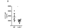
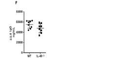
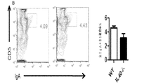
IL40は、一般に、リンパ球及び白血球の特定の集団中に存在し得る特定の受容体に結合されるものとする。受容体を同定するために、本明細書に記載のとおりIL40が標識(FLAG若しくはHIS−タグ)または放射能のいずれかで標識される方法を使用することができる。アミノ酸基を用いた標識(FLAGまたはHIS−タグ)で標識した場合、第2の抗FLAGまたは蛍光色素で標識した抗−HIS抗体を用いることによって、IL40がその受容体と上手く結合しているところを検出することができ、また、蛍光活性化セルソーター(FACS)で検出することができる。放射能で標識した場合、結合は、受容体を発現させる細胞に結合させている放射活性計数を測定することによって監視することができる。IL40の生物活性は、さまざまな白血球集団中において(例えば、表5に列挙した集団を参照のこと)発現がIL40によって調節される遺伝子の発現を測定することによって監視し得る。白血球(例えば脾細胞)は、mRNAを細胞から調製する前にインビトロにおいてIL40の存在下で6時間培養することができ、かつ、リアルタイムPCRでこれらの遺伝子の発現を測定するために使用することができる。インビボでは、IgAの最適な生成にIL40が必須である。このため、インビボでは、IL40アンタゴニストをマウスに投与し、さまざまな時間間隔でその後の血清または血漿中のIgA濃度を測定することによって、IL40アンタゴニストの活性を監視することができる。逆に、インビボでは、IL40−/−マウスに投与し、さまざまな時間間隔で血清または血漿中のIgA濃度を測定することによって、IL40アンタゴニストの活性を測定することができる。奏功するIL40アゴニストにより、IL40−/−の突然変異によって誘発されたIgAの欠損を補正することができるものとし、このため、IgA濃度は、正常なマウスのIgA濃度まで上昇するものとする。 IL40 should generally be bound to a specific receptor that may be present in a specific population of lymphocytes and leukocytes. To identify the receptor, methods can be used in which IL40 is labeled either with a label (FLAG or HIS-tag) or with radioactivity as described herein. When labeled with an amino acid group (FLAG or HIS-tag), IL40 is successfully bound to its receptor by using a second anti-FLAG or anti-HIS antibody labeled with a fluorescent dye. And can be detected with a fluorescence activated cell sorter (FACS). When labeled with radioactivity, binding can be monitored by measuring radioactivity counts bound to cells expressing the receptor. The biological activity of IL40 can be monitored by measuring the expression of genes whose expression is regulated by IL40 in various leukocyte populations (see, eg, the populations listed in Table 5). Leukocytes (eg splenocytes) can be cultured in vitro in the presence of IL40 for 6 hours before mRNA is prepared from the cells and can be used to measure the expression of these genes by real-time PCR. it can. In vivo, IL40 is essential for optimal production of IgA. Thus, in vivo, IL40 antagonist activity can be monitored by administering IL40 antagonist to mice and measuring subsequent serum or plasma IgA concentrations at various time intervals. Conversely, in vivo, the activity of an IL40 antagonist can be measured by administering to IL40 − / − mice and measuring the concentration of IgA in serum or plasma at various time intervals. It is assumed that a successful IL40 agonist can correct the IgA deficiency induced by the mutation of IL40 − / −, so that the IgA concentration rises to the normal mouse IgA concentration.
また、IL40融合タンパク質を使用して、インビボまたはインビトロでのIL40活性を監視するか、またはインビボでの未変性IL40の薬物速度を変動させることができる。融合タンパク質の例としては、これらに限定されないが、融合によりIL40−Fc融合タンパク質となるような免疫グロブリン重鎖に結合するものが挙げられる。この融合タンパク質は、多くの白血球集団中に存在するFc受容体に結合することができることによって、インビボにおいて更に安定し得るか、または所望の結合特性を示し得る。このため、リンパ系組織に対して融合タンパク質の選択的局在がもたらされ得る。別の方法としては、IL40を使用して、B細胞に優先的に結合する他のサイトカインまたはケモカインとの融合タンパク質を産生し得る。例えば、IL40は、B細胞及びT細胞の双方のサブセット中に存在するIL4受容体と結合するIL4サイトカインのこれらの一部をコードするインターロイキン4(IL4)遺伝子の一部に融合し得る。別の方法としては、IL40は、CXCR5に結合するケモカインであるCXCL13に融合させることができ、受容体もB細胞中に優先的に発現する。これらの融合タンパク質が、Bリンパ球の生物学的応答またはヒトの体内でのそれらのホーミングパターンを向上させるか、または変化させ得る所望の生物学的性質を示す場合もある。 IL40 fusion proteins can also be used to monitor IL40 activity in vivo or in vitro, or to vary the pharmacokinetics of native IL40 in vivo. Examples of fusion proteins include, but are not limited to, those that bind to an immunoglobulin heavy chain that results in an IL40-Fc fusion protein upon fusion. This fusion protein may be more stable in vivo or may exhibit the desired binding properties by being able to bind to Fc receptors present in many leukocyte populations. This can result in selective localization of the fusion protein relative to lymphoid tissue. Alternatively, IL40 can be used to produce fusion proteins with other cytokines or chemokines that preferentially bind to B cells. For example, IL40 can be fused to a portion of the interleukin 4 (IL4) gene that encodes these portions of the IL4 cytokine that bind to the IL4 receptor present in both B and T cell subsets. Alternatively, IL40 can be fused to CXCL13, a chemokine that binds CXCR5, and the receptor is also preferentially expressed in B cells. These fusion proteins may exhibit desirable biological properties that may improve or alter the biological response of B lymphocytes or their homing pattern in the human body.
IL40は、放射能(アミノ酸または原子)により、または少量のアミノ酸をその配列に添加することによって標識させ得る。これまでに使用された二つ共通の標識としては、HIS−タグ及びFLAGが挙げられる。後者は、速やかに利用可能であり、それらのエピトープを認識し、このため、標識を付着させたときに標識IL40の検出に使用することができる商業用モノクローナル抗体であるという利点を有する。 IL40 can be labeled by radioactivity (amino acids or atoms) or by adding small amounts of amino acids to the sequence. Two common labels used so far include HIS-tag and FLAG. The latter has the advantage that it is a commercial monoclonal antibody that is readily available and recognizes their epitopes and thus can be used to detect labeled IL40 when the label is attached.
更なる態様では、IL40ペプチドまたはタンパク質の使用方法が提供される。本方法では、ペプチドまたはタンパク質を使用することができる。例えば:
a)免疫細胞の誘導方法。本方法は、免疫細胞を誘導して、synaptogyrin2及び/若しくはB細胞によって産生される他のIL40誘導タンパク質を産生するため、または免疫細胞の分化または成熟を誘発するために活性剤としてペプチドまたはタンパク質を使用することを含む。細胞は、24時間インビトロで、組織培養培地、典型的には、RPMI1640若しくはDMEM若しくはウシ胎児血清、グルタミン及びメルカプトエタノールを補足した類似の培地を用いて、IL40と共にインキュベートすることができる(さまざまな濃度にて)。
b)IL40が関与する疾患の治療を必要とする対象における、IL40が関与する疾患の治療方法。本方法は、治療有効量のペプチドまたはタンパク質を対象へ投与することを含み、このペプチドまたはタンパク質はIL40アンタゴニストである。いくつかの実施形態では、本疾患は、自己免疫疾患またはリンパ腫である。
c)IL40が関与する疾患の治療を必要とする対象における、IL40が関与する疾患の治療方法。本方法は、治療有効量のIL40またはペプチド若しくはタンパク質を対象へ投与することを含み、このペプチドまたはタンパク質はIL40アンタゴニストである。いくつかの実施形態では、疾患はIgA欠損症候群、ホジキンリンパ腫若しくは非ホジキンリンパ腫、びまん性大細胞型リンパ腫、菌状息肉腫、マントル細胞リンパ腫、多発性骨髄腫または別のリンパ腫若しくは白血病;リウマチ性関節炎、全身性エリテマトーデス、シェーグレン症候群、橋本甲状腺炎、強皮症、グレーブス病、クローン病、潰瘍性大腸炎、原発性胆汁性肝硬変症、自己免疫性肝炎、多発性硬化症、乾癬、アトピー性皮膚炎、特発性肺胞線維症、過敏性肺炎、非特異的間質性肺炎または別の自己免疫疾患である。
d)IL40が関与する疾患の診断方法。本方法としては、診断法/セラノスティクス法において、標的またはサンプル対照としてペプチドまたはタンパク質を使用することを含む。抗体の使用をベースにして、唾液、血清、血漿精液、気管支肺胞洗浄液体、尿、涙、リンパ液、汗、胆汁、脳脊髄液などの生理液中での可溶性タンパク質の存在を検出するために利用可能な多くの方法がある。最も一般的な方法としては、IL40の異なる工ピトープを認識する2つの異なる抗体を使用する酵素結合(ELISA)免疫吸着法である。そのうちの1つを生理液を配置するプレート中での捕捉抗体として使用する。この抗体をプレートに固着させて、液体内に存在するIL40を「捕捉」する。二次抗体は酵素に結合する。最終的に、基質を使用して、酵素により処理し、典型的には、専用ELISAリーダーで検出され得る所与の色を発現させる。他の方法としては、酵素基質の代わりに放射能を使用して、所与の液体中に存在するIL40の量を測定するラジオイムノアッセイが挙げられる。本液体は、異なる疾患を有する患者から得ることができる。典型的には、活性化B細胞は、さまざまな癌または炎症若しくは自己免疫疾患の病原体において任意の役割を担っていることが判明した。いくつかの実施形態では、本疾患は、リンパ腫、自己免疫疾患、全身性エリテマトーデス、リウマチ性関節炎または乾癬である。
e)IL40の受容体の同定方法。本方法は、IL40受容体に結合するためのリガンドとしてペプチドまたはタンパク質を使用することを含む。IL40受容体は、その受容体に結合するために使用することができる標識IL40を用いて同定され得る。次に、抗−IL40または抗−標識抗体を使用してリガンド/受容体複合体を免疫沈降させることができる。こうした標識の例としては、His−タグ、フラグ−タグなどが挙げられる。また、受容体を発現するラジオイムノアッセイ細胞を介して最初に検出されるように、IL40を放射線標識することもできる。異なる細胞を放射線標識と共にインキュベートし、インキユベート後、細胞を洗浄するか、または粘度及び遊離対結合放射線標識IL40の遠心分離によって分離される勾配を介して通過させる。放射能を保持する細胞は、特定のIL40受容体を発現するものとする。
In a further aspect, methods of using IL40 peptides or proteins are provided. In this method, peptides or proteins can be used. For example:
a) Induction method of immune cells. The method induces immune cells to produce peptides or proteins as active agents to produce synaptogyrin2 and / or other IL40-derived proteins produced by B cells, or to induce immune cell differentiation or maturation. Including using. Cells can be incubated with IL40 in vitro for 24 hours using tissue culture media, typically RPMI 1640 or DMEM or similar media supplemented with fetal calf serum, glutamine and mercaptoethanol (various concentrations). At).
b) A method for treating a disease involving IL40 in a subject in need of treatment for a disease involving IL40. The method includes administering to the subject a therapeutically effective amount of a peptide or protein, wherein the peptide or protein is an IL40 antagonist. In some embodiments, the disease is an autoimmune disease or lymphoma.
c) A method for treating a disease involving IL40 in a subject in need of treatment for a disease involving IL40. The method includes administering to the subject a therapeutically effective amount of IL40 or a peptide or protein, wherein the peptide or protein is an IL40 antagonist. In some embodiments, the disease is IgA deficiency syndrome, Hodgkin lymphoma or non-Hodgkin lymphoma, diffuse large cell lymphoma, mycosis fungoides, mantle cell lymphoma, multiple myeloma or another lymphoma or leukemia; rheumatoid arthritis , Systemic lupus erythematosus, Sjogren's syndrome, Hashimoto's thyroiditis, scleroderma, Graves' disease, Crohn's disease, ulcerative colitis, primary biliary cirrhosis, autoimmune hepatitis, multiple sclerosis, psoriasis, atopic dermatitis Is idiopathic alveolar fibrosis, hypersensitivity pneumonia, non-specific interstitial pneumonia or another autoimmune disease.
d) A method for diagnosing a disease involving IL40. The method includes using a peptide or protein as a target or sample control in a diagnostic / theranostic method. Based on the use of antibodies, to detect the presence of soluble proteins in physiological fluids such as saliva, serum, plasma semen, bronchoalveolar lavage fluid, urine, tears, lymph fluid, sweat, bile, cerebrospinal fluid There are many ways available. The most common method is an enzyme linked (ELISA) immunosorbent method that uses two different antibodies that recognize different engineered pitopes of IL40. One of them is used as a capture antibody in a plate on which physiological fluid is placed. This antibody is affixed to the plate to “capture” IL40 present in the liquid. The secondary antibody binds to the enzyme. Ultimately, the substrate is used to treat with an enzyme, typically to develop a given color that can be detected with a dedicated ELISA reader. Other methods include radioimmunoassays that use radioactivity instead of enzyme substrate to measure the amount of IL40 present in a given liquid. This liquid can be obtained from patients with different diseases. Typically, activated B cells have been found to play any role in various cancers or pathogens of inflammatory or autoimmune diseases. In some embodiments, the disease is lymphoma, autoimmune disease, systemic lupus erythematosus, rheumatoid arthritis or psoriasis.
e) Identification method of IL40 receptor. The method includes using a peptide or protein as a ligand for binding to the IL40 receptor. An IL40 receptor can be identified with a labeled IL40 that can be used to bind to that receptor. The ligand / receptor complex can then be immunoprecipitated using anti-IL40 or anti-labeled antibodies. Examples of such labels include His-tags and flag-tags. IL40 can also be radiolabeled so that it is initially detected via radioimmunoassay cells expressing the receptor. Different cells are incubated with the radiolabel and after incubation, the cells are washed or passed through a gradient separated by centrifugation of viscosity and free versus bound radiolabeled IL40. Cells that retain radioactivity shall express specific IL40 receptors.
更なる態様では、IL40ペプチドまたはタンパク質を産生する細胞が提供され、本細胞は、組換え型細菌細胞、組換え型酵母菌細胞または組換え型哺乳類細胞である。本細胞は、本明細書に記載のいずれかのIL40ペプチドまたはタンパク質を産生し得る。また、細胞を含む臓器、組織、または動物が提供される。 In a further aspect, a cell producing an IL40 peptide or protein is provided, wherein the cell is a recombinant bacterial cell, a recombinant yeast cell or a recombinant mammalian cell. The cell may produce any IL40 peptide or protein described herein. Also provided are organs, tissues, or animals containing cells.
別の態様では、未変性遺伝子において発見される1つ以上のイントロンを欠失している配列か、または非天然ヌクレオチドが導入されている配列など、IL40遺伝子またはIL40 cDNAのヌクレオチド配列の一部または全体を含む核酸が提供される。核酸は、以下のものであり得る。
a)IL40遺伝子またはIL40 cDNAのヌクレオチド配列の一部または全体を含み、この核酸が本明細書に記載のIL40ペプチドまたはタンパク質をコードする;
b)別のヌクレオチド配列、標識(例えば、HIS−タグ若しくはFLAG)、またはビニルスルホン誘導体科染料、蛍光色素分子などの化学誘導体、またはプロテオミクスなど他の技術で一般的に使用される他のタグ(例えばビオチン)に共役結合されている。
c)プライマー、プローブ、アンチセンス分子、またはIL40遺伝子またはIL40 cDNA配列をベースとしたオリゴヌクレオチド、
d)異種核酸配列に付着した組換え型構造体、または
e)混合a)〜d)のいずれかを組み合わせたもの。
In another aspect, a portion of the nucleotide sequence of the IL40 gene or IL40 cDNA, such as a sequence lacking one or more introns found in the native gene, or a sequence into which unnatural nucleotides have been introduced, or Nucleic acids containing the whole are provided. The nucleic acid can be:
a) comprising part or all of the nucleotide sequence of the IL40 gene or IL40 cDNA, wherein the nucleic acid encodes an IL40 peptide or protein as described herein;
b) Another nucleotide sequence, a label (eg, HIS-tag or FLAG), or a chemical derivative such as a vinylsulfone derivative family dye, a fluorescent dye molecule, or other tags commonly used in other technologies such as proteomics ( For example, biotin).
c) primers, probes, antisense molecules, or oligonucleotides based on the IL40 gene or IL40 cDNA sequence,
d) A recombinant structure attached to a heterologous nucleic acid sequence, or e) a combination of any of a) to d).
いくつかの実施形態では、異種核酸配列は、プロモーター、エンハンサー、ベクターまたは発現ベクターであってもよい。 In some embodiments, the heterologous nucleic acid sequence may be a promoter, enhancer, vector or expression vector.
更なる態様では、IL40配列含有核酸の使用方法が提供される。本方法では、核酸を使用することができる。例えば:
a)IL40が関与する疾患の治療を必要とする対象における、IL40が関与する疾患の治療方法。この方法は、治療有効量の核酸を対象へ投与することを含み、本核酸により、IL40の発現が減少する。いくつかの実施形態では、本疾患は、自己免疫疾患またはリンパ腫である。
b)IL40が関与する疾患の治療を必要とする対象における、IL40が関与する疾患の治療方法。この方法としては、治療有効量の核酸を対象へ投与することを含み、本核酸によりIL40の発現が減少する。いくつかの実施形態では、疾患はIgA欠損症候群、ホジキンリンパ腫若しくは非ホジキンリンパ腫、びまん性大細胞型リンパ腫,菌状息肉腫、マントル細胞リンパ腫、または別のリンパ腫若しくは白血病;リウマチ性関節炎、全身性エリテマトーデス、シェーグレン症候群、橋本甲状腺炎、強皮症、グレーブス病、クローン病、潰瘍性大腸炎、原発性胆汁性肝硬変症、自己免疫性肝炎、多発性硬化症、乾癬、アトピー性皮膚炎、特発性肺胞線維症、過敏性肺炎、非特異的間質性肺炎または別の自己免疫疾患である。また、いくつかの実施形態では、核酸はRNAi分子であってもよい。
c)IL40が関与する疾患の診断方法。本方法は、疾患の診断法/セラノスティクス法においてプローブとして核酸を使用することを含む。いくつかの実施形態では、本疾患は、リンパ腫、自己免疫疾患、全身性エリテマトーデス、リウマチ性関節炎または乾癬である。
In a further aspect, methods for using IL40 sequence-containing nucleic acids are provided. In this method, a nucleic acid can be used. For example:
a) A method for treating a disease involving IL40 in a subject in need of treatment for a disease involving IL40. The method includes administering a therapeutically effective amount of a nucleic acid to a subject, wherein the nucleic acid reduces IL40 expression. In some embodiments, the disease is an autoimmune disease or lymphoma.
b) A method for treating a disease involving IL40 in a subject in need of treatment for a disease involving IL40. This method includes administering a therapeutically effective amount of a nucleic acid to a subject, wherein the nucleic acid reduces IL40 expression. In some embodiments, the disease is IgA deficiency syndrome, Hodgkin lymphoma or non-Hodgkin lymphoma, diffuse large cell lymphoma, mycosis fungoides, mantle cell lymphoma, or another lymphoma or leukemia; rheumatoid arthritis, systemic lupus erythematosus , Sjogren's syndrome, Hashimoto's thyroiditis, scleroderma, Graves' disease, Crohn's disease, ulcerative colitis, primary biliary cirrhosis, autoimmune hepatitis, multiple sclerosis, psoriasis, atopic dermatitis, idiopathic lung Cystic fibrosis, hypersensitivity pneumonia, nonspecific interstitial pneumonia or another autoimmune disease. In some embodiments, the nucleic acid may also be an RNAi molecule.
c) A method for diagnosing a disease involving IL40. The method includes using a nucleic acid as a probe in a disease diagnostic / theranostic method. In some embodiments, the disease is lymphoma, autoimmune disease, systemic lupus erythematosus, rheumatoid arthritis or psoriasis.
更なる態様では、IL40配列含有核酸の組換え型形態を含む細胞が提供され、本細胞は、組換え型細菌細胞、組換え型酵母菌細胞または組換え型哺乳類細胞である。本細胞は、本明細書に記載のいずれかのIL40配列含有核酸を含み得る。また、細胞を含む臓器、組織、または動物が提供される。 In a further aspect, a cell comprising a recombinant form of an IL40 sequence-containing nucleic acid is provided, the cell being a recombinant bacterial cell, a recombinant yeast cell or a recombinant mammalian cell. The cell can comprise any IL40 sequence-containing nucleic acid described herein. Also provided are organs, tissues, or animals containing cells.
別の態様では、IL40を発現する細胞のサブセットの選択方法が提供される。本方法は、IL40を発現する細胞を含む細胞集団にIL40結合分子を添加すること及び選択された細胞の集団を提供するためにIL40結合分子により標識された細胞を選択することを含む。IL40発現細胞としては、B細胞、場合により他の白血球及び骨髄細胞及び胎児肝細胞が挙げられる。IL40を発現し得る細胞型は、上皮細胞、内皮細胞、線維芽細胞、他の間質細胞またはさまざまな細胞型または特定の系譜への関与レベルの造血前駆体を含み得るこれらの臓器である。いくつかの実施形態では、a)IL40発現細胞は、マウス細胞、ラット細胞、ヒト細胞であってもよい;b)IL40結合分子は、抗−IL40抗体またはIL40受容体であってもよい。c)選択細胞は、血液、体液、細胞懸濁液または患者サンプルから選択することができる;d)選択細胞は、IL40発現細胞を調べるための調査ツールであり得る;e)また、選択細胞が血球である場合、選択細胞は、i)選択細胞により作製された免疫グロブリンのmRNA供給源;またはii)完全ヒト化抗体の新規作製方法の供給源であり得る;
またはf)a)〜e)のいずれかの組み合わせである。
In another aspect, a method for selecting a subset of cells expressing IL40 is provided. The method includes adding an IL40 binding molecule to a cell population comprising cells expressing IL40 and selecting cells labeled with the IL40 binding molecule to provide a selected population of cells. IL40 expressing cells include B cells, optionally other leukocytes and bone marrow cells and fetal liver cells. Cell types that can express IL40 are those organs that may contain epithelial cells, endothelial cells, fibroblasts, other stromal cells or various cell types or levels of hematopoietic progenitors for specific lineages. In some embodiments, a) IL40 expressing cells may be mouse cells, rat cells, human cells; b) IL40 binding molecules may be anti-IL40 antibodies or IL40 receptors. c) Selected cells can be selected from blood, body fluids, cell suspensions or patient samples; d) Selected cells can be research tools for examining IL40 expressing cells; e) In the case of blood cells, the selected cells can be i) a source of immunoglobulin mRNA produced by the selected cells; or ii) a source of novel methods for making fully humanized antibodies;
Or f) any combination of a) to e).
本方法の更なる実施形態では、IL40が関与する疾患の治療を必要とする対象における、IL40が関与する疾患の治療方法が提供される。本方法は、治療有効量の選択細胞を対象へ投与することを含む。いくつかの実施形態では、疾患はIgA欠損症候群、ホジキンリンパ腫若しくは非ホジキンリンパ腫、びまん性大細胞型リンパ腫、菌状息肉腫、マントル細胞リンパ腫、または別のリンパ腫若しくは白血病;リウマチ性関節炎、全身性エリテマトーデス、シェーグレン症候群、橋本甲状腺炎、強皮症、グレーブス病、クローン病、潰瘍性大腸炎、原発性胆汁性肝硬変症、自己免疫性肝炎、多発性硬化症、乾癬、アトピー性皮膚炎、特発性肺胞線維症、過敏性肺炎、非特異的間質性肺炎または別の自己免疫疾患である。 In a further embodiment of the method, there is provided a method for treating a disease involving IL40 in a subject in need of treatment for a disease involving IL40. The method includes administering to the subject a therapeutically effective amount of the selected cells. In some embodiments, the disease is IgA deficiency syndrome, Hodgkin lymphoma or non-Hodgkin lymphoma, diffuse large cell lymphoma, mycosis fungoides, mantle cell lymphoma, or another lymphoma or leukemia; rheumatoid arthritis, systemic lupus erythematosus , Sjogren's syndrome, Hashimoto's thyroiditis, scleroderma, Graves' disease, Crohn's disease, ulcerative colitis, primary biliary cirrhosis, autoimmune hepatitis, multiple sclerosis, psoriasis, atopic dermatitis, idiopathic lung Cystic fibrosis, hypersensitivity pneumonia, nonspecific interstitial pneumonia or another autoimmune disease.
別の態様では、対象における活性化B細胞の検出方法が提供される。本方法は、対象中でのIL40濃度を測定することを含み、IL40濃度が対照よりも上昇する場合には、活性化B細胞が示唆される。本方法では、例えば、
a)IL40濃度は、免疫検出技術により測定することができる;
b)本方法は、これらに限定されないが、例えばインターロイキン6、インターロイキン10及び特定の免疫グロブリンなど別のバイオマーカーを測定すること更に含むことができる;
c)本方法は、自己免疫疾患またはリンパ腫の診断を必要とする患者において、自己免疫疾患またはリンパ腫を診断することができ、濃度の上昇により、リンパ腫または自己免疫疾患が示唆される;
d)IL40濃度の上昇により、リンパ腫または自己免疫疾患のIL40産生サブタイプを決定することができる;または
e)混合a)〜d)のいずれかを組み合わせたものである。
In another aspect, a method for detecting activated B cells in a subject is provided. The method includes measuring IL40 concentration in the subject, suggesting activated B cells if the IL40 concentration is elevated relative to the control. In this method, for example,
a) IL40 concentration can be measured by immunodetection techniques;
b) The method can further comprise measuring another biomarker such as, but not limited to,
c) The method can diagnose an autoimmune disease or lymphoma in a patient in need of diagnosis of an autoimmune disease or lymphoma, and an increase in concentration suggests a lymphoma or autoimmune disease;
d) An increase in IL40 concentration can determine the IL40 production subtype of lymphoma or autoimmune disease; or e) any combination of a) to d).
いくつかの実施形態では、免疫検出方法はELISA法、組織学的方法、蛍光活性化細胞選別、ラジオイムノアッセイ(RIA)、免疫放射定量分析法、免疫組織化学的方法、蛍光免疫測定法、化学発光分析法、生物発光分析法、ウエスタンブロッティング法またはドットブロッティング法である。 In some embodiments, the immunodetection method is an ELISA method, histological method, fluorescence activated cell sorting, radioimmunoassay (RIA), immunoradiometric assay, immunohistochemical method, fluorescent immunoassay, chemiluminescence Analysis method, bioluminescence analysis method, Western blotting method or dot blotting method.
別の態様では、リンパ腫または自己免疫疾患の治療を必要とする対象におけるリンパ腫または自己免疫疾患の治療方法が提供される。この方法は、治療有効量の抗−IL40抗体またはIL40配列含有核酸を対象または対象の腫瘍、組織または細胞へ投与することを含む。いくつかの実施形態では、抗体は、中和抗−IL40抗体であってもよく、また、核酸はアンチセンスRNAであってもよい。いくつかの実施形態では、アンチセンスRNAはRNAi分子である。 In another aspect, a method for treating lymphoma or autoimmune disease in a subject in need of treatment for lymphoma or autoimmune disease is provided. The method includes administering a therapeutically effective amount of an anti-IL40 antibody or IL40 sequence-containing nucleic acid to a subject or a tumor, tissue or cell of the subject. In some embodiments, the antibody may be a neutralizing anti-IL40 antibody and the nucleic acid may be antisense RNA. In some embodiments, the antisense RNA is an RNAi molecule.
別の態様では、対象におけるIL40産生細胞の同定方法が提供される。本方法は、例えば、免疫組織化学的方法またはインサイチュハイブリダイゼーションにおいてIL40産生細胞を同定するために、プローブとして抗−IL40抗体またはIL40配列含有核酸を使用することを含む。IL−40抗体を使用して、フローサイトメトリーによりIL40産生細胞を検出することができるか、または免疫組織化学的方法を実施することができ、IL40配列含有核酸プローブを使用して、IL−40を産生する細胞から得られたmRNAのノーザンブロット法を行うことができる。このIL40配列を使用して、IL40産生細胞から得たmRNAでリアルタイムポリメラーゼ連鎖反応(PCR)を実施するためにプライマーをデザインすることができる。このIL40配列プローブを使用して、インサイチュハイブリダイゼーションによりIL40産生細胞を同定することができる。 In another aspect, a method for identifying an IL40 producing cell in a subject is provided. The method includes using an anti-IL40 antibody or IL40 sequence-containing nucleic acid as a probe, for example, to identify IL40 producing cells in an immunohistochemical method or in situ hybridization. IL-40 antibodies can be used to detect IL40 producing cells by flow cytometry, or immunohistochemical methods can be performed, and IL-40 sequence containing nucleic acid probes can be used to Northern blotting of mRNA obtained from cells producing can be performed. Using this IL40 sequence, primers can be designed to perform real-time polymerase chain reaction (PCR) on mRNA obtained from IL40-producing cells. This IL40 sequence probe can be used to identify IL40 producing cells by in situ hybridization.
更なる態様では、IL40受容体の同定方法が提供される。いくつかの実施形態では、本方法には、
a)IL40、標識IL40、His−タグIL40、ビオチン標識IL40、またはこれらの組み合わせによりIL40応答細胞の標識を行うことと、標識細胞を単離、精製及び/または分離することを含む。これらのラベルにより標識された細胞は、例えば、HIS−タグに対する蛍光色素標識特異的抗体を用いることにより、また、蛍光活性化セルソーターを介してサンプルを実行することにより、または、ビオチン標識IL40の場合、類似した方法において蛍光色素標識ストレプトアビジンを使用することによって分離することができる。または、
b)IL40、標識IL40、His−タグIL40、ビオチン標識IL40、またはこれらの組み合わせによりIL40応答細胞の標識を行うことと、標識細胞を単離、精製及び/または分離することを含み、本細胞は、IL40受容体が発現する真核細胞または細菌由細胞である;
c)酵母2ハイブリッド系においてIL40に結合するタンパク質を同定する;または
d)IL40受容体発現細胞由来のメンブレン調製からのIL40結合タンパク質を免疫沈降させる。例えば、水性培地中において少量のガラスビーズ、セラミックビーズまたは鋼鉄ビーズを細胞サンプルと混合したものを用いて、IL40受容体を発現する細胞を破壊することができる。この混合物に対して、攪拌及び振盪により高水準攪拌を行う。ビーズは細胞と衝突し、細胞を破壊して、細胞間成分を放出させる。均質化中、機械的剪断(ボルテックス)を適度に行い、遠心分離によって分離できる最良のメンブレンまたは細胞内調製物となる。標識(HIS−タグであってもよい)に対して抗体を使用することにより、タンパク質複合体(その受容体に結合された標識IL40など)の免疫沈降を達成することができる。免疫沈降の分析及び配列決定により、この複合体中に存在するタンパク質が同定されるものとする。
In a further aspect, a method for identifying an IL40 receptor is provided. In some embodiments, the method includes:
a) labeling IL40-responsive cells with IL40, labeled IL40, His-tag IL40, biotin-labeled IL40, or combinations thereof, and isolating, purifying and / or separating the labeled cells. Cells labeled with these labels can be obtained, for example, by using a fluorescent dye-labeled specific antibody against the HIS-tag, by running the sample via a fluorescence activated cell sorter, or in the case of biotin-labeled IL40 Can be separated by using fluorescent dye labeled streptavidin in a similar manner. Or
b) labeling IL40-responsive cells with IL40, labeled IL40, His-tag IL40, biotin-labeled IL40, or a combination thereof, and isolating, purifying and / or separating the labeled cells, A eukaryotic cell or a bacterial cell expressing the IL40 receptor;
c) identify proteins that bind to IL40 in the yeast two-hybrid system; or d) immunoprecipitate IL40 binding proteins from membrane preparations derived from IL40 receptor expressing cells. For example, a small amount of glass beads, ceramic beads or steel beads mixed with a cell sample in an aqueous medium can be used to destroy cells expressing the IL40 receptor. The mixture is stirred at a high level by stirring and shaking. The beads collide with the cells, destroy the cells, and release intercellular components. Appropriate mechanical shearing (vortexing) during homogenization results in the best membrane or intracellular preparation that can be separated by centrifugation. By using an antibody against a label (which may be a HIS-tag), immunoprecipitation of a protein complex (such as labeled IL40 bound to its receptor) can be achieved. Immunoprecipitation analysis and sequencing shall identify the proteins present in this complex.
別の態様では、IL40またはその機能的変異体の使用方法が提供される。本方法は、以下となるように免疫細胞をIL40または機能的変異体に曝露することを含む:
a)インビトロまたはインビボにおいてB細胞の成長及び分化を促進する;
b)ハイブリドーマ培養物の成長を増大させる;
c)ハイブリドーマ培養物により抗体生成物が増加する;または
d)例えば、ヒト、イヌ、マウス、ネコ、ウシ、ウマ、ブタ、ヤギ、またはヒツジなど異なる哺乳類種由来のIL40を使用することにより、組織または細胞の種発生源を決定する。
In another aspect, a method of using IL40 or a functional variant thereof is provided. The method includes exposing immune cells to IL40 or a functional variant so that:
a) promotes B cell growth and differentiation in vitro or in vivo;
b) increase the growth of the hybridoma culture;
c) the antibody product is increased by the hybridoma culture; or d) by using IL40 from different mammalian species such as, for example, human, dog, mouse, cat, cow, horse, pig, goat or sheep. Or determine the species source of the cells.
例えば、Bリンパ球は、さまざまなサイトカイン(IL4、IL6)及びB細胞受容体(抗−免疫グロブリン)を刺激する抗体、またはCD40受容体(CD40リガンドまたはCD40受容体に対する抗体)に結合する分子を補充した標準的組織培養培地において培養によってインビトロで成長し、分化し得る。これらの条件により、Bリンパ球の成長及び/または分化を招く。B細胞の分化を支持することで既知の他のサイトカインとしては、IFNγ、TGFβ、IL5,IL13及びCXCL13が挙げられる。正常B細胞のB細胞骨髄腫または他の腫瘍細胞との融合から得られるB細胞ハイブリドーマは、ハイブリドーマ(IL6)の成長を支持するサイトカインを補充した選択的培地(ハイブリドーマの成長を支持するため)においてインビトロで培養され得る。 For example, B lymphocytes bind antibodies that stimulate various cytokines (IL4, IL6) and B cell receptors (anti-immunoglobulins), or CD40 receptors (CD40 ligand or antibodies to CD40 receptors). Can grow and differentiate in vitro by culture in supplemented standard tissue culture media. These conditions lead to B lymphocyte growth and / or differentiation. Other cytokines known to support B cell differentiation include IFNγ, TGFβ, IL5, IL13 and CXCL13. B cell hybridomas resulting from the fusion of normal B cells with B cell myeloma or other tumor cells are in selective medium supplemented with cytokines that support hybridoma (IL6) growth (to support hybridoma growth). It can be cultured in vitro.
自己免疫、自己免疫疾患またはリンパ腫に関与する前述の組成物または方法のいずれかにおいて、
自己免疫疾患は、全身性エリテマトーデス、リウマチ性関節炎、乾癬、グレーブス病、自己免疫性肝炎、原発性胆汁性肝硬変、橋本甲状腺炎、またはシェーグレン症候群であってもよく、
リンパ腫は、ホジキンリンパ腫及び非ホジキンリンパ腫、マントル細胞リンパ腫、びまん性大細胞型B細胞リンパ腫、濾胞性リンパ腫、慢性リンパ性白血病、急性リンパ性白血病、MALTリンパ腫、バーキットリンパ腫、菌状息肉腫、または多発性骨髄腫であってもよい。
In any of the preceding compositions or methods involved in autoimmunity, autoimmune disease or lymphoma,
The autoimmune disease may be systemic lupus erythematosus, rheumatoid arthritis, psoriasis, Graves' disease, autoimmune hepatitis, primary biliary cirrhosis, Hashimoto's thyroiditis, or Sjogren's syndrome,
Lymphoma can be Hodgkin and non-Hodgkin lymphoma, mantle cell lymphoma, diffuse large B-cell lymphoma, follicular lymphoma, chronic lymphocytic leukemia, acute lymphocytic leukemia, MALT lymphoma, Burkitt lymphoma, mycosis fungoides, or It may be multiple myeloma.
治療するまたは治療を含む前述の組成物または方法のいずれかにおいて、抗体、ペプチド、タンパク質または核酸を局所的または全身に送達することができる。 In any of the foregoing compositions or methods to treat or include treatment, the antibody, peptide, protein or nucleic acid can be delivered locally or systemically.
診断の、診断、診断するまたは診断法/セラノスティクス法を含む前述の組成物または方法のいずれかにおいて、本方法は、生検または組織学的サンプルなど、例えば、血清、血液、体液、腫瘍、組織、または細胞などのサンプルにて実施することができる。 In any of the foregoing compositions or methods of diagnostic, diagnosing, diagnosing or comprising diagnostic / theranostic methods, the method comprises a biopsy or histological sample, such as serum, blood, body fluid, tumor, It can be performed on a sample such as a tissue or a cell.
抗体、ペプチド、タンパク質または核酸分子を含む前述の組成物または方法のいずれかにおいて、以下の製剤処方において本分子であり得る。
a)薬学的に許容可能な担体、賦形剤またはそれらを組み合わせたものを含む;
b)滅菌製剤として使用される;
c)これらに限定されないが、抗−TNFa抗体(Remicade,Humira);抗−BAFF(Benlysta);抗−CD20(Rituximab);及び抗−CD30(Adcetris)など、自己免疫疾患またはリンパ腫を治療するための別の治療薬を含む;
d)例えば、エマルション、ミセルなどの緩効性または徐放性製剤である;
e)例えば、リポソーム、包接錯体、担体などの標的投与形態である;または
f)a)〜e)のいずれかの組み合わせである。
In any of the foregoing compositions or methods comprising an antibody, peptide, protein or nucleic acid molecule, it can be the molecule in the following pharmaceutical formulation.
a) including pharmaceutically acceptable carriers, excipients or combinations thereof;
b) used as a sterile formulation;
c) To treat autoimmune diseases or lymphomas including but not limited to anti-TNFa antibodies (Remicade, Humira); anti-BAFF (Benlysta); anti-CD20 (Rituximab); and anti-CD30 (Adcetris). Including another therapeutic agent;
d) slow release or sustained release formulations such as emulsions, micelles;
e) Target dosage forms such as, for example, liposomes, inclusion complexes, carriers; or f) any combination of a) to e).
本発明を更に完全に理解するためには、添付図面と共に考慮される以下の発明を実施するための形態を参照する。
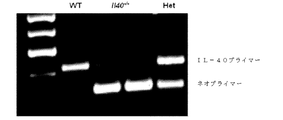
遺伝子C17orf99の遺伝子産物に関連した抗体、ペプチド、タンパク質及び核酸は、さまざまな実施形態に含まれる。さまざまな種のC17orf99遺伝子のヌクレオチド配列及びC17orf99cDNA及びさまざまな種由来のC17orf99遺伝子産物のアミノ酸配列は、以下の受け入れ番号を有する(すべてが本明細書に参照として援用される):ヒトC17ORF99:NM_001163075;マウスC17ORF99:NM_029964(全米バイオテクノロジー情報センター(National Center for Biotechnology Information)ウェブサイト(nebi.nlm.nih・gov)を参照されたい)。本明細書で使用するとき、C17orf99遺伝子産物はインターロイキン−40(IL40またはIL−40)とも称される。 Antibodies, peptides, proteins and nucleic acids associated with the gene product of gene C17orf99 are included in various embodiments. Nucleotide sequences of C17orf99 genes of various species and amino acid sequences of C17orf99 cDNA and C17orf99 gene products from various species have the following accession numbers (all of which are hereby incorporated by reference): human C17ORF99: NM_001163075; Mouse C17ORF99: NM_029964 (see National Center for Biotechnology Information website (nebi.nlm.nih.gov)). As used herein, the C17orf99 gene product is also referred to as interleukin-40 (IL40 or IL-40).
抗体は、例えば、IgG、IgM、IgA、IgD及びIgEなど、任意の免疫結合剤である。また、抗体は、抗原結合領域を有する任意の抗体様分子であってもよく、また、例えばFab’、Fab、F(ab’)2、シングルドメイン抗体(DAB)、Fv、scFv(単鎖Fv)などの抗体断片が挙げられる。さまざまな抗体ベース構造体及び断片の調製及び使用のための技術は、当業者に周知である。抗体の調製及び特性付けのための手段も当業者に周知である(例えば、Harlow and Lane,「Antibodies:A Laboratory Manual、「Cold Spring Harbor Laboratory,1988)、を参照されたい)。モノクローナル抗体(mAb)は、例えば、再現性及び大規模生産など、特定の利点を有すると認識されている。このため、ヒト、マウス、サル、ラット、ハムスター、ウサギ及びニワトリさえも起源とするモノクローナル抗体が予想される。 The antibody is any immunobinding agent such as, for example, IgG, IgM, IgA, IgD, and IgE. The antibody may also be any antibody-like molecule having an antigen-binding region, and for example, Fab ′, Fab, F (ab ′) 2 , single domain antibody (DAB), Fv, scFv (single chain Fv ) And other antibody fragments. Techniques for the preparation and use of various antibody-based structures and fragments are well known to those skilled in the art. Means for antibody preparation and characterization are also well known to those skilled in the art (see, for example, Harlow and Lane, "Antibodies: A Laboratory Manual," Cold Spring Harbor Laboratory, 1988) Monoclonal antibodies (mAb). ) Have been recognized to have certain advantages, such as reproducibility and large-scale production, etc. This is why monoclonal antibodies originating from humans, mice, monkeys, rats, hamsters, rabbits and even chickens are expected. The
広範の動物種においてIL40に対するポリクローナル抗体の調製が可能である。典型的には、抗血清の産生に使用される動物は、ウサギ、マウス、ラット、ハムスター、モルモットまたはヤギである。免疫原性の増大には、補助剤の使用及びこれらに限定されないが、例えば、アオガイヘモシアニンまたはウシ血清アルブミンなどの担体タンパク質との共役が公知の手技である。 Polyclonal antibodies against IL40 can be prepared in a wide range of animal species. Typically, the animal used for the production of antisera is a rabbit, mouse, rat, hamster, guinea pig or goat. For increasing immunogenicity, the use of adjuvants and conjugation with carrier proteins such as, but not limited to, mussel hemocyanin or bovine serum albumin are known techniques.
モノクローナル抗体は、例えば、米国特許第4,196,265号(参照によって本明細書に援用される)に例示されているものなど、既知の技術を使用して、迅速に調製することができる(40〜44)。典型的には、この技術は、例えば、精製または部分的精製タンパク質、ペプチドまたはドメインなどの選択された免疫原組成物で好適な動物を免疫化することを含む。本免疫化組成物は、効率よく抗体産生細胞を刺激する方法で投与する(45〜47)。ハイブリドーマ分泌モノクローナル抗体は、単離することができる。 Monoclonal antibodies can be rapidly prepared using known techniques, such as those exemplified in US Pat. No. 4,196,265 (incorporated herein by reference). 40-44). Typically, this technique involves immunizing a suitable animal with a selected immunogenic composition such as, for example, a purified or partially purified protein, peptide or domain. The present immunization composition is administered by a method that efficiently stimulates antibody-producing cells (45-47). Hybridoma secreting monoclonal antibodies can be isolated.
ポリクローナルまたはモノクローナル抗体は、所望の場合、濾過、遠心分離及びHPLCまたは親和性クロマトグラフィーなどのさまざまなクロマトグラフ法を用いて、更に精製することができる(47)。 Polyclonal or monoclonal antibodies can be further purified, if desired, using various chromatographic methods such as filtration, centrifugation and HPLC or affinity chromatography (47).
ヒト化モノクローナル抗体は、起源抗原の特異性を保持しつつ、遺伝子工学技術を用いて修飾され、定常領域及び/または可変領域フレームワーク配列をヒト配列に置き換えた動物発生源抗体である。このような抗体は、概して、ヒト抗原に対して特異性を有するげっ歯類抗体から誘導される。このような抗体は、概して、インビボでの治療的適用に有用である。この戦略により、外来性抗体に対する宿主反応が減少し、かつヒトエフェクター機能の選択が可能になる。したがって、本発明のいくかの実施形態には、マウス、ラットまたはヒト定常及び/または可変領域ドメイン、二重特異性抗体、組換え型及び遺伝子操作抗体及びそれらの断片を担持する他の種由来のキメラ抗体と同様に、IL40に対するヒト化抗体が含まれる。ヒト化免疫グロブリンの産生技術は、当業者に既知である(44、47〜51)。例えば、米国特許第5,693,762号は、1つ以上の相補的決定領域(CDRの)を有するヒト化免疫グロブリンの産生方法及びその組成物について開示している。無処置抗体に混ぜ合わせるとき、ヒト化免疫グロブリンは、ヒトにおいて実質的に非免疫原性であり、抗原に対してタンパク質またはエピトープを含有する他の化合物などのドナー免疫グロブリンと実質的に同一の親和性を保持する。本分野での他の教示の例としては、米国特許第6,054,297号:同5,861,155号:及び同6,020,192号が挙げられる(具体的には、すべてが参照によって援用される)。患者の疾患に対して「カスタム」作製された抗体の発現方法も同様に公知であり、こうした「カスタム」作製抗体も意図される。 Humanized monoclonal antibodies are animal source antibodies that have been modified using genetic engineering techniques to retain the specificity of the original antigen and replace constant region and / or variable region framework sequences with human sequences. Such antibodies are generally derived from rodent antibodies that have specificity for human antigens. Such antibodies are generally useful for in vivo therapeutic applications. This strategy reduces host response to foreign antibodies and allows selection of human effector function. Thus, some embodiments of the invention include mouse, rat or human constant and / or variable region domains, bispecific antibodies, recombinant and genetically engineered antibodies and other species carrying fragments thereof. Similar to the chimeric antibody, a humanized antibody against IL40 is included. Techniques for producing humanized immunoglobulins are known to those skilled in the art (44, 47-51). For example, US Pat. No. 5,693,762 discloses a method for producing humanized immunoglobulins having one or more complementary determining regions (CDRs) and compositions thereof. When combined with intact antibodies, humanized immunoglobulins are substantially non-immunogenic in humans and are substantially identical to donor immunoglobulins such as other compounds containing proteins or epitopes against the antigen. Retain affinity. Examples of other teachings in the field include US Pat. Nos. 6,054,297: 5,861,155: and 6,020,192 (specifically, see all ). Methods for expressing “custom” engineered antibodies for a patient's disease are also known, and such “custom” engineered antibodies are also contemplated.
本発明のいくつかの実施形態としては、IL40ペプチドまたはタンパク質が挙げられる。ある実施形態では、自然発生IL40タンパク質は、IL40変異型、例えば、置換変異型、欠失変異型及び/または挿入変異型と置換することができる。 Some embodiments of the invention include IL40 peptides or proteins. In certain embodiments, a naturally occurring IL40 protein can be replaced with an IL40 variant, eg, a substitution variant, a deletion variant, and / or an insertion variant.
置換変異型は、タンパク質内で更に1つ以上の部位でのアミノ酸の交換を含有する。置換は、通常、形状及び/または電荷が類似しているアミノ酸の交換が関与する同類置換である。欠失変異型では、未変性タンパク質の1つ以上の残基が欠失している。挿入突然変異体または変異型としては、タンパク質中の非未端点において1つ以上のアミノ酸の添加が挙げられる。変異型は、自然発生IL40タンパク質配列に対して、約80%以上の同一性、約85%以上の同一性、または約90%以上の同一性、約95%以上の同一性、約100%以上の同一性を有していてもよい。例えば、Clustal Omega、MUSCLE、MViewまたはMAFFT配列比較プログラムを用いて配列比較を実施することができる。比較配列では、1つのタンパク質と別のタンパク質との比較セグメントは、比較されるアミノ酸長さの約100%、または比較されるアミノ酸長さの約95%、約85%、または約80%であってもよい。比較長さは、少なくとも約20、30、40、50、55、60、65、70、または75アミノ酸以上であってもよい。変異型は、一般の自然配列のように特定の物理化学的または機能的特徴を維持していてもよく、他の変異型は、構造的及び機能的特徴の修飾された組み合わせを有してもよい。したがって、いくつかの実施形態としては、例えばIL40受容体と結合する、またはIgA応答に対するB細胞の分化に関与するなど、いくつかのまたはすべてのIL40の機能を有する機能的活性IL40変異型が挙げられる。また、いくつかの実施形態としては、IL40アゴニストまたはIL40アンタゴニストとして機能する変異型が挙げられる。いくつかの実施形態では、変異型としては自然発生ヒトIL40配列または他の種の自然発生IL40配列と同一である配列は含まない。機能的活性IL40ペプチド及びIL40ペプチド変異型を包含するなど、IL40ペプチド及びこれらの置換変異型、欠失変異型及び/または挿入変異型並びにIL40アゴニストまたはIL40アンタゴニストとして機能するIL40ペプチド及びIL40ペプチド変異型もまた意図される。いくつかの実施形態では、ペプチド変異型は、ヒトまたは他の種のIL40内に存在する自然発生アミノ酸配列と同一であるアミノ酸配列は含まない。 Substitution variants contain amino acid exchanges at one or more additional sites within the protein. A substitution is usually a conservative substitution involving the exchange of amino acids that are similar in shape and / or charge. In a deletion mutant, one or more residues of the native protein are deleted. Insertion mutants or variants include the addition of one or more amino acids at non-end points in the protein. A variant has about 80% or more identity, about 85% or more identity, or about 90% or more identity, about 95% or more identity, about 100% or more to a naturally occurring IL40 protein sequence May have the same identity. For example, sequence comparisons can be performed using Clustal Omega, MUSCLE, MView, or MATFT sequence comparison programs. In a comparative sequence, the comparison segment between one protein and another protein is about 100% of the compared amino acid length, or about 95%, about 85%, or about 80% of the compared amino acid length. May be. The comparison length may be at least about 20, 30, 40, 50, 55, 60, 65, 70, or 75 amino acids or more. Variants may maintain certain physicochemical or functional characteristics like general natural sequences, while other variants may have modified combinations of structural and functional characteristics. Good. Thus, some embodiments include functionally active IL40 variants that have some or all of IL40 function, such as binding to the IL40 receptor or involved in B cell differentiation to IgA responses. It is done. Also, some embodiments include variants that function as IL40 agonists or IL40 antagonists. In some embodiments, variants do not include naturally occurring human IL40 sequences or sequences that are identical to naturally occurring IL40 sequences of other species. IL40 peptides and their substitution variants, deletion variants and / or insertion variants and IL40 peptides and IL40 peptide variants that function as IL40 agonists or IL40 antagonists, including functionally active IL40 peptides and IL40 peptide variants Is also intended. In some embodiments, the peptide variant does not include an amino acid sequence that is identical to a naturally occurring amino acid sequence present in human or other species of IL40.
ある実施形態としては、IL40タンパク質の切頭型または他のセグメントとの融合が挙げられ、記載された機能を示す。融合タンパク質は、第二のタンパク質のすべてまたは一部に結合されているすべてのまたは一部のIL40を含有することができる。例えば、1つのタンパク質のC末端は、他のタンパク質のN末端に結合してもよい。あるいは、タンパク質は、例えば、インテグリン、フィブロネクチン受容体または他の膜糖タンパク質と非共有結合により結合されていてもよい。IL40タンパク質は、自然発生IL40アミノ酸配列またはそれらの変異型を含有することができる。 Some embodiments include fusion with a truncated or other segment of the IL40 protein and exhibit the described function. The fusion protein can contain all or part of IL40 bound to all or part of the second protein. For example, the C-terminus of one protein may be bound to the N-terminus of another protein. Alternatively, the protein may be non-covalently bound to, for example, an integrin, fibronectin receptor or other membrane glycoprotein. The IL40 protein can contain a naturally occurring IL40 amino acid sequence or a variant thereof.
いくつかの実施形態では、アミノ酸変異型が関与していないか、またはアミノ酸変異型に加えて、IL40誘導体が提供される。こうした誘導体の例としては、グリコシル化修飾IL40タンパク質、例えば、ポリエチレングリコールによって修飾されたタンパク質(PEG化)などの化学修飾IL40タンパク質、及び例えば131I標識IL40、ビオチン−IL40などのIL40抱合体が挙げられる。 In some embodiments, an amino acid variant is not involved, or in addition to the amino acid variant, an IL40 derivative is provided. Examples of such derivatives include glycosylated modified IL40 proteins, eg chemically modified IL40 proteins such as proteins modified with polyethylene glycol (PEGylation), and IL40 conjugates such as 131 I-labeled IL40, biotin-IL40, etc. It is done.
本発明のいくつの実施形態としては、自然発生IL40タンパク質またはそれらの変異型など、IL40タンパク質のすべてまたは一部をコードする核酸が挙げられる。核酸は、DNAまたはRNA分子であってもよい。核酸は、例えば、発現を目的とした別の核酸配列に共役結合されてもよく、検出を目的とした標識に共役結合されてもよく、または検出を目的とした化学的誘導体に共役結合されてもよい。例えば、核酸は、緑色蛍光タンパク質(GFP)などの標識に共役結合されてもよく、ビオチンなどの化学的誘導体に共役結合されてもよい。 Some embodiments of the invention include nucleic acids that encode all or part of an IL40 protein, such as naturally occurring IL40 proteins or variants thereof. The nucleic acid may be a DNA or RNA molecule. The nucleic acid may be conjugated to, for example, another nucleic acid sequence for expression, conjugated to a label for detection, or conjugated to a chemical derivative for detection. Also good. For example, the nucleic acid may be conjugated to a label such as green fluorescent protein (GFP) or conjugated to a chemical derivative such as biotin.
核酸は、IL40ヌクレオチド配列の増幅または合成のためのプライマーとして、またはIL40ヌクレオチド配列の同定のためのプローブとして利用することができる。いくつかの実施形態では、核酸は、IL40遺伝子またはIL40cDNA配列を含むオリゴヌクレオチドである。特定の実施形態では、核酸はアンチセンス分子である。 The nucleic acids can be utilized as primers for the amplification or synthesis of IL40 nucleotide sequences or as probes for the identification of IL40 nucleotide sequences. In some embodiments, the nucleic acid is an oligonucleotide comprising an IL40 gene or IL40 cDNA sequence. In certain embodiments, the nucleic acid is an antisense molecule.
アンチセンスオリゴヌクレオチドは、自然発生ヌクレオチド及び/または修飾若しくは置換オリゴヌクレオチドであってもよいリボ核酸(RNA)またはデオキシリボ核酸(DNA)のオリゴマーまたはポリマーである。さまざまな実施形態では、アンチセンスオリゴヌクレオチドとしては、IL40標的配列にハイブリッド形成されているヌクレオチド配列が挙げられ、例えば、プライマー結合部位として使用するための追加の5’及び/または3’フランキング配列を挙げることができる。いくつかの実施形態では、アンチセンスオリゴヌクレオチドとしては、これに限定されるものではないが、ホスホロチオエート、キラルホスホロチオエート、ホスホロジチオエート、ホスホロトリエステル、アミノアルキルホスホロトリエステル、メチル及び他のアルキルホスホネート(例えば、3’−アルキレンホスホネート及びキラルホスホネートなど)、ホスフィネート、ホスホルアミデート(例えば、3’−アミノホスホルアミデート及びアミノアルキルホスホルアミデートなど)、チオノホスホルアミデート、チオノアルキルホスホネート、チオノアルキルホスホトリエステル、並びにこれらの類縁体に連結される正常な3’−5’連結、並びに2’−5’連結を有する及びヌクレオシド単位の近接対が3’−5’から5’−3’または2’−5’から5’−2’に連結される逆極性を有するボラノホスフェートなどの修飾オリゴヌクレオチド主鎖が挙げられる。また、種々の塩、混合塩及び遊離酸形態も挙げられる。こうした修飾主鎖オリゴヌクレオチドの調製は、例えば、米国特許第4,469,863号及び同第5,750,666号に提供されて教示されており、これらはすべて参照により本明細書に援用される。アンチセンスオリゴヌクレオチドのデザイン及び合成は、当該技術分野において周知である(52)。また、アンチセンスオリゴヌクレオチド配列をデザインするためのコンピュータープログラムも利用可能である(53)。 Antisense oligonucleotides are ribonucleic acid (RNA) or deoxyribonucleic acid (DNA) oligomers or polymers that may be naturally occurring nucleotides and / or modified or substituted oligonucleotides. In various embodiments, antisense oligonucleotides include nucleotide sequences that are hybridized to IL40 target sequences, eg, additional 5 ′ and / or 3 ′ flanking sequences for use as primer binding sites. Can be mentioned. In some embodiments, antisense oligonucleotides include, but are not limited to, phosphorothioates, chiral phosphorothioates, phosphorodithioates, phosphorotriesters, aminoalkyl phosphorotriesters, methyl and other alkyl phosphonates. (Eg 3'-alkylene phosphonates and chiral phosphonates), phosphinates, phosphoramidates (eg 3'-aminophosphoramidates and aminoalkyl phosphoramidates), thionophosphoramidates, thiono Alkyl phosphonates, thionoalkyl phosphotriesters, and normal 3'-5 'linkages linked to these analogs, and adjacent pairs of 2'-5' linkages and nucleoside units from 3'-5 ' 5'-3 ' Others include modified oligonucleotide backbones such boranophosphate having opposite polarities coupled to 5'-2 from '2'-5. Also included are various salts, mixed salts and free acid forms. The preparation of such modified backbone oligonucleotides is provided and taught, for example, in US Pat. Nos. 4,469,863 and 5,750,666, all of which are incorporated herein by reference. The The design and synthesis of antisense oligonucleotides is well known in the art (52). Computer programs for designing antisense oligonucleotide sequences are also available (53).
ペプチド、タンパク質及び核酸の標準的産生方法及び作製方法が適用されてもよい。組換え型核酸コード構造体のデザインなどの標準的組換え方法が開発されてもよい。例えば、Thompson D.A. Cell and Molecular Biology Manual 2011を参照されたい。発現ベクター、例えば、コード領域に操作可能に連結されたプロモーターなどが発明され得る。組換え型酵母菌及び組換え型哺乳類細胞などの組換え型原核生物細胞及び組換え型真核生物細胞など、ベクターを含む細胞が提供される。互換性のある発現方法論も開発することができる。 Standard production and production methods of peptides, proteins and nucleic acids may be applied. Standard recombinant methods such as the design of recombinant nucleic acid coding structures may be developed. For example, Thompson D.C. A. See Cell and Molecular Biology Manual 2011. Expression vectors such as promoters operably linked to the coding region can be invented. Cells comprising the vector are provided, such as recombinant prokaryotic cells and recombinant eukaryotic cells such as recombinant yeast and recombinant mammalian cells. Compatible expression methodologies can also be developed.
例えば、IL40タンパク質またはタンパク質変異型をコードするポリヌクレオチドを、所望の宿主細胞中で機能的であるプロモーターの制御下に配置してもよい。非常に広範のプロモーターが公知であり、また、特定用途に応じて本発明の実施形態の発現ベクター中で使用可能である。通常は、選択されたプロモーターは、そのプロモーターが活性となる細胞に依存する。例えば、エンハンサー、リボソーム結合部位、転写末端部位などの他の発現制御配列も任意により含まれる。1つ以上のこれらの制御配列を含む構造体は、「発現カセット」と称される。したがって、本発明の実施形態は、関連のある機能的タンパク質をコードする核酸が所望の原核または真核宿主細胞中に多く発現するために導入される発現カセットを提供する(例えば、Ream W and Field K.G. Molecular Biology Techniques.Academic Press.2012を参照されたい)。 For example, a polynucleotide encoding an IL40 protein or protein variant may be placed under the control of a promoter that is functional in the desired host cell. A very wide variety of promoters are known and can be used in the expression vectors of embodiments of the present invention depending on the particular application. Usually, the promoter selected will depend on the cell in which the promoter is active. For example, other expression control sequences such as an enhancer, a ribosome binding site, and a transcription end site are optionally included. A structure containing one or more of these control sequences is referred to as an “expression cassette”. Thus, embodiments of the present invention provide expression cassettes that are introduced so that the nucleic acid encoding the relevant functional protein is highly expressed in a desired prokaryotic or eukaryotic host cell (eg, Ream W and Field). KG Molecular Biology Technologies. Academic Press. 2012).
いくつかの実施形態では、均質性が少なくとも約70%、75%、80%、85%または90%であるペプチドまたはタンパク質の実質的に純粋である組成物が含まれ、約92%、95%、98%、または99%以上の均質性も含まれる。精製されたペプチド及びタンパク質は、例えば、抗体産生の免疫原として、免疫細胞中での誘導分化、成熟またはタンパク質発現のための活性剤として、または医薬組成物中の治療薬として使用可能である。 In some embodiments, a composition that is substantially pure of a peptide or protein that has a homogeneity of at least about 70%, 75%, 80%, 85%, or 90% is included, including about 92%, 95% , 98%, or 99% or greater homogeneity. Purified peptides and proteins can be used, for example, as immunogens for antibody production, as active agents for induced differentiation, maturation or protein expression in immune cells, or as therapeutic agents in pharmaceutical compositions.
IL40の濃度は、核酸またはタンパク質濃度で測定され得る。例えば、細胞中でのIL40 mRNAの発現量を測定することができるか、または活性化B細胞中に存在するIL40タンパク質の量を測定することができる。これらに限定されないがPCR、マイクロアレイ法またはノーザンブロット法などの方法を用いて、mRNAの定量化を行うことができる(54、55)。タンパク質の定量化は、これらに限定されないが、酵素結合免疫吸着法(ELISA)、ラジオイムノアッセイ(RIA)、免疫放射定量分析法、蛍光免疫測定法、化学発光分析法、生物発光分析法、またはウエスタンブロッティング法、抗−タンパク質特異的抗体を含むFACS(細胞によって産生するため)などの免疫検出法を使用して実施可能である。制御値は、制御細胞集団または1つ以上の制御対象からのIL40濃度の平均値または中央値であり得る。 The concentration of IL40 can be measured by nucleic acid or protein concentration. For example, the expression level of IL40 mRNA in a cell can be measured, or the amount of IL40 protein present in activated B cells can be measured. Although not limited thereto, mRNA can be quantified using a method such as PCR, microarray method or Northern blot method (54, 55). Protein quantification includes, but is not limited to, enzyme-linked immunosorbent assay (ELISA), radioimmunoassay (RIA), immunoradiometric assay, fluorescence immunoassay, chemiluminescence assay, bioluminescence assay, or Western It can be performed using immunodetection methods such as blotting methods, FACS with anti-protein specific antibodies (to be produced by cells). The control value can be an average or median IL40 concentration from a control cell population or one or more control subjects.
いくつかの実施形態では、対象がIL40が関与する疾患を有しているという診断の後に本明細書に記載のものなどの治療が行われてもよい。例えば、診断の後に治療有効量のIL40アンタゴニストを、自己免疫またはリンパ腫と診断された対象に投与すること、またはIL40ヌクレオチド配列を含有する治療的有効量のオリゴヌクレオチドを対象へ投与することを含む治療が行われ得る。 In some embodiments, treatments such as those described herein may be performed following a diagnosis that the subject has a disease involving IL40. For example, a treatment comprising administering a therapeutically effective amount of an IL40 antagonist after diagnosis to a subject diagnosed with autoimmunity or lymphoma, or administering a therapeutically effective amount of an oligonucleotide containing an IL40 nucleotide sequence to a subject. Can be done.
いくつかの実施形態は、本発明のさまざまな実施形態の治療的使用を伴う。これらの実施形態では、対象は、本発明のさまざまな実施形態の抗体、ペプチド、タンパク質若しくは核酸またはこれらの任意の組み合わせであってもよい治療的有効量の活性剤を投与され得る。治療的有効量は、個人の状態の薬物治療に対する対象の健全性を促進するか、または改善する量である。例えば、任意の期間の対象の寿命の延長、対象の状態に寄与し得る対象への疼痛の減少、疾病の重症度の低下、治療薬の治療的効果の増大、病態または疾患の予後の改善、治療薬の投与量または投与頻度の減少、治療の侵襲性が低下している対象の治療計画の変更、及び治療薬による副作用の重症度または頻度の減少である。リンパ腫または白血病について、治療上の利益としては、疾患の新生物発生の減少または遅延、過剰増殖の低減、腫瘍成長の低下、転移の遅延、癌細胞または腫瘍細胞増殖率の低下も挙げられる。対象への活性物質の投与量は、熟練の施術者が好適な量を決定することができる対象の体重、投与態様及び疾患の適応症及び重症度に応じて変化する。 Some embodiments involve the therapeutic use of various embodiments of the invention. In these embodiments, the subject can be administered a therapeutically effective amount of an active agent that can be an antibody, peptide, protein or nucleic acid of any of the various embodiments of the invention, or any combination thereof. A therapeutically effective amount is an amount that promotes or improves the health of the subject relative to the medication of the individual's condition. For example, extending the life of a subject for any period of time, reducing pain in a subject that can contribute to the subject's condition, reducing the severity of the disease, increasing the therapeutic effect of the therapeutic agent, improving the prognosis of the condition or disease, Reducing the dose or frequency of treatment, changing the treatment plan for subjects with less invasive treatment, and reducing the severity or frequency of side effects from the treatment. For lymphoma or leukemia, therapeutic benefits also include a reduction or delay in neoplastic development of disease, a reduction in hyperproliferation, a decrease in tumor growth, a delay in metastasis, a decrease in the rate of cancer cells or tumor cell proliferation. The dosage of the active substance to the subject will vary depending on the subject's body weight, mode of administration and disease indication and severity for which a skilled practitioner can determine a suitable amount.
いくつかのケースでは、IL40遺伝子またはIL40cDNA配列を含むアンチセンス分子は、IL40が関与する疾患を有する対象において、IL40の発現を低下させるために治療薬として使用され得る。例えば、アンチセンス分子はsiRNAであってもよい。siRNAは、RNA干渉(RNAi)法において使用するための小型阻害物質RNA二重鎖である。RNAiは、二本鎖RNAが小型二本鎖セグメント(siRNA)に切断される自然発生遺伝子サイレンシングプロセスであり、その後、タンパク質−RNA複合体(「RISC」と称する)と関連し、標的mRNAが切断される(56)。さまざまな実施形態において、siRNAは、その標的IL40mRNAに対して相補度が変動し、サイズ18〜30の塩基対であってもよい。いくつかの実施形態では、siRNAとしては、センス鎖及びアンチセンス鎖の一方またはその両方の5’及び/または3’末端部での不対塩基を含んでもよい。いくつかの実施形態では、siRNAは、2つの別個の鎖の二重鎖であるか、または二重鎖領域を形成するためにヘアピン構造体を形成する一本鎖であってもよい。siRNAのデザイン及び合成は、当該技術分野において周知である(57)。また、siRNAをデザインするためのコンピュータープログラムも利用可能である(58)。他のRNAi分子としては、ゲノム的にコードするRNAであり、IL40の遺伝子発現を調整し得るミクロRNAが挙げられる。 In some cases, an antisense molecule comprising an IL40 gene or IL40 cDNA sequence can be used as a therapeutic agent to reduce IL40 expression in a subject having a disease involving IL40. For example, the antisense molecule may be siRNA. siRNAs are small inhibitor RNA duplexes for use in RNA interference (RNAi) methods. RNAi is a naturally occurring gene silencing process in which double stranded RNA is cleaved into small double stranded segments (siRNA), which are then associated with a protein-RNA complex (referred to as “RISC”) where the target mRNA is It is cut (56). In various embodiments, the siRNA may vary in complementarity to its target IL40 mRNA and may be 18-30 base pairs in size. In some embodiments, the siRNA may include unpaired bases at the 5 'and / or 3' end of one or both of the sense strand and the antisense strand. In some embodiments, the siRNA may be two separate strand duplexes, or a single strand that forms a hairpin structure to form a duplex region. The design and synthesis of siRNA is well known in the art (57). Computer programs for designing siRNAs are also available (58). Other RNAi molecules include genomically encoded RNAs and microRNAs that can regulate IL40 gene expression.
対象は、ヒト、イヌ、マウス、ネコ、または、例えばウシ、ウマ、ブタ、ヤギまたはヒツジなどの他の哺乳類であってもよい。いくつかの実施形態では、対象はIL40が関与する疾患の疑いのある対象である。いくつかの実施形態では、対象はIL40が関与する疾患の治療の必要のある対象または患者である。 The subject may be a human, dog, mouse, cat, or other mammal such as a cow, horse, pig, goat or sheep. In some embodiments, the subject is a subject suspected of having a disease involving IL40. In some embodiments, the subject is a subject or patient in need of treatment for a disease involving IL40.
解析、診断及びテラノーシス(theranosis)用のサンプルは、ヒト、イヌ、マウス、ネコ、または、例えばウシ、ウマ、ブタ、ヤギまたはヒツジなどの他の哺乳類由来であってもよい。 Samples for analysis, diagnosis and theranosis may be from humans, dogs, mice, cats, or other mammals such as cows, horses, pigs, goats or sheep.
異なる投与用製剤(滅菌、バッファ、緩効性、徐放性安定剤、軟膏など)は、最適の投与経路に依存して使用可能である。例えば、Niazi S.K.Handbook of Pharmaceutical Manufacturing Formulations Informa Healthcare 2012を参照されたい。IL40/IL40受容体相互作用の抗炎症性アゴニストまたはアンタゴニストを用いた場合と同様に、治療効果を最適化するために他の確立された薬剤と組み合わせて使用可能である。加えて、本化合物(複数可)は、1つの製剤戦略において他の治療と組み合わせて使用してもよい。薬理変化体を使用して、所望の薬物動態学成果(分泌、半減期、溶解度、または分泌経路の最適化)を得ることができる。 Different dosage formulations (sterilized, buffered, slow release, sustained release stabilizers, ointments, etc.) can be used depending on the optimal route of administration. For example, Niazi S.M. K. See Handbook of Pharmaceutical Manufacturing Formats Information Healthcare 2012. As with anti-inflammatory agonists or antagonists of IL40 / IL40 receptor interaction, it can be used in combination with other established agents to optimize therapeutic effects. In addition, the compound (s) may be used in combination with other treatments in one formulation strategy. Pharmacological variants can be used to obtain the desired pharmacokinetic outcome (secretion, half-life, solubility, or optimization of the secretory pathway).
これらの正確な投与量は、治療目的に依存し、当業者が既知の技術を使用して確認できる。例えば、Ansel,et al,Pharmaceutical Dosage Forms and Drug Delivery; Lieberman (1992)Pharmaceutical Dosage Forms(vols.1〜3)、Dekker,ISBN 0824770846, 082476918X, 0824712692,0824716981:Lloyd (1999)The Art,Science and Technology of Pharmaceutical Compounding;及びPickar(1999)Dosage Calculationsを参照されたい。従来技術で公知なとおり、タンパク質の分解、全身投与対局所送達、新規プロテアーゼ合成率、並びに年齢、体重、全身健康状態、性別、食事、投与時期、薬物相互作用及び状態の重症度の調整は、必須であってもよく、また当業者によっていくつかの実験で確認し得る。 These exact dosages will depend on the therapeutic purpose and can be ascertained by one skilled in the art using known techniques. For example, Ansel, et al, Pharmaceutical Dosage Forms and Drug Delivery; Lieberman (1992) Pharmaceutical Dosage Forms (vols. of Pharmaceutical Compounding; and Pickar (1999) Dosage Calculations. As known in the prior art, protein degradation, systemic administration versus local delivery, rate of new protease synthesis, and adjustment of age, weight, general health, sex, diet, time of administration, drug interaction and severity of condition It may be essential and can be confirmed in some experiments by those skilled in the art.
さまざまな薬学的に許容可能な賦形剤は当業者において周知である。本明細書で使用するとき、「薬学的に許容可能な賦形剤」としては、組成物の活性成分と混ぜ合わせたとき、成分が生物活性を保持することができ、対象の免疫系との破壊的反応を招くことのない材料が挙げられる。このようなものとしては、安定剤、防腐剤、塩若しくは糖複合体または結晶などを挙げることもできる。例えば、Niazi S .K. Handbook of Pharmaceutical Manufacturing Formulations Informa Healthcare 2012を参照されたい。 Various pharmaceutically acceptable excipients are well known to those skilled in the art. As used herein, “pharmaceutically acceptable excipient” refers to an ingredient that, when combined with the active ingredient of the composition, can retain biological activity and is Examples include materials that do not cause a destructive reaction. Examples of such substances include stabilizers, preservatives, salts or sugar complexes, or crystals. For example, Niazi S. K. See Handbook of Pharmaceutical Manufacturing Formats Information Healthcare 2012.
例示的薬学的担体としては、滅菌水性非水性溶液、懸濁液及び乳濁液が挙げられる。例としては、これらに限定されないが、リン酸緩衝生理食塩水、水、水中油型エマルションなどのエマルション、及びさまざまな種類の湿潤剤などの標準的薬学的賦形剤が挙げられる。非水性溶媒の例としては、プロピレングリコール、ポリエチレングリコール、オリーブ油などの植物油及びエチルオレエートなどの注射用有機エステルである。水性担体としては、水、アルコール性溶液/水性溶液、エマルションまたは塩類溶液及び緩衝媒体などの懸濁液が挙げられる。親ビヒクルとしては、塩化ナトリウム溶液、リンガーデキストロース及びデキストロース及び塩化ナトリウム、乳酸加リンガー液また凝固油が挙げられる。静脈内ビヒクルとしては、流体及び栄養素補液、電解質補液(例えば、リンガーデキストロースをベースとしたものなど)が挙げられる。他の実施形態では、組成物は、緩効性粒子、ガラスビーズ、包帯、眼へのインサート及び局所形態などの固体基質中に導入される。投与経路としては、局所、全身、呼吸器系、経口、眼、インプラント、膣内、肛門、座薬、除放デバイスなどが挙げられる。 Exemplary pharmaceutical carriers include sterile aqueous non-aqueous solutions, suspensions, and emulsions. Examples include, but are not limited to, standard pharmaceutical excipients such as phosphate buffered saline, water, emulsions such as oil-in-water emulsions, and various types of wetting agents. Examples of non-aqueous solvents are propylene glycol, polyethylene glycol, vegetable oils such as olive oil, and injectable organic esters such as ethyl oleate. Aqueous carriers include water, alcoholic / aqueous solutions, emulsions or suspensions, including saline and buffered media. Parent vehicles include sodium chloride solution, Ringer dextrose and dextrose and sodium chloride, lactated Ringer's solution or coagulated oil. Intravenous vehicles include fluid and nutrient replenishers, electrolyte replenishers (eg, those based on Ringer dextrose, etc.). In other embodiments, the composition is introduced into a solid matrix such as slow release particles, glass beads, bandages, eye inserts and topical forms. Administration routes include local, systemic, respiratory system, oral, eye, implant, intravaginal, anus, suppository, sustained release device and the like.
核酸化合物のインビボ投与については、核酸は、遊離(または「裸」)核酸として投与されることができ、または核酸の細胞標的への送達を増大させる送達剤と共に配合することができる。送達剤の例としては、これらに限定されないが、リポソーム、カチオン性脂質、PEG化ポリカチオン、カチオン性ブロックコポリマー及びポリエチレンアミン複合体が挙げられる(59)。 For in vivo administration of a nucleic acid compound, the nucleic acid can be administered as a free (or “naked”) nucleic acid, or can be formulated with a delivery agent that increases delivery of the nucleic acid to a cellular target. Examples of delivery agents include, but are not limited to, liposomes, cationic lipids, PEGylated polycations, cationic block copolymers and polyethyleneamine conjugates (59).
本明細書内の別の項で記載した適応症用の既存の治療薬は、IL40/IL40受容体相互作用のアゴニスト/アンタゴニストを組み合わせて、または引き続いて、治療成果を最適化するために使用することができる。 Existing therapeutics for indications described elsewhere in this specification are used to combine or subsequently combine agonist / antagonists of IL40 / IL40 receptor interaction to optimize therapeutic outcome be able to.
本明細書に記載のIL40遺伝子、cDNA核酸、ペプチドまたはタンパク質は、ヒトIL40ヌクレオチドまたはアミノ酸配列をベースにしていてもよく、またはイヌ、マウス、ネコ、ウシ、ヒツジ、ヤギ、ブタ若しくはウマなど、別の哺乳類由来のヌクレオチド及びアミノ酸配列をベースにしていてもよい。同様に、抗−IL40抗体を作製するために使用される抗原は、ヒトIL40抗原をベースにしてもよく、またはイヌ、マウス、ネコ、ウシ、ウマ、ブタ、ヤギまたはヒツジなどの別の哺乳類由来のIL40抗原をベースにしていてもよい。 The IL40 gene, cDNA nucleic acid, peptide or protein described herein may be based on a human IL40 nucleotide or amino acid sequence, or alternatively, such as a dog, mouse, cat, cow, sheep, goat, pig or horse. It may be based on nucleotide and amino acid sequences derived from these mammals. Similarly, the antigen used to generate the anti-IL40 antibody may be based on the human IL40 antigen or from another mammal such as a dog, mouse, cat, cow, horse, pig, goat or sheep. It may be based on the IL40 antigen.
本発明は、添付の例を参照することによって更に理解されてもよく、これらの例は目的を説明することのみを意図するものであって、いかなる意味においても本発明の範囲を制限するために構築されるものではない。 The invention may be further understood by reference to the appended examples, which are intended only to illustrate the purpose and to limit the scope of the invention in any way It is not built.
(実施例1)
序論
近年特性が決定されたサイトカイン産生B細胞のサブセット、すなわち、B調節細胞(Breg/B10)、Bエフェクタ−1(Bel)及びBエフェクタ−2(Be2)細胞により、炎症及び自己免疫疾患におけるB細胞の役割が抗体生成物に優ることを示すエビデンスが示される。特に、これらは、サイトカイン生成能力を示す。本明細書では、インターロイキン−40(IL40)と称された活性化B細胞により作製された新規サイトカインについて記載する。IL40は、マウス及びヒトの両者の胎児肝臓及び骨髄において発現する未同定遺伝子(C17ORF99)によってコードされる。また、CD40L(またはLPS)によって刺激されたB細胞中に発現する。その産生は、いくつかのサイトカイン(IL−4、IL−13及びTGFβ)によって増強され、IFNγによって阻害される。IL40は24kDの分泌タンパク質であり、他の既知のサイトカインとの関連はない。Il40欠損マウス(Il40-/-)は、巨脾腫また改変B細胞表現型を呈し、パイエル板では、CD19+B細胞数の増大及びIgA産生細胞数の低下を有する。IL40の転写は、泌乳乳腺において誘導される。更に、IL40の転写がSLE患者由来のPBMC内及びMRLFas1pr/1prマウス由来の脾細胞内で上昇する。IL40はB細胞の発現及び分化への多面効果を有する新規B細胞誘導サイトカインであると結論付けられる。
Example 1
Introduction The recently characterized subsets of cytokine-producing B cells, namely B regulatory cells (Breg / B10), B effector-1 (Bel) and B effector-2 (Be2) cells, allow B in inflammation and autoimmune diseases. Evidence is shown that the role of the cell is superior to the antibody product. In particular, they show the ability to produce cytokines. This specification describes a novel cytokine produced by activated B cells called interleukin-40 (IL40). IL40 is encoded by an unidentified gene (C17ORF99) that is expressed in both mouse and human fetal liver and bone marrow. It is also expressed in B cells stimulated by CD40L (or LPS). Its production is enhanced by several cytokines (IL-4, IL-13 and TGFβ) and inhibited by IFNγ. IL40 is a 24 kD secreted protein and is not associated with other known cytokines. Il40-deficient mice (Il40 − / − ) exhibit splenomegaly or a modified B cell phenotype, and Peyer's patches have an increased number of CD19 + B cells and a decreased number of IgA producing cells. IL40 transcription is induced in the lactating mammary gland. Furthermore, IL40 transcription is elevated in PBMC from SLE patients and in splenocytes from MRLFas 1pr / 1pr mice. It is concluded that IL40 is a novel B cell-inducing cytokine with pleiotropic effects on B cell expression and differentiation.
サイトカインは、細胞の成長、分化、炎症の調節及び造血に影響を与える活性を有する多面性分泌タンパク質の大規模な多様スーパーファミリーである(1、2)。ヒトの疾患を調節するサイトカインを介するメカニズムの解明において、新規サイトカイン及びそれらの受容体の同定は中心的であった。この情報は、これらの疾患の診断、治療及び予防に重要であった(3)。これらの重要性を考慮すると、例えば配列予測ソフトウエア及びデータベース検索など、新規サイトカインの検索に強い関心があった(4)。これによって、多くのサイトカインの同定が得られ、そのうちの最も多くは、遺伝子重複から発生する可能性の高いスーパーファミリーに属している。
(A)サイトカインとしてのIL40の同定
Cytokines are a large and diverse superfamily of pleiotropic secretory proteins with activities that affect cell growth, differentiation, regulation of inflammation and hematopoiesis (1, 2). In elucidating the mechanisms mediated by cytokines that regulate human disease, the identification of novel cytokines and their receptors has been central. This information was important for the diagnosis, treatment and prevention of these diseases (3). Considering their importance, there was a strong interest in searching for new cytokines, for example, sequence prediction software and database searching (4). This provides the identification of many cytokines, most of which belong to the superfamily likely to arise from gene duplication.
(A) Identification of IL40 as a cytokine
本発明者らは、新規サイトカインの検索に関心を持っていた。この目的を達成するために、Affymetrix U133 2.0 microarrays(5,6)をベースとするBody Index of Gene Expression(BIGE)として公知のヒトの身体における遺伝子発現の包括的データベースを用いた。このデータベースは、現在、105の異なるヒト組織/細胞型由来のゲノム規模での発現データを含有し、かつ、リンパ系組織並びに休止及び活性化されたBリンパ球及びTリンパ球の両者が挙げられる。免疫系において重要な新規遺伝子を同定するために、非免疫組織と比較したとき、リンパ球若しくは骨髄細胞のいずれかまたは免疫系の組織(骨髄、脾臓、リンパ節、胸腺、扁桃)において多く発現した遺伝子に関してBIGEデータベースを検索した。このスクリーニングにより、リンパ系組織に関連した511の遺伝子及び免疫細胞に関連した1569を得て、ここ数十年において同定され、記述されたすべての免疫系遺伝子を実質上同定した(ほとんどのサイトカイン及びケモカイン遺伝子を含む)。重要なことに、35の新規の良好でない特性決定された遺伝子が同定され、免疫系において多く発現されている膜貫通または分泌タンパク質のいずれかをコードすることが予測された。本発明者らは、近年これらの遺伝子、tetraspanin33(TSPAN33)、活性化B細胞によって発現する膜貫通タンパク質の例を公表した(7)。
B.IL40は、B細胞により産生されたサイトカインである。
The inventors were interested in searching for new cytokines. To achieve this goal, a comprehensive database of gene expression in the human body known as Body Index of Gene Expression (BIGE) based on Affymetrix U133 2.0 microarrays (5, 6) was used. This database currently contains genome-scale expression data from 105 different human tissues / cell types and includes both lymphoid tissues and resting and activated B and T lymphocytes. . Overexpressed in either lymphocytes or bone marrow cells or tissues of the immune system (bone marrow, spleen, lymph nodes, thymus, tonsils) when compared to non-immune tissues to identify novel genes important in the immune system The BIGE database was searched for genes. This screening yielded 511 genes related to lymphoid tissues and 1569 related to immune cells, and virtually identified all the immune system genes identified and described in the last decades (most cytokines and Including chemokine genes). Importantly, 35 new poorly characterized genes have been identified and were predicted to encode either transmembrane or secreted proteins that are highly expressed in the immune system. The inventors have recently published examples of these genes, tetraspanin33 (TSPAN33), transmembrane proteins expressed by activated B cells (7).
B. IL40 is a cytokine produced by B cells.
胎児肝臓、骨髄及び活性化B細胞に発現する新規低分子分泌タンパク質をコードするこれらの遺伝子C17orf99の別の1つの特性決定について記述している。本発明者らは、活性化B細胞により産生された低分子分泌タンパク質であるという事実も含め、典型なサイトカイン特性を有するために、この分子をインターロイキン40(IL40)と称した。IL40は、活性化時にB細胞により産生され、その生成物は、Th2サイトカイン(IL−4及びIL−13)またはTGFβにより増強される。逆に、その生成物はIFNγ、Th1サイトカインによって阻害される。Il40-/-マウスは、脾臓中でのB細胞数の増加を有する巨脾症を示す。更に、Il40−/−マウスは、腸パイエル板内、IgA産生部位での胚中心数及び全IgA分泌B細胞が少ない。Il40とIgAとの結合は、乳腺において泌乳の発生時にIl40が誘導される観測結果によって更に示唆される。最終的に、MRLFas(1pr/1pr)マウス(SLEマウスモデル)由来の脾細胞においてIl40転写が増加していることが判明し、自己免疫疾患において関与し得ることが示唆される。 Describes another characterization of these genes C17orf99, which encodes a novel small secreted protein expressed in fetal liver, bone marrow and activated B cells. We named this molecule Interleukin 40 (IL40) because it has typical cytokine properties, including the fact that it is a small molecule secreted protein produced by activated B cells. IL40 is produced by B cells upon activation and its product is enhanced by Th2 cytokines (IL-4 and IL-13) or TGFβ. Conversely, the product is inhibited by IFNγ, a Th1 cytokine. Il40 − / − mice exhibit splenomegaly with an increased number of B cells in the spleen. Furthermore, Il40 − / − mice have a low number of germinal centers and total IgA-secreting B cells in the intestinal Peyer's patch and at the site of IgA production. The binding between Il40 and IgA is further suggested by the observation that Il40 is induced during lactation in the mammary gland. Finally, it was found that Il40 transcription was increased in splenocytes derived from MRLFas ( 1pr / 1pr ) mice (SLE mouse model), suggesting that it may be involved in autoimmune diseases.
結果
A.新規サイトカインの同定
アノテーションされていない免疫系関連遺伝子(C17orf99)は、BIGEデータベースの解析を介して最初に同定した。C17orf99mRNAは、胎児肝臓、骨髄及び30時間抗−CD40+IL−4により活性化させたB細胞において多く発現し、他の部位での発現は、ほとんどまたはまったく認められない。すべての組織及びC17orf99の発現の平均強度の完全なリストを表1に提示する。ヒト遺伝子は、予測された20のアミノ酸(図1B)のN末端シグナル配列を有する265のアミノ酸のタンパク質をコードするオープンリーディングフレームを含有し、245のアミノ酸(約27KDa)の成熟タンパク質が予測される。BIGEデータベースにおいてIL40の発現の完全なリストは、表1に提供される。霊長類、イヌ及びマウスなどの哺乳類において相同遺伝子が同定された(6030468B19Rik、72%のタンパク質配列変換)が、ニワトリ及びゼブラフィッシュ(図2)中には存在しなかった。遺伝子は、造血臓器(ホメオスタシス)(9)においてかつ活性化Bリンパ球(炎症性)(2)によって産生された低分子分泌タンパク質(8)をコードするため、IL40(図1)と称している。BIGEの発現プロファイルは、ヒト組織RNA(図2A)のqRT−PCR解析を使用して、マウスRNAサンプルの同等の収集を用いて確認し、これは、免疫系関連組織及び特に骨髄においてマウス相同体が多くかつ特異的に発現していることが示された(図2B)。Pfam searchから、IL40は、現在公知のいかなるサイトカインファミリーにも属さずに(データに図示せず)、IL40が造血臓器及び活性化B細胞において産生された新規サイトカインであることがわかる。

本試験の前に、C17orf99は、遺伝子の調査において分泌タンパク質として同定し、シグナル配列を予測した(10)。IL40が分泌されていることを確かめるために、ヒトIL40cDNAをB細胞活性化及び分化のバーキットリンパ腫モデルであるヒト2E2B細胞からクローン化し(11)、インフレームをpTT5のクローニング部位に挿入し(12)、その結果、C末端8xHisタグとの融合タンパク質をコードする組換え型遺伝子となる。その後、HEK293細胞をpTT5一Il40構造体または対照として空のベクターにより形質移入し、1日目及び3日目に上清を収集し、組換え型IL40タンパク質の存在について解析した。抗−His抗体を用いた精製上清の親和力のウエスタンブロッティング法により、pTT5−I140構造体により形質移入された細胞のみに約27kDのタンパク質を検出し(図1C)、IL40が分泌タンパク質であることを確認する。
Prior to this study, C17orf99 was identified as a secreted protein in a genetic study and predicted a signal sequence (10). To confirm that IL40 is secreted, human IL40 cDNA was cloned from human 2E2B cells, a Burkitt lymphoma model of B cell activation and differentiation (11), and inserted in-frame into the cloning site of pTT5 (12 ), Resulting in a recombinant gene encoding a fusion protein with a C-terminal 8xHis tag. HEK293 cells were then transfected with the pTT5Il40 structure or an empty vector as a control and supernatants were collected on
B.IL40は、活性化B細胞により産生される。
例えばIL−2、IL−7及びIL−15などのサイトカインの多くは、リンパ球の個体発生及び活性化の両方において機能を有するため(13)、本発明者らは、IL40は更にこれらのプロセスに関与している可能性があると仮定した。BIGEデータベースにおけるその発現パターンから、B細胞がCD40L+IL−4により刺激されるとき、IL40の発現が増加していることがわかる。この点を確認するために、休止または活性条件下において、PBMCから精製されたヒトB細胞におけるIL40の転写を抗−CD40+IL−4を用いて24時間測定し(図4A)、TSPAN33の発現と比較した(本発明者らが近年記述したB細胞活性化マーカー)(7)。IL40の転写は、TSPAN33(p=0.01)と共に50倍を超えて(p=0.002)上方制御したところ、活性化されたヒトB細胞によるIL40の発現が示された。これらの実験は、休止または活性条件下においてヒト2E2及びジャーカット(T細胞株)を使用して繰り返し行った(2E2活性化用の抗−CD40mAb及び抗−CD3+抗−CD28)(図5A)。IL40転写は、活性化2E2細胞においてのみ誘導された。LPS+IL−4によって活性化されたマウス脾細胞(図4B)及び同一条件下にて刺激されたA20−2JマウスB細胞(図5B)と類似の結果が得られた。IL40転写の反応速度を測定するために、C57Bl/6マウス由来のCdl9+B細胞(図6)にCD40L+IL−4により、8時間、24時間、72時間及び96時間刺激を与え、Il40及びAicda(遺伝子コードAID、活性化B細胞において免疫グロブリンクラススイッチに関与する酵素(14));遺伝子の発現をqRT−PCRによって測定した(図4C)。IL40 mRNAを8時間以内で上方制御し、その発現は96時間以上上昇し続けた。
B. IL40 is produced by activated B cells.
Since many of the cytokines such as IL-2, IL-7 and IL-15 have functions in both ontogeny and activation of lymphocytes (13), we have further demonstrated that IL40 is a process of these processes. Assuming that you may be involved in The expression pattern in the BIGE database shows that IL40 expression is increased when B cells are stimulated with CD40L + IL-4. To confirm this, IL40 transcription in human B cells purified from PBMC was measured for 24 hours with anti-CD40 + IL-4 under resting or active conditions (FIG. 4A) and compared to TSPAN33 expression. (B cell activation marker recently described by the present inventors) (7). IL40 transcription was upregulated more than 50-fold (p = 0.002) with TSPAN33 (p = 0.01), indicating IL40 expression by activated human B cells. These experiments were repeated using human 2E2 and Jurkat (T cell line) under resting or active conditions (anti-CD40 mAb and anti-CD3 + anti-CD28 for 2E2 activation) (FIG. 5A). IL40 transcription was only induced in activated 2E2 cells. Similar results were obtained with mouse splenocytes activated by LPS + IL-4 (FIG. 4B) and A20-2J mouse B cells stimulated under the same conditions (FIG. 5B). To measure the kinetics of IL40 transcription, Cdl9 + B cells from C57B1 / 6 mice (FIG. 6) were stimulated with CD40L + IL-4 for 8, 24, 72 and 96 hours, and IL40 and Aicda ( Gene code AID, enzyme involved in immunoglobulin class switch in activated B cells (14)); gene expression was measured by qRT-PCR (FIG. 4C). IL40 mRNA was upregulated within 8 hours and its expression continued to rise for over 96 hours.
活性化B細胞は、刺激に依存してTh1型(Be1由来)またはTh2(Be2由来)サイトカインを分泌することが示されたため(15)、IL40の発現をサイトカインによって同様に調節したか否かを判断する必要があった。 Activated B cells have been shown to secrete Th1-type (Be1-derived) or Th2 (Be2-derived) cytokines depending on the stimulation (15), so whether IL40 expression was similarly regulated by cytokines It was necessary to judge.
マウス脾細胞においてCD40(図4E)またはTLR4(図7)の刺激は、IL−4、IL−13、IFNγまたは組み合わせたものを混ぜ合わせた(図4D)。IL40転写はTh2サイトカインの刺激時に誘発され(IL−4及び/またはIL−13、約8倍)、IFNγ(Th1サイトカイン)は3倍増加させたのみであった。抗−CD40の刺激によりIL40の転写が20倍以上増加した。興味深いことに、Th2サイトカイン(IL4またはIL13)は、抗−CD40の刺激のみの場合よりもほぼ2倍以上のIL40の産生と協同しており、その一方でIFNγにより、抗−CD40単独で観測されたレベルの半分までIL40の発現が減少した。更に、抗−CD40+TGFβによる刺激により最も高いレベルのIL40の産生が誘導された(図4E)。本発明者らは、抗−CD40の刺激時にIL40の産生が誘導され、Th2サイトカイン及びTGFβにより協同され、IFNγにより阻害されると結論付けている。 Stimulation of CD40 (FIG. 4E) or TLR4 (FIG. 7) in mouse splenocytes mixed IL-4, IL-13, IFNγ or combinations (FIG. 4D). IL40 transcription was induced upon stimulation of Th2 cytokines (IL-4 and / or IL-13, approximately 8 fold) and IFNγ (Th1 cytokine) was only increased 3 fold. Anti-CD40 stimulation increased IL40 transcription more than 20-fold. Interestingly, Th2 cytokines (IL4 or IL13) cooperate almost twice as much with the production of IL40 as anti-CD40 stimulation alone, while IFNγ observed with anti-CD40 alone. IL40 expression was reduced to half the level. Furthermore, stimulation with anti-CD40 + TGFβ induced the highest levels of IL40 production (FIG. 4E). We conclude that production of IL40 is induced upon stimulation of anti-CD40, is cooperated by Th2 cytokines and TGFβ, and is inhibited by IFNγ.
C.IL40-/-マウスは、B細胞ホメオスタシスにおいて欠陥を有する。
IL40の生物活性の特性を更に決定するために、IL40(6030468B19Rik)遺伝子の標的欠失を有する突然変異マウス株を得た(図8a、遺伝子型の決定を確認するには図9を参照されたい)。IL40−/−マウスは、生存可能であり、かつ受精し、体重増加または身体の大きさに欠陥はない(図10)。それぞれ、胸腺、腹腔、脾臓及び骨髄におけるT細胞サブセット(図11)、B1/B2の細胞比率(図12)、プラズマ細胞及び胚中心細胞のB細胞成熟(図13)、及びpro/pre/未成熟B細胞集団(図14)を比較し、WtとIL40−/−マウスとでは有意差のないことが判明した。しかし、6週齢までにIL40−/−マウスは巨脾症を呈し、IL40−/−マウスでは、長さ(0.0940±0.004対0.1360±0.0151cm、p=0.005)、腫瘤(0.1018±0.005対0.1366±0.0147g、p=0.0285)及び総細胞数(76.03±6.87対96.34±2.15*10^6細胞、p=0.0224)が増大した。CD19+B細胞対T細胞(CD3+)との比率を比較したとき、IL40−/−マウスは、Wtマウスと比較して、T細胞に対してより高い比率のB細胞を含むことが明らかになり、この差は統計的に有意であった(4.4対3.4、p=0.086(図8C)。更に、IL40−/−マウスでは、IgGl及びIgAはこれらの野生型(WT)対応物(それぞれ、616対189μg/mL、p=0.05及び10対21μg/mL、p=0.03)を上回る血清レベルの上昇を示した(図8F)。これらのデータからIl40−/−マウスにおけるB細胞のホメオスタシスが変化する可能性が示唆され、かつ、Il40は、B細胞の分化/生存において任意の役割を果たし得ることが示唆される。
C. IL40 − / − mice are defective in B cell homeostasis.
To further characterize the biological activity of IL40, a mutant mouse strain with a targeted deletion of the IL40 (603468B19Rik) gene was obtained (FIG. 8a, see FIG. 9 to confirm genotyping). ). IL40 − / − mice are viable and fertilized, and there is no defect in weight gain or body size (FIG. 10). T cell subsets in thymus, abdominal cavity, spleen and bone marrow (FIG. 11), B1 / B2 cell ratio (FIG. 12), B cell maturation of plasma cells and germinal center cells (FIG. 13), and pro / pre / un A comparison of mature B cell populations (FIG. 14) revealed no significant difference between Wt and IL40 − / − mice. However, by 40 weeks of age, IL40 − / − mice exhibit splenomegaly, and IL40 − / − mice have a length (0.0940 ± 0.004 vs. 0.1360 ± 0.0151 cm, p = 0.005). ), Mass (0.1018 ± 0.005 vs. 0.1366 ± 0.0147 g, p = 0.0285) and total cell count (76.03 ± 6.87 vs. 96.34 ± 2.15 * 10 ^ 6) Cells, p = 0.0224) increased. When comparing the ratio of CD19 + B cells to T cells (CD3 +), it was revealed that IL40 − / − mice contain a higher ratio of B cells to T cells compared to Wt mice, The difference was statistically significant (4.4 vs. 3.4, p = 0.086 (FIG. 8C). Furthermore, in IL40 − / − mice, IgGl and IgA are their wild type (WT) counterparts. (FIG. 8F) showed elevated serum levels above (616 vs. 189 μg / mL, p = 0.05 and 10 vs. 21 μg / mL, p = 0.03, respectively) From these data, Il40 − / − mice It is suggested that the homeostasis of B cells may be altered, and that IL40 may play any role in B cell differentiation / survival.
D.IL40/マウスは、より少ないIgA産生細胞を有する。
IL40-/-マウスは、血清IgA値の上昇を示すと考えられるため、粘膜免疫と結合した抗体イソタイプ、パイエル板、消化管の免疫監視に関与するリンパ結節関連の腸におけるプラズマ及び胚中心細胞の量を比較した(15)。パイエル板は、天然胚中心及びIgA分泌プラズマ細胞を含む(16)。胚中心細胞(B220+PNA+)の存在に関して、WT及びIL40-/-マウスのパイエル板から単細胞懸濁液を調製し、染色した(図15a)。IgAスイッチ胚中心細胞(PNA+B220hiIgMloIgA+)(図15B)及び全IgAスイッチ細胞(B220lo-hiPNA+IgA+)(図15C)をフローサイトメトリーにより測定した(17〜19)。IL40-/-マウスは、2倍超の胚中心細胞の減少(n=5、p=0.0005)、IgAスイッチ胚中心細胞では2.5倍の減少(n=5、p=0.0001)、及び全IgA陽性細胞(PNA+胚中心B220hi及びB220neg-loプラズマ細胞)では10倍の減少(n=5、p=0.0001)が認められた。更に、Wt及びIl40-/-マウスの糞塊中のIgA濃度は、ELISA(図15D)により測定し、IgA陽性細胞の欠失が粘膜におけるIgA分泌レベルに影響を与えるか否かを判断した。Wtマウスと比較して、IL40-/-の糞塊中のIgA値は2倍の減少が認められた(n=10、p=0.006)。IgAの欠失がIL40-/-マウス中に存在することにより、IL40が乳腺に発現するか否かを調査した。図15Eに示すように、乳腺内での泌乳時にIL40の発現が誘導される。総合すれば、これらの観測結果からIgA応答におけるIL40の役割が示唆される。
D. IL40 / mouse has fewer IgA producing cells.
Since IL40 − / − mice are thought to show elevated serum IgA levels, antibody isotypes coupled with mucosal immunity, Peyer's patches, plasma and germinal center cells in the lymph node-associated intestine involved in gastrointestinal immunity surveillance Amounts were compared (15). Peyer's patches contain natural germinal centers and IgA-secreting plasma cells (16). Single cell suspensions were prepared and stained from Peyer's patches of WT and IL40 − / − mice for the presence of germinal center cells (B220 + PNA + ) (FIG. 15a). IgA switch germinal center cells (PNA + B220 hi IgM lo IgA + ) (FIG. 15B) and total IgA switch cells (B220 lo-hi PNA + IgA + ) (FIG. 15C) were measured by flow cytometry (17-19). . IL40 − / − mice have a greater than 2-fold reduction in germinal center cells (n = 5, p = 0.0005) and 2.5-fold reduction in IgA-switched germinal center cells (n = 5, p = 0.0001) ), And all IgA positive cells (PNA + germinal center B220 hi and B220 neg-lo plasma cells) showed a 10-fold decrease (n = 5, p = 0.0001). Furthermore, the IgA concentration in the fecal mass of Wt and Il40 − / − mice was measured by ELISA (FIG. 15D) to determine whether or not IgA positive cell deletion affects the level of IgA secretion in the mucosa. Compared to Wt mice, the IgA value in the IL40 − / − fecal mass was found to be reduced by a factor of 2 (n = 10, p = 0.006). It was investigated whether IL40 was expressed in the mammary gland by the presence of IgA deletion in IL40 − / − mice. As shown in FIG. 15E, IL40 expression is induced during lactation in the mammary gland. Taken together, these observations suggest a role for IL40 in the IgA response.
これらの変化は、クラススイッチ組換え(CSR)機構(20、21)での欠陥または欠陥、胚中心応答(増殖(22)、生存(23)、活性化(24))の成熟での欠陥に起因し得る。最初に可能性のある機構として特定するために、WT及びIl40-/-B細胞におけるインビトロでのCSRの誘発を実施し、得られたIgG1、IgG2a、IgG2b及びIgAのクラススイッチはフローサイトメトリーを使用して監視した(図16を参照されたい)。WtとIl40-/-IgG1、IgG2a及びIgG3スイッチ細胞との間で差異は認められず、Il40-/-B細胞(p=0.03)においてIgAスイッチのわずかな増加のみ認められた。したがって、本発明者らは、Il40-/-マウスのパイエル板におけるIgA産生細胞の欠乏は、CSRを受ける能力の欠陥によるものではないと結論付ける。 These changes may be due to defects or defects in the class switch recombination (CSR) mechanism (20, 21), maturation of germinal center responses (proliferation (22), survival (23), activation (24)). Can be attributed. To initially identify as a possible mechanism, in vitro CSR induction in WT and Il40 − / − B cells was performed, and the resulting IgG1, IgG2a, IgG2b, and IgA class switches perform flow cytometry. Used and monitored (see FIG. 16). No difference was observed between Wt and Il40 − / − IgG1, IgG2a and IgG3 switch cells, and only a slight increase in IgA switch was observed in Il40 − / − B cells (p = 0.03). Therefore, we conclude that the lack of IgA producing cells in Peyer's patches of Il40 − / − mice is not due to a defect in the ability to undergo CSR.
IL40応答遺伝子の同定
その後、本発明者らは、IL40がリンパ球に及ぼす効果をより詳細に調査することにした。この目的を達成するために、遺伝子アレイを用いて、休止及びWTまたはIl40-/-マウスのリンパ節から得たリンパ球を刺激したLPS+IL−4に関する国際的な遺伝子発現解析を実施した。
Identification of IL40 responsive genes Subsequently, we decided to investigate in more detail the effect of IL40 on lymphocytes. To achieve this goal, an international gene expression analysis for LPS + IL-4 that stimulated lymphocytes from resting and lymph nodes from WT or Il40 − / − mice was performed using gene arrays.
遺伝子プロファイルの2つの群:休止または活性化条件下で、WTマウス対IL40-/-マウスにおいて上方制御されている遺伝子(表2)及びIL40-/-マウス対WTマウスにおいて上方制御されている遺伝子(表3)の特性を決定した。予測したとおり、最も特異的な発現遺伝子はIL40であった。興味深いことに、上位4番目に特異的に発現した遺伝子はsynaptogyrin2(Syngr2)であった。Synaptogyrin2は、中枢神経系のニューロン中に発見されるsynaptogyrinファミリーに属しているが、そのファミリーメンバーとは異なり、Syngr2は、中枢神経系内に発現するものではない。SYNGR2の発現プロファイルを決定するために、SYNGR2の発現部位に関してBIGEデータベースを解析した(図17。BIGEにおけるSYNGR2の最上発現部位に関して表4を参照されたい)。SYNGR2は、活性化B細胞、単細胞及びヒト結腸によって発現すると判断された。
近年では、IL40は活性化B細胞により産生されると判断されているが、IL40の標的細胞は明らかになっていない。IL40がB細胞またはT細胞の遺伝子発現に作用するか否かを判断するために、WTマウスサンプルとIl40-/-マウスサンプルにおいて、CD81、CD86、Ms4al(CD20)、IL−4及びStat6など、「B細胞の活性化/分化」に関与する遺伝子」と考えられる50の遺伝子とCD3(3遺伝子)、Cxcr4、Il2α及びIFNγなど、「T細胞の活性化/分化」に関与する遺伝子」と考えられる38の遺伝子とを比較した(表5)。IL40がT細胞に作用するとき、IL40が存在しないことにより(IL40-/-サンプルにおいて)、B細胞ではなくT細胞に影響を及ぼすものであり、反対に、IL40がB細胞に作用するとき、B細胞活性化遺伝子の差が予期される(図18)。更には、IL40が双方の細胞型に作用するとき、双方の遺伝子セットは変化するものとする。休止状態では、B細胞発現遺伝子のみがWTとIL40-/-マウスとの間で特異的に発現するが、この差は、統計上有意ではなかった(p=0.09)(これはおそらく、T細胞との比較に使用されたリンパ節サンプルでのB細胞の数がより少ないためである)。しかし、LPS+IL−4により刺激されたサンプルにおいて(T細胞遺伝子ではなく)、B細胞は、特異的に発現した唯一の細胞であった(p=0.018)。これらの結果により、IL40応答細胞がB細胞であることが強く示唆される。
IL40は、全身性エリテマトーデスにおいて上方制御される。
自己免疫疾患では、多くの場合、例えばIL−6、IL−21及びBLySなどの既知のB細胞サイトカインの量の異常調節が行われるため(7)、全身性エリテマトーデス(SLE)のMRL/Fas1pr/1prマウスモデル由来の脾細胞におけるIL40のmRNAの発現を測定した。このモデルでは、マウスは、抗−dsDNA(二重鎖DNA)抗体の産生と共に全身的自己免疫値及び免疫性糸球体腎炎の上昇を示し、そのいずれもがSLEの特徴である。幼若マウスは正常であると考えられ、9週の時点で目に見える症状は示さなかった。24週までにリンパ腺症及びいくつかの皮膚病変を伴う中間狼瘡症状が発現し、36週までに完全な狼瘡症状が明らかになり、多くの場合死に至る(25)。IL40mRNAの発現(図19)が増加することが判明した。マウスモデルにおいて検出されたIL40値の上昇により、IL40の異常調節がSLEの病理学に関与する場合もあり、また、本疾患の治療の対象を示し得ることが示唆される。
IL40 is upregulated in systemic lupus erythematosus.
In autoimmune diseases, MRL / Fas 1pr of systemic lupus erythematosus (SLE) is often due to abnormal regulation of the amount of known B cell cytokines such as IL-6, IL-21 and BLyS . The expression of IL40 mRNA in spleen cells derived from the / 1pr mouse model was measured. In this model, mice show increased systemic autoimmunity and immune glomerulonephritis with the production of anti-dsDNA (double stranded DNA) antibodies, both of which are characteristic of SLE. Young mice were considered normal and showed no visible symptoms at 9 weeks. By 24 weeks, intermediate lupus symptoms with lymphadenopathy and some skin lesions develop and by 36 weeks complete lupus symptoms become apparent and often death (25). It was found that IL40 mRNA expression (FIG. 19) increased. Increased IL40 levels detected in mouse models suggest that dysregulation of IL40 may be involved in the pathology of SLE and may indicate a target for treatment of this disease.
これらのヒト遺伝子の機能的特性付けにおいて有意な進歩があったが、それらの大多数においては、情報はほとんどまたはまったくなかった。実際に、コンセンサスコード配列(CCDS)(26、27)プロジェクトでは、18,673の遺伝子番号(公表14)が列挙されており、そのうちの13669のみが記述名(HUGO Gene Nomenclature Committee:ヒト遺伝子命名委員会)を有する(28)。このため、5,000を超えるCCDSエントリーには有用な名前はなく、ほとんどが確実に特性が決定されていない状態である。これらの研究を阻害する別の合併症では、研究するための試薬を欠いている。本明細書では、本発明者らは、未同定遺伝子C17orf99によってコードされるIL40と称される新規サイトカインの同定に関して報告している。C17orf99は、最初は、ヒト遺伝子発現のBIGEデータベースの解析を介して同定された86の遺伝子セットの一部として同定された(29)。これらの発現パターンは、体内の他の場所に発現がほとんどまたはまったく認められず、膜貫通または分泌タンパク質のいずれかをコードすることが予測されている細胞器官または免疫系のいずれかと関連している。本発明者らは、近年、この方法を使用して同定した新規B細胞活性化分子(TSPAN33)の同定についても報告している(7)。IL40/C17orf99は、本発明者らがこの解析の一部として報告している第2の遺伝子である。 Although there has been significant progress in the functional characterization of these human genes, there has been little or no information in the majority of them. In fact, in the consensus coding sequence (CCDS) (26, 27) project, there are 18,673 gene numbers (publication 14) listed, of which only 13669 are descriptive names (HUGO Gene Nomenclature Committee) (28). For this reason, there are no useful names for more than 5,000 CCDS entries, and most are in a state where the characteristics have not been reliably determined. Another complication that inhibits these studies lacks the reagents to study. Here we report on the identification of a novel cytokine called IL40 encoded by the unidentified gene C17orf99. C17orf99 was initially identified as part of an 86 gene set identified through analysis of the BIGE database of human gene expression (29). These expression patterns are associated with either cell organs or the immune system that are expected to encode either transmembrane or secreted proteins with little or no expression elsewhere in the body . The present inventors have also recently reported the identification of a novel B cell activation molecule (TSPAN33) identified using this method (7). IL40 / C17orf99 is the second gene that we have reported as part of this analysis.
新規サイトカインインターロイキン−40(IL40)としてC17orf99によりコードされたタンパク質の同定は、いくつかの因子に基づく。第1に、発現が免疫系のいくつかの組織及び細胞に制限される低分子分泌タンパク質であり、他の多くの公知のサイトカインに関する発現プロファイルに類似している。本発明者らは、骨髄におけるIL40の発現に関して調査を開始し、他のサイトカインと対照的に、骨髄間質細胞ではなく、リンパ液により発現したことを見出した。BIGEデータベースにおいて活性化された末梢血B細胞により、わずかではあるが、IL40が有意に発現することから、更にこれがB細胞生成物であったことが示唆された。IL40は、低分子分泌タンパク質をコードし、その発現が、前炎症状態下で培養されたB細胞中において誘発され、さまざまなサイトカインによって調節されている。総合すれば、本発明者らは、C17orf99は、新規B細胞誘導サイトカインをコードしていると結論付ける。 The identification of the protein encoded by C17orf99 as the novel cytokine interleukin-40 (IL40) is based on several factors. First, it is a small secreted protein whose expression is restricted to several tissues and cells of the immune system, similar to the expression profile for many other known cytokines. The inventors have begun to investigate the expression of IL40 in the bone marrow and found that it was expressed by lymph rather than bone marrow stromal cells, in contrast to other cytokines. IL40 was significantly expressed by peripheral blood B cells activated in the BIGE database, further suggesting that this was a B cell product. IL40 encodes a small secreted protein whose expression is induced in B cells cultured under pro-inflammatory conditions and is regulated by various cytokines. Taken together, we conclude that C17orf99 encodes a novel B cell-derived cytokine.
近年まで、もっとも公知であるサイトカインは、近年特徴が明らかになったものであっても、公知のサイトカインファミリーに属している。例えば、IL37は、インターロイキンファミリ1ーのメンバーである。これにより、同定が促進される。その一方で、IL40は、任意の公知サイトカインファミリーには属さない。本発明者らは、哺乳類において相同遺伝子を同定したが、ニワトリまたはゼブラフィッシュでは同定しなかった。このことは、その機能が哺乳類の免疫系に制限され得ることを示している。
Until recently, the most known cytokines belong to the known cytokine family, even if their characteristics have been clarified in recent years. For example, IL37 is a member of the
実際に、本発明者らは、IFNγ、Th1サイトカインではなく、Th2サイトカイン(IL−4及びIL−13)の存在下において、抗−CD40mAb(またはCD40L、若しくはLPS)による活性化時に、B細胞内でIL40が誘導されると判断した。IL40は、TGFβの存在下における活性化により最も有意に誘導される。これまでB細胞機能上でTGFβに寄与していたいくつかの機能は、間接的にIL40によって媒介されてもよい。TGFβがB細胞の増殖(30)及びIgG1,IgG2a、IgG3及びIgEへのCSRを同時に阻害し、IgA及びIgG2bに対するCSRを誘導する。 Indeed, the inventors have found that B cell intracellularity upon activation with anti-CD40 mAb (or CD40L or LPS) in the presence of Th2 cytokines (IL-4 and IL-13) but not IFNγ, Th1 cytokines. It was determined that IL40 was induced. IL40 is most significantly induced by activation in the presence of TGFβ. Several functions that previously contributed to TGFβ on B cell function may be indirectly mediated by IL40. TGFβ simultaneously inhibits B cell proliferation (30) and CSR to IgG1, IgG2a, IgG3 and IgE, and induces CSR for IgA and IgG2b.
B細胞条件でのTGFβノックアウト(31)またはTGFR(30)(もしくはTGFRの成分、すなわちSmad2-/-(32))マウスは、パイエル板中でのIgAの産生において欠陥を有するが、胚中心細胞の頻度及び数の増大並びに他のイソタイプのすべてに対するCSRは増加した。IL40-/-マウスは、パイエル板のIgA産生細胞中に欠陥を有するが、胚中心細胞が減少していることから、この欠陥の機構はTGFβまたはTGFR欠失マウスから分離されていることが示唆される。更に、内因性TGFβは、CSRのインビトロ脾細胞の誘導中においてすべてのイソタイプに対してCSRには必須である。内因性TGFβが阻害されることによりすべてのイソタイプのCSRが減少し(33)するため、LPS及びThlまたはTh2サイトカインによるインビトロ誘導中、Il40-/-マウス由来のB細胞でもCSRに欠陥を有することはないが、IgAスイッチのわずかな増加は認められた。GC及びIgA産生細胞内の欠陥は、例えば、活性化(CD40またはB7欠失マウス)(34)、増殖(Ccnd3-/-マウス)(22)、生存(Pdcd1lg2-/-、CD274-/-Pdcd1lg2-/-及びPdcd1-/-マウス)(35)、または基底膜への移動(FTY720によるS1P阻害)(36)など、GC応答の欠陥により発生したと考えられる点に注意することが重要である。しかし、これらの欠陥はいずれも、脾臓においてB細胞数の増加を有するが、パイエル板における胚中心細胞及びIgA分泌細胞の数が少ないIl40-/-マウスの表現型に類似している。また、Il40-/-マウスの糞便中のIgA数が少ないことを明らかにした。TGFβでは、活性化B細胞によるIl40の産生を強く増強し、また乳腺における泌乳時にIl40が誘導される。総合して考えると、これらの観測結果から、Il40がIgA応答に対するB細胞の分化に関与していることがわかる。 TGFβ knockout (31) or TGFR (30) (or a component of TGFR, ie Smad2 − / − (32)) mice under B cell conditions are defective in IgA production in Peyer's patches, but germinal center cells Increased frequency and number and increased CSR for all other isotypes. IL40 − / − mice have defects in Peyer's patch IgA-producing cells, but germinal center cells are reduced, suggesting that the mechanism of this defect is isolated from TGFβ or TGFR-deficient mice. Is done. Furthermore, endogenous TGFβ is essential for CSR for all isotypes during induction of CSR in vitro splenocytes. Inhibition of endogenous TGFβ reduces all isotypes of CSR (33), so B cells from Il40 − / − mice are also defective in CSR during in vitro induction with LPS and Thl or Th2 cytokines There was no slight increase in the IgA switch. Defects in GC and IgA producing cells include, for example, activation (CD40 or B7 deficient mice) (34), proliferation (Ccnd3 − / − mice) (22), survival (Pdcd1lg2 − / − , CD274 − / − Pdcd1lg2 -/- And Pdcd1 -/- mice) (35), or translocation to the basement membrane (S1P inhibition by FTY720) (36) is important to note that it is thought to be caused by a defective GC response . However, both of these defects are similar to the phenotype of Il40 − / − mice with an increased number of B cells in the spleen but a low number of germinal center cells and IgA secreting cells in Peyer's patches. It was also clarified that the number of IgA in the feces of Il40 − / − mice was small. TGFβ strongly enhances the production of Il40 by activated B cells and induces Il40 during lactation in the mammary gland. Taken together, these observations indicate that Il40 is involved in B cell differentiation in response to IgA responses.
近年では、分泌サイトカインを介する炎症誘発性または抗炎症性応答のスキューイングにおいて、B細胞は、T細胞との相互作用を有することがわかっているため(37)、B細胞サブセットによって産生されるサイトカインは、免疫調節の主要調節因子として認識されている。リウマチ性関節炎(RA)及びSLEなどの自己免疫疾患の治療における治療戦略として、特異的サイトカイン相互作用の改変により需要が増加しているため、活性化B細胞により産生される新規サイトカインを発見することは非常に有用である(38、39)。更に、B細胞は、多くのヒト自己免疫疾患に関与していることがわかっており、このため、B細胞により産生された新規サイトカインを同定することにより、B細胞媒介病態の理解に寄与することになる。 In recent years, B cells have been shown to interact with T cells in skewing pro-inflammatory or anti-inflammatory responses via secreted cytokines (37), thus cytokines produced by B cell subsets Is recognized as a major regulator of immune regulation. To discover novel cytokines produced by activated B cells as a therapeutic strategy in the treatment of autoimmune diseases such as rheumatoid arthritis (RA) and SLE due to the increasing demand due to the modification of specific cytokine interactions Is very useful (38, 39). In addition, B cells are known to be involved in many human autoimmune diseases, and thus contribute to an understanding of B cell mediated pathologies by identifying novel cytokines produced by B cells. become.
本明細書において、本発明者らは、周辺域において活性化B細胞により排他的に産生された新規サイトカインであるIL40に関する最初の報告について記述している。これまでにIL40に関する報告が存在しないため、その確実な経路及びシグナル伝達機構は明らかになっていない。本発明者らは、マイクロアレイ解析により、WTとIl40-/-マウスのリンパ節とを比較したとき、ほとんどのB細胞遺伝子が変化したことを明らかになったため、IL40は、自己分泌シグナル伝達において活性化B細胞により機能し得ると考えている。また、免疫応答の調節におけるB細胞サイトカインの関与を理解するにあたって、B細胞の分化に伴う新規サイトカインの発見も重要である。更に、IL40-/-転写は、SLEマウス及びSLEヒトにおいて増加していることから、IL40の異常調節が自己免疫疾患に関与し得ることが示唆される。 Herein, we describe the first report on IL40, a novel cytokine produced exclusively by activated B cells in the surrounding area. Since there is no report regarding IL40 so far, its reliable pathway and signal transduction mechanism have not been clarified. Since the present inventors have revealed that most B cell genes have changed when comparing WT and lymph nodes of Il40 − / − mice by microarray analysis, IL40 is active in autocrine signaling. It is thought that it can function by activated B cells. In order to understand the involvement of B cell cytokines in regulating immune responses, it is also important to discover novel cytokines that accompany B cell differentiation. Furthermore, IL40 − / − transcription is increased in SLE mice and SLE humans, suggesting that dysregulation of IL40 may be involved in autoimmune diseases.
方法:
B細胞、CSR及びプラズマ細胞の分化
脾臓細胞をFBS(10%)、50mMβ−メルカプトエタノール及び1x抗生物質−抗かび物質混合物(15240−062;Invitrogen Corp.)の入ったRPMI中で37°Cにて48ウェルプレートで再懸濁し、以下の試薬により刺激した:IgAへのCSRに関して、大腸菌由来のLPS(5μg/ml)(055:B5;Sigma−Aldrich)及びTGF−β(2ng/ml;R&D Systems)及び抗−IgDデキストラン(Fina Biosolutions)。更に、IgGへのCSR用のrmIL−4(5ng/ml)とIgG3へのCSR用のIFN−γ(50ng/ml;PeproTech Inc.)とを組み合わせてLPSを使用した。表面Igを解析するために、FITC−抗−マウスIgGl(クローンA85−1)、抗−マウスIgG2a(クローンR19−15)、抗−マウスIgG2b(クローンR12−3)、抗−マウスIgG3(クローンR40−82)または、抗−マウスIgA(クローンC10−3)ラットmAb及びPE−抗−マウスCD45R(B220)(クローンRA3−6B2)ラットmAb(BD Biosciences)による染色後、第4日目に細胞を収集した。細胞はPBS中1%パラホルムアルデヒドで固定し、FACSにより解析した。
Method:
B Cell, CSR and Plasma Cell Differentiation Spleen cells were cultured at 37 ° C. in RPMI with FBS (10%), 50 mM β-mercaptoethanol and 1 × antibiotic-antifungal mixture (15240-062; Invitrogen Corp.). Were resuspended in 48-well plates and stimulated with the following reagents: LPS from E. coli (5 μg / ml) (055: B5; Sigma-Aldrich) and TGF-β (2 ng / ml; R & D) for CSR to IgA Systems) and anti-IgD dextran (Fina Biosolutions). Furthermore, LPS was used in combination with rmIL-4 (5 ng / ml) for CSR to IgG and IFN-γ (50 ng / ml; PeproTech Inc.) for CSR to IgG3. To analyze surface Ig, FITC-anti-mouse IgGl (clone A85-1), anti-mouse IgG2a (clone R19-15), anti-mouse IgG2b (clone R12-3), anti-mouse IgG3 (clone R40) -82) or cells on
パイエル板B細胞をフィリコエリトリン(PE)−標識抗−マウスCD45R(B220)ratmAb(RA3−6B2、eBiosciences)及びFITC−標識PNA(E−Y Laboratories、San Mateo,CA)、7AAD(Biolegend)及びAPC−標識IgM(Biolegend)で染色し、FACScalibur(Becton−Dickinson)及びFlowJoソフトウエアにより解析した。 Peyer's patch B cells were treated with phycoerythrin (PE) -labeled anti-mouse CD45R (B220) rat mAb (RA3-6B2, eBiosciences) and FITC-labeled PNA (EY Laboratories, San Mateo, Calif.), 7AAD (Biolegend) and Stained with APC-labeled IgM (Biolegend) and analyzed with FACScalibur (Becton-Dickinson) and FlowJo software.
リアルタイムPCRを用いて、所与のサンプル中のIL40mRNAの量を測定することができる。この目的では、さまざまなマシンは利用可能であり、それぞれが正確なmRNAを増幅し、測定するために、特定のプライマーのデザインを必要とする。Lightcycler(Roche(Indianapolis,Indiana,USA))を使用した。 Real-time PCR can be used to measure the amount of IL40 mRNA in a given sample. For this purpose, various machines are available, each requiring specific primer designs to amplify and measure the correct mRNA. The Lightcycler (Roche (Indianapolis, Indiana, USA)) was used.
実施例2
IL40受容体運搬細胞の同定
IL40受容体を運搬している細胞を判定するために、C57BL/6マウス由来の脾臓を収集し、均質化し、FACSチューブ(0.5×10E6細胞/チューブ)に入れた。まず、脾臓を氷上で30分間、FACSブロックバッファにてインキュベートした。次に、氷冷FACS洗浄により1回細胞を洗浄して、組換え型His−タグIL40(10μg/ml)で30分間氷上でインキュベートした。その後、細胞を2回洗浄し、氷上でウサギ抗−6x His標識抗体(1:500希釈)で30分間インキュベートした。次に、細胞を2回洗浄し、FITC(1:200希釈)、イソタイプ対照または他の細胞表面染色抗体に共役結合されたヤギ抗−ウサギ二次抗体でインキュベートした。結果は図20に示す。この実験から、IL40はB細胞に結合するが、T細胞に結合しないことがわかった。
Example 2
Identification of IL40 receptor-carrying cells To determine cells carrying IL40 receptor, spleens from C57BL / 6 mice are collected, homogenized, and placed in FACS tubes (0.5 × 10E6 cells / tube) It was. First, the spleen was incubated in FACS block buffer for 30 minutes on ice. The cells were then washed once with ice-cold FACS wash and incubated with recombinant His-tag IL40 (10 μg / ml) for 30 minutes on ice. Cells were then washed twice and incubated with rabbit anti-6x His-labeled antibody (1: 500 dilution) for 30 minutes on ice. The cells were then washed twice and incubated with goat anti-rabbit secondary antibody conjugated to FITC (1: 200 dilution), isotype control or other cell surface stained antibody. The results are shown in FIG. From this experiment, it was found that IL40 binds to B cells but not T cells.
実験で使用した試薬:
ウサギ抗−6xHisタグ抗体(Abeam、カタログ番号ab9108)
FITCロバ抗−ウサギ(Biolegend、カタログ番号406403)
ウサギIgGイソタイプ対照(Santa Cruz、カタログ番号sc−2027)
PE抗−マウスCD45R/B220(Biolegend、カタログ番号103207)
APC抗−マウスCD8b.2抗体(Biolegend、カタログ番号140409)
PE抗−マウスCD3抗体(Biolegend、カタログ番号100307)
APC抗−マウスCD19抗体(Biolegend、カタログ番号115511)
Reagents used in the experiment:
Rabbit anti-6xHis tag antibody (Abeam, catalog number ab9108)
FITC donkey anti-rabbit (Biolegend, catalog number 406403)
Rabbit IgG isotype control (Santa Cruz, catalog number sc-2027)
PE anti-mouse CD45R / B220 (Biolegend, catalog number 103207)
APC anti-mouse CD8b. 2 antibodies (Biolegend, catalog number 140409)
PE anti-mouse CD3 antibody (Biolegend, catalog number 100307)
APC anti-mouse CD19 antibody (Biolegend, catalog number 115511)
実施例3
受容体発現細胞のサイトカインに対する機能的応答。細胞においてIL40によって調節するバイオマーカー遺伝子を見出すために、脾臓、リンパ節、胸腺または骨髄などの免疫器官由来細胞をさまざまな量の組換え型マウスまたはヒトIL40(使用される細胞の発生源に依存して)と共にインキュベートし、その後、細胞をこれらの遺伝子のmRNAに転写させるために、数時間(6時間、8時間、24時間)インキュベートした。その後、細胞を採取して、Affymetrixの遺伝子アレイを用いてマイクロアレイ解析のために処理した。標準的技術を用いてmRNAを調製し、マイクロアレイを用いて、ハイブリダイゼーションのためにcDNAを産生している。マイクロアレイを読み取り、データは専有Affymetrixソフトウエアにより解析した。遺伝子は制御されている。
Example 3
Functional response to cytokines of receptor expressing cells. In order to find biomarker genes that are regulated by IL40 in cells, cells derived from immune organs such as spleen, lymph nodes, thymus or bone marrow vary in varying amounts of recombinant mouse or human IL40 (depending on the source of the cells used) And then incubated for several hours (6 hours, 8 hours, 24 hours) to allow the cells to be transcribed into mRNA for these genes. Cells were then harvested and processed for microarray analysis using Affymetrix gene arrays. MRNA is prepared using standard techniques, and microarrays are used to produce cDNA for hybridization. The microarray was read and the data analyzed by proprietary Affymetrix software. The gene is regulated.
実施例4
アゴニストまたはアンタゴニストの機能に関するIL40配列変異型の評価。受容体発現細胞の機能的応答を使用して、アゴニストまたはアンタゴニストを監視することができる。この目的を達成するために、アゴニストまたはアンタゴニストのいずれか一方を用いて受容体発現細胞をインキュベートし、qPCRによりIL40応答遺伝子の発現を監視することができる。これらの遺伝子の量により、IL40がその受容体に結合できるか否かが示される。キラー細胞は、これらのキラー細胞においてIL40によって誘導される遺伝子と同一の遺伝子誘導するIL40アゴニストを用いてインキュベートする。IL40及びそれぞれの候補アンタゴニストを用いてキラー細胞をインキュベートした場合、アンタゴニストにより、これらのバイオマーカー遺伝子の誘導をブロックするものとする。
Example 4
Assessment of IL40 sequence variants for agonist or antagonist function. The functional response of receptor expressing cells can be used to monitor agonists or antagonists. To achieve this goal, receptor expressing cells can be incubated with either agonists or antagonists and the expression of IL40 responsive genes can be monitored by qPCR. The amount of these genes indicates whether IL40 can bind to its receptor. Killer cells are incubated with an IL40 agonist that induces the same genes as those induced by IL40 in these killer cells. When killer cells are incubated with IL40 and respective candidate antagonists, the induction of these biomarker genes should be blocked by the antagonist.
実施例5
受容体構造の同定の予測的実施例。サイトカインの受容体は、いくつかのタンパク質鎖から構成され得る。良好な結果をもたらす確立された方法を使用して、後述のようないくつかの可能な方法を使用してIL40受容体を同定することができる。
Example 5
Predictive example of receptor structure identification. Cytokine receptors can be composed of several protein chains. Using established methods that give good results, IL40 receptors can be identified using several possible methods as described below.
方法A:種子方法、cDNAライブラリ、及びクローン結合試験。cDNAライブラリにより、IL40に結合することが既知の細胞を調製することができる。いずれの細胞がIL40に結合するかは、IL40を放射能タグで標識し、ラジオイムノアッセイを実施することによって決定することができる。別の方法としては、His−タグIL40を使用して、細胞に結合することができ、また、抗−His抗体を使用して、受容体を発現する細胞へのサイトカインの結合を検出することができる。Hisの代わりにFLAGなどの他の標識を使用することができる。一旦、受容体を発現する細胞が同定されると、これらの細胞のcDNAライブラリを作製することができ、HEK293またはBAF3細胞などの細胞をcDNAライブラリークローンのプールにより形質移入することができる。現在、IL40に結合するこれらの細胞は、上述のとおり監視することができる。蛍光性標識細胞は、フローサイトメトリーにより選別することができ、mRNAの供給源として使用するように培養することができる。このmRNAを使用して、IL40受容体鎖の発現も担う所与の細胞に形質移入されたライブラリからcDNAをクローニングすることができる。cDNA構造体における特定の配列を使用して、その細胞中で見出されるライブラリcDNAの他に、PCR用にプライマーをデザインすることができる。IL40受容体が構成される鎖の数とは関係なく、本方法を使用することができ、唯一の相違点は、陽性細胞が同定される頻度である。 Method A: Seed method, cDNA library, and clone binding test. A cDNA library can be used to prepare cells known to bind to IL40. Which cells bind to IL40 can be determined by labeling IL40 with a radioactivity tag and performing a radioimmunoassay. Alternatively, His-tag IL40 can be used to bind to the cells, and anti-His antibodies can be used to detect cytokine binding to cells expressing the receptor. it can. Other labels such as FLAG can be used in place of His. Once cells expressing the receptor are identified, cDNA libraries of these cells can be generated and cells such as HEK293 or BAF3 cells can be transfected with a pool of cDNA library clones. Currently, these cells that bind to IL40 can be monitored as described above. Fluorescently labeled cells can be sorted by flow cytometry and can be cultured for use as a source of mRNA. This mRNA can be used to clone cDNA from a library transfected into a given cell that is also responsible for expression of the IL40 receptor chain. In addition to library cDNA found in the cell, primers can be designed for PCR using specific sequences in the cDNA construct. Regardless of the number of chains that the IL40 receptor comprises, the method can be used, the only difference being the frequency with which positive cells are identified.
方法B:生化学的方法、リガンドの標識及び結合複合体の単離。このリガンドをHis若しくはFLAG(またはその他)などのタグにより標識し、IL40受容体を発現する細胞由来の膜を用いてインキュベートすることができる。さまざまな洗剤を添加して、受容体を結合し、抗−Hisまたは抗−Flag抗体により免疫沈降させたゲル中で複合体を奏功させることができるリガンドの能力を最適化することができる。この方法は、IL40受容体が有する鎖の数に関係なく使用するものとする。得られた免疫沈降バンドを単離して、配列を決定し、バイオインフォマティクスを使用して、各タンパク質をコードする遺伝子の同一性を判定することができる。 Method B: Biochemical methods, ligand labeling and isolation of binding complexes. The ligand can be labeled with a tag such as His or FLAG (or others) and incubated with a cell-derived membrane that expresses the IL40 receptor. Various detergents can be added to optimize the ability of the ligand to bind the receptor and allow the complex to succeed in gels immunoprecipitated with anti-His or anti-Flag antibodies. This method should be used regardless of the number of chains that the IL40 receptor has. The resulting immunoprecipitation bands can be isolated, sequenced, and bioinformatics can be used to determine the identity of the genes encoding each protein.
方法C:遺伝的方法;2ハイブリッドシステム。IL40相互作用タンパク質は、2つの酵母菌ハイブリッド系を用いて同定することができる。IL40は、融合タンパク質(例えば、GALBD)として調製し、その潜在的に相互作用するタンパク質は、GALADで標識された融合タンパク質として調製することができる。この場合、IL40は「餌」であり、相互作用タンパク質は「餌食」である。サッカロマイセス・セレヴィシエにおいてこの系の試験を行い、この酵母菌系は、相互作用タンパク質により報告された遺伝子(例えば、LacZ)の転写を招くことになる。LacZ遺伝子に発現する酵母菌は、2つのタンパク質の良好な相互作用(相補性)が発現する酵母菌である。この系により、IL40受容体を構成するいくつかのタンパク質を同定することができる。 Method C: Genetic method; two hybrid system. IL40 interacting proteins can be identified using a two yeast hybrid system. IL40 can be prepared as a fusion protein (eg, GALBD) and its potentially interacting protein can be prepared as a fusion protein labeled with GALAD. In this case, IL40 is “bait” and the interacting protein is “bait”. Testing this system in Saccharomyces cerevisiae, this yeast system will lead to transcription of the gene (eg, LacZ) reported by the interacting protein. A yeast expressed in the LacZ gene is a yeast that expresses a good interaction (complementarity) between two proteins. This system makes it possible to identify several proteins that constitute the IL40 receptor.
実施例6
抗体の産生
方法A:マウスまたはウサギポリクローナル;免疫的選択。抗体は、組換え型ヒト、マウスまたはラットIL40により動物を免疫化することにより、作製することができる。ヒツジ、ヤギ、ロバまたはウマは、免疫化することができる。いくつかの免疫化量を用いることができ、また、抗体の作製は、ELISAによって監視することができる。一旦所望の応答が得られると、免疫化させた動物を出血させることにより、また血清を得ることにより、抗体を採取することができる。
Example 6
Antibody production Method A: Mouse or rabbit polyclonal; immunoselection. Antibodies can be generated by immunizing animals with recombinant human, mouse or rat IL40. Sheep, goats, donkeys or horses can be immunized. Several immunization doses can be used and antibody production can be monitored by ELISA. Once the desired response is obtained, antibodies can be harvested by bleeding the immunized animal and obtaining serum.
方法B:マウスまたはラットモノクローナル抗体。マウスをIL40により免疫化し、マウスの応答を監視し、かつIL40に対して強い応答を示すものを選択することによって、ヒトIL40に対するモノクローナル抗体を作製することができる。脾臓を得て、骨髄腫細胞に対してポリエチレングリコールまたは類似の融合試薬により融合して、ハイブリドーマを産生する。得られたハイブリドーマをインビトロで成長させ、HAT培養液を用いて選択し、ハイブリドーマを選択的に成長させることができる。得られたハイブリドーマをクローン化し、産生された抗体は、ELISA、ウエスタン、FACSまたは免疫組織化学的方法によりスクリーニングすることができる。 Method B: Mouse or rat monoclonal antibody. Monoclonal antibodies against human IL40 can be generated by immunizing mice with IL40, monitoring the mouse response, and selecting those that exhibit a strong response to IL40. Spleens are obtained and fused to myeloma cells with polyethylene glycol or similar fusion reagents to produce hybridomas. The resulting hybridoma can be grown in vitro and selected using a HAT medium to selectively grow the hybridoma. The resulting hybridomas are cloned and the antibodies produced can be screened by ELISA, Western, FACS or immunohistochemical methods.
ファージディスプレイを用いる抗体の作製。マウスをヒトIL40により免疫化し、これらの脾臓を使用して、ファージディスプレイライブラリを生成する。その後、組換え型IL40に結合する能力に関して、このライブラリをスクリーニングする。良好なファージディスプレイクローンを同定し、抗体の結合部位の配列情報を得るためにこれらの配列を得る。分子生物学技術を用いて得られた抗体に遺伝子操作を行い、適切な完全ヒト抗体とすることができる。 Production of antibodies using phage display. Mice are immunized with human IL40 and these spleens are used to generate a phage display library. The library is then screened for the ability to bind to recombinant IL40. These sequences are obtained to identify good phage display clones and to obtain sequence information for antibody binding sites. Genetically manipulated antibodies obtained using molecular biology techniques can be made into suitable fully human antibodies.
実施例7
サイトカインの発現のための核酸構造体
方法1:ヒトまたはマウス由来の細胞、IL40mRNAの発現を使用して、固定オリゴ−dTを用いるRT−PCRを実施することができる。次にIL40のためにデザインされたプライマーを用いて、IL40cDNAを増幅することができる。このcDNAを哺乳類、細菌または昆虫プラスミドベクターなどの高発現ベクターに挿入することができる。各系では、IL40遺伝子を含有するこのcDNAを適切な受容個体細胞に挿入して、その細胞がIL40タンパク質を産生することができるようにインキュベートすることができる。これらの細胞の上清を採取して、生化学的方法を用いてIL40を精製するために使用する。ヒトIL40 cDNA配列(配列番号4)を図21に示す。
Example 7
Nucleic acid constructs for cytokine expression Method 1: RT-PCR with immobilized oligo-dT can be performed using human or mouse derived cells, expression of IL40 mRNA. The IL40 cDNA can then be amplified using primers designed for IL40. This cDNA can be inserted into a high expression vector such as a mammalian, bacterial or insect plasmid vector. In each system, this cDNA containing the IL40 gene can be inserted into an appropriate recipient individual cell and incubated so that the cell can produce the IL40 protein. The supernatants of these cells are collected and used to purify IL40 using biochemical methods. The human IL40 cDNA sequence (SEQ ID NO: 4) is shown in FIG.
方法2:方法1と同様ではあるが、方法1にはないエクソン/イントロンに特異的なプライマーをデザインすることによって、IL40mRNAのクローニングスプライシング変異体のクローニングを行う。
Method 2: Similar to
方法3:標識遺伝子またはレポーター遺伝子をコードする配列を挿入して、融合タンパク質を作製することができる点以外は方法1と同様である。融合タンパク質を使用して、IL40産生細胞を検出することができる。ウェスタンブロット法または親和性精製では、標識融合タンパク質も使用することができる。
Method 3: Same as
方法4:プロモーターを異なる遺伝子の高誘導性または高活性プロモーターと置換することができる点以外は、方法1と同様である。これにより、最終発現系において導入遺伝子の発現を制御することができる。
Method 4: Same as
実施例8
IL40タンパク質の作製及び精製のための上記核酸構造体の使用
方法1:陽性または陰性選択剤(ネオマイシン/βガラクトシダーゼ/GFP)と共にIL40cDNAインサートを含有するプラスミドベクター構造体を使用して、IL40cDNAが挿入されるベクターに依存して、哺乳類(HEK、HELAなど)細胞株、昆虫細胞または細菌に形質移入することができる。プラスミドを発現する形質転換/形質移入細胞は、正の選択または負の選択を行い、組換え型プロテインを産生することのない細胞を除去することができる。高用量組換え型IL40は、形質移入細胞/形質転換細胞の上清または溶解産物から直接得ることができる。
Example 8
Use of the above nucleic acid constructs for the production and purification of IL40 protein Method 1: IL40 cDNA is inserted using a plasmid vector construct containing an IL40 cDNA insert with a positive or negative selection agent (neomycin / β-galactosidase / GFP). Depending on the vector, mammalian (HEK, HELA, etc.) cell lines, insect cells or bacteria can be transfected. Transformed / transfected cells that express the plasmid can be positively or negatively selected to eliminate cells that do not produce recombinant protein. High-dose recombinant IL40 can be obtained directly from the supernatant or lysate of transfected / transformed cells.
方法2:別の方法では、IL40cDNAインサートを含むベクターにより形質移入/形質転換した細胞の溶解産物で化学的沈殿を実施して、濃縮量のタンパク質を得ることができる。化学的方法としては、塩、pH、有機溶剤、金属イオン、非イオン性ポリマー、すべてのタンパク質を非特異的に沈殿させる方法が挙げられる。組換え型タンパク質を含有する溶解産物は、化学的沈殿剤と共に混合する。沈殿物は、低速遠心分離またはデカントすることにより採取することができる。次により小さい沈殿剤がメンブレンを通過することができるような透析チューブを介して透析することにより沈殿物を再懸濁し、そのチューブ内にタンパク質を保持することができる。沈殿物は、組換え型タンパク質など、濃縮した量の全タンパク質を含有することになる。 Method 2: Alternatively, chemical precipitation can be performed on the lysate of cells transfected / transformed with a vector containing the IL40 cDNA insert to obtain a concentrated amount of protein. Chemical methods include salts, pH, organic solvents, metal ions, nonionic polymers, and methods that non-specifically precipitate all proteins. The lysate containing the recombinant protein is mixed with a chemical precipitant. The precipitate can be collected by low speed centrifugation or decanting. The precipitate can then be resuspended by dialysis through a dialysis tube such that smaller precipitants can pass through the membrane, retaining the protein in that tube. The precipitate will contain a concentrated amount of total protein, such as recombinant protein.
方法3:別の方法では、抗−IL40抗体または電磁ビーズに連結させた組換え型IL40受容体を含むカラムを用いて、IL40cDNAインサートを含むベクターにより形質移入/形質転換した細胞の溶解産物で親和性精製を実施することができる。このカラムを洗浄して、非特異的結合タンパク質を除去することができる一方で、組換え型IL40とビーズに連結された抗体との結合を維持することができる。次に、rIL40と抗−IL40抗体との間の相互作用に影響を及ぼすpHまたは塩の変化を用いて、カラムを溶出させることができる。 Method 3: Another method uses a column containing a recombinant IL40 receptor linked to an anti-IL40 antibody or electromagnetic beads to affinity for lysates of cells transfected / transformed with a vector containing an IL40 cDNA insert. Sex purification can be carried out. The column can be washed to remove non-specific binding proteins while maintaining binding between recombinant IL40 and the antibody linked to the beads. The column can then be eluted using pH or salt changes that affect the interaction between rIL40 and anti-IL40 antibody.
方法4:別の方法では、IL40cDNAインサートを含むベクターにより形質移入/形質転換した細胞の溶解産物は、メンブレンを介するサイズ排除を用いて収集し、濃縮させることができる。30KD未満のタンパク質に対してのみ貫通可能なメンブレンを含有する遠心分離カラムを使用することができる。次に、30KD未満のタンパク質を収集し、その中には、形質移入/形質転換細胞から作製した高用量の組換え型IL40を含むものとする。 Method 4: In another method, lysates of cells transfected / transformed with a vector containing the IL40 cDNA insert can be collected and concentrated using size exclusion through a membrane. Centrifugal columns containing membranes that can only penetrate proteins below 30 KD can be used. Next, proteins less than 30 KD are collected, including high doses of recombinant IL40 made from transfected / transformed cells.
方法5:別の方法では、IL40cDNAインサートを含むベクターにより形質移入/形質転換した細胞の溶解産物は、高速液体クロマトグラフィーを用いて収集し、精製することができる。各タンパク質が分析物と別々に相互作用するため、組換え型IL40を含有する分画を収集することができる。 Method 5: In another method, lysates of cells transfected / transformed with a vector containing the IL40 cDNA insert can be collected and purified using high performance liquid chromatography. Because each protein interacts with the analyte separately, fractions containing recombinant IL40 can be collected.
実施例9
IL40受容体発現細胞の単離(FACS単離)
方法A:単離、精製。IL40受容体(複数可)を運搬している細胞を判定するために、マウス脾臓を収集し、均質化し、FACSチューブ(0.5e6細胞/チューブ)に入れる。まず、FACSブロックバッファにより30分間氷上で脾細胞をインキュベートする。次に、氷冷FACS洗浄により1回細胞を洗浄して、組換え型His−タグIL40(10μg/ml)で30分間氷上でインキュベートする。その後、細胞を2回洗浄し、ウサギ抗−Hisタグ抗体(1:500希釈)で氷上で30分間インキュベートする。その後、細胞を2回洗浄し、FITC、イソタイプ対照または他の細胞表面染色抗体に共役結合されたヤギ抗−ウサギ二次抗体で30分間インキュベートする。この実験から、IL40はB細胞に結合し、陽性対照として使用できることがわかる。
Example 9
Isolation of cells expressing IL40 receptor (FACS isolation)
Method A: Isolation, purification. To determine cells carrying IL40 receptor (s), mouse spleens are collected, homogenized and placed in FACS tubes (0.5e6 cells / tube). First, splenocytes are incubated on ice for 30 minutes with FACS block buffer. The cells are then washed once with ice-cold FACS wash and incubated with recombinant His-tag IL40 (10 μg / ml) for 30 minutes on ice. The cells are then washed twice and incubated with rabbit anti-His tag antibody (1: 500 dilution) for 30 minutes on ice. The cells are then washed twice and incubated with a goat anti-rabbit secondary antibody conjugated to FITC, isotype control or other cell surface stained antibody for 30 minutes. This experiment shows that IL40 binds to B cells and can be used as a positive control.
標識細胞は、FITCのこれらの発現をベースとしたフローサイトメトリーにより選別することができる。選別した細胞は、IL40受容体を発現するものとする。 Labeled cells can be sorted by flow cytometry based on their expression of FITC. Sorted cells shall express the IL40 receptor.
方法B:細胞集団からの枯渇。毒素で標識したIL40(例えば、抗体薬物共役など)を使用して、IL40受容体を発現する細胞を破壊することができる。IL40受容体に対する抗体は、単独または抗体−薬物共役のいずれか一方を同じ目的で使用することもできる。
方法C:動物における細胞の枯渇。抗−IL40受容体モノクローナル抗体の静脈内注射を使用して、IL40受容体−発現細胞を枯渇させることができる。抗体は、「裸」抗体であるか、または抗体薬物共役であってもよい。
Method B: Depletion from the cell population. Toxin labeled IL40 (eg, antibody drug conjugates, etc.) can be used to destroy cells that express the IL40 receptor. Antibodies for IL40 receptor can be used for the same purpose, either alone or antibody-drug conjugates.
Method C: Cell depletion in animals. Intravenous injection of anti-IL40 receptor monoclonal antibodies can be used to deplete IL40 receptor-expressing cells. The antibody may be a “naked” antibody or an antibody drug conjugate.
実施例10
IL40の影響を発見するためのIL40トランスジェニックまたはノックアウトマウスの使用。IL40-/-マウスを使用して、IL40の機能を調査することができる。このマウスを公表した(Tang, T. et al. A mouse knockout library for secreted and trasnmembrane proteins, 2010.Nat.Biotechnol.7:749−55)。観察された唯一の表現型は、マウスを免疫化したとき、血清中でIgG1が上昇した。このマウス由来の免疫組織を得ることができ、異なる剤(T細胞では抗−CD3及び抗−CD28;B細胞ではリポポリサッカリド(LPS)または抗−CD40及びIL4;単球ではLPS)により活性化することができる。次にマイクロアレイ分析を行って野生型とIL40-/-組織との間に特異的に発現する遺伝子を同定することができる。さまざまな組織のフローサイトメトリーによりマウスも表現型となっているか、または血液化学検査並びに血球計数を実施することができる。癌、自己免疫疾患または感染性疾患など、さまざまな疾患モデルでマウスを使用することができる。
Example 10
Use of IL40 transgenic or knockout mice to discover the effects of IL40. IL40 − / − mice can be used to investigate the function of IL40. This mouse was published (Tang, T. et al. A mouse knockout library for secreted and trasnmembrane proteins, 2010. Nat. Biotechnol. 7: 749-55). The only phenotype observed was an increase in IgG1 in serum when mice were immunized. Immune tissue from this mouse can be obtained and activated by different agents (anti-CD3 and anti-CD28 for T cells; lipopolysaccharide (LPS) or anti-CD40 and IL4 for B cells; LPS for monocytes) can do. Microarray analysis can then be performed to identify genes that are specifically expressed between wild type and IL40 − / − tissues. Mice are also phenotyped by flow cytometry of various tissues, or blood chemistry as well as blood cell counts can be performed. Mice can be used in various disease models such as cancer, autoimmune disease or infectious disease.
IL40-/-マウスのほかに、マウスの導入遺伝子として、IL40が過剰発現することもあり得る。このため、インビボでのIL40の影響が拡大し、マウスモデルにおけるこのサイトカインの影響を調査する。この調査を行うために、マウスの胚の単一細胞への前核の注入により導入遺伝子を導入し、ゲノム内に集約させる。遺伝子は、遺伝子の発現がトリガーされ得るプロモーターの制御下にある。インビボでのIL40の過剰発現により、このサイトカインのインビボでの影響を理解する。得られたトランスジェニックIL40マウスの表現型により、インビボでのその機能を理解することができる。 In addition to IL40 − / − mice, IL40 may be overexpressed as a mouse transgene. This expands the effects of IL40 in vivo and investigates the effects of this cytokine in a mouse model. To perform this investigation, the transgene is introduced by injection of the pronuclei into a single cell of a mouse embryo and aggregated into the genome. The gene is under the control of a promoter that can trigger expression of the gene. The overexpression of IL40 in vivo understands the in vivo effects of this cytokine. The phenotype of the resulting transgenic IL40 mouse can understand its function in vivo.
参考文献
以下の公開は、参照により本明細書に組み込まれる。
1.Oppenheim JJ.Cytokines:past, present, and future。International journal of hematology2001;74(l):3〜8。
2.Dinarello CA.Historical insights into cytokinesEuropean journal of immunology 2007;37 Suppl 1:S34−45。
3.Shi Y, Ullrich SJ, Zhang J, Connolly K, Grzegorzewski KJ, Barber MC, et al. A novel cytokine receptor−ligand pair Identification, molecular characterization, and in vivo immunomodulatory activity。The Journal of biological chemistry.
2000;275(25):19167〜76。
4.Zou Q, Wang Z, Guan X, Liu B, Wu Y, Lin Z. An approach for identifying cytokines based on a novel ensemble classifier. BioMed research international. 2013:2013:686090。
5.Lee J, Hever A, Willhite D, Zlotnik A, Hevezi P. Effects of RNA degradation on gene expression analysis of human postmortem tissues. FASEB journal : official publication of the Federation of American Societies for Experimental Biology. 2005;19(10):1356〜8。
6.Roth RB, Hevezi p, Lee J, Willhite D, Lechner SM, Foster AC, et al. Gene expression analyses reveal molecular relationships among 20 regions of the human CNS. Neurogenetics. 2006;7(2):67−80。
7.Luu VP, Hevezi P, Vences−Catalan F, Maravillas−Montero JL, White CA, Casali P, et al. TSPAN33 is a novel marker of activated and malignant B cells. Clinical Immunology. 2013;149(3, Part B):388〜99。
8.Ioannidou E. Therapeutic modulation of growth factors and cytokines in regenerative medicine. Current pharmaceutical design. 2006;12(19):2397〜408。
9.Velazquez L, Cheng AM, Fleming HE, Furlonger C, Vesely S, Bernstein A, et al. Cytokine signaling and hematopoietic homeostasis are disrupted in Lnk−deficient mice. The Journal of experimental medicine. 2002;195(12):1599〜611。
10.Clark HF, Gumey AL, Abaya E, Baker K, Baldwin D, Brush J, et al. The secreted protein discovery initiative (SPDI), a large−scale effort to identify novel human secreted and transmembrane proteins: a bioinformatics assessment. Genome research. 2003;13(10):2265〜70。
11.Xu Z, Fulop Z, Wu G, Pone EJ, Zhang J, Mai T, et al. 14−3−3 adaptor proteins recruit AID to 5’−AGCT−3’−rich switch regions for class switch recombination. Nature structural & molecular biology. 2010; 17(9):1124〜35。
12.Loignon M, Perret S, Kelly J, Boulais D, Cass B, Bisson L, et al. Stable high volumetric production of glycosylated human recombinant IFNalpha2b in HEK293 cells. BMC biotechnology. 2008:8:65。
13.Ma A, Koka R, Burkett P. Diverse functions of IL−2, IL−15, and IL−7 in lymphoid homeostasis. Annual review of immunology. 2006:24:657〜79。
14.Rosenberg BR, Papavasiliou FN. Beyond SHM and CSR:AID and related cytidine deaminases in the host response to viral infection Advances in immunology
2007:94:215〜44。
15.Lycke NY, Bemark M. The role of Peyer’s patches in synchronizing gut IgA responses. Frontiers in immunology 2012:3:329。
16.Fagarasan S, Kinoshita K, Muramatsu M, Ikuta K, Honjo T. In situ class switching and differentiation to IgA−producing cells in the gut lamina propria. Nature. 2001;413(6856):639〜43。
17.Park SR, Zan H, Pal Z, Zhang J, Al−Qahtani A, Pone EJ, et al. HoxC4 binds to the promoter of the cytidine deaminase AID gene to induce AID expression, class−switch DNA recombination and somatic hypermutation Nature immunology. 2009;10(5):540〜50。
18.Tezuka H, Abe Y, Iwata M, Takeuchi H, Ishikawa H, Matsushita M, et al. Regulation of lgA production by naturally occurring TNF/iNOS−producing dendritic cells.Nature. 2007;448(7156):929〜33。
19.Chen L, Chen Z, Baker K, Halvorsen EM, da Cunha AP, Flak MB, et al. The short isoform of the CEACAM1 receptor in intestinal T cells regulates mucosal immunity and homeostasis via Tfh cell induction.Immunity 2012;37(5):930〜46。
20.Muramatsu M, Kinoshita K, Fagarasan S, Yamada S, Shinkai Y, Honjo T. Class switch recombination and hypermutation require activation−induced cytidine deaminase (AID), a potential RNA editing enzyme. Cell. 2000; 102(5):553〜63。
21.Rada c, Di Noia JM, Neuberger MS.Mismatch recognition and uracil excision provide complementary paths to both Ig switching and the A/ T−focused phase of somatic mutation. Molecular cell. 2004; 16(2):163〜71。
22.Cato MH, Chintalapati SK, Yau IW, Omori SA, Rickert RC. Cyclin D3 is selectively required for proliferative expansion of germinal center B cells. Molecular and cellular biology.2011 ;31(1):127〜37。
23.Durandy A, Kracker S, Fischer A. Primary antibody deficiencies. Nature reviews Immunology. 2013;13(7):519〜33。
24.Bergqvist P, Gardby E, Stensson A, Bemark M, Lycke NY.Gut IgA class switch recombination in the absence of CD40 does not occur in the lamina propria and is independent of germinal centers. Journal of immunology.2006:177(11):772〜83。
25.Liu J, Karypis G, Hippen KL, Vegoe AL, Ruiz P, Gilkeson GS, et al. Genomic view of systemic autoimmunity in MRLlpr mice. Genes and immunity.2006;7(2):156〜68。
26.Pruitt KD, Harrow J, Harte RA, Wallin C, Diekhans M, Maglott DR, et al. The consensus coding sequence (CCDS) project: Identifying a common protein−coding gene set for the human and mouse genomes. Genome research. 2009;19(7):1316〜23。
27.Harte RA, Farrell CM, Loveland JE, Suner MM, Wilming L, Aken B, et al. Tracking and coordinating an international curation effort for the CCDS Project.
Database : the journal of biological databases and curation. 2012:2012:bas008。
28.Gray KA, Daugherty LC, Gordon SM, Seal RL, Wright MW, Bruford EA.
Genenames.org: the HGNC resources in 2013.Nucleic acids research.2013;41(Database issue):D545〜52。
29.Burkhardt AM, Tai KP, Flores−Guiterrez JP, Vilches−Cisneros N, Kamdar K, Barbosa−Quintana O, et al. CXCL17 is a mucosal chemokine elevated in idiopathic pulmonary fibrosis that exhibits broad antimicrobial activity.Journal of immunology.2012;188(12):6399−406。
30.Cazac BB, Roes J. TGF−beta receptor controls B cells responsiveness and induction of IgA in vivo.Immunity 2000:13(4):443〜51。
31.Gros MJ, Naquet p, Guinamard RR.Cell intrinsic TGF−beta l regulation of B cells.Journal of immunology.2008;180(12):8153〜8。
32.Klein J, Ju W, Heyer J, Wittek B, Haneke T, Knaus p, et al. B cells−specific deficiency for Smad2 in vivo leads to defects in TGF−beta−directed IgA switching and changes in B cells fate.Journal of immunology.2006;176(4):2389〜96。
33.Snapper CM, Waegell W, Beernink H, Dasch JR. Transforming growth factor−beta 1 is required for secretion of IgG of all subclasses by LPS−activated murine B cells in vitro. Journal of immunology.1993:151(9):4625〜36。
34.Wong SC, 0h E, Ng CH, Lam KP。Impaired germinal center formation and recall T− cell−dependent immune responses in mice lacking the costimulatory ligand B7〜H2。Blood.2003;102(4):1381〜8。
35.Good−Jacobson KL, Szumilas CG, Chen L, Sharpe AH, Tomayko MM, Shlomchik MJ. PD−1 regulates germinal center B cells survival and the formation and affinity of long−lived plasma cells. Nature immunology. 2010:11(6):535〜42。
36.Gohda M, Kunisawa J, Miura F, Kagiyama Y, Kurashima Y, Higuchi M, et al. Sphingosine 1−phosphate regulates the egress of IgA plasmablasts from Peyer’s patches for intestinal IgA responses.Journal of immunology.2008; 180(8):5335〜43。
37.Harris DP, Haynes L, Sayles PC, Duso DK, Eaton SM, Lepak NM, et al. Reciprocal regulation of polarized cytokine production by effector B and T cells. Nature immunology. 2000;1(6):475〜82。
38.Vilcek J, Feldmann M. Historical review:Cytokines as therapeutics and targets of therapeutics. Trends in pharmacological sciences.2004;25(4):201〜9。
39.Feldmann M. Many cytokines are very useful therapeutic targets in disease. The Journal of clinical investigation. 2008;118(ll):3533〜6。
40.Nakamura RM (1983) Monoclonal antibodies: methods and clinical laboratory applications. Clin Physiol Biochem 1: 160〜172。
41.Leenaars M, Hendriksen CF (2005) Critical steps in the production of polyclonal and monoclonal antibodies: evaluation and recommendations. ILAR J 46:269〜279。
42.Tomita M, Tsumoto K (2011) Hybridoma technologies for antibody production.Immunotherapy 3:371〜380。
43.Siegel DL (2002)Recombinant monoclonal antibody technology.Transfus Clin Biol 9:15〜22。
44.Glassy MC (1993) Production methods for generating human monoclonal antibodies.Hum Antibodies Hybridomas 4:154〜165。
45.Butler M, Meneses−Acosta A (2012) Recent advances in technology supporting biopharmaceutical production from mammalian cells. Appl Microbiol Biotechnol 96:885〜894。
46.Rasmussen SK, Naested H, Muller C, Tolstrup AB, Frandsen TP (2012) Recombinant antibody mixtures: production strategies and cost considerations. Arch Biochem Biophys 526: 139〜145。
47.Marichal−Gallardo PA, Alvarez MM (2012) State−of−the−art in downstream processing of monoclonal antibodies: process trends in design and validation. Biotechnol Prog 28:899〜916。
48.Chon JH, Zarbis−Papastoitsis G (2011) Advances in the production and downstream processing of antibodies. N Biotechnol 28:458〜463。
49.DiFede G, Bronte G, Rizzo S, Rolfo Cervetto C, Cocorullo G, et al.(2011) Monoclonal antibodies and antibody fragments: state of the art and future perspectives in the treatment of non−haemato logical tumors.Expert Opin Biol Ther 11:1433〜1445。
50.Chiarella P (2011) Production, novel assay development and clinical applications of monoclonal antibodies. Recent Pat Anticancer Drug Discov 6:258〜267。
51.Kaneko E, Niwa R (2011) Optimizing therapeutic antibody function: progress with Fc domain engineering.BioDrugs 25:1〜11。
52.Sczakiel G (2000) Theoretical and experimental approaches to design effective antisense oligonucleotides.Front Biosci 5:D194〜201。
53.Far RK, Leppert J, Frank K, Sczakiel G (2005) Technical improvements in the computational target search for antisense oligonucleotides. Oligonucleotides 15:223〜233。
54.Pascal, L.E., et al, BMC Genomics 2008, 9:246.
55.Gry,M. et al, BMC Genomics 2009, 10:365。
56.Bakhtiyari S, Haghani K, Basati G, Karimfar MH (2013) siRNA therapeutics in the treatment of diseases. Ther Deliv 4:45〜57。
57.Peek AS, Behlke MA (2007) Design of active small interfering RNAs.Curr Opin Mol Ther 9:110〜118。
58.Baranova A, Bode J, Manyam G, Emelianenko M (2011) An efficient algorithm for systematic analysis of nucleotide strings suitable for siRNA design. BMC Res Notes 4: 168.
59.Juliano, R.et al., Nucleic Acids Res. 2008 July; 36(12):4158−4171。
References The following publications are incorporated herein by reference:
1. Openheim JJ. Cytokines: past, present, and future. International journal of hematology 2001; 74 (l): 3-8.
2. Dinarello CA. Historical insights into cytokines European journal of immunology 2007; 37 Suppl 1: S34-45.
3. Shi Y, Ullrich SJ, Zhang J, Connolly K, Grzegorzewski KJ, Barber MC, et al. A novel cytoreceptor-ligand pair identification, molecular charac- terization, and in vivo immunity activity. The Journal of biologic chemistry.
2000; 275 (25): 19167-76.
4). Zou Q, Wang Z, Guan X, Liu B, Wu Y, Lin Z. Anapprofor for identifying cytokines based on a novel ensemble classifier. BioMed research international. 2013: 2013: 686090.
5. Lee J, Hever A, Willhite D, Zlotnik A, Hevezi P. et al. Effects of RNA degradation on gene expression analysis of human postmortem tissues. FASEB journal: official publication of the Federation of American Society for Experimental Biology. FASEB journal: official publication of the Federation of American Society for Experimental Biology. 2005; 19 (10): 1356-8.
6). Roth RB, Hevezip, Lee J, Willhite D, Lechner SM, Foster AC, et al. Gene expression analyses revival molecular relations among 20 regions of the human CNS. Neurogenetics. 2006; 7 (2): 67-80.
7). Luu VP, Hevezi P, Vences-Catalan F, Maravilas-Montero JL, White CA, Casali P, et al. TSPAN33 is a novel marker of activated and maligant B cells. Clinical Immunology. 2013; 149 (3, Part B): 388-99.
8). Ioannido E.I. Therapeutic modulation of growth factors and cytokines in regenerative medicine. Current pharmaceutical design. 2006; 12 (19): 2397-408.
9. Velazquez L, Cheng AM, Fleming HE, Furlonger C, Vesery S, Bernstein A, et al. Cytokine signaling and hematopoietic homeostasis are ruptured in Lnk-definient mice. The Journal of experimental medicine. 2002; 195 (12): 1599-611.
10. Clark HF, Gumey AL, Abaya E, Baker K, Baldwin D, Brush J, et al. The secreted protein discovery initiative (SPDI), a large-scale effect to identify novel human secreted and transmembrane bios. Genome research. 2003; 13 (10): 2265-70.
11. Xu Z, Fullop Z, Wu G, Pone EJ, Zhang J, Mai T, et al. 14-3-3 adapter proteins recruit AID to 5'-AGCT-3'-rich switch regions for class switch recombination. Nature structural & molecular biology. 2010; 17 (9): 1124-35.
12 Loignon M, Perret S, Kelly J, Boulaus D, Cass B, Bison L, et al. Stable high volume production of glycosylated human recombinant IFNalpha2b in HEK293 cells. BMC biotechnology. 2008: 8: 65.
13. Ma A, Koka R, Burkett P. et al. Diversity functions of IL-2, IL-15, and IL-7 in lymphoid homeostasis. Annual review of immunology. 2006: 24: 657-79.
14 Rosenberg BR, Papavasilio FN. Beyond SHM and CSR: AID and related citizens in the host response to virtual influence Advances in immunology
2007: 94: 215-44.
15. Lycke NY, Bemark M. et al. The role of Peyer's patches in synchronizing gu IgA responses. Frontiers in immunology 2012: 3: 329.
16. Fagarasasan S, Kinoshita K, Muramatsu M, Ikuta K, Honjo T. In situ class switching and differentiation to IgA-producing cells in the gut lamina propria. Nature. 2001; 413 (6856): 639-43.
17. Park SR, Zan H, Pal Z, Zhang J, Al-Qahtani A, Pone EJ, et al. HoxC4 binds to the promoter of the cytidine deaminase AID gene to induc AID expression, class-switch DNA recombination and somatic hydration. 2009; 10 (5): 540-50.
18. Tezuka H, Abe Y, Iwata M, Takeuchi H, Ishikawa H, Matsushita M, et al. Regulation of lgA production by naturally occurring TNF / iNOS-producing dendritic cells. Nature. 2007; 448 (7156): 929-33.
19. Chen L, Chen Z, Baker K, Halvorsen EM, da Cunha AP, Flak MB, et al. The short isoform of the CEACAM1 receptor in intestinal T cells regulates mucosal immunity and homeostia via Tfh cell induc- tion. Immunity 2012; 37 (5): 930-46.
20. Muramatsu M, Kinoshita K, Fagarasa S, Yamada S, Shinkai Y, Honjo T .; Class switch recombination and hyperrequirement activation-induced cytidine deaminase (AID), a potential RNA editing enzyme. Cell. 2000; 102 (5): 553-63.
21. Radac, Di Noia JM, Neuberger MS. Mismatch recognition and uracil excision proposal complementary paths to both Ig switching and the A / T-focused phase of somatic mutation. Molecular cell. 2004; 16 (2): 163-71.
22. Cato MH, Chinalpatati SK, Yau IW, Omori SA, Rickert RC. Cyclin D3 is selectively required for proliferative expansion of genomic center B cells. Molecular and cellular biology. 2011; 31 (1): 127-37.
23. Durandy A, Kracker S, Fischer A. Primary antibody bodies. Nature reviews Immunology. 2013; 13 (7): 519-33.
24. Bergqvist P, Gardby E, Stensson A, Bemark M, Licke NY. Gut IgA class switch recombination in the absence of CD40 dos not occurr in the lamina propria and is independent of genomic centers. Journal of immunology. 2006: 177 (11): 772-83.
25. Liu J, Karypis G, Hippen KL, Vegoe AL, Ruiz P, Gilkeson GS, et al. Genomic view of system autoimmunity in MRLlpr mice. Genes and immunity. 2006; 7 (2): 156-68.
26. Pruit KD, Harlow J, Harte RA, Wallin C, Diekhans M, Maglott DR, et al. The consensus coding sequence (CCDS) project: Identifying a common protein-coding gene for the human and mouse genes. Genome research. 2009; 19 (7): 1316-23.
27. Harte RA, Farrell CM, Loveland JE, Suner MM, Wilming L, Aken B, et al. Tracking and coordinating an international curation effort for the CCDS Project.
Database: the journal of biological databases and curation. 2012: 2012: bas008.
28. Gray KA, Daugherty LC, Gordon SM, Seal RL, Wright MW, Bruford EA.
Genenames. org: the HGNC resources in 2013. Nucleic acids research. 2013; 41 (Database issue): D545-52.
29. Burkhardt AM, Tai KP, Flores-Guiterrez JP, Vilches-Cistereros N, Kamdar K, Barbosa-Quintana O, et al. CXCL17 is a mucosal chemokine evolved in idiopathic pulmonary fibrosis that exhibits broad antibiotic activity. Journal of immunology. 2012; 188 (12): 6399-406.
30. Cazac BB, Roes J. et al. TGF-beta receptor controls B cells responses and induction of IgA in vivo. TGF-beta receptor controls B cells responses and induction of IgA in vivo. Immunity 2000: 13 (4): 443-51.
31. Gros MJ, Naquet p, Guinamard RR. Cell intrinsic TGF-beta regulation of B cells. Journal of immunology. 2008; 180 (12): 8153-8.
32. Klein J, Ju W, Heyer J, Witetek B, Haneke T, Knaus p, et al. B cells-specific definition for Smad2 in vivo leads to defects in TGF-beta-directed IgA switching and changes in B cellsfate. Journal of immunology. 2006; 176 (4): 2389-96.
33. Snapper CM, Waegell W, Beerlink H, Dasch JR. Transforming growth factor-
34. Wong SC, 0h E, Ng CH, Lam KP. Impaired germinal center formation and recall T-cell-dependent immunity responses in the messaging labeling B7-H2. Blood. 2003; 102 (4): 1381-8.
35. Good-Jacobson KL, Sumilis CG, Chen L, Sharp AH, Tomayko MM, Shromchik MJ. PD-1 regulatorys center center B cells Survival and the formation and affinity of long-lived plasma cells. Nature immunology. 2010: 11 (6): 535-42.
36. Gohda M, Kunizawa J, Miura F, Kagiyama Y, Kurashima Y, Higuchi M, et al. Sphingosine 1-phosphate regulates the egress of IgA plasmablasts from Peyer's patches for intestinal IgA responses. Journal of immunology. 2008; 180 (8): 5335-43.
37. Harris DP, Haynes L, Sayles PC, Duso DK, Eaton SM, Lepak NM, et al. Reciprocal regulation of polarized cytokine production by effector B and T cells. Nature immunology. 2000; 1 (6): 475-82.
38. Vilsek J, Feldmann M .; Historical reviews: Cytokines as therapeutics and targets of therapeutics. Trends in pharmacologic sciences. 2004; 25 (4): 201-9.
39. Feldmann M.M. Many cytokines are very useful therapeutic targets in disease. The Journal of clinical examination. 2008; 118 (ll): 3533-6.
40. Nakamura RM (1983) Monoclonal antibodies: methods and clinical laboratory applications. Clin Physiol Biochem 1: 160-172.
41. Leenaars M, Hendriksen CF (2005) Critical steps in the production of monochrome and evaluations: recommendations and recommendations. ILAR J 46: 269-279.
42. Tomita M, Tsumoto K (2011) Hybridoma technologies for antibody production. Immunotherapy 3: 371-380.
43. Siegel DL (2002) Recombinant monoclonal antibody technology. Transfus Clin Biol 9: 15-22.
44. Glassy MC (1993) Production methods for generating human monoantibodies. Hum Antibodies Hybridomas 4: 154-165.
45. Butler M, Meneses-Acosta A (2012) Regent advancements in technology supporting biopharmaceutical production from mammalian cells. Appl Microbiol Biotechnol 96: 885-894.
46. Rasmussen SK, Naested H, Muller C, Tolstrup AB, Flandsen TP (2012) Recombinant antibody body mixes and production costs. Arch Biochem Biophys 526: 139-145.
47. Mariach-Gallardo PA, Alvarez MM (2012) State-of-the-art in downstream processing of monoclonal antigens: process trends in design. Biotechnol Prog 28: 899-916.
48. Chong JH, Zarbis-Papasitissis G (2011) Advances in the production and downstream processing of antigens. N Biotechnol 28: 458-463.
49. DiFeed G, Bronte G, Rizzo S, Rolfo Servetto C, Cocorulo G, et al. (2011) Monoclonal antigens and antibody fragments: state of the art and future perfections in the treatment of non-haematologicum. Expert Opin Biol Ther 11: 1433-1445.
50. Chiarella P (2011) Production, novel assay development and clinical applications of monoclonal antigens. Reent Pat Anticancer Drug Discov 6: 258-267.
51. Kaneko E, Niwa R (2011) Optimizing therapeutic antigenic function: progress with Fc domain engineering. BioDrugs 25: 1-11.
52. Scakiel G (2000) Theoretical and experimental approaches to design effective antisense oligonucleotides. Front Biosci 5: D194-201.
53. Far RK, Leppert J, Frank K, Sczakiel G (2005) Technical implications in the computational target for antisense oligonucleotides. Oligonucleotides 15: 223-233.
54. Pascal, L.M. E. , Et al, BMC Genomics 2008, 9: 246.
55. Gry, M.M. et al, BMC Genomics 2009, 10: 365.
56. Bakhtiyari S, Haghani K, Basati G, Karimfar MH (2013) siRNA therapeutics in the treatment of diseases. The Deliv 4: 45-57.
57. Peak AS, Behlke MA (2007) Design of active small interfering RNAs. Curr Opin Mol Ther 9: 110-118.
58. Baranova A, Bode J, Manyam G, Emianenko M (2011) Ancient algorithm for systematic engineering of RNAs. BMC Res Notes 4: 168.
59. Juliano, R.D. et al. Nucleic Acids Res. 2008 July; 36 (12): 4158-4171.
本発明は、好ましい実施態様に関連して記載してきが、当業者が速やかに理解するように、本発明の原理及び範囲を逸脱することなく変化形態及び変形形態を利用し得ることを理解すべきである。したがって、こうした変化形態は、本発明及び次の請求項の範囲内で実施され得る。 Although the invention has been described in connection with a preferred embodiment, it should be understood that variations and modifications can be utilized without departing from the principles and scope of the invention, as will be readily appreciated by those skilled in the art. It is. Accordingly, such variations can be practiced within the scope of the invention and the following claims.
Claims (59)
a)IgG、IgM、IgA、IgDまたはIgE;
b)モノクローナル抗体;
c)Fab'、Fab、F(ab')2、シングルドメイン抗体(sdAb)、FvまたはscFv(単鎖Fv);
d)標識抗体;
e)中和抗体;または
f)a)〜e)の任意の組み合わせである、
前記抗体。 An antibody against a C17orf99 polypeptide gene product (IL40) comprising:
a) IgG, IgM, IgA, IgD or IgE;
b) a monoclonal antibody;
c) Fab ′, Fab, F (ab ′) 2 , single domain antibody (sdAb), Fv or scFv (single chain Fv);
d) a labeled antibody;
e) neutralizing antibody; or f) any combination of a) to e).
Said antibody.
免疫検出方法において、検出剤として請求項1に記載の前記抗体を用いてIL40を免疫検出することを含む前記方法。 A method for detecting IL40 in a sample, comprising:
In the immunodetection method, the method comprising immunodetecting IL40 using the antibody according to claim 1 as a detection agent.
a)IL40の配列変異型、多形体または種対応物;
b)IL40の置換変異型、挿入変異型または欠失変異型;
c)以下からなる群から選択されるIL40の非配列誘導体;
グリコシル化修飾IL40、化学修飾IL40及びIL40抱合体;
d)IL40の機能的変異型:
e)IL40の機能的セグメント、IL40の保存領域、またはIL40の非保存領域;
f)IL40の融合タンパク質;または
g)a)〜f)の任意の組み合わせである、
前記ペプチドまたはタンパク質。 A peptide according to claim 9,
a) a sequence variant, polymorph or species counterpart of IL40;
b) substitutional, insertional or deletion mutants of IL40;
c) a non-sequence derivative of IL40 selected from the group consisting of:
Glycosylated modified IL40, chemically modified IL40 and IL40 conjugates;
d) Functional variant of IL40:
e) a functional segment of IL40, a conserved region of IL40, or a non-conserved region of IL40;
f) a fusion protein of IL40; or g) any combination of a) to f),
Said peptide or protein.
請求項9または請求項10に記載の治療有効量の前記ペプチドまたはタンパク質を前記対象へ投与することを含み、前記ペプチドまたはタンパク質はIL40アンタゴニストである、前記方法。 A method for treating a disease involving IL40 in a subject in need of treatment for a disease involving IL40, comprising:
11. The method comprising administering to the subject a therapeutically effective amount of the peptide or protein of claim 9 or claim 10, wherein the peptide or protein is an IL40 antagonist.
治療有効量のIL40または請求項9または請求項10に記載の前記ペプチドまたはタンパク質を前記対象へ投与することを含み、前記ペプチドまたはタンパク質はIL40アゴニストである、前記方法。 A method for treating a disease involving IL40 in a subject in need of treatment for a disease involving IL40, comprising:
11. The method comprising administering to the subject a therapeutically effective amount of IL40 or the peptide or protein of claim 9 or claim 10, wherein the peptide or protein is an IL40 agonist.
a)別のヌクレオチド配列、標識または化学的誘導体に共役結合される;
b)プライマー、プローブ、アンチセンス分子、またはIL40遺伝子またはIL40 cDNA配列をベースとしたオリゴヌクレオチドである;
c)異種核酸配列に付着した組換え型構造体である;または
d)a)〜c)の任意の組み合わせである;
前記核酸。 A nucleic acid according to claim 26 or claim 27,
a) conjugated to another nucleotide sequence, label or chemical derivative;
b) Primers, probes, antisense molecules, or oligonucleotides based on the IL40 gene or IL40 cDNA sequence;
c) a recombinant structure attached to a heterologous nucleic acid sequence; or d) any combination of a) to c);
Said nucleic acid.
IL40を発現する細胞を含む細胞集団にIL40を結合させる分子を添加することと、
選択された細胞の集団を提供するために前記IL40結合分子により標識された細胞を選択することとを含む、前記方法。 A method for selecting a subset of cells expressing IL40, comprising:
Adding a molecule that binds IL40 to a cell population comprising cells expressing IL40;
Selecting the cells labeled with the IL40 binding molecule to provide a population of selected cells.
a)前記IL40発現細胞は、マウス細胞、ラット細胞、ヒト細胞である;
b)前記IL40結合分子は、抗−IL40抗体またはIL40受容体である;
c)前記選択細胞は、血液、体液、細胞懸濁液または患者サンプルから選択される;
d)前記選択細胞は、IL40発現細胞を調べるための調査ツールである;
e)前記選択細胞は血球であり、また
i)前記選択細胞により作製された免疫グロブリンのmRNA供給源;
ii)完全ヒト化抗体の新規作製方法の供給源であり;
または
f)a)〜e)のいずれかの組み合わせ、である前記方法。 40. The method of claim 39, wherein
a) The IL40-expressing cells are mouse cells, rat cells, human cells;
b) the IL40 binding molecule is an anti-IL40 antibody or IL40 receptor;
c) the selected cells are selected from blood, body fluids, cell suspensions or patient samples;
d) The selected cell is a research tool for examining IL40-expressing cells;
e) the selected cells are blood cells; and i) an immunoglobulin mRNA source produced by the selected cells;
ii) a source of new methods for producing fully humanized antibodies;
Or f) the method as described in any one of a) to e).
治療有効量の請求項39に記載の選択細胞を前記対象へ投与することを含む、前記方法。 A method of treating a disease involving IL40 in a subject in need thereof comprising:
40. The method comprising administering to the subject a therapeutically effective amount of the selected cells of claim 39.
a)前記IL40濃度は、免疫検出技術により測定される;
b)前記方法は更に、別のバイオマーカーを測定することを含む;
c)前記方法により、自己免疫疾患またはリンパ腫の診断を必要とする前記対象において、自己免疫疾患またはリンパ腫を診断し、前記濃度の上昇により、リンパ腫または自己免疫疾患が示唆される;
d)前記IL40濃度の上昇により、リンパ腫または自己免疫疾患のIL40産生サブタイプを決定する;または
e)a)〜d)のいずれかを組み合わせたものである、前記方法。 44. The method of claim 43, comprising:
a) The IL40 concentration is measured by immunodetection techniques;
b) the method further comprises measuring another biomarker;
c) The method diagnoses an autoimmune disease or lymphoma in the subject in need of diagnosis of an autoimmune disease or lymphoma, and an increase in the concentration suggests a lymphoma or autoimmune disease;
d) An increase in IL40 concentration determines the IL40 production subtype of lymphoma or autoimmune disease; or e) any combination of a) to d).
a)IL40、標識IL40、His−タグIL40、またはこれらの組み合わせによりIL40応答細胞の標識を行うこと、及び前記標識細胞の単離、精製及び/または分離を行うこと;
b)IL40、標識IL40、His−タグIL40、またはこれらの組み合わせによりIL40応答細胞の標識を行うこと、及び前記標識細胞の単離、精製及び/または分離を行うことを含み、前記細胞は前記IL40受容体を発現する真核またはバクテリア細胞である;
c)酵母2ハイブリッド系においてIL40に結合するタンパク質を同定すること;または
d)前記IL40受容体発現細胞由来のメンブレン調製からのIL40結合タンパク質を免疫沈降させること、を含む前記方法。 A method for identifying an IL40 receptor comprising:
a) labeling IL40-responsive cells with IL40, labeled IL40, His-tagged IL40, or a combination thereof, and isolating, purifying and / or separating said labeled cells;
b) labeling IL40-responsive cells with IL40, labeled IL40, His-tagged IL40, or combinations thereof, and isolating, purifying and / or separating said labeled cells, said cells comprising said IL40 A eukaryotic or bacterial cell that expresses the receptor;
c) identifying a protein that binds to IL40 in a yeast two-hybrid system; or d) immunoprecipitation of an IL40 binding protein from a membrane preparation derived from said IL40 receptor-expressing cells.
b)ハイブリドーマ培養物の成長を増大させる;
c)ハイブリドーマ培養物による抗体生成物を増加させる;または
d)異なる哺乳類種由来のIL40を使用することにより、組織または細胞の種発生源を決定する;
ように免疫細胞をIL40またはその機能的変異型に曝露することを含むIL40の使用方法。 a) promotes B cell growth and differentiation in vitro or in vivo;
b) increase the growth of the hybridoma culture;
c) increase the antibody product from the hybridoma culture; or d) determine the source of tissue or cell species by using IL40 from a different mammalian species;
A method of using IL40 comprising exposing immune cells to IL40 or a functional variant thereof.
前記自己免疫疾患は、全身性エリテマトーデス、リウマチ性関節炎、乾癬、グレーブス病、自己免疫性肝炎、原発性胆汁性肝硬変、橋本甲状腺炎、またはシェーグレン症候群であり、
前記リンパ腫は、ホジキンリンパ腫及び非ホジキンリンパ腫、マントル細胞リンパ腫、びまん性大細胞型B細胞リンパ腫、濾胞性リンパ腫、慢性リンパ性白血病、MALTリンパ腫、バーキットリンパ腫、菌状息肉腫、または多発性骨髄腫である、主題。 54. The subject matter according to any one of claims 1 to 53 involved in autoimmunity, autoimmune disease or lymphoma,
The autoimmune disease is systemic lupus erythematosus, rheumatoid arthritis, psoriasis, Graves' disease, autoimmune hepatitis, primary biliary cirrhosis, Hashimoto's thyroiditis, or Sjogren's syndrome,
The lymphoma may be Hodgkin lymphoma and non-Hodgkin lymphoma, mantle cell lymphoma, diffuse large B cell lymphoma, follicular lymphoma, chronic lymphocytic leukemia, MALT lymphoma, Burkitt lymphoma, mycosis fungoides, or multiple myeloma The subject.
前記方法をサンプル上で実施する、
請求項1〜53のいずれか一項に記載の主題。 Diagnostics, diagnosis, diagnosis or diagnostic / theranostics methods,
Performing the method on a sample;
54. The subject matter according to any one of claims 1 to 53.
a)薬学的に許容可能な担体、賦形剤またはそれらを組み合わせたものを含む;
b)滅菌配合物として使用される;
c)自己免疫疾患またはリンパ腫を治療するための別の治療薬を含む;
d)緩効性または徐放性製剤である;
e)標的投与形態である;
f)a)〜e)のいずれかの組み合わせである、
製剤処方である請求項1〜53のいずれか一項に記載の主題。 Comprising an antibody, peptide, protein or nucleic acid molecule, said molecule comprising
a) including pharmaceutically acceptable carriers, excipients or combinations thereof;
b) used as a sterile formulation;
c) including another therapeutic agent for treating autoimmune disease or lymphoma;
d) is a slow or sustained release formulation;
e) is a targeted dosage form;
f) any combination of a) to e),
54. The subject matter according to any one of claims 1 to 53 which is a pharmaceutical formulation.
Applications Claiming Priority (3)
| Application Number | Priority Date | Filing Date | Title |
|---|---|---|---|
| US201361906855P | 2013-11-20 | 2013-11-20 | |
| US61/906,855 | 2013-11-20 | ||
| PCT/US2014/066712 WO2015077506A2 (en) | 2013-11-20 | 2014-11-20 | Identification of a novel b cell cytokine |
Publications (2)
| Publication Number | Publication Date |
|---|---|
| JP2016540506A true JP2016540506A (en) | 2016-12-28 |
| JP2016540506A5 JP2016540506A5 (en) | 2018-09-06 |
Family
ID=53180393
Family Applications (1)
| Application Number | Title | Priority Date | Filing Date |
|---|---|---|---|
| JP2016533639A Pending JP2016540506A (en) | 2013-11-20 | 2014-11-20 | Identification of novel B cell cytokines |
Country Status (8)
| Country | Link |
|---|---|
| US (2) | US20160297860A1 (en) |
| EP (1) | EP3071232A4 (en) |
| JP (1) | JP2016540506A (en) |
| CN (1) | CN105764527A (en) |
| AU (1) | AU2014352891B2 (en) |
| CA (1) | CA2929313A1 (en) |
| MX (1) | MX2016006586A (en) |
| WO (1) | WO2015077506A2 (en) |
Families Citing this family (3)
| Publication number | Priority date | Publication date | Assignee | Title |
|---|---|---|---|---|
| US20180237800A1 (en) * | 2015-09-21 | 2018-08-23 | The Regents Of The University Of California | Compositions and methods for target nucleic acid modification |
| CN113795756A (en) * | 2019-02-08 | 2021-12-14 | 百进生物科技公司 | Use of IL-40and method for detecting IL-40activity |
| CN114231611A (en) * | 2021-12-07 | 2022-03-25 | 中南大学湘雅二医院 | Method for researching influence of interferon inducible gene IFIT3 on B lymphocyte activation and differentiation |
Citations (2)
| Publication number | Priority date | Publication date | Assignee | Title |
|---|---|---|---|---|
| US20080039413A1 (en) * | 2003-10-21 | 2008-02-14 | Morris David W | Novel compositions and methods in cancer |
| EP2535714A1 (en) * | 2011-06-16 | 2012-12-19 | Istituto Nazionale Di Genetica Molecolare-INGM | Biomarkers for autoimmune liver diseases and uses thereof |
Family Cites Families (5)
| Publication number | Priority date | Publication date | Assignee | Title |
|---|---|---|---|---|
| US7517529B2 (en) * | 2003-04-08 | 2009-04-14 | Biotempt B.V. | Treatment of type I diabetes |
| BRPI0921845A2 (en) * | 2008-11-12 | 2019-09-17 | Medimmune Llc | stable sterile aqueous formulation, pharmaceutical unit dosage form, pre-filled syringe, and methods for treating a disease or disorder, treating or preventing rejection, depleting unique expressing t cells in a human patient, and disrupting central germinal architecture in a secondary lymphoid organ of a primate |
| CN102482667B (en) * | 2009-07-29 | 2014-04-23 | 希森美康株式会社 | Marker and reagent for detection of human il-17-producing helper t cells, and method for detection of human il-17-producing helper T cells |
| US20120213768A1 (en) * | 2011-02-19 | 2012-08-23 | Baylor Research Institute | Diagnostic and Therapeutic Uses for B Cell Maturation Antigen |
| WO2013177533A1 (en) * | 2012-05-25 | 2013-11-28 | California Institute Of Technology | Expression of secreted and cell-surface polypeptides |
-
2014
- 2014-11-20 WO PCT/US2014/066712 patent/WO2015077506A2/en active Application Filing
- 2014-11-20 CA CA2929313A patent/CA2929313A1/en not_active Abandoned
- 2014-11-20 US US15/036,207 patent/US20160297860A1/en not_active Abandoned
- 2014-11-20 EP EP14864834.8A patent/EP3071232A4/en not_active Withdrawn
- 2014-11-20 AU AU2014352891A patent/AU2014352891B2/en not_active Expired - Fee Related
- 2014-11-20 MX MX2016006586A patent/MX2016006586A/en unknown
- 2014-11-20 JP JP2016533639A patent/JP2016540506A/en active Pending
- 2014-11-20 CN CN201480063597.2A patent/CN105764527A/en active Pending
-
2019
- 2019-11-21 US US16/690,395 patent/US20200087368A1/en not_active Abandoned
Patent Citations (2)
| Publication number | Priority date | Publication date | Assignee | Title |
|---|---|---|---|---|
| US20080039413A1 (en) * | 2003-10-21 | 2008-02-14 | Morris David W | Novel compositions and methods in cancer |
| EP2535714A1 (en) * | 2011-06-16 | 2012-12-19 | Istituto Nazionale Di Genetica Molecolare-INGM | Biomarkers for autoimmune liver diseases and uses thereof |
Non-Patent Citations (3)
| Title |
|---|
| ATLAS ANTIBODIES AB, ANTI-C17ORF99 PRODUCT DATASHEET, JPN6018039534, December 2012 (2012-12-01), ISSN: 0003893901 * |
| MOL. CELL. PROTEOMICS, vol. 11, no. 12, JPN6018039535, December 2012 (2012-12-01), pages 1885 - 1897, ISSN: 0003893902 * |
| UNIPROT: "Accession No.Q6UX52", HTTPS://WWW.UNIPROT.ORG/UNIPROT/Q6UX52.TXT, JPN6018039537, 23 January 2007 (2007-01-23), ISSN: 0003893903 * |
Also Published As
| Publication number | Publication date |
|---|---|
| US20200087368A1 (en) | 2020-03-19 |
| AU2014352891B2 (en) | 2019-09-12 |
| CN105764527A (en) | 2016-07-13 |
| US20160297860A1 (en) | 2016-10-13 |
| MX2016006586A (en) | 2016-08-05 |
| EP3071232A4 (en) | 2017-09-06 |
| AU2014352891A1 (en) | 2016-05-19 |
| WO2015077506A3 (en) | 2015-11-12 |
| CA2929313A1 (en) | 2015-05-28 |
| EP3071232A2 (en) | 2016-09-28 |
| WO2015077506A2 (en) | 2015-05-28 |
Similar Documents
| Publication | Publication Date | Title |
|---|---|---|
| US20110033451A1 (en) | Interleukin-17f antibodies and other il-17f signaling antagonists and uses therefor | |
| US7704503B2 (en) | Use of IL-17F in diagnosis and therapy of airway inflammation | |
| Lissilaa et al. | Although IL-6 trans-signaling is sufficient to drive local immune responses, classical IL-6 signaling is obligate for the induction of T cell-mediated autoimmunity | |
| Qing et al. | Pathogenic anti-DNA antibodies modulate gene expression in mesangial cells: involvement of HMGB1 in anti-DNA antibody-induced renal injury | |
| US20200087368A1 (en) | Identification of a novel b cell cytokine | |
| US20080241130A1 (en) | Methods and compositions for modulating il-17f/il-17a biological activity | |
| US20200255825A1 (en) | Biological materials and therapeutic uses thereof | |
| US20130045212A1 (en) | Biological Materials and Uses Thereof | |
| US11815434B2 (en) | Method for treating rheumatoid arthritis | |
| ZA200509143B (en) | GITR ligand and GITR ligand-related molecules and antibodies and uses thereof | |
| US20200362024A1 (en) | Biological materials and uses thereof | |
| JP2021097682A (en) | Therapy, diagnosis and screening using card14 | |
| JP2008118862A (en) | Function modifier of nk cell and method for screening the same |
Legal Events
| Date | Code | Title | Description |
|---|---|---|---|
| A521 | Request for written amendment filed |
Free format text: JAPANESE INTERMEDIATE CODE: A523 Effective date: 20171120 |
|
| A621 | Written request for application examination |
Free format text: JAPANESE INTERMEDIATE CODE: A621 Effective date: 20171120 |
|
| RD04 | Notification of resignation of power of attorney |
Free format text: JAPANESE INTERMEDIATE CODE: A7424 Effective date: 20180615 |
|
| A521 | Request for written amendment filed |
Free format text: JAPANESE INTERMEDIATE CODE: A523 Effective date: 20180726 |
|
| A977 | Report on retrieval |
Free format text: JAPANESE INTERMEDIATE CODE: A971007 Effective date: 20180919 |
|
| A131 | Notification of reasons for refusal |
Free format text: JAPANESE INTERMEDIATE CODE: A131 Effective date: 20181005 |
|
| A601 | Written request for extension of time |
Free format text: JAPANESE INTERMEDIATE CODE: A601 Effective date: 20181227 |
|
| A601 | Written request for extension of time |
Free format text: JAPANESE INTERMEDIATE CODE: A601 Effective date: 20190301 |
|
| A521 | Request for written amendment filed |
Free format text: JAPANESE INTERMEDIATE CODE: A523 Effective date: 20190405 |
|
| A521 | Request for written amendment filed |
Free format text: JAPANESE INTERMEDIATE CODE: A821 Effective date: 20190405 |
|
| A02 | Decision of refusal |
Free format text: JAPANESE INTERMEDIATE CODE: A02 Effective date: 20190726 |