JP2016509831A - Marker gene for oocyte competence - Google Patents
Marker gene for oocyte competence Download PDFInfo
- Publication number
- JP2016509831A JP2016509831A JP2015557467A JP2015557467A JP2016509831A JP 2016509831 A JP2016509831 A JP 2016509831A JP 2015557467 A JP2015557467 A JP 2015557467A JP 2015557467 A JP2015557467 A JP 2015557467A JP 2016509831 A JP2016509831 A JP 2016509831A
- Authority
- JP
- Japan
- Prior art keywords
- exon
- oocyte
- genes
- camk1d
- hsph1
- Prior art date
- Legal status (The legal status is an assumption and is not a legal conclusion. Google has not performed a legal analysis and makes no representation as to the accuracy of the status listed.)
- Pending
Links
Images


Classifications
-
- C—CHEMISTRY; METALLURGY
- C12—BIOCHEMISTRY; BEER; SPIRITS; WINE; VINEGAR; MICROBIOLOGY; ENZYMOLOGY; MUTATION OR GENETIC ENGINEERING
- C12Q—MEASURING OR TESTING PROCESSES INVOLVING ENZYMES, NUCLEIC ACIDS OR MICROORGANISMS; COMPOSITIONS OR TEST PAPERS THEREFOR; PROCESSES OF PREPARING SUCH COMPOSITIONS; CONDITION-RESPONSIVE CONTROL IN MICROBIOLOGICAL OR ENZYMOLOGICAL PROCESSES
- C12Q1/00—Measuring or testing processes involving enzymes, nucleic acids or microorganisms; Compositions therefor; Processes of preparing such compositions
- C12Q1/68—Measuring or testing processes involving enzymes, nucleic acids or microorganisms; Compositions therefor; Processes of preparing such compositions involving nucleic acids
- C12Q1/6876—Nucleic acid products used in the analysis of nucleic acids, e.g. primers or probes
- C12Q1/6881—Nucleic acid products used in the analysis of nucleic acids, e.g. primers or probes for tissue or cell typing, e.g. human leukocyte antigen [HLA] probes
-
- C—CHEMISTRY; METALLURGY
- C12—BIOCHEMISTRY; BEER; SPIRITS; WINE; VINEGAR; MICROBIOLOGY; ENZYMOLOGY; MUTATION OR GENETIC ENGINEERING
- C12N—MICROORGANISMS OR ENZYMES; COMPOSITIONS THEREOF; PROPAGATING, PRESERVING, OR MAINTAINING MICROORGANISMS; MUTATION OR GENETIC ENGINEERING; CULTURE MEDIA
- C12N5/00—Undifferentiated human, animal or plant cells, e.g. cell lines; Tissues; Cultivation or maintenance thereof; Culture media therefor
- C12N5/06—Animal cells or tissues; Human cells or tissues
- C12N5/0602—Vertebrate cells
- C12N5/0608—Germ cells
- C12N5/0609—Oocytes, oogonia
-
- C—CHEMISTRY; METALLURGY
- C12—BIOCHEMISTRY; BEER; SPIRITS; WINE; VINEGAR; MICROBIOLOGY; ENZYMOLOGY; MUTATION OR GENETIC ENGINEERING
- C12Q—MEASURING OR TESTING PROCESSES INVOLVING ENZYMES, NUCLEIC ACIDS OR MICROORGANISMS; COMPOSITIONS OR TEST PAPERS THEREFOR; PROCESSES OF PREPARING SUCH COMPOSITIONS; CONDITION-RESPONSIVE CONTROL IN MICROBIOLOGICAL OR ENZYMOLOGICAL PROCESSES
- C12Q2600/00—Oligonucleotides characterized by their use
- C12Q2600/124—Animal traits, i.e. production traits, including athletic performance or the like
-
- C—CHEMISTRY; METALLURGY
- C12—BIOCHEMISTRY; BEER; SPIRITS; WINE; VINEGAR; MICROBIOLOGY; ENZYMOLOGY; MUTATION OR GENETIC ENGINEERING
- C12Q—MEASURING OR TESTING PROCESSES INVOLVING ENZYMES, NUCLEIC ACIDS OR MICROORGANISMS; COMPOSITIONS OR TEST PAPERS THEREFOR; PROCESSES OF PREPARING SUCH COMPOSITIONS; CONDITION-RESPONSIVE CONTROL IN MICROBIOLOGICAL OR ENZYMOLOGICAL PROCESSES
- C12Q2600/00—Oligonucleotides characterized by their use
- C12Q2600/158—Expression markers
Abstract
卵丘細胞(CC)遺伝子発現が、妊娠可能性が最も高い胚を選ぶ形態学的スコアリングの付加的な方法として探求されている。本発明は、潜在的バイオマーカー遺伝子のエクソンレベル分析に使用される卵母細胞のエンドポイント基準としての出産及び胚発生の使用に基づいて、受精後に正常妊娠を生じさせる哺乳動物卵母細胞のコンピテンスの評価のためにバイオマーカー遺伝子を特定する新規の方法に関する。本発明は、このようにして特定されたCC発現バイオマーカー遺伝子、及び本発明の方法を用いて特定されたバイオマーカー遺伝子に基づく予後モデルを更に提供する。【選択図】なしCumulus cell (CC) gene expression is being explored as an additional method of morphological scoring to select the most likely embryos. The present invention relates to the competence of mammalian oocytes that give birth to normal pregnancy after fertilization based on the use of birth and embryonic development as the oocyte endpoint criteria used for exon level analysis of potential biomarker genes. The present invention relates to a novel method for identifying biomarker genes for the evaluation of The present invention further provides a prognostic model based on the CC expression biomarker gene thus identified and the biomarker gene identified using the method of the present invention. [Selection figure] None
Description
卵丘細胞(CC)遺伝子発現は、妊娠可能性が最も高い胚を選ぶ形態学的スコアリングの付加的な方法として探求されている。本発明は、潜在的バイオマーカー遺伝子のエクソンレベル分析に使用される卵母細胞のエンドポイント基準としての出産及び胚発生の使用に基づいて、受精後に正常妊娠を生じさせる哺乳動物卵母細胞のコンピテンスの評価のためにバイオマーカー遺伝子を特定する新規の方法に関する。本発明は、このようにして特定されたCC発現バイオマーカー遺伝子、及び本発明の方法を用いて特定されたバイオマーカー遺伝子に基づく予後モデルを更に提供する。 Cumulus cell (CC) gene expression is being explored as an additional method of morphological scoring to select embryos with the highest fertility. The present invention relates to the competence of mammalian oocytes that give birth to normal pregnancy after fertilization based on the use of birth and embryonic development as the oocyte endpoint criteria used for exon level analysis of potential biomarker genes. The present invention relates to a novel method for identifying biomarker genes for the evaluation of The present invention further provides a prognostic model based on the CC expression biomarker gene thus identified and the biomarker gene identified using the method of the present invention.
単一胚移植(SET)はART後に多胎妊娠を制限する好ましい治療である。出産成功率(carry home baby rate)を損なわないように、初回周期で移植する胚の選択は更により重要となっている。形態に基づく既存の基準とは別の他の方法が現在調査されている。成長及び成熟中に卵母細胞と密着する卵丘細胞(CC)における定量的遺伝子発現測定の使用は有望な方法のように思われる(非特許文献1)。CC発現が胚発生に関連し得るという主題について初めて公表された研究(非特許文献2)以来、幾つかの他の研究でこの可能性が探求されており、CC発現と種々のエンドポイントとの関連付けが試みられている。探求されているエンドポイントの例は、胚発生(非特許文献3、非特許文献4、非特許文献5、非特許文献6、非特許文献7、非特許文献8)、卵母細胞の異数体段階(非特許文献9)、卵母細胞核成熟段階(非特許文献10)、及び患者の視点からおそらく最も重要な妊娠転帰(非特許文献11、非特許文献12、非特許文献13、非特許文献14、非特許文献15)である。異なる研究間の結果の確認はCC遺伝子発現の分析では明らかでないようである。最新の文献では、異なる研究に共通して見られた遺伝子は多くない。例えば、ヒアルロン酸合成酵素2(HAS2)は、2つの研究(非特許文献4、非特許文献2)では不十分な胚形態と比較して良好な品質の胚においてより高度に発現されるが、2つの他の研究(非特許文献3、非特許文献13)では胚形態に関連付けることができなかった。相違は異なるエンドポイントを有する異なる実験設計に起因し得るが、遺伝子発現は患者刺激プロトコル等の既知の因子(非特許文献16、非特許文献17、非特許文献18)、又は異なるIVF研究所で使用される培養培地等の未だ評定されていない因子に影響を受ける可能性がある。
Single embryo transfer (SET) is the preferred treatment to limit multiple pregnancy after ART. The selection of embryos to be transplanted in the first cycle is even more important so as not to impair the carry home baby rate. Other methods are currently being investigated in addition to existing standards based on morphology. The use of quantitative gene expression measurements in cumulus cells (CC) that adhere to oocytes during growth and maturation appears to be a promising method (Non-Patent Document 1). Since the first published study on the subject that CC expression can be related to embryonic development (2), several other studies have explored this possibility, and the relationship between CC expression and various endpoints has been explored. An association is being attempted. Examples of endpoints that are being explored are embryogenesis (Non-patent
本研究では、47人の卵細胞質内精子注入法(ICSI)患者に由来する47個の個々の卵丘複合体を、定量リアルタイムポリメラーゼ連鎖反応(QPCR)を用いてレトロスペクティブに分析した。本サンプルセットを用いることで、以前の研究(非特許文献14)の妊娠予測モデルをその予測力について検証した。次の工程では、予測力がより強い新たな遺伝子を検索するために、卵母細胞コンピテンスを予測する数学的モデルにおいて以前に記載の3つの遺伝子(エフリン-B2(EFNB2)、カルシウム/カルモジュリン依存性プロテインキナーゼID(CAMK1D)、スタニオカルシン1(STC1))、並びに複数の新規の遺伝子及び該遺伝子のスプライス変異体を考慮して新たな多変量モデルを構築した(表を参照されたい)。 In this study, 47 individual cumulus complexes from 47 intracytoplasmic sperm injection (ICSI) patients were analyzed retrospectively using quantitative real-time polymerase chain reaction (QPCR). By using this sample set, the pregnancy prediction model of the previous study (Non-Patent Document 14) was verified for its predictive power. In the next step, the three genes previously described in the mathematical model for predicting oocyte competence (ephrin-B2 (EFNB2), calcium / calmodulin dependence) to search for new genes with greater predictive power A new multivariate model was constructed taking into account protein kinase ID (CAMK1D), stanniocalcin 1 (STC1)), as well as several new genes and splice variants of the genes (see table).
本患者サンプルセットにより、これまで文献に報告されていない分析が可能となった。新規の移植周期での妊娠をもたらさなかった卵母細胞に由来するCC、及び凍結胚移植(FRET)周期後に妊娠をもたらしたその同胞卵母細胞に由来するCCを分析した(患者内分析)。本発明者らの知る限りでは、これは最終臨床用途で行われているようなSET状況で同じ回収周期の妊娠及び非妊娠CCを比較する最初の研究である。 This patient sample set enabled analyzes not previously reported in the literature. CCs derived from oocytes that did not result in pregnancy in the new transplant cycle and CCs derived from their sibling oocytes that resulted in pregnancy after the frozen embryo transfer (FRET) cycle were analyzed (intra-patient analysis). To the best of our knowledge, this is the first study comparing pregnant and non-pregnant CCs with the same recovery cycle in a SET situation as done in the final clinical application.
本発明は、分娩、着床をもたらす、着床前胚盤胞若しくは胚を形成する、及び/又は受精をもたらす哺乳動物卵母細胞のコンピテンスの評価用のバイオマーカー遺伝子又はそのスプライス変異体を検出する方法であって、
使用する前記卵母細胞の選択エンドポイントとして出産及び胚発生を用いることと、
前記卵母細胞に関連する少なくとも1つの顆粒膜又は卵丘細胞を含むサンプルのマイクロアレイ実験における遺伝子発現のエクソンレベル分析を行うことと、
前記遺伝子発現のエクソンレベル分析の患者内ベースの比較を行うことと、
対応t検定において0.05未満のp値のみを分娩、着床をもたらす、着床前胚盤胞若しくは胚を形成する、及び/又は受精をもたらす哺乳動物卵母細胞のコンピテンスを評価することが可能なバイオマーカー遺伝子に関して有意とみなすことと、
を含む、方法に関する。
The present invention detects a biomarker gene or splice variant thereof for assessing the competence of a mammalian oocyte that results in labor, implantation, forms a preimplantation blastocyst or embryo, and / or results in fertilization A way to
Using birth and embryonic development as a selection endpoint for the oocyte to be used;
Performing exon level analysis of gene expression in a microarray experiment of a sample comprising at least one granulosa or cumulus cell associated with the oocyte;
Performing an in-patient based comparison of exon level analysis of said gene expression;
It is possible to assess the competence of mammalian oocytes that only deliver a p-value of less than 0.05 in a paired t-test resulting in delivery, implantation, formation of preimplantation blastocysts or embryos, and / or fertilization Considering significant for biomarker genes,
Including a method.
代替的な実施の形態では、遺伝子は上記エクソンレベル遺伝子分析の差次的発現の卵母細胞のコンピテンスの評価におけるバイオマーカー遺伝子とみなされ、該差次的発現は対照又は参照標準における該遺伝子のエクソンレベルの発現と少なくとも20 %異なる。 In an alternative embodiment, the gene is considered a biomarker gene in the assessment of the competence of differentially expressed oocytes in the exon level gene analysis, wherein the differential expression of the gene in a control or reference standard At least 20% different from exon level expression.
既存の方法と比較すると、本発明の方法は卵母細胞の選択エンドポイント、遺伝子のエクソンレベル分析及び患者内ベースの比較という点で異なる。出産及び胚発生をバイオマーカー遺伝子の特定に使用される卵母細胞の選択エンドポイントとして用いることで、実際に本所見の結果は受精、形態ベースの胚品質又は胚盤胞発達のような「中間エンドポイント」とも称される他のエンドポイントが使用されている当該技術分野とは異なる。全卵母細胞コンピテンスに対する中間エンドポイントの関連性は、例えば3日目に良好な(形態ベースの)胚発生能を有する卵母細胞が症例の33 %の妊娠しかもたらさないために限られている。したがって、適切なエンドポイントの選択は、少なくとも60 %の正の予測値(PPV)及び負の予測値(NPV)を与えることが示された本バイオマーカー遺伝子セットを得る鍵となっている。
Compared to existing methods, the method of the present invention differs in terms of oocyte selection endpoint, gene exon level analysis and intra-patient-based comparison. By using childbirth and embryonic development as selection endpoints for oocytes used to identify biomarker genes, the results of this finding are actually “intermediate” such as fertilization, morphology-based embryo quality or blastocyst development It is different from the art where other endpoints, also called “endpoints”, are used. The association of intermediate endpoints to total oocyte competence is limited, for example, because on
更なる差別化特徴は遺伝子の選択が患者内の比較によるものであったという事実に基づく。例えば妊娠した又は妊娠していない患者のレトロスペクティブ卵丘細胞分析を行うことで、データは遺伝子の発現に影響を及ぼす患者間の差異によるバイアスを受ける。卵丘細胞遺伝子発現レベルは、患者特異的特徴(例えば年齢、BMI、前処置)、卵母細胞品質及び他の遺伝子の発現(非特許文献17)によって影響を受ける。卵母細胞のコンピテンスは種々のプロセスに成功する能力によっても決まる。代わりに患者内サンプルを使用することで、種々の前処置を受けた患者の出産予測に使用することができる適用可能な遺伝子であることが示された本発明のバイオマーカー遺伝子の特定に加えて、上記患者間の差異が抑制される。 Further differentiating features are based on the fact that gene selection was due to intra-patient comparison. For example, by performing a retrospective cumulus cell analysis of pregnant or non-pregnant patients, the data is biased by differences between patients that affect gene expression. Cumulus cell gene expression levels are affected by patient-specific characteristics (eg age, BMI, pretreatment), oocyte quality and expression of other genes (17). Oocyte competence also depends on the ability to succeed in various processes. In addition to identifying biomarker genes of the present invention that have been shown to be applicable genes that can be used to predict childbirth for patients who have received various pretreatments by using in-patient samples instead The difference between the patients is suppressed.
最後に、卵母細胞遺伝子発現分析はエクソンレベル分析として行われる。以下の実施例に実証されるように、これにより予測モデルの分解能が増大した。これらの遺伝子内のエクソンの発現レベルが予測値を有することが示されたことを考えると、一部の遺伝子の全発現は予測的ではないことが分かる。遺伝子の全発現を考えると、エクソンレベルで関連した一部のシグナルは均一化され、興味深いマーカーが失われる可能性がある。 Finally, oocyte gene expression analysis is performed as an exon level analysis. As demonstrated in the examples below, this increased the resolution of the prediction model. Given that the expression levels of exons within these genes have been shown to have predictive values, it can be seen that the total expression of some genes is not predictive. Given the total expression of the gene, some signals related at the exon level may be homogenized and interesting markers may be lost.
上述の方法を使用することで最小限の予測的バイオマーカー遺伝子のリストが得られ、これを本明細書の下記表13に提示する。したがって更なる態様では、本発明は表13の遺伝子に対して遺伝子発現のエクソンレベル分析を行う、上述の方法を提供する。この遺伝子リストは、最も強い出産予測能を有する独立した遺伝子の組合せを選択する多変量分析に使用することができる卵母細胞コンピテンスマーカー遺伝子の宝庫と考えられる。本明細書の下記実施例に実証されるように、本発明者らは、これらの遺伝子の組合せを異なるタイプの生殖補助医療(ART)治療後の卵母細胞コンピテンスの予測モデルに首尾よく適用した。 Using the method described above, a minimal list of predictive biomarker genes is obtained and is presented in Table 13 herein below. Accordingly, in a further aspect, the present invention provides a method as described above, wherein an exon level analysis of gene expression is performed on the genes of Table 13. This gene list is thought to be a treasure trove of oocyte competence marker genes that can be used for multivariate analysis to select independent gene combinations that have the strongest ability to predict birth. As demonstrated in the Examples herein below, we have successfully applied these gene combinations to predictive models of oocyte competence after different types of assisted reproduction (ART) treatment. .
したがって、別の実施の形態では、本発明はスクリーニング実験における卵母細胞の卵丘細胞遺伝子発現を用いて遺伝子又はスプライス変異体のバイオマーカー遺伝子組合せモデルを検出する方法であって、
a.上述の方法を用いて特定された少なくとも2つのバイオマーカー遺伝子を検出することと、
b.前記少なくとも2つのバイオマーカー遺伝子のバイオマーカー遺伝子発現の組合せと卵母細胞の出産能との相関関係を評定する両側t検定を用いるとともに、0.05未満のタイプI p値というカットオフ値を有する前記少なくとも2つのバイオマーカー遺伝子の組合せを、出産が可能な哺乳動物卵母細胞のコンピテンスを予測する又は胚若しくは未分化胚芽細胞のin vitro発生を評価するモデルとして残すことと、
c.前記少なくとも2つのバイオマーカーのスプライス変異体が0.3未満のタイプIII p値を有するか否かを確立する段階的多変量回帰分析を行うとともに、該スプライス変異体を工程bで残された前記モデルに加えることと、
d.工程cのスプライス変異体を含む前記モデルの全p値を決定するとともに、該全p値が更に低減している場合に該スプライス変異体を該モデルに残すことと、
を含む、方法を提供する。
Thus, in another embodiment, the present invention is a method for detecting a biomarker gene combination model of a gene or splice variant using cumulus cell gene expression of an oocyte in a screening experiment comprising:
detecting at least two biomarker genes identified using the method described above;
b. Use a two-sided t-test to assess the correlation between the combination of biomarker gene expression of the at least two biomarker genes and the oocyte's fertility and have a cut-off value of type I p value less than 0.05 Leaving the combination of the at least two biomarker genes as a model to predict competence of a mammal oocyte capable of giving birth or to assess in vitro development of embryos or undifferentiated germ cells;
c. performing a stepwise multivariate regression analysis to establish whether the at least two biomarker splice variants have a Type III p value of less than 0.3, and wherein the splice variants are left in step b Adding to the model,
d. determining the total p-value of the model comprising the splice variant of step c and leaving the splice variant in the model if the total p-value is further reduced;
Providing a method.
以下の実施例から明らかなように、一実施の形態では上述のスクリーニング実験はマイクロアレイ実験又はQPCR実験である。 As will be apparent from the following examples, in one embodiment, the screening experiment described above is a microarray experiment or a QPCR experiment.
代替アプローチでは、代わりに表13に提示される遺伝子の少なくとも2つのバイオマーカー遺伝子の組合せが除外される。特定の実施の形態では、バイオマーカー遺伝子発現モデルを確立するために使用される遺伝子は、下記表8に挙げられる11遺伝子から選択される。このようにして特定されたバイオマーカー遺伝子及びバイオマーカー遺伝子の組合せを使用することで、本発明は分娩、着床をもたらす、着床前胚盤胞若しくは胚を形成する、及び/又は受精をもたらす哺乳動物卵母細胞のコンピテンスを評価するin vitro方法における上記遺伝子及び遺伝子の組合せの使用を更に提供する。 In an alternative approach, a combination of at least two biomarker genes of the genes presented in Table 13 instead is excluded. In certain embodiments, the gene used to establish the biomarker gene expression model is selected from the 11 genes listed in Table 8 below. By using biomarker genes and combinations of biomarker genes identified in this way, the present invention results in parturition, implantation, formation of preimplantation blastocysts or embryos, and / or fertilization Further provided is the use of the above genes and gene combinations in in vitro methods for assessing the competence of mammalian oocytes.
したがって、更なる実施の形態では、本発明は分娩、着床をもたらす、着床前胚盤胞若しくは胚を形成する、及び/又は受精をもたらす哺乳動物卵母細胞のコンピテンスを評価するin vitro方法であって、
前記卵母細胞と関連する少なくとも1つの顆粒膜又は卵丘細胞を含むサンプルにおいて遺伝子のバイオマーカー遺伝子組合せモデルを検出するために上述の方法を用いて特定されたバイオマーカー遺伝子発現のレベル、又は上述の方法を用いて特定されたバイオマーカーの組合せのバイオマーカー遺伝子発現のレベルを決定する工程と、
前記発現レベル(複数の場合もあり)に基づいて分娩、着床をもたらす、着床前胚盤胞若しくは胚を形成する、及び/又は受精をもたらす前記卵母細胞のコンピテンスを評価する工程と、
を含む、in vitro方法を提供する。
Accordingly, in a further embodiment, the present invention provides an in vitro method for assessing the competence of mammalian oocytes resulting in labor, implantation, forming preimplantation blastocysts or embryos, and / or resulting in fertilization. Because
The level of biomarker gene expression identified using the method described above for detecting a biomarker gene combination model of genes in a sample comprising at least one granulosa or cumulus cell associated with the oocyte, or Determining the level of biomarker gene expression of a combination of biomarkers identified using the method of
Assessing the competence of the oocyte resulting in delivery, implantation, forming a preimplantation blastocyst or embryo and / or resulting in fertilization based on the expression level (s);
In vitro methods are provided, including
上述の方法では、卵母細胞のコンピテンスの評価は本発明の方法を用いて決定されたバイオマーカー遺伝子組合せモデルを使用して、特に本明細書に記載のバイオマーカー遺伝子組合せモデルの1つを使用して行われる。上記モデルでは、遺伝子の発現レベルを上記卵母細胞の妊娠確率(P)を与える数式(以下)に用いる。一実施の形態では、卵母細胞のコンピテンスの評価に用いられる発現レベルは正規化発現レベルである。このため一実施の形態では、本明細書に提示される哺乳動物卵母細胞のコンピテンスを評価するin vitro方法は、発現レベルを正規化する工程を更に含む。別の実施の形態では、invitro方法は発現レベルを正規化する工程であって、上記マーカーの絶対発現レベルを、その発現とマーカーではない遺伝子、例えば構成的に発現されたハウスキーピング遺伝子の発現とを比較することで補正することによってバイオマーカー遺伝子の発現レベルを正規化する、工程を含む。一実施の形態では、バイオマーカー遺伝子発現レベルの正規化に使用されるハウスキーピング遺伝子はUBC、B2M、アクチン、GAPDH、HPRT、CPB、G6PD、ヒストンH2A、及びミトコンドリアリボソームタンパク質S 18C遺伝子(RNA18S5としても知られる)、特にUBC又はB2Mからなる(consisting of)群から選択される。 In the method described above, the evaluation of oocyte competence uses a biomarker gene combination model determined using the method of the present invention, in particular using one of the biomarker gene combination models described herein. Done. In the model, the expression level of the gene is used in a mathematical expression (hereinafter) that gives the pregnancy probability (P) of the oocyte. In one embodiment, the expression level used to assess oocyte competence is a normalized expression level. Thus, in one embodiment, the in vitro methods for assessing the competence of mammalian oocytes presented herein further comprise normalizing expression levels. In another embodiment, the in vitro method normalizes the expression level, wherein the absolute expression level of the marker is determined from its expression and a non-marker gene, such as the expression of a constitutively expressed housekeeping gene. Normalizing the expression level of the biomarker gene by correcting by comparing. In one embodiment, the housekeeping genes used to normalize biomarker gene expression levels are UBC, B2M, actin, GAPDH, HPRT, CPB, G6PD, histone H2A, and the mitochondrial ribosomal protein S 18C gene (also known as RNA18S5). Selected from the group consisting of UBC or B2M (consisting of).
代替方法では、哺乳動物卵母細胞のコンピテンスは、マーカー遺伝子発現のレベルをコンピテンスが既知の対照と比較することによって評価される。発現レベルに少なくとも20 %の差が存在する場合、上記遺伝子の差次的発現は卵母細胞のコンピテンスの指標である。このため一実施の形態では、哺乳動物卵母細胞のコンピテンスは本明細書に記載のバイオマーカー遺伝子組合せモデルの1つを用いて評価される。上記実施の形態では、in vitro方法は、上記遺伝子のエクソンレベルの遺伝子発現を正規化する工程と、上記正規化発現レベルに基づいて分娩、着床をもたらす、着床前胚盤胞若しくは胚を形成する、及び/又は受精をもたらす卵母細胞のコンピテンスを評価する工程とを更に含み得る。別の実施の形態では、卵母細胞は任意に正規化発現レベルを用いてマーカー遺伝子発現のレベルをコンピテンスが既知の対照と比較することによって評価され、上記遺伝子の発現レベルに少なくとも20 %の差が存在する場合に該卵母細胞は分娩、着床をもたらす、着床前胚盤胞若しくは胚を形成する、及び/又は受精をもたらすことが可能である。 In an alternative method, the competence of a mammalian oocyte is assessed by comparing the level of marker gene expression to a control with a known competence. If there is a difference of at least 20% in the expression level, the differential expression of the gene is an indicator of oocyte competence. Thus, in one embodiment, the competence of mammalian oocytes is assessed using one of the biomarker gene combination models described herein. In the above embodiment, the in vitro method includes a step of normalizing exon level gene expression of the gene and a preimplantation blastocyst or embryo that brings about delivery and implantation based on the normalized expression level. Assessing the competence of the oocyte to form and / or result in fertilization. In another embodiment, the oocytes are assessed by comparing the level of marker gene expression with a control of known competence, optionally using a normalized expression level, and a difference of at least 20% in the expression level of said gene. When present, the oocyte can provide parturition, implantation, form a preimplantation blastocyst or embryo, and / or provide fertilization.
当業者は、上記1つ又は複数のバイオマーカー遺伝子のマーカー遺伝子発現のレベルの決定に利用可能な方法を十分承知している。一実施の形態では、この方法は、ポリヌクレオチド又は該ポリヌクレオチド内の1つ又は複数の領域に特異的にハイブリダイズすることが可能なプライマー及び/又はプローブを使用する生物学的アッセイを用いて上記遺伝子のポリヌクレオチドレベルを測定することを含む。別の実施の形態では、この方法はタンパク質、そのプロフォーム、その基質又はその代謝産物の結合剤、抗体又はそのフラグメントを使用する生物学的アッセイを用いて関連遺伝子産物のタンパク質レベルを測定することを含む。 Those skilled in the art are well aware of methods available for determining the level of marker gene expression of the one or more biomarker genes. In one embodiment, the method employs a biological assay that uses primers and / or probes that are capable of specifically hybridizing to a polynucleotide or one or more regions within the polynucleotide. Measuring the polynucleotide level of said gene. In another embodiment, the method measures the protein level of a related gene product using a biological assay using a protein, its proform, its substrate or its metabolite binder, antibody or fragment thereof. including.
上述の方法は哺乳動物卵母細胞、特にヒト卵母細胞のコンピテンスを決定するように構成されるが、これはサンプルがそれらに限定されることを意味するものではない。本発明に使用されるサンプルは明らかに卵母細胞に由来する顆粒膜又は卵丘を含むが、卵母細胞と関連する少なくとも1つの顆粒膜又は卵丘細胞を含む、卵胞液をベースとする又は培地に由来するものであってもよい。対照又は参照サンプルでは卵母細胞のコンピテンスは既知であり、既知のコンピテント又は既知の非コンピテント卵母細胞を含むサンプルを使用することができ、該サンプルは試験対象のサンプルと同じ又は異なる被験体から得ることができる。特定の実施の形態では、対照又は参照サンプルは、試験対象のサンプルと同じ被験体から得た又は得ていない既知の非コンピテント卵母細胞と関連する少なくとも1つの顆粒膜又は卵丘細胞を含む。 Although the methods described above are configured to determine the competence of mammalian oocytes, particularly human oocytes, this does not mean that the sample is limited to them. Samples used in the present invention clearly contain granule membranes or cumulus derived from oocytes, but are based on follicular fluid, which contains at least one granule membrane or cumulus cells associated with oocytes, or It may be derived from a medium. In the control or reference sample, the competence of the oocyte is known and a sample containing a known competent or known non-competent oocyte can be used, which sample is the same or different from the sample under test. Can be obtained from the body. In certain embodiments, the control or reference sample comprises at least one granulosa or cumulus cell associated with a known non-competent oocyte obtained or not from the same subject as the sample to be tested. .
以下の実施例によると、以下の遺伝子、すなわちCAMK1D、PTGS2、EFNB2、VCAN、STC1、STC2、PGR及びGPX3のエクソンレベル分析から、サンプルにおける卵母細胞のコンピテンスを予測するバイオマーカー遺伝子発現モデルを確立することができることが見出された。特に、本発明によるin vitro方法に使用されるバイオマーカー遺伝子は、SASH1、MROH9、NCOA7、DNAH3、HSPH1エクソン2、HSPH1エクソン6、GALNTL6、SPTBN5、CAMK1Dエクソン1、CAMK1Dエクソン9及びEFNB2を含む群から選択される。したがって、本発明は分娩、着床をもたらす、着床前胚盤胞若しくは胚を形成する、及び/又は受精をもたらす哺乳動物卵母細胞のコンピテンスを評価するin vitro方法であって、
卵母細胞と関連する少なくとも1つの顆粒膜又は卵丘細胞を含むサンプルにおけるCAMK1D、PTGS2、EFNB2、VCAN、STC1、STC2、PGR及びGPX3からなる群から選択される1つ又は複数のバイオマーカー遺伝子のエクソンレベルの遺伝子発現を決定する工程と、
バイオマーカー遺伝子発現のレベルをコンピテンスが既知の対照と比較する工程と、
上記比較に基づいて分娩、着床をもたらす、着床前胚盤胞若しくは胚を形成する、及び/又は受精をもたらす卵母細胞のコンピテンスを評価する工程と、
を含む、in vitro方法を提供する。
The following example establishes a biomarker gene expression model that predicts oocyte competence in samples from exon level analysis of the following genes: CAMK1D, PTGS2, EFNB2, VCAN, STC1, STC2, PGR and GPX3 It has been found that it can be done. In particular, the biomarker gene used in the in vitro method according to the present invention is from the group comprising SASH1, MROH9, NCOA7, DNAH3,
One or more biomarker genes selected from the group consisting of CAMK1D, PTGS2, EFNB2, VCAN, STC1, STC2, PGR and GPX3 in a sample comprising at least one granulosa or cumulus cell associated with an oocyte Determining exon level gene expression;
Comparing the level of biomarker gene expression with a control of known competence;
Evaluating the competence of the oocyte resulting in delivery, implantation, forming a preimplantation blastocyst or embryo and / or resulting in fertilization based on the comparison;
In vitro methods are provided, including
特定の実施の形態では、遺伝子発現のエクソンレベル分析をSASH1、MROH9、NCOA7、DNAH3、HSPH1エクソン2、HSPH1エクソン6、GALNTL6、SPTBN5、CAMK1Dエクソン1、CAMK1Dエクソン9及びEFNB2から選択される1つ又は複数の遺伝子のスプライス変異体に対して行う。上述の遺伝子セットのバイオマーカーの好ましい組合せは、
EFNB2及びNCOA7、
CAMK1Dエクソン9及びHSPH1エクソン2及びNCOA7、
CAMK1Dエクソン9及びHSPH1エクソン6及びNCOA7、
CAMK1Dエクソン1及びSASH1、並びに、
EFBN2及びSASH1、
を含むリストから、最終的には表13に挙げられる遺伝子から選択される1つ又は複数の付加的な遺伝子、特に表8に挙げられる遺伝子から選択される1つ又は(or)複数の付加的な更なる遺伝子、特にCAMK1D、PTGS2、EFNB2、VCAN、STC1、STC2、PGR及びGPX3からなる群から選択される1つ又は複数の付加的な遺伝子、より具体的にはEFNB2、CAMK1Dエクソン1、CAMK1Dエクソン9、HSPH1エクソン2、HSPH1エクソン6、NCOA7及びSASH1から選択される遺伝子、更により具体的にはHSPH1エクソン2、HSPH1エクソン6、NCOA7及びSASH1から選択される遺伝子と組み合わせて選択される。
In certain embodiments, the exon level analysis of gene expression is one or more selected from SASH1, MROH9, NCOA7, DNAH3,
EFNB2 and NCOA7,
CAMK1D exon 9 and
CAMK1D exon 9 and HSPH1 exon 6 and NCOA7,
EFBN2 and SASH1,
One or more additional genes selected from the genes listed in Table 13 from the list comprising, in particular one or more additional genes selected from the genes listed in Table 8 One or more additional genes selected from the group consisting of CAMK1D, PTGS2, EFNB2, VCAN, STC1, STC2, PGR and GPX3, more specifically EFNB2,
このため更なる実施の形態では、本発明は分娩、着床をもたらす、着床前胚盤胞若しくは胚を形成する、及び/又は受精をもたらす哺乳動物卵母細胞のコンピテンスを評価するin vitro方法であって、
卵母細胞と関連する少なくとも1つの顆粒膜又は卵丘細胞を含むサンプルにおけるCAMK1D、PTGS2、EFNB2、VCAN、STC1、STC2、PGR及びGPX3からなる群から選択される2つ、3つ、4つ、5つ、6つ、7つ又は8つのバイオマーカー遺伝子のエクソンレベルの遺伝子発現を決定する工程と、
上記遺伝子のエクソンレベルの遺伝子発現を正規化する工程と、
上記正規化発現レベルに基づいて分娩、着床をもたらす、着床前胚盤胞若しくは胚を形成する、及び/又は受精をもたらす卵母細胞のコンピテンスを評価する工程と、
を含む、in vitro方法を提供する。
Thus, in a further embodiment, the invention provides an in vitro method for assessing the competence of mammalian oocytes that results in labor, implantation, forms preimplantation blastocysts or embryos, and / or results in fertilization Because
2, 3, 4, selected from the group consisting of CAMK1D, PTGS2, EFNB2, VCAN, STC1, STC2, PGR and GPX3 in a sample comprising at least one granulosa or cumulus cell associated with an oocyte Determining exon level gene expression of 5, 6, 7 or 8 biomarker genes;
Normalizing exon level gene expression of the gene;
Assessing the competence of the oocyte resulting in delivery, implantation, forming a preimplantation blastocyst or embryo and / or resulting in fertilization based on the normalized expression level;
In vitro methods are provided, including
別の実施の形態では、本発明は分娩、着床をもたらす、着床前胚盤胞若しくは胚を形成する、及び/又は受精をもたらす哺乳動物卵母細胞のコンピテンスを評価するin vitro方法であって、
卵母細胞と関連する少なくとも1つの顆粒膜又は卵丘細胞を含むサンプルにおける表13に挙げられる遺伝子から選択される1つ又は複数の遺伝子、特に表8に挙げられる遺伝子から選択される1つ又は複数の付加的な遺伝子、特にHSPH1エクソン2、HSPH1エクソン6、NCOA7及びSASH1からなる群から選択される1つ又は複数の付加的な遺伝子と組み合わせたCAMK1Dエクソン1、CAMK1Dエクソン9又はEFNB2のエクソンレベルの遺伝子発現を決定する工程と、
上記遺伝子のエクソンレベルの遺伝子発現を正規化する工程と、
上記正規化発現レベルに基づいて分娩、着床をもたらす、着床前胚盤胞若しくは胚を形成する、及び/又は受精をもたらす卵母細胞のコンピテンスを評価する工程と、
を含む、in vitro方法を提供する。
In another embodiment, the invention is an in vitro method for assessing the competence of a mammalian oocyte that results in labor, implantation, forms a preimplantation blastocyst or embryo, and / or results in fertilization. And
One or more genes selected from the genes listed in Table 13 in a sample comprising at least one granulosa or cumulus cell associated with an oocyte, in particular one selected from the genes listed in Table 8 or Exon levels of multiple additional genes, particularly
Normalizing exon level gene expression of the gene;
Assessing the competence of the oocyte resulting in delivery, implantation, forming a preimplantation blastocyst or embryo and / or resulting in fertilization based on the normalized expression level;
In vitro methods are provided, including
更なる態様では、本発明は任意に別のin vitro卵母細胞、精子又は胚評価方法と組み合わせた、受精後に正常妊娠を生じさせることが可能な卵母細胞を特定する本発明によるin vitro方法の使用を提供する。 In a further aspect, the present invention relates to an in vitro method according to the invention for identifying an oocyte capable of producing a normal pregnancy after fertilization, optionally in combination with another in vitro oocyte, sperm or embryo evaluation method Provide the use of.
更なる態様では、本発明は本発明によるin vitro方法に従ってスコアリングされた哺乳動物卵母細胞を含む卵母細胞バンク、及び本発明によるin vitro方法に従ってスコアリングされた卵母細胞の使用を含む胚を調製する方法を提供する。 In a further aspect, the present invention comprises an oocyte bank comprising mammalian oocytes scored according to the in vitro method according to the present invention, and the use of an oocyte scored according to the in vitro method according to the present invention. A method for preparing an embryo is provided.
更なる番号が付けられた本発明の実施の形態として以下のものが挙げられる:
1. 分娩、着床をもたらす、着床前胚盤胞若しくは胚を形成する、及び/又は受精をもたらす哺乳動物卵母細胞のコンピテンスを評価するin vitro方法であって、
卵母細胞と関連する少なくとも1つの顆粒膜又は卵丘細胞を含むサンプルにおけるHSPH1のマーカー遺伝子発現のレベルを決定する工程と、
マーカー遺伝子発現のレベルをコンピテンスが既知の対照と比較するか、又は上記遺伝子のエクソンレベルの遺伝子発現を正規化する工程と、
上記比較に基づいて又は上記正規化発現レベルを用いて分娩、着床をもたらす、着床前胚盤胞若しくは胚を形成する、及び/又は受精をもたらす卵母細胞のコンピテンスを評価する工程と、
を含む、in vitro方法。
2. 卵母細胞と関連する少なくとも1つの顆粒膜又は卵丘細胞を含むサンプルにおけるPGR、GSTA4、GSTA3、GPX3、EFNB2、CAMK1D、PTGS2、STC1、STC2、SASH1、MROH9、SPTBN5、GALNTL6、NCOA7、DNAH3又はVCANを含むリストから選択される1つ又は複数の付加的な遺伝子、特にEFNB2、CAMK1Dエクソン1、CAMK1Dエクソン9、HSPH1エクソン2、HSPH1エクソン6、NCOA7及びSASH1を含むリストから選択される1つ又は複数の付加的な遺伝子のマーカー遺伝子発現のレベルを決定することを更に含む、請求項1に記載のin vitro方法。
3. 卵母細胞と関連する少なくとも1つの顆粒膜又は卵丘細胞を含むサンプルにおけるHSPH1、並びにCAMK1D、PTGS2、EFNB2、GSTA4、STC1、STC2及びVCANを含むリストから選択される1つ、2つ、3つ、4つ、5つ、6つ又は7つの遺伝子、特にHSPH1、並びにCAMK1Dエクソン1、CAMK1Dエクソン9、EFNB2、NCOA7及びSASH1から選択される1つ、2つ、3つ、4つ又は5つの遺伝子のマーカー遺伝子発現のレベルを決定することを含む、請求項2に記載のin vitro方法。
4. 卵母細胞と関連する少なくとも1つの顆粒膜又は卵丘細胞を含むサンプルにおけるHSPH1、CAMK1D、PTGS2、EFNB2、GSTA4、STC1、STC2及びVCANのマーカー遺伝子発現のレベルを決定すること、特にHSPH1、CAMK1Dエクソン1、CAMK1Dエクソン9、EFNB2、NCOA7及びSASH1のマーカー遺伝子発現のレベルを決定することを含む、請求項3に記載のinvitro方法。
5. 卵母細胞と関連する少なくとも1つの顆粒膜又は卵丘細胞を含むサンプルにおけるHSPH1、並びにSASH1、CAMK1D、MROH9、SPTBN5、GALNTL6、NCOA7、DNAH3及びEFBN2を含むリストから選択される1つ、2つ、3つ、4つ、5つ、6つ又は7つの遺伝子のマーカー遺伝子発現のレベルを決定することを含む、請求項2に記載のinvitro方法。
6. 卵母細胞と関連する少なくとも1つの顆粒膜又は卵丘細胞を含むサンプルにおけるHSPH1、SASH1、CAMK1D、MROH9、SPTBN5、GALNTL6、NCOA7、DNAH3及びEFBN2のマーカー遺伝子発現のレベルを決定することを含む、請求項5に記載のin vitro方法。
7. 卵母細胞と関連する少なくとも1つの顆粒膜又は卵丘細胞を含むサンプルにおけるRABGAP1L、SLC7A11、ALDH1L2、ASNS、BTNL3、TICRR、CHTOP、CDC42EP3、CEBPG、DOCK9、GATS、GOT1、HINT3、KLF10、MBD3、MOCOS、MSR1、NPHP4、NPR1、PAK7、PHGDH、RNF166、ROBO2、SLC6A9、SLIT2、TSC22D3、TUB1及びUNC80を含むリストから選択される1つ又は複数の付加的な遺伝子のマーカー遺伝子発現のレベルを決定することを更に含む、請求項1〜6のいずれか一項に記載のin vitro方法。
8. HSPH1のエクソン2及び/又はエクソン6の発現レベルを決定することによってHSPH1のマーカー遺伝子発現のレベルを決定する、請求項1〜7のいずれか一項に記載のin vitro方法。
9. 分娩、着床をもたらす、着床前胚盤胞若しくは胚を形成する、及び/又は受精をもたらす哺乳動物卵母細胞のコンピテンスを評価するin vitro方法であって、
卵母細胞と関連する少なくとも1つの顆粒膜又は卵丘細胞を含むサンプルにおけるCAMK1Dのエクソン1及び/又はエクソン9のマーカー遺伝子発現のレベルを決定する工程と、
マーカー遺伝子発現のレベルをコンピテンスが既知の対照と比較する工程と、
上記比較に基づいて分娩、着床をもたらす、着床前胚盤胞若しくは胚を形成する、及び/又は受精をもたらす卵母細胞のコンピテンスを評価する工程と、
代替的には、卵母細胞と関連する少なくとも1つの顆粒膜又は卵丘細胞を含むサンプルにおけるCAMK1Dのエクソン1及び/又はエクソン9のマーカー遺伝子発現のレベルを決定する工程と、
上記遺伝子のエクソンレベルの遺伝子発現を正規化する工程と、
上記正規化発現レベルに基づいて分娩、着床をもたらす、着床前胚盤胞若しくは胚を形成する、及び/又は受精をもたらす卵母細胞のコンピテンスを評価する工程と、
を含む、in vitro方法。
10. 卵母細胞と関連する少なくとも1つの顆粒膜又は卵丘細胞を含むサンプルにおけるPGR、GSTA4、GSTA3、GPX3、EFNB2、HSPH1、PTGS2、STC1、STC2、SASH1、MROH9、SPTBN5、GALNTL6、NCOA7、DNAH3又はVCANを含むリストから選択される1つ又は複数の付加的な遺伝子、特にHSPH1エクソン2、HSPH1エクソン6、EFNB2、NCOA7及びSASH1から選択される1つ、2つ、3つ、4つ又は5つの付加的な遺伝子のマーカー遺伝子発現のレベルを決定することを更に含む、請求項9に記載のin vitro方法。
11. 卵母細胞と関連する少なくとも1つの顆粒膜又は卵丘細胞を含むサンプルにおけるCAMK1Dのエクソン1及び/又はエクソン9並びにHSPH1、PTGS2、EFNB2、GSTA4、STC1、STC2及びVCANを含むリストから選択される1つ、2つ、3つ、4つ、5つ、6つ又は7つの遺伝子、特にCAMK1Dのエクソン1及び/又はエクソン9並びにHSPH1エクソン2、HSPH1エクソン6、EFNB2、NCOA7及びSASH1から選択される1つ、2つ、3つ、4つ又は5つの遺伝子のマーカー遺伝子発現のレベルを決定することを含む、請求項10記載のinvitro方法。
12. 卵母細胞と関連する少なくとも1つの顆粒膜又は卵丘細胞を含むサンプルにおけるHSPH1、CAMK1D、PTGS2、EFNB2、GSTA4、STC1、STC2及びVCANの遺伝子のマーカー遺伝子発現のレベル、特にCAMK1Dのエクソン1及び/又はエクソン9並びにHSPH1エクソン2、HSPH1エクソン6、EFNB2、NCOA7及びSASH1の発現を決定することを含む、請求項11に記載のin vitro方法。
13. 卵母細胞と関連する少なくとも1つの顆粒膜又は卵丘細胞を含むサンプルにおけるRABGAP1L、SLC7A11、ALDH1L2、ASNS、BTNL3、TICRR、CHTOP、CDC42EP3、CEBPG、DOCK9、GATS、GOT1、HINT3、KLF10、MBD3、MOCOS、MSR1、NPHP4、NPR1、PAK7、PHGDH、RNF166、ROBO2、SLC6A9、SLIT2、TSC22D3、TUB1及びUNC80を含むリストから選択される1つ又は複数の付加的な遺伝子のマーカー遺伝子発現のレベルを決定することを更に含む、請求項9〜12のいずれか一項に記載のin vitro方法。
14. 分娩、着床をもたらす、着床前胚盤胞若しくは胚を形成する、及び/又は受精をもたらす哺乳動物卵母細胞のコンピテンスを評価するin vitro方法であって、
卵母細胞と関連する少なくとも1つの顆粒膜又は卵丘細胞を含むサンプルにおけるSASH1のマーカー遺伝子発現のレベルを決定する工程と、
マーカー遺伝子発現のレベルをコンピテンスが既知の対照と比較するか、又は上記遺伝子のエクソンレベルの遺伝子発現を正規化する工程と、
上記比較に基づいて又は上記正規化発現レベルを用いて分娩、着床をもたらす、着床前胚盤胞若しくは胚を形成する、及び/又は受精をもたらす卵母細胞のコンピテンスを評価する工程と、
を含む、in vitro方法。
15. 卵母細胞と関連する少なくとも1つの顆粒膜又は卵丘細胞を含むサンプルにおけるPGR、GSTA4、GSTA3、GPX3、EFNB2、CAMK1D、PTGS2、STC1、STC2、HSPH1、MROH9、SPTBN5、GALNTL6、NCOA7、DNAH3又はVCANを含むリストから選択される1つ又は複数の付加的な遺伝子、特にEFNB2、CAMK1Dエクソン1、CAMK1Dエクソン9、HSPH1エクソン2、HSPH1エクソン6、NCOA7及びSASH1を含むリストから選択される1つ又は複数の付加的な遺伝子のマーカー遺伝子発現のレベルを決定することを更に含む、請求項14に記載のin vitro方法。
16. 卵母細胞と関連する少なくとも1つの顆粒膜又は卵丘細胞を含むサンプルにおけるHSPH1、並びにCAMK1D、PTGS2、EFNB2、GSTA4、STC1、STC2及びVCANを含むリストから選択される1つ、2つ、3つ、4つ、5つ、6つ又は7つの遺伝子、特にHSPH1、並びにCAMK1Dエクソン1、CAMK1Dエクソン9、EFNB2、NCOA7、HSPH1エクソン2及びHSPH1エクソン6から選択される1つ、2つ、3つ、4つ又は5つの遺伝子のマーカー遺伝子発現のレベルを決定することを含む、請求項15に記載のin vitro方法。
17. 卵母細胞と関連する少なくとも1つの顆粒膜又は卵丘細胞を含むサンプルにおけるHSPH1、CAMK1D、PTGS2、EFNB2、GSTA4、STC1、STC2及びVCANのマーカー遺伝子発現のレベルを決定すること、特にHSPH1、CAMK1Dエクソン1、CAMK1Dエクソン9、EFNB2、NCOA7及びSASH1のマーカー遺伝子発現のレベルを決定することを含む、請求項16に記載のinvitro方法。
18. 卵母細胞と関連する少なくとも1つの顆粒膜又は卵丘細胞を含むサンプルにおけるSASH1、並びにHSPH1、CAMK1D、MROH9、SPTBN5、GALNTL6、NCOA7、DNAH3及びEFBN2を含むリストから選択される1つ、2つ、3つ、4つ、5つ、6つ又は7つの遺伝子のマーカー遺伝子発現のレベルを決定することを含む、請求項15に記載のinvitro方法。
19. 卵母細胞と関連する少なくとも1つの顆粒膜又は卵丘細胞を含むサンプルにおけるHSPH1、SASH1、CAMK1D、MROH9、SPTBN5、GALNTL6、NCOA7、DNAH3及びEFBN2のマーカー遺伝子発現のレベルを決定することを含む、請求項18に記載のin vitro方法。
20. 卵母細胞と関連する少なくとも1つの顆粒膜又は卵丘細胞を含むサンプルにおけるRABGAP1L、SLC7A11、ALDH1L2、ASNS、BTNL3、TICRR、CHTOP、CDC42EP3、CEBPG、DOCK9、GATS、GOT1、HINT3、KLF10、MBD3、MOCOS、MSR1、NPHP4、NPR1、PAK7、PHGDH、RNF166、ROBO2、SLC6A9、SLIT2、TSC22D3、TUB1及びUNC80を含むリストから選択される1つ又は複数の付加的な遺伝子のマーカー遺伝子発現のレベルを決定することを更に含む、請求項14〜19のいずれか一項に記載のin vitro方法。
21. SASH1のエクソン12の発現レベルを決定することによってSASH1のマーカー遺伝子発現のレベルを決定する、請求項14〜20のいずれか一項に記載のin vitro方法。
Further numbered embodiments of the invention include the following:
1. An in vitro method for assessing the competence of a mammalian oocyte that results in labor, implantation, forms a preimplantation blastocyst or embryo, and / or results in fertilization,
Determining the level of marker gene expression of HSPH1 in a sample comprising at least one granulosa or cumulus cell associated with an oocyte;
Comparing the level of marker gene expression to a control of known competence, or normalizing exon level gene expression of said gene;
Evaluating the competence of the oocyte that results in delivery, implantation, forms a preimplantation blastocyst or embryo, and / or results in fertilization based on the comparison or using the normalized expression level;
In vitro methods.
2. PGR, GSTA4, GSTA3, GPX3, EFNB2, CAMK1D, PTGS2, STC1, STC2, SASH1, MROH9, SPTBN5, GALNTL6, NCOA7, DNAH3 in samples containing at least one granulosa or cumulus cell associated with the oocyte Or one or more additional genes selected from a list comprising VCAN, in particular one selected from a list comprising EFNB2,
3. HSPH1 in a sample containing at least one granulosa or cumulus cell associated with an oocyte, and one or two selected from a list containing CAMK1D, PTGS2, EFNB2, GSTA4, STC1, STC2 and VCAN, 3, 4, 5, 6 or 7 genes, especially HSPH1, and one, two, three, four or five selected from
4. Determining the level of marker gene expression of HSPH1, CAMK1D, PTGS2, EFNB2, GSTA4, STC1, STC2, and VCAN in a sample containing at least one granulosa or cumulus cell associated with an oocyte, in particular HSPH1, 4. The invitro method according to
5. HSPH1 in a sample containing at least one granulosa or cumulus cell associated with an oocyte, and one selected from a list comprising SASH1, CAMK1D, MROH9, SPTBN5, GALNTL6, NCOA7, DNAH3 and EFBN2, 3. The in vitro method of
6. Determining the level of marker gene expression of HSPH1, SASH1, CAMK1D, MROH9, SPTBN5, GALNTL6, NCOA7, DNAH3 and EFBN2 in a sample containing at least one granulosa or cumulus cell associated with an oocyte 6. The in vitro method according to
7. RABGAP1L, SLC7A11, ALDH1L2, ASNS, BTNL3, TICRR, CHTOP, CDC42EP3, CEBPG, DOCK9, GATS, GOT1, HINT3, KLF10, MBD3 in samples containing at least one granulosa or cumulus cells associated with oocytes Determine the level of marker gene expression of one or more additional genes selected from the list, including MOCOS, MSR1, NPHP4, NPR1, PAK7, PHGDH, RNF166, ROBO2, SLC6A9, SLIT2, TSC22D3, TUB1 and UNC80 The in vitro method according to any one of
8. The in vitro method according to any one of
9. An in vitro method for assessing the competence of a mammalian oocyte that results in parturition, implantation, forms a preimplantation blastocyst or embryo, and / or results in fertilization,
Determining the level of
Comparing the level of marker gene expression to a control of known competence;
Evaluating the competence of the oocyte resulting in delivery, implantation, forming a preimplantation blastocyst or embryo and / or resulting in fertilization based on the comparison;
Alternatively, determining the level of
Normalizing exon level gene expression of the gene;
Assessing the competence of the oocyte resulting in delivery, implantation, forming a preimplantation blastocyst or embryo and / or resulting in fertilization based on the normalized expression level;
In vitro methods.
10. PGR, GSTA4, GSTA3, GPX3, EFNB2, HSPH1, PTGS2, STC1, STC2, SASH1, MROH9, SPTBN5, GALNTL6, NCOA7, DNAH3 in samples containing at least one granulosa or cumulus cell associated with the oocyte Or one or more additional genes selected from a list comprising VCAN, in particular one, two, three, four or five selected from
11.
12. The level of marker gene expression of HSPH1, CAMK1D, PTGS2, EFNB2, GSTA4, STC1, STC2, and VCAN genes in a sample containing at least one granulosa or cumulus cell associated with an oocyte, especially
13. RABGAP1L, SLC7A11, ALDH1L2, ASNS, BTNL3, TICRR, CHTOP, CDC42EP3, CEBPG, DOCK9, GATS, GOT1, HINT3, KLF10, MBD3 in samples containing at least one granulosa or cumulus cell associated with the oocyte Determine the level of marker gene expression of one or more additional genes selected from the list, including MOCOS, MSR1, NPHP4, NPR1, PAK7, PHGDH, RNF166, ROBO2, SLC6A9, SLIT2, TSC22D3, TUB1 and UNC80 The in vitro method according to any one of claims 9 to 12, further comprising:
14. An in vitro method for assessing the competence of a mammalian oocyte that results in parturition, implantation, forms a preimplantation blastocyst or embryo, and / or results in fertilization,
Determining the level of marker gene expression of SASH1 in a sample comprising at least one granulosa or cumulus cell associated with an oocyte;
Comparing the level of marker gene expression to a control of known competence, or normalizing exon level gene expression of said gene;
Evaluating the competence of the oocyte that results in delivery, implantation, forms a preimplantation blastocyst or embryo, and / or results in fertilization based on the comparison or using the normalized expression level;
In vitro methods.
15. PGR, GSTA4, GSTA3, GPX3, EFNB2, CAMK1D, PTGS2, STC1, STC2, HSPH1, MROH9, SPTBN5, GALNTL6, NCOA7, DNAH3 in samples containing at least one granulosa or cumulus cell associated with the oocyte Or one or more additional genes selected from a list comprising VCAN, in particular one selected from a list comprising EFNB2,
16. HSPH1 in a sample containing at least one granulosa or cumulus cell associated with an oocyte and one, two selected from a list comprising CAMK1D, PTGS2, EFNB2, GSTA4, STC1, STC2 and VCAN, Three, four, five, six or seven genes, especially HSPH1, and one, two, three selected from
17. Determining the level of marker gene expression of HSPH1, CAMK1D, PTGS2, EFNB2, GSTA4, STC1, STC2, and VCAN in a sample comprising at least one granulosa or cumulus cell associated with an oocyte, in particular HSPH1, 17. The in vitro method of claim 16, comprising determining the level of marker gene expression of
18. SASH1 in a sample containing at least one granulosa or cumulus cell associated with an oocyte, and one selected from a list comprising HSPH1, CAMK1D, MROH9, SPTBN5, GALNTL6, NCOA7, DNAH3 and EFBN2, 16. The in vitro method of
19. Determining the level of marker gene expression of HSPH1, SASH1, CAMK1D, MROH9, SPTBN5, GALNTL6, NCOA7, DNAH3 and EFBN2 in a sample containing at least one granulosa or cumulus cell associated with an oocyte The in vitro method according to claim 18.
20. RABGAP1L, SLC7A11, ALDH1L2, ASNS, BTNL3, TICRR, CHTOP, CDC42EP3, CEBPG, DOCK9, GATS, GOT1, HINT3, KLF10, MBD3 in samples containing at least one granulosa or cumulus cell associated with the oocyte Determine the level of marker gene expression of one or more additional genes selected from the list, including MOCOS, MSR1, NPHP4, NPR1, PAK7, PHGDH, RNF166, ROBO2, SLC6A9, SLIT2, TSC22D3, TUB1 and UNC80 The in vitro method according to any one of claims 14 to 19, further comprising:
21. The in vitro method according to any one of claims 14 to 20, wherein the level of marker gene expression of SASH1 is determined by determining the expression level of
本発明において、「卵母細胞コンピテンス」又は「コンピテンス」という用語は減数分裂を再開する、受精後に開裂する、胚発生及び妊娠成立の促進を助ける、並びに良好な健康状態で妊娠に至らせる卵母細胞の能力を意味する。言い換えると、該用語は分娩、着床をもたらす、着床前胚盤胞若しくは胚を形成する、及び/又は受精をもたらす哺乳動物卵母細胞の能力を表す。 In the present invention, the term “oocyte competence” or “competence” refers to an oocyte that resumes meiosis, cleaves after fertilization, helps to promote embryonic development and pregnancy, and leads to pregnancy in good health. It means the ability of the cell. In other words, the term refers to the ability of a mammalian oocyte to effect delivery, implantation, form a preimplantation blastocyst or embryo, and / or effect fertilization.
本発明によると、卵母細胞は自然周期、修正自然周期、又は例えば体外受精(IVF)若しくは卵細胞質内精子注入法(ICSI)を含むART(生殖補助医療)の刺激周期によって生じ得る。本発明の方法に使用される卵母細胞は通例、排卵誘発の20時間〜44時間後に得られ、排卵誘発は自然又は修正自然周期で得ることができる。「自然周期」という用語は、雌又は女性が卵母細胞を生成する自然周期を指す。「刺激周期」とも称される「修正自然周期」という用語は、多発性卵胞成長及び/又は卵母細胞の排卵を雌又は女性の処置によって誘導するプロセスを指す。かかる刺激周期に用いることができる排卵誘発としては、例えば黄体形成ホルモン又は類似体、絨毛性ゴナドトロピン及び類似体、FSH及びアゴニスト、GnRH及び類似体、上皮成長因子(EGF)及び類似体、EGF様タンパク質(ペプチド)アンフィレグリン、エピレグリン、ベタセルリン及びその類似体、インターロイキン-6、インターロイキン-1、白血病抑制因子(LIF)ホスホジエステラーゼ4型阻害剤、上記のいずれかを活性化する低分子量化合物、並びに上記の任意の組合せを挙げることができる。特定の処置としては、組み換えFSH及び/又はhMG、又はクエン酸クロミフェン、タモキシフェン、レトロゾール等と関連するGnRH類似体(アゴニスト又はアンタゴニスト)による卵巣刺激が挙げられる。
According to the present invention, the oocyte can be produced by a natural cycle, a modified natural cycle, or a stimulation cycle of ART (assisted reproduction) including, for example, in vitro fertilization (IVF) or intracytoplasmic sperm injection (ICSI). Oocytes used in the methods of the invention are typically obtained 20-44 hours after induction of ovulation, and induction of ovulation can be obtained in a natural or modified natural cycle. The term “natural cycle” refers to the natural cycle in which a female or female produces an oocyte. The term “modified natural cycle”, also referred to as “stimulation cycle”, refers to the process of inducing multiple follicular growth and / or ovulation of an oocyte by treatment of a female or female. Examples of ovulation induction that can be used in such stimulation cycles include luteinizing hormone or analogs, chorionic gonadotropins and analogs, FSH and agonists, GnRH and analogs, epidermal growth factor (EGF) and analogs, EGF-like proteins (Peptide) amphiregulin, epiregulin, betacellulin and analogs thereof, interleukin-6, interleukin-1, leukemia inhibitory factor (LIF)
一実施形態では、本発明の方法に使用される卵母細胞は排卵誘発の20時間〜40時間後に得られる及び/又は採取される。別の実施形態では、本発明の方法に使用される卵母細胞は排卵誘発の34時間〜38時間後、特に排卵誘発の36時間後に得られる及び/又は採取される。当業者には明らかなように、卵母細胞への排卵誘発はin vitro又はin vivoで行うことができる。 In one embodiment, the oocytes used in the methods of the invention are obtained and / or harvested 20-40 hours after ovulation induction. In another embodiment, the oocytes used in the methods of the invention are obtained and / or collected 34-38 hours after ovulation induction, in particular 36 hours after ovulation induction. As will be apparent to those skilled in the art, ovulation induction in oocytes can be performed in vitro or in vivo.
「濾胞細胞」とも称される「顆粒膜細胞」は、哺乳動物の卵巣内の発生中の雌性配偶子(卵母細胞)と密接に関連する性索の体細胞である。 “Granulosa cells”, also called “follicular cells”, are somatic cells of the sex cord that are closely associated with the developing female gamete (oocyte) in the mammalian ovary.
「卵丘細胞」は、卵胞内及び排卵後の卵母細胞周囲の卵丘(discus proligerus)(細胞塊)に存在する細胞である。 A “cumulus cell” is a cell that is present in the follicle and in the discus proligerus (cell mass) around the oocyte after ovulation.
本発明の方法において、「卵母細胞」への言及は成熟卵母細胞及び/又は排卵により誘導された卵母細胞と関連する顆粒膜細胞及び/又は卵丘細胞を含むことを意味する。このため本発明の一実施形態では、使用される卵母細胞は卵丘細胞を指し、該卵丘細胞は排卵誘発の前又は後に本発明の方法を用いて分析することができる。 In the methods of the present invention, reference to “oocyte” is meant to include granule membrane cells and / or cumulus cells associated with mature oocytes and / or oocytes induced by ovulation. Thus, in one embodiment of the present invention, the oocyte used refers to cumulus cells, which can be analyzed using the method of the present invention before or after ovulation induction.
「マーカー発現のレベルを決定する」という表現は、十分な任意のマーカー発現生成物(核酸及びタンパク質を含む)を検出するのに当業者が利用可能な技術を用いたサンプルにおける核酸又はタンパク質レベルでのマーカー発現の程度の評定を意味する。「マーカー遺伝子発現レベル」は、例えば核酸マイクロアレイ、ノーザンブロット法、逆転写PCR、ウエスタンブロット法、酵素結合免疫吸着法、タンパク質マイクロアレイ又はFACS分析を用いて決定することができる。「マーカー遺伝子発現」という用語は完全長遺伝子又はその変異体、特に特定のエクソンを含有するスプライス変異体の発現を含むことを意味する。 The expression “determining the level of marker expression” refers to the level of nucleic acid or protein in a sample using techniques available to those skilled in the art to detect any marker expression product (including nucleic acids and proteins) sufficient. Means the assessment of the degree of marker expression. The “marker gene expression level” can be determined using, for example, a nucleic acid microarray, Northern blotting, reverse transcription PCR, Western blotting, enzyme-linked immunosorbent assay, protein microarray or FACS analysis. The term “marker gene expression” is meant to include the expression of a full-length gene or a variant thereof, in particular a splice variant containing a particular exon.
本発明に関して特に興味深いスプライス変異体は、
HSPH1のエクソン2及び/又はエクソン6、
CAMK1Dのエクソン1及び/又はエクソン9、
NCOA7のエクソン1及び/又はエクソン2、
SASH1のエクソン12、
MROH9のエクソン14、
SPTBN5のエクソン8、
GALNTL6のエクソン16、及び/又は、
DNAH3のエクソン21、
を含むスプライス変異体のいずれか又はそれらの組合せである。
Particularly interesting splice variants for the present invention are:
MROH9 exon 14,
Exon 16 of GALNTL6 and / or
DNAH3 exon 21,
Or any combination thereof.
特定の実施形態では、本発明の方法で使用したスプライス変異体の組合せは、HSPH1のエクソン2及びHSPH1のエクソン6、HSPH1のエクソン2及びCAMK1Dのエクソン1又はエクソン9、HSPH1のエクソン2及びNCOA7のエクソン1又はエクソン2;HSPH1のエクソン2及びSASH1のエクソン12、HSPH1のエクソン2及びMROH9の(of)エクソン14、HSPH1のエクソン2及びSPTBN5のエクソン8、HSPH1のエクソン2及びGALNTL6、HSPH1のエクソン2及びDNAH3のエクソン21、HSPH1のエクソン6及びCAMK1Dのエクソン1又はエクソン9、HSPH1のエクソン6及びNCOA7のエクソン1又はエクソン2;HSPH1のエクソン6及びSASH1のエクソン12、HSPH1のエクソン6及びMROH9のエクソン14、HSPH1のエクソン6及びSPTBN5のエクソン8、HSPH1のエクソン6及びGALNTL6、HSPH1のエクソン6及びDNAH3のエクソン21、HSPH1のエクソン2及びエクソン6並びにCAMK1Dのエクソン1、HSPH1のエクソン2及びエクソン6並びにCAMK1Dのエクソン9、HSPH1のエクソン2及びエクソン6並びにNCOA7のエクソン1、HSPH1のエクソン2及びエクソン6並びにNCOA7のエクソン2;HSPH1のエクソン2及びエクソン6並びにSASH1のエクソン12、HSPH1のエクソン2及びエクソン6並びにMROH9のエクソン14、HSPH1のエクソン2及びエクソン6並びにSPTBN5のエクソン8、HSPH1のエクソン2及びエクソン6並びにGALNTL6、HSPH1のエクソン2及びエクソン6並びにDNAH3のエクソン21、HSPH1のエクソン2及びエクソン6並びにCAMK1Dのエクソン1及びエクソン9、HSPH1のエクソン2及びエクソン6並びにCAMK1Dのエクソン1及びエクソン9並びにNCOA7のエクソン1及びエクソン2、HSPH1のエクソン2及びエクソン6並びにCAMK1Dのエクソン1及びエクソン9並びにNCOA7のエクソン1及びエクソン2並びにSASH1のエクソン12、HSPH1のエクソン2及びエクソン6並びにCAMK1Dのエクソン1及びエクソン9並びにNCOA7のエクソン1及びエクソン2並びにSASH1のエクソン12並びにMROH9のエクソン14、HSPH1のエクソン2及びエクソン6並びにCAMK1Dのエクソン1及びエクソン9並びにNCOA7のエクソン1及びエクソン2並びにSASH1のエクソン12並びにMROH9のエクソン14並びにSPTBN5のエクソン8、HSPH1のエクソン2及びエクソン6並びにCAMK1Dのエクソン1及びエクソン9並びにNCOA7のエクソン1及びエクソン2並びにSASH1のエクソン12並びにMROH9のエクソン14並びにSPTBN5のエクソン8並びにGALNTL6のエクソン16、HSPH1のエクソン2及びエクソン6並びにCAMK1Dのエクソン1及びエクソン9並びにNCOA7のエクソン1及びエクソン2並びにSASH1のエクソン12並びにMROH9のエクソン14並びにSPTBN5のエクソン8並びにGALNTL6のエクソン16並びにDNAH3のエクソン21を含むリストから選択される。
In certain embodiments, the combination of splice variants used in the methods of the present invention comprises
更なる特定の実施形態では、本発明の方法で使用したスプライス変異体の組合せは、CAMK1Dのエクソン1及びCAMK1Dのエクソン9、CAMK1Dのエクソン1及びHSPH1のエクソン2又はエクソン6、CAMK1Dのエクソン1及びNCOA7のエクソン1又はエクソン2、CAMK1Dのエクソン1及びSASH1のエクソン12、CAMK1Dのエクソン1及びMROH9のエクソン14、CAMK1Dのエクソン1及びSPTBN5のエクソン8、CAMK1Dのエクソン1及びGALNTL6のエクソン16、CAMK1Dのエクソン1及びDNAH3のエクソン21、CAMK1Dのエクソン9及びHSPH1のエクソン2又はエクソン6、CAMK1Dのエクソン9及びNCOA7のエクソン1又はエクソン2、CAMK1Dのエクソン9及びSASH1のエクソン12、CAMK1Dのエクソン9及びMROH9のエクソン14、CAMK1Dのエクソン9及びSPTBN5のエクソン8、CAMK1Dのエクソン9及びGALNTL6、CAMK1Dのエクソン9及びDNAH3のエクソン21、CAMK1Dのエクソン1及びエクソン9並びにHSPH1のエクソン2、CAMK1Dのエクソン1及びエクソン9並びにHSPH1のエクソン6、CAMK1Dのエクソン1及びエクソン9並びにNCOA7のエクソン1、CAMK1Dのエクソン1及びエクソン9並びにNCOA7のエクソン2、CAMK1Dのエクソン1及びエクソン9並びにSASH1のエクソン12、CAMK1Dのエクソン1及びエクソン9並びにMROH9のエクソン14、CAMK1Dのエクソン1及びエクソン9並びにSPTBN5のエクソン8、CAMK1Dのエクソン1及びエクソン9並びにGALNTL6のエクソン16、CAMK1Dのエクソン1及びエクソン9並びにDNAH3のエクソン21、CAMK1Dのエクソン1及びエクソン9並びにHSPH1のエクソン2及びエクソン6、CAMK1Dのエクソン1及びエクソン9並びにHSPH1のエクソン2及びエクソン6並びにNCOA7のエクソン1、CAMK1Dのエクソン1及びエクソン9並びにHSPH1のエクソン2及びエクソン6並びにNCOA7のエクソン1及びエクソン2、CAMK1Dのエクソン1及びエクソン9並びにHSPH1のエクソン2及びエクソン6並びにNCOA7のエクソン1及びエクソン2並びにSASH1のエクソン12、CAMK1Dのエクソン1及びエクソン9並びにHSPH1のエクソン2及びエクソン6並びにNCOA7のエクソン1及びエクソン2並びにSASH1のエクソン12並びにMROH9のエクソン14、CAMK1Dのエクソン1及びエクソン9並びにHSPH1のエクソン2及びエクソン6並びにNCOA7のエクソン1及びエクソン2並びにSASH1のエクソン12並びにMROH9及びSPTBN5のエクソン14、CAMK1Dのエクソン1及びエクソン9並びにHSPH1のエクソン2及びエクソン6並びにNCOA7のエクソン1及びエクソン2並びにSASH1のエクソン12並びにMROH9及びSPTBN5のエクソン14並びにGALNTL6のエクソン16、CAMK1Dのエクソン1及びエクソン9並びにHSPH1のエクソン2及びエクソン6並びにNCOA7のエクソン1及びエクソン2並びにSASH1のエクソン12並びにMROH9及びSPTBN5のエクソン14並びにGALNTL6のエクソン16並びにDNAH3のエクソン21、CAMK1Dのエクソン1及びSASH1、CAMK1Dのエクソン9及びHSPH1及びNCOA7のエクソン2、CAMK1Dのエクソン9及びHSPH1のエクソン2及びNCOA7のエクソン1又はエクソン2、CAMK1Dのエクソン1及びSASH1のエクソン12、CAMK1Dのエクソン9及びHSPH1のエクソン6及びSASH1のエクソン12、CAMK1Dのエクソン9及びHSPH1及びSASH1のエクソン6を含むリストから選択される。 In a further specific embodiment, the combination of splice variants used in the methods of the invention is CAMK1D exon 1 and CAMK1D exon 9, CAMK1D exon 1 and HSPH1 exon 2 or exon 6, CAMK1D exon 1 and NCOA7 exon 1 or exon 2, CAMK1D exon 1 and SASH1 exon 12, CAMK1D exon 1 and MROH9 exon 14, CAMK1D exon 1 and SPTBN5 exon 8, CAMK1D exon 1 and GALNTL6 exon 16, CAMK1D Exon 1 and exon 21 of DNAH3, exon 9 of CAMK1D and exon 2 or 6 of HSPH1, exon 9 and CAMK1D of exon 9 and NCOA7, exon 9 of CAMK1D and exon 9 of SASH1, exon 9 of CAMK1D and MROH9 Exon 14, CAMK1D exon 9 and SPTBN5 exon 8, CAMK1D exon 9 and GALNTL6, CAMK1D exon 9 and DNAH3 exon 21, CAMK1D exon 1 and e Exon 2 of NCPH7, Exon 1 of NCOA7, Exon 1 of NCOA7, Exon 1 of NCOA7, Exon 2 of NCOA7, Exon 2 of CAMK1D, Exon 2 of CAMK1D 1 and exon 9 and SASH1 exon 12, CAMK1D exon 1 and exon 9 and MROH9 exon 14, CAMK1D exon 1 and exon 9 and SPTBN5 exon 8, CAMK1D exon 1 and exon 9 and GALNTL6 exon 16, CAMK1D Exon 1 and 9 of DNAH3, exon 21 of DNAH3, exon 1 and exon 9 of CAMK1D and exon 2 and exon 6 of HSPH1, exon 1 and exon 9 of CAMK1D and exon 2 and exon 6 of HSPH1 and exon 1 of NCOA7, CAMK1D Exon 1 and Exon 9 of HSPH1, Exon 2 and Exon 6 of HSPH1, and Exon 1 and Exon 2, C of NCOA7 Exon 1 and Exon 9 of AMK1D and Exon 2 and Exon 6 of HSPH1 and Exon 1 and Exon 2 of NCOA7 and Exon 12 of SASH1, Exon 1 and Exon 9 of CAMK1D and Exon 2 and Exon 6 of HSPH1 and Exon 1 and NCOA7 Exon 2 and exon 12 of SASH1 and exon 14 of MROH9, exon 1 and exon 9 of CAMK1D and exon 2 and exon 6 of HSPH1 and exon 1 and exon 2 of NCOA7 and exon 12 of SASH1 and exon 14 of MROH9 and SPTBN5, CAMK1D Exon 1 and exon 9 of HSPH1, exon 2 and exon 6 of HSPH1, exon 1 and exon 2 of NCOA7 and exon 12 of MRH9 and SPTBN5 and exon 16 of GALNTL6, exon 1 and exon 9 of CAMK1D and HSPH1 Exon 2 and Exon 6, NCOA7 Exon 1 and Exon 2, and SASH1 Exon 14 of MROH9 and SPTBN5 and Exon 16 of GALNTL6 and Exon 21 of DNAH3, Exon 1 and SASH1 of CAMK1D, Exon 9 of CAMK1D and Exon 2 of HSPH1 and NCOA7, Exon 2 of CAMK1D and HSPH1 Exon 2 and NCOA7 Exon 1 or Exon 2, CAMK1D Exon 1 and SASH1 Exon 12, CAMK1D Exon 9 and HSPH1 Exon 6 and SASH1 Exon 12, CAMK1D Exon 9 and HSPH1 and SASH1 Exon 6 .
別の実施形態では、本発明の方法で使用したスプライス変異体の組合せは、EFNB2及びCAMK1Dのエクソン9又はエクソン1、EFNB2及びHSPH1のエクソン2又はエクソン6、EFNB2及びNCOA7のエクソン1又はエクソン2、EFNB2及びSASH1のエクソン12、EFNB2及びMROH9のエクソン14、EFNB2及びSPTBN5のエクソン8、EFNB2及びGALNTL6のエクソン16、EFNB2及びDNAH3のエクソン21、EFNB2及びCAMK1Dのエクソン9又はエクソン1及びHSPH1のエクソン2又はエクソン6、EFNB2及びCAMK1Dのエクソン9又はエクソン1及びNCOA7のエクソン1又はエクソン2、EFNB2及びCAMK1Dのエクソン9又はエクソン1及びSASH1のエクソン12、EFNB2及びHSPH1のエクソン2又はエクソン6及びNCOA7のエクソン1又はエクソン2、EFNB2及びHSPH1のエクソン2又はエクソン6及びSASH1のエクソン12、EFNB2及びNCOA7のエクソン1又はエクソン2及びCAMK1Dのエクソン9又はエクソン1、EFNB2及びNCOA7のエクソン1又はエクソン2及びHSPH1のエクソン2又はエクソン6、EFNB2及びNCOA7のエクソン1又はエクソン2及びSASH1のエクソン12、EFNB2及びSASH1のエクソン12及びNCOA7のエクソン1又はエクソン2及びHSPH1のエクソン2又はエクソン6、EFNB2及びCAMK1Dのエクソン9又はエクソン1及びHSPH1のエクソン2又はエクソン6及びNCOA7のエクソン1又はエクソン2、EFNB2及びNCOA7、EFNB2及びSASH1、EFNB2及びNCOA7のエクソン1又はエクソン2、EFNB2及びSASH1のエクソン12を含むリストから選択される。 In another embodiment, the combination of splice variants used in the methods of the invention comprises exon 9 or exon 1 of EFNB2 and CAMK1D, exon 2 or exon 6 of EFNB2 and HSPH1, exon 1 or exon 2 of EFNB2 and NCOA7, EFNB2 and SASH1 exon 12, EFNB2 and MROH9 exon 14, EFNB2 and SPTBN5 exon 8, EFNB2 and GALNTL6 exon 16, EFNB2 and DNAH3 exon 21, EFNB2 and CAMK1D exon 9 or exon 1 and HSPH1 exon 2 or Exon 6, exon 9 of EFNB2 and CAMK1D or exon 1 of exon 1 and NCOA7 or exon 2 of EFNB2 and CAMK1D, exon 9 of exon 1 and SASH1, exon 2 of EFNB2 and HSPH1, or exon 1 of exon 6 and NCOA7 Or exon 2, exon 2 of EFNB2 and HSPH1, exon 6 and exon 12 of SASH1, exon 1 or exon 2 of EFNB2 and NCOA7 and exon 9 of CAMK1D or Exon 1 or Exon 2 of Xon 1, EFNB2 and NCOA7 and Exon 2 or Exon 6 of HSPH1, Exon 1 or Exon 2 of EFNB2 and NCOA7 and Exon 12 of SASH1, Exon 12 of EFNB2 and SASH1 and Exon 1 or NCON7 of NCOA7 And exon 2 or exon 6 of HSPH1, exon 9 or exon 1 of EFNB2 and CAMK1D and exon 2 or exon 6 of HSPH1 and exon 1 or exon 2 of NCOA7, EFNB2 and NCOA7, EFNB2 and SASH1, exon 1 of EFNB2 and NCOA7 or Selected from a list containing exon 12, exon 2, EFNB2 and SASH1.
別の実施形態では、本発明の方法で使用したスプライス変異体の組合せは、SASH1のエクソン12及びHSPH1又はHSPH1のエクソン2又はエクソン6;SASH1のエクソン12及びMROH9又はMROH9のエクソン14;SASH1のエクソン12及びSPTBN5又はSPTBN5のエクソン8;SASH1のエクソン12及びCAMK1D又はCAMK1Dのエクソン1又はエクソン9;SASH1のエクソン12及びGALNTL6又はGALNTL6のエクソン16;SASH1のエクソン12及びNCOA7又はNCOA7のエクソン1又はエクソン2;SASH1のエクソン12及びDNAH3又はDNAH3のエクソン21;SASH1のエクソン12及びEFNB2;SASH1のエクソン12及びHSPH1又はHSPH1のエクソン2又はエクソン6及びSPTBN5又はSPTBN5のエクソン8;SASH1のエクソン12及びHSPH1又はHSPH1のエクソン2又はエクソン6及びCAMK1D又はCAMK1Dのエクソン1又はエクソン9;SASH1のエクソン12及びHSPH1又はHSPH1のエクソン2又はエクソン6及びGALNTL6又はGALNTL6のエクソン16;SASH1のエクソン12及びHSPH1又はHSPH1のエクソン2又はエクソン6及びNCOA7又はNCOA7のエクソン1又はエクソン2;SASH1のエクソン12及びHSPH1又はHSPH1のエクソン2又はエクソン6及びDNAH3又はDNAH3のエクソン21;SASH1のエクソン12及びHSPH1又はHSPH1のエクソン2又はエクソン6及びEFNB2;SASH1のエクソン12及びHSPH1又はHSPH1のエクソン2又はエクソン6及びMROH9又はMROH9のエクソン14;SASH1のエクソン12及びHSPH1又はHSPH1のエクソン2又はエクソン6及びMROH9又はMROH9のエクソン14及びSPTBN5又はSPTBN5のエクソン8;SASH1のエクソン12及びHSPH1又はHSPH1のエクソン2又はエクソン6及びMROH9又はMROH9のエクソン14及びCAMK1D又はCAMK1Dのエクソン1又はエクソン9;SASH1のエクソン12及びHSPH1又はHSPH1のエクソン2又はエクソン6及びMROH9又はMROH9のエクソン14及びGALNTL6又はGALNTL6のエクソン16;SASH1のエクソン12及びHSPH1又はHSPH1のエクソン2又はエクソン6及びMROH9又はMROH9のエクソン14及びNCOA7又はNCOA7のエクソン1又はエクソン2;SASH1のエクソン12及びHSPH1又はHSPH1のエクソン2又はエクソン6及びMROH9又はMROH9のエクソン14及びDNAH3又はDNAH3のエクソン21;SASH1のエクソン12及びHSPH1又はHSPH1のエクソン2又はエクソン6及びMROH9又はMROH9のエクソン14及びEFNB2;SASH1のエクソン12及びHSPH1又はHSPH1のエクソン2又はエクソン6及びMROH9又はMROH9のエクソン14及びSPTBN5又はSPTBN5のエクソン8及びCAMK1D又はCAMK1Dのエクソン1又はエクソン9;SASH1のエクソン12及びHSPH1又はHSPH1のエクソン2又はエクソン6及びMROH9又はMROH9のエクソン14及びSPTBN5又はSPTBN5のエクソン8及びGALNTL6又はGALNTL6のエクソン16;SASH1のエクソン12及びHSPH1又はHSPH1のエクソン2又はエクソン6及びMROH9又はMROH9のエクソン14及びSPTBN5又はSPTBN5のエクソン8及びNCOA7又はNCOA7のエクソン1又はエクソン2;SASH1のエクソン12及びHSPH1又はHSPH1のエクソン2又はエクソン6及びMROH9又はMROH9のエクソン14及びSPTBN5又はSPTBN5のエクソン8及びDNAH3又はDNAH3のエクソン21;SASH1のエクソン12及びHSPH1又はHSPH1のエクソン2又はエクソン6及びMROH9又はMROH9のエクソン14及びSPTBN5又はSPTBN5のエクソン8及びEFNB2;SASH1のエクソン12及びHSPH1又はHSPH1のエクソン2又はエクソン6及びMROH9又はMROH9のエクソン14及びSPTBN5又はSPTBN5のエクソン8及びCAMK1D又はCAMK1Dのエクソン1又はエクソン9及びGALNTL6又はGALNTL6のエクソン16;SASH1のエクソン12及びHSPH1又はHSPH1のエクソン2又はエクソン6及びMROH9又はMROH9のエクソン14及びSPTBN5又はSPTBN5のエクソン8及びCAMK1D又はCAMK1Dのエクソン1又はエクソン9及びNCOA7又はNCOA7のエクソン1又はエクソン2;SASH1のエクソン12及びHSPH1又はHSPH1のエクソン2又はエクソン6及びMROH9又はMROH9のエクソン14及びSPTBN5又はSPTBN5のエクソン8及びCAMK1D又はCAMK1Dのエクソン1又はエクソン9及びDNAH3又はDNAH3のエクソン21;SASH1のエクソン12及びHSPH1又はHSPH1のエクソン2又はエクソン6及びMROH9又はMROH9のエクソン14及びSPTBN5又はSPTBN5のエクソン8及びCAMK1D又はCAMK1Dのエクソン1又はエクソン9及びEFNB2;SASH1のエクソン12及びHSPH1又はHSPH1のエクソン2又はエクソン6及びMROH9又はMROH9のエクソン14及びSPTBN5又はSPTBN5のエクソン8及びCAMK1D又はCAMK1Dのエクソン1又はエクソン9及びGALNTL6又はGALNTL6のエクソン16及びNCOA7又はNCOA7のエクソン1又はエクソン2;SASH1のエクソン12及びHSPH1又はHSPH1のエクソン2又はエクソン6及びMROH9又はMROH9のエクソン14及びSPTBN5又はSPTBN5のエクソン8及びCAMK1D又はCAMK1Dのエクソン1又はエクソン9及びGALNTL6又はGALNTL6のエクソン16及びDNAH3又はDNAH3のエクソン21;SASH1のエクソン12及びHSPH1又はHSPH1のエクソン2又はエクソン6及びMROH9又はMROH9のエクソン14及びSPTBN5又はSPTBN5のエクソン8及びCAMK1D又はCAMK1Dのエクソン1又はエクソン9及びGALNTL6又はGALNTL6のエクソン16及びEFNB2;SASH1のエクソン12及びHSPH1又はHSPH1のエクソン2又はエクソン6及びMROH9又はMROH9のエクソン14及びSPTBN5又はSPTBN5のエクソン8及びCAMK1D又はCAMK1Dのエクソン1又はエクソン9及びGALNTL6又はGALNTL6のエクソン16及びNCOA7又はNCOA7のエクソン1又はエクソン2及びDNAH3又はDNAH3のエクソン21;SASH1のエクソン12及びHSPH1又はHSPH1のエクソン2又はエクソン6及びMROH9又はMROH9のエクソン14及びSPTBN5又はSPTBN5のエクソン8及びCAMK1D又はCAMK1Dのエクソン1又はエクソン9及びGALNTL6又はGALNTL6のエクソン16及びNCOA7又はNCOA7のエクソン1又はエクソン2及びEFNB2;SASH1のエクソン12及びHSPH1又はHSPH1のエクソン2又はエクソン6及びMROH9又はMROH9のエクソン14及びSPTBN5又はSPTBN5のエクソン8及びCAMK1D又はCAMK1Dのエクソン1又はエクソン9及びGALNTL6又はGALNTL6のエクソン16及びNCOA7又はNCOA7のエクソン1又はエクソン2及びDNAH3又はDNAH3のエクソン21及びEFNB2を含むリストから選択される。 In another embodiment, the combination of splice variants used in the methods of the invention comprises exon 12 of SASH1 and exon 2 or 6 of HSPH1 or HSPH1; exon 12 of SASH1 and exon 14 of MROH9 or MROH9; exon of SASH1 12 and SPTBN5 or SPTBN5 exon 8; SASH1 exon 12 and CAMK1D or CAMK1D exon 1 or exon 9; SASH1 exon 12 and GALNTL6 or GALNTL6 exon 16; SASH1 exon 12 and NCOA7 or NCOA7 exon 1 or exon 2 Exon 12 of SASH1 and exon 21 of DNAH3 or DNAH3; exon 12 of SASH1 and EFNB2; exon 12 of SASH1 and HSPH1 or HSPH1 exon 2 or exon 6 of SPTBN5 or SPTBN5; exon 12 of SASH1 and HSPH1 or HSPH1 Exon 2 or Exon 6 and CAMK1D or CAMK1D Exon 1 or Exon 9; SASH1 Exon 12 and HSPH1 or HSPH1 Exon 2 or Exon 6 and GALNT Exon 16 of L6 or GALNTL6; exon 12 of SASH1 and exon 2 of HSPH1 or HSPH1 or exon 6 of NCOA7 or NCOA7; exon 12 of SASH1 and exon 2 of HSPH1 or HSPH1 or exon 6 and DNAH3 or DNAH3 Exon 21 of SASH1; Exon 12 of SASH1 and HSPH1 or HSPH1 Exon 2 or Exon 6 and EFNB2; Exon 12 of SASH1 and HSPH1 or HSPH1 Exon 2 and Exon 14 of MROH9 or MROH9; Exon 12 of SASH1 and HSPH1 or HSPH1 exon 2 or exon 6 and MROH9 or MROH9 exon 14 and SPTBN5 or SPTBN5 exon 8; SASH1 exon 12 and HSPH1 or HSPH1 exon 2 or exon 6 and MROH9 or MROH9 exon 14 and CAMK1D or CAMK1D exon 1 Or exon 9; exon 12 of SASH1 and exon 2 of HSPH1 or HSPH1 or exon 6 of MROH9 or MROH9 and exon 14 of GALNTL6 or GALNTL6 Exon 12 of SASH1 and exon 2 of HSPH1 or HSPH1 or exon 6 of MROH9 or MROH9 and exon 1 or exon 2 of NCOA7 or NCOA7; exon 12 of SASH1 and exon 2 of HSPH1 or HSPH1 or exon 6 and MROH9 or MROH9 exon 14 and DNAH3 or DNAH3 exon 21; SASH1 exon 12 and HSPH1 or HSPH1 exon 2 or exon 6 and MROH9 or MROH9 exon 14 and EFNB2; SASH1 exon 12 and HSPH1 or HSPH1 exon 2 or Exon 6 and MROH9 or MROH9 exon 14 and SPTBN5 or SPTBN5 exon 8 and CAMK1D or CAMK1D exon 1 or exon 9; SASH1 exon 12 and HSPH1 or HSPH1 exon 2 or exon 6 and MROH9 or MROH9 exon 14 and SPTBN5 Or SPTBN5 exon 8 and GALNTL6 or GALNTL6 exon 16; SASH1 exon 12 and HSPH1 or HSPH1 exon 2 or exon 6 and MROH9 Is exon 14 of MROH9 and exon 8 of SPTBN5 or SPTBN5 or exon 1 or 2 of NCOA7 or NCOA7; exon 12 of SASH1 and exon 2 of HSPH1 or HSPH1 or exon 6 of MROH9 or MROH9 or exon 14 of SPTBN5 or SPTBN5 8 and DNAH3 or DNAH3 exon 21; SASH1 exon 12 and HSPH1 or HSPH1 exon 2 or exon 6 and MROH9 or MROH9 exon 14 and SPTBN5 or SPTBN5 exon 8 and EFNB2; SASH1 exon 12 and HSPH1 or HSPH1 exon 2 or exon 6 and exon 14 of MROH9 or MROH9 and exon 8 of SPTBN5 or SPTBN5 and exon 1 of CAMK1D or CAMK1D or exon 16 of GALNTL6 or GALNTL6; exon 12 of SASH1 and exon 2 of HSPH1 or HSPH1 or exon 6 And MROH9 or MROH9 exon 14 and SPTBN5 or SPTBN5 exon 8 and CAMK1D or CAMK1D exon 1 or exon 9 and NCOA7 or NCOA7 Exon 1 or exon 2 of SASH1, exon 12 of HSPH1 or HSPH1, exon 2 or exon 6 of MROH9 or MROH9 or exon 14 of SPTBN5 or SPTBN5 and exon 1 or exon 9 of CAMK1D or CAMK1D or DNAH3 or DNAH3 Exon 21; Exon 12 of SASH1 and Exon 2 of HSPH1 or HSPH1 or Exon 6 of MROH9 or MROH9 or Exon 14 of SPTBN5 or SPTBN5 and Exon 1 of CAMK1D or CAMK1D or Exon 9 and EFNB2 of SASH1; Exon 12 and HSPH1 of SASH1 Or exon 2 of HSPH1, and exon 14 of MROH9 or MROH9 and exon 8 of SPTBN5 or SPTBN5 and exon 1 or exon 9 of CAMK1D or CAMK1D and exon 16 of GALNTL6 or GALNTL6 and exon 1 or exon 2 of NCOA7 or NCOA7; Exon 12 of SASH1 and exon 2 of HSPH1 or HSPH1 or exon 6 of MROH9 or MROH9 and exon of SPTBN5 or SPTBN5 8 and CAMK1D or CAMK1D exon 1 or 9 and GALNTL6 or GALNTL6 exon 16 and DNAH3 or DNAH3 exon 21; SASH1 exon 12 and HSPH1 or HSPH1 exon 2 or exon 6 and MROH9 or MROH9 exon 14 and SPTBN5 Or SPTBN5 exon 8 and CAMK1D or CAMK1D exon 1 or exon 9 and GALNTL6 or GALNTL6 exon 16 and EFNB2; SASH1 exon 12 and HSPH1 or HSPH1 exon 2 or exon 6 and MROH9 or MROH9 exon 14 and SPTBN5 or SPTBN5 Exon 8 of CAMK1D or exon 1 of CAMK1D or exon 9 of GALNTL6 or GALNTL6 and exon 1 of NCOA7 or NCOA7 or exon 2 of DNAH3 or DNAH3; exon 2 of SASH1 and HSPH1 or HSPH1 Exon 6 and MROH9 or exon 14 of MROH9 and exon 8 of SPTBN5 or SPTBN5 and exon 1 or exon 9 of CAMK1D or CAMK1D And GALNTL6 or GALNTL6 exon 16 and NCOA7 or NCOA7 exon 1 or exon 2 and EFNB2; SASH1 exon 12 and HSPH1 or HSPH1 exon 2 or exon 6 and MROH9 or MROH9 exon 14 and SPTBN5 or SPTBN5 exon 8 and CAMK1D Or it is selected from the list comprising exon 1 or exon 9 of CAMK1D and exon 16 of GALNTL6 or GALNTL6 and exon 1 or exon 2 of NCOA7 or NCOA7 and exon 21 and EFNB2 of DNAH3 or DNAH3.
別の実施形態では、本発明によるin vitroモデルで使用したモデルは、STC1、SASH1、PGR、GSTA4、GSTA3、GPX、NCOA7、HSPH1、MROH9、DNAH3、GALNTL6及びSPTBN5から選択される1つ又は複数のマーカー遺伝子、特にSTC1、SASH1、PGR、GSTA4、GSTA3、GPX、NCOA7、及びHSPH1から選択される1つ又は複数の遺伝子と組み合わせたEFNB2、又はCAMK1Dのエクソン1又はエクソン9の決定に基づいている。更なる実施形態では、使用したモデルは、SASH1のエクソン12、NCOA7のエクソン1、NCOA7のエクソン2、HSPH1のエクソン2、HSPH1のエクソン6、MROH9のエクソン14、DNAH3のエクソン21、GALNTL6のエクソン16及びSPTBN5のエクソン8から選択される1つ又は複数のスプライス変異体、特にSASH1のエクソン12、NCOA7のエクソン1、NCOA7のエクソン2、HSPH1のエクソン2、及びHSPH1のエクソン6から選択される1つ又は複数のスプライス変異体と組み合わせたEFNB2、又はCAMK1Dのエクソン1又はエクソン9の決定に基づいている。
In another embodiment, the model used in the in vitro model according to the invention is one or more selected from STC1, SASH1, PGR, GSTA4, GSTA3, GPX, NCOA7, HSPH1, MROH9, DNAH3, GALNTL6 and SPTBN5. Based on the determination of
対照は、コンピテンスが既知の卵母細胞と関連する少なくとも1つの顆粒膜又は卵丘細胞である。この点において、陽性対照は分娩、着床をもたらす、着床前胚盤胞若しくは胚を形成する、及び/又は受精をもたらすコンピテント卵母細胞と関連する少なくとも1つの顆粒膜又は卵丘細胞である。別の実施形態では、陽性対照は染色体的に正常な卵母細胞であり得る。 A control is at least one granulosa or cumulus cell associated with an oocyte of known competence. In this regard, a positive control is at least one granulosa or cumulus cell associated with a competent oocyte that results in labor, implantation, forms a preimplantation blastocyst or embryo, and / or results in fertilization. is there. In another embodiment, the positive control can be a chromosomally normal oocyte.
幾つかの実施形態では、表13の少なくとも2つのマーカー遺伝子の発現レベルを決定する。特定の実施形態では、以下の組合せのうちの1つのバイオマーカー遺伝子発現レベルを決定する:
EFNB2、GSTA4及びPGR、
EFNB2、GSTA4、PGR、GPX3及びGSTA3、
EFNB2、及びNCOA7、
EFNB2、及びSASH1、
EFNB2、CAMK1Dエクソン1、CAMK1Dエクソン9、SASH1、MROH9、NCOA7、DNAH3、HSPH1エクソン2、HSPH1エクソン6、GALNTL6、及びSPTBN5、
EFNB2、CAMK1Dエクソン1、CAMK1Dエクソン9、HSPH1エクソン2、HSPH1エクソン6、NCOA7、及びSASH1、
CAMK1Dエクソン9、HSPH1エクソン2及びNCOA7、
CAMK1Dエクソン9、HSPH1エクソン6及びNCOA7、
CAMK1Dエクソン1、及びSASH1。
In some embodiments, the expression levels of at least two marker genes in Table 13 are determined. In certain embodiments, the biomarker gene expression level of one of the following combinations is determined:
EFNB2, GSTA4 and PGR,
EFNB2, GSTA4, PGR, GPX3 and GSTA3,
EFNB2, and NCOA7,
EFNB2, and SASH1,
EFNB2,
EFNB2,
CAMK1D exon 9,
CAMK1D exon 9, HSPH1 exon 6 and NCOA7,
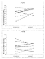
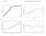
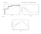
これらの新たなモデルは、妊娠及び出産の潜在性について卵母細胞をランク付けすることが可能な判明した。上述のモデルから明らかなように、CAMK1Dエクソン1又はエクソン9及びEFNB2は患者内及び患者間分析の陽性対照遺伝子として確認されており、これらのマーカーとGSTA4、GSTA3、PGR、GPX3、NCOA7、SASH1、MROH9、DNAH3、HSPH1エクソン2、HSPH1エクソン6、GALNTL6及びSPTBN5から選択される更なる遺伝子とを組み合わせることで、妊娠及び出産に対する卵母細胞コンピテンスの決定において予測的なモデルが得られる。以下の実施例から(from)明らかなように、これらの遺伝子の組合せを使用することでPPV、NPV及び予測精度は60%を上回った。別の実施形態では、本発明のin vitroモデル(例えば下記表6のモデル3を参照されたい)は例えば年齢、刺激日数及び相対エストラジオール(E2)、抗ミュラー管ホルモンレベル(AMH)、3日目の卵胞刺激ホルモンレベル(Day 3 FSH)等の患者及び/又は周期パラメーターを更に含み得る。特定の実施形態では、本発明のin vitroモデルに使用される患者及び周期パラメーターは刺激日数、相対E2及び年齢である。
These new models have been found to be able to rank oocytes for pregnancy and childbirth potential. As is apparent from the above model,
上述の遺伝子セットから離れた一実施形態では、本発明は、妊娠(pregnancy)及び出産をもたらす哺乳動物卵母細胞のコンピテンスを予測するin vitroモデルであって、上記方法が、
卵母細胞と関連する少なくとも1つの顆粒膜又は卵丘細胞を含むサンプルにおいてEFNB2、GSTA4及びPGRのマーカー遺伝子発現のレベルを決定する工程と、
マーカー遺伝子発現のレベルをコンピテンスが既知の対照と比較するか、又は上記遺伝子のエクソンレベルの遺伝子発現を正規化する工程と、
上記比較又は上記正規化発現レベルに基づいて妊娠及び出産をもたらす卵母細胞のコンピテンスを評価する工程と、
を含む、in vitroモデルを提供する。
In one embodiment away from the gene set described above, the invention is an in vitro model for predicting mammalian oocyte competence resulting in pregnancy and childbirth, wherein the method comprises:
Determining the level of marker gene expression of EFNB2, GSTA4 and PGR in a sample comprising at least one granulosa or cumulus cell associated with an oocyte;
Comparing the level of marker gene expression to a control of known competence, or normalizing exon level gene expression of said gene;
Evaluating the competence of the oocyte resulting in pregnancy and childbirth based on the comparison or the normalized expression level;
An in vitro model is provided.
上述の方法の一実施形態では、卵母細胞のコンピテンスは、方程式P=-a+b*EFNB2+c*GSTA4-d*PGR(式中、aは1.00〜4.00の数であり、bは0.00〜2.00の数であり、cは0.00〜2.00の数であり、dは0.00〜2.00の数であり、EFNB2はEFNB2の正規化発現レベルであり、GSTA4はGSTA4の正規化発現レベルであり、PGRはPGRの正規化発現レベルである)で与えられる妊娠確率(P)の決定に基づく。特定の実施形態では、上述の方程式におけるaは2.00〜3.00の数であり、bは0.00〜1.00の数であり、cは0.00〜1.00の数であり、dは0.00〜1.00の数である。また更なる実施形態では、上述の方程式におけるaは2.26であり、bは0.79であり、cは0.095であり、dは0.096である。 In one embodiment of the above method, the competence of the oocyte is the equation P = −a + b * EFNB2 + c * GSTA4-d * PGR where a is a number from 1.00 to 4.00 and b is 0.00 C is a number between 0.00 and 2.00, d is a number between 0.00 and 2.00, EFNB2 is a normalized expression level of EFNB2, GSTA4 is a normalized expression level of GSTA4, and PGR Is the normalized expression level of PGR) and is based on the determination of pregnancy probability (P). In certain embodiments, a in the above equation is a number from 2.00 to 3.00, b is a number from 0.00 to 1.00, c is a number from 0.00 to 1.00, and d is a number from 0.00 to 1.00. In still further embodiments, a in the above equation is 2.26, b is 0.79, c is 0.095, and d is 0.096.
EFNB2、GSTA4及びPGRに基づく上述のモデルの一実施形態では、in vitro方法は、卵母細胞と関連する少なくとも1つの顆粒膜又は卵丘細胞を含むサンプルにおけるGPX3及びGSTA3のマーカー遺伝子発現のレベルを決定することと、上記更なるマーカー遺伝子発現を卵母細胞のコンピテンスの評価に使用することとを更に含む。EFNB2、GSTA4及びPGRに基づく上述のモデルの更なる実施形態では、卵母細胞のコンピテンスは方程式P=-a+b*EFNB2+c*GSTA4-d*PGR-e*GPX3-f*GSTA3(式中、aは1.00〜4.00の数であり、bは0.00〜2.00の数であり、cは0.00〜2.00の数であり、dは0.00〜2.00の数であり、eは0.00〜2.00の数であり、fは0.00〜2.00の数であり、EFNB2はEFNB2の正規化発現レベルであり、GSTA4はGSTA4の正規化発現レベルであり、PGRはPGRの正規化発現レベルであり、GPX3はGPX3の正規化発現レベルであり、GSTA3はGSTA3の正規化発現レベルである)で与えられる妊娠確率(P)の決定に基づく。特定の実施形態では、上述の方程式におけるaは1.00〜2.00の数であり、bは0.00〜1.00の数であり、cは0.00〜1.00の数であり、dは0.00〜1.00の数であり、eは0.00〜1.00の数であり、fは0.00〜1.00の数である。より具体的には、上述の方程式におけるaは1.02であり、bは0.63であり、cは0.27であり、dは0.11であり、eは0.43であり、fは0.51である。 In one embodiment of the above model based on EFNB2, GSTA4 and PGR, the in vitro method determines the level of GPX3 and GSTA3 marker gene expression in a sample comprising at least one granulosa or cumulus cell associated with the oocyte. And further comprising using said additional marker gene expression to assess oocyte competence. In a further embodiment of the above model based on EFNB2, GSTA4 and PGR, the competence of the oocyte is the equation P = -a + b * EFNB2 + c * GSTA4-d * PGR-e * GPX3-f * GSTA3 (formula A is a number from 1.00 to 4.00, b is a number from 0.00 to 2.00, c is a number from 0.00 to 2.00, d is a number from 0.00 to 2.00, and e is a number from 0.00 to 2.00. Yes, f is a number between 0.00 and 2.00, EFNB2 is the normalized expression level of EFNB2, GSTA4 is the normalized expression level of GSTA4, PGR is the normalized expression level of PGR, GPX3 is the normal expression level of GPX3 Expression level, GSTA3 is the normalized expression level of GSTA3) and is based on the determination of pregnancy probability (P). In certain embodiments, a in the above equation is a number from 1.00 to 2.00, b is a number from 0.00 to 1.00, c is a number from 0.00 to 1.00, d is a number from 0.00 to 1.00, e is a number from 0.00 to 1.00, and f is a number from 0.00 to 1.00. More specifically, a in the above equation is 1.02, b is 0.63, c is 0.27, d is 0.11, e is 0.43, and f is 0.51.
EFNB2、GSTA4及びPGRに基づく上述のモデルの一実施形態では、in vitro方法は、患者及び周期の特徴、すなわち年齢、刺激日数及びRel E2(相対E2)を決定することと、上記更なる患者及び周期の特徴を卵母細胞のコンピテンスの評価に使用することとを更に含む。EFNB2、GSTA4及びPGRに基づく上述のモデルの更なる実施形態では、卵母細胞のコンピテンスは方程式P=-a+b*EFNB2+c*GSTA4-d*PGR-e*GPX3-f*GSTA3+g*刺激日数+h*Rel E2+i*年齢(式中、aは5.00〜15.00の数であり、bは0.00〜2.00の数であり、cは0.00〜2.00の数であり、dは0.00〜2.00の数であり、eは0.00〜2.00の数であり、fは0.00〜2.00の数であり、gは0.00〜2.00の数であり、hは0.00〜2.00の数であり、iは0.00〜2.00の数であり、EFNB2はEFNB2の正規化発現レベルであり、GSTA4はGSTA4の正規化発現レベルであり、PGRはPGRの正規化発現レベルであり、GPX3はGPX3の正規化発現であり、GSTA3はGSTA3の正規化発現レベルである)で与えられる妊娠確率(P)の決定に基づく。特定の実施形態では、上述の方程式におけるaは9.00〜13.00の数であり、bは1.00〜2.00の数であり、cは0.00〜1.00の数であり、dは0.00〜1.00の数であり、eは0.00〜1.00の数であり、fは0.00〜1.00の数であり、gは0.00〜1.00の数であり、hは0.00〜1.00の数であり、iは0.00〜1.00の数である。より具体的には、上述の方程式におけるaは11.27であり、bは1.35であり、cは0.46であり、dは0.24であり、eは0.66であり、fは0.86であり、gは0.0.50であり、hは0.009であり、iは0.14である。 In one embodiment of the above model based on EFNB2, GSTA4 and PGR, the in vitro method determines patient and cycle characteristics, ie, age, days of stimulation and Rel E2 (relative E2), and the additional patient and Further comprising using the cycle characteristics to assess oocyte competence. In a further embodiment of the above model based on EFNB2, GSTA4 and PGR, the competence of the oocyte is the equation P = -a + b * EFNB2 + c * GSTA4-d * PGR-e * GPX3-f * GSTA3 + g * Stimulation days + h * Rel E2 + i * Age (where a is a number from 5.00 to 15.00, b is a number from 0.00 to 2.00, c is a number from 0.00 to 2.00, d is a number from 0.00 2.00 number, e is a number from 0.00 to 2.00, f is a number from 0.00 to 2.00, g is a number from 0.00 to 2.00, h is a number from 0.00 to 2.00, i is a number from 0.00 to 2.00. EFNB2 is the normalized expression level of EFNB2, GSTA4 is the normalized expression level of GSTA4, PGR is the normalized expression level of PGR, GPX3 is the normalized expression level of GPX3, GSTA3 Is the normalized expression level of GSTA3) based on the determination of pregnancy probability (P). In certain embodiments, a in the above equation is a number from 9.00 to 13.00, b is a number from 1.00 to 2.00, c is a number from 0.00 to 1.00, d is a number from 0.00 to 1.00, e is a number from 0.00 to 1.00, f is a number from 0.00 to 1.00, g is a number from 0.00 to 1.00, h is a number from 0.00 to 1.00, and i is a number from 0.00 to 1.00. More specifically, a in the above equation is 11.27, b is 1.35, c is 0.46, d is 0.24, e is 0.66, f is 0.86, and g is 0.0. 50, h is 0.009, and i is 0.14.
上述の遺伝子セットから離れた一実施形態では、本発明は、妊娠及び出産をもたらす哺乳動物卵母細胞のコンピテンスを予測するin vitroモデルであって、上記方法が、
卵母細胞と関連する少なくとも1つの顆粒膜又は卵丘細胞を含むサンプルにおいてEFNB2及びNCOA7のマーカー遺伝子発現のレベルを決定する工程と、
マーカー遺伝子発現のレベルをコンピテンスが既知の対照と比較するか、又は上記遺伝子のエクソンレベルの遺伝子発現を正規化する工程と、
上記比較又は上記正規化発現レベルに基づいて妊娠及び出産をもたらす卵母細胞のコンピテンスを評価する工程と、
を含む、in vitroモデルを提供する。
In one embodiment away from the gene set described above, the present invention is an in vitro model for predicting the competence of mammalian oocytes resulting in pregnancy and childbirth, wherein the method comprises:
Determining the level of marker gene expression of EFNB2 and NCOA7 in a sample comprising at least one granulosa or cumulus cell associated with an oocyte;
Comparing the level of marker gene expression to a control of known competence, or normalizing exon level gene expression of said gene;
Evaluating the competence of the oocyte resulting in pregnancy and childbirth based on the comparison or the normalized expression level;
An in vitro model is provided.
上述の方法の一実施形態では、卵母細胞のコンピテンスは方程式P=a+b*EFNB2+c*NCOA7(式中、aは2.00〜5.00の数であり、bは2.00〜4.00の数であり、cは1.00〜3.00の数であり、EFNB2はEFNB2の正規化発現レベルであり、NCOA7はNCOA7の正規化発現レベルである)で与えられる妊娠確率(P)の決定に基づく。特定の実施形態では、妊娠確率(P)は方程式P=a+b*EFNB2+c*NCOA7(式中、aは3.00〜4.00の数であり、bは2.00〜3.00の数であり、cは1.00〜2.00の数である)で与えられる。また更なる実施形態では、上述の方程式におけるaは3.23であり、bは2.57であり、cは1.87である。 In one embodiment of the above method, the competence of the oocyte is the equation P = a + b * EFNB2 + c * NCOA7 where a is a number from 2.00 to 5.00 and b is a number from 2.00 to 4.00. , C is a number from 1.00 to 3.00, EFNB2 is the normalized expression level of EFNB2, and NCOA7 is the normalized expression level of NCOA7). In certain embodiments, the pregnancy probability (P) is the equation P = a + b * EFNB2 + c * NCOA7 where a is a number from 3.00 to 4.00, b is a number from 2.00 to 3.00, and c is Is a number between 1.00 and 2.00). In a still further embodiment, a in the above equation is 3.23, b is 2.57, and c is 1.87.
上述の遺伝子セットから離れた一実施形態では、本発明は、GnRH(ゴナドトロピン放出ホルモン)アンタゴニスト及びrFSH(組み換え卵胞刺激ホルモン)で前処置した被験体において妊娠及び出産をもたらす哺乳動物卵母細胞のコンピテンスを予測するin vitroモデルであって、上記方法が、
卵母細胞と関連する少なくとも1つの顆粒膜又は卵丘細胞を含むサンプルにおいてCAMK1Dエクソン9、HSPH1エクソン2及びNCOA7のマーカー遺伝子発現のレベルを決定する工程と、
マーカー遺伝子発現のレベルをコンピテンスが既知の対照と比較するか、又は上記遺伝子のエクソンレベルの遺伝子発現を正規化する工程と、
上記比較又は上記正規化発現レベルに基づいて妊娠をもたらす卵母細胞のコンピテンスを評価する工程と、
を含む、in vitroモデルを提供する。
In one embodiment away from the gene set described above, the present invention relates to the competence of mammalian oocytes resulting in pregnancy and childbirth in a subject pretreated with a GnRH (gonadotropin releasing hormone) antagonist and rFSH (recombinant follicle stimulating hormone). An in vitro model that predicts
Determining the level of marker gene expression of CAMK1D exon 9,
Comparing the level of marker gene expression to a control of known competence, or normalizing exon level gene expression of said gene;
Evaluating the competence of the oocyte resulting in pregnancy based on the comparison or the normalized expression level;
An in vitro model is provided.
上述の方法の一実施形態では、卵母細胞のコンピテンスは方程式P=a+b*CAMK1Dエクソン9+c*HSPH1エクソン2+d*NCOA7(式中、aは-3.00〜-1.00の数であり、bは2.00〜4.00の数であり、cは0.00〜3.00の数であり、dは0.00〜2.00の数であり、CAMK1Dエクソン9はCAMK1Dエクソン9の正規化発現レベルであり、HSPH1エクソン2はHSPH1エクソン2の正規化発現レベルであり、NCOA7はNCOA7の正規化発現レベルである)で与えられる妊娠確率(P)の決定に基づき、正の値は妊娠を表し、負の値は非妊娠を表す。特定の実施形態では、妊娠確率(P)は方程式P=a+b*CAMK1Dエクソン9+c*HSPH1エクソン2+d*NCOA7(式中、aは-2.00〜-1.00の数であり、bは1.00〜2.00の数であり、cは0.00〜1.00の数であり、dは0.00〜1.00の数である)で与えられる。また更なる実施形態では、aは-1.37であり、bは1.79であり、cは0.89であり、dは0.74である。
In one embodiment of the above method, the oocyte competence is the equation P = a + b * CAMK1D exon 9 + c * HSPH1 exon 2 + d * NCOA7, where a is a number from −3.00 to −1.00 , B is a number from 2.00 to 4.00, c is a number from 0.00 to 3.00, d is a number from 0.00 to 2.00, CAMK1D exon 9 is the normalized expression level of CAMK1D exon 9, and
上述の遺伝子セットから離れた一実施形態では、本発明は、GnRH(ゴナドトロピン放出ホルモン)アンタゴニスト及びHP-hMG(高純度ヒト閉経期尿性ゴナドトロピン)で前処置した被験体において妊娠及び出産をもたらす哺乳動物卵母細胞のコンピテンスを予測するin vitroモデルであって、上記方法が、
卵母細胞と関連する少なくとも1つの顆粒膜又は卵丘細胞を含むサンプルにおいてCAMK1Dエクソン1及びSASH1のマーカー遺伝子発現のレベルを決定する工程と、
マーカー遺伝子発現のレベルをコンピテンスが既知の対照と比較するか、又は上記遺伝子のエクソンレベルの遺伝子発現を正規化する工程と、
上記比較又は上記正規化発現レベルに基づいて妊娠をもたらす卵母細胞のコンピテンスを評価する工程と、
を含む、in vitroモデルを提供する。
In one embodiment away from the gene set described above, the present invention provides a mammal that causes pregnancy and childbirth in a subject pretreated with a GnRH (gonadotropin releasing hormone) antagonist and HP-hMG (high purity human menopausal urinary gonadotropin). An in vitro model for predicting competence of animal oocytes, the method comprising:
Determining the level of marker gene expression of
Comparing the level of marker gene expression to a control of known competence, or normalizing exon level gene expression of said gene;
Evaluating the competence of the oocyte resulting in pregnancy based on the comparison or the normalized expression level;
An in vitro model is provided.
上述の方法の一実施形態では、卵母細胞のコンピテンスは、方程式P=a+b*SASH1-c*CAMK1Dエクソン1(式中、aは0.00〜2.00の数であり、bは0.00〜2.00の数であり、cは0.00〜2.00の数であり、CAMK1Dエクソン1はCAMK1Dエクソン1の正規化発現レベルであり、SASH1はSASH1の正規化発現レベルである)で与えられる妊娠確率(P)の決定に基づき、正の値は妊娠を表し、負の値は非妊娠を表す。特定の実施形態では、妊娠確率(P)は方程式P=a+b*SASH1-c*CAMK1Dエクソン1(式中、aは0.00〜1.00の数であり、bは0.00〜1.00の数であり、cは0.00〜1.00の数である)で与えられる。また更なる実施形態では、aは0.47であり、bは0.91であり、cは0.0.23である。
In one embodiment of the above method, the competence of the oocyte is the equation P = a + b * SASH1-c * CAMK1D exon 1 where a is a number from 0.00 to 2.00 and b is from 0.00 to 2.00. The probability of pregnancy given by (c) is a number between 0.00 and 2.00,
本発明の方法による遺伝子発現レベルの比較は、得られた遺伝子発現レベルを、アッセイしたサンプルの量の差及び使用したサンプルの品質(例えば試験したmRNAの量及び品質)の変動性の両方について補正した後に行うのが好ましい。同じサンプル中の参照遺伝子に対するレベルの正規化によって補正を行うことができる。通例、UBC、B2M、アクチン、GAPDH、HPRT、CPB、G6PD、ヒストンH2A又はミトコンドリアリボソームタンパク質S 18C遺伝子、特にUBC又はB2M等の「ハウスキーピング遺伝子」をこの正規化に使用する。マーカー遺伝子の発現レベルは、マーカーの絶対発現レベルをその発現とマーカーではない遺伝子、例えば構成的に発現されたハウスキーピング遺伝子の発現とを比較することで補正することによって正規化することができる。正規化に好適な遺伝子としては、UBC、B2M、アクチン、GAPDH、HPRT、CPB、G6PD、ヒストンH2A又はミトコンドリアリボソームタンパク質S18C遺伝子等のハウスキーピング遺伝子が挙げられる。この正規化により、とりわけ対照サンプルに対する或るサンプル、例えば試験サンプルにおける発現レベルの比較が可能となる。 Comparison of gene expression levels according to the method of the present invention corrects the resulting gene expression levels for both variability in the amount of sample assayed and the quality of the sample used (eg, the amount and quality of the mRNA tested). It is preferable to carry out after this. Corrections can be made by normalizing levels relative to a reference gene in the same sample. Typically, “housekeeping genes” such as UBC, B2M, actin, GAPDH, HPRT, CPB, G6PD, histone H2A or mitochondrial ribosomal protein S 18C genes, in particular UBC or B2M, are used for this normalization. The expression level of a marker gene can be normalized by correcting the absolute expression level of the marker by comparing its expression with the expression of a gene that is not a marker, such as a constitutively expressed housekeeping gene. Suitable genes for normalization include housekeeping genes such as UBC, B2M, actin, GAPDH, HPRT, CPB, G6PD, histone H2A or mitochondrial ribosomal protein S18C gene. This normalization allows comparison of the expression level in a particular sample, for example a test sample, among others.
群内の他の卵母細胞と関連する卵丘又は顆粒膜細胞の発現レベルと比較した、所与の卵母細胞と関連する卵丘細胞又は顆粒膜細胞におけるマーカー遺伝子発現の差により、相対品質による卵母細胞群における卵母細胞のランク付けが可能となる。このため本発明の一実施形態では、個体又は個体群に由来する複数の卵母細胞を単一アッセイでグループ分け及びスクリーニングした後、受精及び/又は着床のために最高の品質可能性スコアを特徴とする卵母細胞を選択する。 Relative quality due to differences in marker gene expression in cumulus cells or granulosa cells associated with a given oocyte compared to the level of cumulus or granulosa cells associated with other oocytes in the group Enables ranking of oocytes in the oocyte group. Thus, in one embodiment of the present invention, the highest quality likelihood score for fertilization and / or implantation is obtained after grouping and screening an oocyte from an individual or population in a single assay. Select the characteristic oocyte.
マーカーの特定、及び卵丘細胞におけるマーカー遺伝子発現の変化と付随する卵母細胞の品質との関連性の決定は、mRNA又はタンパク質発現に基づく臨床診断キットの基礎を与える。本発明による卵母細胞品質の決定に必要とされる基本的な材料及び試薬はキットにすることができる。幾つかの実施形態では、キットは、本明細書に開示の少なくとも1つの遺伝子の発現レベルを特異的に検出する少なくとも1つの試薬と、1つ又は複数の本発明の方法によるキットの使用説明書とを含む。各々のキットは必然的に手法を特異的にする試薬を含む。このため、セットの少なくとも1つのマーカー遺伝子が発現するmRNAの検出については、試薬は例えばcDNA又はオリゴヌクレオチド等のmRNAに相補的な核酸プローブを含む。核酸プローブは基質表面(例えばマイクロアレイ)に固定化されていても又は固定化されていなくてもよい。セットの少なくとも1つの遺伝子にコードされるポリペプチド産物の検出については、試薬はポリペプチドに特異的に結合する抗体を含む。手法に応じて、キットは抽出バッファー及び/又は試薬、増幅バッファー及び/又は試薬、ハイブリダイゼーションバッファー及び/又は試薬、免疫検出バッファー及び/又は試薬、標識化バッファー及び/又は試薬、並びに検出手段の1つ又は複数を更に含み得る。手法の種々の工程を行うためにこれらのバッファー及び試薬を使用するプロトコルがキットに含まれ得る。試薬は固体(例えば凍結乾燥)形態又は液体形態で供給することができる。本発明のキットは、各々個々のバッファー及び/又は試薬について異なる容器(例えばバイアル、アンプル、試験管、フラスコ又はボトル)を任意に含み得る。開示の方法の幾つかの工程を行うのに好適な他の容器も提供され得る。幾つかの実施形態では、本発明のキットは対照サンプルを更に含む。1つ又は複数の本発明の方法によるキットの使用説明書は、卵丘細胞又は顆粒膜細胞サンプルをプロセシングする及び/又は試験を行うための説明書、並びに結果を解釈する、すなわち分娩、着床をもたらす、着床前胚盤胞若しくは胚を形成する、及び/又は受精をもたらす卵母細胞のコンピテンスを決定するための説明書を更に含む。一実施形態では、定量RT-PCRによりマーカー遺伝子のmRNA発現を決定するキットは、標準的なプライマー及び定量RT-PCRを行うための他の試薬を、アッセイ対象の各遺伝子の最適な検出の成功のために配合された反応成分とともに含み得る。増幅プライマーは、評定される哺乳動物卵母細胞の種に応じた関連マーカー遺伝子(複数の場合もあり)のヌクレオチド配列に基づいて選択することができる。任意の単一のマーカー遺伝子又はマーカー遺伝子の組合せをキットに組み込むことができる。ユーザーにカバレージとコストとのバランスを選ぶ選択肢を与えるために、異なるキットが分析の対象となるマーカー遺伝子に対する異なるプライマー群を与えることができる。 The identification of markers and the determination of the association between changes in marker gene expression in cumulus cells and the associated oocyte quality provide the basis for clinical diagnostic kits based on mRNA or protein expression. The basic materials and reagents required for determining oocyte quality according to the present invention can be kits. In some embodiments, the kit comprises at least one reagent that specifically detects the expression level of at least one gene disclosed herein and instructions for using the kit according to one or more methods of the invention. Including. Each kit necessarily contains reagents that make the procedure specific. Thus, for the detection of mRNA expressed by at least one marker gene in the set, the reagent comprises a nucleic acid probe complementary to the mRNA, such as cDNA or oligonucleotide. The nucleic acid probe may or may not be immobilized on the substrate surface (for example, a microarray). For detection of polypeptide products encoded by at least one gene in the set, the reagent comprises an antibody that specifically binds to the polypeptide. Depending on the technique, the kit may be an extraction buffer and / or a reagent, an amplification buffer and / or a reagent, a hybridization buffer and / or a reagent, an immunodetection buffer and / or a reagent, a labeling buffer and / or a reagent, and a detection means. One or more. Protocols that use these buffers and reagents to perform the various steps of the procedure can be included in the kit. Reagents can be supplied in solid (eg lyophilized) form or in liquid form. The kit of the present invention may optionally include different containers (eg, vials, ampoules, test tubes, flasks or bottles) for each individual buffer and / or reagent. Other containers suitable for performing some steps of the disclosed method may also be provided. In some embodiments, the kit of the invention further comprises a control sample. Instructions for using the kit according to one or more of the methods of the invention include instructions for processing and / or testing cumulus cells or granulosa cell samples and interpreting the results, i.e. delivery, implantation Instructions for determining the competence of the oocyte that yields, forms a preimplantation blastocyst or embryo and / or results in fertilization. In one embodiment, the kit for determining mRNA expression of a marker gene by quantitative RT-PCR uses standard primers and other reagents for performing quantitative RT-PCR, and successful detection of each gene being assayed optimally. Together with reaction components formulated for the purpose. Amplification primers can be selected based on the nucleotide sequence of the relevant marker gene (s) depending on the mammalian oocyte species being assessed. Any single marker gene or combination of marker genes can be incorporated into the kit. In order to give the user the option of choosing a balance between coverage and cost, different kits can be given different primer sets for the marker genes to be analyzed.
本発明による方法の適用可能性
好ましい実施形態では、本発明の方法を用いて、差次的に発現される遺伝子セットの発現レベルに基づいて妊娠コンピテント卵母細胞を生成する又は生成しない女性被験体を特定することもできる。しかしながら、本発明の方法は非ヒト動物、例えば他の哺乳動物、鳥類、両生類、爬虫類等にも適用可能である。例えば、本発明を用いて推定女性不妊治療の研究用の動物モデルを得ることができる。
Applicability of the method according to the invention In a preferred embodiment, a female subject who uses the method of the invention to produce or does not produce a pregnancy competent oocyte based on the expression level of a differentially expressed gene set. You can also identify your body. However, the method of the present invention is also applicable to non-human animals such as other mammals, birds, amphibians, reptiles and the like. For example, the present invention can be used to obtain animal models for the study of putative female infertility treatments.
さらに、本発明を用いて、妊娠コンピテント卵母細胞を生成する能力を妨げる又は阻害する異常、例えば薬剤への曝露、毒物への曝露、環境因子、卵巣機能不全、卵巣嚢胞、閉経前又は閉経期の病態、癌、自己免疫障害、ホルモン機能障害、細胞増殖障害、又は妊娠コンピテント卵母細胞の発生を阻害する若しくは妨げる別の健康状態を有する雌性被験体を特定することができる。例えば、特定の妊娠シグネチャー遺伝子を特徴的な発現レベルで発現する被験体をスクリーニングして、被験体が妊娠コンピテント卵母細胞の生成を妨げる基礎健康状態を有するか否かを評定することができる。特に、かかる被験体をスクリーニングして被験体が更年期の兆候を示すか否か、被験体が癌、自己免疫疾患若しくは卵巣異常、例えば卵巣嚢胞を有するか否か、又は被験体が「妊娠コンピテント」卵母細胞の発生を妨げ得る別の健康状態、例えばホルモン障害、アレルギー性障害等を有するか否かを評定することができる。 Further, the present invention can be used to prevent or inhibit the ability to generate pregnancy competent oocytes, such as drug exposure, toxic exposure, environmental factors, ovarian dysfunction, ovarian cysts, premenopausal or menopause. A female subject with a stage of disease, cancer, autoimmune disorder, hormonal dysfunction, cell proliferation disorder, or another health condition that inhibits or prevents the development of pregnancy competent oocytes can be identified. For example, subjects that express a particular pregnancy signature gene at a characteristic expression level can be screened to assess whether the subject has a basal health condition that prevents the production of pregnancy competent oocytes. . In particular, if such a subject is screened and the subject shows signs of menopause, whether the subject has cancer, an autoimmune disease or an ovarian abnormality, such as an ovarian cyst, or the subject is “pregnant competent” It can be assessed whether or not it has another health condition that can interfere with oocyte development, such as hormonal disorders, allergic disorders, and the like.
さらに、本方法を用いてヒト又は非ヒト雌性被験体における推定女性不妊治療の有効性を評定することができる。基本的に、かかる方法は雌性被験体、好ましくは女性を推定不妊治療で治療することと、少なくとも1つの卵母細胞及び関連する周囲の卵胞又は顆粒膜細胞を治療後に上記女性から単離することと、任意に少なくとも1つの卵母細胞及び関連する周囲の細胞を治療前に更に単離することと、少なくとも1つの卵丘細胞を上記単離された卵母細胞の各々から単離することと、(周囲の)妊娠コンピテント卵母細胞と関連する卵丘細胞が特徴的なレベルで発現する又は発現しない少なくとも1つの遺伝子の発現レベルを検出することと、少なくとも1つの妊娠シグネチャー遺伝子を妊娠コンピテント卵母細胞の周囲の卵丘細胞に(治療しない場合よりも)特徴的なレベルで発現する卵丘細胞を生じるか否かに基づいて、上記推定不妊治療の有効性を評定することとを含み得る。上述のようにヒト女性被験体が好ましいが、本方法を雌の非ヒト動物、例えば雌の非ヒト霊長類、又は推定ヒト不妊治療の評価用の他の好適な動物モデルにおいて推定不妊治療の有効性を評定するために用いることができる。 Furthermore, the method can be used to assess the effectiveness of putative female infertility treatments in human or non-human female subjects. Basically, such methods treat a female subject, preferably a woman, with a putative infertility treatment and isolate at least one oocyte and associated surrounding follicle or granulosa cells from the woman after treatment. Optionally further isolating at least one oocyte and associated surrounding cells prior to treatment, and isolating at least one cumulus cell from each of said isolated oocytes; Detecting the expression level of at least one gene that is or is not expressed at a characteristic level in the cumulus cells associated with the (surrounding) pregnancy competent oocyte and detecting at least one pregnancy signature gene Evaluate the effectiveness of the above infertility treatment based on whether or not the cumulus cells around the tent oocyte produce cumulus cells that are expressed at a characteristic level (rather than untreated) It may include a Rukoto. Although human female subjects are preferred as described above, the method is useful for estimating putative infertility treatments in female non-human animals, such as female non-human primates, or other suitable animal models for evaluation of putative human infertility treatments. Can be used to assess sex.
また、本発明を用いてin vitro又はin vivo不妊治療の有効性を高めることができる。特に、「妊娠インコンピテント」であることが見出された又は未熟な卵母細胞は、体内又は体外受精の前、その最中又はその後に「妊娠シグネチャー」遺伝子、例えばホルモン、成長因子、分化因子等であることが特定された遺伝子にコードされた1つ又は複数の遺伝子産物を含有する培地中で培養することができる。基本的に、これらの遺伝子産物の存在は、「妊娠コンピテント」卵母細胞の周囲の卵丘及び/又は顆粒膜細胞により通常発現され、通常は卵母細胞に栄養を与え、それによりこれらの卵母細胞が受精時に正常妊娠をもたらす能力を高める栄養遺伝子産物の欠乏を補うものである。 Moreover, the effectiveness of in vitro or in vivo infertility treatment can be enhanced by using the present invention. In particular, oocytes that have been found to be “pregnancy incompetent” or immature are pre-, during, or after in vivo or in vitro fertilization genes such as hormones, growth factors, differentiation It can be cultured in a medium containing one or more gene products encoded by a gene identified as a factor or the like. Basically, the presence of these gene products is normally expressed by cumulus and / or granulosa cells surrounding “pregnant competent” oocytes, and normally nourishes the oocytes, thereby It compensates for the deficiency of nutritional gene products that enhance the ability of oocytes to produce normal pregnancy upon fertilization.
代替的には、上記妊娠シグネチャー遺伝子がコードする1つ又は複数の遺伝子産物を、本発明の方法により妊娠コンピテント卵母細胞を生成しないことが発覚した被験体に投与することができる。かかる投与は非経口投与、例えば静脈内投与、筋肉内投与、膣内投与、皮下注射又は経口投与若しくは経皮投与であり得る。代替的には、これらの遺伝子産物は標的部位、例えば雌の卵巣又は子宮環境に局所的に投与することができる。例えば、雌性被験体の子宮又は卵巣に「妊娠シグネチャー」遺伝子がコードする1つ又は複数の遺伝子産物の持続送達をもたらす薬物送達デバイスを埋め込んでもよい。 Alternatively, one or more gene products encoded by the pregnancy signature gene can be administered to a subject found not to produce pregnancy competent oocytes by the methods of the present invention. Such administration may be parenteral, such as intravenous, intramuscular, intravaginal, subcutaneous injection or oral or transdermal. Alternatively, these gene products can be administered locally to a target site, such as a female ovary or uterine environment. For example, a drug delivery device may be implanted that provides sustained delivery of one or more gene products encoded by a “pregnancy signature” gene into the uterus or ovary of a female subject.
上記に詳述したように、群内の他の卵母細胞と関連する卵丘又は顆粒膜細胞の発現レベルと比較した、所与の卵母細胞と関連する卵丘細胞又は顆粒膜細胞におけるマーカー遺伝子発現の差により、相対品質による卵母細胞群における卵母細胞のランク付けが可能となる。このため本発明の一実施形態では、個体又はヒト若しくは非ヒト雌性被験体に由来する複数の卵母細胞を単一アッセイでグループ分け及びスクリーニングした後、最高の品質可能性スコアを特徴とする卵母細胞を受精及び/又は着床のために選択する。 Markers in cumulus cells or granulosa cells associated with a given oocyte as compared to the expression levels of cumulus or granulosa cells associated with other oocytes in the group as detailed above The difference in gene expression enables ranking of oocytes in the oocyte population by relative quality. Thus, in one embodiment of the invention, eggs characterized by the highest quality likelihood score after grouping and screening multiple oocytes from an individual or a human or non-human female subject in a single assay. Mother cells are selected for fertilization and / or implantation.
実施例1−QPCR分析
材料及び方法
患者集団
この研究はUZBrusselの倫理委員会により認可され、患者の同意書を得た。47人のICSI患者を胚移植方針(単一胚移植)及び規定の卵巣刺激プロトコル:組み換え卵胞刺激ホルモン(FSH)と組み合わせたGnRHアンタゴニスト(Gonal-f, Merck-Serono,Geneva,Switzerland;n=4又はPuregon,MSD,Oss,TheNetherlands;n=43)に基づいて選択した。不妊の原因は男性因子のみ(n=19)、女性因子のみ(排卵障害n=3及び卵管性不妊n=2)、男性因子及び女性因子の組合せ(OAT及び子宮内膜症n=3)並びに特発性(n=20)であった。20人の患者に培養3日目に単一胚移植を行い、これらのうち10人が妊娠した。27人の患者に5日目に移植を行うと、これらのうち9人が妊娠した。
Example 1-QPCR Analytical Materials and Methods Patient Population This study was approved by the UZBrussel Ethics Committee and patient consent was obtained. 47 ICSI patients with embryo transfer strategy (single embryo transfer) and prescribed ovarian stimulation protocol: GnRH antagonists combined with recombinant follicle stimulating hormone (FSH) (Gonal-f, Merck-Serono, Geneva, Switzerland; n = 4 Or selected based on Puregon, MSD, Oss, TheNetherlands; n = 43). The cause of infertility is male factor only (n = 19), female factor only (ovulation disorder n = 3 and tubal infertility n = 2), combination of male factor and female factor (OAT and endometriosis n = 3) And idiopathic (n = 20). Twenty patients received a single embryo transfer on
ヒト卵丘細胞の採取及び胚培養
経膣超音波を用いて卵胞発達をモニタリングした。内分泌プロファイルを、感度がそれぞれ5 ng/l、0.03 μg/l、0.1 IU/l未満、0.1 IU/l、全不正確性(%CV)がそれぞれ6未満、7未満、6未満及び6未満の検証アッセイを用いたCOBAS6001免疫分析装置(Roche,Roche Diagnostics,Mannheim,Germany)での電気化学発光による血清17β-エストラジオール(E2)、プロゲステロン、FSH及びLHの分析によってモニタリングした。直径17 mmの少なくとも3つの卵胞が経膣超音波で観察された場合に、最終卵胞成熟を10000 IU用量のhCGにより誘導した。36時間後に卵母細胞回収を行った。CC採取を非特許文献15に記載のように行った。簡潔に述べると、個々の卵母細胞剥離を80 IU/mlのCumulase(MediCult, Lyon, France)を含有する40 μl滴のHTF-SSSによって30秒以内に行い、酵素を含まない液滴で順次洗浄した。いずれの場合にも、卵母細胞毎のCCのレトロスペクティブ分析を可能にするためにこの時点から卵母細胞を個別に扱った。剥離後にCCを直接液体窒素に浸漬した。ICSIを以前に記載のように(Van Landuyt et al. 2005)行い、SAGE連続培地(CooperSurgical,Leisegang Medical,Berlin)中で胚を培養した。胚培養の3日目又は5/6日目に胚を以前に記載のように(Van Landuyt et al. 2011)ガラス化し、その後のFRET周期に使用した。
Human cumulus cell collection and embryo culture Transvaginal ultrasound was used to monitor follicular development. Endocrine profiles with sensitivity of 5 ng / l, 0.03 μg / l, less than 0.1 IU / l, 0.1 IU / l, and total inaccuracy (% CV) of less than 6, less than 7, less than 6, and less than 6, respectively Monitoring was performed by analysis of serum 17β-estradiol (E2), progesterone, FSH and LH by electrochemiluminescence on a COBAS6001 immunoanalyzer (Roche, Roche Diagnostics, Mannheim, Germany) using a validation assay. Final follicular maturation was induced by 10000 IU dose of hCG when at least 3 follicles 17 mm in diameter were observed with transvaginal ultrasound. Oocyte collection was performed 36 hours later. CC collection was performed as described in
47人全ての患者について、28人の非妊娠患者のうち7人を除いて移植に選択される胚を生じる卵母細胞に関連するCCのみを分析し、凍結単一胚移植周期後に妊娠をもたらしたガラス化胚に関連する1つの追加のCCサンプル(1人の患者、2つのCCサンプルを除く)を分析した(7人の患者から合計で8つの追加のCC)。図1に各分析に使用した異なるサンプルを示す。 For all 47 patients, except for 7 out of 28 non-pregnant patients, only the CC associated with the oocyte producing the embryo selected for transfer was analyzed, resulting in pregnancy after a frozen single embryo transfer cycle One additional CC sample (excluding 1 patient, 2 CC samples) associated with a vitrified embryo was analyzed (a total of 8 additional CCs from 7 patients). Figure 1 shows the different samples used for each analysis.
遺伝子の選択
本研究は、QPCRを用いて卵母細胞品質についてCC遺伝子発現の予測値を評価する順序の3番目である。3つの研究にわたって正確な戦略に従い、ICSI患者における卵母細胞品質に関して分析する遺伝子を選んだ。第1の研究では、胚形態について予測的な2つ(イノシトール三リン酸3-キナーゼA(ITPKA)及び一過性受容体潜在的カチオンチャネル、サブファミリーM、メンバー7(TRPM7))及び妊娠転帰について2つ(シンデカン4(SDC4)及びバーシカン(VCAN))の上位4つの遺伝子を特定した。これら4つの遺伝子を次の研究に加えることとした(非特許文献14)。ITPKA及びTRPM7は胚発生にも関連するが、SDC4及びVCANは今回妊娠モデルに残らず、段階的重回帰分析に使用する場合にEFNB2、CAMK1D及びSTC1に置き換えられた。幾つかの研究から胚発生及び妊娠転帰について予測的な遺伝子間の関連性が示されないため(非特許文献13、非特許文献14)、この第3の研究では3つの妊娠関連遺伝子のみについて繰り返すこととした。本発明者らの仮定は、この「カスケード」試験戦略により、最も強い妊娠予測的遺伝子が連続研究から除外されることである(表2)。本研究で分析される他の9つの遺伝子はGSTA3、GSTA4、GPX3、GSR、ITPR1、SLC2A1、THBS1、TGFB1及びPGRであった(表1)。
Gene selection This study is the third in an order to evaluate the predictive value of CC gene expression for oocyte quality using QPCR. Following a precise strategy across the three studies, we chose genes to analyze for oocyte quality in ICSI patients. The first study included two predictive embryonic forms (inositol triphosphate 3-kinase A (ITPKA) and transient receptor potential cation channel, subfamily M, member 7 (TRPM7)) and pregnancy outcome. The top four genes for two (syndecan 4 (SDC4) and versican (VCAN)) were identified. We decided to add these four genes to the next study (Non-Patent Document 14). Although ITPKA and TRPM7 are also associated with embryonic development, SDC4 and VCAN are now not part of the pregnancy model and have been replaced by EFNB2, CAMK1D and STC1 when used for stepwise multiple regression analysis. Some studies do not show any predictable genetic relationship for embryonic development and pregnancy outcome (Non-patent document 13, Non-patent document 14), so this third study should be repeated for only three pregnancy-related genes It was. Our hypothesis is that this “cascade” testing strategy excludes the strongest pregnancy predictive genes from serial studies (Table 2). The other nine genes analyzed in this study were GSTA3, GSTA4, GPX3, GSR, ITPR1, SLC2A1, THBS1, TGFB1, and PGR (Table 1).

(A)「一般的機能」の欄の情報を指す;(B)「ヒトCCにおける卵母細胞品質マーカーとして以前に記載されている」の欄の情報を指す;CC:卵丘細胞;FSH:卵胞刺激ホルモン;ICSI:卵細胞質内精子注入法
(A) refers to the information in the column “General Function”; (B) refers to the information in the column “previously described as an oocyte quality marker in human CC”; CC: cumulus cells; FSH: Follicle stimulating hormone; ICSI: Intracytoplasmic sperm injection
ICSIにおける対応する卵母細胞の種々の胚形態又は妊娠転帰に関連する卵丘細胞(CC)の遺伝子発現を、3つの異なるサンプルセットの3つの遺伝子パネルについて評定した。各サンプルセットにおいて最も予測的であることが見出された遺伝子を、その後の独立した患者サンプルセットにおいて試験した。太字で表示した遺伝子を以前の研究による最良の予測的遺伝子として残した。MII:第二中期卵母細胞;COC:卵丘複合体;rFSH:組み換え卵胞刺激ホルモン;HP-hMG:高純度ヒト閉経期尿性ゴナドトロピン
The gene expression of cumulus cells (CC) associated with different embryonic morphology or pregnancy outcome of the corresponding oocyte in ICSI was assessed for three gene panels in three different sample sets. The genes found to be the most predictive in each sample set were tested in subsequent independent patient sample sets. The gene shown in bold was left as the best predictive gene from previous studies. MII: 2nd metaphase oocyte; COC: cumulus complex; rFSH: recombinant follicle stimulating hormone; HP-hMG: high purity human menopausal urinary gonadotropin
RNA抽出及びcDNA合成
全RNAを、DNase工程及び抽出前の5ng/μlポリ(dA)(Roche Applied Science,Mannheim,Germany)の添加を含むRNeasy Micro Kit(Qiagen,Westburg,Leusden,The Netherlands)を用いて以前に記載されているように(非特許文献17)抽出した。抽出に続いて第2のDNase処理(RQ1RNase-Free DNase,Promega,Leiden,The Netherlands)を行った。
RNA extraction and cDNA synthesis Total RNA using the RNeasy Micro Kit (Qiagen, Westburg, Leusden, The Netherlands) with DNase step and addition of 5 ng / μl poly (dA) (Roche Applied Science, Mannheim, Germany) prior to extraction Extracted as previously described (Non-patent Document 17). The extraction was followed by a second DNase treatment (RQ1RNase-Free DNase, Promega, Leiden, The Netherlands).
逆転写酵素(RT)を、iScript cDNA Synthesis Kit(Bio-Rad Laboratories,Ghent,Belgium)を用いて以前に記載されているように(非特許文献17)行った。RT反応において酵素又はRNAを除外することで陰性対照を生成した。 Reverse transcriptase (RT) was performed as previously described (17) using the iScript cDNA Synthesis Kit (Bio-Rad Laboratories, Ghent, Belgium). Negative controls were generated by excluding enzyme or RNA in the RT reaction.
リアルタイムPCR
CAMK1D、STC1及びEFNB2のプライマー配列は非特許文献14に挙げられる。GPX3、GSTA3、GSTA4、PGR、THB1、ITPRA、SLC2A1、GSR及びTGFb1のプライマーは表3に見ることができる。β-2-ミクログロブリン(B2M)及びユビキチンC(UBC)の両方を検証したが、これらは以前に正規化因子として使用されている(非特許文献15)。サイクリング条件、陰性対照、標準曲線及び正規化(B2M及びUBCによる)は以前に記載されている通りであるが(非特許文献15)、全PCR反応を10 μl反応に適合させた。以下に言及する全ての値は、各サンプルのB2M及びUBCの両方の平均に対して正規化した値である。
Real-time PCR
Non-patent literature 14 lists the primer sequences of CAMK1D, STC1, and EFNB2. Primers for GPX3, GSTA3, GSTA4, PGR, THB1, ITPRA, SLC2A1, GSR and TGFb1 can be found in Table 3. Both β-2-microglobulin (B2M) and ubiquitin C (UBC) were verified, but these have been used previously as normalization factors (Non-patent Document 15). Cycling conditions, negative controls, standard curves and normalization (by B2M and UBC) were as previously described (Non-Patent Document 15), but the entire PCR reaction was adapted to a 10 μl reaction. All values mentioned below are normalized to the average of both B2M and UBC for each sample.
統計
患者間分析
第1の分析では、両側t検定(GraphPadPrism version 4.01 for Windows登録商標,GraphPad Software,San Diego California USA)を用いて、妊娠をもたらす又はもたらさない(19出産、28非妊娠)卵母細胞の卵丘複合体を比較した。全データをLOG変換して正規分布を得た。複数の遺伝子を試験したため、ボンフェローニ補正では0.0042未満のP値のみを有意とみなした。
Statistical patient-to-patient analysis In the first analysis, a two-tailed t-test (GraphPadPrism version 4.01 for Windows registered trademark , GraphPad Software, San Diego California USA) was used to produce or not give birth (19 births, 28 non-pregnant) oocytes Cell cumulus complexes were compared. All data were LOG transformed to obtain a normal distribution. Since multiple genes were tested, Bonferroni correction only considered P values less than 0.0042 as significant.
第2の分析では、以前に記載されているように(非特許文献15)段階的重回帰分析を用いてモデルを構築した。簡潔に述べると、「y=a+bx+cz+ds+et+fu+gv+hw」の出力方程式を有する線形回帰モデルを、応答変数(x、z、s、t、u、v、w)遺伝子発現及び/又は患者及び周期特徴(全てに表3に挙げられる)を用いて構築した。「b-h」は含まれる変数のそれぞれの指数であり、「a」は方程式の切片である。変数のタイプIII P値が0.3未満であり、モデルのP値が改善される場合に変数をモデルに加えた。モデルの最後に逆回帰工程を行い、余剰の変数を除外した。4つの異なるモデルを妊娠予測のために構築した。2つのモデルで遺伝子発現値のみが許容される(初めにモデルを3つの遺伝子に制限し、次にモデルのP値を改善する限りにおいて全ての遺伝子をモデルに入れた)。他の2つのモデルでは、全ての患者及び周期特徴を、これらの追加の変数がモデルを改善することができる場合に遺伝子のみから構成されるモデルに入れることによって患者及び周期特徴による補正の必要性を評定した。これらの追加の因子を導入することで、遺伝子発現に生じ得る患者間の変動性を均一化し、卵母細胞品質に関連する差を増大させることができた。 In the second analysis, a model was constructed using stepwise multiple regression analysis as previously described (15). Briefly, a linear regression model with an output equation of “y = a + bx + cz + ds + et + fu + gv + hw” is expressed as a response variable (x, z, s, t, u, v, w ) Constructed using gene expression and / or patient and cycle characteristics (all listed in Table 3). “B-h” is the index of each of the included variables, and “a” is the intercept of the equation. A variable was added to the model if the type III P value of the variable was less than 0.3 and the P value of the model improved. An inverse regression process was performed at the end of the model to exclude excess variables. Four different models were built for pregnancy prediction. Only gene expression values are allowed in the two models (first restricting the model to 3 genes and then putting all genes into the model as long as the P value of the model is improved). In the other two models, the need for correction by patient and cycle features by putting all patient and cycle features into a model consisting only of genes if these additional variables can improve the model Was rated. By introducing these additional factors, it was possible to equalize the variability between patients that could occur in gene expression and to increase differences related to oocyte quality.
全てのモデルについて、精度((真陽性+真陰性)/(真陽性+偽陽性+偽陰性+真陰性))並びに陽性及び陰性の予測値(PPV=真陽性/(真陽性+偽陰性)及びNPV=真陰性/(真陰性+偽陽性))を算出した。全てのモデルの能力は受信者動作特性(ROC)によって表され、曲線下面積(AUC)を算出した。 For all models, accuracy ((true positive + true negative) / (true positive + false positive + false negative + true negative)) and positive and negative predictive values (PPV = true positive / (true positive + false negative) and NPV = true negative / (true negative + false positive)) was calculated. All model capabilities were expressed by receiver operating characteristics (ROC) and the area under the curve (AUC) was calculated.
患者内分析
最後に、全ての胚を連続周期で個別に移植したため、研究により1回の卵母細胞回収周期に由来する2つ(又は3つ)の卵母細胞を卵母細胞毎の既知の妊娠転帰と比較することが可能になった。この目的で、新規の周期で妊娠しなかった28人の患者のうち7人について、妊娠をもたらす後続の凍結単一胚移植周期で置き換えた胚のCCを新規の周期で移植したものと比較した。1人の患者について、最初の胚が妊娠をもたらさなかったため2回の連続した凍結胚置換周期を分析した。7つの遺伝子を、上述のモデルの1つにおけるその存在又は最初に妊娠モデルに加えた場合のその付加P値に基づきこの分析に選んだ。対応t検定を各々の遺伝子について行い、遺伝子のみを含有し、47 CCサンプル(19出産)について構築したが、凍結周期の8 CCサンプルを除外した患者間分析による以前の規定のモデルを用いて妊娠可能性を算出した。
Intra-patient analysis Finally, all embryos were individually transplanted in a continuous cycle, so the study revealed two (or three) oocytes from one oocyte recovery cycle per known oocyte It became possible to compare with pregnancy outcome. To this end, 7 out of 28 patients who did not become pregnant in the new cycle were compared to those transplanted with a new cycle of embryo CC that was replaced by a subsequent frozen single embryo transfer cycle that resulted in pregnancy. . For one patient, two consecutive frozen embryo replacement cycles were analyzed because the first embryo did not result in pregnancy. Seven genes were chosen for this analysis based on their presence in one of the above models or their added P value when first added to the pregnancy model. A paired t-test was performed for each gene, containing only the gene and constructed for 47 CC samples (19 births), but using the pre-defined model from the inter-patient analysis excluding 8 CC samples in the freezing cycle The possibility was calculated.
結果
患者集団
妊娠群及び非妊娠群の患者及び周期特徴の間に統計学的差異は見られなかった(表3)。両患者群の平均年齢及びBMIは低いが同等であった。プロゲステロンレベルは両方の群で低かった。平均周期試行数は両群で異ならなかった。妊娠群及び非妊娠群のそれぞれで12人及び17人の患者がその初回周期を受け、6人及び10人がその第2周期を受け、両群で1人がその第3周期を受けた。卵母細胞成熟度及び受精のパーセンテージは両群で正常であり、3日目の良好な品質の胚のパーセンテージは50 %超であった。移植時の胚品質スコアは両群で異ならなかった。19の新規のSET周期が出産をもたらした。
Results Patient population No statistical differences were found between pregnant and non-pregnant patients and cycle characteristics (Table 3). The mean age and BMI for both patient groups were low but comparable. Progesterone levels were low in both groups. The average number of trials was not different between the two groups. In pregnant and non-pregnant groups, 12 and 17 patients received the first cycle, 6 and 10 received the second cycle, and one in both groups received the third cycle. Oocyte maturity and fertilization percentages were normal in both groups, and the percentage of good quality embryos on
COC:卵丘卵母細胞複合体;相対E2:E2/回収されたCOC;卵巣応答:(回収されたCOC/ゴナドトロピン用量)×100;卵母細胞成熟度:MII/回収されたCOCの割合;2PN:2PN/ICSI後に無傷の卵母細胞の割合;3日目に7細胞以上=3日目に少なくとも7細胞を有する胚/2PNの割合;低フラグメンテーション:3日目のフラグメンテーションが10 %未満の胚/2PNの割合;良好な品質の胚:3日目のフラグメンテーションが10 %未満であり、少なくとも7細胞を有する胚/2PNの割合;ahCG日に測定される血清値;ns:P>0.05;SD=標準偏差。
COC: cumulus oocyte complex; relative E2: E2 / recovered COC; ovarian response: (recovered COC / gonadotropin dose) x 100; oocyte maturity: MII / recovered COC ratio; 2PN: percentage of intact oocytes after 2PN / ICSI; 7 cells or more on
妊娠予測
以前のサンプルセットで構築したモデルを用いた予測的遺伝子(CAMK1D、EFNB2及びSTC1)の検証
以前の研究(非特許文献14)で検討された3つの遺伝子から構成される妊娠予測モデルを、独立した本患者群において試験した。47人の患者のCCのCAMK1D、EFNB2及びSTC1の遺伝子発現値を上で得られた方程式に代入し、47個の卵母細胞の各々について妊娠可能性を予測する値を得た。本研究の47個のサンプルを用いた算出により得られたPPV及びNPVは62 %及び86 %であり、精度は72 %であった。
Validation of predictive genes (CAMK1D, EFNB2 and STC1) using a model constructed from a sample set before pregnancy prediction A pregnancy prediction model composed of three genes examined in a previous study (Non-patent Document 14) Tested in this independent patient group. The CAMK1D, EFNB2 and STC1 gene expression values of CC from 47 patients were substituted into the equation obtained above to obtain a value predicting fertility for each of 47 oocytes. PPV and NPV obtained by calculation using 47 samples in this study were 62% and 86%, and the accuracy was 72%.
患者間分析:本サンプルセットの全12遺伝子のt検定
妊娠をもたらさない28個の卵母細胞の卵丘複合体を、出産をもたらした卵母細胞の19 CCと比較した。EFNB2のみが妊娠群で統計的に高かった。CAMK1D、GSTA4及びGSRのみが妊娠群でより高度な発現の傾向を示した(それぞれのP値:0.0068、0.0123及び0.0507)。全遺伝子のグラフは図2に見ることができる。
Inter-patient analysis: t-test for all 12 genes in this sample set. The cumulus complex of 28 oocytes that did not result in pregnancy was compared to 19 CC of oocytes that resulted in childbirth. Only EFNB2 was statistically higher in the pregnancy group. Only CAMK1D, GSTA4 and GSR showed a higher expression tendency in the pregnancy group (respectively P values: 0.0068, 0.0123 and 0.0507). A graph of all genes can be seen in FIG.
患者間分析:段階的重回帰分析
妊娠モデルを構築する第1の工程では、第1の変数として加えた場合の付加P値を全遺伝子について算出したが、これは表4に見ることができる。
Inter-patient analysis: stepwise multiple regression analysis In the first step of constructing a pregnancy model, additional P values when added as the first variable were calculated for all genes, which can be seen in Table 4.
付加P値は各々の遺伝子を第1の変数として妊娠モデルに挿入することで得られる。遺伝子をP値の増大により順序化する。
Additional P-values are obtained by inserting each gene as the first variable into the pregnancy model. Genes are ordered by increasing P value.
初めに、モデルを12遺伝子のうち3つの算入に限定した(モデル1)。EFNB2、GSTA4及びPGRが残り、P値が0.0015、PPVが68 %、NPVが79 %、精度が73 %、AUCが0.82のモデルが得られた。更に患者及び周期特徴(表3)を加えることによりこのモデルの改善を試みた場合、以前のモデルの改善は見られなかった(モデル1 bis)。
Initially, the model was limited to 3 out of 12 genes (Model 1). EFNB2, GSTA4, and PGR remained, and a model with P value of 0.0015, PPV of 68%, NPV of 79%, accuracy of 73%, and AUC of 0.82 was obtained. In addition, when trying to improve this model by adding patient and cycle characteristics (Table 3), there was no improvement of the previous model (
次の工程では、P値が改善される場合に3つを超える遺伝子をモデルに入れた(モデル2)。12遺伝子のうち5つがこのモデルに残り(すなわちEFNB2、GSTA4、PGR、GPX3及びGSTA3)、0.0001未満のP値、78 %のPPV、83 %のNPV、81 %の精度、0.93のAUCが得られた。患者及び周期特徴を加えることでモデルが改善された(モデル3)。残ったパラメーターは刺激日数、相対E2及び年齢であった。拡張モデルのPPV、NPV及び精度は全て93 %に増大し、AUCは0.95に増大した(表5)。完全数学的モデルは表6に見ることができる。ROC曲線を図3に示す。 In the next step, more than 3 genes were put into the model when the P-value was improved (Model 2). Five of the 12 genes remain in the model (ie EFNB2, GSTA4, PGR, GPX3 and GSTA3), resulting in a P-value less than 0.0001, 78% PPV, 83% NPV, 81% accuracy, 0.93 AUC It was. The model was improved by adding patient and cycle characteristics (Model 3). The remaining parameters were days of stimulation, relative E2 and age. Extended model PPV, NPV and accuracy all increased to 93% and AUC increased to 0.95 (Table 5). The complete mathematical model can be seen in Table 6. The ROC curve is shown in FIG.
少なくとも1度残った遺伝子及び因子のみを挙げる。モデル1では、モデルの完成までに最大3つの遺伝子が残った。モデル2では、モデルのP値を改善することができる場合に無制限数の遺伝子がモデルに入れられた。モデル1及びモデル2の改善を試みるために、表3の患者及び周期特徴をモデルに入れた。モデル2のみを改善することができ、モデル3が得られた。x:モデルを顕著に改善する因子;#:数;「無制限」とはモデルを改善することができる場合に12遺伝子の全てがモデルに入れられることを表す;stim:卵巣刺激;PPV:陽性予測値;NPV:陰性予測値;AUC:曲線下面積;a:モデルにおける0.01未満の最終タイプIII P値;*hCG日のE2値が5人の患者で失われていることから42人の患者のみがこのモデルを構築するために加えられた。
List only genes and factors that remain at least once. In
完全数学的モデルを、卵丘細胞遺伝子発現値並びに患者及び周期特徴を用いた妊娠転帰の予測に使用した。#:数
A full mathematical model was used to predict pregnancy outcomes using cumulus cell gene expression values and patient and cycle characteristics. #:number
患者内妊娠予測
7人の患者について、後続の移植周期における移植後に臨床妊娠をもたらした凍結保存胚に関連する凍結貯蔵CCサンプルを分析した。この材料を使用して、上記で得られた多変量モデル(表6)に存在する遺伝子及び/又は最小の付加P値を有する5遺伝子(表4)、すなわちCAMK1D、EFNB2、GPX3、GSR、GSAT4、GSTA3及びPGRを分析した。第1の探索的方法としてこれら7つの遺伝子に対する対応t検定を行い、これは図4に見ることができる。5つの遺伝子は妊娠をもたらす卵母細胞のCCにおいて上昇傾向を有していた(0.05未満のP値;0.07未満のP値のみが多重比較のボンフェローニ補正後に有意である):EFNB2(妊娠及び非妊娠の有意差のみ)、CAMK1D、GSR及びPGR。GSTA4、GPX3及びGSTA3は全てP値が0.05未満であった。同じ患者のCC間の倍率変化を算出し、遺伝子当たりの平均を得た(1.1〜2.5)(表7)。対応t検定でP値が0.1未満の5つの遺伝子について、予想レベルに基づいて正確に推定されたCCのパーセンテージは対応t検定から71 %〜88 %であった(例えば、非妊娠関連CCと比較してより高度な発現が妊娠の場合に予想される)。次の工程では、患者間分析(47CCサンプル)で得られた多変量モデル(3遺伝子モデル(=モデル1)及び5遺伝子モデル(=モデル2))の予測力を用いて、各患者のCCをその妊娠可能性についてランク付けした(表6)。全患者及び周期特徴が単一回収周期内の卵母細胞で同一であるため、これらの変数を含有するモデルを使用することは明らかに無意味である。患者4を除く全てのCCがその妊娠可能性について正確にランク付けされた(表7)。
Pregnant pregnancy prediction
Seven patients were analyzed for cryopreserved CC samples associated with cryopreserved embryos that resulted in clinical pregnancy after transplantation in subsequent transplant cycles. Using this material, the genes present in the multivariate model obtained above (Table 6) and / or 5 genes with the lowest added P value (Table 4), ie CAMK1D, EFNB2, GPX3, GSR, GSAT4 , GSTA3 and PGR were analyzed. As a first exploratory method, paired t-tests for these seven genes were performed and can be seen in FIG. Five genes tended to be elevated in CC of oocytes leading to pregnancy (P values less than 0.05; only P values less than 0.07 were significant after multiple comparison Bonferroni correction): EFNB2 (pregnancy and Only significant differences in non-pregnancy), CAMK1D, GSR and PGR. GSTA4, GPX3 and GSTA3 all had P values less than 0.05. The fold change between CCs of the same patient was calculated and the average per gene was obtained (1.1-2.5) (Table 7). For 5 genes with a P-value less than 0.1 in the paired t test, the percentage of CC accurately estimated based on the expected level was 71% to 88% from the paired t test (eg, compared to non-pregnancy-related CC) Higher expression is expected in pregnancy). In the next step, the CC of each patient is calculated using the predictive power of the multivariate model (3 gene model (= model 1) and 5 gene model (= model 2)) obtained in the inter-patient analysis (47CC samples). They were ranked for their fertility (Table 6). It is clearly pointless to use a model containing these variables because all patients and cycle characteristics are identical in oocytes within a single collection cycle. All CCs except
この表に患者内分析の概要を示す。各列に1つの卵丘複合体について示す。1人の患者当たり1回の回収周期による2つ又は3つ卵丘複合体を分析した。遺伝子発現レベルの比率は常に妊娠が非妊娠を上回る。患者6については、妊娠FRET周期の卵丘複合体を新規及びFRET非妊娠周期の卵丘複合体と比較した。モデル1及び2はそれぞれ表5の3つ及び5つの遺伝子を用いて構築したモデルである。発現値を表6の数学的モデルに挿入することによってランク付けが得られた。ランク番号「1」は妊娠を達成する可能性が最も高い卵母細胞である。ns:有意でない;na:適用可能でない;nlr:直線関係なし。a:以前の結果に基づいて予想されるように発現値は妊娠及び非妊娠間で上下しない。b:ランク付けは多変量モデルを用いて補正されない。FRET:凍結胚移植周期。
This table gives an overview of intra-patient analysis. Each column shows one cumulus complex. Two or three cumulus complexes with one collection cycle per patient were analyzed. The ratio of gene expression levels is always higher in pregnancy than in non-pregnancy. For patient 6, the pregnancy FRET cycle cumulus complex was compared to the new and FRET non-gestational cycle cumulus complex.
論考
CCにおける12遺伝子の発現及び周囲の卵母細胞の妊娠可能性を予測するその能力を本研究で分析した。3つの遺伝子(CAMK1D、EFNB2及びSTC1)を以前の研究(非特許文献14)により加えたが、これらが妊娠予測に最も予測的であることが分かった。EFNB2は本研究でも妊娠をもたらした卵母細胞のCCにおいて顕著に上方調節され、CAMK1Dは同じ傾向を示した(P=0.0068)。CAMK1D、EFNB2及びSTC1を用いて以前の患者データセットによる遺伝子のみから構成されるモデルをこの独立した患者群で検証したが、同様のモデルの全体的性能が得られ、精度は本研究で72 %、以前の研究で79 %であった。
Discussion
The expression of 12 genes in CC and its ability to predict fertility of surrounding oocytes were analyzed in this study. Three genes (CAMK1D, EFNB2 and STC1) were added by previous studies (Non-Patent Document 14) and found to be the most predictive of pregnancy prediction. EFNB2 was also significantly up-regulated in CC of oocytes that resulted in pregnancy in this study, and CAMK1D showed the same trend (P = 0.0068). A model consisting only of genes from previous patient datasets using CAMK1D, EFNB2 and STC1 was validated in this independent group of patients, but the overall performance of a similar model was obtained with an accuracy of 72% in this study. In previous studies, 79%.
本発明者らの以前の研究によるEFNB2及びCAMK1Dに加えて、2つの新たに研究された遺伝子GSTA4及びGSRも妊娠群のCCにおいて上方調節傾向を示した。 In addition to EFNB2 and CAMK1D from our previous studies, the two newly studied genes GSTA4 and GSR also showed a tendency to upregulate in pregnant group CC.
多変量アプローチでは、本サンプルセットにおける12の試験遺伝子のうち3つのみを使用することで出産を予測することが可能であるか否かを試験した(患者間分析)。3つの最も予測的な遺伝子はGSTA4、PGR及びEFNB2であり、EFNB2、CAMK1D及びSTC1を有する以前のモデルと同様の精度で得られた(73 %対72 %)。3つの遺伝子を使用する場合、患者又は周期特徴によりモデルを改善することができず、3つの遺伝子の発現が患者及び周期因子によって僅かに影響を受けることが示唆された。出産モデルは2つの更なる遺伝子(GPX3及びGSTA3)を加えることで改善することができ、精度を最大で81 %に増大し、0.93のAUCが得られた。この5遺伝子モデルは3つの患者及び周期特徴(年齢、相対E2及び刺激日数)で改善され、PPV、NPV及び精度が93%の最適化モデルが得られたが、遺伝子のみを含有するモデルと同様のAUCであった。理想的には、遺伝子発現に基づく妊娠予測は分析時間及びコストを減らすために限られた遺伝子セットで可能であるものとする。この患者集団で試験した12遺伝子のうち、2つの最も反復性の遺伝子はGSTA4及びEFNB2である(表5を参照されたい)。これら2つの遺伝子は3つ全ての妊娠モデルに存在し、それぞれ3つのうち3つの及び3つのうち2つの予測モデルで0.01未満のタイプIIIP値を有する。モデルのタイプIII P値が患者及び周期特徴についてではなく遺伝子についてのみ有意であることから、モデルにより遺伝子発現値が常に患者及び周期特徴よりも重要であることも示された。さらに、AUC結果は遺伝子のみを含有するモデルと遺伝子と患者及び周期特徴とを組み合わせたモデルとで同等である。 In the multivariate approach, we tested whether it was possible to predict childbirth using only 3 of the 12 test genes in this sample set (inter-patient analysis). The three most predictive genes were GSTA4, PGR and EFNB2, which were obtained with the same accuracy as previous models with EFNB2, CAMK1D and STC1 (73% vs. 72%). When using three genes, the model could not be improved by patient or cycle characteristics, suggesting that the expression of the three genes is slightly affected by patient and cycle factors. The birth model could be improved by adding two additional genes (GPX3 and GSTA3), increasing the accuracy up to 81% and obtaining an AUC of 0.93. This five-gene model improved with three patients and cycle characteristics (age, relative E2 and days of stimulation) and resulted in an optimized model with PPV, NPV and 93% accuracy, but similar to the model containing only the gene AUC. Ideally, pregnancy prediction based on gene expression should be possible with a limited set of genes to reduce analysis time and cost. Of the 12 genes tested in this patient population, the two most repetitive genes are GSTA4 and EFNB2 (see Table 5). These two genes are present in all three pregnancy models, each having a type IIIP value of less than 0.01 in three out of three and two out of three predictive models. The model also showed that gene expression values were always more important than patient and cycle characteristics, since the type III P value of the model was significant only for genes, not for patient and cycle characteristics. Furthermore, the AUC results are equivalent for a model containing only genes and a model combining genes with patients and periodic features.
7人の患者について、良好な形態の胚を生じ、連続的に移植される卵母細胞の2(又は3)CCを分析した(患者内分析)。全ての卵母細胞について、全胚移植が後続のSET周期に行われるため妊娠転帰が既知である。7つの遺伝子(EFNB2、CAMK1D、GSR、PGR、GSTA4、GSTA3及びGPX3)の発現を評定することで、モデルの単一遺伝子又はそれらの組合せに基づき、2つの卵母細胞のいずれを妊娠可能性が最も高いものと考えるかを推定することが容易である。各々の遺伝子を個別に考えた場合、最良の予測は補正推定値が71 %〜86 %のCAMK1D、GSR、EFNB2、PGR及びGSTA4に見ることができた。以前のモデルに従って3つ又は5つの遺伝子を組み合わせることで、7人の患者のうち6人が妊娠可能性を正確に予測する卵丘複合体を有していた。この予測は3又は5遺伝子モデルを使用する場合と同じであった。驚くべきことに、多変量モデルは単一遺伝子分析とは異なる患者で失敗していた。全遺伝子により患者6の第2の比較の妊娠転帰が誤って予測されたが、多変量モデルでは予測に成功したため、とりわけ妊娠をもたらす卵母細胞のCCが予測モデルの構築に考慮されないために多変量アプローチは個々の遺伝子よりも強力であるようである。個々の遺伝子を5つのうち4つで補正した患者4の多変量アプローチの予測の失敗は、他の因子(例えば移植時の子宮内膜状態)に起因し得る。この分析では依然として患者数が限られているが、これらの結果は期待の持てるものである。これは、患者間で妊娠を予測する一部の遺伝子により多変量アプローチを用いて患者における卵母細胞の品質をランク付けし、各々の卵母細胞に妊娠可能性をもたらすことも可能であることを確認した最初の研究である。CC遺伝子発現分析はART研究所における有用なツールとなり得るが、明らかに低い精子品質及び異相(out-of-phase)子宮内膜の最終的影響を考慮していない。
Seven patients were analyzed for 2 (or 3) CC of oocytes that yielded well-formed embryos and were transplanted continuously (intra-patient analysis). For all oocytes, pregnancy outcome is known because whole embryo transfer occurs in the subsequent SET cycle. By assessing the expression of seven genes (EFNB2, CAMK1D, GSR, PGR, GSTA4, GSTA3 and GPX3), one of the two oocytes can become pregnant based on a single gene or a combination of them It is easy to estimate what is considered the highest. When considering each gene individually, the best prediction could be seen for CAMK1D, GSR, EFNB2, PGR and GSTA4 with corrected estimates of 71% to 86%. By combining 3 or 5 genes according to previous models, 6 out of 7 patients had cumulus complexes that accurately predicted fertility. This prediction was the same as when using the 3 or 5 gene model. Surprisingly, the multivariate model failed in different patients than single gene analysis. All genes falsely predicted the outcome of pregnancy in the second comparison of patient 6, but the prediction was successful in the multivariate model, especially because the CC of the oocyte that resulted in pregnancy was not considered in the construction of the prediction model. The random approach appears to be more powerful than individual genes. Failure to predict the multivariate approach for
本研究では、同じ刺激プロトコルを有するが、培養培地が異なる新たな患者集団への以前に見出された予測的遺伝子の使用を確認することができた。このデータから、2つの異なる培養培地を用いる2つの研究において分析した唯一の遺伝子であるEFNB2及びCAMK1Dが培養培地に影響されないことが示唆される。本モデルの有効性を試験するために、今後の分析では異なる刺激プロトコル及び異なる培養培地を有する患者が加えられる。本研究の特定の遺伝子が妊娠予測に重要であることの理由は推測の域を出ていない。以前に言及されているように(非特許文献14)、他の経路の中でもCAMK1Dはステロイド関連遺伝子(CYP11A1、STC2及びHSD3B1)との強い相関からステロイド産生に関連し得る。このデータセットでは、CAMK1Dはステロイド関連遺伝子、すなわちEFNB2及びGSTA4とは別にPGRとも強く相関し(全てピアソン相関分析によるP<0.0001)、依然としてCAMK1Dが2つ以上の経路と関連する可能性を残している。妊娠モデルにおけるPGRの存在及び患者におけるその予測力は、ステロイド関連遺伝子が妊娠予測に有用であり得ることを示している。PGRは以前に記載されているが、妊娠及び非妊娠の差は定量PCRでは確認することができなかった(Hamel et al. 2010b)。一部の妊娠モデルに存在するGSTA3はプロゲステロン産生と関係し(Raffalli-Mathieu et al. 2008)、ステロイド産生経路の重要性が繰り返し述べられる。卵巣におけるEFNB2の機能は未だ知られていないが、B型エフリンが黄体化プロセスに関連することが提唱され(Egawa et al. 2003)、エフリンB2受容体は異数性卵母細胞と比較して正常卵母細胞に由来するCCで異なって発現されることが見出されている(非特許文献9)。試験した他の遺伝子の中でも、グルタチオンファミリーのメンバーのみがモデルに残されるか又はt検定で有意であった。グルタチオン酵素は、グルタチオンを用いた細胞における(フリーラジカルの)解毒作用に重要である。低酸素症は活性酸素種(ROS)の形成をもたらし、これが脂質過酸化、酵素不活性化及び細胞損傷を引き起こし、CCだけでなく、卵母細胞においても(Tatemoto et al. 2000)アポトーシスをもたらす(Buttke and Sandstrom1994)。酸化的ストレスは卵母細胞品質を評定するための潜在的標的として他のグループにより既に報告されている(Leeet al. 2010、非特許文献7)。これらの経路の一部の転写産物が妊娠をもたらす卵母細胞に由来するCCにおいて高いことの理由の1つは、これらの卵母細胞が必要に応じてストレス環境からより良好に保護されることであり得る。 In this study, we were able to confirm the use of previously found predictive genes to new patient populations with the same stimulation protocol but different culture media. This data suggests that the only genes analyzed in two studies using two different culture media, EFNB2 and CAMK1D, are not affected by the culture media. To test the effectiveness of this model, future analyzes will include patients with different stimulation protocols and different culture media. The reason why certain genes in this study are important for pregnancy prediction is not speculative. As previously mentioned (Non-Patent Document 14), among other pathways, CAMK1D may be associated with steroid production due to its strong correlation with steroid-related genes (CYP11A1, STC2 and HSD3B1). In this data set, CAMK1D strongly correlates with PGR separately from steroid-related genes, ie EFNB2 and GSTA4 (all P <0.0001 by Pearson correlation analysis), still leaving the possibility that CAMK1D is associated with more than one pathway Yes. The presence of PGR in pregnancy models and its predictive power in patients indicate that steroid-related genes may be useful in predicting pregnancy. Although PGR has been described previously, the difference between pregnant and non-pregnant could not be confirmed by quantitative PCR (Hamel et al. 2010b). GSTA3, present in some pregnancy models, is associated with progesterone production (Raffalli-Mathieu et al. 2008), reiterating the importance of the steroidogenic pathway. Although the function of EFNB2 in the ovaries is not yet known, it has been proposed that type B ephrin is involved in the luteinization process (Egawa et al. 2003), and ephrin B2 receptors are compared to aneuploid oocytes. It has been found that it is differentially expressed in CC derived from normal oocytes (Non-patent Document 9). Among other genes tested, only members of the glutathione family were left in the model or were significant by t-test. Glutathione enzymes are important for (free radical) detoxification in cells using glutathione. Hypoxia leads to the formation of reactive oxygen species (ROS), which causes lipid peroxidation, enzyme inactivation and cell damage, leading to apoptosis not only in CC but also in oocytes (Tatemoto et al. 2000) (Buttke and Sandstrom1994). Oxidative stress has already been reported by other groups as a potential target for assessing oocyte quality (Leeet al. 2010, Non-Patent Document 7). One reason why transcripts of some of these pathways are high in CC derived from oocytes that bring about pregnancy is that these oocytes are better protected from stress environments as needed It can be.
結論
重要であると推定される遺伝子を、卵丘複合体を卵母細胞毎に個別に凍結した異なる患者群に対する3連続研究においてバッチで試験することで、妊娠に最も予測的な遺伝子を残し、毎回これらを新たな候補遺伝子と対比させた。この「カスケード」戦略は、CC遺伝子発現をARTにおける卵母細胞の品質マーカーとして用いた妊娠予測力の増大を試みるものであった。第2の研究の(非特許文献14)EFNB2、CAMK1D及びSTC1によるモデルとして効果的であることが判明した戦略を、独立した患者サンプルセットに対して確認することができた。出産予測モデルを更に改善するために、EFNB2をPGR及びグルタチオン代謝に関連する遺伝子とともに残したままでモデルを構築した(患者間分析)。この新たなモデルは卵母細胞をその妊娠可能性についてランク付けすることが可能であることが判明した(患者内分析)。本モデルの日常用途への妥当性は、依然として患者内分析を可能にする、より大きく多様な患者集団でプロスペクティブ評定する必要がある。
Conclusion Genes presumed to be important are tested in batches in three consecutive studies on different patient groups in which cumulus complexes are individually frozen per oocyte, leaving the most predictive genes for pregnancy, Each time these were contrasted with new candidate genes. This “cascade” strategy attempted to increase pregnancy predictive power using CC gene expression as an oocyte quality marker in ART. The strategy found to be effective as a model by EFNB2, CAMK1D and STC1 in the second study (Non-Patent Document 14) could be confirmed against an independent set of patient samples. In order to further improve the birth prediction model, the model was constructed with EFNB2 remaining with the genes related to PGR and glutathione metabolism (inter-patient analysis). This new model was found to be able to rank oocytes for their fertility (intra-patient analysis). The validity of this model for daily use needs to be prospectively assessed in a larger and more diverse patient population that still allows intra-patient analysis.
実施例2−エクソンレベル分析
3回のマイクロアレイ実験を行い、得られたデータを4回のマイクロアレイ分析に供した。マイクロアレイ分析1、2、3又は4をそれぞれ下記に指定した(下記表14を参照されたい)。アレイ実験1及び2をデータマイニング(卵母細胞コンピテンスについて予測的な遺伝子又は遺伝子のエクソンを見出す)に用い、アレイ実験3を反復性の予測的遺伝子のフィルタリング(異なる不妊治療センターで僅かに異なる治療の後に予測的な、より一般に適用可能な遺伝子又はエクソンを見出す)に用いた。最も新しい分析は、出産の患者内及び周期内(同じ刺激コホートに由来する卵母細胞)分析を含むマイクロアレイ実験2に基づくものである。
Example 2-Exon level analysis
Three microarray experiments were performed and the resulting data was subjected to four microarray analyses.
材料:
3回のマイクロアレイ実験のために、卵丘細胞を以前に記載されているように(非特許文献15)採取した。RNAを、Qiagen Rneasyマイクロキットを用いて非特許文献15に記載のように抽出した。RNAの品質及び量をBioanalyzer(Agilent)及びNanodrop(Thermo Scientific)を用いて検証した。
material:
For three microarray experiments, cumulus cells were collected as previously described (15). RNA was extracted as described in
マイクロアレイについて、100 ngの全RNA及び1ラウンドのT7ベース増幅(Message Amp II-Biotin Enhanced,Ambion)から開始して一次増幅を行った。良好な品質のaRNA(サイズ分布、3'/5'比及び%プレゼントコール)を検証し、Affymetrix Human Genome U133 Plus 2.0 ArrayをSalkInstitute(USA,San Diego)で行った。遺伝子読み取り値の更なる分析をGenesifter(Perkin Elmer)を用いて行った。アレイ実験2及び3についてはRNAを上述のように抽出、測定及び評価したが、増幅はNugen V2キットを用いて行った。アレイ実験2及び3の両方でアレイはAffymetrix Human Gene STアレイとした。データは主にMicrosoft Excelで分析した。
The microarray was subjected to primary amplification starting from 100 ng total RNA and one round of T7-based amplification (Message Amp II-Biotin Enhanced, Ambion). A good quality aRNA (size distribution, 3 ′ / 5 ′ ratio and% present call) was verified and the Affymetrix Human Genome U133 Plus 2.0 Array was performed at Salk Institute (USA, San Diego). Further analysis of gene readings was performed using Genesifter (Perkin Elmer). For
方法:
患者内比較は、同じ患者及び同じ刺激及びピックアップ周期に由来する陽性サンプル(単一胚移植における妊娠をもたらす卵母細胞のCC)及び陰性サンプル(単一胚移植後に妊娠をもたらさない卵母細胞のCC)の両方を含むものであった。
Method:
Intra-patient comparisons show positive samples (CC of oocytes that lead to pregnancy in single embryo transfer) and negative samples (oocytes that do not cause pregnancy after single embryo transfer) from the same patient and the same stimulus and pick-up cycle. CC).
比較又は分析は、標準又は対応t検定による1パラメーター及び段階的重回帰分析を用いた多変量で行った。多変量アプローチにより予測力を増大させる遺伝子又はエクソンの組合せが得られた。 Comparison or analysis was performed multivariate using one parameter with standard or paired t-test and stepwise multiple regression analysis. A multivariate approach resulted in a combination of genes or exons that increased predictive power.
目的:
ART治療後の出産について予測的な遺伝子又は遺伝子(エクソン)のスプライス変異体を見出す。これは患者内比較で行った。
the purpose:
Find splicing variants of genes or genes (exons) that are predictive of birth after ART treatment. This was done by intra-patient comparison.
設定及び第1の結果(アレイ分析1及び2):
2006年に行われたアレイ実験1は、出産の患者間比較及び胚発生の患者内比較を含むものであった。患者の前処置はHP-hMGと組み合わせたGnRHアゴニストであった。これにより500を超える遺伝子を含む2つの品質リストが得られた(「Q1」=卵母細胞及び胚発生の成功について予測的な遺伝子、「Q3」=出産について予測的な遺伝子)。
Setup and first result (
An
アレイ分析2は2012年1月に行われ、5人の患者及び15個のアレイに対する出産及び発生遅延の患者内分析からなるものであり、差次的に調節される遺伝子を含む幾つかのリストが得られた。この場合、患者をGnRHアンタゴニスト及び組み換えFSHで前処置した。
アレイ実験3のみをマイクロアレイ分析4の確認データセットとして使用したが、これを下記に更に記載する。
表14:FOBIのマイクロアレイ実験
アレイ分析4:エクソンレベルでのアレイ実験2+3のマイクロアレイデータの再分析
妊娠について予測的な卵丘細胞において発現される遺伝子の検索において、驚くべきことに、一部の遺伝子についてこれらの遺伝子のスプライス変異体の特定の組合せが予測的であることが見出された。この所見は、下記に説明されるように現在研究所で利用可能な2つの最も標準的な分析法であるマイクロアレイ及びQ RT-PCRでは明らかでない。
Array analysis 4: Reanalysis of microarray data from
この研究分野では、最も一般的に適用されるマイクロアレイ分析(Affymetrix Human Genome U133 Plus 2及びAffymetrixHuman Gene STアレイ)は、特定の遺伝子に関連するコーディング及び非コーディングRNAの一部を認識する4つ以上のプローブに基づき、遺伝子毎に得られる1つの一般化(平均)発現値を評価/考慮するものである。この数値は特定の遺伝子に関連するRNA分子の大部分の存在量又は欠如を反映するが、特定のスプライス変異体の調節に関する情報を与えるものではない。
In this area of research, the most commonly applied microarray analysis (Affymetrix Human
一方、代替的な「標準」分析技法である逆転写後の定量PCR(RT-QPCR)では通常、この特定の遺伝子のスプライス変異体の特定の画分のみを評価するための遺伝子の大半に相当する1エクソンの存在量のみが分析される。 On the other hand, alternative “standard” analytical techniques, reverse-transcription quantitative PCR (RT-QPCR), usually represent the majority of genes to evaluate only a specific fraction of splice variants of this specific gene Only the abundance of one exon is analyzed.
具体的には、2つ以上のスプライス変異体の探求は、特定のスプライス変異体が組織特異的に(例えば卵丘細胞特異的に)コードされ得るか又は1つ以上のタンパク質の活性型を分泌するため適切である。代替スプライス変異体(多くの場合、より短い形態)も調節機能を有し、実際にタンパク質の量又は効果を下方調節する。したがって、特定の遺伝子の種々のスプライス変異体の情報の組合せはより多くの情報を有するが、探求にはより多くの時間及び資源も必要とされ、一般的に行われていない。 Specifically, the search for two or more splice variants may be that a particular splice variant may be encoded tissue-specifically (eg cumulus cell-specific) or secrete an active form of one or more proteins. Is appropriate. Alternative splice variants (often shorter forms) also have regulatory functions and actually down regulate the amount or effect of the protein. Thus, although the combination of information for various splice variants of a particular gene has more information, exploration requires more time and resources and is not commonly done.
それにもかかわらず、本研究では上述のアレイ実験をエクソンレベルで再分析した。この実験では、エクソンレベルで受精、胚発生及び着床の成功について予測的な遺伝子を下記のアレイ実験2で選択した。
Nevertheless, this study re-analyzed the above array experiments at the exon level. In this experiment, predictive genes for fertilization, embryonic development and implantation success at the exon level were selected in
サンプル:
15個のAffymetrixマイクロアレイを、5人のICSI患者に由来する15個の卵丘サンプルに対して行った。CCは以下の3つの転帰カテゴリーをもたらした卵母細胞に由来するものであった:1.移植され、妊娠をもたらした高い形態学的スコア(胚品質1)を有する胚(P-CC)、2.移植されたが妊娠をもたらさなかった高い形態学的スコア(胚品質1)を有する胚(NP-CC)、及び3.移植を検討するには発生が遅過ぎた胚(NT-CC)。
sample:
15 Affymetrix microarrays were performed on 15 cumulus samples from 5 ICSI patients. CC was derived from an oocyte that resulted in the following three outcome categories: 1. Embryo (P-CC) with a high morphological score (embryo quality 1) that was transferred and resulted in pregnancy; 2. Embryo with high morphological score (embryo quality 1) that was transferred but did not result in pregnancy (NP-CC), and 3. Embryo that was developed too late to consider transplant (NT-CC) .
本研究の候補の患者をGnRHアンタゴニスト及びrFSHで刺激したが、第1の単一胚移植では妊娠せず、単一凍結胚移植に戻し、妊娠がもたらされた。カテゴリー1、2又は3のかかるCCサンプルは、1回のピックアップ周期に由来するため、患者内分析が可能であった。
Candidate patients in this study were stimulated with a GnRH antagonist and rFSH, but did not become pregnant with the first single embryo transfer and returned to single frozen embryo transfer, resulting in pregnancy. Such CC samples of
分析:
この分析をエクソンレベルで行った。4回の比較を行い、3つの条件を比較する3回のANOVA、P-CC対NP-CC及びP-CC対NT-CCの最良の条件を比較する1回の2 t検定を行った。患者の変数はt検定比較の確率変数とみなし、5人の患者に対するANOVA分析において1度ランダム、1度固定として、5人の患者のうち3人のANOVA比較ではランダムとみなした(5日目に移植した患者のみを考慮し、3日目に移植した2人の患者は考慮しなかった)。
analysis:
This analysis was performed at the exon level. Four comparisons were made and one 2 t test comparing the best conditions of three ANOVA, P-CC vs NP-CC and P-CC vs NT-CC comparing three conditions. Patient variables were considered random variables for t-test comparisons, randomized once in ANOVA analysis for 5 patients, fixed once, and random in 3 ANOVA comparisons of 5 patients (Day 5) Only patients who were transplanted on the first day, and two patients who were transplanted on the third day were not considered).
遺伝子は、a)P-CCとNP-CC及びNT-CCとの両方とで差次的に発現される、b)同方向に異なる(例えば2つの陰性対照サンプルで低い)、c)差の少なくとも1つが1.5未満であり、q<0.1(誤発見率)である、並びにd)遺伝子の2つ以上のエクソンが残された場合に潜在的に興味深いとみなした。 Genes are a) differentially expressed in both P-CC and NP-CC and NT-CC, b) differing in the same direction (eg low in two negative control samples), c) differential At least one was less than 1.5, q <0.1 (false discovery rate), and d) potentially more interesting if more than one exon of the gene was left.
結果:
これにより本特許出願の主題である34遺伝子リストが得られた。次いで(then)、これらに以前のリストによるこれまでの11の最も強い遺伝子(Q1+2+1.246、上記表14)を追加し、45のリスト(表13)にした。これらを、段階的重回帰分析を用いるアレイ実験3においてエクソンレベルでフィルタリングした。簡潔に述べると、アレイ実験3では、112個のマイクロアレイが単一胚移植周期(SET周期)後に妊娠した又は妊娠しなかった112人の患者の個々のCCから利用可能であった。これら112人の患者における表13の遺伝子の全てのエクソン値を、段階的重回帰分析を用いて構築する予測モデルに検討した。これは極めて多数の変数を相殺するために複数のラウンドで行い、過剰適合を防ぐために相互検証を用いて行った。これにより最も強力な遺伝子の組合せが残り、「11遺伝子リスト」(SASH1、MROH9、NCOA7、DNAH3、HSPH1エクソン2、HSPH1エクソン6、GALNTL6、SPTBN5、CAMK1Dエクソン1、CAMK1Dエクソン9、EFNB2)が得られた(表8)。
result:
This provided the 34 gene list that is the subject of this patent application. Then (then), 11 previous strongest genes from the previous list (Q1 + 2 + 1.246, Table 14 above) were added to these, making a list of 45 (Table 13). These were filtered at the exon level in
「11遺伝子リスト」は最も予測的なエクソンレベルの遺伝子の組合せからなり、したがって本願の中核となる。元の「45遺伝子リスト」はそれらに関連する他の遺伝子を含有し、最終的には必要に応じて11遺伝子の1つ又は複数を置き換えることができた。この例は下記に与えられ、表11により詳細に論考される。簡潔に述べると、GnRHアンタゴニスト及びrFSH刺激患者の強力な卵母細胞コンピテンス予測モデルがCAMK1Dエクソン9、HSPH1エクソン2及びNCOA7を含有することが見出されたが(精度=73 % ROC-AUC=0.733)、同じ患者でCAMK1Dエクソン9、HSPH1エクソン6及びNCOA7を有するモデルも僅かに劣るが卵母細胞コンピテンスの予測であった(精度=68 %ROC-AUC=0.714)。
The “11 gene list” consists of the most predictive exon level gene combinations and is therefore central to this application. The original “45 gene list” contained other genes associated with them, and was eventually able to replace one or more of the 11 genes as needed. An example of this is given below and is discussed in more detail in Table 11. Briefly, a powerful oocyte competency prediction model for GnRH antagonist and rFSH stimulated patients was found to contain CAMK1D exon 9,
表8
Table 8
結論:
幾つかの遺伝子及び遺伝子の特定のエクソンを、種々の前処置を受けた患者を含むアレイ実験において出産及び胚発生に関連付けることができ、これらを独立したアレイデータセットにおいても確認した(アレイ実験3)。アレイ実験3の強度は、3つの異なる欧州地区からのサンプルに対して行われたことによる。このようにして、残された遺伝子の最終サブセットは、これまで既知の任意の遺伝子の組合せよりも普遍的に適用可能である。これを試験するために、この11遺伝子のサブセットを除く2回の確認実験を行い、卵母細胞コンピテンスを予測する上述の遺伝子の能力を実証した。第1の確認実験は患者内及び患者間分析を含むレトロスペクティブ研究であり、第2の研究は3つの患者群におけるプロスペクティブ研究であった。
Conclusion:
Several genes and specific exons of genes can be associated with childbirth and embryonic development in array experiments involving patients who received various pretreatments, which were also confirmed in independent array datasets (array experiment 3 ). The intensity of
実施例3−レトロスペクティブ研究
2つの臨床現場で107人の患者から採取されたサンプルを用いてレトロスペクティブ研究を行った。QPCRを11遺伝子リストの7つの卵母細胞品質マーカー(HSPH1エクソン2、HSPH1エクソン6、CAMK1Dエクソン1、CAMK1Dエクソン9、EFNB2、NCOA7、SASH1)及び2つの内在性対照(UBC及びB2M)について3つの異なるバッチでエクソン特異的に行った(表9)。
Example 3-Retrospective study
Retrospective studies were conducted using samples from 107 patients at two clinical sites. QPCR for three oocyte quality markers (
表9:概念実証研究に利用可能なサンプル。B1/2/3=バッチ1/2/3、場所1+2=欧州及び米国の不妊治療センター、患者の前処置=rFSH又はHP-hMGと組み合わせたGnRHアンタゴニスト、妊娠転帰=移植の少なくとも2週間後の妊娠、非妊娠=移植後に妊娠せず、遅延=発生が遅く、移植に検討されない胚
卵丘細胞の採取:
卵丘細胞サンプルを1つの欧州及び1つの米国の不妊治療センターで採取したが(表9)、これらは1回に1つの胚を移植した(SET=単一胚移植)患者に由来し、妊娠又は非妊娠が判明した。これによりコンピテント及び低コンピテント卵母細胞のCCのレトロスペクティブ比較が可能となった。これは最も明らかなアプローチであるが、卵母細胞は異なる患者に由来し(=患者間分析)、制御されない幾らかの患者間の差異が生じる。妊娠をもたらす又はもたらさず、CCの患者内比較のレトロスペクティブ研究も可能にする単一卵母細胞採取手法(ピックアップ)による2回以上の連続した単一胚移植を受けた患者サブセットも存在していた。
Collection of cumulus cells:
Cumulus cell samples were taken at one European and one US fertility center (Table 9), which originated from a patient who transferred one embryo at a time (SET = single embryo transfer) and was pregnant Or non-pregnancy was found. This allowed retrospective comparison of CC between competent and low competent oocytes. This is the most obvious approach, but the oocytes originate from different patients (= inter-patient analysis), resulting in some uncontrolled differences between patients. There was also a subset of patients who received two or more consecutive single embryo transfers with a single oocyte collection technique (pickup) that caused or did not conceive pregnancy and also allowed a retrospective study of CC intra-patient comparison .
以下の3つのタイプの卵丘細胞サンプルを採取した:胚が移植され、妊娠をもたらした卵母細胞のサンプル、移植されたが、妊娠をもたらさなかった良好な品質の胚のサンプル、及び遅延胚の卵丘サンプル。遅延胚はより遅く発生し、低卵母細胞コンピテンスの指標とみなされる胚であり、これらの胚はしたがって移植に検討されない。遅延胚の卵丘細胞を、妊娠をもたらす卵母細胞を有する患者の患者内陰性対照として使用した。妊娠をもたらす及びもたらさない形態学的に同一の胚のCC間の区別(下記で妊娠対非妊娠と称する)は、妊娠をもたらす卵母細胞又はin vitro発生が遅延した胚のCC間の区別(下記で「妊娠対胚発生の遅延」と称される)よりも重要であると考えられ、これは後者の情報が長期胚培養を行う研究所で既に利用可能であるためである。 Three types of cumulus cell samples were taken: a sample of an oocyte where the embryo was transplanted and resulted in pregnancy, a sample of a good quality embryo that was transplanted but did not result in pregnancy, and a delayed embryo Cumulus sample. Delayed embryos develop later and are considered embryos of low oocyte competence, and these embryos are therefore not considered for transfer. Delayed embryo cumulus cells were used as in-patient negative controls for patients with oocytes resulting in pregnancy. The distinction between CCs in morphologically identical embryos with and without pregnancy (referred to below as pregnancy vs. non-pregnancy) is the distinction between oocytes that cause pregnancy or CCs in embryos with delayed in vitro development ( (Referred to below as “delay of embryo versus embryo development”), because the latter information is already available in laboratories performing long-term embryo cultures.
全ての患者がGnRHアンタゴニスト及びrFSH又はHP-hMGによる卵巣刺激を受け、初めに治療群毎に分析された。 All patients received ovarian stimulation with GnRH antagonists and rFSH or HP-hMG and were initially analyzed by treatment group.
統計:
統計分析においては、第1の1パラメーターアプローチをスチューデントt検定(対応及び独立)により用いた。第2に、相互検証多変量モデルを二値応答に段階的重回帰分析を用いて構築した。
statistics:
For statistical analysis, the first one-parameter approach was used by Student's t test (paired and independent). Second, a cross-validated multivariate model was constructed using stepwise multiple regression analysis for binary responses.
結果1:患者内分析
a)1パラメーター:妊娠対胚発生の遅延
妊娠又は胚成長の遅延をもたらす卵母細胞のCC間の1パラメーター分析(対応t検定)(n=8)において7つの遺伝子を研究したが、7つのうち4つの遺伝子で一貫した上方調節が見出された(表10)。CAMK1Dエクソン1及びNCOA7は症例の88 %で上方調節され、CAMK1Dエクソン9及びSASH1は患者の75 %で上方調節された。しかしながら、患者間の差異が大きいことから有意差には達しなかった(対応t検定のp値は0.14〜0.24の範囲であった)。
Result 1: In-patient analysis
a) One parameter: delay of pregnancy vs embryo development Seven genes were studied in a one-parameter analysis (corresponding t-test) between CCs of oocytes that resulted in delay of pregnancy or embryo growth, Consistent upregulation was found in four of these genes (Table 10).
b)1パラメーター:妊娠対非妊娠
同じ患者(n=7)の妊娠をもたらす又はもたらさない卵母細胞のCC間の比較において、7つの遺伝子のうち以下の3つを研究したが、71 %〜86 %の患者において一貫した上方調節が示された:EFNB2、CAMK1Dエクソン1及びCAMK1Dエクソン9。CAMK1Dエクソン1(p 0.021)では有意性に達した。1つの遺伝子HSPH1エクソン6は71 %の患者において一貫した下方調節を示した。
b) One parameter: pregnancy vs non-pregnancy In a comparison between CCs of oocytes with or without pregnancy in the same patient (n = 7), the following three of the seven genes were studied, but 71% Consistent upregulation was shown in 86% of patients: EFNB2,
したがって、この第1の患者内分析では、妊娠をもたらさない良好な形態の胚を生じる卵母細胞又は同じ患者の遅延胚のCCと比較した、妊娠をもたらす卵母細胞のCCにおける既知の卵母細胞品質マーカー遺伝子CAMK1Dの上方調節が確認される。これらの結果から3つの新たな遺伝子:NCOA7、SASH1及びHSPH1の関連性も確認される。 Thus, in this first intra-patient analysis, a known oocyte in the CC of the oocyte that yields pregnancy compared to the oocyte that produces a well-formed embryo that does not result in pregnancy or the CC of the delayed embryo of the same patient. Upregulation of the cell quality marker gene CAMK1D is confirmed. These results also confirm the association of three new genes: NCOA7, SASH1 and HSPH1.
表10:n=患者数、#ok=上方又は下方調節を有する患者の%、妊娠中=妊娠患者において遺伝子が上方又は下方調節される
同様の状況での以前の結果から、多変量アプローチにより正確に予測されたサンプルの数が増大することが示されている(Wathlet et al., 2013)。したがって、同じサンプルに対する多変量患者内分析も行った。 Previous results in similar situations indicate that the number of samples accurately predicted by the multivariate approach increases (Wathlet et al., 2013). Therefore, a multivariate intra-patient analysis for the same sample was also performed.
c)多変量:妊娠対胚発生の遅延又は非妊娠
より洗練された多変量分析から、EFNB2及びNCOA7を用いる強力な妊娠予測モデルが得られ、73 %の精度及び0.772のROC-AUCが得られた。より詳細な結果を下記に提示する(表11及び図5)。これにより卵母細胞品質マーカーとしての既知の新規のCC遺伝子の予測的品質が更に確認された。
c) Multivariate: Delayed pregnancy versus embryonic development or non-pregnancy A more sophisticated multivariate analysis yielded a strong pregnancy prediction model using EFNB2 and NCOA7, with an accuracy of 73% and a ROC-AUC of 0.772. It was. More detailed results are presented below (Table 11 and Figure 5). This further confirmed the predictive quality of a known novel CC gene as an oocyte quality marker.
結果2:患者間分析
サンプルサイズを増大させるために、2つの場所からのサンプルの患者間分析も行った。この追加の分析はより一般的な適用可能モデルを提供する。
Results 2: Inter-patient analysis To increase sample size, inter-patient analysis of samples from two locations was also performed. This additional analysis provides a more general applicable model.
GnRHアンタゴニスト及びrFSH又はHP-hMG刺激患者において同じ遺伝子が卵母細胞コンピテンスについて予測的であり、一部の遺伝子で逆相関である(例えば図6及び図7のCAMK1D)。解釈を容易にするために、前処置特異的モデルを構築し、本明細書に記載した。これらは後に治療特異的な指標を用いる或る数学的モデル(以前に非特許文献17で適用された)と組み合わせられる。 The same gene is predictive for oocyte competence in GnRH antagonists and rFSH or HP-hMG stimulated patients, with some genes being inversely related (eg, CAMK1D in FIGS. 6 and 7). To facilitate interpretation, a pretreatment specific model was constructed and described herein. These are later combined with some mathematical model (previously applied in non-patent document 17) using treatment specific indicators.
a)GnRHアンタゴニスト及びrFSH刺激患者の妊娠対胚発生の遅延
患者内分析から、本明細書で分析された7つの遺伝子のうち4つを用いて、妊娠又は胚発生の遅延をもたらす卵母細胞のCCを区別することができることが示された。
a) Delayed pregnancy versus embryonic development in GnRH antagonist and rFSH-stimulated patients From within-patient analysis, using 4 of the 7 genes analyzed here, the oocytes that lead to delayed pregnancy or embryonic development It was shown that CC can be distinguished.
しかしながら、このGnRHアンタゴニスト及びrFSH刺激患者では患者間の差異により強力な数学的モデルの構築が妨げられるようである。45のリストの追加の遺伝子を加えることで、これを回避することができた。 However, it seems that differences between patients in this GnRH antagonist and rFSH stimulated patient prevent the construction of a powerful mathematical model. This could be avoided by adding an additional gene in the 45 list.
b)GnRHアンタゴニスト及びrFSH刺激患者の妊娠対非妊娠
バッチ1、2及び3のGnRHアンタゴニスト及びrFSH刺激患者(n=34人の妊娠及び29人の非妊娠患者)について、3つの遺伝子を含有する強力な卵母細胞品質予測モデルが得られた。2つの遺伝子CAMK1Dエクソン9及びHSPH1エクソン2は妊娠と正に相関し、NCOA7は負に相関していた。より詳細な結果を下記に提示する(表11及び図6)。このモデルの精度は73%、AUCは0.733であった。このモデルを今回GnRHアンタゴニスト及びrFSH刺激患者のプロスペクティブ研究に使用する。
b) Pregnancy vs. non-pregnancy of GnRH antagonists and rFSH-stimulated patients For
c)GnRHアンタゴニスト及びHP-hMG刺激患者の妊娠対胚発生の遅延
妊娠をもたらす卵母細胞のCCと胚発生の遅延をもたらす卵母細胞のCCとを区別するアンタゴニストHP-hMG患者の多変量モデル(n=20)は、高い精度(80%)及びAUC(0.8081)を有していることが見出された。SASH1は妊娠と正に相関し、CAMK1Dエクソン1は妊娠と負に相関した。より詳細な結果を下記に提示する(表11及び図7)。SASH1発現はCAMK1Dエクソン9、NCOA7発現とも相関していた。CAMK1Dエクソン1発現はCAMK1Dエクソン9発現と相関していた。
c) Delayed pregnancy versus embryonic development in GnRH antagonist and HP-hMG-stimulated patients Multivariate model of antagonistic HP-hMG patients that discriminates between CC of oocytes resulting in pregnancy and CC of oocytes resulting in delayed embryogenesis (N = 20) was found to have high accuracy (80%) and AUC (0.8081). SASH1 was positively correlated with pregnancy, and
d)GnRHアンタゴニスト及びHP-hMG刺激患者の妊娠対非妊娠
GnRHアゴニスト及びHP-HMG患者(n=24)における妊娠対非妊娠の多変量モデルも強力であり(精度=75 %及びAUC=0.8392)、またSASH1発現と卵母細胞発生コンピテンスとの正の相関に大きく依存していた(詳細な図面は示さない)。
d) Pregnancy versus non-pregnancy in GnRH antagonist and HP-hMG stimulated patients
A multivariate model of pregnancy versus non-pregnancy in GnRH agonist and HP-HMG patients (n = 24) is also powerful (accuracy = 75% and AUC = 0.8392), and positive correlation between SASH1 expression and oocyte development competence (Detailed drawings are not shown).
表11:レトロスペクティブQPCR研究における種々の亜集団の遺伝子モデル(適切な場合にエクソンレベル)及びその予測値の図式的概要
Table 11: Schematic overview of genetic models (exon levels where appropriate) and their predicted values for different subpopulations in retrospective QPCR studies
レトロスペクティブ研究の結論:
どちらも陽性対照遺伝子として選ばれたCAMK1Dエクソン9及びEFNB2は、患者内及び患者間分析において確認した本データセットに含まれる。
Conclusion of retrospective research:
CAMK1D exon 9 and EFNB2, both of which were selected as positive control genes, are included in this data set confirmed in intra-patient and inter-patient analyses.
卵母細胞コンピテンスと特許可能な遺伝子の発現との関係性も、異なるタイプのART前処置後の予測モデルとの関連で確認した。HSPH1エクソン2及びNCOA7はアンタゴニスト/rFSH刺激患者の最終モデルに残される。SASH1はアンタゴニスト/HP-hMG刺激患者の強力な予測的遺伝子である。HSPH1エクソン6及びEFNB2が最終モデルに残されないことは、これらのマーカーが卵母細胞コンピテンスに関連しないことを意味するものではない。これは、これらが他のマーカーと相関し(HSPH1エクソン6はHSPH1エクソン2と相関し、EFNB2はCAMK1D発現と相関する)、したがって回帰分析から排除されるためである。
The relationship between oocyte competence and patentable gene expression was also confirmed in the context of predictive models after different types of ART pretreatment.
実施例4−プロスペクティブ研究
ケースコントロール評定者盲検プロスペクティブ研究を、先に(higher)記載の卵母細胞コンピテンス予測モデルの2つを用いるFOBIによって行った(図6及び図7)。研究は以下の3つの患者群を含んでいた:形態学的胚選別(非特許文献14に記載)並びに3日目の胚のCC遺伝子分析及び移植による実験群、並びに形態学的胚選別単独及び3日目又は5日目の胚の移植による2つの対照群。2つの対照は妊娠率が2つの移植計画で異なることから加える。5日目の移植により初回周期における臨床妊娠率が僅かに増大するが、累積妊娠率は3日目の移植周期の方が高い(Glujovsky et al. Cochrane review 2012)。
Example 4-Prospective study A case-control grader blind prospective study was performed by FOBI using two of the oocyte competency prediction models previously described (higher) (Figures 6 and 7). The study included the following three patient groups: morphological embryo selection (described in Non-Patent Document 14) and experimental groups by CC gene analysis and transplantation of
目的:
目的は、各々の患者から得られる卵母細胞のプールにおいて、各卵母細胞の周囲のCCにおける特定の遺伝子の発現を比較することによってコンピテンスが最も高い卵母細胞を特定することである。この卵母細胞に由来する胚を患者に移植し戻した。形態学的(morphological)選別と組み合わせたCC遺伝子発現分析は、CC評価を行わない適合患者(すなわち、移植する胚の選択は日常形態学的胚選別のみによって決まる)と比較してこれらの患者で妊娠可能性を増大することが予想される。
the purpose:
The goal is to identify the highest competence oocytes by comparing the expression of specific genes in the CC surrounding each oocyte in the pool of oocytes obtained from each patient. The embryo derived from this oocyte was transferred back to the patient. CC gene expression analysis combined with morphological selection is performed in these patients compared to matched patients who do not undergo CC assessment (ie, selection of embryos to transfer depends only on routine morphological embryo selection) Expected to increase fertility.
患者集団及び分析される遺伝子:
この研究は17人の患者を含んでいた。含まれる患者は、胚培養の3日後(概して8細胞期の胚)の卵細胞質内精子注入法(ICSI)及び単一胚移植による不妊治療を予定するART診療所の患者である。許容される患者の前処置はGnRHアンタゴニスト+rFSH又はHP-hMGであり、図5及び図6に記載の遺伝子発現モデルをそれぞれこれらの患者に使用する。
Patient population and genes analyzed:
This study included 17 patients. Included patients are those from the ART clinic who plan to undergo infertility treatment by intracytoplasmic sperm injection (ICSI) and
分析される遺伝子(CAMK1Dエクソン9、CAMK1Dエクソン1、HSPH1エクソン2、NCOA7、SASH1)は本特許出願に提示される11及び45遺伝子リストからのものであった。
The genes analyzed (CAMK1D exon 9,
手法の種々の工程:
この研究は以下の工程を含んでいた:
1.患者に情報を与え、インフォームドコンセントに署名した場合に加えた
2.卵母細胞をこれらの患者から通常通り採取した
3.卵母細胞をその卵丘細胞からcumulaseを用いて通常通り個別に単離した
4.卵丘細胞を即座にプロセシング又は凍結した
5.卵母細胞を不妊治療(本研究ではICSI及び胚培養)に更に使用した*
6.卵丘細胞における特定の遺伝子の発現を評定した
7.得られるCCスコアは最もコンピテントな卵母細胞(出産可能性が最も高い)を潜在的に示し、これを使用して各患者に利用可能な最良の形態学的グレードを有する胚から最も有望な胚を選択した
8.この単一胚を患者に移植し戻した**
9.CC遺伝子発現に基づく胚移植を受けた各々の患者について、CC遺伝子発現分析を行わない3日目(概して8細胞胚)及び5日目(概して膨張胚盤胞)に胚移植した適合患者(同じ年齢区分、同様の前処置及び同じ量の移植に利用可能な良好な品質の胚を有する)を、評定者盲検的に選択した
10.3つの患者群(実験群及び2つの対照群)について着床及び妊娠率を評価する
注釈:*この工程は後の使用のための、最終的にはCC遺伝子発現結果に観察された欠陥を補うための栄養素を添加した特定の培地での卵母細胞凍結を含み得る
**CCスコアを独立して、又は形態学的若しくは他の卵母細胞、精子若しくは胚評価方法と組み合わせて使用することができる
Various steps of the method:
This study included the following steps:
1. Added to give information to patients and sign informed consent
2. Oocytes were collected from these patients as usual
3. Oocytes were individually isolated from the cumulus cells as usual using cumulase
4. Immediate processing or freezing of cumulus cells
5. Further use of oocytes for fertility treatment (ICSI and embryo culture in this study) *
6. We evaluated the expression of specific genes in cumulus cells
7. The resulting CC score is potentially the most competent oocyte (most likely to give birth) and is used to best represent the embryo with the best morphological grade available to each patient Selected promising embryos
8. I transferred this single embryo back to my patient **
9. For each patient who has undergone embryo transfer based on CC gene expression, matched patients who have received an embryo transfer on day 3 (generally 8-cell embryos) and day 5 (typically expanded blastocysts) without CC gene expression analysis (With the same age category, similar pretreatment and good quality embryos available for the same amount of transplantation) was selected blinded by raters
10. Assessing implantation and pregnancy rates for three patient groups (experimental group and two control groups): * This process will compensate for the observed defects in CC gene expression results for later use and ultimately May include oocyte freezing in specific media supplemented with nutrients for
** CC score can be used independently or in combination with morphological or other oocyte, sperm or embryo evaluation methods
卵丘細胞遺伝子発現評価:
特定の遺伝子の発現を各卵母細胞のCCにおいて定量化した。各遺伝子の絶対発現を同じCCサンプルにおける2つの内因性遺伝子の発現に対して正規化し、サンプル間の細胞数差及び技術的誤差を相殺した。次いで、正規化発現値を図6及び図7に記載の数式に用い、各卵母細胞のスコアを得た。このスコアが高いほど、この卵母細胞が出産をもたらす可能性が高くなる。
Cumulus cell gene expression assessment:
The expression of specific genes was quantified in the CC of each oocyte. The absolute expression of each gene was normalized to the expression of two endogenous genes in the same CC sample to offset cell number differences and technical errors between samples. Next, the normalized expression value was used in the mathematical formulas shown in FIGS. 6 and 7, and the score of each oocyte was obtained. The higher this score is, the more likely this oocyte is to give birth.
結果:CC遺伝子発現ベースの卵母細胞評価を使用した場合の妊娠率
17人の患者のうち8人にGnRHアンタゴニストrFSH前処置を行い、9人にGnRHアンタゴニストHP-hMG前処置を行った。CC評価の有益な効果は3日目の移植計画で明らかである(図7)。妊娠可能性は3日目に胚移植し、CC試験を行わなかった患者と比較して3日目の胚移植及びCC試験を行った患者で2倍高かった。患者数は今回、5日目に胚移植した患者と比較した増大の有意性を確認するには限られていた。
Results: Pregnancy rates using CC gene expression-based oocyte assessment
Of the 17 patients, 8 were pretreated with GnRH antagonist rFSH and 9 were pretreated with GnRH antagonist HP-hMG. The beneficial effects of CC assessment are evident in the
表12:プロスペクティブ研究の中間結果
灰色=実験アームの17人の患者、白色=2つの対照群、有意性=実験群と各対照群とを比較するカイ二乗検定に基づく、ns=p>0.05、妊娠=少なくとも2週間の妊娠(2回の連続した陽性hCG検出)。
プロスペクティブ研究の結論:
上記のように診療所においてCC試験分析を実行することで、研究に関与する患者の妊娠可能性が顕著に増大し、したがって本特許出願の主題である遺伝子の関連性が証明される。
Prospective study conclusions:
Performing a CC test analysis in the clinic as described above significantly increases the fertility of patients involved in the study, thus demonstrating the relevance of the genes that are the subject of this patent application.
この原理証明実験に使用したモデルは2つ及び3つの遺伝子を含有していた。以前のマイクロアレイ実験(より多くの遺伝子及びより多くの患者が分析された)から、より多くの遺伝子(最大で少なくとも5つ又は6つ)を加えることでモデルの予測力が更に改善されることが分かる。この研究は、本特許のリストのより多くの遺伝子を用いて現在進行中である。 The model used for this proof-of-principle experiment contained two and three genes. From previous microarray experiments (more genes and more patients analyzed), adding more genes (up to at least 5 or 6) can further improve the predictive power of the model I understand. This work is currently underway with more genes from the list in this patent.
一般的結論:
45卵母細胞品質マーカー遺伝子リストはUZBrussel患者を用いた2アレイ実験によるものであり、3つの異なる欧州不妊治療センターからの100人を超える患者を含む3番目の独立アレイ実験によるデータを用いて相互検証した。このアプローチは、治療結果を予測する確かなマーカー遺伝子及び遺伝子組合せ/モデルを提供する。
General conclusion:
The 45 oocyte quality marker gene list is from a two-array experiment using UZBrussel patients and reciprocally using data from a third independent array experiment involving over 100 patients from three different European infertility centers Verified. This approach provides reliable marker genes and gene combinations / models that predict treatment outcome.
概念実証として、4つの新規の遺伝子を含有する7遺伝子サブセットを、2つの不妊治療センターで採取された新たな患者のCCサンプルに対する代替技法(QPCR)を用いて試験した。4つの遺伝子が有用な卵母細胞品質マーカーであることが確認された。これにより、提案された遺伝子リストが有効な卵母細胞品質予測的遺伝子を含有することが確認される。 As a proof-of-concept, a 7-gene subset containing 4 new genes was tested using an alternative technique (QPCR) on new patient CC samples collected at 2 fertility centers. Four genes were identified as useful oocyte quality markers. This confirms that the proposed gene list contains valid oocyte quality predictive genes.
最後に、遺伝子モデルをプロスペクティブ研究において試験したが、胚を上記の卵母細胞品質マーカー遺伝子のCCにおける発現に基づいて選択した患者で妊娠率が増大した。 Finally, genetic models were tested in prospective studies, but pregnancy rates were increased in patients whose embryos were selected based on the expression of the above-described oocyte quality marker gene in CC.
ICSIによる標準治療を受けたART診療所の患者について後者の研究を行い、これにより臨床状況への卵母細胞品質マーカーの適用性が確認される。 The latter study is conducted on patients in ART clinics who have received standard treatment with ICSI, confirming the applicability of oocyte quality markers to clinical situations.
参考文献
Glujovsky D, Blake D,Farquhar C, Bardach A. Cleavage stage versus blastocyst stage embryo transferin assisted reproductive technology.Cochrane Database Syst Rev. 2012 Jul 11;7
Wathlet S, Adriaenssens T,Segers I, Verheyen G, Janssens R, Coucke W, Devroey P and Smitz J, Newcandidate genes to predict pregnancy outcome in single embryo transfer cycleswhen using cumulus cell gene expression, Fertil Steril 98, 432-439, 2012.
References
Glujovsky D, Blake D, Farquhar C, Bardach A. Cleavage stage versus blastocyst stage embryo transferin assisted reproductive technology.Cochrane Database Syst Rev. 2012 Jul 11; 7
Wathlet S, Adriaenssens T, Segers I, Verheyen G, Janssens R, Coucke W, Devroey P and Smitz J, Newcandidate genes to predict pregnancy outcome in single embryo transfer cycleswhen using cumulus cell gene expression,
表13:遺伝子リスト
NCBI遺伝子ID
熱ショック105kDa/110kDaタンパク質1
カルシウム/カルモジュリン依存性プロテインキナーゼID
プロスタグランジン-エンドペルオキシドシンターゼ2(プロスタグランジンG/Hシンターゼ及びシクロオキシゲナーゼ)
エフリン-B2
グルタチオンS-トランスフェラーゼα4
スタニオカルシン
バーシカン
プロゲステロン受容体
グルタチオンS-トランスフェラーゼα3
グルタチオンペルオキシダーゼ3(血漿)
マエストロ熱様リピートファミリーメンバー9
RAB GTPアーゼ活性化タンパク質1様
溶質輸送体ファミリー7(アニオン性アミノ酸輸送体軽鎖、xc系)、メンバー11
アルデヒドデヒドロゲナーゼ1ファミリー、メンバーL2
アスパラギンシンテターゼ(グルタミン加水分解性)
ブチロフィリン様3
TOPBP1相互作用チェックポイント及び複製調節因子
PRMT1のクロマチン標的
CDC42エフェクタータンパク質(Rho GTPアーゼ結合)3
CCAAT/エンハンサー結合タンパク質(C/EBP)、γ
ダイニン、軸糸、重鎖3
細胞質分裂デディケーター9
UDP-N-アセチル-α-D-ガラクトサミン:ポリペプチドN-アセチルガラクトサミニルトランスフェラーゼ様6
GATS、ストローマ抗原3逆鎖
グルタミン酸−オキサロ酢酸トランスアミナーゼ1、可溶性(アスパラギン酸アミノトランスフェラーゼ1)
ヒスチジントライアドヌクレオチド結合タンパク質3
クルッペル様因子10
メチル-CpG結合ドメインタンパク質3
モリブデン補酵素硫化酵素
マクロファージスカベンジャー受容体1
核内受容体活性化補助因子7
ネフロン癆4
ナトリウム利尿ペプチド受容体A/グアニル酸シクラーゼA(心房性ナトリウム利尿ペプチド受容体A)
p21タンパク質(Cdc42/Rac)活性化キナーゼ7
ホスホグリセリン酸デヒドロゲナーゼ
リングフィンガータンパク質166
ラウンドアバウト、軸索誘導受容体、ホモログ2(ショウジョウバエ)
SAM及びSH3ドメイン含有1
溶質輸送体ファミリー6(神経伝達物質輸送体、グリシン)、メンバー9
スリットホモログ2(ショウジョウバエ)
スペクトリン、β、非赤血球5
TSC22ドメインファミリー、メンバー3
チューブリン、α1a
unc-80ホモログ(シノラブディス・エレガンス(C. elegans))
Table 13: Gene list
NCBI gene ID
Heat shock 105kDa /
Calcium / calmodulin-dependent protein kinase ID
Prostaglandin-endoperoxide synthase 2 (prostaglandin G / H synthase and cyclooxygenase)
Ephrin-B2
Glutathione S-transferase α4
Stanniocalcin versican progesterone receptor glutathione S-transferase α3
Glutathione peroxidase 3 (plasma)
Maestro fever repeat family member 9
RAB GTPase-activated protein 1-like solute transporter family 7 (anionic amino acid transporter light chain, xc system), member 11
Asparagine synthetase (glutamine hydrolyzable)
TOPBP1 interaction checkpoint and replication regulator
Chromatin target of PRMT1
CDC42 effector protein (Rho GTPase binding) 3
CCAAT / enhancer binding protein (C / EBP), γ
Dynein, shaft yarn,
Cytokinesis indicator 9
UDP-N-acetyl-α-D-galactosamine: polypeptide N-acetylgalactosaminyltransferase-like 6
GATS,
Histidine triad
Kruppel-
Methyl-CpG binding
Molybdenum coenzyme sulfidase
Nuclear receptor activation cofactor 7
Nephron 癆 4
Natriuretic peptide receptor A / guanylate cyclase A (atrial natriuretic peptide receptor A)
p21 protein (Cdc42 / Rac) -activated kinase 7
Phosphoglycerate dehydrogenase ring finger protein 166
Roundabout, axon guidance receptor, homolog 2 (Drosophila)
Contains SAM and
Solute transporter family 6 (neurotransmitter transporter, glycine), member 9
Slit homolog 2 (Drosophila)
Spectrin, beta,
TSC22 domain family,
Tubulin, α1a
unc-80 homolog (Cinoeledis elegance (C. elegans))
図1
47 rFSH ICSISET patients 47人のrFSH ICSI SET患者
19 pregnantpatients with 19 transferred embryos 19個の移植胚による19人の妊娠患者
28 non-pregnantpatients with 28 transferred embryos 28個の移植胚による28人の非妊娠患者
7 non-pregnantSET patients had a FRET cycle 7人の非妊娠SET患者にFRET周期を行った
7 COC from thefresh cycle not resulting in pregnancy from 7 patients 7人の患者に由来する妊娠をもたらさない新規の周期による7つのCOC
6 patients had1 frozen/thawed embryo replaced which resulted in pregnancy (6 COC) 6人の患者に1凍結融解胚置換を行うと、妊娠がもたらされた(6つのCOC)
1 patient had 2times 1 frozen/thawed embryo replaced which did give pregnancy in the 2ndattempt (2 COC) 1人の患者に2回の1凍結融解胚置換を行うと、2回目の試みで妊娠がもたらされた(2つのCOC)
図2
Not pregnant 非妊娠
Pregnant 妊娠
図3
Sensitivity 感度
Specificity 特異性
Model モデル
図4
NormalizedEFNB2 expression 正規化EFNB2発現
non-pregnant 非妊娠
pregnant 妊娠
Normalized GSRexpression 正規化GSR発現
NormalizedGSTA4 expression 正規化GSTA4発現
NormalizedGSTA3 expression 正規化GSTA3発現
NormalizedCAMK1D expression 正規化CAMK1D発現
Normalized PGRexpression 正規化PGR発現
Normalized GPX3expression 正規化GPX3発現
図5
A ROC-Curve ROC曲線
Sensitivity 感度
Specificity 特異性
Relation Geneexpression to predicted pregnancy 遺伝子発現と予測妊娠との関係性
Pregnancy aspredicted by model モデルにより予測された妊娠
図6
ROC-Curve ROC曲線
Sensitivity 感度
Specificity 特異性
Relation Geneexpression to predicted pregnancy 遺伝子発現と予測妊娠との関係性
Pregnancy aspredicted by model モデルにより予測された妊娠
図7
ROC-Curve ROC曲線
Sensitivity 感度
Specificity 特異性
Relation Geneexpression to predicted pregnancy 遺伝子発現と予測妊娠との関係性
Pregnancy aspredicted by model モデルにより予測された妊娠
Figure 1
47 rFSH ICSISET patients 47 rFSH ICSISET patients
19 pregnantpatients with 19 transferred embryos 19 pregnant patients with 19 transplanted embryos
28 non-pregnantpatients with 28 transferred embryos 28 non-pregnant patients with 28 transplanted embryos
7 non-pregnantSET patients had a FRET cycle 7 non-pregnantSET patients had a FRET cycle
7 COC from thefresh cycle not resulting in pregnancy from 7 patients 7 COC from a new cycle that does not result in pregnancy originating from 7 patients
6 patients had1 frozen / thawed embryo replaced which resulted in pregnancy (6 COC) 6 patients had 1 frozen / thawed embryo replaced which resulted in pregnancy (6 COCs)
1 patient had 2times 1 frozen / thawed embryo replaced which did give pregnancy in the 2 nd attempt (2 COC) (2 COCs)
Figure 2
Not pregnant
Pregnant pregnancy
Figure 3
Sensitivity sensitivity
Specificity
Model Model
Figure 4
NormalizedEFNB2 expression Normalized EFNB2 expression
non-pregnant non-pregnant
pregnant pregnancy
Normalized GSRexpression
Normalized GSTA4 expression Normalized GSTA4 expression
Normalized GSTA3 expression Normalized GSTA3 expression
NormalizedCAMK1D expression Normalized CAMK1D expression
Normalized PGRexpression Normalized PGRexpression
Normalized GPX3expression Normalized GPX3 expression
FIG.
A ROC-Curve ROC curve
Sensitivity sensitivity
Specificity
Relation Geneexpression to predicted pregnancy
Pregnancy aspredicted by model
Fig. 6
ROC-Curve ROC curve
Sensitivity sensitivity
Specificity
Relation Geneexpression to predicted pregnancy
Pregnancy aspredicted by model
FIG.
ROC-Curve ROC curve
Sensitivity sensitivity
Specificity
Relation Geneexpression to predicted pregnancy
Pregnancy aspredicted by model
Claims (19)
使用する前記卵母細胞の選択エンドポイントとして出産及び胚発生を用いることと、
前記卵母細胞に関連する少なくとも1つの顆粒膜又は卵丘細胞を含むサンプルにおける遺伝子発現のエクソンレベル分析を行うことと、
前記遺伝子発現のエクソンレベル分析の患者内ベースの比較を行うことと、
対応t検定において0.05未満のp値のみを分娩、着床をもたらす、着床前胚盤胞若しくは胚を形成する、及び/又は受精をもたらす哺乳動物卵母細胞のコンピテンスの評価用のバイオマーカー遺伝子に関して有意とみなすことと、
を含む、方法。 A method of detecting a biomarker gene or splice variant thereof for assessing the competence of a mammalian oocyte that results in parturition, implantation, forms a preimplantation blastocyst or embryo, and / or results in fertilization. And
Using birth and embryonic development as a selection endpoint for the oocyte to be used;
Performing exon level analysis of gene expression in a sample comprising at least one granulosa or cumulus cell associated with the oocyte;
Performing an in-patient based comparison of exon level analysis of said gene expression;
Biomarker genes for assessment of competence of mammalian oocytes that only deliver p-values less than 0.05 in paired t-tests, resulting in delivery, implantation, formation of preimplantation blastocysts or embryos, and / or fertilization With regard to
Including a method.
a.請求項1又は2に記載の方法を用いて少なくとも2つのバイオマーカー遺伝子を検出することと、
b.妊娠をもたらす又はもたらさない前記少なくとも2つのバイオマーカー遺伝子の卵丘バイオマーカー遺伝子発現の組合せを比較する両側t検定を用いるとともに、0.05未満のタイプI p値というカットオフ値を有する前記少なくとも2つのバイオマーカー遺伝子の組合せを、出産が可能な哺乳動物卵母細胞のコンピテンスを予測する又は胚若しくは未分化胚芽細胞のin vitro発生を評価するモデルとして残すことと、
c.前記少なくとも2つのバイオマーカーのスプライス変異体が0.3未満のタイプIII p値を有するか否かを確立する段階的多変量回帰分析を行うとともに、該スプライス変異体を工程bで残された前記モデルに加えることと、
d.工程cのスプライス変異体を含む前記モデルの全p値を決定するとともに、該全p値が更に低減している場合に該スプライス変異体を該モデルに残すことと、
を含む、方法。 A method for detecting a biomarker gene combination model of a gene or splice variant using cumulus cell gene expression of an oocyte in an experiment, comprising:
detecting at least two biomarker genes using the method of claim 1 or 2, and
b. using the two-sided t-test comparing the cumulus biomarker gene expression combinations of the at least two biomarker genes with or without pregnancy and having a cutoff value of type I p value less than 0.05 Leaving a combination of two biomarker genes as a model to predict competence of a maternal oocyte capable of giving birth or to assess in vitro development of embryos or undifferentiated germ cells;
c. performing a stepwise multivariate regression analysis to establish whether the at least two biomarker splice variants have a Type III p value of less than 0.3, and wherein the splice variants are left in step b Adding to the model,
d. determining the total p-value of the model comprising the splice variant of step c and leaving the splice variant in the model if the total p-value is further reduced;
Including a method.
前記卵母細胞と関連する少なくとも1つの顆粒膜又は卵丘細胞を含むサンプルにおいて請求項1若しくは2に記載の方法を用いて特定されたバイオマーカー遺伝子発現のレベル、又は請求項3に記載の方法を用いて特定されたバイオマーカーの組合せのバイオマーカー遺伝子発現のレベルを決定する工程と、
前記発現レベル(複数の場合もあり)に基づいて分娩、着床をもたらす、着床前胚盤胞若しくは胚を形成する、及び/又は受精をもたらす前記卵母細胞のコンピテンスを評価する工程と、
を含む、in vitro方法。 An in vitro method for assessing the competence of a mammalian oocyte that results in parturition, implantation, forms a preimplantation blastocyst or embryo, and / or results in fertilization,
The level of biomarker gene expression identified using the method of claim 1 or 2 in a sample comprising at least one granulosa or cumulus cell associated with the oocyte, or the method of claim 3. Determining the level of biomarker gene expression of the combination of biomarkers identified using
Assessing the competence of the oocyte resulting in delivery, implantation, forming a preimplantation blastocyst or embryo and / or resulting in fertilization based on the expression level (s);
In vitro methods.
EFNB2及びNCOA7、
CAMK1Dエクソン9及びHSPH1エクソン2及びNCOA7、
CAMK1Dエクソン9及びHSPH1エクソン6及びNCOA7、
CAMK1Dエクソン1及びSASH1、並びに、
EFBN2及びSASH1、
を含むリストから選択される、請求項4に記載のin vitro方法。 The biomarker combination is
EFNB2 and NCOA7,
CAMK1D exon 9 and HSPH1 exon 2 and NCOA7,
CAMK1D exon 9 and HSPH1 exon 6 and NCOA7,
CAMK1D exon 1 and SASH1, and
EFBN2 and SASH1,
5. The in vitro method of claim 4, wherein the method is selected from a list comprising:
前記卵母細胞と関連する少なくとも1つの顆粒膜又は卵丘細胞を含むサンプルにおける表13に挙げられる遺伝子から選択される1つ又は複数の遺伝子、特に表8に挙げられる遺伝子から選択される1つ又は複数の付加的な遺伝子、特にHSPH1エクソン2、HSPH1エクソン6、NCOA7及びSASH1からなる群から選択される1つ又は複数の付加的な遺伝子と組み合わせたCAMK1Dエクソン1、CAMK1Dエクソン9又はEFNB2のいずれかのエクソンレベルの遺伝子発現を決定する工程と、
前記発現レベルに基づいて分娩、着床をもたらす、着床前胚盤胞若しくは胚を形成する、及び/又は受精をもたらす前記卵母細胞のコンピテンスを評価する工程と、
を含む、in vitro方法。 An in vitro method for assessing the competence of a mammalian oocyte that results in parturition, implantation, forms a preimplantation blastocyst or embryo, and / or results in fertilization,
One or more genes selected from the genes listed in Table 13 in a sample comprising at least one granulosa or cumulus cell associated with said oocyte, in particular one selected from the genes listed in Table 8 Or any of CAMK1D exon 1, CAMK1D exon 9, or EFNB2 combined with one or more additional genes selected from the group consisting of a plurality of additional genes, particularly HSPH1 exon 2, HSPH1 exon 6, NCOA7 and SASH1 Determining the exon level of gene expression;
Evaluating the competence of the oocyte that results in delivery, implantation, forms a preimplantation blastocyst or embryo and / or results in fertilization based on the expression level;
In vitro methods.
Applications Claiming Priority (3)
| Application Number | Priority Date | Filing Date | Title |
|---|---|---|---|
| EP13155633 | 2013-02-18 | ||
| EP13155633.4 | 2013-02-18 | ||
| PCT/EP2014/053164 WO2014125129A1 (en) | 2013-02-18 | 2014-02-18 | Marker genes for oocyte competence |
Related Child Applications (1)
| Application Number | Title | Priority Date | Filing Date |
|---|---|---|---|
| JP2019017867A Division JP7012676B2 (en) | 2013-02-18 | 2019-02-04 | Marker gene for oocyte competence |
Publications (1)
| Publication Number | Publication Date |
|---|---|
| JP2016509831A true JP2016509831A (en) | 2016-04-04 |
Family
ID=47715921
Family Applications (2)
| Application Number | Title | Priority Date | Filing Date |
|---|---|---|---|
| JP2015557467A Pending JP2016509831A (en) | 2013-02-18 | 2014-02-18 | Marker gene for oocyte competence |
| JP2019017867A Active JP7012676B2 (en) | 2013-02-18 | 2019-02-04 | Marker gene for oocyte competence |
Family Applications After (1)
| Application Number | Title | Priority Date | Filing Date |
|---|---|---|---|
| JP2019017867A Active JP7012676B2 (en) | 2013-02-18 | 2019-02-04 | Marker gene for oocyte competence |
Country Status (11)
| Country | Link |
|---|---|
| US (1) | US10053733B2 (en) |
| EP (1) | EP2956552B1 (en) |
| JP (2) | JP2016509831A (en) |
| CN (1) | CN105164275A (en) |
| CA (1) | CA2901144A1 (en) |
| DK (1) | DK2956552T3 (en) |
| ES (1) | ES2920753T3 (en) |
| HK (1) | HK1218660A1 (en) |
| HU (1) | HUE059437T2 (en) |
| PL (1) | PL2956552T3 (en) |
| WO (1) | WO2014125129A1 (en) |
Families Citing this family (6)
| Publication number | Priority date | Publication date | Assignee | Title |
|---|---|---|---|---|
| GB201519944D0 (en) * | 2015-11-12 | 2015-12-30 | Univ Warwick | Preimplantation Screening |
| CN105861709B (en) * | 2016-05-19 | 2019-10-18 | 山东大学齐鲁医院 | The method of Oocyte Development potential is evaluated using granular cell GRIM-19 relative expression quantity |
| CN110819719B (en) * | 2018-08-09 | 2023-03-03 | 山东大学 | IMP2 pathway related gene and protein in maternal-zygote transformation and application thereof |
| CN109929918B (en) * | 2019-03-26 | 2020-10-30 | 浙江大学 | Method for evaluating quality of eggs and embryos based on granular cell gene expression condition |
| CN113046323A (en) * | 2021-04-02 | 2021-06-29 | 四川农业大学 | Method for regulating and controlling ovarian granulosa cells based on miR-532-5p and target genes thereof |
| GB2607578A (en) * | 2021-06-01 | 2022-12-14 | Ntostis Panagiotis | Biomarker |
Family Cites Families (4)
| Publication number | Priority date | Publication date | Assignee | Title |
|---|---|---|---|---|
| EP1828419A1 (en) * | 2004-11-17 | 2007-09-05 | ReproCure, LLC | Methods of determining human egg competency |
| WO2011000805A1 (en) * | 2009-06-29 | 2011-01-06 | INSERM (Institut National de la Santé et de la Recherche Médicale) | Biomarkers of oocyte competency and method of use |
| EP2499485A4 (en) * | 2009-11-12 | 2013-05-29 | Univ Laval | Ovarian markers of oocyte competency and uses thereof |
| US20140011206A1 (en) | 2011-02-09 | 2014-01-09 | The Regents Of The University Of California | Determination of oocyte quality |
-
2014
- 2014-02-18 CN CN201480017795.5A patent/CN105164275A/en active Pending
- 2014-02-18 PL PL14707352.2T patent/PL2956552T3/en unknown
- 2014-02-18 JP JP2015557467A patent/JP2016509831A/en active Pending
- 2014-02-18 HU HUE14707352A patent/HUE059437T2/en unknown
- 2014-02-18 DK DK14707352.2T patent/DK2956552T3/en active
- 2014-02-18 ES ES14707352T patent/ES2920753T3/en active Active
- 2014-02-18 US US14/768,578 patent/US10053733B2/en active Active
- 2014-02-18 EP EP14707352.2A patent/EP2956552B1/en active Active
- 2014-02-18 WO PCT/EP2014/053164 patent/WO2014125129A1/en active Application Filing
- 2014-02-18 CA CA2901144A patent/CA2901144A1/en not_active Abandoned
-
2016
- 2016-06-20 HK HK16106661.7A patent/HK1218660A1/en unknown
-
2019
- 2019-02-04 JP JP2019017867A patent/JP7012676B2/en active Active
Non-Patent Citations (3)
| Title |
|---|
| FERTIL. STERIL., vol. 98, no. 2, JPN6017050112, 2012, pages 432 - 439 * |
| HUM. REPROD., vol. 26, no. 5, JPN6017050114, 2011, pages 1035 - 1051 * |
| REPROD. BIOMED. ONLINE, vol. Vol.23, JPN6017050116, 2011, pages 341 - 346 * |
Also Published As
| Publication number | Publication date |
|---|---|
| JP7012676B2 (en) | 2022-02-14 |
| HUE059437T2 (en) | 2022-11-28 |
| WO2014125129A1 (en) | 2014-08-21 |
| US20160017422A1 (en) | 2016-01-21 |
| EP2956552A1 (en) | 2015-12-23 |
| CA2901144A1 (en) | 2014-08-21 |
| HK1218660A1 (en) | 2017-03-03 |
| PL2956552T3 (en) | 2022-07-25 |
| US10053733B2 (en) | 2018-08-21 |
| EP2956552B1 (en) | 2022-03-30 |
| CN105164275A (en) | 2015-12-16 |
| ES2920753T3 (en) | 2022-08-09 |
| DK2956552T3 (en) | 2022-07-11 |
| JP2019088309A (en) | 2019-06-13 |
Similar Documents
| Publication | Publication Date | Title |
|---|---|---|
| JP7012676B2 (en) | Marker gene for oocyte competence | |
| AU2017265007B2 (en) | Methods and systems for assessment of clinical infertility | |
| US8754014B2 (en) | Methods for selecting oocytes and competent embryos with high potential for pregnancy outcome | |
| EP2480883B1 (en) | Methods for assessing infertility and/or egg quality | |
| US20140011206A1 (en) | Determination of oocyte quality | |
| JP2015527870A (en) | Method and device for assessing the risk of presumed births developing a condition | |
| JP2010503385A (en) | Mammalian oocyte developmental eligibility granule membrane marker and use thereof | |
| US9090938B2 (en) | Methods for selecting competent oocytes and competent embryos with high potential for pregnancy outcome | |
| Weizman et al. | Triggering method in assisted reproduction alters the cumulus cell transcriptome | |
| Xu et al. | Gene expression profiles in mouse cumulus cells derived from in vitro matured oocytes with and without blastocyst formation | |
| US11566289B2 (en) | Marker genes for oocyte competence | |
| WO2019168971A1 (en) | Methods for assessing risk of increased time-to-first-conception | |
| Al-Qazzaz | Impact of DNA Methylation and Gene Expression of H19, SNRPN and LINE-1 Genes on Oligospermic Men Infertility | |
| Evian Annual Reproduction (EVAR) Workshop Group 2010 Fauser BCJM Diedrich K. Bouchard P. Domínguez F. Matzuk M. Franks S. Hamamah S. Simón C. Devroey P. Ezcurra D. Howles CM | Contemporary genetic technologies and female reproduction | |
| Sati et al. | Reproductive (epi) genetics | |
| US20160074069A1 (en) | Methods of selection of mammalian oocytes | |
| Class et al. | Patent application title: METHODS FOR SELECTING COMPETENT OOCYTES AND COMPETENT EMBRYOS WITH HIGH POTENTIAL FOR PREGNANCY OUTCOME Inventors: Samir Hamamah (Montpellier, FR) Said Assou (Montpellier, FR) John De Vos (Montpellier, FR) Assignees: INSTITUT NATIONAL DE LA SANTE ET DE LA RECHERCHE M | |
| Class et al. | Patent application title: METHODS AND DEVICES FOR ASSESSING RISK OF FEMALE INFERTILITY Inventors: Piraye Yurttas Beim (New York, NY, US) Piraye Yurttas Beim (New York, NY, US) Assignees: Celmatix, Inc. |
Legal Events
| Date | Code | Title | Description |
|---|---|---|---|
| A621 | Written request for application examination |
Free format text: JAPANESE INTERMEDIATE CODE: A621 Effective date: 20170124 |
|
| A977 | Report on retrieval |
Free format text: JAPANESE INTERMEDIATE CODE: A971007 Effective date: 20171120 |
|
| A131 | Notification of reasons for refusal |
Free format text: JAPANESE INTERMEDIATE CODE: A131 Effective date: 20180130 |
|
| A521 | Request for written amendment filed |
Free format text: JAPANESE INTERMEDIATE CODE: A523 Effective date: 20180427 |
|
| A02 | Decision of refusal |
Free format text: JAPANESE INTERMEDIATE CODE: A02 Effective date: 20181004 |