JP2015521169A - How to extract a tooth - Google Patents
How to extract a tooth Download PDFInfo
- Publication number
- JP2015521169A JP2015521169A JP2015509565A JP2015509565A JP2015521169A JP 2015521169 A JP2015521169 A JP 2015521169A JP 2015509565 A JP2015509565 A JP 2015509565A JP 2015509565 A JP2015509565 A JP 2015509565A JP 2015521169 A JP2015521169 A JP 2015521169A
- Authority
- JP
- Japan
- Prior art keywords
- tooth
- collagenase
- composition
- another embodiment
- applying
- Prior art date
- Legal status (The legal status is an assumption and is not a legal conclusion. Google has not performed a legal analysis and makes no representation as to the accuracy of the status listed.)
- Pending
Links
Images


Classifications
-
- A—HUMAN NECESSITIES
- A61—MEDICAL OR VETERINARY SCIENCE; HYGIENE
- A61K—PREPARATIONS FOR MEDICAL, DENTAL OR TOILETRY PURPOSES
- A61K38/00—Medicinal preparations containing peptides
- A61K38/16—Peptides having more than 20 amino acids; Gastrins; Somatostatins; Melanotropins; Derivatives thereof
- A61K38/43—Enzymes; Proenzymes; Derivatives thereof
- A61K38/46—Hydrolases (3)
- A61K38/48—Hydrolases (3) acting on peptide bonds (3.4)
- A61K38/4886—Metalloendopeptidases (3.4.24), e.g. collagenase
-
- A—HUMAN NECESSITIES
- A61—MEDICAL OR VETERINARY SCIENCE; HYGIENE
- A61C—DENTISTRY; APPARATUS OR METHODS FOR ORAL OR DENTAL HYGIENE
- A61C19/00—Dental auxiliary appliances
- A61C19/06—Implements for therapeutic treatment
- A61C19/063—Medicament applicators for teeth or gums, e.g. treatment with fluorides
-
- A—HUMAN NECESSITIES
- A61—MEDICAL OR VETERINARY SCIENCE; HYGIENE
- A61K—PREPARATIONS FOR MEDICAL, DENTAL OR TOILETRY PURPOSES
- A61K38/00—Medicinal preparations containing peptides
- A61K38/16—Peptides having more than 20 amino acids; Gastrins; Somatostatins; Melanotropins; Derivatives thereof
- A61K38/43—Enzymes; Proenzymes; Derivatives thereof
- A61K38/46—Hydrolases (3)
- A61K38/48—Hydrolases (3) acting on peptide bonds (3.4)
-
- A—HUMAN NECESSITIES
- A61—MEDICAL OR VETERINARY SCIENCE; HYGIENE
- A61K—PREPARATIONS FOR MEDICAL, DENTAL OR TOILETRY PURPOSES
- A61K45/00—Medicinal preparations containing active ingredients not provided for in groups A61K31/00 - A61K41/00
- A61K45/06—Mixtures of active ingredients without chemical characterisation, e.g. antiphlogistics and cardiaca
-
- A—HUMAN NECESSITIES
- A61—MEDICAL OR VETERINARY SCIENCE; HYGIENE
- A61P—SPECIFIC THERAPEUTIC ACTIVITY OF CHEMICAL COMPOUNDS OR MEDICINAL PREPARATIONS
- A61P1/00—Drugs for disorders of the alimentary tract or the digestive system
- A61P1/02—Stomatological preparations, e.g. drugs for caries, aphtae, periodontitis
-
- A—HUMAN NECESSITIES
- A61—MEDICAL OR VETERINARY SCIENCE; HYGIENE
- A61P—SPECIFIC THERAPEUTIC ACTIVITY OF CHEMICAL COMPOUNDS OR MEDICINAL PREPARATIONS
- A61P41/00—Drugs used in surgical methods, e.g. surgery adjuvants for preventing adhesion or for vitreum substitution
-
- C—CHEMISTRY; METALLURGY
- C12—BIOCHEMISTRY; BEER; SPIRITS; WINE; VINEGAR; MICROBIOLOGY; ENZYMOLOGY; MUTATION OR GENETIC ENGINEERING
- C12Y—ENZYMES
- C12Y304/00—Hydrolases acting on peptide bonds, i.e. peptidases (3.4)
- C12Y304/24—Metalloendopeptidases (3.4.24)
- C12Y304/24003—Microbial collagenase (3.4.24.3)
-
- A—HUMAN NECESSITIES
- A61—MEDICAL OR VETERINARY SCIENCE; HYGIENE
- A61C—DENTISTRY; APPARATUS OR METHODS FOR ORAL OR DENTAL HYGIENE
- A61C3/00—Dental tools or instruments
- A61C3/14—Dentists' forceps or the like for extracting teeth
-
- C—CHEMISTRY; METALLURGY
- C12—BIOCHEMISTRY; BEER; SPIRITS; WINE; VINEGAR; MICROBIOLOGY; ENZYMOLOGY; MUTATION OR GENETIC ENGINEERING
- C12Y—ENZYMES
- C12Y304/00—Hydrolases acting on peptide bonds, i.e. peptidases (3.4)
- C12Y304/24—Metalloendopeptidases (3.4.24)
- C12Y304/24007—Interstitial collagenase (3.4.24.7), i.e. matrix metalloprotease 1 or MMP1
-
- F—MECHANICAL ENGINEERING; LIGHTING; HEATING; WEAPONS; BLASTING
- F04—POSITIVE - DISPLACEMENT MACHINES FOR LIQUIDS; PUMPS FOR LIQUIDS OR ELASTIC FLUIDS
- F04C—ROTARY-PISTON, OR OSCILLATING-PISTON, POSITIVE-DISPLACEMENT MACHINES FOR LIQUIDS; ROTARY-PISTON, OR OSCILLATING-PISTON, POSITIVE-DISPLACEMENT PUMPS
- F04C2270/00—Control; Monitoring or safety arrangements
- F04C2270/04—Force
- F04C2270/041—Controlled or regulated
Abstract
本発明は、抜歯の前に、歯の周辺の歯根膜を破壊することができるコラゲナーゼなどの薬剤を含有する組成物を抜去される歯の周辺の組織と接触させることを含む、抜歯方法に関する。【選択図】なしThe present invention relates to a tooth extraction method comprising contacting a composition containing a drug such as collagenase capable of destroying the periodontal ligament around the tooth with a tissue around the tooth to be extracted before tooth extraction. [Selection figure] None
Description
本発明は、抜歯の前に、コラゲナーゼなどの歯の周辺の歯根膜を破壊することができる薬剤を含む組成物と抜去する歯の周辺の組織を接触させることを含む抜歯する方法に関する。本発明は、さらに、酵素による抜歯に続いて歯科インプラントと歯を取り換える方法に関する。 The present invention relates to a method for extracting a tooth, including contacting a composition around a tooth to be extracted with a composition containing an agent capable of destroying the periodontal ligament around the tooth, such as collagenase, before extraction. The invention further relates to a method for replacing dental implants and teeth following enzymatic extraction.
抜歯のための種々の歯科処置が当技術分野で知られている。一般に、これらの方法は、歯根膜を十分に破壊して支えている歯槽骨が適切に広がり、歯が十分にゆるみ、それによってすぐに除去することができるまで、歯科用エレベーターで歯を脱臼させることを含む。歯の除去は、通常、歯根先端および測方に断続的に力を付加することによって歯科鉗子で成し遂げる。歯に容易に近づけない(例えば、歯肉線下で歯が欠けている理由で)場合、抜去するために歯に到達するためには、外科的処置を必要とすることもあり、歯を覆っている軟組織を持ち上げて、周辺の顎骨組織の一部をドリルで除去することを必要とする。 Various dental procedures for tooth extraction are known in the art. In general, these methods dislodge the teeth with a dental elevator until the alveolar bone that sufficiently destroys and supports the periodontal ligament is properly spread and the teeth are sufficiently loosened so that they can be removed immediately. Including that. Teeth removal is usually accomplished with dental forceps by applying force intermittently to the root tip and measurement. If it is not easily accessible to the tooth (for example, because the tooth is missing under the gingival line), it may require a surgical procedure to reach the tooth for removal and cover the tooth It is necessary to lift the soft tissue that is present and to drill away some of the surrounding jawbone tissue.
そのような従来の歯科処置中および処置後、一般的に、出血、体液の滲出もしくは浸出、または他の形態の体液損失が生じる。加えて、骨組織はこの処置の間、除去されるか、または損傷を受ける。その結果、腫れおよび残存している出血が治癒期間中持続する。通常、抜歯処置中の周辺組織への損傷が大きいか否かにかかわらず、これらの副作用は明白である。 During and after such conventional dental procedures, bleeding, fluid exudation or leaching, or other forms of fluid loss generally occur. In addition, bone tissue is removed or damaged during this procedure. As a result, swelling and remaining bleeding persists throughout the healing period. These side effects are usually evident regardless of whether there is significant damage to the surrounding tissue during the extraction procedure.
Komatsu(J.Biomech.,40:634〜644,2007)は、歯−歯根膜骨側において歯根膜の応力緩和中のコラーゲン分子およびコラーゲン原線維の整列を開示している。 Komatsu (J. Biomech., 40: 634-644, 2007) discloses the alignment of collagen molecules and collagen fibrils during periodontal ligament stress relaxation on the tooth-periodontal bone side.
Komatsuら(J.Biomech. 40:2700〜2706,2007;およびJ.Dental Biomech.,doi:10.406/2010/502318)は、in vitroにおいて、コラゲナーゼにより歯根膜コラーゲン線維で満たされているより広い領域が縮小し、コラーゲン線維の複屈折性の消失が減少し、および歯根膜検体中の歯根膜線維領域が縮小することを開示している。 Komatsu et al. (J. Biomech. 40: 2700-2706, 2007; and J. Dental Biomech., Doi: 10.406 / 2010/502318) from in vitro filled with periodontal collagen fibers by collagenase. It is disclosed that a large area is reduced, the loss of birefringence of collagen fibers is reduced, and the periodontal ligament area in the periodontal ligament specimen is reduced.
疼痛、組織損傷および/または外傷を含む抜歯の有害な結果を最小限に抑える必要性と、処置時間の縮小および治癒期間の短縮とともに無歯領域を簡便に再構築できるようにする必要性への対応がまだなされていない。 The need to minimize the deleterious consequences of tooth extraction, including pain, tissue damage and / or trauma, and the need to be able to easily reconstruct edentulous areas with shorter treatment times and shorter healing periods No response has yet been made.
本発明は治療を必要とする対象に抜歯に付随するリスクを低下させる方法に関し、該方法は、歯の周辺の歯根膜を破壊することができるコラゲナーゼなどの有効量の薬剤を含む組成物の有効容量を前述の歯の歯根膜腔中に適用することと、それによって治療を必要とする対象において抜歯に付随するリスクおよび過剰な痛みを軽減することとを含む。 The present invention relates to a method for reducing the risk associated with tooth extraction in a subject in need of treatment, said method comprising the effectiveness of a composition comprising an effective amount of an agent such as collagenase capable of destroying the periodontal ligament around the tooth. Applying the volume into the periodontal cavity of the aforementioned tooth, thereby reducing the risks and excessive pain associated with tooth extraction in a subject in need of treatment.
歯の周辺の歯根膜を切断することができる薬剤をその歯の周辺の組織中に局所適用することによって歯を抜去することは、一般的な方法を用いて抜歯するのに必要な力と比較して、使う必要のある力が著しく少ないという予期せぬ発見に本発明は一部、基づいている。さらに、本発明の方法は、出血の減少、歯槽変形の減少および歯槽骨への最小損傷などのその有利な特徴のために患者の抜群の快適さと、その結果による患者のコンプライアンスとを確実にする。 Removing a tooth by topically applying a drug that can cut the periodontal ligament around the tooth into the tissue around the tooth is compared to the force required to remove the tooth using common methods Thus, the present invention is based in part on the unexpected discovery that there is significantly less force that needs to be used. Furthermore, the method of the present invention ensures patient comfort and consequent patient compliance due to its advantageous features such as reduced bleeding, reduced alveolar deformation and minimal damage to the alveolar bone. .
一実施形態において、本発明は治療を必要とする対象に抜歯する方法を提供し、該方法は、(a)歯の周辺の歯根膜を破壊することができる薬剤の有効量を含む組成物を前述の歯の周辺組織中に適用することと、(b)前述の歯を抜去することとを含む。 In one embodiment, the present invention provides a method of extracting a tooth in a subject in need of treatment, the method comprising (a) a composition comprising an effective amount of an agent capable of disrupting the periodontal ligament around the tooth. Applying to the surrounding tissue of the tooth, and (b) removing the tooth.
別の実施形態において、薬剤は酵素であり、前述の酵素はコラーゲンペプチド結合を切断または加水分解することができる。さらに別の実施形態において、酵素はタンパク分解酵素である。さらに別の実施形態において、酵素はプロテアーゼである。さらに別の実施形態において、酵素は、コラーゲン中のペプチド結合を加水分解するか、さもなければ切断することができるタンパク分解酵素である。さらに別の実施形態において、酵素は、ディスパーゼ、コラゲナーゼ、プロテイナーゼK、ヒアルロニダーゼおよびそれらの組み合わせからなる群から選択される。それぞれの実現性により、本発明の別々の実施形態を表す。 In another embodiment, the agent is an enzyme, which can cleave or hydrolyze collagen peptide bonds. In yet another embodiment, the enzyme is a proteolytic enzyme. In yet another embodiment, the enzyme is a protease. In yet another embodiment, the enzyme is a proteolytic enzyme that can hydrolyze or otherwise cleave peptide bonds in collagen. In yet another embodiment, the enzyme is selected from the group consisting of dispase, collagenase, proteinase K, hyaluronidase and combinations thereof. Each possibility represents a separate embodiment of the present invention.
さらに別の実施形態において、組成物は、鎮痛剤、麻酔薬、抗生物質、麻薬性鎮痛剤、ビタミン剤、成長因子および香味料からなる群から選択される薬剤をさらに含む。それぞれの実現性により、本発明の別々の実施形態を表す。 In yet another embodiment, the composition further comprises an agent selected from the group consisting of analgesics, anesthetics, antibiotics, narcotic analgesics, vitamins, growth factors and flavoring agents. Each possibility represents a separate embodiment of the present invention.
さらに別の実施形態において、抜歯は、前述の歯の長軸に平行して抜去力を付加することと、それによって歯の周辺組織の損傷、出血および骨喪失を最小限にすることとを含む。さらに別の実施形態において、抜歯は、回転力を付加することがない。 In yet another embodiment, extraction includes applying an extraction force parallel to the long axis of the tooth, thereby minimizing damage, bleeding and bone loss to the surrounding tissue of the tooth. . In yet another embodiment, tooth extraction does not add rotational force.
別の実施形態において、適用することには、歯の周辺の歯根膜腔で少なくとも1つの部位と接触することを含む。別の実施形態において、接触することとは、歯根膜内部に前述の組成物を注射することを含む。 In another embodiment, applying includes contacting at least one site in the periodontal cavity around the tooth. In another embodiment, contacting includes injecting the aforementioned composition into the periodontal ligament.
別の実施形態において、前述の歯を抜去するステップは、前述の組成物を適用してから15分から3時間以内に行われる。 In another embodiment, the step of removing the tooth is performed within 15 minutes to 3 hours after application of the composition.
さらに別の実施形態において、歯の周辺の歯根膜を破壊することができる薬剤は、コラゲナーゼである。 In yet another embodiment, the agent capable of disrupting the periodontal ligament around the tooth is collagenase.
さらに別の実施形態において、歯の周辺の歯根膜を破壊することができる薬剤は、コラーゲンに対する抗体である。 In yet another embodiment, the agent capable of disrupting the periodontal ligament around the tooth is an antibody against collagen.
さらに別の実施形態において、前述のコラゲナーゼは、I型コラゲナーゼ、II型コラゲナーゼ、III型コラゲナーゼおよびそれらの組み合わせからなる群から選択される。それぞれの実現性により、本発明の別々の実施形態を表す。さらに別の実施形態において、前述の組成物は、I型コラゲナーゼ対II型コラゲナーゼを1対1の質量比で含む。さらに別の実施形態において、前述の組成物は、0.01〜3mLの有効容量を有する。さらに別の実施形態において、前述の組成物は、0.1〜2mLの有効容量を有する。 In yet another embodiment, the aforementioned collagenase is selected from the group consisting of type I collagenase, type II collagenase, type III collagenase and combinations thereof. Each possibility represents a separate embodiment of the present invention. In yet another embodiment, the aforementioned composition comprises type I collagenase to type II collagenase in a 1: 1 mass ratio. In yet another embodiment, the aforementioned composition has an effective volume of 0.01-3 mL. In yet another embodiment, the aforementioned composition has an effective volume of 0.1-2 mL.
さらに別の実施形態において、治療を必要とする前述の対象は、出血障害を伴う対象である。 In yet another embodiment, the aforementioned subject in need of treatment is a subject with a bleeding disorder.
さらに別の実施形態において、方法は、前述の歯を抜去した後に得られた窩に歯科インプラントを配置することによって前述の歯を取り換えることをさらに含む。 In yet another embodiment, the method further comprises replacing the aforementioned tooth by placing a dental implant in the fossa obtained after removing the aforementioned tooth.
さらに別の実施形態において、本発明は治療を必要とする対象において抜歯する方法を提供し、該方法は、(a)有効量のコラゲナーゼを含む組成物を歯の周辺の歯根膜の複数の部位に注射することと、(b)前述の組成物の注射から少なくとも15分後に前述の歯を抜去することとを含む。 In yet another embodiment, the invention provides a method of extracting a tooth in a subject in need of treatment, the method comprising: (a) applying a composition comprising an effective amount of collagenase to multiple sites of the periodontal ligament around the tooth And (b) removing said teeth at least 15 minutes after injection of said composition.
さらに別の実施形態において、方法は、前述の歯を抜去した後に得られた窩に歯科インプラントを配置することをさらに含む。 In yet another embodiment, the method further comprises placing a dental implant in a fovea obtained after removing the aforementioned tooth.
さらに別の実施形態において、本発明は、歯を歯科インプラントと取り換える方法を提供し、該方法は、(a)有効量コラゲナーゼを含む組成物を歯の周辺の歯根膜に適用することと、(b)前述の組成物を塗付してから少なくとも15分後に前述の歯を抜去することと、(c)前述の歯を抜去した後に得られた窩に歯科インプラントを配置することとを含む。 In yet another embodiment, the present invention provides a method of replacing a tooth with a dental implant, the method comprising: (a) applying a composition comprising an effective amount of collagenase to the periodontal ligament around the tooth; b) removing said tooth at least 15 minutes after application of said composition; and (c) placing a dental implant in the fossa obtained after removing said tooth.
さらに別の実施形態において、適用することには、前述の歯の周辺の歯根膜の少なくとも1部位に前述の組成物を注射することを含む。さらに別の実施形態において、適用することには、前述の歯の周辺の歯根膜の複数の部位に前述の組成物を注射することを含む。さらに別の実施形態において、適用することには、前述の歯の周辺の歯根膜の2〜8の部位に前述の組成物を注射することを含む。 In yet another embodiment, applying includes injecting said composition into at least one site of the periodontal ligament around said tooth. In yet another embodiment, applying includes injecting the composition described above into multiple portions of the periodontal ligament around the tooth. In yet another embodiment, applying includes injecting said composition into 2-8 sites of the periodontal ligament around said tooth.
さらに別の実施形態において、方法は、前述の歯を抜去する前に、前述の歯の周辺の組織に有効量の失活剤を塗付することを含む。さらに別の実施形態において、前述の失活剤は、キレート剤である。 In yet another embodiment, the method includes applying an effective amount of a quenching agent to the tissue surrounding the tooth before removing the tooth. In yet another embodiment, the quenching agent is a chelating agent.
さらに別の実施形態において、前述の失活剤は、エチレンジアミン四酢酸(EDTA)とその誘導体である。さらに別の実施形態において、前述の失活剤は、EDTAである。 In yet another embodiment, the aforementioned quencher is ethylenediaminetetraacetic acid (EDTA) and its derivatives. In yet another embodiment, the aforementioned quencher is EDTA.
さらに別の実施形態において、本発明は抜歯用のキットを提供し、該キットは前述の歯の周辺組織への有効量の薬剤を含有する組成物を含む第1の容器と、有効量の失活剤を含有する組成物を含む第2の容器と、該キットの各構成成分の使用と使用する順番とを収載した取扱説明書とを備える。 In yet another embodiment, the present invention provides a kit for tooth extraction, the kit comprising a first container comprising a composition containing an effective amount of a drug to the surrounding tissues of the tooth, and an effective amount lost. A second container containing a composition containing an active agent, and an instruction manual containing the use of each component of the kit and the order of use are provided.
さらに別の実施形態において、キットは、鎮痛剤、麻酔薬、抗生物質、麻薬性鎮痛剤、ビタミン剤、成長因子、香味料およびその組み合わせからなる群から選択される薬剤を含む容器をさらに備える。 In yet another embodiment, the kit further comprises a container comprising an agent selected from the group consisting of analgesics, anesthetics, antibiotics, narcotic analgesics, vitamins, growth factors, flavorings and combinations thereof.
さらに別の実施形態において、キットは、前述の歯を抜去するための歯科手段をさらに含む。 In yet another embodiment, the kit further comprises dental means for extracting the aforementioned teeth.
さらに別の実施形態において、本発明は、治療を必要とする対象の歯の歯根膜をゆるめるための薬剤の使用を提供し、該使用は、歯の歯根膜を破壊することができる薬剤を前述の歯の周辺の前述の歯根膜に適用することと、それによって前述の歯をその歯槽に固定する歯根膜をゆるめることと含む。 In yet another embodiment, the present invention provides the use of an agent to loosen the periodontal ligament of a subject in need of treatment, said use comprising an agent capable of destroying the periodontal ligament of the tooth. Applying to the periodontal ligament around the teeth of the tooth and loosening the periodontal ligament thereby securing the tooth to the alveoli.
さらに別の実施形態において、薬剤は、歯の周辺の歯根膜でのペプチド結合を切断または加水分解することができる酵素である。さらに別の実施形態において、前述の薬剤は、タンパク分解酵素である。さらに別の実施形態において、酵素は、ディスパーゼ、コラゲナーゼ、プロテイナーゼK、ヒアルロニダーゼおよびそれらの組み合わせからなる群から選択される。さらに別の実施形態において、酵素はコラゲナーゼである。さらに別の実施形態において、前述のコラゲナーゼは、I型コラゲナーゼ、II型コラゲナーゼ、III型コラゲナーゼおよびそれらの組み合わせからなる群から選択される。さらに別の実施形態において、前述の組成物は、I型コラゲナーゼ対II型コラゲナーゼを1対1の質量比で含む。さらに別の実施形態において、前述の組成物は、0.01〜3mLの有効容量を有する。 In yet another embodiment, the agent is an enzyme that can cleave or hydrolyze peptide bonds in the periodontal ligament around the teeth. In yet another embodiment, the aforementioned agent is a proteolytic enzyme. In yet another embodiment, the enzyme is selected from the group consisting of dispase, collagenase, proteinase K, hyaluronidase and combinations thereof. In yet another embodiment, the enzyme is collagenase. In yet another embodiment, the aforementioned collagenase is selected from the group consisting of type I collagenase, type II collagenase, type III collagenase and combinations thereof. In yet another embodiment, the aforementioned composition comprises type I collagenase to type II collagenase in a 1: 1 mass ratio. In yet another embodiment, the aforementioned composition has an effective volume of 0.01-3 mL.
さらに別の実施形態において、前述の組成物は、鎮痛剤、麻酔薬、抗生物質、麻薬性鎮痛剤、ビタミン剤、成長因子、香味料およびそれらの組み合わせからなる群から選択される薬剤をさらに含む。 In yet another embodiment, the aforementioned composition further comprises an agent selected from the group consisting of analgesics, anesthetics, antibiotics, narcotic analgesics, vitamins, growth factors, flavoring agents, and combinations thereof. .
さらに別の実施形態において、歯の周辺の歯根膜を破壊することができる薬剤は、コラーゲンに対する抗体である。 In yet another embodiment, the agent capable of disrupting the periodontal ligament around the tooth is an antibody against collagen.
さらに別の実施形態において、適用することには、歯の周辺の歯根膜腔の少なくとも1部位で接触することを含む。 In yet another embodiment, applying includes contacting at least one site in the periodontal cavity around the tooth.
さらに別の実施形態において、前述の対象は、出血障害を伴う。 In yet another embodiment, the aforementioned subject is associated with a bleeding disorder.
さらに別の実施形態において、この使用には、前述の歯を抜去する前に前述の歯の周辺の組織に有効量の失活剤を塗付することをさらに含む。さらに別の実施形態において、前述の失活剤は、エチレンジアミン四酢酸である。さらに別の実施形態において、この使用は、前述の組成物を適用してから10分から3時間後に有効量の失活剤を塗付することを含む。 In yet another embodiment, the use further includes applying an effective amount of a deactivator to the tissue surrounding the tooth before removing the tooth. In yet another embodiment, the aforementioned quencher is ethylenediaminetetraacetic acid. In yet another embodiment, the use comprises applying an effective amount of a quenching agent 10 minutes to 3 hours after application of the aforementioned composition.
さらに別の実施形態において、本発明の使用および方法は、治療を必要とする対象において、抜歯に付随するリスクを減少させる。 In yet another embodiment, the uses and methods of the present invention reduce the risk associated with tooth extraction in a subject in need of treatment.
さらに別の実施形態において、本発明の使用および方法は、治療を必要とする対象に歯科インプラントを直ちに設置することを可能にすることによって、抜歯した歯の修復を早める。 In yet another embodiment, the uses and methods of the present invention expedite restoration of extracted teeth by allowing immediate placement of dental implants in subjects in need of treatment.
本発明の他の目的、特徴および利点は、以下の説明から明白になろう。 Other objects, features and advantages of the present invention will become apparent from the following description.
本発明は、歯槽に歯を固定する歯根膜を酵素的または生化学的に切断することによって歯または欠けた歯根の断片を抜去する方法を提供する。別の実施形態において、本方法は、歯の周辺の歯根膜を破壊することができる薬剤を含有する組成物を歯根膜腔と接触させることを含む。一実施形態において、薬剤はコラゲナーゼであり、歯根膜はコラーゲンでできているため、コラゲナーゼはコラーゲン中のペプチド結合を壊す。したがって、本発明の方法による抜歯によって、抜歯処置中に頬側−舌側方向におよび近心−遠心側方向に回転圧の力および脱臼力を付加する必要が最小限になり、また、その必要さえなくなる結果がもたらされる。 The present invention provides a method for removing teeth or missing root fragments by enzymatically or biochemically cutting the periodontal ligament that fixes the teeth in the alveoli. In another embodiment, the method comprises contacting a composition containing an agent capable of disrupting the periodontal ligament around the tooth with the periodontal cavity. In one embodiment, the collagenase breaks peptide bonds in collagen because the drug is collagenase and the periodontal ligament is made of collagen. Therefore, extraction by the method of the present invention minimizes and eliminates the need to apply rotational pressure and dislocation forces in the buccal-lingual and mesial-distal directions during extraction procedures. Even the result is lost.
本明細書で用いる場合、用語「歯」とは、健康な歯、損傷を受けた歯、患歯、欠けた歯根などの歯の断片を含むがこれらに限定されないことを理解すべきである。 As used herein, the term “tooth” should be understood to include, but is not limited to, tooth fragments such as healthy teeth, damaged teeth, affected teeth, and missing roots.
本明細書で用いる場合、用語「歯根膜」とは、一般的にPDLと略され、基本的に歯のセメント質をそれが位置する歯槽骨に付着させる一群の特殊化した結合組織線維を指す。各歯根膜は、幅が0.15〜0.38mmである。大部分がI型コラーゲンとIII型コラーゲンからなるPDL線維は、それらの部位によってグループに分けられ、例えば、歯槽頂線維、水平線維、斜線維、歯根尖周囲線維、および根間線維と名付けられている。 As used herein, the term “periodontal ligament”, generally abbreviated as PDL, refers to a group of specialized connective tissue fibers that basically attach the dental cementum to the alveolar bone in which it is located. . Each periodontal ligament has a width of 0.15 to 0.38 mm. PDL fibers, mostly consisting of type I and type III collagen, are grouped according to their location and are named, for example, alveolar fibers, horizontal fibers, oblique fibers, periapical fibers, and interroot fibers .
歯根膜は、歯周組織の一部であり、歯の支持構造である歯の結合系に属し、歯を周辺組織に結合させて、接触や圧力を知覚できるようにするのに役立つ。歯周組織は、セメント質、歯根膜、歯槽骨および歯肉からなる。これらのうち、セメント質のみが歯の一部である。歯根膜は、歯槽骨をセメント質に結合させている。歯槽骨は、歯根を取り囲んで支持体となり、一般的に歯槽、すなわち「槽」と呼ばれるものを作っている。骨の上にあるのは、歯肉または歯茎であり、口の中で容易に見ることができる。 The periodontal ligament is part of the periodontal tissue and belongs to the tooth connective system, which is the support structure of the tooth, and serves to connect the tooth to the surrounding tissue so that contact and pressure can be perceived. Periodontal tissue consists of cementum, periodontal ligament, alveolar bone and gingiva. Of these, only cementum is part of the tooth. The periodontal ligament binds the alveolar bone to the cementum. The alveolar bone surrounds the root and serves as a support, creating what is commonly referred to as the alveolar or "tank". Above the bone is the gums or gums, which can be easily seen in the mouth.
セメント質は、歯槽骨に埋め込まれている歯根の象牙質を覆う組織である。歯根内は、根管、すなわち歯髄が入っている歯髄腔であり、髄室の頂部から各根管の底まで下方に伸びて、根尖孔を経て歯周組織に通じる。歯肉より上に露出する歯の部分は、歯冠である。冠を覆っているエナメル質は、高度にミネラル化した白っぽい物質である。エナメル質の下には象牙質がある。これは顕微鏡下で見ると多孔質の硬組織である。歯の中心に位置する髄室は、主に結合組織、神経回路網および血管からなる歯髄を収容する。 Cementum is a tissue that covers the dentin of the roots embedded in the alveolar bone. The root of the tooth is the root canal, that is, the pulp cavity containing the pulp, and extends downward from the top of the medullary canal to the bottom of each root canal and through the apical hole to the periodontal tissue. The portion of the tooth that is exposed above the gingiva is the crown. The enamel covering the crown is a highly mineralized whitish substance. Under the enamel is dentin. This is a porous hard tissue when viewed under a microscope. The pulp chamber located in the center of the tooth houses the pulp consisting mainly of connective tissue, neural networks and blood vessels.
どんな歯でも、例えば外傷に起因して、または細菌代謝物による歯の硬組織の鉱質除去(う蝕)に起因して損傷を受けるもしくは腐食し得る。腐敗が続くと、細菌は多くの場合多孔性の象牙質を通って移動して、歯髄を感染させる。次に、感染に対する免疫応答が続き、血管を拡大させて、歯に入ってくる神経を圧迫することがある。その結果が歯髄の炎症(歯髄炎)である。 Any tooth can be damaged or corroded, for example, due to trauma or due to demineralization (caries) of the dental hard tissue by bacterial metabolites. As decay continues, bacteria often migrate through the porous dentin and infect the pulp. Next, the immune response to the infection may continue, expanding blood vessels and compressing the nerves that enter the teeth. The result is dental pulp inflammation (pulisitis).
歯に影響を及ぼす可能性がある別の病的状態は、歯周(歯肉)疾患である。歯周疾患の主因は、プラークの蓄積、すなわち歯と歯茎の上に自然に形成される細菌の目に見えない粘着性の層に起因する感染症である。プラークは細菌を含み、これによって患者の免疫系を刺激する毒素が産生される。慢性感染症中に正常に機能しない宿主反応は、歯周組織の破壊の主要な問題であり、例えば、骨吸収およびマトリックスメタロプロテアーゼ(MMP)活性化を誘発するプロスタグランジンE2などの炎症性メディエーターの過剰産生に起因する。コラーゲン中のペプチド結合を壊すタンパク質分解酵素(コラゲナーゼ)、例えばMMP−1およびMMP−8などのMMPの活性化は、歯周組織の破壊につながる。最終的に、歯はゆるみ、除去しなければならないこともあり得る。 Another pathological condition that can affect teeth is periodontal (gingival) disease. The main cause of periodontal disease is an infection caused by plaque build-up, an invisible adhesive layer of bacteria that naturally forms on the teeth and gums. Plaques contain bacteria that produce toxins that stimulate the patient's immune system. Host responses that do not function properly during chronic infection are a major problem of periodontal tissue destruction, including inflammatory mediators such as prostaglandin E2 that induce bone resorption and matrix metalloproteinase (MMP) activation. Due to overproduction of. Activation of proteolytic enzymes (collagenases) that break peptide bonds in collagen, for example MMPs such as MMP-1 and MMP-8, leads to the destruction of periodontal tissue. Eventually, the teeth may loosen and have to be removed.
口腔およびその解剖学的構造に影響を及ぼすこれらや他の状態が起こる場合、介入する種々の歯科処置が知られている。これらの処置は、通常、一般の歯科医師、歯内治療医、補綴専門医、顎顔面外科医および歯周治療専門医によって行われる。 When these and other conditions that affect the oral cavity and its anatomy occur, various dental procedures are known to intervene. These procedures are usually performed by general dentists, endodontists, prosthetics, maxillofacial surgeons and periodontists.
歯の除去または抜歯のための主要な指標は、いずれの臨床的に利用可能である歯科補綴学的方法を用いても将来再建が可能でない程度まで歯冠が損傷を受ける、または腐食する場合である。歯周(歯肉)疾患の進行のために、抜歯も必要とされることがある。歯の除去または抜歯の別の一般的な理由は、歯の存在により密集または不正咬合を生じている場合、または別の歯(例えば、智歯)の萌出を防ぐ場合、または矯正処置(「ブレース」)に備える場合である。抜歯を示す別の状態は、非治療可能な根尖周囲膿瘍である。 The primary indicator for tooth removal or extraction is when the crown is damaged or corroded to the extent that it cannot be reconstructed in the future using any clinically available dental prosthetic method. is there. Extraction may also be required for the progression of periodontal (gingival) disease. Another common reason for tooth removal or extraction is when the presence of a tooth has caused compaction or malocclusion, or to prevent eruption of another tooth (eg, wisdom), or orthodontic treatment ("braces") ). Another condition that indicates extraction is a non-treatable periapical abscess.
時々、歯の頂部が歯肉縁下で欠けている場合、または歯根残部が歯槽中にある場合、または抜歯を選択された歯が歯茎より上に完全に萌出していない場合、抜歯対象の歯に到達するために、覆っている歯肉と骨組織の一部を最初に除去することが必要なこともある。 Occasionally, if the top of the tooth is missing below the gingival margin, or if the root rest is in the alveum, or if the tooth selected for extraction has not completely erupted above the gums, In order to reach it, it may be necessary to first remove some of the overlying gums and bone tissue.
歯への損傷が回復不能である場合、歯の除去または抜歯が必要とされることが多い。単純な抜歯中、歯科医は最初に歯科用エレベーターで歯を持ち上げて、歯科鉗子で歯をつかんで、前後に歯を揺動させる。この揺動動作は、周辺の歯槽骨に固定させた歯を保持している歯根膜線維を壊すことによって歯槽骨から歯をゆるめる。次いで、歯を歯槽から引き離し、歯槽を開けた状態で残す。 If the damage to the tooth is irreversible, tooth removal or extraction is often required. During a simple tooth extraction, the dentist first lifts the tooth with a dental elevator, grabs the tooth with dental forceps, and swings the tooth back and forth. This rocking action loosens the teeth from the alveolar bone by breaking the periodontal ligament fibers holding the teeth fixed to the surrounding alveolar bone. The teeth are then pulled away from the alveoli, leaving the alveoli open.
歯を取り換える方法としては、抜歯即時インプラント埋入が挙げられる。歯科インプラントは、歯に似た修復物を支持するために歯科医学で用いられている、通常チタン製の「歯根」装置である。即時インプラト埋入の場合、抜歯窩の壁を維持して、インプラントの一次安定性を改善するために、非外傷性抜歯が重要である。 An example of a method for replacing a tooth is an immediate dental implant placement. A dental implant is a “root” device, usually made of titanium, used in dentistry to support a tooth-like restoration. In the case of immediate implant placement, atraumatic extraction is important to maintain the wall of the extraction socket and improve the primary stability of the implant.
歯科医学分野で周知のように、抜歯中医師によって付加される圧力は、歯槽の基礎をなしている柔らかい歯茎および歯槽骨などの隣接組織を含むがこれに限定されない組織に対して、破壊的な外傷影響を及ぼす。場合によっては、抜歯中医師によって付加される圧力は、歯冠または歯根などの歯組成を破断させ、抜歯がさらに困難になり、全般の骨損失を増大させ、かつ術後治癒時間が長くなる。本発明は、非外傷性抜歯のプロセスを容易にし、それによって機械的な抜歯処置の有害影響を防ぐ、または少なくとも減少させる。 As is well known in the dentistry field, the pressure applied by a doctor during extraction is destructive to tissues including but not limited to the soft gums and alveolar bone underlying the alveoli. Has traumatic effects. In some cases, the pressure applied by the physician during tooth extraction breaks the tooth composition such as the crown or root, making tooth extraction more difficult, increasing overall bone loss and increasing post-operative healing time. The present invention facilitates the process of atraumatic extraction, thereby preventing or at least reducing the deleterious effects of mechanical extraction procedures.
一実施形態において、本発明は、治療を必要とする対象において抜歯する方法を提供し、該方法はa)歯の周辺の歯根膜を破壊することができる薬剤の有効量を含有する組成物を前述の歯の周辺組織中に適用することと、b)前述の歯を抜去することとを含む。別の実施形態において、本発明の方法は、歯茎、骨および/または血管から選択される周辺組織への外傷を最小限にすることと、抜歯に付随する疼痛を最小限にすること、および機械的処置の継続時間を最小限にすることとを含むが、これらに限定されない多数の利点をもたらす。 In one embodiment, the present invention provides a method of extracting a tooth in a subject in need of treatment, the method comprising: a) a composition containing an effective amount of an agent capable of disrupting the periodontal ligament around the tooth. Applying to the surrounding tissue of said tooth, and b) removing said tooth. In another embodiment, the method of the present invention minimizes trauma to peripheral tissue selected from gums, bones and / or blood vessels, minimizes pain associated with extraction, and a machine. Providing a number of advantages including, but not limited to, minimizing the duration of physical treatment.
一部の実施形態によれば、周辺組織とは、抜歯される組織の周辺の組織を指し、前述の組織は歯茎、骨および/または血管から選択される。 According to some embodiments, peripheral tissue refers to tissue surrounding the tissue to be extracted, and said tissue is selected from gums, bones and / or blood vessels.
特定の実施形態によれば、機械的処置の継続時間は、予測可能、再現可能かつ効率的な方法で減少する。抜歯における継続時間の減少は、当業者が容易に決定できるように、使用される酵素の量および酵素にさらされる継続時間によって決定される。 According to certain embodiments, the duration of mechanical treatment is reduced in a predictable, reproducible and efficient manner. The decrease in duration of tooth extraction is determined by the amount of enzyme used and the duration of exposure to the enzyme, as can be readily determined by one skilled in the art.
特定の実施形態によれば、抜歯に付随する外傷は、歯茎などの隣接組織、内在する骨板、口腔内の血管または口腔および/または顔の他の組織において明らかである。 According to certain embodiments, trauma associated with tooth extraction is evident in adjacent tissues such as gums, underlying bone plates, blood vessels in the oral cavity or other tissues of the oral cavity and / or face.
本発明の方法のさらなる利点としては、骨膜エレベーターによる歯肉線維の手動による切断を含む歯肉線維の手動による切断のステップを回避することと、歯根尖端が欠けるのを防止することとが挙げられる。 Further advantages of the method of the present invention include avoiding the step of manual cutting of gingival fibers, including manual cutting of gingival fibers by a periosteal elevator, and preventing the tip of the root of the tip from being lost.
別の実施形態において、本発明の方法は、治療を必要とする対象において、抜歯の前に歯茎および顎骨の部分を切断する必要性を最小限にすることによって抜歯に付随するリスクを減少させることに有益である。さらに、本発明の方法は、抜歯に伴う外傷を最小限にして、抜歯の前に本発明の方法において麻酔薬の投与量を減らすことを可能にする。さらに、本発明の方法は、抜歯のために無痛方法または痛みを減少させるプラットフォームを提供する。実際に、本発明の方法は、抜歯のために必要な機械的な圧力(力)が最小となる。 In another embodiment, the method of the present invention reduces the risk associated with tooth extraction in subjects in need of treatment by minimizing the need to cut portions of the gums and jawbone prior to tooth extraction. It is beneficial to. Furthermore, the method of the present invention makes it possible to minimize the trauma associated with extraction and to reduce the dose of anesthetic in the method of the present invention prior to extraction. Furthermore, the method of the present invention provides a painless method or a pain reducing platform for tooth extraction. Indeed, the method of the present invention minimizes the mechanical pressure (force) required for tooth extraction.
別の実施形態において、抜歯に付随するリスクとしては、とりわけ、出血、腫脹、疼痛、抜歯部位の感染(ドライソケット)、および口内の他の歯または組織(歯茎もしくは頬)への損傷が挙げられる。抜歯に付随するリスクとしては、さらに、顎骨骨折、TMJ(顎関節)の一時的もしくは永続的な損傷、口の一時的もしくは永続的なしびれ、および抜歯処置および/または麻酔による命にかかわる合併症が挙げられ得る。加えて、抜歯に付随するリスクとしては、長引く出血、紫斑、副鼻腔の露出と口腔−上顎洞の連通、神経損傷、上顎洞への歯もしくは歯の部分の変位、ドライソケット(歯槽骨炎)があり得る。 In another embodiment, risks associated with extraction include bleeding, swelling, pain, infection at the extraction site (dry socket), and damage to other teeth or tissues in the mouth (gum or cheek), among others. . Risks associated with tooth extraction include further fractures of the jaw, temporary or permanent damage to the TMJ (temporomandibular joint), temporary or permanent numbness of the mouth, and life-threatening complications from tooth extraction and / or anesthesia. Can be mentioned. In addition, the risks associated with tooth extraction include prolonged bleeding, purpura, sinus exposure and oral-maxillary sinus communication, nerve damage, displacement of teeth or teeth to the maxillary sinus, dry socket (alveolar osteomyelitis) There can be.
本発明の方法は、外傷手術に関連した心理的効果を減らすことにも効果的であることに留意されたい。加えて、本発明の方法は、全身性副作用を低下させて、個人病院での全身性易感染性患者を治療することを可能にし、および感染症および/または炎症を伴う歯に好適である。 It should be noted that the method of the present invention is also effective in reducing the psychological effects associated with trauma surgery. In addition, the method of the present invention makes it possible to treat systemically susceptible patients in private hospitals with reduced systemic side effects and is suitable for teeth with infections and / or inflammation.
本明細書で用いる場合、用語「抜歯」とは、通常、局所麻酔下で、口の中で目に見える歯で行われる単純な機械的抜歯であり、歯の目に見える部分を持ち上げおよび/またはつかむ器具の使用のみを必要とする抜歯、および例えば、歯肉線下で損傷しているか、または十分に萌出していないかのいずれかの理由で容易に到達することができない歯を除去する場合の手術を伴う抜歯が挙げられるがこれらに限定されない。 As used herein, the term “extraction” is a simple mechanical extraction, usually performed under local anesthesia with a visible tooth in the mouth, lifting the visible part of the tooth and / or Or removing a tooth that only requires the use of a grasping tool and, for example, a tooth that is damaged under the gingival line or that cannot be easily reached because it has not erupted sufficiently However, the present invention is not limited thereto.
別の実施形態において、本発明の方法を利用する抜歯は、歯槽を覆う傷口の開いた生傷の治癒を促進させる結果をもたらす。別の実施形態において、本発明の方法を利用する抜歯は、柔軟な歯茎組織の充填を促進させる結果をもたらす。別の実施形態において、本発明の方法を利用する抜歯は、骨再形成により歯槽の閉鎖を促進させる結果をもたらす。 In another embodiment, tooth extraction utilizing the method of the present invention results in promoting healing of open wounds covering the alveoli. In another embodiment, tooth extraction utilizing the method of the present invention results in promoting soft gum tissue filling. In another embodiment, tooth extraction utilizing the method of the present invention results in promoting alveolar closure by bone remodeling.
別の実施形態において、本発明は、治療を必要とする対象において前述の歯を抜去した後に得られた窩に歯科インプラントを配置する方法を提供する。別の実施形態において、治療を必要とする対象において歯科インプラントを配置する方法は、a)例えば、コラゲナーゼなどの歯の周りの歯根膜を切断することができる薬剤の有効量を含有する組成物を歯の周辺の歯根膜に適用するステップと、b)前述の組成物を塗付してから少なくとも15分後に前述の歯を抜去するステップと、c)前述の歯を抜去した後に得られた窩に歯科インプラントを配置するステップとを含む。 In another embodiment, the present invention provides a method of placing a dental implant in a fovea obtained after removal of the aforementioned tooth in a subject in need of treatment. In another embodiment, a method of placing a dental implant in a subject in need of treatment comprises: a) a composition containing an effective amount of an agent capable of cutting the periodontal ligament around the tooth, such as, for example, collagenase. Applying to the periodontal ligament around the teeth; b) removing the aforementioned teeth at least 15 minutes after application of the aforementioned composition; and c) a cavity obtained after removing said teeth. Placing a dental implant on the surface.
歯科インプラントを配置する本発明の方法は、周辺の歯槽骨の骨板への損傷が最小であり、したがって配置(移植術)の成功の可能性を向上させる。さらに、本発明の方法は、いかなる支持骨増大術の必要もなく、歯科インプラント配置の実施可能性を向上させる。 The inventive method of placing a dental implant has minimal damage to the bone plate of the surrounding alveolar bone, thus increasing the likelihood of successful placement (transplantation). Furthermore, the method of the present invention increases the feasibility of dental implant placement without the need for any supportive bone augmentation.
いかなる機序または理論に束縛されるものではないが、本発明は、歯をその歯槽に固定する歯根膜をゆるめることによって、歯科医は、軽減した力および/または圧力を用いて抜歯処置を行うこととなるとの考えに基づく。本発明は、さらに、歯をその歯槽に固定する歯根膜をゆるめることによって、口腔内での組織断裂が最小限となる抜歯処置をもたらすとの考えに基づく。加えて、歯をその歯槽に固定する歯根膜(PDL)をゆるめることによって、口腔内での出血が最小限となる抜歯処置をもたらし、および歯根膜が破損するまで歯を前後に重複して揺動させる。 Without being bound by any mechanism or theory, the present invention allows the dentist to perform extraction procedures with reduced force and / or pressure by loosening the periodontal ligament that secures the tooth to its alveoli. Based on the idea that it will be. The present invention is further based on the idea that loosening the periodontal ligament that secures the tooth to its alveoli results in a tooth extraction procedure that minimizes tissue rupture in the oral cavity. In addition, loosening the periodontal ligament (PDL) that secures the tooth to its alveoli results in a tooth extraction procedure that minimizes bleeding in the oral cavity and shakes the tooth back and forth until the periodontal ligament is damaged. Move.
コラゲナーゼはコラーゲンを消化する特異的な能力を有する酵素であるので、コラゲナーゼを含むまたはコラゲナーゼからなる組成物とPDLを接触させることによってPDLをゆるめることが達成されると思われる。コラゲナーゼは、精製後、クロストリジウムヒストリチカムによる発酵から得ることもできる。コラゲナーゼは、クロマトグラフィー技術によって精製することもできる。あるいは、コラゲナーゼは無菌化された凍結乾燥散剤として提供されてもよい。任意選択で、コラゲナーゼは1mLあたり50ABC単位のミニマムアッセイを含む。 Since collagenase is an enzyme with a specific ability to digest collagen, it appears that loosening PDL can be achieved by contacting PDL with a composition comprising or consisting of collagenase. Collagenase can also be obtained from fermentation with Clostridium histolyticum after purification. Collagenase can also be purified by chromatographic techniques. Alternatively, collagenase may be provided as a sterilized lyophilized powder. Optionally, the collagenase comprises a minimum assay of 50 ABC units per mL.
一実施形態によれば、歯の周辺の歯根膜を破壊することができる薬剤は酵素である。別の実施形態によれば、前述の酵素は、コラーゲンペプチド結合を切断または加水分解することができる。さらに別の実施形態によれば、酵素はタンパク分解酵素である。さらに別の実施形態によれば、酵素は、ディスパーゼ、コラゲナーゼ、プロテイナーゼK、ヒアルロニダーゼおよびそれらの組み合わせからなる群から選択される。それぞれの実現性により、本発明の別々の実施形態を表す。 According to one embodiment, the agent capable of destroying the periodontal ligament around the tooth is an enzyme. According to another embodiment, the aforementioned enzyme can cleave or hydrolyze collagen peptide bonds. According to yet another embodiment, the enzyme is a proteolytic enzyme. According to yet another embodiment, the enzyme is selected from the group consisting of dispase, collagenase, proteinase K, hyaluronidase and combinations thereof. Each possibility represents a separate embodiment of the present invention.
さらに別の実施形態によれば、組成物は、鎮痛剤、麻酔薬、抗生物質、麻薬性鎮痛剤、ビタミン剤、成長因子および香味料からなる群から選択される薬剤をさらに含む。それぞれの実現性により、本発明の別々の実施形態を表す。 According to yet another embodiment, the composition further comprises an agent selected from the group consisting of analgesics, anesthetics, antibiotics, narcotic analgesics, vitamins, growth factors and flavoring agents. Each possibility represents a separate embodiment of the present invention.
組成物は、薬学的に許容される担体、賦形剤または希釈剤をさらに含むこともある。 The composition may further comprise a pharmaceutically acceptable carrier, excipient or diluent.
さらに別の実施形態によれば、本発明の方法は、前述の歯を抜去する前、および歯の周辺の歯根膜を破壊することができる薬剤を塗付した後に、前述の歯の周辺組織中に有効量の失活剤を塗付することをさらに含む。さらに別の実施形態によれば、前述の失活剤はキレート剤である。 According to yet another embodiment, the method of the present invention can be used in the surrounding tissue of the tooth before removing the tooth and after applying a drug capable of destroying the periodontal ligament around the tooth. Further comprising applying an effective amount of a quenching agent. According to yet another embodiment, the aforementioned quenching agent is a chelating agent.
本明細書で用いる場合、用語「キレート剤」とは、金属酵素を失活させることができる天然薬剤または合成薬剤が挙げられるが、これらに限定されない。 As used herein, the term “chelating agent” includes, but is not limited to, natural or synthetic agents that can inactivate metalloenzymes.
さらに別の実施形態によれば、前述の失活剤は、エチレンジアミン四酢酸(EDTA)またはその類似体もしくは誘導体である。さらに別の実施形態によれば、前述の失活剤はEDTAである。 According to yet another embodiment, the quenching agent is ethylenediaminetetraacetic acid (EDTA) or an analog or derivative thereof. According to yet another embodiment, the quenching agent is EDTA.
本明細書で用いる場合、用語「その類似体もしくは誘導体」とは、好適な生物活性変異体EDTAを含む。本発明の文脈において化学修飾とは、化学成分、基または部分による修飾が挙げられる。さらに、本明細書に記述のものなどの各特定の化合物は、本発明による同様の活性と、したがって有用性とを有する類似体もしくは誘導体のファミリー全体を生じ得る。同様に、本明細書に記述のものなどの単一化合物は、本発明による有用な化合物の大きなクラスの単一ファミリーの一員を表し得る。したがって、本発明は、本明細書に記述の化合物だけでなく、そのような化合物の類似体および誘導体、特に、当技術分野で一般的に知られている方法によって同一とみなし得る化合物、当業者にとって認識できる化合物を完全に包含する。 As used herein, the term “analog or derivative thereof” includes the suitable biologically active variant EDTA. In the context of the present invention, chemical modification includes modification with a chemical moiety, group or moiety. In addition, each particular compound, such as those described herein, can give rise to an entire family of analogs or derivatives having similar activity and thus utility according to the present invention. Similarly, a single compound such as those described herein may represent a member of a large class of single families of useful compounds according to the present invention. Accordingly, the present invention is not limited to the compounds described herein, but analogs and derivatives of such compounds, particularly those compounds that can be considered identical by methods generally known in the art, those skilled in the art. Completely recognizable compounds.
コラゲナーゼは、液体担体を含むために液体に溶かして、PDL腔に適用する、しみこませる、または注射してもよい。適用した後、PDL腔からコラゲナーゼ溶液の漏出の発現を最小限にするために、患者の頭部を数分間、例えば4〜120分間、固定して、実質的に動かないようにしてもよい。PDL上でコラゲナーゼが作用するのに十分だが過剰にならない時間を可能にすることで、良好な臨床成果がもたらされる。 Collagenase may be dissolved in a liquid to contain a liquid carrier, applied to the PDL cavity, impregnated, or injected. After application, the patient's head may be fixed for several minutes, eg, 4 to 120 minutes, so as not to move substantially in order to minimize the onset of collagenase solution leakage from the PDL cavity. Allowing sufficient but not excessive time for collagenase to act on PDL results in good clinical outcome.
本発明のコラゲナーゼ組成物は、好ましくは精製された酵素の特定数の活性単位または特定質量のいずれかを混合することによって調製が可能である。コラゲナーゼは、その活性のためにしっかりと結合された亜鉛と、ゆるく結合されたカルシウムとを必要とするメタロプロテアーゼであり、および生理的条件下でコラーゲンの三重らせん領域を加水分解することによって消化することが知られている。一部の実施形態において、コラゲナーゼは、I型コラゲナーゼAUXとI型コラゲナーゼABCおよびII型コラゲナーゼAUXとII型コラゲナーゼABCと呼ばれる2つの微生物コラゲナーゼからなる。当然のことながら、用語「I型コラゲナーゼ」、「I型ABC」、「I型AUX」、「I型コラゲナーゼAUX」および「I型コラゲナーゼABC」とは、同じものを意味し、かつ同義的に使用することができる。同様に、用語「II型コラゲナーゼ」、「II型ABC」、「II型AUX」、「II型コラゲナーゼAUX」および「II型コラゲナーゼABC」とは、同じ酵素を指し、同様に同義的に使用することができる。これらのコラゲナーゼは、細菌細胞によって分泌される。細菌細胞は、クロマトグラフィー法によってクロストリジウムヒストリチカム培養液上清から単離されて、精製される。両コラゲナーゼは、特別なプロテアーセであり、同じEC番号(E.C3.4.24.3)を共有する。 The collagenase composition of the present invention can be prepared preferably by mixing either a specific number of active units or a specific mass of purified enzyme. Collagenase is a metalloprotease that requires tightly bound zinc and loosely bound calcium for its activity, and digests by hydrolyzing the triple helix region of collagen under physiological conditions It is known. In some embodiments, the collagenase consists of two microbial collagenases called type I collagenase AUX and type I collagenase ABC and type II collagenase AUX and type II collagenase ABC. Of course, the terms “type I collagenase”, “type I ABC”, “type I AUX”, “type I collagenase AUX” and “type I collagenase ABC” mean the same and are synonymous. Can be used. Similarly, the terms “type II collagenase”, “type II ABC”, “type II AUX”, “type II collagenase AUX” and “type II collagenase ABC” refer to the same enzyme and are used interchangeably as well. be able to. These collagenases are secreted by bacterial cells. Bacterial cells are isolated and purified from the supernatant of Clostridium histolyticum by chromatographic methods. Both collagenases are special proteases and share the same EC number (EC 3.4.4.24.3).
I型コラゲナーゼAUXは、分子量115kDaの約1000個のアミノ酸からなる単一ポリペプチド鎖を有する。II型コラゲナーゼAUXも、分子量110kDaの約1000個のアミノ酸からなる単一ポリペプチド鎖を有する。I型コラゲナーゼAUXとII型コラゲナーゼAUXの領域には配列相同性があることが文献によって示されているが、2つのポリペプチドは、ウエスタンブロット解析によって示されるように免疫学的に交差反応性はないと考えられる。原薬(コラゲナーゼ濃縮物)は、I型コラゲナーゼAUX対II型コラゲナーゼAUXを約1対1の質量比で有する。コラゲナーゼ濃縮物は、1.528の消衰係数を有する。 Type I collagenase AUX has a single polypeptide chain consisting of about 1000 amino acids with a molecular weight of 115 kDa. Type II collagenase AUX also has a single polypeptide chain of about 1000 amino acids with a molecular weight of 110 kDa. Although the literature shows that there is sequence homology in the regions of type I collagenase AUX and type II collagenase AUX, the two polypeptides are immunologically cross-reactive as shown by Western blot analysis. It is not considered. The drug substance (collagenase concentrate) has a type I collagenase AUX to type II collagenase AUX in a mass ratio of about 1: 1. The collagenase concentrate has an extinction coefficient of 1.528.
このように、本発明のコラゲナーゼ組成物は、1または複数のコラゲナーゼ型、例えば、I型コラゲナーゼ、II型コラゲナーゼ、III型コラゲナーゼまたはそれらの組み合わせを含み得る。コラゲナーゼ組成物は、すべての型のコラゲナーゼに対して広範な特異性をさらに含み得る。他の選択肢の中で、コラゲナーゼ組成物は、I型コラゲナーゼ対II型コラゲナーゼを1対1の質量比で、I型コラゲナーゼ対II型コラゲナーゼを1.5〜10対1の質量比で、I型コラゲナーゼ対II型コラゲナーゼを1対1.5〜10の質量比で、I型コラゲナーゼ対II型コラゲナーゼを1対2の質量比で含むことができる。一部の実施形態において、いくつかのコラゲナーゼ型を組み合わせることで、異なるコラゲナーゼによって提供される相乗活性が向上し、優れた治療効果がもたらされる。 Thus, the collagenase composition of the present invention may comprise one or more collagenase types, such as type I collagenase, type II collagenase, type III collagenase or combinations thereof. The collagenase composition may further comprise a wide range of specificities for all types of collagenase. Among other options, the collagenase composition comprises a type I collagenase to type II collagenase in a 1: 1 mass ratio, a type I collagenase to type II collagenase in a mass ratio of 1.5 to 10: 1, and a type I collagenase. Collagenase to type II collagenase can be included in a mass ratio of 1 to 1.5-10 and type I collagenase to type II collagenase in a mass ratio of 1 to 2. In some embodiments, combining several collagenase types improves the synergistic activity provided by different collagenases, resulting in superior therapeutic effects.
別の実施形態において、本発明のコラゲナーゼは、コラーゲンに対して相乗活性を有するI型コラゲナーゼとII型コラゲナーゼの組み合わせである。別の実施形態において、コラゲナーゼ活性は、合成ペプチドまたはコラーゲン基質のいずれかを加水分解する酵素の能力によって測定される。本明細書で開示されるもの以外の酵素アッセイを用いて、機能的に同等の酵素組成物を明らかにし、調製することができることを当業者は認識するであろう。 In another embodiment, the collagenase of the present invention is a combination of type I collagenase and type II collagenase that have synergistic activity against collagen. In another embodiment, collagenase activity is measured by the ability of the enzyme to hydrolyze either a synthetic peptide or a collagen substrate. Those skilled in the art will recognize that functionally equivalent enzyme compositions can be identified and prepared using enzyme assays other than those disclosed herein.
別の実施形態において、医薬製剤は、1または複数の薬学的に許容される担体または賦形剤とともに調製された本発明のコラゲナーゼ組成物の治療有効量を含む。 In another embodiment, the pharmaceutical formulation comprises a therapeutically effective amount of a collagenase composition of the invention prepared with one or more pharmaceutically acceptable carriers or excipients.
本明細書で用いる場合、用語「薬学的に許容される担体または賦形剤」とは、非毒性の不活性固体、半固体もしくは液体の充填剤、希釈剤、封入材料または任意のタイプの製剤補助剤を意味する。薬学的に許容される担体として役立つことができる材料の一部の例としては、ラクトース、グルコースおよびショ糖などの糖類;トウモロコシデンプンおよびジャガイモデンプンなどのデンプン類;カルボキシルメチルセルロースナトリウム、エチルセルロースおよび酢酸セルロースなどのセルロースとその誘導体;トラガント末;モルト;ゼラチン;タルク;プロピレングリコールなどのグリコール;オレイン酸エチルおよびラウリン酸エチルなどのエチル;寒天;水酸化マグネシウムおよび水酸化アルミニウムなどの緩衝剤;アルギン酸;発熱性物質非含有水;等張食塩水;リンゲル液;エチルアルコールおよび燐酸緩衝液;ならびにラウリル硫酸ナトリウムおよびステアリン酸マグネシウムなど適合する他の非毒性潤滑剤であり、ならびに着色剤、放出剤、コーティング剤、芳香剤、防腐剤および抗酸化剤も配合者の判断により組成物中に含むことができる。 As used herein, the term “pharmaceutically acceptable carrier or excipient” refers to a non-toxic inert solid, semi-solid or liquid filler, diluent, encapsulating material or any type of formulation. Means an adjuvant. Some examples of materials that can serve as pharmaceutically acceptable carriers include sugars such as lactose, glucose and sucrose; starches such as corn starch and potato starch; sodium carboxymethylcellulose, ethylcellulose and cellulose acetate and the like Cellulose and its derivatives; tragacanth powder; malt; gelatin; talc; glycol such as propylene glycol; ethyl such as ethyl oleate and ethyl laurate; agar; buffer such as magnesium hydroxide and aluminum hydroxide; alginic acid; Substance-free water; isotonic saline; Ringer's solution; ethyl alcohol and phosphate buffer; and other compatible non-toxic lubricants such as sodium lauryl sulfate and magnesium stearate; Coloring agents, releasing agents, coating agents, perfuming agents, preservatives and antioxidants can be included in the composition at the discretion of the formulator to.
本発明の医薬組成物は、局所的に、皮下に、歯根膜内に、歯肉内に、または埋め込まれたリザーバーを介して投与することができる。組成物は、PDL腔に直接滴下注入によって注入されるまたは適用することができる。本明細書で用いる場合、用語「局所性投与」および「局所投与」は同義であり、上述した直接適用のいずれかを含み得る。 The pharmaceutical compositions of the invention can be administered topically, subcutaneously, into the periodontal ligament, into the gingiva, or via an implanted reservoir. The composition can be injected or applied by instillation directly into the PDL cavity. As used herein, the terms “topical administration” and “topical administration” are synonymous and may include any of the direct applications described above.
注射用製剤、例えば、無菌の注射用水性懸濁剤または油性懸濁剤は、好適な分散剤または湿潤剤および懸濁化剤を用いる既知の技術によって調製することができる。また、無菌注射用製剤は、薬学的に許容される非毒性希釈剤または溶媒に溶解した無菌注射剤、懸濁剤または乳剤であり得る。使用することができる許容される賦形剤および溶媒には、水、リンゲル液、注射用水(U.S.P)および等張食塩液がある。加えて、無菌不揮発性油は、溶媒または懸濁媒として従来通り用いられる。この目的で、合成モノグリセリドまたはジグリセリドを含みいかなる無刺激の不揮発性油でも使用され得る。加えて、オレイン酸などの脂肪酸は注射用製剤に使用される。 Injectable preparations, for example, sterile injectable aqueous or oleaginous suspensions can be prepared by known techniques using suitable dispersing or wetting agents and suspending agents. The sterile injectable preparation may also be a sterile injectable preparation, suspension or emulsion dissolved in a pharmaceutically acceptable non-toxic diluent or solvent. Among the acceptable vehicles and solvents that can be employed are water, Ringer's solution, water for injection (USP), and isotonic saline. In addition, sterile, fixed oils are conventionally employed as a solvent or suspending medium. For this purpose any bland fixed oil can be employed including synthetic mono- or diglycerides. In addition, fatty acids such as oleic acid are used in injectable formulations.
別の実施形態において、コラゲナーゼは、コラゲナーゼに対する不活性を含み、薬学的に許容される液体担体またはゲル担体に添加する。別の実施形態において、液体担体は緩衝液である。別の実施形態において、液体担体は等張液である。別の実施形態において、液体担体は、生理食塩水である。別の実施形態において、液体担体は水性NaCl/CaCl2緩衝液である。別の実施形態において、液体担体はデキストラン溶液である。別の実施形態において、コラゲナーゼはheptastich溶液に溶解して適用される。 In another embodiment, the collagenase is inactive against collagenase and is added to a pharmaceutically acceptable liquid or gel carrier. In another embodiment, the liquid carrier is a buffer. In another embodiment, the liquid carrier is an isotonic solution. In another embodiment, the liquid carrier is saline. In another embodiment, the liquid carrier is an aqueous NaCl / CaCl 2 buffer. In another embodiment, the liquid carrier is a dextran solution. In another embodiment, collagenase is applied dissolved in a heptastich solution.
注射製剤は、例えば、細菌保持フィルターを通して濾過することによって、または使用の前に滅菌水もしくは他の無菌の注射用媒質に溶解または分散することができる無菌固体組成物の形態で滅菌剤を組み込むことによって無菌化され得る。無菌液剤は、後に使用のために凍結乾燥してもよい。 Injectable formulations incorporate a sterilant, for example, in the form of a sterile solid composition that can be dissolved or dispersed in sterile water or other sterile injectable medium prior to use, either by filtration through a bacteria-retaining filter or prior to use. Can be sterilized. The sterile solution may be lyophilized for later use.
本発明のコラゲナーゼの投与のための剤形としては、ゲル剤、液剤またはスプレー剤が挙げられる。コラゲナーゼは、必要に応じて、薬学的に許容される担体および任意の必要な防腐剤または緩衝剤と無菌条件下で混合されてもよい。一部の実施形態において、ゲル剤は、本発明のコラゲナーゼに加えて、動物性脂肪と植物性脂肪、油、ワックス、パラフィン、デンプン、トラガカント、セルロース誘導体、ポリエチレングリコール、シリコーン、ベントナイト、ケイ酸、タルクおよび酸化亜鉛またはそれらの混合物などの賦形剤を含む。一部の実施形態において、スプレー剤は、本発明の化合物に加えて、ラクトース、タルク、ケイ酸、水酸化アルミニウム、ケイ酸カルシウムおよびポリアミド粉体またはこれらの物質の混合物などの賦形剤を含む。一部の実施形態において、スプレー剤は、クロロフルオロ炭化水素などの通例の高圧ガスをさらに含む。 Examples of dosage forms for administration of the collagenase of the present invention include gels, solutions, and sprays. Collagenase may be mixed under sterile conditions with a pharmaceutically acceptable carrier and any necessary preservatives or buffers as may be required. In some embodiments, the gel is in addition to the collagenase of the present invention, animal and vegetable fats, oils, waxes, paraffins, starches, tragacanths, cellulose derivatives, polyethylene glycols, silicones, bentonites, silicic acids, Contains excipients such as talc and zinc oxide or mixtures thereof. In some embodiments, sprays include, in addition to a compound of the present invention, excipients such as lactose, talc, silicic acid, aluminum hydroxide, calcium silicate and polyamide powder or mixtures of these substances. . In some embodiments, the spray further comprises a customary high pressure gas such as chlorofluorohydrocarbon.
別の実施形態において、本発明のコラゲナーゼは、ラクトースで調製された凍結乾燥注射用組成物である。一実施形態において、注射用コラゲナーゼの1ミリグラムに対して、1〜4mgのラクトースで調製される。別の実施形態において、注射用コラゲナーゼ1ミリグラムに対して、合成基質pzGPGGPAを用いる効力検定法で測定された約1000〜3000SRC単位および10000〜51000単位である。 In another embodiment, the collagenase of the invention is a lyophilized injectable composition prepared with lactose. In one embodiment, it is prepared with 1-4 mg lactose per milligram of injectable collagenase. In another embodiment, for each milligram of injectable collagenase, about 1000-3000 SRC units and 10000-51000 units measured in a potency assay using the synthetic substrate pzGPGGPA.
別の実施形態において、本発明のコラゲナーゼ組成物は、約6.0〜8.0のpHレベルで、スクロース、トリスで調製された凍結乾燥注射用組成物である。 In another embodiment, the collagenase composition of the present invention is a lyophilized injectable composition prepared with sucrose, Tris at a pH level of about 6.0-8.0.
別の実施形態において、本発明のコラゲナーゼ組成物は、歯根膜内注射によって投与される。別の実施形態において、本発明のコラゲナーゼ組成物は、PDL腔中にコンピュータ制御型注射剤送達系によって投与される。別の実施形態において、コラゲナーゼ組成物は、歯肉縁下帯上に浸され、それによってPDL腔に投与される。 In another embodiment, the collagenase composition of the invention is administered by intra-periodontal injection. In another embodiment, the collagenase composition of the invention is administered into a PDL cavity by a computer controlled injection delivery system. In another embodiment, the collagenase composition is immersed on the subgingival zone and thereby administered to the PDL cavity.
別の実施形態において、本発明のコラゲナーゼ組成物は、抗生物質、抗炎症薬、消毒薬、麻酔薬またはそれらの任意の組み合わせをさらに含む。 In another embodiment, the collagenase composition of the present invention further comprises an antibiotic, an anti-inflammatory agent, a disinfectant, an anesthetic, or any combination thereof.
別の実施形態において、本明細書で用いる場合、本発明の化合物の「治療有効量」および「有効量」という用語は、同義であり、所定の歯に固定するPDLにゆるみをもたらす化合物の量をいう。一般的に、本発明の組成物の異なる量および/または濃度は、異なる歯(すなわち臼歯対切歯)に適用される。したがって、治療効果は、抜歯する特定の歯に応じて、および当業者によって評価されるように年齢(小児、成人など)および歯の状態に応じて異なる量および/または濃度で達成される。 In another embodiment, as used herein, the terms “therapeutically effective amount” and “effective amount” of a compound of the invention are synonymous and the amount of the compound that causes loosening of the PDL that is fixed to a given tooth. Say. In general, different amounts and / or concentrations of the composition of the invention apply to different teeth (ie molar versus incisors). Thus, the therapeutic effect is achieved in different amounts and / or concentrations depending on the particular tooth to be extracted and depending on age (children, adults, etc.) and dental condition as assessed by one skilled in the art.
しかし、当然のことながら、本発明の組成物の総投与量は、健全な医学的判断の範囲内で主治医によって決定される。いかなる特定の患者のための特定の治療上有効な服用レベルでも、治療される障害および障害の重症度;使用される特定の化合物の活性;使用される特定の組成物:患者の年齢、体重、全身の健康状態、性別と食生活;使用される特定の化合物の投与時間、投与経路および排出速度;治療継続期間;使用される特定の化合物と組み合わせて、または同時に用いられる薬物を含む種々の因子、ならびに医療分野で周知の同様の因子に依存する。別の実施形態において、コラゲナーゼの量および濃度は、PDLをゆるめるのに効果的である。別の実施形態において、本明細書で用いる場合、用語「ゆるめる」とは、軟化させる、弛緩させる、または破裂させることを含む。別の実施形態において、本発明の方法は、歯と歯茎をぴったり合う状態で保持しているPDLによって及ぼされる緊張を減少させる。 It will be appreciated, however, that the total dosage of the compositions of the present invention will be decided by the attending physician within the scope of sound medical judgment. The particular therapeutically effective dose level for any particular patient, the disorder being treated and the severity of the disorder; the activity of the particular compound used; the particular composition used: the patient's age, weight, Various factors, including general health, sex and diet; administration time, route and excretion rate of the particular compound used; duration of treatment; drugs used in combination with or simultaneously with the particular compound used As well as similar factors well known in the medical field. In another embodiment, the amount and concentration of collagenase is effective to loosen PDL. In another embodiment, as used herein, the term “relaxing” includes softening, relaxing, or rupturing. In another embodiment, the method of the present invention reduces the tension exerted by the PDL holding the teeth and gums in close contact.
一部の実施形態において、コラゲナーゼの有効量および濃度は、担体1mL当たり少なくとも1,000ABC単位、担体1mL当たり少なくとも2,000ABC単位、担体1mL当たり少なくとも3,000ABC単位、担体1mL当たり2,000〜約8,000ABC単位の範囲内、担体1mL当たり約3,000〜約25,000ABC単位の範囲内である。通常は、コラゲナーゼの量が低いほど、濃度を上げなければならない。したがって、2,000ABC単位を用いることが望ましい特定の状況の場合、8,000〜20,000単位/mLの濃度が望ましい。 In some embodiments, the effective amount and concentration of collagenase is at least 1,000 ABC units per mL of carrier, at least 2,000 ABC units per mL of carrier, at least 3,000 ABC units per mL of carrier, 2,000 to about 2,000 per mL of carrier. Within the range of 8,000 ABC units, from about 3,000 to about 25,000 ABC units per mL of support. Usually, the lower the amount of collagenase, the higher the concentration should be. Thus, for certain situations where it is desirable to use 2,000 ABC units, a concentration of 8,000 to 20,000 units / mL is desirable.
別の実施形態において、適用/注射される組成物の総量は、約1mLを超えない。別の実施形態において、適用/注射される組成物の総量は、約0.8mLを超えない。別の実施形態において、適用/注射される組成物の総量は、約0.5mLを超えない。別の実施形態において、適用/注射される組成物の総量は、約0.4mLを超えない。別の実施形態において、適用/注射される組成物の総量は、約0.1mLを超えない。別の実施形態において、適用/注射される組成物の総量は、約0.05mLを超えない。別の実施形態において、適用/注射される組成物の総量は、約0.01mLを超えない。別の実施形態において、適用/注射される組成物の総量は、約0.005mLを超えない。別の実施形態において、適用/注射される組成物の総量は、約0.001mLを超えない。別の実施形態において、適用/注射される組成物の総量は、約0.0005mLを超えない。 In another embodiment, the total amount of composition applied / injected does not exceed about 1 mL. In another embodiment, the total amount of composition applied / injected does not exceed about 0.8 mL. In another embodiment, the total amount of composition applied / injected does not exceed about 0.5 mL. In another embodiment, the total amount of composition applied / injected does not exceed about 0.4 mL. In another embodiment, the total amount of composition applied / injected does not exceed about 0.1 mL. In another embodiment, the total amount of composition applied / injected does not exceed about 0.05 mL. In another embodiment, the total amount of composition applied / injected does not exceed about 0.01 mL. In another embodiment, the total amount of composition applied / injected does not exceed about 0.005 mL. In another embodiment, the total amount of composition applied / injected does not exceed about 0.001 mL. In another embodiment, the total amount of composition applied / injected does not exceed about 0.0005 mL.
別の実施形態において、歯のPDL腔当たりのコラゲナーゼ組成物の総投与量(例えば、注射による)は、複数の部位と部分において、少なくとも2カ所の部位と部分において、少なくとも3カ所の部位と部分において、または少なくとも4カ所の部位と部分において適用される。さらに別の実施形態において、歯のPDL腔当たりのコラゲナーゼ組成物の総投与量は、抜歯することになる歯の周りの異なる部位で適用される。別の実施形態において、少なくとも8カ所の部分または部位が適用(例えば注射による)の対象になる。 In another embodiment, the total dose (eg, by injection) of the collagenase composition per tooth PDL cavity is at least two sites and parts, at least two sites and parts, at a plurality of sites and parts. Or at least 4 sites and parts. In yet another embodiment, the total dose of collagenase composition per tooth PDL cavity is applied at different sites around the tooth to be extracted. In another embodiment, at least 8 portions or sites are to be applied (eg, by injection).
別の実施形態において、抜歯することになる歯の周りの異なる部位は、互いに近接している(互いに離れている距離は2mm未満)。別の実施形態において、組成物は注射によって適用される。この場合、少容量のPDL腔内でコラゲナーゼを確実に良好に分布するためには、注射シリンジの針はPDL腔に対して急角度で挿入せず、むしろ垂直に挿入にする。 In another embodiment, the different sites around the tooth to be extracted are close to each other (the distance between them is less than 2 mm). In another embodiment, the composition is applied by injection. In this case, in order to ensure a good distribution of collagenase within the small volume PDL cavity, the needle of the injection syringe is not inserted at a steep angle with respect to the PDL cavity, but rather is inserted vertically.
別の実施形態において、治療を必要とする対象は、ヒトである。別の実施形態において、対象はペット、哺乳動物または家畜である。 In another embodiment, the subject in need of treatment is a human. In another embodiment, the subject is a pet, mammal or livestock.
別の実施形態において、治療を必要とする対象は、抜歯を必要とする対象である。別の実施形態において、治療を必要とする対象は、う蝕によってひどい損傷を受けた少なくとも1つの歯を有する対象である。別の実施形態において、治療を必要とする対象は、歯周疾患を伴う対象である。別の実施形態において、治療を必要とする対象は、歯内治療学的にも、または手術によっても維持することができず、かつ広範な炎症(例えば、上顎洞または軟組織の炎症)または嚢胞を引き起こしている根尖性歯周病を有する少なくとも1つの歯を有する対象である。別の実施形態において、治療を必要とする対象は、外傷(歯周領域での多発骨折、縦骨折および広範囲な骨欠損)によって損傷を受けた少なくとも1つの歯を有する対象である。別の実施形態において、治療を必要とする対象は、叢生または妨げられた萌出(乳歯および永久歯は、歯科矯正上の理由から抜歯しなければならない)を伴う対象である。別の実施形態において、治療を必要とする対象は、正常咬合(挺出歯またはかなりの傾斜歯、特に以前の接触による)を維持するために、歯科補綴学的処置による治療を受ける。 In another embodiment, the subject in need of treatment is a subject in need of tooth extraction. In another embodiment, the subject in need of treatment is a subject having at least one tooth severely damaged by caries. In another embodiment, the subject in need of treatment is a subject with periodontal disease. In another embodiment, the subject in need of treatment cannot be maintained either endodontically or by surgery and has extensive inflammation (eg, maxillary sinus or soft tissue inflammation) or cysts A subject having at least one tooth with apical periodontal disease causing. In another embodiment, the subject in need of treatment is a subject having at least one tooth damaged by trauma (multiple fractures in the periodontal region, longitudinal fractures and extensive bone defects). In another embodiment, the subject in need of treatment is a subject with crowding or disturbed eruption (deciduous and permanent teeth must be extracted for orthodontic reasons). In another embodiment, the subject in need of treatment is treated with a dental prosthetic procedure to maintain normal occlusion (extruded or significantly inclined teeth, especially due to previous contact).
別の実施形態において、治療を必要とする対象は、癌を伴う対象、腫瘍手術中の対象であり、少なくとも1つの歯が腫瘍領域内に位置する。別の実施形態において、治療を必要とする対象は、病巣修復(神経痛の場合は、抜歯は臨床および放射線医学の所見なしで行われることが多い)を受けている対象である。別の実施形態において、治療を必要とする対象は、三叉神経痛(顔面痛)を伴う対象である。別の実施形態において、治療を必要とする対象は、病巣感染(医師は基礎疾患によって決定する)を伴う対象である。別の実施形態において、治療を必要とする対象は、心内膜炎を伴う対象である。別の実施形態において、治療を必要とする対象は、急性リウマチ熱を伴う対象である。別の実施形態において、治療を必要とする対象は、急性糸球体腎炎を伴う対象である。 In another embodiment, the subject in need of treatment is a subject with cancer, a subject undergoing tumor surgery, and at least one tooth is located within the tumor region. In another embodiment, the subject in need of treatment is a subject undergoing focal repair (in the case of neuralgia, tooth extraction is often done without clinical and radiological considerations). In another embodiment, the subject in need of treatment is a subject with trigeminal neuralgia (facial pain). In another embodiment, the subject in need of treatment is a subject with focal infection (determined by the underlying disease as determined by the physician). In another embodiment, the subject in need of treatment is a subject with endocarditis. In another embodiment, the subject in need of treatment is a subject with acute rheumatic fever. In another embodiment, the subject in need of treatment is a subject with acute glomerulonephritis.
別の実施形態において、治療を必要とする対象は、急性歯痛を患っている対象である。別の実施形態において、治療を必要とする対象は、全身的疾患(複数可)の初期および重症の炎症性合併症を伴う対象である。別の実施形態において、治療を必要とする対象は、急性感染症(膿瘍)を伴う対象である。別の実施形態において、治療を必要とする対象は、智歯抜歯の対象である。別の実施形態において、治療を必要とする対象は、矯正療法の完了前の智歯抜歯の対象である。 In another embodiment, the subject in need of treatment is a subject suffering from acute toothache. In another embodiment, the subject in need of treatment is a subject with early and severe inflammatory complications of systemic disease (s). In another embodiment, the subject in need of treatment is a subject with an acute infection (abscess). In another embodiment, the subject in need of treatment is a subject with wisdom tooth extraction. In another embodiment, the subject in need of treatment is a subject with wisdom tooth extraction prior to completion of orthodontic treatment.
別の実施形態において、治療を必要とする対象は、出血障害を伴う対象である。別の実施形態において、治療を必要とする対象は、糖尿病を伴う対象である。別の実施形態において、治療を必要とする対象は、心疾患を伴う対象である。別の実施形態において、治療を必要とする対象は、免疫系に影響を及ぼす疾患を伴う対象である。 In another embodiment, the subject in need of treatment is a subject with a bleeding disorder. In another embodiment, the subject in need of treatment is a subject with diabetes. In another embodiment, the subject in need of treatment is a subject with heart disease. In another embodiment, the subject in need of treatment is a subject with a disease that affects the immune system.
一実施形態によれば、本発明は抜歯用のキットを提供し、該キットは前述の歯の周りの組織への有効量の薬剤を含有する組成物を含む第1の容器を含む。さらに別の実施形態によれば、該キットは、鎮痛剤、麻酔薬、抗生物質、麻薬性鎮痛剤、ビタミン剤、成長因子、香味料およびそれらの組み合わせからなる群から選択される薬剤を含有するさらなる組成物をさらに含む。さらに別の実施形態によれば、該キットは、有効量の失活剤を含有する組成物をさらに含む。さらに別の実施形態によれば、該キットは、前述の歯を抜去するための歯科手段をさらに含む。さらに別の実施形態によれば、該キットは、キットの各構成成分の使用と使用する順番とを収載した取扱説明書をさらに備える。さらに別の実施形態によれば、取扱説明書は、本発明の方法によるキットの使用を記述する。 According to one embodiment, the present invention provides a kit for tooth extraction, the kit comprising a first container comprising a composition containing an effective amount of an agent for tissue around the aforementioned tooth. According to yet another embodiment, the kit contains an agent selected from the group consisting of analgesics, anesthetics, antibiotics, narcotic analgesics, vitamins, growth factors, flavorings, and combinations thereof. Further comprising a further composition. According to yet another embodiment, the kit further comprises a composition containing an effective amount of a quenching agent. According to yet another embodiment, the kit further comprises dental means for extracting the aforementioned teeth. According to yet another embodiment, the kit further comprises an instruction manual containing the use of each component of the kit and the order of use. According to yet another embodiment, the instructions describe the use of the kit according to the method of the invention.
さらに別の実施形態によれば、取扱説明書は、本発明の方法によるキットの使用を以下の通り記述する:(a)歯の周辺の歯根膜を破壊することができる有効量の薬剤を含有する組成物を抜歯する歯の周辺の組織中に適用し、および適用する前に、任意選択で、鎮痛剤、麻酔薬、抗生物質、麻薬性鎮痛剤、ビタミン剤、成長因子、香味料およびそれらの組み合わせからなる群から選択される薬剤を含有する前述のさらなる組成物と前述の組成物とを混合する;(b)任意選択で、前述の歯を抜去する前に、(a)から15分〜3時間遅らせる;(c)任意選択で、または代わりに、前述の歯を抜去する前に、前述の歯の周辺の組織中に失活剤の組成物を塗付する;および(d)前述の歯を抜歯する。 According to yet another embodiment, the instructions describe the use of the kit according to the method of the invention as follows: (a) contains an effective amount of a drug capable of disrupting the periodontal ligament around the teeth Apply the composition to the tissue surrounding the tooth to be extracted, and optionally before applying, analgesics, anesthetics, antibiotics, narcotic analgesics, vitamins, growth factors, flavoring agents and them Mixing said further composition with said composition containing an agent selected from the group consisting of: (b) optionally 15 minutes from (a) before removing said tooth Delayed by ~ 3 hours; (c) optionally or alternatively, applying a deactivator composition in the tissue surrounding the tooth before removing the tooth; and (d) Extract the teeth.
以下の実施例は、本発明のいくつかの実施形態をより十分に説明するために示す。しかし、これらの実施例は、本発明の広い範囲を限定するものとして決して解釈されてはならない。当業者は、本発明の範囲から逸脱することなく、本明細書に開示される原理の多くの変更および修正を容易に案出することができる。 The following examples are presented in order to more fully illustrate some embodiments of the invention. However, these examples should in no way be construed as limiting the broad scope of the invention. One skilled in the art can readily devise many variations and modifications of the principles disclosed herein without departing from the scope of the invention.
実施例1
コラゲナーゼによる歯肉組織のin vitroでの断片化
A.ミリメートル格子を使用する寸法測定
歯肉組織は種々の大きさで容易に入手することができ、PDLと共通する多くの特性を有するので、in vitro実験の目的で歯肉組織を選択する。PDLは薄すぎてこの種の実験には脆弱である。
Example 1
In vitro fragmentation of gingival tissue with collagenase Dimensional measurement using a millimeter grid Gingival tissue is readily available in various sizes and has many properties in common with PDL, so gingival tissue is selected for the purpose of in vitro experiments. PDL is too thin and vulnerable to this type of experiment.
動物の犠牲から数分以内に歯肉組織のバンド(20×3×0.3mm3)をブタ下顎骨の歯隙領域から得た。このバンドを約5×3×0.3mm3の4つの検体に同程度にスライスした(図1および図2A)。 Within minutes of animal sacrifice, gingival tissue bands (20 × 3 × 0.3 mm 3 ) were obtained from the space area of the porcine mandible. This band was sliced to four specimens of approximately 5 × 3 × 0.3 mm 3 to the same extent (FIGS. 1 and 2A).
2つの検体は5450単位/mLのコラゲナーゼCLPSA溶液(Worthington Biochemical Corporation,NJ, USA;図2Dおよび2E)を含むリン酸緩衝生理食塩水(PBS)0.5mL中で調整し、2つの試料はPBS0.5mL中で調整して(図2Bおよび2C)、対照として用いた。4つのすべての検体は、37℃の水槽中、異なる時間間隔での穏やかな撹拌下でインキュベートした。 Two specimens were prepared in 0.5 ml phosphate buffered saline (PBS) containing 5450 units / ml collagenase CLPSA solution (Worthington Biochemical Corporation, NJ, USA; FIGS. 2D and 2E), and the two samples were PBS0. Adjusted in 5 mL (Figures 2B and 2C) and used as a control. All four specimens were incubated in a 37 ° C. water bath with gentle agitation at different time intervals.
PBSまたはコラゲナーゼによる3時間または24時間インキュベーション後に、検体をミリメートル格子上に置くことによって、各検体の総量を測定し、組織分解の程度を測定した。 After 3 or 24 hours incubation with PBS or collagenase, the total amount of each specimen was measured by placing the specimen on a millimeter grid and the extent of tissue degradation was determined.
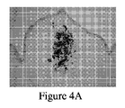
3時間インキュベーション後、コラゲナーゼによりインキュベートした検体の量は60%を超えて、5×3×0.1mm3まで減少した(図2D)が、コラゲナーゼによる24時間インキュベーション後、スライスは測定不能な検体まで断片化した(図2E)ことが結果からわかる。対照検体の量は、最初の検体(図2A)と比較して、PBSによる3時間または24時間インキュベーション後(それぞれ図2Bおよび2C)、変化がなかった。
B.コラーゲン変性の病理組織学的試験
After 3 hours incubation, the amount of specimen incubated with collagenase decreased by more than 60% to 5 × 3 × 0.1 mm 3 (FIG. 2D), but after 24 hours incubation with collagenase, the slices became unmeasurable specimen It can be seen from the results that fragmentation occurred (FIG. 2E). The amount of control specimen was unchanged after 3 or 24 hours incubation with PBS (Figures 2B and 2C, respectively) compared to the initial specimen (Figure 2A).
B. Histopathological examination of collagen degeneration
コラーゲン変性は、ヘマトキシリン−エオジン(H&E)染色を用いて観察することができるが、ピクロシリウスレッド(PSR)染色での偏光顕微鏡下においてより良好に可視化される。この目的で、口腔の咀嚼粘膜を含む硬口蓋からパンチ生検を採取した。図3Aに示すように、2,500、5,000、7,500または10,000単位/mLのコラゲナーゼCLPSA溶液(Worthington Biochemical Corporation,NJ,USA)を含有する50μLの量を組織の中央の口蓋ひだの隆線の真下に注入した。 Collagen degeneration can be observed using hematoxylin-eosin (H & E) staining, but is better visualized under a polarizing microscope with picrosirius red (PSR) staining. For this purpose, punch biopsies were taken from the hard palate containing the chewing mucosa of the oral cavity. As shown in FIG. 3A, an amount of 50 μL containing 2,500, 5,000, 7,500 or 10,000 units / mL of collagenase CLPSA solution (Worthington Biochemical Corporation, NJ, USA) was added to the central palate of the tissue. It was injected directly under the ridge of the folds.
図3Bの白の水平破線の両側の星印で示すように、注入の領域を示すために、固定化の前にパンチ生検の両側にコラーゲン/PBS/インク注入の部位と平行にインディアンインクを注入した。コラーゲン変性の領域を評価するために、各スライスに、隆線と平行な水平線によって(図3B−白の破線:表1−水平隆線)、および水平線と交差して隆線と直角をなす垂直線(図3B−黒色の線;表1−垂直隆線)で印を付けた。 Prior to immobilization, the Indian ink was placed parallel to the site of collagen / PBS / ink injection on both sides of the punch biopsy to indicate the area of injection, as indicated by the star on either side of the white horizontal dashed line in FIG. 3B. Injected. To assess the area of collagen degeneration, each slice is divided by a horizontal line parallel to the ridge (FIG. 3B—white dashed line: Table 1—horizontal ridge) and perpendicular to and perpendicular to the ridge. Marked with a line (FIG. 3B—black line; Table 1—vertical ridge).
対照群は非処置(非注入)試料およびPBSを注入した検体からなった(図4Aおよび4B)。これらの群は、処置群の条件と同様の条件下でインキュベートした。パンチ組織検体は、ホルマリンによる固定の前に、37℃で0、2、5、12および24時間PBS中でインキュベートした。 The control group consisted of untreated (non-injected) samples and specimens injected with PBS (FIGS. 4A and 4B). These groups were incubated under conditions similar to those of the treatment group. Punch tissue specimens were incubated in PBS for 0, 2, 5, 12, and 24 hours at 37 ° C. prior to formalin fixation.
固定後、組織を整えて、パラフィンに包埋して、注入部位(インクで印を付けた)のレベルで長軸方向に切片化し、次いでヘマトキシリン&エオシン、マッソントリクローム(MT)およびピクロシリウスレッド(PSR)で染色して、病理組織学試験に回した。処置前後の各スライスの大きさ(mm単位で)は、垂直線(表1−隆線垂直方向)に沿って、および水平線(表1−隆線水平方向)に沿って評価した。代表的な検体は、以下の通りに図4に示す:(A)試料番号56−インクのみを注入した対照、ヘマトキシリン&エオシン染色;(B)試料番号31− PBSを注入した対照、ピクロシリウスレッド(PSR)で染色、偏光顕微鏡下で観察した;(C)試料番号33−コラゲナーゼ2,500単位/mLを注入、ピクロシリウスレッド(PSR)で染色、偏光顕微鏡下で観察した;(D)試料番号35−コラゲナーゼ5,000単位/mLを注入、ピクロシリウスレッド(PSR)で染色、偏光顕微鏡下で観察した;(E)試料番号37−コラゲナーゼ7,500単位/mLを注入、ピクロシリウスレッド(PSR)で染色、偏光顕微鏡下で観察した;および(F) 試料番号39−コラゲナーゼ10,000単位/mLを注入、ピクロシリウスレッド(PSR)で染色、偏光顕微鏡下で観察した。 After fixation, the tissue is trimmed, embedded in paraffin, sectioned longitudinally at the level of the injection site (marked with ink), then hematoxylin & eosin, Masson trichrome (MT) and Picrosirius Stained with red (PSR) and sent to histopathological examination. The size (in mm) of each slice before and after treatment was evaluated along the vertical line (Table 1—ridge vertical direction) and along the horizontal line (Table 1—ridge horizontal direction). Representative specimens are shown in FIG. 4 as follows: (A) Sample number 56—control injected with ink alone, hematoxylin & eosin staining; (B) Sample number 31—control injected with PBS, Picrosirius Stained with red (PSR), observed under a polarizing microscope; (C) sample number 33-collagenase 2,500 units / mL injected, stained with picrosirius red (PSR), observed under a polarizing microscope; (D ) Sample number 35-collagenase 5,000 units / mL injected, stained with picrosirius red (PSR) and observed under a polarizing microscope; (E) Sample number 37-collagenase 7,500 units / mL injected, Stained with Kurosirius Red (PSR), observed under a polarizing microscope; and (F) Sample No. 39—injected with 10,000 units / mL of collagenase, Picrosiri Stained with thread (PSR), and observed under a polarizing microscope.
処置から2時間および5時間後の組織構造をそれぞれ表1および2にまとめる。 The tissue structures at 2 and 5 hours after treatment are summarized in Tables 1 and 2, respectively.
病理検査では、対照群と比較して、処置検体のすべてにおいて用量依存的様式で大きなコラーゲン溶解が認められた。図4に示し、さらに表1および2に記載するように、歯肉組織変性の領域は、薄いコラーゲン線維束を淡い染色で、脂肪組織細胞膜を淡い染色で、および小動脈壁の過好酸球増加と粒状度によって示した。変性の最も大きな領域は、7,500単位/mLのコラーゲンで処置した試料で観察された(図4E)。対照試料(それぞれ図4Aおよび4B)は、正常範囲内であった。 Pathological examination showed greater collagen dissolution in a dose-dependent manner in all treated specimens compared to the control group. As shown in FIG. 4 and further described in Tables 1 and 2, the regions of gingival tissue degeneration include thin collagen fiber bundles with light staining, adipose tissue cell membrane with light staining, and hypereosinophils in small arterial walls. And indicated by granularity. The greatest area of denaturation was observed in samples treated with 7,500 units / mL of collagen (FIG. 4E). Control samples (FIGS. 4A and 4B, respectively) were within the normal range.

実施例2
歯根膜内注射量のex−vivoでの評価
特定の切片で処置する歯の長さおよび歯根膜の幅に関する実験の所見に基づいて評価を行った。このデータを用いて、PDLの推定量を算出した。次いで、この推定量を臨床的に再評価した。
Example 2
Ex-vivo evaluation of intra-periodontal injection volume Evaluation was made based on experimental findings regarding the length of the teeth and periodontal ligaments treated with specific sections. The estimated amount of PDL was calculated using this data. This estimate was then clinically reassessed.
インディアンインクを下顎骨の第3切歯および第1小臼歯のPDLの6ポイントに注入した。色素の漏れが生じた注入量によってエンドポイントを決定した。6箇所の注入ポイントの各々で最適量を20μLとして決定した。 Indian ink was injected into the third incisor of the mandible and 6 points in the PDL of the first premolar. The endpoint was determined by the injection volume at which dye leakage occurred. The optimal volume was determined as 20 μL at each of the 6 injection points.
実施例3
コラゲナーゼ注入後のPDLの断片化
歯肉構造ではなく、PDL構造を評価するために、下顎骨の両側の第3切歯と第1小臼歯およびこれらの周辺の組織を異なる濃度(2,500、5,000、7,500または10,000単位/mL)で酵素注入(6ポイントで20μL)を行った。注入から2時間後、下顎骨を切断して、関連する歯の周りを整え、骨槽に埋まった歯を残し、次いでホルマリンで固定した。検体を5〜6日間脱灰し、次いで整えて(図5)、パラフィンに包埋して、歯の長軸にそって切片化した。
Example 3
To assess the PDL structure, but not the fragmented gingival structure of PDL after collagenase injection , the third incisors and first premolars on both sides of the mandible and their surrounding tissues were of different concentrations (2,500, 5 , 000, 7,500 or 10,000 units / mL). Two hours after the injection, the mandible was cut and trimmed around the relevant teeth, leaving the teeth buried in the alveolus and then fixed with formalin. Samples were decalcified for 5-6 days, then trimmed (FIG. 5), embedded in paraffin, and sectioned along the long axis of the tooth.
歯根の異なる切片でのPDLコラーゲン線維束の分解および周辺組織の構造の完全性(固有歯槽)を検査するために、切片をヘマトキシリン&エオシンおよびピクロシリウスレッドで染色して、病理組織学試験に回した。PBSを注入した、または注入しなかった検体を対照として用いた。 To examine the degradation of PDL collagen fiber bundles in different sections of the root and the structural integrity of the surrounding tissues (intrinsic alveoli), the sections were stained with hematoxylin & eosin and picrosirius red for histopathological examination Turned. Samples with or without PBS were used as controls.
コラゲナーゼ処置後にPDLの断片化を観察した。 PDL fragmentation was observed after collagenase treatment.
実施例4
Ex vivoでの抜歯
動物を犠牲にしてから4時間後に下顎骨両側の第3切歯および第1小臼歯を採取して、例えば図6に示すように、異なる濃度(2,500、5,000、7,500または10,000単位/mL)で歯根膜内酵素注入(6ポイントで20μL)を行った。注入から2時間後、回転力を付加することなく、歯の長軸に沿って標準抜歯鉗子を使用して歯を抜去した。抜去の容易さを対照(非注入群およびPBS注入群)歯と比較して、決定した。
Example 4
The third incisors and first premolars on both sides of the mandible were collected 4 hours after sacrificing the ex vivo extraction tooth , for example, as shown in FIG. 6, with different concentrations (2,500, 5,000 , 7,500, or 10,000 units / mL). Periodontal enzyme injection (20 μL at 6 points) was performed. Two hours after injection, the tooth was removed using standard extraction forceps along the long axis of the tooth without applying rotational force. Ease of removal was determined by comparing to control (non-injection group and PBS injection group) teeth.
付加力をkgf単位で測定する抜歯用電子装置を用いて、抜去で必要とする力の量を測定した(図7A〜7B).この装置を使用して、ネジで締めた抜歯鉗子で歯の頂部をしっかりと保持した。下顎骨は、4本の調整ネジで下顎骨を保持することができる2つの軸手段で別々に保持した。下顎骨は、歯根の長軸に沿って力を付加することによって抜去できるよう、すなわち骨/歯槽から上方向に歯を引き離すように配置した。 The amount of force required for extraction was measured using an extraction electronic device that measures the applied force in units of kgf (FIGS. 7A to 7B). This device was used to hold the top of the tooth firmly with screwed extraction forceps. The mandible was held separately by two axial means that can hold the mandible with four adjustment screws. The mandible was placed so that it could be removed by applying a force along the long axis of the root, i.e., pulling the tooth upward from the bone / alveolar.
コラゲナーゼ溶液(10,000単位/mL)を注入した手操作による抜歯は、著しく容易であり、すなわち、非処置の歯を抜去するために必要とした力よりも、必要とする力は著しく少なかった。さらに、処置歯を一塊として抜去したが、非処置歯は抜去中いくつかの歯冠が壊れた。 Manual extraction with a collagenase solution (10,000 units / mL) was remarkably easy, ie, it required significantly less force than that required to remove the untreated teeth. . In addition, the treated teeth were removed as a lump, while some of the crowns were broken during removal of the untreated teeth.
実施例5
In vivoでの抜歯
6月齢の飼育ブタに対して麻酔をかける。有効量のコラゲナーゼを含有する組成物の一連の歯根膜内注射を歯の周りの6部位に投与する。コラゲナーゼ歯根膜内注射から約30分後に、部位をEDTAで洗浄する。
Example 5
In vivo tooth extraction 6 month old domestic pigs are anesthetized. A series of intra-periodontal injections of a composition containing an effective amount of collagenase is administered at six sites around the teeth. Approximately 30 minutes after the collagenase intra periodontal injection, the site is washed with EDTA.
歯の長軸に沿って力を使い、回転力を使うことなく、歯を骨槽から上向きに引き離すことによって歯を抜去する。抜去は抜歯鉗子またはエレベーターを使用して行う。抜去する歯に向かって凹面側で、エレベーターを近心または遠心のPDL腔中に挿入する。顔面に向かって歯を動かすようにエレベーターを回転させる。 Using force along the long axis of the tooth, the tooth is extracted by pulling the tooth upward away from the alveolar ridge without using rotational force. Extraction is performed using extraction forceps or an elevator. On the concave side towards the tooth to be removed, an elevator is inserted into the mesial or distal PDL cavity. Rotate the elevator to move the teeth towards the face.
歯を適切に持ち上げてから、歯を脱臼させて抜去する。 Lift the teeth properly, then dislodge them and remove them.
抜去で必要とした力の量は、抜歯装置を用いて評価する。結果を非処置歯の抜去で必要とした力と比較する。 The amount of force required for extraction is evaluated using a tooth extraction device. The result is compared with the force required to remove the untreated tooth.
実施例6
歯槽の保存
歯を取り換える場合、骨および付随する軟組織は重要な検討事項である。インプラントを配置する場合、取り付け具用に十分な骨が必要であり、歯肉組織を正しく処置することは審美的に好ましい結果をもたらすために不可欠である。骨損失が激しい場合、取り外し可能な義歯(RDP)を考慮する必要がある。
Example 6
Bone and associated soft tissue are important considerations when replacing the preserved teeth in the alveoli. When placing an implant, sufficient bone is required for the fixture and proper treatment of the gingival tissue is essential to produce aesthetically pleasing results. If bone loss is severe, removable dentures (RDP) need to be considered.
本発明の非外傷性抜歯方法を適用すると、切歯抜去部位の治癒継続期間が少なくとも50%縮小する。さらに、抜歯後最初の6ヵ月以内では歯槽隆線の高さおよび幅に明白な減少が見られない。隣接した近心および遠位の歯の表面に付随する骨は、その骨の元の横寸法である。このように、歯槽の保存は得られる。したがって、PDL腔をコラゲナーゼで処置することによって、歯槽保存を向上させ、それによって歯科インプラントを効率よく移植する機会が上昇する。 When the atraumatic extraction method of the present invention is applied, the healing duration of the incisor extraction site is reduced by at least 50%. Furthermore, there is no apparent reduction in alveolar ridge height and width within the first 6 months after extraction. Bone associated with adjacent mesial and distal tooth surfaces is the original lateral dimension of the bone. In this way, preservation of the alveoli is obtained. Therefore, treating the PDL cavity with collagenase improves alveolar preservation, thereby increasing the chance of efficiently implanting dental implants.
前述の特定の実施形態の説明は、本発明の要領を十分に明らかにしているので、現在の知識を応用することによって種々の用途を目的として過度の実験を実施することなく、および上位概念から逸脱することなくそのような特定の実施形態を容易に修正および/または適合させることができる。したがって、そのような適合および修正は、開示される実施形態の均等物の意味および範囲内で理解されなければならず、かつ理解されることを意図する。本明細書で使用する語法または用語は、説明を目的としており、限定するものではないことを理解すべきである。種々の開示した機能を実施する手段、材料およびステップは、本発明を逸脱することなく種々の代替形態を取り得る。 The description of the specific embodiments described above sufficiently clarifies the point of the present invention, so that the present knowledge can be applied without undue experimentation for various purposes and from a superordinate concept. Such particular embodiments can be readily modified and / or adapted without departing. Accordingly, such adaptations and modifications must be understood and intended to be understood within the meaning and scope of the equivalents of the disclosed embodiments. It is to be understood that the terminology or terminology used herein is for the purpose of description and not limitation. The means, materials, and steps for carrying out various disclosed functions may take a variety of alternative forms without departing from the invention.
一実施形態によれば、本発明は抜歯用のキットを提供し、該キットは前述の歯の周りの組織への、歯の周辺の歯根膜を破壊することができる有効量の薬剤を含有する組成物を含む第1の容器を含む。さらに別の実施形態によれば、該キットは、鎮痛剤、麻酔薬、抗生物質、麻薬性鎮痛剤、ビタミン剤、成長因子、香味料およびそれらの組み合わせからなる群から選択される薬剤を含有するさらなる組成物をさらに含む。さらに別の実施形態によれば、該キットは、有効量の失活剤を含有する組成物をさらに含む。さらに別の実施形態によれば、該キットは、前述の歯を抜去するための歯科手段をさらに含む。さらに別の実施形態によれば、該キットは、キットの各構成成分の使用と使用する順番とを収載した取扱説明書をさらに備える。さらに別の実施形態によれば、取扱説明書は、本発明の方法によるキットの使用を記述する。 According to one embodiment, the present invention provides a kit for tooth extraction, which kit contains an effective amount of an agent capable of destroying the periodontal ligament around the tooth to the tissue surrounding the tooth. A first container containing the composition is included. According to yet another embodiment, the kit contains an agent selected from the group consisting of analgesics, anesthetics, antibiotics, narcotic analgesics, vitamins, growth factors, flavorings, and combinations thereof. Further comprising a further composition. According to yet another embodiment, the kit further comprises a composition containing an effective amount of a quenching agent. According to yet another embodiment, the kit further comprises dental means for extracting the aforementioned teeth. According to yet another embodiment, the kit further comprises an instruction manual containing the use of each component of the kit and the order of use. According to yet another embodiment, the instructions describe the use of the kit according to the method of the invention.
Claims (36)
a)歯の周辺の歯根膜を破壊することができる有効量の薬剤を含有する組成物を前記歯の周辺の組織に適用することと、
b)前記歯を抜去することと、
を含む、方法。 A method of extracting teeth in a subject in need thereof,
a) applying a composition containing an effective amount of a drug capable of destroying the periodontal ligament around the tooth to the tissue around the tooth;
b) removing the teeth;
Including a method.
b)前記組成物の注射から少なくとも15分後に前記歯を抜去することと、
を含む、請求項1に記載の方法。 a) injecting a composition containing an effective amount of collagenase into a plurality of sites of the periodontal ligament around the tooth;
b) removing the teeth at least 15 minutes after injection of the composition;
The method of claim 1 comprising:
b)前記歯を抜去する前に、前記歯の周辺の組織に有効量の失活剤を適用することと;
c)前記歯を抜去することと、
を含む、請求項1に記載の方法。 a) applying a composition containing an effective amount of a drug capable of destroying the periodontal ligament around the tooth to the tissue around the tooth;
b) applying an effective amount of a quenching agent to the tissue surrounding the tooth before removing the tooth;
c) removing the teeth;
The method of claim 1 comprising:
a)有効量のコラゲナーゼを含有する組成物を歯の周辺の歯根膜に適用することと;
b)前記組成物を適用してから少なくとも15分後に前記歯を抜去することと;
c)前記歯を抜去した後に得られた窩に歯科インプラントを設置することと、
を含む、方法。 A method of replacing a tooth with a dental implant,
a) applying a composition containing an effective amount of collagenase to the periodontal ligament around the teeth;
b) removing the teeth at least 15 minutes after application of the composition;
c) placing a dental implant in the fossa obtained after removing the tooth;
Including a method.
Applications Claiming Priority (3)
| Application Number | Priority Date | Filing Date | Title |
|---|---|---|---|
| US201261640743P | 2012-05-01 | 2012-05-01 | |
| US61/640,743 | 2012-05-01 | ||
| PCT/IL2013/050367 WO2013164822A1 (en) | 2012-05-01 | 2013-05-01 | Methods for extracting a tooth |
Publications (2)
| Publication Number | Publication Date |
|---|---|
| JP2015521169A true JP2015521169A (en) | 2015-07-27 |
| JP2015521169A5 JP2015521169A5 (en) | 2016-06-23 |
Family
ID=49514270
Family Applications (1)
| Application Number | Title | Priority Date | Filing Date |
|---|---|---|---|
| JP2015509565A Pending JP2015521169A (en) | 2012-05-01 | 2013-05-01 | How to extract a tooth |
Country Status (10)
| Country | Link |
|---|---|
| US (1) | US10016492B2 (en) |
| EP (1) | EP2844276B1 (en) |
| JP (1) | JP2015521169A (en) |
| CN (1) | CN104271152B (en) |
| AU (1) | AU2013255400A1 (en) |
| BR (1) | BR112014027347A2 (en) |
| CA (1) | CA2872227A1 (en) |
| IL (1) | IL235435B (en) |
| IN (1) | IN2014MN02422A (en) |
| WO (1) | WO2013164822A1 (en) |
Families Citing this family (4)
| Publication number | Priority date | Publication date | Assignee | Title |
|---|---|---|---|---|
| EP3119420B1 (en) | 2014-03-20 | 2021-09-22 | Technion Research & Development Foundation Ltd. | Delivery system comprising a proteolytic enzyme or effector thereof for use in a method for oral treatment and uses thereof |
| KR101551344B1 (en) | 2014-04-14 | 2015-09-08 | 한국기계연구원 | A guiding system for a direction in tooth extraction and a method thereof |
| US11033357B1 (en) * | 2020-11-12 | 2021-06-15 | King Abdulaziz University | Methods for tooth extraction using a thermoelectric device |
| WO2023112020A1 (en) * | 2021-12-14 | 2023-06-22 | Ramot At Tel Aviv University Ltd. | Modified recombinant collagenase, compositions comprising the same and uses thereof in dental related procedures |
Family Cites Families (22)
| Publication number | Priority date | Publication date | Assignee | Title |
|---|---|---|---|---|
| US5139944A (en) | 1985-08-10 | 1992-08-18 | Biophram (Uk) Limited | Collagen-specific enzyme with platelet aggregation inhibition properties |
| DE3719367A1 (en) * | 1987-06-10 | 1988-12-29 | Duncker Martin Dr | Use of active substances which influence the metabolism and/or the structural properties of collagen in orthodontics |
| US5709873A (en) | 1988-02-26 | 1998-01-20 | Niels Bukh A/S | Method of treating conditions of teeth and their supporting tissue |
| US7947270B2 (en) | 1992-05-22 | 2011-05-24 | Arcimboldo Ab | Removing dental plaque with krill enzymes |
| US6379654B1 (en) | 2000-10-27 | 2002-04-30 | Colgate Palmolive Company | Oral composition providing enhanced tooth stain removal |
| US6984128B2 (en) * | 2002-11-01 | 2006-01-10 | Bas Medical, Inc. | Methods for enabling and stabilizing tooth movement |
| US20050186526A1 (en) * | 2002-11-01 | 2005-08-25 | Bas Medical, Inc. | Methods and systems for enabling and stabilizing tooth movement |
| EP1600141B1 (en) * | 2004-05-24 | 2013-04-17 | 3M Deutschland GmbH | Collagenolytic active enzyme containing compositions for the treatment of dental caries |
| US7811560B2 (en) | 2006-01-30 | 2010-10-12 | Auxilium Us Holdings, Llc | Compositions and methods for treating collagen-mediated diseases |
| EP1844727A1 (en) * | 2006-04-12 | 2007-10-17 | 3M Innovative Properties Company | Dental instrument for removing solely unremineralisable carious dentin |
| US8454362B2 (en) * | 2006-10-16 | 2013-06-04 | Natural Dental Implants Ag | Customized dental prosthesis for periodontal- or osseointegration, and related systems and methods |
| WO2008100833A2 (en) * | 2007-02-13 | 2008-08-21 | Auxilium International Holdings, Inc. | Production of recombinant collagenases colg and colh in escherichia coli |
| US20090098509A1 (en) * | 2007-10-15 | 2009-04-16 | Arthur Kitchings Weathers | Dental tooth extraction implement and method thereof |
| US20090181342A1 (en) * | 2008-01-15 | 2009-07-16 | Hua-Hong Chien | Tooth Extration Assisting Device |
| US20100061972A1 (en) | 2008-08-19 | 2010-03-11 | Auxilium US Holdings,LLC | Method of Treating Peyronie's Disease |
| US20120045816A1 (en) | 2008-09-09 | 2012-02-23 | Sialix, Inc. | Novel Glycosylated Polypeptides |
| WO2011163577A1 (en) * | 2010-06-24 | 2011-12-29 | The Board Of Trustees Of The University Of Illinois | Reagents and methods for preparing teeth for implantation |
| CA2847197C (en) | 2010-10-30 | 2020-11-03 | Isis Innovation Limited | Treatment for dupuytren's disease |
| WO2012125948A1 (en) | 2011-03-16 | 2012-09-20 | Biospecifics Technologies Corp. | Compositions and methods for producing clostridial collagenases |
| WO2013059619A1 (en) | 2011-10-21 | 2013-04-25 | Auxilium Pharmaceuticals, Inc. | Method of treating or reducing efp |
| US20150079069A1 (en) * | 2012-04-30 | 2015-03-19 | Elastomics Ab | Methods for modification of tissues |
| EP3119420B1 (en) * | 2014-03-20 | 2021-09-22 | Technion Research & Development Foundation Ltd. | Delivery system comprising a proteolytic enzyme or effector thereof for use in a method for oral treatment and uses thereof |
-
2013
- 2013-05-01 BR BR112014027347A patent/BR112014027347A2/en not_active IP Right Cessation
- 2013-05-01 EP EP13784170.6A patent/EP2844276B1/en active Active
- 2013-05-01 AU AU2013255400A patent/AU2013255400A1/en not_active Abandoned
- 2013-05-01 CN CN201380023125.XA patent/CN104271152B/en active Active
- 2013-05-01 WO PCT/IL2013/050367 patent/WO2013164822A1/en active Application Filing
- 2013-05-01 CA CA2872227A patent/CA2872227A1/en not_active Abandoned
- 2013-05-01 JP JP2015509565A patent/JP2015521169A/en active Pending
- 2013-05-01 IN IN2422MUN2014 patent/IN2014MN02422A/en unknown
-
2014
- 2014-10-29 US US14/527,487 patent/US10016492B2/en active Active
- 2014-10-30 IL IL235435A patent/IL235435B/en active IP Right Grant
Non-Patent Citations (3)
| Title |
|---|
| "抜歯について", [ONLINE], JPN6017003662, 23 February 2010 (2010-02-23) * |
| KAWADA J, KOMATSU K: "In Vitro Effects of Collagenase on Biomechanical Properties and Morphological Features of the Rat Mo", JAPANESE JOURNAL OF ORAL BIOLOGY, vol. 42, no. 3, JPN6017003656, 2000, pages 193 - 205 * |
| 山根 明 ほか: "ラット下顎切歯歯根膜の機械的性質に及ぼすコラゲナーゼ処理の影響", 歯科基礎医学会雑誌, vol. 第33巻(捕冊), JPN6017003659, 1991, pages p.245 * |
Also Published As
| Publication number | Publication date |
|---|---|
| US10016492B2 (en) | 2018-07-10 |
| CN104271152B (en) | 2016-08-17 |
| EP2844276B1 (en) | 2019-10-02 |
| BR112014027347A2 (en) | 2017-06-27 |
| WO2013164822A1 (en) | 2013-11-07 |
| CA2872227A1 (en) | 2013-11-07 |
| EP2844276A4 (en) | 2015-12-16 |
| IN2014MN02422A (en) | 2015-08-14 |
| US20150050617A1 (en) | 2015-02-19 |
| AU2013255400A1 (en) | 2014-12-04 |
| CN104271152A (en) | 2015-01-07 |
| EP2844276A1 (en) | 2015-03-11 |
| IL235435B (en) | 2018-10-31 |
Similar Documents
| Publication | Publication Date | Title |
|---|---|---|
| Mitsias et al. | The root membrane technique: human histologic evidence after five years of function | |
| Andreasen et al. | Essentials of traumatic injuries to the teeth: a step-by-step treatment guide | |
| Chen et al. | Dental implants: Maintenance, care and treatment of peri‐implant infection | |
| Calişkan | Pulpotomy of carious vital teeth with periapical involvement | |
| Hirooka | The biologic concept for the use of enamel matrix protein: true periodontal regeneration. | |
| Kaya-Büyükbayram et al. | Regenerative endodontic treatment of an infected immature dens invaginatus with the aid of cone-beam computed tomography | |
| US10016492B2 (en) | Methods for extracting a tooth | |
| Heithersay | Replantation of avulsed teeth. A review | |
| Feiglin | Root resorption | |
| Isidor et al. | New attachment formation on citric acid treated roots | |
| Nethander et al. | Experimental autogenous tooth transplantation in the dog: a comparison between one-and two-stage surgical techniques | |
| Gault | Idiopathic ankylosis-resorption: diagnosis and treatment | |
| WO2010024702A2 (en) | Pharmaceutical composition for the treatment of periodontal diseases | |
| Eliasson et al. | Autotransplanted teeth with early-stage endodontic treatment: a radiographic evaluation | |
| Azevedo et al. | Time of endodontic treatment in autogenic transplants of mature teeth: histological study in dogs | |
| Dumsha | Management of avulsions | |
| Fridström et al. | Evaluating Emdogain® and healing of replanted teeth using an intra‐individual experimental–control study design | |
| Di Girolamo et al. | Critical investigation of the pre-surgical anatomic factors that influence the aesthetic result in post-extractive implantology and immediate prosthetic | |
| Berechet et al. | PERI-IMPLANTITIS VERSUS PERIODONTITIS-SIMILARITIES AND DIFFERENCES. LITERATURE REVIEW. | |
| Zürcher et al. | Management of avulsed teeth | |
| LIEBLICH et al. | Microbiologic considerations with dental implants | |
| Pontes et al. | Use of acellular dermal matrix graft in the treatment of gingival recessions: a case report | |
| Al-shammari | Regenerative endodontic procedure in immature permanent teeth | |
| Bawa et al. | Tri-immuno phasic periodontal therapy (TIP) therapy modern notion of handling periodontal diseases | |
| Shah et al. | Periimplant diseases and its treatment modalities: A |
Legal Events
| Date | Code | Title | Description |
|---|---|---|---|
| A521 | Request for written amendment filed |
Free format text: JAPANESE INTERMEDIATE CODE: A523 Effective date: 20160428 |
|
| A621 | Written request for application examination |
Free format text: JAPANESE INTERMEDIATE CODE: A621 Effective date: 20160428 |
|
| A131 | Notification of reasons for refusal |
Free format text: JAPANESE INTERMEDIATE CODE: A131 Effective date: 20170207 |
|
| A601 | Written request for extension of time |
Free format text: JAPANESE INTERMEDIATE CODE: A601 Effective date: 20170502 |
|
| A601 | Written request for extension of time |
Free format text: JAPANESE INTERMEDIATE CODE: A601 Effective date: 20170706 |
|
| A02 | Decision of refusal |
Free format text: JAPANESE INTERMEDIATE CODE: A02 Effective date: 20171205 |