JP2015500638A - ERBB3 mutations in cancer - Google Patents
ERBB3 mutations in cancer Download PDFInfo
- Publication number
- JP2015500638A JP2015500638A JP2014544721A JP2014544721A JP2015500638A JP 2015500638 A JP2015500638 A JP 2015500638A JP 2014544721 A JP2014544721 A JP 2014544721A JP 2014544721 A JP2014544721 A JP 2014544721A JP 2015500638 A JP2015500638 A JP 2015500638A
- Authority
- JP
- Japan
- Prior art keywords
- erbb3
- cancer
- mutation
- subject
- nucleic acid
- Prior art date
- Legal status (The legal status is an assumption and is not a legal conclusion. Google has not performed a legal analysis and makes no representation as to the accuracy of the status listed.)
- Pending
Links
Images


Classifications
-
- C—CHEMISTRY; METALLURGY
- C12—BIOCHEMISTRY; BEER; SPIRITS; WINE; VINEGAR; MICROBIOLOGY; ENZYMOLOGY; MUTATION OR GENETIC ENGINEERING
- C12Q—MEASURING OR TESTING PROCESSES INVOLVING ENZYMES, NUCLEIC ACIDS OR MICROORGANISMS; COMPOSITIONS OR TEST PAPERS THEREFOR; PROCESSES OF PREPARING SUCH COMPOSITIONS; CONDITION-RESPONSIVE CONTROL IN MICROBIOLOGICAL OR ENZYMOLOGICAL PROCESSES
- C12Q1/00—Measuring or testing processes involving enzymes, nucleic acids or microorganisms; Compositions therefor; Processes of preparing such compositions
- C12Q1/68—Measuring or testing processes involving enzymes, nucleic acids or microorganisms; Compositions therefor; Processes of preparing such compositions involving nucleic acids
- C12Q1/6876—Nucleic acid products used in the analysis of nucleic acids, e.g. primers or probes
- C12Q1/6883—Nucleic acid products used in the analysis of nucleic acids, e.g. primers or probes for diseases caused by alterations of genetic material
- C12Q1/6886—Nucleic acid products used in the analysis of nucleic acids, e.g. primers or probes for diseases caused by alterations of genetic material for cancer
-
- C—CHEMISTRY; METALLURGY
- C12—BIOCHEMISTRY; BEER; SPIRITS; WINE; VINEGAR; MICROBIOLOGY; ENZYMOLOGY; MUTATION OR GENETIC ENGINEERING
- C12Q—MEASURING OR TESTING PROCESSES INVOLVING ENZYMES, NUCLEIC ACIDS OR MICROORGANISMS; COMPOSITIONS OR TEST PAPERS THEREFOR; PROCESSES OF PREPARING SUCH COMPOSITIONS; CONDITION-RESPONSIVE CONTROL IN MICROBIOLOGICAL OR ENZYMOLOGICAL PROCESSES
- C12Q1/00—Measuring or testing processes involving enzymes, nucleic acids or microorganisms; Compositions therefor; Processes of preparing such compositions
- C12Q1/68—Measuring or testing processes involving enzymes, nucleic acids or microorganisms; Compositions therefor; Processes of preparing such compositions involving nucleic acids
- C12Q1/6813—Hybridisation assays
-
- A—HUMAN NECESSITIES
- A61—MEDICAL OR VETERINARY SCIENCE; HYGIENE
- A61K—PREPARATIONS FOR MEDICAL, DENTAL OR TOILETRY PURPOSES
- A61K31/00—Medicinal preparations containing organic active ingredients
- A61K31/33—Heterocyclic compounds
- A61K31/395—Heterocyclic compounds having nitrogen as a ring hetero atom, e.g. guanethidine or rifamycins
- A61K31/495—Heterocyclic compounds having nitrogen as a ring hetero atom, e.g. guanethidine or rifamycins having six-membered rings with two or more nitrogen atoms as the only ring heteroatoms, e.g. piperazine or tetrazines
- A61K31/505—Pyrimidines; Hydrogenated pyrimidines, e.g. trimethoprim
- A61K31/517—Pyrimidines; Hydrogenated pyrimidines, e.g. trimethoprim ortho- or peri-condensed with carbocyclic ring systems, e.g. quinazoline, perimidine
-
- A—HUMAN NECESSITIES
- A61—MEDICAL OR VETERINARY SCIENCE; HYGIENE
- A61K—PREPARATIONS FOR MEDICAL, DENTAL OR TOILETRY PURPOSES
- A61K39/00—Medicinal preparations containing antigens or antibodies
- A61K39/395—Antibodies; Immunoglobulins; Immune serum, e.g. antilymphocytic serum
- A61K39/39533—Antibodies; Immunoglobulins; Immune serum, e.g. antilymphocytic serum against materials from animals
- A61K39/39558—Antibodies; Immunoglobulins; Immune serum, e.g. antilymphocytic serum against materials from animals against tumor tissues, cells, antigens
-
- A—HUMAN NECESSITIES
- A61—MEDICAL OR VETERINARY SCIENCE; HYGIENE
- A61P—SPECIFIC THERAPEUTIC ACTIVITY OF CHEMICAL COMPOUNDS OR MEDICINAL PREPARATIONS
- A61P35/00—Antineoplastic agents
-
- C—CHEMISTRY; METALLURGY
- C12—BIOCHEMISTRY; BEER; SPIRITS; WINE; VINEGAR; MICROBIOLOGY; ENZYMOLOGY; MUTATION OR GENETIC ENGINEERING
- C12Q—MEASURING OR TESTING PROCESSES INVOLVING ENZYMES, NUCLEIC ACIDS OR MICROORGANISMS; COMPOSITIONS OR TEST PAPERS THEREFOR; PROCESSES OF PREPARING SUCH COMPOSITIONS; CONDITION-RESPONSIVE CONTROL IN MICROBIOLOGICAL OR ENZYMOLOGICAL PROCESSES
- C12Q1/00—Measuring or testing processes involving enzymes, nucleic acids or microorganisms; Compositions therefor; Processes of preparing such compositions
- C12Q1/68—Measuring or testing processes involving enzymes, nucleic acids or microorganisms; Compositions therefor; Processes of preparing such compositions involving nucleic acids
- C12Q1/6844—Nucleic acid amplification reactions
- C12Q1/686—Polymerase chain reaction [PCR]
-
- C—CHEMISTRY; METALLURGY
- C12—BIOCHEMISTRY; BEER; SPIRITS; WINE; VINEGAR; MICROBIOLOGY; ENZYMOLOGY; MUTATION OR GENETIC ENGINEERING
- C12Q—MEASURING OR TESTING PROCESSES INVOLVING ENZYMES, NUCLEIC ACIDS OR MICROORGANISMS; COMPOSITIONS OR TEST PAPERS THEREFOR; PROCESSES OF PREPARING SUCH COMPOSITIONS; CONDITION-RESPONSIVE CONTROL IN MICROBIOLOGICAL OR ENZYMOLOGICAL PROCESSES
- C12Q2600/00—Oligonucleotides characterized by their use
- C12Q2600/112—Disease subtyping, staging or classification
-
- C—CHEMISTRY; METALLURGY
- C12—BIOCHEMISTRY; BEER; SPIRITS; WINE; VINEGAR; MICROBIOLOGY; ENZYMOLOGY; MUTATION OR GENETIC ENGINEERING
- C12Q—MEASURING OR TESTING PROCESSES INVOLVING ENZYMES, NUCLEIC ACIDS OR MICROORGANISMS; COMPOSITIONS OR TEST PAPERS THEREFOR; PROCESSES OF PREPARING SUCH COMPOSITIONS; CONDITION-RESPONSIVE CONTROL IN MICROBIOLOGICAL OR ENZYMOLOGICAL PROCESSES
- C12Q2600/00—Oligonucleotides characterized by their use
- C12Q2600/156—Polymorphic or mutational markers
Abstract
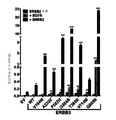
本発明は、癌における体細胞ErbB3変異に関し、ErbB3癌を特定、診断、および予後診断する方法、ならびに癌を治療する方法を含み、患者のある亜集団を含む。【選択図】図4The present invention relates to somatic ErbB3 mutations in cancer, including methods for identifying, diagnosing, and prognosing ErbB3 cancer, and methods for treating cancer, including a subpopulation of patients. [Selection] Figure 4
Description
関連出願
本出願は、米国特許法第119条(e)の下で、2011年11月30日に出願された米国仮出願第61/629,951号に対する優先権の利益を主張するものであり、参照によりその全体が本明細書に組み込まれる。
RELATED APPLICATION This application claims the benefit of priority over US Provisional Application No. 61 / 629,951 filed on November 30, 2011 under 35 USC 119 (e). , Incorporated herein by reference in its entirety.
本発明は、癌における体細胞ErbB3変異に関し、ErbB3癌を特定、診断、および予後診断する方法、ならびに癌を治療する方法を含み、患者のある亜集団を含む。 The present invention relates to somatic ErbB3 mutations in cancer, including methods for identifying, diagnosing, and prognosing ErbB3 cancer, and methods for treating cancer, including a subpopulation of patients.
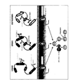
ERBB受容体としても知られる、受容体チロシンキナーゼ(RTK)のヒト上皮成長因子受容体(HER)ファミリーは、4つのメンバー、EGFR/ERBB1/HER1、ERBB2/HER2、ERBB3/HER3、およびERBB4/HER4からなる(Hynes et al.Nature Reviews Cancer 5,341−354(2005)、Baselga et al.Nature Reviews Cancer 9,463−475(2009))。ERBBファミリーメンバーは、細胞外ドメイン(ECD)、単スパン膜貫通領域、細胞内チロシンキナーゼドメイン、およびC末端シグナル伝達尾部を含む(Burgess et al.Mol Cell 12,541−552(2003)、Ferguson.Annual Review of Biophysics 37,353−373(2008))。ECDは、2つのLドメイン(IおよびIII)ならびに2つの高システインドメイン(IIおよびIV)からなる4つのドメイン構造である(Burgess et al.Mol Cell 12,541−552(2003)、Ferguson.Annual Review of Biophysics 37,353−373(2008))。ERBB受容体は、上皮成長因子(EGF)、形質転換成長因子−α(TGF−α)、およびニューレグリンを含む複数のリガンドにより活性化される(Yarden et al.Nat Rev Mol Cell Biol 2,127−137(2001))。受容体の活性化は、ドメインIおよびIIIに同時に結合する単一リガンド分子を必要とし、ドメインIIにおける二量体化アームを介したヘテロ二量体化またはホモ二量体化につながる(Burgess et al.Mol Cell 12,541−552(2003)、Ogiso et al.Cell 110,775−787(2002)、Cho.Science 297,1330−1333(2002)、Dawson et al.Molecular and Cellular Biology 25,7734−7742(2005)、Alvarado et al.Cell 142,568−579(2010)、Lemmon et al.Cell 141,1117−1134(2010))。リガンドの不在化で、ドメインII二量体化アームは、ドメインIVとの分子内相互作用を介して挟み込まれ、「繋留された」自己抑制構成をもたらす(Burgess et al.Mol Cell 12,541−552(2003)、Cho.Science297,1330−1333(2002)、Lemmon et al.Cell 141,1117−1134(2010)、Ferguson et al.Mol Cell 11,507−517(2003))。 The human epidermal growth factor receptor (HER) family of receptor tyrosine kinases (RTKs), also known as ERBB receptors, has four members, EGFR / ERBB1 / HER1, ERBB2 / HER2, ERBB3 / HER3, and ERBB4 / HER4. (Hynes et al. Nature Reviews Cancer 5, 341-354 (2005), Baselga et al. Nature Reviews Cancer 9, 463-475 (2009)). ERBB family members include an extracellular domain (ECD), a single span transmembrane region, an intracellular tyrosine kinase domain, and a C-terminal signaling tail (Burgess et al. Mol Cell 12, 541-552 (2003), Ferguson. Annual Review of Biophysics 37, 353-373 (2008)). ECD is a four-domain structure consisting of two L domains (I and III) and two high cysteine domains (II and IV) (Burges et al. Mol Cell 12, 541-552 (2003), Ferguson. Annual). Review of Biophysics 37, 353-373 (2008)). The ERBB receptor is activated by multiple ligands including epidermal growth factor (EGF), transforming growth factor-α (TGF-α), and neuregulin (Yarden et al. Nat Rev Mol Cell Biol 2,127). -137 (2001)). Receptor activation requires a single ligand molecule that binds simultaneously to domains I and III, leading to heterodimerization or homodimerization via a dimerization arm in domain II (Burgesse et al. al. Mol Cell 12, 541-552 (2003), Ogiso et al. Cell 110, 775-787 (2002), Cho. Science 297, 1330-1333 (2002), Dawson et al. Molecular and Cellular Biology 25, 7734. -7742 (2005), Alvarado et al. Cell 142, 568-579 (2010), Lemmon et al. Cell 141, 1117-1134 (2010)). In the absence of ligand, the domain II dimerization arm is sandwiched via intramolecular interaction with domain IV, resulting in a “tethered” self-inhibited configuration (Burgess et al. Mol Cell 12, 541- 552 (2003), Cho. Science 297, 1330-1333 (2002), Lemmon et al. Cell 141, 1117-1134 (2010), Ferguson et al. Mol Cell 11, 507-517 (2003)).
4つのERBB受容体は、類似のドメイン組織を共有するが、機能的および構造的研究は、ERBB2が既知のERBBファミリーリガンドのいずれにも結合せず、構成的に二量体化に適した「非繋留」(オープン)構造であることを示す(Garrett et al.Mol Cell 11,495−505(2003))。対照的に、ERBB3は、リガンド結合、ヘテロ二量体化、およびシグナル伝達が可能であるが、傷害のあるキナーゼドメインを有する(Baselga et al.Nature Reviews Cancer 9,463−475(2009)、Jura et al.Proceedings of the National Academy of Sciences 106,21608−21613(2009)、Shi et al.Proceedings of the National Academy of Sciences 107,7692−7697(2010))。ERBB2およびERBB3は、それら自体は機能的に不完全であるが、それらのヘテロ二量体は、細胞シグナル伝達の強力な活性剤である(Pinkas−Kramarski et al.The EMBO Journal 15,2452−2467(1996)、Tzahar et al.Molecular and Cellular Biology 16,5276−5287(1996)、Holbro et al.Proceedings of the National Academy of Sciences 100,8933−8938(2003))。 Although the four ERBB receptors share a similar domain organization, functional and structural studies have shown that ERBB2 does not bind to any of the known ERBB family ligands and is constitutively suitable for dimerization. It shows that the structure is “non-tethered” (open) (Garret et al. Mol Cell 11, 495-505 (2003)). In contrast, ERBB3 is capable of ligand binding, heterodimerization, and signaling, but has a damaged kinase domain (Baselga et al. Nature Reviews Cancer 9, 463-475 (2009), Jura). et al., Proceedings of the National Academy of Sciences 106, 21608-21613 (2009), Shi et al., Proceedings of the National Academy of Sciences 107, 769276). Although ERBB2 and ERBB3 are themselves functionally defective, their heterodimers are potent activators of cell signaling (Pinkas-Kramarski et al. The EMBO Journal 15, 2452-2467. (1996), Tzahar et al. Molecular and Cellular Biology 16, 5276-5287 (1996), Holbro et al. Proceedings of the National Academy of Sciences 100, 8933-8938 (2003).
ERBB受容体は、正常な成長および発達の重要な調節因子であるが、それらの脱制御も癌の発達および進行に関与している(Baselga et al.Nature Reviews Cancer 9,463−475(2009)、Sithanandam et al.Cancer Gene Ther 15,413−448(2008)、Hynes et al.Current Opinion in Cell Biology 21,177−184(2009))。具体的には、受容体過剰発現をもたらし、体細胞変異を活性化する遺伝子増幅は、様々な癌においてERBB2およびEGFR内で発生することが知られている(Sithanandam et al.Cancer Gene Ther 15,413−448(2008)、Hynes et al.Current Opinion in Cell Biology 21,177−184(2009)、Wang et al.Cencer Cell 10,25−38(2006)、Yamauchi et al.Biomark Med 3,139−151(2009))。これは、EGFRおよびERBB2を標的とする複数の低分子および抗体に基づく治療法の開発をもたらした(Baselga et al.Nature Reviews Cancer 9,463−475(2009)、Alvarez et al.Journal of Clinical Oncology 28,3366−3379(2010))。腫瘍形成におけるERBB4の正確な役割は、十分に確立されていないが(Koutras et al.Critical Reviews in Oncology/Hematology 74,73−78(2010))、ERBB4内の形質転換体細胞変異が黒色腫において報告されている(Prickett et al.Nature Genetics 41,1127−1132(2009))。近年、ERBB2シグナル伝達において重要な役割を果たし、既存の治療法に対する耐性を促進することにも関与することを考慮し、ERBB3が有力な癌治療標的として浮上した(Baselga et al.Nature Reviews Cancer 9,463−475(2009)、Amin et al.Semin Cell Dev Biol 21、944−950(2010))。ERBB3増幅および/または過剰発現は、いくつかの癌において知られているが、ERBB3体細胞変異の散発的発生のみが報告されており、これらの変異の機能的関連は研究されていない。本明細書に提供される発明は、ヒト癌における頻繁なERBB3体細胞変異の特定に関する。 ERBB receptors are important regulators of normal growth and development, but their deregulation is also involved in cancer development and progression (Baselga et al. Nature Reviews Cancer 9, 463-475 (2009)). Sithanandam et al. Cancer Gene Ther 15, 413-448 (2008), Hynes et al. Current Opinion in Cell Biology 21, 177-184 (2009)). Specifically, gene amplification that results in receptor overexpression and activates somatic mutations is known to occur within ERBB2 and EGFR in various cancers (Sithanandam et al. Cancer Gene Ther 15, 413-448 (2008), Hynes et al. Current Opinion in Cell Biology 21, 177-184 (2009), Wang et al. Cencer Cell 10, 25-38 (2006), Yamauchi et al. Biomark Med 3,139. 151 (2009)). This has led to the development of multiple small molecule and antibody-based therapeutics targeting EGFR and ERBB2 (Baselga et al. Nature Reviews Cancer 9, 463-475 (2009), Alvarez et al. Journal of Clinical Oncology). 28, 3366-3379 (2010)). Although the exact role of ERBB4 in tumorigenesis has not been well established (Koutras et al. Critical Reviews in Oncology / Hematology 74, 73-78 (2010)), transformant cell mutations within ERBB4 are Have been reported (Prickett et al. Nature Genetics 41, 1277-1132 (2009)). In recent years, ERBB3 has emerged as a potential cancer therapeutic target, considering that it plays an important role in ERBB2 signaling and is also involved in promoting resistance to existing therapies (Baselga et al. Nature Reviews Cancer 9 , 463-475 (2009), Amin et al. Semin Cell Dev Biol 21, 944-950 (2010)). Although ERBB3 amplification and / or overexpression is known in some cancers, only sporadic occurrences of ERBB3 somatic mutations have been reported and the functional association of these mutations has not been studied. The invention provided herein relates to the identification of frequent ERBB3 somatic mutations in human cancer.
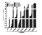
本発明は、胃および結腸腫瘍を含むが、これらに限定されない様々なヒト腫瘍と関連付けられる、受容体チロシンキナーゼ(RTK)のヒト上皮成長因子受容体(HER)ファミリーのERBB3受容体内の複数の体細胞変異事象の発見に少なくとも部分的に基づく。これらの変異は、ヒト腫瘍発生の素因があり、および/または直接寄与すると考えられる。実際に、本明細書に記載されるとおり、変異の一部は腫瘍発生をインビボで促進するという証拠がある。 The present invention relates to multiple bodies within the ERBB3 receptor of the human epidermal growth factor receptor (HER) family of receptor tyrosine kinases (RTKs) associated with a variety of human tumors, including but not limited to gastric and colon tumors. Based at least in part on the discovery of cellular mutation events. These mutations are predisposed to and / or directly contribute to human tumor development. Indeed, as described herein, there is evidence that some of the mutations promote tumorigenesis in vivo.
一態様では、本発明は、ErbB3癌検出薬を提供する。一実施形態では、ErbB3癌検出薬は、ErbB3消化管癌検出薬である。別の実施形態では、検出薬は、ErbB3核酸配列中のErbB3変異に特異的に結合することができる試薬を含む。他の一実施形態では、ErbB3核酸配列は、配列番号3または1を含む。 In one aspect, the present invention provides an ErbB3 cancer detection agent. In one embodiment, the ErbB3 cancer detection agent is an ErbB3 gastrointestinal cancer detection agent. In another embodiment, the detection agent comprises a reagent that can specifically bind to an ErbB3 mutation in the ErbB3 nucleic acid sequence. In another embodiment, the ErbB3 nucleic acid sequence comprises SEQ ID NO: 3 or 1.
いくつかの実施形態では、この試薬は、式

Xは任意の核酸であり、aは約0〜約250であり、
YはErbB3変異コドンであり、
Zは任意の核酸であり、bは約0〜約250である。
他の一実施形態では、変異コドンが、(i)104、809、232、262、284、325、846、928、60、111、135、295、406、453、498、1089、および1164からなる群から選択される、配列番号2の位置のアミノ酸、または(ii)193位の終止コドンをコードする。他の一実施形態では、消化管癌は、胃癌または結腸癌である。
In some embodiments, the reagent has the formula
X is any nucleic acid, a is from about 0 to about 250,
Y is an ErbB3 mutant codon,
Z is any nucleic acid and b is from about 0 to about 250.
In another embodiment, the mutated codon consists of (i) 104, 809, 232, 262, 284, 325, 846, 928, 60, 111, 135, 295, 406, 453, 498, 1089, and 1164. It encodes the amino acid at position SEQ ID NO: 2, or (ii) a stop codon at position 193, selected from the group. In another embodiment, the gastrointestinal cancer is gastric cancer or colon cancer.
別の態様では、本発明は、対象におけるErbB3消化管癌の存在を決定する方法を提供する。一実施形態では、この方法は、対象から得られる生体試料においてErbB3をコードする核酸配列中の変異を検出することであって、この変異は、ErbB3アミノ酸配列の少なくとも1つの位置にアミノ酸変化をもたらし、この変異は、対象におけるErbB3消化管癌を示す。別の実施形態では、アミノ酸変化を生じる変異は、104、809、232、262、284、325、846、928、60、111、135、295、406、453、498、1089、1164、および193からなる群から選択される、配列番号2の位置にある。他の実施形態では、消化管癌は、胃癌または結腸癌である。 In another aspect, the invention provides a method for determining the presence of ErbB3 gastrointestinal cancer in a subject. In one embodiment, the method detects a mutation in a nucleic acid sequence encoding ErbB3 in a biological sample obtained from a subject, wherein the mutation results in an amino acid change at at least one position of the ErbB3 amino acid sequence. This mutation is indicative of ErbB3 gastrointestinal cancer in the subject. In another embodiment, mutations resulting in amino acid changes are from 104, 809, 232, 262, 284, 325, 846, 928, 60, 111, 135, 295, 406, 453, 498, 1089, 1164, and 193. It is in the position of SEQ ID NO: 2 selected from the group consisting of In other embodiments, the gastrointestinal cancer is gastric cancer or colon cancer.
別の態様では、本発明は、対象におけるErbB3癌の存在を決定する方法を提供する。一実施形態では、この方法は、対象から得られる生体試料においてErbB3をコードする核酸配列中のアミノ酸変異の存在または不在を検出することを含み、この変異は、配列番号2の104、809、232、262、284、325、846、928、60、111、135、295、406、453、498、1089、1164、193、492、および714からなる群から選択される少なくとも1つの位置にアミノ酸変化をもたらし、変異の存在は、対象におけるErbB3癌を示す。別の実施形態では、ErbB3癌は、胃癌、結腸癌、食道癌、直腸癌、盲腸癌、非小細胞肺(NSCLC)腺癌、NSCLC(扁平上皮癌)、腎臓腺癌、黒色腫、卵巣癌、大細胞肺癌、小細胞肺癌(SCLC)、肝細胞癌(HCC)、肺癌、および膵臓癌からなる群から選択される。 In another aspect, the present invention provides a method for determining the presence of ErbB3 cancer in a subject. In one embodiment, the method comprises detecting the presence or absence of an amino acid mutation in a nucleic acid sequence encoding ErbB3 in a biological sample obtained from the subject, the mutation comprising 104, 809, 232 of SEQ ID NO: 2. , 262, 284, 325, 846, 928, 60, 111, 135, 295, 406, 453, 498, 1089, 1164, 193, 492, and 714, the amino acid change at at least one position selected from the group consisting of The presence of the mutation is indicative of ErbB3 cancer in the subject. In another embodiment, the ErbB3 cancer is gastric cancer, colon cancer, esophageal cancer, rectal cancer, cecal cancer, non-small cell lung (NSCLC) adenocarcinoma, NSCLC (squamous cell carcinoma), renal adenocarcinoma, melanoma, ovarian cancer Selected from the group consisting of: large cell lung cancer, small cell lung cancer (SCLC), hepatocellular carcinoma (HCC), lung cancer, and pancreatic cancer.
さらに別の態様では、決定方法は、以下の追加のステップ:治療薬を該対象に投与すること、必要とする対象を特定すること、必要とする対象から試料を得ることのうちの1つ、またはそれらの任意の組み合わせをさらに含む。一実施形態では、治療薬はErbB阻害剤である。他の実施形態では、このErbB阻害剤は、EGFR拮抗薬、ErbB2拮抗薬、ErbB3拮抗薬、ErbB4拮抗薬、およびEGFR/ErbB3拮抗薬からなる群から選択される。別の実施形態では、阻害剤は、低分子阻害剤である。一実施形態では、拮抗薬は、拮抗薬抗体である。さらに別の実施形態では、この抗体は、モノクローナル抗体、二重特異性抗体、キメラ抗体、ヒト抗体、ヒト化抗体、および抗体断片からなる群から選択される。 In yet another aspect, the determination method comprises one of the following additional steps: administering a therapeutic agent to the subject, identifying the subject in need, obtaining a sample from the subject in need, Or any combination thereof. In one embodiment, the therapeutic agent is an ErbB inhibitor. In other embodiments, the ErbB inhibitor is selected from the group consisting of an EGFR antagonist, an ErbB2 antagonist, an ErbB3 antagonist, an ErbB4 antagonist, and an EGFR / ErbB3 antagonist. In another embodiment, the inhibitor is a small molecule inhibitor. In one embodiment, the antagonist is an antagonist antibody. In yet another embodiment, the antibody is selected from the group consisting of monoclonal antibodies, bispecific antibodies, chimeric antibodies, human antibodies, humanized antibodies, and antibody fragments.
別の態様では、検出ステップは、増幅または配列決定を含む。一実施形態では、この検出は、変異を増幅させるか、または配列決定することと、変異またはその配列を検出することと、を含む。別の実施形態では、増幅は、増幅プライマーまたは増幅プライマー対を、試料から単離された核酸鋳型と混合することを含む。他の実施形態では、プライマーまたはプライマー対は、該変異の近位にあるか、またはそれを含む領域に相補的であるか、または部分的に相補的であり、核酸鋳型上でポリメラーゼによる核酸重合を開始することができる。他の一実施形態では、ポリメラーゼおよび核酸鋳型を含むDNA重合反応においてプライマーまたはプライマー対を伸長させて増幅産物を生成することをさらに含む。別の実施形態では、増幅または配列決定において、変異は、生体試料から単離されたゲノムDNA中の変異を配列決定すること、変異もしくはその増幅産物をアレイにハイブリダイズすること、変異もしくはその増幅産物を制限酵素で消化すること、または変異のリアルタイムPCR増幅のうちの1つ以上を含むプロセスにより検出される。さらに別の実施形態では、増幅または配列決定は、生体試料から単離された核酸中の変異を部分的または完全に配列決定することをさらに含む。他の実施形態では、増幅は、ポリメラーゼ連鎖反応(PCR)、逆転写酵素PCR(RT−PCR)、またはリガーゼ連鎖反応(LCR)を、PCR、RT−PCR、またはLCRにおける鋳型としての生体試料から単離された核酸を用いて行うことを含む。 In another aspect, the detecting step comprises amplification or sequencing. In one embodiment, the detection comprises amplifying or sequencing the mutation and detecting the mutation or its sequence. In another embodiment, amplification includes mixing an amplification primer or amplification primer pair with a nucleic acid template isolated from a sample. In other embodiments, the primer or primer pair is proximal to, or partially complementary to, or partially complementary to the region containing the mutation, and nucleic acid polymerization by a polymerase on the nucleic acid template. Can start. In another embodiment, the method further comprises extending a primer or primer pair in a DNA polymerization reaction comprising a polymerase and a nucleic acid template to produce an amplification product. In another embodiment, in amplification or sequencing, the mutation is sequencing a mutation in genomic DNA isolated from a biological sample, hybridizing the mutation or its amplification product to an array, mutation or amplification thereof The product is detected by a process involving digestion with restriction enzymes or one or more of real-time PCR amplification of the mutation. In yet another embodiment, amplification or sequencing further comprises partially or fully sequencing mutations in the nucleic acid isolated from the biological sample. In other embodiments, amplification is performed using a polymerase chain reaction (PCR), reverse transcriptase PCR (RT-PCR), or ligase chain reaction (LCR) from a biological sample as a template in PCR, RT-PCR, or LCR. Using an isolated nucleic acid.
他の一態様では、本発明は、必要とする対象における消化管癌を治療する方法を提供する。一実施形態では、この方法は、a)対象から得られる生体試料においてErbB3をコードする核酸配列中の変異を検出することを含み、この変異は、ErbB3アミノ酸配列の少なくとも1つの位置にアミノ酸変化をもたらし、この変異は、ErbB3消化管癌を示す。別の実施形態では、この方法は、b)治療薬を該対象に投与することをさらに含む。他の実施形態では、アミノ酸変化をもたらす変異は、配列番号2の104、809、232、262、284、325、846、928、60、111、135、295、406、453、498、1089、1164、および193からなる群から選択される位置に存在する。別の実施形態では、消化管癌は、胃癌または結腸癌である。 In another aspect, the invention provides a method for treating gastrointestinal cancer in a subject in need thereof. In one embodiment, the method comprises a) detecting a mutation in a nucleic acid sequence encoding ErbB3 in a biological sample obtained from a subject, wherein the mutation comprises an amino acid change at at least one position of the ErbB3 amino acid sequence. This mutation is indicative of ErbB3 gastrointestinal cancer. In another embodiment, the method further comprises b) administering a therapeutic agent to the subject. In other embodiments, the mutation that results in an amino acid change is 104, 809, 232, 262, 284, 325, 846, 928, 60, 111, 135, 295, 406, 453, 498, 1089, 1164 of SEQ ID NO: 2. , And 193. In another embodiment, the gastrointestinal cancer is gastric cancer or colon cancer.
一態様では、本発明は、対象におけるErbB3癌を治療する方法を提供する。一実施形態では、この方法は、a)対象から得られる生体試料においてErbB3をコードする核酸配列中のアミノ酸変異の存在または不在を検出することを含み、この変異は、配列番号2の104、809、232、262、284、325、846、928、60、111、135、295、406、453、498、1089、1164、193、492、および714からなる群から選択される少なくとも1つの位置にアミノ酸変化をもたらし、変異の存在は、対象におけるErbB3癌を示す。別の実施形態では、この方法は、b)治療薬を該対象に投与することをさらに含む。いくつかの実施形態では、ErbB3癌は、胃癌、結腸癌、食道癌、直腸癌、盲腸癌、非小細胞肺(NSCLC)腺癌、NSCLC(扁平上皮癌)、腎癌、黒色腫、卵巣癌、大細胞肺癌、小細胞肺癌(SCLC)、肝細胞癌(HCC)、肺癌、および膵臓癌からなる群から選択される。 In one aspect, the present invention provides a method of treating ErbB3 cancer in a subject. In one embodiment, the method comprises a) detecting the presence or absence of an amino acid mutation in a nucleic acid sequence encoding ErbB3 in a biological sample obtained from a subject, the mutation comprising 104,809 of SEQ ID NO: 2. 232, 262, 284, 325, 846, 928, 60, 111, 135, 295, 406, 453, 498, 1089, 1164, 193, 492, and 714 in at least one position The change is caused and the presence of the mutation indicates ErbB3 cancer in the subject. In another embodiment, the method further comprises b) administering a therapeutic agent to the subject. In some embodiments, the ErbB3 cancer is gastric cancer, colon cancer, esophageal cancer, rectal cancer, cecal cancer, non-small cell lung (NSCLC) adenocarcinoma, NSCLC (squamous cell carcinoma), renal cancer, melanoma, ovarian cancer. Selected from the group consisting of: large cell lung cancer, small cell lung cancer (SCLC), hepatocellular carcinoma (HCC), lung cancer, and pancreatic cancer.
別の態様では、治療方法は、ErbB3阻害剤を必要とする。1つの追加の実施形態では、治療薬は、ErbB阻害剤である。別の実施形態では、このErbB阻害剤は、EGFR拮抗薬、ErbB2拮抗薬、ErbB3拮抗薬、ErbB4拮抗薬、およびEGFR/ErbB3拮抗薬からなる群から選択される。さらに別の実施形態では、拮抗薬は、低分子阻害剤である。一実施形態では、拮抗薬は、拮抗薬抗体である。他の実施形態では、この抗体は、モノクローナル抗体、二重特異性抗体、キメラ抗体、ヒト抗体、ヒト化抗体、および抗体断片からなる群から選択される。 In another aspect, the method of treatment requires an ErbB3 inhibitor. In one additional embodiment, the therapeutic agent is an ErbB inhibitor. In another embodiment, the ErbB inhibitor is selected from the group consisting of an EGFR antagonist, an ErbB2 antagonist, an ErbB3 antagonist, an ErbB4 antagonist, and an EGFR / ErbB3 antagonist. In yet another embodiment, the antagonist is a small molecule inhibitor. In one embodiment, the antagonist is an antagonist antibody. In other embodiments, the antibody is selected from the group consisting of monoclonal antibodies, bispecific antibodies, chimeric antibodies, human antibodies, humanized antibodies, and antibody fragments.
追加の実施形態
一態様では、本発明は、必要とする対象におけるErbB3癌の存在を決定する方法を提供する。一実施形態では、この方法は、対象から得られる生体試料においてErbB3をコードする核酸配列中のアミノ酸変異の存在または不在を検出するステップを含み、この変異は、M60、R193、A232、P262、V295、G325、M406、D492、V714、Q809、R1089、T1164からなる群から選択される少なくとも1つの位置にアミノ酸変化をもたらす。別の実施形態では、この方法は、治療薬を対象に投与することをさらに含む。他の一実施形態では、この方法は、必要とする対象を特定することをさらに含む。さらに別の実施形態では、この方法は、必要とする対象から試料を得ることをさらに含む。一実施形態では、ErbB3癌は、胃癌、結腸癌、食道癌、直腸癌、盲腸癌、非小細胞肺(NSCLC)腺癌、NSCLC(扁平上皮癌)、腎癌、黒色腫、卵巣癌、大細胞肺癌、小細胞肺癌(SCLC)、肝細胞癌(HCC)、肺癌、および膵臓癌からなる群から選択される。
In one aspect of the additional embodiment, the present invention provides a method of determining the presence of ErbB3 cancer in a subject in need thereof. In one embodiment, the method comprises detecting the presence or absence of an amino acid mutation in a nucleic acid sequence encoding ErbB3 in a biological sample obtained from the subject, the mutation comprising M60, R193, A232, P262, V295. , G325, M406, D492, V714, Q809, R1089, and T1164, resulting in an amino acid change in at least one position selected from the group consisting of: In another embodiment, the method further comprises administering a therapeutic agent to the subject. In another embodiment, the method further includes identifying a subject in need. In yet another embodiment, the method further comprises obtaining a sample from the subject in need. In one embodiment, the ErbB3 cancer is gastric cancer, colon cancer, esophageal cancer, rectal cancer, cecal cancer, non-small cell lung (NSCLC) adenocarcinoma, NSCLC (squamous cell carcinoma), renal cancer, melanoma, ovarian cancer, large Selected from the group consisting of cell lung cancer, small cell lung cancer (SCLC), hepatocellular carcinoma (HCC), lung cancer, and pancreatic cancer.
別の態様では、本発明は、対象から得られる生体試料においてErbB3をコードする核酸配列中の変異を検出することを含む、必要とする対象におけるErbB3消化管癌の存在を決定する方法を提供し、この変異は、V104、Y111、A232、P262、G284、T389、およびQ809からなる群から選択される少なくとも1つの位置にアミノ酸変化をもたらす。別の実施形態では、この方法は、治療薬を対象に投与することをさらに含む。他の一実施形態では、この方法は、必要とする対象を特定することをさらに含む。さらに別の実施形態では、この方法は、必要とする対象から試料を得ることをさらに含む。他の一実施形態では、ErbB3消化管癌は、胃癌または結腸癌である。 In another aspect, the present invention provides a method for determining the presence of ErbB3 gastrointestinal cancer in a subject in need comprising detecting a mutation in a nucleic acid sequence encoding ErbB3 in a biological sample obtained from the subject. This mutation results in an amino acid change at at least one position selected from the group consisting of V104, Y111, A232, P262, G284, T389, and Q809. In another embodiment, the method further comprises administering a therapeutic agent to the subject. In another embodiment, the method further includes identifying a subject in need. In yet another embodiment, the method further comprises obtaining a sample from the subject in need. In another embodiment, the ErbB3 gastrointestinal cancer is gastric cancer or colon cancer.
他の一態様では、本発明は、必要とする対象における、ErbB拮抗薬に応答する可能性のあるErBB3消化管癌を特定する方法を提供し、該方法は、対象から得られる消化管癌細胞においてErbB3をコードする核酸配列中の変異を検出することを含み、この変異は、V104、Y111、A232、P262、G284、T389、およびQ809からなる群から選択される少なくとも1つの位置に存在する。別の実施形態では、この方法は、治療薬を対象に投与することをさらに含む。他の一実施形態では、この方法は、必要とする対象から試料を得ることをさらに含む。他の一実施形態では、ErbB3消化管癌は、胃癌または結腸癌である。 In another aspect, the present invention provides a method for identifying ErBB3 gastrointestinal cancer that may respond to an ErbB antagonist in a subject in need thereof, wherein the method comprises gut cancer cells obtained from the subject. Detecting a mutation in the nucleic acid sequence encoding ErbB3, wherein the mutation is present in at least one position selected from the group consisting of V104, Y111, A232, P262, G284, T389, and Q809. In another embodiment, the method further comprises administering a therapeutic agent to the subject. In another embodiment, the method further comprises obtaining a sample from the subject in need. In another embodiment, the ErbB3 gastrointestinal cancer is gastric cancer or colon cancer.
別の態様では、本発明は、必要とする対象におけるErbB3癌を治療する方法を提供する。一実施形態では、この方法は、対象から得られる生体試料においてErbB3をコードする核酸配列中のアミノ酸変異の存在または不在を検出するステップを含み、この変異は、M60、R193、A232、P262、V295、G325、M406、D492、V714、Q809、R1089、T1164からなる群から選択される少なくとも1つの位置にアミノ酸変化をもたらす。別の実施形態では、この方法は、治療薬を該対象に投与するステップをさらに含む。 In another aspect, the present invention provides a method for treating ErbB3 cancer in a subject in need thereof. In one embodiment, the method comprises detecting the presence or absence of an amino acid mutation in a nucleic acid sequence encoding ErbB3 in a biological sample obtained from the subject, the mutation comprising M60, R193, A232, P262, V295. , G325, M406, D492, V714, Q809, R1089, and T1164, resulting in an amino acid change in at least one position selected from the group consisting of: In another embodiment, the method further comprises administering a therapeutic agent to the subject.
別の態様では、本発明は、必要とする対象におけるErbB3消化管癌を治療する方法を提供する。一実施形態では、この方法は、対象から得られる生体試料においてErbB3をコードする核酸配列中の変異を検出するステップを含み、この変異は、V104、Y111、A232、P262、G284、T389、およびQ809からなる群から選択される少なくとも1つの位置にアミノ酸変化をもたらす。別の実施形態では、この方法は、治療薬を該対象に投与するステップをさらに含む。 In another aspect, the present invention provides a method of treating ErbB3 gastrointestinal cancer in a subject in need thereof. In one embodiment, the method comprises detecting a mutation in a nucleic acid sequence encoding ErbB3 in a biological sample obtained from the subject, the mutation comprising: V104, Y111, A232, P262, G284, T389, and Q809. An amino acid change is made at at least one position selected from the group consisting of: In another embodiment, the method further comprises administering a therapeutic agent to the subject.
一実施形態では、本発明の方法において投与される治療薬は、ErbB阻害剤である。別の実施形態では、このErbB阻害剤は、EGFR拮抗薬、ErbB2拮抗薬、ErbB3拮抗薬、ErbB4拮抗薬、およびEGFR/ErbB3拮抗薬からなる群から選択される。他の一実施形態では、阻害剤は、低分子阻害剤である。いくつかの実施形態では、ErbB阻害剤は、EGFR拮抗薬である。他の実施形態では、ErbB阻害剤は、ErbB2拮抗薬である。他の一実施形態では、ErbB阻害剤は、ErbB3拮抗薬である。別の一実施形態では、ErbB阻害剤は、ErbB4拮抗薬である。いくつかの実施形態では、ErbB阻害剤は、EGFR/ErbB3拮抗薬である。他の実施形態では、拮抗薬は、拮抗薬抗体である。いくつかの実施形態では、この抗体は、モノクローナル抗体、二重特異性抗体、キメラ抗体、ヒト抗体、ヒト化抗体、および抗体断片からなる群から選択される。 In one embodiment, the therapeutic agent administered in the methods of the invention is an ErbB inhibitor. In another embodiment, the ErbB inhibitor is selected from the group consisting of an EGFR antagonist, an ErbB2 antagonist, an ErbB3 antagonist, an ErbB4 antagonist, and an EGFR / ErbB3 antagonist. In another embodiment, the inhibitor is a small molecule inhibitor. In some embodiments, the ErbB inhibitor is an EGFR antagonist. In other embodiments, the ErbB inhibitor is an ErbB2 antagonist. In another embodiment, the ErbB inhibitor is an ErbB3 antagonist. In another embodiment, the ErbB inhibitor is an ErbB4 antagonist. In some embodiments, the ErbB inhibitor is an EGFR / ErbB3 antagonist. In other embodiments, the antagonist is an antagonist antibody. In some embodiments, the antibody is selected from the group consisting of monoclonal antibodies, bispecific antibodies, chimeric antibodies, human antibodies, humanized antibodies, and antibody fragments.
別の態様では、本発明の方法は、試料から得られる核酸配列が、変異(複数可)の存在または不在について分析される検出ステップを含む。一実施形態では、この検出は、変異を増幅させるか、または配列決定することと、変異またはその配列を検出することと、を含む。別の実施形態では、増幅は、増幅プライマーまたは増幅プライマー対を、試料から単離された核酸鋳型と混合することを含む。他の一実施形態では、プライマーまたはプライマー対は、該変異の近位にあるか、またはそれを含む領域に相補的であるか、または部分的に相補的であり、核酸鋳型上でポリメラーゼによる核酸重合を開始することができる。さらに別の実施形態では、この方法は、ポリメラーゼおよび核酸鋳型を含むDNA重合反応においてプライマーまたはプライマー対を伸長させて増幅産物を生成することをさらに含む。いくつかの実施形態では、この変異が、生体試料から単離されたゲノムDNA中の変異を配列決定すること、変異もしくはその増幅産物をアレイにハイブリダイズすること、変異もしくはその増幅産物を制限酵素で消化すること、または変異のリアルタイムPCR増幅のうちの1つ以上を含むプロセスにより検出される。他の実施形態では、この方法は、生体試料から単離された核酸中の変異を部分的または完全に配列決定することをさらに含む。一実施形態では、増幅は、ポリメラーゼ連鎖反応(PCR)、逆転写酵素PCR(RT−PCR)、またはリガーゼ連鎖反応(LCR)を、PCR、RT−PCR、またはLCRにおける鋳型としての生体試料から単離された核酸を用いて行うことを含む。 In another aspect, the methods of the invention include a detection step in which a nucleic acid sequence obtained from a sample is analyzed for the presence or absence of the mutation (s). In one embodiment, the detection comprises amplifying or sequencing the mutation and detecting the mutation or its sequence. In another embodiment, amplification includes mixing an amplification primer or amplification primer pair with a nucleic acid template isolated from a sample. In another embodiment, the primer or primer pair is complementary to or partially complementary to the region proximal to or containing the mutation, and the nucleic acid by the polymerase on the nucleic acid template. Polymerization can be initiated. In yet another embodiment, the method further comprises extending a primer or primer pair in a DNA polymerization reaction comprising a polymerase and a nucleic acid template to produce an amplification product. In some embodiments, the mutation is to sequence a mutation in genomic DNA isolated from a biological sample, to hybridize the mutation or its amplification product to an array, to restrict the mutation or its amplification product to a restriction enzyme Or by a process involving one or more of real-time PCR amplification of the mutation. In other embodiments, the method further comprises partially or fully sequencing the mutation in the nucleic acid isolated from the biological sample. In one embodiment, amplification is performed by polymerase chain reaction (PCR), reverse transcriptase PCR (RT-PCR), or ligase chain reaction (LCR) from a biological sample as a template in PCR, RT-PCR, or LCR. Using a separated nucleic acid.
この特許の出願は、色付けされた少なくとも1つの図面を含む。この特許の複製は、色図面(複数可)と共に、必要に応じて必要な料金の支払い時に特許商標庁により提供される。 The patent application contains at least one drawing in color. Copies of this patent, along with color drawing (s), will be provided by the Patent and Trademark Office upon payment of the necessary fee as required.
本発明の実施は、別段の指示がない限り、分子生物学(組み換え技術を含む)、微生物学、細胞生物学、および生化学の従来技術を用い、これらは当該技術分野の技術の範囲内である。そのような技術は、“Molecular Cloning:A Laboratory Manual”,2ndedition(Sambrook et al.,1989)、“Oligonucleotide Synthesis”(M.J.Gait,et.,1984)、“Animal Cell Culture”(R.I.Freshney,et.,1987)、“Methods in Enzymology”(Academic Press,Inc.)、“Handbook of Experimental Immunology”,4thedition(D.M.Weir & C.C.Blackwell,eds.,Blackwell Science Inc.,1987)、“Gene Transfer Vectors for Mammalian Cells”(J.M.Miller & M.P.Calos,eds.,1987)、“Current Protocols in Molecular Biology”(F.M.Ausubel et al.,eds.,1987)、および“PCR:The Polymerase Chain Reaction”(Mullis et al.,eds.,1994)等の文献において完全に説明されている。 The practice of the present invention uses conventional techniques of molecular biology (including recombinant techniques), microbiology, cell biology, and biochemistry unless otherwise indicated, and these are within the skill of the art. is there. Such techniques,: (. Sambrook et al, 1989) (. M.J.Gait, et, 1984) "Molecular Cloning A Laboratory Manual", 2 nd edition, "Oligonucleotide Synthesis", "Animal Cell Culture" ( R. I. Freshney, et., 1987), “Methods in Enzymology” (Academic Press, Inc.), “Handbook of Experimental Immunology”, 4 th edition (D. M. We. C. & C. Wel. , Blackwell Science Inc., 1987), “Gene Transfer Vec. ors for Mammalian Cells "(JM Miller & MP Calos, eds., 1987)," Current Protocols in Molecular Biology "(F. M. Ausubel et al., eds., 1987), and" PCR. : The Polymerase Chain Reaction "(Mullis et al., Eds., 1994).
定義
別段に定義されない限り、本明細書に使用される全ての技術、表記、および他の科学用語は、本発明が属する当該技術分野の当業者によって広く理解される意味を有する。場合によっては、一般に理解される意味を持つ用語は、明確にするため、および/または即時参照のために本明細書において定義され、本明細書におけるそのような定義の包含は、必ずしも当該技術分野において概して理解されるものを越える実質的な差を表すと解釈されるべきではない。本明細書において説明または参照される技術および手技は、概して、当業者により十分に理解され、例えば、Sambrook et al.,Molecular Cloning:A Laboratory Manual 2nd.edition(1989) Cold Spring Harbor Laboratory Press,Cold Spring Harbor,N.Y.に記載される分子クローニング方法等の従来の方法論を使用して一般に用いられる。必要に応じて、市販のキットおよび試薬の使用を必要とする手技は、概して、別段の記載がない限り、製造者が定義したプロトコルおよび/またはパラメータに従って実行される。したがって本方法、キット、および使用を説明する前に、本発明が、記載される特定の方法論、プロトコル、細胞株、動物種または属、構成、および試薬に限定されず、それらは当然のことながら異なり得ることを理解されたい。本明細書で使用される専門用語は、特定の実施形態を説明するためだけのものであり、本発明の範囲が添付の特許請求の範囲によってのみ限定されるため、限定するよう意図されないことも理解されたい。
Definitions Unless defined otherwise, all techniques, notations, and other scientific terms used herein have the meaning commonly understood by one of ordinary skill in the art to which this invention belongs. In some instances, terms with a commonly understood meaning are defined herein for purposes of clarity and / or immediate reference, and the inclusion of such definitions herein is not necessarily the subject art. Should not be construed to represent substantial differences beyond what is generally understood. The techniques and procedures described or referred to herein are generally well understood by those skilled in the art, for example, see Sambrook et al. , Molecular Cloning: A Laboratory Manual 2nd. edition (1989) Cold Spring Harbor Laboratory Press, Cold Spring Harbor, N.A. Y. Generally used using conventional methodologies such as the molecular cloning methods described in. Where necessary, procedures requiring the use of commercially available kits and reagents are generally performed according to manufacturer defined protocols and / or parameters unless otherwise stated. Thus, prior to describing the present methods, kits, and uses, the present invention is not limited to the particular methodologies, protocols, cell lines, animal species or genera, configurations, and reagents described, as such It should be understood that it can be different. The terminology used herein is for the purpose of describing particular embodiments only and is not intended to be limiting, as the scope of the invention is limited only by the appended claims. I want you to understand.
本明細書および添付の特許請求の範囲で使用される単数形「a」、「and」、および「the」は、文脈が別途明確に指示しない限り、複数指示対象を含むことに留意すべきである。 It should be noted that the singular forms “a”, “and”, and “the” as used herein and in the appended claims include plural referents unless the context clearly dictates otherwise. is there.
本明細書および請求項全体で、「含む(comprise)」という用語または「comprises」または「comprising」等の変型は、述べられた整数または整数の群の包含を暗示するが、任意の他の整数または整数の群の除外を暗示しないことが理解されるであろう。 Throughout this specification and claims, the term “comprise” or a variant such as “comprises” or “comprising” implies the inclusion of the stated integer or group of integers, but any other integer It will also be understood that it does not imply the exclusion of groups of integers.
「ポリヌクレオチド」または「核酸」という用語は、本明細書において同義的に使用する際、任意の長さのヌクレオチドのポリマーを指し、DNAおよびRNAを含む。ヌクレオチドは、デオキシリボヌクレオチド、リボヌクレオチド、修飾ヌクレオチドもしくは塩基、および/またはそれらの類似体、またはDNAもしくはRNAポリメラーゼによりポリマーに組み込まれ得る任意の基質であることができる。ポリヌクレオチドは、メチル化ヌクレオチドおよびそれらの類似体等の修飾ヌクレオチドを含み得る。存在する場合、ヌクレオチド構造への修飾は、ポリマーの組立て前または後に付与され得る。ヌクレオチドの配列は、非ヌクレオチド構成成分によって妨害され得る。ポリヌクレオチドは、例えば、標識構成成分との共役によって重合後にさらに修飾され得る。他のタイプの修飾として、例えば、「キャップ」、天然に存在するヌクレオチドのうちの1つ以上の類似体との置換、例えば、非荷電性連結を持つもの(例えば、メチルリン酸塩、ホスホトリエステル、ホスホアミド酸塩、カルバミン酸塩等)、および荷電性連結を持つもの(例えば、ホスホロチオエート、ホスホロジチオエート)等のヌクレオチド間修飾、例えば、タンパク質(例えば、ヌクレアーゼ、毒素、抗体、シグナルペプチド、ポリ−L−リシン等)のペンダント部分を含むもの、挿入剤を持つもの(例えば、アクリジン、ソラレン等)、キレート化剤を含むもの(例えば、金属、放射活性金属、ホウ素、酸化金属等)、アルキル化剤を含むもの、修飾された連結を持つもの(例えば、αアノマー核酸等)、ならびに非修飾形態のポリヌクレオチド(複数可)が挙げられる。さらに、通常は糖に存在するヒドロキシル基のいずれかは、例えば、ホスホン酸基、リン酸基と置き換えられ得るか、標準保護基により保護され得るか、もしくは追加のヌクレオチドへの追加の連結を調製するように活性化され得るか、または固体支持体に共役され得る。5’および3’末端OHは、リン酸化されるか、またはアミンもしくは1〜20個の炭素原子の有機キャッピング基部分と置換されることができる。他のヒドロキシルは、標準保護基に誘導体化されてもよい。ポリヌクレオチドは、概して当該技術分野において知られている、リボースまたはデオキシリボース糖の類似形態を含むこともでき、例えば、2’−O−メチル−2’−O−アリール、2’−フルオロ−もしくは2’−アジド−リボース、炭素環式糖類似体、α−アノマー糖、アラビノース、キシロース、またはリキソース等のエピマー糖、ピラノース糖、フラノース糖、セドヘプツロース、非環式類似体、およびメチルリボシド等の脱塩基ヌクレオシド類似体を含む。1つ以上のホスホジエステル連結は、代替の連結基により置き換えられ得る。これらの代替連結基としては、限定されないが、リン酸塩は、P(O)S(「チオエート」)、P(S)S(「ジチオエート」)、”(O)NR2(「アミダート」)、P(O)R、P(O)OR’、COまたはCH2(「ホルムアセタール」)により置換される実施形態が挙げられ、各RまたはR’は、独立して、Hまたは置換もしくは非置換アルキル(1〜20C)であり、任意選択によりエーテル(−−O−−)連結、アリール、アルケニル、シクロアルキル、シクロアルケニル、もしくはアラルジルを含む。ポリヌクレオチド中の全ての連結が同一である必要はない。前述の説明は、RNAおよびDNAを含む、本明細書において言及される全てのポリヌクレオチドに適用する。 The terms “polynucleotide” or “nucleic acid”, as used interchangeably herein, refer to a polymer of nucleotides of any length, including DNA and RNA. Nucleotides can be deoxyribonucleotides, ribonucleotides, modified nucleotides or bases, and / or their analogs, or any substrate that can be incorporated into a polymer by DNA or RNA polymerase. A polynucleotide may comprise modified nucleotides, such as methylated nucleotides and their analogs. If present, modifications to the nucleotide structure may be imparted before or after assembly of the polymer. The sequence of nucleotides can be disturbed by non-nucleotide components. A polynucleotide can be further modified after polymerization, for example, by conjugation with a labeling component. Other types of modifications include, for example, “caps”, substitutions with one or more analogs of naturally occurring nucleotides, eg, uncharged linkages (eg, methyl phosphate, phosphotriesters) , Phosphoramidates, carbamates, etc.) and internucleotide modifications such as those with charged linkages (eg phosphorothioates, phosphorodithioates), eg proteins (eg nucleases, toxins, antibodies, signal peptides, poly -L-lysine etc. containing pendant part, having intercalating agent (eg acridine, psoralen etc.), containing chelating agent (eg metal, radioactive metal, boron, metal oxide etc.), alkyl Containing an agent, having a modified linkage (eg, α-anomeric nucleic acid, etc.), as well as unmodified forms Li nucleotide (s), and the like. In addition, any of the hydroxyl groups normally present in sugars can be replaced, for example, with phosphonic acid groups, phosphate groups, protected with standard protecting groups, or prepared for additional linkages to additional nucleotides. Can be activated or conjugated to a solid support. The 5 'and 3' terminal OH can be phosphorylated or substituted with amines or organic capping group moieties of 1 to 20 carbon atoms. Other hydroxyls may be derivatized to standard protecting groups. Polynucleotides can also include similar forms of ribose or deoxyribose sugars, generally known in the art, such as 2'-O-methyl-2'-O-aryl, 2'-fluoro- or 2'-azido-ribose, carbocyclic sugar analogs, α-anomeric sugars, epimeric sugars such as arabinose, xylose, or lyxose, pyranose sugars, furanose sugars, cedoheptulose, acyclic analogs, and abasic such as methylriboside Includes nucleoside analogs. One or more phosphodiester linkages can be replaced by alternative linking groups. These alternative linking groups include, but are not limited to, phosphates include P (O) S (“thioate”), P (S) S (“dithioate”), “(O) NR2 (“ amidate ”), Examples include those substituted by P (O) R, P (O) OR ′, CO, or CH 2 (“formacetal”), wherein each R or R ′ is independently H or substituted or unsubstituted alkyl. (1-20C), optionally including an ether (—O—) linkage, aryl, alkenyl, cycloalkyl, cycloalkenyl, or araldyl. Not all linkages in a polynucleotide need be the same. The foregoing description applies to all polynucleotides referred to herein, including RNA and DNA.
「オリゴヌクレオチド」は、本明細書において使用する際、少なくとも約7ヌクレオチド長および約250ヌクレオチド長未満の短い一本鎖ポリヌクレオチドを指す。オリゴヌクレオチドは、合成され得る。「オリゴヌクレオチド」および「ポリヌクレオチド」という用語は、相互排他的ではない。ポリヌクレオチドについての上記の説明は、オリゴヌクレオチドに等しく、完全に適用可能である。 “Oligonucleotide” as used herein refers to short single-stranded polynucleotides that are at least about 7 nucleotides in length and less than about 250 nucleotides in length. Oligonucleotides can be synthesized. The terms “oligonucleotide” and “polynucleotide” are not mutually exclusive. The above description for polynucleotides is equivalent to oligonucleotides and is fully applicable.
「プライマー」という用語は、核酸にハイブリダイズし、一般に遊離3’−−OH基を提供することによって、相補的核酸の重合化を可能にすることができる、一本鎖ポリヌクレオチドを指す。 The term “primer” refers to a single stranded polynucleotide that is capable of allowing complementary nucleic acids to polymerize by hybridizing to a nucleic acid and generally providing a free 3′-OH group.
本明細書で使用する際、「遺伝子」という用語は、その鋳型またはメッセンジャーRNAを介して、特定のペプチド、ポリペプチド、またはタンパク質に特徴的なアミノ酸の配列をコードする、DNA配列を指す。「遺伝子」という用語は、RNA産物をコードするDNA配列も指す。遺伝子という用語は、本明細書においてゲノムDNAを参照して使用する際、介在する非コード領域ならびに調節領域を含み、5’および3’末端を含むことができる。 As used herein, the term “gene” refers to a DNA sequence that, through its template or messenger RNA, encodes a sequence of amino acids characteristic of a particular peptide, polypeptide, or protein. The term “gene” also refers to a DNA sequence that encodes an RNA product. The term gene, as used herein with reference to genomic DNA, includes intervening non-coding regions as well as regulatory regions and can include 5 'and 3' ends.
「体細胞変異」または「体細胞変化」という用語は、ヌクレオチド配列中の変化(例えば、1つ以上のヌクレオチドの挿入、欠失、変換、または置換)を指し、生殖系細胞とは対照的に、体細胞内で獲得される。この用語は、別段の指示がない限り、ヌクレオチド配列の相補体中の対応する変化も包含する。 The term “somatic mutation” or “somatic change” refers to a change in a nucleotide sequence (eg, insertion, deletion, conversion, or substitution of one or more nucleotides), as opposed to a germline cell. , Acquired in somatic cells. The term also encompasses corresponding changes in the complement of nucleotide sequences, unless otherwise indicated.
「アミノ酸変化」という用語は、参照配列に対して、アミノ酸配列中の変化(例えば、1つ以上のアミノ酸の挿入、置換、または欠失、例えば、内部欠失またはN−もしくはC−末端切断)を指す。 The term “amino acid change” refers to a change in an amino acid sequence relative to a reference sequence (eg, insertion, substitution, or deletion of one or more amino acids, eg, internal deletion or N- or C-terminal truncation) Point to.
「変化」という用語は、ヌクレオチド変化またはアミノ酸変化のいずれかを指す。 The term “change” refers to either a nucleotide change or an amino acid change.
「体細胞変異に対応するヌクレオチド位置での遺伝的変化」、「体細胞変異に対応するヌクレオチド位置でのヌクレオチド変化」という用語、およびそれらの文法的変形は、該体細胞変異により占拠される相対対応DNA位置でのポリヌクレオチド配列中のヌクレオチド変化を指す。この用語は、別段の指示がない限り、ヌクレオチド配列の相補体中の対応する変化も包含する。 The terms “genetic changes at nucleotide positions corresponding to somatic mutations”, “nucleotide changes at nucleotide positions corresponding to somatic mutations”, and grammatical variations thereof are relative to those occupied by the somatic mutation. Refers to a nucleotide change in a polynucleotide sequence at the corresponding DNA position. The term also encompasses corresponding changes in the complement of nucleotide sequences, unless otherwise indicated.
「アレイ」または「マイクロアレイ」という用語は、基質上のハイブリダイズ可能なアレイ要素、好ましくはポリヌクレオチドプローブ(例えば、オリゴヌクレオチド)の秩序配置を指す。この基質は、ガラススライド等の固体基質、またはニトロセルロース膜等の半固体基質であり得る。 The term “array” or “microarray” refers to an ordered arrangement of hybridizable array elements, preferably polynucleotide probes (eg, oligonucleotides) on a substrate. The substrate can be a solid substrate such as a glass slide or a semi-solid substrate such as a nitrocellulose membrane.
「増幅」という用語は、参照核酸配列またはその相補体の1つ以上の複製を生成するプロセスを指す。増幅は、線形または指数関数的であり得る(例えば、ポリメラーゼ連鎖反応(PCR))。「複製」は、必ずしも鋳型配列に対して完全な配列相補性または同一性を意味しない。例えば、複製は、デオキシイノシン等のヌクレオチド類似体、意図的な配列変化(例えば、鋳型に対してハイブリダイズ可能であるが、完全に相補的ではない配列を含むプライマーを介して導入される配列変化)、および/または増幅中に発生する配列エラーを含むことができる。 The term “amplification” refers to the process of producing one or more copies of a reference nucleic acid sequence or its complement. Amplification can be linear or exponential (eg, polymerase chain reaction (PCR)). “Replication” does not necessarily mean complete sequence complementarity or identity to the template sequence. For example, replication may be a nucleotide analog such as deoxyinosine, an intentional sequence change (eg, a sequence change introduced through a primer that includes a sequence that can hybridize to a template but is not completely complementary). ), And / or sequence errors that occur during amplification.
「変異特異的オリゴヌクレオチド」という用語は、ヌクレオチド変化を含む標的核酸の領域にハイブリダイズするオリゴヌクレオチドを指す(多くの場合、置換)。「体細胞変異特異的ハイブリダイゼーション」は、変異特異的オリゴヌクレオチドが、その標的核酸に対してハイブリダイズされるとき、変異特異的オリゴヌクレオチド中のヌクレオチドが、ヌクレオチド変化と特異的に塩基対合することを意味する。特定のヌクレオチド変化に対して変異特異的ハイブリダイゼーション可能な体細胞変異特異的オリゴヌクレオチドは、その変化に「特異的」であると言われる。 The term “mutation-specific oligonucleotide” refers to an oligonucleotide that hybridizes to a region of the target nucleic acid that contains nucleotide changes (often a substitution). “Somatic mutation-specific hybridization” means that when a mutation-specific oligonucleotide is hybridized to its target nucleic acid, the nucleotides in the mutation-specific oligonucleotide specifically base pair with nucleotide changes. Means that. Somatic mutation-specific oligonucleotides that are capable of mutation-specific hybridization to a particular nucleotide change are said to be “specific” for that change.
「変異特異的プライマー」という用語は、プライマーである変異特異的オリゴヌクレオチドを指す。 The term “mutation-specific primer” refers to a mutation-specific oligonucleotide that is a primer.
「プライマー伸長アッセイ」という用語は、ヌクレオチドが核酸に付加され、直接または間接的に検出される、より長い核酸または「伸長産物」をもたらすアッセイを指す。ヌクレオチドを付加して、核酸の5’または3’末端を伸長させることができる。 The term “primer extension assay” refers to an assay in which nucleotides are added to a nucleic acid, resulting in a longer nucleic acid or “extension product” that is detected directly or indirectly. Nucleotides can be added to extend the 5 'or 3' end of the nucleic acid.
「変異特異的ヌクレオチド取り込みアッセイ」という用語は、プライマーが、(a)ヌクレオチド変異の3’または5’である領域での標的核酸にハイブリダイズされ、(b)ポリメラーゼにより伸長され、それによりヌクレオチドに相補的なヌクレオチドを伸長産物の中に取り込む、プライマー伸長アッセイを指す。 The term “mutation-specific nucleotide incorporation assay” refers to a primer that is (a) hybridized to a target nucleic acid in a region that is 3 ′ or 5 ′ of a nucleotide mutation, and (b) extended by a polymerase, thereby Refers to a primer extension assay that incorporates complementary nucleotides into the extension product.
「変異特異的プライマー伸長アッセイ」という用語は、変異特異的プライマーが、標的核酸にハイブリダイズされて伸長される、プライマー伸長アッセイを指す。 The term “mutation specific primer extension assay” refers to a primer extension assay in which a mutation specific primer is hybridized and extended to a target nucleic acid.
「変異特異的オリゴヌクレオチドハイブリダイゼーションアッセイ」という用語は、(a)変異特異的オリゴヌクレオチドが、標的核酸にハイブリダイズされ、(b)ハイブリダイゼーションが、直接または間接的に検出される、アッセイを指す。 The term “mutation-specific oligonucleotide hybridization assay” refers to an assay in which (a) a mutation-specific oligonucleotide is hybridized to a target nucleic acid and (b) hybridization is detected directly or indirectly. .
「5’ヌクレアーゼアッセイ」という用語は、変異特異的オリゴヌクレオチドの標的核酸へのハイブリダイゼーションが、ハイブリダイズされたプローブの核酸分解切断を許可し、検出可能なシグナルをもたらす、アッセイを指す。 The term “5 ′ nuclease assay” refers to an assay in which hybridization of a mutation-specific oligonucleotide to a target nucleic acid permits nucleolytic cleavage of the hybridized probe, resulting in a detectable signal.
「分子指標を用いるアッセイ」という用語は、変異特異的オリゴヌクレオチドの標的核酸へのハイブリダイゼーションが、遊離オリゴヌクレオチドにより放出される検出可能なシグナルのレベルより高い検出可能なシグナルのレベルをもたらす、アッセイを指す。 The term “assay using molecular indicators” is an assay in which hybridization of a mutation-specific oligonucleotide to a target nucleic acid results in a level of detectable signal that is higher than the level of detectable signal emitted by the free oligonucleotide. Point to.
「オリゴヌクレオチド結紮アッセイ」という用語は、変異特異的オリゴヌクレオチドおよび第2のオリゴヌクレオチドが、互いに隣接して標的核酸上でハイブリダイズされ、(介在ヌクレオチドを介して直接または間接的に)一緒に結紮され、結紮産物が、直接または間接的に検出される、アッセイを指す。 The term “oligonucleotide ligation assay” means that a mutation-specific oligonucleotide and a second oligonucleotide are hybridized on a target nucleic acid adjacent to each other and ligated together (directly or indirectly via an intervening nucleotide). And the ligation product is detected directly or indirectly.
「標的配列」、「標的核酸」、または「標的核酸配列」という用語は、概して、ヌクレオチド変化が存在することが疑われるか、または知られている、関心のポリヌクレオチド配列を指し、増幅により生成されるそのような標的核酸の複製を含む。 The terms “target sequence”, “target nucleic acid”, or “target nucleic acid sequence” generally refer to a polynucleotide sequence of interest that is suspected or known to have a nucleotide change, generated by amplification. Including replication of such target nucleic acid.
「検出」という用語は、直接および間接検出を含む、任意の検出手段を含む。 The term “detection” includes any detection means, including direct and indirect detection.
「癌」および「癌性」という用語は、典型的に未制御の細胞増殖により特徴付けられる哺乳類において生理学的状態を指すか、または説明する。本発明により診断される癌は、ErbB3変異の存在により特徴付けられる任意のタイプの癌であり、特に転移性または局所的に進行した切除不可能な癌を含み、制限なしに、胃癌、結腸癌、食道癌、直腸癌、盲腸癌、直腸結腸癌、非小細胞肺(NSCLC)腺癌、NSCLC(扁平上皮癌)、腎臓腺癌、黒色腫、卵巣癌、大細胞肺癌、小細胞肺癌(SCLC)、肝細胞癌(HCC)、肺癌、頭頸部癌、および膵臓癌を含む。 The terms “cancer” and “cancerous” refer to or describe the physiological condition in mammals that is typically characterized by unregulated cell growth. Cancers diagnosed by the present invention are any type of cancer characterized by the presence of ErbB3 mutations, particularly including metastatic or locally advanced unresectable cancers, including without limitation gastric cancer, colon cancer , Esophageal cancer, rectal cancer, cecal cancer, colorectal cancer, non-small cell lung (NSCLC) adenocarcinoma, NSCLC (squamous cell carcinoma), renal adenocarcinoma, melanoma, ovarian cancer, large cell lung cancer, small cell lung cancer (SCLC) ), Hepatocellular carcinoma (HCC), lung cancer, head and neck cancer, and pancreatic cancer.
本明細書で使用する際、癌を発症する「危険性のある」対象は、検出可能な疾患または疾患の症状を有しても有しなくてもよく、本明細書に記載される診断方法の前に、検出可能な疾患または疾患の症状を呈しても呈しなくてもよい。「危険性のある」とは、本明細書に記載され、当業者に既知のとおり、癌の発症に相関する測定可能なパラメータである、1つ以上の危険因子を有することを意味する。これらの危険因子のうちの1つ以上を有する対象は、これらの危険因子(複数可)のうちの1つ以上を有しない対象より癌を発症する可能性が高い。 As used herein, a “at risk” subject who develops cancer may or may not have a detectable disease or disease symptom, and the diagnostic methods described herein Before or after, a detectable disease or disease symptom may or may not be present. “At risk” means having one or more risk factors, as described herein and known to those of skill in the art, that are measurable parameters that correlate with the development of cancer. A subject with one or more of these risk factors is more likely to develop cancer than a subject that does not have one or more of these risk factor (s).
「診断」という用語は、本明細書において、分子または病理状態、疾患、もしくは状態の特定または分類、例えば、癌を指すように使用される。「診断」は、例えば、分子特徴による癌の特定の亜型の分類も指し得る(例えば、特定の遺伝子または核酸領域内のヌクレオチド変異(複数可)により特徴付けられる患者の亜集団)。 The term “diagnosis” is used herein to refer to the identification or classification of a molecule or pathological condition, disease or condition, eg, cancer. “Diagnosis” can also refer to, for example, the classification of a particular subtype of cancer by molecular characteristics (eg, a subpopulation of patients characterized by nucleotide mutation (s) within a particular gene or nucleic acid region).
「診断を補助する」という用語は、本明細書において、癌の特定型の症状または状態の存在または性質に関する臨床決定を支援する方法を指すように使用される。例えば、癌の診断を補助する方法は、癌を示す1つ以上の遺伝子マーカーの存在もしくは不在、または個人からの生体試料において癌を有する危険性の増加を測定することを含み得る。 The term “helping diagnosis” is used herein to refer to a method that supports clinical decisions regarding the presence or nature of certain types of symptoms or conditions of cancer. For example, a method to assist in the diagnosis of cancer can include measuring the presence or absence of one or more genetic markers indicative of cancer, or an increased risk of having cancer in a biological sample from an individual.
「予後診断」という用語は、本明細書において、癌を発症する可能性の予測を指すように使用される。「予測」という用語は、本明細書において、患者が薬物または一式の薬物に好ましく、または好ましくなく応答する可能性を指すように使用される。一実施形態では、この予測は、それらの応答の程度に関する。一実施形態では、予測は、患者が、治療、例えば、特定の治療薬による治療後に、疾患の再発なしに所定の期間の間生存もしくは改善するか否か、および/またはその可能性に関する。本発明の予測方法を臨床的に用いて、任意の特定患者に最適な治療法を選択することにより治療決定を行うことができる。本発明の予測方法は、患者が、例えば、指定の治療薬または組み合わせの投与、外科的介入、ステロイド治療等を含む指定の治療計画等の治療計画に好ましく応答する可能性があるかどうか、または治療計画後の患者の長期生存が可能であるか否かを予測する際に有益なツールである。 The term “prognosis” is used herein to refer to the prediction of the likelihood of developing cancer. The term “prediction” is used herein to refer to the likelihood that a patient will respond preferably or unfavorably to a drug or set of drugs. In one embodiment, this prediction relates to the extent of their response. In one embodiment, the prediction relates to whether and / or the likelihood that a patient will survive or improve for a period of time without treatment recurrence after treatment, eg, treatment with a particular therapeutic agent. The prediction method of the present invention can be used clinically to make treatment decisions by selecting the optimal treatment for any particular patient. The prediction method of the present invention determines whether a patient may respond favorably to a treatment plan, such as a specified treatment plan including, for example, administration of a specified therapeutic agent or combination, surgical intervention, steroid treatment, etc., or It is a useful tool in predicting whether long-term survival of patients after treatment planning is possible.
本明細書で使用する際、「治療」は、治療される個人または細胞の自然経過を変更させる試みにおける臨床介入を指し、臨床病理学の経過前または経過中に行うことができる。治療の望ましい効果は、疾患またはその状態もしくは症状の発生もしくは再発を防ぐこと、その疾患の状態または症状を軽減すること、その疾患の任意の直接または間接的病理的帰結を低下させること、疾患の進行速度を減少させること、疾患状態を改善または緩和すること、および寛解または改善した予後を達成することを含む。いくつかの実施形態では、本発明の方法および組成物は、疾患または障害の発達を遅延させる試みにおいて有用である。 As used herein, “treatment” refers to clinical intervention in an attempt to alter the natural course of the individual or cell being treated and can be performed before or during the course of clinical pathology. The desired effect of treatment is to prevent the occurrence or recurrence of the disease or its condition or symptom, reduce the condition or symptom of the disease, reduce any direct or indirect pathological consequences of the disease, Including reducing the rate of progression, improving or alleviating the disease state, and achieving remission or improved prognosis. In some embodiments, the methods and compositions of the invention are useful in an attempt to delay the development of a disease or disorder.
「癌治療薬」、「癌を治療するために有効な治療薬」、およびそれらの文法的変型は、本明細書において使用する際、有効な量で提供されるとき、癌を有する対象において治療的利益を提供することが知られているか、臨床的に示されているか、または医師により予想される薬剤を指す。一実施形態では、この表現は、有効な量で提供されるとき、癌を有する対象において治療効果を提供することが予想される臨床的に許容されている薬剤として、製造者により市販されているか、または有資格の臨床医により他の方法で使用されている任意の薬剤を含む。様々な非限定的実施形態では、癌治療薬は、化学療法薬、HER二量体化阻害剤、HER抗体、腫瘍関連抗原に対して配向された抗体、抗ホルモン化合物、サイトカイン、EGFR標的薬、抗血管形成剤、チロシンキナーゼ阻害剤、増殖阻害剤および抗体、細胞毒性薬、アポトーシスを誘導する抗体、COX阻害剤、ファルネシルトランスフェラーゼ阻害剤、腫瘍胎児性タンパク質CA125を結合する抗体、HER2ワクチン、Rafまたはras阻害剤、リポソームドキソルビシン、トポテカン、タキセン、二重チロシンキナーゼ阻害剤、TLK286、EMD−7200、ペルツズマブ、トラスツズマブ、エルロチニブ、およびベバシズマブを含む。 “Cancer therapeutics,” “therapeutic agents effective to treat cancer,” and grammatical variations thereof, as used herein, are treated in a subject having cancer when provided in an effective amount. Refers to an agent that is known to provide clinical benefits, clinically shown, or expected by a physician. In one embodiment, is this expression marketed by the manufacturer as a clinically acceptable agent that, when provided in an effective amount, is expected to provide a therapeutic effect in a subject with cancer? Or any drug that is otherwise used by a qualified clinician. In various non-limiting embodiments, the cancer therapeutic agent is a chemotherapeutic agent, a HER dimerization inhibitor, a HER antibody, an antibody directed against a tumor associated antigen, an anti-hormonal compound, a cytokine, an EGFR targeting agent, Anti-angiogenic agents, tyrosine kinase inhibitors, growth inhibitors and antibodies, cytotoxic agents, antibodies that induce apoptosis, COX inhibitors, farnesyltransferase inhibitors, antibodies that bind the oncofetal protein CA125, HER2 vaccine, Raf or Including ras inhibitors, liposomal doxorubicin, topotecan, taxene, dual tyrosine kinase inhibitors, TLK286, EMD-7200, pertuzumab, trastuzumab, erlotinib, and bevacizumab.
「化学療法」は、癌の治療において有用な化学化合物の使用である。化学療法において使用される化学療法薬の例として、チオテパおよびCYTOXAN(登録商標)シクロホスファミド等のアルキル化剤;ブスルファン、インプロスルファン、およびピポスルファン等のアルキルスルホン酸塩;ベンゾドーパ、カルボクオン、メツレドーパ、およびウレドーパ等のアジリジン;アルトレタミン、トリエチレンメラミン、トリエチレンホスホルアミド、トリエチレンチオホスホルアミド、およびトリメチロールメラミン等のエチレンイミンおよびメチルアメルアミン;TLK286(TELCYTA(商標));アセトゲニン(特にブラタシンおよびブラタシノン);δ−9−テトラヒドロカンナビノール(ドロナビノール、MARINOL(登録商標));β−ラパコン;ラパコール;コルチシン;ベツリン酸;カンプトテシン(合成類似体トポテカン(HYCAMTIN(登録商標))、CPT−11(イリノテカン、CAMPTOSAR(登録商標))、アセチルカンプトテシン、スコポレクチン、および9−アミノカンプトテシンを含む);ブリオスタチン;カリスタチン;CC−1065(そのアドゼレシン、カルゼレシン、およびビゼレシン合成類似体を含む);ポドフィロトキシン;ポドフィリン酸;テニポシド;クリプトフィシン(特にクリプトフィシン1およびクリプトフィシン8);ドラスタチン;デュオカルマイシン(合成類似体、KW−2189およびCB1−TM1を含む);エレウテロビン;パンクラチスタチン;サルコジクチイン;スポンジスタチン;クロラムブシル、クロルナファジン、クロロホスファミド、エストラムスチン、イフォスファミド、メクロレタミン、塩酸メクロレタミンオキシド、メルファラン、ノベムビチン、フェネステリン、プレドニムスチン、トロフォスファミド、ウラシルマスタード等のナイトロジェンマスタード;カルムスチン、クロロゾトシン、フォテムスチン、ロムスチン、ニムスチン、およびラニムスチン等のニトロソウレア;クロドロネート等のビスホスホネート;エネジイン抗生物質(例えば、カリケアミシン、特にカリケアミシンγIIおよびカリケアミシンωII等の抗生物質(例えば、Agnew,Chem Intl.Ed.Engl.,33:183−186(1994)参照)およびアンナマイシン、AD32、アルカルビシン、ダウノルビシン、デクスラゾキサン、DX−52−1、エピルビシン、GPX−100、イダルビシン、KRN5500、メノガリル、ダイネミシン(ダイネミシンAを含む)、エスペラミシン、ネオカルジノスタチン発色団、および関連クロモプロテイン系エンジイン抗生物質発色団、アクラシノマイシン、アクチノマイシン、オースラマイシン、アザセリン、ブレオマイシン、カクチノマイシン、カラビシン、カルミノマイシン、カルジノフィリン、クロモマイシン、ダクチノマイシン、デトルビシン、6−ジアゾ−5−オキソ−L−ノルロイシン、ADRIAMYCIN(登録商標)、ドキソルビシン(モルホリノ−ドキソルビシン、シアノモルホリノ−ドキソルビシン、2−ピロリノ−ドキソルビシン、リポソームドキソルビシン、およびデオキシドキソルビシンを含む)、エソルビシン、マルセロマイシン、ミトマイシンC等のミトマイシン、ミコフェノール酸、ノガラマイシン、オリボマイシン、ペプロマイシン、ポトフィロマイシン、ピューロマイシン、ケラマイシン、ロドルビシン(rodorubicin)、ストレプトニグリン、ストレプトゾシン、ツベルシジン(tubercidin)、ウベニメクス、ジノスタチン(zinostatin)、およびゾルビシン(zorubicin)等のアントラサイクリン;デノプテリン(denopterin)、プテロプテリン(pteropterin)、およびトリメトレキセート(trimetrexate)、等の葉酸類似体;フルダラビン(fludarabine)、6−メルカプトプリン、チアミプリン、およびチオグアニン等のプリン類似体、アンシタビン、アザシチジン(azacitidine)、6−アザウリジン(azauridine)、カルモフール、シタラビン、ジデオキシウリジン、ドキシフルリジン、エノシタビン(enocitabine)、およびフロキシウリジン(floxuridine)等のピリミジン類似体;カルステロン(calusterone)、プロピオン酸ドロモスタノロン、エピチオスタノール、メピチオスタン、およびテストラクトン(testolactone)等のアンドロゲン;アミノグルテチミド、ミトタン、およびトリロスタン等の抗アドレナル、フォリン酸(ロイコボリン)等の葉酸補液;アセグラトン;ALIMTA(登録商標)等の抗葉酸塩抗新生物薬、LY231514ペメトレキセド、メトトレキセート等のジヒドロ葉酸還元酵素阻害剤、5−フルオロウラシル(5−FU)等の抗代謝物、およびUFT、S−1およびカペシタビン等のそのプロドラッグ、およびチミジル酸シンターゼ阻害薬、およびラルチトレキセド等のグリシンアミドリボヌクレオチドホルミルトランスフェラーゼ阻害剤(TOMUDEXRM、TDX);エニルウラシル等のジヒドロピリミジンデヒドロゲナーゼの阻害剤;アルドホスファミドグリコシド;アミノレブリン酸;アムサクリン;ベストラブシル;ビサントレン;エダトレキサート;デフォファミン;デメコルシン;ジアジクオン;エルホルニチン;エリプチニウム酢酸塩;エポチロン;エトグルシド;硝酸ガリウム;ヒドロキシウレア;レンチナン;ロニダイニン;メイタンシンおよびアンサミトシン等のメイタンシノイド;ミトグアゾン;ミトキサントロン;モピダンモル;ニトラエリン;ペントスタチン;フェナメット;ピラルビシン;ロソキサントロン;2−エチルヒドラジド;プロカルバジン;PSK7ポリサッカリド錯体(JHS Natural Products,Eugene,OR);ラゾキサン;リゾキシン;シゾフィラン;スピロゲルマニウム;テヌアゾン酸;トリアジクオン;2,2’,2”−トリクロロトリエチルアミン;トリコテセン(特に、T−2毒素、ベラキュリンA、ロリジンA、およびアングジン);ウレタン;ビンデシン(ELDISINE(登録商標)、FILDESIN(登録商標));デカルバジン;マンノムスチン;ミトブロニトール;ミトラクトール;ピポブロマン;ガシトシン;アラビノシド(“Ara−C”);シクロホスファミド;チオテパ;タキソイドおよびタキセン(例えば、TAXOL(登録商標)パクリタキセル(Bristol−Myers Squibb Oncology,Princeton,N.J.)、ABRAXANE(商標)パクリタキセルのクレモホルを含まないアルブミン設計されたナノ粒子製剤(American Pharmaceutical Partners,Schaumberg,Illinois),およびTAXOTERE(登録商標)ドセタキセル(Rhone−Poulenc Rorer,Antony,France);クロランブシル;ゲムシタビン(GEMZAR(登録商標));6−チオグアニン;メルカプトプリン;プラチナ;プラチナ類似体またはシスプラチン、オキサリプラチン、およびカルボプラチン等のプラチナベースの類似体;ビンブラスチン(VELBAN(登録商標));エトポシド(VP−16;イフォスファミド;ミトキサントロン;ビンクリスチン(ONCOVIN(登録商標);ビンカアルカロイド;ビノレルビン(NAVELBINE(登録商標));ノバントロン;エダトレキセート;ダウノマイシン;アミノプテリン;ゼローダ;イバンドロン酸塩;トポイソメラーゼ阻害剤RFS 2000;ジフルオロメチルオルニチン(DMFO);レチノイン酸等のレチノイド;上記のうちのいずれかの製薬的に許容される塩、酸、または誘導体;ならびにCHOP(シクロホスファミド、ドキソルビシン、ビンクリスチン、およびプレドニソロンの複合療法の省略形)およびFOLFOX(オキサリプラチン(ELOXATIN(商標))と5−FUおよびロイコボリンとの組み合わせを用いる治療計画の省略形)が挙げられる。 “Chemotherapy” is the use of chemical compounds useful in the treatment of cancer. Examples of chemotherapeutic drugs used in chemotherapy include alkylating agents such as thiotepa and CYTOXAN® cyclophosphamide; alkyl sulfonates such as busulfan, improsulfan, and piperosulfan; benzodopa, carboxone, methredopa And aziridines such as ureodopa; ethyleneimines and methylamines such as altretamine, triethylenemelamine, triethylenephosphoramide, triethylenethiophosphoramide, and trimethylolmelamine; TLK286 (TELCYTA ™); acetogenin (especially Bratacin and bratacinone); δ-9-tetrahydrocannabinol (dronabinol, MARINOL®); β-lapachone; rapacol; cortisine; betulinic acid Camptothecin (including synthetic analogs topotecan (HYCAMTIN®), CPT-11 (irinotecan, CAMPTOSAR®), acetylcamptothecin, scopolectin, and 9-aminocamptothecin); bryostatin; calstatin; CC-1065 (Including its adzelesin, calzeresin, and biselecin synthetic analogues); podophyllotoxin; podophyllic acid; teniposide; cryptophysin (especially cryptophycin 1 and cryptophysin 8); dolastatin; duocarmycin (synthetic analogue, KW- 2189 and CB1-TM1); eleuterine; panclastatin; sarcoditin; sponge statins; chlorambucil, chlornafazine, chlorophosphamide, est Mustard, ifosfamide, mechloretamine, mechlorethamine hydrochloride, melphalan, novemvitine, phenesterine, prednisomus, nitrogen mustard such as trophosphamide, uracil mustard; carmustine, chlorozotocin, fotemustine, lomustine, nitrostine, etc Bisphosphonates such as clodronate; enedine antibiotics (eg calicheamicin, in particular antibiotics such as calicheamicin γII and calicheamicin ωII (see eg Agnew, Chem Intl. Ed. Engl., 33: 183-186 (1994)) and anamycin , AD32, alcarbicin, daunorubicin, dexrazoxane, DX-52-1, epirubicin, GPX -100, idarubicin, KRN5500, menogalyl, dynemicin (including dynemicin A), esperamicin, neocarzinostatin chromophore, and related chromoprotein enediyne antibiotic chromophores, aclacinomycin, actinomycin, ausramycin, azaserine, Bleomycin, cactinomycin, carabicin, carminomycin, cardinophylline, chromomycin, dactinomycin, detorubicin, 6-diazo-5-oxo-L-norleucine, ADRIAMYCIN®, doxorubicin (morpholino-doxorubicin, cyanomorpholino -Doxorubicin, 2-pyrrolino-doxorubicin, liposomal doxorubicin, and deoxyxorubicin), esorubicin, marcelomycin, Mitomycin such as Tomycin C, Mycophenolic acid, Nogaramycin, Olivemycin, Pepromycin, Potomycin, Puromycin, Keramycin, Rodorubicin, Streptonigrin, Streptozocin, Tubercidin, Ubenimex, Zinostat, Zinostat And anthracyclines such as zorubicin; folic acid analogues such as denoterin, pteropterin, and trimetrexate; fludarabine, 6-mercaptopurine, thiaminine, and the like Purine analogs, ancitabine, azacitiz Pyrimidine analogues such as (azacitidine), 6-azauridine, carmofur, cytarabine, dideoxyuridine, doxyfluridine, enocitabine, and floxuridine; And androgens such as test lactone; anti-adrenal such as aminoglutethimide, mitotane, and trilostane; folic acid replacement fluid such as folinic acid (leucovorin); antifolate antineoplastic such as acegraton; ALIMTA (registered trademark) Dihydrofolate reductase inhibitors such as drugs, LY231514 pemetrexed, methotrexate, 5 Antimetabolites such as fluorouracil (5-FU), and prodrugs such as UFT, S-1 and capecitabine, and thymidylate synthase inhibitors, and glycinamide ribonucleotide formyltransferase inhibitors such as raltitrexed (TOMUDEX RM) , TDX); inhibitors of dihydropyrimidine dehydrogenases such as eniluracil; aldophosphamide glycosides; aminolevulinic acid; amsacrine; vestlabsyl; bisantrene; edatrexate; defofamine; demecorcin; Gallium nitrate; hydroxyurea; lentinan; lonidinine; maytansinoids such as maytansine and ansamitocin; mitoguazone; Nitroeline; Pentostatin; Phenamestat; Pirarubicin; Losoxanthrone; 2-Ethylhydrazide; Procarbazine; PSK7 Polysaccharide Complexes (JHS Natural Products, Eugene, OR); Razoxan; Rizoxin; Schizophyllanate; Spirogermanite; 2,2 ′, 2 ″ -trichlorotriethylamine; trichothecenes (especially T-2 toxin, veraculin A, loridine A, and angjin); urethane; vindesine (ELDISINE®, FILDESIN®); decarbazine; mannomustine Mitoblonitol; mitactol; pipobroman; gasitocin; arabinoside ("Ara-C"); cyclophosphami Thiotepa; taxoids and taxenes (e.g., TAXOL <(R)> paclitaxel (Bristol-Myers Squibb Oncology, Princeton, N .; J. et al. ), ABRAXANE (TM) paclitaxel cremophor-free albumin-designed nanoparticle formulations (American Pharmaceutical Partners, Schaumberg, Illinois), and TAXOTER (R) docetaxel (Rhone-PoulencR); (GEMZAR®); 6-thioguanine; mercaptopurine; platinum; platinum analogs or platinum-based analogs such as cisplatin, oxaliplatin, and carboplatin; vinblastine (VELBAN®); etoposide (VP-16) Ifosfamide; mitoxantrone; vincristine (ONCOVIN Vinca alkaloid; vinorelbine (NAVELBINE®); Novantrone; edatrexate; daunomycin; aminopterin; xeloda; ibandronate; topoisomerase inhibitor RFS 2000; difluoromethylornithine (DMFO); retinoids such as retinoic acid; Pharmaceutically acceptable salts, acids, or derivatives of any of them; and CHOP (abbreviation for combined therapy of cyclophosphamide, doxorubicin, vincristine, and prednisolone) and FOLFOX (OXALIplatin (ELOXATIN ™)) And abbreviated treatment plan using a combination of 5-FU and leucovorin).
「医薬製剤」という用語は、その中に含まれる活性成分の生物活性を有効にすることを許すようにそのような形態を取り、製剤が投与される対象に対して受け入れられないほど毒性である追加の成分を含まない調製物を指す。 The term “pharmaceutical formulation” takes such a form to allow the biological activity of the active ingredient contained therein to be effective and is unacceptably toxic to the subject to which the formulation is administered. Refers to a preparation with no additional ingredients.
「製薬的に許容される担体」は、対象に対して非毒性である、活性成分以外の医薬製剤中の成分を指す。製薬的に許容される担体として、緩衝剤、賦形剤、安定剤、または保存剤が挙げられるが、これらに限定されない。 “Pharmaceutically acceptable carrier” refers to an ingredient in a pharmaceutical formulation other than the active ingredient that is non-toxic to a subject. Pharmaceutically acceptable carriers include, but are not limited to, buffers, excipients, stabilizers, or preservatives.
「有効な量」は、所望の治療または予防結果を達成するために必要な用量および期間で有効な量を指す。治療薬の「治療上有効な量」は、個人の疾患状態、年齢、性別、および体重等の要因、ならびに抗体が個人において所望の応答を引き出す能力に従って異なり得る。治療上有効な量は、治療上有益な効果が、その治療薬の任意の毒性または悪影響を上回る量でもある。癌の場合、薬物の治療上有効な量は、癌細胞の数を低減し、腫瘍サイズを低減し、周辺器官への癌細胞の浸潤を阻害し(すなわち、ある程度遅延させ、このましくは停止する)、腫瘍転移を阻害し(すなわち、ある程度遅延させ、好ましくは停止する)、腫瘍増殖をある程度阻害し、および/または癌と関連付けられる症状のうちの1つ以上をある程度軽減し得る。薬物が増殖を防ぎ、および/または既存の癌細胞を殺傷し得る範囲で、細胞増殖抑制性および/または細胞毒性であり得る。「予防上有効な量」は、所望の予防結果を達成するために必要な用量および期間で有効な量を指す。必ずしもそうではないが典型的に、予防的用量は、疾患に先立って、または早期段階で対象において使用されるため、予防上有効な量は、治療上有効な量未満である。 “Effective amount” refers to an amount effective at the dose and for the duration necessary to achieve the desired therapeutic or prophylactic result. A “therapeutically effective amount” of a therapeutic agent can vary according to factors such as the individual's disease state, age, sex, and weight, and the ability of the antibody to elicit a desired response in the individual. A therapeutically effective amount is also an amount at which a therapeutically beneficial effect exceeds any toxic or adverse effects of the therapeutic agent. In the case of cancer, a therapeutically effective amount of a drug reduces the number of cancer cells, reduces tumor size, inhibits cancer cell invasion into surrounding organs (ie, delays to some extent, or preferably stops). May inhibit tumor metastasis (ie, delay to some extent, preferably stop), inhibit tumor growth to some extent, and / or alleviate one or more of the symptoms associated with cancer to some extent. To the extent that the drug can prevent growth and / or kill existing cancer cells, it can be cytostatic and / or cytotoxic. A “prophylactically effective amount” refers to an amount that is effective at the dose and for the period required to achieve the desired prophylactic result. Typically, but not necessarily, since a prophylactic dose is used in subjects prior to or at an earlier stage of disease, a prophylactically effective amount is less than a therapeutically effective amount.
「個人」、「対象」、または「患者」は、脊椎動物である。ある実施形態では、脊椎動物は哺乳類である。哺乳類として、霊長類(ヒトおよび非ヒト霊長類を含む)および齧歯類(例えば、マウスおよびラット)が挙げられるが、これらに限定されない。ある実施形態では、哺乳類はヒトである。 An “individual”, “subject”, or “patient” is a vertebrate. In certain embodiments, the vertebrate is a mammal. Mammals include, but are not limited to, primates (including human and non-human primates) and rodents (eg, mice and rats). In certain embodiments, the mammal is a human.
「患者の亜集団」、およびその文法的変型は、本明細書において使用する際、それが属する広義の疾患カテゴリーにおいて患者のサブセットをその他から区別する、1つ以上の独特の測定可能および/または特定可能な特徴を有するとして特徴付けられる患者のサブセットを指す。そのような特徴は、疾患のサブカテゴリー、性別、ライフスタイル、健康履歴、関与する臓器/組織、治療履歴等を含む。一実施形態では、患者の亜集団は、特定のヌクレオチド位置および/または領域にヌクレオチド変異(例えば、体細胞変異)を含む、核酸署名により特徴付けられる。 “Sub-population of patients”, and grammatical variations thereof, as used herein, are one or more unique measurable and / or distinctions that distinguish a subset of patients from others in the broader disease category to which they belong. Refers to a subset of patients characterized as having identifiable characteristics. Such characteristics include disease sub-categories, gender, lifestyle, health history, organs / tissues involved, treatment history, and the like. In one embodiment, a subpopulation of patients is characterized by a nucleic acid signature that includes nucleotide mutations (eg, somatic mutations) at specific nucleotide positions and / or regions.
「対照対象」は、癌を有すると診断されていない、および癌と関連付けられる任意の兆候または症状を患っていない健常な対象を指す。 A “control subject” refers to a healthy subject that has not been diagnosed with cancer and does not suffer from any signs or symptoms associated with cancer.
「試料」という用語は、本明細書において使用する際、例えば、身体的、生化学的、化学的、および/または生理学的特徴に基づいて特徴付けられる、および/または特定される、細胞および/または他の分子的実体を含む、関心の対象から得られる、または派生する組成物を指す。例えば、「疾患試料」という表現およびその変型は、特徴付けられる細胞および/または分子的実体を含むことが予想されるか、または知られている関心の対象から得られる任意の試料を指す。 The term “sample” as used herein refers to cells and / or characterized, for example, based on physical, biochemical, chemical, and / or physiological characteristics. Or refers to a composition obtained or derived from an object of interest, including other molecular entities. For example, the expression “disease sample” and variants thereof refers to any sample that is expected to contain the cell and / or molecular entity being characterized or obtained from a known subject of interest.
「組織または細胞試料」は、対象または患者の組織から得られる類似の細胞の集団を意味する。組織または細胞試料の源は、新鮮な、凍結された、および/または保存された器官もしくは組織試料、または生検または吸引物;血液または任意の血液成分;血清、尿、痰、または唾液等の体液からの固体組織であり得る。組織試料は、一次または培養された細胞または細胞株であってもよい。任意選択により、組織または細胞試料は、疾患組織/器官から得られる。組織試料は、保存剤、抗凝結剤、緩衝剤、固定剤、栄養素、抗生物質等の自然界において組織と自然に混合されない成分を含み得る。「参照試料」、「参照細胞」、「参照組織」、「対照試料」、「対照細胞」、または「対照組織」は、本明細書において使用する際、本発明の方法または組成物を用いて特定する疾患または状態を患っていないことが知られているか、またはそう考えられている源から得られる試料、細胞、または組織を指す。一実施形態では、参照試料、参照細胞、参照組織、対照試料、対照細胞、または対象組織は、本発明の組成物または方法を使用して、疾患または状態が特定される同一対象または患者の身体の健常な部分から得られる。一実施形態では、参照試料、参照細胞、参照組織、対照試料、対照細胞、または対象組織は、本発明の組成物または方法を使用して、疾患または状態が特定される対象または患者ではない個人の身体の健常な部分から得られる。 By “tissue or cell sample” is meant a population of similar cells obtained from the tissue of a subject or patient. The source of the tissue or cell sample can be a fresh, frozen and / or stored organ or tissue sample, or a biopsy or aspirate; blood or any blood component; serum, urine, sputum, or saliva It can be solid tissue from body fluids. The tissue sample may be primary or cultured cells or cell lines. Optionally, the tissue or cell sample is obtained from diseased tissue / organ. The tissue sample may contain components that are not naturally mixed with the tissue in nature, such as preservatives, anticoagulants, buffers, fixatives, nutrients, antibiotics and the like. A “reference sample”, “reference cell”, “reference tissue”, “control sample”, “control cell”, or “control tissue” as used herein is a method or composition of the invention. Refers to a sample, cell, or tissue obtained from a source that is known or thought not to be afflicted with a particular disease or condition. In one embodiment, the reference sample, reference cell, reference tissue, control sample, control cell, or subject tissue is the body of the same subject or patient whose disease or condition is identified using the composition or method of the invention. It is obtained from the healthy part. In one embodiment, the reference sample, reference cell, reference tissue, control sample, control cell, or subject tissue is an individual who is not the subject or patient whose disease or condition is identified using the compositions or methods of the invention. Obtained from a healthy part of the body.
本明細書における目的で、組織試料の「部分」は、組織試料の単一部分または一片、例えば、組織試料から切断される組織または細胞の薄片を意味する。組織試料の複数区分を採取し、本発明に従って分析にかけてよいことが理解されるが、本発明は、組織試料の同一区分が形態学的および分子的レベルの両方で分析されるか、またはタンパク質および核酸の両方に関して分析される方法を含む。 For purposes herein, a “portion” of a tissue sample means a single portion or piece of a tissue sample, eg, a slice of tissue or cells that is cut from the tissue sample. Although it will be appreciated that multiple sections of tissue samples may be taken and subjected to analysis according to the present invention, the present invention may be used to analyze the same section of tissue samples at both morphological and molecular levels, or protein and Includes methods analyzed for both nucleic acids.
「相関する(correlate)」または「相関している(correlating)」は、任意の方法で、第1の分析またはプロトコルの性能および/または結果を、第2の分析またはプロトコルの性能および/または結果と比較することを意味する。例えば、第2のプロトコルを実行する際に第1の分析またはプロトコルの結果を使用してよく、および/または第2の分析またはプロトコルが行われるべきか否かを決定するために、第1の分析またはプロトコルの結果を使用してもよい。遺伝子発現分析またはプロトコルの実施形態に関して、特定の治療計画が行われるべきか否かを決定するために、遺伝子発現分析またはプロトコルの結果を使用してよい。 “Correlate” or “correlating” refers to the performance and / or results of the first analysis or protocol, the performance and / or results of the second analysis or protocol in any way. Means to compare. For example, the results of the first analysis or protocol may be used when performing the second protocol, and / or the first analysis to determine whether the second analysis or protocol should be performed. Analysis or protocol results may be used. With respect to gene expression analysis or protocol embodiments, the results of gene expression analysis or protocol may be used to determine whether a particular treatment regimen should be performed.
「低分子」または「有機低分子」は、本明細書において、約500ダルトン以下の分子量を有する有機分子として定義される。 “Small molecule” or “small organic molecule” is defined herein as an organic molecule having a molecular weight of about 500 Daltons or less.
「標識」という語は、本明細書において使用されるとき、検出可能な化合物または組成物を指す。標識は、それ自体が検出可能であり得るか(例えば、放射性同位元素標識もしくは蛍光標識)、または酵素標識の場合は、検出可能な産物をもたらす基質化合物もしくは組成物の化学的変化を触媒し得る。検出可能な標識として機能することができる放射性核種として、例えば、I−131、I−123、I−125、Y−90、Re−188、Re−186、At−211、Cu−67、Bi−212、およびPd−109が挙げられる。 The term “label” as used herein refers to a detectable compound or composition. The label can itself be detectable (eg, a radioisotope label or fluorescent label) or, in the case of an enzyme label, can catalyze a chemical change in the substrate compound or composition that results in a detectable product. . Radionuclides that can function as detectable labels include, for example, I-131, I-123, I-125, Y-90, Re-188, Re-186, At-211, Cu-67, Bi- 212, and Pd-109.
本明細書における「約」値またはパラメータに関する言及は、その値またはパラメータ自体を対象にする実施形態を含む(および説明する)。例えば、「約X」に関する記述は、「X」に関する記述を含む。 Reference herein to “about” a value or parameter includes (and describes) embodiments that are directed to that value or parameter itself. For example, the description about “about X” includes the description about “X”.
「添付文書」という用語は、治療薬の市販のパッケージに習慣的に含まれる、適応、用途、用量、投与、複合療法、禁忌、および/またはそのような治療薬の使用に関する警告に関する情報を含む指示書を指すように使用される。 The term “package insert” includes information regarding indications, uses, doses, administration, combination therapies, contraindications, and / or warnings about the use of such therapeutics that are customarily included in commercial packages of therapeutics Used to refer to instructions.
「抗体」および「免疫グロブリン」という用語は、最も広い意味において同義的に使用され、モノクローナル抗体(例えば、完全長または正常なモノクローナル抗体)、ポリクローナル抗体、一価抗体、多価抗体、多特異性抗体(例えば、所望の生物活性を呈する限り、二特異性抗体)を含み、(本明細書においてさらに詳述されるとおり)ある抗体断片を含んでもよい。抗体は、キメラ、ヒト、ヒト化、および/または親和性成熟であり得る。「抗体断片」は、正常な抗体の一部分を含み、好ましくは、その抗原結合領域を含む。抗体断片の例として、Fab、Fab’、F(ab’)2、およびFv断片;二重特異性抗体;線形抗体;一本鎖抗体分子;および抗体断片から形成される多特異性抗体が挙げられる。 The terms “antibody” and “immunoglobulin” are used interchangeably in the broadest sense and are monoclonal antibodies (eg, full-length or normal monoclonal antibodies), polyclonal antibodies, monovalent antibodies, multivalent antibodies, multispecificity Antibodies (eg, bispecific antibodies as long as they exhibit the desired biological activity), and may include certain antibody fragments (as described in further detail herein). The antibody can be chimeric, human, humanized, and / or affinity matured. “Antibody fragments” comprise a portion of a normal antibody, preferably comprising the antigen binding region thereof. Examples of antibody fragments include Fab, Fab ′, F (ab ′) 2 , and Fv fragments; bispecific antibodies; linear antibodies; single chain antibody molecules; and multispecific antibodies formed from antibody fragments. It is done.
関心の抗原に「結合する」本発明の抗体は、その抗体が、抗原を発現するタンパク質または細胞もしくは組織を標的する際に診断および/または治療薬として有用であるように十分な親和性を持つ抗原に結合するものである。抗体の標的分子への結合に関して、特定のポリペプチド標的上の特定のポリペプチドまたはエピトープに「特異的結合」または「特異的に結合する」または「特異的である」という用語は、非特異的相互作用とは測定可能な程度に異なる結合を意味する。特異的結合は、例えば、対照分子の結合と比較して、分子の結合を決定することにより測定することができる。例えば、特異的結合は、標的、例えば、過剰な非標識標的に類似する対照分子との競合により決定することができる。この場合、特異的結合は、標識された標的のプローブへの結合が、過剰な非標識標的により競合して阻害される場合に示される。特定の一実施形態では、「特異的に結合する」は、他の特定された非標的HER受容体ではなく、抗体のその特定された標的HER受容体への結合を指す。例えば、抗HER3抗体は、HER3に特異的に結合するが、EGFR、HER2、またはHER4には特異的に結合しない。EGFR/HER3二重特異性抗体は、EGFRおよびHER3に特異的に結合するが、HER2またはHER4には特異的に結合しない。 An antibody of the invention that “binds” to an antigen of interest has sufficient affinity such that the antibody is useful as a diagnostic and / or therapeutic agent in targeting a protein or cell or tissue that expresses the antigen. It binds to the antigen. With respect to binding of an antibody to a target molecule, the term “specific binding” or “specifically binds” or “specific” to a specific polypeptide or epitope on a specific polypeptide target is non-specific. An interaction means a bond that is measurablely different. Specific binding can be measured, for example, by determining the binding of a molecule relative to the binding of a control molecule. For example, specific binding can be determined by competition with a target, eg, a control molecule that resembles an excess of unlabeled target. In this case, specific binding is indicated when the binding of the labeled target to the probe is competitively inhibited by excess unlabeled target. In one particular embodiment, “specifically binds” refers to the binding of an antibody to its specified target HER receptor, rather than other specified non-target HER receptors. For example, an anti-HER3 antibody specifically binds to HER3 but not specifically to EGFR, HER2, or HER4. EGFR / HER3 bispecific antibodies specifically bind to EGFR and HER3, but not specifically to HER2 or HER4.
「HER受容体」または「ErbB受容体」は、HER受容体ファミリーに属する受容体タンパク質チロシンキナーゼであり、EGFR(ErbB1、HER1)、HER2(ErbB2)、HER3(ErbB3)、およびHER4(ErbB4)受容体を含む。HER受容体は、概して、HERリガンドに結合し、および/または別のHER受容体分子を用いて二量体化し得る、細胞外ドメイン;親油性膜貫通ドメイン;保存された細胞内チロシンキナーゼドメイン;およびホスホリル化され得るいくつかのチロシン残基を内包するカルボキシル末端シグナル伝達ドメインを含む。HER受容体は、「天然配列」HER受容体またはその「アミノ酸配列変異体」であってよい。好ましくは、HER受容体は、天然配列ヒトHER受容体である。「HER経路」は、HER受容体ファミリーにより媒介されるシグナル伝達ネットワークを指す。 “HER receptor” or “ErbB receptor” is a receptor protein tyrosine kinase that belongs to the HER receptor family and is EGFR (ErbB1, HER1), HER2 (ErbB2), HER3 (ErbB3), and HER4 (ErbB4) receptors. Including the body. HER receptors generally bind to HER ligands and / or can dimerize with another HER receptor molecule; an extracellular domain; a lipophilic transmembrane domain; a conserved intracellular tyrosine kinase domain; And a carboxyl-terminal signaling domain that includes several tyrosine residues that can be phosphorylated. The HER receptor may be a “native sequence” HER receptor or an “amino acid sequence variant” thereof. Preferably, the HER receptor is a native sequence human HER receptor. “HER pathway” refers to a signaling network mediated by the HER receptor family.
「ErbB1」、「HER1」、「上皮増殖因子受容体」、および「EGFR」という用語は、本明細書において同義的に使用され、例えば、Carpenter et al Ann.Rev.Biochem.56:881−914(1987)において開示されるEGFRを指し、その天然に存在する変異体形態(例えば、Ullrich et al,Nature(1984)309:418425およびHumphrey et al.PNAS(USA)87:4207−4211(1900)に記載される欠失変異体EGFR)、ならびにEGFRvIII等のその変異体を含む。EGFRの変異体は、欠失、置換、および挿入変異体、例えば、Lynch et al(New England Journal of Medicine 2004,350:2129)、Paez et al(Science 2004,304:1497)、およびPao et al(PNAS2004,101:13306)に記載されるものも含む。本明細書において、「EGFR細胞外ドメイン」または「EGFR ECD」は、細胞の外側にあり、細胞膜に係留されるか、または循環中のいずれかにあるEGFRのドメインを指し、その断片を含む。一実施形態では、EGFRの細胞外ドメインは、4つのドメイン:「ドメインI」(約1〜158のアミノ酸残基)、「ドメインII」(アミノ酸残基159〜336)、「ドメインIII」(アミノ酸残基337〜470)、および「ドメインIV」(アミノ酸残基471〜645)を含んでよく、これらの境界は近接し、約1〜3個のアミノ酸だけ異なり得る。 The terms “ErbB1”, “HER1”, “epidermal growth factor receptor”, and “EGFR” are used interchangeably herein, see, eg, Carpenter et al Ann. Rev. Biochem. 56: 881-914 (1987) and refers to naturally occurring mutant forms thereof (eg, Ulrich et al, Nature (1984) 309: 418425 and Humphrey et al. PNAS (USA) 87: 4207). -4211 (1900), as well as mutants thereof such as EGFRvIII. Variants of EGFR include deletion, substitution, and insertion mutants such as Lynch et al (New England Journal of Medicine 2004, 350: 2129), Paez et al (Science 2004, 304: 1497), and Pao et al. (PNAS 2004, 101: 13306) are also included. As used herein, “EGFR extracellular domain” or “EGFR ECD” refers to a domain of EGFR that is outside the cell and either anchored to the cell membrane or in circulation, including fragments thereof. In one embodiment, the extracellular domain of EGFR has four domains: “Domain I” (about 1-158 amino acid residues), “Domain II” (amino acid residues 159-336), “Domain III” (amino acids Residues 337-470), and "domain IV" (amino acid residues 471-645), the boundaries of which are close and may differ by about 1-3 amino acids.
「ErbB2」および「HER2」という表現は、本明細書において同義的に使用され、例えば、Semba et al,PNAS(USA)82:6497−6501(1985)およびYamamoto et al.Nature 319:230−234(1986)(GenBank受入番号X03363)に記載されるヒトHER2タンパク質を指す。「er£B2」という用語は、ヒトHER2をコードする遺伝子を指し、「neu」は、ラットpi85”eaをコードする遺伝子を指す。好適なHER2は、天然配列ヒトHER2である。 The expressions “ErbB2” and “HER2” are used interchangeably herein and are described, for example, in Semba et al, PNAS (USA) 82: 6497-6501 (1985) and Yamamoto et al. Nature 319: 230-234 (1986) (GenBank accession number X03363) refers to the human HER2 protein. The term “er £ B2” refers to the gene encoding human HER2, and “neu” refers to the gene encoding rat pi85 ″ ea. A preferred HER2 is the native sequence human HER2.
本明細書において、「HER2細胞外ドメイン」または「HER2 ECD」は、細胞の外側にあり、細胞膜に係留されるか、または循環中のいずれかにあるHER2のドメインを指し、その断片を含む。一実施形態では、HER2の細胞外ドメインは、4つのドメイン:「ドメインI」(約1〜195のアミノ酸残基)、「ドメインII」(約196〜319のアミノ酸残基)、「ドメインIII」(約320〜488のアミノ酸残基)、および「ドメインIV」(約489〜630のアミノ酸残基)(シグナルペプチドを含まない残基番号付け)を含んでよい。Garrett et al.Mol.Cell.11:495−505(2003)、Cho et al Nature All:756−760(2003)、Franklin et al Proc.Natl.Acad.ScL 90:317−328(2004)、およびPlowman et al Proc.Natl.Acad.ScL 90:1746−1750(1993)を参照されたい。 As used herein, “HER2 extracellular domain” or “HER2 ECD” refers to a domain of HER2 that is outside the cell and either anchored to the cell membrane or in circulation, including fragments thereof. In one embodiment, the extracellular domain of HER2 has four domains: “Domain I” (about 1-195 amino acid residues), “Domain II” (about 196-319 amino acid residues), “Domain III”. (Approximately 320-488 amino acid residues), and “domain IV” (approximately 489-630 amino acid residues) (residue numbering without signal peptide). Garrett et al. Mol. Cell. 11: 495-505 (2003), Cho et al Nature All: 756-760 (2003), Franklin et al Proc. Natl. Acad. ScL 90: 317-328 (2004), and Plowman et al Proc. Natl. Acad. ScL 90: 1746-1750 (1993).
「ErbB3」および「HER3」は、例えば、米国特許第5,183,884号および第5,480,968号、ならびにKraus et al.PNAS(USA)86:9193−9197(1989)に開示される受容体ポリペプチドを指す(図2および3も参照)。 “ErbB3” and “HER3” are described, for example, in US Pat. Nos. 5,183,884 and 5,480,968, and Kraus et al. Refers to the receptor polypeptide disclosed in PNAS (USA) 86: 9193-9197 (1989) (see also FIGS. 2 and 3).
本明細書において、「HER3細胞外ドメイン」または「HER3 ECD」、または「ErbB3細胞外ドメイン」は、細胞の外側にあり、細胞膜に係留されるか、または循環中のいずれかにあるHER3のドメインを指し、その断片を含む。一実施形態では、HER3の細胞外ドメインは、4つのドメイン:ドメインI、ドメインII、ドメインIII、およびドメインIVを含み得る。一実施形態では、HER3 ECDは、アミノ酸1〜636(シグナルペプチドを含む番号付け)を含む。一実施形態では、HER3ドメインIIIは、アミノ酸328〜532(シグナルペプチドを含む番号付け)を含む。 As used herein, “HER3 extracellular domain” or “HER3 ECD” or “ErbB3 extracellular domain” is a domain of HER3 that is outside the cell and is either anchored to the cell membrane or in circulation. And includes fragments thereof. In one embodiment, the extracellular domain of HER3 may comprise four domains: domain I, domain II, domain III, and domain IV. In one embodiment, the HER3 ECD comprises amino acids 1-636 (numbering including the signal peptide). In one embodiment, HER3 domain III comprises amino acids 328-532 (numbering including signal peptide).
「ErbB4」および「HER4」という用語は、本明細書において、例えば、欧州特許出願第599,274号、Plowman et al,Proc.Natl.Acad.ScL USA,90:1746−1750(1993)、およびPlowman et al,Nature,366;473−475(1993)に開示される、受容体ポリぺプチドを指し、例えば、1999年4月22日に公開された国際特許第WO99/19488号に開示される、そのアイソフォームを含む。「HERリガンド」は、HER受容体に結合し、および/または活性化するポリペプチドを意味する。本明細書における特定の関心のHERリガンドは、上皮増殖因子(EGF)等の天然配列ヒトHERリガンド(Savage et al,J.Biol Chem.247:7612−7621(1972))、形質転換増殖因子α(TGF−α)(Marquardt et al,Science 223:1079−1082(1984))、神経鞘腫またはケラチノサイト自己分泌増殖因子としても知られるアンフィレグリン(Shoyab et al Science 243:1074−1076(1989)、Kimura et al Nature 348:257−260(1990)、およびCook et al Mol Cell Biol.11:2547−2557(1991))、ベタセルリン(Shing et al,Science 259:1604−1607(1993)、およびSasada et al Biochem.Biophys.Res.Commun.190:1173(1993))、ヘパリン結合上皮増殖因子(HB−EGF)(Higashiyama et al,Science 251:936−939(1991))、エピレグリン(Toyoda et al,J.Biol.Chem.270:7495−7500(1995)、およびKomurasaki et al Oncogene 15:2841−2848(1997))、ヘレグリン(以下参照)、ネウレグリン−2(NRG−2)(Carraway et al,Nature 387:512−516(1997))、ネウレグリン−3(NRG−3)(Zhang et al,Proc.Natl.Acad.ScL 94:9562−9567(1997))、ネウレグリン−4(NRG−4)(Harari et al Oncogene 18:2681−89(1999))、およびクリプト(CR−I)(Kanmm et al.J.Biol.Chem.272(6):3330−3335(1997))である。EGFRに結合するHERリガンドとして、EGF、TGF−α、アンフィレグリン、ベタセルリン、HB−EGF、およびエピレグリンが挙げられる。HER3に結合するHERリガンドとして、ヘレグリンおよびNRG−2が挙げられる。HER4に結合可能なHERリガンドとして、ベタセルリン、エピレグリン、HB−EGF、NRG−2、NRG−3、NRG−4、およびヘレグリンが挙げられる。 The terms “ErbB4” and “HER4” are used herein, for example, in European Patent Application No. 599,274, Plowman et al, Proc. Natl. Acad. ScL USA, 90: 1746-1750 (1993), and refers to the receptor polypeptide disclosed in Plowman et al, Nature, 366; 473-475 (1993), for example, published on Apr. 22, 1999 Including the isoforms disclosed in published International Patent No. WO99 / 19488. “HER ligand” means a polypeptide that binds to and / or activates a HER receptor. HER ligands of particular interest herein are native sequence human HER ligands such as epidermal growth factor (EGF) (Savage et al, J. Biol Chem. 247: 7612-7621 (1972)), transforming growth factor α (TGF-α) (Marquardt et al, Science 223: 1079-1082 (1984)), Amphiregulin (Shoyab et al Science 243: 1074-1076 (1989), also known as schwannoma or keratinocyte autocrine growth factor) Kimura et al Nature 348: 257-260 (1990), and Cook et al Mol Cell Biol. 11: 2547-2557 (1991)), betacellulin (Shing et a 1, Science 259: 1604-1607 (1993), and Sasada et al Biochem. Biophys. Res. Commun. 190: 1173 (1993)), heparin-binding epidermal growth factor (HB-EGF) (Higashiyama et al, Science 251: 936-939 (1991)), epiregulin (Toyoda et al, J. Biol. Chem. 270: 7495-7500 (1995), and Komurasaki et al Oncogene 15: 2841-2848 (1997)), heregulin (see below), Neuregulin-2 (NRG-2) (Carraway et al, Nature 387: 512-516 (1997)), Neuregulin-3 (NRG- ) (Zhang et al, Proc. Natl. Acad. ScL 94: 9562-9567 (1997)), Neuregulin-4 (NRG-4) (Harri et al Oncogene 18: 2681-89 (1999)), and crypto (CR -I) (Kanmm et al. J. Biol. Chem. 272 (6): 3330-3335 (1997)). HER ligands that bind to EGFR include EGF, TGF-α, amphiregulin, betacellulin, HB-EGF, and epiregulin. HER ligands that bind to HER3 include heregulin and NRG-2. HER ligands that can bind to HER4 include betacellulin, epiregulin, HB-EGF, NRG-2, NRG-3, NRG-4, and heregulin.
「ヘレグリン」(HRG)は、本明細書において使用するとき、米国特許第5,641,869号、またはMarchionni et al,Nature,362:312−318(1993)に開示される、ヘレグリン遺伝子産物によりコードされるポリペプチドを指す。ヘレグリンの例として、ヘレグリン−α、ヘレグリン−β1、ヘレグリン−β2、およびヘレグリン−β3(Holmes et al,Science,256:1205−1210(1992)、および米国特許第5,641,869号)、neu分化因子(NDF)(Peles et al Cell 69:205−216(1992)、アセチルコリン受容体誘導活性(ARIA)(Falls et al.Cell 72:801−815(1993))、グリア増殖因子(GGF)(Mrchionni et al.,Nature,362:312−318(1993))、感覚および運動神経由来因子(SMDF)(Ho et al.J.Biol.Chem.270:14523−14532(1995))、γ−ヘレグリン(Schaefer et al.Oncogene 15:1385−1394(1997))が挙げられる。本明細書において「HER二量体」は、少なくとも2つのHER受容体を含む非共有結合した二量体である。そのような錯体は、2つ以上のHER受容体を発現する細胞が、HERリガンドに曝露され、免疫沈降により単離され、例えば、Sliwkowski et al,J.Biol.Chem.,269(20):14661−14665(1994)に記載される、SDS−PAGEにより分析されることができるときに形成され得る。サイトカイン受容体サブユニット(例えば、gpl30)等の他のタンパク質は、二量体と関連付けられ得る。 “Heregulin” (HRG), as used herein, is derived from the heregulin gene product disclosed in US Pat. No. 5,641,869 or Marchionni et al, Nature, 362: 312-318 (1993). Refers to the encoded polypeptide. Examples of heregulin include heregulin-α, heregulin-β1, heregulin-β2, and heregulin-β3 (Holmes et al, Science, 256: 1205-1210 (1992) and US Pat. No. 5,641,869), neu Differentiation factor (NDF) (Peles et al Cell 69: 205-216 (1992), acetylcholine receptor-inducing activity (ARIA) (Falls et al. Cell 72: 801-815 (1993)), glial growth factor (GGF) ( MRCHIONNI et al., Nature, 362: 312-318 (1993)), sensory and motor neuron-derived factor (SMDF) (Ho et al. J. Biol. Chem. 270: 14523-14532 (1995)), γ-hereguri (Schaefer et al. Oncogene 15: 1385-1394 (1997)), where a “HER dimer” is a non-covalently linked dimer comprising at least two HER receptors. Such complexes are obtained by exposing cells expressing two or more HER receptors to HER ligands and isolated by immunoprecipitation, eg, Sliwskiski et al, J. Biol. Chem., 269 (20): 14661. -14665 (1994), can be formed when other proteins such as cytokine receptor subunits (eg gpl30) can be associated with the dimer. .
本明細書において「HERヘテロ二量体」は、少なくとも2つの異なるHER受容体、例えば、EGFR−HER2、EGFR−HER3、EGFR−HER4、HER2−HER3、またはHER2−HER4ヘテロ二量体等を含む非共有結合した二量体である。 As used herein, “HER heterodimer” includes at least two different HER receptors, such as EGFR-HER2, EGFR-HER3, EGFR-HER4, HER2-HER3, or HER2-HER4 heterodimer. A non-covalent dimer.
「HER阻害剤」または「ErbB阻害剤」または「ErbB拮抗薬」は、HER活性または機能を干渉する薬剤である。HER阻害剤の例として、HER抗体(例えば、EGFR、HER2、HER3、またはHER4抗体);EGFR標的薬物;低分子HER拮抗薬;HERチロシンキナーゼ阻害剤;HER2およびEGFR二重チロシンキナーゼ阻害剤(例えば、ラパチニブ/GW572016);アンチセンス分子(例えば、WO2004/87207参照);および/またはMAPKもしくはAkt等の下流シグナル伝達分子に結合するか、またはその機能を干渉する薬剤が挙げられる。好ましくは、HER阻害剤は、HER受容体に結合する抗体である。一般に、HER阻害剤は、特定のHER受容体に特異的に結合し、そのシグナル伝達活性を防止または低減するが、他のHER受容体には特異的に結合しない。例えば、HER3拮抗薬は、特異的に結合してその活性を低減するが、EGFR、HER2、またはHER4には特異的に結合しない。 A “HER inhibitor” or “ErbB inhibitor” or “ErbB antagonist” is an agent that interferes with HER activity or function. Examples of HER inhibitors include HER antibodies (eg, EGFR, HER2, HER3, or HER4 antibodies); EGFR targeted drugs; small molecule HER antagonists; HER tyrosine kinase inhibitors; HER2 and EGFR dual tyrosine kinase inhibitors (eg, , Lapatinib / GW572016); antisense molecules (see eg WO 2004/87207); and / or agents that bind to or interfere with the function of downstream signaling molecules such as MAPK or Akt. Preferably, the HER inhibitor is an antibody that binds to the HER receptor. In general, HER inhibitors specifically bind to a specific HER receptor and prevent or reduce its signaling activity, but do not specifically bind to other HER receptors. For example, a HER3 antagonist specifically binds and reduces its activity, but does not specifically bind to EGFR, HER2, or HER4.
「HER二量体化阻害剤」または「HDI」は、HERホモ二量体またはHERヘテロ二量体の形成を阻害する薬剤である。好ましくは、HER二量体化阻害剤は抗体である。しかしながら、HER二量体化阻害剤は、ペプチドおよび非ペプチド低分子、ならびにHERホモ二量体またはヘテロ二量体の形成を阻害する他の化学的実体も含む。 A “HER dimerization inhibitor” or “HDI” is an agent that inhibits the formation of a HER homodimer or HER heterodimer. Preferably, the HER dimerization inhibitor is an antibody. However, HER dimerization inhibitors also include peptide and non-peptide small molecules, as well as other chemical entities that inhibit the formation of HER homodimers or heterodimers.
「HER二量体化を阻害する」抗体は、基礎となる機序に関わらず、HER二量体の形成を阻害するか、または干渉する抗体である。一実施形態では、そのような抗体は、そのヘテロ二量体結合部位においてHER2に結合する。抗体を阻害する二量体化の特定の一例は、ペルツズマブ(Pmab)またはMAb 2C4である。HER二量体阻害剤の他の例として、EGFRに結合し、1つ以上の他のHER受容体とのその二量体化を阻害する抗体(例えば、活性化または非繋留EGFRに結合するEGFRモノクローナル抗体806、Mab806;例えば、Johns et al,J.Biol.Chem.279(29):30375−30384(2004));HER4に結合し、1つ以上の他のHER受容体とのその二量体化を阻害する抗体;ペプチド二量体化阻害剤(米国特許第6,417,168号);アンチセンス二量体阻害剤等が挙げられる。 An antibody that “inhibits HER dimerization” is an antibody that inhibits or interferes with the formation of a HER dimer, regardless of the underlying mechanism. In one embodiment, such an antibody binds to HER2 at its heterodimer binding site. One particular example of dimerization that inhibits antibodies is pertuzumab (Pmab) or MAb 2C4. Other examples of HER dimer inhibitors include antibodies that bind to EGFR and inhibit its dimerization with one or more other HER receptors (eg, EGFR that binds to activated or untethered EGFR) Monoclonal antibody 806, Mab 806; eg, Johns et al, J. Biol. Chem. 279 (29): 30375-30384 (2004)); binds to HER4 and its dimer with one or more other HER receptors. Examples thereof include antibodies that inhibit somerization; peptide dimerization inhibitors (US Pat. No. 6,417,168); antisense dimer inhibitors and the like.
本明細書において使用する際、「HER2拮抗薬」または「EGFR阻害剤」は、EGFRに特異的に結合し、そのシグナル伝達活性を予防または低減し、HER2、HER3、またはHER4に特異的に結合しない化合物を指す。そのような薬剤の例として、EGFRに結合する抗体および低分子が挙げられる。EGFRに結合する抗体の例 As used herein, a “HER2 antagonist” or “EGFR inhibitor” specifically binds to EGFR, prevents or reduces its signaling activity, and specifically binds to HER2, HER3, or HER4. Refers to compounds that do not. Examples of such agents include antibodies and small molecules that bind to EGFR. Examples of antibodies that bind to EGFR
本明細書において使用する際、「EGFR拮抗薬」または「EGFR阻害剤」は、EGFRに特異的に結合し、そのシグナル伝達活性を予防または低減し、HER2、HER3、またはHER4に特異的に結合しない化合物を指す。そのような薬剤の例として、EGFRに結合する抗体および低分子が挙げられる。EGFRに結合する抗体の例として、MAb 579(ATCC CRL HB 8506)、Mab 455(ATCC CRL HB8507)、Mab 225(ATCC CRL 8508)、Mab 528(ATCC CRL 8509)(米国特許第4,943,533号、Mendelsohn et al.参照)、およびキメラ化225(C225またはセツキシマブ;ERBITUX(登録商標)および再形成ヒト225(H225)(WO96/40210、Imclone Systems Inc.参照);IMC−11F8、完全ヒトEGFR標的抗体(Imclone);II型変異体EGFRに結合する抗体(米国特許第5,212,290);米国特許第5,891,996号に記載されるEGFRに結合するヒト化およびキメラ化抗体;およびABX−EGFまたはパニツムマブ等のEGFRに結合するヒト抗体(WO98/50433、Abgenix/Amgen参照);EMD 55900(Stragliotto et al.Eur.J.Cancer 32A:636−640(1996));EGFR結合のためにEGFおよびTGF−αの両方と競合するEGFRに対して配向されたEMD7200(マツズマブ)ヒト化EGFR抗体(EMD/Merck);ヒトEGFR抗体、HuMax−EGFR(GenMab);E1.1、E2.4、E2.5、E6.2、E2.11、E6.3、およびE7.6.3として知られ、米国特許6,235,883号に記載される完全ヒト抗体;およびmAb 806またはヒト化mAb 806(Johns et al,J.Biol.Chem.−447(Medarex Inc);およびmAb 806もしくはヒト化mAb 806(Johns et al,J.Biol.Chem.279(29):30375−30384(2004))が挙げられる。抗EGFR抗体は、細胞毒性剤と共有結合され得、したがって免疫抱合体を生成する(例えば、EP659,439A2、Merck Patent GmbH参照)。EGFR拮抗薬として、米国特許第5,616,582号、第5,457,105号、第5,475,001号、第5,654,307号、第5,679,683号、第6,084,095号、第6,265,410号、第6,455,534号、第6,521,620号、第6,596,726号、第6,713,484号、第5,770,599号、第6,140,332号、第5,866,572号、第6,399,602号、第6,344,459号、第6,602,863号、第6,391,874号、第6,344,455号、第5,760,041号、第6,002,008号、および第5,747,498号、ならびに以下のPCT公開第WO98/14451号、第WO98/50038号、第WO99/09016号、および第WO99/24037号に記載の化合物等の低分子を含む。特定の低分子EGFR拮抗薬としては、OSI−774(CP−358774、エルロチニブ、TARCEVA(登録商標)Genentech/OSI Pharmaceuticals);PD 183805(CI 1033、2−プロペンアミド、N−[4−[(3−クロロ−4−フルオロフェニル)アミノ]−7−[3−(4−モルホリニル)プロポキシ]−6−キナゾリニル]−、ジヒドロクロリド、Pfizer Inc.);ZD1839、ゲフィチニブ(IRESSA(登録商標)4−(3’−クロロ−4’−フルオロアニリノ)−7−メトキシ−6−(3−モルホリノプロポキシ)キナゾリン、AstraZeneca);ZM 105180((6−アミノ−4−(3−メチルフェニル−アミノ)−キナゾリン、Zeneca);BIBX−1382(N8−3−クロロ−4−フルオロ−フェニル)−N2−(1−メチル−ピペリジン−4−イル)−ピリミド[5,4−d]ピリミジン−2,8−ジアミン、Boehringer Ingelheim);PKI−166((R)−4−[4−[(1−フェニルエチル)アミノ]−1H−ピロロ[2,3−d]ピリミジン−6−]−フェノール);(R)−6−(4−ヒドロキシフェニル)−4−[((1−フェニルエチル)アミノ]−7H−ピロロ[2,3−d]ピリミジン);CL−387785(N−[4−[(3−ブロモフェニル)アミノ]−6−キナゾリニル]−2−ブチナミド);EKB−569(N−[4−[(3−クロロ−4−フルオロフェニル)アミノ]−3−シアノ−7−エトキシ−6−キノリニル]−4−(ジメチルアミノ)−2−ブテンアミド)(Wyeth);AG1478(Sugen);およびAG1571(SU5271;Sugen)が挙げられる。 As used herein, an “EGFR antagonist” or “EGFR inhibitor” specifically binds to EGFR, prevents or reduces its signaling activity, and specifically binds to HER2, HER3, or HER4. Refers to compounds that do not. Examples of such agents include antibodies and small molecules that bind to EGFR. Examples of antibodies that bind to EGFR include MAb 579 (ATCC CRL HB 8506), Mab 455 (ATCC CRL HB8507), Mab 225 (ATCC CRL 8508), Mab 528 (ATCC CRL 8509) (US Pat. No. 4,943,533). No., Mendelsohn et al.), And chimerization 225 (C225 or cetuximab; ERBITUX® and reshaped human 225 (H225) (see WO96 / 40210, Imclone Systems Inc.); IMC-11F8, fully human EGFR Target antibodies (Imclone); antibodies that bind to type II mutant EGFR (US Pat. No. 5,212,290); humans that bind to EGFR as described in US Pat. No. 5,891,996 And human antibodies that bind to EGFR such as ABX-EGF or panitumumab (see WO 98/50433, Abgenix / Amgen); EMD 55900 (Straglioto et al. Eur. J. Cancer 32A: 636-640 (1996) )); EMD7200 (matuzumab) humanized EGFR antibody (EMD / Merck) oriented against EGFR competing with both EGF and TGF-α for EGFR binding; human EGFR antibody, HuMax-EGFR (GenMab); A fully human antibody known as E1.1, E2.4, E2.5, E6.2, E2.11, E6.3, and E7.6.3 and described in US Pat. No. 6,235,883; And mAb 806 or humanized mAb 80 (Johns et al, J. Biol. Chem.-447 (Medarex Inc); and mAb 806 or humanized mAb 806 (Johns et al, J. Biol. Chem. 279 (29): 30375-30384 (2004)). Anti-EGFR antibodies can be covalently linked to cytotoxic agents and thus generate immunoconjugates (see, eg, EP659,439A2, Merck Patent GmbH) US Pat. No. 5,457,105, No. 5,475,001, No. 5,654,307, No. 5,679,683, No. 6,084,095, No. 6,265,410, 6,455,534, 6,521,620, 6,596,726, 6,713,484, 5,770,599, 6,140,332, 5,866,572, 6,399,602, 6,344,459, , 602,863, 6,391,874, 6,344,455, 5,760,041, 6,002,008, and 5,747,498, and the following: PCT Publication No. WO98 / 14451, WO98 / 50038, WO99 / 09016, and WO99 / 24037. Specific small molecule EGFR antagonists include OSI-774 (CP-358774, erlotinib, TARCEVA® Genentech / OSI Pharmaceuticals); PD 183805 (CI 1033, 2-propenamide, N- [4-[(3 -Chloro-4-fluorophenyl) amino] -7- [3- (4-morpholinyl) propoxy] -6-quinazolinyl]-, dihydrochloride, Pfizer Inc.); ZD1839, gefitinib (IRESSA® 4- ( 3'-chloro-4'-fluoroanilino) -7-methoxy-6- (3-morpholinopropoxy) quinazoline, AstraZeneca); ZM 105180 ((6-amino-4- (3-methylphenyl-amino) -quinazoline Zene ca); BIBX-1382 (N8-3-chloro-4-fluoro-phenyl) -N2- (1-methyl-piperidin-4-yl) -pyrimido [5,4-d] pyrimidine-2,8-diamine, Boehringer Ingelheim); PKI-166 ((R) -4- [4-[(1-phenylethyl) amino] -1H-pyrrolo [2,3-d] pyrimidine-6-]-phenol); (R)- 6- (4-hydroxyphenyl) -4-[((1-phenylethyl) amino] -7H-pyrrolo [2,3-d] pyrimidine); CL-387785 (N- [4-[(3-bromophenyl) ) Amino] -6-quinazolinyl] -2-butynamide); EKB-569 (N- [4-[(3-chloro-4-fluorophenyl) amino] -3-cyano-7-ethoxy-6- Noriniru] -4- (dimethylamino) -2-butenamide) (Wyeth); AG1478 (Sugen); and AG1571 (SU5271; Sugen) and the like.
「HER抗体」は、HER受容体に結合する抗体である。随意選択により、HER抗体は、HER活性化または機能をさらに干渉する。特定のHER2抗体として、ペルツズマブおよびトラスツズマブが挙げられる。特定のEGFR抗体の例として、セツキシマブおよびパニツムマブが挙げられる。HER抗体に関する特許公開として、US5,677,171、US5,720,937、US5,720,954、US5,725,856、US5,770,195、US5,772,997、US6,165,464、US6,387,371、US6,399,063、US2002/19221 IA1、US6,015,567、US6,333,169、US4,968,603、US5,821,337、US6,054,297、US6,407,213、US6,719,971、US6,800,738、US2004/0236078A1、US5,648,237、US6,267,958、US6,685,940、US6,821,515、WO98/17797,US6,333,398,US6,797,814、US6,339,142、US6,417,335、US6,489,447、WO99/31140、US2003/0147884A1、US2003/0170234A1、US2005/0002928A1、US6,573,043、US2003/0152987A1、WO99/48527、US2002/0141993A1、WO01/00245、US2003/0086924、US2004/0013667A1、WO00/69460、WO01/00238、WO01/15730、US6,627,196B1、US6,632,979B1、WO01/00244、US2002/0090662A1、WO01/89566、US2002/0064785、US2003/0134344、WO04/24866、US2004/0082047、US2003/0175845A1、WO03/087131、US2003/0228663、WO2004/008099A2、US2004/0106161、WO2004/048525、US2004/0258685A1、US5,985,553、US5,747,261、US4,935,341、US5,401,638、US5,604,107、WO87/07646、WO89/10412、WO91/05264、EP412,116B1、EP494,135B1、US5,824,311、EP444,181B1、EP1,006,194A2、US2002/0155527A1、WO91/02062、US5,571,894、US5,939,531、EP502,812B1、WO93/03741、EP554,441 B1、EP656,367 A1、US5,288,477、US5,514,554、US5,587,458、WO93/12220、WO93/16185、US5,877,305、WO93/21319、WO93/21232、US5,856,089、WO94/22478、US5,910,486、US6,028,059、WO96/07321、US5,804,396、US5,846,749、EP 711,565、WO96/16673、US5,783,404、US5,977,322、US6,512,097、WO97/00271、US6,270,765、US6,395,272、US5,837,243、WO96/40789、US5,783,186、US6,458,356、WO97/20858、WO97/38731、US6,214,388、US5,925,519、WO98/02463、US5,922,845、WO98/18489、WO98/33914、US5,994,071、WO98/45479、US6,358,682B1、US2003/0059790、WO99/55367、WO01/20033、US2002/0076695 A1、WO00/78347、WO01/09187、WO01/21192、WO01/32155、WO01/53354、WO01/56604、WO01/76630、WO02/05791、WO02/11677、US6,582,919、US2002/0192652A1、US2003/0211530A1、WO02/44413、US2002/0142328、US6,602,670 B2、WO02/45653、WO02/055106、US2003/0152572、US2003/0165840、WO02/087619、WO03/006509、WO03/012072、WO03/028638、US2003/0068318、WO03/041736、EP1,357,132、US2003/0202973、US2004/0138160、US5,705,157、US6,123,939、EP616,812 B1、US2003/0103973、US2003/0108545、US6,403,630 B1、WO00/61145、WO00/61185、US6,333,348 B1、WO01/05425、WO01/64246、US2003/0022918、US2002/0051785 A1、US6,767,541、WO01/76586、US2003/0144252、WO01/87336、US2002/0031535 A1、WO01/87334、WO02/05791、WO02/09754、US2003/0157097、US2002/0076408、WO02/055106、WO02/070008、WO02/089842、WO11/076683、およびWO03/86467が挙げられる。 A “HER antibody” is an antibody that binds to a HER receptor. Optionally, the HER antibody further interferes with HER activation or function. Specific HER2 antibodies include pertuzumab and trastuzumab. Examples of specific EGFR antibodies include cetuximab and panitumumab. Patent publications relating to HER antibodies include US 5,677,171, US 5,720,937, US 5,720,954, US 5,725,856, US 5,770,195, US 5,772,997, US 6,165,464, US6. , 387, 371, US 6,399,063, US 2002/19221 IA1, US 6,015,567, US 6,333,169, US 4,968,603, US 5,821,337, US 6,054,297, US 6,407, 213, US 6,719,971, US 6,800,738, US 2004/0236078 A1, US 5,648,237, US 6,267,958, US 6,685,940, US 6,821,515, WO 98/17797, US 6,333 398, US 6,797 814, US 6,339,142, US 6,417,335, US 6,489,447, WO 99/31140, US 2003 / 0147884A1, US 2003 / 0170234A1, US 2005 / 0002928A1, US 6,573,043, US 2003 / 0152987A1, WO 99/48527, US2002 / 0141993A1, WO01 / 00245, US2003 / 0086924, US2004 / 0013667A1, WO00 / 69460, WO01 / 00238, WO01 / 15730, US6,627,196B1, US6,632,979B1, WO01 / 00244, US2002 / 0090662A1, WO01 / 89656, US2002 / 0064785, US2003 / 0134344, W O04 / 24866, US2004 / 0082047, US2003 / 0175845A1, WO03 / 0887131, US2003 / 0228663, WO2004 / 008099A2, US2004 / 0106161, WO2004 / 048525, US2004 / 0258685A1, US5,985,553, US4, 747,261, US4 935,341, US 5,401,638, US 5,604,107, WO87 / 07646, WO89 / 10412, WO91 / 05264, EP412,116B1, EP494,135B1, US5,824311, EP444,181B1, EP1,006 194A2, US2002 / 0155527A1, WO91 / 02062, US5,571,894, US5,939 531, EP502,812B1, WO93 / 03741, EP554,441 B1, EP656,367 A1, US5,288,477, US5,514,554, US5,587,458, WO93 / 12220, WO93 / 16185, US5,877, 305, WO93 / 21319, WO93 / 12232, US5,856,089, WO94 / 22478, US5,910,486, US6,028,059, WO96 / 07321, US5,804,396, US5,846,749, EP711 565, WO 96/16673, US 5,783,404, US 5,977,322, US 6,512,097, WO 97/00271, US 6,270,765, US 6,395,272, US 5,837,243, W 96/40789, US 5,783,186, US 6,458,356, WO 97/20858, WO 97/38731, US 6,214,388, US 5,925,519, WO 98/02463, US 5,922,845, WO 98/18489, WO 98/33914, US 5,994,071, WO 98/45479, US 6,358,682B1, US 2003/0059790, WO 99/55367, WO 01/20033, US 2002/0076695 A1, WO 00/78347, WO 01/09187, WO 01/21192, WO 01 / 32155, WO01 / 53354, WO01 / 56604, WO01 / 76630, WO02 / 05791, WO02 / 11777, US6,582,919, US2002 0192652A1, US2003 / 0211530A1, WO02 / 44413, US2002 / 0142328, US6,602,670 B2, WO02 / 45653, WO02 / 0555106, US2003 / 0152572, US2003 / 0165840, WO02 / 087619, WO03 / 006509, WO03 / 012072 / 028638, US2003 / 0068318, WO03 / 041736, EP1,357,132, US2003 / 0202973, US2004 / 0138160, US5,705,157, US6,123,939, EP616,812 B1, US2003 / 0103973, US2003 / 0108545, US 6,403,630 B1, WO 00/61145, WO 0/61185, US 6,333,348 B1, WO 01/05425, WO 01/64246, US 2003/0022918, US 2002/0051785 A1, US 6,767,541, WO 01/76586, US 2003/0144252, WO 01/87336, US 2002/0031535 A1 , WO01 / 87334, WO02 / 05791, WO02 / 09754, US2003 / 0157097, US2002 / 0076408, WO02 / 0556106, WO02 / 070008, WO02 / 089842, WO11 / 076683, and WO03 / 86467.
「HER活性化」は、任意の1つ以上のHER受容体の活性化またはリン酸化を指す。一般に、HER活性化は、シグナル変換をもたらす(例えば、HER受容体または基質ポリペプチド中のHER受容体ホスホリル化チロシン残基の細胞内キナーゼドメインにより引き起こされる)。HER活性化は、関心のHER受容体を含むHER二量体に結合するHERリガンドにより媒介され得る。HER二量体に結合するHERリガンドは、二量体中のHER受容体のうちの1つ以上のキナーゼドメインを活性化し得、それによりHER受容体のうちの1つ以上におけるチロシン残基のホスホリル化、および/またはAktまたはMAPK細胞内キナーゼ等の追加の基質ポリペプチド(複数可)におけるチロシン残基のホスホリル化をもたらす。 “HER activation” refers to the activation or phosphorylation of any one or more HER receptors. In general, HER activation results in signal transduction (eg, caused by the intracellular kinase domain of a HER receptor phosphorylated tyrosine residue in the HER receptor or substrate polypeptide). HER activation can be mediated by HER ligands that bind to HER dimers, including the HER receptor of interest. A HER ligand that binds to a HER dimer can activate one or more kinase domains of the HER receptors in the dimer, thereby phosphorylating tyrosine residues in one or more of the HER receptors. And / or phosphorylation of tyrosine residues in additional substrate polypeptide (s) such as Akt or MAPK intracellular kinases.
「ホスホリル化」は、1つ以上のリン酸基(複数可)のHER受容体等のタンパク質、またはその基質への付加を指す。 “Phosphorylation” refers to the addition of one or more phosphate group (s) to a protein, such as a HER receptor, or a substrate thereof.
HER2上の「ヘテロ二量体結合部位」は、二量体をそれと共に形成する時にEGFR、HER3、またはHER4の細胞外ドメイン内の領域と接触するか、または干渉する、HER2の細胞外ドメイン内の領域を指す。この領域は、HER2のドメインIIにおいて見出される。Franklin et al.Cancer Cell 5:317−328(2004)。 A “heterodimer binding site” on HER2 is in the extracellular domain of HER2 that contacts or interferes with a region in the extracellular domain of EGFR, HER3, or HER4 when it forms a dimer with it. Refers to the area. This region is found in domain II of HER2. Franklin et al. Cancer Cell 5: 317-328 (2004).
HER2の「ヘテロ二量体結合部位に結合する」HER2抗体は、ドメインII内の残基に結合し(および任意選択により、ドメインIおよびIII等のHER2細胞外ドメインの他のドメイン内の残基にも結合し)、HER2−EGFR、HER2−HER3、またはHER2−HER4ヘテロ二量体の形成を少なくともある程度立体的に妨害され得る。Franklin et al.Cancer Cell 5:317−328(2004)は、RCSB タンパク質データバンクに預けられている(IDコードIS78)HER2−ペルツズマブ結晶構造を特徴付け、HER2のヘテロ二量体結合部位に結合する例示の抗体を示す。HER2の「ドメインIIに結合する」抗体は、ドメインII内の残基、および任意選択により、ドメインIおよびIII等のHER2の他のドメイン(複数可)内の残基に結合する。 HER2 antibodies that bind to the heterodimer binding site of HER2 bind to residues in domain II (and optionally residues in other domains of the HER2 extracellular domain such as domains I and III). Also sterically hinder the formation of HER2-EGFR, HER2-HER3, or HER2-HER4 heterodimers. Franklin et al. Cancer Cell 5: 317-328 (2004) characterizes the HER2-pertuzumab crystal structure deposited in the RCSB protein data bank (ID code IS78) and shows exemplary antibodies that bind to the heterodimer binding site of HER2. Show. An antibody that “binds to domain II” of HER2 binds to residues in domain II, and optionally residues in other domain (s) of HER2, such as domains I and III.
本明細書において開示される様々な抗体を説明するために使用されるとき、「単離された」とは、それが発現した細胞または細胞培養から特定され、分離および/または回復された抗体を意味する。その自然環境の汚染成分は、典型的に、ポリペプチドの診断または治療的使用を干渉する材料であり、酵素、ホルモン、および他のタンパク質性または非タンパク質性溶質を含み得る。好適な実施形態では、抗体は、(1)スピニングカップ配列決定器の使用により、N末端もしくは内部アミノ酸配列の少なくとも15残基を得るために十分な程度で精製されるか、または(2)クマシーブルー、もしくは好ましくは銀染色を使用して、非還元もしくは還元条件下で、SDS−PAGEにより均質に精製される。単離した抗体は、ポリペプチド自然環境の少なくとも1つの成分が存在しないため、組み換え細胞内の原位置に抗体を含む。しかしながら、通常、単離したポリペプチドは、少なくとも1つの精製工程により調製される。 As used herein to describe the various antibodies disclosed herein, “isolated” refers to an antibody that has been identified and separated and / or recovered from the cell or cell culture in which it was expressed. means. Contaminating components of its natural environment are typically materials that interfere with the diagnostic or therapeutic use of the polypeptide and may include enzymes, hormones, and other proteinaceous or non-proteinaceous solutes. In preferred embodiments, the antibody is (1) purified to a sufficient extent to obtain at least 15 residues of the N-terminal or internal amino acid sequence by use of a spinning cup sequencer, or (2) Coomassie Purified homogeneously by SDS-PAGE under non-reducing or reducing conditions using blue or preferably silver staining. Isolated antibody includes the antibody in situ within recombinant cells since at least one component of the polypeptide's natural environment will not be present. Ordinarily, however, isolated polypeptide will be prepared by at least one purification step.
「ErbB3癌検出薬」は、ERBB3核酸配列またはアミノ酸配列内のErbB3癌と関連付けられる変異を検出することができる薬剤を指す。典型的に、検出薬は、ERBB3配列に特異的に結合することができる試薬を含む。好適な実施形態では、この試薬は、ERBB3核酸配列中のErbB3変異に特異的に結合することができる。一実施形態では、検出薬は、ERBB3核酸配列(例えば、配列番号1または3)に特異的にハイブリダイズすることができるポリヌクレオチドを含む。いくつかの実施形態では、このポリヌクレオチドは、変異を含むErbB3配列に特異的にハイブリダイズする核酸配列を含むプローブである。別の実施形態では、検出薬は、ERBB3アミノ酸配列に特異的に結合することができる試薬を含む。別の実施形態では、アミノ酸配列は、本明細書に記載されるとおり、変異を含む。検出薬は、標識をさらに含み得る。好適な一実施形態では、ErbB3癌検出薬は、ErbB3消化管癌検出薬である。 “ErbB3 cancer detection agent” refers to an agent capable of detecting a mutation associated with an ErbB3 cancer within an ERBB3 nucleic acid sequence or amino acid sequence. Typically, the detection agent comprises a reagent that can specifically bind to the ERBB3 sequence. In a preferred embodiment, the reagent can specifically bind to an ErbB3 mutation in the ERBB3 nucleic acid sequence. In one embodiment, the detection agent comprises a polynucleotide that can specifically hybridize to an ERBB3 nucleic acid sequence (eg, SEQ ID NO: 1 or 3). In some embodiments, the polynucleotide is a probe that includes a nucleic acid sequence that specifically hybridizes to an ErbB3 sequence that includes a mutation. In another embodiment, the detection agent comprises a reagent that can specifically bind to the ERBB3 amino acid sequence. In another embodiment, the amino acid sequence comprises a mutation as described herein. The detection agent can further comprise a label. In a preferred embodiment, the ErbB3 cancer detection agent is an ErbB3 gastrointestinal cancer detection agent.
ErbB3体細胞変異
一態様では、本発明は、対象からの試料において癌と関連付けられるErbB3体細胞変異の存在または不在を検出する方法、ならびに対象からの試料においてこれらの体細胞変異のうちの1つ以上の存在または不在を検出することにより、癌を診断および予後診断する方法を提供し、体細胞変異の存在は、対象が癌を有することを示す。癌の危険性と関連付けられるErbB3体細胞変異は、ゲノム希望の関連研究、修飾因子スクリーン、および家族に基づくスクリーニングを含む戦略を使用して特定した。
In one aspect, the present invention provides a method for detecting the presence or absence of an ErbB3 somatic mutation associated with cancer in a sample from a subject, and one of these somatic mutations in a sample from a subject. By detecting the presence or absence of the above, a method for diagnosing and prognosing cancer is provided, and the presence of a somatic mutation indicates that the subject has cancer. ErbB3 somatic mutations associated with cancer risk were identified using strategies that included genome-related association studies, modifier screens, and family-based screening.
本発明の方法において使用するための体細胞変異または変化は、ErbB3中の変化、またはこのタンパク質をコードする遺伝子を含む。いくつかの実施形態では、この体細胞変異は、遺伝子(またはその調節領域)をコードするゲノムDNA中に存在する。様々な実施形態では、体細胞変異は、ErbB3(配列番号1、受入番号NM_001982)をコードする核酸における置換、挿入、または欠失である。一実施形態では、変化は、ErbB3(配列番号2、受入番号NM_001973)のアミノ酸配列中のM60、G69、M91、V104、Y111、R135、R193、A232、P262、Q281、G284、V295、Q298、G325、T389、R453、M406、V438、D492、K498、V714、Q809、S846、E928、S1046、R1089、T1164、およびD1194のうちの1つ以上にアミノ酸置換をもたらす変異である。一実施形態では、置換は、M60K、G69R、M91I、V104L、V104M、Y111C、R135L、R193*、A232V、P262S、P262H、Q281H、G284H、G284R、V295A、Q298*、G325R、T389K、M406K、V438I、R453H、D492H、K498I、V714M、Q809R、S846I、E928G、S1046N、R1089W、T1164A、およびD1194E(*は終止コドンを示す)のうちの少なくとも1つである。様々な実施形態では、少なくとも1つの変化は、ErbB3におけるアミノ酸置換、挿入、切断、または欠失である。いくつかの実施形態では、変化は、アミノ酸置換である。 Somatic mutations or changes for use in the methods of the invention include a change in ErbB3, or a gene encoding this protein. In some embodiments, the somatic mutation is present in genomic DNA encoding the gene (or its regulatory region). In various embodiments, the somatic mutation is a substitution, insertion, or deletion in a nucleic acid encoding ErbB3 (SEQ ID NO: 1, accession number NM_001982). In one embodiment, the change is M60, G69, M91, V104, Y111, R135, R193, A232, P262, Q281, G284, V295, Q298, G325 in the amino acid sequence of ErbB3 (SEQ ID NO: 2, accession number NM_001973). , T389, R453, M406, V438, D492, K498, V714, Q809, S846, E928, S1046, R1089, T1164, and D1194. In one embodiment, the substitution is M60K, G69R, M91I, V104L, V104M, Y111C, R135L, R193 * , A232V, P262S, P262H, Q281H, G284H, G284R, V295A, Q298 * , G325R, T389K, M406K, At least one of R453H, D492H, K498I, V714M, Q809R, S846I, E928G, S1046N, R1089W, T1164A, and D1194E ( * indicates a stop codon). In various embodiments, at least one change is an amino acid substitution, insertion, truncation, or deletion in ErbB3. In some embodiments, the change is an amino acid substitution.
ErbB3変異の特定
本発明の重要な態様では、ErbB3アミノ酸残基のクラスターが変異多発点として特定されている。具体的には、ドメインI(配列番号2の1位〜213位)とII(配列番号2の214位〜284位)との間のインターフェースに少なくとも1つの置換を含むErbB3は、ErbB3癌を示すことが見出されている。具体的には、体細胞変異の顕著な細胞外ドメイン(ECD)クラスターは、少なくともErbB3アミノ酸残基104、232、および284により決定されるドメインI/IIインターフェースにおいて見出されている。一実施形態では、このドメインは、アミノ酸残基60によりさらに決定される。別の実施形態では、体細胞変異のクラスターは、V104〜LまたはM、A232〜V、およびG284〜Rを含む。他の一実施形態では、このクラスターは、M60〜Kを含む。
Identification of ErbB3 mutations In an important aspect of the present invention, a cluster of ErbB3 amino acid residues is specified as the mutation origin. Specifically, ErbB3 comprising at least one substitution at the interface between Domain I (
一態様では、本発明は、対象から得られる生体試料において、ヒトErbB3のドメインIIとIIIとの間のアミノ酸位置104、232、および284により決定される、インターフェースにおけるアミノ酸変異の存在または不在を決定する方法を提供する。このインターフェースは、60位によりさらに決定され得る。 In one aspect, the invention determines the presence or absence of an amino acid mutation at the interface, as determined by amino acid positions 104, 232, and 284 between domains II and III of human ErbB3 in a biological sample obtained from a subject. Provide a way to do it. This interface can be further determined by the 60th position.
体細胞変異の検出
本明細書に記載される検出方法のいずれかにおいて使用する際、核酸は、ゲノムDNA、ゲノムDNAから転写されるRNA、RNAから生成されるcDNAであり得る。核酸は、脊椎動物、例えば、哺乳類に由来し得る。核酸は、それがその源から直接得られるか、またはその源において見出される核酸の複製である場合、特定の源「に由来する」と言われる。
Detection of Somatic Mutations When used in any of the detection methods described herein, the nucleic acid can be genomic DNA, RNA transcribed from genomic DNA, cDNA generated from RNA. The nucleic acid can be derived from a vertebrate, eg, a mammal. A nucleic acid is said to be “derived from” a particular source if it is obtained directly from that source or is a copy of a nucleic acid found in that source.
核酸は、その核酸の複製、例えば、増幅から生じる複製を含む。増幅は、ある例では、例えば、変型を検出するための所望の量の材料を得るために望ましい場合がある。次に、増幅産物を以下に記載されるもの等の変型検出法に供して、増幅産物中に変型が存在するかどうかを決定する。 Nucleic acid includes replication of the nucleic acid, eg, replication resulting from amplification. Amplification may be desirable in certain instances, for example, to obtain a desired amount of material for detecting a variant. The amplified product is then subjected to a variant detection method such as that described below to determine whether a variant is present in the amplified product.
体細胞変異または変型は、当業者に既知のある方法により検出され得る。そのような方法とし、DNA配列決定;体細胞変異特異的ヌクレオチド組み込みアッセイおよび体細胞変異特異的プライマー伸長アッセイ等のプライマー伸長分析(例えば、体細胞変異特異的PCR、体細胞変異特異的結紮連鎖反応(LCR)、およびギャップ−LCR);変異特異的オリゴヌクレオチドハイブリダイゼーションアッセイ(例えば、オリゴヌクレオチド結紮アッセイ);切断剤からの保護を使用して核酸二本鎖中のミスマッチ塩基を検出する切断保護アッセイ;MutSタンパク質結合の分析; 変型および野生型核酸分子の移動性を比較する電気泳動分析;変性勾配ゲル電気泳動(DGGE、例えば、Myers et al.(1985) Nature 313:495);ミスマッチ塩基対におけるRNase切断の分析;ヘテロ二本鎖DNAの化学的または酵素的切断の分析;質量分析(例えば、MALDI−TOF);遺伝子ビット分析(GBA);5’ヌクレアーゼ分析(例えば、TaqMan(商標))、および分子指標を用いるアッセイが挙げられるが、これらに限定されない。これらの方法の一部は、以下でさらに詳述される。 Somatic mutations or variants can be detected by certain methods known to those skilled in the art. Such methods include DNA sequencing; primer extension analysis such as somatic mutation specific nucleotide incorporation assays and somatic mutation specific primer extension assays (eg, somatic mutation specific PCR, somatic mutation specific ligation chain reactions) (LCR), and gap-LCR); mutation-specific oligonucleotide hybridization assays (eg, oligonucleotide ligation assays); cleavage protection assays that detect mismatch bases in nucleic acid duplexes using protection from cleaving agents Analysis of MutS protein binding; electrophoretic analysis comparing the mobility of variant and wild type nucleic acid molecules; denaturing gradient gel electrophoresis (DGGE, eg, Myers et al. (1985) Nature 313: 495); RNase cleavage Analysis; analysis of chemical or enzymatic cleavage of heteroduplex DNA; mass spectrometry (eg, MALDI-TOF); gene bit analysis (GBA); 5 ′ nuclease analysis (eg, TaqMan ™), and molecular index Examples include, but are not limited to, assays using Some of these methods are described in further detail below.
標的核酸中の変型の検出は、当該技術分野において周知の技術を使用して、標的核酸の分子クローニングおよび配列決定により達成され得る。代替として、ポリメラーゼ連鎖反応(PCR)等の増幅技術を使用して、腫瘍組織からのゲノムDNA調製から直接標的核酸配列を増幅させることができる。次に、増幅した配列の核酸配列を決定し、そこから変型を特定することができる。増幅技術は当該技術分野において周知であり、例えば、ポリメラーゼ連鎖反応は、Saiki et al.,Science 239:487,1988、米国特許第4,683,203号、および第4,683,195号に記載される。 Detection of variants in the target nucleic acid can be accomplished by molecular cloning and sequencing of the target nucleic acid using techniques well known in the art. Alternatively, amplification techniques such as polymerase chain reaction (PCR) can be used to amplify the target nucleic acid sequence directly from genomic DNA preparation from tumor tissue. Next, the nucleic acid sequence of the amplified sequence can be determined and the variant can be identified therefrom. Amplification techniques are well known in the art, for example, polymerase chain reaction is described in Saiki et al. , Science 239: 487, 1988, U.S. Pat. Nos. 4,683,203, and 4,683,195.
当該技術分野において既知のリガーゼ連鎖反応を使用して、標的核酸配列を増幅させることもできる。例えば、Wu et al.,Genomics 4:560−569(1989)を参照されたい。さらに、対立遺伝子特異的PCRとして知られる技術を修正して、体細胞変異(例えば、置換)を検出するために使用することもできる。例えば、Ruano and Kidd(1989)Nucleic Acids Research 17:8392、McClay et al.(2002)Analytical Biochem.301:200−206を参照されたい。この技術のある実施形態では、変異特異的プライマーは、プライマーの3’末端ヌクレオチドが、標的核酸中の特定の変型に相補的である(すなわち、それと特異的に塩基対合することができる)。特定の変型が存在しない場合、増幅産物は観測されない。増幅抵抗変異システム(ARMS)を使用して、変型(例えば、置換)を検出することもできる。ARMSは、例えば、欧州特許出願公開第0332435号において、およびNewton et al.,Nucleic Acids Research,17:7,1989において説明される。 The target nucleic acid sequence can also be amplified using ligase chain reactions known in the art. For example, Wu et al. Genomics 4: 560-569 (1989). In addition, a technique known as allele-specific PCR can be modified and used to detect somatic mutations (eg, substitutions). See, for example, Ruano and Kidd (1989) Nucleic Acids Research 17: 8392, McClay et al. (2002) Analytical Biochem. 301: 200-206. In certain embodiments of this technique, the mutation-specific primer is such that the 3 'terminal nucleotide of the primer is complementary to (ie, can specifically base pair with) a particular variant in the target nucleic acid. In the absence of a particular variant, no amplification product is observed. An amplified resistance mutation system (ARMS) can also be used to detect variants (eg, substitutions). ARMS is described, for example, in EP-A-0332435 and in Newton et al. Nucleic Acids Research, 17: 7, 1989.
変型(例えば、置換)を検出するために有用な他の方法として、(1)変異特異的ヌクレオチド組み込みアッセイ、例えば、単一塩基伸長アッセイ(例えば、Chen et al.(2000)Genome Res.10:549−557、Fan et al.(2000)Genome Res.10:853−860、Pastinen et al.(1997)Genome Res.7:606−614、およびYe et al.(2001)Hum.Mut.17:305−316参照)、(2)変異特異的プライマー伸長アッセイ(例えば、Ye et al.(2001)Hum.Mut.17:305−316、およびShen et al.Genetic Engineering News,vol.23,Mar.15,2003参照)(対立遺伝子特異的PCRを含む)、(3)5’ヌクレアーゼアッセイ(例えば、De La Vega et al.(2002)BioTechniques 32:S48−S54(TaqMan.RTM.アッセイについて説明する)、Ranade et al.(2001)Genome Res.11:1262−1268、およびShi(2001)Clin.Chem.47:164−172参照)、(4)分子指標を用いるアッセイ(例えば、Tyagi et al.(1998)Nature Biotech.16:49−53、およびMhlanga et al.(2001)Methods 25:463−71)、および(5)オリゴヌクレオチド結紮アッセイ(例えば、Grossman et al.(1994)Nuc.Acids Res.22:4527−4534、特許出願公開第US2003/0119004 A1、PCT国際公開第WO01/92579 A2、および米国特許第6,027,889号参照)が挙げられるが、これらに限定されない。 Other methods useful for detecting variants (eg, substitutions) include: (1) Mutation-specific nucleotide incorporation assays, such as single base extension assays (eg, Chen et al. (2000) Genome Res. 10: 549-557, Fan et al. (2000) Genome Res.10: 853-860, Pastinen et al. (1997) Genome Res.7: 606-614, and Ye et al. (2001) Hum. 305-316), (2) mutation-specific primer extension assays (see, eg, Ye et al. (2001) Hum. Mut. 17: 305-316, and Shen et al. Genetic Engineering News, vol. 23, Mar. 1 5, 2003) (including allele-specific PCR), (3) 5 'nuclease assays (eg, De La Vega et al. (2002) BioTechniques 32: S48-S54 (explains the TaqMan. RTM. Assay). Ranade et al. (2001) Genome Res. 11: 1262-1268 and Shi (2001) Clin. Chem. 47: 164-172), (4) Assays using molecular indices (eg, Tyagi et al. (1998) Nature Biotech. 16: 49-53, and Mhlanga et al. (2001) Methods 25: 463-71), and (5) oligonucleotide ligation assays (eg, Grossman e al. (1994) Nuc. Acids Res.22: 4527-4534, published patent application US 2003/0119004 A1, PCT international publication WO 01/92579 A2, and US patent 6,027,889). However, it is not limited to these.
変型は、ミスマッチ検出法により検出されてもよい。ミスマッチは、100%相補的でないハイブリダイズされた核酸二本鎖である。総相補性の欠損は、欠失、挿入、変換、または置換に起因し得る。ミスマッチ検出法の一例は、ミスマッチ修復検出(MRD)アッセイであり、例えば、Faham et al.,Proc.Natl.Acad.Sci.USA 102:14717−14722(2005)およびFaham et al.,Hum.Mol.Genet.10:1657−1664(2001)に記載されている。ミスマッチ切断技術の別の例は、RNase保護法であり、Winter et al.,Proc.Natl.Acad.Sci.USA,82:7575,1985、およびMyers et al.,Science 230:1242,1985に詳細に記載されている。例えば、本発明の方法は、ヒト野生型標的核酸に相補的な標識リボプローブの使用を必要とし得る。組織試料に由来するリボプローブおよび標的核酸は、一緒にアニール(ハイブリダイズ)され、次に二本鎖RNA構造中のいくつかのミスマッチを検出することができる、酵素RNase Aを用いて分解される。ミスマッチがRNase Aにより検出される場合、ミスマッチの部位で切断する。したがって、アニールされたRNA調製物が電気泳動ゲルマトリックス上で分離されるとき、ミスマッチが検出され、RNase Aにより切断された場合、リボプローブおよびmRNAまたはDNAの完全長二本鎖RNAより小さいRNA産物が見られる。リボプローブは、変型を有することが疑われる位置を包含するならば、完全長の標的核酸である必要はないが、標的核酸の一部分であり得る。 The variant may be detected by a mismatch detection method. A mismatch is a hybridized nucleic acid duplex that is not 100% complementary. A loss of total complementarity can result from a deletion, insertion, conversion, or substitution. An example of a mismatch detection method is the mismatch repair detection (MRD) assay, see, eg, Faham et al. , Proc. Natl. Acad. Sci. USA 102: 14717-14722 (2005) and Faham et al. , Hum. Mol. Genet. 10: 1657-1664 (2001). Another example of a mismatch cleavage technique is the RNase protection method, which is described in Winter et al. , Proc. Natl. Acad. Sci. USA, 82: 7575, 1985, and Myers et al. , Science 230: 1242, 1985. For example, the methods of the invention may require the use of labeled riboprobes that are complementary to human wild type target nucleic acids. Riboprobes and target nucleic acids from tissue samples are annealed (hybridized) together and then degraded using the enzyme RNase A, which can detect some mismatches in double stranded RNA structures . If a mismatch is detected by RNase A, it is cleaved at the mismatch site. Thus, when the annealed RNA preparation is separated on an electrophoretic gel matrix, mismatches are detected and, when cleaved by RNase A, an RNA product that is smaller than the full-length double-stranded RNA of riboprobes and mRNA or DNA Is seen. A riboprobe need not be a full-length target nucleic acid if it includes a position suspected of having a variant, but can be part of a target nucleic acid.
同様の方法で、DNAプローブを使用して、例えば、酵素的または化学的切断を介してミスマッチを検出することができる。例えば、Cotton et al.,Proc.Natl.Acad.Sci.USA,85:4397,1988、およびShenk et al.,Proc.Natl.Acad.Sci.USA,72:989,1975を参照されたい。代替として、ミスマッチは、マッチ二本鎖に対して、ミスマッチ二本鎖の電気泳動移動度の変動により検出されることができる。例えば、Cariello,Human Genetics,42:726,1988を参照されたい。リボプローブまたはDNAプローブのいずれかを用いて、変型を含むことが疑われる標的核酸は、ハイブリダイゼーション前に増幅され得る。標的核酸の変化は、特にこの変化が欠失および挿入等のグロス再配置である場合、サザンハイブリダイゼーションを使用して検出することもできる。 In a similar manner, DNA probes can be used to detect mismatches, for example, via enzymatic or chemical cleavage. For example, Cotton et al. , Proc. Natl. Acad. Sci. USA, 85: 4397, 1988, and Shenk et al. , Proc. Natl. Acad. Sci. USA, 72: 989, 1975. Alternatively, a mismatch can be detected by a variation in the electrophoretic mobility of the mismatched duplex relative to the matched duplex. See, for example, Cariello, Human Genetics, 42: 726, 1988. Using either riboprobes or DNA probes, target nucleic acids suspected of containing a variant can be amplified prior to hybridization. Changes in the target nucleic acid can also be detected using Southern hybridization, especially when this change is gross rearrangement such as deletions and insertions.
標的核酸または周囲のマーカー遺伝子の制限断片長多型(RFLP)プローブを使用して、変型(例えば、挿入または欠失)を検出することができる。挿入および欠失は、標的核酸のクローニング、配列決定、および増幅により検出することもできる。一本鎖構成多型(SSCP)分析を使用して、対立遺伝子の塩基変化変異体を検出することもできる。例えば、Orita et al.,Proc.Natl.Sci.USA 86:2766−2770,1989、およびGenomics,5:874−879,1989を参照されたい。SSCPは、ErbB3体細胞変異の検出のために修飾されることができる。SSCPは、一本鎖PCR産物の電気泳動移行の変化により塩基差を特定する。一本鎖PCR産物は、二本鎖PCR産物を加熱するか、または他の方法で変性させることにより生成されることができる。一本鎖核酸は、塩基配列に部分的に依存する二次構造を理フォールディングまたは形成し得る。一本鎖増幅産物の異なる電気泳動移動度は、SNP位置での塩基配列差に関連する。変性勾配ゲル電気泳動(DGGE)は、多型DNAに特融の異なる配列依存性安定度および溶解特性、ならびに変性勾配ゲル中の電気泳動移行パターンにおける対応する差に基づいて、SNP対立遺伝子を区別する。 Variations (eg, insertions or deletions) can be detected using restriction fragment length polymorphism (RFLP) probes of the target nucleic acid or surrounding marker genes. Insertions and deletions can also be detected by cloning, sequencing, and amplification of the target nucleic acid. Single-strand polymorphism (SSCP) analysis can also be used to detect allelic base change variants. For example, Orita et al. , Proc. Natl. Sci. USA 86: 2766-2770, 1989 and Genomics, 5: 874-879, 1989. SSCP can be modified for the detection of ErbB3 somatic mutations. SSCP identifies base differences by changes in electrophoretic migration of single-stranded PCR products. Single stranded PCR products can be generated by heating or otherwise denaturing double stranded PCR products. Single-stranded nucleic acids can fold or form secondary structures that depend in part on the base sequence. Different electrophoretic mobilities of single-stranded amplification products are related to base sequence differences at SNP positions. Denaturing gradient gel electrophoresis (DGGE) distinguishes SNP alleles based on different sequence-dependent stability and solubility characteristics unique to polymorphic DNA and corresponding differences in electrophoretic migration patterns in denaturing gradient gels To do.
体細胞変異または変型は、マイクロアレイの使用により検出されてもよい。マイクロアレイは、典型的に数千の核酸プローブの配列を使用して、高度に厳密な条件下で、例えば、cDNAまたはcRNA試料を用いてハイブリダイズする、多重技術である。プローブ標的ハイブリダイゼーションは、典型的に、フルオロフォア、銀、または化学発光標識された標的の検出により検出および定量化されて、標的中の核酸配列の相対存在度を決定する。典型的なマイクロアレイでは、プローブは、化学マトリックス(エポキシ−シラン、アミノ−シラン、リシン、ポリアクリルアミド、またはその他)への共有結合により個体表面に取り付けられる。個体表面は、例えば、ガラス、シリコンチップ、または微小ビーズである。様々なマイクロアレイは、市販されており、例えば、Affymetrix,Inc.およびIllumina,Inc.により製造されるものを含む。 Somatic mutations or variants may be detected through the use of microarrays. Microarrays are a multiplex technique that typically uses sequences of thousands of nucleic acid probes to hybridize under highly stringent conditions, for example, with cDNA or cRNA samples. Probe target hybridization is typically detected and quantified by detection of a fluorophore, silver, or chemiluminescent labeled target to determine the relative abundance of nucleic acid sequences in the target. In a typical microarray, the probes are attached to the solid surface by covalent bonds to a chemical matrix (epoxy-silane, amino-silane, lysine, polyacrylamide, or others). The solid surface is, for example, glass, a silicon chip, or a microbead. Various microarrays are commercially available, see, for example, Affymetrix, Inc. And Illumina, Inc. Including those manufactured by.
体細胞変異の検出のための別の方法は、質量分析に基づく。質量分析は、DNAの4つのヌクレオチドのそれぞれの固有質量を利用する。潜在的な変異含有ErbB3核酸は、体細胞変異を有する核酸の質量の差を測定することにより質量分析によって明確に分析されることができる。MALDI−TOF(マトリックス支援レーザー脱離イオン化−飛行時間型)質量分析技術は、体細胞変異を含む核酸等の分枝質量の極めて精密な決定に有用である。核酸分析に対する多数の手法は、質量分析に基づいて開発された。例示の質量分析に基づく方法は、伝統的なゲルベースのフォーマットおよびマイクロアレイ等の他の手法と併せて利用することもできる、プライマー伸長アッセイを含む。 Another method for detection of somatic mutations is based on mass spectrometry. Mass spectrometry utilizes the intrinsic mass of each of the four nucleotides of DNA. Potential mutation-containing ErbB3 nucleic acids can be unambiguously analyzed by mass spectrometry by measuring the mass difference of nucleic acids with somatic mutations. The MALDI-TOF (Matrix Assisted Laser Desorption / Ionization-Time-of-Flight) mass spectrometry technique is useful for very precise determination of branch masses such as nucleic acids containing somatic mutations. A number of techniques for nucleic acid analysis have been developed based on mass spectrometry. Exemplary mass spectrometry based methods include primer extension assays that can also be utilized in conjunction with other techniques such as traditional gel-based formats and microarrays.
配列特異的リボザイム(米国特許第5,498,531号)を使用して、リボザイム切断部位の開発または喪失に基づいて体細胞変異を検出することもできる。完全にマッチした配列は、ヌクレアーゼ切断消化アッセイまたは溶解温度の差により、ミスマッチ配列から区別され得る。変異が制限酵素切断部位に影響を及ぼす場合、変異は、制限酵素消化パターンにおける変化、およびゲル電気泳動により決定される核酸断片長の対応する変化により特定することができる。 Sequence specific ribozymes (US Pat. No. 5,498,531) can also be used to detect somatic mutations based on the development or loss of a ribozyme cleavage site. Perfectly matched sequences can be distinguished from mismatched sequences by nuclease cleavage digestion assays or differences in lysis temperatures. If the mutation affects a restriction enzyme cleavage site, the mutation can be identified by a change in the restriction enzyme digestion pattern and a corresponding change in the nucleic acid fragment length determined by gel electrophoresis.
本発明の他の実施形態では、タンパク質に基づく検出技術を使用して、本明細書に開示される遺伝的変型を有する遺伝子によりコードされる変異タンパク質を検出する。タンパク質の変異形態の存在の決定は、当該技術分野において既知の任意の適切な技術、例えば、電気泳動(例えば、変性または非変性ポリアクリルアミドゲル電気泳動;2次元ゲル電気泳動;キャピラリー電気泳動、および等電点電気泳動)、クロマトグラフィー(例えば、サイジングクロマトグラフィー、高性能液体クロマトグラフィー(HPLC)、および陽イオン交換HPLC)、および質量分析(例えば、MALDI−TOF質量分析、エレクトロスプレーイオン化(ESI)質量分析、およびタンデム質量分析)を使用して実行することができる。例えば、Ahrer and Jungabauer(2006)J.Chromatog.B.Analyt.Technol.Biomed.Life Sci.841:110−122、およびWada(2002)J.Chromatog.B.781:291−301)を参照されたい。適切な技術は、検出される変型の性質に部分的に基づいて選択され得る。例えば、置換アミノ酸が元のアミノ酸とは異なる電荷を有するアミノ酸置換をもたらす変型は、等電点電気泳動法により検出されることができる。高電圧でpH勾配を有するゲルを介したポリペプチドの等電点電気泳動法は、それらのpHによりタンパク質を分離する。pH勾配ゲルは、野生型タンパク質を含む同時走行ゲルと比較することができる。変型が新たなタンパク質分解切断部位の生成、または既存のものの廃絶をもたらす場合、試料は、適切な電気泳動、クロマトグラフィー、または質量分析技術を使用して、タンパク分解に供された後、ペプチドマッピングに供され得る。変型の存在は、エドマン分解またはある形態の質量分析等のタンパク質配列決定技術を使用して検出されてもよい。 In other embodiments of the invention, protein-based detection techniques are used to detect mutated proteins encoded by genes having the genetic variants disclosed herein. Determination of the presence of a mutated form of a protein can be accomplished by any suitable technique known in the art, such as electrophoresis (eg, denaturing or non-denaturing polyacrylamide gel electrophoresis; two-dimensional gel electrophoresis; capillary electrophoresis, and Isoelectric focusing), chromatography (eg, sizing chromatography, high performance liquid chromatography (HPLC), and cation exchange HPLC), and mass spectrometry (eg, MALDI-TOF mass spectrometry, electrospray ionization (ESI)) Mass spectrometry, and tandem mass spectrometry). For example, Ahrer and Jungbauer (2006) J. MoI. Chromatog. B. Analyt. Technol. Biomed. Life Sci. 841: 110-122, and Wada (2002) J. MoI. Chromatog. B. 781: 291-301). Appropriate techniques can be selected based in part on the nature of the variant being detected. For example, a variant that results in an amino acid substitution in which the substituted amino acid has a different charge than the original amino acid can be detected by isoelectric focusing. Isoelectric focusing of polypeptides via gels with a high voltage and pH gradient separates proteins according to their pH. The pH gradient gel can be compared to a co-running gel containing wild type protein. If the variant results in the creation of new proteolytic cleavage sites, or abolition of existing ones, the sample is subjected to proteolysis using appropriate electrophoresis, chromatography, or mass spectrometry techniques and then peptide mapping Can be provided. The presence of the variant may be detected using protein sequencing techniques such as Edman degradation or some form of mass spectrometry.
これらの技術の組み合わせを使用する当該技術分野において既知の方法が使用されてもよい。例えば、HPLC顕微鏡タンデム質量分析技術では、タンパク分解は、タンパク質上で行われ、得られるペプチド混合物は、逆相クロマトグラフ分離により分離される。次にタンデム質量分析を行い、そこから収集したデータを分析する。(Gatlin et al.(2000)Anal.Chem.,72:757−763)。別の例では、非変性ゲル電気泳動は、MALDI質量分析と組み合わされる(Mathew et al.(2011)Anal.Biochem.416:135−137)。 Methods known in the art using a combination of these techniques may be used. For example, in HPLC microscope tandem mass spectrometry techniques, proteolysis is performed on the protein and the resulting peptide mixture is separated by reverse phase chromatographic separation. Next, tandem mass spectrometry is performed and the data collected from it is analyzed. (Gatlin et al. (2000) Anal. Chem., 72: 757-763). In another example, non-denaturing gel electrophoresis is combined with MALDI mass spectrometry (Mathew et al. (2011) Anal. Biochem. 416: 135-137).
いくつかの実施形態では、タンパク質は、タンパク質に特異的に結合する抗体またはペプチド等の試薬を使用して、試料から単離されてよく、次にさらに分析して、上で開示された技術のうちのいずれかを使用して、遺伝的変型の存在または不在を決定する。 In some embodiments, the protein may be isolated from the sample using a reagent, such as an antibody or peptide that specifically binds to the protein, and then further analyzed to achieve the techniques disclosed above. One of them is used to determine the presence or absence of a genetic variant.
代替として、試料中の変異タンパク質の存在は、本発明による遺伝的変異を有するタンパク質に特異的な抗体、つまり、変型を有するタンパク質に特異的に結合するが、変型を欠失するタンパク質の形態には結合しない抗体に基づいて、免疫親和性アッセイにより検出され得る。そのような抗体は、当該技術分野において既知の任意の適切な技術により生成されることができる。抗体を使用して、溶液試料からの特定タンパク質を免疫沈降させるか、または、例えば、ポリアクリルアミドゲルにより分離されたタンパク質を免疫ブロットすることができる。免疫細胞化学的方法は、組織または細胞中の特定のタンパク質変型を検出する際に使用することもできる。例えば、酵素免疫測定吸着法(ELISA)、放射性免疫アッセイ(RIA)、免疫放射定量測定アッセイ(IRMA)、および免疫酵素アッセイ(IEMA)を含む、免疫結合他の周知の抗体に基づく技術を使用することもでき、モノクローナルまたはポリクローナル抗体を使用するサンドイッチアッセイを含む。例えば、米国特許第4,376,110号および第4,486,530号を参照されたい。 Alternatively, the presence of the mutated protein in the sample is in the form of an antibody specific for a protein having a genetic variation according to the present invention, i.e. a protein that specifically binds to a protein having a variant but lacks the variant. Can be detected by immunoaffinity assays based on antibodies that do not bind. Such antibodies can be generated by any suitable technique known in the art. The antibodies can be used to immunoprecipitate specific proteins from solution samples or to immunoblot proteins separated by, for example, polyacrylamide gels. Immunocytochemical methods can also be used in detecting specific protein variants in tissues or cells. Use immunoconjugate and other well-known antibody-based techniques, including, for example, enzyme immunoassay adsorption (ELISA), radioimmunoassay (RIA), immunoradiometric assay (IRMA), and immunoenzyme assay (IEMA) It can also include sandwich assays using monoclonal or polyclonal antibodies. See, for example, U.S. Pat. Nos. 4,376,110 and 4,486,530.
遺伝子マーカーの特定
体細胞変異と生殖細胞変異との間の関係は、癌において調査した(例えば、Zauber et al.J.Pathol.2003 Feb;199(2):146−51参照)。本明細書に開示されるErbB3体細胞変異は、癌の発達と関連付けられる遺伝子マーカーを特定するために有用である。例えば、本明細書に開示される体細胞変異を使用して、生殖細胞中の単一ヌクレオチド多型(SNP)および連鎖不均衡である任意の追加のSNPを特定することができる。実際に、癌と関連付けられる第1のSNPと連鎖不均衡である任意の追加のSNPは、癌と関連付けられる。指定のSNPと癌との間の関連が実証されると、癌と関連付けられる追加のSNPの発見は、この特定領域内のSNPの密度を増加させるために非常な関心であり得る。
The relationship between specific somatic mutations and germline mutations in genetic markers was investigated in cancer (see, for example, Zauber et al. J. Pathol. 2003 Feb; 199 (2): 146-51). The ErbB3 somatic mutations disclosed herein are useful for identifying genetic markers associated with cancer development. For example, somatic mutations disclosed herein can be used to identify single nucleotide polymorphisms (SNPs) and any additional SNPs that are linkage disequilibrium in germ cells. In fact, any additional SNP that is in linkage disequilibrium with the first SNP associated with the cancer is associated with the cancer. Once the association between a designated SNP and cancer is demonstrated, the discovery of additional SNPs associated with the cancer can be of great interest to increase the density of SNPs within this particular region.
追加のSNPを特定し、連鎖不均衡分析を行うための方法は、当該技術分野において周知である。例えば、本明細書に開示されるSNPと連鎖不均衡である追加のSNPの特定は、(a)複数の個人からの第1のSNPを含むか、または取り囲むゲノム領域からの断片を増幅させるステップと、(b)該第1のSNPを内包するか、または取り囲むゲノム領域内の第2のSNPを特定するステップと、(c)該第1のSNPと該第2のSNPとの間の連鎖不均衡分析を行うステップと、(d)該第2のSNPを、該第1のマーカーと連鎖不均衡であるとして選択するステップと、を必要とし得る。この方法は、ステップ(a)に先行して、複数の個人からの体細胞変異を含むか、または取り囲むゲノム領域からの断片を増幅させること、および該体細胞変異を内包するか、または取り囲むゲノム領域内のSNPを特定すること等の、あるステップを含むように修正され得る。 Methods for identifying additional SNPs and performing linkage disequilibrium analyzes are well known in the art. For example, identifying additional SNPs that are linkage disequilibrium with the SNPs disclosed herein includes (a) amplifying a fragment from a genomic region that includes or surrounds a first SNP from a plurality of individuals. And (b) identifying a second SNP in a genomic region that encloses or surrounds the first SNP, and (c) a linkage between the first SNP and the second SNP. Performing an imbalance analysis and (d) selecting the second SNP as being in linkage disequilibrium with the first marker. Prior to step (a), the method comprises amplifying a fragment from a genomic region that includes or surrounds a somatic mutation from a plurality of individuals, and a genome that includes or surrounds the somatic mutation. It can be modified to include certain steps, such as identifying SNPs in the region.
ErbB3癌検出薬
一態様では、本発明は、ErbB3癌検出薬を提供する。一実施形態では、この検出薬は、図39に示されるErbB3配列(配列2のアミノ酸配列または配列番号3の核酸配列)に特異的に結合することができる試薬を含む。別の実施形態では、検出薬は、図2(配列番号1)または図39(配列番号3)に示されるERBB3核酸配列に特異的にハイブリダイズすることができるポリヌクレオチドを含む。好適な実施形態では、ポリヌクレオチドは、図39に示される変異を含むErbB3核酸配列に特異的にハイブリダイズする、核酸配列を含む(配列番号3)。
In one aspect of the ErbB3 cancer detection agent , the present invention provides an ErbB3 cancer detection agent. In one embodiment, the detection agent comprises a reagent that can specifically bind to the ErbB3 sequence shown in FIG. 39 (amino acid sequence of
別の態様では、ErbB3癌検出薬は、特定の式を有するポリヌクレオチドを含む。一実施形態では、ポリヌクレオチド式は、
Xは任意の核酸であり、aは約0〜約250(すなわち、5’方向)であり、
YはErbB3変異コドンを表し、
Zは任意の核酸であり、bは約0〜約250(すなわち、3’方向)である。
In another aspect, the ErbB3 cancer detection agent comprises a polynucleotide having a specific formula. In one embodiment, the polynucleotide formula is
X is any nucleic acid, a is from about 0 to about 250 (ie, in the 5 ′ direction),
Y represents an ErbB3 mutant codon,
Z is any nucleic acid and b is from about 0 to about 250 (ie, 3 ′ direction).
別の実施形態では、aまたはbは、約250以下、または5’(aの場合)もしくは3’(bの場合)方向である。いくつかの実施形態では、aまたはbは、約0〜約250であり、aまたはbは、約0〜約245、約0〜約240、約0〜約230、約0〜約220、約0〜約210、約0〜約200、約0〜約190、約0〜約180、約0〜約170、約0〜約160、約0〜約150、約0〜約140、約0〜約130、約0〜約120、約0〜約110、約0〜約100、約0〜約90、約0〜約80、約0〜約70、約0〜約60、約0〜約50、約0〜約45、約0〜約40、約0〜約35、約0〜約30、約0〜約25、約0〜約20、約0〜約15、約0〜約10、または約0〜約5である。 In another embodiment, a or b is about 250 or less, or in the 5 '(for a) or 3' (for b) direction. In some embodiments, a or b is about 0 to about 250, and a or b is about 0 to about 245, about 0 to about 240, about 0 to about 230, about 0 to about 220, about 0 to about 210, about 0 to about 200, about 0 to about 190, about 0 to about 180, about 0 to about 170, about 0 to about 160, about 0 to about 150, about 0 to about 140, about 0 About 130, about 0 to about 120, about 0 to about 110, about 0 to about 100, about 0 to about 90, about 0 to about 80, about 0 to about 70, about 0 to about 60, about 0 to about 50 About 0 to about 45, about 0 to about 40, about 0 to about 35, about 0 to about 30, about 0 to about 25, about 0 to about 20, about 0 to about 15, about 0 to about 10, or About 0 to about 5.
他の一実施形態では、aまたはbは、約35以下である。いくつかの実施形態では、aまたはbは、約0〜約35、約0〜約34、約0〜約33、約0〜約32、約0〜約31、約0〜約30、約0〜約29、約0〜約28、約0〜約27、 In another embodiment, a or b is about 35 or less. In some embodiments, a or b is about 0 to about 35, about 0 to about 34, about 0 to about 33, about 0 to about 32, about 0 to about 31, about 0 to about 30, about 0. To about 29, about 0 to about 28, about 0 to about 27,
約0〜約26、約0〜約25、約0〜約24、約0〜約23、約0〜約22、約0〜約21、約0〜約20、約0〜約19、約0〜約18、約0〜約17、約0〜約16、約0〜約15、約0〜約14、約0〜約13、約0〜約12、約0〜約11、約0〜約10、約0〜約9、約0〜約8、約0〜約7、約0〜約6、約0〜約5、約0〜約4、約0〜約3、または約0〜約2である。 About 0 to about 26, about 0 to about 25, about 0 to about 24, about 0 to about 23, about 0 to about 22, about 0 to about 21, about 0 to about 20, about 0 to about 19, about 0 To about 18, about 0 to about 17, about 0 to about 16, about 0 to about 15, about 0 to about 14, about 0 to about 13, about 0 to about 12, about 0 to about 11, about 0 to about 10, about 0 to about 9, about 0 to about 8, about 0 to about 7, about 0 to about 6, about 0 to about 5, about 0 to about 4, about 0 to about 3, or about 0 to about 2 It is.
他の一実施形態では、ポリヌクレオチドは、配列番号2の60位においてアミノ酸をコードするErbB3核酸配列にハイブリダイズし、Yは、AAAおよびAAGからなる群から選択される。これは、結腸癌と関連付けられるM60K変異に対応する。
In another embodiment, the polynucleotide hybridizes to an ErbB3 nucleic acid sequence encoding an amino acid at
他の一実施形態では、ポリヌクレオチドは、配列番号2の104位においてアミノ酸をコードするErbB3核酸配列にハイブリダイズし、Yは、ATG、CTT、CTC、CTA、CTG、TTA、およびTTGからなる群から選択される。これは、結腸癌、胃癌、卵巣癌および乳癌と関連付けられるV104MまたはV104L変異に対応する。
In another embodiment, the polynucleotide hybridizes to an ErbB3 nucleic acid sequence encoding an amino acid at
他の一実施形態では、ポリヌクレオチドは、配列番号2の111位においてアミノ酸をコードするErbB3核酸配列にハイブリダイズし、Yは、TGTおよびTGCからなる群から選択される。これは、胃癌と関連付けられるY111C変異に対応する。 In another embodiment, the polynucleotide hybridizes to an ErbB3 nucleic acid sequence encoding an amino acid at position 111 of SEQ ID NO: 2, and Y is selected from the group consisting of TGT and TGC. This corresponds to the Y111C mutation associated with gastric cancer.
他の一実施形態では、ポリヌクレオチドは、配列番号2の135位においてアミノ酸をコードするErbB3核酸配列にハイブリダイズし、Yは、CTT、CTC、CTA、CTG、TTA、およびTTGからなる群から選択される。これは、胃癌と関連付けられるR135L変異に対応する。 In another embodiment, the polynucleotide hybridizes to an ErbB3 nucleic acid sequence encoding an amino acid at position 135 of SEQ ID NO: 2, and Y is selected from the group consisting of CTT, CTC, CTA, CTG, TTA, and TTG Is done. This corresponds to the R135L mutation associated with gastric cancer.
他の一実施形態では、ポリヌクレオチドは、配列番号2の193位においてアミノ酸をコードするErbB3核酸配列にハイブリダイズし、Yは、TAA、TAG、およびTGAからなる群から選択される。これは、結腸癌と関連付けられるR193*(*は終止コドンである)変異に対応する。 In another embodiment, the polynucleotide hybridizes to an ErbB3 nucleic acid sequence encoding an amino acid at position 193 of SEQ ID NO: 2, and Y is selected from the group consisting of TAA, TAG, and TGA. This corresponds to the R193 * ( * is a stop codon) mutation associated with colon cancer.
他の一実施形態では、ポリヌクレオチドは、配列番号2の232位においてアミノ酸をコードするErbB3核酸配列にハイブリダイズし、Yは、GTT、GTC、GTA、およびGTGからなる群から選択される。これは、胃癌と関連付けられるA232V変異に対応する。 In another embodiment, the polynucleotide hybridizes to an ErbB3 nucleic acid sequence encoding an amino acid at position 232 of SEQ ID NO: 2, and Y is selected from the group consisting of GTT, GTC, GTA, and GTG. This corresponds to the A232V mutation associated with gastric cancer.
他の一実施形態では、ポリヌクレオチドは、配列番号2の262位においてアミノ酸をコードするErbB3核酸配列にハイブリダイズし、Yは、CAT、CAC、TCT、TCC、TCA、TCG、AGT、およびAGCからなる群から選択される。これは、結腸癌および/または胃癌と関連付けられるP262HまたはP262S変異に対応する。
In another embodiment, the polynucleotide hybridizes to an ErbB3 nucleic acid sequence encoding an amino acid at
他の一実施形態では、ポリヌクレオチドは、配列番号2の284位においてアミノ酸をコードするErbB3核酸配列にハイブリダイズし、Yは、CGT、CGC、CGA、CGG、AGA、およびAGGからなる群から選択される。これは、結腸癌または肺癌(NSCLC腺癌)と関連付けられるG284R変異に対応する。
In another embodiment, the polynucleotide hybridizes to an ErbB3 nucleic acid sequence encoding an amino acid at
他の一実施形態では、ポリヌクレオチドは、配列番号2の295位においてアミノ酸をコードするErbB3核酸配列にハイブリダイズし、Yは、GCT、GCC、GCA、およびGCGからなる群から選択される。これは、結腸癌と関連付けられるV295A変異に対応する。
In another embodiment, the polynucleotide hybridizes to an ErbB3 nucleic acid sequence encoding an amino acid at
他の一実施形態では、ポリヌクレオチドは、配列番号2の325位においてアミノ酸をコードするErbB3核酸配列にハイブリダイズし、Yは、CGT、CGC、CGA、CGG、AGA、およびAGGからなる群から選択される。これは、結腸癌と関連付けられるG325R変異に対応する。
In another embodiment, the polynucleotide hybridizes to an ErbB3 nucleic acid sequence encoding an amino acid at
他の一実施形態では、ポリヌクレオチドは、配列番号2の406位においてアミノ酸をコードするErbB3核酸配列にハイブリダイズし、Yは、ACT、ACC、AGA、ACG、AAAおよびAAGからなる群から選択される。これは、胃癌と関連付けられるM406KまたはM406T変異に対応する。 In another embodiment, the polynucleotide hybridizes to an ErbB3 nucleic acid sequence encoding an amino acid at position 406 of SEQ ID NO: 2, and Y is selected from the group consisting of ACT, ACC, AGA, ACG, AAA and AAG The This corresponds to the M406K or M406T mutation associated with gastric cancer.
他の一実施形態では、ポリヌクレオチドは、配列番号2の453位においてアミノ酸をコードするErbB3核酸配列にハイブリダイズし、Yは、CATおよびCACからなる群から選択される。これは、胃癌と関連付けられるR453H変異に対応する。 In another embodiment, the polynucleotide hybridizes to an ErbB3 nucleic acid sequence that encodes an amino acid at position 453 of SEQ ID NO: 2, and Y is selected from the group consisting of CAT and CAC. This corresponds to the R453H mutation associated with gastric cancer.
他の一実施形態では、ポリヌクレオチドは、配列番号2の498位においてアミノ酸をコードするErbB3核酸配列にハイブリダイズし、Yは、ATT、ATC、およびATAからなる群から選択される。これは、胃癌と関連付けられるK498I変異に対応する。 In another embodiment, the polynucleotide hybridizes to an ErbB3 nucleic acid sequence encoding an amino acid at position 498 of SEQ ID NO: 2, and Y is selected from the group consisting of ATT, ATC, and ATA. This corresponds to the K498I mutation associated with gastric cancer.
他の一実施形態では、ポリヌクレオチドは、配列番号2の809位においてアミノ酸をコードするErbB3核酸配列にハイブリダイズし、Yは、CGT、CGC、CGA、CGG、AGA、およびAGGからなる群から選択される。これは、胃癌と関連付けられるQ809R変異に対応する。 In another embodiment, the polynucleotide hybridizes to an ErbB3 nucleic acid sequence encoding an amino acid at position 809 of SEQ ID NO: 2, and Y is selected from the group consisting of CGT, CGC, CGA, CGG, AGA, and AGG Is done. This corresponds to the Q809R mutation associated with gastric cancer.
他の一実施形態では、ポリヌクレオチドは、配列番号2の846位においてアミノ酸をコードするErbB3核酸配列にハイブリダイズし、Yは、ATT、ATC、およびATAからなる群から選択される。これは、結腸癌と関連付けられるS846I変異に対応する。
In another embodiment, the polynucleotide hybridizes to an ErbB3 nucleic acid sequence that encodes an amino acid at
他の一実施形態では、ポリヌクレオチドは、配列番号2の928位においてアミノ酸をコードするErbB3核酸配列にハイブリダイズし、Yは、GGT、GGC、GGA、およびGGGからなる群から選択される。これは、胃癌および乳癌と関連付けられるE928G変異に対応する。 In another embodiment, the polynucleotide hybridizes to an ErbB3 nucleic acid sequence encoding an amino acid at position 928 of SEQ ID NO: 2, and Y is selected from the group consisting of GGT, GGC, GGA, and GGG. This corresponds to the E928G mutation associated with gastric and breast cancer.
他の一実施形態では、ポリヌクレオチドは、配列番号2の1089位においてアミノ酸をコードするErbB3核酸配列にハイブリダイズし、Yは、TGGである。これは、胃癌と関連付けられるR1089W変異に対応する。 In another embodiment, the polynucleotide hybridizes to an ErbB3 nucleic acid sequence encoding an amino acid at position 1089 of SEQ ID NO: 2, and Y is TGG. This corresponds to the R1089W mutation associated with gastric cancer.
他の一実施形態では、ポリヌクレオチドは、配列番号2の1164位においてアミノ酸をコードするErbB3核酸配列にハイブリダイズし、Yは、GCT、GCC、GCA、およびGCGからなる群から選択される。これは、結腸癌と関連付けられるT1164A変異に対応する。 In another embodiment, the polynucleotide hybridizes to an ErbB3 nucleic acid sequence encoding an amino acid at position 1164 of SEQ ID NO: 2, and Y is selected from the group consisting of GCT, GCC, GCA, and GCG. This corresponds to the T1164A mutation associated with colon cancer.
他の一実施形態では、ポリヌクレオチドは、配列番号2の492位においてアミノ酸をコードするErbB3核酸配列にハイブリダイズし、Yは、CATおよびCACからなる群から選択される。これは、肺癌(NSCLC腺癌)と関連付けられるD492H変異に対応する。 In another embodiment, the polynucleotide hybridizes to an ErbB3 nucleic acid sequence encoding an amino acid at position 492 of SEQ ID NO: 2, and Y is selected from the group consisting of CAT and CAC. This corresponds to the D492H mutation associated with lung cancer (NSCLC adenocarcinoma).
他の一実施形態では、ポリヌクレオチドは、配列番号2の714位においてアミノ酸をコードするErbB3核酸配列にハイブリダイズし、Yは、ATGである。これは、肺癌(NSCLC腺癌)と関連付けられるV714M変異に対応する。 In another embodiment, the polynucleotide hybridizes to an ErbB3 nucleic acid sequence that encodes an amino acid at position 714 of SEQ ID NO: 2, and Y is ATG. This corresponds to the V714M mutation associated with lung cancer (NSCLC adenocarcinoma).
癌の診断、予後診断、および治療
本発明は、対象からの試料中の本明細書に開示される癌と関連付けられる1つ以上の体細胞変異または変型の存在を検出することにより、対象における癌の診断または予後診断の方法を提供する。本発明の方法において使用するための体細胞変異または変型は、ErbB3中の変型、またはこのタンパク質をコードする遺伝子を含む。いくつかの実施形態では、この体細胞変異は、遺伝子(またはその調節領域)をコードするゲノムDNA中に存在する。様々な実施形態では、体細胞変異は、ErbB3をコードする核酸における置換、挿入、または欠失である。一実施形態では、変型は、ErbB3(配列番号2)のアミノ酸配列中のM60、G69、M91、V104、Y111、R135、R193、A232、P262、Q281、G284、V295、Q298、G325、T389、R453、M406、V438、D492、K498、V714、Q809、S846、E928、S1046、R1089、T1164、およびD1194のうちの1つ以上にアミノ酸置換をもたらす変異である。一実施形態では、置換は、ErbB3(配列番号2)のアミノ酸配列中のM60K、G69R、M91I、V104L、V104M、Y111C、R135L、R193*、A232V、P262S、P262H、Q281H、G284H、G284R、V295A、Q298*、G325R、T389K、M406K、V438I、R453H、D492H、K498I、V714M、Q809R、S846I、E928G、S1046N、R1089W、T1164A、およびD1194E(*は終止コドンを示す)のうちの少なくとも1つである。一実施形態では、変異は、胃癌、結腸癌、食道癌、直腸癌、盲腸癌、結腸直腸癌、非小細胞肺(NSCLC)腺癌、NSCLC(扁平上皮癌)、腎癌、黒色腫、卵巣癌、大細胞肺癌、小細胞肺癌(SCLC)、肝細胞癌(HCC)、肺癌、および膵臓癌からなる群から選択される、ErbB3癌の存在を示す。
Cancer Diagnosis, Prognosis, and Treatment The present invention relates to cancer in a subject by detecting the presence of one or more somatic mutations or variants associated with the cancer disclosed herein in a sample from the subject. Provide a method of diagnosis or prognosis. Somatic mutations or variants for use in the methods of the invention include variants in ErbB3, or the gene encoding this protein. In some embodiments, the somatic mutation is present in genomic DNA encoding the gene (or its regulatory region). In various embodiments, the somatic mutation is a substitution, insertion, or deletion in a nucleic acid encoding ErbB3. In one embodiment, the variant is M60, G69, M91, V104, Y111, R135, R193, A232, P262, Q281, G284, V295, Q298, G325, T389, R453 in the amino acid sequence of ErbB3 (SEQ ID NO: 2). M406, V438, D492, K498, V714, Q809, S846, E928, S1046, R1089, T1164, and D1194 are mutations that result in amino acid substitutions. In one embodiment, the substitution is M60K, G69R, M91I, V104L, V104M, Y111C, R135L, R193 * , A232V, P262S, P262H, Q281H, G284H, G284R, V295A, in the amino acid sequence of ErbB3 (SEQ ID NO: 2), Q298 * , G325R, T389K, M406K, V438I, R453H, D492H, K498I, V714M, Q809R, S846, E928G, S1046N, R1089W, T1164A, and D1194E ( * indicates a stop codon). In one embodiment, the mutation is gastric cancer, colon cancer, esophageal cancer, rectal cancer, cecal cancer, colorectal cancer, non-small cell lung (NSCLC) adenocarcinoma, NSCLC (squamous cell carcinoma), renal cancer, melanoma, ovary The presence of ErbB3 cancer, selected from the group consisting of cancer, large cell lung cancer, small cell lung cancer (SCLC), hepatocellular carcinoma (HCC), lung cancer, and pancreatic cancer.
他の一実施形態では、変化は、ErbB3(配列番号2)のアミノ酸配列中のM60、V104、Y111、R153、R193、A232、P262、V295、G325、M406、R453、D492、K498、V714、Q809、R1089、およびT1164のうちの1つ以上にアミノ酸置換をもたらす変異である。別の実施形態では、置換は、ErbB3(配列番号2)のアミノ酸配列中のM60K、V104M、V104L、Y111C、R153L、R193*、A232V、P262S、P262H、V295A、G325R、M406K、R453H、D492H、K498I、V714M、Q809R、R1089W、およびD1194E(*は終止コドンを示す)のうちの少なくとも1つである。一実施形態では、変異は、胃癌、結腸癌、食道癌、直腸癌、盲腸癌、結腸直腸癌、非小細胞肺(NSCLC)腺癌、NSCLC(扁平上皮癌)、腎癌、黒色腫、卵巣癌、大細胞肺癌、小細胞肺癌(SCLC)、肝細胞癌(HCC)、肺癌、および膵臓癌からなる群から選択される、ErbB3癌の存在を示す。 In another embodiment, the change is M60, V104, Y111, R153, R193, A232, P262, V295, G325, M406, R453, D492, K498, V714, Q809 in the amino acid sequence of ErbB3 (SEQ ID NO: 2). , R1089, and T1164 are mutations that result in amino acid substitutions. In another embodiment, the substitution is M60K, V104M, V104L, Y111C, R153L, R193 * , A232V, P262S, P262H, V295A, G325R, M406K, R453H, D492H, K498I in the amino acid sequence of ErbB3 (SEQ ID NO: 2). , V714M, Q809R, R1089W, and D1194E ( * indicates a stop codon). In one embodiment, the mutation is gastric cancer, colon cancer, esophageal cancer, rectal cancer, cecal cancer, colorectal cancer, non-small cell lung (NSCLC) adenocarcinoma, NSCLC (squamous cell carcinoma), renal cancer, melanoma, ovary The presence of ErbB3 cancer, selected from the group consisting of cancer, large cell lung cancer, small cell lung cancer (SCLC), hepatocellular carcinoma (HCC), lung cancer, and pancreatic cancer.
他の一実施形態では、変化は、ErbB3(配列番号2)のアミノ酸配列中のV104、Y111、R153、A232、P262、G284、T389、R453、K498、およびQ809のうちの1つ以上にアミノ酸置換をもたらす変異である。別の実施形態では、置換は、ErbB3(配列番号2)のアミノ酸配列中のV104L、V104M、Y111C、R153L、A232V、P262S、P262H、G284R、T389K、R453H、K498I、およびQ809Rのうちの少なくとも1つである。一実施形態では、ErbB3変異は、消化管癌の存在を示す。別の実施形態では、消化管癌は、胃癌、結腸癌、食道癌、直腸癌、盲腸癌、および結腸直腸癌のうちの1つである。 In another embodiment, the change is an amino acid substitution in one or more of V104, Y111, R153, A232, P262, G284, T389, R453, K498, and Q809 in the amino acid sequence of ErbB3 (SEQ ID NO: 2). Is a mutation that causes In another embodiment, the substitution is at least one of V104L, V104M, Y111C, R153L, A232V, P262S, P262H, G284R, T389K, R453H, K498I, and Q809R in the amino acid sequence of ErbB3 (SEQ ID NO: 2). It is. In one embodiment, the ErbB3 mutation indicates the presence of gastrointestinal cancer. In another embodiment, the gastrointestinal cancer is one of gastric cancer, colon cancer, esophageal cancer, rectal cancer, cecal cancer, and colorectal cancer.
一実施形態では、ErbB3置換は、M60に存在する。別の実施形態では、置換は、M60Kである。他の一実施形態では、変異は、結腸癌の存在を示す。 In one embodiment, the ErbB3 substitution is present in M60. In another embodiment, the substitution is M60K. In another embodiment, the mutation indicates the presence of colon cancer.
一実施形態では、ErbB3置換は、V104に存在する。別の実施形態では、置換は、V104LまたはV104Mである。他の一実施形態では、変異は、胃癌または結腸癌の存在を示す。 In one embodiment, an ErbB3 substitution is present at V104. In another embodiment, the substitution is V104L or V104M. In another embodiment, the mutation indicates the presence of gastric cancer or colon cancer.
一実施形態では、ErbB3置換は、V111に存在する。別の実施形態では、置換は、V111Cである。他の一実施形態では、変異は、胃癌の存在を示す。 In one embodiment, an ErbB3 substitution is present at V111. In another embodiment, the substitution is V111C. In another embodiment, the mutation indicates the presence of gastric cancer.
一実施形態では、ErbB3置換は、R135に存在する。別の実施形態では、置換は、R135Lである。他の一実施形態では、変異は、胃癌の存在を示す。 In one embodiment, an ErbB3 substitution is present at R135. In another embodiment, the substitution is R135L. In another embodiment, the mutation indicates the presence of gastric cancer.
一実施形態では、ErbB3置換は、R193に存在する。別の実施形態では、置換は、R193*である。他の一実施形態では、変異は、結腸癌の存在を示す。 In one embodiment, an ErbB3 substitution is present at R193. In another embodiment, the substitution is R193 * . In another embodiment, the mutation indicates the presence of colon cancer.
一実施形態では、ErbB3置換は、A232に存在する。別の実施形態では、置換は、A232Vである。他の一実施形態では、変異は、胃癌の存在を示す。 In one embodiment, the ErbB3 substitution is at A232. In another embodiment, the substitution is A232V. In another embodiment, the mutation indicates the presence of gastric cancer.
一実施形態では、ErbB3置換は、P262に存在する。別の実施形態では、置換は、P262SまたはP262Hである。他の一実施形態では、変異は、結腸癌または胃癌の存在を示す。 In one embodiment, an ErbB3 substitution is present at P262. In another embodiment, the substitution is P262S or P262H. In another embodiment, the mutation indicates the presence of colon cancer or gastric cancer.
一実施形態では、ErbB3置換は、G284に存在する。別の実施形態では、置換は、G284Rである。他の一実施形態では、変異は、肺癌(非小細胞肺(NSCLC)腺癌)または結腸癌の存在を示す。 In one embodiment, the ErbB3 substitution is present in G284. In another embodiment, the substitution is G284R. In another embodiment, the mutation indicates the presence of lung cancer (non-small cell lung (NSCLC) adenocarcinoma) or colon cancer.
一実施形態では、ErbB3置換は、V295に存在する。別の実施形態では、置換は、V295Aである。他の一実施形態では、変異は、結腸癌の存在を示す。 In one embodiment, the ErbB3 substitution is present at V295. In another embodiment, the substitution is V295A. In another embodiment, the mutation indicates the presence of colon cancer.
一実施形態では、ErbB3置換は、G325に存在する。別の実施形態では、置換は、G325Rである。他の一実施形態では、変異は、結腸癌の存在を示す。 In one embodiment, the ErbB3 substitution is present in G325. In another embodiment, the substitution is G325R. In another embodiment, the mutation indicates the presence of colon cancer.
一実施形態では、ErbB3置換は、M406に存在する。別の実施形態では、置換は、M406Kである。他の一実施形態では、変異は、胃癌の存在を示す。 In one embodiment, an ErbB3 substitution is present at M406. In another embodiment, the substitution is M406K. In another embodiment, the mutation indicates the presence of gastric cancer.
一実施形態では、ErbB3置換は、R453に存在する。別の実施形態では、置換は、R453Hである。他の一実施形態では、変異は、胃癌または結腸癌の存在を示す。 In one embodiment, an ErbB3 substitution is present at R453. In another embodiment, the substitution is R453H. In another embodiment, the mutation indicates the presence of gastric cancer or colon cancer.
一実施形態では、ErbB3置換は、K498に存在する。別の実施形態では、置換は、K498Iである。他の一実施形態では、変異は、胃癌の存在を示す。 In one embodiment, the ErbB3 substitution is at K498. In another embodiment, the substitution is K498I. In another embodiment, the mutation indicates the presence of gastric cancer.
一実施形態では、ErbB3置換は、D492に存在する。別の実施形態では、置換は、D492Hである。他の一実施形態では、変異は、肺癌(非小細胞肺(NSCLC)腺癌)の存在を示す。 In one embodiment, an ErbB3 substitution is present at D492. In another embodiment, the substitution is D492H. In another embodiment, the mutation indicates the presence of lung cancer (non-small cell lung (NSCLC) adenocarcinoma).
一実施形態では、ErbB3置換は、V714に存在する。別の実施形態では、置換は、V714Mである。他の一実施形態では、変異は、肺癌(非小細胞肺(NSCLC)扁平上皮癌)の存在を示す。 In one embodiment, an ErbB3 substitution is present at V714. In another embodiment, the substitution is V714M. In another embodiment, the mutation indicates the presence of lung cancer (non-small cell lung (NSCLC) squamous cell carcinoma).
一実施形態では、ErbB3置換は、Q809に存在する。別の実施形態では、置換は、Q809Rである。他の一実施形態では、変異は、胃癌の存在を示す。 In one embodiment, an ErbB3 substitution is present in Q809. In another embodiment, the substitution is Q809R. In another embodiment, the mutation indicates the presence of gastric cancer.
一実施形態では、ErbB3置換は、S846に存在する。別の実施形態では、置換は、S846Iである。他の一実施形態では、変異は、結腸癌の存在を示す。 In one embodiment, the ErbB3 substitution is present at S846. In another embodiment, the substitution is S846I. In another embodiment, the mutation indicates the presence of colon cancer.
一実施形態では、ErbB3置換は、R1089に存在する。別の実施形態では、置換は、R1089Wである。他の一実施形態では、変異は、胃癌の存在を示す。 In one embodiment, an ErbB3 substitution is present at R1089. In another embodiment, the substitution is R1089W. In another embodiment, the mutation indicates the presence of gastric cancer.
一実施形態では、ErbB3置換は、T1164に存在する。別の実施形態では、置換は、T1164Aである。他の一実施形態では、変異は、結腸癌の存在を示す。 In one embodiment, an ErbB3 substitution is present at T1164. In another embodiment, the substitution is T1164A. In another embodiment, the mutation indicates the presence of colon cancer.
様々な実施形態では、少なくとも1つの変型は、ErbB3におけるアミノ酸置換、挿入、切断、または欠失である。いくつかの実施形態では、変型は、アミノ酸置換である。これらの変型のうちの任意の1つ以上は、以下に記載される検出、診断、および予後診断の方法のうちのいずれかにおいて使用され得る。 In various embodiments, at least one variant is an amino acid substitution, insertion, truncation, or deletion in ErbB3. In some embodiments, the variant is an amino acid substitution. Any one or more of these variants can be used in any of the detection, diagnosis, and prognostic methods described below.
一実施形態では、本発明は、対象における癌を示す体細胞変異の存在または不在を検出するための方法を提供し、(a)対象からの試料を、ErbB3遺伝子における体細胞変異の存在または不在を検出することができる試薬と接触させることと、(b)変異の存在または不在を決定することと、を含み、変異の存在は、対象が、癌を患っているか、または発症する危険性があることを示す。 In one embodiment, the invention provides a method for detecting the presence or absence of a somatic mutation indicative of cancer in a subject, and (a) a sample from the subject is present or absent of a somatic mutation in the ErbB3 gene. And (b) determining the presence or absence of the mutation, wherein the presence of the mutation is at risk of developing or at risk of developing the subject. Indicates that there is.
本方法において使用するための試薬は、オリゴヌクレオチド、DNAプローブ、RNAプローブ、およびリボザイムであり得る。いくつかの実施形態では、試薬は標識される。標識としては、例えば、放射性同位元素標識、蛍光標識、生物発光標識、または酵素標識が挙げられ得る。検出可能な標識として機能することができる放射性核種として、例えば、I−131、I−123、I−125、Y−90、Re−188、Re−186、At−211、Cu−67、Bi−212、およびPd−109が挙げられる。 Reagents for use in the present methods can be oligonucleotides, DNA probes, RNA probes, and ribozymes. In some embodiments, the reagent is labeled. The label can include, for example, a radioisotope label, a fluorescent label, a bioluminescent label, or an enzyme label. Radionuclides that can function as detectable labels include, for example, I-131, I-123, I-125, Y-90, Re-188, Re-186, At-211, Cu-67, Bi- 212, and Pd-109.
対象における癌を示す体細胞変異を検出するための方法も提供され、対象からの生体試料を、ErbB3遺伝子における体細胞変異の存在または不在を検出することを含み、変異の存在は、対象が、癌を患っているか、または発症する危険性があることを示す。本方法の様々な実施形態では、1つ以上の体細胞変異の存在の検出は、直接配列決定、変異特異的プローブハイブリダイゼーション、変異特異的プライマー伸長、変異特異的増幅、変異特異的ヌクレオチド組み込み、5’ヌクレアーゼ分解、分子指標アッセイ、オリゴヌクレオチド結紮アッセイ、サイズ分析、および一本鎖構成多型からなる群から選択されるプロセスにより実行される。いくつかの実施形態では、試料からの核酸は、1つ以上の変異の存在を決定する前に増幅される。 Also provided is a method for detecting a somatic mutation indicative of cancer in a subject, comprising detecting a biological sample from the subject for the presence or absence of a somatic mutation in the ErbB3 gene, Indicates that you are suffering from or at risk of developing cancer. In various embodiments of the method, detection of the presence of one or more somatic mutations comprises direct sequencing, mutation-specific probe hybridization, mutation-specific primer extension, mutation-specific amplification, mutation-specific nucleotide incorporation, Performed by a process selected from the group consisting of 5 ′ nuclease degradation, molecular indicator assay, oligonucleotide ligation assay, size analysis, and single-stranded polymorphism. In some embodiments, the nucleic acid from the sample is amplified prior to determining the presence of one or more mutations.
本発明は、対象における癌を診断または予後診断するための方法をさらに提供し、(a)対象からの試料を、ErbB3遺伝子における体細胞変異の存在または不在を検出することができる試薬と接触させることと、(b)変異の存在または不在を決定することと、を含み、変異の存在は、対象が、癌を患っているか、または発症する危険性があることを示す。 The invention further provides a method for diagnosing or prognosing cancer in a subject, wherein (a) a sample from the subject is contacted with a reagent capable of detecting the presence or absence of a somatic mutation in the ErbB3 gene And (b) determining the presence or absence of the mutation, wherein the presence of the mutation indicates that the subject has or is at risk of developing cancer.
本発明は、対象における癌を診断または予後診断する方法をさらに提供し、対象からの生体試料を、ErbB3遺伝子における体細胞変異の存在または不在を検出することを含み、遺伝子変異の存在は、対象が、癌を患っているか、または発症する危険性があることを示す。 The present invention further provides a method of diagnosing or prognosing cancer in a subject, comprising detecting a biological sample from the subject for the presence or absence of a somatic mutation in the ErbB3 gene, Indicates that you are suffering from or at risk of developing cancer.
本発明は、対象における癌を診断または予後診断する方法も提供し、(a)対象からの試料を含む核酸を得ることと、(b)その試料を分析して、ErbB3遺伝子における少なくとも1つの体細胞変異の存在を検出することと、を含み、遺伝的変異の存在は、対象が、癌を患っているか、または発症する危険性があることを示す。 The invention also provides a method for diagnosing or prognosing cancer in a subject, (a) obtaining a nucleic acid comprising a sample from the subject, and (b) analyzing the sample to at least one body in the ErbB3 gene. Detecting the presence of a cellular mutation, wherein the presence of the genetic mutation indicates that the subject has or is at risk of developing cancer.
いくつかの実施形態では、診断または予後診断の方法は、対象を癌について1つ以上の追加の診断検査に供すること、例えば、1つ以上の追加のマーカーをスクリーニングすること、または対象を撮像手技に供することをさらに含む。 In some embodiments, the diagnostic or prognostic method involves subjecting the subject to one or more additional diagnostic tests for cancer, eg, screening one or more additional markers, or imaging the subject. Further comprising.
上記方法のうちのいずれかは、この方法の結果に基づいて、対象の癌を治療することをさらに含む。いくつかの実施形態では、上記方法は、試料中の少なくとも1つの体細胞変異の存在を検出することをさらに含む。一実施形態では、第1の体細胞変異の存在は、少なくとも1つの追加の体細胞変異の存在と一緒に、第1の体細胞変異を有し、少なくとも1つの追加の体細胞変異の存在を欠く対象と比較して、癌の危険性の増加を示す。 Any of the above methods further comprise treating the subject's cancer based on the results of the method. In some embodiments, the method further comprises detecting the presence of at least one somatic mutation in the sample. In one embodiment, the presence of the first somatic mutation has the first somatic mutation together with the presence of at least one additional somatic mutation, and the presence of at least one additional somatic mutation. Shows an increased risk of cancer compared to subjects lacking.
癌の診断の危険性が高い対象を特定する方法も提供され、(a)対象からの生体試料を、ErbB3遺伝子における第1の体細胞変異の存在または不在を検出することと、(b)少なくとも1つの追加の体細胞変異の存在または不在を決定することと、を含み、第1および少なくとも1つの追加の体細胞変異の存在は、対象が、この第1および少なくとも1つの追加の体細胞変異の存在を欠く対象と比較して、癌の診断の危険性が高いことを示す。 Also provided is a method of identifying a subject at high risk of diagnosing cancer, wherein (a) detecting a biological sample from the subject for the presence or absence of a first somatic mutation in the ErbB3 gene; (b) at least Determining the presence or absence of one additional somatic mutation, wherein the presence of the first and at least one additional somatic mutation is the subject's first and at least one additional somatic mutation. This indicates a higher risk of cancer diagnosis compared to subjects lacking the presence of.
対象における癌の亜表現型の診断および/または予後診断を補助する方法も提供され、この方法は、対象に由来する生体試料においてErbB3をコードする遺伝子における体細胞変異の存在を検出することを含む。一実施形態では、体細胞変異は、ErbB3(配列番号2)のアミノ酸配列中のアミノ酸置換G284Rをもたらし、癌の亜表現型は、G284R変異体ErbB3を発現する細胞のHERリガンド非依存性シグナル伝達により少なくとも部分的に特徴付けられる。別の実施形態では、体細胞変異は、ErbB3(配列番号2)のアミノ酸配列中のアミノ酸置換Q809Rをもたらし、癌の亜表現型は、Q809R変異体ErbB3を発現する細胞のHERリガンド非依存性シグナル伝達により少なくとも部分的に特徴付けられる。 Also provided is a method to assist in the diagnosis and / or prognosis of a cancer subphenotype in a subject, the method comprising detecting the presence of a somatic mutation in a gene encoding ErbB3 in a biological sample derived from the subject. . In one embodiment, the somatic mutation results in an amino acid substitution G284R in the amino acid sequence of ErbB3 (SEQ ID NO: 2), and the cancer subphenotype is HER ligand-independent signaling in cells expressing the G284R mutant ErbB3. At least in part. In another embodiment, the somatic mutation results in an amino acid substitution Q809R in the amino acid sequence of ErbB3 (SEQ ID NO: 2), and the cancer subphenotype is a HER ligand independent signal of a cell expressing the Q809R mutant ErbB3. Characterized at least in part by transmission.
本発明は、ErbB受容体を標的とする癌治療薬に対する対象の応答を予測する方法をさらに提供し、対象から得られる生体試料においてErbB3(配列番号2)のアミノ酸配列中のアミノ酸変異をもたらし、体細胞変異の存在は、ErbB受容体を標的とする治療薬に対する応答を示す。一実施形態では、治療薬は、ErbB拮抗薬または結合剤、例えば、抗ErbB抗体である。 The present invention further provides a method for predicting a subject's response to a cancer therapeutic that targets an ErbB receptor, resulting in an amino acid mutation in the amino acid sequence of ErbB3 (SEQ ID NO: 2) in a biological sample obtained from the subject, The presence of somatic mutations indicates a response to a therapeutic agent that targets the ErbB receptor. In one embodiment, the therapeutic agent is an ErbB antagonist or binding agent, eg, an anti-ErbB antibody.
上記の方法のいずれかにおいて使用するための生体試料は、当業者に既知のある方法を使用して得られ得る。生体試料は、脊椎動物、具体的には、哺乳類から得られ得る。ある実施形態では、生体試料は、細胞または組織を含む。標的核酸(またはコードされるポリペプチド)の変化は、組織試料から検出され得るか、または血液、血清、尿、痰、唾液、粘膜、および組織等の他の体試料から検出され得る。そのような体試料をスクリーニングすることにより、癌等の疾患について簡素な早期診断を達成することができる。さらに、治療の進行は、標的核酸(またはコードされるポリペプチド)の変化についてそのような体試料を検査することにより、より容易に監視されることができる。いくつかの実施形態では、生体試料は、癌を有することが疑われる個人から得られる。 Biological samples for use in any of the above methods can be obtained using certain methods known to those skilled in the art. Biological samples can be obtained from vertebrates, specifically mammals. In certain embodiments, the biological sample comprises cells or tissues. Changes in the target nucleic acid (or encoded polypeptide) can be detected from tissue samples or can be detected from other body samples such as blood, serum, urine, sputum, saliva, mucous membranes, and tissues. By screening such a body sample, a simple early diagnosis can be achieved for diseases such as cancer. Furthermore, the progress of therapy can be more easily monitored by examining such body samples for changes in the target nucleic acid (or encoded polypeptide). In some embodiments, the biological sample is obtained from an individual suspected of having cancer.
対象または該対象から得られる生体試料が、本明細書に開示される体細胞変異を含むことを決定した後、有効な量の適切な癌治療薬が、対象における癌を治療するために対象に投与され得ることが企図される。 After determining that the subject or biological sample obtained from the subject contains a somatic mutation disclosed herein, an effective amount of an appropriate cancer therapeutic agent is administered to the subject to treat cancer in the subject. It is contemplated that it can be administered.
上記の方法に従って、ErbB3中に体細胞変異を含む核酸における1つ以上の変異の存在を検出することにより、哺乳類における癌の診断を補助するための方法も提供される。 In accordance with the above method, a method is also provided to assist in the diagnosis of cancer in a mammal by detecting the presence of one or more mutations in a nucleic acid containing a somatic mutation in ErbB3.
別の実施形態では、上記の方法に従って、対象がErbB3における体細胞変異を含むか否かを決定することにより、癌を有する対象が、治療薬に応答するか否かを予測するための方法が提供される。 In another embodiment, a method for predicting whether a subject with cancer responds to a therapeutic agent by determining whether the subject contains a somatic mutation in ErbB3 according to the method described above. Provided.
対象におけるErbB3中の体細胞変異の存在または不在を検出することにより、対象が癌を発症する素因を評価するための方法も提供される。 Also provided is a method for assessing a predisposition for a subject to develop cancer by detecting the presence or absence of a somatic mutation in ErbB3 in the subject.
哺乳類における癌を亜分類する方法も提供され、この方法は、ErbB3中の体細胞変異の存在を検出することを含む。 Also provided is a method of subclassifying cancer in a mammal, the method comprising detecting the presence of a somatic mutation in ErbB3.
患者の亜集団における癌を治療するために有効な治療薬を特定する方法も提供され、この方法は、薬剤の有効性をErbB3中の体細胞変異の存在と相関させることを含む。 Also provided is a method of identifying an effective therapeutic agent for treating cancer in a subpopulation of patients, the method comprising correlating the efficacy of the agent with the presence of somatic mutations in ErbB3.
追加の方法は、適切な場合、および必要に応じて、適切な臨床介入ステップを決定するために有用な情報を提供する。したがって、本発明の方法の一実施形態では、この方法は、本明細書に開示される、癌と関連付けられるErbB3体細胞変異の存在または不在の評価の結果に基づく臨床介入ステップをさらに含む。例えば、適切な介入は、予防的および治療ステップ、または本発明の方法により得られる遺伝的情報に基づく任意の同時最新の予防または治療ステップの調整(複数可)を必要とし得る。 Additional methods provide useful information to determine appropriate clinical intervention steps when appropriate and as needed. Accordingly, in one embodiment of the method of the invention, the method further comprises a clinical intervention step based on the results of the assessment of the presence or absence of an ErbB3 somatic mutation associated with cancer as disclosed herein. For example, appropriate intervention may require prophylactic and therapeutic steps or any simultaneous up-to-date preventive or therapeutic step adjustment (s) based on genetic information obtained by the methods of the present invention.
当業者に明らかとなるように、本明細書に記載の任意の方法では、体細胞変異の存在の検出は、疾患の特徴(例えば、疾患の存在または亜型)を明確に示し、体細胞変異の非検出もまた、疾患の相互特性化を提供することにより有益である。 As will be apparent to those skilled in the art, in any of the methods described herein, detection of the presence of a somatic mutation clearly indicates the characteristics of the disease (eg, the presence or subtype of the disease) Non-detection of is also beneficial by providing a mutual characterization of the disease.
なおもさらなる方法は、哺乳類における癌を治療する方法を含み、哺乳類から生体試料を得るステップと、この生体試料を、本明細書に開示されるErbB3体細胞変異の存在について調べるステップと、該組織または細胞試料中の変異の存在または不在を決定するときに、有効な量の適切な治療薬を該哺乳類に投与するステップと、を含む。任意選択により、この方法は、有効な量の標的された癌治療薬を該哺乳類に投与することを含む。 Still further methods include a method of treating cancer in a mammal, obtaining a biological sample from the mammal, examining the biological sample for the presence of an ErbB3 somatic mutation disclosed herein, and the tissue Or administering an effective amount of a suitable therapeutic agent to the mammal in determining the presence or absence of a mutation in a cell sample. Optionally, the method comprises administering to the mammal an effective amount of a targeted cancer therapeutic agent.
ErbB3体細胞変異が存在することが知られている対象における癌を治療する方法も提供され、この方法は、癌を治療するために有効な治療薬を対象に投与することを含む。 Also provided is a method of treating cancer in a subject known to have an ErbB3 somatic mutation, comprising administering to the subject a therapeutic agent effective to treat the cancer.
癌を有する対象を治療する方法も提供され、この方法は、少なくとも1つの臨床研究における該癌を治療するために有効であることが以前に示された治療薬を対象に投与することを含み、この薬剤は、それぞれがErbB3体細胞変異を有している少なくとも5人のヒト対象に投与された。一実施形態では、少なくとも5人の対象は、少なくとも5人の対象群について合計で2つ以上の異なる体細胞変異を有した。一実施形態では、少なくとも5人の対象は、少なくとも5人の対象群全体で同一の体細胞変異を有した。 Also provided is a method of treating a subject having cancer, the method comprising administering to the subject a therapeutic agent previously shown to be effective for treating the cancer in at least one clinical study, This drug was administered to at least 5 human subjects, each with an ErbB3 somatic mutation. In one embodiment, at least 5 subjects had a total of 2 or more different somatic mutations for a group of at least 5 subjects. In one embodiment, at least 5 subjects had the same somatic mutation across a group of at least 5 subjects.
特定の癌患者亜集団の癌対象を治療する方法も提供され、該亜集団に対する治療薬として承認される有効な量の治療薬を対象に投与することを含み、この亜集団は、ErbB3体細胞変異との少なくとも部分的な関連により特徴付けられる。 Also provided is a method of treating a cancer subject in a particular cancer patient sub-population, comprising administering to the subject an effective amount of a therapeutic agent approved as a therapeutic agent for the sub-population, wherein the sub-population comprises ErbB3 somatic cells. Characterized by at least partial association with mutation.
一実施形態では、亜集団はヨーロッパ系である。一実施形態では、本発明は、癌治療薬を製造することと、癌を有するか、または有すると考えられる、およびErbB3体細胞変異を有する対象に薬剤を投与するための指示書と共に薬剤をパッケージ化することと、を含む。 In one embodiment, the subpopulation is European. In one embodiment, the present invention manufactures a cancer therapeutic and packages the drug with instructions for administering the drug to a subject who has or is believed to have cancer and has an ErbB3 somatic mutation. Including.
癌治療薬で治療するために、癌を患う患者を選択する方法も提供され、ErbB3体細胞変異の存在を検出することを含む。 A method of selecting a patient suffering from cancer for treatment with a cancer therapeutic agent is also provided, comprising detecting the presence of an ErbB3 somatic mutation.
癌の治療のための治療薬は、いくつかの実施形態では、製薬学的用途に適切な組成物に組み込まれ得る。このような組成物は、典型的に、ペプチドまたはポリペプチド、および許容される担体、例えば、製薬学的に許容されるものを含む。「製薬学的に許容される担体」として、医薬投与に適合する、任意および全ての溶媒、分散媒質、コーティング、抗菌および抗真菌剤、等張および吸収遅延剤等が挙げられる(Gennaro,Remington:The science and practice of pharmacy.Lippincott,Williams & Wilkins,Philadelphia,Pa.(2000))。そのような担体または希釈剤の例として、水、生理食塩水、フィンガー溶液、デキストロース溶液、および5%ヒト血清アルブミンが挙げられるが、これらに限定されない。リポソームおよび固定油等の非水性媒体を使用してもよい。従来の媒質または薬剤が活性化合物と適合しないときを除いて、これらの組成物の使用が企図される。補助的な活性化合物もまた、組成物中に組み込むことができる。 A therapeutic agent for the treatment of cancer, in some embodiments, may be incorporated into a composition suitable for pharmaceutical use. Such compositions typically include the peptide or polypeptide and an acceptable carrier, such as a pharmaceutically acceptable one. “Pharmaceutically acceptable carrier” includes any and all solvents, dispersion media, coatings, antibacterial and antifungal agents, isotonic and absorption delaying agents, and the like that are compatible with pharmaceutical administration (Gennaro, Remington: The science and practice of pharmacy.Lippincott, Williams & Wilkins, Philadelphia, Pa. (2000). Examples of such carriers or diluents include, but are not limited to, water, saline, finger solution, dextrose solution, and 5% human serum albumin. Nonaqueous media such as liposomes and fixed oils may be used. The use of these compositions is contemplated except when conventional media or agents are incompatible with the active compound. Supplementary active compounds can also be incorporated into the compositions.
本発明の治療薬(および癌の治療のための任意の追加の治療薬)は、非経口、肺内、髄腔内および鼻腔内、ならびに必要に応じて、局所治療、病巣内投与を含む、任意の適切な手段により投与され得る。非経口注入として、例えば、筋肉内、静脈内、動脈内、腹腔内、または皮下投与が挙げられる。投与は、任意の適切な経路により、例えば、静脈内または皮下注入等の注入により、投与が短期であるか、または長期であるかに部分的に依存して行うことができる。様々な時点に渡る単回または複数回投与、ボーラス投与、およびパルス注入を含むが、これらに限定されない様々な投与計画は、本明細書において企図される。 The therapeutic agents of the present invention (and any additional therapeutic agents for the treatment of cancer) include parenteral, intrapulmonary, intrathecal and intranasal, and optionally local treatment, intralesional administration, Administration can be by any suitable means. Parenteral injection includes, for example, intramuscular, intravenous, intraarterial, intraperitoneal, or subcutaneous administration. Administration can be by any suitable route, for example by injection, such as intravenous or subcutaneous injection, depending in part on whether the administration is short-term or long-term. Various dosing regimes are contemplated herein including, but not limited to, single or multiple doses over various time points, bolus doses, and pulse infusions.
癌治療薬を投与するための有効な投与量および計画は、経験的に決定されてよく、このような決定は、当該技術分野の範囲内である。単回または複数回投与が用いられてもよい。癌治療薬の生体内投与が用いられるとき、通常な投与量は、投与経路に応じて、1日当たり哺乳類の体重1kg当たり約10ng〜最大100mg、好ましくは約1μg/kg/日〜10mg/kg/日まで異なり得る。特定の投与量および送達方法に関するガイダンスは、文献において提供されており、例えば、米国特許第4,657,760号、第5,206,344号、または第5,225,212号を参照されたい。 Effective doses and schedules for administering cancer therapeutics may be determined empirically and such determinations are within the skill of the art. Single or multiple administrations may be used. When in vivo administration of cancer therapeutics is used, typical dosages are from about 10 ng up to 100 mg / kg of mammal body weight per day, preferably about 1 μg / kg / day to 10 mg / kg / day, depending on the route of administration. Can be different until the day. Guidance regarding specific dosages and delivery methods is provided in the literature, see, eg, US Pat. Nos. 4,657,760, 5,206,344, or 5,225,212. .
本発明の一態様は、本明細書に記載される体細胞変異のうちの1つ以上により特定される、HER3/ErbB3癌を有する個人を治療する方法を提供する。一実施形態では、この方法は、有効な量のHER阻害剤を個人に投与するステップを含む。別の実施形態では、HER阻害剤は、HER受容体に結合する抗体である。好適な実施形態では、抗体は、ErbB3受容体に結合する。一実施形態では、HER抗体は、参照によりその全体が本明細書に組み込まれる、FuhらのWO10/108127に記載されるもの等のHER3および少なくとも1つの追加HER受容体に特異的に結合する抗原結合ドメインを含む、多特異的抗体である。一実施形態では、HER阻害剤により治療されるErbB3癌は、HER3を発現する細胞を含む。一実施形態では、HER阻害剤により治療される癌は、胃癌、結腸癌、食道癌、直腸癌、盲腸癌、結腸直腸癌、非小細胞肺(NSCLC)腺癌、NSCLC(扁平上皮癌)、腎癌、黒色腫、卵巣癌、大細胞肺癌、小細胞肺癌(SCLC)、肝細胞癌(HCC)、肺癌、および膵臓癌である。 One aspect of the present invention provides a method of treating an individual having HER3 / ErbB3 cancer identified by one or more of the somatic mutations described herein. In one embodiment, the method includes administering to the individual an effective amount of a HER inhibitor. In another embodiment, the HER inhibitor is an antibody that binds to the HER receptor. In preferred embodiments, the antibody binds to the ErbB3 receptor. In one embodiment, the HER antibody is an antigen that specifically binds HER3 and at least one additional HER receptor, such as those described in Fuh et al., WO 10/108127, which is incorporated herein by reference in its entirety. A multispecific antibody comprising a binding domain. In one embodiment, the ErbB3 cancer treated with a HER inhibitor comprises cells that express HER3. In one embodiment, the cancer treated with a HER inhibitor is gastric cancer, colon cancer, esophageal cancer, rectal cancer, cecal cancer, colorectal cancer, non-small cell lung (NSCLC) adenocarcinoma, NSCLC (squamous cell carcinoma), Renal cancer, melanoma, ovarian cancer, large cell lung cancer, small cell lung cancer (SCLC), hepatocellular carcinoma (HCC), lung cancer, and pancreatic cancer.
本発明の別の態様は、個人におけるHER受容体の生体活性を阻害する方法を提供し、有効な量のHER阻害剤を個人に投与することを含む。一実施形態では、HER受容体は、個人における癌細胞により発現されるHER3受容体である。別の実施形態では、HER阻害剤は、少なくともHER3に特異的に結合する抗原結合ドメインを含むHER抗体である。 Another aspect of the invention provides a method of inhibiting HER receptor bioactivity in an individual, comprising administering to the individual an effective amount of a HER inhibitor. In one embodiment, the HER receptor is a HER3 receptor expressed by cancer cells in the individual. In another embodiment, the HER inhibitor is a HER antibody comprising an antigen binding domain that specifically binds at least HER3.
本発明の一態様は、薬剤として使用するためのHER抗体を提供する。本発明の一態様は、薬剤の製造において使用するためのHER抗体を提供する。薬剤は、一実施形態では、本明細書に記載される体細胞変異のうちの1つ以上により特定されるHerbB3/HER3癌を治療することができる。一実施形態では、薬剤は、HER3受容体の生体活性を阻害するためのものである。一実施形態では、HER抗体は、HER3、またはHER3および少なくとも1つの追加のHER受容体に特異的に結合する抗原結合ドメインを含む。 One aspect of the invention provides a HER antibody for use as a medicament. One aspect of the invention provides HER antibodies for use in the manufacture of a medicament. The agent, in one embodiment, can treat HerbB3 / HER3 cancer identified by one or more of the somatic mutations described herein. In one embodiment, the agent is for inhibiting HER3 receptor biological activity. In one embodiment, the HER antibody comprises an antigen binding domain that specifically binds HER3, or HER3 and at least one additional HER receptor.
別の態様では、本発明は、いくつかの異なる種類の治療の方法に適切なHER阻害剤を提供する。一実施形態では、HER阻害剤は、トラスツズマブ−ERBB2ドメインIVに結合する抗ERBB2;ペルツズマブ−ERBB2ドメインIIに結合し、二量体化を防ぐ抗ERBB2抗体;抗ERBB3.1−リガンド結合を遮断する(ドメインIIIに結合する)抗ERBB3;抗ERBB3.2−ドメインIIIに結合し、リガンド結合を遮断する抗ERBB3;MEHD7945A−リガンド結合を遮断する(EGFRおよびERBB3のドメインIIIに結合する)二重ERBB3/EGFR抗体;ラパチニブ−二重ERBB2/EGFR低分子阻害剤;およびGDC−094148−PI3K阻害剤からなる群から選択される。 In another aspect, the present invention provides HER inhibitors suitable for several different types of treatment methods. In one embodiment, the HER inhibitor binds to trastuzumab-ERBB2 domain IV; anti-ERBB2 that binds to pertuzumab-ERBB2 domain II and prevents dimerization; blocks anti-ERBB3.1-ligand binding. Anti-ERBB3 (binding to domain III); anti-ERBB3.2—anti-ERBB3 binding to domain III and blocking ligand binding; MEHD7945A—double ERBB3 blocking ligand binding (binding to domain III of EGFR and ERBB3) / EGFR antibody; lapatinib-double ERBB2 / EGFR small molecule inhibitor; and GDC-094148-PI3K inhibitor.
別の態様では、本発明は、対象におけるErbB3癌を治療する方法において使用するための抗癌治療薬を提供し、該方法は、(i)対象から得られる生体試料においてErbB3をコードする核酸配列中のアミノ酸変異の存在または不在を検出することであって、この変異が、(本明細書に記載される)ErbB3アミノ酸配列の少なくとも1つの位置にアミノ酸変化をもたらし、変異の存在が、試料を得た対象における癌の存在を示すことと、(ii)核酸配列中に変異が検出される場合、有効な量の抗癌治療薬を対象に投与することと、を含む。 In another aspect, the present invention provides an anti-cancer therapeutic for use in a method of treating ErbB3 cancer in a subject, the method comprising: (i) a nucleic acid sequence encoding ErbB3 in a biological sample obtained from the subject. Detecting the presence or absence of an amino acid mutation in the amino acid, wherein the mutation results in an amino acid change in at least one position of the ErbB3 amino acid sequence (described herein), wherein the presence of the mutation Indicating the presence of cancer in the obtained subject, and (ii) administering an effective amount of an anti-cancer therapeutic to the subject if a mutation is detected in the nucleic acid sequence.
併用療法
本方法において複合療法が用いられ得ることが企図される。併用療法として、2つ以上の癌治療薬の投与が挙げられるが、これに限定されない。治療薬の組み合わせでの投与は、典型的に、定義された期間に渡って行われる(通常、選択される組み合わせに応じて、数分、数時間、数日、または数週間)。併用療法は、これらの治療薬の連続様式での投与、つまり、各治療薬が異なる時点に投与される場合、ならびにこれらの治療薬または治療薬のうちの少なくとも2つの実質的に同時投与を包含することが意図される。
Combination therapy It is contemplated that a combination therapy may be used in the present method. Combination therapy includes, but is not limited to, administration of two or more cancer therapeutics. Administration of a combination of therapeutic agents typically occurs over a defined period of time (usually minutes, hours, days, or weeks depending on the combination selected). Combination therapy includes administration of these therapeutic agents in a sequential manner, that is, when each therapeutic agent is administered at a different time, as well as substantially simultaneous administration of at least two of these therapeutic agents or therapeutic agents. Is intended to be.
治療薬は、同一経路または異なる経路により投与され得る。例えば、組み合わせでのErbB拮抗薬は、静脈内注入により投与され得るが、組み合わせでの化学療法薬は、経口投与され得る。代替として、例えば、治療薬の両方は、経口投与され得るか、または両方の治療薬は、特定の治療薬に応じて、静脈内注入により投与され得る。治療薬が投与される配列もまた、特定の薬剤に応じて変化する。 The therapeutic agents can be administered by the same route or by different routes. For example, ErbB antagonists in combination can be administered by intravenous infusion, while chemotherapeutic drugs in combination can be administered orally. Alternatively, for example, both therapeutic agents can be administered orally, or both therapeutic agents can be administered by intravenous infusion, depending on the particular therapeutic agent. The sequence in which the therapeutic agents are administered also varies depending on the particular agent.
一態様では、本発明は、本明細書に記載される体細胞変異のうちの1つ以上により特定される、HER3/ErbB3癌を有する個人を治療する方法を提供し、この治療方法は、複数のErbB阻害剤を投与することを含む。一実施形態では、この方法は、ErbB3阻害剤、例えば、ErbB3拮抗薬、および少なくとも1つの追加のErbB阻害剤、例えば、EGFR、ErbB2、またはErbB4拮抗薬を投与することを含む。別の実施形態では、この方法は、ErbB3拮抗薬およびEGFR拮抗薬を投与することを含む。他の一実施形態では、この方法は、ErbB3拮抗薬およびErbB2拮抗薬を投与することを含む。さらに別の実施形態では、この方法は、ErbB3拮抗薬およびErbB4拮抗薬を投与することを含む。いくつかの実施形態では、ErbB拮抗薬のうちの少なくとも1つは、抗体である。別の実施形態では、ErbB拮抗薬のそれぞれは抗体である。 In one aspect, the invention provides a method of treating an individual having HER3 / ErbB3 cancer identified by one or more of the somatic mutations described herein, the treatment method comprising a plurality of methods. Administration of an ErbB inhibitor. In one embodiment, the method comprises administering an ErbB3 inhibitor, eg, an ErbB3 antagonist, and at least one additional ErbB inhibitor, eg, an EGFR, ErbB2, or ErbB4 antagonist. In another embodiment, the method comprises administering an ErbB3 antagonist and an EGFR antagonist. In another embodiment, the method comprises administering an ErbB3 antagonist and an ErbB2 antagonist. In yet another embodiment, the method comprises administering an ErbB3 antagonist and an ErbB4 antagonist. In some embodiments, at least one of the ErbB antagonists is an antibody. In another embodiment, each ErbB antagonist is an antibody.
キット
本明細書に記載されるか、または示唆される適用における使用のために、キットまたは製造物品も提供される。このようなキットは、バイアル瓶、管等の1つ以上の容器手段を受容して厳重に封じ込めるように区画化されているキャリア手段を含んでよく、容器手段のそれぞれは、本方法において使用される分離要素のうちの1つを含む。例えば、容器手段のうちの1つは、検出可能に標識されるか、または標識することができるプローブを備え得る。このようなプローブは、本明細書に開示される癌と関連付けられるErbB3体細胞変異を含むポリヌクレオチドに特異的なポリヌクレオチドであり得る。このキットが、標的核酸を検出するために核酸ハイブリダイゼーションを利用する場合、キットは、標的核酸配列の増幅のためのヌクレオチド(複数可)を含む容器、および/またはレポーター手段、例えば、酵素、蛍光、または放射性同位元素標識等のレポーター分子に結合される、アビジンまたはストレプトアビジン等のビオチン結合タンパク質を含む容器も有し得る。一実施形態では、本発明のキットは、本明細書に記載される1つ以上のErbB3癌検出薬を含む。好適な実施形態では、キットは、本明細書に記載される、1つ以上のErbB3消化管癌検出薬または1つ以上のErbB3肺癌検出薬を含む。別の実施形態では、キットは、本明細書に記載される、治療薬(例えば、ErbB3阻害剤)を含む。
Kits or articles of manufacture are also provided for use in the applications described or suggested herein. Such a kit may include carrier means that are compartmentalized to receive and tightly contain one or more container means such as vials, tubes, etc., each of which is used in the method. One of the separation elements. For example, one of the container means can be detectably labeled or can comprise a probe that can be labeled. Such a probe can be a polynucleotide specific for a polynucleotide comprising an ErbB3 somatic mutation associated with the cancer disclosed herein. Where the kit utilizes nucleic acid hybridization to detect the target nucleic acid, the kit includes a container containing nucleotide (s) for amplification of the target nucleic acid sequence, and / or reporter means, eg, enzyme, fluorescence Or a container containing a biotin-binding protein, such as avidin or streptavidin, which is bound to a reporter molecule such as a radioisotope label. In one embodiment, the kit of the invention comprises one or more ErbB3 cancer detection agents described herein. In a preferred embodiment, the kit comprises one or more ErbB3 gastrointestinal cancer detection agents or one or more ErbB3 lung cancer detection agents as described herein. In another embodiment, the kit includes a therapeutic agent (eg, an ErbB3 inhibitor) as described herein.
他の実施形態では、キットは、本明細書に開示される癌と関連付けられるErbB3体細胞変異を含むポリペプチドを検出することができる標識薬を含み得る。このような薬剤は、ポリペプチドに結合する抗体であり得る。このような薬剤は、ポリペプチドに結合するペプチドであり得る。キットは、例えば、本明細書に開示される遺伝子変異体を含むポリペプチドに結合する第1の抗体(例えば、固体支持体に付着した)、および任意選択により、ポリペプチドまたは第1の抗体のいずれかに結合し、検出可能な標識に共役される第2の異なる抗体を含み得る。 In other embodiments, the kit can include a labeling agent that can detect a polypeptide comprising an ErbB3 somatic mutation associated with a cancer disclosed herein. Such an agent can be an antibody that binds to a polypeptide. Such an agent can be a peptide that binds to a polypeptide. The kit can be, for example, a first antibody that binds to a polypeptide comprising a genetic variant disclosed herein (eg, attached to a solid support), and optionally, the polypeptide or first antibody. A second different antibody can be included that binds to either and is conjugated to a detectable label.
キットは、典型的に、上記の容器、および緩衝液、希釈剤、フィルター、針、カテーテル、シリンジ、および使用のための指示書を有するパッケージ挿入物を含む、商業的およびユーザの立場から望ましい材料を含む1つ以上の他の容器を含む。組成物が特定の治療または非治療適用に使用されることを示すために、ラベルが容器上に存在してよく、上記のもの等の生体内または生体外使用のいずれかについて指示を示してもよい。キット中の他の任意の成分としては、1つ以上の緩衝剤(例えば、ブロック緩衝剤、洗浄緩衝剤、基質緩衝剤等)、酵素標識により化学的に変化される、基質等の他の試薬(例えば、クロモゲン)、エピトープ抽出溶液、対照試料(陽性および/または陰性対照)、対象スライド(複数可)等が挙げられる。 Kits typically include materials described above, and materials desirable from a commercial and user standpoint, including package inserts with buffers, diluents, filters, needles, catheters, syringes, and instructions for use. Including one or more other containers. A label may be present on the container to indicate that the composition is to be used for a particular therapeutic or non-therapeutic application, and may indicate instructions for either in vivo or in vitro use such as those described above. Good. Other optional components in the kit include one or more buffers (eg, block buffers, wash buffers, substrate buffers, etc.), other reagents such as substrates that are chemically altered by enzyme labeling. (Eg, chromogen), epitope extraction solution, control sample (positive and / or negative control), target slide (s) and the like.
別の態様では、本発明は、対象における癌を検出するためのキットの製造において、ErbB3癌検出薬の使用を提供する。一の実施形態では、対象から得られる生体試料においてErbB3をコードする核酸配列中のアミノ酸変異の存在または不在を検出することを含み、この変異は、(本明細書に記載される)ErbB3アミノ酸配列の少なくとも1つの位置にアミノ酸変化をもたらし、変異の存在は、試料が得られた対象における癌の存在を示す。 In another aspect, the present invention provides the use of an ErbB3 cancer detection agent in the manufacture of a kit for detecting cancer in a subject. In one embodiment, the method comprises detecting the presence or absence of an amino acid mutation in a nucleic acid sequence encoding ErbB3 in a biological sample obtained from a subject, wherein the mutation comprises an ErbB3 amino acid sequence (described herein). An amino acid change at at least one of the positions, and the presence of the mutation indicates the presence of cancer in the subject from which the sample was obtained.
マーケティングの方法
本明細書における発明は、癌の診断または予後診断の開示される方法をマーケティングするための方法も包含し、開示される方法の使用をターゲットオーディエンスに宣伝、指示、および/または特定化することを含む。
Marketing Methods The invention herein also encompasses methods for marketing the disclosed methods of cancer diagnosis or prognosis, and advertises, directs, and / or specifies the use of the disclosed methods to the target audience. Including doing.
マーケティングは、概して、スポンサーが特定され、メッセージが制御される非個人的な媒体を介する有料通信である。本明細書における目的で、マーケティングとしては、公告、広報、プロダクトプレイスメント、資金提供、引受業務等が挙げられる。この用語は、印刷通信媒体のいずれかに出現する資金提供された情報公示も含む。 Marketing is generally a paid communication through a non-personal medium in which sponsors are identified and messages are controlled. For purposes herein, marketing includes public notice, public relations, product placement, funding, underwriting, and the like. The term also includes funded information notices that appear on any of the printed communication media.
本明細書における診断方法のマーケティングは、任意の手段により達成され得る。これらのメッセージを送達するために使用されるマーケティング媒体の例としては、テレビ、ラジオ、映画、雑誌、新聞、インターネット、および掲示板が挙げられ、放送媒体に出現するメッセージであるコマーシャルを含む。 Marketing of diagnostic methods herein can be accomplished by any means. Examples of marketing media used to deliver these messages include television, radio, movies, magazines, newspapers, the Internet, and bulletin boards, including commercials that are messages that appear on broadcast media.
使用されるマーケティングの種類は、多くの因子、例えば、病院、保険会社、診療所、医師、看護婦、および患者等の対象となるターゲットオーディエンス、ならびに費用考慮および薬剤および診断のマーケティングを規制する関連管轄法に依存する。マーケティングは、サービスインタラクションおよび/またはユーザの人工統計および地理的場所等の他のデータにより定義されるユーザの特徴に基づいて個別化またはカスタマイズされ得る。 The type of marketing used depends on a number of factors, for example, target audiences such as hospitals, insurance companies, clinics, doctors, nurses, and patients, as well as cost considerations and regulations that regulate drug and diagnostic marketing. Depends on jurisdiction. Marketing may be individualized or customized based on user characteristics defined by service interaction and / or other data such as user artificial statistics and geographic location.
以下の実施例は、説明するためだけに提供され、本発明の範囲をいかようにも限定するよう意図されない。 The following examples are provided for illustrative purposes only and are not intended to limit the scope of the invention in any way.
本明細書に引用される全ての患者および参考文献は、参照によりそれら全体が本明細書に組み込まれる。 All patients and references cited herein are hereby incorporated by reference in their entirety.
実施例−ヒト癌における腫瘍形成ERBB3
ヒト癌におけるERBB3の重要性を考慮して、ヒト癌について組織的に調査し、再発する体細胞変異を特定し、これらの変異が形質転換することも示す。さらに、癌のERBB3変異体駆動型動物モデルにおける標的治療薬を評価し、それらの大部分が、ERBB3変異体駆動型腫瘍形成を遮断する際に有効であることを示す。
Example-Tumorigenic ERBB3 in human cancer
Given the importance of ERBB3 in human cancer, we systematically investigate human cancer, identify recurrent somatic mutations, and show that these mutations are transformed. In addition, targeted therapeutics in ERBB3 mutant-driven animal models of cancer are evaluated and the majority of them are shown to be effective in blocking ERBB3 mutant-driven tumor formation.
材料および方法
腫瘍DNA、変異、およびゲノム増幅
適切に承諾された一次ヒト腫瘍試料は、市販の供給源から得た(図1)。この研究において使用されるヒト組織試料は、使用前に非識別化(二重コード化)されたため、これらの試料を使用する研究は、米国保健社会福祉省規則および関連ガイダンス(45 CFR Part 46)の下で、ヒト対象研究として考慮されない。使用される全ての腫瘍中の腫瘍含有量は、病理学レビューにより70%未満であることが確認された。腫瘍DNAは、Qiagen Tissue簡易キット(Qiagen,CA)を使用して抽出した。ERBB3の全てのコーディングエキソンは、以下の表1に列挙されるプライマーを使用して増幅した(Applied Biosystems,CA)。PCR製品は、外側対および内側対の2つのプライマー対を使用して生成し(表1)、標準PCR条件を使用して、3730xl ABIシーケンサーを使用して配列決定した。配列決定データは、dbSNPデータベースに存在しない変異体の存在について、Mutation Surveyor(Softgenetics,PA)および追加の自動化配列整列プログラムを使用して分析した。特定される推定変異体は、元の腫瘍DNAのDNA配列決定または質量分析(Sequenom,CA)により確認した後、腫瘍DNAに適用される類似のプロセスにより隣接したマッチ正常DNAにおけるその不在を確認した。代表的な正常ERBB3核酸およびアミノ酸配列は、図2および3にそれぞれ提供される。
細胞株
IL−3依存性マウスpro−B細胞株BaF3およびMCF10A、哺乳類上皮細胞は、ATCC(American Type Culture Collection,Manassas,VA)から購入した。BaF3細胞は、10%(v/c)ウシ胎仔血清(Thermo Fisher Scientific,IL)、2mM Lグルタミン、100U/mLペニシリン、100mg/mLストレプトマイシン(完全RPMU)、および2ng/mLマウスIL−3を補充したRPMI 1640中で維持した。MCF10A細胞は、5%(v/v)ウマ血清、0.5μg/mLヒドロコルチゾン、100ng/mLコレラ毒、10μg/mLインスリン、20ng/mL EGF、2mM Lグルタミン、100U/mLペニシリン、および100mg/mLストレプトマイシンを補充したDMEM:F12中で維持した。
Cell lines IL-3-dependent mouse pro-B cell lines BaF3 and MCF10A, mammalian epithelial cells were purchased from ATCC (American Type Culture Collection, Manassas, Va.). BaF3 cells are supplemented with 10% (v / c) fetal calf serum (Thermo Fisher Scientific, IL), 2 mM L glutamine, 100 U / mL penicillin, 100 mg / mL streptomycin (complete RPMU), and 2 ng / mL mouse IL-3 In RPMI 1640. MCF10A cells are 5% (v / v) horse serum, 0.5 μg / mL hydrocortisone, 100 ng / mL cholera toxin, 10 μg / mL insulin, 20 ng / mL EGF, 2 mM L-glutamine, 100 U / mL penicillin, and 100 mg / mL. Maintained in DMEM: F12 supplemented with streptomycin.
プラスミドおよび抗体
レトロウイルスベクター、pRetro−IRES−GFP(Jaiswal,B.S.et al.Cancer Cell 16,463−474(2009))を使用して、c末端FLAGタグ付ERBB3野生型および変異体を安定的に発現した。この研究で使用されるERBB3変異体はQuickChange部位特異的突然変異誘発キット(Stratagene,CA)を使用して生成した。ERBB2を用いて以前に行われたとおり、天然分泌シグナル配列を除去した後、糖タンパク質D(gD)N末端タグまたはgDコーディング配列に溶融されたEGFR単純ヘルペスシグナル配列を持つ完全長ERNN2を発現するレトロウイルス構成は、pLPCXレトロウイルスベクターを使用して発現した(Clontech,CA)(Schaefer et al.J Biol Chem 274,859−866(1999))。
Plasmids and antibodies The retroviral vector, pRetro-IRES-GFP (Jaiswal, BS et al.
ウェスタンブロットの場合、pERBB3(1289)、pEGFR(Y1068)、pRTBB2(T1221/2)、pAKT(Ser473)、pMAPK、総MAPKおよびAKT(Cell Signaling Technology,MA)、gD(Genentech Inc,CA)、β−アクチンおよびFLAG M2(Sigma Life Science,MO)、およびHRP共役二次抗体(Pierce Biotechnology,IL)を認識する抗体をこの研究に使用した。 For Western blots, pERBB3 (1289), pEGFR (Y1068), pRTBB2 (T1221 / 2), pAKT (Ser473), pMAPK, total MAPK and AKT (Cell Signaling Technology, MA), gD (Genentech β, CA) -Actin and FLAG M2 (Sigma Life Science, MO), and antibodies recognizing HRP-conjugated secondary antibody (Pierce Biotechnology, IL) were used in this study.
安定した細胞株の生成
野生型または変異体ERBB3FLAGおよびgD−EGFRまたはgD ERBB2をコードするレトロウイルス構成体を、Fugene 6(Roche,Basal)を使用して、Pheonix両性細胞にトランスフェクトした。次に得られるウイルスを、BaF3またはMCF10A細胞のいずれかに形質導入した。レトロウイルスIRES駆動型GFPの発現に基づいて、いずれかの空ベクター、野生型、またはERBB3変異体レトロウイルス感染した細胞の上位10%は、フローサイトメトリーにより無菌分類し、ウェスタンブロットによりタンパク質の発現について特性化した。EGFRまたはERBB2と一緒にERBB3変異体を発現する安定株を生成するために、FACS分類されたERBB3野生型または変異体発現細胞を、野生型EGFRまたはERBB2ウイルスのいずれかに感染させた。次に、感染した細胞を、1μg/mLピューロマイシンを用いて7日間選択した。次に、これらの細胞のプールさらなる研究に使用した。
Generation of stable cell lines Retroviral constructs encoding wild type or mutant ERBB3FLAG and gD-EGFR or gD ERBB2 were transfected into Pheonix amphoteric cells using Fugene 6 (Roche, Basal). The resulting virus was then transduced into either BaF3 or MCF10A cells. Based on the expression of retrovirus IRES-driven GFP, the top 10% of any empty vector, wild type, or ERBB3 mutant retrovirus infected cells are aseptically classified by flow cytometry and protein expression by Western blot Was characterized. To generate stable strains that express ERBB3 mutants along with EGFR or ERBB2, FACS-sorted ERBB3 wild type or mutant expressing cells were infected with either wild type EGFR or ERBB2 virus. Infected cells were then selected with 1 μg / mL puromycin for 7 days. These pools of cells were then used for further studies.
生存および増殖アッセイ
野生型および変異体ERBB3を安定的に発現するBaF3細胞を単独で、またはEGFRもしくはERBB2と一緒に、PBS中で2回洗浄し、IL3を含まない完全RPMI培地中、8個の複製で3×96ウェルプレートに置いた。次に、必要に応じて、異なる濃度のNRG1および抗NRG1抗体または異なるERBB抗体、チロシンキナーゼ、またはPI3K低分子阻害剤を用いて細胞を処置し、生存または細胞増殖に対するそれらの効果を試験し、関連を図面に表した。0時間および120時間において、生きた細胞を、Cell Titer−Glo発光細胞生存キット(Promega Corp.,WI)およびSynergy 2(Biotek Instrument,CA)を使用して決定した。細胞番号値の全てを、0時間の値に対して正規化した。ERBB3−WTを安定的に発現するMCF10Aの増殖を評価するために、変異体をPBS中で2回洗浄し、5000個の細胞を、3通の無血清培地中、8個の複製で96ウェルプレートに置き、5日間増幅させた。細胞番号は、発光細胞生存キットを使用して、0日目および5日目に測定した。提示されたデータは、0日目に対して5日目の生存の平均±標準誤差を示す。平均および統計的有意性は、GraphPad Vソフトウェア(GraphPad,CA)を使用して決定した。
Survival and proliferation assay BaF3 cells stably expressing wild type and mutant ERBB3, either alone or together with EGFR or ERBB2, were washed twice in PBS, in 8% complete RPMI medium without IL3. Duplicates were placed in 3x96 well plates. Then, if necessary, treat the cells with different concentrations of NRG1 and anti-NRG1 antibodies or different ERBB antibodies, tyrosine kinases, or PI3K small molecule inhibitors to test their effects on survival or cell proliferation, The relationship is represented in the drawing. At 0 and 120 hours, viable cells were determined using the Cell Titer-Glo Luminescent Cell Survival Kit (Promega Corp., WI) and Synergy 2 (Biotek Instrument, CA). All cell number values were normalized to the 0 hour value. To assess the growth of MCF10A stably expressing ERBB3-WT, mutants were washed twice in PBS and 5000 cells were 96-well in 8 replicates in 3 serum-free media. Placed on plate and amplified for 5 days. Cell numbers were measured on
免疫沈降およびウェスタンブロット
細胞表面上で発現したヘテロ二量体ERBB3−ERBB2受容体錯体のレベルを評価するために、免疫沈降の前に、膜不透過性架橋剤ビス(スルホスクシニミジル)基質(BS3)(Thermo scientific,IL)を使用して、細胞表面タンパク質を架橋した。リガンド(NRG1)処置したか、処置していないBaF3細胞を、冷たい50mM HEPES pH7.5中で2回洗浄し、150mM NaClを、1mM BS3を用いて、HEPES緩衝剤中で60分間4℃で処置した。50mM Tris−Clおよび150mM NaCl、pH7.5を用いて2回細胞を洗浄することにより、架橋を停止させた。次に、溶解緩衝液I中で細胞を 溶解した(50mM TrisHCl pH7.5、150mM NaCl、1mM EDTA、1% Triton X−100)。免疫沈降の場合、浄化溶解物を、一晩4℃で、抗FLAG−M2抗体連結尾0図(Sigma,MO)でインキュベートした。FLAGビーズを、溶解緩衝液Iを使用して3回洗浄した。ビーズ上に残留する免疫沈降タンパク質を、SDS−PAGE負荷緩衝液中で沸騰させ、4〜12%SDS−PAGE(Invitrogen,CA)上で溶解し、ニトロセルロース膜の上に移した。免疫沈降タンパク質または溶解物からのタンパク質を、適切な一次HRP共役二次抗体、および化学発光Supersignal West Dura化学発光検出基質(Thermo Fisher Scientific,IL)を使用して検出した。
Immunoprecipitation and Western blot To assess the level of heterodimeric ERBB3-ERBB2 receptor complex expressed on the cell surface, the membrane impermeable crosslinker bis (sulfosuccinimidyl) substrate prior to immunoprecipitation Cell surface proteins were cross-linked using (BS3) (Thermo scientific, IL). Ligand (NRG1) treated or untreated BaF3 cells were washed twice in cold 50 mM HEPES pH 7.5 and 150 mM NaCl treated with 1 mM BS3 in HEPES buffer for 60 minutes at 4 ° C. did. Crosslinking was stopped by washing the cells twice with 50 mM Tris-Cl and 150 mM NaCl, pH 7.5. Next, cells were lysed in lysis buffer I (50 mM TrisHCl pH 7.5, 150 mM NaCl, 1 mM EDTA, 1% Triton X-100). In the case of immunoprecipitation, the clarified lysate was incubated overnight at 4 ° C. with anti-FLAG-M2 antibody-linked tail (Figure, MO). FLAG beads were washed 3 times using lysis buffer I. The immunoprecipitated protein remaining on the beads was boiled in SDS-PAGE loading buffer, dissolved on 4-12% SDS-PAGE (Invitrogen, CA) and transferred onto a nitrocellulose membrane. Proteins from immunoprecipitated proteins or lysates were detected using the appropriate primary HRP conjugated secondary antibody and a chemiluminescent Supersignal West Dura chemiluminescent detection substrate (Thermo Fisher Scientific, IL).
ウェスタンブロット研究の場合、MCF10A細胞を血清飢餓させ、EGFまたはNRG1の不在下で増殖させた。同様に、ERBB受容体および下流シグナル伝達成分の状態を、IL−3の不在下で増殖させたBaF3細胞中で評価した。 For Western blot studies, MCF10A cells were serum starved and grown in the absence of EGF or NRG1. Similarly, the status of ERBB receptor and downstream signaling components was evaluated in BaF3 cells grown in the absence of IL-3.
近接結紮アッセイ
野生型またはP262H、G284R、およびQ809R ERBB3変異体をERBB2と一緒に安定した発現するBaF3細胞株を、準培養密度に増殖させた。細胞をPBSで2回洗浄し、IL3を含まないRPMI培地中で一晩インキュベートした。これらの細胞のサイトスピン調製物を作製し、空気乾燥させて、4%パラホルムアルデヒドで15分間固定した後、PBS中0.05% Tritonを用いて10分間透過させた。Duolink遮断溶液(Soderberget al.Nat Methods 3,995−1000(2006))を用いて60分間遮断した後、抗FLAG(ウサギ)および抗gD(マウス)または抗ERBB3(マウス)(Labvision,CA)および抗ERBB2(ウサギ)(Dako,Denmark)抗体のいずれかを用いて、1時間室温で細胞をインキュベートした。Duolink抗ウサギ+および抗マウス−PLAプローブ、およびDuolink II検出試薬(Uppsala,Sweden)遠赤を使用して、製造者のプロトコルに従ってDuolink染色を行った(Soderberget al.Nat Methods 3,995−1000(2006))。Axioplan2、Zeiss顕微鏡、およびDAPI/Texas redに適切なフィルターを使用して、63倍対物レンズで画像取得を行った。シグナルの定量測定の場合、ユーザ定義された閾値を適用した後、Duolink画像ツールソフトウェアを用いて、tiff画像ファイルを分析した。
Proximity Ligation Assay BaF3 cell lines stably expressing wild type or P262H, G284R, and Q809R ERBB3 mutants together with ERBB2 were grown to subculture density. Cells were washed twice with PBS and incubated overnight in RPMI medium without IL3. Cytospin preparations of these cells were made, air dried, fixed with 4% paraformaldehyde for 15 minutes, and permeabilized with 0.05% Triton in PBS for 10 minutes. After blocking for 60 minutes with Duolink blocking solution (Soderberg et al. Nat Methods 3,995-1000 (2006)), anti-FLAG (rabbit) and anti-gD (mouse) or anti-ERBB3 (mouse) (Labvision, CA) and Cells were incubated for 1 hour at room temperature with any of the anti-ERBB2 (rabbit) (Dako, Denmark) antibodies. Duolink anti-rabbit + and anti-mouse-PLA probes and Duolink II detection reagent (Uppsala, Sweden) far red were used to perform Duolink staining according to the manufacturer's protocol (Soderberget al. Nat Methods 3,995-1000 ( 2006)). Image acquisition was performed with a 63 × objective using filters suitable for Axioplan2, Zeiss microscope, and DAPI / Texas red. In the case of quantitative measurement of the signal, the tiff image file was analyzed using Duolink image tool software after applying a user-defined threshold.
コロニー形成アッセイ
EGFR(2×10⁵)またはERBB2(50,000)を安定的に発現するBaF3細胞を、ERBB3野生型または変異体と一緒に、2mLのIL3を含まないメチルセルロース(STEMCELL Technologies,Canada)と混合し、6ウェルプレートの上に置き、指示されるときは、置く前に、異なるERBB抗体またはチロシンキナーゼまたはPI3K低分子阻害剤を用いて細胞を処置した。次に、プレートを37℃で2週間インキュベートした。MCF10Aコロニー形成の場合、ERBB3−WTまたは変異体を、単独で、またはEGFRまたはERBB2との組み合わせで安定的に発現する20,000個のMCF10A細胞は、DMEM:F12欠損血清、EGF、およびNRG1中の0.35%寒天と混合し、0.5%ベース寒天上に置いた。次に、プレートを37℃で3週間インキュベートした。コロニーの存在は、Gelカウント撮像装置(Oxford Optronix Ltd,UK)を使用して評価した。各プレート中のコロニーの数を、Gelカウントソフトウェア(Oxford Optronix Ltd,UK)を使用して定量化した。
Colony Formation Assay BaF3 cells stably expressing EGFR (2 × 10 5 ) or ERBB2 (50,000), along with ERBB3 wild type or mutant, 2 mL of IL3 free methylcellulose (STEMCELL Technologies, Canada) The cells were treated with different ERBB antibodies or tyrosine kinases or PI3K small molecule inhibitors prior to placement when mixed and placed on 6-well plates. The plates were then incubated for 2 weeks at 37 ° C. In the case of MCF10A colony formation, 20,000 MCF10A cells stably expressing ERBB3-WT or mutant alone or in combination with EGFR or ERBB2 are in DMEM: F12 deficient serum, EGF, and NRG1. Of 0.35% agar and placed on 0.5% base agar. The plates were then incubated for 3 weeks at 37 ° C. The presence of colonies was assessed using a Gel count imager (Oxford Optronix Ltd, UK). The number of colonies in each plate was quantified using Gel count software (Oxford Optronix Ltd, UK).
3次元形態形成または腺房形成アッセイ
野生型および変異体ERBB3を安定的に発現するMCF10A細胞を単独で、またはEGFRもしくはERBB2のいずれかと一緒に、既に記載されているプロトコルに従って、8ウェルチャンバスライド中の増殖因子還元マトリゲル(BD Biosciences,CA)上に播種した(Debnathet al.Methods30,256−268(2003))。腺房の形態形成を、10倍対物レンズを使用するZeiss顕微鏡を使用して、12〜15日目に撮影する。
Three-dimensional morphogenesis or acinar formation assay MCF10A cells stably expressing wild type and mutant ERBB3 alone or in combination with either EGFR or ERBB2 in an 8-well chamber slide according to the protocol already described Seeded on growth factor-reduced Matrigel (BD Biosciences, CA) (
13日目の3D培地の完全抽出、固定、および免疫染色は、以前に記載されるとおり行った(Lee et al.Nat Methods 4,359−365(2007))。手短に言うと、抽出後、腺房をメタノール−アセトン(1:1)で固定し、ラット抗−α6インテグリン(Millipore,Billerica MA)、ウサギ抗Ki67(Vector Labs,Burlingame,CA)、およびDAPIで染色した。ヤギ抗ラットAlexa Fluor 647(Invitrogen,CA)およびヤギ抗ウサギAlexa Fluor 532(Invitrogen,CA)二次抗体をこの研究に使用した。40x油浸漬対物レンズを用いて、Leica SPE共焦点顕微鏡を使用して共焦点像を行った。
Full extraction, fixation and immunostaining of 3D medium on
トランスウェル移行研究
空ベクターを安定的に発現するMCF−10A細胞、野生型ERBB3、またはERBB3の様々な変異体(50,000細胞)を、8μmトランスウェル移行チャンバ(Corning,#3422)の上に播種した。無血清アッセイ培地中で20時間細胞を移行させた。綿棒を使用して膜の上部分の細胞をこすり取り、移行した細胞を3.7%(v/v)パラホルムアルデヒド中で固定し、0.1%クリスタルバイオレットで染色した。全てのトランスウェルから、20倍の拡大で位相差顕微鏡下、5つの異なるフィールドから画像を撮影し、移行した細胞の数をカウントした。得られた数もまた、Hoechst染料により核を染色することにより検証した。ERBB3変異体発現細胞中で観測される移行の倍率増加を、野生型ERBB3発現細胞と比較して計算し、プリズムパッドソフトウェアを用いてスチューデントのt検定を行い、その有意性を検査した。
Transwell transfer studies MCF-10A cells, wild type ERBB3, or various mutants of ERBB3 (50,000 cells) stably expressing empty vectors were placed on an 8 μm transwell transfer chamber (Corning, # 3422). Sowing. Cells were allowed to migrate for 20 hours in serum-free assay medium. Cells from the upper part of the membrane were scraped using a cotton swab and the transferred cells were fixed in 3.7% (v / v) paraformaldehyde and stained with 0.1% crystal violet. All transwells were imaged from 5 different fields under a phase contrast microscope at 20x magnification and the number of transferred cells was counted. The number obtained was also verified by staining nuclei with Hoechst dye. The fold increase in migration observed in ERBB3 mutant-expressing cells was calculated relative to wild-type ERBB3-expressing cells, and Student's t-test was performed using prism pad software to test its significance.
動物研究
ERBB2と一緒にERBB3野生型または変異体を発現するBaF3細胞(2x10⁶)を、尾静脈注入により8〜12週齢のBalb/Cヌードマウスに埋め込んだ。生体内抗体有効性研究の場合、マウスを、40mg/kg QW抗ブタクサ(対照)、10mg/kg QWトランスツズマブ、50mg/kg QW抗ERBB3.1、および100mg/kg QW抗ERBB3.2を用いて、細胞移植後4日目から治療した。治療につき合計13匹の動物に注入した。この10匹のマウスの生存を追跡し、うち3匹を20日目に剖検に使用して、骨髄、脾臓、および肝臓の組織学的分析により疾患の進行を評価した。これらの動物から得られる骨髄および脾臓単一細胞懸濁液も、FACS分析によりGFP陽性BaF3細胞の存在および比率について分析した。可能な場合、生存研究において死亡した動物または瀕死の動物を解剖して、死因を確認した。脾臓、肝臓、および骨髄の形態学的および組織学的分析も、これらの動物に対して行った。骨髄、脾臓、および肝臓を、10%中性緩衝ホルマリン中に固定した後、自動化組織プロセッサ(TissueTek,CA)内で処理し、パラフィンに埋め込んだ。4ミクロン厚区分をH&E(Sigma,MO)で染色し、浸潤する腫瘍細胞の存在について組織学的に分析した。Nikon DS−Rカメラを用いて、Nikon 80i複合顕微鏡上で組織構造の写真を撮影した。全ての動物研究は、Genentechの研究機関の動物管理使用委員会(IACUC)承認されたプロトコルの下で行った。
Animal studies BaF3 cells (2 × 10 6 ) expressing ERBB3 wild type or mutant with ERBB2 were implanted into 8-12 week old Balb / C nude mice by tail vein injection. For in vivo antibody efficacy studies, mice were treated with 40 mg / kg QW anti-ragweed (control), 10 mg / kg QW transtuzumab, 50 mg / kg QW anti-ERBB3.1, and 100 mg / kg QW anti-ERBB3.2, Treatment was performed on the 4th day after cell transplantation. A total of 13 animals were injected per treatment. The survival of these 10 mice was followed, 3 of which were used for necropsy on
統計分析
提示されるエラーバーは、平均±標準誤差を表す。スチューデントのt検定(両側)を統計分析に使用して、GraphPad Prism 5.00(GraphPad Software,San Diego,CA)を使用する治療群と比較した。0.05未満のP値は、統計的に有意であると考慮した(*p<0.05、**p<0.01、***p<0.001、および****p<0.0001)。生存分析のカプラン−マイヤー法の場合、ログランク統計を使用して、生存の差を検査した。
Statistical analysis The error bars presented represent the mean ± standard error. Student's t test (two-sided) was used for statistical analysis and compared to treatment groups using GraphPad Prism 5.00 (GraphPad Software, San Diego, Calif.). P values less than 0.05 were considered statistically significant ( * p <0.05, ** p <0.01, *** p <0.001, and *** p < 0.00 0.0001). For the Kaplan-Meier method of survival analysis, log rank statistics were used to examine differences in survival.
結果
ERBB3変異の特定
マッチした正常試料と一緒に、70個の一次結腸腫瘍の全エキソーム配列決定を行う際に、ERBB3中の体細胞変異を特定した(Seshagiri,S.et al.Comprehensive analysis of colon cancer genomes identifies recurrent mutations and R−spondin fusions.(Mansuscript in Preparation2011))。ヒト固体腫瘍におけるERBB3変異の普及をさらに理解するために、ERBB3のコーディングエキソンを、102(全エキソームスクリーニングからの70試料(Seshagiri,S.et al.Comprehensive analysis of colon cancer genomes identifies recurrent mutations and R−spondin fusions.(Manuscript in Preparation2011))および32追加結腸試料)結腸直腸癌、92胃癌、74非小細胞肺(NSCLC)腺癌(アデノ)、67NSCLC(扁平上皮癌)、45腎癌、37黒色腫、32卵巣癌、16肺大細胞癌、15食道癌、12小細胞肺癌(SCLC)、11肝細胞癌(HCC)、および9他の癌[4肺癌(その他)、2盲腸癌、1肺癌(神経内分泌)、1膵臓癌、および1直腸癌]からなる合計512のヒト一次腫瘍試料中で配列決定した(図1)。胃癌の12%(11/92)、結腸癌の11%(11/102)、NSCLCの1%(アデノ、1/74)、およびNSCLCの1%(扁平上皮、1/67)においてERBB3変異を変化させるタンパク質を発見した(図4)。以前の研究は、NSCLC(扁平上皮、0.5%[3/188])、膠芽腫(1%[1/91])、ホルモン陽性乳癌(5%[3/65])、結腸癌(1%[1/100])、卵巣癌(1%[3/339])、および頭頸部癌(1%[1/74])において、ERBB3変異を変化させる散発性タンパク質を報告しているが、再発変異について報告されておらず、癌におけるこれらの変異の機能的関連も評価されていない(図4、および表2、3)。追加の配列決定および質量分析を介して、元の腫瘍DNAにおけるそれらの存在およびマッチした隣接する正常組織における不在について検査することにより、この研究で報告された全ての変異は、体細胞性であることを確認した。ミスセンス変異に加えて、3つの同義的(非タンパク質変化)変異も発見し、それぞれ結腸癌、胃癌、および卵巣がんにおける変異である。さらに、結腸腫瘍では、RNA配列データを使用して、(Seshagiri,S.et al.Comprehensive analysis of colon cancer genomes identifies recurrent mutations and R−spondin fusions.(Manuscript in Preparation2011))、これらの試料中のERBB3変異体の発現およびERBB2の発現を確認した(図5)。
Results Identification of ERBB3 mutations Somatic mutations in ERBB3 were identified when performing whole exome sequencing of 70 primary colon tumors with matched normal samples (Sesagiri, S. et al. Comprehensive analysis of colon). cancer genesis identifiers regenerative mutations and R-spondin fusions (Manscript in Preparation 2011)). To further understand the prevalence of ERBB3 mutations in human solid tumors, the coding exon of ERBB3 was identified as 102 (70 samples from whole exome screening (Seshagiri, S. et al. Comprehensive analysis of colon cancers identifiers -Spondin fusions (Manusscript in Preparation 2011)) and 32 additional colon samples) colorectal cancer, 92 gastric cancer, 74 non-small cell lung (NSCLC) adenocarcinoma (adeno), 67 NSCLC (squamous cell carcinoma), 45 renal cancer, 37 black Tumor, 32 ovarian cancer, 16 lung large cell carcinoma, 15 esophageal cancer, 12 small cell lung cancer (SCLC), 11 hepatocellular carcinoma (HCC), And 9 other cancers [4 lung cancer (others), 2 cecal cancer, 1 lung cancer (neuroendocrine), 1 pancreatic cancer, and 1 rectal cancer] were sequenced in a total of 512 human primary tumor samples (FIG. 1) . ERBB3 mutations in 12% (11/92) of gastric cancer, 11% of colon cancer (11/102), 1% of NSCLC (adeno, 1/74), and 1% of NSCLC (squamous epithelium, 1/67) A protein to be changed was discovered (FIG. 4). Previous studies included NSCLC (squamous epithelium, 0.5% [3/188]), glioblastoma (1% [1/91]), hormone positive breast cancer (5% [3/65]), colon cancer ( Report sporadic proteins that alter ERBB3 mutations in 1% [1/100]), ovarian cancer (1% [3/339]), and head and neck cancer (1% [1/74]). No recurrent mutations have been reported and the functional relevance of these mutations in cancer has not been evaluated (Figure 4 and Tables 2 and 3). All mutations reported in this study are somatic by examining for their presence in the original tumor DNA and absence in matched adjacent normal tissues via additional sequencing and mass spectrometry It was confirmed. In addition to missense mutations, three synonymous (non-protein change) mutations have also been found, mutations in colon cancer, stomach cancer, and ovarian cancer, respectively. In addition, in colon tumors, RNA sequence data was used (Sesagiri, S. et al. Comprehensive analysis of colon cancer genes identity regenerative mutations and R-spondin fusions. Mutant expression and ERBB2 expression were confirmed (FIG. 5).
変異の大部分は、一部はERBB3のキナーゼドメインおよび細胞内尾部にマップされるが、主にECD領域内に集合した。興味深いことに、ECD変異体の中で4つの位置V104、A232、P262、およびG284は、複数の試料に渡って再発置換を含み、これらが変異多発点であることを示す。分析で特定された4つのECD多発点位置のうち2つV104およびG284は、それぞれ卵巣および肺(腺癌)試料において変異したことが以前に報告されている(Greenmanet al.Nature 446,153−158(2007)、Dinget al.Nature 455,1069−1075(2008))。さらに、多発点位置のそれぞれにおける再発ミスセンス置換の大部分は、これらの変異の潜在的なドライバーの役割を示す同一のアミノ酸変化をもたらした。データを結腸癌に関して以前に公開された単一ERBB3変異と組み合わせたとき、キナーゼドメインにおける多発点変異、S846Iも特定した(Jeonget al.International Journal of Cancer 119,2986−2987(2006))。 Most of the mutations map to the kinase domain and intracellular tail of ERBB3, but mainly assembled within the ECD region. Interestingly, four positions V104, A232, P262, and G284 among ECD variants contain recurrent substitutions across multiple samples, indicating that these are mutational multiple points. Of the four ECD multiple point locations identified in the analysis, two V104 and G284 have been previously reported to be mutated in ovarian and lung (adenocarcinoma) samples, respectively (Greenman et al. Nature 446, 153-158). (2007), Dinget al. Nature 455, 1069-1075 (2008)). Furthermore, the majority of recurrent missense substitutions at each of the multiple point positions resulted in identical amino acid changes indicating a potential driver role for these mutations. When the data was combined with a single published ERBB3 mutation previously published for colon cancer, a multiple point mutation in the kinase domain, S846I, was also identified (Jeonget al. International Journal of Cancer 119, 2986-2987 (2006)).
興味深いことには、特定された変異残基の大部分は、ERBB3相同分子種を越えて保存され(図6に示される、ならびにC.lupus(配列番号のXP_538226.2)配列)、残基の一部は、ERBBファミリーメンバー間で保存され、これらの変異が機能的効果を有する可能性があることをさらに示唆する。
変異についてさらに理解するために、公開されたERBB3 ECD7およびキナーゼドメイン(Juraet al.Proceedings of the National Academy of Sciences 106,21608−21613(2009)、Shiet al.Proceedings of the National Academy of Science 107,7692−7697(2010))結晶構造(図7および図8)にそれらをマッピングした。興味深いことに、V104、A232、およびG284での多発点変異は、ドメインI/IIインターフェース内に集合する。ドメインIIとIIIとの間のインターフェースにおけるこれら3つの部位の集合は、それらが共通機序により作動し得ることを示唆する。ドメインIIは、脊椎動物のように配列されたいくつかの高システインモジュールを含む。これらの半非依存性特徴の中で、関係のわずかな変化には、ファミリーメンバー間で機能的重要性が割り当てられている。(Alvarado et al.Nature 461,287−291(2009))。V104/A232/G284変異は、これらのモジュールのうちの1つ以上を移動させ、変化した表現型を生じ得る。P262での変異は、ドメインIIの基部にQ271に近接して存在し、繋留された閉じた構成に必要なドメインII/IV相互作用に関与する。残基809および846でのキナーゼドメイン変異は、エンドサイトーシスにおいて役割を与えられた区分である、EGFRキナーゼ構造内のC末端尾部により取られる経路に近接する位置に相同である。他の変異部位は、図8に現れる。
To further understand the mutations, the published ERBB3 ECD 7 and kinase domains (Juraet al. Proceedings of the National Academy of Sciences 106, 21608-21613 (2009), Shietal. -7697 (2010)) they were mapped to the crystal structure (FIGS. 7 and 8). Interestingly, multiple point mutations at V104, A232, and G284 assemble within the domain I / II interface. The assembly of these three sites at the interface between domains II and III suggests that they can operate by a common mechanism. Domain II contains several high cysteine modules arranged like vertebrates. Among these semi-independent features, minor changes in relationships have been assigned functional importance among family members. (Alvarado et al. Nature 461, 287-291 (2009)). The V104 / A232 / G284 mutation can move one or more of these modules, resulting in an altered phenotype. Mutations at P262 are present in the base of domain II and in close proximity to Q271 and are involved in the domain II / IV interaction required for the tethered closed configuration. Kinase domain mutations at
ERBB3変異体は、MCF10A哺乳類上皮細胞のリガンド非依存性増殖を促進する。
MCF−10A哺乳類上皮細胞は、増殖のためにEGFを必要とする(Soule,H.D.et al.Cancer Res 50,6075−6086(1990)、Petersenet al.Proceedings of the National Academy of Sciences of the United States of America 89,9064−9068(1992))。MCF10A細胞中で発現するとき、腫瘍遺伝子は、それらをEGF非依存性にすることができる(Debnathet al.The Journal of cell biology 163,315−326(2003)、Muthuswamyet al.Nat Cell Biol 3,785−792(2001))。ERBB3変異の腫瘍形成性を理解するために、選択したERBB3変異体群が、細胞形質転換および増殖を支援する能力を検査した。4つのECD多発点変異体および2つの(V714MおよびQ809R)ERBB3キナーゼドメイン変異体を含む、6つの(V104M、A232V、P262H、P262S、G284R、およびT389K)ERBB3 ECD変異体を、それらの細胞増殖、シグナル伝達、腺房形成、アンカレッジ非依存性増殖、および移行について、MCF10A細胞中でそれらを安定的に発現することにより検査した。ERBBファミリーメンバーは、シグナル伝達および細胞形質転換においてヘテロ二量体として機能するため、それらを野生型(WT)EGFRまたはERBB2と共発現させることにより、ERBB3変異体の機能効果も検査した。ERBB3変異体は、MCF10Aにおいて単独で発現されるとき、外因性ERBB3リガンドNRG1またはEGFの不在下で、ERBB3−WTと比較して、リガンド非依存性増殖の非常にわずかな増加(図9)、コロニー形成(図10)、またはpERBB3、pAKT、およびpERKのようなシグナル伝達活性化状態マーカーの上昇(図11A)を示した。しかしながら、EGFRまたはERBB2との組み合わせでのERBB3変異体の発現は、ERBB3−WTと比較して、増殖およびコロニー形成の著しい増加を示した(図9および図10)。さらに、ERBB3変態の大部分は、EGFRまたはERBB2との組み合わせで、pERBB3、pAKT、およびpERKの上昇につながる(図11BおよびC)。
The ERBB3 mutant promotes ligand-independent growth of MCF10A mammalian epithelial cells.
MCF-10A mammalian epithelial cells require EGF for proliferation (Soule, HD et al.
MCF10A細胞は、再構成された3次元(3D)基底膜ゲル培地上で、EGFの存在下で培養されるとき、腺房細胞楕円体を形成する(Muthuswamyet al.Nat Cell Biol 3,785−792(2001)、MuthuswamyBreast Cancer Research 13,103(2011))。しかしながら、いくつかの腫瘍遺伝子の発現は、それらをEGF非依存性にし、複雑な多腺房構造ももたらす(Debnathet al.The Journal of cell biology 163,315−326(2003)、Brummeret al.Journal of Biological Chemistry 281,626−637(2005)、Bundyet al.Molecular Cancer 4,43(2005))。血清、EGF、およびNRG1を欠く3D培養研究において、ERBB3変異体の異所的発現は、EGFRまたはMCF10A細胞中のEGFRまたはERBB2との組み合わせで、ERBB3−WTをEGFRまたはERBB2と共発現するMCF10A細胞と比較して、大きな腺房構造を促進した(図12A)。MCF10細胞を共発現するERBB3変異体/ERBB2に由来する腺房中のKi67の染色、増殖のマーカーは、検査した全ての変異体内の増殖の増加を示した(図12B)。さらに、ERBB3変異体/ERBB2のサブセットを発現する同一のMCF10A細胞は、ERBB3−WT/ERBB2細胞と比較して、移行の増加も示した(図12Cおよび図13A)。これらの結果を踏まえて、ERBB3変異体の腫瘍形成性質を確認する。
MCF10A cells form acinar cell ellipsoids when cultured in the presence of EGF on reconstituted three-dimensional (3D) basement membrane gel medium (Mutuswamyet al. Nat Cell Biol 3,785-792). (2001),
ERBB3変異体は、結腸上皮細胞のアンカレッジ非依存性増殖を促進する。
IMCEは、腫瘍形成Rasの発現により形質転換され得る、不死化マウス結腸上皮細胞である(D’Abaco et al.(1996).Mol Cell Biol 16,884−891、Whitehead et al.(1993).PNAS90,587−591)。IMCE細胞を使用して、ERBB3変異体を、ERBB3変異体を単独で、またはERBB2との組み合わせのいずれかで安定した発現させることにより、アンカレッジ非依存性増殖、シグナル伝達、および成体内腫瘍発生について検査した。図13B(a〜b)に示されるように、ERBB3−WTまたは変異体は、それ自体が発現されるとき、アンカレッジ非依存性増殖を促進しないことが分かった。しかしながら、ERBB3変異体の大部分は、ERBB3−WTとは異なり、ERBB2と共発現されるとき、アンカレッジ非依存性増殖を促進した(図13B(a〜b))。観測されるアンカレッジ非依存性増殖と一致して、ERBB2と一緒にERBB3変異体を発現するIMCE細胞の大部分は、pERBB3および/またはpERBB2の上昇、ならびにpAKTおよび/またはpERKの同時増加を示した(図13B(c〜d))。ERBB3変異体の一部は、それ自体がERBB3変異体の上昇を示したが、アンカレッジ非依存性増殖または下流シグナル伝達を促進しなかった。ERBB3変異体の腫瘍形成活性をさらに確認するために、いくつかの多発点ECD変異体発現細胞を、それらが生体内の腫瘍増殖を促進する能力を検査した。それらがアンカレッジ非依存性増殖およびシグナル伝達を支援する能力と一致して、ERBB3 V104M、P262H、またはG284Rを共発現するIMCE細胞は、WTとはkとなり、ERBB2と一緒に腫瘍増殖を促進する(図13B(e))。
ERBB3 variants promote anchorage-independent growth of colonic epithelial cells.
IMCE is an immortalized mouse colonic epithelial cell that can be transformed by the expression of tumorigenic Ras (D'Abaco et al. (1996).
ERBB3変異体は、IL3非依存性細胞生存および形質転換を促進する。
ERBB3変異の腫瘍形成の関連をさらに確認するために、ERBB3変異体を、シグナル伝達、細胞生存、およびアンカレッジ非依存性増殖に対するそれらの影響について、それらを単独で、またはEGFRもしくはERBB2との組み合わせでのいずれかで、IL−3依存性BaF3細胞中でそれらを安定した発現させることにより検査した。BaF3は、遺伝子の腫瘍形成活性、および標的腫瘍形成ドライバーを標的とする薬物の開発を研究するために広く使用されているインターロイキン(IL)−3依存性pro−B細胞株である(Lee et al.(2006).PLoS medicine3,e485、Warmuth et al.(2007)Current opinion in oncology19,55−60)。ERBB3変異体は、単独で発現されたとき、BaF3細胞のIL−3非依存性生存をほとんど、または全く促進しなかったが、EGFR−WTまたはERBB2−WTとの組み合わせで共発現されたとき、WT−ERBB3よりもはるかに有効であった(図14および図15A、B)。ERBB2と共発現されたERBB3変異体は、約10〜50倍以上、IL−3非依存性生存を促進することにおいて、EGFRと共発現されたときよりはるかに有効であった(図14)。これは、活性化後に形成されるERBB3−ERBB2ヘテロ二量体が、細胞シグナル伝達の最も有力な活性因子に含まれることを示す以前の研究と一致する(Pinkas−Kramarskiet al.The EMBO journal 15,2452−2467(1996)、Tzaharet al.Molecular and cellular biology 16,5276−5287(1996)、Holbroet al.PNAS 100,8933−8938(2003))。興味深いことに、Q809Rキナーゼドメイン変異体は、ERBB2またはEGFRとの組み合わせで、BaF3細胞のIL−3非依存性生存を促進することにおいて、検査したECD変異体のどれよりも有効であった。観測されるIL−3非依存性細胞生存活性と一致して、ERBB3変異体の大部分は、単独で、またはERBB2またEGFRとの組み合わせで発現されるとき、リン酸化の増加、活性ERBB受容体の署名を示した(図15A〜C)。さらに、ERBB2と共発現したERBB3変異体は、ERBB3−WTと比較して、p−ERBB2(Y1221/2)の上昇を示した(図15C)。またEGFRまたはERBB2との組み合わせで、ERBB3変異の大部分は、ERBB3変異体による構成的下流シグナル伝達と一致して、p−AKTおよびp−ERKレベルの上昇を示した(図15B、C)。ERBB3変異体がBaF3細胞のIL3非依存性生存を促進する能力を確認し、次に、これらの変異体がアンカレッジ非依存性増殖を促進する能力を調べた。P262H、G284R、およびQ809R ERBB3変異体を、ERBB2との組み合わせで安定的に発現するBaF3細胞は、ERBB3−WTと比較して、頑強なアンカレッジ非依存性増殖を促進したことが分かった(図16)。変異体のいくつかは、EGFRを用いて発現されると、いくつかのアンカレッジ非依存性増殖を促進したが、この影響は、ERBB2との組み合わせで観測されるほど顕著ではなかった。これは、ERBB2媒介性腫瘍形成シグナル伝達におけるERBB3の要件を確立する、以前の報告と一致する(Holbroet al.PNAS 100,8933−8938(2003)、Lee−Hoeflichet al.Cancer Research 68,5878−5887(2008))。
ERBB3 mutants promote IL3-independent cell survival and transformation.
To further confirm the association of ERBB3 mutations with tumorigenesis, ERBB3 mutants were tested for their effects on signaling, cell survival, and anchorage-independent growth, either alone or in combination with EGFR or ERBB2. Were examined by stably expressing them in IL-3-dependent BaF3 cells. BaF3 is an interleukin (IL) -3-dependent pro-B cell line that is widely used to study the tumorigenic activity of genes and the development of drugs that target targeted tumorigenic drivers (Lee et (2006) .PLoS medical 3, e485, Warmuth et al. (2007) Current opinion in oncology 19, 55-60). The ERBB3 mutant, when expressed alone, promoted little or no IL-3 independent survival of BaF3 cells, but when co-expressed in combination with EGFR-WT or ERBB2-WT, It was much more effective than WT-ERBB3 (FIGS. 14 and 15A, B). The ERBB3 mutant co-expressed with ERBB2 was much more effective than when co-expressed with EGFR in promoting IL-3 independent survival by about 10-50 times or more (FIG. 14). This is consistent with previous studies showing that ERBB3-ERBB2 heterodimers formed after activation are among the most potent activators of cell signaling (Pinkas-Kramarski et al. The
BaF3システムを使用して、6つのECD多発点変異体および4つのERBB3キナーゼドメイン変異体(V714M、Q809R、S846I、およびE928G)を含む、いくつかのERBB3 ECD変異体(V104M、A232V、P262H、P262S、G284R、およびT389K)を、IL−3非依存性細胞生存、シグナル伝達、およびアンカレッジ非依存性増殖に対するそれらの影響について、ERBB3変異体を単独で、またはERBB2との組み合わせで安定した発現することにより検査した。ERBB3は、キナーゼ不全であり、リガンド結合に続いて、ERBB2とのヘテロ二量体を選好的に形成して、シグナル伝達を促進する(Holbro et al.(2003)上記、Karunagaran et al.(1996).The EMBO journal15,254−264;Lee−Hoeflich et al.(2008)上記、Sliwkowski et al.(1994)上記)。これと一致して、外因性リガンドの不在下で、ERBB3野生型(WT)およびERBB3変異体は、それ自体がBaF3細胞のIL−3非依存性生存を促進しなかった(図37A)。しかしながら、外因性ERBB3の不在下で、ERBB3変異体は、ERBB3−WTとは異なり、ERBB2と共発現されたとき、IL3依存性BaF3細胞生存を促進し(図37A)、ERBB3変異体が、リガンド非依存的様式で機能し得ることを示す。ERBB3変異体の細胞生存活性は、それらがキナーゼ死(KD)ERBB2 K753M変異体と共発現したときに廃止され、キナーゼ活性ERBB2の要件を確認する(図37A)。ERBB3変異体を、それらがアンカレッジ非依存性増殖を促進する能力についてさらに調べた。ERBB3変異体は、生存アッセイにおいて観測されるとおり、それ自体はアンカレッジ非依存性増殖を支援しなかった(図37B)。しかしながら、ERBB2との組み合わせで検査されるERBB3変異体の大部分は、ERBB3−WT/ERBB2を発現するBaF3細胞と比較して、アンカレッジ非依存性増殖を促進した(図37B〜C)。ERBB3により促進されたアンカレッジ非依存性増殖は、ERBB3変異体がERBB2−KDとの組み合わせでコロニー形成を促進しなかったため、ERBB2のキナーゼ活性に依存することが確認された(図37B〜B)。BaF3細胞のウェスタンブロット分析は、ERBB3変異体の発現が、ERBB2との組み合わせで、ERBB3−WTと比較して、pERBB3、pERBB2、pAKT、および/またはpERKの増加につながったことを示した(図37D〜F)。細胞生存活性またはアンカレッジ非依存性増殖の欠失と一致して、ERBB3変異体は、それ自体またはERBB2−KDとの組み合わせで、pERBB2および/またはpAKT/pERKの上昇を示さなかったが(図37D〜F)、ERBB3変異体は、それ自体が、BaF3細胞により発現された内因性ERBB2に起因する可能性があるpERBB3レベルのいくらかの上昇を示した。ERBB2との組み合わせで、その弱いシグナル伝達と一致するERBB3 V714Mキナーゼドメイン変異体は、わずかな細胞生存活性を示し、アンカレッジ非依存性増殖を示さなかった(図37A〜C)。対照的に、最も活性なQ809R変異体は、ERBB2との組み合わせで、ERBB3−WTと比較して、頑強な下流シグナル伝達を示した(図37A〜C)。
Using the BaF3 system, several ERBB3 ECD variants (V104M, A232V, P262H, P262S), including 6 ECD multiple point variants and 4 ERBB3 kinase domain variants (V714M, Q809R, S846I, and E928G) , G284R, and T389K) stably express ERBB3 mutants alone or in combination with ERBB2 for their effects on IL-3-independent cell survival, signaling, and anchorage-independent growth Were inspected. ERBB3 is kinase deficient and, following ligand binding, preferentially forms a heterodimer with ERBB2 to promote signaling (Holbro et al. (2003) supra, Karunagaran et al. (1996). The
ERBB3変異体によるリガンド非依存性腫瘍形成シグナル伝達
ERBB3変異体が腫瘍形成シグナル伝達を促進する機序を理解する目的で、BaF3系を使用してERBB3変異体のリガンド依存性を検査した。
Ligand-Independent Tumorigenic Signaling by ERBB3 Mutants To understand the mechanism by which ERBB3 mutants promote oncogenic signaling, the BaF3 system was used to examine the ligand dependence of ERBB3 mutants.
ERBB3変異体によりリガンド非依存性シグナル伝達を確立するために、それらがIL−3非依存性BaF3生存を、漸増用量の抗NRG1抗体、ERBBリガンド中和抗体下で促進する能力を検査した。NRG1中和抗体の付加(Hegdeet al.Manuscript submitted(2011)は、ERBB3変異体がIL−3非依存性生存またはアンカレッジ非依存性コロニー形成を促進する能力に対して悪影響を及ぼさなかった(図17)。これと一致して、細胞表面受容体架橋に続いて行われる免疫沈降において、リガンドの不在下、ERBB3−WTおよびERBB2を架橋するBaF3細胞と比較して、ERBB3変異体/ERBB2ヘテロ二量体のレベルの増加についての証拠を発見した(図18)。これは、ERBB3−WT/ERBB2を発現する細胞と比較したときに、近接結紮アッセイを使用して、IL−3またはNRG1の不在下で培養されたERBB3変異体/ERBB2を発現するBaF3細胞中の細胞表面ヘテロ二量体のレベルの上昇によりさらに確認された(Soderberget al.Nat Methods 3,995−1000(2006))(図19および図20A〜B)。これらのデータは、ERBB3変異体が、ERBB2との組み合わせで、BaF3のIL−3生存を、NRG1非依存的に促進できることを示唆する。 To establish ligand-independent signaling with ERBB3 variants, they were tested for their ability to promote IL-3 independent BaF3 survival under increasing doses of anti-NRG1 antibody, ERBB ligand neutralizing antibody. Addition of NRG1 neutralizing antibody (Hegdeet al. Manusscript submitted (2011) did not adversely affect the ability of ERBB3 mutants to promote IL-3 independent survival or anchorage independent colonization (FIG. 17) Consistent with this, in immunoprecipitation following cell surface receptor cross-linking, ERBB3 mutant / ERBB2 heterozygous compared to BaF3 cells cross-linking ERBB3-WT and ERBB2 in the absence of ligand. Evidence for increased levels of the mer was found (FIG. 18), using a proximity ligation assay when compared to cells expressing ERBB3-WT / ERBB2, using IL-3 or NRG1 In BaF3 cells expressing ERBB3 mutant / ERBB2 cultured in the presence of Further confirmation by increased levels of cell surface heterodimers (Soderberget al. Nat Methods 3,995-1000 (2006)) (FIGS. 19 and 20A-B) These data indicate that ERBB3 mutants This suggests that IL-3 survival of BaF3 can be promoted in an NRG1-independent manner in combination with ERBB2.
ERBB3変異体がリガンドに非依存的にシグナル伝達することができることが確立されたため、それらの活性がリガンド付加により補助され得るかどうかを検査した。NRG1が、ERBB3−WTまたは変異体を単独で発現するBaF3細胞の生存を支援できないことが分かった(図20C)。しかしながら、検査した最高濃度では、ERBB3変異体の対部分をERBB2と一緒に発現するBaF3細胞のIL−3非依存性生存は、ERBB3−WT/ERBB2発現細胞と同様の方法で増加した(図21)。興味深いことに、A232V ERBB3変異体は、WT ERBB3のように、NRG1用量依存性IL−3非依存性生存応答を示した(図21)。対照的に、G284RおよびQ809Rは、これらの変異体を発現する未治療細胞と比較したときに、リガンド付加に続いて生存の著しい増加を示さなかった。G284R ECDおよびQ809Rキナーゼドメイン変異体によるリガンド付加に対する最小応答は、これらの変異体によるシグナル伝達のリガンド非依存性モードの支配的役割を示唆する(図21)。これと一貫して、リガンド付加に続いて、P262HおよびWT ERBB3は、ヘテロ二量体形成の増加を示したが、G284R ECD変異体およびQ809Rキナーゼドメイン変異体は、未刺激細胞と比較したときに、ヘテロ二量体形成のわずかな増加を示した(図18)。これらの結果は、全てのERBB3変異体はリガンド非依存性シグナル伝達することができるが、それらのうちの一部は、依然としてリガンド刺激に応答することができることを示す。 Since it was established that ERBB3 variants can signal ligand independent, it was examined whether their activity could be aided by ligand addition. It was found that NRG1 could not support the survival of BaF3 cells expressing ERBB3-WT or mutant alone (FIG. 20C). However, at the highest concentration tested, IL-3-independent survival of BaF3 cells expressing the ERBB3 variant paired part with ERBB2 increased in a similar manner as ERBB3-WT / ERBB2 expressing cells (FIG. 21). ). Interestingly, the A232V ERBB3 mutant, like WT ERBB3, showed a NRG1 dose-dependent IL-3 independent survival response (FIG. 21). In contrast, G284R and Q809R did not show a significant increase in survival following ligand addition when compared to untreated cells expressing these mutants. The minimal response to ligand addition by G284R ECD and Q809R kinase domain mutants suggests a dominant role for ligand-independent modes of signal transduction by these mutants (FIG. 21). Consistent with this, following ligand addition, P262H and WT ERBB3 showed increased heterodimer formation, whereas G284R ECD mutant and Q809R kinase domain mutants were compared to unstimulated cells. Showed a slight increase in heterodimer formation (FIG. 18). These results indicate that all ERBB3 mutants can signal ligand-independent signaling, but some of them can still respond to ligand stimulation.
ERBB3変異体が腫瘍形成シグナル伝達を促進する機序をさらに理解するために、抗NRG1抗体を中和する漸増用量のERBB3リガンドでこれらの細胞を処置することにより、BaF3系中のERBB3変異体のリガンド依存性を検査した(Hegde et al.(2011)上記)。NRG1中和抗体(Id)の付加は、ERBB3変異体がIL−3依存性生存を促進する能力に影響を及ぼさないことが分かった(図37G)。図37Hでは、ERBB3 ECD変異体は、漸増用量の外因性NRG1に応答して、IL−3非依存性BaF3生存を示す。 To further understand the mechanism by which ERBB3 mutants promote oncogenic signaling, treating these cells with increasing doses of ERBB3 ligand neutralizing anti-NRG1 antibodies, the ERBB3 mutants in the BaF3 lineage Ligand dependence was examined (Hegde et al. (2011) supra). Addition of NRG1 neutralizing antibody (Id) was found not to affect the ability of ERBB3 mutants to promote IL-3-dependent survival (FIG. 37G). In FIG. 37H, ERBB3 ECD variants show IL-3 independent BaF3 survival in response to increasing doses of exogenous NRG1.
ERBB3変異体は、生体内で腫瘍形成を促進する。
BaF3細胞が、腫瘍遺伝子の異所性発現によりIL−3非依存性にし、マウスに埋め込まれたときに白血病様疾患を促進し、全体的な生存の低下につながることを示した(Hornet al.Oncogene 27,4096−4106(2008)、Jaiswalet al.Cancer Cell 16,463−474(2009))。ERBB3−WTを発現するBaF3細胞、ECD変異体(P262HまたはG284R)、またはキナーゼドメインERBB3変異体(Q809R)が、ERBB2との組み合わせで、白血病様疾患を促進する能力について検査した。ERBB−WT単独で形質導入されたBaF3細胞、またはERBB2を空ベクターと一緒に対照として使用した。ERBB3変異体を発現するBaF3細胞を、ERBB2と一緒に移植したマウスは、22〜27日の平均生存を示したことが分かった(図22)。対照的に、ERBB3−WT単独またはERBB2を空ベクターと共に発現するBaF3細胞を受けるマウスは、60日間の研究期間の最後に全て生きていた。しかしながら、ERBB3−WTおよびERBB2を共発現するBaF3細胞を受ける動物は、著しく長い潜伏期間と共に白血病様疾患を発症した(39日、図22)。ERBB3−WT/ERBB2 BaF3細胞は、生体外でIL−3非依存性を示さなかったが、動物モデルにおけるそれらの活性は、ERBB3−WT/ERBB2二量体を活性化することができる生体内環境における増殖因子およびサイトカインの存在に起因する可能性があり、ERBB3−ERBB2ヘテロ二量体について報告されたリガンド依存性シグナル伝達に部分的に起因する(Junttilaet al.Cancer Cell 15,429−440(2009))。疾患の進行を追跡するために、処置につき3匹のマウスの追加集団に対して、20日目に剖検を行った。これらの動物からの骨髄、脾臓、および肝臓試料を病理的異常について検討した。BaF3細胞はeGFPでタグ付けされたため、単離した骨髄および脾臓を、蛍光活性化細胞分類(FACS)により浸潤細胞について調べた。生存の減少と一致して、ERBB3変異体/ERBB2を発現する細胞を移植したマウスからの骨髄および脾臓は、ERBB3−WTまたはERBB2/空ベクター対照細胞を受けたマウスからの骨髄および脾臓と比較して、著しい比率の浸潤するeGFP陽性細胞を示した(図23〜26)。さらに、観測された長い潜伏機関と一致して、非常に低いレベルの浸潤eGFP陽性細胞は、ERBB3−WT/ERBB2−WT細胞を受ける動物からの肝臓および脾臓において検出された。またERBB3変異体/ERBB2アームからの動物は、20日目の空ベクター対照またはERBB3−WT/ERBB2と比較して、脾臓(図25Aおよび図27)および肝臓(図25Bおよび図27)のサイズおよび重量の増加を示し、さらに浸潤細胞の存在を確認する。加えて、ヘマトキシリンおよびエオシン(H&E)染色された骨髄、脾臓、および肝臓区分の組織学的評価は、20日目の対照と比較したときに、ERBB3変異体/ERBB2を発現する細胞を有する動物における芽細胞の著しい浸潤を示した(図26)。これらの結果は、ERBB3変異体の生体内腫瘍形成能力を実証する。
ERBB3 mutants promote tumorigenesis in vivo.
BaF3 cells have been made IL-3 independent by ectopic expression of oncogenes and have been shown to promote leukemia-like disease when implanted in mice, leading to reduced overall survival (Hornet al. Oncogene 27, 4096-4106 (2008), Jaiswalet
標的治療は、ERBB3変異体に対して有効である。
ERBB受容体を直接標的とする複数の薬剤は、様々な癌を治療するために承認される(Baselga and SwainNature Reviews Cancer 9,463−475(2009)、Alvarezet al.Journal of Clinical Oncology 28,3366−3379(2010))。ERBB3を含むERBBファミリーメンバー、およびそれらの下流成分を標的とするいくつかの追加の候補薬は、臨床試験および開発の様々な段階にある(Alvarezet al.Journal of Clinical Oncology 28,3366−3379(2010))。トラスツズマブ−ERBB2ドメインIVに結合する抗ERBB2抗体(Junttilaet al.Cancer Cell 15,429−440(2009)),ペルツズマブ−ERBB2ドメインIIに結合し、二量体化を防ぐ抗ERBB2抗体(Junttilaet al.Cancer Cell 15,429−440(2009)),抗ERBB31−リガンド結合を遮断する(ドメインIIIに結合する)抗ERBB3(Schaefer,G.et al.Cancer Cell(2011))、抗ERBB32−ドメインIIIに結合し、リガンド結合を遮断する抗ERBB3抗体(Wilsonet al..Cancer Cell 20,158−172(2011))、MEHD7945A−リガンド結合を遮断する(EGFRおよびERBB3のドメインIIIに結合する)二重ERBB3/EGFR抗体(Schaefer,G.et al.Cancer Cell(2011))、セツキシマブ−リガンド結合を遮断する(EGFRのドメインIIIに結合する)EGFR抗体(Li,S.et al.Cancer Cell 7,301−311(2005))、ラパチニブ(Medina,P.J.& Goodin,S.Clin Ther 30,1426−1447(2008))−二重ERBB2/EGFR低分子阻害剤およびGDC−0941(Edgar,K.A.et al.Cancer Research 70,1164−1172(2010))−PI3K阻害剤を、BaF3系を使用して、細胞増殖およびコロニー形成を遮断することに対するそれらの影響について検査した(図28、図29、および図30)。抗体のサブセットを、生体内の有効性についても検査した(図31)。増殖およびコロニー形成アッセイの両方において、Q809Rに対する場合を除いて、試験される全ての変異体に対して非常に有効である低分子阻害剤ラパチニブ、および全ての変異体に対して有効であるGDC−0941は、検査される用量で部分的に有効であるに過ぎないことが分かった(図28および29)。コロニー形成アッセイにおいて検査される抗体の中で、トラスツズマブ抗ERBB3.2およびMEHD7945は、検査される全ての変異体に対して全て有効であった(図28および29)。しかしながら、ペルツズマブ、抗ERBB3.1およびGDC−0941は、ERBB3 ECD変異体により誘導される増殖およびコロニー形成を遮断することにおいて非常に有効であるが、Q809RキナーゼドメインERBB3変異体に対して中度に有効であるに過ぎなかった(図28および29)。これと一致して、変異体ERBB3およびERBB2を共発現するBaF3細胞中、生体外で効果的であるとき、これらの薬剤は、pAKTおよび/またはpERKレベル、およびERBB3および/またはpERBB3のレベルも遮断または低減した(図32および図33)。
Targeted therapy is effective against ERBB3 variants.
Several drugs that directly target the ERBB receptor have been approved to treat various cancers (Baselga and Stain
トラスツズマブ、抗ERBB3.1および抗ERBB3.2も、G284RおよびQ809R ERBB3変異体に対して、BaF3系を生体内で使用して検査した(図31、34、および35)。生体外で観測されるように、トラスツズマブは、G284RまたはQ809R ERBB3/ERBB2を発現するBaF3を受けるマウスにおける白血病様疾患を遮断することにおいて非常に有効であった(図31A)。同様に、抗ERBB3.1および抗ERBB3.2は、G284R ERBB3−ECDおよびERBB2を共発現するBaF3を受けるマウスにおける白血病様疾患の発症を遮断した(図31A)。しかしながら、これらの抗ERBB3抗体は、未治療の対照動物と比較して、生存を著しく改善したが、Q809R ERBB3/ERBB2を発現するBaF3細胞を受けるマウスにおける疾患の発症を遮断することにおいて部分的に有効であるに過ぎなかった(図31B)。標的治療について観測された有効性と一致して、脾臓および骨髄においてERBB3変異体を発現する浸潤BaF3細胞の著しい減少を発見した(図34および図36)。観測されるBaF3細胞の浸潤の低下と一致して、脾臓および肝臓重量は、Balb/Cヌードマウスについて予想される正常範囲内であった(図35および図25)。これらのデータは、開発中であるか、またはヒト使用のために承認された複数の治療薬が、ERBB3変異体駆動型腫瘍に対して有効であり得ることを示す。 Trastuzumab, anti-ERBB3.1 and anti-ERBB3.2 were also tested against G284R and Q809R ERBB3 mutants using the BaF3 system in vivo (FIGS. 31, 34, and 35). As observed in vitro, trastuzumab was very effective in blocking leukemia-like diseases in mice receiving BaF3 expressing G284R or Q809R ERBB3 / ERBB2 (FIG. 31A). Similarly, anti-ERBB3.1 and anti-ERBB3.2 blocked the development of leukemia-like disease in mice receiving BaF3 co-expressing G284R ERBB3-ECD and ERBB2 (FIG. 31A). However, these anti-ERBB3 antibodies significantly improved survival compared to untreated control animals, but in part in blocking disease development in mice receiving BaF3 cells expressing Q809R ERBB3 / ERBB2. It was only effective (FIG. 31B). Consistent with the efficacy observed for targeted therapy, we found a marked decrease in infiltrating BaF3 cells expressing ERBB3 variants in the spleen and bone marrow (FIGS. 34 and 36). Consistent with the observed reduction in BaF3 cell infiltration, spleen and liver weights were within the normal range expected for Balb / C nude mice (FIGS. 35 and 25). These data indicate that multiple therapeutics that are under development or approved for human use may be effective against ERBB3 mutant-driven tumors.
この研究では、結腸癌および胃癌における頻繁なERBB3体細胞変異の特定を報告する。特定した変異のいくつかは、腫瘍形成変異の特徴である多発点を形成する複数の非依存性試料において発生する。 This study reports the identification of frequent ERBB3 somatic mutations in colon and gastric cancer. Some of the identified mutations occur in multiple independent samples that form multiple points characteristic of oncogenic mutations.
これらの生体外および生体内機能研究は、ECDおよびキナーゼドメインERBB3変異体の両方の腫瘍形成性質を実証する。さらに、リガンド滴定実験を使用して、ECD変異体の一部、V104M、P262H、Q284R、およびT389Kが、ERBB3リガンドNRG1の不在下で腫瘍形成性であるが、NRG1の付加によりさらに刺激され得ることを示す。ECD変異は、WTに対して非繋留確認に対して繋留されたERBB ECDと非繋留ERBB3 ECDとの間の均衡を移行させ得る。 These in vitro and in vivo functional studies demonstrate the tumorigenic properties of both ECD and kinase domain ERBB3 variants. Furthermore, using ligand titration experiments, some of the ECD variants, V104M, P262H, Q284R, and T389K are tumorigenic in the absence of ERBB3 ligand NRG1, but can be further stimulated by the addition of NRG1 Indicates. ECD mutations can shift the balance between ERBB ECD and non-tethered ERBB3 ECD tethered for non-tethered confirmation to WT.
いくつかの治療薬を、ERBB3変異体駆動型腫瘍形成シグナル伝達を標的とすることにおける生体外および生体内の両方でのそれらの利用性について検査したところ、複数の低分子阻害剤、抗ERBB2および抗ERBB3 ECD抗体は、検査したERBB3変異体の大部分により腫瘍形成シグナル伝達を遮断することにおいて非常に有効であることが分かった。興味深いことに、ペルツズマブ、抗ERBB3.1およびGDC−0941は、キナーゼドメイン変異体Q809Rを遮断することにおいて有効ではなく、この変異による顕著な作用機序を示す。以前の研究は、ペルツズマブがリガンド媒介性ERBB3/ERBB2二量体化を遮断することにおいて非常に有効であるが、トラスツズマブは、リガンド非依存性ERBB2/ERBB3二量体形成を遮断することにおいてより有効であることを示した(Junttila,T.T.et al.Cancer Cell 15,429−440(2009))。これと一致して、リガンド非応答性キナーゼドメインERBB3変異体Q809Rは、ペルツズマブと比較して、トラスツズマブによる阻害に対してはるかに応答性が高く、Q809R ERBB3シグナル伝達における非リガンドヘテロ二量体錯体に対する潜在的な役割を示唆する。PI3K阻害剤GDC−0941は、検査したERBB3変異体の大部分に対して非常に活性であるが、キナーゼドメイン変異体Q809Rを遮断することにおけるその低い有用性は、PI3キナーゼに加えて、他の下流シグナル伝達分子の関与を示唆する。
Several therapeutic agents were tested for their utility both in vitro and in vivo in targeting ERBB3 mutant-driven tumorigenesis signaling, and found that multiple small molecule inhibitors, anti-ERBB2 and Anti-ERBB3 ECD antibodies have been found to be very effective in blocking tumorigenic signaling by the majority of ERBB3 variants tested. Interestingly, pertuzumab, anti-ERBB3.1 and GDC-0941 are not effective in blocking the kinase domain mutant Q809R and show a significant mechanism of action due to this mutation. Previous studies have shown that pertuzumab is very effective at blocking ligand-mediated ERBB3 / ERBB2 dimerization, whereas trastuzumab is more effective at blocking ligand-independent ERBB2 / ERBB3 dimerization (Junttila, TT et al.
shRNA媒介性ERBB3ノックダウンは、生体内増殖に影響を及ぼす。
IMCE細胞におけるERBB3変異体の腫瘍形成活性を確立した後、腫瘍細胞株におけるノックダウンERBB3の影響を検査しようとした。近年の研究は、CW−2、結腸細胞株、およびDV90、肺株が、それぞれERBB3 E928GおよびV104M変異体を発現することを報告した。以前に公表された標的構成を使用してERBB3を標的とするドキシサイクリン(dox)誘導性shRNAを発現する、安定したCW−2およびDV90細胞株を生成した(Garnett et al.(2012)Nature483,570−575)。配列決定を標的とするdox誘導性ルシフェラーゼ(luc)を発現した対照株も生成した。dox誘導時に、luc shRNA発現株とは対照的に、ERBB3およびpERKのレベルは、ERBB3 shRNAを発現した細胞において減少した(図38A〜B)。dox誘導後のERBB3の喪失と一致して、DV90およびCW−2はいずれも、ルシフェラーゼshRNA株または非誘導株と比較して、アンカレッジ非依存性増殖の低下を示した(図38C〜F)。次に、DV90およびCW−2細胞中のERBB3のノックダウンが、腫瘍を生体内で形成する能力に影響を及ぼし得るかどうかを検査した。shRNAを標的とするERBB3のdox媒介性誘導時に、DV90およびCW−2細胞はいずれも、luc−shRNAを発現したか、またはERBB3 shRNAを発現するように誘導されないDV90またはCW−2細胞を担持する動物と比較して、腫瘍増殖の著しい減少を示した(図38G〜J)。これらのデータを一緒に考慮して、腫瘍発生におけるERBB3変異の役割をさらに確認する。
shRNA-mediated ERBB3 knockdown affects in vivo growth.
After establishing the tumorigenic activity of ERBB3 mutants in IMCE cells, we tried to examine the effects of knockdown ERBB3 in tumor cell lines. Recent studies have reported that CW-2, colon cell lines, and DV90, lung lines express ERBB3 E928G and V104M mutants, respectively. Previously published targeting constructs were used to generate stable CW-2 and DV90 cell lines expressing doxycycline (dox) inducible shRNA targeting ERBB3 (Garnett et al. (2012) Nature 483,570). -575). A control strain expressing a dox-inducible luciferase (luc) targeting sequencing was also generated. Upon dox induction, ERBB3 and pERK levels were reduced in cells expressing ERBB3 shRNA, in contrast to the luc shRNA expression strain (FIGS. 38A-B). Consistent with the loss of ERBB3 following dox induction, both DV90 and CW-2 showed reduced anchorage-independent growth compared to luciferase shRNA strains or non-induced strains (FIGS. 38C-F). . Next, it was examined whether knockdown of ERBB3 in DV90 and CW-2 cells could affect the ability to form tumors in vivo. Upon dox-mediated induction of ERBB3 targeting shRNA, both DV90 and CW-2 cells carry DV90 or CW-2 cells that expressed luc-shRNA or not induced to express ERBB3 shRNA Compared to animals, there was a marked decrease in tumor growth (Figures 38G-J). Together, these data further confirm the role of ERBB3 mutations in tumor development.
Claims (33)
Xは任意の核酸であり、aは約0〜約250であり、
YはErbB3変異コドンであり、
Zは任意の核酸であり、bは約0〜約250である、請求項1に記載の癌検出薬。 The reagent is of the formula
X is any nucleic acid, a is from about 0 to about 250,
Y is an ErbB3 mutant codon,
The cancer detection agent according to claim 1, wherein Z is an arbitrary nucleic acid, and b is about 0 to about 250.
a)前記対象から得られる生体試料においてErbB3をコードする核酸配列中の変異を検出することであって、前記変異が、前記ErbB3アミノ酸配列の少なくとも1つの位置にアミノ酸変化をもたらし、前記変異が、前記対象におけるErbB3消化管癌を示す、検出することと、
b)治療薬を前記対象に投与することと、を含む、方法。 A method of treating gastrointestinal cancer in a subject in need thereof, comprising:
a) detecting a mutation in a nucleic acid sequence encoding ErbB3 in a biological sample obtained from said subject, said mutation resulting in an amino acid change at at least one position of said ErbB3 amino acid sequence, wherein said mutation comprises: Detecting ErbB3 gastrointestinal cancer in said subject;
b) administering a therapeutic agent to said subject.
前記対象から得られる生体試料においてErbB3をコードする核酸配列中のアミノ酸変異の存在または不在を検出することであって、前記変異が、配列番号2の104、809、232、262、284、325、846、928、60、111、135、295、406、453、498、1089、1164、193、492、および714からなる群から選択される少なくとも1つの位置にアミノ酸変化をもたらし、前記変異の存在が、前記対象におけるErbB3癌を示す、検出することと、
b)治療薬を前記対象に投与することと、を含む、方法。 A method of treating ErbB3 cancer in a subject comprising:
Detecting the presence or absence of an amino acid mutation in a nucleic acid sequence encoding ErbB3 in a biological sample obtained from said subject, wherein said mutation is 104, 809, 232, 262, 284, 325 of SEQ ID NO: 2. 846, 928, 60, 111, 135, 295, 406, 453, 498, 1089, 1164, 193, 492, and 714, causing an amino acid change, wherein the presence of said mutation is Detecting ErbB3 cancer in said subject;
b) administering a therapeutic agent to said subject.
Applications Claiming Priority (3)
| Application Number | Priority Date | Filing Date | Title |
|---|---|---|---|
| US201161629951P | 2011-11-30 | 2011-11-30 | |
| US61/629,951 | 2011-11-30 | ||
| PCT/US2012/000568 WO2013081645A2 (en) | 2011-11-30 | 2012-11-29 | Erbb3 mutations in cancer |
Related Child Applications (1)
| Application Number | Title | Priority Date | Filing Date |
|---|---|---|---|
| JP2018000704A Division JP2018085998A (en) | 2011-11-30 | 2018-01-05 | ERBB3 mutation in cancer |
Publications (2)
| Publication Number | Publication Date |
|---|---|
| JP2015500638A true JP2015500638A (en) | 2015-01-08 |
| JP2015500638A5 JP2015500638A5 (en) | 2016-01-28 |
Family
ID=47358263
Family Applications (2)
| Application Number | Title | Priority Date | Filing Date |
|---|---|---|---|
| JP2014544721A Pending JP2015500638A (en) | 2011-11-30 | 2012-11-29 | ERBB3 mutations in cancer |
| JP2018000704A Pending JP2018085998A (en) | 2011-11-30 | 2018-01-05 | ERBB3 mutation in cancer |
Family Applications After (1)
| Application Number | Title | Priority Date | Filing Date |
|---|---|---|---|
| JP2018000704A Pending JP2018085998A (en) | 2011-11-30 | 2018-01-05 | ERBB3 mutation in cancer |
Country Status (14)
| Country | Link |
|---|---|
| US (1) | US20130195870A1 (en) |
| EP (1) | EP2785864A2 (en) |
| JP (2) | JP2015500638A (en) |
| KR (1) | KR20140098834A (en) |
| CN (2) | CN104271761B (en) |
| AU (1) | AU2012346540C1 (en) |
| BR (1) | BR112014012979A2 (en) |
| CA (1) | CA2857114A1 (en) |
| HK (1) | HK1201301A1 (en) |
| IL (1) | IL232713A0 (en) |
| MX (1) | MX2014006529A (en) |
| RU (2) | RU2019103083A (en) |
| SG (1) | SG11201402510TA (en) |
| WO (1) | WO2013081645A2 (en) |
Cited By (1)
| Publication number | Priority date | Publication date | Assignee | Title |
|---|---|---|---|---|
| WO2020059772A1 (en) * | 2018-09-20 | 2020-03-26 | 第一三共株式会社 | Treatment of her3 mutant cancer by administration of anti-her3 antibody-drug conjugate |
Families Citing this family (5)
| Publication number | Priority date | Publication date | Assignee | Title |
|---|---|---|---|---|
| WO2014131019A2 (en) * | 2013-02-25 | 2014-08-28 | Ohio State Innovation Foundation | Her-1, her-3 and igf-1r compositions and uses thereof |
| WO2016038609A1 (en) | 2014-09-08 | 2016-03-17 | Yeda Research And Development Co. Ltd. | Anti-her3 antibodies and uses of same |
| AU2015313811A1 (en) | 2014-09-08 | 2017-04-06 | Yeda Research And Development Co. Ltd. | Compositions and methods for treating cancer resistant to a tyrosine kinase inhibitor (TKI) |
| EP3512886A1 (en) * | 2016-09-15 | 2019-07-24 | Universität Stuttgart | Antigen binding protein against her3 |
| CN111187835B (en) * | 2019-02-02 | 2023-03-31 | 中国科学院上海营养与健康研究所 | Pancreatic cancer target ERBB2 and application thereof in diagnosis and treatment |
Citations (2)
| Publication number | Priority date | Publication date | Assignee | Title |
|---|---|---|---|---|
| JP2010509922A (en) * | 2006-11-16 | 2010-04-02 | ジェネンテック インコーポレイテッド | Genetic variation associated with tumors |
| JP2010511382A (en) * | 2006-12-01 | 2010-04-15 | エージェンシー フォー サイエンス,テクノロジー アンド リサーチ | Cancer-related protein kinase |
Family Cites Families (155)
| Publication number | Priority date | Publication date | Assignee | Title |
|---|---|---|---|---|
| US2003211A (en) | 1934-09-05 | 1935-05-28 | Royal Typewriter Co Inc | Typewriting machine |
| CU22545A1 (en) | 1994-11-18 | 1999-03-31 | Centro Inmunologia Molecular | OBTAINING A CHEMICAL AND HUMANIZED ANTIBODY AGAINST THE RECEPTOR OF THE EPIDERMAL GROWTH FACTOR FOR DIAGNOSTIC AND THERAPEUTIC USE |
| US4657760A (en) | 1979-03-20 | 1987-04-14 | Ortho Pharmaceutical Corporation | Methods and compositions using monoclonal antibody to human T cells |
| US4376110A (en) | 1980-08-04 | 1983-03-08 | Hybritech, Incorporated | Immunometric assays using monoclonal antibodies |
| US4486530A (en) | 1980-08-04 | 1984-12-04 | Hybritech Incorporated | Immunometric assays using monoclonal antibodies |
| US4935341A (en) | 1986-06-04 | 1990-06-19 | Whitehead Institute For Biomedical Research | Detection of point mutations in neu genes |
| US4943533A (en) | 1984-03-01 | 1990-07-24 | The Regents Of The University Of California | Hybrid cell lines that produce monoclonal antibodies to epidermal growth factor receptor |
| US4683203A (en) | 1984-04-14 | 1987-07-28 | Redco N.V. | Immobilized enzymes, processes for preparing same, and use thereof |
| US4683195A (en) | 1986-01-30 | 1987-07-28 | Cetus Corporation | Process for amplifying, detecting, and/or-cloning nucleic acid sequences |
| US5206344A (en) | 1985-06-26 | 1993-04-27 | Cetus Oncology Corporation | Interleukin-2 muteins and polymer conjugation thereof |
| US7838216B1 (en) | 1986-03-05 | 2010-11-23 | The United States Of America, As Represented By The Department Of Health And Human Services | Human gene related to but distinct from EGF receptor gene |
| US5401638A (en) | 1986-06-04 | 1995-03-28 | Oncogene Science, Inc. | Detection and quantification of neu related proteins in the biological fluids of humans |
| US4968603A (en) | 1986-12-31 | 1990-11-06 | The Regents Of The University Of California | Determination of status in neoplastic disease |
| US5824311A (en) | 1987-11-30 | 1998-10-20 | Trustees Of The University Of Pennsylvania | Treatment of tumors with monoclonal antibodies against oncogene antigens |
| US5720937A (en) | 1988-01-12 | 1998-02-24 | Genentech, Inc. | In vivo tumor detection assay |
| WO1989006692A1 (en) | 1988-01-12 | 1989-07-27 | Genentech, Inc. | Method of treating tumor cells by inhibiting growth factor receptor function |
| IE61148B1 (en) | 1988-03-10 | 1994-10-05 | Ici Plc | Method of detecting nucleotide sequences |
| JPH03505964A (en) | 1988-04-18 | 1991-12-26 | アプライド・バイオテクノロジー・インコーポレーテッド | Expression of neu gene and detection of gene product |
| DK0474727T3 (en) | 1989-05-19 | 1998-01-12 | Genentech Inc | HER2 extracellular domain |
| US5705157A (en) | 1989-07-27 | 1998-01-06 | The Trustees Of The University Of Pennsylvania | Methods of treating cancerous cells with anti-receptor antibodies |
| EP0444181B2 (en) | 1989-08-04 | 2010-11-24 | Bayer Schering Pharma Aktiengesellschaft | C-erbb-2 external domain: gp75 |
| US6884418B1 (en) | 1989-08-04 | 2005-04-26 | Berlex Laboratories, Inc. | Use of ligand-mimicking agents and anti-neoplastic drugs in cancer therapy |
| CA2066428C (en) | 1989-09-08 | 2000-11-28 | Bert Vogelstein | Structural alterations of the egf receptor gene in human gliomas |
| DE68926248T2 (en) | 1989-09-29 | 1996-12-19 | Oncogene Science Inc | p100 "new" human protein and the use of this protein for the detection of preneoplasmic or neoplasmic in humans |
| US5225212A (en) | 1989-10-20 | 1993-07-06 | Liposome Technology, Inc. | Microreservoir liposome composition and method |
| US5183884A (en) | 1989-12-01 | 1993-02-02 | United States Of America | Dna segment encoding a gene for a receptor related to the epidermal growth factor receptor |
| AU662311B2 (en) | 1991-02-05 | 1995-08-31 | Novartis Ag | Recombinant antibodies specific for a growth factor receptor |
| US5571894A (en) | 1991-02-05 | 1996-11-05 | Ciba-Geigy Corporation | Recombinant antibodies specific for a growth factor receptor |
| IL101943A0 (en) | 1991-05-24 | 1992-12-30 | Genentech Inc | Structure,production and use of heregulin |
| WO1994004679A1 (en) | 1991-06-14 | 1994-03-03 | Genentech, Inc. | Method for making humanized antibodies |
| ES2206447T3 (en) | 1991-06-14 | 2004-05-16 | Genentech, Inc. | HUMANIZED ANTIBODY FOR HEREGULINE. |
| US6800738B1 (en) | 1991-06-14 | 2004-10-05 | Genentech, Inc. | Method for making humanized antibodies |
| US5939531A (en) | 1991-07-15 | 1999-08-17 | Novartis Corp. | Recombinant antibodies specific for a growth factor receptor |
| EP0656367A1 (en) | 1991-08-22 | 1995-06-07 | Becton, Dickinson and Company | Method and composition for cancer therapy and for prognosticating responses to cancer theraphy |
| US7018809B1 (en) | 1991-09-19 | 2006-03-28 | Genentech, Inc. | Expression of functional antibody fragments |
| US5288477A (en) | 1991-09-27 | 1994-02-22 | Becton, Dickinson And Company | Method for prognosticating response to cancer therapy |
| US5587458A (en) | 1991-10-07 | 1996-12-24 | Aronex Pharmaceuticals, Inc. | Anti-erbB-2 antibodies, combinations thereof, and therapeutic and diagnostic uses thereof |
| AU3236793A (en) | 1991-12-12 | 1993-07-19 | Berlex Laboratories, Inc. | Recombinant and chimeric antibodies to c-erbB-2 |
| AU661533B2 (en) | 1992-01-20 | 1995-07-27 | Astrazeneca Ab | Quinazoline derivatives |
| AU675929B2 (en) | 1992-02-06 | 1997-02-27 | Curis, Inc. | Biosynthetic binding protein for cancer marker |
| AU4025193A (en) | 1992-04-08 | 1993-11-18 | Cetus Oncology Corporation | Humanized C-erbB-2 specific antibodies |
| ZA932522B (en) | 1992-04-10 | 1993-12-20 | Res Dev Foundation | Immunotoxins directed against c-erbB-2(HER/neu) related surface antigens |
| WO1994009022A1 (en) | 1992-10-09 | 1994-04-28 | Oncor, Inc. | Methods for the detection of chromosome structural abnormalities by in situ hybridization to fixed tissue |
| CA2103323A1 (en) | 1992-11-24 | 1994-05-25 | Gregory D. Plowman | Her4 human receptor tyrosine kinase |
| ATE186219T1 (en) | 1993-03-24 | 1999-11-15 | Berlex Biosciences | COMBINATION OF ANTIHORMONAL AND BINDING MOLECULES FOR CANCER TREATMENT |
| AU6527894A (en) | 1993-03-30 | 1994-10-24 | Trustees Of The University Of Pennsylvania, The | Prevention of tumors with monoclonal antibodies against (neu) |
| GB9314893D0 (en) | 1993-07-19 | 1993-09-01 | Zeneca Ltd | Quinazoline derivatives |
| US5498531A (en) | 1993-09-10 | 1996-03-12 | President And Fellows Of Harvard College | Intron-mediated recombinant techniques and reagents |
| ATE207366T1 (en) | 1993-12-24 | 2001-11-15 | Merck Patent Gmbh | IMMUNOCONJUGATES |
| US5654307A (en) | 1994-01-25 | 1997-08-05 | Warner-Lambert Company | Bicyclic compounds capable of inhibiting tyrosine kinases of the epidermal growth factor receptor family |
| IL112249A (en) | 1994-01-25 | 2001-11-25 | Warner Lambert Co | Pharmaceutical compositions containing di and tricyclic pyrimidine derivatives for inhibiting tyrosine kinases of the epidermal growth factor receptor family and some new such compounds |
| IL112248A0 (en) | 1994-01-25 | 1995-03-30 | Warner Lambert Co | Tricyclic heteroaromatic compounds and pharmaceutical compositions containing them |
| US6811779B2 (en) | 1994-02-10 | 2004-11-02 | Imclone Systems Incorporated | Methods for reducing tumor growth with VEGF receptor antibody combined with radiation and chemotherapy |
| US20030108545A1 (en) | 1994-02-10 | 2003-06-12 | Patricia Rockwell | Combination methods of inhibiting tumor growth with a vascular endothelial growth factor receptor antagonist |
| US5910486A (en) | 1994-09-06 | 1999-06-08 | Uab Research Foundation | Methods for modulating protein function in cells using, intracellular antibody homologues |
| US5846749A (en) | 1994-10-12 | 1998-12-08 | The Regents Of The University Of California | Quantitative measurement of tissue protein identified by immunohistochemistry and standardized protein determination |
| US5804396A (en) | 1994-10-12 | 1998-09-08 | Sugen, Inc. | Assay for agents active in proliferative disorders |
| US6214388B1 (en) | 1994-11-09 | 2001-04-10 | The Regents Of The University Of California | Immunoliposomes that optimize internalization into target cells |
| EP0711565B1 (en) | 1994-11-10 | 1998-08-26 | Eberhard-Karls-Universität Tübingen Universitätsklinikum | Inhibiting growth of leukemic cells by targeting HER-2 protein |
| AU4289496A (en) | 1994-12-02 | 1996-06-19 | Chiron Corporation | Method of promoting an immune response with a bispecific antibody |
| US5783404A (en) | 1995-04-13 | 1998-07-21 | Amgen Inc. | Methods and compositions for determining HER-2/neu expression using monoclonal antibodies |
| GB9508538D0 (en) | 1995-04-27 | 1995-06-14 | Zeneca Ltd | Quinazoline derivatives |
| US5747498A (en) | 1996-05-28 | 1998-05-05 | Pfizer Inc. | Alkynyl and azido-substituted 4-anilinoquinazolines |
| CA2222231A1 (en) | 1995-06-07 | 1996-12-19 | Imclone Systems Incorporated | Antibody and antibody fragments for inhibiting the growth of tumors |
| US6410690B1 (en) | 1995-06-07 | 2002-06-25 | Medarex, Inc. | Therapeutic compounds comprised of anti-Fc receptor antibodies |
| US5977322A (en) | 1995-06-14 | 1999-11-02 | The Regents Of The University Of California | High affinity human antibodies to tumor antigens |
| SI9620103A (en) | 1995-07-06 | 1998-10-31 | Novartis Ag | Pyrrolopyrimidines and processes for the preparation thereof |
| US6685940B2 (en) | 1995-07-27 | 2004-02-03 | Genentech, Inc. | Protein formulation |
| US6267958B1 (en) | 1995-07-27 | 2001-07-31 | Genentech, Inc. | Protein formulation |
| US5783186A (en) | 1995-12-05 | 1998-07-21 | Amgen Inc. | Antibody-induced apoptosis |
| US5760041A (en) | 1996-02-05 | 1998-06-02 | American Cyanamid Company | 4-aminoquinazoline EGFR Inhibitors |
| GB9603095D0 (en) | 1996-02-14 | 1996-04-10 | Zeneca Ltd | Quinazoline derivatives |
| PL190489B1 (en) | 1996-04-12 | 2005-12-30 | Warner Lambert Co | Irreversible inhibitors of tyrosine kinases |
| EP1736554B1 (en) | 1996-05-29 | 2013-10-09 | Cornell Research Foundation, Inc. | Detection of nucleic acid sequence differences using coupled ligase detection and polymerase chain reactions |
| US5925519A (en) | 1996-06-03 | 1999-07-20 | The Regents Of The University Of California | Genetic alterations associated with prostate cancer |
| US5922845A (en) | 1996-07-11 | 1999-07-13 | Medarex, Inc. | Therapeutic multispecific compounds comprised of anti-Fcα receptor antibodies |
| ES2186908T3 (en) | 1996-07-13 | 2003-05-16 | Glaxo Group Ltd | HETEROCICICLES CONDENSED COMPOUNDS AS INHIBITORS OF PPROTEINA-TIROSINA-QUINASAS. |
| ID18494A (en) | 1996-10-02 | 1998-04-16 | Novartis Ag | PIRAZOLA DISTRIBUTION IN THE SEQUENCE AND THE PROCESS OF MAKING IT |
| NZ519191A (en) | 1996-10-18 | 2005-04-29 | Univ Texas | Antibodies that bind to domain 1 of ErbB2 useful for inducing cell death via apoptosis, and nucleic acids encoding such antibodies |
| US6468547B1 (en) | 1996-10-30 | 2002-10-22 | Uab Research Foundation | Enhancement of tumor cell chemosensitivity and radiosensitivity using single chain secretory antibodies |
| WO1998018489A1 (en) | 1996-10-30 | 1998-05-07 | The Uab Research Foundation | Enhancement of tumor cell chemosensitivity and radiosensitivity using single chain intracellular antibodies |
| EP1864999B1 (en) | 1996-11-27 | 2009-03-18 | Genentech, Inc. | Affinity purification of polypeptide on protein a matrix |
| CA2279547A1 (en) | 1997-01-31 | 1998-08-06 | University Of Rochester | Chimeric antibody fusion proteins for the recruitment and stimulation of an antitumor immune response |
| US6002008A (en) | 1997-04-03 | 1999-12-14 | American Cyanamid Company | Substituted 3-cyano quinolines |
| US5994071A (en) | 1997-04-04 | 1999-11-30 | Albany Medical College | Assessment of prostate cancer |
| US20020076695A1 (en) | 1997-04-04 | 2002-06-20 | Jeffrey S. Ross | Methods for treating prostate cancer |
| US6235883B1 (en) | 1997-05-05 | 2001-05-22 | Abgenix, Inc. | Human monoclonal antibodies to epidermal growth factor receptor |
| WO1998050038A1 (en) | 1997-05-06 | 1998-11-12 | American Cyanamid Company | Use of quinazoline compounds for the treatment of polycystic kidney disease |
| TW436485B (en) | 1997-08-01 | 2001-05-28 | American Cyanamid Co | Substituted quinazoline derivatives |
| AU9805398A (en) | 1997-10-15 | 1999-05-03 | Children's Medical Center Corporation | Novel human egf receptors and use thereof |
| KR20010031813A (en) | 1997-11-06 | 2001-04-16 | 윌리암 에이취 캘넌, 에곤 이 버그 | Use of quinazoline derivatives as tyrosine kinase inhibitors for treating colonic polyps |
| ZA9811162B (en) | 1997-12-12 | 2000-06-07 | Genentech Inc | Treatment with anti-ERBB2 antibodies. |
| US6358682B1 (en) | 1998-01-26 | 2002-03-19 | Ventana Medical Systems, Inc. | Method and kit for the prognostication of breast cancer |
| US6417168B1 (en) | 1998-03-04 | 2002-07-09 | The Trustees Of The University Of Pennsylvania | Compositions and methods of treating tumors |
| WO1999048527A1 (en) | 1998-03-27 | 1999-09-30 | Genentech, Inc. | Apo-2 ligand-anti-her-2 antibody synergism |
| US7244826B1 (en) | 1998-04-24 | 2007-07-17 | The Regents Of The University Of California | Internalizing ERB2 antibodies |
| ATE321066T1 (en) | 1998-05-06 | 2006-04-15 | Genentech Inc | ANTI-HER2 ANTIBODY COMPOSITION |
| US6573043B1 (en) | 1998-10-07 | 2003-06-03 | Genentech, Inc. | Tissue analysis and kits therefor |
| ES2188254T3 (en) | 1998-11-19 | 2003-06-16 | Warner Lambert Co | N- (4- (3-CHLORO-4-FLUORO-PHENYLAMINE) -7- (3-MORFOLIN-4-IL-PROPOXI) -QUIN AZOLIN-6-IL) -ACRILAMADA, AN IRREVERSIBLE THYROSINE KINASE INHIBITOR. |
| DE60041808D1 (en) | 1999-01-27 | 2009-04-30 | Cornell Res Foundation Inc | TREATMENT OF CANCER WITH HER-2 / NEU OVEREXPRIMATION |
| US6333348B1 (en) | 1999-04-09 | 2001-12-25 | Aventis Pharma S.A. | Use of docetaxel for treating cancers |
| US6316462B1 (en) | 1999-04-09 | 2001-11-13 | Schering Corporation | Methods of inducing cancer cell death and tumor regression |
| ES2320311T3 (en) | 1999-05-14 | 2009-05-21 | Genentech, Inc. | TREATMENT WITH ANTI-ERBB2 ANTIBODIES. |
| AUPQ105799A0 (en) | 1999-06-18 | 1999-07-08 | Victor Chang Cardiac Research Institute, The | Cell growth inhibition |
| JP4579471B2 (en) | 1999-06-25 | 2010-11-10 | ジェネンテック, インコーポレイテッド | Treatment of prostate cancer with anti-ErbB2 antibody |
| EP1189641B1 (en) | 1999-06-25 | 2009-07-29 | Genentech, Inc. | HUMANIZED ANTI-ErbB2 ANTIBODIES AND TREATMENT WITH ANTI-ErbB2 ANTIBODIES |
| US20030086924A1 (en) | 1999-06-25 | 2003-05-08 | Genentech, Inc. | Treatment with anti-ErbB2 antibodies |
| DK2283866T3 (en) | 1999-06-25 | 2015-05-18 | Genentech Inc | METHODS OF TREATMENT USING ANTI-ERBB ANTIBODY-MAYTANSINOID CONJUGATES |
| US20040013667A1 (en) | 1999-06-25 | 2004-01-22 | Genentech, Inc. | Treatment with anti-ErbB2 antibodies |
| GB9917012D0 (en) | 1999-07-20 | 1999-09-22 | Pharmacia & Upjohn Spa | Combined preparations comprising antitumor agents |
| MXPA02000962A (en) | 1999-07-29 | 2002-07-02 | Medarex Inc | Human monoclonal antibodies to her2 neu. |
| ES2330301T3 (en) | 1999-08-27 | 2009-12-09 | Genentech, Inc. | DOSAGES FOR TREATMENT WITH ANTI-ERBB2 ANTIBODIES. |
| WO2001021192A2 (en) | 1999-09-22 | 2001-03-29 | Corixa Corporation | Methods for diagnosis and therapy of hematological and virus-associated malignancies |
| GB9925958D0 (en) | 1999-11-02 | 1999-12-29 | Bundred Nigel J | Therapeutic use |
| AU2001227966A1 (en) | 2000-01-20 | 2001-07-31 | Chiron Corporation | Methods for treating tumors |
| US20030022918A1 (en) | 2000-02-29 | 2003-01-30 | Horak Ivan David | Farnesyl protein transferase inhibitor combinations with an her2 antibody |
| US6632979B2 (en) | 2000-03-16 | 2003-10-14 | Genentech, Inc. | Rodent HER2 tumor model |
| US6767541B2 (en) | 2000-03-20 | 2004-07-27 | The Regents Of The University Of California | HER-2/neu overexpression abrogates growth inhibitory pathways |
| WO2001076630A1 (en) | 2000-04-06 | 2001-10-18 | Kyowa Hakko Kogyo Co., Ltd. | Diagnostics and remedies for rheumatoid arthritis |
| GB0008368D0 (en) | 2000-04-06 | 2000-05-24 | Astrazeneca Ab | Combination product |
| CZ20023748A3 (en) | 2000-05-15 | 2003-04-16 | Pharmacia Italia S.P.A. | Aromatase inhibitors and monoclonal anti-HER2 antibodies functioning as antitumor agents |
| US7306801B2 (en) | 2000-05-15 | 2007-12-11 | Health Research, Inc. | Methods of therapy for cancers characterized by overexpression of the HER2 receptor protein |
| CN102698265A (en) | 2000-05-19 | 2012-10-03 | 杰南技术公司 | Gene detection assay for improving the likelihood of an effective response to an erbb antagonist cancer therapy |
| CA2410950A1 (en) | 2000-05-30 | 2001-12-06 | Hans-Michael Wenz | Methods for detecting target nucleic acids using coupled ligation and amplification |
| GB0017635D0 (en) | 2000-07-18 | 2000-09-06 | Pharmacia & Upjohn Spa | Antitumor combined therapy |
| DE10036735C2 (en) | 2000-07-27 | 2002-07-11 | Infineon Technologies Ag | Multiplier circuit with offset compensation and quadricorrelator |
| TWI317285B (en) | 2000-07-28 | 2009-11-21 | Dainippon Sumitomo Pharma Co | New use and kit for remedies for cancer |
| JP2004527456A (en) | 2000-08-09 | 2004-09-09 | イムクローン システムズ インコーポレイティド | Treatment of hyperproliferative diseases with EGF receptor antagonists |
| US6984494B2 (en) | 2000-08-15 | 2006-01-10 | Genentech, Inc. | Analytical method |
| US20020142328A1 (en) | 2000-12-01 | 2002-10-03 | Danenberg Kathleen D. | Method of determining a chemotherapeutic regimen by assaying gene expression in primary tumors |
| US6602670B2 (en) | 2000-12-01 | 2003-08-05 | Response Genetics, Inc. | Method of determining a chemotherapeutic regimen based on ERCC1 expression |
| US7005278B2 (en) | 2001-03-02 | 2006-02-28 | Danenberg Kathleen D | Method of determining dihydropyrimidine dehydrogenase gene expression |
| CA2436910C (en) | 2000-12-01 | 2014-06-17 | Response Genetics, Inc. | Method of determining epidermal growth factor receptor and her2-neu gene expression and correlation of levels thereof with survival rates |
| US6582919B2 (en) | 2001-06-11 | 2003-06-24 | Response Genetics, Inc. | Method of determining epidermal growth factor receptor and HER2-neu gene expression and correlation of levels thereof with survival rates |
| WO2002045653A2 (en) | 2000-12-08 | 2002-06-13 | Uab Research Foundation | Combination radiation therapy and chemotherapy in conjuction with administration of growth factor receptor antibody |
| EP1349574A2 (en) | 2001-01-09 | 2003-10-08 | MERCK PATENT GmbH | Combination therapy using receptor tyrosine kinase inhibitors and angiogenesis inhibitors |
| WO2002087619A1 (en) | 2001-04-27 | 2002-11-07 | Takeda Chemical Industries, Ltd. | Preventive/therapeutic method for cancer |
| CA2449166A1 (en) | 2001-05-08 | 2002-11-14 | Merck Patent Gesellschaft Mit Beschraenkter Haftung | Combination therapy using anti-egfr antibodies and anti-hormonal agents |
| ITRM20010408A1 (en) | 2001-07-10 | 2003-01-10 | Univ Napoli Federico Ii | CYTOTOXIC HUMAN MINI-ANTIBODY FOR CANCER CELLS THAT EXPRESS THE ERBB2 RECEPTOR. |
| WO2003011897A1 (en) * | 2001-07-27 | 2003-02-13 | The Regents Of The University Of California | Modulation of heregulin and her3 interaction |
| WO2003012072A2 (en) | 2001-08-03 | 2003-02-13 | The Trustees Of The University Of Pennsylvania | Monoclonal antibodies to activated erbb family members and methods of use thereof |
| US20030068318A1 (en) | 2001-09-28 | 2003-04-10 | O'brien Timothy | Treatment of uterine serous papillary cancer |
| US20030096823A1 (en) | 2001-11-16 | 2003-05-22 | Beryl Asp | Method for the treatment of cardiotoxicity induced by antitumor compounds |
| US20030119004A1 (en) | 2001-12-05 | 2003-06-26 | Wenz H. Michael | Methods for quantitating nucleic acids using coupled ligation and amplification |
| US20030175845A1 (en) | 2002-03-13 | 2003-09-18 | Kalbag Suresh M. | Use of sulfitolysis in high performance peptide mapping |
| JP2005534623A (en) | 2002-04-08 | 2005-11-17 | スミスクライン ビーチャム コーポレーション | ERB family inhibitors and methods of treating cancer comprising administering RAF and / or RAS inhibitors |
| CA2481515C (en) | 2002-04-10 | 2013-10-01 | Genentech, Inc. | Anti-her2 antibody variants |
| ITTO20020340A1 (en) | 2002-04-19 | 2003-10-20 | Biother Di Contardi Gabriella | LOCATION OF THE HER2 RECEPTOR BY HUMANIZED BIOTINYLATE ANTIBODY. |
| US20030202973A1 (en) | 2002-04-29 | 2003-10-30 | Dr. George Pieczenik | Treatment of refractory human tumors with epidermal growth factor receptor and HER1 mitogenic ligand (EGFRML) antagonists |
| PT1585966E (en) | 2002-07-15 | 2012-02-20 | Hoffmann La Roche | Treatment of cancer with the anti-erbb2 antibody rhumab 2c4 |
| ES2629602T5 (en) | 2002-09-11 | 2021-06-08 | Genentech Inc | Protein purification |
| WO2004048525A2 (en) | 2002-11-21 | 2004-06-10 | Genentech, Inc. | Therapy of non-malignant diseases or disorders with anti-erbb2 antibodies |
| WO2004087207A2 (en) | 2003-03-27 | 2004-10-14 | Georgetown University | Method for inducing apoptosis and aneuploidy regression in cancer cells |
| ES2572728T3 (en) | 2009-03-20 | 2016-06-02 | F. Hoffmann-La Roche Ag | Bispecific anti-HER antibodies |
| DK2516469T3 (en) | 2009-12-22 | 2016-05-02 | Roche Glycart Ag | ANTI-HER3 antibodies and uses thereof |
-
2012
- 2012-11-29 CA CA2857114A patent/CA2857114A1/en not_active Abandoned
- 2012-11-29 BR BR112014012979-7A patent/BR112014012979A2/en not_active IP Right Cessation
- 2012-11-29 RU RU2019103083A patent/RU2019103083A/en not_active Application Discontinuation
- 2012-11-29 WO PCT/US2012/000568 patent/WO2013081645A2/en active Application Filing
- 2012-11-29 RU RU2014126098A patent/RU2014126098A/en not_active Application Discontinuation
- 2012-11-29 CN CN201280067318.0A patent/CN104271761B/en not_active Expired - Fee Related
- 2012-11-29 CN CN201710084997.7A patent/CN106987620A/en active Pending
- 2012-11-29 JP JP2014544721A patent/JP2015500638A/en active Pending
- 2012-11-29 AU AU2012346540A patent/AU2012346540C1/en not_active Ceased
- 2012-11-29 MX MX2014006529A patent/MX2014006529A/en unknown
- 2012-11-29 EP EP12801651.6A patent/EP2785864A2/en not_active Withdrawn
- 2012-11-29 US US13/694,413 patent/US20130195870A1/en not_active Abandoned
- 2012-11-29 SG SG11201402510TA patent/SG11201402510TA/en unknown
- 2012-11-29 KR KR1020147017927A patent/KR20140098834A/en not_active Application Discontinuation
-
2014
- 2014-05-20 IL IL232713A patent/IL232713A0/en unknown
-
2015
- 2015-02-16 HK HK15101705.7A patent/HK1201301A1/en not_active IP Right Cessation
-
2018
- 2018-01-05 JP JP2018000704A patent/JP2018085998A/en active Pending
Patent Citations (2)
| Publication number | Priority date | Publication date | Assignee | Title |
|---|---|---|---|---|
| JP2010509922A (en) * | 2006-11-16 | 2010-04-02 | ジェネンテック インコーポレイテッド | Genetic variation associated with tumors |
| JP2010511382A (en) * | 2006-12-01 | 2010-04-15 | エージェンシー フォー サイエンス,テクノロジー アンド リサーチ | Cancer-related protein kinase |
Non-Patent Citations (5)
| Title |
|---|
| CANCER CELL, 2013, VOL.23, NO.5, PP.603-617, JPN6016034344, ISSN: 0003398321 * |
| INT. J. CANCER, 2006, VOL.119, NO.12, PP.2986-2987, JPN6016034340, ISSN: 0003398317 * |
| NATURE, 2007, VOL.446, NO.7132, PP.153-158, SUPPLEMENTARY TABLE 2, JPN6016034341, ISSN: 0003398318 * |
| NATURE, 2008, VOL.455, NO.7216, PP.1069-1075, JPN6016034343, ISSN: 0003398320 * |
| NATURE, 2010, VOL.466, NO.7308, PP.869-873, SUPPLEMENTARY TABLE 3, JPN6016034342, ISSN: 0003398319 * |
Cited By (2)
| Publication number | Priority date | Publication date | Assignee | Title |
|---|---|---|---|---|
| WO2020059772A1 (en) * | 2018-09-20 | 2020-03-26 | 第一三共株式会社 | Treatment of her3 mutant cancer by administration of anti-her3 antibody-drug conjugate |
| JPWO2020059772A1 (en) * | 2018-09-20 | 2021-08-30 | 第一三共株式会社 | Treatment of HER3 mutant cancer with anti-HER3 antibody-drug conjugate |
Also Published As
| Publication number | Publication date |
|---|---|
| IL232713A0 (en) | 2014-07-31 |
| CN104271761A (en) | 2015-01-07 |
| RU2014126098A (en) | 2016-01-27 |
| MX2014006529A (en) | 2014-11-25 |
| AU2012346540C1 (en) | 2019-07-04 |
| US20130195870A1 (en) | 2013-08-01 |
| HK1201301A1 (en) | 2015-08-28 |
| WO2013081645A2 (en) | 2013-06-06 |
| WO2013081645A3 (en) | 2013-07-25 |
| AU2012346540B2 (en) | 2018-09-13 |
| AU2012346540A1 (en) | 2014-07-03 |
| BR112014012979A2 (en) | 2020-10-20 |
| CN104271761B (en) | 2017-03-15 |
| CA2857114A1 (en) | 2013-06-06 |
| RU2019103083A (en) | 2019-03-22 |
| KR20140098834A (en) | 2014-08-08 |
| SG11201402510TA (en) | 2014-06-27 |
| CN106987620A (en) | 2017-07-28 |
| JP2018085998A (en) | 2018-06-07 |
| NZ625380A (en) | 2016-10-28 |
| EP2785864A2 (en) | 2014-10-08 |
Similar Documents
| Publication | Publication Date | Title |
|---|---|---|
| JP2018085998A (en) | ERBB3 mutation in cancer | |
| ES2541925T3 (en) | Methods and compositions for diagnostic use in cancer patients | |
| US20200172631A1 (en) | Erbb2/her2 mutations in the transmembrane or juxtamembrane domain | |
| US20090269344A1 (en) | Anti-EGFR antibody therapy based on an increased copy number of the EGFR gene in tumor tissues | |
| CN102014913A (en) | Combination therapy with C-MET and EGFR antagonists | |
| JP2015514710A (en) | Diagnosis and treatment of HER3 inhibitors | |
| JP5606537B2 (en) | Methods and compositions for diagnostic use in cancer patients | |
| TW201326201A (en) | Methods of treating cancer | |
| JP2016526016A (en) | How to treat cancer | |
| Bahassi et al. | A patient-derived somatic mutation in the epidermal growth factor receptor ligand-binding domain confers increased sensitivity to cetuximab in head and neck cancer | |
| US20140314747A1 (en) | Predicting response to egfr inhibitors | |
| NZ625380B2 (en) | Erbb3 mutations in cancer | |
| UZ et al. | CORRECTED VERSION |
Legal Events
| Date | Code | Title | Description |
|---|---|---|---|
| A521 | Request for written amendment filed |
Free format text: JAPANESE INTERMEDIATE CODE: A523 Effective date: 20151130 |
|
| A621 | Written request for application examination |
Free format text: JAPANESE INTERMEDIATE CODE: A621 Effective date: 20151130 |
|
| A131 | Notification of reasons for refusal |
Free format text: JAPANESE INTERMEDIATE CODE: A131 Effective date: 20160920 |
|
| A601 | Written request for extension of time |
Free format text: JAPANESE INTERMEDIATE CODE: A601 Effective date: 20161219 |
|
| A601 | Written request for extension of time |
Free format text: JAPANESE INTERMEDIATE CODE: A601 Effective date: 20170220 |
|
| A521 | Request for written amendment filed |
Free format text: JAPANESE INTERMEDIATE CODE: A523 Effective date: 20170321 |
|
| A02 | Decision of refusal |
Free format text: JAPANESE INTERMEDIATE CODE: A02 Effective date: 20170905 |
|
| A521 | Request for written amendment filed |
Free format text: JAPANESE INTERMEDIATE CODE: A523 Effective date: 20180105 |
|
| A911 | Transfer to examiner for re-examination before appeal (zenchi) |
Free format text: JAPANESE INTERMEDIATE CODE: A911 Effective date: 20180115 |
|
| A912 | Re-examination (zenchi) completed and case transferred to appeal board |
Free format text: JAPANESE INTERMEDIATE CODE: A912 Effective date: 20180126 |
|
| A601 | Written request for extension of time |
Free format text: JAPANESE INTERMEDIATE CODE: A601 Effective date: 20190123 |
|
| A601 | Written request for extension of time |
Free format text: JAPANESE INTERMEDIATE CODE: A601 Effective date: 20190225 |
|
| A601 | Written request for extension of time |
Free format text: JAPANESE INTERMEDIATE CODE: A601 Effective date: 20190325 |
|
| A521 | Request for written amendment filed |
Free format text: JAPANESE INTERMEDIATE CODE: A523 Effective date: 20190423 |