JP2014507136A - NaV1.7 knockout mice and their use - Google Patents
NaV1.7 knockout mice and their use Download PDFInfo
- Publication number
- JP2014507136A JP2014507136A JP2013550571A JP2013550571A JP2014507136A JP 2014507136 A JP2014507136 A JP 2014507136A JP 2013550571 A JP2013550571 A JP 2013550571A JP 2013550571 A JP2013550571 A JP 2013550571A JP 2014507136 A JP2014507136 A JP 2014507136A
- Authority
- JP
- Japan
- Prior art keywords
- mouse
- pain
- knockout
- global
- mice
- Prior art date
- Legal status (The legal status is an assumption and is not a legal conclusion. Google has not performed a legal analysis and makes no representation as to the accuracy of the status listed.)
- Pending
Links
Images


Classifications
-
- A—HUMAN NECESSITIES
- A01—AGRICULTURE; FORESTRY; ANIMAL HUSBANDRY; HUNTING; TRAPPING; FISHING
- A01K—ANIMAL HUSBANDRY; CARE OF BIRDS, FISHES, INSECTS; FISHING; REARING OR BREEDING ANIMALS, NOT OTHERWISE PROVIDED FOR; NEW BREEDS OF ANIMALS
- A01K67/00—Rearing or breeding animals, not otherwise provided for; New breeds of animals
- A01K67/027—New breeds of vertebrates
-
- C—CHEMISTRY; METALLURGY
- C12—BIOCHEMISTRY; BEER; SPIRITS; WINE; VINEGAR; MICROBIOLOGY; ENZYMOLOGY; MUTATION OR GENETIC ENGINEERING
- C12N—MICROORGANISMS OR ENZYMES; COMPOSITIONS THEREOF; PROPAGATING, PRESERVING, OR MAINTAINING MICROORGANISMS; MUTATION OR GENETIC ENGINEERING; CULTURE MEDIA
- C12N15/00—Mutation or genetic engineering; DNA or RNA concerning genetic engineering, vectors, e.g. plasmids, or their isolation, preparation or purification; Use of hosts therefor
- C12N15/09—Recombinant DNA-technology
- C12N15/63—Introduction of foreign genetic material using vectors; Vectors; Use of hosts therefor; Regulation of expression
- C12N15/79—Vectors or expression systems specially adapted for eukaryotic hosts
- C12N15/85—Vectors or expression systems specially adapted for eukaryotic hosts for animal cells
- C12N15/8509—Vectors or expression systems specially adapted for eukaryotic hosts for animal cells for producing genetically modified animals, e.g. transgenic
-
- A—HUMAN NECESSITIES
- A01—AGRICULTURE; FORESTRY; ANIMAL HUSBANDRY; HUNTING; TRAPPING; FISHING
- A01K—ANIMAL HUSBANDRY; CARE OF BIRDS, FISHES, INSECTS; FISHING; REARING OR BREEDING ANIMALS, NOT OTHERWISE PROVIDED FOR; NEW BREEDS OF ANIMALS
- A01K67/00—Rearing or breeding animals, not otherwise provided for; New breeds of animals
- A01K67/027—New breeds of vertebrates
- A01K67/0275—Genetically modified vertebrates, e.g. transgenic
- A01K67/0276—Knockout animals
-
- A—HUMAN NECESSITIES
- A61—MEDICAL OR VETERINARY SCIENCE; HYGIENE
- A61K—PREPARATIONS FOR MEDICAL, DENTAL OR TOILETRY PURPOSES
- A61K49/00—Preparations for testing in vivo
- A61K49/0004—Screening or testing of compounds for diagnosis of disorders, assessment of conditions, e.g. renal clearance, gastric emptying, testing for diabetes, allergy, rheuma, pancreas functions
- A61K49/0008—Screening agents using (non-human) animal models or transgenic animal models or chimeric hosts, e.g. Alzheimer disease animal model, transgenic model for heart failure
-
- C—CHEMISTRY; METALLURGY
- C07—ORGANIC CHEMISTRY
- C07K—PEPTIDES
- C07K16/00—Immunoglobulins [IGs], e.g. monoclonal or polyclonal antibodies
-
- C—CHEMISTRY; METALLURGY
- C12—BIOCHEMISTRY; BEER; SPIRITS; WINE; VINEGAR; MICROBIOLOGY; ENZYMOLOGY; MUTATION OR GENETIC ENGINEERING
- C12N—MICROORGANISMS OR ENZYMES; COMPOSITIONS THEREOF; PROPAGATING, PRESERVING, OR MAINTAINING MICROORGANISMS; MUTATION OR GENETIC ENGINEERING; CULTURE MEDIA
- C12N5/00—Undifferentiated human, animal or plant cells, e.g. cell lines; Tissues; Cultivation or maintenance thereof; Culture media therefor
- C12N5/06—Animal cells or tissues; Human cells or tissues
- C12N5/0602—Vertebrate cells
- C12N5/0608—Germ cells
- C12N5/0609—Oocytes, oogonia
-
- C—CHEMISTRY; METALLURGY
- C12—BIOCHEMISTRY; BEER; SPIRITS; WINE; VINEGAR; MICROBIOLOGY; ENZYMOLOGY; MUTATION OR GENETIC ENGINEERING
- C12N—MICROORGANISMS OR ENZYMES; COMPOSITIONS THEREOF; PROPAGATING, PRESERVING, OR MAINTAINING MICROORGANISMS; MUTATION OR GENETIC ENGINEERING; CULTURE MEDIA
- C12N5/00—Undifferentiated human, animal or plant cells, e.g. cell lines; Tissues; Cultivation or maintenance thereof; Culture media therefor
- C12N5/06—Animal cells or tissues; Human cells or tissues
- C12N5/0602—Vertebrate cells
- C12N5/0608—Germ cells
- C12N5/061—Sperm cells, spermatogonia
-
- A—HUMAN NECESSITIES
- A01—AGRICULTURE; FORESTRY; ANIMAL HUSBANDRY; HUNTING; TRAPPING; FISHING
- A01K—ANIMAL HUSBANDRY; CARE OF BIRDS, FISHES, INSECTS; FISHING; REARING OR BREEDING ANIMALS, NOT OTHERWISE PROVIDED FOR; NEW BREEDS OF ANIMALS
- A01K2217/00—Genetically modified animals
- A01K2217/07—Animals genetically altered by homologous recombination
- A01K2217/075—Animals genetically altered by homologous recombination inducing loss of function, i.e. knock out
- A01K2217/077—Animals genetically altered by homologous recombination inducing loss of function, i.e. knock out heterozygous knock out animals displaying phenotype
-
- A—HUMAN NECESSITIES
- A01—AGRICULTURE; FORESTRY; ANIMAL HUSBANDRY; HUNTING; TRAPPING; FISHING
- A01K—ANIMAL HUSBANDRY; CARE OF BIRDS, FISHES, INSECTS; FISHING; REARING OR BREEDING ANIMALS, NOT OTHERWISE PROVIDED FOR; NEW BREEDS OF ANIMALS
- A01K2227/00—Animals characterised by species
- A01K2227/10—Mammal
- A01K2227/105—Murine
-
- A—HUMAN NECESSITIES
- A01—AGRICULTURE; FORESTRY; ANIMAL HUSBANDRY; HUNTING; TRAPPING; FISHING
- A01K—ANIMAL HUSBANDRY; CARE OF BIRDS, FISHES, INSECTS; FISHING; REARING OR BREEDING ANIMALS, NOT OTHERWISE PROVIDED FOR; NEW BREEDS OF ANIMALS
- A01K2267/00—Animals characterised by purpose
- A01K2267/03—Animal model, e.g. for test or diseases
Abstract
生存可能なグローバルNaV1.7−/−ノックアウトマウス、およびグローバルNaV1.7−/−ノックアウトマウスの繁殖コロニーが開示される。また、NaV1.7−/−ノックアウトマウスにより産生された、機能性NaV1.7をコードしない単離マウス配偶子;NaV1.7−/−ノックアウトマウスから単離された単離NaV1.7−/−マウス細胞、またはその子孫細胞;および、NaV1.7−/−ノックアウトマウスから得た初代細胞培養物または二次細胞株および組織もしくは臓器外植片またはそれらの培養物、も開示される。さらに、最初、単離NaV1.7−/−マウス細胞および骨髄腫細胞の融合により形成されたハイブリドーマ、および、抗体を産生する方法も開示される。また、NaV1.7−/−ノックアウトマウスを使って検証された、有望なNaV1.7阻害剤の選別、および試験NaV1.7阻害剤化合物の用量範囲探索に有用なアッセイが開示される。
【選択図】図8BViable global Na V 1.7 - / - knockout mice, and global Na V 1.7 - / - breeding colony of knockout mice is disclosed. Also isolated mouse gametes produced by Na V 1.7 − / − knockout mice that do not encode functional Na V 1.7; isolated isolated from Na V 1.7 − / − knockout mice Na V 1.7 − / − mouse cells, or progeny cells thereof; and primary cell cultures or secondary cell lines and tissue or organ explants obtained from Na V 1.7 − / − knockout mice or their A culture is also disclosed. In addition, hybridomas formed by the fusion of isolated Na V 1.7 − / − mouse cells and myeloma cells and methods for producing antibodies are also disclosed. There are also assays useful for screening potential Na V 1.7 inhibitors and exploring the dose range of test Na V 1.7 inhibitor compounds, validated using Na V 1.7 − / − knockout mice. Disclosed.
[Selection] Figure 8B
Description
本出願は、37C.F.R.セクション1.821(c)および1.821(e)で要求される、コンピュータ読み取り可能形式(CRF)および紙コピーの両方の役割を果たすASCII「txt」配列リストを含み、それは参照によってその全体が組み込まれる。2012年1月17日に作成されたその「txt」ファイルの名称は、A−1588−WO−PCT−SeqList011812.ST25.txtで、サイズは2kbである。 This application is filed with 37C. F. R. Contains an ASCII “txt” sequence list that serves as both computer readable format (CRF) and paper copy as required in sections 1.821 (c) and 1.821 (e), which is incorporated by reference in its entirety. Incorporated. The name of the “txt” file created on January 17, 2012 is A-1588-WO-PCT-SeqList0111812. ST25. At txt, the size is 2 kb.
本出願全体を通し、種々の出版物が丸括弧または角括弧で囲って参照される。これらの出版物の開示は、本発明の属する最先端技術をさらに完全に記載するために、その全体が参照によって本出願に組み込まれる。 Throughout this application, various publications are referenced within parentheses or square brackets. The disclosures of these publications are incorporated herein by reference in their entirety to more fully describe the state of the art to which this invention belongs.
関連技術の考察
単離細胞株の使用(すなわち、インビトロ系)は、種々の遺伝子とタンパク質がもたらす生理的役割の理解には有用であるが、より完全な情報は、哺乳動物で(すなわち、インビボ系で)これらのタンパク質の役割の効果を直接研究することにより得ることができる。この目的のために、ある特定の遺伝子の発現レベルを変えた種々の哺乳動物が作られている。これらの哺乳動物の内の1つの種類は、いわゆる遺伝子導入哺乳動物である。これらの内の後者の哺乳動物は、新規遺伝子または複数遺伝子を有し、これらは異なる種由来であり、それらのインタクトゲノム中に挿入され、従って、「遺伝子導入」ということになる。別の種類は、ノックイン哺乳動物である。これらの動物は、自身の1つの遺伝子が削除され、その同じ遺伝子の変異体により置き換えられる。この手法は、選択遺伝子/タンパク質の発現過剰または発現低下を起こさせるために使用されることが多い。機能的リン酸化部位を有するキナーゼおよびタンパク質は、この手法での選択の標的である。最初の2つの技術の組み合わせを使用して、内在性宿主遺伝子の座位で外来性遺伝子、例えば、同等の遺伝子のマウス座位でヒト遺伝子、を発現させる「遺伝子導入−ノックイン」哺乳動物を生成できる。最後に挙げる手法は、グローバルヌル変異体、またはいわゆる、「ノックアウト」哺乳動物を生成することであり、そこでは内在性遺伝子の発現が、組換え技術か、または古典的遺伝子技術を使った遺伝子操作により抑制される。例えば、Nassarら(2006)は、電位依存性ナトリウムチャネルNaV1.3を発現できないグローバルヌル変異体マウス株を生成した(Nassar et al.、NaV1.3ヌル変異体マウスにおいて、神経損傷が強い異痛および異所性放電を誘導する(Nerve injury induces robust allodynia and ectopic discharges in NaV1.3 null mutant mice)、Mol.Pain 2:33(2006))。
Discussion of Related Art Although the use of isolated cell lines (ie, in vitro systems) is useful for understanding the physiological roles that various genes and proteins provide, more complete information is available in mammals (ie, in vivo It can be obtained by directly studying the effects of the role of these proteins (in the system). For this purpose, various mammals have been produced in which the expression level of a particular gene is altered. One type of these mammals are so-called transgenic mammals. Of these, the latter mammals have a novel gene or genes, which are from different species and are inserted into their intact genome, hence “gene transfer”. Another type is a knock-in mammal. These animals have their own gene deleted and replaced by a variant of that same gene. This technique is often used to cause overexpression or underexpression of selected genes / proteins. Kinases and proteins with functional phosphorylation sites are targets for selection in this approach. A combination of the first two techniques can be used to generate “transgenic-knock-in” mammals that express foreign genes at the endogenous host gene locus, eg, human genes at the equivalent gene mouse locus. The last approach is to generate global null mutants, or so-called “knock-out” mammals, where endogenous gene expression is genetically engineered using either recombinant techniques or classical genetic techniques. It is suppressed by. For example, Nassar et al. (2006) generated a global null mutant mouse strain that cannot express voltage-gated sodium channel Na V 1.3 (Nassar et al., Na V 1.3 null mutant mice, Induces strong allodynia and ectopic discharge (Nerve injuries industries allodynia and electrical discharges in Na V 1.3 null mutant mice), Mol. Pain 2:33 (2006).
電位依存性ナトリウムチャネル(VGSC)は、興奮性細胞、例えば、中枢および末梢ニューロン、心臓ならびに骨格筋筋細胞、ならびに神経内分泌細胞における活動電位の開始と伝播に関与する糖タンパク質複合体である。哺乳類のナトリウムチャネルは、ヘテロ三量体であり、中心部の、気孔形成アルファ(α)サブユニットおよび補助的ベータ(β)サブユニットから構成される。アルファサブユニット遺伝子の変異は、発作性疾患、例えば、ヒトの癲癇、QT延長症候群、および高カリウム血性周期性四肢麻痺、ならびにマウスの運動終末板疾患および小脳失調に関連づけられている(Isom、ナトリウムチャネルβサブユニット:決して補助的役割などというようなものではない(Sodium channel beta subunits:anything but auxiliary)、Neuroscientist 7(1):42−54(2001))。β−サブユニットは、VGSC中のα−サブユニットの局在化、発現および機能的特性を調節する。 Voltage-gated sodium channels (VGSCs) are glycoprotein complexes that are involved in the initiation and propagation of action potentials in excitable cells, such as central and peripheral neurons, heart and skeletal muscle muscle cells, and neuroendocrine cells. Mammalian sodium channels are heterotrimers and are composed of a central, pore-forming alpha (α) subunit and auxiliary beta (β) subunit. Mutations in the alpha subunit gene have been associated with seizure disorders such as human epilepsy, long QT syndrome, and hyperkalemic periodic limb paralysis, and motor endplate disease and cerebellar ataxia in mice (Isom, sodium Channel β subunit: never an auxiliary role or the like (Sodium channel beta subunits: anying but auxiliary), Neuroscientist 7 (1): 42-54 (2001)). The β-subunit regulates the localization, expression and functional properties of the α-subunit in VGSC.
電位依存性ナトリウムチャネルは、9つの異なるサブタイプ(NaV1.1〜NaV1.9)から構成されるファミリーを含む。表1に示すように、これらのサブタイプは、組織特異的局在化および機能的差異を示す(Goldin、A.L.、ナトリウムチャネル研究の復活(Resurgence of sodium channel research)、Annu Rev Physiol 63:871−94(2001);Wilson et al.、電位依存性イオンチャネル阻害剤として有用な組成物(Compositions useful as inhibitors of voltage−gated ion channels)、米国特許公開第2005/0187217A1号、を参照)。その遺伝子ファミリーの3つのメンバー(NaV1.8、1.9、1.5)は、よく知られたナトリウムチャネル遮断薬テトロドトキシン(TTX)による遮断に対し抵抗性であり、これらは、この遺伝子ファミリー中でのサブタイプ特異性を示している。変異分析により、グルタメート387が、TTX結合に対し、極めて重要な残基であると特定された(Noda、M.、H.Suzuki、et al.、「ナトリウムチャネルIIに関し、ただ1つの点変異が、テトロドトキシンとサキシトキシンに不感受性を付与する(A single point mutation confers tetrodotoxin and saxitoxin insensitivity on the sodium channel II)」FEBS Lett 259(1):213−6(1989)、参照)。 Voltage-gated sodium channels comprise a family composed of nine different subtypes (Na V 1.1 to Na V 1.9). As shown in Table 1, these subtypes show tissue-specific localization and functional differences (Goldin, AL, Resurrence of sodium channel research, Annu Rev Physiol 63). : 871-94 (2001); Wilson et al., Compositions useful as voltage-gated ion channel inhibitors (see Compositions useful as inhibitors of voltage-gated ion channels, US Patent Publication No. 2005 / 0187217A1). . Three members of the gene family (Na v 1.8, 1.9, 1.5) are resistant to block by the well-known sodium channel blocker tetrodotoxin (TTX), which is the gene It shows subtype specificity within the family. Mutation analysis identified glutamate 387 as a critical residue for TTX binding (Noda, M., H. Suzuki, et al., “With respect to sodium channel II, there is only one point mutation. Confers insensitivity to tetrodotoxin and saxitoxin (see A single point mutations conflicts and saxitoxin insensitivity on the sodium channel II), FEBS Lett.
VGSCファミリーとラットTTX IC50値。略号:CNS=中枢神経系、PNS=末梢神経系、DRG=脊髄後根神経節、TG=三叉神経節。(Wilson et al.、電位依存性イオンチャネル阻害剤として有用な組成物(Compositions useful as inhibitors of Voltage−gated ion channels)、米国特許公開第2005/0187217A1号;Goldin、ナトリウムチャネル研究の復活(Resurgence of Sodium Channel Research)、Annu Rev
Physiol 63:871−94(2001)、参照)。
VGSC family and rat TTX IC50 values. Abbreviations: CNS = central nervous system, PNS = peripheral nervous system, DRG = dorsal root ganglion, TG = trigeminal ganglion. (Wilson et al., Compositions useful for inhibitor of voltage-gate ion channels), US Patent Publication No. 2005 / 0187217A1; Goldin, Resurgence of Sodium Channel Research (Sodium Channel Research), Annu Rev
Physiol 63: 871-94 (2001), see).

一般的に、電位依存性ナトリウムチャネル(NaV)は、神経系の興奮性組織の活動電位の急速な上昇の開始に関与し、これは、正常および異常疼痛感覚を構成し、コードする電気信号を伝達する。NaVチャネルのアンタゴニストは、これらの疼痛信号を弱めることができ、限定されないが、急性、慢性、炎症性、神経障害性疼痛、等の種々の疼痛状態の治療に有用である。既知のNaVアンタゴニスト、例えば、TTX、リドカイン、ブピバカイン、フェニトイン、ラモトリギン、およびカルバマゼピンは、ヒトおよび動物モデルで、疼痛を弱めるのに有用であることが示されている(Mao、J.and L.L.Chen、神経障害性疼痛軽減のための全身性リドカイン(Systemic lidocaine for neuropathic pain relief)、Pain 87(1):7−17(2000);Jensen、T.S.、神経障害性疼痛における抗けいれん薬:理論的根拠と臨床的証拠(rationale and clinical evidence)、Eur J Pain 6(SupplA):61−68(2002);Rozen、T.D.、群発頭痛および三叉神経痛の管理における抗てんかん薬(Antiepileptic drugs in the management of cluster headache and trigeminal neuralgia)、Headache 41 Suppl 1:S25−32(2001);Backonja、M.M.、神経障害性疼痛の治療への抗けいれん薬の使用(Use of anticonvulsants for treatment of neuropathic pain)、Neurology 59(5Suppl2):S14−7(2002)、を参照)。 In general, voltage-gated sodium channels (Na v ) are involved in the onset of a rapid rise in the action potential of excitable tissues of the nervous system, which constitutes and encodes normal and abnormal pain sensations. To communicate. Na V channel antagonists can attenuate these pain signals and are useful in the treatment of various pain conditions including, but not limited to, acute, chronic, inflammatory, neuropathic pain and the like. Known Na V antagonists such as TTX, lidocaine, bupivacaine, phenytoin, lamotrigine, and carbamazepine have been shown to be useful in reducing pain in humans and animal models (Mao, J. and L. et al. L. Chen, Systemic lidocaine for neuropathic pain relief, Pain 87 (1): 7-17 (2000); Jensen, TS, anti-neuropathic pain Anticonvulsants: rationale and clinical evidence, Eur J Pain 6 (SupplA): 61-68 (2002); Rozen, TD, management of cluster headache and trigeminal neuralgia Antiepileptic drugs (anti-epileptic drugs in the management of cluster headache and trigeminal neuralia), Headache 41 Suppl 1: S25-32 (2001); (See Use of antibiotics for treatment of neuropathic pain), Neurology 59 (5 Suppl2): S14-7 (2002)).
TTX感受性NaV1.7チャネルのαサブユニットは、SCN9A遺伝子によりコードされる。NaV1.7チャネルは、脊髄後根神経節の末梢感覚ニューロンで優先的に発現し、疼痛の知覚に関与する。ヒトでは、SCN9A遺伝子の変異は、過剰または低感受性で痛みを感ずる素因に関連づけられている。例えば、疼痛知覚でのNaV1.7チャネルの役割は、最近の臨床的遺伝子連鎖解析により確立され、これにより、SCN9A遺伝子の機能獲得型変異が、遺伝性疼痛症候群、例えば、原発性肢端紅痛症(PE)、遺伝性紅痛症(IEM)、および発作性激痛症(PEPD)の病因学的根拠として示された(例えば、Yang et al.、原発性肢端紅痛症の患者のナトリウムチャネルαサブユニットをコードするSCN9Aの変異(Mutations in SCN9A、encoding a sodium channel alpha subunit、in patients with primary erythermalgia)、J.Med.Genet.41:171−174(2004);Harty et al.、紅痛症におけるNaV1.7変異体A863P:侵害受容性脊髄後根神経節ニューロンの興奮性に対する、活性化および定常状態不活性化の変化の影響(NaV1.7 mutant A863P in erythromelalgia:effects of altered activation and steady−state inactivation on excitability of nociceptive dorsal root ganglion neurons)、J.Neurosci.26(48):12566−75(2006);Estacion et al.、連続体としてのNaV1.7機能獲得型変異のA1632Eは、紅痛症および発作性激痛症変異に関連する生理的変化を示し、両障害を発症する(NaV1.7 gain−of−function mutations as a continuum:A1632E displays physiological changes associated with erythromelalgia and paroxysmal extreme pain disorder mutations and produces symptoms of both disorders)、J.Neurosci.28(43):11079−88(2008)、を参照)。さらに、NaV1.7の過剰発現が、転移性の強い前立腺癌細胞株中で検出されている(Diss et al.、ヒト前立腺癌用の有望な新規マーカー:インビボ発現電位依存性ナトリウムチャネル(A potential novel marker for human prostate cancer:voltage−gated sodium channel expression in vivo)、Prostate Cancer and Prostatic Diseases 8:266−73(2005);Uysal−Onganer et al.、上皮増殖因子は、インビトロ転移性挙動のヒト前立腺癌PC−3M細胞を強化する:電位依存性ナトリウムチャネルの関与(Epidermal growth factor potentiates invitro metastatic behavior human prostate cancer PC−3M cells:involvement of voltage−gated sodium channel)、Molec.Cancer 6:76(2007)。 The α subunit of the TTX sensitive Na V 1.7 channel is encoded by the SCN9A gene. The Na V 1.7 channel is preferentially expressed in peripheral sensory neurons of the dorsal root ganglion and is involved in pain perception. In humans, mutations in the SCN9A gene have been linked to a predisposition to pain in excess or low sensitivity. For example, the role of the Na V 1.7 channel in pain perception has been established by recent clinical gene linkage analysis, whereby gain-of-function mutations in the SCN9A gene are associated with hereditary pain syndromes such as primary limb extremities. Presented as the etiological basis of erythema (PE), hereditary erythema (IEM), and paroxysmal acute pain (PEPD) (eg, Yang et al., Patients with primary limb erythema pain) Mutation in SCN9A, encoding a sodium channel alpha subunit, in patients with primary array, (J. Med. 41 et al .: 74). Erythema Kicking Na V 1.7 mutant A863P: infringement against the excitability of acceptability dorsal root ganglion neurons, activation and the effect of changes in the steady-state inactivation (Na V 1.7 mutant A863P in erythromelalgia : effects of altered activation and steady-state inactivation on excitability of nociceptive dorsal root ganglion neurons), J.Neurosci.26 (48):. 12566-75 (2006); Estacion et al, Na V 1.7 gain-of-function mutations of as a continuum A1632E exhibits physiological changes associated with erythema and paroxysmal severe pain mutations and develops both disorders (Na V1 . 7 gain-of-function mutations as a continuum: A1632E displays physiological changes associated with erythromelalgia and paroxysmal extreme pain disorder mutations and produces symptoms of both disorders), J.Neurosci.28 (43): 11079-88 (2008), a reference ). Furthermore, overexpression of Na V 1.7 has been detected in highly metastatic prostate cancer cell lines (Diss et al., A promising novel marker for human prostate cancer: in vivo expression voltage-gated sodium channel ( A potential novel marker for human prosthetic cancer: voltage-gate sodium channel expression in vivo, Prostate Cancer and Prosthetic Diseases 8: 266-U. Enhancing human prostate cancer PC-3M cells: involvement of voltage-gated sodium channels entiates invitro metastatic behavior human prostate cancer PC-3M cells: involvement of voltage-gated sodium channel), Molec.Cancer 6:76 (2007).
SCN9A遺伝子機能喪失型変異は、それ以外では健康な個人が、いかなる形の疼痛感覚も感じることが完全にできなくなる状態をもたらす(例えば、Ahmad et al.、SCN9Aの停止コドン変異は、疼痛感覚の欠落を引き起こす(A stop codon mutation in SCN9A causes lack of pain sensation)、Hum.Mol.Genet.16(17):2114−21(2007))。 A loss-of-function mutation in the SCN9A gene results in a condition in which otherwise healthy individuals are completely unable to feel any form of pain sensation (eg, Ahmad et al., SCN9A stop codon mutation (A stop codon mutation in SCN9A causes rack of pain sensation), Hum. Mol. Genet. 16 (17): 2114-21 (2007)).
条件付きノックアウトによるSCN9A遺伝子の細胞特異的欠失は、機械的、熱的または炎症性疼痛を知覚する能力を減らす(Nassar et al.、侵害受容器特異的遺伝子欠失は、急性および炎症性疼痛でのNaV1.7(PN1)の主要役割を明らかにする(Nociceptor−specific gene deletion reveals a major role for NaV1.7(PN1) in acute and inflammatory pain)、Proc.Natl.Acad.Sci、USA.101(34):12706−12711(2004))。 Cell-specific deletion of the SCN9A gene by conditional knockout reduces the ability to perceive mechanical, thermal or inflammatory pain (Nassar et al., Nociceptor-specific gene deletion is acute and inflammatory pain The role of Na V 1.7 (PN1) in the United States is clarified (Nocceptor-specific gene development reviews a major role for Na V 1.7 (PN1) in account and infrastructure pain), Proc. USA.101 (34): 12706-12711 (2004)).
このような証拠に基づいて、脊髄後根神経節の末梢感覚ニューロン中でのNaV1.7チャネル活性または発現レベルの低減が、例えば、慢性痛、神経障害性疼痛、および神経痛のための有効な疼痛治療法として提案されている(例えば、Thakker et al.、疼痛の治療のためのSCN9A遺伝子発現および/または機能の抑制(Suppression of SCN9A gene expression and/or function for the treatment of pain)、国際公開第WO2009/033027A2号;Yeomans et al.、主要求心性神経中のNaV1.7ナトリウムチャネルのヘルペスベクター媒介ノックダウンによる炎症性痛覚過敏の減少(Decrease in inflammatory hyperalgesia by herpes vector−mediated knockdown of NaV1.7 sodium channels in primary afferents)、Hum.Gene Ther.16(2):271−7(2005);Fraser et al.、強力・選択的NaV1.7ナトリウムチャネル遮断薬(Potent and selective NaV1.7 sodium channel blockers)、国際公開第WO2007/109324A2号;Hoyt et al.、新規種類のベンゾアゼピノンNa(v)1.7遮断薬の発見:神経障害性疼痛用の有望な治療薬(Discovery of a novel class of benzazepinone Na(v)1.7 blockers:potential treatments for neuropathic pain)、Bioorg.Med.Chem.Lett.17(16):4630−34(2007);Hoyt et al.、ベンゾアゼピノンNaV 1.7遮断薬:神経障害性疼痛用の有望な治療薬(Benzazepinone NaV 1.7 blockers:Potential treatments for neuropathic pain)、Bioorg.Med.Chem.Lett.17(22):6172−77(2007))。 Based on such evidence, a reduction in Na V 1.7 channel activity or expression levels in peripheral sensory neurons of the dorsal root ganglia is effective for, for example, chronic pain, neuropathic pain, and neuralgia Suppression of SCN9A gene expression and / or function for the treatment of pain (for example, Thakker et al., Suppression of SCN9A gene expression and / or function for the treatment of pain, international) Publication No. WO2009 / 033027A2 Patent;. Yeomans et al, reduction of inflammatory hyperalgesia by herpes vector-mediated knockdown of Na V 1.7 sodium channels of the major afferent nerves (decrease in infla matory hyperalgesia by herpes vector-mediated knockdown of Na V 1.7 sodium channels in primary afferents), Hum.Gene Ther.16 (2):. 271-7 (2005); Fraser et al, strong-selective Na V 1 .7 sodium channel blocker (Potent and selective Na V 1.7 sodium channel blockers), International Publication No. WO 2007 / 109324A2; Hoyt et al. Promising therapeutic for disability pain (Discovery of a novel class of benzazepineone) a (v) 1.7 blockers: potential treatments for neuropathic pain), Bioorg.Med.Chem.Lett.17 (16):. 4630-34 (2007); Hoyt et al, benzazepinone Na V 1.7 blockers: Promising therapeutics for neuropathic pain (Benzazepineone Na V 1.7 blockers: Potential treatments for neuropathic), Bioorg. Med. Chem. Lett. 17 (22): 6172-77 (2007).
Nassarらは、マウス中の遺伝子切除を行い、疼痛経路におけるNaV1.7の機能を調べた;しかし、グローバルNaV1.7ヌル変異体は、摂食できなかったことが明らかな理由で、(ヒトとは異なり)出生後すぐに死ぬことが明らかになったと報告した(侵害受容器特異的遺伝子欠失は、急性および炎症性疼痛でのNaV1.7(PN1)の主要役割を明らかにする(Nociceptor−specific gene deletion reveals a major role for NaV1.7(PN1) in acute and inflammatory pain)、Proc Natl Acad Sci USA.101(34):12706−12711(2004))。実際、生き残った92匹の子の内で、72%は、ヘテロ接合体であり、残りはNaV1.7野性型であった。Nassarら(2004)は、「...全ての感覚および交感神経ニューロン中でのNaV1.7欠失は、周産期致死表現型を引き起こす」と述べた(Nassar et al.、同上、ページ12708)。 Nassarらは、認められた新生仔死亡を考慮して、Cre−loxP手法を使って、侵害受容器特異的ノックアウトを生成した。 これらの組織限定ノックアウトは、感覚および交感神経ニューロンの下位の集団では、もはやNav1.7の発現はないが、身体のその他の部位ではどこでもNav1.7の発現がある動物として記述された。それらのマウスは、NaV1.8CreデリーターマウスをfloxNaV1.7マウスと交配させ、組織限定NaV1.7−/−マウスおよび同腹子対照を生成させることにより作られた。これらの侵害受容器特異的動物は、その後、侵害受容および疼痛の機序を研究するために使用された。 Nassar et al. Performed gene excision in mice and examined the function of Na V 1.7 in the pain pathway; however, it was clear that the global Na V 1.7 null mutant could not be fed Reported that it was found to die soon after birth (unlike humans) (nociceptor-specific gene deletion has played a major role in Na V 1.7 (PN1) in acute and inflammatory pain) Revealed (Nocceptor-specific gene deletion reviews a major roll for Na V 1.7 (PN1) in accurate and inframetric pain), Proc Natl Acad Sci. In fact, of the 92 surviving offspring, 72% were heterozygous and the rest were Na V 1.7 wild type. Nassar et al. (2004) stated that “... Na V 1.7 deletion in all sensory and sympathetic neurons causes a perinatal lethal phenotype” (Nassar et al., Ibid. Page 12708). Nassar et al. Generated a nociceptor-specific knockout using the Cre-loxP approach, taking into account the observed neonatal death. These tissue-restricted knockouts were described as animals with no expression of Nav1.7 in the subpopulation of sensory and sympathetic neurons, but with expression of Nav1.7 everywhere else in the body. The mice were made by mating Na V 1.8Cre delitater mice with floxNa V 1.7 mice to generate tissue-restricted Na V 1.7 − / − mice and littermate controls. These nociceptor-specific animals were then used to study nociceptive and pain mechanisms.
Nassarらは、別の報告で、「NaV1.7のグローバル欠失は、P0で致死であるため、NaV1.8およびNaV1.7の両方のグローバルノックアウトを生成することはできない」と述べた(Nassar et al.、神経障害性疼痛は、NaV1.7およびNaV1.8の両方欠損マウスで通常通り発生する(Neuropathic pain develops normally in mice lacking both NaV1.7 and NaV1.8)、Mol.Pain 1−24(2005))。 Nassar et al. Reported in another report: “The global deletion of Na V 1.7 is lethal at P0 and therefore cannot generate both Na V 1.8 and Na V 1.7 global knockouts. (Nassar et al., Neuropathic pain occurs normally in mice deficient in both Na V 1.7 and Na V 1.8 (Neuropathic pain developing normally in the bottling Na V 1.7). and Na V 1.8), Mol. Pain 1-24 (2005)).
上述の技術とは対照的に、本発明は、NaV1.7媒介生理機能の研究のための、および例えば、特に、疼痛および神経内分泌障害を標的とした医薬品の開発のための、特に、グローバルNaV1.7ヌル変異体マウスおよび繁殖性NaV1.7ノックアウトマウス株を提供する。 In contrast to the techniques described above, the present invention provides for the study of Na V 1.7-mediated physiology, and in particular for the development of pharmaceuticals specifically targeting pain and neuroendocrine disorders, in particular. Global Na V 1.7 null mutant mice and reproductive Na V 1.7 knockout mouse strains are provided.
本発明は、生存可能なグローバルNaV1.7−/−ノックアウトマウスに関し、このグローバルNaV1.7ノックアウト変異は、出生後0日目(P0)世代のマウスで早くも致死であるという当技術分野における教示とは驚くべき対比をなすものである(侵害受容器特異的遺伝子欠失は、急性および炎症性疼痛でのNaV1.7(PN1)の主要役割を明らかにする(Nociceptor−specific gene deletion reveals a major role for NaV1.7(PN1) in acute and inflammatory pain)、Proc.Natl.Acad.Sci、USA.101(34):12706−12711(2004)); (Nassar et al.、神経障害性疼痛は、NaV1.7およびNaV1.8の両方欠損マウスで通常通り発生する(Neuropathic pain develops normally in mice lacking both NaV1.7 and NaV1.8)、Mol.Pain 1−24(2005))。Nassar等により採用されたものと同じC57BL/6J背景下で、新生仔Nav1.7−/−マウスにより示された活力の欠如を注意深く観察し、C57BL/6Jに比べて高められた活力を有する株であり、さらに、その中の雌がC57BL/6Jに比べて強化された保育挙動を示す異なる株を計画的に選択することにより、我々は、これらのより活力のある株背景由来の、このような生存可能なグローバルNaV1.7−/−ノックアウトマウスを生成できた。 The present invention relates to a viable global Na V 1.7 − / − knockout mouse, wherein the global Na V 1.7 knockout mutation is prematurely lethal in mice at 0 days after birth (P0) generation. In sharp contrast to the teachings in the art (nociceptor-specific gene deletion reveals a major role of Na V 1.7 (PN1) in acute and inflammatory pain (Nociceptor- specific gene deletion revolutions a major role for Na V 1.7 (PN1) in accurate and infrastructure pain, Proc. Natl. Acad. Sci, USA. ., Neuropathic Pain occurs normally in both deficient mice Na V 1.7 and Na V 1.8 (Neuropathic pain develops normally in mice lacking both Na V 1.7 and Na V 1.8), Mol.Pain 1- 24 (2005)). A strain with increased vitality compared to C57BL / 6J, carefully observing the lack of vitality exhibited by neonatal Nav1.7 − / − mice under the same C57BL / 6J background as employed by Nassar et al. In addition, by deliberately selecting different strains in which females exhibit enhanced childcare behavior compared to C57BL / 6J, we have derived this from these more vigorous strain backgrounds. Viable global Na V 1.7 − / − knockout mice could be generated.
本発明の一実施形態では、マウスは、異系交配もしくは戻し交配されたグローバルNaV1.7−/−ノックアウトマウスであるか、または同様にNaV1.7−/−であるそれら由来の子孫マウスである。グローバルNaV1.7−/−ノックアウトマウスまたはそのNaV1.7−/−子孫は、また、同じ株または異なる株のNaV1.7+/+パートナーと交配させ、NaV1.7+/−の遺伝子型を有する他の子孫を生成させることができる。グローバルNaV1.7−/−ノックアウトマウスまたはそのNaV1.7−/−子孫は、また、同じ株または異なる株のNaV1.7+/−パートナーと交配させて、NaV1.7+/−またはNaV1.7−/−の遺伝子型を有する他の子孫を生成させることもできる。
In one embodiment of the invention, the mice are out-of-breed or backcrossed global Na V 1.7 − / − knockout mice or derived from those that are also Na V 1.7 − / −. Offspring mouse. Global Na V 1.7 − / − knockout mice or their Na V 1.7 − / − offspring can also be crossed with Na V 1.7 + / + partners of the same or different strains to obtain Na V 1.7. Other offspring with a genotype of +/− can be generated. Global Na V 1.7 − / − knockout mice or their Na V 1.7 − / − offspring can also be crossed with Na V 1.7 +/− partners of the same or different strains to produce
本発明の別の実施形態では、グローバルNaV1.7−/−ノックアウトマウスは、成体である。 In another embodiment of the invention, the global Na V 1.7 − / − knockout mouse is an adult.
本発明の別の実施形態では、NaV1.7−/−マウス細胞(例えば、B−リンパ球、T細胞、または神経細胞)は、グローバルNaV1.7−/−ノックアウトマウスから単離でき、従って、子孫細胞、初代細胞培養または2次細胞株がグローバルNaV1.7−/−ノックアウトマウスから得られる。 In another embodiment of the invention, Na V 1.7 − / − mouse cells (eg, B-lymphocytes, T cells, or neurons) are isolated from global Na V 1.7 − / − knockout mice. Thus, progeny cells, primary cell cultures or secondary cell lines can be obtained from global Na V 1.7 − / − knockout mice.
本発明の他の実施形態では、組織または臓器植片、またはそれらの培養物もまた、グローバルNaV1.7−/−ノックアウトマウスから得られる。 In other embodiments of the invention, tissue or organ grafts, or cultures thereof, are also obtained from global Na V 1.7 − / − knockout mice.
本発明のグローバルNaV1.7−/−ノックアウトマウス成体は、繁殖性雄および雌をそれぞれ含むために、本発明の別の態様は、グローバルNaV1.7−/−ノックアウトマウスの繁殖コロニーに関し、成体グローバルNaV1.7−/−ノックアウトマウスの少なくとも1つの繁殖ペアを含む。 Since the global Na V 1.7 − / − knockout mouse adults of the present invention include fertile males and females, respectively, another aspect of the present invention is the breeding colonies of global Na V 1.7 − / − knockout mice. For at least one breeding pair of adult global Na V 1.7 − / − knockout mice.
本発明の別の実施形態では、ハイブリドーマを、上述のNaV1.7−/−マウスB−リンパ球細胞、と骨髄腫細胞の融合により作ることができる。 In another embodiment of the invention, hybridomas can be made by fusion of the Na V 1.7 − / − mouse B-lymphocyte cells described above with myeloma cells.
本発明の別の態様では、NaV1.7に対する抗体の調製は、限定されないが、マウスまたはヒトNaV1.7を含む。本発明のNaV1.7−/−ノックアウトマウスにより産生された抗ヒトNaV1.7抗体のCDR配列に基づいて、これらのCDRを抗体中に組み込んだ、NaV1.7イオンチャネル活性に拮抗性のまたはそれを刺激するキメラまたはヒト化抗体を作り出すことができる。これは、治療に有効でありうる。 In another aspect of the invention, the preparation of antibodies against Na V 1.7 includes, but is not limited to, mouse or human Na V 1.7. Based on the CDR sequences of the anti-human Na V 1.7 antibodies produced by the Na V 1.7 − / − knockout mice of the present invention, these V CDRs incorporated Na V 1.7 ion channel activity into the antibody. Chimeric or humanized antibodies can be created that are antagonistic to or stimulate it. This can be effective for treatment.
他の実施形態では、本発明のグローバルNaV1.7−/−ノックアウトマウスは、薬剤研究および開発、例えば、オンターゲット/オフターゲット効果の間の区別、または疼痛および鎮静効果の間の区別を行うためのインビボプロトコルに有用である。 In other embodiments, the global Na V 1.7 − / − knockout mice of the present invention provide drug research and development, eg, a distinction between on-target / off-target effects, or a distinction between pain and sedation effects. Useful for in vivo protocols to do.
例えば、本発明の一実施形態では、NaV1.7特異的生化学的刺激を伴うアッセイが、グローバルNaV1.7−/−ノックアウトマウスを使って検証された。このアッセイは、例えば、予期されるNaV1.7阻害剤の選別に対し有用であり、この選別は、研究または臨床的目的にとって有用でありうる。このアッセイは、下記を含む:
(a)哺乳動物(例えば、マウス、ラット、ウサギ、ケナガイタチ、イヌ、非ヒト霊長類、またはヒト)に、試験化合物(候補NaV1.7阻害剤)を投与し、続けて、
(b)投薬その哺乳動物に、陰性対照(試験化合物を受けていない)中で疼痛関連反応を誘導するのに有効な1回投与量のNaV1.7活性化因子(例えば、ベラトリジン、デルタメトリン、またはグラヤノトキシンIII)を投与し(このような投与は、全身性でも、局所的でもよい);および、次いで
(c)哺乳動物の疼痛関連反応が、陰性対照に比べて、減少するかどうかを判定する。記載のように、哺乳動物への試験化合物投与は、全身性(例えば、腹腔内、静脈内、筋肉内、または経口投与)であっても、または局所的(例えば、皮下、足底内、または局部投与)であってもよい。局所投与の場合は、Nav1.7活性化因子の局所投与と同じ部位またはその近くである必要がある。
For example, in one embodiment of the invention, assays involving Na V 1.7 specific biochemical stimulation were validated using global Na V 1.7 − / − knockout mice. This assay is useful, for example, for the selection of expected Na V 1.7 inhibitors, which may be useful for research or clinical purposes. This assay includes:
(A) administering a test compound (candidate Na V 1.7 inhibitor) to a mammal (eg, a mouse, rat, rabbit, kennel, dog, non-human primate, or human);
(B) Dosing the mammal with a single dose of Na V 1.7 activator (eg, veratridine, deltamethrin) effective to induce a pain-related response in a negative control (no test compound received). Or (grayanotoxin III) is administered (such administration may be systemic or local); and then (c) is the mammal's pain-related response reduced compared to the negative control? Determine if. As described, administration of the test compound to the mammal may be systemic (eg, intraperitoneal, intravenous, intramuscular, or oral) or topically (eg, subcutaneous, intraplantar, or Local administration). For local administration, it should be at or near the same site as the local administration of Nav1.7 activator.
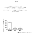
本発明の別の実施形態では、ナトリウムチャネル活性化因子は、NaV1.7特異的生化学的刺激を伴うアッセイで使用され、NaV1.7−遮断試験化合物の居所的または全身性の投与量を選択するのに有用である。臨床試験でのいずれかの試験化合物の適切な投与量の選択は、難しい仕事であり、理想的には、投与量を、ある種の、例えば、標的特異的であることがわかっているPETリガンドのバイオマーカーを置き換える量に較正することにより行われる。ナトリウムチャネル全般、および特に、NaV1.7には、今まで、このようなバイオマーカーがなかった。本明細書で、我々は、ベラトリジン、デルタメトリン、およびグラヤノトキシン、等のナトリウムチャネルの活性化因子は、適切な投与量をラットまたはマウスに注射すると、定量化可能な挙動による反応を生成することを開示する。これら3つの分子は、構造的に異なるが、それぞれがNaV1.7を活性化するという点で共通の生理的機序を共有する。実施例5で、我々は、これらのナトリウムチャネル活性化因子のいずれかを注射時に、ラットとマウスが、それぞれ、定量可能な用量依存性の足を引っ込めるおよび、足をなめる挙動を示すことを初めて明らかにする。これらの挙動は、モルヒネにより減少し、非選択的ナトリウムチャネルアンタゴニストのメキシレチンにより抑制されるため、これらの挙動が疼痛を反映し、ナトリウムチャネルにより媒介されていることを立証している。 In another embodiment of the invention, the sodium channel activator is used in an assay involving Na V 1.7 specific biochemical stimulation, and the local or systemic Na V 1.7-blocking test compound. Useful for selecting dosages. Choosing an appropriate dose of any test compound in clinical trials is a difficult task and ideally the dose is known to be a PET ligand that is known to be certain, eg, target specific By calibrating the amount to replace the biomarker. The sodium channel in general, and in particular Na V 1.7, has so far lacked such a biomarker. Here, we show that sodium channel activators, such as veratridine, deltamethrin, and greyanotoxin, produce responses with quantifiable behavior when injected into rats or mice at appropriate doses. Is disclosed. Although these three molecules are structurally different, they share a common physiological mechanism in that each activates Na V 1.7. In Example 5, we show for the first time that when injected with any of these sodium channel activators, rats and mice, respectively, show quantifiable dose-dependent paw withdrawal and licking paw behavior. To clarify. These behaviors are reduced by morphine and suppressed by the non-selective sodium channel antagonist mexiletine, demonstrating that these behaviors reflect pain and are mediated by sodium channels.
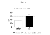
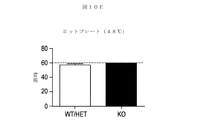
最も重要なこととして、我々は、本明細書の実施例5で、NaV1.7活性化因子、例えば、ベラトリジンの1μgの、グローバルNav1.7−/−マウスの足へ注射が、足を引っ込める、または足をなめる挙動を起こさせず、他方、野性型マウスでは、この同じ1μg投与が、強い足を引っ込める挙動を起こさせることを示す。メキシレチン(NaV1.7ならびに他の全てのナトリウムチャネルを遮断する)の野性型マウスへの前投与が、1μg投与量のベラトリジンにより誘発される足を引っ込める挙動を抑制するので、NaV1.7の薬理学的遮断は、同じ効果を実現するはずである。従って、前臨床の試験で、ナトリウムチャネル活性化因子による刺激は、生きている動物に投与される所与の化合物がNaV1.7を遮断するか否かの有用な試験である。さらに、この試験は、臨床的疼痛症候群を治療するNaV1.7試験阻害剤の適正な投薬量を決定するために臨床的に使用でき得る。適正な臨床的投与量は、ナトリウムチャネル活性化因子の投与に対する疼痛反応を抑制する量であろう。
Most importantly, in Example 5 herein, we injected a paw of a global Nav1.7 − / − mouse with 1 μg of Na V 1.7 activator, eg veratridine, We show that this same 1 μg dose causes a strong paw withdrawal behavior in wild type mice, while not causing withdrawal or paw licking behavior. Pre-administration of mexiletine (which blocks Na V 1.7 as well as all other sodium channels) to wild-type mice inhibits the paw withdrawal behavior induced by a 1 μg dose of veratridine, so
一実施形態では、本発明は、試験化合物(候補NaV1.7阻害剤)の用量範囲探索に有用な下記を含むアッセイを含む:
(a)第1の哺乳動物(例えば、マウス、ラット、ウサギ、ケナガイタチ、イヌ、非ヒト霊長類、またはヒト)に、試験化合物の第1の投与量を投与し、続けて、
(b)第1の哺乳動物に、陰性対照(試験化合物を受けていない)で疼痛関連反応を誘導するのに有効な(局所的または全身性)投与量のNaV1.7活性化因子(例えば、ベラトリジン、デルタメトリン、またはグラヤノトキシンIII)を投与し(このような投与は全身性でも局所的でもよい);次に
(c)疼痛関連反応が、陰性対照に比べて、第1の哺乳動物で減少するか否かを判定し;さらに
(d)疼痛関連反応が、陰性対照に比べて減少する試験化合物の最も少ない第2の投与量を特定する。哺乳動物への試験化合物投与は、全身性(例えば、腹腔内、静脈内、筋肉内、または経口投与)であっても、または局所的(例えば、皮下、足底内、局部投与)であってもよい。局所的な場合は、NaV1.7活性化因子の局所投与と同じ部位か、またはその近くである必要がある。回復期間が充分で、試験化合物およびNaV1.7活性化因子の効果が消滅し、いずれの残余試験化合物とNaV1.7活性化因子化合物の存在も哺乳動物で検出不可能になった後に、第1とは異なる第2の投与量レベルでの投与のために、第1の哺乳動物が後で再使用できる。あるいは、アッセイは、同じ種の第2の哺乳動物に試験化合物の第2の投与量を投与し、続いて、第2の哺乳動物に、陰性対照で疼痛関連反応を誘導するのに有効な投与量のNaV1.7活性化因子を投与し;さらに、疼痛関連反応が、陰性対照に比べて、第2の哺乳動物で減少するかどうかを判定することをさらに含むことができる。
In one embodiment, the present invention comprises an assay comprising the following useful for exploring the dose range of a test compound (candidate Na V 1.7 inhibitor):
(A) administering a first dose of a test compound to a first mammal (eg, a mouse, rat, rabbit, kennel, dog, non-human primate, or human), followed by
(B) A first mammal has a dose of Na V 1.7 activator (local or systemic) effective to induce a pain-related response in a negative control (no test compound received) ( (E.g., such administration may be systemic or local); Determine whether the animal decreases or not; and (d) identify the second dose of test compound that results in a decrease in pain-related response relative to the negative control. Administration of the test compound to the mammal may be systemic (eg, intraperitoneal, intravenous, intramuscular, or oral) or local (eg, subcutaneous, intraplantar, topical) Also good. If local, it should be at or near the same site as the local administration of the Na V 1.7 activator. The recovery period was sufficient so that the effects of the test compound and the Na V 1.7 activator disappeared, and the presence of any residual test compound and Na V 1.7 activator compound became undetectable in mammals. Later, the first mammal can later be reused for administration at a second dosage level different from the first. Alternatively, the assay involves administering a second dose of the test compound to a second mammal of the same species, followed by administration effective to induce a pain-related response in the negative control in the second mammal. The method can further comprise administering an amount of Na V 1.7 activator; further, determining whether the pain-related response is reduced in the second mammal relative to the negative control.
別の実施形態では、本発明は、試験化合物(候補NaV1.7阻害剤)の用量範囲探索に有用な下記を含むアッセイを含む:
(a)第1の哺乳動物(例えば、マウス、ラット、ウサギ、ケナガイタチ、イヌ、非ヒト霊長類、またはヒト)に、第1の投与量の試験化合物、および第2の哺乳動物(同じ種の)に、第1の投与量とは異なる第2の投与量の試験化合物を投与し、続けて、
(b)第1のおよび第2の哺乳動物に、陰性対照(試験化合物を受けていない)で疼痛関連反応を誘導するのに有効な量のNaV1.7活性化因子(例えば、ベラトリジン、デルタメトリン、またはグラヤノトキシンIII)を投与し(このような投与は、全身性でも局所的でもよい);その後、
(c)疼痛関連反応が、陰性対照に比べて、第1の哺乳動物と第2の哺乳動物で減少するか否かを判定し;さらに、
(d)疼痛関連反応が、陰性対照に比べて、低減する試験化合物の最も少ない第2の投与量を特定する。哺乳動物に投与される試験化合物投与は、全身性(例えば、腹腔内、静脈内、筋肉内、または経口投与)でも、または局所的(例えば、皮下、足底内、または局部投与)でもよい。局所的な場合は、NaV1.7活性化因子の局所投与と同じ部位か、またはその近くである必要がある。
In another embodiment, the invention includes an assay comprising the following useful for exploring the dose range of a test compound (candidate Na V 1.7 inhibitor):
(A) a first mammal (eg, a mouse, rat, rabbit, beak, dog, non-human primate, or human) to a first dose of a test compound, and a second mammal (of the same species) ) Is administered a second dose of the test compound different from the first dose, followed by
(B) an amount of Na V 1.7 activator effective to induce a pain-related response in a negative control (not receiving the test compound) in a first and second mammal (eg, veratridine, Deltamethrin, or greyanotoxin III) (such administration may be systemic or local);
(C) determining whether the pain-related response is decreased in the first mammal and the second mammal relative to the negative control;
(D) The second dose at which the pain-related response is reduced relative to the negative control is reduced. Test compound administration administered to a mammal can be systemic (eg, intraperitoneal, intravenous, intramuscular, or oral) or local (eg, subcutaneous, intraplantar, or local). If local, it should be at or near the same site as the local administration of the Na V 1.7 activator.
このようにして、NaV1.7を抑制する投与量を決定し、推定上有害作用を生ずる可能性のあるより高い投与量と比較し、治療濃度域を決定できる。さらに、NaV1.7試験阻害剤の有効性の臨床試験では、このような投与量でのみ、治療の有効性をNaV1.7の結果と見なすことができる。グローバルNaV1.7−/−マウスにより得られる重要な知識は、ナトリウムチャネル活性化因子は、NaV1.7を介して、さらには、NaV1.7のみを介して、疼痛反応を生成することである。 In this way, a dose that inhibits Na V 1.7 can be determined and compared to a higher dose that can potentially cause adverse effects, and a therapeutic concentration range can be determined. Furthermore, in clinical trials of the effectiveness of Na V 1.7 test inhibitors, the efficacy of treatment can be considered as a result of Na V 1.7 only at such doses. Global Na V 1.7 - / - important knowledge gained by the mouse, sodium channel activator via the Na V 1.7, further only through the Na V 1.7, the pain response Is to generate.
以降の図および発明の詳細な説明を考慮すれば、多くの追加の本発明の態様および利点が明らかになるであろう。 Many additional aspects and advantages of the present invention will become apparent in view of the following figures and detailed description of the invention.
実施形態の詳細な説明
本明細書で使われるセクション見出しは、構成上の目的のみのためであり、記載された主題を限定するものと解釈されるべきではない。
DETAILED DESCRIPTION OF EMBODIMENTS Section headings used herein are for organizational purposes only and are not to be construed as limiting the subject matter described.
定義
本明細書で別段の規定がない限り、本出願に関連して使われている科学的および技術的用語は、通常、当業者により理解されている意味を持つものとする。さらに、文脈から別義が要求されない限り、単数形の用語は、複数の対象物を含み、複数の用語は、反収の対象物を含むものとする。従って、文脈から別義が明確に指示されない限り、本明細書および添付請求項で使われる単数形「a」、「an」および「the」は、複数対象物を含む。例えば、「タンパク質(a protein)」への参照は、複数のタンパク質を含み;「細胞(a cell)」への参照は、複数の細胞集団を含む。
Definitions Unless otherwise defined herein, scientific and technical terms used in connection with the present application shall generally have the meanings as understood by those of ordinary skill in the art. Further, unless otherwise required by context, singular terms shall include plural objects and plural terms shall include counter object. Thus, unless the context clearly indicates otherwise, the singular forms “a”, “an”, and “the” as used herein and in the appended claims include plural objects. For example, a reference to “a protein” includes a plurality of proteins; a reference to “a cell” includes a plurality of cell populations.
「ポリペプチド」および「タンパク質」は、本明細書では同義に使用され、ペプチド結合を介して共有結合で結合した2つ以上のアミノ酸分子鎖を含む。これらの用語は、特定の長さの生成物を意味しない。従って、「ペプチド」および「オリゴペプチド」は、ポリペプチドの定義の内に含まれる。この用語は、翻訳後修飾、例えば、グリコシル化、アセチル化、リン酸化、等、をされたポリペプチドを含む。さらに、タンパク質断片、類似体、変異または変種タンパク質、融合タンパク質、等は、ポリペプチドの意味の内に含まれる。この用語は、また、既知のタンパク質工学技術を使って、組換えにより発現されうる、1つまたは複数のアミノ酸類似体または非標準もしくは非天然アミノ酸が含まれる分子を含む。さらに、融合タンパク質は、周知の有機化学技術により本明細書記載のように誘導体化できる。 “Polypeptide” and “protein” are used interchangeably herein and include two or more amino acid chains covalently linked through peptide bonds. These terms do not imply a particular length of product. Thus, “peptide” and “oligopeptide” are included within the definition of polypeptide. The term includes polypeptides that have been subjected to post-translational modifications such as glycosylation, acetylation, phosphorylation, and the like. Furthermore, protein fragments, analogs, mutant or variant proteins, fusion proteins, etc. are included within the meaning of a polypeptide. The term also includes molecules that contain one or more amino acid analogs or non-standard or unnatural amino acids that can be recombinantly expressed using known protein engineering techniques. Furthermore, the fusion protein can be derivatized as described herein by well-known organic chemistry techniques.
「組換え」という用語は、物質(例えば、核酸またはポリペプチド)がヒトの介入により、人工的にまたは合成により(すなわち、非天然的に)変えられていることを示す。変更は、その天然環境または状態内の、またはそこから取り出された物質に対し行うことができる。例えば、「組換え核酸」は、核酸を、例えば、クローニング、DNAシャフリングまたは他のよく知られた分子生物学的方法の間に、組み換えることにより作られるものである。このような分子生物学的方法の例は、Maniatis et al.、分子クローニング。実験マニュアル(Molecular Cloning.A Laboratory Manual).Cold Spring Harbor Laboratory、Cold Spring Harbor、N.Y(1982)、で見つけられる。「組換えDNA分子」は、このような分子生物学的技術により一緒に連結されたDNAのセグメントから構成される。本明細書で使われる「組換えタンパク質」または「組換えポリペプチド」という用語は、組換えDNA分子を使って発現されるタンパク質分子を意味する。「組換え宿主細胞」は、組換え核酸を含む、および/または発現する細胞である。 The term “recombinant” indicates that the substance (eg, nucleic acid or polypeptide) has been altered by human intervention, either artificially or synthetically (ie, non-naturally). Changes can be made to material within or removed from its natural environment or condition. For example, a “recombinant nucleic acid” is one that is made by recombining a nucleic acid, for example, during cloning, DNA shuffling, or other well-known molecular biological methods. Examples of such molecular biological methods are described in Maniatis et al. Molecular cloning. Experimental Manual (Molecular Cloning. A Laboratory Manual). Cold Spring Harbor Laboratory, Cold Spring Harbor, N.A. Y (1982). A “recombinant DNA molecule” is composed of segments of DNA linked together by such molecular biology techniques. The term “recombinant protein” or “recombinant polypeptide” as used herein refers to a protein molecule that is expressed using a recombinant DNA molecule. A “recombinant host cell” is a cell that contains and / or expresses a recombinant nucleic acid.
「ポリヌクレオチド」または「核酸」という用語は、2つ以上のヌクレオチド残基を含む単鎖および二重鎖ヌクレオチドポリマーの両方を含む。ヌクレオチド残基含有ポリヌクレオチドは、リボヌクレオチドもしくはデオキシリボヌクレオチドであってもよく、またはいずれかのヌクレオチドの型の修飾型であってもよい。前記修飾は、塩基修飾、例えば、ブロモウリジンおよびイノシン誘導体、リボース修飾、例えば、2’,3’−ジデオキシリボース、およびヌクレオチド間結合修飾、例えば、ホスホロチオアート、ホスホロジチオアート、ホスホロセレノアート、ホスホロジセレノアート、ホスホロアニロチオアート、ホスホラニラダートおよびホスホロアミダートを含む。 The term “polynucleotide” or “nucleic acid” includes both single-stranded and double-stranded nucleotide polymers comprising two or more nucleotide residues. Nucleotide residue-containing polynucleotides can be ribonucleotides or deoxyribonucleotides, or can be modified versions of either nucleotide type. Such modifications include base modifications such as bromouridine and inosine derivatives, ribose modifications such as 2 ', 3'-dideoxyribose, and internucleotide linkage modifications such as phosphorothioate, phosphorodithioate, phosphoroseleno. Including art, phosphorodiselenoate, phosphoroanilothioate, phosphoraniladate and phosphoramidate.
「オリゴヌクレオチド」という用語は、200以下のヌクレオチド残基を含むポリヌクレオチドを意味する。いくつかの実施形態では、オリゴヌクレオチドは、10〜60塩基長である。他の実施形態では、オリゴヌクレオチドは、12、13、14、15、16、17、18、19、または20〜40ヌクレオチド長である。オリゴヌクレオチドは、例えば、変異体遺伝子の作製での使用に対しては、単鎖でも、二重鎖でもよい。オリゴヌクレオチドは、センス、またはアンチセンスオリゴヌクレオチドのいずれでもよい。オリゴヌクレオチドは、標識を含んでもよく、これには、定量化または検出を容易にするための同位体標識(例えば、125I、14C、13C、35S、3H、2H、13N、15N、18O、17O、等);アッセイ検出用の蛍光標識、ハプテンまたは抗原性標識、が含まれる。オリゴヌクレオチドは、例えば、PCRプライマー、クローニングプライマーまたはハイブリダイゼーションプローブとして使用できる。 The term “oligonucleotide” refers to a polynucleotide comprising 200 or fewer nucleotide residues. In some embodiments, the oligonucleotide is 10-60 bases in length. In other embodiments, the oligonucleotide is 12, 13, 14, 15, 16, 17, 18, 19, or 20-40 nucleotides in length. Oligonucleotides may be single-stranded or double-stranded, for example, for use in generating mutant genes. The oligonucleotide may be either a sense or an antisense oligonucleotide. Oligonucleotides may include labels, including isotope labels (eg, 125 I, 14 C, 13 C, 35 S, 3 H, 2 H, 13 N, for ease of quantification or detection. , 15 N, 18 O, 17 O, etc.); fluorescent labels, haptens or antigenic labels for assay detection. Oligonucleotides can be used, for example, as PCR primers, cloning primers or hybridization probes.
「ポリヌクレオチド配列」または「ヌクレオチド配列」または「核酸配列」は、本明細書で同義に使用され、ポリヌクレオチド中のヌクレオチド残基の一次配列であり、これには、オリゴヌクレオチド、DNA、およびRNA、核酸、または、文脈によっては、ヌクレオチド残基の一次配列を表す文字列を含む。いずれの特定ポリヌクレオチド配列からも、所与の核酸または相補的ポリヌクレオチド配列が決定可能である。ゲノムまたは合成由来のDNAまたはRNAが含まれ、単鎖でも二重鎖でも、また、センス鎖でもアンチセンス鎖でもよい。別段の指定がない限り、本明細書で考察されるいずれの単鎖ポリヌクレオチド配列の左側末端も、5’末端であり;二重鎖ポリヌクレオチド配列の左手方向は、5’方向と見なされる。5’から3’の新生RNA転写物の付加方向は、転写方向と呼ばれ;RNA転写物の5’から5’末端であるRNA転写物と同じ配列を有するDNA鎖上の配列領域は、「上流配列」と呼ばれ;RNA転写物の3’から3’末端であるRNA転写物と同じ配列を有するDNA鎖上の配列領域は、「下流配列」と呼ばれる。 "Polynucleotide sequence" or "nucleotide sequence" or "nucleic acid sequence" are used interchangeably herein and are the primary sequence of nucleotide residues in a polynucleotide, including oligonucleotides, DNA, and RNA , Nucleic acid or, depending on the context, includes a string representing the primary sequence of nucleotide residues. From any particular polynucleotide sequence, a given nucleic acid or complementary polynucleotide sequence can be determined. Genomic or synthetically derived DNA or RNA is included, and may be single-stranded or double-stranded, and may be sense or antisense strand. Unless specified otherwise, the left-hand end of any single-stranded polynucleotide sequence discussed herein is the 5 'end; the left-hand direction of double-stranded polynucleotide sequences is considered the 5' direction. The direction of addition of the 5 ′ to 3 ′ nascent RNA transcript is referred to as the transcription direction; the sequence region on the DNA strand having the same sequence as the RNA transcript at the 5 ′ to 5 ′ end of the RNA transcript is “ The sequence region on the DNA strand having the same sequence as the RNA transcript that is the 3 ′ to 3 ′ end of the RNA transcript is referred to as the “downstream sequence”.
本明細書で使用される「単離核酸分子」または「単離核酸配列」は、(1)特定され、通常自然の供給源において一緒に存在する少なくとも1つの混在核酸分子から分離されているか、または(2)クローン化され、増幅され、標識され、もしくは別の方法で背景核酸から区別され、それにより、目的の核酸の配列が特定できるようにされているか、いずれかの核酸分子である。単離核酸分子は、天然に見つかる形態または設定で存在するもの以外のものである。しかし、単離核酸分子は、通常、ポリペプチド(例えば、オリゴペプチドまたは抗体)を発現する細胞中に含まれる(例えば、核酸分子が天然の細胞とは異なる染色体の部位に存在する)核酸分子を含む。 As used herein, an “isolated nucleic acid molecule” or “isolated nucleic acid sequence” is (1) separated from at least one mixed nucleic acid molecule identified and usually present together in a natural source, Or (2) any nucleic acid molecule that has been cloned, amplified, labeled, or otherwise distinguished from the background nucleic acid so that the sequence of the nucleic acid of interest can be identified. Isolated nucleic acid molecules are other than those present in the forms or settings found in nature. However, an isolated nucleic acid molecule usually comprises a nucleic acid molecule contained in a cell that expresses a polypeptide (eg, an oligopeptide or antibody) (eg, the nucleic acid molecule is present at a chromosomal site different from that of natural cells). Including.
本明細書で使用される「〜をコードする核酸分子」「〜をコードするDNA配列」および「〜をコードするDNA」という用語は、デオキシリボ核酸の鎖に沿ったデオキシリボヌクレオチドの順序または配列を指す。これらのデオキシリボヌクレオチドの順序は、mRNA鎖に沿ったリボヌクレオチドの順序を決定し、また、ポリペプチド(タンパク質)鎖に沿ったアミノ酸の順序も決定する。従って、DNA配列は、RNA配列およびアミノ酸配列をコードする。 As used herein, the terms "nucleic acid molecule encoding", "DNA sequence encoding" and "DNA encoding" refer to the order or sequence of deoxyribonucleotides along the strand of deoxyribonucleic acid. . The order of these deoxyribonucleotides determines the order of ribonucleotides along the mRNA chain and also determines the order of amino acids along the polypeptide (protein) chain. Thus, the DNA sequence encodes an RNA sequence and an amino acid sequence.
「遺伝子」という用語は、広範に使用され、生物学的機能に関連するいずれの核酸をも意味する。遺伝子は、典型的には、コード配列、および/または、そのようなコード配列の発現に必要とされる調節配列を含む。「遺伝子」という用語は、特定のゲノムまたは組換え配列、ならびにその配列デコードされるcDNAまたはmRNAに適用される。「融合遺伝子」は、天然で一緒には見出されないか、または天然でそのコードされた融合タンパク質(すなわち、キメラタンパク質)中に存在する同じ配列中に一緒には見出されない、異なるタンパク質由来の部分を含むポリペプチドをコードするコーディング領域を含む。遺伝子は、また、例えば、他のタンパク質のための認識配列を形成する非発現核酸セグメントを含む。非発現核酸セグメントは、転写因子等の調節タンパク質が結合する転写調節領域であって、隣接または近くの配列の転写を生ずるものを含む。 The term “gene” is used broadly and refers to any nucleic acid associated with a biological function. A gene typically includes coding sequences and / or regulatory sequences required for expression of such coding sequences. The term “gene” applies to a particular genomic or recombinant sequence, as well as to the cDNA or mRNA whose sequence is decoded. “Fusion genes” are derived from different proteins that are not found together in nature or that are not found together in the same sequence that is naturally present in the encoded fusion protein (ie, a chimeric protein). It includes a coding region that encodes a polypeptide comprising the portion. A gene also includes non-expressed nucleic acid segments that, for example, form recognition sequences for other proteins. Non-expressed nucleic acid segments include transcriptional regulatory regions to which regulatory proteins such as transcription factors bind, resulting in transcription of adjacent or nearby sequences.
「遺伝子の発現」または「核酸の発現」は、文脈に依存して、DNAのRNAへの転写(任意で、RNAの修飾、例えば、スプライシングを含む)、RNAのポリペプチドへの翻訳(場合により、その後のポリペプチドの翻訳後修飾を含む)、または転写と翻訳の両方を意味する。 “Gene expression” or “nucleic acid expression” refers to transcription of DNA into RNA (optionally including RNA modification, eg, splicing), translation of RNA into polypeptide (optionally) , Including subsequent post-translational modifications of the polypeptide), or both transcription and translation.
本明細書で使用される「コーディング領域」または「コード配列」という用語は、構造遺伝子に関連して使われる場合、mRNA分子の翻訳の結果として新生ポリペプチドで見出されるアミノ酸をコードするヌクレオチド配列を意味する。真核生物では、コーディング領域は、5’側のイニシエータのメチオニンをコードするヌクレオチドトリプレット「ATG」および3’側の停止コドン(すなわち、TAA、TAG、TGA)を指定する3つのトリプレットの内の1つにより境界が形成されている。 As used herein, the term “coding region” or “coding sequence” when used in connection with a structural gene refers to a nucleotide sequence that encodes an amino acid found in a nascent polypeptide as a result of translation of an mRNA molecule. means. In eukaryotes, the coding region is a nucleotide triplet “ATG” that encodes the 5 ′ initiator methionine and one of three triplets that specify the 3 ′ stop codon (ie, TAA, TAG, TGA). A boundary is formed by the two.
「制御配列」または「制御シグナル」という用語は、特定の宿主細胞中で、それが連結されているコード配列の発現およびプロセッシングに影響を与えうるポリヌクレオチド配列を意味する。このような制御配列の性質は、宿主生物体に依存する可能性がある。特定の実施形態では、原核生物の制御配列は、プロモーター、リボソーム結合部、および転写終止配列を含むことができる。真核生物の制御配列は、1つまたは複数の転写因子用認識部位を含むプロモーター、転写エンハンサー配列または要素、ポリアデニル化部位、および転写終止配列を含むことができる。制御配列は、リーダー配列および/または融合パートナー配列を含むことができる。プロモーターおよびエンハンサーは、転写に関与する細胞タンパク質と特異的に相互作用する短い配列のDNAから構成される(Maniatis、et al.、Science 236:1237(1987))。プロモーターおよびエンハンサー配列は、酵母、昆虫および哺乳動物細胞ならびにウイルスの遺伝子、等の種々の真核生物の源から単離されている(類似の調節領域、すなわち、プロモーターは、原核生物でも見出される)。特定のプロモーターおよびエンハンサーの選択は、どの細胞型を使って目的のタンパク質を発現させる予定かに依存する。いくつかの真核生物のプロモーターおよびエンハンサーは、広い宿主範囲を有するが、他のものは、限られた細胞型サブセット中で機能する(概説は、Voss、et al.、Trends Biochem.Sci.、11:287(1986)、および、Maniatis、et al.、Science 236:1237(1987)、を参照)。 The term “control sequence” or “control signal” means a polynucleotide sequence that can affect the expression and processing of a coding sequence to which it is linked in a particular host cell. The nature of such control sequences may depend on the host organism. In certain embodiments, prokaryotic control sequences can include a promoter, a ribosome junction, and a transcription termination sequence. Eukaryotic control sequences can include promoters, transcription enhancer sequences or elements, recognition sites for one or more transcription factors, polyadenylation sites, and transcription termination sequences. The control sequence can include a leader sequence and / or a fusion partner sequence. Promoters and enhancers are composed of short sequences of DNA that interact specifically with cellular proteins involved in transcription (Maniatis, et al., Science 236: 1237 (1987)). Promoter and enhancer sequences have been isolated from various eukaryotic sources such as yeast, insect and mammalian cells and viral genes (similar regulatory regions, ie promoters are also found in prokaryotes). . The selection of a particular promoter and enhancer depends on which cell type is used to express the protein of interest. Some eukaryotic promoters and enhancers have a broad host range, while others function in a limited subset of cell types (reviewed in Voss, et al., Trends Biochem. Sci., 11: 287 (1986) and Maniatis, et al., Science 236: 1237 (1987)).
「ベクター」という用語は、タンパク質コード情報を宿主細胞中に移入するために使用されるいずれの分子または実体をも(例えば、核酸、プラスミド、バクテリオファージまたはウイルス)を意味する。 The term “vector” refers to any molecule or entity (eg, nucleic acid, plasmid, bacteriophage or virus) used to transfer protein coding information into a host cell.
本明細書で使用される「発現ベクター」または「発現構築物」という用語は、特定の宿主細胞中で機能的に連結されたコード配列の発現に必要な所望のコード配列および適切な核酸制御配列を含む組換えDNA分子を意味する。発現ベクターは、限定されないが、転写、翻訳に影響するまたはこれを制御する配列、および、イントロンが存在する場合には、それに機能的に連結されたコーディング領域のRNAスプライシングに影響を与える配列を含むことができる。原核生物中の発現に必要な核酸配列は、プロモーター、任意選択でオペレーター配列、リボソーム結合部位および場合によっては他の配列を含む。真核細胞は、プロモーター、エンハンサー、ならびに終止およびポリアデニル化シグナルを利用することが知られている。分泌シグナルペプチド配列は、また、任意選択で、所望に応じ、その細胞から目的のポリペプチドのより簡単な単離を目的として、目的のコード配列に機能的に連結された発現ベクターによりコードされ、それにより、発現ポリペプチドが組換え宿主細胞により分泌されうる。このような技術は、当技術分野でよく知られている(例えば、Goodey、Andrew R.;et al.、ペプチドおよびDNA配列(Peptide and DNA sequences)、米国特許第5,302,697号;Weiner et al.、タンパク質分泌用組成物および方法(Compositions and methods for protein secretion)、米国特許第6,022,952号および米国特許第6,335,178号;Uemura et al.、タンパク質発現ベクターおよびその利用(Protein expression vector and utilization thereof)、米国特許第7,029,909号;Ruben et al.、27 ヒト分泌タンパク質(human secreted proteins)、米国特許公開第2003/0104400A1号)。 As used herein, the term “expression vector” or “expression construct” refers to a desired coding sequence and appropriate nucleic acid control sequences necessary for the expression of a coding sequence operably linked in a particular host cell. It means a recombinant DNA molecule containing. Expression vectors include, but are not limited to, sequences that affect or control transcription, translation, and if present introns, sequences that affect RNA splicing of the coding region operably linked thereto. be able to. Nucleic acid sequences necessary for expression in prokaryotes include a promoter, optionally an operator sequence, a ribosome binding site and optionally other sequences. Eukaryotic cells are known to utilize promoters, enhancers, and termination and polyadenylation signals. The secretory signal peptide sequence is also optionally encoded by an expression vector operably linked to the coding sequence of interest for the purpose of easier isolation of the polypeptide of interest from the cell, if desired. Thereby, the expressed polypeptide can be secreted by the recombinant host cell. Such techniques are well known in the art (eg, Goodey, Andrew R .; et al., Peptide and DNA sequences, US Pat. No. 5,302,697; Weiner et al., Compositions and methods for protein section, US Pat. Nos. 6,022,952 and 6,335,178; Uemura et al., protein expression vectors and their Utilization (Protein expression vector and utility thereof), US Pat. No. 7,029,909; Ruben et al., 27 Humans Protein (human secreted proteins), US Patent Publication No. 2003 / 0104400A1).
本明細書で使われる「操作可能な組み合わせで」、「操作可能な順に」および「機能的に連結された」という用語は、所与の遺伝子の転写、および/または所望のタンパク質分子の合成を指示できる核酸分子が産生されるような形式の核酸配列の結合を意味する。この用語は、また、機能的タンパク質が産生されるような形式でのアミノ酸配列の結合を意味する。例えば、タンパク質をコードする配列に「機能的に連結」されるベクター中の制御配列は、タンパク質をコードする配列の発現が、制御配列の転写活性に適合する条件下で実現されるように、それに対し連結される。 As used herein, the terms “in operable combinations”, “in order of operation” and “operably linked” refer to transcription of a given gene and / or synthesis of a desired protein molecule. It means the binding of nucleic acid sequences in such a way that a nucleic acid molecule that can be indicated is produced. The term also refers to the binding of amino acid sequences in such a way that a functional protein is produced. For example, a control sequence in a vector that is “operably linked” to a protein-encoding sequence may be expressed in such a way that expression of the protein-encoding sequence is achieved under conditions compatible with the transcriptional activity of the control sequence. It is connected to.
「宿主細胞」という用語は、核酸で形質転換されているか、または、形質転換されることが可能で、それにより、目的の遺伝子を発現する細胞を意味する。この用語は、目的の遺伝子が存在する限り、その子孫が形態学または遺伝的構成が元の親細胞に同じであっても同じでなくても、親細胞の子孫を含む。多数の入手可能な、よく知られた宿主細胞のいずれも、本発明の実施に使用できる。特定の宿主の選択は、技術分野によって認識されているいくつかの要因に依存する。これらには、例えば、選択発現ベクターとの適合性、DNA分子コードされるペプチドの毒性、形質転換速度、ペプチドの回収の容易さ、発現特性、バイオセーフティおよび費用、が含まれる。これらの要因の間のバランスは、全ての宿主が特定のDNA配列の発現に等しく有効なわけではないという理解と合致させなければならない。これらの一般ガイドライン内で、培養に有用な微生物の宿主細胞には、細菌(例えば、大腸菌種)、酵母(例えば、サッカロミセス種)および他の真菌細胞、昆虫細胞、植物細胞、哺乳類(ヒトを含む)細胞、例えば、CHO細胞およびHEK−293細胞、が含まれる。修飾は、DNAレベルでも同様に行うことができる。ペプチドをコードするDNA配列は、選択宿主細胞に適合性のより高いコドンに変更できる。大腸菌については、最適化コドンが、当技術分野で知られている。コドンを置換して、制限酵素部位を除去する、またはサイレント制限酵素部位を含めることができ、これは、選択宿主細胞中でのDNAプロセッシングを支援し得る。次いで、形質転換宿主は、培養され、精製される。宿主細胞は、従来の発酵条件下で培養し、所望の化合物を発現させることができる。このような発酵条件は、当技術分野でよく知られている。 The term “host cell” means a cell that has been transformed or is capable of being transformed with a nucleic acid and thereby expressing a gene of interest. The term includes the progeny of the parent cell, so long as the gene of interest is present, whether its progeny is the same or not the same as the original parent cell. Any of a number of available and well-known host cells can be used in the practice of the present invention. The selection of a particular host depends on several factors recognized by the technical field. These include, for example, compatibility with selected expression vectors, toxicity of the peptide encoded by the DNA molecule, transformation rate, ease of peptide recovery, expression characteristics, biosafety and cost. The balance between these factors must be consistent with the understanding that not all hosts are equally effective at expressing a particular DNA sequence. Within these general guidelines, microbial host cells useful for culture include bacteria (eg, E. coli species), yeast (eg, Saccharomyces species) and other fungal cells, insect cells, plant cells, mammals (humans). ) Cells, such as CHO cells and HEK-293 cells. Modifications can be made at the DNA level as well. The DNA sequence encoding the peptide can be changed to a codon that is more compatible with the selected host cell. For E. coli, optimized codons are known in the art. Codon substitutions can be made to remove restriction enzyme sites or include silent restriction enzyme sites, which can aid in DNA processing in selected host cells. The transformed host is then cultured and purified. Host cells can be cultured under conventional fermentation conditions to express the desired compound. Such fermentation conditions are well known in the art.
「形質移入」という用語は、細胞による外来性または外因性DNAの取込を意味し、外因性のDNAが、細胞膜内に導入された場合に、細胞は「形質移入」されている。いくつかの形質移入技術が当技術分野でよく知られており、本明細書で開示されている。例えば、Graham et al.、1973、Virology 52:456;Sambrook et al.、2001、分子クローニング:実験マニュアル(Molecular Cloning:A Laboratory Manual)、同上;Davis et al.、1986、分子生物学における基礎的方法(Basic Methods in Molecular Biology)、Elsevier;Chu et al.、1981、Gene 13:197、を参照されたい。このような技術を使用して、1つまたは複数の外因性DNA成分を適切な宿主細胞中に導入することができる。 The term “transfection” means the uptake of exogenous or exogenous DNA by a cell, and a cell has been “transfected” when exogenous DNA has been introduced into the cell membrane. Several transfection techniques are well known in the art and are disclosed herein. For example, Graham et al. 1973, Virology 52: 456; Sambrook et al. 2001, Molecular Cloning: Experimental Manual (Molecular Cloning: A Laboratory Manual), ibid .; Davis et al. 1986, Basic Methods in Molecular Biology, Elsevier; Chu et al. 1981, Gene 13: 197. Such techniques can be used to introduce one or more exogenous DNA components into suitable host cells.
「形質転換」という用語は、細胞の遺伝的特性の変化を意味し、細胞は、改変されて新規DNAまたはRNAを含む場合、形質転換されている。例えば、形質移入、形質導入、または他の技術による新しい遺伝物質の導入により、その自然の状態から遺伝的に改変されている場合、細胞は形質転換されている。形質移入または形質導入後、形質転換DNAは、物理的にその細胞の染色体中に組み込むことにより、細胞のDNAと組み換えることができ、または、複製されることなく、エピソーム要素として一時的に維持でき、または、独立にプラスミドとして複製できる。形質転換DNAが細胞分裂により複製される場合、細胞は、「安定に形質転換」されていると見なされる。 The term “transformation” means a change in the genetic properties of a cell, and a cell has been transformed if it has been modified to contain new DNA or RNA. A cell has been transformed if it has been genetically altered from its natural state, for example, by transfection, transduction, or introduction of new genetic material by other techniques. After transfection or transduction, the transforming DNA can be recombined with the cell's DNA by physically integrating it into the cell's chromosome, or temporarily maintained as an episomal element without being replicated Or can be replicated independently as a plasmid. A cell is considered “stablely transformed” if the transforming DNA is replicated by cell division.
同系交配マウス株(例えば、C57Bl/6J)を、異なる同系交配マウス株(例えば、BALB/c)に変化させるプロセスは、「戻し交配(backcrossing)」または「戻し交配(backcross)」と呼ばれる。これは、マウス株が意図した研究目的に適さない場合に有用であり;株は、育種計画により遺伝的に改変できる。遺伝子型を「株A」(例えば、C57Bl/6J)から「株B」(例えば、BALB/c)に完全に形質転換するために、マウスは、通常、少なくとも10回、戻し交配される必要があり;その後にのみ、それらは、「類遺伝子性」と呼ぶことができる。マウスを戻し交配するために、変異体マウスは、選ばれた同系交配株(例えば、BALB/c)と交配される。この交配の出生児は、「戻し交配#1」(または「N1」)と命名され、両方の株のハイブリッドである(例えば、約:50%C57Bl6/Jおよび50%BALB/c)。出生児は遺伝子型が特定でき、これらの目的の遺伝子に対するヘテロ接合体のみが、同系交配マウス株の野性型個体(例えば、野性型BALB/cマウス)と、再度交配される。通常、この手順は、類遺伝子性BALB/c系統が得られるまで約10回繰り返される。同系交配マウス株のいずれの組み合わせも、この手法を使用して選ばれた株の類遺伝子系統を作ることができる。例として、2010年12月までに、我々は、同系交配BALB/c背景の第5戻し交配世代(「N5」)を生成し、特徴付けした。ヘテロ接合の(「HET」)出生児は、約98.6%BALB/cの遺伝的背景である。2011年の5月には、我々は、最初のBALB/c類遺伝子性繁殖ペアを得た。我々は、この背景がNaV1.7KO新生児の生存率を改善するか否か(すなわち、ヒトケア/給餌の要求が減ったか否か)を評価するために現在交配している。今のところ、結果は、C57Bl6/J−BALB/cハイブリッドと同じであるように思われる。
The process of changing an inbred mouse strain (eg, C57B1 / 6J) into a different inbred mouse strain (eg, BALB / c) is referred to as “backcrossing” or “backcross”. This is useful when the mouse strain is not suitable for the intended research purpose; the strain can be genetically modified by a breeding program. In order to fully transform a genotype from “strain A” (eg C57B1 / 6J) to “strain B” (eg BALB / c), mice usually need to be backcrossed at least 10 times. Yes; only after that they can be called "genetic". In order to backcross the mice, the mutant mice are bred with a selected inbred strain (eg, BALB / c). The offspring of this cross is named “
同系交配マウス株(例えば、C57Bl/6J)を、非近交系マウス株(例えば、CD1)に変えるプロセスは、「異系交配(outcrossing)」または「異系交配(outcross)」と呼ばれる。これは、同系交配マウス株からの変異体(ノックアウト)動物が衰弱の徴候を示す場合に有用であり;目的の遺伝子に対するヘテロ接合出生児は、非近交系マウス株と交配して、遺伝的変異および活力を同系交配マウス株中に導入できる。マウスの異系交配を行うために、変異マウスは、選択された非近交系株(例えば、CD1)由来の野性型マウスと交配される。この交配の出生児は、ハイブリッドである(例えば、約:50%C57Bl6/Jおよび50%CD1)。非近交系マウスは、遺伝子プール中で変異が多すぎて類遺伝子性系統を作れないために、通常は、それ以上「異系交配」されない。 The process of changing an inbred mouse strain (eg, C57B1 / 6J) to an outbred mouse strain (eg, CD1) is referred to as “outcrossing” or “outcross”. This is useful when a mutant (knockout) animal from an inbred mouse strain shows signs of weakness; a heterozygous offspring for the gene of interest can be crossed with an outbred mouse strain to Mutations and vitality can be introduced into inbred mouse strains. In order to perform outbreeding of mice, mutant mice are bred with wild type mice derived from selected outbred strains (eg, CD1). The offspring of this mating are hybrid (eg, approximately: 50% C57B16 / J and 50% CD1). Outbred mice are usually not “outbred” any more because there are too many mutations in the gene pool to create an ancestral line.
タンパク質の「ドメイン(domain)」または「領域(region)」(本明細書では同義に使用される)は、完全なタンパク質まで、およびこれを含む全体タンパク質のいずれかの部分であり、通常、完全なタンパク質より少ない部分を含む。ドメインは、(必要はないが)特定の生物学的、生化学的、または構造的機能または部位(例えば、リガンド結合ドメイン、またはサイトゾルの、膜貫通型の、もしくは細胞外のドメイン)を、残りのタンパク質鎖とは無関係に保持し、および/または、それらと関連付けられ得る。 A “domain” or “region” (used interchangeably herein) of a protein is any part of and up to and including the entire protein, usually the complete Contains fewer parts than normal proteins. Domains (although not necessary) identify specific biological, biochemical, or structural functions or sites (eg, ligand binding domains, or cytosolic, transmembrane or extracellular domains), It can be retained and / or associated with the remaining protein chains.
「哺乳動物」は、哺乳類に分類されるいずれの動物をも意味し、ヒト、家畜(domestic and farm animal)、および動物園、スポーツ、またはペット動物を含み、例えば、イヌ、ウマ、ネコ、雌ウシ、ラット、マウス、非ヒト霊長類(例えば、サル、類人猿)、等である。 “Mammal” means any animal classified as a mammal and includes humans, domestic and farm animals, and zoo, sports, or pet animals, eg, dogs, horses, cats, cows , Rats, mice, non-human primates (eg, monkeys, apes), and the like.
「げっ歯類(rodent)」および「複数げっ歯類(rodents)」という用語は、系統学的ネズミ目の全メンバーを指し、それら由来の全ての未来の世代のいずれかのおよび全ての子孫を含む。 The terms “rodent” and “plural rodents” refer to all members of the phylogenetic murine, including any and all descendants of all future generations derived from them. Including.
「マウス」という用語は、ファミリーネズミ科のいずれかのおよび全てのメンバーを指し、ラットおよびマウスを含む。 The term “mouse” refers to any and all members of the family Muridae and includes rats and mice.
生物学的物質、例えば、ポリペプチド、核酸、宿主細胞、等に関連して、本明細書全体を通して使用される「天然の」という用語は、自然で見出される物質を意味する。 The term “natural” as used throughout this specification in connection with biological materials such as polypeptides, nucleic acids, host cells, etc. means materials found in nature.
動物、例えば、マウスまたは特に、グローバルNaV1.7−/−ノックアウトマウスに関し、「生存可能な」という用語は、動物が、成人期に達することができる(新生仔または幼動物の場合には)、または成人期に達していて、適切な栄養を摂ると自ら生きることができることを意味する。 With respect to animals, such as mice or, in particular, global Na V 1.7 − / − knockout mice, the term “viable” means that the animal can reach adulthood (in the case of neonates or pups). ), Or that you have reached adulthood and can live on their own if you get proper nutrition.
「ノックアウト」という用語は、細胞中の内在性DNA配列によりコードされるタンパク質、例えば、サブユニットのナトリウムチャネル、電位依存性、タイプIX(「NaV1.7」としても知られる)の少なくとも一部の発現の部分的または完全抑制を意味する。「NaV1.7ノックアウト」、「NaV1.7KO」、「NaV1.7−/−」、「NaV1.7−/−ノックアウト」および「NaV1.7ヌル変異体」という用語は、本明細書では同義に使用され、機能性NaV1.7タンパク質の発現の完全抑制を示す細胞または哺乳動物を意味する。「hNaV1.7」という用語は、ヒトNaV1.7を意味する。 The term “knockout” refers to at least one of the proteins encoded by endogenous DNA sequences in a cell, eg, the sodium channel of a subunit, voltage-dependent, type IX (also known as “Na V 1.7”). Means partial or complete suppression of the expression of the part. “Na V 1.7 knockout”, “Na V 1.7KO”, “Na V 1.7 − / − ”, “Na V 1.7 − / − knockout” and “Na V 1.7 null mutant” The term is used interchangeably herein and refers to a cell or mammal that exhibits complete suppression of expression of a functional Na V 1.7 protein. The term “hNa V 1.7” means human Na V 1.7.
「ノックアウト構築物」という用語は、細胞中の内在性DNA配列によりコードされるタンパク質の発現を減少させるか、または抑制するように設計されている核酸配列を意味する。ノックアウト構築物として使用される核酸配列は、通常、(1)抑制される遺伝子のいくつかの部分(エキソン配列、イントロン配列、および/またはプロモーター配列)由来のDNA、および(2)細胞中のノックアウト構築物の存在を検出するために使用されるマーカー配列、からなる。ノックアウト構築物は、細胞中に挿入され、ネイティブDNA配列の転写を防ぐか、または阻止するような位置で、細胞のゲノムDNAに組み込まれる。このような挿入は、通常、相同組換えにより起こる(すなわち、ノックアウト構築物が細胞中に挿入され、組み換えられて、ノックアウト構築物が内在性DNAの対応する位置に組み込まれる時に、内在性DNA配列に相同なノックアウト構築物の領域が相互にハイブリダイズする)。ノックアウト構築物の核酸配列は、1)抑制される1つまたは複数の遺伝子のエキソンおよび/またはイントロンの完全または部分的配列、2)遺伝子の抑制されるプロモーターの完全または部分的配列、または3)これらの組み合わせ、を含むことができる。 The term “knockout construct” means a nucleic acid sequence that is designed to reduce or suppress the expression of a protein encoded by an endogenous DNA sequence in a cell. The nucleic acid sequences used as knockout constructs are usually (1) DNA from several parts of the gene to be repressed (exon sequences, intron sequences and / or promoter sequences), and (2) knockout constructs in cells Consisting of a marker sequence used to detect the presence of. The knockout construct is inserted into the cell and integrates into the genomic DNA of the cell at a position that prevents or blocks transcription of the native DNA sequence. Such insertions usually occur by homologous recombination (ie, when the knockout construct is inserted into the cell and recombined so that the knockout construct is integrated into the corresponding location of the endogenous DNA and is homologous to the endogenous DNA sequence. Regions of the knockout construct hybridize to each other). The nucleic acid sequence of the knockout construct can be: 1) the complete or partial sequence of the exon and / or intron of the gene or genes to be suppressed, 2) the complete or partial sequence of the promoter of the gene, or 3) these Can be included.
典型的には、ノックアウト構築物は、胚性幹細胞(ES細胞)中に挿入され、通常、相同組換えプロセスにより、ES細胞ゲノムDNA中に統合される。このES細胞は、その後、発生過程の胚中に導入され、統合される。 Typically, knockout constructs are inserted into embryonic stem cells (ES cells) and integrated into ES cell genomic DNA, usually by a homologous recombination process. The ES cells are then introduced and integrated into the developing embryo.
「遺伝子の撹乱」および「遺伝子撹乱」という表現は、核酸配列のネイティブDNA配列の1つの領域(通常、1つまたは複数のエキソン)、および/または遺伝子のプロモーター領域中へ挿入し、遺伝子の野性型または天然配列に比べて、細胞中でのその遺伝子の発現を減らすか、または防ぐことを意味する。例として、撹乱されるDNA配列(プロモーターおよび/またはコーディング領域)に相補的なDNA配列中に挿入される抗生物質耐性遺伝子をコードするDNA配列を含む核酸構築物を調製できる。その後、この核酸構築物を細胞中に形質移入すると、構築物は、ゲノムDNAに統合される。従って、その時点でDNAが抗生物質抵抗性遺伝子により撹乱されているために、その細胞の多くの子孫は、少なくとも一部の細胞中で、もはやその遺伝子を発現しないか、または低いレベルで発現することになる。 The terms “gene disruption” and “gene disruption” are inserted into one region (usually one or more exons) of the native DNA sequence of a nucleic acid sequence and / or into the promoter region of a gene, Means to reduce or prevent expression of the gene in the cell relative to the type or native sequence. By way of example, a nucleic acid construct comprising a DNA sequence encoding an antibiotic resistance gene inserted into a DNA sequence complementary to the DNA sequence to be disrupted (promoter and / or coding region) can be prepared. The nucleic acid construct is then transfected into the cell and the construct is integrated into the genomic DNA. Thus, because the DNA is now perturbed by the antibiotic resistance gene, many progeny of the cell no longer express the gene or are expressed at low levels in at least some cells It will be.
「導入遺伝子」という用語は、哺乳動物または哺乳類胚の1つまたは複数の細胞中に挿入されうる、宿主とは異なる種に由来する単離ヌクレオチド配列を意味する。導入遺伝子は、任意選択で、直接、または間接的に、細胞機構と連携して、導入遺伝子の転写および/または発現を調節する機能を果たすことができる他の遺伝的要素(例えば、プロモーター、ポリA配列、等)に機能的に連結されていてもよい。あるいは、または追加として、導入遺伝子は、導入遺伝子の哺乳動物細胞または胚の核の染色体DNA中への統合を支援する(例えば、相同組換えで)ヌクレオチド配列に連結されてもよい。導入遺伝子は、哺乳動物の内在性遺伝物質中の特定のヌクレオチド配列に対し相同または異種であるか、またはハイブリッド配列(すなわち、哺乳動物の遺伝物質に対し、導入遺伝子の1つまたは複数の部分が相同で、1つまたは複数の部分が異種)であるヌクレオチド配列から構成されてもよい。導入遺伝子ヌクレオチド配列は、哺乳動物中で内因的に見出されるポリペプチドまたはポリペプチドの変異体をコードしてもよく、哺乳動物の天然に存在しないポリペプチド(すなわち、外因性のポリペプチド)をコードしてもよく、または、内在性および外因性ポリペプチドのハイブリッドをコードしてもよい。導入遺伝子がプロモーターに機能的に連結される場合は、そのプロモーターは、哺乳動物、および/または導入遺伝子に対し、相同であっても、異種であってもよい。あるいは、プロモーターは、内在性および外因性のプロモーター要素(エンハンサー、サイレンサー、サプレッサー、等)のハイブリッドであってもよい。 The term “transgene” means an isolated nucleotide sequence from a species different from the host that can be inserted into one or more cells of a mammal or mammalian embryo. The transgene is optionally linked to other genetic elements (e.g., promoter, poly, etc.) that can function directly or indirectly in conjunction with cellular machinery to regulate transcription and / or expression of the transgene. A sequence, etc.) may be functionally linked. Alternatively or additionally, the transgene may be linked to a nucleotide sequence that assists in the integration of the transgene into the chromosomal DNA of the mammalian cell or embryo nucleus (eg, by homologous recombination). The transgene may be homologous or heterologous to a specific nucleotide sequence in the mammalian endogenous genetic material, or may be a hybrid sequence (ie, one or more parts of the transgene relative to the mammalian genetic material). It may be composed of a nucleotide sequence that is homologous and heterologous in one or more parts. The transgene nucleotide sequence may encode a polypeptide or a variant of a polypeptide found endogenously in a mammal, and encodes a non-naturally occurring polypeptide of a mammal (ie, an exogenous polypeptide). Or it may encode a hybrid of endogenous and exogenous polypeptides. If the transgene is operably linked to a promoter, the promoter may be homologous or heterologous to the mammal and / or the transgene. Alternatively, the promoter may be a hybrid of endogenous and exogenous promoter elements (enhancer, silencer, suppressor, etc.).
「疼痛関連反応」は、疼痛誘導刺激が加えられた場合、哺乳動物の特定の種で典型的に示される、例えば、マウスおよびラットでの足を持ち上げる、足をなめる、足を引っ込める、鳴く、またはこれらのいずれかの組み合わせの挙動のような、認識されるいずれかの挙動である。ヒト対象では、例えば、口頭または文書による疼痛の自己報告、または叫び声が「疼痛関連反応」といえる。 A “pain-related response” is typically shown in certain species of mammals when a pain-inducing stimulus is applied, e.g., lifting the foot, licking the foot, retracting the foot, ringing in mice and rats, Or any recognized behavior, such as the behavior of any combination of these. In human subjects, for example, oral or written pain self-reports or screams can be referred to as “pain-related reactions”.
用語の「子孫」は、特定の哺乳動物、すなわち、そのゲノムDNAに挿入されたノックアウト構築物を含む哺乳動物由来で、それから派生するいずれかの、および全ての未来の世代を意味する。従って、いずれかの継続的世代の子孫が本明細書で含まれ、それにより、子孫のF1、F2、F3、世代、等は、どこまでもこの定義に含まれる。 The term “offspring” refers to any future generation from, and derived from, a particular mammal, ie, a mammal that includes a knockout construct inserted into its genomic DNA. Accordingly, any successive generation of progeny is included herein, whereby progeny F1, F2, F3, generations, etc. are included in this definition forever.
追加の実施形態
1つ、2つ、またはそれ超の追加の目的遺伝子が、マウス由来の遺伝子(改変ヌクレオチド配列を有してもよい)または導入遺伝子の挿入により「ノックアウト」、または「ノックイン」されているグローバルNav1.7KOマウスが、本発明の範囲に含まれる。このような哺乳動物は、本明細書で各「ノックアウト」または遺伝子導入「ノックイン」構築物を生成するために記載された手続きを繰り返すことにより、または単一ノックアウト遺伝子を有する哺乳動物と相互に交配し、二重、または多重の、ノックアウトおよび/またはノックイン遺伝子型を有するものを選別することにより、生成できる。撹乱されるか、または組換えで発現されるDNA上の少なくとも一部の配列情報を入手でき、構築物および選別プローブの両方の調製に使用できるという条件で、ノックアウトまたはノックインされる遺伝子は、いずれの遺伝子でもよい。
ノックアウト遺伝子の選択
通常、ノックアウト構築物に使用されるDNAは、1つまたは複数のエキソンおよび/またはイントロン領域、および/またはプロモーター領域であるが、cDNAが充分に大きければ、cDNA配列であってもよい。一般的に、DNAは、少なくとも約1キロベース(kb)長、好ましくは、3〜4kbの長さであり、それにより、構築物がES細胞のゲノムDNAに導入される場合(下記で考察)、ハイブリダイゼーションに充分な相補的配列を提供する。典型的には、ノックアウトされる目的の遺伝子は、ノックアウトした場合、致死に繋がらない遺伝子であろう。
Knockout gene selection Typically, the DNA used in the knockout construct is one or more exons and / or intron regions and / or promoter regions, but may be a cDNA sequence if the cDNA is sufficiently large. . In general, the DNA is at least about 1 kilobase (kb) long, preferably 3-4 kb long, so that when the construct is introduced into the genomic DNA of ES cells (discussed below), Provide a complementary sequence sufficient for hybridization. Typically, the gene of interest that is knocked out will be a gene that, when knocked out, does not lead to lethality.
選択遺伝子をノックアウトするために使用されるDNA配列は、例えば、Sambrookら(分子クローニング:実験マニュアル(Molecular Cloning:A Laboratory Manual)、Cold Spring Harbor Laboratory Press、Cold Spring Harbor、N.Y.[1989])により記載されたもの、等の当技術分野でよく知られた方法を使って得ることができる。このような方法には、例えば、少なくとも一部のゲノム配列を得るための、同じ遺伝子の少なくとも一部をコードするcDNAプローブを使ったゲノムライブラリーの選別が含まれる。あるいは、cDNA配列がノックアウト構築物で使用される場合は、そのcDNAは、オリゴヌクレオチドプローブまたは抗体を使ってcDNAライブラリーを選別することより(この場合、ライブラリーは、発現ベクター中に挿入される)得ることができる。プロモーター配列がノックアウト構築物で使用される場合は、合成DNAプローブは、プロモーター配列を含むゲノムライブラリーを選別するよう設計できる。 The DNA sequence used to knock out the selection gene is described, for example, in Sambrook et al. (Molecular Cloning: A Laboratory Manual, Cold Spring Harbor Laboratory Press, Cold Spring Harbor, N.Y. 198). ), Etc., etc. can be used using methods well known in the art. Such methods include, for example, selection of a genomic library using a cDNA probe encoding at least a portion of the same gene to obtain at least a portion of the genomic sequence. Alternatively, if the cDNA sequence is used in a knockout construct, the cDNA is obtained by selecting a cDNA library using an oligonucleotide probe or antibody (in which case the library is inserted into an expression vector) Can be obtained. If a promoter sequence is used in the knockout construct, the synthetic DNA probe can be designed to screen a genomic library containing the promoter sequence.
ノックアウト構築物で使用されるDNAを得る別の方法は、DNA合成装置を使って、DNA配列を合成により作製することである。 Another way to obtain DNA for use in the knockout construct is to use a DNA synthesizer to make DNA sequences synthetically.
ノックアウト構築物をコードするDNA配列は、遺伝子操作およびES細胞への挿入に充分な量で生成されなければならない。増幅は、1)その配列を適切なベクターに配置し、急速にベクターを増幅できる細菌細胞または他の細胞を形質転換すること、2)PCR増幅により、または3)DNAシンセサイザーを使った合成により行うことができる。 The DNA sequence encoding the knockout construct must be generated in an amount sufficient for genetic manipulation and insertion into ES cells. Amplification is performed by 1) placing the sequence in an appropriate vector and transforming bacterial cells or other cells that can rapidly amplify the vector, 2) by PCR amplification, or 3) by synthesis using a DNA synthesizer. be able to.
ノックアウト構築物の調製
ノックアウト構築物の生成に使われるDNA配列は、典型的には、新しいマーカー遺伝子をコードするDNA配列が、このDNA配列内の適切な位置に挿入されうるような部位で切断されるように選択される特定の制限酵素で消化される。マーカー遺伝子挿入用の適切な位置は、自然遺伝子の発現を防ぐように機能する位置であり;この位置は、切断される配列中の制限酵素部位、およびエキソン配列、プロモーター配列または両方の内のどれが遮断されるのか(すなわち、プロモーター機能を抑制するまたはネイティブエキソンの合成を抑制するのに必要な正確な挿入位置)、等の種々の要因に依存する。典型的には、DNA切断用に選択される酵素は、より長いアームおよびより短いアームを生成し、より短いアームは、少なくとも約300ベース対(bp)である。いくつかの例では、マーカー遺伝子が、ノックアウト構築物に挿入される場合、元のゲノム配列と同程度のノックアウト構築物の長さを保持するために、1つまたは複数の抑制される遺伝子のエキソンの内の一部またはさらには全てを、実際に除去するのが望ましい。これらの場合では、ゲノムDNAは、適切な制限エンドヌクレアーゼで切断し、それにより、適切な大きさの断片が除去できる。
Preparation of the Knockout Construct The DNA sequence used to generate the knockout construct will typically be cleaved at a site such that the DNA sequence encoding the new marker gene can be inserted at an appropriate location within this DNA sequence. Digested with specific restriction enzymes selected. Appropriate positions for marker gene insertion are those that function to prevent expression of the native gene; this position can be any restriction enzyme site in the sequence to be cleaved, and any of exon sequences, promoter sequences, or both Is blocked (ie, the exact insertion site required to suppress promoter function or suppress native exon synthesis). Typically, the enzyme selected for DNA cleavage produces longer and shorter arms, which are at least about 300 base pairs (bp). In some instances, when a marker gene is inserted into a knockout construct, within one or more of the exons of the suppressed gene to retain the same knockout construct length as the original genomic sequence. It is desirable to actually remove some or even all of this. In these cases, genomic DNA can be cleaved with an appropriate restriction endonuclease, thereby removing an appropriately sized fragment.
マーカー遺伝子は、検出可能な、および/またはアッセイ可能ないずれの核酸配列でもよいが、典型的には、それは、抗生物質耐性遺伝子またはゲノム中のその発現または存在が容易に検出できる他の遺伝子である。マーカー遺伝子は、通常、機能的に連結されたそれ自体のプロモーター、または挿入された細胞中で活性であるか、または容易に活性化できる任意のソース由来の別の強力プロモーターあるが、マーカー遺伝子は、抑制される遺伝子のプロモーターを使って転写でき得るので、それ自体のプロモーターが付随している必要はない。さらに、マーカー遺伝子は、通常、遺伝子の3’末端に結合したポリA配列を有し;この配列が遺伝子の転写を終止させる役目をする。好ましいマーカー遺伝子は、neo(ネオマイシン耐性遺伝子)およびベータgal(ベータガラクトシダーゼ)、等のいずれかの抗生物質耐性遺伝子である。 The marker gene can be any detectable and / or assayable nucleic acid sequence, but typically it is an antibiotic resistance gene or other gene whose expression or presence in the genome can be easily detected. is there. The marker gene is usually its own operably linked promoter, or another strong promoter from any source that is active in the inserted cell or can be easily activated, but the marker gene is It can be transcribed using the promoter of the repressed gene, so it does not have to be accompanied by its own promoter. In addition, a marker gene usually has a poly A sequence attached to the 3 'end of the gene; this sequence serves to terminate the transcription of the gene. Preferred marker genes are any antibiotic resistance genes such as neo (neomycin resistance gene) and beta gal (beta galactosidase).
ゲノムDNA配列が適切な制限酵素により消化完了した後で、マーカー遺伝子配列は、当業者によく知られた、およびSambrookら(同上)に記載の方法を使ってゲノムDNA配列に連結される。連結されるDNA断片の末端は、適合性でなければならない;これは、適合性末端を生成する酵素で全ての断片を切断するか、または連結反応の前に平滑末端化することにより実現される。平滑末端化は、例えば、付着末端にはめ込まれるクレノー断片(DNAポリメラーゼI)の使用による、等の当技術分野でよく知られた方法を使って行える。 After the genomic DNA sequence is digested with the appropriate restriction enzymes, the marker gene sequence is ligated to the genomic DNA sequence using methods well known to those skilled in the art and described in Sambrook et al. The ends of the DNA fragments to be ligated must be compatible; this is achieved by cleaving all fragments with an enzyme that produces compatible ends or by blunting them prior to the ligation reaction. . Blunting can be done using methods well known in the art, such as, for example, by using a Klenow fragment (DNA polymerase I) that fits into a sticky end.
連結ノックアウト構築物は、胚性幹細胞中に直接挿入さても(下記で考察)、または、最初に、挿入に先立つ増幅のために適切なベクター中に置かれてもよい。好ましいベクターは、細菌細胞中で急速に増幅されるもの、例えば、pBluescript II SKベクター(Stratagene、San Diego、Calif.)またはpGEM7(Promega Corp.、Madison、Wis.)である。 The ligation knockout construct may be inserted directly into embryonic stem cells (discussed below) or initially placed in a suitable vector for amplification prior to insertion. Preferred vectors are those that are rapidly amplified in bacterial cells, such as the pBluescript II SK vector (Stratagene, San Diego, Calif.) Or pGEM7 (Promega Corp., Madison, Wis.).
胚性幹細胞の形質移入
本発明は、限定されないが、ウサギ、ラット、ハムスター、およびマウスを含む任意のげっ歯類の種由来のノックアウト哺乳動物の産生を意図している。好ましいげっ歯類には、ラットおよびマウスを含むネズミ科ファミリーのメンバーが含まれる。ノックアウト生成用のES細胞が導出できるマウス株には、C57BL/6、129SV、CD1、またはBALB/cが含まれる。一般的に、ノックアウト哺乳動物の産生に使われる胚性幹細胞(ES細胞)は、生成されるノックアウト哺乳動物と同じ種である。従って、例えば、マウス胚性幹細胞が、通常、ノックアウトマウスの生成に使用される。
Embryonic Stem Cell Transfection The present invention contemplates the production of knockout mammals from any rodent species, including but not limited to rabbits, rats, hamsters, and mice. Preferred rodents include members of the murine family including rats and mice. Mouse strains from which ES cells for knockout generation can be derived include C57BL / 6, 129SV, CD1, or BALB / c. In general, embryonic stem cells (ES cells) used to produce knockout mammals are the same species as the knockout mammal that is produced. Thus, for example, mouse embryonic stem cells are typically used to generate knockout mice.
胚性幹細胞(ES細胞)は、典型的には、ノックアウト構築物の生殖細胞系列伝達を生成するように、発生過程の胚の生殖細胞系列の一部中に組み込まれる能力とその一部になる能力で選択される。従って、この能力を有すると考えられるいずれのES細胞株も、本明細書での使用に適する。ES細胞の産生に通常使用される1つのマウス株は、129J株である。好ましいES細胞株は、マウス細胞株D3(アメリカ合衆国培養細胞株保存機関、カタログ番号:CRL 1934)である。細胞を培養し、下記に記載のもの等の、当業者よく知られた方法を使ってDNA挿入用に調製される:Robertson:奇形癌腫および胚性幹細胞:実践的手法(Teratocarcinomas and Embryonic Stem Cells:A Practical Approach)、E.J.Robertson、ed.IRL Press、Washington、D.C.[1987]、およびBradley et al.、Current Topics in Devel.Biol.、20:357−371[1986]、ならびにHogan et al.、マウス胚の操作:実験マニュアル(Manipulating the Mouse Embryo:A Laboratory Manual)、Cold Spring Harbor Laboratory Press、Cold Spring Harbor、N.Y.[1986]。 Embryonic stem cells (ES cells) are typically capable of being incorporated into and part of the germline part of a developing embryo to produce germline transmission of a knockout construct. Selected. Accordingly, any ES cell line thought to have this ability is suitable for use herein. One mouse strain commonly used for the production of ES cells is the 129J strain. A preferred ES cell line is the mouse cell line D3 (United States Cultured Cell Line Storage Agency, catalog number: CRL 1934). Cells are cultured and prepared for DNA insertion using methods well known to those skilled in the art, such as those described below: Robertson: Teratocarcinoma and Embryonic Stem Cells: Practical Procedures (Teratocarcinomas and Embryonic Stems Cells: A Practical Approach), E.I. J. et al. Robertson, ed. IRL Press, Washington, D.C. C. [1987], and Bradley et al. , Current Topics in Devel. Biol. 20: 357-371 [1986], and Hogan et al. Manipulation of Mouse Embryo: Experimental Manual (A Laboratory Manual), Cold Spring Harbor Laboratory Press, Cold Spring Harbor, N .; Y. [1986].
ノックアウト構築物のES細胞中への挿入は、例えば、電気穿孔、微量注入、およびリン酸カルシウム処理(Robertson、ed.、同上中のLovell−Badgeを参照)等の当技術分野でよく知られた種々の方法を使って達成できる。好ましい挿入方法は、電気穿孔である。 Insertion of knockout constructs into ES cells can be accomplished by a variety of methods well known in the art such as, for example, electroporation, microinjection, and calcium phosphate treatment (see Robertson, ed., Supra, Lovell-Badge). Can be achieved using A preferred insertion method is electroporation.
細胞に挿入される各ノックアウト構築物DNAは、ノックアウト構築物がベクターに挿入されている場合は、最初に、直線化しなければならない。直線化は、ベクター配列内でのみ切断され、ノックアウト構築物配列内で切断されないように選択された適切な制限エンドヌクレアーゼでDNAを消化させることにより実現される。 Each knockout construct DNA that is inserted into the cell must first be linearized if the knockout construct is inserted into the vector. Linearization is achieved by digesting the DNA with an appropriate restriction endonuclease selected so that it is cleaved only within the vector sequence and not within the knockout construct sequence.
DNA配列の挿入のために、ノックアウト構築物DNAは、選択された挿入方法に適切な条件下でES細胞に加えられる。2つ以上の構築物をES細胞中に導入する場合には、各構築物をコードするDNAを、同時にまたは1回で導入できる。 For insertion of the DNA sequence, the knockout construct DNA is added to the ES cells under conditions appropriate for the chosen insertion method. When two or more constructs are introduced into ES cells, the DNA encoding each construct can be introduced simultaneously or once.
細胞を電気穿孔する場合は、ES細胞およびノックアウト構築物DNAを、電気穿孔装置を使い、メーカーの使用ガイドラインに従って電気パルスに曝す。電気穿孔後、細胞は、適切なインキュベーション条件下で回復させる。次に、細胞をノックアウト構築物の存在に関し選別する。 When cells are electroporated, ES cells and knockout construct DNA are exposed to an electrical pulse using an electroporation device according to the manufacturer's usage guidelines. After electroporation, the cells are allowed to recover under appropriate incubation conditions. The cells are then screened for the presence of the knockout construct.
選別は、種々の方法を使って行える。マーカー遺伝子が抗生物質耐性遺伝子の場合は、それ以外は致死の濃度の抗生物質の存在下で細胞を培養する。生存細胞は、おそらく統合されたノックアウト構築物を有する。マーカー遺伝子が抗生物質耐性遺伝子以外である場合は、ES細胞ゲノムDNAのサザンブロットを使って、マーカー配列のみにハイブリダイズするように設計されたDNA配列で検出できる。最後に、マーカー遺伝子がその活性を検出できる酵素(例えば、ベータガラクトシダーゼ)をコードする遺伝子である場合は、酵素の基質を適切な条件下で細胞に添加し、酵素活性を解析できる。 Sorting can be done using various methods. If the marker gene is an antibiotic resistance gene, the cells are otherwise cultured in the presence of a lethal concentration of antibiotic. Surviving cells probably have an integrated knockout construct. If the marker gene is other than an antibiotic resistance gene, it can be detected using a Southern blot of ES cell genomic DNA with a DNA sequence designed to hybridize only to the marker sequence. Finally, when the marker gene is a gene encoding an enzyme whose activity can be detected (for example, beta galactosidase), the enzyme activity can be analyzed by adding the enzyme substrate to the cell under appropriate conditions.
ノックアウト構築物は、ES細胞ゲノム中のいくつかの部位に組み込むことができ、ランダム挿入イベントの発生に起因して、各細胞のゲノム中の異なる部位に組み込むことができ;所望の挿入部位は、ノックアウトされるDNA配列に相補的な位置にある。典型的には、ノックアウト構築物を取り込むES細胞の内の約1〜5パーセント未満が、望ましい部位にノックアウト構築物を実際に組み込まれる。ノックアウト構築物の適切な組込みを有するこれらの細胞を特定するために、例えば、Sambrook et al.、同上、に記載の方法、等の標準的な方法を使って、細胞からDNAを抽出できる。次に、DNAは、特定の制限酵素で消化されるゲノムDNAに、特異的パターンでハイブリダイズするように設計されたプローブまたは複数プローブを使って、サザンブロットで検出できる。あるいは、または追加として、ゲノムDNAは、特定のサイズと配列のDNA断片を増幅するように特別に設計されたプローブを使ってPCRにより増幅できる(すなわち、適切な位置のノックアウト構築物を含む細胞のみが、適切なサイズのDNA断片を生成する)。 Knockout constructs can be integrated into several sites in the ES cell genome and can be integrated into different sites in the genome of each cell due to the occurrence of random insertion events; In a position complementary to the DNA sequence to be processed. Typically, less than about 1-5 percent of the ES cells that take up the knockout construct actually incorporate the knockout construct at the desired site. To identify these cells with the appropriate integration of the knockout construct, see, for example, Sambrook et al. DNA can be extracted from cells using standard methods such as those described above. The DNA can then be detected by Southern blot using a probe or probes designed to hybridize in a specific pattern to genomic DNA that is digested with specific restriction enzymes. Alternatively, or additionally, genomic DNA can be amplified by PCR using a probe specifically designed to amplify a DNA fragment of a particular size and sequence (ie, only cells containing a knockout construct in the appropriate location can be Generate DNA fragments of the appropriate size).
胚の注入/移植
適切な部位にノックアウト構築物を含む適切なES細胞が特定された後で、細胞は、胚中に挿入される。挿入は、種々の方法で実現できるが、好ましい方法は、微量注入である。微量注入では、約10〜30細胞がマイクロピペット中に集められ、ES細胞を発生段階の胚中に組み込むのに適した発生段階にある胚中に注入される。
Embryo Injection / Transplantation After the appropriate ES cells containing the knockout construct at the appropriate site are identified, the cells are inserted into the embryo. Insertion can be accomplished in a variety of ways, but the preferred method is microinjection. In microinjection, about 10-30 cells are collected in a micropipette and injected into a developing stage embryo suitable for incorporating ES cells into the developing stage embryo.
適切な胚の発生段階は、非常に種依存であるが、マウスでは、約3.5日である。胚は、妊娠雌の子宮を灌流することにより得られる。これを実現する適切な方法は、当業者には既知であり、Bradley(Robertson、ed.、同上)により説明されている。 A suitable embryonic developmental stage is very species dependent, but in mice it is about 3.5 days. Embryos are obtained by perfusing the uterus of a pregnant female. Suitable methods for achieving this are known to those skilled in the art and are described by Bradley (Robertson, ed., Ibid.).
適切な年齢/発生段階のいずれの胚でも使用に適するが、好ましい胚は、雄で、ES細胞遺伝子によりコードされる毛色とは異なる毛色をコードする遺伝子を有するものである。このようにして、出生児は、モザイク毛色を探す(ES細胞が発生段階の胚中に組み込まれたことを示す)ことにより、ノックアウト構築物の存在を容易に選別できる、従って、例えば、ES細胞株が白色毛皮の遺伝子を持つ場合、選択される胚は、黒または茶色の毛皮の遺伝子を持つものとなる。 Although any embryo of the appropriate age / development stage is suitable for use, preferred embryos are male and have a gene encoding a coat color different from that encoded by the ES cell gene. In this way, the offspring can easily screen for the presence of the knockout construct by looking for mosaic coat color (indicating that ES cells have been integrated into the developing embryo), for example, ES cell lines If has a white fur gene, the embryo selected will have a black or brown fur gene.
ES細胞が胚中に導入された後、胚は、偽妊娠養母の子宮中に移植される。いずれの養母も使用可能であるが、典型的には、うまく交配し、繁殖する能力、および子どもの世話をする能力で選択される。このような養母は、典型的には、同じ種の精管切除された雄との交配により供給される。偽妊娠養母の段階は、移植の成功にとって重要であり、それは種依存である。マウスでは、この段階は、約2〜3日の偽妊娠である。 After the ES cells are introduced into the embryo, the embryo is transplanted into the uterus of the pseudopregnant foster mother. Any foster mother can be used, but is typically selected for its ability to mate and breed successfully and to take care of children. Such foster mothers are typically supplied by mating with vasectomized males of the same species. The pseudopregnant foster stage is important for successful transplantation, and it is species dependent. In mice, this stage is about 2-3 days pseudopregnancy.
ノックアウト遺伝子の存在に関する選別
概して、養母の子として生まれた出生児は、最初に、毛色選択法(上述の)が採用された場合モザイク毛色で選別できる。さらに、または代替法として、出生児の尾部組織由来のDNAを、上述のように、サザンブロット、および/またはPCRを使ってノックアウト構築物の存在に関し選別できる。次に、モザイク模様に見える出生児が、それらが、生殖細胞系列中にノックアウト構築物を有し、ホモ接合のノックアウト動物を生成すると考えられる場合は、相互に交配される。出生児が生殖細胞系列伝達を有するか否かが不明確な場合は、それらは親株と、または他の株と交配でき、出生児をヘテロ接合性に関し選別できる。ヘテロ接合体は、上で説明のように、サザンブロット、および/またはDNAのPCR増幅により特定される。
Screening for the presence of the knockout gene In general, offspring born as foster mothers can first be screened with mosaic hair color if the hair color selection method (described above) is employed. Additionally or alternatively, DNA from birth tail tissue can be screened for the presence of the knockout construct using Southern blots and / or PCR, as described above. Next, babies that appear to be mosaic are mated to each other if they have a knockout construct in the germline and are thought to produce a homozygous knockout animal. If it is unclear whether the offspring have germline transmission, they can be bred with the parental strain or with other strains and the offspring can be screened for heterozygosity. Heterozygotes are identified by Southern blot and / or PCR amplification of DNA as described above.
次いで、ヘテロ接合体が、相互に交配され、ホモ接合のノックアウト出生児を生成できる。ホモ接合体は、この交配の産物であるマウス、ならびにヘテロ接合体および野性型マウスであるとわかっているマウス由来の等量のゲノムDNAのサザンブロットにより特定できる。サザンブロットで選別するプローブは、上で説明のように設計できる。 The heterozygotes can then be crossed together to produce homozygous knockout babies. Homozygotes can be identified by Southern blots of equal amounts of genomic DNA from mice that are the product of this mating and mice that are known to be heterozygotes and wild-type mice. Probes selected by Southern blot can be designed as described above.
他のノックアウト出生児を特定し、特徴付ける手段が利用できる。例えば、ノーザンブロット法を使って、ノックアウト遺伝子、マーカー遺伝子、または両方をコードする転写物の存在または非存在に関して、mRNAを調べることができる。さらに、ウェスタンブロットを使用して、ノックアウト遺伝子によりコードされたタンパク質に対する抗体を用い、またはマーカー遺伝子産物に対する抗体を用いて、ウェスタンブロットを探査することにより、これらの出生児の種々の組織中のノックアウトされた遺伝子の発現レベルを、この遺伝子が発現している場合に、評価できる。最後に、出生児由来の種々の細胞のインサイツ分析(例えば、細胞の固定および抗体で標識)および/またはFACS(蛍光標識細胞分取)分析を適切な抗体を使って行い、ノックアウト構築物遺伝子産物の存在または非存在を探索することができる。 Means for identifying and characterizing other knockout babies are available. For example, Northern blots can be used to examine mRNA for the presence or absence of transcripts encoding knockout genes, marker genes, or both. Furthermore, knockouts in various tissues of these offspring can be obtained by using western blots to probe western blots using antibodies against the protein encoded by the knockout gene or using antibodies against the marker gene product. The expression level of the generated gene can be evaluated if this gene is expressed. Finally, in situ analysis (eg, cell fixation and antibody labeling) and / or FACS (fluorescence-labeled cell sorting) analysis of various cells from offspring is performed using appropriate antibodies to determine the knockout construct gene product You can search for presence or absence.
新生仔のNaV1.7KOマウスは、明らかに摂食できないことを示すと報告された(Nassar et al.、2004)ために、本明細書の実施例1に記載のように、手による給餌を使って、栄養を新生仔のNaV1.7KOマウスに与えることができる。代わりの「手での給餌」の必要のない、静脈内給餌または胃挿入管およびシリンジポンプ等を使った給餌方法/人工飼育もまた、有用になりうる。しかし、必要な付随手術は、新生仔マウスに対しかなりのリスクを課す。あるいは、各マウスの子を単独でカップ中で飼育することを含む、いわゆる「pup in a Cup」を使用することができる(脳の発達の研究へのpup in a Cupモデルの使用(Use of pup in a Cup Model to Study Brain Development)、J.Nutr.、123:382−385(1993))。しかし、付随するシリンジポンプに関連する胃手術により引き起こされうる身体的傷害に加えて、この技術は、これらの社会性動物の行動障害を誘導し、従って、このようなマウスを使って得られるインビボデータの一部の信頼性に影響を与える可能性がある。上述のような合併症を避けて、ノックアウトの生存を増やす別の可能な方法は、ノックアウトを授乳雌親と一緒に置くことであり、そこでは、授乳がマウスの子の正常な同腹子により誘導される。すなわち、NaV1.7ノックアウトマウスの子は、NaV1.7ノックアウトマウスの子への給餌の必要性に応じ、正常な同腹子と交換される。雌親が外来性NaV1.7KOマウスの子を拒絶しないのを確実にするために、ノックアウトは、雌親の香りでマークされうる。時折、全ての遺伝子型の新生仔で腹部膨満が観察された。この腹部膨満は、腹部の膨満感の原因となる胃の空洞中の空気の存在を特徴とする。このような状況下では、永久取り付け型29ゲージ1/2インチ針を装着した極細インスリンシリンジを使って、空気を手作業で除去できる。
Neonatal Na V 1.7 KO mice were reported to show apparently inability to feed (Nassar et al., 2004), so manual feeding as described in Example 1 herein. Can be used to feed newborn Na V 1.7KO mice. Intravenous feeding or feeding methods / artificial feeding using a gastrostomy tube and syringe pump, etc., without the need for alternative “hand feeding” can also be useful. However, the necessary concomitant surgery poses a considerable risk for newborn mice. Alternatively, the so-called “pup in a Cup” can be used, which involves raising the pups of each mouse alone in a cup (use of pup model to study brain development). in a Cup Model to Study Brain Development), J. Nutr., 123: 382-385 (1993)). However, in addition to the physical injury that can be caused by gastric surgery associated with the accompanying syringe pump, this technique induces behavioral deficits in these social animals, and thus can be obtained in vivo using such mice. May affect the reliability of some of the data. Another possible way to avoid the complications mentioned above and increase the survival of the knockout is to place the knockout with the nursing parent, where feeding is induced by normal littermates of the mouse offspring Is done. In other words, the children of Na V 1.7 knockout mice, according to the needs of the feeding to children of Na V 1.7 knockout mice and replaced with normal littermates. To ensure that the female parent does not reject the offspring of exogenous Na V 1.7 KO mice, the knockout can be marked with the scent of the female parent. Occasionally, abdominal distension was observed in newborns of all genotypes. This abdominal distension is characterized by the presence of air in the stomach cavity that causes abdominal distension. Under such circumstances, air can be removed manually using an ultra-fine insulin syringe with a permanently attached 29
導入遺伝子技術
導入遺伝子の選択
典型的には、本発明で有用な導入遺伝子は、目的のポリペプチド、例えば、神経系、免疫反応、造血、炎症、細胞成長と増殖、細胞系列分化、および/またはストレス反応に関与するポリペプチドをコードするヌクレオチド配列であろう。本発明の範囲内に含まれるのは、1つ、2つ、またはそれ超の導入遺伝子の、本発明のNaV1.7ノックアウトマウスへの挿入である。
Transgene technology
Transgene Selection Typically, transgenes useful in the present invention are of interest in the polypeptide of interest, eg, nervous system, immune response, hematopoiesis, inflammation, cell growth and proliferation, cell lineage differentiation, and / or stress response. It will be the nucleotide sequence that encodes the polypeptide involved. Included within the scope of the present invention is the insertion of one, two or more transgenes into the Na V 1.7 knockout mice of the present invention.
2つ以上の導入遺伝子が本発明で使用される場合は、導入遺伝子は、別々に調製、および挿入でき、または1つの挿入用構築物として一緒に生成できる。導入遺伝子は、各導入遺伝子の発現を進めるために選択された両方のプロモーターに対し、および/または哺乳動物に対し、相同であっても、異種であってもよい。さらに、導入遺伝子は、少なくといくらかの生物活性を有する、すなわち、同じ種の野性型、非遺伝子導入哺乳動物では容易に観察されないレベル(生化学的、細胞性および/または形態学的)で効果を示す、完全長cDNAであっても、ゲノムDNA配列であっても、またはそれらのいずれかの断片、サブユニットまたは変異体であってもよい。任意選択で、導入遺伝子は、ハイブリッドヌクレオチド配列、すなわち、相同および/または異種のcDNAおよび/またはゲノムDNA断片から構築された配列であってもよい。導入遺伝子は、また、任意選択で、天然cDNAおよび/またはゲノム配列、またはそれらの対立遺伝子変異体の内の1つまたは複数の変異体であってもよい。 If more than one transgene is used in the present invention, the transgenes can be prepared and inserted separately or can be generated together as one insertion construct. The transgene may be homologous or heterologous to both promoters selected to drive expression of each transgene and / or to the mammal. Furthermore, the transgene has at least some biological activity, ie, effects at a level (biochemical, cellular and / or morphological) that is not readily observed in wild type, non-transgenic mammals of the same species. May be a full-length cDNA, a genomic DNA sequence, or any fragment, subunit or variant thereof. Optionally, the transgene may be a hybrid nucleotide sequence, ie a sequence constructed from homologous and / or heterologous cDNA and / or genomic DNA fragments. The transgene may also optionally be one or more of the native cDNA and / or genomic sequences, or allelic variants thereof.
それぞれの導入遺伝子は、当技術分野でよく知られた1つまたは複数の方法を使って、適切な量で単離し、得ることができる。導入遺伝子の単離に有用なこれらのおよび他の方法は、例えば、Sambrook et al.(分子クローニング:実験マニュアル(Molecular Cloning:A Laboratory Manual)、Cold Spring Harbor Laboratory Press、Cold Spring Harbor、N.Y.[1989])およびBerger and Kimmel(酵素学における方法:分子クローニング技術ガイド(Methods in Enzymology:Guide to Molecular Cloning Techniques)、vol.152、Academic Press、Inc.、San Diego、Calif.[1987])、に記述されている。 Each transgene can be isolated and obtained in an appropriate amount using one or more methods well known in the art. These and other methods useful for transgene isolation are described, for example, in Sambrook et al. (Molecular Cloning: Experimental Manual (Molecular Cloning: A Laboratory Manual), Cold Spring Harbor Laboratory Press, Cold Spring Harbor, NY [1989]) and Berger and Kimmel (Method in molecular engineering: guide in molecular cloning Enzymology: Guide to Molecular Cloning Techniques), vol. 152, Academic Press, Inc., San Diego, Calif. [1987]).
それぞれの導入遺伝子のヌクレオチド配列が既知の場合は、導入遺伝子は、化学合成方法、例えば、Engels et al.(Angew.Chem.Int.Ed.Engl.、28:716−734[1989])に記載の方法を使って、全体または一部を合成できる。これらの方法には、特に、核酸合成ホスホトリエステル、ホスホラミダイトおよびH−ホスホナート法が含まれる。あるいは、導入遺伝子は、導入遺伝子DNAに選択的にハイブリダイズする1つまたは複数の核酸プローブ(クローン化される導入遺伝子に対し許容可能なレベルの相同性を有するオリゴヌクレオチド、cDNAまたは、ゲノムDNA断片、等)を使って、適切なcDNAまたはゲノムライブラリーを選別することにより得ることができる。別の適切な導入遺伝子を得る方法は、ポリメラーゼ連鎖反応(PCR)である。しかし、この方法をうまく使うには、導入遺伝子のヌクレオチド配列に関する充分な情報が、適切なヌクレオチド配列の増幅に有用な適切なオリゴヌクレオチドプライマーを設計するために利用できることが必要である。 If the nucleotide sequence of each transgene is known, the transgene can be synthesized by chemical synthesis methods, eg, Engels et al. (Angew. Chem. Int. Ed. Engl., 28: 716-734 [1989]) can be used to synthesize the whole or a part. These methods include in particular the nucleic acid synthesis phosphotriester, phosphoramidite and H-phosphonate methods. Alternatively, the transgene is one or more nucleic acid probes that selectively hybridize to the transgene DNA (an oligonucleotide, cDNA or genomic DNA fragment having an acceptable level of homology to the cloned transgene) , Etc.) can be used to screen for suitable cDNA or genomic libraries. Another method for obtaining a suitable transgene is the polymerase chain reaction (PCR). However, successful use of this method requires that sufficient information regarding the nucleotide sequence of the transgene is available to design suitable oligonucleotide primers useful for amplification of the appropriate nucleotide sequence.
選択の方法に、オリゴヌクレオチドプライマーまたはプローブの使用が必要な場合(例えば、PCR、cDNAまたはゲノムライブラリー選別)には、プローブまたはプライマーとして選択されるオリゴヌクレオチド配列は、ライブラリー選別またはPCRの間に起こる非特異的結合の量を最小限にするために、適切な長さであり、一義的である必要がある。プローブまたはプライマーの実際の配列は、通常、別の生物体からの同じまたは類似の遺伝子由来の保存または高相同配列もしくは領域に基づく。任意選択で、プローブまたはプライマーは、変性させてもよい。 If the method of selection requires the use of oligonucleotide primers or probes (eg, PCR, cDNA or genomic library selection), the oligonucleotide sequences selected as probes or primers may be used during library selection or PCR. In order to minimize the amount of non-specific binding that occurs, it must be of an appropriate length and unambiguous. The actual sequence of the probe or primer is usually based on conserved or highly homologous sequences or regions from the same or similar genes from another organism. Optionally, the probe or primer may be denatured.
導入遺伝子のアミノ酸配列のみがわかっている場合は、可能性のある、機能的核酸配列は、それぞれのアミノ酸残基に対する既知で好ましいコドンを使って導入遺伝子を推測できる。そこで、この配列を、化学的に合成できる。 If only the amino acid sequence of the transgene is known, possible functional nucleic acid sequences can be inferred from the transgene using known and preferred codons for each amino acid residue. This sequence can then be chemically synthesized.
本発明は、導入遺伝子変異体配列の使用を包含する。変異体導入遺伝子は、野性型配列と比較して、1つまたは複数のヌクレオチド置換、欠失、および/または挿入を含む導入遺伝子である。ヌクレオチド置換、欠失、および/または挿入により、野性型アミノ酸配列とはアミノ酸配列が異なる遺伝子産物(すなわち、タンパク質)を生成することができる。このような変異体の調製は、当技術分野でよく知られており、また、例えば、Wells et al.(Gene、34:315[1985])、およびSambrook et al、同上、に記載されている。 The present invention encompasses the use of transgene variant sequences. A variant transgene is a transgene that includes one or more nucleotide substitutions, deletions, and / or insertions compared to a wild-type sequence. Nucleotide substitutions, deletions, and / or insertions can generate gene products (ie, proteins) that differ in amino acid sequence from the wild type amino acid sequence. The preparation of such mutants is well known in the art and is described, for example, in Wells et al. (Gene, 34: 315 [1985]), and Sambrook et al, ibid.
調節の要素の選択
導入遺伝子は、典型的には、プロモーターに機能的に連結されており、そこでプロモーターは、各導入遺伝子の発現を特定の様式で調節するように選択される。
Selection of Regulatory Elements Transgenes are typically operably linked to promoters, where promoters are selected to regulate the expression of each transgene in a specific manner.
2つ以上の導入遺伝子が使われる場合には、それぞれの導入遺伝子は、同じまたは異なるプロモーターにより調節できる。選択されたプロモーターは、相同(すなわち、導入遺伝子を形質移入される哺乳動物と同じ種由来)であっても、異種(すなわち、導入遺伝子を形質移入される哺乳動物の種以外の源由来)であってもよい。従って、各プロモーターの源は、いずれの単細胞、原核生物または真核生物の生物体からでもよく、またはいずれの脊椎動物または無脊椎動物生物体からのものでもよい。 If more than one transgene is used, each transgene can be regulated by the same or different promoters. The selected promoter may be homologous (ie, from the same species as the mammal being transfected with the transgene), but heterologous (ie, from a source other than the species of mammal being transfected with the transgene). There may be. Thus, the source of each promoter may be from any unicellular, prokaryotic or eukaryotic organism, or from any vertebrate or invertebrate organism.
他のベクター成分の選択
導入遺伝子およびプロモーターに加えて、本発明の導入遺伝子の調製に有用なベクターは、典型的には、(1)導入遺伝子が挿入される哺乳動物中での導入遺伝子の最適発現、および(2)細菌または哺乳類宿主細胞中でのベクターの増幅に有用な1つまたは複数の他の要素を含む。これらの要素のそれぞれは、ベクター中で、それぞれ他の要素に対して適切に配置され、それにより、それぞれの作用を最大化する。このような位置決めは、当業者にはよく知られている。下記の要素が、任意選択で、必要に応じ、ベクター中に含むことができる。
Selection of other vector components In addition to transgenes and promoters, vectors useful for the preparation of the transgenes of the present invention typically include: (1) Optimal transgenes in mammals into which the transgene is inserted. Contains one or more other elements useful for expression and (2) amplification of the vector in bacterial or mammalian host cells. Each of these elements is appropriately positioned with respect to each other in the vector, thereby maximizing their action. Such positioning is well known to those skilled in the art. The following elements can optionally be included in the vector, if desired.
i.シグナル配列要素
導入遺伝子によりコードされるポリペプチドが分泌される本発明の実施形態にでは、シグナル配列と名付けられた小さなポリペプチドが存在する場合が多く、これは、導入遺伝子によりコードされたポリペプチドをそれが合成される細胞から外部へ誘導する。典型的には、シグナル配列は、導入遺伝子のコーディング領域中の5’末端の近く、または5’末端に配置される。多くのシグナル配列が特定されており、機能的で、従って、遺伝子導入組織と適合するそれらの内のいずれも、導入遺伝子と共に使用可能である。従って、シグナル配列をコードするヌクレオチド配列は、導入遺伝子に対し相同でも、異種でもよく、遺伝子導入哺乳動物に対し、相同でも、異種でもよい。さらに、シグナル配列をコードするヌクレオチド配列は、上述の方法を使って、化学的に合成できる。しかし、本明細書の目的のために好ましいシグナル配列は、導入遺伝子と共に天然で生成するもの(すなわち、導入遺伝子に相同である)である。
i. Signal sequence elements In embodiments of the invention in which the polypeptide encoded by the transgene is secreted, there is often a small polypeptide termed the signal sequence, which is the polypeptide encoded by the transgene. To the outside from the cell where it is synthesized. Typically, the signal sequence is located near or at the 5 'end in the transgene coding region. Many signal sequences have been identified and are functional and thus any of those compatible with the transgenic tissue can be used with the transgene. Accordingly, the nucleotide sequence encoding the signal sequence may be homologous or heterologous to the transgene, and may be homologous or heterologous to the transgenic mammal. In addition, the nucleotide sequence encoding the signal sequence can be chemically synthesized using the methods described above. However, preferred signal sequences for purposes herein are those that are naturally produced with the transgene (ie, are homologous to the transgene).
ii.膜アンカードメイン要素
いくつかの例では、導入遺伝子を、特定の細胞膜の表面またはその細胞膜上に発現させることが好ましい場合がある。天然膜タンパク質は、ポリペプチドの一部として、タンパク質を膜に係留する役割をするアミノ酸の一続きの配列を含む。しかし、膜上の天然にはないタンパク質に対して、このような一続きの配列のアミノ酸を加えて、この特徴を付与できる。頻繁に、このアンカードメインは、ポリペプチド配列の内部部分であり、そのため、それをコードするヌクレオチド配列は、導入遺伝子ヌクレオチド配列の内部領域中へ操作される。しかし、他の場合では、アンカードメインをコードするヌクレオチド配列は、導入遺伝子ヌクレオチド配列の5’または3’末端に結合され得る。ここで、アンカードメインをコードするヌクレオチド配列は、最初に、導入遺伝子をコードするヌクレオチド配列とは別の成分として、ベクター中の適切な位置に配置できる。シグナル配列に関しては、アンカードメインは、いずれの源からであってもよく、従って、導入遺伝子および遺伝子導入哺乳動物の両方に対して、相同でも、異種でもよい。あるいは、アンカードメインは、前記の方法を使って化学的に合成できる。
ii. Membrane Anchor Domain Element In some instances, it may be preferable to express a transgene on the surface of or on a particular cell membrane. Native membrane proteins contain a stretch of amino acids that serve as a part of the polypeptide to anchor the protein to the membrane. However, such a sequence of amino acids can be added to a non-natural protein on the membrane to confer this feature. Frequently, this anchor domain is an internal part of the polypeptide sequence, so that the nucleotide sequence encoding it is engineered into an internal region of the transgene nucleotide sequence. In other cases, however, the nucleotide sequence encoding the anchor domain may be linked to the 5 ′ or 3 ′ end of the transgene nucleotide sequence. Here, the nucleotide sequence encoding the anchor domain can be initially placed at an appropriate location in the vector as a separate component from the nucleotide sequence encoding the transgene. With respect to the signal sequence, the anchor domain may be from any source and thus may be homologous or heterologous to both the transgene and the transgenic mammal. Alternatively, anchor domains can be synthesized chemically using the methods described above.
iii.複製起点要素
この成分は、典型的には、市販品購入される原核生物発現ベクターの一部であり、宿主細胞中でのベクターの増幅を支援する。選択したベクターが複製起点部位を含まない場合は、既知の配列に基づいて化学的に合成し、ベクターに連結できる。
iii. Origin of replication element This component is typically part of a commercially available prokaryotic expression vector that assists in the amplification of the vector in the host cell. If the selected vector does not contain an origin of replication, it can be chemically synthesized based on a known sequence and ligated to the vector.
iv.転写終止要素
この要素は、ポリアデニル化またはポリA配列としても知られ、典型的には、ベクター中の導入遺伝子ヌクレオチド配列の3’に位置し、導入遺伝子の転写を終わらせる役割をする。この要素をコードするヌクレオチド配列は、ライブラリーから容易にクローン化されるか、またはベクターの一部として市販品の購入さえもできるが、上述のような、ヌクレオチド配列合成法を使って容易に合成することもできる。
iv. Transcription termination element This element, also known as polyadenylation or polyA sequence, is typically located 3 'of the transgene nucleotide sequence in the vector and serves to terminate transcription of the transgene. The nucleotide sequence encoding this element can be easily cloned from a library or even purchased commercially as part of a vector, but is easily synthesized using nucleotide sequence synthesis methods as described above. You can also
v.イントロン要素
多くの場合、導入遺伝子の転写は、クローニングベクター上の1つのイントロンまたは2つ以上のイントロンの存在(エキソンに連結された)により増加する。イントロンは、導入遺伝子がゲノムDNA配列の完全長または断片である場合は特に、導入遺伝子ヌクレオチド配列内の天然配列であり得る。イントロンがヌクレオチド配列内の天然配列ではない場合は(大抵のcDNAのように)、イントロンは、別のソースから入手できる。イントロンは、導入遺伝子、および/または遺伝子導入哺乳動物に対し相同であっても、異種であってもよい。イントロンが有効であるように転写される必要があるため、プロモーターおよび導入遺伝子に対するイントロンの位置は重要である。従って、導入遺伝子がcDNA配列である場合は、イントロンの好ましい位置は、転写開始部位に対し3’位置で、ポリA転写終止配列に対し5’位置である。好ましくは、cDNA導入遺伝子に対しては、イントロンは、導入遺伝子ヌクレオチド配列を妨害しないように、導入遺伝子ヌクレオチド配列の片側またはもう一方の側(すなわち、5’または3’)に配置される。挿入される宿主細胞に適合する限りにおいて、ウイルス、原核生物および真核生物(側物または動物)生命体を含む、いずれの源由来のいずれのイントロンも、本発明の実施に使用可能である。また、本明細書では、合成イントロンも含まれる。任意選択で、2つ以上のイントロンをベクター中で使用可能である。好ましいイントロンとエキソンのセットは、ヒト成長ホルモン(hGH)DNA配列である。
v. Intron Elements In many cases, transgene transcription is increased by the presence of one intron or two or more introns (linked to exons) on the cloning vector. An intron can be a native sequence within a transgene nucleotide sequence, particularly when the transgene is a full length or fragment of a genomic DNA sequence. If the intron is not a native sequence within the nucleotide sequence (like most cDNAs), the intron can be obtained from another source. Introns may be homologous or heterologous to the transgene and / or transgenic mammal. The position of the intron relative to the promoter and transgene is important because the intron needs to be transcribed in order to be effective. Thus, when the transgene is a cDNA sequence, the preferred position of the intron is the 3 ′ position relative to the transcription start site and the 5 ′ position relative to the poly A transcription termination sequence. Preferably, for cDNA transgenes, introns are placed on one or the other side of the transgene nucleotide sequence (ie, 5 ′ or 3 ′) so as not to interfere with the transgene nucleotide sequence. Any intron from any source can be used in the practice of the present invention, as long as it is compatible with the host cell to be inserted, including viral, prokaryotic and eukaryotic (side or animal) organisms. Also included herein are synthetic introns. Optionally, more than one intron can be used in the vector. A preferred intron and exon set is the human growth hormone (hGH) DNA sequence.
vi.選択可能マーカー要素
選択可能マーカー遺伝子は、選択培養培地中で増殖される形質移入細胞の生存および増殖に必要なポリペプチドをコードする。典型的な選択マーカー遺伝子は、(a)抗生物質または他の毒素、例えば、原核生物宿主細胞に対してはアンピシリン、テトラサイクリン、またはカナマイシン、および哺乳動物細胞に対してはネオマイシン、ヒグロマイシン、またはメトトレキサート、に耐性を付与するか、(b)細胞の栄養要求欠陥を補足するか、または(c)複合培地から得られない重要な栄養素、例えば、桿菌培養用D−アラニンラセマーゼ、をコードする遺伝子を提供する、タンパク質をコードする。
vi. Selectable marker element The selectable marker gene encodes a polypeptide necessary for the survival and growth of transfected cells grown in a selective culture medium. Typical selectable marker genes are (a) antibiotics or other toxins, such as ampicillin, tetracycline, or kanamycin for prokaryotic host cells, and neomycin, hygromycin, or methotrexate for mammalian cells, Provides genes encoding (b) complementing auxotrophic defects in cells, or (c) important nutrients not available from complex media, such as D-alanine racemase for gonococcal culture Encodes a protein.
本発明で有用な上述の全てのならびに他の要素は、当業者には、よく知られ、また、例えば、Sambrook et al.(分子クローニング:実験マニュアル(Molecular Cloning:A Laboratory Manual)、Cold Spring Harbor Laboratory Press、Cold Spring Harbor、N.Y.[1989])およびBerger et al.、eds.(分子クローニング技術のガイド(Guide to Molecular Cloning Techniques)、Academic Press、Inc.、San Diego、Calif.[1987])、に記載されている。 All of the above as well as other elements useful in the present invention are well known to those of skill in the art and are described, for example, in Sambrook et al. (Molecular Cloning: A Laboratory Manual), Cold Spring Harbor Laboratory Press, Cold Spring Harbor, NY [1989]) and Berger et al. Eds. (Guide to Molecular Cloning Techniques, Academic Press, Inc., San Diego, Calif. [1987]).
クローニングベクターの構築
本発明の遺伝子導入哺乳動物の調製に有用な導入遺伝子カセットの増幅に最も役立つクローニングベクターは、原核細胞宿主に適合するものである。しかし、真核細胞宿主、およびこれらの細胞に適合するベクターは、本発明の範囲内に入る。
The most useful cloning vector for amplification of useful transgene cassettes in the preparation of transgenic mammals build the present invention cloning vectors are those compatible with the prokaryotic host. However, eukaryotic hosts and vectors compatible with these cells are within the scope of the invention.
ある特定の場合では、クローニングベクター上に含められる種々の要素の一部が、市販のクローニングまたは増幅ベクター、例えば、pUC18、pUC19、pBR322、pGEMベクター(Promega Corp、Madison、Wis.)、pBluescript(登録商標)ベクター、例えば、pBIISK+/−(Stratagene Corp.、La Jolla、Calif.)、等、中に既に存在するものもある(これらの全ては、原核細胞宿主に対し安定である)。この場合、導入遺伝子をベクター中に挿入することが求められるだけである。 In certain cases, some of the various elements included on the cloning vector may include commercially available cloning or amplification vectors such as pUC18, pUC19, pBR322, pGEM vectors (Promega Corp, Madison, Wis.), PBluescript (registered). Trademark) vectors, such as pBIISK +/− (Stratagene Corp., La Jolla, Calif.), Etc. already exist (all of which are stable to prokaryotic hosts). In this case, it is only required to insert the transgene into the vector.
しかし、使用される1つまたは複数の要素が、クローニングまたは増幅ベクター上に予め存在しない場合は、個別に入手し、ベクターに連結することができる。それぞれの要素の入手および連結方法は、当業者にはよく知られており、上述の導入遺伝子(すなわち、DNA合成、ライブラリー選別、等)の入手方法に類似である。 However, if the element or elements used are not pre-existing on the cloning or amplification vector, they can be obtained separately and linked to the vector. Methods for obtaining and linking each element are well known to those skilled in the art and are similar to the methods for obtaining the transgenes described above (ie, DNA synthesis, library selection, etc.).
導入遺伝子ヌクレオチド配列のクローニングまたは増幅用に、および/または哺乳類胚の形質移入用に使用されるベクターは、当技術分野でよく知られた方法を使って構築される。そのような方法には、Sambrook et al.、同上、に記載のように、例えば、制限エンドヌクレアーゼ消化の標準的な方法、連結反応、DNAおよび/またはRNAのアガロースおよびアクリルアミドゲル精製、DNAおよび/またはRNAのカラムクロマトグラフィー精製、DNAのフェノール/クロロホルム抽出、DNAシーケンシング、ポリメラーゼ連鎖反応増幅、等が含まれる。 Vectors used for cloning or amplification of transgene nucleotide sequences and / or for transfection of mammalian embryos are constructed using methods well known in the art. Such methods include Sambrook et al. As described in, e.g., standard methods of restriction endonuclease digestion, ligation reactions, agarose and acrylamide gel purification of DNA and / or RNA, column chromatography purification of DNA and / or RNA, phenol of DNA / Chloroform extraction, DNA sequencing, polymerase chain reaction amplification, and the like.
本発明の実施に使用される最終的ベクターは、典型的には、市販のベクター等の出発クローニングまたは増幅ベクターから構築される。このベクターは、完成ベクターに含まれる要素の一部を含んでも、含まなくてもよい。出発ベクター中にいかなる所望の要素も存在しない場合は、各要素は、連結される要素の末端およびベクターの末端が連結反応に適合するように、適切な制限エンドヌクレアーゼでベクターを切断することにより、個別にベクターに連結できる。いくつかの場合では、満足のできる連結反応を行うために、連結される末端を一緒に「平滑末端化する」ことが必要となり得る。平滑末端化は、最初に、Klenow DNAポリメラーゼまたはT4DNAポリメラーゼを使って、4つ全てのヌクレオチドの存在下で、「付着末端」を埋めることにより実現される。この方法は、当技術分野でよく知られており、また、例えば、Sambrooketal.、同上、に記載されている。 The final vector used in the practice of the invention is typically constructed from a starting cloning or amplification vector such as a commercially available vector. This vector may or may not include some of the elements contained in the finished vector. If no desired element is present in the starting vector, each element is cleaved with an appropriate restriction endonuclease so that the ends of the elements to be ligated and the ends of the vector are compatible with the ligation reaction, Can be individually linked to a vector. In some cases, it may be necessary to “blunt ends” the ligated ends together to perform a satisfactory ligation reaction. Blunting is accomplished by first filling in “sticky ends” using Klenow DNA polymerase or T4 DNA polymerase in the presence of all four nucleotides. This method is well known in the art and is described, for example, in Sambrook et al. , Ibid.
あるいは、ベクターに挿入される2つ以上の要素を、最初に、一緒に連結し(相互に隣接して配置される場合は)、その後、ベクターに連結できる。 Alternatively, two or more elements inserted into a vector can be first ligated together (if placed adjacent to each other) and then ligated to the vector.
ベクターを作成する他の1つの方法は、種々の要素の全ての連結反応を、1つの反応混合物中で同時に行うことである。ここで、要素の不適切な連結反応または挿入のために、多くのナンセンスまたは非機能性ベクターが生成されるであろうが、機能性ベクターは、制限エンドヌクレアーゼ消化により特定、選択できる。 Another way of creating the vector is to perform all the ligation reactions of the various elements simultaneously in one reaction mixture. Here, many nonsense or non-functional vectors will be generated due to improper ligation or insertion of elements, but functional vectors can be identified and selected by restriction endonuclease digestion.
ベクターの作成後、増幅のために、原核生物宿主細胞中に形質移入できる。増幅に使われる細胞は、代表例としては、大腸菌DH5−アルファ(Gibco/BRL、Grand Island、N.Y.)およびDH5−アルファに類似の特性を有する他の大腸菌株である。 After creation of the vector, it can be transfected into a prokaryotic host cell for amplification. The cells used for amplification are typically E. coli DH5-alpha (Gibco / BRL, Grand Island, NY) and other E. coli strains having similar properties to DH5-alpha.
哺乳類宿主細胞が使用される場合は、例えば、チャイニーズハムスター卵巣(CHO細胞;Urlab et al.、Proc.Natl.Acad.Sci USA、77:4216[1980]))およびヒト胚性腎臓細胞株293(Graham et al.、J.Gen.Virol.、36:59[1977])等の細胞株、ならびに他の系統が適切である。 When mammalian host cells are used, for example, Chinese hamster ovary (CHO cells; Urlab et al., Proc. Natl. Acad. Sci USA, 77: 4216 [1980])) and human embryonic kidney cell line 293 ( Cell lines such as Graham et al., J. Gen. Virol., 36:59 [1977]), as well as other lines are suitable.
増幅のためのベクターの選択宿主細胞株への形質移入は、リン酸カルシウム、電気穿孔、微量注入、リポフェクションまたはDEAEデキストラン等の方法を使って実現される。選択された方法は、部分的に、形質移入される宿主細胞型に依存する。これらおよび他の適切な方法は、当業者にはよく知られており、また、Sambrook et al.、同上、に記載されている。 Selection of Vector for Amplification Transfection into host cell lines is accomplished using methods such as calcium phosphate, electroporation, microinjection, lipofection or DEAE dextran. The method chosen will depend, in part, on the host cell type to be transfected. These and other suitable methods are well known to those skilled in the art and are also described in Sambrook et al. , Ibid.
ベクターを十分に増幅されるのに充分長い時間(通常、大腸菌細胞の場合で一晩)培養の後、ベクター(この段階では、プラスミドと呼ばれることも多い)は、細胞から単離され、精製される。典型的には、細胞は、溶解され、プラスミドが他の細胞含有物から抽出される。プラスミド精製に適する方法には、特に、アルカリ溶解ミニプレップ法(Sambrook et al.、同上)が含まれる。 After culturing for a long enough time (usually overnight in the case of E. coli cells) to sufficiently amplify the vector, the vector (often referred to as a plasmid at this stage) is isolated from the cell and purified. The Typically, cells are lysed and plasmids are extracted from other cell contents. Methods suitable for plasmid purification include, among others, the alkaline lysis miniprep method (Sambrook et al., Ibid.).
挿入用プラスミドの調製
典型的には、導入遺伝子を含むプラスミドは、直線化され、胚への挿入の前に、選択制限エンドヌクレアーゼを使って、その一部が除去される。いくつかの例では、ベクターの他の部分から直鎖断片として、導入遺伝子、プロモーター、および調節要素を単離し、それにより、導入遺伝子、プロモーター、イントロン(使われる場合は)、エンハンサー、ポリA配列、および任意選択で、シグナル配列または膜アンカードメインを含む直鎖ヌクレオチド配列のみを胚に注入することが好ましい場合がある。これは、これらの要素を含む核酸配列領域を取り出すようにプラスミドを切断し、アガロースゲル電気泳動法または他の適切な精製方法を使ってこの領域を精製することにより実現できる。
Preparation of plasmid for insertion Typically, the plasmid containing the transgene is linearized and a portion thereof is removed using a selective restriction endonuclease prior to insertion into the embryo. In some examples, the transgene, promoter, and regulatory elements are isolated from other parts of the vector as linear fragments, thereby transgene, promoter, intron (if used), enhancer, poly A sequence. , And optionally, it may be preferable to inject only linear nucleotide sequences containing signal sequences or membrane anchor domains into the embryo. This can be accomplished by cleaving the plasmid to remove the nucleic acid sequence region containing these elements and purifying this region using agarose gel electrophoresis or other suitable purification method.
遺伝子導入またはノックアウト哺乳動物の産生
遺伝子導入またはノックアウト(KO)哺乳動物は、当業者によく知られた方法を使って容易に調製できる。例えば、遺伝子導入げっ歯類を調製するために、Hogan et al.、eds.、(マウス胚の操作:実験マニュアル(Manipulating the Mouse Embryo:A Laboratory Manual)、Cold Spring Harbor Laboratory Press、Cold Spring Harbor、N.Y.[1986])に記載のもの、等の方法を採用できる。
Gene Transfer or Knockout Mammal Gene Transfer or Knockout (KO) mammals can be readily prepared using methods well known to those skilled in the art. For example, to prepare transgenic rodents, see Hogan et al. Eds. (Manipulating the Mouse Embryo: A Laboratory Manual), Cold Spring Harbor Laboratory Press, Cold Spring Harbor, N. Y. [1986]).
本発明を実施するのに使用されるいずれの哺乳類種の特定の系統も、一般的に良好な健康、良好な胚収量、良好な胚中での前核視感度、および良好な生殖適応度の観点から選択される。さらに、ハプロタイプが重要な要因である。例えば、遺伝子導入マウスが産生される場合、C57BL/6またはC57BL/6xDBA/2F1等の株、またはFVB系統がよく使われる(Charles River Labs、Boston、Mass.、The Jackson Laboratory、Bar Harbor、Me、またはTaconic Labs.、から市販品として入手可能)。好ましい株は、H−2b、H−2dまたはH−2qハプロタイプを有するもの、例えば、C57BL/6またはDBA/1である。本発明の実施に使われる系統は、それ自体遺伝子導入体であっても、および/またはノックアウト(すなわち、部分的に、または完全に抑制された1つまたは複数の遺伝子を有する哺乳動物)であってもよい。最初のノックアウト哺乳動物および遺伝子導入哺乳動物の両方の調製のために、同じ系統が使われるのが好ましいであろう。これは、その後の交配および戻し交配をより効率的にすることになろう。 The specific strain of any mammalian species used to practice the present invention generally has good health, good embryo yield, good pronuclear visual sensitivity in embryos, and good reproductive fitness. Selected from the perspective. In addition, haplotypes are an important factor. For example, if the transgenic mouse is produced, C57BL / 6 or C57BL / 6xDBA / 2F 1 like strains or FVB lines are often used (Charles River Labs,, Boston, Mass., The Jackson Laboratory, Bar Harbor, Me Or commercially available from Taconic Labs.). Preferred strains are those with H- 2b , H- 2d or H- 2q haplotypes, such as C57BL / 6 or DBA / 1. The strains used in the practice of the present invention may be transgenics themselves and / or knockouts (ie, mammals with one or more genes that are partially or completely repressed). May be. It will be preferred that the same strain be used for the preparation of both the initial knockout mammal and the transgenic mammal. This will make subsequent mating and backcrossing more efficient.
ノックアウト(KO)マウス系統の生成(所望の遺伝子に対する)は、改変胚性幹(ES)細胞の、1つのマウス株「X」から異なるマウス株「Y」の胚盤胞中への移植から始まる。通常、異なる毛色のマウス株を選択して、両方の色からなる「キメラ」毛を有する動物を得るのが好都合である。例えば、改変ES細胞がアグーチ模様マウス系統(129/Sv)由来で、黒色マウス胚盤胞(C57/Bl6)に挿入される場合、アグーチ模様と黒毛を有するキメラが得られる。次の重要なステップは、改変ES細胞が生殖細胞系列伝達のための生殖腺を備えているかどうかを判定することである。これは、キメラマウスを、胚盤胞ソース(C57/Bl6)に対応する株の動物に交配することにより判定される。交配によりアグーチ模様(ES細胞色)の子を産生する場合は、C57Bl6/129Sv混合背景で、生殖系列伝達が実現されたことを示す。 Generation of a knockout (KO) mouse strain (for the desired gene) begins with the transfer of modified embryonic stem (ES) cells from one mouse strain “X” into a different mouse strain “Y” blastocyst. . It is usually convenient to select mouse strains of different hair colors to obtain animals with “chimeric” hairs of both colors. For example, when the modified ES cell is derived from an agouti-patterned mouse strain (129 / Sv) and inserted into a black mouse blastocyst (C57 / B16), a chimera having an agouti-pattern and black hair is obtained. The next important step is to determine whether the modified ES cells are equipped with gonads for germline transmission. This is determined by mating the chimeric mouse with an animal of the strain corresponding to the blastocyst source (C57 / B16). When a child with an agouti pattern (ES cell color) is produced by crossing, it indicates that germline transmission was realized in a mixed background of C57B16 / 129Sv.
異なるマウス株は違った挙動をするので、混合背景は、遺伝子調査で理想的なものではない。従って、1つの特定のマウス株から純粋なマウス系統を産生するのが好ましい。目的のマウスに交配する場合、戻し交配が同系交配マウス株との間で使われ、また、異系交配が非近交系マウス株との間で使われる。戻し交配は、選択同系交配マウス株に対し、凡そ99.99%の遺伝的同一性に達するまで、図1Aに図示するように行われ、これは約8〜10回の戻し交配を要し、研究目的のためには好ましい手法である。この新しい系統は、この時点で、類遺伝子性であると見なされる。異系交配(図1Bに図示)は、1回の交配の交配のみを含み、それぞれの親の系統の半分の遺伝コードを有するハイブリッドマウスが得られる。この系統は、類遺伝子性とは見なされず、また、系統のかなりの遺伝的変異性に起因して、類遺伝子性系統にすることができない。残念ながら、これは、近交系で特徴的なことであるように、戻し交配は、生成するマウスコロニーをより弱くする可能性があり、また、劣性遺伝性疾患になる傾向がある。異系交配は、1回(1x交配)のみ行われ、遺伝的多様性を高度同系交配マウス株中に挿入することが意図されているので、純粋な系統を生成できない。この方法は、育種系統に対し、繁殖性、活力および大きさを回復する場合が多い。 Mixed backgrounds are not ideal for genetic studies because different mouse strains behave differently. Therefore, it is preferred to produce a pure mouse strain from one particular mouse strain. When mating to a target mouse, backcrossing is used with inbred mouse strains and outcrossing is used with outbred mouse strains. Backcrossing is performed as illustrated in FIG. 1A until approximately 99.99% genetic identity is reached for the selected inbred mouse strain, which requires about 8-10 backcrossings, This is the preferred method for research purposes. This new line is considered syngenic at this point. Outbred mating (shown in FIG. 1B) involves only one mating mating, resulting in hybrid mice having a genetic code that is half that of each parental strain. This line is not considered genic and cannot be made genic due to the considerable genetic variability of the line. Unfortunately, as is characteristic of inbred lines, backcrossing can make the resulting mouse colonies weaker and also tends to be recessive genetic diseases. Since outbreeding is performed only once (1x mating) and it is intended to insert genetic diversity into highly inbred mouse strains, pure lines cannot be generated. This method often restores fertility, vitality and size to the breeding line.
胚を得るために、および代用の宿主として役立たせるために使われる哺乳動物の年齢は、使われる種依存であるが、当業者なら簡単に判断できる。例えば、マウスが使われる場合は、より多くの胚を得ることができ、ホルモン注射よりよく反応することから、思春期前の雌が好ましい。 The age of the mammal used to obtain the embryo and serve as a surrogate host is dependent on the species used, but can be readily determined by one skilled in the art. For example, when mice are used, prepubertal females are preferred because they can obtain more embryos and respond better than hormone injections.
同様に、種馬として使用される雄哺乳動物は、通常、他の基準の中でも特に性的成熟年齢で選択される。 Similarly, male mammals used as stallions are usually selected at sexual maturity age, among other criteria.
卵産生、交配、および/または胚の再移植用の雌を準備するのに、ホルモンまたは他の化学化合物の投与が必要な場合がある。使われるホルモン/補因子の型と量、ならびにホルモンの投与タイミングは、哺乳動物のそれぞれの種により変化する。このような配慮は、当業者には容易にわかることであろう。 Administration of hormones or other chemical compounds may be required to prepare females for egg production, mating, and / or embryo reimplantation. The type and amount of hormone / cofactor used and the timing of hormone administration will vary with each mammal species. Such considerations will be readily apparent to those skilled in the art.
典型的には、準備ができた雌(すなわち、受精できる卵子を産生している雌)は、種馬の雄と交配され、得られた受精胚は、次に、導入遺伝子導入用に取り出される。あるいは、卵子と精子を、適切な雌と雄から入手し、インビトロ受精用に使用して導入遺伝子の導入に適する胚を産生できる。 Typically, ready females (ie, females producing fertile eggs) are bred with stallion males, and the resulting fertilized embryos are then removed for transgene transfer. Alternatively, eggs and sperm can be obtained from appropriate females and males and used for in vitro fertilization to produce embryos suitable for transgene transfer.
通常、受精胚は、前核が出現するまで適切な培地中でインキュベートされる。下記に記載のように、この頃に、導入遺伝子を含むヌクレオチド配列が雌または雄前核中に導入される。マウス等の一部の種では、雄前核が好ましい。 Usually, fertilized embryos are incubated in a suitable medium until the pronuclei appear. At this time, the nucleotide sequence containing the transgene is introduced into the female or male pronucleus, as described below. For some species such as mice, the male pronucleus is preferred.
導入遺伝子ヌクレオチド配列の胚中への導入は、当技術分野で既知のいずれの手段によっても、例えば、微量注入、電気穿孔、またはリポフェクションにより実現できる。導入遺伝子ヌクレオチド配列の胚への導入後、胚は、様々な時間の間、インビトロでインキュベートされるか、または代用宿主に再移植されるか、またはこれらの両方がなされる。インビトロでの成熟までのインキュベーションは、本発明の範囲内に入る。1つのよく行われる方法は、胚をインビトロで、種に応じて約1〜7日間インキュベートし、その後、代用宿主中に再移植するものである。 Introduction of the transgene nucleotide sequence into the embryo can be accomplished by any means known in the art, for example, by microinjection, electroporation, or lipofection. After introduction of the transgene nucleotide sequence into the embryo, the embryo is incubated in vitro for various times, or reimplanted into a surrogate host, or both. Incubation to maturity in vitro is within the scope of the present invention. One common method is to incubate embryos in vitro for about 1-7 days, depending on the species, and then reimplanted into a surrogate host.
再移植は、標準的な方法で行える。使用される雌「養母」株は、全般的耐久力および健康、ならびに、出生児の世話能力により選択される。マウスの場合、C57BL/6xDBA1またはCD1、またはBALB/c等の株が、通常、適切である。しかし、本発明のNav1.7−/−ノックアウト用には、C57BL/6背景は特に好適ではない。理由は、子が十分な活力に欠け、雌親が、通常、十分に熱心な母ではないからである。 Reimplantation can be done by standard methods. The female “foster mother” strain used is selected according to overall endurance and health, as well as the ability to care for the baby. In the case of mice, strains such as C57BL / 6xDBA1 or CD1, or BALB / c are usually appropriate. However, the C57BL / 6 background is not particularly suitable for the Nav 1.7 − / − knockout of the present invention. The reason is that the child lacks sufficient vitality and the female parent is usually not a sufficiently enthusiastic mother.
通常、代用宿主は、麻酔され、胚が輸卵管中に挿入される。特定の宿主に移植される胚の数は、種により変化するが、通常は、種が天然で産出する出生児数と同程度である。 Usually, the surrogate host is anesthetized and the embryo is inserted into the oviduct. The number of embryos transferred to a particular host varies with the species, but is usually comparable to the number of offspring that the species naturally produces.
代用宿主の遺伝子導入出生児は、いずれの適切な方法を用いても、導入遺伝子の存在および/または発現により選別できる。選別は、少なくとも一部の導入遺伝子に相補的なプローブを使って、サザンブロットまたはノーザン法分析により行われる場合が多い。導入遺伝子によりコードされるタンパク質に対する抗体を使ったウェスタンブロット分析を、代わりの、または追加の、導入遺伝子産物の存在に関する選別方法として採用できる。典型的には、DNAが、尾部組織(尾先端から約1cmが取り出される)から調製され、導入遺伝子がサザン分析またはPCRにより分析される。あるいは、最高レベルで導入遺伝子を発現していると考えられる組織または細胞が導入遺伝子の存在および発現の観点から、サザン分析またはPCRを使って試験されるが、いずれの組織または細胞型も、この分析用に使用可能である。 The surrogate host transgenic infant can be selected by the presence and / or expression of the transgene using any suitable method. Selection is often performed by Southern blot or Northern analysis using probes complementary to at least some transgenes. Western blot analysis using antibodies against the protein encoded by the transgene can be employed as an alternative or additional screening method for the presence of the transgene product. Typically, DNA is prepared from tail tissue (about 1 cm is removed from the tail tip) and the transgene is analyzed by Southern analysis or PCR. Alternatively, tissues or cells suspected of expressing the transgene at the highest level are tested using Southern analysis or PCR in terms of the presence and expression of the transgene, but any tissue or cell type Can be used for analysis.
代わりの、または追加の導入遺伝子の存在の評価方法には、限定されないが、適切な生化学的アッセイ、例えば、酵素、および/または免疫学的アッセイ、特定のマーカーまたは酵素活性用の組織学的株、フローサイトメトリー分析、等が含まれる。血液の分析もまた、血液中の導入遺伝子産物の検出、ならびに種々の型の血液細胞および他の血液成分のレベルに対する導入遺伝子の影響の評価に有用でありうる。 Methods for assessing the presence of alternative or additional transgenes include, but are not limited to, suitable biochemical assays, such as enzymes, and / or immunological assays, histology for specific markers or enzyme activity Strains, flow cytometry analysis, and the like. Analysis of blood can also be useful for the detection of transgene products in the blood and for assessing the effects of transgenes on the levels of various types of blood cells and other blood components.
遺伝子導入哺乳動物の子孫は、遺伝子導入哺乳動物を適切なパートナーと交配することにより、または遺伝子導入哺乳動物から入手した卵子、および/または精子のインビトロ受精により、得ることができる。パートナーと交配を行う場合は、パートナーは、遺伝子導入および/またはノックアウトであっても、そうでなくてもよく、遺伝子導入パートナーの場合は、同じまたは異なる、または両方の導入遺伝子を含んでいてもよい。あるいは、パートナーは、親系統であってもよい。インビトロ受精が使われる場合は、受精胚は、代用宿主に移植して、またはインビトロでインキュベートしても、またはそれらの両方を行ってもよい。いずれかの方法を使って、上述の方法、または他の適切な方法により、導入遺伝子の存在について子孫を評価できる。 The progeny of the transgenic mammal can be obtained by mating the transgenic mammal with an appropriate partner, or by in vitro fertilization of an egg and / or sperm obtained from the transgenic mammal. When mating with a partner, the partner may or may not be transgenic and / or knocked out, and in the case of a transgenic partner, it may contain the same or different or both transgenes. Good. Alternatively, the partner may be a parent line. If in vitro fertilization is used, the fertilized embryo may be transplanted into a surrogate host, incubated in vitro, or both. Any method can be used to assess progeny for the presence of the transgene by the methods described above, or other suitable methods.
ノックアウト/遺伝子導入哺乳動物の調製
2つ以上のノックアウト構築物、および/または、2つ以上の導入遺伝子を含む哺乳動物が、いくつかの方法のいずれかを使って調製される。典型的には、調製方法は、本明細書記載の、所望のノックアウト構築物または導入遺伝子の1つをそれぞれ含む一連の哺乳動物、例えば、マウスを生成するものである。このような哺乳動物は、一連の交配、戻し交配および選択により育種され、最終的には、全体が所望のノックアウト構築物、および/または導入遺伝子を含む単一哺乳動物を生成する。この場合、哺乳動物は、ノックアウト構築物、および/または導入遺伝子の存在を除けば、野性型に類遺伝子性(遺伝的に同じ)である。
Preparation of Knockout / Transgenic Mammals Two or more knockout constructs and / or mammals containing two or more transgenes are prepared using any of several methods. Typically, the method of preparation is to generate a series of mammals, eg, mice, each containing one of the desired knockout constructs or transgenes described herein. Such mammals are bred by a series of crosses, backcrosses and selections, ultimately producing a single mammal that contains the desired knockout construct and / or transgene in its entirety. In this case, the mammal is allogenic (generally the same) to the wild type, except for the presence of the knockout construct and / or the transgene.
典型的には、交配および戻し交配は、育種プロセス中のそれぞれ特定のステップの目的に応じて、兄弟姉妹または親株を出生児と交配することで実現される。特定の例では、適切な染色体の部位にそれぞれのノックアウト構築物、および/または導入遺伝子を含む単一出生児を生成するために、多数の出生児を生成する必要がある場合もある。さらに、最終的に所望の遺伝子型を生成するために、数世代にわたり交配または戻し交配をする必要がある場合もある。 Typically, mating and backcrossing are accomplished by mating siblings or parent strains with the offspring, depending on the purpose of each particular step in the breeding process. In certain instances, it may be necessary to generate a large number of offspring to generate a single offspring containing each knockout construct and / or transgene at the appropriate chromosomal site. Furthermore, it may be necessary to cross or backcross for several generations to ultimately generate the desired genotype.
ノックアウト哺乳動物の用途
一般的に、ノックアウト哺乳動物は、抑制されている遺伝子または複数遺伝子に応じて様々な用途がある。例えば、遺伝子または複数遺伝子が、免疫抑制または炎症に関与すると考えられているコードタンパク質を抑制する場合、この哺乳動物は、免疫調節に有用な薬剤、すなわち、これらの活性を強化するか、または抑制する薬剤、を選別するために使用できる。
Uses of knockout mammals Generally, knockout mammals have a variety of uses depending on the gene or genes being suppressed. For example, if a gene or genes suppress an encoded protein that is thought to be involved in immunosuppression or inflammation, the mammal enhances or suppresses these useful agents for immunomodulation, i.e., their activity. Can be used to sort out drugs.
本発明のグローバルNaV1.7ノックアウトマウスは、疼痛、神経内分泌障害、または前立腺癌の治療に有望な薬剤を選別するために使用できる。有用な薬剤の選別は、一連の投与量でマウスに候補薬剤を投与し、種々の時点で評価される障害に対する薬剤の効果をアッセイすることを含む。さらに、本発明の哺乳動物は、神経系の発達の評価、および特定のNaV1.7遺伝子変異の効果の調査に役立つ可能性がある。本発明のNaV1.7ノックアウトマウスおよびその子孫の実施形態には、また、発現されうる追加の導入遺伝子、および/または含まれるノックアウト構築物に応じて、種々の用途が考えられる。有用な薬剤の選別には、最初に、哺乳動物で、疾患の誘導、または疾患モデルの誘導、次いで、一連の投与量による候補薬剤の哺乳動物への投与、および種々の時点での評価される疾患または障害に対する薬剤の効果のアッセイを含めることができる。代わりに、または追加で、薬剤は、疾患または疾患モデルの誘導を行う前に、または同時に投与でき得る。他の実施形態では、本発明のグローバルNaV1.7−/−ノックアウトマウスは、薬剤研究および開発、例えば、オンターゲット/オフターゲット効果を識別する、または疼痛および鎮静の効果を識別するためのインビボプロトコル用として、さらに有用である。 The global Na V 1.7 knockout mice of the present invention can be used to screen promising agents for the treatment of pain, neuroendocrine disorders, or prostate cancer. Selection of useful agents involves administering candidate agents to mice at a series of doses and assaying the effect of the agent on the disorder being evaluated at various time points. Furthermore, the mammals of the present invention may be useful in assessing nervous system development and investigating the effects of certain Na V 1.7 gene mutations. Embodiments of the Na V 1.7 knockout mice and their progeny of the present invention also have various uses depending on the additional transgene that can be expressed and / or the knockout constructs included. Selection of useful agents is first evaluated in mammals by induction of disease or disease model, then administration of candidate agents to the mammal by a series of doses, and evaluation at various time points. Assays of the effect of the drug on the disease or disorder can be included. Alternatively or additionally, the agent may be administered prior to or concurrently with the induction of the disease or disease model. In other embodiments, the global Na V 1.7 − / − knockout mice of the present invention are used for drug research and development, eg, to identify on-target / off-target effects, or to identify pain and sedation effects. Further useful for in vivo protocols.
疾患または状態に使用される薬剤の選別に加えて、本発明の哺乳動物は、疾患または状態の予防または治療を目的とした治療法の設計に有用でありうる。例えば、哺乳動物は、疾患または状態の発症の前に、同時に、または後で、特定の食事、運動ルーチン、照射治療、および/または1つまたは複数の化合物または物質の組み合わせで治療でき得る。このような総合的療法または治療法は、疾患または状態と戦う上で、化合物単独による治療より、有効な場合がある。さらに、血圧、体温、体重、脈拍、挙動、毛の外観(ひだ状毛皮)、等の診断基準を評価でき得る。 In addition to selecting agents for use in a disease or condition, the mammals of the invention can be useful in the design of treatments aimed at preventing or treating the disease or condition. For example, the mammal can be treated with a particular diet, exercise routine, radiation therapy, and / or one or more compounds or combinations of substances before, simultaneously with, or after the onset of the disease or condition. Such comprehensive therapies or therapies may be more effective than the treatment with the compound alone in combating the disease or condition. Furthermore, diagnostic criteria such as blood pressure, body temperature, weight, pulse, behavior, hair appearance (pleated fur), etc. can be evaluated.
本発明のグローバルNaV1.7−/−ノックアウトマウスは、また、1つまたは複数の細胞株を精製するのに使用できる。そのような細胞系統は、例えば、特定の組織または器官に対するノックアウトの影響の評価、および組織中でNaV1.7の活性レベルに影響する可能性のある化合物の選別、等の多くの用途がある。このような化合物は、治療薬として有用であり得る。 The global Na V 1.7 − / − knockout mice of the present invention can also be used to purify one or more cell lines. Such cell lines have many uses such as, for example, assessing the effects of knockout on a particular tissue or organ, and screening for compounds that may affect the level of Na V 1.7 activity in the tissue. is there. Such compounds can be useful as therapeutic agents.
このような細胞系統の産生は、当業者に知られている種々の方法を使って実現できる。実際の培養条件は、培養される細胞の組織と型に依存する。必要以上の実験をせずに、細胞を使って、異なる濃度の多量および微量栄養素、増殖因子、血清、等を含む種々の培地が試験して、細胞の成長と増殖の最適条件を決定することができる。同様に、他の培養条件、例えば、インキュベーター中の細胞密度、培地温度、および二酸化炭素濃度も、容易に評価、最適化でき、また、このプロセスに影響する化合物を特定できる。 Production of such cell lines can be achieved using various methods known to those skilled in the art. Actual culture conditions depend on the tissue and type of cells being cultured. Without undue experimentation, cells can be used to test various media containing different concentrations of macro and micro nutrients, growth factors, serum, etc. to determine the optimal conditions for cell growth and proliferation. Can do. Similarly, other culture conditions such as cell density, media temperature, and carbon dioxide concentration in the incubator can be easily assessed and optimized, and compounds that affect this process can be identified.
マウスまたはヒトNaV1.7等のNaV1.7に対する抗体の調製(NaV1.7KOマウスは、マウスNav1.7または他の種の密接に関連しているNav1.7配列、例えば、ヒトSCN9A遺伝子産物、に対し自己免疫寛容ではないため)を含む他の用途は、当業者には容易にわかるであろう。本発明のノックアウトマウスにより産生された抗ヒトNaV1.7抗体のCDR配列に基づいて、これらのCDRを抗体中に組み込んだ、NaV1.7イオンチャネル活性に対する拮抗または刺激用のキメラまたはヒト化抗体を開発できる。拮抗的または遮断的抗ヒトNaV1.7抗体の治療効果は、当業者には容易に明白となるであろう Preparation of antibodies to Na V 1.7, such as a mouse or a human Na V 1.7 (Na V 1.7KO mice closely related Nav1.7 sequence of mouse Nav1.7 or other species, e.g., Other uses will be readily apparent to those skilled in the art, including those that are not autoimmune to the human SCN9A gene product. Based on the CDR sequences of the anti-human Na V 1.7 antibodies produced by the knockout mice of the present invention, these chimeras for incorporation or antagonism of Na V 1.7 ion channel activity incorporating these CDRs into the antibody or Humanized antibodies can be developed. The therapeutic effect of an antagonistic or blocking anti-human Na V 1.7 antibody will be readily apparent to those skilled in the art.
抗体の産生
ポリクローナル抗体。ポリクローナル抗体は、典型的には、適切な抗原およびアジュバントの複数部位の皮下(sc)または腹腔内(ip)注射により動物中で産生される。あるいは、抗原は、動物のリンパ節に直接注入してもよい(Kilpatrick et al.、Hybridoma、16:381−389、1997、を参照)。改善された抗体反応は、二官能性または誘導体化薬剤、例えば、マレイミドベンゾイルスルホスクシンイミドエステル(システイン残基を介したコンジュゲーション)、N−ヒドロキシスクシンイミド(リジン残基を介したコンジュゲーション)、グルタルアルデヒド、コハク酸無水物または当技術分野で既知の他の薬剤を使って、適切な抗原を、免疫される種中で免疫原性のタンパク質、例えば、スカシ貝ヘモシアニン、血清アルブミン、ウシチログロブリン、または大豆トリプシン阻害剤、と複合体化させることにより得ることができる。
Antibody production
Polyclonal antibody. Polyclonal antibodies are typically produced in animals by multiple site subcutaneous (sc) or intraperitoneal (ip) injections of the appropriate antigen and adjuvant. Alternatively, the antigen may be injected directly into the lymph nodes of the animal (see Kilpatrick et al., Hybridoma, 16: 381-389, 1997). Improved antibody reactions can be achieved with bifunctional or derivatized drugs such as maleimidobenzoylsulfosuccinimide ester (conjugation via cysteine residues), N-hydroxysuccinimide (conjugation via lysine residues), glutaraldehyde , Succinic anhydride or other agents known in the art, suitable antigens, proteins that are immunogenic in the species being immunized, such as mussel hemocyanin, serum albumin, bovine thyroglobulin, or It can be obtained by complexing with soybean trypsin inhibitor.
動物は、例えば、(マウスに対して)100μgのタンパク質または複合体を、3倍量のフロイント完全アジュバントと混合し、その溶液を皮内の複数部位に注射することにより、抗原、免疫原性コンジュゲート、または誘導体に対し免疫される。1ヶ月後、フロイント完全アジュバント中のペプチドまたはコンジュゲートの元の量の1/5〜1/10の複数部位の皮下注射により、動物は追加免疫される。追加免疫注射7〜14日後に、動物から採血し、血清の抗体力価がアッセイされる。動物は、力価がプラトーになるまで、追加免疫される。動物は、同じ抗原の複合体で追加免疫されるのが好ましいが、異なるタンパク質と、および/または異なる架橋結合試薬を介して複合体化されたものでもよい。複合体は、また、タンパク質融合体として組換え細胞培養でも作製可能である。また、ミョウバン等の凝集剤を適切に使って、免疫反応を適切に強化できる。
An animal can, for example, mix 100 μg of protein or complex (for mice) with 3 volumes of Freund's complete adjuvant and inject the solution into multiple sites in the skin to produce antigen, immunogenic conjugate Immunized against a gate or derivative. One month later, the animals are boosted by subcutaneous injection of
モノクローナル抗体。モノクローナル抗体は、当技術分野で知られているいずれの技術を用いても、例えば、免疫化計画の完結後に遺伝子導入動物から採取された脾臓細胞を不死化することによって、産生出来る。脾臓細胞は、当技術分野で知られているいずれの技術を用いても、例えば、それらを骨髄腫細胞と融合しハイブリドーマを産生することにより、不死化できる。例えば、モノクローナル抗体は、Kohler et al.、Nature、256:495(1975)、によって最初に報告されたハイブリドーマ法を使って作成できる。または、組換えDNA法(例えば、Cabilly et al.、免疫グロブリンの産生方法、およびその方法で使用するベクターと形質転換宿主細胞(Methods of producing immunoglobulins、vectors and transformed host cells for use therein)、米国特許第6、331、415号)により作製できる。この方法には、軽鎖および重鎖、任意選択で、抗体をグリコシル化できる哺乳動物細胞系統(例えば、CHO細胞)の全般的等モル産生を促進する「split DHFR」法が含まれる(例えば、抗体産生(Antibody production)、欧州特許第EP0481790A2号、および米国特許第5,545,403号)。
Monoclonal antibody . Monoclonal antibodies can be produced using any technique known in the art, for example, by immortalizing spleen cells collected from the transgenic animal after completion of the immunization plan. Spleen cells can be immortalized using any technique known in the art, for example, by fusing them with myeloma cells to produce hybridomas. For example, monoclonal antibodies are described in Kohler et al. , Nature, 256: 495 (1975), and can be made using the hybridoma method first reported. Alternatively, recombinant DNA methods (eg, Cabilly et al., Methods for producing immunoglobulins, and vectors and transformed host cells used in the methods (Methods of producing immunoglobulins, vectors and transformed host cells for the US) No. 6, 331, 415). This method includes the “split DHFR” method that promotes the overall equimolar production of light and heavy chains, optionally mammalian cell lines capable of glycosylating antibodies (eg, CHO cells) (eg, Antibody production, European Patent No.
ハイブリドーマ法では、マウスまたは他の適切な宿主哺乳動物、例えば、ラット、ハムスターまたはマカクザルが、本明細書に記載のように免疫され、免疫化に使われるタンパク質に特異的に結合する抗体を産生するか、または産生が可能なリンパ球を誘発する。あるいは、リンパ球は、インビトロで免疫できる。次に、リンパ球は、適切な融合剤、例えば、ポリエチレングリコールを使って、骨髄腫細胞と融合され、ハイブリドーマ細胞を形成する(Goding、モノクローナル抗体:原理と実践(Monoclonal Antibodies:Principles and Practice)、pp.59−103(Academic Press、1986))。 In the hybridoma method, a mouse or other suitable host mammal, such as a rat, hamster or macaque, is immunized as described herein to produce antibodies that specifically bind to the protein used for immunization. Or induce lymphocytes capable of production. Alternatively, lymphocytes can be immunized in vitro. The lymphocytes are then fused with myeloma cells to form hybridoma cells using an appropriate fusion agent, such as polyethylene glycol (Goding, Monoclonal Antibodies: Principles and Practices), pp. 59-103 (Academic Press, 1986)).
ハイブリドーマ細胞は、調製されるとすぐに、好ましくは、未融合の親骨髄腫細胞の増殖または生存を抑制する1つまたは複数の物質を含む、適切な培地中に播種され、増殖される。例えば、親骨髄腫細胞が、酵素ヒポキサンチングアニンホスホリボシルトランスフェラーゼ(HGPRTまたはHPRT)を欠く場合は、ハイブリドーマ用の培地は、典型的に、HGPRT欠乏細胞の増殖を防ぐ物質である、ヒポキサンチン、アミノプテリン、およびチミジン(HAT培地)を含む。 As soon as the hybridoma cells are prepared, they are preferably seeded and grown in a suitable medium containing one or more substances that inhibit the growth or survival of the unfused parental myeloma cells. For example, if the parent myeloma cells lack the enzyme hypoxanthine guanine phosphoribosyltransferase (HGPRT or HPRT), the culture medium for the hybridoma is typically a substance that prevents the growth of HGPRT-deficient cells, hypoxanthine, amino Contains pterin and thymidine (HAT medium).
好ましい骨髄腫細胞は、効率的に融合し、選択抗体産生細胞抗体の安定な高レベル産生を支援し、さらに培地に感受性の高いものである。ヒト骨髄腫およびマウスヒトヘテロミエローマ細胞系統は、また、ヒトモノクローナル抗体の産生用として記載されている(Kozbor、J.Immunol.、133:3001(1984);Brodeuretal.、モノクローナル抗体産生技術と適用(Monoclonal Antibody Production Techniques and Applications)、pp.51−63(Marcel Dekker、Inc.、NewYork、1987))。ハイブリドーマ産生融合体法で使われる骨髄腫細胞は、好ましくは非抗体産生性であり、高い融合効率、および酵素欠損を有し、この酵素欠陥は、特定の選択培地中での増殖を不能にし、所望の融合細胞(ハイブリドーマ)のみの増殖を支援する。マウス融合体に使われる適切な細胞系統の例には、Sp−20、P3−X63/Ag8、P3−X63−Ag8.653、NS1/1.Ag41、Sp210−Ag14、FO、NSO/U、MPC−11、MPC11−X45−GTG1.7およびS194/5XXOBul、が含まれ、ラット融合体に使われる細胞系統の例には、R210.RCY3、Y3−Ag1.2.3、IR983Fおよび4B210が含まれる。細胞融合体に有用な他の細胞系統は、U−266、GM1500−GRG2、LICR−LON−HMy2およびUC729−6である。 Preferred myeloma cells are those that fuse efficiently, support stable high level production of selected antibody-producing cell antibodies, and are more sensitive to media. Human myeloma and mouse human heteromyeloma cell lines have also been described for the production of human monoclonal antibodies (Kozbor, J. Immunol., 133: 3001 (1984); Brodeuretal., Monoclonal antibody production techniques and applications ( Monoclonal Antibiotic Production Techniques and Applications), pp. 51-63 (Marcel Dekker, Inc., New York, 1987)). The myeloma cells used in the hybridoma-producing fusion method are preferably non-antibody producing, have high fusion efficiency, and enzyme deficiency, which disables growth in certain selective media, Supports the growth of only the desired fused cells (hybridomas). Examples of suitable cell lines used for mouse fusions include Sp-20, P3-X63 / Ag8, P3-X63-Ag8.653, NS1 / 1.1. Ag41, Sp210-Ag14, FO, NSO / U, MPC-11, MPC11-X45-GTG1.7 and S194 / 5XXOBul, and examples of cell lines used for rat fusion include R210. RCY3, Y3-Ag1.2.3, IR983F and 4B210 are included. Other cell lines useful for cell fusions are U-266, GM1500-GRG2, LICR-LON-HMy2 and UC729-6.
ハイブリドーマ細胞が成長している培地は、抗原に対するモノクローナル抗体の産生に関しアッセイされる。ハイブリドーマ細胞により産生されるモノクローナル抗体の結合特異性は、免疫沈降またはインビトロ結合アッセイ、例えば、ラジオイムノアッセイ(RIA)もしくは酵素免疫測定法(ELISA)により測定されるのが好ましい。モノクローナル抗体の結合親和性は、例えば、BIAcore(登録商標)またはスキャチャード解析により測定できる(Munsonetal.、Anal.Biochem.、107:220(1980);Fischer et al.、ペプチド免疫グロブリンコンジュゲート(peptide−immunoglobulin−conjugate)、国際公開第WO2007/045463A1号、実施例10、これらは、参照によりその全体が本明細書に組み込まれる)。 Culture medium in which hybridoma cells are growing is assayed for production of monoclonal antibodies directed against the antigen. The binding specificity of monoclonal antibodies produced by hybridoma cells is preferably determined by immunoprecipitation or in vitro binding assays, such as radioimmunoassay (RIA) or enzyme linked immunosorbent assay (ELISA). The binding affinity of a monoclonal antibody can be measured, for example, by BIAcore® or Scatchard analysis (Munson et al., Anal. Biochem., 107: 220 (1980); Fischer et al., Peptide immunoglobulin conjugates (peptide) -Immunoglobulin-conjugate), International Publication No. WO2007 / 045463A1, Example 10, which are incorporated herein by reference in their entirety).
所望の特異性、親和性、および/または活性の抗体を産生するハイブリドーマ細胞が特定された後、クローンは、限界希釈法によりサブクローニングでき、標準的な方法で増殖できる(Goding、モノクローナル抗体:原理と実際(Monoclonal Antibodies:Principles and Practice)、pp.59−103(Academic Press、1986))。この目的のために適切な培地には、例えば、D−MEMまたはRPMI−1640培地が含まれる。さらに、ハイブリドーマ細胞は、動物中で腹水腫瘍としてインビボ増殖できる。 After hybridoma cells producing antibodies of the desired specificity, affinity, and / or activity have been identified, clones can be subcloned by limiting dilution and can be propagated by standard methods (Goding, monoclonal antibodies: principle and Actually (Monoclonal Antibodies: Principles and Practice), pp. 59-103 (Academic Press, 1986)). Suitable culture media for this purpose include, for example, D-MEM or RPMI-1640 medium. Furthermore, hybridoma cells can be grown in vivo as ascites tumors in animals.
ハイブリドーマまたはmAbは、さらに選別されて、特定の特性、例えば、特異的抗原または標的との結合親和性を有するmAbを特定できる。サブクローンにより分泌されるモノクローナル抗体は、従来の免疫グロブリン精製法、例えば、タンパク質A−セファロース、ヒドロキシルアパタイトクロマトグラフィー、ゲル電気泳動法、透析、親和性クロマトグラフィー、またはいずれかの他の当技術分野で既知の他の適切な精製技術、を使って培地、腹水、または血清から、適切に分離される。 Hybridomas or mAbs can be further screened to identify mAbs with specific properties, such as binding affinity for specific antigens or targets. Monoclonal antibodies secreted by subclones can be obtained by conventional immunoglobulin purification methods such as protein A-Sepharose, hydroxylapatite chromatography, gel electrophoresis, dialysis, affinity chromatography, or any other art. Is suitably separated from the medium, ascites, or serum using other suitable purification techniques known in
抗体および他のポリペプチドの組換え産生。免疫グロブリンまたは目的のポリペプチド由来の関連アミノ酸配列は、直接タンパク質配列決定により決定でき、適切なコード化ヌクレオチド配列がユニバーサルコドン表に従って設計できる。あるいは、ゲノムまたはモノクローナル抗体をコードするcDNAは、単離されて、このような抗体を産生している細胞から、従来の方法を使って配列決定される(例えば、モノクローナル抗体の重鎖および軽鎖をコードする遺伝子に特異的に結合できるオリゴヌクレオチドプローブを使うことにより、)。関連DNA配列は、直接核酸配列決定により決定できる。 Recombinant production of antibodies and other polypeptides. The relevant amino acid sequence from the immunoglobulin or polypeptide of interest can be determined directly by protein sequencing and the appropriate encoding nucleotide sequence can be designed according to the universal codon table. Alternatively, cDNAs encoding genomic or monoclonal antibodies are isolated and sequenced from cells producing such antibodies using conventional methods (eg, heavy and light chains of monoclonal antibodies). By using an oligonucleotide probe that can specifically bind to the gene encoding). The relevant DNA sequence can be determined directly by nucleic acid sequencing.
DNAのクローニングは標準的な方法を使って行われる(例えば、Sambrook et al.(1989)分子クローニング:研究室ガイド(Molecular Cloning:A Laboratory Guide)、Vols 1−3、Cold Spring Harbor Press、を参照。この文献は参照により本明細書に組み込まれる)。例えば、cDNAライブラリーを、ポリA+mRNAの、好ましくは、膜結合性mRNAの逆転写により生成でき、ライブラリーは、ヒト免疫グロブリンポリペプチド遺伝子配列に特異的なプローブを使って選別される。一実施形態では、しかしながら、ポリメラーゼ連鎖反応(PCR)を使って、目的の免疫グロブリン遺伝子セグメント(例えば、軽鎖または重鎖可変セグメント)をコードするcDNA(または完全長cDNAの一部)が増幅される。増幅された配列は、いずれの適切なベクター、例えば、発現ベクター、ミニ遺伝子ベクター、またはファージディスプレイベクター、にも容易に挿入できる。目的の免疫グロブリンポリペプチドの一部の配列を決定できる限り、使われるクローニングの特定の方法は、重大ではないことは、理解されよう。 Cloning of DNA is performed using standard methods (see, eg, Sambrook et al. (1989) Molecular Cloning: Laboratory Cloning: A Laboratory Guide, Vols 1-3, Cold Spring Harbor Press). This document is incorporated herein by reference). For example, a cDNA library can be generated by reverse transcription of poly A + mRNA, preferably membrane-bound mRNA, and the library is screened using probes specific for human immunoglobulin polypeptide gene sequences. In one embodiment, however, polymerase chain reaction (PCR) is used to amplify a cDNA (or part of a full-length cDNA) encoding an immunoglobulin gene segment of interest (eg, a light chain or heavy chain variable segment). The The amplified sequence can be easily inserted into any suitable vector, such as an expression vector, minigene vector, or phage display vector. It will be understood that the particular method of cloning used is not critical so long as the sequence of a portion of the immunoglobulin polypeptide of interest can be determined.
抗体核酸の1つの源は、目的の抗原で免疫された動物由来のB細胞を得て、それを不死細胞に融合することにより産生されるハイブリドーマである。あるいは、核酸は、免疫された動物のB細胞(または全脾臓)から単離できる。また別の抗体をコードする核酸の源は、例えば、ファージディスプレイ技術により生成されるこのような核酸のライブラリーである。目的のペプチド、例えば、所望の結合特性を有する可変領域ペプチド、をコードするポリヌクレオチドは、標準的な方法、例えば、パニングにより特定できる。 One source of antibody nucleic acid is a hybridoma produced by obtaining B cells from an animal immunized with the antigen of interest and fusing it to immortal cells. Alternatively, the nucleic acid can be isolated from B cells (or whole spleen) of the immunized animal. Another source of nucleic acids encoding antibodies is, for example, a library of such nucleic acids generated by phage display technology. A polynucleotide encoding a peptide of interest, eg, a variable region peptide having the desired binding characteristics, can be identified by standard methods, eg, panning.
免疫グロブリンポリペプチドの全可変領域をコードする配列が、決定できるが、一部の可変領域のみ、例えば、CDRコード部分のみを配列決定することで十分な場合もある。配列決定は、標準的な方法を使って行われる(例えば、Sambrook et al.(1989)分子クローニング:研究室ガイド(Molecular Cloning:A Laboratory Guide)、Vols 1−3、Cold Spring Harbor Press、およびSanger、F.et al.(1977)Proc.Natl.Acad.Sci.USA 74:5463−5467、を参照。これらの文献は、参照により本明細書に組み込まれる)。クローン化核酸の配列を、ヒト免疫グロブリン遺伝子およびcDNAの報告されている配列と比較することにより、当業者なら、配列決定された領域に応じて、容易に以下を決定できるであろう:(i)ハイブリドーマ免疫グロブリンポリペプチドの生殖系列セグメント使用(重鎖のアイソタイプを含む)、および(ii)N領域付加および体細胞変異のプロセスから得られた配列を含む、重鎖および軽鎖可変領域の配列。免疫グロブリン遺伝子配列情報の1つの源は、国立バイオテクノロジー情報センター、国立医学図書館、国立衛生研究所(メリーランド州Bethesda)である。 Although the sequence encoding the entire variable region of an immunoglobulin polypeptide can be determined, it may be sufficient to sequence only a portion of the variable region, eg, the CDR coding portion. Sequencing is performed using standard methods (see, eg, Sambrook et al. (1989) Molecular Cloning: Molecular Cloning: A Laboratory Guide, Vols 1-3, Cold Spring Harbor Press, and Sanger. F. et al. (1977) Proc. Natl. Acad. Sci. USA 74: 5463-5467, which are incorporated herein by reference). By comparing the sequence of the cloned nucleic acid with the reported sequence of human immunoglobulin genes and cDNA, one of ordinary skill in the art will be able to easily determine the following, depending on the sequenced region: (i Sequences of heavy and light chain variable regions, including sequences derived from the process of hybridoma immunoglobulin polypeptide germline segment usage (including heavy chain isotype), and (ii) N region addition and somatic mutation processes . One source of immunoglobulin gene sequence information is the National Center for Biotechnology Information, the National Library of Medicine, National Institutes of Health (Bethesda, MD).
単離DNAは、制御配列に機能的に連結でき、または発現ベクター中に配置でき、次いで、発現ベクターを、それ以外の方法では免疫グロブリンタンパク質を産生しない宿主細胞中に形質移入し、組換え宿主細胞中のモノクローナル抗体の合成を指示できる。抗体の組換え産生は、当技術分野でよく知られている。 The isolated DNA can be operably linked to control sequences or placed in an expression vector, which is then transfected into a host cell that would otherwise not produce an immunoglobulin protein and a recombinant host. Directs the synthesis of monoclonal antibodies in cells. Recombinant production of antibodies is well known in the art.
別の核酸配列と機能的関係に置かれる場合、核酸は、機能的に連結されている。例えば、プレ配列または分泌リーダー用のDNAは、ポリペプチドの分泌に関与するタンパク質前駆体として発現される場合、ポリペプチドのためのDNAに機能的に連結され、プロモーターまたはエンハンサーは、配列の転写に影響を与える場合、コード配列に機能的に連結され、またはリボソーム結合部位は、翻訳を促進するような位置にある場合、コード配列に機能的に連結されている。一般的に、機能的に連結されるは、連結されるDNA配列が近接しており、分泌リーダーの場合には、近接して、さらに読み相にあることを意味する。しかし、エンハンサーは、近接していなければならないことはない。連結は、好都合な制限酵素部位での連結反応により行われる。このような部位が存在しない場合は、合成オリゴヌクレオチドアダプターまたはリンカーが従来の方法に従い使用される。 Nucleic acids are operably linked when placed in a functional relationship with another nucleic acid sequence. For example, DNA for a presequence or secretory leader is operably linked to DNA for a polypeptide when expressed as a protein precursor involved in the secretion of the polypeptide, and a promoter or enhancer is used to transcribe the sequence. When influencing, it is operably linked to a coding sequence, or a ribosome binding site is operably linked to a coding sequence if it is in a position that facilitates translation. In general, functionally linked means that the DNA sequences to be linked are in close proximity and, in the case of a secretion leader, in close proximity and in reading phase. However, enhancers do not have to be close. Ligation is performed by a ligation reaction at convenient restriction enzyme sites. If such sites do not exist, synthetic oligonucleotide adapters or linkers are used according to conventional methods.
多くのベクターが当技術分野で知られている。ベクター成分は、以下のうちの1つまたは複数を含んでもよい:シグナル配列(例えば、抗体の分泌を指示する;例えば、ATGGACATGAGGGTGCCCGCTCAGCTCCTGGGGCTCCTGCTGCTGTGGCTGAGAGGTGCGCGCTGT//配列番号4、これは、VK−1グナルペプチド配列MDMRVPAQLLGLLLLWLRGARC/配列番号5、をコードする)、複製起点、1つまたは複数の選択マーカー遺伝子(例えば、抗生物質または他の薬剤耐性を付与する、栄養要求欠陥を補充する、または重大な培地中にない重大な栄養素を供給することができる)、エンハンサー配列、プロモーター、および転写終止配列。これらは全て当技術分野でよく知られている。
Many vectors are known in the art. The vector component may include one or more of the following: a signal sequence (eg, directing antibody secretion; eg, ATGGACATGAGGGTGCCCGCTCAGCTCCTGGGGCCTCGCTGCTGTGGCTGAGAGGTGCCGCGCTGT // SEQ ID NO: 4, VK-1
細胞、細胞系統、および細胞培養物は、同義に使用されることが多く、本明細書の全てのこのような呼称は子孫を含む。形質転換体および形質転換細胞は、対象一次細胞および継代の数に関係なくそれ由来の培養物を含む。全ての子孫は、計画的または偶発性変異に起因して、DNA内容が正確に同じでない可能性があることも理解されよう。元の形質転換細胞での選別と同じ機能または生物活性を有する変異体子孫が含まれる。 Cells, cell lines, and cell cultures are often used interchangeably and all such designations herein include progeny. Transformants and transformed cells include primary cells of interest and cultures derived therefrom regardless of the number of passages. It will also be appreciated that all progeny may not have exactly the same DNA content due to planned or accidental mutations. Variant progeny that have the same function or biological activity as the selection in the original transformed cell are included.
代表的宿主細胞には、原核生物、酵母、または高等真核生物細胞が含まれる。原核生物宿主細胞には、真正細菌、例えば、グラム陰性またはグラム陽性菌、例えば、大腸菌属等の腸内細菌科、例えば、大腸菌、エンテロバクター、エルウィニア、クレブシエラ、プロテウス属、サルモネラ菌、例えば、ネズミチフス菌、セラチア、例えば、セラチア菌、および赤痢菌属、ならびに枯草菌およびB.リケニホルミス、等のバシラス属、シュードモナス属、およびストレプトマイセス属、が含まれる。真核生物の微生物、例えば、糸状菌類または酵母は、組換えポリペプチドまたは抗体用の、適切なクローニングまたは発現宿主である。出芽酵母、すなわち一般的パン酵母は、低級真核生物宿主微生物中では、最もよく使われる。しかし、いくつかの他の属、種、および株が、通常、利用可能で、本明細書で有用であり、これらは、下記のものがある:ピキア属、例えば、P.パストリス、分裂酵母;クリベロマイセス、ヤロウイア属;カンジダ;トリコデルマ・リージア;アカパンカビ;シュワニオミセスオキシデンタリス等のシュワニオミセス属;および糸状菌類、例えば、パンカビ、アオカビ類、トリポクラジウム、およびアスペルギルス宿主、例えば、偽巣性コウジ菌およびクロコウジカビ。 Exemplary host cells include prokaryotes, yeast, or higher eukaryote cells. Prokaryotic host cells include eubacteria such as Gram negative or Gram positive bacteria, for example Enterobacteriaceae such as E. coli, such as E. coli, Enterobacter, Erwinia, Klebsiella, Proteus, Salmonella, eg, Salmonella Serratia, for example Serratia and Shigella, and Bacillus subtilis and B. Genus Bacillus, Pseudomonas, and Streptomyces, such as Licheniformis. Eukaryotic microorganisms such as filamentous fungi or yeast are suitable cloning or expression hosts for recombinant polypeptides or antibodies. Budding yeast, or common baker's yeast, is most commonly used in lower eukaryotic host microorganisms. However, several other genera, species, and strains are usually available and useful herein, including the following: Pichia, eg, P. Pastoris, fission yeast; Cryberomyces, Yarrowia; Candida; Trichoderma reesia; Acapangabi; Schwaniomyces genus such as Schwanniomyces oxydentalis; For example, pseudonogenic Aspergillus or aspergillus.
グリコシル化抗体の発現用の宿主細胞は、多細胞の生物体から誘導できる。無脊椎動物細胞の例には、植物および昆虫細胞が含まれる。多くのバキュロウイルス株および変異体ならびに、例えば、ヨトウガ(毛虫)、ネッタイシマカ(蚊)、ヒトスジシマカ(蚊)、キイロショウジョウバエ(果実蠅)、およびカイコ、等の宿主由来の対応する許容可能な昆虫宿主細胞が特定されている。このような細胞の形質移入用の種々のウイルス株、例えば、オートグラファ・カリフォルニカNPVのL−1変異体およびカイコNPVのBm−5株、等が公的に入手可能である。 Host cells for the expression of glycosylated antibody can be derived from multicellular organisms. Examples of invertebrate cells include plant and insect cells. Many baculovirus strains and mutants, and corresponding acceptable insect host cells from hosts such as, for example, mites, caterpillars, mosquitoes, mosquitoes, fruit flies, and silkworms Has been identified. Various virus strains for transfection of such cells are publicly available, such as the L-1 mutant of Autographa californica NPV and the Bm-5 strain of silkworm NPV.
脊椎動物宿主細胞は、また、適切な宿主であり、このような細胞からのポリペプチド(抗体を含む)の組換え産生が定型手段になっている。有用な哺乳類宿主細胞株の例は、チャイニーズハムスター卵巣細胞であり、これには、CHOK1細胞(ATCC CCL61)、DXB−11、DG−44、およびチャイニーズハムスター卵巣細胞/−DHFR(CHO、Urlaub et al.、Proc.Natl.Acad.Sci.USA 77:4216(1980));SV40で形質転換されたサル腎臓CV1系統(COS−7、ATCC CRL1651);ヒト胚性腎臓系統(浮遊培養による増殖用にサブクローニングされた293または293細胞、[Graham et al.、J.Gen Virol.36:59(1977)];ベビーハムスター腎臓細胞(BHK、ATCC CCL10);マウスセルトリ細胞(TM4、Mather、Biol.Reprod.23:243−251(1980));サル腎臓細胞(CV1 ATCC CCL70);アフリカミドリザル腎臓細胞(VERO−76、ATCC CRL−1587);ヒト子宮頚癌細胞(HELA、ATCC CCL2);イヌ腎臓細胞(MDCK、ATCC CCL34);BUFラット肝臓細胞(BRL3A、ATCC CRL1442);ヒト肺細胞(W138、ATCC CCL75);ヒト肝細胞腫細胞(HepG2、HB8065);マウス乳房腫瘍(MMT 060562、ATCC CCL51);TRI細胞(Mather et al.、Annals N.Y Acad.Sci.383:44−68(1982));MRC5細胞もしくはFS4細胞;または哺乳類骨髄腫細胞、が含まれる。 Vertebrate host cells are also suitable hosts, and recombinant production of polypeptides (including antibodies) from such cells has become a routine means. Examples of useful mammalian host cell lines are Chinese hamster ovary cells, including CHOK1 cells (ATCC CCL61), DXB-11, DG-44, and Chinese hamster ovary cells / -DHFR (CHO, Urlaub et al. , Proc. Natl. Acad. Sci. USA 77: 4216 (1980)); SV40 transformed monkey kidney CV1 strain (COS-7, ATCC CRL1651); human embryonic kidney strain (for growth in suspension culture) Subcloned 293 or 293 cells, [Graham et al., J. Gen Virol. 36:59 (1977)]; baby hamster kidney cells (BHK, ATCC CCL10); mouse Sertoli cells (TM4, Mother, Biol. Rep. 23: 243-251 (1980)); monkey kidney cells (CV1 ATCC CCL70); African green monkey kidney cells (VERO-76, ATCC CRL-1587); human cervical cancer cells (HELA, ATCC CCL2); Cells (MDCK, ATCC CCL34); BUF rat liver cells (BRL3A, ATCC CRL1442); human lung cells (W138, ATCC CCL75); human hepatoma cells (HepG2, HB8065); mouse breast tumors (MMT 060562, ATCC CCL51) TRI cells (Mother et al., Annals NY Acad. Sci. 383: 44-68 (1982)); MRC5 cells or FS4 cells; or mammalian myeloma cells.
宿主細胞は、ポリペプチド(抗体を含む)産生のために形質転換されるか、または上記核酸またはベクターを形質移入され、プロモーターを誘導し、形質転換体を選択し、または所望の配列をコードする遺伝子を増幅するために、必要に応じて改善された従来の栄養素培地中で培養される。さらに、選択マーカーで分離された複数コピーの転写ユニットを有する新規ベクターおよび形質移入細胞系統が、抗体等のポリペプチドの発現に特に有用である。 Host cells are transformed for production of polypeptides (including antibodies) or are transfected with the nucleic acid or vector, induce a promoter, select for transformants, or encode a desired sequence. To amplify the gene, it is cultured in a conventional nutrient medium modified as necessary. Furthermore, novel vectors and transfected cell lines having multiple copies of transcription units separated by a selectable marker are particularly useful for the expression of polypeptides such as antibodies.
本発明に有用なポリペプチドの産生に使用される宿主細胞は、種々の培地中で培養できる。市販の培地、例えば、ハムF10培地(Sigma)、基礎培地((MEM)、Sigma)、RPMI−1640(Sigma)、およびダルベッコ変法イーグル培地((DMEM)、Sigma)は、宿主細胞の培養に適する。さらに、下記に記載のいずれかの培地も、宿主細胞用培地として使用可能である:Ham et al.、Meth.Enz.58:44(1979)、Barnes et al.、Anal.Biochem.102:255(1980)、米国特許第4,767,704号;同4,657,866号;同4,927,762号;同4,560,655号;または同5,122,469号;国際公開第WO90103430号;同WO87/00195号;または米国再発行特許第30,985号。これらの培地のいずれも、必要に応じ、下記を補充できる:ホルモンおよび/または他の増殖因子(多例えば、インスリン、トランスフェリン、または上皮増殖因子)、塩(例えば、塩化ナトリウム、カルシウム、マグネシウム、およびリン酸塩)、緩衝剤(例えば、HEPES)、ヌクレオチド(例えば、アデノシンおよびチミジン)、抗生物質(例えば、ゲンタマイシン(登録商標)薬剤)、微量成分(μM範囲で最終濃度中に通常存在する無機化合物と定義される)、およびブドウ糖または等量のエネルギー源。他に必要ないずれかの補充剤も、当業者には既知の適切な濃度で含めることができる。培養条件、例えば、温度、pH、等は、発現用に選択された宿主細胞で以前使われたものであり、当業者には明らかであろう。 Host cells used to produce polypeptides useful in the present invention can be cultured in a variety of media. Commercially available media such as Ham F10 medium (Sigma), basal medium ((MEM), Sigma), RPMI-1640 (Sigma), and Dulbecco's modified Eagle medium ((DMEM), Sigma) are used for host cell culture. Suitable. Furthermore, any of the media described below can also be used as a host cell media: Ham et al. Meth. Enz. 58:44 (1979), Barnes et al. Anal. Biochem. 102: 255 (1980), U.S. Pat. No. 4,767,704; 4,657,866; 4,927,762; 4,560,655; or 5,122,469; International Publication No. WO90103430; WO87 / 00195; or US Reissue Patent 30,985. Any of these media can optionally be supplemented with: hormones and / or other growth factors (eg, insulin, transferrin, or epidermal growth factor), salts (eg, sodium chloride, calcium, magnesium, and Phosphates), buffers (eg, HEPES), nucleotides (eg, adenosine and thymidine), antibiotics (eg, gentamicin® drugs), minor components (inorganic compounds normally present in final concentrations in the μM range) ), And glucose or an equivalent amount of energy source. Any other necessary supplements may also be included at appropriate concentrations that would be known to those skilled in the art. The culture conditions, such as temperature, pH, etc., are those previously used in the host cell selected for expression and will be apparent to those skilled in the art.
宿主細胞の培養の際には、組換えポリペプチドは、細胞内で、細胞周辺腔内で、または培地中へ直接分泌できる。最初のステップとして、抗体等のポリペプチドが細胞内で産生される場合は、宿主細胞または溶解断片、等の粒子状懐死組織片は、例えば、遠心分離または限外濾過により除去される。 During host cell culture, the recombinant polypeptide can be secreted intracellularly, in the periplasmic space, or directly into the medium. As a first step, if a polypeptide such as an antibody is produced intracellularly, particulate necrotic tissue pieces such as host cells or lysed fragments are removed, for example, by centrifugation or ultrafiltration.
抗体または抗体断片は、例えば、ヒドロキシルアパタイトクロマトグラフィー、陽イオンもしくは陰イオン交換クロマトグラフィー、または好ましくは、目的の抗原またはタンパク質Aもしくはタンパク質Gを親和性リガンドとして使用した親和性クロマトグラフィー、を使って、精製できる。タンパク質Aは、ヒトγ1、γ2、またはγ4重鎖に基づくポリペプチドを含むタンパク質の精製に使用できる(Lindmark et al.、J.Immunol.Meth.62:1−13(1983))。タンパク質Gは、全てのマウスアイソタイプおよびヒトγ3用に推奨されている(Guss et al.、EMBOJ.5:15671575(1986))。親和性リガンドが結合するマトリックスは、アガロースが最も多いが、他のマトリックスも利用可能である。機械的安定なマトリックス、例えば、気孔制御ガラスまたはポリ(スチレンジビニル)ベンゼンは、アガロースの場合より、高速の流量、および短い処理時間の実現を可能とする。タンパク質が、CH3ドメインを含む場合は、Bakerbond ABX(登録商標)樹脂(J.T.Baker、Phillipsburg、N.J.)が精製に有用である。回収される抗体にもよるが、エタノール沈殿、逆相HPLC、等電点電気泳動、SDS−PAGE、および硫酸アンモニウム沈殿法、等のタンパク質精製用の他の技術もまた可能である。
The antibody or antibody fragment may be obtained using, for example, hydroxylapatite chromatography, cation or anion exchange chromatography, or preferably affinity chromatography using the antigen of interest or protein A or protein G as an affinity ligand. Can be purified. Protein A can be used for purification of proteins containing polypeptides based on human γ1, γ2, or γ4 heavy chains (Lindmark et al., J. Immunol. Meth. 62: 1-13 (1983)). Protein G is recommended for all mouse isotypes and human γ3 (Guss et al., EMBO J. 5: 156671575 (1986)). The matrix to which the affinity ligand binds is most agarose, but other matrices are available. Mechanically stable matrices such as controlled pore glass or poly (styrenedivinyl) benzene allow for faster flow rates and shorter processing times than agarose. If the protein contains a
キメラ、ヒト化、Human Engineered(商標)、Xenomouse(登録商標)モノクローナル抗体。げっ歯類モノクローナル抗体の可変Igドメインがヒト定常Igドメインに融合されているキメラモノクローナル抗体は、当技術分野で既知の標準的方法を使って生成できる(Morrison、S.L.、et al.(1984)キメラヒト抗体分子;ヒト定常領域ドメインを有するマウス抗原結合ドメイン(Chimeric Human Antibody Molecules;Mouse Antigen Binding Domains with Human Constant Region Domains)、Proc.Natl.Acad.Sci.USA 81、6841−6855;および、Boulianne、G.L.、et al、Nature 312、643−646.(1984)、を参照)。例えば、(1)非ヒト相補性決定領域(CDR)をヒトフレームワークおよび定常領域上にグラフト化する(当技術分野で「CDRグラフティング」によるヒト化と呼ばれるプロセス)または(2)全非ヒト可変ドメインを移植するが、表面残基の置換によりひと様表面でそれらを「クローキング」する(当技術分野で「ベニアリング」と呼ばれるプロセス)または(3)それぞれの残基の抗原結合または抗体構造への関与の可能性およびその免疫原性に対する可能性に基づいて、選択非ヒトアミノ酸残基をよりヒト様に改変することによる、等の多くのヒト化または抗体配列のよりヒト様化改変技術に関して報告されている。例えば、Jones et al.、Nature 321:522 525(1986);Morrison et al.、Proc.Natl.Acad.Sci.、U.S.A.、81:6851 6855(1984);Morrison and Oi、Adv.Immunol.、44:65 92(1988);Verhoeyer et al.、Science 239:1534 1536(1988);Padlan、Molec.Immun.28:489 498(1991);Padlan、Molec.Immunol.31(3):169 217(1994);およびKettleborough、C.A.et al.、Protein Eng.4(7):773 83(1991);Co、M.S.、et al.(1994)、J.Immunol.152、2968−2976);Studnicka et al.Protein Engineering 7:805−814(1994);を参照されたい。これらのそれぞれは、参照により、その全体が、本明細書に組み込まれる。 Chimeric, humanized, Human Engineered ™, Xenomouse® monoclonal antibody . Chimeric monoclonal antibodies in which the variable Ig domain of a rodent monoclonal antibody is fused to a human constant Ig domain can be generated using standard methods known in the art (Morrison, SL, et al. ( 1984) chimeric human antibody molecules; mouse antigen-binding domains with human constant region domains (Mouse Antigen Binding Domains with Human Contant Region Domains), Proc. Natl. Boulianne, GL, et al, Nature 312, 643-646. (1984)). For example, (1) grafting a non-human complementarity determining region (CDR) onto the human framework and constant regions (a process referred to in the art as humanization by “CDR grafting”) or (2) total non-human Transplant the variable domains but “cloak” them at the human surface by substitution of surface residues (a process called “veneering” in the art) or (3) antigen binding or antibody structure of each residue More humanized or more humanized modification techniques of antibody sequences, such as by modifying selected non-human amino acid residues to be more human-like based on their possible involvement in and their potential for immunogenicity Have been reported. For example, Jones et al. , Nature 321: 522 525 (1986); Morrison et al. Proc. Natl. Acad. Sci. U. S. A. 81: 6851 6855 (1984); Morrison and Oi, Adv. Immunol. 44:65 92 (1988); Verhoeyer et al. Science 239: 1534 1536 (1988); Padlan, Molec. Immun. 28: 489 498 (1991); Padlan, Molec. Immunol. 31 (3): 169 217 (1994); and Kettleborough, C .; A. et al. , Protein Eng. 4 (7): 773 83 (1991); S. Et al. (1994), J.M. Immunol. 152, 2968-2976); Studnikka et al. See Protein Engineering 7: 805-814 (1994); Each of these is hereby incorporated by reference in its entirety.
例えば、(1)非ヒト相補性決定領域(CDR)をヒトフレームワークおよび定常領域上にグラフト化する(当技術分野で「CDRグラフティング」によるヒト化と呼ばれるプロセス)または(2)全非ヒト可変ドメインを移植するが、表面残基の置換によりひと様表面でそれらを「クローキング」する(当技術分野で「ベニアリング」と呼ばれるプロセス)または(3)それぞれの残基の抗原結合または抗体構造への関与の可能性およびその免疫原性に対する可能性に基づいて、選択非ヒトアミノ酸残基をよりヒト様に改変することによる、等の多くのヒト化または抗体配列のよりヒト様への改変に関する技術が、報告されている。例えば、Jones et al.、Nature 321:522 525(1986);Morrison et al.、Proc.Natl.Acad.Sci.、U.S.A.、81:6851 6855(1984);Morrison and Oi、Adv.Immunol.、44:65 92(1988);Verhoeyer et al.、Science 239:1534 1536(1988);Padlan、Molec.Immun.28:489 498(1991);Padlan、Molec.Immunol.31(3):169 217(1994);およびKettleborough、C.A.et al.、Protein Eng.4(7):773 83(1991);Co、M.S.、et al.(1994)、J.Immunol.152、2968−2976);Studnicka et al.Protein Engineering 7:805−814(1994);を参照されたい。これらのそれぞれは、参照により、その全体が、本明細書に組み込まれる。 For example, (1) grafting a non-human complementarity determining region (CDR) onto the human framework and constant regions (a process referred to in the art as humanization by “CDR grafting”) or (2) total non-human Transplant the variable domains but “cloak” them at the human surface by substitution of surface residues (a process called “veneering” in the art) or (3) antigen binding or antibody structure of each residue Many humanized or more antibody-like modifications of the antibody sequence, such as by making the selected non-human amino acid residues more human-like based on their possible involvement in and their potential for immunogenicity Technology has been reported. For example, Jones et al. , Nature 321: 522 525 (1986); Morrison et al. Proc. Natl. Acad. Sci. U. S. A. 81: 6851 6855 (1984); Morrison and Oi, Adv. Immunol. 44:65 92 (1988); Verhoeyer et al. Science 239: 1534 1536 (1988); Padlan, Molec. Immun. 28: 489 498 (1991); Padlan, Molec. Immunol. 31 (3): 169 217 (1994); and Kettleborough, C .; A. et al. , Protein Eng. 4 (7): 773 83 (1991); S. Et al. (1994), J.M. Immunol. 152, 2968-2976); Studnikka et al. See Protein Engineering 7: 805-814 (1994); Each of these is hereby incorporated by reference in its entirety.
抗体は、また、内在性免疫グロブリン産生のない遺伝子導入動物を使って産生でき、ヒト免疫グロブリン座位を含むように操作される(例えば、Mendez et al.、Nat.Genet.15:146−156(1997)、参照)。例えば、国際公開第WO98/24893号は、ヒトIg座位を有する遺伝子導入動物を開示しており、ここでは、動物は、内在性重鎖および軽鎖座位の不活化のために、機能的内在性免疫グロブリンを産生しない。国際公開第WO91/10741号は、また、免疫原に対し免疫反応を起こすことができる遺伝子導入非霊長類哺乳類宿主を開示し、ここでは、抗体は、霊長類の定常、および/または可変領域を有し、また、内在性免疫グロブリンをコードする座位が置換または不活化される。国際公開第WO96/30498号は、定常または可変領域の全てまたは一部を置換し、改変抗体分子を形成する、等の哺乳動物で免疫グロブリン座位を改変するためのCre/Lox系の使用を開示する。国際公開第WO94/02602号は、不活化内在性Ig座位および機能性ヒトIg座位を有する非ヒト哺乳類宿主を開示する。米国特許第5,939,598号は、マウスが内在性重鎖を欠き、1つまたは複数の異種定常領域を含む外因性の免疫グロブリン座位を発現する遺伝子導入マウスの作製方法を開示する。 Antibodies can also be produced using transgenic animals without endogenous immunoglobulin production and engineered to contain human immunoglobulin loci (see, eg, Mendez et al., Nat. Genet. 15: 146-156 ( 1997)). For example, International Publication No. WO 98/24893 discloses a transgenic animal having a human Ig locus, where the animal is functionally endogenous due to inactivation of the endogenous heavy and light chain loci. Does not produce immunoglobulin. International Publication No. WO 91/10741 also discloses a transgenic non-primate mammalian host that is capable of generating an immune response against an immunogen, wherein the antibody comprises a primate constant and / or variable region. And the locus encoding the endogenous immunoglobulin is substituted or inactivated. International Publication No. WO 96/30498 discloses the use of the Cre / Lox system to modify immunoglobulin loci in mammals, such as replacing all or part of the constant or variable region to form modified antibody molecules. To do. International Publication No. WO 94/02602 discloses a non-human mammalian host having an inactivated endogenous Ig locus and a functional human Ig locus. US Pat. No. 5,939,598 discloses a method for making transgenic mice that lack an endogenous heavy chain and express an exogenous immunoglobulin locus that includes one or more heterologous constant regions.
上述の遺伝子導入動物を使って、選択された抗原性分子に対し免疫反応を生成でき、抗体産生細胞を動物から取り出して、ヒト由来モノクローナル抗体を分泌するハイブリドーマの産生に使用できる。免疫化プロトコル、アジュバント、等は、当技術分野で既知であり、例えば、国際公開第WO96/33735号に記載のように、遺伝子導入マウスの免疫化に使われる。モノクローナル抗体は、生物活性または対応するタンパク質の生理的な効果を抑制または中和する能力に関し試験できる。また、Jakobovits et al.、Proc.Natl.Acad.Sci.USA、90:2551(1993);Jakobovits et al.、Nature、362:255−258(1993);Bruggermann et al.、Year in Immuno.、7:33(1993);Mendez et al.、Nat.Genet.15:146−156(1997);および米国特許第5,591,669、同5,589,369号、同5,545,807号;ならびに米国特許公開第20020199213号、も参照されたい。米国特許公開第20030092125号は、動物の免疫反応を所望のエピトープに偏らせる方法を記載している。ヒト抗体は、また、インビトロ活性化B細胞によっても生成できる(米国特許第5,567,610号および同5,229,275号、参照)。 The transgenic animal described above can be used to generate an immune response against a selected antigenic molecule, and antibody-producing cells can be removed from the animal and used to produce hybridomas that secrete human-derived monoclonal antibodies. Immunization protocols, adjuvants, etc. are known in the art and are used, for example, for immunization of transgenic mice as described in WO 96/33735. Monoclonal antibodies can be tested for their ability to inhibit or neutralize biological activity or the physiological effects of the corresponding protein. In addition, Jakobovits et al. Proc. Natl. Acad. Sci. USA, 90: 2551 (1993); Jakobovits et al. , Nature, 362: 255-258 (1993); Bruggermann et al. Year in Immuno. 7:33 (1993); Mendez et al. Nat. Genet. 15: 146-156 (1997); and U.S. Patents 5,591,669, 5,589,369, 5,545,807; and U.S. Patent Publication No. 20020199213. US Patent Publication No. 20030092125 describes a method for biasing an animal's immune response to a desired epitope. Human antibodies can also be generated by in vitro activated B cells (see US Pat. Nos. 5,567,610 and 5,229,275).
ファージディスプレイ技術による抗体産生
繊維状バクテリオファージ表面上のコード化抗体断片の組換えヒト抗体遺伝子レパートリー作成およびディスプレイ技術開発により、ヒト由来抗体生成の別の手段が提供されるようになった。ファージディスプレイについては、例えば、Dower et al.、国際公開第WO91/17271号、McCafferty et al.、国際公開第WO92/01047号、およびCaton and Koprowski、Proc.Natl.Acad.Sci.USA、87:6450−6454(1990)、に記載されている。これらは参照によりその全体が本明細書に組み込まれる。ファージ技術により産生された抗体は、通常、細菌中で抗原結合断片、例えば、FvまたはFab断片として産生され、従って、エフェクター機能に欠ける。エフェクター機能は、下記の2つの戦略の内の1つにより導入できる。断片を操作して、哺乳動物細胞中での発現のために完全抗体中に、またはエフェクター機能を誘発できる第2の結合部位を有する二重特異性抗体断片中に組み込むことができる。
Development of recombinant human antibody gene repertoires of encoded antibody fragments on the surface of antibody-producing filamentous bacteriophages by phage display technology and display technology development have provided another means of generating human-derived antibodies. For phage display, see, for example, Dower et al. International Publication No. WO 91/17271, McCafferty et al. , International Publication No. WO 92/01047, and Caton and Koprowski, Proc. Natl. Acad. Sci. USA, 87: 6450-6454 (1990). These are incorporated herein by reference in their entirety. Antibodies produced by phage technology are usually produced in bacteria as antigen-binding fragments, eg, Fv or Fab fragments, and thus lack effector function. Effector functions can be introduced by one of the following two strategies. The fragment can be engineered into a complete antibody for expression in mammalian cells or into a bispecific antibody fragment having a second binding site capable of eliciting effector function.
典型的には、抗体のFd断片(VH−CH1)および軽鎖(VL−CL)は、PCRにより別々にクローン化され、ランダムに組み合わされてコンビナトリアルファージディスプレイライブラリーを生成し、次に、これを、特定の抗原に対する結合に関し選択できる。抗体断片は、ファージ表面に発現され、FvまたはFab(従って、抗体断片をコードするDNAを含むファージ)の抗原結合による選択は、数ラウンドの抗原結合および再増幅により実現される。この方法は、パニングと呼ばれる。抗原に特異的な抗体断片が富化され、最終的に単離される。 Typically, antibody Fd fragments (V H -C H 1) and light chains (V L -C L ) are cloned separately by PCR and randomly combined to generate a combinatorial phage display library. This can then be selected for binding to a particular antigen. Antibody fragments are expressed on the surface of the phage, and selection by antigen binding of Fv or Fab (and thus phage containing DNA encoding the antibody fragment) is achieved by several rounds of antigen binding and reamplification. This method is called panning. Antibody fragments specific for the antigen are enriched and finally isolated.
ファージディスプレイ技術は、また、げっ歯類モノクローナル抗体のヒト化のための「誘導選択」と呼ばれる手法にも使うことができる(Jespers、L.S.、et al.、Bio/Technology 12、899−903(1994)、を参照)。このために、マウスモノクローナル抗体のFd断片を、ヒト軽鎖ライブラリーと組み合わせてディスプレイでき、得られたハイブリッドFabライブラリーを、次に、抗原で選択できる。マウスFd断片は、それにより、選択を誘導するテンプレートを提供する。その後、ヒト軽鎖は、ヒトFd断片ライブラリーと組み合わされる。生成されたライブラリーの選択により、完全ヒトFabが得られる。
Phage display technology can also be used in a technique called “induced selection” for humanization of rodent monoclonal antibodies (Jespers, LS, et al., Bio /
ファージディスプレイライブラリーからヒト抗体を得るための種々の方法が報告されている(例えば、Hoogenboom et al.、J.Mol.Biol.、227:381(1991);Marks et al.、J.Mol.Biol、222:581−597(1991);米国特許第5,565,332号および同5,573,905号;Clackson、T.、and Wells、J.A.、TIBTECH 12、173−184(1994)、を参照)。特に、インビトロ選択および進化ofファージディスプレイライブラリー由来抗体の進展産物は強力なツールになっている(Burton、D.R.、and Barbas III、C.F.、Adv.Immunol.57、191−280(1994);および、Winter、G.、et al.、Annu.Rev.Immunol.12、433−455(1994);米国特許公開第20020004215号および国際公開第WO92/01047号;2003年10月9日公告の米国特許公開第20030190317号および米国特許第6,054,287号;米国特許第5,877,293号、を参照されたい)。Watkins、「ファージ発現抗体ライブラリーのキャプチャーリフト法による選別(Screening of Phage−Expressed Antibody Libraries by Capture Lift)」、分子生物学の方法、抗体ファージディスプレイ:方法とプロトコル(Methods in Molecular Biology、Antibody Phage Display:Methods and Protocols)178:187−193、および2003年3月6日公告の米国特許公開第20030044772号は、ファージ発現抗体ライブラリーまたは他の結合分子のキャプチャーリフト法による選別方法について記載している。この方法は、候補結合分子の固体支持上への固定化を含む方法である。
Various methods have been reported for obtaining human antibodies from phage display libraries (see, eg, Hoogenboom et al., J. Mol. Biol., 227: 381 (1991); Marks et al., J. Mol. Biol, 222: 581-597 (1991); US Patent Nos. 5,565,332 and 5,573,905; Clackson, T., and Wells, JA,
本発明は、下記の実施例への参照により、さらに完全に理解されるであろう。これらの実施例は、本発明の範囲を多少なりとも限定するものと解釈されるべきではない。 The invention will be more fully understood by reference to the following examples. These examples should not be construed to limit the scope of the invention in any way.
実施例1:戻し交配および異系交配
NaV1.7KO動物の報告されている明らかに摂食できないことによる新生仔の致死性(C57BL/6に対し少なくとも8世代戻し交配されたDeltagen、San Mateo、CA:B6.129P2−Scn9atm1Dgen/J由来ヘテロ接合のNav1.7+/−マウス)(Nassar et al.、2004)の理由で、我々は、これらの動物をCD1背景に異系交配し、その系統に活力を付与し、さらに、別の戻し交配をBALB/c系統で行い、類遺伝子性系統を生成した(2011年5月に受け取った最初の繁殖ペア)(図1A〜B参照)。
Example 1 : Backcrossing and outcrossing Neonatal lethality of Na V 1.7KO animals due to reported apparent inability to feed (Deltagen, San Mateo backcrossed to C57BL / 6 for at least 8 generations) , CA: B6.129P2-Scn9atm1Dgen / J-derived heterozygous Nav1.7 +/− mice) (Nassar et al., 2004) we crossbred these animals with CD1 background and Vigor was conferred on the line, and another backcross was performed on the BALB / c line to generate an allogenic line (first breeding pair received in May 2011) (see FIGS. 1A-B).
Scn9a−CD1KOまたはScn9a−BalbcKO動物に有意差は認められなかった。我々は、これらの動物を、NaV1.7KOと呼び、必要に応じ、この株を指定する。本明細書にに示される全データは、単一の異系交配および戻し交配で集められた。さらなる戻し交配(BALB/c系統のみに対し)は、現在実行中であり、今までもところ、得られた結果は、N4に対する戻し交配世代と同じである。生存のさらなる増加は認められなかった。戻し交配の最終結果は、N7(約99%BALB/c)で類遺伝子性系統に到達時に決定されなければならない。NaV1.7KOマウスは、それらの同腹子と大略同じサイズと重量で誕生した(例えば、図2B)が、差異は同腹子内ですぐに明らかになった(誕生後16時間以内;図2A)。NaV1.7KOマウスは、それらの同腹子ほど早く成長せず、少し弱かった。NaV1.7KO子は、資源獲得競争に苦労し、巣の底(食物供給源から離れている)までの到達で終わってしまい、ついには死亡する。明らかな摂食/ミルク哺乳の能力のために、我々は、NaV1.7KO子を野性型兄弟姉妹から離して養母と一緒に置くことが、より高い生存確率を与えるかどうかを考えた。残念ながら、それらの小さいサイズおよび相対的衰弱のために、雌親の適切な刺激が可能とはならなかった。我々は、すぐにミルク産生(授乳)の減少に気付き、新生仔のNaV1.7KO動物は死亡した。P4NaV1.7KO新生仔の徹底的解剖学的評価の後で、我々は、これらの動物は、機会があれば摂食することができたという結論に達した。図3A〜Gと図4A〜Eに示すように、全ての必要な臓器は、正しい位置にあった。 No significant difference was observed in Scn9a-CD1KO or Scn9a-Balbko KO animals. We will call these animals Na V 1.7KO and designate this strain as necessary. All data presented herein was collected in a single outcross and backcross. Further backcrosses (for BALB / c lines only) are currently in progress and so far the results obtained are the same as the backcross generation for N4. There was no further increase in survival. The final result of backcrossing must be determined upon reaching an ancestral line at N7 (approximately 99% BALB / c). Na V 1.7 KO mice were born with approximately the same size and weight as their littermates (eg, FIG. 2B), but the differences were immediately apparent within the littermates (within 16 hours after birth; FIG. 2A). ). Na V 1.7KO mice did not grow as fast as their littermates and were slightly weaker. The Na V 1.7KO child struggles to compete for resources, ends up reaching the bottom of the nest (away from the food source), and eventually dies. Because of the apparent feeding / milk feeding ability, we considered whether placing Na V 1.7KO pups with wild mothers away from wild-type siblings would give a higher probability of survival. Unfortunately, due to their small size and relative weakness, proper stimulation of the dams has not been possible. We immediately noticed a decrease in milk production (lactation) and the neonatal Na V 1.7KO animals died. After a thorough anatomical evaluation of P4Na V 1.7 KO neonates, we came to the conclusion that these animals could be fed if needed. As shown in FIGS. 3A-G and FIGS. 4A-E, all necessary organs were in the correct position.
人工のマウスミルクを開発した(Auestad et al.、1989およびYajima et al.、2006、から変更したレシピ;本明細書の実施例2参照)。 Artificial mouse milk was developed (recipe modified from Aestad et al., 1989 and Yajima et al., 2006; see Example 2 herein).
最初に、我々は、誕生後24時間の体重に従って、同腹子中の全てのNaV1.7KO候補を特定した。大まかに、同腹子の最小1/4を、25%の予測メンデル比(Het x Het繁殖ペアに対し)に対応する有望なノックアウトとして特定した。より大きな「対照」動物を、下記式に従って巣から取り出した:
[候補KOの数]+2対照(KO数≦2の場合)
[候補KOの数]+3対照(KO数≧3の場合)
First, we identified all Na V 1.7KO candidates in littermates according to their body weight 24 hours after birth. Roughly, the least 1/4 of littermates were identified as promising knockouts corresponding to a predicted Mendelian ratio of 25% (relative to the Het x Het breeding pair). Larger “control” animals were removed from the nest according to the following formula:
[Number of candidate KOs] + 2 controls (when KO number ≤ 2)
[Number of candidate KOs] +3 controls (when KO number ≧ 3)
いくつかの対照動物の存在により、雌親の十分な授乳刺激を可能とし、その少ない数により、内部同腹子競合を減らした。対照動物は、標準的ポリメラーゼ連鎖反応(PCR)により測定して、両性の野性型およびヘテロ接合混合物であった。 The presence of several control animals allowed sufficient lactation stimulation of the dams, and the small number reduced internal litter competition. Control animals were amphoteric wild type and heterozygous mixtures as measured by standard polymerase chain reaction (PCR).
簡単に説明すると、以下のプライマーを使って、コロニー中の全ての動物の遺伝子型の特定を行った:順方向Scn9a:5’−AGACTCTGCGTGCTGCTGGCAAAAAC−3’(配列番号1);順方向ネオマイシン:5’−GGGCCAGCTCATTCCTCCCACTCAT−3’(配列番号3)および逆方向Scn9a:5’−CGTGGAAAGACCTTTGTCCCACCTG−3’(配列番号2)。これらのプライマーは、267塩基対の内在性(E)バンド(プライマー順方向Scn9a+プライマー逆Scn9a);(図7Aのバンドを参照)、または389塩基対の標的(T)バンドを生じた(プライマー順方向ネオマイシン+プライマー逆方向Scn9a);図7Bのバンドを参照)。予想遺伝子型パターンは、以下の通りであった: Briefly, the following primers were used to identify the genotype of all animals in the colonies: forward Scn9a: 5′-AGACTCTGCCGTCCGCTGGCAAAAAAC-3 ′ (SEQ ID NO: 1); forward neomycin: 5 ′ -GGGGCCAGCTCATTCCTCCCACTCAT-3 '(SEQ ID NO: 3) and reverse Scn9a: 5'-CGTGGAAAGACCTTTGTCCCACCTG-3' (SEQ ID NO: 2). These primers produced an endogenous (E) band of 267 base pairs (primer forward direction Scn9a + primer reverse Scn9a); (see band in FIG. 7A), or a target (T) band of 389 base pairs (primer order). Direction neomycin + primer reverse direction Scn9a); see band in FIG. 7B). The expected genotype pattern was as follows:
WT(+/+)動物 =内在性(E)バンドのみ
HET(+/−)動物 =内在性(E)+標的(T)バンド
KO(−/−)動物 =標的(T)バンドのみ。
WT (+ / +) animals = endogenous (E) band only HET (+/-) animals = endogenous (E) + target (T) band KO (-/-) animals = target (T) band only.
KO表現型は、内在性(E)バンドの「非存在」により決定されるので、我々は、最初に、一連のDNA濃度(25ng〜100ng)で試験し、産物を生成するのに十分なPCR反応用のインタクトDNAがあることを確認した。図7Aに示すように、わずか25ngのDNAで、野性型動物(AMA−50)中で視認できるPCR産物を生成するのに十分であった。同じDNA濃度の存在下で、KO動物(AMA−161)中では、PCR産物は存在しなかったが、このことは、この動物が内在性Scn9a遺伝子の無傷コピーを持っていることを示唆している。図7Bでは、標的プライマー対に対するPCR産物(バンド)の存在により、(i)NaV1.7KO DNA(AMA−161)は、良好な質の無傷であったこと、および(ii)動物AMA−161は、標的Neo遺伝子を持ち、従って、NaV1.7KOマウスと認められることが確認される。図7Cでは、PCR反応試料の対照は、DNAを含まず、従って、PCR産物を生成しなかった。 Since the KO phenotype is determined by the “absence” of the endogenous (E) band, we first tested with a series of DNA concentrations (25 ng to 100 ng) and sufficient PCR to produce the product. It was confirmed that there was intact DNA for reaction. As shown in FIG. 7A, as little as 25 ng of DNA was sufficient to produce a PCR product visible in wild type animals (AMA-50). In the presence of the same DNA concentration, no PCR product was present in KO animals (AMA-161), suggesting that this animal has an intact copy of the endogenous Scn9a gene. Yes. In FIG. 7B, due to the presence of the PCR product (band) against the target primer pair, (i) Na V 1.7KO DNA (AMA-161) was good quality intact and (ii) animal AMA- 161 has the target Neo gene and is thus confirmed to be recognized as a Na V 1.7KO mouse. In FIG. 7C, the PCR reaction sample control contained no DNA and therefore did not produce a PCR product.
NaV1.7KOであると確認された動物は、2つの方法で再確認された。最初に、それらが独特の表現型(熱疼痛感覚の欠如、および嗅覚消失の存在;本明細書の実施例4参照)を示す、一連の挙動試験を受け、さらに遺伝子型が確認される。第2に、全動物は、安楽死の際に、同じPCR方法(上述の)により遺伝子型が再特定された。 Animals identified as Na V 1.7 KO were reconfirmed in two ways. Initially they undergo a series of behavioral tests showing a unique phenotype (absence of thermal pain sensation and the presence of loss of smell; see Example 4 herein) and further genotypes are confirmed. Second, all animals were genotyped again by the same PCR method (described above) upon euthanasia.
NaV1.7KO候補に、1日3回を/7日間/週、これを14〜21日間続けて手で給餌した(図5Bのシリンジおよび図6の給餌を参照)。歯が生えると、追加のカロリー源として全ての新生仔マウスに対し柔らかい補助食物を利用できるようにした。合計28匹のNaV1.7KO動物を11ヶ月間の間に生成し、24匹はCD1背景を有し、4匹はBALB/c背景を有した。2つの背景の数の間の差異は、コロニーのサイズの反映であり、育種方法の困難さ、または結果ではない。我々は、動物は、どちらの背景のものでも非常に類似の方法で挙動を行うことを見出した(表2の「状態(status)」カラム参照)。2010年12月1日現在で18匹の動物が生存していた。8匹は、健康関連問題により失われ、試験(免疫化アッセイ)中の2匹の動物は、免疫化による健康上の合併症の後で、安楽死させる必要が生じた。それらの死は、特定の表現型に帰することはできなかった。雄雌両方の動物は、それぞれの株に対し予測比(約50%)で存在した。確認された遺伝子型の動物は、2010年12月1日現在、54日〜7.5ヶ月の年齢であった。表2(下記)は、それぞれのNaV1.7KOマウスのリストである。成人期に達すると、NaV1.7KO動物は、交配できた。NaV1.7KO動物の、雄(表2A)と雌(表2B)の両方が、同じ背景由来の逆の性のヘテロ接合の(HET)動物と交配された。雄雌両方共、繁殖可能であった。雄は、特に活気がなく、その仕事に非効率的であり、18の別々の交配イベントで1匹の同腹子の生成に繋がった(表2A)。いくつかの個体の場合に、泌尿性器の死後分析が行われ、精子数を測定し、全ての繁殖問題が調査された。繁殖性雄および不妊雄の両方が認められた。例えば、AMA−161は、分析され、不妊と判定された(セルトリ細胞および精原細胞が存在したが、成熟精子は存在しなかった。しかし、類似の表現型は、一部の対照動物中でも認められた)が、一方、AMA−380は、繁殖性であった。NaV1.7KO雄交配で観察された低い成功率は、NaV1.7KOマウスで観察された嗅覚消失(臭い感覚の欠如)表現型に関連付けられる可能性がある。NaV1.7KO雄は、ケージ中に置かれた雌のフェロモンを受け取ることができない可能性がある。両株の雌は、生殖の観点では成功であった(4交配イベントから3匹の同腹子)(表2B)。調査した全ての雌は、妊娠、同腹子の出産、および子への授乳/給餌ができた。ケージ中に置かれた雄は、対形成を妨害する可能性のある嗅覚消失の徴候のないNaV1.7HETであったので、雌にとって、臭い感覚は交配目的には必要ないであろう。
Na V 1.7KO candidates were fed by
図8A〜Eは、NaV1.7KOマウスの外部表現型を示す。外部表現型は、図8A〜Dに示すように、サイズの顕著な差異を除いて、正常であった(図8A〜Dの矢印で示す、より小さいNaV1.7KOマウスを参照)。それらの目は、開かれ、歯は生え、さらに、毛はよく発達していた。それらは、同腹子とほぼ同時にケージ内で動くことができた。誕生後、16時間の早い時期に、NaV1.7KO動物は、同腹子よりサイズが小さいことが認められた。図8Eは、離乳後8週間にわたるサイズ比較を示す。離乳後野性型同腹子に比べて、NaV1.7KO動物は、約25%小さかった。このサイズ差は、成長の間に直面したカロリー制約と、従って、代償的供給源(完全な置換ではない)としての手による給餌に対する要求に起因する可能性がある。 FIGS. 8A-E show the external phenotype of Na V 1.7KO mice. The external phenotype was normal with the exception of significant differences in size as shown in FIGS. 8A-D (see smaller Na V 1.7KO mice, indicated by arrows in FIGS. 8A-D). Their eyes were open, teeth were growing, and hair was well developed. They were able to move in the cage almost simultaneously with the litter. Na V 1.7KO animals were found to be smaller in size than litters as early as 16 hours after birth. FIG. 8E shows a size comparison over 8 weeks after weaning. Na V 1.7KO animals were about 25% smaller compared to postweaning wild-type littermates. This size difference may be due to the caloric constraints encountered during growth and thus the demand for manual feeding as a compensatory source (not a complete replacement).
Na V 1.7KOマウスで観察された臨床的イベント。NaV1.7KOコロニーを約1年間にわたりモニターした。これらの動物が要求する集中的新生仔ケアは別として、若干の再発性の離乳後の健康問題が認められた。これらが、遺伝的障害に関連するのか、または新生仔期間中の強い操作に起因するのかは、明らかでない。NaV1.7KOの約40%(集団の雌(8例)および雄(3例))で皮膚炎が観察された。この臨床的イベントは、下記治療により解決に成功した:週1回のBaytril(Western Medical Supply、Inc.、Arcadia、CA;cat.#2269)処置、続けて、三重抗生物質軟膏(Butler Schein Animal Health、Dublin、OH;cat.#031087)の毎日2回の局所的塗布を組み合わせた最適化治療。皮膚炎が目の近くに現れた場合には、局所の三重眼部軟膏(Webster Veterinary、Sterling、MA;cat.#07−805−4502)を代わりに使った。軽度の場合には、Baytril単独治療で問題を完全回復まで解決するのに十分であった。痒み行動アッセイ(本明細書の実施例4参照)から集めたデータに基づき、ジフェンヒドラミンは、Nav1.7KOマウスに対し投与されなかった。理由は、それらがヒスタミン誘導痒みに対し非感受性であるからである。NaV1.7KOマウスコロニーの約25%に拡張膀胱が認められた。これは、雌(2例)より雄(5例)で多かった。治療は、拡張膀胱領域に手で圧力をゆっくり加え、尿を放出させることから構成した。さらに、動物が脱水症の徴候を示す場合は、NaCl液の皮下注射を組み合わせた。陰茎脱が2、3匹の雄で報告され、器官を手でその腔中に戻し、発赤が観察される場合には、局所的三重抗生物質と組み合わせることにより解決した。臨床的徴候を示す全動物は、Teklad Diamond Soft Bedding(Harlan Laboratories、Indianapolis、IN;cat.#7089)に置き、標準的寝具とすり減らす作用を起こす接触を最小限にし、および/または妨害物が検出された場合には、尿産生をモニターした。 Clinical event observed in Na V 1.7KO mice . Na V 1.7KO colonies were monitored for approximately one year. Apart from the intensive neonatal care required by these animals, some recurrent post-weaning health problems were noted. It is not clear whether these are related to genetic disorders or due to intense manipulation during the neonatal period. Dermatitis was observed in approximately 40% of Na V 1.7KO (population female (8 cases) and male (3 cases)). This clinical event was successfully resolved with the following treatments: weekly Baytril (Western Medical Supply, Inc., Arcadia, CA; cat. # 2269) treatment followed by triple antibiotic ointment (Butler Schein Animal Health) , Dublin, OH; cat. # 031087), an optimized treatment combined with twice daily topical application. If dermatitis appeared near the eyes, topical triple eye ointment (Webster Veterinary, Sterling, MA; cat. # 07-805-4502) was used instead. In mild cases, Baytril monotherapy was sufficient to resolve the problem to full recovery. Based on data collected from the itching behavior assay (see Example 4 herein), diphenhydramine was not administered to Nav1.7 KO mice. The reason is that they are insensitive to histamine-induced itch. Dilated bladder was observed in approximately 25% of Na V 1.7KO mouse colonies. This was more in males (5 cases) than in females (2 cases). Treatment consisted of slowly applying pressure to the expanded bladder area by hand to release urine. In addition, when the animals showed signs of dehydration, a subcutaneous injection of NaCl solution was combined. Penile prolapse was reported in a few males and the organ was manually returned into its cavity, and if redness was observed, it was resolved by combining with topical triple antibiotics. All animals showing clinical signs should be placed on Teklad Diamond Soft Beding (Harlan Laboratories, Indianapolis, IN; cat. # 7089) to minimize contact and / or detection of obstructions with standard bedding. When done, urine production was monitored.
実施例2:人工マウスミルク(AMA)の調製
調製された人工マウスミルクの手順と組成は、Yajimaと共同研究者およびAuestadと共同研究者による記載に厳密に従って設計した(Yajima、M、et al.、「マウスの子人工飼育用のマウスミルクに同等の化学的に生成されたミルク代用品(A Chemically Derived Milk Substitute that is Compatible with Mouse Milk for Artificial Rearing of Mouse Pups)」Exp.Amin.55(4)、391−397、(2006);Auestad、N、et al.、ラットのミルクに同等のミルク代用品;その調製、組成および人工飼育ラットの成長および代謝に対する影響(Milk−substitutes comparable to rat’s milk;their preparation、composition and impact on development and metabolism in the artificially reared rat)、Br.J.Nutr.61:495−518(1989))。
Example 2 : Preparation of Artificial Mouse Milk (AMA) The procedure and composition of the prepared artificial mouse milk was designed in strict accordance with the description by Yajima and co-workers and Aestad and co-workers (Yajima, M, et al. , “A Chemically Derived Milk Substitute that is compatible with Mouse milk for Art. 55”, “A Chemically Derived Milk Substitute that is compatible with Mouse for Art. ), 391-397, (2006); Aestad, N, et al., Milk substitute equivalent to rat milk; its preparation, composition and growth and metabolism in artificially raised rats Influence (Milk-substitutes comparable to rat's milk; their preparation, composition and impact on development and metabolism in the artificially reared rat), Br.J.Nutr.61: 495-518 (1989)).
試薬と方法。入手元、使用量を含む使用試薬、溶剤と使用量、ならびに種々コメントが、表3(下記)に一覧表示されている。 Reagents and methods . Sources, reagents used, including amounts used, solvents and amounts used, and various comments are listed in Table 3 (below).
方法。ステップ1:水酸化ナトリウムを秤量し、10Lビーカーに移した。蒸留水(1.3L)を加え、オーバーヘッドスターラーを適所に置いた。水酸化カリウムを秤量し、この溶液に加え、また、L−セリン、L−シスチンおよびL−トリプトファンを加えた。この溶液を水浴中、58℃まで加熱した。都合がよければ、この溶液または混合物を、ビンに移し、その後の使用の前に、5日間まで冷蔵庫中で貯蔵できる。次に、カゼインを温かい溶液にゆっくり加え、混合物を71℃まで90分間加熱した。この混合物を4Lのガラスビーカーに移し、50℃まで加熱した。オーバーヘッドスターラーを適所に置き、混合物を沸騰するまで加熱した(図5A)。ステップ2:グリセロ燐酸カルシウム、塩化マグネシウム六水和物および塩化カルシウムを秤量し、200mLの蒸留水に溶解し、20分間均一化した。この混合物を、カゼイン混合物に、攪拌を続けながらゆっくり添加した。ステップ3:炭酸カルシウムおよびクエン酸四水和物を秤量し、100mLの蒸留水に加え、1分間均一化し、カゼイン混合物にゆっくり加えた。ステップ4:リン酸ナトリウム二塩基性七水化物および一塩基性リン酸カリウムを秤量し、50mLの蒸留水に溶解し、カゼイン溶液にゆっくり添加した。ステップ5:ラクトース一水和物を秤量し、220mLの蒸留水中で均一化した後、カゼイン混合物に加えた。ステップ6:硫酸鉄七水化物およびクエン酸を秤量し、5mLの蒸留水に溶解し、カゼイン混合物に加えた。ステップ7:硫酸マンガン水和物、硫酸銅五水和物および硫酸亜鉛七水化物を秤量し、5mLの蒸留水に溶解後、カゼイン溶液に加えた。ステップ9:フッ化ナトリウムおよびヨウ化カリウムを秤量し、5mLの蒸留水に溶解し、カゼイン混合物に加えた。ステップ8および10:乳清タンパクを秤量し、600mLの蒸留水中で均一化した。この混合物に、L−カルニチン、アルファピコリン酸HCl、エタノールアミンおよびタウリンの10mL蒸留水中溶液を添加した。 Way . Step 1: Sodium hydroxide was weighed and transferred to a 10 L beaker. Distilled water (1.3 L) was added and an overhead stirrer was put in place. Potassium hydroxide was weighed and added to this solution, and L-serine, L-cystine and L-tryptophan were added. This solution was heated to 58 ° C. in a water bath. If convenient, the solution or mixture can be transferred to a bottle and stored in the refrigerator for up to 5 days prior to subsequent use. Casein was then slowly added to the warm solution and the mixture was heated to 71 ° C. for 90 minutes. This mixture was transferred to a 4 L glass beaker and heated to 50 ° C. An overhead stirrer was placed in place and the mixture was heated to boiling (FIG. 5A). Step 2: Calcium glycerophosphate, magnesium chloride hexahydrate and calcium chloride were weighed and dissolved in 200 mL distilled water and homogenized for 20 minutes. This mixture was slowly added to the casein mixture with continued stirring. Step 3: Calcium carbonate and citric acid tetrahydrate were weighed and added to 100 mL distilled water, homogenized for 1 minute, and slowly added to the casein mixture. Step 4: Sodium phosphate dibasic heptahydrate and monobasic potassium phosphate were weighed, dissolved in 50 mL distilled water and slowly added to the casein solution. Step 5: Lactose monohydrate was weighed and homogenized in 220 mL of distilled water and then added to the casein mixture. Step 6: Iron sulfate heptahydrate and citric acid were weighed, dissolved in 5 mL of distilled water and added to the casein mixture. Step 7: Manganese sulfate hydrate, copper sulfate pentahydrate and zinc sulfate heptahydrate were weighed, dissolved in 5 mL of distilled water, and added to the casein solution. Step 9: Sodium fluoride and potassium iodide were weighed, dissolved in 5 mL of distilled water and added to the casein mixture. Steps 8 and 10: Whey protein was weighed and homogenized in 600 mL of distilled water. To this mixture was added a solution of L-carnitine, alphapicolinic acid HCl, ethanolamine and taurine in 10 mL distilled water.
ステップ11および12:パーム油、ココヤシ油、トウモロコシ油、MCT油、大豆油およびコレステロールの混合物をホットプレート上で60℃まで加熱した。二水素性クエン酸コリンとビタミンの混合物を秤量し、70mLの蒸留水中に懸濁した。水酸化ナトリウム(5N)を加えた。この混合物をカゼイン混合物にゆっくり加えた。オイル混合物をこのカゼイン混合物に加え、さらに蒸留水を加えて体積を4Lにした。混合物をビンに移し、3日以内に使用するまで冷蔵庫で貯蔵した。
貯蔵した人工ミルクを冷蔵庫から取り出し、水浴中の4Lビーカーに移し、沸騰するまで加熱した。水中で15分間煮沸することにより、ホモジナイザーチップを滅菌した。ステップ13:人工ミルク混合物を、均一化し、その間に、ビタミンK1、ビタミンAパルミチン酸塩およびDL−トコフェロール酢酸エステルを秤量し、添加した。均一化を続けながら人工ミルクを煮沸水浴中で保持し、その間に、無菌の15mLエッペンチューブ(図5B)に分注した。人工ミルクを使用するまで−80℃で貯蔵した。 The stored artificial milk was removed from the refrigerator, transferred to a 4 L beaker in a water bath, and heated until it boiled. The homogenizer chip was sterilized by boiling in water for 15 minutes. Step 13: The artificial milk mixture was homogenized while vitamin K1, vitamin A palmitate and DL-tocopherol acetate were weighed and added. While continuing to homogenize, the artificial milk was kept in a boiling water bath, during which time it was dispensed into sterile 15 mL Eppendorf tubes (FIG. 5B). Artificial milk was stored at −80 ° C. until used.
実施例3:遺伝子型同定
下記のプライマーを使ってコロニー中の全ての動物の遺伝子型を特定した:順方向Scn9a5’AGACTCTGCGTGCTGCTGGCAAAAAC3’(配列番号1);逆方向Scn9a5’CGTGGAAAGACCTTTGTCCCACCTG3’(配列番号2)および順方向ネオマイシン5’GGGCCAGCTCATTCCTCCCACTCAT3’(配列番号3)。これらのプライマーは、267塩基対の内在性バンド(順方向Scn9a+逆方向Scn9a)または389塩基対の標的バンド(順方向ネオマイシン+逆方向Scn9a)生じた。
Example 3 : Genotyping The following primers were used to identify the genotype of all animals in the colony: forward Scn9a5'AGACTCTGCCGTCTGCTGGCAAAAAAC3 '(SEQ ID NO: 1); reverse Scn9a5'CGTGGAAAAGACCTTTGTCCCACCTTG3'
PCRサイクル条件は、以下の通りであった:
(1)95℃で7分間、続けて、下記(2)〜(4)を35xサイクル、続けて、(5)〜(6):
(2)96℃で10秒間;
(3)60℃で30秒間;
(4)72℃で1.5分間、
(5)72℃で7分間;
(6)4℃に冷却。
PCR cycle conditions were as follows:
(1) The following (2) to (4) are repeated 35x cycles at 95 ° C. for 7 minutes, and then (5) to (6):
(2) at 96 ° C. for 10 seconds;
(3) 30 ° C. for 30 seconds;
(4) 1.5 minutes at 72 ° C,
(5) 7 minutes at 72 ° C;
(6) Cool to 4 ° C.
同定遺伝子型パターンは以下の通りであった:
WTまたは+/+ =内在性(E)バンドのみ
HETまたは+/− =内在性(E)+標的(T)バンド
KOまたは−/− =標的にしている(T)バンドのみ
The identified genotype pattern was as follows:
WT or + / + = endogenous (E) band only HET or +/- = endogenous (E) + target (T) band KO or-/-= targeted (T) band only
約25ng〜1μgのDNA濃度が、各試料で最初に試験された。内在性バンドは、AMA−161試料のいずれにも(または他のNav1.7KO試料のいずれにも)、最初確認した生存NaV1.7KO動物(CD1背景;図7A〜C)にも、存在しなかった。動物AMA−50は、確認済み野性型(WT)である。DNA不含の対照は、予想通りブランクであった。 A DNA concentration of about 25 ng to 1 μg was first tested with each sample. Endogenous bands are present in any of the AMA-161 samples (or any of the other Nav1.7KO samples) and in the first identified live Na V 1.7KO animals (CD1 background; FIGS. 7A-C). I didn't. Animal AMA-50 is a confirmed wild type (WT). The DNA free control was blank as expected.
実施例4:疼痛試験
熱試験。ポーサーマルスティミュレータは、研究者が不連続の熱刺激(放射熱)を特定の領域(例えば、足)に送出することを可能とする装置である。動物は、25℃に加熱されたガラス表面上の試験チャンバーに収容された。試験開始時、後肢の下のタイマーに連動した熱ビームのスイッチを入れる。刺激に反応した動物の足の動きが刺激を終わらせ、試験の終点としての役割を果たす。20秒の最大カットオフ時間を設定し、組織損傷を防いだ。典型的には、動物を、少なくとも5分の試験間隔をあけて、2回試験した。最初の2回の測定値が一致しない場合は、さらに1、2回の試験を行い、動物の真の熱閾値を明確にした。熱源から足を移動するまでの潜時を評価項目として記録した。
Example 4 : Pain test
Thermal test . Pothermal stimulators are devices that allow researchers to deliver discontinuous thermal stimuli (radiant heat) to specific areas (eg, feet). The animals were housed in a test chamber on a glass surface heated to 25 ° C. At the start of the test, switch on the heat beam linked to the timer under the hind limb. The movement of the animal's feet in response to the stimulus ends the stimulus and serves as the endpoint of the test. A maximum cut-off time of 20 seconds was set to prevent tissue damage. Typically, animals were tested twice with a test interval of at least 5 minutes. If the first two measurements did not match, one or two more tests were performed to define the animal's true heat threshold. The latency until moving the foot from the heat source was recorded as an evaluation item.
図9Aは、NaV1.7KOマウス株Scn9a−CD1は、熱刺激(Hargreaves Apparatus)に対し、右足(n=3)では、明らかにごく僅かな反応があり、左足(n=5)では反応しなかったことを示す。図9Bは、NaV1.7KOマウス株Scn9a−BalbCは、両足ともに(n=1)反応しなかったことを示す。WTとHETの間では差は認められず(図9A〜B)、全て正常に反応し、熱刺激適用後10〜15秒以内に足を逃避させた。大部分のNaV1.7KO動物は、最大カットオフ時間(20秒)に到達するまで、熱刺激に耐えた。 FIG. 9A shows that the Na V 1.7KO mouse strain Scn9a-CD1 has a clearly negligible response on the right foot (n = 3) and on the left foot (n = 5) in response to heat stimulation (Hargreaves Apparatus). Indicates not. FIG. 9B shows that the Na V 1.7KO mouse strain Scn9a-BalbC did not react on both feet (n = 1). There was no difference between WT and HET (FIGS. 9A-B), all responded normally and the foot was allowed to escape within 10-15 seconds after application of thermal stimulation. Most Na V 1.7KO animals withstood thermal stimulation until the maximum cut-off time (20 seconds) was reached.
ホットプレート装置は、所定の温度に設定された制御可能加熱表面を有する。マウスをその装置上に置き、熱に対する反応をモニターした。反応には、足を持ち上げる、足をなめる、足を引っ込める、および/またはジャンプする挙動が含まれる。最大時間制限を常に使用したが、刺激強度(すなわち、温度)に応じて変える。急性試験に対しては、装置に許容される最大時間は、温度を基準として次の通りとした:48℃=60秒;50℃=40秒;52.5℃=30秒;55℃=20秒。反復試験では、少なくとも10分の試験間隔をおき、試験から足を完全に回復させた。試験の開始時に、動物を試験チャンバー中に置き、タイマーをスタートさせた。反応、または最大カットオフ時間への到達のどちらかが先に発生した時点で、動物を装置から取り除いた。最初の反応までの潜時を評価項目として記録した。 The hot plate apparatus has a controllable heating surface set to a predetermined temperature. Mice were placed on the device and the response to heat was monitored. The reaction includes the behavior of lifting the foot, licking the foot, retracting the foot and / or jumping. The maximum time limit was always used, but varies depending on the stimulus intensity (ie temperature). For acute testing, the maximum time allowed for the device was based on temperature as follows: 48 ° C. = 60 seconds; 50 ° C. = 40 seconds; 52.5 ° C. = 30 seconds; 55 ° C. = 20 Seconds. In the repeat test, the paw was fully recovered from the test with a test interval of at least 10 minutes. At the start of the test, the animal was placed in the test chamber and a timer was started. Animals were removed from the device when either the response or the maximum cutoff time occurred first. The latency until the first reaction was recorded as an evaluation item.
図10A〜Hは、4種の異なる温度(48、50、52.5、および55℃)の試験で、両方のNaV1.7KOマウス株が熱疼痛に対し非感受性であったことを示す。Scn9a−CD1 NaV1.7−/−KOマウス(図10A〜D;n=14)およびScn9a−BalbCNaV1.7−/−KOマウス(図10E〜H;n=4)は、55℃の最高試験可能温度であっても、熱疼痛に対し非感受性であった。この最高温度では、設定カットオフ時間(20秒)でホットプレートから取り除き、組織損傷を避ける必要があった。 FIGS. 10A-H show that both Na V 1.7KO mouse strains were insensitive to thermal pain in four different temperature studies (48, 50, 52.5, and 55 ° C.). . Scn9a-CD1 Na V 1.7 − / − KO mice (FIGS. 10A-D; n = 14) and Scn9a-BalbCNa V 1.7 − / − KO mice (FIGS. 10E-H; n = 4) Even the highest testable temperature was insensitive to thermal pain. At this maximum temperature, it was necessary to remove from the hot plate with a set cut-off time (20 seconds) to avoid tissue damage.
触覚アロディニアVon Frey試験。Von Freyフィラメントを使ってげっ歯類の機械的な感受性が評価される。マウスを、ワイヤメッシュフロアー上に置き、個別試験チャンバー中に収容し、落ち着くまで順化させる。次に、較正された種々の曲げ強度のフィラメントをマウスの足に押しつけ、無害触知(例えば、触覚)刺激に対する反応を測定した。一連のフィラメントに対する反応パターンおよび無反応により、動物の機械的な閾値を決定した。この閾値を、このアッセイの評価項目として使用した。 Tactile allodynia Von Frey test . The mechanical sensitivity of rodents is evaluated using Von Frey filaments. Mice are placed on a wire mesh floor, housed in a separate test chamber, and acclimatized until settled. Next, various calibrated bending strength filaments were pressed against the mouse paw and the response to an innocuous tactile (eg, tactile) stimulus was measured. The animal's mechanical threshold was determined by the response pattern and no response to a series of filaments. This threshold was used as an endpoint for this assay.
図11A〜Bは、NaV1.7KOマウス株Scn9a−CD1(図11A;n=16)およびScn9a−BalbC(図11B;n=4)が、Von Freyアロディニア刺激に対し、野性型/ヘテロ接合対照同腹子に類似の形式で反応したことを示す。全ての試験マウスは、1.5gのカットオフ閾値に達した。NaV1.7KO動物は、機械的な圧力を正常に認知すると思われ、この結果は、ヒトのNaV1.7欠失に関する報告された観察と一致する。 FIGS. 11A-B show that Na V 1.7KO mouse strains Scn9a-CD1 (FIG. 11A; n = 16) and Scn9a-BalbC (FIG. 11B; n = 4) are wild-type / heterozygous for Von Frey allodynia stimulation. Shown to react in a similar manner to control littermates. All test mice reached a cut-off threshold of 1.5 g. Na V 1.7 KO animals appear to perceive mechanical pressure normally, and this result is consistent with the reported observations for the human Na V 1.7 deletion.
嗅覚消失試験。埋設食物試験(Buried Food Test)を開発して、マウスの嗅覚が損なわれていないかどうかを評価した。簡単に説明すると、1つのペレットをホームケージ中に置くことにより、動物が、ニャコラーダ香料入りの食物ペレットを鑑別できるように訓練した。各動物は、翌朝、ペレットが食べられ、食物として認識されたことを保証するためチェックされた。食物を試験日の前にケージ中に1日1個ずつ3日間の3回置いた。その後、試験日の前の18時間、動物を絶食させた。試験日に、マウスを、3cmのクリーン寝具を備えた標準的マウスケージに移し、5分間順化させた。5分後、マウスを、ランダムなコーナーの寝具の下1cmに埋設された食物ペレットを有する別のクリーンケージに移した。次に、マウスをケージに再導入した。食物を見つけ食べ始めるまでの潜時を記録し、試験の評価項目として使った。動物が15分後も食物を見つけられなかった場合は、試験は中止された。 Olfactory loss test . A buried food test was developed to assess whether the olfaction of mice was intact. Briefly, the animals were trained to distinguish food pellets with scented Nya colada by placing one pellet in the home cage. Each animal was checked the next morning to ensure that the pellet was eaten and recognized as food. Food was placed in cages three times, one day each for 3 days before the test day. The animals were then fasted for 18 hours prior to the test date. On the test day, mice were transferred to a standard mouse cage equipped with 3 cm clean bedding and allowed to acclimate for 5 minutes. After 5 minutes, the mice were transferred to another clean cage with food pellets embedded 1 cm under the random corner bedding. The mice were then reintroduced into the cage. The latency until finding food and starting eating was recorded and used as an endpoint in the study. If the animal did not find food after 15 minutes, the study was discontinued.
図12A〜Bは、絶食NaV1.7KOマウス株Scn9a−CD1(図12A;n=14)およびScn9a−BalbC(図12B;n=4)が、アッセイの最初の200秒内にペレットを探し出した同年齢/同じ性別の対照(WT/HET)同腹子に比べて、15分の割り当て時間の間に隠された香料入りの食物ペレットの位置を探すことが困難であった(図12A)か、またはそれができなかった(図12B)ことを示す。 FIGS. 12A-B show that fasted Na V 1.7KO mouse strains Scn9a-CD1 (FIG. 12A; n = 14) and Scn9a-BalbC (FIG. 12B; n = 4) located the pellet within the first 200 seconds of the assay. Was it difficult to find the location of the scented food pellet hidden during the 15 minute allocation time compared to the same age / same sex control (WT / HET) littermates (FIG. 12A)? Or it could not be done (FIG. 12B).
痒み試験。試験日に、刺激物注射の前に、全動物を観察チャンバーに30分間順化させた。150μgのヒスタミン二リン酸エステルの注射剤を、100μLの量で、動物の背中の肩甲骨の間の皮内に投与した。この領域は、試験の前日注射を配置しやすくするために剪毛した。マウスを手で優しく抑制しながら、皮内注射を行った。ヒスタミン注射後、動物を観察チャンバー中に置き、掻き動作を最大40分間数えた。痒み掻き動作の数を評価項目として記録した。 Itching test . On the test day, all animals were acclimated to the observation chamber for 30 minutes prior to stimulus injection. 150 μg of histamine diphosphate injection was administered intradermally between the scapulas of the animal's back in an amount of 100 μL. This area was shaved to facilitate placement of the injection the day before the test. Intradermal injections were performed while gently restraining the mice by hand. After histamine injection, the animals were placed in the observation chamber and the scratching motion was counted for a maximum of 40 minutes. The number of itching operations was recorded as an evaluation item.
図13A〜Bは、野性型/ヘテロ接合の同腹子とは異なり、NaV1.7KOマウス株Scn9a−CD1(図13A;n=12)およびScn9a−BalbC(図13B;n=3)は、ヒスタミンに対し非感受性であり(ダイアモンド参照)、非常にわずかな掻き動作を示す(図13A)か、または掻き動作を全く示さなかった(図13B)ことを示す。NaV1.7KOマウスによる掻き動作の平均数は、野性型/ヘテロ接合の対照同腹子(逆三角形)の食塩水注射後の平均数の範囲内であった。特に、アッセイ最初の10分の間、野性型/ヘテロ接合の動物は、激しく掻いた(正位三角形)が、一方、NaV1.7KO(ダイアモンド)は、反応しなかった。 FIGS. 13A-B differ from wild-type / heterozygous littermates in that Na V 1.7KO mouse strains Scn9a-CD1 (FIG. 13A; n = 12) and Scn9a-BalbC (FIG. 13B; n = 3) It is insensitive to histamine (see diamond) and shows very little scratching (FIG. 13A) or no scratching (FIG. 13B). The average number of scratching movements by Na V 1.7 KO mice was within the average number after saline injection of wild type / heterozygous control littermates (inverted triangles). In particular, during the first 10 minutes of the assay, wild type / heterozygous animals scratched vigorously (orthogonal triangles), while Na V 1.7KO (diamond) did not respond.
実施例5:NaV1.7阻害剤用バイオマーカーの特定のためのNaV1.7ノックアウトマウスの使用
Na V 1.7阻害剤用オンターゲット生化学的刺激。電位依存性ナトリウムチャネルは、神経細胞興奮性の一次決定因子であり、従って、疼痛を含む興奮性亢進の神経学的障害に対する新規治療の有望な標的である。ヒトの遺伝的障害からの臨床的証拠は、9つのナトリウムチャネルの内で、SCN9A遺伝子によりコードされるNaV1.7サブタイプは、疼痛の伝達に不可欠であることを示し、標的阻害剤の開発にとって魅力的なポイントとなっている。しかし、NaV1.7に対する療法の開発には、重要な課題がある。第1のハードルは、他のナトリウムチャネルサブタイプに比べて、NaV1.7に対する十分な選択性を有する分子を見つけることである;文献には、個別のNavサブタイプに対する選択性のある化合物の報告が含まれている(Jarvis、MF et al.、強力で、選択性のNav1.8ナトリウムチャネル遮断薬のA−803467が、ラットの神経障害性および炎症性疼痛を弱める(A−803467、a potent and selective Nav1.8 sodium channel blocker、attenuates neuropathic and inflammatory pain in the rat)、Proc Natl Acad Sci USA.104(20):8520−25(2007);Beaudoin、S et al.、スルホンアミド誘導体(Sulfonamide Derivatives)、国際公開第WO2010/079443A1号)。重要な追加のステップは、候補分子の送達モード、非特異的血漿タンパク質結合、半減期および他の薬物動態学的特性を含む薬剤様の特性をインビボで最適化することである。この後で、候補化合物は、動物疾患モデルの行動上の有効性に対する前臨床種で疼痛を含む試験ができる。その後の毒性学試験で、治療濃度域が規定され、投薬の方向付けがなされる。
Example 5 : Use of Na V 1.7 knockout mice to identify biomarkers for Na V 1.7 inhibitors
On-target biochemical stimulation for Na V 1.7 inhibitors . Voltage-gated sodium channels are the primary determinant of neuronal excitability and are therefore a promising target for new treatments for hyperexcitatory neurological disorders, including pain. Clinical evidence from human genetic disorders indicates that, among the 9 sodium channels, the Na V 1.7 subtype encoded by the SCN9A gene is essential for pain transmission, It is an attractive point for development. However, the development of therapy for Na V 1.7 has significant challenges. The first hurdle is to find molecules that have sufficient selectivity for Na v 1.7 compared to other sodium channel subtypes; the literature includes compounds that are selective for individual Nav subtypes. (Jarvis, MF et al., Potent and selective Nav1.8 sodium channel blocker A-803467 attenuates rat neuropathic and inflammatory pain (A-803467, a potential and selective Nav1.8 sodium channel blocker, attendant neuropathic and inflammatory pain in the rat), Proc Natl Acad Sci USA. udoin, S et al., Sulfonamide Derivatives, International Publication No. WO2010 / 079443A1). An important additional step is to optimize drug-like properties in vivo, including candidate molecule delivery mode, non-specific plasma protein binding, half-life and other pharmacokinetic properties. Following this, the candidate compounds can be tested in preclinical species for painful behavioral efficacy in animal disease models. Subsequent toxicology studies define the therapeutic concentration range and direct the dosage.
これらの全てのステップで、候補分子が、実際に、標的に結合しているか否かを知ること(単なる有効性や暴露から把握するには困難なステップ)が重大である。例えば、非常に多くの血漿または脳暴露が標的結合に繋がらない場合もある;インビトロ透析または遠心分離実験により決定される平衡血漿タンパク質結合は、必ずしも候補分子の真の活性量を予測しない可能性がある;また、候補分子による個別または別々の多くのタンパク質の結合は、特に疼痛の動物疾患モデルの有効性を(真のまたは偽の)作り出すことができる。重大な課題は、オンターゲットバイオマーカーを介して、候補化合物がその意図した標的に到達し、機能的効果を行使したことをインビボ系で知ることである。これは、最も正確な治療濃度域を確立する上で重要である。候補分子の臨床的開発では、この課題は、さらに重要である。例えば、標的結合のないネガティブな臨床試験では、さらなる試験の必要性を示し、一方、試験された治療薬がそのバイオマーカーに結合しなかったネガティブな臨床試験では、通常、さらなる開発を終了する。 In all these steps, it is important to know whether the candidate molecule is actually bound to the target (a step that is difficult to grasp from mere effectiveness or exposure). For example, so much plasma or brain exposure may not lead to target binding; equilibrium plasma protein binding determined by in vitro dialysis or centrifugation experiments may not necessarily predict the true activity of the candidate molecule. Yes; also, the binding of many individual or separate proteins by candidate molecules can create (true or false) efficacy, particularly in animal disease models of pain. A significant challenge is to know in vivo systems that a candidate compound has reached its intended target and exercised a functional effect via an on-target biomarker. This is important in establishing the most accurate therapeutic concentration range. In clinical development of candidate molecules, this challenge is even more important. For example, negative clinical trials without target binding indicate the need for further testing, whereas negative clinical trials in which the tested therapeutic agent did not bind to the biomarker usually cease further development.
NaV1.7は、電位により活性化されるナトリウムイオンチャネルであり、バイオマーカーの基準として使われる可能性のあるどの化学神経伝達物質またはリガンドによっても活性化されない。わかっている限り、ニューロンの電気スパイクを産生するのは機能であるにすぎず、何ら信号伝達経路を開始または調節するものでない。これは、下流の生化学的影響の試験によるNaV1.7機能の抑制のモニタリングを非常に困難にする。従って、NaV1.7用の既知のバイオマーカー、特に、9つの全ての電位依存性ナトリウムチャネルの中でNaV1.7に対し特異的であることが知られているものは、存在しない。 Na V 1.7 is a voltage-activated sodium ion channel that is not activated by any chemical neurotransmitter or ligand that may be used as a biomarker reference. As far as is known, it is only the function that produces electrical spikes in neurons, and it does not initiate or regulate any signaling pathway. This makes it very difficult to monitor inhibition of Na V 1.7 function by testing downstream biochemical effects. Thus, there are no known biomarkers for Na V 1.7, in particular none of all nine voltage-gated sodium channels known to be specific for Na V 1.7 .
我々は、電位依存性ナトリウムチャネル、および本明細書に記載の本発明のNaV1.7グローバルKOマウスの非特異的モジュレーターを使って、NaV1.7阻害剤に対しバイオマーカーとして機能するインビボアッセイを開発した。ナトリウムチャネルモジュレーターであるベラトリジンは、特徴的時間経過で成長したマウスとラットの、定量可能で強力な、用量依存性、および再現可能な、足をなめる、および足を引っ込める挙動を生み出した。これらの疼痛様挙動は、一部の薬物(例えば、モルヒネ、デュロキセチン)により幾分抑制されたが、全ての既存の疼痛用薬物(例えば、ガバペンチン)に抑制されるとは限らず、また、非特異的ナトリウムチャネル遮断薬(メキシレチン)により完全に抑制された。痛みによる足を引っ込める挙動が、2つのさらなる化学的に異なるナトリウムチャネルモジュレーターである、デルタメトリン(構造は下記II)およびグラヤノトキシンIII(「GRAY3」;構造は下記III)で観察され、さらに、このアッセイがナトリウムチャネルの機能を反映し、ベラトリジン固有の効果ではないという証拠が観察された。 We function as a biomarker for Na V 1.7 inhibitors using voltage-gated sodium channels and the non-specific modulators of the inventive Na V 1.7 global KO mice described herein. An in vivo assay was developed. The sodium channel modulator veratridine produced quantifiable and potent, dose-dependent, and reproducible licking and withdrawal behaviors of mice and rats grown over a characteristic time course. These pain-like behaviors were somewhat suppressed by some drugs (eg, morphine, duloxetine), but not all existing pain drugs (eg, gabapentin) and non- It was completely suppressed by a specific sodium channel blocker (mexiletine). Pain withdrawal behavior due to pain was observed with two additional chemically distinct sodium channel modulators, deltamethrin (structure II below) and greyanotoxin III (“GRAY3”; structure III below) Evidence was observed that the assay reflected sodium channel function and not veratridine-specific effects.
ラットのベラトリジンに誘導された足を引っ込める挙動を測定するアッセイを開発した。重要なのは、NaV1.7のないマウス(グローバルノックアウトNaV1.7)の試験の場合は、ベラトリジンの効果がなく、ベラトリジンの効果は、完全にNaV1.7により媒介されるということである。従って、本発明のこの態様は、実験的NaV1.7遮断薬の、マウス、ラット、ウサギ、フェレット、イヌ、非ヒト霊長類、またはヒト等の哺乳類対象での、例えば、実験的VR1阻害剤の生化学的刺激マーカーとして機能するカプサイシン試験に類似の皮膚適用による試験に使われるオンターゲットおよびオンメカニズム生化学的刺激を示す(Chizh、BA et al.、TRPV1受容体媒介活性および炎症性のヒト痛覚過敏症に与えるTRPV1アンタゴニストSB−705498の効果(The effects of the TRPV1 antagonist SB−705498 on TRPV1 receptor−mediated activity and inflammatory hyperalgesia in humans)、Pain 132(1−2):132−141(2007);Gavva NR、Bannon AW、et al.、バニロイド受容体TRPV1アンタゴニストの反復投与がTRPV1遮断により誘発される高熱症を弱める(Repeated administration of vanilloid receptor TRPV1 antagonists attenuates hyper thermia elicited by TRPV1 blockade)、J Pharmacol Exp Ther 323:128−137(2007))。本発明のこの態様は、臨床試験用の適切な投与量の選択および臨床試験結果の解釈のための重要な利点、ならびに最良のナトリウムチャネル薬剤発見のためのツールを示している。 An assay was developed to measure rat veratridine-induced paw withdrawal behavior. Importantly, in the study of mice without Na V 1.7 (global knockout Na V 1.7), there is no effect of veratridine, and the effect of veratridine is completely mediated by Na V 1.7. It is. Thus, this aspect of the invention relates to, for example, experimental VR1 inhibition of experimental Na V 1.7 blockers in mammalian subjects such as mice, rats, rabbits, ferrets, dogs, non-human primates, or humans. Shows on-target and on-mechanism biochemical stimulation used in skin application studies similar to the capsaicin test that function as a biochemical stimulation marker for drugs (Chizh, BA et al., TRPV1 receptor mediated activity and inflammatory Effect of the TRPV1 antagonist SB-705498 on human hyperalgesia (The effects of the TRPV1 antagonist SB-705498 on TRPV1 receptor-mediated activity and inflammatory hyperinjuries) ans), Pain 132 (1-2): 132-141 (2007); Gavva NR, Bannon AW, et al., Repeated administration of vanilloid receptor TRPV1 antagonist attenuates hyperthermia induced by TRPV1 blockade (Repeated administration) of the vanilloid receptor TRPV1 antagonists attached hyperthermetic by TRPV1 block), J Pharmacol Exp Ther 323: 128-137 (2007)). This aspect of the invention shows important advantages for selecting appropriate dosages for clinical trials and interpreting clinical trial results, as well as tools for finding the best sodium channel drugs.
図14は、ベラトリジン(4α、9−エポキシ−3β−ベラトロイルオキシ−5β−セバン−4β、12、14、16β、17、20−ヘキサオール;化学構造は、下記Iに示す)によるNaV1.7のインビトロ調節を示す。図14に示す電流は、減算処理されていない。30μMベラトリジンの添加により、記載したように、ピーク電流が低下し、定常的、持続性内向き電流を生成した。この2つ目の効果は、正に帯電したナトリウムイオンのNaV1.7発現ニューロン中への連続内向き流束を生成し、後で疼痛として感知されるニューロンのスパイキングを生成することが予測できる。 FIG. 14 shows Na V by veratridine (4α, 9-epoxy-3β-veratroyloxy-5β-seban-4β, 12, 14, 16β, 17, 20-hexaol; chemical structure shown in I below). The in vitro regulation of 1.7 is shown. The current shown in FIG. 14 is not subtracted. Addition of 30 μM veratridine reduced the peak current as described and produced a steady, sustained inward current. This second effect creates a continuous inward flux of positively charged sodium ions into Na V 1.7-expressing neurons, generating neuronal spiking that is later perceived as pain. Predictable.
HEK293細胞中で安定に発現したhNaV1.7を通る電流が、ホールセル構成のパッチクランプ法を使って、−100mVの保持電位から10mV間隔の一群の脱分極性電位パルスにより誘導された。 A current through hNa V 1.7, which was stably expressed in HEK293 cells, was induced by a group of depolarizing potential pulses from a holding potential of −100 mV to a 10 mV interval using a whole cell configuration patch clamp method.
ホールセルパッチクランプ電気生理学。ホールセルパッチクランプ構成を使って室温(約22℃)で細胞を電位固定した。ピペット抵抗は、1.5〜2.0MΩであった。ホールセルキャパシタンスおよび直列抵抗は、補償しなかった。取込の間、電流を5kHzでフィルター(4極ベッセル型)にかけ、pClamp9.2を使って、20kHzでデジタイズした。細胞を培養皿から取り出し、化合物灌流用溶液交換マニホールドに連結されたマイクロピペットの前方に直接置いた。ナトリウム電流をモニターするために、−10mVまでの10msパルスを5秒毎に送出し、デルタメトリンおよびベラトリジンの場合には、外部化合物添加の前後で電流を記録した。あるいは、グラヤノトキシンIIIの場合には、細胞内の(ピペット)溶液中に含めたグラヤノトキシンIIIの電流を記録した。外部溶液は、次の構成とした:140mM NaCl、5.0mM KCl、2.0mM CaCl2、1.0mM MgCl2、10mM HEPES、および11mMブドウ糖、NaOHでpH7.4に調節。 内部溶液は次の構成とした:62.5mM CsCl、75mM CsF、2.5mM MgCl2、5mM EGTA、および10mM HEPES、CsOHでpH7.25に調節。結果を図19A〜Cに示す。これらの図は、デルタメトリンおよびグラヤノトキシンがhNaV1.7を活性化したことを示し、図14は、一群のステップ脱分極に反応して、ベラトリジンがhNaV1.7を活性化したことを示す。
Whole cell patch clamp electrophysiology . Cells were voltage clamped at room temperature (about 22 ° C.) using a whole cell patch clamp configuration. Pipette resistance was 1.5-2.0 MΩ. Whole cell capacitance and series resistance were not compensated. During the acquisition, current was applied to the filter (4-pole Bessel type) at 5 kHz and digitized at 20 kHz using pClamp 9.2. Cells were removed from the culture dish and placed directly in front of a micropipette connected to a compound perfusion solution exchange manifold. To monitor the sodium current, a 10 ms pulse to -10 mV was delivered every 5 seconds, and in the case of deltamethrin and veratridine, the current was recorded before and after addition of the external compound. Alternatively, in the case of greyanotoxin III, the current of gyranotoxin III contained in the intracellular (pipette) solution was recorded. The external solution was composed as follows: 140 mM NaCl, 5.0 mM KCl, 2.0 mM CaCl 2 , 1.0 mM MgCl 2 , 10 mM HEPES, and 11 mM glucose, adjusted to pH 7.4 with NaOH. The internal solution was following configuration: 62.5mM CsCl, 75mM CsF, 2.5mM
ラット(Sprague−Dawley系、雄(別段の指定がなければ)、8〜10週齢)の足に注入したベラトリジンは、2つの別々の疼痛関連挙動を引き起こした。動物は、足を持ち上げ、足をなめ、また、動物は、足を引っ込めた(図15)。足底内にベラトリジン30μgを注射した。より高い投与量は、疼痛関連挙動に加えて激しい震えを含む不定の神経学的副作用を生じたが、さらに調査はしなかった。足を持ち上げるおよび足を引っ込める挙動は、ベラトリジン注射後20分間記録され、両方の挙動は、ベラトリジン注射の1時間前の経口投与(p.o.)によるナトリウムチャネル遮断薬メキシレチンにより用量依存的に抑制された。 Veratridine injected into the foot of rats (Sprague-Dawley strain, male (unless otherwise specified), 8-10 weeks old) caused two separate pain-related behaviors. The animal lifted the paw and licked the paw, and the animal retracted the paw (Figure 15). 30 μg of veratridine was injected into the sole of the foot. Higher doses resulted in indefinite neurological side effects including severe tremors in addition to pain-related behavior, but were not further investigated. Lifting and retracting behaviors were recorded 20 minutes after veratridine injection, both behaviors being dose-dependently suppressed by the sodium channel blocker mexiletine by oral administration (po) 1 hour prior to veratridine injection It was done.
既知の前臨床および臨床鎮痛剤を試験し、それらがベラトリジン誘導挙動を防げるかどうかを調べた。表4は、デュロキセチンおよびモルヒネの両方が、足を引っ込める挙動を回復させたが、足を持ち上げる挙動は回復させなかったことを示す。これは、ベラトリジン誘導反応は、疼痛を反映するが、モルヒネまたはデュロキセチンにより制御されない薬理学的に異なる構成要素を有する可能性があることを示唆している。さらなる鎮痛剤ガバペンチンおよび抗炎症性のナプロキセンは、足を持ち上げる、または足を引っ込める挙動を減らさず、ベラトリジン誘導された疼痛は、異なる経路で進むか、またはあまりに強すぎて、これらの比較的弱い薬剤では、軽減されないことを示唆している。「化合物52」は、インビボ使用に最適化されたアミノトリアジンNaV1.7遮断薬であり、Bregman et al.、ホルマリンモデルの持続性疼痛で経口有効性を有する強力な、状態依存性Nav1.7阻害剤の特定(Identification of a potent、state−dependent inhibitor of Nav1.7 with oral efficacy in the formalin model of persistent pain)、J.Med.Chem.54(13):4427−45、July 14、2011;epub June 2、2011)、に記載されている。この薬剤は、ホルマリンモデルの持続性疼痛に有効である。ジアゼパムは、前臨床で効果的な鎮痛剤ではなく、ベラトリジンモデルでは効果がなかった抗不安薬である。この結果は、観察された足を引っ込める疼痛を示す挙動と再度一致する。 Known preclinical and clinical analgesics were tested to see if they could prevent veratridine-induced behavior. Table 4 shows that both duloxetine and morphine restored paw withdrawal behavior but did not restore paw lifting behavior. This suggests that veratridine-induced responses may have pharmacologically distinct components that reflect pain but are not controlled by morphine or duloxetine. Additional analgesics gabapentin and anti-inflammatory naproxen do not reduce the behavior of lifting or retracting the foot, and veratridine-induced pain proceeds in different pathways or is too strong, these relatively weak drugs Suggests that it will not be mitigated. “Compound 52” is an aminotriazine Na V 1.7 blocker optimized for in vivo use and is described in Bregman et al. , Identification of a state-dependent inhibitor of Nav1.7, a potent, state-dependent Nav1.7 inhibitor that has oral efficacy in persistent pain in the formalin model ), J.M. Med. Chem. 54 (13): 4427-45, July 14, 2011; epub June 2, 2011). This drug is effective for persistent pain in the formalin model. Diazepam is not an effective analgesic in preclinical practice and is an anxiolytic that was ineffective in the veratridine model. This result is again consistent with the observed behavior of pain withdrawal.
表4.雄ラット(株)の疼痛試験に対する鎮痛剤化合物の効果。
化合物52は、Bregman et al.、ホルマリンモデルの持続性疼痛で経口有効性を有する強力な、状態依存性Nav1.7阻害剤の特定(Identification of a potent、state−dependent inhibitor of Nav1.7 with oral efficacy in the formalin model of persistent pain)、J.Med.Chem.54(13):4427−45、July 14、2011;epub June 2、2011)に記載されている。
Table 4. Effect of analgesic compound on pain test of male rats.
Compound 52 is described in Bregman et al. , Identification of a state-dependent inhibitor of Nav1.7, a potent, state-dependent Nav1.7 inhibitor that has oral efficacy in persistent pain in the formalin model ), J.M. Med. Chem. 54 (13): 4427-45, July 14, 2011; epub June 2, 2011).
雄マウス(CD1株、Harlan Laboratories、Inc.、Indianapolis、IN、から入手、35g〜45gの体重)に注射されたベラトリジンは、この場合も用量依存性的に、同等の足を持ち上げる挙動を起こした。図16は、1%エタノールのリン酸塩緩衝食塩水中溶液による表示投与量のベラトリジンのi.pl.注射30分後に記録した、合計足を持ち上げ挙動時間を示す。ベラトリジンの効果は、1μgベラトリジンの刺激の30分前の、30mg/kgの非特異的ナトリウムチャネル阻害剤メキシレチンのi.p.投与により遮断された。
Veratridine injected into male mice (obtained from CD1 strain, Harlan Laboratories, Inc., Indianapolis, Ind., 35 g-45 g body weight) again behaved in the same manner to lift the paw in a dose dependent manner . FIG. 16 shows i.e. of indicated doses of veratridine with a solution of 1% ethanol in phosphate buffered saline. pl. The total paw lift behavior time recorded 30 minutes after injection is shown. The effect of veratridine was i.e. 30 mg / kg of the non-specific sodium
2種の他のナトリウムチャネルモジュレーターを、雄マウス(CD1株、Harlanから入手)に別々に注射し、それぞれが、足を持ち上げる/足をなめる反応を生成した。図17A〜Bに、1%エタノールのリン酸塩緩衝食塩水溶液中のデルタメトリンの10μg懸濁液投与(図17A;n=6)またはグラヤノトキシンIIIの0.1μg投与(図17B;n=6)に反応した足を引っ込める挙動の合計時間を示す。食塩水溶液中のメキシレチンの30mg/kg i.p.事前投与は、デルタメトリンまたはグラヤノトキシンIIIに対する足を持ち上げる/足をなめる反応を抑制した。デルタメトリンで試験したマウスの体重は、30g〜45gで、グラヤノトキシンIIIで試験したマウスの体重は、30g〜45gであった。結果は、ベラトリジン関連痛み挙動は、ベラトリジンのみに特異的ではなく、ナトリウムチャネルに対する機能的影響を共有する3つの非関連分子が、同じ結果を生成したので、むしろ、1つまたは複数のナトリウムチャネルの活性化に特異的であることを支持している。 Two other sodium channel modulators were injected separately into male mice (CD1 strain, obtained from Harlan), each producing a lift / foot licking response. 17A-B, 10 μg suspension administration of deltamethrin in 1% ethanol phosphate buffered saline (FIG. 17A; n = 6) or 0.1 μg administration of greyanotoxin III (FIG. 17B; n = 6). ) Shows the total time for the behavior to retract the responding foot. 30 mg / kg of mexiletine in saline solution i. p. Pre-administration suppressed the paw lift / licking response to deltamethrin or greyanotoxin III. The weight of mice tested with deltamethrin ranged from 30 g to 45 g, and the weight of mice tested with greyanotoxin III ranged from 30 g to 45 g. The results show that veratridine-related pain behavior is not specific to veratridine alone, but rather because three unrelated molecules that share functional effects on sodium channels produced the same results, rather than one or more sodium channels. Supports specific activation.
1つ、いくつか、または9つ全てのナトリウムチャネルアイソフォームの活性化は、痛み挙動を生成する可能性がある。しかし、最良のバイオマーカーは、NaV1.7(Scn9a)の特異的阻害剤を検出するものである。この目的のために、1μgのベラトリジンが、成体グローバルノックアウトNaV1.7マウス(n=5)および野性型ヘテロ接合体同腹子(n=6)の足に注射された。ノックアウトマウスは、誕生以降NaV1.7を欠いている;文献報告に反して、Nav1.7の除去は、必ずしも新生仔の致死に繋がらない。ベラトリジン注射は、野性型ヘテロ接合体マウスでは、強力な足を引っ込める反応を起こしたが、同量、同容量のベラトリジンは、NaV1.7ノックアウトでは反応を生じなかった(図18)。嗅覚および疼痛の欠如を除いて、この実験で使用されたような健康なNaV1.7ノックアウトマウスは、全体の動きに関する開放野外試験を含め、明確な欠陥を示さない。ノックアウトのカラム中に残されている少しの「足を上げる」時間は、マウスが、30分にわたる過程の中で行うであろう、足を持ち上げるまたは足をなめる挙動の正常な長さの時間に帰することができる。全ての挙動実験は、マウス遺伝子型または処理に関し、観察者盲検で行った。野生型ヘテロ接合体マウスのベラトリジン誘導の足を持ち上げる/足をなめる挙動は、また、メキシレチンの薬理学的投与により抑制された。 Activation of one, some, or all nine sodium channel isoforms can produce pain behavior. However, the best biomarkers are those that detect specific inhibitors of Na V 1.7 (Scn9a). For this purpose, 1 μg veratridine was injected into the legs of adult global knockout Na V 1.7 mice (n = 5) and wild-type heterozygous littermates (n = 6). Knockout mice lack Na V 1.7 since birth; contrary to literature reports, removal of Nav 1.7 does not necessarily lead to neonatal lethality. Veratridine injection caused a strong paw withdrawal reaction in wild type heterozygous mice, but the same amount and volume of veratridine did not respond with Na V 1.7 knockout (FIG. 18). Except for olfactory and lack of pain, healthy Na V 1.7 knockout mice as used in this experiment show no obvious defects, including open field tests for overall movement. The little “pick up” time left in the column of knockouts is the normal length of time that the mouse will lift or lick the paw, which would be done in the course of 30 minutes. I can return. All behavioral experiments were blinded to the observer for mouse genotype or treatment. The veratridine-induced paw lifting / licking behavior of wild type heterozygous mice was also suppressed by pharmacological administration of mexiletine.
この実施例5で記載の結果は、ナトリウムチャネル活性化因子が、しっかりした、定量化可能な疼痛反応を生成し、この反応が、Nav1.7の活性化のみを介して媒介され、さらに、この反応が、ナトリウムチャネルの薬理学的抑制に対し感受性が高いことを示す。従って、本明細書に記載の本発明のベラトリジンバイオマーカーアッセイは、NaV1.7に特異的なオンターゲット生化学的刺激アッセイである。 The results described in this Example 5 show that the sodium channel activator produces a firm, quantifiable pain response that is mediated only through the activation of Nav1.7, The response is sensitive to pharmacological inhibition of sodium channels. Thus, the inventive veratridine biomarker assay described herein is an on-target biochemical stimulation assay specific for Na V 1.7.
実施例6:抗Nav1.7抗体を生成するためのNaV1.7ノックアウトマウスの使用
図20A〜Bは、本発明のNaV1.7ノックアウトマウスによる抗体生成を示す。NaV1.7ノックアウトマウスは、ヒトNaV1.7を発現している細胞で免疫された。抗体分泌ハイブリドーマ細胞を、マウス脾臓融合体から標準的手法で調製し、ハイブリドーマ細胞を培養し、さらに、上清をハイブリドーマ細胞のそれぞれのウエルから単離した。
Example 6 : Use of Na V 1.7 knockout mice to generate anti-Nav 1.7 antibodies FIGS. 20A-B show antibody production by Na V 1.7 knockout mice of the present invention. Na V 1.7 knockout mice were immunized with cells expressing human Na V 1.7. Antibody secreting hybridoma cells were prepared from mouse spleen fusions by standard techniques, the hybridoma cells were cultured, and the supernatant was isolated from each well of the hybridoma cells.
図20A〜Bは、代表的ハイブリドーマ上清の抗NaV1.7抗体の存在に関するフローサイトメトリーを使った試験を示す。HEK293細胞(親293細胞またはヒトNaV1.7を安定発現した細胞)を、試験上清および蛍光タグ抗マウス二次抗体と共にインキュベートした。各細胞を、健康な細胞集団でゲーティングしたフローサイトメトリー蛍光標識細胞分取(FACS)を使って、蛍光発光を測定した。抗体結合を測定するために、2x10⁵ヒトNaV1.7発現細胞を1:20希釈IgG含有上清と共に4℃で1時間インキュベートした。非結合抗体を、PBS+2%FBSを使った2回の洗浄により除去した。細胞を、1μg/mLのフルオレセインイソチオシアナート(FITC)標識ヤギF(ab’)2抗マウスIgG二次抗体(Southern Biotech 1032−02)と共に、4℃で45分間、インキュベートした。PBS+2%FBSで2回洗浄後、細胞を0.5μg/mLのヨウ化プロピジウム溶液(Invitrogen P3566)中に再懸濁し、健康な細胞集団をゲーティングするフローサイトメトリー用BD−FACS Caliber(登録商標)選別装置に供した。 FIGS. 20A-B show a test using flow cytometry for the presence of anti-Na V 1.7 antibodies in representative hybridoma supernatants. HEK293 cells (parental 293 cells or cells stably expressing human Na V 1.7) were incubated with the test supernatant and a fluorescent-tagged anti-mouse secondary antibody. Fluorescence was measured using flow cytometric fluorescence labeled cell sorting (FACS), where each cell was gated with a healthy cell population. To measure antibody binding, 2 × 10 5 human Na V 1.7 expressing cells were incubated with supernatant containing 1:20 diluted IgG for 1 hour at 4 ° C. Unbound antibody was removed by two washes with PBS + 2% FBS. The cells were incubated with 1 μg / mL fluorescein isothiocyanate (FITC) labeled goat F (ab ′) 2 anti-mouse IgG secondary antibody (Southern Biotech 1032-02) at 4 ° C. for 45 minutes. After washing twice with PBS + 2% FBS, the cells are resuspended in 0.5 μg / mL propidium iodide solution (Invitrogen P3566) and BD-FACS Caliber® for flow cytometry to gate a healthy cell population. ) Used for sorting equipment.
蛍光発光強度(下記プロットのx−軸;各点は単一細胞由来のデータを表す)は、293細胞発現hNav1.7の平均値(図20A)の方が、親HEK293細胞(図20B)からの平均値より大きかった。解釈は、ノックアウトマウスは抗体を産生し、これらの抗体は、hNaV1.7を標的としていた、ということである(Y−軸は、標識マーカーを反映していない)。 Fluorescence emission intensity (x-axis of the following plot; each point represents data derived from a single cell), the average value of 293 cell-expressed hNav1.7 (FIG. 20A) is higher than the parent HEK293 cells (FIG. 20B). It was larger than the average value. The interpretation is that knockout mice produced antibodies, and these antibodies were targeted to hNa V 1.7 (Y-axis does not reflect labeled marker).
Claims (36)
(a)生存可能な新生仔または周産期グローバルNaV1.7−/−ノックアウトマウスの子を得ることと、および
(b)その子が成人期に達するまで適切な栄養を与えることと、
を含む方法。 A method of generating an adult global Na V 1.7 − / − knockout mouse comprising:
(A) obtaining a surviving newborn or perinatal global Na V 1.7 − / − knockout mouse offspring, and (b) providing appropriate nutrition until the offspring reaches adulthood;
Including methods.
(b)哺乳動物に、陰性対照に疼痛関連反応を誘導するのに有効な投与量のNaV1.7活性化因子を投与することと、および、次いで
(c)哺乳動物の疼痛関連反応が、陰性対照に比べて、減少するかどうかを判定すること、
を含むアッセイ。 (A) administering the test compound to the mammal, followed by
(B) administering to the mammal a dose of a Na V 1.7 activator effective to induce a pain-related response in the negative control; and then (c) the mammal's pain-related response. To determine if it decreases compared to the negative control,
An assay comprising:
(b) 第1の哺乳動物に、陰性対照に疼痛関連反応を誘導するのに有効な投与量のNaV1.7活性化因子を投与することと、および、次いで、
(c) 陰性対照に比べて、疼痛関連反応が第1の哺乳動物で減少するかどうかを判定することと、ならびに
(d) 陰性対照に比べて、疼痛関連反応が減らされる、試験化合物の、最も低い第2の投与量を決定することと、
を含むアッセイ。 (A) administering a first dose of a test compound to a first mammal, followed by
(B) administering to the first mammal a dose of a Na V 1.7 activator effective to induce a pain-related response in the negative control; and then
(C) determining whether the pain-related response is reduced in the first mammal relative to the negative control; and (d) of a test compound wherein the pain-related response is reduced compared to the negative control. Determining the lowest second dose;
An assay comprising:
(f)第2の哺乳動物の疼痛関連反応が、陰性対照に比べて、減少するかどうかを判定すること、
をさらに含む、請求項28に記載のアッセイ。 (E) administering a second dose of a test compound to a second mammal of the same species, followed by effective in inducing a pain-related response in the negative control in the second mammal; Administering a dose of Na V 1.7 activator, and then
(F) determining whether the second mammal's pain-related response is reduced compared to a negative control;
30. The assay of claim 28, further comprising:
(b)第1のおよび第2の哺乳動物に、陰性対照で疼痛関連反応を誘導するのに有効な局所投与量のNaV1.7活性化因子を投与することと、および、次いで、
(c)疼痛関連反応が、陰性対照に比べて、第1の哺乳動物および第2の哺乳動物で減少するかどうかを判定することと、ならびに、
(d)陰性対照に比べて、疼痛関連反応が減らされる、試験化合物の、最も低い第2の投与量を特定することと、
を含むアッセイ。 (A) administering a first dose of a test compound to a first mammal and a second dose of a test compound different from the first dose to a second mammal of the same species; ,continue,
(B) administering to the first and second mammals a local dose of a Na V 1.7 activator effective to induce a pain-related response in a negative control; and then
(C) determining whether the pain-related response is reduced in the first mammal and the second mammal relative to the negative control; and
(D) identifying the lowest second dose of the test compound that reduces the pain-related response compared to the negative control;
An assay comprising:
Applications Claiming Priority (5)
| Application Number | Priority Date | Filing Date | Title |
|---|---|---|---|
| US201161433910P | 2011-01-18 | 2011-01-18 | |
| US61/433,910 | 2011-01-18 | ||
| US201161557877P | 2011-11-09 | 2011-11-09 | |
| US61/557,877 | 2011-11-09 | ||
| PCT/US2012/021757 WO2012099983A1 (en) | 2011-01-18 | 2012-01-18 | Nav1.7 knockout mice and uses thereof |
Publications (1)
| Publication Number | Publication Date |
|---|---|
| JP2014507136A true JP2014507136A (en) | 2014-03-27 |
Family
ID=45561108
Family Applications (1)
| Application Number | Title | Priority Date | Filing Date |
|---|---|---|---|
| JP2013550571A Pending JP2014507136A (en) | 2011-01-18 | 2012-01-18 | NaV1.7 knockout mice and their use |
Country Status (13)
| Country | Link |
|---|---|
| US (1) | US20120185956A1 (en) |
| EP (1) | EP2665822A1 (en) |
| JP (1) | JP2014507136A (en) |
| KR (1) | KR20140009311A (en) |
| CN (1) | CN103492575A (en) |
| AU (1) | AU2012207366A1 (en) |
| CA (1) | CA2823707A1 (en) |
| CL (1) | CL2013002068A1 (en) |
| EA (1) | EA201370161A1 (en) |
| MX (1) | MX2013008282A (en) |
| SG (1) | SG191993A1 (en) |
| WO (1) | WO2012099983A1 (en) |
| ZA (1) | ZA201305129B (en) |
Families Citing this family (24)
| Publication number | Priority date | Publication date | Assignee | Title |
|---|---|---|---|---|
| US8486647B2 (en) | 2010-06-09 | 2013-07-16 | Regeneron Pharmaceuticals, Inc. | Neuropeptide release assay for sodium channels |
| US8871996B2 (en) | 2010-06-09 | 2014-10-28 | Regeneron Pharmaceuticals, Inc. | Mice expressing human voltage-gated sodium channels |
| ES2910501T3 (en) | 2012-05-18 | 2022-05-12 | Janssen Biotech Inc | Variants and methods of use of Huwentoxin-IV |
| US10414808B2 (en) | 2012-05-18 | 2019-09-17 | Janssen Biotech, Inc. | Huwentoxin-IV variants and methods of use |
| CN105188694B (en) | 2013-03-15 | 2018-07-31 | 卓莫赛尔公司 | Sodium channel modulators for treating pain |
| FR3007762B1 (en) * | 2013-06-28 | 2015-09-04 | Galderma Res & Dev | ANTIBODIES AGAINST THE HUMAN NAV1.9 SODIUM CHANNEL AND THEIR USES FOR THE DIAGNOSIS OF INFLAMMATORY SKIN DISEASES |
| FR3007763A1 (en) * | 2013-06-28 | 2015-01-02 | Galderma Res & Dev | ANTIBODIES AGAINST THE HUMAN NAV1.9 SODIUM CHANNEL AND THEIR USES IN DIAGNOSIS |
| FR3007656B1 (en) * | 2013-06-28 | 2015-06-26 | Galderma Res & Dev | MODULATORS OF THE NAV1.9 SODIUM CHANNEL FOR THE TREATMENT OF INFLAMMATORY SKIN DISEASE AND DIAGNOSTIC METHODS |
| SG11201601367QA (en) | 2013-09-10 | 2016-03-30 | Chromocell Corp | Sodium channel modulators for the treatment of pain and diabetes |
| CN105637087A (en) * | 2013-09-18 | 2016-06-01 | 科马布有限公司 | Methods, cells and organisms |
| CA2926052A1 (en) | 2013-10-03 | 2015-04-09 | Janssen Biotech, Inc. | Protoxin-ii variants and methods of use |
| US9914769B2 (en) | 2014-07-15 | 2018-03-13 | Kymab Limited | Precision medicine for cholesterol treatment |
| US8986694B1 (en) | 2014-07-15 | 2015-03-24 | Kymab Limited | Targeting human nav1.7 variants for treatment of pain |
| US9067998B1 (en) | 2014-07-15 | 2015-06-30 | Kymab Limited | Targeting PD-1 variants for treatment of cancer |
| US9045545B1 (en) | 2014-07-15 | 2015-06-02 | Kymab Limited | Precision medicine by targeting PD-L1 variants for treatment of cancer |
| US8992927B1 (en) | 2014-07-15 | 2015-03-31 | Kymab Limited | Targeting human NAV1.7 variants for treatment of pain |
| GB201403775D0 (en) | 2014-03-04 | 2014-04-16 | Kymab Ltd | Antibodies, uses & methods |
| EP2918166A1 (en) | 2014-03-10 | 2015-09-16 | Westfälische Wilhelms-Universität Münster | TTP/MRP14 double knock out mouse model of psoriasis |
| US9139648B1 (en) | 2014-07-15 | 2015-09-22 | Kymab Limited | Precision medicine by targeting human NAV1.9 variants for treatment of pain |
| MA41642A (en) | 2015-03-03 | 2018-01-09 | Janssen Biotech Inc | PROTOXIN II VARIANTS AND METHODS OF USE |
| EP3960757A1 (en) | 2015-04-02 | 2022-03-02 | Janssen Biotech, Inc. | Protoxin-ii variants and methods of use |
| CN105684993B (en) * | 2016-02-01 | 2018-06-01 | 江苏大学 | GABAergic neurons conditionity knocks out the preparation of gene PGC-1 α mouse |
| US11779604B2 (en) | 2016-11-03 | 2023-10-10 | Kymab Limited | Antibodies, combinations comprising antibodies, biomarkers, uses and methods |
| CN111560076B (en) * | 2020-05-15 | 2021-05-07 | 广州百暨基因科技有限公司 | Chimeric antigen receptor immune cell and preparation method and application thereof |
Family Cites Families (40)
| Publication number | Priority date | Publication date | Assignee | Title |
|---|---|---|---|---|
| US4560655A (en) | 1982-12-16 | 1985-12-24 | Immunex Corporation | Serum-free cell culture medium and process for making same |
| US4657866A (en) | 1982-12-21 | 1987-04-14 | Sudhir Kumar | Serum-free, synthetic, completely chemically defined tissue culture media |
| US4816567A (en) | 1983-04-08 | 1989-03-28 | Genentech, Inc. | Recombinant immunoglobin preparations |
| US4767704A (en) | 1983-10-07 | 1988-08-30 | Columbia University In The City Of New York | Protein-free culture medium |
| GB8516415D0 (en) | 1985-06-28 | 1985-07-31 | Celltech Ltd | Culture of animal cells |
| US4927762A (en) | 1986-04-01 | 1990-05-22 | Cell Enterprises, Inc. | Cell culture medium with antioxidant |
| US5567610A (en) | 1986-09-04 | 1996-10-22 | Bioinvent International Ab | Method of producing human monoclonal antibodies and kit therefor |
| KR100195632B1 (en) | 1988-07-23 | 1999-06-15 | 스티븐 조지 가랜드 | New secretory leader sequences |
| EP0435911B1 (en) | 1988-09-23 | 1996-03-13 | Cetus Oncology Corporation | Cell culture medium for enhanced cell growth, culture longevity and product expression |
| GB8823869D0 (en) | 1988-10-12 | 1988-11-16 | Medical Res Council | Production of antibodies |
| US5175384A (en) | 1988-12-05 | 1992-12-29 | Genpharm International | Transgenic mice depleted in mature t-cells and methods for making transgenic mice |
| DE69133566T2 (en) | 1990-01-12 | 2007-12-06 | Amgen Fremont Inc. | Formation of xenogenic antibodies |
| US5229275A (en) | 1990-04-26 | 1993-07-20 | Akzo N.V. | In-vitro method for producing antigen-specific human monoclonal antibodies |
| US5427908A (en) | 1990-05-01 | 1995-06-27 | Affymax Technologies N.V. | Recombinant library screening methods |
| GB9014932D0 (en) | 1990-07-05 | 1990-08-22 | Celltech Ltd | Recombinant dna product and method |
| GB9015198D0 (en) | 1990-07-10 | 1990-08-29 | Brien Caroline J O | Binding substance |
| US5122469A (en) | 1990-10-03 | 1992-06-16 | Genentech, Inc. | Method for culturing Chinese hamster ovary cells to improve production of recombinant proteins |
| GB9022543D0 (en) | 1990-10-17 | 1990-11-28 | Wellcome Found | Antibody production |
| US5565332A (en) | 1991-09-23 | 1996-10-15 | Medical Research Council | Production of chimeric antibodies - a combinatorial approach |
| DE69333082T2 (en) | 1992-02-11 | 2004-05-06 | Cell Genesys, Inc., Foster City | OBTAINING HOMOZYGOTEM BY TARGETED GENETIC EVENTS |
| US5573905A (en) | 1992-03-30 | 1996-11-12 | The Scripps Research Institute | Encoded combinatorial chemical libraries |
| ES2301158T3 (en) | 1992-07-24 | 2008-06-16 | Amgen Fremont Inc. | XENOGENIC ANTIBODY PRODUCTION. |
| US6054287A (en) | 1994-05-27 | 2000-04-25 | Methodist Hospital Of Indiana, Inc. | Cell-type-specific methods and devices for the low temperature preservation of the cells of an animal species |
| US6130364A (en) | 1995-03-29 | 2000-10-10 | Abgenix, Inc. | Production of antibodies using Cre-mediated site-specific recombination |
| EP0822830B1 (en) | 1995-04-27 | 2008-04-02 | Amgen Fremont Inc. | Human anti-IL-8 antibodies, derived from immunized xenomice |
| GB9712818D0 (en) | 1996-07-08 | 1997-08-20 | Cambridge Antibody Tech | Labelling and selection of specific binding molecules |
| CA2722378C (en) | 1996-12-03 | 2015-02-03 | Amgen Fremont Inc. | Human antibodies that bind tnf.alpha. |
| US20020032315A1 (en) | 1997-08-06 | 2002-03-14 | Manuel Baca | Anti-vegf antibodies |
| US20030044772A1 (en) | 1997-08-04 | 2003-03-06 | Applied Molecular Evolution [Formerly Ixsys] | Methods for identifying ligand specific binding molecules |
| US6022952A (en) | 1998-04-01 | 2000-02-08 | University Of Alberta | Compositions and methods for protein secretion |
| US20020029391A1 (en) | 1998-04-15 | 2002-03-07 | Claude Geoffrey Davis | Epitope-driven human antibody production and gene expression profiling |
| ATE429504T1 (en) | 1998-11-20 | 2009-05-15 | Fuso Pharmaceutical Ind | PROTEIN EXPRESSION VECTOR AND USE THEREOF |
| AU4010400A (en) | 1999-03-18 | 2000-10-04 | Human Genome Sciences, Inc. | 27 human secreted proteins |
| ES2295228T3 (en) | 2000-11-30 | 2008-04-16 | Medarex, Inc. | TRANSGROMIC TRANSCROMOSOMIC ROLLERS FOR THE PREPARATION OF HUMAN ANTIBODIES. |
| US7968545B2 (en) | 2003-08-05 | 2011-06-28 | Vertex Pharmaceuticals Inc. | Compositions useful as inhibitors of voltage-gated ion channels |
| GB0512214D0 (en) * | 2005-06-15 | 2005-07-27 | Capsant Neurotechnologies Ltd | Method |
| TW200722436A (en) | 2005-10-21 | 2007-06-16 | Hoffmann La Roche | A peptide-immunoglobulin-conjugate |
| WO2007109324A2 (en) | 2006-03-21 | 2007-09-27 | Xenon Pharmaceuticals, Inc. | Potent and selective nav 1.7 sodium channel blockers |
| WO2009033027A2 (en) | 2007-09-05 | 2009-03-12 | Medtronic, Inc. | Suppression of scn9a gene expression and/or function for the treatment of pain |
| PL2385938T3 (en) | 2009-01-12 | 2015-07-31 | Pfizer Ltd | Sulfonamide derivatives |
-
2012
- 2012-01-18 KR KR1020137021599A patent/KR20140009311A/en not_active Application Discontinuation
- 2012-01-18 WO PCT/US2012/021757 patent/WO2012099983A1/en active Application Filing
- 2012-01-18 AU AU2012207366A patent/AU2012207366A1/en not_active Abandoned
- 2012-01-18 US US13/353,198 patent/US20120185956A1/en not_active Abandoned
- 2012-01-18 EP EP12702103.8A patent/EP2665822A1/en not_active Withdrawn
- 2012-01-18 JP JP2013550571A patent/JP2014507136A/en active Pending
- 2012-01-18 CN CN201280009502.XA patent/CN103492575A/en active Pending
- 2012-01-18 CA CA2823707A patent/CA2823707A1/en not_active Abandoned
- 2012-01-18 EA EA201370161A patent/EA201370161A1/en unknown
- 2012-01-18 SG SG2013054382A patent/SG191993A1/en unknown
- 2012-01-18 MX MX2013008282A patent/MX2013008282A/en unknown
-
2013
- 2013-07-08 ZA ZA2013/05129A patent/ZA201305129B/en unknown
- 2013-07-18 CL CL2013002068A patent/CL2013002068A1/en unknown
Also Published As
| Publication number | Publication date |
|---|---|
| EA201370161A1 (en) | 2013-12-30 |
| CL2013002068A1 (en) | 2014-05-16 |
| CA2823707A1 (en) | 2012-07-26 |
| WO2012099983A1 (en) | 2012-07-26 |
| US20120185956A1 (en) | 2012-07-19 |
| SG191993A1 (en) | 2013-08-30 |
| CN103492575A (en) | 2014-01-01 |
| EP2665822A1 (en) | 2013-11-27 |
| ZA201305129B (en) | 2014-08-27 |
| AU2012207366A1 (en) | 2013-07-11 |
| KR20140009311A (en) | 2014-01-22 |
| MX2013008282A (en) | 2014-02-10 |
Similar Documents
| Publication | Publication Date | Title |
|---|---|---|
| JP2014507136A (en) | NaV1.7 knockout mice and their use | |
| US20130115171A1 (en) | Nav1.7-related assays | |
| EP1573314B1 (en) | Transgenic mice expressing human cd20 | |
| JP4857450B2 (en) | Transgenic non-human mammal that reproduces the pathology of human rheumatoid arthritis | |
| US20210045367A1 (en) | Genetically modified non-human animal with human or chimeric genes | |
| US11470827B2 (en) | Transgenic mice expressing human TREM proteins and methods of use thereof | |
| JP2022048157A (en) | Non-human animals exhibiting decreased upper and lower motor neuron function and perception | |
| US9063123B2 (en) | Transgenic mice expressing human toll-like receptor 8 | |
| JPH10201396A (en) | H2-m altered transgenic animal | |
| JP2009511050A (en) | Transgenic ROSA26-luciferase mice for bioluminescence imaging | |
| CA2471432C (en) | Nonhuman model animal lacking the ability to control lymphocyte migration | |
| US20050044581A1 (en) | Animals and cells containing a mutated alpha2/omega1 gene | |
| JP2012125235A (en) | Interstitial pneumonia model and application of the same | |
| WO2006085673A1 (en) | Hairless transgenic animal | |
| JP2015021853A (en) | Screening of medicine using psychiatric symptom model animal | |
| JP2004533802A (en) | Targeted transgenic non-human mammal with human FAD presenilin mutation and reproductive offspring | |
| JP2010527589A (en) | Disturbances in memory and learning associated with disruption of the ephrin receptor A6 (EphA6) gene | |
| Hitz | The role of MAP-Kinases in anxiety disorders and depression-studies with knockout and knockdown mouse models | |
| WO2002086131A1 (en) | Gene expression controlling unit and utilization thereof | |
| Hitz | The role of MAP-Kinases in anxiety and depression: studies with knockout and knockdown mouse models | |
| NISHINORI et al. | 1PO-001 Genetic analysis of C57BL/6J with congenital white spotting | |
| JP2003009888A (en) | Gene expression controlling unit and its use | |
| JP2014171413A (en) | Transgenic nonhuman mammal |