JP2013528362A - Binding domain - Google Patents
Binding domain Download PDFInfo
- Publication number
- JP2013528362A JP2013528362A JP2013505452A JP2013505452A JP2013528362A JP 2013528362 A JP2013528362 A JP 2013528362A JP 2013505452 A JP2013505452 A JP 2013505452A JP 2013505452 A JP2013505452 A JP 2013505452A JP 2013528362 A JP2013528362 A JP 2013528362A
- Authority
- JP
- Japan
- Prior art keywords
- amino acid
- immunoglobulin
- domain
- isolated polypeptide
- polypeptide
- Prior art date
- Legal status (The legal status is an assumption and is not a legal conclusion. Google has not performed a legal analysis and makes no representation as to the accuracy of the status listed.)
- Pending
Links
Images

Classifications
-
- C—CHEMISTRY; METALLURGY
- C07—ORGANIC CHEMISTRY
- C07K—PEPTIDES
- C07K16/00—Immunoglobulins [IG], e.g. monoclonal or polyclonal antibodies
-
- C—CHEMISTRY; METALLURGY
- C07—ORGANIC CHEMISTRY
- C07K—PEPTIDES
- C07K16/00—Immunoglobulins [IG], e.g. monoclonal or polyclonal antibodies
- C07K16/18—Immunoglobulins [IG], e.g. monoclonal or polyclonal antibodies against material from animals or humans
-
- C—CHEMISTRY; METALLURGY
- C07—ORGANIC CHEMISTRY
- C07K—PEPTIDES
- C07K16/00—Immunoglobulins [IG], e.g. monoclonal or polyclonal antibodies
- C07K16/18—Immunoglobulins [IG], e.g. monoclonal or polyclonal antibodies against material from animals or humans
- C07K16/22—Immunoglobulins [IG], e.g. monoclonal or polyclonal antibodies against material from animals or humans against growth factors ; against growth regulators
-
- C—CHEMISTRY; METALLURGY
- C07—ORGANIC CHEMISTRY
- C07K—PEPTIDES
- C07K16/00—Immunoglobulins [IG], e.g. monoclonal or polyclonal antibodies
- C07K16/18—Immunoglobulins [IG], e.g. monoclonal or polyclonal antibodies against material from animals or humans
- C07K16/24—Immunoglobulins [IG], e.g. monoclonal or polyclonal antibodies against material from animals or humans against cytokines, lymphokines or interferons
- C07K16/241—Tumor Necrosis Factors
-
- C—CHEMISTRY; METALLURGY
- C07—ORGANIC CHEMISTRY
- C07K—PEPTIDES
- C07K16/00—Immunoglobulins [IG], e.g. monoclonal or polyclonal antibodies
- C07K16/18—Immunoglobulins [IG], e.g. monoclonal or polyclonal antibodies against material from animals or humans
- C07K16/24—Immunoglobulins [IG], e.g. monoclonal or polyclonal antibodies against material from animals or humans against cytokines, lymphokines or interferons
- C07K16/244—Interleukins [IL]
- C07K16/247—IL-4
-
- C—CHEMISTRY; METALLURGY
- C07—ORGANIC CHEMISTRY
- C07K—PEPTIDES
- C07K16/00—Immunoglobulins [IG], e.g. monoclonal or polyclonal antibodies
- C07K16/18—Immunoglobulins [IG], e.g. monoclonal or polyclonal antibodies against material from animals or humans
- C07K16/28—Immunoglobulins [IG], e.g. monoclonal or polyclonal antibodies against material from animals or humans against receptors, cell surface antigens or cell surface determinants
- C07K16/30—Immunoglobulins [IG], e.g. monoclonal or polyclonal antibodies against material from animals or humans against receptors, cell surface antigens or cell surface determinants from tumour cells
- C07K16/3007—Carcino-embryonic Antigens
-
- C—CHEMISTRY; METALLURGY
- C07—ORGANIC CHEMISTRY
- C07K—PEPTIDES
- C07K2317/00—Immunoglobulins specific features
- C07K2317/50—Immunoglobulins specific features characterized by immunoglobulin fragments
- C07K2317/56—Immunoglobulins specific features characterized by immunoglobulin fragments variable (Fv) region, i.e. VH and/or VL
- C07K2317/567—Framework region [FR]
-
- C—CHEMISTRY; METALLURGY
- C07—ORGANIC CHEMISTRY
- C07K—PEPTIDES
- C07K2317/00—Immunoglobulins specific features
- C07K2317/50—Immunoglobulins specific features characterized by immunoglobulin fragments
- C07K2317/56—Immunoglobulins specific features characterized by immunoglobulin fragments variable (Fv) region, i.e. VH and/or VL
- C07K2317/569—Single domain, e.g. dAb, sdAb, VHH, VNAR or nanobody®
-
- C—CHEMISTRY; METALLURGY
- C07—ORGANIC CHEMISTRY
- C07K—PEPTIDES
- C07K2317/00—Immunoglobulins specific features
- C07K2317/90—Immunoglobulins specific features characterized by (pharmaco)kinetic aspects or by stability of the immunoglobulin
- C07K2317/92—Affinity (KD), association rate (Ka), dissociation rate (Kd) or EC50 value
Landscapes
- Chemical & Material Sciences (AREA)
- Health & Medical Sciences (AREA)
- Organic Chemistry (AREA)
- Immunology (AREA)
- Life Sciences & Earth Sciences (AREA)
- Biochemistry (AREA)
- Biophysics (AREA)
- General Health & Medical Sciences (AREA)
- Genetics & Genomics (AREA)
- Medicinal Chemistry (AREA)
- Molecular Biology (AREA)
- Proteomics, Peptides & Aminoacids (AREA)
- Cell Biology (AREA)
- Oncology (AREA)
- Peptides Or Proteins (AREA)
Abstract
本発明は、免疫グロブリン単一可変ドメインの単量体状態を安定化する、免疫グロブリン軽鎖アミノ酸配列(VL)中のアミノ酸残基に関する。具体的には、本発明は、DPK9フレームワークVκドメイン抗体を安定化するいくつかの変異を記載するが、他を排除するものではない。
【選択図】図3The present invention relates to amino acid residues in an immunoglobulin light chain amino acid sequence (V L ) that stabilize the monomeric state of an immunoglobulin single variable domain. Specifically, the present invention describes several mutations that stabilize DP K 9 framework Vκ domain antibodies, but does not exclude others.
[Selection] Figure 3
Description
本発明は、免疫グロブリン単一可変ドメインの単量体状態を安定化する、免疫グロブリン軽鎖アミノ酸配列(VL)中のアミノ酸残基に関する。具体的には、本発明は、DPK9フレームワークVKドメイン抗体の単量体状態を安定化するいくつかの変異を記載するが、他を排除するものではない。 The present invention relates to amino acid residues in an immunoglobulin light chain amino acid sequence (V L ) that stabilize the monomeric state of an immunoglobulin single variable domain. Specifically, the present invention describes several mutations that stabilize the monomeric state of DP K 9 framework V K domain antibodies, but does not exclude others.
ドメイン抗体は、抗原に結合する、既知の最小の抗体フラグメントであって、これは免疫グロブリンの重鎖もしくは軽鎖(それぞれVHおよびVL)の強固な可変領域を含んでいる(総説として、たとえば、Holt et al. (2003) Trends in Biotechnology Vol.21, No.11 p. 484-490)。 A domain antibody is the smallest known antibody fragment that binds to an antigen, which contains a strong variable region of an immunoglobulin heavy or light chain (V H and V L, respectively) (reviewed, For example, Holt et al. (2003) Trends in Biotechnology Vol. 21, No. 11 p. 484-490).
特異的な標的分子/抗原と結合する、ヒト抗体軽鎖および重鎖可変ドメイン抗体(VκおよびVH dAb)、ラクダ科動物VHHドメイン(ナノボディ)、および新規サメ抗原受容体などのいくつかのドメイン抗体が、免疫治療薬として開発されている(たとえば、Enever et al. Current Opinion in Biotechnology (2009); 20: 1-7を参照されたい)。 Several, such as human antibody light and heavy chain variable domain antibodies (V κ and V H dAb), camelid V HH domains (Nanobodies), and novel shark antigen receptors that bind specific target molecules / antigens Domain antibodies have been developed as immunotherapeutic agents (see, eg, Enever et al. Current Opinion in Biotechnology (2009); 20: 1-7).
免疫治療薬としてのドメイン抗体の開発は、一本鎖Fvの場合に確立されたのと同じアプローチに従ってなされ、この開発では、dAbファージディスプレイライブラリーをスクリーニングして標的結合ポリペプチドを選択した後、抗体親和性(KD)を改善するために親和性成熟を行う。適当な方法は、たとえば、WO 2005/118642に記載されている。 The development of domain antibodies as immunotherapeutics follows the same approach established in the case of single chain Fv, which involves screening a dAb phage display library and selecting a target binding polypeptide, Affinity maturation is performed to improve antibody affinity (K D ). Suitable methods are described, for example, in WO 2005/118642.
ドメイン抗体の性質の1つは、それが単量体もしくは多量体(特に二量体)の形で存在し、しかも標的と結合しうることである。単量体dAbは、標的のクロスリンクを防止することが好都合であるような(たとえば、標的が受容体チロシンキナーゼ、たとえばTNFR1などの細胞表面受容体であるような)特定の標的もしくは状況に好ましいといえる。場合によっては、二量体もしくは多量体として結合すると、細胞表面上の受容体の、受容体クロスリンクを引き起こす可能性があり、そのため、受容体アゴニズムおよび有害な受容体シグナル伝達を増大させる可能性がある。あるいはまた、二量体を形成するdAbは、標的クロスリンクを確実にするために、またはアビディティー効果による結合の向上、安定性もしくは溶解性の改善などのために好ましいこともある。 One property of a domain antibody is that it exists in monomeric or multimeric (particularly dimeric) form and can bind to a target. Monomeric dAbs are preferred for certain targets or situations where it is convenient to prevent target cross-linking (eg, the target is a cell surface receptor such as a receptor tyrosine kinase, eg TNFR1) It can be said. In some cases, binding as a dimer or multimer can cause receptor cross-linking of receptors on the cell surface, thus increasing receptor agonism and harmful receptor signaling There is. Alternatively, dAbs that form dimers may be preferred to ensure targeted cross-linking or to improve binding, stability or solubility due to avidity effects, and the like.
ドメイン抗体のような小さいフラグメントの利点の一つは、フォーマッティングおよびターゲティングアプローチのために他の分子と組み合わせて使用できる点である。こうしたターゲティングアプローチには、同時に複数の標的と結合するためのマルチドメイン構築物の作製が含まれる。たとえば、ドメインのうち1つがアルブミンのような血清タンパク質と結合するマルチドメイン構築物を作製することができる。血清アルブミンと結合するドメイン抗体(AlbudAbs(商標名))は、たとえば、WO 05/118642に記載され、ドメイン融合パートナーに、それ自体で延長された血中半減期をもたらすことができる。 One advantage of small fragments such as domain antibodies is that they can be used in combination with other molecules for formatting and targeting approaches. Such targeting approaches include the creation of multidomain constructs for binding multiple targets simultaneously. For example, a multidomain construct can be made in which one of the domains binds to a serum protein such as albumin. Domain antibodies that bind serum albumin (AlbudAbs ™) are described, for example, in WO 05/118642 and can provide domain fusion partners with an extended blood half-life on their own.
マルチドメイン構築物の関わる特定のターゲティングアプローチのために、たとえば、AlbudAbが血清アルブミンと結合したdAb-AlbudAb(商標名)のような二重標的化分子を作製すべき場合には、上記のように単量体dAbを使用することが好ましいが、それは、dAbの二量体化が、たとえば、高分子量タンパク質凝集物の形成をもたらす可能性があるためである。 For specific targeting approaches involving multi-domain constructs, for example when dual targeting molecules such as dAb-AlbudAb (trade name) in which AlbudAb is bound to serum albumin should be generated, a simple as described above. It is preferred to use a monomeric dAb because dimerization of the dAb may result in the formation of high molecular weight protein aggregates, for example.
したがって、免疫グロブリンの集団が、用途に応じて、単量体もしくは二量体の割合を増加させるように、ニーズにしたがって、免疫グロブリンを適合させられることが必要である。このように、特定の用途のための単量体もしくは二量体を開発するために、最初から、単量体もしくは二量体を高い割合で有するライブラリーを選択することができる。これによって、薬物を、より有効に疾患を治療するという目的に適合させることが可能になると考えられる。あるいはまた、ニーズに合わせるために、存在するdAbまたは”親”dAbの二量体化状態を変化させることも望ましいといえる。 It is therefore necessary that the immunoglobulin population be adapted according to needs so that the population of immunoglobulins increases the proportion of monomer or dimer, depending on the application. Thus, in order to develop monomers or dimers for a particular application, a library with a high proportion of monomers or dimers can be selected from the beginning. This would allow the drug to be adapted to the goal of treating the disease more effectively. Alternatively, it may be desirable to change the dimerization state of an existing dAb or “parent” dAb to meet needs.
単量体もしくは二量体dAbの生成を優先的に選択できることは、これらのdAbをフォーマッティング、および、たとえば二重標的化分子に使用する場合、いっそうの柔軟性を与える。 The ability to preferentially select for the production of monomeric or dimeric dAbs provides more flexibility when formatting these dAbs and using them, for example, in dual targeting molecules.
本発明は、免疫グロブリン軽鎖アミノ酸配列(VL)中のアミノ酸残基を記載するが、この残基は免疫グロブリン単一可変ドメインの単量体状態を安定化するものである。具体的には、本発明は、DPK9フレームワークVKドメイン抗体の単量体状態を安定化するいくつかの変異を記載する。したがって、本発明は、要求される単一可変ドメイン免疫グロブリンの望ましい性質に応じて、高い割合で、または低い割合で単量体もしくは二量体を有する、VLドメイン抗体のライブラリーの設計に適用され、すなわち、単量体状態と二量体状態のどちらが好ましいかに応じて、変異はさまざまに異なる可能性がある。したがって、本発明は、さらに数多くの、望ましい性質を有するdAb候補を単離する方法を提供する。 The present invention describes amino acid residues in the immunoglobulin light chain amino acid sequence (V L ), which stabilize the monomeric state of the immunoglobulin single variable domain. Specifically, the present invention describes several mutations that stabilize the monomeric state of DP K 9 framework V K domain antibodies. Thus, the present invention provides for the design of a library of VL domain antibodies that have a high or low proportion of monomers or dimers, depending on the desired properties of the required single variable domain immunoglobulin. Depending on the application, ie whether the monomeric state or the dimeric state is preferred, the mutations can vary widely. Accordingly, the present invention provides a number of further methods for isolating dAb candidates having desirable properties.
したがって、第1の態様において、本発明は、バリアント免疫グロブリン軽鎖単一可変ドメインを含む単離されたポリペプチドを提供するものであって、この前記バリアントは、ヒト生殖系列の抗体遺伝子セグメントにコードされるフレームワーク領域のアミノ酸配列を含んでおり、36、38、43、44、46および87位のアミノ酸のうち少なくとも1つは置換されているが、前記の位置はKabatのアミノ酸ナンバリングシステムにより割り当てられたものである。免疫グロブリン分子内部のCDRおよびフレームワーク(FR)領域の位置、およびナンバリングシステムは、Kabatらによって示された(Kabat, E.A. et al., Sequences of Proteins of Immunological Interest, Fifth Edition, U.S. Department of Health and Human Services, U.S. Government Printing Office (1991))。アミノ酸番号が示される、本発明のあらゆる態様もしくは実施形態において、位置はKabatによって割り当てられる。 Accordingly, in a first aspect, the present invention provides an isolated polypeptide comprising a variant immunoglobulin light chain single variable domain, wherein said variant is present in a human germline antibody gene segment. Contains the amino acid sequence of the encoded framework region, wherein at least one of the amino acids at positions 36, 38, 43, 44, 46 and 87 is substituted, but said position is determined by Kabat's amino acid numbering system. Assigned. The location of the CDR and framework (FR) regions within the immunoglobulin molecule and the numbering system was shown by Kabat et al. (Kabat, EA et al., Sequences of Proteins of Immunological Interest, Fifth Edition, US Department of Health and Human Services, US Government Printing Office (1991)). In any aspect or embodiment of the invention in which the amino acid number is indicated, the position is assigned by Kabat.
言及することができる、本発明のもう1つの実施形態によれば、バリアント免疫グロブリン軽鎖単一可変ドメインを含む単離されたポリペプチドが与えられるが、この前記バリアントは、ヒト生殖系列の抗体遺伝子セグメントによってコードされるフレームワーク領域のアミノ酸配列を含み、さらに38、43、および44位のアミノ酸のうち少なくとも1つが置換されたものであって、前記位置はKabatアミノ酸ナンバリングシステムにしたがって割り当てられた。 According to another embodiment of the invention, which can be mentioned, an isolated polypeptide comprising a variant immunoglobulin light chain single variable domain is provided, said variant being a human germline antibody. Including the amino acid sequence of the framework region encoded by the gene segment, further substituted at least one of amino acids at positions 38, 43, and 44, wherein the positions were assigned according to the Kabat amino acid numbering system .
ある実施形態において、前記バリアント免疫グロブリン軽鎖単一可変ドメインは、VL免疫グロブリン軽鎖単一可変ドメインである。さらに他の実施形態において、前記バリアント免疫グロブリン軽鎖単一可変ドメインは、ヒトVL免疫グロブリン軽鎖単一可変ドメインである。免疫グロブリン軽鎖単一可変ドメインは、ヒト生殖系列抗体遺伝子セグメントによってコードされるフレームワーク領域を有する親VLアミノ酸配列であり、バリアントは、元のVH境界面の38、43、もしくは44位の少なくとも1つに変異を有することが好適である。同様に好適であるのは、免疫グロブリン軽鎖単一可変ドメインが、ヒト生殖系列抗体遺伝子セグメントによってコードされるフレームワーク領域を有する親VLアミノ酸配列であり、バリアントが、元のVH境界面の36、46、もしくは87位の少なくとも1つに変異を有することである。 In one embodiment, the variant immunoglobulin light chain single variable domain is a VL immunoglobulin light chain single variable domain. In still other embodiments, the variant immunoglobulin light chain single variable domain is a human VL immunoglobulin light chain single variable domain. The immunoglobulin light chain single variable domain is the parental VL amino acid sequence with the framework region encoded by the human germline antibody gene segment, and the variant is at position 38, 43, or 44 of the original VH interface. It is preferable to have a mutation in at least one of the above. Also suitable is an immunoglobulin light chain single variable domain whose parental VL amino acid sequence has a framework region encoded by a human germline antibody gene segment, wherein the variant has the original VH interface Of at least one of positions 36, 46, or 87.
ある実施形態において、単離されたポリペプチドもしくはバリアントは、実質的に、溶液中で二量体である。当然のことながら、本明細書で使用される「実質的に」という言葉は、標準条件(MALLS/ 実験の項を参照されたい;PBSバッファー、タンパク質濃度1mg/ml)下でのMALLSによる測定により、理論的分子量より10%以上高く、二量体分子の分子量以下の、平均分子量を示すタンパク質の割合を意味する。さまざまなレベルの測定分子量は、すでに、上記条件下でdAbタンパク質が二量体化する程度および傾向を示した。この実施形態において、バリアントは次のアミノ酸、Q38、A43もしくはP44の少なくとも1つを有する。バリアント免疫グロブリン軽鎖単一可変ドメインは、SEC MALLSによる測定で、実質的に二量体であることが適当である。溶液中で実質的に二量体であって、少なくともQ38、A43もしくはP44の少なくとも1つを有するバリアントは、ヒト生殖系列配列DPK9に由来しないヒト生殖系列抗体遺伝子配列によってコードされる免疫グロブリンフレームワーク領域を有することが適当である。ある実施形態において、免疫グロブリン軽鎖親VL配列は、本明細書に記載のDOM7h-8ではない。
In certain embodiments, the isolated polypeptide or variant is substantially dimer in solution. Of course, the term “substantially” as used herein refers to the measurement by MALLS under standard conditions (see MALLS / experimental section; PBS buffer,
別の実施形態において、単離されたポリペプチドもしくはバリアントは、実質的に、溶液中で単量体である。この実施形態において、バリアントは、アミノ酸Q38がアミノ酸R、N、D、EまたはGのいずれかで置換されたアミノ酸配列を含有することが適当である。バリアントは、アミノ酸A43がD、I、L、F、TまたはWで置換されたアミノ酸配列を含有することが適当である。A43が置換されている実施形態において、それがDで置き換えられていることが適当である。別の実施形態において、バリアントは、アミノ酸A43がK、YまたはEで置換されたアミノ酸配列を含有する。バリアントは、アミノ酸P44がR、N、D、C、Q、E、H、I、L、K、M、F、T、YまたはVで置換されたアミノ酸配列を含有することが適当である。別の実施形態において、バリアントは、アミノ酸P44がAで置き換えられたアミノ酸配列を含有する。もう一つの実施形態において、バリアントは、アミノ酸Y36がA、Q、G、S、TまたはVで置換されたアミノ酸配列を含有する。別の実施形態において、バリアントはアミノ酸Y46がR、D、Q、EまたはFで置換されたアミノ酸配列を含有する。Y46が置換されている実施形態において、それがDで置換されていることが適当である。別の実施形態において、バリアントは、アミノ酸配列Y87がD、C、LまたはFで置換されたアミノ酸配列を含有する。Y87が置換されている実施形態において、それがLで置換されていることが適当である。ある実施形態において、バリアントは、上記実施形態のいずれかによる任意のアミノ酸置換を、6残基のうち任意の2つ、または3つ以上、たとえば4、5、もしくは6つ、任意に組み合わせて含む。 In another embodiment, the isolated polypeptide or variant is substantially monomeric in solution. In this embodiment, the variant suitably contains an amino acid sequence in which amino acid Q38 is substituted with any of amino acids R, N, D, E or G. Suitably the variant contains an amino acid sequence in which amino acid A43 is replaced by D, I, L, F, T or W. In embodiments where A43 is substituted, it is appropriate that it be replaced with D. In another embodiment, the variant contains an amino acid sequence in which amino acid A43 is replaced with K, Y, or E. Suitably the variant contains an amino acid sequence in which amino acid P44 is replaced by R, N, D, C, Q, E, H, I, L, K, M, F, T, Y or V. In another embodiment, the variant contains an amino acid sequence in which amino acid P44 is replaced with A. In another embodiment, the variant contains an amino acid sequence in which amino acid Y36 is replaced with A, Q, G, S, T, or V. In another embodiment, the variant contains an amino acid sequence in which amino acid Y46 is replaced with R, D, Q, E, or F. In embodiments where Y46 is substituted, it is appropriate that it be substituted with D. In another embodiment, the variant contains an amino acid sequence in which amino acid sequence Y87 is replaced with D, C, L, or F. In embodiments where Y87 is substituted, it is appropriate that it is substituted with L. In certain embodiments, the variant comprises any amino acid substitution according to any of the above embodiments, in any two, or three or more of six residues, such as 4, 5, or 6, in any combination .
本発明の任意の態様もしくは実施形態の、ある実施形態において、バリアント免疫グロブリン単一可変ドメインは、VLドメインであるか、またはそれに由来するものであって、κ系統VL(Vκ)が適当である。いくつかのヒトVκ系統が知られている。ある実施形態において、VLはκI系統VLであり、本明細書に記載のκI系統、DPK9が適当である。 In certain embodiments of any aspect or embodiment of the invention, the variant immunoglobulin single variable domain is or is derived from a VL domain, wherein the kappa strain V L (V κ ) is Is appropriate. Several human Vκ strains are known. In certain embodiments, the VL is the κI strain VL, and the κI strain described herein, DPK9, is suitable.
別の実施形態において、単離されたポリペプチドは、免疫グロブリン単一可変ドメインである。 In another embodiment, the isolated polypeptide is an immunoglobulin single variable domain.
本発明の別の態様において、36、38、43、44、46または87位の少なくとも1つが変異していることを特徴とする、Vκ DPK9免疫グロブリンドメインが与えられるが、前記位置はKabatのナンバリングにしたがって決定される。言及することができる本発明の別の態様において、38、43または44位の少なくとも1つが変異していることを特徴とする、Vκ DPK9免疫グロブリンドメインが与えられるが、前記位置はKabatのナンバリングにしたがって決定される。当然のことながら、本明細書で使用される「置換された」という用語は、アミノ酸置換を指しており、この場合、天然型Vκ DPK9免疫グロブリンドメインの特定のアミノ酸が別のアミノ酸に変異し、もしくは置換されている。36位がA、Q、G、S、TまたはVから選択されるアミノ酸に変異していることが適当であり、前記位置はKabatのナンバリングにしたがって決定される。38位がR、N、D、EおよびGから選択されるアミノ酸に変異していることが適当であり、前記位置はKabatのナンバリングにしたがって決定される。43位がD、I、L、F、K、E、TおよびWから選択されるアミノ酸に変異していることが適当であり、前記位置はKabatのナンバリングにしたがって決定される。44位がR、N、D、C、Q、E、H、I、L、K、M、F、T、YおよびVから選択されるアミノ酸に変異していることが適当であり、前記位置はKabatのナンバリングにしたがって決定される。46位が、R、D、Q、EまたはFから選択される、たとえばDのようなアミノ酸に変異していることが適当であり、前記位置はKabatのナンバリングにしたがって決定される。87位がD、C、LまたはFから選択される、たとえばLのようなアミノ酸に変異していることが適当であり、前記位置はKabatのナンバリングにしたがって決定される。ある実施形態において、Vκ DPK9免疫グロブリンドメインは、本発明の任意の実施形態によるアミノ酸変異のうち任意の2つを組み合わせて含んでいる。本発明のVκ DPK9免疫グロブリンドメインは、溶液中で、実質的に単量体であることが適当である。本発明のポリペプチドもしくは免疫グロブリンの生物物理学的性質は、任意の適当な方法にしたがって測定することができる。いくつかの適当な方法を本明細書の実施例の項に記載する。ある実施形態において、本発明のVκ DPK9免疫グロブリンドメインは、SEC MALLSによる測定で、実質的に単量体状態である。 In another aspect of the present invention, at least one 36,38,43,44,46 or 87-position, characterized in that mutated, but given a V kappa DPK9 immunoglobulin domain, the position of Kabat Determined according to numbering. In another aspect of the present invention which may be mentioned, at least one of 38 and 43 or 44-position, characterized in that mutated, but given a V kappa DPK9 immunoglobulin domain, the position numbering of Kabat Determined according to Of course, the term "substituted" as used herein is pointing to amino acid substitutions, in this case, certain amino acids naturally occurring V kappa DPK9 immunoglobulin domain is mutated to another amino acid Or has been replaced. Suitably position 36 is mutated to an amino acid selected from A, Q, G, S, T or V, said position being determined according to Kabat numbering. Suitably, position 38 is mutated to an amino acid selected from R, N, D, E and G, said position being determined according to Kabat numbering. Suitably position 43 is mutated to an amino acid selected from D, I, L, F, K, E, T and W, said position being determined according to Kabat numbering. Suitably position 44 is mutated to an amino acid selected from R, N, D, C, Q, E, H, I, L, K, M, F, T, Y and V, said position Is determined according to Kabat numbering. Suitably position 46 is mutated to an amino acid such as D selected from R, D, Q, E or F, said position being determined according to Kabat numbering. Suitably position 87 is mutated to an amino acid such as L selected from D, C, L or F, said position being determined according to Kabat numbering. In certain embodiments, the Vκ DPK9 immunoglobulin domain comprises any two of the amino acid mutations in combination according to any embodiment of the invention. V kappa DPK9 immunoglobulin domain of the invention, in solution, is suitably substantially monomeric. The biophysical properties of the polypeptides or immunoglobulins of the present invention can be measured according to any suitable method. Some suitable methods are described in the Examples section herein. In certain embodiments, V kappa DPK9 immunoglobulin domain of the invention, as measured by SEC MALLS, substantially monomeric state.
ある実施形態において、本発明にしたがって単離されたポリペプチドもしくは免疫グロブリンドメインが与えられるが、前記単離されたポリペプチドもしくは免疫グロブリンは、標的リガンドに対する結合特異性を有する。前記単離されたポリペプチドもしくは免疫グロブリンは、抗原結合活性を示すことが適当である。ある実施形態において、標的リガンドはヒト抗原である。 In certain embodiments, an isolated polypeptide or immunoglobulin domain is provided according to the present invention, wherein the isolated polypeptide or immunoglobulin has binding specificity for a target ligand. Suitably the isolated polypeptide or immunoglobulin exhibits antigen binding activity. In certain embodiments, the target ligand is a human antigen.
別の実施形態では、本発明の任意の態様もしくは実施形態にしたがって、単離されたポリペプチドもしくは免疫グロブリンドメインが与えられるが、この場合、前記単離されたポリペプチドは、36、38、43、44、46もしくは87位の少なくとも1つの位置にフレームワーク変異を有するものであって、解離平衡定数KDの減少の結果として、親分子と比べて、ヒト血清アルブミンに対する抗原結合活性が改善された。 In another embodiment, an isolated polypeptide or immunoglobulin domain is provided according to any aspect or embodiment of the invention, wherein said isolated polypeptide is 36, 38, 43 It includes those having a framework mutation in at least one position of 44, 46 or 87-position, as a result of a reduction in the dissociation equilibrium constant K D, as compared to the parent molecule, an improved antigen binding activity to human serum albumin It was.
別の実施形態において、本発明は、本発明のポリペプチドもしくは免疫グロブリンを含む、ポリペプチドのリストを提供するが、このポリペプチドの少なくとも60、70、75、80、85、または90%は、SEC MALLSまたはAUCによる測定で、単量体の形をとる(実施例の項を参照されたい)。 In another embodiment, the present invention provides a list of polypeptides comprising a polypeptide or immunoglobulin of the present invention, wherein at least 60, 70, 75, 80, 85, or 90% of the polypeptide is Takes monomeric form as determined by SEC MALLS or AUC (see Examples section).
また他の態様は、36、38、43、44、46もしくは87位のアミノ酸の少なくとも1つが変異している、本発明のポリペプチドもしくはバリアント免疫グロブリン軽鎖可変ドメイン領域を含有するライブラリーを提供するが、前記位置はKabatのナンバリングにしたがって割り当てられる。 Yet another embodiment provides a library containing a polypeptide or variant immunoglobulin light chain variable domain region of the invention, wherein at least one of amino acids at positions 36, 38, 43, 44, 46 or 87 is mutated. However, the position is assigned according to Kabat numbering.
言及することができるさらに他の態様は、38、43、および44位のアミノ酸の少なくとも1つが変異している、本発明のポリペプチドもしくはバリアント免疫グロブリン軽鎖可変ドメイン領域を含有するライブラリーを提供するが、前記位置はKabatのナンバリングにしたがって割り当てられる。 Yet another embodiment that may be mentioned provides a library containing a polypeptide or variant immunoglobulin light chain variable domain region of the invention, wherein at least one of amino acids at positions 38, 43, and 44 is mutated. However, the position is assigned according to Kabat numbering.
本発明のさらに別の態様は、43位がD、I、L、KまたはEから選択される、Vκ免疫グロブリンドメインのライブラリーを提供する。 Yet another embodiment of the present invention provides a library of Vκ immunoglobulin domains, wherein position 43 is selected from D, I, L, K or E.
本発明のさらに別の態様は、46位がR、D、Q、EまたはFから選択され、たとえばDである、Vκ免疫グロブリンドメインのライブラリーを提供する。 Yet another embodiment of the present invention provides a library of V kappa immunoglobulin domains, wherein position 46 is selected from R, D, Q, E or F, for example D.
本発明のさらに別の態様は、87位がD、C、LまたはFから選択され、たとえばLである、Vκ免疫グロブリンドメインのライブラリーを提供する。 Yet another embodiment of the present invention provides a library of V kappa immunoglobulin domains, wherein position 87 is selected from D, C, L or F, for example L.
ある実施形態において、ライブラリーはVκ DPK9ライブラリーである。 In certain embodiments, the library is a Vκ DPK9 library.
もう一つの態様は、本発明にしたがってポリペプチドもしくはバリアント免疫グロブリン軽鎖可変ドメイン領域を発現させるためのライブラリーであって、前記ポリペプチドもしくは免疫グロブリン軽鎖可変ドメインをコードする一連の核酸配列を含む前記ライブラリーを提供する。 Another embodiment is a library for expressing a polypeptide or variant immunoglobulin light chain variable domain region according to the present invention, comprising a series of nucleic acid sequences encoding said polypeptide or immunoglobulin light chain variable domain. Including said library.
本発明のポリペプチドもしくは免疫グロブリン軽鎖単一可変ドメインをコードする核酸のライブラリーも与えられる。ある態様において、本発明は、本発明に基づくリストもしくはライブラリーを提供するが、この前記ライブラリーはCDR領域中にさらに多様性を含んでいる。CDR領域における多様性は、適当な方法によって生じさせることができる。 Also provided is a library of nucleic acids encoding a polypeptide of the invention or an immunoglobulin light chain single variable domain. In certain embodiments, the present invention provides a list or library according to the present invention, the library further comprising diversity in the CDR regions. Diversity in the CDR regions can be generated by appropriate methods.
別の態様は、本発明のポリペプチドもしくは免疫グロブリン軽鎖単一可変ドメインをコードする核酸を提供する。 Another aspect provides a nucleic acid encoding a polypeptide of the invention or an immunoglobulin light chain single variable domain.
本発明は、本発明のポリペプチドもしくは免疫グロブリン単一可変ドメインを含有する医薬組成物、ならびに薬剤として使用するための本発明のポリペプチドもしくは免疫グロブリン単一可変ドメインを提供する。前記医薬組成物は、当業者によく知られているさまざまな投与形態に適しており、製薬上許容される担体もしくは添加物を含有することができる。さらに、本発明は、本発明のポリペプチドもしくは免疫グロブリン単一可変ドメインを、治療の必要のある患者に投与することを含む、治療法を提供する。 The invention provides pharmaceutical compositions containing a polypeptide or immunoglobulin single variable domain of the invention, as well as a polypeptide or immunoglobulin single variable domain of the invention for use as a medicament. The pharmaceutical composition is suitable for various dosage forms well known to those skilled in the art and may contain a pharmaceutically acceptable carrier or additive. Furthermore, the present invention provides a therapeutic method comprising administering a polypeptide or immunoglobulin single variable domain of the present invention to a patient in need of treatment.
本発明のポリペプチドもしくは免疫グロブリン軽鎖単一可変ドメインは、もっと大きな融合分子、または二重特異性もしくは多重特異性分子の一部となっていてもよい。適当な大型構築物には、dAb-dAb、mAb-dAb、またはdAb-ポリペプチド構築物がある。 A polypeptide or immunoglobulin light chain single variable domain of the invention may be part of a larger fusion molecule, or bispecific or multispecific molecule. Suitable large constructs include dAb-dAb, mAb-dAb, or dAb-polypeptide constructs.
本発明はさらに、本発明にしたがって変異を導入することを含む、dAbを作製するためのプロセスを提供する。 The present invention further provides a process for making dAbs comprising introducing mutations according to the present invention.
発明の詳細な説明
本明細書では、明瞭かつ簡潔な明細書を書くことができるように、実施形態に準拠して本発明を説明した。本発明から離れることなしに実施形態をさまざまに組み合わせ、または分けることができることが、意図され、理解される。
DETAILED DESCRIPTION OF THE INVENTION The present invention has been described with reference to the embodiments so that a clear and concise specification can be written. It is intended and understood that the embodiments can be variously combined or separated without departing from the invention.
特に断らない限り、本明細書で使用される科学技術用語はすべて、当技術分野(たとえば、細胞培養、分子遺伝学、核酸化学、ハイブリダイゼーション技術および生化学)の当業者によって普通に理解されるのと同じ意味を有する。分子、遺伝、および生化学的方法(全般的には、Sambrook et al., Molecular Cloning: A Laboratory Manual, 2d ed. (1989) Cold Spring Harbor Laboratory Press, Cold Spring Harbor, N.Y. 、およびAusubel et al., Short Protocols in Molecular Biology (1999) 4th Ed, John Wiley & Sons, Inc.を参照すべきであり、これらは参考として本明細書に組み入れられる)および化学的方法のための標準的な技術が使用される。 Unless otherwise noted, all technical and scientific terms used herein are commonly understood by one of ordinary skill in the art (eg, cell culture, molecular genetics, nucleic acid chemistry, hybridization techniques, and biochemistry). Has the same meaning as Molecular, genetic, and biochemical methods (generally Sambrook et al., Molecular Cloning: A Laboratory Manual, 2d ed. (1989) Cold Spring Harbor Laboratory Press, Cold Spring Harbor, NY, and Ausubel et al. , Short Protocols in Molecular Biology (1999) 4th Ed, John Wiley & Sons, Inc., which are incorporated herein by reference) and used standard techniques for chemical methods. Is done.
本明細書で使用される「免疫グロブリン」は、抗体分子に特徴的な、免疫グロブリンフォールドを保持するポリペプチドのファミリーを表し、それは2つのβシートおよび、通常は、保存されたジスルフィド結合を含んでいる。免疫グロブリンスーパーファミリーのメンバーは、in vivoで細胞間相互作用および非細胞性相互作用の多くの態様に関与するが、これには、免疫系における広範な役割(たとえば、抗体、T細胞受容体分子など)、細胞接着への関与(たとえば、ICAM分子)、および細胞内シグナル伝達(たとえば、PDGF受容体のような受容体分子)が含まれる。本発明は、結合ドメインを有するすべての免疫グロブリンスーパーファミリー分子に適用可能である。 As used herein, “immunoglobulin” refers to a family of polypeptides that retain the immunoglobulin fold characteristic of antibody molecules, which includes two β sheets and, usually, a conserved disulfide bond. It is out. Members of the immunoglobulin superfamily are involved in many aspects of cell-cell and non-cellular interactions in vivo, including broad roles in the immune system (eg, antibodies, T cell receptor molecules). Etc.), involvement in cell adhesion (eg, ICAM molecules), and intracellular signaling (eg, receptor molecules such as PDGF receptors). The present invention is applicable to all immunoglobulin superfamily molecules having binding domains.
本明細書で使用される「ドメイン」は、タンパク質の残りの部分とは独立して三次構造を保持する、折り畳まれたタンパク質構造を指す。概して、ドメインは、タンパク質の個別の機能的性質に関与するものであって、多くの場合、タンパク質の残りの部分および/またはドメインの、機能の損失なしに、他のタンパク質に加えたり、取り除いたり、または移動させることができる。単一抗体可変ドメインもしくは免疫グロブリン単一可変ドメインは、抗体可変ドメインに特徴的な配列を含有する、折り畳まれたポリペプチドドメインを意味する。したがって、これには、完全な抗体可変ドメインおよび改変された可変ドメイン(たとえば、この改変可変ドメインでは、1つもしくは複数のループが、抗体可変ドメインに特徴的でない配列で置き換えられている)、または、トランケートされ、あるいはN-もしくはC-末端伸長を含む、抗体可変ドメインが含まれるが、さらに、少なくともある程度は全長ドメインの結合活性および特異性を保持する、可変ドメインの折り畳みフラグメントも含まれる。 As used herein, a “domain” refers to a folded protein structure that retains tertiary structure independently of the rest of the protein. In general, a domain is responsible for the individual functional properties of a protein and often adds to or removes from other proteins without loss of function of the rest of the protein and / or domain. Or can be moved. A single antibody variable domain or an immunoglobulin single variable domain means a folded polypeptide domain that contains sequences characteristic of antibody variable domains. Thus, this may include a complete antibody variable domain and a modified variable domain (eg, in this modified variable domain, one or more loops have been replaced with sequences not characteristic of antibody variable domains), or Also included are antibody variable domains that are truncated, truncated, or contain N- or C-terminal extensions, but also include folded fragments of variable domains that retain, at least in part, the binding activity and specificity of the full-length domain.
「Vκ DPK9免疫グロブリンドメイン」(「DPκ9」とも記す)は、ヒトフレームワークO12/O2/DPK9に由来する免疫グロブリンドメインである。このようなドメインは、ヒトフレームワークJk1由来の配列をさらに含有してもよい。免疫グロブリンドメインは、他のヒトフレームワーク領域に由来してもよい。ヒトVκドメインの構造的レパートリーの解析は、たとえば、Tomlinson et al. (1995), EMBO J, 14; p. 1628-38に記載されている。それに加えて、マウスおよびヒトの生殖系列遺伝子レパートリー間の構造上の相違は、たとえば、Amalgro et al. (1998); Immunogenetics; 47; p. 355-363に記載されている。 "V kappa DPK9 immunoglobulin domain" (also referred to as "DP kappa 9") is an immunoglobulin domains derived from a human framework O12 / O2 / DPK9. Such a domain may further contain sequences derived from the human framework Jk1. The immunoglobulin domain may be derived from other human framework regions. Analysis structural repertoire of human V kappa domain, e.g., Tomlinson et al (1995), EMBO J, 14;. Is described in p 1628-38.. In addition, structural differences between the mouse and human germline gene repertoires are described, for example, in Amalgro et al. (1998); Immunogenetics; 47; p. 355-363.
「免疫グロブリン単一可変ドメイン」という表現は、異なる、もしくは他の、V領域もしくはドメインとは独立して、抗原もしくはエピトープと特異的に結合する結合ドメイン、または抗体可変ドメイン(VH、VHH、VL)を指す。免疫グロブリン単一可変ドメインは、他の可変領域もしくは可変ドメインとともに一定のフォーマットで(たとえばホモ、またはヘテロ多量体として)存在することができるが、この場合、他の領域もしくはドメインは、免疫グロブリン単一可変ドメインによる抗原結合のために必要とはされない(すなわち、この場合、免疫グロブリン単一可変ドメインは、追加の可変ドメインとは独立して、抗原と結合する)。「ドメイン抗体」もしくは「dAb」は、本明細書で使用される「免疫グロブリン単一可変ドメイン」である。「単一抗体可変ドメイン」もしくは「抗体単一可変ドメイン」は、本明細書で使用される「免疫グロブリン単一可変ドメイン」と同じである。免疫グロブリン単一可変ドメインは、ある実施形態では、ヒト抗体可変ドメインであるが、齧歯類(たとえばWO 00/29004に記載、その内容は参考としてその全体を本明細書に組み入れられる)、テンジクザメ、およびラクダ科動物VHH dAbなどの他種由来の単一抗体可変ドメインも含まれる。ラクダ科動物VHHは、ラクダ、ラマ、アルパカ、ヒトコブラクダ、およびグアナコなどの種に由来する免疫グロブリン単一可変ドメインポリペプチドであって、これらの動物は自然に軽鎖を欠いた重鎖抗体を産生する。VHHはヒト化することができる。 The expression “immunoglobulin single variable domain” refers to a different or other binding domain that specifically binds an antigen or epitope independently of a V region or domain, or an antibody variable domain (V H , V HH , V L ). An immunoglobulin single variable domain can exist in a fixed format (eg, as a homo- or heteromultimer) with other variable regions or domains, in which case the other region or domain is an immunoglobulin single variable. It is not required for antigen binding by one variable domain (ie, in this case, the immunoglobulin single variable domain binds the antigen independently of the additional variable domain). A “domain antibody” or “dAb” is an “immunoglobulin single variable domain” as used herein. A “single antibody variable domain” or “antibody single variable domain” is the same as an “immunoglobulin single variable domain” as used herein. The immunoglobulin single variable domain, in one embodiment, is a human antibody variable domain, but is a rodent (eg, as described in WO 00/29004, the contents of which are hereby incorporated by reference in its entirety), shark shark And single antibody variable domains from other species such as camelid V HH dAbs are also included. Camelid V HH is an immunoglobulin single variable domain polypeptide derived from species such as camels, llamas, alpaca, dromedaries, and guanacos, which naturally produce heavy chain antibodies that lack light chains. Produce. V HH can be humanized.
本発明のすべての態様において、当該の、もしくはそれぞれの、免疫グロブリン単一可変ドメインは、抗体重鎖および軽鎖単一可変ドメイン、たとえばVH、VLおよびVHHから独立して選択される。抗体重鎖ドメインはVHもしくはVH、VHH、VHHもしくはVHHで表される。抗体軽鎖ドメインはVLもしくはVLで示される。免疫グロブリン軽鎖単一可変ドメインに関する「バリアント」とは、天然に存在する生殖系列もしくは親免疫グロブリン軽鎖のアミノ酸配列を含有するが、1つもしくは複数のアミノ酸が異なっているものである。すなわち、「バリアント」は、その起源である、天然に存在する配列または「親」配列と比較して1つもしくは複数のアミノ酸の相違を含有する。「親」配列は、天然に存在する免疫グロブリン軽鎖単一可変ドメイン配列、生殖系列免疫グロブリン軽鎖配列、または当該抗原と結合することが確認されている免疫グロブリン軽鎖単一可変ドメインのアミノ酸配列であることが適当である。ある実施形態において、親配列は、それぞれWO2005093074およびWO04101790に記載の4Gまたは6Gライブラリーのようなライブラリーから選択することができる。 In all embodiments of the invention, the or each immunoglobulin single variable domain is independently selected from antibody heavy and light chain single variable domains such as VH , VL and VHH. . Antibody heavy chain domain is represented by VH or V H, VHH, V H H or V HH. Antibody light chain domain is indicated by VL or V L. A “variant” with respect to an immunoglobulin light chain single variable domain is one that contains the amino acid sequence of a naturally occurring germline or parent immunoglobulin light chain, but differs in one or more amino acids. That is, a “variant” contains one or more amino acid differences compared to its naturally occurring or “parent” sequence. A “parent” sequence is a naturally occurring immunoglobulin light chain single variable domain sequence, a germline immunoglobulin light chain sequence, or an amino acid of an immunoglobulin light chain single variable domain that has been found to bind to the antigen. Suitably an array. In certain embodiments, the parent sequence can be selected from a library such as the 4G or 6G library described in WO2005093074 and WO04101790, respectively.
「系統」は、同じ「親」クローンに由来する一連の免疫グロブリン単一可変ドメインを表す。たとえば、いくつかのバリアントクローンを含む系統は、親もしくは出発免疫グロブリン単一可変ドメインから、多様化、部位特異的変異誘発、エラーを起こしやすい、またはエラーを加えたライブラリーの作製によって、作製することができる。結合分子は、親和性成熟のプロセスで作製することが適当である。適当な免疫グロブリン軽鎖単一可変ドメインを特定するためのアッセイおよびスクリーニング法は、たとえば、PCT/EP2010/052008およびPCT/EP2010/052007に記載されている。「親」配列としては、本明細書に記載のDOM7h-8のような免疫グロブリン単一可変ドメインが挙げられる。前記バリアントは、CDR配列中に変異を含んでいてもよく、そうした変異が抗原特異性の相違の一因となることが適当である。 “Strain” refers to a series of immunoglobulin single variable domains derived from the same “parent” clone. For example, a line containing several variant clones is generated from a parent or starting immunoglobulin single variable domain by diversification, site-directed mutagenesis, error-prone or error-created libraries be able to. Suitably the binding molecule is made by a process of affinity maturation. Assays and screening methods for identifying suitable immunoglobulin light chain single variable domains are described, for example, in PCT / EP2010 / 052008 and PCT / EP2010 / 052007. “Parent” sequences include immunoglobulin single variable domains, such as DOM7h-8 as described herein. Said variants may contain mutations in the CDR sequence, and suitably such mutations contribute to differences in antigen specificity.
ある実施形態において、親配列は、溶液状態(たとえば、MALLSおよび/またはSEC MALLSまたはAUCによって測定される)および熱安定性(たとえばDSCで測定される)を含めて、生物物理学的特性の1つもしくはいくつかを改善するよう、本発明にしたがって改変することができる。ある実施形態において、バリアントは、免疫グロブリン軽鎖単一可変ドメイン中の1つもしくは複数のアミノ酸位置にアミノ酸置換を有する。本発明の免疫グロブリン軽鎖単一可変ドメインは、溶液中で単量体、二量体、三量体、または多量体を形成することができる。さまざまなオリゴマーが互いに平衡状態となることができる。平衡は、迅速であることもあるが、緩慢であることもある。「実質的に単量体である」とは、単一可変ドメインの主たる形態が、溶液中で単量体であることを意味する。溶液状態は、本明細書に記載のSEC-MALLS、またはAUCで測定することができる。本発明が(実質的に)純粋な単量体を提供することが適当である。ある実施形態において、dAbは少なくとも70、75、80、85、90、95、98、99、99.5%または100%純粋な単量体である。同様に、「実質的に二量体である」とは、溶液中の主たる形態が二量体型であることを意味する。ある実施形態において、dAbの二量体型は少なくとも70、75、80、85、90、95、98、99、99.5%または100%純粋な二量体である。単量体/二量体状態をSEC MALLSで測定する場合、dAb濃度は5〜10μMの範囲内であれば適当である。 In certain embodiments, the parent sequence is one of biophysical properties, including solution state (eg, measured by MALLS and / or SEC MALLS or AUC) and thermal stability (eg, measured by DSC). Modifications can be made in accordance with the present invention to improve one or several. In certain embodiments, the variant has an amino acid substitution at one or more amino acid positions in the immunoglobulin light chain single variable domain. The immunoglobulin light chain single variable domains of the invention can form monomers, dimers, trimers, or multimers in solution. Various oligomers can be in equilibrium with each other. Equilibrium can be fast or slow. “Substantially monomeric” means that the major form of a single variable domain is monomeric in solution. The solution state can be measured by SEC-MALLS or AUC described herein. Suitably the present invention provides (substantially) pure monomers. In certain embodiments, the dAb is at least 70, 75, 80, 85, 90, 95, 98, 99, 99.5% or 100% pure monomer. Similarly, “substantially dimer” means that the main form in solution is the dimer form. In certain embodiments, the dimer form of dAb is at least 70, 75, 80, 85, 90, 95, 98, 99, 99.5% or 100% pure dimer. When the monomer / dimer state is measured by SEC MALLS, it is appropriate if the dAb concentration is in the range of 5 to 10 μM.
ある実施形態において、本発明の免疫グロブリン単一可変ドメイン、ポリペプチドもしくはリガンドは、任意の抗体フォーマットとして提供することができる。本明細書で使用される「抗体フォーマット」は、抗原に対する結合特異性を構造に付与するために、1つもしくは複数の抗体可変ドメインを組み込むことができる、任意の適当なポリペプチド構造を表す。さまざまな、適当な抗体フォーマット、たとえば、キメラ抗体、ヒト化抗体、ヒト抗体、一本鎖抗体、二重特異性抗体、抗体重鎖、抗体軽鎖、抗体重鎖および/または軽鎖のホモ二量体およびヘテロ二量体、前記のいずれかの抗原結合フラグメント(たとえば、Fvフラグメント(例、一本鎖Fv (scFv)、ジスルフィド結合したFv)、Fabフラグメント、Fab’ フラグメント、F(ab’)2 フラグメント)、一本鎖抗体可変ドメイン(たとえば、dAb、VH、VHH、VL)、および前記のいずれかの改変型(たとえば、ポリエチレングリコールもしくは他の適当なポリマー、またはヒト化VHHの共有結合によって修飾されている)が当技術分野で知られている。 In certain embodiments, the immunoglobulin single variable domains, polypeptides or ligands of the invention can be provided as any antibody format. As used herein, “antibody format” refers to any suitable polypeptide structure that can incorporate one or more antibody variable domains to confer binding specificity to the structure on the antigen. Various suitable antibody formats such as chimeric antibodies, humanized antibodies, human antibodies, single chain antibodies, bispecific antibodies, antibody heavy chains, antibody light chains, antibody heavy chains and / or light chain homoduplexes Mers and heterodimers, antigen binding fragments of any of the foregoing (eg, Fv fragments (eg, single chain Fv (scFv), disulfide bonded Fv), Fab fragments, Fab ′ fragments, F (ab ′) 2 fragments), single chain antibody variable domains (eg, dAb, V H , V HH , V L ), and any of the aforementioned variants (eg, polyethylene glycol or other suitable polymer, or humanized V HH Are known in the art.
本明細書で使用される「抗体」は、自然に抗体を産生する任意の種に由来するか、組換えDNA技術で作製されるかにかかわらず;たとえば、血清、B細胞、ハイブリドーマ、トランスフェクトーマ、酵母または細菌から単離されるかどうかにかかわらず、IgG、IgM、IgA、IgDもしくはIgE、またはフラグメント(たとえば、Fab、F(ab’)2、Fv、ジスルフィド結合Fv、scFv、閉構造多重特異性抗体、ジスルフィド結合scFv、diabody)を意味する。 As used herein, an “antibody” is derived from any species that naturally produces antibodies or is produced by recombinant DNA technology; for example, serum, B cells, hybridomas, transfer IgG, IgM, IgA, IgD or IgE, or fragments (eg, Fab, F (ab ') 2 , Fv, disulfide bond Fv, scFv, closed structure, whether isolated from Kutoma, yeast or bacteria Multispecific antibody, disulfide bond scFv, diabody).
本明細書に記載される「抗原」は、本発明の結合ドメインが結合する分子である。典型的には、抗原は、抗体リガンドと結合して、in vivoで抗体応答を引き起こす能力を有する。抗原はたとえば、ポリペプチド、タンパク質、核酸、または他の分子であってもよい。 An “antigen” as described herein is a molecule to which a binding domain of the invention binds. Typically, an antigen has the ability to bind an antibody ligand and cause an antibody response in vivo. An antigen may be, for example, a polypeptide, protein, nucleic acid, or other molecule.
本明細書で使用される「標的」という表現は、結合部位を有するポリペプチドドメインが結合することができる、生物学的分子(たとえば、ペプチド、ポリペプチド、タンパク質、脂質、炭水化物)を表す。標的は、たとえば、細胞内標的(たとえば、細胞内タンパク質標的)、可溶性標的(たとえば分泌型)、または細胞表面標的(たとえば、膜タンパク質、受容体タンパク質)とすることができる。標的は疾患に関与する分子であって、前記標的と本発明の結合分子との結合が、前記疾患の改善または治療に役立つことがあれば好適である。標的抗原は、ポリペプチド、タンパク質、もしくは核酸であるか、またはその一部とすることができるが、これらは天然に存在していても、合成であってもよい。これに関連して、本発明のリガンドは、標的抗原と結合して、アンタゴニストもしくはアゴニスト(たとえばEPO受容体アゴニスト)として作用することができる。当業者には当然のことながら、選択は数多くさまざまである。それはたとえば、ヒトもしくは動物タンパク質、サイトカイン、サイトカイン受容体(このサイトカイン受容体には、サイトカイン類の受容体が含まれる)、酵素、酵素の補助因子、またはDNA結合タンパク質とすることができる。 As used herein, the expression “target” refers to a biological molecule (eg, peptide, polypeptide, protein, lipid, carbohydrate) to which a polypeptide domain having a binding site can bind. The target can be, for example, an intracellular target (eg, an intracellular protein target), a soluble target (eg, a secreted form), or a cell surface target (eg, a membrane protein, a receptor protein). The target is a molecule involved in a disease, and it is preferable if the binding between the target and the binding molecule of the present invention is useful for the improvement or treatment of the disease. The target antigen can be a polypeptide, protein, or nucleic acid, or part thereof, which can be naturally occurring or synthetic. In this regard, the ligands of the invention can bind to the target antigen and act as an antagonist or agonist (eg, an EPO receptor agonist). As will be appreciated by those skilled in the art, there are many different choices. It can be, for example, a human or animal protein, a cytokine, a cytokine receptor (which includes receptors for cytokines), an enzyme, an enzyme cofactor, or a DNA binding protein.
ある実施形態において、本発明の免疫グロブリン単一可変ドメインもしくはポリペプチドは、「二重特異性リガンド」の一部であるとすることができるが、この「二重特異性リガンド」は、第1の抗原もしくはエピトープ結合部位(たとえば、第1の免疫グロブリン単一可変ドメイン)、および第2の抗原もしくはエピトープ結合部位(たとえば、第2の免疫グロブリン単一可変ドメイン)を含むリガンドを指すものであって、この結合部位もしくは可変ドメインは、2つの抗原(たとえば、異なる抗原、または同一抗原の2コピー)と、または単一特異性免疫グロブリンが通常は結合しない、同一抗原上の2つのエピトープと、結合する能力を有する。たとえば、2つのエピトープは同一抗原上に存在してもよいが、同一エピトープではないか、または単一特異性リガンドが結合するほど近接していない。ある実施形態において、本発明の二重特異性リガンドは、異なる特異性を有する結合部位もしくは可変ドメインで構成されるが、同じ特異性を有する相互補完的な可変ドメインペア(すなわちVH/VLペア)を含まない(すなわち、単一結合部位を形成しない)。 In certain embodiments, an immunoglobulin single variable domain or polypeptide of the invention can be part of a “bispecific ligand”, wherein the “bispecific ligand” An antigen or epitope binding site (eg, a first immunoglobulin single variable domain) and a ligand comprising a second antigen or epitope binding site (eg, a second immunoglobulin single variable domain). This binding site or variable domain can be either two antigens (eg, different antigens, or two copies of the same antigen), or two epitopes on the same antigen to which monospecific immunoglobulins do not normally bind, Has the ability to bind. For example, two epitopes may be present on the same antigen, but are not the same epitope, or not close enough to bind a monospecific ligand. In certain embodiments, the dual specific ligands of the invention are composed of binding sites or variable domains with different specificities, but mutually complementary variable domain pairs (ie V H / V L) with the same specificity. Pair) (ie, does not form a single binding site).
二重特異性リガンド、および二重特異性リガンドを調製するための適当な方法は、WO 2004/058821、WO 2004/003019、およびWO 03/002609に記載されており、これらの公開された国際出願のそれぞれ教示する内容全体は、参考として本明細書に組み入れられる。 Bispecific ligands and suitable methods for preparing bispecific ligands are described in WO 2004/058821, WO 2004/003019, and WO 03/002609, and these published international applications The entire teachings of each of these are incorporated herein by reference.
ある実施形態において、本発明の免疫グロブリン単一可変ドメインを用いて、二重もしくは多重特異性組成物または融合ポリペプチドを作製することができる。したがって、本発明の免疫グロブリン単一可変ドメインを、より大きな構築物の中で使用することができる。適当な構築物には、抗-SA免疫グロブリン単一可変ドメイン(dAb)とモノクローナル抗体、NCE、タンパク質もしくはポリペプチドとの融合タンパク質などがある。したがって、本発明の抗-SA免疫グロブリン単一可変ドメインを用いて、多重特異性分子、たとえば、dAb-dAb(すなわち、一方が抗-SA dAbである2つ結合した免疫グロブリン単一可変ドメイン)、mAb-dAbまたはポリペプチド-dAb構築物などの二重特異性分子を構築することができる。これらの構築物において、抗-SA dAb(AlbudAb(登録商標))成分は、血清アルブミン(SA)との結合によって、半減期の延長をもたらす。適当なmAb-dAb、およびこれらの構築物を作製するための方法は、たとえば、WO2009/068649に記載されている。 In certain embodiments, the immunoglobulin single variable domains of the invention can be used to make bi- or multispecific compositions or fusion polypeptides. Thus, the immunoglobulin single variable domains of the present invention can be used in larger constructs. Suitable constructs include anti-SA immunoglobulin single variable domains (dAbs) and monoclonal antibodies, NCEs, proteins or polypeptide fusion proteins. Thus, an anti-SA immunoglobulin single variable domain of the invention can be used to produce a multispecific molecule, such as dAb-dAb (ie, two linked immunoglobulin single variable domains, one of which is an anti-SA dAb) Bispecific molecules such as mAb-dAb or polypeptide-dAb constructs can be constructed. In these constructs, the anti-SA dAb (AlbudAb®) component provides an extended half-life by binding to serum albumin (SA). Suitable mAb-dAbs and methods for making these constructs are described, for example, in WO2009 / 068649.
それに加えて、WO04003019およびWO2008/096158は、抗-SA免疫グロブリン単一可変ドメイン(dAb)などの抗血清アルブミン(SA)結合部分を明らかにしており、これは治療上有用な半減期を有する。これらの文書は、単量体抗-SA dAb、ならびにそのようなdAbを含有する多重特異性リガンド、たとえば、TNFR1などの標的抗原と特異的に結合する、抗-SA dAbおよびdAbを含有するリガンドを記載する。2種以上に由来する血清アルブミンと特異的に結合する結合部分、たとえば、ヒト/マウス交差反応性抗-SA dAbが公表されている。 In addition, WO04003019 and WO2008 / 096158 reveal antiserum albumin (SA) binding moieties such as anti-SA immunoglobulin single variable domains (dAb), which have a therapeutically useful half-life. These documents describe monomeric anti-SA dAbs and multispecific ligands containing such dAbs, for example ligands containing anti-SA dAbs and dAbs that specifically bind to a target antigen such as TNFR1. Is described. Binding moieties that specifically bind to serum albumin from more than one species, such as human / mouse cross-reactive anti-SA dAbs, have been published.
WO05118642およびWO2006/059106は、薬物の半減期を長くするために、抗-SA免疫グロブリン単一可変ドメインのような抗-SA結合部分を薬物に結合させる(コンジュゲート化または複合体化)という着想を記載している。タンパク質、ペプチドおよび新規化学物質(NCE)薬物が開示および例示されている。WO2006/059106は、インスリン分泌促進薬、たとえば、グルカゴン様ペプチド(GLP)-1などのインクレチンホルモンの半減期を長くするためにこのコンセプトを使用することを記載する。 WO05118642 and WO2006 / 059106 have the idea of conjugating (conjugating or conjugating) anti-SA binding moieties such as anti-SA immunoglobulin single variable domains to the drug in order to increase the half-life of the drug Is described. Proteins, peptides and novel chemical (NCE) drugs are disclosed and exemplified. WO2006 / 059106 describes the use of this concept to increase the half-life of insulin secretagogues such as incretin hormones such as glucagon-like peptide (GLP) -1.
Holt et al, “Anti-Serum albumin domain antibodies for extending the half-lives of short lived drugs”, Protein Engineering, Design & Selection, vol 21, no 5, pp283-288, 2008についても言及しておく。 See also Holt et al, “Anti-Serum albumin domain antibodies for extending the half-lives of short lived drugs”, Protein Engineering, Design & Selection, vol 21, no 5, pp283-288, 2008.
本発明はまた、請求されたポリペプチドのカノニカル構造を提供する。ドメイン抗体(dAb)の構造および配列の分析から、6つの抗原結合ループ(VHドメインから3つおよびVκドメインから3つ)は、主鎖の立体構造、またはカノニカル構造のレパートリーが少ないことが示された(Chothia C & Lesk AM. (1987). Canonical structures for the hypervariable regions of immunoglobulins. J Mol Biol. 196, 901-17; Chothia et al. (1989). Conformations of immunoglobulin hypervariable regions. Nature, 342, 877-883; Tomlinson et al. (1995) 上記)。 The invention also provides the canonical structure of the claimed polypeptide. Domain antibody (dAb) structure and sequence analysis shows that the six antigen-binding loops (3 from the VH domain and 3 from the Vκ domain) have a small repertoire of backbone conformation or canonical structure. (Chothia C & Lesk AM. (1987). Canonical structures for the hypervariable regions of immunoglobulins. J Mol Biol. 196, 901-17; Chothia et al. (1989). Conformations of immunoglobulin hypervariable regions. Nature, 342, 877 -883; Tomlinson et al. (1995) supra).
カノニカル構造は、下記によって決定される:
1.抗原結合ループの長さ;
2.ループそれ自体および抗体フレームワークの中の、重要な部位にある特有の残基。
The canonical structure is determined by:
1. The length of the antigen binding loop;
2. Unique residues at key sites within the loop itself and the antibody framework.
ヒトVκドメインのカノニカル構造は、Tomlinson et al., (1995)に記載されている。 The canonical structure of the human Vκ domain is described in Tomlinson et al., (1995).
本明細書におけるヒトVκドメインに対する言及は、κ軽鎖遺伝子012/02/DPK9およびJK1を含む単一のフレームワークを基本として、抗原結合部位の位置に側鎖の多様性が組み込まれている。このフレームワークによってコードされるVκドメインのカノニカル構造は、2:1:1 (Tomlinson et al., 1995)である。3つのループ(L1、L2、L3)のそれぞれのカノニカル構造にとって構造上重要な残基は、これらの主鎖の立体構造を保つために、通常、多様化されない。 Reference herein to the human Vκ domain is based on a single framework containing the kappa light chain genes 012/02 / DPK9 and JK1, and incorporates side chain diversity at the location of the antigen binding site. . Canonical structures of V kappa domain encoded by this framework, 2: 1: (. Tomlinson et al, 1995) 1 is. Residues that are structurally important for the canonical structure of each of the three loops (L1, L2, L3) are usually not diversified to preserve the conformation of these backbones.
本発明はまた、本明細書に記載のリガンド(単一可変ドメイン、融合タンパク質、ポリペプチド、二重特異性リガンド、および多重特異性リガンド)をコードする、単離された、および/または組換え型の、核酸分子を提供する。 The invention also provides isolated and / or recombinant encoding the ligands described herein (single variable domains, fusion proteins, polypeptides, bispecific ligands, and multispecific ligands). A nucleic acid molecule of the type is provided.
本発明はまた、本発明の組換え核酸分子を含有するベクターを提供する。ある実施形態において、ベクターは、本発明の組換え核酸分子に機能しうるように連結された、1つもしくは複数の発現調節領域もしくは配列を含有する、発現ベクターである。本発明は、本発明の組換え核酸分子またはベクターを含有する、組換え宿主細胞も提供する。適当なベクター(たとえば、プラスミド、ファジミド)、発現調節領域、宿主細胞、ならびに本発明の組換え宿主細胞を作製するための方法は、当業者によく知られており、本明細書では実施例をさらに記載する。 The present invention also provides a vector containing the recombinant nucleic acid molecule of the present invention. In certain embodiments, the vector is an expression vector containing one or more expression control regions or sequences operably linked to a recombinant nucleic acid molecule of the invention. The invention also provides a recombinant host cell containing the recombinant nucleic acid molecule or vector of the invention. Appropriate vectors (eg, plasmids, phagemids), expression control regions, host cells, and methods for making the recombinant host cells of the invention are well known to those skilled in the art and are described herein as examples. Further described.
適当な発現ベクターは、いくつかの成分、たとえば複製開始点、選択可能なマーカー遺伝子、1つもしくは複数の発現調節領域、たとえば転写調節領域(例、プロモーター、エンハンサー、ターミネーター)および/または1つもしくは複数の翻訳シグナル、シグナル配列またはリーダー配列などを含有することができる。発現調節領域およびシグナル配列が存在する場合、ベクターまたは他の起源が、それらを提供することができる。たとえば、抗体鎖をコードするクローン化された核酸の、転写および/または翻訳調節配列を用いて、発現を指示することができる。 Suitable expression vectors include several components, such as an origin of replication, a selectable marker gene, one or more expression regulatory regions, such as transcriptional regulatory regions (eg, promoters, enhancers, terminators) and / or one or Multiple translation signals, signal sequences or leader sequences can be included. If expression control regions and signal sequences are present, vectors or other sources can provide them. For example, transcriptional and / or translational regulatory sequences of the cloned nucleic acid encoding the antibody chain can be used to direct expression.
所望の宿主細胞で発現させるためにプロモーターを提供することができる。プロモーターは、構成的であっても、誘導性であってもよい。たとえば、プロモーターは、抗体、抗体鎖、またはその一部をコードする核酸に、その核酸の転写を指示できるよう、機能的に連結することができる。原核宿主用(たとえば、大腸菌用にはlac、tac、T3、T7プロモーター)および真核宿主用(たとえば、サルウイルス40初期もしくは後期プロモーター、ラウス肉腫ウイルス末端反復配列プロモーター、サイトメガロウイルスプロモーター、アデノウイルス後期プロモーター)にさまざまな適当なプロモーターが利用できる。
A promoter can be provided for expression in the desired host cell. The promoter may be constitutive or inducible. For example, a promoter can be operably linked to a nucleic acid encoding an antibody, antibody chain, or portion thereof, such that it can direct transcription of the nucleic acid. For prokaryotic hosts (eg, lac, tac, T3, T7 promoters for E. coli) and eukaryotic hosts (eg,
それに加えて、発現ベクターは典型的には、ベクターを保有する宿主細胞を選択するための選択マーカー、ならびに、複製可能な発現ベクターの場合には、複製開始点を含有する。抗生物質耐性もしくは薬剤耐性を付与する産物をコードする遺伝子は一般的な選択マーカーであって、原核細胞にも(たとえば、ラクタマーゼ遺伝子(アンピシリン耐性)、テトラサイクリン耐性のためのTet遺伝子)、真核細胞にも(たとえば、ネオマイシン(G418またはジェネテシン)、gpt(ミコフェノール酸)、アンピシリン、またはハイグロマイシン耐性遺伝子)使用することができる。ジヒドロ葉酸還元酵素マーカー遺伝子は、さまざまな宿主において、メトトレキサートを用いた選択を可能にする。宿主の栄養要求性マーカーの遺伝子産物をコードする遺伝子(たとえば、LEU2、URA3、HIS3)は、しばしば酵母で選択可能なマーカーとして使用される。ウイルス(たとえばバキュロウイルス)もしくはファージベクター、ならびに、宿主細胞のゲノム中に組み込むことができるベクター、たとえばレトロウイルスベクター、の使用も考えられる。哺乳動物細胞および原核細胞(大腸菌)、昆虫細胞(ショウジョウバエ属(Drosophila)Schnieder S2細胞、Sf9)および酵母(P. methanolica、P. pastoris、S. cerevisiae)で発現させるための適当な発現ベクターは当技術分野でよく知られている。 In addition, expression vectors typically contain a selectable marker for selecting host cells carrying the vector, as well as an origin of replication in the case of replicable expression vectors. Genes encoding products that confer antibiotic resistance or drug resistance are common selection markers, and are also prokaryotic cells (eg, lactamase gene (ampicillin resistance), Tet gene for tetracycline resistance), eukaryotic cells (Eg, neomycin (G418 or geneticin), gpt (mycophenolic acid), ampicillin, or hygromycin resistance gene). The dihydrofolate reductase marker gene allows selection with methotrexate in a variety of hosts. Genes encoding the gene products of host auxotrophic markers (eg, LEU2, URA3, HIS3) are often used as yeast selectable markers. The use of viruses (eg baculovirus) or phage vectors, as well as vectors that can be integrated into the genome of the host cell, eg retroviral vectors, is also conceivable. Suitable expression vectors for expression in mammalian and prokaryotic cells (E. coli), insect cells (Drosophila Schnieder S2 cells, Sf9) and yeast (P. methanolica, P. pastoris, S. cerevisiae) Well known in the technical field.
適当な宿主細胞は、大腸菌(E. coli)、枯草菌(B. subtilis)、および/または他の適当な細菌を含む原核細胞;真核細胞、たとえば真菌もしくは酵母細胞(例、ピキア・パストリス(Pichia pastoris)、アスペルギルス属菌(Aspergillus sp.)、出芽酵母(Saccharomyces cerevisiae)、分裂酵母(Schizosaccharomyces pombe)、アカパンカビ(Neurospora crassa))、または他の下等真核細胞、ならびに高等真核生物の細胞、たとえば昆虫(例、ショウジョウバエ属(Drosophila)Schnieder S2細胞、Sf9昆虫細胞 (WO 94/26087 (O’Connor))、哺乳動物(例、COS細胞、たとえばCOS-1 (ATCC受入番号CRL-1650)およびCOS-7 (ATCC 受入番号CRL-1651)、CHO (たとえば、ATCC受入番号CRL-9096、CHO DG44 (Urlaub, G. and Chasin, LA., Proc. Natl. Acad. Sci. USA, 77(7):4216-4220 (1980)))、293 (ATCC受入番号CRL-1573)、HeLa (ATCC受入番号CCL-2)、CV1 (ATCC受入番号CCL-70)、WOP (Dailey, L., et al., J. Virol., 54:739-749 (1985)、3T3、293T (Pear, W. S., et al., Proc. Natl. Acad. Sci. U.S.A., 90:8392-8396 (1993)、NSO細胞、SP2/0、HuT 78細胞など)、または植物(例、タバコ)由来の細胞とすることができる。(たとえば、Ausubel, F.M. et al., eds. Current Protocols in Molecular Biology, Greene Publishing Associates and John Wiley & Sons Inc. (1993)を参照されたい。)ある実施形態において、宿主細胞は、分離された宿主細胞であり、多細胞生物(たとえば、植物または動物)の一部ではない。ある実施形態において、宿主細胞はヒト以外の宿主細胞である。 Suitable host cells include prokaryotic cells including E. coli, B. subtilis, and / or other suitable bacteria; eukaryotic cells such as fungi or yeast cells (eg, Pichia pastoris ( Pichia pastoris, Aspergillus sp., Saccharomyces cerevisiae, Schizosaccharomyces pombe, Neurospora crassa, or other lower eukaryotic cells, and higher eukaryotic cells Eg, insects (eg, Drosophila Schnieder S2 cells, Sf9 insect cells (WO 94/26087 (O'Connor)), mammals (eg, COS cells, eg COS-1 (ATCC accession number CRL-1650)) And COS-7 (ATCC accession number CRL-1651), CHO (e.g. ATCC accession number CRL-9096, CHO DG44 (Urlaub, G. and Chasin, LA., Proc. Natl. Acad. Sci. USA, 77 (7 ): 4216-4220 (1980))), 293 (ATCC accession number CRL-1573), HeLa (ATCC accession number CCL-2), CV1 (AT CC accession number CCL-70), WOP (Dailey, L., et al., J. Virol., 54: 739-749 (1985), 3T3, 293T (Pear, WS, et al., Proc. Natl. Acad Sci. USA, 90: 8392-8396 (1993), NSO cells, SP2 / 0, HuT 78 cells, etc.) or plants (eg tobacco) derived cells (eg Ausubel, FM et al., eds. Current Protocols in Molecular Biology, Greene Publishing Associates and John Wiley & Sons Inc. (1993).) In certain embodiments, the host cell is an isolated host cell and is a multicellular organism. (For example, plants or animals.) In certain embodiments, the host cell is a non-human host cell.
ある実施形態において、本発明のポリペプチドもしくは免疫グロブリン単一可変ドメインは、適当な発現系で発現されると分泌される。本発明のアミノ酸置換または変異が発現の損失を招かないことが適当である。 In certain embodiments, a polypeptide or immunoglobulin single variable domain of the invention is secreted when expressed in a suitable expression system. Suitably, the amino acid substitutions or mutations of the present invention do not result in loss of expression.
追加の発現系としては、たとえば、に記載のような無細胞系がある。さらに別の実施形態において、PCT/GB2005/003243およびWO2006/046042に記載のような無細胞発現系を用いて、可変ドメインの発現を達成することができる。 As an additional expression system, there is a cell-free system as described in, for example. In yet another embodiment, cell-free expression systems such as those described in PCT / GB2005 / 003243 and WO2006 / 046042 can be used to achieve variable domain expression.
本発明の実施形態に適用することができる開示の詳細に関する、WO200708515、161ページ、24行〜189ページ、10行に言及しておく。この開示は、それが本明細書の文中に明確に掲載されて本発明の実施形態に関わるとして、また、下記の請求項に組み入れるべき内容に明確な裏付けを与えるために、参考として本明細書に組み入れられる。これには、「免疫グロブリンに基づくリガンドの調製」、「ライブラリーベクター系」、「ライブラリー構築」、「単一可変ドメインの組み合わせ」、「リガンドの性質検討」、「治療用および診断用の組成物および使用」、ならびに「機能しうるように連結する」、「ナイーブな」、「予防」、「抑制」、「治療」、「治療上有効な用量」および「有効な」の定義について詳細を提供する、WO200708515、161ページ、24行〜189ページ、10行に示される内容が含まれる。 Reference is made to WO200708515, page 161, line 24 to page 189, line 10, regarding the details of the disclosure that can be applied to embodiments of the present invention. This disclosure is hereby incorporated by reference as it appears explicitly in the text of this specification to relate to embodiments of the invention and to provide clear support for what is to be incorporated into the following claims. Is incorporated into. This includes "immunoglobulin-based ligand preparation", "library vector system", "library construction", "single variable domain combination", "ligand characterization", "therapeutic and diagnostic Details about the definition of “composition and use” and “operably linked”, “naive”, “prevention”, “suppression”, “treatment”, “therapeutically effective dose” and “effective” The contents shown in WO200708515, page 161, line 24 to page 189, line 10 are provided.
(実施例)
方法
SECおよびSEC MALLS(多角度光散乱検出器付きサイズ排除クロマトグラフィー)は、溶液中の巨大分子の特徴を明らかにするための非破壊的手法である。簡単に述べると、(通常、DulbeccoのPBSバッファー中1mg/mlの濃度の)タンパク質は、その流体力学的性質にしたがって、PBS中でサイズ排除クロマトグラフィー(使用したカラム:Tosoh Biosciences TSK gel3000 G3000SWXLおよびSuperdex200 または75 10/300GL、それぞれ(カタログ番号:17-5175-01および17-5174-01))により分離される。
(Example)
Method
SEC and SEC MALLS (size exclusion chromatography with multi-angle light scattering detectors) are non-destructive techniques for characterizing macromolecules in solution. Briefly, proteins (usually at a concentration of 1 mg / ml in Dulbecco's PBS buffer) were size-excluded chromatography in PBS according to their hydrodynamic properties (columns used: Tosoh Biosciences TSK gel3000 G3000SWXL and Superdex200). Or 75 10 / 300GL, respectively (catalog numbers: 17-5175-01 and 17-5174-01)).
分離後、散乱光に対するタンパク質の性向を、多角度光散乱(MALLS)検出器(Wyatt, US)を用いて測定した。タンパク質が検出器を通過する間の散乱光の強度を、角度の関数として測定する。屈折率(RI)検出器により測定されたタンパク質濃度と合わせて得られたこの測定値によって、適当な等式を用いて分子量の計算が可能となる(解析ソフトウェアAstra v.5.3.4.12の必須部分)。溶出ピークの中間点での最高濃度は、約8-10μMであり、これが、結果的に、MALLSがタンパク質の溶液中での(単量体/二量体)状態を測定する濃度となる。 Following separation, the propensity of the protein to scattered light was measured using a multi-angle light scattering (MALLS) detector (Wyatt, US). The intensity of the scattered light as the protein passes through the detector is measured as a function of angle. This measurement, taken together with the protein concentration measured by the refractive index (RI) detector, allows the calculation of the molecular weight using an appropriate equation (essential part of the analysis software Astra v.5.3.4.12 ). The highest concentration at the midpoint of the elution peak is about 8-10 μM, which results in the concentration at which MALLS measures the (monomer / dimer) state in the protein solution.
示差走査熱量測定(DSC)は、サンプルおよび基準物の温度を上げるのに必要とされる熱量の差を温度の関数として測定する、熱分析の技術である。それは、タンパク質における広範な温度遷移を調べるために使用することができるが、融解温度ならびに熱力学パラメーターの測定に有用である。手短に述べると、タンパク質を、180℃/時間(通常PBS中1mg/ml)という一定の割合で加熱し、熱変性に伴う検出可能な熱容量変化を温度の関数として測定する。遷移中点(Tm)を測定するが、これは、タンパク質の50%が未変性コンフォメーションをとり、残りの50%が変性している温度とされている。この状況で、調べたタンパク質の大半は、完全に可逆的にアンフォールドするわけではないので、DSCは、見かけの遷移中点(appTm)を測定した。TmもしくはappTmが高いほど、分子はいっそう安定である。この実施例で使用したソフトウェアパッケージはOriginR v7.0383 (OriginLab)であった。 Differential scanning calorimetry (DSC) is a technique of thermal analysis that measures the difference in the amount of heat required to raise the temperature of a sample and a reference as a function of temperature. It can be used to investigate a wide range of temperature transitions in proteins, but is useful for measuring melting temperatures as well as thermodynamic parameters. Briefly, the protein is heated at a constant rate of 180 ° C./hour (usually 1 mg / ml in PBS) and the detectable heat capacity change associated with thermal denaturation is measured as a function of temperature. The midpoint of transition (T m ) is measured, which is the temperature at which 50% of the protein is in a native conformation and the remaining 50% is denatured. In this situation, DSC measured the apparent transition midpoint ( app T m ) because most of the proteins examined did not unfold completely reversibly. The higher the Tm or appTm, the more stable the molecule. The software package used in this example was Origin R v7.0383 (OriginLab).
分析用超遠心(AUC):沈降平衡は溶液の分子量を測定するための方法である(たとえば、Lebowitz et al. Protein Science (2002), 11:2067-2079に記載)。 Analytical ultracentrifugation (AUC) : sedimentation equilibrium is a method for measuring the molecular weight of a solution (for example, described in Lebowitz et al. Protein Science (2002), 11: 2067-2079).
本実施例において、3つの6-チャネル平衡セルに、原液サンプルを10、20、30、150、200、300、400、500および600倍希釈して作製した9つのタンパク質溶液(540〜90μg/mlの範囲)を入れた。各サンプルチャネルに、120μlタンパク質溶液を入れ、対照チャネルには、Dulbeccoリン酸緩衝生理食塩水(DPBS)希釈バッファーを入れた。上記のセルをAN90-TIローターに入れ、吸光度およびレイリー光学干渉計(屈折率検出)を備えたBeckman Coulter ProteomeLab XL-1分析用遠心機の中に設置した。3つの最高濃度について280nmで吸光度スキャンを記録した;最低濃度については230nmを用いた。温度は25℃に設定した。
In this example, nine protein solutions (540-90 μg / ml) prepared by diluting
その後ローターを25,000rpmの回転状態とした。次に、25,000rpmで12、16、および20時間後にセルをスキャンした。ベースライン補正を実験的に測定するために、運転終了時にローター速度を48,000rpmに上げ、1つの「オーバースピード」スキャンを8時間後に記録した。 Thereafter, the rotor was rotated at 25,000 rpm. The cells were then scanned after 12, 16, and 20 hours at 25,000 rpm. To experimentally measure baseline correction, the rotor speed was increased to 48,000 rpm at the end of the run and one “overspeed” scan was recorded 8 hours later.
KDALTONプログラム(Alliance Protein Laboratories, Philo et al. (1994), J.Biol.Chem.,269, p. 27840-27846; Philo, J.S. (2000), Methods Enzymol. 321, 100-120)を用いて、得られたデータを解析した。ポリペプチドの偏比容は、SENDTERPプログラム(Laue et al. (1992) In: Analytical ultracentrifugation in biochemistry and polymer science. S.E.Harding, A.J.Rowe, and J.C.Horton, eds, Royal Society of Chemistry, pp.90-125)を用いて、(与えられたアミノ酸配列から計算される)理論的アミノ酸組成に基づいて、25℃で0.7256 ml/gと算出された。DPBSの溶媒密度は、25℃で、以前行った測定に基づいて1.03994 g/mlとした。 Using the KDALTON program (Alliance Protein Laboratories, Philo et al. (1994), J. Biol. Chem., 269, p. 27840-27846; Philo, JS (2000), Methods Enzymol. 321, 100-120) The obtained data was analyzed. The partial specific volume of polypeptides is described in the SENDTERP program (Laue et al. (1992) In: Analytical ultracentrifugation in biochemistry and polymer science. SEHarding, AJRowe, and JCHorton, eds, Royal Society of Chemistry, pp. 90-125. ) Was used to calculate 0.7256 ml / g at 25 ° C. based on the theoretical amino acid composition (calculated from the given amino acid sequence). The solvent density of DPBS was 1.03994 g / ml at 25 ° C. based on previous measurements.
Biacore解析:表面プラズモン共鳴(SPR)(BIAcore(商標名)、GE Healthcare)実験は、リガンド(dAb)の、その抗原(たとえば血清アルブミン、Protein Lなど)に対する結合反応速度およびKDの測定を可能にする。 Biacore Analysis: surface plasmon resonance (SPR) (BIAcore (trade name), GE Healthcare) experiments, the ligand (dAb), allows the measurement of binding kinetics and K D for the antigen (e.g. serum albumin, Protein L, etc.) To.
単一のアルブミン結合dAb(AlbudAb(商標名))の、その抗原に対する結合親和性(KD)を測定するために、精製dAbを、HBS-EP BIAcoreバッファー中で5000 nMから39 mM(5000 nM、2500 nM、1250 nM、625 nM、312 nM、156 nM、78 nM、39 nM)までのAlbudAb濃度で、(1級アミンカップリングでCM5チップ(BIAcore)上に固定化された)ヒト血清アルブミン上に40μl/分の流速で注入した。データ解析は、機器のソフトウェア(Bia-evaluation 3.2 RC1)を用いて、通常の確立されたアルゴリズムにしたがって行った。データ解析は次のパラメーター:
KD − [M]
ka − [M-1*sec-1]
kd − [sec-1]
をもたらすが、このKDは解離平衡定数、Mはモル濃度、Kaは会合速度定数、Kdは解離速度定数、およびsecは時間・秒である。
To determine the binding affinity (K D ) of a single albumin-bound dAb (AlbudAb ™) to its antigen, purified dAb was purified from 5000 nM to 39 mM (5000 nM in HBS-EP BIAcore buffer). , 2500 nM, 1250 nM, 625 nM, 312 nM, 156 nM, 156 nM, 78 nM, 39 nM), human serum albumin (immobilized on CM5 chip (BIAcore) with primary amine coupling) Injected at a flow rate of 40 μl / min. Data analysis was performed using instrument software (Bia-evaluation 3.2 RC1) according to the usual established algorithm. Data analysis has the following parameters:
K D − [M]
k a − [M -1 * sec -1 ]
k d − [sec -1 ]
Leads to a, the the K D dissociation equilibrium constant, M is the molar concentration, K a is the association rate constant, K d is the dissociation rate constant, and sec is the time-seconds.
dAb溶液状態を予測するためのProtein L結合速度論の利用:Protein L(PpLとも称する)は、Peptostreptococcus magnusの細胞壁で最初に発見されたB細胞スーパー抗原であって(Bjorck L. (1998) Protein L. A novel bacterial cell wall protein with affinity for Ig L chains. J Immunol, 15;140(4):1194-7)、フレームワーク1領域内の残基との相互作用によって、κアイソタイプの免疫グロブリン(Ig)軽鎖可変ドメイン(Vκ)と結合する(M. Graille, E. Stura, N. Housden, J. Beckingham, S. Bottomley, D. Beale, M. Taussig, B. Sutton, M. Gore, J. Charbonnier (2001) Complex between Peptostreptococcus magnus Protein L and a Human Antibody Reveals Structural Convergence in the Interaction Modes of Fab Binding Proteins. Structure, Volume 9, Issue 8, Pages 679-687)。菌株に応じて、Protein Lは、4つ(P. magnus菌株312)または5つ(P. magnus菌株3316)の、相同な(>70%タンパク質配列同一性)、タンデムのVκ結合ドメインを含み、これは可動性ペプチドリンカー領域で隔てられている(Kastern W, Sjobring U, Bjorck L. (1992) Structure of peptostreptococcal protein L and identification of a repeated immunoglobulin light chain-binding domain. J Biol Chem., 25;267(18):12820-5)。Protein Lがある種のVκドメインを含有するIgGまたはFab分子と結合すると、強いアビディティ効果が見られるが、この効果は、複数のProtein Lドメインが存在すること、ならびに単一Protein Lドメイン内に高親和性および低親和性の結合界面が存在することの両方によってもたらされると推定される(Kastern et al., 1992)。
Use of Protein L binding kinetics to predict dAb solution state: Protein L (also referred to as PpL) is a B cell superantigen first discovered in the cell wall of Peptostreptococcus magnus (Bjorck L. (1998) Protein L. A novel bacterial cell wall protein with affinity for Ig L chains. J Immunol, 15; 140 (4): 1194-7), by interacting with residues in the
当該dAbの溶液状態と相関する上記アビディティ効果の調節が観察されること、すなわち、単量体、二量体、および他のオリゴマー状態が、適正な条件下で、Protein Lに対して差のある結合反応速度を示すことが想定された。このように、Protein L結合反応速度論を、dAbの溶液状態を決定するための代用物として使用することができる。したがって、リアルタイム速度論的Protein L:dAb結合データは、代表的な溶液状態のdAbパネルに対する表面プラズモン共鳴(BIAcore)によって得られた。 The modulation of the avidity effect correlating with the solution state of the dAb is observed, ie, the monomer, dimer, and other oligomer states are different from Protein L under the right conditions It was assumed to show a binding reaction rate. Thus, Protein L binding kinetics can be used as a surrogate for determining the solution state of dAb. Thus, real-time kinetic Protein L: dAb binding data was obtained by surface plasmon resonance (BIAcore) for a representative solution state dAb panel.
4-ドメインProtein L(P. magnus3316由来;Sigma, P3101)およびビオチン化Protein A(別称b-PpA;Sigma P2165)を、pH 4.5酢酸バッファー(BIAcore)中で10μg/mlに希釈し、BIAcore CM5チップに固定化した。この結果、下記を保持するチップとなった:Fc1 = ブランク、Fc2 = 363RU b-PpA、およびFc3 = 311RU Protein L。チップ表面へのdAbの再結合を最小限にとどめるために、Protein Lの表面密度は低く、しかも流速は高くして使用した。 4-domain Protein L (from P. magnus3316; Sigma, P3101) and biotinylated Protein A (also known as b-PpA; Sigma P2165) were diluted to 10 μg / ml in pH 4.5 acetate buffer (BIAcore) and BIAcore CM5 chip Immobilized to. This resulted in a chip holding: Fc1 = blank, Fc2 = 363RU b-PpA, and Fc3 = 311RU Protein L. In order to minimize rebinding of dAb to the chip surface, Protein L was used with a low surface density and a high flow rate.
既知の代表的な溶液状態(SEC MALLSによりあらかじめ測定)の8つの精製Vκ dAbのパネルは、HBS-EPで2.5μMに希釈した後、156 nMまで全体で5段階に2倍段階希釈した。結合は、各希釈物100μlを流速50μl/分で注入し、BIAcore 3000(BIAcore, Sweden)機器上で600秒の解離時間をとることによって測定した。チップ表面は、サイクルの間に、pH 2.5グリシンバッファー(BIAcore)の25μlパルスで再生した。 A panel of eight purified V kappa dAbs in known representative solution conditions (pre-measured by SEC MALLS) was diluted in 2.5-fold with HBS-EP and then two-fold diluted in five steps to a total of 156 nM. Binding was measured by injecting 100 μl of each dilution at a flow rate of 50 μl / min and taking a dissociation time of 600 seconds on a BIAcore 3000 (BIAcore, Sweden) instrument. The chip surface was regenerated with a 25 μl pulse of pH 2.5 glycine buffer (BIAcore) between cycles.
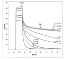
2.5μMのdAbのProtein L結合解析に関する代表的なセンサーグラムデータを示す(図1)。示されたセンサーグラムの配置および形は、調べた各dAbの濃度範囲の全体で維持された。 Representative sensorgram data for Protein L binding analysis of 2.5 μM dAb is shown (FIG. 1). The sensorgram configuration and shape shown was maintained throughout the concentration range of each dAb examined.
Protein Lで誘導体化したチップの全体にdAbを注入した後、会合段階の終点(反応ポイント1、図1参照)、および解離段階に入って5分の時点(反応ポイント2、図1参照)に設定したレポートポイントを用いて、これらの時点でProtein Lに結合したdAbの量を得ることができる(関連対照フローセルから得られた値を上記データから差し引く)。下記の等式を用いて、5分の時点で結合したdAbの割合(%B5とも呼ばれる)を決定することができる:Resp 1/Resp2 = %B5。
After injecting dAb into the entire chip derivatized with Protein L, at the end of the association phase (
当該dAbが単量体であるならば、%B5は低い(典型的には0-5)が、当該dAbが二量体であれば、%B5は高くなる(典型的には60-100)。問題のdAbサンプルが単量体と二量体溶液状態の間の平衡状態で存在する場合、または単量体と二量体の混合物からなる場合、%B5の値は、単量体と二量体dAbの%B5の間となる。したがって、%B5値は、当該dAbの予想される溶液状態を数字で表すものである。 If the dAb is a monomer,% B 5 is low (typically 0-5), if the dAb dimers,% B 5 is high (typically 60- 100). If dAb sample in question is composed of a mixture of a case, or a monomer and dimer present in equilibrium between the monomer and dimer in solution, the value of% B 5 is monomeric and secondary Between% B 5 of the monomer dAb. Therefore, the% B 5 value represents the expected solution state of the dAb by a number.
Vκ単量体および二量体に関するProtein L結合反応速度には明確な差異が示されたので、解離の速度および程度に基づいて、溶液状態間の区別が可能となった。それぞれのdAbに関する曲線の相対的配置および形は、分析した濃度に関わりなく不変であったことに留意すべきである。ラングミュア(Langmuir)1:1モデルへのカーブフィッティングは、on-rateについてはこれが急激すぎると判断されたので試みられず、off-rate(kd)に関するフィッティングは、解離曲線の激しい二相性により妨げられた。 A clear difference was shown in the Protein L binding kinetics for the V κ monomer and dimer, allowing discrimination between solution states based on the rate and extent of dissociation. It should be noted that the relative arrangement and shape of the curve for each dAb was unchanged regardless of the concentration analyzed. Curve fitting to the Langmuir 1: 1 model was not attempted because this was considered too sharp for on-rate, and fitting for off-rate (k d ) was hampered by the intense biphasic nature of the dissociation curve. It was.
関連の対照dAbを用いて、単量体および二量体が見いだされる範囲を明らかにすることができるので、dAbの溶液状態を予測することができる。 The relevant control dAb can be used to reveal the extent to which monomers and dimers are found so that the solution state of the dAb can be predicted.
さまざまなV L 免疫グロブリン単一可変ドメインにおけるA43D変異の影響
抗原に対する結合親和性を有するいくつかのdAbを取り上げ、変異を導入して、43位のアミノ酸(A)を(D)で置き換えた。変異は、部位特異的変異誘発によって導入した。
Effect of A43D Mutations on Various VL Immunoglobulin Single Variable Domains Several dAbs with binding affinity for antigen were picked, mutations were introduced, and amino acid (A) at position 43 was replaced with (D). Mutations were introduced by site-directed mutagenesis.
下記のdAbを取り上げた:
PEP1-5-19 (抗TNFα dAb):
DIQMTQSPSSLSASVGDRVTITCRASQSIDSYLHWYQQKPGKAPKLLIYSASELQSGVPSRFSGSGSGTDFTLTISSLQPEDFATYYCQQVVWRPFTFGQGTKVEIKR (配列番号1)
DOM15-10 (抗ヒトVEGF dAb)
DIQMTQSPSSLSASVGDRVTITCRASQWIGPELSWYQQKPGKAPKLLIYHTSILQSGVPSRFSGSGSGTDFTLTISSLQPEDFATYYCQQYMFQPRTFGQGTKVEIRR (配列番号2)
DOM13-25-3 (抗CEA dAb)
DIQMTQSPSSLSASVGDRVTITCRASQSIGPWLSWYQQKPGKAPKLLFYQVSRLQSGVPSRFSGSGSGTDFTLTIISLQPEDFATYYCQQNLAPPYTFGQGTKVEIKR (配列番号3)
DOM9-155-25 (抗IL-4 抗Fcn dAb)
DIQMTQSPSSLSASVGDRVTITCRASRPISDWLHWYQQKPGKAPKLLIAWASTLDSGVPSRFSGSGSGTDFTLTISSLQPEDFATYYCLQEGWGPPTFGQGTKVEIKR (配列番号4)
DOM7h-14 (抗HSA dAb)
DIQMTQSPSSLSASVGDRVTITCRASQWIGSQLSWYQQKPGKAPKLLIMWRSSLQSGVPSRFSGSGSGTDFTLTISSLQPEDFATYYCAQGAALPRTFGQGTKVEIKR (配列番号5)
溶液中の状態は上記のようにSEC-MALLSで測定した:
PEP1-5-19 (anti-TNFα dAb):
DIQMTQSPSSLSASVGDRVTITCRASQSIDSYLHWYQQKPGKAPKLLIYSASELQSGVPSRFSGSGSGTDFTLTISSLQPEDFATYYCQQVVWRPFTFGQGTKVEIKR (SEQ ID NO: 1)
DOM15-10 (anti-human VEGF dAb)
DIQMTQSPSSLSASVGDRVTITCRASQWIGPELSWYQQKPGKAPKLLIYHTSILQSGVPSRFSGSGSGTDFTLTISSLQPEDFATYYCQQYMFQPRTFGQGTKVEIRR (SEQ ID NO: 2)
DOM13-25-3 (Anti-CEA dAb)
DIQMTQSPSSLSASVGDRVTITCRASQSIGPWLSWYQQKPGKAPKLLFYQVSRLQSGVPSRFSGSGSGTDFTLTIISLQPEDFATYYCQQNLAPPYTFGQGTKVEIKR (SEQ ID NO: 3)
DOM9-155-25 (anti-IL-4 anti-Fcn dAb)
DIQMTQSPSSLSASVGDRVTITCRASRPISDWLHWYQQKPGKAPKLLIAWASTLDSGVPSRFSGSGSGTDFTLTISSLQPEDFATYYCLQEGWGPPTFGQGTKVEIKR (SEQ ID NO: 4)
DOM7h-14 (anti-HSA dAb)
DIQMTQSPSSLSASVGDRVTITCRASQWIGSQLSWYQQKPGKAPKLLIMWRSSLQSGVPSRFSGSGSGTDFTLTISSLQPEDFATYYCAQGAALPRTFGQGTKVEIKR (SEQ ID NO: 5)
The state in solution was measured with SEC-MALLS as described above:
表1:dAbおよびA43D変異体の生物物理学的性質 Table 1: Biophysical properties of dAb and A43D variants
元の境界面の残基で変異誘発させたDOM7h-8またはDOM7h-14ライブラリーの調製および解析
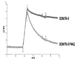
背景:ヒト軽鎖サブグループhuVκI (DPK9) に由来する2つのVκ dAbを変異解析のために選択したが、そのDOM7h-8 (WO05/118642に記載) およびDOM7h-14 (WO2008/096158に記載)は、いずれもヒト血清アルブミン(HSA)と結合する。便宜のために、使用されたDOM7h-8クローンは、Bsa I制限酵素認識部位を除去するサイレント変異を有する(↓ は制限酵素が切断する部位を示す;制限酵素認識部位は、51位のサイレントなCからTへの変異によって破壊される)。ヒトVκ軽鎖はProtein Lに結合する(下記のより詳細に記載する)。Protein L結合の維持は、免疫グロブリンドメインの適切なフォールディングを明確に示す。
Preparation and analysis of DOM7h-8 or DOM7h-14 libraries mutagenized with original interface residues
Background: Two Vκ dAbs from the human light chain subgroup huVκI (DP K 9) were selected for mutation analysis, but their DOM7h-8 (described in WO05 / 118642) and DOM7h-14 (in WO2008 / 096158) All described) bind to human serum albumin (HSA). For convenience, the DOM7h-8 clone used has a silent mutation that removes the Bsa I restriction enzyme recognition site (↓ indicates the site where the restriction enzyme cleaves; the restriction enzyme recognition site is silent at position 51. Destroyed by C to T mutation). Human V kappa light chain binds to Protein L (described in more detail below). Maintenance of Protein L binding clearly indicates proper folding of the immunoglobulin domain.
使用したDOM7h-8およびDOM7h-14のヌクレオチドおよびアミノ酸配列は下記の通りである:
DOM7h-8
ヌクレオチド配列:
GACATCCAGATGACCCAGTCTCCATCCTCCCTGTCTGCATC↓TGTAGGAGACCGTGTCACCATCACTTGCCGGGCAAGTCAGAGCATTAGCAGCTATTTAAATTGGTATCAGCAGAAACCAGGGAAAGCCCCTAAGCTCCTGATCTATCGGAATTCCCCTTTGCAAAGTGGGGTCCCATCACGTTTCAGTGGCAGTGGATCTGGGACAGATTTCACTCTCACCATCAGCAGTCTGCAACCTGAAGATTTTGCTACGTACTACTGTCAACAGACGTATAGGGTGCCTCCTACGTTCGGCCAAGGGACCAAGGTGGAAATCAAACGG (配列番号6)
アミノ酸配列:
DIQMTQSPSSLSASVGDRVTITCRASQSISSYLNWYQQKPGKAPKLLIYRNSPLQSGVPSRFSGSGSGTDFTLTISSLQPEDFATYYCQQTYRVPPTFGQGTKVEIKR (配列番号7)
DOM7h-14
ヌクレオチド配列:
GACATCCAGATGACCCAGTCTCCATCCTCCCTGTCTGCATCTGTAGGAGACCGTGTCACCATCACTTGCCGGGCAAGTCAGTGGATTGGGTCTCAGTTATCTTGGTACCAGCAGAAACCAGGGAAAGCCCCTAAGCTCCTGATCATGTGGCGTTCCTCGTTGCAAAGTGGGGTCCCATCACGTTTCAGTGGCAGTGGATCTGGGACAGATTTCACTCTCACCATCAGCAGTCTGCAACCTGAAGATTTTGCTACGTACTACTGTGCTCAGGGTGCGGCGTTGCCTAGGACGTTCGGCCAAGGGACCAAGGTGGAAATCAAACGG (配列番号8)
アミノ酸配列:
DIQMTQSPSSLSASVGDRVTITCRASQWIGSQLSWYQQKPGKAPKLLIMWRSSLQSGVPSRFSGSGSGTDFTLTISSLQPEDFATYYCAQGAALPRTFGQGTKVEIKR (配列番号9)
上記dAbの生化学的性質を以下に示す。
DOM7h-8
Nucleotide sequence:
GACATCCAGATGACCCAGTCTCCATCCTCCCTGTCTGCATC ↓ TGTAGGAGACCGTGTCACCATCACTTGCCGGGCAAGTCAGAGCATTAGCAGCTATTTAAATTGGTATCAGCAGAAACCAGGGAAAGCCCCTAAGCTCCTGATCTATCGGAATTCCCCTTTGCAAAGTGGGGTCCCATCACGTTTCAGTGGCAGTGGATCTGGGACAGATTTCACTCTCACCATCAGCAGTCTGCAACCTGAAGATTTTGCTACGTACTACTGTCAACAGACGTATAGGGTGCCTCCTACGTTCGGCCAAGGGACCAAGGTGGAAATCAAACGG (SEQ ID NO: 6)
Amino acid sequence:
DIQMTQSPSSLSASVGDRVTITCRASQSISSYLNWYQQKPGKAPKLLIYRNSPLQSGVPSRFSGSGSGTDFTLTISSLQPEDFATYYCQQTYRVPPTFGQGTKVEIKR (SEQ ID NO: 7)
DOM7h-14
Nucleotide sequence:
GACATCCAGATGACCCAGTCTCCATCCTCCCTGTCTGCATCTGTAGGAGACCGTGTCACCATCACTTGCCGGGCAAGTCAGTGGATTGGGTCTCAGTTATCTTGGTACCAGCAGAAACCAGGGAAAGCCCCTAAGCTCCTGATCATGTGGCGTTCCTCGTTGCAAAGTGGGGTCCCATCACGTTTCAGTGGCAGTGGATCTGGGACAGATTTCACTCTCACCATCAGCAGTCTGCAACCTGAAGATTTTGCTACGTACTACTGTGCTCAGGGTGCGGCGTTGCCTAGGACGTTCGGCCAAGGGACCAAGGTGGAAATCAAACGG (SEQ ID NO: 8)
Amino acid sequence:
DIQMTQSPSSLSASVGDRVTITCRASQWIGSQLSWYQQKPGKAPKLLIMWRSSLQSGVPSRFSGSGSGTDFTLTISSLQPEDFATYYCAQGAALPRTFGQGTKVEIKR (SEQ ID NO: 9)
The biochemical properties of the dAb are shown below.
表2:DOM7h-8およびDOM7h-14の生物物理学的性質および抗原結合化学量論。 Table 2: Biophysical properties and antigen binding stoichiometry of DOM7h-8 and DOM7h-14.
DOM7h-8は二量体としてHSAと結合する(表2を参照されたい)。元のVH/VL境界面にある残基を分析のために選択した。これらの変異は、dAbに抗原結合活性を与える超可変CDR領域とは対照的に、Vκドメイン抗体の保存されたフレームワーク領域内にある。 DOM7h-8 binds to HSA as a dimer (see Table 2). Residues at the original V H / V L interface were selected for analysis. These mutations are in the conserved framework regions of Vκ domain antibodies, as opposed to hypervariable CDR regions that confer antigen binding activity to dAbs.
DOM7h-14はPBS中250μM未満の濃度で主に単量体として存在する(表2を参照されたい)。DOM7h-14を含めることによって、すでに主として単量体状態であるdAbの抗原結合およびProtein L結合活性に及ぼす、変異の影響を評価することが可能になる。 DOM7h-14 is present primarily as a monomer at a concentration of less than 250 μM in PBS (see Table 2). Inclusion of DOM7h-14 makes it possible to assess the effect of mutations on the antigen binding and protein L binding activity of dAbs that are already predominantly monomeric.
実施例2A - DOM7h-8
DOM7h-8については、元のVH/VL境界面にある残基、Q38、A43およびP44の変異を有する3つの個別のライブラリーを作製した:
大腸菌発現ベクターpDOM5の中のDOM7h-8を鋳型として用いて、部位特異的変異誘発によって変異を導入した(pDOM5はLacZプロモーターの制御下で、pUC119に基づく発現ベクターである)。部位特異的変異誘発は、鋳型である100 ngプラスミドDNA、およびそれぞれ必要な変異を含有する相補的プライマーを用いて、PCRによって行った。反応は、2.5U PfuTurboポリメラーゼ (Stratagene)をPCR混合物 [100ng プラスミドテンプレート、プライマー (各2μM)、dNTP (各0.2mM)、1% (v/v) ホルムアミド/1x PfuTurboバッファー(Stratagene)]に添加することにより、ホットスタートで開始した。反応は次の温度サイクルで行った[94℃ 2分;18回 (94℃ 30秒、55℃ 30秒、および68℃ 20分);68℃ 2分;10℃保持]。PCR反応物をQIAquick PCR精製キット(Qiagen)で精製し、50μl H2Oで溶出した。精製DNAを、DpnI(New England Biolabs)で1時間、制限酵素消化して、インプットプラスミドテンプレートを除去した。制限酵素処理したDNAサンプルをエタノール沈殿して、5μl H2Oに懸濁した。沈殿したDNAを、2xTY/カルベニシリン0.1 mg/mlプレート上に蒔いた、化学的にコンピテントにした大腸菌細胞に入れて形質転換し、37℃にて一晩インキュベートした。
Example 2A-DOM7h-8
For DOM7h-8, three separate libraries were created with mutations in the residues at the original VH / VL interface, Q38, A43 and P44:
Mutations were introduced by site-directed mutagenesis using DOM7h-8 in the E. coli expression vector pDOM5 as a template (pDOM5 is an expression vector based on pUC119 under the control of the LacZ promoter). Site-directed mutagenesis was performed by PCR using 100 ng plasmid DNA as template and complementary primers each containing the required mutation. For the reaction, add 2.5 U PfuTurbo polymerase (Stratagene) to the PCR mixture [100 ng plasmid template, primer (2 μM each), dNTP (0.2 mM each), 1% (v / v) formamide / 1x PfuTurbo buffer (Stratagene)] It started with a hot start. The reaction was carried out in the next temperature cycle [94 ° C. 2 minutes; 18 times (94 ° C. 30 seconds, 55 ° C. 30 seconds, and 68 ° C. 20 minutes); The PCR reaction was purified with QIAquick PCR purification kit (Qiagen) and eluted with 50 μl H 2 O. The purified DNA was digested with DpnI (New England Biolabs) for 1 hour to remove the input plasmid template. The restriction enzyme-treated DNA sample was ethanol precipitated and suspended in 5 μl H 2 O. The precipitated DNA was transformed into chemically competent E. coli cells plated on 2xTY / carbenicillin 0.1 mg / ml plates and incubated overnight at 37 ° C.
プライマーは次のとおりであった。 The primers were as follows:
Q38 (プライマー:
5’-GCAGCTATTTAAATTGGTATCAGNNKAAACCAGGGAAAGCCCC-3’ (配列番号10);
5’-GGGGCTTTCCCTGGTTTMNNCTGATACCAATTTAAATAGCTGC-3’ (配列番号11))、
A43 (プライマー:
5’-CAGCAGAAACCAGGGAAANNKCCTAAGCTCCTGATCTATCGG-3’ (配列番号12);
5’-CCGATAGATCAGGAGCTTAGGMNNTTTCCCTGGTTTCTGCTG-3’ (配列番号13))、
P44 (プライマー:
5’-CAGCAGAAACCAGGGAAAGCCNNKAAGCTCCTGATCTATCGGAATTCCC-3’ (配列番号14);
5’-GGGAATTCCGATAGATCAGGAGCTTMNNGGCTTTCCCTGGTTTCTGCTG-3’ (配列番号15))。
Q38 (Primer:
5'-GCAGCTATTTAAATTGGTATCAGNNKAAACCAGGGAAAGCCCC-3 '(SEQ ID NO: 10);
5'-GGGGCTTTCCCTGGTTTMNNCTGATACCAATTTAAATAGCTGC-3 '(SEQ ID NO: 11)),
A43 (Primer:
5'-CAGCAGAAACCAGGGAAANNKCCTAAGCTCCTGATCTATCGG-3 '(SEQ ID NO: 12);
5'-CCGATAGATCAGGAGCTTAGGMNNTTTCCCTGGTTTCTGCTG-3 '(SEQ ID NO: 13)),
P44 (Primer:
5'-CAGCAGAAACCAGGGAAAGCCNNKAAGCTCCTGATCTATCGGAATTCCC-3 '(SEQ ID NO: 14);
5'-GGGAATTCCGATAGATCAGGAGCTTMNNGGCTTTCCCTGGTTTCTGCTG-3 '(SEQ ID NO: 15)).
多様性を導入するために使用されるNNKコドンは、20個のアミノ酸すべてとTAG終止コドンをコードする。Protein Lと結合すると確認されたクローンは、プライマーDOM8(AGCGGATAACAATTTCACACAGGA (配列番号16))を用いて配列決定した。 The NNK codon used to introduce diversity encodes all 20 amino acids and the TAG stop codon. Clones confirmed to bind to Protein L were sequenced using primer DOM8 (AGCGGATAACAATTTCACACAGGA (SEQ ID NO: 16)).
各ライブラリーから96個のコロニーを無作為に採取して96ウェルプレートフォーマットに入れ、メーカーの説明書(Novagen)にしたがって、OnEx溶液1、2および3を添加した1 ml 2xTY 0.1mg/ml カルベニシリン中で発現させた。培養物はInforsHT振盪器内で、950 rpm高湿度で3日間、30℃にて増殖させた。細胞を遠心分離(ベンチトップSorvall遠心機にて4.5k rpm、30分)によりペレットとし、上清75μlを等量のHBS-EPバッファー(GE Healthcare)に加えた。発現した上清を、ストレプトアビジンコートBIAcoreチップ(495 RU)に結合したビオチン化Protein L(Pierce)を用いて、Protein Lの結合についてBIAcoreでスクリーニングした。Protein Lとの結合が確認されたクローンは、プライマーDOM8(上記の配列番号16)を用いて配列決定した。
Randomly pick 96 colonies from each library into a 96-well plate format and add 1 ml 2xTY 0.1 mg / ml carbenicillin with
Q38、A43およびP44位でのアミノ酸変異のすべてを得るために、ライブラリーのランダムスクリーニングでは確認されなかったクローンを部位特異的変異誘発によって作製したが、これには、表3に挙げたプライマーとともに、鋳型として、大腸菌発現ベクターpDOM5中のDOM7h-8を使用した。 To obtain all of the amino acid mutations at positions Q38, A43 and P44, clones that were not identified by random screening of the library were generated by site-directed mutagenesis, along with the primers listed in Table 3. DOM7h-8 in the E. coli expression vector pDOM5 was used as a template.
表3: NNKライブラリー(上記)において確認されなかったQ38、A43、またはP44位でのDOM7h-8変異体の作製に使用したプライマーペア
DOM7h-8変異体のスクリーニング: コグネイトHSA結合およびスーパー抗原Protein Lに対するdAb結合活性の特徴を明らかにするために、細菌上清からの精製の前後で、Q38、A43、またはP44位のDOM7h-8変異体をBIAcoreでスクリーニングした。精製タンパク質に対してSECおよびSEC MALLSを使用し、親dAbおよび変異体のオリゴマー化状態の特徴を明らかにした。 Screening for DOM7h-8 mutants : DOM7h-8 at position Q38, A43, or P44 before and after purification from bacterial supernatant to characterize cognate HSA binding and dAb binding activity against the superantigen Protein L Mutants were screened with BIAcore. SEC and SEC MALLS were used on the purified protein to characterize the oligomerization state of the parent dAb and the mutant.
Protein LおよびHSA結合活性に関する細菌上清中のdAbのスクリーニング:細菌クローンを採取して96ウェルプレートフォーマットに入れ、メーカーの説明書(Novagen)にしたがって、OnEx溶液1、2および3を添加した1 ml 2xTY 0.1mg/ml カルベニシリン中で発現させた。培養物はInforsHT振盪器内で、950 rpm高湿度で3日間、30℃にて増殖させた。細胞を遠心分離(ベンチトップSorvall遠心機にて4.5k、30分)によりペレットとし、上清75μlを等量のHBS-EPバッファー(GE Healthcare)に加えた。希釈された上清を、CM5 BIAcoreチップ(789 RU)にカップリングしたProtein L(Sigma)および同じCM5チップ(6036RU)上の別のフローセルに結合したHSAを用いて、Protein Lの結合についてBIAcoreでスクリーニングした(表4〜6を参照されたい)。
Screening of dAbs in bacterial supernatants for protein L and HSA binding activity : Bacterial clones were picked and placed in a 96-well plate format and
Protein LおよびHSA結合をアッセイするための、ならびにSECおよびSEC MALLS分析のための、V κ dAbの精製:メーカーの説明書(Novagen)にしたがって、OnEx溶液1、2および3を添加した2xTY カルベニシリン100μg/ml、消泡剤、中の0.5 l培養において、Q38、A43、またはP44位でのDOM7h-8変異体を発現する全クローンからタンパク質を発現させた。培養物は、InforsHT振盪器内で、250 rpmで3日間、30℃にて増殖させた。培養物は、ベンチトップ遠心機で45分間4,500rpmで遠心分離し、回転させながら2時間Streamline Protein L 15mlにバッチ結合させることによって、清澄化した上清からタンパク質を精製した。高塩濃度PBSバッファーで十分に洗浄した後、0.1Mグリシン pH 2を用いてタンパク質を、>95%の純度で樹脂から溶出した。さらに生化学的/生物物理学的な性質検討を行う前に、タンパク質を濃縮し、バッファーをPBSに交換した。
Purification of V κ dAb for assaying Protein L and HSA binding and for SEC and SEC MALLS analysis : 100 μg of 2xTY carbenicillin with
1μM、500nM、250nM、125nM、62.5nMおよび31.25nMの濃度範囲の精製タンパク質は、CM5チップ上の別々のフローセルにカップリングした、Protein L(311RU)との結合およびHSA(559RU)との結合について、BIAcoreでアッセイした。親分子DOM7h-8(二量体)より有意に早くProtein Lから解離するクローンは、安定な単量体か、または二量体と平衡状態にある単量体のいずれかであるとされた(図2;表4〜6を参照されたい)。精製タンパク質は、HSA結合についても分析し、抗原と接触するdAbのCDR領域のコンフォメーションに及ぼす変異の影響を評価した(表4〜6を参照されたい)。 Purified proteins in a concentration range of 1 μM, 500 nM, 250 nM, 125 nM, 62.5 nM and 31.25 nM are coupled to separate flow cells on a CM5 chip for binding to Protein L (311RU) and HSA (559RU) Assayed with BIAcore. Clones that dissociated from Protein L significantly earlier than the parent molecule DOM7h-8 (dimer) were either considered to be stable monomers or monomers in equilibrium with the dimer ( FIG. 2; see Tables 4-6). The purified protein was also analyzed for HSA binding and assessed the effect of mutations on the conformation of the CDR region of the dAb in contact with the antigen (see Tables 4-6).
0.5mg/mlおよび1.6mg/mlの濃度範囲で精製タンパク質を、SECおよび/またはSEC MALLSで分析し、それらの溶液中での状態を明らかにした(表4〜6を参照されたい)。
表4-6:DOM7h-8発現上清および精製タンパク質のBIAcoreおよび生物物理学的分析。網掛けした行は、DOM7h-8 Vκ dAb二量体を単量体化する変異を特定する。(xはBIAcoreチップ上に固定化されたリガンドに結合しないことを示す;√ はBIAcoreチップ上に固定化されたリガンドとの良好な結合を示す;√wはBIAcoreチップ上に固定化されたリガンドとの弱い結合を示す;Mは単量体を示す;Dは二量体を示す;M/Dは二量体と平衡状態にある単量体を示す;D/Tはサンプル中にdAb二量体および三量体が存在することを示す;* は平衡状態でないM/Dが、より単量体に向かう傾向があることを示す)。 Table 4-6 : BIAcore and biophysical analysis of DOM7h-8 expression supernatant and purified protein. Shaded line identifies the mutations that monomer the DOM7h-8 V κ dAb dimers. (X indicates that it does not bind to the ligand immobilized on the BIAcore chip; √ indicates good binding with the ligand immobilized on the BIAcore chip; √w indicates the ligand immobilized on the BIAcore chip. M indicates monomer; D indicates dimer; M / D indicates monomer in equilibrium with dimer; D / T indicates dAb dimer in sample Indicates the presence of trimer and trimer; * indicates that M / D that is not in equilibrium tends to be more monomeric).
結論:P44での変異は、DOM7h-8の溶液中の状態を変化させる。いくつかの変異が二量体DOM7h-8を単量体化する。 Conclusion : Mutation at P44 changes the state of DOM7h-8 in solution. Several mutations monomerize the dimer DOM7h-8.
2B) DOM7h-14
DOM7h-14について、元のVH/VL境界面にある残基、Q38、A43およびP44に変異を有する3つの個別のライブラリーを作製した。大腸菌発現ベクターpDOM5の中のDOM7h-14を鋳型として使用し、さらに上記のようにNNKコドンを用いて、部位特異的変異誘発によって変異を導入した。プライマーは次のとおりであった:
Q38
(プライマー:
5’-GGGTCTCAGTTATCTTGGTACCAGNNKAAACCAGGGAAAGCCCC-3’ (配列番号37);
5’-GGGGCTTTCCCTGGTTTMNNCTGGTACCAAGATAACTGAGACCC-3’ (配列番号38))
A43
(プライマー:
5’-CAGCAGAAACCAGGGAAANNKCCTAAGCTCCTGATCATGTGG-3’ (配列番号39);
5’-CCACATGATCAGGAGCTTAGGMNNTTTCCCTGGTTTCTGCTG-3’ (配列番号40)); または
P44
(プライマー:
5’-CAGCAGAAACCAGGGAAAGCCNNKAAGCTCCTGATCATGTGGCGTTCC-3’ (配列番号41);
5’-GGAACGCCACATGATCAGGAGCTTMNNGGCTTTCCCTGGTTTCTGCTG-3’(配列番号42))
スクリーニングするために、ライブラリーで大腸菌HB2151細胞を形質転換した。
2B) DOM7h-14
For DOM7h-14, three separate libraries were made with mutations in the residues at the original VH / VL interface, Q38, A43 and P44. Mutations were introduced by site-directed mutagenesis using DOM7h-14 in the E. coli expression vector pDOM5 as a template and using the NNK codon as described above. Primers were as follows:
Q38
(Primer:
5'-GGGTCTCAGTTATCTTGGTACCAGNNKAAACCAGGGAAAGCCCC-3 '(SEQ ID NO: 37);
5'-GGGGCTTTCCCTGGTTTMNNCTGGTACCAAGATAACTGAGACCC-3 '(SEQ ID NO: 38))
A43
(Primer:
5'-CAGCAGAAACCAGGGAAANNKCCTAAGCTCCTGATCATGTGG-3 '(SEQ ID NO: 39);
5'-CCACATGATCAGGAGCTTAGGMNNTTTCCCTGGTTTCTGCTG-3 '(SEQ ID NO: 40)); or
P44
(Primer:
5'-CAGCAGAAACCAGGGAAAGCCNNKAAGCTCCTGATCATGTGGCGTTCC-3 '(SEQ ID NO: 41);
5'-GGAACGCCACATGATCAGGAGCTTMNNGGCTTTCCCTGGTTTCTGCTG-3 '(SEQ ID NO: 42))
To screen, E. coli HB2151 cells were transformed with the library.
DOM7h-14のQ38、A43、またはP44位での全アミノ酸バリアントの単離:プライマーDOM8(上記配列番号16)およびDOM9(CGCCAGGGTTTTCCCAGTCACGAC (配列番号75))を用いて、Q38、A43、もしくはP44位で変異させたDOM7h-14ライブラリーからランダムに選択した96コロニーについて、コロニースクリーンPCRを行った。PCR産物を、DOM8(上記配列番号16)を用いて配列決定し、Protein L結合を分析して、全dAbが発現されること、ならびにそれらがProtein L結合を維持していることを確認した。 Isolation of all amino acid variants at positions Q38, A43, or P44 of DOM7h-14 : using primers DOM8 (SEQ ID NO: 16 above) and DOM9 (CGCCAGGGTTTTCCCAGTCACGAC (SEQ ID NO: 75)) at positions Q38, A43, or P44 Colony screen PCR was performed on 96 colonies randomly selected from the mutated DOM7h-14 library. PCR products were sequenced using DOM8 (SEQ ID NO: 16 above) and protein L binding was analyzed to confirm that all dAbs were expressed and that they maintained protein L binding.
最初のスクリーニングの試みから得られないクローンは、下記のプライマーを用いて、部位特異的変異誘発で作製した(表7):
表7:NNKライブラリーにおいて確認されなかったQ38、A43、またはP44位でのDOM7h-14変異体を作製するための部位特異的変異誘発用プライマーペア
Table 7 : Site-directed mutagenesis primer pairs to generate DOM7h-14 mutants at position Q38, A43, or P44 that were not identified in the NNK library
DOM7h-14変異体のスクリーニング:Vκ dAbの構造、ひいては抗原結合活性に影響を及ぼすQ38、A43、またはP44での変異の可能性を特徴付けるために、単量体Vκ dAb DOM7h-14のQ38、A43、またはP44位での全アミノ酸バリアントを、Protein LおよびHSA結合活性についてBIAcoreでスクリーニングした。同一チップ上の別々のフローセルに存在するProtein Lとの結合は、dAb発現が生じたこと、または損なわれなかったことを確認した。 Screening of DOM7h-14 mutants : To characterize the potential of mutations in Q38, A43, or P44 affecting the structure of V κ dAb and thus antigen binding activity, Q38 of monomeric Vκ dAb DOM7h-14, All amino acid variants at position A43 or P44 were screened with BIAcore for protein L and HSA binding activity. Binding to Protein L present in separate flow cells on the same chip confirmed that dAb expression occurred or was not impaired.
Protein LおよびHSA結合に関する発現上清のスクリーニング:細菌クローンを採取して96ウェルプレートフォーマットに入れ、メーカーの説明書(Novagen)にしたがって、OnEx溶液1、2および3を添加した1 ml 2xTY 0.1mg/ml カルベニシリン中で発現させた。培養物はInforsHT振盪器内で、950 rpm高湿度で3日間、30℃にて増殖させた。細胞を遠心分離(ベンチトップSorvall遠心機にて4.5k、30分)によりペレットとし、上清75μlを等量のHBS-EPバッファー(GE Healthcare)に加えた。発現上清を、CM5 BIAcoreチップ(789 RU)にカップリングしたProtein L(Sigma)および同じCM5チップ(6036RU)上の別のフローセルにカップリングしたHSAを用いて、Protein Lの結合についてBIAcoreでスクリーニングした(表8を参照されたい)。
表8:Protein Lおよび抗原(HSA)結合に関するDOM7h-14発現上清のBIAcore分析。(√は結合を示す;ndは未測定を示す)。 Table 8: BIAcore analysis of DOM7h-14 expression supernatant for Protein L and antigen (HSA) binding. (√ indicates bond; nd indicates unmeasured).
結論:調べたすべての変異体は、Protein Lに結合し、HSA結合を維持しているので、これは変異がdAb構造ひいては抗原結合に影響を与えないことを示す。 Conclusion : All mutants examined bind to Protein L and maintain HSA binding, indicating that the mutation does not affect dAb structure and hence antigen binding.
PEP1-5-19 P44変異体のスクリーニング
別のクローンで変異を引き起こすことの効果を究明するために、プライマーとともに、大腸菌発現ベクターpDOM5内のPEP1-5-19を鋳型として使用して、PEP1-5-19のP44での変異を部位特異的変異誘発により引き起こした。
PEP1-5-19 Screening for P44 mutants To investigate the effects of causing mutations in different clones, using PEP1-5-19 in the E. coli expression vector pDOM5 as a template with primers, PEP1-5 -19 mutations at P44 were caused by site-directed mutagenesis.
5’-GCAGAAACCAGGGAAAGCCNNKAAGCTCCTGATCTATAGTGC-3’ (配列番号57)、
5’-GCACTATAGATCAGGAGCTTMNNGGCTTTCCCTGGTTTCTGC-3’(配列番号58)。
5'-GCAGAAACCAGGGAAAGCCNNKAAGCTCCTGATCTATAGTGC-3 '(SEQ ID NO: 57),
5′-GCACTATAGATCAGGAGCTTMNNGGCTTTCCCTGGTTTCTGC-3 ′ (SEQ ID NO: 58).
親PEP1-5-19、ならびにPEP1-5-19 P44ライブラリーからランダムに選択した94コロニーを、96ウェルプレートフォーマットで、メーカーの説明書(Novagen)にしたがって、OnEx溶液1、2および3を添加した1 ml 2xTY 0.1mg/ml カルベニシリン中で発現させた。培養物はInforsHT振盪器内で、950 rpm高湿度で3日間、30℃にて増殖させた。細胞を遠心分離(ベンチトップSorvall遠心機にて4.5k rpm、30分)によりペレットとし、上清75μlを等量のHBS-EPバッファー(GE Healthcare)に加えた。発現上清を、CM5 BIAcoreチップにカップリングしたProtein L(Sigma)を用いて、Protein Lの結合についてBIAcore(311RU)でスクリーニングした。すべてのクローンはプライマーDOM8(上記配列番号16)を用いて配列決定した。親分子PEP1-5-19(二量体)より有意に早くProtein Lから解離するクローンは、安定な単量体か、または二量体と平衡状態にある単量体のいずれかであるとされた(表9を参照されたい)。
表9:Protein L結合に関するPEP1-5-19 P44変異体の上清スクリーニング(Dは二量体を示す;Mは単量体を示す;M/Dは単量体/二量体を示す;ndは配列決定した94クローンの中に変異体が特定されなかったため未測定)。 Table 9 : Supernatant screening of PEP1-5-19 P44 mutants for Protein L binding (D indicates dimer; M indicates monomer; M / D indicates monomer / dimer; nd is not measured because no variants were identified among the 94 clones sequenced).
結論:P44位でのDOM7h-8変異体について見られたように、変異は、元の二量体PEP1-5-19の溶液中の状態を変化させた。 Conclusion : As seen for the DOM7h-8 mutant at position P44, the mutation changed the state of the original dimer PEP1-5-19 in solution.
43位で変異させたナイーブなV κ dAbプールの構築
元のVH/VL境界面での変異が、ナイーブライブラリーに関連するdAbライブラリーの単量体含量を高める可能性について、さらに理解を深めるために、4G Vκ dAbライブラリー(WO2005093074に記載)を用いて、部位特異的変異誘発により43位に変異を導入した。このアプローチによって、普遍的で幅広い視点で変異を分析することが可能となるが、それは、特定の変異が、さまざまなCDRの組み合わせおよび組成を越えて有効となることを示唆する。
Understand how mutations at the original VH / VL interface of the naïve V κ dAb pool mutated at position 43 may increase the monomer content of the dAb library associated with the naive library In order to deepen this, a mutation was introduced into position 43 by site-directed mutagenesis using a 4G V κ dAb library (described in WO2005093074). This approach makes it possible to analyze mutations from a universal and broad perspective, which suggests that specific mutations are effective across different CDR combinations and compositions.
プライマーは、Fw 2の43位をA43A、-D、-K、-R、-E、-Iまたは-L のいずれかに変更するようにStratagene Quikchangeプライマー設計ソフトウェアで設計し、Sigmaで合成された(OD 1μmolスケールまで合成し、PAGEで合成した)。
Primers were designed with Stratagene Quikchange primer design software and synthesized by Sigma to change position 43 of
プライマー配列:
A43A_fwd: gcagaaaccagggaaagcccctaagctcctgatc (配列番号59)
A43A_rev: gatcaggagcttaggggctttccctggtttctgc (配列番号60)
A43D_fwd: gcagaaaccagggaaagaccctaagctcctgatc (配列番号61)
A43D_rev: gatcaggagcttagggtctttccctggtttctgc (配列番号62)
A43K_fwd: aaattggtaccagcagaaaccagggaaaaagcctaagctcctgatc (配列番号63)
A43K_rev: gatcaggagcttaggctttttccctggtttctgctggtaccaattt (配列番号64)
A43R_fwd: gtaccagcagaaaccagggaaacggcctaagctcctg (配列番号65)
A43R_rev: caggagcttaggccgtttccctggtttctgctggtac (配列番号66)
A43E_fwd: cagcagaaaccagggaaagagcctaagctcctgatctatg (配列番号67)
A43E_rev: catagatcaggagcttaggctctttccctggtttctgctg (配列番号68)
A43I_fwd: ggtaccagcagaaaccagggaaaatccctaagctcct (配列番号69)
A43I_rev: aggagcttagggattttccctggtttctgctggtacc (配列番号70)
A43L_fwd: tggtaccagcagaaaccagggaaactgcctaagctcctga (配列番号71)
A43L_rev: tcaggagcttaggcagtttccctggtttctgctggtacca (配列番号72)
50 mlの2 x TY培地 + カルベニシリン100μg/mlに、pDOM10グリセロール原液中のナイーブ4G Vκライブラリー50μlを接種し、37℃にて一晩インキュベートした。Qiagen QIAfilter midi-prepを用いて、メーカーの使用説明書にしたがって、プラスミドDNAを単離した。pDOM10は、dAbの可溶性発現のために設計されたプラスミドベクターである。これはpUC119ベクターを基にしており、発現はLacZプロモーターの制御下である。dAbの上清中への発現は、dAb遺伝子をN末端で、一般的なGASリーダーシグナルペプチド(WO2005093074参照)に融合させることによって確実となった。それに加えて、FLAGタグがdAbのC末端に付加された。
Primer sequence:
A43A_fwd: gcagaaaccagggaaagcccctaagctcctgatc (SEQ ID NO: 59)
A43A_rev: gatcaggagcttaggggctttccctggtttctgc (SEQ ID NO: 60)
A43D_fwd: gcagaaaccagggaaagaccctaagctcctgatc (SEQ ID NO: 61)
A43D_rev: gatcaggagcttagggtctttccctggtttctgc (SEQ ID NO: 62)
A43K_fwd: aaattggtaccagcagaaaccagggaaaaagcctaagctcctgatc (SEQ ID NO: 63)
A43K_rev: gatcaggagcttaggctttttccctggtttctgctggtaccaattt (SEQ ID NO: 64)
A43R_fwd: gtaccagcagaaaccagggaaacggcctaagctcctg (SEQ ID NO: 65)
A43R_rev: caggagcttaggccgtttccctggtttctgctggtac (SEQ ID NO: 66)
A43E_fwd: cagcagaaaccagggaaagagcctaagctcctgatctatg (SEQ ID NO: 67)
A43E_rev: catagatcaggagcttaggctctttccctggtttctgctg (SEQ ID NO: 68)
A43I_fwd: ggtaccagcagaaaccagggaaaatccctaagctcct (SEQ ID NO: 69)
A43I_rev: aggagcttagggattttccctggtttctgctggtacc (SEQ ID NO: 70)
A43L_fwd: tggtaccagcagaaaccagggaaactgcctaagctcctga (SEQ ID NO: 71)
A43L_rev: tcaggagcttaggcagtttccctggtttctgctggtacca (SEQ ID NO: 72)
50 ml of 2 × TY medium +
部位特異的変異誘発反応は、Stratagene Quikchange IIキットを用いて、下記を除いてメーカーのプロトコールにしたがって行った。反応は次のように実施した:(50μl反応液あたり)5μl 10x反応バッファー、1.55μl(120 ng)pDOM10ナイーブ4G Vκ Midiprep、1.25μlフォワードプライマー(125 ng)、1.25μlリバースプライマー(125 ng)、1μl dNTPミックス、38.95μl滅菌水、1μl Pfu ultra。変異誘発は以下のPCRプログラムで行った。1.95℃ 30秒、2.95℃ 30秒、3.55℃ 1分、4.68℃ 4分、5.ステップ2〜4を17サイクル、6.4℃に保持。1μl Dpn Iを各反応液に添加し、37℃にて1時間インキュベートした。 Site-directed mutagenesis reactions were performed using the Stratagene Quikchange II kit according to the manufacturer's protocol, except as follows. The reaction was carried out as follows: (50 [mu] l reaction solution per) 5 [mu] l 10x reaction buffer, 1.55μl (120 ng) pDOM10 naive 4G V κ Midiprep, 1.25μl forward primer (125 ng), 1.25μl reverse primer (125 ng) 1 μl dNTP mix, 38.95 μl sterile water, 1 μl Pfu ultra. Mutagenesis was performed with the following PCR program. 1. 95 ° C for 30 seconds, 2.95 ° C for 30 seconds, 3.55 ° C for 1 minute, 4.68 ° C for 4 minutes, 5. Hold steps 2-4 at 6.4 ° C for 17 cycles. 1 μl Dpn I was added to each reaction and incubated at 37 ° C. for 1 hour.
5μlの各Dpn I消化反応液を、50μlに分けたエレクトロコンピテントなHB2151大腸菌細胞と混合し、0.2 cmエレクトロポレーションキュベット(Biorad)に入れて30分間氷上でインキュベートし、標準的な大腸菌K12の設定(2.5 kV/cm、25μF、200Ω)でエレクトロポレーションすることによって、形質転換した。950μlの加温したSOC培地(Invitrogen, 15544-034)を、エレクトロポレーション後直ちに加え、14 ml Falconチューブに移し、37℃にて200 rpmで1時間インキュベートした。回収培養物のすべてをLB + カルベニシリン100μg/mlに蒔き(330μl x 3)、37℃にて一晩インキュベートした。QPix2XT (Genetix)を用いて、クローンを採取して、125μl 2 x TY + 2%グルコース + 100 μg/mlカルベニシリンを容れた96ウェルプレート(Corning)に入れ、37℃にて、250 rpmで一晩、加湿インキュベーター(New Brunswick)内でインキュベートした。 Mix 5 μl of each Dpn I digestion reaction with 50 μl of electrocompetent HB2151 E. coli cells, place in a 0.2 cm electroporation cuvette (Biorad) and incubate on ice for 30 min. Transformation was performed by electroporation at the settings (2.5 kV / cm, 25 μF, 200Ω). 950 μl of warmed SOC medium (Invitrogen, 15544-034) was added immediately after electroporation, transferred to a 14 ml Falcon tube and incubated at 37 ° C. at 200 rpm for 1 hour. All harvested cultures were plated on LB + carbenicillin 100 μg / ml (330 μl × 3) and incubated overnight at 37 ° C. Using QPix2XT (Genetix), clones were picked and placed in a 96 well plate (Corning) containing 125 μl 2 x TY + 2% glucose + 100 μg / ml carbenicillin at 37 ° C, 250 rpm overnight. Incubated in a humidified incubator (New Brunswick).
発現培養は、各ライブラリープールから2プレートについて準備した:1 ml TB + 個別のOnEx (Invitrogen)成分(1L培地あたり:20 ml溶液1、50 ml溶液2、1 ml溶液3) + カルベニシリン100μg/ml + 消泡剤 (A204, Sigma) 2滴を2 ml深型ウェル・ブロックに加えた。培養物は30℃にて750 rpm、湿度85%で3日間インキュベートした。その後、粗製上清を集め、4℃にて4500 rpmで30分間遠心して清澄化し、-80℃で保存した。
Expression cultures were prepared for 2 plates from each library pool: 1 ml TB + individual OnEx (Invitrogen) components (per 1 L medium: 20
ナイーブライブラリーをバックグラウンドとしてA43D、-K、-R、-E、-Iおよび-Lについて、単量体化能力、発現、および安定化効果をランク付けする
上記A43変異体ライブラリーから作製された粗製上清サンプルを、上記方法の項で記載のように、BIAcore 3000機器(BIAcore, Sweden)を用いてProtein L結合により分析した。2つの別々のBIAcore CM5チップを用いてデータを収集した;2つのチップはいずれも、フローセル2および3(Fc2およびFc3)において小量(約500〜700 RU)のProtein Lで誘導体化されており、フローセル1(Fc1)においてブランクの活性化-不活化表面を有する。結果を図3に示す。
Created from the above A43 variant library that ranks monomerization ability, expression, and stabilization effect for A43D, -K, -R, -E, -I and -L with naive library as background The crude supernatant samples were analyzed by Protein L binding using a BIAcore 3000 instrument (BIAcore, Sweden) as described in the method section above. Data were collected using two separate BIAcore CM5 chips; both chips were derivatized with a small amount (approximately 500-700 RU) Protein L in
データ解析は、Microsoft Excelにエクスポートされた、Fc2-1またはFc3-1からのレポートポイントテーブルを用いて行った。2つのレポートポイントは上記方法に含まれ、%B5値が得られた。この%B5値を用いてクローンをランク付けした。対照dAb DOM7h-8(二量体対照、64% ±5)およびDOM4-130-54(単量体対照、4% ±5)に対する%B5値を用いて、クローンを、単量体(SM)、二量体(SD)、または迅速平衡状態(RE)として分類した。 Data analysis was performed using a report point table from Fc2-1 or Fc3-1 exported to Microsoft Excel. The two reporting points included in the process, 5 value% B was obtained. It was ranked clones using the% B 5 values. Using% B5 values for the control dAb DOM7h-8 (dimer control, 64% ± 5) and DOM4-130-54 (monomer control, 4% ± 5), clones were monomeric (SM) , Dimer (SD), or rapid equilibrium (RE).
DOM4-130-54のアミノ酸配列および核酸配列は下記の通りである:
DOM4-130-54
ヌクレオチド配列:
ATGTTATTTAAATCATTATCAAAATTAGCAACCGCAGCAGCATTTTTTGCAGGCGTGGCAACAGCGTCGACGGACATCCAGATGACCCAGTCTCCATCCTCCCTGTCTGCATCTGTAGGAGACCGTGTCACCATCACTTGCCGGGCAAGTCAGGATATTTACCTGAATTTAGACTGGTATCAGCAGAAACCAGGGAAAGCCCCTAAGCTCCTGATCAATTTTGGTTCCGAGTTGCAAAGTGGTGTCCCATCACGTTTCAGTGGCAGTGGATATGGGACAGATTTCACTCTCACCATCAGCAGTCTGCAACCTGAAGATTTCGCTACGTACTACTGTCAACCGTCTTTTTACTTCCCTTATACGTTCGGCCAAGGGACCAAGGTGGAAATCAAACGGGCGGCCGCAGAACAAAAACTCATCTCAGAAGAGGATCTGAATTAATAA (配列番号73)
アミノ酸配列
MLFKSLSKLATAAAFFAGVATASTDIQMTQSPSSLSASVGDRVTITCRASQDIYLNLDWYQQKPGKAPKLLINFGSELQSGVPSRFSGSGYGTDFTLTISSLQPEDFATYYCQPSFYFPYTFGQGTKVEIKRA (配列番号74)。
The amino acid sequence and nucleic acid sequence of DOM4-130-54 is as follows:
DOM4-130-54
Nucleotide sequence:
ATGTTATTTAAATCATTATCAAAATTAGCAACCGCAGCAGCATTTTTTGCAGGCGTGGCAACAGCGTCGACGGACATCCAGATGACCCAGTCTCCATCCTCCCTGTCTGCATCTGTAGGAGACCGTGTCACCATCACTTGCCGGGCAAGTCAGGATATTTACCTGAATTTAGACTGGTATCAGCAGAAACCAGGGAAAGCCCCTAAGCTCCTGATCAATTTTGGTTCCGAGTTGCAAAGTGGTGTCCCATCACGTTTCAGTGGCAGTGGATATGGGACAGATTTCACTCTCACCATCAGCAGTCTGCAACCTGAAGATTTCGCTACGTACTACTGTCAACCGTCTTTTTACTTCCCTTATACGTTCGGCCAAGGGACCAAGGTGGAAATCAAACGGGCGGCCGCAGAACAAAAACTCATCTCAGAAGAGGATCTGAATTAATAA (SEQ ID NO: 73)
Amino acid sequence
MLFKSLSKLATAAAFFAGVATASTDIQMTQSPSSLSASVGDRVTITCRASQDIYLNLDWYQQKPGKAPKLLINFGSELQSGVPSRFSGSGYGTDFTLTISSLQPEDFATYYCQPSFYFPYTFGQGTKVEIKRA (sequence number 74).
次の場合はクローンをスプレッドシート分析から除外した:レスポンス1 = <50 RU、かつレスポンス2 = 負の値、もしくはレスポンス1 = 負の値、または配列決定から43位の残基がAである(これがSDMによって変更されたライブラリーの場合)ことが判明した場合、または配列決定から推定上対をなしていないシステイン残基がdAb中に存在することが判明した場合。
表10:GraphPad Prismソフトウェアで計算した、図2.0に関する統計学的まとめ
結論:Protein L BIAcoreスクリーンはA43ライブラリーの中でProtein L結合に相違があることを明らかにしたと思われた。概要グラフおよび表(図3、表10)、ならびにセンサーグラムの視覚的検討によって、各ライブラリーにわたるデータの全体的な傾向を判定することができる。単量体様結合プロフィールの強化が、A43D、A43IおよびA43Lライブラリーでもっとも明確に認められた - これはA43残基の、上記残基のいずれかへの置換もしくは変異が、結果として単量体集団を多く含有するライブラリーをもたらすことを示す。平均%B5値のわずかな減少がA43KおよびA43Eライブラリーで見られたが、A43RライブラリーはWT(A43A)と同等の値を生じた。
Table 10 : Statistical summary for Figure 2.0, calculated with GraphPad Prism software
Conclusion : The Protein L BIAcore screen appeared to reveal differences in Protein L binding within the A43 library. Overview graphs and tables (Figure 3, Table 10), as well as visual inspection of sensorgrams, can determine the overall trend of the data across each library. The enhancement of the monomer-like binding profile was most clearly observed in the A43D, A43I and A43L libraries-this was a substitution or mutation of the A43 residue to any of the above residues resulting in a monomer Shows that it results in a library containing a large population. Although a slight reduction in mean% B 5 value was seen in A43K and A43E library, A43R library yielded comparable values between WT (A43A).
SD(DOM7h-8)およびSM(DOM4-130-54)対照は、分析した14プレートの全体にわたって非常に再現性の高い%B5値を示し、このことは、使用したBIAcoreチップが多くの再生サイクルを越えてその結合能力を保持していることを示唆した。 The SD (DOM7h-8) and SM (DOM4-130-54) controls showed very reproducible% B5 values across the 14 plates analyzed, indicating that the BIAcore chip used had many regeneration cycles. It is suggested that it retains its binding ability beyond.
Y36、L46またはY87でのDOM7h-8変異体
DOM7h-8について、さらに3つの追加のライブラリーを、元のVH/VL境界面の残基:Y36、L46およびY87での変異によって作製した。変異は、実施例2Aに記載の部位特異的変異誘発によって、下記のプライマーを用いて導入した:
Y36 (プライマー:
5’-GCAGCTATTTAAATTGGNNKCAGCAGAAACCAGGGAAAGCCCCTAAG-3’ (配列番号76);
5’-CTTAGGGGCTTTCCCTGGTTTCTGCTGMNNCCAATTTAAATAGCTGC-3’ (配列番号77))
L46 (プライマー:
5’-CCAGGGAAAGCCCCTAAGNNKCTGATCTATCGGAATTCCCCTTTG-3’ (配列番号78);
5’-CAAAGGGGAATTCCGATAGATCAGMNNCTTAGGGGCTTTCCCTGG-3’ (配列番号79))
Y87 (プライマー:
5’-CCTGAAGATTTTGCTACGTACNNKTGTCAACAGACGTATAG-3’ (配列番号80);
5’-CTATACGTCTGTTGACAMNNGTACGTAGCAAAATCTTCAGG-3’ (配列番号81))。
DOM7h-8 variant at Y36, L46 or Y87
For DOM7h-8, three additional libraries were created by mutations at residues at the original VH / VL interface: Y36, L46 and Y87. Mutations were introduced by site-directed mutagenesis as described in Example 2A using the following primers:
Y36 (Primer:
5'-GCAGCTATTTAAATTGGNNKCAGCAGAAACCAGGGAAAGCCCCTAAG-3 '(SEQ ID NO: 76);
5'-CTTAGGGGCTTTCCCTGGTTTCTGCTGMNNCCAATTTAAATAGCTGC-3 '(SEQ ID NO: 77))
L46 (Primer:
5'-CCAGGGAAAGCCCCTAAGNNKCTGATCTATCGGAATTCCCCTTTG-3 '(SEQ ID NO: 78);
5'-CAAAGGGGAATTCCGATAGATCAGMNNCTTAGGGGCTTTCCCTGG-3 '(SEQ ID NO: 79))
Y87 (Primer:
5'-CCTGAAGATTTTGCTACGTACNNKTGTCAACAGACGTATAG-3 '(SEQ ID NO: 80);
5′-CTATACGTCTGTTGACAMNNGTACGTAGCAAAATCTTCAGG-3 ′ (SEQ ID NO: 81)).
多様性を導入するために使用されるNNKコドンは、全20個のアミノ酸およびTAG終止コドンをコードする。各ライブラリーからコロニーをランダムに採取して、プライマーDOM8およびDOM9(前記)を用いてコロニーPCRを行った。手短に述べると、単一コロニーをつまようじで採取して、23μlPlatinum Blue PCR Supermix、1μl DOM8 (10μM)および1μl DOM9 (10μM)を含有するPCR混合液に入れた。反応はEppendorf Mastercycler Gradient内で次のような温度サイクルで行った:95℃ 5分;30x(95℃ 30秒、55℃ 30秒、72℃ 1分30秒)。選別されたコロニーは、レプリカ平板法で2x TY Carb (0.1 mg/ml)寒天平板上に蒔いて37℃で一晩培養するか、または100μl 2x TY Carb (0.1 mg/ml)に接種してInfors HT振盪器で37℃にて250 rpmで一晩培養した。
The NNK codon used to introduce diversity encodes all 20 amino acids and the TAG stop codon. Colonies were randomly picked from each library and colony PCR was performed using primers DOM8 and DOM9 (described above). Briefly, single colonies were picked with a toothpick and placed in a PCR mixture containing 23 μl Platinum Blue PCR Supermix, 1 μl DOM8 (10 μM) and 1 μl DOM9 (10 μM). The reaction was carried out in the Eppendorf Mastercycler Gradient with the following temperature cycle: 95 ° C. 5 minutes; 30 × (95 ° C. 30 seconds, 55 ° C. 30 seconds, 72 ° C. 1
Y36、L46およびY87位での全アミノ酸バリアントを得るために、ライブラリーのランダムスクリーニングで確認されなかったクローンを、大腸菌発現ベクターpDOM5中のDOM7h-8を鋳型として用い、表11に記載のプライマーを用いた、部位特異的変異誘発により作製した。
表11:ランダムスクリーニングで発見されなかったY36、L46およびY87変異体を作製するためのプライマー。 Table 11 : Primers for generating Y36, L46 and Y87 mutants not found in random screening.
Y36、L46またはY87位でのDOM7h-8変異体は、HSAおよびスーパー抗原Protein Lに対するdAb結合活性を特徴付けるために、BIAcoreによって、精製タンパク質としてスクリーニングした。 DOM7h-8 mutants at positions Y36, L46 or Y87 were screened as purified proteins by BIAcore to characterize dAb binding activity against HSA and superantigen Protein L.
Q38、A43またはP44位でのDOM7h-8変異体を発現するすべてのクローンに由来するタンパク質を、メーカーの説明書(Novagen)にしたがってOnEx溶液1、2および3を添加した、2xTYカルベニシリン100μg/ml、消泡剤中で、50 ml培養で発現させた。培養物はInforsHT振盪器において250 rpmで30℃にて3日間培養した。細胞を、遠心分離(ベンチトップSorvall遠心機において4.5Kで30分)によりペレットとし、発現されたdAbを上清から、PCC48(The Automation Partnership)を用いてProtein Lに対するアフィニティクロマトグラフィーにより精製した。
Proteins from all clones expressing the DOM7h-8 mutant at position Q38, A43 or P44 were supplemented with
精製タンパク質は、可能な限り、1μMで、CM5チップ上の別々のフローセルにカップリングしたProtein L(311RU)との結合、およびHSA(559RU)との結合についてBIAcoreでアッセイした。親分子DOM7h-8(二量体)より有意に早くProtein Lから解離するクローンは、安定な単量体、または二量体と平衡状態にある単量体であるとされた。精製タンパク質は、HSA結合についても分析し、抗原と接触するdAbのCDR領域のコンフォメーションに及ぼす変異の影響を評価した(表12を参照されたい)。
表12:DOM7h-8精製タンパク質のProtein Lおよび抗原(HSA)結合に関するBIAcore分析(√ は結合することを示す;xは結合しないことを示す;Mは単量体を示す;Dは二量体を示す;M/Dは二量体と平衡状態にある単量体を示す;ndは未測定を示す)。全体をハイライトした変異体は単量体化して、HSA結合は壊れているが、L46DおよびY87L変異体は抗原結合を保持し、かつ安定な単量体を形成する。 Table 12 : BIAcore analysis for protein L and antigen (HSA) binding of DOM7h-8 purified protein (√ indicates binding; x indicates no binding; M indicates monomer; D indicates dimer M / D indicates a monomer in equilibrium with the dimer; nd indicates unmeasured). The whole highlighted mutant becomes monomeric and HSA binding is broken, while the L46D and Y87L mutants retain antigen binding and form stable monomers.
結論:DOM7h-8親dAb分子の一部の変異体は、もはやHSAと結合しないが、それでもなお親の二量体状態は維持しており、それはProtein L結合の結果に基づく。これは、これらの変異が、見かけ上、Protein L結合部位の完全性、または分子の二量体化状態に影響を与えることなく、HSA結合パラトープのコンフォメーションを破壊することを示唆する。Y36、L46またはY87でのいくつかの変異は、DOM7h-8を単量体化すると思われる。変異体L46DおよびY87Lは、DOM7h-8の単量体化を引き起こし、かつHSA結合を保持することが明らかになった。 Conclusion : Some variants of the DOM7h-8 parental dAb molecule no longer bind HSA but still maintain the parent dimer state, which is based on the results of Protein L binding. This suggests that these mutations apparently disrupt the conformation of the HSA binding paratope without affecting the integrity of the Protein L binding site or the dimerization state of the molecule. Several mutations at Y36, L46 or Y87 appear to monomerize DOM7h-8. Mutants L46D and Y87L were found to cause DOM7h-8 monomerization and retain HSA binding.
A43IおよびA43D変異は、下記のプライマーとともに、鋳型として大腸菌発現ベクターpET30a中のDOM7h-11-15を用いた部位特異的変異誘発によって、DOM7h-11-15に導入された:
A43I (プライマー:
5’-CAGCAGAAACCAGGGAAAATTCCTAAGCTCCTGATCCTT-3’ (配列番号102)
5’-AAGGATCAGGAGCTTAGGAATTTTCCCTGGTTTCTGCTG-3’ (配列番号103))
A43D (プライマー:
5’-CAGCAGAAACCAGGGAAAGATCCTAAGCTCCTGATCCTT-3’ (配列番号104)
5’-AAGGATCAGGAGCTTAGGATCTTTCCCTGGTTTCTGCTG-3’ (配列番号105))
DOM7h-11-15のアミノ酸配列および核酸配列は次の通りである:
DOM7h-11-15ヌクレオチド配列:
GACATCCAGATGACCCAGTCTCCATCCTCCCTGTCTGCATCTGTAGGAGACCGTGTCACCATCACTTGCCGGGCAAGTCGTCCGATTGGGACGATGTTAAGTTGGTACCAGCAGAAACCAGGGAAAGCCCCTAAGCTCCTGATCCTTGCTTTTTCCCGTTTGCAAAGTGGGGTCCCATCACGTTTCAGTGGCAGTGGATCTGGGACAGATTTCACTCTCACCATCAGCAGTCTGCAACCTGAAGATTTTGCTACGTACTACTGCGCGCAGGCTGGGACGCATCCTACGACGTTCGGCCAAGGGACCAAGGTGGAAATCAAACGG (配列番号106)
DOM7h-11-15アミノ酸配列:
DIQMTQSPSSLSASVGDRVTITCRASRPIGTMLSWYQQKPGKAPKLLILAFSRLQSGVPSRFSGSGSGTDFTLTISSLQPEDFATYYCAQAGTHPTTFGQGTKVEIKR (配列番号107)
A43IおよびA43D変異は、下記のプライマーとともに、鋳型として大腸菌発現ベクターpET30a中のDOM7h-14-10を用いた部位特異的変異誘発によって、DOM7h-14-10に導入された:
A43I (プライマー:
5’-CAGCAGAAACCAGGGAAAATTCCTAAGCTCCTGATCATG-3’ (配列番号108)
5’-CATGATCAGGAGCTTAGGAATTTTCCCTGGTTTCTGCTG-3’ (配列番号109))
A43D (プライマー:
5’-CAGCAGAAACCAGGGAAAGATCCTAAGCTCCTGATCATG-3’ (配列番号110)
5’-CATGATCAGGAGCTTAGGATCTTTCCCTGGTTTCTGCTG-3’ (配列番号111))
DOM7h-14-10のアミノ酸配列および核酸配列は次の通りである:
DOM7h-14-10ヌクレオチド配列:
GACATCCAGATGACCCAGTCTCCATCCTCCCTGTCTGCATCTGTAGGAGACCGTGTCACCATCACTTGCCGGGCAAGTCAGTGGATTGGGTCTCAGTTATCTTGGTACCAGCAGAAACCAGGGAAAGCCCCTAAGCTCCTGATCATGTGGCGTTCCTCGTTGCAAAGTGGGGTCCCATCACGTTTCAGTGGCAGTGGATCTGGGACAGATTTCACTCTCACCATCAGCAGTCTGCAACCTGAAGATTTTGCTACGTACTACTGTGCTCAGGGTTTGAGGCATCCTAAGACGTTCGGCCAAGGGACCAAGGTGGAAATCAAACGG (配列番号112)
DOM7h-14-10アミノ酸配列:
DIQMTQSPSSLSASVGDRVTITCRASQWIGSQLSWYQQKPGKAPKLLIMWRSSLQSGVPSRFSGSGSGTDFTLTISSLQPEDFATYYCAQGLRHPKTFGQGTKVEIKR (配列番号113)。
A43I and A43D mutations were introduced into DOM7h-11-15 by site-directed mutagenesis using DOM7h-11-15 in E. coli expression vector pET30a as a template with the following primers:
A43I (Primer:
5'-CAGCAGAAACCAGGGAAAATTCCTAAGCTCCTGATCCTT-3 '(SEQ ID NO: 102)
5'-AAGGATCAGGAGCTTAGGAATTTTCCCTGGTTTCTGCTG-3 '(SEQ ID NO: 103))
A43D (Primer:
5'-CAGCAGAAACCAGGGAAAGATCCTAAGCTCCTGATCCTT-3 '(SEQ ID NO: 104)
5'-AAGGATCAGGAGCTTAGGATCTTTCCCTGGTTTCTGCTG-3 '(SEQ ID NO: 105))
The amino acid sequence and nucleic acid sequence of DOM7h-11-15 is as follows:
DOM7h-11-15 nucleotide sequence:
GACATCCAGATGACCCAGTCTCCATCCTCCCTGTCTGCATCTGTAGGAGACCGTGTCACCATCACTTGCCGGGCAAGTCGTCCGATTGGGACGATGTTAAGTTGGTACCAGCAGAAACCAGGGAAAGCCCCTAAGCTCCTGATCCTTGCTTTTTCCCGTTTGCAAAGTGGGGTCCCATCACGTTTCAGTGGCAGTGGATCTGGGACAGATTTCACTCTCACCATCAGCAGTCTGCAACCTGAAGATTTTGCTACGTACTACTGCGCGCAGGCTGGGACGCATCCTACGACGTTCGGCCAAGGGACCAAGGTGGAAATCAAACGG (SEQ ID NO: 106)
DOM7h-11-15 amino acid sequence:
DIQMTQSPSSLSASVGDRVTITCRASRPIGTMLSWYQQKPGKAPKLLILAFSRLQSGVPSRFSGSGSGTDFTLTISSLQPEDFATYYCAQAGTHPTTFGQGTKVEIKR (SEQ ID NO: 107)
A43I and A43D mutations were introduced into DOM7h-14-10 by site-directed mutagenesis using DOM7h-14-10 in the E. coli expression vector pET30a as a template with the following primers:
A43I (Primer:
5'-CAGCAGAAACCAGGGAAAATTCCTAAGCTCCTGATCATG-3 '(SEQ ID NO: 108)
5'-CATGATCAGGAGCTTAGGAATTTTCCCTGGTTTCTGCTG-3 '(SEQ ID NO: 109))
A43D (Primer:
5'-CAGCAGAAACCAGGGAAAGATCCTAAGCTCCTGATCATG-3 '(SEQ ID NO: 110)
5'-CATGATCAGGAGCTTAGGATCTTTCCCTGGTTTCTGCTG-3 '(SEQ ID NO: 111))
The amino acid sequence and nucleic acid sequence of DOM7h-14-10 is as follows:
DOM7h-14-10 nucleotide sequence:
GACATCCAGATGACCCAGTCTCCATCCTCCCTGTCTGCATCTGTAGGAGACCGTGTCACCATCACTTGCCGGGCAAGTCAGTGGATTGGGTCTCAGTTATCTTGGTACCAGCAGAAACCAGGGAAAGCCCCTAAGCTCCTGATCATGTGGCGTTCCTCGTTGCAAAGTGGGGTCCCATCACGTTTCAGTGGCAGTGGATCTGGGACAGATTTCACTCTCACCATCAGCAGTCTGCAACCTGAAGATTTTGCTACGTACTACTGTGCTCAGGGTTTGAGGCATCCTAAGACGTTCGGCCAAGGGACCAAGGTGGAAATCAAACGG (SEQ ID NO: 112)
DOM7h-14-10 amino acid sequence:
DIQMTQSPSSLSASVGDRVTITCRASQWIGSQLSWYQQKPGKAPKLLIMWRSSLQSGVPSRFSGSGSGTDFTLTISSLQPEDFATYYCAQGLRHPKTFGQGTKVEIKR (SEQ ID NO: 113).
親であるDOM7h-11-15、およびA43DもしくはA43I変異体、ならびに親のDOM7h-14-10、およびA43DもしくはA43I変異体のタンパク質を、2xTY培地中でOnEx Autoinduction System (Invitrogen, UK)を用いて、大腸菌細胞から発現させて精製した。精製親タンパク質もしくは変異タンパク質とHSAとの結合を、559 RU HSAをカップリングさせた低密度CM5チップを用いてBiacore 2000で分析した(実施例の方法を参照されたい)。タンパク質は1μM、0.5μM、0.25μM、62 nM、32 nM、16 nMおよび8 nM濃度で分析した。 Parental DOM7h-11-15, and A43D or A43I mutants, and parental DOM7h-14-10 and A43D or A43I mutant proteins were analyzed in 2xTY medium using the OnEx Autoinduction System (Invitrogen, UK). It was expressed from E. coli cells and purified. The binding of the purified parental protein or mutant protein to HSA was analyzed on a Biacore 2000 using a low density CM5 chip coupled to 559 RU HSA (see Example Methods). The protein was analyzed at 1 μM, 0.5 μM, 0.25 μM, 62 nM, 32 nM, 16 nM and 8 nM concentrations.
DOM7h-11-15のKDは3.8 mMであり、DOM7h-11-15 A43I変異体のKDは6.4 nMである。変異体の抗原親和性は単量体のDOM7h-11-15親より1000倍向上している。A43DおよびA32I変異体の単量体状態は、分析用超遠心によって、別個に実証された。 K D of DOM7h-11-15 is 3.8 mM, K D of DOM7h-11-15 A43I variant is 6.4 nM. The antigen affinity of the mutant is 1000 times better than the monomeric DOM7h-11-15 parent. The monomeric state of A43D and A32I mutants was demonstrated separately by analytical ultracentrifugation.
DOM7h-14-10のKDは26nMであり、A43IおよびA43D変異体のKDはそれぞれ11.7および13.1nMである。変異体は単量体DOM7h-14-10より抗原親和性が2倍向上している。A43DおよびA43I変異体の単量体状態は、分析用遠心によって、別個に実証された。
表13:精製親タンパク質もしくは変異タンパク質とHSAとの結合分析の結果
結論:驚くべきことに、抗体可変ドメインの元の境界面での変異が、パラトープに有利な影響を及ぼすことが示され、それによってドメイン抗体の抗原結合親和性が改善された。
Table 13: Results of analysis of binding of purified parental protein or mutant protein to HSA Conclusion: Surprisingly, mutations at the original interface of the antibody variable domain have been shown to have a favorable effect on the paratope. Improved the antigen binding affinity of the domain antibody.
参考文献:
Bathelemy et al., 2007. Comprehensive analysis of the factors contributing to the stability and solubility of autonomous human VH domains. J Biol Chem 283 p3639-3654.
Chatellier et al., 1996. Functional mapping of the conserved residues located at the VL and VH domain interface of a Fab. J Mol Biol 246 p1-6.
Chothia et al., 1985. Domain association in immunoglobulin molecules the packing of variable domains. J Mol Biol 186 651-663.
Famm et al., 2008. Thermodynamically stable aggregation resistant antibody domains through directed evolution. J Mol Biol 376 p926-931.
Jespers et al., 2004. Aggregation-resistant domain antibodies selected on phage by heat denaturation. Nature Biotech 22 p1161-1165.
Matsuura and Pluckthun 2003. Selection based on the folding properties of proteins with ribosome display. FEBS 539 p24-28.
Matsuura and Pluckthun 2004. Strategies for selection from protein libraries composed of de novo designed secondary structure modules. Origins of life and evolution of the biosphere 34 p151-157.
Raffen et al., 1998. Reengineering immunoglobulin domain interactions by introduction of charged residues. Protein Engineering 11 p303-309.
Sieber et al., 1998. Selecting proteins with improved stability by a phage-based method. Nature 16 p955-960.
Stevens et al., 1980. Self-association of the human immunoglobulin кI light chains: role of the third hypervariable region. PNAS 77 pe 1144-1148.
米国特許第6,485,943号。Method for altering antibody light chain interactions.
Vargas-Madrazo and Paz-Garcia 2003. An improved model of association for VH-VL immunoglobulin domains: asymmetries between VH and VL in the packing of some of the interface residues. J Mol Recog 16 p113-120.
References :
Bathelemy et al., 2007. Comprehensive analysis of the factors contributing to the stability and solubility of autonomous human VH domains. J Biol Chem 283 p3639-3654.
Chatellier et al., 1996. Functional mapping of the conserved residues located at the VL and VH domain interface of a Fab.J Mol Biol 246 p1-6.
Chothia et al., 1985. Domain association in immunoglobulin molecules the packing of variable domains. J Mol Biol 186 651-663.
Famm et al., 2008. Thermodynamically stable aggregation resistant antibody domains through directed evolution.J Mol Biol 376 p926-931.
Jespers et al., 2004. Aggregation-resistant domain antibodies selected on phage by heat denaturation. Nature Biotech 22 p1161-1165.
Matsuura and Pluckthun 2003. Selection based on the folding properties of proteins with ribosome display.FEBS 539 p24-28.
Matsuura and Pluckthun 2004. Strategies for selection from protein libraries composed of de novo designed secondary structure modules. Origins of life and evolution of the biosphere 34 p151-157.
Raffen et al., 1998. Reengineering immunoglobulin domain interactions by introduction of charged residues. Protein Engineering 11 p303-309.
Sieber et al., 1998. Selecting proteins with improved stability by a phage-based method.Nature 16 p955-960.
Stevens et al., 1980. Self-association of the human immunoglobulin к I light chains: role of the third hypervariable region.PNAS 77 pe 1144-1148.
US Patent 6,485,943. Method for altering antibody light chain interactions.
Vargas-Madrazo and Paz-Garcia 2003. An improved model of association for VH-VL immunoglobulin domains: asymmetries between VH and VL in the packing of some of the interface residues.J Mol Recog 16 p113-120.
Claims (35)
Applications Claiming Priority (3)
| Application Number | Priority Date | Filing Date | Title |
|---|---|---|---|
| US32625810P | 2010-04-21 | 2010-04-21 | |
| US61/326,258 | 2010-04-21 | ||
| PCT/EP2011/056220 WO2011131659A2 (en) | 2010-04-21 | 2011-04-19 | Binding domains |
Publications (1)
| Publication Number | Publication Date |
|---|---|
| JP2013528362A true JP2013528362A (en) | 2013-07-11 |
Family
ID=44121747
Family Applications (1)
| Application Number | Title | Priority Date | Filing Date |
|---|---|---|---|
| JP2013505452A Pending JP2013528362A (en) | 2010-04-21 | 2011-04-19 | Binding domain |
Country Status (5)
| Country | Link |
|---|---|
| US (1) | US20130045895A1 (en) |
| EP (1) | EP2560992A2 (en) |
| JP (1) | JP2013528362A (en) |
| CA (1) | CA2796932A1 (en) |
| WO (1) | WO2011131659A2 (en) |
Families Citing this family (2)
| Publication number | Priority date | Publication date | Assignee | Title |
|---|---|---|---|---|
| EP3656788A3 (en) * | 2009-02-19 | 2020-07-29 | Glaxo Group Limited | Improved anti-serum albumin binding variants |
| IL318433A (en) | 2014-05-16 | 2025-03-01 | Ablynx Nv | Improved immunoglobulin variable domains |
Citations (4)
| Publication number | Priority date | Publication date | Assignee | Title |
|---|---|---|---|---|
| JP2008500830A (en) * | 2004-06-01 | 2008-01-17 | ドマンティス リミテッド | Bispecific fusion antibodies with increased serum half-life |
| WO2008096158A2 (en) * | 2007-02-08 | 2008-08-14 | Domantis Limited | Antibody single variable domains against serum albumin |
| JP2009531028A (en) * | 2006-03-17 | 2009-09-03 | バイオジェン・アイデック・エムエイ・インコーポレイテッド | Stabilized polypeptide compounds |
| WO2010017595A1 (en) * | 2008-08-14 | 2010-02-18 | Arana Therapeutics Limited | Variant domain antibodies |
Family Cites Families (12)
| Publication number | Priority date | Publication date | Assignee | Title |
|---|---|---|---|---|
| WO1994026087A2 (en) | 1993-05-14 | 1994-11-24 | Connor Kim C O | Recombinant protein production and insect cell culture and process |
| US6485943B2 (en) | 1995-01-17 | 2002-11-26 | The University Of Chicago | Method for altering antibody light chain interactions |
| IL127127A0 (en) | 1998-11-18 | 1999-09-22 | Peptor Ltd | Small functional units of antibody heavy chain variable regions |
| DK1399484T3 (en) | 2001-06-28 | 2010-11-08 | Domantis Ltd | Double-specific ligand and its use |
| AU2003244817B2 (en) | 2002-06-28 | 2010-08-26 | Domantis Limited | Antigen-binding immunoglobulin single variable domains and dual-specific constructs |
| EP1578801A2 (en) | 2002-12-27 | 2005-09-28 | Domantis Limited | Dual specific single domain antibodies specific for a ligand and for the receptor of the ligand |
| US20090005257A1 (en) | 2003-05-14 | 2009-01-01 | Jespers Laurent S | Process for Recovering Polypeptides that Unfold Reversibly from a Polypeptide Repertoire |
| ATE479760T1 (en) | 2004-03-24 | 2010-09-15 | Domantis Ltd | UNIVERSAL SIGNAL PEPTIDE GAS1 |
| GB0423871D0 (en) | 2004-10-27 | 2004-12-01 | Domantis Ltd | Method |
| CN101128487B (en) | 2004-12-02 | 2012-10-10 | 杜门蒂斯有限公司 | Bispecific domain antibodies targeting serum albumin and GLP-1 or PYY |
| US20070012251A1 (en) | 2005-07-07 | 2007-01-18 | Martin Zucker | Seal arrangement with corrosion barrier and method |
| US20090148905A1 (en) | 2007-11-30 | 2009-06-11 | Claire Ashman | Antigen-binding constructs |
-
2011
- 2011-04-19 EP EP11714991A patent/EP2560992A2/en not_active Withdrawn
- 2011-04-19 US US13/642,200 patent/US20130045895A1/en not_active Abandoned
- 2011-04-19 JP JP2013505452A patent/JP2013528362A/en active Pending
- 2011-04-19 CA CA2796932A patent/CA2796932A1/en not_active Abandoned
- 2011-04-19 WO PCT/EP2011/056220 patent/WO2011131659A2/en not_active Ceased
Patent Citations (4)
| Publication number | Priority date | Publication date | Assignee | Title |
|---|---|---|---|---|
| JP2008500830A (en) * | 2004-06-01 | 2008-01-17 | ドマンティス リミテッド | Bispecific fusion antibodies with increased serum half-life |
| JP2009531028A (en) * | 2006-03-17 | 2009-09-03 | バイオジェン・アイデック・エムエイ・インコーポレイテッド | Stabilized polypeptide compounds |
| WO2008096158A2 (en) * | 2007-02-08 | 2008-08-14 | Domantis Limited | Antibody single variable domains against serum albumin |
| WO2010017595A1 (en) * | 2008-08-14 | 2010-02-18 | Arana Therapeutics Limited | Variant domain antibodies |
Also Published As
| Publication number | Publication date |
|---|---|
| WO2011131659A3 (en) | 2011-12-29 |
| CA2796932A1 (en) | 2011-10-27 |
| WO2011131659A2 (en) | 2011-10-27 |
| US20130045895A1 (en) | 2013-02-21 |
| EP2560992A2 (en) | 2013-02-27 |
Similar Documents
| Publication | Publication Date | Title |
|---|---|---|
| TWI826880B (en) | Interleukin 21 mutant and use thereof | |
| JP6797862B2 (en) | Genetically engineered immunoglobulin heavy chain-light chain pair and its use | |
| KR102105776B1 (en) | Antibodies that bind csf1r | |
| JP2023514013A (en) | Claudin 18.2 binding moieties and their uses | |
| US11499150B2 (en) | Selecting for developability of polypeptide drugs in eukaryotic cell display systems | |
| US9562101B2 (en) | Lectin-like oxidized LDL receptor 1 antibodies and methods of use | |
| WO2021219048A1 (en) | Bispecific antibody targeting nkg2a and pd-l1, and application | |
| KR20210076918A (en) | Antibody constructs binding to 4-1BB and tumor-associated antigens and uses thereof | |
| CN119390839B (en) | Humanized nanoantibodies targeting E-cadherin 17 and their applications | |
| KR20220146456A (en) | Antibodies and chimeric antigen receptors targeting glypican-3 (GPC3) and methods of using the same | |
| KR20210084509A (en) | Antibody Libraries and Methods | |
| CN114835810A (en) | An anti-PD-1 nanobody and its application | |
| CN115558023A (en) | anti-CD 3 antibodies and uses thereof | |
| WO2021143914A1 (en) | Activated anti-ox40 antibody, production method therefor and application thereof | |
| CN119698294A (en) | Variant antibodies that bind to the gamma-delta T cell receptor | |
| JP2013528362A (en) | Binding domain | |
| JP2024521987A (en) | Methods for screening and expression of disulfide-bonded binding polypeptides | |
| KR20230131183A (en) | GUCY2C binding molecule and its uses | |
| WO2021010888A1 (en) | Novel bssl antibodies | |
| JP2026508171A (en) | Common light chain antibody library, method for preparing same, and use thereof | |
| US20220372168A1 (en) | Multispecific fgf21 receptor agonists and their uses | |
| JP2025533779A (en) | Antibodies and chimeric antigen receptors targeting GCC and methods of use thereof | |
| CN117597365A (en) | Multispecific FGF21 receptor agonists and their applications | |
| KR20220087488A (en) | PD-L1 binding molecule | |
| WO2024255756A1 (en) | Method for producing bispecific antibody on basis of common light chain |
Legal Events
| Date | Code | Title | Description |
|---|---|---|---|
| A621 | Written request for application examination |
Free format text: JAPANESE INTERMEDIATE CODE: A621 Effective date: 20140328 |
|
| A131 | Notification of reasons for refusal |
Free format text: JAPANESE INTERMEDIATE CODE: A131 Effective date: 20150507 |
|
| A02 | Decision of refusal |
Free format text: JAPANESE INTERMEDIATE CODE: A02 Effective date: 20151020 |