JP2013509261A - Bone grafting material - Google Patents
Bone grafting material Download PDFInfo
- Publication number
- JP2013509261A JP2013509261A JP2012537073A JP2012537073A JP2013509261A JP 2013509261 A JP2013509261 A JP 2013509261A JP 2012537073 A JP2012537073 A JP 2012537073A JP 2012537073 A JP2012537073 A JP 2012537073A JP 2013509261 A JP2013509261 A JP 2013509261A
- Authority
- JP
- Japan
- Prior art keywords
- bone graft
- bone
- implant
- graft implant
- fibers
- Prior art date
- Legal status (The legal status is an assumption and is not a legal conclusion. Google has not performed a legal analysis and makes no representation as to the accuracy of the status listed.)
- Pending
Links
Images


Classifications
-
- A—HUMAN NECESSITIES
- A61—MEDICAL OR VETERINARY SCIENCE; HYGIENE
- A61F—FILTERS IMPLANTABLE INTO BLOOD VESSELS; PROSTHESES; DEVICES PROVIDING PATENCY TO, OR PREVENTING COLLAPSING OF, TUBULAR STRUCTURES OF THE BODY, e.g. STENTS; ORTHOPAEDIC, NURSING OR CONTRACEPTIVE DEVICES; FOMENTATION; TREATMENT OR PROTECTION OF EYES OR EARS; BANDAGES, DRESSINGS OR ABSORBENT PADS; FIRST-AID KITS
- A61F2/00—Filters implantable into blood vessels; Prostheses, i.e. artificial substitutes or replacements for parts of the body; Appliances for connecting them with the body; Devices providing patency to, or preventing collapsing of, tubular structures of the body, e.g. stents
- A61F2/02—Prostheses implantable into the body
- A61F2/28—Bones
-
- A—HUMAN NECESSITIES
- A61—MEDICAL OR VETERINARY SCIENCE; HYGIENE
- A61L—METHODS OR APPARATUS FOR STERILISING MATERIALS OR OBJECTS IN GENERAL; DISINFECTION, STERILISATION OR DEODORISATION OF AIR; CHEMICAL ASPECTS OF BANDAGES, DRESSINGS, ABSORBENT PADS OR SURGICAL ARTICLES; MATERIALS FOR BANDAGES, DRESSINGS, ABSORBENT PADS OR SURGICAL ARTICLES
- A61L27/00—Materials for grafts or prostheses or for coating grafts or prostheses
- A61L27/02—Inorganic materials
- A61L27/04—Metals or alloys
-
- A—HUMAN NECESSITIES
- A61—MEDICAL OR VETERINARY SCIENCE; HYGIENE
- A61L—METHODS OR APPARATUS FOR STERILISING MATERIALS OR OBJECTS IN GENERAL; DISINFECTION, STERILISATION OR DEODORISATION OF AIR; CHEMICAL ASPECTS OF BANDAGES, DRESSINGS, ABSORBENT PADS OR SURGICAL ARTICLES; MATERIALS FOR BANDAGES, DRESSINGS, ABSORBENT PADS OR SURGICAL ARTICLES
- A61L27/00—Materials for grafts or prostheses or for coating grafts or prostheses
- A61L27/02—Inorganic materials
- A61L27/10—Ceramics or glasses
-
- A—HUMAN NECESSITIES
- A61—MEDICAL OR VETERINARY SCIENCE; HYGIENE
- A61L—METHODS OR APPARATUS FOR STERILISING MATERIALS OR OBJECTS IN GENERAL; DISINFECTION, STERILISATION OR DEODORISATION OF AIR; CHEMICAL ASPECTS OF BANDAGES, DRESSINGS, ABSORBENT PADS OR SURGICAL ARTICLES; MATERIALS FOR BANDAGES, DRESSINGS, ABSORBENT PADS OR SURGICAL ARTICLES
- A61L27/00—Materials for grafts or prostheses or for coating grafts or prostheses
- A61L27/50—Materials characterised by their function or physical properties, e.g. injectable or lubricating compositions, shape-memory materials, surface modified materials
- A61L27/56—Porous materials, e.g. foams or sponges
-
- A—HUMAN NECESSITIES
- A61—MEDICAL OR VETERINARY SCIENCE; HYGIENE
- A61P—SPECIFIC THERAPEUTIC ACTIVITY OF CHEMICAL COMPOUNDS OR MEDICINAL PREPARATIONS
- A61P19/00—Drugs for skeletal disorders
-
- A—HUMAN NECESSITIES
- A61—MEDICAL OR VETERINARY SCIENCE; HYGIENE
- A61F—FILTERS IMPLANTABLE INTO BLOOD VESSELS; PROSTHESES; DEVICES PROVIDING PATENCY TO, OR PREVENTING COLLAPSING OF, TUBULAR STRUCTURES OF THE BODY, e.g. STENTS; ORTHOPAEDIC, NURSING OR CONTRACEPTIVE DEVICES; FOMENTATION; TREATMENT OR PROTECTION OF EYES OR EARS; BANDAGES, DRESSINGS OR ABSORBENT PADS; FIRST-AID KITS
- A61F2/00—Filters implantable into blood vessels; Prostheses, i.e. artificial substitutes or replacements for parts of the body; Appliances for connecting them with the body; Devices providing patency to, or preventing collapsing of, tubular structures of the body, e.g. stents
- A61F2/02—Prostheses implantable into the body
- A61F2/30—Joints
- A61F2/3094—Designing or manufacturing processes
- A61F2/30965—Reinforcing the prosthesis by embedding particles or fibres during moulding or dipping
-
- A—HUMAN NECESSITIES
- A61—MEDICAL OR VETERINARY SCIENCE; HYGIENE
- A61F—FILTERS IMPLANTABLE INTO BLOOD VESSELS; PROSTHESES; DEVICES PROVIDING PATENCY TO, OR PREVENTING COLLAPSING OF, TUBULAR STRUCTURES OF THE BODY, e.g. STENTS; ORTHOPAEDIC, NURSING OR CONTRACEPTIVE DEVICES; FOMENTATION; TREATMENT OR PROTECTION OF EYES OR EARS; BANDAGES, DRESSINGS OR ABSORBENT PADS; FIRST-AID KITS
- A61F2/00—Filters implantable into blood vessels; Prostheses, i.e. artificial substitutes or replacements for parts of the body; Appliances for connecting them with the body; Devices providing patency to, or preventing collapsing of, tubular structures of the body, e.g. stents
- A61F2/02—Prostheses implantable into the body
- A61F2/30—Joints
- A61F2002/30001—Additional features of subject-matter classified in A61F2/28, A61F2/30 and subgroups thereof
- A61F2002/30003—Material related properties of the prosthesis or of a coating on the prosthesis
- A61F2002/30004—Material related properties of the prosthesis or of a coating on the prosthesis the prosthesis being made from materials having different values of a given property at different locations within the same prosthesis
- A61F2002/30011—Material related properties of the prosthesis or of a coating on the prosthesis the prosthesis being made from materials having different values of a given property at different locations within the same prosthesis differing in porosity
-
- A—HUMAN NECESSITIES
- A61—MEDICAL OR VETERINARY SCIENCE; HYGIENE
- A61F—FILTERS IMPLANTABLE INTO BLOOD VESSELS; PROSTHESES; DEVICES PROVIDING PATENCY TO, OR PREVENTING COLLAPSING OF, TUBULAR STRUCTURES OF THE BODY, e.g. STENTS; ORTHOPAEDIC, NURSING OR CONTRACEPTIVE DEVICES; FOMENTATION; TREATMENT OR PROTECTION OF EYES OR EARS; BANDAGES, DRESSINGS OR ABSORBENT PADS; FIRST-AID KITS
- A61F2/00—Filters implantable into blood vessels; Prostheses, i.e. artificial substitutes or replacements for parts of the body; Appliances for connecting them with the body; Devices providing patency to, or preventing collapsing of, tubular structures of the body, e.g. stents
- A61F2/02—Prostheses implantable into the body
- A61F2/30—Joints
- A61F2002/30001—Additional features of subject-matter classified in A61F2/28, A61F2/30 and subgroups thereof
- A61F2002/30003—Material related properties of the prosthesis or of a coating on the prosthesis
- A61F2002/30004—Material related properties of the prosthesis or of a coating on the prosthesis the prosthesis being made from materials having different values of a given property at different locations within the same prosthesis
- A61F2002/30032—Material related properties of the prosthesis or of a coating on the prosthesis the prosthesis being made from materials having different values of a given property at different locations within the same prosthesis differing in absorbability or resorbability, i.e. in absorption or resorption time
-
- A—HUMAN NECESSITIES
- A61—MEDICAL OR VETERINARY SCIENCE; HYGIENE
- A61F—FILTERS IMPLANTABLE INTO BLOOD VESSELS; PROSTHESES; DEVICES PROVIDING PATENCY TO, OR PREVENTING COLLAPSING OF, TUBULAR STRUCTURES OF THE BODY, e.g. STENTS; ORTHOPAEDIC, NURSING OR CONTRACEPTIVE DEVICES; FOMENTATION; TREATMENT OR PROTECTION OF EYES OR EARS; BANDAGES, DRESSINGS OR ABSORBENT PADS; FIRST-AID KITS
- A61F2/00—Filters implantable into blood vessels; Prostheses, i.e. artificial substitutes or replacements for parts of the body; Appliances for connecting them with the body; Devices providing patency to, or preventing collapsing of, tubular structures of the body, e.g. stents
- A61F2/02—Prostheses implantable into the body
- A61F2/30—Joints
- A61F2/30767—Special external or bone-contacting surface, e.g. coating for improving bone ingrowth
- A61F2002/3092—Special external or bone-contacting surface, e.g. coating for improving bone ingrowth having an open-celled or open-pored structure
-
- A—HUMAN NECESSITIES
- A61—MEDICAL OR VETERINARY SCIENCE; HYGIENE
- A61F—FILTERS IMPLANTABLE INTO BLOOD VESSELS; PROSTHESES; DEVICES PROVIDING PATENCY TO, OR PREVENTING COLLAPSING OF, TUBULAR STRUCTURES OF THE BODY, e.g. STENTS; ORTHOPAEDIC, NURSING OR CONTRACEPTIVE DEVICES; FOMENTATION; TREATMENT OR PROTECTION OF EYES OR EARS; BANDAGES, DRESSINGS OR ABSORBENT PADS; FIRST-AID KITS
- A61F2250/00—Special features of prostheses classified in groups A61F2/00 - A61F2/26 or A61F2/82 or A61F9/00 or A61F11/00 or subgroups thereof
- A61F2250/0014—Special features of prostheses classified in groups A61F2/00 - A61F2/26 or A61F2/82 or A61F9/00 or A61F11/00 or subgroups thereof having different values of a given property or geometrical feature, e.g. mechanical property or material property, at different locations within the same prosthesis
- A61F2250/0023—Special features of prostheses classified in groups A61F2/00 - A61F2/26 or A61F2/82 or A61F9/00 or A61F11/00 or subgroups thereof having different values of a given property or geometrical feature, e.g. mechanical property or material property, at different locations within the same prosthesis differing in porosity
-
- A—HUMAN NECESSITIES
- A61—MEDICAL OR VETERINARY SCIENCE; HYGIENE
- A61F—FILTERS IMPLANTABLE INTO BLOOD VESSELS; PROSTHESES; DEVICES PROVIDING PATENCY TO, OR PREVENTING COLLAPSING OF, TUBULAR STRUCTURES OF THE BODY, e.g. STENTS; ORTHOPAEDIC, NURSING OR CONTRACEPTIVE DEVICES; FOMENTATION; TREATMENT OR PROTECTION OF EYES OR EARS; BANDAGES, DRESSINGS OR ABSORBENT PADS; FIRST-AID KITS
- A61F2250/00—Special features of prostheses classified in groups A61F2/00 - A61F2/26 or A61F2/82 or A61F9/00 or A61F11/00 or subgroups thereof
- A61F2250/0014—Special features of prostheses classified in groups A61F2/00 - A61F2/26 or A61F2/82 or A61F9/00 or A61F11/00 or subgroups thereof having different values of a given property or geometrical feature, e.g. mechanical property or material property, at different locations within the same prosthesis
- A61F2250/003—Special features of prostheses classified in groups A61F2/00 - A61F2/26 or A61F2/82 or A61F9/00 or A61F11/00 or subgroups thereof having different values of a given property or geometrical feature, e.g. mechanical property or material property, at different locations within the same prosthesis differing in adsorbability or resorbability, i.e. in adsorption or resorption time
-
- A—HUMAN NECESSITIES
- A61—MEDICAL OR VETERINARY SCIENCE; HYGIENE
- A61F—FILTERS IMPLANTABLE INTO BLOOD VESSELS; PROSTHESES; DEVICES PROVIDING PATENCY TO, OR PREVENTING COLLAPSING OF, TUBULAR STRUCTURES OF THE BODY, e.g. STENTS; ORTHOPAEDIC, NURSING OR CONTRACEPTIVE DEVICES; FOMENTATION; TREATMENT OR PROTECTION OF EYES OR EARS; BANDAGES, DRESSINGS OR ABSORBENT PADS; FIRST-AID KITS
- A61F2310/00—Prostheses classified in A61F2/28 or A61F2/30 - A61F2/44 being constructed from or coated with a particular material
- A61F2310/00005—The prosthesis being constructed from a particular material
- A61F2310/00329—Glasses, e.g. bioglass
-
- A—HUMAN NECESSITIES
- A61—MEDICAL OR VETERINARY SCIENCE; HYGIENE
- A61L—METHODS OR APPARATUS FOR STERILISING MATERIALS OR OBJECTS IN GENERAL; DISINFECTION, STERILISATION OR DEODORISATION OF AIR; CHEMICAL ASPECTS OF BANDAGES, DRESSINGS, ABSORBENT PADS OR SURGICAL ARTICLES; MATERIALS FOR BANDAGES, DRESSINGS, ABSORBENT PADS OR SURGICAL ARTICLES
- A61L2430/00—Materials or treatment for tissue regeneration
- A61L2430/02—Materials or treatment for tissue regeneration for reconstruction of bones; weight-bearing implants
Abstract
本発明の開示は、骨移植材料と、その材料から形成された骨移植インプラントに関する。いくつかの実施形態では、骨移植インプラントは、重なって絡まり合った複数の生体活性ガラス繊維を含む多孔質母材と、母材の全体に分散した複数の細孔とを含んでおり、それにより、繊維は約5ナノメートル〜約100マイクロメートルに及ぶ繊維径で特徴付けられ、細孔は約100ナノメートル〜約1ミリメートルの細孔径で特徴付けられる。インプラントは臨床的応用で望まれる形状に成形することができる。この実施形態は、骨欠損の治療に使用されてもよい。例えば、骨移植インプラントは湿らされて、移植に適した形状に成形されてもよい。そして、インプラントは、準備された解剖学的部位に導入されてもよい。The present disclosure relates to bone graft materials and bone graft implants formed from the materials. In some embodiments, the bone graft implant includes a porous matrix that includes a plurality of bioactive glass fibers that are intertwined and intertwined, and a plurality of pores that are dispersed throughout the matrix. The fibers are characterized by fiber diameters ranging from about 5 nanometers to about 100 micrometers, and the pores are characterized by pore diameters from about 100 nanometers to about 1 millimeter. The implant can be shaped into the desired shape for clinical application. This embodiment may be used for the treatment of bone defects. For example, a bone graft implant may be moistened and formed into a shape suitable for implantation. The implant may then be introduced into the prepared anatomical site.
Description
<関連出願の相互参照>
本発明は、「骨移植材料」の名称で2009年10月29日に出願された米国仮出願第61/256,287号に基づく優先権を主張しており、それら両方を参照して全体を本明細書に組み込む。本出願はまた、「動的な生体活性ナノファイバ足場」の名称で2008年5月12日に出願された米国仮出願第61/127,172号に基づく優先権を主張して同じ名称で2009年5月7日に出願された同時係属中の米国特許出願第12/437,531号に関連する。
<Cross-reference of related applications>
The present invention claims priority based on US Provisional Application No. 61 / 256,287, filed Oct. 29, 2009, under the name “Bone Graft Material,” which is incorporated herein by reference in its entirety. Include in the book. This application also claims priority under US Provisional Application No. 61 / 127,172, filed May 12, 2008, under the name “Dynamic Bioactive Nanofiber Scaffold”. Related to co-pending US patent application Ser. No. 12 / 437,531, filed on Jan. 7.
<技術分野>
本発明の開示は、概して、骨修復(bone repair)または修復材料(restorative materials)、およびその材料を使用する方法に関する。特に、本発明の開示は、その材料から形成された繊維状骨移植材料、インプラントおよび使用に関連する方法に関する。
<Technical field>
The present disclosure generally relates to bone repair or restorative materials and methods of using the materials. In particular, the present disclosure relates to fibrous bone graft materials, implants formed from the materials and methods related to use.
改良された骨移植材料は、継続的に必要とされてきた。既知の自家移植材料は、許容できる物理的・生物学性質を有しており、骨成長に適した構造を示す。しかしながら、自家移植骨を使用するには、患者に複数回の手術または広範囲の手術を行うことを必要とし、その結果、患者を麻酔下におく時間が増加し、相当な痛みと、合併症および他の感染症のリスクの増大と、そして供与部(donor site)の罹患とをもたらす。 Improved bone graft materials have been continually needed. Known autograft materials have acceptable physical and biological properties and exhibit a structure suitable for bone growth. However, the use of autograft bones requires the patient to perform multiple or extensive surgeries, resulting in increased time to keep the patient under anesthesia, considerable pain, complications and It leads to an increased risk of other infectious diseases and the morbidity of the donor site.
代わりに、骨移植のために同種移植デバイスを用いることができる。同種移植デバイスは提供者の骨から加工される。同種移植デバイスは、適切な構造を有すると共に患者のリスクと痛みを低減できる追加の利点もあるが、同様に、感染症伝播と拒絶反応の可能性から生じるリスク増加を招く。自家移植および同種移植デバイスは、形状および寸法のバリエーションの観点からさらに制限される。 Alternatively, allograft devices can be used for bone grafting. Allograft devices are fabricated from donor bone. Allograft devices have the added benefit of having the right structure and reducing patient risk and pain, but also result in increased risk resulting from infection transmission and the possibility of rejection. Autograft and allograft devices are further limited in terms of shape and dimensional variations.
残念ながら、自家移植および同種移植デバイスは採取された天然材料から製造されるので、その品質は本質的に変動する。同様に、自家移植品(autograft supplies)は、患者からどれくらいの骨を安全に取り出せるかによっても制限を受け、その量は、重病または弱っている場合には厳しく制限されるだろう。 Unfortunately, since autograft and allograft devices are manufactured from harvested natural materials, their quality varies inherently. Similarly, autograft supplies are also limited by how much bone they can safely remove from the patient, and the amount will be severely limited if they are seriously ill or weak.
多種多様な合成骨移植材料が現在使用可能である。近年、例えば生体活性ガラス(bioactive glass:「BAG」)粒子系材料などの新しい材料は、天然骨由来の移植材料の代わりまたは補うものとしてますます実現可能なになっている。これらの新しい(骨由来ではない:non-bone derived)材料は、痛みを伴い本質的にリスクのある患者への採取手順(harvesting procedures)を回避できる利点がある。また、骨由来ではない材料の使用により、感染症伝播のリスクを減らすことができる。自家移植および同種移植デバイスと同様に、これらの新しい人工材料は、骨再生を促進する骨伝導足場(osteoconductive scaffolds)として機能することができる。好ましくは、移植材料は吸収可能で、最終的には新生骨の組織に置き換わる。 A wide variety of synthetic bone graft materials are currently available. In recent years, new materials such as bioactive glass (“BAG”) particulate materials, for example, have become increasingly feasible as an alternative or supplement to natural bone-derived graft materials. These new (non-bone derived) materials have the advantage of avoiding harvesting procedures for painful and inherently risky patients. In addition, the use of materials that are not derived from bone can reduce the risk of infection transmission. Similar to autograft and allograft devices, these new artificial materials can function as osteoconductive scaffolds that promote bone regeneration. Preferably, the graft material is resorbable and eventually replaces the new bone tissue.
今日入手可能な多くの人工骨移植は、例えばリン酸カルシウムを含有する組成物など、天然骨と類似の性質を有する材料を含んでいる。典型的なリン酸カルシウム組成物は、タイプB炭酸ヒドロキシアパタイト(carbonated hydroxyapatite)(Ca5(PO4)3x(CO3)x(OH))を含む。リン酸カルシウムセラミックは調製され、そしてこれに限定されないが、成形体およびセメントを含む様々な形態で哺乳動物に移植されている。例えばヒドロキシアパタイト(HA)、リン酸三カルシウム(TCP)、リン酸四カルシウム(TTCP)、ならびに他のリン酸カルシウム(CAP)塩およびミネラルなどの別の化学量論的組成物は全て、天然骨の適応性、生体適合性、構造および強度にマッチさせるために使用されている。リン酸カルシウム系材料は広く受け入れられているが、それらは、幅広い臨床的応用で利用するのに必要な取り扱い易さ、柔軟性、および液体キャリア/貯蔵媒体として機能する能力が欠如している。リン酸カルシウム材料は本質的に硬く、取り扱い易くするために、キャリア材料との混合物の一部として一般的に提供される。そのような混合物は、典型的には、活性リン酸カルシウム成分(active calcium phosphate ingredient) とキャリアとの比率が約50:50であり、10:90くらい低くてもよい。 Many artificial bone grafts available today contain materials that have properties similar to natural bone, such as, for example, compositions containing calcium phosphate. A typical calcium phosphate composition comprises type B carbonated hydroxyapatite (Ca 5 (PO 4 ) 3x (CO 3 ) x (OH)). Calcium phosphate ceramics have been prepared and implanted in mammals in a variety of forms including, but not limited to, molded bodies and cement. Other stoichiometric compositions such as hydroxyapatite (HA), tricalcium phosphate (TCP), tetracalcium phosphate (TTCP), and other calcium phosphate (CAP) salts and minerals are all natural bone indications Used to match sex, biocompatibility, structure and strength. While calcium phosphate-based materials are widely accepted, they lack the ease of handling, flexibility, and ability to function as a liquid carrier / storage medium necessary for use in a wide range of clinical applications. The calcium phosphate material is inherently hard and is generally provided as part of a mixture with a carrier material to facilitate handling. Such mixtures typically have an active calcium phosphate ingredient to carrier ratio of about 50:50 and may be as low as 10:90.
骨の血管再生(revascularization)、骨折治癒、および骨リモデリングを促進する多孔率、細孔径および細孔径分布の役割は、骨移植材料の成功のために重要な要因であると認識されてきた。しかしながら、現在入手可能な骨移植材料は、理想的な移植材料に必要とされる必須の化学的および物理的性質が未だに欠如している。例えば、現在入手可能な骨移植材料は早く吸収されすぎる傾向にあり、その一方でいくつかの骨移植材料は、材料の化学組成と構造に起因して、吸収されるのに長くかかりすぎる。例えば、ヒドロキシアパタイトから形成されたある材料は、吸収されるのに長くかかりすぎ、その一方で硫化カルシウムまたはB-TCPから形成された材料は、早く吸収されすぎる傾向にある。さらに、もし材料の多孔率が高すぎれば(例えば約90%)、吸収が起こった後に残る基材は、骨伝導を支援するのに十分ではないおそれがある。反対に、もし材料の多孔率が低すぎれば(例えば30%)、あまりに多くの材料を吸収しなくてはならず、長い吸収率(longer resorption rates)をもたらす。さらに、過剰な材料は、細胞湿潤のために残余の移植材料中に残された空間が十分ではないおそれがあることを意味する。一方、移植材料が軟らかすぎて、臨床的利用の間に加えられる様々な物理的圧力によって、移植材料に保持された液体が失われるかもしれない。 The role of porosity, pore size and pore size distribution in promoting bone revascularization, fracture healing, and bone remodeling has been recognized as important factors for the success of bone graft materials. However, currently available bone graft materials still lack the essential chemical and physical properties required for an ideal graft material. For example, currently available bone graft materials tend to be absorbed too quickly, while some bone graft materials take too long to be absorbed due to the chemical composition and structure of the material. For example, some materials formed from hydroxyapatite take too long to be absorbed, while materials formed from calcium sulfide or B-TCP tend to be absorbed too quickly. Furthermore, if the porosity of the material is too high (eg, about 90%), the substrate remaining after resorption occurs may not be sufficient to support bone conduction. Conversely, if the porosity of the material is too low (eg 30%), too much material must be absorbed, resulting in longer resorption rates. Furthermore, excess material means that there may not be enough space left in the remaining transplant material due to cell wetting. On the other hand, the implant material is too soft and the various physical pressures applied during clinical use may cause the liquid retained in the implant material to be lost.
したがって、最適な骨移植に必要とされる必要な生体材料、構造、および臨床的取り扱いを提供する改良された骨移植材料の必要性が残されている。さらに必要なのは、新しい組織の形成が、単にテンプレートからではなく生理学的プロセスを通して達成できるように、骨移植のための改良された作動機構を提供できる動的な生体活性骨移植材料である。同様に、必要に応じて、例えばナノ、ミクロ、メソおよびマクロ細孔(porosity)などの様々なレベルの細孔を有するように製造できる人工的な骨移植材料の必要性も残されている。さらに、異なる手術用途および解剖学的用途に必要とされる臨床的に関連する形状へと容易にモールド成形または成形(molded or shaped)できる材料を提供すると同時に、差動的(differential)又は段階的(staged)な吸収能を有するように選択的に構成および構造化できる骨移植材料に対する必要性が残されている。特に、様々な程度の多孔率、差分的な生体吸収性(bioresorbability)、圧縮抵抗および放射線不透過性(radiopacity)の特徴を含み、さらに、例えばコラーゲンなどのキャリア材料に対する活性成分の含有量を最大にする骨移植材料を提供することは非常に望ましいだろう。さらに望ましくは、上記の利点を全て有し、さらに、臨床背景(clinical setting)で容易に取り扱うことのできる薬物送達(drug delivery)を可能にするだけでなく、抗菌性も含む骨移植材料であろう。本発明の開示の実施形態は、これらの必要性およびその他の必要性に対応している。 Thus, there remains a need for improved bone graft materials that provide the necessary biomaterials, structure, and clinical handling required for optimal bone grafting. What is further needed is a dynamic bioactive bone graft material that can provide an improved actuation mechanism for bone grafting so that the formation of new tissue can be achieved through physiological processes rather than simply from a template. Similarly, there remains a need for artificial bone graft materials that can be manufactured to have varying levels of pores, such as nano, micro, meso and macro porosity, if desired. In addition, it provides a material that can be easily molded or shaped into clinically relevant shapes required for different surgical and anatomical applications, while at the same time differential or stepwise There remains a need for bone graft materials that can be selectively constructed and structured to have (staged) resorbability. In particular, it includes various degrees of porosity, differential bioresorbability, compression resistance and radioopacity characteristics, and further maximizes the content of active ingredients for carrier materials such as collagen It would be highly desirable to provide a bone graft material that makes it. More desirably, it is a bone graft material that has all of the advantages described above and that not only allows for drug delivery that can be easily handled in a clinical setting, but also has antibacterial properties. Let's go. The disclosed embodiments of the present invention address these and other needs.
本発明の開示は、骨移植材料と、その材料から形成された骨移植インプラントを提供する。また、これらの骨移植材料とインプラントを用いた骨欠損を治療する方法も提供される。これらの骨移植材料は、最適な骨移植に必要な生体材料、構造、および臨床的取り扱いを提供することにより、上述の満たされていない要求に対応する。さらに、これらの骨移植材料は、新しい組織の形成が、単にテンプレートおよび置換(replacement)からではなく誘導および形成の生理学的プロセスを通して達成できるように、骨移植のための改良された作動機構を提供する。さらに、これらの人工的な骨移植は、必要に応じて、例えばナノ、ミクロ、メソおよびマクロ細孔などの様々なレベルの細孔を有するように製造することができる。骨移植材料は、異なる手術用途および解剖学的用途に必要とされる臨床的に関連する形状へと容易にモールド成形または成形できると同時に、差分的又は段階的な吸収能を有するように選択的に構成および構造化できる。さらに、これらの骨移植材料は、様々な程度の多孔率、差分的な生体吸収性、圧縮抵抗および放射線不透過性を有してもよく、例えばコラーゲンなどのキャリア材料に対する活性成分の含有量を最大にすることもできる。これらの骨移植材料はまた、薬物送達を可能にするだけでなく抗菌性も有する。これらの材料は、臨床背景で容易に取り扱うこともできる。 The present disclosure provides bone graft materials and bone graft implants formed from the materials. Also provided are methods of treating bone defects using these bone graft materials and implants. These bone grafting materials address the above unmet needs by providing the biomaterials, structures, and clinical handling necessary for optimal bone grafting. In addition, these bone graft materials provide an improved actuation mechanism for bone grafting so that new tissue formation can be achieved through a physiological process of induction and formation rather than simply from templates and replacements. To do. In addition, these artificial bone grafts can be manufactured to have various levels of pores, such as nano, micro, meso and macropores, if desired. Bone graft materials can be easily molded or molded into clinically relevant shapes required for different surgical and anatomical applications while at the same time being selective to have differential or graded absorbency Can be configured and structured. In addition, these bone graft materials may have varying degrees of porosity, differential bioabsorbability, compression resistance and radiopacity, for example, the content of active ingredients relative to a carrier material such as collagen. It can also be maximized. These bone graft materials also have antibacterial properties as well as enabling drug delivery. These materials can also be easily handled in a clinical context.
ある実施形態では、骨移植インプラントは、重なって(overlapping)絡まり合った(interlocking)複数の生体活性ガラス繊維を含む多孔質母材と、母材の全体に分散した複数の細孔とを含んでおり、繊維は約5ナノメートル〜約100マイクロメートルに及ぶ繊維径で特徴付けられている。細孔は約100ナノメートル〜約1ミリメートルの直径を有することができる。インプラントは臨床的応用で望まれる形状に成形することができる。生体活性ガラス粒子も、母材全体に分散していてもよい。 In certain embodiments, the bone graft implant includes a porous matrix comprising a plurality of overlapping bioactive glass fibers and a plurality of pores dispersed throughout the matrix. And the fibers are characterized by fiber diameters ranging from about 5 nanometers to about 100 micrometers. The pores can have a diameter of about 100 nanometers to about 1 millimeter. The implant can be shaped into the desired shape for clinical application. Bioactive glass particles may also be dispersed throughout the matrix.
別の実施形態では、骨欠損の治療方法が提供される。この方法は、骨移植インプラントを提供することを含み、骨移植インプラントは、重なって絡まり合った複数の生体活性ガラス繊維を有する多孔質足場と、足場の全体に分散した細孔とを含み、繊維は約5ナノメートル〜約100マイクロメートルに及ぶ繊維径によって特徴付けられ、細孔は、約100ナノメートル〜約1ミリメートルに及ぶ細孔径によって特徴付けられている。治療される解剖学的部位は、骨移植インプラントを受容するように準備(prepared)される。その後、骨移植インプラントは、骨欠損に導入される。 In another embodiment, a method for treating a bone defect is provided. The method includes providing a bone graft implant, the bone graft implant including a porous scaffold having a plurality of overlapping and entangled bioactive glass fibers and pores dispersed throughout the scaffold, the fiber Are characterized by fiber diameters ranging from about 5 nanometers to about 100 micrometers, and pores are characterized by pore diameters ranging from about 100 nanometers to about 1 millimeter. The anatomical site to be treated is prepared to receive a bone graft implant. The bone graft implant is then introduced into the bone defect.
本発明の開示の上述の特徴およびその他の特徴は、添付の図面を参照しながら以下の典型的な実施形態の記載を検討することにより、本発明の開示に関連する技術分野の当業者にとって明らかになるだろう。 The foregoing and other features of the present disclosure will become apparent to those skilled in the art to which the present disclosure relates by reviewing the following description of exemplary embodiments with reference to the accompanying drawings, in which: Will be.
本発明の開示は、骨移植材料と、それらの材料から形成された骨移植インプラントを提供する。これらの骨移植材料は、最適な骨移植に必要な生体活性、構造、および臨床的取り扱いを提供する。さらに、これらの骨移植材料は、新しい組織の形成が、単にテンプレートからではなく生理学的プロセスを通して達成できるように、骨移植のための改良された作動機構を提供する。さらに、これらの人工的な骨移植材料は、必要に応じて、例えばナノ、ミクロ、メソおよびマクロ細孔などの様々なレベルの細孔を有するように製造することができる。骨移植材料は、異なる手術用途および解剖学的用途に必要とされる臨床的に関連する形状へと容易にモールド成形または成形できると同時に、差動的又は段階的な吸収能を有するように選択的に構成および構造化することができる。さらに、これらの骨移植材料は、様々な程度の多孔率、差分的な生体吸収性、圧縮抵抗および放射線不透過性を有してもよく、例えばコラーゲンなどのキャリア材料に対する活性成分の含有量を最大にすることもできる。これらの骨移植材料はまた、薬物送達を可能にするだけでなく抗菌性も有する。これらの材料は、臨床背景で容易に取り扱うこともできる。 The present disclosure provides bone graft materials and bone graft implants formed from those materials. These bone graft materials provide the bioactivity, structure, and clinical handling necessary for optimal bone grafting. Furthermore, these bone graft materials provide an improved actuation mechanism for bone grafting so that the formation of new tissue can be achieved through a physiological process, not simply from a template. In addition, these artificial bone graft materials can be manufactured to have various levels of pores, such as nano, micro, meso and macropores, if desired. Bone graft materials can be easily molded or molded into the clinically relevant shapes required for different surgical and anatomical applications, while at the same time being selected for differential or graded absorbency Can be structured and structured in an automated manner. In addition, these bone graft materials may have varying degrees of porosity, differential bioabsorbability, compression resistance and radiopacity, for example, the content of active ingredients relative to a carrier material such as collagen. It can also be maximized. These bone graft materials also have antibacterial properties as well as enabling drug delivery. These materials can also be easily handled in a clinical context.
本発明の開示の実施形態では、例えばナノ、ミクロ、メソおよびマクロ細孔を有する多孔質骨移植材料を用いてもよい。骨移植材料は、生体活性(「BAG」)繊維またはBAG繊維と材料粒子の組合せを含むことができる。骨移植材料は、繊維の寸法と長さに起因して、その多孔質構造を維持しながら所望の形状にモールド成形または詰め込む(packed)ことのできる動的構造体(a dynamic structure)になる。骨移植材料は、骨伝導性および/または骨刺激性であってもよい。実施形態で使用される構成要素(components)の直径および化学組成を変えることにより、骨移植材料は差動的な吸収性を有してもよく、それにより抗生物質を含む薬物送達のような有利な機能を容易にしてもよい。 In disclosed embodiments of the present invention, porous bone graft materials having, for example, nano, micro, meso and macropores may be used. The bone graft material can include bioactive (“BAG”) fibers or a combination of BAG fibers and material particles. Due to the size and length of the fibers, the bone graft material becomes a dynamic structure that can be molded or packed into the desired shape while maintaining its porous structure. The bone graft material may be osteoconductive and / or bone stimulating. By varying the diameter and chemical composition of the components used in the embodiment, the bone graft material may have differential absorbency, thereby benefiting such as drug delivery including antibiotics. This function may be made easier.
骨移植材料の実施形態は、比較的小径の、特に直径100ナノメートル未満のBAG繊維を含むことができる。ある実施形態では、繊維径は10ナノメートル未満にすることができ、他の実施形態では、繊維径は約5ナノメートルにすることができる。実施形態で使用される材料は生体活性材料なので、体液と相互作用したときに、骨移植材料はその表面にCaP層を形成してもよい。 Embodiments of the bone graft material can include relatively small diameter BAG fibers, particularly less than 100 nanometers in diameter. In some embodiments, the fiber diameter can be less than 10 nanometers, and in other embodiments, the fiber diameter can be about 5 nanometers. Since the material used in the embodiment is a bioactive material, the bone graft material may form a CaP layer on its surface when interacting with body fluids.
別の実施形態では、骨移植材料は繊維と組み合わせて粒子を含んでもよい。粒子物質の存在は、機械的強度および圧縮抵抗を提供するためだけでなく、骨移植材料の吸収率および吸収プロファイルを変更または制御するために利用されてもよい。粒子は生体活性ガラス、硫化カルシウム、リン酸カルシウム、またはヒドロキシアパタイトであってもよい。粒子は中実でもよく、または中空でもよい。 In another embodiment, the bone graft material may include particles in combination with fibers. The presence of particulate material may be utilized not only to provide mechanical strength and compression resistance, but also to alter or control the resorption rate and resorption profile of the bone graft material. The particles may be bioactive glass, calcium sulfide, calcium phosphate, or hydroxyapatite. The particles may be solid or hollow.
骨移植材料はモールド成形可能であってもよく、使いやすい臨床的取り扱いのために機能性モールド型に梱包(package)することができるさらに、骨移植材料は、例えばさらに取り扱い易くするために、コラーゲン等の他の添加剤と混合することができる。骨移植材料とコラーゲンの混合物はフォーム(foam)の形態にされてもよく、フォームはストリップ(strip)、連続するロールシート(rolled sheet)、スポンジまたは栓にさらに成形されてもよい。しかしながら、フォームは、様々な形状と寸法を有するいずれの形態をとってもよいことが理解される。 The bone graft material may be moldable and can be packaged in a functional mold for easy clinical use.In addition, the bone graft material may be collagen, for example, to make it easier to handle. Etc. and other additives. The mixture of bone graft material and collagen may be in the form of a foam, and the foam may be further formed into a strip, a rolled sheet, a sponge or a plug. However, it is understood that the foam may take any form having various shapes and dimensions.
さらに、骨移植材料とコラーゲンの混合物は、パテまたは他のモールド可能な材料の形を取ってもよい。例えば、ある実施形態では、BAG繊維および粒子は、コラーゲンのスラリーと混合され、所望の形状のモールド型に注がれ、そして凍結乾燥(freeze dried)されて所望のフォーム形状を得ることができる。コラーゲン(collaged)を用いたタイプに依存する他の実施例では、フォームは固定された形状を有することができ、またはフォームは生理食塩水、血液または骨髄穿刺液(bone marrow aspirate)などの液体を添加してパテに変えてもよい。パテは骨移植材料を、例えばCMC、ヒアルロン酸、またはアルギン酸ナトリウムなどの他の添加剤と組み合わせることで製造することもできる。パテは注入または塗り付けることにより損傷部位に直接塗布できるので、骨移植材料をパテの形態で提供できることにより材料を容易に使用できるようになる。また、パテ混合物の取り扱い易さと成形性(moldability)により、臨床医は所望の形状に容易にかつ迅速に成形することができる。 Further, the bone graft material and collagen mixture may take the form of a putty or other moldable material. For example, in one embodiment, BAG fibers and particles can be mixed with a collagen slurry, poured into a mold of the desired shape, and freeze dried to obtain the desired foam shape. In other embodiments, depending on the type with collagen, the foam can have a fixed shape, or the foam can contain a liquid such as saline, blood or bone marrow aspirate. It may be added to change into putty. The putty can also be made by combining the bone graft material with other additives such as CMC, hyaluronic acid, or sodium alginate. Since the putty can be applied directly to the damaged site by injection or application, the material can be easily used by providing the bone graft material in the form of the putty. In addition, the ease of handling and moldability of the putty mixture allows the clinician to easily and quickly form the desired shape.
以下、図面に示した実施形態を参照する。それにもかかわらず、それにより本発明の開示の範囲の限定を意図するものではなく、図示されたデバイスの代替案またはさらなる変形と、そこに図示された本発明の開示の原理のさらなる用途は、本発明の開示に関連する技術分野の当業者にとって通常起こりうると予期されることが理解されるだろう。 Reference will now be made to the embodiments illustrated in the drawings. Nonetheless, it is not intended to limit the scope of the disclosure of the present invention, and alternatives or further variations of the illustrated device and further uses of the disclosed principles of the present invention illustrated therein It will be understood that this would normally occur to one of ordinary skill in the art related to the present disclosure.
本発明の開示は、生体適合性で生体吸収性の構造母材を骨欠損の修復用または治療用のインプラントの形状に導入する目的で、多種多様な組成および構造の形態に製造することのできる人工骨移植材料に関する。骨移植材料は、差動的生体吸収性を有する骨刺激性および/または骨伝導性インプラントにすることができる。いくつかの実施形態では、骨移植材料は実質的にBAG繊維を含んでいる。 The present disclosure can be manufactured in a wide variety of compositions and structural forms for the purpose of introducing a biocompatible, bioresorbable structural matrix into the shape of a bone defect repair or treatment implant. The present invention relates to an artificial bone graft material. The bone graft material can be a bone stimulating and / or osteoconductive implant with differential bioresorption. In some embodiments, the bone graft material substantially comprises BAG fibers.
ある実施形態では、骨移植材料は、例えば生体活性ガラス粒子の含有量および構造的特徴だけでなく生体活性ガラス繊維の直径、寸法、形状および表面特性などの組成および製造の変化(compositional and manufacturing variables)と、例えばリン酸三カルシウム、ヒドロキシアパタイト等の追加の添加剤の含有物(inclusion)とを制御することにより、選択的に決定されることができる。そのような製造の変更を選択的に制御することにより、例えば多孔性、生体吸収性、組織および/または細胞透過、カルシウム生物学的利用性(calcium bioavailability)、柔軟性、強度、圧縮性などの選択可能な特性の程度を有する人工的な骨移植材料を提供することができる。開示した骨移植材料のこれらおよび他の特徴は、以下でより詳細に説明されている。 In one embodiment, the bone graft material is composed of compositional and manufacturing variables such as bioactive glass fiber content, structural characteristics, as well as diameter, size, shape and surface properties of the bioactive glass fiber. ) And the inclusion of additional additives such as tricalcium phosphate, hydroxyapatite, etc., can be selectively determined. By selectively controlling such manufacturing changes, such as porosity, bioabsorbability, tissue and / or cell penetration, calcium bioavailability, flexibility, strength, compressibility, etc. Artificial bone graft materials having a selectable degree of properties can be provided. These and other features of the disclosed bone graft materials are described in more detail below.
骨移植材料に使用される生体活性ガラスは、45S5(46.1モル%SiO2, 26.9モル%CaO, 24.4モル%Na2Oおよび2.5モル%P2O5)、58S(60モル%SiO2, 36モル%CaO and 4モル%P2O5)、S70C30(70モル%SiO2, 30モル%CaO)等に類似する組成を有していてもよい。骨移植材料は、例えば、(例えばストロンチウムを組み込むことによって)増加したX線不透過性、体内での遅いまたは早い溶解速度(dissolution rate)、表面テクスチャニング(surface texturing)等の特定の希望の特性を有するように調節(tailor)されてもよい。 Bioactive glass for use in bone grafting material, 45S5 (46.1 mol% SiO 2, 26.9 mol% CaO, 24.4 mole% Na 2 O and 2.5 mol% P 2 O 5), 58S (60 mol% SiO 2 , 36 mol% CaO and 4 mol% P 2 O 5 ), S70C30 (70 mol% SiO 2 , 30 mol% CaO), etc. Bone graft materials have specific desired properties such as increased radiopacity (eg, by incorporating strontium), slow or fast dissolution rates in the body, surface texturing, etc. May be tailored to have
骨移植材料は、骨欠損内での骨活性(bone activity)のための足場として機能することができる。骨移植に使用される足場材料は、例えば45S5ガラスなどの生体活性ガラスであってもよく、それは骨伝導性および骨刺激性の両方にできる。 The bone graft material can serve as a scaffold for bone activity within the bone defect. The scaffold material used for bone grafting may be a bioactive glass, for example 45S5 glass, which can be both osteoconductive and bone stimulating.
本発明の開示の骨移植材料は、可撓性でモールド成形可能にすることができ、または特定の形状の構造体を模倣し、増強し、もしくは置き換えるために予備成形することができる。例えば、骨移植材料は、外科手術で使用される臼蓋カップまたは骨格をモデルにしたその他のコンポーネントに形成することができる。骨移植材料は、例えばストリップ、ブロック、くさび(wedges)等の臨床的に有用な任意の形状に成形することもできる。以下により詳細に説明されているように、この形状は繊維状材料をモールド成形によって、または単に切断(cutting)、引き裂き(tearing)、折り畳み(folding)、分離(separating)によって、その臨床的応用に望まれる形態に成形される。 The bone graft material of the present disclosure can be flexible and moldable, or can be preformed to mimic, augment, or replace a particular shaped structure. For example, the bone graft material can be formed into an acetabular cup or other component modeled on a skeleton used in surgery. The bone graft material can also be formed into any clinically useful shape such as strips, blocks, wedges, and the like. As explained in more detail below, this shape can be applied to the clinical application of a fibrous material by molding or simply by cutting, tearing, folding, separating. Molded into desired form.
実施の形態では骨移植材料は生体活性ガラス繊維から形成されており、必要に応じて、繊維は所定の断面直径寸法を有するように製造されてもよい。繊維は、常に一定の繊維を作り出すために、例えば電気紡績(electro-spinning)またはレーザ紡績で形成してもよい。ある実施形態では、骨移植材料は均一な直径の繊維の足場から形成してもよい。さらに、生体活性ガラス繊維は様々な直径および/または断面形状を有するように形成されてもよく、中空チューブとして引き延ばされてもよい(drawn)。さらに、繊維は、多種多様な形状の提供のために、メッシュ状、織布状(provision)、絡まり合ったもの(intertangled)等にすることができる。 In the embodiment, the bone graft material is formed from bioactive glass fibers, and the fibers may be manufactured to have a predetermined cross-sectional diameter if necessary. The fibers may be formed, for example, by electro-spinning or laser spinning in order to always produce a constant fiber. In some embodiments, the bone graft material may be formed from a uniform diameter fiber scaffold. Further, the bioactive glass fibers may be formed to have various diameters and / or cross-sectional shapes and may be drawn as a hollow tube. Further, the fibers can be mesh, woven, intertangled, etc. to provide a wide variety of shapes.
例えば、各繊維が他の繊維と並ぶ(juxtaposed)ようにまたはずれる(out of alignment)ように製造された生体活性ガラス繊維の骨移植材料は、材料内の個々のガラス繊維のランダムな関係により作り出された多量の空隙により、グラスウールまたは「コットンボール」の外観を有する骨移植材料をもたらしうる。そのような製造によって、特定の患者の外科手技における外科的または解剖学的な要求を満たすために外科医が材料を所望の全体形状に手で成形できるように、骨移植材料を全体にわたって軟質または柔軟(pliable)な質感(texture)にすることが可能になる。そのような材料はまた、例えば含まれた生体活性ガラス粒子、抗菌繊維、粒状薬剤、例えばストロンチウム、マグネシウム、亜鉛などの微量の元素または金属、鉱物カルシウム源(mineralogical calcium sources)など、骨移植材料全体にわたってランダムに分散した添加剤を組み込むのにも容易に役立つ。さらに、生体活性ガラス繊維は、有機酸(例えばギ酸、ヒアルロン酸など)、鉱物カルシウム源(例えばリン酸三カルシウム、ヒドロキシアパタイト、硫酸カルシウムなど)、抗菌剤、抗ウイルス剤、ビタミン、X線不透過剤(x-ray opacifiers)、または他のそのような材料によって被覆してもよい。 For example, a bioactive glass fiber bone graft material manufactured so that each fiber is juxtaposed or out of alignment is created by the random relationship of individual glass fibers within the material. The large amount of voids created can result in a bone graft material having the appearance of glass wool or “cotton balls”. Such manufacture allows the bone graft material to be soft or flexible throughout so that the surgeon can manually shape the material into the desired overall shape to meet the surgical or anatomical requirements of a particular patient's surgical procedure. (pliable) texture can be achieved. Such materials also include the entire bone graft material, such as contained bioactive glass particles, antibacterial fibers, particulate agents, trace elements or metals such as strontium, magnesium, zinc, and mineral calcium sources. It is also easy to incorporate additives that are randomly dispersed throughout. Furthermore, bioactive glass fibers are organic acids (eg formic acid, hyaluronic acid, etc.), mineral calcium sources (eg tricalcium phosphate, hydroxyapatite, calcium sulfate, etc.), antibacterial agents, antiviral agents, vitamins, radiopaque. It may be coated with x-ray opacifiers, or other such materials.
生体活性ガラス繊維と同様に、生体活性ガラス粒子の包含は、幅広い寸法と形態とを有する粒子を用いて達成されて、粗い表面、非常に大きい表面積等を含むことができる。例えば、粒子は、粒子内部の表面を露出できるように、穿孔(perforations)を備えた内腔(interior lumens)を含むように作られてもよい(tailored)。そのような粒子はより迅速に吸収されて、作られた材料は異なる吸収性によって特徴付けることができるだろう。孔を開けられたまたは多孔性の粒子は、例えば均一な直径または均一な孔径によって特徴付けられてもよい。粒子によって提供された多孔性は、骨移植材料または骨移植材料から形成されたインプラントにふさわしい多孔性の二次的な範囲(secondary range)としてみなされてもよい。生体活性ガラスの繊維および粒子の寸法、横径(transverse diameter)、表面の質感、および形態を変更することにより、もし含まれていれば、製造者は、患者に移植する前および後に材料の機能に大きな影響を及ぼし得る選択的に変更可能な特徴を備えた生体活性ガラス骨移植材料を提供する能力を有している。 As with bioactive glass fibers, inclusion of bioactive glass particles is achieved using particles having a wide range of dimensions and shapes, and can include rough surfaces, very large surface areas, and the like. For example, the particles may be tailored to include interior lumens with perforations so that the surface inside the particles can be exposed. Such particles will be absorbed more rapidly, and the material made will be characterized by different absorbency. Perforated or porous particles may be characterized, for example, by a uniform diameter or a uniform pore size. The porosity provided by the particles may be considered as a secondary range of porosity suitable for bone graft materials or implants formed from bone graft materials. By changing the dimensions, transverse diameter, surface texture, and morphology of the fibers and particles of the bioactive glass, the manufacturer, if included, can function the material before and after implantation in the patient. Has the ability to provide a bioactive glass bone graft material with selectively alterable features that can significantly affect
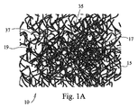
図1Aおよび図1Bは、本発明の開示に係る第1の実施形態の繊維状足場10を示している。足場10は、三次元多孔質支持足場または母材(three-dimensional porous support scaffold or matrix)10を規定する絡まり合った複数の繊維15から構成されている。支持母材10は、絡み合ったまたは織り合わされているがそれらの交差点17で必ずしも融合していない生体活性ガラス繊維15から構成されている。よって、繊維15の少なくともいくつかはある自由度で互いに移動して、本質的に動的な支持ウェブ(支持織物:support web)10を生じてもよい。得られた動的な繊維状足場10の支柱(struts)19として使用される繊維15の組成は、典型的な生体活性ガラス、セラミックまたはガラス−セラミックの製剤(formulations)であり、様々な繊維径および構造寸法(construct size)の中で足場繊維15が生体活性の特質を有することで一般に特徴付けられる。
1A and 1B show a
動的足場10を規定する繊維15の直径は、繊維15をそれらの交差点15で焼結、融合、またその他の付着することなしに、ただし必要なら、足場10をさらに固くするためにいくつかのそのような融合または付着を用いてもよいが、得られた三次元足場10それ自身の固有の絡まり合いを可能にするために、典型的には著しく小さい。従って、足場10は完全にばらばらにならないように自己拘束されているが、組織形成とそれによる増殖に対して十分な支持を提供しながら柔軟性が残るように足場10にその動的な性質を付与するために、支持支柱19を規定する個々の繊維15は、短い距離だけ互いに自由に動く。
The diameters of the
以下に詳細に説明するように、実質的に1マイクロメートル(1000ナノメートル)未満の直径を有することで特徴付けられる複数の繊維15は、実質的に100ナノメートル未満の直径を有することで特徴付けられる複数の繊維15と同様に、動的な足場10を形成するのに十分である。足場10はまた、多様な直径分布を有する複数の繊維15から構成されてもよく、直径の組合せは、動的な柔軟性、構造的支持(structural support)、内部空隙の寸法(internal void size)、空隙の分布、圧縮率、溶解速度および吸収率などの特定の組合せを生成するために用いられる。例えば、いくつかの繊維15は、初期の骨成長を誘導するために、反応が早く迅速に骨に吸収されてもよい。さらに、他の繊維15または粒子などの骨移植材料の残った材料は、より長い時間にわたって吸収されて、先に吸収された材料がなくなった後に骨成長を支え続けるように設計されてもよい。骨成長活性の第1バースト(first burst)後に手術部位が十分に治癒しない場合、このようなタイプの層状または段階的な吸収は臨床的に重要であるだろう。吸収の様々なレベルが発生するように提供することにより、材料は、治癒プロセスにわたるよりよい制御を可能にし、「全か無か」の状況を回避することができる。
As described in detail below, the plurality of
典型的には、構造物内にある繊維径はナノレベルから始まる範囲で変動し(range)、ここでナノファイバは1ミクロン未満(サブミクロン)、約100ミクロン以下の直径の繊維と規定されており、より典型的には、繊維径は約0.005ミクロン〜約10ミクロンの範囲で変動し、さらに典型的には、繊維径は約0.05ミクロン〜約6ミクロンの範囲で変動し、さらにより典型的には、繊維径は、0.5ミクロン〜約20ミクロンの範囲で変動し、さらに典型的には、繊維径は、約1ミクロン〜約6ミクロンの範囲で変動する。すべてのケースにおいて、必用に応じて、得られる足場10の1つ以上の特徴を変化させるために所定量のより大きい繊維(larger fibers)を加えてもよい。より小さい(典型的には10マイクロメートル未満の)直径の繊維15の量が減って、足場構造体(scaffolding construct)10の多くが比較的大きい直径の繊維15を含むにつれ、構造体10全体は典型的には自己束縛が低下する傾向を示すことに注目すべきである。したがって、構成する繊維15の相対的な直径およびアスペクト比を変更することにより、得られる足場構造体(scaffold structure)10は、高い又は低い柔軟性を有するように、および高い又は低い荷重支持剛性(load-bearing rigidity)を有するように調節されてもよい。さらに、細胞の付着と反応性に利用可能な表面積を著しく高めるために、繊維15は特定の寸法で、例えばナノスケールの大きさで構成されてもよい。ある実施形態では、骨移植材料は少なくとも1つのナノファイバを含んでいる。
Typically, the fiber diameter within a structure ranges from a nano level, where nanofibers are defined as fibers with a diameter of less than 1 micron (submicron) and about 100 microns or less. More typically, the fiber diameter varies from about 0.005 microns to about 10 microns, and more typically, the fiber diameter varies from about 0.05 microns to about 6 microns, Even more typically, the fiber diameter varies from 0.5 microns to about 20 microns, and more typically, the fiber diameter varies from about 1 micron to about 6 microns. In all cases, a predetermined amount of larger fibers may be added as needed to alter one or more characteristics of the resulting
動的な足場10の機構に影響を及ぼす1つの要因は、比較的小さい直径の繊維15の組み込みと得られるインプラント20である。多孔性の繊維足場10は、絡まり合って(interlocking)、もつれた(entangled)、配向した三次元繊維インプラント20が得られる様々な方法で形成することができる。
One factor that affects the mechanism of the
図1Aおよび図1Bに示すように、それらの繊維15は必ずしも連続していなくてもよく、短く不連続でもよく、または長く連続した繊維15と短く不連続の繊維15との組合せでもよい。繊維15は接触して交差点17を規定し、さらに細孔または空隙37も規定する。繊維の寸法および相互作用のモードを変化させることにより、得られたインプラントの細孔径分布のみならずその多孔性も制御できる。これにより、細孔径および分布の制御のみならずインプラントの全細孔(total porosity)(約95%以下またはそれ以上)の制御が可能になり、ナノ(約1ミクロン未満で、100ナノメートルほどの小ささ、またはより小さい細孔直径)、ミクロ(約1〜約10ミクロンの細孔直径)、メソ(約10〜約100ミクロンの細孔直径)、およびマクロ(約100ミクロンを越え、1mmほどの大きさ、またはより大きい細孔直径)の所定の細孔を備えるように材料を形成することができる。細孔37は、選択された形成技術のみならず、細孔径と細孔径分布、選択された繊維寸法範囲と寸法分布の機能により、典型的に約100ナノメートル〜約1mmの寸法の範囲で変動する。しかしながら、繊維と細孔径はこれらの範囲に限定されるものではなく、この説明はナノファイバおよびナノ細孔に焦点を当てているが、本発明の開示の骨移植材料はマクロサイズの繊維および細孔を等しく含んで繊維および細孔の直径の範囲を作り出すことが理解されるだろう。
As shown in FIGS. 1A and 1B, the
典型的なインプラント20の内部にある細孔径の1つの分布の効果と、その容積寄与(volumetric contribution)および表面積寄与(surface area contribution)の例を、図6Aおよび図6Bを参照しながら示し、それらはさらに以下で説明する。よって、得られたインプラントまたはデバイス20は、スパンレイド(spunlaid)又はスパンブロー(spun blown)処理、メルトブロー(melt blown)処理、湿式マット(wet laid matt)または「ガラス組織」処理等を経て形成された不織布であってもよく、またフェルト、ガーゼ、コットンボール、綿菓子等の特徴を有するように形成されてもよい。
An example of the effect of one distribution of pore sizes within a
典型的には、マクロ、メソ、およびミクロ細孔はデバイス20内に同時に発生し、より典型的には、それらは相互接続している。当業者は水銀圧入ポロシメトリー(mercury intrusion porosimetry)、ヘリウムピクノメトリー(helium pycnometry)、走査型電子顕微鏡等の種々の技術を用いて細孔を容易に特徴付けることができるので、本明細書では、細孔の各タイプを過度に定量化する必要はない。かなりの程度の特定タイプの細孔を有するとしてデバイス20を特徴付けるためには、所望の寸法範囲にあるわずかな孔よりは多くの存在が必要ではあるが、具体的な個数またはパーセンテージ(割合)は要求されない。むしろ、マクロ、メゾ、マイクロ、および/またはナノ細孔を決定するために、当業者による定性評価が使用されるだろう。いくつかの実施形態では、多孔質の繊維状インプラント20の全細孔(overall porosity)は、細孔容積で測定して一般的にパーセントで表現すると、比較的高くなるだろう。0パーセントの細孔容積は、完全にまたは理論的に高密度な材料を指している。言い換えれば、ゼロ細孔(zero porosity)の材料は、細孔を全く有していない。同様に、100パーセントの細孔容積は、「全て細孔」または空気を示しているだろう。当業者は、細孔容積の概念に精通しており、それを容易に計算して適用することができるだろう。
Typically, macro, meso, and micropores occur simultaneously in
骨移植インプラント20は、典型的には約30%を越える細孔容積を有しており、より典型的には約50%または60%を越える細孔容積を有していてもよく、または日常的に達成可能(routinely attainable)であってもよい。いくつかの実施形態では、足場インプラント20は、少なくとも約70%の細孔容積を有していてもよく、他の実施形態では、約75%または80%を越える細孔容積を有していてもよい。骨移植インプラントは、約90%〜97%を越える細孔容積を有するように調製されてもよい。
The
いくつかの骨移植インプラント20にとって、マクロ、メソ、およびミクロ細孔を、またいくつかのケースではナノ細孔を含む細孔勾配(porosity gradient)を有することは有利である。骨移植インプラント20が湿った場合、適切な圧縮抵抗および柔軟性を作り出すための繊維と粒子の組合せは維持される。骨移植インプラント20はまた、典型的には相互接続した細孔により特徴付けることもでき、そのことが増加した毛細管現象とウィッキング能(水分を運ぶ能力:wicking capability)に関連づけられる。そのような骨移植インプラント20は、時間をかけて徐放するために液体物質を迅速に運んで(wicking)保持することができなくてはならない。
For some
繊維15は、典型的には、例えば生理的変動、細胞の圧力差、脈動する治癒環境内での流体力学など、その環境内での変化に応じて足場10のわずかな柔軟性と動きを提供する非融合の結合(non-fused linkages)35を有している。この生体内環境は治癒プロセスの経過にわたって変化できるまたは変化するであろうし、数ヶ月またはそれ以上もの長い間にわたって持続するだろう。足場10は典型的には、治癒メカニズムが阻害されないように、治癒プロセスを通してその適切な支持特性と孔37の分布を保持する。治癒プロセスの間、絡まり合ってもつれた繊維15の母材によって規定された孔37は、体液と骨移植材料を新生骨成長の部位に運ぶのに役立ってもよい。液体はまた、治癒プロセスに対する動的な応答において、足場10そして特に孔37が寸法および形状を変えるように、生体活性ガラス等から形成された繊維15をゆっくり溶解する。
The
足場10は、典型的には、細胞、小分子、タンパク質、生理学的流体、血液、骨髄、酸素等にとって十分に透過性の三次元微細構造を備えており、足場10の全容積にわたって流れる。さらに、足場10の動的性質は、ミクロ環境を検出またはミクロ環境に応答する能力と、応力および圧力を加えた要素に基づいてミクロ環境内のその構造20を調節する能力とそれにを付与する。
The
さらに、足場10は典型的には、例えば、通常は骨、組織または同様の生物学的部位で見いだされるような空隙、孔または組織平面などの不規則な形状の欠損内に物理的に配置されたときに、骨移植材料またはデバイス20に適合性(compliance)するように十分な三次元形状を有している。デバイス20は典型的には、欠損に挿入されるときにいくらかの圧縮を受け、その一方で足場10の透過特性は維持されている。典型的には、骨空隙充填材の配置と同様に、デバイス20は欠損壁内の天然組織の2mm以内に留まる。
Furthermore, the
足場10から形成された骨移植インプラントまたはデバイス20は、フェルト、コットンボール、織物(textile fabrics)テキスタイル布地、ガーゼ等と類似して見えてもよい。これらの形態は、液体、タンパク質、骨髄穿刺液、細胞を運び、付着し、および包含する能力のみならず、それらの存在をかなりの容積の内部に保持する能力を有しているが、必ずしも全てが完全に保持されてなくてもよく、例えば、圧縮されたときにいくらかの液体が構造から放出されてもよい。
A bone graft implant or
骨移植インプラントまたはデバイス20の他の利点は、「親」の微細構造と同等のものを保持しながら、取り扱い、注入性(injectability)、配置、低侵襲注入(minimally invasive injection)、部位の適合性および保持力等を向上するために、動的な繊維状足場10をキャリアまたは修飾剤(modifiers)で修飾(modify)または混合するそれらの能力である。そのようなキャリアは、足場10のミクロスケール(典型的には100マイクロメートル未満のオーダー)の構造を維持しながら、デバイス20のマクロスケールの取り扱い特性を理想的に修飾する。これらのキャリアは、足場の形状、微細構造、化学的性質および/または生体活性の性質を実質的に変えることなく、急速に(典型的には約2週間未満で、より典型的には約2日未満で)吸収する。これらのキャリアは、ポロキサマー(polaxamer)、グリセロール、アルカリ性酸化物コポリマー(alkaline oxide copolymers)、骨髄穿刺液等が挙げられる。
Other advantages of the bone graft implant or
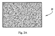
図2Aは、例えばストリップ状またはシート状のインプラント20の実施形態を示す。図2Bは、例えばコットンボールに類似の三次元構造の形態にされたインプラント20の実施形態を示す。ある実施例では、複数の絡まり合った繊維15が、コットンボールの一般的な外観を有するランダム配向の集合体(assemblage)20に紡糸またはブロー(spun or blown)される。繊維15は、典型的には、約10000nm(10マイクロメートル)以下の範囲において実質的に約1000nm(1マイクロメートル)未満の直径を有することで特徴付けられる。得られたコットンボールデバイス20は、使いやすい寸法に形成することができるが、典型的には圧縮されていない直径で約1〜約6センチメートルから形成されてもよく、そして最初の寸法の約1/2〜1/4に圧縮されてもよい。いくつかのケースでは、デバイス20は、(それが液体で湿って、その種の固定によってデバイスを所望の形状および密度にし、または真空圧縮されるまでは)圧縮力が取り除かれると、実質的に元の寸法および形状に戻ることができる。しかしながら、多くのケースで、デバイス20は変形されたまになるだろう。繊維の直径の範囲を、約10nm未満から約10ミクロンを越えるまで変更しながら、いくつかの繊維15の相対的な直径を変更することにより、「コットンボール」から「綿菓子」まで変動する構造体が作り出されでもよい。
FIG. 2A shows an embodiment of an
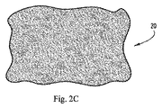
図2Cは、例えば織って作ったメッシュ状のインプラント20の実施形態を示す。ある実施例では、繊維15は、織って、編んで、またはその他の成形で、ガーゼ状の構成(consistency)を有する織物のデバイス20にしてもよい。繊維15は、典型的に直径が約1マイクロメートルを越えており、直径が約100マイクロメータより大きくてもよい。繊維が多少または完全に規則正しくても、繊維15のミクロスケールの方向は典型的にはランダムである。マクロスケールでは、繊維15は典型的にはより規則正しい。これらのデバイス20の構成は、自己拘束の効果を維持するために、それらの中に取り込まれた小さい繊維15の変更された量を有していてもよい。
FIG. 2C shows an embodiment of a mesh-
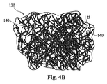
図3Aおよび図3Bは、本発明の開示の他の実施形態を示しており、図1Aおよび図1Bについて上述したような生理活性繊維状足場110であるが、ガラスの小球(microspheres)または粒子140を有する。ガラス粒子140は、典型的には、繊維115と同様にいくつかの一般的な組成物から形成されているが、その代わりに、他の異なる組成物から形成されてもよい。インプラント120内に粒子140が存在することの1つの利点は、インプラント120の全体的な圧縮抵抗への寄与である。インプラント120の1つの機能は、典型的には、骨の再生に供給される栄養液の吸収および保持であるので、液体が時期尚早に「絞り出される」ことのないように、インプラントに圧縮力に対するいくつかのレベルの抵抗を提供するのに有利である。粒子140(これは球体か粒子であるが)がインプラントを固くし、その他の点では主として絡まり合った繊維115から成る多孔質足場である。
3A and 3B illustrate another embodiment of the present disclosure, which is a bioactive
ガラス粒子140は、典型的にはほぼ球形であるが、他の規則的なまたは不規則な形状を有していてもよい。ガラス粒子140は典型的には寸法が変化し、およそ繊維115(より典型的には支柱119)の幅から、典型的な繊維幅を越える大きさのオーダーの直径までの範囲で変動する直径を有する。必要に応じて、粒子140はほぼ球体状から楕円体状(spheroidal)まで、または楕円形(elliptical)から不規則形状まで形状が変化してもよい。粒子140はさらに、ほぼ平坦なプレートレット(platelets)として形成してもよく、さらに、有効表面積と溶解速度とを増加するために、プレートレット(または他の形状物)は穿孔または内部空隙を有して成形されてもよい。同様に、骨細胞付着(bone cell attachmen)、粒子の塗布性(particulate coatability)等の要因に影響を与えるために、粒子140の形状を変更してもよい。
ある実施形態では、ガラス粒子140は約20ミクロン〜約1ミリメートルの平均直径を有していてもよい。別の実施形態では、粒子140は約300〜500ミクロンの平均直径を有していてもよい。さらに別の実施形態では、ガラス粒子140は約350ミクロンの平均直径を有していてもよい。
In certain embodiments,
繊維と同様に、生体活性ガラス粒子140は、有機酸(例えばギ酸、ヒアルロン酸など)、鉱物カルシウム源(例えばリン酸三カルシウム、ヒドロキシアパタイト、硫酸カルシウムなど)、抗菌剤、抗ウイルス剤、ビタミン、X線不透過剤(x-ray opacifiers)、または他のそのような材料によって被覆してもよい。小さい粒子は、繊維の交差点117の中または周囲にひっかかる(lodge)傾向にあり、大きい粒子は、足場120自身の名かに埋め込まれて繊維115のウェブによって適所に保持される傾向にある。細孔サイズの小球は、細孔137内にひっかかる傾向があるだろう。
Similar to the fiber, the
ガラス微粒子140は、所定の生理活性材料から構成されてもよく、ミネラル、骨成長培地(bone growth media)等の所定の選択を所定の速度で放出するように、足場110を体外(in vitro)に配置する所定の期間にわたって溶解するように調整することができる。生体活性ガラスの吸収速度を調整し、そしてミネラル等が身体に導入される速度(同様に、増加した圧縮抵抗を足場インプラント20に提供するためにどれくらい長く粒子140を利用可能にするか)を調節するために、ガラス微粒子140の組成、寸法、形状を変更してもよい。たとえば、所定の生体活性ガラスの組成と粒子容積において、不規則な形状の粒子140は球状の粒子140よりも広い表面積を有することになり、よってより急速に溶解するだろう。
The
さらに、ガラス粒子140は、所定の速度と所定の期間で骨再成部位の周囲に放出されるべき医薬品、抗生物質、抗ウイルス剤、ビタミン等の特定の混合物で充填された生体活性ガラス、ポリマー等の中空の小球であってもよい。放出速度と放出の持続時間は、粒子サイズ、細孔と肉厚等の関数のみならず、それらの分布関数であろう。
Furthermore, the
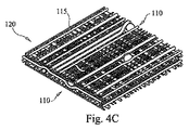
上述したように、骨移植材料の形状やテクスチャは、全体的な容積、表面積および柔軟性を最大にするようにランダムに構成することができ、または全く対称的に、生体活性ガラス繊維から、例えば、メッシュまたはマトリクスタイプのアセンブリなどのより固くかつ均一な配置に製造することができる。図4A〜図4Cに図示した非限定的な実施例で例示されているように、メッシュまたはマトリクスアセンブリでは、ガラス繊維は、指向的に(in a directional manner)柔軟性を制限する積層配置で配置することができ、または、繊維は、交互の層がお互いに交差する関係で層状にすることができる。図4Aでは、マトリクスアセンブリ110は、繊維115および粒子140を含む個々の層(discrete layers)を備えた秩序構成(ordered configuration)を有するように示されている。図4Bでは、マトリクスアセンブリは、繊維115および粒子140のランダム配置構成(randomly arranged configuration)を有するように示されている。図4Cでは、マトリクスアセンブリ110は、各層の全体にわたって繊維115と粒子140の間隔の違いにより、層が異なる細孔を有している構成を有するように示されている。すなわち、不均一に間隔をあけた繊維115および粒子140が原因で、細孔137の寸法はマトリクスアセンブリ全体で異なっている。本明細書の概念を図示する目的で、図4A〜図4Cは個々に整列した(discretely aligned)繊維115を示しているが、材料110の個々の層は組織化されずにランダムに配置された繊維115および粒子140を含んでいてもよいと理解されるべきである。
As mentioned above, the shape and texture of the bone graft material can be randomly configured to maximize the overall volume, surface area and flexibility, or quite symmetrically from bioactive glass fibers, for example Can be manufactured in a stiffer and uniform arrangement, such as mesh or matrix type assemblies. In the mesh or matrix assembly, as illustrated in the non-limiting example illustrated in FIGS. 4A-4C, the glass fibers are arranged in a stacked arrangement that limits flexibility in a directional manner. Or the fibers can be layered with alternating layers intersecting each other. In FIG. 4A, the
本発明の開示の利点は、外科医が使用する材料の同様に変更された機能性をもたらす多種多様な代わりの構成および構造配置である。図4A〜図4Cに図示するように、本発明の開示の骨移植材料は、生体活性ガラス繊維構造体の内部に、埋め込まれた生体活性ガラス粒子を含むことができる。量、寸法、および粒子の特徴によって決定されているような粒子を内包することにより、得られた骨移植材料の圧縮性、生体吸収性および多孔性に影響を与えることができる。骨生成と患者の回復を支援するために、例えばリン酸カルシウム(CaP)、カルシウム硫酸塩(CaS)、ヒドロキシアパタイト(HA)、カルボキシメチルセルロース(carboxymethycellulose:CMC)、コラーゲン、グリセリン、ゼラチンなどの追加の添加剤を、生理活性ガラス繊維の骨移植材料の変更された構造体のいずれかに含むこともできる。 An advantage of the present disclosure is a wide variety of alternative configurations and structural arrangements that provide similarly altered functionality of materials used by surgeons. As illustrated in FIGS. 4A-4C, the bone graft material of the present disclosure can include bioactive glass particles embedded within a bioactive glass fiber structure. Inclusion of particles as determined by the amount, size, and particle characteristics can affect the compressibility, bioabsorbability and porosity of the resulting bone graft material. Additional additives such as calcium phosphate (CaP), calcium sulfate (CaS), hydroxyapatite (HA), carboxymethycellulose (CMC), collagen, glycerin, gelatin to aid bone formation and patient recovery Can be included in any of the modified structures of bioactive glass fiber bone graft material.
ある実施形態では、骨移植材料の表面積は、材料の構造的マトリクス内への骨内部成長を高めるために最大にされます。別の有用な可変(useful variable)は、選択された細胞が材料内に浸透する深さを制御する細胞フィルタとして機能するように例えばナノ、ミクロ、メゾおよびマクロ細孔などの異なる細孔の(複数の)層を提供するために、選択的に構成される(composed and configured)骨移植材料の性能である。骨移植材料の調製は、異なる断面直径、形状および/または組成を有する生体活性ガラスの繊維および/または粒子を含むように選択的に変更できるので、差動的吸収能を備えた骨移植材料を製造するために材料特性を調整することができる。この特徴により、特定の状況または患者のニーズに合わせて外科医が骨移植材料を選択することが可能になる。材料の生体活性ガラスマトリクス中への骨内部成長の早さを制御することにより、外科医は、個々の患者の特定のニーズに適した骨移植材料を選択する際に、ほぼ無制限の適応性を発揮することができます。 In certain embodiments, the surface area of the bone graft material is maximized to enhance bone ingrowth into the structural matrix of the material. Another useful variable is that of different pores (for example nano, micro, meso and macropores) to act as a cell filter that controls the depth at which selected cells penetrate into the material. The performance of bone graft materials that are selectively and configured to provide multiple layers. The preparation of the bone graft material can be selectively modified to include bioactive glass fibers and / or particles having different cross-sectional diameters, shapes and / or compositions, so that Material properties can be adjusted to produce. This feature allows the surgeon to select a bone graft material for a particular situation or patient need. By controlling the speed of bone ingrowth into the bioactive glass matrix of the material, the surgeon has almost unlimited flexibility in selecting a bone graft material suitable for the specific needs of the individual patient. can.
別の実施形態では、生体活性ガラスは、カルシウムをストロンチウムに部分的に置き換えて処方された。カルシウムをストロンチウムに部分的な置換することにより、減少した吸収/反応速度と、さらに増加した放射線濃度または放射線不透過性(radioopacity)を有する生体活性ガラスが生成される。したがって、生体活性ガラスは、長期間にわたって体内に存在し続けて、そしてより容易に視認できるX線ターゲットも提供する。 In another embodiment, the bioactive glass was formulated with partial replacement of calcium with strontium. Partial replacement of calcium with strontium produces a bioactive glass with reduced absorption / reaction rate and further increased radiation concentration or radiopacity. Thus, the bioactive glass also provides an X-ray target that remains in the body for an extended period of time and is more easily visible.
別の実施形態では、銀(または他の抗菌材料)を生体活性ガラス繊維足場の構造マトリクスに組み込むことができる。銀は抗菌材料であり、生体活性ガラス材料の固有の抗菌性能を高めるさせる。典型的には、非常に細い繊維が移植部位で溶解して銀がすぐに放出されるように、銀は非常に細い生体活性ガラス繊維へのドーパントとして追加されて、銀が抗菌剤として作用して手術直後の感染症を防ぐことができ、その一方、残りの足場材料は自身の仕事をこなす。代わりに、Agを繊維として導入して生体活性ガラス繊維と折り合わしても、上述のガラス粒子と類似の粒子として導入しても、またその他で導入してもよい。もちろん、アルカリ性(8〜10の高pH)ガラスを作り出すために、繊維を形成する生体活性ガラスの組成を変えることで、抗菌性能を有する材料を提供できるだろう。 In another embodiment, silver (or other antimicrobial material) can be incorporated into the structural matrix of the bioactive glass fiber scaffold. Silver is an antibacterial material and enhances the inherent antibacterial performance of bioactive glass materials. Typically, silver is added as a dopant to the very thin bioactive glass fibers so that the very thin fibers dissolve at the implantation site and silver is released immediately, and the silver acts as an antimicrobial agent. Can prevent infections immediately after surgery, while the rest of the scaffold material does its job. Alternatively, Ag may be introduced as a fiber and combined with the bioactive glass fiber, or may be introduced as a particle similar to the glass particles described above, or may be introduced elsewhere. Of course, to create an alkaline (8-10 high pH) glass, the composition of the bioactive glass forming the fiber could be changed to provide a material with antibacterial performance.
この発明の1つの利点は、様々な形状に容易にモールド成形できることである。材料を機能トレイ(functional tray)内にこん包(packaging)することにより、ここでトレイはモールド型として働いて、手術室内で材料を様々な形状で提供できる。特に、材料は、血液、生理食塩水、骨髄、その他の天然の体液等を加えたときに、凝集塊(cohesive mass)になる。 One advantage of the present invention is that it can be easily molded into a variety of shapes. By packaging the material in a functional tray, the tray now acts as a mold and can provide the material in various shapes within the operating room. In particular, the material becomes a cohesive mass when blood, saline, bone marrow, other natural body fluids, and the like are added.
ある実施形態では、図5A〜図5Dに示すように、骨移植材料は外科キット200の一部として提供される。キット200は、凹部またはくぼみ212、より典型的には入れ子状になった凹部(nested recesses)のセットを有しており骨移植材10、110を保管し、保持し、そして操作するためのトレイ部210と、トレイ部210に封止的に嵌め込むための蓋部220とを含む。トレイ部210および蓋部220は、典型的には熱可塑性材料から形成されているが、代わりに任意の使いやすい材料から作ってもよい。
In certain embodiments, the bone graft material is provided as part of a
深い凹部チャンバ212は、そこに装填された(so-loaded)骨移植材料が同様に単純な形状を有するように、典型的には、例えば矩形ブロックまたはくさび形などの単純な形状を有している。骨移植材10、110は、典型的には、絡み合ったまたは織り合わされた生体活性ガラス繊維の塊(mass)として提供される。生体活性ガラス繊維は、骨の空洞(bony cavity)に外科的に配置する準備のできている形式(format)(例えば、織物またはメッシュ形式など)で提供されてもよく、または、生体活性ガラスの塊をより柔軟で構造的に一体(structurally unitary)にレンダリングするのを支援するために、例えば生理食塩水、グリセロール、ゼラチン、血漿、またはコラーゲンゲルもしくはチップなどの液体の添加を必要とする配置前に、追加の調製を必要とする様式(例えば、より緩い絡み合いの形式)で提供されてもよい。そのような液体は、任意でキットパッケージ200に含まれてもよく、または分離して提供されてもよい。
The
ある実施例では、キット200は、トレイ本体210と、トレイ本体に嵌め込むことのできる蓋200とを含んで提供される。トレイ本体210は、生体活性ガラス繊維10の容積(volume)を含むための1つ以上の凹部212を含んでいる。生体活性ガラス繊維の容積は、織られ、編まれ、絡み合わされてもよく、または緩いスタックとして提供されてもよい。生体活性ガラス繊維の容積は、任意で、例えば抗菌性銀、ポリマー、または代わりのガラス組成物などの他の組成の繊維を含むことができ、また、任意で、粒状物質もしくは同じ生体活性ガラス組成の粒子、または例えば代わりのガラス、金属、金属酸化物、医薬品、栄養素、および/または抗菌剤等の代わりの組成を含むこともできる。キットはまた、生体活性ガラスの容積と混合するために生理食塩水またはコラーゲンゲルなどの液体を任意で含んでいてもよい。
In one embodiment, a
操作の際は、外科医はキット200の蓋220を取り除いて、含まれている生体活性ガラス材10の部分(portion)を取り外す。その後、骨の空洞に挿入するために、外科医が生体活性ガラス材料を成形し、サイズに切って(sized)もよい。所望する程度の柔軟性および/または構造的統一性(structural integrity)を達成するために、このプロセスは、例えば生理食塩水、コラーゲンゲル、血漿、血液などの適切な液体を生体活性ガラス材料に添加することを含んでもよい。生体活性ガラス材料が所望のサイズと形状に成形されたら、それを骨の空洞に挿入する。このプロセスは、単一の操作として、または一連の工程として行うことができる。
In operation, the surgeon removes the
図6A及び図6Bは、細孔径分布に基づいた骨移植材料の実施形態の容積寄与(volumetric contribution)と表面積寄与(surface area contribution)を示している。記述してきたように、ある実施形態では、インプラント20の骨移植材料は、例えばナノ、ミクロメゾ、マイクロ多孔性などの様々な多孔性を有する構造を有している。図6A及び図6Bに示すように、メソ細孔とミクロ細孔は、骨移植材料の容積の大部分に寄与するが、ナノ細孔は、骨移植材料によって提供される表面積のかなりの大部分に貢献します。つまり、所定の容積に対する高表面をより高く(higher surface higher)得るために、実施形態は、所定の容積に対して、ナノ細孔を含む細孔分布(porosity distribution)を利用してもよい。もちろん、実施形態によって、これらおよび他の特徴および利点が提供することができる。
6A and 6B show the volumetric and surface area contributions of an embodiment of a bone graft material based on pore size distribution. As described, in certain embodiments, the bone graft material of the
図7は、本発明の開示の実施形態の繊維の1日後および3日後の経時顕微鏡写真であり、一方、図8は、本発明の開示の実施形態の繊維を37℃の擬似体液に浸漬して3日後の経時顕微鏡写真である。 FIG. 7 is a time-lapse micrograph after 1 day and 3 days of the fiber of the disclosed embodiment of the present invention, while FIG. Is a time-lapse micrograph after 3 days.
図9は、本発明の開示のガラス繊維足場で2日、4日および6日の培養をした骨芽細胞を示す一連の経時電子顕微鏡写真(SEMs)である。図示されているように、6日のインキュベーションの間に細胞密度が増加している。図10は、1つの足場あたり100000個のMC3T3−E1細胞を初期播種(initial seeding)して2日、4日および6日の図9のガラス繊維足場で示された骨芽細胞の増殖のグラフを示している。図11は、間葉幹細胞(mesenchymal stem cells)が播種された繊維の顕微鏡写真を示す。このような細胞は、骨芽細胞の増殖と分化の骨刺激効果を支援することができる。この効果は、DNA含有量の測定と、オステオカルシンおよびアルカリホスファターゼの濃度レベルの上昇した存在とに基づいて測定することができる。
FIG. 9 is a series of time-lapse electron micrographs (SEMs) showing osteoblasts cultured for 2, 4, and 6 days on a glass fiber scaffold of the present disclosure. As shown, the cell density increases during the 6 day incubation. FIG. 10 is a graph of osteoblast proliferation shown in the glass fiber scaffold of FIG. 9 at
<比較動物実験>
図12〜図16は、本発明の開示の繊維性骨移植材料の実施形態の哺乳動物(具体的にはこのケースではウサギ)での試験の結果を示す。試験では、直径約5mm、長さ10mmの大きさを有する両側大腿骨遠位欠損(bilateral distal femoral bone defect)を作成した。本開示の骨移植材料の実施形態に加えて、この比較研究において、市販の骨移植代用品のプロダクト#1(Product #1)も共に試験を行った。プロダクト#1は、ケイ酸塩の代用骨移植材料(マサチューセッツ州フォックスボローのApaTech社から入手可能なACTIFUSE(商標))であり、プロダクト#2は、代用合成骨移植(ペンシルバニア州モルバーンのOrthovita社から入手可能なVITOSS(商標))である。特に、図12は、骨移植材料の実施形態とプロダクト1および2との性能を6週、12週、24週で比較する哺乳動物で行われた試験からの一連のX線画像を示している。図13は、骨移植材の実施形態とプロダクト1および2との性能を比較する哺乳動物で行われた試験からの別の一連の画像を示している。図14は、哺乳動物の試験中において、骨移植材の実施形態とプロダクト1および2とによって示された新生骨の成長の組織形態計測的な比較(histomorphometric comparison)を示している。図15は、哺乳動物の試験中において、骨移植材の実施形態とプロダクト1および2とによる時間をかけて残った残留材料の組織形態計測的な比較を示している。図16は、哺乳動物の試験中において、骨移植材の実施形態とプロダクト1および2とによって示された機械的強度の組織形態計測的な比較を示している。
<Comparison animal experiment>
12-16 show the results of testing mammals (specifically rabbits in this case) of embodiments of the disclosed fibrous bone graft material of the present invention. In the test, a bilateral distal femoral bone defect having a diameter of about 5 mm and a length of 10 mm was created. In addition to the bone graft material embodiment of the present disclosure, a commercial bone graft
本発明の開示の骨移植材は、骨移植に使用するために記述されているが、本発明の開示の移植材料はまた、軟組織や軟骨の修復にも同様に適用できると考えられる。したがって、本明細書で提供される繊維状移植材料の用途は、さまざまな医療用途、特に新しい結合組織の形成が望まれている場合を含むことができる。 Although the disclosed bone graft material has been described for use in bone grafting, the disclosed graft material is also considered applicable to soft tissue and cartilage repair as well. Accordingly, the applications of the fibrous graft material provided herein can include a variety of medical applications, particularly where it is desired to form new connective tissue.
本発明の開示は、図面と上記記載において詳細に図示および説明してきたが、それらは実例であって制限的な性質のものではないと見なされるべきである。実施形態は、ベストモード要件および実施可能要件を満足するように上記明細書で図示され説明されていると理解される。当業者はほぼ無限の数のわずかな変化および変更を上記の実施形態に容易に行えること、およびそのような実施形態のバリエーションの全てを本明細書中で説明しようとするのは現実的ではないことが理解される。従って、本発明の開示の精神内にある全ての変化および変更は保護されることが望まれていると理解される。 While the disclosure of the present invention has been illustrated and described in detail in the drawings and foregoing description, they are to be considered as illustrative and not restrictive in nature. It is understood that the embodiments are illustrated and described in the above specification to satisfy best mode requirements and enablement requirements. Those skilled in the art can readily make an almost infinite number of minor changes and modifications to the above-described embodiments, and it is not realistic to attempt to describe all such variations of these embodiments herein. It is understood. Accordingly, it is understood that all changes and modifications within the spirit of the present disclosure are desired to be protected.
Claims (32)
骨移植インプラントを提供する工程であって、前記骨移植インプラントは、重なって絡まり合った複数の生体活性ガラス繊維を含む多孔質足場と、前記足場の全体に分散した細孔とを含み、前記繊維は約5ナノメートル〜約100マイクロメートルに及ぶ繊維径によって特徴付けられ、前記細孔は、約100ナノメートル〜約1ミリメートルに及ぶ細孔径によって特徴付けられている、骨移植インプラントを提供する工程と、治療される解剖学的部位を、骨移植インプラントを受容するように準備する工程と、前記骨移植インプラントを、前記骨欠損に導入する工程と、を含むことを特徴とする骨欠損の治療方法。 A method for treating bone defects,
Providing a bone graft implant, the bone graft implant comprising a porous scaffold comprising a plurality of overlapping and entangled bioactive glass fibers, and pores dispersed throughout the scaffold, wherein the fibers Providing a bone graft implant characterized by a fiber diameter ranging from about 5 nanometers to about 100 micrometers, wherein the pores are characterized by pore diameters ranging from about 100 nanometers to about 1 millimeter. Treating a bone defect comprising: preparing an anatomical site to be treated to receive a bone graft implant; and introducing the bone graft implant into the bone defect. Method.
Applications Claiming Priority (3)
| Application Number | Priority Date | Filing Date | Title |
|---|---|---|---|
| US25628709P | 2009-10-29 | 2009-10-29 | |
| US61/256,287 | 2009-10-29 | ||
| PCT/US2010/054542 WO2011053725A1 (en) | 2009-10-29 | 2010-10-28 | Bone graft material |
Publications (2)
| Publication Number | Publication Date |
|---|---|
| JP2013509261A true JP2013509261A (en) | 2013-03-14 |
| JP2013509261A5 JP2013509261A5 (en) | 2013-12-19 |
Family
ID=43922550
Family Applications (1)
| Application Number | Title | Priority Date | Filing Date |
|---|---|---|---|
| JP2012537073A Pending JP2013509261A (en) | 2009-10-29 | 2010-10-28 | Bone grafting material |
Country Status (9)
| Country | Link |
|---|---|
| US (1) | US20110144764A1 (en) |
| EP (1) | EP2493424A4 (en) |
| JP (1) | JP2013509261A (en) |
| KR (1) | KR20120101021A (en) |
| CN (1) | CN102596102A (en) |
| AU (1) | AU2010313347A1 (en) |
| CA (1) | CA2779103A1 (en) |
| MX (1) | MX2012004919A (en) |
| WO (1) | WO2011053725A1 (en) |
Cited By (1)
| Publication number | Priority date | Publication date | Assignee | Title |
|---|---|---|---|---|
| WO2014192803A1 (en) * | 2013-05-31 | 2014-12-04 | 学校法人同志社 | Tissue regeneration matrix |
Families Citing this family (442)
| Publication number | Priority date | Publication date | Assignee | Title |
|---|---|---|---|---|
| US20070084897A1 (en) | 2003-05-20 | 2007-04-19 | Shelton Frederick E Iv | Articulating surgical stapling instrument incorporating a two-piece e-beam firing mechanism |
| US9060770B2 (en) | 2003-05-20 | 2015-06-23 | Ethicon Endo-Surgery, Inc. | Robotically-driven surgical instrument with E-beam driver |
| US8215531B2 (en) | 2004-07-28 | 2012-07-10 | Ethicon Endo-Surgery, Inc. | Surgical stapling instrument having a medical substance dispenser |
| US11896225B2 (en) | 2004-07-28 | 2024-02-13 | Cilag Gmbh International | Staple cartridge comprising a pan |
| US7669746B2 (en) | 2005-08-31 | 2010-03-02 | Ethicon Endo-Surgery, Inc. | Staple cartridges for forming staples having differing formed staple heights |
| US10159482B2 (en) | 2005-08-31 | 2018-12-25 | Ethicon Llc | Fastener cartridge assembly comprising a fixed anvil and different staple heights |
| US11246590B2 (en) | 2005-08-31 | 2022-02-15 | Cilag Gmbh International | Staple cartridge including staple drivers having different unfired heights |
| US7934630B2 (en) | 2005-08-31 | 2011-05-03 | Ethicon Endo-Surgery, Inc. | Staple cartridges for forming staples having differing formed staple heights |
| US9237891B2 (en) | 2005-08-31 | 2016-01-19 | Ethicon Endo-Surgery, Inc. | Robotically-controlled surgical stapling devices that produce formed staples having different lengths |
| US11484312B2 (en) | 2005-08-31 | 2022-11-01 | Cilag Gmbh International | Staple cartridge comprising a staple driver arrangement |
| US20070106317A1 (en) | 2005-11-09 | 2007-05-10 | Shelton Frederick E Iv | Hydraulically and electrically actuated articulation joints for surgical instruments |
| US20150352247A1 (en) * | 2014-06-04 | 2015-12-10 | Qiang Jie | Compositions and methods for regeneration of hard tissues |
| US10524916B2 (en) | 2006-01-11 | 2020-01-07 | Novabone Products, Llc | Resorbable macroporous bioactive glass scaffold and method of manufacture |
| US8820603B2 (en) | 2006-01-31 | 2014-09-02 | Ethicon Endo-Surgery, Inc. | Accessing data stored in a memory of a surgical instrument |
| US11224427B2 (en) | 2006-01-31 | 2022-01-18 | Cilag Gmbh International | Surgical stapling system including a console and retraction assembly |
| US8708213B2 (en) | 2006-01-31 | 2014-04-29 | Ethicon Endo-Surgery, Inc. | Surgical instrument having a feedback system |
| US7845537B2 (en) | 2006-01-31 | 2010-12-07 | Ethicon Endo-Surgery, Inc. | Surgical instrument having recording capabilities |
| US20110290856A1 (en) | 2006-01-31 | 2011-12-01 | Ethicon Endo-Surgery, Inc. | Robotically-controlled surgical instrument with force-feedback capabilities |
| US11278279B2 (en) | 2006-01-31 | 2022-03-22 | Cilag Gmbh International | Surgical instrument assembly |
| US20120292367A1 (en) | 2006-01-31 | 2012-11-22 | Ethicon Endo-Surgery, Inc. | Robotically-controlled end effector |
| US20110024477A1 (en) | 2009-02-06 | 2011-02-03 | Hall Steven G | Driven Surgical Stapler Improvements |
| US8186555B2 (en) | 2006-01-31 | 2012-05-29 | Ethicon Endo-Surgery, Inc. | Motor-driven surgical cutting and fastening instrument with mechanical closure system |
| US7753904B2 (en) | 2006-01-31 | 2010-07-13 | Ethicon Endo-Surgery, Inc. | Endoscopic surgical instrument with a handle that can articulate with respect to the shaft |
| US11793518B2 (en) | 2006-01-31 | 2023-10-24 | Cilag Gmbh International | Powered surgical instruments with firing system lockout arrangements |
| US8992422B2 (en) | 2006-03-23 | 2015-03-31 | Ethicon Endo-Surgery, Inc. | Robotically-controlled endoscopic accessory channel |
| US8322455B2 (en) | 2006-06-27 | 2012-12-04 | Ethicon Endo-Surgery, Inc. | Manually driven surgical cutting and fastening instrument |
| US10568652B2 (en) | 2006-09-29 | 2020-02-25 | Ethicon Llc | Surgical staples having attached drivers of different heights and stapling instruments for deploying the same |
| US8348131B2 (en) | 2006-09-29 | 2013-01-08 | Ethicon Endo-Surgery, Inc. | Surgical stapling instrument with mechanical indicator to show levels of tissue compression |
| US8684253B2 (en) | 2007-01-10 | 2014-04-01 | Ethicon Endo-Surgery, Inc. | Surgical instrument with wireless communication between a control unit of a robotic system and remote sensor |
| US8652120B2 (en) | 2007-01-10 | 2014-02-18 | Ethicon Endo-Surgery, Inc. | Surgical instrument with wireless communication between control unit and sensor transponders |
| US11291441B2 (en) | 2007-01-10 | 2022-04-05 | Cilag Gmbh International | Surgical instrument with wireless communication between control unit and remote sensor |
| US11039836B2 (en) | 2007-01-11 | 2021-06-22 | Cilag Gmbh International | Staple cartridge for use with a surgical stapling instrument |
| US8540128B2 (en) | 2007-01-11 | 2013-09-24 | Ethicon Endo-Surgery, Inc. | Surgical stapling device with a curved end effector |
| US8727197B2 (en) | 2007-03-15 | 2014-05-20 | Ethicon Endo-Surgery, Inc. | Staple cartridge cavity configuration with cooperative surgical staple |
| US8893946B2 (en) | 2007-03-28 | 2014-11-25 | Ethicon Endo-Surgery, Inc. | Laparoscopic tissue thickness and clamp load measuring devices |
| US11564682B2 (en) | 2007-06-04 | 2023-01-31 | Cilag Gmbh International | Surgical stapler device |
| US8931682B2 (en) | 2007-06-04 | 2015-01-13 | Ethicon Endo-Surgery, Inc. | Robotically-controlled shaft based rotary drive systems for surgical instruments |
| US7753245B2 (en) | 2007-06-22 | 2010-07-13 | Ethicon Endo-Surgery, Inc. | Surgical stapling instruments |
| US11849941B2 (en) | 2007-06-29 | 2023-12-26 | Cilag Gmbh International | Staple cartridge having staple cavities extending at a transverse angle relative to a longitudinal cartridge axis |
| US8758391B2 (en) | 2008-02-14 | 2014-06-24 | Ethicon Endo-Surgery, Inc. | Interchangeable tools for surgical instruments |
| US8636736B2 (en) | 2008-02-14 | 2014-01-28 | Ethicon Endo-Surgery, Inc. | Motorized surgical cutting and fastening instrument |
| US8573465B2 (en) | 2008-02-14 | 2013-11-05 | Ethicon Endo-Surgery, Inc. | Robotically-controlled surgical end effector system with rotary actuated closure systems |
| US9179912B2 (en) | 2008-02-14 | 2015-11-10 | Ethicon Endo-Surgery, Inc. | Robotically-controlled motorized surgical cutting and fastening instrument |
| US7866527B2 (en) | 2008-02-14 | 2011-01-11 | Ethicon Endo-Surgery, Inc. | Surgical stapling apparatus with interlockable firing system |
| RU2493788C2 (en) | 2008-02-14 | 2013-09-27 | Этикон Эндо-Серджери, Инк. | Surgical cutting and fixing instrument, which has radio-frequency electrodes |
| US7819298B2 (en) | 2008-02-14 | 2010-10-26 | Ethicon Endo-Surgery, Inc. | Surgical stapling apparatus with control features operable with one hand |
| US20130153641A1 (en) | 2008-02-15 | 2013-06-20 | Ethicon Endo-Surgery, Inc. | Releasable layer of material and surgical end effector having the same |
| US11272927B2 (en) | 2008-02-15 | 2022-03-15 | Cilag Gmbh International | Layer arrangements for surgical staple cartridges |
| US9005230B2 (en) | 2008-09-23 | 2015-04-14 | Ethicon Endo-Surgery, Inc. | Motorized surgical instrument |
| US8210411B2 (en) | 2008-09-23 | 2012-07-03 | Ethicon Endo-Surgery, Inc. | Motor-driven surgical cutting instrument |
| US9386983B2 (en) | 2008-09-23 | 2016-07-12 | Ethicon Endo-Surgery, Llc | Robotically-controlled motorized surgical instrument |
| US11648005B2 (en) | 2008-09-23 | 2023-05-16 | Cilag Gmbh International | Robotically-controlled motorized surgical instrument with an end effector |
| US8608045B2 (en) | 2008-10-10 | 2013-12-17 | Ethicon Endo-Sugery, Inc. | Powered surgical cutting and stapling apparatus with manually retractable firing system |
| KR101726885B1 (en) * | 2008-10-17 | 2017-04-26 | 내셔널 유니버시티 오브 싱가포르 | Resorbable scaffolds for bone repair and long bone tissue engineering |
| US8517239B2 (en) | 2009-02-05 | 2013-08-27 | Ethicon Endo-Surgery, Inc. | Surgical stapling instrument comprising a magnetic element driver |
| CA2751664A1 (en) | 2009-02-06 | 2010-08-12 | Ethicon Endo-Surgery, Inc. | Driven surgical stapler improvements |
| US8444036B2 (en) | 2009-02-06 | 2013-05-21 | Ethicon Endo-Surgery, Inc. | Motor driven surgical fastener device with mechanisms for adjusting a tissue gap within the end effector |
| EP2485780A4 (en) * | 2009-10-07 | 2014-05-21 | Bio2 Technologies Inc | Devices and methods for tissue engineering |
| US8220688B2 (en) | 2009-12-24 | 2012-07-17 | Ethicon Endo-Surgery, Inc. | Motor-driven surgical cutting instrument with electric actuator directional control assembly |
| US8851354B2 (en) | 2009-12-24 | 2014-10-07 | Ethicon Endo-Surgery, Inc. | Surgical cutting instrument that analyzes tissue thickness |
| US8783543B2 (en) | 2010-07-30 | 2014-07-22 | Ethicon Endo-Surgery, Inc. | Tissue acquisition arrangements and methods for surgical stapling devices |
| US8468673B2 (en) | 2010-09-10 | 2013-06-25 | Bio2 Technologies, Inc. | Method of fabricating a porous orthopedic implant |
| US9861361B2 (en) | 2010-09-30 | 2018-01-09 | Ethicon Llc | Releasable tissue thickness compensator and fastener cartridge having the same |
| US9839420B2 (en) | 2010-09-30 | 2017-12-12 | Ethicon Llc | Tissue thickness compensator comprising at least one medicament |
| US11298125B2 (en) | 2010-09-30 | 2022-04-12 | Cilag Gmbh International | Tissue stapler having a thickness compensator |
| US9320523B2 (en) | 2012-03-28 | 2016-04-26 | Ethicon Endo-Surgery, Llc | Tissue thickness compensator comprising tissue ingrowth features |
| US10945731B2 (en) | 2010-09-30 | 2021-03-16 | Ethicon Llc | Tissue thickness compensator comprising controlled release and expansion |
| US9629814B2 (en) | 2010-09-30 | 2017-04-25 | Ethicon Endo-Surgery, Llc | Tissue thickness compensator configured to redistribute compressive forces |
| US11925354B2 (en) | 2010-09-30 | 2024-03-12 | Cilag Gmbh International | Staple cartridge comprising staples positioned within a compressible portion thereof |
| US11812965B2 (en) | 2010-09-30 | 2023-11-14 | Cilag Gmbh International | Layer of material for a surgical end effector |
| US9517063B2 (en) | 2012-03-28 | 2016-12-13 | Ethicon Endo-Surgery, Llc | Movable member for use with a tissue thickness compensator |
| US9364233B2 (en) | 2010-09-30 | 2016-06-14 | Ethicon Endo-Surgery, Llc | Tissue thickness compensators for circular surgical staplers |
| US9295464B2 (en) | 2010-09-30 | 2016-03-29 | Ethicon Endo-Surgery, Inc. | Surgical stapler anvil comprising a plurality of forming pockets |
| US8695866B2 (en) | 2010-10-01 | 2014-04-15 | Ethicon Endo-Surgery, Inc. | Surgical instrument having a power control circuit |
| RU2606493C2 (en) | 2011-04-29 | 2017-01-10 | Этикон Эндо-Серджери, Инк. | Staple cartridge, containing staples, located inside its compressible part |
| US9072535B2 (en) | 2011-05-27 | 2015-07-07 | Ethicon Endo-Surgery, Inc. | Surgical stapling instruments with rotatable staple deployment arrangements |
| US11207064B2 (en) | 2011-05-27 | 2021-12-28 | Cilag Gmbh International | Automated end effector component reloading system for use with a robotic system |
| RU2018129876A (en) * | 2011-10-24 | 2019-03-20 | СИНЕРДЖИ БАЙОМЕДИКАЛ ЭлЭлСи | COMPOSITIONS AND THEIR APPLICATION IN BONE REGENERATION |
| US9044230B2 (en) | 2012-02-13 | 2015-06-02 | Ethicon Endo-Surgery, Inc. | Surgical cutting and fastening instrument with apparatus for determining cartridge and firing motion status |
| US11225430B2 (en) | 2012-03-26 | 2022-01-18 | Steven Jung | Bioactive glass scaffolds, and method of making |
| US9045362B2 (en) | 2013-03-15 | 2015-06-02 | Mosci Corp. | Bioactive glass scaffolds, and method of making |
| US8449904B1 (en) | 2012-03-26 | 2013-05-28 | Mosci, Corp. | Bioactive glass scaffolds, and method of making |
| CN104321024B (en) | 2012-03-28 | 2017-05-24 | 伊西康内外科公司 | Tissue thickness compensator comprising a plurality of layers |
| MX353040B (en) | 2012-03-28 | 2017-12-18 | Ethicon Endo Surgery Inc | Retainer assembly including a tissue thickness compensator. |
| CN104334098B (en) | 2012-03-28 | 2017-03-22 | 伊西康内外科公司 | Tissue thickness compensator comprising capsules defining a low pressure environment |
| WO2013153185A1 (en) * | 2012-04-11 | 2013-10-17 | Innotere Gmbh | Implant made of a fiber composite material |
| AU2013267381B2 (en) * | 2012-05-30 | 2016-03-31 | New York University | Tissue repair devices and scaffolds |
| US10207027B2 (en) | 2012-06-11 | 2019-02-19 | Globus Medical, Inc. | Bioactive bone graft substitutes |
| US9101358B2 (en) | 2012-06-15 | 2015-08-11 | Ethicon Endo-Surgery, Inc. | Articulatable surgical instrument comprising a firing drive |
| RU2636861C2 (en) | 2012-06-28 | 2017-11-28 | Этикон Эндо-Серджери, Инк. | Blocking of empty cassette with clips |
| US11202631B2 (en) | 2012-06-28 | 2021-12-21 | Cilag Gmbh International | Stapling assembly comprising a firing lockout |
| US9649111B2 (en) | 2012-06-28 | 2017-05-16 | Ethicon Endo-Surgery, Llc | Replaceable clip cartridge for a clip applier |
| US9408606B2 (en) | 2012-06-28 | 2016-08-09 | Ethicon Endo-Surgery, Llc | Robotically powered surgical device with manually-actuatable reversing system |
| BR112014032776B1 (en) | 2012-06-28 | 2021-09-08 | Ethicon Endo-Surgery, Inc | SURGICAL INSTRUMENT SYSTEM AND SURGICAL KIT FOR USE WITH A SURGICAL INSTRUMENT SYSTEM |
| US9289256B2 (en) | 2012-06-28 | 2016-03-22 | Ethicon Endo-Surgery, Llc | Surgical end effectors having angled tissue-contacting surfaces |
| US20140005718A1 (en) | 2012-06-28 | 2014-01-02 | Ethicon Endo-Surgery, Inc. | Multi-functional powered surgical device with external dissection features |
| US20140001231A1 (en) | 2012-06-28 | 2014-01-02 | Ethicon Endo-Surgery, Inc. | Firing system lockout arrangements for surgical instruments |
| US9339392B2 (en) | 2012-08-02 | 2016-05-17 | Prosidyan, Inc. | Method of dose controlled application of bone graft materials by weight |
| US20140079789A1 (en) * | 2012-09-18 | 2014-03-20 | Novabone Products, Llc | Bioglass with Glycosaminoglycans |
| MX364729B (en) | 2013-03-01 | 2019-05-06 | Ethicon Endo Surgery Inc | Surgical instrument with a soft stop. |
| RU2672520C2 (en) | 2013-03-01 | 2018-11-15 | Этикон Эндо-Серджери, Инк. | Hingedly turnable surgical instruments with conducting ways for signal transfer |
| CN105246518B (en) * | 2013-03-14 | 2018-11-06 | 普罗斯蒂安公司 | Bioactivity, porous composite material bone collection implantation material |
| US8889178B2 (en) * | 2013-03-14 | 2014-11-18 | Prosidyan, Inc | Bioactive porous bone graft compositions in synthetic containment |
| US9883860B2 (en) | 2013-03-14 | 2018-02-06 | Ethicon Llc | Interchangeable shaft assemblies for use with a surgical instrument |
| US9629629B2 (en) | 2013-03-14 | 2017-04-25 | Ethicon Endo-Surgey, LLC | Control systems for surgical instruments |
| US8883195B2 (en) | 2013-03-14 | 2014-11-11 | Prosidyan, Inc. | Bioactive porous bone graft implants |
| US9381274B2 (en) * | 2013-03-14 | 2016-07-05 | Prosidyan, Inc. | Bone graft implants containing allograft |
| US20140277505A1 (en) * | 2013-03-15 | 2014-09-18 | Dale Mitchell | Spinal implants with bioactive glass markers |
| US9867612B2 (en) | 2013-04-16 | 2018-01-16 | Ethicon Llc | Powered surgical stapler |
| BR112015026109B1 (en) | 2013-04-16 | 2022-02-22 | Ethicon Endo-Surgery, Inc | surgical instrument |
| RU2565743C2 (en) * | 2013-06-24 | 2015-10-20 | Общество с ограниченной ответственностью "НЭВЗ-Н" | Implant for bone defect elimination |
| RU2678363C2 (en) | 2013-08-23 | 2019-01-28 | ЭТИКОН ЭНДО-СЕРДЖЕРИ, ЭлЭлСи | Firing member retraction devices for powered surgical instruments |
| US9510828B2 (en) | 2013-08-23 | 2016-12-06 | Ethicon Endo-Surgery, Llc | Conductor arrangements for electrically powered surgical instruments with rotatable end effectors |
| US9539286B2 (en) | 2013-10-18 | 2017-01-10 | Globus Medical, Inc. | Bone grafts including osteogenic stem cells, and methods relating to the same |
| US9486483B2 (en) | 2013-10-18 | 2016-11-08 | Globus Medical, Inc. | Bone grafts including osteogenic stem cells, and methods relating to the same |
| US9579421B2 (en) | 2014-02-07 | 2017-02-28 | Globus Medical Inc. | Bone grafts and methods of making and using bone grafts |
| US9463264B2 (en) | 2014-02-11 | 2016-10-11 | Globus Medical, Inc. | Bone grafts and methods of making and using bone grafts |
| US9962161B2 (en) | 2014-02-12 | 2018-05-08 | Ethicon Llc | Deliverable surgical instrument |
| JP6462004B2 (en) | 2014-02-24 | 2019-01-30 | エシコン エルエルシー | Fastening system with launcher lockout |
| US9820738B2 (en) | 2014-03-26 | 2017-11-21 | Ethicon Llc | Surgical instrument comprising interactive systems |
| BR112016021943B1 (en) | 2014-03-26 | 2022-06-14 | Ethicon Endo-Surgery, Llc | SURGICAL INSTRUMENT FOR USE BY AN OPERATOR IN A SURGICAL PROCEDURE |
| US10013049B2 (en) | 2014-03-26 | 2018-07-03 | Ethicon Llc | Power management through sleep options of segmented circuit and wake up control |
| US20150272582A1 (en) | 2014-03-26 | 2015-10-01 | Ethicon Endo-Surgery, Inc. | Power management control systems for surgical instruments |
| BR112016023807B1 (en) | 2014-04-16 | 2022-07-12 | Ethicon Endo-Surgery, Llc | CARTRIDGE SET OF FASTENERS FOR USE WITH A SURGICAL INSTRUMENT |
| US10426476B2 (en) | 2014-09-26 | 2019-10-01 | Ethicon Llc | Circular fastener cartridges for applying radially expandable fastener lines |
| BR112016023825B1 (en) | 2014-04-16 | 2022-08-02 | Ethicon Endo-Surgery, Llc | STAPLE CARTRIDGE FOR USE WITH A SURGICAL STAPLER AND STAPLE CARTRIDGE FOR USE WITH A SURGICAL INSTRUMENT |
| CN106456176B (en) | 2014-04-16 | 2019-06-28 | 伊西康内外科有限责任公司 | Fastener cartridge including the extension with various configuration |
| US20150297223A1 (en) | 2014-04-16 | 2015-10-22 | Ethicon Endo-Surgery, Inc. | Fastener cartridges including extensions having different configurations |
| US10561422B2 (en) | 2014-04-16 | 2020-02-18 | Ethicon Llc | Fastener cartridge comprising deployable tissue engaging members |
| BR112017004361B1 (en) | 2014-09-05 | 2023-04-11 | Ethicon Llc | ELECTRONIC SYSTEM FOR A SURGICAL INSTRUMENT |
| US11311294B2 (en) | 2014-09-05 | 2022-04-26 | Cilag Gmbh International | Powered medical device including measurement of closure state of jaws |
| US9788836B2 (en) | 2014-09-05 | 2017-10-17 | Ethicon Llc | Multiple motor control for powered medical device |
| US10105142B2 (en) | 2014-09-18 | 2018-10-23 | Ethicon Llc | Surgical stapler with plurality of cutting elements |
| US11523821B2 (en) | 2014-09-26 | 2022-12-13 | Cilag Gmbh International | Method for creating a flexible staple line |
| BR112017005981B1 (en) | 2014-09-26 | 2022-09-06 | Ethicon, Llc | ANCHOR MATERIAL FOR USE WITH A SURGICAL STAPLE CARTRIDGE AND SURGICAL STAPLE CARTRIDGE FOR USE WITH A SURGICAL INSTRUMENT |
| US10076325B2 (en) | 2014-10-13 | 2018-09-18 | Ethicon Llc | Surgical stapling apparatus comprising a tissue stop |
| US9924944B2 (en) | 2014-10-16 | 2018-03-27 | Ethicon Llc | Staple cartridge comprising an adjunct material |
| US11141153B2 (en) | 2014-10-29 | 2021-10-12 | Cilag Gmbh International | Staple cartridges comprising driver arrangements |
| US10517594B2 (en) | 2014-10-29 | 2019-12-31 | Ethicon Llc | Cartridge assemblies for surgical staplers |
| US9844376B2 (en) | 2014-11-06 | 2017-12-19 | Ethicon Llc | Staple cartridge comprising a releasable adjunct material |
| US9566368B2 (en) * | 2014-11-13 | 2017-02-14 | Bioventus, Llc | Moldable bone graft compositions |
| US10736636B2 (en) | 2014-12-10 | 2020-08-11 | Ethicon Llc | Articulatable surgical instrument system |
| EA025434B1 (en) * | 2014-12-16 | 2016-12-30 | Общество с ограниченной ответственностью "НЭВЗ-Н" | Surgical implant for osteosynthesis |
| US10188385B2 (en) | 2014-12-18 | 2019-01-29 | Ethicon Llc | Surgical instrument system comprising lockable systems |
| US9844375B2 (en) | 2014-12-18 | 2017-12-19 | Ethicon Llc | Drive arrangements for articulatable surgical instruments |
| BR112017012996B1 (en) | 2014-12-18 | 2022-11-08 | Ethicon Llc | SURGICAL INSTRUMENT WITH AN ANvil WHICH IS SELECTIVELY MOVABLE ABOUT AN IMMOVABLE GEOMETRIC AXIS DIFFERENT FROM A STAPLE CARTRIDGE |
| US9844374B2 (en) | 2014-12-18 | 2017-12-19 | Ethicon Llc | Surgical instrument systems comprising an articulatable end effector and means for adjusting the firing stroke of a firing member |
| US9987000B2 (en) | 2014-12-18 | 2018-06-05 | Ethicon Llc | Surgical instrument assembly comprising a flexible articulation system |
| US10085748B2 (en) | 2014-12-18 | 2018-10-02 | Ethicon Llc | Locking arrangements for detachable shaft assemblies with articulatable surgical end effectors |
| US10004501B2 (en) | 2014-12-18 | 2018-06-26 | Ethicon Llc | Surgical instruments with improved closure arrangements |
| KR101705854B1 (en) * | 2015-01-27 | 2017-02-10 | 루크 루 | Bone connection material |
| US10180463B2 (en) | 2015-02-27 | 2019-01-15 | Ethicon Llc | Surgical apparatus configured to assess whether a performance parameter of the surgical apparatus is within an acceptable performance band |
| US11154301B2 (en) | 2015-02-27 | 2021-10-26 | Cilag Gmbh International | Modular stapling assembly |
| US10182816B2 (en) | 2015-02-27 | 2019-01-22 | Ethicon Llc | Charging system that enables emergency resolutions for charging a battery |
| US10548504B2 (en) | 2015-03-06 | 2020-02-04 | Ethicon Llc | Overlaid multi sensor radio frequency (RF) electrode system to measure tissue compression |
| US10245033B2 (en) | 2015-03-06 | 2019-04-02 | Ethicon Llc | Surgical instrument comprising a lockable battery housing |
| US10441279B2 (en) | 2015-03-06 | 2019-10-15 | Ethicon Llc | Multiple level thresholds to modify operation of powered surgical instruments |
| US9901342B2 (en) | 2015-03-06 | 2018-02-27 | Ethicon Endo-Surgery, Llc | Signal and power communication system positioned on a rotatable shaft |
| US9993248B2 (en) | 2015-03-06 | 2018-06-12 | Ethicon Endo-Surgery, Llc | Smart sensors with local signal processing |
| US10617412B2 (en) | 2015-03-06 | 2020-04-14 | Ethicon Llc | System for detecting the mis-insertion of a staple cartridge into a surgical stapler |
| US9924961B2 (en) | 2015-03-06 | 2018-03-27 | Ethicon Endo-Surgery, Llc | Interactive feedback system for powered surgical instruments |
| JP2020121162A (en) | 2015-03-06 | 2020-08-13 | エシコン エルエルシーEthicon LLC | Time dependent evaluation of sensor data to determine stability element, creep element and viscoelastic element of measurement |
| US9808246B2 (en) | 2015-03-06 | 2017-11-07 | Ethicon Endo-Surgery, Llc | Method of operating a powered surgical instrument |
| US10687806B2 (en) | 2015-03-06 | 2020-06-23 | Ethicon Llc | Adaptive tissue compression techniques to adjust closure rates for multiple tissue types |
| US10195305B2 (en) * | 2015-03-24 | 2019-02-05 | Orthovita, Inc. | Bioactive flowable wash-out resistant bone graft material and method for production thereof |
| US10213201B2 (en) | 2015-03-31 | 2019-02-26 | Ethicon Llc | Stapling end effector configured to compensate for an uneven gap between a first jaw and a second jaw |
| US10016529B2 (en) | 2015-06-10 | 2018-07-10 | Globus Medical, Inc. | Biomaterial compositions, implants, and methods of making the same |
| US11426489B2 (en) | 2015-06-10 | 2022-08-30 | Globus Medical, Inc. | Biomaterial compositions, implants, and methods of making the same |
| US11058425B2 (en) * | 2015-08-17 | 2021-07-13 | Ethicon Llc | Implantable layers for a surgical instrument |
| US10105139B2 (en) | 2015-09-23 | 2018-10-23 | Ethicon Llc | Surgical stapler having downstream current-based motor control |
| US10363036B2 (en) | 2015-09-23 | 2019-07-30 | Ethicon Llc | Surgical stapler having force-based motor control |
| US10238386B2 (en) | 2015-09-23 | 2019-03-26 | Ethicon Llc | Surgical stapler having motor control based on an electrical parameter related to a motor current |
| US10327769B2 (en) | 2015-09-23 | 2019-06-25 | Ethicon Llc | Surgical stapler having motor control based on a drive system component |
| US10299878B2 (en) | 2015-09-25 | 2019-05-28 | Ethicon Llc | Implantable adjunct systems for determining adjunct skew |
| US10980539B2 (en) | 2015-09-30 | 2021-04-20 | Ethicon Llc | Implantable adjunct comprising bonded layers |
| US11890015B2 (en) | 2015-09-30 | 2024-02-06 | Cilag Gmbh International | Compressible adjunct with crossing spacer fibers |
| US10603039B2 (en) | 2015-09-30 | 2020-03-31 | Ethicon Llc | Progressively releasable implantable adjunct for use with a surgical stapling instrument |
| US20170086829A1 (en) | 2015-09-30 | 2017-03-30 | Ethicon Endo-Surgery, Llc | Compressible adjunct with intermediate supporting structures |
| USD818408S1 (en) * | 2015-11-23 | 2018-05-22 | The Boeing Company | Aircraft suite window bay |
| US10292704B2 (en) | 2015-12-30 | 2019-05-21 | Ethicon Llc | Mechanisms for compensating for battery pack failure in powered surgical instruments |
| US10368865B2 (en) | 2015-12-30 | 2019-08-06 | Ethicon Llc | Mechanisms for compensating for drivetrain failure in powered surgical instruments |
| US10265068B2 (en) | 2015-12-30 | 2019-04-23 | Ethicon Llc | Surgical instruments with separable motors and motor control circuits |
| CN108882932B (en) | 2016-02-09 | 2021-07-23 | 伊西康有限责任公司 | Surgical instrument with asymmetric articulation configuration |
| US10245029B2 (en) | 2016-02-09 | 2019-04-02 | Ethicon Llc | Surgical instrument with articulating and axially translatable end effector |
| US11213293B2 (en) | 2016-02-09 | 2022-01-04 | Cilag Gmbh International | Articulatable surgical instruments with single articulation link arrangements |
| US10448948B2 (en) | 2016-02-12 | 2019-10-22 | Ethicon Llc | Mechanisms for compensating for drivetrain failure in powered surgical instruments |
| US10258331B2 (en) | 2016-02-12 | 2019-04-16 | Ethicon Llc | Mechanisms for compensating for drivetrain failure in powered surgical instruments |
| US11224426B2 (en) | 2016-02-12 | 2022-01-18 | Cilag Gmbh International | Mechanisms for compensating for drivetrain failure in powered surgical instruments |
| US10376263B2 (en) | 2016-04-01 | 2019-08-13 | Ethicon Llc | Anvil modification members for surgical staplers |
| US10617413B2 (en) | 2016-04-01 | 2020-04-14 | Ethicon Llc | Closure system arrangements for surgical cutting and stapling devices with separate and distinct firing shafts |
| US10456137B2 (en) | 2016-04-15 | 2019-10-29 | Ethicon Llc | Staple formation detection mechanisms |
| US10426467B2 (en) | 2016-04-15 | 2019-10-01 | Ethicon Llc | Surgical instrument with detection sensors |
| US10357247B2 (en) | 2016-04-15 | 2019-07-23 | Ethicon Llc | Surgical instrument with multiple program responses during a firing motion |
| US10335145B2 (en) | 2016-04-15 | 2019-07-02 | Ethicon Llc | Modular surgical instrument with configurable operating mode |
| US10405859B2 (en) | 2016-04-15 | 2019-09-10 | Ethicon Llc | Surgical instrument with adjustable stop/start control during a firing motion |
| US10828028B2 (en) | 2016-04-15 | 2020-11-10 | Ethicon Llc | Surgical instrument with multiple program responses during a firing motion |
| US11179150B2 (en) | 2016-04-15 | 2021-11-23 | Cilag Gmbh International | Systems and methods for controlling a surgical stapling and cutting instrument |
| US11607239B2 (en) | 2016-04-15 | 2023-03-21 | Cilag Gmbh International | Systems and methods for controlling a surgical stapling and cutting instrument |
| US10492783B2 (en) | 2016-04-15 | 2019-12-03 | Ethicon, Llc | Surgical instrument with improved stop/start control during a firing motion |
| US10368867B2 (en) | 2016-04-18 | 2019-08-06 | Ethicon Llc | Surgical instrument comprising a lockout |
| US20170296173A1 (en) | 2016-04-18 | 2017-10-19 | Ethicon Endo-Surgery, Llc | Method for operating a surgical instrument |
| US11317917B2 (en) | 2016-04-18 | 2022-05-03 | Cilag Gmbh International | Surgical stapling system comprising a lockable firing assembly |
| KR101854648B1 (en) * | 2016-05-04 | 2018-06-20 | 한국세라믹기술원 | Bioactive glass fabric type bone morphogen and manufacturing method of the same |
| US20200000595A1 (en) | 2016-06-07 | 2020-01-02 | HD LifeSciences LLC | High X-Ray Lucency Lattice Structures |
| KR101872283B1 (en) * | 2016-12-07 | 2018-06-29 | 한국생산기술연구원 | 3d porous scaffold filled with micro filaments and manufacturing method thereof |
| US10888322B2 (en) | 2016-12-21 | 2021-01-12 | Ethicon Llc | Surgical instrument comprising a cutting member |
| US20180168633A1 (en) | 2016-12-21 | 2018-06-21 | Ethicon Endo-Surgery, Llc | Surgical stapling instruments and staple-forming anvils |
| US10835247B2 (en) | 2016-12-21 | 2020-11-17 | Ethicon Llc | Lockout arrangements for surgical end effectors |
| US11179155B2 (en) | 2016-12-21 | 2021-11-23 | Cilag Gmbh International | Anvil arrangements for surgical staplers |
| BR112019011947A2 (en) | 2016-12-21 | 2019-10-29 | Ethicon Llc | surgical stapling systems |
| US10695055B2 (en) | 2016-12-21 | 2020-06-30 | Ethicon Llc | Firing assembly comprising a lockout |
| US10758230B2 (en) | 2016-12-21 | 2020-09-01 | Ethicon Llc | Surgical instrument with primary and safety processors |
| US20180168577A1 (en) | 2016-12-21 | 2018-06-21 | Ethicon Endo-Surgery, Llc | Axially movable closure system arrangements for applying closure motions to jaws of surgical instruments |
| US10537325B2 (en) | 2016-12-21 | 2020-01-21 | Ethicon Llc | Staple forming pocket arrangement to accommodate different types of staples |
| US20180168615A1 (en) | 2016-12-21 | 2018-06-21 | Ethicon Endo-Surgery, Llc | Method of deforming staples from two different types of staple cartridges with the same surgical stapling instrument |
| US11134942B2 (en) | 2016-12-21 | 2021-10-05 | Cilag Gmbh International | Surgical stapling instruments and staple-forming anvils |
| CN110099619B (en) | 2016-12-21 | 2022-07-15 | 爱惜康有限责任公司 | Lockout device for surgical end effector and replaceable tool assembly |
| US11419606B2 (en) | 2016-12-21 | 2022-08-23 | Cilag Gmbh International | Shaft assembly comprising a clutch configured to adapt the output of a rotary firing member to two different systems |
| US10736629B2 (en) | 2016-12-21 | 2020-08-11 | Ethicon Llc | Surgical tool assemblies with clutching arrangements for shifting between closure systems with closure stroke reduction features and articulation and firing systems |
| US10835245B2 (en) | 2016-12-21 | 2020-11-17 | Ethicon Llc | Method for attaching a shaft assembly to a surgical instrument and, alternatively, to a surgical robot |
| US10898186B2 (en) | 2016-12-21 | 2021-01-26 | Ethicon Llc | Staple forming pocket arrangements comprising primary sidewalls and pocket sidewalls |
| JP7010956B2 (en) | 2016-12-21 | 2022-01-26 | エシコン エルエルシー | How to staple tissue |
| US10856868B2 (en) | 2016-12-21 | 2020-12-08 | Ethicon Llc | Firing member pin configurations |
| US10426471B2 (en) | 2016-12-21 | 2019-10-01 | Ethicon Llc | Surgical instrument with multiple failure response modes |
| US20180228570A1 (en) * | 2017-02-14 | 2018-08-16 | HD LifeSciences LLC | Variably X-Ray Lucent Marker System |
| WO2018178313A1 (en) * | 2017-03-29 | 2018-10-04 | Vito Nv | Surgical implants comprising graded porous structures |
| CA3058777A1 (en) | 2017-04-01 | 2018-10-04 | HD LifeSciences LLC | Fluid interface system for implants |
| WO2018182834A1 (en) | 2017-04-01 | 2018-10-04 | HD LifeSciences LLC | Three-dimensional lattice structures for implants |
| KR102000455B1 (en) * | 2017-06-02 | 2019-07-16 | 한국세라믹기술원 | Fabric type bone morphogen comprising bioactive glass fiber and manufacturing method of the same |
| KR102005757B1 (en) * | 2017-06-02 | 2019-07-31 | 한국세라믹기술원 | Bio ceramic for structural body comprising bioactive glass fiber and manufacturing method of the same |
| US10327767B2 (en) | 2017-06-20 | 2019-06-25 | Ethicon Llc | Control of motor velocity of a surgical stapling and cutting instrument based on angle of articulation |
| USD879808S1 (en) | 2017-06-20 | 2020-03-31 | Ethicon Llc | Display panel with graphical user interface |
| US11517325B2 (en) | 2017-06-20 | 2022-12-06 | Cilag Gmbh International | Closed loop feedback control of motor velocity of a surgical stapling and cutting instrument based on measured displacement distance traveled over a specified time interval |
| USD879809S1 (en) | 2017-06-20 | 2020-03-31 | Ethicon Llc | Display panel with changeable graphical user interface |
| US11090046B2 (en) | 2017-06-20 | 2021-08-17 | Cilag Gmbh International | Systems and methods for controlling displacement member motion of a surgical stapling and cutting instrument |
| US10307170B2 (en) | 2017-06-20 | 2019-06-04 | Ethicon Llc | Method for closed loop control of motor velocity of a surgical stapling and cutting instrument |
| US10779820B2 (en) | 2017-06-20 | 2020-09-22 | Ethicon Llc | Systems and methods for controlling motor speed according to user input for a surgical instrument |
| US11071554B2 (en) | 2017-06-20 | 2021-07-27 | Cilag Gmbh International | Closed loop feedback control of motor velocity of a surgical stapling and cutting instrument based on magnitude of velocity error measurements |
| US10368864B2 (en) | 2017-06-20 | 2019-08-06 | Ethicon Llc | Systems and methods for controlling displaying motor velocity for a surgical instrument |
| US10813639B2 (en) | 2017-06-20 | 2020-10-27 | Ethicon Llc | Closed loop feedback control of motor velocity of a surgical stapling and cutting instrument based on system conditions |
| US10980537B2 (en) | 2017-06-20 | 2021-04-20 | Ethicon Llc | Closed loop feedback control of motor velocity of a surgical stapling and cutting instrument based on measured time over a specified number of shaft rotations |
| US10646220B2 (en) | 2017-06-20 | 2020-05-12 | Ethicon Llc | Systems and methods for controlling displacement member velocity for a surgical instrument |
| US10390841B2 (en) | 2017-06-20 | 2019-08-27 | Ethicon Llc | Control of motor velocity of a surgical stapling and cutting instrument based on angle of articulation |
| US11653914B2 (en) | 2017-06-20 | 2023-05-23 | Cilag Gmbh International | Systems and methods for controlling motor velocity of a surgical stapling and cutting instrument according to articulation angle of end effector |
| US10888321B2 (en) | 2017-06-20 | 2021-01-12 | Ethicon Llc | Systems and methods for controlling velocity of a displacement member of a surgical stapling and cutting instrument |
| US10881399B2 (en) | 2017-06-20 | 2021-01-05 | Ethicon Llc | Techniques for adaptive control of motor velocity of a surgical stapling and cutting instrument |
| US10624633B2 (en) | 2017-06-20 | 2020-04-21 | Ethicon Llc | Systems and methods for controlling motor velocity of a surgical stapling and cutting instrument |
| USD890784S1 (en) | 2017-06-20 | 2020-07-21 | Ethicon Llc | Display panel with changeable graphical user interface |
| US11382638B2 (en) | 2017-06-20 | 2022-07-12 | Cilag Gmbh International | Closed loop feedback control of motor velocity of a surgical stapling and cutting instrument based on measured time over a specified displacement distance |
| US10881396B2 (en) | 2017-06-20 | 2021-01-05 | Ethicon Llc | Surgical instrument with variable duration trigger arrangement |
| US10993716B2 (en) | 2017-06-27 | 2021-05-04 | Ethicon Llc | Surgical anvil arrangements |
| US10772629B2 (en) | 2017-06-27 | 2020-09-15 | Ethicon Llc | Surgical anvil arrangements |
| US10631859B2 (en) | 2017-06-27 | 2020-04-28 | Ethicon Llc | Articulation systems for surgical instruments |
| US11324503B2 (en) | 2017-06-27 | 2022-05-10 | Cilag Gmbh International | Surgical firing member arrangements |
| US10856869B2 (en) | 2017-06-27 | 2020-12-08 | Ethicon Llc | Surgical anvil arrangements |
| US11266405B2 (en) | 2017-06-27 | 2022-03-08 | Cilag Gmbh International | Surgical anvil manufacturing methods |
| USD869655S1 (en) | 2017-06-28 | 2019-12-10 | Ethicon Llc | Surgical fastener cartridge |
| USD851762S1 (en) | 2017-06-28 | 2019-06-18 | Ethicon Llc | Anvil |
| US11564686B2 (en) | 2017-06-28 | 2023-01-31 | Cilag Gmbh International | Surgical shaft assemblies with flexible interfaces |
| US11259805B2 (en) | 2017-06-28 | 2022-03-01 | Cilag Gmbh International | Surgical instrument comprising firing member supports |
| US10716614B2 (en) | 2017-06-28 | 2020-07-21 | Ethicon Llc | Surgical shaft assemblies with slip ring assemblies with increased contact pressure |
| US10758232B2 (en) | 2017-06-28 | 2020-09-01 | Ethicon Llc | Surgical instrument with positive jaw opening features |
| US10903685B2 (en) | 2017-06-28 | 2021-01-26 | Ethicon Llc | Surgical shaft assemblies with slip ring assemblies forming capacitive channels |
| EP4070740A1 (en) | 2017-06-28 | 2022-10-12 | Cilag GmbH International | Surgical instrument comprising selectively actuatable rotatable couplers |
| USD906355S1 (en) | 2017-06-28 | 2020-12-29 | Ethicon Llc | Display screen or portion thereof with a graphical user interface for a surgical instrument |
| US11246592B2 (en) | 2017-06-28 | 2022-02-15 | Cilag Gmbh International | Surgical instrument comprising an articulation system lockable to a frame |
| USD854151S1 (en) | 2017-06-28 | 2019-07-16 | Ethicon Llc | Surgical instrument shaft |
| US10211586B2 (en) | 2017-06-28 | 2019-02-19 | Ethicon Llc | Surgical shaft assemblies with watertight housings |
| US10765427B2 (en) | 2017-06-28 | 2020-09-08 | Ethicon Llc | Method for articulating a surgical instrument |
| US11389161B2 (en) | 2017-06-28 | 2022-07-19 | Cilag Gmbh International | Surgical instrument comprising selectively actuatable rotatable couplers |
| US10398434B2 (en) | 2017-06-29 | 2019-09-03 | Ethicon Llc | Closed loop velocity control of closure member for robotic surgical instrument |
| US10932772B2 (en) | 2017-06-29 | 2021-03-02 | Ethicon Llc | Methods for closed loop velocity control for robotic surgical instrument |
| US10898183B2 (en) | 2017-06-29 | 2021-01-26 | Ethicon Llc | Robotic surgical instrument with closed loop feedback techniques for advancement of closure member during firing |
| US10258418B2 (en) | 2017-06-29 | 2019-04-16 | Ethicon Llc | System for controlling articulation forces |
| US11007022B2 (en) | 2017-06-29 | 2021-05-18 | Ethicon Llc | Closed loop velocity control techniques based on sensed tissue parameters for robotic surgical instrument |
| US11304695B2 (en) | 2017-08-03 | 2022-04-19 | Cilag Gmbh International | Surgical system shaft interconnection |
| US11944300B2 (en) | 2017-08-03 | 2024-04-02 | Cilag Gmbh International | Method for operating a surgical system bailout |
| US11471155B2 (en) | 2017-08-03 | 2022-10-18 | Cilag Gmbh International | Surgical system bailout |
| CN107469155B (en) * | 2017-08-10 | 2018-06-22 | 中南大学湘雅医院 | A kind of compound bone-grafting material of sustained-release antibacterial and preparation method thereof |
| US10796471B2 (en) | 2017-09-29 | 2020-10-06 | Ethicon Llc | Systems and methods of displaying a knife position for a surgical instrument |
| US10743872B2 (en) | 2017-09-29 | 2020-08-18 | Ethicon Llc | System and methods for controlling a display of a surgical instrument |
| USD917500S1 (en) | 2017-09-29 | 2021-04-27 | Ethicon Llc | Display screen or portion thereof with graphical user interface |
| US11399829B2 (en) | 2017-09-29 | 2022-08-02 | Cilag Gmbh International | Systems and methods of initiating a power shutdown mode for a surgical instrument |
| US10729501B2 (en) | 2017-09-29 | 2020-08-04 | Ethicon Llc | Systems and methods for language selection of a surgical instrument |
| USD907647S1 (en) | 2017-09-29 | 2021-01-12 | Ethicon Llc | Display screen or portion thereof with animated graphical user interface |
| US10765429B2 (en) | 2017-09-29 | 2020-09-08 | Ethicon Llc | Systems and methods for providing alerts according to the operational state of a surgical instrument |
| US10893945B2 (en) | 2017-09-29 | 2021-01-19 | Luis E Duarte | Bone cage including offset sets of protrusions within a bone ingrowth cavity and related methods |
| USD907648S1 (en) | 2017-09-29 | 2021-01-12 | Ethicon Llc | Display screen or portion thereof with animated graphical user interface |
| US11090075B2 (en) | 2017-10-30 | 2021-08-17 | Cilag Gmbh International | Articulation features for surgical end effector |
| US11134944B2 (en) | 2017-10-30 | 2021-10-05 | Cilag Gmbh International | Surgical stapler knife motion controls |
| US10842490B2 (en) | 2017-10-31 | 2020-11-24 | Ethicon Llc | Cartridge body design with force reduction based on firing completion |
| US10779903B2 (en) | 2017-10-31 | 2020-09-22 | Ethicon Llc | Positive shaft rotation lock activated by jaw closure |
| US11866611B2 (en) * | 2017-12-08 | 2024-01-09 | Tomita Pharmaceutical Co., Ltd. | Plasma spray material |
| US10687813B2 (en) | 2017-12-15 | 2020-06-23 | Ethicon Llc | Adapters with firing stroke sensing arrangements for use in connection with electromechanical surgical instruments |
| US10966718B2 (en) | 2017-12-15 | 2021-04-06 | Ethicon Llc | Dynamic clamping assemblies with improved wear characteristics for use in connection with electromechanical surgical instruments |
| US10743875B2 (en) | 2017-12-15 | 2020-08-18 | Ethicon Llc | Surgical end effectors with jaw stiffener arrangements configured to permit monitoring of firing member |
| US10828033B2 (en) | 2017-12-15 | 2020-11-10 | Ethicon Llc | Handheld electromechanical surgical instruments with improved motor control arrangements for positioning components of an adapter coupled thereto |
| US11033267B2 (en) | 2017-12-15 | 2021-06-15 | Ethicon Llc | Systems and methods of controlling a clamping member firing rate of a surgical instrument |
| US10779826B2 (en) | 2017-12-15 | 2020-09-22 | Ethicon Llc | Methods of operating surgical end effectors |
| US11197670B2 (en) | 2017-12-15 | 2021-12-14 | Cilag Gmbh International | Surgical end effectors with pivotal jaws configured to touch at their respective distal ends when fully closed |
| US10779825B2 (en) | 2017-12-15 | 2020-09-22 | Ethicon Llc | Adapters with end effector position sensing and control arrangements for use in connection with electromechanical surgical instruments |
| US11006955B2 (en) | 2017-12-15 | 2021-05-18 | Ethicon Llc | End effectors with positive jaw opening features for use with adapters for electromechanical surgical instruments |
| US10743874B2 (en) | 2017-12-15 | 2020-08-18 | Ethicon Llc | Sealed adapters for use with electromechanical surgical instruments |
| US11071543B2 (en) | 2017-12-15 | 2021-07-27 | Cilag Gmbh International | Surgical end effectors with clamping assemblies configured to increase jaw aperture ranges |
| US10869666B2 (en) | 2017-12-15 | 2020-12-22 | Ethicon Llc | Adapters with control systems for controlling multiple motors of an electromechanical surgical instrument |
| USD910847S1 (en) | 2017-12-19 | 2021-02-16 | Ethicon Llc | Surgical instrument assembly |
| US11020112B2 (en) | 2017-12-19 | 2021-06-01 | Ethicon Llc | Surgical tools configured for interchangeable use with different controller interfaces |
| US11045270B2 (en) | 2017-12-19 | 2021-06-29 | Cilag Gmbh International | Robotic attachment comprising exterior drive actuator |
| US10729509B2 (en) | 2017-12-19 | 2020-08-04 | Ethicon Llc | Surgical instrument comprising closure and firing locking mechanism |
| US10835330B2 (en) | 2017-12-19 | 2020-11-17 | Ethicon Llc | Method for determining the position of a rotatable jaw of a surgical instrument attachment assembly |
| US10716565B2 (en) | 2017-12-19 | 2020-07-21 | Ethicon Llc | Surgical instruments with dual articulation drivers |
| US11076853B2 (en) | 2017-12-21 | 2021-08-03 | Cilag Gmbh International | Systems and methods of displaying a knife position during transection for a surgical instrument |
| US11129680B2 (en) | 2017-12-21 | 2021-09-28 | Cilag Gmbh International | Surgical instrument comprising a projector |
| US20190192147A1 (en) | 2017-12-21 | 2019-06-27 | Ethicon Llc | Surgical instrument comprising an articulatable distal head |
| US11311290B2 (en) | 2017-12-21 | 2022-04-26 | Cilag Gmbh International | Surgical instrument comprising an end effector dampener |
| WO2020023938A1 (en) | 2018-07-26 | 2020-01-30 | HD LifeSciences LLC | Dynamic implant fixation plate |
| US11083458B2 (en) | 2018-08-20 | 2021-08-10 | Cilag Gmbh International | Powered surgical instruments with clutching arrangements to convert linear drive motions to rotary drive motions |
| US10856870B2 (en) | 2018-08-20 | 2020-12-08 | Ethicon Llc | Switching arrangements for motor powered articulatable surgical instruments |
| US10842492B2 (en) | 2018-08-20 | 2020-11-24 | Ethicon Llc | Powered articulatable surgical instruments with clutching and locking arrangements for linking an articulation drive system to a firing drive system |
| US11324501B2 (en) | 2018-08-20 | 2022-05-10 | Cilag Gmbh International | Surgical stapling devices with improved closure members |
| USD914878S1 (en) | 2018-08-20 | 2021-03-30 | Ethicon Llc | Surgical instrument anvil |
| US10912559B2 (en) | 2018-08-20 | 2021-02-09 | Ethicon Llc | Reinforced deformable anvil tip for surgical stapler anvil |
| US11207065B2 (en) | 2018-08-20 | 2021-12-28 | Cilag Gmbh International | Method for fabricating surgical stapler anvils |
| US11253256B2 (en) | 2018-08-20 | 2022-02-22 | Cilag Gmbh International | Articulatable motor powered surgical instruments with dedicated articulation motor arrangements |
| US10779821B2 (en) | 2018-08-20 | 2020-09-22 | Ethicon Llc | Surgical stapler anvils with tissue stop features configured to avoid tissue pinch |
| US11045192B2 (en) | 2018-08-20 | 2021-06-29 | Cilag Gmbh International | Fabricating techniques for surgical stapler anvils |
| US11039834B2 (en) | 2018-08-20 | 2021-06-22 | Cilag Gmbh International | Surgical stapler anvils with staple directing protrusions and tissue stability features |
| US11291440B2 (en) | 2018-08-20 | 2022-04-05 | Cilag Gmbh International | Method for operating a powered articulatable surgical instrument |
| US11497617B2 (en) | 2019-01-16 | 2022-11-15 | Nanohive Medical Llc | Variable depth implants |
| US11147553B2 (en) | 2019-03-25 | 2021-10-19 | Cilag Gmbh International | Firing drive arrangements for surgical systems |
| US11172929B2 (en) | 2019-03-25 | 2021-11-16 | Cilag Gmbh International | Articulation drive arrangements for surgical systems |
| US11696761B2 (en) | 2019-03-25 | 2023-07-11 | Cilag Gmbh International | Firing drive arrangements for surgical systems |
| US11147551B2 (en) | 2019-03-25 | 2021-10-19 | Cilag Gmbh International | Firing drive arrangements for surgical systems |
| US11432816B2 (en) | 2019-04-30 | 2022-09-06 | Cilag Gmbh International | Articulation pin for a surgical instrument |
| US11253254B2 (en) | 2019-04-30 | 2022-02-22 | Cilag Gmbh International | Shaft rotation actuator on a surgical instrument |
| US11426251B2 (en) | 2019-04-30 | 2022-08-30 | Cilag Gmbh International | Articulation directional lights on a surgical instrument |
| US11452528B2 (en) | 2019-04-30 | 2022-09-27 | Cilag Gmbh International | Articulation actuators for a surgical instrument |
| US11903581B2 (en) | 2019-04-30 | 2024-02-20 | Cilag Gmbh International | Methods for stapling tissue using a surgical instrument |
| US11648009B2 (en) | 2019-04-30 | 2023-05-16 | Cilag Gmbh International | Rotatable jaw tip for a surgical instrument |
| US11471157B2 (en) | 2019-04-30 | 2022-10-18 | Cilag Gmbh International | Articulation control mapping for a surgical instrument |
| US11627959B2 (en) | 2019-06-28 | 2023-04-18 | Cilag Gmbh International | Surgical instruments including manual and powered system lockouts |
| US11464601B2 (en) | 2019-06-28 | 2022-10-11 | Cilag Gmbh International | Surgical instrument comprising an RFID system for tracking a movable component |
| US11523822B2 (en) | 2019-06-28 | 2022-12-13 | Cilag Gmbh International | Battery pack including a circuit interrupter |
| US11291451B2 (en) | 2019-06-28 | 2022-04-05 | Cilag Gmbh International | Surgical instrument with battery compatibility verification functionality |
| US11298127B2 (en) | 2019-06-28 | 2022-04-12 | Cilag GmbH Interational | Surgical stapling system having a lockout mechanism for an incompatible cartridge |
| US11660163B2 (en) | 2019-06-28 | 2023-05-30 | Cilag Gmbh International | Surgical system with RFID tags for updating motor assembly parameters |
| US11241235B2 (en) | 2019-06-28 | 2022-02-08 | Cilag Gmbh International | Method of using multiple RFID chips with a surgical assembly |
| US11553971B2 (en) | 2019-06-28 | 2023-01-17 | Cilag Gmbh International | Surgical RFID assemblies for display and communication |
| US11051807B2 (en) | 2019-06-28 | 2021-07-06 | Cilag Gmbh International | Packaging assembly including a particulate trap |
| US11376098B2 (en) | 2019-06-28 | 2022-07-05 | Cilag Gmbh International | Surgical instrument system comprising an RFID system |
| US11219455B2 (en) | 2019-06-28 | 2022-01-11 | Cilag Gmbh International | Surgical instrument including a lockout key |
| US11684434B2 (en) | 2019-06-28 | 2023-06-27 | Cilag Gmbh International | Surgical RFID assemblies for instrument operational setting control |
| US11224497B2 (en) | 2019-06-28 | 2022-01-18 | Cilag Gmbh International | Surgical systems with multiple RFID tags |
| US11478241B2 (en) | 2019-06-28 | 2022-10-25 | Cilag Gmbh International | Staple cartridge including projections |
| US11259803B2 (en) | 2019-06-28 | 2022-03-01 | Cilag Gmbh International | Surgical stapling system having an information encryption protocol |
| US11497492B2 (en) | 2019-06-28 | 2022-11-15 | Cilag Gmbh International | Surgical instrument including an articulation lock |
| US11638587B2 (en) | 2019-06-28 | 2023-05-02 | Cilag Gmbh International | RFID identification systems for surgical instruments |
| US11399837B2 (en) | 2019-06-28 | 2022-08-02 | Cilag Gmbh International | Mechanisms for motor control adjustments of a motorized surgical instrument |
| US11771419B2 (en) | 2019-06-28 | 2023-10-03 | Cilag Gmbh International | Packaging for a replaceable component of a surgical stapling system |
| US11426167B2 (en) | 2019-06-28 | 2022-08-30 | Cilag Gmbh International | Mechanisms for proper anvil attachment surgical stapling head assembly |
| US11246678B2 (en) | 2019-06-28 | 2022-02-15 | Cilag Gmbh International | Surgical stapling system having a frangible RFID tag |
| US11298132B2 (en) | 2019-06-28 | 2022-04-12 | Cilag GmbH Inlernational | Staple cartridge including a honeycomb extension |
| CN110575565B (en) * | 2019-10-11 | 2022-08-23 | 许和平 | Bone substitute material and preparation method and application thereof |
| US11559304B2 (en) | 2019-12-19 | 2023-01-24 | Cilag Gmbh International | Surgical instrument comprising a rapid closure mechanism |
| US11234698B2 (en) | 2019-12-19 | 2022-02-01 | Cilag Gmbh International | Stapling system comprising a clamp lockout and a firing lockout |
| US11529139B2 (en) | 2019-12-19 | 2022-12-20 | Cilag Gmbh International | Motor driven surgical instrument |
| US11931033B2 (en) | 2019-12-19 | 2024-03-19 | Cilag Gmbh International | Staple cartridge comprising a latch lockout |
| US11607219B2 (en) | 2019-12-19 | 2023-03-21 | Cilag Gmbh International | Staple cartridge comprising a detachable tissue cutting knife |
| US11911032B2 (en) | 2019-12-19 | 2024-02-27 | Cilag Gmbh International | Staple cartridge comprising a seating cam |
| US11504122B2 (en) | 2019-12-19 | 2022-11-22 | Cilag Gmbh International | Surgical instrument comprising a nested firing member |
| US11529137B2 (en) | 2019-12-19 | 2022-12-20 | Cilag Gmbh International | Staple cartridge comprising driver retention members |
| US11464512B2 (en) | 2019-12-19 | 2022-10-11 | Cilag Gmbh International | Staple cartridge comprising a curved deck surface |
| US11844520B2 (en) | 2019-12-19 | 2023-12-19 | Cilag Gmbh International | Staple cartridge comprising driver retention members |
| US11701111B2 (en) | 2019-12-19 | 2023-07-18 | Cilag Gmbh International | Method for operating a surgical stapling instrument |
| US11291447B2 (en) | 2019-12-19 | 2022-04-05 | Cilag Gmbh International | Stapling instrument comprising independent jaw closing and staple firing systems |
| US11446029B2 (en) | 2019-12-19 | 2022-09-20 | Cilag Gmbh International | Staple cartridge comprising projections extending from a curved deck surface |
| US11304696B2 (en) | 2019-12-19 | 2022-04-19 | Cilag Gmbh International | Surgical instrument comprising a powered articulation system |
| US11576672B2 (en) | 2019-12-19 | 2023-02-14 | Cilag Gmbh International | Surgical instrument comprising a closure system including a closure member and an opening member driven by a drive screw |
| USD966512S1 (en) | 2020-06-02 | 2022-10-11 | Cilag Gmbh International | Staple cartridge |
| USD975851S1 (en) | 2020-06-02 | 2023-01-17 | Cilag Gmbh International | Staple cartridge |
| USD975278S1 (en) | 2020-06-02 | 2023-01-10 | Cilag Gmbh International | Staple cartridge |
| USD974560S1 (en) | 2020-06-02 | 2023-01-03 | Cilag Gmbh International | Staple cartridge |
| USD967421S1 (en) | 2020-06-02 | 2022-10-18 | Cilag Gmbh International | Staple cartridge |
| USD976401S1 (en) | 2020-06-02 | 2023-01-24 | Cilag Gmbh International | Staple cartridge |
| USD975850S1 (en) | 2020-06-02 | 2023-01-17 | Cilag Gmbh International | Staple cartridge |
| US11896736B2 (en) | 2020-07-13 | 2024-02-13 | Globus Medical, Inc | Biomaterial implants and methods of making the same |
| US11737748B2 (en) | 2020-07-28 | 2023-08-29 | Cilag Gmbh International | Surgical instruments with double spherical articulation joints with pivotable links |
| US11931025B2 (en) | 2020-10-29 | 2024-03-19 | Cilag Gmbh International | Surgical instrument comprising a releasable closure drive lock |
| US11517390B2 (en) | 2020-10-29 | 2022-12-06 | Cilag Gmbh International | Surgical instrument comprising a limited travel switch |
| US11844518B2 (en) | 2020-10-29 | 2023-12-19 | Cilag Gmbh International | Method for operating a surgical instrument |
| US11617577B2 (en) | 2020-10-29 | 2023-04-04 | Cilag Gmbh International | Surgical instrument comprising a sensor configured to sense whether an articulation drive of the surgical instrument is actuatable |
| US11717289B2 (en) | 2020-10-29 | 2023-08-08 | Cilag Gmbh International | Surgical instrument comprising an indicator which indicates that an articulation drive is actuatable |
| USD1013170S1 (en) | 2020-10-29 | 2024-01-30 | Cilag Gmbh International | Surgical instrument assembly |
| US11896217B2 (en) | 2020-10-29 | 2024-02-13 | Cilag Gmbh International | Surgical instrument comprising an articulation lock |
| USD980425S1 (en) | 2020-10-29 | 2023-03-07 | Cilag Gmbh International | Surgical instrument assembly |
| US11534259B2 (en) | 2020-10-29 | 2022-12-27 | Cilag Gmbh International | Surgical instrument comprising an articulation indicator |
| US11452526B2 (en) | 2020-10-29 | 2022-09-27 | Cilag Gmbh International | Surgical instrument comprising a staged voltage regulation start-up system |
| US11779330B2 (en) | 2020-10-29 | 2023-10-10 | Cilag Gmbh International | Surgical instrument comprising a jaw alignment system |
| US11678882B2 (en) | 2020-12-02 | 2023-06-20 | Cilag Gmbh International | Surgical instruments with interactive features to remedy incidental sled movements |
| US11849943B2 (en) | 2020-12-02 | 2023-12-26 | Cilag Gmbh International | Surgical instrument with cartridge release mechanisms |
| US11627960B2 (en) | 2020-12-02 | 2023-04-18 | Cilag Gmbh International | Powered surgical instruments with smart reload with separately attachable exteriorly mounted wiring connections |
| US11944296B2 (en) | 2020-12-02 | 2024-04-02 | Cilag Gmbh International | Powered surgical instruments with external connectors |
| US11890010B2 (en) | 2020-12-02 | 2024-02-06 | Cllag GmbH International | Dual-sided reinforced reload for surgical instruments |
| US11653920B2 (en) | 2020-12-02 | 2023-05-23 | Cilag Gmbh International | Powered surgical instruments with communication interfaces through sterile barrier |
| US11653915B2 (en) | 2020-12-02 | 2023-05-23 | Cilag Gmbh International | Surgical instruments with sled location detection and adjustment features |
| US11744581B2 (en) | 2020-12-02 | 2023-09-05 | Cilag Gmbh International | Powered surgical instruments with multi-phase tissue treatment |
| US11737751B2 (en) | 2020-12-02 | 2023-08-29 | Cilag Gmbh International | Devices and methods of managing energy dissipated within sterile barriers of surgical instrument housings |
| US11749877B2 (en) | 2021-02-26 | 2023-09-05 | Cilag Gmbh International | Stapling instrument comprising a signal antenna |
| US11812964B2 (en) | 2021-02-26 | 2023-11-14 | Cilag Gmbh International | Staple cartridge comprising a power management circuit |
| US11723657B2 (en) | 2021-02-26 | 2023-08-15 | Cilag Gmbh International | Adjustable communication based on available bandwidth and power capacity |
| US11950777B2 (en) | 2021-02-26 | 2024-04-09 | Cilag Gmbh International | Staple cartridge comprising an information access control system |
| US11701113B2 (en) | 2021-02-26 | 2023-07-18 | Cilag Gmbh International | Stapling instrument comprising a separate power antenna and a data transfer antenna |
| US11751869B2 (en) | 2021-02-26 | 2023-09-12 | Cilag Gmbh International | Monitoring of multiple sensors over time to detect moving characteristics of tissue |
| US11950779B2 (en) | 2021-02-26 | 2024-04-09 | Cilag Gmbh International | Method of powering and communicating with a staple cartridge |
| US11744583B2 (en) | 2021-02-26 | 2023-09-05 | Cilag Gmbh International | Distal communication array to tune frequency of RF systems |
| US11730473B2 (en) | 2021-02-26 | 2023-08-22 | Cilag Gmbh International | Monitoring of manufacturing life-cycle |
| US11925349B2 (en) | 2021-02-26 | 2024-03-12 | Cilag Gmbh International | Adjustment to transfer parameters to improve available power |
| US11696757B2 (en) | 2021-02-26 | 2023-07-11 | Cilag Gmbh International | Monitoring of internal systems to detect and track cartridge motion status |
| US11793514B2 (en) | 2021-02-26 | 2023-10-24 | Cilag Gmbh International | Staple cartridge comprising sensor array which may be embedded in cartridge body |
| US11806011B2 (en) | 2021-03-22 | 2023-11-07 | Cilag Gmbh International | Stapling instrument comprising tissue compression systems |
| US11737749B2 (en) | 2021-03-22 | 2023-08-29 | Cilag Gmbh International | Surgical stapling instrument comprising a retraction system |
| US11723658B2 (en) | 2021-03-22 | 2023-08-15 | Cilag Gmbh International | Staple cartridge comprising a firing lockout |
| US11717291B2 (en) | 2021-03-22 | 2023-08-08 | Cilag Gmbh International | Staple cartridge comprising staples configured to apply different tissue compression |
| US11759202B2 (en) | 2021-03-22 | 2023-09-19 | Cilag Gmbh International | Staple cartridge comprising an implantable layer |
| US11826042B2 (en) | 2021-03-22 | 2023-11-28 | Cilag Gmbh International | Surgical instrument comprising a firing drive including a selectable leverage mechanism |
| US11826012B2 (en) | 2021-03-22 | 2023-11-28 | Cilag Gmbh International | Stapling instrument comprising a pulsed motor-driven firing rack |
| US11903582B2 (en) | 2021-03-24 | 2024-02-20 | Cilag Gmbh International | Leveraging surfaces for cartridge installation |
| US11857183B2 (en) | 2021-03-24 | 2024-01-02 | Cilag Gmbh International | Stapling assembly components having metal substrates and plastic bodies |
| US11944336B2 (en) | 2021-03-24 | 2024-04-02 | Cilag Gmbh International | Joint arrangements for multi-planar alignment and support of operational drive shafts in articulatable surgical instruments |
| US11832816B2 (en) | 2021-03-24 | 2023-12-05 | Cilag Gmbh International | Surgical stapling assembly comprising nonplanar staples and planar staples |
| US11793516B2 (en) | 2021-03-24 | 2023-10-24 | Cilag Gmbh International | Surgical staple cartridge comprising longitudinal support beam |
| US11786243B2 (en) | 2021-03-24 | 2023-10-17 | Cilag Gmbh International | Firing members having flexible portions for adapting to a load during a surgical firing stroke |
| US11849944B2 (en) | 2021-03-24 | 2023-12-26 | Cilag Gmbh International | Drivers for fastener cartridge assemblies having rotary drive screws |
| US11744603B2 (en) | 2021-03-24 | 2023-09-05 | Cilag Gmbh International | Multi-axis pivot joints for surgical instruments and methods for manufacturing same |
| US11786239B2 (en) | 2021-03-24 | 2023-10-17 | Cilag Gmbh International | Surgical instrument articulation joint arrangements comprising multiple moving linkage features |
| US11896219B2 (en) | 2021-03-24 | 2024-02-13 | Cilag Gmbh International | Mating features between drivers and underside of a cartridge deck |
| US11896218B2 (en) | 2021-03-24 | 2024-02-13 | Cilag Gmbh International | Method of using a powered stapling device |
| US11849945B2 (en) | 2021-03-24 | 2023-12-26 | Cilag Gmbh International | Rotary-driven surgical stapling assembly comprising eccentrically driven firing member |
| US11826047B2 (en) | 2021-05-28 | 2023-11-28 | Cilag Gmbh International | Stapling instrument comprising jaw mounts |
| US11877745B2 (en) | 2021-10-18 | 2024-01-23 | Cilag Gmbh International | Surgical stapling assembly having longitudinally-repeating staple leg clusters |
| US11957337B2 (en) | 2021-10-18 | 2024-04-16 | Cilag Gmbh International | Surgical stapling assembly with offset ramped drive surfaces |
| US11937816B2 (en) | 2021-10-28 | 2024-03-26 | Cilag Gmbh International | Electrical lead arrangements for surgical instruments |
Citations (9)
| Publication number | Priority date | Publication date | Assignee | Title |
|---|---|---|---|---|
| JP2002524152A (en) * | 1998-09-04 | 2002-08-06 | ビオンクス インプランツ オサケユイチア | Bioabsorbable surgical composites and devices |
| JP2004305748A (en) * | 2003-04-02 | 2004-11-04 | Lifescan Inc | Composite scaffold seeded with mammalian cells |
| US20050118236A1 (en) * | 2002-12-03 | 2005-06-02 | Gentis Inc. | Bioactive, resorbable scaffolds for tissue engineering |
| US20050226904A1 (en) * | 2002-03-15 | 2005-10-13 | Hoon Choi | Fibrous composite for tissue engineering |
| US20070142916A1 (en) * | 2005-12-21 | 2007-06-21 | Olson Stanley W Jr | Bone graft composition, method and implant |
| WO2007144662A1 (en) * | 2006-06-16 | 2007-12-21 | Imperial Innovations Limited | Bioactive glass |
| WO2008049242A1 (en) * | 2006-10-23 | 2008-05-02 | Eth Zurich | Implant material |
| JP2009500054A (en) * | 2005-07-01 | 2009-01-08 | シンベンション アーゲー | Medical device containing reticulated composite material |
| WO2009027594A2 (en) * | 2007-07-09 | 2009-03-05 | Centre National De La Recherche Scientifique | Strontium doped bioactive glasses |
Family Cites Families (22)
| Publication number | Priority date | Publication date | Assignee | Title |
|---|---|---|---|---|
| US4861733A (en) * | 1987-02-13 | 1989-08-29 | Interpore International | Calcium phosphate bone substitute materials |
| JPH06116114A (en) * | 1992-10-09 | 1994-04-26 | Nikon Corp | Bone-filling material |
| US5626861A (en) * | 1994-04-01 | 1997-05-06 | Massachusetts Institute Of Technology | Polymeric-hydroxyapatite bone composite |
| US20010051833A1 (en) * | 1995-10-11 | 2001-12-13 | Walter Mary Ann | Moldable, hand-shapable biodegradable implant material |
| US6902584B2 (en) * | 1995-10-16 | 2005-06-07 | Depuy Spine, Inc. | Bone grafting matrix |
| AU738334B2 (en) * | 1997-05-30 | 2001-09-13 | Osteobiologics, Inc. | Fiber-reinforced, porous, biodegradable implant device |
| US6398814B1 (en) * | 1998-09-14 | 2002-06-04 | Bionx Implants Oy | Bioabsorbable two-dimensional multi-layer composite device and a method of manufacturing same |
| FI110063B (en) * | 1998-12-11 | 2002-11-29 | Antti Yli-Urpo | New bioactive product and its use |
| CA2377402C (en) * | 1999-06-14 | 2011-01-18 | Imperial College Innovations | Silver-containing, sol-gel derived bioglass compositions |
| KR20030027934A (en) * | 2000-07-03 | 2003-04-07 | 오스테오테크 주식회사 | Osteogenic Implants derived from Bone |
| FI117963B (en) * | 2001-04-26 | 2007-05-15 | Eija Marjut Pirhonen | Material that replaces bone |
| DE60230739D1 (en) * | 2001-05-01 | 2009-02-26 | Amedica Corp | X-RAY BONE TRANSPLANT |
| US20040009598A1 (en) * | 2001-07-11 | 2004-01-15 | Hench Larry L | Use of bioactive glass compositions to stimulate osteoblast production |
| US6955716B2 (en) * | 2002-03-01 | 2005-10-18 | American Dental Association Foundation | Self-hardening calcium phosphate materials with high resistance to fracture, controlled strength histories and tailored macropore formation rates |
| FI120333B (en) * | 2003-08-20 | 2009-09-30 | Bioretec Oy | A porous medical device and a method of making it |
| EP1729675A4 (en) * | 2004-03-05 | 2011-05-18 | Univ Columbia | Multi-phased, biodegradable and osteointegrative composite scaffold for biological fixation of musculoskeletal soft tissue to bone |
| FR2873683B1 (en) * | 2004-07-27 | 2007-06-15 | Inst Nat Sciences Appliq | POROUS BIOVERRE AND PROCESS FOR PREPARING THE SAME |
| CN101060821A (en) * | 2004-09-21 | 2007-10-24 | 麻省理工学院 | Gradient scaffolding and methods of producing the same |
| US8535357B2 (en) * | 2004-12-09 | 2013-09-17 | Biomet Sports Medicine, Llc | Continuous phase compositions for ACL repair |
| FI20055304L (en) * | 2005-06-13 | 2007-02-20 | Bioretec Oy | A bioabsorbable implant with variable properties |
| CA2656050C (en) * | 2006-06-29 | 2015-02-03 | Orthovita, Inc. | Kit for bone graft comprising collagen,calcium phosphate,and bioactive glass |
| US20100136086A1 (en) * | 2008-05-12 | 2010-06-03 | Day Thomas E | Dynamic bioactive nanofiber scaffolding |
-
2010
- 2010-10-28 JP JP2012537073A patent/JP2013509261A/en active Pending
- 2010-10-28 CN CN2010800494798A patent/CN102596102A/en active Pending
- 2010-10-28 WO PCT/US2010/054542 patent/WO2011053725A1/en active Application Filing
- 2010-10-28 EP EP10827487.9A patent/EP2493424A4/en not_active Withdrawn
- 2010-10-28 AU AU2010313347A patent/AU2010313347A1/en not_active Abandoned
- 2010-10-28 KR KR1020127013508A patent/KR20120101021A/en not_active Application Discontinuation
- 2010-10-28 CA CA2779103A patent/CA2779103A1/en not_active Abandoned
- 2010-10-28 MX MX2012004919A patent/MX2012004919A/en not_active Application Discontinuation
- 2010-10-28 US US12/914,772 patent/US20110144764A1/en not_active Abandoned
Patent Citations (9)
| Publication number | Priority date | Publication date | Assignee | Title |
|---|---|---|---|---|
| JP2002524152A (en) * | 1998-09-04 | 2002-08-06 | ビオンクス インプランツ オサケユイチア | Bioabsorbable surgical composites and devices |
| US20050226904A1 (en) * | 2002-03-15 | 2005-10-13 | Hoon Choi | Fibrous composite for tissue engineering |
| US20050118236A1 (en) * | 2002-12-03 | 2005-06-02 | Gentis Inc. | Bioactive, resorbable scaffolds for tissue engineering |
| JP2004305748A (en) * | 2003-04-02 | 2004-11-04 | Lifescan Inc | Composite scaffold seeded with mammalian cells |
| JP2009500054A (en) * | 2005-07-01 | 2009-01-08 | シンベンション アーゲー | Medical device containing reticulated composite material |
| US20070142916A1 (en) * | 2005-12-21 | 2007-06-21 | Olson Stanley W Jr | Bone graft composition, method and implant |
| WO2007144662A1 (en) * | 2006-06-16 | 2007-12-21 | Imperial Innovations Limited | Bioactive glass |
| WO2008049242A1 (en) * | 2006-10-23 | 2008-05-02 | Eth Zurich | Implant material |
| WO2009027594A2 (en) * | 2007-07-09 | 2009-03-05 | Centre National De La Recherche Scientifique | Strontium doped bioactive glasses |
Non-Patent Citations (4)
| Title |
|---|
| JPN6014040349; Key Engineering Materials Vols.361-363, Part 1, 2008, pp.471-474 * |
| JPN6014040350; Key Engineering Materials Vols.330-332, Part 2, 2007, pp.815-818 * |
| JPN6014040352; Journal of Materials Science Vol.41, No.13, 200607, pp.4321-4326 * |
| JPN6014040353; Journal of Sol-Gel Science and Technology Vol.45, No.1, 200801, pp.115-119 * |
Cited By (1)
| Publication number | Priority date | Publication date | Assignee | Title |
|---|---|---|---|---|
| WO2014192803A1 (en) * | 2013-05-31 | 2014-12-04 | 学校法人同志社 | Tissue regeneration matrix |
Also Published As
| Publication number | Publication date |
|---|---|
| KR20120101021A (en) | 2012-09-12 |
| MX2012004919A (en) | 2012-08-15 |
| CN102596102A (en) | 2012-07-18 |
| AU2010313347A1 (en) | 2012-05-17 |
| EP2493424A4 (en) | 2014-04-30 |
| US20110144764A1 (en) | 2011-06-16 |
| CA2779103A1 (en) | 2011-05-05 |
| WO2011053725A1 (en) | 2011-05-05 |
| EP2493424A1 (en) | 2012-09-05 |
Similar Documents
| Publication | Publication Date | Title |
|---|---|---|
| US11338061B2 (en) | Dynamic bioactive bone graft material having an engineered porosity | |
| JP2013509261A (en) | Bone grafting material | |
| US8567162B2 (en) | Dynamic bioactive bone graft material and methods for handling | |
| US11850155B2 (en) | Dynamic bioactive nanofiber scaffolding | |
| JP6810331B2 (en) | Bioactive porous bone graft implant | |
| EP2968658B1 (en) | Bioactive porous composite bone graft implants |
Legal Events
| Date | Code | Title | Description |
|---|---|---|---|
| A521 | Written amendment |
Free format text: JAPANESE INTERMEDIATE CODE: A523 Effective date: 20131028 |
|
| A621 | Written request for application examination |
Free format text: JAPANESE INTERMEDIATE CODE: A621 Effective date: 20131028 |
|
| A131 | Notification of reasons for refusal |
Free format text: JAPANESE INTERMEDIATE CODE: A131 Effective date: 20140924 |
|
| A02 | Decision of refusal |
Free format text: JAPANESE INTERMEDIATE CODE: A02 Effective date: 20150324 |