JP2012533560A - Peptides that can control uptake in cells - Google Patents
Peptides that can control uptake in cells Download PDFInfo
- Publication number
- JP2012533560A JP2012533560A JP2012520795A JP2012520795A JP2012533560A JP 2012533560 A JP2012533560 A JP 2012533560A JP 2012520795 A JP2012520795 A JP 2012520795A JP 2012520795 A JP2012520795 A JP 2012520795A JP 2012533560 A JP2012533560 A JP 2012533560A
- Authority
- JP
- Japan
- Prior art keywords
- molecule
- peptide
- amino acids
- sequence
- selective transport
- Prior art date
- Legal status (The legal status is an assumption and is not a legal conclusion. Google has not performed a legal analysis and makes no representation as to the accuracy of the status listed.)
- Pending
Links
- 108090000765 processed proteins & peptides Proteins 0.000 title claims abstract description 365
- 102000004196 processed proteins & peptides Human genes 0.000 title description 90
- 229940024606 amino acid Drugs 0.000 claims abstract description 225
- 150000001413 amino acids Chemical class 0.000 claims abstract description 210
- 230000002378 acidificating effect Effects 0.000 claims abstract description 56
- CKLJMWTZIZZHCS-REOHCLBHSA-N L-aspartic acid Chemical compound OC(=O)[C@@H](N)CC(O)=O CKLJMWTZIZZHCS-REOHCLBHSA-N 0.000 claims abstract description 40
- WHUUTDBJXJRKMK-VKHMYHEASA-N L-glutamic acid Chemical compound OC(=O)[C@@H](N)CCC(O)=O WHUUTDBJXJRKMK-VKHMYHEASA-N 0.000 claims abstract description 38
- 229940009098 aspartate Drugs 0.000 claims abstract description 37
- 229930195712 glutamate Natural products 0.000 claims abstract description 37
- 206010028980 Neoplasm Diseases 0.000 claims description 211
- 235000001014 amino acid Nutrition 0.000 claims description 210
- 229920000736 dendritic polymer Polymers 0.000 claims description 172
- 239000000412 dendrimer Substances 0.000 claims description 171
- AOJJSUZBOXZQNB-TZSSRYMLSA-N Doxorubicin Chemical compound O([C@H]1C[C@@](O)(CC=2C(O)=C3C(=O)C=4C=CC=C(C=4C(=O)C3=C(O)C=21)OC)C(=O)CO)[C@H]1C[C@H](N)[C@H](O)[C@H](C)O1 AOJJSUZBOXZQNB-TZSSRYMLSA-N 0.000 claims description 167
- -1 6-aminohexanoyl Chemical group 0.000 claims description 92
- 210000001519 tissue Anatomy 0.000 claims description 89
- 229960004679 doxorubicin Drugs 0.000 claims description 83
- 102000009027 Albumins Human genes 0.000 claims description 72
- 108010088751 Albumins Proteins 0.000 claims description 72
- 239000003814 drug Substances 0.000 claims description 64
- 239000003795 chemical substances by application Substances 0.000 claims description 62
- 238000000034 method Methods 0.000 claims description 50
- 239000002872 contrast media Substances 0.000 claims description 38
- 238000003384 imaging method Methods 0.000 claims description 37
- 108091005804 Peptidases Proteins 0.000 claims description 29
- 230000001225 therapeutic effect Effects 0.000 claims description 28
- 235000009697 arginine Nutrition 0.000 claims description 27
- 229920002307 Dextran Polymers 0.000 claims description 26
- 229920001223 polyethylene glycol Polymers 0.000 claims description 26
- 239000004365 Protease Substances 0.000 claims description 25
- 150000002632 lipids Chemical class 0.000 claims description 25
- RCINICONZNJXQF-MZXODVADSA-N taxol Chemical compound O([C@@H]1[C@@]2(C[C@@H](C(C)=C(C2(C)C)[C@H](C([C@]2(C)[C@@H](O)C[C@H]3OC[C@]3([C@H]21)OC(C)=O)=O)OC(=O)C)OC(=O)[C@H](O)[C@@H](NC(=O)C=1C=CC=CC=1)C=1C=CC=CC=1)O)C(=O)C1=CC=CC=C1 RCINICONZNJXQF-MZXODVADSA-N 0.000 claims description 25
- 229920000642 polymer Polymers 0.000 claims description 17
- 229940124597 therapeutic agent Drugs 0.000 claims description 16
- 150000002306 glutamic acid derivatives Chemical class 0.000 claims description 15
- 230000002285 radioactive effect Effects 0.000 claims description 14
- 230000006907 apoptotic process Effects 0.000 claims description 13
- 150000008574 D-amino acids Chemical class 0.000 claims description 12
- 102000005741 Metalloproteases Human genes 0.000 claims description 10
- 108010006035 Metalloproteases Proteins 0.000 claims description 10
- 229940127089 cytotoxic agent Drugs 0.000 claims description 10
- 239000011159 matrix material Substances 0.000 claims description 10
- 229930012538 Paclitaxel Natural products 0.000 claims description 9
- 229960001592 paclitaxel Drugs 0.000 claims description 9
- 239000002246 antineoplastic agent Substances 0.000 claims description 8
- 239000003638 chemical reducing agent Substances 0.000 claims description 8
- 230000022131 cell cycle Effects 0.000 claims description 7
- 210000001723 extracellular space Anatomy 0.000 claims description 7
- 230000019491 signal transduction Effects 0.000 claims description 7
- 238000010791 quenching Methods 0.000 claims description 6
- 230000000171 quenching effect Effects 0.000 claims description 6
- 239000002616 MRI contrast agent Substances 0.000 claims description 5
- 208000035346 Margins of Excision Diseases 0.000 claims description 5
- 230000002209 hydrophobic effect Effects 0.000 claims description 5
- 230000005298 paramagnetic effect Effects 0.000 claims description 5
- 239000002534 radiation-sensitizing agent Substances 0.000 claims description 4
- 238000002271 resection Methods 0.000 claims description 4
- 150000001484 arginines Chemical class 0.000 claims 9
- 102100037486 Reverse transcriptase/ribonuclease H Human genes 0.000 claims 3
- 230000001965 increasing effect Effects 0.000 abstract description 22
- 238000001727 in vivo Methods 0.000 abstract description 15
- 230000004087 circulation Effects 0.000 abstract description 9
- 230000032258 transport Effects 0.000 description 331
- 241000699670 Mus sp. Species 0.000 description 135
- 125000005647 linker group Chemical group 0.000 description 132
- 210000004027 cell Anatomy 0.000 description 99
- 239000002502 liposome Substances 0.000 description 76
- 238000002347 injection Methods 0.000 description 69
- 239000007924 injection Substances 0.000 description 69
- 241001465754 Metazoa Species 0.000 description 55
- 238000003776 cleavage reaction Methods 0.000 description 54
- 230000007017 scission Effects 0.000 description 54
- 241000699666 Mus <mouse, genus> Species 0.000 description 46
- 238000002474 experimental method Methods 0.000 description 42
- 238000006243 chemical reaction Methods 0.000 description 34
- 238000011282 treatment Methods 0.000 description 33
- 210000002381 plasma Anatomy 0.000 description 31
- 229940079593 drug Drugs 0.000 description 30
- PEEHTFAAVSWFBL-UHFFFAOYSA-N Maleimide Chemical compound O=C1NC(=O)C=C1 PEEHTFAAVSWFBL-UHFFFAOYSA-N 0.000 description 29
- 102000035195 Peptidases Human genes 0.000 description 26
- 238000004128 high performance liquid chromatography Methods 0.000 description 26
- 229920000962 poly(amidoamine) Polymers 0.000 description 26
- 230000015572 biosynthetic process Effects 0.000 description 25
- 239000000499 gel Substances 0.000 description 25
- 125000004429 atom Chemical group 0.000 description 23
- 239000002105 nanoparticle Substances 0.000 description 23
- 238000003786 synthesis reaction Methods 0.000 description 23
- 108090000623 proteins and genes Proteins 0.000 description 21
- SEQKRHFRPICQDD-UHFFFAOYSA-N N-tris(hydroxymethyl)methylglycine Chemical compound OCC(CO)(CO)[NH2+]CC([O-])=O SEQKRHFRPICQDD-UHFFFAOYSA-N 0.000 description 20
- XSQUKJJJFZCRTK-UHFFFAOYSA-N Urea Chemical compound NC(N)=O XSQUKJJJFZCRTK-UHFFFAOYSA-N 0.000 description 20
- 125000003118 aryl group Chemical group 0.000 description 20
- 239000000203 mixture Substances 0.000 description 20
- 239000000523 sample Substances 0.000 description 20
- VLYWMPOKSSWJAL-UHFFFAOYSA-N sulfamethoxypyridazine Chemical compound N1=NC(OC)=CC=C1NS(=O)(=O)C1=CC=C(N)C=C1 VLYWMPOKSSWJAL-UHFFFAOYSA-N 0.000 description 20
- 230000000694 effects Effects 0.000 description 19
- 210000004185 liver Anatomy 0.000 description 19
- 210000004072 lung Anatomy 0.000 description 19
- 238000012360 testing method Methods 0.000 description 19
- 230000004614 tumor growth Effects 0.000 description 19
- 206010021143 Hypoxia Diseases 0.000 description 17
- 210000003734 kidney Anatomy 0.000 description 17
- 108010012934 Albumin-Bound Paclitaxel Proteins 0.000 description 16
- 102000004190 Enzymes Human genes 0.000 description 16
- 108090000790 Enzymes Proteins 0.000 description 16
- 229940028652 abraxane Drugs 0.000 description 16
- 235000019419 proteases Nutrition 0.000 description 16
- 235000018102 proteins Nutrition 0.000 description 16
- 102000004169 proteins and genes Human genes 0.000 description 16
- 229910052688 Gadolinium Inorganic materials 0.000 description 15
- 238000004458 analytical method Methods 0.000 description 15
- UIWYJDYFSGRHKR-UHFFFAOYSA-N gadolinium atom Chemical compound [Gd] UIWYJDYFSGRHKR-UHFFFAOYSA-N 0.000 description 15
- SENLDUJVTGGYIH-UHFFFAOYSA-N n-(2-aminoethyl)-3-[[3-(2-aminoethylamino)-3-oxopropyl]-[2-[bis[3-(2-aminoethylamino)-3-oxopropyl]amino]ethyl]amino]propanamide Chemical compound NCCNC(=O)CCN(CCC(=O)NCCN)CCN(CCC(=O)NCCN)CCC(=O)NCCN SENLDUJVTGGYIH-UHFFFAOYSA-N 0.000 description 15
- 230000002829 reductive effect Effects 0.000 description 15
- 239000000243 solution Substances 0.000 description 15
- 230000002776 aggregation Effects 0.000 description 14
- 125000000217 alkyl group Chemical group 0.000 description 14
- 210000004369 blood Anatomy 0.000 description 14
- 239000008280 blood Substances 0.000 description 14
- 150000001875 compounds Chemical class 0.000 description 14
- 239000000126 substance Substances 0.000 description 14
- 206010027476 Metastases Diseases 0.000 description 13
- WDLRUFUQRNWCPK-UHFFFAOYSA-N Tetraxetan Chemical compound OC(=O)CN1CCN(CC(O)=O)CCN(CC(O)=O)CCN(CC(O)=O)CC1 WDLRUFUQRNWCPK-UHFFFAOYSA-N 0.000 description 13
- 239000002253 acid Substances 0.000 description 13
- 238000004220 aggregation Methods 0.000 description 13
- 125000000637 arginyl group Chemical class N[C@@H](CCCNC(N)=N)C(=O)* 0.000 description 13
- YJGVMLPVUAXIQN-UHFFFAOYSA-N epipodophyllotoxin Natural products COC1=C(OC)C(OC)=CC(C2C3=CC=4OCOC=4C=C3C(O)C3C2C(OC3)=O)=C1 YJGVMLPVUAXIQN-UHFFFAOYSA-N 0.000 description 13
- 210000001165 lymph node Anatomy 0.000 description 13
- 229960001237 podophyllotoxin Drugs 0.000 description 13
- YVCVYCSAAZQOJI-UHFFFAOYSA-N podophyllotoxin Natural products COC1=C(O)C(OC)=CC(C2C3=CC=4OCOC=4C=C3C(O)C3C2C(OC3)=O)=C1 YVCVYCSAAZQOJI-UHFFFAOYSA-N 0.000 description 13
- XLYOFNOQVPJJNP-UHFFFAOYSA-N water Substances O XLYOFNOQVPJJNP-UHFFFAOYSA-N 0.000 description 13
- DNIAPMSPPWPWGF-UHFFFAOYSA-N Propylene glycol Chemical compound CC(O)CO DNIAPMSPPWPWGF-UHFFFAOYSA-N 0.000 description 12
- 239000000872 buffer Substances 0.000 description 12
- 238000001502 gel electrophoresis Methods 0.000 description 12
- 125000000623 heterocyclic group Chemical group 0.000 description 12
- 239000003112 inhibitor Substances 0.000 description 12
- 238000002595 magnetic resonance imaging Methods 0.000 description 12
- 238000005259 measurement Methods 0.000 description 12
- 239000011541 reaction mixture Substances 0.000 description 12
- 230000001988 toxicity Effects 0.000 description 12
- 231100000419 toxicity Toxicity 0.000 description 12
- 102100030412 Matrix metalloproteinase-9 Human genes 0.000 description 11
- 239000007997 Tricine buffer Substances 0.000 description 11
- DTQVDTLACAAQTR-UHFFFAOYSA-N Trifluoroacetic acid Chemical compound OC(=O)C(F)(F)F DTQVDTLACAAQTR-UHFFFAOYSA-N 0.000 description 11
- 230000004913 activation Effects 0.000 description 11
- 230000000259 anti-tumor effect Effects 0.000 description 11
- 210000004899 c-terminal region Anatomy 0.000 description 11
- 239000004202 carbamide Substances 0.000 description 11
- 239000000975 dye Substances 0.000 description 11
- RTZKZFJDLAIYFH-UHFFFAOYSA-N ether Substances CCOCC RTZKZFJDLAIYFH-UHFFFAOYSA-N 0.000 description 11
- 125000002887 hydroxy group Chemical group [H]O* 0.000 description 11
- 230000001146 hypoxic effect Effects 0.000 description 11
- RAXXELZNTBOGNW-UHFFFAOYSA-N imidazole Natural products C1=CNC=N1 RAXXELZNTBOGNW-UHFFFAOYSA-N 0.000 description 11
- 239000012528 membrane Substances 0.000 description 11
- 230000009401 metastasis Effects 0.000 description 11
- 230000001338 necrotic effect Effects 0.000 description 11
- 210000000056 organ Anatomy 0.000 description 11
- 238000010647 peptide synthesis reaction Methods 0.000 description 11
- 239000008194 pharmaceutical composition Substances 0.000 description 11
- YJGVMLPVUAXIQN-XVVDYKMHSA-N podophyllotoxin Chemical compound COC1=C(OC)C(OC)=CC([C@@H]2C3=CC=4OCOC=4C=C3[C@H](O)[C@@H]3[C@@H]2C(OC3)=O)=C1 YJGVMLPVUAXIQN-XVVDYKMHSA-N 0.000 description 11
- 238000000746 purification Methods 0.000 description 11
- VEXZGXHMUGYJMC-UHFFFAOYSA-N Hydrochloric acid Chemical compound Cl VEXZGXHMUGYJMC-UHFFFAOYSA-N 0.000 description 10
- 108010015302 Matrix metalloproteinase-9 Proteins 0.000 description 10
- 108010057466 NF-kappa B Proteins 0.000 description 10
- 102000003945 NF-kappa B Human genes 0.000 description 10
- CDMGBJANTYXAIV-UHFFFAOYSA-N SB 203580 Chemical compound C1=CC(S(=O)C)=CC=C1C1=NC(C=2C=CC(F)=CC=2)=C(C=2C=CN=CC=2)N1 CDMGBJANTYXAIV-UHFFFAOYSA-N 0.000 description 10
- UZMAPBJVXOGOFT-UHFFFAOYSA-N Syringetin Natural products COC1=C(O)C(OC)=CC(C2=C(C(=O)C3=C(O)C=C(O)C=C3O2)O)=C1 UZMAPBJVXOGOFT-UHFFFAOYSA-N 0.000 description 10
- 238000007792 addition Methods 0.000 description 10
- 238000003556 assay Methods 0.000 description 10
- 229910052799 carbon Inorganic materials 0.000 description 10
- KCFYHBSOLOXZIF-UHFFFAOYSA-N dihydrochrysin Natural products COC1=C(O)C(OC)=CC(C2OC3=CC(O)=CC(O)=C3C(=O)C2)=C1 KCFYHBSOLOXZIF-UHFFFAOYSA-N 0.000 description 10
- 238000002372 labelling Methods 0.000 description 10
- 210000003205 muscle Anatomy 0.000 description 10
- 239000003981 vehicle Substances 0.000 description 10
- WEVYAHXRMPXWCK-UHFFFAOYSA-N Acetonitrile Chemical compound CC#N WEVYAHXRMPXWCK-UHFFFAOYSA-N 0.000 description 9
- 241000699660 Mus musculus Species 0.000 description 9
- 108010090804 Streptavidin Proteins 0.000 description 9
- 238000002835 absorbance Methods 0.000 description 9
- 125000002777 acetyl group Chemical group [H]C([H])([H])C(*)=O 0.000 description 9
- 239000000969 carrier Substances 0.000 description 9
- 210000000170 cell membrane Anatomy 0.000 description 9
- 238000009472 formulation Methods 0.000 description 9
- APFVFJFRJDLVQX-AHCXROLUSA-N indium-111 Chemical compound [111In] APFVFJFRJDLVQX-AHCXROLUSA-N 0.000 description 9
- 238000001802 infusion Methods 0.000 description 9
- 210000002540 macrophage Anatomy 0.000 description 9
- 125000000956 methoxy group Chemical group [H]C([H])([H])O* 0.000 description 9
- 229910052757 nitrogen Inorganic materials 0.000 description 9
- 230000008685 targeting Effects 0.000 description 9
- UWHCKJMYHZGTIT-UHFFFAOYSA-N tetraethylene glycol Chemical compound OCCOCCOCCOCCO UWHCKJMYHZGTIT-UHFFFAOYSA-N 0.000 description 9
- 125000004080 3-carboxypropanoyl group Chemical group O=C([*])C([H])([H])C([H])([H])C(O[H])=O 0.000 description 8
- BGIYKDUASORTBB-UHFFFAOYSA-N 4-[4-(4-fluorophenyl)-2-(4-nitrophenyl)-1H-imidazol-5-yl]pyridine Chemical compound C1=CC([N+](=O)[O-])=CC=C1C1=NC(C=2C=CC(F)=CC=2)=C(C=2C=CN=CC=2)N1 BGIYKDUASORTBB-UHFFFAOYSA-N 0.000 description 8
- XKJMBINCVNINCA-UHFFFAOYSA-N Alfalone Chemical compound CON(C)C(=O)NC1=CC=C(Cl)C(Cl)=C1 XKJMBINCVNINCA-UHFFFAOYSA-N 0.000 description 8
- 108020004414 DNA Proteins 0.000 description 8
- 239000012825 JNK inhibitor Substances 0.000 description 8
- 229940118135 JNK inhibitor Drugs 0.000 description 8
- XUJNEKJLAYXESH-REOHCLBHSA-N L-Cysteine Chemical compound SC[C@H](N)C(O)=O XUJNEKJLAYXESH-REOHCLBHSA-N 0.000 description 8
- 239000004472 Lysine Substances 0.000 description 8
- IDIDJDIHTAOVLG-VKHMYHEASA-N S-methylcysteine Chemical compound CSC[C@H](N)C(O)=O IDIDJDIHTAOVLG-VKHMYHEASA-N 0.000 description 8
- QHKYPYXTTXKZST-UHFFFAOYSA-N SB-202190 Chemical compound C1=CC(O)=CC=C1C1=NC(C=2C=CC(F)=CC=2)=C(C=2C=CN=CC=2)N1 QHKYPYXTTXKZST-UHFFFAOYSA-N 0.000 description 8
- 108010071390 Serum Albumin Proteins 0.000 description 8
- 102000007562 Serum Albumin Human genes 0.000 description 8
- FUWUEFKEXZQKKA-UHFFFAOYSA-N beta-thujaplicin Chemical compound CC(C)C=1C=CC=C(O)C(=O)C=1 FUWUEFKEXZQKKA-UHFFFAOYSA-N 0.000 description 8
- 201000011510 cancer Diseases 0.000 description 8
- 125000004432 carbon atom Chemical group C* 0.000 description 8
- 238000002296 dynamic light scattering Methods 0.000 description 8
- 150000002148 esters Chemical class 0.000 description 8
- 125000001072 heteroaryl group Chemical group 0.000 description 8
- 238000004949 mass spectrometry Methods 0.000 description 8
- 229920001184 polypeptide Polymers 0.000 description 8
- 238000001356 surgical procedure Methods 0.000 description 8
- 125000001255 4-fluorophenyl group Chemical group [H]C1=C([H])C(*)=C([H])C([H])=C1F 0.000 description 7
- KDXKERNSBIXSRK-UHFFFAOYSA-N Lysine Natural products NCCCCC(N)C(O)=O KDXKERNSBIXSRK-UHFFFAOYSA-N 0.000 description 7
- 125000004122 cyclic group Chemical group 0.000 description 7
- 125000000753 cycloalkyl group Chemical group 0.000 description 7
- 235000018417 cysteine Nutrition 0.000 description 7
- 239000002552 dosage form Substances 0.000 description 7
- GNBHRKFJIUUOQI-UHFFFAOYSA-N fluorescein Chemical compound O1C(=O)C2=CC=CC=C2C21C1=CC=C(O)C=C1OC1=CC(O)=CC=C21 GNBHRKFJIUUOQI-UHFFFAOYSA-N 0.000 description 7
- 235000018977 lysine Nutrition 0.000 description 7
- 150000007523 nucleic acids Chemical class 0.000 description 7
- 238000002360 preparation method Methods 0.000 description 7
- 125000002924 primary amino group Chemical group [H]N([H])* 0.000 description 7
- 239000000047 product Substances 0.000 description 7
- 239000000725 suspension Substances 0.000 description 7
- LVNGJLRDBYCPGB-LDLOPFEMSA-N (R)-1,2-distearoylphosphatidylethanolamine Chemical compound CCCCCCCCCCCCCCCCCC(=O)OC[C@H](COP([O-])(=O)OCC[NH3+])OC(=O)CCCCCCCCCCCCCCCCC LVNGJLRDBYCPGB-LDLOPFEMSA-N 0.000 description 6
- HNDVDQJCIGZPNO-YFKPBYRVSA-N L-histidine Chemical compound OC(=O)[C@@H](N)CC1=CN=CN1 HNDVDQJCIGZPNO-YFKPBYRVSA-N 0.000 description 6
- FBOZXECLQNJBKD-ZDUSSCGKSA-N L-methotrexate Chemical compound C=1N=C2N=C(N)N=C(N)C2=NC=1CN(C)C1=CC=C(C(=O)N[C@@H](CCC(O)=O)C(O)=O)C=C1 FBOZXECLQNJBKD-ZDUSSCGKSA-N 0.000 description 6
- SJRJJKPEHAURKC-UHFFFAOYSA-N N-Methylmorpholine Chemical compound CN1CCOCC1 SJRJJKPEHAURKC-UHFFFAOYSA-N 0.000 description 6
- VSPFURGQAYMVAN-UHFFFAOYSA-N SB220025 Chemical compound NC1=NC=CC(C=2N(C=NC=2C=2C=CC(F)=CC=2)C2CCNCC2)=N1 VSPFURGQAYMVAN-UHFFFAOYSA-N 0.000 description 6
- 229930006000 Sucrose Natural products 0.000 description 6
- CZMRCDWAGMRECN-UGDNZRGBSA-N Sucrose Chemical compound O[C@H]1[C@H](O)[C@@H](CO)O[C@@]1(CO)O[C@@H]1[C@H](O)[C@@H](O)[C@H](O)[C@@H](CO)O1 CZMRCDWAGMRECN-UGDNZRGBSA-N 0.000 description 6
- 125000003275 alpha amino acid group Chemical group 0.000 description 6
- 230000008901 benefit Effects 0.000 description 6
- 210000000481 breast Anatomy 0.000 description 6
- 239000011203 carbon fibre reinforced carbon Substances 0.000 description 6
- 230000004700 cellular uptake Effects 0.000 description 6
- 230000001419 dependent effect Effects 0.000 description 6
- 238000001514 detection method Methods 0.000 description 6
- HSUGRBWQSSZJOP-RTWAWAEBSA-N diltiazem Chemical compound C1=CC(OC)=CC=C1[C@H]1[C@@H](OC(C)=O)C(=O)N(CCN(C)C)C2=CC=CC=C2S1 HSUGRBWQSSZJOP-RTWAWAEBSA-N 0.000 description 6
- 238000009826 distribution Methods 0.000 description 6
- 238000001962 electrophoresis Methods 0.000 description 6
- 238000002073 fluorescence micrograph Methods 0.000 description 6
- 238000000799 fluorescence microscopy Methods 0.000 description 6
- 238000002523 gelfiltration Methods 0.000 description 6
- HNDVDQJCIGZPNO-UHFFFAOYSA-N histidine Natural products OC(=O)C(N)CC1=CN=CN1 HNDVDQJCIGZPNO-UHFFFAOYSA-N 0.000 description 6
- 235000014304 histidine Nutrition 0.000 description 6
- 230000007062 hydrolysis Effects 0.000 description 6
- 238000006460 hydrolysis reaction Methods 0.000 description 6
- 230000007954 hypoxia Effects 0.000 description 6
- 238000000338 in vitro Methods 0.000 description 6
- 238000011534 incubation Methods 0.000 description 6
- MWDZOUNAPSSOEL-UHFFFAOYSA-N kaempferol Natural products OC1=C(C(=O)c2cc(O)cc(O)c2O1)c3ccc(O)cc3 MWDZOUNAPSSOEL-UHFFFAOYSA-N 0.000 description 6
- 239000000463 material Substances 0.000 description 6
- 229960000485 methotrexate Drugs 0.000 description 6
- VLKZOEOYAKHREP-UHFFFAOYSA-N n-Hexane Chemical compound CCCCCC VLKZOEOYAKHREP-UHFFFAOYSA-N 0.000 description 6
- 239000002773 nucleotide Substances 0.000 description 6
- 125000003729 nucleotide group Chemical group 0.000 description 6
- 238000011580 nude mouse model Methods 0.000 description 6
- 229920000724 poly(L-arginine) polymer Polymers 0.000 description 6
- 108010011110 polyarginine Proteins 0.000 description 6
- 102000040430 polynucleotide Human genes 0.000 description 6
- 108091033319 polynucleotide Proteins 0.000 description 6
- 239000002157 polynucleotide Substances 0.000 description 6
- 239000011347 resin Substances 0.000 description 6
- 229920005989 resin Polymers 0.000 description 6
- 238000000926 separation method Methods 0.000 description 6
- 239000005720 sucrose Substances 0.000 description 6
- 150000003568 thioethers Chemical class 0.000 description 6
- 150000003573 thiols Chemical group 0.000 description 6
- 239000004475 Arginine Substances 0.000 description 5
- 108010051109 Cell-Penetrating Peptides Proteins 0.000 description 5
- 102000020313 Cell-Penetrating Peptides Human genes 0.000 description 5
- 201000008808 Fibrosarcoma Diseases 0.000 description 5
- DHMQDGOQFOQNFH-UHFFFAOYSA-N Glycine Chemical compound NCC(O)=O DHMQDGOQFOQNFH-UHFFFAOYSA-N 0.000 description 5
- 206010061218 Inflammation Diseases 0.000 description 5
- ROHFNLRQFUQHCH-YFKPBYRVSA-N L-leucine Chemical compound CC(C)C[C@H](N)C(O)=O ROHFNLRQFUQHCH-YFKPBYRVSA-N 0.000 description 5
- 102000002274 Matrix Metalloproteinases Human genes 0.000 description 5
- 108010000684 Matrix Metalloproteinases Proteins 0.000 description 5
- 108091028043 Nucleic acid sequence Proteins 0.000 description 5
- 241000514450 Podocarpus latifolius Species 0.000 description 5
- 239000002202 Polyethylene glycol Substances 0.000 description 5
- PJANXHGTPQOBST-VAWYXSNFSA-N Stilbene Natural products C=1C=CC=CC=1/C=C/C1=CC=CC=C1 PJANXHGTPQOBST-VAWYXSNFSA-N 0.000 description 5
- 239000007983 Tris buffer Substances 0.000 description 5
- 210000000577 adipose tissue Anatomy 0.000 description 5
- 125000000539 amino acid group Chemical group 0.000 description 5
- ODKSFYDXXFIFQN-UHFFFAOYSA-N arginine Natural products OC(=O)C(N)CCCNC(N)=N ODKSFYDXXFIFQN-UHFFFAOYSA-N 0.000 description 5
- 230000004071 biological effect Effects 0.000 description 5
- 230000037396 body weight Effects 0.000 description 5
- 150000001721 carbon Chemical group 0.000 description 5
- 238000005119 centrifugation Methods 0.000 description 5
- 230000021615 conjugation Effects 0.000 description 5
- 238000005520 cutting process Methods 0.000 description 5
- XUJNEKJLAYXESH-UHFFFAOYSA-N cysteine Natural products SCC(N)C(O)=O XUJNEKJLAYXESH-UHFFFAOYSA-N 0.000 description 5
- DOBMPNYZJYQDGZ-UHFFFAOYSA-N dicoumarol Chemical compound C1=CC=CC2=C1OC(=O)C(CC=1C(OC3=CC=CC=C3C=1O)=O)=C2O DOBMPNYZJYQDGZ-UHFFFAOYSA-N 0.000 description 5
- 150000007857 hydrazones Chemical class 0.000 description 5
- 230000004054 inflammatory process Effects 0.000 description 5
- 230000003834 intracellular effect Effects 0.000 description 5
- 230000003287 optical effect Effects 0.000 description 5
- 238000012634 optical imaging Methods 0.000 description 5
- 239000003755 preservative agent Substances 0.000 description 5
- 150000003839 salts Chemical class 0.000 description 5
- 238000010532 solid phase synthesis reaction Methods 0.000 description 5
- 238000010200 validation analysis Methods 0.000 description 5
- 230000004580 weight loss Effects 0.000 description 5
- PFTAWBLQPZVEMU-DZGCQCFKSA-N (+)-3',4',5,7-Tetrahydroxy-2,3-trans-flavan-3-ol Natural products C1([C@H]2OC3=CC(O)=CC(O)=C3C[C@@H]2O)=CC=C(O)C(O)=C1 PFTAWBLQPZVEMU-DZGCQCFKSA-N 0.000 description 4
- PFTAWBLQPZVEMU-ZFWWWQNUSA-N (+)-epicatechin Chemical compound C1([C@@H]2OC3=CC(O)=CC(O)=C3C[C@@H]2O)=CC=C(O)C(O)=C1 PFTAWBLQPZVEMU-ZFWWWQNUSA-N 0.000 description 4
- DOEWDSDBFRHVAP-KRXBUXKQSA-N (E)-3-tosylacrylonitrile Chemical compound CC1=CC=C(S(=O)(=O)\C=C\C#N)C=C1 DOEWDSDBFRHVAP-KRXBUXKQSA-N 0.000 description 4
- ILVFNAZMSMNXJG-RMKNXTFCSA-N 1-fluoro-2-[(e)-2-(4-methoxyphenyl)ethenyl]benzene Chemical compound C1=CC(OC)=CC=C1\C=C\C1=CC=CC=C1F ILVFNAZMSMNXJG-RMKNXTFCSA-N 0.000 description 4
- GQMYQEAXTITUAE-UHFFFAOYSA-N 1H-indole-5-carboxamide Chemical compound NC(=O)C1=CC=C2NC=CC2=C1 GQMYQEAXTITUAE-UHFFFAOYSA-N 0.000 description 4
- ARYCMKPCDNHQCL-UHFFFAOYSA-N 2-(3,4-dihydroxyphenyl)-7,8-dihydroxy-1-benzopyran-4-one Chemical compound C1=C(O)C(O)=CC=C1C1=CC(=O)C2=CC=C(O)C(O)=C2O1 ARYCMKPCDNHQCL-UHFFFAOYSA-N 0.000 description 4
- SHGLJXBLXNNCTE-UHFFFAOYSA-N 2-(4-hydroxyphenyl)chromen-4-one Chemical compound C1=CC(O)=CC=C1C1=CC(=O)C2=CC=CC=C2O1 SHGLJXBLXNNCTE-UHFFFAOYSA-N 0.000 description 4
- GAQGZEPUTWPXOG-UHFFFAOYSA-N 2-[[6-amino-2-[[4-methyl-2-[[4-methylsulfanyl-2-(pyrrolidine-2-carbonylamino)butanoyl]amino]pentanoyl]amino]hexanoyl]amino]pentanedioic acid Chemical compound OC(=O)CCC(C(O)=O)NC(=O)C(CCCCN)NC(=O)C(CC(C)C)NC(=O)C(CCSC)NC(=O)C1CCCN1 GAQGZEPUTWPXOG-UHFFFAOYSA-N 0.000 description 4
- LCAWNFIFMLXZPQ-UHFFFAOYSA-N 4',7-dihydroxyflavone Chemical compound C1=CC(O)=CC=C1C1=CC(=O)C2=CC=C(O)C=C2O1 LCAWNFIFMLXZPQ-UHFFFAOYSA-N 0.000 description 4
- KFLWBZPSJQPRDD-ARJAWSKDSA-N 5-[(z)-2-nitroethenyl]-1,3-benzodioxole Chemical compound [O-][N+](=O)\C=C/C1=CC=C2OCOC2=C1 KFLWBZPSJQPRDD-ARJAWSKDSA-N 0.000 description 4
- CCCIGFPBADVTFE-UHFFFAOYSA-N 7-hydroxy-2-(3,4,5-trihydroxyphenyl)chromen-4-one Chemical compound C=1C(O)=CC=C(C(C=2)=O)C=1OC=2C1=CC(O)=C(O)C(O)=C1 CCCIGFPBADVTFE-UHFFFAOYSA-N 0.000 description 4
- 101710151806 72 kDa type IV collagenase Proteins 0.000 description 4
- 102100026802 72 kDa type IV collagenase Human genes 0.000 description 4
- 208000010444 Acidosis Diseases 0.000 description 4
- 108091026890 Coding region Proteins 0.000 description 4
- CMSMOCZEIVJLDB-UHFFFAOYSA-N Cyclophosphamide Chemical compound ClCCN(CCCl)P1(=O)NCCCO1 CMSMOCZEIVJLDB-UHFFFAOYSA-N 0.000 description 4
- 108010060110 D-JNKI-1 Proteins 0.000 description 4
- 102100037819 Fas apoptotic inhibitory molecule 1 Human genes 0.000 description 4
- WZUVPPKBWHMQCE-UHFFFAOYSA-N Haematoxylin Chemical compound C12=CC(O)=C(O)C=C2CC2(O)C1C1=CC=C(O)C(O)=C1OC2 WZUVPPKBWHMQCE-UHFFFAOYSA-N 0.000 description 4
- 101000878509 Homo sapiens Fas apoptotic inhibitory molecule 1 Proteins 0.000 description 4
- 239000003458 I kappa b kinase inhibitor Substances 0.000 description 4
- PSPFQEBFYXJZEV-UHFFFAOYSA-N N'-(1,8-dimethyl-4-imidazo[1,2-a]quinoxalinyl)ethane-1,2-diamine Chemical compound C1=C(C)C=C2N3C(C)=CN=C3C(NCCN)=NC2=C1 PSPFQEBFYXJZEV-UHFFFAOYSA-N 0.000 description 4
- PBBRWFOVCUAONR-UHFFFAOYSA-N PP2 Chemical compound C12=C(N)N=CN=C2N(C(C)(C)C)N=C1C1=CC=C(Cl)C=C1 PBBRWFOVCUAONR-UHFFFAOYSA-N 0.000 description 4
- BIVQBWSIGJFXLF-UHFFFAOYSA-N PPM-18 Chemical compound C=1C(=O)C2=CC=CC=C2C(=O)C=1NC(=O)C1=CC=CC=C1 BIVQBWSIGJFXLF-UHFFFAOYSA-N 0.000 description 4
- REFJWTPEDVJJIY-UHFFFAOYSA-N Quercetin Chemical compound C=1C(O)=CC(O)=C(C(C=2O)=O)C=1OC=2C1=CC=C(O)C(O)=C1 REFJWTPEDVJJIY-UHFFFAOYSA-N 0.000 description 4
- FAPWRFPIFSIZLT-UHFFFAOYSA-M Sodium chloride Chemical compound [Na+].[Cl-] FAPWRFPIFSIZLT-UHFFFAOYSA-M 0.000 description 4
- 230000035508 accumulation Effects 0.000 description 4
- 238000009825 accumulation Methods 0.000 description 4
- 230000007950 acidosis Effects 0.000 description 4
- 208000026545 acidosis disease Diseases 0.000 description 4
- 239000013543 active substance Substances 0.000 description 4
- HRMVIAFZYCCHGF-BMCUWHFPSA-N am111 peptide Chemical compound C([C@H](C(=O)N[C@H](CC(C)C)C(=O)N[C@H](CC(N)=O)C(=O)N[C@H](CC(C)C)C(=O)N[C@H]([C@H](C)O)C(=O)N[C@H]([C@H](C)O)C(=O)N1[C@H](CCC1)C(=O)N[C@H](CCCNC(N)=N)C(=O)N[C@H](CCCCN)C(=O)N1[C@H](CCC1)C(=O)N[C@H](CCCNC(N)=N)C(=O)N1[C@H](CCC1)C(=O)N1[C@H](CCC1)C(=O)N[C@H](CCCNC(N)=N)C(=O)N[C@H](CCCNC(N)=N)C(=O)N[C@H](CCCNC(N)=N)C(=O)N[C@H](CCC(N)=O)C(=O)N[C@H](CCCNC(N)=N)C(=O)N[C@H](CCCNC(N)=N)C(=O)N[C@H](CCCCN)C(=O)N[C@H](CCCCN)C(=O)N[C@H](CCCNC(N)=N)C(=O)NCC(O)=O)NC(=O)[C@@H]1N(CCC1)C(=O)[C@@H](CCC(N)=O)NC(=O)[C@H](NC(=O)[C@@H]1N(CCC1)C(=O)[C@@H](CCCNC(N)=N)NC(=O)[C@@H](CO)NC(=O)[C@@H](CCC(N)=O)NC(=O)[C@H](N)CC(O)=O)C(C)C)C1=CC=CC=C1 HRMVIAFZYCCHGF-BMCUWHFPSA-N 0.000 description 4
- ACPOUJIDANTYHO-UHFFFAOYSA-N anthra[1,9-cd]pyrazol-6(2H)-one Chemical compound C1=CC(C(=O)C=2C3=CC=CC=2)=C2C3=NNC2=C1 ACPOUJIDANTYHO-UHFFFAOYSA-N 0.000 description 4
- MWPLVEDNUUSJAV-UHFFFAOYSA-N anthracene Chemical compound C1=CC=CC2=CC3=CC=CC=C3C=C21 MWPLVEDNUUSJAV-UHFFFAOYSA-N 0.000 description 4
- 125000001584 benzyloxycarbonyl group Chemical group C(=O)(OCC1=CC=CC=C1)* 0.000 description 4
- 201000008274 breast adenocarcinoma Diseases 0.000 description 4
- AYMYWHCQALZEGT-ORCRQEGFSA-N butein Chemical compound OC1=CC(O)=CC=C1C(=O)\C=C\C1=CC=C(O)C(O)=C1 AYMYWHCQALZEGT-ORCRQEGFSA-N 0.000 description 4
- WWVKQTNONPWVEL-UHFFFAOYSA-N caffeic acid phenethyl ester Natural products C1=C(O)C(O)=CC=C1C=CC(=O)OCC1=CC=CC=C1 WWVKQTNONPWVEL-UHFFFAOYSA-N 0.000 description 4
- 230000008859 change Effects 0.000 description 4
- HVYWMOMLDIMFJA-DPAQBDIFSA-N cholesterol Chemical compound C1C=C2C[C@@H](O)CC[C@]2(C)[C@@H]2[C@@H]1[C@@H]1CC[C@H]([C@H](C)CCCC(C)C)[C@@]1(C)CC2 HVYWMOMLDIMFJA-DPAQBDIFSA-N 0.000 description 4
- RTIXKCRFFJGDFG-UHFFFAOYSA-N chrysin Chemical compound C=1C(O)=CC(O)=C(C(C=2)=O)C=1OC=2C1=CC=CC=C1 RTIXKCRFFJGDFG-UHFFFAOYSA-N 0.000 description 4
- PJANXHGTPQOBST-QXMHVHEDSA-N cis-stilbene Chemical compound C=1C=CC=CC=1/C=C\C1=CC=CC=C1 PJANXHGTPQOBST-QXMHVHEDSA-N 0.000 description 4
- 239000011248 coating agent Substances 0.000 description 4
- 238000000576 coating method Methods 0.000 description 4
- ZQSIJRDFPHDXIC-UHFFFAOYSA-N daidzein Chemical compound C1=CC(O)=CC=C1C1=COC2=CC(O)=CC=C2C1=O ZQSIJRDFPHDXIC-UHFFFAOYSA-N 0.000 description 4
- 238000013461 design Methods 0.000 description 4
- 238000010586 diagram Methods 0.000 description 4
- 229960004166 diltiazem Drugs 0.000 description 4
- 208000037265 diseases, disorders, signs and symptoms Diseases 0.000 description 4
- 239000006185 dispersion Substances 0.000 description 4
- 229940115080 doxil Drugs 0.000 description 4
- 238000012377 drug delivery Methods 0.000 description 4
- 230000009977 dual effect Effects 0.000 description 4
- QELUYTUMUWHWMC-UHFFFAOYSA-N edaravone Chemical compound O=C1CC(C)=NN1C1=CC=CC=C1 QELUYTUMUWHWMC-UHFFFAOYSA-N 0.000 description 4
- 125000001495 ethyl group Chemical group [H]C([H])([H])C([H])([H])* 0.000 description 4
- 230000005284 excitation Effects 0.000 description 4
- 239000013604 expression vector Substances 0.000 description 4
- 238000001914 filtration Methods 0.000 description 4
- XHEFDIBZLJXQHF-UHFFFAOYSA-N fisetin Chemical compound C=1C(O)=CC=C(C(C=2O)=O)C=1OC=2C1=CC=C(O)C(O)=C1 XHEFDIBZLJXQHF-UHFFFAOYSA-N 0.000 description 4
- 239000007850 fluorescent dye Substances 0.000 description 4
- 230000006870 function Effects 0.000 description 4
- 108010085742 growth hormone-releasing peptide-2 Proteins 0.000 description 4
- 238000010438 heat treatment Methods 0.000 description 4
- 238000011065 in-situ storage Methods 0.000 description 4
- 230000002401 inhibitory effect Effects 0.000 description 4
- 230000009545 invasion Effects 0.000 description 4
- 230000000302 ischemic effect Effects 0.000 description 4
- IYRMWMYZSQPJKC-UHFFFAOYSA-N kaempferol Chemical compound C1=CC(O)=CC=C1C1=C(O)C(=O)C2=C(O)C=C(O)C=C2O1 IYRMWMYZSQPJKC-UHFFFAOYSA-N 0.000 description 4
- 229960001913 mecysteine Drugs 0.000 description 4
- 229930182817 methionine Natural products 0.000 description 4
- 125000002496 methyl group Chemical group [H]C([H])([H])* 0.000 description 4
- UXOUKMQIEVGVLY-UHFFFAOYSA-N morin Natural products OC1=CC(O)=CC(C2=C(C(=O)C3=C(O)C=C(O)C=C3O2)O)=C1 UXOUKMQIEVGVLY-UHFFFAOYSA-N 0.000 description 4
- 230000035515 penetration Effects 0.000 description 4
- 239000000546 pharmaceutical excipient Substances 0.000 description 4
- 239000012071 phase Substances 0.000 description 4
- SWUARLUWKZWEBQ-VQHVLOKHSA-N phenethyl caffeate Chemical compound C1=C(O)C(O)=CC=C1\C=C\C(=O)OCCC1=CC=CC=C1 SWUARLUWKZWEBQ-VQHVLOKHSA-N 0.000 description 4
- SWUARLUWKZWEBQ-UHFFFAOYSA-N phenylethyl ester of caffeic acid Natural products C1=C(O)C(O)=CC=C1C=CC(=O)OCCC1=CC=CC=C1 SWUARLUWKZWEBQ-UHFFFAOYSA-N 0.000 description 4
- UYWQUFXKFGHYNT-UHFFFAOYSA-N phenylmethyl ester of formic acid Natural products O=COCC1=CC=CC=C1 UYWQUFXKFGHYNT-UHFFFAOYSA-N 0.000 description 4
- 239000002953 phosphate buffered saline Substances 0.000 description 4
- 230000004962 physiological condition Effects 0.000 description 4
- 230000008569 process Effects 0.000 description 4
- BBEAQIROQSPTKN-UHFFFAOYSA-N pyrene Chemical compound C1=CC=C2C=CC3=CC=CC4=CC=C1C2=C43 BBEAQIROQSPTKN-UHFFFAOYSA-N 0.000 description 4
- 230000002441 reversible effect Effects 0.000 description 4
- SOEDEYVDCDYMMH-UHFFFAOYSA-N robinetin Chemical compound C=1C(O)=CC=C(C(C=2O)=O)C=1OC=2C1=CC(O)=C(O)C(O)=C1 SOEDEYVDCDYMMH-UHFFFAOYSA-N 0.000 description 4
- JVXZRQGOGOXCEC-UHFFFAOYSA-N scutellarein Chemical compound C1=CC(O)=CC=C1C1=CC(=O)C2=C(O)C(O)=C(O)C=C2O1 JVXZRQGOGOXCEC-UHFFFAOYSA-N 0.000 description 4
- 239000007787 solid Substances 0.000 description 4
- 239000007790 solid phase Substances 0.000 description 4
- 241000894007 species Species 0.000 description 4
- 230000003595 spectral effect Effects 0.000 description 4
- 238000010186 staining Methods 0.000 description 4
- 238000007920 subcutaneous administration Methods 0.000 description 4
- 239000000758 substrate Substances 0.000 description 4
- 125000000999 tert-butyl group Chemical group [H]C([H])([H])C(*)(C([H])([H])[H])C([H])([H])[H] 0.000 description 4
- 230000007704 transition Effects 0.000 description 4
- LENZDBCJOHFCAS-UHFFFAOYSA-N tris Chemical compound OCC(N)(CO)CO LENZDBCJOHFCAS-UHFFFAOYSA-N 0.000 description 4
- 238000012800 visualization Methods 0.000 description 4
- VEVRNHHLCPGNDU-MUGJNUQGSA-N (2s)-2-amino-5-[1-[(5s)-5-amino-5-carboxypentyl]-3,5-bis[(3s)-3-amino-3-carboxypropyl]pyridin-1-ium-4-yl]pentanoate Chemical compound OC(=O)[C@@H](N)CCCC[N+]1=CC(CC[C@H](N)C(O)=O)=C(CCC[C@H](N)C([O-])=O)C(CC[C@H](N)C(O)=O)=C1 VEVRNHHLCPGNDU-MUGJNUQGSA-N 0.000 description 3
- XSHQBIXMLULFEV-UHFFFAOYSA-N 1-NA-PP1 Chemical compound C12=C(N)N=CN=C2N(C(C)(C)C)N=C1C1=CC=CC2=CC=CC=C12 XSHQBIXMLULFEV-UHFFFAOYSA-N 0.000 description 3
- RCYPVQCPYKNSTG-UHFFFAOYSA-N 2-(1,3-benzothiazol-2-yl)-2-[2-[2-(3-pyridinyl)ethylamino]-4-pyrimidinyl]acetonitrile Chemical compound N=1C2=CC=CC=C2SC=1C(C#N)C(N=1)=CC=NC=1NCCC1=CC=CN=C1 RCYPVQCPYKNSTG-UHFFFAOYSA-N 0.000 description 3
- JKMHFZQWWAIEOD-UHFFFAOYSA-N 2-[4-(2-hydroxyethyl)piperazin-1-yl]ethanesulfonic acid Chemical compound OCC[NH+]1CCN(CCS([O-])(=O)=O)CC1 JKMHFZQWWAIEOD-UHFFFAOYSA-N 0.000 description 3
- UJVDJAPJQWZRFR-DHIUTWEWSA-N 2-amino-n-[(2r)-1-[[(1r)-1-formamido-2-(1h-indol-3-yl)ethyl]amino]-3-(1h-indol-3-yl)-1-oxopropan-2-yl]-2-methylpropanamide Chemical compound C1=CC=C2C(C[C@@H](NC(=O)[C@@H](CC=3C4=CC=CC=C4NC=3)NC(=O)C(C)(N)C)NC=O)=CNC2=C1 UJVDJAPJQWZRFR-DHIUTWEWSA-N 0.000 description 3
- UIAGMCDKSXEBJQ-IBGZPJMESA-N 3-o-(2-methoxyethyl) 5-o-propan-2-yl (4s)-2,6-dimethyl-4-(3-nitrophenyl)-1,4-dihydropyridine-3,5-dicarboxylate Chemical compound COCCOC(=O)C1=C(C)NC(C)=C(C(=O)OC(C)C)[C@H]1C1=CC=CC([N+]([O-])=O)=C1 UIAGMCDKSXEBJQ-IBGZPJMESA-N 0.000 description 3
- ODYAQBDIXCVKAE-UHFFFAOYSA-N 4-[4-(2-fluorophenyl)phenyl]-N-(4-hydroxyphenyl)butanamide Chemical compound C1=CC(O)=CC=C1NC(=O)CCCC1=CC=C(C=2C(=CC=CC=2)F)C=C1 ODYAQBDIXCVKAE-UHFFFAOYSA-N 0.000 description 3
- QSUSKMBNZQHHPA-UHFFFAOYSA-N 4-[4-(4-fluorophenyl)-1-(3-phenylpropyl)-5-pyridin-4-ylimidazol-2-yl]but-3-yn-1-ol Chemical compound C=1C=CC=CC=1CCCN1C(C#CCCO)=NC(C=2C=CC(F)=CC=2)=C1C1=CC=NC=C1 QSUSKMBNZQHHPA-UHFFFAOYSA-N 0.000 description 3
- NSQNZEUFHPTJME-UHFFFAOYSA-N 4-[5-(4-chlorophenyl)-3-(trifluoromethyl)pyrazol-1-yl]benzenesulfonamide Chemical compound C1=CC(S(=O)(=O)N)=CC=C1N1C(C=2C=CC(Cl)=CC=2)=CC(C(F)(F)F)=N1 NSQNZEUFHPTJME-UHFFFAOYSA-N 0.000 description 3
- QTBSBXVTEAMEQO-UHFFFAOYSA-N Acetic acid Chemical compound CC(O)=O QTBSBXVTEAMEQO-UHFFFAOYSA-N 0.000 description 3
- CSCPPACGZOOCGX-UHFFFAOYSA-N Acetone Chemical compound CC(C)=O CSCPPACGZOOCGX-UHFFFAOYSA-N 0.000 description 3
- 102100035652 BRISC and BRCA1-A complex member 2 Human genes 0.000 description 3
- 101710086174 BRISC and BRCA1-A complex member 2 Proteins 0.000 description 3
- 241000894006 Bacteria Species 0.000 description 3
- 102000007590 Calpain Human genes 0.000 description 3
- 108010032088 Calpain Proteins 0.000 description 3
- 125000002038 D-arginyl group Chemical group N[C@@H](C(=O)*)CCCNC(=N)N 0.000 description 3
- 102000053602 DNA Human genes 0.000 description 3
- IAZDPXIOMUYVGZ-UHFFFAOYSA-N Dimethylsulphoxide Chemical compound CS(C)=O IAZDPXIOMUYVGZ-UHFFFAOYSA-N 0.000 description 3
- LFQSCWFLJHTTHZ-UHFFFAOYSA-N Ethanol Chemical compound CCO LFQSCWFLJHTTHZ-UHFFFAOYSA-N 0.000 description 3
- 101000637726 Homo sapiens Toll/interleukin-1 receptor domain-containing adapter protein Proteins 0.000 description 3
- LCWXJXMHJVIJFK-UHFFFAOYSA-N Hydroxylysine Natural products NCC(O)CC(N)CC(O)=O LCWXJXMHJVIJFK-UHFFFAOYSA-N 0.000 description 3
- UETNIIAIRMUTSM-UHFFFAOYSA-N Jacareubin Natural products CC1(C)OC2=CC3Oc4c(O)c(O)ccc4C(=O)C3C(=C2C=C1)O UETNIIAIRMUTSM-UHFFFAOYSA-N 0.000 description 3
- QNAYBMKLOCPYGJ-REOHCLBHSA-N L-alanine Chemical compound C[C@H](N)C(O)=O QNAYBMKLOCPYGJ-REOHCLBHSA-N 0.000 description 3
- 150000008575 L-amino acids Chemical class 0.000 description 3
- 235000014852 L-arginine Nutrition 0.000 description 3
- 125000002059 L-arginyl group Chemical group O=C([*])[C@](N([H])[H])([H])C([H])([H])C([H])([H])C([H])([H])N([H])C(=N[H])N([H])[H] 0.000 description 3
- AGPKZVBTJJNPAG-WHFBIAKZSA-N L-isoleucine Chemical compound CC[C@H](C)[C@H](N)C(O)=O AGPKZVBTJJNPAG-WHFBIAKZSA-N 0.000 description 3
- COLNVLDHVKWLRT-QMMMGPOBSA-N L-phenylalanine Chemical compound OC(=O)[C@@H](N)CC1=CC=CC=C1 COLNVLDHVKWLRT-QMMMGPOBSA-N 0.000 description 3
- 241000124008 Mammalia Species 0.000 description 3
- CYZKJBZEIFWZSR-LURJTMIESA-N N(alpha)-methyl-L-histidine Chemical compound CN[C@H](C(O)=O)CC1=CNC=N1 CYZKJBZEIFWZSR-LURJTMIESA-N 0.000 description 3
- NQTADLQHYWFPDB-UHFFFAOYSA-N N-Hydroxysuccinimide Chemical compound ON1C(=O)CCC1=O NQTADLQHYWFPDB-UHFFFAOYSA-N 0.000 description 3
- 206010028851 Necrosis Diseases 0.000 description 3
- 108010020346 Polyglutamic Acid Proteins 0.000 description 3
- XBDQKXXYIPTUBI-UHFFFAOYSA-N Propionic acid Chemical compound CCC(O)=O XBDQKXXYIPTUBI-UHFFFAOYSA-N 0.000 description 3
- JUJWROOIHBZHMG-UHFFFAOYSA-N Pyridine Chemical compound C1=CC=NC=C1 JUJWROOIHBZHMG-UHFFFAOYSA-N 0.000 description 3
- 108020004511 Recombinant DNA Proteins 0.000 description 3
- MTCFGRXMJLQNBG-UHFFFAOYSA-N Serine Natural products OCC(N)C(O)=O MTCFGRXMJLQNBG-UHFFFAOYSA-N 0.000 description 3
- VMHLLURERBWHNL-UHFFFAOYSA-M Sodium acetate Chemical compound [Na+].CC([O-])=O VMHLLURERBWHNL-UHFFFAOYSA-M 0.000 description 3
- 238000000692 Student's t-test Methods 0.000 description 3
- AYFVYJQAPQTCCC-UHFFFAOYSA-N Threonine Natural products CC(O)C(N)C(O)=O AYFVYJQAPQTCCC-UHFFFAOYSA-N 0.000 description 3
- 239000004473 Threonine Substances 0.000 description 3
- 208000007536 Thrombosis Diseases 0.000 description 3
- 102100032120 Toll/interleukin-1 receptor domain-containing adapter protein Human genes 0.000 description 3
- QIVBCDIJIAJPQS-UHFFFAOYSA-N Tryptophan Natural products C1=CC=C2C(CC(N)C(O)=O)=CNC2=C1 QIVBCDIJIAJPQS-UHFFFAOYSA-N 0.000 description 3
- KZSNJWFQEVHDMF-UHFFFAOYSA-N Valine Natural products CC(C)C(N)C(O)=O KZSNJWFQEVHDMF-UHFFFAOYSA-N 0.000 description 3
- 239000002671 adjuvant Substances 0.000 description 3
- 229960003767 alanine Drugs 0.000 description 3
- 150000001408 amides Chemical group 0.000 description 3
- 125000003277 amino group Chemical group 0.000 description 3
- HTIQEAQVCYTUBX-UHFFFAOYSA-N amlodipine Chemical compound CCOC(=O)C1=C(COCCN)NC(C)=C(C(=O)OC)C1C1=CC=CC=C1Cl HTIQEAQVCYTUBX-UHFFFAOYSA-N 0.000 description 3
- 229960000528 amlodipine Drugs 0.000 description 3
- 239000007864 aqueous solution Substances 0.000 description 3
- 150000001491 aromatic compounds Chemical class 0.000 description 3
- 125000006615 aromatic heterocyclic group Chemical group 0.000 description 3
- 230000008033 biological extinction Effects 0.000 description 3
- 125000002091 cationic group Chemical group 0.000 description 3
- 239000013522 chelant Substances 0.000 description 3
- 230000000295 complement effect Effects 0.000 description 3
- 238000011109 contamination Methods 0.000 description 3
- 239000002254 cytotoxic agent Substances 0.000 description 3
- 231100000599 cytotoxic agent Toxicity 0.000 description 3
- 230000003247 decreasing effect Effects 0.000 description 3
- YSMODUONRAFBET-UHFFFAOYSA-N delta-DL-hydroxylysine Natural products NCC(O)CCC(N)C(O)=O YSMODUONRAFBET-UHFFFAOYSA-N 0.000 description 3
- 229960001912 dicoumarol Drugs 0.000 description 3
- YSMODUONRAFBET-UHNVWZDZSA-N erythro-5-hydroxy-L-lysine Chemical compound NC[C@H](O)CC[C@H](N)C(O)=O YSMODUONRAFBET-UHNVWZDZSA-N 0.000 description 3
- 229920000370 gamma-poly(glutamate) polymer Polymers 0.000 description 3
- ZDXPYRJPNDTMRX-UHFFFAOYSA-N glutamine Natural products OC(=O)C(N)CCC(N)=O ZDXPYRJPNDTMRX-UHFFFAOYSA-N 0.000 description 3
- 125000003055 glycidyl group Chemical group C(C1CO1)* 0.000 description 3
- QJHBJHUKURJDLG-UHFFFAOYSA-N hydroxy-L-lysine Natural products NCCCCC(NO)C(O)=O QJHBJHUKURJDLG-UHFFFAOYSA-N 0.000 description 3
- 230000005847 immunogenicity Effects 0.000 description 3
- 239000012535 impurity Substances 0.000 description 3
- 230000008595 infiltration Effects 0.000 description 3
- 238000001764 infiltration Methods 0.000 description 3
- 230000005764 inhibitory process Effects 0.000 description 3
- 230000003993 interaction Effects 0.000 description 3
- 238000001990 intravenous administration Methods 0.000 description 3
- RGXCTRIQQODGIZ-UHFFFAOYSA-O isodesmosine Chemical compound OC(=O)C(N)CCCC[N+]1=CC(CCC(N)C(O)=O)=CC(CCC(N)C(O)=O)=C1CCCC(N)C(O)=O RGXCTRIQQODGIZ-UHFFFAOYSA-O 0.000 description 3
- 229960000310 isoleucine Drugs 0.000 description 3
- AGPKZVBTJJNPAG-UHFFFAOYSA-N isoleucine Natural products CCC(C)C(N)C(O)=O AGPKZVBTJJNPAG-UHFFFAOYSA-N 0.000 description 3
- 150000002596 lactones Chemical class 0.000 description 3
- 229960003136 leucine Drugs 0.000 description 3
- 230000014759 maintenance of location Effects 0.000 description 3
- 239000003550 marker Substances 0.000 description 3
- 230000007246 mechanism Effects 0.000 description 3
- 125000002950 monocyclic group Chemical group 0.000 description 3
- KQMPRSZTUSSXND-UHFFFAOYSA-N n-(4-amino-5-cyano-6-ethoxypyridin-2-yl)-2-(2,5-dimethoxyphenyl)acetamide Chemical compound NC1=C(C#N)C(OCC)=NC(NC(=O)CC=2C(=CC=C(OC)C=2)OC)=C1 KQMPRSZTUSSXND-UHFFFAOYSA-N 0.000 description 3
- 230000017074 necrotic cell death Effects 0.000 description 3
- 229960000715 nimodipine Drugs 0.000 description 3
- 102000039446 nucleic acids Human genes 0.000 description 3
- 108020004707 nucleic acids Proteins 0.000 description 3
- 230000001575 pathological effect Effects 0.000 description 3
- 239000000816 peptidomimetic Substances 0.000 description 3
- COLNVLDHVKWLRT-UHFFFAOYSA-N phenylalanine Natural products OC(=O)C(N)CC1=CC=CC=C1 COLNVLDHVKWLRT-UHFFFAOYSA-N 0.000 description 3
- 125000003367 polycyclic group Chemical group 0.000 description 3
- 229960000624 procarbazine Drugs 0.000 description 3
- CPTBDICYNRMXFX-UHFFFAOYSA-N procarbazine Chemical compound CNNCC1=CC=C(C(=O)NC(C)C)C=C1 CPTBDICYNRMXFX-UHFFFAOYSA-N 0.000 description 3
- 235000019833 protease Nutrition 0.000 description 3
- 125000000714 pyrimidinyl group Chemical group 0.000 description 3
- 238000003908 quality control method Methods 0.000 description 3
- 238000011002 quantification Methods 0.000 description 3
- 239000001022 rhodamine dye Substances 0.000 description 3
- 210000005005 sentinel lymph node Anatomy 0.000 description 3
- 210000002966 serum Anatomy 0.000 description 3
- 239000011780 sodium chloride Substances 0.000 description 3
- 238000000527 sonication Methods 0.000 description 3
- 230000002269 spontaneous effect Effects 0.000 description 3
- PJANXHGTPQOBST-UHFFFAOYSA-N stilbene Chemical compound C=1C=CC=CC=1C=CC1=CC=CC=C1 PJANXHGTPQOBST-UHFFFAOYSA-N 0.000 description 3
- 238000010254 subcutaneous injection Methods 0.000 description 3
- 238000006467 substitution reaction Methods 0.000 description 3
- NCEXYHBECQHGNR-QZQOTICOSA-N sulfasalazine Chemical compound C1=C(O)C(C(=O)O)=CC(\N=N\C=2C=CC(=CC=2)S(=O)(=O)NC=2N=CC=CC=2)=C1 NCEXYHBECQHGNR-QZQOTICOSA-N 0.000 description 3
- 229960001940 sulfasalazine Drugs 0.000 description 3
- NCEXYHBECQHGNR-UHFFFAOYSA-N sulfasalazine Natural products C1=C(O)C(C(=O)O)=CC(N=NC=2C=CC(=CC=2)S(=O)(=O)NC=2N=CC=CC=2)=C1 NCEXYHBECQHGNR-UHFFFAOYSA-N 0.000 description 3
- 239000006228 supernatant Substances 0.000 description 3
- 238000012353 t test Methods 0.000 description 3
- 238000002560 therapeutic procedure Methods 0.000 description 3
- 231100000331 toxic Toxicity 0.000 description 3
- 230000002588 toxic effect Effects 0.000 description 3
- 238000011830 transgenic mouse model Methods 0.000 description 3
- 238000013519 translation Methods 0.000 description 3
- ZGYICYBLPGRURT-UHFFFAOYSA-N tri(propan-2-yl)silicon Chemical compound CC(C)[Si](C(C)C)C(C)C ZGYICYBLPGRURT-UHFFFAOYSA-N 0.000 description 3
- 210000004881 tumor cell Anatomy 0.000 description 3
- OUYCCCASQSFEME-UHFFFAOYSA-N tyrosine Natural products OC(=O)C(N)CC1=CC=C(O)C=C1 OUYCCCASQSFEME-UHFFFAOYSA-N 0.000 description 3
- 239000004474 valine Substances 0.000 description 3
- 239000013598 vector Substances 0.000 description 3
- 239000013603 viral vector Substances 0.000 description 3
- YBJHBAHKTGYVGT-ZKWXMUAHSA-N (+)-Biotin Chemical group N1C(=O)N[C@@H]2[C@H](CCCCC(=O)O)SC[C@@H]21 YBJHBAHKTGYVGT-ZKWXMUAHSA-N 0.000 description 2
- 229930013915 (+)-catechin Natural products 0.000 description 2
- 235000007219 (+)-catechin Nutrition 0.000 description 2
- 235000007246 (+)-epicatechin Nutrition 0.000 description 2
- CXQWRCVTCMQVQX-LSDHHAIUSA-N (+)-taxifolin Chemical compound C1([C@@H]2[C@H](C(C3=C(O)C=C(O)C=C3O2)=O)O)=CC=C(O)C(O)=C1 CXQWRCVTCMQVQX-LSDHHAIUSA-N 0.000 description 2
- PFTAWBLQPZVEMU-HIFRSBDPSA-N (-)-catechin Chemical compound C1([C@@H]2OC3=CC(O)=CC(O)=C3C[C@H]2O)=CC=C(O)C(O)=C1 PFTAWBLQPZVEMU-HIFRSBDPSA-N 0.000 description 2
- 229930013799 (-)-catechin Natural products 0.000 description 2
- 235000007331 (-)-catechin Nutrition 0.000 description 2
- PFTAWBLQPZVEMU-UKRRQHHQSA-N (-)-epicatechin Chemical compound C1([C@H]2OC3=CC(O)=CC(O)=C3C[C@H]2O)=CC=C(O)C(O)=C1 PFTAWBLQPZVEMU-UKRRQHHQSA-N 0.000 description 2
- 235000007355 (-)-epicatechin Nutrition 0.000 description 2
- 229930013783 (-)-epicatechin Natural products 0.000 description 2
- XMOCLSLCDHWDHP-DOMZBBRYSA-N (-)-gallocatechin Chemical compound C1([C@@H]2OC3=CC(O)=CC(O)=C3C[C@H]2O)=CC(O)=C(O)C(O)=C1 XMOCLSLCDHWDHP-DOMZBBRYSA-N 0.000 description 2
- JOITUBWXBFATOR-HKBOAZHASA-N (3r)-3-[[4-[[(2s)-1-[[(2s)-5-(diaminomethylideneamino)-1,1-diethoxypentan-2-yl]amino]-4-methyl-1-oxopentan-2-yl]amino]-4-oxobutanoyl]amino]-4-(trimethylazaniumyl)butanoate Chemical compound NC(N)=NCCC[C@@H](C(OCC)OCC)NC(=O)[C@H](CC(C)C)NC(=O)CCC(=O)N[C@H](CC([O-])=O)C[N+](C)(C)C JOITUBWXBFATOR-HKBOAZHASA-N 0.000 description 2
- AXTKTZHLZLOIIO-PBEPODTISA-N (4s)-4-[[(2s,3s)-2-acetamido-3-methylpentanoyl]amino]-5-[[(2s,3r)-1-[[(2s)-1-carboxy-3-oxopropan-2-yl]amino]-3-hydroxy-1-oxobutan-2-yl]amino]-5-oxopentanoic acid Chemical compound CC[C@H](C)[C@H](NC(C)=O)C(=O)N[C@@H](CCC(O)=O)C(=O)N[C@@H]([C@@H](C)O)C(=O)N[C@@H](CC(O)=O)C=O AXTKTZHLZLOIIO-PBEPODTISA-N 0.000 description 2
- FFTVPQUHLQBXQZ-KVUCHLLUSA-N (4s,4as,5ar,12ar)-4,7-bis(dimethylamino)-1,10,11,12a-tetrahydroxy-3,12-dioxo-4a,5,5a,6-tetrahydro-4h-tetracene-2-carboxamide Chemical compound C1C2=C(N(C)C)C=CC(O)=C2C(O)=C2[C@@H]1C[C@H]1[C@H](N(C)C)C(=O)C(C(N)=O)=C(O)[C@@]1(O)C2=O FFTVPQUHLQBXQZ-KVUCHLLUSA-N 0.000 description 2
- HIIMPSTXUQUYOO-FPJBJBNZSA-N (4s,7r,10r,13s,16r)-13-cyclopropyl-4,10,11,16-tetramethyl-7-(2-methylpropyl)-17-oxa-5,8,11,14-tetrazabicyclo[16.4.0]docosa-1(22),18,20-triene-6,9,12-trione Chemical compound C1([C@H]2C(=O)N(C)[C@H](C)C(=O)N[C@@H](C(N[C@@H](C)CCC3=CC=CC=C3O[C@H](C)CN2)=O)CC(C)C)CC1 HIIMPSTXUQUYOO-FPJBJBNZSA-N 0.000 description 2
- QGKMIGUHVLGJBR-UHFFFAOYSA-M (4z)-1-(3-methylbutyl)-4-[[1-(3-methylbutyl)quinolin-1-ium-4-yl]methylidene]quinoline;iodide Chemical compound [I-].C12=CC=CC=C2N(CCC(C)C)C=CC1=CC1=CC=[N+](CCC(C)C)C2=CC=CC=C12 QGKMIGUHVLGJBR-UHFFFAOYSA-M 0.000 description 2
- ZWIXEQBDJMFCMN-DHHNQDMHSA-N (7r,10r,13s,16r)-13-cyclopropyl-7-[(4-fluorophenyl)methyl]-10,11,16-trimethyl-17-oxa-5,8,11,14-tetrazabicyclo[16.4.0]docosa-1(22),18,20-triene-6,9,12-trione;hydrate;hydrochloride Chemical compound O.Cl.C1([C@@H]2NC[C@H](OC3=CC=CC=C3CCCNC(=O)[C@@H](CC=3C=CC(F)=CC=3)NC(=O)[C@@H](C)N(C)C2=O)C)CC1 ZWIXEQBDJMFCMN-DHHNQDMHSA-N 0.000 description 2
- NOPNWHSMQOXAEI-PUCKCBAPSA-N (7s,9s)-7-[(2r,4s,5s,6s)-4-(2,3-dihydropyrrol-1-yl)-5-hydroxy-6-methyloxan-2-yl]oxy-6,9,11-trihydroxy-9-(2-hydroxyacetyl)-4-methoxy-8,10-dihydro-7h-tetracene-5,12-dione Chemical compound N1([C@H]2C[C@@H](O[C@@H](C)[C@H]2O)O[C@H]2C[C@@](O)(CC=3C(O)=C4C(=O)C=5C=CC=C(C=5C(=O)C4=C(O)C=32)OC)C(=O)CO)CCC=C1 NOPNWHSMQOXAEI-PUCKCBAPSA-N 0.000 description 2
- 125000006552 (C3-C8) cycloalkyl group Chemical group 0.000 description 2
- KWTSXDURSIMDCE-QMMMGPOBSA-N (S)-amphetamine Chemical compound C[C@H](N)CC1=CC=CC=C1 KWTSXDURSIMDCE-QMMMGPOBSA-N 0.000 description 2
- FTVWIRXFELQLPI-ZDUSSCGKSA-N (S)-naringenin Chemical compound C1=CC(O)=CC=C1[C@H]1OC2=CC(O)=CC(O)=C2C(=O)C1 FTVWIRXFELQLPI-ZDUSSCGKSA-N 0.000 description 2
- DJCVSFWGKYHMKH-YVMONPNESA-N (Z)-3-(4-iodophenyl)-2-mercaptoacrylic acid Chemical compound OC(=O)C(\S)=C\C1=CC=C(I)C=C1 DJCVSFWGKYHMKH-YVMONPNESA-N 0.000 description 2
- 125000003088 (fluoren-9-ylmethoxy)carbonyl group Chemical group 0.000 description 2
- 108091032973 (ribonucleotides)n+m Proteins 0.000 description 2
- DHBXNPKRAUYBTH-UHFFFAOYSA-N 1,1-ethanedithiol Chemical compound CC(S)S DHBXNPKRAUYBTH-UHFFFAOYSA-N 0.000 description 2
- IANQTJSKSUMEQM-UHFFFAOYSA-N 1-benzofuran Chemical compound C1=CC=C2OC=CC2=C1 IANQTJSKSUMEQM-UHFFFAOYSA-N 0.000 description 2
- QWENRTYMTSOGBR-UHFFFAOYSA-N 1H-1,2,3-Triazole Chemical compound C=1C=NNN=1 QWENRTYMTSOGBR-UHFFFAOYSA-N 0.000 description 2
- CRBYNQCDRNZCNX-UHFFFAOYSA-N 2',3,4,4',6'-Pentahydroxychalcone Natural products OC1=CC(O)=CC(O)=C1C(=O)C=CC1=CC=C(O)C(O)=C1 CRBYNQCDRNZCNX-UHFFFAOYSA-N 0.000 description 2
- KQRPHHDERPYSPX-UHFFFAOYSA-N 2-(2,3-dihydroxyphenyl)-3,6-dihydroxychromen-4-one Chemical compound OC=1C(=O)C2=CC(O)=CC=C2OC=1C1=CC=CC(O)=C1O KQRPHHDERPYSPX-UHFFFAOYSA-N 0.000 description 2
- DNISTMYBAOCXPD-UHFFFAOYSA-N 2-(2,4-dihydroxyphenyl)-3,6-dihydroxychromen-4-one Chemical compound OC1=CC(O)=CC=C1C1=C(O)C(=O)C2=CC(O)=CC=C2O1 DNISTMYBAOCXPD-UHFFFAOYSA-N 0.000 description 2
- UEJJHQNACJXSKW-UHFFFAOYSA-N 2-(2,6-dioxopiperidin-3-yl)-1H-isoindole-1,3(2H)-dione Chemical compound O=C1C2=CC=CC=C2C(=O)N1C1CCC(=O)NC1=O UEJJHQNACJXSKW-UHFFFAOYSA-N 0.000 description 2
- OLTZHXYLLRJLST-UHFFFAOYSA-N 2-(2-imino-4,5,6,7-tetrahydro-1,3-benzothiazol-3-yl)-1-(4-methylphenyl)ethanone Chemical compound C1=CC(C)=CC=C1C(=O)CN1C(=N)SC2=C1CCCC2 OLTZHXYLLRJLST-UHFFFAOYSA-N 0.000 description 2
- COAWNPJQKJEHPG-UHFFFAOYSA-N 2-(3,4-dihydroxyphenyl)-3,5,7-trihydroxy-1lambda^{4}-chromen-1-ylium chloride Chemical compound [Cl-].[O+]=1C2=CC(O)=CC(O)=C2C=C(O)C=1C1=CC=C(O)C(O)=C1 COAWNPJQKJEHPG-UHFFFAOYSA-N 0.000 description 2
- BXPBSBBFPNTFFT-UHFFFAOYSA-N 2-(3,4-dihydroxyphenyl)-3,6-dihydroxychromen-4-one Chemical compound OC=1C(=O)C2=CC(O)=CC=C2OC=1C1=CC=C(O)C(O)=C1 BXPBSBBFPNTFFT-UHFFFAOYSA-N 0.000 description 2
- SGTNSNPWRIOYBX-UHFFFAOYSA-N 2-(3,4-dimethoxyphenyl)-5-{[2-(3,4-dimethoxyphenyl)ethyl](methyl)amino}-2-(propan-2-yl)pentanenitrile Chemical compound C1=C(OC)C(OC)=CC=C1CCN(C)CCCC(C#N)(C(C)C)C1=CC=C(OC)C(OC)=C1 SGTNSNPWRIOYBX-UHFFFAOYSA-N 0.000 description 2
- SAYGKHKXGCPTLX-UHFFFAOYSA-N 2-(carbamoylamino)-5-(4-fluorophenyl)-3-thiophenecarboxamide Chemical compound NC(=O)C1=C(NC(=O)N)SC(C=2C=CC(F)=CC=2)=C1 SAYGKHKXGCPTLX-UHFFFAOYSA-N 0.000 description 2
- PSVUSJKZJQMCSP-UHFFFAOYSA-N 2-(carbamoylamino)-5-phenylthiophene-3-carboxamide Chemical compound NC(=O)C1=C(NC(=O)N)SC(C=2C=CC=CC=2)=C1 PSVUSJKZJQMCSP-UHFFFAOYSA-N 0.000 description 2
- LAKWINYVWJPHQW-UHFFFAOYSA-N 2-[2,6-di(propan-2-yl)phenyl]-5-hydroxyisoindole-1,3-dione Chemical compound CC(C)C1=CC=CC(C(C)C)=C1N1C(=O)C2=CC(O)=CC=C2C1=O LAKWINYVWJPHQW-UHFFFAOYSA-N 0.000 description 2
- GRUVVLWKPGIYEG-UHFFFAOYSA-N 2-[2-[carboxymethyl-[(2-hydroxyphenyl)methyl]amino]ethyl-[(2-hydroxyphenyl)methyl]amino]acetic acid Chemical compound C=1C=CC=C(O)C=1CN(CC(=O)O)CCN(CC(O)=O)CC1=CC=CC=C1O GRUVVLWKPGIYEG-UHFFFAOYSA-N 0.000 description 2
- UMUPQWIGCOZEOY-JOCHJYFZSA-N 2-amino-2-methyl-n-[(2r)-1-(1-methylsulfonylspiro[2h-indole-3,4'-piperidine]-1'-yl)-1-oxo-3-phenylmethoxypropan-2-yl]propanamide Chemical compound C([C@@H](NC(=O)C(C)(N)C)C(=O)N1CCC2(C3=CC=CC=C3N(C2)S(C)(=O)=O)CC1)OCC1=CC=CC=C1 UMUPQWIGCOZEOY-JOCHJYFZSA-N 0.000 description 2
- WTOFYLAWDLQMBZ-UHFFFAOYSA-N 2-azaniumyl-3-thiophen-2-ylpropanoate Chemical group OC(=O)C(N)CC1=CC=CS1 WTOFYLAWDLQMBZ-UHFFFAOYSA-N 0.000 description 2
- FDAYLTPAFBGXAB-UHFFFAOYSA-N 2-chloro-n,n-bis(2-chloroethyl)ethanamine Chemical compound ClCCN(CCCl)CCCl FDAYLTPAFBGXAB-UHFFFAOYSA-N 0.000 description 2
- 125000006290 2-hydroxybenzyl group Chemical group [H]OC1=C(C([H])=C([H])C([H])=C1[H])C([H])([H])* 0.000 description 2
- HMMGKOVEOFBCAU-BCDBGHSCSA-N 3-Acetyl-11-keto-beta-boswellic acid Chemical compound C1C[C@@H](OC(C)=O)[C@](C)(C(O)=O)[C@@H]2CC[C@@]3(C)[C@]4(C)CC[C@@]5(C)CC[C@@H](C)[C@H](C)[C@H]5C4=CC(=O)[C@@H]3[C@]21C HMMGKOVEOFBCAU-BCDBGHSCSA-N 0.000 description 2
- XCHLNGBTHLJLFG-UHFFFAOYSA-N 3-chloro-4-nitro-n-(5-nitro-1,3-thiazol-2-yl)benzamide Chemical compound S1C([N+](=O)[O-])=CN=C1NC(=O)C1=CC=C([N+]([O-])=O)C(Cl)=C1 XCHLNGBTHLJLFG-UHFFFAOYSA-N 0.000 description 2
- CAOCNWKAZKGDQH-UHFFFAOYSA-N 3-sulfonylprop-2-enenitrile Chemical compound O=S(=O)=C=CC#N CAOCNWKAZKGDQH-UHFFFAOYSA-N 0.000 description 2
- IBAKVEUZKHOWNG-UHFFFAOYSA-N 4-n-[2-(4-phenoxyphenyl)ethyl]quinazoline-4,6-diamine Chemical compound C12=CC(N)=CC=C2N=CN=C1NCCC(C=C1)=CC=C1OC1=CC=CC=C1 IBAKVEUZKHOWNG-UHFFFAOYSA-N 0.000 description 2
- IFGWYHGYNVGVRB-UHFFFAOYSA-N 5-(2,4-difluorophenoxy)-n-[2-(dimethylamino)ethyl]-1-(2-methylpropyl)indazole-6-carboxamide Chemical compound CN(C)CCNC(=O)C=1C=C2N(CC(C)C)N=CC2=CC=1OC1=CC=C(F)C=C1F IFGWYHGYNVGVRB-UHFFFAOYSA-N 0.000 description 2
- LDTAHRLHGHFHKP-UHFFFAOYSA-N 5-(5,6-dimethoxy-1-benzimidazolyl)-3-[(2-methylsulfonylphenyl)methoxy]-2-thiophenecarbonitrile Chemical compound C1=2C=C(OC)C(OC)=CC=2N=CN1C(SC=1C#N)=CC=1OCC1=CC=CC=C1S(C)(=O)=O LDTAHRLHGHFHKP-UHFFFAOYSA-N 0.000 description 2
- IYBLVRRCNVHZQJ-UHFFFAOYSA-N 5-Hydroxyflavone Chemical compound C=1C(=O)C=2C(O)=CC=CC=2OC=1C1=CC=CC=C1 IYBLVRRCNVHZQJ-UHFFFAOYSA-N 0.000 description 2
- WBSMIPAMAXNXFS-UHFFFAOYSA-N 5-Nitro-2-(3-phenylpropylamino)benzoic acid Chemical compound OC(=O)C1=CC([N+]([O-])=O)=CC=C1NCCCC1=CC=CC=C1 WBSMIPAMAXNXFS-UHFFFAOYSA-N 0.000 description 2
- ULTTYPMRMMDONC-UHFFFAOYSA-N 5-[(2,5-dihydroxyphenyl)methyl-[(2-hydroxyphenyl)methyl]amino]-2-hydroxybenzoic acid Chemical compound C1=C(O)C(C(=O)O)=CC(N(CC=2C(=CC=CC=2)O)CC=2C(=CC=C(O)C=2)O)=C1 ULTTYPMRMMDONC-UHFFFAOYSA-N 0.000 description 2
- IDPUKCWIGUEADI-UHFFFAOYSA-N 5-[bis(2-chloroethyl)amino]uracil Chemical compound ClCCN(CCCl)C1=CNC(=O)NC1=O IDPUKCWIGUEADI-UHFFFAOYSA-N 0.000 description 2
- OKRNDQLCMXUCGG-UHFFFAOYSA-N 5-hydroxy-2-(4-hydroxyphenyl)chromen-4-one Chemical compound C1=CC(O)=CC=C1C1=CC(=O)C2=C(O)C=CC=C2O1 OKRNDQLCMXUCGG-UHFFFAOYSA-N 0.000 description 2
- RZTAMFZIAATZDJ-HNNXBMFYSA-N 5-o-ethyl 3-o-methyl (4s)-4-(2,3-dichlorophenyl)-2,6-dimethyl-1,4-dihydropyridine-3,5-dicarboxylate Chemical compound CCOC(=O)C1=C(C)NC(C)=C(C(=O)OC)[C@@H]1C1=CC=CC(Cl)=C1Cl RZTAMFZIAATZDJ-HNNXBMFYSA-N 0.000 description 2
- YOELZIQOLWZLQC-UHFFFAOYSA-N 6-(4-fluorophenyl)-5-pyridin-4-yl-2,3-dihydroimidazo[2,1-b]thiazole Chemical compound C1=CC(F)=CC=C1C1=C(C=2C=CN=CC=2)N2CCSC2=N1 YOELZIQOLWZLQC-UHFFFAOYSA-N 0.000 description 2
- WYWHKKSPHMUBEB-UHFFFAOYSA-N 6-Mercaptoguanine Natural products N1C(N)=NC(=S)C2=C1N=CN2 WYWHKKSPHMUBEB-UHFFFAOYSA-N 0.000 description 2
- SLXKOJJOQWFEFD-UHFFFAOYSA-N 6-aminohexanoic acid Chemical compound NCCCCCC(O)=O SLXKOJJOQWFEFD-UHFFFAOYSA-N 0.000 description 2
- UJOBWOGCFQCDNV-UHFFFAOYSA-N 9H-carbazole Chemical compound C1=CC=C2C3=CC=CC=C3NC2=C1 UJOBWOGCFQCDNV-UHFFFAOYSA-N 0.000 description 2
- 240000006409 Acacia auriculiformis Species 0.000 description 2
- ZGCSNRKSJLVANE-UHFFFAOYSA-N Aglycone-Rebeccamycin Natural products N1C2=C3NC4=C(Cl)C=CC=C4C3=C(C(=O)NC3=O)C3=C2C2=C1C(Cl)=CC=C2 ZGCSNRKSJLVANE-UHFFFAOYSA-N 0.000 description 2
- GNRLUBOJIGSVNT-UHFFFAOYSA-N Aminoethoxyacetic acid Chemical compound NCCOCC(O)=O GNRLUBOJIGSVNT-UHFFFAOYSA-N 0.000 description 2
- 229930183010 Amphotericin Natural products 0.000 description 2
- QGGFZZLFKABGNL-UHFFFAOYSA-N Amphotericin A Natural products OC1C(N)C(O)C(C)OC1OC1C=CC=CC=CC=CCCC=CC=CC(C)C(O)C(C)C(C)OC(=O)CC(O)CC(O)CCC(O)C(O)CC(O)CC(O)(CC(O)C2C(O)=O)OC2C1 QGGFZZLFKABGNL-UHFFFAOYSA-N 0.000 description 2
- BOJKULTULYSRAS-OTESTREVSA-N Andrographolide Chemical compound C([C@H]1[C@]2(C)CC[C@@H](O)[C@]([C@H]2CCC1=C)(CO)C)\C=C1/[C@H](O)COC1=O BOJKULTULYSRAS-OTESTREVSA-N 0.000 description 2
- 108091023037 Aptamer Proteins 0.000 description 2
- DCXYFEDJOCDNAF-UHFFFAOYSA-N Asparagine Natural products OC(=O)C(N)CC(N)=O DCXYFEDJOCDNAF-UHFFFAOYSA-N 0.000 description 2
- 208000023275 Autoimmune disease Diseases 0.000 description 2
- 238000012935 Averaging Methods 0.000 description 2
- VHKZGNPOHPFPER-ONNFQVAWSA-N BAY11-7085 Chemical compound CC(C)(C)C1=CC=C(S(=O)(=O)\C=C\C#N)C=C1 VHKZGNPOHPFPER-ONNFQVAWSA-N 0.000 description 2
- 102100021676 Baculoviral IAP repeat-containing protein 1 Human genes 0.000 description 2
- 102100021663 Baculoviral IAP repeat-containing protein 5 Human genes 0.000 description 2
- 102100027522 Baculoviral IAP repeat-containing protein 7 Human genes 0.000 description 2
- 102100027517 Baculoviral IAP repeat-containing protein 8 Human genes 0.000 description 2
- 101710178104 Baculoviral IAP repeat-containing protein 8 Proteins 0.000 description 2
- 206010006187 Breast cancer Diseases 0.000 description 2
- KAKZBPTYRLMSJV-UHFFFAOYSA-N Butadiene Chemical compound C=CC=C KAKZBPTYRLMSJV-UHFFFAOYSA-N 0.000 description 2
- OBMZMSLWNNWEJA-XNCRXQDQSA-N C1=CC=2C(C[C@@H]3NC(=O)[C@@H](NC(=O)[C@H](NC(=O)N(CC#CCN(CCCC[C@H](NC(=O)[C@@H](CC4=CC=CC=C4)NC3=O)C(=O)N)CC=C)NC(=O)[C@@H](N)C)CC3=CNC4=C3C=CC=C4)C)=CNC=2C=C1 Chemical compound C1=CC=2C(C[C@@H]3NC(=O)[C@@H](NC(=O)[C@H](NC(=O)N(CC#CCN(CCCC[C@H](NC(=O)[C@@H](CC4=CC=CC=C4)NC3=O)C(=O)N)CC=C)NC(=O)[C@@H](N)C)CC3=CNC4=C3C=CC=C4)C)=CNC=2C=C1 OBMZMSLWNNWEJA-XNCRXQDQSA-N 0.000 description 2
- XCGMILZGRGEWHL-UHFFFAOYSA-N CCC(C)C(C(=O)NC(CCCCN)C(=O)NC(C(C)CC)C(=O)NC(CC1=CNC2=CC=CC=C21)C(=O)NC(CC3=CC=CC=C3)C(=O)NC(CCC(=O)N)C(=O)NC(CC(=O)N)C(=O)NC(CCCNC(=N)N)C(=O)NC(CCCNC(=N)N)C(=O)NC(CCSC)C(=O)NC(CCCCN)C(=O)NC(CC4=CNC5=CC=CC=C54)C(=O)NC(CCCCN)C(=O)NC(CCCCN)C(=O)NC(CO)C(=O)NC(CC(=O)O)C(=O)NCC(=O)NCC(=O)NC(CC6=CC=C(C=C6)OP(=O)(O)O)C(=O)NC(CCSC)C(=O)NC(CC(=O)O)C(=O)NC(CCSC)C(=O)NC(CO)C(=O)O)NC(=O)C(CCC(=O)N)NC(=O)C(CCCNC(=N)N)N Chemical compound CCC(C)C(C(=O)NC(CCCCN)C(=O)NC(C(C)CC)C(=O)NC(CC1=CNC2=CC=CC=C21)C(=O)NC(CC3=CC=CC=C3)C(=O)NC(CCC(=O)N)C(=O)NC(CC(=O)N)C(=O)NC(CCCNC(=N)N)C(=O)NC(CCCNC(=N)N)C(=O)NC(CCSC)C(=O)NC(CCCCN)C(=O)NC(CC4=CNC5=CC=CC=C54)C(=O)NC(CCCCN)C(=O)NC(CCCCN)C(=O)NC(CO)C(=O)NC(CC(=O)O)C(=O)NCC(=O)NCC(=O)NC(CC6=CC=C(C=C6)OP(=O)(O)O)C(=O)NC(CCSC)C(=O)NC(CC(=O)O)C(=O)NC(CCSC)C(=O)NC(CO)C(=O)O)NC(=O)C(CCC(=O)N)NC(=O)C(CCCNC(=N)N)N XCGMILZGRGEWHL-UHFFFAOYSA-N 0.000 description 2
- PGGUOGKHUUUWAF-ROUUACIJSA-N Calpeptin Chemical compound CCCC[C@@H](C=O)NC(=O)[C@H](CC(C)C)NC(=O)OCC1=CC=CC=C1 PGGUOGKHUUUWAF-ROUUACIJSA-N 0.000 description 2
- 241000283707 Capra Species 0.000 description 2
- OKTJSMMVPCPJKN-UHFFFAOYSA-N Carbon Chemical compound [C] OKTJSMMVPCPJKN-UHFFFAOYSA-N 0.000 description 2
- DQFBYFPFKXHELB-UHFFFAOYSA-N Chalcone Natural products C=1C=CC=CC=1C(=O)C=CC1=CC=CC=C1 DQFBYFPFKXHELB-UHFFFAOYSA-N 0.000 description 2
- XUJNEKJLAYXESH-UWTATZPHSA-N D-Cysteine Chemical compound SC[C@@H](N)C(O)=O XUJNEKJLAYXESH-UWTATZPHSA-N 0.000 description 2
- QNAYBMKLOCPYGJ-UHFFFAOYSA-N D-alpha-Ala Natural products CC([NH3+])C([O-])=O QNAYBMKLOCPYGJ-UHFFFAOYSA-N 0.000 description 2
- 229930028154 D-arginine Natural products 0.000 description 2
- 229930195713 D-glutamate Natural products 0.000 description 2
- WHUUTDBJXJRKMK-GSVOUGTGSA-L D-glutamate group Chemical group N[C@H](CCC(=O)[O-])C(=O)[O-] WHUUTDBJXJRKMK-GSVOUGTGSA-L 0.000 description 2
- UBSCDKPKWHYZNX-UHFFFAOYSA-N Demethoxycapillarisin Natural products C1=CC(O)=CC=C1OC1=CC(=O)C2=C(O)C=C(O)C=C2O1 UBSCDKPKWHYZNX-UHFFFAOYSA-N 0.000 description 2
- 229930195710 D‐cysteine Natural products 0.000 description 2
- 102000012199 E3 ubiquitin-protein ligase Mdm2 Human genes 0.000 description 2
- 108050002772 E3 ubiquitin-protein ligase Mdm2 Proteins 0.000 description 2
- 102100037024 E3 ubiquitin-protein ligase XIAP Human genes 0.000 description 2
- ULGZDMOVFRHVEP-RWJQBGPGSA-N Erythromycin Chemical compound O([C@@H]1[C@@H](C)C(=O)O[C@@H]([C@@]([C@H](O)[C@@H](C)C(=O)[C@H](C)C[C@@](C)(O)[C@H](O[C@H]2[C@@H]([C@H](C[C@@H](C)O2)N(C)C)O)[C@H]1C)(C)O)CC)[C@H]1C[C@@](C)(OC)[C@@H](O)[C@H](C)O1 ULGZDMOVFRHVEP-RWJQBGPGSA-N 0.000 description 2
- PEDCQBHIVMGVHV-UHFFFAOYSA-N Glycerine Chemical compound OCC(O)CO PEDCQBHIVMGVHV-UHFFFAOYSA-N 0.000 description 2
- 239000004471 Glycine Substances 0.000 description 2
- ZHPLPRUARZZBET-UHFFFAOYSA-N Gossypetin Natural products O1C2=C(O)C(O)=CC(O)=C2C(=O)C(O)C1C1=CC=C(O)C(O)=C1 ZHPLPRUARZZBET-UHFFFAOYSA-N 0.000 description 2
- 108010051696 Growth Hormone Proteins 0.000 description 2
- ZPFXBGIJKDANBP-UHFFFAOYSA-N Hibiscetin Natural products OC1=C(O)C(O)=CC(C2=C(C(=O)C3=C(O)C=C(O)C(O)=C3O2)O)=C1 ZPFXBGIJKDANBP-UHFFFAOYSA-N 0.000 description 2
- 101000971171 Homo sapiens Apoptosis regulator Bcl-2 Proteins 0.000 description 2
- 101000936083 Homo sapiens Baculoviral IAP repeat-containing protein 7 Proteins 0.000 description 2
- 101000653679 Homo sapiens Translationally-controlled tumor protein Proteins 0.000 description 2
- 101001087422 Homo sapiens Tyrosine-protein phosphatase non-receptor type 13 Proteins 0.000 description 2
- 241000713772 Human immunodeficiency virus 1 Species 0.000 description 2
- OAKJQQAXSVQMHS-UHFFFAOYSA-N Hydrazine Chemical compound NN OAKJQQAXSVQMHS-UHFFFAOYSA-N 0.000 description 2
- QIGBRXMKCJKVMJ-UHFFFAOYSA-N Hydroquinone Chemical compound OC1=CC=C(O)C=C1 QIGBRXMKCJKVMJ-UHFFFAOYSA-N 0.000 description 2
- RKUNBYITZUJHSG-UHFFFAOYSA-N Hyosciamin-hydrochlorid Natural products CN1C(C2)CCC1CC2OC(=O)C(CO)C1=CC=CC=C1 RKUNBYITZUJHSG-UHFFFAOYSA-N 0.000 description 2
- 229940122296 IKK2 inhibitor Drugs 0.000 description 2
- PIWKPBJCKXDKJR-UHFFFAOYSA-N Isoflurane Chemical compound FC(F)OC(Cl)C(F)(F)F PIWKPBJCKXDKJR-UHFFFAOYSA-N 0.000 description 2
- KLDXJTOLSGUMSJ-JGWLITMVSA-N Isosorbide Chemical compound O[C@@H]1CO[C@@H]2[C@@H](O)CO[C@@H]21 KLDXJTOLSGUMSJ-JGWLITMVSA-N 0.000 description 2
- YMFNPBSZFWXMAD-UHFFFAOYSA-N JSH-23 Chemical compound NC1=CC(C)=CC=C1NCCCC1=CC=CC=C1 YMFNPBSZFWXMAD-UHFFFAOYSA-N 0.000 description 2
- QNAYBMKLOCPYGJ-UWTATZPHSA-N L-Alanine Natural products C[C@@H](N)C(O)=O QNAYBMKLOCPYGJ-UWTATZPHSA-N 0.000 description 2
- KDXKERNSBIXSRK-YFKPBYRVSA-N L-Lysine Natural products NCCCC[C@H](N)C(O)=O KDXKERNSBIXSRK-YFKPBYRVSA-N 0.000 description 2
- ONIBWKKTOPOVIA-BYPYZUCNSA-N L-Proline Chemical compound OC(=O)[C@@H]1CCCN1 ONIBWKKTOPOVIA-BYPYZUCNSA-N 0.000 description 2
- 229930064664 L-arginine Natural products 0.000 description 2
- ODKSFYDXXFIFQN-BYPYZUCNSA-P L-argininium(2+) Chemical compound NC(=[NH2+])NCCC[C@H]([NH3+])C(O)=O ODKSFYDXXFIFQN-BYPYZUCNSA-P 0.000 description 2
- DCXYFEDJOCDNAF-REOHCLBHSA-N L-asparagine Chemical compound OC(=O)[C@@H](N)CC(N)=O DCXYFEDJOCDNAF-REOHCLBHSA-N 0.000 description 2
- 239000004201 L-cysteine Substances 0.000 description 2
- 235000013878 L-cysteine Nutrition 0.000 description 2
- 229930195714 L-glutamate Natural products 0.000 description 2
- WHUUTDBJXJRKMK-VKHMYHEASA-L L-glutamate group Chemical group N[C@@H](CCC(=O)[O-])C(=O)[O-] WHUUTDBJXJRKMK-VKHMYHEASA-L 0.000 description 2
- FFEARJCKVFRZRR-BYPYZUCNSA-N L-methionine Chemical compound CSCC[C@H](N)C(O)=O FFEARJCKVFRZRR-BYPYZUCNSA-N 0.000 description 2
- LRQKBLKVPFOOQJ-YFKPBYRVSA-N L-norleucine Chemical compound CCCC[C@H]([NH3+])C([O-])=O LRQKBLKVPFOOQJ-YFKPBYRVSA-N 0.000 description 2
- TXDUTHBFYKGSAH-UHFFFAOYSA-N LSM-6483 Chemical compound C1=CC=C2N(C)C3C(NC=4C5=CC=CC=4)=C5CCN3C(=O)C2=C1 TXDUTHBFYKGSAH-UHFFFAOYSA-N 0.000 description 2
- 102000006835 Lamins Human genes 0.000 description 2
- 108010047294 Lamins Proteins 0.000 description 2
- ROHFNLRQFUQHCH-UHFFFAOYSA-N Leucine Natural products CC(C)CC(N)C(O)=O ROHFNLRQFUQHCH-UHFFFAOYSA-N 0.000 description 2
- GDBQQVLCIARPGH-UHFFFAOYSA-N Leupeptin Natural products CC(C)CC(NC(C)=O)C(=O)NC(CC(C)C)C(=O)NC(C=O)CCCN=C(N)N GDBQQVLCIARPGH-UHFFFAOYSA-N 0.000 description 2
- 241000829100 Macaca mulatta polyomavirus 1 Species 0.000 description 2
- 229930126263 Maytansine Natural products 0.000 description 2
- RHTZDFORBKRGQU-UHFFFAOYSA-N Melanoxetin Natural products C1=C(O)C(O)=CC=C1C1=C(O)C(=O)C2=CC=C(O)C(O)=C2O1 RHTZDFORBKRGQU-UHFFFAOYSA-N 0.000 description 2
- 206010027458 Metastases to lung Diseases 0.000 description 2
- HBNPJJILLOYFJU-VMPREFPWSA-N Mibefradil Chemical compound C1CC2=CC(F)=CC=C2[C@H](C(C)C)[C@@]1(OC(=O)COC)CCN(C)CCCC1=NC2=CC=CC=C2N1 HBNPJJILLOYFJU-VMPREFPWSA-N 0.000 description 2
- YXOLAZRVSSWPPT-UHFFFAOYSA-N Morin Chemical compound OC1=CC(O)=CC=C1C1=C(O)C(=O)C2=C(O)C=C(O)C=C2O1 YXOLAZRVSSWPPT-UHFFFAOYSA-N 0.000 description 2
- IKMDFBPHZNJCSN-UHFFFAOYSA-N Myricetin Chemical compound C=1C(O)=CC(O)=C(C(C=2O)=O)C=1OC=2C1=CC(O)=C(O)C(O)=C1 IKMDFBPHZNJCSN-UHFFFAOYSA-N 0.000 description 2
- JZRMBDHPALEPDM-UHFFFAOYSA-N N-(6-Chloro-9H-pyrido[3,4-b]indol-8-yl)-3-pyridinecarboxamide Chemical compound C=12NC3=CN=CC=C3C2=CC(Cl)=CC=1NC(=O)C1=CC=CN=C1 JZRMBDHPALEPDM-UHFFFAOYSA-N 0.000 description 2
- CHILCFMQWMQVAL-UHFFFAOYSA-N N-[3,5-bis(trifluoromethyl)phenyl]-5-chloro-2-hydroxybenzamide Chemical compound OC1=CC=C(Cl)C=C1C(=O)NC1=CC(C(F)(F)F)=CC(C(F)(F)F)=C1 CHILCFMQWMQVAL-UHFFFAOYSA-N 0.000 description 2
- 108010084333 N-palmitoyl-S-(2,3-bis(palmitoyloxy)propyl)cysteinyl-seryl-lysyl-lysyl-lysyl-lysine Proteins 0.000 description 2
- 108010006696 Neuronal Apoptosis-Inhibitory Protein Proteins 0.000 description 2
- 102100022397 Nitric oxide synthase, brain Human genes 0.000 description 2
- 101710111444 Nitric oxide synthase, brain Proteins 0.000 description 2
- 102100029438 Nitric oxide synthase, inducible Human genes 0.000 description 2
- 101710089543 Nitric oxide synthase, inducible Proteins 0.000 description 2
- BUQLXKSONWUQAC-UHFFFAOYSA-N Parthenolide Natural products CC1C2OC(=O)C(=C)C2CCC(=C/CCC1(C)O)C BUQLXKSONWUQAC-UHFFFAOYSA-N 0.000 description 2
- 101710176384 Peptide 1 Proteins 0.000 description 2
- 102000003993 Phosphatidylinositol 3-kinases Human genes 0.000 description 2
- 108090000430 Phosphatidylinositol 3-kinases Proteins 0.000 description 2
- VUGRLRAUZWGZJP-UHFFFAOYSA-N Plantaginin Natural products OC1C(O)C(O)C(CO)OC1OC(C(=C1O)O)=CC2=C1C(=O)C=C(C=1C=CC(O)=CC=1)O2 VUGRLRAUZWGZJP-UHFFFAOYSA-N 0.000 description 2
- ISZWRZGKEWQACU-UHFFFAOYSA-N Primuletin Natural products OC1=CC=CC(C=2OC3=CC=CC=C3C(=O)C=2)=C1 ISZWRZGKEWQACU-UHFFFAOYSA-N 0.000 description 2
- RJKFOVLPORLFTN-LEKSSAKUSA-N Progesterone Chemical compound C1CC2=CC(=O)CC[C@]2(C)[C@@H]2[C@@H]1[C@@H]1CC[C@H](C(=O)C)[C@@]1(C)CC2 RJKFOVLPORLFTN-LEKSSAKUSA-N 0.000 description 2
- 229940079156 Proteasome inhibitor Drugs 0.000 description 2
- KAESVJOAVNADME-UHFFFAOYSA-N Pyrrole Chemical compound C=1C=CNC=1 KAESVJOAVNADME-UHFFFAOYSA-N 0.000 description 2
- ZVOLCUVKHLEPEV-UHFFFAOYSA-N Quercetagetin Natural products C1=C(O)C(O)=CC=C1C1=C(O)C(=O)C2=C(O)C(O)=C(O)C=C2O1 ZVOLCUVKHLEPEV-UHFFFAOYSA-N 0.000 description 2
- LOUPRKONTZGTKE-WZBLMQSHSA-N Quinine Chemical compound C([C@H]([C@H](C1)C=C)C2)C[N@@]1[C@@H]2[C@H](O)C1=CC=NC2=CC=C(OC)C=C21 LOUPRKONTZGTKE-WZBLMQSHSA-N 0.000 description 2
- QEHOIJJIZXRMAN-UHFFFAOYSA-N Rebeccamycin Natural products OC1C(O)C(OC)C(CO)OC1N1C2=C3NC4=C(Cl)C=CC=C4C3=C3C(=O)NC(=O)C3=C2C2=CC=CC(Cl)=C21 QEHOIJJIZXRMAN-UHFFFAOYSA-N 0.000 description 2
- 102000018779 Replication Protein C Human genes 0.000 description 2
- 108010027647 Replication Protein C Proteins 0.000 description 2
- 208000007660 Residual Neoplasm Diseases 0.000 description 2
- QNVSXXGDAPORNA-UHFFFAOYSA-N Resveratrol Natural products OC1=CC=CC(C=CC=2C=C(O)C(O)=CC=2)=C1 QNVSXXGDAPORNA-UHFFFAOYSA-N 0.000 description 2
- HWTZYBCRDDUBJY-UHFFFAOYSA-N Rhynchosin Natural products C1=C(O)C(O)=CC=C1C1=C(O)C(=O)C2=CC(O)=C(O)C=C2O1 HWTZYBCRDDUBJY-UHFFFAOYSA-N 0.000 description 2
- 108010039491 Ricin Proteins 0.000 description 2
- IDIDJDIHTAOVLG-UHFFFAOYSA-N S-methyl-L-cysteine Natural products CSCC(N)C(O)=O IDIDJDIHTAOVLG-UHFFFAOYSA-N 0.000 description 2
- OJZZJTLBYXHUSJ-UHFFFAOYSA-N SB 200646 Chemical compound C=1C=C2N(C)C=CC2=CC=1NC(=O)NC1=CC=CN=C1 OJZZJTLBYXHUSJ-UHFFFAOYSA-N 0.000 description 2
- ZONYXWQDUYMKFB-UHFFFAOYSA-N SJ000286395 Natural products O1C2=CC=CC=C2C(=O)CC1C1=CC=CC=C1 ZONYXWQDUYMKFB-UHFFFAOYSA-N 0.000 description 2
- 190014017285 Satraplatin Chemical compound 0.000 description 2
- 102100038803 Somatotropin Human genes 0.000 description 2
- 108010002687 Survivin Proteins 0.000 description 2
- NKANXQFJJICGDU-QPLCGJKRSA-N Tamoxifen Chemical compound C=1C=CC=CC=1C(/CC)=C(C=1C=CC(OCCN(C)C)=CC=1)/C1=CC=CC=C1 NKANXQFJJICGDU-QPLCGJKRSA-N 0.000 description 2
- MUMGGOZAMZWBJJ-DYKIIFRCSA-N Testostosterone Chemical compound O=C1CC[C@]2(C)[C@H]3CC[C@](C)([C@H](CC4)O)[C@@H]4[C@@H]3CCC2=C1 MUMGGOZAMZWBJJ-DYKIIFRCSA-N 0.000 description 2
- GAMYVSCDDLXAQW-AOIWZFSPSA-N Thermopsosid Natural products O(C)c1c(O)ccc(C=2Oc3c(c(O)cc(O[C@H]4[C@H](O)[C@@H](O)[C@H](O)[C@H](CO)O4)c3)C(=O)C=2)c1 GAMYVSCDDLXAQW-AOIWZFSPSA-N 0.000 description 2
- FOCVUCIESVLUNU-UHFFFAOYSA-N Thiotepa Chemical compound C1CN1P(N1CC1)(=S)N1CC1 FOCVUCIESVLUNU-UHFFFAOYSA-N 0.000 description 2
- IVTVGDXNLFLDRM-HNNXBMFYSA-N Tomudex Chemical compound C=1C=C2NC(C)=NC(=O)C2=CC=1CN(C)C1=CC=C(C(=O)N[C@@H](CCC(O)=O)C(O)=O)S1 IVTVGDXNLFLDRM-HNNXBMFYSA-N 0.000 description 2
- LUKBXSAWLPMMSZ-OWOJBTEDSA-N Trans-resveratrol Chemical compound C1=CC(O)=CC=C1\C=C\C1=CC(O)=CC(O)=C1 LUKBXSAWLPMMSZ-OWOJBTEDSA-N 0.000 description 2
- 102100029887 Translationally-controlled tumor protein Human genes 0.000 description 2
- 102100033014 Tyrosine-protein phosphatase non-receptor type 13 Human genes 0.000 description 2
- JXLYSJRDGCGARV-WWYNWVTFSA-N Vinblastine Natural products O=C(O[C@H]1[C@](O)(C(=O)OC)[C@@H]2N(C)c3c(cc(c(OC)c3)[C@]3(C(=O)OC)c4[nH]c5c(c4CCN4C[C@](O)(CC)C[C@H](C3)C4)cccc5)[C@@]32[C@H]2[C@@]1(CC)C=CCN2CC3)C JXLYSJRDGCGARV-WWYNWVTFSA-N 0.000 description 2
- 229930003316 Vitamin D Natural products 0.000 description 2
- QYSXJUFSXHHAJI-XFEUOLMDSA-N Vitamin D3 Natural products C1(/[C@@H]2CC[C@@H]([C@]2(CCC1)C)[C@H](C)CCCC(C)C)=C/C=C1\C[C@@H](O)CCC1=C QYSXJUFSXHHAJI-XFEUOLMDSA-N 0.000 description 2
- HIMJIPRMECETLJ-UHFFFAOYSA-N Wogonin Natural products COc1cc(O)c(O)c2C(=O)C=C(Oc12)c3ccccc3 HIMJIPRMECETLJ-UHFFFAOYSA-N 0.000 description 2
- 108700031544 X-Linked Inhibitor of Apoptosis Proteins 0.000 description 2
- MIFGOLAMNLSLGH-QOKNQOGYSA-N Z-Val-Ala-Asp(OMe)-CH2F Chemical compound COC(=O)C[C@@H](C(=O)CF)NC(=O)[C@H](C)NC(=O)[C@H](C(C)C)NC(=O)OCC1=CC=CC=C1 MIFGOLAMNLSLGH-QOKNQOGYSA-N 0.000 description 2
- NGBKFLTYGSREKK-PMACEKPBSA-N Z-Val-Phe-H Chemical compound N([C@@H](C(C)C)C(=O)N[C@@H](CC=1C=CC=CC=1)C=O)C(=O)OCC1=CC=CC=C1 NGBKFLTYGSREKK-PMACEKPBSA-N 0.000 description 2
- 108010090922 acetyl-leucyl-glutamyl-histidyl-aspartal Proteins 0.000 description 2
- 239000012190 activator Substances 0.000 description 2
- 238000007259 addition reaction Methods 0.000 description 2
- 125000002947 alkylene group Chemical group 0.000 description 2
- SHGAZHPCJJPHSC-YCNIQYBTSA-N all-trans-retinoic acid Chemical compound OC(=O)\C=C(/C)\C=C\C=C(/C)\C=C\C1=C(C)CCCC1(C)C SHGAZHPCJJPHSC-YCNIQYBTSA-N 0.000 description 2
- TUFYVOCKVJOUIR-UHFFFAOYSA-N alpha-Thujaplicin Natural products CC(C)C=1C=CC=CC(=O)C=1O TUFYVOCKVJOUIR-UHFFFAOYSA-N 0.000 description 2
- JBDGDEWWOUBZPM-XYPYZODXSA-N ambroxol Chemical compound NC1=C(Br)C=C(Br)C=C1CN[C@@H]1CC[C@@H](O)CC1 JBDGDEWWOUBZPM-XYPYZODXSA-N 0.000 description 2
- 229960005174 ambroxol Drugs 0.000 description 2
- 150000001412 amines Chemical class 0.000 description 2
- 230000006229 amino acid addition Effects 0.000 description 2
- 229960002684 aminocaproic acid Drugs 0.000 description 2
- BFNBIHQBYMNNAN-UHFFFAOYSA-N ammonium sulfate Chemical compound N.N.OS(O)(=O)=O BFNBIHQBYMNNAN-UHFFFAOYSA-N 0.000 description 2
- 229910052921 ammonium sulfate Inorganic materials 0.000 description 2
- 235000011130 ammonium sulphate Nutrition 0.000 description 2
- VIROVYVQCGLCII-UHFFFAOYSA-N amobarbital Chemical compound CC(C)CCC1(CC)C(=O)NC(=O)NC1=O VIROVYVQCGLCII-UHFFFAOYSA-N 0.000 description 2
- 229940009444 amphotericin Drugs 0.000 description 2
- APKFDSVGJQXUKY-INPOYWNPSA-N amphotericin B Chemical compound O[C@H]1[C@@H](N)[C@H](O)[C@@H](C)O[C@H]1O[C@H]1/C=C/C=C/C=C/C=C/C=C/C=C/C=C/[C@H](C)[C@@H](O)[C@@H](C)[C@H](C)OC(=O)C[C@H](O)C[C@H](O)CC[C@@H](O)[C@H](O)C[C@H](O)C[C@](O)(C[C@H](O)[C@H]2C(O)=O)O[C@H]2C1 APKFDSVGJQXUKY-INPOYWNPSA-N 0.000 description 2
- ASLUCFFROXVMFL-UHFFFAOYSA-N andrographolide Natural products CC1(CO)C(O)CCC2(C)C(CC=C3/C(O)OCC3=O)C(=C)CCC12 ASLUCFFROXVMFL-UHFFFAOYSA-N 0.000 description 2
- 230000002424 anti-apoptotic effect Effects 0.000 description 2
- 239000000427 antigen Substances 0.000 description 2
- 108091007433 antigens Proteins 0.000 description 2
- 102000036639 antigens Human genes 0.000 description 2
- XADJWCRESPGUTB-UHFFFAOYSA-N apigenin Natural products C1=CC(O)=CC=C1C1=CC(=O)C2=CC(O)=C(O)C=C2O1 XADJWCRESPGUTB-UHFFFAOYSA-N 0.000 description 2
- 235000008714 apigenin Nutrition 0.000 description 2
- KZNIFHPLKGYRTM-UHFFFAOYSA-N apigenin Chemical compound C1=CC(O)=CC=C1C1=CC(=O)C2=C(O)C=C(O)C=C2O1 KZNIFHPLKGYRTM-UHFFFAOYSA-N 0.000 description 2
- 229940117893 apigenin Drugs 0.000 description 2
- 229960001230 asparagine Drugs 0.000 description 2
- 235000009582 asparagine Nutrition 0.000 description 2
- FIVPIPIDMRVLAY-UHFFFAOYSA-N aspergillin Natural products C1C2=CC=CC(O)C2N2C1(SS1)C(=O)N(C)C1(CO)C2=O FIVPIPIDMRVLAY-UHFFFAOYSA-N 0.000 description 2
- 238000011717 athymic nude mouse Methods 0.000 description 2
- XJHSMFDIQHVMCY-UHFFFAOYSA-M aurothiomalic acid Chemical compound OC(=O)CC(S[Au])C(O)=O XJHSMFDIQHVMCY-UHFFFAOYSA-M 0.000 description 2
- 125000002393 azetidinyl group Chemical group 0.000 description 2
- 230000009286 beneficial effect Effects 0.000 description 2
- QZVNQOLPLYWLHQ-ZEQKJWHPSA-N benidipine Chemical compound C1([C@H]2C(=C(C)NC(C)=C2C(=O)OC)C(=O)O[C@H]2CN(CC=3C=CC=CC=3)CCC2)=CC=CC([N+]([O-])=O)=C1 QZVNQOLPLYWLHQ-ZEQKJWHPSA-N 0.000 description 2
- 229960004916 benidipine Drugs 0.000 description 2
- 125000003236 benzoyl group Chemical group [H]C1=C([H])C([H])=C(C([H])=C1[H])C(*)=O 0.000 description 2
- NGBKFLTYGSREKK-UHFFFAOYSA-N benzyl n-[3-methyl-1-oxo-1-[(1-oxo-3-phenylpropan-2-yl)amino]butan-2-yl]carbamate Chemical compound C=1C=CC=CC=1CC(C=O)NC(=O)C(C(C)C)NC(=O)OCC1=CC=CC=C1 NGBKFLTYGSREKK-UHFFFAOYSA-N 0.000 description 2
- 229960001467 bortezomib Drugs 0.000 description 2
- GXJABQQUPOEUTA-RDJZCZTQSA-N bortezomib Chemical compound C([C@@H](C(=O)N[C@@H](CC(C)C)B(O)O)NC(=O)C=1N=CC=NC=1)C1=CC=CC=C1 GXJABQQUPOEUTA-RDJZCZTQSA-N 0.000 description 2
- DNSISZSEWVHGLH-UHFFFAOYSA-N butanamide Chemical compound CCCC(N)=O DNSISZSEWVHGLH-UHFFFAOYSA-N 0.000 description 2
- 125000000484 butyl group Chemical group [H]C([*])([H])C([H])([H])C([H])([H])C([H])([H])[H] 0.000 description 2
- 108010007877 calpain inhibitor III Proteins 0.000 description 2
- 108010082989 calpeptin Proteins 0.000 description 2
- YKPUWZUDDOIDPM-SOFGYWHQSA-N capsaicin Chemical compound COC1=CC(CNC(=O)CCCC\C=C\C(C)C)=CC=C1O YKPUWZUDDOIDPM-SOFGYWHQSA-N 0.000 description 2
- 239000002775 capsule Substances 0.000 description 2
- 125000002915 carbonyl group Chemical group [*:2]C([*:1])=O 0.000 description 2
- 210000000845 cartilage Anatomy 0.000 description 2
- 230000030833 cell death Effects 0.000 description 2
- 230000001413 cellular effect Effects 0.000 description 2
- 235000005513 chalcones Nutrition 0.000 description 2
- IHOVFYSQUDPMCN-DBEBIPAYSA-N chembl444172 Chemical compound C([C@H](COC=1C2=CC=CN=C2C=CC=1)O)N(CC1)CCN1[C@@H]1C2=CC=CC=C2[C@H]2C(F)(F)[C@H]2C2=CC=CC=C12 IHOVFYSQUDPMCN-DBEBIPAYSA-N 0.000 description 2
- 210000000038 chest Anatomy 0.000 description 2
- 235000012000 cholesterol Nutrition 0.000 description 2
- LOUPRKONTZGTKE-UHFFFAOYSA-N cinchonine Natural products C1C(C(C2)C=C)CCN2C1C(O)C1=CC=NC2=CC=C(OC)C=C21 LOUPRKONTZGTKE-UHFFFAOYSA-N 0.000 description 2
- CXQWRCVTCMQVQX-UHFFFAOYSA-N cis-dihydroquercetin Natural products O1C2=CC(O)=CC(O)=C2C(=O)C(O)C1C1=CC=C(O)C(O)=C1 CXQWRCVTCMQVQX-UHFFFAOYSA-N 0.000 description 2
- OROGSEYTTFOCAN-DNJOTXNNSA-N codeine Chemical compound C([C@H]1[C@H](N(CC[C@@]112)C)C3)=C[C@H](O)[C@@H]1OC1=C2C3=CC=C1OC OROGSEYTTFOCAN-DNJOTXNNSA-N 0.000 description 2
- 230000024203 complement activation Effects 0.000 description 2
- 239000002299 complementary DNA Substances 0.000 description 2
- 239000012141 concentrate Substances 0.000 description 2
- VPUGDVKSAQVFFS-UHFFFAOYSA-N coronene Chemical compound C1=C(C2=C34)C=CC3=CC=C(C=C3)C4=C4C3=CC=C(C=C3)C4=C2C3=C1 VPUGDVKSAQVFFS-UHFFFAOYSA-N 0.000 description 2
- ZYGHJZDHTFUPRJ-UHFFFAOYSA-N coumarin Chemical compound C1=CC=C2OC(=O)C=CC2=C1 ZYGHJZDHTFUPRJ-UHFFFAOYSA-N 0.000 description 2
- 239000012043 crude product Substances 0.000 description 2
- HPXRVTGHNJAIIH-UHFFFAOYSA-N cyclohexanol Chemical compound OC1CCCCC1 HPXRVTGHNJAIIH-UHFFFAOYSA-N 0.000 description 2
- 229960004397 cyclophosphamide Drugs 0.000 description 2
- 150000001945 cysteines Chemical class 0.000 description 2
- 235000007240 daidzein Nutrition 0.000 description 2
- 230000006378 damage Effects 0.000 description 2
- FFNDMZIBVDSQFI-UHFFFAOYSA-N delphinidin chloride Chemical compound [Cl-].[O+]=1C2=CC(O)=CC(O)=C2C=C(O)C=1C1=CC(O)=C(O)C(O)=C1 FFNDMZIBVDSQFI-UHFFFAOYSA-N 0.000 description 2
- 125000004852 dihydrofuranyl group Chemical group O1C(CC=C1)* 0.000 description 2
- 125000005043 dihydropyranyl group Chemical group O1C(CCC=C1)* 0.000 description 2
- 239000003085 diluting agent Substances 0.000 description 2
- 239000000539 dimer Substances 0.000 description 2
- 201000010099 disease Diseases 0.000 description 2
- 208000035475 disorder Diseases 0.000 description 2
- 210000005069 ears Anatomy 0.000 description 2
- 238000004520 electroporation Methods 0.000 description 2
- 239000003995 emulsifying agent Substances 0.000 description 2
- 239000000839 emulsion Substances 0.000 description 2
- 230000002255 enzymatic effect Effects 0.000 description 2
- YQGOJNYOYNNSMM-UHFFFAOYSA-N eosin Chemical compound [Na+].OC(=O)C1=CC=CC=C1C1=C2C=C(Br)C(=O)C(Br)=C2OC2=C(Br)C(O)=C(Br)C=C21 YQGOJNYOYNNSMM-UHFFFAOYSA-N 0.000 description 2
- 125000001301 ethoxy group Chemical group [H]C([H])([H])C([H])([H])O* 0.000 description 2
- IFQUWYZCAGRUJN-UHFFFAOYSA-N ethylenediaminediacetic acid Chemical compound OC(=O)CNCCNCC(O)=O IFQUWYZCAGRUJN-UHFFFAOYSA-N 0.000 description 2
- 210000003527 eukaryotic cell Anatomy 0.000 description 2
- 230000029142 excretion Effects 0.000 description 2
- 238000001125 extrusion Methods 0.000 description 2
- 229960003580 felodipine Drugs 0.000 description 2
- 239000000706 filtrate Substances 0.000 description 2
- 235000011990 fisetin Nutrition 0.000 description 2
- 229930003949 flavanone Natural products 0.000 description 2
- 150000002208 flavanones Chemical class 0.000 description 2
- 235000011981 flavanones Nutrition 0.000 description 2
- 229930003944 flavone Natural products 0.000 description 2
- 150000002212 flavone derivatives Chemical class 0.000 description 2
- 235000011949 flavones Nutrition 0.000 description 2
- GVEPBJHOBDJJJI-UHFFFAOYSA-N fluoranthrene Natural products C1=CC(C2=CC=CC=C22)=C3C2=CC=CC3=C1 GVEPBJHOBDJJJI-UHFFFAOYSA-N 0.000 description 2
- MHMNJMPURVTYEJ-UHFFFAOYSA-N fluorescein-5-isothiocyanate Chemical compound O1C(=O)C2=CC(N=C=S)=CC=C2C21C1=CC=C(O)C=C1OC1=CC(O)=CC=C21 MHMNJMPURVTYEJ-UHFFFAOYSA-N 0.000 description 2
- 238000012632 fluorescent imaging Methods 0.000 description 2
- 230000008014 freezing Effects 0.000 description 2
- 238000007710 freezing Methods 0.000 description 2
- 239000004023 fresh frozen plasma Substances 0.000 description 2
- 230000002068 genetic effect Effects 0.000 description 2
- 229940045109 genistein Drugs 0.000 description 2
- 235000006539 genistein Nutrition 0.000 description 2
- TZBJGXHYKVUXJN-UHFFFAOYSA-N genistein Natural products C1=CC(O)=CC=C1C1=COC2=CC(O)=CC(O)=C2C1=O TZBJGXHYKVUXJN-UHFFFAOYSA-N 0.000 description 2
- ZCOLJUOHXJRHDI-CMWLGVBASA-N genistein 7-O-beta-D-glucoside Chemical compound O[C@@H]1[C@@H](O)[C@H](O)[C@@H](CO)O[C@H]1OC1=CC(O)=C2C(=O)C(C=3C=CC(O)=CC=3)=COC2=C1 ZCOLJUOHXJRHDI-CMWLGVBASA-N 0.000 description 2
- FIVPIPIDMRVLAY-RBJBARPLSA-N gliotoxin Chemical compound C1C2=CC=C[C@H](O)[C@H]2N2[C@]1(SS1)C(=O)N(C)[C@@]1(CO)C2=O FIVPIPIDMRVLAY-RBJBARPLSA-N 0.000 description 2
- 229940103893 gliotoxin Drugs 0.000 description 2
- 229930190252 gliotoxin Natural products 0.000 description 2
- 230000001434 glomerular Effects 0.000 description 2
- 230000024924 glomerular filtration Effects 0.000 description 2
- 125000000404 glutamine group Chemical group N[C@@H](CCC(N)=O)C(=O)* 0.000 description 2
- LRJBQNPYJJIASE-UHFFFAOYSA-N gold;2-sulfanylbutanedioic acid Chemical compound [Au].OC(=O)CC(S)C(O)=O LRJBQNPYJJIASE-UHFFFAOYSA-N 0.000 description 2
- YRRAGUMVDQQZIY-UHFFFAOYSA-N gossypetin Chemical compound C1=C(O)C(O)=CC=C1C1=C(O)C(=O)C2=C(O)C=C(O)C(O)=C2O1 YRRAGUMVDQQZIY-UHFFFAOYSA-N 0.000 description 2
- 239000000122 growth hormone Substances 0.000 description 2
- SCMLRESZJCKCTC-KMYQRJGFSA-N gtpl8173 Chemical compound C12=CC=C(CSCC)C=C2C2=C(CNC3=O)C3=C3C4=CC(CSCC)=CC=C4N4C3=C2N1[C@]1(C)[C@@](O)(C(=O)OC)C[C@H]4O1 SCMLRESZJCKCTC-KMYQRJGFSA-N 0.000 description 2
- LNEPOXFFQSENCJ-UHFFFAOYSA-N haloperidol Chemical compound C1CC(O)(C=2C=CC(Cl)=CC=2)CCN1CCCC(=O)C1=CC=C(F)C=C1 LNEPOXFFQSENCJ-UHFFFAOYSA-N 0.000 description 2
- 230000036541 health Effects 0.000 description 2
- 229940025294 hemin Drugs 0.000 description 2
- BTIJJDXEELBZFS-QDUVMHSLSA-K hemin Chemical compound CC1=C(CCC(O)=O)C(C=C2C(CCC(O)=O)=C(C)\C(N2[Fe](Cl)N23)=C\4)=N\C1=C/C2=C(C)C(C=C)=C3\C=C/1C(C)=C(C=C)C/4=N\1 BTIJJDXEELBZFS-QDUVMHSLSA-K 0.000 description 2
- 125000005842 heteroatom Chemical group 0.000 description 2
- JYGXADMDTFJGBT-VWUMJDOOSA-N hydrocortisone Chemical compound O=C1CC[C@]2(C)[C@H]3[C@@H](O)C[C@](C)([C@@](CC4)(O)C(=O)CO)[C@@H]4[C@@H]3CCC2=C1 JYGXADMDTFJGBT-VWUMJDOOSA-N 0.000 description 2
- 230000003301 hydrolyzing effect Effects 0.000 description 2
- HNPAHGHFONBTLV-KSJQNFQUSA-N hypoestoxide Chemical compound CC(=O)O[C@@H]1C[C@]2(C)O[C@H]2CC[C@]2(C)O[C@H]2C[C@@H]2CC(=O)C(=C)[C@@H]1C2(C)C HNPAHGHFONBTLV-KSJQNFQUSA-N 0.000 description 2
- HNPAHGHFONBTLV-UHFFFAOYSA-N hypoestoxide Natural products CC(=O)OC1CC2(C)OC2CCC2(C)OC2CC2CC(=O)C(=C)C1C2(C)C HNPAHGHFONBTLV-UHFFFAOYSA-N 0.000 description 2
- 238000010348 incorporation Methods 0.000 description 2
- CGIGDMFJXJATDK-UHFFFAOYSA-N indomethacin Chemical compound CC1=C(CC(O)=O)C2=CC(OC)=CC=C2N1C(=O)C1=CC=C(Cl)C=C1 CGIGDMFJXJATDK-UHFFFAOYSA-N 0.000 description 2
- 238000001095 inductively coupled plasma mass spectrometry Methods 0.000 description 2
- 239000012678 infectious agent Substances 0.000 description 2
- 230000002757 inflammatory effect Effects 0.000 description 2
- 239000007972 injectable composition Substances 0.000 description 2
- 238000007918 intramuscular administration Methods 0.000 description 2
- 238000007912 intraperitoneal administration Methods 0.000 description 2
- 238000007913 intrathecal administration Methods 0.000 description 2
- CVUANYCQTOGILD-QVHKTLOISA-N isoalantolactone Chemical compound C1CCC(=C)[C@@H]2C[C@@H]3C(=C)C(=O)O[C@@H]3C[C@]21C CVUANYCQTOGILD-QVHKTLOISA-N 0.000 description 2
- CVUANYCQTOGILD-UHFFFAOYSA-N isoalantolactone Natural products C1CCC(=C)C2CC3C(=C)C(=O)OC3CC21C CVUANYCQTOGILD-UHFFFAOYSA-N 0.000 description 2
- 125000000959 isobutyl group Chemical group [H]C([H])([H])C([H])(C([H])([H])[H])C([H])([H])* 0.000 description 2
- 229960002725 isoflurane Drugs 0.000 description 2
- DXDRHHKMWQZJHT-FPYGCLRLSA-N isoliquiritigenin Chemical compound C1=CC(O)=CC=C1\C=C\C(=O)C1=CC=C(O)C=C1O DXDRHHKMWQZJHT-FPYGCLRLSA-N 0.000 description 2
- DXDRHHKMWQZJHT-UHFFFAOYSA-N isoliquiritigenin chalcone Natural products C1=CC(O)=CC=C1C=CC(=O)C1=CC=C(O)C=C1O DXDRHHKMWQZJHT-UHFFFAOYSA-N 0.000 description 2
- 125000001449 isopropyl group Chemical group [H]C([H])([H])C([H])(*)C([H])([H])[H] 0.000 description 2
- 229960002479 isosorbide Drugs 0.000 description 2
- 235000008777 kaempferol Nutrition 0.000 description 2
- 229940043355 kinase inhibitor Drugs 0.000 description 2
- HXEACLLIILLPRG-RXMQYKEDSA-N l-pipecolic acid Natural products OC(=O)[C@H]1CCCCN1 HXEACLLIILLPRG-RXMQYKEDSA-N 0.000 description 2
- GKQPCPXONLDCMU-CCEZHUSRSA-N lacidipine Chemical compound CCOC(=O)C1=C(C)NC(C)=C(C(=O)OCC)C1C1=CC=CC=C1\C=C\C(=O)OC(C)(C)C GKQPCPXONLDCMU-CCEZHUSRSA-N 0.000 description 2
- 229960004340 lacidipine Drugs 0.000 description 2
- 210000005053 lamin Anatomy 0.000 description 2
- 230000003902 lesion Effects 0.000 description 2
- GDBQQVLCIARPGH-ULQDDVLXSA-N leupeptin Chemical compound CC(C)C[C@H](NC(C)=O)C(=O)N[C@@H](CC(C)C)C(=O)N[C@H](C=O)CCCN=C(N)N GDBQQVLCIARPGH-ULQDDVLXSA-N 0.000 description 2
- 108010052968 leupeptin Proteins 0.000 description 2
- 230000000670 limiting effect Effects 0.000 description 2
- IQPNAANSBPBGFQ-UHFFFAOYSA-N luteolin Chemical compound C=1C(O)=CC(O)=C(C(C=2)=O)C=1OC=2C1=CC=C(O)C(O)=C1 IQPNAANSBPBGFQ-UHFFFAOYSA-N 0.000 description 2
- LRDGATPGVJTWLJ-UHFFFAOYSA-N luteolin Natural products OC1=CC(O)=CC(C=2OC3=CC(O)=CC(O)=C3C(=O)C=2)=C1 LRDGATPGVJTWLJ-UHFFFAOYSA-N 0.000 description 2
- 235000009498 luteolin Nutrition 0.000 description 2
- 238000012423 maintenance Methods 0.000 description 2
- 229950002736 marizomib Drugs 0.000 description 2
- 231100000682 maximum tolerated dose Toxicity 0.000 description 2
- WKPWGQKGSOKKOO-RSFHAFMBSA-N maytansine Chemical group CO[C@@H]([C@@]1(O)C[C@](OC(=O)N1)([C@H]([C@@H]1O[C@@]1(C)[C@@H](OC(=O)[C@H](C)N(C)C(C)=O)CC(=O)N1C)C)[H])\C=C\C=C(C)\CC2=CC(OC)=C(Cl)C1=C2 WKPWGQKGSOKKOO-RSFHAFMBSA-N 0.000 description 2
- 230000001404 mediated effect Effects 0.000 description 2
- 239000002609 medium Substances 0.000 description 2
- 108020004999 messenger RNA Proteins 0.000 description 2
- 206010061289 metastatic neoplasm Diseases 0.000 description 2
- CKJNUZNMWOVDFN-UHFFFAOYSA-N methanone Chemical compound O=[CH-] CKJNUZNMWOVDFN-UHFFFAOYSA-N 0.000 description 2
- 125000001360 methionine group Chemical group N[C@@H](CCSC)C(=O)* 0.000 description 2
- 125000002816 methylsulfanyl group Chemical group [H]C([H])([H])S[*] 0.000 description 2
- 229960004438 mibefradil Drugs 0.000 description 2
- 229960003793 midazolam Drugs 0.000 description 2
- DDLIGBOFAVUZHB-UHFFFAOYSA-N midazolam Chemical compound C12=CC(Cl)=CC=C2N2C(C)=NC=C2CN=C1C1=CC=CC=C1F DDLIGBOFAVUZHB-UHFFFAOYSA-N 0.000 description 2
- 229960004023 minocycline Drugs 0.000 description 2
- 238000002156 mixing Methods 0.000 description 2
- 238000010369 molecular cloning Methods 0.000 description 2
- 235000007708 morin Nutrition 0.000 description 2
- BQJCRHHNABKAKU-KBQPJGBKSA-N morphine Chemical compound O([C@H]1[C@H](C=C[C@H]23)O)C4=C5[C@@]12CCN(C)[C@@H]3CC5=CC=C4O BQJCRHHNABKAKU-KBQPJGBKSA-N 0.000 description 2
- PCOBUQBNVYZTBU-UHFFFAOYSA-N myricetin Natural products OC1=C(O)C(O)=CC(C=2OC3=CC(O)=C(O)C(O)=C3C(=O)C=2)=C1 PCOBUQBNVYZTBU-UHFFFAOYSA-N 0.000 description 2
- 235000007743 myricetin Nutrition 0.000 description 2
- 229940116852 myricetin Drugs 0.000 description 2
- WGEYAGZBLYNDFV-UHFFFAOYSA-N naringenin Natural products C1(=O)C2=C(O)C=C(O)C=C2OC(C1)C1=CC=C(CC1)O WGEYAGZBLYNDFV-UHFFFAOYSA-N 0.000 description 2
- 235000007625 naringenin Nutrition 0.000 description 2
- 229940117954 naringenin Drugs 0.000 description 2
- SDHTXBWLVGWJFT-XKCURVIJSA-N oridonin Chemical compound C([C@@H]1[C@@H](O)[C@@]23C(C1=C)=O)C[C@H]2[C@]12[C@@H](O)CCC(C)(C)[C@H]1[C@H](O)[C@@]3(O)OC2 SDHTXBWLVGWJFT-XKCURVIJSA-N 0.000 description 2
- CAQAFLRZJHXSIS-UHFFFAOYSA-N oridonin Natural products CC1(C)C=CC(O)C23COC(O)(C(O)C12)C45C(O)C(CCC34)C(=C)C5=O CAQAFLRZJHXSIS-UHFFFAOYSA-N 0.000 description 2
- 229910052760 oxygen Inorganic materials 0.000 description 2
- 125000001037 p-tolyl group Chemical group [H]C1=C([H])C(=C([H])C([H])=C1*)C([H])([H])[H] 0.000 description 2
- KTEXNACQROZXEV-PVLRGYAZSA-N parthenolide Chemical compound C1CC(/C)=C/CC[C@@]2(C)O[C@@H]2[C@H]2OC(=O)C(=C)[C@@H]21 KTEXNACQROZXEV-PVLRGYAZSA-N 0.000 description 2
- 229940069510 parthenolide Drugs 0.000 description 2
- 239000002245 particle Substances 0.000 description 2
- YPVZJXMTXCOTJN-UHFFFAOYSA-N pelargonidin chloride Chemical compound [Cl-].C1=CC(O)=CC=C1C(C(=C1)O)=[O+]C2=C1C(O)=CC(O)=C2 YPVZJXMTXCOTJN-UHFFFAOYSA-N 0.000 description 2
- 239000008177 pharmaceutical agent Substances 0.000 description 2
- LDBPIZIYTBIRPM-VQHVLOKHSA-N phenyl (e)-3-(3,4-dihydroxyphenyl)prop-2-enoate Chemical compound C1=C(O)C(O)=CC=C1\C=C\C(=O)OC1=CC=CC=C1 LDBPIZIYTBIRPM-VQHVLOKHSA-N 0.000 description 2
- 125000001997 phenyl group Chemical group [H]C1=C([H])C([H])=C(*)C([H])=C1[H] 0.000 description 2
- 229950004354 phosphorylcholine Drugs 0.000 description 2
- 239000003757 phosphotransferase inhibitor Substances 0.000 description 2
- MXXWOMGUGJBKIW-YPCIICBESA-N piperine Chemical compound C=1C=C2OCOC2=CC=1/C=C/C=C/C(=O)N1CCCCC1 MXXWOMGUGJBKIW-YPCIICBESA-N 0.000 description 2
- 229940075559 piperine Drugs 0.000 description 2
- WVWHRXVVAYXKDE-UHFFFAOYSA-N piperine Natural products O=C(C=CC=Cc1ccc2OCOc2c1)C3CCCCN3 WVWHRXVVAYXKDE-UHFFFAOYSA-N 0.000 description 2
- 235000019100 piperine Nutrition 0.000 description 2
- PEZPMAYDXJQYRV-UHFFFAOYSA-N pixantrone Chemical compound O=C1C2=CN=CC=C2C(=O)C2=C1C(NCCN)=CC=C2NCCN PEZPMAYDXJQYRV-UHFFFAOYSA-N 0.000 description 2
- 229960004403 pixantrone Drugs 0.000 description 2
- 239000013612 plasmid Substances 0.000 description 2
- 231100000614 poison Toxicity 0.000 description 2
- 239000002574 poison Substances 0.000 description 2
- 229920000447 polyanionic polymer Polymers 0.000 description 2
- 239000000843 powder Substances 0.000 description 2
- 230000000861 pro-apoptotic effect Effects 0.000 description 2
- 238000012545 processing Methods 0.000 description 2
- 229960002429 proline Drugs 0.000 description 2
- AQHHHDLHHXJYJD-UHFFFAOYSA-N propranolol Chemical compound C1=CC=C2C(OCC(O)CNC(C)C)=CC=CC2=C1 AQHHHDLHHXJYJD-UHFFFAOYSA-N 0.000 description 2
- 125000001436 propyl group Chemical group [H]C([*])([H])C([H])([H])C([H])([H])[H] 0.000 description 2
- 239000003207 proteasome inhibitor Substances 0.000 description 2
- 125000006239 protecting group Chemical group 0.000 description 2
- UMJSCPRVCHMLSP-UHFFFAOYSA-N pyridine Natural products COC1=CC=CN=C1 UMJSCPRVCHMLSP-UHFFFAOYSA-N 0.000 description 2
- 125000004076 pyridyl group Chemical group 0.000 description 2
- HNJBEVLQSNELDL-UHFFFAOYSA-N pyrrolidin-2-one Chemical compound O=C1CCCN1 HNJBEVLQSNELDL-UHFFFAOYSA-N 0.000 description 2
- 235000005875 quercetin Nutrition 0.000 description 2
- 229960001285 quercetin Drugs 0.000 description 2
- DRYRBWIFRVMRPV-UHFFFAOYSA-N quinazolin-4-amine Chemical compound C1=CC=C2C(N)=NC=NC2=C1 DRYRBWIFRVMRPV-UHFFFAOYSA-N 0.000 description 2
- 125000002943 quinolinyl group Chemical group N1=C(C=CC2=CC=CC=C12)* 0.000 description 2
- 229960004432 raltitrexed Drugs 0.000 description 2
- 229960005567 rebeccamycin Drugs 0.000 description 2
- INSACQSBHKIWNS-QZQSLCQPSA-N rebeccamycin Chemical compound O[C@@H]1[C@@H](O)[C@H](OC)[C@@H](CO)O[C@H]1N1C2=C3N=C4[C](Cl)C=CC=C4C3=C3C(=O)NC(=O)C3=C2C2=CC=CC(Cl)=C21 INSACQSBHKIWNS-QZQSLCQPSA-N 0.000 description 2
- 230000006798 recombination Effects 0.000 description 2
- 230000009467 reduction Effects 0.000 description 2
- 230000001105 regulatory effect Effects 0.000 description 2
- 235000021283 resveratrol Nutrition 0.000 description 2
- 229940016667 resveratrol Drugs 0.000 description 2
- PYWVYCXTNDRMGF-UHFFFAOYSA-N rhodamine B Chemical compound [Cl-].C=12C=CC(=[N+](CC)CC)C=C2OC2=CC(N(CC)CC)=CC=C2C=1C1=CC=CC=C1C(O)=O PYWVYCXTNDRMGF-UHFFFAOYSA-N 0.000 description 2
- FGDZQCVHDSGLHJ-UHFFFAOYSA-M rubidium chloride Chemical compound [Cl-].[Rb+] FGDZQCVHDSGLHJ-UHFFFAOYSA-M 0.000 description 2
- VHXNKPBCCMUMSW-FQEVSTJZSA-N rubitecan Chemical compound C1=CC([N+]([O-])=O)=C2C=C(CN3C4=CC5=C(C3=O)COC(=O)[C@]5(O)CC)C4=NC2=C1 VHXNKPBCCMUMSW-FQEVSTJZSA-N 0.000 description 2
- 229950009213 rubitecan Drugs 0.000 description 2
- NGWSFRIPKNWYAO-SHTIJGAHSA-N salinosporamide A Chemical compound C([C@@H]1[C@H](O)[C@]23C(=O)O[C@]2([C@H](C(=O)N3)CCCl)C)CCC=C1 NGWSFRIPKNWYAO-SHTIJGAHSA-N 0.000 description 2
- NGWSFRIPKNWYAO-UHFFFAOYSA-N salinosporamide A Natural products N1C(=O)C(CCCl)C2(C)OC(=O)C21C(O)C1CCCC=C1 NGWSFRIPKNWYAO-UHFFFAOYSA-N 0.000 description 2
- 239000012723 sample buffer Substances 0.000 description 2
- 229960005399 satraplatin Drugs 0.000 description 2
- 229920006395 saturated elastomer Polymers 0.000 description 2
- 230000035945 sensitivity Effects 0.000 description 2
- 231100000161 signs of toxicity Toxicity 0.000 description 2
- 239000001632 sodium acetate Substances 0.000 description 2
- 235000017281 sodium acetate Nutrition 0.000 description 2
- 239000002904 solvent Substances 0.000 description 2
- 210000000952 spleen Anatomy 0.000 description 2
- 239000003381 stabilizer Substances 0.000 description 2
- 238000003756 stirring Methods 0.000 description 2
- 239000007929 subcutaneous injection Substances 0.000 description 2
- 125000001424 substituent group Chemical group 0.000 description 2
- KDYFGRWQOYBRFD-UHFFFAOYSA-L succinate(2-) Chemical compound [O-]C(=O)CCC([O-])=O KDYFGRWQOYBRFD-UHFFFAOYSA-L 0.000 description 2
- 125000002730 succinyl group Chemical group C(CCC(=O)*)(=O)* 0.000 description 2
- 229960003433 thalidomide Drugs 0.000 description 2
- 238000011287 therapeutic dose Methods 0.000 description 2
- 125000000335 thiazolyl group Chemical group 0.000 description 2
- 125000003396 thiol group Chemical group [H]S* 0.000 description 2
- 229960001196 thiotepa Drugs 0.000 description 2
- 125000000341 threoninyl group Chemical group [H]OC([H])(C([H])([H])[H])C([H])(N([H])[H])C(*)=O 0.000 description 2
- MNRILEROXIRVNJ-UHFFFAOYSA-N tioguanine Chemical compound N1C(N)=NC(=S)C2=NC=N[C]21 MNRILEROXIRVNJ-UHFFFAOYSA-N 0.000 description 2
- 229960003087 tioguanine Drugs 0.000 description 2
- 230000000699 topical effect Effects 0.000 description 2
- UCFGDBYHRUNTLO-QHCPKHFHSA-N topotecan Chemical compound C1=C(O)C(CN(C)C)=C2C=C(CN3C4=CC5=C(C3=O)COC(=O)[C@]5(O)CC)C4=NC2=C1 UCFGDBYHRUNTLO-QHCPKHFHSA-N 0.000 description 2
- 229960000303 topotecan Drugs 0.000 description 2
- CRBYNQCDRNZCNX-DUXPYHPUSA-N trans-2',3,4,4',6'-pentahydroxychalcone Chemical compound OC1=CC(O)=CC(O)=C1C(=O)\C=C\C1=CC=C(O)C(O)=C1 CRBYNQCDRNZCNX-DUXPYHPUSA-N 0.000 description 2
- DQFBYFPFKXHELB-VAWYXSNFSA-N trans-chalcone Chemical compound C=1C=CC=CC=1C(=O)\C=C\C1=CC=CC=C1 DQFBYFPFKXHELB-VAWYXSNFSA-N 0.000 description 2
- 238000013518 transcription Methods 0.000 description 2
- 230000035897 transcription Effects 0.000 description 2
- 238000012546 transfer Methods 0.000 description 2
- 230000001052 transient effect Effects 0.000 description 2
- 229960001727 tretinoin Drugs 0.000 description 2
- ARSRJFRKVXALTF-UHFFFAOYSA-N tricetin Chemical compound C=1C(O)=CC(O)=C(C(C=2)=O)C=1OC=2C1=CC(O)=C(O)C(O)=C1 ARSRJFRKVXALTF-UHFFFAOYSA-N 0.000 description 2
- CMPNIWQMRYYTMK-UHFFFAOYSA-O tricetinidin Natural products [O+]=1C2=CC(O)=CC(O)=C2C=CC=1C1=CC(O)=C(O)C(O)=C1 CMPNIWQMRYYTMK-UHFFFAOYSA-O 0.000 description 2
- RXRGZNYSEHTMHC-BQBZGAKWSA-N troxacitabine Chemical compound O=C1N=C(N)C=CN1[C@H]1O[C@@H](CO)OC1 RXRGZNYSEHTMHC-BQBZGAKWSA-N 0.000 description 2
- 229950010147 troxacitabine Drugs 0.000 description 2
- 125000000430 tryptophan group Chemical group [H]N([H])C(C(=O)O*)C([H])([H])C1=C([H])N([H])C2=C([H])C([H])=C([H])C([H])=C12 0.000 description 2
- 125000001493 tyrosinyl group Chemical group [H]OC1=C([H])C([H])=C(C([H])=C1[H])C([H])([H])C([H])(N([H])[H])C(*)=O 0.000 description 2
- 238000002604 ultrasonography Methods 0.000 description 2
- 229960001055 uracil mustard Drugs 0.000 description 2
- 125000002987 valine group Chemical group [H]N([H])C([H])(C(*)=O)C([H])(C([H])([H])[H])C([H])([H])[H] 0.000 description 2
- ZOCKGBMQLCSHFP-KQRAQHLDSA-N valrubicin Chemical compound O([C@H]1C[C@](CC2=C(O)C=3C(=O)C4=CC=CC(OC)=C4C(=O)C=3C(O)=C21)(O)C(=O)COC(=O)CCCC)[C@H]1C[C@H](NC(=O)C(F)(F)F)[C@H](O)[C@H](C)O1 ZOCKGBMQLCSHFP-KQRAQHLDSA-N 0.000 description 2
- 229960000653 valrubicin Drugs 0.000 description 2
- 230000008728 vascular permeability Effects 0.000 description 2
- 210000005166 vasculature Anatomy 0.000 description 2
- 210000003462 vein Anatomy 0.000 description 2
- 229960001722 verapamil Drugs 0.000 description 2
- 229960003048 vinblastine Drugs 0.000 description 2
- JXLYSJRDGCGARV-XQKSVPLYSA-N vincaleukoblastine Chemical compound C([C@@H](C[C@]1(C(=O)OC)C=2C(=CC3=C([C@]45[C@H]([C@@]([C@H](OC(C)=O)[C@]6(CC)C=CCN([C@H]56)CC4)(O)C(=O)OC)N3C)C=2)OC)C[C@@](C2)(O)CC)N2CCC2=C1NC1=CC=CC=C21 JXLYSJRDGCGARV-XQKSVPLYSA-N 0.000 description 2
- 229960004528 vincristine Drugs 0.000 description 2
- OGWKCGZFUXNPDA-XQKSVPLYSA-N vincristine Chemical compound C([N@]1C[C@@H](C[C@]2(C(=O)OC)C=3C(=CC4=C([C@]56[C@H]([C@@]([C@H](OC(C)=O)[C@]7(CC)C=CCN([C@H]67)CC5)(O)C(=O)OC)N4C=O)C=3)OC)C[C@@](C1)(O)CC)CC1=C2NC2=CC=CC=C12 OGWKCGZFUXNPDA-XQKSVPLYSA-N 0.000 description 2
- OGWKCGZFUXNPDA-UHFFFAOYSA-N vincristine Natural products C1C(CC)(O)CC(CC2(C(=O)OC)C=3C(=CC4=C(C56C(C(C(OC(C)=O)C7(CC)C=CCN(C67)CC5)(O)C(=O)OC)N4C=O)C=3)OC)CN1CCC1=C2NC2=CC=CC=C12 OGWKCGZFUXNPDA-UHFFFAOYSA-N 0.000 description 2
- GBABOYUKABKIAF-GHYRFKGUSA-N vinorelbine Chemical compound C1N(CC=2C3=CC=CC=C3NC=22)CC(CC)=C[C@H]1C[C@]2(C(=O)OC)C1=CC([C@]23[C@H]([C@]([C@H](OC(C)=O)[C@]4(CC)C=CCN([C@H]34)CC2)(O)C(=O)OC)N2C)=C2C=C1OC GBABOYUKABKIAF-GHYRFKGUSA-N 0.000 description 2
- 229960002066 vinorelbine Drugs 0.000 description 2
- 235000019166 vitamin D Nutrition 0.000 description 2
- 239000011710 vitamin D Substances 0.000 description 2
- 150000003710 vitamin D derivatives Chemical class 0.000 description 2
- 229940046008 vitamin d Drugs 0.000 description 2
- VHBFFQKBGNRLFZ-UHFFFAOYSA-N vitamin p Natural products O1C2=CC=CC=C2C(=O)C=C1C1=CC=CC=C1 VHBFFQKBGNRLFZ-UHFFFAOYSA-N 0.000 description 2
- WAEXFXRVDQXREF-UHFFFAOYSA-N vorinostat Chemical compound ONC(=O)CCCCCCC(=O)NC1=CC=CC=C1 WAEXFXRVDQXREF-UHFFFAOYSA-N 0.000 description 2
- 229960000237 vorinostat Drugs 0.000 description 2
- 239000000080 wetting agent Substances 0.000 description 2
- XLTFNNCXVBYBSX-UHFFFAOYSA-N wogonin Chemical compound COC1=C(O)C=C(O)C(C(C=2)=O)=C1OC=2C1=CC=CC=C1 XLTFNNCXVBYBSX-UHFFFAOYSA-N 0.000 description 2
- 229950005752 zosuquidar Drugs 0.000 description 2
- 229930007845 β-thujaplicin Natural products 0.000 description 2
- KNJNGVKTAFTUFL-OCMUWRIYSA-N ω-conotoxin Chemical compound N([C@@H](CO)C(=O)N[C@@H](CCCN=C(N)N)C(=O)N[C@@H](CC(C)C)C(=O)N[C@@H](CCSC)C(=O)N[C@@H](CC=1C=CC(O)=CC=1)C(=O)N[C@@H](CC(O)=O)C(=O)N[C@H]1C(N[C@@H](CSSC1)C(=O)N[C@@H]([C@@H](C)O)C(=O)NCC(=O)N[C@@H](CO)C(=O)N[C@@H]1C(N[C@@H](CCCN=C(N)N)C(=O)N[C@H](CO)C(=O)NCC(=O)N[C@H](CCCCN)C(=O)N[C@H](CSSC1)C(N)=O)=O)=O)C(=O)[C@@H]1CSSC[C@@H](N)C(=O)N[C@H](CCCCN)C(=O)NCC(=O)N[C@H](CCCCN)C(=O)NCC(=O)N[C@H](C)C(=O)N[C@@H](CCCCN)C(=O)N1 KNJNGVKTAFTUFL-OCMUWRIYSA-N 0.000 description 2
- 108091058550 ω-conotoxin Proteins 0.000 description 2
- WWYNJERNGUHSAO-XUDSTZEESA-N (+)-Norgestrel Chemical compound O=C1CC[C@@H]2[C@H]3CC[C@](CC)([C@](CC4)(O)C#C)[C@@H]4[C@@H]3CCC2=C1 WWYNJERNGUHSAO-XUDSTZEESA-N 0.000 description 1
- HMJIYCCIJYRONP-UHFFFAOYSA-N (+-)-Isradipine Chemical compound COC(=O)C1=C(C)NC(C)=C(C(=O)OC(C)C)C1C1=CC=CC2=NON=C12 HMJIYCCIJYRONP-UHFFFAOYSA-N 0.000 description 1
- CEMAWMOMDPGJMB-UHFFFAOYSA-N (+-)-Oxprenolol Chemical compound CC(C)NCC(O)COC1=CC=CC=C1OCC=C CEMAWMOMDPGJMB-UHFFFAOYSA-N 0.000 description 1
- SVJMILFDDBKCGG-UHFFFAOYSA-N (1e)-1-[amino-(3,4-dichloroanilino)methylidene]-2-propan-2-ylguanidine;hydrochloride Chemical compound Cl.CC(C)N=C(N)\N=C(/N)NC1=CC=C(Cl)C(Cl)=C1 SVJMILFDDBKCGG-UHFFFAOYSA-N 0.000 description 1
- MTPWCXBUZLEBDD-UHFFFAOYSA-N (2,5-dioxopyrrolidin-1-yl) 3-[2-[2-[2-[2-[2-[2-[2-[2-[3-(2,5-dioxopyrrol-1-yl)propanoylamino]ethoxy]ethoxy]ethoxy]ethoxy]ethoxy]ethoxy]ethoxy]ethoxy]propanoate Chemical compound O=C1CCC(=O)N1OC(=O)CCOCCOCCOCCOCCOCCOCCOCCOCCNC(=O)CCN1C(=O)C=CC1=O MTPWCXBUZLEBDD-UHFFFAOYSA-N 0.000 description 1
- XMAYWYJOQHXEEK-OZXSUGGESA-N (2R,4S)-ketoconazole Chemical compound C1CN(C(=O)C)CCN1C(C=C1)=CC=C1OC[C@@H]1O[C@@](CN2C=NC=C2)(C=2C(=CC(Cl)=CC=2)Cl)OC1 XMAYWYJOQHXEEK-OZXSUGGESA-N 0.000 description 1
- BLSQLHNBWJLIBQ-OZXSUGGESA-N (2R,4S)-terconazole Chemical compound C1CN(C(C)C)CCN1C(C=C1)=CC=C1OC[C@@H]1O[C@@](CN2N=CN=C2)(C=2C(=CC(Cl)=CC=2)Cl)OC1 BLSQLHNBWJLIBQ-OZXSUGGESA-N 0.000 description 1
- KUUJEXLRLIPQQJ-HNNXBMFYSA-N (2s)-1-(3,6-dibromocarbazol-9-yl)-3-piperazin-1-ylpropan-2-ol Chemical compound C([C@H](O)CN1C2=CC=C(Br)C=C2C2=CC(Br)=CC=C21)N1CCNCC1 KUUJEXLRLIPQQJ-HNNXBMFYSA-N 0.000 description 1
- XUNKPNYCNUKOAU-VXJRNSOOSA-N (2s)-2-[[(2s)-2-[[(2s)-2-[[(2s)-2-[[(2s)-2-[[(2s)-2-[[(2s)-2-[[(2s)-2-[[(2s)-2-amino-5-(diaminomethylideneamino)pentanoyl]amino]-5-(diaminomethylideneamino)pentanoyl]amino]-5-(diaminomethylideneamino)pentanoyl]amino]-5-(diaminomethylideneamino)pentanoyl]a Chemical compound NC(N)=NCCC[C@H](N)C(=O)N[C@@H](CCCN=C(N)N)C(=O)N[C@@H](CCCN=C(N)N)C(=O)N[C@@H](CCCN=C(N)N)C(=O)N[C@@H](CCCN=C(N)N)C(=O)N[C@@H](CCCN=C(N)N)C(=O)N[C@@H](CCCN=C(N)N)C(=O)N[C@@H](CCCN=C(N)N)C(=O)N[C@@H](CCCN=C(N)N)C(O)=O XUNKPNYCNUKOAU-VXJRNSOOSA-N 0.000 description 1
- AOUOVFRSCMDPFA-QSDJMHMYSA-N (2s)-2-[[(2s)-2-[[(2s)-2-[[(2s)-2-amino-3-carboxypropanoyl]amino]-4-carboxybutanoyl]amino]-3-methylbutanoyl]amino]butanedioic acid Chemical compound OC(=O)C[C@@H](C(O)=O)NC(=O)[C@H](C(C)C)NC(=O)[C@H](CCC(O)=O)NC(=O)[C@@H](N)CC(O)=O AOUOVFRSCMDPFA-QSDJMHMYSA-N 0.000 description 1
- NHMUTADCTDDWPV-YFNVTMOMSA-N (2s)-6-amino-2-[[(2s)-2-[[(2s)-2-[[(2s)-1-[(2s)-2-amino-3-methylbutanoyl]pyrrolidine-2-carbonyl]amino]-4-methylsulfanylbutanoyl]amino]-4-methylpentanoyl]amino]hexanoic acid Chemical compound NCCCC[C@@H](C(O)=O)NC(=O)[C@H](CC(C)C)NC(=O)[C@H](CCSC)NC(=O)[C@@H]1CCCN1C(=O)[C@@H](N)C(C)C NHMUTADCTDDWPV-YFNVTMOMSA-N 0.000 description 1
- DNXIKVLOVZVMQF-UHFFFAOYSA-N (3beta,16beta,17alpha,18beta,20alpha)-17-hydroxy-11-methoxy-18-[(3,4,5-trimethoxybenzoyl)oxy]-yohimban-16-carboxylic acid, methyl ester Natural products C1C2CN3CCC(C4=CC=C(OC)C=C4N4)=C4C3CC2C(C(=O)OC)C(O)C1OC(=O)C1=CC(OC)=C(OC)C(OC)=C1 DNXIKVLOVZVMQF-UHFFFAOYSA-N 0.000 description 1
- DIWRORZWFLOCLC-HNNXBMFYSA-N (3s)-7-chloro-5-(2-chlorophenyl)-3-hydroxy-1,3-dihydro-1,4-benzodiazepin-2-one Chemical compound N([C@H](C(NC1=CC=C(Cl)C=C11)=O)O)=C1C1=CC=CC=C1Cl DIWRORZWFLOCLC-HNNXBMFYSA-N 0.000 description 1
- FJIKWRGCXUCUIG-HNNXBMFYSA-N (3s)-7-chloro-5-(2-chlorophenyl)-3-hydroxy-1-methyl-3h-1,4-benzodiazepin-2-one Chemical compound O=C([C@H](O)N=1)N(C)C2=CC=C(Cl)C=C2C=1C1=CC=CC=C1Cl FJIKWRGCXUCUIG-HNNXBMFYSA-N 0.000 description 1
- GUXHBMASAHGULD-SEYHBJAFSA-N (4s,4as,5as,6s,12ar)-7-chloro-4-(dimethylamino)-1,6,10,11,12a-pentahydroxy-3,12-dioxo-4a,5,5a,6-tetrahydro-4h-tetracene-2-carboxamide Chemical compound C1([C@H]2O)=C(Cl)C=CC(O)=C1C(O)=C1[C@@H]2C[C@H]2[C@H](N(C)C)C(=O)C(C(N)=O)=C(O)[C@@]2(O)C1=O GUXHBMASAHGULD-SEYHBJAFSA-N 0.000 description 1
- AKYHKWQPZHDOBW-UHFFFAOYSA-N (5-ethenyl-1-azabicyclo[2.2.2]octan-7-yl)-(6-methoxyquinolin-4-yl)methanol Chemical compound OS(O)(=O)=O.C1C(C(C2)C=C)CCN2C1C(O)C1=CC=NC2=CC=C(OC)C=C21 AKYHKWQPZHDOBW-UHFFFAOYSA-N 0.000 description 1
- GBBSUAFBMRNDJC-MRXNPFEDSA-N (5R)-zopiclone Chemical compound C1CN(C)CCN1C(=O)O[C@@H]1C2=NC=CN=C2C(=O)N1C1=CC=C(Cl)C=N1 GBBSUAFBMRNDJC-MRXNPFEDSA-N 0.000 description 1
- OMJKFYKNWZZKTK-POHAHGRESA-N (5z)-5-(dimethylaminohydrazinylidene)imidazole-4-carboxamide Chemical compound CN(C)N\N=C1/N=CN=C1C(N)=O OMJKFYKNWZZKTK-POHAHGRESA-N 0.000 description 1
- FPVKHBSQESCIEP-UHFFFAOYSA-N (8S)-3-(2-deoxy-beta-D-erythro-pentofuranosyl)-3,6,7,8-tetrahydroimidazo[4,5-d][1,3]diazepin-8-ol Natural products C1C(O)C(CO)OC1N1C(NC=NCC2O)=C2N=C1 FPVKHBSQESCIEP-UHFFFAOYSA-N 0.000 description 1
- 125000006700 (C1-C6) alkylthio group Chemical group 0.000 description 1
- 125000002733 (C1-C6) fluoroalkyl group Chemical group 0.000 description 1
- 125000000171 (C1-C6) haloalkyl group Chemical group 0.000 description 1
- 125000006716 (C1-C6) heteroalkyl group Chemical group 0.000 description 1
- METKIMKYRPQLGS-GFCCVEGCSA-N (R)-atenolol Chemical compound CC(C)NC[C@@H](O)COC1=CC=C(CC(N)=O)C=C1 METKIMKYRPQLGS-GFCCVEGCSA-N 0.000 description 1
- BGRJTUBHPOOWDU-NSHDSACASA-N (S)-(-)-sulpiride Chemical compound CCN1CCC[C@H]1CNC(=O)C1=CC(S(N)(=O)=O)=CC=C1OC BGRJTUBHPOOWDU-NSHDSACASA-N 0.000 description 1
- RKUNBYITZUJHSG-FXUDXRNXSA-N (S)-atropine Chemical compound C1([C@@H](CO)C(=O)O[C@H]2C[C@H]3CC[C@@H](C2)N3C)=CC=CC=C1 RKUNBYITZUJHSG-FXUDXRNXSA-N 0.000 description 1
- WHTVZRBIWZFKQO-AWEZNQCLSA-N (S)-chloroquine Chemical compound ClC1=CC=C2C(N[C@@H](C)CCCN(CC)CC)=CC=NC2=C1 WHTVZRBIWZFKQO-AWEZNQCLSA-N 0.000 description 1
- UKAUYVFTDYCKQA-UHFFFAOYSA-N -2-Amino-4-hydroxybutanoic acid Natural products OC(=O)C(N)CCO UKAUYVFTDYCKQA-UHFFFAOYSA-N 0.000 description 1
- MHCVCKDNQYMGEX-UHFFFAOYSA-N 1,1'-biphenyl;phenoxybenzene Chemical group C1=CC=CC=C1C1=CC=CC=C1.C=1C=CC=CC=1OC1=CC=CC=C1 MHCVCKDNQYMGEX-UHFFFAOYSA-N 0.000 description 1
- JPRPJUMQRZTTED-UHFFFAOYSA-N 1,3-dioxolanyl Chemical group [CH]1OCCO1 JPRPJUMQRZTTED-UHFFFAOYSA-N 0.000 description 1
- KUUJEXLRLIPQQJ-UHFFFAOYSA-N 1-(3,6-dibromo-9-carbazolyl)-3-(1-piperazinyl)-2-propanol Chemical compound C12=CC=C(Br)C=C2C2=CC(Br)=CC=C2N1CC(O)CN1CCNCC1 KUUJEXLRLIPQQJ-UHFFFAOYSA-N 0.000 description 1
- BOVGTQGAOIONJV-BETUJISGSA-N 1-[(3ar,6as)-3,3a,4,5,6,6a-hexahydro-1h-cyclopenta[c]pyrrol-2-yl]-3-(4-methylphenyl)sulfonylurea Chemical compound C1=CC(C)=CC=C1S(=O)(=O)NC(=O)NN1C[C@H]2CCC[C@H]2C1 BOVGTQGAOIONJV-BETUJISGSA-N 0.000 description 1
- GJHKWLSRHNWTAN-UHFFFAOYSA-N 1-ethoxy-4-(4-pentylcyclohexyl)benzene Chemical compound C1CC(CCCCC)CCC1C1=CC=C(OCC)C=C1 GJHKWLSRHNWTAN-UHFFFAOYSA-N 0.000 description 1
- IIZPXYDJLKNOIY-JXPKJXOSSA-N 1-palmitoyl-2-arachidonoyl-sn-glycero-3-phosphocholine Chemical compound CCCCCCCCCCCCCCCC(=O)OC[C@H](COP([O-])(=O)OCC[N+](C)(C)C)OC(=O)CCC\C=C/C\C=C/C\C=C/C\C=C/CCCCC IIZPXYDJLKNOIY-JXPKJXOSSA-N 0.000 description 1
- 125000001462 1-pyrrolyl group Chemical group [*]N1C([H])=C([H])C([H])=C1[H] 0.000 description 1
- FRPZMMHWLSIFAZ-UHFFFAOYSA-N 10-undecenoic acid Chemical compound OC(=O)CCCCCCCCC=C FRPZMMHWLSIFAZ-UHFFFAOYSA-N 0.000 description 1
- FPIPGXGPPPQFEQ-UHFFFAOYSA-N 13-cis retinol Natural products OCC=C(C)C=CC=C(C)C=CC1=C(C)CCCC1(C)C FPIPGXGPPPQFEQ-UHFFFAOYSA-N 0.000 description 1
- BFPYWIDHMRZLRN-UHFFFAOYSA-N 17alpha-ethynyl estradiol Natural products OC1=CC=C2C3CCC(C)(C(CC4)(O)C#C)C4C3CCC2=C1 BFPYWIDHMRZLRN-UHFFFAOYSA-N 0.000 description 1
- GCKMFJBGXUYNAG-UHFFFAOYSA-N 17alpha-methyltestosterone Natural products C1CC2=CC(=O)CCC2(C)C2C1C1CCC(C)(O)C1(C)CC2 GCKMFJBGXUYNAG-UHFFFAOYSA-N 0.000 description 1
- VOXZDWNPVJITMN-ZBRFXRBCSA-N 17β-estradiol Chemical compound OC1=CC=C2[C@H]3CC[C@](C)([C@H](CC4)O)[C@@H]4[C@@H]3CCC2=C1 VOXZDWNPVJITMN-ZBRFXRBCSA-N 0.000 description 1
- YBYIRNPNPLQARY-UHFFFAOYSA-N 1H-indene Natural products C1=CC=C2CC=CC2=C1 YBYIRNPNPLQARY-UHFFFAOYSA-N 0.000 description 1
- MDNSLPICAWKNAG-UHFFFAOYSA-N 2-(2,5-dioxopyrrol-1-yl)propanoic acid Chemical compound OC(=O)C(C)N1C(=O)C=CC1=O MDNSLPICAWKNAG-UHFFFAOYSA-N 0.000 description 1
- FALRKNHUBBKYCC-UHFFFAOYSA-N 2-(chloromethyl)pyridine-3-carbonitrile Chemical compound ClCC1=NC=CC=C1C#N FALRKNHUBBKYCC-UHFFFAOYSA-N 0.000 description 1
- VHVPQPYKVGDNFY-DFMJLFEVSA-N 2-[(2r)-butan-2-yl]-4-[4-[4-[4-[[(2r,4s)-2-(2,4-dichlorophenyl)-2-(1,2,4-triazol-1-ylmethyl)-1,3-dioxolan-4-yl]methoxy]phenyl]piperazin-1-yl]phenyl]-1,2,4-triazol-3-one Chemical compound O=C1N([C@H](C)CC)N=CN1C1=CC=C(N2CCN(CC2)C=2C=CC(OC[C@@H]3O[C@](CN4N=CN=C4)(OC3)C=3C(=CC(Cl)=CC=3)Cl)=CC=2)C=C1 VHVPQPYKVGDNFY-DFMJLFEVSA-N 0.000 description 1
- ACTOXUHEUCPTEW-BWHGAVFKSA-N 2-[(4r,5s,6s,7r,9r,10r,11e,13e,16r)-6-[(2s,3r,4r,5s,6r)-5-[(2s,4r,5s,6s)-4,5-dihydroxy-4,6-dimethyloxan-2-yl]oxy-4-(dimethylamino)-3-hydroxy-6-methyloxan-2-yl]oxy-10-[(2s,5s,6r)-5-(dimethylamino)-6-methyloxan-2-yl]oxy-4-hydroxy-5-methoxy-9,16-dimethyl-2-o Chemical compound O([C@H]1/C=C/C=C/C[C@@H](C)OC(=O)C[C@@H](O)[C@@H]([C@H]([C@@H](CC=O)C[C@H]1C)O[C@H]1[C@@H]([C@H]([C@H](O[C@@H]2O[C@@H](C)[C@H](O)[C@](C)(O)C2)[C@@H](C)O1)N(C)C)O)OC)[C@@H]1CC[C@H](N(C)C)[C@@H](C)O1 ACTOXUHEUCPTEW-BWHGAVFKSA-N 0.000 description 1
- OBYNJKLOYWCXEP-UHFFFAOYSA-N 2-[3-(dimethylamino)-6-dimethylazaniumylidenexanthen-9-yl]-4-isothiocyanatobenzoate Chemical compound C=12C=CC(=[N+](C)C)C=C2OC2=CC(N(C)C)=CC=C2C=1C1=CC(N=C=S)=CC=C1C([O-])=O OBYNJKLOYWCXEP-UHFFFAOYSA-N 0.000 description 1
- UIKWDDSLMBHIFT-WKIKZPBSSA-N 2-[4-[(3z)-3-[2-(trifluoromethyl)thioxanthen-9-ylidene]propyl]piperazin-1-yl]ethyl decanoate Chemical compound C1CN(CCOC(=O)CCCCCCCCC)CCN1CC\C=C\1C2=CC(C(F)(F)F)=CC=C2SC2=CC=CC=C2/1 UIKWDDSLMBHIFT-WKIKZPBSSA-N 0.000 description 1
- CEQFOVLGLXCDCX-UHFFFAOYSA-N 2-[[4-(dimethylamino)phenyl]diazenyl]benzoic acid Chemical compound C1=CC(N(C)C)=CC=C1N=NC1=CC=CC=C1C(O)=O CEQFOVLGLXCDCX-UHFFFAOYSA-N 0.000 description 1
- FSVJFNAIGNNGKK-UHFFFAOYSA-N 2-[cyclohexyl(oxo)methyl]-3,6,7,11b-tetrahydro-1H-pyrazino[2,1-a]isoquinolin-4-one Chemical compound C1C(C2=CC=CC=C2CC2)N2C(=O)CN1C(=O)C1CCCCC1 FSVJFNAIGNNGKK-UHFFFAOYSA-N 0.000 description 1
- JIVPVXMEBJLZRO-CQSZACIVSA-N 2-chloro-5-[(1r)-1-hydroxy-3-oxo-2h-isoindol-1-yl]benzenesulfonamide Chemical compound C1=C(Cl)C(S(=O)(=O)N)=CC([C@@]2(O)C3=CC=CC=C3C(=O)N2)=C1 JIVPVXMEBJLZRO-CQSZACIVSA-N 0.000 description 1
- SGUAFYQXFOLMHL-UHFFFAOYSA-N 2-hydroxy-5-{1-hydroxy-2-[(4-phenylbutan-2-yl)amino]ethyl}benzamide Chemical compound C=1C=C(O)C(C(N)=O)=CC=1C(O)CNC(C)CCC1=CC=CC=C1 SGUAFYQXFOLMHL-UHFFFAOYSA-N 0.000 description 1
- KYGSXEYUWRFVNY-UHFFFAOYSA-N 2-pyran-2-ylidenepropanedinitrile Chemical compound N#CC(C#N)=C1OC=CC=C1 KYGSXEYUWRFVNY-UHFFFAOYSA-N 0.000 description 1
- AZSNMRSAGSSBNP-UHFFFAOYSA-N 22,23-dihydroavermectin B1a Natural products C1CC(C)C(C(C)CC)OC21OC(CC=C(C)C(OC1OC(C)C(OC3OC(C)C(O)C(OC)C3)C(OC)C1)C(C)C=CC=C1C3(C(C(=O)O4)C=C(C)C(O)C3OC1)O)CC4C2 AZSNMRSAGSSBNP-UHFFFAOYSA-N 0.000 description 1
- 125000001698 2H-pyranyl group Chemical group O1C(C=CC=C1)* 0.000 description 1
- 125000001627 3 membered heterocyclic group Chemical group 0.000 description 1
- AUUIARVPJHGTSA-UHFFFAOYSA-N 3-(aminomethyl)chromen-2-one Chemical compound C1=CC=C2OC(=O)C(CN)=CC2=C1 AUUIARVPJHGTSA-UHFFFAOYSA-N 0.000 description 1
- 125000006201 3-phenylpropyl group Chemical group [H]C1=C([H])C([H])=C(C([H])=C1[H])C([H])([H])C([H])([H])C([H])([H])* 0.000 description 1
- 125000004364 3-pyrrolinyl group Chemical group [H]C1=C([H])C([H])([H])N(*)C1([H])[H] 0.000 description 1
- 125000001397 3-pyrrolyl group Chemical group [H]N1C([H])=C([*])C([H])=C1[H] 0.000 description 1
- 125000001963 4 membered heterocyclic group Chemical group 0.000 description 1
- GMQPELBCDCDDHW-UHFFFAOYSA-N 4-(3,9-diazaspiro[5.5]undecan-3-yl)benzonitrile Chemical compound C1=CC(C#N)=CC=C1N1CCC2(CCNCC2)CC1 GMQPELBCDCDDHW-UHFFFAOYSA-N 0.000 description 1
- WCKQPPQRFNHPRJ-UHFFFAOYSA-N 4-[[4-(dimethylamino)phenyl]diazenyl]benzoic acid Chemical compound C1=CC(N(C)C)=CC=C1N=NC1=CC=C(C(O)=O)C=C1 WCKQPPQRFNHPRJ-UHFFFAOYSA-N 0.000 description 1
- 125000000339 4-pyridyl group Chemical group N1=C([H])C([H])=C([*])C([H])=C1[H] 0.000 description 1
- 125000001826 4H-pyranyl group Chemical group O1C(=CCC=C1)* 0.000 description 1
- 125000002373 5 membered heterocyclic group Chemical group 0.000 description 1
- BPVHBBXCESDRKW-UHFFFAOYSA-N 5(6)-carboxyfluorescein Chemical compound C12=CC=C(O)C=C2OC2=CC(O)=CC=C2C21OC(=O)C1=CC(C(=O)O)=CC=C21.C12=CC=C(O)C=C2OC2=CC(O)=CC=C2C11OC(=O)C2=CC=C(C(=O)O)C=C21 BPVHBBXCESDRKW-UHFFFAOYSA-N 0.000 description 1
- FTOAOBMCPZCFFF-UHFFFAOYSA-N 5,5-diethylbarbituric acid Chemical compound CCC1(CC)C(=O)NC(=O)NC1=O FTOAOBMCPZCFFF-UHFFFAOYSA-N 0.000 description 1
- SODWJACROGQSMM-UHFFFAOYSA-N 5,6,7,8-tetrahydronaphthalen-1-amine Chemical compound C1CCCC2=C1C=CC=C2N SODWJACROGQSMM-UHFFFAOYSA-N 0.000 description 1
- PJJGZPJJTHBVMX-UHFFFAOYSA-N 5,7-Dihydroxyisoflavone Chemical compound C=1C(O)=CC(O)=C(C2=O)C=1OC=C2C1=CC=CC=C1 PJJGZPJJTHBVMX-UHFFFAOYSA-N 0.000 description 1
- NJYVEMPWNAYQQN-UHFFFAOYSA-N 5-carboxyfluorescein Chemical compound C12=CC=C(O)C=C2OC2=CC(O)=CC=C2C21OC(=O)C1=CC(C(=O)O)=CC=C21 NJYVEMPWNAYQQN-UHFFFAOYSA-N 0.000 description 1
- YMZMTOFQCVHHFB-UHFFFAOYSA-N 5-carboxytetramethylrhodamine Chemical compound C=12C=CC(N(C)C)=CC2=[O+]C2=CC(N(C)C)=CC=C2C=1C1=CC=C(C(O)=O)C=C1C([O-])=O YMZMTOFQCVHHFB-UHFFFAOYSA-N 0.000 description 1
- 125000004070 6 membered heterocyclic group Chemical group 0.000 description 1
- GKKZBRQHUWQKMA-UHFFFAOYSA-N 6,7-dimethoxyquinoxaline Chemical compound C1=CN=C2C=C(OC)C(OC)=CC2=N1 GKKZBRQHUWQKMA-UHFFFAOYSA-N 0.000 description 1
- USSIQXCVUWKGNF-UHFFFAOYSA-N 6-(dimethylamino)-4,4-diphenylheptan-3-one Chemical compound C=1C=CC=CC=1C(CC(C)N(C)C)(C(=O)CC)C1=CC=CC=C1 USSIQXCVUWKGNF-UHFFFAOYSA-N 0.000 description 1
- BZTDTCNHAFUJOG-UHFFFAOYSA-N 6-carboxyfluorescein Chemical compound C12=CC=C(O)C=C2OC2=CC(O)=CC=C2C11OC(=O)C2=CC=C(C(=O)O)C=C21 BZTDTCNHAFUJOG-UHFFFAOYSA-N 0.000 description 1
- ANORACDFPHMJSX-UHFFFAOYSA-N 64339-18-0 Chemical compound [Cl-].OC(=O)C1=CC=CC=C1C(C1=CC=2CCCN3CCCC(C=23)=C1O1)=C2C1=C(CCC1)C3=[N+]1CCCC3=C2 ANORACDFPHMJSX-UHFFFAOYSA-N 0.000 description 1
- VCCNKWWXYVWTLT-CYZBKYQRSA-N 7-[(2s,3r,4s,5s,6r)-4,5-dihydroxy-6-(hydroxymethyl)-3-[(2s,3r,4r,5r,6s)-3,4,5-trihydroxy-6-methyloxan-2-yl]oxyoxan-2-yl]oxy-5-hydroxy-2-(3-hydroxy-4-methoxyphenyl)chromen-4-one Chemical compound C1=C(O)C(OC)=CC=C1C(OC1=C2)=CC(=O)C1=C(O)C=C2O[C@H]1[C@H](O[C@H]2[C@@H]([C@H](O)[C@@H](O)[C@H](C)O2)O)[C@@H](O)[C@H](O)[C@@H](CO)O1 VCCNKWWXYVWTLT-CYZBKYQRSA-N 0.000 description 1
- CJIJXIFQYOPWTF-UHFFFAOYSA-N 7-hydroxycoumarin Natural products O1C(=O)C=CC2=CC(O)=CC=C21 CJIJXIFQYOPWTF-UHFFFAOYSA-N 0.000 description 1
- SPBDXSGPUHCETR-JFUDTMANSA-N 8883yp2r6d Chemical compound O1[C@@H](C)[C@H](O)[C@@H](OC)C[C@@H]1O[C@@H]1[C@@H](OC)C[C@H](O[C@@H]2C(=C/C[C@@H]3C[C@@H](C[C@@]4(O[C@@H]([C@@H](C)CC4)C(C)C)O3)OC(=O)[C@@H]3C=C(C)[C@@H](O)[C@H]4OC\C([C@@]34O)=C/C=C/[C@@H]2C)/C)O[C@H]1C.C1C[C@H](C)[C@@H]([C@@H](C)CC)O[C@@]21O[C@H](C\C=C(C)\[C@@H](O[C@@H]1O[C@@H](C)[C@H](O[C@@H]3O[C@@H](C)[C@H](O)[C@@H](OC)C3)[C@@H](OC)C1)[C@@H](C)\C=C\C=C/1[C@]3([C@H](C(=O)O4)C=C(C)[C@@H](O)[C@H]3OC\1)O)C[C@H]4C2 SPBDXSGPUHCETR-JFUDTMANSA-N 0.000 description 1
- GJCOSYZMQJWQCA-UHFFFAOYSA-N 9H-xanthene Chemical compound C1=CC=C2CC3=CC=CC=C3OC2=C1 GJCOSYZMQJWQCA-UHFFFAOYSA-N 0.000 description 1
- GDALETGZDYOOGB-UHFFFAOYSA-N Acridone Natural products C1=C(O)C=C2N(C)C3=CC=CC=C3C(=O)C2=C1O GDALETGZDYOOGB-UHFFFAOYSA-N 0.000 description 1
- NIXOWILDQLNWCW-UHFFFAOYSA-M Acrylate Chemical compound [O-]C(=O)C=C NIXOWILDQLNWCW-UHFFFAOYSA-M 0.000 description 1
- 108700028369 Alleles Proteins 0.000 description 1
- KHOITXIGCFIULA-UHFFFAOYSA-N Alophen Chemical compound C1=CC(OC(=O)C)=CC=C1C(C=1N=CC=CC=1)C1=CC=C(OC(C)=O)C=C1 KHOITXIGCFIULA-UHFFFAOYSA-N 0.000 description 1
- USFZMSVCRYTOJT-UHFFFAOYSA-N Ammonium acetate Chemical compound N.CC(O)=O USFZMSVCRYTOJT-UHFFFAOYSA-N 0.000 description 1
- 239000005695 Ammonium acetate Substances 0.000 description 1
- OVCDSSHSILBFBN-UHFFFAOYSA-N Amodiaquine Chemical compound C1=C(O)C(CN(CC)CC)=CC(NC=2C3=CC=C(Cl)C=C3N=CC=2)=C1 OVCDSSHSILBFBN-UHFFFAOYSA-N 0.000 description 1
- 206010002091 Anaesthesia Diseases 0.000 description 1
- 108700031308 Antennapedia Homeodomain Proteins 0.000 description 1
- 229930003347 Atropine Natural products 0.000 description 1
- 102000010595 BABAM2 Human genes 0.000 description 1
- KHBQMWCZKVMBLN-UHFFFAOYSA-N Benzenesulfonamide Chemical compound NS(=O)(=O)C1=CC=CC=C1 KHBQMWCZKVMBLN-UHFFFAOYSA-N 0.000 description 1
- CULUWZNBISUWAS-UHFFFAOYSA-N Benznidazole Chemical compound [O-][N+](=O)C1=NC=CN1CC(=O)NCC1=CC=CC=C1 CULUWZNBISUWAS-UHFFFAOYSA-N 0.000 description 1
- 241000701822 Bovine papillomavirus Species 0.000 description 1
- VMIYHDSEFNYJSL-UHFFFAOYSA-N Bromazepam Chemical compound C12=CC(Br)=CC=C2NC(=O)CN=C1C1=CC=CC=N1 VMIYHDSEFNYJSL-UHFFFAOYSA-N 0.000 description 1
- RKLNONIVDFXQRX-UHFFFAOYSA-N Bromperidol Chemical compound C1CC(O)(C=2C=CC(Br)=CC=2)CCN1CCCC(=O)C1=CC=C(F)C=C1 RKLNONIVDFXQRX-UHFFFAOYSA-N 0.000 description 1
- UMSGKTJDUHERQW-UHFFFAOYSA-N Brotizolam Chemical compound C1=2C=C(Br)SC=2N2C(C)=NN=C2CN=C1C1=CC=CC=C1Cl UMSGKTJDUHERQW-UHFFFAOYSA-N 0.000 description 1
- VOVIALXJUBGFJZ-KWVAZRHASA-N Budesonide Chemical compound C1CC2=CC(=O)C=C[C@]2(C)[C@@H]2[C@@H]1[C@@H]1C[C@H]3OC(CCC)O[C@@]3(C(=O)CO)[C@@]1(C)C[C@@H]2O VOVIALXJUBGFJZ-KWVAZRHASA-N 0.000 description 1
- COVZYZSDYWQREU-UHFFFAOYSA-N Busulfan Chemical compound CS(=O)(=O)OCCCCOS(C)(=O)=O COVZYZSDYWQREU-UHFFFAOYSA-N 0.000 description 1
- 125000000882 C2-C6 alkenyl group Chemical group 0.000 description 1
- 102000005600 Cathepsins Human genes 0.000 description 1
- 108010084457 Cathepsins Proteins 0.000 description 1
- JWBOIMRXGHLCPP-UHFFFAOYSA-N Chloditan Chemical compound C=1C=CC=C(Cl)C=1C(C(Cl)Cl)C1=CC=C(Cl)C=C1 JWBOIMRXGHLCPP-UHFFFAOYSA-N 0.000 description 1
- RKWGIWYCVPQPMF-UHFFFAOYSA-N Chloropropamide Chemical compound CCCNC(=O)NS(=O)(=O)C1=CC=C(Cl)C=C1 RKWGIWYCVPQPMF-UHFFFAOYSA-N 0.000 description 1
- JZUFKLXOESDKRF-UHFFFAOYSA-N Chlorothiazide Chemical compound C1=C(Cl)C(S(=O)(=O)N)=CC2=C1NCNS2(=O)=O JZUFKLXOESDKRF-UHFFFAOYSA-N 0.000 description 1
- 241000251556 Chordata Species 0.000 description 1
- 235000001258 Cinchona calisaya Nutrition 0.000 description 1
- QCDFBFJGMNKBDO-UHFFFAOYSA-N Clioquinol Chemical compound C1=CN=C2C(O)=C(I)C=C(Cl)C2=C1 QCDFBFJGMNKBDO-UHFFFAOYSA-N 0.000 description 1
- 108020004705 Codon Proteins 0.000 description 1
- ITRJWOMZKQRYTA-RFZYENFJSA-N Cortisone acetate Chemical compound C1CC2=CC(=O)CC[C@]2(C)[C@@H]2[C@@H]1[C@@H]1CC[C@@](C(=O)COC(=O)C)(O)[C@@]1(C)CC2=O ITRJWOMZKQRYTA-RFZYENFJSA-N 0.000 description 1
- 241000700108 Ctenophora <comb jellyfish phylum> Species 0.000 description 1
- PMATZTZNYRCHOR-CGLBZJNRSA-N Cyclosporin A Chemical compound CC[C@@H]1NC(=O)[C@H]([C@H](O)[C@H](C)C\C=C\C)N(C)C(=O)[C@H](C(C)C)N(C)C(=O)[C@H](CC(C)C)N(C)C(=O)[C@H](CC(C)C)N(C)C(=O)[C@@H](C)NC(=O)[C@H](C)NC(=O)[C@H](CC(C)C)N(C)C(=O)[C@H](C(C)C)NC(=O)[C@H](CC(C)C)N(C)C(=O)CN(C)C1=O PMATZTZNYRCHOR-CGLBZJNRSA-N 0.000 description 1
- 108010036949 Cyclosporine Proteins 0.000 description 1
- AUNGANRZJHBGPY-UHFFFAOYSA-N D-Lyxoflavin Natural products OCC(O)C(O)C(O)CN1C=2C=C(C)C(C)=CC=2N=C2C1=NC(=O)NC2=O AUNGANRZJHBGPY-UHFFFAOYSA-N 0.000 description 1
- COLNVLDHVKWLRT-MRVPVSSYSA-N D-phenylalanine Chemical compound OC(=O)[C@H](N)CC1=CC=CC=C1 COLNVLDHVKWLRT-MRVPVSSYSA-N 0.000 description 1
- 229930182832 D-phenylalanine Natural products 0.000 description 1
- FMTDIUIBLCQGJB-UHFFFAOYSA-N Demethylchlortetracyclin Natural products C1C2C(O)C3=C(Cl)C=CC(O)=C3C(=O)C2=C(O)C2(O)C1C(N(C)C)C(O)=C(C(N)=O)C2=O FMTDIUIBLCQGJB-UHFFFAOYSA-N 0.000 description 1
- MDNWOSOZYLHTCG-UHFFFAOYSA-N Dichlorophen Chemical compound OC1=CC=C(Cl)C=C1CC1=CC(Cl)=CC=C1O MDNWOSOZYLHTCG-UHFFFAOYSA-N 0.000 description 1
- WDJUZGPOPHTGOT-OAXVISGBSA-N Digitoxin Natural products O([C@H]1[C@@H](C)O[C@@H](O[C@@H]2C[C@@H]3[C@@](C)([C@@H]4[C@H]([C@]5(O)[C@@](C)([C@H](C6=CC(=O)OC6)CC5)CC4)CC3)CC2)C[C@H]1O)[C@H]1O[C@@H](C)[C@H](O[C@H]2O[C@@H](C)[C@@H](O)[C@@H](O)C2)[C@@H](O)C1 WDJUZGPOPHTGOT-OAXVISGBSA-N 0.000 description 1
- LTMHDMANZUZIPE-AMTYYWEZSA-N Digoxin Natural products O([C@H]1[C@H](C)O[C@H](O[C@@H]2C[C@@H]3[C@@](C)([C@@H]4[C@H]([C@]5(O)[C@](C)([C@H](O)C4)[C@H](C4=CC(=O)OC4)CC5)CC3)CC2)C[C@@H]1O)[C@H]1O[C@H](C)[C@@H](O[C@H]2O[C@@H](C)[C@H](O)[C@@H](O)C2)[C@@H](O)C1 LTMHDMANZUZIPE-AMTYYWEZSA-N 0.000 description 1
- IIUZTXTZRGLYTI-UHFFFAOYSA-N Dihydrogriseofulvin Natural products COC1CC(=O)CC(C)C11C(=O)C(C(OC)=CC(OC)=C2Cl)=C2O1 IIUZTXTZRGLYTI-UHFFFAOYSA-N 0.000 description 1
- 230000010777 Disulfide Reduction Effects 0.000 description 1
- 206010058314 Dysplasia Diseases 0.000 description 1
- XXPXYPLPSDPERN-UHFFFAOYSA-N Ecteinascidin 743 Natural products COc1cc2C(NCCc2cc1O)C(=O)OCC3N4C(O)C5Cc6cc(C)c(OC)c(O)c6C(C4C(S)c7c(OC(=O)C)c(C)c8OCOc8c37)N5C XXPXYPLPSDPERN-UHFFFAOYSA-N 0.000 description 1
- LVGKNOAMLMIIKO-UHFFFAOYSA-N Elaidinsaeure-aethylester Natural products CCCCCCCCC=CCCCCCCCC(=O)OCC LVGKNOAMLMIIKO-UHFFFAOYSA-N 0.000 description 1
- 102000003951 Erythropoietin Human genes 0.000 description 1
- 108090000394 Erythropoietin Proteins 0.000 description 1
- 241000588724 Escherichia coli Species 0.000 description 1
- BFPYWIDHMRZLRN-SLHNCBLASA-N Ethinyl estradiol Chemical compound OC1=CC=C2[C@H]3CC[C@](C)([C@](CC4)(O)C#C)[C@@H]4[C@@H]3CCC2=C1 BFPYWIDHMRZLRN-SLHNCBLASA-N 0.000 description 1
- 241000206602 Eukaryota Species 0.000 description 1
- 206010015866 Extravasation Diseases 0.000 description 1
- 239000001576 FEMA 2977 Substances 0.000 description 1
- RBBWCVQDXDFISW-UHFFFAOYSA-N Feprazone Chemical compound O=C1C(CC=C(C)C)C(=O)N(C=2C=CC=CC=2)N1C1=CC=CC=C1 RBBWCVQDXDFISW-UHFFFAOYSA-N 0.000 description 1
- DJBNUMBKLMJRSA-UHFFFAOYSA-N Flecainide Chemical compound FC(F)(F)COC1=CC=C(OCC(F)(F)F)C(C(=O)NCC2NCCCC2)=C1 DJBNUMBKLMJRSA-UHFFFAOYSA-N 0.000 description 1
- 108010010803 Gelatin Proteins 0.000 description 1
- HEMJJKBWTPKOJG-UHFFFAOYSA-N Gemfibrozil Chemical compound CC1=CC=C(C)C(OCCCC(C)(C)C(O)=O)=C1 HEMJJKBWTPKOJG-UHFFFAOYSA-N 0.000 description 1
- WHUUTDBJXJRKMK-UHFFFAOYSA-N Glutamic acid Natural products OC(=O)C(N)CCC(O)=O WHUUTDBJXJRKMK-UHFFFAOYSA-N 0.000 description 1
- CTKINSOISVBQLD-UHFFFAOYSA-N Glycidol Chemical compound OCC1CO1 CTKINSOISVBQLD-UHFFFAOYSA-N 0.000 description 1
- 235000010469 Glycine max Nutrition 0.000 description 1
- UXWOXTQWVMFRSE-UHFFFAOYSA-N Griseoviridin Natural products O=C1OC(C)CC=C(C(NCC=CC=CC(O)CC(O)C2)=O)SCC1NC(=O)C1=COC2=N1 UXWOXTQWVMFRSE-UHFFFAOYSA-N 0.000 description 1
- WDZVGELJXXEGPV-YIXHJXPBSA-N Guanabenz Chemical compound NC(N)=N\N=C\C1=C(Cl)C=CC=C1Cl WDZVGELJXXEGPV-YIXHJXPBSA-N 0.000 description 1
- 229940121710 HMGCoA reductase inhibitor Drugs 0.000 description 1
- 239000012981 Hank's balanced salt solution Substances 0.000 description 1
- GVGLGOZIDCSQPN-PVHGPHFFSA-N Heroin Chemical compound O([C@H]1[C@H](C=C[C@H]23)OC(C)=O)C4=C5[C@@]12CCN(C)[C@@H]3CC5=CC=C4OC(C)=O GVGLGOZIDCSQPN-PVHGPHFFSA-N 0.000 description 1
- 208000009889 Herpes Simplex Diseases 0.000 description 1
- 101000990902 Homo sapiens Matrix metalloproteinase-9 Proteins 0.000 description 1
- 101000928278 Homo sapiens Natriuretic peptides B Proteins 0.000 description 1
- 101000604197 Homo sapiens Neuronatin Proteins 0.000 description 1
- 102000008100 Human Serum Albumin Human genes 0.000 description 1
- 108091006905 Human Serum Albumin Proteins 0.000 description 1
- PMMYEEVYMWASQN-DMTCNVIQSA-N Hydroxyproline Chemical compound O[C@H]1CN[C@H](C(O)=O)C1 PMMYEEVYMWASQN-DMTCNVIQSA-N 0.000 description 1
- HEFNNWSXXWATRW-UHFFFAOYSA-N Ibuprofen Chemical compound CC(C)CC1=CC=C(C(C)C(O)=O)C=C1 HEFNNWSXXWATRW-UHFFFAOYSA-N 0.000 description 1
- 206010061216 Infarction Diseases 0.000 description 1
- 108091092195 Intron Proteins 0.000 description 1
- 102000004310 Ion Channels Human genes 0.000 description 1
- YQEZLKZALYSWHR-UHFFFAOYSA-N Ketamine Chemical compound C=1C=CC=C(Cl)C=1C1(NC)CCCCC1=O YQEZLKZALYSWHR-UHFFFAOYSA-N 0.000 description 1
- 235000019766 L-Lysine Nutrition 0.000 description 1
- UKAUYVFTDYCKQA-VKHMYHEASA-N L-homoserine Chemical compound OC(=O)[C@@H](N)CCO UKAUYVFTDYCKQA-VKHMYHEASA-N 0.000 description 1
- 239000004395 L-leucine Substances 0.000 description 1
- 235000019454 L-leucine Nutrition 0.000 description 1
- 125000001176 L-lysyl group Chemical group [H]N([H])[C@]([H])(C(=O)[*])C([H])([H])C([H])([H])C([H])([H])C(N([H])[H])([H])[H] 0.000 description 1
- QEFRNWWLZKMPFJ-ZXPFJRLXSA-N L-methionine (R)-S-oxide Chemical compound C[S@@](=O)CC[C@H]([NH3+])C([O-])=O QEFRNWWLZKMPFJ-ZXPFJRLXSA-N 0.000 description 1
- QEFRNWWLZKMPFJ-UHFFFAOYSA-N L-methionine sulphoxide Natural products CS(=O)CCC(N)C(O)=O QEFRNWWLZKMPFJ-UHFFFAOYSA-N 0.000 description 1
- HXEACLLIILLPRG-YFKPBYRVSA-N L-pipecolic acid Chemical compound [O-]C(=O)[C@@H]1CCCC[NH2+]1 HXEACLLIILLPRG-YFKPBYRVSA-N 0.000 description 1
- 229930182821 L-proline Natural products 0.000 description 1
- QIVBCDIJIAJPQS-VIFPVBQESA-N L-tryptophane Chemical compound C1=CC=C2C(C[C@H](N)C(O)=O)=CNC2=C1 QIVBCDIJIAJPQS-VIFPVBQESA-N 0.000 description 1
- OUYCCCASQSFEME-QMMMGPOBSA-N L-tyrosine Chemical compound OC(=O)[C@@H](N)CC1=CC=C(O)C=C1 OUYCCCASQSFEME-QMMMGPOBSA-N 0.000 description 1
- KZSNJWFQEVHDMF-BYPYZUCNSA-N L-valine Chemical compound CC(C)[C@H](N)C(O)=O KZSNJWFQEVHDMF-BYPYZUCNSA-N 0.000 description 1
- 239000002145 L01XE14 - Bosutinib Substances 0.000 description 1
- 101150036626 LSR gene Proteins 0.000 description 1
- JVTAAEKCZFNVCJ-UHFFFAOYSA-M Lactate Chemical compound CC(O)C([O-])=O JVTAAEKCZFNVCJ-UHFFFAOYSA-M 0.000 description 1
- 241000254158 Lampyridae Species 0.000 description 1
- GQYIWUVLTXOXAJ-UHFFFAOYSA-N Lomustine Chemical compound ClCCN(N=O)C(=O)NC1CCCCC1 GQYIWUVLTXOXAJ-UHFFFAOYSA-N 0.000 description 1
- 108060001084 Luciferase Proteins 0.000 description 1
- 239000005089 Luciferase Substances 0.000 description 1
- 229910052765 Lutetium Inorganic materials 0.000 description 1
- IPWKIXLWTCNBKN-UHFFFAOYSA-N Madelen Chemical compound CC1=NC=C([N+]([O-])=O)N1CC(O)CCl IPWKIXLWTCNBKN-UHFFFAOYSA-N 0.000 description 1
- QSLMDECMDJKHMQ-UHFFFAOYSA-N Maprotiline Chemical compound C12=CC=CC=C2C2(CCCNC)C3=CC=CC=C3C1CC2 QSLMDECMDJKHMQ-UHFFFAOYSA-N 0.000 description 1
- ZPXSCAKFGYXMGA-UHFFFAOYSA-N Mazindol Chemical compound N12CCN=C2C2=CC=CC=C2C1(O)C1=CC=C(Cl)C=C1 ZPXSCAKFGYXMGA-UHFFFAOYSA-N 0.000 description 1
- OCJYIGYOJCODJL-UHFFFAOYSA-N Meclizine Chemical compound CC1=CC=CC(CN2CCN(CC2)C(C=2C=CC=CC=2)C=2C=CC(Cl)=CC=2)=C1 OCJYIGYOJCODJL-UHFFFAOYSA-N 0.000 description 1
- SBDNJUWAMKYJOX-UHFFFAOYSA-N Meclofenamic Acid Chemical compound CC1=CC=C(Cl)C(NC=2C(=CC=CC=2)C(O)=O)=C1Cl SBDNJUWAMKYJOX-UHFFFAOYSA-N 0.000 description 1
- YLCXGBZIZBEVPZ-UHFFFAOYSA-N Medazepam Chemical compound C12=CC(Cl)=CC=C2N(C)CCN=C1C1=CC=CC=C1 YLCXGBZIZBEVPZ-UHFFFAOYSA-N 0.000 description 1
- WESWYMRNZNDGBX-YLCXCWDSSA-N Mefloquine hydrochloride Chemical compound Cl.C([C@@H]1[C@@H](O)C=2C3=CC=CC(=C3N=C(C=2)C(F)(F)F)C(F)(F)F)CCCN1 WESWYMRNZNDGBX-YLCXCWDSSA-N 0.000 description 1
- 102000003939 Membrane transport proteins Human genes 0.000 description 1
- 108090000301 Membrane transport proteins Proteins 0.000 description 1
- NPPQSCRMBWNHMW-UHFFFAOYSA-N Meprobamate Chemical compound NC(=O)OCC(C)(CCC)COC(N)=O NPPQSCRMBWNHMW-UHFFFAOYSA-N 0.000 description 1
- FQISKWAFAHGMGT-SGJOWKDISA-M Methylprednisolone sodium succinate Chemical compound [Na+].C([C@@]12C)=CC(=O)C=C1[C@@H](C)C[C@@H]1[C@@H]2[C@@H](O)C[C@]2(C)[C@@](O)(C(=O)COC(=O)CCC([O-])=O)CC[C@H]21 FQISKWAFAHGMGT-SGJOWKDISA-M 0.000 description 1
- GCKMFJBGXUYNAG-HLXURNFRSA-N Methyltestosterone Chemical compound C1CC2=CC(=O)CC[C@]2(C)[C@@H]2[C@@H]1[C@@H]1CC[C@](C)(O)[C@@]1(C)CC2 GCKMFJBGXUYNAG-HLXURNFRSA-N 0.000 description 1
- LWYXFDXUMVEZKS-ZVFOLQIPSA-N Methysergide maleate Chemical compound OC(=O)\C=C/C(O)=O.C1=CC(C=2[C@H](N(C)C[C@@H](C=2)C(=O)N[C@H](CO)CC)C2)=C3C2=CN(C)C3=C1 LWYXFDXUMVEZKS-ZVFOLQIPSA-N 0.000 description 1
- UEQUQVLFIPOEMF-UHFFFAOYSA-N Mianserin Chemical compound C1C2=CC=CC=C2N2CCN(C)CC2C2=CC=CC=C21 UEQUQVLFIPOEMF-UHFFFAOYSA-N 0.000 description 1
- BYBLEWFAAKGYCD-UHFFFAOYSA-N Miconazole Chemical compound ClC1=CC(Cl)=CC=C1COC(C=1C(=CC(Cl)=CC=1)Cl)CN1C=NC=C1 BYBLEWFAAKGYCD-UHFFFAOYSA-N 0.000 description 1
- 101710202709 Middle T antigen Proteins 0.000 description 1
- ZFMITUMMTDLWHR-UHFFFAOYSA-N Minoxidil Chemical compound NC1=[N+]([O-])C(N)=CC(N2CCCCC2)=N1 ZFMITUMMTDLWHR-UHFFFAOYSA-N 0.000 description 1
- 229930192392 Mitomycin Natural products 0.000 description 1
- 241000713333 Mouse mammary tumor virus Species 0.000 description 1
- 241001529936 Murinae Species 0.000 description 1
- NWIBSHFKIJFRCO-WUDYKRTCSA-N Mytomycin Chemical compound C1N2C(C(C(C)=C(N)C3=O)=O)=C3[C@@H](COC(N)=O)[C@@]2(OC)[C@@H]2[C@H]1N2 NWIBSHFKIJFRCO-WUDYKRTCSA-N 0.000 description 1
- WQGDQGAFSDMBLA-UHFFFAOYSA-N N-(3-cyano-4,5,6,7-tetrahydro-1-benzothiophen-2-yl)-1-naphthalenecarboxamide Chemical compound C1=CC=C2C(C(NC3=C(C=4CCCCC=4S3)C#N)=O)=CC=CC2=C1 WQGDQGAFSDMBLA-UHFFFAOYSA-N 0.000 description 1
- 102100022219 NF-kappa-B essential modulator Human genes 0.000 description 1
- 101710090077 NF-kappa-B essential modulator Proteins 0.000 description 1
- CMWTZPSULFXXJA-UHFFFAOYSA-N Naproxen Natural products C1=C(C(C)C(O)=O)C=CC2=CC(OC)=CC=C21 CMWTZPSULFXXJA-UHFFFAOYSA-N 0.000 description 1
- 102100036836 Natriuretic peptides B Human genes 0.000 description 1
- DDUHZTYCFQRHIY-UHFFFAOYSA-N Negwer: 6874 Natural products COC1=CC(=O)CC(C)C11C(=O)C(C(OC)=CC(OC)=C2Cl)=C2O1 DDUHZTYCFQRHIY-UHFFFAOYSA-N 0.000 description 1
- 102100038816 Neuronatin Human genes 0.000 description 1
- GRYLNZFGIOXLOG-UHFFFAOYSA-N Nitric acid Chemical compound O[N+]([O-])=O GRYLNZFGIOXLOG-UHFFFAOYSA-N 0.000 description 1
- SNIOPGDIGTZGOP-UHFFFAOYSA-N Nitroglycerin Chemical compound [O-][N+](=O)OCC(O[N+]([O-])=O)CO[N+]([O-])=O SNIOPGDIGTZGOP-UHFFFAOYSA-N 0.000 description 1
- SHAYBENGXDALFF-UHFFFAOYSA-N Nortriptyline hydrochloride Chemical compound [Cl-].C1CC2=CC=CC=C2C(=CCC[NH2+]C)C2=CC=CC=C21 SHAYBENGXDALFF-UHFFFAOYSA-N 0.000 description 1
- 239000004677 Nylon Substances 0.000 description 1
- FELGMEQIXOGIFQ-UHFFFAOYSA-N Ondansetron Chemical compound CC1=NC=CN1CC1C(=O)C(C=2C(=CC=CC=2)N2C)=C2CC1 FELGMEQIXOGIFQ-UHFFFAOYSA-N 0.000 description 1
- YIKSCQDJHCMVMK-UHFFFAOYSA-N Oxamide Chemical compound NC(=O)C(N)=O YIKSCQDJHCMVMK-UHFFFAOYSA-N 0.000 description 1
- 229930182555 Penicillin Natural products 0.000 description 1
- JGSARLDLIJGVTE-MBNYWOFBSA-N Penicillin G Chemical compound N([C@H]1[C@H]2SC([C@@H](N2C1=O)C(O)=O)(C)C)C(=O)CC1=CC=CC=C1 JGSARLDLIJGVTE-MBNYWOFBSA-N 0.000 description 1
- TZRXHJWUDPFEEY-UHFFFAOYSA-N Pentaerythritol Tetranitrate Chemical compound [O-][N+](=O)OCC(CO[N+]([O-])=O)(CO[N+]([O-])=O)CO[N+]([O-])=O TZRXHJWUDPFEEY-UHFFFAOYSA-N 0.000 description 1
- 239000000026 Pentaerythritol tetranitrate Substances 0.000 description 1
- 101800005149 Peptide B Proteins 0.000 description 1
- RGCVKNLCSQQDEP-UHFFFAOYSA-N Perphenazine Chemical compound C1CN(CCO)CCN1CCCN1C2=CC(Cl)=CC=C2SC2=CC=CC=C21 RGCVKNLCSQQDEP-UHFFFAOYSA-N 0.000 description 1
- XPFRXWCVYUEORT-UHFFFAOYSA-N Phenacemide Chemical compound NC(=O)NC(=O)CC1=CC=CC=C1 XPFRXWCVYUEORT-UHFFFAOYSA-N 0.000 description 1
- VBCPVIWPDJVHAN-UHFFFAOYSA-N Phenoxybenzamine hydrochloride Chemical compound [Cl-].C=1C=CC=CC=1C[NH+](CCCl)C(C)COC1=CC=CC=C1 VBCPVIWPDJVHAN-UHFFFAOYSA-N 0.000 description 1
- CXOFVDLJLONNDW-UHFFFAOYSA-N Phenytoin Chemical compound N1C(=O)NC(=O)C1(C=1C=CC=CC=1)C1=CC=CC=C1 CXOFVDLJLONNDW-UHFFFAOYSA-N 0.000 description 1
- 239000004721 Polyphenylene oxide Chemical group 0.000 description 1
- ONIBWKKTOPOVIA-UHFFFAOYSA-N Proline Natural products OC(=O)C1CCCN1 ONIBWKKTOPOVIA-UHFFFAOYSA-N 0.000 description 1
- OFOBLEOULBTSOW-UHFFFAOYSA-N Propanedioic acid Natural products OC(=O)CC(O)=O OFOBLEOULBTSOW-UHFFFAOYSA-N 0.000 description 1
- KNAHARQHSZJURB-UHFFFAOYSA-N Propylthiouracile Chemical compound CCCC1=CC(=O)NC(=S)N1 KNAHARQHSZJURB-UHFFFAOYSA-N 0.000 description 1
- 108010029485 Protein Isoforms Proteins 0.000 description 1
- 102000001708 Protein Isoforms Human genes 0.000 description 1
- 101710149951 Protein Tat Proteins 0.000 description 1
- NRCMAYZCPIVABH-UHFFFAOYSA-N Quinacridone Chemical compound N1C2=CC=CC=C2C(=O)C2=C1C=C1C(=O)C3=CC=CC=C3NC1=C2 NRCMAYZCPIVABH-UHFFFAOYSA-N 0.000 description 1
- LCQMZZCPPSWADO-UHFFFAOYSA-N Reserpilin Natural products COC(=O)C1COCC2CN3CCc4c([nH]c5cc(OC)c(OC)cc45)C3CC12 LCQMZZCPPSWADO-UHFFFAOYSA-N 0.000 description 1
- QEVHRUUCFGRFIF-SFWBKIHZSA-N Reserpine Natural products O=C(OC)[C@@H]1[C@H](OC)[C@H](OC(=O)c2cc(OC)c(OC)c(OC)c2)C[C@H]2[C@@H]1C[C@H]1N(C2)CCc2c3c([nH]c12)cc(OC)cc3 QEVHRUUCFGRFIF-SFWBKIHZSA-N 0.000 description 1
- BKRGVLQUQGGVSM-KBXCAEBGSA-N Revanil Chemical compound C1=CC(C=2[C@H](N(C)C[C@H](C=2)NC(=O)N(CC)CC)C2)=C3C2=CNC3=C1 BKRGVLQUQGGVSM-KBXCAEBGSA-N 0.000 description 1
- AUNGANRZJHBGPY-SCRDCRAPSA-N Riboflavin Chemical compound OC[C@@H](O)[C@@H](O)[C@@H](O)CN1C=2C=C(C)C(C)=CC=2N=C2C1=NC(=O)NC2=O AUNGANRZJHBGPY-SCRDCRAPSA-N 0.000 description 1
- 108091028664 Ribonucleotide Proteins 0.000 description 1
- PPTYJKAXVCCBDU-UHFFFAOYSA-N Rohypnol Chemical compound N=1CC(=O)N(C)C2=CC=C([N+]([O-])=O)C=C2C=1C1=CC=CC=C1F PPTYJKAXVCCBDU-UHFFFAOYSA-N 0.000 description 1
- AUKTXZSSNBBQKQ-XYPYZODXSA-N S(=O)(=O)(O)O[C@@H]1CC[C@H](CC1)NC1=NC2=CC(=C(C=C2N=C1)OC)OC Chemical compound S(=O)(=O)(O)O[C@@H]1CC[C@H](CC1)NC1=NC2=CC(=C(C=C2N=C1)OC)OC AUKTXZSSNBBQKQ-XYPYZODXSA-N 0.000 description 1
- ULXKXLZEOGLCRJ-BYPYZUCNSA-N S-ethyl-L-cysteine zwitterion Chemical compound CCSC[C@H](N)C(O)=O ULXKXLZEOGLCRJ-BYPYZUCNSA-N 0.000 description 1
- 229920005654 Sephadex Polymers 0.000 description 1
- 239000012507 Sephadex™ Substances 0.000 description 1
- 229920002684 Sepharose Polymers 0.000 description 1
- 108020004682 Single-Stranded DNA Proteins 0.000 description 1
- 239000004187 Spiramycin Substances 0.000 description 1
- LKAJKIOFIWVMDJ-IYRCEVNGSA-N Stanazolol Chemical compound C([C@@H]1CC[C@H]2[C@@H]3CC[C@@]([C@]3(CC[C@@H]2[C@@]1(C)C1)C)(O)C)C2=C1C=NN2 LKAJKIOFIWVMDJ-IYRCEVNGSA-N 0.000 description 1
- 108091081024 Start codon Proteins 0.000 description 1
- ZSJLQEPLLKMAKR-UHFFFAOYSA-N Streptozotocin Natural products O=NN(C)C(=O)NC1C(O)OC(CO)C(O)C1O ZSJLQEPLLKMAKR-UHFFFAOYSA-N 0.000 description 1
- CRKGMGQUHDNAPB-UHFFFAOYSA-N Sulconazole nitrate Chemical compound O[N+]([O-])=O.C1=CC(Cl)=CC=C1CSC(C=1C(=CC(Cl)=CC=1)Cl)CN1C=NC=C1 CRKGMGQUHDNAPB-UHFFFAOYSA-N 0.000 description 1
- NHUHCSRWZMLRLA-UHFFFAOYSA-N Sulfisoxazole Chemical compound CC1=NOC(NS(=O)(=O)C=2C=CC(N)=CC=2)=C1C NHUHCSRWZMLRLA-UHFFFAOYSA-N 0.000 description 1
- PJSFRIWCGOHTNF-UHFFFAOYSA-N Sulphormetoxin Chemical compound COC1=NC=NC(NS(=O)(=O)C=2C=CC(N)=CC=2)=C1OC PJSFRIWCGOHTNF-UHFFFAOYSA-N 0.000 description 1
- HMHVCUVYZFYAJI-UHFFFAOYSA-N Sultiame Chemical compound C1=CC(S(=O)(=O)N)=CC=C1N1S(=O)(=O)CCCC1 HMHVCUVYZFYAJI-UHFFFAOYSA-N 0.000 description 1
- SEQDDYPDSLOBDC-UHFFFAOYSA-N Temazepam Chemical compound N=1C(O)C(=O)N(C)C2=CC=C(Cl)C=C2C=1C1=CC=CC=C1 SEQDDYPDSLOBDC-UHFFFAOYSA-N 0.000 description 1
- GUGOEEXESWIERI-UHFFFAOYSA-N Terfenadine Chemical compound C1=CC(C(C)(C)C)=CC=C1C(O)CCCN1CCC(C(O)(C=2C=CC=CC=2)C=2C=CC=CC=2)CC1 GUGOEEXESWIERI-UHFFFAOYSA-N 0.000 description 1
- 239000004098 Tetracycline Substances 0.000 description 1
- FZWLAAWBMGSTSO-UHFFFAOYSA-N Thiazole Chemical compound C1=CSC=N1 FZWLAAWBMGSTSO-UHFFFAOYSA-N 0.000 description 1
- WKDDRNSBRWANNC-UHFFFAOYSA-N Thienamycin Natural products C1C(SCCN)=C(C(O)=O)N2C(=O)C(C(O)C)C21 WKDDRNSBRWANNC-UHFFFAOYSA-N 0.000 description 1
- KLBQZWRITKRQQV-UHFFFAOYSA-N Thioridazine Chemical compound C12=CC(SC)=CC=C2SC2=CC=CC=C2N1CCC1CCCCN1C KLBQZWRITKRQQV-UHFFFAOYSA-N 0.000 description 1
- 108010036928 Thiorphan Proteins 0.000 description 1
- 108020004440 Thymidine kinase Proteins 0.000 description 1
- HJLSLZFTEKNLFI-UHFFFAOYSA-N Tinidazole Chemical compound CCS(=O)(=O)CCN1C(C)=NC=C1[N+]([O-])=O HJLSLZFTEKNLFI-UHFFFAOYSA-N 0.000 description 1
- JLRGJRBPOGGCBT-UHFFFAOYSA-N Tolbutamide Chemical compound CCCCNC(=O)NS(=O)(=O)C1=CC=C(C)C=C1 JLRGJRBPOGGCBT-UHFFFAOYSA-N 0.000 description 1
- 108700019146 Transgenes Proteins 0.000 description 1
- FNYLWPVRPXGIIP-UHFFFAOYSA-N Triamterene Chemical compound NC1=NC2=NC(N)=NC(N)=C2N=C1C1=CC=CC=C1 FNYLWPVRPXGIIP-UHFFFAOYSA-N 0.000 description 1
- HWHLPVGTWGOCJO-UHFFFAOYSA-N Trihexyphenidyl Chemical compound C1CCCCC1C(C=1C=CC=CC=1)(O)CCN1CCCCC1 HWHLPVGTWGOCJO-UHFFFAOYSA-N 0.000 description 1
- BGDKAVGWHJFAGW-UHFFFAOYSA-N Tropicamide Chemical compound C=1C=CC=CC=1C(CO)C(=O)N(CC)CC1=CC=NC=C1 BGDKAVGWHJFAGW-UHFFFAOYSA-N 0.000 description 1
- 241000251539 Vertebrata <Metazoa> Species 0.000 description 1
- QYKIQEUNHZKYBP-UHFFFAOYSA-N Vinyl ether Chemical compound C=COC=C QYKIQEUNHZKYBP-UHFFFAOYSA-N 0.000 description 1
- 241000700605 Viruses Species 0.000 description 1
- FPIPGXGPPPQFEQ-BOOMUCAASA-N Vitamin A Natural products OC/C=C(/C)\C=C\C=C(\C)/C=C/C1=C(C)CCCC1(C)C FPIPGXGPPPQFEQ-BOOMUCAASA-N 0.000 description 1
- 229930003471 Vitamin B2 Natural products 0.000 description 1
- 229930003448 Vitamin K Natural products 0.000 description 1
- JLPULHDHAOZNQI-JLOPVYAASA-N [(2r)-3-hexadecanoyloxy-2-[(9e,12e)-octadeca-9,12-dienoyl]oxypropyl] 2-(trimethylazaniumyl)ethyl phosphate Chemical class CCCCCCCCCCCCCCCC(=O)OC[C@H](COP([O-])(=O)OCC[N+](C)(C)C)OC(=O)CCCCCCC\C=C\C\C=C\CCCCC JLPULHDHAOZNQI-JLOPVYAASA-N 0.000 description 1
- KPCZJLGGXRGYIE-UHFFFAOYSA-N [C]1=CC=CN=C1 Chemical group [C]1=CC=CN=C1 KPCZJLGGXRGYIE-UHFFFAOYSA-N 0.000 description 1
- 230000002159 abnormal effect Effects 0.000 description 1
- GOEMGAFJFRBGGG-UHFFFAOYSA-N acebutolol Chemical compound CCCC(=O)NC1=CC=C(OCC(O)CNC(C)C)C(C(C)=O)=C1 GOEMGAFJFRBGGG-UHFFFAOYSA-N 0.000 description 1
- 229960002122 acebutolol Drugs 0.000 description 1
- CWRYPZZKDGJXCA-UHFFFAOYSA-N acenaphthene Chemical compound C1=CC(CC2)=C3C2=CC=CC3=C1 CWRYPZZKDGJXCA-UHFFFAOYSA-N 0.000 description 1
- DHKHKXVYLBGOIT-UHFFFAOYSA-N acetaldehyde Diethyl Acetal Natural products CCOC(C)OCC DHKHKXVYLBGOIT-UHFFFAOYSA-N 0.000 description 1
- 150000001241 acetals Chemical class 0.000 description 1
- 229960000571 acetazolamide Drugs 0.000 description 1
- BZKPWHYZMXOIDC-UHFFFAOYSA-N acetazolamide Chemical compound CC(=O)NC1=NN=C(S(N)(=O)=O)S1 BZKPWHYZMXOIDC-UHFFFAOYSA-N 0.000 description 1
- 229960001466 acetohexamide Drugs 0.000 description 1
- VGZSUPCWNCWDAN-UHFFFAOYSA-N acetohexamide Chemical compound C1=CC(C(=O)C)=CC=C1S(=O)(=O)NC(=O)NC1CCCCC1 VGZSUPCWNCWDAN-UHFFFAOYSA-N 0.000 description 1
- 238000010306 acid treatment Methods 0.000 description 1
- 150000007513 acids Chemical class 0.000 description 1
- FZEYVTFCMJSGMP-UHFFFAOYSA-N acridone Chemical compound C1=CC=C2C(=O)C3=CC=CC=C3NC2=C1 FZEYVTFCMJSGMP-UHFFFAOYSA-N 0.000 description 1
- XJLATMLVMSFZBN-VYDXJSESSA-N actinonin Chemical compound CCCCC[C@H](CC(=O)NO)C(=O)N[C@@H](C(C)C)C(=O)N1CCC[C@H]1CO XJLATMLVMSFZBN-VYDXJSESSA-N 0.000 description 1
- XJLATMLVMSFZBN-UHFFFAOYSA-N actinonine Natural products CCCCCC(CC(=O)NO)C(=O)NC(C(C)C)C(=O)N1CCCC1CO XJLATMLVMSFZBN-UHFFFAOYSA-N 0.000 description 1
- 230000009471 action Effects 0.000 description 1
- 230000007059 acute toxicity Effects 0.000 description 1
- 231100000403 acute toxicity Toxicity 0.000 description 1
- 125000002252 acyl group Chemical group 0.000 description 1
- 125000004423 acyloxy group Chemical group 0.000 description 1
- 239000000654 additive Substances 0.000 description 1
- 238000005054 agglomeration Methods 0.000 description 1
- 235000004279 alanine Nutrition 0.000 description 1
- HXHWSAZORRCQMX-UHFFFAOYSA-N albendazole Chemical compound CCCSC1=CC=C2NC(NC(=O)OC)=NC2=C1 HXHWSAZORRCQMX-UHFFFAOYSA-N 0.000 description 1
- 229960002669 albendazole Drugs 0.000 description 1
- 230000003597 albuminatic effect Effects 0.000 description 1
- 125000001931 aliphatic group Chemical group 0.000 description 1
- 125000004450 alkenylene group Chemical group 0.000 description 1
- 125000003282 alkyl amino group Chemical group 0.000 description 1
- 125000004419 alkynylene group Chemical group 0.000 description 1
- OENHQHLEOONYIE-UKMVMLAPSA-N all-trans beta-carotene Natural products CC=1CCCC(C)(C)C=1/C=C/C(/C)=C/C=C/C(/C)=C/C=C/C=C(C)C=CC=C(C)C=CC1=C(C)CCCC1(C)C OENHQHLEOONYIE-UKMVMLAPSA-N 0.000 description 1
- FPIPGXGPPPQFEQ-OVSJKPMPSA-N all-trans-retinol Chemical compound OC\C=C(/C)\C=C\C=C(/C)\C=C\C1=C(C)CCCC1(C)C FPIPGXGPPPQFEQ-OVSJKPMPSA-N 0.000 description 1
- OFCNXPDARWKPPY-UHFFFAOYSA-N allopurinol Chemical compound OC1=NC=NC2=C1C=NN2 OFCNXPDARWKPPY-UHFFFAOYSA-N 0.000 description 1
- 229960003459 allopurinol Drugs 0.000 description 1
- VREFGVBLTWBCJP-UHFFFAOYSA-N alprazolam Chemical compound C12=CC(Cl)=CC=C2N2C(C)=NN=C2CN=C1C1=CC=CC=C1 VREFGVBLTWBCJP-UHFFFAOYSA-N 0.000 description 1
- 229960004538 alprazolam Drugs 0.000 description 1
- PAZJSJFMUHDSTF-UHFFFAOYSA-N alprenolol Chemical compound CC(C)NCC(O)COC1=CC=CC=C1CC=C PAZJSJFMUHDSTF-UHFFFAOYSA-N 0.000 description 1
- 229960002213 alprenolol Drugs 0.000 description 1
- 230000004075 alteration Effects 0.000 description 1
- XSDQTOBWRPYKKA-UHFFFAOYSA-N amiloride Chemical compound NC(=N)NC(=O)C1=NC(Cl)=C(N)N=C1N XSDQTOBWRPYKKA-UHFFFAOYSA-N 0.000 description 1
- 229960002576 amiloride Drugs 0.000 description 1
- ROBVIMPUHSLWNV-UHFFFAOYSA-N aminoglutethimide Chemical compound C=1C=C(N)C=CC=1C1(CC)CCC(=O)NC1=O ROBVIMPUHSLWNV-UHFFFAOYSA-N 0.000 description 1
- 229960003437 aminoglutethimide Drugs 0.000 description 1
- IYIKLHRQXLHMJQ-UHFFFAOYSA-N amiodarone Chemical compound CCCCC=1OC2=CC=CC=C2C=1C(=O)C1=CC(I)=C(OCCN(CC)CC)C(I)=C1 IYIKLHRQXLHMJQ-UHFFFAOYSA-N 0.000 description 1
- 229960005260 amiodarone Drugs 0.000 description 1
- 235000019257 ammonium acetate Nutrition 0.000 description 1
- 229940043376 ammonium acetate Drugs 0.000 description 1
- 229960001301 amobarbital Drugs 0.000 description 1
- 229960001444 amodiaquine Drugs 0.000 description 1
- QWGDMFLQWFTERH-UHFFFAOYSA-N amoxapine Chemical compound C12=CC(Cl)=CC=C2OC2=CC=CC=C2N=C1N1CCNCC1 QWGDMFLQWFTERH-UHFFFAOYSA-N 0.000 description 1
- 229960002519 amoxapine Drugs 0.000 description 1
- 229940025084 amphetamine Drugs 0.000 description 1
- 239000003708 ampul Substances 0.000 description 1
- 238000002266 amputation Methods 0.000 description 1
- RNLQIBCLLYYYFJ-UHFFFAOYSA-N amrinone Chemical compound N1C(=O)C(N)=CC(C=2C=CN=CC=2)=C1 RNLQIBCLLYYYFJ-UHFFFAOYSA-N 0.000 description 1
- 229960002105 amrinone Drugs 0.000 description 1
- XCPGHVQEEXUHNC-UHFFFAOYSA-N amsacrine Chemical compound COC1=CC(NS(C)(=O)=O)=CC=C1NC1=C(C=CC=C2)C2=NC2=CC=CC=C12 XCPGHVQEEXUHNC-UHFFFAOYSA-N 0.000 description 1
- 229960001220 amsacrine Drugs 0.000 description 1
- HSNWZBCBUUSSQD-UHFFFAOYSA-N amyl nitrate Chemical compound CCCCCO[N+]([O-])=O HSNWZBCBUUSSQD-UHFFFAOYSA-N 0.000 description 1
- 230000006538 anaerobic glycolysis Effects 0.000 description 1
- 230000037005 anaesthesia Effects 0.000 description 1
- 230000003698 anagen phase Effects 0.000 description 1
- 238000004873 anchoring Methods 0.000 description 1
- 230000033115 angiogenesis Effects 0.000 description 1
- 150000008064 anhydrides Chemical class 0.000 description 1
- 125000002178 anthracenyl group Chemical group C1(=CC=CC2=CC3=CC=CC=C3C=C12)* 0.000 description 1
- 239000003242 anti bacterial agent Substances 0.000 description 1
- 230000001093 anti-cancer Effects 0.000 description 1
- 230000003466 anti-cipated effect Effects 0.000 description 1
- 229940041181 antineoplastic drug Drugs 0.000 description 1
- 230000001640 apoptogenic effect Effects 0.000 description 1
- 238000013459 approach Methods 0.000 description 1
- 239000008365 aqueous carrier Substances 0.000 description 1
- 239000007900 aqueous suspension Substances 0.000 description 1
- 239000008135 aqueous vehicle Substances 0.000 description 1
- 238000000149 argon plasma sintering Methods 0.000 description 1
- 150000004982 aromatic amines Chemical class 0.000 description 1
- 150000008378 aryl ethers Chemical class 0.000 description 1
- 125000005362 aryl sulfone group Chemical group 0.000 description 1
- 125000005361 aryl sulfoxide group Chemical group 0.000 description 1
- 125000005110 aryl thio group Chemical group 0.000 description 1
- 125000000732 arylene group Chemical group 0.000 description 1
- 125000004104 aryloxy group Chemical group 0.000 description 1
- 235000003704 aspartic acid Nutrition 0.000 description 1
- GXDALQBWZGODGZ-UHFFFAOYSA-N astemizole Chemical compound C1=CC(OC)=CC=C1CCN1CCC(NC=2N(C3=CC=CC=C3N=2)CC=2C=CC(F)=CC=2)CC1 GXDALQBWZGODGZ-UHFFFAOYSA-N 0.000 description 1
- 229960002274 atenolol Drugs 0.000 description 1
- RKUNBYITZUJHSG-SPUOUPEWSA-N atropine Chemical compound O([C@H]1C[C@H]2CC[C@@H](C1)N2C)C(=O)C(CO)C1=CC=CC=C1 RKUNBYITZUJHSG-SPUOUPEWSA-N 0.000 description 1
- 229960000396 atropine Drugs 0.000 description 1
- AUJRCFUBUPVWSZ-XTZHGVARSA-M auranofin Chemical compound CCP(CC)(CC)=[Au]S[C@@H]1O[C@H](COC(C)=O)[C@@H](OC(C)=O)[C@H](OC(C)=O)[C@H]1OC(C)=O AUJRCFUBUPVWSZ-XTZHGVARSA-M 0.000 description 1
- 229960005207 auranofin Drugs 0.000 description 1
- WOIIIUDZSOLAIW-NSHDSACASA-N azapropazone Chemical compound C1=C(C)C=C2N3C(=O)[C@H](CC=C)C(=O)N3C(N(C)C)=NC2=C1 WOIIIUDZSOLAIW-NSHDSACASA-N 0.000 description 1
- 229960001671 azapropazone Drugs 0.000 description 1
- 229960002170 azathioprine Drugs 0.000 description 1
- LMEKQMALGUDUQG-UHFFFAOYSA-N azathioprine Chemical compound CN1C=NC([N+]([O-])=O)=C1SC1=NC=NC2=C1NC=N2 LMEKQMALGUDUQG-UHFFFAOYSA-N 0.000 description 1
- 150000001539 azetidines Chemical class 0.000 description 1
- 125000004069 aziridinyl group Chemical group 0.000 description 1
- 229960002319 barbital Drugs 0.000 description 1
- 230000004888 barrier function Effects 0.000 description 1
- 229940092705 beclomethasone Drugs 0.000 description 1
- NBMKJKDGKREAPL-DVTGEIKXSA-N beclomethasone Chemical compound C1CC2=CC(=O)C=C[C@]2(C)[C@]2(Cl)[C@@H]1[C@@H]1C[C@H](C)[C@@](C(=O)CO)(O)[C@@]1(C)C[C@@H]2O NBMKJKDGKREAPL-DVTGEIKXSA-N 0.000 description 1
- 125000003785 benzimidazolyl group Chemical group N1=C(NC2=C1C=CC=C2)* 0.000 description 1
- 229960004001 benznidazole Drugs 0.000 description 1
- 125000000499 benzofuranyl group Chemical group O1C(=CC2=C1C=CC=C2)* 0.000 description 1
- 125000004601 benzofurazanyl group Chemical group N1=C2C(=NO1)C(=CC=C2)* 0.000 description 1
- 125000001164 benzothiazolyl group Chemical group S1C(=NC2=C1C=CC=C2)* 0.000 description 1
- 125000004196 benzothienyl group Chemical group S1C(=CC2=C1C=CC=C2)* 0.000 description 1
- 125000004541 benzoxazolyl group Chemical group O1C(=NC2=C1C=CC=C2)* 0.000 description 1
- PMPQCPQAHTXCDK-UHFFFAOYSA-M benzyl-dimethyl-(2-phenoxyethyl)azanium;3-carboxynaphthalen-2-olate Chemical compound C1=CC=C2C=C(C([O-])=O)C(O)=CC2=C1.C=1C=CC=CC=1C[N+](C)(C)CCOC1=CC=CC=C1 PMPQCPQAHTXCDK-UHFFFAOYSA-M 0.000 description 1
- 229960000254 bephenium Drugs 0.000 description 1
- 150000001576 beta-amino acids Chemical group 0.000 description 1
- OQFSQFPPLPISGP-UHFFFAOYSA-N beta-carboxyaspartic acid Natural products OC(=O)C(N)C(C(O)=O)C(O)=O OQFSQFPPLPISGP-UHFFFAOYSA-N 0.000 description 1
- TUPZEYHYWIEDIH-WAIFQNFQSA-N beta-carotene Natural products CC(=C/C=C/C=C(C)/C=C/C=C(C)/C=C/C1=C(C)CCCC1(C)C)C=CC=C(/C)C=CC2=CCCCC2(C)C TUPZEYHYWIEDIH-WAIFQNFQSA-N 0.000 description 1
- 235000013734 beta-carotene Nutrition 0.000 description 1
- 239000011648 beta-carotene Substances 0.000 description 1
- 229960002747 betacarotene Drugs 0.000 description 1
- 229960002537 betamethasone Drugs 0.000 description 1
- UREBDLICKHMUKA-DVTGEIKXSA-N betamethasone Chemical compound C1CC2=CC(=O)C=C[C@]2(C)[C@]2(F)[C@@H]1[C@@H]1C[C@H](C)[C@@](C(=O)CO)(O)[C@@]1(C)C[C@@H]2O UREBDLICKHMUKA-DVTGEIKXSA-N 0.000 description 1
- 229960000516 bezafibrate Drugs 0.000 description 1
- IIBYAHWJQTYFKB-UHFFFAOYSA-N bezafibrate Chemical compound C1=CC(OC(C)(C)C(O)=O)=CC=C1CCNC(=O)C1=CC=C(Cl)C=C1 IIBYAHWJQTYFKB-UHFFFAOYSA-N 0.000 description 1
- 230000033228 biological regulation Effects 0.000 description 1
- 230000005540 biological transmission Effects 0.000 description 1
- 230000029918 bioluminescence Effects 0.000 description 1
- 238000005415 bioluminescence Methods 0.000 description 1
- 229960002685 biotin Drugs 0.000 description 1
- 235000020958 biotin Nutrition 0.000 description 1
- 239000011616 biotin Substances 0.000 description 1
- YSXKPIUOCJLQIE-UHFFFAOYSA-N biperiden Chemical compound C1C(C=C2)CC2C1C(C=1C=CC=CC=1)(O)CCN1CCCCC1 YSXKPIUOCJLQIE-UHFFFAOYSA-N 0.000 description 1
- 230000002051 biphasic effect Effects 0.000 description 1
- 229960000503 bisacodyl Drugs 0.000 description 1
- 230000000740 bleeding effect Effects 0.000 description 1
- 230000017531 blood circulation Effects 0.000 description 1
- 230000036765 blood level Effects 0.000 description 1
- 210000000988 bone and bone Anatomy 0.000 description 1
- UBPYILGKFZZVDX-UHFFFAOYSA-N bosutinib Chemical compound C1=C(Cl)C(OC)=CC(NC=2C3=CC(OC)=C(OCCCN4CCN(C)CC4)C=C3N=CC=2C#N)=C1Cl UBPYILGKFZZVDX-UHFFFAOYSA-N 0.000 description 1
- 229960003736 bosutinib Drugs 0.000 description 1
- 230000006931 brain damage Effects 0.000 description 1
- 231100000874 brain damage Toxicity 0.000 description 1
- 208000029028 brain injury Diseases 0.000 description 1
- 238000005282 brightening Methods 0.000 description 1
- 229960002729 bromazepam Drugs 0.000 description 1
- 229960002802 bromocriptine Drugs 0.000 description 1
- OZVBMTJYIDMWIL-AYFBDAFISA-N bromocriptine Chemical compound C1=CC(C=2[C@H](N(C)C[C@@H](C=2)C(=O)N[C@]2(C(=O)N3[C@H](C(N4CCC[C@H]4[C@]3(O)O2)=O)CC(C)C)C(C)C)C2)=C3C2=C(Br)NC3=C1 OZVBMTJYIDMWIL-AYFBDAFISA-N 0.000 description 1
- 229960004037 bromperidol Drugs 0.000 description 1
- 229960003051 brotizolam Drugs 0.000 description 1
- 229960004436 budesonide Drugs 0.000 description 1
- 239000007853 buffer solution Substances 0.000 description 1
- 229960004064 bumetanide Drugs 0.000 description 1
- MAEIEVLCKWDQJH-UHFFFAOYSA-N bumetanide Chemical compound CCCCNC1=CC(C(O)=O)=CC(S(N)(=O)=O)=C1OC1=CC=CC=C1 MAEIEVLCKWDQJH-UHFFFAOYSA-N 0.000 description 1
- 229960002092 busulfan Drugs 0.000 description 1
- 125000004369 butenyl group Chemical group C(=CCC)* 0.000 description 1
- STDBAQMTJLUMFW-UHFFFAOYSA-N butobarbital Chemical compound CCCCC1(CC)C(=O)NC(=O)NC1=O STDBAQMTJLUMFW-UHFFFAOYSA-N 0.000 description 1
- 229960003874 butobarbital Drugs 0.000 description 1
- 239000001506 calcium phosphate Substances 0.000 description 1
- 229910000389 calcium phosphate Inorganic materials 0.000 description 1
- 235000011010 calcium phosphates Nutrition 0.000 description 1
- 238000011088 calibration curve Methods 0.000 description 1
- 229960003475 cambendazole Drugs 0.000 description 1
- 230000009400 cancer invasion Effects 0.000 description 1
- 229940054025 carbamate anxiolytics Drugs 0.000 description 1
- FFGPTBGBLSHEPO-UHFFFAOYSA-N carbamazepine Chemical compound C1=CC2=CC=CC=C2N(C(=O)N)C2=CC=CC=C21 FFGPTBGBLSHEPO-UHFFFAOYSA-N 0.000 description 1
- 229960000623 carbamazepine Drugs 0.000 description 1
- 150000004657 carbamic acid derivatives Chemical class 0.000 description 1
- CFOYWRHIYXMDOT-UHFFFAOYSA-N carbimazole Chemical compound CCOC(=O)N1C=CN(C)C1=S CFOYWRHIYXMDOT-UHFFFAOYSA-N 0.000 description 1
- 229960001704 carbimazole Drugs 0.000 description 1
- 150000001717 carbocyclic compounds Chemical class 0.000 description 1
- 125000002837 carbocyclic group Chemical group 0.000 description 1
- 125000003178 carboxy group Chemical group [H]OC(*)=O 0.000 description 1
- UHBYWPGGCSDKFX-UHFFFAOYSA-N carboxyglutamic acid Chemical compound OC(=O)C(N)CC(C(O)=O)C(O)=O UHBYWPGGCSDKFX-UHFFFAOYSA-N 0.000 description 1
- 238000004113 cell culture Methods 0.000 description 1
- 210000003855 cell nucleus Anatomy 0.000 description 1
- 238000012512 characterization method Methods 0.000 description 1
- 239000002738 chelating agent Substances 0.000 description 1
- JQXXHWHPUNPDRT-YOPQJBRCSA-N chembl1332716 Chemical compound O([C@](C1=O)(C)O\C=C/[C@@H]([C@H]([C@@H](OC(C)=O)[C@H](C)[C@H](O)[C@H](C)[C@@H](O)[C@@H](C)/C=C\C=C(C)/C(=O)NC=2C(O)=C3C(O)=C4C)C)OC)C4=C1C3=C(O)C=2\C=N\N1CCN(C)CC1 JQXXHWHPUNPDRT-YOPQJBRCSA-N 0.000 description 1
- 230000000973 chemotherapeutic effect Effects 0.000 description 1
- 238000002512 chemotherapy Methods 0.000 description 1
- 229960003677 chloroquine Drugs 0.000 description 1
- WHTVZRBIWZFKQO-UHFFFAOYSA-N chloroquine Natural products ClC1=CC=C2C(NC(C)CCCN(CC)CC)=CC=NC2=C1 WHTVZRBIWZFKQO-UHFFFAOYSA-N 0.000 description 1
- 229960002155 chlorothiazide Drugs 0.000 description 1
- ZPEIMTDSQAKGNT-UHFFFAOYSA-N chlorpromazine Chemical compound C1=C(Cl)C=C2N(CCCN(C)C)C3=CC=CC=C3SC2=C1 ZPEIMTDSQAKGNT-UHFFFAOYSA-N 0.000 description 1
- 229960001076 chlorpromazine Drugs 0.000 description 1
- 229960001761 chlorpropamide Drugs 0.000 description 1
- 229960001523 chlortalidone Drugs 0.000 description 1
- 238000013375 chromatographic separation Methods 0.000 description 1
- 238000004587 chromatography analysis Methods 0.000 description 1
- 229960001265 ciclosporin Drugs 0.000 description 1
- CCGSUNCLSOWKJO-UHFFFAOYSA-N cimetidine Chemical compound N#CNC(=N/C)\NCCSCC1=NC=N[C]1C CCGSUNCLSOWKJO-UHFFFAOYSA-N 0.000 description 1
- 229960001380 cimetidine Drugs 0.000 description 1
- DERZBLKQOCDDDZ-JLHYYAGUSA-N cinnarizine Chemical compound C1CN(C(C=2C=CC=CC=2)C=2C=CC=CC=2)CCN1C\C=C\C1=CC=CC=C1 DERZBLKQOCDDDZ-JLHYYAGUSA-N 0.000 description 1
- 229960000876 cinnarizine Drugs 0.000 description 1
- 229960003405 ciprofloxacin Drugs 0.000 description 1
- MYSWGUAQZAJSOK-UHFFFAOYSA-N ciprofloxacin Chemical compound C12=CC(N3CCNCC3)=C(F)C=C2C(=O)C(C(=O)O)=CN1C1CC1 MYSWGUAQZAJSOK-UHFFFAOYSA-N 0.000 description 1
- DCSUBABJRXZOMT-IRLDBZIGSA-N cisapride Chemical compound C([C@@H]([C@@H](CC1)NC(=O)C=2C(=CC(N)=C(Cl)C=2)OC)OC)N1CCCOC1=CC=C(F)C=C1 DCSUBABJRXZOMT-IRLDBZIGSA-N 0.000 description 1
- 229960005132 cisapride Drugs 0.000 description 1
- DCSUBABJRXZOMT-UHFFFAOYSA-N cisapride Natural products C1CC(NC(=O)C=2C(=CC(N)=C(Cl)C=2)OC)C(OC)CN1CCCOC1=CC=C(F)C=C1 DCSUBABJRXZOMT-UHFFFAOYSA-N 0.000 description 1
- AGOYDEPGAOXOCK-KCBOHYOISA-N clarithromycin Chemical compound O([C@@H]1[C@@H](C)C(=O)O[C@@H]([C@@]([C@H](O)[C@@H](C)C(=O)[C@H](C)C[C@](C)([C@H](O[C@H]2[C@@H]([C@H](C[C@@H](C)O2)N(C)C)O)[C@H]1C)OC)(C)O)CC)[C@H]1C[C@@](C)(OC)[C@@H](O)[C@H](C)O1 AGOYDEPGAOXOCK-KCBOHYOISA-N 0.000 description 1
- 229960002626 clarithromycin Drugs 0.000 description 1
- 229960005228 clioquinol Drugs 0.000 description 1
- 229960001403 clobazam Drugs 0.000 description 1
- CXOXHMZGEKVPMT-UHFFFAOYSA-N clobazam Chemical compound O=C1CC(=O)N(C)C2=CC=C(Cl)C=C2N1C1=CC=CC=C1 CXOXHMZGEKVPMT-UHFFFAOYSA-N 0.000 description 1
- WDQPAMHFFCXSNU-BGABXYSRSA-N clofazimine Chemical compound C12=CC=CC=C2N=C2C=C(NC=3C=CC(Cl)=CC=3)C(=N/C(C)C)/C=C2N1C1=CC=C(Cl)C=C1 WDQPAMHFFCXSNU-BGABXYSRSA-N 0.000 description 1
- 229960004287 clofazimine Drugs 0.000 description 1
- 229960001214 clofibrate Drugs 0.000 description 1
- KNHUKKLJHYUCFP-UHFFFAOYSA-N clofibrate Chemical compound CCOC(=O)C(C)(C)OC1=CC=C(Cl)C=C1 KNHUKKLJHYUCFP-UHFFFAOYSA-N 0.000 description 1
- DGBIGWXXNGSACT-UHFFFAOYSA-N clonazepam Chemical compound C12=CC([N+](=O)[O-])=CC=C2NC(=O)CN=C1C1=CC=CC=C1Cl DGBIGWXXNGSACT-UHFFFAOYSA-N 0.000 description 1
- 229960003120 clonazepam Drugs 0.000 description 1
- LQOLIRLGBULYKD-JKIFEVAISA-N cloxacillin Chemical compound N([C@@H]1C(N2[C@H](C(C)(C)S[C@@H]21)C(O)=O)=O)C(=O)C1=C(C)ON=C1C1=CC=CC=C1Cl LQOLIRLGBULYKD-JKIFEVAISA-N 0.000 description 1
- 229960003326 cloxacillin Drugs 0.000 description 1
- QZUDBNBUXVUHMW-UHFFFAOYSA-N clozapine Chemical compound C1CN(C)CCN1C1=NC2=CC(Cl)=CC=C2NC2=CC=CC=C12 QZUDBNBUXVUHMW-UHFFFAOYSA-N 0.000 description 1
- 229960004170 clozapine Drugs 0.000 description 1
- 238000000975 co-precipitation Methods 0.000 description 1
- 229960004126 codeine Drugs 0.000 description 1
- 238000004624 confocal microscopy Methods 0.000 description 1
- 229940035811 conjugated estrogen Drugs 0.000 description 1
- 230000001268 conjugating effect Effects 0.000 description 1
- 238000010276 construction Methods 0.000 description 1
- 239000000356 contaminant Substances 0.000 description 1
- 238000007796 conventional method Methods 0.000 description 1
- 230000002596 correlated effect Effects 0.000 description 1
- 230000000875 corresponding effect Effects 0.000 description 1
- 229960003290 cortisone acetate Drugs 0.000 description 1
- 235000001671 coumarin Nutrition 0.000 description 1
- 229960000956 coumarin Drugs 0.000 description 1
- 239000002577 cryoprotective agent Substances 0.000 description 1
- 210000004748 cultured cell Anatomy 0.000 description 1
- 230000001186 cumulative effect Effects 0.000 description 1
- 125000004093 cyano group Chemical group *C#N 0.000 description 1
- 150000001924 cycloalkanes Chemical class 0.000 description 1
- 150000001925 cycloalkenes Chemical class 0.000 description 1
- 125000001995 cyclobutyl group Chemical group [H]C1([H])C([H])([H])C([H])(*)C1([H])[H] 0.000 description 1
- 125000000582 cycloheptyl group Chemical group [H]C1([H])C([H])([H])C([H])([H])C([H])([H])C([H])(*)C([H])([H])C1([H])[H] 0.000 description 1
- 125000000113 cyclohexyl group Chemical group [H]C1([H])C([H])([H])C([H])([H])C([H])(*)C([H])([H])C1([H])[H] 0.000 description 1
- UCZUCGRGTXIWLR-UHFFFAOYSA-N cyclohexyl hydrogen sulfate Chemical compound OS(=O)(=O)OC1CCCCC1 UCZUCGRGTXIWLR-UHFFFAOYSA-N 0.000 description 1
- 125000000640 cyclooctyl group Chemical group [H]C1([H])C([H])([H])C([H])([H])C([H])([H])C([H])(*)C([H])([H])C([H])([H])C1([H])[H] 0.000 description 1
- 125000001511 cyclopentyl group Chemical group [H]C1([H])C([H])([H])C([H])([H])C([H])(*)C1([H])[H] 0.000 description 1
- 125000001559 cyclopropyl group Chemical group [H]C1([H])C([H])([H])C1([H])* 0.000 description 1
- 229930182912 cyclosporin Natural products 0.000 description 1
- 229960001140 cyproheptadine Drugs 0.000 description 1
- JJCFRYNCJDLXIK-UHFFFAOYSA-N cyproheptadine Chemical compound C1CN(C)CCC1=C1C2=CC=CC=C2C=CC2=CC=CC=C21 JJCFRYNCJDLXIK-UHFFFAOYSA-N 0.000 description 1
- 125000000151 cysteine group Chemical group N[C@@H](CS)C(=O)* 0.000 description 1
- 231100000135 cytotoxicity Toxicity 0.000 description 1
- 230000003013 cytotoxicity Effects 0.000 description 1
- 229960003901 dacarbazine Drugs 0.000 description 1
- 229960000766 danazol Drugs 0.000 description 1
- POZRVZJJTULAOH-LHZXLZLDSA-N danazol Chemical compound C1[C@]2(C)[C@H]3CC[C@](C)([C@](CC4)(O)C#C)[C@@H]4[C@@H]3CCC2=CC2=C1C=NO2 POZRVZJJTULAOH-LHZXLZLDSA-N 0.000 description 1
- 125000001295 dansyl group Chemical group [H]C1=C([H])C(N(C([H])([H])[H])C([H])([H])[H])=C2C([H])=C([H])C([H])=C(C2=C1[H])S(*)(=O)=O 0.000 description 1
- 238000000354 decomposition reaction Methods 0.000 description 1
- 229960001878 decoquinate Drugs 0.000 description 1
- JHAYEQICABJSTP-UHFFFAOYSA-N decoquinate Chemical compound N1C=C(C(=O)OCC)C(=O)C2=C1C=C(OCC)C(OCCCCCCCCCC)=C2 JHAYEQICABJSTP-UHFFFAOYSA-N 0.000 description 1
- 230000007123 defense Effects 0.000 description 1
- 230000007850 degeneration Effects 0.000 description 1
- 230000001934 delay Effects 0.000 description 1
- 230000003111 delayed effect Effects 0.000 description 1
- 230000002939 deleterious effect Effects 0.000 description 1
- 238000012217 deletion Methods 0.000 description 1
- 230000037430 deletion Effects 0.000 description 1
- 229960002398 demeclocycline Drugs 0.000 description 1
- 229960002593 desoximetasone Drugs 0.000 description 1
- VWVSBHGCDBMOOT-IIEHVVJPSA-N desoximetasone Chemical compound C1CC2=CC(=O)C=C[C@]2(C)[C@]2(F)[C@@H]1[C@@H]1C[C@@H](C)[C@H](C(=O)CO)[C@@]1(C)C[C@@H]2O VWVSBHGCDBMOOT-IIEHVVJPSA-N 0.000 description 1
- 229960003957 dexamethasone Drugs 0.000 description 1
- UREBDLICKHMUKA-CXSFZGCWSA-N dexamethasone Chemical compound C1CC2=CC(=O)C=C[C@]2(C)[C@]2(F)[C@@H]1[C@@H]1C[C@@H](C)[C@@](C(=O)CO)(O)[C@@]1(C)C[C@@H]2O UREBDLICKHMUKA-CXSFZGCWSA-N 0.000 description 1
- NIJJYAXOARWZEE-UHFFFAOYSA-N di-n-propyl-acetic acid Natural products CCCC(C(O)=O)CCC NIJJYAXOARWZEE-UHFFFAOYSA-N 0.000 description 1
- 229960002069 diamorphine Drugs 0.000 description 1
- 229960003529 diazepam Drugs 0.000 description 1
- AAOVKJBEBIDNHE-UHFFFAOYSA-N diazepam Chemical compound N=1CC(=O)N(C)C2=CC=C(Cl)C=C2C=1C1=CC=CC=C1 AAOVKJBEBIDNHE-UHFFFAOYSA-N 0.000 description 1
- 125000002576 diazepinyl group Chemical group N1N=C(C=CC=C1)* 0.000 description 1
- 229960004042 diazoxide Drugs 0.000 description 1
- GDLBFKVLRPITMI-UHFFFAOYSA-N diazoxide Chemical compound ClC1=CC=C2NC(C)=NS(=O)(=O)C2=C1 GDLBFKVLRPITMI-UHFFFAOYSA-N 0.000 description 1
- 229960003887 dichlorophen Drugs 0.000 description 1
- HUPFGZXOMWLGNK-UHFFFAOYSA-N diflunisal Chemical compound C1=C(O)C(C(=O)O)=CC(C=2C(=CC(F)=CC=2)F)=C1 HUPFGZXOMWLGNK-UHFFFAOYSA-N 0.000 description 1
- 229960000616 diflunisal Drugs 0.000 description 1
- 230000029087 digestion Effects 0.000 description 1
- WDJUZGPOPHTGOT-XUDUSOBPSA-N digitoxin Chemical compound C1[C@H](O)[C@H](O)[C@@H](C)O[C@H]1O[C@@H]1[C@@H](C)O[C@@H](O[C@@H]2[C@H](O[C@@H](O[C@@H]3C[C@@H]4[C@]([C@@H]5[C@H]([C@]6(CC[C@@H]([C@@]6(C)CC5)C=5COC(=O)C=5)O)CC4)(C)CC3)C[C@@H]2O)C)C[C@@H]1O WDJUZGPOPHTGOT-XUDUSOBPSA-N 0.000 description 1
- 229960000648 digitoxin Drugs 0.000 description 1
- 229960005156 digoxin Drugs 0.000 description 1
- LTMHDMANZUZIPE-PUGKRICDSA-N digoxin Chemical compound C1[C@H](O)[C@H](O)[C@@H](C)O[C@H]1O[C@@H]1[C@@H](C)O[C@@H](O[C@@H]2[C@H](O[C@@H](O[C@@H]3C[C@@H]4[C@]([C@@H]5[C@H]([C@]6(CC[C@@H]([C@@]6(C)[C@H](O)C5)C=5COC(=O)C=5)O)CC4)(C)CC3)C[C@@H]2O)C)C[C@@H]1O LTMHDMANZUZIPE-PUGKRICDSA-N 0.000 description 1
- LTMHDMANZUZIPE-UHFFFAOYSA-N digoxine Natural products C1C(O)C(O)C(C)OC1OC1C(C)OC(OC2C(OC(OC3CC4C(C5C(C6(CCC(C6(C)C(O)C5)C=5COC(=O)C=5)O)CC4)(C)CC3)CC2O)C)CC1O LTMHDMANZUZIPE-UHFFFAOYSA-N 0.000 description 1
- RBOXVHNMENFORY-DNJOTXNNSA-N dihydrocodeine Chemical compound C([C@H]1[C@H](N(CC[C@@]112)C)C3)C[C@H](O)[C@@H]1OC1=C2C3=CC=C1OC RBOXVHNMENFORY-DNJOTXNNSA-N 0.000 description 1
- 229960000920 dihydrocodeine Drugs 0.000 description 1
- XYYVYLMBEZUESM-UHFFFAOYSA-N dihydrocodeine Natural products C1C(N(CCC234)C)C2C=CC(=O)C3OC2=C4C1=CC=C2OC XYYVYLMBEZUESM-UHFFFAOYSA-N 0.000 description 1
- ADYPXRFPBQGGAH-UMYZUSPBSA-N dihydroergotamine mesylate Chemical compound CS(O)(=O)=O.C([C@H]1C(=O)N2CCC[C@H]2[C@]2(O)O[C@@](C(N21)=O)(C)NC(=O)[C@H]1CN([C@H]2[C@@H](C=3C=CC=C4NC=C(C=34)C2)C1)C)C1=CC=CC=C1 ADYPXRFPBQGGAH-UMYZUSPBSA-N 0.000 description 1
- 229960000807 dihydroergotamine mesylate Drugs 0.000 description 1
- 125000005057 dihydrothienyl group Chemical group S1C(CC=C1)* 0.000 description 1
- 229960000691 diiodohydroxyquinoline Drugs 0.000 description 1
- 125000000532 dioxanyl group Chemical group 0.000 description 1
- 229960004192 diphenoxylate Drugs 0.000 description 1
- SHTAFWKOISOCBI-UHFFFAOYSA-N diphenoxylate hydrochloride Chemical compound [Cl-].C1CC(C(=O)OCC)(C=2C=CC=CC=2)CC[NH+]1CCC(C#N)(C=1C=CC=CC=1)C1=CC=CC=C1 SHTAFWKOISOCBI-UHFFFAOYSA-N 0.000 description 1
- LOKCTEFSRHRXRJ-UHFFFAOYSA-I dipotassium trisodium dihydrogen phosphate hydrogen phosphate dichloride Chemical compound P(=O)(O)(O)[O-].[K+].P(=O)(O)([O-])[O-].[Na+].[Na+].[Cl-].[K+].[Cl-].[Na+] LOKCTEFSRHRXRJ-UHFFFAOYSA-I 0.000 description 1
- IZEKFCXSFNUWAM-UHFFFAOYSA-N dipyridamole Chemical compound C=12N=C(N(CCO)CCO)N=C(N3CCCCC3)C2=NC(N(CCO)CCO)=NC=1N1CCCCC1 IZEKFCXSFNUWAM-UHFFFAOYSA-N 0.000 description 1
- 229960002768 dipyridamole Drugs 0.000 description 1
- UVTNFZQICZKOEM-UHFFFAOYSA-N disopyramide Chemical compound C=1C=CC=NC=1C(C(N)=O)(CCN(C(C)C)C(C)C)C1=CC=CC=C1 UVTNFZQICZKOEM-UHFFFAOYSA-N 0.000 description 1
- 229960001066 disopyramide Drugs 0.000 description 1
- 239000002270 dispersing agent Substances 0.000 description 1
- 238000002224 dissection Methods 0.000 description 1
- 125000002228 disulfide group Chemical group 0.000 description 1
- 125000005883 dithianyl group Chemical group 0.000 description 1
- 125000005411 dithiolanyl group Chemical group S1SC(CC1)* 0.000 description 1
- PMMYEEVYMWASQN-UHFFFAOYSA-N dl-hydroxyproline Natural products OC1C[NH2+]C(C([O-])=O)C1 PMMYEEVYMWASQN-UHFFFAOYSA-N 0.000 description 1
- FGXWKSZFVQUSTL-UHFFFAOYSA-N domperidone Chemical compound C12=CC=CC=C2NC(=O)N1CCCN(CC1)CCC1N1C2=CC=C(Cl)C=C2NC1=O FGXWKSZFVQUSTL-UHFFFAOYSA-N 0.000 description 1
- 229960001253 domperidone Drugs 0.000 description 1
- 229940042317 doxorubicin liposome Drugs 0.000 description 1
- RMEDXOLNCUSCGS-UHFFFAOYSA-N droperidol Chemical compound C1=CC(F)=CC=C1C(=O)CCCN1CC=C(N2C(NC3=CC=CC=C32)=O)CC1 RMEDXOLNCUSCGS-UHFFFAOYSA-N 0.000 description 1
- 229960000394 droperidol Drugs 0.000 description 1
- 239000000890 drug combination Substances 0.000 description 1
- 238000009510 drug design Methods 0.000 description 1
- 239000013583 drug formulation Substances 0.000 description 1
- 229960003645 econazole nitrate Drugs 0.000 description 1
- 238000002330 electrospray ionisation mass spectrometry Methods 0.000 description 1
- 230000012202 endocytosis Effects 0.000 description 1
- 238000002674 endoscopic surgery Methods 0.000 description 1
- 239000003623 enhancer Substances 0.000 description 1
- ZJKNESGOIKRXQY-UHFFFAOYSA-N enoximone Chemical compound C1=CC(SC)=CC=C1C(=O)C1=C(C)NC(=O)N1 ZJKNESGOIKRXQY-UHFFFAOYSA-N 0.000 description 1
- 229960000972 enoximone Drugs 0.000 description 1
- 229960001903 ergotamine tartrate Drugs 0.000 description 1
- 229960003276 erythromycin Drugs 0.000 description 1
- 229940105423 erythropoietin Drugs 0.000 description 1
- 229930182833 estradiol Natural products 0.000 description 1
- 229960005309 estradiol Drugs 0.000 description 1
- FRPJXPJMRWBBIH-RBRWEJTLSA-N estramustine Chemical compound ClCCN(CCCl)C(=O)OC1=CC=C2[C@H]3CC[C@](C)([C@H](CC4)O)[C@@H]4[C@@H]3CCC2=C1 FRPJXPJMRWBBIH-RBRWEJTLSA-N 0.000 description 1
- 229960001842 estramustine Drugs 0.000 description 1
- AVOLMBLBETYQHX-UHFFFAOYSA-N etacrynic acid Chemical compound CCC(=C)C(=O)C1=CC=C(OCC(O)=O)C(Cl)=C1Cl AVOLMBLBETYQHX-UHFFFAOYSA-N 0.000 description 1
- 229960003199 etacrynic acid Drugs 0.000 description 1
- GXRZIMHKGDIBEW-UHFFFAOYSA-N ethinamate Chemical compound NC(=O)OC1(C#C)CCCCC1 GXRZIMHKGDIBEW-UHFFFAOYSA-N 0.000 description 1
- 229960002568 ethinylestradiol Drugs 0.000 description 1
- AEOCXXJPGCBFJA-UHFFFAOYSA-N ethionamide Chemical compound CCC1=CC(C(N)=S)=CC=N1 AEOCXXJPGCBFJA-UHFFFAOYSA-N 0.000 description 1
- LVGKNOAMLMIIKO-QXMHVHEDSA-N ethyl oleate Chemical compound CCCCCCCC\C=C/CCCCCCCC(=O)OCC LVGKNOAMLMIIKO-QXMHVHEDSA-N 0.000 description 1
- 229940093471 ethyl oleate Drugs 0.000 description 1
- XFBVBWWRPKNWHW-UHFFFAOYSA-N etodolac Chemical compound C1COC(CC)(CC(O)=O)C2=N[C]3C(CC)=CC=CC3=C21 XFBVBWWRPKNWHW-UHFFFAOYSA-N 0.000 description 1
- 229960005293 etodolac Drugs 0.000 description 1
- VJJPUSNTGOMMGY-MRVIYFEKSA-N etoposide Chemical compound COC1=C(O)C(OC)=CC([C@@H]2C3=CC=4OCOC=4C=C3[C@@H](O[C@H]3[C@@H]([C@@H](O)[C@@H]4O[C@H](C)OC[C@H]4O3)O)[C@@H]3[C@@H]2C(OC3)=O)=C1 VJJPUSNTGOMMGY-MRVIYFEKSA-N 0.000 description 1
- 229960005420 etoposide Drugs 0.000 description 1
- 230000007717 exclusion Effects 0.000 description 1
- 238000004299 exfoliation Methods 0.000 description 1
- 230000036251 extravasation Effects 0.000 description 1
- 229960001596 famotidine Drugs 0.000 description 1
- XUFQPHANEAPEMJ-UHFFFAOYSA-N famotidine Chemical compound NC(N)=NC1=NC(CSCCC(N)=NS(N)(=O)=O)=CS1 XUFQPHANEAPEMJ-UHFFFAOYSA-N 0.000 description 1
- 230000002349 favourable effect Effects 0.000 description 1
- 229960002297 fenofibrate Drugs 0.000 description 1
- YMTINGFKWWXKFG-UHFFFAOYSA-N fenofibrate Chemical compound C1=CC(OC(C)(C)C(=O)OC(C)C)=CC=C1C(=O)C1=CC=C(Cl)C=C1 YMTINGFKWWXKFG-UHFFFAOYSA-N 0.000 description 1
- 229960000489 feprazone Drugs 0.000 description 1
- 239000012467 final product Substances 0.000 description 1
- 229960003670 flecainide acetate Drugs 0.000 description 1
- 238000000684 flow cytometry Methods 0.000 description 1
- 229960004884 fluconazole Drugs 0.000 description 1
- RFHAOTPXVQNOHP-UHFFFAOYSA-N fluconazole Chemical compound C1=NC=NN1CC(C=1C(=CC(F)=CC=1)F)(O)CN1C=NC=N1 RFHAOTPXVQNOHP-UHFFFAOYSA-N 0.000 description 1
- 229960004413 flucytosine Drugs 0.000 description 1
- XRECTZIEBJDKEO-UHFFFAOYSA-N flucytosine Chemical compound NC1=NC(=O)NC=C1F XRECTZIEBJDKEO-UHFFFAOYSA-N 0.000 description 1
- SYWHXTATXSMDSB-GSLJADNHSA-N fludrocortisone acetate Chemical compound C1CC2=CC(=O)CC[C@]2(C)[C@]2(F)[C@@H]1[C@@H]1CC[C@@](C(=O)COC(=O)C)(O)[C@@]1(C)C[C@@H]2O SYWHXTATXSMDSB-GSLJADNHSA-N 0.000 description 1
- SMANXXCATUTDDT-QPJJXVBHSA-N flunarizine Chemical compound C1=CC(F)=CC=C1C(C=1C=CC(F)=CC=1)N1CCN(C\C=C\C=2C=CC=CC=2)CC1 SMANXXCATUTDDT-QPJJXVBHSA-N 0.000 description 1
- 229960000326 flunarizine Drugs 0.000 description 1
- 229960000676 flunisolide Drugs 0.000 description 1
- 229960002200 flunitrazepam Drugs 0.000 description 1
- 125000003983 fluorenyl group Chemical group C1(=CC=CC=2C3=CC=CC=C3CC12)* 0.000 description 1
- 238000001917 fluorescence detection Methods 0.000 description 1
- 238000002189 fluorescence spectrum Methods 0.000 description 1
- 239000003269 fluorescent indicator Substances 0.000 description 1
- 108091006047 fluorescent proteins Proteins 0.000 description 1
- 102000034287 fluorescent proteins Human genes 0.000 description 1
- 229960003336 fluorocortisol acetate Drugs 0.000 description 1
- 229960003528 flurazepam Drugs 0.000 description 1
- SAADBVWGJQAEFS-UHFFFAOYSA-N flurazepam Chemical compound N=1CC(=O)N(CCN(CC)CC)C2=CC=C(Cl)C=C2C=1C1=CC=CC=C1F SAADBVWGJQAEFS-UHFFFAOYSA-N 0.000 description 1
- 229960002390 flurbiprofen Drugs 0.000 description 1
- SYTBZMRGLBWNTM-UHFFFAOYSA-N flurbiprofen Chemical compound FC1=CC(C(C(O)=O)C)=CC=C1C1=CC=CC=C1 SYTBZMRGLBWNTM-UHFFFAOYSA-N 0.000 description 1
- 238000011010 flushing procedure Methods 0.000 description 1
- 229960002714 fluticasone Drugs 0.000 description 1
- 125000003838 furazanyl group Chemical group 0.000 description 1
- ZZUFCTLCJUWOSV-UHFFFAOYSA-N furosemide Chemical compound C1=C(Cl)C(S(=O)(=O)N)=CC(C(O)=O)=C1NCC1=CC=CO1 ZZUFCTLCJUWOSV-UHFFFAOYSA-N 0.000 description 1
- 229960003883 furosemide Drugs 0.000 description 1
- 125000002541 furyl group Chemical group 0.000 description 1
- 230000004927 fusion Effects 0.000 description 1
- 230000005251 gamma ray Effects 0.000 description 1
- 239000008273 gelatin Substances 0.000 description 1
- 229920000159 gelatin Polymers 0.000 description 1
- 235000019322 gelatine Nutrition 0.000 description 1
- 235000011852 gelatine desserts Nutrition 0.000 description 1
- 229960003627 gemfibrozil Drugs 0.000 description 1
- 229960004580 glibenclamide Drugs 0.000 description 1
- 229960000346 gliclazide Drugs 0.000 description 1
- 229960001381 glipizide Drugs 0.000 description 1
- ZJJXGWJIGJFDTL-UHFFFAOYSA-N glipizide Chemical compound C1=NC(C)=CN=C1C(=O)NCCC1=CC=C(S(=O)(=O)NC(=O)NC2CCCCC2)C=C1 ZJJXGWJIGJFDTL-UHFFFAOYSA-N 0.000 description 1
- 235000013922 glutamic acid Nutrition 0.000 description 1
- 239000004220 glutamic acid Substances 0.000 description 1
- ZNNLBTZKUZBEKO-UHFFFAOYSA-N glyburide Chemical compound COC1=CC=C(Cl)C=C1C(=O)NCCC1=CC=C(S(=O)(=O)NC(=O)NC2CCCCC2)C=C1 ZNNLBTZKUZBEKO-UHFFFAOYSA-N 0.000 description 1
- 235000011187 glycerol Nutrition 0.000 description 1
- 229960003711 glyceryl trinitrate Drugs 0.000 description 1
- 229960002867 griseofulvin Drugs 0.000 description 1
- DDUHZTYCFQRHIY-RBHXEPJQSA-N griseofulvin Chemical compound COC1=CC(=O)C[C@@H](C)[C@@]11C(=O)C(C(OC)=CC(OC)=C2Cl)=C2O1 DDUHZTYCFQRHIY-RBHXEPJQSA-N 0.000 description 1
- 230000012010 growth Effects 0.000 description 1
- 239000001963 growth medium Substances 0.000 description 1
- 229960003050 guanabenz acetate Drugs 0.000 description 1
- 125000005843 halogen group Chemical group 0.000 description 1
- 229960003878 haloperidol Drugs 0.000 description 1
- BCQZXOMGPXTTIC-UHFFFAOYSA-N halothane Chemical compound FC(F)(F)C(Cl)Br BCQZXOMGPXTTIC-UHFFFAOYSA-N 0.000 description 1
- 229960003132 halothane Drugs 0.000 description 1
- 230000002489 hematologic effect Effects 0.000 description 1
- 125000004404 heteroalkyl group Chemical group 0.000 description 1
- 125000004474 heteroalkylene group Chemical group 0.000 description 1
- 125000000592 heterocycloalkyl group Chemical group 0.000 description 1
- 125000004051 hexyl group Chemical group [H]C([H])([H])C([H])([H])C([H])([H])C([H])([H])C([H])([H])C([H])([H])* 0.000 description 1
- 229920006158 high molecular weight polymer Polymers 0.000 description 1
- 239000013628 high molecular weight specie Substances 0.000 description 1
- 230000036571 hydration Effects 0.000 description 1
- 238000006703 hydration reaction Methods 0.000 description 1
- OROGSEYTTFOCAN-UHFFFAOYSA-N hydrocodone Natural products C1C(N(CCC234)C)C2C=CC(O)C3OC2=C4C1=CC=C2OC OROGSEYTTFOCAN-UHFFFAOYSA-N 0.000 description 1
- 229960000890 hydrocortisone Drugs 0.000 description 1
- 239000001257 hydrogen Substances 0.000 description 1
- 229910052739 hydrogen Inorganic materials 0.000 description 1
- 125000004435 hydrogen atom Chemical group [H]* 0.000 description 1
- 230000005660 hydrophilic surface Effects 0.000 description 1
- 229960002591 hydroxyproline Drugs 0.000 description 1
- 229960003210 hyoscyamine Drugs 0.000 description 1
- 229930005342 hyoscyamine Natural products 0.000 description 1
- 229960001680 ibuprofen Drugs 0.000 description 1
- 238000010191 image analysis Methods 0.000 description 1
- 125000002962 imidazol-1-yl group Chemical group [*]N1C([H])=NC([H])=C1[H] 0.000 description 1
- 125000003037 imidazol-2-yl group Chemical group [H]N1C([*])=NC([H])=C1[H] 0.000 description 1
- 125000002140 imidazol-4-yl group Chemical group [H]N1C([H])=NC([*])=C1[H] 0.000 description 1
- 125000000336 imidazol-5-yl group Chemical group [H]N1C([H])=NC([H])=C1[*] 0.000 description 1
- 125000002632 imidazolidinyl group Chemical group 0.000 description 1
- 125000002636 imidazolinyl group Chemical group 0.000 description 1
- 125000002883 imidazolyl group Chemical group 0.000 description 1
- 229960002182 imipenem Drugs 0.000 description 1
- ZSKVGTPCRGIANV-ZXFLCMHBSA-N imipenem Chemical compound C1C(SCC\N=C\N)=C(C(O)=O)N2C(=O)[C@H]([C@H](O)C)[C@H]21 ZSKVGTPCRGIANV-ZXFLCMHBSA-N 0.000 description 1
- 230000001900 immune effect Effects 0.000 description 1
- 125000003454 indenyl group Chemical group C1(C=CC2=CC=CC=C12)* 0.000 description 1
- 229910052738 indium Inorganic materials 0.000 description 1
- APFVFJFRJDLVQX-UHFFFAOYSA-N indium atom Chemical compound [In] APFVFJFRJDLVQX-UHFFFAOYSA-N 0.000 description 1
- 229940055742 indium-111 Drugs 0.000 description 1
- 125000003387 indolinyl group Chemical group N1(CCC2=CC=CC=C12)* 0.000 description 1
- 125000003406 indolizinyl group Chemical group C=1(C=CN2C=CC=CC12)* 0.000 description 1
- 125000001041 indolyl group Chemical group 0.000 description 1
- 229960000905 indomethacin Drugs 0.000 description 1
- 230000006698 induction Effects 0.000 description 1
- 230000007574 infarction Effects 0.000 description 1
- 238000013383 initial experiment Methods 0.000 description 1
- 208000014674 injury Diseases 0.000 description 1
- 150000002484 inorganic compounds Chemical class 0.000 description 1
- 229910010272 inorganic material Inorganic materials 0.000 description 1
- 238000003780 insertion Methods 0.000 description 1
- 230000037431 insertion Effects 0.000 description 1
- 238000010255 intramuscular injection Methods 0.000 description 1
- 239000007928 intraperitoneal injection Substances 0.000 description 1
- 238000010253 intravenous injection Methods 0.000 description 1
- 238000011835 investigation Methods 0.000 description 1
- UXZFQZANDVDGMM-UHFFFAOYSA-N iodoquinol Chemical compound C1=CN=C2C(O)=C(I)C=C(I)C2=C1 UXZFQZANDVDGMM-UHFFFAOYSA-N 0.000 description 1
- 150000002500 ions Chemical class 0.000 description 1
- 125000000904 isoindolyl group Chemical group C=1(NC=C2C=CC=CC12)* 0.000 description 1
- 125000002183 isoquinolinyl group Chemical group C1(=NC=CC2=CC=CC=C12)* 0.000 description 1
- 125000001786 isothiazolyl group Chemical group 0.000 description 1
- 125000000842 isoxazolyl group Chemical group 0.000 description 1
- 229960004427 isradipine Drugs 0.000 description 1
- 229960004130 itraconazole Drugs 0.000 description 1
- 229960002418 ivermectin Drugs 0.000 description 1
- 210000001503 joint Anatomy 0.000 description 1
- 229960003299 ketamine Drugs 0.000 description 1
- 229960004125 ketoconazole Drugs 0.000 description 1
- DKYWVDODHFEZIM-UHFFFAOYSA-N ketoprofen Chemical compound OC(=O)C(C)C1=CC=CC(C(=O)C=2C=CC=CC=2)=C1 DKYWVDODHFEZIM-UHFFFAOYSA-N 0.000 description 1
- 229960000991 ketoprofen Drugs 0.000 description 1
- 229960001632 labetalol Drugs 0.000 description 1
- 229910052747 lanthanoid Inorganic materials 0.000 description 1
- 150000002602 lanthanoids Chemical class 0.000 description 1
- 238000002357 laparoscopic surgery Methods 0.000 description 1
- 239000000787 lecithin Substances 0.000 description 1
- 229940067606 lecithin Drugs 0.000 description 1
- 235000010445 lecithin Nutrition 0.000 description 1
- 229960004400 levonorgestrel Drugs 0.000 description 1
- QDLAGTHXVHQKRE-UHFFFAOYSA-N lichenxanthone Natural products COC1=CC(O)=C2C(=O)C3=C(C)C=C(OC)C=C3OC2=C1 QDLAGTHXVHQKRE-UHFFFAOYSA-N 0.000 description 1
- 150000002634 lipophilic molecules Chemical class 0.000 description 1
- 239000007788 liquid Substances 0.000 description 1
- 238000004895 liquid chromatography mass spectrometry Methods 0.000 description 1
- 229960003587 lisuride Drugs 0.000 description 1
- 210000005228 liver tissue Anatomy 0.000 description 1
- 230000033001 locomotion Effects 0.000 description 1
- 229960002247 lomustine Drugs 0.000 description 1
- 230000007774 longterm Effects 0.000 description 1
- RDOIQAHITMMDAJ-UHFFFAOYSA-N loperamide Chemical compound C=1C=CC=CC=1C(C=1C=CC=CC=1)(C(=O)N(C)C)CCN(CC1)CCC1(O)C1=CC=C(Cl)C=C1 RDOIQAHITMMDAJ-UHFFFAOYSA-N 0.000 description 1
- 229960001571 loperamide Drugs 0.000 description 1
- JCCNYMKQOSZNPW-UHFFFAOYSA-N loratadine Chemical compound C1CN(C(=O)OCC)CCC1=C1C2=NC=CC=C2CCC2=CC(Cl)=CC=C21 JCCNYMKQOSZNPW-UHFFFAOYSA-N 0.000 description 1
- 229960003088 loratadine Drugs 0.000 description 1
- 229960004391 lorazepam Drugs 0.000 description 1
- 229960004033 lormetazepam Drugs 0.000 description 1
- 239000013541 low molecular weight contaminant Substances 0.000 description 1
- 239000013627 low molecular weight specie Substances 0.000 description 1
- 125000003588 lysine group Chemical group [H]N([H])C([H])([H])C([H])([H])C([H])([H])C([H])([H])C([H])(N([H])[H])C(*)=O 0.000 description 1
- 230000005291 magnetic effect Effects 0.000 description 1
- 239000002069 magnetite nanoparticle Substances 0.000 description 1
- VZCYOOQTPOCHFL-UPHRSURJSA-N maleic acid Chemical compound OC(=O)\C=C/C(O)=O VZCYOOQTPOCHFL-UPHRSURJSA-N 0.000 description 1
- 239000011976 maleic acid Substances 0.000 description 1
- 229940098895 maleic acid Drugs 0.000 description 1
- 125000005439 maleimidyl group Chemical group C1(C=CC(N1*)=O)=O 0.000 description 1
- 238000004519 manufacturing process Methods 0.000 description 1
- 229960004090 maprotiline Drugs 0.000 description 1
- 238000001819 mass spectrum Methods 0.000 description 1
- 229960000299 mazindol Drugs 0.000 description 1
- 229960003439 mebendazole Drugs 0.000 description 1
- BAXLBXFAUKGCDY-UHFFFAOYSA-N mebendazole Chemical compound [CH]1C2=NC(NC(=O)OC)=NC2=CC=C1C(=O)C1=CC=CC=C1 BAXLBXFAUKGCDY-UHFFFAOYSA-N 0.000 description 1
- 229960003803 meclofenamic acid Drugs 0.000 description 1
- 229960001474 meclozine Drugs 0.000 description 1
- 229960002225 medazepam Drugs 0.000 description 1
- PSGAAPLEWMOORI-PEINSRQWSA-N medroxyprogesterone acetate Chemical compound C([C@@]12C)CC(=O)C=C1[C@@H](C)C[C@@H]1[C@@H]2CC[C@]2(C)[C@@](OC(C)=O)(C(C)=O)CC[C@H]21 PSGAAPLEWMOORI-PEINSRQWSA-N 0.000 description 1
- 229960002985 medroxyprogesterone acetate Drugs 0.000 description 1
- 229960003464 mefenamic acid Drugs 0.000 description 1
- 229960001962 mefloquine Drugs 0.000 description 1
- SGDBTWWWUNNDEQ-LBPRGKRZSA-N melphalan Chemical compound OC(=O)[C@@H](N)CC1=CC=C(N(CCCl)CCCl)C=C1 SGDBTWWWUNNDEQ-LBPRGKRZSA-N 0.000 description 1
- 229960001924 melphalan Drugs 0.000 description 1
- 230000009061 membrane transport Effects 0.000 description 1
- 229960003869 mepenzolate bromide Drugs 0.000 description 1
- ALARQZQTBTVLJV-UHFFFAOYSA-N mephobarbital Chemical compound C=1C=CC=CC=1C1(CC)C(=O)NC(=O)N(C)C1=O ALARQZQTBTVLJV-UHFFFAOYSA-N 0.000 description 1
- 229960004815 meprobamate Drugs 0.000 description 1
- 229960000365 meptazinol Drugs 0.000 description 1
- JLICHNCFTLFZJN-HNNXBMFYSA-N meptazinol Chemical compound C=1C=CC(O)=CC=1[C@@]1(CC)CCCCN(C)C1 JLICHNCFTLFZJN-HNNXBMFYSA-N 0.000 description 1
- GLVAUDGFNGKCSF-UHFFFAOYSA-N mercaptopurine Chemical compound S=C1NC=NC2=C1NC=N2 GLVAUDGFNGKCSF-UHFFFAOYSA-N 0.000 description 1
- 229960001428 mercaptopurine Drugs 0.000 description 1
- KBOPZPXVLCULAV-UHFFFAOYSA-N mesalamine Chemical compound NC1=CC=C(O)C(C(O)=O)=C1 KBOPZPXVLCULAV-UHFFFAOYSA-N 0.000 description 1
- 229960004963 mesalazine Drugs 0.000 description 1
- 229910052751 metal Inorganic materials 0.000 description 1
- 239000002184 metal Substances 0.000 description 1
- 239000003475 metalloproteinase inhibitor Substances 0.000 description 1
- 230000001394 metastastic effect Effects 0.000 description 1
- 229960001797 methadone Drugs 0.000 description 1
- 125000001434 methanylylidene group Chemical group [H]C#[*] 0.000 description 1
- 229960001703 methylphenobarbital Drugs 0.000 description 1
- 229960004584 methylprednisolone Drugs 0.000 description 1
- 229960001566 methyltestosterone Drugs 0.000 description 1
- 229960004377 methysergide maleate Drugs 0.000 description 1
- IYJMSDVSVHDVGT-PEQKVOOWSA-N metildigoxin Chemical compound O1[C@H](C)[C@@H](OC)[C@@H](O)C[C@@H]1O[C@@H]1[C@@H](C)O[C@@H](O[C@@H]2[C@H](O[C@@H](O[C@@H]3C[C@@H]4[C@]([C@@H]5[C@H]([C@]6(CC[C@@H]([C@@]6(C)[C@H](O)C5)C=5COC(=O)C=5)O)CC4)(C)CC3)C[C@@H]2O)C)C[C@@H]1O IYJMSDVSVHDVGT-PEQKVOOWSA-N 0.000 description 1
- 229960003746 metildigoxin Drugs 0.000 description 1
- 229960002237 metoprolol Drugs 0.000 description 1
- IUBSYMUCCVWXPE-UHFFFAOYSA-N metoprolol Chemical compound COCCC1=CC=C(OCC(O)CNC(C)C)C=C1 IUBSYMUCCVWXPE-UHFFFAOYSA-N 0.000 description 1
- 229960000282 metronidazole Drugs 0.000 description 1
- VAOCPAMSLUNLGC-UHFFFAOYSA-N metronidazole Chemical compound CC1=NC=C([N+]([O-])=O)N1CCO VAOCPAMSLUNLGC-UHFFFAOYSA-N 0.000 description 1
- 229960003955 mianserin Drugs 0.000 description 1
- 229960002509 miconazole Drugs 0.000 description 1
- 238000000520 microinjection Methods 0.000 description 1
- 230000005012 migration Effects 0.000 description 1
- 238000013508 migration Methods 0.000 description 1
- 238000002324 minimally invasive surgery Methods 0.000 description 1
- 229960003632 minoxidil Drugs 0.000 description 1
- 229960004857 mitomycin Drugs 0.000 description 1
- 229960000350 mitotane Drugs 0.000 description 1
- KKZJGLLVHKMTCM-UHFFFAOYSA-N mitoxantrone Chemical compound O=C1C2=C(O)C=CC(O)=C2C(=O)C2=C1C(NCCNCCO)=CC=C2NCCNCCO KKZJGLLVHKMTCM-UHFFFAOYSA-N 0.000 description 1
- 229960001156 mitoxantrone Drugs 0.000 description 1
- 230000004048 modification Effects 0.000 description 1
- 238000012986 modification Methods 0.000 description 1
- 108091005601 modified peptides Proteins 0.000 description 1
- 238000012544 monitoring process Methods 0.000 description 1
- 229960005181 morphine Drugs 0.000 description 1
- 238000000569 multi-angle light scattering Methods 0.000 description 1
- 201000006417 multiple sclerosis Diseases 0.000 description 1
- 208000017708 myomatous neoplasm Diseases 0.000 description 1
- UATCLPJEZJKNHE-UHFFFAOYSA-N n-(3',6'-dihydroxy-3-oxospiro[2-benzofuran-1,9'-xanthene]-5-yl)-2-iodoacetamide Chemical compound O1C(=O)C2=CC(NC(=O)CI)=CC=C2C21C1=CC=C(O)C=C1OC1=CC(O)=CC=C21 UATCLPJEZJKNHE-UHFFFAOYSA-N 0.000 description 1
- 125000004108 n-butyl group Chemical group [H]C([H])([H])C([H])([H])C([H])([H])C([H])([H])* 0.000 description 1
- 125000001624 naphthyl group Chemical group 0.000 description 1
- 125000004593 naphthyridinyl group Chemical group N1=C(C=CC2=CC=CN=C12)* 0.000 description 1
- 229960002009 naproxen Drugs 0.000 description 1
- CMWTZPSULFXXJA-VIFPVBQESA-N naproxen Chemical compound C1=C([C@H](C)C(O)=O)C=CC2=CC(OC)=CC=C21 CMWTZPSULFXXJA-VIFPVBQESA-N 0.000 description 1
- 239000004311 natamycin Substances 0.000 description 1
- 229960003255 natamycin Drugs 0.000 description 1
- 235000010298 natamycin Nutrition 0.000 description 1
- NCXMLFZGDNKEPB-FFPOYIOWSA-N natamycin Chemical compound O[C@H]1[C@@H](N)[C@H](O)[C@@H](C)O[C@H]1O[C@H]1/C=C/C=C/C=C/C=C/C[C@@H](C)OC(=O)/C=C/[C@H]2O[C@@H]2C[C@H](O)C[C@](O)(C[C@H](O)[C@H]2C(O)=O)O[C@H]2C1 NCXMLFZGDNKEPB-FFPOYIOWSA-N 0.000 description 1
- 229920005615 natural polymer Polymers 0.000 description 1
- 239000013642 negative control Substances 0.000 description 1
- 230000007935 neutral effect Effects 0.000 description 1
- AIKVCUNQWYTVTO-UHFFFAOYSA-N nicardipine hydrochloride Chemical compound Cl.COC(=O)C1=C(C)NC(C)=C(C(=O)OCCN(C)CC=2C=CC=CC=2)C1C1=CC=CC([N+]([O-])=O)=C1 AIKVCUNQWYTVTO-UHFFFAOYSA-N 0.000 description 1
- HYIMSNHJOBLJNT-UHFFFAOYSA-N nifedipine Chemical compound COC(=O)C1=C(C)NC(C)=C(C(=O)OC)C1C1=CC=CC=C1[N+]([O-])=O HYIMSNHJOBLJNT-UHFFFAOYSA-N 0.000 description 1
- 229960001597 nifedipine Drugs 0.000 description 1
- MDJFHRLTPRPZLY-UHFFFAOYSA-N nimorazole Chemical compound [O-][N+](=O)C1=CN=CN1CCN1CCOCC1 MDJFHRLTPRPZLY-UHFFFAOYSA-N 0.000 description 1
- 229960004918 nimorazole Drugs 0.000 description 1
- KJONHKAYOJNZEC-UHFFFAOYSA-N nitrazepam Chemical compound C12=CC([N+](=O)[O-])=CC=C2NC(=O)CN=C1C1=CC=CC=C1 KJONHKAYOJNZEC-UHFFFAOYSA-N 0.000 description 1
- 229960001454 nitrazepam Drugs 0.000 description 1
- 229910017604 nitric acid Inorganic materials 0.000 description 1
- 125000000449 nitro group Chemical group [O-][N+](*)=O 0.000 description 1
- IAIWVQXQOWNYOU-FPYGCLRLSA-N nitrofural Chemical compound NC(=O)N\N=C\C1=CC=C([N+]([O-])=O)O1 IAIWVQXQOWNYOU-FPYGCLRLSA-N 0.000 description 1
- NXFQHRVNIOXGAQ-YCRREMRBSA-N nitrofurantoin Chemical compound O1C([N+](=O)[O-])=CC=C1\C=N\N1C(=O)NC(=O)C1 NXFQHRVNIOXGAQ-YCRREMRBSA-N 0.000 description 1
- 229960000564 nitrofurantoin Drugs 0.000 description 1
- 229960001907 nitrofurazone Drugs 0.000 description 1
- SGXXNSQHWDMGGP-IZZDOVSWSA-N nizatidine Chemical compound [O-][N+](=O)\C=C(/NC)NCCSCC1=CSC(CN(C)C)=N1 SGXXNSQHWDMGGP-IZZDOVSWSA-N 0.000 description 1
- 229960004872 nizatidine Drugs 0.000 description 1
- 238000012148 non-surgical treatment Methods 0.000 description 1
- 239000012457 nonaqueous media Substances 0.000 description 1
- 231100000252 nontoxic Toxicity 0.000 description 1
- 230000003000 nontoxic effect Effects 0.000 description 1
- 229940053934 norethindrone Drugs 0.000 description 1
- VIKNJXKGJWUCNN-XGXHKTLJSA-N norethisterone Chemical compound O=C1CC[C@@H]2[C@H]3CC[C@](C)([C@](CC4)(O)C#C)[C@@H]4[C@@H]3CCC2=C1 VIKNJXKGJWUCNN-XGXHKTLJSA-N 0.000 description 1
- 229960001158 nortriptyline Drugs 0.000 description 1
- 229920001778 nylon Polymers 0.000 description 1
- 229960000988 nystatin Drugs 0.000 description 1
- VQOXZBDYSJBXMA-NQTDYLQESA-N nystatin A1 Chemical compound O[C@H]1[C@@H](N)[C@H](O)[C@@H](C)O[C@H]1O[C@H]1/C=C/C=C/C=C/C=C/CC/C=C/C=C/[C@H](C)[C@@H](O)[C@@H](C)[C@H](C)OC(=O)C[C@H](O)C[C@H](O)C[C@H](O)CC[C@@H](O)[C@H](O)C[C@](O)(C[C@H](O)[C@H]2C(O)=O)O[C@H]2C1 VQOXZBDYSJBXMA-NQTDYLQESA-N 0.000 description 1
- 229920002113 octoxynol Polymers 0.000 description 1
- 235000008390 olive oil Nutrition 0.000 description 1
- 239000004006 olive oil Substances 0.000 description 1
- 229960000381 omeprazole Drugs 0.000 description 1
- SBQLYHNEIUGQKH-UHFFFAOYSA-N omeprazole Chemical compound N1=C2[CH]C(OC)=CC=C2N=C1S(=O)CC1=NC=C(C)C(OC)=C1C SBQLYHNEIUGQKH-UHFFFAOYSA-N 0.000 description 1
- 229960005343 ondansetron Drugs 0.000 description 1
- 238000005580 one pot reaction Methods 0.000 description 1
- 238000005457 optimization Methods 0.000 description 1
- 150000002894 organic compounds Chemical class 0.000 description 1
- 150000002895 organic esters Chemical class 0.000 description 1
- 229960002313 ornidazole Drugs 0.000 description 1
- 230000003204 osmotic effect Effects 0.000 description 1
- 125000001715 oxadiazolyl group Chemical group 0.000 description 1
- XCGYUJZMCCFSRP-UHFFFAOYSA-N oxamniquine Chemical compound OCC1=C([N+]([O-])=O)C=C2NC(CNC(C)C)CCC2=C1 XCGYUJZMCCFSRP-UHFFFAOYSA-N 0.000 description 1
- 229960000462 oxamniquine Drugs 0.000 description 1
- BAINIUMDFURPJM-UHFFFAOYSA-N oxatomide Chemical compound O=C1NC2=CC=CC=C2N1CCCN(CC1)CCN1C(C=1C=CC=CC=1)C1=CC=CC=C1 BAINIUMDFURPJM-UHFFFAOYSA-N 0.000 description 1
- 229960002698 oxatomide Drugs 0.000 description 1
- 125000002971 oxazolyl group Chemical group 0.000 description 1
- 229960001816 oxcarbazepine Drugs 0.000 description 1
- CTRLABGOLIVAIY-UHFFFAOYSA-N oxcarbazepine Chemical compound C1C(=O)C2=CC=CC=C2N(C(=O)N)C2=CC=CC=C21 CTRLABGOLIVAIY-UHFFFAOYSA-N 0.000 description 1
- 125000003551 oxepanyl group Chemical group 0.000 description 1
- 125000003566 oxetanyl group Chemical group 0.000 description 1
- 229960004454 oxfendazole Drugs 0.000 description 1
- BEZZFPOZAYTVHN-UHFFFAOYSA-N oxfendazole Chemical compound C=1C=C2NC(NC(=O)OC)=NC2=CC=1S(=O)C1=CC=CC=C1 BEZZFPOZAYTVHN-UHFFFAOYSA-N 0.000 description 1
- 230000001590 oxidative effect Effects 0.000 description 1
- 230000010627 oxidative phosphorylation Effects 0.000 description 1
- 229960004570 oxprenolol Drugs 0.000 description 1
- 125000004430 oxygen atom Chemical group O* 0.000 description 1
- 229960000649 oxyphenbutazone Drugs 0.000 description 1
- HFHZKZSRXITVMK-UHFFFAOYSA-N oxyphenbutazone Chemical compound O=C1C(CCCC)C(=O)N(C=2C=CC=CC=2)N1C1=CC=C(O)C=C1 HFHZKZSRXITVMK-UHFFFAOYSA-N 0.000 description 1
- 125000003854 p-chlorophenyl group Chemical group [H]C1=C([H])C(*)=C([H])C([H])=C1Cl 0.000 description 1
- 238000007911 parenteral administration Methods 0.000 description 1
- 210000002976 pectoralis muscle Anatomy 0.000 description 1
- QOFFJEBXNKRSPX-ZDUSSCGKSA-N pemetrexed Chemical compound C1=N[C]2NC(N)=NC(=O)C2=C1CCC1=CC=C(C(=O)N[C@@H](CCC(O)=O)C(O)=O)C=C1 QOFFJEBXNKRSPX-ZDUSSCGKSA-N 0.000 description 1
- 229960005079 pemetrexed Drugs 0.000 description 1
- 229940049954 penicillin Drugs 0.000 description 1
- 229960004321 pentaerithrityl tetranitrate Drugs 0.000 description 1
- VOKSWYLNZZRQPF-GDIGMMSISA-N pentazocine Chemical compound C1C2=CC=C(O)C=C2[C@@]2(C)[C@@H](C)[C@@H]1N(CC=C(C)C)CC2 VOKSWYLNZZRQPF-GDIGMMSISA-N 0.000 description 1
- 229960005301 pentazocine Drugs 0.000 description 1
- WEXRUCMBJFQVBZ-UHFFFAOYSA-N pentobarbital Chemical compound CCCC(C)C1(CC)C(=O)NC(=O)NC1=O WEXRUCMBJFQVBZ-UHFFFAOYSA-N 0.000 description 1
- 229960001412 pentobarbital Drugs 0.000 description 1
- 229960002340 pentostatin Drugs 0.000 description 1
- FPVKHBSQESCIEP-JQCXWYLXSA-N pentostatin Chemical compound C1[C@H](O)[C@@H](CO)O[C@H]1N1C(N=CNC[C@H]2O)=C2N=C1 FPVKHBSQESCIEP-JQCXWYLXSA-N 0.000 description 1
- 125000001147 pentyl group Chemical group C(CCCC)* 0.000 description 1
- 108010091748 peptide A Proteins 0.000 description 1
- 230000007030 peptide scission Effects 0.000 description 1
- 229960000762 perphenazine Drugs 0.000 description 1
- 125000002080 perylenyl group Chemical group C1(=CC=C2C=CC=C3C4=CC=CC5=CC=CC(C1=C23)=C45)* 0.000 description 1
- CSHWQDPOILHKBI-UHFFFAOYSA-N peryrene Natural products C1=CC(C2=CC=CC=3C2=C2C=CC=3)=C3C2=CC=CC3=C1 CSHWQDPOILHKBI-UHFFFAOYSA-N 0.000 description 1
- 238000002823 phage display Methods 0.000 description 1
- 210000001539 phagocyte Anatomy 0.000 description 1
- 230000000144 pharmacologic effect Effects 0.000 description 1
- 229960003396 phenacemide Drugs 0.000 description 1
- 125000001792 phenanthrenyl group Chemical group C1(=CC=CC=2C3=CC=CC=C3C=CC12)* 0.000 description 1
- NFBAXHOPROOJAW-UHFFFAOYSA-N phenindione Chemical compound O=C1C2=CC=CC=C2C(=O)C1C1=CC=CC=C1 NFBAXHOPROOJAW-UHFFFAOYSA-N 0.000 description 1
- 229960000280 phenindione Drugs 0.000 description 1
- DDBREPKUVSBGFI-UHFFFAOYSA-N phenobarbital Chemical compound C=1C=CC=CC=1C1(CC)C(=O)NC(=O)NC1=O DDBREPKUVSBGFI-UHFFFAOYSA-N 0.000 description 1
- 229960002695 phenobarbital Drugs 0.000 description 1
- 229960002036 phenytoin Drugs 0.000 description 1
- WTJKGGKOPKCXLL-RRHRGVEJSA-N phosphatidylcholine Chemical compound CCCCCCCCCCCCCCCC(=O)OC[C@H](COP([O-])(=O)OCC[N+](C)(C)C)OC(=O)CCCCCCCC=CCCCCCCCC WTJKGGKOPKCXLL-RRHRGVEJSA-N 0.000 description 1
- 150000003904 phospholipids Chemical class 0.000 description 1
- 125000005541 phosphonamide group Chemical group 0.000 description 1
- 229910052698 phosphorus Inorganic materials 0.000 description 1
- BZQFBWGGLXLEPQ-REOHCLBHSA-N phosphoserine Chemical compound OC(=O)[C@@H](N)COP(O)(O)=O BZQFBWGGLXLEPQ-REOHCLBHSA-N 0.000 description 1
- 125000004592 phthalazinyl group Chemical group C1(=NN=CC2=CC=CC=C12)* 0.000 description 1
- SHUZOJHMOBOZST-UHFFFAOYSA-N phylloquinone Natural products CC(C)CCCCC(C)CCC(C)CCCC(=CCC1=C(C)C(=O)c2ccccc2C1=O)C SHUZOJHMOBOZST-UHFFFAOYSA-N 0.000 description 1
- 239000002504 physiological saline solution Substances 0.000 description 1
- YVUQSNJEYSNKRX-UHFFFAOYSA-N pimozide Chemical compound C1=CC(F)=CC=C1C(C=1C=CC(F)=CC=1)CCCN1CCC(N2C(NC3=CC=CC=C32)=O)CC1 YVUQSNJEYSNKRX-UHFFFAOYSA-N 0.000 description 1
- 229960003634 pimozide Drugs 0.000 description 1
- PHUTUTUABXHXLW-UHFFFAOYSA-N pindolol Chemical compound CC(C)NCC(O)COC1=CC=CC2=NC=C[C]12 PHUTUTUABXHXLW-UHFFFAOYSA-N 0.000 description 1
- 229960002508 pindolol Drugs 0.000 description 1
- HXEACLLIILLPRG-UHFFFAOYSA-N pipecolic acid Chemical compound OC(=O)C1CCCCN1 HXEACLLIILLPRG-UHFFFAOYSA-N 0.000 description 1
- 125000004193 piperazinyl group Chemical group 0.000 description 1
- 229960002702 piroxicam Drugs 0.000 description 1
- QYSPLQLAKJAUJT-UHFFFAOYSA-N piroxicam Chemical compound OC=1C2=CC=CC=C2S(=O)(=O)N(C)C=1C(=O)NC1=CC=CC=N1 QYSPLQLAKJAUJT-UHFFFAOYSA-N 0.000 description 1
- GGYSHFFKUHGBIK-BTJKTKAUSA-N pizotifen maleate Chemical compound OC(=O)\C=C/C(O)=O.C1CN(C)CCC1=C1C2=CC=CC=C2CCC2=C1C=CS2 GGYSHFFKUHGBIK-BTJKTKAUSA-N 0.000 description 1
- 231100000572 poisoning Toxicity 0.000 description 1
- 230000000607 poisoning effect Effects 0.000 description 1
- 229920002401 polyacrylamide Polymers 0.000 description 1
- 238000002264 polyacrylamide gel electrophoresis Methods 0.000 description 1
- 229920000570 polyether Chemical group 0.000 description 1
- 239000008389 polyethoxylated castor oil Substances 0.000 description 1
- 239000004926 polymethyl methacrylate Substances 0.000 description 1
- 229920005862 polyol Polymers 0.000 description 1
- 150000003077 polyols Chemical class 0.000 description 1
- 150000004032 porphyrins Chemical class 0.000 description 1
- 238000002600 positron emission tomography Methods 0.000 description 1
- OXCMYAYHXIHQOA-UHFFFAOYSA-N potassium;[2-butyl-5-chloro-3-[[4-[2-(1,2,4-triaza-3-azanidacyclopenta-1,4-dien-5-yl)phenyl]phenyl]methyl]imidazol-4-yl]methanol Chemical compound [K+].CCCCC1=NC(Cl)=C(CO)N1CC1=CC=C(C=2C(=CC=CC=2)C2=N[N-]N=N2)C=C1 OXCMYAYHXIHQOA-UHFFFAOYSA-N 0.000 description 1
- 230000003389 potentiating effect Effects 0.000 description 1
- 229960002957 praziquantel Drugs 0.000 description 1
- 229960001289 prazosin Drugs 0.000 description 1
- IENZQIKPVFGBNW-UHFFFAOYSA-N prazosin Chemical compound N=1C(N)=C2C=C(OC)C(OC)=CC2=NC=1N(CC1)CCN1C(=O)C1=CC=CO1 IENZQIKPVFGBNW-UHFFFAOYSA-N 0.000 description 1
- 239000002244 precipitate Substances 0.000 description 1
- 238000001556 precipitation Methods 0.000 description 1
- 229960005205 prednisolone Drugs 0.000 description 1
- OIGNJSKKLXVSLS-VWUMJDOOSA-N prednisolone Chemical compound O=C1C=C[C@]2(C)[C@H]3[C@@H](O)C[C@](C)([C@@](CC4)(O)C(=O)CO)[C@@H]4[C@@H]3CCC2=C1 OIGNJSKKLXVSLS-VWUMJDOOSA-N 0.000 description 1
- 229960004618 prednisone Drugs 0.000 description 1
- XOFYZVNMUHMLCC-ZPOLXVRWSA-N prednisone Chemical compound O=C1C=C[C@]2(C)[C@H]3C(=O)C[C@](C)([C@@](CC4)(O)C(=O)CO)[C@@H]4[C@@H]3CCC2=C1 XOFYZVNMUHMLCC-ZPOLXVRWSA-N 0.000 description 1
- 238000002953 preparative HPLC Methods 0.000 description 1
- 230000002335 preservative effect Effects 0.000 description 1
- 229960002393 primidone Drugs 0.000 description 1
- DQMZLTXERSFNPB-UHFFFAOYSA-N primidone Chemical compound C=1C=CC=CC=1C1(CC)C(=O)NCNC1=O DQMZLTXERSFNPB-UHFFFAOYSA-N 0.000 description 1
- 229960003081 probenecid Drugs 0.000 description 1
- DBABZHXKTCFAPX-UHFFFAOYSA-N probenecid Chemical compound CCCN(CCC)S(=O)(=O)C1=CC=C(C(O)=O)C=C1 DBABZHXKTCFAPX-UHFFFAOYSA-N 0.000 description 1
- 229960003912 probucol Drugs 0.000 description 1
- FYPMFJGVHOHGLL-UHFFFAOYSA-N probucol Chemical compound C=1C(C(C)(C)C)=C(O)C(C(C)(C)C)=CC=1SC(C)(C)SC1=CC(C(C)(C)C)=C(O)C(C(C)(C)C)=C1 FYPMFJGVHOHGLL-UHFFFAOYSA-N 0.000 description 1
- 229960003111 prochlorperazine Drugs 0.000 description 1
- WIKYUJGCLQQFNW-UHFFFAOYSA-N prochlorperazine Chemical compound C1CN(C)CCN1CCCN1C2=CC(Cl)=CC=C2SC2=CC=CC=C21 WIKYUJGCLQQFNW-UHFFFAOYSA-N 0.000 description 1
- 229960003387 progesterone Drugs 0.000 description 1
- 239000000186 progesterone Substances 0.000 description 1
- 150000003147 proline derivatives Chemical class 0.000 description 1
- QZWHWHNCPFEXLL-UHFFFAOYSA-N propan-2-yl n-[2-(1,3-thiazol-4-yl)-3h-benzimidazol-5-yl]carbamate Chemical compound N1C2=CC(NC(=O)OC(C)C)=CC=C2N=C1C1=CSC=N1 QZWHWHNCPFEXLL-UHFFFAOYSA-N 0.000 description 1
- 239000003380 propellant Substances 0.000 description 1
- 125000004368 propenyl group Chemical group C(=CC)* 0.000 description 1
- 235000019260 propionic acid Nutrition 0.000 description 1
- 229960003712 propranolol Drugs 0.000 description 1
- 229960002662 propylthiouracil Drugs 0.000 description 1
- 210000002307 prostate Anatomy 0.000 description 1
- 230000017854 proteolysis Effects 0.000 description 1
- 210000001938 protoplast Anatomy 0.000 description 1
- 125000001042 pteridinyl group Chemical group N1=C(N=CC2=NC=CN=C12)* 0.000 description 1
- 125000000561 purinyl group Chemical group N1=C(N=C2N=CNC2=C1)* 0.000 description 1
- 125000003373 pyrazinyl group Chemical group 0.000 description 1
- 125000003072 pyrazolidinyl group Chemical group 0.000 description 1
- 125000002755 pyrazolinyl group Chemical group 0.000 description 1
- 125000003226 pyrazolyl group Chemical group 0.000 description 1
- 125000002098 pyridazinyl group Chemical group 0.000 description 1
- WKSAUQYGYAYLPV-UHFFFAOYSA-N pyrimethamine Chemical compound CCC1=NC(N)=NC(N)=C1C1=CC=C(Cl)C=C1 WKSAUQYGYAYLPV-UHFFFAOYSA-N 0.000 description 1
- 229960000611 pyrimethamine Drugs 0.000 description 1
- OYRRZWATULMEPF-UHFFFAOYSA-N pyrimidin-4-amine Chemical compound NC1=CC=NC=N1 OYRRZWATULMEPF-UHFFFAOYSA-N 0.000 description 1
- 125000004527 pyrimidin-4-yl group Chemical group N1=CN=C(C=C1)* 0.000 description 1
- 125000000719 pyrrolidinyl group Chemical group 0.000 description 1
- 125000000168 pyrrolyl group Chemical group 0.000 description 1
- WVIICGIFSIBFOG-UHFFFAOYSA-N pyrylium Chemical compound C1=CC=[O+]C=C1 WVIICGIFSIBFOG-UHFFFAOYSA-N 0.000 description 1
- MIXMJCQRHVAJIO-TZHJZOAOSA-N qk4dys664x Chemical compound O.C1([C@@H](F)C2)=CC(=O)C=C[C@]1(C)[C@@H]1[C@@H]2[C@@H]2C[C@H]3OC(C)(C)O[C@@]3(C(=O)CO)[C@@]2(C)C[C@@H]1O.C1([C@@H](F)C2)=CC(=O)C=C[C@]1(C)[C@@H]1[C@@H]2[C@@H]2C[C@H]3OC(C)(C)O[C@@]3(C(=O)CO)[C@@]2(C)C[C@@H]1O MIXMJCQRHVAJIO-TZHJZOAOSA-N 0.000 description 1
- 125000002294 quinazolinyl group Chemical group N1=C(N=CC2=CC=CC=C12)* 0.000 description 1
- 229960004482 quinidine sulfate Drugs 0.000 description 1
- 229960000948 quinine Drugs 0.000 description 1
- 229960003110 quinine sulfate Drugs 0.000 description 1
- 150000004053 quinones Chemical class 0.000 description 1
- 125000001567 quinoxalinyl group Chemical group N1=C(C=NC2=CC=CC=C12)* 0.000 description 1
- GGWBHVILAJZWKJ-KJEVSKRMSA-N ranitidine hydrochloride Chemical compound [H+].[Cl-].[O-][N+](=O)\C=C(/NC)NCCSCC1=CC=C(CN(C)C)O1 GGWBHVILAJZWKJ-KJEVSKRMSA-N 0.000 description 1
- 229910052761 rare earth metal Inorganic materials 0.000 description 1
- 150000002910 rare earth metals Chemical class 0.000 description 1
- 239000012429 reaction media Substances 0.000 description 1
- 230000009257 reactivity Effects 0.000 description 1
- 238000005215 recombination Methods 0.000 description 1
- 230000024769 regulation of transport Effects 0.000 description 1
- 210000005084 renal tissue Anatomy 0.000 description 1
- 230000003362 replicative effect Effects 0.000 description 1
- 238000012827 research and development Methods 0.000 description 1
- BJOIZNZVOZKDIG-MDEJGZGSSA-N reserpine Chemical compound O([C@H]1[C@@H]([C@H]([C@H]2C[C@@H]3C4=C([C]5C=CC(OC)=CC5=N4)CCN3C[C@H]2C1)C(=O)OC)OC)C(=O)C1=CC(OC)=C(OC)C(OC)=C1 BJOIZNZVOZKDIG-MDEJGZGSSA-N 0.000 description 1
- 229960003147 reserpine Drugs 0.000 description 1
- 230000000241 respiratory effect Effects 0.000 description 1
- 230000004044 response Effects 0.000 description 1
- 230000000717 retained effect Effects 0.000 description 1
- 206010039073 rheumatoid arthritis Diseases 0.000 description 1
- 229960002477 riboflavin Drugs 0.000 description 1
- 239000002336 ribonucleotide Substances 0.000 description 1
- 125000002652 ribonucleotide group Chemical group 0.000 description 1
- 210000003705 ribosome Anatomy 0.000 description 1
- 229960001225 rifampicin Drugs 0.000 description 1
- 125000006413 ring segment Chemical group 0.000 description 1
- 238000002432 robotic surgery Methods 0.000 description 1
- MDMGHDFNKNZPAU-UHFFFAOYSA-N roserpine Natural products C1C2CN3CCC(C4=CC=C(OC)C=C4N4)=C4C3CC2C(OC(C)=O)C(OC)C1OC(=O)C1=CC(OC)=C(OC)C(OC)=C1 MDMGHDFNKNZPAU-UHFFFAOYSA-N 0.000 description 1
- YYMBJDOZVAITBP-UHFFFAOYSA-N rubrene Chemical compound C1=CC=CC=C1C(C1=C(C=2C=CC=CC=2)C2=CC=CC=C2C(C=2C=CC=CC=2)=C11)=C(C=CC=C2)C2=C1C1=CC=CC=C1 YYMBJDOZVAITBP-UHFFFAOYSA-N 0.000 description 1
- 210000003079 salivary gland Anatomy 0.000 description 1
- 125000002914 sec-butyl group Chemical group [H]C([H])([H])C([H])([H])C([H])(*)C([H])([H])[H] 0.000 description 1
- 238000002603 single-photon emission computed tomography Methods 0.000 description 1
- 238000003998 size exclusion chromatography high performance liquid chromatography Methods 0.000 description 1
- 210000003491 skin Anatomy 0.000 description 1
- 150000003384 small molecules Chemical class 0.000 description 1
- 238000002415 sodium dodecyl sulfate polyacrylamide gel electrophoresis Methods 0.000 description 1
- LNKSLLXPNOTUHF-UHFFFAOYSA-M sodium;2-amino-2-(hydroxymethyl)propane-1,3-diol;dodecyl sulfate Chemical compound [Na+].OCC(N)(CO)CO.CCCCCCCCCCCCOS([O-])(=O)=O LNKSLLXPNOTUHF-UHFFFAOYSA-M 0.000 description 1
- FRUFOUVTWCKBKQ-UHFFFAOYSA-M sodium;4-[6-(2,5-dioxopyrrol-1-yl)hexanoyloxy]-3-nitrobenzenesulfonate Chemical compound [Na+].[O-][N+](=O)C1=CC(S([O-])(=O)=O)=CC=C1OC(=O)CCCCCN1C(=O)C=CC1=O FRUFOUVTWCKBKQ-UHFFFAOYSA-M 0.000 description 1
- 238000001228 spectrum Methods 0.000 description 1
- 229960001294 spiramycin Drugs 0.000 description 1
- 235000019372 spiramycin Nutrition 0.000 description 1
- 229930191512 spiramycin Natural products 0.000 description 1
- LXMSZDCAJNLERA-ZHYRCANASA-N spironolactone Chemical compound C([C@@H]1[C@]2(C)CC[C@@H]3[C@@]4(C)CCC(=O)C=C4C[C@H]([C@@H]13)SC(=O)C)C[C@@]21CCC(=O)O1 LXMSZDCAJNLERA-ZHYRCANASA-N 0.000 description 1
- 229960002256 spironolactone Drugs 0.000 description 1
- PWEBUXCTKOWPCW-UHFFFAOYSA-L squarate Chemical compound [O-]C1=C([O-])C(=O)C1=O PWEBUXCTKOWPCW-UHFFFAOYSA-L 0.000 description 1
- 230000000087 stabilizing effect Effects 0.000 description 1
- 229960000912 stanozolol Drugs 0.000 description 1
- 239000007858 starting material Substances 0.000 description 1
- 230000003068 static effect Effects 0.000 description 1
- 238000001370 static light scattering Methods 0.000 description 1
- 235000021286 stilbenes Nutrition 0.000 description 1
- ZSJLQEPLLKMAKR-GKHCUFPYSA-N streptozocin Chemical compound O=NN(C)C(=O)N[C@H]1[C@@H](O)O[C@H](CO)[C@@H](O)[C@@H]1O ZSJLQEPLLKMAKR-GKHCUFPYSA-N 0.000 description 1
- 229960001052 streptozocin Drugs 0.000 description 1
- WPLOVIFNBMNBPD-ATHMIXSHSA-N subtilin Chemical group CC1SCC(NC2=O)C(=O)NC(CC(N)=O)C(=O)NC(C(=O)NC(CCCCN)C(=O)NC(C(C)CC)C(=O)NC(=C)C(=O)NC(CCCCN)C(O)=O)CSC(C)C2NC(=O)C(CC(C)C)NC(=O)C1NC(=O)C(CCC(N)=O)NC(=O)C(CC(C)C)NC(=O)C(NC(=O)C1NC(=O)C(=C/C)/NC(=O)C(CCC(N)=O)NC(=O)C(CC(C)C)NC(=O)C(C)NC(=O)CNC(=O)C(NC(=O)C(NC(=O)C2NC(=O)CNC(=O)C3CCCN3C(=O)C(NC(=O)C3NC(=O)C(CC(C)C)NC(=O)C(=C)NC(=O)C(CCC(O)=O)NC(=O)C(NC(=O)C(CCCCN)NC(=O)C(N)CC=4C5=CC=CC=C5NC=4)CSC3)C(C)SC2)C(C)C)C(C)SC1)CC1=CC=CC=C1 WPLOVIFNBMNBPD-ATHMIXSHSA-N 0.000 description 1
- KDYFGRWQOYBRFD-UHFFFAOYSA-N succinic acid Chemical compound OC(=O)CCC(O)=O KDYFGRWQOYBRFD-UHFFFAOYSA-N 0.000 description 1
- 229940014800 succinic anhydride Drugs 0.000 description 1
- 229960004718 sulconazole nitrate Drugs 0.000 description 1
- 229960004730 sulfabenzamide Drugs 0.000 description 1
- PBCZLFBEBARBBI-UHFFFAOYSA-N sulfabenzamide Chemical compound C1=CC(N)=CC=C1S(=O)(=O)NC(=O)C1=CC=CC=C1 PBCZLFBEBARBBI-UHFFFAOYSA-N 0.000 description 1
- SKIVFJLNDNKQPD-UHFFFAOYSA-N sulfacetamide Chemical compound CC(=O)NS(=O)(=O)C1=CC=C(N)C=C1 SKIVFJLNDNKQPD-UHFFFAOYSA-N 0.000 description 1
- 229960002673 sulfacetamide Drugs 0.000 description 1
- SEEPANYCNGTZFQ-UHFFFAOYSA-N sulfadiazine Chemical compound C1=CC(N)=CC=C1S(=O)(=O)NC1=NC=CC=N1 SEEPANYCNGTZFQ-UHFFFAOYSA-N 0.000 description 1
- 229960004306 sulfadiazine Drugs 0.000 description 1
- 229960004673 sulfadoxine Drugs 0.000 description 1
- 229960000654 sulfafurazole Drugs 0.000 description 1
- QPPBRPIAZZHUNT-UHFFFAOYSA-N sulfamerazine Chemical compound CC1=CC=NC(NS(=O)(=O)C=2C=CC(N)=CC=2)=N1 QPPBRPIAZZHUNT-UHFFFAOYSA-N 0.000 description 1
- 229960002597 sulfamerazine Drugs 0.000 description 1
- 229960005404 sulfamethoxazole Drugs 0.000 description 1
- GECHUMIMRBOMGK-UHFFFAOYSA-N sulfapyridine Chemical compound C1=CC(N)=CC=C1S(=O)(=O)NC1=CC=CC=N1 GECHUMIMRBOMGK-UHFFFAOYSA-N 0.000 description 1
- 229960002211 sulfapyridine Drugs 0.000 description 1
- MBGGBVCUIVRRBF-UHFFFAOYSA-N sulfinpyrazone Chemical compound O=C1N(C=2C=CC=CC=2)N(C=2C=CC=CC=2)C(=O)C1CCS(=O)C1=CC=CC=C1 MBGGBVCUIVRRBF-UHFFFAOYSA-N 0.000 description 1
- 229960003329 sulfinpyrazone Drugs 0.000 description 1
- 229910052717 sulfur Inorganic materials 0.000 description 1
- 125000004434 sulfur atom Chemical group 0.000 description 1
- YBBRCQOCSYXUOC-UHFFFAOYSA-N sulfuryl dichloride Chemical compound ClS(Cl)(=O)=O YBBRCQOCSYXUOC-UHFFFAOYSA-N 0.000 description 1
- MLKXDPUZXIRXEP-MFOYZWKCSA-N sulindac Chemical compound CC1=C(CC(O)=O)C2=CC(F)=CC=C2\C1=C/C1=CC=C(S(C)=O)C=C1 MLKXDPUZXIRXEP-MFOYZWKCSA-N 0.000 description 1
- 229960000894 sulindac Drugs 0.000 description 1
- JLKIGFTWXXRPMT-UHFFFAOYSA-N sulphamethoxazole Chemical compound O1C(C)=CC(NS(=O)(=O)C=2C=CC(N)=CC=2)=N1 JLKIGFTWXXRPMT-UHFFFAOYSA-N 0.000 description 1
- 229960004940 sulpiride Drugs 0.000 description 1
- 229960002573 sultiame Drugs 0.000 description 1
- KQKPFRSPSRPDEB-UHFFFAOYSA-N sumatriptan Chemical compound CNS(=O)(=O)CC1=CC=C2NC=C(CCN(C)C)C2=C1 KQKPFRSPSRPDEB-UHFFFAOYSA-N 0.000 description 1
- 229960000658 sumatriptan succinate Drugs 0.000 description 1
- 230000001629 suppression Effects 0.000 description 1
- 239000004094 surface-active agent Substances 0.000 description 1
- 230000004083 survival effect Effects 0.000 description 1
- 239000000375 suspending agent Substances 0.000 description 1
- 210000001179 synovial fluid Anatomy 0.000 description 1
- 210000001258 synovial membrane Anatomy 0.000 description 1
- 230000002194 synthesizing effect Effects 0.000 description 1
- 229920001059 synthetic polymer Polymers 0.000 description 1
- 230000009885 systemic effect Effects 0.000 description 1
- 231100000057 systemic toxicity Toxicity 0.000 description 1
- 239000003826 tablet Substances 0.000 description 1
- 229960001603 tamoxifen Drugs 0.000 description 1
- FQZYTYWMLGAPFJ-OQKDUQJOSA-N tamoxifen citrate Chemical compound [H+].[H+].[H+].[O-]C(=O)CC(O)(CC([O-])=O)C([O-])=O.C=1C=CC=CC=1C(/CC)=C(C=1C=CC(OCCN(C)C)=CC=1)/C1=CC=CC=C1 FQZYTYWMLGAPFJ-OQKDUQJOSA-N 0.000 description 1
- 229960003454 tamoxifen citrate Drugs 0.000 description 1
- 229960003188 temazepam Drugs 0.000 description 1
- VCKUSRYTPJJLNI-UHFFFAOYSA-N terazosin Chemical compound N=1C(N)=C2C=C(OC)C(OC)=CC2=NC=1N(CC1)CCN1C(=O)C1CCCO1 VCKUSRYTPJJLNI-UHFFFAOYSA-N 0.000 description 1
- 229960001693 terazosin Drugs 0.000 description 1
- 229960002722 terbinafine Drugs 0.000 description 1
- DOMXUEMWDBAQBQ-WEVVVXLNSA-N terbinafine Chemical compound C1=CC=C2C(CN(C\C=C\C#CC(C)(C)C)C)=CC=CC2=C1 DOMXUEMWDBAQBQ-WEVVVXLNSA-N 0.000 description 1
- 229960000580 terconazole Drugs 0.000 description 1
- 229960000351 terfenadine Drugs 0.000 description 1
- 229960003604 testosterone Drugs 0.000 description 1
- 229930101283 tetracycline Natural products 0.000 description 1
- 229960002180 tetracycline Drugs 0.000 description 1
- 235000019364 tetracycline Nutrition 0.000 description 1
- 150000003522 tetracyclines Chemical class 0.000 description 1
- TXEYQDLBPFQVAA-UHFFFAOYSA-N tetrafluoromethane Chemical compound FC(F)(F)F TXEYQDLBPFQVAA-UHFFFAOYSA-N 0.000 description 1
- 125000003718 tetrahydrofuranyl group Chemical group 0.000 description 1
- 125000001412 tetrahydropyranyl group Chemical group 0.000 description 1
- NQRYJNQNLNOLGT-UHFFFAOYSA-N tetrahydropyridine hydrochloride Natural products C1CCNCC1 NQRYJNQNLNOLGT-UHFFFAOYSA-N 0.000 description 1
- 125000005958 tetrahydrothienyl group Chemical group 0.000 description 1
- 125000004632 tetrahydrothiopyranyl group Chemical group S1C(CCCC1)* 0.000 description 1
- 125000003698 tetramethyl group Chemical group [H]C([H])([H])* 0.000 description 1
- 125000003831 tetrazolyl group Chemical group 0.000 description 1
- MPLHNVLQVRSVEE-UHFFFAOYSA-N texas red Chemical compound [O-]S(=O)(=O)C1=CC(S(Cl)(=O)=O)=CC=C1C(C1=CC=2CCCN3CCCC(C=23)=C1O1)=C2C1=C(CCC1)C3=[N+]1CCCC3=C2 MPLHNVLQVRSVEE-UHFFFAOYSA-N 0.000 description 1
- 231100001274 therapeutic index Toxicity 0.000 description 1
- 239000004308 thiabendazole Substances 0.000 description 1
- WJCNZQLZVWNLKY-UHFFFAOYSA-N thiabendazole Chemical compound S1C=NC(C=2NC3=CC=CC=C3N=2)=C1 WJCNZQLZVWNLKY-UHFFFAOYSA-N 0.000 description 1
- 229960004546 thiabendazole Drugs 0.000 description 1
- 235000010296 thiabendazole Nutrition 0.000 description 1
- 125000001113 thiadiazolyl group Chemical group 0.000 description 1
- 125000005308 thiazepinyl group Chemical group S1N=C(C=CC=C1)* 0.000 description 1
- GGNIKGLUPSHSBV-UHFFFAOYSA-N thiazole-5-carboxamide Chemical compound NC(=O)C1=CN=CS1 GGNIKGLUPSHSBV-UHFFFAOYSA-N 0.000 description 1
- 125000001544 thienyl group Chemical group 0.000 description 1
- 125000001583 thiepanyl group Chemical group 0.000 description 1
- 125000002053 thietanyl group Chemical group 0.000 description 1
- 150000003556 thioamides Chemical class 0.000 description 1
- 229960002784 thioridazine Drugs 0.000 description 1
- LJJKNPQAGWVLDQ-SNVBAGLBSA-N thiorphan Chemical compound OC(=O)CNC(=O)[C@@H](CS)CC1=CC=CC=C1 LJJKNPQAGWVLDQ-SNVBAGLBSA-N 0.000 description 1
- WZDGZWOAQTVYBX-XOINTXKNSA-N tibolone Chemical compound C([C@@H]12)C[C@]3(C)[C@@](C#C)(O)CC[C@H]3[C@@H]1[C@H](C)CC1=C2CCC(=O)C1 WZDGZWOAQTVYBX-XOINTXKNSA-N 0.000 description 1
- 229960001023 tibolone Drugs 0.000 description 1
- 229960005053 tinidazole Drugs 0.000 description 1
- 229960002277 tolazamide Drugs 0.000 description 1
- OUDSBRTVNLOZBN-UHFFFAOYSA-N tolazamide Chemical compound C1=CC(C)=CC=C1S(=O)(=O)NC(=O)NN1CCCCCC1 OUDSBRTVNLOZBN-UHFFFAOYSA-N 0.000 description 1
- 229960005371 tolbutamide Drugs 0.000 description 1
- 231100000167 toxic agent Toxicity 0.000 description 1
- 239000003440 toxic substance Substances 0.000 description 1
- PKVRCIRHQMSYJX-AIFWHQITSA-N trabectedin Chemical compound C([C@@]1(C(OC2)=O)NCCC3=C1C=C(C(=C3)O)OC)S[C@@H]1C3=C(OC(C)=O)C(C)=C4OCOC4=C3[C@H]2N2[C@@H](O)[C@H](CC=3C4=C(O)C(OC)=C(C)C=3)N(C)[C@H]4[C@@H]21 PKVRCIRHQMSYJX-AIFWHQITSA-N 0.000 description 1
- 229960000977 trabectedin Drugs 0.000 description 1
- FGMPLJWBKKVCDB-UHFFFAOYSA-N trans-L-hydroxy-proline Natural products ON1CCCC1C(O)=O FGMPLJWBKKVCDB-UHFFFAOYSA-N 0.000 description 1
- VZCYOOQTPOCHFL-UHFFFAOYSA-N trans-butenedioic acid Natural products OC(=O)C=CC(O)=O VZCYOOQTPOCHFL-UHFFFAOYSA-N 0.000 description 1
- 230000005030 transcription termination Effects 0.000 description 1
- 230000002103 transcriptional effect Effects 0.000 description 1
- 238000010361 transduction Methods 0.000 description 1
- 230000026683 transduction Effects 0.000 description 1
- 238000001890 transfection Methods 0.000 description 1
- 238000003151 transfection method Methods 0.000 description 1
- 230000005945 translocation Effects 0.000 description 1
- 230000032895 transmembrane transport Effects 0.000 description 1
- 230000008733 trauma Effects 0.000 description 1
- PHLBKPHSAVXXEF-UHFFFAOYSA-N trazodone Chemical compound ClC1=CC=CC(N2CCN(CCCN3C(N4C=CC=CC4=N3)=O)CC2)=C1 PHLBKPHSAVXXEF-UHFFFAOYSA-N 0.000 description 1
- 229960003991 trazodone Drugs 0.000 description 1
- 229960005294 triamcinolone Drugs 0.000 description 1
- GFNANZIMVAIWHM-OBYCQNJPSA-N triamcinolone Chemical compound O=C1C=C[C@]2(C)[C@@]3(F)[C@@H](O)C[C@](C)([C@@]([C@H](O)C4)(O)C(=O)CO)[C@@H]4[C@@H]3CCC2=C1 GFNANZIMVAIWHM-OBYCQNJPSA-N 0.000 description 1
- 229960001288 triamterene Drugs 0.000 description 1
- 125000004306 triazinyl group Chemical group 0.000 description 1
- 229960003386 triazolam Drugs 0.000 description 1
- JOFWLTCLBGQGBO-UHFFFAOYSA-N triazolam Chemical compound C12=CC(Cl)=CC=C2N2C(C)=NN=C2CN=C1C1=CC=CC=C1Cl JOFWLTCLBGQGBO-UHFFFAOYSA-N 0.000 description 1
- 125000001425 triazolyl group Chemical group 0.000 description 1
- QORWJWZARLRLPR-UHFFFAOYSA-H tricalcium bis(phosphate) Chemical compound [Ca+2].[Ca+2].[Ca+2].[O-]P([O-])([O-])=O.[O-]P([O-])([O-])=O QORWJWZARLRLPR-UHFFFAOYSA-H 0.000 description 1
- 125000002023 trifluoromethyl group Chemical group FC(F)(F)* 0.000 description 1
- 229960003904 triflupromazine Drugs 0.000 description 1
- XSCGXQMFQXDFCW-UHFFFAOYSA-N triflupromazine Chemical compound C1=C(C(F)(F)F)C=C2N(CCCN(C)C)C3=CC=CC=C3SC2=C1 XSCGXQMFQXDFCW-UHFFFAOYSA-N 0.000 description 1
- 229960001032 trihexyphenidyl Drugs 0.000 description 1
- 239000013638 trimer Substances 0.000 description 1
- 229960001082 trimethoprim Drugs 0.000 description 1
- IEDVJHCEMCRBQM-UHFFFAOYSA-N trimethoprim Chemical compound COC1=C(OC)C(OC)=CC(CC=2C(=NC(N)=NC=2)N)=C1 IEDVJHCEMCRBQM-UHFFFAOYSA-N 0.000 description 1
- 229960002835 trimipramine maleate Drugs 0.000 description 1
- YDGHCKHAXOUQOS-BTJKTKAUSA-N trimipramine maleate Chemical compound [O-]C(=O)\C=C/C([O-])=O.C1CC2=CC=CC=C2[NH+](CC(C[NH+](C)C)C)C2=CC=CC=C21 YDGHCKHAXOUQOS-BTJKTKAUSA-N 0.000 description 1
- 229960004791 tropicamide Drugs 0.000 description 1
- 230000036326 tumor accumulation Effects 0.000 description 1
- 239000010981 turquoise Substances 0.000 description 1
- 238000000870 ultraviolet spectroscopy Methods 0.000 description 1
- ORHBXUUXSCNDEV-UHFFFAOYSA-N umbelliferone Chemical compound C1=CC(=O)OC2=CC(O)=CC=C21 ORHBXUUXSCNDEV-UHFFFAOYSA-N 0.000 description 1
- HFTAFOQKODTIJY-UHFFFAOYSA-N umbelliferone Natural products Cc1cc2C=CC(=O)Oc2cc1OCC=CC(C)(C)O HFTAFOQKODTIJY-UHFFFAOYSA-N 0.000 description 1
- PSVXZQVXSXSQRO-UHFFFAOYSA-N undecaethylene glycol Chemical compound OCCOCCOCCOCCOCCOCCOCCOCCOCCOCCOCCO PSVXZQVXSXSQRO-UHFFFAOYSA-N 0.000 description 1
- 125000004417 unsaturated alkyl group Chemical group 0.000 description 1
- MSRILKIQRXUYCT-UHFFFAOYSA-M valproate semisodium Chemical compound [Na+].CCCC(C(O)=O)CCC.CCCC(C([O-])=O)CCC MSRILKIQRXUYCT-UHFFFAOYSA-M 0.000 description 1
- 229960000604 valproic acid Drugs 0.000 description 1
- 235000015112 vegetable and seed oil Nutrition 0.000 description 1
- 239000008158 vegetable oil Substances 0.000 description 1
- 125000000391 vinyl group Chemical group [H]C([*])=C([H])[H] 0.000 description 1
- 229930003231 vitamin Natural products 0.000 description 1
- 239000011782 vitamin Substances 0.000 description 1
- 229940088594 vitamin Drugs 0.000 description 1
- 235000013343 vitamin Nutrition 0.000 description 1
- 235000019155 vitamin A Nutrition 0.000 description 1
- 239000011719 vitamin A Substances 0.000 description 1
- 235000019164 vitamin B2 Nutrition 0.000 description 1
- 239000011716 vitamin B2 Substances 0.000 description 1
- 235000019168 vitamin K Nutrition 0.000 description 1
- 239000011712 vitamin K Substances 0.000 description 1
- 150000003721 vitamin K derivatives Chemical class 0.000 description 1
- 229940045997 vitamin a Drugs 0.000 description 1
- 229940046010 vitamin k Drugs 0.000 description 1
- 230000002618 waking effect Effects 0.000 description 1
- 238000005406 washing Methods 0.000 description 1
- 238000009736 wetting Methods 0.000 description 1
- 150000003732 xanthenes Chemical class 0.000 description 1
- 229960000820 zopiclone Drugs 0.000 description 1
- OENHQHLEOONYIE-JLTXGRSLSA-N β-Carotene Chemical compound CC=1CCCC(C)(C)C=1\C=C\C(\C)=C\C=C\C(\C)=C\C=C\C=C(/C)\C=C\C=C(/C)\C=C\C1=C(C)CCCC1(C)C OENHQHLEOONYIE-JLTXGRSLSA-N 0.000 description 1
Images


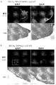
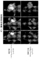

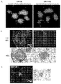


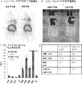
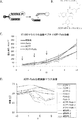
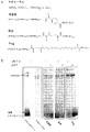


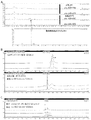


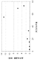


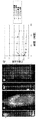
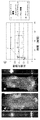


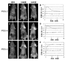


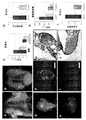


Classifications
-
- A—HUMAN NECESSITIES
- A61—MEDICAL OR VETERINARY SCIENCE; HYGIENE
- A61K—PREPARATIONS FOR MEDICAL, DENTAL OR TOILETRY PURPOSES
- A61K47/00—Medicinal preparations characterised by the non-active ingredients used, e.g. carriers or inert additives; Targeting or modifying agents chemically bound to the active ingredient
- A61K47/50—Medicinal preparations characterised by the non-active ingredients used, e.g. carriers or inert additives; Targeting or modifying agents chemically bound to the active ingredient the non-active ingredient being chemically bound to the active ingredient, e.g. polymer-drug conjugates
- A61K47/51—Medicinal preparations characterised by the non-active ingredients used, e.g. carriers or inert additives; Targeting or modifying agents chemically bound to the active ingredient the non-active ingredient being chemically bound to the active ingredient, e.g. polymer-drug conjugates the non-active ingredient being a modifying agent
- A61K47/62—Medicinal preparations characterised by the non-active ingredients used, e.g. carriers or inert additives; Targeting or modifying agents chemically bound to the active ingredient the non-active ingredient being chemically bound to the active ingredient, e.g. polymer-drug conjugates the non-active ingredient being a modifying agent the modifying agent being a protein, peptide or polyamino acid
- A61K47/64—Drug-peptide, drug-protein or drug-polyamino acid conjugates, i.e. the modifying agent being a peptide, protein or polyamino acid which is covalently bonded or complexed to a therapeutically active agent
-
- A—HUMAN NECESSITIES
- A61—MEDICAL OR VETERINARY SCIENCE; HYGIENE
- A61K—PREPARATIONS FOR MEDICAL, DENTAL OR TOILETRY PURPOSES
- A61K47/00—Medicinal preparations characterised by the non-active ingredients used, e.g. carriers or inert additives; Targeting or modifying agents chemically bound to the active ingredient
- A61K47/50—Medicinal preparations characterised by the non-active ingredients used, e.g. carriers or inert additives; Targeting or modifying agents chemically bound to the active ingredient the non-active ingredient being chemically bound to the active ingredient, e.g. polymer-drug conjugates
- A61K47/51—Medicinal preparations characterised by the non-active ingredients used, e.g. carriers or inert additives; Targeting or modifying agents chemically bound to the active ingredient the non-active ingredient being chemically bound to the active ingredient, e.g. polymer-drug conjugates the non-active ingredient being a modifying agent
- A61K47/56—Medicinal preparations characterised by the non-active ingredients used, e.g. carriers or inert additives; Targeting or modifying agents chemically bound to the active ingredient the non-active ingredient being chemically bound to the active ingredient, e.g. polymer-drug conjugates the non-active ingredient being a modifying agent the modifying agent being an organic macromolecular compound, e.g. an oligomeric, polymeric or dendrimeric molecule
- A61K47/59—Medicinal preparations characterised by the non-active ingredients used, e.g. carriers or inert additives; Targeting or modifying agents chemically bound to the active ingredient the non-active ingredient being chemically bound to the active ingredient, e.g. polymer-drug conjugates the non-active ingredient being a modifying agent the modifying agent being an organic macromolecular compound, e.g. an oligomeric, polymeric or dendrimeric molecule obtained otherwise than by reactions only involving carbon-to-carbon unsaturated bonds, e.g. polyureas or polyurethanes
- A61K47/595—Polyamides, e.g. nylon
-
- A—HUMAN NECESSITIES
- A61—MEDICAL OR VETERINARY SCIENCE; HYGIENE
- A61K—PREPARATIONS FOR MEDICAL, DENTAL OR TOILETRY PURPOSES
- A61K47/00—Medicinal preparations characterised by the non-active ingredients used, e.g. carriers or inert additives; Targeting or modifying agents chemically bound to the active ingredient
- A61K47/50—Medicinal preparations characterised by the non-active ingredients used, e.g. carriers or inert additives; Targeting or modifying agents chemically bound to the active ingredient the non-active ingredient being chemically bound to the active ingredient, e.g. polymer-drug conjugates
- A61K47/51—Medicinal preparations characterised by the non-active ingredients used, e.g. carriers or inert additives; Targeting or modifying agents chemically bound to the active ingredient the non-active ingredient being chemically bound to the active ingredient, e.g. polymer-drug conjugates the non-active ingredient being a modifying agent
- A61K47/56—Medicinal preparations characterised by the non-active ingredients used, e.g. carriers or inert additives; Targeting or modifying agents chemically bound to the active ingredient the non-active ingredient being chemically bound to the active ingredient, e.g. polymer-drug conjugates the non-active ingredient being a modifying agent the modifying agent being an organic macromolecular compound, e.g. an oligomeric, polymeric or dendrimeric molecule
- A61K47/59—Medicinal preparations characterised by the non-active ingredients used, e.g. carriers or inert additives; Targeting or modifying agents chemically bound to the active ingredient the non-active ingredient being chemically bound to the active ingredient, e.g. polymer-drug conjugates the non-active ingredient being a modifying agent the modifying agent being an organic macromolecular compound, e.g. an oligomeric, polymeric or dendrimeric molecule obtained otherwise than by reactions only involving carbon-to-carbon unsaturated bonds, e.g. polyureas or polyurethanes
- A61K47/60—Medicinal preparations characterised by the non-active ingredients used, e.g. carriers or inert additives; Targeting or modifying agents chemically bound to the active ingredient the non-active ingredient being chemically bound to the active ingredient, e.g. polymer-drug conjugates the non-active ingredient being a modifying agent the modifying agent being an organic macromolecular compound, e.g. an oligomeric, polymeric or dendrimeric molecule obtained otherwise than by reactions only involving carbon-to-carbon unsaturated bonds, e.g. polyureas or polyurethanes the organic macromolecular compound being a polyoxyalkylene oligomer, polymer or dendrimer, e.g. PEG, PPG, PEO or polyglycerol
-
- A—HUMAN NECESSITIES
- A61—MEDICAL OR VETERINARY SCIENCE; HYGIENE
- A61K—PREPARATIONS FOR MEDICAL, DENTAL OR TOILETRY PURPOSES
- A61K47/00—Medicinal preparations characterised by the non-active ingredients used, e.g. carriers or inert additives; Targeting or modifying agents chemically bound to the active ingredient
- A61K47/50—Medicinal preparations characterised by the non-active ingredients used, e.g. carriers or inert additives; Targeting or modifying agents chemically bound to the active ingredient the non-active ingredient being chemically bound to the active ingredient, e.g. polymer-drug conjugates
- A61K47/51—Medicinal preparations characterised by the non-active ingredients used, e.g. carriers or inert additives; Targeting or modifying agents chemically bound to the active ingredient the non-active ingredient being chemically bound to the active ingredient, e.g. polymer-drug conjugates the non-active ingredient being a modifying agent
- A61K47/62—Medicinal preparations characterised by the non-active ingredients used, e.g. carriers or inert additives; Targeting or modifying agents chemically bound to the active ingredient the non-active ingredient being chemically bound to the active ingredient, e.g. polymer-drug conjugates the non-active ingredient being a modifying agent the modifying agent being a protein, peptide or polyamino acid
-
- A—HUMAN NECESSITIES
- A61—MEDICAL OR VETERINARY SCIENCE; HYGIENE
- A61K—PREPARATIONS FOR MEDICAL, DENTAL OR TOILETRY PURPOSES
- A61K47/00—Medicinal preparations characterised by the non-active ingredients used, e.g. carriers or inert additives; Targeting or modifying agents chemically bound to the active ingredient
- A61K47/50—Medicinal preparations characterised by the non-active ingredients used, e.g. carriers or inert additives; Targeting or modifying agents chemically bound to the active ingredient the non-active ingredient being chemically bound to the active ingredient, e.g. polymer-drug conjugates
- A61K47/69—Medicinal preparations characterised by the non-active ingredients used, e.g. carriers or inert additives; Targeting or modifying agents chemically bound to the active ingredient the non-active ingredient being chemically bound to the active ingredient, e.g. polymer-drug conjugates the conjugate being characterised by physical or galenical forms, e.g. emulsion, particle, inclusion complex, stent or kit
- A61K47/6905—Medicinal preparations characterised by the non-active ingredients used, e.g. carriers or inert additives; Targeting or modifying agents chemically bound to the active ingredient the non-active ingredient being chemically bound to the active ingredient, e.g. polymer-drug conjugates the conjugate being characterised by physical or galenical forms, e.g. emulsion, particle, inclusion complex, stent or kit the form being a colloid or an emulsion
- A61K47/6911—Medicinal preparations characterised by the non-active ingredients used, e.g. carriers or inert additives; Targeting or modifying agents chemically bound to the active ingredient the non-active ingredient being chemically bound to the active ingredient, e.g. polymer-drug conjugates the conjugate being characterised by physical or galenical forms, e.g. emulsion, particle, inclusion complex, stent or kit the form being a colloid or an emulsion the form being a liposome
-
- A—HUMAN NECESSITIES
- A61—MEDICAL OR VETERINARY SCIENCE; HYGIENE
- A61K—PREPARATIONS FOR MEDICAL, DENTAL OR TOILETRY PURPOSES
- A61K49/00—Preparations for testing in vivo
- A61K49/001—Preparation for luminescence or biological staining
- A61K49/0013—Luminescence
- A61K49/0017—Fluorescence in vivo
- A61K49/0019—Fluorescence in vivo characterised by the fluorescent group, e.g. oligomeric, polymeric or dendritic molecules
- A61K49/0021—Fluorescence in vivo characterised by the fluorescent group, e.g. oligomeric, polymeric or dendritic molecules the fluorescent group being a small organic molecule
- A61K49/0032—Methine dyes, e.g. cyanine dyes
-
- A—HUMAN NECESSITIES
- A61—MEDICAL OR VETERINARY SCIENCE; HYGIENE
- A61K—PREPARATIONS FOR MEDICAL, DENTAL OR TOILETRY PURPOSES
- A61K49/00—Preparations for testing in vivo
- A61K49/001—Preparation for luminescence or biological staining
- A61K49/0013—Luminescence
- A61K49/0017—Fluorescence in vivo
- A61K49/005—Fluorescence in vivo characterised by the carrier molecule carrying the fluorescent agent
- A61K49/0056—Peptides, proteins, polyamino acids
-
- A—HUMAN NECESSITIES
- A61—MEDICAL OR VETERINARY SCIENCE; HYGIENE
- A61P—SPECIFIC THERAPEUTIC ACTIVITY OF CHEMICAL COMPOUNDS OR MEDICINAL PREPARATIONS
- A61P35/00—Antineoplastic agents
-
- A—HUMAN NECESSITIES
- A61—MEDICAL OR VETERINARY SCIENCE; HYGIENE
- A61P—SPECIFIC THERAPEUTIC ACTIVITY OF CHEMICAL COMPOUNDS OR MEDICINAL PREPARATIONS
- A61P9/00—Drugs for disorders of the cardiovascular system
-
- C—CHEMISTRY; METALLURGY
- C07—ORGANIC CHEMISTRY
- C07K—PEPTIDES
- C07K7/00—Peptides having 5 to 20 amino acids in a fully defined sequence; Derivatives thereof
- C07K7/04—Linear peptides containing only normal peptide links
- C07K7/06—Linear peptides containing only normal peptide links having 5 to 11 amino acids
Abstract
本明細書の特定の実施形態では、インビボ循環の増加した選択的輸送分子が開示される。一部の実施形態では、本明細書で開示される選択的輸送分子は、式(A−X−B−C)−Mを有し、式中、Cは積荷成分であり;Aは5〜9個の連続した酸性アミノ酸を含む配列を有するペプチドであり、該アミノ酸はアスパルタートおよびグルタマートから選択され;Bは5〜20個の連続した塩基性アミノ酸を含む配列を有するペプチドであり;Xはリンカーであり;かつMは高分子キャリアである。

Description
関連出願への相互参照
本出願は、2009年7月15日出願の米国仮出願第61/225,872号の利益を主張する。この出願は、その全体が参照により本明細書に組み込まれる。
This application claims the benefit of US Provisional Application No. 61 / 225,872 filed Jul. 15, 2009. This application is incorporated herein by reference in its entirety.
連邦政府による資金提供を受けた研究開発の記載
本発明は、米国防総省認可番号W81XWH−05−1−0183、および米国立衛生研究所認可番号NIBIB−K08EB008122に基づく政府の支援により行われた。政府は本発明に対し一定の権利を有する。
DESCRIPTION OF FEDERALLY SPONSORED RESEARCH AND DEVELOPMENT This invention was made with government support under US Department of Defense license number W81XWH-05-1-0183, and National Institutes of Health license number NIBIB-K08EB008122. The government has certain rights to the invention.
発明の背景
細胞膜は細胞外部との境界を定め、細胞内に出入りする輸送を調節する。膜は、主に脂質およびタンパク質からできていて、疎水性の内側を取り囲む形で親水性の表面を提供し、物質は細胞に入る前にそこを通過しなければならない。多くの小さい親油性化合物は細胞膜を受動的に通過可能であるが、大抵の化合物、粒子および物質は、能動的機構を頼りにして生細胞中に入らなければならない。
Background of the Invention The cell membrane delimits the exterior of the cell and regulates transport into and out of the cell. The membrane is mainly made up of lipids and proteins, providing a hydrophilic surface in a manner that surrounds the hydrophobic interior, and the substance must pass through it before entering the cell. Many small lipophilic compounds are able to passively pass through cell membranes, but most compounds, particles and substances have to enter living cells relying on active mechanisms.
本明細書中の特定の実施形態では、(A−X−B−C)−Mの構造の分子が開示され、式中、
Cは、少なくとも1つの造影剤であり;
Aは、5〜9個の連続した酸性アミノ酸を含む配列を有するペプチドであり、該アミノ酸はアスパルタートおよびグルタマートから選択され;
Bは、5〜20個の連続した塩基性アミノ酸を含む配列を有するペプチドであり;
Xは、リンカーであり;かつ
Mは、高分子キャリアであり;
ここでMは、AまたはBに結合している。
In certain embodiments herein, molecules of the structure (A-X-B-C) -M are disclosed, wherein
C is at least one contrast agent;
A is a peptide having a sequence comprising 5 to 9 consecutive acidic amino acids, the amino acids being selected from aspartate and glutamate;
B is a peptide having a sequence comprising 5 to 20 consecutive basic amino acids;
X is a linker; and M is a polymeric carrier;
Here, M is bonded to A or B.
一部の実施形態では、Aは、5〜9個の連続したグルタマートを含む配列を有している。一部の実施形態では、Bは、5〜12個の連続したアルギニンを含む配列を有する。一部の実施形態では、Bは、9個の連続したアルギニンを含む配列を有する。一部の実施形態では、(a)Aは8〜9個の連続したグルタマートを含む配列を有し、(b)Bは9個の連続したアルギニンを含む配列を有する。一部の実施形態では、AおよびBは、D−アミノ酸を含む。一部の実施形態では、Xは、切断可能なリンカーである。一部の実施形態では、Xは、ペプチド結合を含む。一部の実施形態では、Xは、6−アミノヘキサノイル、5−アミノ−3−オキサペンタノイル、またはその組み合わせを含む。一部の実施形態では、Xは、ジスルフィド結合を含む。一部の実施形態では、Xは、約6〜約30原子の長さである。一部の実施形態では、Xは、pH感受性リンカーである。一部の実施形態では、Xは、細胞外空間で切断される。一部の実施形態では、Xは、プロテアーゼ、マトリックスメタロプロテアーゼ、またはこれらの組み合わせにより切断される。一部の実施形態では、Xは、還元剤により切断される。一部の実施形態では、Xは、PLGLAG、PLGLAx(xは任意のアミノ酸)、PLG−C(me)−AG、ESPAYYTA、およびRLQLKL、AND RLQLK(AC)から選択される。一部の実施形態では、Mは、デンドリマー、デキストラン、PEGポリマー、またはアルブミンから選択される高分子キャリアである。一部の実施形態では、Mは、PEGポリマーである。一部の実施形態では、Cは、造影剤、治療薬、またはその組み合わせを含む。一部の実施形態では、Cは、蛍光成分、発光成分、リン光成分、蛍光消光成分、放射性成分、放射線不透過性成分、常磁性成分、コントラスト剤、またはその組み合わせから選択される造影剤を含む。一部の実施形態では、Cは、化学療法剤、放射線増感剤、アポトーシスを調節する薬剤、および細胞周期を調節する薬剤、シグナル伝達カスケードを調節する薬剤、またはこれらの組み合わせから選択される治療薬を含む。一部の実施形態では、積荷は、インドカルボシアニン染料である。一部の実施形態では、積荷は、インドカルボシアニン染料、Cy5、Cy5.5、Cy7、IRDYE800CW、ALEXA647、またはこれらの組み合わせである。一部の実施形態では、積荷は、MRIコントラスト剤である。一部の実施形態では、積荷は、[4,7,10−トリス(カルボキシメチル)−1,4,7,10−テトラアザシクロドデカ−1−イル]アセチルのGd錯体である。 In some embodiments, A has a sequence comprising 5-9 consecutive glutamates. In some embodiments, B has a sequence comprising 5-12 consecutive arginines. In some embodiments, B has a sequence comprising 9 consecutive arginines. In some embodiments, (a) A has a sequence that includes 8-9 consecutive glutamates, and (b) B has a sequence that includes 9 consecutive arginines. In some embodiments, A and B include D-amino acids. In some embodiments, X is a cleavable linker. In some embodiments, X comprises a peptide bond. In some embodiments, X comprises 6-aminohexanoyl, 5-amino-3-oxapentanoyl, or a combination thereof. In some embodiments, X comprises a disulfide bond. In some embodiments, X is about 6 to about 30 atoms in length. In some embodiments, X is a pH sensitive linker. In some embodiments, X is cleaved in the extracellular space. In some embodiments, X is cleaved by a protease, matrix metalloprotease, or a combination thereof. In some embodiments, X is cleaved by a reducing agent. In some embodiments, X is selected from PLGLAG, PLGLAx (x is any amino acid), PLG-C (me) -AG, ESPAYYTA, and RLQLKL, AND RLQLK (AC). In some embodiments, M is a polymeric carrier selected from dendrimers, dextrans, PEG polymers, or albumin. In some embodiments, M is a PEG polymer. In some embodiments, C comprises a contrast agent, a therapeutic agent, or a combination thereof. In some embodiments, C comprises a contrast agent selected from a fluorescent component, a luminescent component, a phosphorescent component, a fluorescence quenching component, a radioactive component, a radiopaque component, a paramagnetic component, a contrast agent, or a combination thereof. Including. In some embodiments, C is a treatment selected from chemotherapeutic agents, radiosensitizers, agents that modulate apoptosis, and agents that modulate the cell cycle, agents that modulate signaling cascades, or combinations thereof. Contains medicine. In some embodiments, the load is an indocarbocyanine dye. In some embodiments, the shipment is an indocarbocyanine dye, Cy5, Cy5.5, Cy7, IRDYE800CW, ALEXA647, or a combination thereof. In some embodiments, the load is an MRI contrast agent. In some embodiments, the cargo is a Gd complex of [4,7,10-tris (carboxymethyl) -1,4,7,10-tetraazacyclododec-1-yl] acetyl.
本明細書中の特定の実施形態では、(A−X−B)n−Lの構造の分子が開示され、式中、
Lは、脂質または脂質コート治療薬または造影剤を含み;
Aは、5〜9個の連続した酸性アミノ酸を含む配列を有するペプチドであり、該アミノ酸は、アスパルタートおよびグルタマートから選択され;
Bは、5〜20個の連続した塩基性アミノ酸を含む配列を有するペプチドであり;
Xは、リンカーであり;かつ
nは、1〜20の整数であり;かつ
ここでLは、Bとの結合により(A−X−B)成分に結合している。
In certain embodiments herein, molecules of the structure (A-X-B) n -L are disclosed, wherein
L comprises a lipid or lipid coat therapeutic or contrast agent;
A is a peptide having a sequence comprising 5 to 9 consecutive acidic amino acids, wherein the amino acids are selected from aspartate and glutamate;
B is a peptide having a sequence comprising 5 to 20 consecutive basic amino acids;
X is a linker; and n is an integer from 1 to 20; and where L is attached to the (A-X-B) component by binding to B.
一部の実施形態では、Aは、5〜9個の連続したグルタマートを含む配列を有する。一部の実施形態では、Bは、5〜12個の連続したアルギニンを含む配列を有する。一部の実施形態では、Bは、9個の連続したアルギニンを含む配列を有する。一部の実施形態では、(a)Aは、8〜9個の連続したグルタマートを含む配列を有し、(b)Bは、9個の連続したアルギニンを含む配列を有する。一部の実施形態では、AおよびBは、D−アミノ酸を含む。一部の実施形態では、Xは、切断可能なリンカーである。一部の実施形態では、Xは、ペプチド結合を含む。一部の実施形態では、Xは、6−アミノヘキサノイル、5−(アミノ)−3−オキサペンタン酸、またはこれらの組み合わせを含む。一部の実施形態では、Xは、ジスルフィド結合を含む。一部の実施形態では、Xは、約6〜約30原子の長さである。一部の実施形態では、Xは、pH感受性リンカーである。一部の実施形態では、Xは、細胞外空間で切断される。一部の実施形態では、Xは、プロテアーゼ、マトリックスメタロプロテアーゼ、またはこれらの組み合わせにより切断される。一部の実施形態では、Xは、還元剤により切断される。一部の実施形態では、Xは、PLGLAG、PLGLAx(xは任意のアミノ酸)、PLG−C(me)−AG、ESPAYYTA、およびRLQLKL、AND RLQLK(AC)から選択される。一部の実施形態では、脂質は、ペグ化されている。一部の実施形態では、脂質は、PEG(2K)−ホスファチジルエタノールアミンである。一部の実施形態では、治療薬は、疎水性である。一部の実施形態では、治療薬は、化学療法剤、放射線増感剤、アポトーシスを調節する薬剤、および細胞周期を調節する薬剤、シグナル伝達カスケードを調節する薬剤、またはこれらの組み合わせから選択される。一部の実施形態では、治療薬は、ドキソルビシンまたはパクリタキセルである。 In some embodiments, A has a sequence comprising 5-9 consecutive glutamates. In some embodiments, B has a sequence comprising 5-12 consecutive arginines. In some embodiments, B has a sequence comprising 9 consecutive arginines. In some embodiments, (a) A has a sequence comprising 8-9 consecutive glutamates, and (b) B has a sequence comprising 9 consecutive arginines. In some embodiments, A and B include D-amino acids. In some embodiments, X is a cleavable linker. In some embodiments, X comprises a peptide bond. In some embodiments, X comprises 6-aminohexanoyl, 5- (amino) -3-oxapentanoic acid, or a combination thereof. In some embodiments, X comprises a disulfide bond. In some embodiments, X is about 6 to about 30 atoms in length. In some embodiments, X is a pH sensitive linker. In some embodiments, X is cleaved in the extracellular space. In some embodiments, X is cleaved by a protease, matrix metalloprotease, or a combination thereof. In some embodiments, X is cleaved by a reducing agent. In some embodiments, X is selected from PLGLAG, PLGLAx (x is any amino acid), PLG-C (me) -AG, ESPAYYTA, and RLQLKL, AND RLQLK (AC). In some embodiments, the lipid is PEGylated. In some embodiments, the lipid is PEG (2K) -phosphatidylethanolamine. In some embodiments, the therapeutic agent is hydrophobic. In some embodiments, the therapeutic agent is selected from chemotherapeutic agents, radiosensitizers, agents that modulate apoptosis, and agents that modulate the cell cycle, agents that modulate signal transduction cascades, or combinations thereof. . In some embodiments, the therapeutic agent is doxorubicin or paclitaxel.
本明細書中の特定の実施形態では、(A−X−B)n−Dの構造の分子が開示され、式中、
Dは、デンドリマーであり;
Aは、5〜9個の連続した酸性アミノ酸を含む配列を有するペプチドであり、該アミノ酸は、アスパルタートおよびグルタマートから選択され;
Bは、5〜20個の連続した塩基性アミノ酸を含む配列を有するペプチドであり;
Xは、リンカーであり;かつ
nは、1〜20の整数であり;かつ
ここでDは、Bとの結合により(A−X−B)成分に結合している。
In certain embodiments herein, molecules of the structure (A—X—B) n —D are disclosed, wherein
D is a dendrimer;
A is a peptide having a sequence comprising 5 to 9 consecutive acidic amino acids, wherein the amino acids are selected from aspartate and glutamate;
B is a peptide having a sequence comprising 5 to 20 consecutive basic amino acids;
X is a linker; and n is an integer from 1 to 20; and D is attached to the (A-X-B) component by a bond with B.
一部の実施形態では、Dは、少なくとも1つの積荷成分を含む。一部の実施形態では、少なくとも1つの積荷成分は造影剤、治療薬、またはこれらの組み合わせである。一部の実施形態では、少なくとも1つの積荷成分は、蛍光成分、発光成分、リン光成分、蛍光消光成分、放射性成分、放射線不透過性成分、常磁性成分、コントラスト剤、またはこれらの組み合わせから選択される造影剤である。一部の実施形態では、少なくとも1つの積荷成分は、化学療法剤、放射線増感剤、アポトーシスを調節する薬剤、および細胞周期を調節する薬剤、シグナル伝達カスケードを調節する薬剤、またはこれらの組み合わせから選択される治療薬である。一部の実施形態では、少なくとも1つの積荷成分は、インドカルボシアニン染料である。一部の実施形態では、少なくとも1つの積荷成分は、インドカルボシアニン染料、Cy5、Cy5.5、Cy7、IRDYE800CW、ALEXA647、またはこれらの組み合わせである。一部の実施形態では、少なくとも1つの積荷成分は、MRIコントラスト剤である。一部の実施形態では、少なくとも1つの積荷成分は、[4,7,10−トリス(カルボキシメチル)−1,4,7,10−テトラアザシクロドデカ−1−イル]アセチルのGd錯体である。一部の実施形態では、Aは、5〜9個の連続したグルタマートを含む配列を有する。一部の実施形態では、Bは、5〜12個の連続したアルギニンを含む配列を有する。一部の実施形態では、Bは、9個の連続したアルギニンを含む配列を有する。一部の実施形態では、(a)Aは、8〜9個の連続したグルタマートを含む配列を有し、かつ(b)Bは、9個の連続したアルギニンを含む配列を有する。一部の実施形態では、AおよびBは、D−アミノ酸を含む。一部の実施形態では、Xは、切断可能なリンカーである。一部の実施形態では、Xは、ペプチド結合を含む。一部の実施形態では、Xは、6−アミノヘキサノイル、5−(アミノ)−3−オキサペンタン酸、またはこれらの組み合わせを含む。一部の実施形態では、Xは、ジスルフィド結合を含む。一部の実施形態では、Xは、約6〜約30原子の長さである。一部の実施形態では、Xは、pH感受性リンカーである。一部の実施形態では、Xは、細胞外空間で切断される。一部の実施形態では、Xは、プロテアーゼ、マトリックスメタロプロテアーゼ、またはこれらの組み合わせにより切断される。一部の実施形態では、Xは、還元剤により切断される。一部の実施形態では、Xは、PLGLAG、PLGLAx(xは任意のアミノ酸)、PLG−C(me)−AG、ESPAYYTA、およびRLQLKL、AND RLQLK(AC)から選択される。 In some embodiments, D includes at least one cargo component. In some embodiments, the at least one cargo component is a contrast agent, a therapeutic agent, or a combination thereof. In some embodiments, the at least one cargo component is selected from a fluorescent component, a luminescent component, a phosphorescent component, a fluorescence quenching component, a radioactive component, a radiopaque component, a paramagnetic component, a contrast agent, or combinations thereof Contrast agent. In some embodiments, the at least one cargo component is from a chemotherapeutic agent, a radiosensitizer, an agent that modulates apoptosis, and an agent that modulates the cell cycle, an agent that modulates a signaling cascade, or a combination thereof. The selected therapeutic agent. In some embodiments, at least one cargo component is an indocarbocyanine dye. In some embodiments, the at least one cargo component is an indocarbocyanine dye, Cy5, Cy5.5, Cy7, IRDYE800CW, ALEXA647, or combinations thereof. In some embodiments, at least one cargo component is an MRI contrast agent. In some embodiments, at least one cargo component is a Gd complex of [4,7,10-tris (carboxymethyl) -1,4,7,10-tetraazacyclododec-1-yl] acetyl. . In some embodiments, A has a sequence comprising 5-9 consecutive glutamates. In some embodiments, B has a sequence comprising 5-12 consecutive arginines. In some embodiments, B has a sequence comprising 9 consecutive arginines. In some embodiments, (a) A has a sequence comprising 8-9 consecutive glutamates, and (b) B has a sequence comprising 9 consecutive arginines. In some embodiments, A and B include D-amino acids. In some embodiments, X is a cleavable linker. In some embodiments, X comprises a peptide bond. In some embodiments, X comprises 6-aminohexanoyl, 5- (amino) -3-oxapentanoic acid, or a combination thereof. In some embodiments, X comprises a disulfide bond. In some embodiments, X is about 6 to about 30 atoms in length. In some embodiments, X is a pH sensitive linker. In some embodiments, X is cleaved in the extracellular space. In some embodiments, X is cleaved by a protease, matrix metalloprotease, or a combination thereof. In some embodiments, X is cleaved by a reducing agent. In some embodiments, X is selected from PLGLAG, PLGLAx (x is any amino acid), PLG-C (me) -AG, ESPAYYTA, and RLQLKL, AND RLQLK (AC).
本明細書中の特定の実施形態では、患者の腫瘍を画像化する方法が開示される。この方法は、(A−X−B−C)−Mの構造の分子を患者に投与した後に腫瘍を画像化することを含み、式中、
Cは、少なくとも1つの造影剤であり;
Aは、5〜9個の連続した酸性アミノ酸を含む配列を有するペプチドであり、該アミノ酸は、アスパルタートおよびグルタマートから選択され;
Bは、5〜20個の連続した塩基性アミノ酸を含む配列を有するペプチドであり;
Xは、リンカーであり;かつ
Mは、高分子キャリアであり;
ここでMは、AまたはBに結合している。
In certain embodiments herein, a method for imaging a patient's tumor is disclosed. The method comprises imaging a tumor after administering a molecule of structure (AXB-C) -M to a patient, wherein:
C is at least one contrast agent;
A is a peptide having a sequence comprising 5 to 9 consecutive acidic amino acids, wherein the amino acids are selected from aspartate and glutamate;
B is a peptide having a sequence comprising 5 to 20 consecutive basic amino acids;
X is a linker; and M is a polymeric carrier;
Here, M is bonded to A or B.
本明細書中の特定の実施形態では、腫瘍の画像化を、それを必要としている患者において行うための(A−X−B−C)−Mの構造の分子の使用が開示されており、式中、
Cは、少なくとも1つの造影剤であり;
Aは、5〜9個の連続した酸性アミノ酸を含む配列を有するペプチドであり、該アミノ酸は、アスパルタートおよびグルタマートから選択され;
Bは、5〜20個の連続した塩基性アミノ酸を含む配列を有するペプチドであり;
Xは、リンカーであり;かつ
Mは、高分子キャリアであり;
ここでMは、AまたはBに結合している。
In certain embodiments herein, the use of a molecule of the structure (A-X-B-C) -M to perform tumor imaging in a patient in need thereof is disclosed. Where
C is at least one contrast agent;
A is a peptide having a sequence comprising 5 to 9 consecutive acidic amino acids, wherein the amino acids are selected from aspartate and glutamate;
B is a peptide having a sequence comprising 5 to 20 consecutive basic amino acids;
X is a linker; and M is a polymeric carrier;
Here, M is bonded to A or B.
本明細書中の特定の実施形態では、患者の腫瘍切除または組織切除のための外科的縁を画像化する方法が開示されており、(A−X−B−C)−Mの構造の分子を患者に投与した後に外科的縁を画像化することを含み、式中、
Cは、少なくとも1つの造影剤であり;
Aは、5〜9個の連続した酸性アミノ酸を含む配列を有するペプチドであり、該アミノ酸は、アスパルタートおよびグルタマートから選択され;
Bは、5〜20個の連続した塩基性アミノ酸を含む配列を有するペプチドであり;
Xは、リンカーであり;かつ
Mは、高分子キャリアであり;
ここでMは、AまたはBに結合している。
In certain embodiments herein, a method of imaging a surgical edge for tumor excision or tissue excision of a patient is disclosed, wherein a molecule of the structure (A-X-B-C) -M Imaging the surgical margin after administering to the patient, wherein
C is at least one contrast agent;
A is a peptide having a sequence comprising 5 to 9 consecutive acidic amino acids, wherein the amino acids are selected from aspartate and glutamate;
B is a peptide having a sequence comprising 5 to 20 consecutive basic amino acids;
X is a linker; and M is a polymeric carrier;
Here, M is bonded to A or B.
本明細書中の特定の実施形態では、腫瘍切除または組織切除のための外科的縁を画像化することを、それを必要としている患者において行うための(A−X−B−C)−M構造の分子の使用が開示され、式中、
Cは、少なくとも1つの造影剤であり;
Aは、5〜9個の連続した酸性アミノ酸を含む配列を有するペプチドであり、該アミノ酸は、アスパルタートおよびグルタマートから選択され;
Bは、5〜20個の連続した塩基性アミノ酸を含む配列を有するペプチドであり;
Xは、リンカーであり;かつ
Mは、高分子キャリアであり;
ここでMは、AまたはBに結合している。
In certain embodiments herein, (AXB-C) -M for performing imaging of a surgical margin for tumor resection or tissue resection in a patient in need thereof. The use of a molecule of structure is disclosed, wherein
C is at least one contrast agent;
A is a peptide having a sequence comprising 5 to 9 consecutive acidic amino acids, wherein the amino acids are selected from aspartate and glutamate;
B is a peptide having a sequence comprising 5 to 20 consecutive basic amino acids;
X is a linker; and M is a polymeric carrier;
Here, M is bonded to A or B.
本明細書中の特定の実施形態では、患者の腫瘍を除去する方法が開示され、(A−X−B−C)−Mの構造の分子を患者に投与した後に腫瘍を除去することを含み、式中、
Cは、少なくとも1つの造影剤であり;
Aは、5〜9個の連続した酸性アミノ酸を含む配列を有するペプチドであり、該アミノ酸は、アスパルタートおよびグルタマートから選択され;
Bは、5〜20個の連続した塩基性アミノ酸を含む配列を有するペプチドであり;
Xは、リンカーであり;かつ
Mは、高分子キャリアであり;
ここでMは、AまたはBに結合している。
In certain embodiments herein, a method of removing a patient's tumor is disclosed, comprising removing the tumor after administering a molecule of the structure of (AX-BC) -M to the patient. , Where
C is at least one contrast agent;
A is a peptide having a sequence comprising 5 to 9 consecutive acidic amino acids, wherein the amino acids are selected from aspartate and glutamate;
B is a peptide having a sequence comprising 5 to 20 consecutive basic amino acids;
X is a linker; and M is a polymeric carrier;
Here, M is bonded to A or B.
本明細書中の特定の実施形態では、腫瘍の除去を、それを必要としている患者において行うための(A−X−B−C)−Mの構造の分子の使用が開示されており、式中、
Cは、少なくとも1つの造影剤であり;
Aは、5〜9個の連続した酸性アミノ酸を含む配列を有するペプチドであり、該アミノ酸は、アスパルタートおよびグルタマートから選択され;
Bは、5〜20個の連続した塩基性アミノ酸を含む配列を有するペプチドであり;
Xは、リンカーであり;かつ
Mは、高分子キャリアであり;
ここでMは、AまたはBに結合している。
In certain embodiments herein, the use of a molecule of the structure (A—X—B—C) -M for performing tumor removal in a patient in need thereof is disclosed and has the formula During,
C is at least one contrast agent;
A is a peptide having a sequence comprising 5 to 9 consecutive acidic amino acids, wherein the amino acids are selected from aspartate and glutamate;
B is a peptide having a sequence comprising 5 to 20 consecutive basic amino acids;
X is a linker; and M is a polymeric carrier;
Here, M is bonded to A or B.
本発明の新規性は添付の特許請求の範囲で詳細に説明される。本発明の原理とそれに付随する図を使った下記の実施形態の詳細な説明により、本発明の特色と利点に関し、より良き理解が得られるであろう。 The novelty of the invention is set forth with particularity in the appended claims. A better understanding of the features and advantages of the present invention will be obtained from the following detailed description of embodiments that uses the principles of the invention and the accompanying drawings.
発明の詳細な説明
選択的輸送分子は、治療薬および/または造影剤の特定細胞および/または組織を標的にした送達を可能にする。選択的輸送分子は、(a)分子輸送配列(B部)、(b)B部に結合した積荷(CまたはD部)および(c)細胞への取り込みを抑制または防ぐのに有効である酸性のA部を含む。B部からA部の分離を可能にするリンカーXの切断は、B部および付随する積荷を細胞中に取り込ませるのに有効である。しかし、選択的輸送分子は、短い血漿中半減期、広範囲への分布、および特異的および非特異的取り込みを有する複数の組織からの遅い洗い流しを伴った急速な薬物動態学的送達にさらされる可能性がある。このように、インビボ循環の増加および血管外漏出選択性の調節がなされた選択的輸送分子に対するニーズが存在する。
Detailed Description of the Invention Selective transport molecules allow targeted delivery of therapeutic and / or contrast agents to specific cells and / or tissues. Selective transport molecules are (a) molecular transport sequences (part B), (b) cargo bound to part B (part C or D), and (c) an acid that is effective in inhibiting or preventing cellular uptake. A part of Cleavage of linker X, which allows separation of part A from part B, is effective for incorporating part B and the accompanying cargo into the cell. However, selective transport molecules can be exposed to rapid pharmacokinetic delivery with short plasma half-life, wide distribution, and slow washout from multiple tissues with specific and non-specific uptake There is sex. Thus, there is a need for selective transport molecules with increased in vivo circulation and regulation of extravasation selectivity.
本明細書中の特定の実施形態では、インビボで循環性が増加した選択的輸送分子が開示される。 In certain embodiments herein, selective transport molecules with increased circulation in vivo are disclosed.
一部の実施形態では、本明細書で開示の選択的輸送分子は、式(A−X−B−C)−Mを有し、Cは、積荷成分;Aは、5〜9個の連続した酸性アミノ酸を含む配列を有するペプチドであり、該アミノ酸はアスパルタートおよびグルタマートから選択され;Bは、5〜20個の連続した塩基性アミノ酸を含む配列を有するペプチドであり;Xは、リンカーであり;かつMは、高分子キャリアである。 In some embodiments, a selective transport molecule disclosed herein has the formula (A-X-B-C) -M, C is the cargo component; A is 5-9 consecutive A peptide having a sequence comprising acidic amino acids, wherein the amino acid is selected from aspartate and glutamate; B is a peptide having a sequence comprising 5 to 20 consecutive basic amino acids; X is a linker Yes; and M is a polymeric carrier.
一部の実施形態では、本明細書で開示の選択的輸送分子は、式(A−X−B)−Dを有し、Cは、積荷成分;Aは、5〜9個の連続した酸性アミノ酸を含む配列を有するペプチドであり、該アミノ酸はアスパルタートおよびグルタマートから選択され;Bは、5〜20個の連続した塩基性アミノ酸を含む配列を有するペプチドであり;Xは、リンカーであり;Dは、デンドリマーである。一部の実施形態では、Dは、積荷成分を含む。 In some embodiments, the selective transport molecules disclosed herein have the formula (A-X-B) -D, C is the cargo component; A is 5-9 consecutive acidic A peptide having a sequence comprising amino acids, wherein the amino acids are selected from aspartate and glutamate; B is a peptide having a sequence comprising 5-20 consecutive basic amino acids; X is a linker; D is a dendrimer. In some embodiments, D includes a cargo component.
定義
下記の用語は、特に他に定めがなければ、そこに記された意味を持つ。
Definitions The following terms have the meanings given to them unless otherwise specified.
用語の細胞透過性ペプチド(CPP)、膜輸送配列(MTS)およびタンパク質伝達ドメインは同義に使われる。本明細書で使われるこの用語は、細胞の細胞膜を通過して転移させるペプチド(ポリペプチドまたはタンパク質)配列を意味する。一部の実施形態では、CPPは、細胞の細胞膜を通過して細胞外の分子を転移させる。一部の実施形態では、CPPは、細胞膜への直接侵入、エンドサイトーシス媒介侵入、または一時的構造の形成、により細胞膜を通過して転移させる。 The terms cell penetrating peptide (CPP), membrane transport sequence (MTS) and protein transduction domain are used interchangeably. As used herein, this term refers to a peptide (polypeptide or protein) sequence that translocates across the cell membrane of a cell. In some embodiments, the CPP translocates extracellular molecules across the cell membrane. In some embodiments, the CPP is translocated across the cell membrane by direct entry into the cell membrane, endocytosis-mediated invasion, or formation of a transient structure.
本明細書で使用されるように、用語の「アプタマー」は、標的分子との非ワトソン・クリック相互作用に基づく高親和性と特異性を有する他の分子との結合能力に基づいてランダムプールから選択されるDNAまたはRNA分子を指す(例えば、Cox and Ellington、Bioorg.Med.Chem.9:2525−2531(2001);Lee et al.、Nuc.Acids Res.32:D95−D100(2004))を参照。一部の実施形態では、アプタマーは、核酸、タンパク質、低分子有機化合物、ビタミン、無機化合物、細胞、および生命体そのものにも、結合する。 As used herein, the term “aptamer” is derived from a random pool based on its ability to bind other molecules with high affinity and specificity based on non-Watson-Crick interactions with the target molecule. Refers to a selected DNA or RNA molecule (eg, Cox and Ellington, Bioorg. Med. Chem. 9: 2525-2531 (2001); Lee et al., Nuc. Acids Res. 32: D95-D100 (2004)) See In some embodiments, aptamers also bind to nucleic acids, proteins, small organic compounds, vitamins, inorganic compounds, cells, and the living organism itself.
用語の「ポリペプチド」、「ペプチド」および「タンパク質」は、本明細書では同義に使われ、アミノ酸残基のポリマーを指す。この用語は、天然アミノ酸ポリマーならびに1つまたは複数のアミノ酸残基が非天然アミノ酸(例えば、アミノ酸類似体)であるアミノ酸ポリマーに適用される。この用語は、完全長タンパク質(すなわち、抗原)を含む任意の長さのアミノ酸鎖を包含し、アミノ酸残基は、ペプチド結合により共有結合されている。本明細書で使われる用語「ペプチド」は、アミノ酸残基のポリマーを指し、通常は、長さは2〜約50残基の範囲である。特定の実施形態では、ペプチドは約2、3、4、5、7、9、10、または11残基〜約50、45、40、45、30、25、20、または15残基の長さの範囲である。特定の実施形態では、ペプチドは8、9、10、11、または12残基〜約15、20または25残基の長さの範囲である。本明細書でアミノ酸配列が提供されている場合は、L−、D−、またはベータアミノ酸の配列タイプ、ならびに逆、反転、および逆反転イソ型も同様に意図されている。また、ペプチドは、1つまたは複数のアミノ酸残基が対応する天然アミノ酸、ならびに天然アミノ酸ポリマーの人工的化学類似体であるアミノ酸ポリマーを含む。さらに、この用語は、ペプチド結合または他の修飾された結合(例えば、ペプチド結合がα−エステル、β−エステル、チオアミド、ホスホンアミド、カルバマート、ヒドロキシラート、等で置換されている結合(例えば、Spatola、(1983)Chem.Biochem.Amino Acids and Proteins 7:267−357を参照)、アミドが飽和アミンで置換されている結合(例えば、Skiles et al.、米国特許第4,496,542号(この特許は、参照により本明細書に組み込まれる)、およびKaltenbronn et al.、(1990)Pp.969−970 in Proc.11th American Peptide Symposium、ESCOM Science Publishers、The Netherlands、等を参照))、により結合されているアミノ酸に適用される。 The terms “polypeptide”, “peptide” and “protein” are used interchangeably herein to refer to a polymer of amino acid residues. This term applies to naturally occurring amino acid polymers as well as amino acid polymers in which one or more amino acid residues are unnatural amino acids (eg, amino acid analogs). The term encompasses amino acid chains of any length, including full length proteins (ie, antigens), where the amino acid residues are covalently linked by peptide bonds. The term “peptide” as used herein refers to a polymer of amino acid residues, usually ranging in length from 2 to about 50 residues. In certain embodiments, the peptide is from about 2, 3, 4, 5, 7, 9, 10, or 11 residues to about 50, 45, 40, 45, 30, 25, 20, or 15 residues in length. Range. In certain embodiments, the peptide ranges in length from 8, 9, 10, 11, or 12 residues to about 15, 20, or 25 residues. Where amino acid sequences are provided herein, L-, D-, or beta amino acid sequence types, as well as reverse, inverted, and reverse inverted isoforms are contemplated as well. Peptides also include natural amino acids to which one or more amino acid residues correspond, as well as amino acid polymers that are artificial chemical analogs of natural amino acid polymers. In addition, the term includes peptide bonds or other modified bonds (eg, peptide bonds that are substituted with α-esters, β-esters, thioamides, phosphonamides, carbamates, hydroxylates, etc. (eg, Spatola (1983) Chem. Biochem. Amino Acids and Proteins 7: 267-357), bonds where the amide is substituted with a saturated amine (see, for example, Skiles et al., US Pat. No. 4,496,542) Patent is incorporated herein by reference), and Kaltenbron et al., (1990) P. 969-970 in Proc. 11th American Peptide Symposium, ESCOM Science Pu. blishers, The Netherlands, et al.))).
用語の「アミノ酸」は、天然および合成アミノ酸、ならびに天然アミノ酸と類似の方式で機能するアミノ酸類似体およびアミノ酸模倣体を指す。天然アミノ酸は、遺伝暗号によりコードされているもの、ならびに後で修飾されたもので、例えば、ヒドロキシプロリン、γ−カルボキシグルタマート、およびO−ホスホセリンである。アミノ酸類似体は、天然アミノ酸と同じ基本的化学構造、すなわち、水素に結合したαカーボン、カルボキシル基、アミノ基、およびR基、例えば、ホモセリン、ノルロイシン、メチオニンスルフォキシド、を有する化合物を指す。このような類似体は、修飾R基(例えば、ノルロイシン)または修飾ペプチド骨格を有し、しかも天然アミノ酸と同じ基本的化学構造を維持している。アミノ酸模倣体は、アミノ酸の一般化学構造とは異なる構造を有するが、天然アミノ酸と類似の方式で機能する化学的化合物を指す。アミノ酸は、Dアミノ酸またはLアミノ酸であってもよい。明細書中のペプチド配列で、小文字はアミノ酸のD異性体を示している(逆に、大文字はアミノ酸のL異性体を示している)。 The term “amino acid” refers to naturally occurring and synthetic amino acids, as well as amino acid analogs and amino acid mimetics that function in a manner similar to the naturally occurring amino acids. Natural amino acids are those encoded by the genetic code, as well as those later modified, such as hydroxyproline, γ-carboxyglutamate, and O-phosphoserine. Amino acid analogs refer to compounds having the same basic chemical structure as a naturally occurring amino acid, ie, an α-carbon bonded to hydrogen, a carboxyl group, an amino group, and an R group, such as homoserine, norleucine, methionine sulfoxide. Such analogs have modified R groups (eg, norleucine) or modified peptide backbones, and maintain the same basic chemical structure as a naturally occurring amino acid. Amino acid mimetics refers to chemical compounds that have a structure that is different from the general chemical structure of an amino acid, but that functions in a manner similar to a naturally occurring amino acid. The amino acid may be a D amino acid or an L amino acid. In the peptide sequences in the specification, lower case letters indicate D isomers of amino acids (in contrast, upper case letters indicate L isomers of amino acids).
本明細書では、アミノ酸は、通常よく知られた3文字記号でも、またはIUPAC−IUB Biochemical Nomenclature Commission推奨の1文字記号で呼んでもよい。同様に、ヌクレオチドは、通常受け入れられている1文字コードで呼んでもよい。 As used herein, amino acids may be referred to by the commonly known three letter symbols or by the one-letter symbols recommended by the IUPAC-IUB Biochemical Nomenclature Commission. Similarly, nucleotides may be referred to with the normally accepted single letter code.
コード化配列中の単一アミノ酸または小さいパーセンテージのアミノ酸を変更、追加、または削除する、ペプチド、ポリペプチド、またはタンパク質配列への個々の置換、欠失または付加は、「保存的改変変異体」であり、この場合、変更により化学的に類似のアミノ酸によるアミノ酸の置換が生ずることは同業者なら認識しているであろう。機能的に類似のアミノ酸を与える保存的置換表は当技術分野でよく知られている。このような保存的改変変異体は、本発明の多形性変異体、種間相同体、および対立遺伝子を追加し、排除しない。 Individual substitutions, deletions or additions to a peptide, polypeptide, or protein sequence that alter, add, or delete a single amino acid or a small percentage of amino acids in the coding sequence are “conservatively modified variants”. Yes, in this case, those skilled in the art will recognize that the alteration results in the substitution of an amino acid with a chemically similar amino acid. Conservative substitution tables giving functionally similar amino acids are well known in the art. Such conservatively modified variants add and do not exclude polymorphic variants, interspecies homologues, and alleles of the present invention.
下記の8つのグループはそれぞれ相互に保存的置換できるアミノ酸を含む:1)アラニン(A)、グリシン(G);2)アスパラギン酸(D)、グルタミン酸(E);3)アスパラギン(N)、グルタミン(Q);4)アルギニン(R)、リジン(K);5)イソロイシン(I)、ロイシン(L)、メチオニン(M)、バリン(V);6)フェニルアラニン(F)、チロシン(Y)、トリプトファン(W);7)セリン(S)、トレオニン(T);および8)システイン(C)、メチオニン(M)(例えば、Creighton、Proteins(1984)を参照)。 The following eight groups each contain amino acids that can be conservatively substituted for each other: 1) alanine (A), glycine (G); 2) aspartic acid (D), glutamic acid (E); 3) asparagine (N), glutamine (Q); 4) arginine (R), lysine (K); 5) isoleucine (I), leucine (L), methionine (M), valine (V); 6) phenylalanine (F), tyrosine (Y), Tryptophan (W); 7) Serine (S), Threonine (T); and 8) Cysteine (C), Methionine (M) (see, eg, Creighton, Proteins (1984)).
本明細書で使われる「リンカー」は、本明細書で開示の選択的輸送分子のA部とB部を結合(例えば、共有結合で)できる任意の分子である。リンカーには、限定されないが、直鎖または分岐鎖炭素リンカー、複素環式炭素リンカー、ペプチドリンカー、およびポリエーテルリンカーが含まれる。例えば、ポリ(エチレングリコール)リンカーは、Quanta Biodesign、Powell、OHから入手可能である。これらのリンカーは、任意選択で、アミドリンケージ、スルフヒドリルリンケージ、またはヘテロ官能化リンケージを持ってもよい。 As used herein, a “linker” is any molecule that can bind (eg, covalently) the A and B portions of the selective transport molecules disclosed herein. Linkers include, but are not limited to, linear or branched carbon linkers, heterocyclic carbon linkers, peptide linkers, and polyether linkers. For example, poly (ethylene glycol) linkers are available from Quanta Biodesign, Powell, OH. These linkers may optionally have amide linkages, sulfhydryl linkages, or heterofunctionalized linkages.
本明細書で使われる用語の「標識」は、本明細書で開示の選択的輸送分子の可視化および/または検出を促進する任意の分子を指す。一部の実施形態では、標識は、蛍光成分である。 As used herein, the term “label” refers to any molecule that facilitates the visualization and / or detection of the selective transport molecules disclosed herein. In some embodiments, the label is a fluorescent component.
用語の「キャリア」は、(a)血漿中半減期および(b)溶解度を増加させる不活性の分子を意味する。一部の実施形態では、キャリアは糸球体濾過を減らすことにより血漿中半減期および溶解度を増加させる。一部の実施形態では、キャリアは腫瘍血管系の血管透過性・滞留性亢進(EPR)による腫瘍取り込みを増加させる。 The term “carrier” means an inert molecule that increases (a) plasma half-life and (b) solubility. In some embodiments, the carrier increases plasma half-life and solubility by reducing glomerular filtration. In some embodiments, the carrier increases tumor uptake by enhanced vascular permeability and retention (EPR) of the tumor vasculature.
用語の「個体(individual)」、「患者(patient)」または「対象(subject)」は同意義に使われる。本明細書で使われるように、これらは任意の哺乳動物(すなわち、分類学による分類の動物界:脊索動物門:脊椎動物亜門:哺乳綱の任意の目、科、および属の種)を意味する。一部の実施形態では、哺乳動物はヒトである。この用語は、健康ケアワーカー(例えば、医師、登録看護師、上級看護師、医師の助手、看護士、またはホスピスワーカー)の監督(例えば、定常的または断続的な)を特徴とする状況を要求しない、またはこの状況に限定されないことに留意されたい。 The terms “individual”, “patient” or “subject” are used interchangeably. As used herein, these refer to any mammal (ie, the animal kingdom of taxonomic classification: Chordata: vertebrates: Species of any order, family, and genus of the mammal class) means. In some embodiments, the mammal is a human. This term does not require a situation characterized by supervision (eg, regular or intermittent) of a health care worker (eg, a doctor, registered nurse, senior nurse, physician assistant, nurse, or hospice worker) Note that, or not limited to this situation.
用語の「投与する(administer)」「投与(administering)」、「投与(administration)」、等は、本明細書で使用されるように、生物学的な作用を行う所望の部位に薬剤または組成物を送達させるために使用可能な方法を指す。これらの方法には、限定されないが、非経口の注入(例えば、静脈内、皮下、腹腔内、筋肉内、血管内、鞘内、硝子体内、温浸、または局所)が含まれる。任意選択で本明細書の薬剤と方法と共に採用される投与技術には、例えば、Goodman and Gilman、The Pharmacological Basis of Therapeutics, current ed.(治療薬の薬理学的基礎、最新版);Pergamon;およびRemington's, Pharmaceutical Sciences (current edition)(レミントンの薬学(最新版))、Mack Publishing Co.、Easton、Pa、で考察されている技術が含まれる。 The terms “administer”, “administration”, “administration”, and the like, as used herein, refer to an agent or composition at a desired site of performing a biological effect. Refers to a method that can be used to deliver an object. These methods include, but are not limited to, parenteral injection (eg, intravenous, subcutaneous, intraperitoneal, intramuscular, intravascular, intrathecal, intravitreal, digestion, or topical). Administration techniques optionally employed in conjunction with the agents and methods herein include, for example, Goodman and Gilman, The Pharmaceuticals Basis of Therapeutics, current ed. (Pharmacological Basics of Therapeutics, Latest Edition); Pergamon; and Remington's, Pharmaceutical Sciences (current edition) (Remington's Pharmacy (latest edition)), Mack Publishing Co. , Easton, Pa. Are included.
本明細書で使用されるように、用語の「薬学的に許容可能な」は、本明細書記載の薬剤の生物活性または特性を抑制しない材料で、相対的に無毒(すなわち、その材料の毒性がその材料の利益を大きく凌いでいる)である材料を指す。一部の例では、薬学的に許容可能な材料は、大きな望ましくない生物学的影響を与えることなく、または有害な形で含まれている組成物の構成成分と大きく相互作用することなく、個体に投与可能である。 As used herein, the term “pharmaceutically acceptable” refers to a material that does not inhibit the biological activity or properties of a drug described herein, and is relatively non-toxic (ie, the toxicity of that material). Is a material that greatly exceeds the benefits of the material). In some examples, a pharmaceutically acceptable material can be used in an individual without significant undesired biological effects or without significant interaction with the components of the composition that are contained in deleterious forms. Can be administered.
本明細書で使われる用語の「手術」は、物理的介入により作用を操作し、変更し、または引き起こすために使用することが可能な方法を指す。これらの方法には、限定されないが、観血療法、内視鏡外科手術、腹腔鏡下手術、低侵襲手術、およびロボット手術が含まれる。 As used herein, the term “surgery” refers to a method that can be used to manipulate, alter, or cause an action by physical intervention. These methods include, but are not limited to, open therapy, endoscopic surgery, laparoscopic surgery, minimally invasive surgery, and robotic surgery.
下記の記号が使われる場合は、以下に示す意味を有する:Fl=フルオレセイン;aca=ahx=X=アミノカプロン酸リンカー(−HN−(CH2)5−CO−);C=L−システイン;E=L−グルタマート;R=L−アルギニン;D=L−アスパルタート;K=L−リジン;A=L−アラニン;r=D−アルギニン;c=D−システイン;e=D−グルタマート;P=L−プロリン;L=L−ロイシン;G=グリシン;V=バリン;I=イソロイシン;M=メチオニン;F=フェニルアラニン;Y=チロシン;W=トリプトファン;H=ヒスチジン;Q=グルタミン;N=アスパラギン;S=セリン;およびT=トレオニン。 When the following symbols are used, they have the following meanings: Fl = fluorescein; aca = ahx = X = aminocaproic acid linker (—HN— (CH 2 ) 5 —CO—); C = L-cysteine; E = L-glutamate; R = L-arginine; D = L-aspartate; K = L-lysine; A = L-alanine; r = D-arginine; c = D-cysteine; e = D-glutamate; L = proline; L = L-leucine; G = glycine; V = valine; I = isoleucine; M = methionine; F = phenylalanine; Y = tyrosine; W = tryptophan; H = histidine; Q = glutamine; S = serine; and T = threonine.
選択的輸送分子
細胞内へのおよび外への輸送の調節は細胞の生存継続のために重要である。例えば、細胞膜は、多くの重要な物質の膜透過を促進することができるイオンチャネル、ポンプ、および交換体を含む。しかし、膜貫通輸送は選択的である:所望の物質の細胞内への侵入を促進し、他のものの退出を促進することに加えて、細胞膜の主要な役割は、物質の無制御な細胞内への侵入を防ぐことである。この細胞膜のバリア機能が、マーカー、薬剤、核酸、および他の外部発生材料の細胞への送達を困難にしている。
Selective transport molecules Regulation of transport into and out of cells is important for continued cell survival. For example, cell membranes include ion channels, pumps, and exchangers that can facilitate membrane permeation of many important substances. However, transmembrane transport is selective: in addition to facilitating entry of the desired substance into the cell and facilitating the exit of others, the main role of the cell membrane is the uncontrolled intracellular movement of the substance Is to prevent the intrusion into. This barrier function of the cell membrane makes it difficult to deliver markers, drugs, nucleic acids, and other externally generated materials to the cells.
複数の膜転移シグナル(MTS)が特定された。例えば、ヒト免疫不全ウイルス1(HIV−1)のTatタンパク質は、細胞外の環境から細胞内へ入ることができる。アンテナペディアホメオボックスタンパク質由来のドメインは、同様に、細胞に侵入可能である。 Multiple membrane translocation signals (MTS) have been identified. For example, the human immunodeficiency virus 1 (HIV-1) Tat protein can enter the cell from the extracellular environment. Domains derived from the Antennapedia homeobox protein are similarly able to enter cells.
MTSを含む分子も同様に他の分子を一緒に細胞中に運び込むために使用可能である。最も重要なMTSは、例えば、正に帯電した側鎖を有するアルギニンアミノ酸を多く含むものである。このような陽イオン性ペプチドにより細胞中に輸送される分子は、「積荷」と命名されてもよく、可逆的または非可逆的に陽イオン性ペプチドに結合してもよい。 MTS-containing molecules can be used to bring other molecules together into the cell as well. The most important MTS is, for example, one that is rich in arginine amino acids with positively charged side chains. Molecules transported into cells by such cationic peptides may be termed “cargo” and may reversibly or irreversibly bind to cationic peptides.
MTSを含む分子により促進された取り込みは、現状では特異性は無く、多くのまたは全部の細胞への取り込みを強化する。積荷の送達に関し、動物の身体の中の細胞のタイプ、または組織、または位置もしくは領域を標的にする能力を有することが望ましい。従って、我々は、インビボ循環を増加させた選択的輸送分子の必要性を特定した。 Uptake facilitated by molecules containing MTS is currently non-specific and enhances uptake into many or all cells. For delivery of cargo, it is desirable to have the ability to target cell types, or tissues, or locations or regions within the animal's body. We have therefore identified the need for selective transport molecules that have increased in vivo circulation.
一部の実施形態では、本明細書で開示される選択的輸送分子は、式(A−X−B−C)−Mを有し、式中、Cは、積荷成分であり;Aは、5〜9個の連続した酸性アミノ酸を含む配列を有するペプチドであり、該アミノ酸は、アスパルタートおよびグルタマートから選択され;Bは、5〜20個の連続した塩基性アミノ酸を含む配列を有するペプチドであり;Xは、リンカーであり;かつMは、高分子キャリアである。 In some embodiments, a selective transport molecule disclosed herein has the formula (A—X—B—C) —M, wherein C is a cargo component; A peptide having a sequence comprising 5 to 9 consecutive acidic amino acids, said amino acid being selected from aspartate and glutamate; B is a peptide having a sequence comprising 5 to 20 consecutive basic amino acids Yes; X is a linker; and M is a polymeric carrier.
一部の実施形態では、本明細書で開示される選択的輸送分子は、式(A−X−B)−Dを有し、Cは、積荷成分であり;Aは、5〜9個の連続した酸性アミノ酸を含む配列を有するペプチドであり、該アミノ酸は、アスパルタートおよびグルタマートから選択され;Bは、5〜20個の連続した塩基性アミノ酸を含む配列を有するペプチドであり;Xは、リンカーであり;Dは、デンドリマーである。一部の実施形態では、Dは、積荷成分を含む。 In some embodiments, a selective transport molecule disclosed herein has the formula (AXB) -D, C is a cargo component; A is 5-9 A peptide having a sequence comprising consecutive acidic amino acids, wherein the amino acid is selected from aspartate and glutamate; B is a peptide having a sequence comprising 5 to 20 consecutive basic amino acids; X is A linker; D is a dendrimer. In some embodiments, D includes a cargo component.
いくつかの実施形態では、本明細書で開示される選択的輸送分子は、直鎖分子である。いくつかの実施形態では、本明細書で開示される選択的輸送分子は、図1Bに模式的に記されたような環式分子である。いくつかの実施形態では、本明細書で開示される選択的輸送分子は、環式部と直鎖部を含む。 In some embodiments, the selective transport molecules disclosed herein are linear molecules. In some embodiments, the selective transport molecules disclosed herein are cyclic molecules as schematically depicted in FIG. 1B. In some embodiments, the selective transport molecules disclosed herein comprise a cyclic part and a linear part.
本明細書で開示される選択的輸送分子は、任意の長さであってよい。一部の実施形態では、本明細書で開示される選択的輸送分子は、リンカーXおよび積荷成分Cの長さを含まずに、約7〜約40アミノ酸長さである。他の実施形態では、特に、複数の非酸性(A部の)または非塩基性(B部の)アミノ酸がA部およびB部の1つまたは両方中に含まれる場合は、本明細書で開示される選択的輸送分子のA部およびB部は合わせて約50、または約60、または約70アミノ酸長さであってもよい。本明細書で開示される選択的輸送分子の環式部は、リンカーXおよび積荷成分Cの長さを含まずに、約12〜約60アミノ酸を含んでもよい。例えば、本明細書で開示される直鎖選択的輸送分子は、約5〜約20個の塩基性アミノ酸(好ましくは、約9〜約16個の塩基性アミノ酸)の塩基性B部および約2〜約20個の酸性アミノ酸の酸性A部(例えば、約5〜約20個、好ましくは、約5〜約9個の酸性アミノ酸)を有してもよい。一部の好ましい実施形態では、本明細書で開示される選択的輸送分子は、約9〜約16個の塩基性アミノ酸の塩基性B部および約5〜約9個の酸性アミノ酸を有してもよい。一部の実施形態では、Aは8個の連続したグルタマート(すなわち、EEEEEEEE、E9、eeeeeeee、またはe9)で、Bは9個の連続したアルギニン(すなわち、RRRRRRRRR、R9、rrrrrrrrr、またはr9)、およびXはPLGLAGである。 The selective transport molecules disclosed herein may be of any length. In some embodiments, the selective transport molecules disclosed herein are about 7 to about 40 amino acids in length, not including the length of linker X and cargo component C. In other embodiments, disclosed herein, particularly where a plurality of non-acidic (part A) or non-basic (part B) amino acids are included in one or both of part A and part B. The portion A and B of the selective transport molecule to be combined may be about 50, or about 60, or about 70 amino acids long. The cyclic portion of the selective transport molecule disclosed herein may comprise from about 12 to about 60 amino acids without the length of linker X and cargo component C. For example, the linear selective transport molecules disclosed herein comprise a basic B portion of about 5 to about 20 basic amino acids (preferably about 9 to about 16 basic amino acids) and about 2 It may have an acidic A part of from about 20 acidic amino acids (eg, from about 5 to about 20, preferably from about 5 to about 9 acidic amino acids). In some preferred embodiments, the selective transport molecules disclosed herein have a basic B portion of about 9 to about 16 basic amino acids and about 5 to about 9 acidic amino acids. Also good. In some embodiments, A is 8 consecutive glutamates (ie, EEEEEEEE, E 9 , eeeeeeee, or e9) and B is 9 consecutive arginines (ie, RRRRRRRRR, R 9 , rrrrrrrr, or r9). ), And X is PLGLAG.
一部の実施形態では、選択的輸送分子は、下記に示す式を有する。一部の例では、ペプチド配列は、アミノ酸記号と数で与えられることに注意されたい。この数は、アミノ酸の数を示す(例えば、R9はRRRRRRRRRまたは9個の連続したL−アルギニンとなり;r9は9個の連続したD−アルギニンまたはrrrrrrrrrとなる)。
ペプチドの合成
本明細書で開示される、選択的輸送分子は、例えば、固相ペプチド合成を含む固相合成等の任意の適切な方法により合成される。Fmocを使ったペプチド合成の例を下記の実施例1に示す。例えば、従来のペプチド合成の固相法は、結晶化形態で調製されるか、または溶液中で新しく調製され、N末端での連続的アミノ酸付加に使われるN−アルファ保護アミノ酸無水物から始めてもよい。各残基追加時に、成長しているペプチド(固体支持上の)を酸処理して、N−アルファ保護基を除去し、数回洗浄して残っている酸を除去し、ペプチド末端の反応媒体に対するアクセシビリティーを良くする。次にペプチドを活性化N−保護アミノ酸対称無水物と反応させ、固体支持体を洗浄した。各残基付加ステップで、合わせて2つまたは3つの別々の付加反応に対し、アミノ酸付加反応を繰り返し、成長中のペプチド分子の到達%を増やすことができる。典型的には、1〜2反応サイクルを使って最初の12残基付加を行い、2〜3反応サイクルで残りの残基の付加を行う。
Peptide Synthesis The selective transport molecules disclosed herein are synthesized by any suitable method such as, for example, solid phase synthesis, including solid phase peptide synthesis. An example of peptide synthesis using Fmoc is shown in Example 1 below. For example, conventional solid-phase methods of peptide synthesis may begin with an N-alpha protected amino acid anhydride prepared in crystallized form or freshly prepared in solution and used for sequential amino acid addition at the N-terminus. Good. As each residue is added, the growing peptide (on the solid support) is acid treated to remove the N-alpha protecting group, washed several times to remove the remaining acid, and the peptide end reaction medium Improve accessibility for. The peptide was then reacted with activated N-protected amino acid symmetrical anhydride and the solid support was washed. In each residue addition step, the amino acid addition reaction can be repeated for a total of two or three separate addition reactions to increase the% reach of the growing peptide molecule. Typically, the first 12 residue additions are made using 1-2 reaction cycles and the remaining residues are added in 2-3 reaction cycles.
ペプチド鎖の成長が完了後、保護されたペプチド樹脂を液体塩酸またはトリフルオロ酢酸等の強酸で処理してブロック解除し支持体からペプチドを放出させる。アミド化されたペプチドを調製するために、合成に使用する樹脂支持体を、樹脂からペプチド切断後にC末端アミドが得られるように選択する。強酸の除去後、ペプチドを1M酢酸溶液中に抽出し凍結乾燥できる。ペプチドをゲル濾過の初期分離により単離して、ペプチドダイマーおよび高分子量ポリマー、さらに望ましくない塩を除去できる。部分精製ペプチドを、分取HPLCクロマトグラフィーでさらに精製し、ペプチドの純度と素性をアミノ酸組成物分析、質量分析および分析HPLCにより確認可能である(例えば、2つの異なる溶媒系を使って)。 After the growth of the peptide chain is complete, the protected peptide resin is treated with a strong acid such as liquid hydrochloric acid or trifluoroacetic acid to unblock and release the peptide from the support. To prepare the amidated peptide, the resin support used for synthesis is selected so that a C-terminal amide is obtained after peptide cleavage from the resin. After removal of the strong acid, the peptide can be extracted into 1M acetic acid solution and lyophilized. Peptides can be isolated by initial separation of gel filtration to remove peptide dimers and high molecular weight polymers, as well as unwanted salts. Partially purified peptides can be further purified by preparative HPLC chromatography, and the purity and identity of the peptides can be confirmed by amino acid composition analysis, mass spectrometry and analytical HPLC (eg, using two different solvent systems).
ポリヌクレオチドの合成
本明細書の特定の実施形態で、本明細書記載の選択的輸送分子をコードしたポリヌクレオチドが開示される。用語の「ポリヌクレオチド」は、少なくとも10塩基長さのヌクレオチドの重合体を指す。ヌクレオチドには、リボヌクレオチド、デオキシヌクレオチド、またはどちらかのタイプのヌクレオチドの修飾物が含まれる。この用語には、1本鎖および2本鎖型のDNAが含まれる。従って、この用語には、例えば、ベクター、例えば、発現ベクター;自律的に複製するプラスミドまたはウイルス;または原核生物もしくは真核生物のゲノムDNA、中に組み込まれる、または他の配列とは無関係な別の分子(例えば、cDNA)として存在する、組換えDNAが含まれる。
Polynucleotide Synthesis In certain embodiments herein, polynucleotides that encode the selective transport molecules described herein are disclosed. The term “polynucleotide” refers to a polymer of nucleotides at least 10 bases in length. Nucleotides include ribonucleotides, deoxynucleotides, or modifications of either type of nucleotide. The term includes single and double stranded DNA. Thus, this term includes, for example, vectors, such as expression vectors; autonomously replicating plasmids or viruses; or prokaryotic or eukaryotic genomic DNA, or otherwise independent of other sequences. Recombinant DNA present as a molecule (eg, cDNA).
これらポリヌクレオチドには、明細書記載の選択的輸送分子、またはその一部をコードしたDNA、cDNA、およびRNA配列が含まれる。一部の実施形態では、ポリヌクレオチドには、プロモーターおよび他の配列が含まれ、宿主細胞中で形質移入(安定でも一時的であってもよい)するためにベクター中に取り込むことができる。 These polynucleotides include DNA, cDNA, and RNA sequences that encode the selective transport molecules described herein, or portions thereof. In some embodiments, the polynucleotide includes a promoter and other sequences that can be incorporated into a vector for transfection (which may be stable or transient) in a host cell.
発現ベクターの作製および形質移入した細胞中での遺伝子の発現には、当技術分野でよく知られた分子クローニング技術の使用を必要とする。例えば、Sambrook et al.、Molecular Cloning -− A Laboratory Manual(分子クローニング−−実験マニュアル)、Cold Spring Harbor Laboratory、Cold Spring Harbor、N.Y.、(1989)および、Current Protocols in Molecular Biology(分子生物学の最新プロトコル)、F.M.Ausubel et al.、eds.、(Current Protocols、a joint venture between Greene Publishing Associates、Inc.およびJohnWiley&Sons、Inc.、最近の増刊)を参照。細胞に目的のポリペプチドの発現をコードした配列を形質移入するために使われる核酸は、通常、ポリペプチドの発現をコードしたヌクレオチド配列に動作可能なように結合した発現制御配列を含む発現ベクターの形態である。本明細書で使われる「動作可能なように結合した(operatively linked)」は、記述された成分が意図したように機能させる関係にあるような近位を指す。コード配列に動作可能なように結合した制御配列は、制御配列に適合する条件下、コード配列の発現が達成されるように連結される。「制御配列」は、それらが連結したコードおよび非コード配列の発現に影響を与えるポリヌクレオチド配列を指す。制御配列には、通常、プロモーター、リボソーム性結合部位、および転写終止配列が含まれる。「制御配列」という用語は、少なくとも発現に影響を与えることができる成分を含むことが意図され、また、その存在が好都合な、例えば、リーダー配列および融合パートナー配列、等の追加の成分を含む。本明細書で使われる用語:ポリペプチド「の発現をコードしたヌクレオチド配列」は、mRNAの転写および翻訳に際しポリペプチドを産生する配列を指す。これには、例えば、イントロンを含む配列が含まれる。本明細書で使われる用語の「発現制御配列」は、動作可能なように結合されている核酸配列の発現を調節する核酸配列を指す。発現制御配列は、それらが核酸配列の転写や、必要に応じ、翻訳の制御、調節を行う場合、核酸配列に動作可能なように結合している。従って、発現制御配列には、適切なプロモーター、エンハンサー、転写ターミネーター、タンパク質コード遺伝子の直前の開始コドン(すなわち、ATG)、イントロンや、その遺伝子の正しい読み枠を維持しmRNA、および停止コドンの正しい翻訳をさせるためのスプライシングシグナル、が含まれる。 Construction of expression vectors and expression of genes in transfected cells requires the use of molecular cloning techniques well known in the art. For example, Sambrook et al. Molecular Cloning--A Laboratory Manual (Molecular Cloning--Experimental Manual), Cold Spring Harbor Laboratory, Cold Spring Harbor, N.A. Y. (1989) and Current Protocols in Molecular Biology (Future Protocol of Molecular Biology), F.M. M.M. Ausubel et al. Eds. , (Current Protocols, a joint venture between Greene Publishing Associates, Inc. and John Wiley & Sons, Inc., recent publication). The nucleic acid used to transfect a cell with a sequence encoding expression of a polypeptide of interest is usually an expression vector comprising an expression control sequence operably linked to a nucleotide sequence encoding the expression of the polypeptide. It is a form. As used herein, “operably linked” refers to a proximal such that the components described are in a functioning relationship as intended. A control sequence operably linked to a coding sequence is ligated in such a way that expression of the coding sequence is achieved under conditions compatible with the control sequences. “Control sequences” refers to polynucleotide sequences that affect the expression of coding and non-coding sequences to which they are linked. Regulatory sequences usually include a promoter, a ribosomal binding site, and a transcription termination sequence. The term “regulatory sequence” is intended to include at least components capable of influencing expression and includes additional components such as leader sequences and fusion partner sequences, the presence of which is advantageous. As used herein, the term “nucleotide sequence encoding expression of” a polypeptide refers to a sequence that produces the polypeptide upon transcription and translation of mRNA. This includes, for example, sequences containing introns. As used herein, the term “expression control sequence” refers to a nucleic acid sequence that regulates the expression of an operably linked nucleic acid sequence. Expression control sequences are operably linked to a nucleic acid sequence when they transcribe the nucleic acid sequence and, if necessary, control and regulate translation. Thus, expression control sequences include the appropriate promoter, enhancer, transcription terminator, start codon immediately preceding the protein-encoding gene (ie, ATG), intron, and the correct reading frame of the gene, mRNA, and stop codon. A splicing signal for translation.
蛍光指示薬コード配列および適切な転写/翻訳制御シグナルを含む発現ベクターを構築するための任意の方法が使用される。これらの方法には、インビトロ組換DNA技術、合成技術およびインビボ組換え/遺伝的組換えが含まれる(例えば、Maniatis、et al.、Molecular Cloning A Laboratory Manual(分子クローニング−実験マニュアル)、Cold Spring Harbor Laboratory、N.Y.、1989、に記載されている技術を参照)。組換えDNAによる宿主細胞の遺伝子導入を当業者にはよく知られている従来技術により行うことができる。 Any method for constructing an expression vector containing a fluorescent indicator coding sequence and appropriate transcriptional / translational control signals is used. These methods include in vitro recombinant DNA techniques, synthetic techniques and in vivo recombination / genetic recombination (eg, Maniatis, et al., Molecular Cloning A Laboratory Manual (Molecular Cloning-Experimental Manual), Cold Spring). (See the technique described in Harbor Laboratory, NY, 1989). Gene transfer of host cells by recombinant DNA can be performed by conventional techniques well known to those skilled in the art.
宿主が大腸菌(E. coli)等の原核生物の場合、コンピテント細胞(すなわち、DNA取り込み可能な細胞)は、指数関数的増殖相から採取し、その後CaCl2、MgCl2またはRbClで処理した細胞から調製される。特定の例では、宿主細胞のプロトプラスト形成の後に、または電気穿孔により遺伝子導入を行う。 If the host is a prokaryote such as E. coli, competent cells (ie cells capable of taking up DNA) are harvested from the exponential growth phase and then treated with CaCl 2 , MgCl 2 or RbCl Prepared from In certain instances, gene transfer is performed after protoplast formation of the host cell or by electroporation.
宿主が真核生物の場合は、リン酸カルシウム共沈、従来の機械的な手法、例えば、微量注入、電気穿孔、リポソーム封入プラスミドの挿入、またはウイルスベクター等のDNA形質移入方法が使用可能である。特定の例では、真核細胞は、本明細書で開示の分子をコードしたDNA配列、および選択可能表現型、例えば、単純ヘルペスチミジンキナーゼ遺伝子をコードした第2の外来DNA分子を同時形質移入される。別の方法は、真核生物ベクター、例えば、シミアンウイルス40(SV40)またはウシパピローマウイルス、を使って一時的に真核細胞に感染させるまたはこれを遺伝子導入し、タンパク質を発現させることである(Eukaryotic Viral Vectors(真核生物ウイルスベクター)、Cold Spring Harbor Laboratory、Gluzman ed.、1982)。原核生物または真核生物中に発現した本発明のポリペプチドの単離と精製技術には、任意の従来の手段、例えば、分取クロマトグラフ分離および、モノクローナルまたはポリクローナル抗体もしくは抗原の使用を含む方法等の免疫学的な分離を使うことが可能である。 When the host is eukaryotic, calcium phosphate coprecipitation, conventional mechanical techniques such as microinjection, electroporation, insertion of liposome-encapsulated plasmids, or DNA transfection methods such as viral vectors can be used. In a particular example, a eukaryotic cell is cotransfected with a DNA sequence that encodes a molecule disclosed herein and a second foreign DNA molecule that encodes a selectable phenotype, eg, a herpes simplex thymidine kinase gene. The Another method is to temporarily infect or transfect eukaryotic cells with eukaryotic vectors such as simian virus 40 (SV40) or bovine papilloma virus to express the protein ( Eukaryotic Viral Vectors (eukaryotic viral vectors), Cold Spring Harbor Laboratory, Gluzman ed., 1982). Techniques for isolating and purifying the polypeptides of the invention expressed in prokaryotes or eukaryotes include any conventional means such as preparative chromatographic separation and the use of monoclonal or polyclonal antibodies or antigens. It is possible to use immunological separation such as
A部
本明細書中の特定の実施形態では、インビボで循環を増加させた選択的輸送分子が開示される。
Part A Certain embodiments herein disclose selective transport molecules that have increased circulation in vivo.
一部の実施形態では、本明細書で開示の選択的輸送分子は、式(A−X−B−C)−Mを有し、式中、Cは、積荷成分であり;Aは、5〜9個の連続した酸性アミノ酸を含む配列を有するペプチドであり、該アミノ酸は、アスパルタートおよびグルタマートから選択され;Bは、5〜20個の連続した塩基性アミノ酸を含む配列を有するペプチドであり;Xは、リンカーであり;かつMは、高分子キャリアである。 In some embodiments, a selective transport molecule disclosed herein has the formula (A—X—B—C) —M, wherein C is a cargo component; A is 5 A peptide having a sequence comprising ˜9 consecutive acidic amino acids, said amino acid being selected from aspartate and glutamate; B is a peptide having a sequence comprising 5-20 consecutive basic amino acids X is a linker; and M is a polymeric carrier.
一部の実施形態では、本明細書で開示の選択的輸送分子は、式(A−X−B)−Dを有し、式中、Cは、積荷成分であり;Aは、5〜9個の連続した酸性アミノ酸を含む配列を有するペプチドであり、該アミノ酸は、アスパルタートおよびグルタマートから選択され;Bは、5〜20個の連続した塩基性アミノ酸を含む配列を有するペプチドであり;Xは、リンカーであり;かつDは、デンドリマーである。一部の実施形態では、Dは、積荷成分を含む。 In some embodiments, the selective transport molecules disclosed herein have the formula (AXB) -D, wherein C is the cargo component; A is 5-9 A peptide having a sequence comprising 5 consecutive acidic amino acids, wherein the amino acid is selected from aspartate and glutamate; B is a peptide having a sequence comprising 5 to 20 consecutive basic amino acids; X Is a linker; and D is a dendrimer. In some embodiments, D includes a cargo component.
一部の実施形態では、本明細書で開示の選択的輸送分子は、式(A−X−B)n−Dを有し、式中、Dは、少なくとも2つの積荷成分を含み;Aは、5〜9個の連続した酸性アミノ酸を含む配列を有するペプチドであり、該アミノ酸は、アスパルタートおよびグルタマートから選択され;Bは、5〜20個の連続した塩基性アミノ酸を含む配列を有するペプチドであり;Xは、リンカーであり;nは、1〜20の整数である。 In some embodiments, the selective transport molecules disclosed herein have the formula (AXB) n -D, wherein D comprises at least two cargo components; A peptide having a sequence comprising 5 to 9 consecutive acidic amino acids, wherein the amino acid is selected from aspartate and glutamate; B is a peptide having a sequence comprising 5 to 20 consecutive basic amino acids X is a linker; n is an integer of 1-20.
一部の本発明の特徴を有する分子の実施形態では、ペプチド部Aは、約2〜約20個の、または約5〜約20個の酸性アミノ酸を含み、一連の酸性アミノ酸(例えば、グルタマートおよびアスパルタートまたは他の酸性アミノ酸)であってもよい。一部の実施形態では、Aは、5〜9個の連続したグルタマートを含む配列を有する。 In some molecular embodiments having features of the invention, peptide portion A comprises about 2 to about 20 or about 5 to about 20 acidic amino acids, and a series of acidic amino acids (e.g., glutamate and Aspartate or other acidic amino acids). In some embodiments, A has a sequence comprising 5-9 consecutive glutamates.
一部の実施形態では、A部は、8個の連続したグルタマート(すなわち、EEEEEEEEまたはeeeeeeee)を含む。 In some embodiments, Part A comprises 8 consecutive glutamates (ie, EEEEEEEE or eeeeeeee).
酸性のA部は、酸性でないアミノ酸を含んでもよい。酸性のA部は、他の成分、例えば、負に帯電した成分を含んでもよい。本明細書で開示の選択的輸送分子の実施形態では、酸性のA部は、負に帯電した部分、好ましくは、アミノ酸を含まない、生理的なpH下で約2〜約20の負電荷を有する部分であってもよい。好ましい実施形態では、A部の負電荷は、B部の正電荷量とおよそ同じである。 The acidic part A may contain non-acidic amino acids. The acidic part A may contain other components, for example, negatively charged components. In the embodiment of the selective transport molecule disclosed herein, the acidic A moiety has a negative charge of about 2 to about 20 at physiological pH free of negatively charged moieties, preferably amino acids. The part which has may be sufficient. In a preferred embodiment, the negative charge in part A is approximately the same as the amount of positive charge in part B.
A部は、L−アミノ酸またはD−アミノ酸である。本発明のいくつかの実施形態では、免疫原性、および付随するペプチダーゼまたはプロテアーゼによる非特異的切断を最小限にとどめるためにD−アミノ酸が好ましい。オリゴ−D−アルギニン配列の細胞による取り込みは、オリゴ−L−アルギニンと同等またはそれより優れていることが知られている。 Part A is an L-amino acid or D-amino acid. In some embodiments of the present invention, D-amino acids are preferred to minimize immunogenicity and associated non-specific cleavage by peptidases or proteases. It is known that the uptake of oligo-D-arginine sequences by cells is equivalent to or better than oligo-L-arginine.
A部は、例えば、ヒドロキシリジン、デスモシン、イソデスモシン、またはその他の非標準的アミノ酸、等の非標準的アミノ酸を含んでもよいことは理解されよう。A部は、例えば、メチル化アミノ酸(例えば、メチルヒスチジン、リジンのメチル化型、等)、アセチル化アミノ酸、アミド化アミノ酸、ホルミル化アミノ酸、ヒドロキシル化アミノ酸、リン酸化アミノ酸、またはその他の修飾アミノ酸、等の翻訳後修飾アミノ酸を含む修飾アミノ酸を含んでもよい。A部は、同様に、非ペプチド結合により結合された部分を含むペプチド模倣成分、および非アミノ酸部により結合されたまたはこれに結合したアミノ酸を含んでもよい。 It will be appreciated that part A may comprise non-standard amino acids such as, for example, hydroxylysine, desmosine, isodesmosine, or other non-standard amino acids. Part A includes, for example, methylated amino acids (eg, methylhistidine, methylated form of lysine, etc.), acetylated amino acids, amidated amino acids, formylated amino acids, hydroxylated amino acids, phosphorylated amino acids, or other modified amino acids, Modified amino acids including post-translationally modified amino acids such as Part A may also include peptidomimetic components that include moieties joined by non-peptide bonds, and amino acids joined by or attached to non-amino acid parts.
Aがアミノ末端にある場合またはAがカルボキシ末端にある場合、一般的な構造A−X−Bおよび−A−X−B−Cが、有効である。すなわち、ペプチド結合のいずれの向きも許容される。 When A is at the amino terminus or when A is at the carboxy terminus, the general structures A-X-B and -A-X-B-C are valid. That is, any orientation of peptide bonds is acceptable.
B部(膜透過配列)
本明細書中の特定の実施形態では、インビボ循環の増加した選択的輸送分子が開示される。
Part B (membrane permeation array)
In certain embodiments herein, selective transport molecules with increased in vivo circulation are disclosed.
一部の実施形態では、本明細書で開示されている選択的輸送分子は、式(A−X−B−C)−Mを有し、式中、Cは、積荷成分であり;Aは、5〜9個の連続した酸性アミノ酸を含む配列を有するペプチドであり、該アミノ酸は、アスパルタートおよびグルタマートから選択され;Bは、5〜20個の連続した塩基性アミノ酸を含む配列を有するペプチドであり;Xは、リンカーであり;かつMは、高分子キャリアである。 In some embodiments, a selective transport molecule disclosed herein has the formula (A-X-B-C) -M, wherein C is a cargo component; A peptide having a sequence comprising 5 to 9 consecutive acidic amino acids, wherein the amino acid is selected from aspartate and glutamate; B is a peptide having a sequence comprising 5 to 20 consecutive basic amino acids X is a linker; and M is a polymeric carrier.
一部の実施形態では、本明細書で開示されている選択的輸送分子は、式(A−X−B)−Dを有し、式中、Cは、積荷成分であり;Aは、5〜9個の連続した酸性アミノ酸を含む配列を有するペプチドであり、該アミノ酸は、アスパルタートおよびグルタマートから選択され;Bは、5〜20個の連続した塩基性アミノ酸を含む配列を有するペプチドであり;Xは、リンカーであり;かつDは、デンドリマーである。一部の実施形態では、Dは、積荷成分を含む。 In some embodiments, a selective transport molecule disclosed herein has the formula (A-X-B) -D, wherein C is a cargo component; A is 5 A peptide having a sequence comprising ˜9 consecutive acidic amino acids, said amino acid being selected from aspartate and glutamate; B is a peptide having a sequence comprising 5-20 consecutive basic amino acids X is a linker; and D is a dendrimer. In some embodiments, D includes a cargo component.
一部の実施形態では、本明細書で開示されている選択的輸送分子は、式(A−X−B)n−Dを有し、式中、Dは、少なくとも2つの積荷成分を含み;Aは、5〜9個の連続した酸性アミノ酸を含む配列を有するペプチドであり、該アミノ酸は、アスパルタートおよびグルタマートから選択され;Bは、5〜20個の連続した塩基性アミノ酸を含む配列を有するペプチドであり;Xは、リンカーであり;かつnは1〜20の整数である。一部の実施形態では、Bは、5〜12個の連続したアルギニンを含む配列を有する。一部の実施形態では、Bは、9個の連続したアルギニンを含む配列を有する。 In some embodiments, the selective transport molecules disclosed herein have the formula (AXB) n -D, wherein D comprises at least two cargo components; A is a peptide having a sequence comprising 5 to 9 consecutive acidic amino acids, said amino acid being selected from aspartate and glutamate; B is a sequence comprising 5 to 20 consecutive basic amino acids X is a linker; and n is an integer of 1-20. In some embodiments, B has a sequence comprising 5-12 consecutive arginines. In some embodiments, B has a sequence comprising 9 consecutive arginines.
一部の本発明の特徴を有する分子の実施形態では、ペプチド部Bは、約5〜約20個の、または約9〜約16個の塩基性アミノ酸を含み、一連の塩基性アミノ酸(例えば、アルギニン、ヒスチジン、リジン、またはその他の塩基性アミノ酸)であってもよい。 In some molecular embodiments having features of the invention, peptide portion B comprises about 5 to about 20, or about 9 to about 16 basic amino acids, and a series of basic amino acids (e.g., Arginine, histidine, lysine, or other basic amino acids).
一部の実施形態では、B部は、9個の連続したアルギニン(すなわち、RRRRRRRRRまたはrrrrrrrrr)を含む。 In some embodiments, Part B comprises 9 consecutive arginines (ie, RRRRRRRRRRR or rrrrrrrrrr).
塩基性B部は、塩基性でないアミノ酸を含んでもよい。塩基性B部は、他の成分、例えば、正に帯電した成分を含んでもよい。いくつかの実施形態では、塩基性B部は、正電荷部、好ましくは生理的なpH下で約5〜約20の陽電荷を有する、アミノ酸を含まない正電荷部であってもよい。好ましい実施形態では、A部の負電荷の量はB部の正電荷の量とおよそ同じである。 The basic part B may contain an amino acid that is not basic. The basic part B may contain other components, for example, positively charged components. In some embodiments, the basic B moiety may be a positively charged moiety, preferably a positively charged moiety that does not include amino acids, having a positive charge of about 5 to about 20 at physiological pH. In a preferred embodiment, the amount of negative charge in part A is approximately the same as the amount of positive charge in part B.
B部は、L−アミノ酸またはD−アミノ酸のいずれかである。本発明のいくつかの実施形態では、免疫原性、および付随するペプチダーゼまたはプロテアーゼによる非特異的切断を最小限にとどめるためにD−アミノ酸が好ましい。細胞によるオリゴ−D−アルギニン配列の取り込みは、オリゴ−L−アルギニンに対する場合と同じか、それより優れていることが知られている。 Part B is either an L-amino acid or a D-amino acid. In some embodiments of the present invention, D-amino acids are preferred to minimize immunogenicity and associated non-specific cleavage by peptidases or proteases. It is known that the uptake of oligo-D-arginine sequences by cells is the same as or better than that for oligo-L-arginine.
B部は、例えば、ヒドロキシリジン、デスモシン、イソデスモシン、またはその他の非標準的アミノ酸、等の非標準的アミノ酸を含んでもよいことは理解されよう。B部は、例えば、メチル化アミノ酸(例えば、メチルヒスチジン、リジンのメチル化型、等)、アセチル化アミノ酸、アミド化アミノ酸、ホルミル化アミノ酸、ヒドロキシル化アミノ酸、リン酸化アミノ酸、またはその他の修飾アミノ酸、等の翻訳後修飾アミノ酸を含む修飾アミノ酸を含んでもよい。B部は、同様に、非ペプチド結合により結合された部分を含むペプチド模倣成分、および非アミノ酸部により結合されたまたはこれに結合したアミノ酸を含んでもよい。 It will be appreciated that part B may comprise non-standard amino acids such as, for example, hydroxylysine, desmosine, isodesmosine, or other non-standard amino acids. Part B includes, for example, methylated amino acids (eg, methylhistidine, methylated form of lysine, etc.), acetylated amino acids, amidated amino acids, formylated amino acids, hydroxylated amino acids, phosphorylated amino acids, or other modified amino acids, Modified amino acids including post-translationally modified amino acids such as Part B may also include peptidomimetic components that include moieties joined by non-peptide bonds, and amino acids joined by or attached to non-amino acid parts.
Xがプロテアーゼにより切断可能なペプチドである実施形態では、Xの切断により生成する新規アミノ末端がBにすでに存在する正電荷に追加する追加正電荷を与えるように、XのC末端がBのN末端へ結合するのが好ましいであろう。 In embodiments where X is a peptide cleavable by a protease, the C-terminus of X is N of B so that the new amino terminus generated by cleavage of X provides an additional positive charge in addition to the positive charge already present on B. It would be preferred to bind to the end.
X部(リンカー)
本明細書中の特定の実施形態では、インビボ循環の増加した選択的輸送分子が開示される。
Part X (linker)
In certain embodiments herein, selective transport molecules with increased in vivo circulation are disclosed.
一部の実施形態では、本明細書で開示されている選択的輸送分子は、式(A−X−B−C)−Mを有し、式中、Cは、積荷成分であり;Aは、5〜9個の連続した酸性アミノ酸を含む配列を有するペプチドであり、該アミノ酸は、アスパルタートおよびグルタマートから選択され;Bは、5〜20個の連続した塩基性アミノ酸を含む配列を有するペプチドであり;Xは、リンカーであり;かつMは、高分子キャリアである。 In some embodiments, a selective transport molecule disclosed herein has the formula (A-X-B-C) -M, wherein C is a cargo component; A peptide having a sequence comprising 5 to 9 consecutive acidic amino acids, wherein the amino acid is selected from aspartate and glutamate; B is a peptide having a sequence comprising 5 to 20 consecutive basic amino acids X is a linker; and M is a polymeric carrier.
一部の実施形態では、本明細書で開示されている選択的輸送分子は、式(A−X−B)−Dを有し、式中、Cは、積荷成分であり;Aは、5〜9個の連続した酸性アミノ酸を含む配列を有するペプチドであり、該アミノ酸は、アスパルタートおよびグルタマートから選択され;Bは、5〜20個の連続した塩基性アミノ酸を含む配列を有するペプチドであり;Xは、リンカーであり;かつDは、デンドリマーである。一部の実施形態では、Dは、積荷成分を含む。 In some embodiments, a selective transport molecule disclosed herein has the formula (A-X-B) -D, wherein C is a cargo component; A is 5 A peptide having a sequence comprising ˜9 consecutive acidic amino acids, said amino acid being selected from aspartate and glutamate; B is a peptide having a sequence comprising 5-20 consecutive basic amino acids X is a linker; and D is a dendrimer. In some embodiments, D includes a cargo component.
切断条件
一部の実施形態では、Xは、切断可能なリンカーである。
Cleavage conditions In some embodiments, X is a cleavable linker.
一部の実施形態では、リンカーXは、特定の条件または特定の環境の存在下での切断が意図されている。好ましい実施形態では、リンカーXは、生理学的条件下で切断可能である。このようなリンカーXの切断は、例えば、積荷送達が望まれる細胞に関連した特定の病理学的シグナルまたは特定の環境により促進され、または影響を受けてもよい。特異的酵素等の特異的条件によるリンカーXの切断のための設計は、細胞による取り込みのこのような条件が生じている特異的位置への標的化を可能にする。従って、選択的輸送分子が所望の細胞、組織、または領域に対する細胞による取り込みの特異的標的化を提供する1つの重要な手段は、このような標的化細胞、組織、または領域の近傍の状態下で切断されるリンカーX部の設計である。リンカーXの切断後、分子のB〜C部はBおよびCの単純な共役体になり、ある場合には、比較的小さい、不活性のリンカーXの残部の切れ残りがある。 In some embodiments, linker X is intended for cleavage in the presence of specific conditions or specific environments. In preferred embodiments, linker X is cleavable under physiological conditions. Such cleavage of linker X may be facilitated or affected by, for example, specific pathological signals or specific circumstances associated with the cells for which cargo delivery is desired. Designs for cleavage of linker X by specific conditions such as specific enzymes allow targeting to specific locations where such conditions of uptake by cells occur. Thus, one important means by which selective transport molecules provide specific targeting of cellular uptake to a desired cell, tissue, or region is under conditions in the vicinity of such targeted cell, tissue, or region. This is the design of the linker X part cleaved by. After cleavage of linker X, the B-C part of the molecule becomes a simple conjugate of B and C, and in some cases there is a remainder of the remainder of the inert linker X that is relatively small.
一部の実施形態では、XはpH感受性リンカーである。一部の実施形態では、Xは、塩基性pH条件下で切断される。一部の実施形態では、Xは、酸性pH条件下で切断される。一部の実施形態では、Xは、プロテアーゼ、マトリックスメタロプロテアーゼ、またはこれらの組み合わせにより切断される。一部の実施形態では、Xは、還元剤により切断される。 In some embodiments, X is a pH sensitive linker. In some embodiments, X is cleaved under basic pH conditions. In some embodiments, X is cleaved under acidic pH conditions. In some embodiments, X is cleaved by a protease, matrix metalloprotease, or a combination thereof. In some embodiments, X is cleaved by a reducing agent.
1つの重要なクラスのシグナルは、マトリックスメタロプロテアーゼ(MMP)の加水分解活性で、これは転移性腫瘍細胞の侵襲的移動に非常に重要である。特定の例では、MMPは、炎症の近傍部位に認められる。特定の例では、MMPは、脳卒中(すなわち、血流減少後の脳損傷を特徴とする障害)の部位近傍で認められる。一部の実施形態では、Xは、MMPにより切断される。従って、本発明の特徴を有する分子の取り込みを、細胞外の環境中の活性MMPを有する特定細胞、組織、または領域に対する、細胞による積荷Cの取り込みに方向付けることができる。 One important class of signals is matrix metalloprotease (MMP) hydrolytic activity, which is very important for the invasive migration of metastatic tumor cells. In certain instances, MMP is found in the vicinity of inflammation. In certain instances, MMPs are found near the site of a stroke (ie, a disorder characterized by brain damage after reduced blood flow). In some embodiments, X is cleaved by MMP. Thus, the uptake of molecules having the characteristics of the present invention can be directed to the uptake of cargo C by cells to specific cells, tissues, or regions that have active MMPs in the extracellular environment.
一部の実施形態では、アミノ酸配列PLGLAG(配列番号1)を含むリンカーXは、メタロプロテアーゼ酵素MMP−2(癌および炎症中の主要MMP)により切断される。このようなリンカーXの切断は、中央のGとL残基の間で起こり、細胞による取り込みを10〜20倍増加させる(実施例4参照)。一部の実施形態では、膜に固定されたMMPにより切断されるように設計されたリンカーXは、その活性が発現細胞の外表面に局在して残るため、特に好ましい。別の実施形態では、積荷Cが厳密な切断位置から拡散し、それによって積荷が空間的に分布することが望まれる場合は、可溶性分泌型MMPにより切断されるように設計されたリンカーXが好ましい。他のMMPで切断可能なその他のリンカーXについては実施例9で考察する。 In some embodiments, linker X comprising the amino acid sequence PLGLAG (SEQ ID NO: 1) is cleaved by the metalloprotease enzyme MMP-2 (major MMP in cancer and inflammation). Such cleavage of linker X occurs between the central G and L residues, increasing cellular uptake by 10-20 fold (see Example 4). In some embodiments, linker X, designed to be cleaved by MMPs anchored to the membrane, is particularly preferred because its activity remains localized on the outer surface of the expressing cell. In another embodiment, a linker X designed to be cleaved by a soluble secreted MMP is preferred when the cargo C diffuses from the exact cutting position and thereby the cargo is desired to be spatially distributed. . Other linkers X cleavable with other MMPs are discussed in Example 9.
一部の実施形態では、リンカーは、癌細胞近傍で認められるタンパク分解性酵素または還元性環境により切断されるように設計される。通常、このような環境、またはこのような酵素は正常細胞近傍では見出されない。 In some embodiments, the linker is designed to be cleaved by a proteolytic enzyme or reducing environment found in the vicinity of the cancer cell. Usually, such an environment or such an enzyme is not found near normal cells.
低酸素は、重要な病理学的シグナルである。例えば、低酸素が癌細胞を照射および化学療法に対しより高い耐性にし、また血管新生を開始させると考えられている。一部の実施形態では、低酸素下にある組織中またはその近傍での切断に適したリンカーXは、B部およびC部に対し癌細胞、癌組織、梗塞領域、および他の低酸素領域を標的にすることを可能にする。一部の実施形態では、ジスルフィド結合を含むリンカーXは、低酸素性の領域中で選択的に切断され、このような領域中で細胞を積荷送達の標的にする。例えば、漏出細胞または壊死細胞、の存在下の低酸素性環境では、遊離チオール類および他の還元剤が細胞外で利用可能となり、一方、通常は細胞外の環境を酸化性に保つO2は、当然枯渇する。一部の実施形態では、このレドックスバランスの変化がリンカーX内のジスルフィド結合の還元および切断を促進する。チオールジスルフィド平行を利用するジスルフィド結合に加え、ヒドロキノンに還元されると分解するキノンを含む結合が低酸素性環境で切断されるように設計されたリンカーXで使われる。 Hypoxia is an important pathological signal. For example, hypoxia is believed to make cancer cells more resistant to irradiation and chemotherapy and initiate angiogenesis. In some embodiments, a linker X suitable for cleavage in or near hypoxic tissue provides cancer cells, cancer tissue, infarct regions, and other hypoxic regions to B and C regions. Makes it possible to target. In some embodiments, linker X comprising a disulfide bond is selectively cleaved in hypoxic regions and targets cells for cargo delivery in such regions. For example, in a hypoxic environment in the presence of leaking cells or necrotic cells, free thiols and other reducing agents are available extracellularly, while O 2 normally keeps the extracellular environment oxidative. Of course, it will run out. In some embodiments, this change in redox balance facilitates reduction and cleavage of disulfide bonds in linker X. In addition to disulfide bonds that utilize thiol disulfide parallelism, it is used in Linker X designed to break bonds containing quinones that degrade when reduced to hydroquinone in a hypoxic environment.
壊死は、リンカーXの切断を引き起こすのに用いることができる酵素または他の細胞含有物の放出をもたらすことが多い。一部の実施形態では、低酸素でない環境下の壊死領域で切断するように設計されたリンカーXは、カルパイン(または他の壊死細胞から放出されたプロテアーゼ)を含む。このようなリンカーXのカルパインによる切断は、A部から結合しているB−C部を放出し、まだ完全には漏出性になっていない疾患細胞および隣接細胞に積荷を取り込ませるであろう。 Necrosis often results in the release of enzymes or other cellular contents that can be used to cause cleavage of linker X. In some embodiments, linker X designed to cleave in necrotic areas under non-hypoxic environments comprises calpain (or other proteases released from necrotic cells). Such cleavage of linker X by calpain will release the B-C portion attached from the A portion and cause the load to be taken up by diseased cells and neighboring cells that have not yet become fully leaky.
アシドーシスは、同様に、酸化的リン酸化から嫌気性解糖および乳酸産生へのワールブルグシフトによって、損傷を受けたまたは低酸素性の組織の部位によく観察される。このような局所的酸性は、酸不安定なリンカーXを作る(例えば、X中にアセタールまたはビニルエーテル結合を含むことにより)ことにより検知することが可能である。あるいは、または、それに追加して、アシドーシスは、B内のアルギニンの一部をヒスチジン(唯一pH7以下の陽イオン性になる)で置換することにより、積荷取り込みを引き起こすものとして使用することができる。 Acidosis is also commonly observed at sites of damaged or hypoxic tissues due to the Warburg shift from oxidative phosphorylation to anaerobic glycolysis and lactate production. Such local acidity can be detected by creating an acid labile linker X (eg, by including an acetal or vinyl ether linkage in X). Alternatively, or in addition, acidosis can be used to cause cargo uptake by replacing part of the arginine in B with histidine (which only becomes cationic below pH 7).
リンカー
一部の実施形態では、1つまたは複数のアミノ酸から成るリンカーが、ペプチド配列A(細胞中への取り込みを防ぐように設計された配列)およびペプチド配列B(すなわち、MTS)を結合するために使用される。一般的に、ペプチドリンカーは、分子を結合させる、または分子間のある程度の最小距離または他の空間的関係を保持することの他に特異的生物活性を持たない。しかし、リンカー成分のアミノ酸は、影響一部の分子特性、例えば、折り畳み構造、正味電荷、または疎水性に影響を与えるように選択可能である。
Linker In some embodiments, a linker consisting of one or more amino acids joins peptide sequence A (sequence designed to prevent uptake into cells) and peptide sequence B (ie, MTS). Used for. In general, peptide linkers have no specific biological activity besides attaching molecules or retaining some minimum distance or other spatial relationship between molecules. However, the amino acid of the linker component can be selected to affect some of the molecular properties that affect it, such as folding structure, net charge, or hydrophobicity.
一部の実施形態では、Xは、切断可能なリンカーである。 In some embodiments, X is a cleavable linker.
一部の実施形態では、リンカーは、可撓性である。一部の実施形態では、リンカーは、剛直である。 In some embodiments, the linker is flexible. In some embodiments, the linker is rigid.
一部の実施形態では、リンカーは、直鎖構造を含む。一部の実施形態では、リンカーは、非直鎖構造を含む。一部の実施形態では、リンカーは、分岐構造を含む。一部の実施形態ではリンカーは、環式構造を含む。 In some embodiments, the linker comprises a linear structure. In some embodiments, the linker comprises a non-linear structure. In some embodiments, the linker comprises a branched structure. In some embodiments, the linker comprises a cyclic structure.
一部の実施形態では、Xは、約5〜約30原子の長さである。一部の実施形態では、Xは、約6原子の長さである。一部の実施形態では、Xは、約8原子の長さである。一部の実施形態では、Xは、約10原子の長さである。一部の実施形態では、Xは、約12原子の長さである。一部の実施形態では、Xは、約14原子の長さである。一部の実施形態では、Xは、約16原子の長さである。一部の実施形態では、Xは、約18原子の長さである。一部の実施形態では、Xは、約20原子の長さである。一部の実施形態では、Xは、約25原子の長さである。一部の実施形態では、Xは、約30原子の長さである。 In some embodiments, X is about 5 to about 30 atoms in length. In some embodiments, X is about 6 atoms long. In some embodiments, X is about 8 atoms long. In some embodiments, X is about 10 atoms long. In some embodiments, X is about 12 atoms long. In some embodiments, X is about 14 atoms long. In some embodiments, X is about 16 atoms long. In some embodiments, X is about 18 atoms long. In some embodiments, X is about 20 atoms long. In some embodiments, X is about 25 atoms long. In some embodiments, X is about 30 atoms long.
一部の実施形態では、リンカーは結合ペプチドA部(すなわち、細胞による取り込みを防ぐペプチド配列)をペプチドB部(すなわち、MTS配列)に共有結合により結合する。一部の実施形態では、共有結合は、エーテル結合、チオエーテル結合、アミン結合、アミド結合、炭素−炭素結合、炭素−窒素結合、炭素−酸素結合、または炭素−硫黄結合を含む。 In some embodiments, the linker covalently attaches the binding peptide A portion (ie, a peptide sequence that prevents uptake by cells) to the peptide B portion (ie, the MTS sequence). In some embodiments, the covalent bond comprises an ether bond, thioether bond, amine bond, amide bond, carbon-carbon bond, carbon-nitrogen bond, carbon-oxygen bond, or carbon-sulfur bond.
一部の実施形態では、Xはペプチド結合を含む。ペプチド結合は、L−アミノ酸および/またはD−アミノ酸を含む。本発明のいくつかの実施形態では、付随するペプチダーゼまたはプロテアーゼによる免疫原性および非特異的切断を最小限にとどめるためにD−アミノ酸が好ましい。細胞によるオリゴ−D−アルギニン配列の取り込みは、オリゴ−L−アルギニンの場合と同等か、またはそれより優れていることが知られている。 In some embodiments, X comprises a peptide bond. Peptide bonds include L-amino acids and / or D-amino acids. In some embodiments of the invention, D-amino acids are preferred in order to minimize immunogenicity and non-specific cleavage by associated peptidases or proteases. It is known that the uptake of oligo-D-arginine sequences by cells is equivalent to or better than that of oligo-L-arginine.
本明細書で開示されるリンカーは、例えば、ヒドロキシリジン、デスモシン、イソデスモシン、またはその他の非標準的アミノ酸、等の非標準的アミノ酸を含んでもよいことは理解されよう。本明細書で開示されるリンカーは、例えば、メチル化アミノ酸(例えば、メチルヒスチジン、リジンのメチル化型、等)、アセチル化アミノ酸、アミド化アミノ酸、ホルミル化アミノ酸、ヒドロキシル化アミノ酸、リン酸化アミノ酸、またはその他の修飾アミノ酸、等の翻訳後修飾アミノ酸を含む修飾アミノ酸を含んでもよい。本明細書で開示されるリンカーは、同様に、非ペプチド結合により結合された部分を含むペプチド模倣成分、および非アミノ酸部により結合されたまたはこれに結合したアミノ酸を含んでもよい。 It will be appreciated that the linkers disclosed herein may include non-standard amino acids such as, for example, hydroxylysine, desmosine, isodesmosine, or other non-standard amino acids. Linkers disclosed herein include, for example, methylated amino acids (eg, methylhistidine, methylated form of lysine, etc.), acetylated amino acids, amidated amino acids, formylated amino acids, hydroxylated amino acids, phosphorylated amino acids, Alternatively, modified amino acids including post-translationally modified amino acids such as other modified amino acids may be included. The linkers disclosed herein may also include peptidomimetic components that include moieties joined by non-peptide bonds, and amino acids joined by or attached to non-amino acid moieties.
一部の実施形態では、Xは、PLGLAG、PLGLAX、PLG−C(me)−AG、ESPAYYTA、およびRLQLKL、AND RLQLK(AC)から選択されるペプチドを含む。 In some embodiments, X comprises a peptide selected from PLGLAG, PLGLAX, PLG-C (me) -AG, ESPAYYTA, and RLQLKL, AND RLQLK (AC).
一部の実施形態では、Xは、以下から選択されるペプチドを含む:PR(S/T)(L/I)(S/T)(配列番号29、括弧中の文字は、示したアミノ酸のどちらかが配列のその位置にあることを示す);
![]()
![]()
一部の実施形態では、Xは、低酸素性条件下で切断される。一部の実施形態では、Xは、ジスルフィド結合を含む。一部の実施形態では、Xは、キニーネを含む。 In some embodiments, X is cleaved under hypoxic conditions. In some embodiments, X comprises a disulfide bond. In some embodiments, X comprises quinine.
一部の実施形態では、Xは、壊死の条件下で切断される。一部の実施形態では、Xは、カルパインにより切断可能な分子を含む。 In some embodiments, X is cleaved under necrotic conditions. In some embodiments, X comprises a molecule cleavable by calpain.
一部の実施形態では、Xは、6−アミノヘキサノイル、5−(アミノ)−3−オキサペンタノイル、またはこれらの組み合わせを含む。一部の実施形態では、Xは、ジスルフィド結合を含む。 In some embodiments, X comprises 6-aminohexanoyl, 5- (amino) -3-oxapentanoyl, or a combination thereof. In some embodiments, X comprises a disulfide bond.
一部の実施形態では、リンカーはアルキルである。一部の実施形態では、リンカーはヘテロアルキルである。 In some embodiments, the linker is alkyl. In some embodiments, the linker is heteroalkyl.
一部の実施形態では、リンカーは、アルキレンである。一部の実施形態では、リンカーは、アルケニレンである。一部の実施形態では、リンカーは、アルキニレンである。一部の実施形態では、リンカーは、ヘテロアルキレンである。 In some embodiments, the linker is alkylene. In some embodiments, the linker is alkenylene. In some embodiments, the linker is alkynylene. In some embodiments, the linker is heteroalkylene.
「アルキル」基は、脂肪族炭化水素基を指す。アルキル成分は、飽和アルキルであっても、不飽和アルキルであってもよい。構造に依存して、アルキル基は、モノラジカルまたはジラジカル(すなわち、アルキレン基)でありうる。 An “alkyl” group refers to an aliphatic hydrocarbon group. The alkyl component may be saturated alkyl or unsaturated alkyl. Depending on the structure, an alkyl group can be a monoradical or a diradical (ie, an alkylene group).
「アルキル」成分は、1〜10炭素原子であってもよい(本明細書に登場する場合は常に、数値の範囲、例えば、「1〜10」は、示された範囲中の各整数を指す;例えば、「1〜10炭素原子」は、アルキル基が、1炭素原子、2炭素原子、3炭素原子、等、10炭素原子まで含んでもよいことを意味する。また、範囲が指定されていない場合の用語「アルキル」の出現にもこの定義が当てはまる)。また、アルキル基は、1〜6炭素原子を有する「低級アルキル」でもよい。本明細書記載の化合物のアルキル基は、「C1−C4アルキル」または類似の指定により示してもよい。単に例として説明するが、「C1−C4アルキル」は、1〜4炭素原子がアルキル鎖中にあることを示し、すなわち、アルキル鎖はメチル、エチル、プロピル、イソ−プロピル、n−ブチル、イソ−ブチル、sec−ブチル、およびt−ブチルから選択されるということである。典型的なアルキル基には、これに全く限定されないが、メチル、エチル、プロピル、イソプロピル、ブチル、イソブチル、ターシャリブチル、ペンチル、ヘキシル、エテニル、プロペニル、ブテニル、等が含まれる。 An “alkyl” component may be from 1 to 10 carbon atoms (whenever appearing herein, a numerical range, eg, “1-10” refers to each integer in the indicated range. For example, “1-10 carbon atoms” means that the alkyl group may contain up to 10 carbon atoms, such as 1 carbon atom, 2 carbon atoms, 3 carbon atoms, etc. Also, no range is specified This definition also applies to the occurrence of the term “alkyl” in the case). The alkyl group may also be a “lower alkyl” having 1 to 6 carbon atoms. The alkyl group of the compounds described herein may be indicated by “C 1 -C 4 alkyl” or similar designations. By way of example only, “C 1 -C 4 alkyl” indicates that 1-4 carbon atoms are in the alkyl chain, ie the alkyl chain is methyl, ethyl, propyl, iso-propyl, n-butyl. , Iso-butyl, sec-butyl, and t-butyl. Typical alkyl groups include, but are not limited to, methyl, ethyl, propyl, isopropyl, butyl, isobutyl, tertiary butyl, pentyl, hexyl, ethenyl, propenyl, butenyl, and the like.
一部の実施形態では、リンカーは、環構造(例えば、アリール)を含む。本明細書で使われる用語の「環」は、任意の共有結合の閉じた構造を指す。環には、例えば、炭素環化合物(例えば、アリールおよびシクロアルキル)、ヘテロサイクル(例えば、ヘテロアリールおよび非芳香族ヘテロサイクル)、芳香族(例えば、アリールおよびヘテロアリール)、および非芳香族(例えば、シクロアルキルおよび非芳香族ヘテロサイクル)が挙げられる。環は、任意選択として、置換されてもよい。環は単環式でも多環式でもよい。 In some embodiments, the linker comprises a ring structure (eg, aryl). The term “ring” as used herein refers to any covalently closed structure. Rings include, for example, carbocyclic compounds (eg, aryl and cycloalkyl), heterocycles (eg, heteroaryl and non-aromatic heterocycles), aromatic (eg, aryl and heteroaryl), and non-aromatic (eg, , Cycloalkyl and non-aromatic heterocycles). Rings may be optionally substituted. The ring may be monocyclic or polycyclic.
本明細書で使われる「アリール」という用語は、環を成形している各原子が炭素原子である芳香族環を指す。アリール環は、5、6、7、8、9、または10以上の炭素原子から形成され得る。アリール基は、任意選択として、置換されてもよい。アリール基の例には、限定されないが、フェニル、ナフタレニル、フェナントレニル、アントラセニル、フルオレニル、およびインデニルが含まれる。構造に依存するが、アリール基はモノラジカルでも、ジラジカル(すなわち、アリーレン基)でもよい。 The term “aryl” as used herein refers to an aromatic ring in which each atom forming the ring is a carbon atom. The aryl ring can be formed from 5, 6, 7, 8, 9, or 10 or more carbon atoms. The aryl group may be optionally substituted. Examples of aryl groups include, but are not limited to, phenyl, naphthalenyl, phenanthrenyl, anthracenyl, fluorenyl, and indenyl. Depending on the structure, the aryl group may be a monoradical or a diradical (ie, an arylene group).
用語の「シクロアルキル」は、リングを形成している各原子(すなわち、骨格原子)が炭素原子である単環式または多環式非芳香族ラジカルを指す。シクロアルキルは、飽和でも部分的不飽和でもよい。シクロアルキル基には、3〜10環原子を有する基が含まれる。シクロアルキルには、限定されないが、シクロプロピル、シクロブチル、シクロペンチル、シクロヘキシル、シクロヘプチル、およびシクロオクチルが含まれる。 The term “cycloalkyl” refers to a monocyclic or polycyclic non-aromatic radical in which each atom forming the ring (ie, the skeletal atom) is a carbon atom. Cycloalkyls can be saturated or partially unsaturated. Cycloalkyl groups include groups having 3 to 10 ring atoms. Cycloalkyl includes, but is not limited to, cyclopropyl, cyclobutyl, cyclopentyl, cyclohexyl, cycloheptyl, and cyclooctyl.
一部の実施形態では、環はシクロアルカンである。一部の実施形態では、環はシクロアルケンである。 In some embodiments, the ring is a cycloalkane. In some embodiments, the ring is a cycloalkene.
一部の実施形態では、環は芳香族環である。用語の「芳香族」は、4n+2(nは整数)のπ電子を含む非局在化π電子系を有する平面環を指す。芳香族環は、5、6、7、8、9、または10以上の原子から形成されてもよい。芳香族化合物は、任意選択で、置換されてもよい。用語の「芳香族」は、炭素環式アリール(例えば、フェニル)および複素環式アリール(または「ヘテロアリール」もしくは「ヘテロ芳香族」)基(例えば、ピリジン)の両方を含む。この用語は、単環または縮合環多環(すなわち、隣接炭素原子対を共有する環)基を含む。 In some embodiments, the ring is an aromatic ring. The term “aromatic” refers to a planar ring having a delocalized π-electron system containing 4n + 2 (n is an integer) π-electrons. The aromatic ring may be formed from 5, 6, 7, 8, 9, or 10 or more atoms. Aromatic compounds may be optionally substituted. The term “aromatic” includes both carbocyclic aryl (eg, phenyl) and heterocyclic aryl (or “heteroaryl” or “heteroaromatic”) groups (eg, pyridine). The term includes monocyclic or fused-ring polycyclic (ie, rings that share adjacent carbon atom pairs) groups.
一部の実施形態では、環は、複素環である。用語の「複素環」は、それぞれO、SおよびNから選択される1〜4ヘテロ原子を含むヘテロ芳香族およびヘテロ脂環式基を指し、ここで各複素環基はその環に4〜10原子を有するが、但し、前記基の環は2つの隣接するOまたはS原子を含まない。非芳香族複素環基は、環中に3原子のみを有する基を含むが、芳香族複素環基は、環中に少なくとも5原子がなければならない。複素環基はベンゾ縮合環系を含む。3員複素環基の例はアジリジニルである。4員複素環基の例はアゼチジニル(アゼチジンの誘導体)である。5員複素環基の例はチアゾリルである。6員複素環基の例はピリジルで、10員複素環基の例はキノリニルである。非芳香族複素環基の例は、ピロリジニル、テトラヒドロフラニル、ジヒドロフラニル、テトラヒドロチエニル、テトラヒドロピラニル、ジヒドロピラニル、テトラヒドロチオピラニル、ピペリジノ、モルホリノ、チオモルホリノ、チオキサニル、ピペラジニル、アゼチジニル、オキセタニル、チエタニル、ホモピペリジニル、オキセパニル、チエパニル、オキサゼピニル、ジアゼピニル、チアゼピニル、1,2,3,6−テトラヒドロピリジニル、2−ピロリニル、3−ピロリニル、インドリニル、2H−ピラニル、4H−ピラニル、ジオキサニル、1,3−ジオキソラニル、ピラゾリニル、ジチアニル、ジチオラニル、ジヒドロピラニル、ジヒドロチエニル、ジヒドロフラニル、ピラゾリジニル、イミダゾリニル、イミダゾリジニル、3−アザビシクロ[3.1.0]ヘキサニル、3−アザビシクロ[4.1.0]ヘプタニル、3H−インドリルおよびキノリジニルである。芳香族複素環基の例は、ピリジニル、イミダゾリル、ピリミジニル、ピラゾリル、トリアゾリル、ピラジニル、テトラゾリル、フリル、チエニル、イソオキサゾリル、チアゾリル、オキサゾリル、イソチアゾリル、ピロリル、キノリニル、イソキノリニル、インドリル、ベンゾイミダゾリル、ベンゾフラニル、シンノリニル、インダゾリル、インドリジニル、フタラジニル、ピリダジニル、トリアジニル、イソインドリル、プテリジニル、プリニル、オキサジアゾリル、チアジアゾリル、フラザニル、ベンゾフラザニル、ベンゾチオフェニル、ベンゾチアゾリル、ベンゾオキサゾリル、キナゾリニル、キノキサリニル、ナフチリジニル、およびフロピリジニルである。前述の基は、可能な場合はC−結合でもN−結合でもよい。例えば、ピロール由来の基は、ピロール−1−イル(N−結合)でもピロール−3−イル(C−結合)でもよい。さらに、イミダゾール由来の基は、イミダゾール−1−イルもしくはイミダゾール−3−イル(両方ともN−結合)またはイミダゾール−2−イル、イミダゾール−4−イルもしくはイミダゾール−5−イル(全てC−結合)であってもよい。複素環基は、ベンゾ縮合環系および1つまたは2つのオキソ(=O)成分により置換された環系、例えば、ピロリジン−2−オンを含む。構造に依存して、複素環基は、モノラジカルでもジラジカル(すなわち、ヘテロシクレン基)でもよい。 In some embodiments, the ring is a heterocycle. The term “heterocycle” refers to heteroaromatic and heteroalicyclic groups containing from 1 to 4 heteroatoms, each selected from O, S and N, wherein each heterocyclic group is 4-10 in the ring. With atoms provided that the ring of said group does not contain two adjacent O or S atoms. Non-aromatic heterocyclic groups include groups having only 3 atoms in the ring, but aromatic heterocyclic groups must have at least 5 atoms in the ring. Heterocyclic groups include benzofused ring systems. An example of a 3-membered heterocyclic group is aziridinyl. An example of a 4-membered heterocyclic group is azetidinyl (a derivative of azetidine). An example of a 5-membered heterocyclic group is thiazolyl. An example of a 6-membered heterocyclic group is pyridyl and an example of a 10-membered heterocyclic group is quinolinyl. Examples of non-aromatic heterocyclic groups are pyrrolidinyl, tetrahydrofuranyl, dihydrofuranyl, tetrahydrothienyl, tetrahydropyranyl, dihydropyranyl, tetrahydrothiopyranyl, piperidino, morpholino, thiomorpholino, thioxanyl, piperazinyl, azetidinyl, oxetanyl, Thietanyl, homopiperidinyl, oxepanyl, thiepanyl, oxazepinyl, diazepinyl, thiazepinyl, 1,2,3,6-tetrahydropyridinyl, 2-pyrrolinyl, 3-pyrrolinyl, indolinyl, 2H-pyranyl, 4H-pyranyl, dioxanyl, 1,3 -Dioxolanyl, pyrazolinyl, dithianyl, dithiolanyl, dihydropyranyl, dihydrothienyl, dihydrofuranyl, pyrazolidinyl, imidazolinyl, imidazolidinyl, 3- Zabishikuro [3.1.0] hexanyl, 3-azabicyclo [4.1.0] heptanyl, a 3H- indolyl and quinolizinyl. Examples of aromatic heterocyclic groups are pyridinyl, imidazolyl, pyrimidinyl, pyrazolyl, triazolyl, pyrazinyl, tetrazolyl, furyl, thienyl, isoxazolyl, thiazolyl, oxazolyl, isothiazolyl, pyrrolyl, quinolinyl, isoquinolinyl, indolyl, benzimidazolyl, benzofuranyl, cinnolylyl , Indolizinyl, phthalazinyl, pyridazinyl, triazinyl, isoindolyl, pteridinyl, purinyl, oxadiazolyl, thiadiazolyl, furazanyl, benzofurazanyl, benzothiophenyl, benzothiazolyl, benzoxazolyl, quinazolinyl, quinoxalinyl, naphthyridinyl, and flopyridinyl. Such groups may be C-attached or N-attached where possible. For example, the group derived from pyrrole may be pyrrol-1-yl (N-bonded) or pyrrol-3-yl (C-bonded). Furthermore, the imidazole-derived groups are imidazol-1-yl or imidazol-3-yl (both N-linked) or imidazol-2-yl, imidazol-4-yl or imidazol-5-yl (all C-linked). It may be. Heterocyclic groups include benzofused ring systems and ring systems substituted by one or two oxo (= O) moieties, for example, pyrrolidin-2-one. Depending on the structure, the heterocyclic group may be a monoradical or a diradical (ie, a heterocyclene group).
一部の実施形態では、環は縮合している。用語の「縮合」は、2つ以上の環が1つまたは複数の結合を共有する構造を指す。一部の実施形態では、環は二量体である。一部の実施形態では、環は三量体である。一部の実施形態では、環が置換されている。 In some embodiments, the rings are fused. The term “fused” refers to a structure in which two or more rings share one or more bonds. In some embodiments, the ring is a dimer. In some embodiments, the ring is a trimer. In some embodiments, the ring is substituted.
用語の「炭素環式」または「炭素環」は、環を形成している各原子が炭素原子である環を指す。炭素環は、アリールおよびシクロアルキルを含む。従って、この用語は、炭素環を、環の骨格が炭素以外の少なくとも1つの原子(すなわち、ヘテロ原子)を含む複素環(「複素環式」)から区別する。複素環は、ヘテロアリールおよびヘテロシクロアルキルを含む。炭素環および複素環は、任意選択で、置換されてもよい。 The term “carbocyclic” or “carbocycle” refers to a ring in which each atom forming the ring is a carbon atom. Carbocycle includes aryl and cycloalkyl. The term thus distinguishes carbocycles from heterocycles (“heterocyclic”) in which the ring backbone contains at least one atom other than carbon (ie, a heteroatom). Heterocycle includes heteroaryl and heterocycloalkyl. Carbocycles and heterocycles may be optionally substituted.
一部の実施形態では、リンカーは置換される。用語の「任意選択で、置換」または「置換」は、参照基が、下記からそれぞれ独立に選択される1つまたは複数の別の基で置換されてもよいことを意味する。C1−C6アルキル、C3−C8シクロアルキル、アリール、ヘテロアリール、C2−C6ヘテロ脂環式、ヒドロキシ、C1−C6アルコキシ、アリールオキシ、C1−C6アルキルチオ、アリールチオ、C1−C6アルキルスルホキシド、アリールスルホキシド、C1−C6アルキルスルホン、アリールスルホン、シアノ、ハロ、C2−C8アシル、C2−C8アシルオキシ、ニトロ、C1−C6ハロアルキル、C1−C6フルオロアルキル、およびC1−C6アルキルアミノを含むアミノ、および保護されたこれらの誘導体。例として挙げれば、任意選択による置換基は、LsRsであってもよく、ここで各Lsは、結合、−O−、−C(=O)−、−S−、−S(=O)−、−S(=O)2−、−NH−、−NHC(=O)−、−C(=O)NH−、S(=O)2NH−、−NHS(=O)2−、−OC(=O)NH−、−NHC(=O)O−、−(C1−C6アルキル)−、または−(C2−C6アルケニル)−から独立に選択され;また、各Rsは、H、(C1−C4アルキル)、(C3C8シクロアルキル)、ヘテロアリール、アリール、およびC1−C6ヘテロアルキルから独立に選択される。任意選択の置換非芳香族基は、1つまたは複数のオキソ(=O)により置換されてもよい。上記の置換基の内で保護された誘導体を形成することができる保護基は当業者には既知である。 In some embodiments, the linker is substituted. The term “optionally substituted” or “substituted” means that the reference group may be substituted with one or more other groups, each independently selected from: C 1 -C 6 alkyl, C 3 -C 8 cycloalkyl, aryl, heteroaryl, C 2 -C 6 heteroalicyclic, hydroxy, C 1 -C 6 alkoxy, aryloxy, C 1 -C 6 alkylthio, arylthio C 1 -C 6 alkyl sulfoxide, aryl sulfoxide, C 1 -C 6 alkyl sulfone, aryl sulfone, cyano, halo, C 2 -C 8 acyl, C 2 -C 8 acyloxy, nitro, C 1 -C 6 haloalkyl, C 1 -C 6 fluoroalkyl, and C 1 -C 6 these derivatives are amino, and protected including alkylamino. By way of example, the optional substituent may be LsRs, where each Ls is a bond, —O—, —C (═O) —, —S—, —S (═O) —. , -S (= O) 2 - , - NH -, - NHC (= O) -, - C (= O) NH-, S (= O) 2 NH -, - NHS (= O) 2 -, - Independently selected from OC (═O) NH—, —NHC (═O) O—, — (C 1 -C 6 alkyl)-, or — (C 2 -C 6 alkenyl) —; and each Rs is , H, (C 1 -C 4 alkyl), (C 3 C 8 cycloalkyl), heteroaryl, aryl, and C 1 -C 6 heteroalkyl. Optional substituted non-aromatic groups may be substituted with one or more oxo (═O). Of the above substituents, protecting groups capable of forming protected derivatives are known to those skilled in the art.
一部の実施形態では、本明細書で開示される選択的輸送分子は、単一のリンカーを含む。その後の治療用量が標的組織中に正しく集中させられるか否かを試験することができるため、画像化用および治療用積荷の両方の取り込みの媒介に対して単一の機序を使用することは、特に有益である。 In some embodiments, the selective transport molecules disclosed herein comprise a single linker. Using a single mechanism for mediating both imaging and therapeutic loads uptake is possible because it can be tested whether subsequent therapeutic doses are correctly focused in the target tissue Is particularly beneficial.
一部の実施形態では、本明細書で開示される選択的輸送分子は、複数のリンカーを含む。本明細書で開示される選択的輸送分子が複数のリンカーXを含む場合は、分子の他の部分からのA部の分離には、全てのリンカーXの切断が必要である。複数のリンカーXの切断は、同時でも逐次でもよい。複数のリンカーXは、異なる特異性のリンカーXを含んでもよく、その場合、A部の分子の他の部分からの分離には、分子に2つ以上の条件または環境(「細胞外のシグナル」)が発生することが必要である。このように、複数のリンカーXの切断は、このような細胞外シグナルの組み合わせの検出手段としての役目をする。例えば、選択的輸送分子は、塩基性B部と酸性のA部を結合する2つのリンカー部XaおよびXbを含んでもよい。両方のリンカーXaおよびXbは、酸性のA部が塩基性B部から離れ、B部および積荷成分C(もしあれば)に対し細胞内へ入るための侵入を許す前に切断されなければならない。リンカー領域は、存在しうる別のリンカーとは無関係に塩基性B部または積荷成分Cに結合してもよく、また、必要なら、3つ以上のリンカー領域Xを含んでもよいことは、わかるであろう。 In some embodiments, the selective transport molecules disclosed herein comprise a plurality of linkers. If the selective transport molecule disclosed herein contains multiple linkers X, cleavage of all linkers X is required for separation of part A from other parts of the molecule. The cleavage of the plurality of linkers X may be simultaneous or sequential. The plurality of linkers X may comprise linkers X of different specificities, in which case separation of the A part of the molecule from other parts of the molecule may involve more than one condition or environment (“extracellular signal”). ) Must occur. Thus, cleavage of a plurality of linkers X serves as a means for detecting such a combination of extracellular signals. For example, the selective transport molecule may comprise two linker moieties Xa and Xb that join the basic B and acidic A moieties. Both linkers Xa and Xb must be cleaved before the acidic A part leaves the basic B part and allows entry into the cell for B part and cargo component C (if any). It will be appreciated that the linker region may bind to the basic B moiety or cargo component C independently of other linkers that may be present, and may include more than two linker regions X if desired. I will.
2つ以上のリンカーXを組み合わせて使用し、所望の細胞、組織または領域に対する分子の標的化および送達をさらに調節することができる。必要に応じ、細胞外のシグナルの組み合わせを使って、リンカーXの切断特異性を広げるまたは狭めることができる。複数のリンカーXが並列して結合される場合は、A部が分子の残りの部分から分離する前に各リンカーXが切断されなければならないために、切断特異性は狭められる。複数のリンカーXが直列で結合される場合は、いずれか1つのリンカーXの切断によりA部が分子の残りの部分から分離できるので、切断の特異性は広げられる。例えば、プロテアーゼまたは低酸素を検出するために(すなわち、プロテアーゼまたは低酸素の存在下Xを切断するために)、リンカーXは、プロテアーゼ高感受性および低感受性の部位を直列に置くように設計され、どちらか一方が酸性のA部を分離させることになる。あるいは、プロテアーゼおよび低酸素の両方(すなわち、プロテアーゼおよび低酸素のどちらか単独ではなく両方の存在下Xを切断する)の存在を検出するために、リンカーXを、プロテアーゼ感受性部位を少なくとも1対の相互にジスルフィド結合したシステインの間に置くように設計する。この場合、A部の分離には、プロテアーゼ切断およびジスルフィド還元の両方が必要となる。 Two or more linkers X can be used in combination to further regulate the targeting and delivery of the molecule to the desired cell, tissue or region. If necessary, a combination of extracellular signals can be used to broaden or narrow the cleavage specificity of linker X. When multiple linkers X are linked in parallel, the cleavage specificity is narrowed because each linker X must be cleaved before the A moiety separates from the rest of the molecule. When a plurality of linkers X are connected in series, cleavage of any one linker X can separate the A part from the rest of the molecule, thus broadening the specificity of cleavage. For example, to detect protease or hypoxia (ie, to cleave X in the presence of protease or hypoxia), linker X is designed to place protease sensitive and hyposensitive sites in series, Either one will separate the acidic part A. Alternatively, to detect the presence of both protease and hypoxia (ie, cleave X in the presence of both protease and hypoxia, but not both), linker X can be combined with at least one pair of protease sensitive sites. It is designed to be placed between cysteines that are disulfide bonded to each other. In this case, separation of part A requires both protease cleavage and disulfide reduction.
細胞外の切断
健常細胞では、構造A−X−BまたはA−X−B−Cの無傷の化合物は、A部が存在するため、細胞中に入ることができないであろう。従って、A部が、細胞への取り込みを妨害して、細胞中に取り込まれず、細胞内の酵素にアクセスできないので、健常細胞中でこのような酵素により効果的に切断されないことから、Xを切断するための厳密な細胞間プロセスは、健常細胞中ではXを切断するには有効ではないであろう。しかし、細胞が傷付いたり、または病気である場合(例えば、癌細胞、低酸素性の細胞、虚血性細胞、アポトーシス性細胞、壊死の細胞)は、このような細胞内の酵素が細胞から漏出し、Aの切断が起こり、B部またはB−Cの細胞内への侵入を可能とし、B部または積荷成分Cの隣接細胞への標的化送達をもたらすであろう。一部の実施形態では、Xは細胞外空間で切断される。
Extracellular cleavage In healthy cells, an intact compound of structure AX-B or AX-B-C will not be able to enter the cell due to the presence of part A. Therefore, part A interferes with uptake into cells, is not taken up into cells, and is unable to access intracellular enzymes, so it is not effectively cleaved by such enzymes in healthy cells, so that X is cleaved. The exact cell-to-cell process to do so would not be effective in cutting X in healthy cells. However, if the cells are damaged or diseased (eg, cancer cells, hypoxic cells, ischemic cells, apoptotic cells, necrotic cells), such intracellular enzymes leak from the cells However, cleavage of A will occur, allowing entry of part B or BC into the cell, resulting in targeted delivery of part B or cargo component C to adjacent cells. In some embodiments, X is cleaved in the extracellular space.
腫瘍周辺の毛細血管および他の外傷部位から漏出することがよくあるという事実が、高分子量分子(例えば、約40kDa以上の分子量)が間質コンパートメントに到達する能力を高めるはずである。リンカーXの切断は通常細胞外であるから、ある程度の巻き添え標識化が期待される。すなわち、適切なプロテアーゼを発現しないが、発現細胞のすぐ近くにある細胞は一部の積荷を受け取る可能性がある。腫瘍に対しては、細胞の表現型の不均質性および疑わしい細胞を高い割合で除去したい望みのために、このような巻き添え標的化は、有益であると考えられる。 The fact that they often leak from capillaries and other trauma sites around the tumor should increase the ability of high molecular weight molecules (eg, molecular weights above about 40 kDa) to reach the stromal compartment. Since cleavage of linker X is usually extracellular, a certain amount of wrapping labeling is expected. That is, cells that do not express the appropriate protease but are in close proximity to the expressing cells may receive some cargo. For tumors, such entrainment targeting is considered beneficial because of the phenotypic heterogeneity of the cells and the desire to remove a high percentage of suspicious cells.
高分子キャリア
本明細書の特定の実施形態では、インビボ循環の増加した選択的輸送分子が開示される。一部の実施形態では、本明細書で開示される選択的輸送分子は、式(A−X−B−C)−Mを有し、式中、Cは積荷成分であり;Aは、5〜9個の連続した酸性アミノ酸を含む配列を有するペプチドであり、該アミノ酸は、アスパルタートおよびグルタマートから選択され;Bは、5〜20個の連続した塩基性アミノ酸を含む配列を有するペプチドであり;Xは、リンカーであり;かつMは、高分子キャリアである。
Polymeric Carriers In certain embodiments herein, selective transport molecules with increased in vivo circulation are disclosed. In some embodiments, the selective transport molecules disclosed herein have the formula (A—X—B—C) —M, where C is the cargo component; A peptide having a sequence comprising ˜9 consecutive acidic amino acids, said amino acid being selected from aspartate and glutamate; B is a peptide having a sequence comprising 5-20 consecutive basic amino acids X is a linker; and M is a polymeric carrier.
用語の「キャリア」は、(a)血漿の半減期および(b)溶解度を増やす不活性の分子を意味する。 The term “carrier” refers to an inert molecule that increases (a) plasma half-life and (b) solubility.
一部の実施形態では、キャリアは、選択的輸送分子の軟骨への取り込みを減らす。一部の実施形態では、キャリアは、選択的輸送分子の関節への取り込みを減らす。一部の実施形態では、キャリアは、選択的輸送分子の肝臓への取り込みを減らす。一部の実施形態では、キャリアは、選択的輸送分子の腎臓への取り込みを減らす。 In some embodiments, the carrier reduces uptake of the selective transport molecule into the cartilage. In some embodiments, the carrier reduces the incorporation of selective transport molecules into the joint. In some embodiments, the carrier reduces uptake of the selective transport molecule into the liver. In some embodiments, the carrier reduces the uptake of selective transport molecules into the kidney.
一部の実施形態では、キャリアは、糸球体濾過を減らすことにより血漿の半減期および溶解度を増加させる。一部の実施形態では、キャリアは、腫瘍血管系の血管透過性・滞留性亢進(EPR)により腫瘍取り込みを増加させる。組織切除手術における遊離選択的輸送分子とキャリアに結合した選択的輸送分子の比較については表2および実施例8を参照。 In some embodiments, the carrier increases plasma half-life and solubility by reducing glomerular filtration. In some embodiments, the carrier increases tumor uptake by enhanced vascular permeability and retention (EPR) of the tumor vasculature. See Table 2 and Example 8 for a comparison of free selective transport molecules and carrier-bound selective transport molecules in tissue excision surgery.
一部の実施形態では、MはAに結合している。一部の実施形態では、MはN末端ポリグルタマートの位置でAに結合している。一部の実施形態では、Mは共有結合でA(または、N末端ポリグルタマート)に結合している。一部の実施形態では、共有結合は、エーテル結合、チオエーテル結合、アミン結合、アミド結合、炭素−炭素結合、炭素−窒素結合、炭素−酸素結合、または炭素−硫黄結合を含む。 In some embodiments, M is bound to A. In some embodiments, M is attached to A at the N-terminal polyglutamate position. In some embodiments, M is covalently linked to A (or N-terminal polyglutamate). In some embodiments, the covalent bond comprises an ether bond, thioether bond, amine bond, amide bond, carbon-carbon bond, carbon-nitrogen bond, carbon-oxygen bond, or carbon-sulfur bond.
一部の実施形態では、MはBに結合している。一部の実施形態では、MはC末端ポリアルギニンの位置でBに結合している。一部の実施形態では、Mは共有結合によりB(または、C末端ポリアルギニン)に結合している。一部の実施形態では、共有結合は、エーテル結合、チオエーテル結合、アミン結合、アミド結合、炭素−炭素結合、炭素−窒素結合、炭素−酸素結合、または炭素−硫黄結合を含む。 In some embodiments, M is bound to B. In some embodiments, M is bound to B at the C-terminal polyarginine position. In some embodiments, M is covalently linked to B (or C-terminal polyarginine). In some embodiments, the covalent bond comprises an ether bond, thioether bond, amine bond, amide bond, carbon-carbon bond, carbon-nitrogen bond, carbon-oxygen bond, or carbon-sulfur bond.
一部の実施形態では、Mは、タンパク質、合成または天然ポリマー、またはデンドリマーから選択される。一部の実施形態では、Mは、デキストラン、PEGポリマー(例えば、PEG 5kDaおよびPEG 12kDa)、アルブミン、またはそれらの組み合わせから選択される。一部の実施形態では、MはPEGポリマーである。
In some embodiments, M is selected from proteins, synthetic or natural polymers, or dendrimers. In some embodiments, M is selected from dextran, PEG polymers (eg,
一部の実施形態では、キャリアのサイズは、50〜70kDである。一部の実施形態では、滑液の取り込みを生じることなく、少量の負電荷がペプチドを肝臓の外側に維持する。 In some embodiments, the size of the carrier is 50-70 kD. In some embodiments, a small amount of negative charge maintains the peptide outside the liver without causing synovial fluid uptake.
一部の実施形態では、選択的輸送分子は、アルブミンに結合している。特定の例では、アルブミンは正常な生理学的条件下で糸球体濾液から排出される。一部の実施形態では、選択的輸送分子は、アルブミンと共有結合共役体を形成することが可能なマレイミド等の反応性基を含む。アルブミン含有選択的輸送分子は、切断依存的な方法で腫瘍中の切断された選択的輸送分子の蓄積を強化する。実施例2参照。一部の実施形態では、アルブミン共役体は、良好な薬物動態学的特性を有するが、化学合成で取扱うのは難しい。 In some embodiments, the selective transport molecule is bound to albumin. In certain instances, albumin is excreted from the glomerular filtrate under normal physiological conditions. In some embodiments, the selective transport molecule comprises a reactive group such as maleimide that is capable of forming a covalent conjugate with albumin. Albumin-containing selective transport molecules enhance the accumulation of cleaved selective transport molecules in tumors in a cleavage-dependent manner. See Example 2. In some embodiments, albumin conjugates have good pharmacokinetic properties, but are difficult to handle by chemical synthesis.
一部の実施形態では、選択的輸送分子はPEGポリマーに結合している。一部の実施形態では、選択的輸送分子はPEG5kDaポリマーに結合している。一部の実施形態では、選択的輸送分子はPEG12kDaポリマーに結合している。一部の実施形態では、5kD PEG共役体は遊離ペプチドと同様の挙動を示した。一部の実施形態では、12kD PEG共役体は、遊離ペプチドに比べ長い半減期を有していた。PEGポリマーを使った効果の詳細分析に関しては、実施例5を参照のこと。 In some embodiments, the selective transport molecule is attached to a PEG polymer. In some embodiments, the selective transport molecule is attached to a PEG5 kDa polymer. In some embodiments, the selective transport molecule is attached to a PEG12 kDa polymer. In some embodiments, the 5 kD PEG conjugate behaved similarly to the free peptide. In some embodiments, the 12 kD PEG conjugate had a longer half-life compared to the free peptide. See Example 5 for a detailed analysis of the effects using PEG polymer.
一部の実施形態では、選択的輸送分子はデキストランに結合している。一部の実施形態では、選択的輸送分子は70kDaデキストランに結合している。一部の実施形態では、種々分子量の混合体になるため、デキストラン共役体は合成と純度の再現が困難である。デキストランを使った効果の詳細分析に関しては、実施例5を参照のこと。 In some embodiments, the selective transport molecule is conjugated to dextran. In some embodiments, the selective transport molecule is bound to a 70 kDa dextran. In some embodiments, dextran conjugates are difficult to synthesize and reproduce in purity because of the mixture of various molecular weights. See Example 5 for a detailed analysis of the effects using dextran.
一部の実施形態では、選択的輸送分子はストレプトアビジンに結合している。ストレプトアビジンを使った効果の詳細分析に関しては、実施例5を参照のこと。 In some embodiments, the selective transport molecule is conjugated to streptavidin. See Example 5 for a detailed analysis of the effects using streptavidin.
一部の実施形態では、選択的輸送分子は第5世代PAMAMデンドリマーに結合している。デンドリマーを使った効果の詳細分析に関しては、実施例5を参照のこと。 In some embodiments, the selective transport molecule is bound to a fifth generation PAMAM dendrimer. See Example 5 for a detailed analysis of the effects using dendrimers.
血漿中半減期および取り込みの変化に関しては、表3を参照のこと。 See Table 3 for changes in plasma half-life and uptake.
一部の実施形態では、キャリアはキャップされている。キャッピング法については、実施例1参照。一部の実施形態では、キャリアのキャッピングは薬物動態学を改善し、親水性を与えることによりキャリアの細胞毒性を減少させる。一部の実施形態では、キャップはアセチル、スクシニル、3−ヒドロキシプロピオニル、2−スルホベンゾイル、グリシジル、PEG−2、PEG−4、PEG−8およびPEG−12から選択される。キャッピング効果の詳細分析については、実施例6を参照のこと。 In some embodiments, the carrier is capped. See Example 1 for capping method. In some embodiments, carrier capping improves pharmacokinetics and reduces carrier cytotoxicity by imparting hydrophilicity. In some embodiments, the cap is selected from acetyl, succinyl, 3-hydroxypropionyl, 2-sulfobenzoyl, glycidyl, PEG-2, PEG-4, PEG-8, and PEG-12. See Example 6 for detailed analysis of capping effects.
デンドリマー
本明細書の特定の実施形態では、選択的輸送分子が開示される。一部の実施形態では、本明細書で開示される選択的輸送分子は、式(A−X−B)n−Dを有し、Dは、デンドリマーであり;Aは、5〜9個の連続した酸性アミノ酸を含む配列を有するペプチドであり、該アミノ酸は、アスパルタートおよびグルタマートから選択され;Bは、5〜20個の連続した塩基性アミノ酸を含む配列を有するペプチドであり;Xは、リンカーであり;かつnは1〜20の整数であり;Dは、Bとの結合により(A−X−B)成分と結合している。一部の実施形態では、Dは、ポリアルギニン末端との結合により(A−X−B)成分と結合している。一部の実施形態では、Dは少なくとも1つの積荷成分を含む。ペプチドのデンドリマーとの共役化の方法に付いては、実施例1を参照のこと。
Dendrimers In certain embodiments herein, selective transport molecules are disclosed. In some embodiments, a selective transport molecule disclosed herein has the formula (AXB) n -D, D is a dendrimer; A is 5-9 A peptide having a sequence comprising consecutive acidic amino acids, wherein the amino acid is selected from aspartate and glutamate; B is a peptide having a sequence comprising 5 to 20 consecutive basic amino acids; X is A linker; and n is an integer from 1 to 20; D is linked to the (AXB) component by binding to B In some embodiments, D is attached to the (AXB) moiety by attachment to a polyarginine terminus. In some embodiments, D includes at least one cargo component. See Example 1 for methods of conjugation of peptides with dendrimers.
本明細書で使われる「デンドリマー」は、多官能性(または多分岐)分子を意味する。一部の実施形態では、デンドリマーは、中心分子が何回も繰り返して分岐する構造である。一部の実施形態では、デンドリマーはナノ粒子である。 As used herein, “dendrimer” means a multifunctional (or multi-branched) molecule. In some embodiments, a dendrimer is a structure in which a central molecule branches repeatedly many times. In some embodiments, the dendrimer is a nanoparticle.
一部の実施形態では、Dは、共有結合でB(またはC末端ポリアルギニン)に結合している。一部の実施形態では、共有結合は、エーテル結合、チオエーテル結合、アミン結合、アミド結合、炭素−炭素結合、炭素−窒素結合、炭素−酸素結合、または炭素−硫黄結合を含む。 In some embodiments, D is covalently bound to B (or C-terminal polyarginine). In some embodiments, the covalent bond comprises an ether bond, thioether bond, amine bond, amide bond, carbon-carbon bond, carbon-nitrogen bond, carbon-oxygen bond, or carbon-sulfur bond.
一部の実施形態では、複数の(A−X−B)成分がDに結合している。実施例3を参照。一部の実施形態では、複数の積荷成分がDに結合している。一部の実施形態では、(a)複数の(A−X−B)成分がDに結合し、(b)複数の積荷成分がDに結合している。 In some embodiments, multiple (AXB) moieties are attached to D. See Example 3. In some embodiments, multiple cargo components are coupled to D. In some embodiments, (a) multiple (AXB) components are coupled to D, and (b) multiple cargo components are coupled to D.
一部の実施形態では、デンドリマーは、アルブミンと共有結合共役体を形成可能なマレイミド等の反応性基を含む。一部の実施形態では、デンドリマーは、選択的輸送分子のC末端でマレイミドリンカーを介して選択的輸送分子と共役される。 In some embodiments, the dendrimer comprises a reactive group such as maleimide that can form a covalent conjugate with albumin. In some embodiments, the dendrimer is conjugated to the selective transport molecule via a maleimide linker at the C-terminus of the selective transport molecule.
一部の実施形態では選択的輸送分子のデンドリマーに対する共役により、非共役(または遊離)選択的輸送分子に比べ、血漿中半減期が増加する。一部の実施形態では、選択的輸送分子のデンドリマーに対する共役により、非共役選択的輸送分子に比べ、急性の毒性が減少する。一部の実施形態では、選択的輸送分子のデンドリマーに対する共役により、非共役選択的輸送分子に比べ、滑膜、軟骨および腎臓による取り込みが減少する。 In some embodiments, conjugation of a selective transport molecule to a dendrimer results in an increase in plasma half-life compared to an unconjugated (or free) selective transport molecule. In some embodiments, conjugation of a selective transport molecule to a dendrimer reduces acute toxicity compared to a non-conjugated selective transport molecule. In some embodiments, conjugation of a selective transport molecule to a dendrimer results in reduced uptake by synovium, cartilage and kidney compared to a non-conjugated selective transport molecule.
一部の実施形態では、デンドリマーナノ粒子に共役した選択的輸送分子は、マクロファージ関連腫瘍の標的化に使われる。一部の実施形態では、さらにリシンAを含むデンドリマーナノ粒子に共役した選択的輸送分子は、皮下マクロファージを毒殺するのに使われる。デンドリマーナノ粒子使用効果の詳細分析については、実施例7を参照のこと。 In some embodiments, selective transport molecules conjugated to dendrimer nanoparticles are used to target macrophage associated tumors. In some embodiments, a selective transport molecule conjugated to dendrimer nanoparticles further comprising ricin A is used to poison the subcutaneous macrophages. See Example 7 for a detailed analysis of the effects of using dendrimer nanoparticles.
積荷
本明細書の特定の実施形態では、インビボ循環の増加した選択的輸送分子が開示される。
In certain embodiments herein, selective transport molecules with increased in vivo circulation are disclosed.
一部の実施形態では、本明細書で開示される選択的輸送分子は、式(A−X−B−C)−Mを有し、式中、Cは積荷成分であり;Aは、5〜9個の連続した酸性アミノ酸を含む配列を有するペプチドであり、該アミノ酸は、アスパルタートおよびグルタマートから選択され;Bは、5〜20個の連続した塩基性アミノ酸を含む配列を有するペプチドであり;Xは、リンカーであり;かつMは、高分子キャリアである。 In some embodiments, the selective transport molecules disclosed herein have the formula (A—X—B—C) —M, where C is the cargo component; A peptide having a sequence comprising ˜9 consecutive acidic amino acids, said amino acid being selected from aspartate and glutamate; B is a peptide having a sequence comprising 5-20 consecutive basic amino acids X is a linker; and M is a polymeric carrier.
一部の実施形態では、本明細書で開示される選択的輸送分子は、式(A−X−B)−Dを有し、式中、Cは積荷成分であり;Aは、5〜9個の連続した酸性アミノ酸を含む配列を有するペプチドであり、該アミノ酸は、アスパルタートおよびグルタマートから選択され;Bは、5〜20個の連続した塩基性アミノ酸を含む配列を有するペプチドであり;Xは、リンカーであり;かつDは、デンドリマーである。一部の実施形態では、Dは、積荷成分を含む。選択的輸送分子に標識を付加する方法については実施例1を参照のこと。 In some embodiments, the selective transport molecules disclosed herein have the formula (AXB) -D, wherein C is the cargo component; A is 5-9 A peptide having a sequence comprising 5 consecutive acidic amino acids, wherein the amino acid is selected from aspartate and glutamate; B is a peptide having a sequence comprising 5 to 20 consecutive basic amino acids; X Is a linker; and D is a dendrimer. In some embodiments, D includes a cargo component. See Example 1 for a method for adding a label to a selective transport molecule.
本明細書で開示される選択的輸送分子は、細胞への取り込みのための異なるタイプの積荷および同じタイプの積荷の異なる種を含む各種の積荷を運ぶのに適している。例えば、異なるタイプの積荷には、マーカー積荷(例えば、蛍光または放射性の標識成分)および治療用積荷(例えば、化学療法用分子、例えば、メトトレキサートまたはドキソルビシン)、またはその他の積荷を含むことができる。異常または病的細胞の破壊が治療で必要な場合、治療の積荷に、「細胞傷害性薬剤」、すなわち細胞の機能を阻害または防止しおよび/または細胞破壊を生じる物質を含むこともできる。 The selective transport molecules disclosed herein are suitable for carrying a variety of loads, including different types of cargo for uptake into cells and different species of the same type of cargo. For example, different types of loads can include marker loads (eg, fluorescent or radioactive label components) and therapeutic loads (eg, chemotherapeutic molecules such as methotrexate or doxorubicin), or other loads. Where abnormal or pathological cell destruction is required for treatment, the therapeutic load can also include “cytotoxic agents”, ie, substances that inhibit or prevent the function of cells and / or cause cell destruction.
蛍光分子等の積荷の送達は、特定の条件を有する細胞または特殊な状態を示す領域中の細胞を可視化するために使うことができる。例えば、血栓症(血栓形成)は、蛍光またはその他のマーカーを含む積荷をその領域に送達するために、リンカーXが血液血栓形成カスケード中の多くのプロテアーゼ中のどれかにより切断されるように設計することにより可視化可能である。同様に、補体活性化も、蛍光またはその他のマーカーをその領域に送達するために、リンカーXが補体活性化カスケード中のいずれか1つまたは複数のプロテアーゼにより切断されるように設計することにより可視化可能である。このように、蛍光分子は、リンカーXの切断によるA部の放出の際に標的細胞および領域に送達されうるマーカーの一例である。 Delivery of cargoes such as fluorescent molecules can be used to visualize cells that have specific conditions or cells in areas that exhibit special conditions. For example, thrombosis (thrombosis) is designed so that linker X is cleaved by any of the many proteases in the blood thrombosis cascade to deliver a cargo containing fluorescence or other markers to that region. Can be visualized. Similarly, complement activation should also be designed such that linker X is cleaved by any one or more proteases in the complement activation cascade to deliver fluorescence or other markers to the region. Can be visualized. Thus, a fluorescent molecule is an example of a marker that can be delivered to target cells and regions upon release of part A by cleavage of linker X.
一部の実施形態では、積荷成分は、造影剤、治療薬、脂質、またはこれらの組み合わせから選択される。 In some embodiments, the cargo component is selected from contrast agents, therapeutic agents, lipids, or combinations thereof.
一部の実施形態では、積荷部分は少なくとも2つの積荷成分を含む。一部の実施形態では、Cはマーカー積荷および治療用の積荷を含む。複数の積荷成分は、例えば、放射性マーカーおよび超音波またはコントラスト剤の両方の送達を可能とし、異なるモードで画像化できる。あるいは、例えば、抗癌薬剤と一緒の放射性積荷の送達により強化された抗癌活性が得られ、または蛍光積荷を伴った放射性積荷の送達により積荷を取り込んだ細胞を見つけ出し特定する複数の手段を可能にする。 In some embodiments, the load portion includes at least two load components. In some embodiments, C includes a marker load and a therapeutic load. Multiple cargo components can be imaged in different modes, for example, allowing the delivery of both radioactive markers and ultrasound or contrast agents. Or, for example, enhanced anticancer activity can be obtained by delivery of a radioactive load with an anti-cancer drug, or multiple means of finding and identifying the loaded cells by delivery of a radioactive load with a fluorescent load To.
積荷成分の付加
積荷成分は、どのような位置でも配向ででもBに付加される。積荷成分を、リンカーXの反対側のB部末端に配置する必要はない。Xが切断された後にその付加部が残される限りは、積荷成分のBへの付加の位置はどこであっても許容される。例えば、図2Aおよび2Bに示すように、B部の反対側末端に結合したリンカーXを有するB部の末端またはその近くに付加してもよい。また、積荷成分を、B部の同じ側の末端またはその近くに結合したリンカーXを有するB部の末端またはその近くに付加してもよい。
Addition of cargo component The cargo component is added to B at any position and orientation. There is no need to place the cargo component at the end of the B section opposite the linker X. As long as the additional part is left after X is cut, the position of adding the load component to B is allowed anywhere. For example, as shown in FIGS. 2A and 2B, it may be added at or near the end of part B having a linker X attached to the opposite end of part B. Alternatively, the cargo component may be added at or near the end of part B with a linker X attached at or near the end on the same side of part B.
分子がデンドリマーを含む場合は、積荷は直接Dに付加される。非限定的例であるが、積荷は次のように付加される:
(A−X−B)−D−積荷。
If the molecule contains a dendrimer, the load is added directly to D. As a non-limiting example, the cargo is added as follows:
(A-X-B) -D-load.
一部の実施形態では、積荷成分は、B(もしくは、C末端ポリアルギニン)またはDに共有結合する。一部の実施形態では、共有結合は、エーテル結合、チオエーテル結合、アミン結合、アミド結合、炭素−炭素結合、炭素−窒素結合、炭素−酸素結合、または炭素−硫黄結合を含む。 In some embodiments, the cargo component is covalently linked to B (or C-terminal polyarginine) or D. In some embodiments, the covalent bond comprises an ether bond, thioether bond, amine bond, amide bond, carbon-carbon bond, carbon-nitrogen bond, carbon-oxygen bond, or carbon-sulfur bond.
積荷としての造影剤
一部の実施形態では、積荷成分は蛍光分子、例えば、フルオレセインである。蛍光積荷成分は、蛍光顕微鏡または未固定培養細胞のフローサイトメトリーの測定を容易にする。
Contrast agent as cargo In some embodiments, the cargo component is a fluorescent molecule, such as fluorescein. The fluorescent cargo component facilitates flow cytometry measurements of fluorescent microscopes or unfixed cultured cells.
一部の実施形態では、積荷成分を、下記を使って標識付けする:ポジトロン放射型断層撮影法(PET)用陽電子放出アイソトープ(例えば、18F)、単一光子放射型コンピュータ断層撮影法(SPECT)用ガンマ線アイソトープ(例えば、99mTc)、磁気共鳴画像(MRI)用常磁性分子またはナノ粒子(例えば、Gd3+キレートまたは被覆マグネタイトナノ粒子)、近赤外(near−IR)画像化用近赤外フルオロフォア、バイオルミネセンス画像化用ルシフェラーゼ(ホタル、細菌、または腔腸動物)またはその他の発光分子、または超音波用パーフルオロカーボン充填小胞。 In some embodiments, the cargo component is labeled using: positron emission tomography (PET) positron emission isotopes (eg, 18 F), single photon emission computed tomography (SPECT) ) Gamma ray isotopes (eg 99m Tc), paramagnetic molecules or nanoparticles for magnetic resonance imaging (MRI) (eg Gd 3+ chelates or coated magnetite nanoparticles), near red for near-IR imaging Outer fluorophore, luciferase for bioluminescence imaging (fireflies, bacteria, or coelenterate) or other luminescent molecules, or perfluorocarbon-filled vesicles for ultrasound.
一部の実施形態では、積荷成分は放射性の成分、例えば、211At、131I、 125I、90Y、186Re、188Re、153Sm、212Bi、32P、Luの放射性アイソトープ、およびその他のもの等の放射性アイソトープである。 In some embodiments, the cargo component is a radioactive component, such as 211 At, 131 I , 125 I, 90 Y, 186 Re, 188 Re, 153 Sm, 212 Bi, 32 P, Lu radioactive isotope, and others A radioactive isotope such as
一部の実施形態では、積荷成分は蛍光成分、例えば、蛍光タンパク質、ペプチド、または蛍光染料分子である。一般的な蛍光染料には、限定されないが、キサンテン、例えば、ローダミン、ロドールおよびフルオレセイン、およびそれらの誘導体;ビマン;クマリンおよびそれらの誘導体、例えば、ウンベリフェロンおよびアミノメチルクマリン;芳香族アミン、例えば、ダンシル;スクアラート染料;ベンゾフラン;蛍光シアニン;カルバゾール;ジシアノメチレンピラン、ポリメチン、オキサベンゾアントラン、キサンテン、ピリリウム、カルボスチル、ペリレン、アクリドン、キナクリドン、ルブレン、アントラセン、コロネン、フェナントレセン、ピレン、ブタジエン、スチルベン、ランタニド金属キレート錯体類、希土類金属キレート錯体類、およびこのような染料の誘導体が挙げられる。蛍光染料は、例えば米国特許第4,452,720号、米国特許第5,227,487号、および米国特許第5,543,295号で考察されている。 In some embodiments, the cargo component is a fluorescent component, such as a fluorescent protein, peptide, or fluorescent dye molecule. Common fluorescent dyes include, but are not limited to, xanthenes such as rhodamine, rhodol and fluorescein and their derivatives; biman; coumarin and their derivatives such as umbelliferone and aminomethylcoumarin; aromatic amines such as , Dansyl; squarate dye; benzofuran; fluorescent cyanine; carbazole; dicyanomethylenepyran, polymethine, oxabenzoanthrane, xanthene, pyrylium, carbostyl, perylene, acridone, quinacridone, rubrene, anthracene, coronene, phenanthrecene, pyrene, butadiene , Stilbene, lanthanide metal chelate complexes, rare earth metal chelate complexes, and derivatives of such dyes. Fluorescent dyes are discussed, for example, in US Pat. No. 4,452,720, US Pat. No. 5,227,487, and US Pat. No. 5,543,295.
一部の実施形態では、積荷成分はフルオレセイン染料である。典型的フルオレセイン染料には、限定されないが、5−カルボキシフルオレセイン、フルオレセイン−5−イソチオシアナートおよび6−カルボキシフルオレセインが含まれる;他のフルオレセイン染料の例は、例えば、米国特許第6,008,379号、米国特許第5,750,409号、米国特許第5,066,580号、および米国特許第4,439,356号に認められる。積荷成分Cには、ローダミン染料、例えば、テトラメチルローダミン−6−イソチオシアナート、5−カルボキシテトラメチルローダミン、5−カルボキシロドール誘導体、テトラメチルおよびテトラエチルローダミン、ジフェニルジメチルおよびジフェニルジエチルローダミン、ジナフチルローダミン、ローダミン101塩化スルホニル(TEXASRED(登録商標)の商品名で販売されている)、およびその他のローダミン染料等が含まれてもよい。他のローダミン染料は、例えば、米国特許第6,080,852号、米国特許第6,025,505号、米国特許第5,936,087号、米国特許第5,750,409号に認められる。積荷成分Cには、シアニン染料、例えば、Cy3、Cy3B、Cy3.5、Cy5、Cy5.5、Cy7等を含んでもよい。 In some embodiments, the cargo component is a fluorescein dye. Typical fluorescein dyes include, but are not limited to, 5-carboxyfluorescein, fluorescein-5-isothiocyanate and 6-carboxyfluorescein; examples of other fluorescein dyes are, for example, US Pat. No. 6,008,379. No. 5,750,409, US Pat. No. 5,066,580, and US Pat. No. 4,439,356. The cargo component C includes rhodamine dyes such as tetramethylrhodamine-6-isothiocyanate, 5-carboxytetramethylrhodamine, 5-carboxyrhodol derivatives, tetramethyl and tetraethylrhodamine, diphenyldimethyl and diphenyldiethylrhodamine, dinaphthylrhodamine. , Rhodamine 101 sulfonyl chloride (sold under the trade name TEXASRED®), and other rhodamine dyes, and the like. Other rhodamine dyes are found, for example, in US Pat. No. 6,080,852, US Pat. No. 6,025,505, US Pat. No. 5,936,087, US Pat. No. 5,750,409. . The cargo component C may include cyanine dyes such as Cy3, Cy3B, Cy3.5, Cy5, Cy5.5, Cy7, and the like.
上記化合物またはその誘導体の一部には、蛍光に加えてリン光を、またはリン光のみを発するものがある。一部のリン光化合物には、ポルフィリン、フタロシアニン、多環芳香族化合物、例えば、ピレン、アントラセンおよびアセナフテン、等が含まれ、これらは積荷成分であっても、積荷成分に含まれてもよい。積荷成分は、同様に、蛍光消光剤、例えば、(4−ジメチルアミノフェニルアゾ)安息香酸(DABCYL)基、等であっても、これを含んでもよい。 Some of the above compounds or derivatives thereof emit phosphorescence or phosphorescence in addition to fluorescence. Some phosphorescent compounds include porphyrins, phthalocyanines, polycyclic aromatic compounds such as pyrene, anthracene and acenaphthene, etc., which may be included in the cargo component or in the cargo component. The cargo component may likewise be or include a fluorescence quencher, such as a (4-dimethylaminophenylazo) benzoic acid (DABCYL) group, and the like.
一部の実施形態では、積荷成分は、蛍光標識である。一部の実施形態では、積荷成分は、インドカルボシアニン染料、Cy5、Cy5.5、Cy7、IRDYE800CW、ALEXA647、またはこれらの組み合わせである。一部の実施形態では、積荷成分は、MRIコントラスト剤である。一部の実施形態では、積荷成分は、[4,7,10−トリス(カルボキシメチル)−1,4,7,10−テトラアザシクロドデカ−1−イル]アセチルのGd錯体である。 In some embodiments, the cargo component is a fluorescent label. In some embodiments, the cargo component is an indocarbocyanine dye, Cy5, Cy5.5, Cy7, IRDYE800CW, ALEXA647, or a combination thereof. In some embodiments, the cargo component is an MRI contrast agent. In some embodiments, the cargo component is a Gd complex of [4,7,10-tris (carboxymethyl) -1,4,7,10-tetraazacyclododec-1-yl] acetyl.
一部の実施形態では、積荷成分は分子標識の全体または一部である。本明細書で使われる「分子標識」は、フルオロフォアおよび蛍光消光剤と相補的な領域を有する一対の結合化合物を意味し、フルオロフォアの蛍光は消光剤によって消光される。相補的な領域の1つまたは両方が積荷成分の一部であってもよい。相補的な領域(例えば、蛍光成分)の1つのみが積荷成分の一部であり、消光剤成分がリンカーXまたは酸性のA部の一部である場合、リンカーXの切断は、蛍光部分の蛍光発光を許容し、切断の検出を可能とするであろう。細胞による取り込みの場合は、分子標識の蛍光部分が細胞の検出を可能とするであろう。例えば、消光剤Qは酸性のA部に付加し本発明Q−A−X−B−C(積荷は蛍光でQによって消光される)の特徴を有する選択的輸送分子を形成できる。積荷成分のQによる消光はXの切断によって解放され、細胞が取り込んだB−C部のマーキングを可能にする。蛍光消光と選択的取り込みの組み合わせは、Xを切断できないものに比べ、X切断可能組織間のコントラストを増加させるはずである。 In some embodiments, the cargo component is all or part of a molecular label. As used herein, “molecular label” refers to a pair of binding compounds having regions complementary to a fluorophore and a fluorescence quencher, and the fluorescence of the fluorophore is quenched by the quencher. One or both of the complementary regions may be part of the cargo component. When only one of the complementary regions (eg, fluorescent component) is part of the cargo component and the quencher component is part of linker X or acidic part A, cleavage of linker X is It will allow fluorescence emission and allow detection of cleavage. In the case of cellular uptake, the fluorescent portion of the molecular label will allow detection of the cell. For example, the quencher Q can be added to the acidic A moiety to form a selective transport molecule having the characteristics of the present invention Q-A-X-B-C (the cargo is fluorescently quenched by Q). Quenching of the cargo component by Q is released by cutting X, allowing the marking of the B-C part taken up by the cells. The combination of fluorescence quenching and selective uptake should increase the contrast between X-cleavable tissues compared to those that cannot cut X.
積荷としての治療薬
一部の実施形態では、積荷成分は治療薬、例えば、癌、虚血組織、または壊死組織の治療に有用な化合物である。
Therapeutic agents as cargo In some embodiments, the cargo component is a therapeutic agent, eg, a compound useful in the treatment of cancer, ischemic tissue, or necrotic tissue.
治療用として、例えば、適切な種類の積荷には、限定されないが、a)化学療法剤;b)放射線増感剤;またはc)アポトーシス、細胞周期、またはその他の重大なシグナル伝達カスケードを調節するペプチドまたはタンパク質、が含まれる。 For therapeutic use, for example, but not limited to appropriate types of cargo, a) chemotherapeutic agents; b) radiosensitizers; or c) modulate apoptosis, cell cycle, or other critical signaling cascades Peptides or proteins.
一部の実施形態では、積荷成分は細胞死(例えば、アポトーシスまたは壊死を介した)を調節する薬剤である。一部の実施形態では、積荷成分はアポトーシス促進剤である。一部の実施形態では、薬剤は抗アポトーシス剤である。一部の実施形態では、薬剤は、以下から選択される:ミノサイクリン;SB−203580(4−(4−フルオロフェニル)−2−(4−メチルスルフィニルフェニル)−5−(4−ピリジル)1H−イミダゾール);PD 169316(4−(4−フルオロフェニル)−2−(4−ニトロフェニル)−5−(4−ピリジル)−1H−イミダゾール);SB 202190(4−(4−フルオロフェニル)−2−(4−ヒドロキシフェニル)−5−(4−ピリジル)1H−イミダゾール);RWJ 67657(4−[4−(4−フルオロフェニル)−1−(3−フェニルプロピル)−5−(4−ピリジニル)−1H−イミダゾール−2−イル]−3−ブチン−1−オール);SB 220025(5−(2−アミノ−4−ピリミジニル)−4−(4−フルオロフェニル)−1−(4−ピペリジンリル(piperidinlyl))イミダゾール);D−JNKI−1((D)−hJIP175−157−DPro−DPro−(D)−HIV−TAT57−48);AM−111(Auris);SP600125(アントラ[1,9−cd]ピラゾール−6(2H)−オン);JNK阻害剤I((L)−HIV−TAT48−57−PP−JBD20);JNK阻害剤III((L)−HIV−TAT47−57−gaba−c−Junδ33−57);AS601245(1,3−ベンゾチアゾール−2−イル(2−[[2−(3−ピリジニル)エチル]アミノ]−4ピリミジニル)アセトニトリル);JNK阻害剤VI(H2N−RPKRPTTLNLF−NH2);JNK阻害剤VIII(N−(4−アミノ−5−シアノ−6−エトキシピリジン−2−イル)−2−(2,5−ジメトキシフェニル)アセトアミド);JNK阻害剤IX(N−(3−シアノ−4,5,6,7−テトラヒドロ−1−ベンゾチエン−2−イル)−1−ナフトアミド);ジクマロール(3,3'−メチレンビス(4−ヒドロキシクマリン));SC−236(4−[5−(4−クロロフェニル)−3−(トリフルオロメチル)−1H−ピラゾール−1−イル]ベンゼン−スルホンアミド);CEP−1347(Cephalon);CEP−11004(Cephalon);少なくともBcl−2ポリペプチドの一部を含む人工タンパク質;組換え型FNK;V5(Bax阻害剤ペプチドV5としても知られる);Baxチャンネル遮断薬((±)−1−(3,6−ジブロモカルバゾール−9−イル)−3−ピペラジン−1−イル−プロパン−2−オール);Bax阻害ペプチドP5(Bax阻害剤ペプチドP5としても知られる);Kp7−6;FAIM(S)(Fasアポトーシス阻害性分子−short);FAIM(L)(Fasアポトーシス阻害性分子−long);Fas:Fc;FAP−1;NOK2;F2051;F1926;F2928;ZB4;FasM3mAb;EGF;740Y−P;SC3036(KKHTDDGYMPMSPGVA);PI3−キナーゼ活性化因子(Santa Cruz Biotechnology、Inc.);Pam3Cys((S)−(2,3−ビス(パルミトイルオキシ)−(2RS)−プロピル)−N−パルミトイル−(R)−Cys−(S)−Ser(S)−Lys4−OH、トリヒドロクロリド);Act1(NF−kB活性化因子1);抗IkB抗体;アセチル−11−ケト−b−ボスウェル酸;アンドログラホリド;カフェー酸フェネチルエステル(CAPE);グリオトキシン;イソヘレニン;NEMO−結合ドメイン結合ペプチド
![]()
![]()
![]()
![]()
![]()
![]()
一部の実施形態では、積荷成分は細胞傷害性薬剤である。一部の実施形態では、積荷成分は、マイタンシン、メトトレキサート(RHEUMATREX(登録商標)、アメトプテリン);シクロホスファミド(CYTOXAN(登録商標));サリドマイド(THALIDOMID(登録商標));パクリタキセル;ペメトレキセド;ペントスタチン;ピポブロマン;ピクサントロン;プリカマイシン;プロカルバジン;プロテアソーム阻害剤(例えば、ボルテゾミブ);ラルチトレキセド;レベッカマイシン;ルビテカン;SN−38;サリノスポラミドA;サトラプラチン;ストレプトゾトシン;スウェインソニン;タリキダル;タキサン;テガフール−ウラシル;テモゾロミド;テストラクトン;チオテパ;チオグアニン;トポテカン;トラベクテジン;トレチノイン;トリプラチンテトラニトラート;トリス(2−クロロエチル)アミン;トロキサシタビン;ウラシルマスタード;バルルビシン;ビンブラスチン;ビンクリスチン;ビノレルビン;ボリノスタット;ゾスキダル;またはそれらの組み合わせである。 In some embodiments, the cargo component is a cytotoxic agent. In some embodiments, the cargo component is maytansine, methotrexate (RHEUMATTREX®, amethopterin); cyclophosphamide (CYTOXAN®); thalidomide (THALIDOMID®); paclitaxel; pemetrexed; pent Statin; Pipbloman; Pixantrone; Prikamycin; Procarbazine; Proteasome inhibitor (eg, bortezomib); Raltitrexed; Rebeccamycin; Rubitecan; SN-38; Salinosporamide A; Satraplatin; Test lactone; thiotepa; thioguanine; topotecan; trabectedin; tretinoin; Acrylate; tris (2-chloroethyl) amine; troxacitabine; uracil mustard; valrubicin; vinblastine; vincristine; vinorelbine; vorinostat; zosuquidar; or a combination thereof.
積荷としての脂質
本明細書の特定の実施形態では、選択的輸送分子が開示される。一部の実施形態では、本明細書で開示される選択的輸送分子は、式(A−X−B)n−Lを有し、Lは脂質であり;Aは、5〜9個の連続した酸性アミノ酸を含む配列を有するペプチドであり、該アミノ酸は、アスパルタートおよびグルタマートから選択され;Bは、5〜20個の連続した塩基性アミノ酸を含む配列を有するペプチドであり;Xは、リンカーである。脂質でカプセル化した治療薬を積荷として使用する効果の詳細分析については実施例4を参照のこと。
Lipids as cargo In certain embodiments herein, selective transport molecules are disclosed. In some embodiments, the selective transport molecules disclosed herein have the formula (AXB) n -L, L is a lipid; A is 5-9 consecutive A peptide having a sequence comprising acidic amino acids, wherein the amino acid is selected from aspartate and glutamate; B is a peptide having a sequence comprising 5 to 20 consecutive basic amino acids; X is a linker It is. See Example 4 for a detailed analysis of the effect of using lipid encapsulated therapeutics as cargo.
一部の実施形態では、Lは共有結合によりB(または、C末端ポリアルギニン)に結合している。一部の実施形態では、共有結合には、エーテル結合、チオエーテル結合、アミン結合、アミド結合、炭素−炭素結合、炭素−窒素結合、炭素−酸素結合、または炭素−硫黄結合が含まれる。一部の実施形態では、Lは非共有結合の相互作用でBに結合している。 In some embodiments, L is covalently linked to B (or C-terminal polyarginine). In some embodiments, the covalent bond includes an ether bond, thioether bond, amine bond, amide bond, carbon-carbon bond, carbon-nitrogen bond, carbon-oxygen bond, or carbon-sulfur bond. In some embodiments, L is bound to B with a non-covalent interaction.
一部の実施形態では、複数の(A−X−B)成分がLに結合している。実施例3参照。一部の実施形態では、複数の積荷成分がLによりカプセル化される。一部の実施形態では、(a)複数の(A−X−B)成分がLに結合し;(b)複数の積荷成分がLによりカプセル化される。 In some embodiments, more than one (A—X—B) moiety is attached to L. See Example 3. In some embodiments, multiple cargo components are encapsulated by L. In some embodiments, (a) multiple (AXB) components are bound to L; (b) multiple cargo components are encapsulated by L.
一部の実施形態では、脂質は疎水性の分子を封入する。一部の実施形態では脂質は治療成分または画像化成分からなる群より選択される少なくとも1つの薬剤を封入する。一部の実施形態では、薬剤はドキソルビシンである。一部の実施形態では、薬剤はパクリタキセルである。一部の実施形態では、薬剤はタモキシフェンである。一部の実施形態では、薬剤は下記から選択される:アロキシプリン、アセブトロール、アセタゾラミド、アセトヘキサミド、アクリバスチン、アルベンダゾール、アロプリノール、アルプラゾラム、アルプレノロール、アミロリド、アミノグルテチミド、アミオダロンHCl、アムロジピン、アモジアキン、アモキサピン、アンフェタミン、アムホテリシン、アムリノン、アムサクリン、アミルニトラート、アミロバルビトン、アステミゾール、アテノロール、アトロピン、アウラノフィン、アザプロパゾン、アザチオプリン、バルビトン、ベクルアミド、ベクロメタゾン、ベンドロフルアジド、ベニジピン、ベノリラート、ベンタゼパム、ベンズヘキソールHCl、ベンズニダゾール、ベフェニウムヒドロキシナフトアート、ベタカロテン、ベタメタゾン、ベザフィブラート、ビペリデン、ビサコジル、ブロマゼパム、ブロモクリプチンメシレート、ブロンペリドール、ブロチゾラム、ブデソニド、ブメタニド、ブスルファン、ブトバルビトン、ブトコナゾールニトラート、カンベンダゾール、カルバマゼピン、カルビマゾール、カルブロマール、クロランブシル、クロルジアゼポキシド、クロルメチアゾール、クロロキン、クロロチアジド、クロルプログアニルHCl、クロルプロマジン、クロルプロパミド、クロルタリドン、シメチジン、シンナリジン、シノキサシン、シプロフロキサシンHCl、シサプリド、クラリスロマイシン、クリオキノール、クロバザム、クロファジミン、クロフィブラート、クロミフェンシトラート、クロナゼパム、クロチアゼパム、クロトリマゾール、クロキサシリン、クロザピン、コデイン、抱合型エストロゲン、コルチゾンアセタート、シクリジン、シクロスポリン、シプロヘプタジンHCl、ダカルバジン、ダナゾール、ダロジピン、デコキナート、デメクロサイクリン、デスオキシメタゾン、デキサメタゾン、デキサンフェタミン、デキスフェンフルラミン、デキストロプロピオキシフェン、ジアモルヒネ、ジアゼパム、ジアゾキシド、ジクロロフェン、ジクマロール、ジフルニサール、ジギトキシン、ジゴキシン、ジヒドロコデイン、ジヒドロエルゴタミンメシレート、ジヨードヒドロキシキノリン、ジリタゼムHCl、ジロキサニドフロアート、ジメンヒドリナート、ジニトルミド、ジフェノキシラートHCl、ジピリダモール、ジソピラミド、ドンペリドン、ドキシサイクリン、ドロペリドール、エコナゾールニトラート、エノキシモン、エルゴタミン酒石酸塩、エリスロマイシン、エストラジオール、エストラムスチン、エタクリン酸、エチナマート、エチニルエストラジオール、エチオナミド、エトプロパジンHCl、エトトイン、エトドラク、エトポシド、ファモチジン、フェロジピン、フェンブフェン、フェンフルラミン、フェノフィブラート、フェノプロフェンカルシム、フレカイニドアセタート、フルコナゾール、フルコルトロン、フルシトシン、フルドロコルチゾンアセタート、フルナニソン、フルナリジンHCl、フルニソリド、フルニトラゼパム、フルオプロマジン、フルペンチキソールデカノアート、フルフェナジンデカノアート、フルラゼパム、フルルビプロフェン、フルチカソンプロピオナート、フルセミド、フルゾリドン、ゲムフィブロジル、グリベンクラミド、グリクラジド、グリピジド、グリセリルトリニトラート、グリセオフルビン、グアナベンズアセタート、ハロファントリンHCl、ハロペリドール、ヒドロコルチゾン、ヒヨスチアミン、イブプロフェン、イミペネム、インドメタシン、イソソルビドジニトラート、イソソルビドモノニトラート、イスラジピン、イトラコナゾール、イベルメクチン、ケトコナゾール、ケトプロフェン、ラベタロール、ラナトシドC、ロムスチン、ロペルアミド、ロラタジン、ロラゼパム、ロルメタゼパム、マレイン酸リスリド、マプロチリンHCl、マジンドール.、メベンダゾール、メクロフェナム酸、メクロジンHCl、メダゼパム、メジゴキシン、メドロキシプロゲステロンアセタート、メフェナム酸、メフロキンHCl、メルファラン、メペンゾラートブロミド、メプロバマート、メプタジノール、メルカプトプリン、メサラジン、メストラノール、メタドン、メタカロン、メトイン、メトトレキサート、メトスクシミド、メチルフェノバルビトン、メチルプレドニゾロン、メチルテストステロン、マレイン酸メチセルギド、メトラゾン、メトプロロール、メトロニダゾール、ミアンセリンHCl、ミコナゾール、ミダゾラム、ミノキシジル、マイトマイシン、ミトタン、ミトザントロン、モルヒネ、ナブメトン、ナドロール、ナルブフィン、ナリジクス酸、ナプロキセン、ナタマイシン、ニカルジピンHCl、ニコウマロン、ニフェジピン、ニモジピン、ニモラゾール、ニトラゼパム、ニトロフラントイン、ニトロフラゾン、ニザチジン、ノルエチステロン、ノルゲストレル、ノルトリプチリンHCl、ナイスタチン、オメプラゾール、オンダンセトロンHCl、オルニダゾール、オキサムニキン、オキサンテルエンボナート、オキサトミド、オキサゼパム、オクスカルバゼピン、オクスフェンダゾール、オクスプレノロール、オキシフェンブタゾン、オキシフェンシルシミンHCl、パクリタキセル、パラメタジオン、ペンタエリトリトールテトラニトラート、ペンタゾシン、ペントバルビトン、ペルフェナジンピモジド、フェナセミド、フェニンジオン、フェノバルビトン、フェノキシベンザミンHCl、フェンスクシミド、フェニルブタゾン、フェニトイン、ピンドロール、ピロキシカム、マレイン酸ピゾチフェン、プラジクアンテル、プラゾシンHCl、プレドニゾロン、プレドニゾン、プリミドン、プロベネシド、プロブコール、プロカルバジンHCl、プロクロルペラジン、プロゲステロン、プログアニルHCl、プロプラノロール、プロピルチオウラシル、ピランテルエンボナート、ピリメタミン、キニジンスルファート、抗細菌性薬剤:ベネタミンペニシリン、キニーネスルファート、ラニチジンHCl、レセルピン、リファンピシン、スピラマイシン、スピロノラクトン、スタノゾロール、スチベストロール、スルコナゾールニトラート、スリンダク、スルファベンズアミド、スルフアセトアミド、スルファジアジン、スルファドキシン、スルファフラゾール、スルファメラジン、スルファメトキサゾール、スルファピリジン、スルファサラジン、スルフィン−ピラゾン、スルピリド、スルチアム、スマトリプタンスクシナート、タモキシフェンシトラート、テマゼパム、テラゾシンHCl、テルビナフィンHCl、テルコナゾール、テルフェナジン、テストラクトン、テストステロン、テトラサイクリン、チアベンダゾール、チオリダジン、チボロン、チニダゾール、チオコナゾール、トラザミド、トルブタミド、トラゾドンHCl、トリアムシノロン、トリアムテレン、トリアゾラム、トリメトプリム、マレイン酸トリミプラミン、トロピカミド、ウンデセン酸、バルプロ酸、ビタミンA、ビタミンB2、ビタミンD、ビタミンE、ビタミンK、ゾピクロン、またはこれらの組み合わせ。 In some embodiments, the lipid encapsulates a hydrophobic molecule. In some embodiments, the lipid encapsulates at least one agent selected from the group consisting of a therapeutic component or an imaging component. In some embodiments, the drug is doxorubicin. In some embodiments, the drug is paclitaxel. In some embodiments, the agent is tamoxifen. In some embodiments, the drug is selected from: Aroxipurine, acebutolol, acetazolamide, acetohexamide, acribastine, albendazole, allopurinol, alprazolam, alprenolol, amiloride, aminoglutethimide, amiodarone HCl, amlodipine , Amodiaquine, amoxapine, amphetamine, amphotericin, amphotericin, amrinone, amsacrine, amylnitrate, amylobarbitone, astemizole, atenolol, atropine, auranofin, azapropazone, azathioprine, barbitone, becleramide, beclomethasone, bendrofluzide, benidipine, benidipine Benzhexol HCl, benznidazole, bephenium hydroxynaphthoate, betacarotene Betamethasone, bezafibrate, biperidene, bisacodyl, bromazepam, bromocriptine mesylate, bromperidol, brotizolam, budesonide, bumetanide, busulfan, butobarbitone, butconazole nitrato, cambendazole, carbamazepine, carbimazole, carbobrom chlordiamsil Thiazole, chloroquine, chlorothiazide, chlorproguanil HCl, chlorpromazine, chlorpropamide, chlorthalidone, cimetidine, cinnarizine, sinoxacin, ciprofloxacin HCl, cisapride, clarithromycin, clioquinol, clobazam, clofazimine, clofibrate, clomiphenci Trat, clonazepam, clothiazepam, clotrima , Cloxacillin, clozapine, codeine, conjugated estrogens, cortisone acetate, cyclidine, cyclosporine, cyproheptadine HCl, dacarbazine, danazol, dalodipine, decoquinate, demeclocycline, desoxymethasone, dexamethasone, dexamphetamine, dexfenful Lamin, dextropropioxyphene, diamorphine, diazepam, diazoxide, dichlorophen, dicoumarol, diflunisal, digitoxin, digoxin, dihydrocodeine, dihydroergotamine mesylate, diiodohydroxyquinoline, dilitazem HCl, diloxanidofuroate, dimenhydrinamide , Diphenoxylate HCl, dipyridamole, disopyramide, domperidone, doxysai Clin, droperidol, econazole nitrate, enoximone, ergotamine tartrate, erythromycin, estradiol, estramustine, ethacrynic acid, etinamate, ethinyl estradiol, etionamide, etopropazine HCl, etotoin, etodolac, etoposide, famotidine, felodifen, fenodifen Lamin, fenofibrate, fenoprofencalcim, flecainide acetate, fluconazole, flucortron, flucytosine, fludrocortisone acetate, flunanison, flunarizine HCl, flunisolide, flunitrazepam, fluopromazine, flupentixol decanoate, flufenadine deca Noato, flurazepam, flurbiprofen, fluticasone pro Onate, frusemide, fluzolidone, gemfibrozil, glibenclamide, gliclazide, glipizide, glyceryl trinitrate, griseofulvin, guanabenzacetate, halophanthrin HCl, haloperidol, hydrocortisone, hyoscyamine, ibuprofen, imipenem, indomethacin isosorbide, isosorbide monoisoride Trat, isradipine, itraconazole, ivermectin, ketoconazole, ketoprofen, labetalol, ranatoside C, lomustine, loperamide, loratadine, lorazepam, lormetazepam, maleic acid lisuride, maprotiline HCl, mazindol. , Mebendazole, meclofenamic acid, meclozine HCl, medazepam, medigoxin, medroxyprogesterone acetate, mefenamic acid, mefloquine HCl, melphalan, mepenzolate bromide, meprobamate, meptazinol, mercaptopurine, mesalazine, methadanol, methadone, methacarone, methine , Methotrexate, methosuximide, methylphenobarbitone, methylprednisolone, methyltestosterone, methysergide maleate, metrazone, metoprolol, metronidazole, mianserin HCl, miconazole, midazolam, minoxidil, mitomycin, mitotane, mitozantrone, morphine budon Acid, naproxen, natamycin, nicar Pin HCl, nicoumarone, nifedipine, nimodipine, nimorazole, nitrazepam, nitrofurantoin, nitrofurazone, nizatidine, norethisterone, norgestrel, nortriptyline HCl, nystatin, omeprazole, ondansetron HCl, ornidazole, oxamniquine, oxantherbane, oxatomide, oxamamide Oxcarbazepine, oxfendazole, oxprenolol, oxyphenbutazone, oxyphensilcymine HCl, paclitaxel, parameterdione, pentaerythritol tetranitrate, pentazocine, pentobarbitone, perphenazine pimozide, phenacemide, phenindione, Phenobarbitone, phenoxybenzamine HCl, fenceximide, Rubutazone, phenytoin, pindolol, piroxicam, pizotifen maleate, praziquantel, prazosin HCl, prednisolone, prednisone, primidone, probenecid, probucol, procarbazine HCl, prochlorperazine, progesterone, proguanol HCl, propranolol, propylthiouracil, pyrantherencil Nate, pyrimethamine, quinidine sulfate, antibacterial agents: venetamine penicillin, quininesulfate, ranitidine HCl, reserpine, rifampicin, spiramycin, spironolactone, stanozolol, stibestrol, sulconazole nitrate, sulindac, sulfabenzamide, Sulfacetamide, sulfadiazine, sulfadoxine, sulfafurazol Sulfamerazine, sulfamethoxazole, sulfapyridine, sulfasalazine, sulfin-pyrazone, sulpiride, sultiam, sumatriptan succinate, tamoxifen citrate, temazepam, terazosin HCl, terbinafine HCl, terconazole, terfenadine, test lactone, testosterone , Tetracycline, thiabendazole, thioridazine, tibolone, tinidazole, thioconazole, tolazamide, tolbutamide, trazodone HCl, triamcinolone, triamterene, triazolam, trimethoprim, trimipramine maleate, tropicamide, undecenoic acid, valproic acid, vitamin A, vitamin B2, vitamin D, vitamin D E, vitamin K, zopiclone, or a combination thereof.
一部の実施形態では、脂質はペグ化されている。一部の実施形態では、脂質はPEG(2K)−ホスファチジルエタノールアミンである。 In some embodiments, the lipid is PEGylated. In some embodiments, the lipid is PEG (2K) -phosphatidylethanolamine.
使用方法
組織の標識化
本明細書の特定の実施形態では、それを必要としている患者に本明細書記載の選択的輸送分子の投与することにより組織の標識化(例えば、腫瘍切除用の外科的縁を決めるために)を行う方法が開示される。一部の実施形態では、組織は腫瘍である。一部の実施形態では、組織は虚血性組織である。一部の実施形態では、組織は低酸素性の組織である。一部の実施形態では、組織は壊死の組織である。一部の実施形態では、組織はアシドーシス性組織である。一部の実施形態では、組織は病気の組織(例えば、感染病原体に感染した組織、有毒な薬剤に接触した組織、自己免疫障害に罹っている組織、炎症性組織)である。
Methods of Use Tissue Labeling In certain embodiments herein, tissue labeling (e.g., tumor excision) is administered to a patient in need thereof by administration of a selective transport molecule as described herein. To determine the surgical edge for use is disclosed. In some embodiments, the tissue is a tumor. In some embodiments, the tissue is ischemic tissue. In some embodiments, the tissue is hypoxic tissue. In some embodiments, the tissue is necrotic tissue. In some embodiments, the tissue is acidosis tissue. In some embodiments, the tissue is diseased tissue (eg, tissue infected with an infectious agent, tissue contacted with a toxic agent, tissue suffering from an autoimmune disorder, inflammatory tissue).
一部の実施形態では、手術中に特定し除去するために組織を標識付けする。一部の実施形態では、患者の腫瘍切除または組織切除のための外科的縁を画像化する方法が本明細書で開示され、この方法は、本明細書で開示される選択的輸送分子を患者に投与した後に外科的縁を画像化することを含む。一部の実施形態では、患者の腫瘍の画像化方法は、本明細書で開示される選択的輸送分子を患者に投与した後に腫瘍を画像化することを含む。一部の実施形態では、患者の腫瘍を除去する方法は、本明細書で開示される選択的輸送分子を患者に投与した後に腫瘍を除去することを含む。 In some embodiments, the tissue is labeled for identification and removal during surgery. In some embodiments, disclosed herein is a method of imaging a surgical margin for tumor excision or tissue excision of a patient, the method comprising: selectively transporting a selective transport molecule disclosed herein to a patient. Imaging the surgical margin after administration. In some embodiments, a method for imaging a patient's tumor comprises imaging the tumor after administering a selective transport molecule disclosed herein to the patient. In some embodiments, a method of removing a patient's tumor comprises removing the tumor after administering a selective transport molecule disclosed herein to the patient.
一部の実施形態では、方法は、手術を受ける予定の患者に本明細書記載の選択的輸送分子を投与することを含む。一部の実施形態では、方法は、手術を受けている患者に本明細書記載の選択的輸送分子を投与することを含む。一部の実施形態では、本明細書記載の選択的輸送分子が患者に全身投与される。一部の実施形態では、本明細書記載の選択的輸送分子が患者に局所的に投与される。 In some embodiments, the method comprises administering a selective transport molecule described herein to a patient who is scheduled to undergo surgery. In some embodiments, the method comprises administering a selective transport molecule described herein to a patient undergoing surgery. In some embodiments, a selective transport molecule described herein is systemically administered to a patient. In some embodiments, a selective transport molecule described herein is administered locally to a patient.
一部の実施形態では、本明細書で開示の選択的輸送分子は、癌の評価および治療の複数の段階で使用される。一部の実施形態では、二重モダリティ(MRおよび蛍光)選択的輸送分子は、特に、莢膜への浸潤が重要である前立腺等の癌のために、腫瘍学者と放射線科医による手術前のステージ分類を可能とし、非手術的治療の候補である患者の手術を防止できる。一部の実施形態では、二重標識選択的輸送分子から得られる解剖学的および生化学的情報は、複雑な外科手術を計画している外科医にとって有用である。一部の実施形態では、選択的輸送分子の切断部位への強い結合により腫瘍生物学に関する局所情報がもたらされ、これは手術中の蛍光画像化により外科医が最も侵襲的な腫瘍成長領域に集中することを可能にするのみならず、病理学者に手術中の組織像により同様の領域に集中させることも可能となる。手術後、一部の実施形態では、二重プローブは、第2のMRIを使って腫瘍除去完成度の追加的評価も可能にする。 In some embodiments, the selective transport molecules disclosed herein are used in multiple stages of cancer assessment and treatment. In some embodiments, dual modality (MR and fluorescence) selective transport molecules are pre-operative by oncologists and radiologists, particularly for cancers such as the prostate where capsular invasion is important. Stage classification is possible, and surgery for patients who are candidates for non-surgical treatment can be prevented. In some embodiments, anatomical and biochemical information obtained from dual label selective transport molecules is useful for surgeons planning complex surgical procedures. In some embodiments, strong binding of selective transport molecules to the cleavage site provides local information about tumor biology, which allows surgeons to focus on the most invasive tumor growth areas through intraoperative fluorescence imaging It is possible not only to make it possible, but also to allow the pathologist to concentrate on a similar region based on the tissue image during the operation. After surgery, in some embodiments, the dual probe also allows an additional assessment of tumor removal completeness using a second MRI.
薬剤送達
本明細書の特定の実施形態では、標的化薬剤送達の方法が開示される。一部の実施形態では、本明細書記載の選択的輸送分子は薬剤を特定の標的(例えば、1個の細胞または複数の細胞)に送達する。一部の実施形態では、本明細書記載の選択的輸送分子は薬剤を腫瘍に送達する。一部の実施形態では、本明細書記載の選択的輸送分子は薬剤を虚血性組織に送達する。一部の実施形態では、本明細書で開示の選択的輸送分子は薬剤を低酸素性組織に送達する。一部の実施形態では、本明細書記載の選択的輸送分子は薬剤を壊死の組織に送達する。一部の実施形態では、本明細書記載の選択的輸送分子は薬剤をアシドーシス性組織に送達する。一部の実施形態では、本明細書記載の選択的輸送分子は薬剤を病気の組織(例えば、感染病原体に感染した組織、有毒薬剤に接触した組織、自己免疫障害に罹っている組織、炎症性組織)に送達する。
Drug Delivery In certain embodiments herein, a method of targeted drug delivery is disclosed. In some embodiments, the selective transport molecules described herein deliver an agent to a specific target (eg, a cell or cells). In some embodiments, the selective transport molecules described herein deliver an agent to a tumor. In some embodiments, the selective transport molecules described herein deliver agents to ischemic tissue. In some embodiments, the selective transport molecules disclosed herein deliver an agent to hypoxic tissue. In some embodiments, the selective transport molecules described herein deliver an agent to necrotic tissue. In some embodiments, the selective transport molecules described herein deliver agents to acidosis tissue. In some embodiments, the selective transport molecules described herein may cause the drug to be transferred to diseased tissue (eg, tissue infected with an infectious agent, tissue contacted with a toxic drug, tissue suffering from an autoimmune disorder, inflammatory Delivered to the tissue).
一部の実施形態では、薬剤は細胞の死亡(例えば、アポトーシスまたは壊死を介した)を調節する薬剤である。一部の実施形態では、薬剤は細胞傷害性薬剤である。一部の実施形態では、薬剤は、マイタンシン、メトトレキサート(RHEUMATREX(登録商標)、アメトプテリン);シクロホスファミド(CYTOXAN(登録商標));サリドマイド(THALIDOMID(登録商標));パクリタキセル;メトレキセド;ペントスタチン;ピポブロマン;ピクサントロン;プリカマイシン;プロカルバジン;プロテアソーム阻害剤(例えば;ボルテゾミブ);ラルチトレキセド;レベッカマイシン;ルビテカン;SN−38;サリノスポラミドA;サトラプラチン;ストレプトゾトシン;スウェインソニン;タリキダル;タキサン;テガフール−ウラシル;テモゾロミド;テストラクトン;チオテパ;チオグアニン;トポテカン;トラベクテジン;トレチノイン;トリプラチンテトラニトラート;トリス(2−クロロエチル)アミン;トロキサシタビン;ウラシルマスタード;バルルビシン;ビンブラスチン;ビンクリスチン;ビノレルビン;ボリノスタット;ゾスキダル;またはこれらの組み合わせである。一部の実施形態では、薬剤はアポトーシス促進剤である。一部の実施形態では、薬剤は抗アポトーシス剤である。一部の実施形態では、薬剤は以下から選択される:ミノサイクリン;SB−203580(4−(4−フルオロフェニル)−2−(4−メチルスルフィニルフェニル)−5−(4−ピリジル)1H−イミダゾール);PD169316(4−(4−フルオロフェニル)−2−(4−ニトロフェニル)−5−(4−ピリジル)−1H−イミダゾール);SB202190(4−(4−フルオロフェニル)−2−(4−ヒドロキシフェニル)−5−(4−ピリジル)1H−イミダゾール);RWJ67657(4−[4−(4−フルオロフェニル)−1−(3−フェニルプロピル)−5−(4−ピリジニル)−1H−イミダゾール−2−イル]−3−ブチン−1−オール);SB220025(5−(2−アミノ−4−ピリミジニル)−4−(4−フルオロフェニル)−1−(4−ピペリジンリル)イミダゾール);D−JNKI−1((D)−hJIP175−157−DPro−DPro−(D)−HIV−TAT57−48);AM−111(Auris);SP600125(アントラ[1,9−cd]ピラゾール−6(2H)−オン);JNK阻害剤I((L)−HIV−TAT48−57−PP−JBD20);JNK阻害剤III((L)−HIV−TAT47−57−gaba−c−Junδ33−57);AS601245(1,3−ベンゾチアゾール−2−イル(2−[[2−(3−ピリジニル)エチル]アミノ]−4ピリミジニル)アセトニトリル);JNK阻害剤VI(H2N−RPKRPTTLNLF−NH2);JNK阻害剤VIII(N−(4−アミノ−5−シアノ−6−エトキシピリジン−2−イル)−2−(2,5−ジメトキシフェニル)アセトアミド);JNK阻害剤IX(N−(3−シアノ−4,5,6,7−テトラヒドロ−1−ベンゾチエン−2−イル)−1−ナフトアミド);ジクマロール(3,3'−メチレンビス(4−ヒドロキシクマリン));SC−236(4−[5−(4−クロロフェニル)−3−(トリフルオロメチル)−1H−ピラゾール−1−イル]ベンゼン−スルホンアミド);CEP−1347(Cephalon);CEP11004(Cephalon);Bcl−2ポリペプチドの少なくとも一部を含む人工タンパク質;組換え型FNK;V5(Bax阻害剤ペプチドV5としても知られる);Baxチャンネル遮断薬((±)−1−(3,6−ジブロモカルバゾール−9−イル)−3−ピペラジン−1−イル−プロパン−2−オール);Bax阻害ペプチドP5(Bax阻害剤ペプチドP5としても知られる);Kp7−6;FAIM(S)(Fasアポトーシス阻害性分子−short);FAIM(L)(Fasアポトーシス阻害性分子−long);Fas:Fc;FAP−1;NOK2;F2051;F1926;F2928;ZB4;FasM3mAb;EGF;740Y−P;SC3036(KKHTDDGYMPMSPGVA);PI3−キナーゼ活性化因子(Santa Cruz Biotechnology、Inc.);Pam3Cys((S)(2,3−ビス(パルミトイルオキシ)−(2RS)−プロピル)−N−パルミトイル−(R)−Cys−(S)−Ser(S)−Lys4−OH、トリヒドロクロリド);Act1(NF−kB活性化因子1);抗IkB抗体;アセチル−11−ケト−b−ボスウェル酸;アンドログラホリド;カフェー酸フェネチルエステル(CAPE);グリオトキシン;イソヘレニン;NEMO−結合ドメイン結合ペプチド
![]()
![]()
![]()
![]()
![]()
![]()
その他の用途
一部の実施形態では、積荷成分が造影剤およびガドリニウムである選択的輸送分子が転移を画像化するのに使用される。一部の実施形態では、積荷成分が造影剤およびガドリニウムである選択的輸送分子が長期的実験に使われ、炎症が顕性転移の前に発生するのか、またはその直後なのか判断される(図40)。一部の実施形態では、積荷成分が造影剤およびガドリニウムである選択的輸送分子がMMPが重要な役割を果たし、MMPおよび炎症の可視化が疾患(例えば、腸疾患、関節リウマチ、または多発性硬化症)過程の理解を改善するために有用である障害の画像化に使用される。選択的輸送分子をデンドリマーナノ粒子に共役し、さらにナノ粒子がガドリニウムにも共役している効果の詳細分析については、実施例40を参照のこと。
Other Applications In some embodiments, selective transport molecules where the cargo components are contrast agent and gadolinium are used to image the transition. In some embodiments, selective transport molecules whose cargo components are contrast agent and gadolinium are used in long-term experiments to determine whether inflammation occurs before or immediately after overt metastasis (FIG. 40). In some embodiments, selective transport molecules where the cargo components are contrast agent and gadolinium play an important role in MMP, and visualization of MMP and inflammation is a disease (eg, bowel disease, rheumatoid arthritis, or multiple sclerosis). ) Used to image disorders that are useful to improve process understanding. See Example 40 for a detailed analysis of the effect of conjugating a selective transport molecule to a dendrimer nanoparticle and the nanoparticle also to gadolinium.
一部の実施形態では、デンドリマーナノ粒子に結合した選択的輸送分子はマクロファージ関連腫瘍の標的化に使われる。一部の実施形態では、デンドリマーナノ粒子に結合し、ナノ粒子がさらにリシンAを含む選択的輸送分子は皮下マクロファージを毒殺するのに使われる。 In some embodiments, selective transport molecules attached to dendrimer nanoparticles are used to target macrophage associated tumors. In some embodiments, selective transport molecules that bind to dendrimer nanoparticles, where the nanoparticles further comprise ricin A, are used to poison poisoning macrophages.
医薬組成物
本明細書の特定の実施形態では、本明細書で開示の選択的輸送分子を含む医薬組成物が開示される。本明細書の医薬組成物は、医薬剤として使われる製剤中への活性薬剤の処理を促進する賦形剤および補助薬を含む1つまたは複数の生理学的に受容可能な担体と共に処方される。適切な処方は選択される投与経路に依存する。医薬組成物に関する概要が、例えば、Remington: The Science and Practice of Pharmacy, Nineteenth Ed(レミントン:薬学の科学と実践、第19版)(Easton、Pa.:Mack Publishing Company、1995);Hoover、John E.、Remington's Pharmaceutical Sciences(レミントンの薬学)、Mack Publishing Co.、Easton、Pennsylvania 1975;Liberman、H.A.and Lachman、L.、Eds.、Pharmaceutical Dosage Forms(医薬品の剤形)、Marcel Decker、NewYork、N.Y.、1980;およびPharmaceutical Dosage Forms and Drug Delivery Systems,Seventh Ed.(医薬品の剤形と薬剤デリバリーシステム、第7版)(Lippincott Williams & Wilkins、1999)に記述されている。
Pharmaceutical Compositions In certain embodiments herein, pharmaceutical compositions comprising the selective transport molecules disclosed herein are disclosed. The pharmaceutical compositions herein are formulated with one or more physiologically acceptable carriers including excipients and adjuvants that facilitate the processing of the active agent into a formulation for use as a pharmaceutical agent. Proper formulation is dependent upon the route of administration chosen. For an overview of pharmaceutical compositions, see, for example, Remington: The Science and Practice of Pharmacy, Nineteenth Ed (Remington: Science and Practice in Pharmacy, 19th Edition) (Easton, Pa .: Mack Publishing Company, ov, 1995); . Remington's Pharmaceutical Sciences, Remington's Pharmacy, Mack Publishing Co. Easton, Pennsylvania 1975; Liberman, H .; A. and Lachman, L .; Eds. , Pharmaceutical Dosage Forms (Pharmaceutical Dosage Form), Marcel Decker, New York, N .; Y. 1980; and Pharmaceutical Dosage Forms and Drug Delivery Systems, Seventh Ed. (Pharmaceutical dosage forms and drug delivery systems, 7th edition) (Lippincott Williams & Wilkins, 1999).
特定の実施形態では、本明細書で開示される医薬組成物は、さらに薬学的に許容可能な希釈剤、賦形剤、またはキャリアを含む。一部の実施形態では、医薬組成物は、他の薬物または医薬剤、キャリア、アジュバント、例えば、保存剤、安定化剤、湿潤剤または乳化剤、溶解推進剤、浸透圧調製塩、および/または緩衝剤を含む。さらに、医薬組成物は、他の治療的に価値のある物質も含む。 In certain embodiments, the pharmaceutical compositions disclosed herein further comprise a pharmaceutically acceptable diluent, excipient, or carrier. In some embodiments, the pharmaceutical composition comprises other drugs or pharmaceutical agents, carriers, adjuvants, such as preservatives, stabilizers, wetting or emulsifying agents, solution propellants, osmotic pressure adjusting salts, and / or buffers. Contains agents. In addition, the pharmaceutical composition includes other therapeutically valuable substances.
特定の実施形態では、本明細書で開示される医薬組成物は、任意の適切な投与経路で患者に投与される。この投与経路には、限定されないが、非経口(静脈内、皮下、腹腔内、筋肉内、血管内、鞘内、硝子体内、注入、または局所)投与が含まれる。 In certain embodiments, the pharmaceutical compositions disclosed herein are administered to a patient by any suitable route of administration. This route of administration includes, but is not limited to, parenteral (intravenous, subcutaneous, intraperitoneal, intramuscular, intravascular, intrathecal, intravitreal, infusion, or topical) administration.
筋肉内、皮下、または静脈内注射に適した製剤には、生理学的に受容可能な無菌で水性のまたは非水性の溶液、分散物、懸濁液または乳剤、および無菌の注射可能溶液または分散液に再構成できる無菌の粉末が含まれる。適切な水性および非水性キャリア、希釈剤、溶剤、または賦形剤の例には、水、エタノール、ポリオール(プロピレングリコール、ポリエチレン−グリコール、グリセリン、クレモホール、等)、適切なそれらの混合物、植物油(例えば、オリーブ油)および注射可能有機エステル、例えば、エチルオレアートが含まれる。例えば、レシチン等のコーティングの使用によって、分散液の場合は必要粒径の維持によって、および界面活性剤の使用によって、適切な流動度が維持される。皮下注入に適した製剤も同様に、任意選択の添加物、例えば、保存剤、湿潤剤、乳化剤、および分配剤(dispensing agent)を含む。 Formulations suitable for intramuscular, subcutaneous, or intravenous injection include physiologically acceptable sterile aqueous or non-aqueous solutions, dispersions, suspensions or emulsions, and sterile injectable solutions or dispersions Contains sterile powder that can be reconstituted. Examples of suitable aqueous and non-aqueous carriers, diluents, solvents, or excipients include water, ethanol, polyols (propylene glycol, polyethylene-glycol, glycerin, cremophor, etc.), suitable mixtures thereof, vegetable oils ( For example, olive oil) and injectable organic esters such as ethyl oleate. The proper fluidity is maintained, for example, by the use of a coating such as lecithin, by the maintenance of the required particle size in the case of dispersions and by the use of surfactants. Formulations suitable for subcutaneous injection also contain optional additives such as preservatives, wetting agents, emulsifying agents, and dispensing agents.
静脈内注入用として、活性薬剤は、任意選択で、水溶液、好ましくは、生理学的に適合した緩衝液、例えば、ハンクス溶液、リンゲル液、または生理的食塩水の緩衝液中で処方される。 For intravenous infusion, the active agents are optionally formulated in aqueous solutions, preferably in physiologically compatible buffers such as Hank's solution, Ringer's solution, or physiological saline buffer.
非経口注入は、任意選択で、ボーラス注入または連続注入を含む。注入用製剤は、任意選択で、ユニット投与量剤形、例えば、アンプルまたは多数回用量容器に防腐剤を添加して用意される。一部の実施形態では、本明細書記載の医薬組成物は、油性または水性の賦形剤中の無菌の懸濁液、溶液または乳化物として非経口の注入に適した形であり、製剤化剤、例えば、懸濁、安定化および/または分散剤を含む。非経口の投与用医薬製剤には、水溶性型活性薬剤の水溶液が含まれる。さらに、懸濁液は、任意選択で、必要に応じて油性注入懸濁液として調製される。 Parenteral injection optionally includes bolus injection or continuous infusion. Injectable formulations are optionally prepared by adding preservatives to a unit dosage form such as an ampoule or multi-dose container. In some embodiments, the pharmaceutical compositions described herein are in a form suitable for parenteral injection as a sterile suspension, solution or emulsion in an oily or aqueous vehicle. Including agents such as suspending, stabilizing and / or dispersing agents. Pharmaceutical formulations for parenteral administration include aqueous solutions of water-soluble active agents. In addition, the suspension is optionally prepared as an oily injection suspension as needed.
一部の実施形態では、本明細書記載の医薬組成物は、正確な投与量の単回投与に適したユニット剤形である。ユニット剤形では、製剤は、適切な量の本明細書で開示の活性薬剤を含むユニット用量に分割される。一部の実施形態では、ユニット投与量は、離散性の量の製剤を含むパッケージの形である。非限定的例は、パッケージ化した錠剤またはカプセル剤、およびバイアルまたはアンプルに入れた粉末である。一部の実施形態では、水性の懸濁液組成物は単回用量の再封できない容器に入れてパッケージ化される。あるいは、複数回用量の再封可能な容器が使用され、この場合、通常は組成物中に防腐剤を含める。単なる例ではあるが、非経口注入用製剤は、アンプル(これに限定されない)を含むユニット剤形で、または多数回用量容器で、防腐剤を添加して、用意される。 In some embodiments, the pharmaceutical compositions described herein are in unit dosage forms suitable for single administration of precise dosages. In unit dosage form, the formulation is divided into unit doses containing appropriate quantities of the active agent disclosed herein. In some embodiments, the unit dosage is in the form of a package containing a discrete amount of the formulation. Non-limiting examples are packaged tablets or capsules, and powders in vials or ampoules. In some embodiments, the aqueous suspension composition is packaged in a single dose non-resealable container. Alternatively, multiple doses of resealable containers are used, which usually include a preservative in the composition. By way of example only, parenteral injectable formulations are provided in prescription units, including but not limited to ampoules, or in multi-dose containers with the addition of preservatives.
実施例1:ペプチド合成
細胞による取り込みを調節できると思われる多くのペプチドを合成した。次の記号が使われている場合は、示された意味を有する:Fl=フルオレセイン;aca=ahx=X=アミノカプロン酸リンカー(−HN−(CH2)5−CO−)、C=L−システイン、E=L−グルタマート、R=L−アルギニン、D=L−アスパルタート、K=L−リジン、A=L−アラニン、r=D−アルギニン、c=D−システイン、e=D−グルタマート、P=L−プロリン、L=L−ロイシン、G=グリシン、V=バリン、I=イソロイシン、M=メチオニン、F=フェニルアラニン、Y=チロシン、W=トリプトファン、H=ヒスチジン、Q=グルタミン、N=アスパラギン、S=セリン、およびT=トレオニン。
Example 1: Peptide synthesis A number of peptides were synthesized that appeared to be able to regulate uptake by cells. When the following symbols are used, they have the indicated meanings: Fl = fluorescein; aca = ahx = X = aminocaproic acid linker (—HN— (CH 2 ) 5 —CO—), C = L-cysteine E = L-glutamate, R = L-arginine, D = L-aspartate, K = L-lysine, A = L-alanine, r = D-arginine, c = D-cysteine, e = D-glutamate, P = L-proline, L = L-leucine, G = glycine, V = valine, I = isoleucine, M = methionine, F = phenylalanine, Y = tyrosine, W = tryptophan, H = histidine, Q = glutamine, N = Asparagine, S = serine, and T = threonine.
下記で考察する配列で、小文字はアミノ酸のD異性体を示している。 In the sequences discussed below, lower case letters indicate the D isomer of amino acids.
ペプチドを、ペプチド合成装置(Pioneer Peptide Synthesis System:Applied Biosystems)による固相合成法ならびに市場で入試可能なFmocアミノ酸、樹脂、およびその他の薬剤を使って合成した。ペプチドをTFA/チオアヌソール/トリイソプロピルシランまたはTFA/チオアヌソール/トリイソプロピルシラン/エタンジチオールを使って切断した。 Peptides were synthesized using a solid phase synthesis method with a peptide synthesizer (Pioneer Peptide Synthesis System: Applied Biosystems) and commercially available Fmoc amino acids, resins, and other drugs. Peptides were cleaved using TFA / thioanusol / triisopropylsilane or TFA / thioanusol / triisopropylsilane / ethanedithiol.
5−(および−6)カルボキシフルオレセインスクシンイミジルエステルでペプチドのアミノ基に、または5−ヨードアセトアミドフルオレセインでペプチドのチオール基にペプチドの標識付をした。 The peptide was labeled to the amino group of the peptide with 5- (and -6) carboxyfluorescein succinimidyl ester or to the thiol group of the peptide with 5-iodoacetamidofluorescein.
粗製ペプチドをHPLCで精製し、一晩凍結乾燥した。 The crude peptide was purified by HPLC and lyophilized overnight.
各ペプチド組成物を質量分析により確認した。 Each peptide composition was confirmed by mass spectrometry.
選択的輸送分子の合成(以降、ペプチド1と呼ぶ)
Suc−e8−(Aop)−PLGC9Me)AG−r9−c−NH2を標準的Fmoc固相ペプチド合成法により合成した。樹脂上にあるうちに、コハク酸無水物を使った反応により、N末端スクシニル基をペプチドに付加した。室温で標準的混合物(チオアヌソール、トリイソプロピルシランおよびエタンジチオール各2%を含むトリフルオロ酢酸)中で一晩置いてペプチドを樹脂から切断した。トリフルオロ酢酸の大部分をロータリーエバポレーターで除去し、50%ヘキサンのジエチルエーテル溶液を添加し、ペプチドを遠心分離により回収した。集めた固形物を50%ヘキサンのエーテル溶液で3回洗浄し一晩真空乾燥した。ペプチドをHPLCで15%〜30%アセトニトリルの水および0.05%TFA中溶液を使って精製し、粗製ペプチドからの30%収率を得た。正確な生成物をエレクトロスプレー質量分析により確認した:計算値3271.5Da、測定値3271.8Da。
Synthesis of selective transport molecules (hereinafter referred to as peptide 1)
Was synthesized by (Aop) -PLGC9Me) standard Fmoc solid phase peptide synthesis method AG-r 9 -c-NH 2 - Suc-
ペプチド標識デンドリマー2の合成
25mgのペプチド1をN2中で2mLDMSOに溶解し2.3mgの2−ニトロ−4−スルホフェニル6−マレイミドヘキサノアートナトリウム塩および20μLのN−メチルモルホリンと反応させた。室温で3時間攪拌後、反応混合物のLC−MS分析により90%超反応が完了していることが示された。反応混合物を0℃に冷却し、150mgのPAMAMデンドリマーおよび2mlの1Mヘペス緩衝液(pH7.8)を添加して5℃で2日間攪拌した(以降、反応混合物2と呼ぶ)。
Synthesis of peptide labeled
Cy5およびペプチド標識付けしたデンドリマー3の合成
1.2mg Cy5モノ(N−ヒドロキシスクシンイミド)を反応混合物2に添加し、5℃で一晩攪拌した(以降、反応混合物3と呼ぶ)。
Synthesis of Cy5 and peptide labeled
キャップしたCy5およびペプチド標識デンドリマー4の合成
166mgのMeO(CH2CH2O)3CH2CH2CO−(N−ヒドロキシスクシンイミド)エステルを5℃で反応混合物3に添加し、その温度で3日間攪拌した。粗製生成物を10mLの水で希釈し、10kDa分画分子量の膜で8回濾過して低分子量混入物を除去した。サイズ排除カラムを使ったHPLCにより純度99%で収率72%の結果が得られた。既知の重量の精製した最終生成物を水に溶解し、250,000M−1cm−1の消衰係数を仮定した650nmのCy5吸光度の測定では、デンドリマー当たり平均3個のフルオロフォアであった。785nmの静的多角度光散乱では、見かけの分子量が72.9kDaであった。785nmの静的光散乱では4.6nmの流体力学的半径が得られた。
Was added to the
DOTA、Cy5およびペプチド標識デンドリマー5の合成
30当量のDOTAモノ−N−ヒドロキシスクシニムドエステルのヘペス緩衝液溶液を反応混合物3と反応させ、5℃で一晩攪拌した(以降、反応混合物5と呼ぶ)。
Synthesis of DOTA, Cy5 and peptide-labeled
キャップしたDOTA、Cy5およびペプチド標識デンドリマー6の合成
反応混合物5を950当量のmPEG4NHSと反応させ、5℃で3日間攪拌した。粗製生成物をキャップしたCy5およびペプチド標識デンドリマー4の記載と同様に精製後、凍結乾燥した。収率は78%であった。
Synthesis of Capped DOTA, Cy5 and Peptide Labeled Dendrimer 6
キャップしたDOTA、Cy5およびペプチド標識デンドリマー6のGd添加
25mgのキャップしたDOTA、Cy5およびペプチド標識デンドリマー6を1mLの0.5M酢酸アンモニウムおよび1mLの水に溶解した。反応混合物を100μLの0.5GdCl3と混合し、光遮断下、室温で3日間攪拌した。5回の水洗により小分子を除去した。過剰の水を10kDa分画分子量の膜フィルターを通した遠心分離により除去した。最終的に、Gd添加生成物7を一晩凍結乾燥し、青色のふわふわした固体を得た。純粋な生成物を秤量し、水に再溶解して200μM溶液を得た。秤量した少量分量を0.5mLの濃硝酸と2時間混合した。誘導結合プラズマ質量分析によりGdの定量測定を行い、デンドリマー当たり平均15Gdの結果を得た。650nmの吸光度に基づいて、デンドリマー当たりのCy5標識の数は3であることが確認された。
Gd addition of capped DOTA, Cy5 and peptide labeled dendrimer 6 25 mg of capped DOTA, Cy5 and peptide labeled dendrimer 6 was dissolved in 1 mL of 0.5 M ammonium acetate and 1 mL of water. The reaction mixture was mixed with 100 μL of 0.5 GdCl 3 and stirred at room temperature for 3 days under light shielding. Small molecules were removed by washing 5 times with water. Excess water was removed by centrifugation through a 10 kDa molecular weight cut off membrane filter. Finally, the Gd-added product 7 was lyophilized overnight to obtain a blue fluffy solid. The pure product was weighed and redissolved in water to give a 200 μM solution. The weighed small portion was mixed with 0.5 mL concentrated nitric acid for 2 hours. Gd was quantitatively measured by inductively coupled plasma mass spectrometry, and an average of 15 Gd per dendrimer was obtained. Based on the absorbance at 650 nm, the number of Cy5 labels per dendrimer was confirmed to be 3.
実施例2:アルブミン共役選択的輸送分子
ペプチドの合成
遊離N末端およびC末端システインを有するペプチドを固相合性により合成した。ペプチド配列e9−oPLGC(me)AG−r9−c−NH2を切断可能なペプチド(oは5−アミノ−3−オキサペンタノイルリンカー、C(me)はS−メチルシステインである)として、またe9−(Peg2)2−r9−cを切断不可能ペプチドとして合成した。HPLCで切断および精製後、ペプチドをNMM添加DMF中で過剰のCy5−モノマレイミドと反応させた。反応完了後、ペプチドを同条件下、予備反応としてマレイミドプロピオン酸−PFPエステとさらに反応させた。HPLC質量分析で反応をモニターし、HPLCで精製した。DOTA標識ペプチドを作るために、ペプチドペプチドのN末端およびペプチドのC末端上のアルギニンとシステインの間のk(Dota)に(Peg4)を固相合成により付加した。これら2つの二重標識ペプチドの配列は、切断可能なペプチドはMal−Peg4−e9−oPLG(Cme)AG−r9−k(DOTA)−c(Cy5)であり、切断不可能ペプチドはmal−Peg4−e9−Peg4−r9−k(DOTA)−c(Cy5)である。
Example 2: Albumin-coupled selective transport molecule Peptide synthesis Peptides with free N-terminal and C-terminal cysteines were synthesized by solid phase compatibility. As a peptide sequence e 9 -oPLGC (me) AG- r 9 -c-NH2 a cleavable peptide (o is 5-amino-3-oxa pentanoyl linker, C (me) is a S- methyl cysteine), the e 9 - (Peg2) was synthesized as non-cleavable peptide 2-r 9 -c. After cleavage and purification by HPLC, the peptide was reacted with excess Cy5-monomaleimide in NMM-supplemented DMF. After completion of the reaction, the peptide was further reacted with maleimidopropionic acid-PFP ester as a preliminary reaction under the same conditions. The reaction was monitored by HPLC mass spectrometry and purified by HPLC. To make a DOTA-labeled peptide, (Peg4) was added by solid phase synthesis to k (Dota) between arginine and cysteine on the N-terminus of the peptide peptide and the C-terminus of the peptide. These sequences of two double labeled peptide cleavable peptide is Mal-Peg4-e 9 -oPLG ( Cme) AG-r 9 -k (DOTA) -c (Cy5), uncleavable peptide mal is a -Peg4-e 9 -Peg4-r 9 -k (DOTA) -c (Cy5).
アルブミンとの反応および酵素切断アッセイ
最初、マレイミドペプチドをマウス血清アルブミン(Sigma)40mg/mLまたは新しい凍結マウス血漿と1時間反応させた。試料をトリシン緩衝10〜20%ポリアクリルアミドゲルに供し、UVPゲルイメージャーでCy5蛍光を調べるために画像化した。酵素切断アッセイのために、Cy5−モノマレイミドおよび切断可能なマレイミドペプチド2nmolを40mg/mLのマウス血清アルブミンと25mMヘペス緩衝液中で反応させた。次にストックを50nMの組換え型MMP−9(EMD Biosciences)で1:10に希釈し5mMにして、4時間20分消化させた後、トリシンゲルにかけCy5を調べるために画像化した。
Reaction with Albumin and Enzymatic Cleavage Assay First, maleimide peptide was reacted with mouse serum albumin (Sigma) 40 mg / mL or fresh frozen mouse plasma for 1 hour. Samples were subjected to a tricine buffered 10-20% polyacrylamide gel and imaged to examine Cy5 fluorescence with a UVP gel imager. For the enzyme cleavage assay, 2 nmol of Cy5-monomaleimide and cleavable maleimide peptide were reacted with 40 mg / mL mouse serum albumin in 25 mM Hepes buffer. The stock was then diluted 1:10 with 50 nM recombinant MMP-9 (EMD Biosciences) to 5 mM, digested for 4 hours and 20 minutes, then applied to a tricine gel and imaged to examine Cy5.
動物
ヌードマウスの左#2乳房脂肪パッドに2X06HT1080ヒト線維肉腫細胞を注入した。注入後5〜7日に実験を行った。ポリオーマミドルTマウス(PyMT)をLesley Elliesから入手し、中から大の大きさの腫瘍が各乳房脂肪パッドにある場合、9〜12週齢にペプチドをIV注入した。百万個のマウス乳房腺癌4T1.2細胞(Robyn Anderson、University of Melbourneから入手)をヌードマウス(図4および7)とBalb/cマウス(図8および9)の左乳房脂肪パッドに注入し、5〜20日後ペプチドを注入し、画像化した。
光学的画像化および組織加工
腫瘍細胞の注入後5〜20日で腫瘍が十分な大きさになった場合は、ペプチドをIV注入し、Maestro小動物イメージャー(CRI)で画像化した。動物をイソフルランの過剰投与により屠殺後解剖した。肺転移を調べるため、肺をOCT凍結保護物質で満たし、小動物イメージャーで画像化した。屠殺時点で尾部静脈から出血させて血液測定を行い、毛細管に集めた。次に、この管をCy5蛍光ペプチドの存在下、小動物イメージャーを使って画像化した。ゲル濾過アッセイ用の組織処理として、30mgの組織を1%SDSトリス緩衝液中でホモジナイズ後、80℃で10分間加熱し、遠心分離した。次に上清を1:10に希釈し、トリシンゲルに供し蛍光画像化した。
Optical Imaging and Tissue Processing When tumors were sufficiently large 5-20 days after tumor cell injection, peptides were IV injected and imaged with Maestro Small Animal Imager (CRI). The animals were dissected after sacrifice by overdose of isoflurane. To examine lung metastases, the lungs were filled with OCT cryoprotectant and imaged with a small animal imager. At the time of sacrifice, blood was measured by bleeding from the tail vein and collected in a capillary tube. The tube was then imaged using a small animal imager in the presence of Cy5 fluorescent peptide. As a tissue treatment for gel filtration assay, 30 mg of tissue was homogenized in 1% SDS Tris buffer, heated at 80 ° C. for 10 minutes, and centrifuged. The supernatant was then diluted 1:10 and subjected to tricine gel for fluorescence imaging.
蛍光組織像
各マウス由来の腫瘍と肺を冷凍型に入れOCT媒質中で凍結後、10μmの凍結切片を切り出した。Cy5蛍光を使って、組織を蛍光解剖顕微鏡上で画像化した。次に、同じ切片または連続切片をヘマトキシリンとエオシン(H&E)で染色した。
Fluorescent tissue image Tumors and lungs derived from each mouse were placed in a freezing mold and frozen in an OCT medium, and then 10 μm frozen sections were cut out. The tissue was imaged on a fluorescent dissecting microscope using Cy5 fluorescence. The same or serial sections were then stained with hematoxylin and eosin (H & E).
111In放射性同位元素実験
3.6nmolの切断可能および切断不可能マレイミド選択的輸送分子を1.2mCi111Inと10mM NaOAc pH7.0中、40℃で1時間反応させた。反応後、100nmolの非放射性InC13を添加し、さらに1時間インキュベートし、未反応DOTAとキレート形成させた。インキュベーション後、ペプチドストック反応物を5kDaアミコンカラム(amicon column)を通して遠心分離し、未反応の非放射性および放射性Inを洗い流した。反応混合物のゲル濾過アッセイを行い、蛍光イメージャーを使って露光し、全ての111Inがペプチドと反応したことを確認した。カラム濾過後、放射能の1/3が回復した。次に、3匹のマウスに約50μCiの0.3nmolペプチドをIV注入した。次に、マウスをプラナーガンマ画像化および光学的画像化(Optix、GE heathcare)を使って48時間まで画像化した後、安楽死させた。マウス組織を採取し、シンチレーションバイアル中に入れ、秤量と計数カウントを行ってID/g%を測定した。比較のため、ID/g%の比を計算し、共同研究者のEdmund Wongがデンドリマーを使って行った以前の同様の実験のレベルと比較した。
111 In Radioisotope Experiment 3.6 nmol of a cleavable and non-cleavable maleimide selective transport molecule was reacted with 1.2 mCi 111 In in 10 mM NaOAc pH 7.0 at 40 ° C. for 1 hour. After the reaction, the addition of non-radioactive INC1 3 of 100 nmol, further incubated for 1 hour and allowed to unreacted DOTA chelating formation. After incubation, the peptide stock reaction was centrifuged through a 5 kDa amicon column to wash away unreacted non-radioactive and radioactive In. A gel filtration assay of the reaction mixture was performed and exposed using a fluorescence imager to confirm that all 111 In had reacted with the peptide. After column filtration, 1/3 of the radioactivity was recovered. Three mice were then IV injected with approximately 50 μCi of 0.3 nmol peptide. The mice were then imaged for up to 48 hours using planar gamma imaging and optical imaging (Optix, GE healthcare) and then euthanized. Mouse tissue was collected and placed in a scintillation vial and weighed and counted to determine ID / g%. For comparison, the ID / g% ratio was calculated and compared to the level of a previous similar experiment conducted by collaborator Edmund Wong with dendrimers.
アルブミン反応性マレイミド選択的輸送分子の合成
アルブミン反応性選択的輸送分子の合成には、標準的遊離ペプチド合成を凌ぐ、1段反応および精製が要求される。スクシニルキャップe8N末端ではなく、NH2−e9−切断部位−r9−c(Cy5)−NH2としてペプチドを合成した。次に、ヘテロ二官能性リンカーを、マレイミド型のN末端上の遊離アミンおよびNHSエステルと反応させることができる。リンカーは、短い親水性のpeg鎖、またはより高い疎水性の炭素鎖を含みうる。最初の合成時に、マレイミドプロプリオン酸PFPエステル、マレイミドPeg8スクシンイミジルエステル、およびマレイミドPeg24スクシンイミジルエステルが全て作られる。PFPエステルは、古典的なPegスクシンイミジルエステルに比べ、反応が著しく速く、収率も高い。Peg8およびPeg24を精製するのは困難で、反応は非効率的であったので、我々は、プロピオン酸リンカーに注力した。最終的配列:mal−e9−oPLG(Cme)AG−r9−c(cy5)が得られている模式的ペプチド構造を図1Aと1Bに示す。親水性が高く、xPLGLAG.53よりもうまく動作するという理由で、ペプチドはoPLG(Cme)AGの切断部位を有するように作られた。
Synthesis of Albumin Reactive Maleimide Selective Transport Molecules Synthesis of albumin reactive selective transport molecules requires a one-step reaction and purification that exceeds standard free peptide synthesis. Rather than succinyl cap e 8 N-terminal, NH 2 -e 9 - peptides were synthesized as cleavage site -r 9 -c (Cy5) -NH 2 . The heterobifunctional linker can then be reacted with a free amine and NHS ester on the N-terminus of the maleimide type. The linker may comprise a short hydrophilic peg chain, or a higher hydrophobic carbon chain. During the initial synthesis, maleimide proprionic acid PFP ester, maleimide Peg8 succinimidyl ester, and maleimide Peg24 succinimidyl ester are all made. The PFP ester reacts significantly faster and yields higher than the classic Peg succinimidyl ester. Since Peg8 and Peg24 were difficult to purify and the reaction was inefficient, we focused on the propionic acid linker. A schematic peptide structure from which the final sequence: mal-e 9 -oPLG (Cme) AG-r 9 -c (cy5) has been obtained is shown in FIGS. 1A and 1B. Highly hydrophilic, xPLGLAG. The peptide was made to have a cleavage site for oPLG (Cme) AG because it works better than 53.
マレイミド選択的輸送分子および酵素切断の試験管中での検証
マレイミド選択的輸送分子をマウス血清アルブミンおよび血漿と水性の条件下で反応させ、ペプチドがアルブミンと試験管中で反応することを確認した。これらのデータは、ポリアクリルアミドゲル電気泳動法(PAGE)の結果によれば、ペプチドとアルブミンの混合の1時間の間に大部分のペプチドがMSAまたは血漿アルブミンと反応することを示している(図1C)。血漿の方が完遂度が幾分高く反応が進むように見える。マレイミドCy5とマレイミド選択的輸送分子のアルブミンへの予備的反応後、共役体をMMP−9で処理し、r9−c(Cy5)ペプチドが効果的にアルブミンから切断されることを確認した。これは、選択的輸送分子は生理学的条件下でアルブミンと反応するのみならず、MMPによって切断されうることを示唆している(図1D)。
In vitro validation of maleimide selective transport molecules and enzymatic cleavage
Maleimide selective transport molecules were reacted with mouse serum albumin and plasma under aqueous conditions to confirm that the peptide reacted with albumin in vitro. These data show that most peptides react with MSA or plasma albumin during the 1 hour of mixing of peptide and albumin according to polyacrylamide gel electrophoresis (PAGE) results (Fig. 1C). The plasma appears to be somewhat more complete and the reaction proceeds. After a preliminary reaction of maleimide Cy5 and a maleimide selective transport molecule to albumin, the conjugate was treated with MMP-9 to confirm that the r 9 -c (Cy5) peptide was effectively cleaved from albumin. This suggests that the selective transport molecule not only reacts with albumin under physiological conditions, but can be cleaved by MMP (FIG. 1D).
HT1080マウスを使った、マレイミド選択的輸送分子の担腫瘍マウスへの注入によって切断不可能ペプチドおよび遊離ペプチドに比較して、より優れたコントラストが得られる
検証されたインビトロ機序を使って、HT1080ヒト線維肉腫担腫瘍マウスにペプチドを注入し、前のバージョンの選択的輸送分子と比較して優れた腫瘍標的化能があるのかどうかを調べた。マレイミドペプチドが遊離の選択的輸送分子と構造的に類似であるため、最初の実験では、10nmolの用量を使用した。このペプチドは、遊離ペプチドに比べ、遙かに長い半減期およびすっと大きな動物への取り込みを有することが明らかになった。そのため、用量を3nmolに減らした(データは示していない)。各2匹のマウスに切断可能なマレイミドペプチドを注入し、2匹のマウスに、切断不可能選択的輸送分子Mal−e9−(Peg2)2−r9−c(Cy5)を注入した。マウスを24時間まで画像化した。各ペプチドに対し、皮膚をつけたまま、および皮膚を除去した後の2匹のマウスの画像を図2Aに示す。切断可能なペプチドでは、切断不可能なペプチドに比べ取り込みが遙かに大きかった。次に、腫瘍および筋肉をホモジナイズし、上清に対しゲル濾過アッセイを行い、ペプチドが切断されたのか、切断されないのか、非切断でアルブミンに結合しているのかを判定した(図2B)。予想されるように、切断可能なペプチドを注入したマウスの腫瘍中に切断およびアルブミン結合ペプチドがみとめられ、切断不可能ペプチドを注入したマウス中では、非切断遊離ペプチドおよびアルブミン結合ペプチドが認められた。筋肉中には、検出可能なペプチドは認められなかった。低用量時のアルブミン反応遊離ペプチドとしてのこれの取り込みレベルは、遊離ペプチドよりも極めて大きかった。動物に3nmolのSuc−e8−xPLGLAG−r9−c(Cy5)を注入した場合、注入24時間後に腫瘍取り込みを検出するのは困難であった(図2C)。PyMTマウスの入手可能性が限られているため、これは2匹のマウスのみの結果であるが、2匹のPyMT遺伝子導入マウスに切断可能および切断不可能ペプチドを注入した場合、取り込みは大きく、識別可能であった(図2D)。
Injection of maleimide-selective transport molecules into tumor-bearing mice using HT1080 mice provides superior contrast compared to non-cleavable and free peptides Using a validated in vitro mechanism, HT1080 human Peptides were injected into fibrosarcoma tumor-bearing mice to see if they have superior tumor targeting ability compared to previous versions of selective transport molecules. Because maleimide peptide is structurally similar to the free selective transport molecule, a dose of 10 nmol was used in the first experiment. This peptide was found to have a much longer half-life and a much larger animal uptake than the free peptide. Therefore, the dose was reduced to 3 nmol (data not shown). Each two mice injected with cleavable maleimide peptides into mice, two mice, uncleavable selective transport molecules Mal-e 9 - (Peg2) was injected 2-r 9 -c the (Cy5). Mice were imaged for up to 24 hours. For each peptide, images of two mice with the skin on and after removal of the skin are shown in FIG. 2A. The cleavable peptide was much larger uptake than the non-cleavable peptide. The tumor and muscle were then homogenized and a gel filtration assay was performed on the supernatant to determine whether the peptide was cleaved, not cleaved, or uncleaved and bound to albumin (FIG. 2B). As expected, cleaved and albumin-binding peptides were found in the tumors of mice injected with cleavable peptides, and uncleaved free and albumin-binding peptides were observed in mice injected with non-cleavable peptides. . There was no detectable peptide in the muscle. The level of uptake of this as an albumin-responsive free peptide at the low dose was much greater than the free peptide. If you animals injected 3nmol of Suc-e 8 -xPLGLAG-r 9 -c (Cy5), it is difficult to detect the tumor uptake to
切断可能および切断不可能マレイミドペプチドを注入したマウスからHT1080腫瘍を集め、蛍光組織像用の処理をした。凍結切片を、Cy5蛍光を調べるため画像化し、連続切片をH&E染色して、特異な取り込み特性に関し顕微鏡レベルでより優れた感触を得た。HT1080腫瘍で前にみてきたように、腫瘍の無血管コア中への取り込みは極わずかであったが、むしろペプチドは腫瘍ストローマ界面の周辺の組織中で、より深い取り込みが少し増えた(図3A)。この取り込み分布は、遊離ペプチドでみてきたものと非常に類似している。切断不可能なペプチドでは、切断可能なペプチドでみられた腫瘍実質の端部またはより深い内側の取り込みとはかなり異なっていた(図3B)。 HT1080 tumors were collected from mice injected with cleavable and non-cleavable maleimide peptides and processed for fluorescent histology. Frozen sections were imaged to examine Cy5 fluorescence and serial sections were H & E stained to give a better feel at the microscopic level for specific uptake properties. As seen previously in HT1080 tumors, uptake into the avascular core of the tumor was negligible, but rather the peptide slightly increased deeper uptake in tissues surrounding the tumor stromal interface (FIG. 3A). ). This uptake distribution is very similar to that seen for free peptides. The non-cleavable peptide was significantly different from the tumor parenchymal edge or deeper inner uptake seen with the cleavable peptide (FIG. 3B).
転移性の同系乳房腺癌腫瘍に関し、大および小腫瘍中でHT1080と類似の分布を有する特異な取り込みがあった
アルブミン反応性ペプチドを4T1.2同系乳房腺癌腫瘍モデルに注入した。この腫瘍モデルは、いかに良好に選択的輸送分子が転移を標的化できるかを判定するのに使用可能である肺および硬骨を含む離れた組織に4T1.2細胞が生来的に転移するという事実を含む多くの利点がある。最初に、3nmolの切断可能および切断不可能ペプチドを、各3匹ずつのマウスに注入した。腫瘍がずっと大きくなり、コントラストが大きな腫瘍でも残存するかどうか、また肺その他への転移が検出できるかどうか判断できるようになった場合に、1セットのマウスで実験を行った。24時間後、マウスを屠殺して、切断可能および切断不可能腫瘍の蛍光の間に明らかな差を認めた(図4)。図3のHT1080腫瘍のように腫瘍を凍結後切片化し、この4T1.2モデルでさらに差が大きいことを示した。切断可能なペプチドを注入したマウスでは、HT1080と異なり、腫瘍全体にわたり明るい蛍光取り込みがあり、また、間質浸潤の領域ではさらに明るい取り込みが認められた(図5A)。遊離ペプチドとは異なり、低倍率像でラベル表示されているように、壊死の腫瘍細胞中での取り込みはずっと小さかったが、これは古典的遊離ペプチドとは異なる。切断不可能ペプチド注入の腫瘍では、拡散した取り込みがあり、この取り込みは切断可能なペプチドより少なく、また明瞭でもなかった(7.5B)。
For metastatic syngeneic breast adenocarcinoma tumors, albumin-reactive peptides that had a specific uptake with distribution similar to HT1080 in large and small tumors were injected into the 4T1.2 syngeneic breast adenocarcinoma tumor model. This tumor model takes advantage of the fact that 4T1.2 cells inherently metastasize to distant tissues, including lung and bone, which can be used to determine how well selective transport molecules can target metastasis. There are many benefits including. Initially, 3 nmol of cleavable and non-cleavable peptides were injected into 3 mice each. An experiment was performed with a set of mice when it became possible to determine if the tumors were much larger and even tumors with high contrast remained and if metastasis to the lungs or other could be detected. After 24 hours, the mice were sacrificed and a clear difference was observed between the fluorescence of the cleavable and non-cleavable tumors (FIG. 4). Tumors were sectioned after freezing, such as the HT1080 tumor in FIG. 3, indicating that the 4T1.2 model is even more different. In mice injected with a cleavable peptide, unlike HT1080, there was bright fluorescence uptake throughout the tumor and even brighter uptake was observed in the area of stromal invasion (FIG. 5A). Unlike the free peptide, the uptake in necrotic tumor cells was much smaller, as labeled in the low magnification image, but this is different from the classical free peptide. In tumors with non-cleavable peptide injections, there was diffuse uptake, which was less and less obvious than the cleavable peptide (7.5B).
図3に示す大きな腫瘍を有するマウスの肺を解剖した。肺葉の蛍光画像化で認められるように、肺全体にわたり小さな転移があるように見えた(図6A)。次に肺を凍結して切片を切り出し、蛍光画像化を行ってCy5ペプチドを検出した。この画像化により切断不可能なペプチドに比べて切断可能なペプチドを注入したマウスの転移中の選択的輸送分子取り込みの増加が認められた(図6B)。取り込みは、転移の端部でより多く、遊離ペプチドの場合のように病巣の深くへ侵入しているようには見えなかった。しかし、さらに小さいマイクロ転移が特定された場合は、より多くの選択的輸送分子取り込みが認められ、おそらくアルブミンペプチドが緻密なコア部よりも、露出端部と小さい転移に対しての標識化により適しているのであろうことが示唆される(図6C)。 The lungs of mice with large tumors shown in FIG. 3 were dissected. There appeared to be small metastases throughout the lung, as can be seen in fluorescent imaging of the lung lobe (FIG. 6A). Next, the lungs were frozen and sections were cut out, and fluorescence imaging was performed to detect Cy5 peptide. This imaging showed an increase in selective transport molecule uptake during metastasis of mice injected with cleavable peptides compared to non-cleavable peptides (FIG. 6B). Uptake was more at the end of the metastasis and did not appear to penetrate deep into the lesion, as was the case with the free peptide. However, if even smaller microtransitions are identified, more selective transport molecule uptake is observed, and albumin peptides are probably better suited for labeling exposed ends and smaller transitions than dense cores. (Figure 6C).
図3に示す大きな腫瘍を有するマウスの肺を解剖した。肺葉の蛍光画像化で認められるように、肺全体にわたり小さな転移があるように見えた(図6A)。次に肺を凍結して切片を切り出し、蛍光画像化を行ってCy5ペプチドを検出した。この画像化により、切断不可能なペプチドに比べて切断可能なペプチドを注入したマウスの転移中の選択的輸送分子取り込みの増加が認められた(図6B)。取り込みは、転移の端部でより多く、遊離ペプチドの場合のように病巣の深くへ侵入しているようには見えなかった。しかし、さらに小さいマイクロ転移が特定された場合は、より多くの選択的輸送分子取り込みが認められ、おそらくアルブミンペプチドが緻密なコア部よりも、露出端部と小さい転移に対しての標識化により適しているのであろうことが示唆される(図6C)。 The lungs of mice with large tumors shown in FIG. 3 were dissected. There appeared to be small metastases throughout the lung, as can be seen in fluorescent imaging of the lung lobe (FIG. 6A). Next, the lungs were frozen and sections were cut out, and fluorescence imaging was performed to detect Cy5 peptide. This imaging showed an increase in selective transport molecule uptake during metastasis of mice injected with cleavable peptides compared to non-cleavable peptides (FIG. 6B). Uptake was more at the end of the metastasis and did not appear to penetrate deep into the lesion, as was the case with the free peptide. However, if even smaller microtransitions are identified, more selective transport molecule uptake is observed, and albumin peptides are probably better suited for labeling exposed ends and smaller transitions than dense cores. (Figure 6C).
腫瘍のコントラスト創出に関して、アルブミン反応性選択的輸送分子は他の選択的輸送に比べよいとは言えないまでも同等である
アルブミン反応性ペプチドは、以前設計された選択的輸送分子よりも同等なのか、さらに良いのかを判定するために、1セットのマウスをそれらの最適の時間と用量を用いて並べて試験した。4T1.2細胞をヌードマウスに注入し、腫瘍が約6mmのサイズの場合は、動物にSuc−e8−xPLGLAG−r9−c(Cy5)遊離ペプチド(10nmol)、G5−PAMAM(選択的輸送分子)6(Cy5)3デンドリマー(2nmol)、53−およびMal−e9−oPLG(Cme)AG−r9−c(Cy5)(3nmol)を注入した。遊離ペプチド注入マウスを画像化し、6時間後、デンドリマーの場合は48時間で、マレイミドの場合は24時間で安楽死させた。皮膚剥がさない場合と剥がした場合の各マウスの画像を最大輝度にして図7に示す。この図は、種々の組織の相対的コントラストとバックグラウンド取り込みを示す。これらのデータは、アルブミン反応性およびマレイミドペプチドが特に良好に作用し、一方、遊離ペプチドは良好であるが、他の組織中でも大きな取り込みがあることを示している。
Albumin-responsive selective transport molecules are comparable if not better than other selective transports in creating tumor contrast. Are albumin-reactive peptides comparable to previously designed selective transport molecules? To determine even better, a set of mice were tested side by side using their optimal time and dose. 4T1.2 cells were injected into nude mice, when the tumors of the size of about 6 mm, animals Suc-e 8 -xPLGLAG-r 9 -c (Cy5) free peptide (10nmol), G5-PAMAM (selective transport Molecule) 6 (Cy5) 3 dendrimer (2 nmol), 53- and Mal-e 9 -oPLG (Cme) AG-r 9 -c (Cy5) (3 nmol) were injected. Free peptide-injected mice were imaged and euthanized 6 hours later, 48 hours for dendrimer and 24 hours for maleimide. FIG. 7 shows the maximum brightness of the image of each mouse when the skin is not peeled off and when the skin is peeled off. This figure shows the relative contrast and background uptake of various tissues. These data indicate that the albumin reactivity and maleimide peptide work particularly well, while the free peptide is good, but there is also significant uptake in other tissues.
マレイミドペプチドのさらなる最適化および分布特性
マレイミドペプチドの体内分布を調べる際、血漿とのインサイチュー反応のための遊離ペプチドとして注入した場合、注入後、ほとんどのペプチドは腎臓へ行くことが明らかになった。予備反応が血漿中半減期を延長し、おそらく腫瘍取り込みを増加させることを考えて、我々は、最初にマウス血清アルブミンとの予備反応を試みた。直感に反して、このことがさらに短い血漿半減期と低い腫瘍取り込みの原因であることが明らかになった。我々は、市販のマウス血清アルブミンは、ゲル電気泳動から明らかなように、マウス血液からより速く排出されるであろう他の分子に対してある種の化学状態にあるオリゴマーを含むのであろうと仮定をした。新鮮凍結マウス血漿がマウス血清アルブミンよりもペプチドと速く反応する(図1C)ことを観察した後で、我々は、新鮮凍結血漿と予備反応させたペプチドを注入することに決めた。2匹のマウスに未反応または血漿予備反応ペプチドのそれぞれを注入し、血液を3、24および48時間後に採取した。驚いたことに、予備反応させたペプチドを注入したマウスの全ての時点で、マレイミドを注入したマウスより高い血漿中ペプチドレベルが得られた(図8A)。動物を画像化し、48時間後に屠殺したとき、予備反応させたマウスの腫瘍で、明らかに大きな量の腫瘍中取り込みが認められた(図8B)。この一連の実験の一環として、蛍光非選択的輸送分子アルブミンのための対照としてCy5−モノマレイミドを注入した。このマウスの蛍光画像化では、腫瘍の蛍光が有意に少なく、コントラストがEPR効果のみでなく、選択的輸送分子の存在にも関係していることを示唆している(図8C)。
Further optimization and distribution characteristics of maleimide peptides When examining the biodistribution of maleimide peptides, it was found that most of the peptides go to the kidney after injection when injected as a free peptide for in situ reaction with plasma . Considering that the pre-reaction extends plasma half-life and possibly increases tumor uptake, we first attempted a pre-reaction with mouse serum albumin. Contrary to intuition, this has been shown to be responsible for the shorter plasma half-life and lower tumor uptake. We hypothesize that commercially available mouse serum albumin will contain oligomers that are in a certain chemical state relative to other molecules that will be excreted faster from mouse blood, as evidenced by gel electrophoresis. Did. After observing that fresh frozen mouse plasma reacts faster with peptide than mouse serum albumin (FIG. 1C), we decided to inject the peptide pre-reacted with fresh frozen plasma. Two mice were injected with each of the unreacted or plasma pre-reactive peptides and blood was collected after 3, 24 and 48 hours. Surprisingly, at all time points in mice injected with pre-reacted peptides, higher plasma peptide levels were obtained than in mice injected with maleimide (FIG. 8A). When the animals were imaged and sacrificed 48 hours later, the tumors of the pre-reacted mice showed a clearly large amount of uptake in the tumor (FIG. 8B). As part of this series of experiments, Cy5-monomaleimide was injected as a control for the fluorescent non-selective transport molecule albumin. Fluorescence imaging of this mouse suggests that tumor fluorescence is significantly less and that contrast is related not only to the EPR effect but also to the presence of selective transport molecules (FIG. 8C).
強いコントラストを得るための十分な腫瘍標的化体内分布に関して、Dota(111In)選択的輸送分子ペプチドが臨床的に重要になる
アルブミン反応性選択的輸送分子で達成されるコントラストのレベルが臨床的画像化に十分かどうかを判断するために、DOTA標識マレイミドペプチドを合成し、ペプチドが111Inとキレートを作ることができるようにした。ペプチドは、Cy5およびDOTAの両方を含み、二重のモダリティの画像化を行うことができる。ペプチドを水性の条件下111Inで標識付けし50μCiのペプチドを、血漿との予備反応をせずに、0.3nmolの用量(光学的画像化に使用した用量よりも10倍少ないペプチド用量)で各動物に注入した。次にマウスをプラナーガンマカメラおよび光学的イメージャーで画像化し、48時間までの種々の時点で放射能と蛍光の両方を検出した。図9Aは、切断可能および切断不可能ペプチドを注入し、48時間後の2匹のマウスである。黒色矢印は腫瘍を指し、際だったコントラストがよくわかる。また、青色の矢印は腎臓を指し、最大量の取り込みを示す臓器である。光学的画像化で、検出可能な唯一の器官は腫瘍であり、腎臓と肝臓は、蛍光浸透の限界のため、インタクトマウスでは認識できなかった(図9B)。治療当たり3匹のマウスから摘出した組織の体内分布を図9Cに示す。この図は、切断可能なペプチドでは、8%ID/gまでの選択的輸送分子が腫瘍に送達されたことを示す。予想外に、切断不可能なペプチドは、切断可能なペプチドより統計的に取り込みが少なくはなかったが、差は存在する。腫瘍コントラストが過去に試験した他の選択的輸送分子構築物と比較してどの程度良好かの感触を得るために、我々は、これらのアルブミン反応性選択的輸送分子およびPAMAMデンドリマーに対する、重要な近くの組織を比較した腫瘍のコントラスト取り込み比を計算した。アルブミン反応性マレイミドペプチドの腫瘍の血液、皮膚、および筋肉に対する比は、我々が放射標識を使って過去に試験した全てのデンドリマーより優れている(図9D)。しかし、切断可能と切断不可能のコントラスト比はそれほど強い印象を与えるものではない。この理由の多くは、マレイミドおよびデンドリマーの両配置に対するEPR効果に起因するものであろう。
Respect sufficient tumor targeting biodistribution for obtaining a strong contrast, Dota (111 In) selective transport molecules peptide clinically level of contrast achieved with albumin reactive selective transport molecules become significant clinical images To determine if it was sufficient for conversion, a DOTA-labeled maleimide peptide was synthesized, allowing the peptide to chelate 111 In. Peptides contain both Cy5 and DOTA and are capable of dual modality imaging. Peptides were labeled with 111 In under aqueous conditions and 50 μCi of peptide was obtained at a dose of 0.3 nmol (10 times less peptide dose than the dose used for optical imaging) without pre-reaction with plasma. Each animal was injected. The mice were then imaged with a planar gamma camera and an optical imager to detect both radioactivity and fluorescence at various time points up to 48 hours. FIG. 9A is two
実施例3:デンドリマー結合選択的輸送分子
ポドフィロトキシン動物実験
選択的輸送分子ポドフィロトキシン(選択的輸送分子−podo)共役体をpodo−ss−ピリジンとSuc−e8−xPLGLAG−r9−cを反応させて合成後、HPLCで精製し、質量分析で確認した。ペプチドをTFA塩として貯蔵し、収率を質量分析で測定した。動物実験のため、無胸腺のヌードマウスの乳房脂肪パッドに1X10⁶HT1080ヒト線維肉腫細胞を注入した。1週間後、腫瘍が30〜50mm3になったときに、マウスに一日おきに1回、合計3回、10μmol/kgのポドフィロトキシン、選択的輸送分子単独、または選択的輸送分子−podoをIP注入した。治療群あたり3匹のマウスを定期的に秤量し、腫瘍を測定した。選択的輸送分子および選択的輸送分子−podo治療群の著しい重量減少のために、10日後マウスを安楽死させた。
Example 3: dendrimer binding selective transport molecules <br/> podophyllotoxin animal experiments selective transport molecules podophyllotoxin (selective transport molecules -Podo) conjugates Podo-ss- pyridine and Suc-e 8 - After synthesizing xPLGLAG-r 9 -c, it was purified by HPLC and confirmed by mass spectrometry. The peptide was stored as a TFA salt and the yield was determined by mass spectrometry. For animal experiments, 1 × 10 6 HT1080 human fibrosarcoma cells were injected into the mammary fat pad of athymic nude mice. One week later, when the tumor is 30-50 mm 3 , the mice are once every other day for a total of 3
ドキソルビシンPAMAMデンドリマーの品質管理
表面にドキソルビシンが結合したPAMAMデンドリマーを使用した。簡単に述べれば、リンカーを合成し、G5PAMAMデンドリマーの表面に反応させた。最後に、ドキソルビシンをデンドリマーに反応させ、Amicon遠心分離カラムで精製した。収率を重量と消衰係数として11,500/(M・cm)を使って計算したドキソルビシン濃度から求めた。チオエーテルおよび3つの異なるヒドラゾンデンドリマーリンカーを図11に示す(図中、チオエーテル、芳香族、尿素、またはPegリンカーと表示した)。デンドリマーからのドキソルビシンの加水分解に関し試験した全てのデンドリマーをPBSpH7.4または50mM酢酸ナトリウムpH5に入れて18〜22時間かけて希釈し、トリシン緩衝SDSゲルに供した。その後、ゲルを488nm励起および600nm発光のUVPイメージャーで画像化した。選択的輸送分子ドキソルビシンデンドリマーをデンドリマー当たり6ドキソルビシンおよび6選択的輸送分子を使って合成した。選択的輸送分子の構造は、Suc−e8−oPLGC(me)AG−r9−cとしたが、理由はこれが高速切断MMP基質であり、チオールを介してデンドリマーと反応したからである。さらなる精製のために、デンドリマーを凍結乾燥後、50%プロピレングリコールおよび50%20mMトリス緩衝液中で再懸濁した。デンドリマーを0.2μmナイロンフィルターを通して遠心分離し、全ての凝集物を除去した。濾過前後の試料をゲル濾過アッセイに供し、精製を確認したが、驚いたことに、この精製により大量の遊離ドキソルビシンおよび高分子量凝集物が除去された。
Quality control of doxorubicin PAMAM dendrimer PAMAM dendrimer with doxorubicin bound to the surface was used. Briefly, a linker was synthesized and reacted with the surface of a G5PAMAM dendrimer. Finally, doxorubicin was reacted with dendrimer and purified with an Amicon centrifuge column. The yield was determined from the doxorubicin concentration calculated using 11,500 / (M · cm) as the weight and extinction coefficient. The thioether and three different hydrazone dendrimer linkers are shown in FIG. 11 (denoted as thioether, aromatic, urea, or Peg linker in the figure). All dendrimers tested for hydrolysis of doxorubicin from dendrimers were diluted in PBS pH 7.4 or 50 mM
デンドリマーを1つのpDoxおよび3つのCy5でコートして近赤外でデンドリマーを明るく蛍光発光させることを目的として、pDoxデンドリマーを合成した。前に記載したようにpH7.4とpH5の緩衝液中で一晩インキュベートしたpDoxデンドリマー試料をHPLC/質量分析に供した。30〜60%アセトニトリル水溶液を使って逆相カラムの0.5%TFA勾配でHPLCを行った。ドキソルビシン(470〜490)およびCy5(645〜655)吸光度ピークをモニターし、pH5試料の新しいドキソルビシンのピークを明らかにした。このピークは、質量分析から遊離pDoxに基づくものと解明された。定量のため、デンドリマーに対し、前のものとは別の蛍光検出器を有する0.14%TFA含有10〜90%ACN勾配カラムのC18逆相カラムを使って、さらにHPLCを行った。デンドリマーに対し、異なるカラムおよび異なる条件になると考えられる2つのカラムの間で別々に実行した。デンドリマーから加水分解されたpDoxの定量のために、200pmolの試料を注入し、12、36、および60pmolのpDox単独の検量線と比較した。しかし、図14からわかるように、pDox標準は劣化していることがこの実験で明らかになった。
A pDox dendrimer was synthesized for the purpose of coating the dendrimer with one pDox and three Cy5s to make the dendrimer brightly fluorescent in the near infrared. PDox dendrimer samples incubated overnight in pH 7.4 and
ドキソルビシンデンドリマーを使った動物実験
複数の実験のなかで、HT1080含有ヌードマウスには特異的単回用量のドキソルビシンデンドリマーを注入した。ドキソルビシン/デンドリマーの注入、用量、数および治療/対照%を、この実験のそれぞれについて表4に示す。マウス重量と腫瘍容量を定期的に測定した。尿素およびPegヒドラゾンデンドリマーに対しては、注入7日目にマウスを分光的に画像化した。Maestro(CRI)スペクトルデコンボルーションシステムおよびソフトウェアを使って、ドキソルビシン蛍光(赤)を自発蛍光(緑)から分離した。腫瘍を取り出し、スペクトルデコンボルーションおよび明視野で画像化した。芳香族とチオエーテルデンドリマーに対しては、各治療群あたり4匹のマウスの割り当てであったが、デンドリマー合成収率が限られているため、尿素とPegヒドラゾンデンドリマーには、2匹のマウスのみとなった。選択的輸送分子デンドリマー実験に対しては、治療あたり4匹のマウスに、各マウスへの単回用量として2mg/kgのドキソルビシンを注入した。この実験用として、各マウスは2つの腫瘍を有し、治療群あたり合計8腫瘍になる。pDox−デンドリマー実験に対しては、治療あたり3匹のマウスに単回用量を注入し、各マウスは2つの腫瘍を有する。pDoxデンドリマーマウスの注入後11日目にmaestro小動物イメージャーで画像化し、デンドリマーマウス中のCy5標識を検出した。これにより、実質的な量の蛍光がマウス中に残されていることが明らかになった。マウス中でCy5が全身にわたり非常に多量に分布したため、皮膚が青色に見えた。
Animal Experiments with Doxorubicin Dendrimer Among several experiments, HT1080 containing nude mice were injected with a specific single dose of doxorubicin dendrimer. The doxorubicin / dendrimer infusion, dose, number and% treatment / control are shown in Table 4 for each of the experiments. Mouse weight and tumor volume were measured periodically. For urea and Peg hydrazone dendrimers, mice were spectrally imaged 7 days after injection. The doxorubicin fluorescence (red) was separated from the spontaneous fluorescence (green) using the Maestro (CRI) spectral deconvolution system and software. Tumors were removed and imaged with spectral deconvolution and bright field. For aromatic and thioether dendrimers, 4 mice were assigned per treatment group, but because of the limited dendrimer synthesis yield, urea and Peg hydrazone dendrimers only had 2 mice. became. For selective transport molecule dendrimer experiments, 4 mice per treatment were injected with 2 mg / kg doxorubicin as a single dose for each mouse. For this experiment, each mouse has 2 tumors, for a total of 8 tumors per treatment group. For the pDox-dendrimer experiment, 3 mice per treatment are injected with a single dose, each mouse having 2 tumors. Eleven days after injection of pDox dendrimer mice, they were imaged with a maestro small animal imager to detect Cy5 labeling in dendrimer mice. This revealed that a substantial amount of fluorescence remained in the mouse. The skin appeared blue because Cy5 was distributed in large amounts throughout the body in mice.
遊離選択的輸送分子ポドフィロトキシン共役体はペプチドによって薬剤より大きな活性を示した
遊離ペプチド薬剤共役体の成功の見込みは低かったが、我々は、少なくとも1つの共役体を調査する価値はあると判断した。そのため、Suc−e8−xPLGLAG−r9−c(ss−ポドフィロトキシン)を合成し、細胞培養試験を行った。このペプチドが細胞に対しマイクロモル以下の毒性を有することが分かり、また、ペプチド合成が非常に難しいということはなかったので、動物で試験する価値があると判断した。そのため、選択的輸送分子を図10Aに示すように合成した。最終構造を図10Bに示す。出発用量を決めるために、ポドフィロトキシン自体を試験し、腫瘍収縮のための有効量を測定した。治癒はしないが腫瘍成長を大きく遅らせるためには、44μmol/kgの一日おき3回注入で十分であることがわかった。ポドフィロトキシンによる明らかな毒性の兆候はなかった(データは示さず)。これらの結果は、48〜100μmol/kgポドフィロトキシンの多数回投与後、極わずかな毒性が認められたとする文献中のデータと類似していた。最初の選択的輸送分子実験のために、44μmol/kgの用量の選択的輸送分子−podoを試みることにしたが、この用量では急性毒性であることがわかった。この用量で、まさに選択的輸送分子による大きな毒性があると判断された(4章で考察するように)。そのため、マウスに有毒でない値まで用量を減らす必要が生じた。
The free selective transport molecule podophyllotoxin conjugate showed greater activity than the drug by the peptide Although the success of the free peptide drug conjugate was low, we believe it is worth investigating at least one conjugate It was judged. Therefore, by combining the Suc-e 8 -xPLGLAG-r 9 -c (ss- podophyllotoxin) were cell culture assays. Since this peptide was found to have submicromolar toxicity to cells and peptide synthesis was not very difficult, it was judged worth testing in animals. Therefore, selective transport molecules were synthesized as shown in FIG. 10A. The final structure is shown in FIG. 10B. To determine the starting dose, podophyllotoxin itself was tested to determine the effective amount for tumor shrinkage. It has been found that injecting 3 times every other day at 44 μmol / kg is sufficient to cure tumor growth, but not cure. There were no obvious signs of toxicity from podophyllotoxin (data not shown). These results were similar to the data in the literature where very little toxicity was observed after multiple doses of 48-100 μmol / kg podophyllotoxin. For the first selective transport molecule experiment, we decided to try a selective transport molecule-podo at a dose of 44 μmol / kg, which proved acutely toxic at this dose. At this dose, it was judged that there was a great toxicity due to the selective transport molecule (as discussed in Chapter 4). Therefore, it was necessary to reduce the dose to a value that was not toxic to mice.
繰り返された毒性の問題のために、HT1080担腫瘍マウスのIP注入剤として投与する選択的輸送分子−podoの用量を10μmol/kgまで減らした。各治療群あたり3匹のマウスに一日おきに3回用量、1回用量を注入した。治療群は、ポドフィロトキシン(podo)単独、選択的輸送分子−podo、選択的輸送分子単独、およびH2O賦形剤とした。両方の選択的輸送分子ペプチドは中程度の腫瘍成長減速を起こしたが、ポドフィロトキシンのこの治療用量は効果的でないことが明らかになった(10.1C)。これらの結果に基づいて、低下した腫瘍成長は、ポドフィロトキシンには関係なく、むしろ選択的輸送分子の存在に関係があることが明らかになった。この場合の選択的輸送分子は最大耐容IV用量でIP投与されたことがわかっていた。腫瘍成長曲線の他に、選択的輸送分子および選択的輸送分子−Podo両方を注入したマウスは実験の間に大きな体重減少を生じ、ペプチドによる全身性の毒性があったことを示唆している(図10D)。これらのデータは、さらに、選択的輸送分子毒性を理由として、ポドフィロトキシン等の薬剤共役体は遊離ペプチド形態を考慮に入れてはいけないことを示唆している。最後に、選択的輸送分子−GFLG−ドキソルビシンペプチドを試みたが、安全用量では、いかなる治療の活性も見いだすことができなかった(データは示さず)。 Due to repeated toxicity problems, the dose of selective transport molecule-podo administered as an IP infusion in HT1080 tumor-bearing mice was reduced to 10 μmol / kg. Three mice per treatment group were injected with three doses and one dose every other day. Treatment group, podophyllotoxin (Podo) alone was selective transport molecules -Podo, selective transport molecules alone, and H 2 O excipient. Both selective transport molecule peptides caused moderate tumor growth slowing, but this therapeutic dose of podophyllotoxin proved ineffective (10.1C). Based on these results, it was found that reduced tumor growth is not related to podophyllotoxin, but rather to the presence of selective transport molecules. It was known that the selective transport molecule in this case was administered IP at the maximum tolerated IV dose. In addition to the tumor growth curve, mice injected with both selective transport molecule and selective transport molecule-Podo produced significant weight loss during the experiment, suggesting systemic toxicity due to the peptide ( FIG. 10D). These data further suggest that drug conjugates such as podophyllotoxin should not take into account the free peptide form because of selective transport molecule toxicity. Finally, a selective transport molecule-GFLG-doxorubicin peptide was attempted, but no therapeutic activity could be found at safe doses (data not shown).
ヒドラゾン結合を介したドキソルビシンのPAMAMデンドリマーに対する共役により抗腫瘍効果が認められる
治療送達用に最も有望であると考えた選択的輸送分子配置は、薬剤のG5PAMAMデンドリマーへの直接結合であった。r9−薬剤共役体の課題およびヒドラジンが作用しうるというヒントから、最初は、ドキソルビシンに付加した安定なチオエーテルリンカーまたは種々の酸不安定ヒドラゾンリンカーを有する共役体に的を絞ることにした。このことが、我々がこれらのタイプの共役体を有する動物の治療活性があるかどうか判断することを可能にした。前に考察したように、ヒドラゾンリンカーの利益は、ネイティブな薬剤がリンカーの加水分解により放出されることである。合成した4つのリンカーの構造を安定なチオエーテルリンカーおよび次の3つのドラゾンリンカーに関する図11Aに示す。これらのリンカーをG5PAMAMデンドリマーと反応させ、最終ステップとして、ドキソルビシンをリンカーと反応させた。純度および酸感受性を試験するため、デンドリマー共役体を酢酸ナトリウムpH5またはリン酸塩緩衝食塩水pH7.4中で決められた時間インキュベートした。これらの実験は同時には行わなかったが、定性的差異は明らかで、pegリンカーが最も加水分解感受性が高いことを示した。尿素と芳香族リンカーは、酸処理に対し感受性が特に高くはないが、尿素リンカーの方がやや安定性が高いようであった。チオエーテルは明らかに最も安定で、最低量の遊離ドキソルビシン混入物が認められた(図11B)。不幸にも、我々はこれらのうちのいずれの分子も、遊離ドキソルビシン混入物の完全にない状態で得ることが出来ていないが、その割合は小さかったのでマウス中のこれらの分子の試験を続行した。
Conjugation of doxorubicin to PAMAM dendrimers via hydrazone linkage has shown anti-tumor effects The selective transport molecule configuration considered most promising for therapeutic delivery was the direct binding of drugs to G5 PAMAM dendrimers. Given the challenges of r 9 -drug conjugates and the hint that hydrazine might work, we initially focused on conjugates with stable thioether linkers or various acid labile hydrazone linkers attached to doxorubicin. This allowed us to determine whether there is therapeutic activity in animals with these types of conjugates. As previously discussed, the benefit of a hydrazone linker is that the native drug is released by hydrolysis of the linker. The structure of the four linkers synthesized is shown in FIG. 11A for a stable thioether linker and the next three drazone linkers. These linkers were reacted with G5PAMAM dendrimers and, as a final step, doxorubicin was reacted with the linker. To test purity and acid sensitivity, dendrimer conjugates were incubated in
全ての4つのドキソルビシン結合デンドリマーをHT1080担腫瘍マウスへの単回用量注入により試験し、結果を表4にまとめた。この表は、実験の終わり時点での対照に対する治療の%体積であり、その薬剤がどの程度の抗腫瘍活性を有するかを示す。他の2つのヒドラゾンデンドリマーの62%および65%に比較して27%の賦形剤対照に対する腫瘍体積を有し、チオエーテルリンカーは対照と差がないことにより、最大の酸感受性を有するpeg結合デンドリマーが最良の結果を得たことが明らかになった。図12A〜Dは、尿素およびpegヒドラゾンリンカーに対する、マウスの画像、腫瘍成長曲線、およびマウス体重の変化を示す。驚いたことに、これらpegおよび尿素リンカーdoxデンドリマーの両方とも、注入7日後のスペクトルでコンボリュートションを使った蛍光画像化で検出可能であった(図12A、B)。腫瘍成長曲線を描画する際、peg−ヒドラゾン腫瘍では、注入後の過程の間に大きな腫瘍サイズ縮退があることが明らかになった(図12C)。これらの2匹のマウスでは、わずか2.5(マウス1)および6.5mg/kg(マウス2)ドキソルビシン注入後、顕著な抗腫瘍効果が認められた。これらの実験からは毒性の兆候はなかった(図12D)。この実験により、pegリンカーが大きな腫瘍成長抑制を示し、試験した他の全てのドキソルビシンデンドリマーよりも優れていることが明らかになった。しかし、全ての実験の内で、対照薬剤ドキソルビシンおよびリポソームドキソルビシン(Doxil)は、抗腫瘍活性に関していつもデンドリマーを打ち負かしている。これは、より高い用量の対照を使ったという事実による可能性があり、また、それらが非常に効果的な薬剤であるという理由にもよる。毒性判断するために用量漸増手法を使った、より多くのマウスによるさらに多くのデータと、それによる治療指数が出てくるまでは、これらのデータからは、デンドリマー薬剤共役体が対照よりも優れているかどうかは判定できない。 All four doxorubicin-conjugated dendrimers were tested by single dose injection into HT1080 bearing tumor mice and the results are summarized in Table 4. This table is the% volume of treatment relative to the control at the end of the experiment and indicates how much antitumor activity the drug has. The peg-bound dendrimer with maximum acid sensitivity by having a tumor volume relative to the vehicle control of 27% compared to 62% and 65% of the other two hydrazone dendrimers and the thioether linker is not different from the control It turned out that the best results were obtained. FIGS. 12A-D show changes in mouse images, tumor growth curves, and mouse body weight for urea and peg hydrazone linkers. Surprisingly, both these pegs and urea linker dox dendrimers were detectable by fluorescence imaging using convolution in the spectrum 7 days after injection (FIGS. 12A, B). When drawing tumor growth curves, it was revealed that peg-hydrazone tumors had a large tumor size degeneration during the post-injection process (FIG. 12C). In these two mice, a significant anti-tumor effect was observed after injection of only 2.5 (mouse 1) and 6.5 mg / kg (mouse 2) doxorubicin. There were no signs of toxicity from these experiments (FIG. 12D). This experiment revealed that the peg linker showed significant tumor growth inhibition and was superior to all other doxorubicin dendrimers tested. However, within all experiments, the control drugs doxorubicin and liposomal doxorubicin (Doxil) have always defeated dendrimers for antitumor activity. This may be due to the fact that higher dose controls were used and also because they are highly effective drugs. Until more data from more mice and the resulting therapeutic index were used to determine toxicity, these data show that dendrimer drug conjugates are superior to controls I can't judge.
ドキソルビシン結合デンドリマーへの選択的輸送分子の付加が溶解度を顕著に減少させる
デンドリマー上で最も良く作動するヒドラゾンリンカーを選別後、新しいバッチの表面に結合した選択的輸送分子を有するデンドリマーを合成した。我々は、中程度の数であれば、以前Cy5含有デンドリマーであった溶解度の問題をもたらさないであろうことを期待して、6選択的輸送分子および6ドキソルビシンを有するG5PAMAMデンドリマーを合成した。原理的には、100を超える薬剤を表面に付加することは可能であるが、我々は、MRI調査用の30ガドリニウム分子を有するデンドリマーで見られたような著しい溶解度の問題を予想したため、この選択肢を避けた。我々は、PAMAMデンドリマー上の全ての未反応アミノ基をキャッピングしてさらに溶解度を高めるためにPeg8またはPeg4−OHキャップを有する2つの異なるデンドリマーを合成した。これは、キャッピング剤としてPeg4を使った光学的およびMRIデンドリマーとも異なる。他の調査でのPeg4に比較して、Peg8およびPeg4−OHでさらに凝集が少ないことが認められた(データは示さず)。最初に、デンドリマーを凍結乾燥後プロピレングリコールに懸濁させた。この画分は多くの量の遊離ドキソルビシンおよび凝集物を有しており、そのため0.2μmフィルターを通して濾過し、凝集物と遊離ドキソルビシンの両方が減少したが完全ではなかった(10.4A)。次に、各デンドリマー、Peg8doxデンドリマー単独、Peg8選択的輸送分子doxデンドリマー、およびPeg4−OH選択的輸送分子doxデンドリマーをpH5およびpH7.4で一晩インキュベートし、ドキソルビシンの加水分解を検出した。これらのデンドリマーのそれぞれで、低いpHの場合はドキソルビシンの加水分解が増加したが、これらのデータは劇的なものではなかった(10.4B)。条件が極めて酸性であったことが理由で、トリス緩衝液の添加前のプロピレングリコールへの再懸濁中に多くのドキソルビシンを失った、と考えている。幾分落胆したが、供給および溶解度上の制約から、1mg/kgのドキソルビシン当量をマウスに注入することにした。これらのデンドリマーは、おそらく遊離ドキソルビシンとしてさえ有効ではない低用量が理由で、腫瘍成長に対して顕著な効果が無いことがわかった(図13C)。
Addition of selective transport molecules to doxorubicin-bound dendrimers significantly reduces solubility. After selecting the hydrazone linker that works best on dendrimers, dendrimers with selective transport molecules attached to the surface of a new batch were synthesized. We synthesized G5PAMAM dendrimers with 6 selective transport molecules and 6 doxorubicin, hoping that moderate numbers would not lead to solubility problems that were previously Cy5-containing dendrimers. In principle, it is possible to add more than 100 drugs to the surface, but we anticipated significant solubility problems such as those seen with dendrimers with 30 gadolinium molecules for MRI investigations, so this option Avoided. We synthesized two different dendrimers with Peg8 or Peg4-OH caps to cap all unreacted amino groups on the PAMAM dendrimer to further increase solubility. This is also different from optical and MRI dendrimers using Peg4 as a capping agent. Less aggregation was observed with Peg8 and Peg4-OH compared to Peg4 in other studies (data not shown). First, the dendrimer was lyophilized and then suspended in propylene glycol. This fraction had a large amount of free doxorubicin and aggregates, so it was filtered through a 0.2 μm filter and both aggregates and free doxorubicin were reduced but not complete (10.4A). Next, each dendrimer, Peg8 dox dendrimer alone, Peg8 selective transport molecule dox dendrimer, and Peg4-OH selective transport molecule dox dendrimer were incubated overnight at
より強力な2−ピロリノドキソルビシンは選択的輸送分子デンドリマーの効力を高めるが、この薬剤は不安定である
選択的輸送分子コートデンドリマーに付随する溶解度と凝集問題が主要な問題であることが認識された。凝集の原因となると思われる複数の変数があり、その内最も重要な2つは、表面上の選択的輸送分子の数および結合した薬剤の数であると思われる。そのため、我々は、選択的輸送分子の数をデンドリマーあたりおよそ4に減らすことにした。さらに、ドキソルビシンをやめて500倍強力な2−ピロリノドキソルビシン(pDox)を使うことにした。これらの共役体は治療の活性のために注入が必要なデンドリマーの量を減らし、より少ない薬剤でのデンドリマーのコーティングを可能にし、またはこのような高濃度が必要でないようにし、最終的に溶解度の問題を少なくするであろう。最後に、我々はデンドリマーの凝集減少がデンドリマー表面上の負に帯電したCy5に役立っていることを見出したが、そのため、Cy5をpDoxデンドリマーに添加した。このようにして、3つのCy5、1つのpDoxを有し、さらに4つの選択的輸送分子が存在するまたは存在しない、G5デンドリマーを作製した。
Although the more potent 2-pyrrolinodoxorubicin enhances the potency of the selective transport molecule dendrimer, the drug is unstable. It is recognized that the solubility and aggregation problems associated with the selective transport molecule coat dendrimer are major issues It was. There are several variables that are likely to cause aggregation, and the two most important of these appear to be the number of selective transport molecules on the surface and the number of bound agents. Therefore, we decided to reduce the number of selective transport molecules to approximately 4 per dendrimer. Furthermore, we decided to stop doxorubicin and use 2-pyrrolinodoxorubicin (pDox) 500 times stronger. These conjugates reduce the amount of dendrimer that needs to be injected for therapeutic activity, allow for coating of dendrimers with less drug, or eliminate the need for such high concentrations, and ultimately reduce solubility It will reduce the problem. Finally, we found that dendrimer aggregation reduction contributed to negatively charged Cy5 on the dendrimer surface, so we added Cy5 to the pDox dendrimer. In this way, G5 dendrimers with 3 Cy5, 1 pDox, and with or without 4 selective transport molecules were made.
これらのデンドリマーの試験の前に、pDoxに対するHT1080含有マウス最大耐容用量は、腫瘍成長を顕著に遅らせる用量である0.1mg/kgであることを見出した。2匹のマウスはそれぞれ0.3および1mg/kgのpDox注入後24時間以内に死亡した。この用量は、これらのマウスのドキソルビシンに対する最大耐容用量よりも約50〜100倍低い。 Prior to testing these dendrimers, it was found that the maximum tolerated dose of HT1080-containing mice against pDox was 0.1 mg / kg, a dose that significantly retarded tumor growth. Two mice died within 24 hours after injection of 0.3 and 1 mg / kg pDox, respectively. This dose is about 50-100 times lower than the maximum tolerated dose for doxorubicin in these mice.
pDoxを合成し、前に考察したドキソルビシンデンドリマーと同様に、ヒドラゾンリンカーを介してデンドリマーと反応させた。pDoxがデンドリマーと反応したことを確認するのは困難であった。標識化は極めて遅く、吸光度はUV分光法で検出できなかった。検出は、プレートリーダーで測定する蛍光スペクトルによってのみ可能で、pDoxがデンドリマーに結合したことは明らかであったが、定量は出来なかった。デンドリマーを、次にpH5緩衝液中でインキュベートし、LC/MSによりpDoxを検出することを試みた。加水分解後、pDoxの吸光度はHPLC吸光度検出器で検出可能で、ピークを質量分析により確認した(図14A)。しかし、濃度を確認することは出来なかった。次に、蛍光検出器付HPLCを使ってpDoxの濃度を測定し、蛍光標準と比較した(図14B)。これらのデータから、1倍数の範囲内でpDoxを定量出来る確信が持てたが、分解の問題からこれらの測定値が非常に正確であるとは言えない。高感度分析HPLCおよび質量スペクトルを使って、pDoxには厳しい安定性の問題があり、複数のピークに分解することを見付けた(図14C)。安定性は、課題標識化収率がデンドリマーあたり1pDox未満になるという十分大きな課題になる可能性があった。しかし、2、3のマウスに選択的輸送分子コートのないデンドリマーを注入し、少なくともある程度のレベルの抗腫瘍活性があるのかどうかを判断した。これらの実験により、0.1mg/kg未満の用量のpDoxデンドリマーで、実質的な抗腫瘍効果が示されたが、pDoxの安定性と低い標識化効率のため、実験を放棄した(図15A)。注入後11日目のマウスの画像化で、腫瘍中の実質的な量のpDoxCy5デンドリマーが認められ、適切で安定なデンドリマー薬剤の組み合わせが合成されれば、この取り組みまだ価値のある将来性があることを示している(図15B)。低標識化効率および選択的輸送分子pDoxデンドリマーと同じ用量のpDoxデンドリマーを確実に注入することが不可能に近いという理由により、pDoxデンドリマー含有選択的輸送分子は、試験されることはなかった。
pDox was synthesized and reacted with the dendrimer via a hydrazone linker, similar to the doxorubicin dendrimer previously discussed. It was difficult to confirm that pDox reacted with the dendrimer. Labeling was very slow and no absorbance could be detected by UV spectroscopy. Detection was possible only by the fluorescence spectrum measured with a plate reader, and it was clear that pDox was bound to the dendrimer, but could not be quantified. Dendrimers were then incubated in
実施例4:リポソームに結合した選択的輸送分子
ナノ粒子のための選択的輸送分子ペプチド合成
選択的輸送分子をアブラキサンと反応させるために、次のペプチドを前の章で記載したように固相合成により合成した:c−e9−oPLG(Cme)AG−r9−k。次に、DMFおよびNMM中で粗製ペプチドのN末端システインにCy5−モノマレイミドを反応させた。ペプチドをHPLCで精製し、DMFおよびNMM中でマレイミドプロピオン酸PFPエステルと反応させた。精製後、最終ペプチド構造は、c(Cy5)−e9−oPLG(Cme)AG−r9−k(CO(CH2)2−マレイミド)であった。DSPE−選択的輸送分子に対しては、−Cy5−e9−oPLG(Cme)AG−r9−cおよびe9−(Peg2)2−r9−cを固相化学およびHPLCを用いた精製によりペプチドを合成した。ペプチドをDMFおよびNMM中で1,2−ジステアロイル−sn−グリセロ−3−ホスホエタノールアミン−N−[マレイミド(ポリエチレングリコール)−2000](DSPE−Peg(2000)−マレイミド)(Avanti Polar Lipids)と反応させ、次に、反応をHPLCでモニターした。反応完了近くで、Cy5−NHSエステルを添加し、この反応をHPLCでモニターした。合成完了後、DSPE−選択的輸送分子−Cy5をHPLCにより精製した。
Example 4: Selective transport molecule attached to liposomes Selective transport molecule peptide synthesis for nanoparticles In order to react the selective transport molecule with Abraxane, the following peptides were described as in the previous section: It was synthesized by solid phase synthesis: c-e 9 -oPLG (Cme ) AG-r 9 -k. Next, Cy5-monomaleimide was reacted with the N-terminal cysteine of the crude peptide in DMF and NMM. The peptide was purified by HPLC and reacted with maleimidopropionic acid PFP ester in DMF and NMM. After purification, the final peptide structures, c (Cy5) -e 9 -oPLG (Cme) AG-r 9 -k (CO (CH 2) 2 - maleimide) was. DSPE- For selective transport molecules, -Cy5-e 9 -oPLG (Cme ) AG-r 9 -c and e 9 - (Peg 2) a 2 -r 9 -c using solid-phase chemistry and HPLC The peptide was synthesized by purification. Peptides in 1,2-distearoyl-sn-glycero-3-phosphoethanolamine-N- [maleimide (polyethylene glycol) -2000] (DSPE-Peg (2000) -maleimide) (Avanti Polar Lipids) in DMF and NMM And then the reaction was monitored by HPLC. Near the completion of the reaction, Cy5-NHS ester was added and the reaction was monitored by HPLC. After completion of synthesis, DSPE-selective transport molecule-Cy5 was purified by HPLC.
選択的輸送分子のアブラキサンに対する反応およびペプチドの切断
Cy5−選択的輸送分子−マレイミドペプチドを、アルブミンに対し1および10%モルパーセントを生理食塩水中で再懸濁したアブラキサン懸濁液に入れ、混合した。反応管をバスソニケーター(bath sonicator)で30秒の超音波処理を3回行うか、または室温で触らずに30分または24時間放置する。最終時点で、トリシン試料緩衝液で反応を止め、90℃で10分間加熱する。次に、試料を10〜20%トリシン緩衝ゲルに供し、UVPゲルイメージャーでCy5蛍光を調べるために画像化を行った。酵素切断アッセイ用として、選択的輸送分子リポソームおよびアブラキサンを20mMトリス150mMNaCl中の50nM組換え型MMP−9(EMD)と共に特定の時間インキュベートし、トリシン試料緩衝液を添加して反応を停止させ、90℃で10分間加熱した。次に、試料を10〜20%トリシン緩衝ゲルに供し、UVPゲルイメージャーで蛍光による画像化を行った。
Reaction of Selective Transport Molecules to Abraxane and Cleavage of Peptide Cy5-Selective Transport Molecules-Maleimide Peptides were placed in an Abraxane suspension resuspended in saline at 1 and 10% mole percent of albumin. . The reaction tube is sonicated three times for 30 seconds with a bath sonicator, or left untouched at room temperature for 30 minutes or 24 hours. At the end, stop the reaction with Tricine sample buffer and heat at 90 ° C. for 10 minutes. Samples were then subjected to 10-20% tricine buffered gel and imaged to examine Cy5 fluorescence with a UVP gel imager. For enzyme cleavage assays, selective transport molecule liposomes and Abraxane were incubated with 50 nM recombinant MMP-9 (EMD) in 20
動的光散乱による選択的輸送分子ナノ粒子のキャラクタリゼーション
リポソームとアブラキサンの調製試料を合成した濃縮物から2mg/mLのヒスチジンpH6.5で緩衝した300mMショ糖溶液中に1:100に希釈した。次に、試料をZetasizer(Malvern)を使って動的光散乱により測定した。平均10回の測定から2〜3の測定値を採取しサイズを強度と容量の関数として測定した。次に、容量%とサイズ(nm)で結果をプロットし試料が単分散ガウス分布のサイズを持つのか、または凝集しているかを示した。
Characterization of Selective Transport Molecular Nanoparticles by Dynamic Light Scattering Preparation of liposomes and Abraxane Samples were diluted 1: 100 in a 300 mM sucrose solution buffered with 2 mg / mL histidine pH 6.5 from the synthesized concentrate. Samples were then measured by dynamic light scattering using a Zetasizer (Malvern). A few measurements were taken from an average of 10 measurements and the size was measured as a function of strength and capacity. Next, the results were plotted by volume% and size (nm) to indicate whether the sample had a monodisperse Gaussian distribution size or was agglomerated.
リポソーム選択的輸送分子の合成
水素添加大豆ホスファチジルコリン:コレステロール:1,2−ジステアロイル−sn−グリセロール−3−ホスホエタノールアミン−N−[メトキシ(ポリエチレングリコール)−2000](DSPE−Peg(2000)(Avanti Polar lipids)が11:7:1の割合で前の調査と同様に合計30μmolの脂質で脂質ケーキを作ることによりリポソームを合成した。リポソーム含有選択的輸送分子として、DSPE−選択的輸送分子−Cy5のDSPE−Peg(2000)に対するモル比率の異なるものをケーキ形成の前に混合した。250mM硫酸アンモニウムおよび50mMショ糖ヒスチジン緩衝液0.33mg/mLを使って加熱とバス超音波処理することにより脂質ケーキを水和した。多重層リポソーム(小胞)を60℃に加熱し、プローブソニケータ(probe sonicator)を使って2分間超音波処理し小単層リポソーム(小胞)(SUV)を作った。次に、SUVを60℃でheat block mini−extruder(Avanti Polar Lipids)を使って最初0.2μm、次に0.1μm膜フィルター(Whatman)を通して20回押出を行った。押出後、セファデックスG25(Sigma−Aldrich)カラムを使って、リポソームをpH6.5の300mMショ糖2mg/mLショ糖緩衝液へ緩衝液交換を行って、全てのリポソーム外硫酸アンモニウムを除去した。その後、リポソームを15mgのドキソルビシンと共に60℃で1時間インキュベートし、次に、セファロースCL−4Bカラム(Sigma−Aldrich)を通して濾過後、4℃で貯蔵した。濃度測定のため、リポソームを1.5%トリトン−X溶液で破壊し、バス超音波処理機で超音波処理して、11,500M−1cm−1を消衰係数として使って吸光度により総ドキソルビシン濃度を測定した。
Synthesis of liposome selective transport molecule Hydrogenated soybean phosphatidylcholine: cholesterol: 1,2-distearoyl-sn-glycerol-3-phosphoethanolamine-N- [methoxy (polyethylene glycol) -2000] (DSPE-Peg (2000)) Lipantis were synthesized by making a lipid cake with a total of 30 μmol of lipids as in the previous study at a ratio of 11: 7: 1 (Avanti Polar lipids) as a liposome-containing selective transport molecule DSPE-selective transport molecule- Different molar ratios of Cy5 to DSPE-Peg (2000) were mixed prior to cake formation Lipids by heating and bath sonication using 0.33 mg / mL of 250 mM ammonium sulfate and 50 mM sucrose histidine buffer. Ke Multilamellar liposomes (vesicles) were heated to 60 ° C. and sonicated with a probe sonicator for 2 minutes to produce small unilamellar liposomes (vesicles) (SUV) The SUV was then extruded 20 times first through a 0.2 μm and then 0.1 μm membrane filter (Whatman) using a heat block mini-extruder (Avanti Polar Lipids) at 60 ° C. After extrusion, Sephadex Using a G25 (Sigma-Aldrich) column, the liposomes were buffer exchanged to 300
選択的輸送分子ドキソルビシンリポソーム中のドキソルビシンの細胞画像化
HT1080細胞を96ウエルのカバーグラス底プレートに蒔き、種々のリポソーム製剤で25分間、無血清の状態で治療した。次に、細胞を30分かけて3回洗浄し、共焦点顕微鏡(Zeiss、LSM 5 Live)で488nmレーザーと550LP発光フィルターを使ってドキソルビシン蛍光を調べるために画像化を行った。治療後、細胞に対し、正常成長培地中にある間および画像化間のインキュベーションの間、72時間までの画像化を行った。
Cellular Imaging of Doxorubicin in Selective Transport Molecule Doxorubicin Liposomes HT1080 cells were seeded in 96-well coverslip bottom plates and treated with various liposome formulations for 25 minutes without serum. Cells were then washed three times over 30 minutes and imaged to examine doxorubicin fluorescence using a 488 nm laser and 550LP emission filter with a confocal microscope (Zeiss,
選択的輸送分子リポソーム試験のための動物実験
ヌードマウスの右または/および左乳房脂肪パッドに1X106細胞を皮下注入し異種移植組織を作製した。1週間後の腫瘍が50〜250mm3に成長後、マウスに、賦形剤、ドキソルビシンリポソーム、または選択的輸送分子コートドキソルビシンリポソームを約3mg/kgのドキソルビシンの量で静脈内(IV)注入した。マウス腫瘍測定および体重測定を定期的な間隔を置いて実施した。一部の実験マウスに対しては、第2の用量を注入した。腫瘍体積が最大近くになると、動物を屠殺し、腫瘍を秤量して画像化した。全動物プロトコルは、UCSD IACUCの承認を受けた。治療群を増やし、さらに大きなn数にするには、より多くのリソースが必要であったため、図20の実験はExplora Biolabsで行った。
Animal Experiment for Selective Transport Molecular Liposome Test Xenograft tissue was generated by subcutaneous injection of 1 × 10 6 cells into the right or / and left breast fat pad of nude mice. After 1 week of tumor growth to 50-250 mm 3 , mice were injected intravenously (IV) with vehicle, doxorubicin liposomes, or selective transport molecule coated doxorubicin liposomes in an amount of about 3 mg / kg doxorubicin. Mouse tumor measurements and body weight measurements were performed at regular intervals. For some experimental mice, a second dose was injected. When tumor volume was near maximum, animals were sacrificed and tumors were weighed and imaged. All animal protocols were approved by UCSD IACUC. Because more resources were required to increase the treatment group and to achieve a larger n number, the experiment in FIG. 20 was performed at Explore Biolabs.
選択的輸送分子コートアブラキサンの合成および検証
ナノ粒子を標的にするために選択的輸送分子を使う1つの方法は、アルブミンパクリタキセル懸濁物の表面をペプチドでコートすることである。7章で考察したものに類似の、アルブミンと反応可能な選択的輸送分子を合成した。この設計の配列は、(Cy5)c−e9−oPLG(Cme)AG−r9−k−マレイミドであり(図16A)、マレイミドがアルブミン上のC34と反応し、次に、切断時にCy5c−e9がアブラキサンから切り離されるであろうことを狙っている。合成と精製後、アルブミン分子に対し1%および10%モル当量の選択的輸送分子を、新たに懸濁させたアブラキサンと、動物への注入に適した高濃度で混合した。30分および24時間後、並発反応を停止し、一部は超音波処理を行った。驚いたことに、超音波処理にもかかわらず、生理食塩水(注入緩衝液)中で反応は30分未満に完結するまで進行したことがわかった(ゲル電気泳動法(図16B))。1%および10%の出発選択的輸送分子に対し、それぞれ84%および70%の選択的輸送分子がアルブミンと反応したことが明らかになった。これは、遊離ペプチドとして残ったものに対するアルブミンと反応したものの蛍光累積強度により測定した。ペプチドがアブラキサンと反応後、0.3%、1%、3%選択的輸送分子を含む、および選択的輸送分子を含まない反応混合物の動的光散乱測定によりナノ粒子が非凝集で約130nmのz−平均を有することが示された(図16C)。アブラキサンと反応した選択的輸送分子をMMP−9酵素と共にインキュベートすると、Cy5c−e9がナノ粒子から容易に切断された(ゲル電気泳動法による検出(図16D))。これらの明瞭で単純な結果から、次の実験シリーズは、これらの共役体の動物中の治療活性を試験することになる。
Synthesis and Validation of Selective Transport Molecule Coated Abraxane One way to use selective transport molecules to target nanoparticles is to coat the surface of an albumin paclitaxel suspension with a peptide. A selective transport molecule capable of reacting with albumin, similar to that discussed in Chapter 7, was synthesized. The sequence of this design, (Cy5) a c-e 9 -oPLG (Cme) AG-r 9 -k- maleimide (Fig. 16A), maleimide reacts with C34 on albumin, then during the cutting Cy5c- e 9 aims to be separated from Abraxane. After synthesis and purification, 1% and 10% molar equivalents of selective transport molecules relative to albumin molecules were mixed with freshly suspended Abraxane at a high concentration suitable for injection into animals. After 30 minutes and 24 hours, the parallel reaction was stopped and some were sonicated. Surprisingly, it was found that in spite of sonication, the reaction proceeded in saline (injection buffer) to completion in less than 30 minutes (gel electrophoresis (FIG. 16B)). It was found that 84% and 70% of the selective transport molecules reacted with albumin, respectively, for 1% and 10% starting selective transport molecules. This was measured by the cumulative fluorescence intensity of those reacted with albumin relative to those remaining as free peptides. After the peptide reacts with Abraxane, the nanoparticles are non-aggregated and approximately 130 nm by dynamic light scattering measurement of reaction mixtures containing 0.3%, 1%, 3% selective transport molecules and no selective transport molecules. It was shown to have a z-average (Figure 16C). When the selective transport molecule reacted with Abraxane was incubated with MMP-9 enzyme, Cy5c-e 9 was easily cleaved from the nanoparticles (detection by gel electrophoresis (FIG. 16D)). From these clear and simple results, the next series of experiments will test the therapeutic activity of these conjugates in animals.
選択的輸送分子でコートされたリポソームの合成と検証
図17Aに示すように選択的輸送分子を表面に有するリポソームを合成するために、選択的輸送分子が結合したリン脂質を最初に合成する必要があった。このため、次のペプチドを合成した:Cy5−e9−oPLG(Cme)AG−r9−c。この配列で、ペプチド上の遊離チオールは、1,2−ジステアロイル−sn−グリセロール−3−ホスホエタノールアミン−N−[マレイミド(ポリエチレングリコール)−2000](DSPE−Peg(2000)マレイミド)と反応可能である。後の切断により、ガドリニウムデンドリマーが合成された方法では不可能であった事をインビトロで確認することが出来るという理由から、Cy5をN末端と反応させた53。反応するとすぐ、ペプチドをDSPE−選択的輸送分子−Cy5として精製し(構造図17B)、リポソームに組み込むために使用した(図17A)。最初、我々はDSPE−選択的輸送分子−Cy5を、あらかじめ作られた医薬グレードのドキシル(Doxil)中に組み込むつもりであったが、ドキシル単独に比べ腫瘍の収縮の点で不成功であった。後でわかったのだが、選択的輸送分子を組み込まれたことを確認する方法がない時点では、我々はあまりにも多くの選択的輸送分子をリポソーム中に組み込みすぎていた。
Synthesis and Validation of Liposomes Coated with Selective Transport Molecules To synthesize liposomes with selective transport molecules on their surface as shown in FIG. 17A, it is necessary to first synthesize phospholipids with selective transport molecules attached. there were. For this purpose, the following peptide was synthesized: Cy5-e 9 -oPLG (Cme) AG-r 9 -c. With this sequence, the free thiol on the peptide reacts with 1,2-distearoyl-sn-glycerol-3-phosphoethanolamine-N- [maleimide (polyethylene glycol) -2000] (DSPE-Peg (2000) maleimide). Is possible. Cy5 was reacted with the N-terminus because it was possible to confirm in vitro that this was not possible with the method by which gadolinium dendrimers were synthesized by subsequent cleavage 53 . Upon reaction, the peptide was purified as DSPE-selective transport molecule-Cy5 (Structure 17B) and used to incorporate into liposomes (Figure 17A). Initially, we intended to incorporate DSPE-selective transport molecule-Cy5 into pre-made pharmaceutical grade Doxil, which was unsuccessful in terms of tumor shrinkage compared to doxil alone. As we learned later, at the time when there was no way to confirm that a selective transport molecule had been incorporated, we had incorporated too many selective transport molecules into the liposomes.
研究室で、リポソームを11:7:1の水素添加大豆ホスファチジルコリン(HSPC):コレステロール:DSPE−Peg(2000)の組成で作製したが、これは医薬ドキシル製剤をできる限り反映している。水和および脂質を100nm膜を通した押出を行う前に、混合種々のモル比の選択的輸送分子を混合することにより種々の濃度のDSPE−選択的輸送分子−Cy5を含むリポソームを作製した。濃度はDSPE−Peg(2000)で0.1〜3モル%の範囲であり、これらはリポソームあたり約9〜270個の選択的輸送分子の範囲になる。動的光散乱測定によりリポソームの平均サイズ(z−平均)が約130nmである事が確認された。代表的%容量VSサイズプロットを図17Cに示す。選択的輸送分子が表面上に存在することを確認するために、リポソームを試験管中でMMP−9で処理し、酵素が選択的輸送分子にアクセスし、ペプチドが酵素により切断されたことを確認した(図17D)。 In the laboratory, liposomes were made with a composition of 11: 7: 1 hydrogenated soy phosphatidylcholine (HSPC): cholesterol: DSPE-Peg (2000), which reflects the pharmaceutical doxil formulation as much as possible. Liposomes containing various concentrations of DSPE-selective transport molecule-Cy5 were made by mixing various molar ratios of selective transport molecules prior to hydration and extrusion of lipids through a 100 nm membrane. Concentrations range from 0.1 to 3 mol% DSPE-Peg (2000), which range from about 9 to 270 selective transport molecules per liposome. It was confirmed by dynamic light scattering measurement that the average liposome size (z-average) was about 130 nm. A representative% volume VS size plot is shown in FIG. 17C. To confirm that the selective transport molecule is present on the surface, the liposomes were treated with MMP-9 in a test tube to confirm that the enzyme had access to the selective transport molecule and the peptide was cleaved by the enzyme. (FIG. 17D).
選択的輸送分子がドキソルビシンの細胞中への取り込みを増やす
リポソームに組み込まれた選択的輸送分子の検証の後で、我々は、選択的輸送分子がドキソルビシンの培養中の細胞への取り込みを増やすかどうか知ることを望んだ。このため、HT1080ヒト線維肉腫細胞を種々の量の選択的輸送分子を含むリポソームと共にインキュベートした。1時間の長いインキュベーションの後の細胞内ドキソルビシンの量は、選択的輸送分子表面上のリポソーム量の増加と共に増えた(図18A)。多くの量のドキソルビシン取り込みがあったが、全処理のほとんどの薬剤は主にエンドソームの窪み中にある様に見えた。しかし、画像を明るくすると、細胞の核の内部で検出できることが明らかになった(図18A挿入図)。
Selective transport molecules increase doxorubicin uptake into cells. After validation of selective transport molecules incorporated into liposomes, we determine whether selective transport molecules increase uptake of doxorubicin into cells in culture. I wanted to know. For this, HT1080 human fibrosarcoma cells were incubated with liposomes containing various amounts of selective transport molecules. The amount of intracellular doxorubicin after a 1 hour long incubation increased with increasing amount of liposomes on the surface of the selective transport molecule (FIG. 18A). Although there was a large amount of doxorubicin uptake, most of the drugs in all treatments appeared to be mainly in the endosomal wells. However, brightening the image revealed that it could be detected inside the cell nucleus (FIG. 18A inset).
選択的輸送分子でコートされたリポソームの抗腫瘍効力はリポソームをコーティングしている選択的輸送分子の数に依存する
これらの励みになるデータに基づいて、選択的輸送分子リポソームを担腫瘍マウスに注入し、選択的輸送分子でコートされたリポソームが非標的化リポソームよりも高い抗腫瘍効果を持ちうるがどうかを判断した。HT1080担腫瘍マウスの治療群あたり4マウスに3mg/kgドキソルビシン当量のリポソームを注入した。治療群あたりの主要数を増やす目的で、各マウスは2つの腫瘍を有した。腫瘍が50〜200mm3であるときに、マウスにDSPE−Peg(2000)リポソーム、非標的化ドキソルビシンリポソームおよび賦形剤単独(ショ糖緩衝液)に対し0.3、1、および3モル%の選択的輸送分子を単回IV注入した。注入後17日間定期的にマウス体重および腫瘍容量を定期的に測定した。腫瘍容量の%変化を時間に対しプロットし、最大の抗腫瘍活性が0.3%選択的輸送分子ドキソルビシンリポソームで得られたことが明らかになった。1%および非標的化リポソームは、抗腫瘍活性は類似しており、3%のものはずっと悪かった。これらのデータからは、より少ない数のリポソーム表面の選択的輸送分子が治療の活性を増加させることが示唆されるが、これは組織培養時の取り込みとは逆である(図18A、B)。10日目から17日目の0.3%および非標的化リポソームの間にはp値<0.02の両側t−検定で統計的有意差があった。全実験継続期間中、全実験でマウスは健常のまま残り、リポソームの毒性による有意な重量減もなかった(データは示さず)。
Antitumor efficacy of liposomes coated with selective transport molecules depends on the number of selective transport molecules coating the liposomes Based on these encouraging data, selective transport molecule liposomes are injected into tumor-bearing mice It was then determined whether liposomes coated with selective transport molecules could have a higher anti-tumor effect than non-targeted liposomes. Four mice per treatment group of HT1080 tumor-bearing mice were injected with 3 mg / kg doxorubicin equivalent of liposomes. Each mouse had two tumors in order to increase the main number per treatment group. When tumors are 50-200 mm 3 , mice were treated with 0.3, 1, and 3 mol% of DSPE-Peg (2000) liposomes, non-targeted doxorubicin liposomes and vehicle alone (sucrose buffer). A single IV injection of selective transport molecules. Mice body weight and tumor volume were measured periodically for 17 days after injection. The% change in tumor volume was plotted against time, demonstrating that maximum antitumor activity was obtained with 0.3% selective transport molecule doxorubicin liposomes. The 1% and untargeted liposomes had similar antitumor activity, with 3% being much worse. These data suggest that a smaller number of selective transport molecules on the surface of the liposome increase therapeutic activity, which is opposite to uptake during tissue culture (FIGS. 18A, B). There was a statistically significant difference between 0.3% from
凝集が選択的輸送分子でコートされたリポソームの効果を抑止する
Amiconカラムを使った遠心分離により濃縮したとき、リポソームの凝集の問題が生じる場合があることが明らかになった。凝集があると治療効果は消滅する。おそらく複数のタイプの標的化ナノ粒子に付随する問題であることを理解した上で、この問題を明確にし避ける必要がある。そのため、2つのバッチの0.3%選択的輸送分子リポソームのサイズおよび分散を動的光散乱により測定した。凝集のあった遠心分離したバッチおよび新しい非濃縮バッチは正常分散であることが確認された(図19A)。光散乱の測定をしないでも、リポソームを0.2μm膜フィルターを通して室温で押出して、大きな抵抗があるかどうかを測定することで凝集は定量的に測定可能である。凝集に関する問題がなければ、リポソームは容易に押出可能であるが、凝集が存在すれば抵抗が増える。凝集が腫瘍成長抑制に影響を与えるかどうかを試験するため、2つのバッチのHT1080異種移植組織マウスに注入した。各2つの腫瘍を持つマウスに、分散0.3%選択的輸送分子リポソーム(4マウス)ならびに、残りの凝集0.3%選択的輸送分子リポソーム(3マウス)、非標的化リポソーム(4マウス)、および賦形剤対照(3マウス)の3mg/kgドキソルビシン当量を注入した。この実験により、前に示したように、分散リポソームは非常に効果的であるが、リポソームが凝集すると全ての治療活性が消滅することが確認された(図19B)。これらの結果から、効果が完全に消滅するので、全てのナノ粒子調製物の測定および凝集のないことの確認が重要であることが強調される。
Aggregation inhibits the effect of liposomes coated with selective transport molecules It has been found that liposome aggregation problems can arise when concentrated by centrifugation using an Amicon column. If there is aggregation, the therapeutic effect disappears. It should be clarified and avoided with the understanding that it is probably a problem with multiple types of targeted nanoparticles. Therefore, the size and dispersion of two batches of 0.3% selective transport molecule liposomes were measured by dynamic light scattering. The agglomerated centrifuged batch and the new non-concentrated batch were confirmed to be normally dispersed (FIG. 19A). Even without measuring light scattering, aggregation can be quantitatively measured by extruding liposomes through a 0.2 μm membrane filter at room temperature and measuring whether there is a large resistance. If there are no problems with aggregation, the liposomes can be easily extruded, but the presence of aggregation increases resistance. To test if aggregation affects tumor growth inhibition, two batches of HT1080 xenograft mice were injected. Mice with two tumors each were distributed 0.3% selective transport molecule liposomes (4 mice), as well as the remaining aggregated 0.3% selective transport molecule liposomes (3 mice), non-targeted liposomes (4 mice) , And 3 mg / kg doxorubicin equivalent of vehicle control (3 mice). This experiment confirmed that dispersed liposomes are very effective, as previously shown, but all therapeutic activity disappears when the liposomes aggregate (FIG. 19B). These results emphasize that it is important to measure all nanoparticle preparations and to confirm that there is no agglomeration, since the effect disappears completely.
標的化選択的輸送分子リポソームは切断不可能選択的輸送分子および非標的化リポソームよりも大きな治療効果を有する
選択的輸送分子標的化リポソームで得られた有望な結果により、2つのさらなる変数の試験に至った。1つ目は、表面の選択的輸送分子の数をさらに減らすことであり、2つ目は、強化された治療活性が選択的輸送分子の切断依存であるか否かを判定することであった。そこで、リポソームを0.1と0.3%表面上選択的輸送分子(それぞれ、表面に約9個と27個の選択的輸送分子)のMMP切断可能なペプチドおよび切断不可能なペプチド(Cy5−e9−(Peg2)2−r9−c)として再作製した。実験群を治療当たり8マウスに増やし、実験は起こりうるどのようなバイアスも除くために独立業務請負人に行わせた。それぞれ1つの腫瘍を有する8匹のHT1080担腫瘍マウスに3mg/kg当量のドキソルビシンリポソームを2回(8日目と22日目)注入し、薬剤処方間の差を強調するようにした(図20A)。驚くべきことに、0.1%選択的輸送分子治療がこの群では最も効果的で、非標的化リポソーム(21日目と28日目)に比べ平均腫瘍容量で4倍超減少を示した。切断可能な選択的輸送分子が非標的化リポソームより優れていることに加えて、切断可能選択的輸送分子は、切断不可能選択的輸送分子より効果的であった。類似の付加数の選択的輸送分子と比較して、両セットの切断可能な選択的輸送分子リポソームは、切断不可能ペプチドよりも小さいマウス中の腫瘍容量を示し、これらの差は複数の時点で有意であった(p値<0.05)(図20B)。実験の終わりまでに、全治療群のマウスは病気になり、体重減があったが、薬剤治療が原因である、または選択的輸送分子含有マウスが未治療および賦形剤対照マウスよりも全て悪いという証拠はなかった(図20C)。血小板数が0.1%の切断可能および切断不可能マウスで賦形剤単独治療群より少ないことを除いて、血液学的分析では、群間の有意差が無いことが明らかになった(データは示さず)。
Targeted selective transport molecule liposomes have a greater therapeutic effect than non-cleavable selective transport molecules and non-targeted liposomes The promising results obtained with selective transport molecule targeted liposomes make it possible to test two additional variables It came. The first was to further reduce the number of surface selective transport molecules and the second was to determine whether the enhanced therapeutic activity was dependent on selective transport molecule cleavage. . Thus, liposomes were synthesized with 0.1 and 0.3% surface selective transport molecules (about 9 and 27 selective transport molecules on the surface, respectively) and non-cleavable peptides (Cy5- e 9 - (Peg2) was re prepared as 2 -r 9 -c). The experimental group was increased to 8 mice per treatment and the experiment was performed by an independent contractor to remove any possible bias. Eight HT1080 tumor-bearing mice, each with one tumor, were injected twice (
実施例5:キャリアの選別
構築物の合成
簡潔に述べれば、ペプチドをN末端チオアセトイミドC末端リジンを用いて合成した。リジンはNHSエステル経由でCy5に結合するために使われ、システインは示された高分子量キャリアにマレイミドリンカー経由で結合するために使われた。ストレプトアビジン結合分子を作るために、4個の選択的輸送分子をストレプトアビジンに直接結合させるために使用されるC末端のビオチンからペプチドを合成した。PAMAMデンドリマーベース構築物を除いて、種々の構築物をサイズ排除HPLCで精製し、純度を独立にトリシンゲル電気泳動法により評価した。2つの技術の間に、いくつかの矛盾が観察されたが、おそらく遊離ペプチドの高分子分子量キャリアに対する非特異的固着およびマレイミドリンカーの切断によると思われる。電気泳動技術を使ってさらなる精製をする努力がなされたがこれはほとんどは効果的でなかった。PAMAMデンドリマーベースの構築物を10kD分画分子量の膜を通して遠心分離により精製した。
Example 5: Carrier selection Construct synthesis Briefly, peptides were synthesized using an N-terminal thioacetimide C-terminal lysine. Lysine was used to bind to Cy5 via NHS ester and cysteine was used to bind to the indicated high molecular weight carrier via a maleimide linker. To make a streptavidin-binding molecule, a peptide was synthesized from the C-terminal biotin used to directly bind the four selective transport molecules to streptavidin. Except for the PAMAM dendrimer base constructs, the various constructs were purified by size exclusion HPLC and the purity was assessed independently by tricine gel electrophoresis. Several discrepancies were observed between the two techniques, possibly due to non-specific anchoring of the free peptide to the high molecular weight carrier and cleavage of the maleimide linker. Efforts were made to further purify using electrophoretic techniques, but this was largely ineffective. The PAMAM dendrimer-based construct was purified by centrifugation through a 10 kD molecular weight cut-off membrane.
画像化
MMTVプロモーターにより駆動されたポリオーマミドルT導入遺伝子(PyMT)を発現している動物をLesley Elliesから入手した。HT−1080異種移植組織を有するヌード動物を社内で作るか、またはExplora Biosciencesから入手した。どちらの場合も、3〜5cmの直径になるとすぐその腫瘍を使った。動物にケタミン/ミダゾラム(80mg/kg、40mg/kg)で麻酔をかけ、示された構築物を尾部静脈から注入した。例によって、動物を麻酔から覚める最初の1時間ずっと観察した。それらに再麻酔をかけ、6時間、12時間、24時間、および48時間の時点で必要に応じ画像化した。最後の画像化の後で、ハロタンの過剰投与により動物を屠殺した。使った画像化システムは時間と共に進化した。ペグ化されているほとんどの共役体に対しては、簡単なライトボックスが使われるが、示されたイメージは、スペクトルデコンボリューションイメージャーで最初に得られたそのものである。アルブミン、デキストランおよびPAMAMデンドリマー共役体を620/20励起フィルターを使い、680nmで画像を集めて試験した。ストレプトアビジン共役体を640/48励起フィルターを使い、700nmで画像を集めて試験した。
Imaging Animals expressing the polyoma middle T transgene (PyMT) driven by the MMTV promoter were obtained from Lesley Elies. Nude animals with HT-1080 xenograft tissue were made in-house or obtained from Explora Biosciences. In both cases, the tumor was used as soon as it was 3-5 cm in diameter. The animals were anesthetized with ketamine / midazolam (80 mg / kg, 40 mg / kg) and the indicated construct was injected through the tail vein. By example, the animals were observed throughout the first hour of waking up from anesthesia. They were re-anaesthetized and imaged as needed at 6, 12, 24, and 48 hours. After the last imaging, the animals were sacrificed by overdose of halothane. The imaging system used has evolved over time. For most conjugates that are pegylated, a simple light box is used, but the image shown is exactly what was originally obtained with a spectral deconvolution imager. Albumin, dextran and PAMAM dendrimer conjugates were tested using 620/20 excitation filters with images collected at 680 nm. Streptavidin conjugates were tested by collecting images at 700 nm using a 640/48 excitation filter.
標準化取り込み値
標準化取り込み値を最初は組織特異的較正をしないで測定し、後で組織特異性および光源の変化を考慮するための再調整を行った。簡潔に述べれば、前に述べたように、30mgの組織をSDS緩衝液(20mMトリスpH7.6、1%SDS)中でホモジナイズした。管を凍結し、Maestroマウスイメージャー(ex=620/20または640/48、em=680nmまたは700nm)を使って画像化した。較正は両セットのフィルターに対し別々に行った。画像分析をAdobe Photoshopを使って行った。
Normalized uptake values Standardized uptake values were initially measured without tissue-specific calibration and later readjusted to account for changes in tissue specificity and light sources. Briefly, 30 mg tissue was homogenized in SDS buffer (20 mM Tris pH 7.6, 1% SDS) as previously described. Tubes were frozen and imaged using a Maestro mouse imager (ex = 620/20 or 640/48, em = 680 nm or 700 nm). Calibration was performed separately for both sets of filters. Image analysis was performed using an Adobe Photoshop.
組織像
凍結切片を10μmに切り出し、蛍光解剖顕微鏡(cy5:ex620/60、em640/48)を使って解析した。FITCタグ付きF−480抗体(1Nヤギ血清で15分間ブロッキングし、1:50で1%ヤギ血清のPBS溶液(Caltag)中へ希釈し、4℃で一晩処理)を使って氷冷アセトン中、10分間固定した組織に対し抗体染色を行った。
Tissue image A frozen section was cut out to 10 μm and analyzed using a fluorescence dissecting microscope (cy5: ex620 / 60, em640 / 48). FITC-tagged F-480 antibody (blocked with 1N goat serum for 15 minutes, diluted 1:50 in 1% goat serum in PBS (Caltag) and treated overnight at 4 ° C.) in ice-cold acetone Antibody staining was performed on tissues fixed for 10 minutes.
磁気共鳴画像化
動物にイソフルランで麻酔をかけ(誘導3%、維持1〜1.5%)、磁場中にいる間、呼吸のモニタリングシステムを使ってモニターした。UCSDの7T磁石とGEインターフェイスを使って画像化を行った。各動物に対し数回のスキャンを行った:脂肪抑制T1強調スピンエコー(Tr=400ms、Te=8ms、スライス厚さ0.3mm、マトリックス512x512、10NEX)、T2アナトミカル(Tr=3000ms、Te=20ms、スライス厚さ0.3mm、マトリックス512x512)、1NEX、およびT1シリーズ(Tr=150ms、250ms、500ms、1000ms、4000ms、Te=12ms)。
Magnetic Resonance Imaging Animals were anesthetized with isoflurane (
血液中半減期はキャリアの分子量とともに増加する
予想したように、血液中半減期はキャリアのサイズとともに変化し、遊離ペプチドおよび5kD PEG共役体の数分からPEG−4キャップ第5世代PAMAMデンドリマーに結合した選択的輸送分子の10時間以上まで広がっている。デキストランおよびアルブミン共役体は中位にあり、血液中半減期はそれぞれ2〜3時間および6〜7時間であった。一般的に、高分子量共役体は主要不純物として遊離ペプチドを含んでいるので、血漿中半減期測定を、多くの場合、注入の約30分後に開始した(図21)。
Blood half-life increases with carrier molecular weight As expected, blood half-life varies with carrier size and bound to PEG-4 cap 5th generation PAMAM dendrimer from minutes of free peptide and 5 kD PEG conjugates Spread over 10 hours of selective transport molecules. Dextran and albumin conjugates were in the middle, with blood half-lives of 2-3 hours and 6-7 hours, respectively. In general, since high molecular weight conjugates contain free peptide as a major impurity, plasma half-life measurements were often initiated approximately 30 minutes after injection (FIG. 21).
同じキャリアに結合した切断可能および切断不可能選択的輸送分子の間で、少なくとも2倍の腫瘍による取り込みの差が観察された。 At least a 2-fold difference in tumor uptake was observed between cleavable and non-cleavable selective transport molecules bound to the same carrier.
アルブミンおよびデキストラン両共役体を切断可能および切断不可能選択的輸送分子を使って合成した。両方のキャリアに対し、48時間の時点で約2倍の取り込みの差が認められ(表3)、キャリアに結合した選択的輸送分子が切断されたことを示している。さらに、ゲル電気泳動法により、48時間時点で腫瘍中に残っている主要蛍光種は切断生成物であることが明らかになった。 Both albumin and dextran conjugates were synthesized using cleavable and non-cleavable selective transport molecules. For both carriers, an approximately 2-fold uptake difference was observed at 48 hours (Table 3), indicating that the selective transport molecule bound to the carrier was cleaved. In addition, gel electrophoresis revealed that the major fluorescent species remaining in the tumor at 48 hours were cleavage products.
組織調査からはキャリア結合切断可能選択的輸送分子はMMP−2産生腫瘍ストローマにより大きく取り込まれるように見える
遊離ペプチドのように、シュラプネルベースのペプチド共役体の取り込みは、MMTVプロモーターにより駆動されたポリオーマミドルT抗原を含む遺伝子導入マウス中のストローマに局在化した。抗体の染色により、このストローマ取り込みはMMP−2の存在と共存することが示された。一部の動物、特にiNOS−/−遺伝子型の動物では、扁平上皮異形成のMMP−9発現領域が、特に、アルブミン結合プローブで強調された。これらの領域は活性のMMPを含むことがインサイチュー酵素電気泳動によって確認された。
From tissue studies, carrier-bound cleavable selective transport molecules appear to be greatly taken up by MMP-2 producing tumor stroma. Localized to stroma in transgenic mice containing middle T antigen. Staining of the antibody indicated that this stromal uptake coexists with the presence of MMP-2. In some animals, particularly iNOS − / − genotype animals, the MMP-9 expression region of squamous dysplasia was highlighted, particularly with albumin binding probes. These regions were confirmed by in situ enzyme electrophoresis to contain active MMPs.
センチネルリンパ節中で切断可能選択的輸送分子はマクロファージにより切断される
ポリグルタマート経由で高分子量キャリアに結合した選択的輸送分子の48時間後の免疫適格性マウスのリンパ節への取り込みがあるということが早期に観察されている。従来、第5世代PAMAMデンドリマーならびに70kDデキストラン、ストレプトアビジンおよびアルブミンで、これが観察されたことがなかった。全d−アミノ酸対照ペプチドを注入した動物では、PyMTマウス中への腫瘍への取り込みが減少した(図22)。また、このことは、非担腫瘍動物中ではすっと少ない程度で発生し、これが腫瘍依存性プロセスであることを示唆している。r9−FITCおよびe9−XPLGLAG−r9−cy5の皮下同時注入がリンパ節中の同時染色をもたらさなかったので、ペプチドの切断もリンパ節自体で発生しているようである。フロイント完全アジュバントの事前注入による全身性活性炎症のある動物が、動物における腫瘍と類似の方式でペプチドを取り込んだが、このことは、選択的輸送分子を活性化しているリンパ節中の活性化したマクロファージに関係があることを示唆している。結局のところ、リンパ節への浸潤のある動物のセンチネルリンパ節中で、浸潤のない動物より多くの取り込みがあるが、取り込みはF−480発現マクロファージと共存し、従って、これは単純に浸潤腫瘍存在下のより多くのマクロファージの活性化が原因であるとすることが可能である(図23)。
Selective transport molecules cleavable in sentinel lymph nodes are cleaved by
PEG(5kDおよび12kD)
5kD PEGおよび12kD PEGに結合したペプチドをHT−1080異種移植組織を有するヌードマウスに、動物あたり6nmolの用量で注入した(図24)。通常、5kD PEG共役体は約5分の血液中半減期を有し、遊離ペプチドに類似した挙動を行うが、l2kD PEG共役体は約20分のさらに長い半減期を有する。対照として、スクランブルバージョンのPLGLAG切断可能配列(LALGPGと表記)およびd−アミノ酸バージョンPLGLAG配列(plglagと表記)も注入した。2つの陰性対照はHT−1080異種移植組織で類似の標準的取り込み値を示したが、我々はd−アミノ酸バージョンを以降の実験で使用することにした。そのときの入手の都合上、ポリオーマ遺伝子導入マウスを野生型およびiNOS−/−バックグラウンドで試験した。得られた値が大きく違わなかったので、以下に示した取り込み値は両方のタイプのPyMTマウスプールに対するものとした。当時は組織特異的較正の必要性に気付かなかったので、これらのデータは単純に肝臓と腎臓組織からの較正を使ってはじめから処理した。表3のペグ化ペプチドのデータは、後で組織特異的較正を受けており、2つの因子により腫瘍値を減らし、2つの因子で脾臓値を増やして、Maestroを使って集めた他のデータと大体同様のこれらのデータを得た。肝臓および腎臓は両方とも遊離ペプチドに比べ取り込み減少を示す。これは、約50%の注入ペプチドが1時間までに尿排出されたことによると思われる。
PEG (5 kD and 12 kD)
Peptides conjugated to 5 kD PEG and 12 kD PEG were injected into nude mice with HT-1080 xenograft tissue at a dose of 6 nmol per animal (FIG. 24). Usually, a 5 kD PEG conjugate has a blood half-life of about 5 minutes and behaves like a free peptide, while a 12 kD PEG conjugate has a longer half-life of about 20 minutes. As controls, a scrambled version of PLGLAG cleavable sequence (designated LALGPG) and a d-amino acid version PLGLAG sequence (designated plaglag) were also injected. Although the two negative controls showed similar standard uptake values in HT-1080 xenograft tissue, we decided to use the d-amino acid version in subsequent experiments. For convenience of availability, polyoma transgenic mice were tested in wild type and iNOS − / − background. Since the values obtained were not significantly different, the uptake values given below were for both types of PyMT mouse pools. Since we were not aware of the need for tissue-specific calibration at the time, these data were simply processed using calibration from liver and kidney tissue. The PEGylated peptide data in Table 3 was later subjected to tissue-specific calibration, reduced tumor values by two factors, increased spleen values by two factors, and other data collected using Maestro. These data were roughly similar. Both liver and kidney show reduced uptake compared to free peptide. This is likely due to approximately 50% of the infused peptide being urinated by 1 hour.
ペグ化ペプチドの初期の実験後、いくつかの変形したものを試験した。ファージディスプレイにより特定されたMMP−2のためのESPAYYTA切断配列は腫瘍への取り込みの改善をもたらさなかったが、肝臓への取り込みの増加につながった。PLGWAG配列も、uPAに対するXSGRSAX配列と同様に、腫瘍への取り込みの減少、および肝臓への取り込みの増加につながった。分子への全体的負電荷の付与が腫瘍を含む全器官からのより速い洗い流しの原因となった。チオルファンおよびアクチノニン等のメタロプロテアーゼおよびメタロペプチダーゼ阻害剤の事前注入は腎臓への取り込みを50%まで減少させ、これらの器官への取り込みが切断依存性であることを示唆している。これらの他の器官中でゼラチン切断実体の存在が酵素電気泳動像ゲルにより確認された。予期されたように、腫瘍取り込みもまた、阻害剤の注入で減少する。 After initial experiments with PEGylated peptides, several variants were tested. The ESPAYYTA cleavage sequence for MMP-2 identified by phage display did not result in improved uptake into the tumor but led to increased uptake into the liver. The PLGWAG sequence, as well as the XSGRAXAX sequence for uPA, led to decreased tumor uptake and increased liver uptake. The application of an overall negative charge to the molecule caused faster flushing from all organs including the tumor. Pre-injection of metalloproteases and metallopeptidase inhibitors such as thiorphan and actinonine reduced renal uptake by 50%, suggesting that uptake into these organs is cleavage dependent. The presence of gelatin cleavage entities in these other organs was confirmed by enzyme electrophoresis gel. As expected, tumor uptake is also reduced with infusion of the inhibitor.
アルブミン
次に、MMP切断可能選択的輸送分子およびその全d−アミノ酸対照ペプチドをアルブミン(正常な生理学的条件下で糸球体の濾液から排出されるタンパク質)と結合させた(図25)。ペプチドはHPLCで純粋であるように見えたが、ゲル電気泳動法では、50%ものcy5が不純物に結合している可能性があることが示された。それでも、これらのペプチドをnNOS−/−バックグラウンド中のポリオーママウス専用として試験した。遺伝的背景は、その時点ですぐに入手可能であることを基準にして選択したが、後でnNOS−/−および野生型マウスからの腫瘍を酵素電気泳動像ゲルを使って比較したとき、腫瘍MMP活性化は大きく違わなかった。皮膚に対する腫瘍の最大コントラストは36時間付近であったが、動物を4〜6nmolペプチドを注入48時間後屠殺した。ペグ化ペプチドと同様に、アルブミンペプチドに対する較正も、最初は単純に肝臓および腎臓基準を使って行い、他のペプチドと比較を行うため、後で調整する必要があった。アルブミンは試験した中で良好なキャリアの1つであった;しかし、当時の化学合成プロセスでは、DOTAキレート剤にガドリニウムを挿入するのに加熱ステップが必要であった。アルブミンはタンパク質であるため、この加熱ステップはアルブミンを変性する可能性があり、T1 MRI薬剤設計のためのその可能性を制約する。
Albumin Next, the MMP-cleavable selective transport molecule and its entire d-amino acid control peptide were conjugated to albumin (a protein excreted from the glomerular filtrate under normal physiological conditions) (FIG. 25). Although the peptide appeared to be pure by HPLC, gel electrophoresis showed that as much as 50% of cy5 could be bound to impurities. Nevertheless, these peptides were tested exclusively for polyoma mice in nNOS − / − background. The genetic background was selected on the basis that it was readily available at that time, but when tumors from nNOS − / − and wild type mice were later compared using enzyme electrophoresis gels, MMP activation was not significantly different. Although the maximum contrast of the tumor to the skin was around 36 hours, the animals were sacrificed 48 hours after injection of 4-6 nmol peptide. Similar to the PEGylated peptide, calibration for albumin peptide was initially performed simply using liver and kidney standards and needed to be adjusted later for comparison with other peptides. Albumin was one of the good carriers tested; however, the chemical synthesis process at that time required a heating step to insert gadolinium into the DOTA chelator. Since albumin is a protein, this heating step can denature albumin, limiting its potential for T1 MRI drug design.
デキストラン
次に、MMP切断可能および全d−アミノ酸対照ペプチドを70kDデキストランに結合し、野生型バックグラウンドのPyMTマウスで試験した。デキストラン合成を目指していくつかの試みを行ったが、精製をHPLC保持時間のカットオフにより行った。電気泳動法で解析すると、合成が信頼できるものかどうか確かではなかった。典型的例としては、ゲルのトップ部にスメアがあるもの(紫の矢印)が、遊離ペプチドと同様にゲルの底部にスミアがあるもの(緑の矢印)および遊離ペプチドのおよそ2倍の大きさのスミアがあるもの(ピンクの矢印)があった。各合成物は、比率は異なるが同じ3つの成分を含んでいるように思われる。最良のマウス画像は、図26に示すように、高分子量種が多いバッチ由来のものであった。次のミドルターコイズ種が大部分であるインジウム標識バージョンはインビトロでMMP−9によりうまく切断されず、低分子量種は遊離ペプチドに結合していないと思われる。表3の取り込み値は、PLGLAGおよびplglagの最初のバッチで得たものである。
Dextran Next, MMP-cleavable and all d-amino acid control peptides were bound to 70 kD dextran and tested in PyMT mice in wild type background. Several attempts were made towards dextran synthesis, but purification was done by HPLC retention time cutoff. When analyzed by electrophoresis, it was not certain whether the synthesis was reliable. A typical example is a smear at the top of the gel (purple arrow), a smear at the bottom of the gel as well as the free peptide (green arrow) and approximately twice the size of the free peptide. There was one with a smear (pink arrow). Each composition appears to contain the same three components in different proportions. The best mouse images were from batches with many high molecular weight species, as shown in FIG. The indium-labeled version, which is predominantly the next middle turquoise species, is not successfully cleaved by MMP-9 in vitro, and the low molecular weight species appear not bound to the free peptide. The uptake values in Table 3 were obtained with the first batch of PLGLAG and pllag.
ストレプトアビジン
3つめの試験したキャリアはストレプトアビジンであった。このスキームでは、ビオチン化ペプチドが容易にストレプトアビジンに結合でき、その結果、構築物合成が大きく促進される。しかし、SUV値は期待はずれで、注入液中での析出物は、ビオチン化選択的輸送分子の沈殿によると思われる極めて問題となるものであった。
Streptavidin The third carrier tested was streptavidin. In this scheme, biotinylated peptides can be easily attached to streptavidin, which greatly facilitates construct synthesis. However, the SUV value was disappointing, and the precipitates in the injection solution were extremely problematic, presumably due to precipitation of biotinylated selective transport molecules.
第5世代PAMAMデンドリマー
高分子量キャリアシリーズの最後の分子として、1725は2つのMMP切断可能選択的輸送分子に結合した第5世代PAMAMデンドリマーとした。残りの126結合部位はスクシニル基でキャップした。その前身物質の多くより純粋であるが、(遊離ペプチドを主要不純物としてゲル電気泳動法で60%)、10分未満のオーダーの第一相および10〜15時間オーダーの第2相という2相性の血液中半減期を有するように見えた。トリシンゲル電気泳動法で、72時間までペプチドが腫瘍中に蓄積するように思われ(図27)、その時点で腫瘍中に存在するほとんどのペプチドが切断されていることを示している。比較的合成が容易であることおよび有望なインビボで特性のため、第5世代PAMAM−(e9−XPLGLAX−r9−k(cy5))2(succ)126を合成した。例によって、HPLC分析は分子が純粋である事を示したが、ゲル電気泳動法は注入液中に遊離ペプチドの存在を示した。電気泳動的技術および塩濃度を使った試料をさらに精製する努力は成功しなかった。この分子をHT−1080担腫瘍ヌード遺伝子導入PyMTマウスに種々の濃度で注入した。SUV値は低濃度で上昇したが、我々の分子検出能力は低下した。最良の使用濃度は、3nmol注入液または1.5nmol注入液であるように見え、動物を24時間後に屠殺した。ゲル電気泳動法は、腫瘍と器官中のほとんどのペプチドが切断され、どの器官にも出発物質が特定されないことを示した。
Fifth Generation PAMAM Dendrimer As the last molecule in the high molecular weight carrier series, 1725 was a fifth generation PAMAM dendrimer linked to two MMP cleavable selective transport molecules. The remaining 126 binding sites were capped with succinyl groups. Although more pure than many of its predecessors, it is biphasic (60% by gel electrophoresis with free peptide as the main impurity), a first phase on the order of less than 10 minutes and a second phase on the order of 10-15 hours. It appeared to have a blood half-life. By tricine gel electrophoresis, the peptide appeared to accumulate in the tumor up to 72 hours (FIG. 27), indicating that most of the peptide present in the tumor is cleaved at that time. Fifth generation PAMAM- (e 9 -XPLGLAX-r 9 -k (cy5)) 2 (suc) 126 was synthesized because of its relatively easy synthesis and promising in vivo properties. By way of example, HPLC analysis showed that the molecule was pure, but gel electrophoresis showed the presence of free peptide in the injection. Efforts to further purify samples using electrophoretic techniques and salt concentrations were unsuccessful. This molecule was injected at various concentrations into HT-1080 tumor-bearing nude gene-transferred PyMT mice. SUV values increased at low concentrations, but our ability to detect molecules decreased. The best use concentration appeared to be 3 nmol or 1.5 nmol infusion, and the animals were sacrificed 24 hours later. Gel electrophoresis showed that most peptides in the tumor and organ were cleaved and no starting material was identified in any organ.
スクシニル化PAMAMデンドリマー上の高速切断配列の試験
高速切断配列を使った効果を試験するために、スクシナートキャップを有する2つのデンドリマーを合成した。1つ目は標準的MMP配列PLGLAXを利用し、2つ目には、P1部位でLをチエニルアラニン(ThA)残基で置換した類似の配列を含めた。この変形により、肝臓への取り込みが2倍に増え腫瘍への取り込みが2.5倍減少した。
Testing of fast cleavage sequences on succinylated PAMAM dendrimers To test the effect of using a fast cleavage sequence, two dendrimers with succinate caps were synthesized. The first utilized the standard MMP sequence PLGLAX and the second included a similar sequence in which L was replaced with a thienylalanine (ThA) residue at the P1 site. This deformation resulted in a 2-fold increase in liver uptake and a 2.5-fold decrease in tumor uptake.
ペグ化PAMAMデンドリマーおよびその変異体の試験
別の実験で、PLGLAXを、PEG4キャップを有するPLGLAGおよびPLG−C(me)−AGと比較した。これらの分子はゲル電気泳動法で約90%の純度であった。PLGLAG配列の使用は、類似の肝臓への取り込みと同様に、PLGLAXに比べて腫瘍取り込み低下の原因であると思われた。ロイシンをメチルシステイン(MeC)の置換により肝臓への取り込みに2倍の減少が生じたが、腫瘍への取り込みはPLGLAXと同様であった。腫瘍の取り込みはPLGLAXおよびPLG−C(me)−AGの間で同じままであったが、肝臓への取り込みは2倍減少した。従って、PLG−C(me)−AGは、高分子量キャリアに対する使用に優れた基材であると見なされる(図28)。
Testing of PEGylated PAMAM dendrimers and variants thereof In another experiment, PLGLAX was compared to PLGLAG and PLG-C (me) -AG with a PEG4 cap. These molecules were approximately 90% pure by gel electrophoresis. The use of PLGLAG sequences appeared to be responsible for the reduced tumor uptake compared to PLGLAX, as well as similar liver uptake. Replacing leucine with methylcysteine (MeC) resulted in a 2-fold decrease in liver uptake, which was similar to PLGLAX. Tumor uptake remained the same between PLGLAX and PLG-C (me) -AG, but liver uptake was reduced 2-fold. Therefore, PLG-C (me) -AG is considered to be an excellent substrate for use with high molecular weight carriers (FIG. 28).
T1−MRIを使ったデキストラン結合選択的輸送分子の試験
最後に、大用量注入時にキャリア結合選択的輸送分子がMRIで見ることが出来る十分な積荷を送達可能かどうかを試験するために、いくつかのバージョンのデキストラン結合ガドリニウム標識選択的輸送分子を合成した。DOTAキレート化基中にガドリニウムを挿入するために必要な加熱ステップに耐えることができる最大の取り込みが可能なキャリアであるという理由で、デキストランを選んだ。次に、これらの分子を動物に注入し、7T MRIスキャナを使って画像化した。エコー時定数を保持しておき、いくつかの緩和時間で画像を撮っていくつかの組織のT1値を算出した。腫瘍と筋肉平均のシグナルを腫瘍容量に対する強度を、Amiraソフトウェアを使って平均化することにより決定した。この手順で進めていて、我々は7Tでの水ファントムのT1値が、文献値の約3sに比べ、2.6±0.4s(n=21)であることを見出した。差は水の中のイオンの存在によると思われ、この水は2重に蒸留していないこと、および主に経時的な磁石の安定性を得るために使われていることから、このことはありうることである。スキャナは少し経時的な不整合があり、しばしば実験の途中でクラッシングしたので、毎日、水ファントムのT1値に対して腫瘍のT1値を正規化した。脂肪抑制画像から計算した強度値も同様に処理した。
Testing of dextran-binding selective transport molecules using T1-MRI Finally, to test whether carrier-bound selective transport molecules can deliver sufficient cargo that can be seen on MRI upon large dose injection, A version of dextran-bound gadolinium-labeled selective transport molecule was synthesized. Dextran was chosen because it is the maximum uptake capable carrier that can withstand the heating steps required to insert gadolinium into the DOTA chelating group. These molecules were then injected into animals and imaged using a 7T MRI scanner. While maintaining the echo time constant, images were taken at several relaxation times to calculate T 1 values for several tissues. Tumor and muscle mean signals were determined by averaging intensity against tumor volume using Amira software. Proceeding with this procedure, we found that the T1 value of the water phantom at 7T was 2.6 ± 0.4 s (n = 21) compared to the literature value of about 3 s. The difference appears to be due to the presence of ions in the water, which is not distilled twice and is mainly used to obtain magnet stability over time. It is possible. Since the scanner was slightly inconsistent over time and was often crushed during the experiment, the T1 value of the tumor was normalized to the T1 value of the water phantom daily. The intensity values calculated from the fat suppression images were processed in the same way.
4バッチのデキストラン共役体をMR画像化で試験し4つの異なる結果を得た。1つ目のバッチ(1746)は、かろうじて腫瘍に取り込まれたが、センチネルリンパ節には取り込まれたように見はえなかった。定量的には、48時間後の腫瘍中には、通常1nmol/gプローブ未満であったが、リンパ節中のプローブの存在が明らか(図29)であったため、各デキストランに結合した4選択的輸送分子を用いてプローブを再合成した。このペプチド(1810)を、マウスに注入し、3nmol/gの最大腫瘍取り込みを得た。このスキャナでは技術的に困難であることから、いずれの分子のT1値も取らなかった。前のペプチドと異なり、このペプチドは、また、インジウム111で放射標識を行った。3番目のデキストランベース光学的構築物(1921)をコントラスト剤注入の前後の両方で、T1で調査した。この分析で、腫瘍中のガドリニウムの存在は、T1強調画像上の腫瘍強度の増加および腫瘍T1の減少と相互に関係があるはずである。不幸にも、腫瘍中のガドリニウム取り込みは1nmol/g未満であり、腫瘍T1に何らかの変化があったにしても、ほとんど相互に関係づけられなかった。最後に、4つ目のデキストラン共役体を作製し、2024と名付けた。画像化データも集めず、腫瘍取り込みは、またしても約1nmol/gであったが、リンパ節取り込みは、検出限界の10nmol/gを超えた。これは、我々の最初の観察をある程度検証したが、現段階では、低い腫瘍取り込み、および大きいバッチ間不整合のために、MR画像化用の候補としては、デキストランベースシュラプネルペプチドを放棄した。 Four batches of dextran conjugate were tested with MR imaging and four different results were obtained. The first batch (1746) was barely taken up by the tumor but did not appear to be taken up by the sentinel lymph node. Quantitatively, in the tumor after 48 hours, it was usually less than 1 nmol / g probe, but the presence of the probe in the lymph nodes was evident (FIG. 29), so 4 selective conjugated to each dextran. Probes were resynthesized using transport molecules. This peptide (1810) was injected into mice to obtain a maximum tumor uptake of 3 nmol / g. Since this scanner is technically difficult, no T1 value was obtained for any molecule. Unlike the previous peptide, this peptide was also radiolabeled with indium 111. A third dextran-based optical construct (1921) was investigated at T1, both before and after contrast agent injection. In this analysis, the presence of gadolinium in the tumor should correlate with increased tumor intensity and decreased tumor T1 on the T1-weighted image. Unfortunately, gadolinium uptake in tumors was less than 1 nmol / g and was hardly correlated with any change in tumor T1. Finally, a fourth dextran conjugate was made and named 2024. No imaging data was collected and tumor uptake was again about 1 nmol / g, but lymph node uptake exceeded the detection limit of 10 nmol / g. This partially validated our initial observations, but at this stage we abandoned dextran-based shrapnel peptides as candidates for MR imaging due to low tumor uptake and large batch-to-batch mismatches.
実施例6:キャリアのキャッピング
この実施例は、キャッピング基のデンドリマーの薬物動態学に与える効果を調査する。ここでは、いくつかの異なるキャッピング基を使ってキャッピングのマウス中の血漿中半減期、腫瘍取り込みおよび腫瘍浸透に与える効果を試験した。
Example 6: Capping of carriers This example investigates the effect of capping groups on the pharmacokinetics of dendrimers. Here, several different capping groups were used to test the effect of capping on plasma half-life, tumor uptake and tumor penetration in mice.
動物
2つのタイプの動物をこれらの実験に使用した。(a)注入時約5〜7mmサイズのHT−1080異種移植組織を有する無胸腺のヌードマウスおよび(b)PyMTマウス。
Animals Two types of animals were used in these experiments. (A) Athymic nude mice with HT-1080 xenograft tissue of approximately 5-7 mm size at the time of injection and (b) PyMT mice.
血漿中半減期の測定
大凡の血漿中半減期を得るために、約10μLの血液を、30分、1時間、2時間および6時間の時点でヘパリン添加毛細管に集めた。血液をMaestroで画像化し、強度をImage Jを使って計算した。自然対数を取って強度を線形化した。血漿中半減期=0.693/m;式中、mはラインの傾斜。
Measurement of plasma half-life To obtain an approximate plasma half-life, approximately 10 μL of blood was collected in heparinized capillaries at 30 min, 1 h, 2 h and 6 h. The blood was imaged with Maestro and the intensity was calculated using Image J. The natural logarithm was taken to linearize the intensity. Plasma half-life = 0.693 / m; where m is the slope of the line.
結果と考察
我々は、最初、アセチル、スクシニル、3−ヒドロキシプロピオニル、2−スルホベンゾイル、グリシジル、PEG−2、PEG−4、PEG−8およびPEG−12、を含む「純粋な」キャップを有する分子で試験した。HPLCを使って分子を精製し、純度をトリシンゲル電気泳動法で確認した(図30)。動物への注入に際しては、プローブはいくつかのパターンのうちの1つに当てはまる挙動をした。3−ヒドロキシプロピオニル、2−スルホベンゾイル、およびグリシドール、を含む一部は、注入後最初の5分の間に血流から急速に除去された(図31)。これらの分子では、取り込みはほとんど肝臓であり、それらは、程度の差はあれ、次の48時間までに除去された。より少ないが、腎臓、唾液腺でも、限定的には腫瘍にも取り込まれている。
Results and Discussion We initially have molecules with “pure” caps including acetyl, succinyl, 3-hydroxypropionyl, 2-sulfobenzoyl, glycidyl, PEG-2, PEG-4, PEG-8 and PEG-12. Tested. The molecule was purified using HPLC and the purity was confirmed by tricine gel electrophoresis (FIG. 30). Upon injection into animals, the probe behaved in one of several patterns. Some, including 3-hydroxypropionyl, 2-sulfobenzoyl, and glycidol were rapidly removed from the bloodstream during the first 5 minutes after injection (FIG. 31). For these molecules, the uptake was mostly liver and they were removed to some extent by the next 48 hours. Less, but also in the kidney, salivary gland, and limitedly in the tumor.
2つ目のパターンは、スクシニルおよびアセチルキャップ化合物で観察された。これらの分子は十分小さいので、やはり腎臓から排泄される。生理的なpHで、スクシニルキャップデンドリマーは高度に負に帯電しており、動物中の血漿中半減期3.2±0.9時間を有する。注入後48時間で、関節、肝臓および腎臓での蓄積が大部分であった。対照的に、アセチルキャップデンドリマーは中性帯電で、より好ましい薬物動態学的特性を有し、平均血漿中半減期5.6±2.5時間であった。48時間では、アセチルキャップデンドリマーは、何らの組織特異的蓄積もなかった。スクシニルおよびアセチルキャップデンドリマーの排出は、一部は肝胆性であり、一部は腎性である。注入直後、24時間後および48時間後のこれら各デンドリマーの画像を図32に示す。 The second pattern was observed with succinyl and acetyl cap compounds. Since these molecules are small enough, they are also excreted from the kidneys. At physiological pH, succinyl-capped dendrimers are highly negatively charged and have a plasma half-life of 3.2 ± 0.9 hours in animals. Forty-eight hours after injection, there was a major accumulation in joints, liver and kidney. In contrast, the acetyl-capped dendrimer was neutrally charged and had more favorable pharmacokinetic properties with an average plasma half-life of 5.6 ± 2.5 hours. At 48 hours, the acetyl cap dendrimer did not have any tissue specific accumulation. The excretion of succinyl and acetyl capped dendrimers is partly hepatobiliary and partly renal. Images of each of these dendrimers immediately after injection, 24 hours and 48 hours later are shown in FIG.
3つめの取り込みパターンは、異なる長さのポリエチレングリコール成分でコートしたデンドリマーで生ずる。PEG−2およびPEG−4デンドリマーは、それぞれ血漿中半減期8.4±1.3時間および13.9±2.9時間で排出されるが、早い時点で、膀胱蛍光が少し観察されたので腎性排出は限られている可能性がある。PEG−8およびPEG−12コートデンドリマーの血液中半減期はそれぞれ13.9±2.9時間、11.6±1.6時間、および18.1±3.7時間であった。全てのこれらデンドリマーは、スクシニルおよびアセチルキャップ分子よりもさらに固有性の大きい腫瘍標的指向化特性を有し、腫瘍中よりも肝臓中の標準化取り込み値が約2倍大きかった(表5)。このことは、画像から容易に見て取れ、腫瘍蓄積が時間と共に緩やかに増加した(図33)。 A third uptake pattern occurs with dendrimers coated with different lengths of polyethylene glycol components. PEG-2 and PEG-4 dendrimers are excreted with plasma half-lives of 8.4 ± 1.3 hours and 13.9 ± 2.9 hours, respectively, but because early bladder fluorescence was slightly observed Renal excretion may be limited. The blood half-lives of PEG-8 and PEG-12 coated dendrimers were 13.9 ± 2.9 hours, 11.6 ± 1.6 hours, and 18.1 ± 3.7 hours, respectively. All these dendrimers had more specific tumor targeting properties than the succinyl and acetyl cap molecules, with a standardized uptake value in the liver about twice that of the tumor (Table 5). This was easily seen from the image and tumor accumulation increased slowly with time (FIG. 33).
高分子量キャリアの2つ目の懸念は、腫瘍への浸透の欠如である。HT−1080異種移植組織で試験した全てのデンドリマーの全体組織像で拡散した腫瘍浸透が観察され、このことがキャップPAMAMデンドリマーに関しては課題ではないことがわかった。他の器官は、異なるキャップで非常に特徴的な取り込みパターンを示した。例えば、アセチルコートデンドリマーは、腎臓により取り込まれ、スクシナートコートデンドリマーは肝臓、腎臓および脾臓により取り込まれ、またPEGコートデンドリマーは、腫瘍を含む全ての器官への拡散性取り込みを示す(図34)。 A second concern for high molecular weight carriers is the lack of tumor penetration. Diffused tumor penetration was observed in the overall histology of all dendrimers tested in HT-1080 xenograft tissue, which proved to be no problem with cap PAMAM dendrimers. Other organs showed very characteristic uptake patterns with different caps. For example, acetyl-coated dendrimers are taken up by the kidney, succinate-coated dendrimers are taken up by the liver, kidney and spleen, and PEG-coated dendrimers show diffusive uptake into all organs including tumors (FIG. 34). .
実施例7:選択的輸送分子のデンドリマーナノ粒子への結合
この実施例では、我々は、いくつかの選択的輸送分子をデンドリマーに結合させることにより、インビボでプロテアーゼ活性を検知し標的化することができる標的化二重標識プローブを設計し、動物全身の光学的、MRIおよび組織像的手法の両方を使ってそれらのマウス中の浸潤性腫瘍を強調する能力を試験した。
Example 7: Binding of selective transport molecules to dendrimer nanoparticles In this example, we can detect and target protease activity in vivo by attaching several selective transport molecules to dendrimers. Possible targeting dual-labeled probes were designed and tested for their ability to highlight invasive tumors in their mice using both whole-body optical, MRI and histological techniques.
合成
簡単に述べれば、ペプチドをC末端システインと合成し、マレイミド結合を介してPAMAMデンドリマーに結合した。ペプチドに結合後、およそ3つのcy5分子または15〜20のDOTAをアミド結合を介してデンドリマーに結合させた。最後に、残っているアミノ基をPEG−4でキャップした。cy5のない構築物のために、ガドリニウムのキレート化をグリシン緩衝液(pH6)中、80℃で3時間行った。2重標識を有する構築物のために、ガドリニウムの冷温キレート化が必要であった(酢酸アンモニウム緩衝液、pH6一晩)。全ての3つのケースで、精製を10kD分画分子量の膜を通して遠心分離により行った。cy5標識構築物のために、デンドリマーの濃度を化合物の合計重量により評価した。デンドリマーの濃度をCy5の吸光度と比較してデンドリマーに対するCy5の数を評価した。ガドリニウム標識構築物のために、また合成直後と注入時に、デンドリマーの濃度を重量により評価し、誘導結合高周波プラズマ質量分析法によりガドリニウムの濃度を評価した。可能なときはいつでも、トリシンゲル電気泳動法により別々に純度を評価し、100%に近いことがわかった。
Synthesis Briefly, peptides were synthesized with a C-terminal cysteine and attached to the PAMAM dendrimer via a maleimide linkage. After conjugation to the peptide, approximately 3 cy5 molecules or 15-20 DOTA were conjugated to the dendrimer via an amide bond. Finally, the remaining amino group was capped with PEG-4. For constructs without cy5, gadolinium chelation was performed in glycine buffer (pH 6) at 80 ° C. for 3 hours. For constructs with a double label, cold chelation of gadolinium was required (ammonium acetate buffer, pH 6 overnight). In all three cases, purification was performed by centrifugation through a 10 kD molecular weight cut-off membrane. For the cy5 labeled construct, the concentration of dendrimer was estimated by the total weight of the compound. The number of Cy5 relative to the dendrimer was evaluated by comparing the concentration of dendrimer with the absorbance of Cy5. For the gadolinium-labeled construct, and immediately after synthesis and at the time of injection, the concentration of dendrimer was evaluated by weight, and the concentration of gadolinium was evaluated by inductively coupled radio frequency plasma mass spectrometry. Whenever possible, purity was assessed separately by tricine gel electrophoresis and found to be close to 100%.
動物
HT−1080腫瘍(3〜8mm)含有ヌードマウスおよび遺伝子導入PyMTマウスを使用した。
Animals Nude mice containing HT-1080 tumors (3-8 mm) and transgenic PyMT mice were used.
光学的画像化
動物に80mg/kgのケタミンと40mg/kgのミダゾラムで麻酔をかけた。1〜3nmolの各プローブを静脈内に注入した。動物をMaestroスペクトルデコンボリューションイメージャー(700nm)で640/48励起フィルター(CRI、Boston、MA)を使って最初の注入後1時間まで、1sおよび3sで画像化した。画像化後、動物をケージに戻し、デルタフェーズパッドを使って暖めた。6時間、24時間および48時間で、動物に再麻酔をかけ画像化した。48時間後、動物を屠殺し、器官を採取し組織を凍結した。
Optical Imaging Animals were anesthetized with 80 mg / kg ketamine and 40 mg / kg midazolam. 1-3 nmol of each probe was injected intravenously. Animals were imaged at 1 s and 3 s with a Maestro spectral deconvolution imager (700 nm) using a 640/48 excitation filter (CRI, Boston, Mass.) For up to 1 hour after the first injection. After imaging, the animals were returned to their cages and warmed using a delta phase pad. The animals were re-anaesthetized and imaged at 6, 24 and 48 hours. After 48 hours, animals were sacrificed, organs were collected and tissues were frozen.
光学的標準化取り込み値
凍結組織を解凍し、蛍光誘導のない状況下で代表的30mg試料を切り出した。PyMT腫瘍のように異種起源であるとわかっている一部の腫瘍に対しては、2〜3つのこのような30mg試料を別々に処理し平均化した。次に、各30mg組織切片を100μL SDS溶解緩衝液(1%SDS、20mMトリス緩衝液、pH7.6)に添加し、使い捨て乳棒を使って粉砕後、80℃で10分加熱した。次に、細胞溶解を確実にするため、試料を4秒間ずつ、2回レンジ加熱した。試料の均等の処置を確実にするため、管を10分遠心分離し、凍結して、凍結している間にMaestroスペクトルデコンボリューションイメージャーを使って画像化した。積算強度をImage Jを使って計算した。デンドリマーあたりのcy5標識数の変動のため、各共役体別々に2点の較正を行った。標準試料として非注入動物由来の腫瘍、肝臓、腎臓および筋肉から30mg試料を切り出して調製し、それに100nMの最終濃度の注入ペプチドを注入して、前記のように処理した。共役体の1つからの濃度曲線に基づいて、濃度に対する信号の直線性を仮定した。標準化取り込み値(SUV)を(cy5 nmol/30mg組織)/(合計注入nmol/合計動物重量)として定義した。
Optical standardized uptake values Frozen tissues were thawed and representative 30 mg samples were excised in the absence of fluorescence induction. For some tumors known to be of heterogeneous origin, such as PyMT tumors, 2-3 such 30 mg samples were processed and averaged separately. Next, each 30 mg tissue section was added to 100 μL SDS lysis buffer (1% SDS, 20 mM Tris buffer, pH 7.6), ground using a disposable pestle, and heated at 80 ° C. for 10 minutes. The sample was then range heated twice for 4 seconds to ensure cell lysis. To ensure even treatment of the samples, the tubes were centrifuged for 10 minutes, frozen and imaged using a Maestro spectral deconvolution imager while frozen. The integrated intensity was calculated using Image J. Due to the variation in the number of cy5 labels per dendrimer, two point calibrations were performed for each conjugate separately. A 30 mg sample was excised and prepared as a standard sample from tumors, liver, kidney and muscle from non-injected animals and injected with 100 nM final concentration of infused peptide and processed as described above. Based on the concentration curve from one of the conjugates, signal linearity with concentration was assumed. Normalized uptake value (SUV) was defined as (cy5 nmol / 30 mg tissue) / (total injection nmol / total animal weight).
蛍光組織像
HT1080およびPyMT腫瘍から20μm、およびPyMT転移から25μmの切片を切り出し、Zeiss Lumar解剖顕微鏡(露光時間5〜15秒、ex620/60、em700/75)を使って調べた。インサイチュー酵素電気泳動用として、DQゼラチンをEMDから入手し、メーカーのインストラクションに従って処理した。標準のヘマトキシリンおよびエオシン手続きを使って連続切片を染色した。
MR画像化
動物に麻酔をかけ、光学的画像化で記載したように注入した。48時間で、動物をUCSDの7T磁石に入れ、イソフルオラン(3%初期量、1%維持用量)を使って麻酔をかけ、T1強調RARE(Tr=2595ms、Te=7.5ms、NEX=10、スライス厚さ0.3mm、収集マトリックス256x256)、T1強調MSME(Tr=498ms、Te=8ms、10NEX、スライス厚さ0.3mm、収集マトリックス128x256)およびTru−FISP(シングルショット、スライス厚さ0.5mm、Tr=3、Te=1.5ms、4NEX、収集マトリックス128x128)を使って画像化し、T1値を得た。MR画像化データをParavision(Brucker)またはAmira4.1.1(Mercury)を使って処理した。
MR imaging Animals were anesthetized and injected as described for optical imaging. At 48 hours, animals were placed in a UCSD 7T magnet and anesthetized with isofluorane (3% initial dose, 1% maintenance dose), T1-weighted RARE (Tr = 2595 ms, Te = 7.5 ms, NEX = 10, Slice thickness 0.3 mm, acquisition matrix 256 × 256), T1 weighted MSME (Tr = 498 ms, Te = 8 ms, 10NEX, slice thickness 0.3 mm, acquisition matrix 128 × 256) and Tru-FISP (single shot,
ガドリニウム標準化取り込み値
48時間の画像化実行後、動物を屠殺し、組織を取り出して凍結した。その後、これらを解凍、秤量、硝酸への溶解後、West Coast Analytical Services(Santa Fe Springs、California)へ送付し、ICP−MSを使ってガドリニウムの定量分析を行った。2μLの注入液を同様に処理し、定量分析のため同所に送付した。標準化取り込み値(SUV)を、(ガドリニウムnmol/組織重量)/(合計注入nmol/合計動物重量)と定義した。
Gadolinium standardized uptake values After 48 hours of imaging, the animals were sacrificed and the tissues were removed and frozen. Then, these were thawed, weighed, dissolved in nitric acid, then sent to West Coast Analytical Services (Santa Fe Springs, California), and quantitative analysis of gadolinium was performed using ICP-MS. 2 μL of the injected solution was processed in the same way and sent to the site for quantitative analysis. The normalized uptake value (SUV) was defined as (gadolinium nmol / tissue weight) / (total injection nmol / total animal weight).
デンドリマーナノ粒子に結合した選択的輸送分子は遊離選択的輸送分子より好ましい体内分布を有する
キャリア結合選択的輸送分子が腫瘍中で選択的に切断される能力を保持しているかどうかを判定するため、2つの構築物を合成し、HPLCと蛍光ゲル電気泳動法を使って純度を評価した。1つ目は、マレイミドリンカーを介してペプチドC末端で切断可能なペプチドに結合したデンドリマーで、2つ目は、対照として使用する全d−アミノ酸ペプチドを有する類似の化合物である(図35a)。デンドリマーナノ粒子に結合し新しい選択的輸送分子をヌード異種移植動物に注入した場合は、皮膚蛍光が大幅に減少し、また関節取り込みがほとんど消滅し、胸郭に対する腫瘍の比率が劇的に増加して、ときには6倍にも達した(図35b〜d)。さらに、蛍光は48時間も残存し、一方、その時間までにほとんどの遊離選択輸送分子が洗い流された(図35d)。この新しい分子の血漿中半減期は、遊離ペプチドベースプローブの10分未満に対して、約9時間に増えた。定量分析およびd−アミノ酸対照ペプチドとの比較により、現れた約半分のシグナルは、血管透過性・滞留性亢進のみによることが示された(切断可能ペプチドの腫瘍SUVは1.8±0.6、n=8であったのに対し切断不可能ペプチドでは0.9±0.3、n=5であった)。対照的に、切断可能遊離ペプチド(suc−e8−OPLG−C(me)−AG−r9−c(cy5))の6時間目の腫瘍SUVはすでに0.2±0.0(n=3)まで低下していた(両方の場合とも、6時間後、p<0.05(図35f))。
The selective transport molecule attached to the dendrimer nanoparticles has a preferred biodistribution over the free selective transport molecule to determine whether the carrier-bound selective transport molecule retains the ability to be selectively cleaved in the tumor. Two constructs were synthesized and assessed for purity using HPLC and fluorescent gel electrophoresis. The first is a dendrimer attached to a peptide cleavable at the peptide C-terminus via a maleimide linker and the second is a similar compound with the entire d-amino acid peptide used as a control (FIG. 35a). When nude selective xenografts were injected with new selective transport molecules bound to dendrimer nanoparticles, skin fluorescence was greatly reduced, joint uptake almost disappeared, and the ratio of tumor to thorax increased dramatically. , Sometimes up to 6 times (FIGS. 35b-d). Furthermore, fluorescence remained for 48 hours, while most of the free selective transport molecules were washed away by that time (FIG. 35d). The plasma half-life of this new molecule increased to about 9 hours compared to less than 10 minutes for the free peptide-based probe. Quantitative analysis and comparison with d-amino acid control peptide showed that about half of the signal that appeared was only due to increased vascular permeability / retention (the cleavable peptide tumor SUV was 1.8 ± 0.6). N = 8 vs 0.9 ± 0.3 for non-cleavable peptides, n = 5). In contrast, cleavable free peptide (suc-e 8 -OPLG-C (me) -AG-r 9 -c (cy5)) 6 hours of tumor SUV of already 0.2 ± 0.0 (n = 3) (in both cases, after 6 hours, p <0.05 (FIG. 35f)).
デンドリマーナノ粒子に結合した選択的輸送分子は自然発生乳癌モデル中の小領域の残存腫瘍に取り込まれる
我々は、次に、デンドリマーナノ粒子に結合した選択的輸送分子をPyMT乳癌モデルで試験した。HT−1080異種移植組織を有するヌード動物はうまくカプセル化された腫瘍を有していたが、さらに浸潤性の高いPyMTモデルにより、われわれのプローブが、腫瘍減量後筋肉に浸潤した残存腫瘍の小さい領域でも検出可能かどうかを判定する機会が得られた(図36a〜c)。図36dは、胸筋筋肉のすぐ隣の200μmの残存腫瘍片の組織分析を示す。前の実験のように、ほとんどの取り込みは腫瘍と筋肉の間のストローマ界面で起きている。最終的には、我々は、標準的取り込み値を使って、取り込みの約33%は切断に起因し、残りの66%は血管透過性・滞留性亢進に起因することを示すことができた(切断可能プローブの1.5±0.3、n=6に対し切断不可能プローブで1.0±0.3、n=5、p<0.05)(図36e)。MMP−2およびMMP−9の両方に対し1体のMMTV−PyMT同種接合ヌル動物により腫瘍標準化取り込み値0.9が得られ、MMP−2および−9が切断の原因である可能性があることを示している(データは示さず)。
Selective transport molecules bound to dendrimer nanoparticles are taken up by a small area of residual tumor in a spontaneous breast cancer model We next tested selective transport molecules bound to dendrimer nanoparticles in a PyMT breast cancer model. Nude animals with HT-1080 xenografts had well-encapsulated tumors, but with the more invasive PyMT model, our probe made small areas of residual tumor that infiltrated muscle after tumor weight loss But there was an opportunity to determine whether it was detectable (FIGS. 36a-c). FIG. 36d shows a histological analysis of a 200 μm residual tumor strip immediately adjacent to the pectoral muscle. As in previous experiments, most uptake occurs at the stromal interface between tumor and muscle. Finally, we were able to show using standard uptake values that about 33% of uptake was due to cleavage and the remaining 66% was due to increased vascular permeability and retention ( The cleavable probe is 1.5 ± 0.3, n = 6, whereas the non-cleavable probe is 1.0 ± 0.3, n = 5, p <0.05) (FIG. 36e). One MMTV-PyMT homozygous null animal for both MMP-2 and MMP-9 gives a tumor standardized uptake value of 0.9, and MMP-2 and -9 may be responsible for cleavage (Data not shown).
デンドリマーナノ粒子に結合した選択的輸送分子は現在利用可能な光学的脱消光技術を改善する
我々の新規光学的プローブが現在使用されている技術を改善するかどうかを試験するために、我々のデンドリマーナノ粒子に結合した選択的輸送分子の取り込みと、通常使用されている脱消光プローブ(Prosense、VisEn Medical)の取り込みと脱消光とを同じPyMT動物モデルで比較した。我々のプローブがより少ない肝臓蛍光を使ってより強力な全体シグナルを発することのみならず(図37a〜c)、我々は、PyMTマウスにおいて統計的に有意な筋肉対腫瘍比の改善を示すこともできた(図37d)。さらに、肺の転移の画像化では、我々のプローブは小さい(<200μm)転移を容易に検出でき、約半分のシグナルは微小転移周辺の明るいマクロファージ由来であり、残りの半分のシグナルは微小転移それ自身由来であった(図37e〜g)。対照的に、Prosenseは転移の中央部を主に染色し、端部をほとんど未染色のまま残し、さらに小さい転移をほとんど染色しなかった(図37h〜j)。肺の定量結果を図37HとLに示す。Prosenseはカテプシンを標的にするため、我々は、細胞外のMMPを標的にする類似のプローブにも着目した。MMP検知に関する文献の結果を再現することは出来たが、露光時間がProsenseまたはデンドリマーナノ粒子に結合した選択的輸送分子よりも5倍長く、また、PyMTモデルで得られたコントラストはProsenseより質が悪いことがわかった(データは示さず)。
Selective transport molecules attached to dendrimer nanoparticles improve currently available optical dequenching techniques. To test whether our new optical probes improve the currently used techniques, our dendrimer The uptake of selective transport molecules bound to the nanoparticles and the uptake and dequenching of commonly used dequenching probes (Prosense, VisEn Medical) were compared in the same PyMT animal model. Not only does our probe emit a stronger overall signal using less liver fluorescence (Figures 37a-c), but we also show a statistically significant improvement in muscle to tumor ratio in PyMT mice. (Fig. 37d). Furthermore, in lung metastasis imaging, our probe can easily detect small (<200 μm) metastases, with about half of the signal coming from bright macrophages around the micrometastasis and the other half of the signal being micrometastasis It was derived from itself (FIGS. 37e-g). In contrast, Prosense dyed mainly the central part of the metastasis, leaving the edges almost unstained, and staining even smaller metastases (FIGS. 37h-j). Lung quantitative results are shown in FIGS. Since Prosense targets cathepsins, we also focused on similar probes that target extracellular MMPs. Although the literature results on MMP detection could be reproduced, the exposure time was five times longer than the selective transport molecules bound to Prosense or dendrimer nanoparticles, and the contrast obtained with the PyMT model was better than that of Prosense. It turned out to be bad (data not shown).
デンドリマーナノ粒子に結合した選択的輸送分子はT1 MRI用に十分な量のガドリニウムを腫瘍に送達できる
デンドリマーナノ粒子に結合した選択的輸送分子の理論的利点の1つは、異なるペイロード分子に結合する能力である。溶解度に関する潜在的懸念に対処するために、我々は、デンドリマーあたり15〜30ガドリニウム分子のみを結合させ、より可溶性の高いポリエチレングリコール成分を加えた。PyMTマウスの大きく変動する腫瘍の問題に対処するため、HT−1080異種移植組織に切り替えた。一連のファントムを添加し、スキャン間の変動を抑えた。定量化のために、全強度を隣接水ファントムに対し正規化した。我々は、組織中に存在するガドリニウムによって、非注入対照に対しガドリニウム注入マウス由来腫瘍の強度の増加に相関があるT1の減少が認められることを期待した。実際、48時間で腫瘍のT1が22%低下し、強度が15%上昇した(図38a〜d)。この結果は、誘導結合高周波プラズマ質量分析法を使って別に測定した腫瘍中の30〜40μMのガドリニウムの存在と合致した。対照的に、全d−アミノ酸陰性対照を注入した動物中のガドリニウム濃度は顕著に低かった(図38e、p<0.05)。これらの変化は大きくないが、シグナル強度の統計的に有意な増加とT1の減少およびガドリニウムの存在との相関により、この差は本当であり、前に仮定したようにプローブが腫瘍を標的化していることが確認される。
Selective transport molecules attached to dendrimer nanoparticles can deliver sufficient amounts of gadolinium to the tumor for T1 MRI One of the theoretical advantages of selective transport molecules attached to dendrimer nanoparticles is to bind to different payload molecules Is ability. To address potential concerns regarding solubility, we attached only 15-30 gadolinium molecules per dendrimer and added a more soluble polyethylene glycol moiety. To address the highly fluctuating tumor problem of PyMT mice, we switched to HT-1080 xenograft tissue. A series of phantoms was added to reduce variability between scans. For quantification, total intensity was normalized to the adjacent water phantom. We expected gadolinium present in the tissue to show a decrease in T1 correlated with an increase in the intensity of tumors from gadolinium-injected mice relative to non-injected controls. In fact, at 48 hours, the T1 of the tumor decreased by 22% and the intensity increased by 15% (FIGS. 38a-d). This result was consistent with the presence of 30-40 μM gadolinium in the tumor, measured separately using inductively coupled radio frequency plasma mass spectrometry. In contrast, gadolinium concentrations in animals injected with all d-amino acid negative controls were significantly lower (FIG. 38e, p <0.05). Although these changes are not significant, this difference is true due to the correlation between a statistically significant increase in signal intensity and a decrease in T1 and the presence of gadolinium, as the probe targeted the tumor as previously assumed. It is confirmed that
二重標識プローブは取り込みが腫瘍/ストローマ界面にある炎症のMMP産生領域に濃縮されていることを立証する
切断可能プローブを注入した動物のMRI画像の、時折発生するのであるが顕著な1つの特徴は、腫瘍周辺の明るいエッジの存在で、ときには隣の実質中へ潜り込んでいた。これらの明るいエッジが意味のあることなのかどうかを判断するために、ガドリニウムおよびcy5の両方を有する二重標識プローブを合成し、HT−1080担腫瘍動物に注入した。この動物をMRIと蛍光で画像化し(図39aとb)、屠殺後、小片の明るい領域を取り出し組織像用に凍結した。組織像では、2つのタイプの取り込みが存在することが明らかであった。予想されたように、腫瘍実質全体に拡散取り込みが起こった。しかし、腫瘍が筋肉に浸潤している領域での非常に明るいスポットの存在のために、MR上で認められるものに対する組織的相関の観点からは、この取り込みの多くは不明瞭である(図39c)。インサイチュー酵素電気泳動およびヘマトキシリンエオシン染色を使ってこれらの明るいスポットの原因を調査したが、予想したように、それらはMMP産生マクロファージの存在と相関関係があった(図39d〜f)。よりうまくカプセル化され、浸潤性も少ない腫瘍中では、蛍光画像化およびT1強調MRIの両方を使って調べて、これらの明るいスポットは無かった。
A dual-labeled probe demonstrates that uptake is concentrated in the MMP-producing region of inflammation at the tumor / stromal interface. One feature that is occasionally but prominent in MRI images of animals injected with cleavable probes. Was in the presence of a bright edge around the tumor and sometimes dive into the adjacent parenchyma. In order to determine if these bright edges were meaningful, dual labeled probes with both gadolinium and cy5 were synthesized and injected into HT-1080 tumor-bearing animals. The animals were imaged with MRI and fluorescence (FIGS. 39a and b), and after sacrifice, the bright areas of the pieces were removed and frozen for histology. In the histology, it was clear that there were two types of uptake. As expected, diffusion uptake occurred throughout the tumor parenchyma. However, because of the presence of very bright spots in the area where the tumor is infiltrating the muscle, much of this uptake is unclear in terms of the histological correlation to that seen on MR (FIG. 39c). ). The cause of these bright spots was investigated using in situ enzyme electrophoresis and hematoxylin eosin staining, but as expected, they correlated with the presence of MMP-producing macrophages (FIGS. 39d-f). In tumors that were better encapsulated and less invasive, these bright spots were absent when examined using both fluorescence imaging and T1-weighted MRI.
実施例8:腫瘍縁の可視化
この実施例では、選択的輸送分子を使って、手術の間にさらに客観的に可視化可能かどうかを調査する。我々は、選択的輸送分子の誘導のある場合と無い場合の手術に対する、外科床(surgical bed)での残存腫瘍細胞および無腫瘍生存を比較した。
Example 8: Visualization of tumor margins In this example, selective transport molecules are used to investigate whether more objective visualization is possible during surgery. We compared residual tumor cells and tumor-free survival in the surgical bed for surgery with and without induction of selective transport molecules.
動物
HT1080ヒト線維肉腫細胞(ATCC)またはMDA−MB435ヒト黒色腫細胞を有する異種移植組織を使用した。8119マウス乳房腺癌細胞およびB16F10マウス黒色腫細胞を有する同種移植組織を実験室で作製した。1x10⁶細胞(8119またはB16F10)を白子C57BL/6マウスの左脇腹に筋肉内注入した。腫瘍最大直径サイズが約1cmになるまで(約7〜10日)、腫瘍同種移植組織をモニターした。
Animals Xenograft tissues with HT1080 human fibrosarcoma cells (ATCC) or MDA-MB435 human melanoma cells were used. Allograft tissues with 8119 mouse mammary adenocarcinoma cells and B16F10 mouse melanoma cells were generated in the laboratory. 1 × 10 6 cells (8119 or B16F10) were injected intramuscularly into the left flank of Shiroko C57BL / 6 mice. Tumor allograft tissue was monitored until the maximum tumor diameter size was approximately 1 cm (approximately 7-10 days).
蛍光光学画像化
蛍光光学画像化を、蛍光解剖顕微鏡(Lumar、Zeiss(GFP:露光時間0.5〜1秒、ex470/40nm;em525/50nm;Cy5:露光時間0.5〜1秒、ex620/60nm;em700/75nm))またはOV100小動物イメージングシステム(オリンパス(GFP:露光時間0.5〜1秒、ex475/40;em530/50;Cy5:露光時間1.5〜3秒、ex620/60nm;em700/75nm)で行った。
Fluorescence optical imaging Fluorescence optical imaging was performed using a fluorescence dissecting microscope (Lumar, Zeiss (GFP: exposure time 0.5-1 second, ex 470/40 nm; em 525/50 nm; Cy5: exposure time 0.5-1 second, ex 620 / 60 nm; em700 / 75 nm)) or OV100 small animal imaging system (Olympus (GFP: exposure time 0.5-1 second, ex475 / 40; em530 / 50; Cy5: exposure time 1.5-3 seconds, ex620 / 60 nm; em700 / 75 nm).
蛍光取り込み
簡単に述べると、30mgの組織をホモジナイズし、SDS緩衝液中で加熱し、凍結後、蛍光で画像化した。SUV値((モル/g組織)/(注入モル/動物重量))の蛍光実験値を同様に処理したペプチド注入組織から得た検量線と比較することにより測定した。
Fluorescence uptake Briefly, 30 mg of tissue was homogenized, heated in SDS buffer, frozen and imaged with fluorescence. The fluorescence experimental value of SUV value ((mol / g tissue) / (injected mole / animal weight)) was measured by comparing it with a calibration curve obtained from similarly treated peptide-injected tissue.
生存手術
上記で詳述した8110細胞またはB16F10細胞を有する同種移植組織で生存手術を行った。動物に80mg/kgのケタミンと40mg/kgのダゾラムで麻酔をかけた。脱毛クリーム剤で毛を除去後、動物に準備処置をして、滅菌した布で包んだ。腫瘍上の皮膚切開後、皮膚弁を大きく展開後、収縮させた。白色光照明を用いて、解剖顕微鏡(Lumar、Zeiss)下で顕微手術器具を使用して腫瘍切除を行った。止血を手持ち型焼灼器(Accu−Temp、Medtronic)で行った。白色光照明で判断して、完全な腫瘍除去後、動物をCy5蛍光誘導治療群または分子誘導なしの標準手術治療群に無作為に割り付けた。選択的輸送分子誘導治療群に入っている動物に、Cy5標識デンドリマー選択的輸送分子(2nmol)を腫瘍切除開始48時間前に注入した。初期手術の間の偏りを除くために、手術外科医を動物が初期腫瘍除去後に受ける治療群に関し半盲検とした。このケースで半盲検は、手術外科医が、動物が後で受ける予定になっている治療群を特定しないように意識的努力をすることを意味する。しかし、尾部静脈注入により選択的輸送分子で動物を治療した人が腫瘍切除を行った人と同じ人であることが時折あったという理由で、個別の動物の視認特徴による動物の特定が避けられない場合もあった。
Survival surgery Survival surgery was performed on allograft tissue with 8110 cells or B16F10 cells detailed above. The animals were anesthetized with 80 mg / kg ketamine and 40 mg / kg dazolam. After removing the hair with a hair removal cream, the animals were prepared and wrapped in a sterile cloth. After skin incision on the tumor, the skin flap was greatly expanded and then contracted. Tumor resection was performed using a microsurgical instrument under a dissecting microscope (Lumar, Zeiss) using white light illumination. Hemostasis was performed with a hand-held cautery (Accu-Temp, Medtronic). Judging by white light illumination, after complete tumor removal, animals were randomly assigned to Cy5 fluorescence-induced treatment group or standard surgical treatment group without molecular induction. Animals in the selective transport molecule-inducing treatment group were injected with Cy5-labeled dendrimer selective transport molecule (2 nmol) 48 hours before the start of tumor resection. To eliminate bias during early surgery, surgical surgeons were half-blinded regarding the treatment group that animals received after initial tumor removal. Half-blind in this case means that the surgical surgeon makes a conscious effort to not identify the treatment group that the animal is scheduled to receive later. However, the identification of animals by visual characteristics of individual animals is avoided because the person who treated the animal with a selective transport molecule by tail vein infusion was sometimes the same person who performed tumor resection. There was no case.
白色光照明で判断して完全な腫瘍切除後、Cy5用励起および発光パラメーターを使って解剖範囲全体を評価した。蛍光シグナル像を隣接モニター上に表示し、全Cy5陽性の組織病巣を摘出した。蛍光誘導のある場合または無い場合の腫瘍除去が完了後、皮膚切開を断続的単純縫合で修復し(6−0絹、Ethicon)、動物をケージに戻し麻酔から回復させた。 After complete tumor resection as judged by white light illumination, the entire anatomical area was evaluated using excitation and emission parameters for Cy5. A fluorescence signal image was displayed on an adjacent monitor, and all Cy5-positive tissue lesions were excised. After tumor removal with or without fluorescence induction was completed, the skin incision was repaired with intermittent simple sutures (6-0 silk, Ethicon) and the animals were returned to their cages and allowed to recover from anesthesia.
動物の腫瘍再発を週3回調べた。腫瘍再発のあるなしに拘わらず全動物を手術後6ヶ月で屠殺した。 The animals were examined for tumor recurrence three times a week. All animals were sacrificed 6 months after surgery with or without tumor recurrence.
残存腫瘍細胞の定量化
手術後の残存腫瘍細胞の定量化を前に詳述したように作製したHT1080の異種移植組織に対し行った。動物に80mg/kgのケタミンおよび40mg/kgのミダゾラムを使って麻酔をかけた。皮膚切開と収縮後、腫瘍を解剖顕微鏡(Zeiss、Lumar)を使った顕微手術の技術により除去した。
Quantification of residual tumor cells Quantification of residual tumor cells after surgery was performed on HT1080 xenograft tissue prepared as detailed above. The animals were anesthetized with 80 mg / kg ketamine and 40 mg / kg midazolam. After skin incision and contraction, the tumor was removed by the technique of microsurgery using a dissecting microscope (Zeiss, Lumar).
GFP、遊離選択的輸送分子(10nmol;手術前6時間)またはデンドリマー選択的輸送分子(2nmol;手術前48時間)を使った生存調査で前に詳述したように、腫瘍切除の蛍光誘導を行った。GFP誘導治療群の動物に対し、GFP用励起および発光パラメーターを使って解剖範囲全域にわたり手術野を評価した。蛍光シグナル像を隣接するモニター上に表示し、全GFP陽性の組織病巣を摘出した。選択的輸送分子治療群の動物の治療を上述の生存と同様に行った。腫瘍切除の完了後、全ての近接範囲に少なくとも5mmのマージンを持たせて残されている外科床を切除し、Alu配列を解析した。残されている外科床に対し残存腫瘍細胞のAlu PCR解析を行った。PCRアッセイは、検出可能に至るまでに必要なサイクル数を測定し、サイクル数はソースDNA量の対数に比例するので、残存腫瘍DNAを対数単位で表した。 Fluorescence induction of tumor excision was performed as detailed previously in survival studies using GFP, free selective transport molecules (10 nmol; 6 hours before surgery) or dendrimer selective transport molecules (2 nmol; 48 hours before surgery) It was. For the animals in the GFP induction treatment group, the surgical field was evaluated throughout the anatomical range using GFP excitation and emission parameters. A fluorescent signal image was displayed on an adjacent monitor, and all GFP-positive tissue lesions were excised. Treatment of animals in the selective transport molecule treatment group was performed in a manner similar to survival described above. After completion of tumor resection, the remaining surgical bed was excised with a margin of at least 5 mm in all adjacent areas, and the Alu sequence was analyzed. Alu PCR analysis of the remaining tumor cells was performed on the remaining surgical bed. The PCR assay measured the number of cycles required to be detectable, and since the number of cycles was proportional to the log of the amount of source DNA, the remaining tumor DNA was expressed in log units.
二重標識ACPPによる蛍光およびMR画像化
上述のように、HT1080異種移植組織を作製した。マウスの手術前MR画像化を行った。次に、マウスに麻酔をかけ、上述のように蛍光誘導下、腫瘍を取り出した。腫瘍除去完了後、マウスをMR画像化室に運び、手術後のMRスキャン像を得た。Amiraソフトウェア(Mercury、Carlsbad、CA)を使って腫瘍容量を手動で3次元的に定量化した。手術前および手術後の画像を使って腫瘍容量定量化を別の人による盲検方式で行った。
Fluorescence and MR imaging with double-labeled ACPP HT1080 xenograft tissue was generated as described above. Pre-operative MR imaging of mice was performed. The mice were then anesthetized and the tumor was removed under fluorescence induction as described above. After completion of tumor removal, the mice were taken to the MR imaging room to obtain post-operative MR scan images. Tumor volume was manually quantified in three dimensions using Amira software (Mercury, Carlsbad, CA). Tumor volume quantification was performed in a blinded manner by another person using pre- and post-operative images.
Cy5標識ACPPによる腫瘍画像化
選択的輸送分子が腫瘍および正常組織間で区別をつけて取り込まれるかどうかを試験するために、遺伝子導入マウス(ライン8119、MMTV−PyMT)中の自然発生乳腺癌由来の同種移植組織および免疫コンピテントマウス中に移植されたマウス黒色腫細胞株B16F10、ならびにヌードマウスに異種移植されたヒト癌細胞株(MDA−MB435黒色腫およびHT1080線維肉腫)、にCy5標識選択的輸送分子を静脈内注入して、切断不可能選択的輸送分子を除去するのに十分な時間後、画像化した。赤外領域近傍での組織による吸収が少ないことから、Cy5を標識マーカーとして選択した。我々は、単離選択的輸送分子ペプチド(遊離選択的輸送分子)およびPAMAMデンドリマーに結合した選択的輸送分子ペプチド(デンドリマー選択的輸送分子)の両方を試験した。正常組織または切断不可能ペプチドで治療したどちらの組織よりも多い状態で、全腫瘍がCy5標識遊離選択的輸送分子およびデンドリマー選択的輸送分子を保持していることがわかった。さらに、全腫瘍タイプで、正常組織に対する腫瘍の比率は、選択的輸送分子に比べ、デンドリマー選択的輸送分子の方が大きいことがわかった(表2)。
Tumor imaging with Cy5-labeled ACPP To test whether selective transport molecules are taken up differentially between tumor and normal tissue, derived from spontaneous breast adenocarcinoma in transgenic mice (line 8119, MMTV-PyMT) Cy5 labeling selective to mouse melanoma cell line B16F10 transplanted into allograft tissues and immunocompetent mice, and human cancer cell lines xenografted to nude mice (MDA-MB435 melanoma and HT1080 fibrosarcoma) After sufficient time to inject the transport molecule intravenously to remove the non-cleavable selective transport molecule, it was imaged. Cy5 was selected as a labeling marker because there was little absorption by the tissue near the infrared region. We tested both isolated selective transport molecule peptides (free selective transport molecules) and selective transport molecule peptides bound to PAMAM dendrimers (dendrimer selective transport molecules). It was found that all tumors carry Cy5-labeled free selective transport molecules and dendrimer selective transport molecules in more states than either normal tissue or non-cleavable treated tissue. Furthermore, for all tumor types, the ratio of tumor to normal tissue was found to be greater for dendrimer selective transport molecules than for selective transport molecules (Table 2).
選択的輸送分子は切除断端で腫瘍の輪郭を描く
正常および腫瘍組織間のマージンの輪郭を描く選択的輸送分子の能力を評価するために、腫瘍の外科的切除の前に、GFP形質移入細胞株MDA−MB435を異種移植したヌードマウスにCy5標識遊離選択的輸送分子を静脈内注入した。腫瘍がうまくカプセル化されている場合は、選択的輸送分子誘導のない標準的外科的技術で完全な腫瘍除去ができ(GFP蛍光および組織分析からも明らかなように)、選択的輸送分子誘導が外科的切除の有効性を改善しないことが明らかになった(データは示さず)。しかし、腫瘍が周辺の組織への浸潤性がある場合は、選択的輸送分子誘導により、白色光では明瞭でない(他の組織の下に埋もれている、または腫瘍の外観が周辺の正常組織とは容易には区別できないために)腫瘍の領域を可視化する能力がもたらされた。
Selective transport molecules delineate tumors at excision stumps To assess the ability of selective transport molecules to demarcate margins between normal and tumor tissue, prior to surgical resection of tumors, GFP transfected cells Nude mice xenografted with strain MDA-MB435 were injected intravenously with Cy5-labeled free selective transport molecules. If the tumor is well encapsulated, standard surgical techniques without selective transport molecule induction can be used to completely remove the tumor (as evidenced by GFP fluorescence and tissue analysis), and selective transport molecule induction can be achieved. It was found that it did not improve the effectiveness of surgical resection (data not shown). However, if the tumor is invasive to the surrounding tissue, it is not clear in white light due to selective transport molecule induction (it is buried under other tissues or the appearance of the tumor is different from the surrounding normal tissue) This provided the ability to visualize the area of the tumor (because it was not easily distinguishable).
蛍光画像化誘導に対する遊離およびデンドリマー選択的輸送分子の比較
遊離選択的輸送分子とデンドリマー選択的輸送分子の蛍光画像化誘導に対する腫瘍切除手術の間の有用性を評価するために、どちらかのバージョンのCy5蛍光標識ペプチドをGFP形質移入MDA−MB435を異種移植したマウスに注入した。遊離選択的輸送分子およびデンドリマー選択的輸送分子のバックグラウンドに対するその腫瘍の目視蛍光コントラストおよびインビボ手術の間の使用し易さを比較した(各条件n=16)。遊離選択的輸送分子に比べて、デンドリマー選択的輸送分子のバックグラウンド蛍光に対する腫瘍のより高いコントラスト、および注入フルオロフォアの量で正規化したときの、デンドリマー選択的輸送分子のより強い絶対腫瘍蛍光が明らかになった。デンドリマー選択的輸送分子による治療で、皮膚と軟骨によるより少ない非特異的取り込みと共に、腫瘍によるより多い蛍光取り込みが生ずることが示された(表2)。我々は、癌細胞の存在に依存しないリンパ節ならびに脂肪組織への遊離およびデンドリマー選択的輸送両分子の限られた蛍光取り込みに言及しなかった。これは、これら両組織中に豊富に存在するマクロファージによる非特異的取り込みに起因すると思われる。
Comparison of Free and Dendrimer Selective Transport Molecules for Fluorescence Imaging Induction To assess the utility of free and dendrimer selective transport molecules during tumor resection surgery for fluorescence imaging induction Cy5 fluorescently labeled peptide was injected into mice xenografted with GFP transfected MDA-MB435. We compared the visual fluorescence contrast of the tumor against the background of free and dendrimer selective transport molecules and ease of use during in vivo surgery (conditions n = 16). Compared to free selective transport molecules, higher contrast of tumors to background fluorescence of dendrimer selective transport molecules, and stronger absolute tumor fluorescence of dendrimer selective transport molecules when normalized by the amount of injected fluorophore It was revealed. Treatment with dendrimer-selective transport molecules has been shown to result in more fluorescent uptake by tumors with less non-specific uptake by skin and cartilage (Table 2). We did not mention the limited fluorescence uptake of both free and dendrimer selective transport molecules into lymph nodes and adipose tissue independent of the presence of cancer cells. This may be due to nonspecific uptake by macrophages abundant in both these tissues.
Alu PCRによる残存腫瘍脂肪の定量化
選択的輸送分子誘導による腫瘍切除の有効性を定量化するために、我々は、Alu PCRを使い外科的切除後に腫瘍床中に残されている残存ヒト(癌)細胞数を測定した。ヒトAlu配列定量化により1x10⁶マウス細胞中の1つのヒト腫瘍細胞と等価物を検出することができることが示された。そのため、ヒトAlu PCRを使って腫瘍縁を感度良く検出可能である。標準的な方法で行った非誘導手術による残存腫瘍細胞数(log[DNA]=4.63±0.82、n=10、両側スチューデントt−検定、p=0.005)に比べて、デンドリマー選択的輸送分子誘導による残存腫瘍細胞数(log[DNA]=3.67±0.47、n=10)が手術部位で10倍少なくなったこと(すなわち、残存癌細胞の90%減少)が明らかになった。対照的に、切除を誘導するために癌細胞に固有のGFP蛍光を使った手術(log[DNA]=4.0±1.4、n=7)または遊離選択的輸送分子を使った手術(log[DNA]=4.67±0.33、n=6)では、標準的非誘導手術(log[DNA]=4.63±0.82、n=10、それぞれ、p=0.26および0.91)に比べて、有効性の改善が示されなかった。異種移植組織内の細胞が選択圧のためにGFP発現が損なわれ、そのため腫瘍床のGFP蛍光調査中に一部の癌細胞が検出からはずれた可能性があり、結果的に不完全な腫瘍切除になったという仮説を立てている。さらに、GFP蛍光は生きている動物中の組織とヘモグロビンにより吸収されるため、手術床の表面にない腫瘍は見逃される可能性がある。遊離選択的輸送分子誘導による癌除去有効性の改善の無いことは、腫瘍組織による少ない遊離選択的輸送分子取り込みを反映していると思われ、これにより、デンドリマー選択的輸送分子における腫瘍と隣接正常組織との間のコントラスト(4.46倍)に比べ、遊離選択的輸送分子におけるコントラスト(2.45倍)が低下した。この観察と整合性があるが、遊離選択的輸送分子誘導を使った場合と比べて、デンドリマー選択的輸送分子誘導によって残存腫瘍細胞が有意に減少している(両側スチューデントt−検定、p=0.0005)。
Quantification of Residual Tumor Fat by Alu PCR To quantify the effectiveness of selective transport molecule-induced tumor resection, we used Alu PCR to identify residual human (cancer) remaining in the tumor bed after surgical resection. ) The cell number was measured. It was shown that human Alu sequence quantification can detect the equivalent of one human tumor cell in 1 × 10 6 mouse cells. Therefore, it is possible to detect the tumor margin with high sensitivity using human Alu PCR. Dendrimer compared to the number of remaining tumor cells by non-guided surgery performed by standard methods (log [DNA] = 4.63 ± 0.82, n = 10, two-sided student t-test, p = 0.005) The number of residual tumor cells (log [DNA] = 3.67 ± 0.47, n = 10) due to selective transport molecule induction was reduced 10 times at the surgical site (ie, 90% reduction in residual cancer cells). It was revealed. In contrast, surgery using GFP fluorescence inherent in cancer cells to induce resection (log [DNA] = 4.0 ± 1.4, n = 7) or surgery using free selective transport molecules ( For log [DNA] = 4.67 ± 0.33, n = 6), standard non-guided surgery (log [DNA] = 4.63 ± 0.82, n = 10, p = 0.26, respectively) Compared to 0.91), no improvement in efficacy was shown. Cells in the xenograft tissue are impaired in GFP expression due to selective pressure, which may cause some cancer cells to go out of detection during the GFP fluorescence study of the tumor bed, resulting in incomplete tumor resection The hypothesis that has become. Furthermore, because GFP fluorescence is absorbed by tissues and hemoglobin in living animals, tumors that are not on the surface of the surgical bed can be missed. The lack of improvement in cancer removal efficacy by induction of free selective transport molecules appears to reflect less uptake of free selective transport molecules by tumor tissue, which leads to tumor and adjacent normal in dendrimer selective transport molecules Compared to the contrast between tissues (4.46 times), the contrast (2.45 times) in the free selective transport molecule was reduced. Consistent with this observation, residual tumor cells are significantly reduced by dendrimer selective transport molecule induction compared to using free selective transport molecule induction (two-sided student t-test, p = 0 .0005).
ACPP誘導手術により得られた外科的試料の組織学的解析
多い蛍光取り込みと悪性細胞の存在の間の相関を定量化するため、我々は、個別ACPP誘導切除試料の組織学的解析を行った。デンドリマー選択的輸送分子プローブは、93.33%の特異性割合である(すなわち、H&E組織学的解析により、デンドリマー選択的輸送分子誘導切除から得られた15試料中の14試料で癌細胞が認められた)。
Histological analysis of surgical samples obtained by ACPP-guided surgery To quantify the correlation between high fluorescence uptake and the presence of malignant cells, we performed histological analysis of individual ACPP-guided excision samples. Dendrimer-selective transport molecule probes have a specificity ratio of 93.33% (ie, H & E histological analysis shows cancer cells in 14 of 15 samples obtained from dendrimer-selective transport molecule-guided excision. )
デンドリマー選択的輸送分子誘導手術で、さらに小さいMRI測定残存腫瘍容量が得られた
標準的比誘導手術に対するデンドリマー選択的輸送分子誘導手術後の残存腫瘍容量を比較するために、Cy5およびガドリニウムキレートの両方を含むデンドリマー選択的輸送分子で治療したHT1080細胞を異種移植したマウスのMR画像化を行った。腫瘍容量を蛍光選択的輸送分子誘導腫瘍切除の前後でMR画像を使って盲検方式により定量化した。MRIスキャンの容量分析によると、標準的非誘導手術(0.016±0.008mm3(n=2))に比べて、Cy5選択的輸送分子蛍光誘導による手術(0.003±0.002mm3(n=2))により約5倍の残存腫瘍減少がもたらされたことがわかった。この結果からは、標本数が小さいことが原因と思われる理由で統計的有意性は得られなかったが(片側スチューデントt−検定、p=0.08)、残存腫瘍細胞減少を示すAlu PCR定量化による上記の結果と一致している。
Dendrimer-selective transport molecule-guided surgery yielded smaller MRI-measured residual tumor volume Both Cy5 and gadolinium chelates were compared to compare residual tumor volume after dendrimer-selective transport molecule-guided surgery versus standard ratio-guided surgery MR imaging of mice xenografted with HT1080 cells treated with a dendrimer selective transport molecule containing Tumor volume was quantified in a blinded manner using MR images before and after fluorescence selective transport molecule-induced tumor resection. According to the volumetric analysis of the MRI scan, the surgery (0.003 ± 0.002 mm 3 ) with Cy5 selective transport molecule fluorescence induction compared to the standard non-guided surgery (0.016 ± 0.008 mm 3 (n = 2)). It was found that (n = 2)) resulted in a reduction in residual tumor of about 5 times. From this result, statistical significance was not obtained due to the reason that the sample size was small (one-sided student t-test, p = 0.08), but Alu PCR quantification showing a decrease in residual tumor cells. This is consistent with the above results.
選択的輸送分子誘導手術による無腫瘍生存の改善
手術が選択的輸送分子誘導によりなされた場合、腫瘍再発が影響を受けるかどうかを試験するために、Cy5標識デンドリマー選択的輸送分子を、遺伝子導入マウス中の自然発生腫瘍(ライン8119、MMTV−PyMT)由来、または黒色腫細胞株(B16F10)由来の細胞を同種移植した免疫適格性マウスに注入した。腫瘍がうまくカプセル化されている場合は、選択的輸送分子誘導があっても無くても完全な腫瘍切除が行えるため、選択的輸送分子誘導では、何らのさらなる生存のための恩恵が得られなかった(データは示さず)。しかし、腫瘍が周辺の組織へ浸潤している場合には、デンドリマー選択的輸送分子誘導なしで腫瘍を摘出したマウスに比べ、デンドリマー選択的輸送分子誘導により腫瘍を摘出したマウスでは、無腫瘍生存が改善されることが明らかになった。黒色腫細胞株B16F10を同種移植したマウスでは、デンドリマー選択的輸送分子誘導手術により無腫瘍生存の2倍の改善が短期間でもたらされ(手術後8週後で20%に対する40%)、また、長期間では50%の無腫瘍生存がもたらされた(24週後で22%に対し33%、ウィルコクソン検定、p=0.05)。遺伝子導入8119乳癌細胞株で同種移植したマウスでは、標準的手術に比べて、デンドリマー選択的輸送分子誘導による手術により長期間(24週)で5倍の無腫瘍生存改善が得られた(手術後20週で10%に比べ50%、ウィルコクソン検定、p=0.03)。
Improving tumor-free survival with selective transport molecule-guided surgery To test whether tumor recurrence is affected when surgery is performed with selective transport molecule induction, Cy5-labeled dendrimer selective transport molecules were introduced into transgenic mice. Cells from a naturally occurring tumor (line 8119, MMTV-PyMT) or from a melanoma cell line (B16F10) were injected into allografted immunocompetent mice. Selective transport molecule induction does not provide any further survival benefit, because if the tumor is well encapsulated, complete tumor resection can be done with or without selective transport molecule induction (Data not shown). However, when tumors infiltrate the surrounding tissues, tumor-free survival is not observed in mice that have been excised by dendrimer-selective transport molecule induction compared to mice that have excised the tumor without dendrimer-selective transport molecule induction. It became clear that it improved. In mice allografted with the melanoma cell line B16F10, dendrimer-selective transport molecule-guided surgery resulted in a 2-fold improvement in tumor-free survival in a short time (40% versus 20% 8 weeks after surgery), and Long term resulted in 50% tumor free survival (33% versus 24% after 24 weeks, Wilcoxon test, p = 0.05). In mice allogeneically transplanted with the transgenic 8119 breast cancer cell line, tumor-free survival was improved 5 times over the long term (24 weeks) by dendrimer-selective transport molecule induction compared to standard surgery (post-surgery). 50% compared to 10% at 20 weeks, Wilcoxon test, p = 0.03).
本明細書で本発明の好ましい実施形態を示し記載してきたが、このような実施形態が単に例としての目的のみで提供されていることは当業者には明らかであろう。当業者なら本発明から解離することなく多くの変化、改変、および置き換えを思いつくであろう。本発明の実施にあたって本明細書記載の本発明の実施形態に対する種々の選択肢を採用可能であることは理解されるべきである。添付の特許請求の範囲は本発明の範囲を定義し、また、それによりこの特許請求の範囲の範囲内の方法および構造ならびにその等価物を包含することが意図されている。 While preferred embodiments of the invention have been shown and described herein, it will be apparent to those skilled in the art that such embodiments are provided for purposes of example only. Those skilled in the art will envision many variations, modifications, and substitutions without departing from the invention. It should be understood that various alternatives to the embodiments of the invention described herein may be employed in the practice of the invention. The appended claims are intended to define the scope of the invention and thereby encompass the methods and structures within the scope of these claims and their equivalents.
Claims (76)
式中、
Cは、少なくとも1つの造影剤であり;
Aは、5〜9個の連続した酸性アミノ酸を含む配列を有するペプチドであり、該アミノ酸はアスパルタートおよびグルタマートから選択され;
Bは、5〜20個の連続した塩基性アミノ酸を含む配列を有するペプチドであり;
Xは、リンカーであり;かつ
Mは、高分子キャリアであり;
ここでMは、AまたはBに結合している、
前記分子。 A molecule of the structure (A-X-B-C) -M,
Where
C is at least one contrast agent;
A is a peptide having a sequence comprising 5 to 9 consecutive acidic amino acids, the amino acids being selected from aspartate and glutamate;
B is a peptide having a sequence comprising 5 to 20 consecutive basic amino acids;
X is a linker; and M is a polymeric carrier;
Where M is bound to A or B;
The molecule.
PLGLAG、xが任意のアミノ酸であるPLGLAx、PLG−C(me)−AG、ESPAYYTA、およびRLQLKL、AND RLQLK(AC)
から選択される、請求項1記載の分子。 X is
PLGLAG, PLGLAx where x is any amino acid, PLG-C (me) -AG, ESPAYYTA, and RLQLKL, AND RLQLK (AC)
2. A molecule according to claim 1 selected from.
式中、
Lは、脂質または脂質コート治療薬または造影剤を含み;
Aは、5〜9個の連続した酸性アミノ酸を含む配列を有するペプチドであり、該アミノ酸はアスパルタートおよびグルタマートから選択され;
Bは、5〜20個の連続した塩基性アミノ酸を含む配列を有するペプチドであり;
Xは、リンカーであり;かつ
Nは、1〜20の整数であり;かつ
ここでLは、Bとの結合により(A−X−B)成分に結合している、
前記分子。 A molecule of structure (A-X-B) n -L,
Where
L comprises a lipid or lipid coat therapeutic or contrast agent;
A is a peptide having a sequence comprising 5 to 9 consecutive acidic amino acids, the amino acids being selected from aspartate and glutamate;
B is a peptide having a sequence comprising 5 to 20 consecutive basic amino acids;
X is a linker; and N is an integer from 1 to 20; and where L is attached to the (A-X-B) component by binding to B;
The molecule.
PLGLAG、xが任意のアミノ酸であるPLGLAx、PLG−C(me)−AG、ESPAYYTA、およびRLQLKL、AND RLQLK(AC)
から選択される、請求項26記載の分子。 X is
PLGLAG, PLGLAx where x is any amino acid, PLG-C (me) -AG, ESPAYYTA, and RLQLKL, AND RLQLK (AC)
27. The molecule of claim 26, selected from.
式中、
Dは、デンドリマーであり;
Aは、5〜9個の連続した酸性アミノ酸を含む配列を有するペプチドであり、該アミノ酸はアスパルタートおよびグルタマートから選択され;
Bは、5〜20個の連続した塩基性アミノ酸を含む配列を有するペプチドであり;
Xは、リンカーであり;かつ
nは、1〜20の整数であり;かつ
ここでDは、Bとの結合により(A−X−B)成分に結合している、
前記分子。 A molecule of structure (A-X-B) n -D,
Where
D is a dendrimer;
A is a peptide having a sequence comprising 5 to 9 consecutive acidic amino acids, the amino acids being selected from aspartate and glutamate;
B is a peptide having a sequence comprising 5 to 20 consecutive basic amino acids;
X is a linker; and n is an integer from 1 to 20; and D is attached to the (A-X-B) component by binding to B,
The molecule.
PLGLAG、xが任意のアミノ酸であるPLGLAx、PLG−C(me)−AG、ESPAYYTA、およびRLQLKL、AND RLQLK(AC)
から選択される、請求項47記載の分子。 X is
PLGLAG, PLGLAx where x is any amino acid, PLG-C (me) -AG, ESPAYYTA, and RLQLKL, AND RLQLK (AC)
48. The molecule of claim 47, selected from:
式中、
Cは、少なくとも1つの造影剤であり;
Aは、5〜9個の連続した酸性アミノ酸を含む配列を有するペプチドであり、該アミノ酸はアスパルタートおよびグルタマートから選択され;
Bは、5〜20個の連続した塩基性アミノ酸を含む配列を有するペプチドであり;
Xは、リンカーであり;かつ
Mは、高分子キャリアであり;
ここでMは、AまたはBに結合している、
前記方法。 A method of imaging a tumor of a patient comprising imaging the tumor after administering a molecule of structure (A-X-B-C) -M to the patient,
Where
C is at least one contrast agent;
A is a peptide having a sequence comprising 5 to 9 consecutive acidic amino acids, the amino acids being selected from aspartate and glutamate;
B is a peptide having a sequence comprising 5 to 20 consecutive basic amino acids;
X is a linker; and M is a polymeric carrier;
Where M is bound to A or B;
Said method.
式中、
Cは、少なくとも1つの造影剤であり;
Aは、5〜9個の連続した酸性アミノ酸を含む配列を有するペプチドであり、該アミノ酸はアスパルタートおよびグルタマートから選択され;
Bは、5〜20個の連続した塩基性アミノ酸を含む配列を有するペプチドであり;
Xは、リンカーであり;かつ
Mは、高分子キャリアであり;
ここでMは、AまたはBに結合している、
前記使用。 Use of a molecule of structure (AX-BC) -M to perform tumor imaging in a patient in need thereof, comprising:
Where
C is at least one contrast agent;
A is a peptide having a sequence comprising 5 to 9 consecutive acidic amino acids, the amino acids being selected from aspartate and glutamate;
B is a peptide having a sequence comprising 5 to 20 consecutive basic amino acids;
X is a linker; and M is a polymeric carrier;
Where M is bound to A or B;
Said use.
式中、
Cは、少なくとも1つの造影剤であり;
Aは、5〜9個の連続した酸性アミノ酸を含む配列を有するペプチドであり、該アミノ酸はアスパルタートおよびグルタマートから選択され;
Bは、5〜20個の連続した塩基性アミノ酸を含む配列を有するペプチドであり;
Xは、リンカーであり;かつ
Mは、高分子キャリアであり;
ここでMは、AまたはBに結合している、
前記方法。 A method of imaging a surgical edge for tumor or tissue resection of a patient comprising imaging the surgical edge after administering a molecule of structure (A-X-B-C) -M to the patient There,
Where
C is at least one contrast agent;
A is a peptide having a sequence comprising 5 to 9 consecutive acidic amino acids, the amino acids being selected from aspartate and glutamate;
B is a peptide having a sequence comprising 5 to 20 consecutive basic amino acids;
X is a linker; and M is a polymeric carrier;
Where M is bound to A or B;
Said method.
式中、
Cは、少なくとも1つの造影剤であり;
Aは、5〜9個の連続した酸性アミノ酸を含む配列を有するペプチドであり、該アミノ酸はアスパルタートおよびグルタマートから選択され;
Bは、5〜20個の連続した塩基性アミノ酸を含む配列を有するペプチドであり;
Xは、リンカーであり;かつ
Mは、高分子キャリアであり;
ここでMは、AまたはBに結合している、
前記使用。 Use of a molecule of structure (AXX-B-C) -M for performing surgical margin imaging for tumor or tissue resection in a patient in need thereof,
Where
C is at least one contrast agent;
A is a peptide having a sequence comprising 5 to 9 consecutive acidic amino acids, the amino acids being selected from aspartate and glutamate;
B is a peptide having a sequence comprising 5 to 20 consecutive basic amino acids;
X is a linker; and M is a polymeric carrier;
Where M is bound to A or B;
Said use.
式中、
Cは、少なくとも1つの造影剤であり;
Aは、5〜9個の連続した酸性アミノ酸を含む配列を有するペプチドであり、該アミノ酸はアスパルタートおよびグルタマートから選択され;
Bは、5〜20個の連続した塩基性アミノ酸を含む配列を有するペプチドであり;
Xは、リンカーであり;かつ
Mは、高分子キャリアであり;かつ
ここでMは、AまたはBに結合している、
前記方法。 A method for removing a tumor of a patient comprising removing the tumor after administering a molecule of structure (A-X-B-C) -M to the patient, comprising:
Where
C is at least one contrast agent;
A is a peptide having a sequence comprising 5 to 9 consecutive acidic amino acids, the amino acids being selected from aspartate and glutamate;
B is a peptide having a sequence comprising 5 to 20 consecutive basic amino acids;
X is a linker; and M is a polymeric carrier; and where M is attached to A or B.
Said method.
式中、
Cは、少なくとも1つの造影剤であり;
Aは、5〜9個の連続した酸性アミノ酸を含む配列を有するペプチドであり、該アミノ酸はアスパルタートおよびグルタマートから選択され;
Bは、5〜20個の連続した塩基性アミノ酸を含む配列を有するペプチドであり;
Xは、リンカーであり;かつ
Mは、高分子キャリアであり;
ここでMは、AまたはBに結合している、
前記使用。 Use of a molecule of structure (AX—B—C) -M to perform tumor removal in a patient in need thereof,
Where
C is at least one contrast agent;
A is a peptide having a sequence comprising 5 to 9 consecutive acidic amino acids, the amino acids being selected from aspartate and glutamate;
B is a peptide having a sequence comprising 5 to 20 consecutive basic amino acids;
X is a linker; and M is a polymeric carrier;
Where M is bound to A or B;
Said use.
Applications Claiming Priority (3)
| Application Number | Priority Date | Filing Date | Title |
|---|---|---|---|
| US22587209P | 2009-07-15 | 2009-07-15 | |
| US61/225,872 | 2009-07-15 | ||
| PCT/US2010/042184 WO2011008992A2 (en) | 2009-07-15 | 2010-07-15 | Peptides whose uptake in cells is controllable |
Publications (2)
| Publication Number | Publication Date |
|---|---|
| JP2012533560A true JP2012533560A (en) | 2012-12-27 |
| JP2012533560A5 JP2012533560A5 (en) | 2013-08-29 |
Family
ID=43450218
Family Applications (1)
| Application Number | Title | Priority Date | Filing Date |
|---|---|---|---|
| JP2012520795A Pending JP2012533560A (en) | 2009-07-15 | 2010-07-15 | Peptides that can control uptake in cells |
Country Status (7)
| Country | Link |
|---|---|
| US (4) | US9808532B2 (en) |
| EP (2) | EP2454271A4 (en) |
| JP (1) | JP2012533560A (en) |
| CN (1) | CN102858794A (en) |
| AU (1) | AU2010273321A1 (en) |
| CA (1) | CA2770980A1 (en) |
| WO (2) | WO2011008996A2 (en) |
Cited By (2)
| Publication number | Priority date | Publication date | Assignee | Title |
|---|---|---|---|---|
| JP2016516000A (en) * | 2013-01-30 | 2016-06-02 | アベラス バイオサイエンシーズ,インク. | Selective delivery molecules and methods of use |
| JP2016528908A (en) * | 2013-08-30 | 2016-09-23 | ノヴォ ノルディスク アー/エス | Production of endocrine progenitor cells from human pluripotent stem cells using small molecules |
Families Citing this family (32)
| Publication number | Priority date | Publication date | Assignee | Title |
|---|---|---|---|---|
| US7985401B2 (en) * | 2003-10-31 | 2011-07-26 | The Regents Of The University Of California | Peptides whose uptake by cells is controllable |
| US9695251B2 (en) | 2003-10-31 | 2017-07-04 | The Regents Of The University Of California | Activatable cell penetrating peptides with quenched fluorophores |
| US8153435B1 (en) | 2005-03-30 | 2012-04-10 | Tracer Detection Technology Corp. | Methods and articles for identifying objects using encapsulated perfluorocarbon tracers |
| US8673267B2 (en) | 2009-03-02 | 2014-03-18 | Massachusetts Institute Of Technology | Methods and products for in vivo enzyme profiling |
| US9808532B2 (en) * | 2009-07-15 | 2017-11-07 | The Regents Of The University Of California | Peptides whose uptake in cells is controllable |
| CA2830235C (en) | 2011-03-15 | 2023-10-24 | Massachusetts Institute Of Technology | Multiplexed detection with isotope-coded reporters |
| AU2012290318B2 (en) | 2011-07-29 | 2016-09-01 | Avelas Biosciences, Inc. | Selective delivery molecules and methods of use |
| US8871189B2 (en) | 2011-11-30 | 2014-10-28 | Mallinckrodt Llc | MMP-targeted therapeutic and/or diagnostic nanocarriers |
| US9355654B1 (en) | 2012-12-21 | 2016-05-31 | Western Digital Technologies, Inc. | Spin torque oscillator for microwave assisted magnetic recording with increased damping |
| US10029017B2 (en) | 2013-01-29 | 2018-07-24 | The Regents Of The University Of California | Pretargeted activatable cell penetrating peptide with intracellularly releasable prodrug |
| ES2685269T3 (en) | 2013-03-27 | 2018-10-08 | F. Hoffmann-La Roche Ag | Genetic markers to predict the degree of response to treatment |
| EP2988786A4 (en) * | 2013-04-22 | 2016-12-21 | Avelas Biosciences Inc | Selective drug delivery compositions and methods of use |
| WO2014183119A1 (en) * | 2013-05-10 | 2014-11-13 | H. Lee Moffitt Cancer Center And Research Institute, Inc. | Molecular imaging probes for lung cancer inoperative guidance |
| WO2015038493A1 (en) * | 2013-09-10 | 2015-03-19 | The Research Foundation For The State University Of New York | Synthesis of novel asymmetric bow-tie pamam dendrimer-based conjugates for tumor-targeting drug delivery |
| IL300472A (en) | 2014-07-30 | 2023-04-01 | Hoffmann La Roche | Genetic markers for predicting responsiveness to therapy with hdl-raising or hdl mimicking agent |
| CN104792979B (en) * | 2014-09-05 | 2017-02-01 | 昆明医科大学第一附属医院 | Fluorescent polypeptide substrate for detecting activity of human matrix metalloproteinase-12 |
| GB201417067D0 (en) | 2014-09-26 | 2014-11-12 | South African Nuclear Energy | Radiopharmaceutical conjugate |
| US10385380B2 (en) | 2014-10-02 | 2019-08-20 | The Regents Of The University Of California | Personalized protease assay to measure protease activity in neoplasms |
| US10596259B2 (en) | 2015-05-20 | 2020-03-24 | The Regents Of The University Of California | Tumor radiosensitization with monomethyl auristatin E (MMAE) and derivatives thereof |
| US11448643B2 (en) | 2016-04-08 | 2022-09-20 | Massachusetts Institute Of Technology | Methods to specifically profile protease activity at lymph nodes |
| WO2017193070A1 (en) | 2016-05-05 | 2017-11-09 | Massachusetts Institute Of Technology | Methods and uses for remotely triggered protease activity measurements |
| AU2018248327A1 (en) | 2017-04-07 | 2019-10-17 | Massachusetts Institute Of Technology | Methods to spatially profile protease activity in tissue and sections |
| US11054428B2 (en) | 2018-03-05 | 2021-07-06 | Massachusetts Institute Of Technology | Inhalable nanosensors with volatile reporters and uses thereof |
| WO2019204363A2 (en) * | 2018-04-16 | 2019-10-24 | Avelas Biosciences, Inc. | Selective delivery of therapeutic and imaging agents |
| MX2021006073A (en) * | 2018-11-29 | 2021-07-06 | Starpharma Pty Ltd | Dendrimer for therapy and imaging. |
| EP3911753A1 (en) | 2019-01-17 | 2021-11-24 | Massachusetts Institute of Technology | Sensors for detecting and imaging of cancer metastasis |
| IT201900002321A1 (en) * | 2019-02-18 | 2020-08-18 | Univ Degli Studi Padova | PEPTIDES WITH ANTI-TUMOR ACTIVITY |
| CN115989039A (en) * | 2020-06-03 | 2023-04-18 | 星药私人有限公司 | Therapeutic conjugates |
| EP4211468A2 (en) * | 2020-09-11 | 2023-07-19 | Glympse Bio, Inc. | Ex vivo protease activity detection for disease detection/diagnostic, staging, monitoring and treatment |
| CA3206148A1 (en) | 2021-01-24 | 2022-07-28 | Michael David FORREST | Therapeutic modifiers of the reverse mode of atp synthase |
| CN115385988B (en) * | 2022-05-25 | 2023-04-11 | 广州医科大学 | MMP enzyme response type tumor targeting polypeptide carrier and application thereof |
| WO2024049306A1 (en) * | 2022-08-31 | 2024-03-07 | Lopez Gonzalez Ricardo | Chemical conjugates, compositions, and methods of preparation and use thereof |
Citations (2)
| Publication number | Priority date | Publication date | Assignee | Title |
|---|---|---|---|---|
| US20070041904A1 (en) * | 2003-10-31 | 2007-02-22 | Tao Jiang | Peptides whose uptake by cells is controllable |
| JP2007511209A (en) * | 2003-10-31 | 2007-05-10 | ザ・レジェンツ・オブ・ザ・ユニバーシティ・オブ・カリフォルニア | Peptides that can control uptake by cells |
Family Cites Families (26)
| Publication number | Priority date | Publication date | Assignee | Title |
|---|---|---|---|---|
| US4466919A (en) | 1982-12-16 | 1984-08-21 | Monsanto Company | Peptide substrates for mammalian collagenase |
| US4507389A (en) | 1982-12-16 | 1985-03-26 | Monsanto Company | Determination of collagenase by reacting with peptide substrates |
| GB8927722D0 (en) | 1989-12-07 | 1990-02-07 | British Bio Technology | Proteins and nucleic acids |
| US5652122A (en) | 1989-12-21 | 1997-07-29 | Frankel; Alan | Nucleic acids encoding and methods of making tat-derived transport polypeptides |
| ATE243230T1 (en) | 1995-11-01 | 2003-07-15 | Bracco Research Sa | GELATED MAGNETICALLY LABELED MOLECULAR SYSTEMS AS NMR IMAGING AGENT |
| US6214966B1 (en) * | 1996-09-26 | 2001-04-10 | Shearwater Corporation | Soluble, degradable poly(ethylene glycol) derivatives for controllable release of bound molecules into solution |
| GB9708265D0 (en) | 1997-04-24 | 1997-06-18 | Nycomed Imaging As | Contrast agents |
| PL337033A1 (en) | 1997-05-21 | 2000-07-31 | Univ Leland Stanford Junior | Composition for and methods of enhancing transport through permeable biological membranes |
| US6592847B1 (en) | 1998-05-14 | 2003-07-15 | The General Hospital Corporation | Intramolecularly-quenched near infrared flourescent probes |
| US6083486A (en) | 1998-05-14 | 2000-07-04 | The General Hospital Corporation | Intramolecularly-quenched near infrared fluorescent probes |
| WO1999067284A2 (en) | 1998-06-20 | 1999-12-29 | Washington University | Membrane-permeant peptide complexes for medical imaging, diagnostics, and pharmaceutical therapy |
| US6436703B1 (en) | 2000-03-31 | 2002-08-20 | Hyseq, Inc. | Nucleic acids and polypeptides |
| US20020009786A1 (en) | 2000-04-18 | 2002-01-24 | Tang Y. Tom | Novel nucleic acids and polypeptides |
| US6737247B2 (en) * | 2000-10-19 | 2004-05-18 | The General Hospital Corporation | Imaging of enzymatic activity |
| US7179784B2 (en) | 2001-07-10 | 2007-02-20 | Massachusetts Institute Of Technology | Surfactant peptide nanostructures, and uses thereof |
| WO2004042034A2 (en) | 2002-11-04 | 2004-05-21 | Fred Hutchinson Cancer Research Center | Method for the linear amplification of complementary dna |
| US7371364B2 (en) | 2003-08-15 | 2008-05-13 | Board Of Regents, The University Of Texas System | Cyclic peptide and imaging compound compositions and uses for targeted imaging and therapy |
| US9695251B2 (en) | 2003-10-31 | 2017-07-04 | The Regents Of The University Of California | Activatable cell penetrating peptides with quenched fluorophores |
| ES2697327T3 (en) | 2003-11-06 | 2019-01-23 | Seattle Genetics Inc | Intermediate compound for the preparation of conjugates comprising auristatin derivatives and a linker |
| EP1812864A2 (en) | 2004-10-07 | 2007-08-01 | Emory University | Multifunctional nanoparticles conjugates and their use |
| TW201004647A (en) | 2008-05-20 | 2010-02-01 | Sigma Tau Ind Farmaceuti | Novel dual targeting antitumoural conjugates |
| KR101095841B1 (en) | 2009-02-19 | 2011-12-21 | 주식회사 나이벡 | Target Activated Cells/Tissue Translocation Peptide for Impermeable Compound StrategyTACTICS and Use Thereof |
| US9808532B2 (en) | 2009-07-15 | 2017-11-07 | The Regents Of The University Of California | Peptides whose uptake in cells is controllable |
| JP5646923B2 (en) | 2010-09-03 | 2014-12-24 | 矢崎総業株式会社 | Vehicle display device and vehicle display system |
| AU2012290318B2 (en) | 2011-07-29 | 2016-09-01 | Avelas Biosciences, Inc. | Selective delivery molecules and methods of use |
| US10029017B2 (en) | 2013-01-29 | 2018-07-24 | The Regents Of The University Of California | Pretargeted activatable cell penetrating peptide with intracellularly releasable prodrug |
-
2010
- 2010-07-15 US US13/384,581 patent/US9808532B2/en active Active
- 2010-07-15 CN CN2010800419072A patent/CN102858794A/en active Pending
- 2010-07-15 WO PCT/US2010/042188 patent/WO2011008996A2/en active Application Filing
- 2010-07-15 WO PCT/US2010/042184 patent/WO2011008992A2/en active Application Filing
- 2010-07-15 CA CA2770980A patent/CA2770980A1/en not_active Abandoned
- 2010-07-15 EP EP10800568.7A patent/EP2454271A4/en not_active Withdrawn
- 2010-07-15 US US13/384,591 patent/US9682151B2/en active Active
- 2010-07-15 JP JP2012520795A patent/JP2012533560A/en active Pending
- 2010-07-15 AU AU2010273321A patent/AU2010273321A1/en not_active Abandoned
- 2010-07-15 EP EP10800571.1A patent/EP2453926A4/en not_active Withdrawn
-
2017
- 2017-02-08 US US15/427,427 patent/US20170305968A1/en not_active Abandoned
-
2019
- 2019-06-28 US US16/457,763 patent/US11246940B2/en active Active
Patent Citations (2)
| Publication number | Priority date | Publication date | Assignee | Title |
|---|---|---|---|---|
| US20070041904A1 (en) * | 2003-10-31 | 2007-02-22 | Tao Jiang | Peptides whose uptake by cells is controllable |
| JP2007511209A (en) * | 2003-10-31 | 2007-05-10 | ザ・レジェンツ・オブ・ザ・ユニバーシティ・オブ・カリフォルニア | Peptides that can control uptake by cells |
Non-Patent Citations (2)
| Title |
|---|
| JPN6015022431; PNAS. Vol.107, No.9, 20100216, p.4311-4316 * |
| JPN6015048997; OLSON, E.S.: 'Activatable cell penetrating peptides for imaging protease activity in vivo' eScholarship [online] , 2008 * |
Cited By (2)
| Publication number | Priority date | Publication date | Assignee | Title |
|---|---|---|---|---|
| JP2016516000A (en) * | 2013-01-30 | 2016-06-02 | アベラス バイオサイエンシーズ,インク. | Selective delivery molecules and methods of use |
| JP2016528908A (en) * | 2013-08-30 | 2016-09-23 | ノヴォ ノルディスク アー/エス | Production of endocrine progenitor cells from human pluripotent stem cells using small molecules |
Also Published As
| Publication number | Publication date |
|---|---|
| US20120134922A1 (en) | 2012-05-31 |
| US20170305968A1 (en) | 2017-10-26 |
| US20120134931A1 (en) | 2012-05-31 |
| EP2454271A4 (en) | 2015-08-12 |
| WO2011008996A2 (en) | 2011-01-20 |
| WO2011008992A3 (en) | 2011-06-30 |
| WO2011008992A2 (en) | 2011-01-20 |
| EP2453926A2 (en) | 2012-05-23 |
| US9808532B2 (en) | 2017-11-07 |
| EP2454271A2 (en) | 2012-05-23 |
| US20200046843A1 (en) | 2020-02-13 |
| CA2770980A1 (en) | 2011-01-20 |
| US11246940B2 (en) | 2022-02-15 |
| CN102858794A (en) | 2013-01-02 |
| WO2011008996A3 (en) | 2011-05-26 |
| EP2453926A4 (en) | 2015-08-12 |
| US9682151B2 (en) | 2017-06-20 |
| AU2010273321A1 (en) | 2012-03-08 |
Similar Documents
| Publication | Publication Date | Title |
|---|---|---|
| US11246940B2 (en) | Peptides whose uptake in cells is controllable | |
| US10029017B2 (en) | Pretargeted activatable cell penetrating peptide with intracellularly releasable prodrug | |
| Qi et al. | Self‐assembled peptide‐based nanomaterials for biomedical imaging and therapy | |
| US10259845B2 (en) | Peptides whose uptake by cells is controllable | |
| Wang et al. | MMP-2-controlled transforming micelles for heterogeneic targeting and programmable cancer therapy | |
| Jenkins et al. | Mini-review: fluorescence imaging in cancer cells using dye-doped nanoparticles | |
| Tarvirdipour et al. | Peptide-based nanoassemblies in gene therapy and diagnosis: paving the way for clinical application | |
| US20100267928A1 (en) | Activatable diagnostic and therapeutic compound | |
| Kim et al. | One-dimensional supramolecular nanoplatforms for theranostics based on co-assembly of peptide amphiphiles | |
| US10058622B2 (en) | PH-sensitive peptides and their nanoparticles for drug delivery | |
| Li et al. | Nanoparticle binding to urokinase receptor on cancer cell surface triggers nanoparticle disintegration and cargo release | |
| Kato et al. | Synthesis and evaluation of a novel adapter lipid derivative for preparation of cyclic peptide-modified PEGylated liposomes: Application of cyclic RGD peptide | |
| Qin et al. | A comprehensive review on peptide-bearing biomaterials: From ex situ to in situ self-assembly | |
| An et al. | Peptide-based nanostructures for cancer diagnosis and therapy | |
| Jiang et al. | A supramolecular nanocarrier for efficient cancer imaging and therapy by targeting at matriptase | |
| WO2016022987A1 (en) | Self-assembling molecules that accumulate in acidic tumor microenvironments | |
| KR20200135224A (en) | Nanocarrier with micelle structure and uses thereof | |
| Aluri | Polypeptide based drug carriers for anti cancer applications | |
| Kidambi et al. | Bioengineering Multifunctional Quantum Dot-Polypeptide Assemblies and Immunoconjugates for the Ablation of Advanced Prostate Cancer Disease |
Legal Events
| Date | Code | Title | Description |
|---|---|---|---|
| A521 | Request for written amendment filed |
Free format text: JAPANESE INTERMEDIATE CODE: A523 Effective date: 20121207 |
|
| A521 | Request for written amendment filed |
Free format text: JAPANESE INTERMEDIATE CODE: A523 Effective date: 20130710 |
|
| A621 | Written request for application examination |
Free format text: JAPANESE INTERMEDIATE CODE: A621 Effective date: 20130710 |
|
| A131 | Notification of reasons for refusal |
Free format text: JAPANESE INTERMEDIATE CODE: A131 Effective date: 20141029 |
|
| A521 | Request for written amendment filed |
Free format text: JAPANESE INTERMEDIATE CODE: A523 Effective date: 20150128 |
|
| RD04 | Notification of resignation of power of attorney |
Free format text: JAPANESE INTERMEDIATE CODE: A7424 Effective date: 20150331 |
|
| A131 | Notification of reasons for refusal |
Free format text: JAPANESE INTERMEDIATE CODE: A131 Effective date: 20150604 |
|
| A521 | Request for written amendment filed |
Free format text: JAPANESE INTERMEDIATE CODE: A523 Effective date: 20150901 |
|
| A131 | Notification of reasons for refusal |
Free format text: JAPANESE INTERMEDIATE CODE: A131 Effective date: 20151203 |
|
| A02 | Decision of refusal |
Free format text: JAPANESE INTERMEDIATE CODE: A02 Effective date: 20160511 |