JP2012525369A - Phosphotetrahydropyran compounds for the treatment of wounds and fibrotic disorders - Google Patents
Phosphotetrahydropyran compounds for the treatment of wounds and fibrotic disorders Download PDFInfo
- Publication number
- JP2012525369A JP2012525369A JP2012507839A JP2012507839A JP2012525369A JP 2012525369 A JP2012525369 A JP 2012525369A JP 2012507839 A JP2012507839 A JP 2012507839A JP 2012507839 A JP2012507839 A JP 2012507839A JP 2012525369 A JP2012525369 A JP 2012525369A
- Authority
- JP
- Japan
- Prior art keywords
- compound
- group
- formula
- aryl
- alkyl
- Prior art date
- Legal status (The legal status is an assumption and is not a legal conclusion. Google has not performed a legal analysis and makes no representation as to the accuracy of the status listed.)
- Pending
Links
- 0 *C(*P(CC[C@]([C@@]([C@@]1O)O)O[C@](*)[C@]1O)(OC(*)OC(P)=O)=O)OC(P)=O Chemical compound *C(*P(CC[C@]([C@@]([C@@]1O)O)O[C@](*)[C@]1O)(OC(*)OC(P)=O)=O)OC(P)=O 0.000 description 1
Images


Classifications
-
- A—HUMAN NECESSITIES
- A61—MEDICAL OR VETERINARY SCIENCE; HYGIENE
- A61K—PREPARATIONS FOR MEDICAL, DENTAL OR TOILETRY PURPOSES
- A61K31/00—Medicinal preparations containing organic active ingredients
- A61K31/66—Phosphorus compounds
- A61K31/665—Phosphorus compounds having oxygen as a ring hetero atom, e.g. fosfomycin
-
- A—HUMAN NECESSITIES
- A61—MEDICAL OR VETERINARY SCIENCE; HYGIENE
- A61P—SPECIFIC THERAPEUTIC ACTIVITY OF CHEMICAL COMPOUNDS OR MEDICINAL PREPARATIONS
- A61P1/00—Drugs for disorders of the alimentary tract or the digestive system
- A61P1/16—Drugs for disorders of the alimentary tract or the digestive system for liver or gallbladder disorders, e.g. hepatoprotective agents, cholagogues, litholytics
-
- A—HUMAN NECESSITIES
- A61—MEDICAL OR VETERINARY SCIENCE; HYGIENE
- A61P—SPECIFIC THERAPEUTIC ACTIVITY OF CHEMICAL COMPOUNDS OR MEDICINAL PREPARATIONS
- A61P11/00—Drugs for disorders of the respiratory system
-
- A—HUMAN NECESSITIES
- A61—MEDICAL OR VETERINARY SCIENCE; HYGIENE
- A61P—SPECIFIC THERAPEUTIC ACTIVITY OF CHEMICAL COMPOUNDS OR MEDICINAL PREPARATIONS
- A61P13/00—Drugs for disorders of the urinary system
- A61P13/12—Drugs for disorders of the urinary system of the kidneys
-
- A—HUMAN NECESSITIES
- A61—MEDICAL OR VETERINARY SCIENCE; HYGIENE
- A61P—SPECIFIC THERAPEUTIC ACTIVITY OF CHEMICAL COMPOUNDS OR MEDICINAL PREPARATIONS
- A61P17/00—Drugs for dermatological disorders
-
- A—HUMAN NECESSITIES
- A61—MEDICAL OR VETERINARY SCIENCE; HYGIENE
- A61P—SPECIFIC THERAPEUTIC ACTIVITY OF CHEMICAL COMPOUNDS OR MEDICINAL PREPARATIONS
- A61P17/00—Drugs for dermatological disorders
- A61P17/02—Drugs for dermatological disorders for treating wounds, ulcers, burns, scars, keloids, or the like
-
- A—HUMAN NECESSITIES
- A61—MEDICAL OR VETERINARY SCIENCE; HYGIENE
- A61P—SPECIFIC THERAPEUTIC ACTIVITY OF CHEMICAL COMPOUNDS OR MEDICINAL PREPARATIONS
- A61P27/00—Drugs for disorders of the senses
- A61P27/02—Ophthalmic agents
-
- A—HUMAN NECESSITIES
- A61—MEDICAL OR VETERINARY SCIENCE; HYGIENE
- A61P—SPECIFIC THERAPEUTIC ACTIVITY OF CHEMICAL COMPOUNDS OR MEDICINAL PREPARATIONS
- A61P27/00—Drugs for disorders of the senses
- A61P27/02—Ophthalmic agents
- A61P27/06—Antiglaucoma agents or miotics
-
- A—HUMAN NECESSITIES
- A61—MEDICAL OR VETERINARY SCIENCE; HYGIENE
- A61P—SPECIFIC THERAPEUTIC ACTIVITY OF CHEMICAL COMPOUNDS OR MEDICINAL PREPARATIONS
- A61P29/00—Non-central analgesic, antipyretic or antiinflammatory agents, e.g. antirheumatic agents; Non-steroidal antiinflammatory drugs [NSAID]
-
- A—HUMAN NECESSITIES
- A61—MEDICAL OR VETERINARY SCIENCE; HYGIENE
- A61P—SPECIFIC THERAPEUTIC ACTIVITY OF CHEMICAL COMPOUNDS OR MEDICINAL PREPARATIONS
- A61P9/00—Drugs for disorders of the cardiovascular system
Abstract
線維性障害および瘢痕化障害、例えば、肺線維症、外科的処置に伴う線維症、創傷治癒障害、瘢痕形成、硬化性障害、眼の線維性障害、眼の治癒障害、手術後の眼の線維症、緑内障、腱の瘢痕化障害、関節の瘢痕化障害、腎臓間質性線維症ならびに腎臓の糸球体線維症および尿細管線維症の治療のためにホスホテトラヒドロピラン化合物を使用する方法。Fibrotic and scarring disorders such as pulmonary fibrosis, fibrosis associated with surgical procedures, wound healing disorders, scar formation, sclerotic disorders, ocular fibrotic disorders, ocular healing disorders, postoperative eye fibers Of using phosphotetrahydropyran compounds for the treatment of glaucoma, glaucoma, tendon scarring, joint scarring, renal interstitial fibrosis and glomerular and tubular fibrosis of the kidney.
Description
本発明は、ホスホテトラヒドロピラン化合物ならびに線維性障害および瘢痕化障害、例えば、肺線維症、外科的処置に伴う線維症、創傷治癒障害、瘢痕形成、硬化性障害、眼の線維性障害、眼の治癒障害、手術後の眼の線維症、緑内障、腱の瘢痕化障害、関節の瘢痕化障害、腎臓間質性線維症ならびに腎臓の糸球体線維症および尿細管線維症を治療する方法に関する。 The present invention relates to phosphotetrahydropyran compounds and fibrotic and scarring disorders such as pulmonary fibrosis, fibrosis associated with surgical procedures, wound healing disorders, scar formation, sclerotic disorders, ocular fibrotic disorders, ocular fibrosis It relates to a method for treating healing disorders, post-operative eye fibrosis, glaucoma, tendon scarring disorders, joint scarring disorders, renal interstitial fibrosis and renal glomerular fibrosis and tubular fibrosis.
線維化プロセスは、広範囲の生理的障害に関連している。瘢痕は、身体の大半の臓器および組織、例えば、眼、肺、中枢神経系、筋肉、関節、腎臓等において問題となる恐れがある。同様のプロセスにより、内科および外科の多くの領域において一般的な他の線維性障害も生じ得る。例えば、腹部手術は、腹腔内線維性癒着および/または狭窄につながることが多く、一方、線維性網膜症、緑内障手術後の瘢痕、線維柱帯網の線維症、増殖性硝子体網膜症、ケロイドおよび肥厚性瘢痕、皮膚疾患(例えば、表皮水疱症、強皮症)、全身性硬化症、肺線維症、糸球体腎炎、尿細管間質性腎線維症、心筋梗塞後の心筋線維症、例えば脳卒中または神経外科手術の後の中枢神経系の瘢痕、ならびに肝硬変は、重大な医学的問題である。不適当な瘢痕は、腱損傷後に関節機能を損なう可能性がある。 The fibrotic process is associated with a wide range of physiological disorders. Scars can be a problem in most organs and tissues of the body, such as the eyes, lungs, central nervous system, muscles, joints, kidneys, and the like. Similar processes can also cause other fibrotic disorders that are common in many areas of medicine and surgery. For example, abdominal surgery often leads to intraperitoneal fibrous adhesions and / or stenosis, while fibrotic retinopathy, scar after glaucoma surgery, trabecular meshwork fibrosis, proliferative vitreoretinopathy, keloid And hypertrophic scars, skin diseases (eg epidermolysis bullosa, scleroderma), systemic sclerosis, pulmonary fibrosis, glomerulonephritis, tubulointerstitial renal fibrosis, myocardial fibrosis after myocardial infarction, eg Stents of the central nervous system after stroke or neurosurgery, as well as cirrhosis, are serious medical problems. Improper scarring can impair joint function after tendon injury.
成体の創傷治癒は、急性炎症、収縮およびコラーゲン沈着(急速な創傷閉鎖および感染の最小化のために最適化されたと考えられる反応)を特徴としている。要するに、人体または任意の哺乳動物の身体が創傷を受けると、一連の複雑な生化学的事象が発生して、損傷を修復する。これらの事象は、炎症期から開始し、炎症期が、増殖期に関与する細胞の遊走および分裂を引き起こすサイトカインの放出をもたらす。この段階で、コラーゲン沈着、肉芽組織形成、上皮形成、および創傷収縮が観察される。線維芽細胞が成長し、コラーゲンおよびフィブロネクチンが排出されることにより、新しい仮の細胞外マトリックス(ECM)が形成される。上皮細胞が遊走し、創傷を覆い、次いで、筋線維芽細胞の収縮により創傷が小さくなる。最後に、再構築期において、コラーゲンが再構築され、割線に沿って再配置され、もはや必要ではない細胞がアポトーシスにより除去される。 Adult wound healing is characterized by acute inflammation, contraction and collagen deposition, a reaction that appears to be optimized for rapid wound closure and minimization of infection. In short, when a human body or any mammalian body is wounded, a series of complex biochemical events occur to repair the damage. These events begin in the inflammatory phase, which results in the release of cytokines that cause the migration and division of cells involved in the proliferative phase. At this stage, collagen deposition, granulation tissue formation, epithelialization, and wound contraction are observed. Fibroblasts grow and collagen and fibronectin are excreted to form a new temporary extracellular matrix (ECM). Epithelial cells migrate and cover the wound, which then shrinks due to contraction of myofibroblasts. Finally, in the remodeling phase, collagen is remodeled and rearranged along the secant, and cells that are no longer needed are removed by apoptosis.
炎症および線維化促進分子により、線維芽細胞および筋線維芽細胞の蓄積を促進する上皮細胞の間葉細胞(EMT)への形質転換、線維症および活性化間葉細胞の遊走が誘導され、線維性病巣が拡大する。細胞分化転換のこのプロセスは、形質転換成長因子−ベータ(TGF−ベータ)により大いに調節される。 Inflammatory and pro-fibrotic molecules induce transformation of epithelial cells to mesenchymal cells (EMT) that promote the accumulation of fibroblasts and myofibroblasts, migration of fibrosis and activated mesenchymal cells, and fibrosis Sexual lesions expand. This process of cell transdifferentiation is greatly regulated by transforming growth factor-beta (TGF-beta).
ほとんどの場合、創傷治癒は、適切に構成された瘢痕組織の形成につながる。しかし、このカスケードにおける1つまたは複数のステップが適切に調節されない場合、健康および外観に対する重篤な結果が生じ得る。これらは、例えば、強皮症および特発性肺線維症ならびに多くの関連する障害において観察されるコラーゲンおよびフィブロネクチンの異常な沈着から生じ得る。さらに、異常な組織修復は、眼の手術、腹部手術、脊椎手術を含めた外科的処置および表面創傷の修復後に発生して、ケロイド形成につながる可能性がある。場合によっては、例えば、眼房水を眼から排出するための外科的処置後に、瘢痕化プロセスにより小さい排水口が早すぎる時期に閉鎖される可能性があるため、正常な組織修復を抑制することが必要となることがある。糖尿病および高血圧症を含めた全身性疾患により、腎線維症および腎不全を引き起こす腎臓における糸球体および尿細管細胞の傷害が誘導される可能性がある。 In most cases, wound healing leads to the formation of properly configured scar tissue. However, if one or more steps in this cascade are not properly adjusted, serious health and appearance consequences can occur. These can arise, for example, from abnormal deposition of collagen and fibronectin observed in scleroderma and idiopathic pulmonary fibrosis and many related disorders. Furthermore, abnormal tissue repair can occur after surgical procedures, including eye surgery, abdominal surgery, spine surgery, and surface wound repair, leading to keloid formation. In some cases, for example after a surgical procedure to drain aqueous humor from the eye, a smaller drain may be closed too early in the scarring process, thus inhibiting normal tissue repair May be required. Systemic diseases, including diabetes and hypertension, can induce glomerular and tubular cell damage in the kidney causing renal fibrosis and renal failure.
瘢痕化プロセスは、生化学的観点から、TGF−ベータにより開始される事象のカスケードとして理解することができる。TGF−ベータは、潜在型形質転換成長因子−ベータ(LTGF−ベータ)に由来する。したがってLTGF−ベータの活性TGF−ベータへの活性化は、治癒および線維化プロセスにおいて重大なステップである。LTGF−ベータ(潜伏関連ペプチド(LAP)に結合し、次にLTGF−ベータ結合タンパク質(LTBP)に結合し得るTGF−ベータを含む)は、LAP中のM6P修飾アミノ酸の認識によって細胞表面のカチオン非依存性マンノース−6−ホスフェート(CI−M6P)受容体に結合する。この結合により、最終的にTGF−ベータの放出につながる事象のカスケードにより開始されるLTGF−ベータの活性化が可能となる。 The scarring process can be understood from a biochemical point of view as a cascade of events initiated by TGF-beta. TGF-beta is derived from latent transforming growth factor-beta (LTGF-beta). Thus, activation of LTGF-beta to active TGF-beta is a critical step in the healing and fibrosis process. LTGF-beta (including TGF-beta, which can bind to latency-related peptide (LAP) and then to LTGF-beta binding protein (LTBP)) is capable of cell surface cation depletion by recognition of M6P-modified amino acids in LAP. It binds to the dependent mannose-6-phosphate (CI-M6P) receptor. This binding allows for the activation of LTGF-beta initiated by a cascade of events that ultimately leads to the release of TGF-beta.
LTGF−ベータがCI−M6P受容体に結合するため、CI−M6P受容体アンタゴニストは、LAP中のM6P含有炭水化物とCI−M6P受容体結合部位を競合することにより治癒プロセスにおいて重大な役割を果たすことができる。このように作用することにより、ホスホテトラヒドロピランCI−M6P受容体アンタゴニストは、LTGF−ベータのM6P受容体への結合を抑制または完全に阻害することができ、活性TGF−ベータのレベルが低減する。 Because LTGF-beta binds to the CI-M6P receptor, CI-M6P receptor antagonists play a critical role in the healing process by competing for M6P-containing carbohydrates in LAP and the CI-M6P receptor binding site Can do. By acting in this manner, the phosphotetrahydropyran CI-M6P receptor antagonist can suppress or completely inhibit the binding of LTGF-beta to the M6P receptor, reducing the level of active TGF-beta.
結合組織成長因子(CTGF)は、これらの細胞プロセスにおいて中心となる伝達物質であると考えられている、分泌されるサイトカインである。特に、CTGFは、コラーゲンおよびフィブロネクチンの沈着の増大を介して細胞外マトリックス産生を増大させることが知られている。CTGFの過剰発現は、細胞外マトリックス成分の過剰な蓄積が見られる強皮症、線維増殖性疾患、および瘢痕などの症状における主要な原因因子であると見なされてきた。 Connective tissue growth factor (CTGF) is a secreted cytokine that is thought to be a central transmitter in these cellular processes. In particular, CTGF is known to increase extracellular matrix production through increased deposition of collagen and fibronectin. Overexpression of CTGF has been regarded as a major causative factor in conditions such as scleroderma, fibroproliferative diseases, and scars where excessive accumulation of extracellular matrix components is seen.
創傷修復のin vivoモデルにおいて形質転換成長因子ベータ(TGF−ベータ)および結合組織成長因子(CTGF)が協調して過剰発現することが報告されており、TGF−ベータにより刺激されたCTGF発現が創傷の治癒に関与していることが示唆されている。皮膚線維症のマウスモデルにおいて、TGF−ベータまたはCTGFの皮下注射は、単独で投与すると効果がわずかしかなかったが、併せて投与すると、より持続的な線維性反応が観察された。したがって、おそらく、CTGFは、瘢痕組織の産生を強化するように作用するTGF−ベータの下流エフェクターとして作用しているであろう。アンチセンスmRNによるまたはCTGF結合抗体によるCTGF発現の阻害により、TGF−ベータに曝された線維芽細胞におけるコラーゲン合成の増大が防止されることも示されており、CTGF誘導がTGF−ベータに対する線維性反応に必須であることが示唆されている。CTGFの抑制は、TGF−ベータによる刺激に対する進行性の線維性反応を防止する可能性がある。 Coordinated overexpression of transforming growth factor beta (TGF-beta) and connective tissue growth factor (CTGF) has been reported in an in vivo model of wound repair, and CTGF expression stimulated by TGF-beta is It has been suggested to be involved in the healing of In a mouse model of dermal fibrosis, subcutaneous injection of TGF-beta or CTGF had little effect when administered alone, but a more sustained fibrotic response was observed when administered together. Thus, it is likely that CTGF is acting as a downstream effector of TGF-beta that acts to enhance the production of scar tissue. Inhibition of CTGF expression by antisense mRN or by a CTGF binding antibody has also been shown to prevent increased collagen synthesis in fibroblasts exposed to TGF-beta, where CTGF induction is fibrotic to TGF-beta. It has been suggested to be essential for the reaction. Inhibition of CTGF may prevent a progressive fibrotic response to stimulation by TGF-beta.
炎症、高グルコース、低酸素症または他のタイプの傷害により誘導されたTGF−ベータのレベルの増大は、細胞の活性化、増殖および遊走につながる様々な細胞および分子プロセスの調節に関係している。TGF−ベータにより、複雑で状況依存的なクロストーク細胞シグナル伝達経路におけるMAPK、PI3K/Akt、Wnt、Hedgehog、NotchおよびHIF経路との分子間相互作用が調節される。 Increased levels of TGF-beta induced by inflammation, high glucose, hypoxia or other types of injury are implicated in the regulation of various cellular and molecular processes that lead to cell activation, proliferation and migration . TGF-beta regulates intermolecular interactions with the MAPK, PI3K / Akt, Wnt, Hedgehog, Notch and HIF pathways in complex and context-dependent crosstalk cell signaling pathways.
これらの線維性疾患および瘢痕化障害の多くにおいて、現在の投薬療法による治療は非常に不十分であり、その一例は特発性肺線維症(IPF)である。IPFは、不十分なガス交換および喀痰を伴わない咳が原因で呼吸困難を引き起こす慢性進行性の間質性肺線維症を特徴とする原因不明の慢性肺疾患である。古典的に成人期の疾患であるIPFは、予後が不良であり、効果的な治療法が確認されていない。IPFは、通常致死的であり、平均余命は診断後3年であり、肺性心および心不全が主要な死因である。病理組織検査により、異常な上皮の修復を伴う肺胞障害ならびに活性化線維芽細胞によるコラーゲン、フィブロネクチンおよび他のECM成分の過剰産生を伴う間質性肺炎が証明されている。 In many of these fibrotic diseases and scarring disorders, treatment with current medication is very inadequate, one example being idiopathic pulmonary fibrosis (IPF). IPF is an unknown cause chronic lung disease characterized by chronic progressive interstitial pulmonary fibrosis that causes dyspnea due to insufficient gas exchange and cough without sputum. IPF, a classic adult disease, has a poor prognosis and no effective treatment has been identified. IPF is usually fatal, life expectancy is 3 years after diagnosis, and pulmonary heart and heart failure are the leading causes of death. Histopathology has demonstrated alveolar damage with abnormal epithelial repair and interstitial pneumonia with overproduction of collagen, fibronectin and other ECM components by activated fibroblasts.
IPFの発症は、煙草喫煙、およびシリカまたは家畜への曝露との強い相関が認められてきた。他の研究により、ウイルス感染との相関が示唆されてきた。この疾患の病因は未だ不明のままであるが、発生機序は、組織修復および創傷治癒の間に通常見られるプロセスと同様の炎症誘発性メディエーターの放出ならびに線維芽細胞の増殖が生じる肺胞上皮細胞傷害により開始されるモデルに従うと考えられている。IPF患者において、肺傷害サイクルは持続し、修復プロセスは解消することがなく、その結果、進行性線維症および正常な肺組織構造の損失が生じる。 The onset of IPF has been strongly correlated with tobacco smoking and silica or livestock exposure. Other studies have suggested a correlation with viral infection. The etiology of this disease remains unknown, but the mechanism of development is the alveolar epithelium, which results in the release of pro-inflammatory mediators and fibroblast proliferation similar to those normally seen during tissue repair and wound healing It is believed to follow a model initiated by cell injury. In IPF patients, the lung injury cycle persists and the repair process does not go away, resulting in progressive fibrosis and loss of normal lung tissue structure.
肺胞組織の急性炎症が、IPFの第1段階であると提唱されてきた。最初の傷害により開始するこの炎症性プロセスは、好中球の急性動員ならびにその後の単球、リンパ球および他の免疫細胞の肺胞腔への遊走を特徴としている。これにより、TGF−ベータ、TNFおよび血小板由来の成長因子が重要な役割を果たす、炎症性メディエーターの放出が付随する上皮および線維芽細胞の活性化が誘導される。罹患しやすい個体において、急性炎症は解消せず、慢性的な炎症性変化の代わりに、進行性線維症をもたらす異常な肺胞組織修復および再構築が生じる。TGF−ベータおよびTNFを含めた線維形成性サイトカインにより、筋線維芽細胞の遊走および肺組織における蓄積が誘導され、次に細胞外マトリックス沈着、コラーゲン蓄積およびIPFの線維性成分の他の特色が促進される。 Acute inflammation of alveolar tissue has been proposed to be the first stage of IPF. This inflammatory process initiated by initial injury is characterized by acute neutrophil recruitment and subsequent migration of monocytes, lymphocytes and other immune cells to the alveolar space. This induces epithelial and fibroblast activation associated with the release of inflammatory mediators, where TGF-beta, TNF and platelet-derived growth factors play an important role. In susceptible individuals, acute inflammation does not resolve, and instead of chronic inflammatory changes, abnormal alveolar tissue repair and remodeling that leads to progressive fibrosis occurs. Fibrosing cytokines including TGF-beta and TNF induce myofibroblast migration and accumulation in lung tissue, which in turn promotes extracellular matrix deposition, collagen accumulation and other features of the fibrous component of IPF Is done.
IPFを患っている患者における線溶および瘢痕化事象の最初の炎症性傷害ならびにその後の後遺症の両方を管理するための投薬療法が差し迫って必要である。 There is an urgent need for medication therapy to manage both the initial inflammatory injury of the fibrinolytic and scarring events and subsequent sequelae in patients suffering from IPF.
眼は、傷害および組織断裂を受けやすい非常に敏感な臓器であり、頻繁に外科的処置を施される。いかなる場合でも、異常な創傷修復は眼の障害につながる可能性があり、これらは、先に記載した炎症性および細胞プロセスに関連する。線維柱帯網(TM)の領域における細胞外マトリックス物質の過剰な蓄積は緑内障の形で観察され、そのような増大は、眼房水流出に対する抵抗性の増大につながり、したがって、眼内圧(IOP)の上昇につながると考えられている。 The eye is a very sensitive organ that is susceptible to injury and tissue rupture and is frequently subjected to surgical procedures. In any case, abnormal wound repair can lead to eye damage, which is associated with the inflammatory and cellular processes described above. Excessive accumulation of extracellular matrix material in the region of the trabecular meshwork (TM) is observed in the form of glaucoma, and such an increase leads to increased resistance to aqueous humor outflow and thus intraocular pressure (IOP) ).
TMは、線維柱帯細胞、結合組織、および正常なIOPを維持するのに必要な正常な抵抗性をもたらす角膜と虹彩の間の角に位置する細胞外マトリックスを含む複雑な組織である。眼房水が無血管性の角膜および水晶体に流れることを可能にするための眼の形状を維持するためおよび圧較差をもたらすために十分なIOPが必要である。緑内障において一般的に見られる過剰なIOPは、視神経に対して有害な影響を有し、網膜神経節細胞および軸索の損失につながり、治療されなければ進行性の失明および盲をもたらす。緑内障は、世界中の回復不能な視覚障害および盲の主な原因の1つである。 TM is a complex tissue that includes trabecular meshwork cells, connective tissue, and an extracellular matrix located in the corner between the cornea and iris that provides the normal resistance necessary to maintain normal IOP. Sufficient IOP is required to maintain the shape of the eye and to provide a pressure range to allow aqueous humor to flow to the avascular cornea and lens. Excessive IOP commonly found in glaucoma has a detrimental effect on the optic nerve, leading to loss of retinal ganglion cells and axons, leading to progressive blindness and blindness if not treated. Glaucoma is one of the leading causes of unrecoverable visual impairment and blindness worldwide.
緑内障のほとんどの形態は、解剖学的、生化学的または生理的原因が存在する眼房水の流れの障害から生じる。慢性または単性緑内障としても知られている原発開放隅角緑内障(POAG)は、米国における全ての緑内障の大部分を占めている。POAGは、眼からの排液に対する異常に高い抵抗性が生じるTMにおける病理学的変化を特徴としている。そのような抵抗性がIOPの増大につながる。 Most forms of glaucoma result from disturbances in the flow of aqueous humor where there are anatomical, biochemical or physiological causes. Primary open angle glaucoma (POAG), also known as chronic or single glaucoma, accounts for the majority of all glaucoma in the United States. POAG is characterized by pathological changes in TM that result in abnormally high resistance to eye drainage. Such resistance leads to an increase in IOP.
現在の抗緑内障療法は、眼房水形成を抑制するためまたは眼房水流出を促進するための投薬療法の使用によりIOPを低下させている。残念ながら、薬物療法単独での使用は、特に眼房水の流出のための正常な流路が著しく遮断されている場合、一部の患者における眼内圧を適切に管理するのに十分ではない。そのような患者は、正常な眼房水の流出を回復し、それにより患者の眼内圧を正常化または少なくとも管理するために外科的処置を必要とし得る。眼房水の流出は、線維柱帯切除術、後唇強膜切除術、穿孔および熱凝固による強膜切開術などの様々な眼内の外科的処置により改善することができる。これらの外科的処置は、本明細書においてまとめて緑内障濾過手術(GFS)と呼ぶ。 Current anti-glaucoma therapy lowers IOP through the use of medications to suppress aqueous humor formation or promote aqueous humor outflow. Unfortunately, the use of drug therapy alone is not sufficient to properly manage intraocular pressure in some patients, especially when the normal flow path for outflow of aqueous humor is severely blocked. Such patients may require surgical procedures to restore normal aqueous humor outflow, thereby normalizing or at least managing the patient's intraocular pressure. Aqueous humor outflow can be improved by various intraocular surgical procedures such as trabeculectomy, posterior labial sclerotomy, sclerotomy with perforation and thermal coagulation. These surgical procedures are collectively referred to herein as glaucoma filtration surgery (GFS).
GFS、特に線維柱帯切除術としても知られているガード付きの(guarded)強膜切開手順は、患者の疾患の長期管理のキーポイントである。緑内障手術は、伝統的に、最大耐量での薬物治療の最中、またはレーザー線維柱帯形成術の失敗後に眼内圧(IOP)が管理されておらず、外科医が術後の瘢痕を厳密に管理することにより、眼内圧が適切に管理されない場合に緑内障性損傷から生じる可能性がある潜在的な失明を管理するために必要がある患者に対して実施される。 A guarded scleral incision procedure, also known as GFS, particularly trabeculectomy, is a key point for long-term management of the patient's disease. Glaucoma surgery traditionally does not manage intraocular pressure (IOP) during maximum tolerated drug treatment or after laser trabeculoplasty failure, and the surgeon strictly manages postoperative scarring This is done for patients who need to manage potential blindness that may arise from glaucomatous damage if intraocular pressure is not properly managed.
緑内障濾過手術において利用される手順は、眼房水の排出を促進するためのフィステルの作製を一般に伴う。様々な手順が利用されてきたが、各手順は、手術部位での結膜の隆起の作製を一般に含む。この隆起は一般的に「濾過胞」と呼ばれる。良好な眼内圧管理に伴うことが最も多い濾過胞は、無血管であり、高さがなく広がっている、または多数の嚢胞空間により隆起している。各研究により、濾過胞中の房水が、通常、結膜を通って濾過され、涙液膜と混ざる、または血管もしくは血管周囲の結膜組織により吸着されることが示唆されてきた。緑内障濾過手術は、一般に、最初は成功するが、瘢痕組織の形成に悩まされることが多く、それにより手術の間に作製されたフィステルが最終的にブロックされる可能性がある。失敗したフィステルにおけるコラーゲンの量の増大により、線維芽細胞の増殖ならびに関連する細胞外マトリックス物質、特にコラーゲン、フィブロネクチンおよびグリコサミノグリカンの産生が観察されたことが示唆されている。 The procedure utilized in glaucoma filtration surgery generally involves the creation of a fistula to facilitate drainage of aqueous humor. Although various procedures have been utilized, each procedure generally involves the creation of a conjunctival ridge at the surgical site. This protuberance is commonly referred to as the “filtrate”. Filamentous follicles, most often associated with good intraocular pressure management, are avascular and spread without height or are raised by multiple cyst spaces. Studies have suggested that aqueous humor in the filtering bleb is usually filtered through the conjunctiva, mixed with the tear film, or adsorbed by blood vessels or perivascular conjunctival tissue. Glaucoma filtration surgery is generally successful at first, but is often plagued by the formation of scar tissue, which can eventually block the fistula created during the surgery. The increased amount of collagen in the failed fistula suggests that fibroblast proliferation and production of associated extracellular matrix materials, particularly collagen, fibronectin and glycosaminoglycans were observed.
瘢痕化プロセスを防止するために、代謝拮抗/抗線維化薬の使用が緑内障濾過手術の重要な補助薬として浮上してきた。試験された様々な薬物の中で、以下のものが、現時点で最も頻繁に使用されているものである。5−フルオロラウシル(5−FU)は、細胞周期のS期およびG2期においてDNA合成を選択的に阻害することにより作用するピリミジン類似体である。マイトマイシンC(MCC)は、DNA依存性RNA合成の阻害を介して線維芽細胞コラーゲン合成を減少させることにより作用し、直接的な細胞毒性効果を有する。しかし、角膜上皮毒性、結膜の創口漏出頻度の増大、低眼圧および低眼圧黄斑症、上脈絡膜出血の発生の増大、含浸スポンジと結膜下組織の間の薬物送達量の変動が原因の正確な薬量測定、眼外/眼内毒性につながる治療部位からの漏出、ならびに高レベルの細胞アポトーシスを含めた、現在の治療法による重篤な合併症が存在している。 To prevent the scarring process, the use of antimetabolite / antifibrotic agents has emerged as an important adjunct to glaucoma filtration surgery. Of the various drugs tested, the following are the most frequently used at this time. 5-Fluorolauryl (5-FU) is a pyrimidine analog that acts by selectively inhibiting DNA synthesis in the S and G2 phases of the cell cycle. Mitomycin C (MCC) acts by reducing fibroblast collagen synthesis through inhibition of DNA-dependent RNA synthesis and has a direct cytotoxic effect. However, accurate due to corneal epithelial toxicity, increased frequency of conjunctival wound leakage, low intraocular and hypotensive macular disease, increased incidence of suprachoroidal hemorrhage, and variation in drug delivery between impregnated sponge and subconjunctival tissue There are serious complications from current therapies, including accurate dosimetry, leakage from treatment sites leading to extraocular / intraocular toxicity, and high levels of cell apoptosis.
現在の薬物レジメンによるこれらの制限が原因で、濾過胞をより良好に保護するために、瘢痕化事象の最初の炎症性傷害ならびにその後の後遺症の両方を管理する新しい方法が差し迫って必要である。 Due to these limitations with current drug regimens, new methods of managing both the initial inflammatory injury of the scarring event as well as its subsequent sequelae are urgently needed to better protect the follicles.
別の未だ対処されていない医学的ニーズは、機能的、美容的および心理的病的状態を引き起こす過剰な皮膚の瘢痕の管理である。外傷、外科的傷害または熱傷後の臨床的な瘢痕管理は、治療の過程で調節されることが多い臨床レジメンを用いた、身体の部位を含めた瘢痕の継続的な物理的評価および患者の過去の瘢痕の病歴の両方の考察を含む。肥厚性瘢痕およびケロイドの受け入れられている保守的な治療法は、手術、コルチコステロイド注射、放射線療法、シリコーンゲルシーティングおよび加圧療法に限定されている。肥厚性瘢痕およびケロイドの原因となる生物学的機序を特に標的にする治療法は、現在の療法を補完し、現在の瘢痕の転帰を改善することができる。 Another unmet medical need is the management of excessive skin scars that cause functional, cosmetic and psychological conditions. Clinical scar management after trauma, surgical injury, or burn is a continuous physical assessment of the scar, including the body part, and the patient's past, using clinical regimens that are often regulated during the course of treatment Includes consideration of both the history of scarring. Accepted conservative treatments for hypertrophic scars and keloids are limited to surgery, corticosteroid injection, radiation therapy, silicone gel sheeting and pressure therapy. Therapies that specifically target the biological mechanisms responsible for hypertrophic scars and keloids can complement current therapies and improve current scar outcomes.
正常な皮膚構造および機能の巨視的な破壊として記述される皮膚の瘢痕は、創傷修復の結果として生じ、線維増殖性反応として進行する。ケロイドは、最初の外傷部位の縁を超えた成長を特徴としており、家族性素因と関連しており、消失することは稀である。肥厚性瘢痕は、通常、時間とともに解消する隆起した紅斑性の線維化病変であり、組織の拘縮を伴う。 Skin scars, described as a macroscopic breakdown of normal skin structure and function, occur as a result of wound repair and progress as a fibroproliferative response. Keloids are characterized by growth beyond the edge of the initial trauma site, are associated with familial predisposition and rarely disappear. A hypertrophic scar is a raised erythematous fibrotic lesion that usually resolves over time, with tissue contracture.
創傷治癒プロセスがうまく進まなかった場合の重篤な結果を考えると、生化学的事象のカスケードの1つまたは複数のステップを管理するための新しい薬剤が差し迫って必要である。最も魅力的なのは、最初および進行中の炎症性事象ならびにその後のコラーゲンおよびフィブロネクチン沈着の両方を調節する方法であろう。ヒトの疾患または障害がかなり確立している場合、そのような方法は、治療効果があり、疾患の症候の寛解につながる。しかし、そのような方法は、外科的処置時および手術後の規定の期間に投与する場合、予防的に使用することもできる。 Given the severe consequences of a poor wound healing process, there is an urgent need for new drugs to manage one or more steps of the biochemical event cascade. Most attractive would be a method of modulating both initial and ongoing inflammatory events and subsequent collagen and fibronectin deposition. Where human diseases or disorders are well established, such methods are therapeutic and lead to remission of the symptoms of the disease. However, such methods can also be used prophylactically when administered during surgical procedures and at defined times after surgery.
腎機能障害および腎不全への進行は、2型糖尿病および高血圧症を含めた慢性全身性疾患の重篤な結果である。2型糖尿病において、血清中の高グルコースレベルおよび組織低酸素症により誘発される糸球体濾過機能の破壊および尿細管装置の肥厚は、腎実質における線維性組織の沈着により誘発される進行性腎機能障害およびほとんど回避不能な後期腎不全につながる。効果的な治療薬が存在しないため、この分野は未だ対処されていない医学的ニーズとなっている。
Renal dysfunction and progression to renal failure are a serious consequence of chronic systemic
一態様において、本発明は、ホスホテトラヒドロピラン化合物の使用を含む、瘢痕の低減または改善が生じる創傷の治癒または線維性障害の軽減を改善する方法を提供する。「創傷または線維性障害」とは、瘢痕または線維性組織の形成が生じ得る任意の状態を意味する。 In one aspect, the present invention provides a method of improving wound healing or alleviating fibrotic disorders in which scarring is reduced or ameliorated, including the use of phosphotetrahydropyran compounds. "Wound or fibrotic disorder" means any condition that can result in the formation of scars or fibrous tissue.
一実施形態において、肺線維症(pulmonary fibrosis)(肺線維症(lung fibrosis))を治療する方法を提供する。治療は、これらに限定されないが、静脈内投与、経口投与、局所投与、および吸入などの好適な投与経路を使用して、治療を必要としている個体に有効量の本明細書に記載のホスホテトラヒドロピラン化合物、またはそれらのプロドラッグもしくは塩を投与することにより実施する。投与する化合物、プロドラッグまたは塩は通常、任意選択的に薬学的に許容される賦形剤および/または担体を含む医薬組成物の形態である。
化学構造
新規の本発明の化合物が基づいている中心的な化学物質は、以下の式I:
In one embodiment, a method of treating pulmonary fibrosis (lung fibrosis) is provided. Treatment is not limited to these, but using an appropriate route of administration, such as intravenous administration, oral administration, topical administration, and inhalation, an effective amount of the phosphotetrahydrol described herein in an individual in need of treatment. It is carried out by administering a pyran compound, or a prodrug or salt thereof. The compound, prodrug or salt to be administered is usually in the form of a pharmaceutical composition, optionally comprising pharmaceutically acceptable excipients and / or carriers.
Chemical Structure The central chemical on which the novel compounds of the invention are based is the following formula I:

に示しており、
式中、nは0から3の整数であり、−O(CH2)nR基はアキシアルまたはエクアトリアル位にあり、Rは低級アルキル、または任意選択的に置換されたヘテロアリールもしくは任意選択的に置換されたアリールであり、ここで置換基は−Cl、−F、CF3、CH3、CH2CH3、−OCH3、−OCF3、−(CH2)mCO2R1、−(CH2)mOR2、−(CH2)mCONHR2、−(CH2)mNHR2、および−(CH2)mCONR2R3(式中、mは0から3の整数であり、R1はH、アルキルおよびアリールからなる群から選択され、R2およびR3は独立して、H、アルキル、アリールおよびアシルからなる群から選択される)からなる群から選択される。
Is shown in
Wherein n is an integer from 0 to 3, the —O (CH 2 ) n R group is in the axial or equatorial position, R is lower alkyl, or optionally substituted heteroaryl or optionally Substituted aryl, wherein the substituents are —Cl, —F, CF 3 , CH 3 , CH 2 CH 3 , —OCH 3 , —OCF 3 , — (CH 2 ) m CO 2 R 1 , — ( CH 2 ) m OR 2 , — (CH 2 ) m CONHR 2 , — (CH 2 ) m NHR 2 , and — (CH 2 ) m CONR 2 R 3 , wherein m is an integer from 0 to 3; R 1 is selected from the group consisting of H, alkyl and aryl, and R 2 and R 3 are independently selected from the group consisting of H, alkyl, aryl and acyl).
この属の化合物は、「式I化合物」と呼ぶ。上記式I化合物の塩、水和物、誘導体およびプロドラッグも含まれる。 Compounds of this genus are referred to as “Formula I compounds”. Also included are salts, hydrates, derivatives and prodrugs of the compounds of formula I above.
「アルキル」という用語は、直鎖、分枝または環状の完全飽和炭化水素残基を示す。炭素原子の数が特定されていない限り、この用語は、「低級アルキル」とも呼ばれるC1~6アルキルを指すことが好ましい。「アルキル」基を一般的な意味、例えば、「プロピル」、「ブチル」、「ペンチル」および「ヘキシル」等で使用する場合、各用語がそれらの全ての異性体形(直鎖、分枝または環状)を含み得るということが理解されよう。好ましいアルキルは、C1~4アルキルであり、より好ましいのは、C1~3アルキルである。直鎖および分枝C1~5アルキルの例としては、メチル、エチル、n−プロピル、イソプロピル、n−ブチル、sec−ブチル、tert−ブチル、n−ペンチル、イソ−ペンチル、1,2−ジメチルプロピル、1,1−ジメチルプロピルが挙げられる。シクロアルキル基の例は、シクロプロピル、シクロプロピルメチル、シクロプロピルエチル、シクロブチル、シクロペンチル、シクロヘキシル等である。
The term “alkyl” refers to a straight, branched or cyclic fully saturated hydrocarbon residue. Unless the number of carbon atoms is specified, the term preferably refers to C 1-6 alkyl, also referred to as “lower alkyl”. When an “alkyl” group is used in its general sense, eg, “propyl”, “butyl”, “pentyl”, “hexyl”, etc., each term is in its all isomeric form (linear, branched or cyclic) It will be understood that this may include: Preferred alkyl is
本明細書に定義のアルキル基は、1つまたは複数の置換基により任意選択的に置換されていてもよい。好適な置換基としては、ハロ(フルオロ、クロロ、ブロモまたはヨード);ハロアルキル(.g.,トリフルオロメチル、トリクロロメチル);ヒドロキシ;メルカプト;フェニル;ベンジル;アミノ;アルキルアミノ;ジアルキルアミノ;アリールアミノ;ヘテロアリールアミノ;アルコキシ(例えば、メトキシ、エトキシ、ブトキシ、プロポキシフェノキシ;ベンジルオキシ等);チオ;アルキルチオ(例えば、メチルチオ、エチルチオ);アシル、例えばアセチル;アシルオキシ、例えば、アセトキシ;カルボキシ(−CO2H);カルボキシアルキル;カルボキシアミド(例えば、−CONH−アルキル、−CON(アルキル)2等);カルボキシアリールおよびカルボキシアミドアリール(例えば、CONH−アリール、−CON(アリール)2);シアノ;またはケト(CH2基がC=Oにより置換されている)が挙げられる。 An alkyl group as defined herein may be optionally substituted with one or more substituents. Suitable substituents include: halo (fluoro, chloro, bromo or iodo); haloalkyl (.g., Trifluoromethyl, trichloromethyl); hydroxy; mercapto; phenyl; benzyl; amino; alkylamino; Heteroarylamino; alkoxy (eg, methoxy, ethoxy, butoxy, propoxyphenoxy; benzyloxy, etc.); thio; alkylthio (eg, methylthio, ethylthio); acyl, eg, acetyl; acyloxy, eg, acetoxy; carboxy (—CO 2 H); carboxyalkyl; carboxamides (e.g., -CONH- alkyl, -CON (alkyl) 2, etc.); carboxyaryl and carboxamide aryl (e.g., CONH- aryl, -CON Aryl) 2), cyano, or keto (CH 2 group is replaced by C = O) and the like.
「アルコキシ」および「アシルオキシ」という用語は、酸素により結合している場合、それぞれアルキルおよびアシル基を指す。 The terms “alkoxy” and “acyloxy” refer to alkyl and acyl groups, respectively, when attached by oxygen.
本明細書において使用する場合、「アルケニル」という用語は、先に定義したエチレン性の一価、二価または多価不飽和アルキルまたはシクロアルキル基を含めた、少なくとも1つのC=C二重結合を含有する直鎖、分枝または環状炭化水素残基から形成される基を示す。したがって、シクロアルケニルも意図している。炭素原子の数が特定されていない限り、アルケニルは、C2~20アルケニルを指すことが好ましい。より好ましいのは、低級アルケニル(C2~6)、好ましくはC2~5、より好ましくはC2~4またはC2~3である。アルケニルおよびシクロアルケニルの例としては、エテニル、プロペニル、1−メチルビニル、ブテニル、イソ−ブテニル、3−メチル−2−ブテニル、1−ペンテニル、シクロペンテニル、1−メチル−シクロペンテニル、1−ヘキセニル、3−ヘキセニル、シクロヘキセニル、1−ヘプテニル、3−ヘプテニル、1−オクテニル、シクロオクテニル、1−ノネニル、2−ノネニル、3−ノネニル、1−デセニル、3−デセニル、1,3−ブタジエニル、1,4−ペンタジエニル、1,3−シクロペンタジエニル、1,3−ヘキサジエニル、1,4−ヘキサジエニル、1,3−シクロヘキサジエニル、1,4−シクロヘキサジエニル、1,3−シクロヘプタジエニル、1,3,5−シクロヘプタトリエニルおよび1,3,5,7−シクロオクタテトラエニルが挙げられる。好ましいアルケニルは、直鎖または分枝である。本明細書に定義の通り、アルケニル基は、置換アルキルについて上で記載した任意選択の置換基により任意選択的に置換されていてもよい。
As used herein, the term “alkenyl” refers to at least one C═C double bond, including an ethylenic monovalent, divalent or polyunsaturated alkyl or cycloalkyl group as defined above. Represents a group formed from a linear, branched or cyclic hydrocarbon residue containing Thus, cycloalkenyl is also contemplated. Unless the number of carbon atoms is specified, alkenyl preferably refers to C 2-20 alkenyl. More preferred are lower alkenyl (
本明細書において使用する場合、「アルキニル」という用語は、先に定義したエチレン性の一価、二価または多価不飽和アルキルまたはシクロアルキル基を含めた、少なくとも1つのC≡C三重結合を含有する直鎖、分枝または環状炭化水素残基から形成される基を示す。炭素原子の数が特定されていない限り、この用語はC2~20アルキニルを指す。より好ましいのは、低級アルキニル(C2~6)、好ましくはC2~5、より好ましくはC2~4またはC2~3アルキニルである。例としては、エチニル、1−プロピニル、2−プロピニル、(異性体を含めた)ブチニル、および(異性体を含めた)ペンチニルが挙げられる。特に好ましいアルキニルは、C2~6アルキニルである。好ましいアルキニルは、直鎖または分枝アルキニルである。本明細書に定義の通り、アルキニルは、アルキルについて上で記載した任意選択の置換基により任意選択的に置換されていてもよい。
As used herein, the term “alkynyl” refers to at least one C≡C triple bond, including an ethylenic monovalent, divalent or polyunsaturated alkyl or cycloalkyl group as defined above. Indicates a group formed from a linear, branched or cyclic hydrocarbon residue containing. Unless the number of carbon atoms is specified, the term refers to C 2-20 alkynyl. More preferred are lower alkynyl (
「アシル」という用語は、直鎖または分枝アルカノイル(C(O)アルキル)、アルケノイル(C(O)アルケニル)またはアルキノイル(C(O)アルキニル)を示す。好ましいアルカノイルは、エタノイル(=アセチル)、プロパノイル、n−ブタノイル、2−メチルプロパノイル、ペンタノイル、2,2−ジメチルプロパノイル、ヘキサノイル、ヘプタノイル、オクタノイル、ノナノイル、デカノイル、ウンデカノイル、ドデカノイル、トリデカノイル、テトラデカノイル、ペンタデカノイル、ヘキサデカノイル、ヘプタデカノイル、オクタデカノイル、ノナデカノイル、イコサノイルである。アルケノイルの例は、プロペノイル、ブテノイル、ペンテノイル、パルミトイル、オレオイルおよびリネオイルである。アシルの炭化水素鎖は、上記の1つまたは複数の置換基により任意選択的にさらに置換されていてもよく、その結果、「アシル」は、置換アシルを指すことも意図している。 The term “acyl” refers to linear or branched alkanoyl (C (O) alkyl), alkenoyl (C (O) alkenyl) or alkinoyl (C (O) alkynyl). Preferred alkanoyl are ethanoyl (= acetyl), propanoyl, n-butanoyl, 2-methylpropanoyl, pentanoyl, 2,2-dimethylpropanoyl, hexanoyl, heptanoyl, octanoyl, nonanoyl, decanoyl, undecanoyl, dodecanoyl, tridecanoyl, tetradecanoyl Noyl, pentadecanoyl, hexadecanoyl, heptadecanoyl, octadecanoyl, nonadecanoyl, icosanoyl. Examples of alkenoyl are propenoyl, butenoyl, pentenoyl, palmitoyl, oleoyl and linen oil. The acyl hydrocarbon chain may be optionally further substituted with one or more substituents as described above, so that “acyl” is also intended to refer to substituted acyl.
「アリール」という用語は、芳香族炭化水素環系の単核、多核、共役または縮合残基を示す。アリールの例は、フェニル、ビフェニルおよびナフチルである。アリール基は、本明細書において定義している1つまたは複数の置換基により任意選択的に置換されていてもよい。したがって、本明細書において使用する場合、「アリール」は、置換アリールも指す。 The term “aryl” refers to a mononuclear, polynuclear, conjugated or fused residue of an aromatic hydrocarbon ring system. Examples of aryl are phenyl, biphenyl and naphthyl. An aryl group may be optionally substituted with one or more substituents as defined herein. Thus, as used herein, “aryl” also refers to substituted aryl.
「ヘテロアリール」という用語は、単核、多核、共役または縮合芳香族複素環式の環系を示し、環状炭化水素残基の1つまたは複数の炭素原子がヘテロ原子で置換されて、複素環式芳香族残基を提供している。2つ以上の炭素原子が置換されている場合、置換している原子は、2つ以上の同じヘテロ原子でも2つの異なるヘテロ原子でもよい。好適なヘテロ原子としては、O、N、SおよびSeが挙げられる。ヘテロアリールの例としては、ピリジル、4−フェニルピリジル、3−フェニルピリジル、チエニル、フリル、ピロリル、インドリル、イミダゾリル、オキサゾリル、ピリダジニル、ピラゾリル、ピラジニル、チアゾリル、ピイミジニル、キノリニル、イソキノリニル、ベンゾフラニル、ベンゾチエニル、プリニル、キナゾリニル、フェナジニル、アクリジニル、ベノキサゾリル、ベンゾチアゾリル等が挙げられる。本明細書に定義の通り、ヘテロアリール基は、上記の1つまたは複数の置換基により任意選択的にさらに置換されていてもよい。 The term “heteroaryl” refers to a mononuclear, polynuclear, conjugated or fused aromatic heterocyclic ring system wherein one or more carbon atoms of a cyclic hydrocarbon residue is replaced with a heteroatom Formula Aromatic residues are provided. When two or more carbon atoms are substituted, the substituting atoms may be two or more of the same heteroatoms or two different heteroatoms. Suitable heteroatoms include O, N, S and Se. Examples of heteroaryl include pyridyl, 4-phenylpyridyl, 3-phenylpyridyl, thienyl, furyl, pyrrolyl, indolyl, imidazolyl, oxazolyl, pyridazinyl, pyrazolyl, pyrazinyl, thiazolyl, pimidinyl, quinolinyl, isoquinolinyl, benzofuranyl, benzothienyl, Examples include prynyl, quinazolinyl, phenazinyl, acridinyl, benoxazolyl, benzothiazolyl and the like. As defined herein, a heteroaryl group may be optionally further substituted with one or more substituents as described above.
本明細書において使用する場合、「アラルキル」という用語は、基−Ar−R’を示し、Arはアリール基であり、R’は低級アルキルまたは置換低級アルキル基である。アリール基は、例えば、ハロ、低級アルキル、アルコキシ、アルキルチオ、低級アルケニル、低級アルキニル、アミノ、アミド、カルボキシル、ヒドロキシル、アリール、アリールオキシ、複素環、置換複素環、ヘテロアリール、置換ヘテロアリール、ニトロ、シアノ、チオール、スルファミド等で他の位置で任意選択的に置換することができる。アラルキル化合物の例としては、二価ハロメチル基、ヒドロキシメチル基、およびアルコキシメチル基を有する芳香族化合物が挙げられる。 As used herein, the term “aralkyl” refers to the group —Ar—R ′, where Ar is an aryl group, and R ′ is a lower alkyl or substituted lower alkyl group. Aryl groups are, for example, halo, lower alkyl, alkoxy, alkylthio, lower alkenyl, lower alkynyl, amino, amide, carboxyl, hydroxyl, aryl, aryloxy, heterocycle, substituted heterocycle, heteroaryl, substituted heteroaryl, nitro, Can be optionally substituted at other positions with cyano, thiol, sulfamide and the like. Examples of aralkyl compounds include aromatic compounds having a divalent halomethyl group, a hydroxymethyl group, and an alkoxymethyl group.
1つの好ましい実施形態において、式I中のRは、1つまたは複数のハロゲン、アルキル、カルボキシ、アミドまたはアミノ基、例えば、−Cl、−F、−CH3、−CH2CH3、−(CH2)mCO2R1、−(CH2)mOR2、−(CH2)mCONHR2、−(CH2)mNHR2、−(CH2)mCONR2R3または−(CH2)mCONR2R3(式中、m=0〜3、R1は、H、アルキルまたはアリールであり、R2またはR3は、独立して、H、アルキル、アリールまたはアシルである)により置換されている置換アリール基である。
In one preferred embodiment, R in formula I is one or more halogen, alkyl, carboxy, amide or amino groups, such as —Cl, —F, —CH 3 , —CH 2 CH 3 , — ( CH 2) m CO 2 R 1 , - (CH 2) m oR 2, - (CH 2)
式I中の他の好ましいR基としては、フェニル;2−メチルフェニル;2,4−ジメチルフェニル;2,4,6−トリメチルフェニル;2−メチル、4−クロロフェニル;アリールオキシアルキル(例えば、フェノキシメチルまたはフェノキシエチル);ベンジル;フェネチル;2、3または4−メトキシフェニル;2、3または4−メチルフェニル;2、3または4−ピリジル;2、4または5−ピリミジニル;2または3−チオフェニル;2、4、または5−(1,3)−オキサゾリル;2、4または5−(1,3)−チアゾリル;2または4−イミダゾリル;3または5−シムトリアゾリルが挙げられる。 Other preferred R groups in Formula I include phenyl; 2-methylphenyl; 2,4-dimethylphenyl; 2,4,6-trimethylphenyl; 2-methyl, 4-chlorophenyl; aryloxyalkyl (eg, phenoxy Benzyl; phenethyl; 2, 3 or 4-methoxyphenyl; 2, 3 or 4-methylphenyl; 2, 3 or 4-pyridyl; 2, 4 or 5-pyrimidinyl; 2 or 3-thiophenyl; 2, 4, or 5- (1,3) -oxazolyl; 2, 4 or 5- (1,3) -thiazolyl; 2 or 4-imidazolyl; 3 or 5-simtriazolyl.
「塩、誘導体またはプロドラッグ」という用語は、対象への投与時に、本明細書に記載の化合物を(直接的または間接的に)生成することができる、任意の薬学的に許容される塩、エステル、溶媒和化合物、水和物または他の化合物を含む。しかし、医薬として「許容されない」塩も、薬学的に許容される塩を調製するのに使用し得るため、本発明の範囲内に入るということが理解されよう。好適な薬学的に許容される塩としては、薬学的に許容される、
(a)塩酸、硫酸、リン酸、硝酸、炭酸、ホウ酸、スルファミン酸、および臭化水素酸などの無機酸、または
(b)酢酸、プロピオン酸、酪酸、酒石酸、マレイン酸、ヒドロキシマレイン酸、フマル酸、マレイン酸、クエン酸、乳酸、粘液酸、グルコン酸、安息香酸、コハク酸、シュウ酸、フェニル酢酸、メタンスルホン酸、トルエンスルホン酸、ベンゼンスルホン酸、サリチル酸スルファニル酸、アスパラギン酸、グルタミン酸、エデト酸、ステアリン酸、パルミチン酸、オレイン酸、ラウリン酸、パントテン酸、タンニン酸、アスコルビン酸および吉草酸などの有機酸
の塩が挙げられるが、これらに限定されない。
The term “salt, derivative or prodrug” refers to any pharmaceutically acceptable salt capable of producing (directly or indirectly) a compound described herein upon administration to a subject. Including esters, solvates, hydrates or other compounds. It will be understood, however, that pharmaceutically “unacceptable” salts are also within the scope of this invention since they can be used to prepare pharmaceutically acceptable salts. Suitable pharmaceutically acceptable salts include pharmaceutically acceptable
(A) Inorganic acids such as hydrochloric acid, sulfuric acid, phosphoric acid, nitric acid, carbonic acid, boric acid, sulfamic acid, and hydrobromic acid, or (b) acetic acid, propionic acid, butyric acid, tartaric acid, maleic acid, hydroxymaleic acid, Fumaric acid, maleic acid, citric acid, lactic acid, mucoic acid, gluconic acid, benzoic acid, succinic acid, oxalic acid, phenylacetic acid, methanesulfonic acid, toluenesulfonic acid, benzenesulfonic acid, salicylic acid sulfanilic acid, aspartic acid, glutamic acid, Examples include, but are not limited to, salts of organic acids such as edetic acid, stearic acid, palmitic acid, oleic acid, lauric acid, pantothenic acid, tannic acid, ascorbic acid and valeric acid.
塩基塩としては、ナトリウム、カリウム、リチウム、カルシウム、マグネシウム、アンモニウムおよびアルキルアンモニウムなどの薬学的に許容されるカチオンを用いて形成されるものが挙げられるがこれらに限定されない。特に、カチオン性塩、例えば、ナトリウムまたはカリウム塩は本発明の範囲内であり、アルキル(例えば、メチル、エチル)リン酸エステルも含まれる。好ましい塩は、ホスホネート官能基のアルカリ金属塩であり、より好ましいのは、カルシウムならびにモノおよびビスナトリウム塩である。 Base salts include, but are not limited to, those formed with pharmaceutically acceptable cations such as sodium, potassium, lithium, calcium, magnesium, ammonium and alkylammonium. In particular, cationic salts such as sodium or potassium salts are within the scope of the present invention, including alkyl (eg methyl, ethyl) phosphate esters. Preferred salts are alkali metal salts of phosphonate functional groups, more preferred are calcium and mono and bis sodium salts.
塩基性窒素含有基は、(1)塩化、臭化またはヨウ化メチル、エチル、プロピルまたはブチルなどのハロゲン化低級アルキル、(2)硫酸ジアルキル、例えば、硫酸ジメチルまたはジエチル等を使用して四級化することができる。 Basic nitrogen-containing groups are quaternized using (1) halogenated lower alkyls such as methyl chloride, bromide or iodide, ethyl, propyl or butyl, and (2) dialkyl sulfates such as dimethyl sulfate or diethyl sulfate. Can be
本発明の化合物は、遊離化合物としてまたは溶媒和化合物(例えば、水和物)として非晶質または結晶形態とすることができ、いずれの種類も本発明の範囲内である。溶媒和の方法は、当技術分野において通例である。好ましい溶媒和化合物は、2から20%存在し得る水である。 The compounds of the present invention can be in amorphous or crystalline form as free compounds or as solvates (eg hydrates), any type being within the scope of the present invention. Solvation methods are routine in the art. A preferred solvate is water which may be present from 2 to 20%.
式Iの化合物の任意のプロドラッグは、本発明の範囲および精神に含まれる。「プロドラッグ」という用語は、in vivoで本発明の化合物に転換されるそれらの誘導体を包含するようにその最も広い意味で使用する。そのような誘導体は、当業者であれば容易に分かり、例えば、(1)遊離ヒドロキシ基がエステル(アセテートなど)に転換される、または(2)遊離アミノ基がアミドに転換される化合物が挙げられる。本発明の化合物のアシル化の手順は、当技術分野においてよく知られており、好適な触媒または塩基の存在下での適切なカルボン酸、無水物または塩化物との反応が挙げられる。好ましいプロドラッグは、ホスホン酸官能基のモノおよびジエステルであり、より好ましいのは、式IIに示しているオキシメチレンが配置されたエステル(oxymethylene spaced esters)であり、式中、Xは水素または置換アルキルであり、最も好ましいのはジエステル(式中、Xは水素であり、Pはtert−ブチルである)である。これらのホスホン酸誘導体の調製の手順は、化学文献においてよく知られている(例えば、非特許文献1参照。)。 Any prodrug of a compound of formula I is within the scope and spirit of the invention. The term “prodrug” is used in its broadest sense to include those derivatives that are converted in vivo to the compounds of the invention. Such derivatives are readily apparent to those skilled in the art and include, for example, (1) compounds in which a free hydroxy group is converted to an ester (such as acetate) or (2) a free amino group is converted to an amide. It is done. Procedures for acylation of the compounds of the present invention are well known in the art and include reaction with a suitable carboxylic acid, anhydride or chloride in the presence of a suitable catalyst or base. Preferred prodrugs are mono and diesters of phosphonic acid functional groups, more preferred are oxymethylene spaced esters, as shown in Formula II, where X is hydrogen or substituted Alkyl, most preferred is a diester where X is hydrogen and P is tert-butyl. The procedures for preparing these phosphonic acid derivatives are well known in the chemical literature (see, for example, Non-Patent Document 1).
式1により記載している本発明の化合物は、特許出願(特許文献1)に記載の経路により調製することができる。
The compounds of the invention described by
瘢痕の低減または改善を促進するための、外科的処置の直前または直後のそのような化合物の使用を含む方法も提供している。 Also provided are methods involving the use of such compounds immediately before or after a surgical procedure to promote the reduction or improvement of scarring.
この方法は、瘢痕を低減しながら創傷の治癒を促進するまたは線維性障害を治療するための当技術分野において知られている他の方法と同時に使用することができる。多数の化合物が、肺線維症のブレオマイシン動物モデルにおいて活性を示し、これらの多くは、本発明の化合物と相加的または相乗的に作用して、より良好な治療転帰をもたらす(例えば、非特許文献2参照。)。具体例は、N−アセチルシステイン、アミノグアニジン、抗VEGF抗体、バチマスタット、ボセンタン、デキサメタゾン、ジフルオロメチルオルニチン、エタネルセプト、ゲフィチニブ、イマチニブ、メチルプレドニゾロン、ペントキシフィリン、ピルフェニドン、プレドニゾロン、ロジグリタゾン、TGF−ベータ抗体、TNF−アルファ抗体、およびビンブラスチンである。 This method can be used simultaneously with other methods known in the art for promoting wound healing while treating scarring or treating fibrotic disorders. A number of compounds show activity in bleomycin animal models of pulmonary fibrosis, many of which act additively or synergistically with the compounds of the present invention, resulting in better therapeutic outcomes (eg, non-patented Reference 2). Specific examples are N-acetylcysteine, aminoguanidine, anti-VEGF antibody, batimastat, bosentan, dexamethasone, difluoromethylornithine, etanercept, gefitinib, imatinib, methylprednisolone, pentoxyphyllin, pirfenidone, prednisolone, rosiglitazone, TGF-beta antibody , TNF-alpha antibody, and vinblastine.
本発明の化合物および組成物は、当業者により決定される任意の適切で効果的な経路により投与することができる。好適な投与経路は、例えば、非特許文献3に記載されている。
The compounds and compositions of the invention can be administered by any suitable and effective route as determined by one skilled in the art. A suitable administration route is described in
例えば、この化合物は、静脈内投与、局所投与、経口投与、筋肉内投与、皮内投与、吸入によりまたは皮下投与することができる。 For example, the compound can be administered intravenously, topically, orally, intramuscularly, intradermally, by inhalation or subcutaneously.
肺線維症の症状を患っている患者を治療するために好ましい投与経路は、吸入による経口投与および局所投与である。本発明の吸入化合物は、乾燥粉末吸入器、定量吸入器と共に使用するためまたは噴霧療法用の溶液として製剤化することができる。本発明の経口化合物は、カプセル、錠剤または溶液として製剤化することができる。 The preferred routes of administration for treating patients suffering from pulmonary fibrosis are oral administration by inhalation and topical administration. The inhalation compounds of the present invention can be formulated for use with dry powder inhalers, metered dose inhalers or as solutions for nebulization therapy. The oral compounds of the invention can be formulated as capsules, tablets or solutions.
心臓の瘢痕を患っている患者を治療するために好ましい投与経路は、経口投与、静脈内投与、皮下投与および外科医により埋め込まれる被覆ステントとしての投与である。 Preferred routes of administration for treating patients suffering from cardiac scars are oral administration, intravenous administration, subcutaneous administration and administration as a coated stent that is implanted by a surgeon.
眼の手術を受けた、または眼に創傷を負った、または緑内障を有する患者を治療するために好ましい投与経路は、経口投与、静脈内投与、眼の患部への直接的注射による投与、点眼薬としての投与、スポンジに染みこませて手術時に創傷に適用することによる投与、および手術により導入される移植片内への含有による投与である。 Preferred routes of administration for treating patients who have undergone eye surgery, wounded eyes or have glaucoma are oral administration, intravenous administration, administration by direct injection into the affected area of the eye, eye drops Administration by soaking in a sponge and applying to a wound during surgery, and administration by inclusion in a graft introduced by surgery.
脊椎および背の手術を受けた患者を治療するために好ましい投与経路は、経口投与、静脈内投与、および粉末、溶液としてまたはスポンジに染みこませることによる手術の間の創傷への直接的な投与である。 Preferred routes of administration for treating patients who have undergone spinal and dorsal surgery are oral administration, intravenous administration, and administration directly to the wound during surgery as a powder, solution or by impregnation into a sponge. It is.
強皮症、腎線維症、および他の線維性症状を患っている患者を治療するために好ましい投与経路は、経口投与、静脈内投与、および被覆移植片による投与である。 Preferred routes of administration for treating patients suffering from scleroderma, renal fibrosis, and other fibrotic conditions are oral administration, intravenous administration, and administration by coated graft.
形成外科手術を受けた、またはケロイドを形成しやすい、または皮膚熱傷を有する患者のために好ましい投与経路は、軟膏もしくはクリームとしての投与、または創傷の部位に直接的に注射する投与、経口もしくは静脈内投与である。 For patients who have undergone plastic surgery or are susceptible to keloid formation or have skin burns, the preferred route of administration is administration as an ointment or cream, or direct injection at the site of the wound, oral or intravenous Internal administration.
手、肩、肘、腰、膝および足の腱を含めた腱への傷害、デュピュイトラン病、凍結肩(癒着性関節包炎)を患っている患者のために好ましい投与経路は、局所注射、軟膏もしくはクリームとしての投与、または創傷の部位への直接的投与、経口もしくは静脈内投与である。 The preferred route of administration for patients suffering from injuries to tendons including the tendons of the hands, shoulders, elbows, hips, knees and feet, Dupuytren's disease, frozen shoulder (adhesive arthritis) is local injection, Administration as an ointment or cream, or direct administration to the wound site, oral or intravenous administration.
各化合物は、例えば、吸入に適した液体または粒子の形態で、注射用の溶液中に、皮膚への直接投与のための軟膏またはクリームとしてなど、投与経路に応じて適切に製剤化する。用量は、普通の当業者が過度の実験を行うことなく決定することができる。静脈内投与については1から1000mgの化合物の用量が好ましく、より好ましいのは、5から100mgの用量である。経口投与については、ホスホネートジエステルプロドラッグの形態の本発明の化合物は、5から4000mg、より好ましくは10から500mgの範囲の用量であることが好ましい。創傷の部位への直接注射については、0.1から1000ミリモルの濃度が好ましく、より好ましいのは、0.1から100ミリモルの用量である。軟膏またはクリームとして投与する場合、ホスホネートジエステルプロドラッグの形態の本発明の化合物は、50から4000mg、より好ましくは50から500mgの範囲の用量であることが好ましい。 Each compound is suitably formulated for the route of administration, for example in the form of a liquid or particles suitable for inhalation, in an injectable solution, as an ointment or cream for direct administration to the skin. The dose can be determined by one of ordinary skill in the art without undue experimentation. For intravenous administration, a dose of 1 to 1000 mg of compound is preferred, with a dose of 5 to 100 mg being more preferred. For oral administration, it is preferred that the compound of the invention in the form of a phosphonate diester prodrug is dosed in the range of 5 to 4000 mg, more preferably 10 to 500 mg. For direct injection at the site of the wound, a concentration of 0.1 to 1000 mmol is preferred, and a dose of 0.1 to 100 mmol is more preferred. When administered as an ointment or cream, the compound of the invention in the form of a phosphonate diester prodrug is preferably dosed in the range of 50 to 4000 mg, more preferably 50 to 500 mg.
本明細書において使用する場合、「対象」という用語は、哺乳動物、特にヒトを意味することを意図している。通常、対象は、本明細書に記載の治療方法を使用して軽減されると期待される疾患、創傷または障害に苦しんでいる。疾患および障害の一部の例は、以下に列挙している。 As used herein, the term “subject” is intended to mean a mammal, particularly a human. Typically, the subject suffers from a disease, wound or disorder that is expected to be alleviated using the methods of treatment described herein. Some examples of diseases and disorders are listed below.
化合物または組成物の「有効量」は、治療される疾患または障害の少なくとも1つの症候の軽減において測定可能な効果をもたらす量である。前記症候が完全に軽減されることは必要ではない。 An “effective amount” of a compound or composition is an amount that provides a measurable effect in alleviating at least one symptom of the disease or disorder being treated. It is not necessary that the symptoms are alleviated completely.
本発明は、CI−M6P受容体のホスホテトラヒドロピランアンタゴニストを提供することにより対象の眼におけるTGF−ベータシグナル伝達を抑制する方法を提供する。対象の眼におけるTGF−ベータシグナル伝達を抑制する方法は、有効量のホスホテトラヒドロピランまたはそれらの薬学的に許容される塩もしくはプロドラッグ、および薬学的に許容される担体を含む組成物を対象に投与するステップを含む。TGF−ベータシグナル伝達関連の眼の障害は、例えば、眼の高血圧症、緑内障、緑内障性網膜症、視神経障害、黄斑変性症、糖尿病性網膜症、脈絡膜血管新生、または増殖性硝子体網膜症でもよい。 The present invention provides a method of inhibiting TGF-beta signaling in a subject eye by providing a phosphotetrahydropyran antagonist of CI-M6P receptor. A method for inhibiting TGF-beta signaling in a subject's eye is directed to a composition comprising an effective amount of phosphotetrahydropyran or a pharmaceutically acceptable salt or prodrug thereof, and a pharmaceutically acceptable carrier. Administering. TGF-beta signaling related eye disorders can also be, for example, ocular hypertension, glaucoma, glaucomatous retinopathy, optic neuropathy, macular degeneration, diabetic retinopathy, choroidal neovascularization, or proliferative vitreoretinopathy Good.
本発明の別の実施形態は、治療を必要としている対象において不適当な形質転換成長因子シグナル伝達を伴うTGF−ベータシグナル伝達関連の眼の障害を治療する方法である。この方法は、有効量のCI−M6P受容体のホスホテトラヒドロピランアンタゴニストまたはそれらの薬学的に許容される塩もしくはプロドラッグ、および薬学的に許容される担体を含む組成物を対象に投与するステップを含む。 Another embodiment of the invention is a method of treating an eye disorder associated with TGF-beta signaling involving inappropriate transforming growth factor signaling in a subject in need of treatment. This method comprises administering to a subject an effective amount of a CI-M6P receptor phosphotetrahydropyran antagonist or a pharmaceutically acceptable salt or prodrug thereof, and a pharmaceutically acceptable carrier. Including.
緑内障濾過手術後の瘢痕化プロセスを低減するための外科的処置の直前または直後のそのようなホスホテトラヒドロピラン化合物の使用を含む方法も提供する。 Also provided are methods comprising the use of such phosphotetrahydropyran compounds immediately before or after a surgical procedure to reduce the scarring process after glaucoma filtration surgery.
本発明の別の実施形態において、対象において緑内障性網膜症、視神経障害、黄斑変性症、糖尿病性網膜症、脈絡膜血管新生、増殖性硝子体網膜症、または白内障および水晶体の他の線維性症状を治療する方法を提供する。この方法は、有効量のCI−M6P受容体のホスホテトラヒドロピランアンタゴニストまたはそれらの薬学的に許容される塩もしくはプロドラッグ、および薬学的に許容される担体を含む組成物を対象に投与するステップを含む。 In another embodiment of the invention, glaucomatous retinopathy, optic neuropathy, macular degeneration, diabetic retinopathy, choroidal neovascularization, proliferative vitreoretinopathy, or other fibrotic symptoms of cataract and lens in another embodiment of the invention. Provide a method of treatment. This method comprises administering to a subject an effective amount of a CI-M6P receptor phosphotetrahydropyran antagonist or a pharmaceutically acceptable salt or prodrug thereof, and a pharmaceutically acceptable carrier. Including.
本発明の別の実施形態は、治療を必要としている対象において不適当な形質転換成長因子シグナル伝達を伴う線維性の眼の障害の炎症要素を治療する方法である。この方法は、有効量のCI−M6P受容体のホスホテトラヒドロピランアンタゴニストまたはそれらの薬学的に許容される塩もしくはプロドラッグ、および薬学的に許容される担体を含む組成物を対象に投与するステップを含む。 Another embodiment of the invention is a method of treating an inflammatory component of a fibrotic eye disorder with inappropriate transforming growth factor signaling in a subject in need of treatment. This method comprises administering to a subject an effective amount of a CI-M6P receptor phosphotetrahydropyran antagonist or a pharmaceutically acceptable salt or prodrug thereof, and a pharmaceutically acceptable carrier. Including.
緑内障濾過手術後の瘢痕化プロセスを惹起する炎症プロセスを抑制するための外科的処置の直前または直後のそのようなホスホテトラヒドロピラン化合物の使用を含む方法も提供する。 Also provided is a method comprising the use of such phosphotetrahydropyran compounds immediately before or after a surgical procedure to inhibit the inflammatory process that causes the scarring process after glaucoma filtration surgery.
本発明の別の実施形態は、治療を必要としている対象において不適当な形質転換成長因子シグナル伝達に伴う線維性肺障害を治療する方法である。この方法は、有効量のCI−M6P受容体のホスホテトラヒドロピランアンタゴニストまたはそれらの薬学的に許容される塩もしくはプロドラッグ、および薬学的に許容される担体を含む組成物を対象に投与するステップを含む。TGF−ベータシグナル伝達関連の肺障害は、特発性肺線維症、または慢性間質性肺炎、剥離性間質性肺炎、通常型間質性肺炎、非特異的間質性肺炎、サルコイドーシス、リンパ性間質性肺炎、リンパ管腫症、非アデノウィルス性の閉塞性細気管支炎、特発性の閉塞性細気管支炎性器質化肺炎、気管支中心性肉芽腫症、非特異的間質性肺炎および急性間質性肺炎を含めた原因不明の成人期および小児期の他の間質性肺疾患(ILD)、ならびに感染、環境曝露、薬物誘導性、代謝異常および免疫不全に関連するILDならびに結合組織病、自己免疫疾患、肺血管炎、肝臓および腸疾患、アミロイドーシスおよび神経皮膚疾患を含めた全身性疾患に伴う他のILDを含めた原因が知られているILDでもよい。 Another embodiment of the invention is a method of treating fibrotic lung injury associated with inappropriate transforming growth factor signaling in a subject in need of treatment. This method comprises administering to a subject an effective amount of a CI-M6P receptor phosphotetrahydropyran antagonist or a pharmaceutically acceptable salt or prodrug thereof, and a pharmaceutically acceptable carrier. Including. TGF-beta signaling-related lung disorders include idiopathic pulmonary fibrosis or chronic interstitial pneumonia, exfoliative interstitial pneumonia, normal interstitial pneumonia, nonspecific interstitial pneumonia, sarcoidosis, lymphatic Interstitial pneumonia, lymphangiomatosis, non-adenoviral obstructive bronchiolitis, idiopathic obstructive bronchiolitis organizing pneumonia, bronchial central granulomatosis, nonspecific interstitial pneumonia and acute Other interstitial lung diseases (ILD) of unknown origin and adulthood, including interstitial pneumonia, and ILD and connective tissue diseases related to infection, environmental exposure, drug-induced, metabolic disorders and immunodeficiencies, It may be an ILD with known causes, including other ILD associated with systemic diseases including autoimmune diseases, pulmonary vasculitis, liver and intestinal diseases, amyloidosis and neurocutaneous diseases.
本発明の別の実施形態は、腱損傷または腱炎を患っている患者を治療する方法である。この疾患は、手、手首、肘、肩、脊柱、鼡径部、膝、足首および足が挙げられるがこれらに限定されない身体の任意の関節に影響を及ぼす恐れがあり、腱修復が損なわれるため関節機能の損失または関節硬直をもたらす。この方法は、有効量のCI−M6P受容体のホスホテトラヒドロピランアンタゴニストまたはそれらの薬学的に許容される塩もしくはプロドラッグ、および薬学的に許容される担体を含む組成物を対象に投与するステップを含む。 Another embodiment of the invention is a method of treating a patient suffering from tendon injury or tendinitis. The disease can affect any joint of the body, including but not limited to the hands, wrists, elbows, shoulders, spine, groin, knees, ankles, and feet, and tendon repair is impaired. Causes loss of function or joint stiffness. This method comprises administering to a subject an effective amount of a CI-M6P receptor phosphotetrahydropyran antagonist or a pharmaceutically acceptable salt or prodrug thereof, and a pharmaceutically acceptable carrier. Including.
本発明の別の実施形態は、形成外科手術もしくは他の外科的発明または皮膚の創傷もしくは熱傷後の不十分なまたは不適当な創傷治癒に苦しんでいる患者を治療する方法である。その例は、熱傷後または美容整形手術後、またはケロイド形成の治療もしくは防止における皮膚修復である。この方法は、有効量のCI−M6P受容体のホスホテトラヒドロピランアンタゴニストまたはそれらの薬学的に許容される塩もしくはプロドラッグ、および薬学的に許容される担体を含む組成物を対象に投与するステップを含む。 Another embodiment of the present invention is a method of treating a patient suffering from insufficient or inadequate wound healing after plastic surgery or other surgical invention or skin wound or burn. Examples are skin repair after burns or after cosmetic surgery or in the treatment or prevention of keloid formation. This method comprises administering to a subject an effective amount of a CI-M6P receptor phosphotetrahydropyran antagonist or a pharmaceutically acceptable salt or prodrug thereof, and a pharmaceutically acceptable carrier. Including.
本発明の別の実施形態は、腎線維症を患っている患者を治療する方法である。糸球体および尿細管線維症は、腎臓の様々な慢性疾患の一般的な徴候である。この方法は、有効量のCI−M6P受容体のホスホテトラヒドロピランアンタゴニストまたはそれらの薬学的に許容される塩もしくはプロドラッグ、および薬学的に許容される担体を含む組成物を対象に投与するステップを含む。 Another embodiment of the invention is a method of treating a patient suffering from renal fibrosis. Glomerular and tubular fibrosis are common signs of various chronic diseases of the kidney. This method comprises administering to a subject an effective amount of a CI-M6P receptor phosphotetrahydropyran antagonist or a pharmaceutically acceptable salt or prodrug thereof, and a pharmaceutically acceptable carrier. Including.
本発明の別の実施形態は、心筋線維症を有する患者を治療する方法である。この疾患は、心筋梗塞、心肥大、開心術、冠動脈疾患等を含めた心臓損傷を有していた個体に影響を及ぼす。この方法は、有効量のCI−M6P受容体のホスホテトラヒドロピランアンタゴニストまたはそれらの薬学的に許容される塩もしくはプロドラッグ、および薬学的に許容される担体を含む組成物を対象に投与するステップを含む。 Another embodiment of the invention is a method of treating a patient with myocardial fibrosis. This disease affects individuals who had heart damage including myocardial infarction, cardiac hypertrophy, open heart surgery, coronary artery disease and the like. This method comprises administering to a subject an effective amount of a CI-M6P receptor phosphotetrahydropyran antagonist or a pharmaceutically acceptable salt or prodrug thereof, and a pharmaceutically acceptable carrier. Including.
本発明の別の実施形態は、全身性および皮膚の強皮症、斑状強皮症、神経線維腫症、腎性線維性疾患、デュピュイトラン拘縮等を含めた局所性または全身性の線維性障害を患っている患者を治療する方法である。この方法は、有効量のCI−M6P受容体のホスホテトラヒドロピランアンタゴニストまたはそれらの薬学的に許容される塩もしくはプロドラッグ、および薬学的に許容される担体を含む組成物を対象に投与するステップを含む。 Another embodiment of the present invention is for local or systemic fibrosis, including systemic and cutaneous scleroderma, maculopathy, neurofibromatosis, renal fibrosis, Dupuytren's contracture, etc. A method of treating a patient suffering from a disorder. This method comprises administering to a subject an effective amount of a CI-M6P receptor phosphotetrahydropyran antagonist or a pharmaceutically acceptable salt or prodrug thereof, and a pharmaceutically acceptable carrier. Including.
本発明の別の実施形態は、脊柱に対する椎弓切除術または任意の他の手術後の過剰な瘢痕組織の形成を有する患者を治療する方法である。この方法は、有効量のCI−M6P受容体のホスホテトラヒドロピランアンタゴニストまたはそれらの薬学的に許容される塩もしくはプロドラッグ、および薬学的に許容される担体を含む組成物を対象に投与する硬膜外層の手術後の線維症の治療を含む。 Another embodiment of the invention is a method of treating a patient having excessive scar tissue formation after a laminectomy or any other operation on the spinal column. This method involves the administration of a composition comprising an effective amount of a phosphotetrahydropyran antagonist of CI-M6P receptor, or a pharmaceutically acceptable salt or prodrug thereof, and a pharmaceutically acceptable carrier to a subject. Includes treatment of fibrosis after outer layer surgery.
(実施例1)
カチオン非依存性マンノース6−ホスフェート受容体に結合するマンノース6−ホスフェートの拮抗
表面プラズモン共鳴(SPR)測定は、全て、Biacore 3000測定器(BIAcore、Piscataway、NJ)を使用して25℃で実施した。CM5研究グレードのセンサーチップ、界面活性剤P20およびアミンカップリングキットもBIAcoreから得た。製造業者により推奨されているように1−エチル−3−(3−ジメチルアミノプロピル)カルボジイミドおよびN−ヒドロキシスクシンイミドを使用して表面を活性化した後に、精製ヒトβ−グルクロニダーゼをCM5センサーチップに固定した。簡潔に言えば、pH4.5の10mM酢酸ナトリウム緩衝液中で10〜20μg/mLの濃度で、ランニング緩衝液としてpH7.5の10mM HEPES、150mM NaClおよび0.005%(v/v)P20を使用して、活性化したデキストラン表面にタンパク質を注入した。カップリング後、未反応のN−ヒドロキシスクシンイミドエステル基をエタノールアミンでブロックした。タンパク質を省いたこと以外は同じ様式で参照面を処理した。0.005%(v/v)界面活性剤P20を補充した、pH6.5の50mM MES(2−(N−モルホリノ)エタンスルホン酸)、150mM NaCl、10mM MnCl2および5mM 2−ホスホグリセロール(MES緩衝液)中のウシ胎仔血清から精製した精製sCl−MPRの試料を、カップリングしたフローセルおよび参照フローセルにそれぞれ40mL/分(試験1)または30mL/分(試験2および3)の流量で80または90μlの体積で注入した。2分(試験1)または3分(試験2および3)後、精製したタンパク質を含有する溶液を緩衝液で置き換え、複合体を2分間解離させた。10μL/分の流量での10mM HClの10μL注入によりセンサーチップ表面を再生した。その後の注入の前にこの表面をランニング緩衝液において1分間再平衡化させた。各濃度のタンパク質の平衡状態での反応は、センサーグラムの定常状態の領域内での10秒の期間にわたる反応を、BIAevaluationソフトウェアパッケージ(バージョン4.0.1)を使用して平均化することにより測定した。平衡状態での反応を阻害剤の濃度の対数に対してプロットし、方程式y=(最小値−最大値)/(1+10(x−logKi))SigmaPlotバージョン10.0、Systat SoftWare、Inc.)を使用して非線形回帰により一部位阻害モデルに合わせた。全ての反応データを三重に参照し、屈折率の変化の寄与の対照を並行して実施し、全ての結合センサーグラムから、1)緩衝液単独からの反応、2)誘導体化していない参照面からの反応、3)適合した濃度のマンノース6−ホスフェート(M6P)または試験ホスホテトラヒドロピラン化合物(式I)を有する緩衝液からの反応を差し引いた。平均KI値を測定したところ、ホスホテトラヒドロピラン試験化合物(式1)については1と20μMの間であり、マンノース−6−ホスフェートについては19μMであった。
Example 1
Antagonism of mannose 6-phosphate binding to cation-independent mannose 6-phosphate receptor All surface plasmon resonance (SPR) measurements were performed at 25 ° C. using a Biacore 3000 instrument (BIAcore, Piscataway, NJ). . CM5 research grade sensor chip, surfactant P20 and amine coupling kit were also obtained from BIAcore. Immobilize purified human β-glucuronidase to CM5 sensor chip after surface activation using 1-ethyl-3- (3-dimethylaminopropyl) carbodiimide and N-hydroxysuccinimide as recommended by the manufacturer did. Briefly, 10 mM HEPES, 150 mM NaCl and 0.005% (v / v) P20 at pH 7.5 as running buffer at a concentration of 10-20 μg / mL in 10 mM sodium acetate buffer at pH 4.5. Used to inject protein onto the activated dextran surface. After coupling, unreacted N-hydroxysuccinimide ester groups were blocked with ethanolamine. The reference surface was processed in the same manner except that the protein was omitted. PH 6.5 50 mM MES (2- (N-morpholino) ethanesulfonic acid), 150 mM NaCl, 10 mM MnCl 2 and 5 mM 2-phosphoglycerol (MES) supplemented with 0.005% (v / v) surfactant P20. A sample of purified sCl-MPR purified from fetal bovine serum in buffer) was flown to the coupled flow cell and reference flow cell at a flow rate of 40 mL / min (Test 1) or 30 mL / min (
これらの条件下で、式1(式中、n=1およびR=2,4−ジメチルフェニル)の化合物が7.9μMのKI値を有することが分かった。 Under these conditions, the compound of formula 1 (where n = 1 and R = 2,4-dimethylphenyl) was found to have a K I value of 7.9 μM.
(実施例2)
筋線維芽細胞分化の阻害
浮遊コラーゲンゲル培養法およびゲル収縮の定量化を使用して筋線維芽細胞分化を測定した。24ウェル組織培養プレートをウシ血清アルブミンで予め被覆した。トリプシン処理した線維芽細胞をMCDB培地(Sigma Aldrich)中に懸濁し、コラーゲン溶液(1部の0.2M HEPES[pH8.0]、4部のコラーゲン[Vitrogen−100、3mg/ml]、および5部のMCDB X2)と混合し、1mLあたり80000の細胞および1.2mg/mLコラーゲンの最終濃度を得た。コラーゲン/細胞懸濁液(1mL)を各ウェルに添加した。重合後、1mL MCDB培地を添加することによりゲルをウェルから分離した。24時間の期間にわたるゲル重量の損失およびゲル直径の減少に基づいてゲルの収縮を定量化した。阻害実験のために、アッセイの開始の前に細胞をホスホテトラヒドロピラン試験化合物の存在下で30分間プレインキュベートした。全細胞タンパク質抽出物においてCTGFおよびアルファ平滑筋アクチン(αSMA)の産生をウェスタンブロット解析により測定した。デンシトメトリーを使用してGAPDH対照に対する信号の強度を算出し、3つの独立した実験から得たデータの平均および標準偏差を算出した。ホスホテトラヒドロピラン化合物(式1)のIC50値を測定したところ、0.01から100μMの間であった。
(Example 2)
Inhibition of myofibroblast differentiation Myofibroblast differentiation was measured using floating collagen gel culture and quantification of gel contraction. 24-well tissue culture plates were pre-coated with bovine serum albumin. Trypsinized fibroblasts were suspended in MCDB medium (Sigma Aldrich) and collagen solution (1 part 0.2 M HEPES [pH 8.0], 4 parts collagen [Vitrogen-100, 3 mg / ml], and 5 parts. Part of MCDB X2) to obtain a final concentration of 80000 cells / mL and 1.2 mg / mL collagen. Collagen / cell suspension (1 mL) was added to each well. After polymerization, the gel was separated from the wells by adding 1 mL MCDB medium. Gel shrinkage was quantified based on loss of gel weight and reduction in gel diameter over a 24 hour period. For inhibition experiments, cells were preincubated for 30 minutes in the presence of phosphotetrahydropyran test compound before the start of the assay. Production of CTGF and alpha smooth muscle actin (αSMA) in whole cell protein extracts was measured by Western blot analysis. Densitometry was used to calculate the intensity of the signal relative to the GAPDH control, and the mean and standard deviation of the data obtained from three independent experiments was calculated. The IC 50 value of the phosphotetrahydropyran compound (Formula 1) was measured and found to be between 0.01 and 100 μM.
(実施例3)
ヒト由来の線維芽細胞における筋線維芽細胞分化の阻害
全身性硬化症を有する患者における患部から、ならびに年齢、性別および部位を適合させた対照からの皮膚線維芽細胞を使用した。5%ウシ胎仔血清(FCS)、2mM L−グルタミン、抗生物質(100単位/mLペニシリンおよび100μg/mLストレプトマイシン)、および1mMピルビン酸ナトリウム(Invitrogen、Burlington、Ontario、Canada)を含有するダルベッコ変法イーグル培地において細胞を成長させた。細胞を24時間血清不足にし、次いで、未処理のままにした、またはTGFβ1(4ng/mL)で処理した。TGFβ1の添加の前に1から100μMの濃度のホスホテトラヒドロピラン化合物(式1)を添加した。24時間後に全細胞タンパク質抽出物においてCTGFおよびαSMAレベルをウェスタンブロット解析により測定した。これらの条件下で、0.01から100μMの間の濃度のホスホテトラヒドロピラン化合物(1)により、CTGFおよびαSMAレベルが50%低減した。
(Example 3)
Inhibition of myofibroblast differentiation in human-derived fibroblasts Skin fibroblasts from affected areas in patients with systemic sclerosis and from age, gender and site matched controls were used. Dulbecco's modified Eagle containing 5% fetal calf serum (FCS), 2 mM L-glutamine, antibiotics (100 units / mL penicillin and 100 μg / mL streptomycin), and 1 mM sodium pyruvate (Invitrogen, Burlington, Ontario, Canada) Cells were grown in medium. Cells were serum starved for 24 hours and then left untreated or treated with TGFβ1 (4 ng / mL). Prior to the addition of TGFβ1, a phosphotetrahydropyran compound (formula 1) at a concentration of 1 to 100 μM was added. After 24 hours, CTGF and αSMA levels were measured by Western blot analysis in whole cell protein extracts. Under these conditions, concentrations of between 0.01 and 100 μM phosphotetrahydropyran compound (1) reduced CTGF and αSMA levels by 50%.
(実施例4)
細胞外マトリックス産生の阻害
IMR−90ヒト胎児肺線維芽細胞を25−sqcmフラスコで1.5×10⁵細胞の密度で播種した。10%(v/v)新生仔ウシ血清およびゲンタマイシン(200μg/mL)またはクロルテトラサイクリン(100μg/mL)を含有する最小培地においてこの細胞を培養した。いずれか一方に対して耐性である細菌を選択しないように2種の抗生物質を交互に使用した。空気中に5%CO2の加湿雰囲気でこの細胞を37℃でインキュベートした。細胞培養の維持のために、72時間毎に培地を変えた。培養したIMR90ヒト肺線維芽細胞およびIPFを有する患者の肺から単離した一次ヒト線維芽細胞を、TGF−ベータ(5ng/mL)および様々な濃度のホスホテトラヒドロピラン試験化合物(式1)の存在下でインキュベートした。ノーザンブロット解析を介してフィブロネクチンのmRNA定量化により細胞外マトリックスの産生を評価し、リアルタイムPCRを確認し、β−アクチンレベルと比較した。0.01から100μMの間の濃度のホスホテトラヒドロピラン試験化合物(式1)により、フィブロネクチン生成物が50%低減した。
Example 4
Inhibition of extracellular matrix production IMR-90 human fetal lung fibroblasts were seeded at a density of 1.5 × 10 5 cells in 25-sqcm flasks. The cells were cultured in minimal medium containing 10% (v / v) newborn calf serum and gentamicin (200 μg / mL) or chlortetracycline (100 μg / mL). Two antibiotics were used alternately so as not to select bacteria resistant to either one. The cells were incubated at 37 ° C. in a humidified atmosphere of 5% CO 2 in air. The medium was changed every 72 hours to maintain cell culture. Cultured IMR90 human lung fibroblasts and primary human fibroblasts isolated from the lungs of patients with IPF were transformed into TGF-beta (5 ng / mL) and the presence of various concentrations of phosphotetrahydropyran test compound (Formula 1) Incubated under. Extracellular matrix production was evaluated by fibronectin mRNA quantification via Northern blot analysis, real-time PCR was confirmed and compared to β-actin levels. Phosphotetrahydropyran test compounds (Formula 1) at concentrations between 0.01 and 100 μM reduced fibronectin product by 50%.
ヒト近位尿細管(HK2)細胞を、正常酸素および低酸素(1%O2)条件の両方で高グルコース(25mM)および0.1μMから100μMの濃度の範囲のホスホテトラヒドロピラン化合物(式I)に72時間曝した。細胞および細胞外フィブロネクチン、コラーゲンIV、E−カドヘリン、ビメンチン、αSMAならびにMMPを含めた線維性マーカーをウェスタンブロット法およびRNA増幅法(リアルタイムPCR)により評価した。高感度のELISAキットを使用してTGFβ1の総レベルおよび活性レベルを検出した。 Human proximal tubule (HK2) cells are converted to high glucose (25 mM) and phosphotetrahydropyran compounds (formula I) in the concentration range of 0.1 μM to 100 μM under both normoxic and hypoxic (1% O2) conditions. 72 hours exposure. Fibrous markers including cellular and extracellular fibronectin, collagen IV, E-cadherin, vimentin, αSMA and MMP were evaluated by Western blotting and RNA amplification (real-time PCR). A high sensitivity ELISA kit was used to detect total and active levels of TGFβ1.
0.01μMから100μMの間の用量の試験ホスホテトラヒドロピラン化合物(式I)により、高グルコースで刺激したヒト近位尿細管細胞における線維症の徴候が効果的に低減する。試験ホスホテトラヒドロピラン化合物(式I)により、これらの腎細胞における間葉形質転換(EMT)に対する上皮のマーカーも低減する。 Test phosphotetrahydropyran compounds (Formula I) at doses between 0.01 μM and 100 μM effectively reduce the signs of fibrosis in human proximal tubule cells stimulated with high glucose. The test phosphotetrahydropyran compound (Formula I) also reduces epithelial markers for mesenchymal transformation (EMT) in these kidney cells.
(実施例5)
細胞外マトリックス産生の組合せ試薬阻害
実施例4に記載の手順に従って、しかしホスホテトラヒドロピラン試験化合物(式1)とN−アセチルシステイン、アミノグアニジン、抗VEGF抗体、バチマスタット、ボセンタン、デキサメタゾン、ジフルオロメチルオルニチン、エタネルセプト、ゲフィチニブ、イマチニブ、メチルプレドニゾロン、ペントキシフィリン、ピルフェニドン、プレドニゾロン、ロジグリタゾン、TGF−ベータ抗体、TNF−アルファ抗体、およびビンブラスチンから選択される1種の化合物との組合せでIMR−90ヒト胎児肺線維芽細胞を処理すると、フィブロネクチンタンパク質の同じ低減を達成するために必要な両方の化合物の濃度が低下した。一部の組合せについては、効果は相加的であったが、他の組合せについては、効果は相乗的であった。
(Example 5)
Combined reagent inhibition of extracellular matrix production According to the procedure described in Example 4, but with phosphotetrahydropyran test compound (Formula 1) and N-acetylcysteine, aminoguanidine, anti-VEGF antibody, batimastat, bosentan, dexamethasone, difluoromethylornithine, IMR-90 human fetal lung in combination with one compound selected from etanercept, gefitinib, imatinib, methylprednisolone, pentoxifylline, pirfenidone, prednisolone, rosiglitazone, TGF-beta antibody, TNF-alpha antibody, and vinblastine Treatment of fibroblasts reduced the concentration of both compounds necessary to achieve the same reduction in fibronectin protein. For some combinations, the effect was additive, while for other combinations, the effect was synergistic.
(実施例6)
齧歯動物細胞における上皮間葉細胞形質転換(EMT)の阻害。
ホスホテトラヒドロピラン試験化合物(式1)の上皮間葉形質転換に対する効果を評価するために、ラットの肺上皮細胞を、TGF−ベータおよび前記作用物質の存在下で培養し、アルファ−平滑筋アクチンをウェスタンブロットにより評価した
初代ラットの肺胞II型細胞(AT2)を成体の雄のスプラーグドーリーラットからエラスターゼ分解(2.0〜2.5U/mL)により単離し、その後、先に記載したIgGで被覆した細菌プレートに特異的に付着させた。次いで、1.1cm2、0.4μm孔径の被覆されていないポリカーボネートフィルターカップ(Transwell、Corning Costar、Cambridge、MA)上の最小の所定の無血清培地(MDSF)において1×10⁶細胞/cm2の密度でAT2細胞を播種した。培養の最初の24〜48時間、培地に100μg/mL cis−OH−プロリン(Sigma)を補充して、培養から線維芽細胞を選択的に除去した。次いで、培養を37℃の加湿した5%CO2インキュベーター中で最大で6日間維持した。培地を毎日変え、10、30または100μMの濃度のTGF−β1(1.0ng/mL)とホスホテトラヒドロピラン試験化合物(式1)との組合せを補充した。24時間後のCTGF、PGDF、またはα−SMAのレベルを測定することによりEMTの阻害を評価した。10から100μMの間の濃度の化合物(式1)により、TGF−ベータ1に誘導された刺激が50%阻害された。
(Example 6)
Inhibition of epithelial-mesenchymal cell transformation (EMT) in rodent cells.
To evaluate the effect of the phosphotetrahydropyran test compound (Formula 1) on epithelial-mesenchymal transformation, rat lung epithelial cells were cultured in the presence of TGF-beta and the agent, and alpha-smooth muscle actin was Primary rat alveolar type II cells (AT2) assessed by Western blot were isolated from adult male Sprague-Dawley rats by elastase degradation (2.0-2.5 U / mL), followed by the IgG described above. Specifically attached to a bacterial plate coated with. Then at a density of 1 × 10 6 cells /
(実施例7)
TGF−ベータ刺激後の細胞シグナル伝達の阻害
NIH−3T3線維芽細胞、IMR−90細胞およびA549細胞をペトリ皿において培養した。HIF1a、NFkB、SMAD2/3、Notch、p53 MAPK/ERK経路を含めた転写因子活性化については、ホタル/ウミシイタケデュアルルシフェラーゼアッセイ構成要素を使用して細胞をトランスフェクトした。トランスフェクション後、L−グルタミンおよび抗生物質を補充した0.5%FCSを含有する低血清培地において細胞を終夜インキュベートした。次いで、0.01μMから最大で100μMの範囲の増大する濃度のホスホテトラヒドロピラン試験化合物(式1)の存在または非存在下で細胞を培養し、0.5から20ng/mLの範囲の濃度の組換えTGF−ベータまたは5から50ng/mLの範囲の濃度の組換えLTGF−ベータで刺激した。刺激の24時間後、細胞を溶解し、ルミネセンスを測定して転写因子活性化を検出した。
(Example 7)
Inhibition of cell signaling after TGF-beta stimulation NIH-3T3 fibroblasts, IMR-90 cells and A549 cells were cultured in Petri dishes. For transcription factor activation, including HIF1a, NFkB, SMAD2 / 3, Notch, p53 MAPK / ERK pathway, cells were transfected using firefly / Renilla dual luciferase assay components. Following transfection, cells were incubated overnight in low serum medium containing 0.5% FCS supplemented with L-glutamine and antibiotics. The cells are then cultured in the presence or absence of increasing concentrations of phosphotetrahydropyran test compound (Formula 1) ranging from 0.01 μM up to 100 μM, with concentration sets ranging from 0.5 to 20 ng / mL. Stimulation with recombinant TGF-beta or recombinant LTGF-beta at concentrations ranging from 5 to 50 ng / mL. Twenty-four hours after stimulation, cells were lysed and luminescence was measured to detect transcription factor activation.
0.01μMから100μMの範囲の濃度のホスホテトラヒドロピラン試験化合物(式1)により、NFkB、HIF1aおよびSMADシグナル伝達経路の活性化が効果的に低減した。図1を参照されたい。 Phosphotetrahydropyran test compounds (Formula 1) at concentrations ranging from 0.01 μM to 100 μM effectively reduced activation of NFkB, HIF1a and SMAD signaling pathways. Please refer to FIG.
(実施例8)
ブレオマイシンマウス肺損傷モデル(モデル1)における活性
8週齢の雄のC57BL/6マウスをマイクロアイソレーターケージ中で12時間の昼/夜サイクルで飼育する。飼料および水は自由に摂取させる。
(Example 8)
Activity in Bleomycin Mouse Lung Injury Model (Model 1) Eight week old male C57BL / 6 mice are housed in a microisolator cage with a 12 hour day / night cycle. Feed and water are freely available.
50μLの食塩水中のブレオマイシンの単回負荷用量(5mU/グラム)を、0日目に気管内に点滴注入する。対照動物に50μLの食塩水を投与する。予防的治療群については0日目に、または治療群については7日目に開始して、毎日2回、様々な用量(i.p.)のホスホテトラヒドロピラン試験化合物(式1)で動物の群を処置する。実験の過程における体重減少についてマウスを試験する。15日目に全てのマウスを屠殺し、肺気道における細胞流入評価のために気管カニューレ挿入によりBALFを採取する。総細胞数ならびに表現型を評価する。コラーゲン沈着を含めた肺病理組織を標準的方法に従って評価する。
A single loading dose of bleomycin (5 mU / gram) in 50 μL saline is instilled intratracheally on day 0. Control animals receive 50 μL saline. Starting on day 0 for the prophylactic treatment group or on day 7 for the treatment group, animals were treated with various doses (ip) of phosphotetrahydropyran test compound (Formula 1) twice daily. Treat the group. Mice are tested for weight loss during the course of the experiment. On
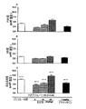
1から100mg/kgの間の用量のホスホテトラヒドロピラン試験化合物(式1)による処置により、効果的に体重増加が改善し、BALF中の総細胞数が低減し、肺胞壁厚およびコラーゲン沈着により判定したところ肺線維症全体が減少している。図2を参照されたい。 Treatment with a dose of phosphotetrahydropyran test compound (Formula 1) between 1 and 100 mg / kg effectively improves weight gain, reduces the total number of cells in BALF, alveolar wall thickness and collagen deposition When judged, the overall pulmonary fibrosis is decreasing. Please refer to FIG.
(実施例9)
ブレオマイシンマウス肺損傷モデル(モデル2)における活性
8週齢の雄のC57BL/6マウスをマイクロアイソレーターケージ中で12時間の昼/夜サイクルで飼育する。飼料および水は自由に摂取させる。50μLの食塩水中のブレオマイシンの単回負荷用量(5mU/グラム)を、0日目に気管内に点滴注入する。対照動物に50μLの食塩水を投与する。皮下に挿入した浸透圧ポンプにより投与する様々な用量のホスホテトラヒドロピラン試験化合物(式1)で動物の群を処理する。実験の過程における体重減少についてマウスを試験する。26日目にマウスを屠殺し、肺気道における細胞流入評価のためにBALFを採取する。総細胞数ならびに表現型を評価する。コラーゲン沈着を含めた肺病理組織を標準的方法に従って評価する。26日目に得た血漿におけるTGF−ベータの活性レベルおよび総レベルを測定する。
Example 9
Activity in Bleomycin Mouse Lung Injury Model (Model 2) Eight week old male C57BL / 6 mice are housed in a microisolator cage with a 12 hour day / night cycle. Feed and water are freely available. A single loading dose of bleomycin (5 mU / gram) in 50 μL saline is instilled intratracheally on day 0. Control animals receive 50 μL saline. Groups of animals are treated with various doses of phosphotetrahydropyran test compound (formula 1) administered by osmotic pump inserted subcutaneously. Mice are tested for weight loss during the course of the experiment. On day 26, mice are sacrificed and BALF is collected for assessment of cell influx in the lung airways. Assess total cell number as well as phenotype. Lung pathological tissue including collagen deposition is assessed according to standard methods. The activity level and total level of TGF-beta in the plasma obtained on day 26 is measured.
1から100mg/kgの間の用量のホスホテトラヒドロピラン試験化合物(式1)による処置により、効果的に体重増加が改善し、BALF中の総細胞数が低減し、コラーゲン沈着および血漿TGF−ベータレベルの減少により判定したところ肺線維症全体が減少している。図3および4を参照されたい。 Treatment with a phosphotetrahydropyran test compound (Formula 1) at a dose between 1 and 100 mg / kg effectively improved weight gain, reduced total cell number in BALF, collagen deposition and plasma TGF-beta levels As a result of the decrease, the overall pulmonary fibrosis decreased. See FIGS. 3 and 4.
(実施例10)
ブレオマイシンマウス肺損傷モデル(モデル3)における活性
実施例9に記載の実験に従って、8週齢の雄のC57BL/6マウスをマイクロアイソレーターケージ中で12時間の昼/夜サイクルで飼育する。飼料および水は自由に摂取させる。50μLの食塩水中のブレオマイシンの単回負荷用量(5mU/グラム)を、0日目に気管内に点滴注入する。対照動物に50μLの食塩水を投与する。26日間の毎日の肺への直接的な気管内投与により、PBSなどの好適なビヒクルに溶解した様々な濃度のホスホテトラヒドロピラン試験化合物(式1)で動物の群を処理する。26日目にマウスを屠殺し、肺気道における細胞流入評価のためにBALFを採取する。総細胞数ならびに表現型を評価する。コラーゲン沈着を含めた肺病理組織を標準的方法に従って評価する。26日目に得た血漿におけるTGF−ベータの活性レベルおよび総レベルを測定する。1から100mg/kgの間の用量のホスホテトラヒドロピラン試験化合物(式1)による処置により、効果的に体重増加が改善し、BALF中の総細胞数が低減し、コラーゲン沈着により判定したところ肺線維症全体が減少し、血漿TGF−ベータレベルが減少している。
(Example 10)
Activity in Bleomycin Mouse Lung Injury Model (Model 3) According to the experiment described in Example 9, 8-week old male C57BL / 6 mice are housed in a microisolator cage with a 12 hour day / night cycle. Feed and water are freely available. A single loading dose of bleomycin (5 mU / gram) in 50 μL saline is instilled intratracheally on day 0. Control animals receive 50 μL saline. Groups of animals are treated with various concentrations of phosphotetrahydropyran test compound (Formula 1) dissolved in a suitable vehicle such as PBS by direct intratracheal administration to the lungs daily for 26 days. On day 26, mice are sacrificed and BALF is collected for assessment of cell influx in the lung airways. Assess total cell number as well as phenotype. Lung pathological tissue including collagen deposition is assessed according to standard methods. The activity level and total level of TGF-beta in the plasma obtained on day 26 is measured. Treatment with a phosphotetrahydropyran test compound (Formula 1) at a dose between 1 and 100 mg / kg effectively improved weight gain, reduced the total number of cells in BALF, and lung fiber as determined by collagen deposition Overall disease is reduced and plasma TGF-beta levels are reduced.
(実施例11)
ブレオマイシンマウス肺損傷モデルにおける併用療法
実施例8、9または10に記載の手順に従って、しかしホスホテトラヒドロピラン試験化合物(式1)とN−アセチルシステイン、アミノグアニジン、抗VEGF抗体、バチマスタット、ボセンタン、デキサメタゾン、ジフルオロメチルオルニチン、エタネルセプト、ゲフィチニブ、イマチニブ、メチルプレドニゾロン、ペントキシフィリン、ピルフェニドン、プレドニゾロン、ロジグリタゾン、TGF−ベータ抗体、TNF−アルファ抗体、およびビンブラスチンから選択される1種の化合物との組合せでマウスを処置することにより、同じ体重増加の改善、BALF中の総細胞数の低減、全体的な肺線維症および血漿TGF−ベータの減少を達成するために必要な両方の化合物の用量が低下した。一部の組合せについては、効果は相加的であったが、他の組合せについては、効果は相乗的であった。
(Example 11)
Combination therapy in a bleomycin mouse lung injury model According to the procedure described in Example 8, 9 or 10, but with a phosphotetrahydropyran test compound (Formula 1) and N-acetylcysteine, aminoguanidine, anti-VEGF antibody, batimastat, bosentan, dexamethasone, Mice in combination with one compound selected from difluoromethylornithine, etanercept, gefitinib, imatinib, methylprednisolone, pentoxifylline, pirfenidone, prednisolone, rosiglitazone, TGF-beta antibody, TNF-alpha antibody, and vinblastine The doses of both compounds required to achieve the same improvement in weight gain, reduced total cell number in BALF, overall pulmonary fibrosis and plasma TGF-beta reduction by treatment Decreased. For some combinations, the effect was additive, while for other combinations, the effect was synergistic.
(実施例12)
実験的緑内障濾過手術のウサギモデルにおけるホスホテトラヒドロピラン試験化合物の評価
ニュージーランド白ウサギ(18)の左眼に改良型の緑内障濾過手術を施した。この動物に、40mg/mLの濃度の術中PXS−25(群1)、0.4mg/mLの濃度の術中マイトマイシンC(MMC)(群2)、または術中試験化合物およびマイトマイシンC(MMC)(群3)を投与した。
(Example 12)
Evaluation of phosphotetrahydropyran test compounds in a rabbit model of experimental glaucoma filtration surgery The left eye of a New Zealand white rabbit (18) was subjected to improved glaucoma filtration surgery. The animals were treated with intraoperative PXS-25 (group 1) at a concentration of 40 mg / mL, intraoperative mitomycin C (MMC) (group 2) at a concentration of 0.4 mg / mL, or intraoperative test compound and mitomycin C (MMC) (group). 3) was administered.
濾過手術は左眼のみに対して実施した。MMC、試験化合物(式1)またはそれらの組合せを、フラップの切断および強膜切開術の前にWeck−Celから切り出した8×10mm(乾燥寸法)の大きいスポンジで送達した。スポンジ片を結膜と強膜の間の所定の濾過部位に3分間置いた。次いで、その領域を30mLの生理食塩水で十分灌注した。手術後の初日に、その後、濾過胞消失までこの動物を毎週再調査した。この試験は8週後に終了した。フルオレセイントレーサー試験を以下の通り実施した。ナトリウムフルオレセインを前眼房に注射し、細隙灯をベースにしたデジタルビデオカメラ装置を使用して排出経路を記録した。この動物を屠殺し、瘢痕および炎症の徴候の免疫組織化学的染色のために組織切片を調製した。結果の検討により、試験化合物(式1)が、マイトマイシンと同様に濾過胞を保護し、外科的処置の部位の周囲の健康な組織も保護したことが明らかになった。対照的に、マイトマイシンを投与した動物は、濾過胞の周囲の組織が非常に明白に壊死していた。 Filtration surgery was performed on the left eye only. MMC, test compound (Formula 1) or combinations thereof were delivered with a large sponge of 8 x 10 mm (dry dimensions) cut from Weck-Cel prior to flap cutting and sclerotomy. A piece of sponge was placed in a predetermined filtration site between the conjunctiva and sclera for 3 minutes. The area was then thoroughly irrigated with 30 mL of saline. On the first day after surgery, the animals were then reviewed weekly until the disappearance of the bleb. The study was terminated after 8 weeks. The fluorescein tracer test was conducted as follows. Sodium fluorescein was injected into the anterior chamber and the discharge route was recorded using a slit lamp-based digital video camera device. The animals were sacrificed and tissue sections were prepared for immunohistochemical staining for signs of scarring and inflammation. Examination of the results revealed that the test compound (Formula 1) protected the filter bleb as well as mitomycin and also protected the healthy tissue surrounding the surgical site. In contrast, the animals receiving mitomycin had very obvious necrosis of the tissue surrounding the filter bleb.
(実施例13)
ウサギ創傷治癒モデルにおける創傷修復の改善
(1つの投与経路につき)4匹の雌のニュージーランド白ウサギを使用する。試験の24時間前に各ウサギの背側の領域から毛皮を除去する。耳介静脈における静脈内注射により動物を麻酔する(ペントバルビトン)。注射の直前に、皮膚を70%イソプロピルアルコールで、その後、1%ヨウ素溶液で拭く。輪郭を描いた2つの部位に外科鋏を使用して1辺が約1cmの正方形の全層創傷を作製する。その切断は筋膜を貫通しない。ウサギは、単独でケージに入れる。
(Example 13)
Improved wound repair in a rabbit wound healing model Four female New Zealand white rabbits are used (per route of administration). The fur is removed from the dorsal area of each rabbit 24 hours prior to testing. Animals are anesthetized by intravenous injection in the auricular vein (pentobarbitone). Immediately prior to injection, the skin is wiped with 70% isopropyl alcohol and then with 1% iodine solution. Use a surgical scissors at the two outlined sites to create a square full-thickness wound about 1 cm on a side. The cut does not penetrate the fascia. Rabbits are placed in cages alone.
ホスホテトラヒドロピラン試験化合物(式1)は、創傷に直接的に適用する(皮膚経路;スパチュラにより適用する100mM溶液)または創傷の各側面に注射する(皮下経路;29ゲージの針を使用して皮下注射する100mM溶液)。皮膚経路については、試験アイテムの除去を回避するために、創傷が閉じるまで毎日4時間ウサギをウサギホルダーに置く。創傷が閉じるまで処置を継続する。創傷が閉じたように見えるまで創傷面積(幅×長さ)を隔日で測定する(約10日間)。次いで、動物を屠殺し、創傷を受けた組織および周囲の皮膚を切除し、厚紙に貼り付け、10%中性緩衝ホルマリン中で平らに固定する。ヘマトキシリンおよびエオシン(H&E)で染色した切片に対して病理組織検査を実施して、一般的な組織および細胞形態を検査する。 The phosphotetrahydropyran test compound (formula 1) is applied directly to the wound (skin route; 100 mM solution applied by a spatula) or injected into each side of the wound (subcutaneous route; subcutaneously using a 29 gauge needle). 100 mM solution to be injected). For the skin route, place the rabbit in the rabbit holder daily for 4 hours until the wound closes to avoid removal of the test item. Treatment is continued until the wound is closed. The wound area (width x length) is measured every other day until the wound appears closed (approximately 10 days). The animals are then sacrificed and the wounded tissue and surrounding skin are excised, affixed to cardboard and fixed flat in 10% neutral buffered formalin. Histopathology is performed on sections stained with hematoxylin and eosin (H & E) to examine general tissue and cell morphology.
ホスホテトラヒドロピラン試験化合物(式1)による処置により、過剰な瘢痕形成が低減し、瘢痕の視覚的品質が改善し、傷害部位での皮膚線維症が低減している。 Treatment with the phosphotetrahydropyran test compound (Formula 1) reduces excessive scar formation, improves the visual quality of the scar, and reduces dermal fibrosis at the site of injury.
(実施例14)
マウスにおける急性のLPS誘導性の肺炎症に対する試験化合物の評価。
年齢を適合させた無菌の雄のC57Bl/6マウス(25.30±0.39g、6〜7週齢)をイソフルランと酸素の混合物で一時的に麻酔した。ビヒクル処置群に、リポ多糖類(LPS、3mg/kg)の気管内点滴注入(i.t.)の15分前に食塩水の静脈内注射(iv)を投与した。動物の別のセットには、LPSの1時間前に1mg/kgまたは2.5mg/kgのデキサメタゾンのi.v.注射を投与した。各動物に、LPS点滴注入の15分前に5、15および30mg/kg(iv)のホスホテトラヒドロピラン試験化合物を投与した。別個の群のマウスを擬似群(食塩水/PBS)として使用し、食塩水のi.v.注射およびPBS(リン酸塩緩衝食塩水)のi.t.注射で処理した。
(Example 14)
Evaluation of test compounds against acute LPS-induced lung inflammation in mice.
Age-matched, sterile male C57B1 / 6 mice (25.30 ± 0.39 g, 6-7 weeks old) were temporarily anesthetized with a mixture of isoflurane and oxygen. The vehicle-treated group received intravenous saline injection (iv) 15 minutes prior to intratracheal instillation (it) of lipopolysaccharide (LPS, 3 mg / kg). Another set of animals included i.d. of 1 mg / kg or 2.5 mg /
LPS投与の3時間後、動物をウレタン(25%、100mL/g体重)で麻酔した。心穿刺(≒1mL)により血液試料を採取し、各動物を頚椎脱臼により屠殺した。気管にカニューレを挿入し、EDTA(1mM)を含有する2mLのPBSで肺を洗浄した。各0.5mLの4回の穏やかな注入および吸引により洗浄液を回収し、遠心分離した。Neubauerチャンバを使用して新鮮な液体検体で総細胞数をTurk溶液で測定した。サイトスピン法を使用して分化細胞数を測定した。気管支肺胞洗浄(BAL)上清のアリコートをさらに処置することなく−80℃で保存した。 Three hours after LPS administration, the animals were anesthetized with urethane (25%, 100 mL / g body weight). Blood samples were collected by cardiac puncture (≈1 mL) and each animal was sacrificed by cervical dislocation. The trachea was cannulated and the lungs were lavaged with 2 mL PBS containing EDTA (1 mM). Wash solution was collected by four gentle injections and aspirations of 0.5 mL each and centrifuged. Total cell counts were measured with Turk solution with fresh liquid specimens using Neubauer chambers. The number of differentiated cells was measured using the cytospin method. An aliquot of bronchoalveolar lavage (BAL) supernatant was stored at −80 ° C. without further treatment.
各肺葉のスライスを動物から直ちに取り出し、秤量し、ミエロペルオキシダーゼ(MPO)アッセイのために液体窒素中で凍結した、または肺水腫評価のためにオーブン中に置いた。MPO活性の測定値を白血球蓄積の指標として使用し、MPO活性の1単位を、式MPO(U/g)=Vmax/s×60/0.0113/0.5により、25℃で1μmol過酸化水素/分を分解するものと定義した。U/g湿組織として表されるMPO活性としてデータを提示した。肺水腫を判定するために、左肺葉を取り出し、秤量し、湿乾重量比測定のために60℃で48時間乾燥した。 Each lobe slice was immediately removed from the animal, weighed, frozen in liquid nitrogen for myeloperoxidase (MPO) assay, or placed in an oven for lung edema assessment. The measured value of MPO activity is used as an index of leukocyte accumulation, and one unit of MPO activity is converted to 1 μmol peroxidation at 25 ° C. according to the formula MPO (U / g) = Vmax / s × 60 / 0.0113 / 0.5. It was defined as decomposing hydrogen / min. Data were presented as MPO activity expressed as U / g wet tissue. To determine pulmonary edema, the left lung lobe was removed, weighed, and dried at 60 ° C. for 48 hours for wet-dry weight ratio measurement.
LPS対照群のBAL液から回収した炎症細胞の総数は、デキサメタゾンおよびホスホテトラヒドロピラン試験化合物(5および30mg/kg)と比較して有意に異なっていなかった。しかし、15mg/kgの用量のホスホテトラヒドロピラン試験化合物(式1;n=1およびR=2,4−ジメチルフェニル)で処理したマウスの群において細胞全体の有意な抑制が見られた(図5)。
The total number of inflammatory cells recovered from BAL fluid in the LPS control group was not significantly different compared to dexamethasone and phosphotetrahydropyran test compounds (5 and 30 mg / kg). However, significant cell-wide inhibition was seen in a group of mice treated with a phosphotetrahydropyran test compound (
ビヒクル対照群のBAL液から回収した細胞の約30%が好中球であった。試験化合物によるマウスの前処置により、ビヒクル(食塩水)対照群と比較して好中球数の有意な減少が生じた(86.7%、P<0.05)。同様に、デキサメタゾンによる動物の処置により、急性肺炎症を有する動物のBAL液への細胞流入がほとんど阻害された(図6を参照されたい)。 Approximately 30% of the cells recovered from the vehicle control group BAL fluid were neutrophils. Pretreatment of mice with the test compound resulted in a significant reduction in neutrophil count compared to the vehicle (saline) control group (86.7%, P <0.05). Similarly, treatment of animals with dexamethasone almost inhibited cell entry into the BAL fluid of animals with acute lung inflammation (see FIG. 6).
(実施例15)
マウスにおけるチオグリコレート誘導性の腹膜炎のモデルにおける試験化合物の評価
21.30±0.39gの標的範囲内の体重の無菌の雄のC57Bl/6マウス(6〜7週齢)をハロタンで麻酔し、次いで、静脈内経路(iv)により増大する用量のホスホテトラヒドロピラン試験化合物(5または15mg/kg、−15分)、デキサメタゾン(1mg/kg、−60分)またはそれぞれのビヒクル(食塩水0.9%、pH7.4)を投与した。Brewerのチオグリコレートのi.p.注射により腹膜炎症を誘導した。滅菌食塩水中にチオグリコレートを新たに調製し(3%、w/v)、1.5mLを腹腔(ip)内に注射した。3時間の処置後、各動物を頚椎脱臼により(麻酔下で)屠殺し、5mLの氷冷PBSを使用して腹膜洗浄により細胞を回収した。洗浄内容物を氷上で直ちに保存した。腹水細胞(PEC)を5mLの氷冷PBSでさらに希釈した。Neubauerチャンバで総細胞数をTurk溶液で測定し、一方、サイトスピン調製物について分化細胞の計数を実施した。腹腔における白血球蓄積の原因が動物の準備の間に発生し得る微小出血であった可能性を除外するために、腹膜洗浄が、顕微鏡検査により赤血球に対して陰性となるように注意深く確認した。
(Example 15)
Evaluation of test compounds in a model of thioglycolate-induced peritonitis in mice 21. Sterile male C57B1 / 6 mice (6-7 weeks old) weighing within the target range of 30. ± .0.39 g are anesthetized with halothane. Then, increasing doses of phosphotetrahydropyran test compound (5 or 15 mg / kg, -15 minutes), dexamethasone (1 mg / kg, -60 minutes) or the respective vehicle (saline 0. 9%, pH 7.4) was administered. Brewer's thioglycolate i. p. Peritoneal inflammation was induced by injection. Thioglycolate was freshly prepared in sterile saline (3%, w / v) and 1.5 mL was injected intraperitoneally (ip). After 3 hours of treatment, each animal was sacrificed by cervical dislocation (under anesthesia) and cells were collected by peritoneal lavage using 5 mL ice-cold PBS. The washed contents were stored immediately on ice. Ascites cells (PEC) were further diluted with 5 mL ice-cold PBS. Total cell counts were measured with Turk solution in a Neubauer chamber, while differentiated cell counts were performed on cytospin preparations. To exclude the possibility that the cause of leukocyte accumulation in the peritoneal cavity was microbleeding that could occur during animal preparation, peritoneal lavage was carefully confirmed by microscopy to be negative for red blood cells.
データは、n匹の個々の動物からの平均値(±SEM)として表す。ソフトウェアGraphpad prism v4を使用してデータの統計分析を実施した。ANOVAを使用して、その後、ダネットのポスト検定およびANOVA(独立スチューデントT検定)を使用して比較データを分析した。0.05未満の統計的P値を有意と見なした。ホスホテトラヒドロピラン試験化合物およびデキサメタゾンの用量依存的な抗炎症作用を記録した。 Data are expressed as mean values (± SEM) from n individual animals. Statistical analysis of the data was performed using the software Graphpad prism v4. Comparative data were analyzed using ANOVA followed by Dunnett's post test and ANOVA (Independent Student T test). Statistical P values less than 0.05 were considered significant. The dose-dependent anti-inflammatory effects of phosphotetrahydropyran test compound and dexamethasone were recorded.
図7は、ホスホテトラヒドロピラン試験化合物(n=1およびR=2,4−ジメチルフェニル;5〜45mg/kg、i.v.、−15分)およびデキサメタゾン(1mg/kg、i.v.、−60分)による前処置の、腹腔におけるチオグリコレート誘導性の白血球遊走に対する影響を示している。多形核(PMN)(A)、単核(MN)動員(B)および細胞全体(C)の結果を腹腔当たりの細胞数として示している。この結果は、11匹のマウスによる代表的実験からのものである。各時点の有意なp値を示している(ダネットのポスト検定および独立T検定)。 FIG. 7 shows phosphotetrahydropyran test compounds (n = 1 and R = 2,4-dimethylphenyl; 5-45 mg / kg, iv, −15 minutes) and dexamethasone (1 mg / kg, iv, FIG. 6 shows the effect of pretreatment with (−60 minutes) on thioglycolate-induced leukocyte migration in the peritoneal cavity. Results for polymorphonuclear (PMN) (A), mononuclear (MN) mobilization (B) and whole cells (C) are shown as number of cells per abdominal cavity. This result is from a representative experiment with 11 mice. Significant p values at each time point are shown (Dunnett's post test and independent T test).
図7にまとめているデータは、チオグリコレート、デキサメタゾン(1mg/kg)または試験化合物のip注射後の最初の3時間の間に腹腔において観察された事象(白血球流入)の順番を示している。未処置のC57BL/6マウスにおいて、腹膜の総細胞数は、ホスホテトラヒドロピラン試験化合物(式1;n=1およびR=2,4−ジメチルフェニル;5および15mg/kg)およびデキサメタゾンで処理したマウスより著しく高かった。
The data summarized in FIG. 7 shows the sequence of events observed in the peritoneal cavity (leukocyte influx) during the first 3 hours after ip injection of thioglycolate, dexamethasone (1 mg / kg) or test compound. . In untreated C57BL / 6 mice, the total number of peritoneal cells was measured in mice treated with phosphotetrahydropyran test compounds (
(実施例16)
ブレオマイシン誘導性の強皮症動物モデルにおける活性
この手順は、非特許文献4に記載の方法に従う。100μLのブレオマイシン(リン酸塩緩衝食塩水(PBS)中の0.1単位/mL)またはPBSを、C57Bl/6マウスの剪毛した背の単一の部位に1日1回4週間皮下注射する。この実験の最後に、マウスをCO2投与により屠殺し、組織学的、免疫組織化学的、およびヒドロキシプロリンアッセイのために皮膚試料を採取する。
(Example 16)
Activity in bleomycin-induced scleroderma animal model This procedure follows the method described in [4]. 100 μL of bleomycin (0.1 units / mL in phosphate buffered saline (PBS)) or PBS is injected subcutaneously once a day for 4 weeks into a single site on the shaved back of C57B1 / 6 mice. At the end of this experiment, mice are sacrificed by CO 2 administration and skin samples are taken for histological, immunohistochemical, and hydroxyproline assays.
皮膚の生体力学的性質の定量分析を実施する。硬さが皮膚の生体力学的性質を反映すると判定する。各動物を麻酔し、それらの背を剪毛し、脱毛する。ガラス試験チャンバをマウスの背の上に載せる。このガラスチャンバをBTC−2000動的皮膚分析器に接続し、この分析器が真空を発生させ、皮膚上の関心部位にレーザー光を向ける。ブレオマイシン処置の最後に皮膚の硬さを測定する。 Perform quantitative analysis of skin biomechanical properties. It is determined that the hardness reflects the biomechanical properties of the skin. Each animal is anesthetized and their backs shaved and depilated. Place the glass test chamber on the back of the mouse. The glass chamber is connected to a BTC-2000 dynamic skin analyzer, which creates a vacuum and directs laser light to the site of interest on the skin. Skin hardness is measured at the end of bleomycin treatment.
ホスホテトラヒドロピラン試験化合物(式1)を、10から100mg/kgの用量で2週間1日2回投与する(ip)。追加の群を、1日1回2週間皮下注射により投与して、ホスホテトラヒドロピラン試験化合物(式1)の局所投与により処置する。4週の実験後、皮膚の切片(0.5μm)を、ミクロトームを使用して切断し、Superfrost Plusスライド上に採取した。次いで、各切片をキシレン中で脱蝋し、下降濃度のアルコール中での連続的液浸により再水和する。切片をヘマトキシリンおよびエオシン(H&E)で染色する。H&Eにより核が青色に染色され、ECM成分および細胞質は、様々な色度のピンク色で染色される。試験化合物(式1)の炎症に対する効果は、0〜3スケールでグレード分けする。0=炎症細胞なし、1=わずかな炎症細胞、2=中程度の炎症細胞、3=大量の炎症細胞。試験化合物(式1)で処置したマウスからの皮膚はグレード1から2であるが、試験化合物を投与しなかったブレオマイシンで処置したマウスはグレード3である。
The phosphotetrahydropyran test compound (Formula 1) is administered twice a day for two weeks at a dose of 10 to 100 mg / kg (ip). Additional groups are treated by topical administration of the phosphotetrahydropyran test compound (Formula 1), administered once daily by subcutaneous injection for 2 weeks. After 4 weeks of experiment, skin sections (0.5 μm) were cut using a microtome and collected on Superfrost Plus slides. Each section is then dewaxed in xylene and rehydrated by continuous immersion in decreasing concentrations of alcohol. Sections are stained with hematoxylin and eosin (H & E). The nuclei are stained blue by H & E, and the ECM components and cytoplasm are stained with pinks of various chromaticities. The effect of the test compound (Formula 1) on inflammation is graded on a 0-3 scale. 0 = no inflammatory cells, 1 = slight inflammatory cells, 2 = moderate inflammatory cells, 3 = abundant inflammatory cells. Skin from mice treated with the test compound (Formula 1) is
ホスホテトラヒドロピラン試験化合物(式1)のコラーゲン合成に対する効果を評価するために、ワンギーソン染色液を使用する半定量分析を使用する。ワンギーソン染色液により、成熟コラーゲン線維については深紅色が、未成熟コラーゲン線維についてはピンク色が生じる。筋肉およびフィブリンは黄色に見え、核は黒色である。コラーゲン線維の量は、以下のスケールを使用して報告している。0=コラーゲン線維なし、1=わずかなコラーゲン線維、2=中程度の量のコラーゲン線維、3=過剰量のコラーゲン線維。試験化合物(式1)で処置したマウスからの皮膚はグレード1から2であるが、試験化合物を投与しなかったブレオマイシンで処置したマウスはグレード3である。
In order to evaluate the effect of the phosphotetrahydropyran test compound (Formula 1) on collagen synthesis, a semi-quantitative analysis using Wangyson stain is used. One-Geeson stain produces a deep red color for mature collagen fibers and a pink color for immature collagen fibers. Muscle and fibrin appear yellow and the nucleus is black. The amount of collagen fibers is reported using the following scale. 0 = no collagen fibers, 1 = slight collagen fibers, 2 = moderate amount of collagen fibers, 3 = excess amount of collagen fibers. Skin from mice treated with the test compound (Formula 1) is
上記のように切片を切断し、処理する。文献の手順に従ってα−SMAの免疫標識を実施し、試験化合物(式1)のα−SMA発現に対する効果を、以下のスケールを使用して0〜3のスケールでグレード分けする。0=α−SMAを発現する細胞なし、1=わずかなα−SMAを発現する細胞、2=中程度の量のα−SMAを発現する細胞、3=大量の数のα−SMAを発現する細胞。試験化合物(式1)で処置したマウスからの皮膚はグレード1から2であるが、試験化合物を投与しなかったブレオマイシンで処置したマウスはグレード3である。
Sections are cut and processed as described above. Immunolabeling of α-SMA is performed according to literature procedures, and the effect of the test compound (Formula 1) on α-SMA expression is graded on a 0-3 scale using the following scale. 0 = no cells expressing α-SMA, 1 = cells expressing a little α-SMA, 2 = cells expressing a moderate amount of α-SMA, 3 = expressing a large number of α-SMA cell. Skin from mice treated with the test compound (Formula 1) is
公開されている手順に従って、創傷組織におけるコラーゲン合成のマーカーとしてヒドロキシプロリンアッセイを実施した。皮膚組織を食塩水においてホモジナイズし、120℃で30分、2N NaOHで加水分解し、クロラミンTおよびエールリッヒ試薬を使用するノイマンおよびローガンの反応の変法により、ヒドロキシプロリン標準曲線を使用して、550nmで測定しながらヒドロキシプロリン含量を測定する。各値は、ヒドロキシプロリンのμg/タンパク質のmgとして表している。1と100mg/kg−日の間の濃度の試験化合物(式1)により、ヒドロキシプロリン含量が有意に低減した。 A hydroxyproline assay was performed as a marker of collagen synthesis in wound tissue according to published procedures. Skin tissue is homogenized in saline, hydrolyzed with 2N NaOH for 30 minutes at 120 ° C., and modified using Neumann and Logan's reaction using Chloramine T and Ehrlich's reagent using a hydroxyproline standard curve at 550 nm. The hydroxyproline content is measured while measuring at 1. Each value is expressed as μg hydroxyproline / mg protein. Test compound (Formula 1) at a concentration between 1 and 100 mg / kg-day significantly reduced the hydroxyproline content.
(実施例17)
糖尿病誘導性の腎線維症における活性
C57Bl/6Jマウスを標準的な動物飼育条件下で普通の食餌で維持した。マウスにクエン酸ナトリウム緩衝液(pH4.5)中のSTZ(Sigma、St Louis、MO)の腹腔内注射を2日間連続で(125mg/kg/日)投与した。一般的なグルコース計(Roche)を使用して尾静脈サンプリングにより血糖を測定した。糖尿病は、STZの後の2週間の16mMより高い朝の血糖読取り値と定義した。血糖レベルが30mMを超えると、糖尿病マウスに0.4Uのインスリン(Novo Nordisk)を隔日に投与して、血糖レベルを高血糖の範囲(16〜30mM)内に維持しながら体重減少を防止した。STZ後の4つの時点(2、8、12および18週間)で屠殺した10匹のマウスの群において糖尿病性腎障害を評価した。正常な26週齢マウス(n=10)の群を対照として使用した。
(Example 17)
Activity in Diabetes-Induced Renal Fibrosis C57B1 / 6J mice were maintained on a normal diet under standard animal rearing conditions. Mice received intraperitoneal injection of STZ (Sigma, St Louis, MO) in sodium citrate buffer (pH 4.5) for 2 consecutive days (125 mg / kg / day). Blood glucose was measured by tail vein sampling using a common glucose meter (Roche). Diabetes was defined as a morning blood glucose reading higher than 16 mM for 2 weeks after STZ. When blood glucose levels exceeded 30 mM, diabetic mice were administered 0.4 U insulin (Novo Nordisk) every other day to prevent weight loss while maintaining blood glucose levels within the hyperglycemic range (16-30 mM). Diabetic nephropathy was evaluated in groups of 10 mice sacrificed at 4 time points (2, 8, 12 and 18 weeks) after STZ. A group of normal 26 week old mice (n = 10) was used as a control.
ホスホテトラヒドロピラン試験化合物(式1)を、1から100mg/kgの用量で(ip)で1日2回試験の期間投与する、または血液中で0.01から100μMの範囲のレベルでのホスホテトラヒドロピラン試験化合物(式1)の安定な全身曝露を伴う皮下に挿入した浸透圧ポンプで投与する。 The phosphotetrahydropyran test compound (Formula 1) is administered twice daily for a period of the test at a dose of 1 to 100 mg / kg (ip) or at a level in the range of 0.01 to 100 μM The pyran test compound (Formula 1) is administered by an osmotic pump inserted subcutaneously with stable systemic exposure.
免疫組織病理学により評価したところ、ホスホテトラヒドロピラン試験化合物(式1)による処置により、糸球体肥大、細胞過多および糸球体間質拡大が低減し、尿細管拡張および萎縮が減少し、間質拡大が低減している。腎実質におけるフィブロネクチン沈着およびコラーゲン沈着は、ホスホテトラヒドロピラン試験化合物(式1)で処置した動物において大きく低減した。 As assessed by immunohistopathology, treatment with the phosphotetrahydropyran test compound (Formula 1) reduced glomerular hypertrophy, cellular hypertrophy and glomerular stromal expansion, decreased tubule dilation and atrophy, and stromal expansion. Has been reduced. Fibronectin and collagen deposition in the renal parenchyma was greatly reduced in animals treated with the phosphotetrahydropyran test compound (Formula 1).
(実施例18)
ラットおよびイヌにおける本発明の化合物のバイオアベイラビリティーの測定。
(Example 18)
Measurement of the bioavailability of the compounds of the invention in rats and dogs.
ホスホテトラヒドロピラン試験化合物(式1;n=1およびR=2,4−ジメチルフェニル)を、強制飼養により10mg/kgで、または静脈内に1mg/kgで、ラットに投与した)。血液試料を15および30分に、ならびに1、2、3、4、8、12および24時間に採取した。50%アセトニトリル/水(20μL)の添加により血漿試料を調製した。トリクロロ酢酸(水中の20%、40μL)の添加、ボルテックス(20s)および4℃で60分の保存後の微量遠心器における5分の遠心分離(10000rpm)によりタンパク質沈殿を実施した。上清をその後分離し、LC−MS分析のために25μLを直接カラムに注入した。既知の濃度の試験化合物を加えた血液試料と比較したデータの分析により、試験化合物のバイオアベイラビリティーが7.7%であることが明らかになった。
Phosphotetrahydropyran test compounds (
この試験を式IIの試験化合物(X=H、P=tert−ブチル)で繰り返した場合、ホスホテトラヒドロピラン試験化合物(式1;n=1およびR=2,4−ジメチルフェニル)の濃度を血漿中で定量化すると、バイオアベイラビリティーを測定したところ52%であった。
When this test was repeated with a test compound of formula II (X = H, P = tert-butyl), the concentration of the phosphotetrahydropyran test compound (
Claims (21)
式中、nは0から3の整数であり、−O(CH2)nR基はアキシアルまたはエクアトリアル位にあり、Rは低級アルキル、または任意選択的に置換されたヘテロアリールもしくは任意選択的に置換されたアリールであり、ここで置換基は−Cl、−F、CF3、CH3、CH2CH3、−OCH3、−OCF3、−(CH2)mCO2R1、−(CH2)mOR2、−(CH2)mCONHR2、−(CH2)mNHR2、および−(CH2)mCONR2R3(式中、mは0から3の整数であり、R1はH、アルキルおよびアリールからなる群から選択され、R2およびR3は独立して、H、アルキル、アリールおよびアシルからなる群から選択される)からなる群から選択される
ことを特徴とする方法。 A method of modulating inflammation and wound healing processes in a subject comprising an effective amount of a phosphotetrahydropyran compound, a salt of a phosphotetrahydropyran compound or a phosphonate prodrug of a phosphonotetrahydropyran compound, and an acceptable pharmaceutical carrier Wherein the phosphotetrahydropyran compound is of formula I:
Wherein n is an integer from 0 to 3, the —O (CH 2 ) n R group is in the axial or equatorial position, R is lower alkyl, or optionally substituted heteroaryl or optionally Substituted aryl, wherein the substituents are —Cl, —F, CF 3 , CH 3 , CH 2 CH 3 , —OCH 3 , —OCF 3 , — (CH 2 ) m CO 2 R 1 , — ( CH 2 ) m OR 2 , — (CH 2 ) m CONHR 2 , — (CH 2 ) m NHR 2 , and — (CH 2 ) m CONR 2 R 3 , wherein m is an integer from 0 to 3; R 1 is selected from the group consisting of H, alkyl and aryl, and R 2 and R 3 are independently selected from the group consisting of H, alkyl, aryl and acyl) And how to.
ことを特徴とする方法。 A method of treating pulmonary fibrosis comprising administering to a subject in need thereof an effective amount of a composition comprising a compound of formula I, a sodium salt of a compound of formula I, a phosphonate prodrug of a compound of formula I Including steps,
ことを特徴とする方法。 A method of treating pulmonary fibrosis, wherein an effective amount of a composition comprising a compound of formula I, a sodium salt of a compound of formula I, a phosphonate prodrug of a compound of formula I and N- Acetylcysteine, aminoguanidine, anti-VEGF antibody, batimastat, bosentan, dexamethasone, difluoromethylornithine, etanercept, gefitinib, imatinib, methylprednisolone, pentoxyphilin, pirfenidone, prednisolone, rosiglitazone, TGF-beta antibody, TNF-alpha antibody, And co-administering one compound selected from vinblastine,
式中、nは0から3の整数であり、−O(CH2)nR基はアキシアルまたはエクアトリアル位にあり、Rは低級アルキル、または任意選択的に置換されたヘテロアリールもしくは任意選択的に置換されたアリールであり、ここで置換基は−Cl、−F、CF3、CH3、CH2CH3、−OCH3、−OCF3、−(CH2)mCO2R1、−(CH2)mOR2、−(CH2)mCONHR2、−(CH2)mNHR2、および−(CH2)mCONR2R3(式中、mは0から3の整数であり、R1はH、アルキルおよびアリールからなる群から選択され、R2およびR3は独立して、H、アルキル、アリールおよびアシルからなる群から選択される)からなる群から選択される
ことを特徴とする使用。 In the manufacture of a medicament for the treatment of pulmonary fibrosis, the formula:
Wherein n is an integer from 0 to 3, the —O (CH 2 ) n R group is in the axial or equatorial position, R is lower alkyl, or optionally substituted heteroaryl or optionally Substituted aryl, wherein the substituents are —Cl 2 , —F, CF 3 , CH 3 , CH 2 CH 3 , —OCH 3 , —OCF 3 , — (CH 2 ) m CO 2 R 1 , — ( CH 2 ) m OR 2 , — (CH 2 ) m CONHR 2 , — (CH 2 ) m NHR 2 , and — (CH 2 ) m CONR 2 R 3 , wherein m is an integer from 0 to 3; R 1 is selected from the group consisting of H, alkyl and aryl, and R 2 and R 3 are independently selected from the group consisting of H, alkyl, aryl and acyl) Use with.
Applications Claiming Priority (3)
| Application Number | Priority Date | Filing Date | Title |
|---|---|---|---|
| US17341609P | 2009-04-28 | 2009-04-28 | |
| US61/173,416 | 2009-04-28 | ||
| PCT/IB2010/000960 WO2010125445A1 (en) | 2009-04-28 | 2010-04-28 | Phosphotetrahydropyran compounds for the treatment of wounds and fibrotic disorders |
Publications (2)
| Publication Number | Publication Date |
|---|---|
| JP2012525369A true JP2012525369A (en) | 2012-10-22 |
| JP2012525369A5 JP2012525369A5 (en) | 2013-06-20 |
Family
ID=43031758
Family Applications (1)
| Application Number | Title | Priority Date | Filing Date |
|---|---|---|---|
| JP2012507839A Pending JP2012525369A (en) | 2009-04-28 | 2010-04-28 | Phosphotetrahydropyran compounds for the treatment of wounds and fibrotic disorders |
Country Status (10)
| Country | Link |
|---|---|
| US (1) | US8557783B2 (en) |
| EP (1) | EP2424542A4 (en) |
| JP (1) | JP2012525369A (en) |
| KR (1) | KR20120049183A (en) |
| AU (1) | AU2010243314C1 (en) |
| CA (1) | CA2760253A1 (en) |
| IL (1) | IL215990A0 (en) |
| NZ (1) | NZ596419A (en) |
| SG (1) | SG175816A1 (en) |
| WO (1) | WO2010125445A1 (en) |
Families Citing this family (5)
| Publication number | Priority date | Publication date | Assignee | Title |
|---|---|---|---|---|
| CA2926480A1 (en) * | 2012-11-15 | 2014-05-22 | Tufts University | Methods, compositions and kits for treating, modulating, or preventing ocular angiogenesis or fibrosis in a subject using a galectin protein inhibitor |
| US20210315914A1 (en) * | 2018-08-10 | 2021-10-14 | New York University | Use of igf-2 receptor agonist ligands for treatment of angelman syndrome and autism |
| AU2019404061A1 (en) | 2018-12-19 | 2021-07-22 | The Board Of Trustees Of The Leland Stanford Junior University | Bifunctional molecules for lysosomal targeting and related compositions and methods |
| CN113368106B (en) * | 2020-02-25 | 2023-09-29 | 渐宽(苏州)生物科技有限公司 | Use of etomod in medicine for preventing and treating idiopathic pulmonary fibrosis |
| CN113288890B (en) * | 2021-06-21 | 2022-02-22 | 南方医科大学南方医院 | Application of N-acetyl-L-cysteine in preparation of medicine for preventing and/or treating sports injury |
Citations (2)
| Publication number | Priority date | Publication date | Assignee | Title |
|---|---|---|---|---|
| JP2004501985A (en) * | 2000-07-11 | 2004-01-22 | ファームアクシス・プロプライエタリー・リミテッド | New compounds and methods |
| WO2004104015A2 (en) * | 2003-05-20 | 2004-12-02 | Pharmaxis Pty Ltd. | Mannose-6-phosphonate compounds for the treatment of inflammatory diseases |
Family Cites Families (2)
| Publication number | Priority date | Publication date | Assignee | Title |
|---|---|---|---|---|
| FR2930942B1 (en) | 2008-05-07 | 2011-03-04 | Centre Nat Rech Scient | NEW USES OF D-MANNOPYRANOSIS DERIVATIVES OF ANGIOGENESIS |
| FR2930943B1 (en) | 2008-05-07 | 2011-03-18 | Centre Nat Rech Scient | NOVEL USES OF D-MANNOPYRANOSIS DERIVATIVES INHIBITORS OF ANGIOGENESIS |
-
2010
- 2010-04-28 JP JP2012507839A patent/JP2012525369A/en active Pending
- 2010-04-28 US US13/318,065 patent/US8557783B2/en not_active Expired - Fee Related
- 2010-04-28 CA CA2760253A patent/CA2760253A1/en not_active Abandoned
- 2010-04-28 SG SG2011079274A patent/SG175816A1/en unknown
- 2010-04-28 AU AU2010243314A patent/AU2010243314C1/en not_active Ceased
- 2010-04-28 EP EP10769382A patent/EP2424542A4/en not_active Withdrawn
- 2010-04-28 NZ NZ596419A patent/NZ596419A/en not_active IP Right Cessation
- 2010-04-28 WO PCT/IB2010/000960 patent/WO2010125445A1/en active Application Filing
- 2010-04-28 KR KR1020117028281A patent/KR20120049183A/en not_active Application Discontinuation
-
2011
- 2011-10-27 IL IL215990A patent/IL215990A0/en unknown
Patent Citations (2)
| Publication number | Priority date | Publication date | Assignee | Title |
|---|---|---|---|---|
| JP2004501985A (en) * | 2000-07-11 | 2004-01-22 | ファームアクシス・プロプライエタリー・リミテッド | New compounds and methods |
| WO2004104015A2 (en) * | 2003-05-20 | 2004-12-02 | Pharmaxis Pty Ltd. | Mannose-6-phosphonate compounds for the treatment of inflammatory diseases |
Non-Patent Citations (1)
| Title |
|---|
| JPN6014022046; IL Farmaco 60, 2005, 721-725 * |
Also Published As
| Publication number | Publication date |
|---|---|
| AU2010243314C1 (en) | 2015-11-12 |
| WO2010125445A1 (en) | 2010-11-04 |
| AU2010243314B2 (en) | 2015-07-02 |
| AU2010243314A1 (en) | 2011-12-01 |
| IL215990A0 (en) | 2012-01-31 |
| SG175816A1 (en) | 2011-12-29 |
| NZ596419A (en) | 2014-01-31 |
| US20120045455A1 (en) | 2012-02-23 |
| KR20120049183A (en) | 2012-05-16 |
| US8557783B2 (en) | 2013-10-15 |
| CA2760253A1 (en) | 2010-11-04 |
| EP2424542A4 (en) | 2012-12-19 |
| EP2424542A1 (en) | 2012-03-07 |
Similar Documents
| Publication | Publication Date | Title |
|---|---|---|
| Carnesecchi et al. | A key role for NOX4 in epithelial cell death during development of lung fibrosis | |
| Honjo et al. | Potential role of Rho-associated protein kinase inhibitor Y-27632 in glaucoma filtration surgery | |
| KR20150115959A (en) | Oxylipin compounds for the treatment of ophthalmic conditions | |
| KR20120046099A (en) | Methods of inhibiting fibrogenesis and treating fibrotic disease | |
| PT2151438E (en) | Pharmaceutical containing ppar-delta agonist | |
| US11739326B2 (en) | RUNX1 inhibition for treatment of proliferative vitreoretinopathy and conditions associated with epithelial to mesenchymal transition | |
| US8557783B2 (en) | Phosphotetrahydropyran compounds for the treatment of wounds and fibrotic disorders | |
| Colangelo et al. | The effects of polydeoxyribonucleotide on wound healing and tissue regeneration: a systematic review of the literature | |
| WO2021066144A1 (en) | Pharmaceutical drug containing heterocyclidene acetamide derivative | |
| Cinar et al. | CB1R and iNOS are distinct players promoting pulmonary fibrosis in Hermansky–Pudlak syndrome | |
| CA2896073C (en) | Stimulation and enhancement of regeneration of tissues | |
| US20120046333A1 (en) | Methods and Compositions of PI-3 Kinase Inhibitors for Treating Fibrosis | |
| US20210379078A1 (en) | Killing Senescent Cells And Treating Senescence-Associated Conditions Using A Bcl Inhibitor And An Mcl-1 Inhibitor | |
| WO2018115319A2 (en) | Inhibitors of cytochrome p450 family 7 subfamily b member 1 (cyp7b1) for use in treating diseases | |
| Liu et al. | Celastrol Alleviates Corneal Stromal Fibrosis by Inhibiting TGF-β1/Smad2/3-YAP/TAZ Signaling After Descemet Stripping Endothelial Keratoplasty | |
| Nikhalashree et al. | Anti-glaucoma medications lowered decorin and altered profibrotic proteins in human tenon's fibroblasts | |
| JP2023518375A (en) | Method of treatment | |
| US8283323B2 (en) | Withanolide compounds as inhibitors of fibrosis and identification of molecular targets for anti-fibrotic drug development | |
| JP7429500B2 (en) | Corneal epithelial disorder therapeutic agent | |
| Xu et al. | The NLRP3 Activation in Infiltrating Macrophages Contributes to Corneal Fibrosis by Inducing TGF-β1 Expression in the Corneal Epithelium | |
| WO2013090991A1 (en) | Tgf-beta therapy | |
| CA3070938A1 (en) | Drug for treating or preventing disorder caused by tgf-.beta. signalling, and application thereof | |
| US20240043396A1 (en) | Methods of treating ocular fibrotic pathologies | |
| WO2023201312A2 (en) | Methods of treating ocular fibrotic pathologies | |
| JP5685751B2 (en) | Subconjunctival fibroblast proliferation inhibitor and method for inhibiting subconjunctival fibroblast proliferation |
Legal Events
| Date | Code | Title | Description |
|---|---|---|---|
| A521 | Request for written amendment filed |
Free format text: JAPANESE INTERMEDIATE CODE: A523 Effective date: 20130426 |
|
| A621 | Written request for application examination |
Free format text: JAPANESE INTERMEDIATE CODE: A621 Effective date: 20130426 |
|
| A131 | Notification of reasons for refusal |
Free format text: JAPANESE INTERMEDIATE CODE: A131 Effective date: 20140603 |
|
| A601 | Written request for extension of time |
Free format text: JAPANESE INTERMEDIATE CODE: A601 Effective date: 20140903 |
|
| A602 | Written permission of extension of time |
Free format text: JAPANESE INTERMEDIATE CODE: A602 Effective date: 20140910 |
|
| A601 | Written request for extension of time |
Free format text: JAPANESE INTERMEDIATE CODE: A601 Effective date: 20141003 |
|
| A602 | Written permission of extension of time |
Free format text: JAPANESE INTERMEDIATE CODE: A602 Effective date: 20141010 |
|
| A521 | Request for written amendment filed |
Free format text: JAPANESE INTERMEDIATE CODE: A523 Effective date: 20141104 |
|
| A02 | Decision of refusal |
Free format text: JAPANESE INTERMEDIATE CODE: A02 Effective date: 20150512 |