JP2012515931A5 - - Google Patents
Download PDFInfo
- Publication number
- JP2012515931A5 JP2012515931A5 JP2012516238A JP2012516238A JP2012515931A5 JP 2012515931 A5 JP2012515931 A5 JP 2012515931A5 JP 2012516238 A JP2012516238 A JP 2012516238A JP 2012516238 A JP2012516238 A JP 2012516238A JP 2012515931 A5 JP2012515931 A5 JP 2012515931A5
- Authority
- JP
- Japan
- Prior art keywords
- substrate
- slide
- blood
- cells
- cell
- Prior art date
- Legal status (The legal status is an assumption and is not a legal conclusion. Google has not performed a legal analysis and makes no representation as to the accuracy of the status listed.)
- Pending
Links
- 210000004027 cells Anatomy 0.000 claims description 89
- 210000000265 Leukocytes Anatomy 0.000 claims description 79
- 210000003324 RBC Anatomy 0.000 claims description 66
- 210000003743 Erythrocytes Anatomy 0.000 claims description 65
- 210000002865 immune cell Anatomy 0.000 claims description 64
- 210000004369 Blood Anatomy 0.000 claims description 53
- 239000008280 blood Substances 0.000 claims description 53
- 239000000758 substrate Substances 0.000 claims description 41
- 210000001772 Blood Platelets Anatomy 0.000 claims description 24
- 238000010186 staining Methods 0.000 claims description 22
- 210000000601 Blood Cells Anatomy 0.000 claims description 21
- 239000012530 fluid Substances 0.000 claims description 18
- 238000001035 drying Methods 0.000 claims description 17
- 238000003384 imaging method Methods 0.000 claims description 13
- 102000001554 Hemoglobins Human genes 0.000 claims description 12
- 108010054147 Hemoglobins Proteins 0.000 claims description 12
- 239000003085 diluting agent Substances 0.000 claims description 11
- 238000004820 blood count Methods 0.000 claims description 8
- 239000002356 single layer Substances 0.000 claims description 8
- 230000003287 optical Effects 0.000 claims description 7
- 238000000701 chemical imaging Methods 0.000 claims description 5
- 238000005286 illumination Methods 0.000 claims description 5
- 239000010410 layer Substances 0.000 claims description 5
- 238000005259 measurement Methods 0.000 claims description 5
- 238000004140 cleaning Methods 0.000 claims description 4
- 230000000877 morphologic Effects 0.000 claims description 4
- 210000001219 anucleate thrombocyte Anatomy 0.000 claims description 3
- 238000003860 storage Methods 0.000 claims description 3
- 239000011248 coating agent Substances 0.000 claims 3
- 238000000576 coating method Methods 0.000 claims 3
- 229910052736 halogen Inorganic materials 0.000 claims 1
- 150000002367 halogens Chemical class 0.000 claims 1
- 230000002452 interceptive Effects 0.000 claims 1
- 238000007670 refining Methods 0.000 claims 1
- 238000002360 preparation method Methods 0.000 description 20
- 239000000523 sample Substances 0.000 description 19
- 239000011521 glass Substances 0.000 description 13
- 238000000034 method Methods 0.000 description 13
- 239000003153 chemical reaction reagent Substances 0.000 description 11
- 238000010191 image analysis Methods 0.000 description 10
- 238000004458 analytical method Methods 0.000 description 8
- 238000010790 dilution Methods 0.000 description 7
- 239000000306 component Substances 0.000 description 6
- 238000009826 distribution Methods 0.000 description 6
- UIIMBOGNXHQVGW-UHFFFAOYSA-M buffer Substances [Na+].OC([O-])=O UIIMBOGNXHQVGW-UHFFFAOYSA-M 0.000 description 5
- 239000000243 solution Substances 0.000 description 5
- 210000000805 Cytoplasm Anatomy 0.000 description 4
- 210000004940 Nucleus Anatomy 0.000 description 4
- 241001510071 Pyrrhocoridae Species 0.000 description 4
- OKKJLVBELUTLKV-UHFFFAOYSA-N methanol Chemical compound OC OKKJLVBELUTLKV-UHFFFAOYSA-N 0.000 description 4
- 239000000203 mixture Substances 0.000 description 4
- 238000004321 preservation Methods 0.000 description 4
- 238000004805 robotic Methods 0.000 description 4
- 239000011780 sodium chloride Substances 0.000 description 4
- 206010008958 Chronic lymphocytic leukaemia Diseases 0.000 description 3
- 208000000429 Leukemia, Lymphocytic, Chronic, B-Cell Diseases 0.000 description 3
- 210000004698 Lymphocytes Anatomy 0.000 description 3
- 210000002381 Plasma Anatomy 0.000 description 3
- 238000004422 calculation algorithm Methods 0.000 description 3
- 239000003086 colorant Substances 0.000 description 3
- 230000002209 hydrophobic Effects 0.000 description 3
- 150000003839 salts Chemical class 0.000 description 3
- 206010000880 Acute myeloid leukaemia Diseases 0.000 description 2
- 101700042850 Bacc Proteins 0.000 description 2
- 210000003651 Basophils Anatomy 0.000 description 2
- 210000003979 Eosinophils Anatomy 0.000 description 2
- 206010024324 Leukaemias Diseases 0.000 description 2
- 208000007046 Leukemia, Myeloid, Acute Diseases 0.000 description 2
- 210000001616 Monocytes Anatomy 0.000 description 2
- 210000000440 Neutrophils Anatomy 0.000 description 2
- 238000010817 Wright-Giemsa staining Methods 0.000 description 2
- 230000002159 abnormal effect Effects 0.000 description 2
- 239000000654 additive Substances 0.000 description 2
- 238000001514 detection method Methods 0.000 description 2
- 239000012470 diluted sample Substances 0.000 description 2
- 238000005516 engineering process Methods 0.000 description 2
- 238000011156 evaluation Methods 0.000 description 2
- 238000002474 experimental method Methods 0.000 description 2
- 239000007788 liquid Substances 0.000 description 2
- 239000012460 protein solution Substances 0.000 description 2
- 102000004169 proteins and genes Human genes 0.000 description 2
- 108090000623 proteins and genes Proteins 0.000 description 2
- 238000007430 reference method Methods 0.000 description 2
- 239000012266 salt solution Substances 0.000 description 2
- 230000011218 segmentation Effects 0.000 description 2
- 230000000007 visual effect Effects 0.000 description 2
- 241001502050 Acis Species 0.000 description 1
- 241001156002 Anthonomus pomorum Species 0.000 description 1
- 206010060945 Bacterial infection Diseases 0.000 description 1
- 206010061590 Blood disease Diseases 0.000 description 1
- 108091003117 Bovine Serum Albumin Proteins 0.000 description 1
- CZMRCDWAGMRECN-UGDNZRGBSA-N D-sucrose Chemical compound O[C@H]1[C@H](O)[C@@H](CO)O[C@@]1(CO)O[C@@H]1[C@H](O)[C@@H](O)[C@H](O)[C@@H](CO)O1 CZMRCDWAGMRECN-UGDNZRGBSA-N 0.000 description 1
- 229920002307 Dextran Polymers 0.000 description 1
- 229920001917 Ficoll Polymers 0.000 description 1
- 208000005209 Hematologic Disease Diseases 0.000 description 1
- 208000002250 Hematologic Neoplasms Diseases 0.000 description 1
- 241000549435 Pria Species 0.000 description 1
- 210000002966 Serum Anatomy 0.000 description 1
- 206010047461 Viral infection Diseases 0.000 description 1
- 208000001756 Virus Disease Diseases 0.000 description 1
- 238000000627 alternating current impedance spectroscopy Methods 0.000 description 1
- 238000000149 argon plasma sintering Methods 0.000 description 1
- 239000012503 blood component Substances 0.000 description 1
- 238000009534 blood test Methods 0.000 description 1
- 238000007664 blowing Methods 0.000 description 1
- 239000007853 buffer solution Substances 0.000 description 1
- 238000007635 classification algorithm Methods 0.000 description 1
- 238000010224 classification analysis Methods 0.000 description 1
- 238000007374 clinical diagnostic method Methods 0.000 description 1
- 230000003436 cytoskeletal Effects 0.000 description 1
- 238000011161 development Methods 0.000 description 1
- 230000018109 developmental process Effects 0.000 description 1
- 238000007865 diluting Methods 0.000 description 1
- 238000003113 dilution method Methods 0.000 description 1
- 201000010099 disease Diseases 0.000 description 1
- 239000006185 dispersion Substances 0.000 description 1
- 238000004043 dyeing Methods 0.000 description 1
- 230000003203 everyday Effects 0.000 description 1
- 239000000284 extract Substances 0.000 description 1
- 150000004676 glycans Polymers 0.000 description 1
- 230000005484 gravity Effects 0.000 description 1
- 238000005534 hematocrit Methods 0.000 description 1
- 201000002138 hematopoietic system disease Diseases 0.000 description 1
- 230000005660 hydrophilic surface Effects 0.000 description 1
- 238000007654 immersion Methods 0.000 description 1
- 230000003834 intracellular Effects 0.000 description 1
- 230000031700 light absorption Effects 0.000 description 1
- 230000003211 malignant Effects 0.000 description 1
- 238000004519 manufacturing process Methods 0.000 description 1
- 239000008155 medical solution Substances 0.000 description 1
- 238000002156 mixing Methods 0.000 description 1
- 230000004048 modification Effects 0.000 description 1
- 238000006011 modification reaction Methods 0.000 description 1
- 238000005457 optimization Methods 0.000 description 1
- 244000045947 parasites Species 0.000 description 1
- 230000001717 pathogenic Effects 0.000 description 1
- 230000002572 peristaltic Effects 0.000 description 1
- 229920001282 polysaccharide Polymers 0.000 description 1
- 239000005017 polysaccharide Substances 0.000 description 1
- 150000004804 polysaccharides Polymers 0.000 description 1
- 238000002203 pretreatment Methods 0.000 description 1
- 210000001995 reticulocyte Anatomy 0.000 description 1
- 238000005070 sampling Methods 0.000 description 1
- 238000004062 sedimentation Methods 0.000 description 1
- FAPWRFPIFSIZLT-UHFFFAOYSA-M sodium chloride Chemical compound [Na+].[Cl-] FAPWRFPIFSIZLT-UHFFFAOYSA-M 0.000 description 1
- 238000007619 statistical method Methods 0.000 description 1
- 238000006467 substitution reaction Methods 0.000 description 1
- 239000000725 suspension Substances 0.000 description 1
- 238000011179 visual inspection Methods 0.000 description 1
Images


Description
1.発明の分野
白血球分類計数を含む全血球計数。
1. FIELD OF THE INVENTION Whole blood counts including leukocyte classification counts.
2.従来技術の説明
全血球計数(CBC)は、米国及び世界で最も一般的に実施されている臨床検査である。現行のCBC法及び機器は、非常に進化しかつ高価であり、全てが、同様のマルチチャネル、マルチ検出器フローシステムベースの技術を使用する。ベックマン・コールター社、ロッシュ・ダイアグノスティックス社、シスメックス社、シーメンス・メディカル・ソルーションズ社、アボット社は、CBC用の自動化機器を製造している。CBC技術に対する根本的に新しい手法は、20年以上導入されていない。
2. Description of the Prior Art Whole blood count (CBC) is the most commonly performed clinical test in the United States and the world. Current CBC methods and equipment are highly evolved and expensive, and all use similar multi-channel, multi-detector flow system based technologies. Beckman Coulter, Roche Diagnostics, Sysmex, Siemens Medical Solutions, and Abbott manufacture automated equipment for CBC. No fundamentally new approach to CBC technology has been introduced for over 20 years.
現在市場に出ているCBC機器は、抗凝固処理された全血を吸引し、それを幾つかの分析ストリームに分割して、「フロー」システム(フローサイトメータ)内でCBCの異なる要素を実施する。これらの要素は、
i.赤血球(RBC)数、ヘモグロビン(Hb)、ヘマトクリット(Hct)、赤血球指数(平均赤血球容積(MCV)、平均赤血球ヘモグロビン量(MCH)及び平均赤血球ヘモグロビン濃度(MCHC))、及び、網状赤血球数を含む赤血球形態と、
ii.白血球(WBC)数及びWBC「分類」計数(好中球、リンパ球、好酸球、好塩基球、及び単球を含む異なる正常白血球のタイプ、並びに、種々の疾患状態において存在する異常なタイプのWBCの計数)と、
iii.血小板数と
を含む。
CBC equipment currently on the market draws anticoagulated whole blood and divides it into several analytical streams to implement different elements of CBC within a “flow” system (flow cytometer) To do. These elements are
i. Includes red blood cell (RBC) count, hemoglobin (Hb), hematocrit (Hct), red blood cell index (mean red blood cell volume (MCV), average red blood cell hemoglobin content (MCH) and average red blood cell hemoglobin concentration (MCHC)), and reticulocyte count Red blood cell morphology,
ii. White blood cell (WBC) counts and WBC "classification" counts (different normal white blood cell types including neutrophils, lymphocytes, eosinophils, basophils, and monocytes, and abnormal types present in various disease states WBC count)
iii. Including platelet count.
他の測定値は、RBC分布幅及び血小板分布幅のようなRBC及び血小板の主要な測定値から導出される幾つかのCBCシステムに含まれる。 Other measurements are included in some CBC systems derived from RBC and platelet major measurements such as RBC distribution width and platelet distribution width.
非血液起源の他の細胞又は寄生虫若しくは他の病原生物は、CBCの一部ではなく、「血液スミア」の目視検査中に得られる観察結果から観察され報告され得る。 Other cells or parasites or other pathogenic organisms of non-blood origin are not part of the CBC and can be observed and reported from observations obtained during visual inspection of “blood smears”.
フローベースCBC機器は、広範な較正及び制御、メンテナンス及び熟練したオペレーターを必要とし、また、試薬、消耗品、及び使い捨て用品に関連するかなりのコストがかかる。日常使用におけるこれらのシステムに関する1つの重要な問題は、血液検体の大部分が、CBCの形態的成分を完成するために更なる試験を必要とすることである。これは、自動化試験の結果が、更なる実験を必要とする結果を含むときはいつでも、熟練した技師によって実施されるスライドガラス上での血液細胞の直接視覚化を伴う。例えば、「人手による」分類計数は、有核の未成熟なRBCが見出されるか、又は、感染、白血病、他の血液疾患が疑われるWBCが見出されると、経験を積んだ観察者による細胞の直接視覚化によって実施される。 Flow-based CBC instruments require extensive calibration and control, maintenance and skilled operators, and have significant costs associated with reagents, consumables, and disposables. One important problem with these systems in everyday use is that the majority of blood samples require further testing to complete the morphological components of CBC. This involves direct visualization of blood cells on a glass slide performed by a skilled technician whenever the results of an automated test include results that require further experimentation. For example, a “manual” classification count can be used to determine the number of cells by experienced observers when a nucleated immature RBC is found or a WBC suspected of being infected, leukemia, or other blood disease is found. Performed by direct visualization.
更なる検討を必要とするこれらの検体の割合は、研究所の方針、患者集団、及び「フラッギング」基準に応じて、約27%の中央値(median rate)を有する10%〜50%の範囲にある。再試験の最も頻繁な理由は、異常な細胞タイプ又は細胞形態の存在、ウィルス若しくは細菌の感染の臨床上の若しくは他の疑い、又は、WBC、RBC若しくは血小板の数の減少である。 The percentage of these specimens that require further study ranges from 10% to 50% with a median rate of approximately 27%, depending on laboratory policy, patient population, and “flagging” criteria. It is in. The most frequent reason for retesting is the presence of abnormal cell types or cell morphology, clinical or other suspicion of viral or bacterial infection, or a reduction in the number of WBC, RBC or platelets.
以下に述べる方法は、著しく簡略化されると共により多用途のCBC機器を可能にし、このCBC機器は、WBC分類計数をもたらすRBC及びWBCの全ての形態観察結果を含むCBCの全てのパラメーターを求める。その方法は、分析のために単一層を調製する新しい方法を使用した画像分析に基づく。試料調製は、非常に容易であり、非常に少量の試薬及び簡単な使い捨てスライドガラスとともに非常に少量の全血しか必要としない。機器自体は、非常に少数の可動部を有する。フローベースシステムと比較して、この新しいシステムは、幾つかの重要な利点、すなわち、
機器の実質的により低いコストと、
消耗品及び試薬の実質的により低いコストと、
実質的により少ないオペレーター時間及び必要とされる熟練度と、
(人手によるWBC分類又は人手によるRBC形態を必要とする)反復試験のより低い割合と、
CBCパラメーターについての同等の精度(一部のパラメーターについては改善もあり得る)と、
同等又はそれ以上のスループットと、
可動部がより少ないことによる簡単な機器メンテナンスと
を有する。
The method described below is significantly simplified and allows for a more versatile CBC device that determines all parameters of the CBC, including all RBC and WBC morphological observations that result in WBC classification counts. . The method is based on image analysis using a new method of preparing a single layer for analysis. Sample preparation is very easy and requires very small amounts of whole blood with very small amounts of reagents and simple disposable glass slides. The device itself has a very small number of moving parts. Compared to the flow-based system, this new system has several important advantages:
Substantially lower cost of equipment,
Substantially lower cost of consumables and reagents,
Substantially less operator time and skill required,
A lower rate of repeated trials (requiring manual WBC classification or manual RBC form);
Equivalent accuracy for CBC parameters (may be improved for some parameters);
Equivalent or better throughput,
With simple equipment maintenance due to fewer moving parts.
重力によって沈降した又は軽く塗布された単一層内の細胞を可視化する能力のために、スライドガラス上にウェッジスミアを生成する従来のプロセスにおいて見られるような細胞の形態の破損及び変形を伴うことなく、画像分析から新しい診断情報を抽出する可能性が存在する。 Due to the ability to visualize cells within a single layer sedimented or lightly applied by gravity, without the disruption and deformation of cell morphology as seen in conventional processes that generate wedge smears on glass slides There is a possibility to extract new diagnostic information from image analysis.
細胞形態の保存は、慢性リンパ性白血病(CLL)又は急性骨髄性白血病(AML)のような血液悪性疾患を有する患者について特に重要である。標準的なウェッジスミアの調製は、悪性リンパ球又は白血球(芽球)の深刻な形態変形をもたらす可能性がある。例えば、拡大した芽球の一部は、その細胞骨格脆性のためにウェッジスミアの調製中に破裂する可能性がある。これらの、いわゆる「破損細胞」の存在は、分類計数における表記を正当化するのに十分なほどCLLにおいて頻繁に起こる。穏やかな単一層の生成システムは、芽球のより正確な認識又は白血病の早期検出を可能にするために、多数の形態学的によく保存された芽球の検出を可能にする可能性がある。 Preservation of cell morphology is particularly important for patients with hematological malignancies such as chronic lymphocytic leukemia (CLL) or acute myeloid leukemia (AML). Standard wedge smear preparation can result in severe morphological deformation of malignant lymphocytes or leukocytes (blasts). For example, some of the expanded blasts may rupture during wedge smear preparation due to their cytoskeletal brittleness. The presence of these so-called “broken cells” occurs frequently in CLL enough to justify the notation in the classification count. A gentle monolayer generation system may allow for the detection of a large number of morphologically well-preserved blasts to allow more accurate recognition of blasts or early detection of leukemia .
システムは、既知の量の血液からの事実上全てのRBC、WBC、及び血小板が計数され、その形態が調査されることを可能にする外観を、従来の染色によって生成する。したがって、システムは、CBCの定量的成分と定性的成分がともに、1つのオペレーションで確定されることを可能にする。システムは、現行の任意の方法によって調製される従来のスミアに関して実施されるWBC分類計数に影響を及ぼすことが知られている分布アーチファクトを回避する。 The system creates an appearance by conventional staining that allows virtually all RBC, WBC, and platelets from a known amount of blood to be counted and examined for their morphology. Thus, the system allows both the quantitative and qualitative components of CBC to be determined in one operation. The system avoids distribution artifacts known to affect WBC classification counts performed on conventional smears prepared by any current method.
一滴の血液又は一滴の希釈された血液を、スライドガラス又は他の基板上に置き、空気の存在下で又は適度の空気の流れの中でそれを乾燥させる方法。この技術は、スライドに移送された量で全ての細胞の単一層を生成する。この技術は、1つ1つの単一細胞が計数され、タイプ(RBC、WBC、血小板、又は他のもの)によって区別されるような定量的移送である。 A method of placing a drop of blood or a drop of diluted blood on a glass slide or other substrate and drying it in the presence of air or in a moderate air stream. This technique produces a monolayer of all cells in the amount transferred to the slide. This technique is a quantitative transfer in which each single cell is counted and distinguished by type (RBC, WBC, platelets, or others).
調査される因子は、適切な希釈剤、適切な希釈係数、並びに、スライドガラスの親水性を改善することによって及び/又は液体滴を機械的に分散させることによって、比較的大きなエリアにわたって滴が分散することを可能にする手段を見出すことを含んでいた。このプロセスは、非常に薄い液体層を生成し、その液体層は、細胞の提示及び保存が、従来の血液「スミア」で見出される提示及び保存に似るように急速に乾燥する。実際には、ある脆性白血球の保存は、従来の血液スミアで見出される機械的せん断と比較して細胞の取り扱いが穏やかであることに起因して、この方法によって改善されるように思われる。 The factors investigated are the appropriate diluent, the appropriate dilution factor, and the dispersion of the drops over a relatively large area by improving the hydrophilicity of the glass slide and / or by mechanically dispersing the liquid drops. It included finding a means to make it possible. This process produces a very thin liquid layer that dries quickly so that the presentation and storage of cells resembles the presentation and storage found in conventional blood “smears”. In fact, the preservation of certain brittle leukocytes appears to be improved by this method due to the gentle handling of the cells compared to the mechanical shear found in conventional blood smears.
本発明の目的は、白血球分類計数を含む全血球計数(CBC)を行うシステムを提供することである。 An object of the present invention is to provide a system for performing a complete blood count (CBC) including a white blood cell classification count.
別の目的は、システムに、コンピューター画像処理と組合せた調製方法を利用させることである。 Another object is to have the system utilize a preparation method combined with computer image processing.
ここで、本発明のこれらの特徴及び目的並びに他の特徴及び目的を、添付図面を参照して更に詳細に述べる。 These and other features and objects of the present invention will now be described in more detail with reference to the accompanying drawings.
全血球計数(CBC)及び白血球分類計数を実施するシステム。このシステムは、コンピューター画像処理と組合せた調製方法を利用する。 A system that performs a complete blood count (CBC) and a white blood cell classification count. This system utilizes a preparation method combined with computer image processing.
全血球計数及び5分類のWBC分類計数を行う装置の完全な動作を実演するシステムを説明する。このシステムは、ロボットプラットフォームの周りに構築することができ、ロボットプラットフォームは、調製プロセス、染色プロセス、及び測定プロセスの異なるステージを示す異なる場所への、ガラス又はプラスチックの顕微鏡スライドのような基板の移動を可能にする。第1のステーションにおいて、この装置は、スライドを準備し、血液試料を希釈し、希釈された試料をスライドに移送する。第2のステーションにおいて、この装置は、小滴からの血液細胞の単一層を乾燥し、乾燥した血液フィルムを即座に染色する。最終ステーションにおいて、この装置は、明視野光学器及びマルチスペクトル撮像を使用して、染色した細胞を分析し、赤血球、白血球、及び血小板を計数し、白血球分類計数を行う。 A system will be described that demonstrates the complete operation of a device that performs a complete blood count and five WBC classification counts. The system can be built around a robot platform that moves a substrate, such as a glass or plastic microscope slide, to different locations that indicate different stages of the preparation, staining, and measurement processes Enable. In the first station, the device prepares the slide, dilutes the blood sample, and transfers the diluted sample to the slide. In the second station, the device dries a single layer of blood cells from the droplet and immediately stains the dried blood film. At the final station, the device uses bright field optics and multispectral imaging to analyze the stained cells, count red blood cells, white blood cells, and platelets, and perform a white blood cell classification count.
第1のステーションは、1)非常に少ない量の流体を取り扱うことを可能にする精密自動化ピペッタと、2)その量の希釈血液が、規定されたエリアにわたって分散することを可能にするための、ピペットチップの下でのガラス基板の自動化移動とを含む。 The first station is 1) a precision automated pipetter that allows to handle very small volumes of fluid, and 2) to allow that volume of diluted blood to be distributed over a defined area. Automatic movement of the glass substrate under the pipette tip.
プログラム可能な精密ピペッタは、3つの直交方向へのスライドガラスの移動を可能にするX、Y、Zコンピューター制御式ステージとともにコンピューターにインタフェースされている。写真(図1を参照)は、組み立てられたユニットを示している。中心には顕微鏡ステージがあり、ピペットチップホルダが、通常は顕微鏡の光学経路であるものの中に挿入されている。顕微鏡の背後には、プログラム可能な精密ピペッタユニットがある。右には、ラップトップコンピューターがあり、該ラップトップコンピューターによって、X、Y、Z移動及び「ピペット」の動作が制御される。左には、スライドの乾燥のために(低温で)使用される手持ち式乾燥器がある。顕微鏡ステージの左には、プロセスを監視し、ピペットチップのスライド上の高さを設定するために使用される小型カメラがある。 A programmable precision pipettor is interfaced to the computer with an X, Y, Z computer controlled stage that allows movement of the slide in three orthogonal directions. The photograph (see FIG. 1) shows the assembled unit. At the center is a microscope stage, and a pipette tip holder is inserted into what is usually the optical path of the microscope. Behind the microscope is a programmable precision pipettor unit. On the right is a laptop computer that controls X, Y, Z movement and “pipette” movement. On the left is a hand-held dryer that is used (at low temperatures) for drying slides. To the left of the microscope stage is a small camera that is used to monitor the process and set the height above the pipette tip slide.
スライドは初めに、例えばコロナ放電装置を使用して清浄することによって「前処理する」ことができる。前処理は、スライドの疎水性又はスライドの「粘着性(stickiness)」を変更する他の方法を伴うことができる。血液試料は、その後、調製システムによってスライドに移送され、乾燥される。血液試料は、スライドに移送される前に、希釈剤によって希釈することができる。次に、スライドを染色する。外部染色器を使用することができるが、自動化分析器製品は、統合された自動化染色器を含むことになる。調製され染色された血液試料の明視野撮像のために、自動化顕微鏡の構成要素が含まれる。ソフトウェアアルゴリズムは、赤血球及び白血球の計数のために、白血球分類計数のために、血小板計数のために、並びにヘモグロビン含有量及び赤血球形態を確定するために、特定の波長で撮影されたデジタル画像を分析する。 The slide can first be “pretreated” by cleaning, for example using a corona discharge device. Pre-treatment can involve other methods of changing the hydrophobicity of the slide or the “stickiness” of the slide. The blood sample is then transferred to the slide by the preparation system and dried. The blood sample can be diluted with a diluent before being transferred to the slide. Next, the slide is stained. Although an external stainer can be used, the automated analyzer product will include an integrated automated stainer. Automated microscope components are included for bright field imaging of prepared and stained blood samples. Software algorithms analyze digital images taken at specific wavelengths for red blood cell and white blood cell counts, for white blood cell classification counts, for platelet counts, and to determine hemoglobin content and red blood cell morphology To do.
このシステムは、自動化装置として組立てられると、「検体チューブ」、混合装置、血液のチューブから血液の一定分量を抽出するロボット機構、上述したハードウェア構成要素、並びにコンピューターユーザーインタフェース及び標準的な臨床検査情報システムとのインタフェースを含んでいる。こうした装置は、血液試料からCBC及び分類計数を自動的に行うことが可能である。最終的に、これらの構成要素は、改良され、製造可能な機器に組み付けることができる。 The system, when assembled as an automated device, is a “sample tube”, a mixing device, a robotic mechanism that extracts an aliquot of blood from a blood tube, the hardware components described above, and a computer user interface and standard clinical tests. Includes an interface to the information system. Such a device can automatically perform CBC and classification counting from a blood sample. Ultimately, these components can be modified and assembled into manufacturable equipment.
完全に自動化されたシステムでは、血液細胞の乾燥され染色された単一層は、コンピューター画像処理を使用して分析されて、赤血球数、白血球数、及び血小板数、並びにCBCの測定され導出される他の成分が求められ、WBC分類計数が行われる。スライドからのRBC及びWBCの計数、形態測定、及び画像の結果が、モニター上に示される。数値データ及び細胞集団のヒストグラム又は散布図の表示だけを提供する今日の市販のシステムと違って、本発明者らのシステムはまた、モニター上に表示される血液細胞の画像から細胞形態を直接評価するという更なる能力を特徴とする。この重要な付加的な視覚能力は、技術者が細胞形態内の異常の存在を迅速に確立することを可能にし、これによって経験を積んだ技師又は他の専門家の人手による検討のために更なるスライドを調製することを正当化することができる。本出願で述べるシステムは、既存の市販システムと同等の又はそれより優れた測定を低コストで提供する。(現行のCBC機器上で見出される複雑な「散布図」を解釈しなければならない代わりに)モニター上で画像を観察することができることは、顕微鏡下で人手によって検討される必要がある試料の数を減少させる。これらの因子は、血液学分野に著しく寄与する技術の改善に役立つ。 In a fully automated system, dried and stained monolayers of blood cells are analyzed using computer imaging to measure and derive red blood cell count, white blood cell count, and platelet count, as well as CBC Are determined and WBC classification counting is performed. RBC and WBC counts from slides, morphometry, and image results are shown on the monitor. Unlike today's commercial systems that only provide numerical data and a histogram or scatter plot of the cell population, our system also assesses cell morphology directly from blood cell images displayed on the monitor. Characterized by the additional ability to do. This important additional visual ability allows the technician to quickly establish the presence of an abnormality in the cell morphology, which can be further improved for manual review by experienced technicians or other professionals. Can be justified. The system described in this application provides a measurement at a low cost that is equivalent to or better than existing commercial systems. The ability to view images on a monitor (instead of having to interpret complex “scatter plots” found on current CBC instruments) is the number of samples that need to be manually examined under a microscope. Decrease. These factors help improve the technology that contributes significantly to the field of hematology.
より具体的には、この装置は、CBC及び白血球分類計数を行う。この装置の一つの実施形態は、上述した幾つかのハードウェアステーションのそれぞれを通してスライドガラスを移動させるロボットプラットフォームを含む。ハードウェアステーションは、スライド及び小滴の調製、スライドの染色、並びに、赤血球計数及び白血球計数、白血球分類計数、血小板計数並びに更なる赤血球分析のための明視野撮像を含む。 More specifically, this device performs CBC and white blood cell classification counts. One embodiment of this apparatus includes a robotic platform that moves a glass slide through each of the several hardware stations described above. The hardware station includes slide and droplet preparation, slide staining, and bright field imaging for red blood cell and white blood cell counts, white blood cell classification counts, platelet counts and further red blood cell analysis.
ハードウェアステーション間でスライドを移動させるロボット装置を開発することができる。細胞の沈降及び形態の全体的な保存のためにタイミングが極めて重要であるため、ロボットプラットフォームが効率的であることが重要である。最終的な市販のシステムは、異なる患者からのスライドが、異なるハードウェアステーションで同時に処理されることを可能にする完全に「パイプライン化された」システムを必要とすることになり、これによって現行の市販のフローベースCBCシステムのスループットレートと競合するか又はそれより優れることになり、現行の市販のフローベースCBCシステムは、通常、8分〜10分で最初の検体を報告し、その後、1検体/分のスループットレートを有する。 A robotic device that moves slides between hardware stations can be developed. It is important that the robotic platform is efficient because timing is crucial for cell sedimentation and overall preservation of morphology. The final commercial system will require a fully “pipelined” system that allows slides from different patients to be processed simultaneously on different hardware stations, thereby Current commercial flow-based CBC systems typically report the first sample in 8-10 minutes, then 1 Has a throughput rate of samples / minute.
第1のステーションにおいて、このシステムは、患者の試料チューブから血液を分取し、必要であれば希釈し、おそらくガラス顕微鏡スライドの形態の基板上に血液試料の一定分量をピペッティングする。典型的な一定分量は、約1/2μl〜2μlであるが、1/10μl〜10μlの範囲であってもよい。単一ガラス顕微鏡スライド(8.47mm(1×3インチ)、1mm厚)は、それぞれの検体に使用されるが、他の基板を使用してもよい。標準的な顕微鏡スライドフォーマットは、最終的な市販のシステム内の使い捨て基板の低コスト解決策である。塗布された(疎水性)リングを含むエリー・サイエンティフィック社(ポーツマス,ニューハンプシャー)からのスライドが図2に示されている。このリングは、血液試料を収容するのに役立ち、血液学者の注意を、細胞を含むエリアに向けさせる。おそらくは最終のスライドは、単一リングを含むか、又は他のスライドは、正方形又は長方形等の形状を含む。 At the first station, the system dispenses blood from the patient's sample tube, dilutes it if necessary, and pipettes an aliquot of the blood sample onto a substrate, possibly in the form of a glass microscope slide. A typical aliquot is about 1/2 μl to 2 μl, but may be in the range of 1/10 μl to 10 μl. A single glass microscope slide (8.43 mm (1 × 3 inches), 1 mm thick) is used for each specimen, but other substrates may be used. The standard microscope slide format is a low cost solution for disposable substrates in the final commercial system. A slide from Erie Scientific (Portsmouth, NH) containing the coated (hydrophobic) ring is shown in FIG. This ring serves to contain the blood sample and directs the hematologist's attention to the area containing the cells. Perhaps the final slide contains a single ring, or the other slide contains a shape such as a square or rectangle.
ピペッティング操作について、市販のコンピューター制御式ピペッタが利用される。この装置は、使い捨てピペットチップ又は検体と検体との間で完全に洗浄されるチップを使用することができる。ピペッティング装置は、2%より良い精度で流体のマイクロリットル又はマイクロリットルの一部をピペッティングすることが可能でなければならない。最終システムが、5%より良い精度で血液成分を計数することができるべきであるため、2%未満の希釈の全体的な精度が望ましい。ピペッティング操作が完了した後、スライドが乾燥される。この乾燥は、制御された空気の流れによって補助することができる。血液をスライド上の薄い層に移送する他の手段を使用してもよいが、血液の全てのサンプリングされた量が基板に移送されることが極めて重要である。 For pipetting operation, a commercially available computer-controlled pipettor is used. This device can use a disposable pipette tip or a tip that is thoroughly washed between the specimen and the specimen. The pipetting device should be capable of pipetting a microliter or part of a microliter of fluid with an accuracy better than 2%. Since the final system should be able to count blood components with better accuracy than 5%, an overall accuracy of dilution of less than 2% is desirable. After the pipetting operation is complete, the slide is dried. This drying can be aided by a controlled air flow. Although other means of transferring blood to a thin layer on the slide may be used, it is crucial that all sampled amounts of blood are transferred to the substrate.
全血用の希釈剤は、塩溶液又はタンパク質溶液を含むことができる。塩溶液は、「生理食塩水」(0.9N)から、複雑な塩の混合物、すなわちヒトの血清に見出される実質上全ての塩をシミュレートする市販の製剤プラズマライトまでの範囲におよぶ。こうした製剤は、塩濃度、緩衝剤、pH、容積モル浸透圧濃度、重量モル浸透圧濃度、緩衝能力、及び種々のタイプの添加剤が異なり得る。 Whole blood diluents can include salt solutions or protein solutions. Salt solutions range from “saline” (0.9 N) to a complex mixture of salts, ie a commercial pharmaceutical plasma light that simulates virtually all salts found in human serum. Such formulations may differ in salt concentration, buffer, pH, osmolarity, osmolality, buffer capacity, and various types of additives.
タンパク質溶液は、ウシアルブミンの簡単な溶液から、選択されたヒトの血漿タンパク質を有する市販の製剤プラズマネートまでの範囲におよび得る。こうした製剤は、タンパク質濃度、緩衝剤、pH、容積モル浸透圧濃度、重量モル浸透圧濃度、緩衝能力、及び種々のタイプの添加剤が異なり得る。 Protein solutions can range from simple solutions of bovine albumin to commercial pharmaceutical plasmanates with selected human plasma proteins. Such formulations may differ in protein concentration, buffer, pH, osmolarity, osmolality, buffer capacity, and various types of additives.
フィコール又はデキストラン又は他の多糖類を含むこれらの溶液の合成又は「置換」バージョンも使用可能とすることができる。他の置換物を使用してもよい。好ましい希釈剤の例は、4:1の比でプラズマネートとプラズマライトとを足したものである。 Synthetic or “substituted” versions of these solutions containing ficoll or dextran or other polysaccharides may also be usable. Other substitutions may be used. An example of a preferred diluent is plasmaate plus plasma light in a 4: 1 ratio.
第2のステーションは、染色プラットフォームである。固定及び染色のために、乾燥されたスライドは、通常、メタノール(固定)、ライト・ギムザ染色、及び緩衝溶液によってフラッディングされる、又は、その中に浸される。平坦に置かれたスライドのフラッディングが実施され得る。精密ピペッティングはこのステップには必要とされないため、低コストのコンピューター駆動式ぜん動ポンプを、試薬をスライドに塗布するために使用することができる。スライド上での疎水性リングの使用は、試薬の移送を比較的簡単にする。各試薬は、スライドを傾けることによって除去される。別の実施形態では、スライドは、固定及び染色用の試薬の槽内に浸され得る。別の実施形態では、反転スライドがプレートを横切って移動させられ、これによってスライドを下から試薬でフラッディングする。スライドの最終的な乾燥は、その後実施されることになる。 The second station is a staining platform. For fixation and staining, the dried slide is usually flooded with or immersed in methanol (fixed), Wright Giemsa stain, and buffer solution. Flooding of a flatly placed slide can be performed. Since precision pipetting is not required for this step, a low cost computer driven peristaltic pump can be used to apply the reagent to the slide. The use of a hydrophobic ring on the slide makes the transfer of the reagents relatively simple. Each reagent is removed by tilting the slide. In another embodiment, the slide may be immersed in a bath of reagent for fixation and staining. In another embodiment, the inverted slide is moved across the plate, thereby flooding the slide with reagents from below. Final drying of the slide will then be performed.
最後に、明視野撮像のために、電動式XYZ移動及び自動化対物レンズ選択に匹敵する顕微鏡又は同様の光学システムを使用することができる。適切なレンズ及びユーザーがマルチスペクトル画像を取得することを可能にする照明サブシステムが必要とされる。高速インタフェースを有する大視野(1600×1200ピクセル)カメラを使用することができる。 Finally, a microscope or similar optical system comparable to motorized XYZ movement and automated objective selection can be used for bright field imaging. There is a need for an appropriate lens and illumination subsystem that allows the user to acquire multispectral images. A large field of view (1600 × 1200 pixel) camera with a high speed interface can be used.
このシステムは、スライド移動、ピペッティング操作、顕微鏡移動、及び画像分析を指示する1つ又は複数のコンピューターによって駆動される。このシステムは、ステージ及びフォーカスモータ及び他の低レベル動作を制御するために、汎用コンピューター及び/又はマイクロプロセッサボードを含むことができる。 The system is driven by one or more computers that direct slide movement, pipetting operations, microscope movement, and image analysis. The system can include a general purpose computer and / or a microprocessor board to control the stage and focus motor and other low level operations.
血液試験方法が適切に働くために、必要であればスライドを清浄し、血液試料を吸引し(かつ必要であれば希釈し)、血液の小滴をスライドに移送するための準備アセンブリを含む方法の各態様が対処されなければならない。撮像のために血液の希釈された小滴を調製するには制約が存在し、その制約は、1)真の単一層を形成し、2)患者の血球数を正確に反映し、3)計数するのが比較的容易であるというものである。血液懸濁液がどれほど薄く分散され得るかに応じて、多少の希釈が必要とされる場合がある。例えば、血液1部と希釈剤3部の割合の希釈液を使用することができ、この場合、希釈剤は、生理的に適合した溶液である。ピペット内に希釈試料の小さなサイズの小滴を取り、それを大きなエリア(通常、1/2平方センチメートル又は1平方センチメートル)にわたって分散させることによって、非常に平坦でかつ薄い小滴が形成される。細胞は、その後、表面に確実に急速に沈降し、急速に乾燥する(おそらく、移動空気の存在によって加速される)。この技術によって、WBC及びRBCが受ける物理的な力が最小になる。試料の量と堆積エリアのバランスをとることによって、赤血球の重なりが最小になる。 In order for the blood test method to work properly, the method includes a preparation assembly for cleaning the slide if necessary, aspirating a blood sample (and diluting if necessary), and transferring a drop of blood to the slide. Each of these aspects must be addressed. There are constraints on preparing diluted droplets of blood for imaging, which constraints are 1) forming a true monolayer, 2) accurately reflecting the patient's blood count, and 3) counting It is relatively easy to do. Depending on how thin the blood suspension can be dispersed, some dilution may be required. For example, a diluent in a ratio of 1 part blood to 3 parts diluent can be used, where the diluent is a physiologically compatible solution. By taking a small sized droplet of diluted sample in a pipette and dispersing it over a large area (usually 1/2 square centimeter or 1 square centimeter), a very flat and thin droplet is formed. The cell then settles quickly and reliably on the surface and dries quickly (perhaps accelerated by the presence of moving air). This technique minimizes the physical forces experienced by the WBC and RBC. By balancing sample volume and deposition area, red blood cell overlap is minimized.
希釈プロセス及びスライド上に滴を置くことは自動化されるべきである。スライドは、最大限に親水性の表面を生成するために、コロナ放電デバイス(エレクトロ・テクニック・プロダクツ社、Sawicki PA)を使用して最初に清浄されることができる。これは、放電をオンにし、約15秒の間、この装置の下で各スライドの「細胞スポット」を螺旋運動で移動させることによって達成される。 The dilution process and placing drops on the slide should be automated. The slide can first be cleaned using a corona discharge device (Electrotechnical Products, Sawicki PA) to produce a maximally hydrophilic surface. This is accomplished by turning on the discharge and moving the “cell spot” of each slide in a helical motion under the device for about 15 seconds.
希釈が必要とされる場合、使い捨てキュベットを、患者のバイアルから吸引される試料を希釈するために使用することができるか、又は、希釈は、バイアルから試料を吸引するために使用される装置内で行うことができる。 If dilution is required, a disposable cuvette can be used to dilute the sample that is aspirated from the patient's vial, or the dilution is within the device used to aspirate the sample from the vial. Can be done.
基板上への血液細胞の塗布の場合、一つの実施形態では、コンピューター制御式ピペットが、1マイクロリットルの血液を取り、それを、3マイクロリットルの希釈剤を含むキュベットに入れる。ピペッタは、試料を混合し、1マイクロリットルの希釈血液をキュベットから取出すために使用される。1マイクロリットルの血液は、スライドが移動している間に、スライド上に分注される。これは、小滴が、比較的大きなエリアにわたって均一に分散することを可能にする。別の実施形態では、ピペットチップは、固定スライド上を移動することができる。 For the application of blood cells on a substrate, in one embodiment, a computer controlled pipette takes 1 microliter of blood and places it in a cuvette containing 3 microliters of diluent. The pipettor is used to mix the sample and remove 1 microliter of diluted blood from the cuvette. One microliter of blood is dispensed onto the slide while the slide is moving. This allows the droplets to be evenly distributed over a relatively large area. In another embodiment, the pipette tip can be moved on a stationary slide.
試料がスライド上に置かれると、これらの希釈液及びこれらの技術を使用して、この方法は、約900000個の赤血球、45000個の血小板、及び1000個の白血球をスライド上に置く。 Once the sample is placed on the slide, using these dilutions and these techniques, the method places approximately 900000 red blood cells, 45000 platelets, and 1000 white blood cells on the slide.
この方法の精度を確定するために、デジタル画像からRBC及びWBCを計数するためのコンピューターアルゴリズムを開発した。そのアルゴリズムは、人手によって計数された顕微鏡視野を、自動化された計数と比較することによって、最初に検証した(図3、図4、及び図5を参照のこと)。著しく高い相関が、自動化された細胞計数の精度を立証した。次に、34個の患者試料を、先に述べた調製技術を使用して処理し、赤血球及び白血球の自動化された計数を、自動化されたCBC「フロー」機器からの細胞計数と比較した。2つの方法の間の高い相関が、赤血球計数と白血球計数の両方について見出された(図6及び図7を参照のこと)。これによって、細胞を定量的に移送する新規な手法が成功したこと、及び、コンピューター画像処理からの自動化された細胞計数が正確な結果をもたらしたことが確認される。 To determine the accuracy of this method, a computer algorithm was developed for counting RBC and WBC from digital images. The algorithm was first verified by comparing the manually counted microscopic field with automated counts (see FIGS. 3, 4 and 5). The remarkably high correlation demonstrated the accuracy of automated cell counting. Next, 34 patient samples were processed using the preparation techniques described above, and the automated counts of red blood cells and white blood cells were compared to cell counts from an automated CBC “flow” instrument. A high correlation between the two methods was found for both red blood cell and white blood cell counts (see FIGS. 6 and 7). This confirms that the new technique for quantitatively transferring cells was successful and that automated cell counting from computer image processing yielded accurate results.
このシステム全体の成功は、ピペッティングステップの精度並びに固定及び染色中に細胞がスライドに付着したままになる能力に依存する。既存の自動化システムに関するRBC計数は、通常、4%以内の正確さであり、WBC計数は5%以内の正確さであり、血小板計数は7%以内の正確さである。これらの総合的な精度レベルに達するために、ピペッティングステップは、2%又は3%の正確さである必要がある。使い捨てチップを用いたこの小滴サイズ(1マイクロリットル又は2マイクロリットル)レベルでの高品質電子制御式ピペッティングは、約1%〜2%の精度及び約0.7%〜1%の繰返し性を有する(バイオヒット,ニュージャージー;www.biohit.com)。プロセスの更なる開発は、使い捨てチップなしの、より正確な異なるピペッティングの形態につながり、試薬コストを低減することができる。 The overall success of this system depends on the accuracy of the pipetting step and the ability of the cells to remain attached to the slide during fixation and staining. RBC counts for existing automated systems are typically within 4% accuracy, WBC counts are within 5% accuracy, and platelet counts are within 7% accuracy. In order to reach these overall accuracy levels, the pipetting step needs to be 2% or 3% accurate. High quality electronically controlled pipetting at this droplet size (1 microliter or 2 microliter) level using a disposable tip provides an accuracy of about 1% to 2% and a repeatability of about 0.7% to 1% (Bio-Hit, New Jersey; www.biohit.com). Further development of the process can lead to more accurate different pipetting configurations without disposable tips and reduce reagent costs.
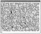
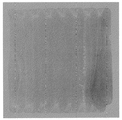
血液の一定分量をスライドガラス上に分散した後、スライドは、乾燥され、固定され、染色される。細胞は、一旦スライドに塗布されると、数秒で沈降する。その後、細胞は乾燥の準備が整う。初期試験中に、市販の乾燥器の幾つかのモデルの低温設定を、手製のノズル及びバッフルとともに使用した。スライドに対して45度の角度で吹き付ける冷たい空気の軽い(低温設定の)ストリームが、再現性のある乾燥条件を提供した。コンピューターで使用されるような電子的に可変の速度を有する小型ファンもまたノズル及びバッフルとともに使用することができる。最適品質の染色結果は、血球形態を評価するために使用される標準的なウェッジスミアの最良の部分でそれが得られるのとほぼ同じ時間でスライドが乾燥するときに、すなわち、15秒〜20秒で得られることがわかった。他の乾燥条件が、乾燥の均一性及び速度を最適化するために実験され得る。幾つかの条件では、強制空気は必要とされない場合がある。この方法によって調製される後続の分析に適する血液スミアは、図8に示される。 After dispersing an aliquot of blood on a glass slide, the slide is dried, fixed and stained. Once applied to the slide, the cells settle in a few seconds. The cells are then ready for drying. During initial testing, the cold settings of several models of commercial dryers were used with handmade nozzles and baffles. A light (cold setting) stream of cold air blowing at a 45 degree angle to the slide provided reproducible drying conditions. Small fans with electronically variable speeds such as those used in computers can also be used with nozzles and baffles. Optimal quality staining results are obtained when the slide dries in approximately the same time that it is obtained with the best part of a standard wedge smear used to assess blood cell morphology, ie, 15 seconds to 20 seconds. It turns out that it can be obtained in seconds. Other drying conditions can be experimented to optimize drying uniformity and speed. In some conditions, forced air may not be required. A blood smear suitable for subsequent analysis prepared by this method is shown in FIG.
固定のために、スライドは、85(容積)%のメタノールの槽内に設置してもよい。固定及び染色のためには、スライドを平坦のままにし、試薬でスライドをフラッディングさせることが適切に働く。染色のために、試薬は、単一溶液のライト・ギムザ染色液及びリンス緩衝液からなるものとしてもよく、又は、他のよく知られている血液染色液を使用してもよい。染色に続いて、スライドを、おそらく強制空気によって再び乾燥する。 For fixation, the slide may be placed in a 85% (by volume) methanol bath. For fixation and staining, it is appropriate to keep the slide flat and flood the slide with reagents. For staining, the reagent may consist of a single solution of Wright Giemsa stain and rinse buffer, or other well-known blood stains may be used. Following staining, the slide is dried again, possibly with forced air.
染色された細胞は、従来の自動化染色器上で染色されたウェッジスミア上の細胞に匹敵しなければならない。研究所の染色器を使用して染色された人手によって調製されたウェッジスミアは、コントロールとして使用され得る。スライドは、視覚的に比較することができ、また、画像分析を使用して、染色の強度、色(臨界波長で染色強度を比較することによって)、及び細胞の形態を比較することができる。 Stained cells should be comparable to cells on wedge smears stained on a conventional automated stainer. Wedge smears prepared manually by using a laboratory stainer can be used as a control. The slides can be compared visually and image analysis can be used to compare staining intensity, color (by comparing staining intensity at critical wavelengths), and cell morphology.
スライドの初期の乾燥及び固定に続いて、自動化染色装置が使用される。スライドは、その後、明視野撮像を遂行する準備が整う。 Following the initial drying and fixation of the slide, an automated dyeing device is used. The slide is then ready to perform bright field imaging.
撮像機器は、自動化ステージ及びフォーカスを有する標準的な明視野顕微鏡又は機能的に等価な装置を含むことができる。一つの実施形態では、顕微鏡は、メルツホイザー社(ドイツ)のステージ及びフォーカスモーターアタッチメント(プライア・サイエンティフィック社、ロックランド,マサチューセッツ)に取付けられる。顕微鏡は、電動式ノーズピースを有し、コンピューター制御下で異なる拡大レンズが選択されることを可能にする。フィルターホイール(プライア・サイエンティフィック社、ロックランド,マサチューセッツ)は、光経路内で狭帯域色フィルターが自動的に選択されることを可能にする。LED照明を、フィルターの代わりに用いることができる。切換え式LEDの使用は、フィルターホイール回転の場合に必要とされる時間と比較して、画像取得時間を大幅に短縮させる。1600×1200ピクセルfirewireカメラ(ポイント・グレイ社、バンクーバー,カナダ)を、狭帯域画像を取得するために使用することができる。 The imaging equipment can include a standard bright field microscope or functionally equivalent device with an automated stage and focus. In one embodiment, the microscope is attached to a stage and focus motor attachment (Prier Scientific, Rockland, Mass.) From Merzhauser (Germany). The microscope has a motorized nosepiece that allows different magnifying lenses to be selected under computer control. A filter wheel (Pria Scientific, Rockland, Mass.) Allows narrowband color filters to be automatically selected in the light path. LED lighting can be used instead of a filter. The use of a switchable LED greatly reduces the image acquisition time compared to the time required for filter wheel rotation. A 1600 × 1200 pixel firewire camera (Point Gray, Vancouver, Canada) can be used to acquire narrowband images.
明視野ステーションの目的は2つある。第1に、撮像ソフトウェアは、総赤血球数及び総白血球数を確定するために明視野画像を使用する。この計数の場合、単一波長の光(例えば570nm)が使用され得る。血小板数の場合、2色の照明を使用することができる。例えば、血小板の非常に高いコントラストでかつ高品質の画像を、570nm画像から430nm画像を減算することによって得ることができる。赤血球、白血球、及び血小板の計数精度は、著しく高く、98%を超えるように思われる。計数についての総合精度は、スライド調製物が、血液チューブから取り出された試料をどれほどよく表しているかに依存することになる。固定及び染色中に非常に少数の細胞しか失われていないことを評価が示した。これは、乾燥プロセスに起因する、スライドに対する沈降した細胞の優れた付着によるものである。現行の自動化CBC機器に典型的である赤血球、白血球、及び血小板の計数についての誤り率が適合され得るか、又はそれが改善され得る。正確な計数は、赤血球及び白血球用の全調製物をスキャンすることによって行うことができるが、平均細胞堆積物が全部で約30000個の血小板又は1視野当たり約500個の血小板を有することになるため、正確な血小板計数は、減少した数の視野を使用してもよい。WBC計数及びRBC計数が行われた視野の例は、図9に示される。 There are two purposes for the bright field station. First, the imaging software uses bright field images to determine the total red blood cell count and total white blood cell count. For this counting, a single wavelength of light (eg 570 nm) can be used. For platelet counts, two colors of illumination can be used. For example, a very high contrast and high quality image of platelets can be obtained by subtracting a 430 nm image from a 570 nm image. The counting accuracy of red blood cells, white blood cells and platelets is remarkably high and seems to exceed 98%. The overall accuracy for the count will depend on how well the slide preparation represents the sample removed from the blood tube. Evaluation showed that very few cells were lost during fixation and staining. This is due to the excellent adhesion of sedimented cells to the slide due to the drying process. The error rate for red blood cell, white blood cell, and platelet counts typical of current automated CBC instruments can be adapted or improved. Accurate counting can be done by scanning the entire preparation for red blood cells and white blood cells, but the average cell deposit will have a total of about 30000 platelets or about 500 platelets per field Thus, an accurate platelet count may use a reduced number of fields. An example of a field of view on which WBC counting and RBC counting have been performed is shown in FIG.
明視野撮像の第2の目的は、白血球分類計数のための、また、血小板及びRBCの形態を評価するための画像を取得することである。正確な分類計数のために、少なくとも200個の白血球が必要とされる。白血球の位置を特定するために、単一波長において10倍の倍率(0.7ミクロン/ピクセル)で全細胞スポットがスキャンされ得る。スキャニング、フォーカストラッキング、及び処理が可能な電動式ステージを使用する典型的なコンピューター化顕微鏡システムは、10視野/秒より高い速度で1600×1200ピクセル画像を分析することができる。細胞堆積物のサイズに応じて、分析すべき200個〜400個の間の視野が存在する可能性がある。全体的な分析は、20秒〜40秒かかることになる。この時間の終わりに、コンピューターは、細胞スポット上での全ての白血球「候補」の場所をわかっていることになる。コンピューターは、その後、これらの場所のうちの200以上の場所に戻り、リフォーカスし、より高い倍率で(すなわち、40倍又は60倍のレンズを使用して)白血球をデジタル化し、画像分析を使用して、白血球タイプを識別し、オペレーターが後で観察するために画像を記憶する。画像をデジタル化して、血液技師による分析とフルカラー観察の両方を最適化するために、少なくとも4つの狭帯域色の光が必要とされる可能性がある。従来から、コンピューター化された白血球分類作業の場合、430nm、500nm、525nm、及び600nmの波長が使用される(ブレナー 1974年;ツァーニザー 1983年;ツァーニザー 1986年)。フィルターホイールシステムにおいて他の波長でも実験が行われ得る。200個〜300個の白血球の取得及び分析は、おそらく約1分で都合よく達成され得ることが予想される。同時に、赤血球形態及びヘモグロビン含有量の評価のために、十分な数のRBCが画像内で利用可能である。 The second purpose of bright-field imaging is to acquire images for white blood cell classification and evaluation of platelet and RBC morphology. At least 200 white blood cells are required for accurate classification counting. To locate the white blood cells, the whole cell spot can be scanned at a single wavelength at 10X magnification (0.7 microns / pixel). A typical computerized microscope system using a motorized stage capable of scanning, focus tracking, and processing is capable of analyzing 1600 × 1200 pixel images at speeds greater than 10 fields / second. Depending on the size of the cell deposit, there can be between 200 and 400 fields of view to be analyzed. The overall analysis will take 20 to 40 seconds. At the end of this time, the computer knows the location of all white blood cell “candidates” on the cell spot. The computer then returns to more than 200 of these locations, refocuses, digitizes white blood cells at higher magnification (ie, using a 40x or 60x lens) and uses image analysis The white blood cell type is then identified and the image stored for later viewing by the operator. At least four narrow band colors of light may be required to digitize the image and optimize both hematologist analysis and full color viewing. Traditionally, for computerized leukocyte sorting operations, wavelengths of 430 nm, 500 nm, 525 nm, and 600 nm have been used (Brenner 1974; Zanizer 1983; Zanizer 1986). Experiments can also be performed at other wavelengths in the filter wheel system. It is expected that acquisition and analysis of 200-300 leukocytes can be conveniently accomplished, perhaps in about 1 minute. At the same time, a sufficient number of RBCs are available in the image for assessment of red blood cell morphology and hemoglobin content.
全体的に見て、明視野撮像(赤血球、白血球、及び血小板の計数+白血球位置特定+血球分類分析)は約2分かかることになる。これは、全体的な時間枠にピッタリ合い、8分〜10分かけて全試料を処理することを可能にする。 Overall, bright field imaging (red blood cell, white blood cell and platelet count + white blood cell location + blood cell classification analysis) would take about 2 minutes. This fits the overall time frame and allows all samples to be processed over 8 to 10 minutes.
プロセスのこの撮像ステージのタイミングは、機器全体のスループットについての現行の制限因子である。他のステップが全て、1分間隔に分けられ得る(染色の個々の構成要素を含む)ため、パイプライン式システムは、1分/試料スループットで達成され得る(「ファーストイン」から「ファーストアウト」まで8分〜10分かかる)。このスループット時間は、1)更なる最適化によって、2)ステップを、2つ若しくは3つの「パイプライン式」光学プラットフォームに分けることによって、又は、3)機器内で2つ若しくは3つの並列の同一明視野システムを使用することによっておそらく改善されるであろう。
The timing of this imaging stage of the process is the current limiting factor for overall instrument throughput. Since all other steps can be divided into 1 minute intervals (including individual components of staining), a pipelined system can be achieved with 1 minute / sample throughput ("first in" to "first out").
約0.2マイクロメートルのピクセル間隔が、正確な血球分類計数のために必要であることが過去の経験によって示されている(ランドウィアード 1981年)。非常に小さなピクセル間隔を有する今日のCCDカメラを使用する場合、この分解能は、40倍、50倍、又は60倍の対物レンズと適切なカメラカプラーを用いて得ることができる。60倍は、乾燥対物レンズについて通常利用可能である最大倍率であるが、より高いコストで入手可能なより高い乾燥対物レンズが存在する。適切なレンズが、色補正及び視野の平坦性を保証するために使用される。0.1マイクロメートル/ピクセル〜0.25マイクロメートル/ピクセルのピクセル間隔での実験が、対物レンズとカメラアダプターの組合せに応じて必要である可能性がある。 Past experience has shown that a pixel spacing of approximately 0.2 micrometers is necessary for accurate blood cell counts (Landweard 1981). When using today's CCD cameras with very small pixel spacing, this resolution can be obtained with a 40x, 50x, or 60x objective lens and a suitable camera coupler. 60x is the maximum magnification that is normally available for dry objectives, but there are higher dry objectives available at higher cost. Appropriate lenses are used to ensure color correction and field flatness. Experiments with pixel spacings from 0.1 micrometers / pixel to 0.25 micrometers / pixel may be necessary depending on the combination of objective and camera adapter.
WBC分類の目標は、最低5分類での分類を正確に行うことである。これは、このソフトウェアの最も複雑な態様である。このソフトウェアにとっての成功は、調製の品質及び画像の品質に大きく依存する。行われた以前の研究から、画像分析に基づく自動化されたWBC分類が可能であることがわかっている。より最近の研究が示したところによれば、(1980年代に使用された油浸対物レンズの代わりに)乾燥対物レンズを用いた画像品質は、色細胞分析及び観察(クロマビジョンのACIS)並びに分類計数及び観察(セラビジョンのMICRO21)について優れた品質を提供する。 The goal of WBC classification is to correctly classify at least 5 classifications. This is the most complex aspect of this software. The success for this software depends largely on the quality of the preparation and the quality of the image. Previous work done has shown that automated WBC classification based on image analysis is possible. More recent studies have shown that image quality with dry objectives (instead of the oil immersion objectives used in the 1980s) is color cell analysis and observation (ACIS for chromavision) and classification. Provides excellent quality for counting and observation (Ceravision MICRO21).
上述した改善された調製技術は、画像分析に非常に役立つ。スパンスミア、又は、手若しくは機器が引張るウェッジスミアを使用した以前のシステムでは、細胞歪が、再現性のある画像分析を達成するときの重大な問題であった。細胞の単一層を軽く塗布するという本発明の方法は、本システムにおけるスライド調製中に細胞の歪が全く存在しないか又はほとんどないことを保証する。全ての調製法(ウェッジ法であれ、スピン法であれ、本発明の調製法であれ)において、細胞は、乾燥するにつれて形態を変化させるが、最適な乾燥条件が人手によって再現され得ることが示されており、また、一層良好な制御が、完全に自動化された調製によって利用可能であることが予想される。上述した改善された検体調製はまた、任意の方法によって調製されるウェッジスミアに関して起こることが分かっている分布アーチファクトをなくし、WBC分類計数に不正確さをもたらす。 The improved preparation techniques described above are very useful for image analysis. In previous systems using span smears or wedge smears pulled by hand or equipment, cell distortion was a significant problem in achieving reproducible image analysis. The inventive method of lightly applying a single layer of cells ensures that there is no or little cell distortion during slide preparation in the system. In all preparation methods (whether the wedge method, the spin method or the preparation method of the present invention), the cells change shape as they dry, but it is shown that optimal drying conditions can be reproduced manually. And better control is expected to be available with fully automated preparation. The improved specimen preparation described above also eliminates distribution artifacts that have been found to occur with wedge smears prepared by any method, resulting in inaccuracies in WBC classification counts.
RBC形態及びヘモグロビン含有量を評価し、次に、白血球に接触するいずれの赤血球をもデジタル的に除去するために、ヘモグロビンだけの画像を取得するためのフィルター(430nm)を含む少なくとも4つの狭帯域画像が画像分析のために使用される。570nmフィルターが、血小板及び核について優れたコントラストを与えることが示されている。他の波長が、好塩基球、単球、リンパ球(全て青の色合い)、好酸球(赤)、及び好中球(中間色)の色を最もよく区別するために選択される。これらの波長はまた、適切である場合、色画像の表示のために使用される。そうでなければ、1つ又は2つの更なる画像が、200超の細胞について撮影される必要がある場合があり、その画像は、分類計数のために分析され、また、モニター上に表示することができる。 At least 4 narrowbands including a filter (430 nm) to acquire RBC morphology and hemoglobin content, and then to digitally remove any red blood cells that contact the white blood cells to acquire hemoglobin-only images Images are used for image analysis. A 570 nm filter has been shown to give excellent contrast for platelets and nuclei. Other wavelengths are selected to best distinguish the colors of basophils, monocytes, lymphocytes (all blue shades), eosinophils (red), and neutrophils (intermediate color). These wavelengths are also used for the display of color images where appropriate. Otherwise, one or two additional images may need to be taken for over 200 cells, which are analyzed for classification counting and displayed on a monitor Can do.
取得された画像を使用して、核及び細胞質の位置を特定するために、細胞質及び核の区分化アルゴリズムが適用される。 A cytoplasm and nucleus segmentation algorithm is applied to locate the nucleus and cytoplasm using the acquired images.
一旦、核及び細胞質の区分化が終了すると、ソフトウェアは、白血球の核及び細胞質から色、形状、サイズ、テクスチャー、及び密度特徴の標準的な集合を抽出する。 Once the nuclear and cytoplasm segmentation is complete, the software extracts a standard set of color, shape, size, texture, and density characteristics from the white blood cell nucleus and cytoplasm.
画像分析ソフトウェア及び分類アルゴリズムを訓練するために、既知の対象物の大きなデータベースを組立てることが必要である。この対象物は、多数の異型細胞を含む数千の全てのタイプの細胞を含む。データベースが確立されると、市販の統計分析パッケージ及び分類子を開発するための判別ルーチンが使用される。分類子は、比較的複雑であり、通常、階層構造の複数の判別子からなる。 In order to train image analysis software and classification algorithms, it is necessary to build a large database of known objects. This object contains thousands of all types of cells, including many atypical cells. Once the database is established, a discriminant routine for developing commercial statistical analysis packages and classifiers is used. The classifier is relatively complex and usually consists of a plurality of classifiers having a hierarchical structure.
存在する赤血球の数を計数することに加えて、CBC機器は、ヘモグロビン含有量及び赤血球形態の評価を提供する。ヘモグロビン含有量を、430nmで個々の赤血球の積分光学濃度を測定することによって求めることができることが確立している(バッカス, 1985年)。赤血球形態もまた、以前の自動化された分類機器上で成功裏に求められており(バッカス,1980年、1982年)、その方法はよく知られている。 In addition to counting the number of red blood cells present, the CBC instrument provides an assessment of hemoglobin content and red blood cell morphology. It has been established that hemoglobin content can be determined by measuring the integrated optical density of individual erythrocytes at 430 nm (Bacchus, 1985). Red blood cell morphology has also been sought after on previous automated sorting instruments (Bacchus, 1980, 1982), and the method is well known.
図4は、赤血球についての自動化計数と人手による計数との相関を示すグラフである。各視野は、200個〜1500個の赤血球を含む。0.993のR平方値が、データの回帰モデルから計算され、0.996の非常に良好なピアソン相関係数を与える。 FIG. 4 is a graph showing the correlation between automated counts and manual counts for red blood cells. Each visual field contains 200-1500 red blood cells. An R-square value of 0.993 is calculated from the regression model of the data, giving a very good Pearson correlation coefficient of 0.996.
図5は、白血球についての自動化計数と人手による計数との相関を示すグラフである。0.960のR平方値が計算され、0.980のピアソン相関係数を与える。 FIG. 5 is a graph showing the correlation between automated counts and manual counts for leukocytes. An R square value of 0.960 is calculated, giving a Pearson correlation coefficient of 0.980.
図8に示す写真は、自動化ピペッティングシステム上で生成されるスライドからの細胞スポットである。通常、正方形細胞スポットの縁部の視野は、ピペットチップが次の列に載るために急速に向きを変えるため少数の細胞を含む。最後の列の端部において、幾つかの細胞スポットは、細胞の「ブロブ」を示し、そこで、ピペットチップが停止し、スライドの表面から持上げられる。こうした混雑した視野において、ソフトウェアは、依然として赤血球数の推定を行うことができるが、コンピューターの計数は、混雑したエリアにおいてわずかに低くなる可能性があるように思われる。この方法は、細胞のより均等な分布を生成するように改良され得る。図9は、細胞スポットからの1つのデジタル画像の詳細を示す。 The photograph shown in FIG. 8 is a cell spot from a slide generated on an automated pipetting system. Normally, the field of view at the edge of a square cell spot contains a small number of cells because the pipette tip turns rapidly to rest in the next row. At the end of the last row, several cell spots show a “blob” of cells where the pipette tip stops and is lifted from the surface of the slide. In such a congested field of view, the software can still make an estimate of the red blood cell count, but the computer count appears to be slightly lower in the congested area. This method can be modified to produce a more even distribution of cells. FIG. 9 shows the details of one digital image from the cell spot.
画像分析プログラムは、それぞれの個々の視野を分析し、総赤血球数と総白血球数を合計する。患者バイアル内の総計数/マイクロリットルを計算するために、スライド上で計数された数が、希釈係数で乗算される。 The image analysis program analyzes each individual field and sums the total red blood cell count and total white blood cell count. The number counted on the slide is multiplied by the dilution factor to calculate the total count / microliter in the patient vial.
以下の表1は、34個のスライドについてのデータ要約を示す。「シスメックス」データは、市販の「フローベース」自動化CBC分析器からの赤血球数及び白血球数を示している。自動化計数及びスケーリングされた計数は、赤血球と白血球の両方について提供される。赤血球数は通常、「百万個の細胞/マイクロリットル」の単位で与えられるため、位置特定された赤血球の数を、「スケーリングされた」赤血球数を得るために1000000で除算した。白血球の場合、計数は、「千個の細胞/マイクロリットル」の単位で与えられるため、その数を、「スケーリングされた」白血球数を得るために1000で除算した。 Table 1 below shows a data summary for 34 slides. “Sysmex” data shows red blood cell and white blood cell counts from a commercially available “flow-based” automated CBC analyzer. Automated counts and scaled counts are provided for both red and white blood cells. Since the red blood cell count is usually given in units of “million cells / microliter”, the number of localized red blood cells was divided by 1000000 to obtain a “scaled” red blood cell count. For leukocytes, the count is given in units of “1000 cells / microliter”, so that number was divided by 1000 to obtain a “scaled” leukocyte count.
検体は、非常に高いものから非常に低いものまでの赤血球数及び白血球数を含むことに留意されたい。 Note that the specimen contains red and white blood cell counts from very high to very low.

図6のグラフは、赤血球についてのシスメックス計数と自動化スライドベース計数との相関を示している。未処理計数は、682449白血球/スライドと1241765白血球/スライドとの間で変動した。97.95%のR平方値が計算された。 The graph in FIG. 6 shows the correlation between the Sysmex count and the automated slide-based count for red blood cells. The raw count varied between 682449 leukocytes / slide and 1241765 leukocytes / slide. An R square value of 97.95% was calculated.
図7に示すグラフは、白血球についてのSysmex計数と自動化スライド計数との相関を示している。未処理計数は、147白血球/スライドから11250白血球/スライドとの間で変動した。99.70%のR平方値が計算された。 The graph shown in FIG. 7 shows the correlation between the Sysmex count and the automated slide count for leukocytes. Untreated counts varied between 147 leukocytes / slide to 11250 leukocytes / slide. An R square value of 99.70% was calculated.
データは、スライドを調製する2つのセッション中に34の患者試料から得られた。データは、典型的な患者を表すが、チューブは、赤血球数及び白血球数の広い分布を有する患者から選択された。34個の試料のうちの、全てではないがほとんどが、日中に冷凍庫に「保管されていた」検体から得られ、その後、夕方近くに引き出され本発明の機器上で調製された。チューブは、一旦引き出されると、連続して処理された。 Data was obtained from 34 patient samples during two sessions of preparing slides. The data represents a typical patient, but tubes were selected from patients with a wide distribution of red blood cell and white blood cell counts. Of the 34 samples, most if not all, were obtained from specimens that were “stored” in the freezer during the day, and then withdrawn near evening and prepared on the instrument of the present invention. The tube was processed continuously once withdrawn.
スライドは、半自動的に調製され、染色され、計数され得る。細胞は、形態学的によく保存され、スライド上での計数は、現行の技術水準のシスメックスCBC機器上で得られる計数と相関がある。 Slides can be prepared semi-automatically, stained and counted. The cells are well preserved morphologically and the counts on the slide correlate with the counts obtained on a current state of the art Sysmex CBC instrument.
スライドガラス上の制限されたエリアへの血液細胞の定量的移送が達成され得ることが示された。十分な細胞が移送されて、スライド上に堆積した平均して900000個を超える赤血球及び1000個を超える白血球に基づいて、正確なRBC数及びWBC数が与えられる。全ての細胞が計数され測定され得る。各細胞の形態は、画像分析によって又は直接可視化によって評価され得る。細胞内の細部の保存は、手又は現行の市販の自動化機器によって行われる従来のスライドガラス調製に関して見られる保存に等しいか又はそれより優れる。移送される細胞の集団全体は、乾燥、固定、及び染色後でも、スライド上に残っているように見える。初期撮像努力による細胞計数は、ほとんどの現行の技術水準のCBC機器に非常によく対応する。 It has been shown that quantitative transfer of blood cells to a limited area on a glass slide can be achieved. Sufficient cells are transferred to give an accurate RBC and WBC count based on an average of more than 900,000 red blood cells and 1000 white blood cells deposited on the slide. All cells can be counted and measured. The morphology of each cell can be assessed by image analysis or by direct visualization. The preservation of intracellular details is equal to or better than that seen with conventional glass slide preparations made by hand or by current commercial automation equipment. The entire population of transferred cells appears to remain on the slide after drying, fixing, and staining. Cell counting with initial imaging efforts corresponds very well to most current state-of-the-art CBC instruments.
CBC機器についてのこの基礎は、現行の技術水準の機器に対して、非常に単純であり、費用がかからず、同等の品質か又は優れた品質であるものとすることができる。 This basis for CBC equipment can be very simple, inexpensive and comparable or superior quality to current state of the art equipment.
以下に述べる方法は、全血球計数、ヘモグロビン含有量、赤血球形態、並びに、血液検体内の血球の直接観察によってこれらの細胞の真の数及び血球形態を反映するWBC分類計数を提供する。これに関して、本発明は、電気インピーダンス、光の吸収及び光の散乱の測定を行う既存のCBC機器及び方法、並びに、血液細胞の直接観察にはよらないが、血液細胞に起因する他の手段と区別される。 The method described below provides a WBC classification count that reflects the true count and blood cell morphology of these cells by direct observation of the whole blood count, hemoglobin content, red blood cell morphology, and blood cells in the blood sample. In this regard, the present invention relates to existing CBC instruments and methods for measuring electrical impedance, light absorption and light scattering, and other means attributable to blood cells, although not directly related to blood cell observation. Differentiated.
上記に照らして、添付の特許請求の範囲によって規定される本発明の概念から逸脱することなく、開示のために選択される本明細書の実施形態に対して種々の変更が行われ得ることがここで当業者によって認識されるであろう。こうした変更の非限定的な例は、異なるレベルの希釈、種々の希釈剤、種々の基板、細胞を固定し染色する種々の手段、及び細胞を撮像する種々の手段を使用することを含む。 In light of the above, various modifications can be made to the embodiments herein selected for disclosure without departing from the inventive concepts defined by the appended claims. One skilled in the art will now recognize. Non-limiting examples of such changes include the use of different levels of dilution, different diluents, different substrates, different means for fixing and staining cells, and different means for imaging cells.
Claims (36)
血液試料の既知の一定分量を吸引することと、
血液細胞の単一層を形成するために、基板上に前記一定分量全体を移送することと、
前記血液細胞の単一層を乾燥することと、
前記乾燥した血液を染色することと、
明視野光学器及びマルチスペクトル撮像を使用して、前記染色された細胞を分析し、赤血球及び白血球並びに血小板の計数を行うことと
を含む方法。 A method for determining a complete blood count,
Aspirating a known aliquot of a blood sample;
Transferring the entire aliquot onto a substrate to form a single layer of blood cells;
Drying said monolayer of blood cells;
Staining the dried blood;
Using bright field optics and multispectral imaging to analyze the stained cells and perform red blood cell and white blood cell and platelet counts.
既知の量の血液試料を吸引することと、
血液細胞の単一層を形成するために、基板に前記血液試料全体を移送することと、
前記血液細胞の単一層を乾燥することと、
前記乾燥した血液を染色することと、
明視野光学器及びマルチスペクトル撮像を使用して、前記染色された細胞を分析し、ヘモグロビン含有量の測定を行うとともに赤血球形態を分析することと
を含む方法。 A method for determining hemoglobin content and red blood cell morphology comprising:
Aspirating a known amount of blood sample;
Transferring the entire blood sample to a substrate to form a single layer of blood cells;
Drying said monolayer of blood cells;
Staining the dried blood;
Analyzing the stained cells using bright field optics and multispectral imaging, measuring hemoglobin content and analyzing red blood cell morphology.
既知の量の血液試料を吸引することと、
血液細胞の単一層を形成するために、基板に前記血液試料全体を移送することと、
前記血液細胞の単一層を乾燥することと、
前記乾燥した血液を染色することと、
明視野光学器及びマルチスペクトル撮像を使用して、前記染色された細胞を分析し、異なるタイプの白血球の計数を行うことと
を含む方法。 An automated method for determining WBC classification counts, comprising:
Aspirating a known amount of blood sample;
Transferring the entire blood sample to a substrate to form a single layer of blood cells;
Drying said monolayer of blood cells;
Staining the dried blood;
Using bright field optics and multispectral imaging to analyze the stained cells and to count different types of white blood cells.
既知の量の血液試料を吸引することと、
単一層を形成するために、基板に前記血液試料全体を移送することと、
前記血液細胞の単一層を乾燥することと、
前記乾燥した血液を染色することと、
明視野光学器及びマルチスペクトル撮像を使用して、前記染色された細胞を分析し、異なるタイプの白血球の計数を行うことと、
オペレーターによる対話的観察のために、計数並びに赤血球及び白血球の画像をディスプレイ上に表示することと
を含む方法。 An automated method of determining WBC classification counts and displaying RBC and WBC counts, morphological measurements, and images,
Aspirating a known amount of blood sample;
Transferring the entire blood sample to a substrate to form a single layer;
Drying said monolayer of blood cells;
Staining the dried blood;
Using bright field optics and multispectral imaging to analyze the stained cells and to count different types of white blood cells;
Displaying the images of red blood cells and white blood cells on a display for interactive observation by an operator.
基板の上方に配置され、血液細胞を含む流体を前記基板上に分配する塗布チップと、
マイクロプロセッサを含むコンピュータと、
記憶デバイスに記憶された、前記システムを制御するためのソフトウェアと
を備え、
前記マイクロプロセッサが前記ソフトウェアを実行する際、前記コンピュータは、前記システムに、
既知の量の前記流体を前記塗布チップに充填することと、
前記既知の量の流体を前記塗布チップから分配し、前記基板への前記流体の排出が起こる一方で、前記塗布チップと前記基板との間の相対移動を維持して、前記流体中の細胞が1つの細胞の厚さの層に前記基板上へ沈降するように、前記既知の量の流体全体を2つ以上の列で前記基板の画定された領域上に載せることと
をさせるシステム。 A system for analyzing cells from blood,
An application chip disposed above the substrate and distributing a fluid containing blood cells onto the substrate;
A computer including a microprocessor;
Software for controlling the system, stored in a storage device,
When the microprocessor executes the software, the computer includes:
Filling the application tip with a known amount of the fluid;
Distributing the known amount of fluid from the application tip and evacuating the fluid to the substrate while maintaining relative movement between the application tip and the substrate so that the cells in the fluid A system wherein the entire known amount of fluid is placed on a defined area of the substrate in two or more rows such that a single cell thick layer settles onto the substrate.
前記コンピュータは、前記システムに、前記基板上の前記細胞を撮像させ、赤血球計数と、白血球計数と、血小板計数とのうちの1つ又は複数を決定させる、請求項5に記載のシステム。 The system further comprises a light receiving device that captures an image of the cell on the substrate,
6. The system of claim 5, wherein the computer causes the system to image the cells on the substrate and determine one or more of red blood cell count, white blood cell count, and platelet count.
前記コンピュータは、前記システムに、前記基板上の前記細胞を撮像させ、全血球計数及び白血球分類計数の一方又は両方を決定させる、請求項5に記載のシステム。 The system further comprises a light receiving device that captures an image of the cell on the substrate,
6. The system of claim 5, wherein the computer causes the system to image the cells on the substrate and determine one or both of a whole blood count and a white blood cell classification count.
前記コンピュータは、前記基板コントローラに前記基板の位置を変えるように指示することにより、前記基板に対する前記塗布チップの移動を制御する、請求項5に記載のシステム。 The system further comprises a substrate controller,
The system according to claim 5, wherein the computer controls movement of the coating chip relative to the substrate by instructing the substrate controller to change a position of the substrate.
前記血液を貯める第1のリザーバと、
希釈剤を貯める第2のリザーバと、
前記血液と前記希釈剤とを混合させて希釈流体を形成するミキサと
を備える、請求項5に記載のシステム。 The system
A first reservoir for storing the blood;
A second reservoir for storing the diluent;
The system of claim 5, comprising a mixer that mixes the blood and the diluent to form a diluted fluid.
前記コンピュータは、前記システムに、
前記流体の列の画像を撮像することと、
前記流体の列の幅を決定するために前記画像を分析することと、
X方向又はY方向の前記基板に対する前記塗布チップの位置と、前記流体の列の幅に等しい距離とを変えることと
をさせる、請求項5に記載のシステム。 The system further comprises a light receiving device that captures an image of the cell on the substrate,
The computer includes the system,
Taking an image of the fluid row;
Analyzing the image to determine the width of the fluid row;
6. The system according to claim 5, wherein the position of the application tip relative to the substrate in the X or Y direction is varied and a distance equal to the width of the fluid row.
前記コンピュータは、前記システムに、固定されたX方向成分またはY方向成分を有する経路内に前記スライドにわたって流体の第3の列を塗布させ、該第3の列は、一つの細胞の厚さを有する細胞の層と、幅とを備え、
前記コンピュータは、前記システムに、固定されたX方向成分またはY方向成分を有する経路内に前記スライドにわたって流体の第4の列を塗布させ、該第4の列は、一つの細胞の厚さを有する細胞の層と、幅とを備え、
前記スライド上で、前記第3の列は前記第2の列に隣接すると共に前記第4の列は前記第3の列に隣接する、請求項5に記載のシステム。 The substrate has a slide;
The computer causes the system to apply a third row of fluid across the slide in a path having a fixed X- or Y-direction component, and the third row determines the thickness of one cell. A layer of cells having, and a width;
The computer causes the system to apply a fourth row of fluid across the slide in a path having a fixed X- or Y-direction component, and the fourth row determines the thickness of one cell. A layer of cells having, and a width;
6. The system of claim 5, wherein on the slide, the third row is adjacent to the second row and the fourth row is adjacent to the third row.
前記システムは、
前記スライドを清浄する放電装置と、
前記スライド上に染料を分配するディスペンサと、
過剰な染料を除去するために前記スライドを傾かせるティルタと、
前記スライド上の前記細胞の画像を撮像する受光装置と、
該受光装置の下方で前記スライドを位置決めするスライドムーバと、
前記スライド上で又は前記スライドを通して光を検出する発光装置と
をさらに備える、請求項5に記載のシステム。 The substrate has a slide;
The system
A discharge device for cleaning the slide;
A dispenser for dispensing dye on the slide;
Tilta tilting the slide to remove excess dye,
A light receiving device for capturing an image of the cell on the slide;
A slide mover for positioning the slide below the light receiving device;
The system of claim 5, further comprising a light emitting device that detects light on or through the slide.
前記スライドの上方約70±40ミクロンの位置に前記塗布チップを位置することと、
約10から100mm/秒の速度で前記塗布チップと前記スライドとの相対移動を維持することと
をさせる、請求項5に記載のシステム。 The computer includes the system,
Positioning the application tip approximately 70 ± 40 microns above the slide;
6. The system of claim 5, wherein the relative movement between the application tip and the slide is maintained at a rate of about 10 to 100 mm / sec.
前記システムは、
新たなスライドを保管するフィーダと、
前記塗布チップが前記スライド上へ流体の列を塗布する第1のステーションと、
前記スライドを染色、固定及び乾燥させる第2のステーションと、
前記スライドを照明すると共に画像化する第3のステーションと、
処理したスライドを受け取るコレクタと、
前記スライドを、前記フィーダから開始して、前記第1のステーション、前記第2のステーション、前記第3のステーション、次いで前記コレクタまで、前記システムを通して移動させるアドバンサと
をさらに備える、請求項5に記載のシステム。 The substrate has a slide;
The system
A feeder for storing new slides;
A first station where the application tip applies a row of fluid onto the slide;
A second station for staining, fixing and drying the slide;
A third station for illuminating and imaging the slide;
A collector to receive the processed slides,
6. The advancer further comprising: an advancer that moves the slide through the system starting from the feeder to the first station, the second station, the third station, and then to the collector. System.
光源と、
前記基板上の前記細胞の画像を撮像する受光装置と
をさらに備え、
前記コンピュータは、前記光源に、
前記基板を照明するための白色光を発することと、
前記白色光の照射を第1の波長に制限するために前記白色光に第1の光フィルタを適用することと、
前記白色光の照射を第2の波長に制限するために前記白色光に第2の光フィルタを適用することと
をさせ、
前記コンピュータは、前記受光装置に、前記基板上の前記細胞の1つ又は複数の画像を撮像させる、請求項5に記載のシステム。 The system
A light source;
A light receiving device that captures an image of the cell on the substrate;
The computer uses the light source as
Emitting white light for illuminating the substrate;
Applying a first optical filter to the white light to limit the illumination of the white light to a first wavelength;
Applying a second optical filter to the white light to limit the white light illumination to a second wavelength;
The system according to claim 5, wherein the computer causes the light receiving device to capture one or more images of the cells on the substrate.
空間シフト又は他の歪みを補償することによって前記画像を洗練することと、
白色光の少なくとも2つの別個の波長が前記スライドに誘導されるときに前記受光装置が撮像する2つ以上の画像を組み合わせて、マルチカラー画像を表示するために生成することと
をさせる、請求項17に記載のシステム。 The computer includes the system,
Refining the image by compensating for spatial shifts or other distortions;
Combining at least two images captured by the light receiving device when at least two separate wavelengths of white light are directed to the slide to generate a multi-color image for display. 18. The system according to 17.
前記受光装置に前記スライドの白黒画像を撮像するように命令することと、
前記レンズの焦点距離を調整することによって焦点及び画質を補正することと、
複数のフィルタを前記スライドと前記光源との間に介在させながら前記レンズの焦点位置をシフトすることと
をさせる、請求項17に記載のシステム。 The computer includes the system,
Instructing the light receiving device to capture a black and white image of the slide;
Correcting focus and image quality by adjusting the focal length of the lens;
The system according to claim 17, wherein a plurality of filters are interposed between the slide and the light source to shift a focal position of the lens.
前記白色光を生成するハロゲン球と、
前記フィルタの位置を変える回転モータと
を備える、請求項17に記載のシステム。 The light source is
A halogen sphere that generates the white light;
18. A system according to claim 17, comprising a rotary motor that changes the position of the filter.
光源と、
前記基板上の前記細胞の画像を撮像する受光装置と
をさらに備え、
前記コンピュータは、
前記光源に第1のLEDに第1の波長で光を照射させ、
前記光源に第2のLEDに第2の波長で光を照射させ、
前記受光装置に前記基板上の前記細胞の1つ又は複数の画像を撮像させる、請求項5に記載のシステム。 The system
A light source;
A light receiving device that captures an image of the cell on the substrate;
The computer
Causing the light source to irradiate the first LED with light at a first wavelength;
Causing the light source to irradiate a second LED with light at a second wavelength;
The system of claim 5, wherein the light receiving device captures one or more images of the cells on the substrate.
Applications Claiming Priority (3)
| Application Number | Priority Date | Filing Date | Title |
|---|---|---|---|
| US4792008P | 2008-04-25 | 2008-04-25 | |
| US61/047,920 | 2008-04-25 | ||
| PCT/US2009/041858 WO2012030313A1 (en) | 2008-04-25 | 2009-04-27 | Method of determining a complete blood count and a white blood cell differential count |
Related Child Applications (2)
| Application Number | Title | Priority Date | Filing Date |
|---|---|---|---|
| JP2015060503A Division JP6278921B2 (en) | 2008-04-25 | 2015-03-24 | System and method for analyzing cells derived from blood or bone marrow and system for analyzing cells derived from blood samples |
| JP2015006095U Continuation JP3203413U (en) | 2008-04-25 | 2015-12-02 | System for analyzing cells from blood |
Publications (2)
| Publication Number | Publication Date |
|---|---|
| JP2012515931A JP2012515931A (en) | 2012-07-12 |
| JP2012515931A5 true JP2012515931A5 (en) | 2014-05-01 |
Family
ID=41215384
Family Applications (3)
| Application Number | Title | Priority Date | Filing Date |
|---|---|---|---|
| JP2012516238A Pending JP2012515931A (en) | 2008-04-25 | 2009-04-27 | System and method for determining total blood count and white blood cell percentage |
| JP2015060503A Active JP6278921B2 (en) | 2008-04-25 | 2015-03-24 | System and method for analyzing cells derived from blood or bone marrow and system for analyzing cells derived from blood samples |
| JP2015006095U Expired - Lifetime JP3203413U (en) | 2008-04-25 | 2015-12-02 | System for analyzing cells from blood |
Family Applications After (2)
| Application Number | Title | Priority Date | Filing Date |
|---|---|---|---|
| JP2015060503A Active JP6278921B2 (en) | 2008-04-25 | 2015-03-24 | System and method for analyzing cells derived from blood or bone marrow and system for analyzing cells derived from blood samples |
| JP2015006095U Expired - Lifetime JP3203413U (en) | 2008-04-25 | 2015-12-02 | System for analyzing cells from blood |
Country Status (5)
| Country | Link |
|---|---|
| US (5) | US9017610B2 (en) |
| EP (1) | EP2446263A1 (en) |
| JP (3) | JP2012515931A (en) |
| AU (1) | AU2009352216B8 (en) |
| WO (1) | WO2012030313A1 (en) |
Families Citing this family (73)
| Publication number | Priority date | Publication date | Assignee | Title |
|---|---|---|---|---|
| WO2008041900A1 (en) | 2006-10-06 | 2008-04-10 | Boule Medical Ab | Device for extraction of a partial defined sample volume from a lager volume, method for operating the device, set of at least two devices, method of operation the set, an analytical instrument connectable to the device, method for operating the instrument, a system, and a method for operating the system |
| US9602777B2 (en) | 2008-04-25 | 2017-03-21 | Roche Diagnostics Hematology, Inc. | Systems and methods for analyzing body fluids |
| US9017610B2 (en) | 2008-04-25 | 2015-04-28 | Roche Diagnostics Hematology, Inc. | Method of determining a complete blood count and a white blood cell differential count |
| JP5380026B2 (en) * | 2008-09-24 | 2014-01-08 | シスメックス株式会社 | Sample imaging device |
| US8842900B2 (en) * | 2008-10-28 | 2014-09-23 | Sysmex Corporation | Specimen processing system and blood cell image classifying apparatus |
| JP5496581B2 (en) * | 2009-08-31 | 2014-05-21 | シスメックス株式会社 | Sample processing equipment |
| US9075225B2 (en) | 2009-10-28 | 2015-07-07 | Alentic Microscience Inc. | Microscopy imaging |
| CA2778725C (en) | 2009-10-28 | 2019-04-30 | Alentic Microscience Inc. | Microscopy imaging |
| US8748186B2 (en) * | 2009-12-22 | 2014-06-10 | Abbott Laboratories | Method for performing a blood count and determining the morphology of a blood smear |
| ES2700297T3 (en) | 2010-11-10 | 2019-02-14 | Roche Diagnostics Hematology Inc | Automatic apparatus to prepare biological samples to examine |
| US9522396B2 (en) | 2010-12-29 | 2016-12-20 | S.D. Sight Diagnostics Ltd. | Apparatus and method for automatic detection of pathogens |
| US9111343B2 (en) | 2011-01-18 | 2015-08-18 | Roche Diagnostics Hematology, Inc. | Microscope slide coordinate system registration |
| ES2890413T3 (en) * | 2011-04-15 | 2022-01-19 | Roche Diagnostics Hematology Inc | System and method for determining the mean cell volume of a blood sample |
| WO2012174535A1 (en) | 2011-06-17 | 2012-12-20 | Constitution Medical, Inc. | Solutions for histoprocessing of biological samples |
| EP2734823B1 (en) | 2011-07-22 | 2023-03-15 | Roche Diagnostics Hematology, Inc. | Sample transport systems and methods |
| WO2013016039A1 (en) * | 2011-07-22 | 2013-01-31 | Constitution Medical, Inc. | Identifying and measuring reticulocytes |
| US9146247B2 (en) | 2011-07-22 | 2015-09-29 | Roche Diagnostics Hematology, Inc. | Sample applicator sensing and positioning |
| EP2734824B1 (en) | 2011-07-22 | 2019-01-23 | Roche Diagnostics Hematology, Inc. | Fluid sample preparation systems and methods |
| SE536634C2 (en) * | 2011-12-09 | 2014-04-15 | Hemcheck Sweden Ab | Device for detecting hemolysis |
| US10640807B2 (en) | 2011-12-29 | 2020-05-05 | S.D. Sight Diagnostics Ltd | Methods and systems for detecting a pathogen in a biological sample |
| US9817221B2 (en) | 2012-06-29 | 2017-11-14 | General Electric Company | Systems and methods for processing and imaging of biological samples |
| US9164015B2 (en) * | 2012-06-29 | 2015-10-20 | General Electric Company | Systems and methods for processing and imaging of biological samples |
| CA2878419A1 (en) | 2012-07-13 | 2014-01-16 | Roche Diagnostics Hematology, Inc. | Controlled dispensing of samples onto substrates |
| JP5789786B2 (en) * | 2012-11-27 | 2015-10-07 | パナソニックIpマネジメント株式会社 | Image measuring apparatus and image measuring method |
| US10502666B2 (en) | 2013-02-06 | 2019-12-10 | Alentic Microscience Inc. | Sample processing improvements for quantitative microscopy |
| CA2938896A1 (en) * | 2013-02-06 | 2014-08-14 | Alentic Microscience Inc. | Detecting and using light representative of a sample |
| US9690976B2 (en) * | 2013-03-11 | 2017-06-27 | Roche Diagnostics Hematology, Inc. | Imaging blood cells |
| BR112015019969B1 (en) | 2013-03-15 | 2021-01-05 | Iris International, Inc. | method for obtaining particle images using a particle analysis system configured for combined hydrodynamic focusing of viscosity and geometry, and a particle analysis system that performs combined hydrodynamic focusing of viscosity and geometry for image particles in a sample of blood |
| BR112015021902B1 (en) | 2013-03-15 | 2021-06-15 | Iris International, Inc | LIQUID FOR INTRACELLULAR PARTICLE AND ORGANELA ALIGNMENT |
| JP6444371B2 (en) | 2013-04-05 | 2018-12-26 | ロッシュ ダイアグノスティクス ヘマトロジー インコーポレイテッド | Automated system and method for preparing biological samples for testing |
| WO2014188405A1 (en) * | 2013-05-23 | 2014-11-27 | Parasight Ltd. | Method and system for imaging a cell sample |
| CN105765440B (en) | 2013-06-26 | 2020-08-18 | 阿兰蒂克微科学股份有限公司 | Sample processing improved device and method for microscopy |
| IL227276A0 (en) * | 2013-07-01 | 2014-03-06 | Parasight Ltd | A method and system for preparing a monolayer of cells, particularly suitable for diagnosis |
| WO2015029032A1 (en) | 2013-08-26 | 2015-03-05 | Parasight Ltd. | Digital microscopy systems, methods and computer program products |
| KR102175186B1 (en) * | 2014-01-17 | 2020-11-06 | 주식회사 수젠텍 | Auto analysis system for strip |
| US10482595B2 (en) | 2014-08-27 | 2019-11-19 | S.D. Sight Diagnostics Ltd. | System and method for calculating focus variation for a digital microscope |
| US9298968B1 (en) * | 2014-09-12 | 2016-03-29 | Flagship Biosciences, Inc. | Digital image analysis of inflammatory cells and mediators of inflammation |
| US20170328924A1 (en) | 2014-11-26 | 2017-11-16 | Ronald Jones | Automated microscopic cell analysis |
| US10625259B1 (en) | 2014-11-26 | 2020-04-21 | Medica Corporation | Automated microscopic cell analysis |
| US11478789B2 (en) | 2014-11-26 | 2022-10-25 | Medica Corporation | Automated microscopic cell analysis |
| JP6134348B2 (en) * | 2015-03-31 | 2017-05-24 | シスメックス株式会社 | Cell imaging device and cell imaging method |
| CA2981975C (en) * | 2015-04-06 | 2023-08-01 | Nanocytomics, LLC | Automated specimen deposition systems and associated methods |
| WO2016203907A1 (en) * | 2015-06-16 | 2016-12-22 | コニカミノルタ株式会社 | Image processing device, image processing method and image processing program |
| EP3859425B1 (en) | 2015-09-17 | 2024-04-17 | S.D. Sight Diagnostics Ltd. | Methods and apparatus for detecting an entity in a bodily sample |
| EP3171178A1 (en) | 2015-11-23 | 2017-05-24 | F. Hoffmann-La Roche AG | System and method for preparing a biological sample for analysis |
| WO2017132172A1 (en) | 2016-01-28 | 2017-08-03 | Siemens Healthcare Diagnostics Inc. | Methods and apparatus for imaging a specimen container and/or specimen using multiple exposures |
| US10371610B2 (en) | 2016-02-23 | 2019-08-06 | Noul Co., Ltd. | Contact-type patch, staining method using the same, and manufacturing method thereof |
| KR20170099738A (en) | 2016-02-23 | 2017-09-01 | 노을 주식회사 | Contact-type staining patch and manufacturing method thereof |
| US11733150B2 (en) | 2016-03-30 | 2023-08-22 | S.D. Sight Diagnostics Ltd. | Distinguishing between blood sample components |
| JP6242432B2 (en) * | 2016-04-28 | 2017-12-06 | シスメックス株式会社 | Specimen smearing device and specimen smearing method |
| EP3455610B1 (en) | 2016-05-11 | 2023-01-04 | S.D. Sight Diagnostics Ltd. | Sample carrier for optical measurements |
| EP3455626A1 (en) | 2016-05-11 | 2019-03-20 | S.D. Sight Diagnostics Ltd. | Performing optical measurements on a sample |
| WO2018022991A1 (en) | 2016-07-29 | 2018-02-01 | Streck, Inc. | Suspension composition for hematology analysis control |
| WO2018054380A1 (en) * | 2016-09-26 | 2018-03-29 | 北京华为应运科技发展有限公司 | Health detection system |
| EP3382376A1 (en) * | 2017-03-27 | 2018-10-03 | Roche Diagnostics GmbH | Method for determining color information and laboratory automation system |
| KR102378095B1 (en) * | 2017-06-15 | 2022-03-23 | 션전 피알에스 리미티드 | Process recording slides for special coloring |
| EP3652293A2 (en) * | 2017-07-13 | 2020-05-20 | Greiner Bio-One North America, Inc. | Culture plates for imaging |
| JP7057820B2 (en) | 2017-07-19 | 2022-04-20 | シーメンス・ヘルスケア・ダイアグノスティックス・インコーポレーテッド | Specimen evaluation method and sample evaluation device using hyperspectral imaging |
| CA3070762A1 (en) * | 2017-07-21 | 2019-01-24 | Helena Laboratories Corporation | System and method for depositing antisera in immunofixation electrophoresis |
| CN109580299A (en) * | 2017-09-28 | 2019-04-05 | 上海新培晶医学检验所有限公司 | Bone marrow cell chromosome flaking method |
| US11921272B2 (en) | 2017-11-14 | 2024-03-05 | S.D. Sight Diagnostics Ltd. | Sample carrier for optical measurements |
| US11047845B1 (en) | 2017-11-15 | 2021-06-29 | Medica Corporation | Control material and methods for cell analyzers |
| EP3773237A4 (en) * | 2018-04-05 | 2022-01-19 | Instapath, Inc. | Disposable biopsy staining kit and related methods and systems |
| JP7146444B2 (en) * | 2018-05-11 | 2022-10-04 | キヤノン株式会社 | Control device, control method and program |
| KR102154283B1 (en) * | 2018-06-01 | 2020-09-09 | (의)삼성의료재단 | Automatic tissue staining apparatus |
| US20210215584A1 (en) * | 2018-08-16 | 2021-07-15 | Simpore Inc. | Devices, methods, and kits for sample analysis using microslit filters |
| EP3884259A1 (en) | 2018-11-20 | 2021-09-29 | Ventana Medical Systems, Inc. | Methods and systems for preparing and analyzing cellular samples for morphological characteristics and biomarker expression |
| WO2021127424A1 (en) * | 2019-12-19 | 2021-06-24 | Roche Diagnostics Hematology, Inc. | Compositions and methods for calibration and quality control |
| CN113390867A (en) * | 2020-03-12 | 2021-09-14 | 平湖莱顿光学仪器制造有限公司 | Intelligent two-dimensional video playing method related to viscosity and video device thereof |
| CN112763395A (en) * | 2020-12-28 | 2021-05-07 | 中国科学院东北地理与农业生态研究所 | Method for microscopic examination of phytoplankton algae |
| CN113624548B (en) * | 2021-10-13 | 2022-04-12 | 中国科学院古脊椎动物与古人类研究所 | Be applied to automatic brill powder device of temporal bone |
| WO2023118586A1 (en) | 2021-12-24 | 2023-06-29 | F. Hoffmann-La Roche Ag | Blast cell classification |
| CN116491495B (en) * | 2023-06-26 | 2023-09-19 | 中国海洋大学 | Equipment for manufacturing whole chip-loading specimen of concave slide shellfish larva |
Family Cites Families (148)
| Publication number | Priority date | Publication date | Assignee | Title |
|---|---|---|---|---|
| US2985096A (en) * | 1956-11-05 | 1961-05-23 | Whirlpool Co | Rotisserie apparatus |
| US3503684A (en) * | 1966-11-09 | 1970-03-31 | Perkin Elmer Corp | Method and apparatus for detecting mitotic blood cells on a blood cell sample slide |
| US3572890A (en) * | 1968-11-18 | 1971-03-30 | Atomic Energy Commission | Microscope slide and spinner |
| US3705048A (en) | 1970-11-06 | 1972-12-05 | Perkin Elmer Corp | Clinical spinner |
| US4207554A (en) * | 1972-08-04 | 1980-06-10 | Med-El Inc. | Method and apparatus for automated classification and analysis of cells |
| FR2263502B1 (en) * | 1974-03-06 | 1976-12-10 | Ass Ctre Transfusion San | |
| US3960488A (en) * | 1974-04-01 | 1976-06-01 | General Electric Company | Method and apparatus for quantitative surface inhibition test |
| US3888206A (en) * | 1974-08-29 | 1975-06-10 | Geometric Data Corp | Automatic blood smear device with vibration damping means |
| US3939648A (en) | 1974-12-06 | 1976-02-24 | Wolff Manufacturing Company | Multi-pitch tool bar for track presses |
| JPS594058B2 (en) * | 1976-07-23 | 1984-01-27 | 株式会社日立製作所 | White blood cell image processing method |
| US4094196A (en) * | 1977-02-25 | 1978-06-13 | Waters Associates, Inc. | Sample injection with automatic cleaning of sampling conduit |
| DE2837319C2 (en) * | 1978-08-26 | 1983-04-21 | Battelle-Institut E.V., 6000 Frankfurt | Pipette system for the exact volume removal and line-like deposition of a material in suspension |
| JPS5672845A (en) * | 1979-11-19 | 1981-06-17 | Hitachi Ltd | Detecting apparatus of examination position of sample |
| US4465212A (en) * | 1980-05-09 | 1984-08-14 | Nordson Corporation | Liquid dispensing device |
| US4478095A (en) * | 1981-03-09 | 1984-10-23 | Spectra-Physics, Inc. | Autosampler mechanism |
| US4395493A (en) * | 1981-05-14 | 1983-07-26 | Coulter Electronics, Inc. | Monolayer device using filter techniques |
| US4468410A (en) * | 1982-08-18 | 1984-08-28 | Immunomed Corp. | Method and apparatus for producing a microscopic specimen slide |
| JPS60162955A (en) | 1984-02-03 | 1985-08-24 | Hitachi Ltd | Automatic analysis device for blood cell |
| JPS60162955U (en) | 1984-04-06 | 1985-10-29 | 三菱電機株式会社 | Grease discharge device for bearing lubrication of rotating electric machines |
| JPH0252254A (en) * | 1988-08-16 | 1990-02-21 | Ichikoh Ind Ltd | Sample coating equipment |
| JPH0720650Y2 (en) | 1988-12-21 | 1995-05-15 | 麒麟麦酒株式会社 | Oblique illumination inverted microscope |
| US5123055A (en) * | 1989-08-10 | 1992-06-16 | International Remote Imaging Systems, Inc. | Method and an apparatus for differentiating a sample of biological cells |
| US5209903A (en) * | 1989-09-06 | 1993-05-11 | Toa Medical Electronics, Co., Ltd. | Synthetic apparatus for inspection of blood |
| DE4024545A1 (en) * | 1990-08-02 | 1992-02-06 | Boehringer Mannheim Gmbh | Metered delivery of biochemical analytical soln., esp. reagent |
| JPH0720650B2 (en) | 1990-08-03 | 1995-03-08 | 株式会社小松製作所 | Injection compression molding method |
| JPH04104036A (en) * | 1990-08-23 | 1992-04-06 | Omron Corp | Manufacture of bone marrow specimen |
| JPH0783817B2 (en) | 1991-03-25 | 1995-09-13 | 株式会社コスモ総合研究所 | Volatile organic compound absorbent and method for recovering the same |
| CA2087086A1 (en) * | 1992-01-22 | 1993-07-23 | Leon Wmm Terstappen | Multidimensional cell differential analysis |
| US5419279A (en) * | 1992-09-29 | 1995-05-30 | Hoffmann-La Roche Inc. | Apparatus for depositing and staining cytological material on a microscope slide |
| JPH08507806A (en) | 1993-03-09 | 1996-08-20 | ミドルセツクス・サイエンシーズ・インコーポレーテツド | Polymer microparticles and preparation method |
| JPH0720650A (en) | 1993-07-06 | 1995-01-24 | Mitsubishi Paper Mills Ltd | Transparent electrophotographic base paper and its production |
| JP2826448B2 (en) | 1993-09-17 | 1998-11-18 | 株式会社日立製作所 | Flow type particle image analysis method and flow type particle image analysis device |
| US5625705A (en) * | 1994-06-03 | 1997-04-29 | Neuromedical Systems, Inc. | Intensity texture based classification system and method |
| US5807522A (en) * | 1994-06-17 | 1998-09-15 | The Board Of Trustees Of The Leland Stanford Junior University | Methods for fabricating microarrays of biological samples |
| FR2730315B1 (en) | 1995-02-07 | 1997-03-21 | Abx Sa | DEVICE FOR STIRRING AND TAKING SAMPLES OF BLOOD PRODUCTS FROM TUBES GROUPED INTO CASSETTES |
| US5676910A (en) * | 1995-06-07 | 1997-10-14 | Alpha Scientific Corporation | Automatic blood film preparation device |
| US5882933A (en) * | 1995-06-08 | 1999-03-16 | Coulter International Corp. | Method for determination of leukocytes and hemoglobin concentration in blood |
| US5658802A (en) * | 1995-09-07 | 1997-08-19 | Microfab Technologies, Inc. | Method and apparatus for making miniaturized diagnostic arrays |
| US5766549A (en) * | 1995-11-14 | 1998-06-16 | Coulter International Corp. | Apparatus for drying blood smear slides |
| US5665312A (en) * | 1995-11-14 | 1997-09-09 | Coulter International Corp. | Blood analysis system having blood storage, transport and automatic slide-making capabilities |
| US5650332A (en) * | 1995-11-14 | 1997-07-22 | Coulter International Corp. | Method for the preparation of microscope slides |
| HUP0003903A1 (en) | 1995-11-16 | 2000-10-28 | Process for settling down particles floating in liquid and for observing them optically | |
| US6151405A (en) | 1996-11-27 | 2000-11-21 | Chromavision Medical Systems, Inc. | System and method for cellular specimen grading |
| US6718053B1 (en) | 1996-11-27 | 2004-04-06 | Chromavision Medical Systems, Inc. | Method and apparatus for automated image analysis of biological specimens |
| US6215892B1 (en) | 1995-11-30 | 2001-04-10 | Chromavision Medical Systems, Inc. | Method and apparatus for automated image analysis of biological specimens |
| AUPN762196A0 (en) | 1996-01-17 | 1996-02-08 | Australian Biomedical Corporation Limited | Specimen preparation apparatus |
| DE19709348C2 (en) * | 1996-05-29 | 1999-07-01 | Schubert Walter Dr Md | Automatic multi-epitope ligand mapping process |
| EP0810428B1 (en) | 1996-05-29 | 2004-04-28 | Walter Dr. Schubert | Automated device and method for measuring and identifying molecules or fragments thereof |
| US5743960A (en) * | 1996-07-26 | 1998-04-28 | Bio-Dot, Inc. | Precision metered solenoid valve dispenser |
| US5738728A (en) * | 1996-07-26 | 1998-04-14 | Bio Dot, Inc. | Precision metered aerosol dispensing apparatus |
| US5741554A (en) * | 1996-07-26 | 1998-04-21 | Bio Dot, Inc. | Method of dispensing a liquid reagent |
| US5916524A (en) * | 1997-07-23 | 1999-06-29 | Bio-Dot, Inc. | Dispensing apparatus having improved dynamic range |
| US6132353A (en) * | 1996-10-21 | 2000-10-17 | Winkelman; James W. | Apparatus and method for separating plasma or serum from the red cells of a blood sample |
| AUPO439496A0 (en) * | 1996-12-24 | 1997-01-23 | Australian Biomedical Corporation Pty. Ltd. | Smearer mechanism |
| US20020049391A1 (en) | 1997-04-08 | 2002-04-25 | Kuracina Thomas C. | Method and apparatus for collecting blood |
| US6902703B2 (en) | 1999-05-03 | 2005-06-07 | Ljl Biosystems, Inc. | Integrated sample-processing system |
| US6268611B1 (en) * | 1997-12-18 | 2001-07-31 | Cellavision Ab | Feature-free registration of dissimilar images using a robust similarity metric |
| US20020159919A1 (en) | 1998-01-09 | 2002-10-31 | Carl Churchill | Method and apparatus for high-speed microfluidic dispensing using text file control |
| US6269846B1 (en) * | 1998-01-13 | 2001-08-07 | Genetic Microsystems, Inc. | Depositing fluid specimens on substrates, resulting ordered arrays, techniques for deposition of arrays |
| US6929953B1 (en) * | 1998-03-07 | 2005-08-16 | Robert A. Levine | Apparatus for analyzing biologic fluids |
| US6235536B1 (en) | 1998-03-07 | 2001-05-22 | Robert A. Levine | Analysis of quiescent anticoagulated whole blood samples |
| US6350613B1 (en) * | 1998-03-07 | 2002-02-26 | Belton Dickinson & Co. | Determination of white blood cell differential and reticulocyte counts |
| WO1999063324A1 (en) | 1998-05-15 | 1999-12-09 | Cellavision Ab | A method and a device for preparing a biological specimen |
| JP3447562B2 (en) | 1998-05-21 | 2003-09-16 | 浜松ホトニクス株式会社 | Analysis method and analyzer |
| FR2782800B1 (en) * | 1998-09-01 | 2000-10-20 | Abx Sa | DEVICE FOR THE AUTOMATIC PREPARATION OF BLOOD SPREADS ON BLADES |
| US6562299B1 (en) * | 1998-09-18 | 2003-05-13 | Cytyc Corporation | Method and apparatus for preparing cytological specimens |
| US6110425A (en) * | 1998-12-03 | 2000-08-29 | Coulter International Corp. | Blood smear slide outloader |
| JP4082815B2 (en) * | 1998-12-29 | 2008-04-30 | シスメックス株式会社 | Sample processing equipment |
| US6228652B1 (en) * | 1999-02-16 | 2001-05-08 | Coulter International Corp. | Method and apparatus for analyzing cells in a whole blood sample |
| US6590612B1 (en) | 1999-03-18 | 2003-07-08 | Cellavision Ab | Optical system and method for composing color images from chromatically non-compensated optics |
| JP3689584B2 (en) * | 1999-03-31 | 2005-08-31 | サッポロビール株式会社 | Coordinate conversion method in position setting means of observation apparatus and observation apparatus provided with coordinate conversion means |
| EP1208378B9 (en) * | 1999-07-08 | 2014-12-10 | Lee H. Angros | Antigen recovery and/or staining apparatus and method |
| EP1261697B1 (en) | 1999-07-09 | 2004-07-28 | Université de Liège | Cold-active beta galactosidase, the process for its preparation and the use thereof |
| US7006674B1 (en) * | 1999-10-29 | 2006-02-28 | Cytyc Corporation | Apparatus and methods for verifying the location of areas of interest within a sample in an imaging system |
| US6440669B1 (en) * | 1999-11-10 | 2002-08-27 | Agilent Technologies, Inc. | Methods for applying small volumes of reagents |
| JP2001174456A (en) | 1999-12-20 | 2001-06-29 | Toyobo Co Ltd | Device and method for subclassification of leukocyte |
| US7754439B2 (en) * | 2003-06-12 | 2010-07-13 | Accupath Diagnostic Laboratories, Inc. | Method and system for the analysis of high density cells samples |
| CA2400432C (en) * | 2000-02-15 | 2008-12-02 | Unitec Co., Ltd. | Apparatus for positioning substances for detection and film for positioning substances for detection and manufacturing method of carrier for substances for detection |
| US6711283B1 (en) * | 2000-05-03 | 2004-03-23 | Aperio Technologies, Inc. | Fully automatic rapid microscope slide scanner |
| US6784981B1 (en) * | 2000-06-02 | 2004-08-31 | Idexx Laboratories, Inc. | Flow cytometry-based hematology system |
| US7025933B2 (en) * | 2000-07-06 | 2006-04-11 | Robodesign International, Inc. | Microarray dispensing with real-time verification and inspection |
| US20020086431A1 (en) * | 2000-08-21 | 2002-07-04 | Markham Walter Bruce | Sample preparation and slide plating apparatus and method |
| WO2002040977A2 (en) * | 2000-11-17 | 2002-05-23 | Molecular Diagnostics, Inc. | Evaluation of microscope slides |
| FR2821428B1 (en) | 2001-02-23 | 2004-08-06 | Abx Sa | REAGENT AND METHOD FOR THE IDENTIFICATION AND COUNTING OF BIOLOGICAL CELLS |
| US6519355B2 (en) | 2001-03-28 | 2003-02-11 | Alan C. Nelson | Optical projection imaging system and method for automatically detecting cells having nuclear and cytoplasmic densitometric features associated with disease |
| SE517626C3 (en) * | 2001-04-12 | 2002-09-04 | Cellavision Ab | Microscopy procedure for scanning and positioning an object, where images are taken and joined in the same image coordinate system to accurately set the microscope table |
| US7297311B2 (en) * | 2001-06-29 | 2007-11-20 | Sysmex Corporation | Automatic smear preparing apparatus and automatic sample analysis system having the same |
| EP1291659A3 (en) * | 2001-09-06 | 2008-05-21 | Sysmex Corporation | Automatic sample analyzer and its components |
| US7368080B2 (en) * | 2002-01-18 | 2008-05-06 | Sysmex Corporation | Smear preparing apparatus |
| US7716303B2 (en) | 2002-03-25 | 2010-05-11 | Moricz Michael Z | Accessing deep web information associated with transportation services using a search engine |
| JP4299597B2 (en) | 2002-07-29 | 2009-07-22 | シスメックス株式会社 | Hematology analyzer and method |
| US7362386B2 (en) * | 2002-08-06 | 2008-04-22 | Toshiba America Consumer Products, L.L.C. | Integrated structural screen panel for projection television |
| KR100681237B1 (en) * | 2002-09-27 | 2007-02-09 | 시마쯔 코퍼레이션 | Liquid portioning method and device |
| KR100700176B1 (en) * | 2002-12-18 | 2007-03-27 | 엘지.필립스 엘시디 주식회사 | Dispenser of liquid crystal display panel and method for controlling gap between substrate and nozzle using the same |
| EP1583956A4 (en) * | 2002-12-20 | 2009-07-15 | Univ Texas | Methods and apparatus for electrosmear analysis |
| US7204960B2 (en) * | 2003-03-03 | 2007-04-17 | Asm Assembly Automation Ltd. | Apparatus and method for calibration of a dispensing system |
| JP4500010B2 (en) | 2003-06-16 | 2010-07-14 | 株式会社日立メディコ | X-ray detector and X-ray CT apparatus using the same |
| US7282180B2 (en) * | 2003-07-02 | 2007-10-16 | Immunivest Corporation | Devices and methods to image objects |
| EP1503202B1 (en) * | 2003-07-31 | 2018-07-25 | Sysmex Corporation | Smear preparing apparatus |
| ITUD20030174A1 (en) | 2003-09-03 | 2005-03-04 | Sire Analytical Systems Srl | INTEGRATED SYSTEM FOR HEMATOLOGICAL ANALYSIS AND ITS METHOD. |
| US20050058330A1 (en) * | 2003-09-16 | 2005-03-17 | Sysmex Corporation | Method of displaying smear image and retrieving method employing the same, surveillance method, system of displaying smear image, program for displaying smear image and recording medium recording the program |
| US9176121B2 (en) | 2004-02-13 | 2015-11-03 | Roche Diagnostics Hematology, Inc. | Identification of blood elements using inverted microscopy |
| JP4748503B2 (en) * | 2004-03-23 | 2011-08-17 | 大日本スクリーン製造株式会社 | Processing equipment |
| US7109477B2 (en) * | 2004-03-31 | 2006-09-19 | Engineering Services Inc. | Automatic sample loader for use with a mass spectrometer |
| KR100710683B1 (en) * | 2004-05-12 | 2007-04-24 | 주식회사 탑 엔지니어링 | Sealant dispenser |
| CN101002217A (en) * | 2004-05-18 | 2007-07-18 | 西尔弗布鲁克研究有限公司 | Pharmaceutical product tracking |
| ES2505253T3 (en) * | 2004-07-23 | 2014-10-09 | Ventana Medical Systems, Inc. | Method and apparatus for applying fluids to a biological sample |
| EP1776695A1 (en) | 2004-07-26 | 2007-04-25 | Imperial Innovations Limited | Optical disk and reader therefor |
| US7468164B2 (en) * | 2004-07-28 | 2008-12-23 | Expert Services Group, Inc. | Automated fluid handling cartridge and fluid processing system |
| FR2873813B1 (en) | 2004-07-30 | 2006-11-17 | Abx Sa | METHOD AND DEVICE FOR CHARACTERIZING CELLULAR COMPONENTS OF A BIOLOGICAL LIQUID |
| US7597845B2 (en) * | 2004-08-23 | 2009-10-06 | Beckman Coulter, Inc. | Apparatus for selectively holding and releasing an object in an analysis system |
| JP4508790B2 (en) * | 2004-09-08 | 2010-07-21 | シスメックス株式会社 | Sample preparation equipment |
| JP4185904B2 (en) * | 2004-10-27 | 2008-11-26 | 株式会社日立ハイテクノロジーズ | Liquid transfer substrate, analysis system, and analysis method |
| US7300804B2 (en) | 2004-11-15 | 2007-11-27 | Beckman Coulter, Inc. | Method and apparatus for controlling the uniform smearing of a biological liquid over a substrate |
| US7689038B2 (en) * | 2005-01-10 | 2010-03-30 | Cytyc Corporation | Method for improved image segmentation |
| US7709249B2 (en) * | 2005-04-01 | 2010-05-04 | 3M Innovative Properties Company | Multiplex fluorescence detection device having fiber bundle coupling multiple optical modules to a common detector |
| JP2008537148A (en) | 2005-04-21 | 2008-09-11 | セレラス ダイアグノスティクス, インコーポレイテッド | Wicking cassette method and apparatus for automated rapid immunohistochemistry |
| US7587078B2 (en) * | 2005-05-02 | 2009-09-08 | Cytyc Corporation | Automated image analysis |
| US8263414B2 (en) | 2005-05-23 | 2012-09-11 | Siemens Healthcare Diagnostics Inc. | Dispensing of a diagnostic liquid onto a diagnostic reagent |
| MX2007016046A (en) * | 2005-06-13 | 2008-03-10 | Tripath Imaging Inc | System and method for re-locating an object in a sample on a slide with a microscope imaging device. |
| US7796797B2 (en) * | 2005-09-28 | 2010-09-14 | Sysmex Corporation | Apparatus for obtaining an image of a blood cell and method for obtaining an image of a blood cell |
| JP2007120969A (en) * | 2005-10-25 | 2007-05-17 | Yoshikawa Kogyo Co Ltd | Smear preparing device |
| US20070128073A1 (en) * | 2005-12-07 | 2007-06-07 | Beckman Coulter, Inc. | Device for automatic preparation of blood smears on plates |
| CN100392403C (en) | 2005-12-09 | 2008-06-04 | 天津理工大学 | Automatic counting method of leucocyte number in blood microimage |
| CN2855326Y (en) * | 2005-12-21 | 2007-01-10 | 深圳迈瑞生物医疗电子股份有限公司 | Preparator of blood smear |
| JP4808492B2 (en) | 2005-12-28 | 2011-11-02 | シスメックス株式会社 | Specimen Image Imaging Device, Specimen Image Imaging System, and Specimen Slide Supply Device |
| EP1993434A2 (en) * | 2006-03-10 | 2008-11-26 | Hadas Lewy | Automated sampling and analysis using a personal sampler device |
| DE102007017807A1 (en) * | 2006-05-19 | 2008-02-14 | Merz, Hartmut, Prof. Dr. med. | Apparatus and method for producing samples placed on slides |
| US8067245B2 (en) * | 2006-07-24 | 2011-11-29 | Medica Corporation | Automated microscope for blood cell analysis |
| US9083565B2 (en) * | 2006-09-25 | 2015-07-14 | Hangzhou H3C Technologies Co., Ltd. | Network apparatus and method for communication between different components |
| CN1945326A (en) | 2006-10-13 | 2007-04-11 | 江西特康科技有限公司 | Five classifying full blood cell analysis method based on vision shape |
| US7561329B2 (en) * | 2006-12-14 | 2009-07-14 | Cytyc Corporation | Illumination source for stained biological samples |
| HUE036225T2 (en) * | 2007-05-11 | 2018-06-28 | Cellsolutions Llc | Cell sample preparation method and apparatus |
| JP2009033411A (en) | 2007-07-26 | 2009-02-12 | Sharp Corp | Broadcast receiver and pay program providing system |
| JP5025371B2 (en) * | 2007-07-31 | 2012-09-12 | シスメックス株式会社 | Blood analyzer |
| WO2009033128A2 (en) | 2007-09-07 | 2009-03-12 | Biogenex Laboraties Inc | Sample processing system |
| JP4975599B2 (en) | 2007-12-17 | 2012-07-11 | シスメックス株式会社 | Specimen preparation apparatus and specimen preparation method |
| JP5466825B2 (en) * | 2008-01-23 | 2014-04-09 | シスメックス株式会社 | Cell image processing system, cell image display system, and cell image display method |
| US7929121B2 (en) * | 2008-03-21 | 2011-04-19 | Abbott Point Of Care, Inc. | Method and apparatus for detecting and counting platelets individually and in aggregate clumps |
| ES2464572T3 (en) | 2008-03-21 | 2014-06-03 | Abbott Point Of Care, Inc. | Method and apparatus for determining red blood cell indices in a blood sample using intrinsic pigmentation of hemoglobin contained in red blood cells |
| JP2011516833A (en) | 2008-03-21 | 2011-05-26 | アボット・ポイント・オブ・ケア | Method and apparatus for analyzing individual cells or particulate matter using fluorescence quenching and / or fluorescence fading |
| CA2718992C (en) | 2008-03-21 | 2013-04-30 | Abbott Point Of Care, Inc. | Method and apparatus for determining the hematocrit of a blood sample utilizing the intrinsic pigmentation of hemoglobin contained within the red blood cells |
| US9017610B2 (en) | 2008-04-25 | 2015-04-28 | Roche Diagnostics Hematology, Inc. | Method of determining a complete blood count and a white blood cell differential count |
| US9602777B2 (en) | 2008-04-25 | 2017-03-21 | Roche Diagnostics Hematology, Inc. | Systems and methods for analyzing body fluids |
| US8119363B2 (en) * | 2008-05-02 | 2012-02-21 | Cellsolutions, Llc | Cell sample preparation method and apparatus |
| EP2214011B1 (en) * | 2008-08-01 | 2019-01-02 | Sysmex Corporation | Blood sample analyzing apparatus |
| US8644581B2 (en) | 2008-11-04 | 2014-02-04 | Beckman Coulter, Inc. | Systems and methods for cellular analysis data pattern global positioning |
| JP5518184B2 (en) * | 2009-04-27 | 2014-06-11 | コンスティテューション・メディカル・インコーポレイテッド | System and method for analyzing body fluids |
-
2009
- 2009-04-27 US US12/430,885 patent/US9017610B2/en active Active
- 2009-04-27 WO PCT/US2009/041858 patent/WO2012030313A1/en active Application Filing
- 2009-04-27 JP JP2012516238A patent/JP2012515931A/en active Pending
- 2009-09-21 EP EP09827031A patent/EP2446263A1/en not_active Withdrawn
- 2009-09-21 AU AU2009352216A patent/AU2009352216B8/en not_active Ceased
-
2010
- 2010-05-21 US US12/785,337 patent/US8815537B2/en active Active
- 2010-05-21 US US12/785,314 patent/US9217695B2/en active Active
-
2015
- 2015-03-24 JP JP2015060503A patent/JP6278921B2/en active Active
- 2015-04-27 US US14/697,331 patent/US10094764B2/en active Active
- 2015-12-02 JP JP2015006095U patent/JP3203413U/en not_active Expired - Lifetime
- 2015-12-18 US US14/974,278 patent/US20160209320A1/en not_active Abandoned
Similar Documents
| Publication | Publication Date | Title |
|---|---|---|
| JP3203413U (en) | System for analyzing cells from blood | |
| JP2012515931A5 (en) | ||
| US10764538B2 (en) | Systems and methods for analyzing body fluids | |
| US11137339B2 (en) | Method for performing a blood count and determining the morphology of a blood smear | |
| JP5518184B2 (en) | System and method for analyzing body fluids | |
| JP5051402B2 (en) | Method for preparing and examining cells from a blood sample | |
| CA2761630A1 (en) | Method of determining a complete blood count and a white blood cell differential count |