JP2012509960A - 3D cell culture article and method thereof - Google Patents
3D cell culture article and method thereof Download PDFInfo
- Publication number
- JP2012509960A JP2012509960A JP2011537638A JP2011537638A JP2012509960A JP 2012509960 A JP2012509960 A JP 2012509960A JP 2011537638 A JP2011537638 A JP 2011537638A JP 2011537638 A JP2011537638 A JP 2011537638A JP 2012509960 A JP2012509960 A JP 2012509960A
- Authority
- JP
- Japan
- Prior art keywords
- cell
- substrate
- porous
- cell culture
- article
- Prior art date
- Legal status (The legal status is an assumption and is not a legal conclusion. Google has not performed a legal analysis and makes no representation as to the accuracy of the status listed.)
- Abandoned
Links
Images


Classifications
-
- C—CHEMISTRY; METALLURGY
- C12—BIOCHEMISTRY; BEER; SPIRITS; WINE; VINEGAR; MICROBIOLOGY; ENZYMOLOGY; MUTATION OR GENETIC ENGINEERING
- C12N—MICROORGANISMS OR ENZYMES; COMPOSITIONS THEREOF; PROPAGATING, PRESERVING, OR MAINTAINING MICROORGANISMS; MUTATION OR GENETIC ENGINEERING; CULTURE MEDIA
- C12N5/00—Undifferentiated human, animal or plant cells, e.g. cell lines; Tissues; Cultivation or maintenance thereof; Culture media therefor
- C12N5/0068—General culture methods using substrates
-
- C—CHEMISTRY; METALLURGY
- C08—ORGANIC MACROMOLECULAR COMPOUNDS; THEIR PREPARATION OR CHEMICAL WORKING-UP; COMPOSITIONS BASED THEREON
- C08J—WORKING-UP; GENERAL PROCESSES OF COMPOUNDING; AFTER-TREATMENT NOT COVERED BY SUBCLASSES C08B, C08C, C08F, C08G or C08H
- C08J9/00—Working-up of macromolecular substances to porous or cellular articles or materials; After-treatment thereof
- C08J9/26—Working-up of macromolecular substances to porous or cellular articles or materials; After-treatment thereof by elimination of a solid phase from a macromolecular composition or article, e.g. leaching out
-
- C—CHEMISTRY; METALLURGY
- C08—ORGANIC MACROMOLECULAR COMPOUNDS; THEIR PREPARATION OR CHEMICAL WORKING-UP; COMPOSITIONS BASED THEREON
- C08L—COMPOSITIONS OF MACROMOLECULAR COMPOUNDS
- C08L83/00—Compositions of macromolecular compounds obtained by reactions forming in the main chain of the macromolecule a linkage containing silicon with or without sulfur, nitrogen, oxygen or carbon only; Compositions of derivatives of such polymers
- C08L83/04—Polysiloxanes
-
- C—CHEMISTRY; METALLURGY
- C08—ORGANIC MACROMOLECULAR COMPOUNDS; THEIR PREPARATION OR CHEMICAL WORKING-UP; COMPOSITIONS BASED THEREON
- C08G—MACROMOLECULAR COMPOUNDS OBTAINED OTHERWISE THAN BY REACTIONS ONLY INVOLVING UNSATURATED CARBON-TO-CARBON BONDS
- C08G77/00—Macromolecular compounds obtained by reactions forming a linkage containing silicon with or without sulfur, nitrogen, oxygen or carbon in the main chain of the macromolecule
- C08G77/04—Polysiloxanes
-
- C—CHEMISTRY; METALLURGY
- C08—ORGANIC MACROMOLECULAR COMPOUNDS; THEIR PREPARATION OR CHEMICAL WORKING-UP; COMPOSITIONS BASED THEREON
- C08J—WORKING-UP; GENERAL PROCESSES OF COMPOUNDING; AFTER-TREATMENT NOT COVERED BY SUBCLASSES C08B, C08C, C08F, C08G or C08H
- C08J2201/00—Foams characterised by the foaming process
- C08J2201/04—Foams characterised by the foaming process characterised by the elimination of a liquid or solid component, e.g. precipitation, leaching out, evaporation
- C08J2201/042—Elimination of an organic solid phase
-
- C—CHEMISTRY; METALLURGY
- C08—ORGANIC MACROMOLECULAR COMPOUNDS; THEIR PREPARATION OR CHEMICAL WORKING-UP; COMPOSITIONS BASED THEREON
- C08J—WORKING-UP; GENERAL PROCESSES OF COMPOUNDING; AFTER-TREATMENT NOT COVERED BY SUBCLASSES C08B, C08C, C08F, C08G or C08H
- C08J2201/00—Foams characterised by the foaming process
- C08J2201/04—Foams characterised by the foaming process characterised by the elimination of a liquid or solid component, e.g. precipitation, leaching out, evaporation
- C08J2201/046—Elimination of a polymeric phase
-
- C—CHEMISTRY; METALLURGY
- C08—ORGANIC MACROMOLECULAR COMPOUNDS; THEIR PREPARATION OR CHEMICAL WORKING-UP; COMPOSITIONS BASED THEREON
- C08J—WORKING-UP; GENERAL PROCESSES OF COMPOUNDING; AFTER-TREATMENT NOT COVERED BY SUBCLASSES C08B, C08C, C08F, C08G or C08H
- C08J2205/00—Foams characterised by their properties
- C08J2205/04—Foams characterised by their properties characterised by the foam pores
- C08J2205/048—Bimodal pore distribution, e.g. micropores and nanopores coexisting in the same foam
-
- C—CHEMISTRY; METALLURGY
- C08—ORGANIC MACROMOLECULAR COMPOUNDS; THEIR PREPARATION OR CHEMICAL WORKING-UP; COMPOSITIONS BASED THEREON
- C08J—WORKING-UP; GENERAL PROCESSES OF COMPOUNDING; AFTER-TREATMENT NOT COVERED BY SUBCLASSES C08B, C08C, C08F, C08G or C08H
- C08J2207/00—Foams characterised by their intended use
- C08J2207/10—Medical applications, e.g. biocompatible scaffolds
-
- C—CHEMISTRY; METALLURGY
- C12—BIOCHEMISTRY; BEER; SPIRITS; WINE; VINEGAR; MICROBIOLOGY; ENZYMOLOGY; MUTATION OR GENETIC ENGINEERING
- C12N—MICROORGANISMS OR ENZYMES; COMPOSITIONS THEREOF; PROPAGATING, PRESERVING, OR MAINTAINING MICROORGANISMS; MUTATION OR GENETIC ENGINEERING; CULTURE MEDIA
- C12N2533/00—Supports or coatings for cell culture, characterised by material
- C12N2533/30—Synthetic polymers
Landscapes
- Chemical & Material Sciences (AREA)
- Health & Medical Sciences (AREA)
- Engineering & Computer Science (AREA)
- Organic Chemistry (AREA)
- Life Sciences & Earth Sciences (AREA)
- Zoology (AREA)
- Biomedical Technology (AREA)
- Genetics & Genomics (AREA)
- Polymers & Plastics (AREA)
- Wood Science & Technology (AREA)
- Bioinformatics & Cheminformatics (AREA)
- Biotechnology (AREA)
- Medicinal Chemistry (AREA)
- Chemical Kinetics & Catalysis (AREA)
- Cell Biology (AREA)
- General Health & Medical Sciences (AREA)
- General Engineering & Computer Science (AREA)
- Biochemistry (AREA)
- Microbiology (AREA)
- Materials Engineering (AREA)
- Apparatus Associated With Microorganisms And Enzymes (AREA)
- Micro-Organisms Or Cultivation Processes Thereof (AREA)
- Manufacture Of Porous Articles, And Recovery And Treatment Of Waste Products (AREA)
Abstract
光学的に透明な多孔質高分子組成物、その組成物を含む物品、並びにその組成物の製造方法、および例えば、ここに定義された細胞機能または遺伝子発現を調節するまたは促進することを含む細胞培養のためのその組成物の使用方法が開示されている。 Optically transparent porous polymeric composition, articles comprising the composition, and methods of making the composition, and cells comprising, for example, modulating or promoting cell function or gene expression as defined herein A method of using the composition for culturing is disclosed.
Description
本出願は、2008年11月24日に出願された米国仮特許出願第61/117366号、および2009年11月12日に出願された米国仮特許出願第61/260456号の恩恵を主張するものである。優先権書類の内容およびここに述べられた任意の出版物、特許、または特許書類の全開示がここに引用される。 This application claims the benefit of US Provisional Patent Application No. 61/117366, filed on November 24, 2008, and US Provisional Patent Application No. 61 / 260,456, filed on November 12, 2009. It is. The contents of the priority document and any disclosures of any publications, patents or patent documents mentioned herein are hereby incorporated by reference.
本開示は、細胞培養物品およびその物品を製造する方法とその物品を細胞培養に使用する方法に関する。 The present disclosure relates to a cell culture article, a method of manufacturing the article, and a method of using the article for cell culture.
本開示は、相互接続された細孔および間隙の網目構造を有する高多孔質三次元(3D)組成物、および培地や細胞の有無にかかわらず、基体上に被覆されたときなどに、光学的透明度を有する細胞培養物品などの、その組成物を含む物品を提供する。本開示はまた、高多孔質3D組成物およびその物品を製造する方法、並びにその物品により、例えば、細胞機能または遺伝子発現を調節すること、および細胞培養モニタリングを含む、細胞培養方法を提供する。 The present disclosure provides highly porous three-dimensional (3D) compositions having interconnected pore and interstitial networks, and optical when coated on a substrate with or without media or cells, etc. Articles comprising the composition are provided, such as cell culture articles having transparency. The present disclosure also provides a method of making a highly porous 3D composition and article thereof, and a cell culture method with the article, including, for example, modulating cell function or gene expression and cell culture monitoring.
本開示の様々な実施の形態を、もしあれば、図面を参照してより詳しく説明する。様々な実施の形態への参照は本発明の範囲を制限するものではなく、本発明の範囲は、ここに添付された特許請求の範囲によってしか制限されない。その上、本明細書における実施例は、制限的ではなく、請求項に記載された発明の多くの可能な実施の形態のいくつかを単に述べたものである。 Various embodiments of the present disclosure, if any, will be described in more detail with reference to the drawings. Reference to various embodiments does not limit the scope of the invention, which is limited only by the scope of the claims appended hereto. Moreover, the examples herein are not limiting and merely set forth some of the many possible embodiments of the claimed invention.
定義
「細孔」は、例えば、高分子物品の表面にある少なくとも1つの外側開口を有する固体高分子の表面、本体、または表面と本体の両方にある空洞または空隙を称する。
The definition “pore” refers to, for example, a solid polymer surface, body, or cavity or void in both the surface and body having at least one outer opening in the surface of the polymer article.
「間隙」は、例えば、高分子物品の表面にある直接的な外側開口を有さない、すなわち、細孔ではない、固体高分子の本体内の空洞または空隙を称するが、隣接するまたは近隣の「細孔」、「間隙」、またはそれらの組合せへの1つ以上の連結または接続によって、高分子物体の外面への間接的な外側開口または経路を有していてもよい。 “Gap” refers, for example, to a cavity or void in the body of a solid polymer that does not have a direct outer opening on the surface of the polymer article, ie, is not a pore, but adjacent or nearby One or more linkages or connections to “pores”, “gaps”, or combinations thereof may have an indirect outer opening or path to the outer surface of the polymeric object.
「多孔質網目構造」は、例えば、本開示による製造中に組成物から粒状材料が除去された後に残る物品の、細孔と間隙からなる、総空隙堆積または合計空隙体積を称する。 “Porous network” refers to, for example, the total void volume or total void volume of the article, which consists of pores and gaps, after the particulate material is removed from the composition during manufacture according to the present disclosure.
「気孔率(porosity)」は、例えば、ある材料の細孔と間隙の合計間隙体積の、その材料の塊の体積に対する比を称する。 “Porosity” refers, for example, to the ratio of the total interstitial volume of pores and gaps of a material to the volume of the mass of the material.
「連続空隙相」は、別の間隙に1つしか接続を有さない「袋小路」または「出口なし」、または「孤立した空隙」、すなわち相互接続のない間隙が実質的にない、相互接続された多孔質網目構造を有する物品を称する。半連続空隙相は、約1から約20体積%などの、上述した「袋小路」または「孤立した空隙」をある程度の量で有してもよい相互接続された多孔質網目構造を有する物品を称する。上述した網目構造特性を有する半連続空隙相に遭遇した場合またはそれが望ましい場合、細孔形成剤の選択には、例えば、拡散ガスまたは昇華性固体などの一時的細孔形成剤(fugitive pore-former)、または高分子相に光学的に一致したまたは類似の透明の中空粒子が考えられる。 A “continuous void phase” is an interconnected “nozzle” or “no outlet” that has only one connection in another gap, or “isolated void”, ie substantially free of gaps without interconnections. An article having a porous network structure. A semi-continuous void phase refers to an article having an interconnected porous network structure that may have some amount of the above-described “bag channels” or “isolated voids”, such as from about 1 to about 20% by volume. . When a semi-continuous void phase having the network characteristics described above is encountered or desirable, the selection of the pore-forming agent includes, for example, a fugitive pore-forming agent such as a diffusion gas or sublimable solid. former), or transparent hollow particles optically consistent with or similar to the polymer phase.
ここに開示された「再構成可能な粉末」は、液体で処理したときに、開示された多孔質相互接続網目構造特性を有する凝集した高分子塊を生じる粉末を称する。 As disclosed herein, “reconfigurable powder” refers to a powder that, when treated with a liquid, results in an agglomerated polymer mass having the disclosed porous interconnect network properties.
「光学密度」、「OD」などの用語は、例えば、培地の存在下または不在下において、所定の波長での所定の長さに関する本発明の物品の多孔質高分子材料の透過率の尺度を称する。 Terms such as “optical density”, “OD” and the like are a measure of the permeability of the porous polymeric material of the article of the present invention for a given length at a given wavelength, for example in the presence or absence of a medium. Called.
「保持率」は、ある期間後に表面に付着しているまたは留まっている、培養表面に平板培養した生存細胞数の割合を称する。 “Retention rate” refers to the percentage of the number of viable cells plated on the culture surface that are attached or remain on the surface after a period of time.
「誘導因子(inducer)」は、細胞または有機体に、発生信号に応答して酵素または一連の酵素の生合成を促進させることのできる分子または同様の作用物を称する。 “Inducer” refers to a molecule or similar agent that can cause a cell or organism to promote the biosynthesis of an enzyme or a series of enzymes in response to a developmental signal.
「アッセイ」、「アッセイ法」などの用語は、リガンドの候補化合物、培地、基体被覆、または同様の検討事項などの、細胞の成長特性または外因性刺激に対する応答の存在、不在、量、程度、動力学、力学、またはタイプ等を決定するための分析を称する。 Terms such as "assay", "assay method" are the presence, absence, amount, extent of cell growth characteristics or response to exogenous stimuli such as candidate ligand compounds, media, substrate coatings, or similar considerations, Refers to analysis to determine dynamics, mechanics, or type.
「付着する」、「付着」、「粘着」、「粘着した」、「粘着性」、「固定化された」などの用語は、一般に、物理吸収、化学結合などのプロセス、またはそれらの組合せなどにより、ある表面に、表面修飾物質、相溶化剤(compatibilizer)、誘導因子、細胞、リガンドの候補化合物、および本開示の同様の実在物を固定化することまたは固定することを称する。特に、「細胞付着」、「細胞粘着」などの用語は、細胞の培養、または細胞のバイオセンサ表面(コーニング社のEpic(登録商標)器具等の装置などの)または培養表面などの表面との相互作用などにより、細胞の表面との相互作用または細胞の表面への結合を称する。 Terms such as “attach”, “attach”, “adhesive”, “adhesive”, “adhesive”, “immobilized” generally refer to processes such as physical absorption, chemical bonding, etc., or combinations thereof Refers to immobilizing or immobilizing a surface modifier, compatibilizer, inducer, cell, candidate ligand compound, and similar entities of the present disclosure on a surface. In particular, terms such as “cell attachment”, “cell adhesion” refer to cell cultures or surfaces such as cell biosensor surfaces (such as devices such as Corning's Epic® instrument) or culture surfaces. Refers to interaction with or binding to the surface of a cell, such as by interaction.
「粘着細胞」は、基体の外面と関連したまま、その上に固定化されたまま、またはそれとある種の接触したままである、原核細胞または真核細胞などの細胞または細胞株もしくは細胞系を称する。そのようなタイプの細胞は、培養後に、多くの細胞に基づくアッセイに前もって必要なプロセスである、洗浄プロセスおよび培地交換プロセスに耐えるまたは生存することができる。「弱い粘着細胞」は、細胞培養中に基体の表面と弱く相互作用する、または結びつく、または接触する、原核細胞または真核細胞などの細胞または細胞株もしくは細胞系を称する。しかしながら、これらのタイプの細胞、例えば、ヒト胎児腎(HEK)細胞は、洗浄または培地交換などの物理的に撹乱する手法によって、基体の表面から容易に解離する傾向にある。「懸濁細胞」は、培養中に基体の表面に細胞が付着も粘着もしない培地中で培養されることが好ましい細胞または細胞系を称する。「細胞培養」、「細胞培養する」は、管理された条件下で原核細胞または真核細胞のいずれかを増殖させるプロセスを称する。「細胞培養」は、多細胞性の真核細胞、特に、動物細胞に由来の細胞の培養だけでなく、複合組織、臓器、病原体などの系の培養も含み得る。 “Adherent cells” are cells or cell lines or cell lines, such as prokaryotic or eukaryotic cells, that remain associated with, immobilized on, or in some form of contact with the outer surface of the substrate. Called. Such types of cells can withstand or survive after culturing, washing and media exchange processes, processes that are required in advance for many cell-based assays. “Weak adherent cells” refer to cells or cell lines or cell lines, such as prokaryotic or eukaryotic cells, that interact weakly or associate with or contact the surface of a substrate during cell culture. However, these types of cells, such as human fetal kidney (HEK) cells, tend to dissociate easily from the surface of the substrate by physically disturbing techniques such as washing or medium exchange. “Suspension cells” refer to cells or cell lines that are preferably cultured in a medium that does not adhere or adhere to the surface of the substrate during culture. “Cell culture” or “cell culture” refers to the process of growing either prokaryotic or eukaryotic cells under controlled conditions. “Cell culture” may include not only culture of multicellular eukaryotic cells, particularly cells derived from animal cells, but also culture of systems such as complex tissues, organs, pathogens and the like.
「細胞」などの用語は、生命の基本機能の全てを単独でまたは他の同様の塊と相互作用して果たし、合成細胞構造、細胞モデル系、および同様の人工細胞系を含む独立して機能できる生命体の最小構造単位を形成できる、必要に応じて1つ以上の原子核および様々な他の細胞小器官を含む、半透膜により囲まれた原形質の小さな、通常は微視的な塊を称する。 Terms such as “cells” perform all of the basic functions of life alone or interact with other similar masses and function independently, including synthetic cell structures, cell model systems, and similar artificial cell systems A small, usually microscopic mass surrounded by a semipermeable membrane, optionally containing one or more nuclei and various other organelles that can form the smallest structural unit of a living organism .
「細胞系」などの用語は、細胞の集まりを称し、互いに相互作用し、それゆえ、生物学的、生理学的、または病態生理学的機能を果たす複数のタイプの細胞(または1つのタイプの細胞の差別化された形態)を含み得る。そのような細胞系の例としては、臓器、組織、幹細胞、分化肝細胞、および同様の細胞系が挙げられる。 Terms such as “cell line” refer to a collection of cells that interact with each other and therefore perform multiple biological, physiological, or pathophysiological functions (or one type of cell). Differentiated forms). Examples of such cell lines include organs, tissues, stem cells, differentiated hepatocytes, and similar cell lines.
当業者に公知の略語(例えば、時間に「h」または「hr」、グラムは「g」または「gm」、ミリリットルは「mL」、および室温は「rt」、ナノメートルは「nm」などの略語)を使用してよい。 Abbreviations known to those skilled in the art (eg, “h” or “hr” for hours, “g” or “gm” for grams, “mL” for milliliters, “rt” for room temperature, “nm” for nanometers, etc. Abbreviations may be used.
例えば、ある成分についての「質量パーセント」、「質量%」、「質量でのパーセント」などの用語は、別記しない限り、百分率として表される、その成分が含まれる組成物の総質量に対する、その成分の質量の比率を称する。 For example, terms such as “mass percent”, “mass%”, “percent by mass” for a component, unless otherwise stated, are expressed as a percentage of the total mass of the composition in which the component is included. Refers to the ratio of the component masses.
「含む(include; includes)」などの用語は、包括的であって排他的ではない、含む(comprising)または以下に限られないが含む(including but not limited to)ことを意味する。 Terms such as “include” are meant to be inclusive and not exclusive, including or not including but not limited to.
「約」の修飾、例えば、本開示の実施の形態を記載する際に利用される、組成物中の成分の量、濃度、体積、プロセス温度、プロセス時間、収率、流量、圧力、および同様の値、並びにその範囲は、例えば、化合物、組成物、濃縮物または使用の配合物を製造するのに使用される典型的な測定および取扱い手法により;これらの手法における不慮の誤りにより;ある方法を実施するために使用される出発材料または成分の製造、供給源、または純度における差により;生じ得る数量の変動を称する。「約」という用語は、例えば、特定の初期濃度または混合物を有する組成物、配合物、または細胞培養の熟成のために異なる量、および特定の初期濃度または混合物を有する組成物または配合物の混合または処理のために異なる量も包含する。「約」という用語により修飾された場合、約が付けられた請求項は、これらの量と同等物を含む。 “About” modifications, such as the amount, concentration, volume, process temperature, process time, yield, flow rate, pressure, and the like of the components in the composition utilized in describing embodiments of the present disclosure Values, and ranges thereof, for example, due to typical measurement and handling techniques used to produce compounds, compositions, concentrates or formulations of use; due to inadvertent errors in these techniques; Due to differences in the production, source, or purity of the starting materials or components used to carry out; refers to the quantity variation that may occur. The term “about” refers to, for example, a composition, formulation, or mixture of compositions or formulations having a particular initial concentration or mixture that has a different initial amount or mixture for aging the cell culture. Or different amounts are included for processing. When modified by the term “about”, the appended claims include equivalents to these quantities.
実施の形態における「から実質的になる」は、例えば、開示の組成物、組成物、配合物を製造するまたは使用する方法、または基体、細胞培養物品、および同様の物品、デバイス、または装置の表面上の組成物を称し、特定の反応体、特定の成分、特定の添加物または要素、特定の試薬、特定の細胞または細胞株、特定の表面改質剤または条件、特定のリガンドまたは薬物の候補、または同様の構造、材料、または選択されたプロセス変数などの、請求項に列記された成分または工程に加え、開示の組成物、物品、装置、および製造方法と使用方法の基本性質と新規の性質に実質的に影響を与えない他の成分または工程を含み得る。開示の成分または工程の基本性質に実質的に影響するかもしれない項目、または本開示に望ましくない特徴を与えるかもしれない項目としては、例えば、細胞培養組成物または物品と液体培地との間の光学的不一致、組成物から実質的に除去できない光学的不一致のまたは不透明な捕捉された細孔形成粒子、細胞培養組成物または物品を生物学的、化学的、または光学的に汚染し得る細孔形成粒子、および同様の検討事項と特徴が挙げられる。 “Consisting essentially of” in embodiments refers to, for example, disclosed compositions, compositions, methods of making or using the formulations, or substrates, cell culture articles, and similar articles, devices, or apparatus. Refers to a composition on the surface of a specific reactant, specific component, specific additive or element, specific reagent, specific cell or cell line, specific surface modifier or condition, specific ligand or drug In addition to the components or steps listed in the claims, such as candidates, or similar structures, materials, or selected process variables, the basic properties and novelty of the disclosed compositions, articles, devices, and methods of manufacture and use Other components or processes that do not substantially affect the properties of Items that may substantially affect the basic properties of the disclosed components or processes, or may give undesirable characteristics to the present disclosure include, for example, between the cell culture composition or article and the liquid medium Optical discrepancy, optical discrepancy or opaque trapped pore-forming particles that cannot be substantially removed from the composition, pores that can biologically, chemically, or optically contaminate cell culture compositions or articles Forming particles, and similar considerations and features.
単数形はここでは、別記しない限り、少なくとも1つ、または1つ以上を意味する。 As used herein, the singular means at least one, or one or more, unless stated otherwise.
「随意的な」、「必要に応じて」などの用語は、その後に記載された事象または状況が生じ得るまたは生じ得ないこと、並びにその記載が、前記事象または状況が生じる場合と生じない場合を含むことを意味する。例えば、「随意的な成分」という句は、その成分が存在しても存在しなくても差し支えないことを意味し、その開示が、その成分を含む実施の形態と含まない実施の形態の両方を含むことを意味する。 Terms such as “optional”, “as needed” indicate that the event or situation described thereafter may or may not occur, and that the description may or may not occur Means to include cases. For example, the phrase “optional component” means that the component may or may not be present, and the disclosure includes both embodiments that include and exclude the component. Is included.
成分、要素、添加剤、細胞タイプ、病原体、および同様の態様について開示された特定の値と好ましい値、並びにその範囲は、説明だけのためであり、それらは、他の定義された値または定義された範囲内の他の値を排除するものではない。本開示の組成物、装置、および方法は、ここに記載された値、特定の値、より特定の値、および好ましい値の任意の値または任意の組合せを有するものが挙げられる。 The specific values and preferred values disclosed for components, elements, additives, cell types, pathogens, and similar aspects, and ranges thereof, are for illustration purposes only, and are not limited to other defined values or definitions. It does not exclude other values within the specified range. Compositions, devices, and methods of the present disclosure include those having any value or any combination of the values, specific values, more specific values, and preferred values described herein.
この開示は、より厳密にin-vivo様挙動に似ている細胞機能を有する、哺乳類細胞、および同様の細胞に使用するためなどの、非動物系細胞培養組成物、物品を提供する。例えば、肝細胞のin-vitro培養は、薬剤開発プロセス(例えば、予測ADMETox)に有用であり得る。何故ならば、薬剤は、より毒性の強い中間体に転化され、解毒プロセスの一部として肝臓中のシトクロム(CY)P450酵素による代謝後に他の化合物(例えば、薬剤)と相互作用し得るからである。しかしながら、in-vitroにおいて初代肝細胞は、薬剤分子を代謝する細胞の能力を主に制御する、アルブミン産生およびCYP450活性を含む機能を急激に失い得る。これまで、これらの細胞に関する毒性と薬剤相互作用の研究は、典型的に、限定的であり、それほど参考にならなかった。 This disclosure provides non-animal cell culture compositions, articles, such as for use with mammalian cells, and similar cells having cell functions that more closely resemble in-vivo-like behavior. For example, in-vitro culture of hepatocytes can be useful for drug development processes (eg, predictive ADMETox). Because drugs can be converted to more toxic intermediates and interact with other compounds (eg, drugs) after metabolism by cytochrome (CY) P450 enzymes in the liver as part of the detoxification process. is there. However, in-vitro primary hepatocytes can rapidly lose functions, including albumin production and CYP450 activity, which primarily control the ability of cells to metabolize drug molecules. To date, toxicity and drug interaction studies on these cells have typically been limited and not very informative.
ある実施の形態において、本開示は、3D細胞培養、細胞検出、または3D培養における細胞モニタリングのための合成組成物を提供する。この組成物は、細胞が増殖し、生きている実在物の外部でその自然の機能性を果たす細胞培養環境を提供し、これらの細胞培養組成物は、例えば、改善された細胞イメージング侵入深さを有することによって、基体内の細胞活性の検出を向上させることのできる適切な光学的仕様を有するように選択または設計できる。 In certain embodiments, the present disclosure provides a synthetic composition for 3D cell culture, cell detection, or cell monitoring in 3D culture. This composition provides a cell culture environment in which cells proliferate and perform their natural functionality outside the living entity, and these cell culture compositions can, for example, have improved cell imaging penetration depth Can be selected or designed to have appropriate optical specifications that can improve the detection of cellular activity within the substrate.
ある実施の形態において、本開示の多孔質高分子物品は、3D細胞培養に有用であり、単独でまたは組合せで、以下の特徴の内の1つ以上を提供する。培養された細胞は、その物品の相互接続された多孔質構造を通って、自由に移行する、連絡する、または互いに接触することができる。培養された細胞は、多孔質物品内の間隙または細孔内で、ある明確なサイズのスフェロイドへと増殖し得る。スフェロイドのサイズは、細孔形成材料の慎重な選択により基体の間隙または細孔の分布を規定することによって調節できる。ある実施の形態において、細胞間での連絡を向上させ、多孔質物品内の網目構造の相互接続されたチャンネルへの栄養素の侵入を向上させ、そのチャンネルからの廃物の輸送を向上させるために、多孔質網目構造の間隙を含む2つ(すなわち、二峰性)またはそれより多い(すなわち、多峰性)粒径分布を有する多孔質物品を製造することができる。細胞または細胞体の増殖に有利に働くように、大きな間隙および細孔サイズを有する多孔質物品を設計することができ、例えば、細胞の連絡、栄養素の交換、および廃物の交換を向上させるために、小さな間隙および細孔サイズを設計することができる。多孔質物品は、例えば、多孔質固形物または多孔質ゲルであって差し支えなく、これは、都合の良い連続または半連続培地交換を含む、培地と容易に組み合わせ、それから分離することができる。所望であれば、多孔質物品は、その物品の屈折率を培地の屈折率とまたはその近くに合わせることによって、培地中に浸漬されたときにほとんど透明にすることができる。ほとんど透明な物品は、例えば、その物品の内部に存在する細胞の光学的結像のためにより深い侵入を可能にする。ポリジメチルシロキサン(PDMS)から製造された本発明の物品における二光子蛍光顕微鏡法のイメージング侵入(imaging penetration)は、例えば、約100から約1,000マイクロメートルに到達でき、例えば、ポリビニルアルコール(PVA)系多孔質物品におけるたった約90マイクロメートルと比べて、約500マイクロメートルより深く到達できる。ある実施の形態において、本開示は、それほど望ましくない光学的性質を有するPVAなどの高分子についてさえ、光学的透明性解決策を提供する。PDMSは、細胞増殖のための非常に安定であり、生物学的適合性材料である。最近、コーニング社(Corning, Inc.)は、3D細胞培養のためにUltraWeb(商標)表面を導入した(www.corning.com/Lifesciences/technical_information/techDocs/UltraWeb_References.pdfを参照のこと)。「UltraWeb」は、ナノファイバ構造からなる合成膜である。細胞は、膜表面の上部で広がり、増殖する。本開示は、「UltraWeb」表面を有する細胞培養システムに基づくin-vitro3D細胞培養システムと比べて、優れた性質を有する改善された3D細胞培養物品および方法を提供する。 In certain embodiments, the porous polymeric articles of the present disclosure are useful for 3D cell culture and, alone or in combination, provide one or more of the following features. The cultured cells are free to migrate, communicate or contact each other through the interconnected porous structure of the article. The cultured cells can grow into certain well-sized spheroids within the voids or pores within the porous article. The size of the spheroids can be adjusted by defining the substrate gap or pore distribution by careful selection of the pore-forming material. In one embodiment, to improve cell-to-cell communication, improve the entry of nutrients into the interconnected channels of the network structure within the porous article, and improve the transport of waste from the channels, Porous articles having two (ie, bimodal) or more (ie, multimodal) particle size distributions that include gaps in the porous network can be produced. Porous articles with large gaps and pore sizes can be designed to favor cell or cell body growth, e.g. to improve cell communication, nutrient exchange, and waste exchange Small gaps and pore sizes can be designed. The porous article can be, for example, a porous solid or a porous gel, which can be easily combined with and separated from the medium, including convenient continuous or semi-continuous medium exchange. If desired, the porous article can be made almost transparent when immersed in the medium by matching the refractive index of the article to or near that of the medium. An almost transparent article allows deeper penetration, for example, due to optical imaging of cells residing within the article. The two-photon fluorescence microscopy imaging penetration in articles of the invention made from polydimethylsiloxane (PDMS) can reach, for example, from about 100 to about 1,000 micrometers, for example, polyvinyl alcohol (PVA) ) Can reach deeper than about 500 micrometers compared to only about 90 micrometers in a porous article. In certain embodiments, the present disclosure provides an optical transparency solution even for polymers such as PVA that have less desirable optical properties. PDMS is a very stable and biocompatible material for cell growth. Recently, Corning, Inc. introduced the UltraWeb ™ surface for 3D cell culture (see www.corning.com/Lifesciences/technical_information/techDocs/UltraWeb_References.pdf). “UltraWeb” is a synthetic film having a nanofiber structure. Cells spread and grow on top of the membrane surface. The present disclosure provides improved 3D cell culture articles and methods with superior properties compared to in-vitro 3D cell culture systems based on cell culture systems having an “UltraWeb” surface.
本開示は、多孔質細胞培養物品およびその物品を製造する方法と使用する方法にも関する。細胞培養物品は、培養された細胞の有用な培養特性および光検出特性を有する三次元環境(3D細胞培養)を提供する。 The present disclosure also relates to porous cell culture articles and methods for making and using the articles. The cell culture article provides a three-dimensional environment (3D cell culture) that has the useful culture and light detection properties of the cultured cells.
生きている実在物中において、細胞は典型的に、細胞外基質(ECM)の支持構造により三次元で増殖する。このECMは、例えば、3Dにおける細胞増殖のための機械的支持を提供すると同時に、増殖し、その機能性を発達させながら、細胞間の連絡を可能にする、コラーゲン、エラスチンおよびラミニンなどのタンパク質を含有する。細胞生物学者、特に、癌の研究者には、長年、ペトリ皿の平らな表面上での伝統的な単層または二次元(2D)細胞培養は、細胞がその自然な生物学的活性および機能を完全に発現するのに必要な増殖環境を再現するために、十分には技術的に洗練されていないと思われてきた。in-vivo細胞増殖構造に密接に似せたそのような3D細胞培養システムにおける研究の関心は、数十年前に始まった。しかしながら、ビッセル(Bissell)等のグループが、これまで2D培養システムにおいて培養された細胞には決して観察されなかった、3D培養システムにおいて培養された乳癌細胞の反転(reversal)を示す(V. W. Weaver, et. al., Journal of Cell Biology, V137 (1), p231-245, 1997を参照)まで、3D細胞培養システムに関する機運は変化しなかった。それから、3D細胞培養システムは、急激に、従来の単層細胞培養システムに取って替わるようになった。 In living entities, cells typically grow in three dimensions with an extracellular matrix (ECM) support structure. This ECM, for example, provides proteins such as collagen, elastin and laminin that provide mechanical support for cell growth in 3D while at the same time proliferating and developing their functionality while allowing cell-cell communication. contains. For many years, cell biologists, especially cancer researchers, have traditionally used single-layer or two-dimensional (2D) cell cultures on the flat surface of a Petri dish, where the cells have their natural biological activity and function. It has not been considered sufficiently technically refined to reproduce the growth environment necessary to fully express. Research interests in such 3D cell culture systems that closely resemble in-vivo cell growth structures began decades ago. However, a group such as Bissell et al. Shows a reversal of breast cancer cells cultured in a 3D culture system that has never been observed before for cells cultured in a 2D culture system (VW Weaver, et al. al., Journal of Cell Biology, V137 (1), p231-245, 1997), the momentum for 3D cell culture systems did not change. Since then, 3D cell culture systems have suddenly replaced conventional monolayer cell culture systems.
コラーゲンゲル(H.K. Kleinman, et al., Biochemistry 21, p6188-6193, 1982参照)およびMatrigel(登録商標)(Bell, E., Ivarsson, et. al., Proc. Natl. Acad. Sci.,76, p. 1274-1278, 1979参照)などの動物組織由来の生体材料が、3D細胞培養におけるECMとして研究者により普通に使用されてきた。これらの材料は、3D細胞培養に有効であり、細胞を基質内の数百マイクロメートルの深さまで結像させられることが実証されてきたが、動物組織の抽出物は、例えば、以下のような欠点を示す:
バッチ毎の基体の制御できない未確定の組成、これにより、異なる起源からの基体の培養結果を比較するのが難しくなる;
基質内で形成された細胞スフェロイドのサイズが不揃いである、これは、結果を定量化するのが難しいことを表す;および
基質のゲル形式のために、培養体の物理的操作が難しくなる、例えば、細胞培養体を乱さずに、培養中に培地を交換するのが難しくなり得る。したがって、様々な合成材料に基づく細胞培養基体が、3D細胞培養のための天然基体に対する代替案として開発されてきたが、ある程度の成功しか得られていない。いくつの例示の実例としては以下のものがある:マイクロ規模またはナノ規模のマイクロファイバからなる合成高分子が成功した細胞培養を示した(E. Entcheva, et al., Biomaterials, 25(26), P5753-5762, 2004; C.E. Semino, et al., Differentiation, 71, p262-270, 2003参照)が、細胞増殖の深さが限られ、二次元細胞培養に似ている;細胞の直径未満の細孔サイズを有する多孔質高分子材料がその表面に細胞スフェロイドの形成を支援するが、ここでも、増殖深さが限られており、これはナノまたはマイクロファイバ基質に似ている;ポリ(乳酸)(PLA)およびポリ(グリコール酸)(PGA)などの合成材料により製造されたマクロ孔質基質(D. Barrera, et al., Copolymerization and degradation of poly(lactic-co-lysine), Macromolecules, 28, p425-432, 1995; G. Vunjak-Novakovic, et al., Dynamic cell seeding of polymer scaffolds for cartilage tissue engineering, Biotechnol. Prog., 14, p. 193-202, 1998参照)は、細胞が、ファイバ系基質より深い深さまで増殖できる構造を提供する。しかしながら、これらの材料は不透明であり、共焦点蛍光顕微鏡法および二光子蛍光法などの最も進歩したイメージング技法でさえ、基体中に数百マイクロメートル以上も深くを結像できない。それゆえ、理想的な3D細胞培養システムは、三次元の細胞増殖を支援するための適切な三次元細胞外基質(ECM)または足場を提供すべきであるだけでなく、培養基質中の細胞をその後検出できるべきでもある。現在入手できる3D細胞培養システムは、主に、3Dでの細胞増殖を支援することに焦点を当てているが、培養された細胞のその後の検出はほとんど考慮されていない。本開示は、細胞の増殖または維持が可能であり。その上、培養された細胞の都合の良い検出、研究、またはモニタリングを与える3D細胞培養システムを提供する。
Collagen gel (see HK Kleinman, et al., Biochemistry 21, p6188-6193, 1982) and Matrigel® (Bell, E., Ivarsson, et. Al., Proc. Natl. Acad. Sci., 76, Biomaterials derived from animal tissues such as p. 1274-1278, 1979) have been commonly used by researchers as ECM in 3D cell culture. Although these materials are effective for 3D cell culture and have been demonstrated to allow cells to be imaged to a depth of several hundred micrometers in the substrate, animal tissue extracts are, for example, Show the disadvantages:
Uncontrollable undefined composition of substrates from batch to batch, which makes it difficult to compare culture results of substrates from different sources;
The size of the cell spheroids formed within the substrate is uneven, which represents the difficulty of quantifying the results; and the gel format of the substrate makes the physical manipulation of the culture difficult, eg It can be difficult to change the medium during culture without disturbing the cell culture. Thus, cell culture substrates based on various synthetic materials have been developed as an alternative to natural substrates for 3D cell culture, but with some success. Some illustrative examples include: Synthetic polymers composed of microscale or nanoscale microfibers have demonstrated successful cell culture (E. Entcheva, et al., Biomaterials, 25 (26), P5753-5762, 2004; CE Semino, et al., Differentiation, 71, p262-270, 2003), but with a limited cell growth depth and resembles two-dimensional cell culture; A porous polymeric material with pore size assists in the formation of cell spheroids on its surface, but again has a limited growth depth, which resembles a nano or microfiber substrate; poly (lactic acid) (PLA) and macroporous substrates made from synthetic materials such as poly (glycolic acid) (PGA) (D. Barrera, et al., Copolymerization and degradation of poly (lactic-co-lysine), Macromolecules, 28, p425-432, 1995; G. Vunjak-Novakovic, et al., Dynamic cell seeding of Polymer scaffolds for cartilage tissue engineering, Biotechnol. Prog., 14, p. 193-202, 1998) provide a structure in which cells can grow to a depth deeper than fiber-based substrates. However, these materials are opaque and even the most advanced imaging techniques such as confocal fluorescence microscopy and two-photon fluorescence cannot image deeper than a few hundred micrometers into the substrate. Therefore, an ideal 3D cell culture system should not only provide an appropriate 3D extracellular matrix (ECM) or scaffold to support 3D cell growth, It should also be detectable afterwards. Currently available 3D cell culture systems focus primarily on supporting cell growth in 3D, but little consideration is given to subsequent detection of cultured cells. The present disclosure is capable of growing or maintaining cells. Moreover, a 3D cell culture system is provided that provides convenient detection, study or monitoring of cultured cells.
細胞培養の限られた視認性の問題および細胞培養をモニタする上での限られた視認性の問題は、三次元(3D)に相互接続された多孔質網目構造を有し、高い光学的透明性を有する、本開示の高多孔質細胞培養組成物および物品により解決される。 Limited visibility problems in cell culture and limited visibility in monitoring cell cultures have a porous network interconnected in three dimensions (3D) and are highly optically transparent It is solved by the highly porous cell culture compositions and articles of the present disclosure that have properties.
ある実施の形態において、本開示は、三次元多孔質細胞培養物品を製造する方法であって、
ジメチルシロキサンプレポリマーまたは前駆体およびシロキサンプレポリマー硬化剤、架橋剤、または同様の触媒などの、少なくとも1種類のモノマーまたはオリゴマーと、少なくとも1種類の粒状細孔形成剤とを含む混合物を重合させて、粒子相を有する連続高分子基質を形成し、
得られた固体基質を処理して、その基質から粒子相を除去する、
各工程を有してなる方法を提供する。
高分子基質中の粒子相は、例えば、連続相、半連続相、不連続相、またはそれらの組合せであって差し支えない。
In certain embodiments, the present disclosure is a method of manufacturing a three-dimensional porous cell culture article comprising:
Polymerizing a mixture comprising at least one monomer or oligomer and at least one particulate pore former, such as a dimethylsiloxane prepolymer or precursor and a siloxane prepolymer curing agent, crosslinking agent, or similar catalyst; Forming a continuous polymer matrix with a particle phase,
Treating the resulting solid substrate to remove the particulate phase from the substrate;
A method comprising each step is provided.
The particulate phase in the polymer matrix can be, for example, a continuous phase, a semi-continuous phase, a discontinuous phase, or a combination thereof.
ある実施の形態において、少なくとも1種類の粒状細孔形成剤は、例えば:
約75マイクロメートルから約1,000マイクロメートルまでの粒径を有する第1の粒子混合物、
約0.1マイクロメートルから約75マイクロメートルまでの粒径を有する第2の粒子混合物、または
それらの組合せ、
の内の少なくとも1つであって差し支えない。
In certain embodiments, the at least one particulate pore former is, for example:
A first particle mixture having a particle size from about 75 micrometers to about 1,000 micrometers;
A second particle mixture having a particle size from about 0.1 micrometer to about 75 micrometers, or a combination thereof;
May be at least one of the above.
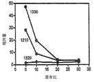
ある実施の形態において、細孔形成剤は、例えば、単峰性粒径分布を有する同様のまたは異なる粒子の混合物を含んで差し支えない。粒径分布特性に応じて、単峰性分布は、大きい、中間の、または小さい間隙、もしくはそれらの混合物を有する粒子相、並びにその粒子相の除去後の、対応する大きい、中間の、または小さい表面細孔、またはその混合物を提供できる。図4は、最密充填単峰性の大きい細孔形成剤集合体の例を示している。 In certain embodiments, the pore former may comprise a mixture of similar or different particles having, for example, a unimodal particle size distribution. Depending on the particle size distribution characteristics, the unimodal distribution is a particle phase with large, medium or small gaps, or mixtures thereof, as well as the corresponding large, medium or small after removal of the particle phase. Surface pores, or mixtures thereof, can be provided. FIG. 4 shows an example of a pore-forming agent aggregate having a close-packed unimodality.
ある実施の形態において、細孔形成剤は、例えば、二峰性粒径分布を有する粒子の混合物を含んで差し支えない。各ピークを適切な量で有する二峰性粒径分布は、大きい間隙と小さな間隙の混合物、大きい表面細孔と小さい表面細孔の混合物、またはそれらの混合物を有する粒子相を提供できる。図5は、大きい細孔形成剤と小さい細孔形成剤の混合物を有する最密充填の二峰性集合体の例を示している。 In certain embodiments, the pore former can comprise, for example, a mixture of particles having a bimodal particle size distribution. A bimodal particle size distribution with the appropriate amount of each peak can provide a mixture of large and small gaps, a mixture of large and small surface pores, or a particle phase having a mixture thereof. FIG. 5 shows an example of a close packed bimodal assembly with a mixture of large and small pore formers.
ある実施の形態において、第1の粒子混合物および第2の粒子混合物を有する場合、それぞれの混合物は、例えば、単峰性粒子、二峰性粒子、単分散性粒子、二分散性粒子、多分散性粒子、およびそれらの組合せから独立して選択して差し支えない。ある実施の形態において、第1の粒子混合物および第2の粒子混合物は、同じ物質または異なる物質であるが、異なる粒径特性、粒径分布特性、またはそれらの組合せを有する物質から構成されて差し支えない。一般に、細孔形成剤の含有量が増加するにつれて、気孔率および細孔サイズは、例えば線形に、増加する。 In certain embodiments, when having a first particle mixture and a second particle mixture, each mixture may be, for example, a monomodal particle, a bimodal particle, a monodisperse particle, a bidisperse particle, a polydisperse May be selected independently from the sex particles and combinations thereof. In certain embodiments, the first particle mixture and the second particle mixture may be composed of materials that are the same material or different materials but have different particle size characteristics, particle size distribution characteristics, or a combination thereof. Absent. In general, as the pore former content increases, the porosity and pore size increase, for example, linearly.
混合物の重合は、適切な基体上で行うことができる。それに加え、またはその代わりに、混合物の重合は、例えば、様々な有用な形状に成形された形態にあり、必要に応じて、例えば、基体、容器、または同様の支持体に付着したまたは関連付けられたプリフォームとして行っても差し支えない、すなわち、少なくとも1種類のモノマーまたはオリゴマーと、少なくとも1種類の細孔形成剤粒子材料とを含む混合物を基体上で重合させて、その基体上に連続高分子基質および不連続粒子相を形成する。 The polymerization of the mixture can be carried out on a suitable substrate. In addition or alternatively, the polymerization of the mixture is, for example, in a form molded into various useful shapes, optionally attached to or associated with, for example, a substrate, container, or similar support. A mixture of at least one monomer or oligomer and at least one pore former particle material is polymerized on the substrate and the continuous polymer is formed on the substrate. Form a matrix and a discontinuous particle phase.
少なくとも1種類のモノマーまたはオリゴマーを重合させる工程は、例えば、シロキサン、ビニル置換トリアルコキシシラン、アルファオレフィン、ビニルエステル、アクリレート、アクリルアミド、不飽和ケトン、モノビニリデン芳香族炭化水素、および同様の重合性モノマーまたはオリゴマー、もしくはそれらの混合物から選択された少なくとも1種類のモノマーまたはオリゴマーの連続高分子相を形成する工程を含む。ここに開示された物品を形成するために重合または共重合させることのできる適切なモノマーまたはオリゴマーの例としては、モノビニリデン芳香族炭化水素(例えば、スチレン、o−,m−およびp−メチルスチレン、2,4−ジメチルスチレン、Ar−エチルスチレン、p−ブチルスチレンなどのアラルキルスチレン、および同様のモノマーまたはオリゴマー;およびアルファ−メチルスチレン、アルファ−エチルスチレン、アルファ−メチル−p−メチルスチレンなどのアルファ−アルキルスチレン、および同様のモノマーまたはオリゴマー;ビニルナフタレン、および同様のモノマーまたはオリゴマー);Ar−ハロ−モノビニリデン芳香族炭化水素(例えば、o−,m−およびp−クロロスチレン、2,4−ジブロモスチレン、2−メチル−4−クロロスチレン、および同様のモノマーまたはオリゴマー);アクリロニトリル、メタクリロニトリル、アルキルアクリレート(例えば、メタクリレート、ブチルアクリレート、エチルヘキシルアクリレート、および同様のモノマーまたはオリゴマー)、対応するアルキルメタクリレート、アクリルアミド(例えば、アクリルアミド、メチルアクリルアミド、N−ブチルアクリルアミド、および同様のモノマーまたはオリゴマー);不飽和ケトン(例えば、ビニルメチルケトン、メチルイソプロペニルケトン、および同様のモノマーまたはオリゴマー);アルファオレフィン(例えば、エチレン、プロピレン、および同様のモノマーまたはオリゴマー);ビニルエステル(例えば、酢酸ビニル、ステアリン酸ビニル、および同様のモノマーまたはオリゴマー);ビニルおよびビニリデンのハロゲン化物(例えば、ビニルおよびビニリデンの塩化物と臭化物、および同様のモノマーまたはオリゴマー);ビニル置換トリアルコキシシラン、および同様のモノマーまたはオリゴマーなどのビニル置換シラン、またはそれらの組合せが挙げられる。細矢等は、“High-Performance Polymer-Based Monolithic Capillary Column,” Anal. Chem., 2006, 78 (16), 5729-5735において、ジアミンである4−[(4−アミノシクロヘキシル)メチル]シクロヘキシルアミン(BACM)およびキラルトランス−1,2−シクロヘキサンジアミン(CHD)と共に、エポキシモノマー、トリス(2,3−エポキシプロピル)イソシアヌレート(TEPIC)を使用して毛管カラムを調製したと述べており;辻岡等は、“A New Preparation Method for Well-Controlled 3D Skeletal Epoxy Resin-Based Polymer Monoliths,” Macromolecules, 2005, 38 (24), 9901-9903において、3Dモノリスを調製するために、ビスフェノールAジグリシジルエーテル(BADE)、(BACM)、およびポリ(エチレングリコール)(PEG)などの細孔生成(porogenic)溶媒を使用したと述べている。 The step of polymerizing at least one monomer or oligomer includes, for example, siloxanes, vinyl substituted trialkoxysilanes, alpha olefins, vinyl esters, acrylates, acrylamides, unsaturated ketones, monovinylidene aromatic hydrocarbons, and similar polymerizable monomers. Or forming a continuous polymer phase of at least one monomer or oligomer selected from oligomers or mixtures thereof. Examples of suitable monomers or oligomers that can be polymerized or copolymerized to form the articles disclosed herein include monovinylidene aromatic hydrocarbons (eg, styrene, o-, m- and p-methylstyrene). , Aralkyl styrene such as 2,4-dimethylstyrene, Ar-ethylstyrene, p-butylstyrene, and similar monomers or oligomers; and alpha-methylstyrene, alpha-ethylstyrene, alpha-methyl-p-methylstyrene, etc. Alpha-alkyl styrene, and similar monomers or oligomers; vinyl naphthalene, and similar monomers or oligomers; Ar-halo-monovinylidene aromatic hydrocarbons (eg, o-, m- and p-chlorostyrene, 2,4 -Dibromostyrene, 2 Methyl-4-chlorostyrene, and similar monomers or oligomers); acrylonitrile, methacrylonitrile, alkyl acrylates (eg, methacrylate, butyl acrylate, ethylhexyl acrylate, and similar monomers or oligomers), corresponding alkyl methacrylates, acrylamides (eg, , Acrylamide, methyl acrylamide, N-butyl acrylamide, and similar monomers or oligomers); unsaturated ketones (eg, vinyl methyl ketone, methyl isopropenyl ketone, and similar monomers or oligomers); alpha olefins (eg, ethylene, propylene) And similar monomers or oligomers); vinyl esters (eg, vinyl acetate, vinyl stearate, and the like) Vinyl and vinylidene halides (eg, vinyl and vinylidene chlorides and bromides, and similar monomers or oligomers); vinyl substituted silanes such as vinyl substituted trialkoxysilanes, and similar monomers or oligomers; Or a combination thereof. Hosoya et al. In “High-Performance Polymer-Based Monolithic Capillary Column,” Anal. Chem., 2006, 78 (16), 5729-5735, diamine 4-[(4-aminocyclohexyl) methyl] cyclohexylamine ( Said that a capillary column was prepared using an epoxy monomer, tris (2,3-epoxypropyl) isocyanurate (TEPIC), together with BACM) and chiral trans-1,2-cyclohexanediamine (CHD); In “A New Preparation Method for Well-Controlled 3D Skeletal Epoxy Resin-Based Polymer Monoliths,” Macromolecules, 2005, 38 (24), 9901-9903, bisphenol A diglycidyl ether (BADE) was prepared to prepare 3D monoliths. ), Porogenic solvents such as (BACM), and poly (ethylene glycol) (PEG) That.
少なくとも1種類の細孔形成剤は、例えば、単糖、多糖、ポリアルキレングリコール、ポリビニルアルコール、氷、ワックス、固体CO2などの昇華性材料、形成される高分子の融点より低い融点を有する物質、水溶性高分子、水不溶性高分子、またはそのコポリマー、シェルとコアを有するマイクロカプセルであって、例えば、シェルがモノマーまたはオリゴマー不溶性材料を含み、コアが水混和性または水溶性材料を含むマイクロカプセル、可溶性シェルと中空またはガス充填コアを有するマイクロバルーン、もしくはそれらの組合せであって差し支えない。 The at least one kind of pore-forming agent is, for example, a sublimable material such as monosaccharide, polysaccharide, polyalkylene glycol, polyvinyl alcohol, ice, wax, solid CO 2, or a substance having a melting point lower than the melting point of the formed polymer. , Water-soluble polymers, water-insoluble polymers, or copolymers thereof, microcapsules having a shell and a core, for example, a shell containing a monomer or oligomer insoluble material and a core containing a water-miscible or water-soluble material It can be a capsule, a microballoon with a soluble shell and a hollow or gas-filled core, or a combination thereof.
得られた重合済み固体基質を処理して、その基質から粒子相を除去する工程は、例えば、
ある物質と接触させて、粒子相を溶解させる工程であって、その物質が、水溶液;有機溶液;超臨界流体、例えば、CO2;低融点固体、例えば、ワックス、水、および同様の低融点固体;ガス、例えば、空気、N2、アルゴン、および同様のガス;またはそれらの組合せ:の内の少なくとも1つを含むものである工程;
基質を加熱して、粒子相を液化または溶解させる工程;または
接触と加熱の組合せ;
を含んで差し支えない。
粒子相のない得られた高分子相は、例えば、1.2から1.4、1.2から1.35、1.25から1.4、1.3から1.49、1.3から1.4、1.35から1.49、1.35から1.4、および同様の値と範囲などの全ての中間値と範囲を含む、約1.2から約1.49までなどの約1.49以下の屈折率を有することができる。
Processing the resulting polymerized solid substrate to remove the particulate phase from the substrate includes, for example:
Contacting with a substance to dissolve the particulate phase, wherein the substance is an aqueous solution; an organic solution; a supercritical fluid, eg, CO 2 ; a low melting solid, eg, wax, water, and a similar low melting point A process comprising at least one of: a solid; a gas such as air, N 2 , argon, and similar gases; or a combination thereof;
Heating the substrate to liquefy or dissolve the particulate phase; or a combination of contact and heating;
Can be included.
The resulting polymer phase without particle phase is, for example, from 1.2 to 1.4, from 1.2 to 1.35, from 1.25 to 1.4, from 1.3 to 1.49, from 1.3. 1.4, 1.35 to 1.49, 1.35 to 1.4, and all intermediate values and ranges such as similar values and ranges, such as about 1.2 to about 1.49 It can have a refractive index of 1.49 or less.
ある実施の形態において、調製方法はさらに、例えば、得られた細胞培養物品において空隙体積となる、すなわち、間隙体積と細孔体積の合計となる粒状細孔形成剤により占められる体積分および連続高分子基質となる空隙体積を有する粒径集合体に基づいて細孔形成剤充填密度を選択する工程をさらに含んでも差し支えない。少なくとも1種類のモノマーまたはオリゴマーと少なくとも1種類の粒状細孔形成剤とを含む混合物は、例えば、高速液固混合、液固ブレンド、液固遠心分離法、またはそれらの組合せ:の内の少なくとも1つによって調製することができる。 In certain embodiments, the preparation method further includes, for example, volume fraction and continuous volume occupied by a particulate pore former that results in a void volume in the resulting cell culture article, i.e., the sum of the pore volume and the pore volume. The method may further include a step of selecting a pore forming agent packing density based on a particle size aggregate having a void volume serving as a molecular substrate. The mixture comprising at least one monomer or oligomer and at least one particulate pore former is at least one of, for example, high-speed liquid-solid mixing, liquid-solid blending, liquid-solid centrifugation, or combinations thereof: Can be prepared.
ある実施の形態において、本開示は、例えば:
相互接続された多孔質網目構造を有する高分子塊であって、相互接続された多孔質網目構造が、細孔サイズの単一分布、または大きいサイズの細孔と小さいサイズの細孔並びに対応する間隙の二峰性分布からなる細孔を含むものである高分子塊を含む、三次元細胞培養物品を提供する。ある実施の形態において、その物品の高分子塊は、例えば:ビーズ;再構成可能な粉末;コーティング配合物、例えば、多孔質高分子塊の粒子の懸濁液;20マイクロメートルから500マイクロメートルの厚さの薄膜、10,000マイクロメートルから100,000マイクロメートル以上の厚さの厚膜、または500マイクロメートルから10,000マイクロメートルの厚さの中間厚の膜、もしくはそれらの組合せ:の内の少なくとも1つであって差し支えない。
In certain embodiments, the disclosure provides, for example:
A polymer mass having an interconnected porous network, wherein the interconnected porous network corresponds to a single distribution of pore sizes, or large and small pores as well as corresponding A three-dimensional cell culture article is provided that includes a polymer mass that includes pores comprising a bimodal distribution of gaps. In certain embodiments, the polymeric mass of the article comprises, for example: beads; reconstitutable powder; coating formulation, eg, suspension of particles of porous polymeric mass; from 20 micrometers to 500 micrometers A thin film of thickness, a thickness of 10,000 micrometers to 100,000 micrometers or more, or an intermediate thickness of 500 micrometers to 10,000 micrometers, or a combination thereof: It may be at least one of the above.
ある実施の形態において、本開示は、
少なくとも1種類のモノマーまたはオリゴマーと少なくとも1種類の粒状細孔形成剤とを含む混合物を重合させて、別個の粒子相を有する連続高分子基質を形成する工程、および
得られた固体基質を処理して、その基質から粒子相を除去する工程、
を含む上述したプロセスにより調製された三次元細胞培養物品を提供する。
In certain embodiments, the present disclosure provides:
Polymerizing a mixture comprising at least one monomer or oligomer and at least one particulate pore former to form a continuous polymer substrate having a distinct particle phase; and treating the resulting solid substrate Removing the particulate phase from the substrate,
A three-dimensional cell culture article prepared by the process described above.
この三次元細胞培養物品は、例えば、基体;およびこの基体上に支持された相互接続された多孔質網目構造を有する高分子相であって、連続または半連続空隙相を有する連続高分子基質を含む高分子層を含んで差し支えない。ある実施の形態において、細胞培養物品は、二相が連続した材料であると特徴付けることができる、すなわち、高分子が連続基質を形成し、粒子空隙が、第2の連続した、中空または開いているかにかかわらない相を形成する。 The three-dimensional cell culture article includes, for example, a substrate; and a polymer phase having an interconnected porous network supported on the substrate, the polymer layer having a continuous or semi-continuous void phase. It may include a polymer layer. In certain embodiments, the cell culture article can be characterized as a biphasic continuous material, i.e., the macromolecule forms a continuous matrix and the particle voids are second continuous, hollow or open. Form a phase regardless of whether or not.
ある実施の形態において、多孔質高分子物品は、例えば、約0.1から約20m2/gの表面積を有し得る。 In certain embodiments, the porous polymeric article can have a surface area of, for example, from about 0.1 to about 20 m 2 / g.
ある実施の形態において、多孔質高分子物品は、水銀または窒素気孔率測定により測定して、中間の値と範囲を含む、例えば、約50%から約95%の気孔率を有し得る;多孔質高分子物品は、中間の値と範囲を含む、例えば、約1.28から約1.49の屈折率を有し得る;多孔質高分子物品は、中間の値と範囲を含む、例えば、約1から約1,000kg/m3の密度を有し得る。 In certain embodiments, the porous polymeric article can have a porosity of between about 50% to about 95%, including intermediate values and ranges, as measured by mercury or nitrogen porosity measurements; The polymeric article can have an intermediate value and range, for example, a refractive index of about 1.28 to about 1.49; the porous polymeric article can have an intermediate value and range, for example, It may have a density from about 1 to about 1,000 kg / m 3 .
ある実施の形態において、ポリジメチルシロキサン多孔質高分子の屈折率は、例えば、約1.28から約1.49であって差し支えなく、典型的な水性細胞培養培地の屈折率は、例えば、約1.33から約1.36であり得る。多孔質高分子の屈折率および典型的な水性細胞培養培地の屈折率は、それぞれの屈折率が一致するまたはほぼ一致する、例えば、それぞれの屈折率の差が約±0.2単位未満、好ましくは約±0.15未満、より好ましくは約±0.12未満、さらにより好ましくは約±0.10未満となるように選択することができる。多孔質高分子物品は、例えば、約0から約1の光学密度、および例えば、約100から約1,000マイクロメートル以上の光侵入深さを有し得る。多孔質高分子物品は、約500から約500,000ダルトンの分子量を有する、ポリマー、コポリマー、または同様の材料を含み得る。 In certain embodiments, the refractive index of the polydimethylsiloxane porous polymer can be, for example, from about 1.28 to about 1.49, and the refractive index of a typical aqueous cell culture medium is, for example, about It can be from 1.33 to about 1.36. The refractive index of the porous polymer and the refractive index of a typical aqueous cell culture medium are the same or nearly the same, for example, each refractive index difference is less than about ± 0.2 units, preferably Can be selected to be less than about ± 0.15, more preferably less than about ± 0.12, and even more preferably less than about ± 0.10. The porous polymeric article can have, for example, an optical density of about 0 to about 1, and a light penetration depth of, for example, about 100 to about 1,000 micrometers or more. The porous polymeric article can include a polymer, copolymer, or similar material having a molecular weight of about 500 to about 500,000 daltons.
ある実施の形態において、その物品は、さらに、栄養素、抗生物質、成長促進剤、成長阻害剤、表面修飾剤、表面相溶化剤、および1種類以上の促進剤、阻害剤、調節剤、調整剤、誘発因子、またはそれらの組合せなどの同様の細胞培養成分からなる群より選択される添加剤を少なくとも1つ含み得る。 In certain embodiments, the article further comprises nutrients, antibiotics, growth promoters, growth inhibitors, surface modifiers, surface compatibilizers, and one or more promoters, inhibitors, regulators, modifiers. At least one additive selected from the group consisting of similar cell culture components such as, inducers, or combinations thereof.
ある実施の形態において、本開示は、例えば、上述した細胞培養を、基体と、この基体上に支持された相互接続された多孔質網目構造を有する高分子層とを含む物品と接触させる工程であって、多孔質高分子層が、連続または半連続空隙相を有する連続高分子基質を含むものである工程、および温度制御、培地交換、および生きている細胞などの適切な培地と培養条件;を含む細胞培養方法を提供する。 In certain embodiments, the present disclosure includes, for example, contacting the cell culture described above with an article comprising a substrate and a polymer layer having an interconnected porous network supported on the substrate. Wherein the porous polymer layer comprises a continuous polymer substrate having a continuous or semi-continuous void phase, and suitable media and culture conditions such as temperature control, media exchange, and living cells; A cell culture method is provided.
この細胞培養は、業界基準のコラーゲン表面の保持率と比べて約70から約100パーセントの保持率を提供する。 This cell culture provides about 70 to about 100 percent retention compared to industry standard collagen surface retention.
ある実施の形態において、適切な細胞株の一例として、ヒト初代細胞が挙げられる。ある実施の形態において、培養物品は、優れた細胞株性能、細胞機能性、細胞生存能力、細胞遺伝子発現、および同様の特性、またはそれらの組合せを提供する。 In certain embodiments, an example of a suitable cell line includes human primary cells. In certain embodiments, the culture article provides superior cell line performance, cell functionality, cell viability, cellular gene expression, and similar properties, or combinations thereof.
細胞培養と細胞検出を促進できる3D細胞培養基体を開発するために、基礎材料の光学的性質を、培養される細胞をその後モニタするために使用される所望の検出またはイメージング技法に適合させることが望ましい。本開示の実施の形態において、蛍光顕微鏡を使用して、培養物中の細胞をモニタするために使用することができる。開示された細胞培養物を構成するための基礎材料の以下の光学的性質を考慮すべきである:光検出中の培養物からの光の干渉を避けるために、動作波長範囲における良好な光学的透明性;および検出中の材料からの光散乱を減少するために培地の屈折率と密接に一致した屈折率。その物品の多孔質材料の光学的性質に加え、この多孔質材料の化学的性質は、細胞の増殖が、培養プロセス中にその物品の変化に曝されないように、培養条件下で非常に安定であるように選択することができる。ある実施の形態において、多孔質材料は、細胞増殖を促進するために、良好なガス透過率および水浸透率を有することが好ましい。 To develop a 3D cell culture substrate that can facilitate cell culture and cell detection, adapting the optical properties of the base material to the desired detection or imaging technique used to subsequently monitor the cultured cells. desirable. In embodiments of the present disclosure, a fluorescence microscope can be used to monitor cells in the culture. The following optical properties of the base material for constructing the disclosed cell culture should be considered: good optical in the operating wavelength range to avoid light interference from the culture during light detection Transparency; and a refractive index that closely matches the refractive index of the medium to reduce light scattering from the material being detected. In addition to the optical properties of the porous material of the article, the chemical nature of the porous material is very stable under culture conditions so that cell growth is not exposed to changes in the article during the culture process. You can choose to be. In certain embodiments, the porous material preferably has good gas permeability and water permeability to promote cell growth.
ある実施の形態において、基礎高分子材料を、例えば、強制充填および浸出プロセスにより、相互接続された細孔または間隙を有する高多孔質構造に形成することができる。強制充填プロセスにおいて、所望のサイズの充填剤または細孔形成剤が基礎高分子材料と均質に混合され、重力または圧力いずれかを使用して、充填剤を互いに緊密に接触させ、モノマーまたはオリゴマー、プレポリマー、または得られる重合済みまたは硬化済み基礎高分子材料の前駆体と密接に充填される。充填剤を除去するために、適切な選択された溶媒を含む超音波浴内で浸出または溶解することなどにより、充填剤が充填された得られた基礎高分子材料を処理することができる。多孔質基体の細孔径分布は、充填剤のサイズにより調節することができ、その充填剤は、所望の培養用途に応じて、一サイズ分布または複数のサイズの分布であって差し支えない。サイズの検討に加え、使用する充填剤は、多孔質基体中に残る任意の残留する未浸出充填剤が細胞増殖に悪影響を与えないように、細胞培養の生理学的条件に対する毒性または干渉性が最小であるべきである。 In certain embodiments, the base polymeric material can be formed into a highly porous structure with interconnected pores or gaps, for example, by a forced filling and leaching process. In a forced filling process, a filler or pore former of the desired size is intimately mixed with the base polymeric material, using either gravity or pressure to bring the fillers into intimate contact with each other, a monomer or oligomer, It is intimately packed with the prepolymer or the precursor of the resulting polymerized or cured base polymer material. To remove the filler, the resulting base polymer material filled with filler can be treated, such as by leaching or dissolving in an ultrasonic bath containing a suitable selected solvent. The pore size distribution of the porous substrate can be adjusted by the size of the filler, which can be a single size distribution or a distribution of multiple sizes, depending on the desired culture application. In addition to size considerations, the filler used is minimally toxic or interfering with the physiological conditions of the cell culture so that any remaining unleached filler remaining in the porous substrate will not adversely affect cell growth. Should be.
所望の粒径およびサイズの分布の細孔形成剤は、例えば、粒径減少法、粒径成長法、またはそれらの組合せを含む任意の適切な方法によって得て差し支えない。乾燥固体粒子またはキャリア流体中に懸濁された粒子に、粒径減少装置を使用することができる。高圧液体流を使用した一液型粒径減少装置は、例えば、「PARTICLE-SIZE REDUCTION APPARATUS, AND USE THEREOF」と題する国際公開第2006/064203号パンフレットに述べられたような増圧器の改良が施されたMicrofluidizer(登録商標)、または同様の装置であり、もしくは同様のサイズ処理装置を使用して、サイズの減少によって所望の粒子を製造しても差し支えない。 A pore former with the desired particle size and size distribution can be obtained by any suitable method including, for example, a particle size reduction method, a particle size growth method, or a combination thereof. A particle size reduction device can be used for dry solid particles or particles suspended in a carrier fluid. A one-part type particle size reduction device using a high-pressure liquid flow is an example of an improvement of a pressure intensifier as described in the pamphlet of WO 2006/064203 entitled “PARTICLE-SIZE REDUCTION APPARATUS, AND USE THEREOF”. Microfluidizer (R), or similar equipment, or similar sizing equipment may be used to produce the desired particles by size reduction.
粒径成長方法の例としては、エマルションまたは懸濁液重合プロセス、または同様の粒径成長方法が挙げられる。サイズ減少粒子またはサイズ増加粒子は、例えば、粗粒分離器、フィルタ、スクリーン、または同様の装置によって、粒径または直径の範囲にしたがって、分離して差し支えない。 Examples of particle size growth methods include emulsion or suspension polymerization processes, or similar particle size growth methods. The size-reduced or size-enhanced particles can be separated according to particle size or diameter range, for example by a coarse separator, filter, screen, or similar device.
所望の粒径および粒径分布は、例えば、レーザ回折、動的光散乱、画像分析、または同様のサイズ処理装置および方法を含む、ホリバ社(www.boriba.com)から入手できるもののような、任意の適切な粒子分析装置によって測定し、特徴付けることができる。ホリバLB−550は、ppmから約40%の固形物までの濃度範囲で、1mmから約6マイクロメートルの粒径を測定することができる。ホリバLA−300レーザ回折粒径分布分析器を使用して、懸濁液または乾燥粉末の粒径を測定して差し支えない。 The desired particle size and particle size distribution can be obtained from Horiba (www.boriba.com), including, for example, laser diffraction, dynamic light scattering, image analysis, or similar size processing equipment and methods, It can be measured and characterized by any suitable particle analyzer. Horiba LB-550 can measure particle sizes from 1 mm to about 6 micrometers in a concentration range from ppm to about 40% solids. A Horiba LA-300 laser diffraction particle size distribution analyzer can be used to measure the particle size of the suspension or dry powder.
培養物品の製造、および基体や包装材料などの付随する部材の入手は、滅菌状態で行えることが好ましい。 The production of the culture article and the acquisition of the accompanying members such as the substrate and the packaging material are preferably performed in a sterilized state.
ある実施の形態において、本開示は、非動物源の細胞培養コーティングおよびin-vivo様細胞培養を提供するその対応する被覆基体を提供する。 In certain embodiments, the present disclosure provides a non-animal source cell culture coating and its corresponding coated substrate that provides an in-vivo-like cell culture.
ある実施の形態において、開示された多孔質コーティングおよび物品は、容易に調製することができ、比較的安価である。開示されたコーティングは、非動物由来の基体コーティングを提供し、得られた被覆製品は、例えば、ロット間の変動がほとんどまたは全くなく、優れた貯蔵安定性および棚−および生物学的−安定性を有する。実施の形態において、開示された多孔質高分子コーティングは、例えば、高分子コーティング中に使用されるモノマーまたはオリゴマーの選択により、容易に調節できる屈折率を有する基体表面コーティングを提供できる。開示されたコーティング組成物は、非毒性であり、生体適合性であり得る基体表面コーティングを提供できる。開示されたコーティング組成物は、様々な表面上に堆積させるのが容易などの、加工性の高い基体表面コーティングを提供し、様々な基体、例えば、プラスチック、ガラス、および同様の細胞培養基体または支持体への細胞とコーティングの付着を向上させる。所望であれば、アミノシランなどの結合層または転化コーティングを選択して、多孔質高分子のガラスなどの基体に対する付着性を向上させても差し支えない。 In certain embodiments, the disclosed porous coatings and articles can be easily prepared and are relatively inexpensive. The disclosed coating provides a non-animal derived substrate coating and the resulting coated product has excellent storage stability and shelf- and biological-stability, for example, with little or no lot-to-lot variation. Have In embodiments, the disclosed porous polymeric coating can provide a substrate surface coating having a refractive index that can be easily adjusted, for example, by the choice of monomer or oligomer used in the polymeric coating. The disclosed coating composition can provide a substrate surface coating that can be non-toxic and biocompatible. The disclosed coating composition provides a highly processable substrate surface coating, such as easy to deposit on a variety of surfaces, and a variety of substrates such as plastic, glass, and similar cell culture substrates or supports. Improves cell and coating adhesion to the body. If desired, a tie layer or conversion coating such as aminosilane may be selected to improve the adhesion of the porous polymer to a substrate such as glass.
ある実施の形態において、本開示は、細胞培養の方法であって、
上述した多孔質組成物または同様の組成物により被覆された基体を提供し、
被覆された基体を、機能的細胞を固定するのに十分な時間に亘り細胞培養物と接触させ、
その細胞を光学的方法によってモニタし、
必要に応じて、基体から細胞を収穫する、
各工程を有してなる方法を提供する。
In certain embodiments, the disclosure provides a method of cell culture comprising:
Providing a substrate coated with the porous composition described above or a similar composition;
Contacting the coated substrate with the cell culture for a time sufficient to immobilize functional cells;
The cells are monitored by optical methods,
Harvest cells from the substrate, if necessary,
A method comprising each step is provided.
ある実施の形態において、基体は、例えば、金属酸化物、複合金属酸化物、合成高分子、天然高分子、同様の材料、またはそれらの組合せから選択される材料であって差し支えない。ある実施の形態において、本開示の多孔質細胞培養物品は、例えば、生体適合性がより優れた物品を得るために、表面処理または状態調節によって、さらに処理して差し支えない。例えば、同一出願人の米国特許第6617152号および同時係属の米国特許出願第11/973832号の各明細書を参照のこと。 In certain embodiments, the substrate can be a material selected from, for example, metal oxides, composite metal oxides, synthetic polymers, natural polymers, similar materials, or combinations thereof. In certain embodiments, the porous cell culture articles of the present disclosure can be further processed, for example, by surface treatment or conditioning, to obtain articles that are more biocompatible. See, for example, commonly assigned US Pat. No. 6,617,152 and co-pending US patent application Ser. No. 11/973832.
細胞培養物の細胞は、例えば、肝細胞などの任意の細胞タイプの、任意の適切な初代細胞、または関連する無限に再生産された細胞株であって差し支えない。細胞培養物は、例えば、アルブミンを盛んに産生する細胞、基体、または同様の実在物、およびそれらの組合せを含んでよい。基体からの細胞の収穫は、例えば、遠心分離、撹拌、洗浄、および同様のプロセス、またはそれらの組合せを含む、任意の適切な手段によって行っても差し支えない。 The cells of the cell culture can be any suitable primary cell of any cell type, such as, for example, hepatocytes, or an associated infinitely regenerated cell line. Cell cultures may include, for example, cells that actively produce albumin, substrates, or similar entities, and combinations thereof. Harvesting cells from the substrate can be done by any suitable means including, for example, centrifugation, agitation, washing, and similar processes, or combinations thereof.
ある実施の形態において、多孔質組成物の調製に使用するのに、様々な生体適合性高分子材料を選択して差し支えない。それに加え、またはそれに代わり、生体適合性高分子材料は、単独で、もしくは他の細胞培養材料または支持材料と組み合わせてまたは混合して使用して差し支えない。その高分子材料の例としては、ポリアミド、ポリカーボネート、ポリアルキレン、ポリアルキレングリコール、ポリアルキレンオキシド、ポリアルキレンテレフタレート、ポリビニルアルコール、ポリビニルエーテル、ポリビニルエステル、ポリハロゲン化ビニル、ポリビニルピロリドン、ポリグリコリド、ポリシロキサン、ポリウレタン、およびそのコポリマー、ニトロセルロース、アクリルエステルとメタクリルエステルのポリマー、ヒドロキシプロピルセルロース、ヒドロキシプロピルメチルセルロース、ヒドロキシブチルメチルセルロース、酢酸セルロース、プロピオン酸セルロース、酢酸酪酸セルロース、酢酸フタル酸セルロース、カルボキシエチルセルロース、三酢酸セルロース、硫酸セルロースナトリウム塩、ポリ(メチルメタクリレート)、ポリ(エチルメタクリレート)、ポリ(ブチルメタクリレート)、ポリ(イソブチルメタクリレート)、ポリ(ヘキシルメタクリレート)、ポリ(イソデシルメタクリレート)、ポリ(ラウリルメタクリレート)、ポリ(フェニルメタクリレート)、ポリ(メタクリレート)、ポリ(イソプロピルアクリレート)、ポリ(イソブチルアクリレート)、ポリ(オクタデシルアクリレート)、ポリエチレンポリ(エチレングリコール)、ポリ(エチレンオキシド)、ポリ(エチレンテレフタレート)、ポリ(ビニルアルコール)、ポリ(酢酸ビニル)、ポリ塩化ビニル、ポリスチレン、ポリヒアルロン酸、カゼイン、ゼラチン、グルテン、ポリ無水物、ポリアクリル酸、アルギネート、キトサン、およびそれらの任意のコポリマー、またはそれらの任意の組合せが挙げられる。 In certain embodiments, various biocompatible polymeric materials can be selected for use in preparing the porous composition. In addition or alternatively, the biocompatible polymeric material can be used alone or in combination or mixed with other cell culture materials or support materials. Examples of the polymer material include polyamide, polycarbonate, polyalkylene, polyalkylene glycol, polyalkylene oxide, polyalkylene terephthalate, polyvinyl alcohol, polyvinyl ether, polyvinyl ester, polyvinyl halide, polyvinyl pyrrolidone, polyglycolide, polysiloxane. , Polyurethane, and copolymers thereof, nitrocellulose, polymers of acrylic and methacrylic esters, hydroxypropylcellulose, hydroxypropylmethylcellulose, hydroxybutylmethylcellulose, cellulose acetate, cellulose propionate, cellulose acetate butyrate, cellulose acetate phthalate, carboxyethylcellulose, three Cellulose acetate, cellulose sulfate sodium salt, poly (methyl Acrylate), poly (ethyl methacrylate), poly (butyl methacrylate), poly (isobutyl methacrylate), poly (hexyl methacrylate), poly (isodecyl methacrylate), poly (lauryl methacrylate), poly (phenyl methacrylate), poly (methacrylate) , Poly (isopropyl acrylate), poly (isobutyl acrylate), poly (octadecyl acrylate), polyethylene poly (ethylene glycol), poly (ethylene oxide), poly (ethylene terephthalate), poly (vinyl alcohol), poly (vinyl acetate), poly Vinyl chloride, polystyrene, polyhyaluronic acid, casein, gelatin, gluten, polyanhydrides, polyacrylic acid, alginate, chitosan, and any copolymers thereof , Or any combination thereof.
細胞の生理的機能は、細胞培養環境によって大きく影響を受け得る。細胞を、その細胞の特定の機能を最良に維持できる系内で培養することが有益であり得る。平らな表面上の従来の二次元(2D)細胞培養と比べると、「Matrigel」などの様々な基質中の三次元(3D)細胞培養には、in-vivo細胞環境を厳密に模倣しているので、細胞機能を維持する上で実証された明らかな有用性がある。細孔径および基質剛性が、3D細胞培養システムにおける細胞形態学および機能を調節する2つの重要な要因と認識されてきた(例えば、C.S. Ranucci, et al., Biomaterials 21(2000) 783-793; M.H. Zaman, et al., Proc Nati Acad Sci USA 103(29) 2006, 10889-94; T. Sun, et al., “Investigation of fibroblast and keratinocyte cell-scaffold interactions using a novel 3D cell culture system”, Journal of Materials Science: Materials in Medicine, 18 (2), 2007, pp. 321-328を参照のこと)。しかしながら、これらの培養システムにおける細孔径の精密な制御および調節は、困難であり得る。 The physiological function of cells can be greatly influenced by the cell culture environment. It may be beneficial to culture the cells in a system that can best maintain the particular function of the cells. Compared to traditional two-dimensional (2D) cell cultures on flat surfaces, three-dimensional (3D) cell cultures in various substrates such as “Matrigel” closely mimic the in-vivo cell environment So there is a clear utility demonstrated in maintaining cell function. Pore size and substrate stiffness have been recognized as two important factors that regulate cell morphology and function in 3D cell culture systems (eg CS Ranucci, et al., Biomaterials 21 (2000) 783-793; MH Zaman, et al., Proc Nati Acad Sci USA 103 (29) 2006, 10889-94; T. Sun, et al., “Investigation of fibroblast and keratinocyte cell-scaffold interactions using a novel 3D cell culture system”, Journal of Materials Science: Materials in Medicine, 18 (2), 2007, pp. 321-328). However, precise control and adjustment of pore size in these culture systems can be difficult.
ある実施の形態において、本開示は、特定の細胞の細胞機能を調節するために、3D細胞培養基体の細孔径および剛性を制御し、調節するための組成物および方法を提供する。 In certain embodiments, the present disclosure provides compositions and methods for controlling and modulating the pore size and stiffness of 3D cell culture substrates to modulate the cellular function of specific cells.
ある実施の形態において、本開示は、3D細胞培養において細胞機能を調節できる多孔質合成材料基体を製造するための設計および方法を提供する。生きている実在物の外部で細胞が増殖し、生物学的機能を果たすためのin-vivo様環境を提供することに加え、細胞機能を調節するために、これらの多孔質基体の細孔径および剛性を制御し、調節しても差し支えなく、この細胞機能は、汎用の細胞に基づく用途の様々な必要性を果たすことができる。例えば、ある薬剤の誘導試験には、標的遺伝子の比較的低い基底発現レベルしか必要ないのに対し、薬剤の阻害試験には、標的遺伝子の比較的高い基底発現レベルが必要である。基体の細孔径および剛性の特性を調節することによって、遺伝子発現レベルを調節する能力は、他の培養条件への乱れを最小にしながら、これらの結果を比較するための有用なプラットホームを提供する。 In certain embodiments, the present disclosure provides designs and methods for producing porous synthetic material substrates that can modulate cell function in 3D cell culture. In addition to providing an in-vivo-like environment for cells to proliferate and perform biological functions outside living entities, the pore size and size of these porous substrates can be adjusted to regulate cell function. The cell function can be controlled and adjusted, and this cellular function can serve various needs for general purpose cell based applications. For example, a drug induction test requires a relatively low basal expression level of the target gene, whereas a drug inhibition test requires a relatively high basal expression level of the target gene. The ability to adjust gene expression levels by adjusting the pore size and stiffness characteristics of the substrate provides a useful platform for comparing these results while minimizing disruption to other culture conditions.
ある実施の形態において、本開示は、異なる細胞株に関して最適化された培養性能を提供するように調節できる、多孔質足場の細孔径および剛性などの性質を有する材料、および細胞の増殖および形態学を調節できる多孔質足場の細孔径および剛性を制御する方法を提供し、その足場は、遺伝子発現レベルで細胞機能を選択的に調節することができる。 In certain embodiments, the present disclosure provides materials having properties such as pore size and stiffness of porous scaffolds that can be adjusted to provide optimized culture performance for different cell lines, and cell growth and morphology Provides a method of controlling the pore size and stiffness of a porous scaffold that can regulate the scaffold, which can selectively regulate cell function at the gene expression level.
開示された多孔質高分子基体は、3D細胞培養に関する以下の特徴を提供できる:
細孔径制御 細孔径は、容易に制御し、調節することができ、これは、細胞と細胞の相互作用および組織化のよく制御された物理的調節を提供する。
The disclosed porous polymer substrate can provide the following features for 3D cell culture:
Pore size control The pore size can be easily controlled and regulated, which provides a well-controlled physical regulation of cell-cell interaction and organization.
細胞の順序付け(ordering) 細胞培養中の細胞の組織化と相互作用は、細胞の増殖と細胞の機能発生に影響を与え、したがって、細胞機能は、細孔径を調節することによって調節できる。 Cell ordering Cell organization and interaction in cell culture affects cell growth and cell function development, and thus cell function can be regulated by regulating pore size.
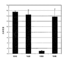
細胞の調節 細孔径制御による細胞機能の調節は、細胞の生理的環境の乱れを最小にする。同じ供給源からの細胞は、異なる基体の細孔径特性によって機能的に調節でき、比較のためのモデルを提供する。例えば、より小さな細孔を有する開示された多孔質PDMS基体において培養されたヒト肝細胞は、CYP1A2、CYP2B6、およびCYP3A4などの、CYP(シトクロムP450)の中程度のレベルを示したが、リファンピン(USAN)またはリファンピシン(INN)により誘発された上昇した薬剤応答を示し、このことは、薬剤候補による細胞機能の誘導研究にうまく適している。より大きい細孔を有する多孔質PDMSにおいて培養されたヒト肝細胞は、上昇したレベルのCYP遺伝子を有することが示され、この状況は、薬剤候補による細胞機能の阻害研究にとって理想的である。 Cell regulation Modulation of cell function by controlling pore size minimizes disruption of the cell's physiological environment. Cells from the same source can be functionally regulated by the pore size characteristics of different substrates, providing a model for comparison. For example, human hepatocytes cultured in the disclosed porous PDMS substrate with smaller pores showed moderate levels of CYP (cytochrome P450), such as CYP1A2, CYP2B6, and CYP3A4, but rifampin ( It shows an elevated drug response induced by USAN) or rifampicin (INN), which is well suited to studies of the induction of cell function by drug candidates. Human hepatocytes cultured in porous PDMS with larger pores have been shown to have elevated levels of CYP genes, which is ideal for studies of inhibition of cell function by drug candidates.
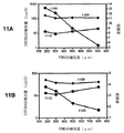
基体の調節および遺伝子の調節 開示された多孔質基体について、例えば、ヤング率として測定された剛性を正確に制御し、調節することができる。開示された基体の細孔径および剛性の容易な調節により、適合性が高く、汎用の3D細胞培養プラットホームが提供される。ヤング率は、高分子材料の剛性の尺度の1つである。ヤング率が高いほど、材料が硬くなる。このことは、材料のサンプルに行った引張試験中に生じた応力−歪み曲線の勾配から実験的に決定できる。応力−歪み曲線は、破損(例えば、破壊)するまで、材料に印加した力(応力)の関数として材料の伸張(歪み)をプロットしている;図13参照のこと。勾配が急な場合、そのサンプルは高い縦弾性係数を有し、このことは、材料が変形に抵抗し、高い剛性を有することを意味する。勾配が穏やかな場合、その材料は低い縦弾性率を有し、このことは、その材料が容易に変形され、低い剛性を有することを意味する。係数は、Pa、またはN/cm2などの強さの単位で表すことができる。生きている細胞の遺伝子発現のレベルは、多孔質高分子層のヤング率の剛性と共に増加することが示された。ある実施の形態において、多孔質高分子層は、例えば、中間の値と範囲を含む、約0.1MPaから約15MPaのヤング率を有し得る。 Substrate Regulation and Gene Regulation For the disclosed porous substrate, for example, the stiffness measured as Young's modulus can be accurately controlled and adjusted. The easy adjustment of the disclosed substrate pore size and stiffness provides a highly compatible and versatile 3D cell culture platform. Young's modulus is one measure of the stiffness of a polymeric material. The higher the Young's modulus, the harder the material. This can be determined empirically from the slope of the stress-strain curve generated during a tensile test performed on a sample of the material. The stress-strain curve plots the elongation (strain) of the material as a function of the force (stress) applied to the material until failure (eg, failure); see FIG. When the slope is steep, the sample has a high modulus of elasticity, which means that the material resists deformation and has a high stiffness. When the gradient is gentle, the material has a low modulus of elasticity, which means that the material is easily deformed and has a low stiffness. The coefficient can be expressed in units of strength such as Pa or N / cm 2 . It has been shown that the level of gene expression in living cells increases with the Young's modulus stiffness of the porous polymer layer. In certain embodiments, the porous polymer layer can have a Young's modulus of about 0.1 MPa to about 15 MPa, including, for example, intermediate values and ranges.
細胞機能は、細胞培養環境によって大きく影響を受け得る。うまく設計された細胞培養システムにおいて、細胞は特定の細胞機能を維持できる。その上、細胞機能は、その物理的相互作用を培養環境で調節することによって、調節することができる。 Cell function can be greatly affected by the cell culture environment. In a well-designed cell culture system, cells can maintain specific cell functions. Moreover, cell function can be regulated by regulating its physical interactions in a culture environment.
ある実施の形態において、本開示は、多孔質培養基体の細孔径および係数(剛性)を調節することによって、3D細胞培養における細胞機能を調節する方法を提供する。 In certain embodiments, the present disclosure provides a method of modulating cell function in 3D cell culture by adjusting the pore size and modulus (rigidity) of the porous culture substrate.
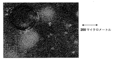
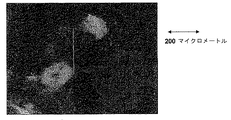
ある実施の形態において、主細孔群の細孔径は、単細胞よりも大きくて差し支えない。したがって、細胞は、細孔の内部に播種し、移行し、増殖し、組織化する傾向にある。基体の細孔径分布は、細胞と細胞のおよび細胞と基質の相互作用への物理的制約として細胞機能に著しく影響を与え得る。開示された多孔質PDMSは、細胞が細孔内に成長することができ、細胞と細胞の相互作用を促進する微小多孔質構造を有する。基体の細孔径の関数としての初代ヒト肝細胞のスフェロイド形成の画像を得た(カラー画像は示されていない)。それらの画像は、多孔質PDMS細孔径範囲は、180〜212マイクロメートルから、300〜355マイクロメートルまで増加するにつれて、1つのスフェロイドが個々の細孔内に形成される傾向にあり、スフェロイドのサイズが、細孔径の増加と共に増加し、次いで、500〜600マイクロメートル、850〜1,000マイクロメートル、そして1,000〜1,400マイクロメートルへと増加すると、個々の細孔中により大きいスフェロイドを形成する代わりに、多数のより小さいスフェロイドが形成されることを示している。1,000マイクロメートルより大きいサイズの細孔の基体内では、より小さいスフェロイドが形成が形成され、細胞は、細胞と細胞の粘着が緩い凝集体を形成する傾向にある。 In certain embodiments, the pore size of the main pore group can be larger than that of a single cell. Thus, cells tend to seed, migrate, proliferate, and organize inside the pores. The pore size distribution of the substrate can significantly affect cell function as a physical constraint on cell-cell and cell-substrate interactions. The disclosed porous PDMS has a microporous structure that allows cells to grow into the pores and promote cell-cell interaction. Images of spheroid formation of primary human hepatocytes as a function of substrate pore size were obtained (color images not shown). The images show that as the porous PDMS pore size range increases from 180-212 micrometers to 300-355 micrometers, one spheroid tends to form in individual pores, and the spheroid size Increases with increasing pore size and then increases to 500-600 micrometers, 850-1,000 micrometers, and 1,000-1,400 micrometers, resulting in larger spheroids in the individual pores. Instead of forming, a number of smaller spheroids are shown to be formed. Within a substrate with pores larger than 1,000 micrometers, smaller spheroids form and the cells tend to form aggregates with loose cell-cell adhesion.
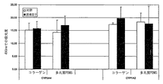
様々な細孔径を有する(棒グラフは示されていない)開示された多孔質PDMS内で培養されたヒト肝細胞における、コラーゲンおよび「Magrigel」対照に対する、遺伝子ABCB1(ATP−結合カセット、サブファミリーB(MDR/TAP)、メンバー1)、ACCC2(ATP−結合カセット、サブファミリーC(CFTR/MRP)、メンバー2)、ALB(アルブミン)、CEBPA(CCAAT/エンハンサー結合タンパク質(C/EBP)、アルファ)、GJB1(ギャップ結合タンパク質、ベータ1)、HNF4A(肝細胞核因子4、アルファ)、およびUGT1(UDPグルクロノシルトランスフェラーゼ1)の基礎的遺伝子発現の実験は、一貫して、10種類の遺伝子マーカーの全てが、リアルタイム定量的ポリメラーゼ連鎖反応(RQ−PCR)により明確に発現されることを示した。これらの遺伝子は、肝細胞の機能が、業界基準のコラーゲン表面および「Matrigel」と比べて、この基体内でどれだけよく保存されているかを測定するための一連のマーカーである。コラーゲン表面と比べて10種類のマーカーの全ての高い発現が、多孔質PDMSがコラーゲンよりもよく肝細胞の機能を保存できることを示している。より重要なことには、これらの遺伝子の発現レベルが、肝細胞機能を保存するための優れた市販の基体の1つである「Matrigel」に匹敵する。図面において、y軸の「RQ」は、ハウスキーピング遺伝子ヒポキサンチングアニンホスホリボシルトランスフェラーゼ1(HPRT1)の発現レベルによって割られた特定の遺伝子マーカーの遺伝子発現レベルの「相対的定量化」を表す。 The gene ABCB1 (ATP-binding cassette, subfamily B (subfamily B) versus collagen and “Magrigel” controls in human hepatocytes cultured in the disclosed porous PDMS with various pore sizes (bar graph not shown) MDR / TAP), member 1), ACCC2 (ATP-binding cassette, subfamily C (CFTR / MRP), member 2), ALB (albumin), CEBPA (CCAAT / enhancer binding protein (C / EBP), alpha), Basic gene expression experiments for GJB1 (gap junction protein, beta 1), HNF4A (hepatocyte nuclear factor 4, alpha), and UGT1 (UDP glucuronosyltransferase 1) have consistently performed all 10 gene markers. But real-time quantitative It has been shown to be clearly expressed by polymerase chain reaction (RQ-PCR). These genes are a series of markers to measure how well hepatocyte functions are conserved within this substrate compared to industry standard collagen surfaces and “Matrigel”. The high expression of all 10 markers compared to the collagen surface indicates that porous PDMS can preserve hepatocyte function better than collagen. More importantly, the expression levels of these genes are comparable to “Matrigel”, one of the excellent commercial substrates for preserving hepatocyte function. In the figure, “RQ” on the y-axis represents “relative quantification” of the gene expression level of a particular genetic marker divided by the expression level of the housekeeping gene hypoxanthine guanine phosphoribosyltransferase 1 (HPRT1).
以下の実施例は、本開示を使用する様式をより完全に記載し、さらに、本開示の様々な態様を実施するために考えられる最良の態様の特定の実施例を図示し実証するように働く。これらの実施例は、本開示の範囲を制限するものではなく、むしろ、説明目的のために提示されたものである。 The following examples more fully describe the manner in which the present disclosure is used, and further serve to illustrate and demonstrate specific examples of the best mode contemplated for carrying out various aspects of the present disclosure. . These examples do not limit the scope of the present disclosure, but rather are presented for illustrative purposes.
マルチウェルプレート形式で細胞培養基体を製造する方法
各々が本発明の多孔質PDMSを有する多数のユニットが、以下に記載されるように標準的な穴あきプレートのマルチウェルアセンブリのガラスインサート上に直接形成することができる。
Method for Manufacturing Cell Culture Substrates in Multiwell Plate Format A number of units, each having a porous PDMS of the present invention, are directly on the glass insert of a standard perforated plate multiwell assembly as described below. Can be formed.
a. マルチウェル穴あきプレートなどの支持基体上への多孔質高分子ユニットの直接形成
選択されたモノマーまたはオリゴマー、随意的な架橋剤、および例えば、1つ、2つまたは多数の粒径分布を有する細孔形成粒子を均一に混合物に混合する。
a. Direct formation of porous polymer units on a support substrate such as a multi-well perforated plate. Selected monomers or oligomers, optional crosslinkers, and fine particles with, for example, one, two or many particle size distributions. The pore-forming particles are uniformly mixed into the mixture.
例えば、遠心、振盪、加圧、または同様の方法によって、細孔形成剤を適切に充填する。 For example, the pore-forming agent is suitably filled by centrifugation, shaking, pressurization, or similar methods.
約500マイクロメートル未満の厚さのガラス板などの支持基体の上面に、マルチウェル穴あきプレートなどの所望のマルチユニット形式の成形型を配置する。 A desired multi-unit mold such as a multi-well perforated plate is placed on top of a support substrate such as a glass plate having a thickness of less than about 500 micrometers.
細孔形成剤が充填されたモノマーまたはオリゴマーの均一な混合物を成形型の空間内に注ぎ入れる。 A homogeneous mixture of monomers or oligomers filled with a pore former is poured into the mold space.
規定の硬化条件で成形型内の均一な混合物を硬化させる。 The uniform mixture in the mold is cured under specified curing conditions.
成形型から硬化した混合物を物理的に取り外す。 Physically remove the cured mixture from the mold.
硬化した混合物から細孔形成剤を除去する。 The pore former is removed from the cured mixture.
適切な接着剤または物理的保持部材などによって、支持基体を穴あきプレートに取り付ける。 The support substrate is attached to the perforated plate, such as by a suitable adhesive or physical holding member.
b. マルチウェルプレートの底部への多孔質高分子の直接形成
選択されたモノマーまたはオリゴマーおよび架橋剤の混合物をマルチウェルプレートの各ウェル中に分配する。
b. Direct Formation of Porous Polymers at the Bottom of the Multiwell Plate The selected monomer or oligomer and crosslinker mixture is dispensed into each well of the multiwell plate.
細孔形成剤を各ウェル中に分配し、約30分間に亘りプレートを振盪する。 Distribute the pore former into each well and shake the plate for about 30 minutes.
さらに30分間の振盪後に細孔形成剤の層が、モノマーまたはオリゴマーおよび細孔形成剤の混合物の表面上に残留するまたは留まるときなどの、モノマーまたはオリゴマーの混合物が細孔形成剤で飽和されるまで、各ウェル中に細孔形成剤を分配し続け、振盪する。 The monomer or oligomer mixture is saturated with the pore former, such as when the layer of pore former remains or remains on the surface of the monomer or oligomer and pore former mixture after another 30 minutes of shaking. Continue to dispense the pore former into each well until shaken.
規定の硬化条件で高分子および細孔形成剤の混合物を硬化させる。 The mixture of polymer and pore former is cured under defined curing conditions.
硬化後、適切な浸出条件を使用して細孔形成剤を除去する。 After curing, the pore former is removed using appropriate leaching conditions.
c. 多孔質高分子を予備成形し、次いで、それらをマルチウェルプレートの各ウェル内に分配する
選択されたモノマーまたはオリゴマー、架橋剤、および1つまたは2つの粒径分布(すなわち、単峰性または二峰性)を有する細孔形成剤を容器内で混合する。
c. Pre-form porous polymers and then distribute them into each well of a multi-well plate Selected monomer or oligomer, crosslinker, and one or two particle size distributions (ie unimodal or bimodal) A pore-forming agent having ridge properties is mixed in a container.
細孔形成剤および高分子前駆体を充填する。 Fill with pore former and polymer precursor.
混合物を硬化させて、高分子を架橋させる。 The mixture is cured to crosslink the polymer.
硬化した高分子から細孔形成剤を浸出させる。 The pore former is leached from the cured polymer.
得られた多孔質高分子を所望の形状とサイズに切断し、その切片をウェルプレートの底部に配置する。 The obtained porous polymer is cut into a desired shape and size, and the section is placed at the bottom of the well plate.
不活性接着剤などの固定手段を施して、多孔質高分子サンプルをプレートの底部に固定する。 A fixing means such as an inert adhesive is applied to fix the porous polymer sample to the bottom of the plate.
多孔質高分子(PDMS)における肝細胞培養の評価
多孔質高分子における肝細胞の細胞株C3Aの付着または生死判別研究 細胞溶解の際に、安定な細胞質型酵素である乳酸脱水素酵素(LDH)が放出される。放出されたLDHの量は、溶解した細胞の数に比例する。生存細胞数を定量することによって、多孔質高分子基質内のC3A細胞の付着を評価するために、CytoTox96細胞毒性アッセイキット(プロメガ社(Promega))を使用した。コラーゲン培養プレートおよび「MatriGel」3D培養体を対照基準として使用した。
Evaluation of hepatocyte culture in porous polymer (PDMS) Research on adhesion or survival of hepatocyte cell line C3A in porous polymer Lactate dehydrogenase (LDH), a stable cytoplasmic enzyme in cell lysis Is released. The amount of LDH released is proportional to the number of lysed cells. The CytoTox 96 cytotoxicity assay kit (Promega) was used to assess the adherence of C3A cells within the porous polymer matrix by quantifying the number of viable cells. Collagen culture plates and “MatriGel” 3D cultures were used as controls.
図6は、多孔質PDMS高分子基質上の肝細胞の細胞株C3Aの24時間の培養後のLDHアッセイに関する細胞付着評価の結果を示している。7日間の培養後のLDHアッセイについて同様の結果が観察され、図7に示されている。LDHアッセイの結果に基づいて、肝細胞3A4細胞株は、開示の多孔質高分子基体中で培養でき、細胞の付着はコラーゲンの基準に匹敵すると容易に結論付けられる。 FIG. 6 shows the results of cell adhesion assessment for LDH assay after 24 hours of culture of hepatocyte cell line C3A on porous PDMS polymer substrate. Similar results were observed for the LDH assay after 7 days of culture and are shown in FIG. Based on the results of the LDH assay, it is readily concluded that the hepatocyte 3A4 cell line can be cultured in the disclosed porous polymer substrate and cell attachment is comparable to collagen standards.
初代ヒト肝細胞の付着または生死判別研究
図8は、誘導因子のある場合と無い場合の比較のコラーゲンと本発明の多孔質高分子(PDMS)における初代ヒト肝細胞培養のLDHアッセイの結果を示している。誘導因子は、10マイクロモルの最終濃度のリファンピンであった。
Fig. 8 shows the results of LDH assay of primary human hepatocyte culture in collagen and the porous polymer of the present invention (PDMS) in comparison with and without inducer. ing. The inducer was 10 micromolar final concentration of rifampin.
正規化基準としてのコラーゲンを使用した多孔質PDMS内で培養された初代ヒト肝細胞の遺伝子発現分析
機能的に安定な初代ヒト肝細胞の長期培養の維持が、薬剤発見のための薬剤代謝研究における主要な目的である。様々な遺伝子マーカーの遺伝子発現は、対応する細胞機能を評価するための信頼性のある方式である。開示された分析において、mRNAレベルで遺伝子発現を特定するために、リアルタイム定量的PCRを使用した。表1に列記された10種類の遺伝子マーカーを、肝細胞特異的機能におけるそれらの役割に基づいて選択した。

図9は、正規化基準としてのコラーゲンに対する開示された組成物における初代ヒト肝細胞の遺伝子発現レベルの結果を示している。図9の結果は、本発明の多孔質PDMS内で培養された初代ヒト肝細胞は、発現のlog10(相対量)として測定された肝細胞を発現させるために使用した10種類の遺伝子マーカー全てを発現した:ABCB1(900)、ABCB2(905)、ALB(910)、CEBPA(915)、1A2(920)、2B6(925)、34A(930)、GJB1(935)、HNF4a(940)、およびUGT1(945)。 FIG. 9 shows the results of gene expression levels of primary human hepatocytes in the disclosed composition against collagen as a normalization criterion. The results in FIG. 9 show that the primary human hepatocytes cultured in the porous PDMS of the present invention are all the 10 types of genetic markers used to express hepatocytes measured as expression log 10 (relative amount). Expressed: ABCB1 (900), ABCB2 (905), ALB (910), CEBPA (915), 1A2 (920), 2B6 (925), 34A (930), GJB1 (935), HNF4a (940), and UGT1 (945).
実施例1
培地調製 開示の培地を製造し使用する態様を実証するために、基礎材料としてポリジメチルシロキサン(PDMS)を使用した。ダウ・コーニング社(Dow-Corning)からのSylgard−182 PDMSエラストマーキットを使用して、PDMS関連材料を調製した。9:1の質量比のSylgard−182A/Sylgard−182B混合物を、所望のサイズ分布の1:3またはわずかに高い体積比で砂糖の結晶と混合した。様々なサイズの篩により市販の顆粒状砂糖(ショ糖)を篩い分けすることによって、所望のサイズ分布を有する砂糖結晶を得て、次いで、2,400rpmで1時間に亘り遠心分離機内に配置して、PDMSプレポリマー内に砂糖結晶を緻密に充填し、次いで、約3時間に亘り75℃で硬化する前に、真空中で脱気した。約40から約70℃の温度で約8時間に亘り加熱超音波浴内に脱イオン水を使用して、硬化したPDMS高分子基質から砂糖結晶を浸出させた。浸出された基質を脱イオン水で洗浄し、次いで、約24時間に亘り滅菌環境において真空下で乾燥させた。得られた多孔質PDMS材料を顕微鏡で調査し、相互接続された細孔構造および高い光学的透明性を有することが分かった。多孔質PDMS材料を、例えば、ハサミ、レーザ、パンチ、および同様の装置と方法で、細胞培養目的に望ましいサイズと厚さに容易に切断した。
Example 1
Medium Preparation Polydimethylsiloxane (PDMS) was used as a base material to demonstrate the manner in which the disclosed medium was made and used. PDMS related materials were prepared using a Sylgard-182 PDMS elastomer kit from Dow-Corning. A 9: 1 mass ratio Sylgard-182A / Sylgard-182B mixture was mixed with sugar crystals in a 1: 3 or slightly higher volume ratio of the desired size distribution. By sieving commercially available granular sugar (sucrose) through various size sieves, sugar crystals having the desired size distribution are obtained and then placed in a centrifuge at 2,400 rpm for 1 hour. The PDMS prepolymer was densely packed with sugar crystals and then degassed in vacuo before curing at 75 ° C. for about 3 hours. Sugar crystals were leached from the cured PDMS polymeric substrate using deionized water in a heated ultrasonic bath at a temperature of about 40 to about 70 ° C. for about 8 hours. The leached substrate was washed with deionized water and then dried under vacuum in a sterile environment for about 24 hours. The resulting porous PDMS material was examined under a microscope and found to have interconnected pore structure and high optical transparency. The porous PDMS material was easily cut to the desired size and thickness for cell culture purposes, for example with scissors, laser, punch, and similar equipment and methods.
光学画像分析 図面を参照すると、図1は、反射型共焦点顕微鏡によって結像した、相互接続された細孔構造を有する例示の多孔質PDMS物品を示している。 Optical Image Analysis Referring to the drawings, FIG. 1 shows an exemplary porous PDMS article having interconnected pore structures imaged by a reflective confocal microscope.
図2Aは、本発明の多孔質PDMS基体における肝細胞のスフェロイドの形成を強調している。拡大した(20X)表面画像は、多孔質PDMS上の直径約150から約180マイクロメートルの細孔の開口(左上に焦点が合っている)およびいくつかの散らばった単独の肝細胞を示している。培養したHepG2スフェロイドが表面の下と物品の間隙内(焦点が合っていない)に形成された。 FIG. 2A highlights the formation of hepatocyte spheroids in the porous PDMS substrate of the present invention. A magnified (20X) surface image shows pore openings of about 150 to about 180 micrometers in diameter (focused on the upper left) and several scattered single hepatocytes on porous PDMS . Cultured HepG2 spheroids were formed below the surface and in the gaps between the articles (out of focus).
図2Bは、焦点が合ったHepG2スフェロイドを強調するために、物品の表面から物品の内部に拡大(20X)焦点が移された図2Aの多孔質PDMS細胞培養物品内の培養中に形成されたHepG2スフェロイドを示している。 FIG. 2B was formed during culture in the porous PDMS cell culture article of FIG. 2A with the enlarged (20 ×) focus transferred from the surface of the article to the interior of the article to highlight the focused HepG2 spheroids. HepG2 spheroids are shown.
比較例1
ポリビニルアルコール(PVA)基体 市販の多孔質PVA基体を使用したことを除いて、実施例1を繰り返した。高気孔率PVAスポンジなどの多孔質PVA基体は、例えば、セイバテック(Ceibatech)(www.ceibatech.com)から市販されており、米国特許第5554659号明細書に開示されている。多孔質PVA基体の二光子蛍光顕微鏡は、たった約90マイクロメートルの侵入しか提供しなかった。
Comparative Example 1
Polyvinyl alcohol (PVA) substrate Example 1 was repeated except that a commercially available porous PVA substrate was used. Porous PVA substrates such as high porosity PVA sponges are commercially available from, for example, Ceibatech (www.ceibatech.com) and are disclosed in US Pat. No. 5,554,659. Two-photon fluorescence microscopy of porous PVA substrates provided only about 90 micrometer penetration.
実施例2
HepG2細胞培養 生存している肝細胞C3A細胞株(10回以下の継代の)の細胞を、市販のコラーゲンおよび「Matrigel」予備被覆表面である対照と共に、多孔質PDMS表面上で、10%のFBSおよび1%のペニシリンを含むDMEM培地内で平板培養した。播種密度はウェル当たり約100K細胞であった。培養物は付着のために37℃で24時間に亘りインキュベートした。培地は、24時間後、三日目および七日目に交換した。実施例1の多孔質PDMS被覆基体は、3Dでの肝細胞スフェロイド培養に有用であることが示された。
Example 2
HepG2 cell culture Cells from a live hepatocyte C3A cell line (less than 10 passages) were placed on a porous PDMS surface with commercially available collagen and a control that was a “Matrigel” pre-coated surface. Plated in DMEM medium with FBS and 1% penicillin. The seeding density was about 100K cells per well. Cultures were incubated for 24 hours at 37 ° C. for attachment. The medium was changed after 24 hours on the third and seventh day. The porous PDMS-coated substrate of Example 1 was shown to be useful for 3D hepatocyte spheroid culture.
図3Aは、反射型共焦点顕微鏡により結像され、実施例1にしたがって調製された、相互接続された多孔質構造を有する多孔質PDMS物品の例示のサンプルを示している。 FIG. 3A shows an exemplary sample of a porous PDMS article having an interconnected porous structure imaged by a reflective confocal microscope and prepared according to Example 1.
図3Bは、530マイクロメートルの深さでの「Qdot」でドープ処理された図3Aのサンプルの二光子蛍光画像を示している。このサンプルと画像は、本開示の多孔質PDMS基体により提示される向上したイメージング侵入深さを示している。460nmの発光を有する「Qdot」蛍光ナノクリスタル(インヴィトロゲン社(Invitrogen)から得られる;www.invitrogen.com参照)が5マイクログラム/mLドープされた多孔質PDMSサンプルを結像するために、二光子蛍光顕微鏡を使用し、図3Bに示されるように、基体内で530マイクロメートルの深さで良好な光信号が検出された。 FIG. 3B shows a two-photon fluorescence image of the sample of FIG. 3A doped with “Qdot” at a depth of 530 micrometers. This sample and image show the improved imaging penetration depth presented by the porous PDMS substrate of the present disclosure. In order to image a porous PDMS sample doped with “Qdot” fluorescent nanocrystals (obtained from Invitrogen; see www.invitrogen.com) having an emission of 460 nm, 5 μg / mL doped. Using a photon fluorescence microscope, a good optical signal was detected in the substrate at a depth of 530 micrometers as shown in FIG. 3B.
図3Aは、多孔質PDMS基体の表面上の細孔の開口(焦点が合っている)および表面の下、すなわち、基体内に形成された肝細胞スフェロイド(焦点が合っていない)を示している。図3Bにおいて、焦点は、スフェロイドに焦点を合わせるために、基体の表面から基体の内部へと下に移された。図3Aおよび3Bの画像は、HepG2細胞は、PDMS基体の相互接続された多孔質構造内に播種し、移行し、HepG2のin-vivo挙動を再現する細胞スフェロイドに形成できることを示した。 FIG. 3A shows pore openings (in focus) on the surface of a porous PDMS substrate and hepatocyte spheroids (out of focus) formed below the surface, ie within the substrate. . In FIG. 3B, the focus was moved down from the surface of the substrate to the interior of the substrate to focus on the spheroids. The images in FIGS. 3A and 3B showed that HepG2 cells can be seeded and migrated into the interconnected porous structure of PDMS substrates and formed into cell spheroids that reproduce the in-vivo behavior of HepG2.
実施例3
初代肝細胞培養 キセノ・テック社(XenoTech, LLC)から購入した低温保存した初代肝細胞を解凍し、Percoll単離キット(キセノ・テック社)を使用して精製した。生存している細胞を、コラーゲンおよび「Matrigel」予備被覆表面である対照と共に、多孔質PDMS表面上で、10%のFBSを含むDFE培地(コーニング社)内で平板培養した。播種密度はウェル当たり約400K細胞であった。培養物は付着のために37℃で18時間に亘りインキュベートした。培地を、培養の残りの期間に亘り無血清MFE培地に変えた。CYP3A4代謝活性のためのリファンピン誘導を、培地を毎日交換しながら、3日間連続して行った。各表面培養におけるCYP3A4代謝活性は、テストステロン(200マイクロモル)と共に3時間に亘り培養した後にHPLC法により評価するか、またはPromegaP450(商標)キットを使用して測定した。培養物中の細胞数は、PromegaCytoTox96非放射性細胞毒性LDHアッセイキットを使用して推測した。
Example 3
Primary hepatocyte culture The cryopreserved primary hepatocytes purchased from XenoTech, LLC were thawed and purified using a Percoll isolation kit (XenoTech). Viable cells were plated in DFE medium (Corning) containing 10% FBS on porous PDMS surfaces with collagen and a “Matrigel” pre-coated surface control. The seeding density was about 400K cells per well. The culture was incubated at 37 ° C. for 18 hours for attachment. The medium was changed to serum-free MFE medium for the rest of the culture. Rifampin induction for CYP3A4 metabolic activity was performed for 3 consecutive days with daily media changes. CYP3A4 metabolic activity in each surface culture was assessed by HPLC method after incubation with testosterone (200 micromolar) for 3 hours, or measured using Promega P450 ™ kit. The number of cells in the culture was estimated using the PromegaCytoTox96 non-radioactive cytotoxicity LDH assay kit.
図10は、様々な培養表面:コラーゲン(2D)(1010)、「Matrigel」(3D)(1020)、単層PDMS(2D)(1030)、および本開示の多孔質PDMS(3D)(1040)による7日間の培養後、光学密度により測定した、生存している肝細胞の数を比較している。本開示の多孔質PDMS(3D)(1040)は、コラーゲン(2D)(1010)および「Matrigel」(3D)(1020)に匹敵し、単層PDMS(2D)(1030)と比べて並外れて良かった。 FIG. 10 shows various culture surfaces: collagen (2D) (1010), “Matrigel” (3D) (1020), monolayer PDMS (2D) (1030), and porous PDMS (3D) (1040) of the present disclosure. After 7 days of culture, the number of living hepatocytes, as measured by optical density, is compared. The porous PDMS (3D) (1040) of the present disclosure is comparable to collagen (2D) (1010) and “Matrigel” (3D) (1020) and is exceptionally good compared to single layer PDMS (2D) (1030). It was.
実施例4
細孔径が、細胞構築およびRNA発現における肝細胞の細胞機能を調節できることを実証するために、180マイクロメートルから1,400マイクロメートルに及ぶ細孔径の多孔質PDMS基体を本開示にしたがって製造した。10対1の混合比のPDMSプレポリマーと硬化剤の混合物を、約75マイクロメートルから約1,400マイクロメートルなどの明確なサイズ分布範囲を有する砂糖結晶と共に緊密に充填し、次いで、約1時間に亘り100℃で硬化させた。硬化時間を延ばしてよければ、より低い硬化温度を使用しても差し支えない。硬化した高分子中の砂糖を水で溶かし、超音波浴中で洗い流して、3D細胞培養のための多孔質PDMS基体を形成した。
Example 4
To demonstrate that pore size can regulate hepatocyte cellular function in cell architecture and RNA expression, porous PDMS substrates with pore sizes ranging from 180 micrometers to 1,400 micrometers were fabricated according to the present disclosure. A 10: 1 mixture ratio of PDMS prepolymer and hardener mixture is intimately packed with sugar crystals having a well-defined size distribution range, such as from about 75 micrometers to about 1,400 micrometers, and then about 1 hour And cured at 100 ° C. A lower curing temperature can be used if the curing time can be extended. The sugar in the cured polymer was dissolved in water and washed away in an ultrasonic bath to form a porous PDMS substrate for 3D cell culture.
初代ヒト肝細胞を播種し、これらの細孔径が管理された多孔質PDMS基体内で培養した。肝細胞形態学の経時的研究により、多孔質基体の細孔径により、培養物中の肝細胞スフェロイドの形成が調節されたことが判明した。より小さい細孔径を有する多孔質基体において、肝細胞は、各個々の細孔内で1つのスフェロイドを形成する傾向にあり、そのスフェロイドのサイズは、細孔のサイズと共に増加した。約355マイクロメートルより大きい細孔において、大きいスフェロイドを形成する代わりに、より小さい多数のスフェロイドが各細孔内に形成する傾向にあった。多孔質基体内のスフェロイドの形成を調節することに加え、リアルタイムポリメラーゼ連鎖反応(PCR)の結果(図11)により、遺伝子CYP2B6および遺伝子CYP3A4の遺伝子発現レベルは、スフェロイドの傾向を反映した、すなわち、両方の遺伝子の遺伝子発現レベルは、基体の細孔径が180マイクロメートルから355マイクロメートルに増加したときに、減少したことが判明した。細孔径が355より大きいときに、遺伝子発現レベルは細孔径と共に増加した。さらに、リファンピンによる遺伝子発現の誘導の倍率は、180マイクロメートルから1,400マイクロメートルに変化するにつれて減少した。図11Aおよび11Bは、様々な細孔径を有する多孔質PDMS基体内でリファンピンにより誘導されたヒト肝細胞の遺伝子発現レベルを示している。図11Aは遺伝子CYP2B6の遺伝子発現を示しており、図11Bは遺伝子CYP3A4の遺伝子発現を示しており、ここで、曲線(1110)、(1120)および(1130)は、それぞれ、基線、誘導、倍率誘導を表している。 Primary human hepatocytes were seeded and cultured in a porous PDMS substrate with controlled pore size. A time course study of hepatocyte morphology revealed that the pore size of the porous substrate regulated the formation of hepatocyte spheroids in the culture. In porous substrates with smaller pore sizes, hepatocytes tended to form one spheroid within each individual pore, and the size of the spheroid increased with the size of the pore. In pores larger than about 355 micrometers, instead of forming large spheroids, a larger number of smaller spheroids tended to form within each pore. In addition to regulating the formation of spheroids within the porous substrate, the real-time polymerase chain reaction (PCR) results (FIG. 11) indicated that the gene expression levels of genes CYP2B6 and CYP3A4 reflected spheroid trends, ie It was found that the gene expression level of both genes decreased when the substrate pore size was increased from 180 micrometers to 355 micrometers. When the pore size was greater than 355, gene expression level increased with pore size. Furthermore, the rate of induction of gene expression by rifampin decreased as it changed from 180 micrometers to 1,400 micrometers. FIGS. 11A and 11B show gene expression levels of human hepatocytes induced by rifampin in porous PDMS substrates with various pore sizes. FIG. 11A shows gene expression of gene CYP2B6, and FIG. 11B shows gene expression of gene CYP3A4, where curves (1110), (1120) and (1130) are baseline, induction and magnification, respectively. Represents induction.
少なくとも先の結果から、以下が結論付けられる:1)多孔質3D細胞培養基体の細孔径は、他の遺伝子の発現レベルを維持しながら、選択した遺伝子の発現レベルを調節することができる;2)基体の細孔径は、誘導倍率における差(すなわち、基線発現レベル(誘導因子のない)で割った誘導因子(薬剤)による遺伝子発現レベル)を反映して、薬剤に対する細胞応答を調節することもできる;3)約355マイクロメートルより小さい細孔径を有する多孔質基体は薬剤に対して高い誘導応答を促進し、これは、細胞への様々な化学物質により誘導用途に使用できる;および4)約355マイクロメートルより大きい細孔径を有する多孔質基体は、選択した遺伝子の増加した遺伝子発現を促進し、これは、細胞と薬剤の相互作用の阻害アッセイに効果的に使用できる。 At least from the previous results, the following can be concluded: 1) The pore size of the porous 3D cell culture substrate can regulate the expression level of the selected gene while maintaining the expression level of other genes; 2 ) The pore size of the substrate may also modulate the cellular response to the drug, reflecting the difference in the induction factor (ie, the gene expression level by the inducer (drug) divided by the baseline expression level (no inducer)) 3) A porous substrate having a pore size of less than about 355 micrometers facilitates a high inductive response to the drug, which can be used in induction applications by various chemicals to cells; and 4) about A porous substrate having a pore size greater than 355 micrometers promotes increased gene expression of selected genes, which inhibits cell-drug interaction. It can be used effectively to Say.
実施例5
多孔質基体の剛性(係数)を調節することによる細胞機能の調節 PDMSの剛性率は調製条件により変動するが、典型的に、100kPaから3MPaの範囲にある。PDMSの剛性(係数)は、プレポリマーおよび硬化剤の混合比と共に減少する。5,10,20および30質量パーセントの混合比のプレポリマーおよび硬化剤の混合比を使用して、実施例4の手法にしたがって多孔質PDMS基体を製造した。初代ヒト肝細胞を播種し、7日間に亘り培養した。図12は、約180マイクロメートルから約212マイクロメートルの細孔径を有し、プレポリマーと硬化剤の混合比に基づく様々な剛性を有する、多孔質PDMS内で培養されたヒト肝細胞の遺伝子発現を示している:CYP1A2(1220)、CYP2B6(1210)、およびCYP3A4(1230)。PDMSの剛性は、硬化剤に対するPDMSプレポリマーの基準の混合比の増加と共に減少する。評価され、図12に示された、混合比により特徴付けられる基体の剛性は、例えば、5:1、10:1、20:1、および30:1であった。図12における細胞のリアルタイムPCRの結果は、CYP2B6およびCYP3A4の遺伝子発現レベルが、硬化剤に対するPDMSプレポリマーの混合比の増加と共に減少する、すなわち、遺伝子発現レベルは多孔質基体の合成と共に増加することを示した。
Example 5
Regulation of cell function by adjusting the stiffness (modulus) of the porous substrate The stiffness of PDMS varies depending on the preparation conditions, but is typically in the range of 100 kPa to 3 MPa. The stiffness (modulus) of PDMS decreases with the mixing ratio of prepolymer and curing agent. Porous PDMS substrates were made according to the procedure of Example 4 using prepolymer and hardener mix ratios of 5, 10, 20, and 30 weight percent mix ratio. Primary human hepatocytes were seeded and cultured for 7 days. FIG. 12 shows gene expression of human hepatocytes cultured in porous PDMS having a pore size of about 180 micrometers to about 212 micrometers and varying stiffness based on prepolymer to hardener mixing ratio. Are shown: CYP1A2 (1220), CYP2B6 (1210), and CYP3A4 (1230). The stiffness of PDMS decreases with increasing baseline mixing ratio of PDMS prepolymer to curing agent. The stiffness of the substrate characterized and characterized by the mixing ratio as shown in FIG. 12 was, for example, 5: 1, 10: 1, 20: 1, and 30: 1. The results of real-time PCR of the cells in FIG. 12 show that CYP2B6 and CYP3A4 gene expression levels decrease with increasing mixing ratio of PDMS prepolymer to sclerosing agent, ie, gene expression levels increase with porous substrate synthesis. showed that.
図13は、硬化剤に対するプレポリマー(モノマーまたはオリゴマー)の様々な混合比を有する、調製された多孔質PDMSに関する測定した係数(応力対歪み)の関係を示している:5:1(1310)、10:1(1320)、20:1(1330)、および30:1(1340)。図14は、混合比の関数としての図13の多孔質PDMS基体に関する曲線の測定した係数の関係を示している。 FIG. 13 shows the measured coefficient (stress versus strain) relationship for prepared porous PDMS with various mixing ratios of prepolymer (monomer or oligomer) to curing agent: 5: 1 (1310) 10: 1 (1320), 20: 1 (1330), and 30: 1 (1340). FIG. 14 shows the relationship of the measured coefficients of the curve for the porous PDMS substrate of FIG. 13 as a function of mixing ratio.
本開示は、様々な特定の実施の形態および技法を参照して説明されてきた。しかしながら、多くの変更および改変が、本開示の精神と範囲に含まれながら、可能であることが理解されよう。 The present disclosure has been described with reference to various specific embodiments and techniques. However, it will be understood that many changes and modifications may be made while remaining within the spirit and scope of the disclosure.
Claims (5)
少なくとも1つのモノマー、オリゴマー、またはそれらの混合物と、少なくとも1種類の粒状細孔形成剤とを含む混合物を重合させて、最密充填粒子相を有する連続高分子基質を形成し、
得られた固体基質を処理して、該基質から前記最密充填粒子相を除去する、
各工程を有してなる方法。 In a method for producing a three-dimensional porous cell culture article,
Polymerizing a mixture comprising at least one monomer, oligomer, or mixture thereof and at least one particulate pore former to form a continuous polymeric substrate having a close packed particle phase;
Treating the resulting solid substrate to remove the close packed particle phase from the substrate;
A method comprising each step.
相互接続された多孔質網目構造を有する高分子塊であって、細孔径とそれに対応する間隙の単峰性分布、より大きい細孔およびより小さい細孔からなる細孔径とそれらに対応する間隙の二峰性分布、またはそれらの組合せからなる細孔を含む高分子塊、
を有してなり、実質的に光学的に透明であることを特徴とする物品。 In a three-dimensional cell culture article,
A polymer mass having an interconnected porous network structure with a unimodal distribution of pore diameters and corresponding gaps, pore diameters composed of larger and smaller pores and corresponding gaps A polymer mass comprising pores of a bimodal distribution, or a combination thereof,
An article characterized in that it is substantially optically transparent.
基体、および
前記基体上に支持された相互接続された多孔質網目構造を有する高分子層、
を有してなり、前記高分子層が、連続または藩連続空隙相を有する連続高分子基質を含むことを特徴とする物品。 A three-dimensional cell culture article prepared by the method of claim 1,
A substrate, and a polymer layer having an interconnected porous network supported on the substrate,
An article characterized in that the polymer layer comprises a continuous polymer substrate having a continuous or soot continuous void phase.
請求項3記載の細胞培養物品を培地と、次いで、生きている細胞と接触させる工程を有してなる方法。 In the cell culture method,
A method comprising the step of contacting the cell culture article of claim 3 with a culture medium and then with living cells.
Applications Claiming Priority (5)
| Application Number | Priority Date | Filing Date | Title |
|---|---|---|---|
| US11736608P | 2008-11-24 | 2008-11-24 | |
| US61/117,366 | 2008-11-24 | ||
| US26045609P | 2009-11-12 | 2009-11-12 | |
| US61/260,456 | 2009-11-12 | ||
| PCT/US2009/065263 WO2010059902A2 (en) | 2008-11-24 | 2009-11-20 | 3d cell-culture article and methods thereof |
Publications (1)
| Publication Number | Publication Date |
|---|---|
| JP2012509960A true JP2012509960A (en) | 2012-04-26 |
Family
ID=42196659
Family Applications (1)
| Application Number | Title | Priority Date | Filing Date |
|---|---|---|---|
| JP2011537638A Abandoned JP2012509960A (en) | 2008-11-24 | 2009-11-20 | 3D cell culture article and method thereof |
Country Status (5)
| Country | Link |
|---|---|
| US (1) | US20100129912A1 (en) |
| EP (1) | EP2361277A4 (en) |
| JP (1) | JP2012509960A (en) |
| CN (1) | CN102292384A (en) |
| WO (1) | WO2010059902A2 (en) |
Cited By (5)
| Publication number | Priority date | Publication date | Assignee | Title |
|---|---|---|---|---|
| JP2016529519A (en) * | 2013-08-28 | 2016-09-23 | ナショナル ユニバーシティ オブ シンガポール | Fine surface structure with integrated micromirrors for 3D multiscale microscopy |
| JP6022602B2 (en) * | 2013-10-29 | 2016-11-09 | 学校法人北里研究所 | Jig for cryopreserving vitrified cells or tissues |
| WO2017104610A1 (en) * | 2015-12-14 | 2017-06-22 | 国立研究開発法人科学技術振興機構 | Porous film, method for manufacturing porous film, microlens array, microreactor, and bio-device |
| WO2019146557A1 (en) * | 2018-01-23 | 2019-08-01 | 富士フイルム株式会社 | Molding material |
| WO2019146558A1 (en) * | 2018-01-23 | 2019-08-01 | 富士フイルム株式会社 | Porous molded article |
Families Citing this family (20)
| Publication number | Priority date | Publication date | Assignee | Title |
|---|---|---|---|---|
| KR101234313B1 (en) * | 2009-06-10 | 2013-02-18 | 주식회사 엘지화학 | Method for manufacturing porous sheet and porous sheet manufactured by the method |
| WO2012048275A2 (en) | 2010-10-08 | 2012-04-12 | Caridianbct, Inc. | Configurable methods and systems of growing and harvesting cells in a hollow fiber bioreactor system |
| DE102011076238A1 (en) | 2011-05-20 | 2012-11-22 | Siemens Ag | Arrangement and method for optical analysis and specific isolation of biological samples |
| DE102012215881A1 (en) * | 2012-09-07 | 2014-03-13 | Wacker Chemie Ag | Porous membranes of crosslinkable silicone compositions |
| JP6612227B2 (en) | 2013-11-16 | 2019-11-27 | テルモ ビーシーティー、インコーポレーテッド | Cell growth in bioreactors |
| EP3122866B1 (en) | 2014-03-25 | 2019-11-20 | Terumo BCT, Inc. | Passive replacement of media |
| JP6830059B2 (en) | 2014-09-26 | 2021-02-17 | テルモ ビーシーティー、インコーポレーテッド | Scheduled cell feeding |
| WO2017004592A1 (en) | 2015-07-02 | 2017-01-05 | Terumo Bct, Inc. | Cell growth with mechanical stimuli |
| JP7034949B2 (en) | 2016-05-25 | 2022-03-14 | テルモ ビーシーティー、インコーポレーテッド | Cell proliferation |
| US11104874B2 (en) | 2016-06-07 | 2021-08-31 | Terumo Bct, Inc. | Coating a bioreactor |
| US11685883B2 (en) | 2016-06-07 | 2023-06-27 | Terumo Bct, Inc. | Methods and systems for coating a cell growth surface |
| EP3293253A1 (en) * | 2016-09-13 | 2018-03-14 | Carroucell | Process for producing a glass macroporous matrix and uses |
| WO2018148309A1 (en) * | 2017-02-08 | 2018-08-16 | The Regents Of The University Of California | Selective plane illumination in the conventional inverted microscope geometry by side illumination |
| CN110612344B (en) | 2017-03-31 | 2023-09-12 | 泰尔茂比司特公司 | cell expansion |
| US11624046B2 (en) | 2017-03-31 | 2023-04-11 | Terumo Bct, Inc. | Cell expansion |
| CN108380065B (en) * | 2018-03-23 | 2021-02-19 | 中国科学院理化技术研究所 | Polydimethylsiloxane porous membrane and preparation method and application thereof |
| US11518971B2 (en) | 2018-11-27 | 2022-12-06 | Research Triangle Institute | Method and apparatus for spatial control of cellular growth |
| US11396585B2 (en) * | 2019-03-06 | 2022-07-26 | The Board Of Trustees Of The University Of Illinois | Method of forming a void, channel, and/or vascular network in a polymeric matrix |
| GB2619893A (en) | 2021-03-23 | 2023-12-20 | Terumo Bct Inc | Cell capture and expansion |
| CN117736650B (en) * | 2023-12-15 | 2024-08-16 | 上海生物芯片有限公司 | Preparation method and application of coating of cell culture device |
Family Cites Families (9)
| Publication number | Priority date | Publication date | Assignee | Title |
|---|---|---|---|---|
| US5554658A (en) * | 1991-08-06 | 1996-09-10 | Rosenblatt; Solomon | Injection molded PVA Sponge |
| AU7533696A (en) * | 1996-12-13 | 1998-06-18 | Ciba-Geigy Ag | New materials |
| US7338798B2 (en) * | 1998-09-15 | 2008-03-04 | The Regents Of The University Of Michigan | System and method for forming a cardiac muscle construct |
| ATE373079T1 (en) * | 1999-06-21 | 2007-09-15 | Gen Hospital Corp | CELL CULTURE SYSTEMS AND METHODS FOR ORGAN SUPPORT FACILITIES |
| US20050009004A1 (en) * | 2002-05-04 | 2005-01-13 | Jia Xu | Apparatus including ion transport detecting structures and methods of use |
| US7494482B2 (en) * | 2001-05-15 | 2009-02-24 | The Brigham And Women's Hospital, Inc. | Methods and apparatus for application of micro-mechanical forces to tissues |
| KR100532915B1 (en) * | 2002-10-29 | 2005-12-02 | 삼성전자주식회사 | Composition for Preparing Porous Interlayer Dielectric Thin Film Containing Monomeric or Oligomeric Saccharides Progen |
| ES2614628T3 (en) * | 2003-06-06 | 2017-06-01 | Human Auto Cell Limited | Matrix, cell implant, procedure for its preparation and use |
| US20100273667A1 (en) * | 2006-02-10 | 2010-10-28 | The Regents Of The University Of Michigan | Cell culture well-plates having inverted colloidal crystal scaffolds |
-
2009
- 2009-11-20 CN CN200980155367.8A patent/CN102292384A/en active Pending
- 2009-11-20 JP JP2011537638A patent/JP2012509960A/en not_active Abandoned
- 2009-11-20 EP EP09828267.6A patent/EP2361277A4/en not_active Withdrawn
- 2009-11-20 WO PCT/US2009/065263 patent/WO2010059902A2/en active Application Filing
- 2009-11-24 US US12/624,948 patent/US20100129912A1/en not_active Abandoned
Cited By (11)
| Publication number | Priority date | Publication date | Assignee | Title |
|---|---|---|---|---|
| JP2016529519A (en) * | 2013-08-28 | 2016-09-23 | ナショナル ユニバーシティ オブ シンガポール | Fine surface structure with integrated micromirrors for 3D multiscale microscopy |
| JP6022602B2 (en) * | 2013-10-29 | 2016-11-09 | 学校法人北里研究所 | Jig for cryopreserving vitrified cells or tissues |
| JPWO2015064380A1 (en) * | 2013-10-29 | 2017-03-09 | 学校法人北里研究所 | Jig for cryopreserving vitrified cells or tissues |
| US10412958B2 (en) | 2013-10-29 | 2019-09-17 | School Juridical Person Kitasato Institute | Device for cell or tissue cryopreservation by vitrification |
| WO2017104610A1 (en) * | 2015-12-14 | 2017-06-22 | 国立研究開発法人科学技術振興機構 | Porous film, method for manufacturing porous film, microlens array, microreactor, and bio-device |
| JPWO2017104610A1 (en) * | 2015-12-14 | 2018-09-27 | 国立研究開発法人科学技術振興機構 | Porous film, porous film manufacturing method, microlens array, microreactor, and biodevice |
| WO2019146557A1 (en) * | 2018-01-23 | 2019-08-01 | 富士フイルム株式会社 | Molding material |
| WO2019146558A1 (en) * | 2018-01-23 | 2019-08-01 | 富士フイルム株式会社 | Porous molded article |
| JPWO2019146557A1 (en) * | 2018-01-23 | 2020-12-17 | 富士フイルム株式会社 | Molding material |
| JPWO2019146558A1 (en) * | 2018-01-23 | 2021-01-07 | 富士フイルム株式会社 | Porous molded body |
| JP7113034B2 (en) | 2018-01-23 | 2022-08-04 | 富士フイルム株式会社 | porous molded body |
Also Published As
| Publication number | Publication date |
|---|---|
| WO2010059902A3 (en) | 2010-08-26 |
| EP2361277A2 (en) | 2011-08-31 |
| EP2361277A4 (en) | 2013-10-02 |
| US20100129912A1 (en) | 2010-05-27 |
| WO2010059902A2 (en) | 2010-05-27 |
| CN102292384A (en) | 2011-12-21 |
Similar Documents
| Publication | Publication Date | Title |
|---|---|---|
| JP2012509960A (en) | 3D cell culture article and method thereof | |
| Oliveira et al. | Superhydrophobic chips for cell spheroids high-throughput generation and drug screening | |
| Hwang et al. | Fabrication of three-dimensional porous cell-laden hydrogel for tissue engineering | |
| Lee et al. | Engineering liver tissue spheroids with inverted colloidal crystal scaffolds | |
| Lee et al. | Colloidal templating of highly ordered gelatin methacryloyl-based hydrogel platforms for three-dimensional tissue analogues | |
| Vadivelu et al. | Liquid marble as bioreactor for engineering three-dimensional toroid tissues | |
| US10131874B2 (en) | Cell culture support and associated method for cell growth and release | |
| WO2010047132A1 (en) | Cell culture kit, screening method, and cell culture kit manufacturing method | |
| US20100297675A1 (en) | Substrate and method for culturing breast cells | |
| KR20190022790A (en) | Spheroids involving biologically-related substances and related methods | |
| EP3446722A1 (en) | Artificial cartilage and method for its production | |
| Shirahama et al. | Fabrication of inverted colloidal crystal poly (ethylene glycol) scaffold: a three-dimensional cell culture platform for liver tissue engineering | |
| Salehi et al. | Microfluidic technologies to engineer mesenchymal stem cell aggregates—applications and benefits | |
| Yang et al. | Engineered liver tissue in vitro to mimic liver functions and its biomedical applications | |
| Sreepadmanabh et al. | Jammed microgel growth medium prepared by flash-solidification of agarose for 3D cell culture and 3D bioprinting | |
| JP6519168B2 (en) | Three-dimensional cell assembly, method for producing the same, and solution for forming the same | |
| Zhang et al. | Towards a New 3Rs Era in the construction of 3D cell culture models simulating tumor microenvironment | |
| Clementi et al. | Cell culture conditions: Cultivation of stem cells under dynamic conditions | |
| WO2014188888A1 (en) | Cell culturing method, particulate culture carrier, and particle-encompassing cell aggregate | |
| Forghani et al. | Methylcellulose based thermally reversible hydrogels | |
| Molley et al. | Freeform printing of heterotypic tumor models within cell-laden microgel matrices | |
| JPWO2004078961A1 (en) | Floating carrier and floating / collecting method | |
| JP6183070B2 (en) | Cell aggregate production method and drug screening method | |
| JP6286948B2 (en) | Cell aggregate production method and drug screening method | |
| EP4116404A1 (en) | Microcarrier-based four-dimensional cell culture device and method for monitoring cell culture using same |
Legal Events
| Date | Code | Title | Description |
|---|---|---|---|
| A621 | Written request for application examination |
Free format text: JAPANESE INTERMEDIATE CODE: A621 Effective date: 20121120 |
|
| A762 | Written abandonment of application |
Free format text: JAPANESE INTERMEDIATE CODE: A762 Effective date: 20131028 |