JP2012500663A - Method and apparatus for disease diagnosis and screening using extremely low frequency electromagnetic fields - Google Patents
Method and apparatus for disease diagnosis and screening using extremely low frequency electromagnetic fields Download PDFInfo
- Publication number
- JP2012500663A JP2012500663A JP2011523873A JP2011523873A JP2012500663A JP 2012500663 A JP2012500663 A JP 2012500663A JP 2011523873 A JP2011523873 A JP 2011523873A JP 2011523873 A JP2011523873 A JP 2011523873A JP 2012500663 A JP2012500663 A JP 2012500663A
- Authority
- JP
- Japan
- Prior art keywords
- elfac
- subject
- electrode
- signal
- data
- Prior art date
- Legal status (The legal status is an assumption and is not a legal conclusion. Google has not performed a legal analysis and makes no representation as to the accuracy of the status listed.)
- Pending
Links
- 238000000034 method Methods 0.000 title claims abstract description 63
- 201000010099 disease Diseases 0.000 title claims abstract description 54
- 208000037265 diseases, disorders, signs and symptoms Diseases 0.000 title claims abstract description 54
- 238000012216 screening Methods 0.000 title claims abstract description 31
- 238000003745 diagnosis Methods 0.000 title claims description 21
- 230000005672 electromagnetic field Effects 0.000 title abstract description 10
- 238000005259 measurement Methods 0.000 claims abstract description 29
- 238000012360 testing method Methods 0.000 claims abstract description 22
- 238000003909 pattern recognition Methods 0.000 claims description 28
- 230000003902 lesion Effects 0.000 claims description 22
- 238000012545 processing Methods 0.000 claims description 21
- 210000003491 skin Anatomy 0.000 claims description 15
- 238000004458 analytical method Methods 0.000 claims description 11
- 238000011067 equilibration Methods 0.000 claims description 10
- 210000004789 organ system Anatomy 0.000 claims description 10
- 230000008569 process Effects 0.000 claims description 9
- 210000000434 stratum corneum Anatomy 0.000 claims description 8
- 238000001914 filtration Methods 0.000 claims description 7
- 230000027758 ovulation cycle Effects 0.000 claims description 6
- FAPWRFPIFSIZLT-UHFFFAOYSA-M Sodium chloride Chemical compound [Na+].[Cl-] FAPWRFPIFSIZLT-UHFFFAOYSA-M 0.000 claims description 4
- 230000009245 menopause Effects 0.000 claims description 4
- 241001465754 Metazoa Species 0.000 claims description 2
- 239000000853 adhesive Substances 0.000 claims description 2
- 230000001070 adhesive effect Effects 0.000 claims description 2
- 239000012237 artificial material Substances 0.000 claims 1
- 230000007274 generation of a signal involved in cell-cell signaling Effects 0.000 claims 1
- 239000012087 reference standard solution Substances 0.000 claims 1
- 206010028980 Neoplasm Diseases 0.000 description 23
- 230000000694 effects Effects 0.000 description 17
- 201000011510 cancer Diseases 0.000 description 16
- 210000001519 tissue Anatomy 0.000 description 16
- 210000000481 breast Anatomy 0.000 description 12
- 210000003414 extremity Anatomy 0.000 description 8
- 239000000499 gel Substances 0.000 description 8
- 206010006187 Breast cancer Diseases 0.000 description 7
- 230000006870 function Effects 0.000 description 7
- 208000026310 Breast neoplasm Diseases 0.000 description 6
- 239000006071 cream Substances 0.000 description 5
- 210000000056 organ Anatomy 0.000 description 5
- 230000004044 response Effects 0.000 description 5
- 238000012935 Averaging Methods 0.000 description 4
- BQCADISMDOOEFD-UHFFFAOYSA-N Silver Chemical compound [Ag] BQCADISMDOOEFD-UHFFFAOYSA-N 0.000 description 4
- 238000013459 approach Methods 0.000 description 4
- 238000010586 diagram Methods 0.000 description 4
- 230000003211 malignant effect Effects 0.000 description 4
- 208000024891 symptom Diseases 0.000 description 4
- 238000003491 array Methods 0.000 description 3
- 238000003066 decision tree Methods 0.000 description 3
- 210000000245 forearm Anatomy 0.000 description 3
- 238000003384 imaging method Methods 0.000 description 3
- 229910052709 silver Inorganic materials 0.000 description 3
- 239000004332 silver Substances 0.000 description 3
- 238000012546 transfer Methods 0.000 description 3
- 229910021607 Silver chloride Inorganic materials 0.000 description 2
- 230000009471 action Effects 0.000 description 2
- 238000013528 artificial neural network Methods 0.000 description 2
- 230000008901 benefit Effects 0.000 description 2
- 230000004071 biological effect Effects 0.000 description 2
- 210000004027 cell Anatomy 0.000 description 2
- 238000006243 chemical reaction Methods 0.000 description 2
- 230000007423 decrease Effects 0.000 description 2
- 238000013461 design Methods 0.000 description 2
- 238000011156 evaluation Methods 0.000 description 2
- 230000003054 hormonal effect Effects 0.000 description 2
- 229940088597 hormone Drugs 0.000 description 2
- 239000005556 hormone Substances 0.000 description 2
- 239000007788 liquid Substances 0.000 description 2
- 210000004072 lung Anatomy 0.000 description 2
- 230000007246 mechanism Effects 0.000 description 2
- 230000028161 membrane depolarization Effects 0.000 description 2
- 239000006072 paste Substances 0.000 description 2
- 230000000149 penetrating effect Effects 0.000 description 2
- BASFCYQUMIYNBI-UHFFFAOYSA-N platinum Chemical compound [Pt] BASFCYQUMIYNBI-UHFFFAOYSA-N 0.000 description 2
- 210000002307 prostate Anatomy 0.000 description 2
- HKZLPVFGJNLROG-UHFFFAOYSA-M silver monochloride Chemical compound [Cl-].[Ag+] HKZLPVFGJNLROG-UHFFFAOYSA-M 0.000 description 2
- 230000037380 skin damage Effects 0.000 description 2
- 239000007787 solid Substances 0.000 description 2
- 238000001228 spectrum Methods 0.000 description 2
- 208000005623 Carcinogenesis Diseases 0.000 description 1
- 206010027476 Metastases Diseases 0.000 description 1
- 208000000453 Skin Neoplasms Diseases 0.000 description 1
- 230000002159 abnormal effect Effects 0.000 description 1
- 230000003321 amplification Effects 0.000 description 1
- 230000004888 barrier function Effects 0.000 description 1
- 230000006399 behavior Effects 0.000 description 1
- 230000036952 cancer formation Effects 0.000 description 1
- 231100000504 carcinogenesis Toxicity 0.000 description 1
- 230000010261 cell growth Effects 0.000 description 1
- 210000003679 cervix uteri Anatomy 0.000 description 1
- 238000012512 characterization method Methods 0.000 description 1
- 210000001072 colon Anatomy 0.000 description 1
- 239000012141 concentrate Substances 0.000 description 1
- 230000006378 damage Effects 0.000 description 1
- 230000001419 dependent effect Effects 0.000 description 1
- 210000004207 dermis Anatomy 0.000 description 1
- 238000001514 detection method Methods 0.000 description 1
- 238000002405 diagnostic procedure Methods 0.000 description 1
- 230000005288 electromagnetic effect Effects 0.000 description 1
- 210000002919 epithelial cell Anatomy 0.000 description 1
- 210000005081 epithelial layer Anatomy 0.000 description 1
- 210000000981 epithelium Anatomy 0.000 description 1
- 210000003238 esophagus Anatomy 0.000 description 1
- 239000012530 fluid Substances 0.000 description 1
- 230000006872 improvement Effects 0.000 description 1
- 230000003993 interaction Effects 0.000 description 1
- 230000037427 ion transport Effects 0.000 description 1
- 150000002500 ions Chemical class 0.000 description 1
- 230000036210 malignancy Effects 0.000 description 1
- 210000005075 mammary gland Anatomy 0.000 description 1
- 238000004519 manufacturing process Methods 0.000 description 1
- 230000001404 mediated effect Effects 0.000 description 1
- 230000002503 metabolic effect Effects 0.000 description 1
- 229910052751 metal Inorganic materials 0.000 description 1
- 239000002184 metal Substances 0.000 description 1
- 150000002739 metals Chemical class 0.000 description 1
- 230000009401 metastasis Effects 0.000 description 1
- 238000012544 monitoring process Methods 0.000 description 1
- 210000001989 nasopharynx Anatomy 0.000 description 1
- 230000001338 necrotic effect Effects 0.000 description 1
- 230000001537 neural effect Effects 0.000 description 1
- 238000003199 nucleic acid amplification method Methods 0.000 description 1
- 238000002559 palpation Methods 0.000 description 1
- 230000037361 pathway Effects 0.000 description 1
- 229910052697 platinum Inorganic materials 0.000 description 1
- 230000000750 progressive effect Effects 0.000 description 1
- 238000011160 research Methods 0.000 description 1
- 230000000717 retained effect Effects 0.000 description 1
- 238000005070 sampling Methods 0.000 description 1
- 239000004065 semiconductor Substances 0.000 description 1
- 230000035807 sensation Effects 0.000 description 1
- 201000000849 skin cancer Diseases 0.000 description 1
- 239000011780 sodium chloride Substances 0.000 description 1
- 230000004083 survival effect Effects 0.000 description 1
- 210000000225 synapse Anatomy 0.000 description 1
- 230000009885 systemic effect Effects 0.000 description 1
- 230000000451 tissue damage Effects 0.000 description 1
- 231100000827 tissue damage Toxicity 0.000 description 1
- 230000008467 tissue growth Effects 0.000 description 1
- 230000032258 transport Effects 0.000 description 1
- 230000004614 tumor growth Effects 0.000 description 1
Images


Classifications
-
- A—HUMAN NECESSITIES
- A61—MEDICAL OR VETERINARY SCIENCE; HYGIENE
- A61B—DIAGNOSIS; SURGERY; IDENTIFICATION
- A61B5/00—Measuring for diagnostic purposes; Identification of persons
- A61B5/24—Detecting, measuring or recording bioelectric or biomagnetic signals of the body or parts thereof
- A61B5/242—Detecting biomagnetic fields, e.g. magnetic fields produced by bioelectric currents
-
- A—HUMAN NECESSITIES
- A61—MEDICAL OR VETERINARY SCIENCE; HYGIENE
- A61B—DIAGNOSIS; SURGERY; IDENTIFICATION
- A61B5/00—Measuring for diagnostic purposes; Identification of persons
- A61B5/24—Detecting, measuring or recording bioelectric or biomagnetic signals of the body or parts thereof
-
- A—HUMAN NECESSITIES
- A61—MEDICAL OR VETERINARY SCIENCE; HYGIENE
- A61B—DIAGNOSIS; SURGERY; IDENTIFICATION
- A61B2562/00—Details of sensors; Constructional details of sensor housings or probes; Accessories for sensors
- A61B2562/02—Details of sensors specially adapted for in-vivo measurements
- A61B2562/0209—Special features of electrodes classified in A61B5/24, A61B5/25, A61B5/283, A61B5/291, A61B5/296, A61B5/053
- A61B2562/0215—Silver or silver chloride containing
-
- A—HUMAN NECESSITIES
- A61—MEDICAL OR VETERINARY SCIENCE; HYGIENE
- A61B—DIAGNOSIS; SURGERY; IDENTIFICATION
- A61B2562/00—Details of sensors; Constructional details of sensor housings or probes; Accessories for sensors
- A61B2562/04—Arrangements of multiple sensors of the same type
- A61B2562/046—Arrangements of multiple sensors of the same type in a matrix array
-
- A—HUMAN NECESSITIES
- A61—MEDICAL OR VETERINARY SCIENCE; HYGIENE
- A61B—DIAGNOSIS; SURGERY; IDENTIFICATION
- A61B5/00—Measuring for diagnostic purposes; Identification of persons
- A61B5/72—Signal processing specially adapted for physiological signals or for diagnostic purposes
- A61B5/7235—Details of waveform analysis
- A61B5/7264—Classification of physiological signals or data, e.g. using neural networks, statistical classifiers, expert systems or fuzzy systems
Landscapes
- Life Sciences & Earth Sciences (AREA)
- Health & Medical Sciences (AREA)
- Medical Informatics (AREA)
- Biophysics (AREA)
- Pathology (AREA)
- Engineering & Computer Science (AREA)
- Biomedical Technology (AREA)
- Heart & Thoracic Surgery (AREA)
- Physics & Mathematics (AREA)
- Molecular Biology (AREA)
- Surgery (AREA)
- Animal Behavior & Ethology (AREA)
- General Health & Medical Sciences (AREA)
- Public Health (AREA)
- Veterinary Medicine (AREA)
- Measurement And Recording Of Electrical Phenomena And Electrical Characteristics Of The Living Body (AREA)
- Measuring And Recording Apparatus For Diagnosis (AREA)
Abstract
極低周波電磁場(特に極低周波交流)の測定および解析によって、生きている有機体における疾病状態の診断またはスクリーニングを行うための新規な方法および装置。そのような場の測定は、身体上もしくは身体中の単一の点または幾つかの試験点において実行され、1つ以上の参照と比較される。時間を通じて変化する電磁場における情報は、収集され、次いで、評価されている組織の疾病状態についての情報を提供するための診断アルゴリズムまたはスクリーニングアルゴリズムによって処理される。 A novel method and apparatus for diagnosing or screening disease states in living organisms by measuring and analyzing extremely low frequency electromagnetic fields (especially very low frequency alternating current). Such field measurements are performed at a single point on or in the body or at several test points and compared to one or more references. Information in the electromagnetic field that varies over time is collected and then processed by a diagnostic or screening algorithm to provide information about the disease state of the tissue being evaluated.
Description
開示するのは、極低周波電磁場の測定および解析によって、生きている有機体における疾病状態の診断またはスクリーニングを行うための新規な方法および装置である。そのような場の測定は、身体上もしくは身体中の単一の点または幾つかの試験点において実行され、1つ以上の参照と比較される。時間を通じて変化する電磁場における情報は、収集され、次いで、評価されている組織の疾病状態についての情報を提供するための診断アルゴリズムまたはスクリーニングアルゴリズムによって処理される。 Disclosed is a novel method and apparatus for diagnosing or screening disease states in living organisms by measuring and analyzing very low frequency electromagnetic fields. Such field measurements are performed at a single point on or in the body or at several test points and compared to one or more references. Information in the electromagnetic field that varies over time is collected and then processed by a diagnostic or screening algorithm to provide information about the disease state of the tissue being evaluated.
生体、臓器系、および細胞の生物学的活動によって、測定可能な電磁気作用が生成されることは十分認められている。スペクトルの一端は神経組織の高周波作用(交流)であり、他端は異常細胞または組織成長を示すとの仮説がある定常状態(直流)作用である。例えば、高周波(交流;AC)電磁場測定に医療用途が存在することは、脳波計および心電計装置に明らかである。より最近では、直流(DC)場が癌診断の方法として研究されている。例えば、ビー.エッチ.ヒルシュヴィッツ(B.H.Hirschowitz)による特許文献1およびエム.エル.ファオペル(M.L.Faupel)による特許文献2では、疾病の診断またはスクリーニングのためにDC電位の測定および解析を行うための装置および方法が想定されている。これらの発明では、極低周波交流(AC)帯における情報は、時間を通じて取得された多数の信号を平均化することによって濾波される。より高い周波数の情報は、アクティブもしくはパッシブなデジタルフィルタまたはアナログフィルタを用いて濾波される。この手法は他にもデービス(Davis)による特許文献3およびストローラ(Stoller)らによる特許文献4において示されている。特許文献3は、例えば、被験者に当てた2つの電極間に発生した起電力を測定することによる癌の診断について開示している。 It is well accepted that biological activities of living organisms, organ systems, and cells produce measurable electromagnetic effects. One end of the spectrum is the high frequency action (alternating current) of neural tissue, and the other end is a steady state (direct current) action with the hypothesis that it shows abnormal cell or tissue growth. For example, the existence of medical applications in high frequency (alternating current; AC) electromagnetic field measurements is evident in electroencephalographs and electrocardiograph devices. More recently, direct current (DC) fields have been studied as cancer diagnostic methods. For example, Bee. Etch. U.S. Pat. No. 6,057,049 and M. Hirschwitz. El. In US Pat. No. 6,099,049 by ML Faupel, an apparatus and method for measuring and analyzing DC potentials for disease diagnosis or screening are envisioned. In these inventions, information in the very low frequency alternating current (AC) band is filtered by averaging multiple signals acquired over time. Higher frequency information is filtered using active or passive digital or analog filters. This technique is also shown in US Pat. No. 5,637,095 by Davis and US Pat. Patent Document 3 discloses, for example, a diagnosis of cancer by measuring an electromotive force generated between two electrodes applied to a subject.
測定値が、上述の特許文献1および特許文献2やフリーマン(Freeman)による特許文献5およびバイ(Bai)による特許文献6に想定されているように、身体上の幾つかの試験点から取得される場合、複数の試験点からの平均化したDC電位の比較が特に興味深い場合がある。さらにまた、上述の特許文献2によって詳細には開示されているように、平均化したDC電圧が判別関数解析によってさらに解析される場合がある。
Measurements were taken from several test points on the body, as envisaged in the above-mentioned
DC電位しか用いないこれらの疾病診断技術では、身体上の各試験部位からの測定値が1つに平均化および/または濾波されることによって処理は容易となるが、情報(例えば、低周波交流情報)が犠牲となる。残念なことに、この情報の損失によって、多くの疾病状態の診断の精度が損なわれる場合がある。例えば、DC電位の解析を伴う臨床研究の公報では、大きな(触知可能な)癌についてはある程度の診断精度があり得るものの、同じ手法によって小さな(触知可能でない)癌には比較的効果がなさそうであると示されている。疾病状態を早期に検出することで患者の生存のための最良の機会が与えられることは十分に認められるので、従来の開示を越えてこの性能が改良されることが示されている。さらに、電位場解析に用いられる従来のパターン認識技術は、生体系およびその疾病状態の複雑性を考慮しないという点において、過度単純化されている場合がある。この点について、悪性腫瘍の生物学およびそれに伴う電磁気活動は、例えば、時間を通じて変化することが知られている。有効性を最大化するために、電磁場の測定に基づく新規な診断およびスクリーニングの手法は、電気活動(例えば、極低周波交流場)における短期変化と、疾病状態が進行するにつれ生じる、より長期の変化との両方を考慮する必要がある。さもなければ、診断を不正確なものとする大きな欠点を生じる。 In these disease diagnosis techniques that use only DC potential, the measurement is facilitated by averaging and / or filtering the measurements from each test site on the body into one, but information (eg, low frequency alternating current) Information). Unfortunately, this loss of information can compromise the accuracy of the diagnosis of many disease states. For example, in clinical research publications involving analysis of DC potentials, there may be some degree of diagnostic accuracy for large (tactile) cancers, but the same technique is relatively effective for small (non-tactile) cancers. It is shown to be unlikely. Since it is well recognized that early detection of disease states provides the best opportunity for patient survival, it has been shown that this performance is improved over previous disclosures. Furthermore, conventional pattern recognition techniques used for potential field analysis may be oversimplified in that they do not take into account the complexity of biological systems and their disease states. In this regard, it is known that the biology of malignant tumors and the accompanying electromagnetic activity changes, for example, over time. In order to maximize effectiveness, new diagnostic and screening techniques based on electromagnetic field measurements have been developed with short-term changes in electrical activity (eg, very low frequency alternating current fields) and longer periods of time as disease states progress. Both changes need to be considered. Otherwise, there is a major drawback that makes the diagnosis inaccurate.
例えば、上皮組織層を通じるイオン輸送を制御する通門機序が病気の状態と病気でない状態との間で異なることがあるため、または他の理由のために、極低周波交流(ELFAC)における変化は良性腫瘍と悪性腫瘍とで異なる場合がある。悪性の上皮細胞は、上皮層を通じてイオンおよび流体を輸送する能力を(様々な程度まで)失うことが知られている。これは、電気信号の解析を平均化および/または濾波の行われた1つのDC成分に制限することによって失われる、時間を通じて変化する低周波現象である。 For example, in the very low frequency alternating current (ELFAC) because the gate mechanisms controlling ion transport through the epithelial tissue layer may differ between diseased and non-disease states, or for other reasons Changes may differ between benign and malignant tumors. Malignant epithelial cells are known to lose (to varying degrees) the ability to transport ions and fluids through the epithelial layer. This is a time-varying low frequency phenomenon that is lost by limiting the analysis of the electrical signal to one DC component that has been averaged and / or filtered.
加えて、より長い期間を通じた変化では、小さな悪性腫瘍の電磁気的挙動は、より大きな腫瘍の挙動とは非常に異なる場合のあることが示されており、より大きな腫瘍では、腫瘍核内の組織破壊の結果として酸化還元電位を生じることが分かっている。別の因子は、より小さな腫瘍は代謝活性がより高く、したがって、より大きな腫瘍と比べ比較的脱分極していることである。 In addition, changes over a longer period of time have shown that the electromagnetic behavior of small malignant tumors can be very different from that of larger tumors, with larger tumors having tissues in the tumor nucleus It has been found that a redox potential is produced as a result of destruction. Another factor is that smaller tumors are more metabolically active and are therefore relatively depolarized compared to larger tumors.
したがって、重要な電磁気変化の解析において、疾病状態の診断およびスクリーニングを最高のものとするために、これらの因子を考慮する必要がある。 Therefore, these factors need to be considered in the analysis of important electromagnetic changes in order to maximize the diagnosis and screening of disease states.
疾病状態の診断およびスクリーニングのための1組の新規なパターン認識出力の形態による、時間を通じて電磁気情報を統合するための新規な改良された方法および装置を開示する。パターン認識に対する重要な診断入力は、本明細書では極低周波交流(ELFAC)と呼ばれる。そのような方法および装置は、生きている有機体上または中の病変組織の領域からの電磁気活動を測定および解析するように動作する。 A new and improved method and apparatus for integrating electromagnetic information over time in the form of a set of novel pattern recognition outputs for diagnosis and screening of disease states is disclosed. An important diagnostic input for pattern recognition is referred to herein as very low frequency alternating current (ELFAC). Such methods and apparatus operate to measure and analyze electromagnetic activity from areas of diseased tissue on or in living organisms.
さらに、疾病状態が進行するにつれて生じる生物学的変化を考慮するパターン認識のための新規な改良された方法および装置を開示する。これは、非電磁気情報(例えば、腫瘍の撮像調査または触診によって示される病変組織の大きさまたは面積)を電磁気情報と統合することによって達成される。 Further disclosed is a new and improved method and apparatus for pattern recognition that takes into account biological changes that occur as the disease state progresses. This is accomplished by integrating non-electromagnetic information (eg, the size or area of the diseased tissue indicated by tumor imaging or palpation) with the electromagnetic information.
さらに、有機組織と測定装置との間の界面におけるノイズによる電磁気活動から重要なELFAC活動を識別し診断的に区別するための新規な手段を開示する。これは、生体から測定可能な信号が得られるが、ELFAC活動のノイズの最小化が達成され、したがって診断的に有用な状態であることを示す信号の部分のみを記録および解析する点からのELFAC活動の定常測定を行うことによって達成される。これは被験者毎に変化することが期待されるので、本発明によって、パターン認識を用いて、ノイズの最小化された性能を判定する新規な手段を開示する。 Further disclosed is a novel means for identifying and diagnostically distinguishing important ELFAC activity from electromagnetic activity due to noise at the interface between the organic tissue and the measurement device. This is an ELFAC from the point that a measurable signal is obtained from the living body, but only a portion of the signal indicating that noise minimization of ELFAC activity has been achieved and is therefore diagnostically useful. This is accomplished by making a steady measurement of activity. Since this is expected to vary from subject to subject, the present invention discloses a novel means of determining noise-minimized performance using pattern recognition.
一部の実施形態では、本発明では、疾病状態における生きている有機体の臓器系をスクリーニングするための新規な改良された方法および装置を提供する。一部の実施形態では
、ノイズの最小化されたELFACは、前立腺または乳房など、臓器系を伴う幾つかの位置から記録される。得られた情報は、疾病に関連する他の症状が観察可能でない臓器系に疾病状態が存在するか否かを判定するために、非線形パターン認識技術を用いて解析される。スクリーニング精度における改良は、暦年齢または臓器の特性値(例えば、大きさ)情報など被験者の変数をELFACデータと共にスクリーニングパターン認識プログラムへ統合することによって達成され得る。
In some embodiments, the present invention provides novel and improved methods and apparatus for screening live organism organ systems in disease states. In some embodiments, the noise minimized ELFAC is recorded from several locations with an organ system, such as the prostate or breast. The obtained information is analyzed using a non-linear pattern recognition technique to determine whether a disease state exists in an organ system where other symptoms related to the disease are not observable. Improvements in screening accuracy can be achieved by integrating subject variables, such as calendar age or organ characteristic value (eg, size) information, with ELFAC data into a screening pattern recognition program.
さらなる本発明の実施形態では、悪性の疾病状態のELFAC診断のための新規な改良された方法を提供する。この実施形態では、悪性活動を有すると疑われた組織上または組織付近の位置からのノイズの最小化されたELFACの記録が、ELFACデータを疑わしい組織の既知の症候学(腫瘍の大きさ、撮像調査からの疑わしさのレベル、対象の年齢など)と組み合わせる特定の非線形パターン認識プログラムを用いて解析される。この手法の目的は、発癌、腫瘍成長、転移中に生じる進行する生物学的変化と、その結果として生じる電磁気学的変化との間の相互作用を利用することである。乳房または手足など、測定装置によって2つの臓器系にアクセス可能な状況では、疑わしい乳房または手足を影響のない、すなわち、反対の乳房または手足との比較を用いて、1組の内部コントロール測定を行うことが可能である。同様に、コントロールまたは参照点は外部の生理食塩水の溶液であってもよく、任意の電圧オフセットが測定系の外部で較正される。 In a further embodiment of the present invention, a new and improved method for ELFAC diagnosis of malignant disease states is provided. In this embodiment, a noise-minimized ELFAC record from a location on or near tissue suspected of having malignant activity is used to obtain ELFAC data from known symptomatology (tumor size, imaging). It is analyzed using a specific non-linear pattern recognition program combined with the level of suspicion from the survey, age of the subject, etc.). The purpose of this approach is to take advantage of the interaction between progressive biological changes that occur during carcinogenesis, tumor growth, and metastasis and the resulting electromagnetic changes. In situations where two organ systems are accessible by the measuring device, such as breasts or limbs, a set of internal control measurements are made using a comparison of the suspicious breasts or limbs unaffected, ie, opposite breasts or limbs. It is possible. Similarly, the control or reference point may be an external saline solution and any voltage offset is calibrated outside the measurement system.
本発明の一部の実施形態による装置のブロック図を図1に示す。この装置は、ノイズの最小化されたELFACデータを取得および解析し、それらのデータを他の情報と統合して疾病診断を生成するために必要な機能を実行する。例示の目的で、装置10が皮膚癌の診断のための構成されているように説明するが、この方法および装置は再構成され、生きているヒトもしくは動物の他の部分または組織(乳腺、前立腺、結腸、肺、鼻咽頭、または他の臓器系など)のスクリーニングまたは診断を行うために同様に用いられることが可能であることが認められる。
A block diagram of an apparatus according to some embodiments of the invention is shown in FIG. This device performs the functions necessary to acquire and analyze noise-minimized ELFAC data and integrate the data with other information to generate a disease diagnosis. For illustrative purposes, the
図1では、ヒト被験者の皮膚表面12は、右前腕上に視認される疑わしい病変14を有する。病変の臨床検査は曖昧であり、より多くの一定の診断が必要である。この場合、病変の位置は既知であるので、複数のセンサアレイ16が疑われる領域に当てられ、参照センサが反対の(左の)前腕(図示せず)上の鏡像位置に当てられる。反対の前腕が参照として用いられていることに留意されたい。他の例では、参照は食塩の溶液もしくは他の外部参照標準、同じ臓器の病気でない部分、またはその患者の病気でない他の組織であって
よい。しかしながら、説明の目的で、乳癌のスクリーニングの場合におけるように、病変14の領域は既知でないと仮定する。この状況では、複数の電極からなる大きなアレイ16を用いて、2つの乳房間のELFAC活動における非対称性によって示される、いずれかの乳房の上に生じる疑わしい領域を識別する。これは、本発明の装置および方法の様々な実施形態では、特定の用途に応じて、また、その用途が疾病のスクリーニングであるか診断であるかに応じて、様々な電極アレイの使用が想定されることを示している。同様に、記録センサ16bの数は、特定の用途に応じて異なってよい。しかしながら、両方の例において、他の臨床情報と共にELFACデータのパターン認識を用いて、有意な疾病が検出される。アクセスされると、アナログ電磁信号はバンドパスフィルタ18を通過し、アナログ−デジタル変換器20においてデジタル形態に変換され、CPU22においてROM24およびRAM26を用いて処理され、最終的に28において表示される。処理中、測定された値は基準値と比較され、所望の場合、他の特性値について調節される。この結果が次いで参照と比較され、スクリーニング活動の場合には疾病の可能性が示唆され、既知のパターンの診断の場合には疾病の可能性および種類が示唆される。本発明を記載する実際の工程を、続いて、図2,3,4に関連してより詳細に示す。
In FIG. 1, the
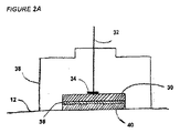
ELFAC感知電極は、用途に応じて、個別にまたは1組のセンサとして、粘着性の可撓性の裏打材上に適用可能である。両方の場合において、測定値における重複が最小化されるように、電極の有効間隔は維持される必要がある。例えば、個々のセンサ16bの間の距離は、皮膚または他の組織に接触しているセンサ面積の直径の2倍以上である必要がある。センサ16bが個別に当てられる場合、技術者は、適切な距離を用いてセンサを配置するように訓練される必要がある。理想的には、電極16bは、相異なる金属の反応による有意なバッテリ効果を生じない種類である必要がある。最新の電極は液体ゲルではなく固体ゲルを有するが、この固体ゲルは角皮層を貫通しないので、信号対雑音は最適ではない。別の手法は、測定装置10に接続された電気リード32を有する銀または他の導電性部品30からなる感知電極である。この電気接続は、導電性頂部ピース34によって銀部品30に対し固定されている。塩化銀36の薄層が銀部品30の表面上に堆積される。この系が、非導電性のプラスチック、すなわち、プラスチック裏打シート38に埋め込まれる。臓器の組織12と塩化銀表面36との間の界面は、ローズ・ラブズ社(Rose Labs,Inc.)製のシナプス(Synapse)電極クリームなど、既知の種類の準液体の電極クリーム、ペーストまたはゲル40によって仲介される。電極クリーム40の第1の目標は、皮膚12の電気抵抗性の角質層を通じる導電性経路を提供することである。これに代えて、本発明では、既知の種類の記録微小電極または記録小電極(例えば、ベックマン・コールター社(Beckman Coulter,Inc.;カリフォルニア州フラートン(Fullerton))を用いることによって、または裏打シート上に複数の針電極からなるアレイ(皮膚12の角質層を越えてちょうど貫通するが、組織の損傷を生じないほど小さな直径である)を用いることによって、皮膚12の角質層が機械的にはかされる(breech)ことが想定される。この代替の電極設計は図2Bの42に示されており、導電性クリーム、ペースト、またはゲル40の使用が不要である。この実施形態では、導電性部品30は多数の小さな貫通する銀電極または白金電極42を備え、それらの電極は自身のリードに接続されており、このリードは測定装置10に接続されるか、または測定装置10に接続された1つのリード(図示せず)にまとめられる。
The ELFAC sensing electrodes can be applied on the adhesive flexible backing, either individually or as a set of sensors, depending on the application. In both cases, the effective spacing of the electrodes needs to be maintained so that the overlap in measurements is minimized. For example, the distance between the individual sensors 16b needs to be at least twice the diameter of the sensor area in contact with the skin or other tissue. If the sensors 16b are applied individually, the technician needs to be trained to place the sensors using the appropriate distance. Ideally, the electrode 16b should be of a type that does not produce a significant battery effect due to the reaction of different metals. Modern electrodes have a solid gel rather than a liquid gel, but since this solid gel does not penetrate the stratum corneum, signal to noise is not optimal. Another approach is a sensing electrode consisting of silver or other
これに代えて、角皮層(皮膚の電気抵抗の90%超の原因である)はレーザ光または加熱したワイヤを制御して用いることによって除去されてもよい。この流儀では、電磁スペクトルの近赤外線領域におけるレーザ光を色素の薄層上に集中させることが可能であり、これによって光エネルギが吸収され、下の真皮に貫通することなく、角皮層を消失させる。大きさの異なる1つ以上の孔が、この手法または他の適切な手法により形成されてよい。角皮層をこのようにして選択的に損ねた後、その部位の上に電極が配置され、ELFAC電位が測定される。この例では、このように形成された1つ以上の孔に、電極クリーム
またはゲル40を必要とせず、また、針電極42を必要とせずに、1つ以上の電極16が直接配置されることが可能である。
Alternatively, the stratum corneum (which is responsible for more than 90% of the skin's electrical resistance) may be removed by controlled use of laser light or heated wire. In this manner, it is possible to concentrate laser light in the near infrared region of the electromagnetic spectrum on a thin layer of dye, which absorbs the light energy and eliminates the corneum layer without penetrating into the underlying dermis. . One or more holes of different sizes may be formed by this technique or other suitable technique. After selectively damaging the stratum corneum in this way, an electrode is placed over the site and the ELFAC potential is measured. In this example, one or
測定装置10は、被験者12に付けられる電極リード32を有する複数の記録入力部を備える。参照リードは患者において病気でない領域に付けられるか、または生理食塩液その他の参照標準の外部の溶液に付けられる。この例では別個の参照電極の使用について示すが、各記録電極が他の電極の参照電極を兼ねることも可能である。この実施形態では、1つを除く各記録電極が他の電極に対し走査される。次に、各記録電極が参照電極としても機能するまで、別の「記録」電極が参照電極となるように自動的に選択される。これによって、各記録電極について複数の測定値が生じるので、各記録チャネルの代表として、算術平均値、中央値、または最頻値が取られてよい。
The measuring
測定装置10は、選択的なバンドパスフィルタ18を利用して、例えば、0.01Hz〜0.1Hzの周波数範囲におけるELFAC信号の解析を行うことが可能である。癌診断についてのこの例では、この特定の周波数範囲内の信号を利用するが、本発明では、診断またはスクリーニングされる疾病の種類に応じて、他の低周波範囲に対し高感度なバンドパスフィルタを用いることも想定される。一般に、より高周波数の範囲は、皮膚による電気抵抗がより大きな場合(例えば、乳癌を検出するには回避される必要がある)に適用される。食道、肺、または子宮頚部など身体内部の裏側に発生する癌には、この問題は存在しないので、真の交流(すなわち、より低い周波数。研究が実施されるまで実際の数字は分からない)に近づけられる。バンドパスフィルタ18は、このための方法に記載されるように、既存の設計の1つ以上のフィルタを含んでよい。
The
バンドパスフィルタは、原理的には、ローパスフィルタおよびハイパスフィルタをカスケード状に組み合わせることによって構築可能である。図6には、そのようなフィルタを示す。第1の部分(C1R1)は高周波信号を通過させるが、第2の部分(C2R2)は低周波数を通過させる(すなわち、高周波信号を退ける)。しかしながら、このフィルタは、第2の部分が第1の部分に負荷を与えるので、ハイパスフィルタおよびローパスフィルタの単純なカスケードとは考えられない。その結果、全体的な伝達関数は、単純に、ハイパス部分およびローパス部分のそれぞれの伝達関数の積ではない。 In principle, a bandpass filter can be constructed by combining a low-pass filter and a high-pass filter in cascade. FIG. 6 shows such a filter. The first part (C 1 R 1 ) passes high frequency signals, while the second part (C 2 R 2 ) passes low frequencies (ie rejects high frequency signals). However, this filter cannot be considered as a simple cascade of high-pass and low-pass filters because the second part loads the first part. As a result, the overall transfer function is not simply the product of the respective transfer functions of the high pass portion and the low pass portion.
上述のフィルタの例は、増幅器を利用しないので、パッシブフィルタと呼ばれる。そのようなフィルタの欠点は、利得が存在しないこと、また、負荷抵抗RLが伝達特性に影響を与えることである。低周波数〜中周波数用途のためのフィルタを製造するよりよい手法は、演算増幅器を用いることである。そのようなフィルタはアクティブフィルタと呼ばれる。アクティブフィルタの利点は、増幅を行えること、またフィルタの特性値を負荷とは無関係にできることである。 The above filter example is called a passive filter because it does not use an amplifier. The disadvantages of such a filter are that there is no gain and that the load resistance RL affects the transfer characteristics. A better approach to manufacturing filters for low to medium frequency applications is to use operational amplifiers. Such a filter is called an active filter. The advantage of the active filter is that amplification can be performed and the characteristic value of the filter can be made independent of the load.
単純なアクティブバンドパスフィルタ(2極システム)を図7に示す。その利得の周波数応答(ボーデ(Bode)グラフ)を図8に与える。
バンドパスフィルタは、入力リード32の各々における信号を別個に濾波し、次いで、濾波された信号の各々を別個のチャネルを介して複数入力アナログ−デジタル変換器20に通過させる。これに代えて、バンドパスフィルタリングは、アナログ−デジタル変換後のデジタルドメインにおいて生じてもよい。加えて、バンドパスフィルタ18は、各チャネルに別個の濾波機構を構成して、そのチャネルについてのみ濾波を行ってもよく、各々の濾波された出力はアナログ−デジタル変換器20の入力に接続される。
A simple active bandpass filter (two pole system) is shown in FIG. The frequency response (Bode graph) of the gain is given in FIG.
The bandpass filter separately filters the signal at each of the input leads 32 and then passes each of the filtered signals through a separate channel to the multi-input analog-to-
アナログ−デジタル変換器20は、理想的には、多入力多重化が可能である(ナショナル・セミコンダクター社(National Semiconductor,Inc.)製のADC808など)。乳癌スクリーニング用に想定されるものなど、非常に大きな測
定アレイには、2つ以上の多入力アナログ−デジタル変換器が必要な場合があり、この変換器の正確な数は、チャネル変換器の性能と、特定の用途に必要なチャネル数とによって決定される。
Ideally, the analog-to-
アナログ−デジタル変換器20は、各チャネルのアナログ信号をデジタル信号に変換し、このデジタル信号が別個の出力チャネルを介して中央処理装置22の複数の入力に中継される。中央処理装置は、より大きな制御システムの構成要素であり、この制御システムはRAM24およびROM26も備える。中央処理装置22のコントロールの記憶されているプログラムは、信号の取得およびサンプリングのレートを制御し、次いでデジタル入力データを処理して、試験された組織の疾病状態に関する使用者に対する出力を生成する。他の関連データ(患者の年齢または病変の大きさなど)は、標準的なコンピュータキーパッドまたは従来型の接触感知スクリーンその他の入力装置または方法を用いることによって入力可能である。中央処理装置は、次いで、予めプログラムされたパターン認識アルゴリズムを用いて、この情報をELFACデータと統合する。使用者に対する最終的な出力は、次いで、コンピュータモニタまたはプリンタなど、表示装置28に供給される。この出力は、ネットワークまたは他のメモリシステムに局所的または遠隔的に記憶されることも可能である。この出力は、特定の用途に応じて、現在の疾病状態の確率に関する数値的な回答、関心対象の疾病が存在するか否かに関する肯定/否定の回答、またはその疾病の重症度を示すスカラー結果、および/または偽色彩画像を構成してもよい。
The analog-to-
ELFAC装置の機能および動作は、基本的な方法を具体化する工程の2つの例から理解される。第1の例には疾病スクリーニングのための方法を要約し、第2の例には診断フォーマットを用いる本発明の特性を示す。 The function and operation of the ELFAC device can be understood from two examples of steps embodying the basic method. The first example summarizes the method for disease screening, and the second example illustrates the characteristics of the present invention using a diagnostic format.
スクリーニングの場合、被験者が無症候性であるので、病変の位置および配置は識別可能でない。この場合、関心対象の部位の表面12のいずれかに電極16の比較的大きなアレイが配置される。疑わしい部位を皮膚を通じて外部的に評価することが可能である場合、電極は皮膚上に配置される。より侵襲的な施術が指示される場合、電極は被験者の臓器または部位に内部的に配置される。乳癌スクリーニングの場合、使用者は、悪性腫瘍が存在するか否か、またどこに存在するのか知らないので、好適には、両乳房の表面全体が測定される。電極アレイ16が配置されると、参照電極は、組織の病気でない領域上または参照標準中に配置される。次いで、参照電極と測定電極16の各々との間のELFAC活動が即座に測定され、帯域濾波されて、所与の被験対象から診断的に有用なELFAC読取値が取得されたか否か、またいつ取得されたかを判定する予めプログラムされたアルゴリズムによって処理される。この時点では、波形中の個々の電圧読取値は各々パターン認識のために保持されている。これは従来の技術とは異なる。従来の技術では、アクティブ濾波と時間を通じて得られた複数の信号の算術平均化との組み合わせによって、装置は低周波交流情報にまで濾波を行う。従来の具体化において取られた手法は、この選択的なローパス濾波および信号平均化を利用することによって、代表的な直流(DC)成分のみを識別することであった。したがって、ELFAC成分における情報が解析されることはなかった。
In the case of screening, since the subject is asymptomatic, the location and placement of the lesion is not discernable. In this case, a relatively large array of
診断の解析の場合、センサ16は疑わしい部位(病変など)に配置される。電極の数およびその配置は、部分的には疑わしい部位の大きさに依存する。一般的には、疑わしい部位および一部の包囲組織を解析する必要がある。スクリーニング処理の場合のように、参照電極は参照サイトまたは参照標準に配置される。参照部位は、鏡像部位(例えば、左腕/右腕)の相当する位置であってもよく、同じ臓器または組織の病気でない部分であってもよく、または病気でない他の組織であってもよい。参照電極と測定電極16の各々との間のELFAC活動は、スクリーニングについて上述したのと同様に処理される。
In the case of diagnostic analysis, the
ノイズが最小化され、したがって診断的に有用なELFAC信号が所与の被験者から取得されたか否か、またそれはいつかを判定するために、最後の電極が被験者上に配置された時から代表的なELFAC信号が取得されるまで、それらの信号が連続的に監視される必要がある。ノイズは皮膚の角皮層の電気抵抗特性のために皮膚/電極界面において生じることが認められる。皮膚表面の電極が表面下の臓器系によって生じる電磁場の有効な変換器であるためには、角質障壁がはかされる必要がある。しかしながら、最も導電性の大きな電極ペーストおよびゲルを用いた場合でも、信号の平衡に達するには時間が掛かる(所与の被験者について数分間またはそれより長い)。しかし、より問題なのは、平衡化の時間は個々人で有意に異なり、1人の個人であっても時および/または試験部位の位置が異なると異なることがあるという事実である。これは、経皮的な活動に関連した表皮抵抗その他の因子は個々人で変化するためである。従来では、電磁場測定のタイミングおよび継続時間は予め定められており、機械に組み込まれており、各々の被験者に対して本質的に同じであった。これによって、一部の被験者は平衡化が完了する前に測定されることがあるので、診断の誤差を生じることがあり、その場合、測定され解析されるのは信号ではなくノイズである。本発明では、ELFAC活動の連続的な測定および監視によって、この問題を回避する。これは、個々人の安定性について、各入来信号を解析することによって達成される。例えば、信号平衡化の未完了は、典型的には、測定電極が当てられた直後、角質層が導電性ペーストまたはゲルによって貫通される過程にあるときに見られる、次第に減少する(増加はしない)電位として現れる。最終的に、平衡化が完了すると、ゆっくり増減するELFAC電位の情報を有する信号が観察される。一部の実施形態において、本発明では、平衡化が起きたことを示す特徴的なELFAC波形を中央処理装置22のパターン認識ソフトウェアが識別するまで、電磁信号の記録および解析を開始しない。これは、記録する時および期間が予め設定されておらず、各被験者について同じでないことを意味しており、試験される各被験者について信号対雑音が最大となるまで電磁気活動の記録および解析が始まらないことを意味する。したがって、各患者および各施術は、各患者の各試験においてノイズの最小化されたデータが得られるように、個別にかつ独立に評価される。一部の実施形態では、平衡中に取得された信号は、記録されても、スクリーニングまたは診断の解析において用いられなくてよい。
A representative from the time the last electrode was placed on a subject to determine if and when a noise-minimized and therefore diagnostically useful ELFAC signal was obtained from a given subject. Until ELFAC signals are acquired, they need to be monitored continuously. It can be seen that noise occurs at the skin / electrode interface due to the electrical resistance properties of the cuticle layer of the skin. In order for the electrode on the skin surface to be an effective transducer of the electromagnetic field produced by the subsurface organ system, the horny barrier needs to be removed. However, even with the most conductive electrode pastes and gels, it takes time to reach signal equilibrium (a few minutes or longer for a given subject). More problematic, however, is the fact that the time of equilibration varies significantly from individual to individual, and even a single individual can vary from time to time and / or from different test site locations. This is because epidermal resistance and other factors associated with transcutaneous activity vary from person to person. In the past, the timing and duration of electromagnetic field measurements were predetermined and built into the machine and were essentially the same for each subject. This can cause diagnostic errors because some subjects may be measured before the equilibration is completed, in which case it is noise rather than signals that are measured and analyzed. In the present invention, this problem is avoided by continuous measurement and monitoring of ELFAC activity. This is accomplished by analyzing each incoming signal for individual stability. For example, incomplete signal balancing typically decreases (but does not increase) as seen when the stratum corneum is in the process of being penetrated by a conductive paste or gel immediately after the measurement electrode is applied. ) Appears as potential. Finally, when equilibration is complete, a signal with ELFAC potential information that slowly increases and decreases is observed. In some embodiments, the present invention does not begin recording and analyzing electromagnetic signals until the pattern recognition software of the
上述のように、ノイズの最小化されたELFAC信号が識別されると、それらの信号は記録され、解析用にRAMメモリ26に保持される。予めプログラムされた解析ソフトウェアは、次いで、人工ニューラルネットワークまたは分類決定木(例えば、CART)など、既知の種類の非線形パターン認識技術を用いる。詳細には、所定周波数範囲内の記録された電圧はすべて、患者の年齢、病変の大きさ、乳癌(または、スクリーニングされている疾病)の家族歴、画像調査の結果など随意の対象変数と共に、パターン認識プログラムへ供給される。本発明では、これらのおよび他の主要変数に基づく被験者の部分集合に対し、異なるパターン認識プログラムが用いられることが想定される。例えば、一部の実施形態では、ホルモン環境の差のために悪性の乳房腫瘍の生物学的活動は閉経前の女性では閉経後の女性と異なる場合がある。それらの差を解析において考慮することが可能である。触知不能な小さな乳癌は、より大きな腫瘍より代謝活性がより高い場合があり、その中核は壊死する場合が多い。同様に、皮膚損傷の代謝活性は物理的な外観に関連する場合があり、より暗く、突起した黒子様構造のように見える病変ほど代謝活性が高くなり得る。
As described above, once ELFAC signals with minimized noise are identified, they are recorded and held in
図3のフローチャートには、皮膚損傷の診断のための入力、中央処理、およびパターン認識プログラムの出力の例を提供する。開始スイッチ44は、中央処理装置22の動作を開始させ、処理動作46を初期化する。この初期化処理によって、装置10の様々な構成要素は動作モードとなる(アナログ−デジタル変換器20からデータ48を読み取るための制御レジスタのリセットおよび作動を含む)。従来技術の装置とは対照的に、この発明では、48において所定の複数の測定期間を初期化しない。代わりに、50において平衡
化が完了し、各記録チャネルにおいて診断上有用なELFACが識別されるまで、データが連続的に読み取られる。50におけるデータの継続的な評価は、ほとんどまたはすべてのチャネルがノイズの最小化されたELFAC信号を運ぶと判定されるまで、循環して工程48へ戻る。
The flowchart of FIG. 3 provides an example of input for diagnosis of skin damage, central processing, and output of a pattern recognition program. The start switch 44 starts the operation of the
図5には、時間および振幅の関数として電磁気のデータを表す。48においてデジタル化されたELFAC信号は、50において、ノイズ112によって不明瞭であるか、ノイズの最小化された状態114に達したと判定される。ノイズの最小化された状態114に達した信号のみが52において記録(保存)され、54における処理のために記憶される。 FIG. 5 represents electromagnetic data as a function of time and amplitude. The ELFAC signal digitized at 48 is determined at 50 to be obscured by noise 112 or to reach a noise-minimized state 114. Only signals that have reached the noise minimized state 114 are recorded (saved) at 52 and stored for processing at 54.
相当数のチャネルが妥当な量の時間内(例えば、約15分以内)にノイズの最小化されたELFAC信号を送ることは不可能であると判定されると、動作は停止され、中央処理装置22は出力表示装置28を介し、医療技術者が接触点を点検するまたは他の問題解決法を用いるための命令を送る。一部のチャネルがノイズの最小化されたELFAC信号を送信することが不可能である場合、非送信チャネルの数が有意でない限り、そのような信号を送ることが可能なチャネルが用いられる。すなわち、ほぼすべてのチャネルが送信している場合、その送信しているチャネルが用いられる。「ほぼすべて」とは、全チャネルのうちの90%以上がノイズの最小化されたELFAC信号を送ることが可能であることを意味する。
If it is determined that a significant number of channels are unable to send a noise-minimized ELFAC signal within a reasonable amount of time (eg, within about 15 minutes), then the operation is stopped and the central processing unit is stopped. 22 sends instructions via the
ノイズの最小化されたELFAC信号が識別されると、それらの信号はキャプチャされ、メモリ54に記憶される。これは、平均化されたDC成分のみが処理のためにメモリに記憶される従来技術の装置とは対照的である。パターン認識モジュール58には、ELFACデータおよび患者の変数(患者の年齢、病変の大きさまたは形状、画像調査による疑わしさのレベルなど)56を統合してパターン認識結果60を取得する人工ニューラルネットワークまたは決定木など既知の種類の非線形パターン認識プログラムが組み込まれており、この結果によって、疑わしい病変が悪性であるか否か(診断モードにおいて用いられる)、または臓器系が不顕性の悪性腫瘍を有する可能性があるか否か(スクリーニングモードにおいて用いられる)の確率通知62に至る。得られる確率通知は、0.05(5%)など一定の値を超える場合、64における癌(CANCER)を示すために用いられ、この値を超えない場合、図4の66におけるように癌でない(NO CANCER)を示すために用いられる。この表示、またはその元となる確率その他の解釈は、モニタまたはプリンタなどの表示装置に出力される、および/またはメモリに記憶されることが可能である。本発明が診断モードにおいて利用される場合、先験的に高い疾病有病率を有する症状集団では、偽陰性結果(すなわち、癌の見逃し)を最小化するように、癌を示すためのカットオフ限界確率を低下させてもよい。一方、先験的な疾病有病率が比較的低いスクリーニング設定において本発明が利用される場合、76における処理は、より高いカットオフ限界確率において癌を示すように較正されてもよい。特定の用途に応じて、64または66における使用者に対する出力は、癌または癌でないことを示すことに加え、診断が正確である確率もしくは可能性、または疾病の重症度を示すスカラー出力を示してもよい。出力64または66が生成されると、プログラムは68において終了する。
Once noise minimized ELFAC signals are identified, they are captured and stored in
58におけるパターン認識モジュールの構成の一例を、図4に与える。パターン認識プログラムは70において開始する。対象の臨床情報は、56においてキーパッドなどの標準的な装置を介して、または接触感知スクリーンもしくは任意の適切な入力装置上のメニューから入力され、シーケンスにおける初期の複数の工程を構成する。例えば、72において、被験者が閉経前であるか後であるかに応じてプログラムは分岐し、被験者が閉経前の場合、月経周期の何日目かに対応する数値が74において入力される。プログラムは、被験者が月経周期のうちホルモンが活性な区分にある場合、76において再び分岐する。
これは、全身的なホルモン変化によって疾病状態が影響を受けることが知られているためである。被験者がホルモンが活性な区分にある場合、パターン認識プログラムは、2つの鏡像の臓器系または位置78の間(両手足の間または両乳房の間など)の点間ELFAC電位により大きな重み付けを行うことができる。有意な差が78において見出される場合、PRR値60は、癌のより高い確率を示し、それらの差が観察されない場合、82または84において結果の確率を決定するために波形特性80(ELFAC波の周波数、ピークおよび/または谷での電位など)が検査される。一方、患者が月経周期のうちホルモンが活性な区分にない場合、86において症状の領域付近の電極部位(例えば、同じ乳房または手足の)からの最大の差がパターン認識プログラムによってより大きく重み付けられる。プログラムは、次いで、肯定応答が得られる場合、88において出力確率に進むか、または、波形評価および続く確率出力のために80に進む。このパターン認識値は、入力が78に由来するか86に由来するかに応じて異なってよい。
An example of the configuration of the pattern recognition module at 58 is given in FIG. The pattern recognition program begins at 70. The subject's clinical information is entered at 56 via a standard device such as a keypad, or from a menu on the touch sensitive screen or any suitable input device, and constitutes the initial steps in the sequence. For example, at 72, the program branches depending on whether the subject is before or after menopause, and if the subject is before menopause, a numerical value corresponding to the day of the menstrual cycle is entered at 74. The program branches again at 76 if the subject is in the hormone active segment of the menstrual cycle.
This is because disease states are known to be affected by systemic hormonal changes. If the subject is in the hormone-active category, the pattern recognition program will heavily weight the point-to-point ELFAC potential between two mirror image organ systems or positions 78 (such as between both limbs or between breasts). Can do. If a significant difference is found at 78, the
ここで図4の72に戻ると、患者が閉経前でない場合、74におけるような月経周期の何日目かに対応する値を入力することは当を得ていない。例示の目的で、92において試験されている疑わしい病変が触知可能であるか否かに基づき、データフローが進行することが可能である。画像調査においてより正確な大きさ情報が利用可能である場合、それらのデータも用いられてよい。病変が触知可能でない場合、プログラムは進み、94において疑わしい病変の領域における一般的な脱分極を(反対の乳房または手足から得られた測定値に対し)ELFAC波が示すか否かを判定することに進み、96において、または98を介して104もしくは106において最終的な確率出力を与える。一方、92において病変が触知可能な場合、100においてパターン認識プログラムは、反対の乳房または手足に対する全脱分極に重み付けを行うのではなく、(例えば、同じ乳房または手足)の症状の領域付近の電極部位からの最大のELFAC差に重み付けを行う。 Returning now to 72 of FIG. 4, if the patient is not premenopausal, it is not reasonable to enter a value corresponding to the day of the menstrual cycle as at 74. For illustrative purposes, the data flow can proceed based on whether or not the suspicious lesion being tested at 92 is palpable. If more accurate size information is available in the image survey, those data may also be used. If the lesion is not palpable, the program proceeds to determine whether the ELFAC wave exhibits a general depolarization (relative to measurements taken from the opposite breast or limb) at 94 in the area of the suspicious lesion. Proceed to give the final probability output at 96 or at 104 or 106 via 98. On the other hand, if the lesion is palpable at 92, the pattern recognition program does not weight the total depolarization for the opposite breast or limb at 100, but near the symptom area (eg, the same breast or limb). Weigh the maximum ELFAC difference from the electrode site.
ELFAC差が98において予めプログラムされたアルゴリズムによって判定されるプリセット閾値より大きい場合、106において疾病のより高い確率が示される。94,100における各々の場合、否定的な結果によって98における波形の特性解析に至り、否定的な結果によって104における疾病のより低い確率の通知に至り、肯定的な結果によって106における疾病のより高い確率の通知に至る。 If the ELFAC difference is greater than a preset threshold determined by a pre-programmed algorithm at 98, a higher probability of disease is indicated at 106. In each case at 94,100, a negative result leads to a characterization of the waveform at 98, a negative result leads to a lower probability of illness at 104, a positive result leads to a higher illness at 106 Leads to a probability notification.
図4ではパターン認識フローチャートの一実施形態が提供されているが、本発明では、パターン認識モジュール58に対し他の生物学的変数が追加されることも想定される。例えば、病変の触知性92は閉経前の被験者に対する判定シーケンスにも統合されてよい。加えて、80,84,86,90,96,102,104,106における確率通知は他の診断調査または以前のELFAC試験からの疑わしさの指数のレベルと統合されてもよい。変数の数および種類は、疾病の診断またはスクリーニングの特定の用途に応じて異なる。例えば、疾病スクリーニングのパターン認識モジュールは、図4の触知性または大きさ92などの病変特性を含まない。代わりに、対象年齢またはスクリーニングされる疾病状態の家族歴など、人口統計学的な変数により大きく依存する。
Although one embodiment of a pattern recognition flowchart is provided in FIG. 4, the present invention envisions that other biological variables may be added to the
したがって、疾病の診断およびスクリーニングのために電磁場を利用することの有効性を最大化するために、異なる生物学的状況には、図4における78,82,88,94,98,100など、異なる判定カットオフ限界またはサブモジュール(例えば、ニューラルネット、決定木)が必要である。 Thus, in order to maximize the effectiveness of using electromagnetic fields for disease diagnosis and screening, different biological situations include different, such as 78, 82, 88, 94, 98, 100 in FIG. Decision cut-off limits or submodules (eg, neural nets, decision trees) are required.
本発明の方法および装置は、極低周波交流(ELFAC)信号の生物学的に調整されたパターン認識を用いて疾病状態の診断およびスクリーニングを行う際、より高い精度を提供するように設計されている。信号は、既知のまたは疑わしい疾病部位を含む身体の複数の異なる部位から測定される。周期、ピーク、谷、傾き、および他の情報など、異なる部
位からの信号の情報を与える態様の比較結果は対象変数と統合され、上皮悪性腫瘍などの疾病状態の検出および診断を行う際により高い精度を提供する。
The methods and apparatus of the present invention are designed to provide greater accuracy when diagnosing and screening disease states using biologically tuned pattern recognition of extremely low frequency alternating current (ELFAC) signals. Yes. Signals are measured from multiple different parts of the body including known or suspected disease sites. Comparison results for aspects that provide signal information from different sites, such as period, peak, valley, slope, and other information, are integrated with the target variable and are higher when detecting and diagnosing disease states such as epithelial malignancies Provides accuracy.
Claims (28)
1つ以上のセンサ電極および1つ以上の参照電極を介して試験部位と参照部位との間において電磁周波数信号を取得する電磁信号取得工程と、
センサ電極によって取得した信号の帯域濾波を行い、センサELFAC信号を生成するセンサ信号生成工程と、
参照電極によって取得した信号の帯域濾波を行い、参照ELFAC信号を生成する参照信号生成工程と、
前記ELFAC信号に基づき疾病の可能性を判定する判定工程と、を含む方法。 A method for screening or diagnosing a disease state in a test subject comprising:
An electromagnetic signal acquisition step of acquiring an electromagnetic frequency signal between the test site and the reference site via the one or more sensor electrodes and the one or more reference electrodes;
A sensor signal generation step of performing band filtering of the signal acquired by the sensor electrode to generate a sensor ELFAC signal;
A reference signal generating step of performing band filtering of the signal acquired by the reference electrode to generate a reference ELFAC signal;
Determining the possibility of a disease based on the ELFAC signal.
類似するセンサデータおよび参照データを低リスクの疾病状態と関連付ける工程と、
相違するセンサデータおよび参照データをより高リスクの疾病状態と関連付ける工程と、を含む請求項3に記載の方法。 The comparison process is further
Associating similar sensor data and reference data with a low-risk disease state;
Associating different sensor data and reference data with a higher risk disease state.
ノイズの最小化されたELFAC測定値から取得された前記信号の代表値である代表測定値を算出する工程と、
疾病の診断およびスクリーニングを行うために前記代表測定値を比較する工程と、をさらに含む請求項7に記載の方法。 Grouping ELFAC signals for each possible sensor / reference electrode combination;
Calculating a representative measurement value that is a representative value of the signal obtained from the ELFAC measurement value with minimized noise;
The method of claim 7, further comprising the step of comparing the representative measurements for disease diagnosis and screening.
試験部位に接触するように設計された1つ以上のセンサ電極と、
参照部位との接触のための1つ以上の参照電極と、
0.001Hz〜0.1Hzの周波数範囲内の極低周波交流(ELFAC)を通過させるバンドパスフィルタと、
電極から取得したデータの収集、解析、および記憶を行うように構成された処理手段と、を備え、
センサ電極、参照電極は、バンドパスフィルタを介して処理手段に動作可能に接続されている装置。 An apparatus for detecting or diagnosing a disease at a test site on or in a test subject,
One or more sensor electrodes designed to contact the test site;
One or more reference electrodes for contact with the reference site;
A bandpass filter that passes very low frequency alternating current (ELFAC) within a frequency range of 0.001 Hz to 0.1 Hz;
Processing means configured to collect, analyze, and store data acquired from the electrodes,
A device in which the sensor electrode and the reference electrode are operatively connected to the processing means via a bandpass filter.
処理手段は前記デジタル信号を受信するように接続されており、処理手段は前記デジタルELFAC信号を比較して疾病の診断または確率のインジケータを生成するように動作する、請求項22に記載の装置。 An analog-to-digital converter connected to receive each ELFAC signal from the sensor electrode and the reference electrode;
23. The apparatus of claim 22, wherein processing means is connected to receive the digital signal, and the processing means is operative to compare the digital ELFAC signal to generate a disease diagnosis or probability indicator.
試験部位または参照部位の表面に電極を付着させる粘着性の外側部分と、
内側部分と、を備え、内側部分は、試験部位にて皮膚の角質層または臓器系の外側の層を貫通可能な長さの1つ以上の微小電極または小電極をさらに備え、
各微小電極または小電極は処理手段に動作可能に接続されている、請求項22に記載の装置。 At least one of the sensor electrode or the reference electrode is
An adhesive outer portion that attaches an electrode to the surface of the test or reference site;
An inner portion, the inner portion further comprising one or more microelectrodes or small electrodes of a length that can penetrate the stratum corneum of the skin or the outer layer of the organ system at the test site;
23. The apparatus of claim 22, wherein each microelectrode or small electrode is operably connected to a processing means.
請求項22に記載の装置。 The processing means includes a pattern recognition module, wherein the sensor ELFAC data is compared with the reference ELFAC data and associated with the possibility that a disease state exists.
The apparatus of claim 22.
Applications Claiming Priority (5)
| Application Number | Priority Date | Filing Date | Title |
|---|---|---|---|
| US9110008P | 2008-08-22 | 2008-08-22 | |
| US61/091,100 | 2008-08-22 | ||
| US11156708P | 2008-11-05 | 2008-11-05 | |
| US61/111,567 | 2008-11-05 | ||
| PCT/US2009/053669 WO2010021898A1 (en) | 2008-08-22 | 2009-08-13 | Method and apparatus for disease diagnosis and screening using extremely low frequency electromagnetic fields |
Related Child Applications (1)
| Application Number | Title | Priority Date | Filing Date |
|---|---|---|---|
| JP2015233010A Division JP2016028778A (en) | 2008-08-22 | 2015-11-30 | Device for diagnosing and screening disease using extremely low frequency electromagnetic field |
Publications (2)
| Publication Number | Publication Date |
|---|---|
| JP2012500663A true JP2012500663A (en) | 2012-01-12 |
| JP2012500663A5 JP2012500663A5 (en) | 2012-09-27 |
Family
ID=41202265
Family Applications (2)
| Application Number | Title | Priority Date | Filing Date |
|---|---|---|---|
| JP2011523873A Pending JP2012500663A (en) | 2008-08-22 | 2009-08-13 | Method and apparatus for disease diagnosis and screening using extremely low frequency electromagnetic fields |
| JP2015233010A Pending JP2016028778A (en) | 2008-08-22 | 2015-11-30 | Device for diagnosing and screening disease using extremely low frequency electromagnetic field |
Family Applications After (1)
| Application Number | Title | Priority Date | Filing Date |
|---|---|---|---|
| JP2015233010A Pending JP2016028778A (en) | 2008-08-22 | 2015-11-30 | Device for diagnosing and screening disease using extremely low frequency electromagnetic field |
Country Status (9)
| Country | Link |
|---|---|
| US (1) | US8712515B2 (en) |
| EP (1) | EP2352420B1 (en) |
| JP (2) | JP2012500663A (en) |
| CN (1) | CN102186412B (en) |
| CA (1) | CA2771260C (en) |
| EA (1) | EA019377B1 (en) |
| HK (1) | HK1162133A1 (en) |
| MX (1) | MX2011001836A (en) |
| WO (1) | WO2010021898A1 (en) |
Cited By (1)
| Publication number | Priority date | Publication date | Assignee | Title |
|---|---|---|---|---|
| JP2016028778A (en) * | 2008-08-22 | 2016-03-03 | フォーペル、マーク エル.FAUPEL Mark L. | Device for diagnosing and screening disease using extremely low frequency electromagnetic field |
Families Citing this family (8)
| Publication number | Priority date | Publication date | Assignee | Title |
|---|---|---|---|---|
| CN103407465B (en) * | 2013-07-08 | 2015-08-19 | 宁波康宝儿童用品有限公司 | A kind of folding pet's hand barrow |
| KR101739656B1 (en) | 2015-11-23 | 2017-05-24 | 이경호 | Handy-type Breast Cancer Diagnosis Device |
| CN107411734A (en) * | 2017-03-06 | 2017-12-01 | 华斌 | A kind of device that user characteristics is obtained according to human-body biological electromagnetic wave |
| US11955242B2 (en) * | 2017-08-24 | 2024-04-09 | Uladzimir Rastoutau | Method for automatic diagnosis of conditions of an object and a system for implementing the same |
| WO2020023377A1 (en) * | 2018-07-23 | 2020-01-30 | The Regents Of The University Of California | Oral and oropharyngeal cancer screening system and methods of use |
| BR102020002019A2 (en) * | 2020-01-30 | 2021-08-10 | Termo Health Tecnologia Ltda | mobile system and process for evaluating breast cancer in patients and electronic application |
| CN113081005A (en) * | 2021-04-15 | 2021-07-09 | 青岛卫来动物健康产业有限公司 | Disease diagnosis and analysis system for animal dogs |
| US20220386921A1 (en) * | 2021-06-04 | 2022-12-08 | Mark D. Noar | Method And System For Monitoring Internal Bodily Disorders By Detecting And Analyzing Tissue Frequencies |
Citations (8)
| Publication number | Priority date | Publication date | Assignee | Title |
|---|---|---|---|---|
| JPH02264635A (en) * | 1988-12-22 | 1990-10-29 | Biofield Corp | Discriminative function analysis method and apparatus for disease diagnosis and screening |
| JPH11503333A (en) * | 1994-12-09 | 1999-03-26 | バイオフィールド コーポレーション | Neural network method and apparatus for screening or detecting disease, injury and physical condition |
| JP2003116802A (en) * | 2001-10-12 | 2003-04-22 | Sekisui Chem Co Ltd | Electric characteristic measuring system |
| JP2005523097A (en) * | 2002-04-17 | 2005-08-04 | ソントラ・メディカル・インコーポレーテッド | Equipment for transmission and reception of electrical signals |
| JP2005525900A (en) * | 2002-05-20 | 2005-09-02 | デイヴィス,リチャード・ジェイ | System for detecting precancerous and cancerous tissue |
| WO2006116091A2 (en) * | 2005-04-21 | 2006-11-02 | Epi-Sci, Llc | Method and system for detecting electrophysiological changes in pre-cancerous and cancerous tissue and epithelium |
| WO2007067632A2 (en) * | 2005-12-06 | 2007-06-14 | Epi-Sci, Llc | Method and system for detecting electrophysiological changes in pre-cancerous and cancerous tissue and epithelium |
| JP2007527767A (en) * | 2004-03-08 | 2007-10-04 | ノア,マーク | Intelligent self-interpreting visceral electromyogram system and method |
Family Cites Families (19)
| Publication number | Priority date | Publication date | Assignee | Title |
|---|---|---|---|---|
| US3204148A (en) * | 1961-05-01 | 1965-08-31 | Eaton Mfg Co | Control for electromagnetic coupling |
| US4328809A (en) * | 1976-09-24 | 1982-05-11 | Barry Herbert Hirschowitz | Device and method for detecting the potential level of the electromagnetic field of a living organism |
| US4407300A (en) * | 1980-07-14 | 1983-10-04 | Davis Robert E | Potentiometric diagnosis of cancer in vivo |
| US4416288A (en) * | 1980-08-14 | 1983-11-22 | The Regents Of The University Of California | Apparatus and method for reconstructing subsurface electrophysiological patterns |
| IL62861A (en) * | 1981-05-13 | 1988-01-31 | Yeda Res & Dev | Method and apparatus for carrying out electric tomography |
| US4557273A (en) * | 1982-12-27 | 1985-12-10 | Stoller Kenneth P | Method and apparatus for detecting ovulation |
| RU2189172C2 (en) * | 1996-10-07 | 2002-09-20 | Бакусов Леонид Михайлович | Remote monitoring method for processing physiological signals |
| JPH1150333A (en) | 1997-08-04 | 1999-02-23 | Kuraray Co Ltd | Fiber for heat compression molding and production of fiber molded product |
| TW375537B (en) * | 1997-08-19 | 1999-12-01 | Satake Eng Co Ltd | Color sorting apparatus for granular material |
| US6022316A (en) * | 1998-03-06 | 2000-02-08 | Spectrx, Inc. | Apparatus and method for electroporation of microporated tissue for enhancing flux rates for monitoring and delivery applications |
| RU2159574C1 (en) * | 2000-04-13 | 2000-11-27 | Успенский Вячеслав Максимилианович | Device for performing express diagnostic of internal organ diseases and oncopathological disorders |
| US6757558B2 (en) | 2000-07-06 | 2004-06-29 | Algodyne, Ltd. | Objective pain measurement system and method |
| ITBO20010110A1 (en) | 2001-03-01 | 2002-09-01 | Tre Esse Progettazione Biomedi | PROCEDURE AND IMPLANTABLE DEVICE FOR THE INTRA-PULMONARY MEASUREMENT OF PHYSICAL PROPERTIES OF THE PULMONARY FABRIC DEPENDENT ON ITS DENSIT |
| US7191000B2 (en) * | 2001-07-31 | 2007-03-13 | Cardiac Pacemakers, Inc. | Cardiac rhythm management system for edema |
| US8262575B2 (en) * | 2002-05-20 | 2012-09-11 | Epi-Sci, Llc | Method and system for detecting electrophysiological changes in pre-cancerous and cancerous tissue |
| US7194306B1 (en) * | 2003-09-05 | 2007-03-20 | Pacesetter, Inc. | Cardiac optimization through low-frequency analysis of hemodynamic variables |
| CA2600427C (en) * | 2005-03-09 | 2013-09-10 | Coloplast A/S | A three-dimensional adhesive device having a microelectronic system embedded therein |
| US20090033333A1 (en) * | 2006-03-01 | 2009-02-05 | G.R. Enlightenment Ltd. | Apparatus and method for measuring parameters associated with electrochemical processes |
| EP2352420B1 (en) * | 2008-08-22 | 2016-02-10 | Mark L. Faupel | Apparatus for disease diagnosis and screening using extremely low frequency electromagnetic fields |
-
2009
- 2009-08-13 EP EP09791468.3A patent/EP2352420B1/en active Active
- 2009-08-13 CN CN2009801406429A patent/CN102186412B/en active Active
- 2009-08-13 EA EA201170365A patent/EA019377B1/en not_active IP Right Cessation
- 2009-08-13 WO PCT/US2009/053669 patent/WO2010021898A1/en active Application Filing
- 2009-08-13 MX MX2011001836A patent/MX2011001836A/en active IP Right Grant
- 2009-08-13 US US12/540,554 patent/US8712515B2/en active Active
- 2009-08-13 CA CA2771260A patent/CA2771260C/en not_active Expired - Fee Related
- 2009-08-13 JP JP2011523873A patent/JP2012500663A/en active Pending
-
2012
- 2012-03-13 HK HK12102538.1A patent/HK1162133A1/en not_active IP Right Cessation
-
2015
- 2015-11-30 JP JP2015233010A patent/JP2016028778A/en active Pending
Patent Citations (8)
| Publication number | Priority date | Publication date | Assignee | Title |
|---|---|---|---|---|
| JPH02264635A (en) * | 1988-12-22 | 1990-10-29 | Biofield Corp | Discriminative function analysis method and apparatus for disease diagnosis and screening |
| JPH11503333A (en) * | 1994-12-09 | 1999-03-26 | バイオフィールド コーポレーション | Neural network method and apparatus for screening or detecting disease, injury and physical condition |
| JP2003116802A (en) * | 2001-10-12 | 2003-04-22 | Sekisui Chem Co Ltd | Electric characteristic measuring system |
| JP2005523097A (en) * | 2002-04-17 | 2005-08-04 | ソントラ・メディカル・インコーポレーテッド | Equipment for transmission and reception of electrical signals |
| JP2005525900A (en) * | 2002-05-20 | 2005-09-02 | デイヴィス,リチャード・ジェイ | System for detecting precancerous and cancerous tissue |
| JP2007527767A (en) * | 2004-03-08 | 2007-10-04 | ノア,マーク | Intelligent self-interpreting visceral electromyogram system and method |
| WO2006116091A2 (en) * | 2005-04-21 | 2006-11-02 | Epi-Sci, Llc | Method and system for detecting electrophysiological changes in pre-cancerous and cancerous tissue and epithelium |
| WO2007067632A2 (en) * | 2005-12-06 | 2007-06-14 | Epi-Sci, Llc | Method and system for detecting electrophysiological changes in pre-cancerous and cancerous tissue and epithelium |
Cited By (1)
| Publication number | Priority date | Publication date | Assignee | Title |
|---|---|---|---|---|
| JP2016028778A (en) * | 2008-08-22 | 2016-03-03 | フォーペル、マーク エル.FAUPEL Mark L. | Device for diagnosing and screening disease using extremely low frequency electromagnetic field |
Also Published As
| Publication number | Publication date |
|---|---|
| US20100049078A1 (en) | 2010-02-25 |
| EA019377B1 (en) | 2014-03-31 |
| HK1162133A1 (en) | 2012-08-24 |
| EP2352420A1 (en) | 2011-08-10 |
| CA2771260A1 (en) | 2010-02-25 |
| CA2771260C (en) | 2017-04-25 |
| US8712515B2 (en) | 2014-04-29 |
| WO2010021898A1 (en) | 2010-02-25 |
| CN102186412B (en) | 2013-11-27 |
| CN102186412A (en) | 2011-09-14 |
| EP2352420B1 (en) | 2016-02-10 |
| JP2016028778A (en) | 2016-03-03 |
| MX2011001836A (en) | 2011-07-28 |
| EA201170365A1 (en) | 2011-10-31 |
Similar Documents
| Publication | Publication Date | Title |
|---|---|---|
| JP2016028778A (en) | Device for diagnosing and screening disease using extremely low frequency electromagnetic field | |
| US8948838B2 (en) | Switch probe for multiple electrode measurement of impedance | |
| JP3320413B2 (en) | Measurement device for electrical impedance of organic or biological materials | |
| AU2008286194B2 (en) | Impedance measurement process | |
| JP2018521722A (en) | Wearable technology for joint health assessment | |
| US6468231B2 (en) | Self-palpation device for examination of breast | |
| JPH11503333A (en) | Neural network method and apparatus for screening or detecting disease, injury and physical condition | |
| JP2005512720A (en) | Medical electrode systems and methods | |
| CN109745046B (en) | Electrical impedance imaging electrode and system suitable for motion state | |
| CN102307524A (en) | System and method for characteristic parameter estimation of gastric impedance spectra in humans | |
| JP2012500663A5 (en) | ||
| Taji et al. | Measuring skin-electrode impedance variation of conductive textile electrodes under pressure | |
| WO2023155310A1 (en) | Cbist imaging method, and imaging system | |
| JP2011505169A (en) | Hepatic steatosis diagnosis and monitoring device based on electrical impedance measurement | |
| RU2138192C1 (en) | Method of identification of tissue type and apparatus for method embodiment | |
| EP3801243B1 (en) | Impedance measurement device | |
| KR102241685B1 (en) | diagnosis apparatus and system of skin disease | |
| CN214017546U (en) | Multi-electrode bioelectrical impedance detection device | |
| Bosnjak et al. | Characterizing dry electrodes impedance by parametric modeling for arm wearable long-term cardiac rhythm monitoring | |
| Dudzinski et al. | Skin impedance measurements by means of novel gold sensors fabricated by direct writing | |
| US20130261420A1 (en) | System and method for non-invasive diagnostic of mammals | |
| KR102091828B1 (en) | Diagnosis system based on artificial intelligence based on impedance | |
| WO2023138690A1 (en) | Electrical impedance tomography based systems and methods | |
| Woń et al. | Combining Body Surface Potential Mapping with ECG Analysis | |
| Melchor-Uceda et al. | Principal Component Regression and Partial Least Squares Regression Evaluation of Electrode Performance for Non-Invasive Multimodal Measurement of Arm Muscle Activity |
Legal Events
| Date | Code | Title | Description |
|---|---|---|---|
| RD04 | Notification of resignation of power of attorney |
Free format text: JAPANESE INTERMEDIATE CODE: A7424 Effective date: 20120125 |
|
| A521 | Request for written amendment filed |
Free format text: JAPANESE INTERMEDIATE CODE: A523 Effective date: 20120810 |
|
| A621 | Written request for application examination |
Free format text: JAPANESE INTERMEDIATE CODE: A621 Effective date: 20120810 |
|
| A131 | Notification of reasons for refusal |
Free format text: JAPANESE INTERMEDIATE CODE: A131 Effective date: 20131203 |
|
| A601 | Written request for extension of time |
Free format text: JAPANESE INTERMEDIATE CODE: A601 Effective date: 20140303 |
|
| A602 | Written permission of extension of time |
Free format text: JAPANESE INTERMEDIATE CODE: A602 Effective date: 20140310 |
|
| A521 | Request for written amendment filed |
Free format text: JAPANESE INTERMEDIATE CODE: A523 Effective date: 20140523 |
|
| A131 | Notification of reasons for refusal |
Free format text: JAPANESE INTERMEDIATE CODE: A131 Effective date: 20141202 |
|
| A601 | Written request for extension of time |
Free format text: JAPANESE INTERMEDIATE CODE: A601 Effective date: 20150302 |
|
| A02 | Decision of refusal |
Free format text: JAPANESE INTERMEDIATE CODE: A02 Effective date: 20150728 |