JP2010517618A - Method and system for determining cerebrovascular autoregulation status of patient - Google Patents
Method and system for determining cerebrovascular autoregulation status of patient Download PDFInfo
- Publication number
- JP2010517618A JP2010517618A JP2009548246A JP2009548246A JP2010517618A JP 2010517618 A JP2010517618 A JP 2010517618A JP 2009548246 A JP2009548246 A JP 2009548246A JP 2009548246 A JP2009548246 A JP 2009548246A JP 2010517618 A JP2010517618 A JP 2010517618A
- Authority
- JP
- Japan
- Prior art keywords
- patient
- blood pressure
- signal
- oxygen content
- brain
- Prior art date
- Legal status (The legal status is an assumption and is not a legal conclusion. Google has not performed a legal analysis and makes no representation as to the accuracy of the status listed.)
- Granted
Links
- 238000000034 method Methods 0.000 title claims abstract description 38
- 230000010456 cerebrovascular autoregulation Effects 0.000 title claims description 7
- 230000036772 blood pressure Effects 0.000 claims abstract description 93
- QVGXLLKOCUKJST-UHFFFAOYSA-N atomic oxygen Chemical compound [O] QVGXLLKOCUKJST-UHFFFAOYSA-N 0.000 claims abstract description 84
- 229910052760 oxygen Inorganic materials 0.000 claims abstract description 84
- 239000001301 oxygen Substances 0.000 claims abstract description 84
- 210000004556 brain Anatomy 0.000 claims abstract description 53
- 238000005259 measurement Methods 0.000 claims abstract description 35
- 230000001042 autoregulative effect Effects 0.000 claims abstract description 17
- 210000003462 vein Anatomy 0.000 claims abstract description 9
- 230000004872 arterial blood pressure Effects 0.000 claims description 72
- 238000012545 processing Methods 0.000 claims description 45
- 230000002490 cerebral effect Effects 0.000 claims description 41
- 230000010455 autoregulation Effects 0.000 claims description 34
- 238000012806 monitoring device Methods 0.000 claims description 23
- 238000007917 intracranial administration Methods 0.000 claims description 18
- 230000003788 cerebral perfusion Effects 0.000 claims description 17
- 238000001914 filtration Methods 0.000 claims description 14
- 230000033228 biological regulation Effects 0.000 claims description 13
- 230000002792 vascular Effects 0.000 claims description 13
- 238000005070 sampling Methods 0.000 claims description 12
- 238000009530 blood pressure measurement Methods 0.000 claims description 9
- 230000008569 process Effects 0.000 claims description 9
- 210000004204 blood vessel Anatomy 0.000 claims description 8
- 210000004369 blood Anatomy 0.000 claims description 6
- 239000008280 blood Substances 0.000 claims description 6
- 230000000737 periodic effect Effects 0.000 claims description 3
- 238000010521 absorption reaction Methods 0.000 claims description 2
- 230000005855 radiation Effects 0.000 claims description 2
- 206010005746 Blood pressure fluctuation Diseases 0.000 claims 2
- 230000036962 time dependent Effects 0.000 claims 2
- 238000003745 diagnosis Methods 0.000 claims 1
- 239000000126 substance Substances 0.000 claims 1
- 230000006870 function Effects 0.000 description 42
- 238000004458 analytical method Methods 0.000 description 20
- 238000012544 monitoring process Methods 0.000 description 19
- 230000017531 blood circulation Effects 0.000 description 17
- 230000003727 cerebral blood flow Effects 0.000 description 15
- 230000035945 sensitivity Effects 0.000 description 14
- 230000009257 reactivity Effects 0.000 description 12
- 238000004497 NIR spectroscopy Methods 0.000 description 10
- 208000030886 Traumatic Brain injury Diseases 0.000 description 10
- 230000009529 traumatic brain injury Effects 0.000 description 9
- 206010019196 Head injury Diseases 0.000 description 8
- 208000001953 Hypotension Diseases 0.000 description 8
- 239000000523 sample Substances 0.000 description 8
- 238000004364 calculation method Methods 0.000 description 7
- 238000002496 oximetry Methods 0.000 description 7
- 241001465754 Metazoa Species 0.000 description 6
- GQPLMRYTRLFLPF-UHFFFAOYSA-N Nitrous Oxide Chemical compound [O-][N+]#N GQPLMRYTRLFLPF-UHFFFAOYSA-N 0.000 description 6
- 230000003728 cerebral autoregulation Effects 0.000 description 6
- 230000008859 change Effects 0.000 description 6
- 230000009467 reduction Effects 0.000 description 6
- 230000002269 spontaneous effect Effects 0.000 description 5
- 102000001554 Hemoglobins Human genes 0.000 description 4
- 108010054147 Hemoglobins Proteins 0.000 description 4
- PIWKPBJCKXDKJR-UHFFFAOYSA-N Isoflurane Chemical compound FC(F)OC(Cl)C(F)(F)F PIWKPBJCKXDKJR-UHFFFAOYSA-N 0.000 description 4
- 230000002612 cardiopulmonary effect Effects 0.000 description 4
- 238000007428 craniotomy Methods 0.000 description 4
- 230000007423 decrease Effects 0.000 description 4
- PJMPHNIQZUBGLI-UHFFFAOYSA-N fentanyl Chemical compound C=1C=CC=CC=1N(C(=O)CC)C(CC1)CCN1CCC1=CC=CC=C1 PJMPHNIQZUBGLI-UHFFFAOYSA-N 0.000 description 4
- 229960002428 fentanyl Drugs 0.000 description 4
- 230000036543 hypotension Effects 0.000 description 4
- 229960002725 isoflurane Drugs 0.000 description 4
- 238000006213 oxygenation reaction Methods 0.000 description 4
- 210000003625 skull Anatomy 0.000 description 4
- 230000003068 static effect Effects 0.000 description 4
- 238000001356 surgical procedure Methods 0.000 description 4
- 206010002091 Anaesthesia Diseases 0.000 description 3
- 208000028399 Critical Illness Diseases 0.000 description 3
- 208000006011 Stroke Diseases 0.000 description 3
- 230000037005 anaesthesia Effects 0.000 description 3
- 210000005013 brain tissue Anatomy 0.000 description 3
- 230000002596 correlated effect Effects 0.000 description 3
- 238000010586 diagram Methods 0.000 description 3
- 230000000694 effects Effects 0.000 description 3
- 210000003743 erythrocyte Anatomy 0.000 description 3
- 238000011156 evaluation Methods 0.000 description 3
- 239000007789 gas Substances 0.000 description 3
- 230000001771 impaired effect Effects 0.000 description 3
- 208000012866 low blood pressure Diseases 0.000 description 3
- 239000001272 nitrous oxide Substances 0.000 description 3
- 230000003287 optical effect Effects 0.000 description 3
- 230000002028 premature Effects 0.000 description 3
- 239000000243 solution Substances 0.000 description 3
- 210000001519 tissue Anatomy 0.000 description 3
- 230000001457 vasomotor Effects 0.000 description 3
- 238000012935 Averaging Methods 0.000 description 2
- 208000010496 Heart Arrest Diseases 0.000 description 2
- 208000028389 Nerve injury Diseases 0.000 description 2
- MWUXSHHQAYIFBG-UHFFFAOYSA-N Nitric oxide Chemical compound O=[N] MWUXSHHQAYIFBG-UHFFFAOYSA-N 0.000 description 2
- 208000027418 Wounds and injury Diseases 0.000 description 2
- 230000001154 acute effect Effects 0.000 description 2
- 230000008901 benefit Effects 0.000 description 2
- 230000000747 cardiac effect Effects 0.000 description 2
- 230000006378 damage Effects 0.000 description 2
- 230000003247 decreasing effect Effects 0.000 description 2
- 238000001514 detection method Methods 0.000 description 2
- 238000011161 development Methods 0.000 description 2
- 229940079593 drug Drugs 0.000 description 2
- 239000003814 drug Substances 0.000 description 2
- 230000007717 exclusion Effects 0.000 description 2
- 230000036541 health Effects 0.000 description 2
- 230000002631 hypothermal effect Effects 0.000 description 2
- 238000001802 infusion Methods 0.000 description 2
- 208000014674 injury Diseases 0.000 description 2
- 238000012417 linear regression Methods 0.000 description 2
- 230000004060 metabolic process Effects 0.000 description 2
- 230000008764 nerve damage Effects 0.000 description 2
- 238000000611 regression analysis Methods 0.000 description 2
- 230000029058 respiratory gaseous exchange Effects 0.000 description 2
- 230000004044 response Effects 0.000 description 2
- 230000003595 spectral effect Effects 0.000 description 2
- 238000012360 testing method Methods 0.000 description 2
- 206010010904 Convulsion Diseases 0.000 description 1
- 241000606768 Haemophilus influenzae Species 0.000 description 1
- 206010020565 Hyperaemia Diseases 0.000 description 1
- 206010021143 Hypoxia Diseases 0.000 description 1
- 238000004566 IR spectroscopy Methods 0.000 description 1
- 206010022840 Intraventricular haemorrhage Diseases 0.000 description 1
- 206010060840 Ischaemic cerebral infarction Diseases 0.000 description 1
- 208000032382 Ischaemic stroke Diseases 0.000 description 1
- 201000009906 Meningitis Diseases 0.000 description 1
- 206010027202 Meningitis bacterial Diseases 0.000 description 1
- 206010058780 Meningitis neonatal Diseases 0.000 description 1
- 206010030113 Oedema Diseases 0.000 description 1
- 241000283973 Oryctolagus cuniculus Species 0.000 description 1
- 241000700159 Rattus Species 0.000 description 1
- 238000012952 Resampling Methods 0.000 description 1
- 206010039897 Sedation Diseases 0.000 description 1
- FAPWRFPIFSIZLT-UHFFFAOYSA-M Sodium chloride Chemical compound [Na+].[Cl-] FAPWRFPIFSIZLT-UHFFFAOYSA-M 0.000 description 1
- 208000001871 Tachycardia Diseases 0.000 description 1
- 206010052428 Wound Diseases 0.000 description 1
- 230000002159 abnormal effect Effects 0.000 description 1
- 206010051895 acute chest syndrome Diseases 0.000 description 1
- 208000038016 acute inflammation Diseases 0.000 description 1
- 230000006022 acute inflammation Effects 0.000 description 1
- 238000004378 air conditioning Methods 0.000 description 1
- 230000003444 anaesthetic effect Effects 0.000 description 1
- 238000000540 analysis of variance Methods 0.000 description 1
- 238000010171 animal model Methods 0.000 description 1
- 201000009904 bacterial meningitis Diseases 0.000 description 1
- 230000009286 beneficial effect Effects 0.000 description 1
- 230000008344 brain blood flow Effects 0.000 description 1
- 238000006243 chemical reaction Methods 0.000 description 1
- 230000004087 circulation Effects 0.000 description 1
- 238000004891 communication Methods 0.000 description 1
- 238000012790 confirmation Methods 0.000 description 1
- 238000010276 construction Methods 0.000 description 1
- 230000001276 controlling effect Effects 0.000 description 1
- 238000012937 correction Methods 0.000 description 1
- 230000006735 deficit Effects 0.000 description 1
- 210000001951 dura mater Anatomy 0.000 description 1
- 238000005516 engineering process Methods 0.000 description 1
- 238000002146 exchange transfusion Methods 0.000 description 1
- 238000013401 experimental design Methods 0.000 description 1
- 238000002474 experimental method Methods 0.000 description 1
- 210000001105 femoral artery Anatomy 0.000 description 1
- 210000003191 femoral vein Anatomy 0.000 description 1
- 230000004907 flux Effects 0.000 description 1
- 210000005153 frontal cortex Anatomy 0.000 description 1
- 238000009499 grossing Methods 0.000 description 1
- 230000004217 heart function Effects 0.000 description 1
- 238000010438 heat treatment Methods 0.000 description 1
- 230000000004 hemodynamic effect Effects 0.000 description 1
- 230000002008 hemorrhagic effect Effects 0.000 description 1
- 208000021822 hypotensive Diseases 0.000 description 1
- 230000001077 hypotensive effect Effects 0.000 description 1
- 230000007954 hypoxia Effects 0.000 description 1
- 230000006872 improvement Effects 0.000 description 1
- 238000001727 in vivo Methods 0.000 description 1
- 208000037798 influenza B Diseases 0.000 description 1
- 239000003983 inhalation anesthetic agent Substances 0.000 description 1
- 238000002347 injection Methods 0.000 description 1
- 239000007924 injection Substances 0.000 description 1
- 210000003734 kidney Anatomy 0.000 description 1
- 210000000265 leukocyte Anatomy 0.000 description 1
- 230000007774 longterm Effects 0.000 description 1
- 238000007726 management method Methods 0.000 description 1
- 239000003550 marker Substances 0.000 description 1
- 238000000691 measurement method Methods 0.000 description 1
- 230000002503 metabolic effect Effects 0.000 description 1
- 210000005036 nerve Anatomy 0.000 description 1
- 230000003705 neurological process Effects 0.000 description 1
- 229960001730 nitrous oxide Drugs 0.000 description 1
- 230000010355 oscillation Effects 0.000 description 1
- 229940094443 oxytocics prostaglandins Drugs 0.000 description 1
- 230000002093 peripheral effect Effects 0.000 description 1
- 230000003094 perturbing effect Effects 0.000 description 1
- 230000010363 phase shift Effects 0.000 description 1
- 230000036316 preload Effects 0.000 description 1
- 150000003180 prostaglandins Chemical class 0.000 description 1
- 238000011084 recovery Methods 0.000 description 1
- 230000036387 respiratory rate Effects 0.000 description 1
- 230000004043 responsiveness Effects 0.000 description 1
- 238000012552 review Methods 0.000 description 1
- 230000036280 sedation Effects 0.000 description 1
- 239000011780 sodium chloride Substances 0.000 description 1
- 239000007787 solid Substances 0.000 description 1
- 238000010183 spectrum analysis Methods 0.000 description 1
- 230000006641 stabilisation Effects 0.000 description 1
- 238000011105 stabilization Methods 0.000 description 1
- 230000009469 supplementation Effects 0.000 description 1
- 230000004083 survival effect Effects 0.000 description 1
- 230000006794 tachycardia Effects 0.000 description 1
- 230000002123 temporal effect Effects 0.000 description 1
- 238000004448 titration Methods 0.000 description 1
- 238000003325 tomography Methods 0.000 description 1
- 238000012546 transfer Methods 0.000 description 1
- 230000001052 transient effect Effects 0.000 description 1
- 230000008733 trauma Effects 0.000 description 1
- 230000001960 triggered effect Effects 0.000 description 1
- 210000000689 upper leg Anatomy 0.000 description 1
- 230000004865 vascular response Effects 0.000 description 1
- BGSZAXLLHYERSY-XQIGCQGXSA-N vecuronium Chemical compound N1([C@@H]2[C@@H](OC(C)=O)C[C@@H]3CC[C@H]4[C@@H]5C[C@@H]([C@@H]([C@]5(CC[C@@H]4[C@@]3(C)C2)C)OC(=O)C)[N+]2(C)CCCCC2)CCCCC1 BGSZAXLLHYERSY-XQIGCQGXSA-N 0.000 description 1
- 229960003819 vecuronium Drugs 0.000 description 1
- 210000001631 vena cava inferior Anatomy 0.000 description 1
- 238000009423 ventilation Methods 0.000 description 1
- 230000002861 ventricular Effects 0.000 description 1
Images
Classifications
-
- A—HUMAN NECESSITIES
- A61—MEDICAL OR VETERINARY SCIENCE; HYGIENE
- A61B—DIAGNOSIS; SURGERY; IDENTIFICATION
- A61B5/00—Measuring for diagnostic purposes; Identification of persons
- A61B5/02—Detecting, measuring or recording for evaluating the cardiovascular system, e.g. pulse, heart rate, blood pressure or blood flow
-
- A—HUMAN NECESSITIES
- A61—MEDICAL OR VETERINARY SCIENCE; HYGIENE
- A61B—DIAGNOSIS; SURGERY; IDENTIFICATION
- A61B5/00—Measuring for diagnostic purposes; Identification of persons
- A61B5/40—Detecting, measuring or recording for evaluating the nervous system
- A61B5/4058—Detecting, measuring or recording for evaluating the nervous system for evaluating the central nervous system
- A61B5/4064—Evaluating the brain
-
- A—HUMAN NECESSITIES
- A61—MEDICAL OR VETERINARY SCIENCE; HYGIENE
- A61B—DIAGNOSIS; SURGERY; IDENTIFICATION
- A61B5/00—Measuring for diagnostic purposes; Identification of persons
- A61B5/02—Detecting, measuring or recording for evaluating the cardiovascular system, e.g. pulse, heart rate, blood pressure or blood flow
- A61B5/02028—Determining haemodynamic parameters not otherwise provided for, e.g. cardiac contractility or left ventricular ejection fraction
-
- A—HUMAN NECESSITIES
- A61—MEDICAL OR VETERINARY SCIENCE; HYGIENE
- A61B—DIAGNOSIS; SURGERY; IDENTIFICATION
- A61B5/00—Measuring for diagnostic purposes; Identification of persons
- A61B5/02—Detecting, measuring or recording for evaluating the cardiovascular system, e.g. pulse, heart rate, blood pressure or blood flow
- A61B5/021—Measuring pressure in heart or blood vessels
-
- A—HUMAN NECESSITIES
- A61—MEDICAL OR VETERINARY SCIENCE; HYGIENE
- A61B—DIAGNOSIS; SURGERY; IDENTIFICATION
- A61B5/00—Measuring for diagnostic purposes; Identification of persons
- A61B5/02—Detecting, measuring or recording for evaluating the cardiovascular system, e.g. pulse, heart rate, blood pressure or blood flow
- A61B5/026—Measuring blood flow
- A61B5/0261—Measuring blood flow using optical means, e.g. infrared light
-
- A—HUMAN NECESSITIES
- A61—MEDICAL OR VETERINARY SCIENCE; HYGIENE
- A61B—DIAGNOSIS; SURGERY; IDENTIFICATION
- A61B5/00—Measuring for diagnostic purposes; Identification of persons
- A61B5/14—Devices for taking samples of blood ; Measuring characteristics of blood in vivo, e.g. gas concentration within the blood, pH-value of blood
-
- A—HUMAN NECESSITIES
- A61—MEDICAL OR VETERINARY SCIENCE; HYGIENE
- A61B—DIAGNOSIS; SURGERY; IDENTIFICATION
- A61B5/00—Measuring for diagnostic purposes; Identification of persons
- A61B5/145—Measuring characteristics of blood in vivo, e.g. gas concentration or pH-value ; Measuring characteristics of body fluids or tissues, e.g. interstitial fluid or cerebral tissue
- A61B5/1455—Measuring characteristics of blood in vivo, e.g. gas concentration or pH-value ; Measuring characteristics of body fluids or tissues, e.g. interstitial fluid or cerebral tissue using optical sensors, e.g. spectral photometrical oximeters
- A61B5/14551—Measuring characteristics of blood in vivo, e.g. gas concentration or pH-value ; Measuring characteristics of body fluids or tissues, e.g. interstitial fluid or cerebral tissue using optical sensors, e.g. spectral photometrical oximeters for measuring blood gases
- A61B5/14553—Measuring characteristics of blood in vivo, e.g. gas concentration or pH-value ; Measuring characteristics of body fluids or tissues, e.g. interstitial fluid or cerebral tissue using optical sensors, e.g. spectral photometrical oximeters for measuring blood gases specially adapted for cerebral tissue
-
- A—HUMAN NECESSITIES
- A61—MEDICAL OR VETERINARY SCIENCE; HYGIENE
- A61B—DIAGNOSIS; SURGERY; IDENTIFICATION
- A61B5/00—Measuring for diagnostic purposes; Identification of persons
- A61B5/02—Detecting, measuring or recording for evaluating the cardiovascular system, e.g. pulse, heart rate, blood pressure or blood flow
- A61B5/021—Measuring pressure in heart or blood vessels
- A61B5/022—Measuring pressure in heart or blood vessels by applying pressure to close blood vessels, e.g. against the skin; Ophthalmodynamometers
-
- A—HUMAN NECESSITIES
- A61—MEDICAL OR VETERINARY SCIENCE; HYGIENE
- A61B—DIAGNOSIS; SURGERY; IDENTIFICATION
- A61B5/00—Measuring for diagnostic purposes; Identification of persons
- A61B5/03—Measuring fluid pressure within the body other than blood pressure, e.g. cerebral pressure ; Measuring pressure in body tissues or organs
- A61B5/031—Intracranial pressure
-
- A—HUMAN NECESSITIES
- A61—MEDICAL OR VETERINARY SCIENCE; HYGIENE
- A61B—DIAGNOSIS; SURGERY; IDENTIFICATION
- A61B5/00—Measuring for diagnostic purposes; Identification of persons
- A61B5/145—Measuring characteristics of blood in vivo, e.g. gas concentration or pH-value ; Measuring characteristics of body fluids or tissues, e.g. interstitial fluid or cerebral tissue
- A61B5/1455—Measuring characteristics of blood in vivo, e.g. gas concentration or pH-value ; Measuring characteristics of body fluids or tissues, e.g. interstitial fluid or cerebral tissue using optical sensors, e.g. spectral photometrical oximeters
- A61B5/1459—Measuring characteristics of blood in vivo, e.g. gas concentration or pH-value ; Measuring characteristics of body fluids or tissues, e.g. interstitial fluid or cerebral tissue using optical sensors, e.g. spectral photometrical oximeters invasive, e.g. introduced into the body by a catheter
Landscapes
- Health & Medical Sciences (AREA)
- Life Sciences & Earth Sciences (AREA)
- Physics & Mathematics (AREA)
- Molecular Biology (AREA)
- Animal Behavior & Ethology (AREA)
- Veterinary Medicine (AREA)
- Biophysics (AREA)
- Pathology (AREA)
- Engineering & Computer Science (AREA)
- Biomedical Technology (AREA)
- Heart & Thoracic Surgery (AREA)
- Medical Informatics (AREA)
- Public Health (AREA)
- Surgery (AREA)
- General Health & Medical Sciences (AREA)
- Cardiology (AREA)
- Physiology (AREA)
- Neurology (AREA)
- Hematology (AREA)
- Vascular Medicine (AREA)
- Psychology (AREA)
- Neurosurgery (AREA)
- Spectroscopy & Molecular Physics (AREA)
- Optics & Photonics (AREA)
- Measurement Of The Respiration, Hearing Ability, Form, And Blood Characteristics Of Living Organisms (AREA)
- Measuring Pulse, Heart Rate, Blood Pressure Or Blood Flow (AREA)
Abstract
患者の脳血管自動調節機能を診断する方法は、患者の血圧を計測する段階と、血圧を計測する段階と実質的に同時に、患者の脳の静脈の酸素含有量を非観血的に計測する段階と、血圧および静脈の酸素含有量の計測値を時間領域で相関付ける段階と、血圧および静脈の酸素含有量の計測値の相関付けに基づいて、患者の脳血管自動調節状態を判断する段階と、を備える。
【選択図】 図1A method of diagnosing a patient's autoregulatory function of a patient measures non-invasively the oxygen content of a patient's brain vein substantially simultaneously with the steps of measuring the blood pressure of the patient and the blood pressure. Correlating the blood pressure and venous oxygen content measurements in the time domain and determining the patient's autoregulatory state based on correlating the blood pressure and venous oxygen content measurements And comprising.
[Selection] Figure 1
Description
本願は、2007年2月12日出願の米国仮特許出願番号第60/899,146の優先権を主張しており、この内容の全体をここに参照として組み込む。 This application claims priority to US Provisional Patent Application No. 60 / 899,146, filed February 12, 2007, the entire contents of which are hereby incorporated by reference.
本願は、脳血圧自動調節に係り、より具体的には、患者の脳血管自動調節機能を診断および/または処置するデバイスおよび方法に係る。 The present application relates to cerebral blood pressure autoregulation, and more particularly to a device and method for diagnosing and / or treating a patient's autocerebral blood vessel regulation function.
本明細書で言及する論文、特許出願公開公報、特許を含む全ての参考資料を、ここに参照として組み込む。 All references, including articles, patent application publications, and patents mentioned herein are hereby incorporated by reference.
脳血圧自動調節とは、脳潅流圧(CPP)が変化した際の一定の脳血流(CBF)保持として定義される。健康面からは、動脈血圧(ABP)が一時的に変化した際に、このプロセスによって脳を血流の低減または過多から保護される。以下は、血圧自動調節機能を損ない、臨床的に影響が大きいことが知られている損傷の例である:脳の外傷(traumatic brain injury) (TBI)(Muizelaar JP, Marmarou A, DeSalles AA, et al. 重症の頭部外傷を負った子供の脳血流および代謝 パート1(Cerebral blood flow and metabolism in severely head-injured children. Part 1): Relationship with GCS score, outcome, ICP, and PVI. J Neurosurg. 1989; 71(1):63-71; Muizelaar JP, Ward JD, Marmarou A, Newlon PG, Wachi A. 重症の頭部外傷を負った子供の脳血流および代謝 パート2(Cerebral blood flow and metabolism in severely head-injured children. part 2): Autoregulation. J Neurosurg. 1989; 71(1): 72-76; Vavilala MS, Muangman S, Tontisirin N, et al. 重症の脳外傷を負った子供の脳の自動調節機能の損傷と6ヶ月後の経過(Impaired cerebral autoregulation and 6-month outcome in children with severe traumatic brain injury: Preliminary findings.) Dev Neurosci. 2006; 28(4-5): 348-353), 発作(stroke) (Dawson SL, Panerai RB, Potter JF.重篤な虚血性脳梗塞後の静的および動的な脳の自動調節機能の連続した経過(Serial changes in static and dynamic cerebral autoregulation after acute ischaemic stroke.) Cerebrovasc Dis. 2003; 16(1): 69-75), 髄膜炎(meningitis) (Berkowitz ID, Hayden WR, Traystman RJ, Jones MD, Jr. HaemophilusB型インフルエンザによるネズミの軟膜血管の自動調節機能の損傷(influenzae type B impairment of pial vessel autoregulation in rats.) Pediatr Res. 1993; 33(1):48-51; Slater AJ, Berkowitz ID, Wilson DA, Traystman RJ.細菌性髄膜炎を患ったラビットの脳自動調節および充血に対する白血球の役割(Role of leukocytes in cerebral autoregulation and hyperemia in bacterial meningitis in rabbits.) Am J Physiol. 1997; 273(1 Pt 2): H380-6), 心肺バイパス法および超低体温循環停止(cardiopulmonary bypass, and deep hypothermic circulatory arrest) (O'Rourke MM, Nork KM, Kurth CD. 低体温心肺バイパスおよび循環停止後の新生児の脳内酸素調節(Neonatal cerebral oxygen regulation after hypothermic cardiopulmonary bypass and circulatory arrest.) Crit Care Med. 2000; 28(1):157-162)。自動調節機能が損なわれると、代謝要求に適う血流の血圧範囲が狭められる。これら患者の低CPPにおける組織の低酸素症、および高CPPにおける浮腫を制限するためには最適なCPP管理が重要ではあるが、監視機能に制限があるので実行が難しい。近年の多モード神経監視技術の急伸にも関わらず、最適なABPおよびCPPの定義は未だなされていない。
Cerebral blood pressure autoregulation is defined as maintaining constant cerebral blood flow (CBF) when cerebral perfusion pressure (CPP) changes. From a health perspective, this process protects the brain from reduced or excessive blood flow when arterial blood pressure (ABP) changes temporarily. The following is an example of an injury known to impair blood pressure autoregulation and to be clinically significant: traumatic brain injury (TBI) (Muizelaar JP, Marmarou A, DeSalles AA, et al. Cerebral blood flow and metabolism in severely head-injured children. Part 1: Relationship with GCS score, outcome, ICP, and PVI. J Neurosurg 1989; 71 (1): 63-71; Muizelaar JP, Ward JD, Marmarou A, Newlon PG, Wachi A. Cerebral blood flow and metabolism in children with severe head trauma,
継続して自動調節血管反応性を監視することにより、「最適なCPP」の検出および血圧の滴定を血管反応性を最大化してCPPを摂動させる範囲に至らせることができると仮定される(Steiner LA, Czosnyka M, Piechnik SK, et al. 脳血圧反応性を継続して監視することで、脳に外傷を負った患者に対して最適な脳潅流圧を決定することができる。(Continuous monitoring of cerebrovascular pressure reactivity allows determination of optimal cerebral perfusion pressure in patients with traumatic brain injury.) Crit Care Med. 2002; 30(4): 733-738)。CBFの血圧変化の結果またはその代用物を定量化することで自動調節が計測されるが、その方法は広範に検討されてきている(Panerai RB. ヒトの脳血圧自動調節機能評価‐計測方法の検討(Assessment of cerebral pressure autoregulation in humans - a review of measurement methods.) Physiol Meas. 1998; 19(3)305-338)。ABPの変化は、薬物、傾斜台、または足錠(thigh cuff)により誘発されることもあり(Asaslid R, Lindegaard KF, Sorteberg W, Nornes H. Cerebral autoregulation dynamics in humans. Stroke. 1989;20(1):45-52)、自然に起こることもある。頭蓋内で急性の炎症が起こっている不安定な患者に対してABPを誘発するには、ABPを自然発生的に変化させることが好適である。しかし、自然発生的且つしばしば微細なABP変動に頼って計測を行っていると、信号対雑音比が劣ることになる。 By continuously monitoring autoregulated vascular reactivity, it is hypothesized that "optimal CPP" detection and blood pressure titration can maximize vascular reactivity to the extent of perturbing CPP (Steiner LA, Czosnyka M, Piechnik SK, et al. Continuous monitoring of cerebral blood pressure responsiveness can determine the optimal cerebral perfusion pressure for patients with traumatic brain injury (Continuous monitoring of cerebrovascular pressure reactivity allows determination of optimal cerebral perfusion pressure in patients with traumatic brain injury.) Crit Care Med. 2002; 30 (4): 733-738). Although the autoregulation is measured by quantifying the result of blood pressure change of CBF or its substitute, the method has been studied extensively (Panerai RB. (Assessment of cerebral pressure autoregulation in humans-a review of measurement methods.) Physiol Meas. 1998; 19 (3) 305-338). Changes in ABP may be induced by drugs, tilt tables, or thigh cuffs (Asaslid R, Lindegaard KF, Sorteberg W, Nornes H. Cerebral autoregulation dynamics in humans. Stroke. 1989; 20 (1 ): 45-52), it may happen naturally. To induce ABP in unstable patients with acute inflammation in the skull, it is preferable to change ABP spontaneously. However, if the measurement relies on spontaneous and often fine ABP fluctuations, the signal-to-noise ratio will be poor.
継続して自動調節機能を監視するにはCBFの多岐に亘る代用物が適しており、経頭蓋ドップラーにより計測される流速を含む。(Czosnyka M, Smielewski P, Kirkpatrick P, Menon DK, Pickard JD. Monitoring of cerebral autoregulation in head-injured patients. Stroke. 1996; 27(10): 1829-1834)、レーザドップラーにより計測される赤血球細胞流量(red blood cell flux)(Lam JM, Hsiang JN, Poon WS. Monitoring of autoregulation using laser Doppler flowmetry in patients with head injury. J Neurosurg. 1997;86(3):438-445)、Licoxモニタで計測した実質酸素圧(parenchymal oxygen tension)(Lang EW, Czosnyka M, Mehdorn HM. Tissue oxygen reactivity and cerebral autoregulation after severe traumatic brain injury. Crit Care Med. 2003; 31(1):267-271; Jaeger M, Schuhmann MU, Soehle M, Meixensberger J. Continuous assessment of cerebrovascular autoregulation after traumatic brain injury using brain tissue oxygen pressure reactivity. Crit Care Med. 2006; 34(6):1783-1788)、および、経頭蓋近赤外分光法(transcranial near-infrared spectroscopy (NIRS))により計測される脳組織の酸素ヘモグロビン飽和(Tsuji M, Saul JP, du Plessis A, et al. Cerebral intravascular oxygenation correlates with mean arterial pressure in critically ill premature infants. Pediatrics. 2000; 106(4):625-632)。自動調節プロセス中の血管径の変化を反映した頭蓋内圧(ICP)の徐波も、また、自動調節を示す指標としてABPと相関している(Czosnyka M, Smielewski P, Kirkpatrick P, Laing RJ, Menon D, Pickard JD. Continuous assessment of the cerebral vasomotor reactivity in head injury. Neurosurgery. 1997;41(1):11-7; discussion 17-9)。自動調節の指標としての理想的なCBFの代用物は、非観血的であり且つ人的な世話の必要が最小限なものであろう。これにより、自動調節に関連する周波数変化を識別可能な時間分解能を有する継続的な信号が提供され、この信号はCBFに近似する。患者の脳血管自動調節機能を診断する方法およびデバイスの改良が望まれている。 A wide variety of CBF substitutes are suitable for continuously monitoring the automatic adjustment function, including the flow rate measured by transcranial Doppler. (Czosnyka M, Smielewski P, Kirkpatrick P, Menon DK, Pickard JD. Monitoring of cerebral autoregulation in head-injured patients. Stroke. 1996; 27 (10): 1829-1834), red blood cell flow rate measured by laser Doppler ( red blood cell flux) (Lam JM, Hsiang JN, Poon WS. Monitoring of autoregulation using laser Doppler flowmetry in patients with head injury. J Neurosurg. 1997; 86 (3): 438-445), real oxygen measured with a Licox monitor Parenchymal oxygen tension (Lang EW, Czosnyka M, Mehdorn HM. Tissue oxygen reactivity and cerebral autoregulation after severe traumatic brain injury. Crit Care Med. 2003; 31 (1): 267-271; Jaeger M, Schuhmann MU, Soehle M, Meixensberger J. Continuous assessment of cerebrovascular autoregulation after traumatic brain injury using brain tissue oxygen pressure reactivity. Crit Care Med. 2006; 34 (6): 1783-1788) and transcranial near-infrared spectroscopy (transcranial near- measured by infrared spectroscopy (NIRS) Oxygen hemoglobin saturation of brain tissue (Tsuji M, Saul JP, du Plessis A, et al. Cerebral intravascular oxygenation correlates with mean arterial pressure in critically ill premature infants. Pediatrics. 2000; 106 (4): 625-632). The slow wave of intracranial pressure (ICP) reflecting changes in vessel diameter during the autoregulation process is also correlated with ABP as an indicator of autoregulation (Czosnyka M, Smielewski P, Kirkpatrick P, Laing RJ, Menon D, Pickard JD. Continuous assessment of the cerebral vasomotor reactivity in head injury. Neurosurgery. 1997; 41 (1): 11-7; discussion 17-9). An ideal substitute for CBF as an indicator of self-regulation would be non-invasive and require minimal human care. This provides a continuous signal with a temporal resolution that can identify frequency changes associated with automatic adjustment, which approximates CBF. It would be desirable to improve methods and devices for diagnosing patient cerebrovascular autoregulation.
記載、図面、および例を考慮することで、さらなる目的および利点が明らかになる。 Further objects and advantages will become apparent from consideration of the description, drawings and examples.
本発明の1実施形態による、患者の脳血管自動調節機能を診断する方法は、患者の血圧を計測する段階と、血圧を計測する段階と実質的に同時に、患者の脳の静脈の酸素含有量を非観血的に計測する段階と、血圧および静脈の酸素含有量の計測値を時間領域で相関付ける段階と、血圧および静脈の酸素含有量の計測値の相関付けに基づいて、患者の脳血管自動調節状態を判断する段階と、を備える。 According to one embodiment of the present invention, a method for diagnosing a patient's cerebral vascular autoregulatory function includes measuring a patient's blood pressure and measuring a blood pressure substantially simultaneously with the oxygen content of a vein in the patient's brain. A non-invasive measurement of blood pressure, correlating blood pressure and venous oxygen content measurements in the time domain, and correlating blood pressure and venous oxygen content measurements. Determining a blood vessel automatic adjustment state.
本発明の1実施形態による、患者の脳血管自動調節機能を診断するシステムは、患者の頭部の外部近傍に配設された脳酸素濃度計と、患者に取り付けられた血圧監視デバイスと、脳酸素濃度計および血圧監視デバイスと通信可能な信号処理部と、を備え、脳酸素濃度計は、患者の脳内の血中酸素含有量の計測値を複数回採り、酸素含有量信号を信号処理部へ出力し、血圧監視デバイスは、酸素含有量の計測と実質的に同期して患者の動脈血圧の計測値を複数回採り、動脈血圧信号を信号処理部へ出力し、信号処理部は、酸素含有量信号および動脈血圧信号に基づいて、時間領域で複数回、線形相関係数を算出する。 According to one embodiment of the present invention, a system for diagnosing a patient's brain blood vessel automatic regulation function includes a brain oximeter disposed near the outside of a patient's head, a blood pressure monitoring device attached to the patient, a brain And a signal processing unit that can communicate with an oximeter and a blood pressure monitoring device. The cerebral oximeter takes multiple measurements of blood oxygen content in the patient's brain and processes the oxygen content signal. The blood pressure monitoring device takes the measurement value of the patient's arterial blood pressure a plurality of times substantially in synchronization with the measurement of the oxygen content, and outputs the arterial blood pressure signal to the signal processing unit. Based on the oxygen content signal and the arterial blood pressure signal, a linear correlation coefficient is calculated a plurality of times in the time domain.
本発明の1実施形態による、患者を処置する方法は、患者の血圧を計測する段階と、血圧を計測する段階と実質的に同時に、患者の脳の静脈の酸素含有量を非観血的に計測する段階と、血圧および静脈の酸素含有量の計測値を相関付ける段階と、血圧および静脈の酸素含有量の計測値の相関付けに基づいて、患者の脳血管自動調節状態を判断する段階と、相関付けに基づいて判断された患者の脳血管状態に基づいて、患者の血圧を変化させる段階と、を備える。 According to an embodiment of the present invention, a method of treating a patient includes non-invasively measuring oxygen content of a patient's brain vein substantially simultaneously with measuring a patient's blood pressure and measuring the blood pressure. Measuring, correlating blood pressure and venous oxygen content measurements, and determining a patient's autoregulatory state based on correlating blood pressure and venous oxygen content measurements; Changing the patient's blood pressure based on the patient's cerebrovascular condition determined based on the correlation.
本発明の1実施形態による、患者の脳血管自動調節機能を診断するシステムで利用されるデータ処理部は、患者から計測された動脈血圧データの動脈血圧信号を受信し、患者の脳から外部的に計測された静脈の酸素含有量データの静脈酸素含有量信号を受信する、少なくとも1つの信号入力ポートと、動脈血圧信号および静脈酸素含有量信号を受信して相関付けて、患者の脳血管自動調節状態を示す相関係数を提供する、信号相関部と、相関係数を出力して、相関係数に基づいて患者の脳血管自動調節状態を示す、信号出力ポートと、を備える。 According to an embodiment of the present invention, a data processing unit used in a system for diagnosing a patient's cerebral vascular autoregulation function receives an arterial blood pressure signal of arterial blood pressure data measured from a patient, and externally transmits from the patient's brain Receiving at least one signal input port for receiving a venous oxygen content signal of the measured venous oxygen content data, and receiving and correlating the arterial blood pressure signal and the venous oxygen content signal, and A signal correlator that provides a correlation coefficient indicating an adjustment state; and a signal output port that outputs the correlation coefficient and indicates a patient's cerebral blood vessel automatic adjustment state based on the correlation coefficient.
本発明の1実施形態による、患者の脳血管自動調節機能を診断するシステム用のデータを処理するようプログラミングされたコンピュータ可読媒体は、患者から計測された血圧データの血圧信号を受信し、患者の脳から外部的に計測された静脈の酸素含有量データの静脈酸素含有量信号を受信する、少なくとも1つの信号受信部と、記動脈血圧信号と静脈酸素含有量信号とを受信して相関付けて、患者の脳血管自動調節状態を示す相関係数を提供する、信号相関部と、相関係数を出力して、相関係数に基づいて患者の脳血管自動調節状態を示す、信号出力部と、を備える。 According to one embodiment of the present invention, a computer readable medium programmed to process data for a system for diagnosing a patient's automatic cerebral vascular regulation function receives a blood pressure signal of blood pressure data measured from a patient, and At least one signal receiving unit for receiving a venous oxygen content signal of venous oxygen content data measured externally from the brain, and receiving and correlating the arterial blood pressure signal and the venous oxygen content signal A signal correlation unit that provides a correlation coefficient indicating the patient's cerebral blood vessel automatic adjustment state; .
本発明は、以下の詳細な記載を、添付図面を参照しながら読むことでよりよく理解されよう。 The invention will be better understood upon reading the following detailed description with reference to the accompanying drawings.
図面に示す本発明の実施形態を説明する際、明瞭を期す目的から特定の用語を利用する。しかし、本発明は選択されている特定の用語に限定されることは意図しない。各特定の部材は、同様に動作して同様の目的を達成する技術的な均等物全てを含むよう理解されるべきである。 In describing the embodiments of the present invention shown in the drawings, specific terminology is used for the sake of clarity. However, it is not intended that the present invention be limited to the specific terms chosen. Each particular member should be understood to include all technical equivalents that operate in a similar manner to achieve a similar purpose.
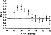
NIRSを利用した脳酸化(cerebral oxygenation)の頭蓋内監視は魅力的な特性を有する。本発明の幾らかの実施形態においては、脳酸素濃度指数(COx)という、自動調節血管反応性の新規な指標を提示するが、このCOxは、ABPの変化を、NIRSに基づく脳組織の酸素ヘモグロビン飽和の監視結果に相関付けた時間領域の分析から得られる。自動調節機能を継続して評価することは、重症の患者の脳潅流圧(CPP)を能動的に最適化する可能性を持つ有望な監視方法である。1実施形態においては、この相関付けは、300秒の重複する期間に継続して行われ、60秒毎に更新され、自動調節失敗を検出するのにABPの変化を誘発する必要がない。 Intracranial monitoring of cerebral oxygenation using NIRS has attractive properties. In some embodiments of the present invention, a novel indicator of autoregulatory vascular reactivity, the brain oxygen concentration index (COx), is presented, which changes ABP in brain tissue oxygen based on NIRS. Obtained from time domain analysis correlated to hemoglobin saturation monitoring results. Continued evaluation of the autoregulatory function is a promising monitoring method with the potential to actively optimize cerebral perfusion pressure (CPP) in critically ill patients. In one embodiment, this correlation is performed continuously over an overlapping period of 300 seconds and is updated every 60 seconds without having to induce a change in ABP to detect auto-adjustment failure.
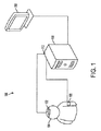
図1は、本発明の1実施形態による患者の脳血管自動調節機能を診断するシステム100の概略図である。脳血管自動調節機能を診断するシステム100は、患者の頭部104の外部位置近傍に配置された脳酸素濃度計102を含む。血圧監視デバイス106を患者に取り付ける。信号処理部108は、脳酸素濃度計102と通信可能であり、さらに血圧監視デバイス106とも通信可能である。本発明の1実施形態においては、脳酸素濃度計は、患者の脳内の血中酸素含有量を計測する。脳酸素濃度計102からの信号は、脳酸素濃度計102内で内部処理されてよい、および/または、信号処理部108で処理されてよい。本発明の1実施形態において脳酸素濃度計102は、患者の脳内の血中酸素含有量の計測を複数回行い、酸素含有量信号を信号処理部108へ入力する。
FIG. 1 is a schematic diagram of a
血圧監視デバイス106は、患者の動脈血圧を、酸素含有量の計測と実質的に同期して複数回計測し、動脈血圧信号を信号処理部108へ出力する。信号処理部108は、酸素含有量信号および動脈血圧信号に基づいて、時間領域で複数回、線形相関係数を算出する。この線形相関係数は、本発明の幾らかの実施形態では脳酸素濃度指標(COx)と称される場合がある。脳酸素濃度計102から信号処理部108へ送られる酸素含有量信号は、脳酸素濃度計、信号処理装置108のいずれかにより、または、脳酸素濃度計102と信号処理部108との間の信号線にある中間ローパスフィルタによりローパスフィルタリングにかけられる。血圧監視デバイス106、信号処理部108、または血圧監視デバイス106と信号処理部108との間の信号線にある中間デバイスは、計測された血圧信号をローパスフィルタリングにかける。血圧監視デバイス106は、頭蓋内圧監視デバイス(不図示)を含みうる。頭蓋内圧監視デバイスは、患者の脳の頭蓋内圧を直接計測するべく、患者に対して外科的に挿入されるカテーテルベースのデバイスを含んでもよい。血圧監視デバイス106は、入手可能な動脈血圧監視デバイスのなかから選択可能な動脈血圧監視デバイスを含んでもよい。本発明の1実施形態においては、脳酸素濃度計102は近赤外線分光器であってよい。
The blood
脳血管自動調節機能を診断するシステム100はさらに、信号処理部108と通信可能であり、患者の他の生体物理学データに関して信号処理部が算出した線形相関係数値を表示する表示部110を含んでよい。例えば表示部は、動脈血圧の関数として算出された線形相関係数を表示してよい。または、信号処理部108は、動脈血圧と頭蓋内圧との間の差異に基づいて脳潅流圧を求めて、脳潅流圧の関数として算出された線形相関係数を表示させる信号を表示部110に提供してよい。
The
脳酸素濃度計102、血圧監視デバイス106、表示部110、および信号処理部108は、物理的配線または他の適切な手段(例えば光学または無線データ通信)により接続されてよい。信号処理部108は、独立型の物理的コンポーネントであってよく、または、ラックシステム等の他のシステムのコンポーネントとして追加されてもよい。信号処理部108は、必ずしも信号データのみの処理に限定されない。一般的なデータ処理機能を含みうる。加えて、信号処理部108の信号処理は、ハードワイアードであってもよいし、信号処理部をプログラミングすることで実装されてもよい。
The cerebral oximeter 102, blood
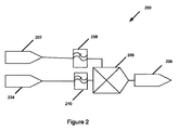
図2は、本発明の1実施形態による患者の脳血管自動調節機能を診断する方法200を示す概略図である。脳血管自動調節機能を診断する方法200は、患者の血圧を計測する段階202、動脈血圧計測202と実質的に同時に患者の脳の静脈の酸素含有量を非観血的に計測する段階204、血圧および静脈の酸素含有量の計測値を時間領域で相関付ける段階205、を含む。本発明の1実施形態においては、患者の脳血管自動調節状態を、血圧202および静脈の酸素含有量の計測値204の相関付けに基づいて判断する206。本発明の1実施形態によると、血圧信号202は、ローパスフィルタリングされる208。ローパスフィルタリング208により血圧信号の遅い変動をフィルタに通すことで、血圧信号のより急速な変動をフィルタリングして無くすことができる。本発明の様々な実施形態によるとローパスフィルタリング208はハードウェアまたはソフトウェアによる実装が可能である。さらに、ローパスフィルタリングは、アナログ信号またはデジタル信号のいずれを処理するかによって、アナログローパスフィルタリングまたはデジタルローパスフィルタリングいずれかであってよい。本発明の1実施形態においては、血圧信号をサンプリングして、デジタル信号を提供してよく、ローパスフィルタリングは、所望のサンプリング周波数を選択することで行われてよい。
FIG. 2 is a schematic diagram illustrating a
本発明の1実施形態においては、静脈の酸素含有量の計測値を、血圧信号と相関付ける205前に、ローパスフィルタリング210にかけてよい。本発明の1実施形態においては、静脈の酸素含有量データのサンプリングは、血圧データをサンプリングすることと実質的に同期して行われてデジタル信号を提供してよい。この場合、ローパスフィルタリング210は、所望のサンプリング周波数を選択することで行われうる。しかし、本発明の一般的な側面は、デジタル信号処理のみに限定されず、デジタルローパスフィルタリングのみに限定もされない。血圧計測データ202は、動脈血圧と対応していてよく、または、これも頭蓋内圧を計測することで求められる脳潅流圧と対応していてもよい。静脈の酸素含有量データは、例えば、患者の頭部の外部近傍に配設された近赤外線源から患者の脳内に向けられた近赤外線照射の差分吸収を計測することで得られうる。
In one embodiment of the present invention, the measured value of venous oxygen content may be subjected to
本発明の別の実施形態は、患者の血圧を計測すること、血圧計測と実質的に同時に患者の脳の酸素濃度を非観血的に計測すること、血圧計測値および酸素含有量の計測値を時間領域で相関付けること、を含む患者の処置方法に係る。血圧計測データは、動脈血圧に対応していてよく、または、これも頭蓋内圧を計測することで求められる脳潅流圧に対応していてもよい。患者の脳血管自動調節状態は、血圧と静脈の酸素含有量の計測値とを相関付けることに基づいて求められ、こうして判断した患者の脳血管自動調節状態に基づき、血圧または脳潅流圧を変化させる。 Another embodiment of the present invention includes measuring a patient's blood pressure, non-invasively measuring a patient's brain oxygen concentration substantially simultaneously with blood pressure measurement, blood pressure measurement value and oxygen content measurement value. In the time domain. The blood pressure measurement data may correspond to the arterial blood pressure, or this may also correspond to the cerebral perfusion pressure obtained by measuring the intracranial pressure. The patient's cerebral vascular autoregulation status is determined based on the correlation between blood pressure and venous oxygen content, and the blood pressure or cerebral perfusion pressure is changed based on the patient's cerebral vascular autoregulation status thus determined. Let
本発明の別の実施形態は、患者の脳血管自動調節機能を診断するシステムとともに利用されるデータ処理部に係る。例えば、データ処理部は、図1の脳血管自動調節機能を診察するシステム100について説明したデータ処理部108と類似していてよい、または同じであってよい。データ処理部108は、患者から計測された血圧データの血圧信号を受信して、患者の脳から外部的に計測された静脈の酸素含有量データの静脈酸素含有量信号を受信する少なくとも1つの信号入力ポート112を含む。データ処理部108はさらに、血圧信号と静脈酸素含有量信号とを受信して相関付けて、患者の脳血管自動調節状態を示す線形相関係数を提供する信号相関部を有する。データ処理部108はさらに、線形相関係数をさらに処理、保存、および/または、表示処理にかけるべく出力する信号出力ポート114を含む。本発明の1実施形態のデータ処理部108は、血圧データをフィルタリングするローパスフィルタを含んでよく、静脈の酸素含有量データをフィルタリングするローパスフィルタを含んでもよい。代替的な実施形態においては、血圧データおよび/または静脈の酸素含有量データは、データ処理部により受信される前に予めフィルタリングされていてもよい。本発明の幾らかの実施形態においては、血圧データは動脈血圧を含みうる。データ処理部108はさらに、患者から計測された頭蓋内圧の頭蓋内圧信号を受信してよい。これは、同じ入力ポート112から受信されても、さらなるデータ入力ポートから受信されてもよい。同様に、動脈血圧信号は、静脈酸素含有量信号と同じ信号入力ポート112を介して、データ処理部108へ送信されてもよく、または、別個のポートを介して提供されてもよい。本発明の幅広い構想は、特定の数のデータ入出力ポートに限定されず、入出力の際利用されるデータポートに応じて多重化されているか否かにも限定されない。加えて、信号入出力ポートは、電気、光学、または無線データ入出力ポートいずれであってもよい。
Another embodiment of the present invention relates to a data processing unit utilized with a system for diagnosing a patient's automatic cerebrovascular regulation function. For example, the data processing unit may be similar to or the same as the
本発明の別の実施形態においては、コンピュータ可読媒体は、患者の脳血管自動調整機能を診断するシステムからのデータを処理するようプログラミングされている。コンピュータ可読媒体は、血圧計測値の少なくとも1つの信号および静脈の酸素含有量の計測値の1つの信号を受信および処理し、時間領域での動脈血圧データおよび静脈の酸素含有量データの相関付けに基づいて線形相関係数を算出するようプログラミングされる。コンピュータ可読媒体は、患者の脳血管自動調節機能を求めることのできる情報を提供する線形相関係数を出力するようプログラミングされる。 In another embodiment of the invention, the computer readable medium is programmed to process data from a system for diagnosing a patient's cerebrovascular autoregulation function. A computer readable medium receives and processes at least one signal of blood pressure measurements and one signal of venous oxygen content measurements to correlate arterial blood pressure data and venous oxygen content data in the time domain. Based on this, it is programmed to calculate a linear correlation coefficient. The computer readable medium is programmed to output a linear correlation coefficient that provides information that can determine the patient's cerebrovascular autoregulation function.
実施例
ここで、本発明の1実施形態におけるCOxは、レーザドップラー血流計測法により求められるABPを自動調節の区切点の下へとゆっくりと低下させる間、子豚の乳児の脳のモデルの低血圧および子豚で継続して計測されたCOxにより自動調節の失敗に対する感度が高いと仮定する。そこで、低血圧に起因する自動調節の欠如を検出するCOxの感度および特異性を求めた。さらにCOxを、同様ではあるが観血的な方法であるレーザドップラー指標(LDx)(前頭皮質(frontoparietal cortex)で計測したレーザドップラー血流とABPとの間の線形相関係数を利用する)との間で比較した。ここで我々は、COxおよびLDxが、それぞれ出所が異なってはいるが、自動制御血管反応性計測値が一致する、と想定した。
EXAMPLE Here, COx in one embodiment of the present invention is a model of a piglet infant brain while slowly lowering the ABP determined by laser Doppler blood flow measurement below the automatic adjustment breakpoint. Assume that sensitivity to autoregulation failure is high due to low blood pressure and continuously measured COx in piglets. Thus, the sensitivity and specificity of COx to detect the lack of automatic regulation due to hypotension was determined. In addition, COx is a similar but invasive method of laser Doppler index (LDx) (using a linear correlation coefficient between laser Doppler blood flow and ABP measured in the frontoparietal cortex) and Compared between. Here, we assumed that the COx and LDx were different in origin, but the automatically controlled vascular reactivity measurements were in agreement.
方法および材料
全ての手順は、Johns Hopkins University Animal Care and Use Committeeにより承認されており、国立衛生研究所の動物実験基準を満たしている。
All procedures and procedures have been approved by the Johns Hopkins University Animal Care and Use Committee and meet the National Institutes of Health animal testing standards.
麻酔
生後3〜8日の2.2−3.9kgの子豚(n=6)を、5%イソフルレン、50%亜酸化窒素、および酸素平衡の吸入により麻酔にかけた。気管開口術により機械的に通気口を設けた。抹消静脈へのルートを確保して、ベクロニウム(5mgのボーラス投与および2mg/hrの点滴)およびフェンタニル(25μgのボーラス投与および25μg/hrの点滴)を投与した。実験中イソフルレンは0.5%低減し、外科手術中フェンタニルは、対象心拍数が190を切り正常血圧である間10―50μg/hrの間で滴定した。記録期間中、血圧はアクティブに低下した際には、フェンタニルを50μg/hr(殆どの子豚について20μg/kg/hr)で点滴して、前負荷の反応として頻拍させた。吸入ガスに比してイソフルレンは0.5%で維持され、亜酸化窒素は50%で維持された。故に、記録期間における麻酔は、一義的には麻酔剤ベースであり、副麻酔補給(sub-anesthetic supplementation)として吸入剤を利用した。この組み合わせは、動物を楽にして、吸入麻酔剤の脳血管反応に対する影響を低減させるべく選択された。子豚は加温パッド上に置き、脳および直腸の温度を摂氏38.5度から39.5度に保った。空調はpH7.35−7.45に保ちPaO2を200−300mmHgに保った。
Anesthesia 3-8 days old 2.2-3.9 kg piglets (n = 6) were anesthetized by inhalation of 5% isoflurane, 50% nitrous oxide, and oxygen balance. A vent was provided mechanically by tracheostomy. A route to the peripheral vein was secured and vecuronium (5 mg bolus and 2 mg / hr infusion) and fentanyl (25 μg bolus and 25 μg / hr infusion) were administered. During the experiment, isoflurane was reduced by 0.5%, and during surgery fentanyl was titrated between 10-50 μg / hr while the subject's heart rate was 190 and normal blood pressure. During the recording period, when blood pressure decreased actively, fentanyl was instilled at 50 μg / hr (20 μg / kg / hr for most piglets) to tachycardia as a preload response. Isoflurane was maintained at 0.5% and nitrous oxide was maintained at 50% relative to inhaled gas. Therefore, anesthesia during the recording period was primarily based on an anesthetic and utilized an inhalant as a sub-anesthetic supplementation. This combination was chosen to ease the animals and reduce the effect of inhalation anesthetics on the cerebrovascular response. The piglets were placed on a heating pad to maintain brain and rectal temperatures between 38.5 and 39.5 degrees Celsius. Air conditioning was maintained at pH 7.35-7.45, and P a O 2 was maintained at 200-300 mmHg.
外科手術
大腿部の静脈にカニューレを相互に(bilaterally)挿入して、薬物点滴および血圧監視および5Frの食道バルーンカテーテル(Copper Surgical,Trundall,CT)用に中央静脈線を確保した。大腿部の動脈に、血圧および血液ガス監視線を確保するカニューレを挿入した。正中線のブレグマの水平方向且つ吻側に4mm開頭して、外部脳室ドレインカテーテルを配置して、ICP監視用にトランスデュースした。さらに最初の開頭に対して水平方向か且つ吻側に4mm開頭して、切開された硬膜から前頭皮質の表面に接するよう、レーザドップラープローブ(Moor Instruments,Devon,U.K.)を配置した。プローブは、大血管の上の配設した際に生じがちな高いベースライン流量値を避け、頭蓋骨に接着されたゴム製のワッシャで固定された。さらに三回目の開頭を後頭部頭蓋骨に対して正中線の水平方向に行い、脳温度プローブを配置した。皮膚を頭蓋骨に戻して、傷をきっちり縫い合わせて、温度を維持し、脳酸素濃度計を較正する条件を揃えた。
The surgical femoral vein was cannulated bilaterally to secure a central venous line for drug instillation and blood pressure monitoring and a 5 Fr esophageal balloon catheter (Coper Surgical, Trundall, CT). The femoral artery was cannulated to ensure blood pressure and blood gas monitoring lines. A 4 mm craniotomy was performed horizontally and rostrally to the midline bregma, and an external ventricular drain catheter was placed and transduced for ICP monitoring. Further, a laser Doppler probe (Moor Instruments, Devon, UK) was placed so as to contact the surface of the frontal cortex from the incised dura mater by 4 mm in the horizontal direction and the rostral side with respect to the first craniotomy. . The probe was fixed with a rubber washer glued to the skull, avoiding the high baseline flow rates that tend to occur when placed over large vessels. A third craniotomy was performed in the horizontal direction of the midline with respect to the occipital skull, and a brain temperature probe was placed. The skin was returned to the skull and the wounds were sewn together to maintain the temperature and to calibrate the brain oximeter.
酸素濃度計プローブ配置
INVOS(インビボの光学的分光器)小児脳酸素濃度計プローブ(Somanetics,Troy,MI)を、目の上の、前頭皮質と後頭皮質とを横切る位置、且つ、前述の開頭部とは反対側に配設し、且つ、矢状静脈洞を避けて正中線の水平方向に1cmの発光ダイオードを配設した。その後、プローブの脳の特異性をCO2対抗量でテストして、換気を増して、呼気終末CO2を少なくとも10mmHg低減させた。脳酸素濃度計測値を、腎臓の上に配置されたプローブから採った酸素濃度計測値と比較した。脳酸素濃度計側値は低減しており(1.2±0.1%/mmHg;±SD)、腎臓酸素濃度計測値は変化がなかった(0.0±0.1%/mmHg)。
Oximeter probe placement INVOS (in vivo optical spectrometer) Pediatric brain oximeter probe (Somanetics, Troy, Mich.) On the eye, across the frontal and occipital cortex and the
信号サンプリング
血圧トランスデューサ(ABP、ICP)、レーザドップラーププローブ、およびINVOS脳酸素濃度計測計からの波形をアナログデジタルコンバータからICM+ソフトウェア(Cambridge University,Cambridge,UK)により60Hzでサンプリングした。INVOS酸素濃度計の時間分解能は、4秒である。これら信号はその後互いに重複しない10秒平均値として時間積分されるが、これは、10秒時間窓の移動平均フィルタを利用して0.1Hzでリサンプリングするのに等しい。この処理により、高周波ノイズが動物の呼吸およびパルス周波数から除去されるが、ナイキスト定理によると、0.05Hz未満で起こる発振および過渡電流(transient)検知を可能とする。CPPは、ABPおよびICPの10秒の平均値間の差異として算出された。
Waveforms from a signal sampling blood pressure transducer (ABP, ICP), a laser Doppler probe, and an INVOS brain oximeter were sampled at 60 Hz from an analog-to-digital converter by ICM + software (Cambridge University, Cambridge, UK). The time resolution of the INVOS oximeter is 4 seconds. These signals are then time integrated as non-overlapping 10 second averages, which is equivalent to resampling at 0.1 Hz using a 10 second time window moving average filter. This process removes high frequency noise from the animal's respiration and pulse frequency, but the Nyquist theorem allows for oscillation and transient detection that occurs below 0.05 Hz. CPP was calculated as the difference between the 10-second average values of ABP and ICP.
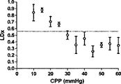
レーザドップラーおよび脳酸素濃度指標の算出
継続した移動ピアソン相関係数(moving Pearson's correlation coefficient)を、CPPとレーザドップラーとの間で行うことでLDxを生成し、あるいは、CPPと脳酸素濃度計の出力との間に行うことでCOxを生成した。300秒の間の連続した、対の、10秒平均値を各計算に利用して、30個のデータ点を各指標に組み込む。これら指標は、重複する期間で60秒毎に計算および記録される。
Laser Doppler and Cerebral Oxygen Concentration Index Calculations Continuous moving Pearson's correlation coefficient is generated between CPP and Laser Doppler to generate LDx, or CPP and brain oximeter output COx was generated by performing between the two. 30 data points are incorporated into each index using a continuous pair of 10-second averages for 300 seconds for each calculation. These indicators are calculated and recorded every 60 seconds in overlapping periods.
血圧低下および自動調節曲線の構築
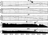
上述のモニタを配置すると、下大静脈内のバルーンカテーテルは、徐々にシリンジポンプからの食塩液注入で膨らみ、ABPが4−5時間かけて10mmHgにまでゆっくりと低減する(図3)。脳酸素濃度計、レーザドップラー血流計、COx、およびLDxの値は、60秒ごとにリアルタイムに記録され、それらが収集されたCPPにより同時に分類された。低血圧が長期間に亘り誘発され、CPPの自然発生的な変化が各準定常状態CPP範囲に亘り起こり、COxの適切な信号対雑音比を提供するのに十分な時間的余裕が与えられた。
Blood pressure reduction and automatic adjustment curve construction With the above monitor in place, the balloon catheter in the inferior vena cava gradually inflates with saline injection from a syringe pump, and ABP slowly decreases to 10 mmHg over 4-5 hours (FIG. 3). Brain oximeter, laser Doppler blood flow meter, COx, and LDx values were recorded in real time every 60 seconds and simultaneously classified by the CPP from which they were collected. Hypotension was triggered over a long period of time, and spontaneous changes in CPP occurred across each quasi-steady state CPP range, giving enough time to provide an adequate signal-to-noise ratio for COx. .
定常状態の自動調節区切点の決定
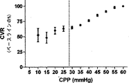
レーザドップラー血流対CPPの散布図を、SigmaStatソフトウェア(Systat,San Jose,CA)を利用して、各子豚の全てのデータについて作成した。最少の誤差平方和(RSE)の組み合わせを有する2つの回帰線に区切ったCPPが、自動調節の区切点として決定および定義された。加えて、脳血管耐性(CVR)の相対変化をベースラインのCPP/レーザドップラー血流比のパーセントとして算出した。
Determination of Steady State Automatic Adjustment Breakpoints Laser Doppler blood flow versus CPP scatter plots were generated for all data for each piglet using SigmaStat software (Systat, San Jose, CA). The CPP delimited by two regression lines with the least sum of error squared (RSE) combination was determined and defined as the breakpoint for automatic adjustment. In addition, the relative change in cerebrovascular resistance (CVR) was calculated as a percentage of the baseline CPP / Laser Doppler blood flow ratio.
レシーバ‐オペレータ特性
Prism software(GraphPad,San Diego,CA)を利用して、COxおよびLDxのレシーバ‐オペレータ特性(ROC)を求めた。これに関しては、各子豚の各CPPの平均指標値を、各子豚についてレーザドップラー血流自動調節関係から導き出したCPP区切点の上下で二分した。
Receiver-operator characteristics Prism software (GraphPad, San Diego, Calif.) Was used to determine COx and LDx receiver-operator characteristics (ROC). In this regard, the average index value of each CPP for each piglet was divided in two above and below the CPP breakpoint derived from the laser Doppler blood flow autoregulation relationship for each piglet.
LDxおよびCOxの比較
COxのLDxに対する回帰分析および線形相関付けを、Prism softwareおよびブランドアルトマン分析を用いて、LDx−COxおよびCOx/LDxを平均値に適用することで行った。この分析は、収集された全ての指標対に対して、および、同じCPPで同じ子豚について収集された平均値について行われた。
Comparison of LDx and COx Regression analysis and linear correlation of COx to LDx was performed by applying LDx-COx and COx / LDx to the mean values using Prism software and Brand Altman analysis. This analysis was performed for all indicator pairs collected and for the mean values collected for the same piglets at the same CPP.
子豚の自動調節機能のスペクトル範囲の確認
ICM Plus softwareを利用して、ABPを入力として利用して、且つ、レーザドップラー血流または脳酸素濃度を出力として利用して、コヒーレンスの相互スペクトル解析を行った。1Hzから0.001Hzの範囲の周波数におけるコヒーレンスについて、低血圧状態および通常血圧状態の間で比較を行った。これらは公式データではないが、提示する時間領域分析のサンプリングおよび計算パラメータを構築するのに利用された(「考察」の項を参照のこと)。
Confirmation of spectral range of piglet automatic regulation function Using ICM Plus software, using ABP as input and using laser Doppler blood flow or brain oxygen concentration as output, perform mutual spectrum analysis of coherence went. A comparison was made between the hypotensive state and the normal blood pressure state for coherence at frequencies ranging from 1 Hz to 0.001 Hz. These are not official data, but were used to construct the sampling and calculation parameters for the presented time domain analysis (see “Discussion” section).
結果
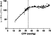
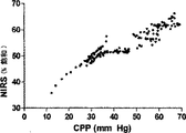
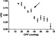
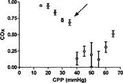
表1が示すように、動脈のpH、PaCO2、および脳温度は、正常血圧で(CPP>50mmHg)、自動調節区切点の上の中程度の血圧で(CPP30−50mmHg)、および自動調節区切点の下の苛酷な低血圧で(CPP<30mmHg)、正常の生理学上の範囲にあった。CO2−反応が酸素濃度計測値に影響するのを避けるべく、PaCO2を一定に保つことを考えたが、各子豚の心拍出量が重篤なレベルに至っては少量の減少が見られた。この少量の減少はPaCO2に対して比較的不変の、離散的な300秒の間隔での圧力パッシビティ(pressure passivity)を評価しているので、自動調節指標へバイアスをかけたとは考えにくい。

一匹の子豚の自動調節機能の評価の1例を図4に示す。レーザドップラー血流の下限は、誤差平方和の総計を最小とする2つの回帰線の交点から容易に特定された(図4A)。興味深いのは、CPPの関数である脳酸素濃度計側値のプロットも、変曲点として特徴付けられないことである(図4B)。しかし、LDxおよびCOxは両方とも、動物の自動調節機能の閾値において急増している(図4Cおよび4D)。 An example of the evaluation of the automatic adjustment function of one piglet is shown in FIG. The lower limit of laser Doppler blood flow was easily identified from the intersection of two regression lines that minimized the sum of the error sum of squares (FIG. 4A). Interestingly, a plot of brain oximeter side values as a function of CPP is also not characterized as an inflection point (FIG. 4B). However, both LDx and COx have increased rapidly at the threshold of the animal's autoregulatory function (FIGS. 4C and 4D).
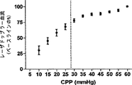
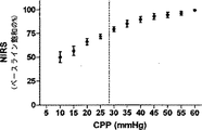
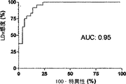
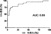
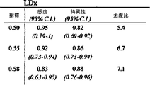
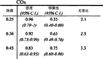
6匹の子豚のレーザドップラー血流からのデータの組み合わせを、CVRおよび脳酸素濃度計測値と比較したものを図5に示す。平均区切点は29.7±5.5mmHgであって、先行技術である子豚の自動調節曲線に関する報告書と矛盾がない(Laptook AR, Stonestreet BS, Oh W. Brain blood flow and O2 delivery during hemorrhagic hypotension in the piglet. Pediatr Res. 1983;17(1):77-80; Mertineit C, Samlalsingh-Parker J, Glibetic M, Richard G, Noya FJ, Aranda JV. Nitric oxide, prostaglandins, and impaired cerebral blood flow autoregulation in group B streptococcal neonatal meningitis. Can J Physiol Pharmacol. 2000; 78(3):217-227)。相対的なCVRの段階的な減少は、CPPが30mmHgにまで低減すると顕著となり、さらなる低減はCPP値が30mmHg未満になると治まってくる。平均LDxおよびCOxは、CPPが30mmHg未満になると増加した(図6Aおよび6B)。各子豚の定常状態の自動調節区切点が分かっていることで、LDxおよびCOxについてROCを求めることができる。これは驚くことではない、というのも、LDxはレーザドップラー血流測値の微分値(derivative)であり、LDxはCOxより性能が良いが、両者において区切点が正確に画定されているからである。ROC曲線の下の領域は、LDxについて0.95であり(図6C)、COxについては0.89である(図6D)。2つの指標についてカットオフの感度、特異性、および尤度比をまとめたものを、図6に示す。一般的には、感度は、両方の指標で特異性よりも勝っている、つまり、全ての子豚が、低血圧時にはCOxおよびLDx両方において異常な自動調節血管反応を示したが、多くは、また、正常血圧または中程度の血圧範囲において、いずれかまたは両方の指標で一時的なディスラプションが示されている。 FIG. 5 shows a combination of data from laser doppler blood flow of 6 piglets compared to CVR and brain oximetry measurements. The average breakpoint is 29.7 ± 5.5 mmHg, consistent with the prior art report on piglet autoregulation curves (Laptook AR, Stonestreet BS, Oh W. Brain blood flow and O2 delivery during hemorrhagic Pediatr Res. 1983; 17 (1): 77-80; Mertineit C, Samlalsingh-Parker J, Glibetic M, Richard G, Noya FJ, Aranda JV. Nitric oxide, prostaglandins, and impaired cerebral blood flow autoregulation in group B streptococcal neonatal meningitis. Can J Physiol Pharmacol. 2000; 78 (3): 217-227). The gradual decrease in relative CVR becomes noticeable when the CPP is reduced to 30 mmHg, and further reduction subsides when the CPP value is less than 30 mmHg. Average LDx and COx increased when CPP was below 30 mmHg (FIGS. 6A and 6B). Knowing the steady-state automatic adjustment breakpoint for each piglet, the ROC can be determined for LDx and COx. This is not surprising, because LDx is a derivative of laser Doppler blood flow measurements and LDx performs better than COx, but the breakpoints are precisely defined in both. is there. The area under the ROC curve is 0.95 for LDx (FIG. 6C) and 0.89 for COx (FIG. 6D). FIG. 6 shows a summary of the sensitivity, specificity, and likelihood ratio of cutoff for the two indices. In general, sensitivity is superior to specificity in both indicators, meaning that all piglets showed abnormal autoregulatory vascular responses in both COx and LDx at low blood pressure, In addition, temporary disruption is indicated by either or both of the indicators in the normal blood pressure range or the moderate blood pressure range.
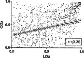
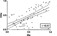
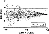
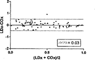
COxおよびLDxの線形相関付けおよびブランドアルトマン比較を図7に示す。指標間の一致度は、分ベースでの評価では制限されている(ピアソンのr=0.36)。これら値が収集されたCPPの5mmHg増分ビンでこれら値を階層化して平均化すると、一致度は大幅に向上した(ピアソンのr=0.67)。ブランドアルトマン法は、計測値範囲に亘りバイアスがないことを示し(全ての計測値においてバイアスは−0.06であり、平均値においては0.03であった)、同じCPPの値同士を平均化した場合、一致度が向上した。 The linear correlation of COx and LDx and the Brand Altman comparison are shown in FIG. The degree of agreement between the indices is limited in the evaluation on a minute basis (Pearson's r = 0.36). When these values were stratified and averaged in the CPP's 5 mm Hg incremental bins where they were collected, the agreement was greatly improved (Pearson's r = 0.67). The Brand Altman method shows no bias across the measurement range (bias was -0.06 for all measurements and 0.03 for the average) and averaged the same CPP values The degree of coincidence improved.
考察
この結果は、ABPと脳酸素濃度測定値との間の時間領域の相関付けにより、自然発生的な自動調節血管反応性を定量化することができ、その結果生じる指標が、子豚のモデルの低血圧に起因する自動調節機能の低減に対して高感度であることを示している。この方法は、臨床適用に際して幾らかの魅力的な特性を有する。実験した動物において行ったようにCOx出力は継続しており60秒毎に更新される。COxは、CPP等の臨床パラメータの関数として臨床的に(at the bedside)示すことができ、自動調節処理の管理変化の影響を示す。COxの算出は頭蓋外科手術を要さず、ABPの自然発生的な変化を利用することができるので、不安定な患者に対してABPの急激な変化を引き起こす必要がなくなる。
Discussion This result can be quantified by the time domain correlation between ABP and cerebral oximetry measurements, and spontaneous autoregulatory vascular reactivity can be quantified. It shows that it is highly sensitive to the reduction of the automatic adjustment function caused by low blood pressure. This method has some attractive properties for clinical applications. The COx output continues as it did in the experimental animals and is updated every 60 seconds. COx can be shown at the bedside as a function of clinical parameters, such as CPP, and shows the impact of administrative changes in the autoregulation process. Calculation of COx does not require cranial surgery and can utilize spontaneous changes in ABP, eliminating the need to cause abrupt changes in ABP for unstable patients.
COxの開発において重要な課題は、波形サンプリングの関連期間の決定であった。この決定の背後にある論理的根拠、COxの限定についての考察、および、COxの潜在的な臨床的適用可能性を以下で述べる。 An important challenge in the development of COx was the determination of the relevant period of waveform sampling. The rationale behind this decision, a discussion of COx limitations, and the potential clinical applicability of COx are discussed below.
COx分析について選択される周波数についての考察
ABPおよびCBFの代用物の間の連合関係(associative relationship)は、周波数領域における分析と、時間領域における分析という2種類に大まかに分類される方法により、動的に評価される。周波数領域分析は(コヒーレンス、伝達関数、または位相のずれに基づく)は、さもなくば静止型のシステム内のABPの定期的且つ周期的な波または誘発された変化に対して適している。この分析法は、通常は生体系に厳密には存在しない線形および定常性(stationarity)を前提としている(Giller CA, Mueller M. Linearity and non-linearity in cerebral hemodynamics. Med Eng Phys. 2003;25(8):633-646)。時間領域分析は、ここでCOsおよびLDxと共に提示されたローパスフィルタされたABPおよびCBF波間の線形相関付けとして行われるが、このフィルタリングはテストのスペクトル範囲を制限する。この種類の分析で自動調節を画定するには、自動調節の失敗により生じるCPPおよび酸素濃度計測値の相関付けを包括する臨床意義のある波長期間が分かっている必要がある。我々が0−0.04Hzの周波数に注目したのは、3つの仮定に基づいている。先ず第一に、最も重要なことであるが、未熟児におけるNIRSおよびABP間のコヒーレンスに周波数領域分析を用いたTsujiらの業績がある(Tsuji M, Saul JP, du Pessis A, et al. Cerebral intravascular oxygenation correlates with mean arterial pressure in critically ill premature infants. Pediatrics. 2000;106(4):625-632)。Tsujiらは、0.01Hz未満の周波数で高いコヒーレンスを有するサブグループを特定して、このグループ内で脳室内出血の発生が多いことを発見し、自動調節機能の損傷の結果なのではないかと推論した。この発見は、臨床意義のあるABPおよびCBFの間の相関付けを画定する際の、これら低周波数の有益性を示唆している。選択された周波数についての第二の理由付けは、ICP由来の自動調節指標(PRx)に由来しており、これは、ICPの「B」徐波をABPと相関付ける。PRxは、頭部に外傷を負った患者の結果と関連付けられることが既に示されており、自動調節処理のマーカとして考えられている(Czosnyka M, Smielewski P, Kirkpatrick P, Laing RJ, Menon D, Pickard JD. Continuous assessment of the cerebra vasomotor reactivity in head injury. Neurosurgery. 1997;41(1):11-7;discussion 17-9)。我々のデータベースにおいては、これらICP徐波は余りに散発的であるのでフーリエ変換分析では明瞭に現れないが、子豚から得られた生の波形では特定でき、これらの存続期間は65−300秒と計測され、これは0.015から0.003Hzの周波数に相当する。最後の理由付けは、本研究で利用した子豚のABPおよびNIRSの波形のコヒーレンス分析に由来する。自動調節下限未満の血圧で得られた波形においては、0.04Hz未満の周波数で、特に、0.02Hz未満の周波数で、コヒーレンスが発見された。このコヒーレンスは、正常血圧中に得られた波形には見られなかった。
Consideration of the frequency selected for COx analysis The association relationship between ABP and CBF surrogates is roughly divided into two types: analysis in the frequency domain and analysis in the time domain. Evaluated. Frequency domain analysis (based on coherence, transfer function, or phase shift) is otherwise suitable for periodic and periodic waves or induced changes of ABP in a stationary system. This analysis method assumes linearity and stationarity that are not strictly present in biological systems (Giller CA, Mueller M. Linearity and non-linearity in cerebral hemodynamics. Med Eng Phys. 2003; 25 ( 8): 633-646). Time domain analysis is performed as a linear correlation between the low-pass filtered ABP and CBF waves presented here with COs and LDx, but this filtering limits the spectral range of the test. In order to define autoregulation in this type of analysis, it is necessary to know a clinically relevant wavelength period that encompasses the correlation of CPP and oximetry measurements resulting from autoregulation failure. The reason we focused on the 0-0.04 Hz frequency is based on three assumptions. First of all, most importantly, Tsuji et al. Have used frequency domain analysis for the coherence between NIRS and ABP in premature infants (Tsuji M, Saul JP, du Pessis A, et al. Cerebral). intravascular oxygenation correlates with mean arterial pressure in critically ill premature infants. Pediatrics. 2000; 106 (4): 625-632). Tsuji et al. Identified a subgroup with high coherence at a frequency of less than 0.01 Hz, discovered that there was a high incidence of intraventricular hemorrhage within this group, and inferred that this may be the result of damage to the autoregulatory function. did. This finding suggests the benefit of these low frequencies in defining a correlation between clinically meaningful ABP and CBF. A second reasoning for the selected frequency comes from the ICP-derived auto-regulation index (PRx), which correlates the “B” slow wave of ICP with ABP. PRx has already been shown to be associated with the results of patients with trauma to the head and is considered as a marker for autoregulation (Czosnyka M, Smielewski P, Kirkpatrick P, Laing RJ, Menon D, Pickard JD. Continuous assessment of the cerebra vasomotor reactivity in head injury. Neurosurgery. 1997; 41 (1): 11-7; discussion 17-9). In our database, these ICP slow waves are so sporadic that they do not appear clearly in the Fourier transform analysis, but can be identified in the raw waveform obtained from the piglet, and their duration is 65-300 seconds. Measured, which corresponds to a frequency of 0.015 to 0.003 Hz. The last reasoning comes from the coherence analysis of the piglet ABP and NIRS waveforms used in this study. In waveforms obtained with blood pressures below the lower limit of autoregulation, coherence was found at frequencies below 0.04 Hz, especially at frequencies below 0.02 Hz. This coherence was not seen in the waveforms obtained during normal blood pressure.
これら発見により、0.04Hz未満の周波数(周期>25秒)で起こった波形関係を解明することが望まれた。同時に、呼吸数および心拍周波数に含んだ高波長領域からエイリアシングノイズを防止することが望まれた。呼吸数は〜0.3Hzであった(3秒周期)。故に、10秒周期を時間平均すると、選択された周波数でこのノイズは抑制され、分解能が維持された。 With these discoveries, it was desired to elucidate the waveform relationship that occurred at frequencies below 0.04 Hz (period> 25 seconds). At the same time, it was desired to prevent aliasing noise from the high wavelength region included in the respiratory rate and heart rate frequency. The respiration rate was ˜0.3 Hz (3-second cycle). Therefore, when the 10-second period was time-averaged, this noise was suppressed and the resolution was maintained at the selected frequency.
COxの制限
こうしてCOxの感度および特異性の誤差の源が理解されたので、改善策が立てられるようになった。ABPの一時的および自然発生的な変化により、短時間で血圧が大きく変化する方法と比して、信号対雑音比が低減する。信号対雑音比を増加させる2つの明確な解決法を選択することができるが、その2つの方法とは、(a)各指標計算のためにサンプリング時間を増加させること、または、(b)指標間の多数の離散的な計算結果を平均化すること、である。我々は二番目の解決法を選択した、というのは、臨床意義のある変数(CPP、温度、血液ガス、鎮静状態等)によるソーティングが可能であるので、一番目の解決法と同じデータ平滑化効果を有するとはいっても利便性が高いので、より有益であるからである。これら変数は、5分間のほうが、20分または60分という期間よりもより不変である可能性が高い。我々の実験設計は、これら変数の制御を行うことでCPPの変化による効果を隔離することを目指しているが、PaCO2も若干ずれた。脳内O2消費の動的変化はCOxにも影響しうる。フェンタニル、亜酸化窒素、およびイソフルレンによる麻酔を採択することで、COx計算に利用される各300秒間にO2消費が安定すると想定した。
COx Limitations Now that the sources of COx sensitivity and specificity errors have been understood, improvements can be made. Due to the temporary and spontaneous changes in ABP, the signal-to-noise ratio is reduced compared to methods in which blood pressure changes significantly in a short time. Two distinct solutions to increase the signal-to-noise ratio can be selected, which are: (a) increasing the sampling time for each index calculation, or (b) the index Averaging a number of discrete computation results in between. We chose the second solution because it is possible to sort by variables of clinical significance (CPP, temperature, blood gas, sedation, etc.), so the same data smoothing as the first solution This is because even if it has an effect, it is more useful because it is highly convenient. These variables are more likely to be invariant in 5 minutes than in a period of 20 minutes or 60 minutes. Our experimental design aims to isolate the effects of changes in CPP by controlling these variables, but PaCO 2 has also shifted slightly. Dynamic changes in brain O 2 consumption can also affect COx. By adopting anesthesia with fentanyl, nitrous oxide, and isoflurane, it was assumed that O 2 consumption was stable for each 300 seconds utilized for COx calculations.
特定のCPP範囲を要する指標計算に排除原理を導入することで信号対雑音比の問題に取り組もうという取り組みもあった。例えば、ABPが10mmHg未満変化する期間を、分析から排除しようとう取り組みがある(Lam JM, Hsiang JN, Poon WS. Monitoring of autoregulation using laser doppler flowmetry in patients with head injury. J Neurosurg. 1997;86(3):438-445)。この安定した血圧を有する排除期間によるバイアス導入は決定されておらず、この方法は、達成されたABPにおける安定化の低減が遅々たるものなので、我々の実験モデルにおいては現実的ではなかった。LDxまたはCOxいずれかで起こる感度欠如が、動物の死の直前といった極端な低血圧状態に大きく制限されていることは、図4のCPPが10の場合における変動性の増加により理解されよう。この範囲におけるデータセットは、記録時間が制限されており、心臓機能を維持する困難性から3匹の動物のみのデータしかないので、不完全である。臨界閉鎖圧未満のABPにより、僅かのABP変動では変化しなかった低く不変のCBFおよび脳酸化(cerebral oxygenation)が生じた可能性がある。(Panerai RB. The critical closing pressure of the cerebral circulation. Med Eng Phys. 2003;25(8):621-632)。このような静的なCBF状態は、COxまたはLDxの評価による、損傷を受けていない自動調節機能に付いて虚像を呈する可能性がある。脳のO2消費の動的な低減は、さらに、これら指標の変動性を増長させる可能性もある。この範囲の血圧は、掲題の臨床的課題にとっては重要ではない。 There has also been an effort to address the problem of signal-to-noise ratio by introducing the exclusion principle into index calculations that require a specific CPP range. For example, there is an effort to exclude the period during which ABP changes by less than 10 mmHg from analysis (Lam JM, Hsiang JN, Poon WS. Monitoring of autoregulation using laser doppler flowmetry in patients with head injury. J Neurosurg. 1997; 86 (3 ): 438-445). The introduction of bias due to this exclusion period with stable blood pressure has not been determined, and this method has not been realistic in our experimental model because the reduction in stabilization achieved in ABP is slow. It can be seen by the increased variability when the CPP of FIG. 4 is 10 that the lack of sensitivity that occurs with either LDx or COx is largely limited to extreme hypotension conditions such as just before the death of the animal. Data sets in this range are incomplete because the recording time is limited and there are only data for three animals due to the difficulty of maintaining cardiac function. ABP below the critical closure pressure may have resulted in low and unchanged CBF and cerebral oxygenation that did not change with slight ABP fluctuations. (Panerai RB. The critical closing pressure of the cerebral circulation. Med Eng Phys. 2003; 25 (8): 621-632). Such a static CBF condition can present a virtual image for an undamaged self-adjusting function, as assessed by COx or LDx. Dynamic reduction of brain O 2 consumption may further increase the variability of these indicators. This range of blood pressure is not critical to the subject clinical problem.
COxの臨床的示唆
自動調節機能の臨床的監視の重要な目的は、自動調節機能を向上させるケアパラメータの説明である。自動調節機能に損傷を受けていない患者は、神経損傷を負っても生存する確率が高く、この可換ロジックとして、自動調節機能の向上により、神経損傷からの復帰および生存の可能性を高めることができることが示唆される(Steiner LA, Czosnyka M, Piechnik SK, et al. Continuous monitoring of cerebrovascular pressure reactivity allows determination of optimal cerebral perfusion pressure in patients with traumatic brain injury. Crit Care Med. 2002;30(4):733-738; Czosnyka M, Smielewski P, Kirkpatrick P, Laing RJ, Menon D, Pickard JD. Continuous assessment of the cerebral vasomotor reactivity in head injury. Neurosurgery. 1997;41(1):11-7; discussion 17-9; Hiler M, Czosnyka M, Hutchinson P, et al. Predictive value of initial computerized tomography scan, intracranial pressure, and state of autoregulation in patients with traumatic brain injury. J Neurosurg. 2006; 104(5):731-737)。臨床的に自動調節機能を定量化する道具により、この仮説をテストすることができる。COxが非観血的なので、神経外科的な介入を経ない、または経ることのできない急性神経炎症(acute neurologic processes)を起こしている患者(中程度の頭部外傷、発作、髄膜炎を有する患者、および、心臓矯正手術のために心肺バイパス手術を受けた患者、または急性胸部症候群のために交換輸血を受けた患者等を含む)に対する適用が可能である。さらにCOxは、観血的監視により得られる他の指標に加えて、重篤な頭部外傷の場合に血圧自動調節機能の監視に対して追加されることで有益となりうる。
An important objective of clinical monitoring of COx's clinical suggestion autoregulation function is the description of care parameters that improve the autoregulation function. Patients who are not damaged by the autoregulatory function have a high probability of surviving even if they suffer nerve damage, and this replaceable logic increases the chances of recovery and survival from nerve damage by improving the autoregulatory function. (Steiner LA, Czosnyka M, Piechnik SK, et al. Continuous monitoring of cerebrovascular pressure reactivity allows determination of optimal cerebral perfusion pressure in patients with traumatic brain injury. Crit Care Med. 2002; 30 (4): 733-738; Czosnyka M, Smielewski P, Kirkpatrick P, Laing RJ, Menon D, Pickard JD. Continuous assessment of the cerebral vasomotor reactivity in head injury. Neurosurgery. 1997; 41 (1): 11-7; discussion 17-9 Hiler M, Czosnyka M, Hutchinson P, et al. Predictive value of initial computerized tomography scan, intracranial pressure, and state of autoregulation in patients with traumatic brain injury. J Neurosurg. 2006; 104 (5): 731-737). This hypothesis can be tested with tools that clinically quantify autoregulatory functions. Patients with acute neurologic processes (with moderate head trauma, seizures, meningitis, with or without neurosurgical intervention because COx is noninvasive For example, patients undergoing cardiopulmonary bypass surgery for cardiac correction surgery, or patients undergoing exchange transfusion for acute chest syndrome). In addition, COx can be beneficial in addition to other indicators obtained by invasive monitoring, in addition to monitoring blood pressure autoregulation functions in the case of severe head trauma.
本明細書で例示および記載した実施形態は、当業者に対して、本発明者が分かっている本発明の作製方法および利用方法のうち最善のものを伝える意図しか持たない。本明細書のうち、本発明の範囲を限定するものは何もない。教示を読んだ当業者であれば理解するように、上述の本発明の実施形態は、本発明を逸脱しない範囲で、修正または変形が可能であり、部材を追加したり削除したりすることが可能である。故に、請求項およびその均等物の範囲内において、本発明は特に記載した以外の形式においても実施が可能である。 The embodiments illustrated and described herein are only intended to convey to those skilled in the art the best of the methods of making and using the present invention known to the inventors. Nothing in this specification limits the scope of the invention. As will be appreciated by those skilled in the art who have read the teachings, the above-described embodiments of the present invention can be modified or modified without departing from the present invention, and members can be added or deleted. Is possible. Therefore, within the scope of the claims and their equivalents, the present invention may be practiced other than as specifically described.
Claims (25)
前記患者の血圧を計測する段階と、
前記血圧を計測する段階と実質的に同時に、前記患者の脳の静脈の酸素含有量を非観血的に計測する段階と、
前記血圧および前記静脈の酸素含有量の計測値を時間領域で相関付ける段階と、
前記血圧および前記静脈の酸素含有量の計測値の前記相関付けに基づいて、前記患者の脳血管自動調節状態を判断する段階と、を備える方法。 A method for diagnosing a patient's autoregulatory function of a brain blood vessel,
Measuring the blood pressure of the patient;
Substantially non-invasively measuring the oxygen content of the brain veins of the patient substantially simultaneously with the step of measuring the blood pressure;
Correlating the blood pressure and the venous oxygen content measurements in the time domain;
Determining an autoregulatory state of the patient based on the correlation of the measured values of the blood pressure and the oxygen content of the vein.
閾値周波数未満の周波数を有し、時間に依存する血圧変動のみを計測するよう前記血圧の計測値をローパスフィルタリングする段階を有する、請求項1に記載の方法。 The step of measuring the blood pressure comprises:
The method of claim 1, comprising low-pass filtering the blood pressure measurement to measure only time-dependent blood pressure fluctuations having a frequency less than a threshold frequency.
閾値周波数未満の周波数を有し、時間に依存する血圧変動のみを計測するよう前記動脈血圧をローパスフィルタリングする値を持つように、選択された実質的に周期的なサンプリングレートで前記患者の動脈血圧値をサンプリングする段階を有する、請求項1に記載の方法。 The step of measuring the blood pressure comprises:
The arterial blood pressure of the patient at a selected substantially periodic sampling rate having a frequency less than a threshold frequency and having a value that low-pass filters the arterial blood pressure to measure only time-dependent blood pressure fluctuations The method of claim 1, comprising sampling the value.
前記患者の血圧値をサンプリングする段階と同期して前記患者の静脈の酸素含有量値をサンプリングする段階を有する、請求項3に記載の方法。 Noninvasively measuring the venous oxygen content comprises:
4. The method of claim 3, comprising sampling the patient's venous oxygen content value in synchronization with sampling the patient's blood pressure value.
前記患者の前記自動調節状態を示す値を有する線形相関係数を算出する段階を有する、請求項4に記載の方法。 Correlating the arterial blood pressure and the venous oxygen content measurement comprises:
5. The method of claim 4, comprising calculating a linear correlation coefficient having a value indicative of the self-adjusting state of the patient.
前記脳潅流圧は、前記動脈血圧と前記頭蓋内圧との間の差として算出される、請求項5に記載の方法。 Measuring the patient's intracranial pressure and displaying the linear correlation coefficient as a function of the patient's cerebral perfusion pressure to provide a pattern indicative of the patient's self-adjusting state;
6. The method of claim 5, wherein the cerebral perfusion pressure is calculated as a difference between the arterial blood pressure and the intracranial pressure.
前記患者の頭部の外部近傍に配設された近赤外線源から前記患者の脳内に向けられた近赤外線照射の差分吸収を計測する段階を有する、請求項1に記載の方法。 Non-invasively measuring the oxygen content of the veins of the patient's brain,
The method of claim 1, comprising measuring differential absorption of near-infrared radiation directed into the patient's brain from a near-infrared source disposed near the exterior of the patient's head.
前記患者の頭部の外部近傍に配設された脳酸素濃度計と、
前記患者に取り付けられた血圧監視デバイスと、
前記脳酸素濃度計および前記血圧監視デバイスと通信可能な信号処理部と、を備え、
前記脳酸素濃度計は、前記患者の脳内の血中酸素含有量の計測値を複数回採り、酸素含有量信号を前記信号処理部へ出力し、
前記血圧監視デバイスは、前記酸素含有量の計測と実質的に同期して前記患者の動脈血圧の計測値を複数回採り、動脈血圧信号を前記信号処理部へ出力し、
前記信号処理部は、前記酸素含有量信号および前記動脈血圧信号に基づいて、時間領域で複数回、線形相関係数を算出する、システム。 A diagnosis system for diagnosing a patient's automatic regulation of cerebral blood vessels,
A cerebral oximeter disposed near the exterior of the patient's head;
A blood pressure monitoring device attached to the patient;
A signal processing unit capable of communicating with the brain oximeter and the blood pressure monitoring device,
The brain oximeter takes a measurement value of blood oxygen content in the brain of the patient a plurality of times, and outputs an oxygen content signal to the signal processing unit,
The blood pressure monitoring device takes a measurement value of the patient's arterial blood pressure a plurality of times substantially in synchronization with the measurement of the oxygen content, and outputs an arterial blood pressure signal to the signal processing unit,
The signal processing unit calculates a linear correlation coefficient a plurality of times in the time domain based on the oxygen content signal and the arterial blood pressure signal.
前記患者の血圧を計測する段階と、
前記血圧を計測する段階と実質的に同時に、前記患者の脳の静脈の酸素含有量を非観血的に計測する段階と、
前記血圧および前記静脈の酸素含有量の計測値を相関付ける段階と、
前記血圧および前記静脈の酸素含有量の計測値の前記相関付けに基づいて、前記患者の脳血管自動調節状態を判断する段階と、
前記相関付けに基づいて判断された前記患者の前記脳血管状態に基づいて、前記患者の血圧を変化させる段階と、を備える方法。 A method of treating a patient comprising:
Measuring the blood pressure of the patient;
Substantially non-invasively measuring the oxygen content of the brain veins of the patient substantially simultaneously with the step of measuring the blood pressure;
Correlating measurements of the blood pressure and the venous oxygen content;
Determining the patient's cerebral vascular autoregulation state based on the correlation of the blood pressure and the venous oxygen content measurements;
Changing the blood pressure of the patient based on the cerebrovascular condition of the patient determined based on the correlation.
前記患者の身体を血圧の変化に反応させる物質を、前記患者に投与する段階を有する、請求項19に記載の方法。 The step of changing the blood pressure comprises:
20. The method of claim 19, comprising administering to the patient a substance that causes the patient's body to respond to changes in blood pressure.
前記患者から計測された動脈血圧データの動脈血圧信号を受信し、前記患者の脳から外部的に計測された静脈の酸素含有量データの静脈酸素含有量信号を受信する、少なくとも1つの信号入力ポートと、
前記動脈血圧信号および前記静脈酸素含有量信号を受信して相関付けて、前記患者の脳血管自動調節状態を示す相関係数を提供する、信号相関部と、
前記相関係数を出力して、前記相関係数に基づいて前記患者の前記脳血管自動調節状態を示す、信号出力ポートと、を備えるデータ処理部。 A data processing unit used in a system for diagnosing a patient's cerebrovascular automatic adjustment function,
At least one signal input port for receiving an arterial blood pressure signal of arterial blood pressure data measured from the patient and receiving a venous oxygen content signal of venous oxygen content data measured externally from the patient's brain When,
A signal correlator for receiving and correlating the arterial blood pressure signal and the venous oxygen content signal to provide a correlation coefficient indicative of the patient's cerebral vascular autoregulation state;
A data processing unit comprising: a signal output port that outputs the correlation coefficient and indicates the cerebral blood vessel automatic adjustment state of the patient based on the correlation coefficient.
前記患者から計測された血圧データの血圧信号を受信し、前記患者の脳から外部的に計測された静脈の酸素含有量データの静脈酸素含有量信号を受信する、少なくとも1つの信号受信部と、
前記動脈血圧信号と前記静脈酸素含有量信号とを受信して相関付けて、前記患者の脳血管自動調節状態を示す相関係数を提供する、信号相関部と、
前記相関係数を出力して、前記相関係数に基づいて前記患者の前記脳血管自動調節状態を示す、信号出力部と、を備えるコンピュータ可読媒体。 A computer readable medium programmed to process data for a system for diagnosing a patient's cerebrovascular autoregulation function,
Receiving a blood pressure signal of blood pressure data measured from the patient, and receiving a venous oxygen content signal of venous oxygen content data measured externally from the brain of the patient; and at least one signal receiving unit;
A signal correlator that receives and correlates the arterial blood pressure signal and the venous oxygen content signal to provide a correlation coefficient indicative of the patient's cerebral vascular autoregulation state;
A computer-readable medium comprising: a signal output unit that outputs the correlation coefficient and indicates the cerebrovascular automatic adjustment state of the patient based on the correlation coefficient.
Applications Claiming Priority (3)
| Application Number | Priority Date | Filing Date | Title |
|---|---|---|---|
| US89914607P | 2007-02-02 | 2007-02-02 | |
| US60/899,146 | 2007-02-02 | ||
| PCT/US2008/000170 WO2008097411A1 (en) | 2007-02-02 | 2008-01-04 | A method and system for determining a cerebrovascular autoregulation state of a patient |
Publications (3)
| Publication Number | Publication Date |
|---|---|
| JP2010517618A true JP2010517618A (en) | 2010-05-27 |
| JP2010517618A5 JP2010517618A5 (en) | 2011-02-24 |
| JP5598838B2 JP5598838B2 (en) | 2014-10-01 |
Family
ID=39682000
Family Applications (1)
| Application Number | Title | Priority Date | Filing Date |
|---|---|---|---|
| JP2009548246A Expired - Fee Related JP5598838B2 (en) | 2007-02-02 | 2008-01-04 | Method, system, data processing unit, computer-readable medium, and program for determining patient's automatic regulation of cerebral blood vessel |
Country Status (7)
| Country | Link |
|---|---|
| US (1) | US8556811B2 (en) |
| EP (1) | EP2117422B1 (en) |
| JP (1) | JP5598838B2 (en) |
| KR (1) | KR20090104917A (en) |
| AU (1) | AU2008214471A1 (en) |
| CA (1) | CA2677257A1 (en) |
| WO (1) | WO2008097411A1 (en) |
Cited By (3)
| Publication number | Priority date | Publication date | Assignee | Title |
|---|---|---|---|---|
| US10172564B2 (en) | 2016-11-24 | 2019-01-08 | Olympus Corporation | Apparatus, computer-readable medium, and method for detecting biological data of target patient from attachable sensor attached to target patient |
| JP2021506509A (en) * | 2017-12-20 | 2021-02-22 | エドワーズ ライフサイエンシーズ コーポレイションEdwards Lifesciences Corporation | Self-regulatory system and method using tissue oximetry and blood pressure |
| JP2025521825A (en) * | 2022-06-30 | 2025-07-10 | ソウル大学病院 | Apparatus and method for evaluating cerebral autoregulation |
Families Citing this family (45)
| Publication number | Priority date | Publication date | Assignee | Title |
|---|---|---|---|---|
| US9468378B2 (en) | 1997-01-27 | 2016-10-18 | Lawrence A. Lynn | Airway instability detection system and method |
| US8932227B2 (en) | 2000-07-28 | 2015-01-13 | Lawrence A. Lynn | System and method for CO2 and oximetry integration |
| US9042952B2 (en) | 1997-01-27 | 2015-05-26 | Lawrence A. Lynn | System and method for automatic detection of a plurality of SPO2 time series pattern types |
| US20070191697A1 (en) | 2006-02-10 | 2007-08-16 | Lynn Lawrence A | System and method for SPO2 instability detection and quantification |
| US9053222B2 (en) | 2002-05-17 | 2015-06-09 | Lawrence A. Lynn | Patient safety processor |
| US7668579B2 (en) | 2006-02-10 | 2010-02-23 | Lynn Lawrence A | System and method for the detection of physiologic response to stimulation |
| KR20090104917A (en) * | 2007-02-02 | 2009-10-06 | 더 존스 홉킨스 유니버시티 | Methods and systems for determining cerebrovascular autoregulatory status in patients |
| US20080319528A1 (en) * | 2007-06-25 | 2008-12-25 | Abbott Laboratories | Modular endoprosthesis with flexible interconnectors between modules |
| US8275553B2 (en) | 2008-02-19 | 2012-09-25 | Nellcor Puritan Bennett Llc | System and method for evaluating physiological parameter data |
| CA2722773C (en) | 2008-05-07 | 2015-07-21 | Lawrence A. Lynn | Medical failure pattern search engine |
| US8926515B2 (en) * | 2008-05-15 | 2015-01-06 | Uab Vittamed | Method and apparatus for continuously monitoring intracranial pressure |
| US7744541B2 (en) * | 2008-07-29 | 2010-06-29 | Raba Equity Partners Ii, Llc | Cerebral vascular reactivity monitoring |
| US8216136B2 (en) | 2009-03-05 | 2012-07-10 | Nellcor Puritan Bennett Llc | Systems and methods for monitoring heart rate and blood pressure correlation |
| WO2011057084A2 (en) | 2009-11-05 | 2011-05-12 | Somanetics Corporation | Cerebral autoregulation indices |
| US9000914B2 (en) * | 2010-03-15 | 2015-04-07 | Welch Allyn, Inc. | Personal area network pairing |
| US8907782B2 (en) | 2010-06-30 | 2014-12-09 | Welch Allyn, Inc. | Medical devices with proximity detection |
| US8957777B2 (en) | 2010-06-30 | 2015-02-17 | Welch Allyn, Inc. | Body area network pairing improvements for clinical workflows |
| US9474451B2 (en) * | 2011-04-01 | 2016-10-25 | Raba Equity Partners Ii, Llc | Systems and methods for varying blood flow to identify autoregulatory ranges in a patient |
| WO2013162319A1 (en) * | 2012-04-27 | 2013-10-31 | 아주대학교산학협력단 | Non-invasive cerebral perfusion increasing device |
| WO2014097293A1 (en) * | 2012-12-18 | 2014-06-26 | Or-Nim Medical Ltd. | System and method for monitoring blood flow condition in region of interest in patient's body |
| US20140278285A1 (en) * | 2013-03-13 | 2014-09-18 | Vasilis Z. Marmarelis | Method for Quantitative Diagnosis of Cerebrovascular, Neurovascular and Neurodegenerative Diseases via Computation of a CO2 Vasomotor Reactivity Index based on a Nonlinear Predictive Model |
| RU2529379C1 (en) * | 2013-03-15 | 2014-09-27 | Федеральное государственное бюджетное учреждение "Российский научно-исследовательский нейрохирургический институт имени профессора А.Л. Поленова" Министерства здравоохранения Российской Федерации | Diagnostic technique for functional value of internal carotid artery |
| FR3009491B1 (en) * | 2013-08-07 | 2015-09-04 | Assist Publ Hopitaux De Paris | INTEGRATED SOLUTION FOR MONITORING AND REAL-TIME MONITORING OF THE PATHOLOGICAL STATUS OF A CEREBRO-LESE PATIENT |
| US9888871B2 (en) | 2014-01-28 | 2018-02-13 | Covidien Lp | Methods and systems for determining a venous signal using a physiological monitor |
| US10383579B2 (en) | 2014-10-16 | 2019-08-20 | Covidien Lp | System and method for monitoring autoregulation |
| US10219705B2 (en) * | 2015-05-08 | 2019-03-05 | Covidien Lp | System and method for identifying autoregulation zones |
| US10194870B2 (en) | 2015-05-27 | 2019-02-05 | Covidien Lp | Systems and methods for optimizing autoregulation measurements |
| US10932724B2 (en) * | 2015-06-17 | 2021-03-02 | Covidien Lp | Systems and methods for monitoring autoregulation using a confidence level |
| US10271779B2 (en) | 2015-06-30 | 2019-04-30 | Covidien Lp | System and method of monitoring autoregulation |
| US10292663B2 (en) * | 2015-06-30 | 2019-05-21 | Covidien Lp | System and method of monitoring autoregulation |
| US11096588B2 (en) | 2015-10-06 | 2021-08-24 | Covidien Lp | System and method for monitoring autoregulation utilizing normalized regional oxygen saturation values |
| ES2929557T3 (en) * | 2015-10-16 | 2022-11-30 | Covidien Lp | System and method to identify self-regulation zones |
| US10244978B2 (en) * | 2015-10-19 | 2019-04-02 | Brain Check Medical, LLC | Method for assessment of cerebrovascular regulation |
| WO2017070064A1 (en) | 2015-10-19 | 2017-04-27 | Covidien Lp | System and method for providing blood pressure safe zone indication during autoregulation monitoring |
| US10736578B2 (en) | 2016-07-14 | 2020-08-11 | Covidien Lp | Systems and methods of monitoring autoregulation |
| US11419506B2 (en) | 2016-08-22 | 2022-08-23 | Covidien Lp | System and method for identifying blood pressure zones during autoregulation monitoring |
| US11419558B2 (en) * | 2017-05-24 | 2022-08-23 | Covidien Lp | Determining a limit of autoregulation |
| US10660530B2 (en) | 2018-04-25 | 2020-05-26 | Covidien Lp | Determining changes to autoregulation |
| US11026586B2 (en) | 2018-04-25 | 2021-06-08 | Covidien Lp | Determining changes to autoregulation |
| US10674964B2 (en) | 2018-04-25 | 2020-06-09 | Covidien Lp | Determining changes to autoregulation |
| US10610164B2 (en) | 2018-04-25 | 2020-04-07 | Covidien Lp | Determining changes to autoregulation |
| EP3968854A4 (en) * | 2019-05-16 | 2023-10-18 | University of Pittsburgh - Of the Commonwealth System of Higher Education | SYSTEMS AND METHODS FOR MONITORING INTRACRANIAL COMPLIANCE |
| CN110960226B (en) * | 2019-12-09 | 2022-07-12 | 海脉医疗科技(天津)有限公司 | A circuit, method and device for monitoring tissue blood oxygen data |
| US12605085B2 (en) * | 2021-10-13 | 2026-04-21 | Kaunas University Of Technology | Method and apparatus for human brain neuroprotection during surgery |
| TW202439333A (en) * | 2023-03-17 | 2024-10-01 | 胡漢華 | Electronic device and method of evaluating risk assessment of cerebrovascular disease |
Citations (4)
| Publication number | Priority date | Publication date | Assignee | Title |
|---|---|---|---|---|
| JPH11244268A (en) * | 1998-03-03 | 1999-09-14 | Fuji Photo Film Co Ltd | Intracerebral oxygen saturation measuring device |
| US6802812B1 (en) * | 2001-07-27 | 2004-10-12 | Nostix Llc | Noninvasive optical sensor for measuring near infrared light absorbing analytes |
| WO2006050078A2 (en) * | 2004-10-28 | 2006-05-11 | Uab Vittamed | Method and apparatus for non-invasive continuous monitoring of cerebrovascular autoregulation state |
| EP2117422A1 (en) * | 2007-02-02 | 2009-11-18 | Johns Hopkins University | A method and system for determining a cerebrovascular autoregulation state of a patient |
Family Cites Families (3)
| Publication number | Priority date | Publication date | Assignee | Title |
|---|---|---|---|---|
| US6033366A (en) * | 1997-10-14 | 2000-03-07 | Data Sciences International, Inc. | Pressure measurement device |
| EP1345527A4 (en) * | 2000-11-28 | 2007-09-19 | Allez Physionix Ltd | SYSTEMS AND METHODS FOR PERFORMING NON-INVASIVE PHYSIOLOGICAL STUDIES |
| DE10331027B4 (en) * | 2003-07-09 | 2017-06-14 | Seleon Gmbh | Method for continuous cerebral autoregulation monitoring and diagnostic device |
-
2008
- 2008-01-04 KR KR1020097018275A patent/KR20090104917A/en not_active Withdrawn
- 2008-01-04 EP EP08713011.8A patent/EP2117422B1/en not_active Not-in-force
- 2008-01-04 US US12/525,029 patent/US8556811B2/en not_active Expired - Fee Related
- 2008-01-04 CA CA002677257A patent/CA2677257A1/en not_active Abandoned
- 2008-01-04 JP JP2009548246A patent/JP5598838B2/en not_active Expired - Fee Related
- 2008-01-04 WO PCT/US2008/000170 patent/WO2008097411A1/en not_active Ceased
- 2008-01-04 AU AU2008214471A patent/AU2008214471A1/en not_active Abandoned
Patent Citations (6)
| Publication number | Priority date | Publication date | Assignee | Title |
|---|---|---|---|---|
| JPH11244268A (en) * | 1998-03-03 | 1999-09-14 | Fuji Photo Film Co Ltd | Intracerebral oxygen saturation measuring device |
| US6802812B1 (en) * | 2001-07-27 | 2004-10-12 | Nostix Llc | Noninvasive optical sensor for measuring near infrared light absorbing analytes |
| WO2006050078A2 (en) * | 2004-10-28 | 2006-05-11 | Uab Vittamed | Method and apparatus for non-invasive continuous monitoring of cerebrovascular autoregulation state |
| JP2008518663A (en) * | 2004-10-28 | 2008-06-05 | ユーエービー・ヴィタメド | Non-invasive continuous monitoring method and apparatus for cerebrovascular self-control state |
| EP2117422A1 (en) * | 2007-02-02 | 2009-11-18 | Johns Hopkins University | A method and system for determining a cerebrovascular autoregulation state of a patient |
| US20100010322A1 (en) * | 2007-02-02 | 2010-01-14 | John Hopkins University | Method and system for determining a cerebrovascular autoregulation state of a patient |
Non-Patent Citations (1)
| Title |
|---|
| JPN6013042903; 方波見剛: '「頭蓋内血腫モデルにおける脳静脈酸素飽和度と脳血流量の関連についての検討」' 聖マリアンナ医科大学雑誌 第21巻第4号, 19930801, p725-p736 * |
Cited By (4)
| Publication number | Priority date | Publication date | Assignee | Title |
|---|---|---|---|---|
| US10172564B2 (en) | 2016-11-24 | 2019-01-08 | Olympus Corporation | Apparatus, computer-readable medium, and method for detecting biological data of target patient from attachable sensor attached to target patient |
| JP2021506509A (en) * | 2017-12-20 | 2021-02-22 | エドワーズ ライフサイエンシーズ コーポレイションEdwards Lifesciences Corporation | Self-regulatory system and method using tissue oximetry and blood pressure |
| JP7195323B2 (en) | 2017-12-20 | 2022-12-23 | エドワーズ ライフサイエンシーズ コーポレイション | Autoregulatory system and method using tissue oximetry and blood pressure |
| JP2025521825A (en) * | 2022-06-30 | 2025-07-10 | ソウル大学病院 | Apparatus and method for evaluating cerebral autoregulation |
Also Published As
| Publication number | Publication date |
|---|---|
| US8556811B2 (en) | 2013-10-15 |
| KR20090104917A (en) | 2009-10-06 |
| EP2117422B1 (en) | 2015-12-09 |
| JP5598838B2 (en) | 2014-10-01 |
| US20100010322A1 (en) | 2010-01-14 |
| EP2117422A1 (en) | 2009-11-18 |
| AU2008214471A1 (en) | 2008-08-14 |
| WO2008097411A1 (en) | 2008-08-14 |
| CA2677257A1 (en) | 2008-08-14 |
| EP2117422A4 (en) | 2012-09-12 |
Similar Documents
| Publication | Publication Date | Title |
|---|---|---|
| JP5598838B2 (en) | Method, system, data processing unit, computer-readable medium, and program for determining patient's automatic regulation of cerebral blood vessel | |
| Chin et al. | Sonographic optic nerve sheath diameter as a surrogate measure for intracranial pressure in anesthetized patients in the Trendelenburg position | |
| Samraj et al. | Near infrared spectroscopy (NIRS) derived tissue oxygenation in critical illness | |
| Kirkpatrick et al. | Multimodal monitoring in neurointensive care | |
| Yamada et al. | Improving perioperative outcomes through minimally invasive and non-invasive hemodynamic monitoring techniques | |
| Altun et al. | Noninvasive monitoring of central venous oxygen saturation by jugular transcutaneous near-infrared spectroscopy in pediatric patients undergoing congenital cardiac surgery | |
| Zhou et al. | High respiratory effort decreases splanchnic and peripheral perfusion in patients with respiratory failure during mechanical ventilation | |
| Friedman et al. | Cardiac output assessed by arterial thermodilution during exsanguination and fluid resuscitation: experimental validation against a reference technique | |
| Andropoulos | Monitoring and vascular access | |
| Fraser Jr et al. | Neurologic monitoring for special cardiopulmonary bypass techniques | |
| Tweddell et al. | All This Monitoring… What’s Necessary, What’s Not? | |
| Davies et al. | In-Ear Measurement of Blood Oxygen Saturation: An Ambulatory Tool Needed To Detect The Delayed Life-Threatening Hypoxaemia in COVID-19 | |
| Trevor Inglis et al. | Establishing normal values of central venous pressure in very low birth weight infants | |
| DeNicola et al. | Noninvasive monitoring in the pediatric intensive care unit | |
| Mariani et al. | Future noninvasive monitoring | |
| Casey | Pulse oximetry-what are we really measuring? | |
| Disma et al. | Perioperative Monitoring: Methods, Implementation, and Interpretation | |
| Nicklin et al. | Neonatal intensive care monitoring | |
| Philip | Noninvasive diagnostic techniques in newborn infants | |
| Cardiello | Ventilatory Support, Extubation and Cerebral Perfusion Changes in Preterm: a NIRS study | |
| Koul et al. | Monitoring During Anaesthesia in the Newborn and Neonate | |
| Stokes et al. | Monitoring infants and neonates | |
| Dabbagh | Postoperative central nervous system monitoring | |
| Ricksten et al. | Cerebral autoregulation in infants during sevoflurane anesthesia for craniofacial surgery | |
| Marchionni et al. | Pulse oximeters in preterm patients: Effect of limb movement artifacts and positioning |
Legal Events
| Date | Code | Title | Description |
|---|---|---|---|
| A521 | Request for written amendment filed |
Free format text: JAPANESE INTERMEDIATE CODE: A523 Effective date: 20110104 |
|
| A621 | Written request for application examination |
Free format text: JAPANESE INTERMEDIATE CODE: A621 Effective date: 20110104 |
|
| A521 | Request for written amendment filed |
Free format text: JAPANESE INTERMEDIATE CODE: A523 Effective date: 20120307 |
|
| A977 | Report on retrieval |
Free format text: JAPANESE INTERMEDIATE CODE: A971007 Effective date: 20121009 |
|
| A131 | Notification of reasons for refusal |
Free format text: JAPANESE INTERMEDIATE CODE: A131 Effective date: 20121016 |
|
| A601 | Written request for extension of time |
Free format text: JAPANESE INTERMEDIATE CODE: A601 Effective date: 20130115 |
|
| A602 | Written permission of extension of time |
Free format text: JAPANESE INTERMEDIATE CODE: A602 Effective date: 20130122 |
|
| A601 | Written request for extension of time |
Free format text: JAPANESE INTERMEDIATE CODE: A601 Effective date: 20130215 |
|
| A602 | Written permission of extension of time |
Free format text: JAPANESE INTERMEDIATE CODE: A602 Effective date: 20130222 |
|
| A601 | Written request for extension of time |
Free format text: JAPANESE INTERMEDIATE CODE: A601 Effective date: 20130315 |
|
| A602 | Written permission of extension of time |
Free format text: JAPANESE INTERMEDIATE CODE: A602 Effective date: 20130325 |
|
| A521 | Request for written amendment filed |
Free format text: JAPANESE INTERMEDIATE CODE: A523 Effective date: 20130415 |
|
| A131 | Notification of reasons for refusal |
Free format text: JAPANESE INTERMEDIATE CODE: A131 Effective date: 20130903 |
|
| A601 | Written request for extension of time |
Free format text: JAPANESE INTERMEDIATE CODE: A601 Effective date: 20131203 |
|
| A602 | Written permission of extension of time |
Free format text: JAPANESE INTERMEDIATE CODE: A602 Effective date: 20131210 |
|
| A521 | Request for written amendment filed |
Free format text: JAPANESE INTERMEDIATE CODE: A523 Effective date: 20131227 |
|
| A131 | Notification of reasons for refusal |
Free format text: JAPANESE INTERMEDIATE CODE: A131 Effective date: 20140218 |
|
| A521 | Request for written amendment filed |
Free format text: JAPANESE INTERMEDIATE CODE: A523 Effective date: 20140519 |
|
| TRDD | Decision of grant or rejection written | ||
| A01 | Written decision to grant a patent or to grant a registration (utility model) |
Free format text: JAPANESE INTERMEDIATE CODE: A01 Effective date: 20140617 |
|
| A601 | Written request for extension of time |
Free format text: JAPANESE INTERMEDIATE CODE: A601 Effective date: 20140716 |
|
| A602 | Written permission of extension of time |
Free format text: JAPANESE INTERMEDIATE CODE: A602 Effective date: 20140722 |
|
| A61 | First payment of annual fees (during grant procedure) |
Free format text: JAPANESE INTERMEDIATE CODE: A61 Effective date: 20140805 |
|
| R150 | Certificate of patent or registration of utility model |
Ref document number: 5598838 Country of ref document: JP Free format text: JAPANESE INTERMEDIATE CODE: R150 |
|
| R250 | Receipt of annual fees |
Free format text: JAPANESE INTERMEDIATE CODE: R250 |
|
| R250 | Receipt of annual fees |
Free format text: JAPANESE INTERMEDIATE CODE: R250 |
|
| R250 | Receipt of annual fees |
Free format text: JAPANESE INTERMEDIATE CODE: R250 |
|
| R250 | Receipt of annual fees |
Free format text: JAPANESE INTERMEDIATE CODE: R250 |
|
| R250 | Receipt of annual fees |
Free format text: JAPANESE INTERMEDIATE CODE: R250 |
|
| R250 | Receipt of annual fees |
Free format text: JAPANESE INTERMEDIATE CODE: R250 |
|
| LAPS | Cancellation because of no payment of annual fees |