JP2010512190A - A platform for detecting changes in tissue content and / or structure using closed loop control in mammalian organisms - Google Patents
A platform for detecting changes in tissue content and / or structure using closed loop control in mammalian organisms Download PDFInfo
- Publication number
- JP2010512190A JP2010512190A JP2009540504A JP2009540504A JP2010512190A JP 2010512190 A JP2010512190 A JP 2010512190A JP 2009540504 A JP2009540504 A JP 2009540504A JP 2009540504 A JP2009540504 A JP 2009540504A JP 2010512190 A JP2010512190 A JP 2010512190A
- Authority
- JP
- Japan
- Prior art keywords
- change
- tissue
- action
- determining
- body part
- Prior art date
- Legal status (The legal status is an assumption and is not a legal conclusion. Google has not performed a legal analysis and makes no representation as to the accuracy of the status listed.)
- Pending
Links
Images


Classifications
-
- A—HUMAN NECESSITIES
- A61—MEDICAL OR VETERINARY SCIENCE; HYGIENE
- A61B—DIAGNOSIS; SURGERY; IDENTIFICATION
- A61B5/00—Measuring for diagnostic purposes; Identification of persons
- A61B5/05—Detecting, measuring or recording for diagnosis by means of electric currents or magnetic fields; Measuring using microwaves or radio waves
- A61B5/0507—Detecting, measuring or recording for diagnosis by means of electric currents or magnetic fields; Measuring using microwaves or radio waves using microwaves or terahertz waves
-
- A—HUMAN NECESSITIES
- A61—MEDICAL OR VETERINARY SCIENCE; HYGIENE
- A61B—DIAGNOSIS; SURGERY; IDENTIFICATION
- A61B5/00—Measuring for diagnostic purposes; Identification of persons
- A61B5/48—Other medical applications
- A61B5/4869—Determining body composition
-
- A—HUMAN NECESSITIES
- A61—MEDICAL OR VETERINARY SCIENCE; HYGIENE
- A61B—DIAGNOSIS; SURGERY; IDENTIFICATION
- A61B5/00—Measuring for diagnostic purposes; Identification of persons
- A61B5/02—Detecting, measuring or recording pulse, heart rate, blood pressure or blood flow; Combined pulse/heart-rate/blood pressure determination; Evaluating a cardiovascular condition not otherwise provided for, e.g. using combinations of techniques provided for in this group with electrocardiography or electroauscultation; Heart catheters for measuring blood pressure
- A61B5/021—Measuring pressure in heart or blood vessels
-
- A—HUMAN NECESSITIES
- A61—MEDICAL OR VETERINARY SCIENCE; HYGIENE
- A61B—DIAGNOSIS; SURGERY; IDENTIFICATION
- A61B5/00—Measuring for diagnostic purposes; Identification of persons
- A61B5/02—Detecting, measuring or recording pulse, heart rate, blood pressure or blood flow; Combined pulse/heart-rate/blood pressure determination; Evaluating a cardiovascular condition not otherwise provided for, e.g. using combinations of techniques provided for in this group with electrocardiography or electroauscultation; Heart catheters for measuring blood pressure
- A61B5/024—Detecting, measuring or recording pulse rate or heart rate
-
- A—HUMAN NECESSITIES
- A61—MEDICAL OR VETERINARY SCIENCE; HYGIENE
- A61B—DIAGNOSIS; SURGERY; IDENTIFICATION
- A61B5/00—Measuring for diagnostic purposes; Identification of persons
- A61B5/05—Detecting, measuring or recording for diagnosis by means of electric currents or magnetic fields; Measuring using microwaves or radio waves
- A61B5/053—Measuring electrical impedance or conductance of a portion of the body
-
- A—HUMAN NECESSITIES
- A61—MEDICAL OR VETERINARY SCIENCE; HYGIENE
- A61B—DIAGNOSIS; SURGERY; IDENTIFICATION
- A61B5/00—Measuring for diagnostic purposes; Identification of persons
- A61B5/24—Detecting, measuring or recording bioelectric or biomagnetic signals of the body or parts thereof
- A61B5/316—Modalities, i.e. specific diagnostic methods
- A61B5/318—Heart-related electrical modalities, e.g. electrocardiography [ECG]
-
- A—HUMAN NECESSITIES
- A61—MEDICAL OR VETERINARY SCIENCE; HYGIENE
- A61B—DIAGNOSIS; SURGERY; IDENTIFICATION
- A61B5/00—Measuring for diagnostic purposes; Identification of persons
- A61B5/41—Detecting, measuring or recording for evaluating the immune or lymphatic systems
- A61B5/413—Monitoring transplanted tissue or organ, e.g. for possible rejection reactions after a transplant
Abstract
本発明の態様は、身体の1つ以上の測定された部位へ印加された信号の分析によって得られた複数のデータ・セットを使用して、測定された上記部位に経時変化を生じさせる、及び、治療プロトコルに変化を生じさせるための方法及び装置を含む。本発明の方法は、測定された身体部位の一部を通して送信された、あるいは該一部から反射された、電磁波信号、印加された音波信号及び電気信号の何れか1つ以上を送受信することを含み得る。いくつかの態様は組織構造の変化、あるいは組織内容の変化を決定することを含みうる。
【選択図】図4Aspects of the invention use a plurality of data sets obtained by analysis of signals applied to one or more measured sites of the body to cause a change over time in the measured sites, and , Including methods and devices for causing changes in treatment protocols. The method of the present invention transmits and receives any one or more of an electromagnetic wave signal, an applied acoustic wave signal, and an electrical signal transmitted through or reflected from a part of a measured body part. May be included. Some aspects may include determining changes in organizational structure or changes in organizational content.
[Selection] Figure 4
Description
本発明は医療診断装置に関する。特に、本発明は、組織の内容及び/又は構造を評価するためのモニタリングシステムに関する。 The present invention relates to a medical diagnostic apparatus. In particular, the present invention relates to a monitoring system for assessing tissue content and / or structure.
超音波は表面あるいは表面下の組織構造の変化を非侵襲的に測定する1つの一般的な方法である。超音波は音響/音のパルス波の使用を採用し、その反射波の解釈によって、表面下の構造の画像或いは別の表現をなすことができる。 Ultrasound is one common method of non-invasively measuring changes in the surface or subsurface tissue structure. Ultrasound employs the use of acoustic / sound pulse waves, and interpretation of the reflected waves can produce an image of the subsurface structure or another representation.
しかしながら、身体上の超音波デバイスの連続的使用または連続的使用に近い使用は、超音波ヘッドと身体の皮膚あるいは別の身体構造との間のコンタクトが必要なため容易に実現可能ではない。そのようなコンタクトは慣習的に、時間にわたって劣化する又は失われるゲル剤の使用によってなされ、そのため、身体のどんな箇所にしても超音波デバイスの有効な装着寿命が制限される。さらに、超音波はそれが組織及び特に骨に吸収されるので、組織の内部のある深さ以上の変化を測定するために使用することができない。 However, continuous use or near-continuous use of ultrasound devices on the body is not easily feasible due to the need for contact between the ultrasound head and the body skin or another body structure. Such contacts are conventionally made through the use of gels that degrade or lose over time, thus limiting the effective wear life of the ultrasound device anywhere on the body. In addition, ultrasound cannot be used to measure changes beyond a certain depth inside the tissue because it is absorbed by the tissue and especially bone.
本発明の一態様は、注目の組織部位の内容(例えば水和状態)又は構造が時間とともに変わったか否か、或いは、メモリに格納された基線「シグネチャ(Signature)」と異なるか否かを決定するために、周期的にあるいは連続的に該組織部位を調べる方法を提供する。哺乳類身体の身体構造又はその内容の局所的な経時変化を決定するために、電磁波、音波の様々な形式あるいは別の形式のエネルギーは、コンパレーターの使用によって、基線と比較されることができる。変化は表面上に、或いは表面の下にあり、あるいは、測定された身体部位の表面から表面下へまたがることもありうる。例えば、そのような変化は、位置的、収縮性の動きに起因して内部構造或いは構造の一部が変わることを特徴とする移動を含みうる。いくつかの実施形態では、例えば、組織の液体内容に向けられた測定は、変化の決定、例えばもし治療の介在が生じなければ、肺水腫あるいは他の疾病状態に結局結びついてしまう液体の蓄積に関係する局部的な組織の液体の変化の決定を可能にする。 One aspect of the present invention determines whether the content (eg, hydration state) or structure of the tissue site of interest has changed over time, or whether it differs from the baseline “Signature” stored in memory. In order to do so, a method for examining the tissue site periodically or continuously is provided. To determine the local aging of the mammalian body's body structure or its contents, various forms of electromagnetic waves, sound waves, or other forms of energy can be compared to the baseline by use of a comparator. The change may be on or below the surface, or may extend from the surface of the measured body part to the surface. For example, such changes can include movements characterized by changes in internal structure or parts of the structure due to positional, contractile movement. In some embodiments, for example, a measurement directed to the fluid content of a tissue results in a determination of change, for example, fluid accumulation that would eventually result in pulmonary edema or other disease states if no intervention is made. Allows the determination of local tissue fluid changes involved.
本発明の1つの実施形態では、超広帯域(UWB)レーダー技術ベースの電磁波が使用される。本発明の代替の実施形態では、1つ以上の他の周波数のバンドが、変化の決定のために身体に適用され得る。本発明の代替の実施形態が、任意の周波数の音波、光あるいは電気エネルギーを使用しうる。(電磁気、音響あるいは別のものの)信号の変化から発生する測定値が、ある時点での部位の状態を表わすデータ・セットに組み入れられる。その後、これらのデータ・セットは、ほかの時点で行われる測定のデータ・セットとの将来の比較のために格納されて、測定されている組織または部位の性質若しくは特性の変化を決定することができる。 In one embodiment of the invention, ultra-wideband (UWB) radar technology based electromagnetic waves are used. In alternative embodiments of the present invention, one or more other frequency bands may be applied to the body for change determination. Alternative embodiments of the present invention may use sound waves, light or electrical energy of any frequency. Measurements originating from signal changes (either electromagnetic, acoustic or otherwise) are incorporated into a data set representing the state of the site at a point in time. These data sets can then be stored for future comparison with measurement data sets made at other times to determine changes in the nature or characteristics of the tissue or site being measured. it can.
本発明の別の態様では、電磁気的、光学的、音響的な波動あるいは別のエネルギー形式、即ち信号の送受信、及び、測定データ・セットの構築のための回路、電源及びトランシーバーが、ある構造に全部あるいは一部分含まれてもよく、例えばパッチ又はユニットによって、身体に完全にあるいは部分的に付着され、又は埋め込まれ得る。本発明の代替の実施形態では、信号を測定するための回路及びトランシーバーは、身体に付着されない、又は、身体に埋め込まれていない構造に含まれる。非付着または非埋め込みの構造は、測定活動を可能にするために手によって保持されているか、或いはそうでなければ支持物によって支持されている場合がある。そのような支持は、測定された被検者の生活空間の中にある固定構造、例えばベッド、クローゼット、バスルームなどの中への1つ以上の装置の収容を含み、これにより、ライフスタイルまたは活動を乱さずに控え目な測定が周期的に得られることを可能にする。本発明の更なる別の実施形態では、測定装置の一部は皮膚の下に配置されることができる一方で、装置の別の側面は、皮膚の外部に位置し、及び/又は皮膚を通って突出し、例えば、本質的に経皮的である。 In another aspect of the invention, there is a circuit, power supply and transceiver for electromagnetic, optical, acoustic waves or other energy formats, i.e. transmission and reception of signals and construction of measurement data sets. It may be included in whole or in part and may be attached or implanted completely or partially in the body, for example by a patch or unit. In an alternative embodiment of the present invention, the circuitry and transceiver for measuring signals are included in structures that are not attached to or implanted in the body. Non-attached or non-embedded structures may be held by hand to allow measurement activity, or otherwise supported by a support. Such support includes the accommodation of one or more devices in a fixed structure, such as a bed, closet, bathroom, etc., in the measured living space of the subject, thereby providing a lifestyle or Allows modest measurements to be obtained periodically without disturbing activity. In yet another embodiment of the invention, a part of the measuring device can be placed under the skin, while another side of the device is located outside and / or through the skin. For example, percutaneous in nature.
信号(電磁気や音響、あるいは別の形式)の変化から発生する測定値は、ある一定の時点における部位の状態を表わすデータ・セットに組み入れられる。その後、これらのデータは、別の時点で行われる測定のデータ・セットとの将来の比較のために保存されて、測定されている組織又は部位の性質若しくは特性の変化を決定することができる。 Measurements resulting from changes in the signal (electromagnetic or acoustic, or another form) are incorporated into a data set that represents the condition of the site at a certain point in time. These data can then be saved for future comparison with a data set of measurements made at another time point to determine changes in the nature or characteristics of the tissue or site being measured.
本発明のいくつかの実施形態では、測定装置の配置及び/又は測定活動の場所のためのガイダンスが、特定部位の場所または測定のターゲットの決定を支援するために利用され得る。ガイダンスは、先行測定及び/又は解剖的な場所の何れかに関連した身体場所の相対的なマッピングの形で提供され得る。そのような解剖的な場所は、解剖的目印(anatomical landmarks)、及び/又は能動若しくは受動の基準マーク又は装置を利用し得る。 In some embodiments of the present invention, guidance for placement of measurement devices and / or location of measurement activities may be utilized to assist in determining the location of a specific site or measurement target. Guidance can be provided in the form of relative mapping of body locations relative to either prior measurements and / or anatomical locations. Such anatomical locations may utilize anatomical landmarks and / or active or passive fiducial marks or devices.
測定装置は、後の検索及び分析のために、測定イベント、例えば時間或いは信号データに関する情報を格納し得る。さらに、測定装置は、信号データの保存、伝達あるいは表示を促進する目的で、生データの処理、例えば数学変換を可能にするコンパレーターを部分的に、或いは全部含むことができる。 The measurement device may store information about measurement events, eg time or signal data, for later retrieval and analysis. In addition, the measuring device can include part or all of a comparator that allows raw data processing, eg, mathematical transformations, to facilitate the storage, transmission or display of signal data.
本発明の1つの実施形態では、電磁気のデータ・セットが、ほかの生理的な測定、例えば光学的、電気的、或いは機械的ものと共に、コンパレーターによって組み合わせられ利用されて、測定されている身体部位の変化に対するより大きな洞察を提供することができる。これらの別の生理的な測定は、制限されないが、温度、体重、生体電気インピーダンス、あるいは光学的、例えば赤外線の測定を含みうる。本発明の好ましい形式では、そのような測定は、心拍数及び/又は心臓波形活動の測定、例えば、心電図とそれにより得られた値、あるいは別の身体パラメーターの測定、例えばEEG、EMGを含む。そのような測定はさらに、異常な身体機能、例えば心房細動に関連する決定及び可能な警戒を含みうる。本発明の別の実施形態では、データ・セットは、測定されている部位の可能な変化のより十分な評価を提供するために、生理的な状態の別の測定と共に、コンパレーターによって組み合わせられ使用されることができる。生理的な状態は、限定されないが、栄養上及び/又は医療上の履歴、質問に対する主観的な反応、診断テスト結果、例えば血液組成分析又は尿検査を含む。 In one embodiment of the invention, an electromagnetic data set is combined and utilized by a comparator along with other physiological measurements, such as optical, electrical, or mechanical, and the body being measured. It can provide greater insight into site changes. These other physiological measurements can include, but are not limited to, temperature, weight, bioelectrical impedance, or optical, eg, infrared measurements. In a preferred form of the invention, such measurements include a measurement of heart rate and / or cardiac waveform activity, eg, an electrocardiogram and the resulting value, or another physical parameter, eg, EEG, EMG. Such measurements may further include decisions related to abnormal physical functions, such as atrial fibrillation, and possible vigilance. In another embodiment of the invention, the data set is combined and used by a comparator along with another measurement of the physiological condition to provide a more thorough assessment of possible changes in the site being measured. Can be done. Physiological conditions include, but are not limited to, nutritional and / or medical history, subjective responses to questions, diagnostic test results, such as blood composition analysis or urinalysis.
本発明のさらなる別の実施形態では、印加された信号、例えば無線周波数の、光学的、或いはインピーダンスの信号の何れか1つ以上が、哺乳類の身体上に、あるいはその身体内に適用された材料又は装置の識別の目的のために、及び/又は哺乳類身体の識別の目的のために、利用されることができる。この識別は、限定されないが、適用された療法の正確な管理、あるいは装置及び/又は人の追跡を確実にする目的を含む、多数の理由で有用でありえる。身体内の既存の構造あるいは適用された材料が、正確な識別レベルを保証するのに必要な特徴を提供する場合がある。代替的には、追加のマーカーあるいは構造がこの目的のために加えられることができる。 In yet another embodiment of the present invention, any one or more of the applied signals, eg, radio frequency, optical, or impedance signals, are applied to or within the mammalian body. Alternatively, it can be utilized for the purpose of device identification and / or for the purpose of mammalian body identification. This identification can be useful for a number of reasons including, but not limited to, the precise management of applied therapy, or the purpose of ensuring device and / or person tracking. Existing structures in the body or applied material may provide the features necessary to ensure an accurate identification level. Alternatively, additional markers or structures can be added for this purpose.
本発明のいくつかの実施形態では、集めたデータ・セットが、1つ以上のデータ収集ユニットへ無線あるいは有線の手段によって送信されることができる。測定装置が完全にあるいは部分的に埋め込まれる本発明の所定の実施形態では、身体または身体構造が、電気的、電波的、音響的、或いは別の適切な通信方法のためのアンテナ若しくは通信の構成として役立つ場合がある。1つ以上のデータ・セットを受信すると、データ収集ユニットが、データ・セットを表示し、及び/又は受信したデータ・セットに関してコンパレーター活動を行ない、この活動の結果を表示することができる。そのようなデータ収集ユニットは1つ以上の測定装置からデータ測定セットを集めてもよい。埋め込み又は非埋め込みの多数の測定装置及び/又は測定コンポーネント、例えば電極或いはUWBアンテナアレイが、組織内容または構造の変化のイメージを提供するために併用されて使用されてもよい。さらに、そのようなデータ収集ユニットは、データ・セットの値、あるいは、傾向付け及び、別のセンサーあるいは入力データとの組み合わせを含む該データ・セットの数学変換を表示するために使用されえる。本発明のまた別の実施形態では、データ・セットあるいはデータ・セットの数学変換は、データ保存、表示あるいは追加の分析、例えば母集団ベース若しくはグループベースの傾向分析のために、また別のデータ収集ユニットあるいは遠隔のデータ管理システムに中継されることができる。 In some embodiments of the present invention, the collected data set can be transmitted to one or more data collection units by wireless or wired means. In certain embodiments of the present invention in which the measurement device is fully or partially implanted, the body or body structure is configured with an antenna or communication for electrical, radio, acoustic, or another suitable communication method. As may be helpful. Upon receipt of one or more data sets, the data collection unit can display the data sets and / or perform comparator activity on the received data sets and display the results of this activity. Such a data collection unit may collect a data measurement set from one or more measurement devices. A number of embedded or non-implanted measurement devices and / or measurement components, such as electrodes or UWB antenna arrays, may be used in combination to provide an image of changes in tissue content or structure. Further, such a data collection unit can be used to display data set values or trending and mathematical transformations of the data set including combinations with other sensors or input data. In yet another embodiment of the present invention, the data set or mathematical transformation of the data set may be used for data storage, display or additional analysis, such as population-based or group-based trend analysis, and other data collection. It can be relayed to a unit or a remote data management system.
本発明は、次のアメリカ合衆国の特許及び特許出願に記載されている方法及び装置に、部分的に関連する:治療の化合物の生物学的モニタリング及び配達用のゲートウエイ・プラットフォームを表題とする番号7,044,911のUS特許、水和モニタリングを表題とする番号20050070778のUS特許、創傷及び潰瘍のモニタリング及び検出用のモニタリング・プラットフォームを表題とする番号2006/0052678のUS特許、血液量減少、出血及び失血の検出用のモニタリング・プラットフォームを表題とする番号2006/0058593のUS特許、哺乳類有機体中の水和及び/又は構造変化の検出用のプラットフォームを表題とするシリアル番号60/837,423のUS仮出願、及び、組織構造の変化の検出用のプラットフォームを表題とするシリアル番号11/837,357のUS仮出願。それらの特許及び特許出願は参照によって全て本明細書に取り入れられる。 The present invention relates in part to the methods and apparatus described in the following United States patents and patent applications: Number 7, headed by a gateway platform for biological monitoring and delivery of therapeutic compounds, US Patent No. 044,911, US Patent No. 2005070778 entitled Hydration Monitoring, US Patent No. 2006/0052678 Titled Monitoring Platform for Wound and Ulcer Monitoring and Detection, Blood Volume Reduction, Bleeding and US Patent No. 2006/0058593 entitled Monitoring Platform for Blood Loss Detection, US Serial Number 60 / 837,423 Titled Platform for Detection of Hydration and / or Structural Changes in Mammalian Organisms Temporary application and change in organizational structure US provisional application Serial No. 11 / 837,357 to the platform use as the title. All of these patents and patent applications are incorporated herein by reference.
哺乳類有機体内の組織の内容、動き及び/又は構造の変化を評価する正確で、客観的で、且つ便利なモニタリングシステムのニーズが存在する。このニーズの1つの分野は、疾病状態、例えばうっ血性心不全患者の肺水腫に結びつく液体の蓄積に関連した液体及び/又は組織の変化の正確な評価である。状態の変化の評価は、測定された1つ以上の状態の変化に応じてより有効な治療の介在及び療法が適用されること、また健康関連の体調のより効果的な管理を可能にすることができる。 There is a need for an accurate, objective and convenient monitoring system for assessing changes in the content, movement and / or structure of tissues within a mammalian organism. One area of this need is the accurate assessment of fluid and / or tissue changes associated with disease states such as fluid accumulation associated with pulmonary edema in patients with congestive heart failure. The assessment of changes in status should enable more effective treatment interventions and therapies to be applied in response to one or more measured changes in status and more effective management of health-related physical conditions. Can do.
表面下の構造と内容を測定する方法として、電磁波、赤外線、音波及び/又は電気波を利用する代替の方法がある。電磁形式は超広帯域のミクロインパルスレーダとする場合がある。この電磁測定は、伝送波の一部或いは全部の時間シフトを利用して組織の内容または構造に関する情報を提供することができる。同様に、これらの波の反射率も内部構造に関する情報を提供する。しかしながら、その最も一般的な実施形態では、そのような測定はこれらの構造の構成の変化ではなく、構造の移動を評価するために使用される。例えば、マキューアン(US5,573,012)は、反復のモードでのパルスエコー・レーダーを使用して心臓運動を測定することを教示した。同様に、シャープらは、身体の表面の動きの非接触的測定のために電波を使用して呼吸と心拍数を決定することを教示した(US4,958,6380)。 As a method of measuring the structure and content under the surface, there are alternative methods using electromagnetic waves, infrared rays, sound waves and / or electric waves. The electromagnetic type may be an ultra-wideband micro impulse radar. This electromagnetic measurement can provide information about the content or structure of the tissue using a time shift of part or all of the transmitted wave. Similarly, the reflectivity of these waves provides information about the internal structure. However, in its most common embodiment, such measurements are used to assess the movement of the structure rather than a change in the configuration of these structures. For example, McEuen (US 5,573,012) taught using pulse echo radar in a repetitive mode to measure heart motion. Similarly, Sharp et al. Taught using radio waves to determine respiration and heart rate for non-contact measurement of body surface movement (US 4,958,6380).
電波の一つの代替使用では、ブリッジズ(US5,829,437)は、組織内の異常、例えば腫瘍を検知するために、組織の異なる組成に起因する誘電率の変化によって反射された、後方散乱された電波の使用を教示した。しかしながら、ブリッジズは、組織状態の変化、例えば、腫瘍からの回復あるいは腫瘍の成長及び成長率或いは治療による回復率、の自動決定を可能にする経時的、相対的測定の使用を教えていない。必要なのは、哺乳類身体の組織構造及び/又は内容(例えば局部の水和)の状態の両方又は何れかの変化の決定を可能にする複数の測定を利用する方法である。 In one alternative use of radio waves, Bridges (US 5,829,437) is backscattered, reflected by changes in dielectric constant due to different compositions of tissue to detect abnormalities in the tissue, such as tumors. Teaching the use of radio waves. However, Bridges does not teach the use of relative measurements over time that allow for the automatic determination of changes in tissue status, such as tumor recovery or tumor growth and growth rate or therapeutic recovery rate. What is needed is a method that utilizes multiple measurements that allow the determination of changes in both or any of the states of the mammalian body's tissue structure and / or content (eg, local hydration).
代替使用では、特定の周波数バンドのRFエネルギーは、注目の組織を通過し、伝送され、又は反射された波の振幅の経時変化は、組織構造または内容の変化を検知するために使用されることができる。さらに、送信された正弦RF波と組織からのその反射波との間の位相差も、組織構造及び内容の変化を検知するために使用されることができる。例えば、この位相差は、組織中の変化する液体密度につれて、組織のバルク誘電体特性の変化のために増加する、又は減少する可能性がある。 In an alternative use, RF energy in a particular frequency band passes through the tissue of interest, is transmitted or reflected, and changes in the amplitude of the reflected wave over time are used to detect changes in tissue structure or content. Can do. In addition, the phase difference between the transmitted sinusoidal RF wave and its reflected wave from the tissue can also be used to detect changes in tissue structure and content. For example, this phase difference can increase or decrease with changing liquid density in the tissue due to changes in the bulk dielectric properties of the tissue.
代替のエネルギー形式(例えばテラヘルツEM信号、光、音あるいは機械的波動)も同様に、組織構造及び内容の変化を検知するために使用されることができる。超音波として典型的に使用される周波数より低い周波数(例えば1kHz〜100kHz)の音波は組織構造及び内容の変化のために利用されることができるいくつかの有用な特性がある。これらの低周波音波は超音波よりはるかに深い侵入深さを持っており、安価なポータブル・ハードウェアを使用して製造され得る。 Alternative energy formats (eg, terahertz EM signals, light, sound, or mechanical waves) can be used to detect changes in tissue structure and content as well. Sound waves at frequencies lower than those typically used as ultrasound (eg, 1 kHz to 100 kHz) have several useful properties that can be utilized for changes in tissue structure and content. These low frequency sound waves have a much deeper penetration depth than ultrasound and can be manufactured using inexpensive portable hardware.
赤外線は、脈拍数、酸素飽和などのようなパラメーターを測定するための様々な診断装置(例えばパルスオキシメーター)に既に使用されている。赤外線あるいはテラヘルツ放射線に関する1つの主な問題は、それらが非常に小さな侵入深さを有することである。そのため、それらはただ、表面上の、あるいはちょうど表面より下の変更を測定することしかできない。従って、赤外線あるいはテラヘルツ放射線を使用して、組織の最初のわずかの層に関してその組織構造または内容の変化を追跡することができるかもしれない。 Infrared radiation is already used in various diagnostic devices (eg pulse oximeters) for measuring parameters such as pulse rate, oxygen saturation and the like. One major problem with infrared or terahertz radiation is that they have a very small penetration depth. As such, they can only measure changes on the surface or just below the surface. Thus, infrared or terahertz radiation may be used to track changes in the tissue structure or content for the first few layers of tissue.
代替的に、注目の組織の生体電気インピーダンスは組織構造及び内容の変化をモニターするために使用されることができる。バイオ電気インピーダンス解析は、(好ましくは、しかし0Hz〜200kHzに制限された)周波数の正弦波電流あるいは電圧を使用し、注目の組織間の電圧降下を測定して電流に対する組織の(抵抗的及び反応的)インピーダンスを決定する。このインピーダンス測定は組織構造及び内容の変化を追跡しモニターするために使用される。 Alternatively, the bioelectrical impedance of the tissue of interest can be used to monitor changes in tissue structure and content. Bioelectrical impedance analysis uses a sinusoidal current or voltage of a frequency (preferably but limited to 0 Hz to 200 kHz) and measures the voltage drop between the tissues of interest to determine the tissue's (resistance and response to current) Determine the impedance. This impedance measurement is used to track and monitor changes in tissue structure and content.
定義
身体−被検者のトルソ、アーム、脚体、頭、又はネックの何れかの一部若しくは全部。それらの外部及び/又は内部の任意の一部またはすべてを含む。
Definition Body—A part or all of a subject's torso, arm, leg, head, or neck. Including any part or all of their exterior and / or interior.
バイオパラメーター−身体又はユーザーに関連し、測定され計量され得る物理的因子。 Bioparameter-a physical factor that can be measured and measured in relation to the body or user.
被検者−本発明の方法あるいは装置を使用して測定される哺乳類。 Subject—a mammal to be measured using the method or apparatus of the present invention.
ここに記述されている本発明は、組織及び/又は体液の1つ以上の検査された場所の電気的、物理的、あるいは化学的性質における有効な変化を決定するための方法及びデバイスを含んでいる。電気的性質は、限定されないが、導電率、誘電率、透磁率を含む。物理的性質は、限定されないが、密度、粘性などを含む。化学的性質は、制限がないが、Cl−、Na+、K+イオンを含む。これらの性質の変化は、生きている哺乳類の身体を構成する様々な不均一な構成部分の組成及び/又は相対的な比例体積の変化に起因しうる。組成及び/又は相対的な体積のこの変化は、印加された電磁気信号、光学信号、または音響信号が検査されている身体部位を通過した飛行時間の変化、及び/又は、検査されている身体部位を通過した印加された信号の電力減衰の変化をもたらす。信号のこれらの変化は、哺乳類の身体の真皮、経皮若しくは真皮下、或いは深い組織部の構造及び/又は内容(例えば水和/浮腫)の相対的な変化をモニターするのに有用である。これらの局所的変更は、身体部位、あるいは全身に生じた変化を計算するために使用されてもよく、また、非侵襲性の測定装置あるいは埋め込まれた測定装置を使用して、1つ以上の身体サイトで行われた複数の局所的な測定により得られることができる。 The invention described herein includes methods and devices for determining effective changes in electrical, physical, or chemical properties of one or more examined locations of tissue and / or body fluids. Yes. Electrical properties include, but are not limited to, electrical conductivity, dielectric constant, and magnetic permeability. Physical properties include but are not limited to density, viscosity and the like. Chemical properties include, but are not limited to, Cl-, Na +, K + ions. These changes in properties may be due to changes in the composition and / or relative proportional volume of the various non-uniform components that make up the living mammalian body. This change in composition and / or relative volume may result in a change in time of flight through the body part to which the applied electromagnetic, optical or acoustic signal is being examined and / or the body part being examined. Resulting in a change in the power attenuation of the applied signal passing through. These changes in the signal are useful for monitoring relative changes in the structure and / or content (eg, hydration / edema) of the dermis, transcutaneous or subdermal, or deep tissue of the mammalian body. These local changes may be used to calculate changes that have occurred in the body part, or the whole body, and may use one or more non-invasive measuring devices or implanted measuring devices. It can be obtained by multiple local measurements made at the body site.
これらの変化は、以下のようなことに限定されないが、以下のようなこと、即ち、正常な身体の機能又は過程(例えば組織又は器官の生長又は発生)、疾病状態の発生又は検出(例えば高血圧症の検出)、腫瘍の生長、あるいは、測定領域又はその態様(例えば異型移植又は移植器官拒絶)における治療内容のモニタリングに関連する変化でありえる。さらに、変化は、制限されないが、水和状態の変化及び/又は失血と関係する身体又は測定領域の変化を含むこともある。本発明の一つの好ましい形式では、1つ以上の装置を身体内に埋め込み、装置から周辺の組織へエネルギーを印加し、印加されたエネルギーに対する組織の反応を測定して局部の液体内容の変化を測定する。上記エネルギーが電気的な場合、印加された1つ以上の電気周波数における組織の電気インピーダンスの大きさが測定されてもよい。特定の実施形態では、相対変化は、この変化の定量化を可能にするために、検査された領域又は部位のサイズ及び/又は組成の何れかによって校正されてもよい。 These changes include but are not limited to the following: normal body function or process (eg, tissue or organ growth or development), disease state occurrence or detection (eg, hypertension) Detection), tumor growth, or changes associated with monitoring treatment content in the measurement area or aspect thereof (eg, atypical transplantation or transplant organ rejection). Further, the changes may include, but are not limited to, changes in the hydration state and / or changes in the body or measurement area associated with blood loss. In one preferred form of the invention, one or more devices are implanted in the body, energy is applied from the device to the surrounding tissue, and the response of the tissue to the applied energy is measured to determine local liquid content changes. taking measurement. If the energy is electrical, the magnitude of tissue electrical impedance at one or more applied electrical frequencies may be measured. In certain embodiments, the relative change may be calibrated by either the size and / or composition of the examined region or site to allow quantification of this change.
本発明の一実施形態では、測定装置(発信機、回路類とセンサー)は、接着剤によって身体サイトに付着され、あるいは、完全に身体内に埋め込まれることができる。代替の実施形態では、装置またはセンサーの一部分あるいは全体が、身体の外部上に配置されることができる。あるいは、それらの装置またはセンサーは、ストラップ、衣料品によって、直接に皮膚または皮膚の下に固定することによって、または手やその他の支持物によって皮膚に対して保持されることによって身体に付着されることができる。さらに別の実施形態では、測定装置は皮膚の表面と直接にコンタクトしない。本発明の範囲は、いずれの身体サイトや、身体への直接または非直接のコンタクトといった身体への付着手段によって限定されない。本発明のまた別の実施形態では、測定装置は第1の部分が身体に埋め込まれ、第2の部分が身体の外部に置かれることができる。そのような実施形態では、第2の部分は、埋め込まれた部分への電源として、例えば電磁相互作用による埋め込まれた部分の電流の誘導のために役立ってもよい。さらに別の実施形態では、埋め込まれた部分は、外部の第2の部分によって受信される信号を送信すること(あるいは逆に)により、身体部位の測定を可能にする。あるいは、第2の部分は、埋め込まれた部分へのデータ検索及び/又は命令の引渡しを目的とする通信を継続的にあるいは間欠性に支援する役目をすることができる。 In one embodiment of the present invention, the measuring device (transmitter, circuitry and sensors) can be attached to the body site by an adhesive or can be completely implanted within the body. In alternative embodiments, part or all of the device or sensor can be placed on the exterior of the body. Alternatively, these devices or sensors are attached to the body by straps, clothing, by securing directly under the skin or skin, or by being held against the skin by hands or other supports be able to. In yet another embodiment, the measuring device does not contact the skin surface directly. The scope of the present invention is not limited by any body site or body attachment means such as direct or indirect contact with the body. In yet another embodiment of the present invention, the measuring device can have a first part embedded in the body and a second part placed outside the body. In such an embodiment, the second part may serve as a power source for the embedded part, eg for the induction of current in the embedded part by electromagnetic interaction. In yet another embodiment, the implanted part allows measurement of the body part by transmitting a signal received by the external second part (or vice versa). Alternatively, the second portion can serve to support communication for the purpose of data retrieval and / or command delivery to the embedded portion continuously or intermittently.
本発明のいくつかの実施形態では、第2の装置、例えばデータ収集及び表示装置は測定装置と通信することができる。そのような通信は双方向であり得、光学的、電磁気、機械的、電気的、または音響的手段を含みうる。本発明の1つのそのような実施形態では、測定装置内の、測定のために利用されるアンテナ及び/又は送信回路などは、1つ以上のデータ収集ユニットとのデータ・セットの通信あるいはデータ・セットの数学的変換のために使用されてもよい。本発明のさらに別の実施形態では、この通信は、身体または身体の一部を、例えば音響信号あるいは超広帯域の信号伝播といった通信用のアンテナあるいは他の必要な構成部分として利用する場合がある。また別の実施形態では、通信は、データ収集ユニットとの通信に先立って、埋め込まれたあるいは身体の表面上の1つ以上の測定装置間の通信でありうる。 In some embodiments of the invention, a second device, such as a data collection and display device, can communicate with the measurement device. Such communications can be bi-directional and can include optical, electromagnetic, mechanical, electrical, or acoustic means. In one such embodiment of the present invention, an antenna and / or transmission circuit or the like utilized for measurement within the measurement device may communicate data sets or data data with one or more data collection units. It may be used for mathematical transformations of sets. In yet another embodiment of the invention, this communication may utilize the body or body part as a communication antenna or other required component, eg, an acoustic signal or ultra-wideband signal propagation. In yet another embodiment, the communication can be communication between one or more measurement devices implanted or on the surface of the body prior to communication with the data collection unit.
コンパレーター活動は、測定装置、データ収集ユニット、または遠隔のデータ管理システムに配置される、あるいはこれらの装置のうち何れか2つ以上の装置にまたがって配置されるシステムによって行なわれることができる。 Comparator activity can be performed by a measurement device, a data collection unit, or a system located across a remote data management system, or across any two or more of these devices.
モニタリング期間は、モニタリングの目的及び/又はユーザーの受容によって、比較的短い期間、例えば数秒間、数分間からより長い期間、例えば数時間、数日間まで及ぶことがある。他の実施形態では、モニタリング期間は、例えば、一日にわたって1時間又は2時間置きに、あるいは数か月の間にわたって1日または2日置きに、周期的であってもよい。そのようなモニタリング期間のニーズは、再び、モニタリングの目的及び/又はユーザーの受容によって決めることができる。 Depending on the purpose of monitoring and / or user acceptance, the monitoring period may range from a relatively short period, such as a few seconds, a few minutes to a longer period, such as a few hours, a few days. In other embodiments, the monitoring period may be periodic, for example, every 1 hour or 2 hours over the day, or every other day or 2 days over several months. The need for such a monitoring period can again be determined by the purpose of monitoring and / or user acceptance.
測定装置
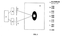
超広帯域放射線 本発明の実施形態の中で使用され得る電磁波の1つの形式は、超広帯域(UWB)放射線である。しかしながら、本発明の範囲はUWBに制限されず、他の形式の電磁波、例えば離散的な周波数帯をも使用することができる。UWBの回路や電源、送信の要求事項は無線電子工学の当業者にとっては周知なものである。測定装置用コンポーネントの1つの代表を図1に示している。図1に示すように、制御要素10は、電磁波信号の開始、及び電磁信号の反射から測定データ・セットへの変換を制御する役割をする。制御要素10は、伝送アンテナ18へ入力信号を送信する伝送回路16を制御する。制御要素10は、周波数、持続時間、電力レベル及び他のパラメーターを含む、送信されたUWB信号の種々のパラメーターを制御することもできる。送信回路16は、身体22内の測定部位20へUWB信号を増幅して送信する伝送アンテナ18に電気的に接続される。測定部位20は任意の身体内における任意の注目の一般的な箇所であり得る。送信された信号19は、測定部位20から反射され、その後、反射信号21が受信アンテナ14及び受信回路12によって受信されるように示されている。制御要素はさらに受信回路12を制御する。
measuring device
Ultra-wideband radiation One type of electromagnetic wave that can be used in embodiments of the present invention is ultra-wideband (UWB) radiation. However, the scope of the present invention is not limited to UWB, and other types of electromagnetic waves, such as discrete frequency bands, can be used. The UWB circuit, power supply, and transmission requirements are well known to those skilled in the radio electronics arts. One representative of measuring device components is shown in FIG. As shown in FIG. 1, the control element 10 serves to control the start of the electromagnetic wave signal and the conversion of the electromagnetic signal reflection to the measurement data set. The control element 10 controls the transmission circuit 16 that transmits an input signal to the transmission antenna 18. The control element 10 can also control various parameters of the transmitted UWB signal, including frequency, duration, power level and other parameters. The transmission circuit 16 is electrically connected to a transmission antenna 18 that amplifies and transmits the UWB signal to the measurement site 20 in the body 22. The measurement site 20 can be any point of interest in any body. The transmitted signal 19 is reflected from the measurement site 20, after which the reflected signal 21 is shown to be received by the receiving antenna 14 and the receiving circuit 12. The control element further controls the receiving circuit 12.
図1に示した例では、送信された信号19は、測定部位20から反射されるように示されている。しかしながら、ある場合には、送信された信号19は、測定部位20を通り抜けて、別の部位、例えば骨にて反射される場合がある。さらに、複数の反射が組織の複数の層によって反射された場合、複数の反射信号21は、受信アンテナ14によって受信されることができる。以前の時点で集められたデータの中にはなかった、ある時点での新しい反射信号の存在は、探索部位20内の新しい構造の指標であり得る。 In the example shown in FIG. 1, the transmitted signal 19 is shown to be reflected from the measurement site 20. However, in some cases, the transmitted signal 19 may pass through the measurement site 20 and be reflected at another site, such as bone. Further, if the multiple reflections are reflected by multiple layers of tissue, the multiple reflected signals 21 can be received by the receive antenna 14. The presence of a new reflected signal at a point in time that was not in the data collected at the previous point in time may be an indication of a new structure within the search site 20.
送信回路16及び受信回路12の制御に加えて、制御素子10は、さらに測定部位の変化を決定するためのコンパレーターを含んでいる。上記信号の送信及び受信のための、対応する信号送信及び受信回路16、12、並びに送信及び受信アンテナ18、14もそれぞれ示されている。本発明の方法の実施においてはコンポーネントの様々な配置、例えば1回路モジュール内の結合した機能、あるいは一個の送信/受信アンテナの使用が採用され得る。また、本発明の方法は何れの実施方法にも制限されない。同様に、本発明の一部の実施形態では、複数の装置、或いはコンポーネント、例えば送信機及び/又は受信機が、測定されている部位の追加情報を提供するために使用され得る。 In addition to the control of the transmission circuit 16 and the reception circuit 12, the control element 10 further includes a comparator for determining the change of the measurement site. Corresponding signal transmission and reception circuits 16 and 12 and transmission and reception antennas 18 and 14 for the transmission and reception of the signals are also shown, respectively. Various implementations of components may be employed in the implementation of the method of the present invention, such as the combined functionality within a circuit module, or the use of a single transmit / receive antenna. Further, the method of the present invention is not limited to any implementation method. Similarly, in some embodiments of the invention, multiple devices, or components, such as transmitters and / or receivers, can be used to provide additional information about the site being measured.
UWBシステムは、従来のシステムよりはるかに広い周波数帯の信号を送信する。UWB信号によって占められたスペクトルの量、即ちUWB信号の帯域幅は中心周波数の約25%以上であることができる。したがって、2GHzを中心とするUWB信号は約500MHzの帯域幅を有しうる。また、4GHzを中心とするUWB信号の帯域幅は約1GHzでありえる。UWB信号を生成する最も一般的な技術は1ナノセカンド(ナノ秒、ns)未満の持続時間のパルスを送信することである。 UWB systems transmit signals in a much wider frequency band than conventional systems. The amount of spectrum occupied by the UWB signal, ie the bandwidth of the UWB signal, can be about 25% or more of the center frequency. Thus, a UWB signal centered at 2 GHz may have a bandwidth of about 500 MHz. Also, the bandwidth of UWB signals centered on 4 GHz can be about 1 GHz. The most common technique for generating UWB signals is to transmit pulses of duration less than 1 nanosecond (nanosecond, ns).
UWBシステムを用いて、非侵襲的で、皮膚の表面に触れることなしで、身体の内部臓器の全体組織を評価することができる。本発明の1つの好ましい形態では、1つ以上のUWB測定装置が、身体内に完全に埋め込まれる。UWBは短いパルス及び反射を使用し、また、身体の内部の異なる層からのこれらのパルスの反射信号の強さ及び/又はこれらのパルスの信号の飛行時間の遅延は、身体内の解剖学的構造、それらの場所、大きさ、誘電性の組成、及びそれらの移動に関するデータを提供する。UWBのいくつかの代表的な応用は、心臓壁移動のような器官移動あるいはその寸法変化、心臓壁厚、腎臓/肝臓/胃などの測定、寸法変化、呼吸、及び/又は密度変化の測定、並びに産科学での応用がある。 The UWB system can be used to evaluate the entire tissue of the body's internal organs, non-invasively and without touching the surface of the skin. In one preferred form of the invention, one or more UWB measurement devices are completely implanted in the body. UWB uses short pulses and reflections, and the strength of the reflected signals of these pulses from different layers inside the body and / or the time-of-flight delay of the signals of these pulses can be anatomical in the body. Provides data on structures, their location, size, dielectric composition, and their movement. Some typical applications of UWB include organ movements such as heart wall movement or its dimensional changes, heart wall thickness, kidney / liver / stomach etc. measurements, dimensional changes, respiration, and / or density change measurements, There are also applications in obstetrics.
超音波のような他の技術に比較してUWBの主な利点は、下記である:
・音波に比較して、空気を介する伝送にあまり影響されないので、UWB信号は皮膚に接する必要はない。
The main advantages of UWB compared to other technologies such as ultrasound are:
• Compared to sound waves, UWB signals do not need to touch the skin because they are less affected by transmission through the air.
・UWB信号は骨の中で減衰しないので、脳のような骨によってカバーされた窩洞の内部の情報を得ることができる。 -Since the UWB signal is not attenuated in the bone, information inside the cavity covered by bones like the brain can be obtained.
・UWB信号は布、寝具、保護服あるいは防弾服のような非導体材料を通過してデータを集めるために使用されることができる。 UWB signals can be used to collect data through non-conductive materials such as cloth, bedding, protective clothing or bulletproof clothing.
・UWB信号は少数の安いコンポーネントで実現されることができるので、低消費電力、ポータブル適用を可能にする。 The UWB signal can be realized with a few cheap components, thus enabling low power consumption and portable applications.
上に言及されたように、UWB測定システムは組織層からの反射信号の検討により組織の構造の変化を検知する。例えば、送信機はUWBパルスを送り、次に、組織の異なる層から反射パルスを受け取る。UWBレーダーの速度が異なるタイプの組織において異なり(例えば、信号伝播は筋肉より脂肪においておよそ2.25倍速い)、また組織の層が異なる深さにあるので、複数の組織層からの反射は異なる時点で受信機に届き、また異なる振幅を持っている。さらに、組織を通過するときの信号の減衰は、組織の密度情報を与える。例えば、図2に示されるように、骨を含む手足の隣に配置された時、UWBレーダー測定システム100は、基線評価の際の対応するパラメーターに関して、受信パルスの時間遅れ及び振幅を測定することにより、様々な層の中の液体蓄積の変化を評価することができる。図2に示される層は表皮層1、真皮層2、脂肪及び結合組織層3、筋肉層4及び筋膜の層5を含んでいる。他の層も評価されることができる。
As mentioned above, the UWB measurement system detects changes in the structure of the tissue by examining the reflected signal from the tissue layer. For example, the transmitter sends UWB pulses and then receives reflected pulses from different layers of tissue. UWB radar speed is different in different types of tissues (eg, signal propagation is about 2.25 times faster in fat than muscle), and because layers of tissue are at different depths, reflections from multiple tissue layers are different It reaches the receiver at the moment and has a different amplitude. Furthermore, the attenuation of the signal as it passes through the tissue gives tissue density information. For example, as shown in FIG. 2, when placed next to a limb including a bone, the UWB radar measurement system 100 measures the time delay and amplitude of the received pulse with respect to the corresponding parameters in the baseline evaluation. This makes it possible to evaluate changes in liquid accumulation in the various layers. The layers shown in FIG. 2 include an
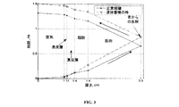
図3は、UWBパルスが組織を通過し、また、例えば組織内の液体蓄積が反射パルスの振幅及び遅延に影響する様子を例示している。これは、液体(例えば水)が筋肉と脂肪よりはるかに高い誘電率があって、液体がある状態でのパルスの緩徐化をもたらすためである。さらに、生理食塩水中のより高い減衰により、反射波に振幅変化がある。この単純モデルの生成に、脂肪と筋肉の性質は、窩洞の中への液体の流入が増えるにつれて食塩水の性質に近づくことが仮定されている。本発明の所定の実施形態では、身体部位内の水和変化の程度は、送信アンテナから少なくとも1つの受信アンテナまでの信号の送信あるいは反射におけるUWB信号の変化、例えば減衰の程度によって評価される。 FIG. 3 illustrates how a UWB pulse passes through tissue and, for example, liquid accumulation in the tissue affects the amplitude and delay of the reflected pulse. This is because liquid (eg water) has a much higher dielectric constant than muscle and fat, resulting in slowing of the pulse in the presence of liquid. Furthermore, there is an amplitude change in the reflected wave due to higher attenuation in the saline. In creating this simple model, it is assumed that fat and muscle properties approach that of saline as the inflow of liquid into the cavity is increased. In certain embodiments of the present invention, the degree of hydration change in the body part is assessed by the change in UWB signal in the transmission or reflection of a signal from the transmitting antenna to at least one receiving antenna, eg, the degree of attenuation.
図3に示しているように、最初の基線測定が行われ、且つ、送信された信号34A及び反射された信号34Bとして描かれている。送信された信号34Aは、(水平軸上の0.0の深さとして示されている)皮膚の1cm上の位置からスタートする。信号34Aは、1.0cmから約1.1cmまでの表皮層(skin layer)、約1.1から約1.4cmまで真皮層(sub-dermal layer)、約1.4cmから約1.9cmまでの脂肪層、及び約1.9cmから約3.9cmまでの筋肉層を通過するように示されている。また、送信された信号34Aは、骨の層で反射されて基線反射信号34Bをきたすように示されている。その後、反射された信号34Bは、他の層を通過し、各層の中の物質のタイプに基づいた様々な遅延及び減衰を受ける。図3はたった1つの反射信号を示しているが、これは明瞭さの目的のためにそうなされたものであることに注意されるべきである。熟練した技術者は、複数の反射信号が受信され分析され得ることを認識するであろう。
As shown in FIG. 3, an initial baseline measurement is made and depicted as transmitted signal 34A and reflected signal 34B. The transmitted signal 34A starts from a
基線反射信号34Bは、送信から受信までに異なる層を進むために約1.3nsの飛行時間を有する。その後、第2の測定がなされ、且つ送信された信号32Aは、後続の測定のために同じ部位に向けられる。そして、送信された信号32Aは、骨の層から反射され、測定システム100によって受信される。この第2の測定では、送信された信号32A及び反射された信号32Bは、基線送信信号34A及び基線反射信号34Bに比較してかなり遅延されている。この例では、往復の飛行時間は、基線信号34A及び34Bの場合の1.3nsと比較して、信号32A及び32Bの場合では約1.5nsである。上に議論されたように、これは、様々な層におけるより多くの水分及び/又は塩水の存在に起因するかもしれない。信号の遅延に加えて、信号の振幅も異なり得る(図3に示されていない)。これも分析されて、上に議論されたように食塩水のような可能な減衰ソースを特定することができる。 The baseline reflected signal 34B has a flight time of about 1.3 ns to travel through different layers from transmission to reception. Thereafter, a second measurement is made and the transmitted signal 32A is directed to the same site for subsequent measurements. The transmitted signal 32A is then reflected from the bone layer and received by the measurement system 100. In this second measurement, the transmitted signal 32A and the reflected signal 32B are significantly delayed compared to the baseline transmit signal 34A and the baseline reflected signal 34B. In this example, the round trip flight time is about 1.5 ns for signals 32A and 32B, compared to 1.3 ns for baseline signals 34A and 34B. As discussed above, this may be due to the presence of more moisture and / or salt water in the various layers. In addition to the signal delay, the signal amplitude can also be different (not shown in FIG. 3). This can also be analyzed to identify possible attenuation sources such as saline as discussed above.
上に議論されたように、実施形態によっては、別の信号形式も測定システム100の中で使用され得る。組織の構造及び/又は内容を評価するために、測定システム100によって使用され得る他の信号形式は以下に議論する。 As discussed above, other signal formats may also be used in measurement system 100 in some embodiments. Other signal formats that can be used by the measurement system 100 to evaluate the structure and / or content of the tissue are discussed below.
高周波電磁放射線: テラヘルツ放射線、あるいはそれより高い放射線、例えば赤外線のような高周波電磁放射線は、表面的な(superficial)組織の構造及び内容の変化をモニターするために潜在的に使用されることができる。これは、組織表面からの反射された放射線の振幅のモニタリングによって達成され得る。構造または内容(浮腫)のどんな変化も、組織内の水分による吸収のために検知されることができる。 High frequency electromagnetic radiation: Terahertz radiation or higher, such as high frequency electromagnetic radiation such as infrared, can potentially be used to monitor changes in the structure and content of superficial tissue . This can be achieved by monitoring the amplitude of the reflected radiation from the tissue surface. Any change in structure or content (edema) can be detected due to absorption by moisture in the tissue.
音響放射: 音波は機械的波動であるが、組織の構造及び内容の変化を検知するために同様の波動伝搬法則を使用しうる。任意の物質中の音波の速度は、温度、物質の弾性特性及び物質の密度と関係がある。したがって、性質の変化によって反映される、生理または形態の変化による組織の要素の任意の変化が、音波の使用により検知されることができる。1つの実施形態では、本発明は、約1〜100kHzの範囲内の周波数を含む音波(高帯域の短パルス、あるいは、”チャープ(Chirp)”のような多数の周波数を含む高帯域長時間信号)を組織に集中させ、組織へ送信され、且つ組織から反射された音波をモニタリングするように構成された装置を含む。適用のタイプによって、3つの異なる解析方法、即ち、a)組織を通り抜ける間の波動減衰、b)反射波の減衰、及び、c)送信された波と骨及び組織の間でのような境界の反射の後の反射波との間の位相差の解析が可能である。さらに、音速が温度とともに変化するので、炎症あるいは感染反応による組織の内部の任意の温度変化も追跡されうる。 Acoustic radiation: Sound waves are mechanical waves, but similar wave propagation laws can be used to detect changes in tissue structure and content. The speed of sound waves in any material is related to temperature, the elastic properties of the material and the density of the material. Thus, any changes in tissue elements due to changes in physiology or morphology, reflected by changes in properties, can be detected through the use of sound waves. In one embodiment, the present invention provides a sound wave containing a frequency in the range of about 1-100 kHz (a high-band short pulse, or a high-band long-time signal containing multiple frequencies such as “Chirp”). ) To the tissue, and includes a device configured to monitor sound waves transmitted to the tissue and reflected from the tissue. Depending on the type of application, there are three different analysis methods: a) wave attenuation while passing through the tissue, b) attenuation of the reflected wave, and c) boundary such as between the transmitted wave and bone and tissue. Analysis of the phase difference between the reflected wave and the reflected wave is possible. Furthermore, since the speed of sound changes with temperature, any temperature change inside the tissue due to inflammation or infection reaction can be tracked.
身体の1つ以上の部位に音響信号を印加する方法及び装置は、音響信号の生成、及びソノグラフによる音波の解析を含む音響信号の解釈の分野の当業者には周知なものである。本発明の特定の実施形態では、音響センサーは身体内に埋め込まれ、それにより、身体と音響測定装置とのよい接触を確保するために必要な音響カップリング手段、例えばゲル剤を回避する。 Methods and apparatus for applying an acoustic signal to one or more parts of the body are well known to those skilled in the art of acoustic signal generation, including acoustic signal generation and sonographic sound wave analysis. In a particular embodiment of the invention, the acoustic sensor is embedded in the body, thereby avoiding the necessary acoustic coupling means, eg gels, to ensure good contact between the body and the acoustic measuring device.
生体電気インピーダンス信号: 本発明の別の実施形態では、前に示したように、注目の組織の生体電気インピーダンスは、組織の構造及び内容の変化をモニタリングするために使用されることができる。これは、注目の組織を通して電流を流し、組織での電圧降下を測定し(あるいは正弦波電圧で組織を刺激して組織を流れる電流を測定し)、そして、電流フローへの組織のインピーダンスを計算することにより達成される。測定された信号の振幅の変化、及び、電圧信号と電流信号との間の位相差は、組織の特性に依存する。(例えば瘢痕組織成長、腫瘍などにより)組織の構造が変化し、及び/又は(例えば浮腫、脂肪/筋肉比率などにより)組織の内容が変化すると、インピーダンスの振幅及び電流または電圧に関する位相は変化し、且つモニタリングされることができる。さらに、DC信号も組織の抵抗の変化を得るために使用されうる。バイオ電気インピーダンスについては、エネルギーの集中(あるいは、測定の体積の制御)は、電流の駆動及び電圧の測定のために使用される電極の幾何学の変更により達成され得る。身体の1つ以上の部位に電気的信号を送出して受信する、必要な電極及び回路類を含む方法及び装置は、生体電気インピーダンスの分野の当業者にとって周知的なものである。 Bioelectrical impedance signal: In another embodiment of the invention, as previously indicated, the bioelectrical impedance of the tissue of interest can be used to monitor changes in tissue structure and content. It conducts current through the tissue of interest, measures the voltage drop across the tissue (or measures the current flowing through the tissue by stimulating the tissue with a sinusoidal voltage), and calculates the tissue impedance to current flow Is achieved. Changes in the amplitude of the measured signal and the phase difference between the voltage signal and the current signal depend on the properties of the tissue. As the structure of the tissue changes (eg, due to scar tissue growth, tumors, etc.) and / or the tissue content changes (eg, due to edema, fat / muscle ratio, etc.), the impedance amplitude and phase with respect to current or voltage change. And can be monitored. In addition, DC signals can also be used to obtain changes in tissue resistance. For bioelectrical impedance, energy concentration (or control of measurement volume) can be achieved by changing the geometry of the electrodes used for current drive and voltage measurement. Methods and devices, including the necessary electrodes and circuitry, that send and receive electrical signals to one or more parts of the body are well known to those skilled in the field of bioelectrical impedance.
本発明の特定の実施形態では、電気的信号を供給し、且つ測定する電極は、注目の身体部位に完全に埋め込まれ、また、1セット以上の電極間のインピーダンスが変化する。そのようなインピーダンス測定は、2ポイントの電極配置(例えば、電流電極が測定電極と同じである場合)、4ポイントの電極配置(例えば、電流電極が測定電極とは異なる場合)、あるいは2ポイント配置及び4ポイント配置の組み合わせを使用することができる。さらに、複数の電極は複数の方向の電気インピーダンスのベクトルの評価を可能にするために使用され得る。 In certain embodiments of the invention, the electrodes that supply and measure the electrical signal are completely implanted in the body part of interest, and the impedance between one or more sets of electrodes changes. Such an impedance measurement can be a two-point electrode arrangement (eg, if the current electrode is the same as the measurement electrode), a four-point electrode arrangement (eg, if the current electrode is different from the measurement electrode), or a two-point arrangement. And a combination of 4 point arrangements can be used. Furthermore, multiple electrodes can be used to allow evaluation of vectors of electrical impedance in multiple directions.
一般的な使用: 任意の周波数のエネルギー・パルスあるいは信号の測定及び保存は、測定の時間及び/又は測定の位置への参照を含みうるデータ・セットを含む。そのような測定と保存の活動は、生データの変換を含みうる。そのような変換は、データ・セットの保存の促進、例えばデータ圧縮を可能にするのに有用であり、あるいは、データ・セットの送信及び/又はコンパレーターによる分析を促進する。コンパレーター用の信号は、適用された1つ以上のエネルギーソース、例えば高周波、音響的、電気的または光学的ソースによるものであり得る。さらに、これらの信号は、長い期間をかけて様々な組み合わせで利用されて、身体部位または組織のダイナミックな変化に対するより大きな洞察を提供することができる。例えば、疑わしい腫瘍の検査は、異なる密度の組織の存在を検知する高周波エネルギーの形式を使用することによって何よりも先に示される場合がある。後の観察は、そのサイトのまわりの増加した腫れ、血流あるいは浮腫を測定するインピーダンス測定を含み、ある期間にわたる変更の傾向分析をより正確に提供することが可能である。 General use: Measurement and storage of energy pulses or signals of any frequency include a data set that may include a reference to the time of measurement and / or the location of the measurement. Such measurement and storage activities can include conversion of raw data. Such a conversion is useful for facilitating storage of the data set, for example allowing data compression, or facilitates transmission of the data set and / or analysis by a comparator. The signal for the comparator may be from one or more applied energy sources, such as a high frequency, acoustic, electrical or optical source. Furthermore, these signals can be utilized in various combinations over time to provide greater insight into dynamic changes in body parts or tissues. For example, examination of a suspicious tumor may be indicated earlier by using a form of radio frequency energy that detects the presence of different density tissue. Later observations include impedance measurements that measure increased swelling, blood flow, or edema around the site, and can provide a more accurate trend analysis of changes over time.
本発明のいくつかの実施形態では、標準のマーカー補助手段、信号配列及び/又は信号改善の補助手段を使用する場合がある。これらの補助手段は、電磁気信号あるいは他の技術、例えばMRIを使用した身体部位のマッピングの利用を含み、データ・セット内の基準点を確立し、或いは、身体に測定装置をより正確に位置させる補助を提供する場合がある。これらの基準点の利用は、データ・セットのアライメントを改善して、標的部位の変化がより正確に決定されることを可能にする。代替的形態では、これらの補助手段は、哺乳類の身体上に付着され、またその中に埋め込まれた受動若しくは能動の装置の使用を含みうる。これらの補助器具は、基準信号を提供するか、あるいはそうでなければ、測定装置及び/又はデータ・セットをターゲットとするために目印として役立つことができる。信頼できる補助手段は、限定されないが、下記のようなものを含みうる。即ち、光学的アライメントの補助手段、例えば入れ墨、信号反射の補助手段、例えば埋め込まれた金属反射体または導電性インク、誘導的に充電された埋め込みの高周波トランシーバー、あるいは埋め込みの音響発信機。そのような補助手段は、後のコンパレーター活動を支援するために装着位置、例えば信号のアライメントの解釈及び信号の複雑さの両方を改善するように、3次元空間を介する既知の方法で幾何学模様(例えば斜交平行模様)に配置されることができる。 In some embodiments of the present invention, standard marker aids, signal alignment and / or signal improvement aids may be used. These auxiliary means include the use of electromagnetic signals or other techniques such as mapping of body parts using MRI to establish a reference point in the data set or to position the measuring device more accurately on the body. May provide assistance. The use of these reference points improves the alignment of the data set and allows target site changes to be determined more accurately. In an alternative form, these auxiliary means may involve the use of passive or active devices attached to and embedded in the mammalian body. These ancillary instruments can provide reference signals or otherwise serve as landmarks for targeting the measuring device and / or data set. Reliable auxiliary means may include, but are not limited to: That is, optical alignment auxiliary means such as tattoos, signal reflection auxiliary means such as embedded metal reflectors or conductive ink, inductively charged embedded high frequency transceivers, or embedded acoustic transmitters. Such an auxiliary means is known in a known manner through three-dimensional space to improve both the mounting position, eg interpretation of signal alignment and signal complexity, to support later comparator activity. It can be arranged in a pattern (for example, an oblique parallel pattern).
本発明の特定の別の関連する実施形態では、検査される部位に関して埋め込まれ、又は位置させられた材料を利用して測定過程を支援することができる。例えば、これらの材料は、関心領域を通るように電気波/電磁気波/音波を集中させるために利用され、あるいは関心領域の後ろの高反射性の目標として役立ち、それによって、印加された電気波/電磁気波/音波の有効信号の強度を高めることができる。 In certain other related embodiments of the present invention, materials that are implanted or positioned with respect to the site being examined can be utilized to assist the measurement process. For example, these materials can be used to focus electrical / electromagnetic / acoustic waves through the region of interest, or serve as highly reflective targets behind the region of interest, thereby applying an applied electrical wave The strength of the effective signal of / electromagnetic wave / sound wave can be increased.
本発明の別の実施形態では、測定装置の送信機要素は識別子を割り当てられる場合がある。そのような識別子は、アンテナ幾何学及び伝送周波数を決定する能力を含み得る。同様に、電子回路の別の部分は、回路の残りの構成要素に追加の識別子を割り当てる場合がある。そのような識別子は、装置内の使い捨てで再使用可能なアッセンブリの構築を可能にするのに役立ち、該アッセンブリのトラッキングを可能にすることができる。さらに、そのような識別子は、データ・セットを管理し、測定される個々の被検物のためのコンパレーターを見つけるように対応付けする際に装置の使用及び装置の形式の特定を可能にする。さらに、そのような識別子は、安全な情報伝送及び測定データ・セットの割り当て/表示のために、暗号鍵の割り当て、あるいは、別のニーズにおいて有用でありえる。 In another embodiment of the invention, the transmitter element of the measuring device may be assigned an identifier. Such an identifier may include the ability to determine antenna geometry and transmission frequency. Similarly, another part of the electronic circuit may assign additional identifiers to the remaining components of the circuit. Such an identifier helps to allow the construction of a disposable and reusable assembly in the device and can allow tracking of the assembly. In addition, such identifiers allow the use of the device and the identification of the device type in managing the data set and associating it to find a comparator for the individual analyte being measured. . Further, such identifiers may be useful in cryptographic key assignments or other needs for secure information transmission and measurement data set assignment / display.
コンパレーター 図1の制御要素10のコンパレーター・サブシステムは、保存手段、及びデータ・セット間の変化を決定する手段の両方を含む。これらの決定は、入力閾値及び、基線値からの変化によって決定された閾値設定ポイント(或いは、基線値を示す1つ以上のデータポイントの代表)の使用を含みうる。代替的に、そのようなコンパレーター機能は、データ・セットにおける傾向を決定し、また1日の内に異なる時点で得られたデータのための調整、例えば日変化の調整を可能にするために平均のロールアウト又は移動の使用を含みうる。また、コンパレーター活動の別の形式は、初期の基線値の決定及びデータ・セットまたはグループの傾向の決定のために、データの母集団(population)及び集団(group)を調査する場合がある。そのようなコンパレーター活動の結果は、図式的に表示されてもよい。例えば将来動向の予測を含む、基線値、及びある期間において基線値からの相対変化を示してもよい。 Comparator The comparator subsystem of the control element 10 of FIG. 1 includes both storage means and means for determining changes between data sets. These determinations can include the use of an input threshold and a threshold set point determined by a change from the baseline value (or a representative of one or more data points that represent the baseline value). Alternatively, such a comparator function can determine trends in the data set and also allow adjustments for data obtained at different times within a day, eg adjustments for daily changes It may include the use of average rollout or movement. Another form of comparator activity may also examine populations and groups of data for initial baseline value determination and data set or group trend determination. The result of such comparator activity may be displayed graphically. For example, a baseline value including prediction of future trends, and a relative change from the baseline value in a certain period may be indicated.
さらに、コンパレーター・サブシステムは、例えば体重、身長、年齢、性別、疾病状態及び薬歴、フィットネス・レベル、装置適用の身体部位、民族性などの入力パラメーター、あるいは、変化及び/又はそのような変化の大きさの一層の定義を可能にするアルゴリズム研究により得られたパラメーターのような別の要因を組込むことがある。そのようなパラメーターは、イベント毎に、あるいは周期的に、例えば、毎日、被検査ユーザーあるいは臨床医の変化に対する認識を、測定されたバイオパラメーターに関連づける因子を含みうる。 In addition, the comparator subsystem may include input parameters such as weight, height, age, gender, disease state and medication history, fitness level, device application body part, ethnicity, etc., and / or changes and / or such Other factors may be incorporated, such as parameters obtained from algorithmic studies that allow further definition of the magnitude of change. Such parameters may include factors that relate perceptions of changes in the user or clinician being tested to the measured bioparameters at each event or periodically, eg, daily.
本発明のまた別の実施形態では、コンパレーター・サブシステムは、ホルモンまたは代謝産物の循環レベル、活動の大きさのような測定された別のバイオパラメーターから得られたデータ、あるいは、環境センサーから得られたデータ、例えば相対的な湿度、周囲温度などを含む他の因子を含みうる。本発明は、これらの因子及び別の因子の組み合わせを採用することができる。また、本発明の範囲はここに記述されている因子及び数学的なルーチンに制限されない。 In yet another embodiment of the present invention, the comparator subsystem may include data obtained from other measured bioparameters such as circulating levels of hormones or metabolites, magnitude of activity, or from environmental sensors. Other factors may be included including the data obtained, eg, relative humidity, ambient temperature, and the like. The present invention can employ a combination of these factors and other factors. Also, the scope of the invention is not limited to the factors and mathematical routines described herein.
本発明の1つ以上の実施形態では、コンパレーターは、1つ以上のセンサーを使用する1つ以上の装置からの測定データを分析し、受信データからの雑音、体動アーチファクトあるいは別の非希望の因子を除去して、測定部位の変化の決定を可能にすることができる。そのような測定データは、ある期間、例えば数秒、数分あるいは数日の間に、あるいは1つ以上の身体部位から集められたデータを含みうる。そのような分析は、短時間の雑音因子、例えば身体活動に関連した動き、あるいは長期傾向、例えば習慣的な(摂食)雑音もしくは、日周シフトを1つ以上のパラメーターから除去することができる。関連する実施形態では、雑音及び/又は予測可能な因子、例えば食事あるいは他の予測可能な活動に関連したデータの除去を支援するために、測定は時間的に規則的もしくは不規則的に行われうる。さらに、雑音を低減し、及び/又は1つ以上の測定装置の電源消費を最適化するために、システムは、パターン解析を通じて学習するか、あるいはプログラムされることにより測定の回数及び周波数を調節することができる。 In one or more embodiments of the present invention, the comparator analyzes measured data from one or more devices that use one or more sensors to detect noise, body motion artifacts or other undesired from the received data. This factor can be removed to allow determination of changes in the measurement site. Such measurement data may include data collected over a period of time, eg, seconds, minutes or days, or from one or more body parts. Such an analysis can remove short-term noise factors, such as movements associated with physical activity, or long-term trends, such as habitual (feeding) noise or diurnal shifts, from one or more parameters. . In related embodiments, measurements are taken regularly or irregularly in time to assist in the removal of data related to noise and / or predictable factors such as meals or other predictable activities. sell. Further, in order to reduce noise and / or optimize the power consumption of one or more measurement devices, the system adjusts the number and frequency of measurements by learning or programmed through pattern analysis. be able to.
特定の実施形態では、1つのセンシング手段、例えばUWBからの測定データは、第2のセンシング手段、例えばインピーダンスからの測定データと共に使用されることができる。そのような使用は、第1のデータ・セットを使用して、第2のデータ・セットの較正または調整を支援し、あるいは、追加の因子を提供して、これによりコンパレーターが可能な変化を評価できるようにする。 In a particular embodiment, measurement data from one sensing means, eg UWB, can be used together with measurement data from a second sensing means, eg impedance. Such use uses the first data set to assist in the calibration or adjustment of the second data set, or provides additional factors that allow the comparator to make possible changes. Enable evaluation.
コンパレーター・サブシステムは様々な場所に存在してもよい。本発明の1つの実施形態では、コンパレーターはデータ・セットを得るのに必要な回路内に一部分あるいは全体が含まれ得る。本発明の別の実施形態では、コンパレーターは、センサー及び/又はセンサー回路、例えばトランシーバーに配線で接続された別個のユニットに配置されることができる。そのような実施形態では、センサーまたはセンサー回路は、コンパレーター活動の一部分あるいは全体を含むユニットとは異なる別個の識別を持つことができる。本発明のさらなる別の実施形態では、コンパレーター活動の場所は、データ分析を促進するために、例えばより複雑な及び/又はより大きなデータ・セットを収容するために、あるいは他の目的、例えば装置、データ収集ユニットなどの電源管理のために、変わることができる。 The comparator subsystem may exist in various locations. In one embodiment of the present invention, the comparator may be included in part or in whole in the circuitry necessary to obtain the data set. In another embodiment of the present invention, the comparator can be placed in a separate unit that is wired to a sensor and / or sensor circuit, eg, a transceiver. In such an embodiment, the sensor or sensor circuit may have a distinct identification that is different from the unit that includes part or all of the comparator activity. In yet another embodiment of the present invention, the location of the comparator activity may be used to facilitate data analysis, eg, to accommodate more complex and / or larger data sets, or for other purposes, eg, devices. It can vary for power management, such as data collection unit.
本発明のまた別の実施形態では、1つ以上の測定されたバイオパラメーターの測定値あるいは数学変換、例えば平均或いは変化率は、必ずしも身体に配置されるとは限らない別個のユニットへ無線手段を通して送信される。この別個のユニットは、コンパレーター活動を行なうために、適切な要素及び回路、例えば伝送手段、データ保存及び数学計算機能及びルーチンを部分的あるいは完全に含みうる。コンパレーターのさらなる別の形式及び場所も容易に考えられる。本発明の範囲はここに記述されたものに制限されない。 In yet another embodiment of the present invention, the measured value or mathematical transformation of one or more measured bioparameters, such as the average or rate of change, through wireless means to a separate unit that is not necessarily placed in the body. Sent. This separate unit may partially or completely include suitable elements and circuits, such as transmission means, data storage and mathematical functions and routines, to perform comparator activities. Still other types and locations of comparators are readily contemplated. The scope of the invention is not limited to that described herein.
測定部位の状態変化の決定をし、及びそのようなイベントの大きさの可能な決定をし次第、コンパレーターは、被検者、介護者あるいは第三者の個人にそのようなイベントを表示するように命じられることができる。任意の変化の表示は、さらに、データ・セットの一部あるいはすべてと比較して、変化なしの通知を含んでもよい。そのような表示は、視覚的な表示、例えば2次元及び3次元の表示を含む部位の解剖図、明滅する明かりあるいは多色の明かり、数値の指標、グラフ、チャート、可聴音、或いは機械的な形式、例えば振動を含んでもよい。そのような表示は、装着型(on-body)の測定装置、ローカルデータ収集ユニット、あるいは無線あるいは有線の手段によって装着型の測定装置かローカルデータ収集ユニットのいずれかに接続された遠隔地のものに配置されることができる。 As soon as the determination of the state change of the measurement site and the possible determination of the magnitude of such an event, the comparator displays such an event to the subject, caregiver or third party individual Can be commanded as. The indication of any change may further include a notification of no change compared to some or all of the data set. Such a display can be a visual display, such as an anatomical view of a site including 2D and 3D displays, flickering or multicolored lights, numerical indicators, graphs, charts, audible sounds, or mechanical It may include form, for example vibration. Such indications may be on-body measurement devices, local data collection units, or remote locations connected to either the wearable measurement device or the local data collection unit by wireless or wired means. Can be arranged.
変化を表示する可能性に加えて、コンパレーターは、日付/時間、大きさ及びユーザー識別を含むイベント記述をデータ保存手段に保存してもよい。そのようなデータ保存手段は電子メモリ、磁気テープ若しくはディスクメモリ、光メモリ、あるいは回収可能なメモリの他の形式を含んでもよい。そのようなデータは、保存手段からコマンドによって検索され、又は周期的に検索されることができる。特定の実施形態では、そのような検索は、無線手段、例えば赤外線若しくは無線周波数ベースのデータ伝送を通じて行われ、あるいは、別の実施形態では、そのような検索は、有線の手段、例えば、コンピューターに取り付けられたドッキング・ステーション又はコンピューターに接続されたシリアル・ケーブルを通じて行われる。 In addition to the possibility to display the change, the comparator may store an event description including date / time, magnitude and user identification in the data storage means. Such data storage means may include electronic memory, magnetic tape or disk memory, optical memory, or other forms of recoverable memory. Such data can be retrieved by command from the storage means or retrieved periodically. In certain embodiments, such searches are performed through wireless means, such as infrared or radio frequency based data transmission, or, in another embodiment, such searches are performed on wired means, such as a computer. This is done through a serial cable connected to an attached docking station or computer.
閉ループ治療管理に関わる装置の実施形態では、コンパレーターはある作用メカニズム(Effecting Mechanism)と通信する。この作用メカニズムは、治療プロトコルを開始させ、又は既存の治療プロトコルを制御し、あるいはあらかじめ指定された一連の動作を開始させる。特定の実施形態では、作用メカニズムは、マイクロプロセッサーにプログラムされた1セットの計算機命令を含む。この作用メカニズムは、センシング・メカニズムとリソースを共有し、あるいは、別個の/付加的なリソースを有することができる。例えば、組織の構造及び内容の変化をセンシングする生体電気インピーダンス方法を使用する場合、及び治療として電気刺激を使用する場合においては、センシングと刺激との電極は同じでありえる。センシング・メカニズム及び作用メカニズムが同様のエネルギー形式(電気的、電磁的、聴覚的、化学的、視覚的など)を使用する場合、同様の配置は採用されうる。 In an embodiment of the device involved in closed loop therapy management, the comparator communicates with an effecting mechanism. This mechanism of action initiates a treatment protocol or controls an existing treatment protocol or initiates a pre-designated sequence of actions. In certain embodiments, the mechanism of action includes a set of computer instructions programmed into the microprocessor. This mechanism of action can share resources with the sensing mechanism or have separate / additional resources. For example, the sensing and stimulation electrodes can be the same when using a bioelectrical impedance method of sensing changes in tissue structure and content, and when using electrical stimulation as a treatment. Similar arrangements can be employed when the sensing and action mechanisms use similar energy types (electrical, electromagnetic, auditory, chemical, visual, etc.).
特定の適用では、センサー・エネルギー、例えば、UWBあるいは音響エネルギーは、別の装置及び/又はコンパレーター回路/ロジックと情報を無線で送信するために利用され得る。これは、同じ主回路がセンシング及び送信の両方に使用されることにより、コンポーネントの数、装置の全体的なサイズを低減し、コストを最小限にすることができるので、本発明の特定の実施形態において有利である。そのような適用では、同じ主回路は、アンテナと、生理的パラメータを検知するための別の構造、またはデータ伝送用の1つ以上の別のアンテナ、電極あるいは別の伝送構造との間で切り替えることができる。いくつかの例では、同じアンテナ及び/又は関連の構造が、オリエンテーション及び他の因子がオーバーラップの使用を許す場合、データ伝送及びセンシングの両方に使用されうる。 In certain applications, sensor energy, eg, UWB or acoustic energy, may be utilized to wirelessly transmit information with another device and / or comparator circuit / logic. This is because the same main circuit is used for both sensing and transmission, so that the number of components, the overall size of the device can be reduced and the cost can be minimized. It is advantageous in form. In such applications, the same main circuit switches between the antenna and another structure for sensing physiological parameters, or one or more other antennas, electrodes or another transmission structure for data transmission. be able to. In some examples, the same antenna and / or associated structure may be used for both data transmission and sensing where orientation and other factors allow the use of overlap.
本発明の特定の実施形態では、作用メカニズムは、装置自体内に、或いは無線若しくは有線の手段を通って装置と通信する外部のプログラム可能な回路に埋め込まれているコンピュータ・プログラムとして実施されることができる。コンパレーターからの情報あるいはデータに応じて、作用メカニズムは、モニタリングされているデータにおける望ましい変化を最終的には引き起こす目的で治療またはアクションを開始するように動作する。図4を参照すると、1つの実施形態では、システムは、プログラム可能な回路402との通信にあるアンテナ401を含み得る。アンテナ及びプログラム可能な回路402は電磁波信号を送受信するように構成されることができる。その後、回路402あるいは別の回路(図示せず)は、上記信号を1つ以上のデータ・セットに変換し、そして、データ・セットは、数学計算を行なうためにコンパレーター403に提示される。これは、身体の測定部位の組織または水和状態の変化の決定を可能にする。システムは、さらに身体部位に所望の変化を生じさせる作用メカニズム404を含み得る。
In a particular embodiment of the invention, the working mechanism is implemented as a computer program embedded in the device itself or in an external programmable circuit that communicates with the device through wireless or wired means. Can do. Depending on the information or data from the comparator, the mechanism of action operates to initiate a treatment or action with the goal of ultimately causing the desired change in the data being monitored. Referring to FIG. 4, in one embodiment, the system may include an
例えば、コンパレーターは患者の肺のスペース内の液体の増加と一致するデータを受信してもよい。そして、この情報は、作用メカニズム、例えばエフェクターへ通信される。エフェクターは、下にさらに詳述される多くの望ましい治療のアクションを開始させる。エフェクターはさらに、任意の状態の関連変化を付き添いの人員に通知するために、アラーム、ページ、ショート・メッセージ・サービス(SMS)テキスト・メッセージ、可聴音、明かりなどを作動させるようにプログラムされることができる。さらに、エフェクターは、自身が開始した、又は変更したアクションをコンピューター・ディスク上のログに、あるいは印刷装置を通じて紙の上に記録することができる。 For example, the comparator may receive data consistent with an increase in fluid in the patient's lung space. This information is then communicated to an action mechanism, such as an effector. The effector initiates a number of desirable therapeutic actions that are described in further detail below. The effector is further programmed to activate alarms, pages, short message service (SMS) text messages, audible sounds, lights, etc. to notify attendant personnel of any changes in any state Can do. In addition, the effector can record actions that it has initiated or modified in a log on a computer disk or on paper through a printing device.
治療管理
組織の構造又は密度の変化の検知で開始されるアクションは、治療の開始、既存の治療の変更、或いは検知された変化の有害作用を低減する予め指定されたアクションの開始又は変更を含みうる。アクション/治療を開始させる作用メカニズムは、変化が検知された組織、あるいは、検出部位の観察された組織変化に関して何らかの因果効果を有する別の注目のサイトの何れかへ治療を向ける手段を含む。本発明のある実施形態では、測定は全身のための治療の行為につながる。例えば、局所的な液体の変化(増加)の検出は、利尿を増加させる治療効果に帰着する。また、作用メカニズムは、より多くの診断情報の取得を開始させるように、例えば自動胸部X線写真、あるいは得られた血液サンプルのクレアチニン・レベルを決定する実験室分析を開始させることを指示することもできる。
Actions initiated upon detection of a change in the structure or density of the treatment management tissue include initiation of treatment, modification of an existing treatment, or initiation or modification of a pre-designated action that reduces the adverse effects of the sensed change. sell. The mechanism of action that initiates the action / treatment includes a means of directing the treatment to either the tissue where the change was detected or to another site of interest that has some causal effect on the observed tissue change at the detection site. In certain embodiments of the invention, the measurement leads to a therapeutic action for the whole body. For example, detection of a local fluid change (increase) results in a therapeutic effect that increases diuresis. In addition, the mechanism of action directs the start of a laboratory analysis to determine, for example, an automated chest radiograph or creatinine level of the resulting blood sample, so as to start acquiring more diagnostic information. You can also.
例えば、電気刺激メカニズムは、電流が組織変化のサイトを通過するように配置された少なくとも2つの電極を含む。ほかの対応する送達メカニズムは、ほかの刺激の形式のために考案されることができる。例えば、電磁波のためのアンテナ若しくは導光路、視覚の刺激のための適切な波長の発光源、局所的な温熱療法を提供するための発熱体、あるいは薬剤の送達用の聴覚刺激及びポンプがある。独立してあるいは互いに結合して使用されえる治療メカニズムは、制限されないが、電気刺激(AC、DC、拍動性の電流)、磁気刺激、光刺激(例えばミトコンドリアのような特定の種類のセルからの反応を誘い出すと知られている周波数の使用)、音刺激(例えば超音波)、熱刺激(例えば赤外線加熱、冷却、凍結)、化学的刺激(例えば薬剤供給または酸素療法)、物理的変化(例えば、適切な位置の循環あるいは変化を改善するためのマッサージ)、埋め込まれたデバイス、或いは埋め込まれたマイクロチップ又はMEMSベースのデバイスの起動を含む。さらに、開始されたアクションは、臨床医が適切なアクション、例えば包帯交換、体位変換、あるいは薬物の交換を取り掛かる注意である場合がある。薬剤の送達は、他の薬を促進するために特定の酵素の送達(例えば瘢痕組織を縮小し、吸収と分散を高めるためのヒアルロニダーゼ)、或いは、豊胸の埋め込み物あるいは別の埋め込まれたデバイス又は物質の周りの瘢痕組織を分解するために超音波を使用するような併用療法を含む。 For example, the electrical stimulation mechanism includes at least two electrodes arranged such that current passes through the site of tissue change. Other corresponding delivery mechanisms can be devised for other types of stimulation. For example, there are antennas or light guides for electromagnetic waves, light sources of appropriate wavelengths for visual stimulation, heating elements to provide local hyperthermia, or auditory stimuli and pumps for drug delivery. The therapeutic mechanisms that can be used independently or in combination with each other are not limited to electrical stimulation (AC, DC, pulsatile current), magnetic stimulation, light stimulation (eg from certain types of cells such as mitochondria) The use of frequencies known to elicit reactions), sound stimulation (eg ultrasound), thermal stimulation (eg infrared heating, cooling, freezing), chemical stimulation (eg drug delivery or oxygen therapy), physical changes ( For example, massage to improve circulation or change in position), embedded devices, or activation of embedded microchips or MEMS based devices. In addition, the initiated action may be a caution that the clinician undertakes an appropriate action, such as bandage change, repositioning, or drug exchange. The delivery of a drug is the delivery of a specific enzyme to facilitate other drugs (eg, hyaluronidase to reduce scar tissue and increase absorption and dispersion), or breast augmentation implant or another implanted device Or combination therapy such as using ultrasound to break down scar tissue around the substance.
例えば、作用メカニズムは、急性肺水腫の治療において有用なループ利尿薬、薬フロセミドを注入する、電子的に制御されている輸液用器具にリンクされることができる。コンパレーターからのデータに応じて、その後、作用メカニズムは、患者の医師によって予め決められたスケジュールに従ってフロセミドの投薬率を増加させるために輸液用器具を調節することができる。作用メカニズムはさらに、電子的にリンクされる任意の生理食塩水注入のような他の静脈内注射の注射率を遅くするようにプログラムされ得る。生理食塩水注入は、患者に供給されている液体の全容積を所望の割合に調節するために単に維持(投与)量として患者に提供される。作用メカニズムがそのアクションを開始させた後、コンパレーター・メカニズムはデータを集めて処理することを継続する。このデータは、さらに作用メカニズムに提示され、そして、作用メカニズムは、再び患者の医師によってセットされたパラメーターに基づいたアクションを開始させ、あるいは変更させることができる。 For example, the mechanism of action can be linked to an electronically controlled infusion device that injects a loop diuretic, the drug furosemide, useful in the treatment of acute pulmonary edema. Depending on the data from the comparator, the mechanism of action can then adjust the infusion device to increase the dosage of furosemide according to a schedule predetermined by the patient's physician. The mechanism of action can be further programmed to slow the injection rate of other intravenous injections, such as any saline infusion that is electronically linked. Saline infusion is simply provided to the patient as a maintenance (dosage) amount to adjust the total volume of fluid being delivered to the patient to the desired rate. After the action mechanism initiates that action, the comparator mechanism continues to collect and process the data. This data is further presented to the action mechanism, which can again initiate or change an action based on parameters set by the patient's physician.
いくつかの実施形態では、コンパレーターからのデータは、同様又は完全に異なるタイプのアクションを開始させる1つ以上の作用メカニズムに伝えられる。例えば、1つの作用メカニズムは上に記述されたような薬服用量を変更する一方で、別の作用メカニズムは、行おうとしている更なる診断のテストあるいはサンプリングに備える(例えば、胸部X線写真を得るためにX線マシンをオンする)。いくつかの実施形態では、これらの作用メカニズムは組み合わせられる。 In some embodiments, the data from the comparator is communicated to one or more action mechanisms that initiate similar or completely different types of actions. For example, one mechanism of action alters the dosage as described above, while another mechanism of action provides for further diagnostic testing or sampling to be performed (eg, a chest radiograph). Turn on the X-ray machine to get). In some embodiments, these mechanisms of action are combined.
作用メカニズムも、医師あるいは他の介護者によって部分的にあるいは完全に制御され得る。いくつかのそのような実施形態では、介護者はデータ・セット変化について通知を受け、その通知に応じて、それらの変化への、作用メカニズムによって実施される治療効果の少なくともいくつかの側面を制御するために、作用メカニズムの少なくともいくつかの側面を制御する。 The mechanism of action can also be partially or fully controlled by a physician or other caregiver. In some such embodiments, the caregiver is notified of data set changes and, in response, controls at least some aspects of the therapeutic effect performed by the mechanism of action on those changes. To control at least some aspects of the mechanism of action.
用途と適用
使用の際、1つ以上の測定装置は、被検者に埋め込まれ、又は付着され、或いは被検者に関して配置され、そして、スイッチ又は別の活性化手段によって活性化されることができる。その活性化は、ユーザーに測定装置を埋め込む前、又は付着する前に行なわれる場合がある。そのような活性化は、さらに測定の正確なポジショニングを保証するために補助器具あるいは別のアライメントツールの使用をも含み得る。また、活性化は、測定装置との無線通信にあるローカルデータ収集ユニットのスイッチ手段或いは別の手段による活性化を含み得る。そのような実施形態では、識別子、例えばコード又はシリアル番号はデータ収集ユニットに対して測定装置を識別するために使用されてもよい。そのような識別子は、1つ以上の測定センサーの仕様を詳述する識別子をさらに含んでいてもよい。そのような識別子は、活性化のとき、データ収集ユニットに自動的に送信され、あるいは代替の使用では、データ収集ユニットに手動で入力される場合がある。
In application and application use, one or more measuring devices can be implanted or attached to a subject or placed with respect to a subject and activated by a switch or another activation means. it can. The activation may occur before the measurement device is implanted or attached to the user. Such activation may further include the use of an auxiliary instrument or another alignment tool to ensure accurate positioning of the measurement. Activation can also include activation by a switch means or other means of a local data collection unit in wireless communication with the measurement device. In such embodiments, an identifier, such as a code or serial number, may be used to identify the measuring device to the data collection unit. Such an identifier may further include an identifier detailing the specification of one or more measurement sensors. Such an identifier may be automatically sent to the data collection unit upon activation, or may alternatively be manually entered into the data collection unit for alternative use.
別の実施形態では、一個のデータ収集ユニットは、一人以上のユーザーに配置される、又は一人以上のユーザーに関連する複数の測定装置と通信することがある。そのような実施形態では、活性化は、測定装置の識別に加えてユーザーの識別を可能にする識別手段を含みうる。 In another embodiment, a single data collection unit may be in communication with multiple measurement devices that are located at or associated with one or more users. In such embodiments, activation may include identification means that allow identification of the user in addition to identification of the measurement device.
活性化後、測定装置は、周期的に、或いはコマンドによって1つ以上の身体部位から測定データを得ることができる。各データ・セット当たりのモニタリング周期は、変化のために測定されているバイオパラメーターの性質に依存して、秒、例えば毎秒、あるいはより長い時間でありえる。別の実施形態では、モニタリング頻度は、測定部位の所望の部分内の変化率に依存して、自動的に調節される場合がある。さらに、異なる装置は、同一の被検者に関して異なるセンサーの構成及び/又は異なるモニタリング頻度を適用しうる。測定データは、分析のためにコンパレーターに直接提供され、あるいは、変化及び/又は変化の大きさの決定のために、コンパレーターに提供される前にある形式で処理されることができる。 After activation, the measurement device can obtain measurement data from one or more body parts periodically or by command. The monitoring period per data set can be seconds, for example every second, or longer depending on the nature of the bioparameter being measured for change. In another embodiment, the monitoring frequency may be adjusted automatically depending on the rate of change within the desired portion of the measurement site. Further, different devices may apply different sensor configurations and / or different monitoring frequencies for the same subject. The measurement data can be provided directly to the comparator for analysis or processed in some form before being provided to the comparator for determination of change and / or magnitude of change.
哺乳類の身体の1つ以上の部位の変化をモニタリングするための電磁気或いは別の形式のエネルギー波の適用は、制限されないが、以下のものを含む。
・全身の水和の有害レベルの検出、将来の水和状態の予測、液体及び/又は体液の合計イオン組成レベルの許容レベルへの回復あるいは復帰を含む、身体の水和及び/又は電解質レベルの変化を経時的にモニタリングすること
・血圧により脈管の膨満(変化)の大きさの評価を可能にするために、所定の心拍数より短い間隔によって分離された期間測定のような高血圧症に関連した頚静脈拡張のモニタリング
・臓器不全及び/又は同種異系移植片拒絶に関連した液体浸潤/局所性浮腫のような、心臓、腎臓あるいは肝臓を含む内部臓器の変化のモニタリング
・不規則、急性及び/又は慢性症状、例えば心房細動、急性腎不全、高血圧又は低血圧、並びに意識消失を示す、心臓を含む1つ以上の器官の変化のモニタリング
・受胎及び/又は妊娠に関連した薬の投与の適切なタイミングの決定を支援するために月経周期中の卵巣の成長/濾胞形成のモニタリング
・感染、不良治癒の検出を支援し、或いは、創傷治療、例えばデブリドマンのタイミングをガイドするために、あるいは、深い組織傷害の程度を決定する創傷段階評価のために創傷及び/又は創傷に関連する瘢痕組織のモニタリング
・潰瘍の発生前のものあるいは皮膚/組織疾病状態の他の形式に関連する組織液状態に潜む変化を検知するために、身体の部位、例えば仙骨、股関節部、或いは踵のモニタリング
・より有効な検出及びその後の治療効果を可能にするために、身体の内部コンパートメント、器官あるいは筋肉群の液体蓄積あるいは変化、例えば疾病状態、外傷あるいは外科的介入に関連した内出血またはコンパートメント症候群のモニタリング
・心原性あるいは非心原性肺水腫の発病及び/又は進行に関連した経時的な液体蓄積の検出
・身体組成の変化、例えばHIV疾患進行あるいは神経筋障害によって引き起こされた筋ジストロフィーのような消耗病に関連した筋肉組成の変化の検出
・血管、あるいは埋め込まれた血管装置、例えば静脈グラフトの狭窄又は閉鎖のモニタリング
さらなる適用は、ユーザーが症状を和らげるために自己投薬し、あるいは、根本的なバイオパラメーターの変化に関連するある形式の治療のアクションをとるように、測定された変化の大きさ及びこの変化への任意の周期的な性質、例えば時刻に関する量的フィードバックを有する装置をユーザーに提供することを含む。代替的に、1つ以上のデータ収集ユニットからデータを受信する遠隔のデータ管理システムの使用は、遠隔地から臨床医がデータ・セット及びコンパレーター活動の出力を評価した上で調整した療法変化を可能にする。
Application of electromagnetic or other forms of energy waves to monitor changes in one or more parts of the mammalian body includes, but is not limited to:
-Detection of harmful levels of systemic hydration, prediction of future hydration status, recovery or return of the total ionic composition level of fluids and / or body fluids to acceptable levels, of body hydration and / or electrolyte levels Monitoring changes over time • Associated with hypertension such as period measurements separated by intervals shorter than a given heart rate to allow assessment of the magnitude of vascular bulge (change) by blood pressure Monitoring of jugular vein dilatation Monitoring of changes in internal organs including the heart, kidney or liver, such as fluid infiltration / local edema associated with organ failure and / or allograft rejection Monitoring / fertility and / or pregnancy of changes in one or more organs including the heart, showing chronic symptoms such as atrial fibrillation, acute renal failure, hypertension or hypotension, and loss of consciousness Monitors ovarian growth / follicle formation during the menstrual cycle to assist in determining the appropriate timing of administration of related drugs, supports infection, detection of poor healing, or guides timing of wound treatment, eg debridement Monitoring of the wound and / or scar tissue associated with the wound for assessment of the wound stage to determine the extent of deep tissue injury, or prior to the occurrence of an ulcer or other form of skin / tissue disease state Monitoring of body parts such as sacrum, hip joint, or wrinkles to detect changes lurking in relevant tissue fluid conditions, internal compartments, organs of the body to enable more effective detection and subsequent therapeutic effects Or fluid accumulation or changes in muscle groups, such as internal bleeding or comparators associated with disease states, trauma or surgical intervention Mental syndrome monitoring • Detection of fluid accumulation over time associated with the onset and / or progression of cardiogenic or non-cardiogenic pulmonary edema • Changes in body composition, eg muscular dystrophy caused by HIV disease progression or neuromuscular damage Detection of changes in muscle composition associated with wasting diseases such as monitoring of stenosis or closure of blood vessels or implanted vascular devices such as vein grafts. Further applications may be self-medication by the user to relieve symptoms, or A device with quantitative feedback on the magnitude of the measured change and any periodic nature to this change, e.g. time of day, so as to take some form of therapeutic action related to the change in the underlying bioparameter Including providing to users. Alternatively, the use of a remote data management system that receives data from one or more data collection units allows the clinician to remotely adjust the therapy change after evaluating the data set and comparator activity output. enable.
上記の詳述は、種々の方面に適用されるように本発明の新しい特徴を示し、説明し、且つ指摘した。しかしながら、例証された装置或いは方法の形式及び詳細には様々な省略、置換、及び変更がこの開示の範囲から逸脱することなく当業者によってなされ得ることが理解されるであろう。認識されるように、本発明は、いくつかの特徴が別のものとは別々に使用され、又は実施されることができるので、ここに述べられた特徴及び利点のすべてを提供するとは限らない形式で具体化されることができる。この開示の範囲は、添付の特許請求の範囲、先の説明、あるいはその両方によって定義される。特許請求の範囲の均等の意味及び範囲内で生じる変化はすべて、その範囲内に包含されることになっている。 The above detailed description shows, describes and points out new features of the present invention as it applies in various ways. However, it will be understood that various omissions, substitutions, and changes may be made in the form and details of the illustrated apparatus or method by those skilled in the art without departing from the scope of this disclosure. As will be appreciated, the invention may not provide all of the features and advantages described herein as some features may be used or implemented separately from another. Can be embodied in a form. The scope of this disclosure is defined by the appended claims, the foregoing description, or both. All changes that come within the meaning and range of equivalency of the claims are to be embraced within their scope.
Claims (47)
前記部位にさらなる所望の変化を生じさせる作用メカニズムを使用して、アクションを生じさせるステップと、を含む身体内の経時変更の達成方法。 Determining a time course in said site using a plurality of data sets obtained by analysis of electromagnetic waves applied to one or more measurement sites in the body;
Generating an action using an action mechanism that causes a further desired change in the site.
電磁波信号を送受信するように構成されたアンテナ及び回路と、
前記信号を1つ以上のデータ・セットに変換するように構成された回路と、
測定された前記身体部位の組織又は水和状態の変化を決定するために、少なくとも2つのデータ・セットに対して数学的計算を行なうコンパレーター・サブシステムと、
前記身体部位の組織に経時変化を生じさせる作用メカニズムと、を備え、
前記作用メカニズムが、前記コンパレーター・サブシステムと通信する身体部位の組織の構造及び内容の経時変化の達成システム。 A system that causes changes in the structure and content of tissue in a body part over time,
An antenna and a circuit configured to transmit and receive electromagnetic wave signals;
A circuit configured to convert the signal to one or more data sets;
A comparator subsystem that performs mathematical calculations on at least two data sets to determine changes in tissue or hydration status of the measured body part;
An action mechanism that causes a change over time in the tissue of the body part, and
A system for achieving aging of the structure and content of body part tissue in which the mechanism of action communicates with the comparator subsystem.
印加された音波信号の送受信用のアンテナ及び回路と、
前記信号を1つ以上のデータ・セットに変換する回路と、
測定された前記身体部位の組織又は水和状態の変化を決定するために少なくとも2つのデータ・セットに対して数学計算を行なうコンパレーター・サブシステムと、
前記身体部位の組織に経時変化を生じさせる作用メカニズムと、を備え、
前記作用メカニズムが、前記コンパレーター・サブシステムと通信する身体部位内の組織構造及び内容の経時変化の達成システム。 A system that causes aging in the tissue structure and content of a body part,
An antenna and a circuit for transmitting and receiving an applied sound wave signal;
Circuitry for converting the signal into one or more data sets;
A comparator subsystem that performs mathematical calculations on at least two data sets to determine changes in tissue or hydration status of the measured body part;
An action mechanism that causes a change over time in the tissue of the body part, and
A system for achieving aging of tissue structure and content in a body part where the mechanism of action communicates with the comparator subsystem.
身体部位の経時変化の達成方法。 A method for achieving a time course of a body part that causes a time course of said body part using a plurality of data sets obtained by analysis of bioelectrical impedance of one or more measured parts of the body.
電気信号を送受信するように構成された電極及び回路と、
前記信号を、前記身体部位のバイオ電気インピーダンスの大きさを示す1つ以上のデータ・セットへ変換する回路と、
測定された前記身体部位の組織又は水和状態の変化を決定するために、少なくとも2つのデータ・セットに対して数学計算を行なうコンパレーター・サブシステムと、
前記身体部位の組織に経時変化を生じさせる作用メカニズムと、を備え、
前記作用メカニズムが、前記コンパレーター・サブシステムと通信する身体部位中の組織構造及び内容の経時変化の達成システム。 A system that causes aging of tissue structure and content in a body part,
Electrodes and circuits configured to transmit and receive electrical signals;
Circuitry for converting the signal into one or more data sets indicative of the magnitude of the bioelectrical impedance of the body part;
A comparator subsystem that performs mathematical calculations on at least two data sets to determine changes in the measured tissue or hydration state of the body part;
An action mechanism that causes a change over time in the tissue of the body part, and
A system for achieving aging of tissue structure and content in a body part where the mechanism of action communicates with the comparator subsystem.
Applications Claiming Priority (3)
| Application Number | Priority Date | Filing Date | Title |
|---|---|---|---|
| US87384406P | 2006-12-07 | 2006-12-07 | |
| US91853407P | 2007-03-16 | 2007-03-16 | |
| PCT/US2007/086846 WO2008070856A2 (en) | 2006-12-07 | 2007-12-07 | Platform for detection of tissue content and/or structural changes with closed-loop control in mammalian organisms |
Publications (2)
| Publication Number | Publication Date |
|---|---|
| JP2010512190A true JP2010512190A (en) | 2010-04-22 |
| JP2010512190A5 JP2010512190A5 (en) | 2011-01-27 |
Family
ID=39323621
Family Applications (1)
| Application Number | Title | Priority Date | Filing Date |
|---|---|---|---|
| JP2009540504A Pending JP2010512190A (en) | 2006-12-07 | 2007-12-07 | A platform for detecting changes in tissue content and / or structure using closed loop control in mammalian organisms |
Country Status (6)
| Country | Link |
|---|---|
| US (1) | US20080200802A1 (en) |
| EP (1) | EP2101641A2 (en) |
| JP (1) | JP2010512190A (en) |
| AU (1) | AU2007329219A1 (en) |
| CA (1) | CA2708005A1 (en) |
| WO (1) | WO2008070856A2 (en) |
Cited By (9)
| Publication number | Priority date | Publication date | Assignee | Title |
|---|---|---|---|---|
| JP2013512064A (en) * | 2009-12-01 | 2013-04-11 | キマ メディカル テクノロジーズ リミテッド | Localization of features in the heart using radio frequency imaging |
| JP2013532508A (en) * | 2010-07-21 | 2013-08-19 | キマ メディカル テクノロジーズ リミテッド | Embedded dielectric measurement system |
| JP2015116473A (en) * | 2013-12-18 | 2015-06-25 | 現代自動車株式会社 | Heartbeat measurement device, heartbeat measurement method, and driver monitoring system |
| JP2017121507A (en) * | 2009-12-01 | 2017-07-13 | キマ メディカル テクノロジーズ リミテッド | Microwave monitoring of heart function |
| US10548485B2 (en) | 2015-01-12 | 2020-02-04 | Zoll Medical Israel Ltd. | Systems, apparatuses and methods for radio frequency-based attachment sensing |
| US10680324B2 (en) | 2013-10-29 | 2020-06-09 | Zoll Medical Israel Ltd. | Antenna systems and devices and methods of manufacture thereof |
| US11013420B2 (en) | 2014-02-05 | 2021-05-25 | Zoll Medical Israel Ltd. | Systems, apparatuses and methods for determining blood pressure |
| US11020002B2 (en) | 2017-08-10 | 2021-06-01 | Zoll Medical Israel Ltd. | Systems, devices and methods for physiological monitoring of patients |
| US11259715B2 (en) | 2014-09-08 | 2022-03-01 | Zoll Medical Israel Ltd. | Monitoring and diagnostics systems and methods |
Families Citing this family (42)
| Publication number | Priority date | Publication date | Assignee | Title |
|---|---|---|---|---|
| US8712549B2 (en) | 2002-12-11 | 2014-04-29 | Proteus Digital Health, Inc. | Method and system for monitoring and treating hemodynamic parameters |
| EP1799101A4 (en) * | 2004-09-02 | 2008-11-19 | Proteus Biomedical Inc | Methods and apparatus for tissue activation and monitoring |
| EP2449964B1 (en) | 2005-07-01 | 2016-08-10 | Impedimed Limited | Connector for Impedance Measurement System |
| US20110054343A1 (en) | 2005-07-01 | 2011-03-03 | Impedimed Limited | Monitoring system |
| WO2007041783A1 (en) | 2005-10-11 | 2007-04-19 | Impedance Cardiology Systems, Inc. | Hydration status monitoring |
| CA2684324C (en) | 2007-04-20 | 2017-05-30 | Impedimed Limited | Monitoring system and probe |
| JP5542050B2 (en) | 2007-08-09 | 2014-07-09 | インぺディメッド リミテッド | Impedance measurement method and apparatus |
| WO2010100649A1 (en) * | 2009-03-04 | 2010-09-10 | Sensible Medical Innovations Ltd. | Methods and systems for monitoring intrabody tissues |
| WO2009031150A2 (en) | 2007-09-05 | 2009-03-12 | Sensible Medical Innovations Ltd. | Method and system for monitoring thoracic tissue fluid |
| WO2009131749A2 (en) | 2008-02-28 | 2009-10-29 | Proteus Biomedical, Inc. | Integrated circuit implementation and fault control system, device, and method |
| US8352015B2 (en) * | 2008-05-27 | 2013-01-08 | Kyma Medical Technologies, Ltd. | Location tracking of a metallic object in a living body using a radar detector and guiding an ultrasound probe to direct ultrasound waves at the location |
| US10667715B2 (en) | 2008-08-20 | 2020-06-02 | Sensible Medical Innovations Ltd. | Methods and devices of cardiac tissue monitoring and analysis |
| EP2424588A4 (en) | 2009-04-29 | 2013-05-22 | Proteus Digital Health Inc | Methods and apparatus for leads for implantable devices |
| US8892185B2 (en) | 2009-06-26 | 2014-11-18 | Cianna Medical, Inc. | Apparatus, systems, and methods for localizing markers or tissue structures within a body |
| US9386942B2 (en) | 2009-06-26 | 2016-07-12 | Cianna Medical, Inc. | Apparatus, systems, and methods for localizing markers or tissue structures within a body |
| WO2011011736A2 (en) | 2009-07-23 | 2011-01-27 | Proteus Biomedical, Inc. | Solid-state thin film capacitor |
| US9615767B2 (en) | 2009-10-26 | 2017-04-11 | Impedimed Limited | Fluid level indicator determination |
| US9585593B2 (en) | 2009-11-18 | 2017-03-07 | Chung Shing Fan | Signal distribution for patient-electrode measurements |
| US20130060103A1 (en) * | 2010-05-13 | 2013-03-07 | Sensible Medical Innovations Ltd. | Method and system for using distributed electromagnetic (em) tissue(s) monitoring |
| US9357970B2 (en) * | 2010-12-30 | 2016-06-07 | University Of Cincinnati | Apparatuses and methods for neurological status evaluation using electromagnetic signals |
| US9226954B2 (en) | 2011-07-20 | 2016-01-05 | Joseph C. McGinley | Method for treating and confirming diagnosis of exertional compartment syndrome |
| US9138188B2 (en) | 2011-07-20 | 2015-09-22 | Joseph C. McGinley | Method for treating and confirming diagnosis of exertional compartment syndrome |
| JP2015512658A (en) | 2011-12-14 | 2015-04-30 | インターセクション・メディカル・インコーポレイテッドIntersection Medical,Inc. | Device, system and method for determining relative spatial variation of subsurface resistivity to frequency in tissue |
| IN2014MN01741A (en) * | 2012-02-15 | 2015-07-03 | Kyma Medical Technologies Ltd | |
| US10456060B2 (en) * | 2012-02-22 | 2019-10-29 | Ghassan S. Kassab | Devices for detecting organ contents using impedance and methods of using the same to provide various therapies |
| JP2013183767A (en) * | 2012-03-06 | 2013-09-19 | Tanita Corp | Muscle function evaluation method and muscle function evaluation apparatus |
| US9138194B1 (en) * | 2012-06-27 | 2015-09-22 | Joseph McGinley | Apparatus for use to replicate symptoms associated with vascular obstruction secondary to vascular compression |
| KR101380788B1 (en) * | 2012-10-19 | 2014-04-04 | 한국과학기술연구원 | Lung cancer diagnostic device using pulsed wave and method of the same |
| US9713437B2 (en) | 2013-01-26 | 2017-07-25 | Cianna Medical, Inc. | Microwave antenna apparatus, systems, and methods for localizing markers or tissue structures within a body |
| US10660542B2 (en) | 2013-01-26 | 2020-05-26 | Cianna Medical, Inc. | RFID markers and systems and methods for identifying and locating them |
| US20150263777A1 (en) * | 2014-03-17 | 2015-09-17 | Jacob Fraden | Sensing case for a mobile communication device |
| JP2015045655A (en) * | 2014-10-27 | 2015-03-12 | キマ メディカル テクノロジーズ リミテッド | Locating features in heart using radio frequency imaging |
| US10130301B2 (en) | 2015-01-22 | 2018-11-20 | Elwha Llc | Devices and methods for remote hydration measurement |
| US10159439B2 (en) | 2015-01-22 | 2018-12-25 | Elwha Llc | Devices and methods for remote hydration measurement |
| US10610326B2 (en) | 2015-06-05 | 2020-04-07 | Cianna Medical, Inc. | Passive tags, and systems and methods for using them |
| US11344239B2 (en) * | 2015-06-08 | 2022-05-31 | Medicomp, Inc. | Non-invasive system and method of spatial localization of specific electrocardiac elements |
| EP3422991B1 (en) | 2016-03-03 | 2021-04-21 | Cianna Medical, Inc. | Implantable markers and systems for using them |
| AU2017248174B2 (en) | 2016-04-06 | 2023-02-02 | Cianna Medical, Inc. | Reflector markers and systems and methods for identifying and locating them |
| WO2018175667A1 (en) | 2017-03-21 | 2018-09-27 | Cianna Medical, Inc. | Reflector markers and systems and methods for identifying and locating them |
| US11883150B2 (en) | 2018-09-06 | 2024-01-30 | Cianna Medical, Inc. | Systems for identifying and locating reflectors using orthogonal sequences of reflector switching |
| US20200129085A1 (en) * | 2018-10-30 | 2020-04-30 | Iowa State University Research Foundation, Inc. | Wireless tissue dielectric spectroscopy with resonant sensors |
| US11918331B2 (en) * | 2019-12-10 | 2024-03-05 | Hill-Rom Services, Inc. | Micro-movement and gesture detection using radar |
Citations (2)
| Publication number | Priority date | Publication date | Assignee | Title |
|---|---|---|---|---|
| WO2005107862A1 (en) * | 2004-04-30 | 2005-11-17 | Flea Street Translational, Llc | Methods and devices for modulation of heart valve function |
| WO2006116608A2 (en) * | 2005-04-27 | 2006-11-02 | The Regents Of The University Of California | Electroporation controlled with real time imaging |
Family Cites Families (18)
| Publication number | Priority date | Publication date | Assignee | Title |
|---|---|---|---|---|
| JPH0432974Y2 (en) * | 1986-12-19 | 1992-08-07 | ||
| US4958638A (en) | 1988-06-30 | 1990-09-25 | Georgia Tech Research Corporation | Non-contact vital signs monitor |
| US5829437A (en) | 1994-07-01 | 1998-11-03 | Interstitial, Inc. | Microwave method and system to detect and locate cancers in heterogenous tissues |
| US5573012A (en) * | 1994-08-09 | 1996-11-12 | The Regents Of The University Of California | Body monitoring and imaging apparatus and method |
| US6200266B1 (en) * | 1998-03-31 | 2001-03-13 | Case Western Reserve University | Method and apparatus for ultrasound imaging using acoustic impedance reconstruction |
| DE10008886A1 (en) * | 2000-02-25 | 2001-09-13 | Ulrich Kreutzer | Defibrillator; has heart rhythm analyser and Doppler ultrasound device to determine blood circulation state from speed of blood cells in heart, with evaluation device and defibrillation signal generator |
| US7044911B2 (en) * | 2001-06-29 | 2006-05-16 | Philometron, Inc. | Gateway platform for biological monitoring and delivery of therapeutic compounds |
| AU2002320310A1 (en) * | 2001-07-06 | 2003-01-21 | Wisconsin Alumni Research Foundation | Space-time microwave imaging for cancer detection |
| WO2003009752A2 (en) * | 2001-07-26 | 2003-02-06 | Chad Edward Bouton | Electromagnetic sensors for biological tissue applications |
| US20040122353A1 (en) * | 2002-12-19 | 2004-06-24 | Medtronic Minimed, Inc. | Relay device for transferring information between a sensor system and a fluid delivery system |
| US20040152988A1 (en) * | 2003-01-31 | 2004-08-05 | Weirich John Paul | Capsule imaging system |
| US7725151B2 (en) * | 2003-06-02 | 2010-05-25 | Van Der Weide Daniel Warren | Apparatus and method for near-field imaging of tissue |
| CA2539547A1 (en) * | 2003-08-20 | 2005-03-03 | Philometron, Inc. | Hydration monitoring |
| US7280863B2 (en) * | 2003-10-20 | 2007-10-09 | Magnetecs, Inc. | System and method for radar-assisted catheter guidance and control |
| US7356366B2 (en) * | 2004-08-02 | 2008-04-08 | Cardiac Pacemakers, Inc. | Device for monitoring fluid status |
| US20060052678A1 (en) * | 2004-09-02 | 2006-03-09 | Drinan Darrel D | Monitoring platform for wound and ulcer monitoring and detection |
| US8900154B2 (en) * | 2005-05-24 | 2014-12-02 | Cardiac Pacemakers, Inc. | Prediction of thoracic fluid accumulation |
| US20080039718A1 (en) * | 2006-08-12 | 2008-02-14 | Philometron | Platform for detection of tissue structure change |
-
2007
- 2007-12-07 CA CA2708005A patent/CA2708005A1/en not_active Abandoned
- 2007-12-07 US US11/952,924 patent/US20080200802A1/en not_active Abandoned
- 2007-12-07 WO PCT/US2007/086846 patent/WO2008070856A2/en active Application Filing
- 2007-12-07 JP JP2009540504A patent/JP2010512190A/en active Pending
- 2007-12-07 AU AU2007329219A patent/AU2007329219A1/en not_active Abandoned
- 2007-12-07 EP EP07855026A patent/EP2101641A2/en not_active Withdrawn
Patent Citations (2)
| Publication number | Priority date | Publication date | Assignee | Title |
|---|---|---|---|---|
| WO2005107862A1 (en) * | 2004-04-30 | 2005-11-17 | Flea Street Translational, Llc | Methods and devices for modulation of heart valve function |
| WO2006116608A2 (en) * | 2005-04-27 | 2006-11-02 | The Regents Of The University Of California | Electroporation controlled with real time imaging |
Cited By (21)
| Publication number | Priority date | Publication date | Assignee | Title |
|---|---|---|---|---|
| US9265438B2 (en) | 2008-05-27 | 2016-02-23 | Kyma Medical Technologies Ltd. | Locating features in the heart using radio frequency imaging |
| US10588599B2 (en) | 2008-05-27 | 2020-03-17 | Zoll Medical Israel Ltd. | Methods and systems for determining fluid content of tissue |
| US10660609B2 (en) | 2009-12-01 | 2020-05-26 | Zoll Medical Israel Ltd. | Methods and systems for determining fluid content of tissue |
| JP2013512064A (en) * | 2009-12-01 | 2013-04-11 | キマ メディカル テクノロジーズ リミテッド | Localization of features in the heart using radio frequency imaging |
| JP2017121507A (en) * | 2009-12-01 | 2017-07-13 | キマ メディカル テクノロジーズ リミテッド | Microwave monitoring of heart function |
| US11471127B2 (en) | 2009-12-01 | 2022-10-18 | Zoll Medical Israel Ltd. | Methods and systems for determining fluid content of tissue |
| JP2013533043A (en) * | 2010-07-21 | 2013-08-22 | キマ メディカル テクノロジーズ リミテッド | Embedded radio frequency sensor |
| US9788752B2 (en) | 2010-07-21 | 2017-10-17 | Zoll Medical Israel Ltd. | Implantable dielectrometer |
| US10136833B2 (en) | 2010-07-21 | 2018-11-27 | Zoll Medical Israel, Ltd. | Implantable radio-frequency sensor |
| JP2013532508A (en) * | 2010-07-21 | 2013-08-19 | キマ メディカル テクノロジーズ リミテッド | Embedded dielectric measurement system |
| US10680324B2 (en) | 2013-10-29 | 2020-06-09 | Zoll Medical Israel Ltd. | Antenna systems and devices and methods of manufacture thereof |
| US11108153B2 (en) | 2013-10-29 | 2021-08-31 | Zoll Medical Israel Ltd. | Antenna systems and devices and methods of manufacture thereof |
| US11539125B2 (en) | 2013-10-29 | 2022-12-27 | Zoll Medical Israel Ltd. | Antenna systems and devices, and methods of manufacture thereof |
| JP2015116473A (en) * | 2013-12-18 | 2015-06-25 | 現代自動車株式会社 | Heartbeat measurement device, heartbeat measurement method, and driver monitoring system |
| US11013420B2 (en) | 2014-02-05 | 2021-05-25 | Zoll Medical Israel Ltd. | Systems, apparatuses and methods for determining blood pressure |
| US11883136B2 (en) | 2014-02-05 | 2024-01-30 | Zoll Medical Israel Ltd. | Systems, apparatuses and methods for determining blood pressure |
| US11259715B2 (en) | 2014-09-08 | 2022-03-01 | Zoll Medical Israel Ltd. | Monitoring and diagnostics systems and methods |
| US11241158B2 (en) | 2015-01-12 | 2022-02-08 | Zoll Medical Israel Ltd. | Systems, apparatuses and methods for radio frequency-based attachment sensing |
| US10548485B2 (en) | 2015-01-12 | 2020-02-04 | Zoll Medical Israel Ltd. | Systems, apparatuses and methods for radio frequency-based attachment sensing |
| US11020002B2 (en) | 2017-08-10 | 2021-06-01 | Zoll Medical Israel Ltd. | Systems, devices and methods for physiological monitoring of patients |
| US11872012B2 (en) | 2017-08-10 | 2024-01-16 | Zoll Medical Israel Ltd. | Systems, devices and methods for physiological monitoring of patients |
Also Published As
| Publication number | Publication date |
|---|---|
| EP2101641A2 (en) | 2009-09-23 |
| WO2008070856A2 (en) | 2008-06-12 |
| US20080200802A1 (en) | 2008-08-21 |
| AU2007329219A1 (en) | 2008-06-12 |
| WO2008070856A3 (en) | 2008-07-31 |
| CA2708005A1 (en) | 2008-06-12 |
Similar Documents
| Publication | Publication Date | Title |
|---|---|---|
| JP2010512190A (en) | A platform for detecting changes in tissue content and / or structure using closed loop control in mammalian organisms | |
| US11806121B2 (en) | Methods of identifying and locating tissue abnormalities in a biological tissue | |
| US20080039718A1 (en) | Platform for detection of tissue structure change | |
| US10506943B2 (en) | Methods and systems for monitoring intrabody tissues | |
| US10238362B2 (en) | Integrated wearable device for detection of fetal heart rate and material uterine contractions with wireless communication capability | |
| US10758150B2 (en) | Method, system and apparatus for using electromagnetic radiation for monitoring a tissue of a user | |
| NL2013884B1 (en) | Wearable ultrasound device for signalling changes in human or animal body. | |
| JP6081355B2 (en) | Embedded radio frequency sensor | |
| US20060100530A1 (en) | Systems and methods for non-invasive detection and monitoring of cardiac and blood parameters | |
| KR20110034013A (en) | Automatic transmission power adjustment for doppler radar | |
| CN107361794B (en) | Device and method for detecting motor nerve feedback based on ultrasonic assembly and peripheral nerve stimulator | |
| CN204797887U (en) | Ultra wide band foetus monitoring system | |
| CN106667437B (en) | System for predicting postoperative complications of implanted medical equipment | |
| Lin | Impedance Spectroscopy for Surface and Fracture Wounds: Sensor Development and Clinical Application | |
| Libove et al. | Wearable brain imager/BMI technology for structural, vascular and functional extraction | |
| Chiao et al. | Review on Noninvasive Radio-Frequency Sensing for Closed-Loop Body Health Management | |
| Patties | In vitro radiosensitization of human cancer cells with focused ultrasound in-duced hyperthermia | |
| Ghosn et al. | Acute exposure to mobile phone and assessment of internal cerebral circulation in young healthy subjects: a transcranial Doppler study |
Legal Events
| Date | Code | Title | Description |
|---|---|---|---|
| A521 | Request for written amendment filed |
Free format text: JAPANESE INTERMEDIATE CODE: A523 Effective date: 20101130 |
|
| A621 | Written request for application examination |
Free format text: JAPANESE INTERMEDIATE CODE: A621 Effective date: 20101130 |
|
| A131 | Notification of reasons for refusal |
Free format text: JAPANESE INTERMEDIATE CODE: A131 Effective date: 20120904 |
|
| A977 | Report on retrieval |
Free format text: JAPANESE INTERMEDIATE CODE: A971007 Effective date: 20120906 |
|
| A02 | Decision of refusal |
Free format text: JAPANESE INTERMEDIATE CODE: A02 Effective date: 20130219 |