JP2010029694A - Method for providing ophthalmic lens capable of reducing eye aberration - Google Patents
Method for providing ophthalmic lens capable of reducing eye aberration Download PDFInfo
- Publication number
- JP2010029694A JP2010029694A JP2009249913A JP2009249913A JP2010029694A JP 2010029694 A JP2010029694 A JP 2010029694A JP 2009249913 A JP2009249913 A JP 2009249913A JP 2009249913 A JP2009249913 A JP 2009249913A JP 2010029694 A JP2010029694 A JP 2010029694A
- Authority
- JP
- Japan
- Prior art keywords
- lens
- aberration
- wavefront
- corneal
- cornea
- Prior art date
- Legal status (The legal status is an assumption and is not a legal conclusion. Google has not performed a legal analysis and makes no representation as to the accuracy of the status listed.)
- Granted
Links
Images


Classifications
-
- A—HUMAN NECESSITIES
- A61—MEDICAL OR VETERINARY SCIENCE; HYGIENE
- A61F—FILTERS IMPLANTABLE INTO BLOOD VESSELS; PROSTHESES; DEVICES PROVIDING PATENCY TO, OR PREVENTING COLLAPSING OF, TUBULAR STRUCTURES OF THE BODY, e.g. STENTS; ORTHOPAEDIC, NURSING OR CONTRACEPTIVE DEVICES; FOMENTATION; TREATMENT OR PROTECTION OF EYES OR EARS; BANDAGES, DRESSINGS OR ABSORBENT PADS; FIRST-AID KITS
- A61F2/00—Filters implantable into blood vessels; Prostheses, i.e. artificial substitutes or replacements for parts of the body; Appliances for connecting them with the body; Devices providing patency to, or preventing collapsing of, tubular structures of the body, e.g. stents
- A61F2/02—Prostheses implantable into the body
- A61F2/14—Eye parts, e.g. lenses, corneal implants; Implanting instruments specially adapted therefor; Artificial eyes
- A61F2/16—Intraocular lenses
- A61F2/1613—Intraocular lenses having special lens configurations, e.g. multipart lenses; having particular optical properties, e.g. pseudo-accommodative lenses, lenses having aberration corrections, diffractive lenses, lenses for variably absorbing electromagnetic radiation, lenses having variable focus
- A61F2/1637—Correcting aberrations caused by inhomogeneities; correcting intrinsic aberrations, e.g. of the cornea, of the surface of the natural lens, aspheric, cylindrical, toric lenses
-
- A—HUMAN NECESSITIES
- A61—MEDICAL OR VETERINARY SCIENCE; HYGIENE
- A61F—FILTERS IMPLANTABLE INTO BLOOD VESSELS; PROSTHESES; DEVICES PROVIDING PATENCY TO, OR PREVENTING COLLAPSING OF, TUBULAR STRUCTURES OF THE BODY, e.g. STENTS; ORTHOPAEDIC, NURSING OR CONTRACEPTIVE DEVICES; FOMENTATION; TREATMENT OR PROTECTION OF EYES OR EARS; BANDAGES, DRESSINGS OR ABSORBENT PADS; FIRST-AID KITS
- A61F2/00—Filters implantable into blood vessels; Prostheses, i.e. artificial substitutes or replacements for parts of the body; Appliances for connecting them with the body; Devices providing patency to, or preventing collapsing of, tubular structures of the body, e.g. stents
- A61F2/02—Prostheses implantable into the body
- A61F2/14—Eye parts, e.g. lenses, corneal implants; Implanting instruments specially adapted therefor; Artificial eyes
- A61F2/16—Intraocular lenses
-
- A—HUMAN NECESSITIES
- A61—MEDICAL OR VETERINARY SCIENCE; HYGIENE
- A61B—DIAGNOSIS; SURGERY; IDENTIFICATION
- A61B3/00—Apparatus for testing the eyes; Instruments for examining the eyes
- A61B3/0016—Operational features thereof
- A61B3/0025—Operational features thereof characterised by electronic signal processing, e.g. eye models
-
- A—HUMAN NECESSITIES
- A61—MEDICAL OR VETERINARY SCIENCE; HYGIENE
- A61F—FILTERS IMPLANTABLE INTO BLOOD VESSELS; PROSTHESES; DEVICES PROVIDING PATENCY TO, OR PREVENTING COLLAPSING OF, TUBULAR STRUCTURES OF THE BODY, e.g. STENTS; ORTHOPAEDIC, NURSING OR CONTRACEPTIVE DEVICES; FOMENTATION; TREATMENT OR PROTECTION OF EYES OR EARS; BANDAGES, DRESSINGS OR ABSORBENT PADS; FIRST-AID KITS
- A61F2240/00—Manufacturing or designing of prostheses classified in groups A61F2/00 - A61F2/26 or A61F2/82 or A61F9/00 or A61F11/00 or subgroups thereof
- A61F2240/001—Designing or manufacturing processes
- A61F2240/002—Designing or making customized prostheses
-
- G—PHYSICS
- G02—OPTICS
- G02C—SPECTACLES; SUNGLASSES OR GOGGLES INSOFAR AS THEY HAVE THE SAME FEATURES AS SPECTACLES; CONTACT LENSES
- G02C2202/00—Generic optical aspects applicable to one or more of the subgroups of G02C7/00
- G02C2202/22—Correction of higher order and chromatic aberrations, wave front measurement and calculation
Abstract
Description
本発明は、眼の収差を低減する眼レンズ(ophthalmic lens)を設計する方法およびそのような視力の向上を与えることができるレンズに関する。 The present invention relates to a method of designing an ophthalmic lens that reduces ocular aberrations and to a lens that can provide such visual enhancement.
現在、同じ年齢の母集団において、埋め込まれた眼内レンズ(IOL、intraocular lens)を有する眼の視質(visual quality)が天然のレンズに匹敵し得ることが論じられている。その結果、そのようなレンズは客観的に天然の水晶体よりも光学的に優れていると見なされているが、70歳の年老いた白内障患者は、眼内レンズの外科的な埋め込み後に、同じ年齢の白内障を患っていない人の視質を得ることしか期待できない。この結果は、現在のIOLが人間の眼の光学系の不具合すなわち光学的な収差を補償するようになっていないという事実によっておそらく説明される。最近、年齢に関連する眼の不具合が調査されてきており、50歳を越える被検者においては、コントラスト感度が著しく衰えることが分かっている。これらの結果は、レンズの埋め込みを伴う白内障手術を受けた個人が、平均年齢が約60歳から70歳の白内障を患っていない人よりも良好なコントラスト感度を得ないことが、コントラスト感度測定により明らかになっているため、前述した議論にあてはまっているように思われる。 Currently, it is argued that in a population of the same age, the visual quality of an eye with an implanted intraocular lens (IOL) can be comparable to a natural lens. As a result, such lenses are objectively considered to be optically superior to the natural lens, but 70-year-old aged cataract patients are the same age after surgical implantation of intraocular lenses. You can only expect to get the visual quality of people who do not have cataract. This result is probably explained by the fact that current IOLs are not designed to compensate for the deficiencies of the human eye optics, ie optical aberrations. Recently, age-related eye defects have been investigated, and it has been found that contrast sensitivity is significantly reduced in subjects over 50 years of age. These results show that contrast sensitivity measurements indicate that individuals who have undergone cataract surgery with lens implantation do not obtain better contrast sensitivity than those who do not have cataracts with an average age of about 60 to 70 years. It seems that this is the case with the discussion above.
たとえ、不具合のある白内障レンズ及び従来のコンタクトレンズ等の他の眼レンズを置きかえることを目的とした眼内レンズが単独要素として優れた視質を伴って開発されてきたとしても、それらが、年齢に伴う収差の不具合を含む多くの眼の収差現象を修正できないことは明らかである。 Even if intraocular lenses aimed at replacing other ophthalmic lenses such as defective cataract lenses and conventional contact lenses have been developed with excellent visual quality as a single element, Obviously, it is impossible to correct the aberration phenomena of many eyes, including the aberration defects associated with.
米国特許第5,777,719号(Williamsら)は、波面解析を用いて、光学系としての眼の高次の収差を正確に測定するための方法および装置を開示している。ハートマン−シャック波面センサを使用することにより、眼の高次の収差を測定することができるとともに、そのようなデータを使用して、これらの収差を補償し、それによって、光学的性能を大きく向上させることができる光学レンズの設計のための十分な情報を得ることができる。ハートマン−シャックセンサは、被検者の眼の網膜の1つの点から反射した光を解析するための手段を与える。瞳孔の面内の波面は、ハートマン−シャックセンサの小型レンズ列の面内で再現される。列内の各小型レンズは、網膜の点光源の空中像を列の焦点面に配置されたCCDカメラ上に形成するために使用される。レーザビームによって網膜上に形成された点光源から生じた眼の波面収差は、各小型レンズで、波面の局所的な勾配に比例した量だけ、各点を変位させる。CCDカメラからの出力はコンピュータに送られ、コンピュータは、勾配データを66個のゼルニケ多項式の一次導関数にあてはめるための計算を行なう。これらの計算から、ゼルニケ多項式を重みづけるための係数が得られる。重みづけられたゼルニケ多項式の合計は、光学系としての眼の収差によって歪められた再構成された波面を表わしている。この場合、個々のゼルニケ多項式の項は、収差の様々なモデルを表わす。 US Pat. No. 5,777,719 (Williams et al.) Discloses a method and apparatus for accurately measuring higher order aberrations of the eye as an optical system using wavefront analysis. By using a Hartman-Shack wavefront sensor, it is possible to measure higher-order aberrations of the eye and to use such data to compensate for these aberrations, thereby greatly improving optical performance Sufficient information for the design of the optical lens that can be made can be obtained. The Hartman-Shack sensor provides a means for analyzing light reflected from a point on the retina of the subject's eye. The wavefront in the plane of the pupil is reproduced in the plane of the small lens array of the Hartman-Shack sensor. Each lenslet in the row is used to form an aerial image of the retinal point light source on a CCD camera located at the focal plane of the row. The eye wavefront aberration generated from the point light source formed on the retina by the laser beam displaces each point by an amount proportional to the local gradient of the wavefront at each small lens. The output from the CCD camera is sent to a computer, which performs calculations to fit the gradient data to the first derivative of 66 Zernike polynomials. From these calculations, the coefficients for weighting the Zernike polynomials are obtained. The sum of the weighted Zernike polynomials represents the reconstructed wavefront distorted by the aberrations of the eye as an optical system. In this case, the individual Zernike polynomial terms represent various models of aberrations.
米国特許第5,050,981(Roffman)は、レンズ−眼系を通過する多数の光線をトレースして変調伝達関数を計算するとともに、結像位置で光線の分布密度を評価することによってレンズを設計するための他の方法を開示している。これは、レンズがはっきりした焦点および最大変調伝達関数を生じることが分かるまで、少なくとも1つのレンズ面を変化させることにより、繰り返し行なわれる。 US Pat. No. 5,050,981 (Roffman) traces a lens by tracing a number of rays passing through the lens-eye system to calculate a modulation transfer function and evaluating the distribution density of the rays at the imaging location. Other methods for designing are disclosed. This is repeated by changing at least one lens surface until it is found that the lens produces a sharp focus and maximum modulation transfer function.
米国特許第6,224,211号(Gordon)は、複数の非球面レンズを角膜に連続的に嵌め込み、これによって、個人の眼全体の球面収差を低減するレンズを見出すことにより、人間の目の視力を向上させるための方法を開示している。 US Pat. No. 6,224,211 (Gordon) continuously fits a plurality of aspheric lenses into the cornea, thereby finding a lens that reduces the spherical aberration of the individual's entire eye. A method for improving visual acuity is disclosed.
設計に関して前述したこれらの方法は、コンタクトレンズや眼の全体の系の収差を完全に補償することができる有水晶体眼のための他の修正レンズの設計に適している。しかしながら、天然の水晶体を置き代えることを目的とする改良された眼内レンズを提供するためには、眼の個々の部分の収差を考慮することが必要である。 These methods described above with respect to design are suitable for the design of contact lenses and other modified lenses for phakic eyes that can fully compensate for the aberrations of the entire system of the eye. However, in order to provide an improved intraocular lens aimed at replacing the natural lens, it is necessary to consider the aberrations of the individual parts of the eye.
米国特許第6,050,687号(Billeら)は、眼の屈折特性が測定され、全体の波面収差に対する眼の個々の面の寄与度を考慮する方法に関するものである。ここに開示された方法は、後側の角膜表面の形状を解析して、屈折修正技術を向上させることを特に目的としている。 US Pat. No. 6,050,687 (Bille et al.) Relates to a method in which the refractive properties of the eye are measured and the contribution of the individual surface of the eye to the overall wavefront aberration is taken into account. The method disclosed herein is particularly intended to improve the refractive correction technique by analyzing the shape of the corneal surface on the back side.
最近、眼の収差研究に焦点が絞られてきており、これらの研究には、年齢に応じたこれらの収差の進展に関する多くの研究が含まれる。2つの特定の研究では、眼の複数の要素の進展が個別に検査され、その結果、若い眼の個々の要素の光学的な収差が互いに相殺されることが分かった。これについては、1998年のOptical Letters第23(21)巻の1713頁から1715頁および2000年のIOVS第41(4)巻545を参照のこと。1993年のRefractive & Corneal Surgery第9巻の173頁から181頁のS. Patelらによる論文は、後側の角膜表面の非球性を開示している。ここには、眼内レンズの倍率および非球性を予測して、将来の偽水晶体眼の光学的性能を最大にする目的で、他の眼のパラメータと共に角膜データを使用できることが示唆されている。また、最近、1999年のIOVS第40(4)巻S535でのAntonio GuiraoおよびPablo Artalにより、角膜の形状が年齢とともに変化して球状に近づくことが分かった。これらの研究は、被検者の角膜がプラスの球面収差を与え、この球面収差が年齢とともにわずかに大きくなることを示唆している。一方、1999年のInvestigative Ophthalmology and Visual Science第40巻の1351頁から1355頁のT Oshikaらにより明らかとなった結果によると、前側の角膜表面の回転対称な収差は、若い眼と年老いた眼との間で異なっていないらしい。1998年のVision Research第38(2)巻の209頁から229頁で、A Glasserらは、角膜が除去された後、アイバンクから得た眼からの天然の水晶体の球面収差を調査した。ここで使用されたレーザスキャナ光学方法によれば、年老いた(66歳)レンズからの球面収差がプラスの球面収差を示したのに対し、10歳のレンズがマイナスの球面収差を示すことが分かった。また、2001年のVision Research、第41巻の235頁から243頁(G Smithら)は、リラックス状態で、天然の水晶体がマイナスの球面収差を有するらしいことを開示している。Smithらは、年老いた眼が大きい収差を有しているため、水晶体の球面収差のマイナス性が年齢とともに低下するらしいことを示唆している。 Recently, there has been a focus on eye aberration research, and these studies include many studies on the evolution of these aberrations as a function of age. In two specific studies, it has been found that the evolution of multiple elements of the eye are examined individually, so that the optical aberrations of the individual elements of the young eye cancel each other. For this, see 1998 Optical Letters 23 (21), pages 1713-1715 and 2000 IOVS 41 (4) 545. 1993, Refractive & Corneal Surgery, Vol. 9, pages 173-181. A paper by Patel et al. Discloses asphericity of the posterior corneal surface. This suggests that corneal data can be used with other eye parameters to predict the magnification and asphericity of intraocular lenses to maximize the optical performance of future pseudophakic eyes. . Recently, Antonio Guirao and Pabo Artal in 1999 IOVS Vol. 40 (4) S535 revealed that the shape of the cornea changed with age and became spherical. These studies suggest that the subject's cornea gives positive spherical aberration, which increases slightly with age. On the other hand, according to the results revealed by T Oshika et al. From page 1351 to page 1355 of the Investigative Ophthalmology and Visual Science Vol. 40 in 1999, the rotationally symmetric aberrations of the anterior corneal surface are similar to those of young and old eyes. There seems to be no difference between. In 1998, Vision Research, Volume 38 (2), pages 209-229, A Glasser et al. Investigated the spherical aberration of the natural lens from the eye obtained from the eye bank after the cornea was removed. According to the laser scanner optical method used here, the spherical aberration from the aged (66 years old) lens showed positive spherical aberration, whereas the 10 year old lens showed negative spherical aberration. It was. Also, 2001 Vision Research, Vol. 41, pages 235-243 (G Smith et al.) Discloses that in a relaxed state, the natural lens appears to have negative spherical aberration. Smith et al. Suggest that the negativeness of the spherical aberration of the crystalline lens seems to decrease with age because the aged eye has large aberrations.
1991年のOphthal. Physiol. Opt.第11巻の137頁から143頁(D A Atchison)では、レンズ表面を非球面化することにより眼内レンズの球面収差を低減する方法が論じられている。Atchisonに概説された方法は、回折効果および異質要素内の光路に沿う屈折率の任意の変化を考慮しない幾何学的な変換計算を基本としている。これらの計算は、回折限界に近いエラーをもたらす。また、WO98/31299(Technomed)では、レイトレーシング法が概説されている。これによれば、眼内レンズの設計のために、角膜の屈折を考慮しようとしている。以上を考慮すると、眼の個々の表面の収差によりよく適合しあるいは収差を補償するとともに、従来の眼レンズによってもたらされるピンぼけや非点収差以外の収差をよりよく修正することができる眼レンズが必要であることは明らかである。
1991, Ophthal. Physiol. Opt.
本発明の目的は、結果として、眼の収差を低減する眼レンズを得るための方法を提供することである。 The object of the present invention is to provide a method for obtaining an ophthalmic lens that results in reduced ocular aberrations.
本発明の他の目的は、眼内への埋め込み後に、眼の収差を低減することができる眼内レンズを得る方法を提供することである。 Another object of the present invention is to provide a method for obtaining an intraocular lens capable of reducing ocular aberrations after implantation in the eye.
本発明の更なる目的は、角膜表面の光学的な不整によって生じる収差を補償することができる眼内レンズを得る方法を提供することである。 It is a further object of the present invention to provide a method for obtaining an intraocular lens that can compensate for aberrations caused by optical irregularities on the corneal surface.
本発明の更に他の目的は、球形から逸脱した波面を、実質的により球形の波面に回復させることができる眼内レンズを提供することである。 Still another object of the present invention is to provide an intraocular lens that can restore a wavefront deviating from a spherical shape to a substantially more spherical wavefront.
また、本発明の目的は、特定の人の集団から入手した平均的な光学的不整および不完全部分を修正することができる眼内レンズを提供し、これにより、同じ集団に属する個人においてレンズの光学的性能を向上させることである。 It is also an object of the present invention to provide an intraocular lens that can correct the average optical irregularities and imperfections obtained from a particular population of people, thereby allowing the lenses of the individual to belong to the same population. It is to improve optical performance.
一般に、本発明は、眼レンズと、眼の収差を低減できる前記眼レンズを得る方法とに関する。この場合の収差とは、波面収差のことである。これは、収束する波面が点像を形成する完全な球面でなければならない、すなわち、眼の網膜上に完全な像を形成する場合、角膜および自然レンズや人工レンズ等といった眼の光学面を通過する波面が完全に球面でなければならないという理解に基づいている。波面が球面から逸れると、収差を伴う異常な像が形成される。これに関連して、用語「非球状の表面」は、回転対称な表面、非対称な表面、および/または不規則な表面を指している。すなわち、球面ではない全ての表面を非球状の表面と称する。M.R.Freemanによる1990年のOptics第10版等の教科書的な文献で説明されているような様々な近似モデルにしたがって、波面収差を数学的な項で表わすことができる。 In general, the present invention relates to an eye lens and a method for obtaining said eye lens capable of reducing eye aberrations. The aberration in this case is a wavefront aberration. This is because the converging wavefront must be a perfect sphere that forms a point image, i.e., when forming a complete image on the retina of the eye, it passes through the optical surface of the eye such as the cornea and natural or artificial lenses. It is based on the understanding that the wavefront to be made must be perfectly spherical. When the wavefront deviates from the spherical surface, an abnormal image with aberration is formed. In this context, the term “non-spherical surface” refers to a rotationally symmetric surface, an asymmetric surface, and / or an irregular surface. That is, all surfaces that are not spherical are called non-spherical surfaces. M.M. R. Wavefront aberrations can be expressed in mathematical terms according to various approximation models such as those described in textbook literature such as Freeman's 1990 Optics 10th edition.
第1の実施形態において、本発明は、その埋め込み後に眼の収差を低減することができる眼内レンズを設計する方法に関する。方法は、少なくとも1つの角膜表面を数学的なモデルとして特徴付け、この数学的なモデルを使用することにより、結果として生じた角膜表面の収差を計算する第1のステップを備えている。これにより、角膜の収差の表示、すなわち、そのような角膜表面を通過した球状波面の波面収差が得られる。選択された数学的なモデルにより、角膜の収差を計算するための様々なルートをとることができる。角膜表面は、回転のコノイドの形で、あるいは、多項式やその結合の形で、数学的なモデルとして特徴付けられることが好ましい。角膜表面は、多項式の一次結合の形で特徴付けられることが更に好ましい。方法の第2のステップは、眼内レンズの倍率を選択することである。これは、眼の光学的な修正の特定の必要性のための従来の方法、例えば、米国特許第5,968,095に記載された方法にしたがって行なわれる。ステップ1およびステップ2の情報から、眼内レンズがモデル化され、これにより、前記レンズおよび角膜モデルを備えた光学系からの波面の収差が低減される。レンズをモデル化する際に考慮される光学系は、一般に、角膜および前記レンズを有しているが、特定の場合、個々の状況に応じて、眼鏡のレンズや、コンタクトレンズ、角膜挿入インプラント、埋め込み可能な修正レンズ等の人工の修正レンズを含む他の光学要素を有していても良い。
In a first embodiment, the present invention relates to a method for designing an intraocular lens that can reduce ocular aberrations after implantation. The method comprises a first step of characterizing at least one corneal surface as a mathematical model and using the mathematical model to calculate the resulting corneal surface aberration. This provides an indication of corneal aberrations, that is, wavefront aberrations of a spherical wavefront that has passed through the corneal surface. Depending on the mathematical model chosen, various routes can be taken to calculate corneal aberrations. The corneal surface is preferably characterized as a mathematical model in the form of a rotating conoid, or in the form of a polynomial or a combination thereof. More preferably, the corneal surface is characterized in the form of a linear combination of polynomials. The second step of the method is to select the magnification of the intraocular lens. This is done according to conventional methods for specific needs of optical correction of the eye, such as the method described in US Pat. No. 5,968,095. From the information in
レンズのモデル化は、所定の予め選択された屈折力を有するレンズ形状を決定するのに役立つ系内の1または複数のレンズパラメータを含んでいる。一般に、これには、前側の半径および表面形状、後側の半径および表面形状、レンズ厚、レンズの屈折率の選択が含まれる。実際には、CeeOn(登録商標)Edge(モデル911)で例示されるようなPharmacia社からのCeeOn(登録商標)レンズ等の従来の球面レンズに基づくデータを用いて、レンズのモデル化を行なうことができる。そのような場合、既に臨床的に承認されたモデルからの逸脱をできる限り少なくすることが好ましい。このため、レンズの中心半径、レンズの厚さ、屈折率の値を所定の値に維持しつつ、異なる形状の前側表面および/または後側表面を選択することにより、これらの表面の一方または両方が非球面状を有するようにすることが好ましい。本発明の他の方法においては、適切な非球面要素を選択することによって、従来の出発点となるレンズの球状の前側表面がモデル化される。レンズは、非球面や他の回転コノイドのような形状を成す少なくとも1つの表面を有していることが好ましい。レンズを非球面状に形成することは、良く知られた技術であり、様々な原理にしたがって行なうことができる。また、そのような表面についての説明は、並行して出願された我々のスウェーデン特許出願0000611−4により詳細に記載されている。 Lens modeling includes one or more lens parameters in the system that help determine a lens shape having a predetermined preselected refractive power. In general, this includes selection of the front radius and surface shape, the back radius and surface shape, the lens thickness, and the refractive index of the lens. In practice, lens modeling is performed using data based on conventional spherical lenses such as CeeOn® lenses from Pharmacia, as exemplified by CeeOn® Edge (model 911). Can do. In such a case, it is preferable to minimize the deviation from the already clinically approved model. For this reason, one or both of these surfaces can be selected by selecting differently shaped front and / or back surfaces while maintaining the lens center radius, lens thickness and refractive index values at predetermined values. Is preferably aspherical. In another method of the invention, the spherical front surface of the lens, which is the conventional starting point, is modeled by selecting the appropriate aspheric element. The lens preferably has at least one surface shaped like an aspherical surface or other rotating conoid. Forming the lens in an aspherical shape is a well-known technique and can be performed according to various principles. A description of such surfaces is also described in more detail in our Swedish patent application 0000611-4 filed in parallel.
また、本発明の方法は、レンズと平均的な角膜のモデルとを備えた光学系の波面収差を平均的な角膜の波面収差と比較するとともに、波面収差が十分に低減されたかどうかを評価することによって、更に発展させることができる。球面レンズから十分に逸脱して角膜の収差を補償するレンズモデルを見出すように変化し得る適切な可変パラメータが、前述したレンズの物理的なパラメータ中で見出される。 The method of the present invention also compares the wavefront aberration of an optical system with a lens and an average corneal model with the average corneal wavefront aberration and evaluates whether the wavefront aberration has been sufficiently reduced. Can be further developed. Appropriate variable parameters that can be varied to find lens models that sufficiently deviate from the spherical lens to compensate for corneal aberrations are found in the physical parameters of the lens described above.
良く知られた解剖学的な測定方法にしたがう直接的な角膜表面測定により、少なくとも1つの角膜表面を数学的なモデルとして特徴付け、これにより、角膜の波面収差を表わす角膜モデルを形成することが好ましい。このような解剖学的な測定方法は、本発明の方法で使用できる定量化可能なモデルで角膜の表面の凹凸を表わすのに役立つ。この目的のための角膜測定は、Orbtechから市販されているORBSCAN(登録商標)ビデオケラトグラフによって行なうことができ、あるいは、Premier Laser Systemsから市販されているEyeSys(登録商標)等の角膜トポグラフィ法によって行なうことができる。少なくとも前側の角膜表面が測定されることが好ましく、また、前側および後側の角膜表面が測定されて特徴付けられ、これらが共に、角膜の全波面収差を表わす多項式の一次結合等の結果として生じる波面収差項で表わされることが更に好ましい。本発明の1つの重要な態様に従って、角膜の波面収差の平均を表わし且つそのような平均的な収差からレンズを設計する目的で、選択された母集団において角膜の特徴付けが行なわれる。その後、母集団の平均的な角膜の波面収差項は、例えば多項式の平均的な一次結合として計算することができ、また、レンズ設計方法で使用することができる。このような態様は、例えば年齢集団等の様々な関連する母集団を選択して、適切な平均的な角膜表面を形成することを含んでいる。これにより、IOLの埋め込みや角膜挿入を含む屈折修正手術または白内障手術を受けるために選択された個人に関連する母集団の平均的な角膜に適合するレンズを有利に提供することができる。これにより、患者は、従来の球面レンズに比べて眼の収差が実質的に低減するレンズを得る。 Characterizing at least one corneal surface as a mathematical model by direct corneal surface measurement according to well-known anatomical measurement methods, thereby forming a corneal model representing corneal wavefront aberrations preferable. Such an anatomical measurement method is useful for representing corneal surface irregularities in a quantifiable model that can be used in the method of the present invention. Corneal measurements for this purpose can be performed by ORBSCAN® video keratograph commercially available from Orbtech or by corneal topography methods such as EyeSys® commercially available from Premier Laser Systems. Can be done. Preferably, at least the anterior corneal surface is measured, and the anterior and posterior corneal surfaces are measured and characterized, both resulting from a linear combination of polynomials representing the total wavefront aberration of the cornea, etc. More preferably, it is expressed in terms of wavefront aberration. In accordance with one important aspect of the present invention, corneal characterization is performed in a selected population in order to represent the average of corneal wavefront aberrations and to design lenses from such average aberrations. The population average corneal wavefront term can then be calculated, for example, as an average linear combination of polynomials and can be used in the lens design method. Such aspects include selecting various relevant populations, such as an age group, to form an appropriate average corneal surface. This can advantageously provide a lens that fits the average cornea of the population associated with the individual selected to undergo refractive surgery or cataract surgery including IOL implantation and corneal insertion. Thereby, the patient obtains a lens in which the aberration of the eye is substantially reduced as compared with the conventional spherical lens.
また、前述した角膜測定は、角膜の屈折力の測定を含んでいることが好ましい。本発明の設計方法でレンズ倍率を選択する場合には、一般に、角膜の屈折力および軸方向の眼の長さが考慮される。 Moreover, it is preferable that the cornea measurement mentioned above includes the measurement of the refractive power of a cornea. When the lens magnification is selected by the design method of the present invention, the refractive power of the cornea and the length of the eye in the axial direction are generally considered.
また、ここで、波面収差が多項式の一次結合として表わされ、また、そのような多項式の1または複数の項によって表わされるように、角膜モデルおよびモデル化された眼内レンズを備える光学系が、実質的に収差が低減された波面を与えることが好ましい。光学技術の当業者であれば、複数のタイプの多項式を利用して収差を表わすことができる。多項式は、ザイデル多項式またはゼルニケ多項式が適当である。本発明においては、ゼルニケ多項式を使用することが好ましい。 Also here, an optical system comprising a corneal model and a modeled intraocular lens, where wavefront aberrations are represented as linear combinations of polynomials and as represented by one or more terms of such polynomials. It is preferable to provide a wavefront with substantially reduced aberrations. A person skilled in the optical arts can represent aberrations using multiple types of polynomials. As the polynomial, a Seidel polynomial or a Zernike polynomial is appropriate. In the present invention, it is preferable to use a Zernike polynomial.
ゼルニケ項を使用して、完全な球面から逸れる光学面によって得られる波面収差を表わす技術は、従来技術であり、例えば1994年のJ. Opt. Soc. Amの第11(7)巻の1949頁から1957頁に概説されたHartmann−Shackセンサと共に使用することができる。また、異なるゼルニケ項が、ピンぼけ、非点収差、コマ収差、より高い収差に至る球面収差を含む異なる収差現象を示すことは、光学分野の専門家の間で十分に立証されている。本発明の方法の実施形態においては、角膜表面測定の結果、角膜表面が最初の15個のゼルニケ多項式の一次結合として表わされる。レイトレーシング法によって、ゼルニケ表示を、結果として生じる波面に変換することができる(方程式(1)に示されるように)。ここで、Ziはi番目のゼルニケ項であり、aiはこの項における重み係数である。ゼルニケ多項式は、単位円上に規定された完全直交多項式のセットである。以下の表1は、最初の15個のゼルニケ項および収差を示しており、各項は、以下の式で示される。
方程式(1)において、ρおよびθは、正規化された半径およびアジマス角をそれぞれ示している。
従来の眼内レンズを用いた光学的な修正は、埋め込みレンズを有する眼から成る光学系の4番目の項だけに従う。ガラス、コンタクトレンズ、非点収差の修正を伴う幾つかの特定の眼内レンズは、更に項5および項6に従うことができ、非点収差を示すゼルニケ多項式を実質的に減らす。
Optical correction using a conventional intraocular lens follows only the fourth term of an optical system consisting of an eye with an embedded lens. Glass, contact lenses, and some specific intraocular lenses with astigmatism correction can further comply with
また、本発明の方法は、前記モデル化された眼内レンズおよび角膜を備える光学系から生じる波面収差を計算して、それを多項式の一次結合で表わし、眼内レンズが波面収差を十分に低減したか否かを判断することを含んでいる。波面収差の低減が十分でない場合には、1または複数の多項式の項が十分に減るまで、レンズが再モデル化される。レンズの再モデル化は、少なくとも1つのレンズ設計パラメータが変更されることを意味する。これらの設計パラメータには、前側の表面形状および中心半径、後側の表面形状および中心半径、レンズの厚さ、レンズの屈折率が含まれる。一般に、そのような再モデル化は、球面から逸れるようにレンズ表面の形状を変化させることを含む。OSLOバージョン5など、設計方法と共に使用するのに有益なレンズ設計に利用できる複数の手段がある。OSLOバージョン5については、1996年のSinclair Opticsの第4章プログラムリファレンスを参照のこと。この適用に関連するゼルニケ多項式の形式が表1に示されている。
In addition, the method of the present invention calculates wavefront aberration generated from the optical system including the modeled intraocular lens and cornea and expresses it by a linear combination of polynomials, and the intraocular lens sufficiently reduces the wavefront aberration. Including determining whether or not. If the wavefront aberration reduction is not sufficient, the lens is remodeled until one or more polynomial terms are sufficiently reduced. Lens remodeling means that at least one lens design parameter is changed. These design parameters include the front surface shape and center radius, the back surface shape and center radius, the lens thickness, and the lens refractive index. In general, such remodeling involves changing the shape of the lens surface to deviate from the spherical surface. There are several means available for lens design that are useful for use with design methods, such as
第1の実施形態の好ましい態様において、本発明の方法は、少なくとも1つの角膜表面をゼルニケ多項式の一次結合として表わし、これによって、結果として生じる波面ゼルニケ係数、すなわち、考慮のために選択される個々のゼルニケ多項式の係数を決定することから成る。その後、前記モデルレンズおよび角膜を備える光学系が、選択されたゼルニケ係数が十分に減った波面を与えるように、レンズがモデル化される。モデル化された眼内レンズおよび角膜を備える光学系から生じた波面を示すゼルニケ多項式のゼルニケ係数を計算して、角膜およびレンズから成る光学系の波面ゼルニケ係数をレンズが十分に低減させたか否かを判断し、また、前記係数が十分に減るまで前記レンズを選択的に再モデル化する更なるステップを用いて方法を発展させることもできる。この態様において、方法は、ゼルニケ多項式を最大4次まで考慮して、球面収差を示すゼルニケ係数および/または非点収差項を十分に減らすようにすることが好ましい。角膜および前記モデル化された眼内レンズを備える光学系から波面の11番目のゼルニケ係数を減らして、球面収差が十分に無い眼を得ることが特に好ましい。また、設計方法は、高次の収差を減らし、これによって、4次よりも高い高次の収差項のゼルニケ係数を減らすようにすることができる。 In a preferred aspect of the first embodiment, the method of the present invention represents at least one corneal surface as a linear combination of Zernike polynomials, whereby the resulting wavefront Zernike coefficients, i.e. the individual selected for consideration. Consisting of determining the coefficients of the Zernike polynomial. The lens is then modeled so that the optical system comprising the model lens and cornea provides a wavefront with a sufficiently reduced selected Zernike coefficient. Whether the lens has sufficiently reduced the wavefront Zernike coefficient of the optical system consisting of the cornea and lens by calculating the Zernike coefficient of the Zernike polynomial that represents the wavefront arising from the optical system with the modeled intraocular lens and cornea And the method can be developed with additional steps to selectively remodel the lens until the coefficients are sufficiently reduced. In this aspect, the method preferably takes into account Zernike polynomials up to the fourth order to sufficiently reduce Zernike coefficients and / or astigmatism terms indicative of spherical aberration. It is particularly preferred to reduce the 11th Zernike coefficient of the wavefront from an optical system comprising the cornea and the modeled intraocular lens to obtain an eye that is sufficiently free of spherical aberration. The design method can also reduce higher order aberrations, thereby reducing the Zernike coefficients of higher order aberration terms higher than the fourth order.
選択された母集団からの角膜特性に基づいてレンズを設計する場合、表面形状を描くゼルニケ多項式によって各個人の角膜表面が表わされ、そこから、波面収差のゼルニケ係数が決定されることが好ましい。これらの結果から、平均的なゼルニケ波面収差係数が計算され、この平均的なゼルニケ波面収差係数が設計方法で使用されて、そのような選択された係数の十分な低減が図られる。本発明に係る他の方法においては、代わりに、表面形状を描くゼルニケ多項式の平均値が計算され、この平均値が設計方法で使用される。大きな母集団からの平均値に基づいた設計方法から得られるレンズが、全てのユーザの視力の質を実質的に向上させる目的を有していることは言うまでもない。平均値に基づいて波面収差項が全て除去されたレンズは、結果的に、あまり望ましくない場合があり、従来のレンズを用いた場合よりも特定の個人の視力を低下させることがある。そのため、選択されたゼルニケ係数だけをある程度の平均値まで減らすことが好ましい。 When designing a lens based on corneal properties from a selected population, it is preferable that the corneal surface of each individual is represented by a Zernike polynomial that describes the surface shape, from which the Zernike coefficient of wavefront aberration is determined. . From these results, an average Zernike wavefront aberration coefficient is calculated, and this average Zernike wavefront aberration coefficient is used in the design method to achieve a sufficient reduction of such selected coefficients. In another method according to the present invention, instead, an average value of the Zernike polynomial that describes the surface shape is calculated, and this average value is used in the design method. It goes without saying that a lens obtained from a design method based on an average value from a large population has the purpose of substantially improving the visual quality of all users. A lens from which all wavefront aberration terms have been removed based on the average value may result in less desirable and may reduce the visual acuity of a particular individual than with a conventional lens. Therefore, it is preferable to reduce only the selected Zernike coefficient to a certain average value.
本発明の設計方法の他のアプローチに従って、選択された母集団の角膜特性と、結果として生じた多項式の一次結合、例えば個々の角膜収差を表わすゼルニケ多項式とが、係数値に関して比較される。この結果から、適した係数値が選択され、適したレンズのための本発明の設計方法において、前記選択された係数値が使用される。符号が同じ収差を有する選択された母集団において、そのような係数値は、一般に、選択された母集団の中で最も低い値となり得る。これにより、この値から設計されたレンズは、従来のレンズと比較して、集団中の全ての個人の視力の質を向上させる。方法の一実施形態は、代表的な患者の集団を選択して、集団中の各被検者における角膜の解剖学的なデータを収集することから成る。また、方法は、瞳孔の直径を示す予め設定された開口サイズにおいて、前記データを、各被検者の角膜表面を示す項に変換することから成る。その後、前記集団の少なくとも1つの角膜表面形状項の平均値が計算され、少なくとも1つの角膜表面形状項が得られる。このことに代え、あるいは、このことに加えて、角膜波面収差項に対応する角膜に対する少なくとも一つの平均値を計算することができる。角膜波面収差項は、レイトレース処理を使用して、対応する角膜表面形状項を変換することによって得られる。前記少なくとも1つの平均角膜表面形状項または前記少なくとも1つの平均角膜波面収差項から、角膜およびレンズを備える光学系の前記少なくとも1つの平均波面収差項を減らすことができる眼レンズが設計される。 In accordance with another approach of the design method of the present invention, the corneal properties of the selected population and the resulting linear combination of polynomials, eg, Zernike polynomials representing individual corneal aberrations, are compared for coefficient values. From this result, a suitable coefficient value is selected, and the selected coefficient value is used in the design method of the present invention for a suitable lens. In selected populations with the same aberration in sign, such coefficient values may generally be the lowest value in the selected population. Thus, a lens designed from this value improves the visual quality of all individuals in the population compared to a conventional lens. One embodiment of the method comprises selecting a representative patient population and collecting corneal anatomical data for each subject in the population. The method also comprises converting the data into a term representing the corneal surface of each subject at a preset aperture size indicative of the pupil diameter. Thereafter, an average value of at least one corneal surface shape term of the population is calculated to obtain at least one corneal surface shape term. Alternatively or additionally, at least one average value for the cornea corresponding to the corneal wavefront aberration term can be calculated. A corneal wavefront aberration term is obtained by transforming the corresponding corneal surface shape term using ray tracing. From the at least one average corneal surface shape term or the at least one average corneal wavefront aberration term, an eye lens is designed that can reduce the at least one average wavefront aberration term of an optical system including a cornea and a lens.
本発明の1つの好ましい実施例において、方法は、更に、計算された少なくとも1つの平均角膜表面形状項または少なくとも1つの平均角膜波面収差項から、人の集団のための平均的な角膜モデルを設計することから成る。また、方法は、設計された眼レンズが少なくとも1つの平均収差項を正確に補償していることをチェックすることから成る。これは、モデル平均角膜およびレンズを通じて移動した波面のこれらの特定の収差項を測定することによって行なわれる。測定された波面において前記少なくとも1つの収差項が十分に減っていない場合には、レンズが再設計される。 In one preferred embodiment of the invention, the method further designs an average corneal model for the population of people from the calculated at least one average corneal surface shape term or at least one average corneal wavefront aberration term. Consists of doing. The method also consists in checking that the designed eye lens accurately compensates for at least one average aberration term. This is done by measuring these specific aberration terms of the wavefront moved through the model mean cornea and the lens. If the at least one aberration term is not sufficiently reduced in the measured wavefront, the lens is redesigned.
平均角膜表面形状項または所定の半径における平均角膜波面収差項から設計されるレンズにおいては、1または複数の表面の記述的な(非球面を描く)定数が計算されることが好ましい。球面の半径は、レンズの屈折力によって決定される。 For lenses designed from an average corneal surface shape term or an average corneal wavefront aberration term at a given radius, it is preferred that a descriptive (aspherical) constant of one or more surfaces is calculated. The radius of the spherical surface is determined by the refractive power of the lens.
角膜表面は、数学的なモデルとして特徴付けられることが好ましく、その結果得られる角膜表面の収差は、数学的なモデルおよびレイトレーシング技術を使用することによって計算される。これによって、角膜波面収差、すなわち、そのような角膜表面を通過した波面の波面収差の式が得られる。選択された数学的なモデルに応じて、角膜波面収差を計算するための様々なルートをとることができる。角膜表面は、回転のコノイドに関し、または、多項式に関し、または、これらの組み合わせに関し、数学的なモデルとして特徴付けられることが好ましい。角膜表面は、多項式の一次結合に関して特徴付けられることが更に好ましい。 The corneal surface is preferably characterized as a mathematical model, and the resulting corneal surface aberrations are calculated by using mathematical models and ray tracing techniques. This yields an equation for corneal wavefront aberration, ie, wavefront aberration of a wavefront that has passed through such corneal surface. Depending on the selected mathematical model, various routes can be taken to calculate the corneal wavefront aberration. The corneal surface is preferably characterized as a mathematical model in terms of rotational conoids, polynomials, or combinations thereof. More preferably, the corneal surface is characterized with respect to a linear combination of polynomials.
本発明の一実施形態においては、眼との関連で、レンズが、前記患者が分類される選択された人の集団の角膜測定から得られた同じ収差項の平均値と略同じ値を有するが符号が異なる少なくとも1つの波面収差項を、通過する波面に与えるように、レンズの少なくとも1つの非球状の表面が設計される。これにより、患者の眼の角膜からの波面は、前記レンズの通過後に、角膜によって与えられる前記少なくとも1つの収差項が減少する。ここで使用する「眼との関連で」という表現は、実際の眼および眼のモデルの両方を意味し得る。 In one embodiment of the invention, in the context of the eye, the lens has approximately the same value as the average value of the same aberration terms obtained from a corneal measurement of a selected population of people into which the patient is classified. At least one non-spherical surface of the lens is designed to provide at least one wavefront aberration term with a different sign to the passing wavefront. This causes the wavefront from the cornea of the patient's eye to reduce the at least one aberration term imparted by the cornea after passing through the lens. As used herein, the expression “in the context of the eye” can mean both the actual eye and the eye model.
本発明の特定の実施形態において、波面は、回転対称なゼルニケ項で表わされた収差項が、最大で4次まで減る。このため、眼レンズの表面は、通過する波面のプラスの球面収差項を減少するように設計される。その結果、角膜が完全なレンズであり、したがって、波面収差項を全く生じない場合には、眼レンズは、角膜および眼レンズを備えた光学系に対し、マイナスの波面球面収差項を与える。この場合、プラスの球面収差は、プラスの倍率を有する球状の表面がプラスの球面収差を生じるように定義される。レンズは、球面収差を補償するようになっていることが好ましく、波面の収差、好ましくは少なくとも11番目のゼルニケ項(表1参照)に相当するゼルニケ多項式の少なくとも1つの項を補償するようになっていることが更に好ましい。 In certain embodiments of the present invention, the wavefront reduces aberration terms expressed in rotationally symmetric Zernike terms to a maximum of 4th order. For this reason, the surface of the eye lens is designed to reduce the positive spherical aberration term of the wavefront that passes through it. As a result, if the cornea is a perfect lens and therefore does not produce any wavefront aberration terms, the eye lens gives a negative wavefront spherical aberration term to the optical system comprising the cornea and the eye lens. In this case, positive spherical aberration is defined such that a spherical surface having a positive magnification produces positive spherical aberration. The lens is preferably adapted to compensate for spherical aberration, and to compensate for at least one term of the Zernike polynomial corresponding to the aberration of the wavefront, preferably corresponding to at least the 11th Zernike term (see Table 1). More preferably.
選択された人の集団は、例えば、特定の年齢範囲に属する人の集団、白内障手術を受ける人の集団、LASIK(レーザ原位置角膜曲率形成術)、RK(放射状角膜切除術)、PRK(レーザー屈折矯正角膜切除術)を含むがこれらに限定されない角膜手術を受けた人の集団、であっても良い。また、人の集団は、特定の眼の疾患を有する人または特定の眼の不具合を有する人の集団であっても良い。 The selected group of people includes, for example, a group of people belonging to a specific age range, a group of people undergoing cataract surgery, LASIK (laser in situ keratoplasty), RK (radial keratotomy), PRK (laser). It may be a population of people who have undergone corneal surgery, including but not limited to refractive keratotomy. The group of people may be a group of people with a specific eye disease or a person with a specific eye defect.
また、レンズには光学倍率が設けられていることが好ましい。これは、眼の光学的な修正の特定の必要性のための従来の方法にしたがって行なわれる。レンズの屈折力は、30ジオプトリー以下であることが好ましい。レンズをモデル化して収差を補償する際に考慮される光学系は、一般に、平均的な角膜および前記レンズを有しているが、特定の場合、個々の状況に応じて、眼鏡のレンズや、コンタクトレンズ、角膜挿入インプラント、埋め込み可能な修正レンズ等の人工の修正レンズを含む他の光学要素を有していても良い。 The lens is preferably provided with an optical magnification. This is done according to conventional methods for the specific need for optical correction of the eye. The refractive power of the lens is preferably 30 diopters or less. Optical systems that are considered when modeling a lens to compensate for aberrations generally have an average cornea and the lens, but in certain cases, depending on the individual situation, Other optical elements may be included, including artificial correction lenses such as contact lenses, corneal insertion implants, implantable correction lenses.
特に好ましい実施形態において、眼レンズは、白内障手術を受ける人のために設計される。この場合、そのような母集団からの平均的な角膜は、以下の式に従う長球の表面によって示されることが分かった。
(i)円錐定数ccは−1から0の範囲の値を有し、
(ii)Rは中心レンズ半径であり、
(iii)adおよびaeは、円錐定数に付け加えられた非球面多項式係数である。
In a particularly preferred embodiment, the ophthalmic lens is designed for people undergoing cataract surgery. In this case, it was found that the average cornea from such a population is represented by the surface of the spheroid according to the following equation:
(I) the conic constant cc has a value in the range of -1 to 0;
(Ii) R is the center lens radius;
(Iii) ad and ae are aspheric polynomial coefficients added to the conic constant.
これらの研究において、長球の表面の円錐定数は、4mmの開口サイズ(瞳孔の直径)における約−0.05から、7mmの開口サイズにおける約−0.18までの範囲である。したがって、平均的な角膜の値に基づいて白内障患者における少なくとも球面収差を低減することにより視力を向上させるのに適した眼レンズは、前記式に従う長球の表面を有している。一般に、角膜は、プラスの球面収差を眼内の波面に生じさせるため、眼内に埋め込むための眼レンズは、前述した長球のカーブに従いつつ、マイナスの球面収差項を有している。本明細書の実施形態部分でより詳しく後述するように、平均球面収差の100%を修正することができる眼内レンズは、設計瞳孔直径および選択された屈折力に応じた正確な値をもって、0よりも小さい円錐定数(cc)を有している(修正されたコノイド面を描く)ことが分かった。例えば、開口の直径を6mmにすると、約−1.03の円錐定数値を有する22ジオプトリーのレンズが形成される。この実施形態において、眼レンズは、OSLO形式で表わされる多項式を使用して、波面収差の球面収差を示すゼルニケ多項式係数を有する角膜の球面収差を、3mmの開口半径における0.000156mmから0.001948mmの範囲の値、2mmの開口半径における0.000036mmから0.000448mmの範囲の値、2.5mmの開口半径における0.0001039mmから0.0009359mmの範囲の値、3.5mmの開口半径における0.000194mmから0.000365mmの範囲の値と釣り合わせるように設計される。角膜の屈折率が1.3375である1つの面を有するモデル角膜において、これらの値を計算した。本発明の範囲から逸脱することなく、角膜の光学的に等価なモデル形式を使用することができる。例えば、複数の表面角膜すなわち屈折率が異なる角膜を使用することができる。これらの範囲のうちのより低い値は、ここでは、特定の開口半径−1倍の標準偏差において測定された平均値と等しい。より高い値は、特定の開口半径+3倍の標準偏差毎に測定された平均値と等しい。使用される平均値および標準偏差が表8、9、10、11に示されている。わずか−1倍のSD(標準偏差)を選択する一方で+3倍のSDを選択する理由は、この実施形態では、プラスの角膜球面収差だけを補償することが便利であり、1倍を越えるマイナスのSDを平均値に加えると、角膜球面収差がマイナスになってしまうからである。 In these studies, the conic constant of the surface of the spheroid ranges from about −0.05 at an aperture size of 4 mm (pupil diameter) to about −0.18 at an aperture size of 7 mm. Thus, an ophthalmic lens suitable for improving visual acuity by reducing at least spherical aberration in a cataract patient based on an average corneal value has an oblong surface according to the above equation. In general, the cornea generates a positive spherical aberration on the wavefront in the eye, so an eye lens for embedding in the eye has a negative spherical aberration term while following the curve of the oval described above. As will be described in more detail later in the embodiments section of this specification, an intraocular lens capable of correcting 100% of the mean spherical aberration has an accurate value depending on the designed pupil diameter and the selected refractive power, with 0 Was found to have a smaller conic constant (cc) (drawing a modified conoid surface). For example, if the diameter of the aperture is 6 mm, a 22 diopter lens having a conic constant value of about −1.03 is formed. In this embodiment, the eye lens uses a polynomial expressed in the OSLO format to convert spherical aberration of the cornea having a Zernike polynomial coefficient indicative of spherical aberration of wavefront aberration from 0.000156 mm to 0.001948 mm at an aperture radius of 3 mm. Values in the range of 0.000036 mm to 0.000448 mm at an aperture radius of 2 mm, values in the range of 0.0001039 mm to 0.0009359 mm at an aperture radius of 2.5 mm, and 0. Designed to balance values in the range of 000194 mm to 0.000365 mm. These values were calculated in a model cornea having one face with a refractive index of the cornea of 1.3375. An optically equivalent model form of the cornea can be used without departing from the scope of the present invention. For example, a plurality of surface corneas, that is, corneas having different refractive indexes can be used. The lower value of these ranges is here equal to the average value measured at a specific aperture radius minus one standard deviation. The higher value is equal to the average value measured for a specific aperture radius + 3 times the standard deviation. The average values and standard deviations used are shown in Tables 8, 9, 10, 11. The reason for selecting only +1 times SD (standard deviation) while selecting +3 times SD is convenient in this embodiment to compensate for only positive corneal spherical aberration, minus over 1 time. This is because the corneal spherical aberration becomes negative if the SD is added to the average value.
本発明の一実施形態において、方法は、一人の特定の患者の角膜の少なくとも1つの波面収差項を測定するステップと、この患者に対応する選択された集団がこの特定の患者の代表(representative)であるか否かを判断するステップとを更に備えている。この患者に対応する選択された集団がこの特定の患者の代表である場合には、選択されたレンズが埋め込まれ、そうでない場合には、他の集団からのレンズが埋め込まれ、あるいは、この患者の角膜の性状を設計角膜として使用して、この患者のための個人的なレンズが設計される。これらの方法ステップは、角膜の収差が極端な値を有する患者に特別な処理を施すことができるため、好ましい。 In one embodiment of the invention, the method measures at least one wavefront aberration term of the cornea of one particular patient, and the selected population corresponding to the patient is representative of the particular patient. And a step of determining whether or not. If the selected population corresponding to this patient is representative of this particular patient, the selected lens is implanted, otherwise a lens from another population is implanted, or this patient Using this cornea property as a design cornea, a personal lens for this patient is designed. These method steps are preferred because they allow special treatment to patients with extreme values of corneal aberrations.
他の実施形態において、本発明は、同じ屈折力を有するが収差が異なる複数のレンズから、患者が必要とする所望の光学的修正に適した屈折力を有する眼内レンズを選択することに関する。この選択方法は、設計方法で説明したと同様の方法で行なわれ、少なくとも1つの角膜表面を数学的モデルを用いて特徴付け、これによって、角膜表面の収差を計算することを含む。前記選択されたレンズおよび角膜モデルから成る光学系が評価され、そのような光学系からの波面の収差を計算することによって、収差が十分に低減されているか否かが判断される。修正が不十分であることが分かると、同じ倍率を有するが収差が異なる新たなレンズが選択される。ここで使用される数学的なモデルは前述したものと同様であり、角膜表面の同じ特徴付け方法を使用することができる。 In another embodiment, the present invention relates to selecting an intraocular lens having a refractive power suitable for a desired optical correction required by a patient from a plurality of lenses having the same refractive power but different aberrations. This selection method is performed in a manner similar to that described in the design method and includes characterizing at least one corneal surface using a mathematical model, thereby calculating aberrations of the corneal surface. An optical system comprising the selected lens and corneal model is evaluated, and it is determined whether the aberration is sufficiently reduced by calculating the aberration of the wavefront from such an optical system. If the correction is found to be insufficient, a new lens with the same magnification but different aberrations is selected. The mathematical model used here is similar to that described above, and the same characterization method of the corneal surface can be used.
選択で決定された収差がゼルニケ多項式の一次結合として表わされ、モデル角膜および選択されたレンズからなる結果として得られる光学系のゼルニケ係数が計算されることが好ましい。光学系の係数値から、眼内レンズが、光学系のゼルニケ係数によって表わされる角膜収差項と十分に釣り合っているか否かを判断することができる。所望の個々の係数の十分な低減が見出されない場合には、光学系の収差を十分に低減することができるレンズが見出されるまで、同じ倍率を有するが収差が異なる新たなレンズを選択することにより、これらのステップを繰り返すことができる。最大で4次までの少なくとも15個のゼルニケ多項式が決定されることが好ましい。球面収差の修正が十分であると見なされると、角膜および眼内レンズから成る光学系におけるゼルニケ多項式の球面収差項だけが修正される。レンズおよび角膜を備えた光学系においては、これらの項の選択が十分に僅かとなるように眼内レンズを選択しなければならないことは言うまでもない。本発明においては、11番目のゼルニケ係数a11を、実質的に除去することができ、あるいは、十分0に近くすることができる。これは、眼の球面収差を十分に低減する眼内レンズを得るための必須条件である。本発明の方法は、同一の方法で他のゼルニケ係数、例えば非点収差、コマ収差、より高次の収差を示すゼルニケ係数を考慮することにより、球面収差以外の他のタイプの収差を修正するために使用することができる。また、モデル化の一部となるように選択されるゼルニケ多項式の数に応じて、より高次の収差を修正することができる。この場合、4次よりも高い次数の収差を修正することができるレンズを選択することができる。 Preferably, the aberration determined in the selection is expressed as a linear combination of Zernike polynomials, and the Zernike coefficients of the resulting optical system consisting of the model cornea and the selected lens are calculated. From the coefficient value of the optical system, it can be determined whether the intraocular lens is sufficiently balanced with the corneal aberration term expressed by the Zernike coefficient of the optical system. If a sufficient reduction in the desired individual coefficients is not found, select a new lens with the same magnification but different aberrations until a lens is found that can sufficiently reduce the aberrations of the optical system. Thus, these steps can be repeated. Preferably, at least 15 Zernike polynomials up to the fourth order are determined. If correction of spherical aberration is deemed sufficient, only the spherical aberration term of the Zernike polynomial in the optical system consisting of the cornea and the intraocular lens is corrected. In an optical system with a lens and cornea, it goes without saying that the intraocular lens must be selected so that the selection of these terms is sufficiently small. In the present invention, the 11 th Zernike coefficient a 11, can be substantially eliminated, or can be close enough 0. This is an essential condition for obtaining an intraocular lens that sufficiently reduces the spherical aberration of the eye. The method of the present invention corrects other types of aberrations other than spherical aberration by taking into account other Zernike coefficients such as astigmatism, coma and higher order aberrations in the same way. Can be used for. Also, higher order aberrations can be corrected depending on the number of Zernike polynomials selected to be part of the modeling. In this case, it is possible to select a lens that can correct aberrations higher than the fourth order.
1つの重要な態様において、選択方法は、倍率範囲を有するレンズと、各倍率範囲内に異なる収差を有する複数のレンズとを備える一式からレンズを選択することを含む。一実施形態において、各倍率範囲内のレンズは、様々な非球面要素を有する前面を備えている。適切なゼルニケ係数で表わされるように、最初のレンズが十分に収差を低減しない場合、同じ倍率を有するが表面(非球面要素)が異なる新たなレンズが選択される。必要に応じて、最良のレンズが見出されるまで、あるいは、考え抜かれた収差項が有意な境界値を下回るように減少するまで、選択方法を繰り返すことができる。実際に、角膜検査から得られるゼルニケ項は、眼科医によって直接に得られ、あるいは、これらのゼルニケ項を一式内のレンズの既知のゼルニケ項と比較するアルゴリズムによって直接に得られる。この比較により、一式内で最も適したレンズを見出して埋め込むことができる。また、白内障手術の前に方法を実施することができ、また、角膜評価から得られるデータは、個々に仕立てられるレンズを製造するために、レンズ製造メーカーに送られる。 In one important aspect, the selection method includes selecting a lens from a set comprising a lens having a magnification range and a plurality of lenses having different aberrations within each magnification range. In one embodiment, the lens within each magnification range includes a front surface having various aspheric elements. As represented by the appropriate Zernike coefficient, if the first lens does not reduce the aberration sufficiently, a new lens with the same magnification but a different surface (aspheric element) is selected. If necessary, the selection method can be repeated until the best lens is found or until the thought-out aberration term is reduced below a significant boundary value. In fact, the Zernike terms obtained from the corneal examination are obtained directly by the ophthalmologist, or directly by an algorithm that compares these Zernike terms to the known Zernike terms of the lenses in the set. This comparison allows the most suitable lens in the set to be found and embedded. Also, the method can be performed prior to cataract surgery, and the data obtained from the corneal evaluation is sent to the lens manufacturer to produce individually tailored lenses.
また、本発明は、眼の角膜を通過した波面を、眼の網膜にその中心を有する実質的に球状の波面に変換することができる少なくとも1つの非球状の表面を有する眼内レンズに関する。波面は、最大で4次まで、回転対称なゼルニケ項で表わされた収差項に対して、実質的に球状である。 The present invention also relates to an intraocular lens having at least one non-spherical surface capable of converting a wavefront that has passed through the cornea of the eye into a substantially spherical wavefront centered on the retina of the eye. The wavefront is substantially spherical with aberration terms expressed in rotationally symmetric Zernike terms up to the fourth order.
特に好ましい実施形態において、本発明は、正規化フォーマットを使用してゼルニケ多項式の項の一次結合として表わされた時に、埋め込み後に角膜のプラスの項と釣り合って眼の球面収差の十分な低減を得ることができるゼルニケ係数a11を伴う4次のマイナスの11番目の項を有する少なくとも1つの表面を備えた眼内レンズに関する。この実施形態の一態様において、レンズのゼルニケ係数a11は、複数の角膜での十分な数のゼルニケ係数a11の評価(estimations)から得られる平均値を補償するように決定される。他の態様において、ゼルニケ係数a11は、一人の患者の個人的な角膜係数を補償するように決定される。したがって、個人的なレンズを高い精度で仕立てることができる。 In a particularly preferred embodiment, the present invention balances the positive term of the cornea after implantation to achieve a sufficient reduction in ocular spherical aberration when expressed as a linear combination of terms in the Zernike polynomial using a normalized format. It relates to an intraocular lens with at least one surface having a fourth order negative eleventh term with a Zernike coefficient a 11 that can be obtained. In one aspect of this embodiment, the Zernike coefficient a 11 of the lens is determined so as to compensate for an average value resulting from the evaluation of a sufficient number of Zernike coefficients a 11 in several corneas (estimations). In other embodiments, the Zernike coefficient a 11 is determined to compensate for a patient's personal corneal coefficient. Therefore, a personal lens can be tailored with high accuracy.
また、本発明は、眼の収差を少なくとも部分的に補償する眼内レンズを患者に提供する他の方法に関する。この方法は、眼から天然レンズを除去することから成る。従来の水晶体乳化処理法を使用することによって、埋め込まれたレンズを外科的に除去することができる。また、方法は、波面センサを使用することにより、レンズを備えていない無水晶体眼の収差を測定することから成る。波面測定に適した方法は、Liang等による1994年のJ.Opt.Soc.Amの第11(7)巻の1949頁から1957頁に記載されている。また、方法は、測定された収差を少なくとも部分的に補償するレンズを一式のレンズ(a kit of lenses)から選択するとともに、前記レンズを眼内に埋め込むことから成る。一式のレンズは、倍率および収差が異なる複数のレンズから成り、前述したと同様の方法で最も適したレンズを見出すことができる。また、患者のために個別に設計されるレンズは、その後の埋め込みのため、無水晶体眼の波面解析に基づいて設計することができる。この方法は、自動的に、角膜の解剖学的な測定が全く考慮されず、前側表面および後側表面を含む角膜全体が考慮されるため、有利である。 The present invention also relates to another method for providing a patient with an intraocular lens that at least partially compensates for ocular aberrations. This method consists of removing the natural lens from the eye. By using a conventional phacoemulsification method, the implanted lens can be removed surgically. The method also comprises measuring the aberrations of the aphakic eye without the lens by using a wavefront sensor. A suitable method for wavefront measurement is described by Liang et al. Opt. Soc. Am, 11 (7), pages 1949 to 1957. The method also comprises selecting a lens from a set of lenses that at least partially compensates for the measured aberration and implanting the lens in the eye. The set of lenses includes a plurality of lenses having different magnifications and aberrations, and the most suitable lens can be found by the same method as described above. Also, lenses that are individually designed for the patient can be designed based on the wavefront analysis of the aphakic eye for subsequent implantation. This method is advantageous because it automatically does not take into account any anatomical measurements of the cornea and takes into account the entire cornea, including the anterior and posterior surfaces.
従来の方法を用いて、本発明に係るレンズを製造することができる。一実施形態において、そのようなレンズは、シリコーンやヒドロゲルらの柔軟で弾力がある材料によって形成される。折り曲げ可能な眼内レンズに適したそのような材料の例は、米国特許第5,444,106号や米国特許第5,236,970号に記載されている。米国特許第6,007,747号にしたがって、非球面状のシリコーンレンズや他の折り曲げ可能なレンズを製造することができる。また、本発明に係るレンズは、ポリメタクリル酸(メチル)等の、より硬質の材料によって形成することができる。当業者であれば、本発明の収差低減レンズを製造するために好適に使用できる他の材料および製造方法を容易に識別することができる。 The lens according to the present invention can be manufactured using a conventional method. In one embodiment, such a lens is formed of a flexible and resilient material such as silicone or hydrogel. Examples of such materials suitable for foldable intraocular lenses are described in US Pat. No. 5,444,106 and US Pat. No. 5,236,970. According to US Pat. No. 6,007,747, aspherical silicone lenses and other foldable lenses can be manufactured. The lens according to the present invention can be formed of a harder material such as polymethacrylic acid (methyl). One skilled in the art can readily identify other materials and manufacturing methods that can be suitably used to manufacture the aberration reducing lens of the present invention.
(例1)
ゼルニケ多項式を使用して、個体から得た10個の角膜表面のサンプルのセットについて示した。ハンフリー・アトラス(Humphrey Atlas)角膜トポグラファを用いて測定された実質高さデータを使用して、角膜のサッグ(sag)データを決定した。角膜トポグラファは、多数の別個の点で高さ(Zi)を測定する。それから、最初の15個のゼルニケ多項式(前述した方程式1に記載されるような)の一次結合として角膜表面を表わすことができる。この場合、Ziはi番目のゼルニケ多項式であり、aiはこの多項式における重み係数である。ゼルニケ多項式は、単位円上に規定された完全直交多項式のセットである。先の表1に記載されたこれらの多項式および重み係数(ai)は、グラム・シュミット(Grahm−Schmidt)直交化処理を使用して、高さデータから計算される。表2には、10個のサンプル角膜におけるゼルニケ係数(ai)がmm単位で示されている。
Zernike polynomials were used to show a set of 10 corneal surface samples obtained from individuals. Corneal sag data was determined using real height data measured using a Humphrey Atlas corneal topographer. The corneal topographer measures height (Z i ) at a number of discrete points. The corneal surface can then be represented as a linear combination of the first 15 Zernike polynomials (as described in
OSLO(シンクレア・オプティクス)等の光学設計ソフトウエアを使用して、これらの波面収差係数を計算することができる。表3は、被検者FCMにおける波面収差の計算結果を示している。(注:OSLOで使用される多項式における正規化因子は、図3に示されるそれと異なっている。この違いを係数値に組み入れた)
(例2)
スペインのムルシアのPablo Artalによって提供された平均的な「古い」角膜情報を使用して、本発明のレンズの平均的な設計の実施形態を計算した。このデータは、16個の古い角膜の母集団サンプルから得られた。この母集団において、全ての被検者は、20/30またはそれ以上の視力を有していた。半径(r0)が2.0mmの開口に関し、ゼルニケ多項式を使用して角膜表面を示した。それから、方程式2、3を使用して半径および非球面値を決定するために、多項式係数を使用した。
Using the average “old” cornea information provided by Pablo Artal, Murcia, Spain, an average design embodiment of the lens of the present invention was calculated. This data was obtained from 16 old corneal population samples. In this population, all subjects had visual acuity of 20/30 or better. For apertures with a radius (r 0 ) of 2.0 mm, the corneal surface was shown using Zernike polynomials. Polynomial coefficients were then used to determine radius and aspheric
なお、非球面定数Kは、表面における球面からの変化を示している(K2=1−e2)(すなわち、球の場合にはK=1であり、放物線の場合にはK=0)(cc=K2−1、この場合、ccは円錐定数)。 The aspherical constant K indicates a change from the spherical surface on the surface (K 2 = 1−e 2 ) (that is, K = 1 for a sphere and K = 0 for a parabola). (Cc = K 2 -1, where cc is a conic constant).
角膜表面は4mmの中心直径に関してのみ示されているため、計算されたR、Kも中心径4mmにおいてのみ正確である。したがって、設計のためには、4.0mmの瞳孔サイズが選択される。この瞳孔サイズは、眼内レンズを設計するのに適している。 Since the corneal surface is shown only for a central diameter of 4 mm, the calculated R and K are also accurate only at a central diameter of 4 mm. Therefore, a pupil size of 4.0 mm is selected for design. This pupil size is suitable for designing an intraocular lens.
平均的なレンズ設計のための出発点として、Pharmacia社のA 22D CeeOn(登録商標)911レンズを選択した。比較のため、22Dとなるように平均的なレンズも設計した(注:レンズの球面が同一であれば、他のジオプトリーであっても、類似のシミュレーション結果を与える)。表4には、出発点の眼のモデルにおける表面情報が概略的に示されている。表4に示された円錐および非球面データにおいては、例1の10個体の角膜に関して平均的な円錐定数CCが決定される。
*「平均的な」角膜におけるこの円錐定数は、GuiraoおよびArtalの公表された研究から得られる。
As a starting point for an average lens design, a Pharmacia A
表5の第1欄には、平均的な角膜における波面収差係数(mm)が示されている。一方、平均的な角膜と911レンズとの組み合わせの係数(mm)が表5の第2欄に示されている。なお、平均的な古い角膜だけのZ11係数(a11)は0.000220mmであり、一方、911が埋め込まれたこの眼のZ11は0.000345mmとなる。
平均的なレンズを最適化して、22D焦点能力を維持しつつ、球面収差を最小限に抑えた。レンズ材料は、22D 911レンズ(HRIシリコーン、その屈折率は37℃で1.4577である)の場合と同一のままにした。その結果得られた等凸レンズの設計が平均的なレンズを最適化して、22D焦点能力を維持しつつ、球面収差を最小限に抑えた。レンズ材料は、22D 911レンズ(HRIシリコーン、その屈折率はその結果得られた等凸レンズの設計が表6に与えられている。このレンズに組み合わされた平均的な角膜の全眼Z11係数は、−2.42×10⁻⁷mmである(角膜+911レンズにおける0.000345mmに対して)。
911レンズおよび平均的なレンズの両方を用いて、光学系内で10人の被検者の角膜を組み合わせた。その結果得られた全眼Z11係数が図1に示されている。図1に示されるように、それぞれの場合、Z11レンズが埋め込まれると、Z11係数の絶対値は小さかった。被検者CGRおよびFCZは比較的低いレベルの角膜球面収差から始まるが、これら2つのケースにおいては、全眼球面収差が過剰に修正される。その結果、これら2つのケースにおいては、全球面収差の符号が大きく逆転し、球面収差が依然としてかなりの大きさとなる。他の全てのケースでは、Z11レンズの埋め込み後、全眼の球面収差が基本的に0となる。22D 911レンズおよび22D平均「Z11」レンズの両方の埋め込みに関する、1995年のAmerican journal of ophthalmologyの第120(2)巻、227頁−240頁のGreivenkampらによる「概略的な眼の光学的レイトレーシングを使用した視力モデリング(Visual acuity modeling using optical raytracing of schimatic eyes)」に記載された標準的な方法にしたがって、10人の各被検者の視力を計算した。OSLO(登録商標)を使用して方形波応答を計算し、また、ソフトウエアモジュールをMatlab(登録商標)に書き込んで、前記方法の後に視力を計算した。その結果得られた視力が図2に示されている。本発明に係る平均的なレンズを埋め込んだ場合、図2に示される調査した10人のケースのうち、8つの被検者の視力は良好であった。視力が低下した場合、スネレン距離は1フィート未満だけ増大したが、この増大は視力検査では現われない。
The corneas of 10 subjects were combined in the optical system using both a 911 lens and an average lens. The resulting omnidirectional Z11 coefficient is shown in FIG. As shown in FIG. 1, in each case, when the Z11 lens was embedded, the absolute value of the Z11 coefficient was small. Subjects CGR and FCZ begin with a relatively low level of corneal spherical aberration, but in these two cases, the total ocular spherical aberration is overcorrected. As a result, in these two cases, the sign of the total spherical aberration is greatly reversed and the spherical aberration is still significant. In all other cases, the spherical aberration of all eyes is essentially zero after the Z11 lens is embedded. "Schematic eye optical ray tracing" by Greivenkamp et al., 1995, volume 120 (2) of the American Journal of Ophthalmology, on the implantation of both
CeeOn(登録商標)911Aと本発明に係る平均的なレンズとの間の光学的特性の差を評価することができるように、平均的な角膜の物理的なモデルを設計して製造した。それは、ゼルニケ係数a11の値が0.000218である非球面状の前面を有するPMMAの凸平レンズである。この値は、計算された平均的な角膜の値すなわち0.000220にほぼ等しい。「平均的な」Z11レンズとCeeOn(登録商標)911Aレンズとを有するモデル眼に関し、PMMAモデルを用いて、オプティカルベンチ上で、角膜MTF測定を行なった。変調伝達関数(MTF)測定は、広く認められた画質定量化法である。20Dの光学倍率を有するレンズにおける50c/mmでのスルーフォーカスMTF測定結果および50c/mmで合焦された周波数MTF曲線が、共に3mmの瞳孔を持つケースにおいて、図3および図4にそれぞれ示されている。0.2MTF単位でのスルーフォーカスMTFの幅は、焦点深度の尺度であり、両方のレンズにおいて等しい。「平均的な」Z11レンズにおける50c/mmで合焦されたMTF曲線はほぼ回折限界であり、CeeOn911Aレンズにおけるそれよりも良好である。 A physical model of the average cornea was designed and manufactured so that the difference in optical properties between the CeeOn® 911A and the average lens according to the present invention can be evaluated. It is a PMMA convex flat lens having an aspherical front surface with a Zernike coefficient a11 of 0.000218. This value is approximately equal to the calculated average corneal value, ie 0.000220. Corneal MTF measurements were performed on an optical bench using a PMMA model for model eyes with “average” Z11 lenses and CeeOn® 911A lenses. Modulation transfer function (MTF) measurement is a widely accepted image quality quantification method. A through focus MTF measurement result at 50 c / mm and a frequency MTF curve focused at 50 c / mm for a lens with an optical magnification of 20D are shown in FIGS. 3 and 4 respectively in the case with a pupil of 3 mm. ing. The width of the through focus MTF in 0.2 MTF units is a measure of the depth of focus and is the same for both lenses. The MTF curve focused at 50 c / mm in the “average” Z11 lens is almost diffraction limited and better than that in the CeeOn911A lens.
角膜モデルのゼルニケ係数および系の焦点位置を調整することによって、角膜の非点収差および系のピンぼけを修正することができる。これを行なって、視力を計算する処理を繰り返すと、図6に示される結果が得られる。これらは、モデルとなる最良の修正された視力を表わしている。ここで、全てのケースにおいて、非点収差およびピンぼけを修正した後(実際には、眼鏡を用いて行なうように)、本発明の平均的なレンズが、同じジオプトリーの911レンズよりも高い最良の修正された視力を生じることが分かる。 By adjusting the Zernike coefficient of the corneal model and the focal position of the system, corneal astigmatism and system defocus can be corrected. When this is done and the process of calculating visual acuity is repeated, the result shown in FIG. 6 is obtained. These represent the best modified visual acuity to model. Here, in all cases, after correcting for astigmatism and defocusing (in practice, as done with glasses), the average lens of the present invention is the best, higher than the 911 lens of the same diopter It can be seen that this results in corrected vision.
(例3)
(個別に設計したレンズ)
平均的なレンズ(「Z11レンズ」)の可能な更なる改良として、例2に示された設計原理と同じ設計原理を使用して、4人の被検者の各角膜毎に、個別化されたレンズ(「I11レンズ」)を設計した。レンズのZ11が個々の角膜のZ11と釣り合うように、個々のレンズを設計した。I11レンズにおける全眼Z11係数が、911レンズおよび平均的なレンズにおける対応する係数と共に、表7に示されている。
(Individually designed lens)
As a possible further improvement of the average lens (“Z11 lens”), it is individualized for each cornea of four subjects using the same design principle as shown in Example 2. Lens ("I11 lens") was designed. Individual lenses were designed so that the Z11 of the lens was balanced with the Z11 of the individual cornea. The total eye Z11 coefficients for the I11 lens are shown in Table 7, along with the corresponding coefficients for the 911 lens and the average lens.
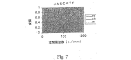
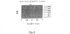
また、911レンズ、Z11(平均)レンズ、I11(個別)レンズのそれぞれにおいて、被検者JAEにおける最高で50c/mmで合焦されたMTF曲線および50c/mmでのスルーフォーカスMTFが図7および図8にプロットされている。図7および図8から分かるように、Z11およびI11レンズが埋め込まれた眼の50c/mmでのMTFは、911レンズが埋め込まれた同じ眼のMTFよりも高い。全てのレンズのスルーフォーカスMTFが十分であることも分かる。Z11は、911と同じ程度の焦点深度を有している。しかしながら、MTFまたはスルーフォーカスMTFに関して、I11レンズがZ11レンズにと比較すると著しい向上を与えない点は興味深い。 Further, in each of the 911 lens, the Z11 (average) lens, and the I11 (individual) lens, the MTF curve focused at a maximum of 50 c / mm and the through focus MTF at 50 c / mm in the subject JAE are shown in FIG. Plotted in FIG. As can be seen from FIGS. 7 and 8, the MTF at 50 c / mm of the eye with embedded Z11 and I11 lenses is higher than the MTF of the same eye with embedded 911 lenses. It can also be seen that the through focus MTF of all lenses is sufficient. Z11 has the same depth of focus as 911. However, regarding the MTF or the through focus MTF, it is interesting that the I11 lens does not give a significant improvement compared to the Z11 lens.
個別化されたレンズを有する被検者の視力も計算した。図9は、これらの視力を、911レンズおよびZ11レンズにおいて計算した視力と比較したものである。 The visual acuity of subjects with individualized lenses was also calculated. FIG. 9 compares these visual acuities with the visual acuities calculated for the 911 and Z11 lenses.
図9から分かるように、4人の被検者の全てにおいて、Z11レンズおよびI11レンズにおける視力は、911レンズにおける視力よりも良好である。また、Z11レンズおよびI11レンズに関する結果が著しくは異ならないことが分かる。すなわち4人の被検者のそれぞれにおいて、平均的な角膜が比較的正確である。 As can be seen from FIG. 9, in all four subjects, the visual acuity of the Z11 lens and the I11 lens is better than that of the 911 lens. It can also be seen that the results for the Z11 lens and the I11 lens are not significantly different. That is, the average cornea is relatively accurate in each of the four subjects.
(例4)
以下、人々の集団から得られた平均的な角膜の球面収差を低減するようになっている眼レンズの設計について詳細に説明する。このレンズは、角膜の球面収差を示す正規化された11番目のゼルニケ項を補償するため、Z11レンズと称する。Z11レンズの移植予定者すなわち白内障の患者から成る母集団を使用することを決定した。
(Example 4)
The following describes in detail the design of an eye lens designed to reduce the average corneal spherical aberration obtained from a population of people. This lens is referred to as a Z11 lens to compensate for the normalized 11th Zernike term indicative of the spherical aberration of the cornea. It was decided to use a population of prospective Z11 lens recipients, ie cataract patients.
(母集団の説明)
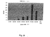
母集団は、スウェーデンのストックホルムにある聖エリック眼病院の71人の白内障患者を含んでいた。これらの患者の年齢層は、35歳から94歳(2000年4月12日の時点で)であった。我々の母集団の平均年齢は73.67歳であった。母集団の年齢のヒストグラムが図10に示されている。
(Explanation of population)
The population included 71 cataract patients from St. Eric Eye Hospital in Stockholm, Sweden. The age group of these patients was 35 to 94 years old (as of April 12, 2000). The average age of our population was 73.67 years. A histogram of the age of the population is shown in FIG.
Orbscan(登録商標)(Orbtek、ソルトレイクシティ)装置を使用して、71人の被検者の角膜を測定した。Orbscan(登録商標)は、角膜の両方の表面、前レンズ面、虹彩の表面を測定する走査スリットを基本とした角膜および前部のトポグラファである。各表面は、高さ、勾配、曲率、倍率のマップとして表示できる。 The corneas of 71 subjects were measured using an Orbscan® (Orbtek, Salt Lake City) apparatus. The Orbscan® is a cornea and anterior topographer based on a scanning slit that measures both the surface of the cornea, the front lens surface, and the surface of the iris. Each surface can be displayed as a map of height, gradient, curvature, and magnification.
(フィッティング・アルゴリズム)
Orbscan(登録商標)を使用して、前面における角膜の高さデータ(角膜の表面上の点のデカルト位置)を得た。また、角膜の光学特性を決定するための生データとして前記データを使用した。Orbscan(登録商標)ファイルの一例からの高さデータが図11に示されている。
(Fitting algorithm)
Orbscan® was used to obtain corneal height data at the anterior surface (Cartesian location of points on the surface of the cornea). The data was also used as raw data for determining the optical properties of the cornea. Height data from an example of an Orbsscan® file is shown in FIG.
高さデータを示すデカルト座標が極座標に変換される(x,y,z→r,θ,z)。その後、表面を描くため、このデータが方程式1bに示されるような一連の多項式に当てはめられる。各多項式における係数(a’s)すなわち重み係数は、表面を完全に描くフィッティング処理によって決定される。使用した多項式(Zi)は、正規化されたゼルニケ多項式である。
(波面収差の計算)
前側の角膜表面の形状(前述したa’sのようなゼルニケ係数)が分かっていれば、この面に起因する波面収差をレイトレーシング処理を使用して決定することができる。これについては、例えば、2000年6月のJ Opt Soc Am A Opt Image Scivisの第17(6)巻955−965頁のAntonio GuiraoおよびPablo Artalによる「ビデオケラトグラフィからの角膜波収差:処理の精度および限界(Corneal wave aberration from videokeratography:accuracy and limitations of the procedure)」に記載されている。
(Calculation of wavefront aberration)
If the shape of the anterior corneal surface (Zernike coefficient like a's described above) is known, the wavefront aberration due to this surface can be determined using ray tracing processing. For example, in June 2000, J Opti Soc Am A Opt Image Scvis, 17 (6), pp. 955-965, “Cornea Wave Aberration from Video Keratography: Processing Accuracy” by Antonio Guirao and Pablo Artal. And the limitations (development and limitation of the procedure).
(結果)
(平均的な角膜の球面収差および形状)
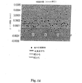
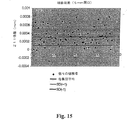
6mm開口における前述した基準を使用して、71人の被検者を評価した。表面高さをゼルニケ多項式に当てはめた後、各被検者の波面収差を測定した。図12は、各ゼルニケ項の平均および標準偏差を示している(正規化形式)。エラーバーは、±1の標準偏差を示している。我々の母集団には、平均して、ゼロとは著しく異なる3つの収差がある。これらは、非点収差(A5)、コマ収差(A9)、球面収差(A11)である。球面収差は、唯一の回転対称な収差であり、回転対称なIOLを用いて修正することができる唯一の収差となる。
(result)
(Average corneal spherical aberration and shape)
71 subjects were evaluated using the criteria described above at a 6 mm aperture. After applying the surface height to the Zernike polynomial, the wavefront aberration of each subject was measured. FIG. 12 shows the mean and standard deviation of each Zernike term (normalized form). Error bars indicate a standard deviation of ± 1. In our population, on average, there are three aberrations that are significantly different from zero. These are astigmatism (A5), coma (A9), and spherical aberration (A11). Spherical aberration is the only rotationally symmetric aberration and is the only aberration that can be corrected using a rotationally symmetric IOL.
図13は、白内障手術前の71人の各被検者における球面収差を示すゼルニケ係数(OSLO形式)の値の散布図である。中央の実線は平均球面収差を示し、破線は+1および−1の標準偏差を示している。表8には、半径、非球面定数、球面収差、2乗平均平方根エラーにおける、平均および標準偏差が記載されているとともに、最大値および最小値が記載されている。
以下の表9、10、11は、4mm、5mm、7mmの開口サイズにおける対応する結果をそれぞれ示している。図14、図15および図16は、対応する散布図である。
(角膜設計)
1つのモデル角膜を設計するとともに、この角膜を使用して各Z11レンズ倍率を設計した。角膜は、母集団において計算された平均値と同じ球面収差を有するように設計された。様々な開口サイズにおける設計角膜半径および非球面定数が表12に示されている。全てのケースにおいて、曲率半径は、ゼルニケ適合データから測定された平均半径となるように取られた。モデル角膜の球面収差値が母集団における平均球面収差値と等しくなるまで、非球面定数が変化した。
One model cornea was designed, and each Z11 lens magnification was designed using this cornea. The cornea was designed to have the same spherical aberration as the average value calculated in the population. The design corneal radius and aspheric constant for various aperture sizes are shown in Table 12. In all cases, the radius of curvature was taken to be the average radius measured from Zernike fit data. The aspheric constant changed until the spherical aberration value of the model cornea became equal to the average spherical aberration value in the population.
前述したように、6mmの開口径の値を使用して、角膜を設計した。このような選択により、5.1mmレンズ径にわたって球面収差が無い(この角膜を有する系で測定した場合)Z11レンズを設計することができる。Z11設計角膜においてリストアップされるOSLO表面が表13に示されている。角膜の屈折率は、1.3375のケラトメトリ(角膜曲率測定)率である。 As described above, the cornea was designed using an aperture diameter value of 6 mm. With such a selection, a Z11 lens can be designed that has no spherical aberration over a 5.1 mm lens diameter (when measured with a system having this cornea). OSLO surfaces listed in the Z11 design cornea are shown in Table 13. The refractive index of the cornea is a keratometry (corneal curvature measurement) rate of 1.3375.
角膜の屈折率が1.3375である1つの面を有するモデル角膜において、これらの値を計算した。本発明の範囲から逸脱することなく、角膜の光学的に等価なモデル形式を使用することができる。例えば、複数の表面角膜すなわち屈折率が異なる角膜を使用することができる。
(レンズの設計)
設計角膜の球面収差と釣り合うように各Z11レンズを設計した。設計の出発点は、エッジおよび中心厚さが修正されて成る米国特許第5,444,106号に記載された同じ倍率のCeeOn Edge(登録商標)911レンズであった。その後、このレンズは、前側の角膜表面から4.5mmの位置に配置された。前側の角膜表面からの距離は、重要ではなく、適度な範囲内で変えることができる。22Dレンズ設計プロセスにおける出発点となる眼のモデルの表面情報が表14に示されている。方程式4に示される式を使用して、レンズの前面が示されている。変数cc、ad、aeは、球面収差が最小となるように修正された。これらの変数は、5.1mmの開口サイズのために決定され、表面は、これらの値から6mmの光学的開口サイズまで外挿される。その結果得られた22D Z11眼モデルが表15に示されている。この22Dレンズの前面は、系(角膜+レンズ)の球面収差が0にほぼ等しくなるように修正された。CeeOn Edge 911 22Dレンズ眼モデルおよび22D Z11レンズ眼モデルに関してOSLOにより計算される波面収差係数が以下の表16に示されている。なお、出発点となる眼モデルにおける球面収差を示す係数は、角膜に配置された6mmの開口径においては0.001005であり、一方、設計されたZ11レンズを有する眼モデルにおける対応する係数は、−1.3399e−06mmである。22Dレンズに関する前述したプロセスと同じプロセスを、同様にして、任意の他のレンズ倍率において行なうことができる。
Each Z11 lens was designed to balance the spherical aberration of the design cornea. The starting point of the design was a
新たなZ11設計のために選択された光学的な態様は、屈折率が1.458であるシリコーンによって形成される等凸レンズである。平均的な角膜の球面収差は、球面収差を有さない系を形成するZ11レンズによって釣り合わせられる。レンズの前面は、設計開口内の全ての軸上光線の光路長が同一の点収束を生じる光路長となるように修正される。このような特徴は、多くのレンズ態様を用いて達成することができる。したがって、Z11レンズは、凸平レンズ、平凸レンズ、不等凸レンズ、あるいは、収斂レンズを形成する他の任意のデザインに基づいて設計することができる。また、眼の屈折エラーを修正するために使用される発散レンズを包含するために、Z11の概念を広げることもできる。光路差を必要に応じて変えて球面収差を相殺するために、前面または後面を修正することもできる。したがって、Z11レンズ設計の目的を達成する多くの設計が可能である。 The optical aspect chosen for the new Z11 design is an equiconvex lens formed by silicone with a refractive index of 1.458. The average corneal spherical aberration is balanced by a Z11 lens forming a system without spherical aberration. The front surface of the lens is modified so that the optical path length of all on-axis rays within the design aperture is the optical path length that produces the same point convergence. Such a feature can be achieved using a number of lens embodiments. Thus, the Z11 lens can be designed based on a convex flat lens, a plano-convex lens, an unequal convex lens, or any other design that forms a converging lens. It is also possible to extend the concept of Z11 to include a diverging lens used to correct eye refraction errors. The front or back surface can also be modified to change the optical path difference as needed to cancel the spherical aberration. Therefore, many designs that achieve the objectives of Z11 lens design are possible.
Claims (118)
(i)少なくとも1つの角膜表面を数学的モデルとして特徴付けるステップと、
(ii)結果として生じる前記角膜表面の収差を、前記数学的なモデルを使用することにより計算するステップと、
(iii)眼内レンズの光学倍率を選択するステップと、
(iv)眼内レンズをモデル化して、前記レンズおよび角膜モデルから成る光学系からの波面の収差を低減するステップとを備えている、方法。 A method for designing an intraocular lens capable of reducing ocular aberrations after implantation comprising:
(I) characterizing at least one corneal surface as a mathematical model;
(Ii) calculating the resulting corneal surface aberration by using the mathematical model;
(Iii) selecting an optical magnification of the intraocular lens;
(Iv) modeling the intraocular lens to reduce wavefront aberrations from an optical system comprising the lens and the cornea model.
(vi)モデル化された眼内レンズが十分に収差を低減したか否かを判断するとともに、選択的に、十分な低減が得られるまで眼内レンズを再モデル化するステップとを更に備えている、請求項1に記載の方法。 (V) calculating aberrations resulting from the optical system comprising the modeled intraocular lens and cornea;
(Vi) determining whether the modeled intraocular lens has sufficiently reduced aberrations, and optionally re-modeling the intraocular lens until sufficient reduction is obtained. The method of claim 1.
(ii)角膜の波面のセルニケ係数を決定するステップと、
(iii)前記モデルレンズと角膜とを備える光学系が十分に低いゼルニケ係数を有する波面を与えるように、眼内レンズをモデル化するステップとを備えている、請求項17に記載の方法。 (I) representing corneal aberration as a linear combination of Zernike polynomials;
(Ii) determining a Zernike coefficient of the cornea wavefront;
18. The method of claim 17, comprising: (iii) modeling an intraocular lens so that an optical system comprising the model lens and cornea provides a wavefront having a sufficiently low Zernike coefficient.
(v)前記眼内レンズが十分にゼルニケ係数を低減したか否かを判断するとともに、選択的に、前記係数の十分な低減が得られるまで前記レンズを再モデル化するステップとを更に備えている、請求項18に記載の方法。 (Iv) calculating a Zernike coefficient of a wavefront arising from an optical system comprising a modeled intraocular lens and cornea;
(V) determining whether the intraocular lens has sufficiently reduced the Zernike coefficient, and optionally further re-modeling the lens until a sufficient reduction in the coefficient is obtained. The method according to claim 18.
(ii)個々の角膜間で多項式の係数を比較し、
(iii)個々の角膜から1つの公称係数値を選択し、
(iv)前記レンズおよび個々の角膜を備える光学系から生じる波面が前記公称係数値を十分に低減するように、レンズをモデル化する、請求項8に記載の方法。 (I) characterizing the corneal surface of the selected population and representing each cornea as a linear combination of polynomials;
(Ii) compare polynomial coefficients between individual corneas,
(Iii) selecting one nominal coefficient value from each cornea;
9. The method of claim 8, wherein the lens is modeled such that (iv) a wavefront arising from an optical system comprising the lens and individual corneas sufficiently reduces the nominal coefficient value.
(i)少なくとも1つの角膜表面を数学的モデルとして特徴付けるステップと、
(ii)結果として生じる前記角膜表面の収差を、前記数学的なモデルを使用することにより計算するステップと、
(iii)光学倍率が同じであるが収差が異なる複数のレンズから、適切な光学倍率を有する眼内レンズを選択するステップと、
(iv)前記選択されたレンズおよび角膜モデルから成る光学系が収差を十分に低減するか否かを判断するステップと、
を備えている方法。 A method for selecting an intraocular lens that can reduce ocular aberrations after implantation comprising:
(I) characterizing at least one corneal surface as a mathematical model;
(Ii) calculating the resulting corneal surface aberration by using the mathematical model;
(Iii) selecting an intraocular lens having an appropriate optical magnification from a plurality of lenses having the same optical magnification but different aberrations;
(Iv) determining whether the optical system comprising the selected lens and corneal model sufficiently reduces aberrations;
A method comprising:
(vi)前記光学系からの波面の収差を前記選択された眼内レンズが十分に低減したか否かを判断するとともに、収差を十分に低減することができるレンズを見出すまで、同じ光学倍率を有する少なくとも1つの新たなレンズを選択することにより、ステップ(iii)およびステップ(iv)を選択的に繰り返すステップとを更に備えている、請求項30に記載の方法。 (V) calculating wavefront aberrations from an optical system comprising the selected lens and corneal model;
(Vi) Determine whether the selected intraocular lens has sufficiently reduced the aberration of the wavefront from the optical system, and at the same optical magnification until finding a lens that can sufficiently reduce the aberration 31. The method of claim 30, further comprising selectively repeating steps (iii) and (iv) by selecting at least one new lens having.
(ii)角膜のゼルニケ係数を決定するステップと、
(iii)前記レンズと角膜とを備える光学系が十分に低いゼルニケ係数を有する波面を与えるように、眼内レンズを選択するステップと、
を備えている、請求項42に記載の方法。 (I) representing corneal aberration as a linear combination of Zernike polynomials;
(Ii) determining a Zernike coefficient of the cornea;
(Iii) selecting an intraocular lens such that an optical system comprising said lens and cornea provides a wavefront having a sufficiently low Zernike coefficient;
43. The method of claim 42, comprising:
(v)前記眼内レンズが十分にゼルニケ係数を低減したか否かを判断するとともに、選択的に、前記係数の十分な低減が得られるまで新たなレンズを選択するステップとを更に備えている、請求項43に記載の方法。 (Iv) calculating a Zernike coefficient of a wavefront arising from an optical system comprising a modeled intraocular lens and cornea;
(V) determining whether or not the intraocular lens has sufficiently reduced the Zernike coefficient, and further comprising selectively selecting a new lens until a sufficient reduction in the coefficient is obtained. 44. The method of claim 43.
代表的な患者集団を選択するステップと、
患者集団内の各被検者に関する角膜の解剖学的なデータを収集するステップと、
前記データを、所定の開口サイズにおける各被検者の角膜表面形状を示す項に移すステップと、
少なくとも1つの平均角膜表面形状項を取得するように、前記患者集団の少なくとも1つの角膜表面形状項の平均値を計算し、および/または、それぞれが角膜表面形状項を介した変換によって得られる、少なくとも1つの対応する角膜波面収差項の平均値を計算するステップと、
前記少なくとも1つの平均角膜表面形状項または前記少なくとも1つの平均角膜波面収差項から、角膜およびレンズを備える光学系の前記少なくとも1つの平均波面収差項を減少させることができる眼レンズを設計するステップと、
を備えていることを特徴とする方法。 A method of designing an eye lens suitable for implantation in the eye,
Selecting a representative patient population;
Collecting corneal anatomical data for each subject in the patient population;
Transferring the data to a term indicating the corneal surface shape of each subject at a predetermined aperture size;
Calculating an average value of at least one corneal surface shape term of the patient population to obtain at least one average corneal surface shape term and / or each obtained by transformation via a corneal surface shape term; Calculating an average value of at least one corresponding corneal wavefront aberration term;
Designing an ophthalmic lens capable of reducing the at least one average wavefront aberration term of an optical system comprising a cornea and a lens from the at least one average corneal surface shape term or the at least one average corneal wavefront aberration term; ,
A method characterized by comprising:
モデル平均角膜およびレンズを通じて移動する波面のこれらの特定の収差項を測定するとともに、測定された波面において前記少なくとも1つの収差項が十分に減少されていない場合にレンズを再設計することにより、設計された眼レンズが少なくとも1つの平均収差項を正確に補償していることをチェックするステップとを更に備えていることを特徴とする、請求項55に記載の方法。 Designing an average corneal model in a patient population from the calculated at least one average corneal surface shape term or at least one average corneal wavefront aberration term;
Design by measuring these specific aberration terms of the wavefront moving through the model mean cornea and the lens and redesigning the lens if the at least one aberration term is not sufficiently reduced in the measured wavefront 56. The method of claim 55, further comprising the step of checking that the shaped ophthalmic lens accurately compensates for at least one average aberration term.
この患者に対応する選択された集団がこの特定の患者の代表であるか否か、また、これが、これらの平均値から設計されたレンズを埋め込む場合であるか否か、また、これが、他の集団からの平均値から設計されたレンズを埋め込む、または、この患者のために個人的なレンズを設計する場合であるか否かを判断するステップとを更に備えることを特徴とする、請求項55から62のいずれか一項に記載の方法。 Measuring at least one wavefront aberration of the cornea of one particular patient;
Whether the selected population corresponding to this patient is representative of this particular patient, whether this is the case when implanting a lens designed from these averages, and this is 56. Implanting a designed lens from an average value from the population or determining if it is a case of designing a personal lens for this patient. 63. The method according to any one of 1 to 62.
眼から天然のレンズを除去するステップと、
波面センサを使用することにより、レンズを備えていない眼の収差を測定するステップと、
波面の検出によって見出される少なくとも1つの収差項を減少することができるレンズを用意するステップと、
患者の眼内に前記レンズを埋め込むステップとを備えている方法。 A method of correcting a patient's visual acuity by implanting an intraocular lens that at least partially compensates for eye aberrations, comprising:
Removing a natural lens from the eye;
Measuring aberrations of an eye without a lens by using a wavefront sensor;
Providing a lens capable of reducing at least one aberration term found by wavefront detection;
Implanting the lens in a patient's eye.
Applications Claiming Priority (4)
| Application Number | Priority Date | Filing Date | Title |
|---|---|---|---|
| SE0001925A SE0001925D0 (en) | 2000-05-23 | 2000-05-23 | Methods of obtaining ophthalmic lenses capable of reducing the aberrations of the eye |
| SE0001925-7 | 2000-05-23 | ||
| SE0004830A SE0004830D0 (en) | 2000-12-22 | 2000-12-22 | An ophthalmic lens and a method for designing such a lens |
| SE0004830-6 | 2000-12-22 |
Related Parent Applications (1)
| Application Number | Title | Priority Date | Filing Date |
|---|---|---|---|
| JP2001585670A Division JP4459501B2 (en) | 2000-05-23 | 2001-05-23 | Eye lens to reduce eye aberration |
Publications (2)
| Publication Number | Publication Date |
|---|---|
| JP2010029694A true JP2010029694A (en) | 2010-02-12 |
| JP5368259B2 JP5368259B2 (en) | 2013-12-18 |
Family
ID=26655116
Family Applications (2)
| Application Number | Title | Priority Date | Filing Date |
|---|---|---|---|
| JP2001585670A Expired - Fee Related JP4459501B2 (en) | 2000-05-23 | 2001-05-23 | Eye lens to reduce eye aberration |
| JP2009249913A Expired - Fee Related JP5368259B2 (en) | 2000-05-23 | 2009-10-30 | Method for obtaining an eye lens that reduces eye aberrations |
Family Applications Before (1)
| Application Number | Title | Priority Date | Filing Date |
|---|---|---|---|
| JP2001585670A Expired - Fee Related JP4459501B2 (en) | 2000-05-23 | 2001-05-23 | Eye lens to reduce eye aberration |
Country Status (20)
| Country | Link |
|---|---|
| EP (3) | EP1943984B1 (en) |
| JP (2) | JP4459501B2 (en) |
| KR (1) | KR20030036192A (en) |
| CN (1) | CN1245929C (en) |
| AT (1) | ATE381303T1 (en) |
| AU (2) | AU6394201A (en) |
| BR (1) | BR0111043A (en) |
| CA (1) | CA2409692C (en) |
| CZ (1) | CZ20023851A3 (en) |
| DE (1) | DE60131964T2 (en) |
| EA (1) | EA200201270A1 (en) |
| EE (1) | EE200200650A (en) |
| HU (1) | HUP0301923A2 (en) |
| IL (1) | IL152772A0 (en) |
| MX (1) | MXPA02011538A (en) |
| NO (1) | NO20025658L (en) |
| PL (1) | PL366214A1 (en) |
| SK (1) | SK16532002A3 (en) |
| TW (1) | TW497970B (en) |
| WO (1) | WO2001089424A1 (en) |
Cited By (1)
| Publication number | Priority date | Publication date | Assignee | Title |
|---|---|---|---|---|
| WO2012127881A1 (en) * | 2011-03-24 | 2012-09-27 | 興和株式会社 | Intraocular lens and manufacturing method therefor |
Families Citing this family (104)
| Publication number | Priority date | Publication date | Assignee | Title |
|---|---|---|---|---|
| US7293871B2 (en) | 2000-11-27 | 2007-11-13 | Ophthonix, Inc. | Apparatus and method of correcting higher-order aberrations of the human eye |
| US6813082B2 (en) | 2000-11-27 | 2004-11-02 | Ophthonix, Inc. | Wavefront aberrator and method of manufacturing |
| SE0004829D0 (en) * | 2000-12-22 | 2000-12-22 | Pharmacia Groningen Bv | Methods of obtaining ophthalmic lenses providing the eye with reduced aberrations |
| JP2004534964A (en) * | 2001-04-27 | 2004-11-18 | ノバルティス アクチエンゲゼルシャフト | Automatic lens design and manufacturing system |
| JP2002350785A (en) * | 2001-05-28 | 2002-12-04 | Menicon Co Ltd | Method of designing ocular lens |
| IL143503A0 (en) * | 2001-05-31 | 2002-04-21 | Visionix Ltd | Aberration correction spectacle lens |
| US7217375B2 (en) | 2001-06-04 | 2007-05-15 | Ophthonix, Inc. | Apparatus and method of fabricating a compensating element for wavefront correction using spatially localized curing of resin mixtures |
| US7434931B2 (en) | 2001-10-25 | 2008-10-14 | Ophthonix | Custom eyeglass manufacturing method |
| US6712466B2 (en) | 2001-10-25 | 2004-03-30 | Ophthonix, Inc. | Eyeglass manufacturing method using variable index layer |
| US6682195B2 (en) | 2001-10-25 | 2004-01-27 | Ophthonix, Inc. | Custom eyeglass manufacturing method |
| US6781681B2 (en) | 2001-12-10 | 2004-08-24 | Ophthonix, Inc. | System and method for wavefront measurement |
| US7034949B2 (en) | 2001-12-10 | 2006-04-25 | Ophthonix, Inc. | Systems and methods for wavefront measurement |
| US20050174535A1 (en) | 2003-02-13 | 2005-08-11 | Lai Shui T. | Apparatus and method for determining subjective responses using objective characterization of vision based on wavefront sensing |
| US6761454B2 (en) | 2002-02-13 | 2004-07-13 | Ophthonix, Inc. | Apparatus and method for determining objective refraction using wavefront sensing |
| US6663240B2 (en) * | 2002-05-15 | 2003-12-16 | Alcon, Inc. | Method of manufacturing customized intraocular lenses |
| US7104648B2 (en) | 2002-09-06 | 2006-09-12 | Synergeyes, Inc. | Hybrid contact lens system and method |
| US7322694B2 (en) | 2002-09-06 | 2008-01-29 | Synergeyes, Inc. | Hybrid contact lens system and method |
| US7163292B2 (en) | 2002-09-06 | 2007-01-16 | Synergeyes, Inc. | Hybrid contact lens system and method |
| US7097301B2 (en) | 2002-09-06 | 2006-08-29 | Synergeyes, Inc. | Hybrid contact lens system and method |
| US6817714B2 (en) * | 2002-09-25 | 2004-11-16 | Bausch And Lomb, Inc. | Method and apparatus relating to the optical zone of an optical element |
| US7381221B2 (en) | 2002-11-08 | 2008-06-03 | Advanced Medical Optics, Inc. | Multi-zonal monofocal intraocular lens for correcting optical aberrations |
| US7896916B2 (en) | 2002-11-29 | 2011-03-01 | Amo Groningen B.V. | Multifocal ophthalmic lens |
| SE0203564D0 (en) * | 2002-11-29 | 2002-11-29 | Pharmacia Groningen Bv | Multifocal opthalmic lens |
| US7036931B2 (en) * | 2003-01-29 | 2006-05-02 | Novartis Ag | Ophthalmic lenses |
| WO2004090611A2 (en) * | 2003-03-31 | 2004-10-21 | Bausch & Lomb Incorporated | Intraocular lens and method for reducing aberrations in an ocular system |
| DE10325841A1 (en) | 2003-06-06 | 2004-12-30 | Acritec Gmbh | intraocular lens |
| US7018039B2 (en) | 2003-11-14 | 2006-03-28 | Synergeyes,Inc. | Contact lens |
| JP2007531559A (en) | 2004-02-20 | 2007-11-08 | オフソニックス,インク | System and method for analyzing wavefront aberrations |
| CA2562268C (en) | 2004-04-05 | 2014-12-02 | Advanced Medical Optics, Inc. | Ophthalmic lenses capable of reducing chromatic aberration |
| US7476248B2 (en) * | 2004-04-06 | 2009-01-13 | Alcon, Inc. | Method of calculating the required lens power for an opthalmic implant |
| US7922326B2 (en) | 2005-10-25 | 2011-04-12 | Abbott Medical Optics Inc. | Ophthalmic lens with multiple phase plates |
| SE0402769D0 (en) * | 2004-11-12 | 2004-11-12 | Amo Groningen Bv | Method of selecting intraocular lenses |
| WO2006054985A1 (en) * | 2004-11-16 | 2006-05-26 | Essilor International (Compagnie Generale D'optique) | Method for designing spectacle lenses taking into account an individual's head and eye movement |
| US20060116763A1 (en) | 2004-12-01 | 2006-06-01 | Simpson Michael J | Contrast-enhancing aspheric intraocular lens |
| ES2272143B1 (en) * | 2004-12-22 | 2008-03-01 | Instituto Oftalmologico De Alicante, S.L. | INTRAOCULAR LENS TO ACROMATIZE THE EYE AND REDUCE ITS ABERRATIONS. |
| MXPA06014056A (en) | 2005-04-05 | 2007-03-07 | Alcon Inc | Optimal iol shape factors for human eyes. |
| US7401922B2 (en) | 2005-04-13 | 2008-07-22 | Synergeyes, Inc. | Method and apparatus for reducing or eliminating the progression of myopia |
| US7543936B2 (en) | 2005-05-06 | 2009-06-09 | Synergeyes, Inc. | Hybrid contact lens system and method of fitting |
| NL1029037C2 (en) * | 2005-05-13 | 2006-11-14 | Akkolens Int Bv | Artificial intraocular lens with variable optical strength, comprises optical elements with different shapes |
| DE102006021521A1 (en) * | 2006-05-05 | 2007-11-08 | Carl Zeiss Meditec Ag | Aspherical artificial eye lens and method for the construction of such |
| AU2007253839B2 (en) * | 2006-05-16 | 2013-11-28 | Essilor International (Compagnie Generale D'optique) | High-order aberration correction for optimization of human visual function |
| US7879089B2 (en) | 2006-05-17 | 2011-02-01 | Alcon, Inc. | Correction of higher order aberrations in intraocular lenses |
| US7537339B2 (en) | 2006-05-25 | 2009-05-26 | Synergeyes, Inc. | Hybrid contact lens system and method of fitting |
| JP5041739B2 (en) | 2006-06-14 | 2012-10-03 | Hoya株式会社 | Intraocular lens |
| US7828432B2 (en) | 2007-05-25 | 2010-11-09 | Synergeyes, Inc. | Hybrid contact lenses prepared with expansion controlled polymeric materials |
| US8747466B2 (en) | 2007-08-27 | 2014-06-10 | Amo Groningen, B.V. | Intraocular lens having extended depth of focus |
| US9216080B2 (en) | 2007-08-27 | 2015-12-22 | Amo Groningen B.V. | Toric lens with decreased sensitivity to cylinder power and rotation and method of using the same |
| US8740978B2 (en) | 2007-08-27 | 2014-06-03 | Amo Regional Holdings | Intraocular lens having extended depth of focus |
| US8974526B2 (en) | 2007-08-27 | 2015-03-10 | Amo Groningen B.V. | Multizonal lens with extended depth of focus |
| WO2009076500A1 (en) * | 2007-12-11 | 2009-06-18 | Bausch & Lomb Incorporated | Method and apparatus for providing eye optical systems with extended depths of field |
| AU2009214036B2 (en) | 2008-02-15 | 2014-04-17 | Amo Regional Holdings | System, ophthalmic lens, and method for extending depth of focus |
| US8439498B2 (en) | 2008-02-21 | 2013-05-14 | Abbott Medical Optics Inc. | Toric intraocular lens with modified power characteristics |
| TWI398338B (en) * | 2008-04-03 | 2013-06-11 | Nat Applied Res Laboratories | Compensation method for optics mold |
| US7871162B2 (en) | 2008-04-24 | 2011-01-18 | Amo Groningen B.V. | Diffractive multifocal lens having radially varying light distribution |
| US8231219B2 (en) | 2008-04-24 | 2012-07-31 | Amo Groningen B.V. | Diffractive lens exhibiting enhanced optical performance |
| US8862447B2 (en) | 2010-04-30 | 2014-10-14 | Amo Groningen B.V. | Apparatus, system and method for predictive modeling to design, evaluate and optimize ophthalmic lenses |
| US20100079723A1 (en) * | 2008-10-01 | 2010-04-01 | Kingston Amanda C | Toric Ophthalimc Lenses Having Selected Spherical Aberration Characteristics |
| WO2010100523A1 (en) * | 2009-03-05 | 2010-09-10 | Amo Regional Holdings | Multizonal lens with extended depth of focus |
| US8331048B1 (en) | 2009-12-18 | 2012-12-11 | Bausch & Lomb Incorporated | Methods of designing lenses having selected depths of field |
| CN102892381A (en) | 2009-12-18 | 2013-01-23 | Amo格罗宁根私人有限公司 | Single microstructure lens, systems and methods |
| ES2549514T3 (en) * | 2010-04-20 | 2015-10-28 | Carl Zeiss Vision International Gmbh | Method to optimize a lens for glasses for the wavefront aberrations of an eye and lens |
| WO2012074742A1 (en) * | 2010-11-30 | 2012-06-07 | Amo Groningen Bv | Method for designing, evaluating and optimizing ophthalmic lenses and laser vision correction |
| EP2646872A1 (en) | 2010-12-01 | 2013-10-09 | AMO Groningen B.V. | A multifocal lens having an optical add power progression, and a system and method of providing same |
| US8894204B2 (en) | 2010-12-17 | 2014-11-25 | Abbott Medical Optics Inc. | Ophthalmic lens, systems and methods having at least one rotationally asymmetric diffractive structure |
| US9931200B2 (en) | 2010-12-17 | 2018-04-03 | Amo Groningen B.V. | Ophthalmic devices, systems, and methods for optimizing peripheral vision |
| JP6093695B2 (en) * | 2011-04-05 | 2017-03-08 | 興和株式会社 | Intraocular lens design method and intraocular lens |
| TWI588560B (en) | 2012-04-05 | 2017-06-21 | 布萊恩荷登視覺協會 | Lenses, devices, methods and systems for refractive error |
| CN108013952A (en) | 2012-08-31 | 2018-05-11 | Amo格罗宁根私人有限公司 | Polycyclic crystalline lens, system and method for extended focal depth |
| US9201250B2 (en) | 2012-10-17 | 2015-12-01 | Brien Holden Vision Institute | Lenses, devices, methods and systems for refractive error |
| TWI600418B (en) | 2012-10-17 | 2017-10-01 | 布萊恩荷登視覺協會 | Lenses, devices, methods and systems for refractive error |
| US9717628B2 (en) | 2012-12-04 | 2017-08-01 | Amo Groningen B.V. | Lenses, systems and methods for providing binocular customized treatments to correct presbyopia |
| CN103006177B (en) * | 2012-12-20 | 2014-11-05 | 南京航空航天大学 | Human eye aberration compensation method based on Zernike conjugated composite pattern |
| EP2967312B1 (en) | 2013-03-11 | 2019-04-24 | Johnson & Johnson Surgical Vision, Inc. | Intraocular lens that matches an image surface to a retinal shape, and method of designing same |
| CN103784298A (en) * | 2013-11-20 | 2014-05-14 | 中国科学院光电技术研究所 | Head-mounted individualized eye aberration correcting and vision training device |
| AU2015242298B2 (en) | 2014-03-10 | 2019-11-14 | Amo Groningen B.V. | Intraocular lens that improves overall vision where there is a local loss of retinal function |
| WO2016142736A1 (en) * | 2015-03-10 | 2016-09-15 | Amo Groningen B.V. | Fresnel piggyback intraocular lens that improves overall vision where there is a local loss of retinal function |
| SG11201607593VA (en) * | 2014-03-11 | 2016-10-28 | Kowa Co | Ophthalmic lens and method for designing ophthalmic lens |
| US10010407B2 (en) | 2014-04-21 | 2018-07-03 | Amo Groningen B.V. | Ophthalmic devices that improve peripheral vision |
| WO2016009500A1 (en) * | 2014-07-15 | 2016-01-21 | 株式会社メニコン | Contact lens manufacturing method and contact lens |
| WO2016035055A1 (en) | 2014-09-05 | 2016-03-10 | Hoya Corporation | Wide depth of focus vortex intraocular lenses and associated methods |
| BR112017004765B1 (en) | 2014-09-09 | 2022-08-23 | Staar Surgical Company | LENS CONFIGURED FOR IMPLANTATION IN A HUMAN EYE |
| CN104490490A (en) * | 2015-01-09 | 2015-04-08 | 爱博诺德(北京)医疗科技有限公司 | Artificial lens and manufacturing method thereof |
| EP3413839A1 (en) | 2016-02-09 | 2018-12-19 | AMO Groningen B.V. | Progressive power intraocular lens, and methods of use and manufacture |
| WO2017156077A1 (en) | 2016-03-09 | 2017-09-14 | Staar Surgical Company | Ophthalmic implants with extended depth of field and enhanced distance visual acuity |
| WO2017153843A1 (en) | 2016-03-11 | 2017-09-14 | Amo Groningen B.V. | Intraocular lenses that improve peripheral vision |
| EP3656350B1 (en) | 2016-03-23 | 2021-06-23 | Johnson & Johnson Surgical Vision, Inc. | Ophthalmic apparatus with corrective meridians having extended tolerance band by modifying refractive powers in uniform meridian distribution |
| EP3932368A1 (en) | 2016-03-23 | 2022-01-05 | Johnson & Johnson Surgical Vision, Inc. | Ophthalmic apparatus with corrective meridians having extended tolerance band |
| AU2017252020B2 (en) | 2016-04-19 | 2021-11-11 | Amo Groningen B.V. | Ophthalmic devices, system and methods that improve peripheral vision |
| EP3522771B1 (en) | 2016-10-25 | 2022-04-06 | Amo Groningen B.V. | Realistic eye models to design and evaluate intraocular lenses for a large field of view |
| EP3595584A1 (en) | 2017-03-17 | 2020-01-22 | AMO Groningen B.V. | Diffractive intraocular lenses for extended range of vision |
| US10739227B2 (en) | 2017-03-23 | 2020-08-11 | Johnson & Johnson Surgical Vision, Inc. | Methods and systems for measuring image quality |
| US11523897B2 (en) | 2017-06-23 | 2022-12-13 | Amo Groningen B.V. | Intraocular lenses for presbyopia treatment |
| EP3646110A1 (en) | 2017-06-28 | 2020-05-06 | Amo Groningen B.V. | Diffractive lenses and related intraocular lenses for presbyopia treatment |
| AU2018293922B2 (en) * | 2017-06-28 | 2023-05-04 | Capricornia Contact Lens Pty Ltd | Improvements to fitting orthokeratology lenses |
| WO2019002390A1 (en) | 2017-06-28 | 2019-01-03 | Amo Groningen B.V. | Extended range and related intraocular lenses for presbyopia treatment |
| US11327210B2 (en) | 2017-06-30 | 2022-05-10 | Amo Groningen B.V. | Non-repeating echelettes and related intraocular lenses for presbyopia treatment |
| WO2019106067A1 (en) | 2017-11-30 | 2019-06-06 | Amo Groningen B.V. | Intraocular lenses that improve post-surgical spectacle independent and methods of manufacturing thereof |
| US10895517B2 (en) | 2018-02-08 | 2021-01-19 | Amo Groningen B.V. | Multi-wavelength wavefront system and method for measuring diffractive lenses |
| EP3749941A1 (en) | 2018-02-08 | 2020-12-16 | AMO Groningen B.V. | Wavefront based characterization of lens surfaces based on reflections |
| CN112867944A (en) | 2018-08-17 | 2021-05-28 | 斯塔尔外科有限公司 | Polymer compositions exhibiting a refractive index nano-gradient |
| WO2020208586A1 (en) * | 2019-04-11 | 2020-10-15 | Amo Development, Llc | Wavefront higher order correction of iols using refractive index modification |
| US11886046B2 (en) | 2019-12-30 | 2024-01-30 | Amo Groningen B.V. | Multi-region refractive lenses for vision treatment |
| WO2021136617A1 (en) | 2019-12-30 | 2021-07-08 | Amo Groningen B.V. | Lenses having diffractive profiles with irregular width for vision treatment |
| CN114326149B (en) * | 2022-02-15 | 2022-09-30 | 天津大学 | Spectacle lens structure and design method thereof |
Citations (2)
| Publication number | Priority date | Publication date | Assignee | Title |
|---|---|---|---|---|
| US5282852A (en) * | 1992-09-02 | 1994-02-01 | Alcon Surgical, Inc. | Method of calculating the required power of an intraocular lens |
| WO2000010448A1 (en) * | 1998-08-19 | 2000-03-02 | Autonomous Technologies Corporation | Apparatus and method for measuring vision defects of a human eye |
Family Cites Families (11)
| Publication number | Priority date | Publication date | Assignee | Title |
|---|---|---|---|---|
| US5236970A (en) | 1987-02-05 | 1993-08-17 | Allergan, Inc. | Optically clear reinforced silicone elastomers of high optical refractive index and improved mechanical properties for use in intraocular lenses |
| US6007747A (en) | 1987-08-24 | 1999-12-28 | Pharmacia & Upjohn Company | Method of making an aspheric soft lens |
| FR2647227B1 (en) * | 1989-05-19 | 1991-08-23 | Essilor Int | OPTICAL COMPONENT, SUCH AS AN INTRAOCULAR IMPLANT OR CONTACT LENS, SUITABLE FOR CORRECTING THE VISION OF AN INDIVIDUAL |
| US5050981A (en) * | 1990-07-24 | 1991-09-24 | Johnson & Johnson Vision Products, Inc. | Lens design method and resulting aspheric lens |
| US5444106A (en) | 1992-04-21 | 1995-08-22 | Kabi Pharmacia Ophthalmics, Inc. | High refractive index silicone compositions |
| SE9501714D0 (en) * | 1995-05-09 | 1995-05-09 | Pharmacia Ab | A method of selecting an intraocular lens to be implanted into an eye |
| US5777719A (en) * | 1996-12-23 | 1998-07-07 | University Of Rochester | Method and apparatus for improving vision and the resolution of retinal images |
| ES2178159T3 (en) | 1997-01-21 | 2002-12-16 | Technomed Ges Fur Med Und Med | PROCEDURE FOR THE CALCULATION OF A THEORETICAL FORM OF AT LEAST ONE SURFACE OF AN ARTIFICIAL OR NATURAL PART OF AN EYE CUTTED BY A BEAM BEAMING THROUGH THE PUPILA OF AN EYE, AND DEVICE FOR THE MANUFACTURE OF AN ARTIFICIAL CRYSTAL. |
| US5888122A (en) * | 1997-04-10 | 1999-03-30 | Prism Ophthalmics, L.L.C. | Method for manufacturing an intraocular lens |
| US6224211B1 (en) | 1999-06-08 | 2001-05-01 | Medjet, Inc. | Super vision |
| US6050687A (en) | 1999-06-11 | 2000-04-18 | 20/10 Perfect Vision Optische Geraete Gmbh | Method and apparatus for measurement of the refractive properties of the human eye |
-
2001
- 2001-05-23 EE EEP200200650A patent/EE200200650A/en unknown
- 2001-05-23 HU HU0301923A patent/HUP0301923A2/en unknown
- 2001-05-23 JP JP2001585670A patent/JP4459501B2/en not_active Expired - Fee Related
- 2001-05-23 AU AU6394201A patent/AU6394201A/en active Pending
- 2001-05-23 BR BR0111043-8A patent/BR0111043A/en not_active Application Discontinuation
- 2001-05-23 EP EP07150063.1A patent/EP1943984B1/en not_active Expired - Lifetime
- 2001-05-23 EP EP10181650.2A patent/EP2332495B1/en not_active Expired - Lifetime
- 2001-05-23 CZ CZ20023851A patent/CZ20023851A3/en unknown
- 2001-05-23 EP EP01938240A patent/EP1284687B1/en not_active Expired - Lifetime
- 2001-05-23 DE DE60131964T patent/DE60131964T2/en not_active Expired - Lifetime
- 2001-05-23 EA EA200201270A patent/EA200201270A1/en unknown
- 2001-05-23 PL PL01366214A patent/PL366214A1/en not_active Application Discontinuation
- 2001-05-23 CA CA002409692A patent/CA2409692C/en not_active Expired - Lifetime
- 2001-05-23 AU AU2001263942A patent/AU2001263942B2/en not_active Expired
- 2001-05-23 IL IL15277201A patent/IL152772A0/en unknown
- 2001-05-23 CN CNB018099238A patent/CN1245929C/en not_active Expired - Lifetime
- 2001-05-23 KR KR1020027015877A patent/KR20030036192A/en not_active Application Discontinuation
- 2001-05-23 AT AT01938240T patent/ATE381303T1/en not_active IP Right Cessation
- 2001-05-23 MX MXPA02011538A patent/MXPA02011538A/en unknown
- 2001-05-23 WO PCT/EP2001/006041 patent/WO2001089424A1/en active IP Right Grant
- 2001-05-23 SK SK1653-2002A patent/SK16532002A3/en unknown
- 2001-05-29 TW TW090112937A patent/TW497970B/en not_active IP Right Cessation
-
2002
- 2002-11-25 NO NO20025658A patent/NO20025658L/en not_active Application Discontinuation
-
2009
- 2009-10-30 JP JP2009249913A patent/JP5368259B2/en not_active Expired - Fee Related
Patent Citations (3)
| Publication number | Priority date | Publication date | Assignee | Title |
|---|---|---|---|---|
| US5282852A (en) * | 1992-09-02 | 1994-02-01 | Alcon Surgical, Inc. | Method of calculating the required power of an intraocular lens |
| WO2000010448A1 (en) * | 1998-08-19 | 2000-03-02 | Autonomous Technologies Corporation | Apparatus and method for measuring vision defects of a human eye |
| JP2002523119A (en) * | 1998-08-19 | 2002-07-30 | オートナマス・テクノロジーズ・コーポレーション | Apparatus and method for measuring visual impairment of the human eye |
Cited By (6)
| Publication number | Priority date | Publication date | Assignee | Title |
|---|---|---|---|---|
| WO2012127881A1 (en) * | 2011-03-24 | 2012-09-27 | 興和株式会社 | Intraocular lens and manufacturing method therefor |
| AU2012232611B2 (en) * | 2011-03-24 | 2016-05-26 | Kowa Company, Ltd. | Intraocular lens and manufacturing method therefor |
| JP5986985B2 (en) * | 2011-03-24 | 2016-09-06 | 興和株式会社 | Intraocular lens and manufacturing method thereof |
| US9808340B2 (en) | 2011-03-24 | 2017-11-07 | Kowa Company, Ltd. | Intraocular lens and manufacturing method thereof |
| KR101837379B1 (en) | 2011-03-24 | 2018-03-13 | 코와 가부시키가이샤 | Intraocular lens and manufacturing method therefor |
| US11020219B2 (en) | 2011-03-24 | 2021-06-01 | Kowa Company, Ltd. | Intraocular lens and manufacturing method thereof |
Also Published As
| Publication number | Publication date |
|---|---|
| SK16532002A3 (en) | 2003-07-01 |
| PL366214A1 (en) | 2005-01-24 |
| DE60131964D1 (en) | 2008-01-31 |
| CZ20023851A3 (en) | 2003-04-16 |
| EP2332495A1 (en) | 2011-06-15 |
| EP1284687B1 (en) | 2007-12-19 |
| EP2332495B1 (en) | 2014-04-23 |
| CA2409692C (en) | 2009-01-20 |
| CA2409692A1 (en) | 2001-11-29 |
| CN1430495A (en) | 2003-07-16 |
| EP1284687A1 (en) | 2003-02-26 |
| AU6394201A (en) | 2001-12-03 |
| EP1943984A1 (en) | 2008-07-16 |
| JP5368259B2 (en) | 2013-12-18 |
| TW497970B (en) | 2002-08-11 |
| ATE381303T1 (en) | 2008-01-15 |
| AU2001263942B2 (en) | 2006-12-14 |
| MXPA02011538A (en) | 2003-06-06 |
| KR20030036192A (en) | 2003-05-09 |
| NO20025658D0 (en) | 2002-11-25 |
| EP1943984B1 (en) | 2016-10-26 |
| EA200201270A1 (en) | 2003-06-26 |
| WO2001089424A1 (en) | 2001-11-29 |
| IL152772A0 (en) | 2003-06-24 |
| JP4459501B2 (en) | 2010-04-28 |
| NO20025658L (en) | 2002-11-25 |
| CN1245929C (en) | 2006-03-22 |
| DE60131964T2 (en) | 2008-12-04 |
| EE200200650A (en) | 2004-06-15 |
| HUP0301923A2 (en) | 2003-09-29 |
| JP2003534565A (en) | 2003-11-18 |
| BR0111043A (en) | 2003-04-15 |
Similar Documents
| Publication | Publication Date | Title |
|---|---|---|
| JP4459501B2 (en) | Eye lens to reduce eye aberration | |
| US6609793B2 (en) | Methods of obtaining ophthalmic lenses providing the eye with reduced aberrations | |
| US7137702B2 (en) | Methods of obtaining ophthalmic lenses providing the eye with reduced aberrations | |
| US9504377B2 (en) | Methods of obtaining ophthalmic lenses providing the eye with reduced aberrations | |
| AU2001263942A1 (en) | Methods of obtaining ophthalmic lenses providing the eye with reduced aberrations | |
| AU2002234579A1 (en) | Methods of obtaining ophthalmic lenses providing the eye with reduced aberrations | |
| JP2010152388A (en) | Method of designing multifocal ophthalmic lens | |
| ZA200209763B (en) | Methods of obtaining ophthalmic lenses providing the eye with reduced aberrations. |
Legal Events
| Date | Code | Title | Description |
|---|---|---|---|
| A131 | Notification of reasons for refusal |
Free format text: JAPANESE INTERMEDIATE CODE: A131 Effective date: 20110913 |
|
| A601 | Written request for extension of time |
Free format text: JAPANESE INTERMEDIATE CODE: A601 Effective date: 20111209 |
|
| A602 | Written permission of extension of time |
Free format text: JAPANESE INTERMEDIATE CODE: A602 Effective date: 20111214 |
|
| A521 | Request for written amendment filed |
Free format text: JAPANESE INTERMEDIATE CODE: A523 Effective date: 20120313 |
|
| A131 | Notification of reasons for refusal |
Free format text: JAPANESE INTERMEDIATE CODE: A131 Effective date: 20120925 |
|
| A601 | Written request for extension of time |
Free format text: JAPANESE INTERMEDIATE CODE: A601 Effective date: 20121220 |
|
| A602 | Written permission of extension of time |
Free format text: JAPANESE INTERMEDIATE CODE: A602 Effective date: 20121226 |
|
| RD04 | Notification of resignation of power of attorney |
Free format text: JAPANESE INTERMEDIATE CODE: A7424 Effective date: 20130308 |
|
| A521 | Request for written amendment filed |
Free format text: JAPANESE INTERMEDIATE CODE: A523 Effective date: 20130322 |
|
| TRDD | Decision of grant or rejection written | ||
| A01 | Written decision to grant a patent or to grant a registration (utility model) |
Free format text: JAPANESE INTERMEDIATE CODE: A01 Effective date: 20130827 |
|
| A61 | First payment of annual fees (during grant procedure) |
Free format text: JAPANESE INTERMEDIATE CODE: A61 Effective date: 20130912 |
|
| R150 | Certificate of patent or registration of utility model |
Ref document number: 5368259 Country of ref document: JP Free format text: JAPANESE INTERMEDIATE CODE: R150 Free format text: JAPANESE INTERMEDIATE CODE: R150 |
|
| R250 | Receipt of annual fees |
Free format text: JAPANESE INTERMEDIATE CODE: R250 |
|
| R250 | Receipt of annual fees |
Free format text: JAPANESE INTERMEDIATE CODE: R250 |
|
| R250 | Receipt of annual fees |
Free format text: JAPANESE INTERMEDIATE CODE: R250 |
|
| LAPS | Cancellation because of no payment of annual fees |