JP2009519339A - MicroRNAs that regulate myocyte proliferation and differentiation - Google Patents
MicroRNAs that regulate myocyte proliferation and differentiation Download PDFInfo
- Publication number
- JP2009519339A JP2009519339A JP2008545718A JP2008545718A JP2009519339A JP 2009519339 A JP2009519339 A JP 2009519339A JP 2008545718 A JP2008545718 A JP 2008545718A JP 2008545718 A JP2008545718 A JP 2008545718A JP 2009519339 A JP2009519339 A JP 2009519339A
- Authority
- JP
- Japan
- Prior art keywords
- mir
- mirna
- gene
- muscle
- sequence
- Prior art date
- Legal status (The legal status is an assumption and is not a legal conclusion. Google has not performed a legal analysis and makes no representation as to the accuracy of the status listed.)
- Pending
Links
Images


Classifications
-
- A—HUMAN NECESSITIES
- A61—MEDICAL OR VETERINARY SCIENCE; HYGIENE
- A61K—PREPARATIONS FOR MEDICAL, DENTAL OR TOILETRY PURPOSES
- A61K31/00—Medicinal preparations containing organic active ingredients
- A61K31/70—Carbohydrates; Sugars; Derivatives thereof
- A61K31/7088—Compounds having three or more nucleosides or nucleotides
-
- C—CHEMISTRY; METALLURGY
- C12—BIOCHEMISTRY; BEER; SPIRITS; WINE; VINEGAR; MICROBIOLOGY; ENZYMOLOGY; MUTATION OR GENETIC ENGINEERING
- C12N—MICROORGANISMS OR ENZYMES; COMPOSITIONS THEREOF; PROPAGATING, PRESERVING, OR MAINTAINING MICROORGANISMS; MUTATION OR GENETIC ENGINEERING; CULTURE MEDIA
- C12N15/00—Mutation or genetic engineering; DNA or RNA concerning genetic engineering, vectors, e.g. plasmids, or their isolation, preparation or purification; Use of hosts therefor
- C12N15/09—Recombinant DNA-technology
- C12N15/11—DNA or RNA fragments; Modified forms thereof; Non-coding nucleic acids having a biological activity
-
- A—HUMAN NECESSITIES
- A61—MEDICAL OR VETERINARY SCIENCE; HYGIENE
- A61K—PREPARATIONS FOR MEDICAL, DENTAL OR TOILETRY PURPOSES
- A61K48/00—Medicinal preparations containing genetic material which is inserted into cells of the living body to treat genetic diseases; Gene therapy
-
- A—HUMAN NECESSITIES
- A61—MEDICAL OR VETERINARY SCIENCE; HYGIENE
- A61P—SPECIFIC THERAPEUTIC ACTIVITY OF CHEMICAL COMPOUNDS OR MEDICINAL PREPARATIONS
- A61P17/00—Drugs for dermatological disorders
- A61P17/02—Drugs for dermatological disorders for treating wounds, ulcers, burns, scars, keloids, or the like
-
- A—HUMAN NECESSITIES
- A61—MEDICAL OR VETERINARY SCIENCE; HYGIENE
- A61P—SPECIFIC THERAPEUTIC ACTIVITY OF CHEMICAL COMPOUNDS OR MEDICINAL PREPARATIONS
- A61P21/00—Drugs for disorders of the muscular or neuromuscular system
-
- A—HUMAN NECESSITIES
- A61—MEDICAL OR VETERINARY SCIENCE; HYGIENE
- A61P—SPECIFIC THERAPEUTIC ACTIVITY OF CHEMICAL COMPOUNDS OR MEDICINAL PREPARATIONS
- A61P21/00—Drugs for disorders of the muscular or neuromuscular system
- A61P21/04—Drugs for disorders of the muscular or neuromuscular system for myasthenia gravis
-
- A—HUMAN NECESSITIES
- A61—MEDICAL OR VETERINARY SCIENCE; HYGIENE
- A61P—SPECIFIC THERAPEUTIC ACTIVITY OF CHEMICAL COMPOUNDS OR MEDICINAL PREPARATIONS
- A61P3/00—Drugs for disorders of the metabolism
-
- A—HUMAN NECESSITIES
- A61—MEDICAL OR VETERINARY SCIENCE; HYGIENE
- A61P—SPECIFIC THERAPEUTIC ACTIVITY OF CHEMICAL COMPOUNDS OR MEDICINAL PREPARATIONS
- A61P43/00—Drugs for specific purposes, not provided for in groups A61P1/00-A61P41/00
-
- A—HUMAN NECESSITIES
- A61—MEDICAL OR VETERINARY SCIENCE; HYGIENE
- A61P—SPECIFIC THERAPEUTIC ACTIVITY OF CHEMICAL COMPOUNDS OR MEDICINAL PREPARATIONS
- A61P5/00—Drugs for disorders of the endocrine system
-
- A—HUMAN NECESSITIES
- A61—MEDICAL OR VETERINARY SCIENCE; HYGIENE
- A61P—SPECIFIC THERAPEUTIC ACTIVITY OF CHEMICAL COMPOUNDS OR MEDICINAL PREPARATIONS
- A61P9/00—Drugs for disorders of the cardiovascular system
-
- C—CHEMISTRY; METALLURGY
- C12—BIOCHEMISTRY; BEER; SPIRITS; WINE; VINEGAR; MICROBIOLOGY; ENZYMOLOGY; MUTATION OR GENETIC ENGINEERING
- C12N—MICROORGANISMS OR ENZYMES; COMPOSITIONS THEREOF; PROPAGATING, PRESERVING, OR MAINTAINING MICROORGANISMS; MUTATION OR GENETIC ENGINEERING; CULTURE MEDIA
- C12N15/00—Mutation or genetic engineering; DNA or RNA concerning genetic engineering, vectors, e.g. plasmids, or their isolation, preparation or purification; Use of hosts therefor
- C12N15/09—Recombinant DNA-technology
- C12N15/11—DNA or RNA fragments; Modified forms thereof; Non-coding nucleic acids having a biological activity
- C12N15/113—Non-coding nucleic acids modulating the expression of genes, e.g. antisense oligonucleotides; Antisense DNA or RNA; Triplex- forming oligonucleotides; Catalytic nucleic acids, e.g. ribozymes; Nucleic acids used in co-suppression or gene silencing
-
- C—CHEMISTRY; METALLURGY
- C12—BIOCHEMISTRY; BEER; SPIRITS; WINE; VINEGAR; MICROBIOLOGY; ENZYMOLOGY; MUTATION OR GENETIC ENGINEERING
- C12N—MICROORGANISMS OR ENZYMES; COMPOSITIONS THEREOF; PROPAGATING, PRESERVING, OR MAINTAINING MICROORGANISMS; MUTATION OR GENETIC ENGINEERING; CULTURE MEDIA
- C12N2310/00—Structure or type of the nucleic acid
- C12N2310/10—Type of nucleic acid
- C12N2310/11—Antisense
- C12N2310/113—Antisense targeting other non-coding nucleic acids, e.g. antagomirs
-
- C—CHEMISTRY; METALLURGY
- C12—BIOCHEMISTRY; BEER; SPIRITS; WINE; VINEGAR; MICROBIOLOGY; ENZYMOLOGY; MUTATION OR GENETIC ENGINEERING
- C12N—MICROORGANISMS OR ENZYMES; COMPOSITIONS THEREOF; PROPAGATING, PRESERVING, OR MAINTAINING MICROORGANISMS; MUTATION OR GENETIC ENGINEERING; CULTURE MEDIA
- C12N2310/00—Structure or type of the nucleic acid
- C12N2310/10—Type of nucleic acid
- C12N2310/14—Type of nucleic acid interfering N.A.
- C12N2310/141—MicroRNAs, miRNAs
-
- C—CHEMISTRY; METALLURGY
- C12—BIOCHEMISTRY; BEER; SPIRITS; WINE; VINEGAR; MICROBIOLOGY; ENZYMOLOGY; MUTATION OR GENETIC ENGINEERING
- C12N—MICROORGANISMS OR ENZYMES; COMPOSITIONS THEREOF; PROPAGATING, PRESERVING, OR MAINTAINING MICROORGANISMS; MUTATION OR GENETIC ENGINEERING; CULTURE MEDIA
- C12N2310/00—Structure or type of the nucleic acid
- C12N2310/30—Chemical structure
- C12N2310/32—Chemical structure of the sugar
- C12N2310/321—2'-O-R Modification
-
- C—CHEMISTRY; METALLURGY
- C12—BIOCHEMISTRY; BEER; SPIRITS; WINE; VINEGAR; MICROBIOLOGY; ENZYMOLOGY; MUTATION OR GENETIC ENGINEERING
- C12N—MICROORGANISMS OR ENZYMES; COMPOSITIONS THEREOF; PROPAGATING, PRESERVING, OR MAINTAINING MICROORGANISMS; MUTATION OR GENETIC ENGINEERING; CULTURE MEDIA
- C12N2310/00—Structure or type of the nucleic acid
- C12N2310/30—Chemical structure
- C12N2310/35—Nature of the modification
- C12N2310/351—Conjugate
- C12N2310/3517—Marker; Tag
-
- C—CHEMISTRY; METALLURGY
- C12—BIOCHEMISTRY; BEER; SPIRITS; WINE; VINEGAR; MICROBIOLOGY; ENZYMOLOGY; MUTATION OR GENETIC ENGINEERING
- C12N—MICROORGANISMS OR ENZYMES; COMPOSITIONS THEREOF; PROPAGATING, PRESERVING, OR MAINTAINING MICROORGANISMS; MUTATION OR GENETIC ENGINEERING; CULTURE MEDIA
- C12N2330/00—Production
- C12N2330/10—Production naturally occurring
Landscapes
- Health & Medical Sciences (AREA)
- Life Sciences & Earth Sciences (AREA)
- Engineering & Computer Science (AREA)
- Chemical & Material Sciences (AREA)
- Genetics & Genomics (AREA)
- General Health & Medical Sciences (AREA)
- Organic Chemistry (AREA)
- Bioinformatics & Cheminformatics (AREA)
- Public Health (AREA)
- Medicinal Chemistry (AREA)
- Veterinary Medicine (AREA)
- Pharmacology & Pharmacy (AREA)
- Animal Behavior & Ethology (AREA)
- Molecular Biology (AREA)
- Biomedical Technology (AREA)
- Biotechnology (AREA)
- General Chemical & Material Sciences (AREA)
- Nuclear Medicine, Radiotherapy & Molecular Imaging (AREA)
- Chemical Kinetics & Catalysis (AREA)
- Zoology (AREA)
- Wood Science & Technology (AREA)
- General Engineering & Computer Science (AREA)
- Epidemiology (AREA)
- Physics & Mathematics (AREA)
- Biochemistry (AREA)
- Microbiology (AREA)
- Plant Pathology (AREA)
- Biophysics (AREA)
- Neurology (AREA)
- Physical Education & Sports Medicine (AREA)
- Diabetes (AREA)
- Orthopedic Medicine & Surgery (AREA)
- Hematology (AREA)
- Obesity (AREA)
- Dermatology (AREA)
- Heart & Thoracic Surgery (AREA)
- Endocrinology (AREA)
- Cardiology (AREA)
- Medicines That Contain Protein Lipid Enzymes And Other Medicines (AREA)
- Pharmaceuticals Containing Other Organic And Inorganic Compounds (AREA)
Abstract
本出願は、筋細胞における遺伝子発現を調整する方法及び組成物を提供する。さらに、本出願の組成物を含む細胞を提供する。
【選択図】 なし
The present application provides methods and compositions for modulating gene expression in muscle cells. Further provided is a cell comprising the composition of the present application.
[Selection figure] None
Description
本出願は、2005年12月12日に出願された米国仮出願60/749,544の優先権を主張し、その内容は引用することによりここに組み込まれているものとする。
本出願は米国NIHの助成金R01−HL075251の支援の下に行われた。従って、米国政府は本出願に対して権利を有する。
本出願にかかる発明は、一般的に、筋細胞における遺伝子発現を調節する方法及び組成物に関し、より詳細には、本出願は、マイクロRNA(microRNA(miRNA))を用いて、筋細胞中の遺伝子の発現レベルを調節する方法、及びmiRNAから成る組成物に関する。
This application claims priority from US Provisional Application 60 / 749,544, filed December 12, 2005, the contents of which are incorporated herein by reference.
This application was made with the support of US NIH grant R01-HL075251. Accordingly, the US government has rights to this application.
The invention according to this application relates generally to methods and compositions for regulating gene expression in muscle cells, and more particularly, this application uses microRNA (microRNA (miRNA)) in muscle cells. The present invention relates to a method for regulating the expression level of a gene and a composition comprising miRNA.
細胞増殖及び分化を調節する分子機構を理解することは、発生生物学の中心課題である。マイクロRNA(miRNA)は最近発見された種類の約22ヌクレオチドから成る調節RNAであり、転写後に遺伝子調節を行う1,2。ますます多くの事実が、多くの生物過程におけるmiRNAの潜在的役割を示している3−8。
しかし、生物過程におけるmiRNAの役割又は役割を特徴付ける本技術分野へのニーズが長期間、また継続的にある。本出願は、該技術分野のいくつかのニーズに対処する。
Understanding the molecular mechanisms that regulate cell proliferation and differentiation is a central issue in developmental biology. MicroRNA (miRNA) is a recently discovered type of regulatory RNA consisting of about 22 nucleotides, which regulates genes after transcription 1, 2 . An increasing number of facts indicate the potential role of miRNAs in many biological processes 3-8 .
However, there is a long-term and ongoing need in the art to characterize the role or role of miRNA in biological processes. This application addresses several needs in the art.
本概説は、本出願で開示した主題のいくつかの実施態様を紹介し、また多くの場合、これらの実施態様の変形物及び置き換えを紹介する。本概説は多数で、多様な実施態様を単に例示する。与えられた実施態様の1又は2以上の代表的特徴についての記載は、同様に例示的である。この様な実施態様は、一般的に既述した特徴を伴う場合も、伴わない場合もある;同様に、本概説に紹介された又はされないに無関係に、これらの特徴は本出願で開示した主題の他の実施態様に適用可能である。過度の繰り返しを避けるために、本概説は、これらの特徴の全ての可能な組合せを紹介しておらず、あるいは示唆してない。 This overview introduces several embodiments of the subject matter disclosed in this application, and often introduces variations and replacements of these embodiments. This overview is numerous and merely illustrates various embodiments. The description of one or more representative features of a given embodiment is exemplary as well. Such embodiments may or may not generally involve the features already described; similarly, these features are subject to the subject matter disclosed in this application, whether or not introduced in this review. It is applicable to other embodiments. In order to avoid undue repetition, this overview does not introduce or suggest all possible combinations of these features.
本出願の一つの実施態様において、患者の筋肉損傷を治療する方法を提供する。いくつかの実施態様において、該方法は患者の筋肉損傷部位に有効量のmiRNA又は、該miRNAをコードするベクター又はmiRNAの阻害剤を投与することから成り、該miRNAは筋肉損傷部位における筋細胞中のある遺伝子を標的とする。いくつかの実施態様において、miRNAの阻害剤は標的miRNAにハイブリダイズすることができ、またいくつかの実施態様において、該標的miRNAは、miR-1、miR-133、miR-206、miR-208、miR-22、miR-26、miR-29、miR-30、miR-128、miR-143及びmiR-145から成る群から選択される。いくつかの特定の実施態様において、1回目にmiRNA-133 及びmiRNA-1の阻害剤の投与は、組み合わされて筋肉損傷部位に行われ、第2回目に、miRNA-1 及びmiRNA-133の阻害剤の投与が、組み合わされて筋肉損傷部位の治療のために行われる。いくつかの実施態様において、筋肉損傷は、機械的筋肉外傷、筋肉変性障害、心臓発作又はこれらの組合せに起因する。いくつかの実施態様において、患者は哺乳動物である。 In one embodiment of the present application, a method for treating muscle damage in a patient is provided. In some embodiments, the method comprises administering to a patient's muscle injury site an effective amount of miRNA, or a vector encoding the miRNA or an inhibitor of miRNA, wherein the miRNA is in muscle cells at the muscle injury site. Target a certain gene. In some embodiments, the miRNA inhibitor can hybridize to the target miRNA, and in some embodiments, the target miRNA is miR-1, miR-133, miR-206, miR-208. , MiR-22, miR-26, miR-29, miR-30, miR-128, miR-143 and miR-145. In some specific embodiments, the first administration of the inhibitor of miRNA-133 and miRNA-1 is performed in combination at the site of muscle injury, and the second time, the inhibition of miRNA-1 and miRNA-133. Administration of agents is performed in combination to treat the site of muscle injury. In some embodiments, the muscle damage is due to mechanical muscle trauma, muscle degeneration disorder, heart attack or a combination thereof. In some embodiments, the patient is a mammal.
本出願の別の実施態様において、筋細胞分化、増殖又は両者を調節する方法を提供する。いくつかの実施態様において、該方法は、筋細胞分化、増殖又はこの両者を調節する方法であって、筋細胞をmiRNA又は筋細胞中の遺伝子を標的とするmiRNAをコードするベクターと接触させ、これにより筋細胞分化、増殖又は両者を調節可能にすることから成る。いくつかの実施態様において、調節は阻害であり、いくつかの実施態様において、該miRNAは遺伝子飜訳を阻害する。
本出願の更に他の実施態様において、筋細胞中の遺伝子の発現を調節する方法を提供する。いくつかの実施態様において、該方法は、筋細胞を筋細胞中の遺伝子を標的とするmiRNA又は該miRNAをコードするベクターと接触させることから成る。いくつかの実施態様において、調節は阻害であり、いくつかの実施態様において、該miRNAは遺伝子飜訳を阻害する。
In another embodiment of the present application, a method for modulating myocyte differentiation, proliferation or both is provided. In some embodiments, the method is a method of modulating muscle cell differentiation, proliferation or both, wherein the muscle cell is contacted with a miRNA or a vector encoding a miRNA that targets a gene in the muscle cell; This consists in making it possible to regulate myocyte differentiation, proliferation or both. In some embodiments, the modulation is inhibition, and in some embodiments, the miRNA inhibits gene translation.
In yet another embodiment of the present application, a method for modulating gene expression in muscle cells is provided. In some embodiments, the method comprises contacting a myocyte with a miRNA that targets a gene in the myocyte or a vector encoding the miRNA. In some embodiments, the modulation is inhibition, and in some embodiments, the miRNA inhibits gene translation.
本出願のさらなる実施態様において、筋細胞中の遺伝子の発現を阻害する方法を提供する。いくつかの実施態様において、該方法は、該筋細胞をmicroRNA(miRNA)分子をコードするベクターで形質転換することから成り、該miRNA分子は、連続した17〜24ヌクレオチドの前記遺伝子の部分配列に少なくとも70%相同であるヌクレオチド配列から成る。但し、miRNAは、該遺伝子に通常存在するチミジンの代わりにウラシルを含む。いくつかの実施態様において、miRNAは、遺伝子の飜訳を阻害する。
本出願の方法のいくつかの実施態様において、用いる該miRNAは、配列番号1〜11のいずれか及び配列番号1〜11のいずれかと少なくとも70%相同な配列から成る群から選択された塩基配列を含む。いくつかの実施態様において、該miRNAは、miR-1、miR-133、miR-206、miR-208、miR-22、miR-26、miR-29、miR-30、miR-128、miR-143及びmiR-145から成る群から選択される。さらに、いくつかの実施態様において、該miRNAは、該遺伝子の3’非飜訳領域を標的とする。
さらに、該方法のいくつかの実施態様において、miRNAの標的となる該遺伝子は、筋細胞分化遺伝子(例えば、ヒストンジアセチラーゼ4(HDAC4)ポリペプチド又は甲状腺ホルモン受容体タンパク質240(TRAP240))、筋細胞増殖遺伝子(例えば、血清応答因子(SRF)ポリペプチドをコードする遺伝子)及びホルモン関連タンパク質(例えば、甲状腺ホルモン結合タンパク質1(Thrap1))から成る群から選択される。
In a further embodiment of the present application, a method for inhibiting the expression of a gene in muscle cells is provided. In some embodiments, the method comprises transforming the muscle cell with a vector encoding a microRNA (miRNA) molecule, wherein the miRNA molecule is a contiguous sequence of 17-24 nucleotides of the gene. Consists of nucleotide sequences that are at least 70% homologous. However, miRNA contains uracil instead of thymidine normally present in the gene. In some embodiments, the miRNA inhibits gene translation.
In some embodiments of the methods of the present application, the miRNA used has a base sequence selected from the group consisting of any one of SEQ ID NOs: 1-11 and at least 70% homologous to any of SEQ ID NOs: 1-11. Including. In some embodiments, the miRNA is miR-1, miR-133, miR-206, miR-208, miR-22, miR-26, miR-29, miR-30, miR-128, miR-143. And selected from the group consisting of miR-145. Further, in some embodiments, the miRNA targets the 3 ′ untranslated region of the gene.
Further, in some embodiments of the method, the gene targeted by miRNA is a myocyte differentiation gene (eg, histone diacetylase 4 (HDAC4) polypeptide or thyroid hormone receptor protein 240 (TRAP240)), It is selected from the group consisting of a muscle cell proliferation gene (eg, a gene encoding a serum response factor (SRF) polypeptide) and a hormone related protein (eg, thyroid hormone binding protein 1 (Thrap1)).
本出願の他の実施態様において、miRNAをコードするベクターが提供される。いくつかの実施態様において、該ベクターは、該miRNA分子をコードする核酸分子に人工的に連結したプロモーター及び転写終結配列を含む。更に、いくつかの実施態様において、該ベクターは、ベクターを筋細胞に導入するための少なくとも1種類の試薬をさらに含むキットに取り込まれる。いくつかの実施態様において、該キットは、ベクターを筋細胞に導入するための使用説明を更に含む。
従って、本出願の目的は、miRNAを介する手法を用いて筋細胞の遺伝子発現を調節する方法を提供することである。この目的は、本出願により、全体的に又は部分的に成就される。
本出願及び無制限の実施例についての以下の研究により、当業者には本出願の上述の目的、他の目的及び利点は自明となろう。
In other embodiments of the present application, vectors encoding miRNA are provided. In some embodiments, the vector comprises a promoter artificially linked to a nucleic acid molecule encoding the miRNA molecule and a transcription termination sequence. Furthermore, in some embodiments, the vector is incorporated into a kit further comprising at least one reagent for introducing the vector into muscle cells. In some embodiments, the kit further comprises instructions for introducing the vector into muscle cells.
Accordingly, an object of the present application is to provide a method of modulating myocyte gene expression using miRNA-mediated techniques. This object is achieved in whole or in part by the present application.
The above objects, other objects and advantages of the present application will be apparent to those skilled in the art from the following work on the present application and the unlimited examples.
本明細書において、特定のmiRNAが、筋細胞の分化及び/又は増殖に影響する、筋細胞における特定の遺伝子発現を調節可能であるという決定を開示する。この発見は、本明細書で開示するように、例えば機械的筋肉外傷、筋肉変性障害及び心臓発作など、様々な原因の筋肉障害の治療に適用できる。本明細書に開示した発見は、更に、遺伝子に特異性を有するmiRNAを用いた筋細胞中の1又は2以上の特異的遺伝子の発現調節に適用できて、また言い換えると、例えば、筋細胞の分化及び/又は増殖のような筋細胞の機能性の調節に適用できる。本出願で用いられる非限定的miRNAの例としては、miRNA-1、miRNA-133、miRNA-206、 miRNA-208が挙げられる。 Disclosed herein is a determination that a specific miRNA can modulate a specific gene expression in a muscle cell that affects muscle cell differentiation and / or proliferation. This discovery is applicable to the treatment of muscle disorders of various causes, such as mechanical muscle trauma, muscle degeneration disorders and heart attacks, as disclosed herein. The findings disclosed herein can also be applied to the regulation of the expression of one or more specific genes in myocytes using miRNAs with gene specificity, and in other words, for example, Applicable to the regulation of myocyte functionality such as differentiation and / or proliferation. Examples of non-limiting miRNAs used in this application include miRNA-1, miRNA-133, miRNA-206, miRNA-208.
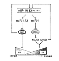
例えば、同一染色体座位に密集するmiRNA-1(miR-1)及びmiRNA-133(miR-133)は発生過程で組織特異的に相伴って転写される。miR―1及びmiR-133は、インビトロ培養筋細胞及びインビボXenopus胚における骨格筋増殖及び分化を調節する上で別個の役割を果たす。miR-1は、筋肉遺伝子発現の転写抑制因子である、ヒストンデアセチラーゼ4(HDAC4)を標的とすることにより、筋形成を促進する。対照的に、miR-133は、血清応答因子(SRF)を抑制することにより、筋細胞増殖を促進する。これらの結果は、同一miRNAポリシストロン由来の共通に転写される2個の成熟miRNAが、別個の生物的機能を果たすことができることを初めて明らかにした。従って、本開示は、miRNAが筋肉遺伝子発現及び胚発生を制御する転写回路に加わる分子機構を提供する。 For example, miRNA-1 (miR-1) and miRNA-133 (miR-133) that are clustered at the same chromosomal locus are transcribed in a tissue-specific manner during development. miR-1 and miR-133 play distinct roles in regulating skeletal muscle proliferation and differentiation in in vitro cultured muscle cells and in vivo Xenopus embryos. miR-1 promotes myogenesis by targeting histone deacetylase 4 (HDAC4), a transcriptional repressor of muscle gene expression. In contrast, miR-133 promotes myocyte proliferation by suppressing serum response factor (SRF). These results revealed for the first time that two commonly transcribed mature miRNAs from the same miRNA polycistron can perform distinct biological functions. Thus, the present disclosure provides the molecular mechanism by which miRNAs participate in transcriptional circuits that control muscle gene expression and embryonic development.
他の非限定的例として、Thrap1発現は同様にmiR-208により調節されている。Thrap1の3’UTRは、2個の推定miR-208結合部位を含む(図18)。2個の標的は、Thrap1終止コドンの〜80bp下流に位置し、互いに僅か〜50bpしか離れていない。両標的は、miR-208のシード領域と完全に相補的である。このThrap1遺伝子は、ユビキタスに発現しているTRAP(甲状腺ホルモン受容体タンパク質)複合体の240kdサブユニットである、TRAP240をコード化している。TRAPは、核受容体に対する活性化補助因子である、複数サブユニットタンパク質複合体であり、またTRAPファミリーメンバーは正常な発生に対して重要である。従って、miR-208は、TRAP240の合成を調節することができ、またホルモン依存性心筋分化を促進する。 As another non-limiting example, Thrap1 expression is similarly regulated by miR-208. The 3'UTR of Thrap1 contains two putative miR-208 binding sites (Figure 18). The two targets are located ˜80 bp downstream of the Thrap1 stop codon and are only ˜50 bp apart from each other. Both targets are completely complementary to the seed region of miR-208. This Thrap1 gene encodes TRAP240, which is a 240 kd subunit of the TRAP (thyroid hormone receptor protein) complex that is ubiquitously expressed. TRAP is a multi-subunit protein complex that is a coactivator for nuclear receptors, and TRAP family members are important for normal development. Thus, miR-208 can regulate TRAP240 synthesis and promote hormone-dependent myocardial differentiation.
1.一般的考察
C. elegans幼虫発生の適時性を制御することで、最初に記載されたmiRNAである、lin-4遺伝子は、lin-14mRNAレベルに顕著な効果を持たず、予想外に、lin-14タンパク質発現を抑制する21ヌクレオチド長の非コード性RNAを産生することが発見された。この小RNAがlin-14の3’非飜訳領域(UTR)内の相補的部位を標的にすることが見出された49,50。この現象は最初遺伝的奇異として処理され、実質的に無視されたが、今や、lin-4に類似した、miRNAと呼ばれる、何百もの小RNAが様々な種の遺伝子に存在して、相補的mRNAの飜訳を調節していると評価されている。最近の報告では、少数のmiRNAが顕著に様々な生物過程で役割を果たすと示唆されているが、大部分は特徴付けされていない。
1. General considerations
By controlling the timeliness of C. elegans larval development, the first described miRNA, the lin-4 gene, has no significant effect on lin-14 mRNA levels, and unexpectedly, lin-14 protein expression It was discovered to produce a 21 nucleotide long non-coding RNA that suppresses This small RNA was found to target a complementary site within the 3 'untranslated region (UTR) of lin- 1449,50 . This phenomenon was first treated as a genetic oddity and was virtually ignored, but now there are hundreds of small RNAs, called miRNAs, similar to lin-4, present in various species of genes that are complementary. It is evaluated to regulate the translation of mRNA. Recent reports suggest that a small number of miRNAs play a prominent role in a variety of biological processes, but are largely uncharacterized.
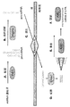
1.A.miRNA生合成及び機構
miRNA生合成に対する一般的モデルは、図13に示されている。成熟miRNAは〜22ヌクレオチド(nt)長を持ち、より長い転写物から処理される51,52。プライマリーmiRNA(pri-miRNA)は、独立な転写単位として、RNA POl IIにより転写可能であり、ホスト遺伝子のスプライスアウトイントロン由来であることができる53。このmiRNA処理経路は、ステムループ構造を持つ54〜70nt長の中間前駆体−miRNA(pre-miRNA)を作る、RNAseIIIエンドヌクレアーゼDroshaによるpri-miRNA切断に始まる。Exportin-5はDrosha切断により残されたスタガードカットを認識し、pre-miRNAを細胞質にRan-GTP依存的に送り出す54〜60。細胞質に入ると、pre-miRNAの両鎖は、もう一つのRNAseIII酵素である、Dicerにより、ステムループの元から約2ヘリカルターン離れた箇所で切断される61〜63。結果得られる〜22量体RNA2重鎖はDicerにより放出され、単鎖のステムアームが、RISC(RNA−誘導サイレンシング複合体)に取り込まれることができる。RISCは、miRNA及びmRNA標的と共に、Argonauteタンパク質ファミリー及びアクセサリー因子のメンバーを含む、リボヌクレオタンパク質複合体である。ステムアーム2本鎖の相対的熱安定性が、どちらの鎖がRISCに取り込まれるかを決めると考えられている:RISCに入る鎖は、しばしば5’末端がより不安定な鎖である64,65。飜訳阻害は、未知の機構により標的mRNAの3’UTR内の標的配列(複数)に対して相補的なmiRNAにより行われる66、67。一般的に、不完全な相補性は、飜訳抑制をもたらすが、完全又は、準完全相補性は、mRNA切断をもたらす68。miRNA生合成の多くの側面、トラフィッキング、RISC集合及びRISC機能の機構については不明の点が多いが、特定のmiRNAの機能研究及びmiRNA経路構成成分の遺伝的及び生化学的解析により、miRNAが様々な生物過程において重要であることが示された。
1. A. miRNA biosynthesis and mechanism
A general model for miRNA biosynthesis is shown in FIG. The mature miRNA has a length of ˜22 nucleotides (nt) and is processed from longer transcripts 51,52 . Primary miRNA (pri-miRNA) can be transcribed by RNA POII as an independent transcription unit and can be derived from the splice-out intron of the host gene 53 . The miRNA processing path, making 54 ~70Nt of the intermediate precursor-miRNA (pre-miRNA) that has a stem-loop structure begins pri-miRNA cleavage by RNAseIII endonuclease Drosha. Exportin-5 recognizes the staggered cut left by Drosha cleavage and delivers pre-miRNA to the cytoplasm in a Ran-GTP-dependent manner 54-60 . Upon entering the cytoplasm, both strands of the pre-miRNA are cleaved by Dicer, another RNAse III enzyme, at a location approximately 2 helical turns away from the origin of the stem loop 61-63 . The resulting ˜22-mer RNA duplex is released by Dicer and a single-stranded stem arm can be incorporated into RISC (RNA-induced silencing complex). RISC is a ribonucleoprotein complex that includes members of the Argonaute protein family and accessory factors, along with miRNA and mRNA targets. The relative thermal stability of the stem arm duplex is believed to determine which strand is incorporated into the RISC: strands that enter RISC are often more unstable at the 5 'end 64, 65 . Translational inhibition is performed by miRNAs complementary to the target sequence (s) within the 3′UTR of the target mRNA by an unknown mechanism 66,67 . In general, incomplete complementarity results in translational suppression, whereas complete or near-perfect complementarity results in mRNA cleavage 68 . Many aspects of miRNA biosynthesis, trafficking, RISC assembly, and the mechanism of RISC function are unclear, but miRNAs vary according to specific miRNA functional studies and genetic and biochemical analyzes of miRNA pathway components It was shown to be important in various biological processes.
I.B.発生におけるmiRNA
多細胞有機体の発生には、遺伝的経路の空間的及び時間的調節が必要である。miRNAは、標的遺伝子の転写後調節により、これらの複雑な遺伝的経路を制御又は微調節すると考えられている。動物発生におけるmiRNAの必要性を決定する一つの研究法は、miRNAを処理して成熟した活性型にするために必要な上流酵素である、Dicerの変異体作成であった。脊椎動物は、全ての脊椎動物miRNAを完全に処理するために必要である、Dicerを1コピーのみ有すると信じられている62,63,69。マウスにおいて、Dicer機能を欠損させると、7.5日胚(E)で致死的結果となる69。Dicer欠損マウスは、原始条痕マーカーT(brachury)を発現せず、原腸形成の間に体が構成される前に、発生が停止することを示す。特に、マウス肢中胚葉期にDicer機能が条件的に失われると、肢サイズが減少しまたプログラムされた細胞死が増加する70。母方の接合体のDicer変異体を作ることにより、ゼブラフィシュのmiRNA形成が完全にブロックされることは、miRNAの欠損は、胚の軸形成又は多くのタイプの細胞のパターン化には影響しないことを示す。しかしながら、原腸形成、脳形成、体節発生及び心臓発生における形態形成全ては、異常であることが証明され、また致死をもたらした71。まとめると、Dicer機能の遺伝解析は、成熟miRNAは正常な発生のために必要であることを示唆する。全てのmiRNA機能の除去は参考になるが、これらはまた、なまくらな道具であり、特定のmiRNAの正確な機能への洞察を提供するものではない。
I. B. MiRNA in development
The development of multicellular organisms requires spatial and temporal regulation of genetic pathways. miRNAs are thought to control or fine-tune these complex genetic pathways by post-transcriptional regulation of target genes. One approach to determine the need for miRNAs in animal development was the creation of Dicer mutants, which are upstream enzymes required to process miRNAs into mature active forms. Vertebrates are believed to have only one copy of Dicer, which is necessary to completely process all vertebrate miRNAs 62,63,69 . In mice, loss of Dicer function results in a lethal result in a 7.5 day embryo (E) 69 . Dicer-deficient mice do not express the primitive streak marker T (brachury), indicating that development stops before the body is constructed during gastrulation. In particular, the conditional loss of Dicer function during the mouse limb mesoderm stage reduces limb size and increases programmed cell death 70 . By creating a Dicer variant of the maternal zygote, zebrafish miRNA formation is completely blocked that miRNA deficiency does not affect embryonic axis formation or patterning of many types of cells Indicates. However, morphogenesis in gastrulation, brain formation, somite development and cardiac development has all proven to be abnormal and led to death 71 . In summary, genetic analysis of Dicer function suggests that mature miRNAs are required for normal development. Although removal of all miRNA functions is informative, these are also loose tools and do not provide insight into the exact function of a particular miRNA.
I.C.特定のmiRNAの生物的役割
miRNAが多くの生物過程に関与することを示唆する事実は、益々増加している。脾臓島細胞において、miR375の過剰発現は、グルコース誘導のインシュリン分泌を抑制するが、内因性miR-375の阻害はインシュリン分泌を促進する72。同様の過剰発現及び阻害戦術により、ERK5タンパク質発現の調節から、脂肪細胞分化におけるmiR-143の役割が同定された73。他の実施例において、5個のmiRNAをコードしている多シストロンのmiRNA遺伝子は、腫瘍発生と関係する74。miRNAの他の機能が、血液新生75、神経細胞分化76,77及びHox遺伝子発現の調節78,79において提言されている。
現在、300を超える既知のヒトmiRNAがあるが、約100個のみが生物機能について何らかの意味で割り当てられている。発生と病理学におけるmiRNAを介した調節の普及性及び重要性を理解するためには、特定のmiRNAの研究が必要である。本出願は、筋肉分化と増殖の調節におけるmiRNAの役割について初めて提供する。
I. C. Biological roles of specific miRNAs
The facts suggesting that miRNAs are involved in many biological processes are increasing. In spleen islet cells, overexpression of miR375 suppresses glucose-induced insulin secretion, whereas inhibition of endogenous miR-375 promotes insulin secretion 72 . By the same overexpression and inhibition tactics, from regulation of ERK5 protein expression, the role of miR-143 in adipocyte differentiation it was identified 73. In another example, a multicistronic
Currently there are over 300 known human miRNAs, but only about 100 have been assigned in some sense for biological function. In order to understand the prevalence and importance of miRNA-mediated regulation in development and pathology, specific miRNA studies are needed. This application provides for the first time the role of miRNAs in the regulation of muscle differentiation and proliferation.
I.D.心臓発生におけるmiRNA
心臓発生は、異なる遺伝的プログラムの詳細な調節を必要とする、従って、心臓に多量存在する、区別を付けて発現するmiRNAが、この複雑な経路を調節するに役立つと推量することは興味深い。この様な、組織特異的な発現パターンは、いくつかのmiRNAについて本出願に開示する。miR-1及びmiR-133は骨格筋及び心臓筋組織の両者に発現するが、miR-208は心筋組織のみに検知される。本明細書開示以前には、これら筋特異的miRNAの機能は明かではなかった。
I. D. MiRNA in heart development
It is interesting to speculate that differentially expressed miRNAs that are abundant in the heart can help regulate this complex pathway, as cardiac development requires detailed regulation of different genetic programs. Such tissue-specific expression patterns are disclosed in this application for several miRNAs. miR-1 and miR-133 are expressed in both skeletal and cardiac muscle tissue, whereas miR-208 is detected only in cardiac muscle tissue. Prior to disclosure herein, the function of these muscle-specific miRNAs was not clear.
I.E.miRNA標的の同定
特定のmiRNAの標的を同定することは、調節経路におけるこれらmiRNAの役割の理解を容易にする。大部分の動物miRNAはこれらの標的部位に対して不完全に相補的であり、このことは動物miRNA標的部位を同定する上で単純なホモロジー探索を用いることを妨げる。この障害を克服するために、いくつかの計算方法が開発され、これらは新しい動物miRNA標的を予想するための基準として、既知のmiRNA標的の保存配列及び特徴を取り込んだ80〜85。例えば、大部分のmiRNAは、“シード”領域と呼ばれる、有効な標的部位内の第2及び第8ヌクレオチドの間に高い相補性を示すことが、いくつかのアルゴリズムでは考慮されている。いくつかの場合、miRNAの3’末端の相補性が、弱い5’末端結合を埋め合わせるので、他のアルゴリズムでは考慮されていない。これらのアルゴリズムではまた、隣接する領域に対し、2又は2以上の種を越えた標的保存配列による予想を評価する。この種の計算方法により、いくつかの哺乳動物miRNA標的部位が成功裏に予想できた。どの様な特定のmiRNAに対して為される予想も、常に間違いなくファルスポジティブを作り出す。しかしながら、この様な予想は仮説ジェネレーター(生成系)として極めて重要である。どの様な予想でも、実験的に証明できて、また関連した生物学的状況に置くことができる。
I. E. Identification of miRNA targets Identifying specific miRNA targets facilitates an understanding of the role of these miRNAs in the regulatory pathway. Most animal miRNAs are incompletely complementary to these target sites, which precludes the use of a simple homology search to identify animal miRNA target sites. To overcome this obstacle, several computational methods have been developed, incorporating 80-85 conserved sequences and features of known miRNA targets as a basis for predicting new animal miRNA targets. For example, some algorithms take into account that most miRNAs show high complementarity between the second and eighth nucleotides in the effective target site, called the “seed” region. In some cases, complementation at the 3 ′ end of miRNAs has not been taken into account by other algorithms as it compensates for weak 5 ′ end binding. These algorithms also evaluate predictions due to target conserved sequences across two or more species for adjacent regions. With this type of calculation method, several mammalian miRNA target sites could be successfully predicted. The predictions made for any particular miRNA will always produce a false positive. However, such a prediction is extremely important as a hypothesis generator. Any expectation can be proven experimentally and placed in a relevant biological context.
I.F.重要性
現在、miRNA指向性抑制の背後の詳細な分子機構を理解するために、またmiRNA発現を解析し標的部位を同定するためのより良いツールを開発するために、また調節経路内の特定のmiRNAに対する生物学的に適切な役割を定めるために、miRNA研究におけるいくつかの活溌な領域が在る。
心臓発生及び病理学は複雑な遺伝的経路の調節と密接に関係しており、これらの経路を理解するために多くの試みが為されてきた。大部分の研究は、転写因子の役割及び心臓遺伝子転写に要求される調節エンハンサー配列に焦点が向けられてきた。心臓遺伝子発現の調節は、極めて複雑であることが分かり、個々の心臓遺伝子は、心臓における非常に制限された発現パターンを指令する、多種の独立したエンハンサーにより調節される。転写後レベルでの他の調節レイヤーが加わることで、潜在的に、miRNAのこの複雑さはさらに劇的に増加した。本出願は、一つには、心筋及び骨格筋遺伝子発現が如何に調節されているかについての新しい理解を提供し、治療及び、研究にこれらの発見の応用を開示する。更に、本明細書で開示した筋分化と増殖のmiRNA調節に関連する発見により、同様に他の経路におけるmiRNAの機能を理解するためのモデルとして役立つ。
I. F. Importance Currently, to understand the detailed molecular mechanisms behind miRNA-directed repression, to develop better tools to analyze miRNA expression and identify target sites, and to identify specific There are several active areas in miRNA research to define biologically relevant roles for miRNA.
Cardiac development and pathology are closely related to the regulation of complex genetic pathways, and many attempts have been made to understand these pathways. Most studies have focused on the role of transcription factors and regulatory enhancer sequences required for cardiac gene transcription. The regulation of cardiac gene expression has proven extremely complex, and individual cardiac genes are regulated by a variety of independent enhancers that direct very restricted expression patterns in the heart. The addition of other regulatory layers at the post-transcriptional level potentially increased this miRNA complexity even more dramatically. This application provides, in part, a new understanding of how myocardial and skeletal muscle gene expression is regulated, and discloses the application of these findings to therapy and research. Furthermore, the discoveries disclosed herein relating to miRNA regulation of muscle differentiation and proliferation serve as models for understanding miRNA function in other pathways as well.
II.定義
便宜上、明細書、実施例及び添付した請求の範囲に用いられる幾つかの用語を此処に集めた。以下の用語は当業者に良く理解されていると信ずるが、以下の定義を、本出願の説明に供するために示す。
異なって定義しなければ、本明細書で用いられる全ての技術的及び科学的用語は、本出願が属するこの分野の当業者には一般的に理解されている意味と同一である。本明細書に記載した方法、デバイス、材料と等価なこれら全てを、本出願の実行又は試験に用いることができるが、代表的な方法、デバイス及び材料についてこれから説明する。
長年にわたる特許法協定に従い、原文の“不定冠詞−ある(a、an)”“定冠詞−その(the)”は、請求の範囲を含め本出願で用いられる場合、“1又は2以上”を意味する。従って、本明細書で“ある(a,an,)”その(
the)”、に言及した場合、1又は2以上(少なくとも1の)の本書の文法的目的物を示す。実施例のために、“ある成分”は1成分又は1以上の成分を表わす。
本明細書で用いるように、値又は質量、重量、時間、容積、濃度、パーセンテージに関する場合、用語“約”は、本出願を実行する上で以下の変動が適切であるように、特定の値から、幾つかの実施態様では、±20%又は±10%、いくつかの実施態様では±5%、いくつかの実施態様では±1%、いくつかの実施態様では、±0.5%、いくつかの実施態様では±0.1%の変動を含む。もし別に指定しない場合、本明細書及び請求の範囲で用いる構成要素、反応条件、等々の量を表現する全ての数は、全ての実施例において、用語“約”により、修正されていると理解すべきである。従って、これに反すると指示しない限り、本明細書及び添付した請求の範囲に示された数字上のパラメーターは、本出願で得られる所望の特性に依存して変化する様に、近似的である。
II. For convenience of definition , several terms used in the specification, examples, and appended claims are collected here. Although the following terms are believed to be well understood by those skilled in the art, the following definitions are provided for the purpose of describing this application.
Unless defined differently, all technical and scientific terms used herein have the same meaning as commonly understood by one of ordinary skill in the art to which this application belongs. All of the methods, devices, and materials equivalent to those described herein can be used in the practice or testing of the present application, but representative methods, devices and materials are now described.
In accordance with patent law agreements over the years, the original "indefinite article-a (an)" or "definite article-the" means "one or more" when used in this application, including the claims. To do. Therefore, in this description, “is (a, an,)” (
the) "indicates one or more (at least one) grammatical objects in this document. For purposes of example," a component "represents one component or more than one component.
As used herein, when referring to a value or mass, weight, time, volume, concentration, percentage, the term “about” is intended to mean a particular value so that the following variations are appropriate in carrying out this application. From some embodiments, ± 20% or ± 10%, in some embodiments ± 5%, in some embodiments ± 1%, in some embodiments ± 0.5%, Some embodiments include ± 0.1% variation. Unless otherwise specified, all numbers expressing quantities of components, reaction conditions, etc. used in the specification and claims are understood to be modified in all examples by the term “about”. Should. Accordingly, unless indicated to the contrary, the numerical parameters set forth in this specification and the appended claims are approximate, such that they vary depending on the desired properties obtained in this application. .
本明細書で用いるように、用語“アミノ酸”及び“アミノ酸残基”は、互換性があり、20個の自然に存在するアミノ酸及び類似体、誘導体及びこれらの同族体(即ち、様々な側鎖を持つアミノ酸類縁体、前記のいずれかの全ての立体異性体)の何れかを表わす。従って、用語“アミノ酸”は、自然又は合成を問わず、アミノ基官能性及び酸官能性を持ち、自然に存在する高分子に含まれ得る、全ての分子を包含する。
アミノ酸はポリペプチドをペプチド結合の部分で化学消化(加水分解)して作ることができる。本明細書に記載するアミノ酸残基は幾つかの実施態様では“L”異性体型である。しかしながら、希望の機能特性がポリペプチドに保持される限り、L−アミノ酸残基を“D”異性体型の残基で置き換えることができる。NH2は、ポリペプチドのアミノ末端に存在する自由アミノ基を表わす。COOHは、ポリペプチドのカルボキシ末端に存在する自由カルボキシ基を表わす。標準的ポリペプチド命名法を守りながら、アミノ酸残基の略号を本明細書に示す表に示した。
本明細書に、化学式で示した全てのアミノ酸残基配列は、通常のアミノ末端からカルボキシ末端の方向について、左より右への方向で表わすことを注意する。更に、用語“アミノ酸”及び“アミノ酸残基”は、修飾型の及び異常なアミノ酸を含む様に広く定義されている。
As used herein, the terms “amino acid” and “amino acid residue” are interchangeable and refer to the 20 naturally occurring amino acids and analogs, derivatives and homologs thereof (ie, various side chains. Or any of the above stereoisomers). Thus, the term “amino acid” encompasses all molecules, whether natural or synthetic, that have amino group functionality and acid functionality and can be included in naturally occurring macromolecules.
Amino acids can be made by chemically digesting (hydrolyzing) a polypeptide at the peptide bond. The amino acid residues described herein are in some embodiments in the “L” isomeric form. However, as long as the desired functional property is retained in the polypeptide, L-amino acid residues can be replaced with residues of the “D” isomeric form. NH 2 represents a free amino group present at the amino terminus of a polypeptide. COOH represents a free carboxy group present at the carboxy terminus of a polypeptide. Abbreviations for amino acid residues are shown in the tables provided herein, while observing standard polypeptide nomenclature.
Note that all amino acid residue sequences shown in the chemical formulas herein are represented from left to right in the normal amino-terminal to carboxy-terminal direction. Furthermore, the terms “amino acid” and “amino acid residue” are broadly defined to include modified and unusual amino acids.
更に、アミン酸残基配列の初め又は終わりのダッシュは、1又は2以上のアミノ酸残基の配列へのペプチド結合又はNH2の様なアミノ末端基又はアセチル基又はCOOHの様なカルボキシ末端基への共有結合を示すことを注意する。
本明細書に用いるように、用語“細胞”は、通常の生物的意味で用いられる。いくつかの実施態様では、細胞は、例えば、脊椎動物のような有機体に存在する。細胞は、真核生物(例えば、骨格筋又は心筋のような、筋細胞)又は原核生物(例えば、バクテリア)であり得る。細胞は、体細胞系又は生殖細胞系由来であり、全能性、多能性又は何らかの程度分化した、分裂性又は非分裂性であり得る。また細胞は、誘導型又は生殖体又は胚、肝細胞又は完全に分化した細胞を含みうる。
本明細書で用いるように、用語“ホスト細胞”及び“組み換えホスト細胞”は、互換性があり、本出願の構成物(例えば、miRNAをコードする発現ベクターのような)の組成物が導入された細胞(例えば、筋細胞)を表わす。更に、この用語は、発現構築物が最初導入された特定の細胞ばかりでなく、この様な細胞の子孫又は潜在的子孫を表わす。突然変異又は環境からの影響により、継代中にある種の変化が生じる可能性があり、この様な子孫は、実際、親細胞とは同一でないかも知れないが、尚、本明細書で用いられる用語の範囲内に含まれる。
Furthermore, the dash at the beginning or end of the amino acid residue sequence may be a peptide bond to the sequence of one or more amino acid residues or an amino terminal group such as NH 2 or a carboxy terminal group such as acetyl or COOH. Note that it shows a covalent bond.
As used herein, the term “cell” is used in its ordinary biological sense. In some embodiments, the cell is present in an organism such as, for example, a vertebrate. The cell can be eukaryotic (eg, a muscle cell, such as skeletal or cardiac muscle) or prokaryotic (eg, a bacterium). The cells are derived from somatic or germline and can be totipotent, pluripotent or some degree of differentiated, dividing or non-dividing. The cells can also include induced or reproductive or embryonic, hepatocytes or fully differentiated cells.
As used herein, the terms “host cell” and “recombinant host cell” are interchangeable and introduce a composition of the composition of the present application (such as an expression vector encoding a miRNA). Cell (eg, myocyte). Furthermore, the term refers to the progeny or potential progeny of such a cell as well as the particular cell into which the expression construct was first introduced. Mutations or environmental influences can cause certain changes during passage, and such progeny may not actually be identical to the parent cell, but are used herein. Within the scope of the terminology used.
本明細書で用いるように、用語“遺伝子”は、例えば、ポリペプチドをコードする構造遺伝子を含む、がそれに制限されない、核酸配列のような、RNAをコードする核酸を表わす。用語“遺伝子”は、また広く、生物機能と結びついた如何なるDNAの断片でも表わす。従って、用語“遺伝子”は、コードする配列;プロモーター領域;転写調節配列;調節タンパク質に対する特異的認識配列である、非発現DNA断片;例えば本出願の見本であるmiRNAにより標的とされ、結合されるmRNAの3’非飜訳領域に転写されるDNA断片のような、遺伝子発現に寄与する部分である非発現DNA断片;希望するパラメーターを持つようデザインされたDNA断片;又はこれらの組合せを含むがこれらに制限されない配列を含む。ある遺伝子は様々な方法で得ることができて、生物試料からのクローニング、既知の又は予想される配列情報による合成及び1又は2以上の存在する配列由来の組み換えが含まれる。 As used herein, the term “gene” refers to a nucleic acid encoding an RNA, such as, for example, a nucleic acid sequence, including but not limited to a structural gene encoding a polypeptide. The term “gene” also broadly refers to any piece of DNA associated with biological function. Thus, the term “gene” is targeted and bound by a coding sequence; a promoter region; a transcriptional regulatory sequence; a non-expressed DNA fragment that is a specific recognition sequence for a regulatory protein; a non-expressed DNA fragment that is a part that contributes to gene expression, such as a DNA fragment transcribed into the 3 ′ untranslated region of mRNA; a DNA fragment designed to have the desired parameters; or a combination thereof Including but not limited to sequences. A gene can be obtained in a variety of ways, including cloning from biological samples, synthesis with known or predicted sequence information, and recombination from one or more existing sequences.
当業者は理解するように、遺伝子は概して、コード鎖及び非コード鎖を含む。本明細書で用いるように、用語“コード鎖”及び“センス鎖”は互換性があり、遺伝子産物に飜訳されるmRNAと同じヌクレオチド配列を持つ核酸配列を表わす。やはり、当業者が理解するように、コード鎖及び/又はセンス鎖がDNA分子を表わすために用いられる場合、このコード/センス鎖は、対応するmRNAに存在するウリジン残基の変わりにチミジン残基を含む。さらに、コード/センス鎖をDNA分子を表わすために用いる場合、コード/センス鎖は、プロモーター、エンハンサー、イントロンを含むがこれに制限されない、mRNAに含まれてない付加的因子を含むことができる。同様に、用語“鋳型鎖”及び“アンチセンス鎖”は、互換性があり、コード/センス鎖に相補的な核酸配列を表わす。しかしながら、例えばmiRNA遺伝子のようにポリペプチド産物をコードしてない遺伝子に対して、用語“コード鎖”が、miRNAを含む鎖を表わすために用いられる。この使用法において、miRNAを含む鎖は、miRNA前駆体に関してはセンス鎖であり、他方、標的RNAに関してはアンチセンス鎖である(即ち、標的RNAに対してアンチセンスである配列をmiRNAは含むので、miRNAは標的RNAとハイブリダイズする。) As those skilled in the art will appreciate, a gene generally includes a coding strand and a non-coding strand. As used herein, the terms “coding strand” and “sense strand” are interchangeable and refer to a nucleic acid sequence having the same nucleotide sequence as the mRNA translated into the gene product. Again, as those skilled in the art will appreciate, when a coding strand and / or sense strand is used to represent a DNA molecule, this coding / sense strand is replaced by a thymidine residue instead of a uridine residue present in the corresponding mRNA. including. Further, when the coding / sense strand is used to represent a DNA molecule, the coding / sense strand can contain additional factors not included in the mRNA, including but not limited to promoters, enhancers, and introns. Similarly, the terms “template strand” and “antisense strand” are interchangeable and refer to a nucleic acid sequence that is complementary to the coding / sense strand. However, for genes that do not encode a polypeptide product, such as, for example, miRNA genes, the term “coding strand” is used to denote the strand that contains the miRNA. In this usage, the strand that contains the miRNA is the sense strand for the miRNA precursor, while it is the antisense strand for the target RNA (ie, the miRNA contains a sequence that is antisense to the target RNA). MiRNA hybridizes with target RNA.)
本明細書で用いるように、用語“相補性”及び”相補的な”は伝統的なWatson-Crick型又は他の非伝統的タイプの相互作用により、他の核酸と1又は2以上の水素結合を形成することができる核酸を表わす。本出願の核酸分子に関連して、核酸分子が相補的配列と結合する自由エネルギーは、いくつかの実施態様において、核酸の関係機能がリボヌクレアーゼ活性を進行可能にするに充分である。例えば、あるmiRNA前駆体のセンス及びアンチセンス鎖の間の相補性の程度は、miRNA前駆体のmiRNAを含む鎖と標的核酸配列の間の相補性の程度と同じ又は異なる可能性がある。核酸分子に対する結合自由エネルギーの測定は当分野ではよく知られている。Freier他、198631; Turner他、198732を参照のこと。
本明細書で用いるように、用語“パーセント相補性”、“パーセント同一性”及び“パーセント同一”は、本明細書では互換性があり、第2の核酸配列と水素結合(例えば、Watson-Crick塩基対)を形成する、ある核酸分子における連続残基の割合を表わす(例えば、10塩基対の中5,6,7,8,9,10塩基対は、50%,60%,70%,80%,90%及び100%相補性である)。用語“100%相補性”、“全面的相補性”及び“完全相補性”は、核酸配列の連続残基の全てが、第2の核酸配列の同数の連続残基と水素結合をすることができることを示す。miRNAは約17〜24ntであり、また5ミスマッチ(即ち、1,2,3,4又は5ミスマッチ)は、遺伝子発現のmiRNAを指向する調節の間、一般的に許容されるので、あるmiRNA及び標的となるRNAの間の少なくとも約70%パーセント相補性は、miRNAが、標的RNAを導く遺伝子の発現を調節するために十分である。
As used herein, the terms “complementarity” and “complementary” refer to one or more hydrogen bonds with other nucleic acids by traditional Watson-Crick type or other non-traditional types of interactions. Represents a nucleic acid capable of forming In connection with the nucleic acid molecules of the present application, the free energy with which the nucleic acid molecule binds to a complementary sequence is sufficient in some embodiments to allow the relevant function of the nucleic acid to proceed with ribonuclease activity. For example, the degree of complementarity between the sense and antisense strands of a miRNA precursor may be the same or different from the degree of complementarity between the miRNA precursor miRNA containing strand and the target nucleic acid sequence. Measurement of binding free energy for nucleic acid molecules is well known in the art. See Freier et al., 1986 31 ; Turner et al., 1987 32 .
As used herein, the terms “percent complementarity”, “percent identity” and “percent identity” are interchangeable herein and are hydrogen bonding (eg, Watson-Crick) to a second nucleic acid sequence. (For example, 5, 6, 7, 8, 9, 10 base pairs out of 10 base pairs are 50%, 60%, 70%, 80%, 90% and 100% complementarity). The terms “100% complementarity”, “full complementarity” and “perfect complementarity” mean that all of the contiguous residues of a nucleic acid sequence hydrogen bond with the same number of contiguous residues of a second nucleic acid sequence. Show what you can do. miRNAs are about 17-24 nt, and 5 mismatches (
用語“遺伝子発現”は、一般的に、生物的に活性なポリペプチドがDNA配列より作られ細胞内で生物活性を示す生物過程を表わす。従って、遺伝子発現は、転写及び飜訳過程を伴うが、遺伝子又は遺伝子産物の生物活性に影響する転写後及び翻訳後の過程を伴う。これらの過程は、RNA合成、RNAプロセシング、RNA輸送及びポリペプチド合成、ポリペプチド輸送及びポリペプチドの翻訳後修飾を含むが、これらに制限されない。さらに、細胞内のタンパク質―タンパク質相互作用に影響する過程が、本明細書に定義した遺伝子発現に影響を与える可能性がある。
しかしながら、例えば、miRNA遺伝子のように、タンパク質産物をコードしてない遺伝子の場合、用語“遺伝子発現”は、前駆体miRNAが遺伝子から作られる過程を表わす。タンパク質をコードする遺伝子に対するRNAポリメラーゼIIにより行われる転写とは異なり、miRNA遺伝子の転写産物はタンパク質を作るために飜訳されないが、一般的に、この過程は転写と呼ばれる。それでもなお、miRNA遺伝子から成熟したmiRNAの合成は、本明細書で用いるように用語“遺伝子発現”に含まれる。
The term “gene expression” generally refers to a biological process in which a biologically active polypeptide is made from a DNA sequence and exhibits biological activity in a cell. Thus, gene expression involves transcription and translation processes, but involves post-transcriptional and post-translational processes that affect the biological activity of the gene or gene product. These processes include, but are not limited to, RNA synthesis, RNA processing, RNA transport and polypeptide synthesis, polypeptide transport and post-translational modification of polypeptides. In addition, processes that affect protein-protein interactions in the cell can affect gene expression as defined herein.
However, in the case of genes that do not encode a protein product, for example miRNA genes, the term “gene expression” refers to the process by which precursor miRNAs are made from genes. Unlike transcription performed by RNA polymerase II on genes encoding proteins, miRNA gene transcripts are not translated to make proteins, but this process is generally referred to as transcription. Nevertheless, the synthesis of mature miRNA from a miRNA gene is included in the term “gene expression” as used herein.
本明細書で用いるように、用語“単離した”は、他の核酸、タンパク質、脂質、炭化水素及び/又は、細胞物質又は合成媒体と通常結合している、他の物質から実質的に遊離した分子を表わす。従って、用語“単離した核酸”は、自然又は合成由来又はその組合せのリボ核酸分子又はデオキシリボ核酸分子(例えば、ゲノムDNA、cDNA、mRNA、miRNAその他)を表し、この核酸分子は(1)“単離した核酸”が自然で見られる細胞と結合していない又は(2)自然で連結してないポリヌクレオチドと作動可能に連結している。同様に、用語“単離したポリペプチド”は、幾つかの実施態様では、組み換えDNA又はRNAから合成された又は合成由来の又はこれらの何らかの組合せによる、ポリペプチドを表し、このポリペプチドは、(1)自然で通常見出されるタンパク質と結合してない、(2)本来そこに存在する細胞から単離された、(3)同じ細胞由来の他のタンパク質から遊離している、(4)異なる種の細胞により発現される又は(5)自然に存在しない物である。
用語“単離した”は、“単離した細胞”という文脈で用いる場合、例えば、器官、組織又は有機体の一部として、自然の環境から取り除かれた細胞を表わす。
As used herein, the term “isolated” is substantially free from other nucleic acids, proteins, lipids, hydrocarbons and / or other substances that are normally associated with cellular or synthetic media. Represents a molecule. Thus, the term “isolated nucleic acid” refers to a ribonucleic acid molecule or deoxyribonucleic acid molecule (eg, genomic DNA, cDNA, mRNA, miRNA, etc.) of natural or synthetic origin or combinations thereof, wherein the nucleic acid molecule is (1) “ An “isolated nucleic acid” is not associated with a cell found in nature or (2) is operably linked to a polynucleotide that is not naturally linked. Similarly, the term “isolated polypeptide” refers, in some embodiments, to a polypeptide synthesized from or derived from a recombinant DNA or RNA, or some combination thereof, wherein the polypeptide is ( 1) not bound to a protein normally found in nature, (2) isolated from the cells originally present, (3) free from other proteins from the same cell, (4) different species Or (5) a non-naturally occurring product.
The term “isolated” when used in the context of “isolated cells” refers to cells that have been removed from their natural environment, for example, as part of an organ, tissue or organism.
本明細書で用いるように、用語“標識”及び“標識された”はプローブ分子に分光学的に、放射線学的に及び他の方法で検出可能な分子部分を結合させることを表わす。従って、用語“標識”又は“標識した”は、ポリペプチドのような分子に検出可能なマーカーを任意に共有結合的に又は非共有結合的に取り込む又は結合することを表わす。ポリペプチドを標識する様々な方法は、当技術分野で既知であり、使用できる。ポリペプチドの標識の実施例としては、以下の標識があるが、これらに制限されない:放射性同位元素、蛍光標識、重原子、酵素標識又はレポーター遺伝子、化学発光基、ビオチニル基、第2のレポーターにより認識される所定のポリペプチドエピトープ(例えば、ロイシンジッパー対配列、抗体の結合位置、金属結合領域、エピトープタグ)。いくつかの実施態様において、標識には様々な長さのスペーサーアームを結合して、潜在的な立体障害を減少させる。 As used herein, the terms “label” and “labeled” refer to attaching a molecular moiety that is spectroscopically, radiologically, and otherwise detectable to a probe molecule. Thus, the term “label” or “labeled” refers to the incorporation or attachment of a detectable marker, optionally covalently or non-covalently, to a molecule such as a polypeptide. Various methods of labeling polypeptides are known in the art and can be used. Examples of labeling of a polypeptide include, but are not limited to, the following labels: radioisotope, fluorescent label, heavy atom, enzyme label or reporter gene, chemiluminescent group, biotinyl group, second reporter A given polypeptide epitope to be recognized (eg, leucine zipper pair sequence, antibody binding location, metal binding region, epitope tag). In some embodiments, the label is attached with spacer arms of various lengths to reduce potential steric hindrance.
本明細書で用いるように、用語“調節する”は、生化学的実体の、どんな又は全ての科学的及び生物的活性又は特性を増加、減少又は他の如何なる変化も表わすことが可能である。例えば、用語“調節する”は、発現、レベル又は活性が、調節因子がない場合に観測される値より大きい又は小さいと言うように、遺伝子の発現レベル又は1又は2以上のタンパク質又はタンパク質サブユニットをコードしているRNA分子又は等価なRNA分子のレベルを変化させること;又は上方制御又は下方制御された1又は2以上のタンパク質又はタンパク質サブユニットの活性を表視、その結果発現、レベル又は活性は、調節因子なしで観測される結果より大きい又は小さい。例えば、用語“調節する”は、“阻害する”又は“抑制する”ということを意味するが、語“調節する”はこの定義に制限されない。 As used herein, the term “modulate” can represent an increase, decrease or any other change in any or all scientific and biological activities or properties of a biochemical entity. For example, the term “modulate” refers to the expression level of a gene or one or more proteins or protein subunits, such that the expression, level or activity is greater or less than the value observed in the absence of a modulator. Altering the level of RNA molecules encoding or equivalent RNA molecules; or viewing the activity of one or more proteins or protein subunits that are up- or down-regulated, resulting in expression, level or activity Is greater or less than the result observed without a regulator. For example, the term “modulate” means “inhibit” or “suppress”, but the term “modulate” is not limited to this definition.
本明細書で用いる、用語“調節”は、ある応答の上方制御(即ち、活性化又は刺激)及び下方制御(即ち、阻害又は抑制)を表わす。従って、用語“調節”は、機能特性又は生物活性又はプロセス(例えば、酵素活性又は受容体結合)と関連して用いる場合、上方制御(例えば、活性化、刺激化)、下方制御(例えば、阻害又は抑制)の能力を表わす又はそうでなければ、この様な特性、活性又はプロセスの性質を変化させる。ある実施例では、この様な調節は、シグナル伝達経路の活性化の様な、連続した特定のでき事であり得る及び/又は特定の細胞型だけに表れうる。 As used herein, the term “modulation” refers to upregulation (ie activation or stimulation) and downregulation (ie inhibition or suppression) of a response. Thus, the term “modulation” when used in connection with a functional property or biological activity or process (eg, enzyme activity or receptor binding), upregulation (eg, activation, stimulation), downregulation (eg, inhibition) Or otherwise, altering such properties, activities or the nature of the process. In certain embodiments, such modulation can be a continuous specific event, such as activation of a signaling pathway, and / or can appear only in a specific cell type.
用語“調節因子”は、調節を行うことができる、ポリペプチド、核酸、高分子、複合体、分子、小分子、化合物、種又はその他(自然に又は非自然に存在)又は、バクテリア、植物、菌類又は動物細胞又は動物組織からの単離物を表わす。アッセイ系に加えることにより、調節因子は、機能的特性、生物活性又は生物過程又はこれらの組合せ(例えば、作用薬、部分的拮抗剤、部分的作用薬、反作用薬、拮抗剤、抗生物質、細菌感染又は細菌増殖の阻害剤、等々)の阻害剤又は活性剤(間接的又は直接的)として可能な活性に対して評価される。このようなアッセイにおいて、多くの調節因子が一度で探索できる。調節因子の活性は、既知、未知又は部分的既知である。
調節因子は、選択的又は非選択的である。本明細書で用いるように、用語“選択的”は、調節因子の文脈(例えば、阻害剤)で用いられる場合、測定しうる又はさもなければ、調節因子が、生物的に対応して、他の類似であるが同一ではない分子(例えば、興味の対象としての標的RNAと同じ遺伝子ファミリーのメンバー由来のRNA)と比べて、一つの分子(例えば、興味の対象としての標的RNA)と相互作用の様式が異なることを表わす。
The term “modulator” refers to a polypeptide, nucleic acid, macromolecule, complex, molecule, small molecule, compound, species or other (naturally or non-naturally occurring) or bacterium, plant, Represents an isolate from a fungus or animal cell or animal tissue. By adding to the assay system, the modulator may be a functional property, biological activity or biological process or a combination thereof (eg, agonist, partial antagonist, partial agonist, counteracting agent, antagonist, antibiotic, bacterial Inhibitors of infection or bacterial growth, etc.) are evaluated for possible activity as inhibitors or activators (indirect or direct). In such an assay, many modulators can be explored at once. The activity of the modulator is known, unknown or partially known.
Modulators are selective or non-selective. As used herein, the term “selective” when used in the context of a modulator (eg, an inhibitor) can be measured or else the modulator is biologically corresponding, Interacts with one molecule (eg, target RNA of interest) compared to similar but not identical molecules (eg, RNA from members of the same gene family as the target RNA of interest) Indicates that the style is different.
選択的調節因子と考える調節因子に対して、標的との相互作用の性質は、標的に関連した他の分子(例えば、標的自体以外のファミリーメンバーからの転写物)との相互作用を完全に排除するものではないと理解すべきである。言い方を変えると、用語、選択的調節因子は、興味ある遺伝子からのmRNA転写物にのみ結合して、関連するファミリメンバーからの転写物には結合しない分子に制限されることを意図してない。この用語は、興味のある遺伝子からの及び関連するファミリーメンバーからの、転写物と相互作用することができる調節因子を含むことを意図するが、しかしながらこれに関しては、ファミリーメンバーと比較して標的との相互作用を変えて、生物的に該当する結果をもたらすような条件をデザインすることは可能である。この様な条件としては、調節因子とファミリメンバーの間で配列が同一でない場合及び、いくつかの調節因子を発現するが全てのファミリーメンバーを発現しない、特定の組織又は細胞型において調節因子を使用する場合を含むが、これに限定されない。後者の条件下で、もしある調節因子が標的と相互作用し、生物的に対応する効果をもたらすならば、ある調節因子は所定の組織における所定の標的に対して選択的であるかも知れないが、この場合、他の組織においては、さらなるファミリーメンバーが発現して、この調節因子が標的と相互作用しても、他のファミリーメンバーが存在するために、この調節因子が組織から“抜き取”られてしまい、生物効果を全くもたらさないこともあり得る。 For regulators that are considered selective regulators, the nature of the interaction with the target completely eliminates interactions with other molecules associated with the target (eg, transcripts from family members other than the target itself). It should be understood that it does not. In other words, the term selective regulator is not intended to be restricted to molecules that bind only to mRNA transcripts from the gene of interest and not to transcripts from related family members. . The term is intended to include regulatory elements that can interact with transcripts from the gene of interest and from related family members, but in this regard, the target is compared to the family member. It is possible to design conditions that will change the interaction of and produce biologically relevant results. Such conditions include using a regulator in a specific tissue or cell type where the sequence is not identical between the regulator and the family member, and that expresses some regulators but not all family members. Including, but not limited to. Under the latter conditions, certain modulators may be selective for a given target in a given tissue if they interact with the target and produce a corresponding biological effect. In this case, in other tissues, an additional family member is expressed, and even if this regulator interacts with the target, the other family member is present, so that this regulator is “extracted” from the tissue. And may have no biological effect.
選択的調節因子と同定されると、調節因子は、他の分子(例えば、興味ある遺伝子と関連する遺伝子のmRNA転写物)と結合する様式とは異なる様式で(例えば、より強く)、一分子(例えば、興味ある遺伝子のあるmRNA転写物)と結合する。本明細書で用いるように、この調節因子は、この調節因子が結合する他のいくつかの可能な分子と比較してより強く結合する分子に対して“選択的結合”又は“優先的結合”を示すという。
本明細書で用いるように、用語“阻害”、“抑制”、“下方制御”及びこれらの文法的言い換えは互換性があり、遺伝子産物(例えば、ポリペプチド)、遺伝子発現、ポリヌクレオチドの活性(例えば、miRNA)又は1又は2以上の遺伝子産物をコードするRNAのレベルが、本出願を実行しない場合観察されるレベル以下に減少する。
Once identified as a selective regulator, the regulator is one molecule in a manner that is different (eg, stronger) from binding to other molecules (eg, mRNA transcripts of genes associated with the gene of interest). (Eg, an mRNA transcript of a gene of interest). As used herein, this modulator is “selective binding” or “preferential binding” to a molecule that binds stronger compared to some other possible molecule to which the modulator binds. It says that.
As used herein, the terms “inhibition”, “suppression”, “down-regulation” and their grammatical paraphrasing are interchangeable: gene product (eg, polypeptide), gene expression, polynucleotide activity ( For example, miRNA) or the level of RNA encoding one or more gene products is reduced below that observed when not carrying out the present application.
いくつかの実施態様において、miRNA分子による阻害は、標的RNAの定常状態の発現レベルの減少に帰結する。いくつかの実施態様において、miRNA分子による阻害は、標的の発現レベルを下方制御できない不活性な又は弱力化した分子存在下で観測されるレベル以下の標的遺伝子の発現をもたらす。いくつかの実施態様において、本出願のmiRNA分子による遺伝子発現の阻害は、miRNA分子存在下での方が非存在下の場合よりも大きい。いくつかの実施態様において、遺伝発現の阻害は、遺伝子によりコードされたmRNAの分解速度の促進と結びついている(例えば、miRNAを介する遺伝子発現の阻害)。いくつかの実施態様において、本出願のmiRNAによる阻害は、標的遺伝子からの遺伝子産物の、miRNA非存在下で観測されるレベル以下の、発現レベルをもたらす。 In some embodiments, inhibition by miRNA molecules results in a decrease in the steady state expression level of the target RNA. In some embodiments, inhibition by a miRNA molecule results in expression of the target gene below that observed in the presence of an inactive or attenuated molecule that cannot down-regulate the target expression level. In some embodiments, inhibition of gene expression by miRNA molecules of the present application is greater in the presence of miRNA molecules than in the absence. In some embodiments, inhibition of genetic expression is associated with an increased rate of degradation of the mRNA encoded by the gene (eg, inhibition of gene expression via miRNA). In some embodiments, inhibition by miRNA of the present application results in an expression level of the gene product from the target gene that is below that observed in the absence of miRNA.
いくつかの実施態様において、例えば内在性miRNAの様な、miRNAはmiRNA阻害剤により阻害される可能性があり、miRNAが阻害されてない場合の遺伝子発現レベル(例えば、遺伝子産物の産生)と比較して、miRNAによる標的となった遺伝子発現レベルの増加をもたらす。本明細書で用いるように、用語“miRNA阻害剤”及び“miRNAの阻害”は互換性があり、miRNAの活性を阻害する分子を表わす。 In some embodiments, miRNAs can be inhibited by miRNA inhibitors, such as endogenous miRNAs, and compared to gene expression levels when miRNAs are not inhibited (eg, production of gene products). This leads to an increase in the level of gene expression targeted by miRNA. As used herein, the terms “miRNA inhibitor” and “miRNA inhibition” are interchangeable and refer to molecules that inhibit the activity of miRNA.
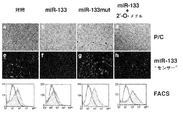
いくつかの実施態様において、miRNA阻害剤は、特定の条件下で、特定の標的miRNAとハイブリダイズするポリヌクレオチドであり、これにより標的miRNAの活性を阻害する。miRNA阻害剤が標的miRNAとハイブリダイズする条件としては、例えば、生理学状態がある。標的miRNAポリヌクレオチドに対するmiRNA阻害剤ポリヌクレオチド配列の相補性に基づき、miRNA阻害剤は標的miRNAとより多く又はより少ない度合いでハイブリダイズする。いくつかの実施態様において、miRNAは、標的miRNAの全て又は部分と完全に相補的である又は不完全に相補的であり、例えば、標的miRNAに対して99%、98%、97%、96%、95%、90%、80%又は70%相補性が挙げられ、当業者により一般的に理解されているように、これらは特定の応用によるものであり、また特異性の必要性による。miRNA阻害剤は、特定の条件セットの下で、標的miRNA活性の要求量を阻害するために必要なことは、標的miRNAと相補性を共有しさえすればよい。本出願に用いられるmiRNA阻害剤の実施例としては、2’-O-メチルポリヌクレオチドのような修飾ポリヌクレオチドが含まれるが、これに制限されない。代表的、非限定の実施例は、表2及び表3に示し、2’-O-メチル-miR-1,2’-O-メチル−miR-133及び2’-O-メチル−miR-208が挙げられ、これらは有意に、それぞれ、miR-1,miR-133又はmiR-208の活性を特異的に阻害できる。 In some embodiments, an miRNA inhibitor is a polynucleotide that hybridizes under certain conditions to a particular target miRNA, thereby inhibiting the activity of the target miRNA. Examples of conditions under which an miRNA inhibitor hybridizes with a target miRNA include physiological conditions. Based on the complementarity of the miRNA inhibitor polynucleotide sequence to the target miRNA polynucleotide, the miRNA inhibitor hybridizes to a greater or lesser extent with the target miRNA. In some embodiments, the miRNA is completely complementary or incompletely complementary to all or part of the target miRNA, eg, 99%, 98%, 97%, 96% to the target miRNA. , 95%, 90%, 80% or 70% complementarity, and as is generally understood by those skilled in the art, these depend on the particular application and on the need for specificity. An miRNA inhibitor need only share complementarity with the target miRNA as long as it is necessary to inhibit the required amount of target miRNA activity under a particular set of conditions. Examples of miRNA inhibitors used in the present application include, but are not limited to, modified polynucleotides such as 2'-O-methyl polynucleotides. Representative, non-limiting examples are shown in Tables 2 and 3 and include 2'-O-methyl-miR-1, 2'-O-methyl-miR-133 and 2'-O-methyl-miR-208. Which can significantly specifically inhibit the activity of miR-1, miR-133 or miR-208, respectively.
本明細書で用いるように、用語“突然変異”は伝統的な内包的意味を保ち、核酸又はポリペプチド配列における、遺伝による、自然発生による又は導入された変化を表し、当業者に一般的に既知の意味で用いられる。
本明細書でも用いるように、用語“筋細胞”は、広く、全ての発生段階にある全ての種類の筋肉細胞を表わす。従って、“筋細胞”は、例えば筋芽細胞のような、未分化の筋肉細胞及び例えば、最終分化した管状筋細胞のような分化した筋肉細胞の両者を含む。“筋細胞”はまた、線状筋肉細胞(例えば、骨格筋細胞)、平滑筋細胞(例えば、小腸筋肉細胞)及び心臓筋肉細胞を含むが、これらに制限されない、様々な組織型の筋肉細胞を含む。さらに、本明細書で用いる“筋細胞”は、種特異的ではない。
目的物に対して使われる、用語“自然に生ずる”は、ある目的物が自然に見出されることを表わす。例えば、自然源から単離ができて、実験室でヒトにより意図的に改変されて無い、有機体(微生物を含め)に存在するポリペプチド又はポリヌクレオチド配列は、自然に生ずる。しかしながら、本明細書で用いる用語によると、人の手による全ての操作は、“自然に生ずる”対象物を“単離した”対象物にすることができる。
As used herein, the term “mutation” retains its traditional inclusive meaning and refers to genetic, naturally occurring or introduced changes in a nucleic acid or polypeptide sequence, commonly known to those of skill in the art. Used in a known meaning.
As used herein, the term “muscle cell” broadly refers to all types of muscle cells at all developmental stages. Thus, “muscle cells” include both undifferentiated muscle cells, eg, myoblasts, and differentiated muscle cells, eg, terminally differentiated tubular muscle cells. “Muscle cells” also refers to muscle cells of various tissue types, including but not limited to linear muscle cells (eg, skeletal muscle cells), smooth muscle cells (eg, small intestine muscle cells) and cardiac muscle cells. Including. Furthermore, “muscle cells” as used herein are not species specific.
The term “naturally occurring” as used for an object means that an object is found naturally. For example, a polypeptide or polynucleotide sequence present in an organism (including microorganisms) that can be isolated from natural sources and not intentionally modified by humans in the laboratory occurs naturally. However, according to the terminology used herein, all manipulations by the hand of a human can make a “naturally occurring” object an “isolated” object.
本明細書で用いるように、用語“核酸”、“ポリヌクレオチド”及び“核酸分子”は、デオキシリボ核酸(DNA),リボ核酸(RNA),オリゴヌクレオチド、ポリメラーゼ連鎖反応(PCR)により作られた断片及び結合、切断、エンドヌクレアーゼ作用及びエキソヌクレアーゼ作用のいずれかにより作られた断片のいずれかを表わす。核酸は、(デオキシヌクレオチド及びリボヌクレオチドのような)自然に存在するヌクレオチド又は自然に存在するヌクレオチドの類似体(例えば、自然に存在するヌクレオチドのα−エナンチオマ型)又は両者の組合せである、モノマーを含むことができる。修飾されたヌクレオチドには、糖部分及び/又はピリミジン又はプリン塩基部分の修飾があることができる。糖修飾としては、例えば、1又は2以上の水酸基をハロゲン、アルキル基、アミン及びアジド基による置き換えが挙げられる又は糖はエーテル又はエステルとして官能化できる。さらに、糖部分全体は、アザ糖及び炭素環式糖類似体のような、立体的に及び電子的に同様な構造により置き換えることができる。塩基部分の修飾の実施例としては、アルキル化プリン及びピリミジン、アシル化プリン及びピリミジン又は既知の複素環置換体が挙げられる。核酸モノマーは、ホスホジエステル結合又は類似の結合により連結できる。ホスホジエステル結合の類似体としては、ホスホロチオリン酸、ホスホロジチオリン酸、ホスホロセレノアート、ホスホロジセレノアート、ホスホロアニロチオアート、ホスホロアニリダート、ホスホラミダート等々が挙げられる。用語“核酸”はまた、いわゆる“ペプチド核酸”も含み、これは、ポリアミン骨格に結合した自然又は修飾核酸塩基を含む。核酸は、1本鎖又は2本鎖である。 As used herein, the terms “nucleic acid”, “polynucleotide” and “nucleic acid molecule” are deoxyribonucleic acid (DNA), ribonucleic acid (RNA), oligonucleotide, fragments made by polymerase chain reaction (PCR). And any of the fragments made by any of binding, cleavage, endonuclease action and exonuclease action. A nucleic acid is a monomer that is a naturally occurring nucleotide (such as deoxynucleotides and ribonucleotides) or an analog of a naturally occurring nucleotide (eg, the α-enantiomer form of a naturally occurring nucleotide) or a combination of both. Can be included. The modified nucleotide can have a sugar moiety and / or a modification of the pyrimidine or purine base moiety. Sugar modifications include, for example, replacement of one or more hydroxyl groups with halogens, alkyl groups, amines and azide groups, or sugars can be functionalized as ethers or esters. Furthermore, the entire sugar moiety can be replaced by sterically and electronically similar structures such as azasugars and carbocyclic sugar analogs. Examples of modification of the base moiety include alkylated purines and pyrimidines, acylated purines and pyrimidines or known heterocyclic substituents. Nucleic acid monomers can be linked by phosphodiester bonds or similar bonds. Analogs of phosphodiester bonds include phosphorothiophosphoric acid, phosphorodithiophosphoric acid, phosphoroselenoate, phosphorodiselenoate, phosphoroanilothioate, phosphoroanilidate, phosphoramidate, and the like. The term “nucleic acid” also includes so-called “peptide nucleic acids”, which include natural or modified nucleobases attached to a polyamine backbone. The nucleic acid is single-stranded or double-stranded.
用語“作動可能に連結”は、2個の核酸領域の関係を記載する場合、これらの領域が意図したやり方で機能することを許容するような並列を表わす。例えば、コードする配列に“作動可能に連結”したコントロール配列は結合した結果、適切な分子(例えば、インデューサー及びポリメラーゼ)がコントロール又は調節配列に結合した時、コート配列は、コントロール配列と両立する条件の下に行われる様に発現する。従って、いくつかの実施態様において、用語“作動可能に連結”は、コード配列の転写がプロモーターによりコントロール及び調節されるやりかたで、コード配列に結合したプロモーターを表わす。コード配列にプロモーターを作動可能に連結する技術は、当技術分野で既知である;興味あるコード配列に対する詳細な方向及び位置は、とりわけ、プロモーターの特定の性質に依存する。 The term “operably linked” when describing the relationship between two nucleic acid regions refers to a juxtaposition that allows these regions to function in the intended manner. For example, a control sequence “operably linked” to a coding sequence binds so that when a suitable molecule (eg, inducer and polymerase) is bound to a control or regulatory sequence, the coat sequence is compatible with the control sequence. Expressed as done under conditions. Thus, in some embodiments, the term “operably linked” refers to a promoter bound to a coding sequence in such a way that transcription of the coding sequence is controlled and regulated by the promoter. Techniques for operably linking a promoter to a coding sequence are known in the art; the precise orientation and position relative to the coding sequence of interest will depend, inter alia, on the particular nature of the promoter.
従って、用語“作動可能に連結”は、ヌクレオチド配列の転写がそのプロモーター領域によりコントロール及び調節されるように、ヌクレオチド配列に結合したプロモーター領域を示す。同様に、ヌクレオチド配列は、作動可能に連結したプロモーターの“転写コントロール”の下にあると言われる。ヌクレオチド配列へのプロモーター領域の作動可能な連結の技術は当技術分野で既知である。
用語“作動可能に連結”はまた、ヌクレオチド配列の転写の終結が、転写終結配列によりコントロールされるように、ヌクレオチド配列と連結した転写終結配列を表わすことができる。いくつかの実施態様において、転写終結配列は、RNAポリメラーゼIIIによる転写を、終結配列、TTTTTTTの第3番目又は4番目Tで終結させる配列を含む。従って、合成初期の小転写物は、3’末端に3又は4Uを有する。
Thus, the term “operably linked” refers to a promoter region linked to a nucleotide sequence such that transcription of the nucleotide sequence is controlled and regulated by the promoter region. Similarly, the nucleotide sequence is said to be under the “transcriptional control” of an operably linked promoter. Techniques for operable linkage of promoter regions to nucleotide sequences are known in the art.
The term “operably linked” can also refer to a transcription termination sequence linked to a nucleotide sequence such that the termination of transcription of the nucleotide sequence is controlled by the transcription termination sequence. In some embodiments, the transcription termination sequence comprises a sequence that terminates transcription by RNA polymerase III at the third or fourth T of TTTTTTT. Thus, early small transcripts have 3 or 4U at the 3 'end.
2個の核酸又はタンパク質配列において、用語“パーセント同一性”及び“パーセント同等”は、比較し、以下の配列比較アルゴリズムの一つ又は目視を用いて測定し、最大の一致があるようにアラインした時、いくつかの実施態様では、少なくとも60%の、またいくつかの実施態様では、少なくとも70%の、またいくつかの実施態様では、少なくとも80%の、またいくつかの実施態様では、少なくとも85%の、またいくつかの実施態様では、少なくとも90%の、またいくつかの実施態様では、少なくとも95%の、またいくつかの実施態様では、少なくとも96%の、またいくつかの実施態様では、少なくとも98%の、またいくつかの実施態様では、少なくとも99%のヌクレオチド又はアミノ酸残基同一性を有する、2又は3以上の配列又は部分配列を表わす。パーセント同一性は、いくつかの実施態様では少なくとも10残基長の配列領域にわたって存在し、またいくつかの実施態様では少なくとも20残基長の配列領域にわたって存在し、またいくつかの実施態様では少なくとも50残基長の配列領域にわたって存在し、またいくつかの実施態様では少なくとも100残基長の配列領域を越えて存在し、またいくつかの実施態様では少なくとも150残基長の配列領域にわたって存在する。いくつかの実施態様において、パーセント同一性は、コード領域又は全miRNAの様な、所与の領域の全長にわたって存在する。 In two nucleic acid or protein sequences, the terms “percent identity” and “percent equivalent” are compared and measured using one of the following sequence comparison algorithms or visually and aligned for maximum match. Sometimes, in some embodiments, at least 60%, in some embodiments, at least 70%, in some embodiments, at least 80%, and in some embodiments, at least 85%. %, In some embodiments, at least 90%, in some embodiments, at least 95%, in some embodiments, at least 96%, and in some embodiments, Have at least 98%, and in some embodiments at least 99% nucleotide or amino acid residue identity, or 2 or It represents a sequence or partial sequence of more. Percent identity exists over a sequence region that is at least 10 residues long in some embodiments, and over a sequence region that is at least 20 residues long in some embodiments, and at least in some embodiments at least Present over a 50-residue sequence region, and in some embodiments, over a sequence region that is at least 100 residues long, and in some embodiments, over a sequence region that is at least 150 residues long . In some embodiments, percent identity exists over the entire length of a given region, such as a coding region or total miRNA.
配列比較のために、一般的には1配列を参照配列として用い、これに対してテスト配列を比較する。配列比較アルゴリズムを用いる場合、テスト配列及び参照配列を計算機にインプットし、必要なら配列座標をデザインし、また配列アルゴリズムプログラムパラメーターを指定する。その後、指定されたプログラムパラメーターに基づき、配列比較アルゴリズムにより、参照配列に対するテスト配列のパーセント配列同一性が計算される。
比較のための配列の最適アラインメントは、例えば、Smith & Waterman、198133に記載されている局所相同性アルゴリズム;Needleman & Wunsch、197034に記載されている相同性アラインメントアルゴリズム;Pearson & Lipman、198835に記載されている類似性探索方法;これらのアルゴリズムの計算機化した実行(Accelrys、Inc.、San Diego、California、United States of America から入手可能なGCG(R) Wisconsin
Package(R),中のGAP、BESTFIT、FASTA及びTFASTA)又は目視で行うことができる。一般的には、Ausubel 他、198936を参照のこと。
For sequence comparison, typically one sequence is used as a reference sequence, to which test sequences are compared. When using a sequence comparison algorithm, test and reference sequences are input into a computer, sequence coordinates are designed, if necessary, and sequence algorithm program parameters are designated. The sequence comparison algorithm then calculates the percent sequence identity for the test sequence (s) relative to the reference sequence, based on the designated program parameters.
Optimal alignment of sequences for comparison is described, for example, by the local homology algorithm described in Smith & Waterman, 1981 33 ; the homology alignment algorithm described in Needleman & Wunsch, 1970 34 ; Pearson & Lipman, 1988 35 Similarity search methods described in; Computerized implementation of these algorithms (GCG (R) Wisconsin available from Accelrys, Inc., San Diego, California, United States of America )
Package (R) , GAP, BESTFIT, FASTA and TFASTA inside) or visually. See generally Ausubel et al., 1989 36 .
パーセント配列同一性及び配列類似性の測定に適したアルゴリズムの1実施例は、BLASTアルゴリズムであり、これはAltschul 他.、199037に記載されている。BLAST解析を行うためのソフトウェアは、the World Wide Web を経由してNational Center for Biotechnology Informationから公共的に入手可能である。このアルゴリズムには、最初、クエリー配列に、長さWのショートワード(short word)を特定することにより、高スコア配列対(high scoring
sequence pairs、HSP)を同定する、この高スコア配列対はデータベース配列における同じ長さのワードとアラインした時、ある正値閾値スコア(positive-valued threshold score)Tと一致する又はTを満足させる。Tは近隣ワードスコア閾値(neighborhood word score threshold)37を表わす。これらの最初の近隣ワードヒットは、これらを含むもっと長いHSPを見出すための探索を始めるためのシードとして働く。その後、ワードヒットは累積するアライメントスコアが増加するに従い、各配列の両方向に伸長される。ヌクレオチド配列に対し、パラメーターM(一致する残基対に対する報酬スコア;常に>0)及びN(ミスマッチ残基に対するペナルティスコア;常に<0)を用いて、累積スコアが計算される。アミノ酸配列に対して、累積スコアを計算するために、スコアリングマトリックスを用いる。累積アラインメントスコアが到達最大値より量Xだけ低下した場合、各方向へのワードヒットの伸長は停止する又は1又は2以上の負のスコアリング残基アラインメントの蓄積により累積スコアはゼロ又はゼロ以下になり又はどちらかの配列の終わりとなる。BLASTアルゴリズムパラメーターW、T及びXはアラインメントの感度及びスピードを決定する。BLASTNプログラム(ヌクレオチド配列に対する)は、ワード長(W)を11として、期待値(E)を10として、カットオフを100として、M=5,N=−4及び両鎖の比較を初期設定、として用いる。アミノ酸配列に対して、BLASTPプログラムを、ワード長(W)を3,期待値(E)を10及びBLOSUM62スコアリングマトリックスを初期設定として用いる38。
One example of an algorithm suitable for measuring percent sequence identity and sequence similarity is the BLAST algorithm, which is described in Altschul et al., 1990 37 . Software for performing BLAST analysis is publicly available from the National Center for Biotechnology Information via the World Wide Web. The algorithm first identifies a high scoring pair by identifying a short word of length W in the query sequence.
This high-scoring sequence pair that identifies a sequence pair (HSP) matches or satisfies a certain positive-valued threshold score T when aligned with the same length word in the database sequence. T represents a neighborhood word score threshold 37 . These initial neighborhood word hits act as seeds for initiating searches to find longer HSPs containing them. The word hits are then extended in both directions of each sequence as the accumulated alignment score increases. For nucleotide sequences, the cumulative score is calculated using the parameters M (reward score for matching residue pairs; always> 0) and N (penalty score for mismatched residues; always <0). For the amino acid sequence, a scoring matrix is used to calculate the cumulative score. If the cumulative alignment score falls by an amount X below the maximum reached, the word hit extension in each direction stops, or the cumulative score drops to zero or below zero by accumulating one or more negative scoring residue alignments Or the end of either sequence. BLAST algorithm parameters W, T and X determine the sensitivity and speed of the alignment. The BLASTN program (for nucleotide sequences) has a word length (W) of 11, an expected value (E) of 10, a cut-off of 100, M = 5, N = -4 and a comparison of both strands, Used as For amino acid sequences, use the BLASTP program with a word length (W) of 3, an expected value (E) of 10, and a BLOSUM62 scoring matrix as the default 38 .
パーセント配列同一性に加えて、BLASTアルゴリズムはまた、2個の配列間の類似性の統計解析を行う。Karlin 及びAltschul 199339を参照のこと。BLASTアルゴリズムで提供される類似性の1測定は、最小和確率(smallest sum
probability、P(N))であり、これは2個のヌクレオチド又はアミノ酸配列間の一致が偶然生ずる確率の表示を提供する。例えば、もし、参照核酸配列に対してテスト核酸配列の比較の最小和確率が、いくつかの実施態様で、約0.1以下,またいくつかの実施態様で、約0.01以下、またいくつかの実施態様で、約0.001以下であるならば、テスト核酸配列は参照配列と類似すると考える。
In addition to percent sequence identity, the BLAST algorithm also performs a statistical analysis of the similarity between two sequences. See Karlin and Altschul 1993 39 . One measure of similarity provided by the BLAST algorithm is the smallest sum probability.
probability, P (N)), which provides an indication of the probability that a match between two nucleotide or amino acid sequences will occur by chance. For example, if the minimum sum probability of a comparison of a test nucleic acid sequence to a reference nucleic acid sequence is about 0.1 or less in some embodiments, and about 0.01 or less in some embodiments, In some embodiments, a test nucleic acid sequence is considered similar to a reference sequence if it is about 0.001 or less.
2個のヌクレオチド配列の文脈において、用語“実質的に同一”は、以下の配列比較アルゴリズムの一つ又は目視により測定し、最大一致のために比較し、またアラインした時、いくつかの実施態様において、少なくとも70%、またいくつかの実施態様において、少なくとも75%、またいくつかの実施態様において、少なくとも80%、またいくつかの実施態様において、少なくとも85%、またまたいくつかの実施態様において、少なくとも90%、またいくつかの実施態様において、少なくとも95%、またいくつかの実施態様において、少なくとも97%、またいくつかの実施態様において、少なくとも99%ヌクレオチド同一性を持つ、2又は3以上の配列又は部分配列を表わす。一実施例において、少なくとも17残基のヌクレオチド配列において、いくつかの実施態様において、また少なくとも18残基のヌクレオチド配列において、いくつかの実施態様において、また少なくとも19残基のヌクレオチド配列において、いくつかの実施態様において、また少なくとも20残基のヌクレオチド配列において、いくつかの実施態様において、また少なくとも21残基のヌクレオチド配列において、いくつかの実施態様において、また少なくとも22残基のヌクレオチド配列において、いくつかの実施態様において、また少なくとも23残基のヌクレオチド配列において、いくつかの実施態様において、また少なくとも24残基のヌクレオチド配列において、いくつかの実施態様において、また少なくとも25残基のヌクレオチド配列において、いくつかの実施態様において、また少なくとも26残基のヌクレオチド配列において、いくつかの実施態様において、また少なくとも27残基のヌクレオチド配列において、いくつかの実施態様において、また少なくとも30残基のヌクレオチド配列において、いくつかの実施態様において、また少なくとも50残基のヌクレオチド配列において、いくつかの実施態様において、また少なくとも75残基のヌクレオチド配列において、いくつかの実施態様において、また少なくとも100残基のヌクレオチド配列において、いくつかの実施態様において、また少なくとも150残基のヌクレオチド配列において、また他の実施例において全長のコード配列を含むヌクレオチド配列において、実質的同一性がある。いくつかの実施態様において、多様性配列は実質的に同一な配列である。用語“多型性”は、集合において、2又は3以上の遺伝的に決定された代替えの配列又は対立遺伝子が生ずることを表わす。対立遺伝子の差異は、1塩基対ほど小さいことも可能である。しかしながら、当業者は、多型性配列は同じ遺伝子に対応すると認識している。 In the context of a two nucleotide sequence, the term “substantially identical” refers to some embodiments when measured and visually aligned for maximum match, by one of the following sequence comparison algorithms or by alignment. At least 70%, in some embodiments, at least 75%, in some embodiments, at least 80%, in some embodiments, at least 85%, and also in some embodiments, At least 90%, in some embodiments, at least 95%, in some embodiments, at least 97%, and in some embodiments, at least 99% nucleotide identity, two or more Represents an array or subsequence. In one example, in a nucleotide sequence of at least 17 residues, in some embodiments, in a nucleotide sequence of at least 18 residues, in some embodiments, in a nucleotide sequence of at least 19 residues, In some embodiments, in nucleotide sequences of at least 20 residues, in some embodiments, in nucleotide sequences of at least 21 residues, in some embodiments, and in nucleotide sequences of at least 22 residues In some embodiments, in nucleotide sequences of at least 23 residues, in some embodiments, in nucleotide sequences of at least 24 residues, in some embodiments, and in nucleotides of at least 25 residues. In some embodiments, in some embodiments, in nucleotide sequences of at least 26 residues, in some embodiments, in nucleotide sequences of at least 27 residues, in some embodiments, and in at least 30 residues In some embodiments, in nucleotide sequences of at least 50 residues, in some embodiments, in nucleotide sequences of at least 75 residues, in some embodiments, and in at least 100 residues. There is substantial identity in the nucleotide sequence of the group, in some embodiments, in a nucleotide sequence of at least 150 residues, and in other examples in the nucleotide sequence comprising the full-length coding sequence. In some embodiments, the diversity sequences are substantially identical sequences. The term “polymorphism” refers to the occurrence of two or more genetically determined alternative sequences or alleles in a population. Allelic differences can be as small as one base pair. However, those skilled in the art recognize that polymorphic sequences correspond to the same gene.
2個のヌクレオチド配列が実質的に同一である他の兆候は、ストリンジェントな条件で、2個の分子が互いに特異的に又は実質的にハイブリダイズする事である。核酸ハイブリダイゼーションの文脈において、比較される2個の核酸配列を、“プローブ配列”及び“テスト配列”と表わすことができる。“プローブ配列”は参照核酸分子であり、また“テスト配列”は異種類の核酸分子の集合の中に見出される、テスト核酸分子である。
ハイブリダイザーション研究又は検定に使われるヌクレオチド配列の実施例としては、いくつかの実施態様において、本出願の核酸分子の少なくとも約14から40ヌクレオチド配列に相補的又は類似したプローブ配列が含まれる。一実施例として、プローブは、与えられた遺伝子の14から20ヌクレオチド又は、望む場合、より長く、30,40,50,60,100,200,300又は500ヌクレオチド又は全長に至る長さまで含まれる。この様な断片は、例えば、化学合成、核酸増幅技術の適用、組み換え産物のための組み換えベクターに選択した配列を導入して、直接断片を合成するにより、直ちに調整することができる。
Another indication that two nucleotide sequences are substantially identical is that the two molecules specifically or substantially hybridize to each other under stringent conditions. In the context of nucleic acid hybridization, two nucleic acid sequences to be compared can be referred to as a “probe sequence” and a “test sequence”. A “probe sequence” is a reference nucleic acid molecule, and a “test sequence” is a test nucleic acid molecule found in a collection of different types of nucleic acid molecules.
Examples of nucleotide sequences used in hybridization studies or assays include, in some embodiments, probe sequences that are complementary or similar to at least about 14 to 40 nucleotide sequences of the nucleic acid molecules of the present application. As an example, probes are included from 14 to 20 nucleotides of a given gene, or longer if desired, up to 30,40,50,60,100,200,300 or 500 nucleotides or up to full length. Such a fragment can be immediately prepared, for example, by directly synthesizing the fragment by introducing the sequence selected into the recombinant vector for the chemical synthesis, application of nucleic acid amplification technique, or recombinant product.
用語“に対して標的とする”は、“に対して特異的にハイブリダイズする”を含み、これはストリンジェントな条件で、特定のヌクレオチド配列が、複雑な核酸混合物(例えば、全細胞DNA又はRNA)に存在する場合、特定のヌクレオチド配列に対してのみ分子が結合、2重鎖になる又はハイブリダイズすることを表わす。
非限定的例として、ハイブリダイゼーションは、少なくとも約1時間,2時間,5時間,12時間,24時間の間、5x SSC、4x SSC、3x SSC、2x SSC、1x SSC又は 0.2x SSC中で行うことができる(SSC緩衝液及び他のハイブリダイゼーション条件の記述について、Sambrook 及びRussell、2001を参照)。ハイブリダイゼーションの温度を、反応のストリンジェンシーに合わせて増加することができて、例えば、約25℃(室温)から約45℃、50℃、55℃、60℃又は65℃までである。ハイブリダイゼーション反応は、ストリンジェンシーに影響を与える他の試薬を含むことができて;例えば、50%ホルムアミド存在下のハイブリダイゼーションは、ある温度におけるハイブリダイゼーションのストリンジェンシーを増加させる。
The term “target to” includes “specifically hybridizes to” which, under stringent conditions, allows a particular nucleotide sequence to form a complex nucleic acid mixture (eg, total cellular DNA or When present in RNA), it means that the molecule binds only to a specific nucleotide sequence, becomes a double strand, or hybridizes.
By way of non-limiting example, hybridization is performed in 5x SSC, 4x SSC, 3x SSC, 2x SSC, 1x SSC or 0.2x SSC for at least about 1 hour, 2 hours, 5 hours, 12 hours, 24 hours. (See Sambrook and Russell, 2001 for a description of SSC buffer and other hybridization conditions). The temperature of hybridization can be increased to match the stringency of the reaction, for example, from about 25 ° C. (room temperature) to about 45 ° C., 50 ° C., 55 ° C., 60 ° C. or 65 ° C. Hybridization reactions can include other reagents that affect stringency; for example, hybridization in the presence of 50% formamide increases the stringency of hybridization at certain temperatures.
ハイブリダイゼーション反応に続いて、単一ステップ又は2,又は3以上の洗浄ステップが行われ、洗浄ステップは、同一又は異なる塩度及び温度で行われる。例えば、洗浄温度は、ストリンジェンシーと合わせるために、約25℃(室温)から約45℃、50℃、55℃、60℃、65℃又はより高温まで上昇できる。洗浄ステップは、界面活性剤、例えば、SDS存在下で行うことができる。例えば、ハイブリダイゼーションに続いて、2x SSC,0.1% SDS中で65℃、2回、各約20分の洗浄ステップ及び付加的に0.2x SSC,0.1% SDS中で65℃、2回、各約20分の洗浄ステップを行うことができる。 Following the hybridization reaction, a single step or two or more wash steps are performed, which are performed at the same or different salinity and temperature. For example, the wash temperature can be raised from about 25 ° C. (room temperature) to about 45 ° C., 50 ° C., 55 ° C., 60 ° C., 65 ° C. or higher to match stringency. The washing step can be performed in the presence of a surfactant, such as SDS. For example, following hybridization, 2 × SSC, 0.1% SDS at 65 ° C., twice, approximately 20 minutes each washing step and additionally 0.2 × SSC, 0.1% SDS at 65 ° C., Two washing steps of about 20 minutes each can be performed.
以下は、本出願の参照ヌクレオチド配列と実質的に同一であり、相同なヌクレオチド配列をクローンするために用いる、ハイブリダイゼーション及び洗浄条件の実施例である:一つの実施例において、プローブヌクレオチド配列を標的ヌクレオチド配列と、7%ドデシル硫酸ナトリウム(SDS),0.5M NaPO4,1mmエチレンジアミンテトラ酢酸(EDTA)中50℃でハイブリダイズして、2X SSC,0.1% SDS中50℃で洗浄する;いくつかの実施態様において、プローブ及びテスト配列を、7%ドデシル硫酸ナトリウム(SDS),0.5M NaPO4,1mmエチレンジアミンテトラ酢酸(EDTA)中50℃でハイブリダイズして、1X SSC,0.1% SDS中50℃で洗浄する;いくつかのプローブ及びテスト配列を7%ドデシル硫酸ナトリウム(SDS),0.5M NaPO4,1mmエチレンジアミンテトラ酢酸(EDTA)中50℃でハイブリダイズして、0.5X SSC,0.1% SDS中50℃で洗浄する;いくつかの実施態様において、プローブ及びテスト配列を、7%ドデシル硫酸ナトリウム(SDS),0.5M NaPO4,1mmエチレンジアミンテトラ酢酸(EDTA)中50℃でハイブリダイズして、0.1X SSC,0.1% SDS中50℃で洗浄する;さらに他の実施例において、プローブ及びテスト配列を、7%ドデシル硫酸ナトリウム(SDS),0.5M NaPO4,1mmエチレンジアミンテトラ酢酸(EDTA)中50℃でハイブリダイズして、0.1X SSC,0.1% SDS中65℃で洗浄する。 The following is an example of hybridization and wash conditions that are used to clone a nucleotide sequence that is substantially identical to and homologous to the reference nucleotide sequence of the present application: In one example, target the probe nucleotide sequence. Hybridize the nucleotide sequence with 7% sodium dodecyl sulfate (SDS), 0.5 M NaPO 4 , 1 mm ethylenediaminetetraacetic acid (EDTA) at 50 ° C. and wash in 2 × SSC, 0.1% SDS at 50 ° C .; In some embodiments, probes and test sequences are hybridized in 7% sodium dodecyl sulfate (SDS), 0.5 M NaPO 4 , 1 mm ethylenediaminetetraacetic acid (EDTA) at 50 ° C. to obtain 1 × SSC, 0.1 Wash in 50% SDS in 50% SDS; some probes and test sequences were washed with 7% sodium dodecyl sulfate (SDS), Hybridize in 0.5 M NaPO 4 , 1 mm ethylenediaminetetraacetic acid (EDTA) at 50 ° C. and wash in 0.5 × SSC, 0.1% SDS at 50 ° C .; in some embodiments, probe and test sequences Is hybridized in 7% sodium dodecyl sulfate (SDS), 0.5 M NaPO 4 , 1 mm ethylenediaminetetraacetic acid (EDTA) at 50 ° C. and washed in 0.1 × SSC, 0.1% SDS at 50 ° C .; In yet another example, the probe and test sequences were hybridized at 50 ° C. in 7% sodium dodecyl sulfate (SDS), 0.5 M NaPO 4 , 1 mm ethylenediaminetetraacetic acid (EDTA) to yield 0.1 × SSC, 0 Wash in 1% SDS at 65 ° C.
さらなるストリンジェントハイブリダイゼーション条件の例としては、50%ホルムアミド、10X
Denhardt(0.2%フィコール、0.2%ポリビニルピロリドン、0.2%仔牛血清アルブミン)及び200mg/mlの、例えば切断したサケ精子DNAのような、変性担体DNA,から成る又はのみから成る溶液を用いて42℃における一晩のハイブリダイゼーションに続く、2回の2X
SSC,0.1% SDS中65℃各約20分洗浄ステップ及び2回の0.2X SSC,0.1% SDS中65℃各約20分の洗浄ステップが挙げられる。
ハイブリダイゼーションには、(1)溶液中の2種の核酸のハイブリダイゼーション又は(2)溶液中の1種の核酸と、例えばフィルターのような、固体支持体に結合した核酸とのハイブリダイゼーションが挙げられる。1核酸が固体支持体上にある場合、ハイブリダイゼーションの前にプレハイブリダイゼーションが行われることができる。プレハイブリダイゼーションは、(相補的ポリヌクレオチド鎖なしに)ハイブリダイゼーションと同じ溶液及び同じ温度で少なくとも、約1時間、3時間又は10時間行うことができる。
従って、本開示の吟味に際して、ストリンジェンシー条件は、当業者には既知である又は不必要な実験なしに、当業者により決定できる36,40〜44。
Examples of further stringent hybridization conditions include 50% formamide, 10 ×
A solution consisting of or consisting of Denhardt (0.2% Ficoll, 0.2% polyvinylpyrrolidone, 0.2% calf serum albumin) and 200 mg / ml of denatured carrier DNA, such as cleaved salmon sperm DNA. Followed by 2 × 2 × following overnight hybridization at 42 ° C.
There are two washing steps of about 20 minutes each at 65 ° C. in SSC, 0.1% SDS and about 20 minutes each at 65 ° C. in 0.2X SSC, 0.1% SDS.
Hybridization includes (1) hybridization of two nucleic acids in solution or (2) hybridization of one nucleic acid in solution with a nucleic acid bound to a solid support, such as a filter. It is done. If one nucleic acid is on the solid support, prehybridization can be performed prior to hybridization. Prehybridization can be performed at least about 1 hour, 3 hours, or 10 hours in the same solution and temperature as the hybridization (without complementary polynucleotide strands).
Thus, in examining the present disclosure, stringency conditions can be determined by those skilled in the art without undue experimentation known or necessary to those skilled in the art 36,40-44 .
用語“に実質的にハイブリダイスする”はプローブ核酸分子及び標的核酸分子の間の相補的なハイブリダイゼーションを表し、また軽微なミスマッチを含む、望みのハイブリダイゼーションを得るために、ハイブリダイゼーション溶液のストリンジェンシーを減少させる。
用語“表現形質”は、細胞又は有機体の全体としての物理的、生化学的及び生理学的性質を表し、例えば、どれか一つの遺伝形質を持つこと又はどれかの遺伝形質グループをもつことである。従って、表現形質は、細胞又は有機体内で、遺伝子発現に由来し、また観測可能又は検定可能な遺伝形質を表わす。
The term “substantially hybridize to” refers to the complementary hybridization between the probe nucleic acid molecule and the target nucleic acid molecule, and to obtain the desired hybridization, including minor mismatches, a string of hybridization solutions. Decrease the gency.
The term “phenotypic” refers to the physical, biochemical and physiological properties of a cell or organism as a whole, eg by having any one genetic trait or any genetic trait group. is there. Thus, a phenotypic trait represents a genetic trait derived from gene expression and observable or testable in a cell or organism.
本明細書で、互換性を持って使われる、用語“ポリペプチド”、“タンパク質”及び“ペプチド”は20タンパク質アミノ酸又はアミノ酸類似体のポリマーを表し、大きさ又は機能を問わない。“タンパク質”はしばしば、比較的大きなポリペプチドを参照して用いられ、“ペプチド”は、小さなポリペプチドを参照して用いられるが、当該分野において、これらの用語は重なって居り、変わる。本明細書で用いる、用語“ポリペプチド”は、特別な注意書きがなければ、ペプチド、ポリペプチド及びタンパク質を表わす。本明細書で用いるように、用語“タンパク質”、“ポリペプチド”及び“ペプチド”は、遺伝子産物を表わす場合、互換性がある。用語“ポリペプチド”は、酵素を含め全てのタンパク質を包含する。従って、ポリペプチドの例としては、遺伝子産物、自然に存在するタンパク質、ホモログ(ある一つの共通祖先から進化した遺伝子)、オルソログ(共通祖先からの遺伝によって2つの生物に存在する遺伝子)、パラログ(重複により、ある生物に2つ以上コピーが存在する遺伝子)、断片及び、他の同等物、変異物及びこれらの類似体が挙げられる。 As used herein interchangeably, the terms “polypeptide”, “protein”, and “peptide” refer to a polymer of 20 protein amino acids or amino acid analogs, regardless of size or function. “Protein” is often used in reference to relatively large polypeptides, and “peptide” is used in reference to small polypeptides, although in the art these terms overlap and vary. As used herein, the term “polypeptide” refers to peptides, polypeptides and proteins unless otherwise noted. As used herein, the terms “protein”, “polypeptide” and “peptide” are interchangeable when referring to a gene product. The term “polypeptide” encompasses all proteins, including enzymes. Thus, examples of polypeptides include gene products, naturally occurring proteins, homologs (genes evolved from one common ancestor), orthologs (genes present in two organisms by inheritance from a common ancestor), paralogs ( Genes with two or more copies in an organism due to duplication), fragments, and other equivalents, variants and analogs thereof.
用語“ポリペプチド断片”又は“断片”は、参照ポリペプチドに対して用いられた場合、参照ポリペプチド自体と比較してアミノ酸残基が欠失したポリペプチドを表し、その場合、残余のアミノ酸配列は、参照ポリペプチドの対応する位置と通常同一である。この様な欠失は、参照ポリペプチドのアミノ末端又はカルボキシ末端又は両末端で生ずることができる。通常断片の長さは、少なくとも、5、6、8,又は10アミノ酸であり又は少なくとも14アミノ酸であり、少なくとも、20,30,40又は50アミノ酸であり又は少なくとも75アミノ酸であり又は少なくとも100,150,200,300,500残基長又はより多くのアミノ酸である。断片は、参照タンパク質の1又は2以上の生物活性を有することができる。さらに、断片は、特定の領域の部分断片を含むことができて、この部分断片はそれが由来する領域の機能を保持する。 The term “polypeptide fragment” or “fragment” when used against a reference polypeptide refers to a polypeptide in which amino acid residues have been deleted as compared to the reference polypeptide itself, in which case the remaining amino acid sequence. Is usually the same as the corresponding position in the reference polypeptide. Such deletions can occur at the amino terminus or carboxy terminus or both termini of the reference polypeptide. Usually the length of the fragment is at least 5, 6, 8, or 10 amino acids or at least 14 amino acids, at least 20, 30, 40 or 50 amino acids or at least 75 amino acids or at least 100,150 , 200, 300, 500 residues in length or more amino acids. Fragments can have one or more biological activities of the reference protein. Furthermore, a fragment can include a partial fragment of a particular region, which retains the function of the region from which it is derived.
本明細書で用いるように、用語“プライマー”は、いくつかの実施態様において、2又は3以上のデオキシリボヌクレオチド又はリボヌクレオチドから成る配列、いくつかの実施態様では、3以上の、いくつかの実施態様では8以上の及びいくつかの実施態様では、少なくとも約20ヌクレオチド以上のエクソン又はイントロン領域ヌクレオチドを表わす。この様なオリゴヌクレオチドの長さは、いくつかの実施態様において、10から30塩基である。 As used herein, the term “primer”, in some embodiments, comprises a sequence consisting of two or more deoxyribonucleotides or ribonucleotides, in some embodiments, three or more, some implementations. Aspects represent 8 or more and in some embodiments, exon or intron region nucleotides of at least about 20 nucleotides or more. The length of such oligonucleotides is 10 to 30 bases in some embodiments.
用語“精製した”は、圧倒的に多く存在する、目的化学種を表わす(例えば、モルベースで、組成物の中で他の化学種より多く存在する)。“精製した分画”は、目的化学種が存在する全ての化学種の中で少なくとも約50%(モルベースで)含まれる組成物である。溶液又は懸濁物中で化学種の純度を決定する上で、化学種が溶けている又は分散している、溶媒又はマトリクスは通常含まれず;代わりに、溶解した又は分散した化学種(興味の対象を含め)のみ考慮する。一般的に、精製した組成物は、組成物中に存在する全ての化学種の約80%以上含む又は存在する全ての化学種の85%、90%、95%、99%又はそれ以上含む1化学種であろう。組成物は本質的に単一化学種を含み、精製した目的化学種は、実質的に同一性(汚染化学物質は従来型の検出方法では、組成物中に検出できない)を持つことができる。ポリペプチドの純度は、当業者に既知の多くの方法で決定できて、例えば、アミノ末端アミノ酸配列解析、ゲル電気泳動及び質量分析法がある。 The term “purified” refers to the species of interest that are predominantly present (eg, on a molar basis, more in the composition than other species). A “purified fraction” is a composition that comprises at least about 50% (on a molar basis) of all species in which the species of interest is present. In determining the purity of a species in solution or suspension, the solvent or matrix in which the species is dissolved or dispersed is usually not included; instead, the dissolved or dispersed species (of interest Only the target). In general, a purified composition comprises about 80% or more of all chemical species present in the composition or 85%, 90%, 95%, 99% or more of all chemical species present 1 It will be a chemical species. The composition essentially comprises a single chemical species, and the purified target chemical species can be substantially identical (contaminating chemicals cannot be detected in the composition by conventional detection methods). The purity of a polypeptide can be determined in a number of ways known to those skilled in the art, for example, amino terminal amino acid sequence analysis, gel electrophoresis, and mass spectrometry.
“参照配列”は、配列比較のためのベースとして用いる定義済みの配列である。参照配列は、例えば、全長のヌクレオチド又はアミノ酸配列の断片のような、より長い配列の部分集合であり得る又は完全配列を含む。2個のタンパク質は、(1)それぞれ2個の蛋白質間で類似した配列(即ち、完全長タンパク質配列の一部)を含み及び(2)2個の蛋白質間で相違する配列を含むので、2個(又は3個以上)のタンパク質の配列比較は、一般的に“比較窓”(上記本明細書で定義)上で2個のタンパク質の配列を比較することで行われ、局所領域の配列類似性を同定し、比較することができる。 A “reference sequence” is a defined sequence used as a base for sequence comparison. A reference sequence can be a subset of a longer sequence, such as, for example, a full-length nucleotide or a fragment of an amino acid sequence, or includes a complete sequence. Since the two proteins (1) contain sequences that are similar between each of the two proteins (ie, part of the full-length protein sequence) and (2) contain sequences that differ between the two proteins, 2 Sequence comparison of individual (or more than three) proteins is generally performed by comparing the sequences of two proteins on a “comparison window” (defined above), and sequence similarity of local regions Sex can be identified and compared.
用語“調節配列”は、本明細書を通して用いられる総称的な用語であり、開始シグナル、エンハンサー、レギュレーター、プロモーター及び終結配列のようなポリヌクレオチド配列を表し、これらは作動的に連結しているコード又は非コード配列の発現に影響を及ぼすために必要又は望ましい。調節配列の例は、Goeddel、199045に記載されており、また、例えば、シミアンウィルス(SV40),アデノウィルス又はサイトメガロウィルス最初期プロモーター、CMV最小プロモーター、lacシステム、trpシステム、TAC又はTRCシステム、発現がT7RNAポリメラーゼで行われるT7プロモーター、ラムダファージの主要オペレーター及びプロモーター領域、fdコートタンパク質のコントロール領域、例えば、3−ホスホグリセラートキナーゼ又は他の解糖系酵素のプロモーター、例えば、Pho5の様な、酸ホスファターゼ野プロモーター、酵母a−接合因子のプロモーター、バキュオロウィルス系の多面体プロモーター及び原核及び真核細胞又はこれらのウィルスの遺伝子発現をコントロールすると知られている他の配列及びこれらの様々な組合せ、が挙げられる。このようなコントロール配列の性質及び使用はホスト有機体により異なる。原核生物におけるこのようなコントロール配列としては、プロモーター、リボソーマル結合部位及び転写終結配列が挙げられる。用語“調節配列”は、最小限、存在することで発現に影響を与えうる構成要素を含むことを意図し、さらに例えば、存在することでリーダー配列及び融合パートナー配列にとって有利となる構成要素を含むことができる。 The term “regulatory sequence” is a generic term used throughout the specification and refers to polynucleotide sequences such as initiation signals, enhancers, regulators, promoters and termination sequences, which are operably linked codes. Or necessary or desirable to affect the expression of non-coding sequences. Examples of regulatory sequences are described in Goeddel, 1990 45 , and for example, simian virus (SV40), adenovirus or cytomegalovirus immediate early promoter, CMV minimal promoter, lac system, trp system, TAC or TRC system, T7 promoter whose expression is carried out with T7 RNA polymerase, the major operator and promoter region of lambda phage, the control region of fd coat protein, such as the promoter of 3-phosphoglycerate kinase or other glycolytic enzymes, such as Pho5 , Acid phosphatase field promoter, yeast a-mating factor promoter, baculovirus-based polyhedral promoter and other sequences known to control gene expression in prokaryotic and eukaryotic cells or these viruses and various combinations thereof , It is. The nature and use of such control sequences vary depending on the host organism. Such control sequences in prokaryotes include promoters, ribosomal binding sites and transcription termination sequences. The term “regulatory sequence” is intended to include, at a minimum, components that can affect expression when present, and further includes components that, for example, are advantageous to leader and fusion partner sequences. be able to.
ある種の実施態様において、ポリヌクレオチド配列の転写は、発現が想定されている細胞型中のポリヌクレオチドの発現を制御する、プロモーター配列(又は他の調節配列)の制御下に在る。またポリヌクレオチドは、自然に存在する形のポリヌクレオチドの発現を調節する配列と同じ又は異なる調節配列の制御下にあることも可能であると理解すべきであろう。いくつかの実施態様において、プロモーター配列は、CMV最小プロモーター、筋肉クレアチンキナーゼ(MCK)及びα―ミオシン重鎖(MHC)プロモーターから成る群から選択される。例えば、骨格筋中での遺伝子発現を制御する、筋肉クレアチンキナーゼ(MCK)プロモーターは例えば、現在入手可能な遺伝子導入(トランスジェニック)技術を用いて骨格筋を含む組織中のmiR-1,miR-133,又はmiR-206のようなmiRNAを発現するために用いることができる。どのようなプロモーター(例えば、本明細書に提示された)に対しても、同定された全プロモーターを使う必要はなく、その機能的誘導体を使うことができることは、理解されている。本明細書で用いるように、用語“機能的誘導体”は、他の、作動的に結合した核酸分子の転写を制御するに充分な配列を含む核酸配列を表わす。従って、“機能的誘導体“は、本明細書で定義したように、最小プロモーターとして機能することができる。 In certain embodiments, transcription of the polynucleotide sequence is under the control of a promoter sequence (or other regulatory sequence) that controls the expression of the polynucleotide in the cell type in which expression is envisaged. It should also be understood that the polynucleotide can be under the control of regulatory sequences that are the same as or different from the sequences that regulate the expression of the naturally occurring form of the polynucleotide. In some embodiments, the promoter sequence is selected from the group consisting of a CMV minimal promoter, muscle creatine kinase (MCK), and α-myosin heavy chain (MHC) promoter. For example, the muscle creatine kinase (MCK) promoter, which controls gene expression in skeletal muscle, can be used, for example, miR-1, miR- in tissues including skeletal muscle using currently available gene transfer (transgenic) techniques. It can be used to express miRNA such as 133, or miR-206. It is understood that for any promoter (eg, presented herein) it is not necessary to use all identified promoters and functional derivatives thereof can be used. As used herein, the term “functional derivative” refers to a nucleic acid sequence that includes sequences sufficient to control transcription of other operably linked nucleic acid molecules. Thus, a “functional derivative” can function as a minimal promoter, as defined herein.
ポリヌクレオチド配列の転写終結は、通常作動的に結合した転写終結配列(例えば、RNAポリメラーゼIII終結配列)により調節される。ある例では、転写終結は、正しいmRNAポリアデニル化にも関係する。3’非転写制御DNA配列は、いくつかの実施態様では、約50から約1000、またいくつかの実施態様では、約100から約1000ヌクレオチド塩基対を含み、また転写及び飜訳終結配列を含む。いくつかの実施態様において、RNAポリメラーゼIII終結配列は、ヌクレオチド配列TTTTTTTを含む。 Transcription termination of polynucleotide sequences is normally regulated by operably linked transcription termination sequences (eg, RNA polymerase III termination sequences). In certain instances, transcription termination is also associated with correct mRNA polyadenylation. The 3 ′ non-transcribed regulatory DNA sequence includes in some embodiments from about 50 to about 1000, and in some embodiments from about 100 to about 1000 nucleotide base pairs, and also includes transcription and translation termination sequences. . In some embodiments, the RNA polymerase III termination sequence comprises the nucleotide sequence TTTTTTTT.
用語“レポーター遺伝子”は、その存在又は活性を直ちに検出できるタンパク質をコードしているヌクレオチド配列を含む核酸を表し、この様なタンパク質として、ルシフェラーゼ、蛍光性タンパク質(例えば、緑色蛍光性タンパク質)、クロランフェニコールアセチルトランスフェラーゼ、β―ガラクトシダーゼ、分泌性胎盤アルカリホスファターゼ、β―ラクタマーゼ、ヒト成長ホルモン及び他の分泌性酵素レポーターがある、がこれらに制限されない。一般的に、レポーター遺伝子は、ホスト細胞では例えば、直接蛍光分析、放射性同位元素又は細胞の分光学的分析によって、また一般的にシグナル解析のために細胞を殺す必要がなしに、細胞分析により検出可能な、他の方法では産生できないポリペプチドをコード化する。ある場合には、レポーター遺伝子は、酵素をコード化し、この酵素は、ホスト細胞の蛍光特性に変化を与え、この変化が定性的、定量的又は半定量的機能又は転写活性化により検出できる。酵素の例としては、エステラーゼ、β−ラクタマーゼ、ホスファターゼ、パーオキシダーゼ、プロテアーゼ、(組織プラスミノーゲン活性化因子又はウロキナーゼ)及び、当業者に既知又は将来開発される適切な発色性の又は蛍光性の基質により検出可能な、他の酵素が挙げられる。 The term “reporter gene” refers to a nucleic acid comprising a nucleotide sequence that encodes a protein whose presence or activity can be readily detected, such as luciferase, fluorescent protein (eg, green fluorescent protein), There are, but are not limited to, lanphenicol acetyltransferase, β-galactosidase, secreted placental alkaline phosphatase, β-lactamase, human growth hormone and other secreted enzyme reporters. In general, reporter genes are detected in host cells by, for example, direct fluorescence analysis, radioisotope or spectroscopic analysis of cells, and generally by cell analysis without having to kill the cells for signal analysis It encodes a possible polypeptide that cannot be produced otherwise. In some cases, the reporter gene encodes an enzyme that changes the fluorescent properties of the host cell, which can be detected by qualitative, quantitative or semi-quantitative function or transcriptional activation. Examples of enzymes include esterases, β-lactamases, phosphatases, peroxidases, proteases (tissue plasminogen activator or urokinase) and appropriate chromogenic or fluorescent ones known or later developed by those skilled in the art. Other enzymes that can be detected by the substrate are included.
本明細書で用いるように、用語“配列決定”は、従来型の手動又は自動化実験技術を用いて、DNA,RNA又はタンパク質標的試料の核酸又はアミノ酸の規則正しい線状の配列を決定することを表わす。
本明細書で用いるように、用語“実質的に純粋”は、ポリヌクレオチド又はポリペプチドが、自然状態で結合している、配列又は分子及び分離の際に用いる分子を実質的に含まないことを表わす。用語“実質的に含まない”は、試料が自然状態では結合している材料及び化合物を、いくつかの実施態様において、試料が少なくとも50%、いくつかの実施態様において、試料が少なくとも70%、いくつかの実施態様において、試料が80%、いくつかの実施態様において、試料が90%含まないことを表わす。
本明細書で用いるように、用語“標的細胞”は、その細胞の中に核酸配列又はポリペプチドを導入すること又は非修飾細胞に、標準的として知られている状態に変化を与えることを望む細胞を表わす。標的細胞に導入される核酸配列は、様々な長さがある。さらに、核酸配列は、標的細胞中に、プラスミドの成分又は他のベクター又は裸の配列として導入することができる。
As used herein, the term “sequencing” refers to determining an ordered linear sequence of nucleic acids or amino acids of a DNA, RNA or protein target sample using conventional manual or automated experimental techniques. .
As used herein, the term “substantially pure” means that the polynucleotide or polypeptide is substantially free of sequences or molecules that are bound in nature and molecules used in the separation. Represent. The term “substantially free” refers to materials and compounds to which the sample is naturally bound, in some embodiments, at least 50% of the sample, in some embodiments, at least 70% of the sample, In some embodiments, this represents 80% sample, and in some embodiments, 90% sample.
As used herein, the term “target cell” wishes to introduce a nucleic acid sequence or polypeptide into the cell or to change the state known as standard to an unmodified cell. Represents a cell. Nucleic acid sequences introduced into target cells have various lengths. In addition, the nucleic acid sequence can be introduced into the target cell as a component of a plasmid or other vector or naked sequence.
本明細書で用いるように、用語“標的遺伝子”は、本出願の方法及び組成物を用いて、変化させる標的となる遺伝子を表わす。従って、標的遺伝子は、発現レベルが、mRNA又はポリペプチドレベルで、miRNAにより下方調節される、核酸配列を含む。同様に、用語“標的RNA”又は“標的mRNA”はmiRNAが結合して、標的遺伝子の発現を変化させようとする、標的遺伝子の転写物を表わす。標的遺伝子は、細胞由来の遺伝子、内因性遺伝子、トランスジーン(導入遺伝子)又は病原体遺伝子、例えばその感染後細胞内に存在するウィルス、のような外因性遺伝子である。標的遺伝子を含む細胞は、例えば、植物、動物、原生動物、ウィルス、バクテリア又は菌類のような如何なる有機体由来でも可能であり又は含有され得る。 As used herein, the term “target gene” refers to a target gene that is altered using the methods and compositions of the present application. Thus, the target gene comprises a nucleic acid sequence whose expression level is down-regulated by miRNA at the mRNA or polypeptide level. Similarly, the term “target RNA” or “target mRNA” refers to a transcript of the target gene to which the miRNA binds to alter the expression of the target gene. The target gene is an exogenous gene such as a cell-derived gene, an endogenous gene, a transgene (transgene) or a pathogen gene, such as a virus present in the cell after its infection. The cell containing the target gene can be derived from or contained in any organism such as, for example, a plant, animal, protozoan, virus, bacterium or fungus.
本明細書で用いるように、用語“転写”は、RNAポリメラーゼ及び遺伝子の相互作用を伴う細胞過程を表し、この遺伝子は、遺伝子のコード配列に存在する構造的情報の、RNAとしての、発現を調節する。この過程は、次のステップを伴うが、これらに制限されない:(a)転写開始;(b)転写伸長;(c)転写スプライシング;(d)転写物キャッピング;(e)転写終結;(f)転写物ポリアデニル化;(g)転写物の核外移動;(h)転写物校正;(i)及び転写物の安定化。 As used herein, the term “transcription” refers to a cellular process involving the interaction of RNA polymerase and a gene, which expresses, as RNA, the structural information present in the coding sequence of the gene. Adjust. This process involves, but is not limited to: (a) transcription initiation; (b) transcription elongation; (c) transcription splicing; (d) transcript capping; (e) transcription termination; (f) Transcript polyadenylation; (g) extranuclear movement of transcript; (h) transcript calibration; (i) and transcript stabilization.
本明細書で用いるように、用語“転写因子”は細胞質の又は核のタンパク質を表し、これらは遺伝子に結合し又は遺伝子のRNA転写物に結合し又は、入れ替わって遺伝子又はRNA転写物に結合する他のタンパク質に結合して、遺伝子の発現を調節する。この様な変化は、さらに、他の機構によりやり遂げることができる;“遺伝子に対する転写因子”の本質は、何らかの方法で、遺伝子の転写物のレベルを変化させる因子に関する。
用語“トランスフェクション”は、例えば、発現ベクターのような核酸の受容細胞への導入を表し、これは、ある場合、核酸を介した遺伝子移動を伴う。用語“形質転換”は、外来性核酸を細胞に取り込む結果として、細胞の遺伝子型が変わる過程を表わす。例えば、形質転換細胞は、本出願のmiRNAを発現することができる。
As used herein, the term “transcription factor” refers to a cytoplasmic or nuclear protein that binds to a gene or binds to an RNA transcript of a gene or, in turn, binds to a gene or RNA transcript. Binds to other proteins to regulate gene expression. Such changes can also be accomplished by other mechanisms; the essence of “a transcription factor for a gene” relates to a factor that in some way changes the level of the transcript of the gene.
The term “transfection” refers to the introduction of a nucleic acid, such as an expression vector, into a recipient cell, which in some cases involves nucleic acid-mediated gene transfer. The term “transformation” refers to the process by which a cell's genotype changes as a result of incorporating foreign nucleic acid into the cell. For example, transformed cells can express miRNA of the present application.
本明細書で用いるように、“有意”又は“有意性”は、2個又は3個以上の存在物間に無作為ではない関係がある確率の統計学的解析に関する。関係が“有意”であるか又は“有意性”を持つか、持たないか決定するために、データの統計的操作を行い、“p−値”として表される確率を計算する。ユーザーが定義したカットオフ値以下のp−値は、有意であると考えられる。ある実施例において、0.05に等しいか、以下であるp−値、いくつかの実施態様において、0.01以下であり、いくつかの実施態様において、0.005以下であり、またいくつかの実施態様において、0.001以下であるp−値は、有意であることを示す。 As used herein, “significant” or “significance” relates to a statistical analysis of the probability that there is a non-random relationship between two or more entities. To determine if the relationship is “significant”, “significant” or not, perform statistical operations on the data and calculate the probability expressed as “p-value”. A p-value below the user-defined cut-off value is considered significant. In some embodiments, a p-value that is equal to or less than 0.05, in some embodiments, 0.01 or less, in some embodiments, 0.005 or less, and some In this embodiment, a p-value of 0.001 or less indicates that it is significant.
本明細書で用いるように、用語“標的RNA”は、変化の標的であるRNA分子(例えば、遺伝子産物をコードしているmRNA分子)を表わす。いくつかの実施態様において、標的RNAは標的遺伝子によりコードされている。同様に、用語“標的部位”は、標的部位に相補的なアンチセンス鎖内の配列を含むmiRNA構築物を介した切断の“標的となる”標的RNA内の配列を表わす。また同様に、用語“標的細胞”は、miRNAの導入を意図している、標的RNAを発現する細胞を表わす。標的細胞は、いくつかの実施態様において筋細胞である。 As used herein, the term “target RNA” refers to an RNA molecule that is the target of alteration (eg, an mRNA molecule that encodes a gene product). In some embodiments, the target RNA is encoded by a target gene. Similarly, the term “target site” refers to a sequence within the target RNA that is “targeted” for cleavage through a miRNA construct that includes a sequence in the antisense strand that is complementary to the target site. Similarly, the term “target cell” refers to a cell that expresses a target RNA intended for introduction of miRNA. The target cell is a muscle cell in some embodiments.
miRNAは、あるRNA分子に対して充分なヌクレオチド同一性を有するならば、miRNA及び該RNA分子が相互作用するという条件下で、該RNA分子の発現を変化させると期待されている該RNA分子を“標的”とする。いくつかの実施態様において、相互作用は筋細胞の中で発生する。いくつかの実施態様で、相互作用は、生理的条件下で生ずる。本明細書で用いるように、用語“生理学的条件”は、筋細胞が対象の部分又は対象組織の部分又はインビトロで増殖するとにかかわらず、筋細胞中のインビボ条件を表わす。従って、本明細書に用いるように、用語“生理学的条件”は、筋細胞が対象の部分又はインビトロでの生育下に筋細胞が暴露される条件下での筋細胞中の条件を表わす。 If miRNA has sufficient nucleotide identity to an RNA molecule, the RNA molecule expected to change the expression of the RNA molecule under the condition that miRNA and the RNA molecule interact with each other. “Target”. In some embodiments, the interaction occurs in muscle cells. In some embodiments, the interaction occurs under physiological conditions. As used herein, the term “physiological condition” refers to an in vivo condition in a muscle cell, regardless of whether the muscle cell grows in a portion of a subject or part of a subject tissue or in vitro. Thus, as used herein, the term “physiological condition” refers to a condition in a muscle cell under conditions in which the muscle cell is exposed to a portion of the subject or under in vitro growth.
本明細書で用いるように、用語“検知し得るレベルの切断”は、標的RNAのランダム分解により生ずるRNAのバックグラウンド以上の切断産物の検知を可能にするに充分な、標的RNAの切断(及び切断したRNA産物の生成)の程度を表わす。標的RNAの少なくとも1〜5%由来のmiRNAを介した切断生成物の産生は、大部分の検知方法のバックグラウンド以上の検知に充分である。 As used herein, the term “detectable level of cleavage” refers to cleavage of a target RNA (and sufficient to allow detection of cleavage products above the background of RNA resulting from random degradation of the target RNA. The degree of production of cleaved RNA products). Production of cleavage products via miRNA from at least 1-5% of the target RNA is sufficient for detection above the background of most detection methods.
用語“マイクロRNA”及び“miRNA”は互換性があり、プリmiRNA,プレmiRNA又は機能的等価物から作られる約17〜24ヌクレオチドの核酸分子を表わす。外因的に供給されたmiRNAとsiRNAの文脈では、この差異は多かれ少なかれ人工的であるが、miRNAは短鎖干渉RNA(siRNA)と対比される。記憶すべき差異は、miRNAは必然的に本明細書で記載した、ヘアピン分子に対するヌクレアーゼ活性の産物であり、siRNAは、完全2重鎖RNA分子又はヘアピン分子から作られ得る。一般的に、mi-RNAに関するさらなる情報及び既知の発刊されたmiRNAのデータベース及びデータベースを採掘するための探索技術は、参照文献として取り込まれている、Wellcome Trust Sanger Institute miRBase::Sequence
に見出すことができる。また、参照文献として取り込まれている、The microRNA Registry, Griffiths-Jone S, NAR, 2004, 32, Database
Issue, D109-D111を参照されたい。
The terms “microRNA” and “miRNA” are interchangeable and refer to nucleic acid molecules of about 17-24 nucleotides made from pre-miRNA, pre-miRNA or functional equivalents. In the context of exogenously supplied miRNA and siRNA, this difference is more or less artificial, but miRNA is contrasted with short interfering RNA (siRNA). The difference to remember is that miRNAs are necessarily the product of nuclease activity against hairpin molecules as described herein, and siRNAs can be made from full duplex RNA molecules or hairpin molecules. In general, more information on mi-RNA and databases of known published miRNAs and search techniques for mining databases are incorporated by reference, Wellcome Trust Sanger Institute miRBase :: Sequence
Can be found in The microRNA Registry, Griffiths-Jone S, NAR, 2004, 32, Database, incorporated as a reference.
See Issue, D109-D111.
本明細書で用いるように、用語“RNA”は、少なくとも1リボヌクレオチド残基を含む分子を表わす。“リボヌクレオチド”は、β-D-リボフラノース分子の2’位置に水酸基を持つヌクレオチドを意味する。この用語は、2重鎖RNA,単鎖RNA,2重鎖及び単鎖領域を有するRNA,部分的に精製したRNAのような分離したRNA,実質的に精製したRNA,合成RNA及び遺伝子組み換えで産生したRNAを含む。従って、RNAは、mRNA転写物、miRNA及びmiRNA前駆体及びsiRNAを含むが、これらに制限されない。本明細書で用いるように、用語“RNA”は、また自然のRNAと比べ、付加、欠失、置換及び/又は1又は2ヌクレオチド以上の変化により変わった改変RNA又は、RNA類縁物質を含むことを意図する。この様な改変は、RNA末端又は内部に、例えば、RNAの1又は2以上のヌクレオチドに、非ヌクレオチド材料の付加を含むことができる。本出願のRNA分子のヌクレオチドは、自然に存在しないヌクレオチド又は化学的に合成したヌクレオチド又はデオキシヌクレオチドのような鎖を作らないヌクレオチドを含むことができる。これらの改変RNAは、自然に存在するRNAの類縁物質と呼ばれる。 As used herein, the term “RNA” refers to a molecule comprising at least one ribonucleotide residue. “Ribonucleotide” means a nucleotide having a hydroxyl group at the 2 ′ position of a β-D-ribofuranose molecule. The term includes double-stranded RNA, single-stranded RNA, RNA with double-stranded and single-stranded regions, isolated RNA, such as partially purified RNA, substantially purified RNA, synthetic RNA, and genetic recombination. Contains the produced RNA. Thus, RNA includes, but is not limited to, mRNA transcripts, miRNA and miRNA precursors and siRNA. As used herein, the term “RNA” also includes modified RNAs or RNA analogs that have been altered by additions, deletions, substitutions and / or changes of one or more nucleotides compared to natural RNA. Intended. Such modifications can include the addition of non-nucleotide material, either at the RNA terminus or within, eg, at one or more nucleotides of the RNA. The nucleotides of the RNA molecules of the present application can include non-naturally occurring nucleotides or nucleotides that do not form a chain, such as chemically synthesized nucleotides or deoxynucleotides. These modified RNAs are referred to as naturally occurring RNA analogs.
本明細書で用いるように、用語“2重鎖RNA”は、少なくともRNAの一部が2重鎖を形成するWatson-Crick塩基対にあるRNA分子を表わす。従って、この用語は、完全に2重鎖又は部分的に2重鎖であるRNA分子を含むと理解すべきである。2重鎖RNAの例としては、分子間ハイブリダイゼーションにより部分的に又は完全に2重鎖である少なくとも2本の区別可能なRNA鎖を含む分子を含むが、これに制限されない。さらに、この用語は、分子内ハイブリダイゼーションにより、2重鎖領域(例えば、ヘアピン)を形成できる単一RNA分子を含むことを意図する。従って、本明細書に用いるように、用語“分子間ハイブリダイゼーション”及び“分子内ハイブリダイゼーション”は、2重鎖形成を伴うヌクレオチドが、それぞれ、異なる分子上に存在又は同一分子上に存在する2重鎖分子を表わす。 As used herein, the term “duplex RNA” refers to an RNA molecule that is in Watson-Crick base pairs where at least a portion of the RNA forms a duplex. Thus, the term should be understood to include RNA molecules that are fully duplexed or partially duplexed. Examples of double stranded RNA include, but are not limited to, molecules comprising at least two distinguishable RNA strands that are partially or fully duplexed by intermolecular hybridization. In addition, the term is intended to include a single RNA molecule that can form a duplex region (eg, a hairpin) by intramolecular hybridization. Thus, as used herein, the terms “intermolecular hybridization” and “intramolecular hybridization” refer to nucleotides with duplex formation, either on different molecules or on the same molecule, respectively. Represents a heavy chain molecule.
本明細書で用いるように、用語“2重鎖領域”は、ヌクレオチド間の水素結合により2重鎖コンフォメーションをもつ核酸分子のどれかの領域及び当業者に理解されている他のどれかの核酸2重鎖を表し、この水素結合は、シトシンとグアノシン間、アデノシンとチミジン間、アデノシンとウラシル間の水素結合を含むがこれらに制限されない。2重鎖領域の長さは、連続した約15塩基対から数千塩基対に及ぶことができる。いくつかの実施態様において、2重鎖領域は少なくとも15塩基対であり、またいくつかの実施態様において、2重鎖領域は15〜300塩基対であり、またいくつかの実施態様において、2重鎖領域は15塩基対から約60塩基対である。上記の様に、2重鎖領域の形成は、分子間ハイブリダイゼーション(即ち、2又は3以上の明確なRNA分子を含む)又は分子内ハイブリダイゼーションを介した、相補的RNA鎖(例えば、センス鎖及びアンチセンス鎖)のハイブリダイゼーションの結果であり、後者のハイブリダイゼーションは、単一RNA分子が、同じRNA分子内に互いにハイブリダイズできる自己相補的な領域を持つ場合に生ずることが可能である。これらの自己相補的領域は、一般的にショートストレッチのヌクレオチド(例えば、約5〜10ヌクレオチド)で隔てられており、分子内ハイブリダイゼーションにより当分野において“ヘアピン”又は“ステムループ構造”と呼ばれるものを形成する。 As used herein, the term “duplex region” refers to any region of a nucleic acid molecule that has a duplex conformation due to hydrogen bonding between nucleotides and any other as understood by one of skill in the art. Represents a nucleic acid duplex, and this hydrogen bond includes, but is not limited to, hydrogen bonds between cytosine and guanosine, between adenosine and thymidine, and between adenosine and uracil. The length of the duplex region can range from about 15 base pairs to thousands of base pairs in succession. In some embodiments, the duplex region is at least 15 base pairs, and in some embodiments, the duplex region is 15-300 base pairs, and in some embodiments, the duplex region is double The chain region is 15 base pairs to about 60 base pairs. As described above, the formation of the double stranded region can be achieved by intermolecular hybridization (ie, including two or more distinct RNA molecules) or complementary RNA strands (eg, sense strands) via intramolecular hybridization. And the antisense strand), the latter hybridization can occur when single RNA molecules have self-complementary regions that can hybridize to each other within the same RNA molecule. These self-complementary regions are generally separated by short stretches of nucleotides (eg, about 5-10 nucleotides) and are referred to in the art as “hairpins” or “stem loop structures” by intramolecular hybridization. Form.
III.核酸
本出願に基づき使われる核酸分子は、筋細胞遺伝子産物をコードする核酸分子及び本出願に基づき筋細胞遺伝子(例えば、miRNA核酸分子)の発現を変化させるために使われる核酸分子を含む。従って、本出願に基づき使われる核酸は、上記核酸分子を含むが、これらに制限されない。例えば。本明細書で用いられる核酸分子は、miR-1(UGGAAUGUAAAGAAGUAUGUA; 配列番号1)、miR-133(UUGGUCCCCUUCAACCAGCUGU; 配列番号2)、miR-206(UGGAAUGUAAGGAAGUGUGUGG; 配列番号3)、miR-208(AUAAGACGAGCAAAAAGCUUGU; 配列番号4)、miR-22(AAGCUGCCAGUUGAAGAACUGU; 配列番号5)、miR-26(UUCAAGUAAUyCAGGAUAGGy(U); 配列番号6)、miR-29(UAGCACCAUyUGAAAUCrGU(kUU); 配列番号7)、miR-30(ykUwmAswysshhswyUvnvv(bC); 配列番号8)、miR-128(UCACAGUGAACCGGUCUCUUUy; 配列番号9)、miR-143(UGAGAUGAAGCACUGUAGCUCA; 配列番号10)及びmiR-145(GUCCAGUUUUCCCAGGAAUCCCUU; 配列番号11);本明細書上記の配列と実質的に同一な配列(例えば、いくつかの実施態様において、配列番号1〜11の何れかに対し少なくとも、70%、80%、85%、90%、91%、92%、93%、94%、95%、96%、97%、98%又は99%相同な配列);及びこれらの配列の部分配列及び伸長配列が含まれる。本出願は、開示した核酸配列を含む遺伝子、cDNA,キメラ遺伝子及びベクターを含む。
本明細書の上記又は他の箇所で用いた1文字ヌクレオチドコードは、本明細書に参考文献として組み込まれた、WIPO Standard ST.25(1998)、Appendix 2、表1、(M.P.E.P. 2422,表1)に従う。特に、以下の1文字コードは、表1に示した対応するヌクレオチドを表わす。括弧(例えば、(n))内のヌクレオチドは、ヌクレオチドが存在又は不在であり得ることを示すことを意図する。さらに、図21は、配列番号5〜11に示すヌクレオチド置換に基づく配列番号5〜11に対し可能な個々の配列を示す。
III. Nucleic acid molecules used in accordance with this application include nucleic acid molecules that encode myocyte gene products and nucleic acid molecules that are used to alter the expression of myocyte genes (eg, miRNA nucleic acid molecules) in accordance with this application. Thus, nucleic acids used in accordance with the present application include, but are not limited to, the above nucleic acid molecules. For example. The nucleic acid molecules used in the present specification are miR-1 (UGGAAUGUAAAAGAGUAUGUUA; SEQ ID NO: 1), miR-133 (UUGGUCCCCUCAACCAGCUGU; SEQ ID NO: 2), miR-206 (UGGAAUGUUAAGGAAGUGUGUG; No. 4), miR-22 (AAGCUGCCCAUUGAGAGAACUGU; SEQ ID NO: 5), miR-26 (UUCAAGUAAUyCAGGAUAGGGy (U); SEQ ID NO: 6), miR-29 (UAGCACCAUyUGAAAUCrGU (kUU); wmhvw) ); SEQ ID NO: 8), miR-128 (UCACAGUGAACCGGGUCUCUUUy; SEQ ID NO: 9), miR-143 (UGAGAUGAAGCACUGUAGCUCA; arrangement) No. 10) and miR-145 (GUCCAGUUUUCCCAGGAAUCCCUU; SEQ ID NO: 11); a sequence substantially identical to the sequences herein above (eg, in some embodiments at least relative to any of SEQ ID NOS: 1-11) 70%, 80%, 85%, 90%, 91%, 92%, 93%, 94%, 95%, 96%, 97%, 98% or 99% homologous sequences); and partial sequences of these sequences And an extended sequence. This application includes genes, cDNAs, chimeric genes and vectors comprising the disclosed nucleic acid sequences.
The single-letter nucleotide codes used above or elsewhere in this specification are the WIPO Standard ST.25 (1998),
表1:1文字ヌクレオチド略号

本明細書に開示した方法に用いられるヌクレオチド配列の例は、互いに相補的な配列を含み、互いに相補的な領域は、いくつかの実施態様では少なくとも約15から300塩基対の2重鎖を、またいくつかの実施態様においては、少なくとも約15〜24塩基対の2重鎖を形成する。2重鎖の1鎖は、本出願の核酸分子の核酸配列を有する少なくとも約15連続塩基を有する核酸配列を含む。ある実施例において、2重鎖の1鎖は、15,16,17又は18ヌクレオチドを含む核酸配列を含み又は望むならば19,20,21,22,23,24,25,26,27,28,29又は30ヌクレオチドのようなさらに長い配列又は本明細書に記載した核酸配列のどれかの全体長までを含む。このような断片は、容易に、例えば、化学合成による断片の直接合成、核酸増幅技術の応用又は組み換え産生のために選択した配列を組み換えベクターに組み入れることにより合成できる。用語“に対して特異的にハイブリダイズする”とは、その配列が(例えば、全細胞DNA又はRNAのような)複雑な核酸混合物中に存在する場合、ある分子がストリンジェントな条件下で特定のヌクレオチド配列に対して結合、2重鎖形成又はハイブリダイズすることを表わす。 Examples of nucleotide sequences used in the methods disclosed herein include sequences that are complementary to each other, and regions that are complementary to each other, in some embodiments, comprise a duplex of at least about 15 to 300 base pairs, In some embodiments, it forms a duplex of at least about 15-24 base pairs. One strand of the duplex comprises a nucleic acid sequence having at least about 15 contiguous bases having the nucleic acid sequence of the nucleic acid molecule of the present application. In one embodiment, one strand of the duplex comprises a nucleic acid sequence comprising 15, 16, 17 or 18 nucleotides, or 19, 20, 21, 22, 23, 24, 25, 26, 27, 28 if desired. , 29 or 30 nucleotides, or up to the entire length of any of the nucleic acid sequences described herein. Such fragments can be readily synthesized, for example, by direct synthesis of the fragments by chemical synthesis, application of nucleic acid amplification techniques or incorporation of sequences selected for recombinant production into a recombinant vector. The term “specifically hybridizes to” means that a molecule is identified under stringent conditions when its sequence is present in a complex nucleic acid mixture (eg, total cellular DNA or RNA). Represents binding, duplex formation or hybridization to the nucleotide sequence of.
用語“部分配列(サブシクエンス)”は、より長い核酸又はアミノ酸配列の或る部分を含む核酸分子又はアミノ酸分子の配列を表わす。配列の例としては、ヌクレアーゼ作用の後成熟miRNAとなるヌクレオチド又はmiRNA前駆体の1本鎖領域を含む、がこれらに制限されない、pri-miRNA又はpre-miRNA(“miRNA前駆体”)の2重鎖領域の部分を含む配列がある。
用語“伸長した配列”は、核酸に取り込まれたヌクレオチド(又は他の類似した分子)の付加を表わす。例えば、あるポリメラーゼ(例えば、あるDNAポリメラーゼ)は核酸分子の3’末端に配列を付加することができる。さらに、ヌクレオチド配列は、プロモーター、プロモーター領域、エンハンサー、ポリアデニル化シグナル、イントロン配列、付加的な制限酵素部位、多重クローニング部位及び他のコード領域などの、他のDNA配列と結合できる。
The term “subsequence” refers to a sequence of nucleic acid molecules or amino acid molecules that includes a portion of a longer nucleic acid or amino acid sequence. Examples of sequences include duplexes of pri-miRNA or pre-miRNA (“miRNA precursor”), including but not limited to nucleotides that become mature miRNA after nuclease action or miRNA precursor single-stranded regions There are sequences that include part of the chain region.
The term “elongated sequence” refers to the addition of nucleotides (or other similar molecules) incorporated into a nucleic acid. For example, certain polymerases (eg, certain DNA polymerases) can add sequences to the 3 ′ end of nucleic acid molecules. In addition, nucleotide sequences can be combined with other DNA sequences such as promoters, promoter regions, enhancers, polyadenylation signals, intron sequences, additional restriction enzyme sites, multiple cloning sites, and other coding regions.
本出願の核酸は、クローン化、合成、組み換え変換、突然変異化可能であり又はこれらの技術の組合せの対象となる。核酸を単離するための標準的組み換えDNA及び分子クローニング技術は、当分野では既知である。例えば、非限定の方法は、Silhavy 他、198446; Ausubel 他、198936; Glover 及びHames、199547;
及びSambrook 及びRussell、200140に記載されている。塩基対変化、欠失又は小さな挿入を作り出す部位特異的突然変異化もまた、出版物(例えば、Adelman他、198348; Sambrook 及びRussell、200140)により例示されているように、当技術分野では既知である。
The nucleic acids of the present application can be cloned, synthesized, recombinantly converted, mutated, or subject to a combination of these techniques. Standard recombinant DNA and molecular cloning techniques for isolating nucleic acids are known in the art. For example, non-limiting methods include Silhavy et al., 1984 46 ; Ausubel et al., 1989 36 ; Glover and Hames, 1995 47 ;
And Sambrook and Russell, 2001 40 . Base pair changes, site-specific mutagenesis of creating a deletion or small insertions are also publications (e.g., Adelman other, 1983 48; Sambrook and Russell, 2001 40) as illustrated by, those skilled in the art Known.
IV,miRNA−発現ベクター
本出願のいくつかの実施態様において、miRNA分子又はmiRNA前駆体分子は、核酸ベクター(あるいは、一般的に“組み換えベクター”又は“発現ベクター”と表される)に挿入された転写単位から発現する。ベクターは、miRNAをコードする核酸分子を筋細胞に運搬し、特定の遺伝子を標的とするために使うことができる。組み換えベクターは、例えば、DNAプラスミド又はウィルスベクターであることができる。様々な発現ベクターが、当分野で既知である。発現ベクターは、発現を希望する細胞種を含むが、これに限定されない、いくつかの因子をベースとして適切に選択される。
用語“ベクター”は、これに連結した他の核酸を輸送できる核酸分子を表わす。ベクターは、連結した核酸の自律的複製及び発現可能なベクターを含む。作動可能に連結した遺伝子の発現をコントロールできるベクターを本明細書では“発現ベクター”と呼ぶ。一般的に、組み換え技術で用いられる発現ベクターは、しばしばプラスミドの形である。しかしながら、本出願は、同等の機能を持ち、これに関してその後当技術分野では既知となった発現ベクターの他の形を含むよう意図している。
IV, miRNA- expression vector In some embodiments of the present application, the miRNA molecule or miRNA precursor molecule is inserted into a nucleic acid vector (alternatively referred to as a “recombinant vector” or “expression vector”). Expressed from the transcription unit. Vectors can be used to deliver nucleic acid molecules encoding miRNA to muscle cells and target specific genes. The recombinant vector can be, for example, a DNA plasmid or a viral vector. Various expression vectors are known in the art. The expression vector is appropriately selected on the basis of several factors including, but not limited to, the cell type desired to be expressed.
The term “vector” refers to a nucleic acid molecule capable of transporting another nucleic acid linked thereto. Vectors include vectors capable of autonomous replication and expression of linked nucleic acids. A vector capable of controlling the expression of an operably linked gene is referred to herein as an “expression vector”. In general, expression vectors used in recombinant techniques are often in the form of plasmids. However, this application is intended to include other forms of expression vectors which have equivalent functionality and which have since become known in the art.
本明細書で用いられる、用語“発現ベクター”は、適切なホスト細胞中で特定のヌクレオチド配列の発現をコントロールできるヌクレオチド配列を表し、転写終結配列に作動可能に連結した所定のヌクレオチド配列に、作動可能に連結したプロモーターを含む。この用語は、また一般的に、ヌクレオチド配列の正常な飜訳に必要な配列を含む。所定のヌクレオチド配列を含む構築物は、キメラであることができる。この構築物は、また、自然に存在するが、異種発現に有用な組み換え型で得られる物である。ヌクレオチド配列が正常に発現するようデザインされた付加的な全ての配列を含む、所定のヌクレオチド配列は、また“発現カセット”とも表される。 As used herein, the term “expression vector” refers to a nucleotide sequence capable of controlling the expression of a particular nucleotide sequence in a suitable host cell and is operatively linked to a predetermined nucleotide sequence operably linked to a transcription termination sequence. Contains a promoter linked in a possible manner. The term also generally includes sequences necessary for normal translation of the nucleotide sequence. A construct comprising a given nucleotide sequence can be chimeric. This construct is also a naturally occurring but obtained recombinantly useful for heterologous expression. A given nucleotide sequence, including all additional sequences designed for normal expression of the nucleotide sequence, is also referred to as an “expression cassette”.
本明細書で用いる、用語“異種遺伝子”、“異種DNA配列”、“異種ヌクレオチド配列”、“外因性核酸分子”又は、“外因性DNA部分”は、各々意図したホスト細胞に対して異種由来の配列又はもし同種由来の場合は、元々の形を改変した配列を表わす。従って、ホスト細胞中の異種遺伝子は、特定のホスト細胞に対して内因性であるが、改変した遺伝子を含み、改変は、例えば、突然変異剤により又は自然の転写制御配列からの分離による。この用語はまた、自然に存在するヌクレオチド配列の多数コピーで生ずる非自然遺伝子を含む。従って、本用語は細胞にとって、異質又は異種のDNA部分又は細胞に対しては同種であるが、ホスト細胞核酸中の、成分が元々見出されるものではない位置にあるDNA部分を表わすことができる。
用語“プロモーター”又は“プロモーター領域”の各々は、コード配列に対し5’に位置し、コード配列転写の制御機能をする遺伝子内のヌクレオチド配列を表わす。プロモーター領域は転写開始部位を含み、さらに、1又は2以上の転写制御要素を含むことができる。いくつかの実施態様において、本出願の方法は、RNAポリメラーゼIIIプロモーターを用いる。
As used herein, the terms “heterologous gene”, “heterologous DNA sequence”, “heterologous nucleotide sequence”, “exogenous nucleic acid molecule” or “exogenous DNA portion” are each derived from a heterologous to the intended host cell. Or if derived from the same species, represents a sequence modified from its original form. Thus, a heterologous gene in a host cell is endogenous to the particular host cell, but includes a modified gene, the modification being, for example, by a mutagen or by separation from natural transcriptional control sequences. The term also includes non-natural genes that occur in multiple copies of a naturally occurring nucleotide sequence. Thus, the term can refer to a heterogeneous or heterologous DNA portion for a cell or a DNA portion that is homologous to a cell but in a position in the host cell nucleic acid where the component is not originally found.
Each of the terms “promoter” or “promoter region” refers to a nucleotide sequence within a gene that is located 5 ′ to the coding sequence and functions to control the coding sequence transcription. The promoter region includes a transcription initiation site and can further include one or more transcription control elements. In some embodiments, the methods of the present application use an RNA polymerase III promoter.
“最小プロモーター”は、基礎レベルの転写が行われるために必要な最小要素を持つヌクレオチド配列である。従って、最小プロモーターは、完全なプロモーターではなく、実験系において構築したレポーターの基礎レベルの転写を制御することができるプロモーターの部分配列である。最小プロモーターは、サイトメガロウィルス(CMV)最小プロモーター、ヘルペスシンプレックスウィルスチミジンキナーゼ(HSV-tk)最小プロモーター、シミアンウィルス40(SV40)最小プロモーター、ヒトβ−アクチン最小プロモーター、ヒトEF2最小プロモーター、アデノウィルスE1B最小プロモーター及び熱ショックタンパク質(hsp)70最小プロモーターを含むが、これらに限定されない。最小プロモーターは、しばしば1又は2以上の転写制御要素と共に増加して、作動可能に連結した遺伝子の転写に影響する。例えば、細胞型特異的又は組織特異的転写制御要素は、最小プロモーターに付加できて、作動的に連結したヌクレオチド配列の転写を細胞型特異的又は組織特異的に制御する、組み換えプロモーターを作り出す。本明細書で用いるように、用語“最小プロモーターは、またRNAポリメラーゼIIIプロモーター(例えば、H1,7SL,5S又はU6プロモーター)、アデノウィルスVA1プロモーター、Vaultプロモーター、テロメラーゼRNAプロモーター及びtRNA遺伝子プロモーターを含むが、これらに限定されない、本明細書で開示したプロモーターの機能的誘導体を含む。 A “minimal promoter” is a nucleotide sequence with the minimum elements necessary for basal level transcription to occur. Thus, a minimal promoter is not a complete promoter but a partial sequence of a promoter that can control the basal level transcription of a reporter constructed in an experimental system. The minimal promoters are cytomegalovirus (CMV) minimal promoter, herpes simplex virus thymidine kinase (HSV-tk) minimal promoter, simian virus 40 (SV40) minimal promoter, human β-actin minimal promoter, human EF2 minimal promoter, adenovirus E1B minimal Including but not limited to promoter and heat shock protein (hsp) 70 minimal promoter. Minimal promoters often increase with one or more transcriptional control elements to affect transcription of operably linked genes. For example, cell type specific or tissue specific transcriptional control elements can be added to minimal promoters to create a recombinant promoter that controls the transcription of operably linked nucleotide sequences in a cell type specific or tissue specific manner. As used herein, the term “minimal promoter” also includes RNA polymerase III promoters (eg, H1,7SL, 5S or U6 promoter), adenovirus VA1 promoter, Vault promoter, telomerase RNA promoter and tRNA gene promoter, Including, but not limited to, functional derivatives of the promoters disclosed herein.
異なるプロモーターは、転写制御因子の異なった組合せである。遺伝子が細胞内で発現しているかいないかと言うことは、遺伝子プロモーターを作り上げる特定の転写制御要素及び細胞の核内に存在する異なる転写要素の組合せに依存する。従って、プロモーターはしばしば、インビボ又はインビトロの機能的活性により、“構成的”、“組織特異的”、“細胞型特異的”又は“誘導的”と分類される。例えば、構成的プロモーターは、有機体の様々な細胞型(いくつかの実施態様において、全ての細胞型)における遺伝子の転写を制御することができるプロモーターである。構成的プロモーターの例としては、ある種の構成的又は“ハウスキーピング”機能をコードする以下の遺伝子に対するプロモーターを含む:ヒポキサンチンホスホリボシルトランスフェラーゼ(HPRT),ジヒドロフォレートリダクターゼ(DHFR;(Scharfmann他、1991)、アデノシンデアミナーゼ、ホスホグリセリン酸塩キナーゼ(PGK),ピルビン酸塩キナーゼ、ホスホグリセリン酸塩ムターゼ、β−アクチンプロモーター(例えば、Williams他、1993参照)及び当業者に既知の他の構成的プロモーター。他方、“組織特異的”又は“細胞型特異的”プロモーターは、有機体のいくつかの組織又は細胞型における転写を制御するが、いくつかの組織又は細胞型において不活性である。組織特異的プロモーターの例としては、本明細書の以下でより詳細に記載するプロモーター及び、当業者に既知の他の組織特異的及び細胞型特異的プロモーターを含む。
プロモーターの文脈で用いると、本明細書で用いる、用語“連結した”は、作動的に連結したヌクレオチド配列の転写を共に制御する、プロモーター因子への物理的な近傍を表わす。
Different promoters are different combinations of transcriptional regulators. Whether a gene is expressed in a cell depends on the particular transcriptional control elements that make up the gene promoter and the combination of different transcription elements present in the nucleus of the cell. Thus, promoters are often classified as “constitutive”, “tissue specific”, “cell type specific” or “inducible” according to functional activity in vivo or in vitro. For example, a constitutive promoter is a promoter that can control transcription of a gene in various cell types of an organism (in some embodiments, all cell types). Examples of constitutive promoters include promoters for the following genes encoding certain constitutive or “housekeeping” functions: hypoxanthine phosphoribosyltransferase (HPRT), dihydrofolate reductase (DHFR; (Scharfmann et al., 1991), adenosine deaminase, phosphoglycerate kinase (PGK), pyruvate kinase, phosphoglycerate mutase, β-actin promoter (see eg, Williams et al., 1993) and other constitutive promoters known to those skilled in the art. On the other hand, a “tissue specific” or “cell type specific” promoter controls transcription in some tissues or cell types of an organism, but is inactive in some tissues or cell types. Examples of typical promoters are described in more detail herein below. Promoter and includes other tissue-specific and cell-type specific promoters known to those skilled in the art.
As used herein in the context of a promoter, the term “linked” as used herein refers to the physical proximity to a promoter element that together control the transcription of operably linked nucleotide sequences.
本明細書で用いる、用語“転写制御配列”又は“転写制御要素”は各々、制御的転写因子に対して応答できる、プロモーター領域中のヌクレオチド配列を表わす。応答性は、転写アウトプットの減少又は増加を含むことができて、また転写制御要素を含むヌクレオチド配列に対する転写因子の結合を介している。いくつかの実施態様において、転写制御配列は、転写終結配列であり、あるいは、本明細書では、転写終結シグナルと表わす。
用語“転写因子”は、転写制御要素及び転写の細胞成分と相互作用することにより、一般的に、遺伝子発現を変化させるタンパク質を表し、RNAポリメラーゼ、転写関連因子(TAF),クロマチン改造タンパク質及び遺伝子転写に影響する他の関連するタンパク質を含む。
As used herein, the term “transcriptional control sequence” or “transcriptional control element” each refers to a nucleotide sequence in a promoter region that can respond to regulatory transcription factors. Responsiveness can include a decrease or increase in transcriptional output and is through the binding of transcription factors to nucleotide sequences that include transcriptional control elements. In some embodiments, the transcription control sequence is a transcription termination sequence or is referred to herein as a transcription termination signal.
The term “transcription factor” generally refers to a protein that alters gene expression by interacting with transcriptional control elements and cellular components of transcription, such as RNA polymerase, transcription-related factor (TAF), chromatin remodeling protein and gene. Includes other related proteins that affect transcription.
V.筋細胞における遺伝子発現を変化させる方法
本出願は、筋細胞における遺伝子発現を特異的に変化させる方法を提供する。いくつかの実施態様において、この方法は、筋細胞と、筋細胞中の遺伝子を標的とするmiRNA又はmiRNAをコードするベクターとの接触を含む。筋細胞中の1又は2以上の特定の遺伝子を標的とすることで、筋細胞の機能又は発生(例えば、分化)を高度に特異的に操作することが可能となる。従って、いくつかの実施態様において、本出願は、さらに、筋細胞と、筋細胞機能又は発生を変化することができる筋細胞中の遺伝子を標的とするmiRNAと接触させることにより、筋細胞機能又は発生を操作する方法を提供する。
いくつかの実施態様において、特定の遺伝子を標的とするmiRNAは、配列番号1〜11のいずれかに示された配列と、少なくとも約70%,80%,85%,90%,91%,92%,93%,94%,95%,96%,97%,98%,又は99%相同な配列を有するmiRNAを含む、miR-1、miR-133、miR-206、miR-208、miR-22、miR-26、miR-29、miR-30、miR-128、miR-143及びmiR-145(それぞれ、配列番号1〜11)から成る群から選択される。
もしあるmiRNAが、あるRNA分子と十分類似したヌクレオチド配列を有し、miRNAとRNA分子が相互作用するに十分な条件下で、このRNA分子の発現を変化させると予期されるならば、このmiRNAは該RNA分子を“標的とする”。いくつかの実施態様において、この相互作用は、筋細胞内で行なわれる。いくつかの実施態様において、この相互作用は、生理的条件下で行なわれる。本明細書で用いるように、用語“生理的条件”とは、この筋細胞が患者又は患者組織の一部であろうと又はこの筋細胞がインビトロで生育しようとを問わず、筋細胞中のインビボ条件を表わす。従って、本明細書で用いるように、用語“生理的条件”は有機体の一部として又はインビトロでの生育であろうと、筋細胞が曝される条件下で、筋細胞内の条件を表わす。
V. Methods for altering gene expression in muscle cells The present application provides methods for specifically altering gene expression in muscle cells. In some embodiments, the method comprises contacting a muscle cell with a miRNA that targets a gene in the muscle cell or a vector encoding the miRNA. By targeting one or more specific genes in myocytes, it is possible to manipulate the function or development (eg, differentiation) of myocytes in a highly specific manner. Thus, in some embodiments, the application further provides myocyte function or contact by contacting a myocyte with a miRNA that targets a gene in a myocyte capable of altering myocyte function or development. Provide a way to manipulate outbreaks.
In some embodiments, the miRNA targeting a particular gene has at least about 70%, 80%, 85%, 90%, 91%, 92 with the sequence shown in any of SEQ ID NOs: 1-11. MiR-1, miR-133, miR-206, miR-208, miR-, including miRNAs with%, 93%, 94%, 95%, 96%, 97%, 98%, or 99%
If an miRNA has a nucleotide sequence sufficiently similar to an RNA molecule and is expected to change the expression of the RNA molecule under conditions sufficient for the miRNA and RNA molecule to interact, the miRNA “Targets” the RNA molecule. In some embodiments, this interaction occurs within muscle cells. In some embodiments, this interaction occurs under physiological conditions. As used herein, the term “physiological condition” refers to in vivo in muscle cells, whether the muscle cells are part of a patient or patient tissue or whether the muscle cells are to grow in vitro. Represents a condition. Thus, as used herein, the term “physiological conditions” refers to conditions within muscle cells under conditions to which they are exposed, whether as part of an organism or in vitro growth.
いくつかの実施態様において、標的となる遺伝子は、筋細胞分化遺伝子又は筋細胞増殖遺伝子であり、また発現されると、それぞれ、筋細胞分化及び/又は増殖を変化させることができる。いくつかの実施態様において、標的遺伝子は、筋細胞分化及び/又は増殖を阻害する遺伝子産物を発現することができる。従って、1又は2以上のこれらの分化及び/又は増殖標的遺伝子の発現を、miRNAにより標的として阻害すると、処置した筋細胞の分化及び/又は増殖が増すことができる。本出願の非限定実施態様例において、筋細胞分化遺伝子は、ヒストンジアセチラーゼ4(HDAC4)ポリペプチド又は甲状腺ホルモン受容タンパク質240(TRAP240)をコード化することができて、また筋細胞増殖遺伝子は、血清応答因子(SRF)ポリペプチドをコード化することができる。
1又は2以上の筋細胞分化又は増殖遺伝子の発現を、本明細書に開示したmiRNAの中の1個を用いて、阻害するために標的とすることができる。例えば、miRNA miR-1及びmiRNA-133はそれぞれ特異的にHDAC4及びSRFの3’非飜訳領域を標的とし、またこれらの遺伝子によりコードされる遺伝子産物の発現を阻害する。従って、本出願のいくつかの実施態様において、筋細胞の分化は、筋細胞を、HDAC4をコードする遺伝子を標的とするmiR-1と接触させることにより増加するが、接触によりHDAC4の発現を実質的に阻害し、筋細胞分化を増す。同様に、本出願のいくつかの実施態様において、筋細胞を、SRFをコードする遺伝子を標的とするmiR-133と接触させることにより、筋細胞増殖を増すことができるが、接触によりSRFの発現を実質的に阻害し、筋細胞増殖を増す。
In some embodiments, the targeted gene is a myocyte differentiation gene or a myocyte proliferation gene, and when expressed can alter myocyte differentiation and / or proliferation, respectively. In some embodiments, the target gene can express a gene product that inhibits myocyte differentiation and / or proliferation. Thus, inhibiting the expression of one or more of these differentiation and / or proliferation target genes as a target by miRNA can increase the differentiation and / or proliferation of treated myocytes. In a non-limiting example embodiment of the present application, the myocyte differentiation gene can encode a histone diacetylase 4 (HDAC4) polypeptide or thyroid hormone receptor protein 240 (TRAP240), and the myocyte proliferation gene is Serum response factor (SRF) polypeptides can be encoded.
Expression of one or more myocyte differentiation or proliferation genes can be targeted for inhibition using one of the miRNAs disclosed herein. For example, miRNA miR-1 and miRNA-133 specifically target the 3 ′ untranslated region of HDAC4 and SRF, respectively, and inhibit the expression of gene products encoded by these genes. Thus, in some embodiments of the present application, myocyte differentiation is increased by contacting myocytes with miR-1, which targets a gene encoding HDAC4, but the contact results in substantial expression of HDAC4. Inhibits and increases myocyte differentiation. Similarly, in some embodiments of the present application, myocyte proliferation can be increased by contacting a myocyte with miR-133 that targets a gene encoding SRF, but the expression of SRF by contact Is substantially inhibited and increases myocyte proliferation.
VI.治療方法
いくつかの実施態様において、本出願は、患者の筋肉損傷を処置する治療方法を提供する。本明細書に開示するように、miRNAは遺伝子を標的とすることにより、遺伝子発現を変化させることができる。特に、筋細胞分化及び/又は増殖を阻害する機能を有する遺伝子産物を発現する遺伝子は、miRNAの標的となり、これらの遺伝子の発現は阻害され、筋細胞分化及び/又は増殖がもたらされる。さらに、miRNA阻害剤は、内因性miRNAを標的とし、特定の遺伝子産物の発現の相対的な増加をもたらし、筋損傷を治療するために有用となる。さらに、miRNA及び/又はmiRNA阻害剤の組合せを筋損傷に共投与することにより、損傷の治癒を最適化することができる。筋細胞分化及び/又は増殖の増加は、損傷した筋組織の治癒上又は失われた筋組織の再増殖の促進上、有用であることができる。
従って、本出願のいくつかの実施態様において、患者の筋肉損傷を治療する方法は、患者の筋肉損傷部位に有効量の、筋肉損傷部位の筋細胞中の遺伝子を標的とするmiRNA、このmiRNAをコードするベクター、miRNAの阻害剤又はこれらの組合せを投与することを含む。
筋肉発生及び比較しうる筋肉増殖及び/又は損傷後の治癒は、段階的に生ずる。代表的な段階としては、未分化筋細胞増殖、その後、筋肉組織中の成熟した細胞への筋細胞の分化が続く。従って、筋肉損傷部位における筋肉組織の修復には、損傷部位への、未分化筋細胞の増殖を促進する、miRNA及び/又はmiRNA阻害剤の良く調整された投与及び損傷部位への、増殖した筋細胞の成熟した機能性筋肉組織への分化を促進する、miRNA及び/又はmiRNA阻害剤の投与が役立つ。
VI. Therapeutic Methods In some embodiments, the application provides therapeutic methods for treating muscle damage in a patient. As disclosed herein, miRNAs can alter gene expression by targeting a gene. In particular, genes that express gene products that have the function of inhibiting myocyte differentiation and / or proliferation are targeted by miRNA, and the expression of these genes is inhibited, resulting in myocyte differentiation and / or proliferation. In addition, miRNA inhibitors target endogenous miRNAs, resulting in a relative increase in the expression of specific gene products, making them useful for treating muscle damage. In addition, co-administration of miRNA and / or miRNA inhibitor combinations to muscle damage can optimize wound healing. Increased myocyte differentiation and / or proliferation can be useful for healing damaged muscle tissue or promoting regrowth of lost muscle tissue.
Thus, in some embodiments of the present application, a method of treating patient muscle damage comprises miRNA targeting a gene in a muscle cell at a site of muscle injury, an effective amount of the miRNA at the site of muscle injury in the patient. Administering an encoding vector, an inhibitor of miRNA, or a combination thereof.
Muscle development and comparable muscle growth and / or healing after injury occurs in stages. A typical step is undifferentiated myocyte proliferation followed by myocyte differentiation into mature cells in muscle tissue. Therefore, repair of muscle tissue at the site of muscle injury involves well-adjusted administration of miRNA and / or miRNA inhibitors that promotes the proliferation of undifferentiated muscle cells to the site of injury and proliferated muscle at the site of injury. Administration of miRNA and / or miRNA inhibitors that facilitate the differentiation of cells into mature functional muscle tissue is helpful.
本明細書に開示するように、例えば、miR-1及びmiR-133の各々は、骨格筋増殖及び分化を調節する上で別個の役割を果たすと判断された。miR-133はSRFの抑制により筋細胞増殖を促進する。対照的に、miR-1は、筋肉遺伝子発現の転写抑制体である、HDAC4を標的として、筋細胞分化を促進する。従って、本出願の非限定的で、代表的な実施態様において、損傷部位における筋細胞増殖を促進するために、第1時点において、miR-133及びmiR-1阻害剤(例えば、2’-O−メチルmiR-1)を筋肉損傷部位に最初共投与する。次ぎに、第2時点において、miR-1及びmiR-133阻害剤(例えば、2’-O-メチル-miR-133)を筋肉損傷部位に共投与し、増殖した筋細胞の分化を増した。多種のmiRNA及び/又はmiRNA阻害剤の時間的に調整した共投与により、筋肉損傷からの回復をさらに改善することができる。
いくつかの実施態様において、筋肉損傷は、機械的筋肉外傷、筋肉変性疾患、心臓発作又はこれらの組合せに起因する。機械的筋肉外傷は、例えば、筋肉組織が切り取られ又は裂かれる、自動車事故又は刺し傷のような鈍力外傷に起因する可能性がある。例えば、非限定的、筋肉変性疾患としては、筋肉ジストロフィー(例えば、Duchenne筋肉ジストロフィー(DMD))、運動ニューロン疾患(例えば、筋萎縮性側索硬化症(ALS))、炎症性ミオパチ(例えば、皮膚筋炎(DM))、神経筋接合部疾患(例えば、重症筋無力症(MG))、内分泌性ミオパシー(例えば、甲状腺機能促進ミオパシー(HYPTM))及び代謝筋肉疾患(例えば、ホスホリラーゼ欠乏症(MPD))がある。例えば、非限定的、心臓発作筋肉損傷としては、心筋梗塞及び心筋再潅流障害がある。
As disclosed herein, for example, each of miR-1 and miR-133 has been determined to play a distinct role in regulating skeletal muscle growth and differentiation. miR-133 promotes myocyte proliferation by suppressing SRF. In contrast, miR-1 targets HDAC4, a transcriptional repressor of muscle gene expression, and promotes myocyte differentiation. Thus, in a non-limiting, exemplary embodiment of the present application, miR-133 and miR-1 inhibitors (eg, 2′-O -Methyl miR-1) is first co-administered at the site of muscle injury. Next, at a second time point, miR-1 and miR-133 inhibitors (eg, 2′-O-methyl-miR-133) were co-administered to the site of muscle injury to increase differentiation of the proliferated myocytes. Time-adjusted co-administration of multiple miRNAs and / or miRNA inhibitors can further improve recovery from muscle damage.
In some embodiments, the muscle damage is due to mechanical muscle trauma, muscle degenerative disease, heart attack or a combination thereof. Mechanical muscle trauma can be due to blunt trauma such as, for example, an automobile accident or a stab wound, where muscle tissue is cut or torn. For example, non-limiting muscle degenerative diseases include muscular dystrophy (eg Duchenne muscular dystrophy (DMD)), motor neuron disease (eg amyotrophic lateral sclerosis (ALS)), inflammatory myopathy (eg skin) Myositis (DM)), neuromuscular junction disease (eg, myasthenia gravis (MG)), endocrine myopathy (eg, hyperthyroidism (HYPTM)) and metabolic muscle disease (eg, phosphorylase deficiency (MPD)) There is. For example, without limitation, heart attack muscle damage includes myocardial infarction and myocardial reperfusion injury.
いくつかの実施態様において、特定の遺伝子を標的とするmiRNAは、配列番号1〜11のいずれかに示された配列と、少なくとも約70%,80%,85%,90%,91%,92%,93%,94%,95%,96%,97%,98%又は 99%相同な配列を有するmiRNAを含む、miR-1、miR-133、miR-206、miR-208、miR-22、miR-26、miR-29、miR-30、miR-128、miR-143及びmiR-145(それぞれ、配列番号1〜11)から成る群から選択される。いくつかの実施態様において、この遺伝子は、筋細胞分化遺伝子(例えば、HDAC4又はTRAP240をコードする)又は筋細胞増殖遺伝子(例えば、SRFをコードする)である。
本出願の治療方法に関しては、好ましい患者は脊椎動物患者である。好ましい脊椎動物は、温血動物である:好ましい温血脊椎動物は哺乳動物である。好ましい哺乳動物は最も好ましくは、ヒトである。本明細書で用いるように、用語“患者”は、ヒト及び動物患畜を含む。従って、獣医の治療上の使用が、本出願に従って提供される。
In some embodiments, the miRNA targeting a particular gene has at least about 70%, 80%, 85%, 90%, 91%, 92 with the sequence shown in any of SEQ ID NOs: 1-11. MiR-1, miR-133, miR-206, miR-208, miR-22, including miRNAs with%, 93%, 94%, 95%, 96%, 97%, 98% or 99% homologous sequences , MiR-26, miR-29, miR-30, miR-128, miR-143 and miR-145 (respectively SEQ ID NOs: 1-11). In some embodiments, the gene is a muscle cell differentiation gene (eg, encoding HDAC4 or TRAP240) or a muscle cell proliferation gene (eg, encoding SRF).
For the treatment methods of the present application, the preferred patient is a vertebrate patient. Preferred vertebrates are warm-blooded animals: Preferred warm-blooded vertebrates are mammals. The preferred mammal is most preferably a human. As used herein, the term “patient” includes human and animal patients. Accordingly, veterinary therapeutic use is provided in accordance with the present application.
従って、本出願は、ヒト及びシベリアトラのような絶滅危機にあることにより重要な哺乳動物;ヒトの消費のために農場で飼われている動物のような、経済的重要性を持つ哺乳動物;及び/又はペット又は動物園で飼われている動物のような、ヒトにとって社会的に重要な哺乳動物の治療を提供する。この様な動物の例としては以下の動物があるが、これらに制限されない:ネコ及びイヌのような肉食動物;ピッグ、ホッグ及び野生イノシシのような豚;ウシ、雄牛、羊、キリン、シカ、ヤギ、バイソン及びラクダのような反芻動物及び/又は有蹄動物;及びウマ。また、鳥類の治療が提供され、絶滅の危機にある及び/又は動物園で飼われている鳥類の治療及びヒトにとって経済的重要性があるので、ニワトリ、特に家禽、例えば、シチメンチョウ、チキン、アヒル、ガチョウ、ギニア家禽等々が含まれる。従って、また、家畜の治療が提供され、家畜豚、反芻動物、有蹄動物、ウマ(競技用ウマ)、家禽等々が含まれる。 Thus, the present application relates to mammals important to being endangered, such as humans and Siberian tigers; mammals of economic importance, such as animals kept on farms for human consumption; And / or provide treatment for mammals of social significance to humans, such as pets or animals kept in zoos. Examples of such animals include, but are not limited to: carnivores such as cats and dogs; pigs such as pigs, hogs and wild boars; cows, bulls, sheep, giraffes, deer Ruminants and / or ungulates such as goats, bison and camels; and horses. Also, chickens, especially poultry such as turkeys, chickens, ducks, etc., are provided with bird treatments, and are endangered and / or treated for birds in zoos and of economic importance to humans. Includes geese, Guinea poultry and more. Accordingly, livestock treatment is also provided, including livestock pigs, ruminants, ungulates, horses (competition horses), poultry and the like.
患者にmiRNA又はmiRNAをコードするベクターを投与する適切な方法としては、全身投与、非経口投与(血管内、筋肉内、動脈内投与を含む)、経口投与、舌下投与、皮下投与、吸入、気管内滴注、外科的埋植、経皮投与、局部注射及び超高速注射/照射等があるが、これらに制限されない。適用可能なところでは、連続輸液は標的部位での薬剤蓄積を促進することができる。
本出願に従って使用される投与の特定の様式は、様々な因子に依存し、これらの因子としては、使用されるmiRNA及び/又はベクター担体、治療する健康状態の重篤度及び投与後の活性化合物の代謝又は除去機構が挙げられるが、これらに制限されない。
Suitable methods for administering miRNA or miRNA-encoding vectors to patients include systemic administration, parenteral administration (including intravascular, intramuscular and intraarterial administration), oral administration, sublingual administration, subcutaneous administration, inhalation, Examples include, but are not limited to, intratracheal instillation, surgical implantation, transdermal administration, local injection, and ultrafast injection / irradiation. Where applicable, continuous infusion can facilitate drug accumulation at the target site.
The particular mode of administration used in accordance with this application will depend on a variety of factors including the miRNA and / or vector carrier used, the severity of the condition being treated and the active compound after administration. But are not limited thereto.
本明細書で用いる用語“有効量”は、測定しうる生物的応答(例えば、筋細胞分化及び/又は増殖の増加)を生み出すに充分な治療薬組成物(例えば、miRNA又はmiRNAをコードするベクターを含む組成物)の量を表わす。本出願の治療組成物中の活性化合物の実際の用量は、特定の患者及び/又は応用に対して、望ましい治療応答を達成するために有効な活性化合物量を投与するために変わりうる。選択した用量レベルは、様々な因子によって変わり得て、これらの因子としては、治療組成物の活性、処方、投与の経路、他の薬剤又は治療との組合せ、治療すべき健康状態の重篤度及び治療する患者の物理的状態及び病歴が挙げられる。好ましくは、最小容量が投与される、また用量は最小有効量に対して、用量規定毒性なしに増量される。有効用量の決定及び修正及びこの様な修正の時期及び方法は、当業者に委ねられる。 As used herein, the term “effective amount” refers to a therapeutic agent composition (eg, miRNA or a vector encoding miRNA) sufficient to produce a measurable biological response (eg, increased myocyte differentiation and / or proliferation). The amount of the composition comprising). The actual dosage of active compound in the therapeutic compositions of the present application may vary to administer the amount of active compound effective to achieve the desired therapeutic response for a particular patient and / or application. The selected dose level can vary depending on a variety of factors including the activity of the therapeutic composition, formulation, route of administration, combination with other drugs or treatments, and severity of the health condition to be treated. And the physical condition and medical history of the patient being treated. Preferably, a minimum volume is administered and the dose is increased without dose-limiting toxicity relative to the minimum effective amount. The determination and modification of effective doses and the timing and methods of such modifications are left to the artisan.
以下の実施例は本出願の様式を説明するために含まれている。本開示と、当業者の一般レベルを考慮すると、当業者は、以下の実施例は例示を意図したもので、無数の変化、修正及び交換を、本出願の範囲を離れることなく、行うことができることを理解するであろう。 The following examples are included to illustrate the format of this application. In view of the present disclosure and the general level of those skilled in the art, one of ordinary skill in the art will be able to make numerous changes, modifications, and replacements without departing from the scope of the present application, with the following examples being intended to be illustrative. You will understand what you can do.
実施例1
骨格筋増殖及び分化における、microRNA(miRNA)の潜在的関与を理解するために、確立したミクロアレイ解析9を用いて、骨格筋分化の間のmiRNAの発現を解析した。C2C12筋芽細胞株は、培養液から血清が除去されると、筋芽細胞は最終的に分化した管状筋細胞に誘導され得る様に、筋芽細胞がインビトロで忠実に骨格筋分化を模倣するので10−12、C2C12筋芽細胞を用いた。分化したC2C12筋芽細胞/管状筋細胞において、検査したmiRNAのある分画の発現は上方制御したことを見出した(図1a及び図6)。分化した筋芽細胞におけるmiR-1及びmiR-133の発現上昇は、ノーザンブロット解析で確認した(図1b及び図7)。
Example 1
To understand the potential involvement of microRNA (miRNA) in skeletal muscle growth and differentiation, established microarray analysis 9 was used to analyze miRNA expression during skeletal muscle differentiation. The C2C12 myoblast cell line faithfully mimics skeletal muscle differentiation in vitro so that when serum is removed from the culture medium, myoblasts can be induced into terminally differentiated tubular myocytes So 10-12 , C2C12 myoblasts were used. We found that in differentiated C2C12 myoblasts / tubular myocytes, the expression of certain fractions of the tested miRNAs was up-regulated (FIGS. 1a and 6). Increased expression of miR-1 and miR-133 in differentiated myoblasts was confirmed by Northern blot analysis (FIGS. 1b and 7).
実施例2
miR-1及びmiR-133は、成体心臓及び骨格筋組織において特異的に発現するが、検査した他の組織では発現しない(図1c及び図8)。しかしながら、哺乳動物発生における特定miRNAの時間分布については殆ど知られてない。従って、マウス胚及び新生児におけるmiR-1及びmiR-133の発現を調べた。miR-1及びmiR-133は、E13.5及びE16.5胚の発生過程の心臓及び骨格筋において、非常に低レベル発現する(図1d及び図8)。miR-1及びmiR-133の発現量の増加が、新生児心臓及び骨格筋で観察されたが、なお成体のものより有意に低レベルである(図1e及び図8)。これらのデータは、大多数のmiRNAは発生過程の比較的後期に発現されるという、zebrafishにおける知見16と一致する。
Example 2
miR-1 and miR-133 are specifically expressed in adult heart and skeletal muscle tissue but not in other tissues examined (FIGS. 1c and 8). However, little is known about the time distribution of specific miRNAs in mammalian development. Therefore, the expression of miR-1 and miR-133 in mouse embryos and newborns was examined. miR-1 and miR-133 are expressed at very low levels in the developing heart and skeletal muscle of E13.5 and E16.5 embryos (FIGS. 1d and 8). Increased expression of miR-1 and miR-133 was observed in neonatal heart and skeletal muscle, but still at significantly lower levels than in adults (FIGS. 1e and 8). These data are consistent with the findings 16 in zebrafish that the majority of miRNAs are expressed relatively late in development.
実施例3
miR-1及びmiR-133両者は、マウス第2染色体(9.3kb分離)及び第18染色体(2.5kb分離)上に共にクラスターしている(図9及び参考文献14)。miR-1又はmiR-133配列を含む〜300bpゲノム遺伝子プローブを用いて、ノーザンブロット解析を行った(図9a〜図9e)。第18染色体由来のmiR-1及びmiR-133プローブは、心臓及び骨格筋から単離した全RNAより〜6kbの単一の最初の転写体を検知し(図9b及び図9c)、このことはmiR-1及びmiR-133は実際に共に転写されることを示す。第2染色体由来のmiR-1及びmiR-133プローブが心臓及び骨格筋から〜10kbの転写体を検知したが、miR-133プローブはまた〜4.5kb及び〜2.2kbの2個のさらなる転写体とハイブリダイズした、一方miR-1プローブもまた主要な〜6kb転写体を検知した(図9d及び図9e)、このことは転写後プロセッシングが行われている可能性を示唆する。まとめると、これらのデータは、心臓及び骨格筋特異的miR-1及びmiR-133の発現が最初の転写段階で指令されていることを示す。
Example 3
Both miR-1 and miR-133 are clustered together on mouse chromosome 2 (9.3 kb isolation) and chromosome 18 (2.5 kb isolation) (FIG. 9 and reference 14). Northern blot analysis was performed using a ~ 300 bp genomic gene probe containing miR-1 or miR-133 sequences (Figures 9a-9e). The miR-1 and miR-133 probes from chromosome 18 detected a single initial transcript of ~ 6 kb from total RNA isolated from heart and skeletal muscle (Figures 9b and 9c) It shows that miR-1 and miR-133 are actually transcribed together. The miR-1 and miR-133 probes from
実施例4
第2染色体及び第18染色体miR-1及びmiR-133クラスターの転写を制御する制御要素は恐らく保存されていると推定した。従って、塩基配列解析を行い、第2染色体及び第18染色体両者の上のmiR-1/133クラスターの約50kb上流にある、高度に保存された領域(〜2kb)を同定した(図10)。第2染色体由来のこのゲノム断片を用いて、トランスジェニックXenopus中のdsRedレポーター遺伝子の発現を促進させて、トランス遺伝子の心臓及び骨格筋特異的発現を見出した(図10)。
Example 4
It was assumed that the regulatory elements that control transcription of
実施例5
骨格筋におけるmiR-1及びmiR-133の機能を検定するために、最初動物細胞中でのmiR-1及びmiR-133の過剰発現を試みた。ノーザンブロット解析及び、miR-1及びmiR-133に対する相補的配列をdsRedコード配列の下流にクローンした、miR-1及びmiR-133“センサー”17を用いて、両miRNAの発現と活性をテストし、確認した(図11及び非表示データ)。C2C12筋芽細胞にmiR-1及びmiR-133をトランスフェクトし、トランスフェクト後、細胞を増殖培地(GM)中に保つ又は分化培地(DM)に移した。miR-1は、それぞれ、初期及び後期筋発生マーカーであるミオゲニン及びミオシン重鎖(MHC)及びMyoD、Mef2及び骨格筋α―アクチンを含む、他の筋発生マーカーの発現の増加が示すように、強く筋形成を促進した(図2a〜図2e、図2i、図2j及び表2)。miR-1は対数期増殖条件(図2c)及び分化条件(図2,図2d、図2e)の両条件に維持された細胞の筋発生マーカー遺伝子発現を誘導した。ホスホヒストンH3発現の顕著な減少によりマークされるように(図2,図2c、図2e及び表2)、miR-1により誘導された筋発生分化の加速は、また細胞増殖の減少を伴う。特に注意すべきことは、骨格筋細胞において内因的に発現しないGFPコントロール又はmiR-208の過剰発現は、効果を示さないので、miR-1誘導の筋形成は特異的である(図2a〜図2e)。さらに、miR-1“シード”配列に導入された突然変異は、筋発生遺伝子発現の活性化能を失う(図2d〜図2e)。これに対し、miR-133の過剰発現は、ミオゲニン及びMHCの発現を抑制し(図2a〜図2e及び表2)、また筋芽細胞増殖を促進する(図2c〜図2e及び表2)。対照は効果が無く、また導入した突然変異は、miR-133の機能を除去したので、再び、miR-133の筋芽細胞増殖への効果は特異的である(図2a〜図2e、図2j)。
Example 5
In order to test the function of miR-1 and miR-133 in skeletal muscle, we first tried to overexpress miR-1 and miR-133 in animal cells. Northern blot analysis and miR-1 and miR-133 “sensor” 17 cloned downstream of the dsRed coding sequence and complementary sequences to miR-1 and miR-133 were used to test the expression and activity of both miRNAs. Confirmed (FIG. 11 and non-display data). C2C12 myoblasts were transfected with miR-1 and miR-133, and after transfection, cells were kept in growth medium (GM) or transferred to differentiation medium (DM). miR-1 shows increased expression of other myogenic markers, including myogenin and myosin heavy chain (MHC) and MyoD, Mef2, and skeletal muscle α-actin, which are early and late myogenesis markers, respectively. Strongly promoted muscle formation (FIGS. 2a-2e, 2i, 2j and Table 2). miR-1 induced myogenic marker gene expression in cells maintained under both logarithmic growth conditions (FIG. 2c) and differentiation conditions (FIGS. 2, 2d, 2e). As marked by a marked decrease in phosphohistone H3 expression (FIGS. 2, 2c, 2e and Table 2), the acceleration of myogenic differentiation induced by miR-1 is also accompanied by a decrease in cell proliferation. Of particular note is that miR-1-induced myogenesis is specific because overexpression of GFP control or miR-208, which is not endogenously expressed in skeletal muscle cells, has no effect (FIGS. 2a-f). 2e). Furthermore, mutations introduced into the miR-1 “seed” sequence lose the ability to activate myogenic gene expression (FIGS. 2d-2e). In contrast, miR-133 overexpression suppresses myogenin and MHC expression (FIGS. 2a-2e and Table 2) and promotes myoblast proliferation (FIGS. 2c-2e and Table 2). Again, the effect of miR-133 on myoblast proliferation was specific because the control had no effect and the introduced mutation removed the function of miR-133 (FIGS. 2a-2e, 2j). ).
C2C12筋芽細胞に、miRNAの機能を阻害すると報告されている18〜19、miR-1又はmiR-133に対しアンチセンスな2’-O−メチル阻害オリゴ(又は対照GFP及びmiR-208)をトランスフェクトするという逆の実験も行った。miR-1阻害剤をトランスフェクトした細胞は、筋発生マーカーの減少及びホスホヒストンH3の増加で示されるように、筋形成の阻害及び筋芽細胞増殖阻害を示した(図2f〜図2i及び表2)。筋芽細胞増殖及び分化の阻害におけるmiR-133の役割と一致して、miR-133の阻害により、筋形成は促進し、細胞増殖は阻害されるという、逆の効果を引き起こした(図2f〜図2j及び表2)。これに反して、対照2’-O−メチル阻害剤は効果が無かった(図2f〜図2j)。上記の結果からの結論は、miR-1及びmiR-133は、骨格筋増殖と分化において別個の働きを有すると言うことである:miR-1は筋芽細胞分化を促進するのに対し、miR-133は筋芽細胞増殖を促進する。
C2C12 myoblasts are treated with 2'-O-methyl inhibitor oligos (or controls GFP and miR-208) that are antisense to 18-19 , miR-1 or miR-133, which are reported to inhibit miRNA function. The reverse experiment of transfecting was also performed. Cells transfected with miR-1 inhibitors showed inhibition of myogenesis and inhibition of myoblast proliferation as shown by a decrease in myogenic markers and an increase in phosphohistone H3 (FIGS. 2f-2i and Tables). 2). Consistent with the role of miR-133 in inhibiting myoblast proliferation and differentiation, inhibition of miR-133 caused the opposite effect of promoting myogenesis and inhibiting cell proliferation (FIG. 2f-). FIG. 2j and Table 2). In contrast, the
実施例6
miR-1及びmiR-133の両者が、大部分の動物種、Drosophilaからヒトまでに存在することは、進化の過程で保存されていることを示唆する。miR-1及びmiR-133のインビボの骨格筋及び心臓発生に対する効果をテストするために、XenopusにおけるmiR-1及びmiR-133のコピーを同定し、誤発現によりこれらの機能を検査した。1細胞期にmiR-1を導入すると、非注入又はmiGFP注入対照と比較して、劇的に軸柱の短縮、前部構造の減少及び背面―腹部軸に沿う体長の増加が生じた(n>45,独立した2実験)(図3)。miR-1注入胚において体節は形成したが(図3)、抗体染色全組織標本及び連続切片から、組織は非常に無秩序化し、分節した構造に発生することができなかった(図3e、図3f、図3j)。心臓組織は、組織学的、トロポミオシン染色(図3f、図3j)及び心臓アクチン染色により検査すると、完全に欠如する。これらの欠陥に加えて、ホスホヒストンH3染色の劇的な減少が見られ(図3i〜図3k)、このことは、筋細胞増殖及び分化の制御上におけるmiR-1の本質的役割と一致する。miR-133の誤発現もまた、前部構造の減少及び体節発生の欠陥をもたらすが、miR-1と比較して、前部―後部長の減少は小幅であり、胚体欠陥は、体節形成が欠ける胚のより後部又は前部の外観において特に重篤である(図3g、図3h)。さらに、miR-133胚において、非常に無秩序で、心臓係蹄又は心室形成を欠くが、心臓組織はしばしば形成される(図3g、図3h、図3k)。まとめると、これらのデータによると、正常な骨格筋及び心臓発生のためには、miR-1及びmiR-133の正しいタイミング及びレベルが必要である。
Example 6
The presence of both miR-1 and miR-133 from most animal species, Drosophila to humans, suggests that they are conserved during evolution. In order to test the effects of miR-1 and miR-133 on skeletal muscle and heart development in vivo, copies of miR-1 and miR-133 in Xenopus were identified and their function was examined by misexpression. Introducing miR-1 at the 1-cell stage resulted in dramatic shortening of the axial column, decreased anterior structure, and increased body length along the dorsal-abdominal axis compared to uninjected or miGFP-injected controls (n > 45, 2 independent experiments) (FIG. 3). Somites formed in miR-1 injected embryos (FIG. 3), but from antibody-stained whole tissue specimens and serial sections, the tissues were highly disordered and could not develop into segmented structures (FIG. 3e, FIG. 3). 3f, FIG. 3j). Heart tissue is completely absent when examined by histological, tropomyosin staining (Figure 3f, Figure 3j) and cardiac actin staining. In addition to these defects, there is a dramatic decrease in phosphohistone H3 staining (FIGS. 3i-3k), consistent with the essential role of miR-1 in controlling myocyte proliferation and differentiation. . Misexpression of miR-133 also results in decreased anterior structure and defect in somitogenesis, but compared to miR-1, the decrease in anterior-posterior length is modest and embryonic defects are It is particularly severe in the more posterior or anterior appearance of embryos lacking nodule formation (FIGS. 3g, 3h). Furthermore, in miR-133 embryos, heart tissue is often formed (Figure 3g, Figure 3h, Figure 3k), although very disordered and lacks heart snare or ventricular formation. In summary, according to these data, correct timing and levels of miR-1 and miR-133 are required for normal skeletal muscle and heart development.
実施例7
HDAC4は、その3’UTRに2個の自然発生の推定miR-1部位を有し、これは脊椎動物種において進化上保存されている(図12)。同様に、哺乳動物SRF遺伝子の3’UTRに、2個の保存されたmiR-133結合部位が存在し(図12)、これはインビボ及びインビトロの筋肉増殖及び分化に重要な役割りを演ずることが示されている11,24,25。
マウスSRF及びHDAC4の3’UTRをルシフェラーゼレポーター遺伝子に連結し、これらの構築物をトランスフェクションコントロールにより哺乳動物細胞にトランスフェクトした。miR-1の異所性過剰発現により、HDAC4 3’UTRルシフェラーゼレポーター遺伝子は抑制され、他方、miR-133はSRF 3’UTRルシフェラーゼレポーター遺伝子の発現を阻害した(図4a)。これに対し、miR-1又はmiR-133“シード”配列に導入した突然変異により、このような抑制は除去され、作用の特異性を示す(図4a)。
Example 7
HDAC4 has two naturally occurring putative miR-1 sites in its 3 ′ UTR, which are evolutionarily conserved in vertebrate species (FIG. 12). Similarly, there are two conserved miR-133 binding sites in the 3′UTR of the mammalian SRF gene (FIG. 12), which play an important role in muscle growth and differentiation in vivo and in vitro. 11, 24, 25 .
Mouse SRF and 3'UTR of HDAC4 were ligated to the luciferase reporter gene, and these constructs were transfected into mammalian cells by transfection controls. Ectopic overexpression of miR-1 suppressed the
上記レポーターをC2C12筋芽細胞にトランスフェクトし、細胞分化の前後にルシフェラーゼ活性を測定すると、レポーター活性は、分化した細胞で劇的に抑制されていることが分かり(図4b)、内因性miR-1及びmiR-133のレベルが上がると、レポーター遺伝子を抑制することを示す。内因性miR-1及びmiR-133の効果及び特異性は、miRNA“センサー”によりモニターした(図11)。これに対し、筋肉分化の指示薬であるMCK-lucレポーターのルシフェラーゼ活性は分化した筋細胞で増加した(図4b)。さらに、miR-1の過剰発現は、増殖条件(図4c)及び分化条件(図4e)におけるC2C12細胞中の内因性HDAC4タンパク質の下方制御をもたらしたが、他方miR-133は、内因性SRFタンパク質の発現を抑制した(図4c、図4e)。これに対し、SRF及びHDAC4のmRNAレベルは、これらのmiRNAにより変わらなかった(図4d)ので、miRNAは主に飜訳を阻害することにより、これらの標的遺伝子の機能を抑制するという考えを支持する。miR-1又はmiR-133に対してアンチセンスな2’-O−メチルオリゴを適用すると、これらは、mRNAレベルへの効果なしに(図4f)、それぞれHDAC4又はSRFのタンパク質レベルに働いた抑制を除去した(図4g)。 When the above reporter was transfected into C2C12 myoblasts and luciferase activity was measured before and after cell differentiation, it was found that the reporter activity was dramatically suppressed in the differentiated cells (FIG. 4b). An increase in the level of 1 and miR-133 indicates repression of the reporter gene. The effects and specificities of endogenous miR-1 and miR-133 were monitored by miRNA “sensors” (FIG. 11). In contrast, the luciferase activity of the MCK-luc reporter, which is an indicator of muscle differentiation, increased in differentiated myocytes (FIG. 4b). Furthermore, overexpression of miR-1 resulted in downregulation of endogenous HDAC4 protein in C2C12 cells in growth conditions (FIG. 4c) and differentiation conditions (FIG. 4e), whereas miR-133 is an endogenous SRF protein. Was suppressed (FIGS. 4c and 4e). In contrast, since the mRNA levels of SRF and HDAC4 were not altered by these miRNAs (Fig. 4d), miRNAs mainly supported the idea that they suppress the function of these target genes by inhibiting translation. To do. When 2'-O-methyl oligos antisense to miR-1 or miR-133 were applied, they acted on the protein level of HDAC4 or SRF, respectively, without any effect on mRNA levels (Figure 4f). Was removed (Figure 4g).
HDAC4及びSRFが、骨格筋遺伝子発現を調節する上で、miR-1又はmiR-133の同系の標的であることを重ねて証明するために、SRF又はHDCA4を発現するプラスミドのコトランスフェクションがmiRNAを介した筋形成を“抑制”するかどうかテストした。実際、図4hに示すように、筋発生遺伝子のmiR-133により誘導された抑制は、SRFの過剰発現により部分的に逆転した。一方、HDAC4は、miR―1の骨格筋遺伝子発現への効果を弱めた(図4h)。 To further prove that HDAC4 and SRF are syngeneic targets of miR-1 or miR-133 in regulating skeletal muscle gene expression, cotransfection of plasmids expressing SRF or HDCA4 miRNA We tested whether to "suppress" myogenesis through the. Indeed, as shown in FIG. 4h, the suppression induced by the myogenic gene miR-133 was partially reversed by overexpression of SRF. On the other hand, HDAC4 attenuated the effect of miR-1 on skeletal muscle gene expression (FIG. 4h).
miR-1及びmiR-133依存的骨格筋増殖及び分化へのHDAC4及びSRFが潜在的に関与することに一致して、内因性HDAC4及びSRFタンパク質レベルは筋原性分化マーカーの発現上昇及び細胞分裂指標マーカーであるホスホヒストンH3発現の減少を伴い、分化したC2C12細胞において下方制御された(図4i及び図7d)。SRF及びHDAC4タンパク質の発現レベルの減少は、miR-1及びmiR-133の発現上昇を伴った(図4iと図1bを比較)。まとめると、これらのデータから、miR-1及びmiR-133は、それぞれ特異的にHDAC4及びSRFタンパク質レベルを抑制し、そのことは逆に、これらのmiRNAの筋芽細胞増殖及び分化を調節する効果に(少なくとも部分的に)寄与することが分かる。 Consistent with the potential involvement of HDAC4 and SRF in miR-1 and miR-133-dependent skeletal muscle growth and differentiation, endogenous HDAC4 and SRF protein levels increase myogenic differentiation marker expression and cell division It was down-regulated in differentiated C2C12 cells with decreased expression of phosphohistone H3, an indicator marker (FIGS. 4i and 7d). Decreased expression levels of SRF and HDAC4 proteins were accompanied by increased expression of miR-1 and miR-133 (compare FIGS. 4i and 1b). Taken together, these data indicate that miR-1 and miR-133 specifically suppress HDAC4 and SRF protein levels, respectively, and conversely the effects of these miRNAs on regulating myoblast proliferation and differentiation (At least partly).
心筋及び骨格筋特異的miR-1及びmiR-133が特徴付けられ、骨格筋増殖と分化を調節するこれらの機能が示された。重要なことに、同一染色体上にクラスターし、また単一転写物として同時に転写される、miR-1及びmiR-133は、異なる標的遺伝子を阻害することで達せられる明確な生物的機能を持った、2個の独立した、成熟miRNAとなることが分かった。このことは、複雑な分子機構にmiRNAが関与することを示す。興味あることに、miR-1及びmiR-133の組織特異的発現はmyoD及びSRFにより調節されるが8、SRF発現は、miR-133で抑制されることである。従って、これらの知見はmiRNAが細胞増殖及び分化を調節する調節経路に関与する、ネガティブ調節ループを明らかにする。 Myocardial and skeletal muscle specific miR-1 and miR-133 have been characterized and demonstrated their ability to regulate skeletal muscle proliferation and differentiation. Importantly, miR-1 and miR-133, clustered on the same chromosome and simultaneously transcribed as a single transcript, have distinct biological functions that can be achieved by inhibiting different target genes It was found to be two independent, mature miRNAs. This indicates that miRNA is involved in a complex molecular mechanism. Interestingly, tissue specific expression of miR-1 and miR-133 is regulated by myoD and SRF 8 , but SRF expression is suppressed by miR-133. Thus, these findings reveal a negative regulatory loop in which miRNAs are involved in regulatory pathways that regulate cell growth and differentiation.
実施例1〜7の材料と方法
ミクロアレイによるMicroRNA発現の解析
全RNAを、10%仔牛胎児血清(FBS)(Sigma)及び1%ペニシリン/ストレプトマイシン(Invitrogen、Carlsbad、California、U.S.A.)を添加したDulbecco改変Eagle培地(DMEM)(Sigma Chemical Co.、St. Louis、Missouri、U.S.A.)から成る増殖培地(GM)中又は、2%ウマ血清(Sigma)を添加した
DMEM(Sigma)から成る分化培地(DM)で培養したC2C12細胞より、異なる時点で(DMに移した最初の日を0日目とカウントし、0.1.3.及び5日目)単離した。ミクロアレイハイブリダイゼーションは記載されているように行い、データ解析した9.要約すると、2.5μg単離RNAを、RNAリガーゼを用いて5’−リン酸−シチジルウリジルCy3-3’(Dharmacon、Inc.、Lafayette、Colorado、U.S.A)で標識し、ALEXA647(R)(Cy5)(Molecular Probes、Eugene、Oregon、U.S.A)で標識した124microRNAに対してオリゴヌクレオチドプローブの0.5mM混合物とディスポーザブルチャンバー(MJ Research、Reno、Nevada、U.S.A.; パート数 SLF-0601)の中でハイブリダイズした。正規化した自然対数データを遺伝子により階層的にクラスター化しヒートマップとしてプロットした。シグナルの範囲は、−4倍から+4倍であった。黄色は培地と比べた高発現を示し、また青色は低発現を示す。
Materials and methods of Examples 1-7
Analysis of MicroRNA expression by microarray Total RNA was added to Dulbecco's modified Eagle medium (DMEM) (Sigma Chemical) supplemented with 10% fetal bovine serum (FBS) (Sigma) and 1% penicillin / streptomycin (Invitrogen, Carlsbad, California, USA). Co., St. Louis, Missouri, USA) in growth medium (GM) or with 2% horse serum (Sigma) added
Isolation from C2C12 cells cultured in differentiation medium (DM) consisting of DMEM (Sigma) at different time points (the first day transferred to DM is counted as
ノーザンブロット解析
全RNAをC2C12細胞、マウス胚又は成体組織より、TRIZOL(R)試薬(Invitrogen)を用いて単離した。miRNAのノーザンブロット解析のために、PEGを用いて、サイズの大きなRNAを除いた。要約すると、30μgの各全RNA試料を5xPEG溶液と1:1で混合し、氷上に10分間置いた。10分後、4℃で、最高速度で遠心し、上清を新チューブに移した。次ぎに、RNAを2.5容量の100%EtOHを加えて沈殿させ、30分、最高速度で遠心した。miRNAに対するノーザンブロット解析は、記載のように行った13。プローブとして用いたmiR-1及びmiR-133オリゴヌクレオチド配列を表3に示した。ノーザンブロット解析はmiRNAの初期の転写体を検出するために用い、各試料から20μg全RNAを用いて、記載のように行った28。miR-1及びmiR-133に対するゲノム断片は、PCRでクローン化し、プローブとして用いた。
Northern blot analysis Total RNA was isolated from C2C12 cells, mouse embryos or adult tissues using TRIZOL® reagent (Invitrogen). For Northern blot analysis of miRNA, PEG was used to remove large RNA. In summary, 30 μg of each total RNA sample was mixed 1: 1 with a 5 × PEG solution and placed on ice for 10 minutes. Ten minutes later, the mixture was centrifuged at 4 ° C. at the maximum speed, and the supernatant was transferred to a new tube. The RNA was then precipitated by adding 2.5 volumes of 100% EtOH and centrifuged at maximum speed for 30 minutes. Northern blot analysis for miRNA was performed as described 13 . The miR-1 and miR-133 oligonucleotide sequences used as probes are shown in Table 3. Northern blot analysis was used to detect early transcripts of miRNA and was performed as described using 20 μg total RNA from each sample 28 . Genomic fragments for miR-1 and miR-133 were cloned by PCR and used as probes.
miR-1及びmiR-133のクローニングと発現
マウス第2番染色体及び第18染色体(ch2及びch18)由来のmiR-1及びmiR-133前駆体にたいするゲノム断片は、マウスゲノムDNAを鋳型(PCRプライマーについては、上記表3を参照)として用い、PCR増幅した。PCR産物は、pcDNATM(+)3.1ベクター(Invitrogen)にクローン化し、miRNAの発現は、発現ベクターを哺乳動物細胞(COS7、10T1/2又はC2C12)にトランスフェクトして測定し、ノーザンブロット解析で検知した。
Cloning and expression of miR-1 and miR-133 The genomic fragments of miR-1 and miR-133 precursors derived from
細胞培養、インビトロ筋形成分化及びルシフェラーゼレポーター検定
記載12のように、C2C12筋芽細胞を培養し、また筋形成を誘導した。一時的トランスフェクション=ルシフェラーゼレポーター検定は記載12,26のように行った。miRNA二重鎖及びmiR-1、miR-133、miR-208 及びGFPに対してアンチセンスな2’-O−メチルオリゴリボヌクレオチドは、Dharmacon(配列については表3参照)より購入した。これらは、哺乳動物細胞中にLIPOFECTAMINETM(Invitrogen)トランスフェクション(200nM)又はAmaxa Biosystems(Gaithersburg、Maryland、U.S.A.) NUCLEOFECTOR(R) システム(5mg)を用いて電気穿孔により導入した。
3’-UTRルシフェラーゼレポーター構築のために、pGL3−対照ベクター(Promega、Madison、Wisconsin、U.S.A.)の多重クローニング部位を取り除き、ルシフェラーゼ遺伝子の下流に置いた。マウスHDACV4及びSRFの3’UTRは、PCR増幅し、改変したpGL3対照ベクターにクローン化し、構築物SRF-3’UTR 及びHDAC4-3’UTRを得た(PCRプライマー配列については表3を参照)。ルシフェラーゼレポーター検定は、記載26のように行った。
Cell culture, in vitro myogenic differentiation and luciferase reporter assay As described 12 , C2C12 myoblasts were cultured and myogenesis was induced. Transient transfection = luciferase reporter assay was performed as described 12,26 . miRNA duplexes and 2′-O-methyl oligoribonucleotides antisense to miR-1, miR-133, miR-208 and GFP were purchased from Dharmacon (see Table 3 for sequences). They were introduced into mammalian cells by electroporation using LIPOFECTAMINE ™ (Invitrogen) transfection (200 nM) or Amaxa Biosystems (Gaithersburg, Maryland, USA) NUCLEOFECTOR® system (5 mg).
For the construction of the 3′-UTR luciferase reporter, the multiple cloning site of the pGL3-control vector (Promega, Madison, Wisconsin, USA) was removed and placed downstream of the luciferase gene. Murine HDACV4 and
ウェスタンブロットと免疫染色
ウェスタンブロットは既に記載27のように行った。以下の抗体を用いた:抗ミオゲニン; SRF; MEF2; HDAC4;及びb-チューブリン(Santa Cruz Biotechnology、Santa Cruz、California、U.S.A.); 及びホスホヒストン H3(Upstate Biotechnology、Lake Placid、New York、U.S.A.)。筋縞のある筋肉特異的MHCを認識する、MF20抗体は、DSHB(University
of Iowa、Iowa City、Iowa、U.S.A)より得た。
免疫染色のために、12ウェルプレート中の処理したC2C12細胞を4%ホルムアルデヒドで、5分間、37℃で、固定し、その後0.1% NP40/PBS溶液に15分間、RTで移した。一次抗体は、3%BSAを含む0.1% NP40-PBS中で2時間次の濃度でインキュベートした:抗ミオゲニン(1:20稀釈)、抗ホスホヒストンH3(1:100稀釈)、MF20(1:10稀釈)。二次抗体フルオレッセイン抗マウス/ラビット(1:100稀釈;Vector Laboratories、Burlingame、California、U.S.A.)は、3%BSAを含む0.1%NP40-PBSに、1時間、37℃で加えた。DAPIを室温で、5分間加えた。PBSで数回洗浄後、細胞を蛍光顕微鏡観察に付した。全ウェルをカバーする10視野を拾い、緑色蛍光ポジティブ細胞及びDAPI染色の全細胞を各ウェル毎にそれぞれ計数した。
Western blot and immunostaining Western blots were already performed as described 27. The following antibodies were used: anti-myogenin; SRF; MEF2; HDAC4; and b-tubulin (Santa Cruz Biotechnology, Santa Cruz, California, USA); and phosphohistone H3 (Upstate Biotechnology, Lake Placid, New York, USA) . The MF20 antibody, which recognizes muscle-specific MHC with stripes, is DSHB (University
of Iowa, Iowa City, Iowa, USA).
For immunostaining, treated C2C12 cells in 12-well plates were fixed with 4% formaldehyde for 5 minutes at 37 ° C. and then transferred to 0.1% NP40 / PBS solution for 15 minutes at RT. Primary antibodies were incubated in 0.1% NP40-PBS with 3% BSA for 2 hours at the following concentrations: anti-myogenin (1:20 dilution), anti-phosphohistone H3 (1: 100 dilution), MF20 (1 : 10 dilution). Secondary antibody fluorescein anti-mouse / rabbit (1: 100 dilution; Vector Laboratories, Burlingame, California, USA) was added to 0.1% NP40-PBS containing 3% BSA for 1 hour at 37 ° C. DAPI was added for 5 minutes at room temperature. After washing several times with PBS, the cells were subjected to fluorescence microscopy. Ten fields covering all wells were picked, and green fluorescent positive cells and DAPI-stained total cells were counted for each well.
RT−PCR解析
RT-PCRは、本質的に記載27に従って行った。全RNAをC2C12細胞からTRIZOL(R)試薬(Invitrogen)を用いて単離し、2,0μg分量をランダムヘキサマー及びMMLV逆転写酵素(Invitrogen)を用いて、cDNAに逆転写した。各場合、増幅のために2.5%cDNAプールを用い、PCRを24〜28サイクル行った。PCRプライマーの配列は、表3に見出すことができる。
RT-PCR analysis
RT-PCR was performed essentially according to description 27 . Total RNA was isolated from C2C12 cells using TRIZOL (R) reagent (Invitrogen), and 2.00 μg aliquots were reverse transcribed into cDNA using random hexamers and MMLV reverse transcriptase (Invitrogen). In each case, a 2.5% cDNA pool was used for amplification and PCR was performed for 24-28 cycles. The PCR primer sequences can be found in Table 3.
Xenopus胚注入及びトランスジェネシス
Xenopus laevis胚の取得及び培養のために、標準的手法を用いた。DNA構築物をKpnIにより線型にし、トランスジェニック胚をKroll & Amaya28に記載された方法に従い作成した。。トランスジーンの発現は、Leica MZFLIII顕微鏡下で解析した。miRNAの調整及びXenopusへの注入は、実質的に既述29のように行った。しかしながら、RNAは注入前にキャップしなかった。免疫組織全標本解析は、記載30のように行った。
Xenopus embryo injection and transgenesis
Standard techniques were used for obtaining and culturing Xenopus laevis embryos. The DNA construct was linearized with KpnI and transgenic embryos were made according to the method described in Kroll & Amaya 28 . . Transgene expression was analyzed under a Leica MZFLIII microscope. Adjustment and injection into Xenopus of miRNA was performed essentially as previously described 29. However, RNA did not cap before injection. Analysis of whole immune tissue specimens was performed as described 30 .
実施例8
miR-208は、ヒト、マウス及びラット間で保存されている心臓特異的miRNAである(図14)。ノーザンブロット解析により、miR-208発現は、発生的に調節されていることが分かった(図15)。ノーザンブロットは、ステージの決まったマウス組織から調整し、miR-208に相補的な放射性標識したプローブで探知した。miR-208レベルは、E13.5、E16.5及び新生期心臓と比較し成体マウス心臓において顕著に高かった。miR-208は、心筋α−ミオシン重鎖(α-MHC)遺伝子のイントロンをホストとしている(図14)。2個の心筋重鎖イソ型の一つ、α-MHCはマウス発生過程で弱く発現するが、その後、成体マウス心臓では、支配的なイソ型になる。miR-208及びα-MHCは、共に、心臓特異的であり、また逆鎖から転写され、このことは、miR-208はα-MHCイントロンから処理された物であり、α-MHC転写と並行して発現されることを示唆する。
Example 8
miR-208 is a heart-specific miRNA that is conserved among humans, mice, and rats (FIG. 14). Northern blot analysis showed that miR-208 expression was regulated developmentally (Figure 15). Northern blots were prepared from staged mouse tissues and detected with a radiolabeled probe complementary to miR-208. miR-208 levels were significantly higher in adult mouse hearts compared to E13.5, E16.5 and neonatal hearts. miR-208 is hosted on an intron of the myocardial α-myosin heavy chain (α-MHC) gene (FIG. 14). One of the two myocardial heavy chain isoforms, α-MHC, is weakly expressed during mouse development but then becomes the dominant isoform in the adult mouse heart. Both miR-208 and α-MHC are heart specific and transcribed from the reverse strand, which means that miR-208 was processed from the α-MHC intron and paralleled with α-MHC transcription. It is suggested that
実施例9
心筋細胞におけるmiR-208のインビトロ機能を調べるために、新生児ラット心筋細胞を選んだが、その理由は、この細胞は心臓細胞の形態学的、生化学的及び電気生理学的特徴の研究のために良く特徴の分かったインビトロモデルであるからである。新生児胃心筋細胞は、誕生後の複製能を失わず;大多数が細胞分裂を行いまたインビトロ及びインビボで増殖し、心臓遺伝子発現もまた活性化している。miR-208は、初期の発生段階に比べて成体心臓で高く発現しているので、心筋細胞分化を促進すると思われる。心臓におけるmiR-208機能を測定するために、このモデル系が心筋形成プログラムへのmiR-208発現及び阻害の効果を研究するために用いられた。
Example 9
In order to investigate the in vitro function of miR-208 in cardiomyocytes, we chose neonatal rat cardiomyocytes because they are good for studying the morphological, biochemical and electrophysiological characteristics of heart cells. This is because it is an in vitro model with known characteristics. Neonatal gastric cardiomyocytes do not lose their ability to replicate after birth; the majority undergo cell division and proliferate in vitro and in vivo, and cardiac gene expression is also activated. miR-208 appears to promote cardiomyocyte differentiation because it is highly expressed in the adult heart compared to early developmental stages. To measure miR-208 function in the heart, this model system was used to study the effects of miR-208 expression and inhibition on the cardiomyogenic program.
インビトロモデル系
培養心筋細胞は、心臓研究において、最も広く用いられる実験モデルの一つである。小哺乳動物より心筋細胞の調整は、全動物研究と比較して経済的であり、信頼性があり、また幅の広い実験が可能となる。経済的及び技術的理由から、心筋細胞は新生児ラットより最も普通に分離される。ラット心筋細胞は、小さな修正を伴い、本質的に既述の記載86通り行った。
機能的、成熟したmiRNAは、miRNA前駆体配列プラス隣接した〜150ヌクレオチドの転写を制御するRNA PolIIプロモーター配列を用いて、異所的に発現することができる。得られたRNA転写物は、miRNAプロセスマシーンに認識され、飜訳抑制を調節できる完全に機能的なmiRNAとなる。miR―208前駆体配列及び隣接領域をマウスゲノムDNAからPCR増幅し、この断片をアデノウィルスベクターに挿入し、miR-208を発現する組み換えアデノウィルスを得た(Ad-208)。ノーザンブロット解析により、単離し、Ad-208の濃度を上げながら感染させた、心筋細胞中でmiR-208発現が用量依存的に増加することが分かった(図16)。この道具を、インビトロでの心筋表現形質に対するmiR-208過剰発現の効果の研究のために用いることができる。
In vitro model system cultured cardiomyocytes are one of the most widely used experimental models in heart research. The regulation of cardiomyocytes from small mammals is more economical, reliable and allows for a wide range of experiments compared to whole animal studies. For economic and technical reasons, cardiomyocytes are most commonly isolated from newborn rats. Rat cardiomyocytes were performed essentially as described 86 with minor modifications.
Functional, mature miRNAs can be expressed ectopically using the miRNA precursor sequence plus an RNA PolII promoter sequence that controls transcription of the adjacent ˜150 nucleotides. The resulting RNA transcript is recognized by the miRNA process machine and becomes a fully functional miRNA that can regulate translational suppression. The miR-208 precursor sequence and flanking regions were PCR amplified from mouse genomic DNA, and this fragment was inserted into an adenovirus vector to obtain a recombinant adenovirus expressing miR-208 (Ad-208). Northern blot analysis showed that miR-208 expression increased in a dose-dependent manner in cardiomyocytes isolated and infected with increasing concentrations of Ad-208 (FIG. 16). This tool can be used to study the effect of miR-208 overexpression on myocardial phenotype in vitro.
異所性miR-208発現の解析
miR-208発現の変化が増殖に効果を持つか調べるために、細胞計数検定を行った。心筋細胞を低密度でプレートし、Ad-208を感染させた。心筋細胞研究において、アデノウィルスは大きな成功を伴って、頻繁に用いられているが、アデノウィルス感染により引き起こされる間接効果をコントロールするために、細胞をAd-208と同じ感染多重度(MOI)のAd-GFPで感染させた。両ウィルスは緑色蛍光タンパク質(GHP)を発現するので、感染効率は落射蛍光顕微鏡によりコントロールした。
細胞は感染前及び感染後24,48,72及び96時間後明視野下で計数した。10視野の細胞を、各時点、両条件について、計数した。細胞計数データに統計解析を適用した。視野当たり計数した細胞数の平均値が、各時点で、Ad-208及びAd-GFP感染間で、有意に異なる確率をStudent 片側t−テストを用いて決定し、一方、Student ペアt−検定により、各感染に対して計数した細胞数平均値が、各時点間で有意に異なる確率を決定した。どの両平均値でも有意に異なる確率が、95%以上又は95%に等しいならば、これらの差異は有意であると考えられる。
Analysis of ectopic miR-208 expression
To examine whether changes in miR-208 expression had an effect on proliferation, a cell count assay was performed. Cardiomyocytes were plated at low density and infected with Ad-208. In cardiomyocyte research, adenoviruses are frequently used with great success, but in order to control the indirect effects caused by adenovirus infection, cells are treated with Ad- at the same multiplicity of infection (MOI) as Ad-208. Infected with GFP. Since both viruses express green fluorescent protein (GHP), the infection efficiency was controlled by an epifluorescence microscope.
Cells were counted under bright field before infection and 24, 48, 72 and 96 hours after infection. Ten fields of cells were counted for both conditions at each time point. Statistical analysis was applied to the cell count data. The average number of cells counted per field determined at each time point the probability of significantly different between Ad-208 and Ad-GFP infection using the Student one-sided t-test, while the Student paired t-test The probability that the mean number of cells counted for each infection was significantly different between each time point was determined. These differences are considered significant if the probability of significantly differing in any of both means is greater than or equal to 95%.
細胞計数検定は、ホスホヒストンH3抗体を用いた細胞分裂指数の決定及びBrdU取り込みによるDNA合成する細胞の割合決定の研究で補完された。miR-208過剰発現が細胞死を引き起こす可能性を排除するために、固定した細胞をTUNEL染色した。心筋細胞増殖に対するAd-208感染の結果の可能性は、細胞数の減少、増加又は不変である。理論により制限されなければ、miR-208は通常分化した成体心筋細胞で高度に発現しているので、miR-208発現は、対照に比べてゆっくりとした心筋増殖を示す可能性がある。
Nkx2.5、MEF2C、GATA4、ミオカルジン及びTBX5を含め1セットの心臓転写因子は、初期分化しつつある心筋細胞において発現しており、これらを心臓分化の初期遺伝子マーカーにしている。α-MHC、β-MHC、α-CA及びMLV2Vのような心筋特異的収縮性タンパク質は、心筋細胞の最終分化マーカーである。これらの心臓遺伝子の幾つかは、発生過程で区別を付けて調節されている。例えば、β-MHCは胚心臓において高度に発現されているが、新生後、下方制御される、これに対しα-MHCは、反対の発現パターンを持つ。心臓遺伝子発現調節に、miR-208が役割を果たしているか調べるために、新生児ラット心筋細胞における心臓マーカー遺伝子発現の調節に対して、異所性miR-208発現が効果を持つか調べた。理論により制限されることなしに、miR-208発現は胎児遺伝子発現を減少させる及び/又は成体心臓遺伝子の発現を促進することが予想される。
The cell counting assay was complemented with studies of determining the cell division index using phosphohistone H3 antibody and determining the percentage of cells that synthesize DNA by BrdU incorporation. To eliminate the possibility that miR-208 overexpression caused cell death, fixed cells were stained with TUNEL. The possible outcome of Ad-208 infection for cardiomyocyte proliferation is a decrease, increase or no change in cell number. Without being limited by theory, miR-208 expression is likely to show slower myocardial proliferation compared to controls because miR-208 is normally highly expressed in differentiated adult cardiomyocytes.
A set of cardiac transcription factors including Nkx2.5, MEF2C, GATA4, myocardin and TBX5 is expressed in cardiomyocytes undergoing early differentiation, and these are used as early gene markers for cardiac differentiation. Myocardial specific contractile proteins such as α-MHC, β-MHC, α-CA and MLV2V are markers of terminal differentiation of cardiomyocytes. Some of these heart genes are differentially regulated during development. For example, β-MHC is highly expressed in the embryonic heart but is down-regulated after birth, whereas α-MHC has the opposite expression pattern. To investigate whether miR-208 plays a role in regulating cardiac gene expression, we investigated whether ectopic miR-208 expression was effective in regulating cardiac marker gene expression in neonatal rat cardiomyocytes. Without being limited by theory, miR-208 expression is expected to decrease fetal gene expression and / or promote adult heart gene expression.
半定量的逆転写PCR(RT-PCR)法を用いて、Ad-208対Ad-GFP感染新生児ラット心筋細胞における様々な心臓マーカー遺伝子のmRNA転写物レベルの相対値を解析した。cDNAライブラリーは、従来の技術を用いて感染心筋細胞から単離したRNAから作成した。殆ど全ての組織で大量に発現するGAPDHを増幅し、cDNAレベルを正規化するために用いた。全てのPCRプライマーセットは、1又は2以上のイントロンをカバーする産物を増幅するよう設計したが、この産物は、もしコンタミネートしたDNAが在るならば、より長いPCR増幅産物を作ることができる。さらに、購入可能な抗体を用いて、様々な心臓マーカーのタンパク質発現レベルを、ウェスタンブロット解析を用いて、調べたが、これは、タンパク質発現レベルに何らかの変化がある場合、mRNA転写レベルと対応するかどうか調べるためである。
miR-208の心臓遺伝子発現への効果の研究に加えて、転写因子及び構造成分を含む様々な心臓タンパク質の局在性に対する何らかの影響が在るか調べた。Ad-208感染心筋細胞の観測によると、これらの細胞は、Ad-GFP感染心筋細胞と異なる形態を示すことが示唆された(図16b)。Ad-208感染心筋細胞は、Ad-GFP感染心筋細胞と比べて“丸形”のようである。
Ad-208及びAd-GFP感染心筋細胞を、ガラスカバースリップ上で固定し、適切な一次抗体及び2次抗体でプローブし、核染色し、顕微鏡分析のためにスライドに取り付けた。
Semi-quantitative reverse transcription PCR (RT-PCR) was used to analyze the relative levels of mRNA transcript levels of various cardiac marker genes in Ad-208 versus Ad-GFP infected neonatal rat cardiomyocytes. A cDNA library was made from RNA isolated from infected cardiomyocytes using conventional techniques. GAPDH, which is abundantly expressed in almost all tissues, was amplified and used to normalize cDNA levels. All PCR primer sets were designed to amplify products that cover one or more introns, but this product can produce longer PCR amplification products if contaminated DNA is present. . In addition, using commercially available antibodies, protein expression levels of various cardiac markers were examined using Western blot analysis, which corresponds to mRNA transcription levels if there is any change in protein expression levels. This is to check whether or not.
In addition to studying the effects of miR-208 on cardiac gene expression, we examined whether there was any effect on the localization of various cardiac proteins, including transcription factors and structural components. Observation of Ad-208 infected cardiomyocytes suggested that these cells show a different morphology than Ad-GFP infected cardiomyocytes (FIG. 16b). Ad-208 infected cardiomyocytes appear to be “round” compared to Ad-GFP infected cardiomyocytes.
Ad-208 and Ad-GFP infected cardiomyocytes were fixed on glass cover slips, probed with appropriate primary and secondary antibodies, nuclear stained, and mounted on slides for microscopic analysis.
miR-208不活性化
miR-208過剰発現研究と並行して、miR-208に対してアンチセンスな2’-O-メチルオリゴヌクレオチドを用いたmiR-208阻害効果を研究した。2’-O-メチルオリゴヌクレオチドは、miRNA機能の配列特異的及び不可逆的阻害剤として、化学量論的に、働く。このmiRNA阻害系を心筋細胞に適用した。アンチセンスmiR-208 2’-O-メチルオリゴヌクレオチド又は、対照として、ランダム2’-O-メチルオリゴヌクレオチドを、カチオン性脂質試薬又は、代わりとして、電気穿孔により、心筋細胞にトランスフェクトした。アンチセンスmiR-208配列を直接ルシフェラーゼ遺伝子の3’に結合して構築したレポーター(luc-miR-208−センサー)を対照として用いて、miR-208機能を阻害するための系の有効性をテストした。mRMA及びタンパク質レベル及びタンパク質局在性を、miR-208過剰発現研究に記載した様に研究した。
miR-208 inactivation
In parallel with the miR-208 overexpression study, the miR-208 inhibitory effect using 2'-O-methyl oligonucleotides antisense to miR-208 was studied. 2'-O-methyl oligonucleotides act stoichiometrically as sequence-specific and irreversible inhibitors of miRNA function. This miRNA inhibition system was applied to cardiomyocytes. Antisense miR-208 2′-O-methyl oligonucleotide or, as a control, random 2′-O-methyl oligonucleotide was transfected into cardiomyocytes by cationic lipid reagent or alternatively by electroporation. Tested the effectiveness of the system to inhibit miR-208 function using as a control a reporter (luc-miR-208-sensor) constructed by linking the antisense miR-208 sequence directly to the 3 'of the luciferase gene did. mRMA and protein levels and protein localization were studied as described in the miR-208 overexpression study.
実施例10
miR-208は肺心臓において弱く発現し、この発現は、成体心臓において劇的に増加する。本実施例において、miR-208機能は、心臓発生において又は成体心臓において、どちらの遺伝子調節により重要であるか解析した。胚においてmiR-208は弱く発現するので、発生過程において、miR-208は、重要でないように見えると主張できるかも知れない。この主張に反して、発生過程において、ある遺伝子過程を調節するためには、正確なmiR-208用量が決定的であるかも知れない。さらに、成体段階と比較して発生過程で弱く発現するホスト遺伝子α-MHCのノックアウトマウスは、、miR-208の発現がこれらの動物に影響を及ぼすか否か知られてないが、胚期致死性である87。成体心臓において検知されたmiR-208の高発現は、この遺伝子の重要な機能は発生後期であることを示すかも知れない。この種の問題を片づけるために、miR-208機能の研究のために2種のマウスモデルを作成した:miR-208ノックアウトマウス及び条件付でmiR-208を過剰発現するトランスジェニックマウスである。
Example 10
miR-208 is weakly expressed in the pulmonary heart and this expression is dramatically increased in the adult heart. In this example, miR-208 function was analyzed for which gene regulation is more important in cardiac development or in the adult heart. Because miR-208 is weakly expressed in the embryo, it may be argued that miR-208 appears to be unimportant during development. Contrary to this assertion, the correct miR-208 dose may be crucial to regulate certain genetic processes during development. Furthermore, knockout mice with the host gene α-MHC, which is weakly expressed during development compared to the adult stage, are not known whether miR-208 expression affects these animals, but embryonic lethality 87 which is sex. The high expression of miR-208 detected in the adult heart may indicate that an important function of this gene is late in development. To remedy this type of problem, two mouse models were created to study miR-208 function: miR-208 knockout mice and conditionally transgenic mice that overexpress miR-208.
miR-208ノックアウトマウスのデザイン
miR-208について機能的にゼロであるマウスをデザインし、このホスト遺伝子α-MHC発現に影響なく作成する。miR-208ノックアウトマウス由来の胚幹細胞の作成は、3段階過程である:標的を狙うベクターの作成;相同組み換えによる胚幹細胞へのDNA配列の導入;及び胚幹細胞由来の遺伝的に改変したマウスの作成である。関係するmiRNAは、シード領域の配列相同性によりファミリーにグループ分けする。これらのファミリーは、インビボにおけるこれらの遺伝子解析を複雑にする可能性のある、同一遺伝子の発現を重複して調節するであろう。miR-208のシード領域は、他の既知のmiRNAと、系統樹においてクラスターせず、miR-208を適切なmiRNAノックアウト候補としている。
miR-208を標的とする構築物は、線型DNA断片と環状プラスミドの間の相同組み換えを用いる88,89、リコンビニアリングと呼ばれる方法を用いて作られる。環状プラスミドは、内部にmiR-208が確かに存在する、α-MHC遺伝子の6〜7kb断片を含むことができる。線型DNA断片は、22ntのmiR-208配列をloxPに挟まれた(floxed)選択カセットで置き換えるようデザインした2本の相同的アームを含む。環状プラスミドで形質転換したバクテリアに線型DNA断片を電気穿孔し、その後、線型断片によりコードされた抵抗性を用いて、組み換えコロニーを選択する。得られた構築物を、相同組み換えによりES細胞を標的とするために用いる。PCRベースのスクリーニング又はサザンブロッティングを用いて、異形接合のES細胞が同定できたら、胚盤胞注入によりキメラを作るために用いる。今回開示したノックアウトデザインは、α-MHCのイントロン内の外因性DNAの小さなフットプリントしか残さず、α-MHCの転写又はα-MHC mRNAのスプライシングパターンは、影響を受けず残ることを保証する。
miR-208 knockout mouse design
A mouse that is functionally zero for miR-208 is designed and created without affecting this host gene α-MHC expression. Creation of embryonic stem cells derived from miR-208 knockout mice is a three-step process: creation of a target-targeting vector; introduction of DNA sequences into embryonic stem cells by homologous recombination; and genetically modified mice derived from embryonic stem cells It is creation. Related miRNAs are grouped into families according to the sequence homology of the seed region. These families will redundantly regulate the expression of the same gene, which may complicate analysis of these genes in vivo. The miR-208 seed region does not cluster with other known miRNAs in the phylogenetic tree, making miR-208 an appropriate miRNA knockout candidate.
Constructs targeting miR-208 are made using a method called recombining 88,89 using homologous recombination between a linear DNA fragment and a circular plasmid. The circular plasmid can contain a 6-7 kb fragment of the α-MHC gene in which miR-208 is indeed present. The linear DNA fragment contains two homologous arms designed to replace the 22 nt miR-208 sequence with a selection cassette that is floxed between loxPs. A linear DNA fragment is electroporated into bacteria transformed with a circular plasmid, and then a recombinant colony is selected using the resistance encoded by the linear fragment. The resulting construct is used to target ES cells by homologous recombination. Once a heterozygous ES cell has been identified using PCR-based screening or Southern blotting, it is used to create a chimera by blastocyst injection. The knockout design disclosed herein ensures that only a small footprint of exogenous DNA within the intron of α-MHC remains, and that α-MHC transcription or α-MHC mRNA splicing patterns remain unaffected.
条件付トランスジェニックマウスデザイン
条件付トランスジェニックマウス法を図17に図示した。条件付トランスジェニックアプローチは、2個のトランスジーンを含む2成分系である。一方のトランスジーンは、miR-208(tet208)をコードし、他方のトランスジーンは、そのプロモーター内の調節配列と結合することによりmiR-208トランスジーンを活性化する、トランスアクティベータ(tTA)をコードする。tTAの結合は、ドキシサイクリン(DOX)が存在する時は常に阻害され(即ち、“tet-オフ”)、従って、DOX処理により、miR-208トランスジーンの一時的調節を可能にする。
miR-208トランスジーンに対しホモ接合のマウスコロニーを確立する。tet208マウスは、tTAトランスジーンを含むトランスジェニックマウスと掛け合わせ、研究のための二重トランジェニックを作成する。メンデル遺伝を仮定すると、4子孫の中の1子孫は、二重トランスジェニックであり、またtTAが発現するところではどこでもmiR-208を発現する。tTA発現を調節するために、α-MHCプロモーターを用いる。α-MHCプロモーターは、良く特徴付けられており、また初期発生において組織特異的発現を正しく調節するに充分である90。tTAを発現させるためのα-MHCプロモーターの使用は、内因性miR-208は通常α-MHC遺伝子内のイントロン由来であるので、二重トランスジェニック動物における内因性miR-208のように、同じ組織中のmiR-208量を増加させる。tTA発現を調節するためにマウスα-MHCプロモーターを使用する、トランスジェニックマウス系統は、存在し、成功裏に、使用されてきた91,92。tet208トランスジェニックマウス系統は、発生過程の胚又は成体マウス心臓、において独立にmiR-208発現の用量効果の研究を可能にする。通常のトランスジェニックマウスにおける初期胚致死性は、研究のために利用可能な発生過程で停止した胚の数を非常に制限し、また表現形質解析を妨げる。条件付方法により、二重トランスジェニックマウスにおける、初期のmiR208過剰発現は致死性と証明されたので、miR-208トランスジーン発現を遅らすことができる。
Conditional Transgenic Mouse Design The conditional transgenic mouse method is illustrated in FIG. The conditional transgenic approach is a two-component system that includes two transgenes. One transgene encodes miR-208 (tet208) and the other transgene activates the transactivator (tTA), which activates the miR-208 transgene by binding to regulatory sequences within its promoter. Code. The binding of tTA is inhibited whenever doxycycline (DOX) is present (ie, “tet-off”), thus allowing temporary modulation of the miR-208 transgene by DOX treatment.
Establish mouse colonies homozygous for the miR-208 transgene. The tet208 mouse is crossed with a transgenic mouse containing a tTA transgene to create a double transgenic for study. Assuming Mendelian inheritance, one of the four offspring is double transgenic and expresses miR-208 wherever tTA is expressed. The α-MHC promoter is used to regulate tTA expression. The α-MHC promoter is well characterized and is sufficient to correctly regulate tissue-specific expression during early development 90 . The use of α-MHC promoter to express tTA is similar to endogenous miR-208 in double transgenic animals because endogenous miR-208 is usually derived from an intron within the α-MHC gene. Increase the amount of miR-208 in. Using the mouse alpha-MHC promoter to regulate the tTA expression, transgenic mouse lines is present, successfully been used 91, 92. The tet208 transgenic mouse strain allows the study of dose effects of miR-208 expression independently in the developing embryo or adult mouse heart. Early embryonic lethality in normal transgenic mice greatly limits the number of embryos that have been arrested in the developmental process available for study and prevents phenotypic analysis. Conditional methods can delay miR-208 transgene expression since early miR208 overexpression in double transgenic mice has proven to be lethal.
解析
特異的な解析は、表現形質がどの様に現れるかに依存する。一般的に、発生過程の胚及び/又は成体における潜在的表現形質を特徴付けるために、組織学的及び生化学的研究方法が受け入れられる。心臓は、全体の異常について吟味され、より微妙な発生的欠陥を同定できるように組織学的解析のために切片にされる。欠陥の可能性は、無数にあり、中隔形成の欠陥から心房の肥厚まで全てが含まれる。表現形質は、電気生理学的に特徴付けられる、収縮性の欠陥でもあり得ると言うことも同様に可能である。
Analysis- specific analysis depends on how the phenotype appears. In general, histological and biochemical research methods are accepted to characterize potential phenotypic traits in developing embryos and / or adults. The heart is examined for gross abnormalities and sectioned for histological analysis so that more subtle developmental defects can be identified. There are a myriad of potential defects, including everything from septal defects to atrial thickening. It is equally possible that the phenotype can also be a contractile defect characterized electrophysiologically.
実施例11
miR-208の直接の分子標的の同定は、生物機能に内在する機構を理解することを容易にする。標的の予想は、インビトロ及びインビボマウスモデルにおけるmiR-208機能の研究に関係する補足的観察に使われる。
理論に制約されることなく、Thrap1発現はmiR-208により調節されると仮定する。Thrap1の3’UTRには、2個の予期したmiR-208結合部位が含まれる(図18)。2個の標的は、Thrap1終止コドンの〜80bp下流に位置し、互いに〜50bpのみ隔てられている。両標的は、miR-208のシード領域と完全に相補的である。このThrap1遺伝子は、遍在的に発現する93、TRAP(甲状腺ホルモン受容体タンパク質)複合体の240kdサブユニットをコードしている。TRAPは、核受容体のコアクチベーターである、多サブユニットタンパク質の複合体である。TRAPは最初、甲状腺ホルモン核受容体として特徴付けられた94。Thrap1は特徴付けられてないが、他のTRAPサブユニットを欠くと核受容体シグナリングに影響を与えることが示された。マウスにおけるTRAP220の遺伝子除去は心臓及び神経系発生を損ねるが、TRAP230及びTRAP240のDrosophila相同体は正常な複眼―触角原基発生のために要求される95,96。Trap2と呼ばれる、Trap1に非常に類似した遺伝子の突然変異が、先天的心臓欠陥である大動脈転位97の患者に発見された。従って、TRAPファミリーメンバーは、正常な発生にとって重要である。特に興味のあることは、甲状腺ホルモンは心臓収縮性に非常な効果を有するので、miR-208の標的としてのThrap1である。
Example 11
Identification of the direct molecular target of miR-208 makes it easier to understand the mechanisms underlying biological functions. Target prediction is used for supplementary observations related to the study of miR-208 function in in vitro and in vivo mouse models.
Without being bound by theory, it is assumed that Thrap1 expression is regulated by miR-208. The 3′UTR of Thrap1 contains two expected miR-208 binding sites (FIG. 18). The two targets are located ˜80 bp downstream of the Thrap1 stop codon and are separated by ˜50 bp from each other. Both targets are perfectly complementary to the seed region of miR-208. This Thrap1 gene encodes the 240 kd subunit of the ubiquitously expressed TRAP (thyroid hormone receptor protein) complex 93 . TRAP is a multi-subunit protein complex that is a coactivator of nuclear receptors. TRAP was initially characterized as thyroid hormone nuclear receptor 94. Although Thrap1 has not been characterized, it has been shown that lack of other TRAP subunits affects nuclear receptor signaling. 95 and 96 required for the antennal primordia generation - gene removal of TRAP220 in mice but detract from generation heart and nervous system, Drosophila homologue of TRAP230 and TRAP240 normal compound eye. A mutation in a gene very similar to Trap1, called Trap2, was found in a patient with aortic translocation 97 , a congenital heart defect. Thus, TRAP family members are important for normal development. Of particular interest is Thrap1 as a target for miR-208 because thyroid hormone has a great effect on cardiac contractility.
甲状腺ホルモンは、心筋イソ型切り替えとリンクしている。心筋細胞において、甲状腺ホルモンは、同時にβ-MHC発現を阻害しつつ98,99、α-MHC mRNAの急速な蓄積をひきおこす。いくつかのポジティブに作用する甲状腺応答因子(TRE)は、α-MHCプロモーター内に局在し、ネガティブに作用する半TREはβ-MHCプロモーター内に局在することが同定された100,101。α-MHC及びβ-MHC遺伝子は、第14番染色体上に直列に並び、これらは、異なる速度でATPを機械的仕事に転換する、2個の心筋重鎖イソ型をコードし、またこれらのタンパク質発現比は、心臓筋節(sarcomere)の収縮性に影響する:α-MHCは“早く”、一方β-MHCは“おそく”。これらの発現は、発生学的に調節される。マウス及びラットにおいて、β-MHCは、後期胎児期において優性的で在るが、生後間もない時期よりα-MHCは優性的に成体心臓イソ型となる。この移行は、生後間もない時期に生ずる甲状腺ホルモンの急激な循環により恐らく引き起こされる98。ウサギ及びヒトのような、より大きな哺乳動物では、β-MHCが優性的な成体心臓イソ型である。しかしながら、α-MHC及びβ-MHC遺伝子のプロモーターは、マウスとヒト間で高度に保存され、これらは同様に調節されていることを示唆する。α-MHCイントロンにmiR-208が由来するとして、理論に制約されなければ、miR-208は、TRAP複合体の成分を標的とすることにより、心筋重鎖イソ型比を調節するネガティブフィードバックループにおいて、甲状腺ホルモンシグナリングの組織特異的な阻害剤として働く可能性がある(図19)。
Thyroid hormone is linked to myocardial isotype switching. In cardiomyocytes, thyroid hormone causes rapid accumulation of α-MHC mRNA 98,99 while simultaneously inhibiting β-MHC expression. Several positive acting thyroid response factors (TRE) were identified to be localized within the α-MHC promoter and negative acting half TRE was identified to be localized within the β-MHC promoter 100,101 . The α-MHC and β-MHC genes line up in tandem on
最初のスクリーニング研究の質問は、miR-208の過剰発現が、3’UTRにおける標的部位と予想されるレポーター遺伝子の発現を下方制御するかどうかである。Trap1 3’UTRを、構成的に発現するルシフェラーゼレポーターのコード配列の後に直接挿入した。結果から、miR-208はTrap1 UTRを標的とすることが示唆される(図18)。この観察を確認するために、Trap1 UTR内の2個の推定標的部位のシード領域に、独立に及び組み合わせて、突然変異をおこさせた。突然変異したポリヌクレオチドをテストして、これらがmiR-208を介する抑制を解除するかどうか測定した。
これまでの研究から、甲状腺ホルモンは、心筋細胞においてα-MHC鎖発現を転写的に活性化し、またβ-MHC発現を阻害する98,99,102。心筋細胞にAd-208を感染させて、miR-208発現が甲状腺ホルモンシグナリングを阻害するかどうか、α―MHC及びβ-MHCの転写物及びタンパク質レベルを監視することにより測定する。miR-208によるα-MHC発現の阻害は、間接的に、miR-208は甲状腺ホルモンシグナリング経路の成分を標的とすると言う仮説を支持する。
予想した標的をさらに確認するために、miR-208発現がmiR-208ノックアウト及びmiR-208トランスジェニックマウスモデルからの、心臓中のmRNA又はタンパク質レベルを減少させるかどうか測定できる。標的タンパク質に対する特異的抗体が用いられる。ヒトThrap1は、商品として購入可能である。マウス研究において効果的でなかったら、マウスThrap1特異的抗体を作成する。
The question of the first screening study is whether miR-208 overexpression down-regulates the expression of a predicted reporter gene as a target site in the 3′UTR.
From previous studies, thyroid hormone transcriptionally activates α-MHC chain expression and inhibits β-MHC expression in cardiomyocytes 98,99,102 . Cardiomyocytes are infected with Ad-208 to determine whether miR-208 expression inhibits thyroid hormone signaling by monitoring α-MHC and β-MHC transcript and protein levels. Inhibition of α-MHC expression by miR-208 indirectly supports the hypothesis that miR-208 targets components of the thyroid hormone signaling pathway.
To further confirm the expected target, it can be determined whether miR-208 expression reduces mRNA or protein levels in the heart from miR-208 knockout and miR-208 transgenic mouse models. Specific antibodies against the target protein are used. Human Thrap1 is commercially available. If not effective in mouse studies, a mouse Thrap1-specific antibody is made.
miR-208のさらなる標的候補
レポーター研究のために、Thrap1に加えて、4個の他の興味あるmiR-208の推定標的を直接ルシフェーラーゼ遺伝子にクローニングした。該3’UTRはSP3(Sp3 トランス作用 転写因子3)、EYA4(眼欠失相同体4)、CSNK2A2(カゼインキナーゼ2、アルファプライムポリペプチド)及びTTN(Titin)由来である。
SP3タンパク質発現は、miR-208発現と反対である;SP3はGCボックスを含む様々なプロポーターと相互作用する転写因子である103,104,105。SP3タンパク質は、胎児マウス心臓において直ちに検知されるが、成体心臓では僅かに検知される。SP3とmiR-208の反対の発現パターンは、miR-208がSP3飜訳を調節することを形式上可能にする。
EYA4は、ヒト心臓の病理学との確立したリンクを考えると、miR-208の興味ある潜在的標的である。拡張した心ミオパシー及び関連する心臓故障を引き起こすヒトEYA4中に、突然変異が同定された106,107。EYA4は、sine-oculisファミリー(Six1-Six6)メンバー及びDach転写因子と相互作用し、遺伝子活性化を起こす転写コアクチベーターである108,109。ヒト突然変異の特徴付けは、弱力化したEYA4レベルが、ヒト心臓故障の形態学的及び血行特性をもたらす106と同様に、zebrafishにおける仕事により支持された。現在、EYA4の潜在的下流心臓遺伝子は同定されてない。
CSNK2A2は、DNA複製、基礎的及び誘導転写の調節、飜訳及び代謝の調節110,111に関係する、広汎に発現しているタンパク質セリン/スレオニンキナーゼである。様々な遺伝的経路を調節する潜在力を持つCSNK2A2に興味が持たれる。
For further target candidate reporter studies of miR-208, in addition to Thrap1, four other miR-208 putative targets of interest were cloned directly into the luciferase gene. The 3′UTR is derived from SP3 (Sp3 trans-acting transcription factor 3), EYA4 (eye deletion homolog 4), CSNK2A2 (
SP3 protein expression is the opposite of miR-208 expression; SP3 is a transcription factor that interacts with various reporters, including the GC box 103,104,105 . SP3 protein is detected immediately in the fetal mouse heart but slightly in the adult heart. The opposite expression pattern of SP3 and miR-208 formally allows miR-208 to regulate SP3 translation.
EYA4 is an interesting potential target for miR-208 given the established link with human heart pathology. In human EYA4 causing dilated cardiac myopathy and associated heart failure, mutations were identified 106 and 107. EYA4 is, sine-oculis family (Six1-Six6) members and Dach transcription factors interact with a transcriptional coactivator to cause gene activation 108,109. The characterization of human mutations was supported by work at zebrafish, as 106 weakened EYA4 levels resulted in morphological and blood flow characteristics of human heart failure. Currently, no potential downstream heart gene for EYA4 has been identified.
CSNK2A2 is, DNA replication, regulation of basal and inducible transcription, relates to the regulation 110 and 111 untranslated and metabolism, is a protein serine / threonine kinase that is expressed widely. We are interested in CSNK2A2, which has the potential to regulate various genetic pathways.
EYA4同様、TTNは、心臓血管機能及び病理学と関連することが確立しているので、TTNはまた、興味あるmiR-208の標的候補である。TTNは、心臓及び骨格筋組織の両者に発現する巨大筋節タンパク質であり、また筋節集合体及び力の伝達に重要である。TTNの突然変異は、肥大性及び拡大性心臓ミオパシーと関係してきた。心臓及び骨格筋機能に対するTTNの要求を仮定すると、miR-208が強くTTN発現を調節することが疑わしくなるが、一つの筋節遺伝子(即ちα-MHC)は、他の筋節遺伝子(即ちTTN)の発現を微調整し、心筋細胞の収縮性を調節すると言う可能性がある。
推定標的遺伝子の3’UTRは、ポーター検定によりテストし、これがmiR-208を介した抑制を与えるかどうか測定した。候補遺伝子を、推定標的部位を突然変異させること及びmiR-208抑制が減少したかどうかテストすることで、さらに特徴付けた。標的候補の最初のレポータースクリーニングの後、転写及びタンパク質レベルで、インビトロmiR-208過剰発現が、候補遺伝子発現へおよぼす効果を、心筋細胞を用いて解析した。様々な標的の生物的関連性を、インビボにおいて、miR-208ノックアウト及び誘導性トランスジェニックマウスモデルを用いて、研究した。インビトロ及びインビボにおける、潜在的miR-208標的の解析により、標的予想が正しいと確認し、心臓におけるmiR-208により調節される遺伝子経路を理解するために、生物学的関連性を確認した。
Like EYA4, TTN is also an interesting miR-208 target candidate because it has been established that it is associated with cardiovascular function and pathology. TTN is a giant sarcomeric protein that is expressed in both heart and skeletal muscle tissue and is important for sarcomere assembly and force transmission. TTN mutations have been associated with hypertrophic and expanding cardiac myopathy. Assuming TTN requirements for cardiac and skeletal muscle function, it becomes doubtful that miR-208 strongly regulates TTN expression, but one sarcomeric gene (ie α-MHC) is ) May be fine-tuned to regulate cardiomyocyte contractility.
The 3′UTR of the putative target gene was tested by the porter test to determine if it gave miR-208 mediated suppression. Candidate genes were further characterized by mutating putative target sites and testing whether miR-208 suppression was reduced. After initial reporter screening of candidate targets, the effect of in vitro miR-208 overexpression on candidate gene expression at the transcriptional and protein level was analyzed using cardiomyocytes. The biological relevance of various targets was studied in vivo using miR-208 knockout and inducible transgenic mouse models. Analysis of potential miR-208 targets in vitro and in vivo confirmed biological relevance in order to confirm that the target prediction was correct and to understand the gene pathways regulated by miR-208 in the heart.
実施例12
生きる過程で、骨格筋は、損傷を受け、繰り返し修復される。筋肉再生は、経年変化の過程で、運動機能を維持し、またDuchenne筋肉ジストロフィーのような神経筋肉疾患における臨床的症状の出現を遅らせる。この組織修復能力は、成熟筋繊維の基底膜と筋細胞膜の間に局在する衛星細胞と呼ばれる幹細胞様細胞の一部により与えられる。損傷を受けると、衛星細胞は細胞周期に再び入り、増殖し、その後細胞周期から出て、静止衛星細胞プールを更新する又は成熟筋繊維に分化する。細胞増殖及び分化プログラムの両者は筋形成に極めて重要である。
本出願は、miRNAは、筋肉細胞の増殖及び分化に対し応答することを示すデータを提供する。
Example 12
In the course of life, skeletal muscle is damaged and repeatedly repaired. Muscle regeneration maintains motor function over the course of aging and delays the appearance of clinical symptoms in neuromuscular diseases such as Duchenne muscular dystrophy. This tissue repair ability is conferred by a portion of stem cell-like cells called satellite cells that are located between the basement membrane and the muscle cell membrane of mature muscle fibers. When damaged, satellite cells reenter the cell cycle, proliferate, and then exit the cell cycle to renew the quiescent satellite cell pool or differentiate into mature muscle fibers. Both cell proliferation and differentiation programs are crucial for myogenesis.
The present application provides data showing that miRNAs respond to muscle cell proliferation and differentiation.
方法
Yan他114に従い、カルジオトキシンを、6週齢の雄C57BL/6マウスの前脛骨筋(TA)に注入した。筋肉を注入後3日目に採取した。非注入TA筋を対照として用いた。全RNAをTA筋から単離し、5μgをmicroRNAのミクロアレイ解析のために用いた。
結果
図20A及び図20Bは、カルジオトキシン注入(損傷)又は非注入(対照)前脛骨筋(TA)からのmiRNAアレイ発現データである。正規化した自然対数データは、遺伝子により、階層的にクラスター化し、またヒートマップとしてプロットした。シグナルの範囲は、―4倍から+4倍であった。明るい陰影は、培地に比較し高発現を示し、また暗い陰影は、低発現を示す。図20Aは損傷した筋肉における下方制御したmiRNAを表し、図20Bは、損傷を受けた筋肉における上方制御したmiRNAを表わす。
Method
According to Yan et al. 114 , cardiotoxin was injected into the anterior tibial muscle (TA) of 6 week old male C57BL / 6 mice. Muscles were collected on the third day after injection. Uninjected TA muscle was used as a control. Total RNA was isolated from TA muscle and 5 μg was used for microRNA microarray analysis.
Results FIGS. 20A and 20B are miRNA array expression data from cardiotoxin infused (damaged) or uninjected (control) anterior tibial muscle (TA). Normalized natural log data was hierarchically clustered by gene and plotted as a heat map. The signal range was -4 to +4 times. A light shade shows high expression compared to the medium, and a dark shade shows low expression. FIG. 20A represents down-regulated miRNA in damaged muscle, and FIG. 20B represents up-regulated miRNA in damaged muscle.
実施例13
骨格筋衛星細胞は、成熟、多核筋肉繊維の細胞膜と周囲の基底膜の間の1群の小単核細胞である。衛星細胞は、長い間、成体骨格筋の前駆細胞と考えられてきた。最近の知見によると、衛星細胞は、不均質であり、幹細胞様能力を持つという考えを支持する。これらの細胞は、静止状態で維持されるが、一度活性化されると、大規模に増殖し、分化及び再生又は筋肉組織の修復を行う筋芽細胞のプールを形成する。
正常状態において、衛星細胞を不活性な休止状態に保つ経路及び分子機構、ならびにこれらの細胞が、筋肉損傷に応答して筋肉再生を促進するためにどの様に活性化するかについては、明らかにされ始めた。対となるボックス/ホメオドメイン転写因子のファミリーメンバーである、Pax3及びPax7が,衛生細胞関係骨格筋再生過程を介して、重要で、明確な役割を果たすことが示されてきた。しかしながら、衛生細胞の維持及び活性化の過程で、Paxタンパク質の発現が、どのように調節するかについては、はっきりしない。興味深いことに、Pax3及びPax7の発現は、分化しつつある筋芽細胞において下方制御されている。より重要なことに、C2C12筋芽細胞におけるPax3及びPax7の異所性過剰発現は、この細胞の分化を阻害する。これらの観察により、衛星細胞の休止及び自己再生、ならびに筋芽細胞増殖及び分化は、タイトな転写及び転写後調節の下に置かれていることが示唆される。
さらに、脳由来の神経栄養因子(BDNF)は、骨格筋衛星細胞において発現され、筋芽細胞分化を阻害する。Pax7及びBDNFの両者は、miR-1/206の推定標的であり、miR-1/206は骨格筋衛生細胞調節において重要な役割を果たすことを関係付けていることが見出され、本明細書において開示されている。
Example 13
Skeletal muscle satellite cells are a group of small mononuclear cells between the mature, polynuclear muscle fiber cell membrane and the surrounding basement membrane. Satellite cells have long been thought of as adult skeletal muscle progenitor cells. Recent findings support the idea that satellite cells are heterogeneous and have stem cell-like capabilities. These cells remain quiescent, but once activated, they proliferate on a large scale and form a pool of myoblasts that differentiate and regenerate or repair muscle tissue.
Clearly the pathways and molecular mechanisms that keep satellite cells in an inactive resting state under normal conditions and how these cells activate to promote muscle regeneration in response to muscle damage Began to be. Pax3 and Pax7, family members of the paired box / homeodomain transcription factor, have been shown to play an important and distinct role through hygienic cell-related skeletal muscle regeneration processes. However, it is unclear how Pax protein expression is regulated during the process of hygiene cell maintenance and activation. Interestingly, Pax3 and Pax7 expression is downregulated in differentiating myoblasts. More importantly, ectopic overexpression of Pax3 and Pax7 in C2C12 myoblasts inhibits the differentiation of this cell. These observations suggest that satellite cell quiescence and self-renewal, and myoblast proliferation and differentiation are under tight transcription and post-transcriptional regulation.
Furthermore, brain-derived neurotrophic factor (BDNF) is expressed in skeletal muscle satellite cells and inhibits myoblast differentiation. Both Pax7 and BDNF are putative targets for miR-1 / 206, and miR-1 / 206 has been found to be implicated in playing an important role in skeletal muscle hygiene cell regulation. Is disclosed.
衛星細胞における確立したmiRNA過剰発現及び検知システム
異所的に衛星細胞内で効率よくmiRNAを過剰発現させるために、レトロウィルスベースの過剰発現システムを適用した。miR-1及びmiR-206に隣接したゲノム配列(約300〜400bp)は、ネズミ科幹細胞ウィルス(MSCV)由来のレトロウィルスベクター内において、スプライスドナー(SD)及びスプライスアクセプター(SA)と隣接し、このレトロウィルス内に、miRNA-SDSA配列の下流に緑色蛍光タンパク質(GFP)をコードする配列が局在する。この様にして、miR−1/206及びGFPは、同時に発現し、GFPの発現はmiR-1/206発現の優れた指標として役立つ(図23)。
衛星細胞内のmiRNAの発現及び阻害効果を追跡するために、dsRed遺伝子の発現が、構成的に活性なCMVプロモーターの転写調節下にある、“miRNAセンサー”を作成した。miR-1/206の相補的配列を、dsRedレポーター遺伝子の3’に連結し、MSCV由来レトロウィルスベクターに挿入して、機能性miRNAがdsRedタンパク質の飜訳を抑制するようにした(図22)。このシステムを用いて、衛星細胞におけるmiRNAの発現及び阻害効果を正確に検出することができて、提供されたこのシステムは、骨格筋内でのmiRNA機能のさらなる研究のための強力な道具となる。
Established miRNA overexpression and detection system in satellite cells In order to overexpress miRNA efficiently in satellite cells ectopically, a retrovirus-based overexpression system was applied. Genomic sequences flanking miR-1 and miR-206 (approximately 300-400 bp) are flanked by splice donor (SD) and splice acceptor (SA) in retroviral vectors derived from murine stem cell virus (MSCV). In the retrovirus, a sequence encoding green fluorescent protein (GFP) is localized downstream of the miRNA-SDSA sequence. In this way, miR-1 / 206 and GFP are expressed simultaneously, and GFP expression serves as an excellent indicator of miR-1 / 206 expression (FIG. 23).
To track miRNA expression and inhibitory effects in satellite cells, a “miRNA sensor” was created in which dsRed gene expression is under the transcriptional control of a constitutively active CMV promoter. The complementary sequence of miR-1 / 206 was ligated 3 ′ of the dsRed reporter gene and inserted into the MSCV-derived retroviral vector so that the functional miRNA suppresses translation of the dsRed protein (FIG. 22). . This system can be used to accurately detect miRNA expression and inhibitory effects in satellite cells, and this provided system will be a powerful tool for further study of miRNA function in skeletal muscle .
Pax7及びBDNFは、衛星細胞におけるmiR-1/206の調節標的である
Pax7及びBDNF遺伝子が、これらの3’UTRに高度に保存されたmiR-1/206標的部位を含むことを発見した(図24,25,26)。これら3’UTR配列を、ルシフェラーゼレポーター中にクローニングし、該miRNAによりこれらレポーターが抑制されたかどうかテストした。図24に示すように、miR-1及びmiR-206は、これらのレポーターを強く抑制した。miRNAを介した抑制は、保存されたmiRNA結合配列が突然変異された場合、消失し、抑制の特異性を示す。これらのデータより、miR-1/206は、筋肉細胞及び/又はこれらの前駆体の増殖及び分化を、重要な標的遺伝子を抑制することにより調節することができることが示唆される。
Pax7 and BDNF are miR-1 / 206 regulatory targets in satellite cells
It was discovered that the Pax7 and BDNF genes contain miR-1 / 206 target sites highly conserved in these 3 ′ UTRs (FIGS. 24, 25, 26). These 3 ′ UTR sequences were cloned into a luciferase reporter and tested to see if they were suppressed by the miRNA. As shown in FIG. 24, miR-1 and miR-206 strongly suppressed these reporters. Inhibition via miRNA disappears when the conserved miRNA binding sequence is mutated, indicating the specificity of the inhibition. These data suggest that miR-1 / 206 can regulate the proliferation and differentiation of muscle cells and / or their precursors by suppressing key target genes.
単一骨格筋筋繊維から衛星細胞の単離
衛星細胞は、成体骨格筋前駆細胞であり、誕生後の生育及び再生に応答的である。理論に制限されることなく、miRNAもまた、衛星細胞のレギュレーターであると仮定できる。この仮説をテストするために、新生児又は成体マウスの後ろ肢又は仕切り板骨格筋から衛星細胞を単離した。最も混ざり気のない衛星細胞群を産生し、再現性ある結果を提供する、単一筋繊維より、衛星細胞の単離ができる。これらの衛星細胞は、培地にbFGFが含まれる場合、未分化の状態で維持することができて、この培地には、Pax7の発現及び他の衛星細胞マーカーが検出できる。しかしながら、インビトロの骨格筋分化過程に正確に類似して、これらは、増殖因子bFGFの除去により、誘導されて筋芽細胞及び管状筋細胞に分化する(図28)。
Isolation of satellite cells from single skeletal muscle muscle fibers Satellite cells are adult skeletal muscle progenitor cells and are responsive to postnatal growth and regeneration. Without being limited by theory, it can be assumed that miRNAs are also regulators of satellite cells. To test this hypothesis, satellite cells were isolated from hind limbs or divider skeletal muscles of newborn or adult mice. Satellite cells can be isolated from a single muscle fiber that produces the most unmixed satellite cell population and provides reproducible results. These satellite cells can be maintained in an undifferentiated state when bFGF is included in the medium, and Pax7 expression and other satellite cell markers can be detected in this medium. However, exactly similar to the in vitro skeletal muscle differentiation process, they are induced to differentiate into myoblasts and tubular muscle cells upon removal of the growth factor bFGF (FIG. 28).
miR-1/206は、衛星細胞の増殖を阻害する
骨格筋前駆細胞におけるmiR-1/206の機能を定義するために、成体マウスの単一筋繊維から単離した衛星細胞を24ウェル組織培養プレートに置き、miR−1/206を該細胞に、SDSAベクターベースのレトロウィルスを用いて、導入する。異所的に発現したmiRNAの発現及び活性を、いくつかの方法で追跡する:ノーザンブロット解析を、miR-1の発現を検知して、定量的に測定するために適用した。miR-1/206活性もまた、“センサー”レポーターを用いて追跡するが、この“センサー”レポーターは、miR-1/206に対する相補的配列をdsRedレポーター遺伝子の3’方向にクローニングする。
増殖検定において、培養衛星細胞は、採取1時間前に、BrdUで標識した。次ぎに該細胞を固定し、増殖細胞は、BrdUポジティブ染色細胞を計数して測定した。図27に示すように、衛星細胞におけるmiR-1/206過剰発現は、これらの増殖を阻害する。
miR-1 / 206 is a 24-well tissue culture plate isolated from adult mouse single muscle fibers to define miR-1 / 206 function in skeletal muscle progenitor cells that inhibit satellite cell proliferation. And miR-1 / 206 is introduced into the cells using SDSA vector-based retroviruses. The expression and activity of ectopically expressed miRNAs is followed in several ways: Northern blot analysis was applied to detect and quantitatively measure miR-1 expression. miR-1 / 206 activity is also followed using a “sensor” reporter, which clones the complementary sequence to miR-1 / 206 in the 3 ′ direction of the dsRed reporter gene.
In the proliferation assay, cultured satellite cells were labeled with BrdU one hour before harvesting. The cells were then fixed and proliferating cells were measured by counting BrdU positive stained cells. As shown in FIG. 27, miR-1 / 206 overexpression in satellite cells inhibits their proliferation.
miR-1/206は、衛星細胞の分化を促進する
分化動特性解析のために、単一筋繊維からの衛星細胞をコラーゲンで被覆した24ウェルプレート上、DMEMプラス20%FBS及び10ng/mlのbFGF中にプレートした。細胞密度は約5x103細胞/cm2であり、またmiR−1/206レトロウィルス又は対照レトロウィルスで感染させた。培地よりbFGFが取り除かれると、衛星細胞は自然に細胞周期から出て、分化する。図28及び図29に示すように、miR-1及びmiR-206の過剰発現は、衛星細胞の分化を促進し、分化動特性を加速する。
まとめると、本実施例に示したデータは、miR-1及びmiR-206は、骨格筋衛星細胞の増殖及び分化過程を調節する上で重要な役割をはたすことを示す。骨格筋再生及び修復過程における衛星細胞の重要な機能を考えると、理論で制限されることなく、miR-1、miR-206及びmiR-133は、骨格筋再生に重要であることが示唆される(図30)。
miR-1 / 206 is a 24-well plate coated with collagen on satellite cells from a single muscle fiber for analysis of differentiation kinetics that promote satellite cell differentiation . DMEM plus 20% FBS and 10 ng / ml bFGF. Plated inside. The cell density was approximately 5 × 10 3 cells / cm 2 and was infected with miR-1 / 206 retrovirus or control retrovirus. When bFGF is removed from the medium, the satellite cells naturally exit the cell cycle and differentiate. As shown in FIGS. 28 and 29, overexpression of miR-1 and miR-206 promotes the differentiation of satellite cells and accelerates the differentiation dynamics.
In summary, the data presented in this example indicate that miR-1 and miR-206 play an important role in regulating the proliferation and differentiation processes of skeletal muscle satellite cells. Given the important functions of satellite cells in the process of skeletal muscle regeneration and repair, it is suggested that miR-1, miR-206, and miR-133 are important for skeletal muscle regeneration without being limited by theory. (FIG. 30).
参考文献
以下に記載した参考文献、及び明細書において引用した全ての文献は、本明細書に参考文献として取り込まれる。但しその範囲は、本明細書で用いた方法、技術、及び/又は組成物に対する背景を提供する文献、又は教える文献である。
1.Bartel, D.P. MicroRNAs: genomics, biogenesis, mechanism, and
function. Cell 116, 281-97(2004).
2. Ambros, V. The functions of
animal microRNAs. Nature 431, 350-5(2004).
3. Lee, R.C., Feinbaum, R.L.
& Ambros, V. The C. elegans heterochronic gene lin-4 encodes small RNAs
with antisense complementarity to lin-14. Cell 75, 843-54(1993).
4. Wightman, B., Ha, I. &
Ruvkun, G. Posttranscriptional regulation of the heterochronic gene lin-14 by
lin-4 mediates temporal pattern formation in C. elegans. Cell 75, 855-62(1993).
5. Chen, C.Z., Li, L., Lodish,
H.F. & Bartel, D.P. MicroRNAs modulate hematopoietic lineage
differentiation. Science 303, 83-6(2004).
6. He, L. et al. A microRNA
polycistron as a potential human oncogene. Nature 435, 828-33(2005).
7. Giraldez, A.J. et al.
MicroRNAs regulate brain morphogenesis in zebrafish. Science 308, 833-8(2005).
8. Zhao, Y., Samal, E. &
Srivastava, D. Serum response factor regulates a muscle-specific microRNA that
targets Hand2 during cardiogenesis. Nature 436, 214-20(2005).
9. Thomson, J.M., Parker, J.,
Perou, C.M. & Hammond, S.M. A custom microarray platform for analysis of
microRNA gene expression. Nat Methods 1, 47-53(2004).
10. Blau, H.M. et al.
Plasticity of the differentiated state. Science 230, 758-66(1985).
11. Soulez, M. et al. Growth
and differentiation of C2 myogenic cells are dependent on serum response
factor. Mol Cell Biol 16, 6065-74(1996).
12. Lu, J., McKinsey, T.A.,
Zhang, C.L. & Olson, E.N. Regulation of skeletal myogenesis by association
of the MEF2 transcription factor with class II histone deacetylases. Mol Cell
6, 233-44(2000).
13. Lee, R.C. & Ambros, V.
An extensive class of small RNAs in Caenorhabditis elegans. Science 294, 862-4(2001).
14. Lagos-Quintana, M. et al.
Identification of tissue-specific microRNAs from mouse. Curr Biol 12, 735-9(2002).
15. Sempere, L.F. et al.
Expression profiling of mammalian microRNAs uncovers a subset of
brain-expressed microRNAs with possible roles in murine and human neuronal
differentiation. Genome Biol 5, R13(2004).
16. Wienholds, E. et al.
MicroRNA expression in zebrafish embryonic development. Science 309, 310-1(2005).
17. Mansfield, J.H. et al.
MicroRNA-responsive 'sensor' transgenes uncover Hox-like and other developmentally
regulated patterns of vertebrate microRNA expression. Nat Genet 36, 1079-83(2004).
18. Hutvagner, G., Simard,
M.J., Mello, C.C. & Zamore, P.D. Sequence-specific inhibition of small RNA
function. PLoS Biol 2, E98(2004).
19. Meister, G., Landthaler,
M., Dorsett, Y. & Tuschl, T. Sequence-specific inhibition of microRNA- and
siRNA-induced RNA silencing. Rna 10, 544-50(2004).
20. Lewis, B.P., Shih, I.H.,
Jones-Rhoades, M.W., Bartel, D.P. & Burge, C.B. Prediction of mammalian
microRNA targets. Cell 115, 787-98(2003).
21. Kiriakidou, M. et al. A
combined computational-experimental approach predicts human microRNA targets.
Genes Dev 18, 1165-78(2004).
22. Krek, A. et al.
Combinatorial microRNA target predictions. Nat Genet 37, 495-500(2005).
23. McKinsey, T.A., Zhang,
C.L., Lu, J. & Olson, E.N. Signal-dependent nuclear export of a histone
deacetylase regulates muscle differentiation. Nature 408, 106-11(2000).
24. Wang, D. et al. Regulation
of cardiac growth and development by SRF and its cofactors. Cold Spring Harb
Symp Quant Biol 67, 97-105(2002).
25. Li, S. et al. Requirement
for serum response factor for skeletal muscle growth and maturation revealed by
tissue-specific gene deletion in mice. Proc Natl Acad Sci U S A 102, 1082-7(2005).
26. Wang, D. et al. Activation
of cardiac gene expression by myocardin, a transcriptional cofactor for serum
response factor. Cell 105, 851-62(2001).
27. Cao, D. et al. Modulation
of smooth muscle gene expression by association of histone acetyltransferases
and deacetylases with myocardin. Mol Cell Biol 25, 364-76(2005).
28. Kroll, K.L. & Amaya,
E. Transgenic Xenopus embryos from sperm nuclear transplantations reveal FGF
signaling requirements during gastrulation. Development 122, 3173-83(1996).
29. Conlon, F.L., Sedgwick,
S.G., Weston, K.M. & Smith, J.C. Inhibition of Xbra transcription
activation causes defects in mesodermal patterning and reveals autoregulation
of Xbra in dorsal mesoderm. Development 122, 2427-35(1996).
30. Brown, D.D. et al. Tbx5
and Tbx20 act synergistically to control vertebrate heart morphogenesis.
Development 132, 553-63(2005).
31. Freier et al.(1986) Proc Natl Acad
Sci USA 83:9373-9377.
32. Turner et al.(1987) Cold Spring Harb
Symp Quant Biol LII:123-133.
33. Smith & Waterman(1981) Adv Appl Math
2:482-489.
34. Needleman & Wunsch(1970) J Mol Biol
48:443-453.
35. Pearson & Lipman(1988) Proc Natl Acad
Sci U S A 85:2444-2448.
36. Ausubel et al., eds(1989) Current Protocols
in Molecular Biology. Wiley, New York, New York, United States of America.
37. Altschul et al.(1990) J Mol Biol
215:403-410.
38. Henikoff & Henikoff(1992) Proc Natl Acad
Sci U S A 89:10915-10919.
39. Karlin & Altschul(1993) Proc Natl Acad
Sci U S A 90:5873-5877.
40. Sambrook & Russell(2001) Molecular
Cloning: A Laboratory Manual, 3rd ed. Cold Spring Harbor Laboratory Press, Cold
Spring Harbor, N.Y.
41. Agrawal S(ed.) Methods in
Molecular Biology, volume 20, Humana Press,
Totowa, New Jersey, United States of America.
42. Tijssen(1993) Laboratory
Techniques in Biochemistry and Molecular Biology-Hybridization with Nucleic
Acid Probes. Elsevier, New York, United States of America.
43. Tibanyenda et al.(1984) Eur J Biochem
139:19-27.
44. Ebel et al.(1992) Biochem
31:12083-12086.
45. Goeddel(1990) Gene Expression
Technology. Methods in Enzymology,
Volume 185, Academic Press, San Diego, California, United States of America.
46. Silhavy(1984) Experiments with
Gene Fusions. Cold Spring Harbor Laboratory, Cold Spring Harbor, New York,
United States of America.
47. Glover & Hames(1995) DNA Cloning: A
Practical Approach, 2nd ed. IRL Press at Oxford University Press, Oxford ; New
York.
48. Adelman et al.(1983) DNA 2:183-193.
49. Lee, R. C., Feinbaum, R.
L., and Ambros, V.(1993). The C. elegans heterochronic gene lin-4 encodes small RNAs
with antisense complementarity to lin-14. Cell 75, 843-854.
50. Wightman, B., Ha, I., and
Ruvkun, G.(1993).
Posttranscriptional regulation of the heterochronic gene lin-14 by lin-4
mediates temporal pattern formation in C. elegans. Cell 75, 855-862.
51. Lee, Y., Kim, M., Han, J.,
Yeom, K. H., Lee, S., Baek, S. H., and Kim, V. N.(2004). MicroRNA genes are transcribed by RNA
polymerase II. Embo J 23, 4051-4060.
52. Bracht, J., Hunter, S.,
Eachus, R., Weeks, P., and Pasquinelli, A. E.(2004). Trans-splicing and polyadenylation of let-7
microRNA primary transcripts. Rna 10, 1586-1594.
53. Rodriguez, A.,
Griffiths-Jones, S., Ashurst, J. L., and Bradley, A.(2004). Identification
of mammalian microRNA host genes and transcription units. Genome Res 14,
1902-1910.
54. Lee, Y., Ahn, C., Han, J.,
Choi, H., Kim, J., Yim, J., Lee, J., Provost, P., Radmark, O., Kim, S., and
Kim, V. N.(2003).
The nuclear RNase III Drosha initiates microRNA processing. Nature 425,
415-419.
55. Basyuk, E., Suavet, F.,
Doglio, A., Bordonne, R., and Bertrand, E.(2003). Human let-7 stem-loop precursors harbor
features of RNase III cleavage products. Nucleic Acids Res 31, 6593-6597.
56. Lund, E., Guttinger, S.,
Calado, A., Dahlberg, J. E., and Kutay, U.(2004). Nuclear export of microRNA precursors.
Science 303, 95-98.
57. Yi, R., Qin, Y., Macara,
I. G., and Cullen, B. R.(2003). Exportin-5 mediates the nuclear export of pre-microRNAs and
short hairpin RNAs. Genes Dev 17, 3011-3016.
58. Yi, R., Doehle, B. P.,
Qin, Y., Macara, I. G., and Cullen, B. R.(2005). Overexpression of exportin 5 enhances RNA
interference mediated by short hairpin RNAs and microRNAs. Rna 11, 220-226.
59. Bohnsack, M. T.,
Czaplinski, K., and Gorlich, D.(2004). Exportin 5 is a RanGTP-dependent dsRNA-binding protein that
mediates nuclear export of pre-miRNAs. Rna 10, 185-191.
60. Gwizdek, C.,
Ossareh-Nazari, B., Brownawell, A. M., Evers, S., Macara, I. G., and Dargemont,
C.(2004).
Minihelix-containing RNAs mediate exportin-5-dependent nuclear export of the
double-stranded RNA-binding protein ILF3. J Biol Chem 279, 884-891.
61. Grishok, A., Pasquinelli,
A. E., Conte, D., Li, N., Parrish, S., Ha, I., Baillie, D. L., Fire, A.,
Ruvkun, G., and Mello, C. C.(2001). Genes and mechanisms related to RNA interference regulate
expression of the small temporal RNAs that control C. elegans developmental
timing. Cell 106, 23-34.
62. Hutvagner, G., McLachlan,
J., Pasquinelli, A. E., Balint, E., Tuschl, T., and Zamore, P. D.(2001). A cellular
function for the RNA-interference enzyme Dicer in the maturation of the let-7
small temporal RNA. Science 293, 834-838.
63. Ketting, R. F., Fischer,
S. E., Bernstein, E., Sijen, T., Hannon, G. J., and Plasterk, R. H.(2001). Dicer functions
in RNA interference and in synthesis of small RNA involved in developmental
timing in C. elegans. Genes Dev 15, 2654-2659.
64. Schwarz, D. S., Hutvagner,
G., Du, T., Xu, Z., Aronin, N., and Zamore, P. D.(2003). Asymmetry in the assembly of the RNAi enzyme
complex. Cell 115, 199-208.
65. Khvorova, A., Reynolds,
A., and Jayasena, S. D.(2003). Functional siRNAs and miRNAs exhibit strand bias. Cell 115,
209-216.
66. Pillai, R. S., Artus, C.
G., and Filipowicz, W.(2004). Tethering of human go proteins to mRNA mimics the
miRNA-mediated repression of protein synthesis. Rna 10, 1518-1525.
67. Doench, J. G., and Sharp,
P. A.(2004).
Specificity of microRNA target selection in translational repression. Genes Dev
18, 504-511.
68. Bartel, D. P.(2004). MicroRNAs:
genomics, biogenesis, mechanism, and function. Cell 116, 281-297.
69. Bernstein, E., Kim, S. Y.,
Carmell, M. A., Murchison, E. P., Alcorn, H., Li, M. Z., Mills, A. A., Elledge,
S. J., Anderson, K. V., and Hannon, G. J.(2003). Dicer is essential for mouse development. Nat
Genet 35, 215-217.
70. Harfe, B. D., McManus, M.
T., Mansfield, J. H., Hornstein, E., and Tabin, C. J.(2005). The RNaseIII
enzyme Dicer is required for morphogenesis but not patterning of the vertebrate
limb. Proc Natl Acad Sci U S A.
71. Giraldez, A. J., Cinalli,
R. M., Glasner, M. E., Enright, A. J., Thomson, J. M., Baskerville, S.,
Hammond, S. M., Bartel, D. P., and Schier, A. F.(2005). MicroRNAs regulate brain morphogenesis in
zebrafish. Science 308, 833-838.
72. Poy, M. N., Eliasson, L.,
Krutzfeldt, J., Kuwajima, S., Ma, X., Macdonald, P. E., Pfeffer, S., Tuschl,
T., Rajewsky, N., Rorsman, P., and Stoffel, M.(2004). A pancreatic islet-specific microRNA
regulates insulin secretion. Nature 432, 226-230.
73. Esau, C., Kang, X.,
Peralta, E., Hanson, E., Marcusson, E. G., Ravichandran, L. V., Sun, Y., Koo,
S., Perera, R. J., Jain, R., et al.(2004). MicroRNA-143 regulates adipocyte
differentiation. J Biol Chem 279, 52361-52365.
74. He, L., Thomson, J. M.,
Hemann, M. T., Hernando-Monge, E., Mu, D., Goodson, S., Powers, S.,
Cordon-Cardo, C., Lowe, S. W., Hannon, G. J., and Hammond, S. M.(2005). A microRNA
polycistron as a potential human oncogene. Nature 435, 828-833.
75. Chen, C. Z., Li, L.,
Lodish, H. F., and Bartel, D. P.(2004). MicroRNAs modulate hematopoietic lineage differentiation.
Science 303, 83-86.
76. Johnston, R. J., and
Hobert, O.(2003).
A microRNA controlling left/right neuronal asymmetry in Caenorhabditis elegans.
Nature 426, 845-849.
77. Chang, S., Johnston, R.
J., Jr., Frokjaer-Jensen, C., Lockery, S., and Hobert, O.(2004). MicroRNAs act
sequentially and asymmetrically to control chemosensory laterality in the
nematode. Nature 430, 785-789.
78. Yekta, S., Shih, I. H.,
and Bartel, D. P.(2004). MicroRNA-directed cleavage of HOXB8 mRNA. Science 304,
594-596.
79. Mansfield, J. H., Harfe,
B. D., Nissen, R., Obenauer, J., Srineel, J., Chaudhuri, A., Farzan-Kashani,
R., Zuker, M., Pasquinelli, A. E., Ruvkun, G., et al.(2004).
MicroRNA-responsive 'sensor' transgenes uncover Hox-like and other
developmentally regulated patterns of vertebrate microRNA expression. Nat Genet
36, 1079-1083.
80. Zhao, Y., Samal, E., and
Srivastava, D.(2005). Serum response factor regulates a muscle-specific microRNA
that targets Hand2 during cardiogenesis. Nature 436, 214-220.
81. Lewis, B. P., Shih, I. H.,
Jones-Rhoades, M. W., Bartel, D. P., and Burge, C. B.(2003). Prediction of
mammalian microRNA targets. Cell 115, 787-798.
82. John, B., Enright, A. J.,
Aravin, A., Tuschl, T., Sander, C., and Marks, D. S.(2004). Human MicroRNA
targets. PLoS Biol 2, e363.
83. Kiriakidou, M., Nelson, P.
T., Kouranov, A., Fitziev, P., Bouyioukos, C., Mourelatos, Z., and
Hatzigeorgiou, A.(2004). A combined computational-experimental approach predicts human
microRNA targets. Genes Dev 18, 1165-1178.
84. Krek, A., Grun, D., Poy,
M. N., Wolf, R., Rosenberg, L., Epstein, E. J., MacMenamin, P., da Piedade, I.,
Gunsalus, K. C., Stoffel, M., and Rajewsky, N.(2005). Combinatorial microRNA target predictions.
Nat Genet 37, 495-500.
85. Rajewsky, N., and Socci,
N. D.(2004).
Computational identification of microRNA targets. Dev Biol 267, 529-535.
86. Nicol, R. L., Frey, N.,
Pearson, G., Cobb, M., Richardson, J., and Olson, E. N.(2001). Activated MEK5
induces serial assembly of sarcomeres and eccentric cardiac hypertrophy. Embo J
20, 2757-2767.
87. Jones, W. K., Grupp, I.
L., Doetschman, T., Grupp, G., Osinska, H., Hewett, T. E., Boivin, G., Gulick,
J., Ng, W. A., and Robbins, J.(1996). Ablation of the murine alpha myosin heavy chain gene leads to
dosage effects and functional deficits in the heart. J Clin Invest 98,
1906-1917.
88. Liu, P., Jenkins, N. A.,
and Copeland, N. G.(2003). A highly efficient recombineering-based method for generating
conditional knockout mutations. Genome Res 13, 476-484.
89. Cotta-de-Almeida, V.,
Schonhoff, S., Shibata, T., Leiter, A., and Snapper, S. B.(2003). A new method for
rapidly generating gene-targeting vectors by engineering BACs through
homologous recombination in bacteria. Genome Res 13, 2190-2194.
90. Subramaniam, A., Jones, W.
K., Gulick, J., Wert, S., Neumann, J., and Robbins, J.(1991). Tissue-specific
regulation of the alpha-myosin heavy chain gene promoter in transgenic mice. J
Biol Chem 266, 24613-24620.
91. Sanbe, A., Gulick, J.,
Hanks, M. C., Liang, Q., Osinska, H., and Robbins, J.(2003). Reengineering
inducible cardiac-specific transgenesis with an attenuated myosin heavy chain
promoter. Circ Res 92, 609-616.
92. Czubryt, M. P., McAnally,
J., Fishman, G. I., and Olson, E. N.(2003). Regulation of peroxisome
proliferator-activated receptor gamma coactivator 1 alpha(PGC-1 alpha) and mitochondrial function by MEF2 and HDAC5. Proc Natl
Acad Sci U S A 100, 1711-1716.
93. Ito, M., Yuan, C. X.,
Malik, S., Gu, W., Fondell, J. D., Yamamura, S., Fu, Z. Y., Zhang, X., Qin, J.,
and Roeder, R. G.(1999). Identity between TRAP and SMCC complexes indicates novel
pathways for the function of nuclear receptors and diverse mammalian
activators. Mol Cell 3, 361-370.
94. Fondell, J. D., Ge, H.,
and Roeder, R. G.(1996). Ligand induction of a transcriptionally active thyroid
hormone receptor coactivator complex. Proc Natl Acad Sci U S A 93, 8329-8333.
95. Treisman, J.(2001). Drosophila
homologues of the transcriptional coactivation complex subunits TRAP240 and
TRAP230 are required for identical processes in eye-antennal disc development.
Development 128, 603-615.
96. Ito, M., Yuan, C. X.,
Okano, H. J., Darnell, R. B., and Roeder, R. G.(2000). Involvement of the TRAP220 component of the
TRAP/SMCC coactivator complex in embryonic development and thyroid hormone
action. Mol Cell 5, 683-693.
97. Muncke, N., Jung, C.,
Rudiger, H., Ulmer, H., Roeth, R., Hubert, A., Goldmuntz, E., Driscoll, D.,
Goodship, J., Schon, K., and Rappold, G.(2003). Missense mutations and gene interruption in
PROSIT240, a novel TRAP240-like gene, in patients with congenital heart defect(transposition of the great arteries). Circulation 108, 2843-2850.
98. Lompre, A. M.,
Nadal-Ginard, B., and Mahdavi, V.(1984). Expression of the cardiac ventricular alpha-
and beta-myosin heavy chain genes is developmentally and hormonally regulated.
J Biol Chem 259, 6437-6446.
99. Everett, A. W., Sinha, A.
M., Umeda, P. K., Jakovcic, S., Rabinowitz, M., and Zak, R.(1984). Regulation of
myosin synthesis by thyroid hormone: relative change in the alpha- and
beta-myosin heavy chain mRNA levels in rabbit heart. Biochemistry 23,
1596-1599.
100. Darling, D. S., Carter,
R. L., Yen, P. M., Welborn, J. M., Chin, W. W., and Umeda, P. K.(1993). Different
dimerization activities of alpha and beta thyroid hormone receptor isoforms. J
Biol Chem 268, 10221-10227.
101. Subramaniam, A., Gulick,
J., Neumann, J., Knotts, S., and Robbins, J.(1993). Transgenic analysis of the thyroid-responsive
elements in the alpha-cardiac myosin heavy chain gene promoter. J Biol Chem
268, 4331-4336.
102. Gustafson, T. A.,
Markham, B. E., Bahl, J. J., and Morkin, E.(1987). Thyroid hormone regulates expression of a
transfected alpha-myosin heavy-chain fusion gene in fetal heart cells. Proc
Natl Acad Sci U S A 84, 3122-3126.
103. Santalucia, T., Boheler,
K. R., Brand, N. J., Sahye, U., Fandos, C., Vinals, F., Ferre, J., Testar, X.,
Palacin, M., and Zorzano, A.(1999). Factors involved in GLUT-1 glucose transporter gene
transcription in cardiac muscle. J Biol Chem 274, 17626-17634.
104. Hagen, G., Muller, S.,
Beato, M., and Suske, G.(1994). Sp1-mediated transcriptional activation is repressed by Sp3.
Embo J 13, 3843-3851.
105. Hagen, G., Muller, S.,
Beato, M., and Suske, G.(1992). Cloning by recognition site screening of two novel GT box
binding proteins: a family of Sp1 related genes. Nucleic Acids Res 20,
5519-5525.
106. Schonberger, J., Wang,
L., Shin, J. T., Kim, S. D., Depreux, F. F., Zhu, H., Zon, L., Pizard, A., Kim,
J. B., Macrae, C. A., et al.(2005). Mutation in the transcriptional coactivator EYA4 causes
dilated cardiomyopathy and sensorineural hearing loss. Nat Genet 37, 418-422.
107.Wayne, S., Robertson, N.
G., DeClau, F., Chen, N., Verhoeven, K., Prasad, S., Tranebjarg, L., Morton, C.
C., Ryan, A. F., Van Camp, G., and Smith, R. J.(2001). Mutations in the transcriptional activator
EYA4 cause late-onset deafness at the DFNA10 locus. Hum Mol Genet 10, 195-200.
108. Borsani, G., DeGrandi,
A., Ballabio, A., Bulfone, A., Bernard, L., Banfi, S., Gattuso, C., Mariani,
M., Dixon, M., Donnai, D., et al.(1999). EYA4, a novel vertebrate gene related to
Drosophila eyes absent. Hum Mol Genet 8, 11-23.
109. Bonini, N. M., Leiserson,
W. M., and Benzer, S.(1993). The eyes absent gene: genetic control of cell survival and
differentiation in the developing Drosophila eye. Cell 72, 379-395.
110. Heanue, T. A., Reshef,
R., Davis, R. J., Mardon, G., Oliver, G., Tomarev, S., Lassar, A. B., and
Tabin, C. J.(1999). Synergistic regulation of vertebrate muscle development by
Dach2, Eya2, and Six1, homologs of genes required for Drosophila eye formation.
Genes Dev 13, 3231-3243.
111. Xu, X., Rich, E. S., Jr.,
and Seldin, D. C.(1998). Murine protein kinase CK2 alpha': cDNA and genomic cloning
and chromosomal mapping. Genomics 48, 79-86.
112.Pinna, L. A., and Meggio,
F.(1997).
Protein kinase CK2("casein kinase-2") and its
implication in cell division and proliferation. Prog Cell Cycle Res 3, 77-97.
113. Granzier, H. L., and
Labeit, S.(2004).
The giant protein titin: a major player in myocardial mechanics, signaling, and
disease. Circ Res 94, 284-295.
114. Yan et al(2003). J. Biol. Chem. 278(10), 8826-8836.
本出願の様々な詳細は、本出願の範囲を超えることなく、変えることが出来ることに留意すべきである。さらに、既に記載した内容は、例証のためのみであり、制限を意図したものではない。
References The references listed below and all references cited in the specification are incorporated herein by reference. However, the scope is literature that provides or teaches background to the methods, techniques, and / or compositions used herein.
1.Bartel, DP MicroRNAs: genomics, biogenesis, mechanism, and
function. Cell 116, 281-97 (2004).
2. Ambros, V. The functions of
animal microRNAs. Nature 431, 350-5 (2004).
3. Lee, RC, Feinbaum, RL
& Ambros, V. The C. elegans heterochronic gene lin-4 encodes small RNAs
with antisense complementarity to lin-14. Cell 75, 843-54 (1993).
4. Wightman, B., Ha, I. &
Ruvkun, G. Posttranscriptional regulation of the heterochronic gene lin-14 by
lin-4 mediates temporal pattern formation in C. elegans. Cell 75, 855-62 (1993).
5. Chen, CZ, Li, L., Lodish,
HF & Bartel, DP MicroRNAs modulate hematopoietic lineage
differentiation. Science 303, 83-6 (2004).
6. He, L. et al. A microRNA
polycistron as a potential human oncogene. Nature 435, 828-33 (2005).
7. Giraldez, AJ et al.
MicroRNAs regulate brain morphogenesis in zebrafish.
8. Zhao, Y., Samal, E. &
Srivastava, D. Serum response factor regulates a muscle-specific microRNA that
targets Hand2 during cardiogenesis. Nature 436, 214-20 (2005).
9. Thomson, JM, Parker, J.,
Perou, CM & Hammond, SM A custom microarray platform for analysis of
microRNA gene expression.
10. Blau, HM et al.
Plasticity of the differentiated state. Science 230, 758-66 (1985).
11. Soulez, M. et al. Growth
and differentiation of C2 myogenic cells are dependent on serum response
factor. Mol Cell Biol 16, 6065-74 (1996).
12. Lu, J., McKinsey, TA,
Zhang, CL & Olson, EN Regulation of skeletal myogenesis by association
of the MEF2 transcription factor with class II histone deacetylases. Mol Cell
6, 233-44 (2000).
13. Lee, RC & Ambros, V.
An extensive class of small RNAs in Caenorhabditis elegans. Science 294, 862-4 (2001).
14. Lagos-Quintana, M. et al.
Identification of tissue-specific microRNAs from mouse.
15. Sempere, LF et al.
Expression profiling of mammalian microRNAs uncovers a subset of
brain-expressed microRNAs with possible roles in murine and human neuronal
differentiation.
16. Wienholds, E. et al.
MicroRNA expression in zebrafish embryonic development. Science 309, 310-1 (2005).
17. Mansfield, JH et al.
MicroRNA-responsive 'sensor' transgenes uncover Hox-like and other developmentally
regulated patterns of vertebrate microRNA expression. Nat Genet 36, 1079-83 (2004).
18. Hutvagner, G., Simard,
MJ, Mello, CC & Zamore, PD Sequence-specific inhibition of small RNA
function.
19. Meister, G., Landthaler,
M., Dorsett, Y. & Tuschl, T. Sequence-specific inhibition of microRNA- and
siRNA-induced RNA silencing.
20. Lewis, BP, Shih, IH,
Jones-Rhoades, MW, Bartel, DP & Burge, CB Prediction of mammalian
microRNA targets. Cell 115, 787-98 (2003).
21. Kiriakidou, M. et al. A
combined computational-experimental approach predicts human microRNA targets.
Genes Dev 18, 1165-78 (2004).
22. Krek, A. et al.
Combinatorial microRNA target predictions.
23. McKinsey, TA, Zhang,
CL, Lu, J. & Olson, EN Signal-dependent nuclear export of a histone
deacetylase regulates muscle differentiation. Nature 408, 106-11 (2000).
24. Wang, D. et al. Regulation
of cardiac growth and development by SRF and its cofactors.Cold Spring Harb
Symp Quant Biol 67, 97-105 (2002).
25. Li, S. et al. Requirement
for serum response factor for skeletal muscle growth and maturation revealed by
tissue-specific gene deletion in mice. Proc Natl
26. Wang, D. et al. Activation
of cardiac gene expression by myocardin, a transcriptional cofactor for serum
response factor. Cell 105, 851-62 (2001).
27. Cao, D. et al. Modulation
of smooth muscle gene expression by association of histone acetyltransferases
and deacetylases with myocardin.
28. Kroll, KL & Amaya,
E. Transgenic Xenopus embryos from sperm nuclear transplantations reveal FGF
signaling requirements during gastrulation. Development 122, 3173-83 (1996).
29. Conlon, FL, Sedgwick,
SG, Weston, KM & Smith, JC Inhibition of Xbra transcription
activation causes defects in mesodermal patterning and reveals autoregulation
of Xbra in dorsal mesoderm. Development 122, 2427-35 (1996).
30. Brown, DD et al. Tbx5
and Tbx20 act synergistically to control vertebrate heart morphogenesis.
Development 132, 553-63 (2005).
31. Freier et al. (1986) Proc Natl Acad
Sci USA 83: 9373-9377.
32. Turner et al. (1987) Cold Spring Harb
Symp Quant Biol LII: 123-133.
33. Smith & Waterman (1981) Adv Appl Math
2: 482-489.
34. Needleman & Wunsch (1970) J Mol Biol
48: 443-453.
35. Pearson & Lipman (1988) Proc Natl Acad
Sci USA 85: 2444-2448.
36. Ausubel et al., Eds (1989) Current Protocols
in Molecular Biology. Wiley, New York, New York, United States of America.
37. Altschul et al. (1990) J Mol Biol
215: 403-410.
38. Henikoff & Henikoff (1992) Proc Natl Acad
Sci USA 89: 10915-10919.
39. Karlin & Altschul (1993) Proc Natl Acad
Sci USA 90: 5873-5877.
40. Sambrook & Russell (2001) Molecular
Cloning: A Laboratory Manual, 3rd ed. Cold Spring Harbor Laboratory Press, Cold
Spring Harbor, NY
41. Agrawal S (ed.) Methods in
Molecular Biology,
Totowa, New Jersey, United States of America.
42. Tijssen (1993) Laboratory
Techniques in Biochemistry and Molecular Biology-Hybridization with Nucleic
Acid Probes. Elsevier, New York, United States of America.
43. Tibanyenda et al. (1984) Eur J Biochem
139: 19-27.
44. Ebel et al. (1992) Biochem
31: 12083-12086.
45. Goeddel (1990) Gene Expression
Technology.Methods in Enzymology,
46. Silhavy (1984) Experiments with
Gene Fusions. Cold Spring Harbor Laboratory, Cold Spring Harbor, New York,
United States of America.
47. Glover & Hames (1995) DNA Cloning: A
Practical Approach, 2nd ed. IRL Press at Oxford University Press, Oxford; New
York.
48. Adelman et al. (1983) DNA 2: 183-193.
49. Lee, RC, Feinbaum, R.
L., and Ambros, V. (1993). The C. elegans heterochronic gene lin-4 encodes small RNAs
with antisense complementarity to lin-14. Cell 75, 843-854.
50. Wightman, B., Ha, I., and
Ruvkun, G. (1993).
Posttranscriptional regulation of the heterochronic gene lin-14 by lin-4
mediates temporal pattern formation in C. elegans.Cell 75, 855-862.
51. Lee, Y., Kim, M., Han, J.,
Yeom, KH, Lee, S., Baek, SH, and Kim, VN (2004). MicroRNA genes are transcribed by RNA
polymerase II.
52. Bracht, J., Hunter, S.,
Eachus, R., Weeks, P., and Pasquinelli, AE (2004). Trans-splicing and polyadenylation of let-7
microRNA primary transcripts.
53. Rodriguez, A.,
Griffiths-Jones, S., Ashurst, JL, and Bradley, A. (2004). Identification
of mammalian microRNA host genes and transcription units.
1902-1910.
54. Lee, Y., Ahn, C., Han, J.,
Choi, H., Kim, J., Yim, J., Lee, J., Provost, P., Radmark, O., Kim, S., and
Kim, VN (2003).
The nuclear RNase III Drosha initiates microRNA processing.Nature 425,
415-419.
55. Basyuk, E., Suavet, F.,
Doglio, A., Bordonne, R., and Bertrand, E. (2003). Human let-7 stem-loop precursors harbor
features of RNase III cleavage products. Nucleic Acids Res 31, 6593-6597.
56. Lund, E., Guttinger, S.,
Calado, A., Dahlberg, JE, and Kutay, U. (2004). Nuclear export of microRNA precursors.
Science 303, 95-98.
57. Yi, R., Qin, Y., Macara,
IG, and Cullen, BR (2003). Exportin-5 mediates the nuclear export of pre-microRNAs and
short hairpin RNAs. Genes Dev 17, 3011-3016.
58. Yi, R., Doehle, BP,
Qin, Y., Macara, IG, and Cullen, BR (2005). Overexpression of
interference mediated by short hairpin RNAs and microRNAs.Rna 11, 220-226.
59. Bohnsack, MT,
Czaplinski, K., and Gorlich, D. (2004).
mediates nuclear export of
60. Gwizdek, C.,
Ossareh-Nazari, B., Brownawell, AM, Evers, S., Macara, IG, and Dargemont,
C. (2004).
Minihelix-containing RNAs mediate exportin-5-dependent nuclear export of the
double-stranded RNA-binding protein ILF3. J Biol Chem 279, 884-891.
61. Grishok, A., Pasquinelli,
AE, Conte, D., Li, N., Parrish, S., Ha, I., Baillie, DL, Fire, A.,
Ruvkun, G., and Mello, CC (2001). Genes and mechanisms related to RNA interference regulate
expression of the small temporal RNAs that control C. elegans developmental
timing. Cell 106, 23-34.
62. Hutvagner, G., McLachlan,
J., Pasquinelli, AE, Balint, E., Tuschl, T., and Zamore, PD (2001). A cellular
function for the RNA-interference enzyme Dicer in the maturation of the let-7
small temporal RNA. Science 293, 834-838.
63. Ketting, RF, Fischer,
SE, Bernstein, E., Sijen, T., Hannon, GJ, and Plasterk, RH (2001). Dicer functions
in RNA interference and in synthesis of small RNA involved in developmental
timing in C. elegans.
64. Schwarz, DS, Hutvagner,
G., Du, T., Xu, Z., Aronin, N., and Zamore, PD (2003). Asymmetry in the assembly of the RNAi enzyme
complex. Cell 115, 199-208.
65. Khvorova, A., Reynolds,
A., and Jayasena, SD (2003). Functional siRNAs and miRNAs exhibit strand bias. Cell 115,
209-216.
66. Pillai, RS, Artus, C.
G., and Filipowicz, W. (2004). Tethering of human go proteins to mRNA mimics the
miRNA-mediated repression of protein synthesis.
67. Doench, JG, and Sharp,
PA (2004).
Specificity of microRNA target selection in translational repression. Genes Dev
18, 504-511.
68. Bartel, DP (2004). MicroRNAs:
genomics, biogenesis, mechanism, and function.Cell 116, 281-297.
69. Bernstein, E., Kim, SY,
Carmell, MA, Murchison, EP, Alcorn, H., Li, MZ, Mills, AA, Elledge,
SJ, Anderson, KV, and Hannon, GJ (2003). Dicer is essential for mouse development. Nat
Genet 35, 215-217.
70. Harfe, BD, McManus, M.
T., Mansfield, JH, Hornstein, E., and Tabin, CJ (2005). The RNaseIII
enzyme Dicer is required for morphogenesis but not patterning of the vertebrate
limb.Proc Natl Acad Sci US A.
71. Giraldez, AJ, Cinalli,
RM, Glasner, ME, Enright, AJ, Thomson, JM, Baskerville, S.,
Hammond, SM, Bartel, DP, and Schier, AF (2005). MicroRNAs regulate brain morphogenesis in
zebrafish.
72.Poy, MN, Eliasson, L.,
Krutzfeldt, J., Kuwajima, S., Ma, X., Macdonald, PE, Pfeffer, S., Tuschl,
T., Rajewsky, N., Rorsman, P., and Stoffel, M. (2004). A pancreatic islet-specific microRNA
regulates insulin secretion. Nature 432, 226-230.
73. Esau, C., Kang, X.,
Peralta, E., Hanson, E., Marcusson, EG, Ravichandran, LV, Sun, Y., Koo,
S., Perera, RJ, Jain, R., et al. (2004). MicroRNA-143 regulates adipocyte
differentiation. J Biol Chem 279, 52361-52365.
74. He, L., Thomson, JM,
Hemann, MT, Hernando-Monge, E., Mu, D., Goodson, S., Powers, S.,
Cordon-Cardo, C., Lowe, SW, Hannon, GJ, and Hammond, SM (2005). A microRNA
polycistron as a potential human oncogene.Nature 435, 828-833.
75. Chen, CZ, Li, L.,
Lodish, HF, and Bartel, DP (2004). MicroRNAs modulate hematopoietic lineage differentiation.
Science 303, 83-86.
76. Johnston, RJ, and
Hobert, O. (2003).
A microRNA controlling left / right neuronal asymmetry in Caenorhabditis elegans.
Nature 426, 845-849.
77. Chang, S., Johnston, R.
J., Jr., Frokjaer-Jensen, C., Lockery, S., and Hobert, O. (2004). MicroRNAs act
sequentially and asymmetrically to control chemosensory laterality in the
nematode. Nature 430, 785-789.
78. Yekta, S., Shih, IH,
and Bartel, DP (2004). MicroRNA-directed cleavage of HOXB8 mRNA.
594-596.
79. Mansfield, JH, Harfe,
BD, Nissen, R., Obenauer, J., Srineel, J., Chaudhuri, A., Farzan-Kashani,
R., Zuker, M., Pasquinelli, AE, Ruvkun, G., et al. (2004).
MicroRNA-responsive 'sensor' transgenes uncover Hox-like and other
developmentally regulated patterns of vertebrate microRNA expression. Nat Genet
36, 1079-1083.
80. Zhao, Y., Samal, E., and
Srivastava, D. (2005). Serum response factor regulates a muscle-specific microRNA
that targets Hand2 during cardiogenesis. Nature 436, 214-220.
81. Lewis, BP, Shih, IH,
Jones-Rhoades, MW, Bartel, DP, and Burge, CB (2003). Prediction of
mammalian microRNA targets. Cell 115, 787-798.
82. John, B., Enright, AJ,
Aravin, A., Tuschl, T., Sander, C., and Marks, DS (2004). Human MicroRNA
targets.
83. Kiriakidou, M., Nelson, P.
T., Kouranov, A., Fitziev, P., Bouyioukos, C., Mourelatos, Z., and
Hatzigeorgiou, A. (2004). A combined computational-experimental approach predicts human
microRNA targets. Genes Dev 18, 1165-1178.
84. Krek, A., Grun, D., Poy,
MN, Wolf, R., Rosenberg, L., Epstein, EJ, MacMenamin, P., da Piedade, I.,
Gunsalus, KC, Stoffel, M., and Rajewsky, N. (2005). Combinatorial microRNA target predictions.
85. Rajewsky, N., and Socci,
ND (2004).
Computational identification of microRNA targets. Dev Biol 267, 529-535.
86. Nicol, RL, Frey, N.,
Pearson, G., Cobb, M., Richardson, J., and Olson, EN (2001). Activated MEK5
induces serial assembly of sarcomeres and eccentric cardiac hypertrophy.Embo J
20, 2757-2767.
87. Jones, WK, Grupp, I.
L., Doetschman, T., Grupp, G., Osinska, H., Hewett, TE, Boivin, G., Gulick,
J., Ng, WA, and Robbins, J. (1996). Ablation of the murine alpha myosin heavy chain gene leads to
dosage effects and functional deficits in the heart.J Clin Invest 98,
1906-1917.
88. Liu, P., Jenkins, NA,
and Copeland, NG (2003). A highly efficient recombineering-based method for generating
conditional knockout mutations.
89. Cotta-de-Almeida, V.,
Schonhoff, S., Shibata, T., Leiter, A., and Snapper, SB (2003). A new method for
rapidly generating gene-targeting vectors by engineering BACs through
homologous recombination in bacteria.
90.Subramaniam, A., Jones, W.
K., Gulick, J., Wert, S., Neumann, J., and Robbins, J. (1991). Tissue-specific
regulation of the alpha-myosin heavy chain gene promoter in transgenic mice.
Biol Chem 266, 24613-24620.
91. Sanbe, A., Gulick, J.,
Hanks, MC, Liang, Q., Osinska, H., and Robbins, J. (2003). Reengineering
inducible cardiac-specific transgenesis with an attenuated myosin heavy chain
promoter.
92. Czubryt, MP, McAnally,
J., Fishman, GI, and Olson, EN (2003). Regulation of peroxisome
proliferator-activated
93. Ito, M., Yuan, CX,
Malik, S., Gu, W., Fondell, JD, Yamamura, S., Fu, ZY, Zhang, X., Qin, J.,
and Roeder, RG (1999). Identity between TRAP and SMCC complexes indicates novel
pathways for the function of nuclear receptors and diverse mammalian
activators.
94. Fondell, JD, Ge, H.,
and Roeder, RG (1996). Ligand induction of a transcriptionally active thyroid
hormone receptor coactivator complex. Proc Natl
95. Treisman, J. (2001). Drosophila
homologues of the transcriptional coactivation complex subunits TRAP240 and
TRAP230 are required for identical processes in eye-antennal disc development.
96. Ito, M., Yuan, CX,
Okano, HJ, Darnell, RB, and Roeder, RG (2000). Involvement of the TRAP220 component of the
TRAP / SMCC coactivator complex in embryonic development and thyroid hormone
action.
97. Muncke, N., Jung, C.,
Rudiger, H., Ulmer, H., Roeth, R., Hubert, A., Goldmuntz, E., Driscoll, D.,
Goodship, J., Schon, K., and Rappold, G. (2003). Missense mutations and gene interruption in
PROSIT240, a novel TRAP240-like gene, in patients with congenital heart defect (transposition of the great arteries). Circulation 108, 2843-2850.
98. Lompre, AM,
Nadal-Ginard, B., and Mahdavi, V. (1984). Expression of the cardiac ventricular alpha-
and beta-myosin heavy chain genes is developmentally and hormonally regulated.
J Biol Chem 259, 6437-6446.
99. Everett, AW, Sinha, A.
M., Umeda, PK, Jakovcic, S., Rabinowitz, M., and Zak, R. (1984). Regulation of
myosin synthesis by thyroid hormone: relative change in the alpha- and
beta-myosin heavy chain mRNA levels in
1596-1599.
100. Darling, DS, Carter,
RL, Yen, PM, Welborn, JM, Chin, WW, and Umeda, PK (1993). Different
dimerization activities of alpha and beta thyroid hormone receptor isoforms. J
Biol Chem 268, 10221-10227.
101.Subramaniam, A., Gulick,
J., Neumann, J., Knotts, S., and Robbins, J. (1993). Transgenic analysis of the thyroid-responsive
elements in the alpha-cardiac myosin heavy chain gene promoter.J Biol Chem
268, 4331-4336.
102. Gustafson, TA,
Markham, BE, Bahl, JJ, and Morkin, E. (1987). Thyroid hormone regulates expression of a
transfected alpha-myosin heavy-chain fusion gene in fetal heart cells.Proc
Natl Acad Sci USA 84, 3122-3126.
103. Santalucia, T., Boheler,
KR, Brand, NJ, Sahye, U., Fandos, C., Vinals, F., Ferre, J., Testar, X.,
Palacin, M., and Zorzano, A. (1999). Factors involved in GLUT-1 glucose transporter gene
transcription in cardiac muscle.J Biol Chem 274, 17626-17634.
104. Hagen, G., Muller, S.,
Beato, M., and Suske, G. (1994). Sp1-mediated transcriptional activation is repressed by Sp3.
105. Hagen, G., Muller, S.,
Beato, M., and Suske, G. (1992). Cloning by recognition site screening of two novel GT box
binding proteins: a family of Sp1 related genes.
5519-5525.
106. Schonberger, J., Wang,
L., Shin, JT, Kim, SD, Depreux, FF, Zhu, H., Zon, L., Pizard, A., Kim,
JB, Macrae, CA, et al. (2005). Mutation in the transcriptional coactivator EYA4 causes
dilated cardiomyopathy and sensorineural hearing loss.
107 Wayne, S., Robertson, N.
G., DeClau, F., Chen, N., Verhoeven, K., Prasad, S., Tranebjarg, L., Morton, C.
C., Ryan, AF, Van Camp, G., and Smith, RJ (2001). Mutations in the transcriptional activator
EYA4 cause late-onset deafness at the DFNA10 locus.
108. Borsani, G., DeGrandi,
A., Ballabio, A., Bulfone, A., Bernard, L., Banfi, S., Gattuso, C., Mariani,
M., Dixon, M., Donnai, D., et al. (1999). EYA4, a novel vertebrate gene related to
Drosophila eyes absent.
109. Bonini, NM, Leiserson,
WM, and Benzer, S. (1993). The eyes absent gene: genetic control of cell survival and
differentiation in the developing Drosophila eye.Cell 72, 379-395.
110. Heanue, TA, Reshef,
R., Davis, RJ, Mardon, G., Oliver, G., Tomarev, S., Lassar, AB, and
Tabin, CJ (1999). Synergistic regulation of vertebrate muscle development by
Dach2, Eya2, and Six1, homologs of genes required for Drosophila eye formation.
111. Xu, X., Rich, ES, Jr.,
and Seldin, DC (1998). Murine protein kinase CK2 alpha ': cDNA and genomic cloning
and chromosomal mapping. Genomics 48, 79-86.
112.Pinna, LA, and Meggio,
F. (1997).
Protein kinase CK2 ("casein kinase-2") and its
implication in cell division and proliferation.Prog
113. Granzier, HL, and
Labeit, S. (2004).
The giant protein titin: a major player in myocardial mechanics, signaling, and
disease. Circ Res 94, 284-295.
114. Yan et al (2003). J. Biol. Chem. 278 (10), 8826-8836.
It should be noted that various details of the present application may be changed without exceeding the scope of the present application. Furthermore, what has been described above is for illustrative purposes only and is not intended to be limiting.
表の簡単な説明
表1は、本明細書で用いるヌクレオチドの1文字略語の一覧である。
表2は、筋原細胞増殖及び分化へのmiR-1及びmiR-133の効果である。増殖培地(GM)で培養したC2C12筋芽細胞に、miR-1、miR-133に対する二重鎖miRNA又は2’-O−メチルアンチセンスオリゴ又はネガティブ対照としてGFPを電気穿孔導入した。36時間後、GMを分化培地(DM)に8,12,24時間移し、細胞をミオゲニン、ホスホヒストンH3及びミオシン重鎖(MHC)に対する抗体を用いて免疫染色解析を行った。ランダムに選んだ視野から選んだ5000個のDAPI染色細胞からポジティブ細胞を計数した。検定を、独立した3回行い比較しうる結果を得た。
表3は、本明細書で開示したオリゴヌクレオチドの名称及び配列を示す。
BRIEF DESCRIPTION Table 1 Table is a list of one-letter abbreviations for nucleotides used herein.
Table 2 is the effect of miR-1 and miR-133 on myoblast proliferation and differentiation. To C2C12 myoblasts cultured in a growth medium (GM), double-stranded miRNA against miR-1, miR-133 or 2′-O-methyl antisense oligo or GFP as a negative control was introduced by electroporation. After 36 hours, GM was transferred to differentiation medium (DM) for 8, 12, 24 hours, and the cells were subjected to immunostaining analysis using antibodies against myogenin, phosphohistone H3 and myosin heavy chain (MHC). Positive cells were counted from 5000 DAPI stained cells selected from randomly selected fields. The test was carried out 3 times independently and a comparable result was obtained.
Table 3 shows the names and sequences of the oligonucleotides disclosed herein.
配列表の簡単な説明
配列一覧では、とりわけ、様々なmiRNA、特にmiR-1、miR-133、miR-206、miR-208、miR-22、miR-26、miR-29、miR-30、miR-128、miR-143及びmiR-145(それぞれ、配列番号1〜11)及び本明細書で開示した追加的ポリヌクレオチド配列を開示する。いくつかの実施例では、RNA配列は、DNAの形で表わす(即ち、ウラシルの変わりにチミジンを示す)。これらの配列は、これらのDNA配列のRNA転写物に対応することを意図している(即ち、各UをTで置き換えて)ことを理解すべきである。
Brief description of the sequence listing The sequence listing includes, among other things, various miRNAs, in particular miR-1, miR-133, miR-206, miR-208, miR-22, miR-26, miR-29, miR-30, miR -128, miR-143 and miR-145 (SEQ ID NO: 1-11, respectively) and additional polynucleotide sequences disclosed herein are disclosed. In some examples, the RNA sequence is represented in the form of DNA (ie, thymidine is substituted for uracil). It should be understood that these sequences are intended to correspond to RNA transcripts of these DNA sequences (ie, replace each U with T).
Claims (52)
(a)該miRNA分子をコードする核酸分子に作動可能に連結したプロモーター、及び
(b)転写終結配列、
を含む請求項1に記載の方法。 A vector encoding the miRNA is
(A) a promoter operably linked to a nucleic acid molecule encoding the miRNA molecule, and (b) a transcription termination sequence,
The method of claim 1 comprising:
(a)該miRNA分子をコードする核酸分子に作動可能に連結したプロモーター、及び
(b)転写終結配列、
を含む請求項14に記載の方法。 A vector encoding the miRNA is
(A) a promoter operably linked to a nucleic acid molecule encoding the miRNA molecule, and (b) a transcription termination sequence,
15. The method of claim 14, comprising:
(a)該miRNA分子をコードする核酸分子に作動可能に連結したプロモーター、及び
(b)転写終結配列、
を含む請求項23に記載の方法。 A vector encoding the miRNA is
(A) a promoter operably linked to a nucleic acid molecule encoding the miRNA molecule, and (b) a transcription termination sequence,
24. The method of claim 23, comprising:
(a)該miRNA分子をコードする核酸分子に作動可能に連結したプロモーター、及び
(b)転写終結配列、
を含む請求項33に記載の方法。 A vector encoding the miRNA is
(A) a promoter operably linked to a nucleic acid molecule encoding the miRNA molecule, and (b) a transcription termination sequence,
34. The method of claim 33, comprising:
(a)該miRNA分子をコードする核酸分子に作動可能に連結したプロモーター、及び
(b)転写終結配列、
を含むベクター。 A vector for expressing a microRNA (miRNA) molecule in muscle cells,
(A) a promoter operably linked to a nucleic acid molecule encoding the miRNA molecule, and (b) a transcription termination sequence,
A vector containing
A muscle cell comprising the vector according to claim 42.
Applications Claiming Priority (2)
| Application Number | Priority Date | Filing Date | Title |
|---|---|---|---|
| US74954405P | 2005-12-12 | 2005-12-12 | |
| PCT/US2006/047255 WO2007070483A2 (en) | 2005-12-12 | 2006-12-12 | Micrornas that regulate muscle cell proliferation and differentiation |
Related Child Applications (2)
| Application Number | Title | Priority Date | Filing Date |
|---|---|---|---|
| JP2013027633A Division JP5796027B2 (en) | 2005-12-12 | 2013-02-15 | MicroRNAs that regulate myocyte proliferation and differentiation |
| JP2013253829A Division JP5740460B2 (en) | 2005-12-12 | 2013-12-09 | MicroRNAs that regulate myocyte proliferation and differentiation |
Publications (2)
| Publication Number | Publication Date |
|---|---|
| JP2009519339A true JP2009519339A (en) | 2009-05-14 |
| JP2009519339A5 JP2009519339A5 (en) | 2010-01-14 |
Family
ID=38163464
Family Applications (3)
| Application Number | Title | Priority Date | Filing Date |
|---|---|---|---|
| JP2008545718A Pending JP2009519339A (en) | 2005-12-12 | 2006-12-12 | MicroRNAs that regulate myocyte proliferation and differentiation |
| JP2013027633A Expired - Fee Related JP5796027B2 (en) | 2005-12-12 | 2013-02-15 | MicroRNAs that regulate myocyte proliferation and differentiation |
| JP2013253829A Expired - Fee Related JP5740460B2 (en) | 2005-12-12 | 2013-12-09 | MicroRNAs that regulate myocyte proliferation and differentiation |
Family Applications After (2)
| Application Number | Title | Priority Date | Filing Date |
|---|---|---|---|
| JP2013027633A Expired - Fee Related JP5796027B2 (en) | 2005-12-12 | 2013-02-15 | MicroRNAs that regulate myocyte proliferation and differentiation |
| JP2013253829A Expired - Fee Related JP5740460B2 (en) | 2005-12-12 | 2013-12-09 | MicroRNAs that regulate myocyte proliferation and differentiation |
Country Status (14)
| Country | Link |
|---|---|
| US (3) | US8431542B2 (en) |
| EP (3) | EP2497827A1 (en) |
| JP (3) | JP2009519339A (en) |
| KR (2) | KR20140130562A (en) |
| CN (2) | CN104027818A (en) |
| AU (1) | AU2006326517B2 (en) |
| BR (1) | BRPI0619755A2 (en) |
| CA (2) | CA2634046A1 (en) |
| DK (1) | DK1969125T3 (en) |
| ES (1) | ES2387250T3 (en) |
| NZ (3) | NZ569738A (en) |
| PL (1) | PL1969125T3 (en) |
| PT (1) | PT1969125E (en) |
| WO (1) | WO2007070483A2 (en) |
Cited By (7)
| Publication number | Priority date | Publication date | Assignee | Title |
|---|---|---|---|---|
| JP2013542921A (en) * | 2010-09-13 | 2013-11-28 | エスエヌユー アールアンドディービー ファウンデーション | Treatment of neurodegenerative diseases targeting miRNA |
| JP2014503192A (en) * | 2010-11-05 | 2014-02-13 | ミラゲン セラピューティクス | Base-modified oligonucleotide |
| WO2018070510A1 (en) * | 2016-10-13 | 2018-04-19 | 国立研究開発法人国立精神・神経医療研究センター | Myogenesis inducing agent |
| JP2019512489A (en) * | 2016-03-07 | 2019-05-16 | ザ ユナイテッド ステイツ オブ アメリカ, アズ リプレゼンテッド バイ ザ セクレタリー, デパートメント オブ ヘルス アンド ヒューマン サービシーズ | MicroRNA and method of using the same |
| JP2020534349A (en) * | 2017-08-18 | 2020-11-26 | クラミス シーオー., エルティーディー.Curamys Co., Ltd. | Uses of miR-18b for the prevention, treatment or diagnosis of muscular and neuromuscular disorders |
| JP2021505176A (en) * | 2017-12-08 | 2021-02-18 | ユニバーシティ オブ ワシントンUniversity of Washington | Methods and compositions for detecting and promoting cardiolipin remodeling and cardiomyocyte maturation, and related methods for the treatment of mitochondrial dysfunction. |
| US11597930B2 (en) | 2020-04-02 | 2023-03-07 | Mirecule, Inc. | Targeted inhibition using engineered oligonucleotides |
Families Citing this family (56)
| Publication number | Priority date | Publication date | Assignee | Title |
|---|---|---|---|---|
| EP1877557A2 (en) | 2005-04-04 | 2008-01-16 | The Board of Regents of The University of Texas System | Micro-rna's that regulate muscle cells |
| JP2009519339A (en) | 2005-12-12 | 2009-05-14 | ザ ユニバーシティ オブ ノース カロライナ アット チャペル ヒル | MicroRNAs that regulate myocyte proliferation and differentiation |
| PT2056882E (en) | 2006-08-01 | 2012-11-19 | Univ Texas | Identification of a micro-rna that activates expression of beta-myosin heavy chain |
| GB2440782A (en) * | 2006-08-04 | 2008-02-13 | Cesare Peschle | Uses and compositions comprising miRNAs |
| SI2056845T1 (en) | 2006-08-08 | 2018-02-28 | Rheinische Friedrich-Wilhelms-Universitaet Bonn | Structure and use of 5' phosphate oligonucleotides |
| US20110111496A1 (en) * | 2007-06-29 | 2011-05-12 | Chiang Li | BACTERIA-MEDIATED GENE MODULATION VIA microRNA MACHINERY |
| NZ583025A (en) * | 2007-07-31 | 2012-06-29 | Univ Texas | Micro-rnas that control myosin expression and myofiber identity |
| WO2009018493A1 (en) * | 2007-07-31 | 2009-02-05 | The Board Of Regents Of The University Of Texas System | A micro-rna family that modulates fibrosis and uses thereof |
| WO2009105759A2 (en) * | 2008-02-21 | 2009-08-27 | The Board Of Regents Of The University Of Texas System | Micro-rnas that modulate smooth muscle proliferation and differentiation and uses thereof |
| WO2009117418A2 (en) | 2008-03-17 | 2009-09-24 | The Board Of Regents Of The University Of Texas System | Identification of micro-rnas involved in neuromuscular synapse maintenance and regeneration |
| US9738680B2 (en) | 2008-05-21 | 2017-08-22 | Rheinische Friedrich-Wilhelms-Universität Bonn | 5′ triphosphate oligonucleotide with blunt end and uses thereof |
| US8193161B2 (en) | 2008-06-09 | 2012-06-05 | New York Medical College | Compositions comprising cardiac stem cells overexpressing specific micrornas and methods of their use in repairing damaged myocardium |
| GB0811715D0 (en) * | 2008-06-26 | 2008-07-30 | Angeletti P Ist Richerche Bio | Therapeutic agents |
| WO2010091204A1 (en) | 2009-02-04 | 2010-08-12 | Board Of Regents, The University Of Texas System | Dual targeting of mir-208 and mir-499 in the treatment of cardiac disorders |
| WO2010129672A1 (en) | 2009-05-05 | 2010-11-11 | Miragen Therapeutics | Lipophilic polynucleotide conjugates |
| EP2435570A1 (en) * | 2009-05-25 | 2012-04-04 | Carola Ponzetto | Differentiation therapy for sarcomas |
| US20130034850A1 (en) * | 2010-02-26 | 2013-02-07 | Natinal Center of Neurology and Psychiatry | Marker for detecting myogenic disease and detection method using the same |
| CA2795430A1 (en) * | 2010-04-08 | 2011-10-13 | Kyoto Prefectural Public University Corporation | Method for detecting rhabdomyosarcoma using sample derived from body fluid |
| WO2012006577A2 (en) | 2010-07-08 | 2012-01-12 | Duke University | Direct reprogramming of cells to cardiac myocyte fate |
| WO2012065024A1 (en) * | 2010-11-11 | 2012-05-18 | University Of Miami | Compositions, cells, kits and methods for autologous stem cell therapy |
| BR112013012319A2 (en) | 2010-12-15 | 2019-09-24 | Miragen Therapeutics | micro rna inhibitors comprising blocked nucleotides |
| EP2508530A1 (en) | 2011-03-28 | 2012-10-10 | Rheinische Friedrich-Wilhelms-Universität Bonn | Purification of triphosphorylated oligonucleotides using capture tags |
| WO2012153135A1 (en) * | 2011-05-09 | 2012-11-15 | University Of Glasgow | Methods of modulating micrornas in the treatment of pulmonary arterial hypertension |
| WO2013006558A2 (en) * | 2011-07-01 | 2013-01-10 | Board Of Regents, The University Of Texas System | Compositions and methods for treating skeletal myopathy |
| WO2013007874A1 (en) * | 2011-07-12 | 2013-01-17 | Mart Saarma | A transgenic animal comprising a deletion or functional deletion of the 3'utr of an endogenous gene. |
| WO2013037065A1 (en) * | 2011-09-13 | 2013-03-21 | Ottawa Hospital Research Institute | Microrna inhibitors |
| JP6177243B2 (en) | 2011-10-06 | 2017-08-09 | ミラゲン セラピューティクス, インコーポレイテッド | Control of whole body energy homeostasis by regulation of microRNA |
| WO2013192576A2 (en) | 2012-06-21 | 2013-12-27 | Miragen Therapeutics | Oligonucleotide-based inhibitors comprising locked nucleic acid motif |
| EP2712870A1 (en) | 2012-09-27 | 2014-04-02 | Rheinische Friedrich-Wilhelms-Universität Bonn | Novel RIG-I ligands and methods for producing them |
| JP6446026B2 (en) | 2013-03-15 | 2018-12-26 | ミラゲン セラピューティクス, インコーポレイテッド | Bridged bicyclic nucleosides |
| EP2968396B1 (en) | 2013-03-15 | 2018-12-19 | Miragen Therapeutics, Inc. | Locked nucleic acid inhibitor of mir-145 and uses thereof |
| WO2014201254A1 (en) * | 2013-06-12 | 2014-12-18 | University Of Washington Through Its Center For Commercialization | Methods for maturing cardiomyocytes and uses thereof |
| CN103710387B (en) * | 2013-12-25 | 2015-10-14 | 天津农学院 | A kind of method promoting bovine muscle satellite cell in-vitro multiplication |
| JP6566536B2 (en) * | 2014-06-16 | 2019-08-28 | 国立大学法人山口大学 | Forward primer |
| CN104498497A (en) * | 2014-12-10 | 2015-04-08 | 山东农业大学 | miRNA related to milk goat mammary epithelial cell apoptosis and application thereof in breeding of milk goats |
| WO2016114354A1 (en) * | 2015-01-14 | 2016-07-21 | 国立大学法人京都大学 | Method for selecting skeletal muscle progenitor cell |
| SG10201906716QA (en) | 2015-01-20 | 2019-08-27 | Miragen Therapeutics Inc | Mir-92 inhibitors and uses thereof |
| CN104877960B (en) * | 2015-06-01 | 2017-09-19 | 东北农业大学 | MiR 2400 remarkably promotes the biological function of bovine muscle satellite cell propagation |
| CN105713972A (en) * | 2016-03-16 | 2016-06-29 | 上海中医药大学 | Application of miRNA to preparation of drug-induced heart disease biomarkers |
| CN106399473B (en) * | 2016-08-23 | 2019-12-06 | 南京大学 | miRNA marker for detecting and evaluating strength training effect or combination thereof and application thereof |
| US10626395B2 (en) | 2016-10-27 | 2020-04-21 | The General Hospital Corporation | Therapeutic targeting of a microRNA to treat Duchenne muscular dystrophy |
| CN108324946B (en) * | 2017-01-20 | 2022-02-18 | 上海市东方医院 | Application of miRNA708 and/or 301 cluster microRNA in improving cardiac function |
| CN107096030A (en) * | 2017-03-20 | 2017-08-29 | 天津农学院 | One kind delays amyotrophic method after mouse denervation using the inhibitor of miR 143 |
| WO2018187523A2 (en) * | 2017-04-05 | 2018-10-11 | University Of Cincinnati | Promotion of cardiomyocyte proliferation and regenerative treatment of the heart by inhibition of microrna-128 |
| WO2019035690A1 (en) * | 2017-08-18 | 2019-02-21 | 서울대학교병원 | USE OF miR-18b FOR PREVENTION, TREATMENT, OR DIAGNOSIS OF MUSCLE DISEASE AND NEUROMUSCULAR DISEASE |
| BR112020018705A2 (en) * | 2018-03-14 | 2021-01-05 | Beth Israel Deaconess Medical Center | MICROWAVE INHIBITORS 22 |
| CN108690846A (en) * | 2018-04-17 | 2018-10-23 | 上海大学 | Inhibit the application of MiR-29b gene expressions |
| CN108823209B (en) * | 2018-07-18 | 2021-04-09 | 华中农业大学 | Skeletal muscle specific promoter and application |
| JP7450945B2 (en) | 2018-08-30 | 2024-03-18 | テナヤ セラピューティクス, インコーポレイテッド | Cardiac cell reprogramming using myocardin and ASCL1 |
| CN111041052B (en) * | 2019-12-30 | 2021-05-28 | 嘉兴学院 | Application of mir-206 in inhibiting triglyceride generation in pig fat cells and lean pig breeding |
| AU2021230476A1 (en) * | 2020-03-02 | 2022-10-20 | Tenaya Therapeutics, Inc. | Gene vector control by cardiomyocyte-expressed microRNAs |
| CN112266912A (en) * | 2020-10-22 | 2021-01-26 | 上海大学 | gRNA of target miR-29b, AAV8-CRISPR/Cas9 system and application thereof |
| WO2023034966A1 (en) * | 2021-09-03 | 2023-03-09 | The University Of North Carolina At Chapel Hill | Compositions and methods of using the same for treating disorders associated with thymosin βeta 4 |
| CN114480672B (en) * | 2022-02-22 | 2023-08-15 | 青岛农业大学 | Method for screening high-yield carnitine-producing black cows through miR-145 |
| CN114748497A (en) * | 2022-04-27 | 2022-07-15 | 广州市妇女儿童医疗中心 | miRNA molecule miR-206 for regulating uterine muscle contraction and application thereof |
| CN117511884A (en) * | 2023-12-06 | 2024-02-06 | 中国农业科学院兰州畜牧与兽药研究所 | Application of RTL1 in inhibiting differentiation of yak myoblasts |
Citations (1)
| Publication number | Priority date | Publication date | Assignee | Title |
|---|---|---|---|---|
| WO2004076622A2 (en) * | 2003-02-10 | 2004-09-10 | National Institute Of Advanced Industrial Science And Technology | Regulation of gene expression by dna interference |
Family Cites Families (9)
| Publication number | Priority date | Publication date | Assignee | Title |
|---|---|---|---|---|
| US7803782B2 (en) | 2003-05-28 | 2010-09-28 | Roche Madison Inc. | Intravenous delivery of polynucleotides to cells in mammalian limb |
| US20050124566A1 (en) * | 2001-05-18 | 2005-06-09 | Sirna Therapeutics, Inc. | RNA interference mediated inhibition of myostatin gene expression using short interfering nucleic acid (siNA) |
| EP2428571B1 (en) * | 2001-09-28 | 2018-07-18 | Max-Planck-Gesellschaft zur Förderung der Wissenschaften e.V. | MicroRNA molecules |
| US7781415B2 (en) * | 2003-02-07 | 2010-08-24 | Roche Madison Inc. | Process for delivering sirna to cardiac muscle tissue |
| SE0300500L (en) * | 2003-02-26 | 2003-11-18 | Scania Cv Publ | Power transmission with a gearshift different from a main gear |
| DK2302055T3 (en) * | 2004-11-12 | 2014-10-13 | Asuragen Inc | Methods and compositions involving miRNA and miRNA inhibitor molecules |
| EP1877557A2 (en) | 2005-04-04 | 2008-01-16 | The Board of Regents of The University of Texas System | Micro-rna's that regulate muscle cells |
| US7338670B2 (en) | 2005-04-14 | 2008-03-04 | Duke University | Use of an agent that restores tissue perfusion and oxygenation |
| JP2009519339A (en) | 2005-12-12 | 2009-05-14 | ザ ユニバーシティ オブ ノース カロライナ アット チャペル ヒル | MicroRNAs that regulate myocyte proliferation and differentiation |
-
2006
- 2006-12-12 JP JP2008545718A patent/JP2009519339A/en active Pending
- 2006-12-12 NZ NZ569738A patent/NZ569738A/en not_active IP Right Cessation
- 2006-12-12 EP EP12166173A patent/EP2497827A1/en not_active Withdrawn
- 2006-12-12 EP EP06845222A patent/EP1969125B1/en active Active
- 2006-12-12 US US12/086,109 patent/US8431542B2/en not_active Expired - Fee Related
- 2006-12-12 AU AU2006326517A patent/AU2006326517B2/en not_active Ceased
- 2006-12-12 PT PT06845222T patent/PT1969125E/en unknown
- 2006-12-12 KR KR1020147028814A patent/KR20140130562A/en not_active Application Discontinuation
- 2006-12-12 BR BRPI0619755-8A patent/BRPI0619755A2/en not_active IP Right Cessation
- 2006-12-12 EP EP13177425.9A patent/EP2671949A1/en not_active Withdrawn
- 2006-12-12 CA CA002634046A patent/CA2634046A1/en not_active Abandoned
- 2006-12-12 NZ NZ593688A patent/NZ593688A/en not_active IP Right Cessation
- 2006-12-12 KR KR1020087017004A patent/KR101485071B1/en not_active IP Right Cessation
- 2006-12-12 PL PL06845222T patent/PL1969125T3/en unknown
- 2006-12-12 CA CA2903764A patent/CA2903764A1/en not_active Abandoned
- 2006-12-12 CN CN201410112733.4A patent/CN104027818A/en active Pending
- 2006-12-12 ES ES06845222T patent/ES2387250T3/en active Active
- 2006-12-12 CN CN200680052681.XA patent/CN101448942B/en not_active Expired - Fee Related
- 2006-12-12 NZ NZ602571A patent/NZ602571A/en not_active IP Right Cessation
- 2006-12-12 WO PCT/US2006/047255 patent/WO2007070483A2/en active Application Filing
- 2006-12-12 DK DK06845222.6T patent/DK1969125T3/en active
-
2013
- 2013-02-15 JP JP2013027633A patent/JP5796027B2/en not_active Expired - Fee Related
- 2013-03-08 US US13/791,309 patent/US9072764B2/en not_active Expired - Fee Related
- 2013-03-08 US US13/791,736 patent/US20130225658A1/en not_active Abandoned
- 2013-12-09 JP JP2013253829A patent/JP5740460B2/en not_active Expired - Fee Related
Patent Citations (1)
| Publication number | Priority date | Publication date | Assignee | Title |
|---|---|---|---|---|
| WO2004076622A2 (en) * | 2003-02-10 | 2004-09-10 | National Institute Of Advanced Industrial Science And Technology | Regulation of gene expression by dna interference |
Non-Patent Citations (3)
| Title |
|---|
| JPN6012041773; ZHAO,Y. et al: 'Serum response factor regulates a muscle-specific microRNA that targets Hand2 during cardiogenesis' Nature Vol.436, No.7048, 2005, p.214-20 * |
| JPN6012041774; ZHAO,X. et al: 'Regulation of MEF2 by histone deacetylase 4- and SIRT1 deacetylase-mediated lysine modifications' Mol Cell Biol Vol.25, No.19, 2005, p.8456-64 * |
| JPN6012041775; SOKOL,N.S. et al: 'Mesodermally expressed Drosophila microRNA-1 is regulated by Twist and is required in muscles during' Genes Dev Vol.19, No.19, 2005, p.2343-54 * |
Cited By (15)
| Publication number | Priority date | Publication date | Assignee | Title |
|---|---|---|---|---|
| JP2013542921A (en) * | 2010-09-13 | 2013-11-28 | エスエヌユー アールアンドディービー ファウンデーション | Treatment of neurodegenerative diseases targeting miRNA |
| JP2014503192A (en) * | 2010-11-05 | 2014-02-13 | ミラゲン セラピューティクス | Base-modified oligonucleotide |
| JP2019512489A (en) * | 2016-03-07 | 2019-05-16 | ザ ユナイテッド ステイツ オブ アメリカ, アズ リプレゼンテッド バイ ザ セクレタリー, デパートメント オブ ヘルス アンド ヒューマン サービシーズ | MicroRNA and method of using the same |
| US11655469B2 (en) | 2016-03-07 | 2023-05-23 | The United States Of America, As Represented By The Secretary, Department Of Health And Human Services | MicroRNAs and methods of their use |
| JP7005020B2 (en) | 2016-10-13 | 2022-02-10 | 国立研究開発法人国立精神・神経医療研究センター | Muscle differentiation inducer |
| WO2018070510A1 (en) * | 2016-10-13 | 2018-04-19 | 国立研究開発法人国立精神・神経医療研究センター | Myogenesis inducing agent |
| JPWO2018070510A1 (en) * | 2016-10-13 | 2019-08-08 | 国立研究開発法人国立精神・神経医療研究センター | Muscle differentiation inducer |
| US10934543B2 (en) | 2016-10-13 | 2021-03-02 | National Center Of Neurology And Psychiatry | Inducer of muscle differentiation |
| US11286484B2 (en) | 2017-08-18 | 2022-03-29 | Curamys Co., Ltd. | Use of miR-18b for prevention, treatment, or diagnosis of muscle disease and neuromuscular disease |
| JP2020534349A (en) * | 2017-08-18 | 2020-11-26 | クラミス シーオー., エルティーディー.Curamys Co., Ltd. | Uses of miR-18b for the prevention, treatment or diagnosis of muscular and neuromuscular disorders |
| US11891604B2 (en) | 2017-08-18 | 2024-02-06 | Curamys Co., Ltd. | Use of miR-18b for prevention, treatment, or diagnosis of muscle disease and neuromuscular disease |
| JP2021505176A (en) * | 2017-12-08 | 2021-02-18 | ユニバーシティ オブ ワシントンUniversity of Washington | Methods and compositions for detecting and promoting cardiolipin remodeling and cardiomyocyte maturation, and related methods for the treatment of mitochondrial dysfunction. |
| JP7279953B2 (en) | 2017-12-08 | 2023-05-23 | ユニバーシティ オブ ワシントン | Methods and compositions for detecting and promoting cardiolipin remodeling and cardiomyocyte maturation, and related methods for treating mitochondrial dysfunction |
| US11597930B2 (en) | 2020-04-02 | 2023-03-07 | Mirecule, Inc. | Targeted inhibition using engineered oligonucleotides |
| US11795459B2 (en) | 2020-04-02 | 2023-10-24 | Mirecule, Inc. | Targeted inhibition using engineered oligonucleotides |
Also Published As
Similar Documents
| Publication | Publication Date | Title |
|---|---|---|
| JP5740460B2 (en) | MicroRNAs that regulate myocyte proliferation and differentiation | |
| US10612026B2 (en) | Pharmaceutical composition for treating cancer comprising microrna as active ingredient | |
| YANG et al. | Dynamic expression of microRNA-127 during porcine prenatal and postnatal skeletal muscle development | |
| AU2013202293B2 (en) | MicroRNAs that regulate muscle cell proliferation and differentiation | |
| US20100305195A1 (en) | microrna mediator of cardiomyopathy and heart failure | |
| MX2008007552A (en) | Micrornas that regulate muscle cell proliferation and differentiation | |
| Guess | Characterization of microRNA Function During Skeletal Myogenesis |
Legal Events
| Date | Code | Title | Description |
|---|---|---|---|
| A521 | Written amendment |
Free format text: JAPANESE INTERMEDIATE CODE: A523 Effective date: 20091117 |
|
| A621 | Written request for application examination |
Free format text: JAPANESE INTERMEDIATE CODE: A621 Effective date: 20091117 |
|
| A131 | Notification of reasons for refusal |
Free format text: JAPANESE INTERMEDIATE CODE: A131 Effective date: 20120816 |
|
| A601 | Written request for extension of time |
Free format text: JAPANESE INTERMEDIATE CODE: A601 Effective date: 20121112 |
|
| A602 | Written permission of extension of time |
Free format text: JAPANESE INTERMEDIATE CODE: A602 Effective date: 20121119 |
|
| A601 | Written request for extension of time |
Free format text: JAPANESE INTERMEDIATE CODE: A601 Effective date: 20121210 |
|
| A602 | Written permission of extension of time |
Free format text: JAPANESE INTERMEDIATE CODE: A602 Effective date: 20121217 |
|
| A601 | Written request for extension of time |
Free format text: JAPANESE INTERMEDIATE CODE: A601 Effective date: 20130111 |
|
| A602 | Written permission of extension of time |
Free format text: JAPANESE INTERMEDIATE CODE: A602 Effective date: 20130121 |
|
| A521 | Written amendment |
Free format text: JAPANESE INTERMEDIATE CODE: A523 Effective date: 20130215 |
|
| A521 | Written amendment |
Free format text: JAPANESE INTERMEDIATE CODE: A821 Effective date: 20130218 |
|
| A521 | Written amendment |
Free format text: JAPANESE INTERMEDIATE CODE: A523 Effective date: 20130319 |
|
| A601 | Written request for extension of time |
Free format text: JAPANESE INTERMEDIATE CODE: A601 Effective date: 20130909 |
|
| A602 | Written permission of extension of time |
Free format text: JAPANESE INTERMEDIATE CODE: A602 Effective date: 20130917 |
|
| A521 | Written amendment |
Free format text: JAPANESE INTERMEDIATE CODE: A523 Effective date: 20131209 |
|
| A02 | Decision of refusal |
Free format text: JAPANESE INTERMEDIATE CODE: A02 Effective date: 20140106 |