JP2009171981A - Mesoderm and definitive endoderm cell population - Google Patents
Mesoderm and definitive endoderm cell population Download PDFInfo
- Publication number
- JP2009171981A JP2009171981A JP2009071364A JP2009071364A JP2009171981A JP 2009171981 A JP2009171981 A JP 2009171981A JP 2009071364 A JP2009071364 A JP 2009071364A JP 2009071364 A JP2009071364 A JP 2009071364A JP 2009171981 A JP2009171981 A JP 2009171981A
- Authority
- JP
- Japan
- Prior art keywords
- cells
- gfp
- cell
- endoderm
- serum
- Prior art date
- Legal status (The legal status is an assumption and is not a legal conclusion. Google has not performed a legal analysis and makes no representation as to the accuracy of the status listed.)
- Pending
Links
- 210000003716 mesoderm Anatomy 0.000 title claims abstract description 71
- 210000004039 endoderm cell Anatomy 0.000 title claims abstract description 22
- 210000004027 cell Anatomy 0.000 claims abstract description 405
- 210000002966 serum Anatomy 0.000 claims abstract description 75
- 108090000623 proteins and genes Proteins 0.000 claims abstract description 69
- 238000000034 method Methods 0.000 claims abstract description 42
- 238000012258 culturing Methods 0.000 claims abstract description 23
- 210000001671 embryonic stem cell Anatomy 0.000 claims abstract description 16
- 230000014509 gene expression Effects 0.000 claims description 118
- 230000004069 differentiation Effects 0.000 claims description 78
- 239000000488 activin Substances 0.000 claims description 73
- 108010059616 Activins Proteins 0.000 claims description 72
- 210000001900 endoderm Anatomy 0.000 claims description 53
- 239000003550 marker Substances 0.000 claims description 24
- 210000003494 hepatocyte Anatomy 0.000 claims description 18
- 239000003795 chemical substances by application Substances 0.000 claims description 11
- 210000001704 mesoblast Anatomy 0.000 claims description 11
- 241001465754 Metazoa Species 0.000 claims description 9
- 238000004519 manufacturing process Methods 0.000 claims description 8
- 108010048367 enhanced green fluorescent protein Proteins 0.000 claims description 7
- 230000012010 growth Effects 0.000 claims description 7
- 230000004083 survival effect Effects 0.000 claims description 7
- 201000010099 disease Diseases 0.000 claims description 5
- 208000037265 diseases, disorders, signs and symptoms Diseases 0.000 claims description 5
- 230000009261 transgenic effect Effects 0.000 claims description 5
- 230000006378 damage Effects 0.000 claims description 4
- 230000002950 deficient Effects 0.000 claims description 4
- 230000004064 dysfunction Effects 0.000 claims description 4
- 230000006870 function Effects 0.000 claims description 4
- 230000035800 maturation Effects 0.000 claims description 4
- 108700019146 Transgenes Proteins 0.000 claims description 3
- 230000003908 liver function Effects 0.000 claims description 3
- 230000035755 proliferation Effects 0.000 claims description 3
- GACSIVHAIFQKTC-OWOJBTEDSA-N 4-fumarylacetoacetic acid Chemical compound OC(=O)CC(=O)CC(=O)\C=C\C(O)=O GACSIVHAIFQKTC-OWOJBTEDSA-N 0.000 claims description 2
- 210000004962 mammalian cell Anatomy 0.000 claims description 2
- 102000005606 Activins Human genes 0.000 claims 2
- 230000001747 exhibiting effect Effects 0.000 claims 1
- 230000006872 improvement Effects 0.000 claims 1
- 238000011161 development Methods 0.000 abstract description 55
- 238000009256 replacement therapy Methods 0.000 abstract description 7
- 239000003814 drug Substances 0.000 abstract description 4
- 230000002062 proliferating effect Effects 0.000 abstract 1
- 108010043121 Green Fluorescent Proteins Proteins 0.000 description 219
- 102000004144 Green Fluorescent Proteins Human genes 0.000 description 219
- 239000005090 green fluorescent protein Substances 0.000 description 219
- 108010053099 Vascular Endothelial Growth Factor Receptor-2 Proteins 0.000 description 137
- 102000016549 Vascular Endothelial Growth Factor Receptor-2 Human genes 0.000 description 132
- 210000002242 embryoid body Anatomy 0.000 description 92
- 102100026818 Inhibin beta E chain Human genes 0.000 description 70
- 230000018109 developmental process Effects 0.000 description 52
- 230000003394 haemopoietic effect Effects 0.000 description 33
- 239000002243 precursor Substances 0.000 description 25
- 230000003511 endothelial effect Effects 0.000 description 18
- 238000010195 expression analysis Methods 0.000 description 18
- 210000001519 tissue Anatomy 0.000 description 15
- 102100023635 Alpha-fetoprotein Human genes 0.000 description 13
- 108010014608 Proto-Oncogene Proteins c-kit Proteins 0.000 description 13
- 102000016971 Proto-Oncogene Proteins c-kit Human genes 0.000 description 13
- 239000002609 medium Substances 0.000 description 13
- 241000699666 Mus <mouse, genus> Species 0.000 description 12
- 241000699670 Mus sp. Species 0.000 description 12
- 238000004458 analytical method Methods 0.000 description 12
- 210000004413 cardiac myocyte Anatomy 0.000 description 12
- 238000002955 isolation Methods 0.000 description 12
- 210000002027 skeletal muscle Anatomy 0.000 description 12
- 101710123134 Ice-binding protein Proteins 0.000 description 11
- 101710082837 Ice-structuring protein Proteins 0.000 description 11
- 101710107540 Type-2 ice-structuring protein Proteins 0.000 description 11
- 230000020619 endoderm development Effects 0.000 description 11
- 239000012634 fragment Substances 0.000 description 11
- 108010088751 Albumins Proteins 0.000 description 10
- 102000009027 Albumins Human genes 0.000 description 10
- 230000000694 effects Effects 0.000 description 10
- 210000003566 hemangioblast Anatomy 0.000 description 10
- 210000004185 liver Anatomy 0.000 description 10
- 102100024505 Bone morphogenetic protein 4 Human genes 0.000 description 9
- 108091003079 Bovine Serum Albumin Proteins 0.000 description 9
- 238000010240 RT-PCR analysis Methods 0.000 description 9
- 238000003556 assay Methods 0.000 description 9
- 230000000747 cardiac effect Effects 0.000 description 9
- 210000003743 erythrocyte Anatomy 0.000 description 9
- 239000012894 fetal calf serum Substances 0.000 description 9
- 230000003847 mesoderm development Effects 0.000 description 9
- 230000001105 regulatory effect Effects 0.000 description 9
- 239000013598 vector Substances 0.000 description 9
- 101000762379 Homo sapiens Bone morphogenetic protein 4 Proteins 0.000 description 8
- 230000024245 cell differentiation Effects 0.000 description 8
- 210000003981 ectoderm Anatomy 0.000 description 8
- 230000011132 hemopoiesis Effects 0.000 description 8
- 230000001965 increasing effect Effects 0.000 description 8
- 230000006698 induction Effects 0.000 description 8
- 210000004165 myocardium Anatomy 0.000 description 8
- 239000013612 plasmid Substances 0.000 description 8
- 230000008685 targeting Effects 0.000 description 8
- 102100031690 Erythroid transcription factor Human genes 0.000 description 7
- 102100024785 Fibroblast growth factor 2 Human genes 0.000 description 7
- 108090000379 Fibroblast growth factor 2 Proteins 0.000 description 7
- 102000007354 PAX6 Transcription Factor Human genes 0.000 description 7
- 108010032788 PAX6 Transcription Factor Proteins 0.000 description 7
- 238000012512 characterization method Methods 0.000 description 7
- 238000000338 in vitro Methods 0.000 description 7
- 230000002107 myocardial effect Effects 0.000 description 7
- UZOVYGYOLBIAJR-UHFFFAOYSA-N 4-isocyanato-4'-methyldiphenylmethane Chemical compound C1=CC(C)=CC=C1CC1=CC=C(N=C=O)C=C1 UZOVYGYOLBIAJR-UHFFFAOYSA-N 0.000 description 6
- 108700028369 Alleles Proteins 0.000 description 6
- 101710100588 Erythroid transcription factor Proteins 0.000 description 6
- 108091034117 Oligonucleotide Proteins 0.000 description 6
- 102000016540 Tyrosine aminotransferases Human genes 0.000 description 6
- 108010042606 Tyrosine transaminase Proteins 0.000 description 6
- 210000002889 endothelial cell Anatomy 0.000 description 6
- 239000000203 mixture Substances 0.000 description 6
- 230000025366 tissue development Effects 0.000 description 6
- 238000002054 transplantation Methods 0.000 description 6
- 101150026303 HEX1 gene Proteins 0.000 description 5
- 101710150336 Protein Rex Proteins 0.000 description 5
- 206010043276 Teratoma Diseases 0.000 description 5
- 238000010009 beating Methods 0.000 description 5
- 230000011712 cell development Effects 0.000 description 5
- 210000002257 embryonic structure Anatomy 0.000 description 5
- 238000002474 experimental method Methods 0.000 description 5
- 210000004602 germ cell Anatomy 0.000 description 5
- 210000003958 hematopoietic stem cell Anatomy 0.000 description 5
- 239000000523 sample Substances 0.000 description 5
- 102000007469 Actins Human genes 0.000 description 4
- 108010085238 Actins Proteins 0.000 description 4
- CIWBSHSKHKDKBQ-JLAZNSOCSA-N Ascorbic acid Chemical compound OC[C@H](O)[C@H]1OC(=O)C(O)=C1O CIWBSHSKHKDKBQ-JLAZNSOCSA-N 0.000 description 4
- 108020004705 Codon Proteins 0.000 description 4
- 108020004414 DNA Proteins 0.000 description 4
- 108091081024 Start codon Proteins 0.000 description 4
- 102000005789 Vascular Endothelial Growth Factors Human genes 0.000 description 4
- 108010019530 Vascular Endothelial Growth Factors Proteins 0.000 description 4
- JLCPHMBAVCMARE-UHFFFAOYSA-N [3-[[3-[[3-[[3-[[3-[[3-[[3-[[3-[[3-[[3-[[3-[[5-(2-amino-6-oxo-1H-purin-9-yl)-3-[[3-[[3-[[3-[[3-[[3-[[5-(2-amino-6-oxo-1H-purin-9-yl)-3-[[5-(2-amino-6-oxo-1H-purin-9-yl)-3-hydroxyoxolan-2-yl]methoxy-hydroxyphosphoryl]oxyoxolan-2-yl]methoxy-hydroxyphosphoryl]oxy-5-(5-methyl-2,4-dioxopyrimidin-1-yl)oxolan-2-yl]methoxy-hydroxyphosphoryl]oxy-5-(6-aminopurin-9-yl)oxolan-2-yl]methoxy-hydroxyphosphoryl]oxy-5-(6-aminopurin-9-yl)oxolan-2-yl]methoxy-hydroxyphosphoryl]oxy-5-(6-aminopurin-9-yl)oxolan-2-yl]methoxy-hydroxyphosphoryl]oxy-5-(6-aminopurin-9-yl)oxolan-2-yl]methoxy-hydroxyphosphoryl]oxyoxolan-2-yl]methoxy-hydroxyphosphoryl]oxy-5-(5-methyl-2,4-dioxopyrimidin-1-yl)oxolan-2-yl]methoxy-hydroxyphosphoryl]oxy-5-(4-amino-2-oxopyrimidin-1-yl)oxolan-2-yl]methoxy-hydroxyphosphoryl]oxy-5-(5-methyl-2,4-dioxopyrimidin-1-yl)oxolan-2-yl]methoxy-hydroxyphosphoryl]oxy-5-(5-methyl-2,4-dioxopyrimidin-1-yl)oxolan-2-yl]methoxy-hydroxyphosphoryl]oxy-5-(6-aminopurin-9-yl)oxolan-2-yl]methoxy-hydroxyphosphoryl]oxy-5-(6-aminopurin-9-yl)oxolan-2-yl]methoxy-hydroxyphosphoryl]oxy-5-(4-amino-2-oxopyrimidin-1-yl)oxolan-2-yl]methoxy-hydroxyphosphoryl]oxy-5-(4-amino-2-oxopyrimidin-1-yl)oxolan-2-yl]methoxy-hydroxyphosphoryl]oxy-5-(4-amino-2-oxopyrimidin-1-yl)oxolan-2-yl]methoxy-hydroxyphosphoryl]oxy-5-(6-aminopurin-9-yl)oxolan-2-yl]methoxy-hydroxyphosphoryl]oxy-5-(4-amino-2-oxopyrimidin-1-yl)oxolan-2-yl]methyl [5-(6-aminopurin-9-yl)-2-(hydroxymethyl)oxolan-3-yl] hydrogen phosphate Polymers Cc1cn(C2CC(OP(O)(=O)OCC3OC(CC3OP(O)(=O)OCC3OC(CC3O)n3cnc4c3nc(N)[nH]c4=O)n3cnc4c3nc(N)[nH]c4=O)C(COP(O)(=O)OC3CC(OC3COP(O)(=O)OC3CC(OC3COP(O)(=O)OC3CC(OC3COP(O)(=O)OC3CC(OC3COP(O)(=O)OC3CC(OC3COP(O)(=O)OC3CC(OC3COP(O)(=O)OC3CC(OC3COP(O)(=O)OC3CC(OC3COP(O)(=O)OC3CC(OC3COP(O)(=O)OC3CC(OC3COP(O)(=O)OC3CC(OC3COP(O)(=O)OC3CC(OC3COP(O)(=O)OC3CC(OC3COP(O)(=O)OC3CC(OC3COP(O)(=O)OC3CC(OC3COP(O)(=O)OC3CC(OC3COP(O)(=O)OC3CC(OC3CO)n3cnc4c(N)ncnc34)n3ccc(N)nc3=O)n3cnc4c(N)ncnc34)n3ccc(N)nc3=O)n3ccc(N)nc3=O)n3ccc(N)nc3=O)n3cnc4c(N)ncnc34)n3cnc4c(N)ncnc34)n3cc(C)c(=O)[nH]c3=O)n3cc(C)c(=O)[nH]c3=O)n3ccc(N)nc3=O)n3cc(C)c(=O)[nH]c3=O)n3cnc4c3nc(N)[nH]c4=O)n3cnc4c(N)ncnc34)n3cnc4c(N)ncnc34)n3cnc4c(N)ncnc34)n3cnc4c(N)ncnc34)O2)c(=O)[nH]c1=O JLCPHMBAVCMARE-UHFFFAOYSA-N 0.000 description 4
- 239000000654 additive Substances 0.000 description 4
- 230000015572 biosynthetic process Effects 0.000 description 4
- 230000004663 cell proliferation Effects 0.000 description 4
- 239000002299 complementary DNA Substances 0.000 description 4
- 150000001875 compounds Chemical class 0.000 description 4
- 239000003636 conditioned culture medium Substances 0.000 description 4
- 210000003038 endothelium Anatomy 0.000 description 4
- 238000000684 flow cytometry Methods 0.000 description 4
- 210000003734 kidney Anatomy 0.000 description 4
- 210000005229 liver cell Anatomy 0.000 description 4
- 230000001537 neural effect Effects 0.000 description 4
- 230000008569 process Effects 0.000 description 4
- 230000000541 pulsatile effect Effects 0.000 description 4
- 108010010803 Gelatin Proteins 0.000 description 3
- 108010002386 Interleukin-3 Proteins 0.000 description 3
- 108090001005 Interleukin-6 Proteins 0.000 description 3
- 102100038380 Myogenic factor 5 Human genes 0.000 description 3
- 101710099061 Myogenic factor 5 Proteins 0.000 description 3
- 101100519293 Neurospora crassa (strain ATCC 24698 / 74-OR23-1A / CBS 708.71 / DSM 1257 / FGSC 987) pdx-1 gene Proteins 0.000 description 3
- 108020005067 RNA Splice Sites Proteins 0.000 description 3
- 108020004440 Thymidine kinase Proteins 0.000 description 3
- 102000040945 Transcription factor Human genes 0.000 description 3
- 108091023040 Transcription factor Proteins 0.000 description 3
- 210000000648 angioblast Anatomy 0.000 description 3
- 238000000137 annealing Methods 0.000 description 3
- 238000006243 chemical reaction Methods 0.000 description 3
- 230000001143 conditioned effect Effects 0.000 description 3
- 230000007423 decrease Effects 0.000 description 3
- 238000010586 diagram Methods 0.000 description 3
- 229940079593 drug Drugs 0.000 description 3
- 230000008913 ectoderm development Effects 0.000 description 3
- 238000011156 evaluation Methods 0.000 description 3
- 239000013604 expression vector Substances 0.000 description 3
- 239000008273 gelatin Substances 0.000 description 3
- 229920000159 gelatin Polymers 0.000 description 3
- 235000019322 gelatine Nutrition 0.000 description 3
- 235000011852 gelatine desserts Nutrition 0.000 description 3
- 239000003102 growth factor Substances 0.000 description 3
- 230000001939 inductive effect Effects 0.000 description 3
- 210000004072 lung Anatomy 0.000 description 3
- 210000002540 macrophage Anatomy 0.000 description 3
- MYWUZJCMWCOHBA-VIFPVBQESA-N methamphetamine Chemical compound CN[C@@H](C)CC1=CC=CC=C1 MYWUZJCMWCOHBA-VIFPVBQESA-N 0.000 description 3
- 229920000609 methyl cellulose Polymers 0.000 description 3
- 239000001923 methylcellulose Substances 0.000 description 3
- 238000007479 molecular analysis Methods 0.000 description 3
- 210000002241 neurite Anatomy 0.000 description 3
- 238000000059 patterning Methods 0.000 description 3
- 210000000130 stem cell Anatomy 0.000 description 3
- 230000002123 temporal effect Effects 0.000 description 3
- 238000012360 testing method Methods 0.000 description 3
- 238000011144 upstream manufacturing Methods 0.000 description 3
- 108091032973 (ribonucleotides)n+m Proteins 0.000 description 2
- -1 2-nitro-4-trifluoromethylbenzoyl Chemical group 0.000 description 2
- BPYKTIZUTYGOLE-IFADSCNNSA-N Bilirubin Chemical compound N1C(=O)C(C)=C(C=C)\C1=C\C1=C(C)C(CCC(O)=O)=C(CC2=C(C(C)=C(\C=C/3C(=C(C=C)C(=O)N\3)C)N2)CCC(O)=O)N1 BPYKTIZUTYGOLE-IFADSCNNSA-N 0.000 description 2
- 102100024506 Bone morphogenetic protein 2 Human genes 0.000 description 2
- 102000002664 Core Binding Factor Alpha 2 Subunit Human genes 0.000 description 2
- 108010043471 Core Binding Factor Alpha 2 Subunit Proteins 0.000 description 2
- RTZKZFJDLAIYFH-UHFFFAOYSA-N Diethyl ether Chemical compound CCOCC RTZKZFJDLAIYFH-UHFFFAOYSA-N 0.000 description 2
- 239000006144 Dulbecco’s modified Eagle's medium Substances 0.000 description 2
- 108700024394 Exon Proteins 0.000 description 2
- 238000012413 Fluorescence activated cell sorting analysis Methods 0.000 description 2
- 108010017213 Granulocyte-Macrophage Colony-Stimulating Factor Proteins 0.000 description 2
- 102100039620 Granulocyte-macrophage colony-stimulating factor Human genes 0.000 description 2
- 206010019663 Hepatic failure Diseases 0.000 description 2
- 102100022054 Hepatocyte nuclear factor 4-alpha Human genes 0.000 description 2
- 101000762366 Homo sapiens Bone morphogenetic protein 2 Proteins 0.000 description 2
- 101001045740 Homo sapiens Hepatocyte nuclear factor 4-alpha Proteins 0.000 description 2
- 101000612671 Homo sapiens Pulmonary surfactant-associated protein C Proteins 0.000 description 2
- 102000003815 Interleukin-11 Human genes 0.000 description 2
- 108090000177 Interleukin-11 Proteins 0.000 description 2
- 102100020880 Kit ligand Human genes 0.000 description 2
- ZDXPYRJPNDTMRX-VKHMYHEASA-N L-glutamine Chemical compound OC(=O)[C@@H](N)CCC(N)=O ZDXPYRJPNDTMRX-VKHMYHEASA-N 0.000 description 2
- 229930182816 L-glutamine Natural products 0.000 description 2
- 108010046938 Macrophage Colony-Stimulating Factor Proteins 0.000 description 2
- 102000007651 Macrophage Colony-Stimulating Factor Human genes 0.000 description 2
- 102000008763 Neurofilament Proteins Human genes 0.000 description 2
- 108010088373 Neurofilament Proteins Proteins 0.000 description 2
- 238000010222 PCR analysis Methods 0.000 description 2
- 108010071690 Prealbumin Proteins 0.000 description 2
- 102100040971 Pulmonary surfactant-associated protein C Human genes 0.000 description 2
- 108010039445 Stem Cell Factor Proteins 0.000 description 2
- 102000006601 Thymidine Kinase Human genes 0.000 description 2
- 102000009190 Transthyretin Human genes 0.000 description 2
- 102000004987 Troponin T Human genes 0.000 description 2
- 108090001108 Troponin T Proteins 0.000 description 2
- 241000269370 Xenopus <genus> Species 0.000 description 2
- 230000000996 additive effect Effects 0.000 description 2
- 210000001789 adipocyte Anatomy 0.000 description 2
- 102000015395 alpha 1-Antitrypsin Human genes 0.000 description 2
- 108010050122 alpha 1-Antitrypsin Proteins 0.000 description 2
- 229940024142 alpha 1-antitrypsin Drugs 0.000 description 2
- 108010026331 alpha-Fetoproteins Proteins 0.000 description 2
- 230000003321 amplification Effects 0.000 description 2
- 238000010171 animal model Methods 0.000 description 2
- 229960005070 ascorbic acid Drugs 0.000 description 2
- 235000010323 ascorbic acid Nutrition 0.000 description 2
- 239000011668 ascorbic acid Substances 0.000 description 2
- 210000002459 blastocyst Anatomy 0.000 description 2
- 210000004369 blood Anatomy 0.000 description 2
- 239000008280 blood Substances 0.000 description 2
- 239000002775 capsule Substances 0.000 description 2
- 230000001332 colony forming effect Effects 0.000 description 2
- 230000003247 decreasing effect Effects 0.000 description 2
- 230000007547 defect Effects 0.000 description 2
- 230000013020 embryo development Effects 0.000 description 2
- 230000000925 erythroid effect Effects 0.000 description 2
- 210000000777 hematopoietic system Anatomy 0.000 description 2
- 230000006801 homologous recombination Effects 0.000 description 2
- 238000002744 homologous recombination Methods 0.000 description 2
- 238000001727 in vivo Methods 0.000 description 2
- 230000000977 initiatory effect Effects 0.000 description 2
- NOESYZHRGYRDHS-UHFFFAOYSA-N insulin Chemical compound N1C(=O)C(NC(=O)C(CCC(N)=O)NC(=O)C(CCC(O)=O)NC(=O)C(C(C)C)NC(=O)C(NC(=O)CN)C(C)CC)CSSCC(C(NC(CO)C(=O)NC(CC(C)C)C(=O)NC(CC=2C=CC(O)=CC=2)C(=O)NC(CCC(N)=O)C(=O)NC(CC(C)C)C(=O)NC(CCC(O)=O)C(=O)NC(CC(N)=O)C(=O)NC(CC=2C=CC(O)=CC=2)C(=O)NC(CSSCC(NC(=O)C(C(C)C)NC(=O)C(CC(C)C)NC(=O)C(CC=2C=CC(O)=CC=2)NC(=O)C(CC(C)C)NC(=O)C(C)NC(=O)C(CCC(O)=O)NC(=O)C(C(C)C)NC(=O)C(CC(C)C)NC(=O)C(CC=2NC=NC=2)NC(=O)C(CO)NC(=O)CNC2=O)C(=O)NCC(=O)NC(CCC(O)=O)C(=O)NC(CCCNC(N)=N)C(=O)NCC(=O)NC(CC=3C=CC=CC=3)C(=O)NC(CC=3C=CC=CC=3)C(=O)NC(CC=3C=CC(O)=CC=3)C(=O)NC(C(C)O)C(=O)N3C(CCC3)C(=O)NC(CCCCN)C(=O)NC(C)C(O)=O)C(=O)NC(CC(N)=O)C(O)=O)=O)NC(=O)C(C(C)CC)NC(=O)C(CO)NC(=O)C(C(C)O)NC(=O)C1CSSCC2NC(=O)C(CC(C)C)NC(=O)C(NC(=O)C(CCC(N)=O)NC(=O)C(CC(N)=O)NC(=O)C(NC(=O)C(N)CC=1C=CC=CC=1)C(C)C)CC1=CN=CN1 NOESYZHRGYRDHS-UHFFFAOYSA-N 0.000 description 2
- 230000010354 integration Effects 0.000 description 2
- 238000012933 kinetic analysis Methods 0.000 description 2
- 208000007903 liver failure Diseases 0.000 description 2
- 231100000835 liver failure Toxicity 0.000 description 2
- 108010082117 matrigel Proteins 0.000 description 2
- 210000004379 membrane Anatomy 0.000 description 2
- 239000012528 membrane Substances 0.000 description 2
- 238000012544 monitoring process Methods 0.000 description 2
- 230000007472 neurodevelopment Effects 0.000 description 2
- 210000005044 neurofilament Anatomy 0.000 description 2
- 230000004031 neuronal differentiation Effects 0.000 description 2
- OUBCNLGXQFSTLU-UHFFFAOYSA-N nitisinone Chemical compound [O-][N+](=O)C1=CC(C(F)(F)F)=CC=C1C(=O)C1C(=O)CCCC1=O OUBCNLGXQFSTLU-UHFFFAOYSA-N 0.000 description 2
- 229960001721 nitisinone Drugs 0.000 description 2
- 210000003458 notochord Anatomy 0.000 description 2
- 238000003199 nucleic acid amplification method Methods 0.000 description 2
- 230000008488 polyadenylation Effects 0.000 description 2
- 102000027426 receptor tyrosine kinases Human genes 0.000 description 2
- 108091008598 receptor tyrosine kinases Proteins 0.000 description 2
- 230000002829 reductive effect Effects 0.000 description 2
- 230000002441 reversible effect Effects 0.000 description 2
- 238000000926 separation method Methods 0.000 description 2
- 239000012679 serum free medium Substances 0.000 description 2
- 210000002363 skeletal muscle cell Anatomy 0.000 description 2
- 230000004096 skeletal muscle tissue growth Effects 0.000 description 2
- 210000003491 skin Anatomy 0.000 description 2
- 241000894007 species Species 0.000 description 2
- 238000010186 staining Methods 0.000 description 2
- UCSJYZPVAKXKNQ-HZYVHMACSA-N streptomycin Chemical compound CN[C@H]1[C@H](O)[C@@H](O)[C@H](CO)O[C@H]1O[C@@H]1[C@](C=O)(O)[C@H](C)O[C@H]1O[C@@H]1[C@@H](NC(N)=N)[C@H](O)[C@@H](NC(N)=N)[C@H](O)[C@H]1O UCSJYZPVAKXKNQ-HZYVHMACSA-N 0.000 description 2
- 238000013518 transcription Methods 0.000 description 2
- 230000035897 transcription Effects 0.000 description 2
- 238000011830 transgenic mouse model Methods 0.000 description 2
- 238000013519 translation Methods 0.000 description 2
- 230000003827 upregulation Effects 0.000 description 2
- 108020005029 5' Flanking Region Proteins 0.000 description 1
- 102000010825 Actinin Human genes 0.000 description 1
- 108010063503 Actinin Proteins 0.000 description 1
- 101000642536 Apis mellifera Venom serine protease 34 Proteins 0.000 description 1
- 108010003415 Aspartate Aminotransferases Proteins 0.000 description 1
- 102000004625 Aspartate Aminotransferases Human genes 0.000 description 1
- 108010049955 Bone Morphogenetic Protein 4 Proteins 0.000 description 1
- 208000020084 Bone disease Diseases 0.000 description 1
- 101710113083 Carbamoyl-phosphate synthase Proteins 0.000 description 1
- 208000024172 Cardiovascular disease Diseases 0.000 description 1
- 108010051219 Cre recombinase Proteins 0.000 description 1
- 102000004127 Cytokines Human genes 0.000 description 1
- 108090000695 Cytokines Proteins 0.000 description 1
- 241000252212 Danio rerio Species 0.000 description 1
- 101100179523 Danio rerio ihhb gene Proteins 0.000 description 1
- 108010053770 Deoxyribonucleases Proteins 0.000 description 1
- 102000016911 Deoxyribonucleases Human genes 0.000 description 1
- 208000013558 Developmental Bone disease Diseases 0.000 description 1
- 206010013801 Duchenne Muscular Dystrophy Diseases 0.000 description 1
- 102000003951 Erythropoietin Human genes 0.000 description 1
- 108090000394 Erythropoietin Proteins 0.000 description 1
- 102100037680 Fibroblast growth factor 8 Human genes 0.000 description 1
- 108010028165 GATA1 Transcription Factor Proteins 0.000 description 1
- 102000016669 GATA1 Transcription Factor Human genes 0.000 description 1
- 108010017080 Granulocyte Colony-Stimulating Factor Proteins 0.000 description 1
- 102000004269 Granulocyte Colony-Stimulating Factor Human genes 0.000 description 1
- 206010019280 Heart failures Diseases 0.000 description 1
- 102100035961 Hematopoietically-expressed homeobox protein HHEX Human genes 0.000 description 1
- 102000009094 Hepatocyte Nuclear Factor 3-beta Human genes 0.000 description 1
- 108010087745 Hepatocyte Nuclear Factor 3-beta Proteins 0.000 description 1
- 241000282412 Homo Species 0.000 description 1
- 101001066268 Homo sapiens Erythroid transcription factor Proteins 0.000 description 1
- 101001027382 Homo sapiens Fibroblast growth factor 8 Proteins 0.000 description 1
- 101001021503 Homo sapiens Hematopoietically-expressed homeobox protein HHEX Proteins 0.000 description 1
- 101000851007 Homo sapiens Vascular endothelial growth factor receptor 2 Proteins 0.000 description 1
- 102100023915 Insulin Human genes 0.000 description 1
- 108090001061 Insulin Proteins 0.000 description 1
- 102000004889 Interleukin-6 Human genes 0.000 description 1
- 101150088608 Kdr gene Proteins 0.000 description 1
- OUYCCCASQSFEME-QMMMGPOBSA-N L-tyrosine Chemical compound OC(=O)[C@@H](N)CC1=CC=C(O)C=C1 OUYCCCASQSFEME-QMMMGPOBSA-N 0.000 description 1
- 108010058398 Macrophage Colony-Stimulating Factor Receptor Proteins 0.000 description 1
- 241000124008 Mammalia Species 0.000 description 1
- 241000699660 Mus musculus Species 0.000 description 1
- 101100310648 Mus musculus Sox17 gene Proteins 0.000 description 1
- 102000003505 Myosin Human genes 0.000 description 1
- 108060008487 Myosin Proteins 0.000 description 1
- 101150079937 NEUROD1 gene Proteins 0.000 description 1
- 241001045988 Neogene Species 0.000 description 1
- 229930193140 Neomycin Natural products 0.000 description 1
- 206010028980 Neoplasm Diseases 0.000 description 1
- 108091028043 Nucleic acid sequence Proteins 0.000 description 1
- 229930182555 Penicillin Natural products 0.000 description 1
- JGSARLDLIJGVTE-MBNYWOFBSA-N Penicillin G Chemical compound N([C@H]1[C@H]2SC([C@@H](N2C1=O)C(O)=O)(C)C)C(=O)CC1=CC=CC=C1 JGSARLDLIJGVTE-MBNYWOFBSA-N 0.000 description 1
- 206010035226 Plasma cell myeloma Diseases 0.000 description 1
- 108010029485 Protein Isoforms Proteins 0.000 description 1
- 102000001708 Protein Isoforms Human genes 0.000 description 1
- 108010076504 Protein Sorting Signals Proteins 0.000 description 1
- 108091034057 RNA (poly(A)) Proteins 0.000 description 1
- 102000004278 Receptor Protein-Tyrosine Kinases Human genes 0.000 description 1
- 108090000873 Receptor Protein-Tyrosine Kinases Proteins 0.000 description 1
- 206010072610 Skeletal dysplasia Diseases 0.000 description 1
- 238000002105 Southern blotting Methods 0.000 description 1
- 108010006785 Taq Polymerase Proteins 0.000 description 1
- 102000036693 Thrombopoietin Human genes 0.000 description 1
- 102100034195 Thrombopoietin Human genes 0.000 description 1
- 108010041111 Thrombopoietin Proteins 0.000 description 1
- 102000004338 Transferrin Human genes 0.000 description 1
- 108090000901 Transferrin Proteins 0.000 description 1
- 108010073929 Vascular Endothelial Growth Factor A Proteins 0.000 description 1
- 230000001919 adrenal effect Effects 0.000 description 1
- 239000011543 agarose gel Substances 0.000 description 1
- 208000007502 anemia Diseases 0.000 description 1
- 230000008901 benefit Effects 0.000 description 1
- 210000002960 bfu-e Anatomy 0.000 description 1
- 210000001172 blastoderm Anatomy 0.000 description 1
- 210000000601 blood cell Anatomy 0.000 description 1
- 210000000988 bone and bone Anatomy 0.000 description 1
- 238000010322 bone marrow transplantation Methods 0.000 description 1
- 238000004113 cell culture Methods 0.000 description 1
- 239000002771 cell marker Substances 0.000 description 1
- 239000002458 cell surface marker Substances 0.000 description 1
- 230000008859 change Effects 0.000 description 1
- 210000004978 chinese hamster ovary cell Anatomy 0.000 description 1
- 238000010367 cloning Methods 0.000 description 1
- 238000012790 confirmation Methods 0.000 description 1
- 238000010276 construction Methods 0.000 description 1
- 230000001276 controlling effect Effects 0.000 description 1
- 239000006059 cover glass Substances 0.000 description 1
- 230000001351 cycling effect Effects 0.000 description 1
- 230000037416 cystogenesis Effects 0.000 description 1
- 230000003111 delayed effect Effects 0.000 description 1
- 238000004925 denaturation Methods 0.000 description 1
- 230000036425 denaturation Effects 0.000 description 1
- 230000001419 dependent effect Effects 0.000 description 1
- 230000001627 detrimental effect Effects 0.000 description 1
- 206010012601 diabetes mellitus Diseases 0.000 description 1
- 230000029087 digestion Effects 0.000 description 1
- 238000009510 drug design Methods 0.000 description 1
- 108010030074 endodeoxyribonuclease MluI Proteins 0.000 description 1
- 229940105423 erythropoietin Drugs 0.000 description 1
- 230000001605 fetal effect Effects 0.000 description 1
- 238000001943 fluorescence-activated cell sorting Methods 0.000 description 1
- 238000002825 functional assay Methods 0.000 description 1
- IRSCQMHQWWYFCW-UHFFFAOYSA-N ganciclovir Chemical compound O=C1NC(N)=NC2=C1N=CN2COC(CO)CO IRSCQMHQWWYFCW-UHFFFAOYSA-N 0.000 description 1
- 229960002963 ganciclovir Drugs 0.000 description 1
- 230000007045 gastrulation Effects 0.000 description 1
- 210000001654 germ layer Anatomy 0.000 description 1
- 108060003196 globin Proteins 0.000 description 1
- 150000004676 glycans Chemical class 0.000 description 1
- 238000009499 grossing Methods 0.000 description 1
- 230000036541 health Effects 0.000 description 1
- 210000002064 heart cell Anatomy 0.000 description 1
- 210000005003 heart tissue Anatomy 0.000 description 1
- 102000055590 human KDR Human genes 0.000 description 1
- 210000005260 human cell Anatomy 0.000 description 1
- 238000009396 hybridization Methods 0.000 description 1
- 210000004408 hybridoma Anatomy 0.000 description 1
- 101150010866 ihh gene Proteins 0.000 description 1
- 230000003053 immunization Effects 0.000 description 1
- 230000016784 immunoglobulin production Effects 0.000 description 1
- 238000012744 immunostaining Methods 0.000 description 1
- 238000010874 in vitro model Methods 0.000 description 1
- 230000002779 inactivation Effects 0.000 description 1
- 238000011534 incubation Methods 0.000 description 1
- 230000002401 inhibitory effect Effects 0.000 description 1
- 229940125396 insulin Drugs 0.000 description 1
- 210000004347 intestinal mucosa Anatomy 0.000 description 1
- 210000004153 islets of langerhan Anatomy 0.000 description 1
- 238000002372 labelling Methods 0.000 description 1
- 230000002045 lasting effect Effects 0.000 description 1
- 208000032839 leukemia Diseases 0.000 description 1
- 239000003446 ligand Substances 0.000 description 1
- 230000031142 liver development Effects 0.000 description 1
- 210000005265 lung cell Anatomy 0.000 description 1
- 210000001161 mammalian embryo Anatomy 0.000 description 1
- 239000000463 material Substances 0.000 description 1
- 238000010208 microarray analysis Methods 0.000 description 1
- 230000005012 migration Effects 0.000 description 1
- 238000013508 migration Methods 0.000 description 1
- 230000009456 molecular mechanism Effects 0.000 description 1
- PJUIMOJAAPLTRJ-UHFFFAOYSA-N monothioglycerol Chemical compound OCC(O)CS PJUIMOJAAPLTRJ-UHFFFAOYSA-N 0.000 description 1
- 230000000921 morphogenic effect Effects 0.000 description 1
- 230000035772 mutation Effects 0.000 description 1
- 201000000050 myeloid neoplasm Diseases 0.000 description 1
- 101150091879 neo gene Proteins 0.000 description 1
- 229960004927 neomycin Drugs 0.000 description 1
- 210000000653 nervous system Anatomy 0.000 description 1
- 210000001020 neural plate Anatomy 0.000 description 1
- 230000014511 neuron projection development Effects 0.000 description 1
- 210000004789 organ system Anatomy 0.000 description 1
- 210000000496 pancreas Anatomy 0.000 description 1
- 230000037361 pathway Effects 0.000 description 1
- 229940049954 penicillin Drugs 0.000 description 1
- 229920000642 polymer Polymers 0.000 description 1
- 102000040430 polynucleotide Human genes 0.000 description 1
- 108091033319 polynucleotide Proteins 0.000 description 1
- 239000002157 polynucleotide Substances 0.000 description 1
- 229920001184 polypeptide Polymers 0.000 description 1
- 229920001282 polysaccharide Polymers 0.000 description 1
- 239000005017 polysaccharide Substances 0.000 description 1
- OXCMYAYHXIHQOA-UHFFFAOYSA-N potassium;[2-butyl-5-chloro-3-[[4-[2-(1,2,4-triaza-3-azanidacyclopenta-1,4-dien-5-yl)phenyl]phenyl]methyl]imidazol-4-yl]methanol Chemical compound [K+].CCCCC1=NC(Cl)=C(CO)N1CC1=CC=C(C=2C(=CC=CC=2)C2=N[N-]N=N2)C=C1 OXCMYAYHXIHQOA-UHFFFAOYSA-N 0.000 description 1
- 210000002996 primitive erythroblast Anatomy 0.000 description 1
- 210000001811 primitive streak Anatomy 0.000 description 1
- 102000004196 processed proteins & peptides Human genes 0.000 description 1
- 108090000765 processed proteins & peptides Proteins 0.000 description 1
- 239000000047 product Substances 0.000 description 1
- 230000000750 progressive effect Effects 0.000 description 1
- 102000004169 proteins and genes Human genes 0.000 description 1
- 102000005962 receptors Human genes 0.000 description 1
- 108020003175 receptors Proteins 0.000 description 1
- 230000006798 recombination Effects 0.000 description 1
- 238000005215 recombination Methods 0.000 description 1
- 230000009467 reduction Effects 0.000 description 1
- 230000001172 regenerating effect Effects 0.000 description 1
- 238000011160 research Methods 0.000 description 1
- 230000004044 response Effects 0.000 description 1
- 238000010839 reverse transcription Methods 0.000 description 1
- 238000012216 screening Methods 0.000 description 1
- 239000004017 serum-free culture medium Substances 0.000 description 1
- 150000003384 small molecules Chemical class 0.000 description 1
- 210000002460 smooth muscle Anatomy 0.000 description 1
- 210000000329 smooth muscle myocyte Anatomy 0.000 description 1
- 239000007787 solid Substances 0.000 description 1
- 238000009331 sowing Methods 0.000 description 1
- 229960005322 streptomycin Drugs 0.000 description 1
- 239000000758 substrate Substances 0.000 description 1
- 239000013589 supplement Substances 0.000 description 1
- 239000000725 suspension Substances 0.000 description 1
- 230000002459 sustained effect Effects 0.000 description 1
- 239000012581 transferrin Substances 0.000 description 1
- 230000001052 transient effect Effects 0.000 description 1
- 230000007704 transition Effects 0.000 description 1
- 230000010024 tubular injury Effects 0.000 description 1
- 208000037978 tubular injury Diseases 0.000 description 1
- OUYCCCASQSFEME-UHFFFAOYSA-N tyrosine Natural products OC(=O)C(N)CC1=CC=C(O)C=C1 OUYCCCASQSFEME-UHFFFAOYSA-N 0.000 description 1
- 230000002792 vascular Effects 0.000 description 1
- 230000006459 vascular development Effects 0.000 description 1
- 208000019553 vascular disease Diseases 0.000 description 1
- 210000004509 vascular smooth muscle cell Anatomy 0.000 description 1
Images


Classifications
-
- C—CHEMISTRY; METALLURGY
- C12—BIOCHEMISTRY; BEER; SPIRITS; WINE; VINEGAR; MICROBIOLOGY; ENZYMOLOGY; MUTATION OR GENETIC ENGINEERING
- C12N—MICROORGANISMS OR ENZYMES; COMPOSITIONS THEREOF; PROPAGATING, PRESERVING, OR MAINTAINING MICROORGANISMS; MUTATION OR GENETIC ENGINEERING; CULTURE MEDIA
- C12N5/00—Undifferentiated human, animal or plant cells, e.g. cell lines; Tissues; Cultivation or maintenance thereof; Culture media therefor
- C12N5/06—Animal cells or tissues; Human cells or tissues
- C12N5/0602—Vertebrate cells
- C12N5/0603—Embryonic cells ; Embryoid bodies
- C12N5/0606—Pluripotent embryonic cells, e.g. embryonic stem cells [ES]
-
- A—HUMAN NECESSITIES
- A61—MEDICAL OR VETERINARY SCIENCE; HYGIENE
- A61P—SPECIFIC THERAPEUTIC ACTIVITY OF CHEMICAL COMPOUNDS OR MEDICINAL PREPARATIONS
- A61P1/00—Drugs for disorders of the alimentary tract or the digestive system
- A61P1/16—Drugs for disorders of the alimentary tract or the digestive system for liver or gallbladder disorders, e.g. hepatoprotective agents, cholagogues, litholytics
-
- A—HUMAN NECESSITIES
- A61—MEDICAL OR VETERINARY SCIENCE; HYGIENE
- A61P—SPECIFIC THERAPEUTIC ACTIVITY OF CHEMICAL COMPOUNDS OR MEDICINAL PREPARATIONS
- A61P17/00—Drugs for dermatological disorders
-
- A—HUMAN NECESSITIES
- A61—MEDICAL OR VETERINARY SCIENCE; HYGIENE
- A61P—SPECIFIC THERAPEUTIC ACTIVITY OF CHEMICAL COMPOUNDS OR MEDICINAL PREPARATIONS
- A61P19/00—Drugs for skeletal disorders
-
- A—HUMAN NECESSITIES
- A61—MEDICAL OR VETERINARY SCIENCE; HYGIENE
- A61P—SPECIFIC THERAPEUTIC ACTIVITY OF CHEMICAL COMPOUNDS OR MEDICINAL PREPARATIONS
- A61P21/00—Drugs for disorders of the muscular or neuromuscular system
- A61P21/04—Drugs for disorders of the muscular or neuromuscular system for myasthenia gravis
-
- A—HUMAN NECESSITIES
- A61—MEDICAL OR VETERINARY SCIENCE; HYGIENE
- A61P—SPECIFIC THERAPEUTIC ACTIVITY OF CHEMICAL COMPOUNDS OR MEDICINAL PREPARATIONS
- A61P3/00—Drugs for disorders of the metabolism
- A61P3/08—Drugs for disorders of the metabolism for glucose homeostasis
- A61P3/10—Drugs for disorders of the metabolism for glucose homeostasis for hyperglycaemia, e.g. antidiabetics
-
- A—HUMAN NECESSITIES
- A61—MEDICAL OR VETERINARY SCIENCE; HYGIENE
- A61P—SPECIFIC THERAPEUTIC ACTIVITY OF CHEMICAL COMPOUNDS OR MEDICINAL PREPARATIONS
- A61P35/00—Antineoplastic agents
- A61P35/02—Antineoplastic agents specific for leukemia
-
- A—HUMAN NECESSITIES
- A61—MEDICAL OR VETERINARY SCIENCE; HYGIENE
- A61P—SPECIFIC THERAPEUTIC ACTIVITY OF CHEMICAL COMPOUNDS OR MEDICINAL PREPARATIONS
- A61P43/00—Drugs for specific purposes, not provided for in groups A61P1/00-A61P41/00
-
- A—HUMAN NECESSITIES
- A61—MEDICAL OR VETERINARY SCIENCE; HYGIENE
- A61P—SPECIFIC THERAPEUTIC ACTIVITY OF CHEMICAL COMPOUNDS OR MEDICINAL PREPARATIONS
- A61P7/00—Drugs for disorders of the blood or the extracellular fluid
- A61P7/06—Antianaemics
-
- A—HUMAN NECESSITIES
- A61—MEDICAL OR VETERINARY SCIENCE; HYGIENE
- A61P—SPECIFIC THERAPEUTIC ACTIVITY OF CHEMICAL COMPOUNDS OR MEDICINAL PREPARATIONS
- A61P9/00—Drugs for disorders of the cardiovascular system
-
- A—HUMAN NECESSITIES
- A61—MEDICAL OR VETERINARY SCIENCE; HYGIENE
- A61P—SPECIFIC THERAPEUTIC ACTIVITY OF CHEMICAL COMPOUNDS OR MEDICINAL PREPARATIONS
- A61P9/00—Drugs for disorders of the cardiovascular system
- A61P9/04—Inotropic agents, i.e. stimulants of cardiac contraction; Drugs for heart failure
-
- C—CHEMISTRY; METALLURGY
- C12—BIOCHEMISTRY; BEER; SPIRITS; WINE; VINEGAR; MICROBIOLOGY; ENZYMOLOGY; MUTATION OR GENETIC ENGINEERING
- C12N—MICROORGANISMS OR ENZYMES; COMPOSITIONS THEREOF; PROPAGATING, PRESERVING, OR MAINTAINING MICROORGANISMS; MUTATION OR GENETIC ENGINEERING; CULTURE MEDIA
- C12N5/00—Undifferentiated human, animal or plant cells, e.g. cell lines; Tissues; Cultivation or maintenance thereof; Culture media therefor
- C12N5/06—Animal cells or tissues; Human cells or tissues
- C12N5/0602—Vertebrate cells
- C12N5/0603—Embryonic cells ; Embryoid bodies
-
- G—PHYSICS
- G01—MEASURING; TESTING
- G01N—INVESTIGATING OR ANALYSING MATERIALS BY DETERMINING THEIR CHEMICAL OR PHYSICAL PROPERTIES
- G01N33/00—Investigating or analysing materials by specific methods not covered by groups G01N1/00 - G01N31/00
- G01N33/48—Biological material, e.g. blood, urine; Haemocytometers
- G01N33/50—Chemical analysis of biological material, e.g. blood, urine; Testing involving biospecific ligand binding methods; Immunological testing
- G01N33/5005—Chemical analysis of biological material, e.g. blood, urine; Testing involving biospecific ligand binding methods; Immunological testing involving human or animal cells
- G01N33/5008—Chemical analysis of biological material, e.g. blood, urine; Testing involving biospecific ligand binding methods; Immunological testing involving human or animal cells for testing or evaluating the effect of chemical or biological compounds, e.g. drugs, cosmetics
- G01N33/5044—Chemical analysis of biological material, e.g. blood, urine; Testing involving biospecific ligand binding methods; Immunological testing involving human or animal cells for testing or evaluating the effect of chemical or biological compounds, e.g. drugs, cosmetics involving specific cell types
- G01N33/5073—Stem cells
-
- C—CHEMISTRY; METALLURGY
- C12—BIOCHEMISTRY; BEER; SPIRITS; WINE; VINEGAR; MICROBIOLOGY; ENZYMOLOGY; MUTATION OR GENETIC ENGINEERING
- C12N—MICROORGANISMS OR ENZYMES; COMPOSITIONS THEREOF; PROPAGATING, PRESERVING, OR MAINTAINING MICROORGANISMS; MUTATION OR GENETIC ENGINEERING; CULTURE MEDIA
- C12N2500/00—Specific components of cell culture medium
- C12N2500/90—Serum-free medium, which may still contain naturally-sourced components
-
- C—CHEMISTRY; METALLURGY
- C12—BIOCHEMISTRY; BEER; SPIRITS; WINE; VINEGAR; MICROBIOLOGY; ENZYMOLOGY; MUTATION OR GENETIC ENGINEERING
- C12N—MICROORGANISMS OR ENZYMES; COMPOSITIONS THEREOF; PROPAGATING, PRESERVING, OR MAINTAINING MICROORGANISMS; MUTATION OR GENETIC ENGINEERING; CULTURE MEDIA
- C12N2501/00—Active agents used in cell culture processes, e.g. differentation
- C12N2501/10—Growth factors
- C12N2501/16—Activin; Inhibin; Mullerian inhibiting substance
-
- C—CHEMISTRY; METALLURGY
- C12—BIOCHEMISTRY; BEER; SPIRITS; WINE; VINEGAR; MICROBIOLOGY; ENZYMOLOGY; MUTATION OR GENETIC ENGINEERING
- C12N—MICROORGANISMS OR ENZYMES; COMPOSITIONS THEREOF; PROPAGATING, PRESERVING, OR MAINTAINING MICROORGANISMS; MUTATION OR GENETIC ENGINEERING; CULTURE MEDIA
- C12N2501/00—Active agents used in cell culture processes, e.g. differentation
- C12N2501/40—Regulators of development
- C12N2501/415—Wnt; Frizzeled
-
- C—CHEMISTRY; METALLURGY
- C12—BIOCHEMISTRY; BEER; SPIRITS; WINE; VINEGAR; MICROBIOLOGY; ENZYMOLOGY; MUTATION OR GENETIC ENGINEERING
- C12N—MICROORGANISMS OR ENZYMES; COMPOSITIONS THEREOF; PROPAGATING, PRESERVING, OR MAINTAINING MICROORGANISMS; MUTATION OR GENETIC ENGINEERING; CULTURE MEDIA
- C12N2506/00—Differentiation of animal cells from one lineage to another; Differentiation of pluripotent cells
- C12N2506/02—Differentiation of animal cells from one lineage to another; Differentiation of pluripotent cells from embryonic cells
Landscapes
- Health & Medical Sciences (AREA)
- Life Sciences & Earth Sciences (AREA)
- Engineering & Computer Science (AREA)
- Chemical & Material Sciences (AREA)
- Bioinformatics & Cheminformatics (AREA)
- Organic Chemistry (AREA)
- General Health & Medical Sciences (AREA)
- Biomedical Technology (AREA)
- Medicinal Chemistry (AREA)
- Veterinary Medicine (AREA)
- Pharmacology & Pharmacy (AREA)
- General Chemical & Material Sciences (AREA)
- Animal Behavior & Ethology (AREA)
- Chemical Kinetics & Catalysis (AREA)
- Public Health (AREA)
- Nuclear Medicine, Radiotherapy & Molecular Imaging (AREA)
- Biotechnology (AREA)
- Genetics & Genomics (AREA)
- Developmental Biology & Embryology (AREA)
- Zoology (AREA)
- Wood Science & Technology (AREA)
- Cell Biology (AREA)
- Reproductive Health (AREA)
- Gynecology & Obstetrics (AREA)
- Hematology (AREA)
- Microbiology (AREA)
- Biochemistry (AREA)
- Diabetes (AREA)
- General Engineering & Computer Science (AREA)
- Immunology (AREA)
- Cardiology (AREA)
- Molecular Biology (AREA)
- Urology & Nephrology (AREA)
- Physical Education & Sports Medicine (AREA)
- Heart & Thoracic Surgery (AREA)
- Toxicology (AREA)
- Tropical Medicine & Parasitology (AREA)
- Pathology (AREA)
- General Physics & Mathematics (AREA)
- Analytical Chemistry (AREA)
Abstract
Description
本発明は、中胚葉および成体型内胚葉細胞集団を単離する方法に関する。 The present invention relates to a method for isolating mesodermal and adult endoderm cell populations.
本出願は、2002年5月17日に提出された米国出願番号第60/381,617号および2003年2月4日に提出された第60/444,851号の特典を請求し、その開示は参照により本明細書に取り込む。 This application claims the benefit of US Application No. 60 / 381,617 filed May 17, 2002 and 60 / 444,851 filed February 4, 2003, the disclosure of which is hereby incorporated by reference. Capture in the description.
本発明は、国立衛生研究所により授与された認可番号2R01HL48834-09および2R01HL65169-02の下で政府支援により行われた。政府は本発明に一部の権利を有し得る。 This invention was made with government support under grant numbers 2R01HL48834-09 and 2R01HL65169-02 awarded by the National Institutes of Health. The government may have some rights in the invention.
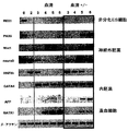
胚発生中に、生体の組織は、外胚葉、中胚葉、および成体型内胚葉(definitive endoderm)という3つの主要な細胞集団から形成される。これらの細胞集団は、初期生殖細胞層としても知られるが、これは原腸陥入(gastrulation)として知られる過程を通じて形成される。嚢胚形成後、各初期生殖細胞層は、特定のセットの細胞集団および組織を産生する。中胚葉は血液細胞、内皮細胞、心筋および骨格筋、ならびに脂肪細胞を生じる。成体型内胚葉は、肝臓、膵臓、および肺を生じる。外胚葉は神経系、皮膚、および副腎組織を生じる。 During embryonic development, living tissues are formed from three main cell populations: ectoderm, mesoderm, and adult endoderm. These cell populations, also known as early germ cell layers, are formed through a process known as gastrulation. After cyst formation, each early germ cell layer produces a specific set of cell populations and tissues. The mesoderm gives rise to blood cells, endothelial cells, myocardium and skeletal muscle, and adipocytes. Adult endoderm gives rise to the liver, pancreas, and lungs. The ectoderm produces the nervous system, skin, and adrenal tissue.
これらの生殖細胞層からの組織発達の過程には、複雑な分子変化を反映する複数の分化段階が含まれる。中胚葉およびその派生物に関連して、3つの異なる段階が規定されている。最初は、胚盤葉上層として知られる構造内の細胞からの中胚葉の誘導である。新生中胚葉としても知られる新しく形成された中胚葉は異なる位置に移動し、これは初期胚における将来の組織発達の部位となろう。パターン化として知られるこの過程は、特定の組織への初期分化段階を反映しているようである。特異化としても知られる最終段階には、パターン化された中胚葉亜集団からの別個の組織の生成が含まれる。近年の研究により、中胚葉は、別個の発達能を有する亜集団を示す連続的な波で誘導されることを示唆する証拠が提供された。最初に形成される中胚葉は胚外領域に移動し、造血細胞および内皮細胞を生じ、次の集団は発達中の胚の前方に移動し、心臓および頭蓋間葉に寄与する。これらの系統関係は、組織学的解析により初めて規定され、主に細胞追跡研究により確認されている。発生運命のこの分離は、発達生物学の分野でよく受け入れられており、現在までに、これらの系統に関係する以前には、中胚葉および内胚葉を単離する利用可能な方法はない。 The process of tissue development from these germline layers includes multiple stages of differentiation that reflect complex molecular changes. In relation to the mesoderm and its derivatives, three different stages have been defined. The first is the induction of mesoderm from cells in a structure known as the upper blastoderm. Newly formed mesoderm, also known as neonatal mesoderm, migrates to a different location, which will be a site for future tissue development in early embryos. This process, known as patterning, seems to reflect the initial differentiation stage into a particular tissue. The final stage, also known as specification, involves the generation of discrete tissue from the patterned mesoderm subpopulation. Recent studies have provided evidence suggesting that the mesoderm is induced by a continuous wave representing a subpopulation with distinct developmental potential. The first formed mesoderm migrates to the extraembryonic region, yielding hematopoietic cells and endothelial cells, and the next population migrates ahead of the developing embryo and contributes to the heart and cranial mesenchyme. These lineage relationships are defined for the first time by histological analysis and are confirmed mainly by cell tracking studies. This separation of developmental fate is well accepted in the field of developmental biology, and to date there is no available method for isolating mesoderm and endoderm before involving these strains.
本発明は、中胚葉および成体型内胚葉細胞集団を単離する方法を提供する。これらの細胞集団は、細胞増殖および分化に影響を及ぼす薬剤を同定するのに、組織発達に関与する遺伝子を同定するのに、および、細胞置換療法のための分化細胞および組織を作製するのに有用である。 The present invention provides methods for isolating mesoderm and adult endoderm cell populations. These cell populations are used to identify drugs that affect cell proliferation and differentiation, to identify genes involved in tissue development, and to create differentiated cells and tissues for cell replacement therapy Useful.
本発明は、中内胚葉および中胚葉細胞の濃縮された細胞集団を提供する。中内胚葉細胞は、本明細書において、短尾(brach+)を発現し、分化誘導条件の存在下で、中胚葉および中胚葉誘導体(心筋および骨格筋、平滑筋、内皮および造血細胞を含む)を産生でき、また、内胚葉および内胚葉誘導体(肝臓細胞および膵臓細胞を含む)も産生できる細胞として定義されている。中胚葉細胞は、本明細書において、brach+であり、分化誘導条件の存在下で、心筋および骨格筋、血管平滑筋、内皮および造血細胞を産生でき、内胚葉および内胚葉誘導体は産生できない、細胞として定義されている。 The present invention provides an enriched cell population of mesendoderm and mesoderm cells. Mesoendoderm cells herein express the short tail (brach + ) and, in the presence of differentiation-inducing conditions, include mesoderm and mesoderm derivatives (including cardiac and skeletal muscle, smooth muscle, endothelial and hematopoietic cells ) And endoderm and endoderm derivatives (including liver cells and pancreatic cells) are also defined. Mesodermal cells are herein brach + and can produce cardiac and skeletal muscle, vascular smooth muscle, endothelial and hematopoietic cells in the presence of differentiation-inducing conditions, and cannot produce endoderm and endoderm derivatives, It is defined as a cell.
本発明はさらに、内胚葉細胞の濃縮された細胞集団を提供する。内胚葉細胞は、本明細書において、短尾(brach+)を発現せず、分化誘導条件の存在下で、肺細胞、肝臓細胞、および膵臓細胞を産生できる細胞として定義されている。 The present invention further provides an enriched cell population of endoderm cells. Endoderm cells are defined herein as cells that do not express a short tail (brach + ) and can produce lung cells, liver cells, and pancreatic cells in the presence of differentiation-inducing conditions.
本発明はまた、中内胚葉および中胚葉細胞の濃縮された細胞集団、および内胚葉細胞の濃縮された細胞集団を単離する方法を提供する。別の実施形態において、本発明は、本発明の細胞集団の増殖、分化、または生存に影響を及ぼす薬剤を同定する方法を提供する。特定の系統および組織の細胞分化および発達に関与する遺伝子を同定する方法も提供する。 The invention also provides mesendoderm and mesoderm cell enriched cell populations and methods of isolating endoderm cell enriched cell populations. In another embodiment, the present invention provides a method of identifying an agent that affects the growth, differentiation, or survival of a cell population of the present invention. Also provided are methods for identifying genes involved in cell differentiation and development of specific lineages and tissues.
brach+細胞を特異的に認識する抗体も提供する。この抗体は、例えば、中内胚葉および内胚葉細胞集団を単離するのに有用である。 Antibodies that specifically recognize brach + cells are also provided. This antibody is useful, for example, in isolating mesendoderm and endoderm cell populations.
別の実施形態において、本発明は、in vitroで細胞を産生する方法を提供する。このような細胞は、例えば、細胞置換療法に有用である。 In another embodiment, the present invention provides a method of producing cells in vitro. Such cells are useful, for example, for cell replacement therapy.
本発明はまた、選択マーカーをコードするDNAが短尾遺伝子座に存在し、よって、1つの短尾対立遺伝子が不活性化されており、選択マーカーが短尾遺伝子座が転写される細胞において発現されているゲノムを有するトランスジェニック非ヒト哺乳動物を提供する。 The present invention also provides that the DNA encoding the selectable marker is present in the short tail locus, so that one short tail allele is inactivated and the selectable marker is expressed in the cell in which the short tail locus is transcribed. Provided is a transgenic non-human mammal having a genome that has been identified.
胚形成中において、中胚葉の形成は、生体プランの確立において、ならびに、血液、内皮、心臓および骨格筋などの複数の臓器系の発達において重要なステップである。しかし、中胚葉形成を制御する分子機序はあまり明確になっていない。培養物中における胚幹(ES)細胞の分化に基づいたモデルシステムを使用して、造血、内皮、心臓筋および骨格筋、ならびに脂肪細胞系統を含む中胚葉に由来する集団を研究してきた。in vitroでのモデルは、中胚葉の誘導および特異化を支持するが、これらの分化事象は、ES細胞から産生された胚様体(ES)として知られる複雑なコロニー中で起こる。中胚葉形成および組織発達をより良く理解するために、形成された時にEBからの中胚葉細胞集団を単離することが有利であろう。しかし、新生中胚葉細胞集団に特異的な抗体が十分に明確ではないために、抗体を使用した細胞選別によりこれらの集団を単離することは不可能であった。 During embryogenesis, mesoderm formation is an important step in the establishment of biological plans and in the development of multiple organ systems such as blood, endothelium, heart and skeletal muscle. However, the molecular mechanisms that control mesoderm formation are less clear. A model system based on the differentiation of embryonic stem (ES) cells in culture has been used to study populations derived from mesoderm, including hematopoiesis, endothelium, heart and skeletal muscle, and adipocyte lineages. While in vitro models support mesoderm induction and specification, these differentiation events occur in complex colonies known as embryoid bodies (ES) produced from ES cells. In order to better understand mesoderm formation and tissue development, it may be advantageous to isolate a mesoderm cell population from EBs when formed. However, it was not possible to isolate these populations by cell sorting using antibodies, as the antibodies specific for the neonatal mesoderm cell populations are not sufficiently clear.
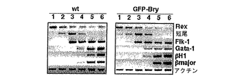
短尾(Tとしても知られる)は、T-ボックス遺伝子として知られる転写因子のファミリーの創設メンバーであり、マウスにおいて天然突然変異として最初に同定された。Papaioannouら(1998)Bioessays 20:9-19。ヘテロ接合型マウスは生存可能であるが、野生型動物よりも短い尾を有している。p.c.約10日目に死亡するホモ接合型突然変異体は脊索を欠失しており、後側中胚葉組織の発達において欠陥を示す。キメラ動物の解析により、短尾は、中胚葉細胞の移動特性に影響を及ぼすことが示された。Wilsonら(1995)Development 121:877-86。発現解析により、短尾について独特かつ興味深いパターンが判明した。それは、原条により移入している全ての細胞ならびに新生および初期移動中胚葉において一過的に発現している。Wilkinsonら(1990)Nature 343:657-9;Herrmannら(1991)Development 113:913-7。発現は、沿軸、外側、および胚外中胚葉において迅速にダウンレギュレートされ、条斑の退行後、尾芽および脊索に限局される。このパターンが得られたので、短尾は、初期中胚葉の最善のマーカーの1つであると考えられ、この系統の発達を追跡するのに使用される。短尾は、解析した全ての種において同定されており、このことは、中胚葉発達におけるその役割は系統発生全体を通じて保存されていることを示唆する。Papaioannouら(1998)。
Tanao (also known as T) is a founding member of a family of transcription factors known as T-box genes and was first identified as a natural mutation in mice. Papaioannou et al. (1998) Bioessays 20: 9-19. Heterozygous mice can survive but have a shorter tail than wild type animals. p.c. Homozygous mutants that die about
本発明によれば、選択マーカー遺伝子は、短尾遺伝子座に組換え標的されている。ES細胞分化開始後に、選択マーカーは、短尾発現を反映するパターンで発現されることが発見された。選択マーカーにより、EBから短尾陽性(Brach+)細胞の選別が可能となり、これにより、中内胚葉および中胚葉細胞の濃縮された細胞集団の単離および特徴づけが可能となった。 According to the present invention, the selectable marker gene is recombination targeted at the short tail locus. After initiation of ES cell differentiation, it was discovered that selectable markers are expressed in a pattern that reflects short tail expression. The selectable marker allowed the selection of short tail positive (Brach + ) cells from EBs, which allowed the isolation and characterization of mesendoderm and enriched cell populations of mesoderm cells.
本発明によれば例示される選択マーカーは、強化緑色蛍光タンパク質(EGFPまたはGFP)である。細胞選別を容易にする他の選択マーカーも当業者には知られており、本発明でも使用し得る。GFPをコードするcDNAは当分野で知られており(そして市販されている、例えばCA州パロアルトのクロンテックからプラスミドpEGFP.C1として)、当分野で既知の方法により標的化ベクター(GFP-Bry)を作製することにより短尾遺伝子座に標的化し得る。ベクターは、好ましくは、短尾遺伝子の最初のエキソンのおよそ3分の2を、GFP発現カセットで置換するように設計されている。 The selectable marker exemplified according to the invention is enhanced green fluorescent protein (EGFP or GFP). Other selectable markers that facilitate cell sorting are known to those of skill in the art and may be used in the present invention. CDNA encoding GFP is known in the art (and commercially available, eg, as plasmid pEGFP.C1 from Clontech, Palo Alto, Calif.), And the targeting vector (GFP-Bry) is obtained by methods known in the art. It can be targeted to the short tail locus by making it. The vector is preferably designed to replace approximately two thirds of the first exon of the short tail gene with a GFP expression cassette.
ヒトおよびマウスを含む数多くの種に由来する短尾遺伝子が当分野で知られており、例えば、Smith(1997)Current Opinion in Genetics & Development 7:474-480に考察されている。GFP発現カセットは、好ましくは、GFPcDNA、および、下流短尾エキソンの翻訳を防ぐための1つ以上の翻訳終止コドンを含む。カセットはさらに、短尾遺伝子の下流領域の転写を防ぐためのSV40ポリアデニル化シグナル配列をコードするエキソンを含み得る。 Short tail genes from a number of species, including humans and mice, are known in the art and are discussed, for example, in Smith (1997) Current Opinion in Genetics & Development 7: 474-480. The GFP expression cassette preferably comprises a GFP cDNA and one or more translation stop codons to prevent translation of downstream short tail exons. The cassette can further include an exon encoding an SV40 polyadenylation signal sequence to prevent transcription of the downstream region of the short tail gene.
べクターは、当分野で既知の方法によりES細胞に導入され、相同的組換えによりGFP-Bry構築体は組み込まれる。ES細胞は、当分野で既知であり、例えばEvansら(1981)Nature 292:154-156、Thompsonら(1995)Proc.Nat'l.Acad.Sci.USA 92;7844、米国特許第5,843,780号、およびReubinoffら(2000)Nature Biotech.18:399により開示された方法により、胚盤胞から単離し得る。好ましい実施形態において、ES細胞はマウスまたはヒトES細胞である。成功裏な標的化後に、短尾開始コドンはGFPの開始コドンとなり、その結果、標的化短尾対立遺伝子は破壊される。得られた細胞はGFP-Bry ES細胞と称される。GFP-Bry ES細胞は、本明細書において、1つの短尾対立遺伝子が不活性化され、GFPが、短尾調節エレメントの制御下で発現されるES細胞として定義される。 Vectors are introduced into ES cells by methods known in the art and the GFP-Bry construct is integrated by homologous recombination. ES cells are known in the art, for example, Evans et al. (1981) Nature 292: 154-156, Thompson et al. (1995) Proc. Nat'l. Acad. Sci. USA 92; 7844, US Pat. No. 5,843,780, And can be isolated from blastocysts by the method disclosed by Reubinoff et al. (2000) Nature Biotech. 18: 399. In a preferred embodiment, the ES cell is a mouse or human ES cell. After successful targeting, the short tail start codon becomes the start codon for GFP, so that the targeted short tail allele is destroyed. The resulting cells are referred to as GFP-Bry ES cells. GFP-Bry ES cells are defined herein as ES cells in which one short tail allele is inactivated and GFP is expressed under the control of short tail regulatory elements.
本発明によると、1つの短尾対立遺伝子が不活性化されているGFP-Bry ES細胞は生存可能であり、正常に発達および分化することが発見された。さらに、GFP発現は、内因性短尾発現を写すことが発見された。従って、brach+細胞は、GFPを発現する細胞を選択することにより単離し得る。GFPを発現する細胞は、簡便には、フローサイトメトリーにより、例えば蛍光活性化細胞選別(FACS)により単離し得る。蛍光特性に基づいて細胞を選別する方法は、当業者には公知である。 According to the present invention, it has been discovered that GFP-Bry ES cells in which one short tail allele is inactivated are viable and develop and differentiate normally. In addition, GFP expression was found to mirror endogenous short tail expression. Thus, brach + cells can be isolated by selecting cells that express GFP. Cells expressing GFP can conveniently be isolated by flow cytometry, for example by fluorescence activated cell sorting (FACS). Methods for sorting cells based on fluorescence properties are known to those skilled in the art.
前記に定義したような中内胚葉および中胚葉細胞の濃縮された細胞集団は、血清の存在下で、GFP+細胞を得るに十分な時間、例えばマウス細胞では約1から約4日間、GFP-Bry ES細胞を培養し、選別し、例えばフローサイトメトリーによりGFP+細胞を単離することにより得られ得る。単離された細胞集団は、少なくとも約50%、好ましくは少なくとも約75%、より好ましくは少なくとも約90%、最も好ましくは少なくとも約95%、または少なくとも約99%の中内胚葉および中胚葉細胞を含む。中内胚葉および中胚葉の相対量は、血清中の培養時間を調整することにより変化させ得、より短い培養時間では、造血および内皮系統にパターン化された中内胚葉および中胚葉の存在に傾き、より長い培養時間では、心筋および骨格筋系統にパターン化された中胚葉の存在に傾く。例えば、中胚葉の濃縮された細胞集団は、血清中で約2.5から約4.5日間培養し、その後、GFP+細胞を選別し、単離することにより得られ得る。血清の存在下における培養は、本明細書において、動物血清、例えばウシ胎児血清(FCS)を補充した培地中での培養として定義される。好ましい実施形態において、培地には、約5%から約25%の血清が補充されている。最適濃度は血清バッチ依存的であり得、当業者により決定できる。 Enriched cell populations of mesendoderm and mesoderm cells as defined above are GFP − in the presence of serum for a time sufficient to obtain GFP + cells, eg, about 1 to about 4 days for mouse cells. It can be obtained by culturing Bry ES cells, sorting and isolating GFP + cells, for example by flow cytometry. The isolated cell population comprises at least about 50%, preferably at least about 75%, more preferably at least about 90%, most preferably at least about 95%, or at least about 99% mesendoderm and mesoderm cells. Including. The relative amount of mesendoderm and mesoderm can be varied by adjusting the culture time in serum, with shorter culture times leaning towards the presence of mesendoderm and mesoderm patterned into hematopoietic and endothelial lineages At longer culture times, the inclination tends to the presence of mesoderm patterned into myocardial and skeletal muscle lineages. For example, a mesoderm enriched cell population can be obtained by culturing in serum for about 2.5 to about 4.5 days, after which GFP + cells are sorted and isolated. Culture in the presence of serum is defined herein as culture in medium supplemented with animal serum, such as fetal calf serum (FCS). In preferred embodiments, the media is supplemented with about 5% to about 25% serum. The optimal concentration can be serum batch dependent and can be determined by one skilled in the art.
中内胚葉および中胚葉細胞の濃縮された細胞集団は、ヒトおよびマウス細胞におけるin vitroでの分化時間の差異を説明するために血清中での培養時間の長さを長くした類似の方法により、ヒトES細胞から作製したGFP-Bry ES細胞から得られ得る。従って、ヒトES細胞から作製したGFP-Bry ES細胞を、GFP+細胞を得るのに十分な時間、例えば約2から約18日間、血清中で培養し、その後、GFP+細胞を選別し、単離する。 Enriched cell populations of mesendoderm and mesoderm cells were obtained by a similar method with increased length of culture time in serum to account for differences in differentiation time in vitro in human and mouse cells. It can be obtained from GFP-Bry ES cells prepared from human ES cells. Therefore, GFP-Bry ES cells prepared from human ES cells are cultured in serum for a time sufficient to obtain GFP + cells, eg, about 2 to about 18 days, after which GFP + cells are selected and isolated. Release.
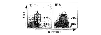
マウスおよびヒトの両方の細胞集団について、単離した細胞が、中胚葉を超えて、例えば血管芽細胞まで分化したかどうかを、チロシンキナーゼ受容体、ヒトKDR、またはマウスFlk-1の存在についてアッセイすることにより容易に決定できる。KDRおよびFlk-1は、中内胚葉および新生中胚葉には発現されていないが、これらの細胞が血管芽細胞/プレ赤血球集団に分化すると、KDRまたはFlk-1発現が検出可能である。KDR+およびflk-1+細胞は、KDRまたはFlk-1に対する抗体を使用してフローサイトメトリーにより同定し得る。このような抗体は当分野で既知であり、標準的な抗体作製法を使用して作製し得る。中内胚葉および中胚葉の濃縮された細胞集団はさらに、細胞選別により、KDR+およびflk-1+細胞を除去することにより濃縮することができる。 For both mouse and human cell populations, assay whether isolated cells have differentiated beyond the mesoderm, eg, to hemangioblasts, for the presence of tyrosine kinase receptor, human KDR, or mouse Flk-1 This can be easily determined. KDR and Flk-1 are not expressed in mesendoderm and neonatal mesoderm, but when these cells differentiate into hemangioblast / pre-erythroid populations, KDR or Flk-1 expression can be detected. KDR + and flk-1 + cells can be identified by flow cytometry using antibodies against KDR or Flk-1. Such antibodies are known in the art and can be made using standard antibody production methods. The mesendoderm and mesoderm enriched cell population can be further enriched by removing KDR + and flk-1 + cells by cell sorting.
図17に示したように、本発明によると、中内胚葉は、内胚葉および中胚葉の両方ならびにその対応する系統を生じる、以前には同定されていなかった細胞集団であることが発見された。in vitro培養物中での血清の存在または非存在を使用して、どの系統が中内胚葉から産生されるかを指示できることがさらに発見された。特に、内胚葉細胞の濃縮された細胞集団は、血清の存在下でマウスES細胞から作製したGFP-Bry ES細胞を約2から4日間培養し、例えばフローサイトメトリーによりGFP+細胞を選別し、単離し、その後、血清の非存在下でGFPを約1から約10日間培養することにより得られ得る。単離された細胞集団は、少なくとも50%、好ましくは少なくとも約75%、より好ましくは少なくとも約90%、最も好ましくは少なくとも約95%、または少なくとも約99%の前記に定義したような内胚葉細胞を含む。 As shown in FIG. 17, according to the present invention, it was discovered that mesendoderm is a previously unidentified cell population that yields both endoderm and mesoderm and its corresponding lineages. . It has further been discovered that the presence or absence of serum in in vitro cultures can be used to indicate which lines are produced from mesendoderm. In particular, the cell population enriched with endoderm cells is obtained by culturing GFP-Bry ES cells prepared from mouse ES cells in the presence of serum for about 2 to 4 days, for example, selecting GFP + cells by flow cytometry, Can be obtained by culturing GFP for about 1 to about 10 days in the absence of serum. The isolated cell population is at least 50%, preferably at least about 75%, more preferably at least about 90%, most preferably at least about 95%, or at least about 99% of endoderm cells as defined above. including.
内胚葉細胞の濃縮された細胞集団は、血清の存在下で約2から10日間GFP-Bry ES細胞を培養し、GFP+細胞を選別し、単離し、その後、血清の非存在下でGFP+細胞を約1から約15日間培養することにより、ヒトES細胞から作製したGFP-Bry ES細胞から得られ得る。 Enriched cell populations of endoderm cells are cultured for about 2 to 10 days in the presence of serum, GFP-Bry ES cells are cultured, GFP + cells are sorted and isolated, then GFP + in the absence of serum The cells can be obtained from GFP-Bry ES cells prepared from human ES cells by culturing the cells for about 1 to about 15 days.
内胚葉細胞の濃縮された集団はさらに、前記したようなKDR+またはFlk-1+細胞を同定および選別することにより濃縮し得る。 The enriched population of endoderm cells can be further enriched by identifying and sorting KDR + or Flk-1 + cells as described above.
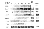
本発明によると、内胚葉の濃縮された細胞集団は、血清の非存在下で成長因子アクチビンの存在下で約2から約10日間GFP-Bry胚性幹細胞を培養し、短尾を発現する細胞を単離することにより得られることがさらに発見された。アクチビンの量は、胚性幹細胞から内胚葉への分化を誘導するのに十分なものである。このような分化は、例えばHNF3β、Mixl-1、Sox17、Hex-1、またはpdx-1を含む、内胚葉発達に関連した遺伝子の発現についてアッセイすることにより測定し得る。好ましい実施形態において、アクチビンの濃度は少なくとも約30ng/mlである。別の好ましい実施形態において、アクチビンの濃度は約100ng/mlである。 According to the present invention, an endoderm-enriched cell population is obtained by culturing GFP-Bry embryonic stem cells in the absence of serum for about 2 to about 10 days in the presence of growth factor activin and expressing a short tail. It was further discovered that can be obtained by isolating. The amount of activin is sufficient to induce differentiation from embryonic stem cells to endoderm. Such differentiation can be measured by assaying for the expression of genes associated with endoderm development, including, for example, HNF3β, Mixl-1, Sox17, Hex-1, or pdx-1. In a preferred embodiment, the activin concentration is at least about 30 ng / ml. In another preferred embodiment, the activin concentration is about 100 ng / ml.
中胚葉の濃縮された細胞集団は、血清の非存在下、アクチビンの存在下、約2から約10日間GFP-Bry胚性幹細胞を培養し、短尾を発現する細胞を単離することにより得られ得る。アクチビンの量は、胚性幹細胞から中胚葉への分化を誘導するのに十分であるが、内胚葉への分化を誘導するには不十分である。中胚葉への分化は、例えばGATA-1を含む、中胚葉発達に関連した遺伝子の発現、および、内胚葉発達に関連した遺伝子の不在についてアッセイすることによりにより測定し得る。好ましい実施形態において、アクチビンの濃度は、30ng/ml未満である。別の好ましい実施形態において、アクチビンの濃度は約3ng/mlである。 A mesoderm-enriched cell population is obtained by culturing GFP-Bry embryonic stem cells in the absence of serum and in the presence of activin for about 2 to about 10 days and isolating cells that express the short tail. Can be. The amount of activin is sufficient to induce differentiation from embryonic stem cells to mesoderm but is insufficient to induce differentiation to endoderm. Mesodermal differentiation can be measured by assaying for the expression of genes associated with mesoderm development, including, for example, GATA-1, and the absence of genes associated with endoderm development. In a preferred embodiment, the activin concentration is less than 30 ng / ml. In another preferred embodiment, the activin concentration is about 3 ng / ml.
本発明はさらに、前記した細胞集団の増殖、分化、または生存に影響を及ぼす薬剤を同定する方法を提供する。この方法には、前記した細胞集団の1つに由来する細胞を、試験する薬剤の非存在下および存在下で培養し、前記薬剤が細胞集団の増殖、分化、または生存に影響を及ぼすかどうかを決定することが含まれる。試験する薬剤は、天然または合成、1つの化合物または混合物、小分子またはポリマー(ポリペプチド、多糖、ポリヌクレオチドなどを含む)、抗体またはその断片、天然または合成化合物のライブラリー由来の化合物、合理的な薬物設計から得られた化合物、または細胞集団に対する効果を、当分野で既知のアッセイ、例えば米国特許第6,110,739号に記載のような標準的な増殖および分化アッセイを使用して評価し得る任意の薬剤であり得る。このような薬剤は、インビボおよびin vitroでの細胞増殖および分化の制御に有用である。 The present invention further provides methods for identifying agents that affect the growth, differentiation, or survival of the cell populations described above. This method involves culturing cells from one of the cell populations described above in the absence and presence of the agent being tested, whether the agent affects the growth, differentiation, or survival of the cell population. Is included. Agents to be tested include natural or synthetic, single compounds or mixtures, small molecules or polymers (including polypeptides, polysaccharides, polynucleotides, etc.), antibodies or fragments thereof, compounds from libraries of natural or synthetic compounds, rational Any compound that can be obtained from a simple drug design, or any effect that can be assessed on cell populations using assays known in the art, such as standard proliferation and differentiation assays such as those described in US Pat.No. 6,110,739. Can be a drug. Such agents are useful for controlling cell proliferation and differentiation in vivo and in vitro.
本発明はさらに、特定の系統および組織の細胞分化および発達に関与する遺伝子を同定する方法を提供する。この方法には、種々の培養時間後に本発明のGFP+細胞集団を単離し、異なる集団の遺伝子発現プロファイルを比較し、集団において独特に発現されている遺伝子を同定することが含まれる。好ましい実施形態において、マイクロアレイ解析およびサブトラクティブハイブリダイゼーションを使用して、遺伝子発現プロファイルを比較する。 The present invention further provides methods for identifying genes involved in cell differentiation and development of specific lineages and tissues. This method involves isolating GFP + cell populations of the invention after various culture times, comparing gene expression profiles of different populations, and identifying genes that are uniquely expressed in the populations. In a preferred embodiment, microarray analysis and subtractive hybridization are used to compare gene expression profiles.
別の実施形態において、本発明は、短尾陽性(brach+)細胞を認識するが、短尾陰性(brach-)細胞は認識しない抗体の作製方法を提供する。動物に免疫誘発形態の本発明の細胞を注入することによりポリクローナル抗体を作製し得る。また、GFP+細胞には存在するが、GFP-細胞には存在しない細胞表面マーカーを同定し、マーカーまたはその断片に対する抗体を作製することにより、抗体を作製し得る。抗体はモノクローナルまたはポリクローナルであり得、断片、遺伝工学した抗体、単鎖抗体などであり得る。抗体は、当分野で公知の方法により作製し得る。このような抗体は、中内胚葉および中胚葉などのbranch+細胞を同定および単離するのに有用である。 In another embodiment, the present invention provides a method of making an antibody that recognizes short tail positive (brach + ) cells but not short tail negative (brach − ) cells. Polyclonal antibodies can be made by injecting animals with the cells of the invention in an immunizing form. Alternatively, antibodies can be generated by identifying cell surface markers that are present in GFP + cells but not in GFP − cells and generating antibodies to the markers or fragments thereof. Antibodies can be monoclonal or polyclonal, and can be fragments, genetically engineered antibodies, single chain antibodies, and the like. Antibodies can be produced by methods known in the art. Such antibodies are useful for identifying and isolating branch + cells such as mesendoderm and mesoderm.
本発明はまた、in vitroで哺乳動物細胞を作製する方法も提供する。1つの実施形態において、この方法には、中胚葉から心筋、平滑筋、内皮、または造血細胞への分化に効果的な条件下で、中内胚葉および中胚葉細胞の濃縮された細胞集団に由来する細胞を培養することが含まれる。in vitroで種々の細胞型への分化に効果的な条件は、当分野で既知である。別の実施形態において、この方法には、内胚葉から肝臓細胞または膵臓細胞への分化に効果的な条件下で、内胚葉細胞の濃縮された細胞集団に由来する細胞を培養することが含まれる。このような分化における効果的な条件は、当分野で既知である。インスリン産生膵臓島細胞の産生が特に考えられる。 The present invention also provides a method for producing mammalian cells in vitro. In one embodiment, the method is derived from a concentrated cell population of mesendoderm and mesoderm cells under conditions effective for differentiation from mesoderm to cardiac muscle, smooth muscle, endothelium, or hematopoietic cells. Culturing cells to be treated. Conditions effective for differentiation into various cell types in vitro are known in the art. In another embodiment, the method includes culturing cells derived from an enriched cell population of endoderm cells under conditions effective for differentiation from endoderm to liver cells or pancreatic cells. . Effective conditions for such differentiation are known in the art. The production of insulin producing pancreatic islet cells is particularly contemplated.
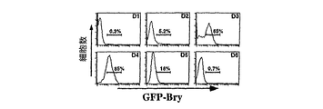
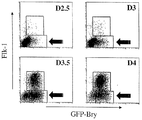
本発明により実証されたように、異なる年齢のEBから単離されたbrach+細胞は異なる発達能を有する。約3日目のマウスEBのBrach+/Flk-細胞は、効率的に、造血および内皮系統を生じるが、約3から10日目のEBの細胞は、心筋細胞系統の細胞を生じる。従って、中内胚葉および中胚葉の濃縮された細胞集団を得るのに使用したES細胞の培養時間を調整することにより、当業者は、造血および内皮系統または心筋細胞系統の効率的な産生を選択できる。 As demonstrated by the present invention, brach + cells isolated from EBs of different ages have different developmental abilities. Approximately 3 day mouse EB Brach + / Flk − cells efficiently generate hematopoietic and endothelial lineages, while approximately 3 to 10 day EB cells generate cardiomyocyte lineage cells. Therefore, by adjusting the culture time of the ES cells used to obtain the mesendoderm and mesoderm enriched cell population, one skilled in the art would select for efficient production of hematopoietic and endothelial or cardiomyocyte cell lines it can.
このような細胞は、例えば、限られた数の細胞型の破壊または機能不全により生じる疾患の処置における、細胞置換療法に有用である。このような疾患には、糖尿病、肝不全、心不全、心臓血管疾患および他の血管の疾患、デュシェンヌ筋ジストロフィー、骨形成不全、および骨髄移植により治療可能な疾患、例えば白血病および貧血が含まれる。Odoricoら(2001)Stem Cells 19:193-204参照。 Such cells are useful for cell replacement therapy, for example in the treatment of diseases caused by the destruction or dysfunction of a limited number of cell types. Such diseases include diabetes, liver failure, heart failure, cardiovascular disease and other vascular diseases, Duchenne muscular dystrophy, bone dysplasia, and diseases treatable by bone marrow transplantation, such as leukemia and anemia. See Odorico et al. (2001) Stem Cells 19: 193-204.
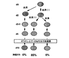
本発明の細胞集団は、細胞置換療法のための分化細胞および組織を作製するのに有用である。細胞置換療法における本発明の細胞集団の適切性は、少数の細胞型の破壊または機能不全に関連した疾患の動物モデルに細胞を移植することにより評価し得る。例えば、本明細書に参照により取り込んだ、例えばGrompeら(1993)Genes & Dev.7:2298により開示されたフマリルアセト酢酸(FAH)欠損マウスは、肝不全のモデルを提供する。FAH欠損マウスは、NTBC(2-(2-ニトロ-4-トリフルオロメチルベンゾイル)-1,3-シクロヘキセジオン)で処置するか、または正常な肝細胞を移植しなければ、進行的な肝不全および尿細管傷害に苦しむ。従って、これらのマウスは、EBから作製した未熟な肝細胞の特徴を有する細胞の能力について試験する上で理想的なモデルを提供する。NTBCから取り出したFAH欠損マウスに肝細胞を移植する方法は当分野で既知であり、例えばOversturfら(1996)Nature Genet.12:266-273に開示されている。正常な肝機能はマウスの生存により示され、また、血清アスパルテートトランスアミナーゼレベル、血漿ビリルビンレベルを測定することにより、および、再生肝臓の正常な構造を決定することにより評価し得る。 The cell populations of the present invention are useful for creating differentiated cells and tissues for cell replacement therapy. The suitability of a cell population of the invention in cell replacement therapy can be assessed by transplanting the cells into an animal model of a disease associated with the destruction or dysfunction of a small number of cell types. For example, fumaryl acetoacetate (FAH) deficient mice, for example disclosed by Grompe et al. (1993) Genes & Dev. 7: 2298, incorporated herein by reference, provide a model for liver failure. FAH-deficient mice are treated with NTBC (2- (2-nitro-4-trifluoromethylbenzoyl) -1,3-cyclohexedione) or progressive liver unless transplanted with normal hepatocytes. Suffers from insufficiency and tubular injury. Thus, these mice provide an ideal model for testing for the ability of cells with immature hepatocyte characteristics made from EBs. Methods for transplanting hepatocytes into FAH-deficient mice removed from NTBC are known in the art and are disclosed, for example, in Oversturf et al. (1996) Nature Genet. 12: 266-273. Normal liver function is demonstrated by mouse survival and can be assessed by measuring serum aspartate transaminase levels, plasma bilirubin levels, and determining the normal structure of regenerating liver.
特定の細胞型の破壊または機能不全から生じる他の疾患の動物モデルは当分野で既知である。このようなモデルは、本発明の他の細胞集団を評価するために同じように使用し得る。 Animal models of other diseases resulting from the destruction or dysfunction of certain cell types are known in the art. Such a model can be used in the same way to evaluate other cell populations of the invention.
本発明はまた、選択マーカーをコードするDNAが短尾遺伝子座に存在し、よって、1つの短尾対立遺伝子が不活性化され、短尾遺伝子が転写される細胞において選択マーカーが発現されている、トランスジェニック非ヒト哺乳動物を提供する。好ましい実施形態において、哺乳動物はマウスであり、選択マーカーはGFPである。特に、トランスジェニックマウスは、GFPをコードするDNA配列が短尾調節エレメントに作動可能に連結している導入遺伝子を含むゲノムを有し、前記導入遺伝子は、短尾を正常に発現する細胞において発現されている。トランスジェニックマウスは、前記したGFP-Bry ES細胞を胚盤胞に注入することにより得られ得、これはその後、偽妊娠雌にインプラントする。トランスジェニック子は、branch+/-に関連した短い尾の表現型により、および分子解析により同定される。このようなトランスジェニック動物は、初期胚(これから本発明の方法により使用する中胚葉を単離する)を得るのに、そして、短尾遺伝子を発現する任意の成体細胞子集団の同定、単離および特徴づけに有用である。このような細胞は、新規幹細胞集団を示し得る。 The present invention also provides that the DNA encoding the selectable marker is present in the short tail locus so that one short tail allele is inactivated and the selectable marker is expressed in a cell in which the short tail gene is transcribed. A transgenic non-human mammal is provided. In a preferred embodiment, the mammal is a mouse and the selectable marker is GFP. In particular, the transgenic mouse has a genome comprising a transgene in which a DNA sequence encoding GFP is operably linked to a short tail regulatory element, said transgene being expressed in cells that normally express the short tail Has been. Transgenic mice can be obtained by injecting the GFP-Bry ES cells described above into blastocysts, which are then implanted into pseudopregnant females. Transgenic offspring are identified by the short tail phenotype associated with branch +/− and by molecular analysis. Such transgenic animals can be used to obtain early embryos (from which the mesoderm used by the method of the invention is isolated) and to identify, isolate any adult cell child population that expresses the short tail gene. And useful for characterization. Such cells may represent a new stem cell population.
本明細書に引用した全ての参考文献のその全体を本明細書に取り込む。 The entirety of all references cited herein are hereby incorporated by reference.
以下の実施例は本発明をさらに説明するためのものである。 The following examples serve to further illustrate the present invention.
材料および方法
ES細胞増殖および分化。ES細胞を、15%ウシ胎児血清(FCS)、ペニシリン、ストレプトマイシン、LIF(1%ならし培地)、および1.5×10⁻⁴Mモノチオグリセロール(MTG;シグマ)を補充したダルベッコ改変イーグル培地(DMEM)中、照射胚支持細胞上に維持した。分化開始の2日前に、細胞を同じ培地中のゼラチン化プレートに移した。EBの産生のために、ES細胞をトリプシン処理し、異なる培養物中に種々の密度で播種した。EBの分化は、15%FCS、2mM L-グルタミン(ギブコ/BRL)、トランスフェリン(200μg/ml)、0.5mMアスコルビン酸(シグマ)、および4.5×10-4M MTGを補充したIMDM中、60mmペトリ等級皿中で実施した。培養物は、加湿チャンバー中で、5%CO2/空気混合物中で、37℃で維持した。
Materials and methods
ES cell proliferation and differentiation. ES cells were treated with Dulbecco's modified Eagle medium (DMEM) supplemented with 15% fetal calf serum (FCS), penicillin, streptomycin, LIF (1% conditioned medium), and 1.5 × 10 −4 M monothioglycerol (MTG; Sigma). ) And maintained on irradiated embryonic feeder cells. Two days before the start of differentiation, cells were transferred to gelatinized plates in the same medium. For production of EBs, ES cells were trypsinized and seeded at different densities in different cultures. EB differentiation was achieved in 60 mm Petri in IMDM supplemented with 15% FCS, 2 mM L-glutamine (Gibco / BRL), transferrin (200 μg / ml), 0.5 mM ascorbic acid (Sigma), and 4.5 × 10 −4 M MTG. Performed in a grade dish. Cultures were maintained at 37 ° C. in a humidified chamber, 5% CO 2 / air mixture.
血清を含まない培地
2つの異なる血清を含まない培地を、以下の実施例の異なる態様において使用した:ノックアウトSR(ギブコBRL)およびStemPro34(ギブコBRL)を補充したIIMD。
Serum-free medium Two different serum-free media were used in different embodiments of the following examples: IIMD supplemented with Knockout SR (Gibco BRL) and StemPro34 (Gibco BRL).
メチルセルロースコロニーアッセイ
A)芽球コロニー:芽細胞コロニーを作製するために(BL-CFCアッセイ)、EB由来細胞を、10%FCS(ハイクローン)、血管内皮増殖因子(VEGF;5ng/ml)、c-kitリガンド(KL;1%ならし培地)、IL-6(5ng/ml)および25%D4T内皮細胞ならし培地(Kennedyら(1997)Nature 386:488-93)を補充した1%メチルセルロース中、0.5×1.5×105細胞/mlで播種した。移行コロニーをVEGFの非存在下で作製した。コロニーは4日間の培養後に評価した。B)造血コロニー:原始的および成体型造血コロニーの増殖のために、細胞を、10%血漿由来血清(PDS;Antech)、5%タンパク質非含有ハイブリドーマ培地(PFHM-II;ギブコ-BRL)と以下のサイトカインを含む1%メチルセルロース中に播種した:c-kitリガンド(KL;1%ならし培地)、エリスロポエチン(2U/ml)、IL-11(25ng/ml)、IL-3(1%ならし培地)、GM-CSF(3ng/ml)、G-CSF(30ng/ml)、M-CSF(5ng/ml)、IL-6(5ng/ml)およびトロンボポエチン(TPO;5ng/ml)。培養物を37℃、5%CO2中に維持した。原始赤血球コロニーを培養の5〜6日目に評価し、一方、成体型赤血球(BFU-E)、マクロファージ、および多系統コロニーは、培養の7〜10日目に計測した。c-kitリガンドは、KL発現ベクター(親切にもジェネティクス・インスチチュートから提供された)でトランスフェクトしたCHO細胞により調整された培地から得られた。IL-3は、IL-3を発現しているベクターでトランスフェクトしたX63AG8-653ミエローマ細胞により調整された培地から得られた。VEGF、GM-CSF、M-CSF、IL-6、IL-11、アクチビン、BMP2、BMP4、bFGF、FGF8、およびIhhは、R&Dシステムズから購入した。
Methylcellulose colony assay A) Blast colonies: To generate blast colonies (BL-CFC assay), EB-derived cells were treated with 10% FCS (high clone), vascular endothelial growth factor (VEGF; 5 ng / ml), c in 1% methylcellulose supplemented with -kit ligand (KL; 1% conditioned medium), IL-6 (5 ng / ml) and 25% D4T endothelial cell conditioned medium (Kennedy et al. (1997) Nature 386: 488-93) And 0.5 × 1.5 × 10 5 cells / ml. Transition colonies were generated in the absence of VEGF. Colonies were evaluated after 4 days of culture. B) Hematopoietic colonies: For the growth of primitive and adult hematopoietic colonies, the cells are divided into 10% plasma-derived serum (PDS; Antech), 5% protein-free hybridoma medium (PFHM-II; Gibco-BRL) and below Seeded in 1% methylcellulose containing the following cytokines: c-kit ligand (KL; 1% conditioned medium), erythropoietin (2U / ml), IL-11 (25ng / ml), IL-3 (1% conditioned) Medium), GM-CSF (3 ng / ml), G-CSF (30 ng / ml), M-CSF (5 ng / ml), IL-6 (5 ng / ml) and thrombopoietin (TPO; 5 ng / ml). Cultures were maintained at 37 ° C. and 5% CO 2 . Primordial red blood cell colonies were evaluated on days 5-6 of culture, while adult red blood cells (BFU-E), macrophages, and multilineage colonies were counted on days 7-10 of culture. The c-kit ligand was obtained from a medium conditioned by CHO cells transfected with a KL expression vector (kindly provided by Genetics Institute). IL-3 was obtained from media conditioned by X63AG8-653 myeloma cells transfected with a vector expressing IL-3. VEGF, GM-CSF, M-CSF, IL-6, IL-11, activin, BMP2, BMP4, bFGF, FGF8, and Ihh were purchased from R & D Systems.
再凝集培養物
細胞を、24ウェルのペトリ等級プレート中、15%FCS(またはノックアウトSR)、2mM L-グルタミン(ギブコ/BRL)、0.5mMアスコルビン酸(シグマ)、および4.5×10-4M MTGを補充したIMDM1mlあたり、2×10⁵で培養した。これらを使用して、細胞がウェルの底に付着するのを防いだ。
Reaggregated cultures Cells in 24-well Petri grade plates, 15% FCS (or knockout SR), 2 mM L-glutamine (Gibco / BRL), 0.5 mM ascorbic acid (Sigma), and 4.5 × 10 −4 M MTG The culture was performed at 2 × 10 5 per 1 ml of IMDM supplemented with. These were used to prevent cells from attaching to the bottom of the wells.
心筋アッセイ
GFP+細胞を、15%血清代替添加物で補充したIMDM中で再凝集した。20時間後、凝集物を、10%血清代替添加物(血清非含有)を有するIMDM中、24または96ウェルプレートのウェル中で培養した。ウェルはゼラチンで予め処理しておいた。培養物を、拍動細胞の発達について毎日モニタリングした。拍動細胞は、通常、培養の2から6日目に検出された。
Myocardial assay
GFP + cells were reaggregated in IMDM supplemented with 15% serum replacement additive. After 20 hours, the aggregates were cultured in wells of 24 or 96 well plates in IMDM with 10% serum replacement additive (serum free). Wells were pretreated with gelatin. Cultures were monitored daily for pulsatile cell development. Beating cells were usually detected on days 2-6 of culture.
細胞表面マーカー染色およびFACS解析
標準的な条件を使用して、細胞を染色した。染色した懸濁液はFACScan(Becton Dickinson、CA)で解析した。
Cell surface marker staining and FACS analysis Cells were stained using standard conditions. The stained suspension was analyzed with FACScan (Becton Dickinson, CA).
遺伝子発現解析
ポリA+RT-PCR解析のために、Bradyら((1990)Meth.in Mol.and Cell Bio.2:17-25)の方法を使用した。逆転写、ポリAテーリング、およびPCR手順を記載の通りに実施し、ただし、X-dtオリゴヌクレオチドは5'-GTTAACTCGAGAATTC(T)24-3'まで短くした。PCR反応からの増幅産物はアガロースゲル上で分離し、Zeta-プローブGTメンブラン(Biorad)に転写するか、または、スロットブロット装置(Schleicher&Schuell)を用いてメンブランに転写した。得られたブロットは、遺伝子の3‘領域に対応する32PランダムプライムcDNA断片(Ready-to-Goラベリング、ファルマシア)を用いてハイブリダイズした(β−H1を除いて全て)。β−H1-特異的プローブは、その3’末端において8塩基の相同性を共有する2つのオリゴヌクレオチド、(5'-TGGAGTCAAAGAGGGCATCATAGACACATGGG-3'、5'-CAGTACACTGGCAATCCCATGTG-3'をアニーリングすることにより調製した。このβ-H1特異的オリゴヌクレオチドは、クレノウ平滑化反応を使用して、32Pで標識した。遺伝子特異的PCRのために、全RNAを、RNイージーミニキットを用いて各試料から抽出し、RNase非含有DNase(キアゲン)で処理した。2μgの全RNAを、OmniscriptRTキット(キアゲン)を使用してランダムヘキサマーを用いてcDNAに逆転写した。PCRを、適切なオリゴヌクレオチドを使用して実施した。PCR反応を、2.5UのTaqポリメラーゼ(プロメガ)、PCR緩衝液、2.5mM MgCl2、0.2μMの各プライマーおよび0.2mM dNTPを用いて実施した。サイクリング条件は以下の通りであった;94℃で5分間、その後、35サイクルの増幅(94℃で1分間変性、60℃で1分間アニーリング、72℃で1分間伸張)、最後に72℃で7分間インキュベート。
Gene Expression Analysis The method of Brady et al. ((1990) Meth. In Mol. And Cell Bio. 2: 17-25) was used for poly A + RT-PCR analysis. Reverse transcription was performed as poly A tailing, and PCR procedures described, however, X-dt oligonucleotides were shortened to 5'-GTTAACTCGAGAATTC (T) 24 -3 '. Amplified products from the PCR reaction were separated on an agarose gel and transferred to a Zeta-probe GT membrane (Biorad) or transferred to the membrane using a slot blot apparatus (Schleicher & Schuell). The resulting blots were hybridized using 32 P random primed cDNA fragments (Ready-to-Go labeling, Pharmacia) corresponding to the 3 ′ region of the gene (all except β-H1). The β-H1-specific probe was prepared by annealing two oligonucleotides that share 8-base homology at their 3 ′ end, (5′-TGGAGTCAAAGAGGGCATCATAGACACATGGG-3 ′, 5′-CAGTACACTGGCAATCCCATGTG-3 ′. This β-H1-specific oligonucleotide was labeled with 32 P using a Klenow smoothing reaction, and total RNA was extracted from each sample using the RN Easy Mini Kit for gene-specific PCR. Treated with RNase-free DNase (Qiagen) 2 μg of total RNA was reverse transcribed into cDNA using random hexamers using OmniscriptRT kit (Qiagen) PCR was performed using appropriate oligonucleotides PCR reactions were performed using 2.5 U Taq polymerase (Promega), PCR buffer, 2.5 mM MgCl 2 , 0.2 μM each primer and 0.2 mM dNTPs, cycling conditions were as follows: 94
標的化ES細胞の作製
適切な培養条件下で、胚幹(ES)細胞は分化し、広域な系統からの発達中の細胞集団を含む、胚様体(EB)として知られる3次元のコロニーを形成する。Smith(2001)Annu.Rev.Cell Dev.Biol.17:435-62。これらのEB由来集団の中で、造血、内皮、心筋および骨格筋系統のものを含む、中胚葉誘導体を検出できる。
Generation of targeted ES cells Under appropriate culture conditions, embryonic stem (ES) cells differentiate and form three-dimensional colonies known as embryoid bodies (EBs) that contain developing cell populations from a broad lineage. Form. Smith (2001) Annu. Rev. Cell Dev. Biol. 17: 435-62. Within these EB-derived populations, mesoderm derivatives can be detected, including those of hematopoietic, endothelial, cardiac and skeletal muscle lineages.
EBにおける中胚葉の開始を追跡し、この集団を示す細胞を単離するために、緑色蛍光タンパク質(GFP)を短尾遺伝子座に標的化した。標的化構築体は、最初のエキソンに、GFPcDNA、および人工イントロン、SV40ポリ(A)配列およびloxPフランキングneoカセットを含み、図1に示す。チミジンキナーゼ(TK)遺伝子は、無作為な組込みに対して選択するために、標的化構築体の3'末端に含められた。標的化ベクターは以下の通りに作製した。 Green fluorescent protein (GFP) was targeted to the short tail locus to follow the onset of mesoderm in EBs and to isolate cells representing this population. The targeting construct contains the GFP cDNA, and artificial intron, SV40 poly (A) sequence and loxP flanking neo cassette in the first exon and is shown in FIG. A thymidine kinase (TK) gene was included at the 3 ′ end of the targeting construct to select for random integration. The targeting vector was prepared as follows.
全マウス短尾(Bry)遺伝子を有するBACクローンを、プライマー5'-AAGGAGCTAACTAACGAGATGAT-3'および5'-TACCTTCAGCACCGGGAACAT-3'を用いて129/Ola株ゲノムライブラリー(Genome Systems)をPCRスクリーニングすることにより単離した。これらのプライマーは、それぞれ、第一および第二Bryエキソン内にアニーリングし、約600bpの診断バンドを増幅する。2kbを超える5´フランキング領域と共にBry遺伝子の1エキソンを有する約3kb長のPstI制限断片を同定し、BACからプラスミドpBSK(Stratagene)にサブクローニングすると、pBSK.Bry-5'構築体が得られた。開始コドンのすぐ上流の約2kbの領域をシークエンスして、ベクター作製のための適切なプライマーアニーリング部位を同定した。 By PCR screening a BAC clone with the whole mouse short tail (Bry) gene using the primers 5'-AAGGAGCTAACTAACGAGATGAT-3 'and 5'-TACCTTCAGCACCGGGAACAT-3' on a 129 / Ola strain genomic library (Genome Systems) Isolated. These primers each anneal within the first and second Bry exons and amplify an approximately 600 bp diagnostic band. An approximately 3kb long PstI restriction fragment with one exon of the Bry gene with a 5 'flanking region of more than 2kb was identified and subcloned from BAC into the plasmid pBSK (Stratagene), resulting in the pBSK.Bry-5' construct . An approximately 2 kb region immediately upstream of the start codon was sequenced to identify an appropriate primer annealing site for vector construction.
オリゴ5'-GCTAGCTAATGGATCCA-3'/5'-GATCTGGATCCATTAGCTAGCTGCA-3'および5'-GATCTTAATGAACGGCAGGTGGGTGCGCGTCCGGAG-3'/5'TCGACTCCGGACGCGCACCCACCTGCCGTTCATTAA-3'を、プラスミドpBSKのPstI/SalI部位に挿入し、2つの連続的な翻訳終止コドンおよび人工的な3´スプライス部位を有する新しくてより適切なポリリンカーを創造した(構築体pBry-AA)。プラスミドpEGFP.C1(クロンテック)をNheI/BglIIで二重消化し、終止コドンを含まないEGFPをコードする得られた約760bpのDNA断片を、pBRY-AAのNheI/BglII部位にクローニングすると、構築体pBry-ABが得られた。loxPが側にあるネオマイシン耐性遺伝子を有するプラスミドpL2-Neo2のXhoI/SalI断片を、pBry-ABのSalI部位に挿入すると、プラスミドpBry-AC(EGFPおよびNeoの同じ方向への転写)が得られた。 Insert oligo 5'-GCTAGCTAATGGATCCA-3 '/ 5'-GATCTGGATCCATTAGCTAGCTGCA-3' and 5'-GATCTTAATGAACGGCAGGTGGGTGCGCGTCCGGAG-3 '/ 5'TCGACTCCGGACGCGCACCCACCTGCCGTTCATTAA-3' into the PstI / SalI sites of plasmid pBSK A new and more suitable polylinker with a stop codon and an artificial 3 'splice site was created (construct pBry-AA). Double digestion of plasmid pEGFP.C1 (Clontech) with NheI / BglII and cloning of the resulting approximately 760 bp DNA fragment encoding EGFP without the stop codon into the NheI / BglII site of pBRY-AA pBry-AB was obtained. When the XhoI / SalI fragment of plasmid pL2-Neo2 carrying the neomycin resistance gene flanked by loxP was inserted into the SalI site of pBry-AB, plasmid pBry-AC (transcription of EGFP and Neo in the same direction) was obtained. .
共通スプライスドナー部位、人工イントロン、スプライスアクセプター部位、およびSV40ポリアデニル化配列を含む短いエキソンを有する556bpのXmal/MluI断片を、市販の発現ベクターpBK-CMV(Stratagene)から切り出した。この断片を以下のようにプラスミドpBry-ACに挿入した:XmaI末端を最後のEGFPコドンに続くBspEI部位に連結し、一方、Mlu末端は、リンカーとしてのオリゴ5'-CGCGTTACTAGTAAGACGTCT-3'/5'-CCGGAGACGTCTTACTAGTAA-3'と共に、loxP-neo-loxPカセットの直ぐ上流に位置するBspEI部位に挿入した。得られた構築体:pBry-AE。HSVチミジンキナーゼ遺伝子をコードする約1.9kbのXhoI/SalI断片を、pBry-AEのXhoI部位に挿入し、無作為な組込みに対する選択を可能とした(構築体pBry-AH)。相同性の「短い腕」をコードするNotI/Eco47III断片をpBry-AFから切り出し、pBry-AHのNotI/Eco47III部位にクローニングすると、プラスミドpBry-Alが得られた。相同性の「長い腕」は、SalIを用いてpBry-AKから切り出し、正しい方向で、pBry-AIのSalI部位に挿入すると、最終標的化ベクターBが得られた。 A 556 bp Xmal / MluI fragment with a short exon containing a common splice donor site, artificial intron, splice acceptor site, and SV40 polyadenylation sequence was excised from the commercial expression vector pBK-CMV (Stratagene). This fragment was inserted into plasmid pBry-AC as follows: the XmaI end was ligated to the BspEI site following the last EGFP codon, while the Mlu end was oligo 5'-CGCGTTACTAGTAAGACGTCT-3 '/ 5' as a linker Along with -CCGGAGACGTCTTACTAGTAA-3 ', it was inserted into the BspEI site located immediately upstream of the loxP-neo-loxP cassette. Resulting construct: pBry-AE. An approximately 1.9 kb XhoI / SalI fragment encoding the HSV thymidine kinase gene was inserted into the XhoI site of pBry-AE, allowing selection for random integration (construct pBry-AH). The NotI / Eco47III fragment encoding the homologous “short arm” was excised from pBry-AF and cloned into the NotI / Eco47III site of pBry-AH, resulting in plasmid pBry-Al. The “long arm” of homology was excised from pBry-AK using SalI and inserted into the SalI site of pBry-AI in the correct orientation, resulting in the final targeting vector B.
胚性幹細胞(E14.1、129/Ola Hooperら(1987)Nature 326:292)を、NotI線形化標的化ベクターpBry-AMを用いて電気穿孔した。トランスフェクトした細胞を有する4つの皿をG418一重選択にかけ、別の4つの皿はG418+ガンシクロビル(Ganc)二重選択にかけた。相同的組換え事象を受けたクローンを、「相同性の短い腕」の直ぐ上流のBry遺伝子の5'領域におけるゲノム配列にアニーリングする1つのプライマー(5'-CAGGTAGAACCCACAACTCCGAC-3')、EGFP(流断バンド;約1.3kb)の5´部分にアニーリングする他方のプライマー(5'-CCGGACACGCTGAACTTGTGGC-3')を用いたPCRにより同定した。正しく標的化したクローンは、サザンブロット解析により確認した:候補クローンのゲノムDNAはHincIIで消化し、標的構築体の外に位置するプローブにハイブリダイズさせた。プローブは、オリゴヌクレオチド対5'-ACAGGATCCCTAAGCCTCAAAAGAGTCGCT-3'/5'-TCTTGGATCCTCCTATCCTATCCCGAAGCTCCT-3'を使用して、PCRによりBry5'フランキング領域(BryATG開始コドンに対して-2018から-1249)から得られた。384G418一重および80G418+Ganc二重選択クローンをスクリーニングし、その4つの中でそれぞれ3つが陽性であることが判明し、これは、標的化効率1.04%および3.75%に対応する。2つの陽性クローンを、改変Creリコンビナーゼ発現ベクターで一過的にトランスフェクトし、neo遺伝子を除去した。これらの標的化クローンは、本明細書では以後、GFP-BryES細胞と称する。 Embryonic stem cells (E14.1, 129 / Ola Hooper et al. (1987) Nature 326: 292) were electroporated using the NotI linearized targeting vector pBry-AM. Four dishes with transfected cells were subjected to G418 single selection and another four dishes were subjected to G418 + ganciclovir (Ganc) double selection. One primer (5'-CAGGTAGAACCCACAACTCCGAC-3 '), EGFP (stream) that anneals clones that have undergone homologous recombination events to the genomic sequence in the 5' region of the Bry gene immediately upstream of the "short arm of homology" The band was identified by PCR using the other primer (5′-CCGGACACGCTGAACTTGTGGC-3 ′) that anneals to the 5 ′ portion of the broken band (about 1.3 kb). Correctly targeted clones were confirmed by Southern blot analysis: genomic DNA of candidate clones was digested with HincII and hybridized to a probe located outside the target construct. The probe was obtained from the Bry5 'flanking region (from -2018 to -1249 to the BryATG start codon) by PCR using the oligonucleotide pair 5'-ACAGGATCCCTAAGCCTCAAAAGAGTCGCT-3' / 5'-TCTTGGATCCTCCTATCCTATCCCGAAGCTCCT-3 ' . 384G418 single and 80G418 + Ganc double selection clones were screened and 3 of the 4 were found to be positive respectively, corresponding to a targeting efficiency of 1.04% and 3.75%. Two positive clones were transiently transfected with a modified Cre recombinase expression vector to remove the neo gene. These targeted clones are hereinafter referred to as GFP-BryES cells.
短尾は、発達中のEBにおいて一過的に発現され、その開始は、造血および内皮系統の確立を規定する遺伝子の発現より前である。GFP-BryES細胞におけるGFP発現が、EB発達中の短尾遺伝子の発現を反映するかどうかを決定するために、GFP発現を評価した。 The short tail is transiently expressed in developing EBs, the onset of which precedes the expression of genes that define hematopoiesis and the establishment of the endothelial lineage. To determine whether GFP expression in GFP-BryES cells reflects expression of the short tail gene during EB development, GFP expression was evaluated.
6日間のEB分化期間中の典型的な発現パターンを図2Aに示す。この実験では、低いレベルのメッセージが、分化24時間以内に検出された。発現は次の48時間かけてアップレギュレートされ、4日目まで持続し、その後、6日目の分化までに検出不可能なレベルまで鋭く下降した。FACS解析により定義したようなGFP発現は類似の時間的なパターンを追随した。低いレベルのGFP+細胞(約5%)が早くも分化2日目に検出された。半数を超える(65%)EB由来細胞が3日目にGFPを発現し、殆ど全ての細胞が分化4日目に陽性であった。PCRにより観察したように、発現は、この時点以後鋭く下降し、6日目までにあるとしても僅かなGFP+が存在していた。この急速なGFP発現の下降により、より長い期間では細胞内に持続されないことが示された。分化3日目および4日目の高い比率のGFP+細胞により、これらの条件下でのEB内での中胚葉の発達は広範であることが示唆される。一緒に考えると、これらの知見により、GFP発現は正確に、EB発達中の短尾遺伝子の発現を反映することが示される。
A typical expression pattern during the 6 day EB differentiation period is shown in FIG. 2A. In this experiment, low level messages were detected within 24 hours of differentiation. Expression was upregulated over the next 48 hours, lasting until
単一短尾対立遺伝子の不活性化が、ES細胞のin vitroでの発達能に対して有害な作用を及ぼす可能性を評価した。示したように、ヘテロ接合型マウスは穏和な表現型を示す。GFP-BryES細胞が造血環境において検出可能な欠陥を示すかどうかを決定するために、それらから産生したEBを、造血前駆体および芽球コロニー形成細胞(BL-CFC)含有量および遺伝子発現パターンについて解析した。図3Aおよび3Bのデータにより、GFP-BryES細胞は、野生型細胞に比べて、同等な数の原始的(EryP)および成体型(Eryd、Mac、Mac/Ery、およびMix)造血前駆体およびBL-CFCを産生することが示される。遺伝子発現パターン(図3C)により、前駆体解析が確認され、GFP-Bryから産生されたEBと野生型ES細胞から産生されたEBの間の差異がほとんどないことが示される。両方のセットのEBが、最初の3日間の分化においてRex-1発現の下降を示した。Rex-1は、ES細胞に発現され、分化を受けるとダウンレギュレートされる、転写因子である。Rogersら(1991)Development 113:815-24。Rex-1の下降の後に、典型的な一過性の短尾発現の波が生じ、その直後には造血および内皮系統の発達に関与する遺伝子の開始が起こる。これらの系統の確立に必須である受容体チロシンキナーゼであるFlk-1(Shalabyら(1995)Nature 376:62-6)は、分化3日目から6日目の間に発現される。造血転写因子であるGATA-1、および胚および成体グロビン遺伝子のβ−H1およびβmajorは、分化4日目に低いレベルで検出された。3つ全ての遺伝子の発現が、次の24時間かけてアップレギュレートされ、この発達段階における原始赤血球系統の増殖および成熟が反映される。Palisら(1999)Development 126:5073-84。本実施例で観察された前駆体数および遺伝子発現パターンは、前の研究に見られるものと一致し、造血系の確立に至る分子プログラムは、標的化GFP-BryES細胞では無傷であることを示す。
We evaluated the possibility that the inactivation of a single short tail allele had a detrimental effect on the in vitro developmental potential of ES cells. As indicated, heterozygous mice display a mild phenotype. To determine whether GFP-BryES cells display a detectable defect in the hematopoietic environment, the EBs produced from them were analyzed for hematopoietic progenitor and blast colony forming cell (BL-CFC) content and gene expression pattern. Analyzed. From the data in FIGS. 3A and 3B, GFP-BryES cells have an equivalent number of primitive (Ery P ) and adult (Ery d , Mac, Mac / Ery, and Mix) hematopoietic precursors compared to wild type cells. And to produce BL-CFC. The gene expression pattern (FIG. 3C) confirms the precursor analysis and shows that there is little difference between EBs produced from GFP-Bry and EBs produced from wild type ES cells. Both sets of EBs showed a decrease in Rex-1 expression during the first 3 days of differentiation. Rex-1 is a transcription factor that is expressed in ES cells and down-regulated upon differentiation. Rogers et al. (1991) Development 113: 815-24. After Rex-1 decline, a typical transient short-tailed wave occurs, followed immediately by the initiation of genes involved in hematopoiesis and endothelial lineage development. Flk-1, a receptor tyrosine kinase essential for the establishment of these strains (Shalaby et al. (1995) Nature 376: 62-6) is expressed between
短尾+細胞の単離
短尾+細胞をGFP発現に基づいて単離できるかどうかを決定するために、3.5日目のEBのGFP+個体を選別し、適切な遺伝子の発現について解析した。図4Aは、GFP陽性(2)および陰性(1)集団の単離に使用したゲートを示す。RT-PCR解析により、短尾発現はGFP+画分に限られることが判明し、GFP発現に基づいた細胞選別を短尾+細胞の単離に使用できることが示された。造血および内皮発達の最も古いマーカーの1つであるFlk-1は、GFP-画分よりもGFP+画分により高いレベルで存在し、少なくとも短尾+細胞の亜集団において発現されていることが示される。短尾およびFlk-1とは対照的に、初期神経発達に関与している遺伝子であるPax-6は[79、80]、GFP-画分においてより高いレベルで発現されており、この系統の前駆体は短尾陰性であることに一致している。これらの細胞選別研究により、短尾遺伝子座の制御下でのGFPの発現により、EBからの短尾+細胞の単離、特徴づけ、および操作の新規マーカーが提供されることが示される。
Short tail + cell isolation To determine if short tail + cells could be isolated based on GFP expression, GFP + individuals from day 3.5 EBs were selected and analyzed for expression of the appropriate gene. FIG. 4A shows the gates used to isolate the GFP positive (2) and negative (1) populations. RT-PCR analysis revealed that short tail expression was limited to the GFP + fraction, indicating that cell sorting based on GFP expression could be used for short tail + cell isolation. One of the oldest markers of hematopoiesis and endothelial development, Flk-1 is present at a higher level in the GFP + fraction than in the GFP − fraction, and is expressed at least in the short tail + cell subpopulation Indicated. In contrast to Tanao and Flk-1, Pax-6, a gene involved in early neuronal development [79, 80], is expressed at higher levels in the GFP - fraction, The precursor is consistent with being short tail negative. These cell sorting studies show that expression of GFP under the control of the short tail locus provides a novel marker of short tail + cell isolation, characterization, and manipulation from EBs.
本実施例は、GFP+細胞を、細胞選別により、3.5日目のEBから単離できることを実証する。GFP+およびGFP-画分の遺伝子発現解析により、短尾発現は主に陽性画分に分離されることが示され、この知見は、GFPに基づいた分画は、短尾陽性細胞の単離法を提供することを明確に実証する。短尾に加えて、初期造血および内皮発達に関与する受容体チロシンFlk-1もまた、陰性画分よりも陽性画分において、より高いレベルで発現されている。これに対し、初期外胚葉および神経外胚葉のマーカーであるRex-1およびPax-6は、GFP-画分において発現されている。これらの知見により、短尾に関連したGFPの発現を使用して、外胚葉から中胚葉を分離できることが実証される。 This example demonstrates that GFP + cells can be isolated from day 3.5 EBs by cell sorting. Gene expression analysis of the GFP + and GFP - fractions showed that the short tail expression was segregated mainly into the positive fraction, and this finding suggests that the GFP-based fraction is the isolation of short tail positive cells Clearly demonstrate the provision of the law. In addition to the short tail, the receptor tyrosine Flk-1, which is involved in early hematopoiesis and endothelial development, is also expressed at higher levels in the positive fraction than in the negative fraction. In contrast, Rex-1 and Pax-6 is a marker of early ectoderm and neural ectoderm, GFP - is expressed in fractions. These findings demonstrate that GFP expression associated with the short tail can be used to separate mesoderm from ectoderm.
Flk-1発現に基づいた短尾陽性細胞の亜集団への分離
Flk-1は、初期胚において造血および内皮系統の確立に必須であることが示され、BL-CFCを含むこれらの系統の最も初期の前駆体で発現されている[Faloonら(2000)Development 127:1931-41]。血液および血管発達における中心的な役割から、GFP+集団内でのその発現は、これらの系統への特異化を受けている中胚葉の亜集団を規定すると仮定された。この可能性をさらに調査するために、EBを、いくつかの発達段階において、GFPおよびFlk-1陽性細胞の存在について解析した。図5Aに示した実験において、3.0日目のEB集団の4.8%がGFPを発現したが、Flk-1は発現せず、一方、細胞の1.2%が両方のマーカーを発現した。両方の画分のサイズは次の12時間かけて劇的に増加し、GFP+/Flk-1-およびGFP+/Flk-1+細胞は、それぞれ、全EB集団の52%および26%を示す。GFPおよびFlk-1発現により規定される3つの集団の発達能を評価するために、GFP-/Flk-1(画分2)、GFP+/Flk-1(画分3)、およびGFP+/Flk-1+(画分4)細胞を、両時点で単離し、BL-CFCおよび2゜EB含量および遺伝子発現パターンについて解析した。画分の能力を、予め選別した集団(画分1)の能力と比較した。BL-CFCの大半が、3.0および3.5日目の分化の両日にGFP+/Flk-1+画分に見出された(図5B)。以前の研究により、全てのBL-CFCがFlk-1を発現することが示されていたのでこれは驚くべきことではない。2゜EBは、GFP-/Flk-1-画分に限定され、この知見は、それらが、初期EBにおける残留の非分化ES細胞から得られるという事実と一致する。GFP+/Flk-1-画分は、これらの培養で使用した条件下では殆どコロニーを生じなかった。遺伝子発現解析により、2つの時点で単離した集団の間で興味深い差異が判明した(図5C)。ES細胞マーカーであるRex-1は、GFP-画分よりもGFP+画分においてより低いレベルで発現され、このことは、これらの集団は分化を受けていることを示す。短尾は、両時点でGFP+画分において発現されていた。レベルは、3.5日目のEBから単離したGFP+/Flk-1+画分よりもGFP+/Flk-1-の方が高いようであり、このことは、この発現は、これらの細胞が造血および内皮系統へと成熟するにつれてダウンレギュレートされ得ることを示唆する。期待したように、Flk-1は、両時点でGFP+/Flk-1+細胞において主に発現されていた。原始的および成体型の両方の造血発達に必須であるヘリックス・ループ・ヘリックス転写因子であるSclは(Shivdasaniら(1995)Nature 373:432-4)、GFP+/Flk-1+画分に限定されているようである。同様に、成体型造血系の確立に必要とされる転写因子のRunx1は(Wangら(1996)Proc.Natl.Acad.Sci.93:3444-9)、GFP+/Flk-1+画分に最も容易に検出された。3.0日目のEBから単離したGFP+/Flk-1-画分には幾分Runx1が発現されていた。分化3日目には3つの全ての画分において結節が発現されている。3.5日目には、GFP+/Flk-1+画分における発現レベルは、他の画分よりも有意に低いようである。Wnt3aおよびWnt8aは顕著に制限された発現パターンを示し、両時点においてGFP+/Flk-1-画分にのみ見出され、これは、系統制限マーカーの発現前の初期中胚葉機能に一致している。BMP2は両方のGFP+画分に発現され、一方、BMP4は、主に、GFP+Flk-1+細胞に見出され、これは、これらの分子は、この系における別個の発達段階において役割を果たしていることを示す。BL-CFC能およびGFP+/Flk-1+細胞の遺伝子発現パターンにより、それらはマウス胚に見られる胚外中胚葉を代表することが示される。
Separation of short tail positive cells into subpopulations based on Flk-1 expression
Flk-1 has been shown to be essential for the establishment of hematopoietic and endothelial lineages in early embryos and is expressed in the earliest precursors of these lines, including BL-CFC [Faloon et al. (2000) Development 127 : 1931-41]. Because of its central role in blood and vascular development, it was hypothesized that its expression within the GFP + population defines a subpopulation of mesoderm that has undergone specification for these strains. To further investigate this possibility, EBs were analyzed for the presence of GFP and Flk-1 positive cells at several developmental stages. In the experiment shown in FIG. 5A, 4.8% of the 3.0 day EB population expressed GFP but not Flk-1, whereas 1.2% of the cells expressed both markers. The size of both fractions increased dramatically over the next 12 hours, with GFP + / Flk-1 - and GFP + / Flk-1 + cells representing 52% and 26% of the total EB population, respectively . To assess the developmental potential of the three populations defined by GFP and Flk-1 expression, GFP − / Flk-1 (fraction 2), GFP + / Flk-1 (fraction 3), and GFP + / Flk-1 + (fraction 4) cells were isolated at both time points and analyzed for BL-CFC and 2 ° EB content and gene expression patterns. The ability of the fraction was compared with the ability of the preselected population (Fraction 1). The majority of BL-CFC was found in the GFP + / Flk-1 + fraction on both day 3.0 and 3.5 day differentiation (FIG. 5B). This is not surprising since previous studies have shown that all BL-CFCs express Flk-1. 2 ° EB is limited to the GFP − / Flk-1 − fraction, and this finding is consistent with the fact that they are derived from residual undifferentiated ES cells in the early EB. The GFP + / Flk-1 − fraction produced few colonies under the conditions used in these cultures. Gene expression analysis revealed interesting differences between the populations isolated at the two time points (FIG. 5C). Rex-1 is ES cell marker, GFP - than fraction is expressed at lower levels in GFP + fraction, this is, these populations show that undergoing differentiation. The short tail was expressed in the GFP + fraction at both time points.
本実施例は、3日目および3.5日目のEBの短尾画分は、Flk-1発現に基づいて2つの画分に分離できることを実証する:短尾+/Flk-1-(GFP+/Flk-1-)および短尾+/Flk-1+(GFP+/Flk-1+)(図5A)。機能的研究により、造血および内皮の両方の細胞を産生できる前駆体(BL-CFC)は(GFP+/Flk-1+)画分に分離することが実証され、このことは、Flk-1のアップレギュレーションは、これらの系統に関与していることを示唆する(図5B)。遺伝子発現研究により、GFP+/Flk-1-およびGFP+/Flk-1+集団の間の差異が実証された(図5C)。
This example demonstrates that the short tail fraction of
GFP/FLK画分間の発達関係
図5に観察された発現パターンは、3つの画分が、GFP-/Flk-1-細胞が、GFP+/Flk-1-細胞を生じ、これは次いでGFP+/Flk-1+細胞を生じるという発達の連続的系列であるという解釈と一致する。これらの画分が共通の発達経路内の特定の段階を示すかどうかを決定するために、各々を3日目のEBから単離し、20時間培養し、その後、GFPおよびFlk-1発現について再度解析した。BL-CFCおよびEryp-CFC能は、培養前および培養後に、各集団について決定した。単離した細胞を、EB分化に使用したのと同じ培地中、ペトリ等級24ウェルプレート中、1×105細胞またはそれ以上の密度で培養した。これらの条件下で、細胞は急速に再凝集し、EB様構造を形成し、細胞数の増殖または減少は殆ど示さずに正常な発達プログラムを追随しているようである。20時間の再凝集培養後に、GFP-/Flk-1-細胞は、有意なGFP+/Flk-1-細胞集団ならびに少数のGFP+/Flk-1+を生じた。GFP+/Flk-1-細胞は、同じ培養期間中にかなりのGFP+/Flk-1+細胞の集団を生じた。GFP+/Flk-1+集団は、再凝集培養後に幾分のGFPおよびFlk-1発現を失うようであった。結果を図6に示す。前駆体能の変化は、表面マーカーの変化と一致していた。3つの中で最も未熟であるGFP-/Flk-1-画分は、培養前または培養後に、検出不可能な数のBL-CFCおよびEryp-CFCを含んでいた。GFP+/Flk-1-画分はまた、培養前にBL-CFCおよびEryp-CFCを殆ど含んでいなかった。しかし、培養後、BL-CFC能は、劇的に、105細胞あたり74から1564まで増加し、これはFlk-1発現の増加と一致する。Eryp-CFCの頻度は、培養期間中には変化しなかった。GFP+/Flk-1+画分は、培養前に、BL-CFCを含んでいたが、Eryp-CFCは殆ど含んでいなかった。BL-CFCは培養後に検出されなかったが、しかし、集団は今ではEryp-CFCを含んでいた。表面マーカー解析と一緒に考えると、これらの前駆体データは、プレ中胚葉(GFP-/Flk-1-)から中胚葉/プレ血管芽細胞(BL-CFC)集団(GFP+/Flk-1-)への、血管芽細胞/プレ赤血球集団(GFP+/Flk-1+)への、後血管芽細胞/赤血球集団(おそらくGFP10/Flk-110)への発達進行を支持する。特定の集団における全ての細胞が20時間の培養期間後に分化するわけではなく、出発表現型を有する細胞が、GFP-/Flk-1-およびGFP+/Flk-1-培養物中には残留していた。
GFP / FLK image min expression patterns observed in the development relationship Figure 5 includes three fractions, GFP - / Flk-1 - cells, GFP + / Flk-1 - to form a cell, which is then GFP + Consistent with the interpretation that it is a continuous line of development that yields / Flk-1 + cells. In order to determine whether these fractions exhibit a particular stage within a common developmental pathway, each was isolated from
この実施例は、単離し、20〜24時間再培養した場合、3日目のEBから単離した3つの各集団は、これらの集団が発達の連続的系列を示すパターンで分化し続けることを示す。例えば、GFP-/Flk-1-は、GFP+/Flk-1-細胞を生じ、これはついでGFP+/Flk-1+を生じる。これらの細胞表面特徴の変化は、期待される発達能の変化に関連していた。GFP+/Flk-1-画分には、培養前の僅かな造血/内皮前駆体(BL-CFC)が含まれていた。培養後、これらの前駆体は検出され、3日目のEBからのGFP+/Flk-1-画分は、造血能および内皮能を有するFlk-1+細胞を生じる能力を含むということを明らかに実証する。
This example shows that when isolated and re-cultured for 20-24 hours, each of the three populations isolated from
GFP/Flk-1-細胞の能力
前の実施例により、3.0日目のEBから単離されたGFP+/Flk-1-細胞は、一晩培養した後に、GFP+/Flk-1+細胞およびBL-CFCを効率的に産生したことが実証された。このプレ-BL-CFC能は、この発達段階においてGFP+/Flk-1-画分に特異的であるかどうかを決定するために、異なる年齢のEBからのGFP+/Flk-1-細胞を、BL-CFCを生じる能力についてアッセイした。図7Aに示したように、BL-CFCを産生する能力は、3日目のGFP+/Flk-1-細胞において最も強かった。この発達能は3.5日目の分化までに劇的に減少し、4.0日目にはほぼ存在しなかった。これらの同じEB由来の新しく単離したGFP+/Flk-1+画分のBL-CFC含量はこの期間かけて増加し、このことは、分化が正常に進行していたことを示す。異なる段階のEB細胞の再凝集培養物中のFlk-1発現パターンは、BL-CFCデータと一致していた。再凝集した3.0日目のGFP+/Flk-1-細胞の培養物には、全集団の40%超過を示した別個のFlk-1画分が含まれていた(図7B)。3.5および4.0日目の培養物中のFlk-1発現は有意に低く、別個のピークよりも全集団の肩から構成されていた。
GFP / Flk-1 - cell capacity According to the previous example, GFP + / Flk-1 - cells isolated from day 3.0 EBs were cultured overnight, then GFP + / Flk-1 + cells and It was demonstrated that BL-CFC was produced efficiently. This pre -BL-CFC ability, GFP + / Flk-1 in this stage of development - in order to determine whether it is specific to the fraction, GFP + / Flk-1 from EB of different ages - Cell And assayed for the ability to generate BL-CFC. As shown in FIG. 7A, the ability to produce BL-CFC was strongest in
GFPおよびFlk-1亜集団の心筋能
造血および内皮中胚葉の発達の後に心臓および頭蓋中胚葉が発達するマウス胚における事象の順序を考えて、単離集団の心筋能を決定した。この解析のために、異なる集団の培養物からの凝集物を、マイクロタイターまたは24ウェルプレート中の血清非含有培地中に移し、心筋細胞を示す、拍動細胞塊の発達についてモニタリングした。これらの条件は、再凝集細胞からの心筋細胞の効率的な発達を支持することが知られている。これらの塊内の細胞の心筋細胞性質の独立的な確認として、代表群を顕微鏡スライドに写し、固定し、トロポニンTの心臓特異的イソ型の存在について染色した。解析した全ての拍動細胞塊が、トロポニンT陽性細胞を含むことが判明し、これは、それらが心筋細胞であることを示す。このアッセイを使用して、異なる段階のEBからの再凝集したGFP+/Flk-1-およびGFP+/Flk-1+画分の心筋細胞能を決定した。比較のために、新しく単離したGFP+/Flk-1+細胞および培養したGFP+/Flk-1-細胞のBL-CFC能を解析した。
Myocardial capacity of the GFP and Flk-1 subpopulations Given the order of events in mouse embryos where the heart and cranial mesoderm develop after hematopoiesis and endothelial mesoderm development, the myocardial capacity of the isolated population was determined. For this analysis, aggregates from different populations of cultures were transferred into serum-free medium in microtiter or 24-well plates and monitored for the development of beating cell mass, showing cardiomyocytes. These conditions are known to support efficient development of cardiomyocytes from reaggregated cells. As an independent confirmation of the cardiomyocyte nature of the cells within these clumps, representative groups were copied to a microscope slide, fixed and stained for the presence of a cardiac specific isoform of troponin T. All pulsatile cell masses analyzed were found to contain troponin T-positive cells, indicating that they are cardiomyocytes. This assay was used to determine the cardiomyocyte potency of re-aggregated GFP + / Flk-1 − and GFP + / Flk-1 + fractions from different stages of EBs. For comparison, BL-CFC ability of freshly isolated GFP + / Flk-1 + cells and cultured GFP + / Flk-1 − cells was analyzed.

表1に示したように、異なる画分のBL-CFC能は、前の実験で観察されたものと類似していた。3つの異なるEB集団から単離したGFP+/Flk-1+細胞により、最も高い数が3.5および4.0日目に見られるBL-CFCが得られた。GFP+/Flk-1-細胞におけるプレ-BL-CFC能は、2.75日目に最大であり、3.5および4.0日目に有意に減少した。画分の心筋細胞能は逆パターンを示した。3.5および4.0日目のGFP+/Flk-1-細胞の移した凝集物のかなりの比率(>80%)が拍動心筋細胞を生じた。拍動細胞は、最初期(2.75日目)のGFP+/Flk-1-細胞から産生した凝集物には観察されなかった。拍動細胞は、3つのいずれの発達段階においてもEBから単離したGFP+/Flk-1+細胞から産生した凝集物には検出されなかった。この解析からの知見は、異なる段階で単離したGFP+/Flk-1-細胞は異なる能力を有するという概念と一致する。早期に発達したものは、血管芽細胞の運命を有するようであり、一方、後期に発達したものは心臓系統およびおそらく他の集団を生じるようである。GFP+/Flk-1+集団は、心筋能を失っているようであり、血管芽細胞系統に限定され得る。これらの知見から、早期に発達している(2.75〜3.0日目)GFP+/Flk-1-細胞は、プレ血管芽細胞中胚葉と称され、3.5から4.0日の間に発達した集団はプレ心臓中胚葉と称される。3.0〜3.5日目のGFP+/Flk-1+集団はBL-CFCを生じるが、後期EB(4.0日目)から単離したものは、原始的赤血球前駆体を含み、これは造血関与の開始を示す。この発達能から、GFP+/Flk-1+集団は血管芽細胞中胚葉と称される。 As shown in Table 1, the BL-CFC capacity of the different fractions was similar to that observed in the previous experiment. GFP + / Flk-1 + cells isolated from three different EB populations resulted in BL-CFCs with the highest numbers seen at 3.5 and 4.0 days. Pre-BL-CFC capacity in GFP + / Flk-1 − cells was maximal at 2.75 days and significantly decreased at 3.5 and 4.0 days. The cardiomyocyte capacity of the fraction showed a reverse pattern. A significant proportion (> 80%) of transferred aggregates of GFP + / Flk-1 − cells at days 3.5 and 4.0 resulted in beating cardiomyocytes. No beating cells were observed in aggregates produced from the earliest (day 2.75) GFP + / Flk-1 − cells. Beating cells were not detected in aggregates produced from GFP + / Flk-1 + cells isolated from EB at any of the three developmental stages. Findings from this analysis are consistent with the notion that GFP + / Flk-1 - cells isolated at different stages have different capacities. Those that develop early appear to have hemangioblast fate, while those that develop late appear to give rise to the cardiac lineage and possibly other populations. The GFP + / Flk-1 + population appears to have lost myocardial capacity and can be limited to the hemangioblast lineage. From these findings, the early developing (day 2.75-3.0) GFP + / Flk-1 - cells are referred to as pre-angioblast mesoderm and the population developed between 3.5 and 4.0 days is pre- It is called heart mesoderm. The GFP + / Flk-1 + population from day 3.0 to 3.5 produces BL-CFC, but the isolated from late EB (day 4.0) contains primitive erythroid progenitors, which initiates hematopoietic involvement Indicates. Because of this developmental potential, the GFP + / Flk-1 + population is referred to as hemangioblast mesoderm.
実施例5及び6は、異なる年齢のEBから単離したGFP+細胞が、異なる発達能を有することを実証する。前の実施例に示したように、3日目のEBに由来するGFP+/Flk-1-細胞は、造血および内皮系統の両方を効率的に産生する。これらの細胞は、拍動細胞塊の欠失により実証されるように、心臓細胞(心臓組織)を生じなかった。これに対し4日目のEBに由来するGFP+細胞は、培養後にFlk-1+細胞およびBL-CFCを殆ど生じなかった。しかし、この集団は、心筋系統の細胞は産生した。これらの知見により、異なる年齢のEBから単離されたGFP+(短尾+)画分は、異なる発達運命を有する別個の集団にパターン化されるようになることが実証される。前の実施例で使用した条件および観察された能力に加えて、他の能力も、条件および添加剤を変化させることにより観察され得る。
Examples 5 and 6 demonstrate that GFP + cells isolated from EBs of different ages have different developmental potential. As shown in the previous examples, GFP + / Flk-1 − cells derived from
血清に由来する因子の役割
短尾+細胞の発達における血清の役割を評価するために、EBを血清の非存在下で分化した。EBはこれらの条件下で発達したが、正常条件で見られたものより幾分小さかった。血清の非存在下において、これらのEB内にはGFP+細胞は検出されず、このことから、中胚葉はこれらの条件下で誘導されないことが示される(図8)。有意な数のGFP+/Flk-1-およびGFP+/Flk-1+細胞が、血清を培養物に添加した場合に発達した。これらの知見により、血清内に見られる成分は、短尾+細胞の発達および分化を誘導できることが明らかに実証される。この過程に役割を果たし得る因子の同定における最初のステップとして、BMP4(20ng/ml)を、血清非含有培養物中の発達中のEBに加えた。この濃度では、BMP4は、分化3日間以内に有意な短尾+細胞の集団を誘導した。しかし、血清とは対照的に、BMP4は、この期間中に、GFP+/Flk-1+集団の発達は支持しなかった。BMP4が、血清の存在下で誘導されるGFP+/Flk-1-細胞からGFP+/Flk-1+細胞を誘導できるかどうかを決定するために、GFP+/Flk-1-細胞を、血清の存在下で3日間分化したEBから単離した。これらの細胞を、培地のみの中、血清を含む培地中、またはBMP4を含む培地中で再凝集した。図8の下の列に示したように、GFP+/Flk-1-細胞は、血清の非存在下で再凝集させた場合には実質的に分化しなかった。期待したように、同じ集団が、血清を培養物に添加した場合には、大きなGFP+/Flk-1+集団を生じた。一次分化培養物中での知見に一致して、BMP4は、培養したGFP+/Flk-1-細胞から有意な数のGFP+/Flk-1+細胞の発達を誘導できなかった。
Role of serum-derived factors To assess the role of serum in the development of short tail + cells, EBs were differentiated in the absence of serum. EB developed under these conditions, but was somewhat smaller than that seen under normal conditions. In the absence of serum, no GFP + cells were detected in these EBs, indicating that mesoderm is not induced under these conditions (FIG. 8). A significant number of GFP + / Flk-1 − and GFP + / Flk-1 + cells developed when serum was added to the culture. These findings clearly demonstrate that components found in serum can induce short tail + cell development and differentiation. As an initial step in identifying factors that could play a role in this process, BMP4 (20 ng / ml) was added to developing EBs in serum-free cultures. At this concentration, BMP4 induced a significant short tail + cell population within 3 days of differentiation. However, in contrast to serum, BMP4 did not support the development of the GFP + / Flk-1 + population during this period. BMP4 is induced in the presence of serum GFP + / Flk-1 - in order to determine whether the cells can induce GFP + / Flk-1 + cells, GFP + / Flk-1 - cells, serum Isolated from EBs differentiated for 3 days in the presence of. These cells were reaggregated in medium alone, medium containing serum, or medium containing BMP4. As shown in the lower row of FIG. 8, GFP + / Flk-1 − cells did not differentiate substantially when reaggregated in the absence of serum. As expected, the same population resulted in a large GFP + / Flk-1 + population when serum was added to the culture. Consistent with findings in the primary differentiation cultures, BMP4 is, GFP + / Flk-1 and cultured - failed to induce the development of a significant number of GFP + / Flk-1 + cells from the cell.
図9は、前の実施例に基づいた中胚葉発達の段階を要約する。ステップ1は、中胚葉誘導およびプレ造血および内皮(プレ血管芽細胞)へのパターン化を示す。ステップ2は、造血および内皮系統への特異化を示す。ステップ3および4は、プレ心臓運命へのパターン化を示す。
FIG. 9 summarizes the stages of mesoderm development based on the previous examples.
細胞集団の内胚葉能の単離および特徴づけ
アフリカツメガエルおよびゼブラフィシュなどのモデル系を使用しての研究により、中胚葉および内胚葉は、中内胚葉として知られる共通の前駆集団から発達することが示唆された。中内胚葉発達段階がEBに存在するかどうかを決定するために、内胚葉系統の発達条件を確立した。この系統の発達のための培養条件を確立する最初のステップとして、分化培養物中の血清の量を変化させた。EBを、血清の存在下および非存在下で産生し(SP34およびその後IMDMプラスSR)、外胚葉、内胚葉および中胚葉発達に関連した遺伝子の発現について異なる段階でアッセイした。外胚葉系統では、神経系統の発達を、PAX6、Wntl、neruoD、および神経フィラメント(NFL)の発現を解析して評価した。これらの遺伝子は、異なる神経発達段階で発現されることが知られている。内胚葉発達の初期段階を、HNF3βの発現によりモニタリングした。内胚葉発達および特異化の後期段階を評価するために、肝臓発達に関与する遺伝子を解析した。これらには、Hex、α−フェトプロテイン(AFP)、HNF4、アルブミン(Alb)、α1−アンチトリプシン(AAT)およびチロシンアミノトランスフェラーゼ(TAT)が含まれた。中胚葉発達は、短尾およびGATA-1の発現によりモニタリングした。遺伝子発現パターンに加えて、神経発達を、EBからの神経突起の成長をモニタリングすることによりアッセイした。これらの神経突起の神経性質は、βIII-チューブリン発現により実証された。中胚葉発達はまた、EB中の造血前駆体の列挙により評価した。図10は、10日間の分化期間におよぶEBの発達能に対する血清の影響を示す。血清の存在下では(血清)、この系統の発達に関連した遺伝子の発現の欠如から実証されるように、神経-外胚葉の分化は殆どない。HNF3βは、分化の初期段階(2〜3日目)で発現され、その後、ダウンレギュレートされる。期待したように、GATA-1は、これらの条件下で産生されたEBで発現される。これらの遺伝子の発現パターンは、血清の非存在下で(血清-)増殖したEB中で基本的に逆転していた。これらのEBは、神経外胚葉に関連した全ての遺伝子を発現したが、中胚葉/造血マーカーGATA-1は発現しなかった。HNF3βは、これらの条件下で増殖した後期EB(10日目)で発現された。GFP発現によりモニタリングされる短尾発現パターンは、これらの知見と一致していた。血清の存在下で産生されるEBは、分化2日目から5日目に存在するかなりの短尾+集団を産生した(図11、-B-系)。短尾は、血清の非存在下で増殖させたEBには検出されなかった(図11、-H-系)。造血前駆体アッセイにより、これらの知見が確認された。血清中で産生したEBは前駆体を含んでいたが(図12、斑点の棒)、血清の非存在下で増殖させたものは含んでいなかった(図12、実線の棒、眼に見えない)。最後に、EBの神経突起能の評価は、これらの種々の解析と一致していた。血清中で増殖したEBはどれも神経突起を生じなかった。これに対し、血清の非存在下で産生されたものの85%がこの活性を示した(図13)。一緒に考えると、これらの知見により、特異的系統の産生における培養条件(血清)の重要性が実証される。それらはまた、血清完全条件も血清非含有条件も外胚葉発達に最適ではないことを実証する。
Isolation and characterization of endoderm potency of cell populations Research using model systems such as Xenopus and zebrafish indicates that mesoderm and endoderm develop from a common precursor population known as mesendoderm Was suggested. To determine whether a mesendoderm developmental stage exists in EB, the developmental conditions of the endoderm lineage were established. As an initial step in establishing culture conditions for the development of this line, the amount of serum in the differentiated culture was varied. EBs were produced in the presence and absence of serum (SP34 and then IMDM plus SR) and assayed at different stages for expression of genes associated with ectoderm, endoderm and mesoderm development. In the ectoderm line, neural lineage development was assessed by analyzing the expression of PAX6, Wntl, neruoD, and neurofilament (NFL). These genes are known to be expressed at different neural developmental stages. The early stages of endoderm development were monitored by HNF3β expression. To assess the late stages of endoderm development and specification, genes involved in liver development were analyzed. These included Hex, α-fetoprotein (AFP), HNF4, albumin (Alb), α1-antitrypsin (AAT) and tyrosine aminotransferase (TAT). Mesodermal development was monitored by short tail and GATA-1 expression. In addition to gene expression patterns, neuronal development was assayed by monitoring neurite growth from EBs. The neuronal nature of these neurites was demonstrated by βIII-tubulin expression. Mesodermal development was also assessed by enumeration of hematopoietic precursors in EB. FIG. 10 shows the effect of serum on the developmental potential of EB over a 10 day differentiation period. In the presence of serum (serum), there is little neuro-ectoderm differentiation, as evidenced by the lack of gene expression associated with the development of this lineage. HNF3β is expressed at an early stage of differentiation (days 2-3) and then down-regulated. As expected, GATA-1 is expressed in EBs produced under these conditions. The expression pattern of these genes was essentially reversed in EBs grown in the absence of serum (serum-). These EBs expressed all genes associated with neuroectodermal but not the mesoderm / hematopoietic marker GATA-1. HNF3β was expressed in late EBs (day 10) grown under these conditions. The short-tail expression pattern monitored by GFP expression was consistent with these findings. EBs produced in the presence of serum produced a significant short tail + population present on
血清中で産生された初期段階EBにおけるHNF3の強力なアップレギュレーションにより、血清は確立には重要であるけれども、内胚葉系統の成熟には重要ではないことが示唆された。この可能性を試験するために、EBを血清の存在下で2日間開始し、その後、血清を含まない条件(SR)に切り替えた。図14に示したように、これらの条件(血清+/-)下で産生されたEBは、分化の3日目から5日目の間にHNF3βを発現した。AFPは分化5日目にアップレギュレートされた。GATA-1発現レベルは、血清で刺激したEBで見出されるものと比較して減少した。次に、血清+/-条件下で産生された6日目のEBを、増殖因子bFGFの存在下で組織培養等級皿に播種した(それらを付着させる)。皿をゼラチンまたはマトリゲルで覆膜し、基質がさらなる内胚葉分化に影響を及ぼすかどうかを決定した。5日後、培地を交換し、追加の因子をこれらの培養物に加えて、肝臓系統の発達を促進した。実験概略およびデータを図15に示す。この実験において、AFPは6日目のEB段階において発現されていなかった。その発現レベルは、ゼラチンまたはマトリゲル上でbFGFの存在下で培養した場合にアップレギュレートされた。低レベルのALBもこの段階で検出された。試験した全ての条件でさらなる培養期間の後にALB発現が増加した。AATおよびTATもまた最後の培養ステップの後に発現された。最も高いレベルのTATが、EB由来細胞をbFGFおよびDexの存在下で培養した場合に見出された。これらの知見により、内胚葉系統の細胞を産生することが可能であり、適切な条件下では、発達中の肝臓に関連した遺伝子を発現する細胞を生じることが明らかに示される。
The strong up-regulation of HNF3 in early stage EB produced in serum suggested that serum is important for establishment but not for endoderm lineage maturation. To test this possibility, EBs were started in the presence of serum for 2 days and then switched to serum free conditions (SR). As shown in FIG. 14, EBs produced under these conditions (serum +/−) expressed HNF3β during the third to fifth days of differentiation. AFP was upregulated on
これらの内胚葉細胞が短尾+または短尾-細胞から発達したかをさらに決定した。この疑問を解決するために、GFP(Bry)+およびGFP(Bry)-細胞を、細胞選別により2.5日目のEBから単離した。これらの集団を再凝集させ、6日目までクラスターとして培養した。6日目に、組織培養等級皿中、bFGFを含む培地中に4日間(計10日間)移動した。遺伝子発現解析により、HNF3βを発現する細胞はGFP+画分(2.5日目)に分離することが示された(図16)。培養が経過するにつれて、この遺伝子は、GFP-画分から産生された細胞において発現された。これは、より後期の発現段階においてHNF3βは、非内胚葉集団において発現されるという事実を反映する。AFP、HEX、ALB、およびHNF4は全て、GFP+画分の誘導体において発現されたが、GFP-細胞から産生された細胞集団においては発現されなかった。これに対し、神経外胚葉のマーカーであるPAX6およびneuroDは、主に、GFP-画分から産生された細胞において見出された。これらの知見により、内胚葉系統は短尾+集団(これは中内胚葉も生じる)から確立され、これらの系統は、共通の前駆体である中内胚葉から得られることが示される。
These endodermal cells brachyury + or Brachyury - was further determined whether developed from the cells. To resolve this question, GFP (Bry) + and GFP (Bry) - cells were isolated from 2
短尾発現細胞の肝臓能をさらに評価するために、bry+およびbry-画分の両方から得られた細胞集団を、α−フェトプロテイン(AFP)、アルブミン(ALB)、およびトランスサイレチン(TTR)などの初期肝臓細胞発達を示す遺伝子、および、系統の成熟を示す遺伝子(α1−アンチトリプシン(AAT)、チロシンアミノトランスフェラーゼ(TAT)、およびカルバモイルリン酸シンターゼI(CPase)を含む)の発現について解析した。β−アクチン発現を対照として使用した。細胞を、bry+およびbry-集団へと選別する前および選別後に解析した。胎児肝臓および成体肝臓対照も解析した。全てのこれらの遺伝子の発現は、bry+集団から得られた細胞に限られ、このことは、肝細胞特徴を有する細胞が、短尾発現細胞から発達することを示す。 To further evaluate the liver capacity of short-tailed cells, cell populations obtained from both the bry + and bry − fractions were divided into α-fetoprotein (AFP), albumin (ALB), and transthyretin (TTR). Analysis of the expression of genes showing early liver cell development, such as genes and lineage maturation genes (including α1-antitrypsin (AAT), tyrosine aminotransferase (TAT), and carbamoyl phosphate synthase I (CPase)) did. β-actin expression was used as a control. Cells were analyzed before and after sorting into bry + and bry − populations. Fetal liver and adult liver controls were also analyzed. Expression of all these genes is limited to cells obtained from the bry + population, indicating that cells with hepatocyte characteristics develop from short tail expressing cells.
EBにおける中胚葉および内胚葉発達の動態
実施例6に記載した予備的な動態解析により、別個の発達運命を有する中胚葉の亜集団が、一定の時間的様式で産生されることが実証された。より詳細な動態解析により、分化の2.5日目から4.0日目の間のGFP+Flk1-(本明細書では以後GFP+集団と称される)の動的発達が示された(図18)。単離し再凝集した場合、2.5および3.0日目のGFP+画分は、最初期段階の造血および内皮関与を示す芽細胞コロニー(図19において血管芽細胞能として示す)を産生した。このGFP+細胞の集団は、殆ど心筋細胞(心臓)能を示さなかった。初期GFP+細胞とは対照的に、3.5日目のEBから単離したものは、有意に減少したBL-CFC能を示したが、心筋細胞への分化において効率的であった。4日目のEBから単離されたGFP+細胞は心筋細胞は産生せず、BL-CFCを産生する能力も殆どなく、このことは、それらはいくつかの他の中胚葉系統に運命づけられ得ることを示唆する。遺伝子発現解析により、これらの機能的アッセイが支持され、4つのGFP+画分間の分子差異が実証された(図20)。いくつかの遺伝子が全ての集団で発現されたが、他のものは興味深い異なるパターンを示した。造血発達に重要であり心臓分化に阻害的であると考えられている遺伝子であるWnt3aは、2.5および3.0日目の集団において発現され、3.5および4.0日目の細胞においてダウンレギュレートされていた。このパターンは、これらの集団の造血/血管から心筋への発達能における変化に一致している。興味深い第二のパターンは、遺伝子Mix1-1のパターンである。中胚葉および内胚葉の発達に役割を果たすこの遺伝子は、2.5および3.0日目のGFP集団には発現されているが、3.5および4.0日目の画分は発現されなかった。一緒に考えると、本実施例の知見により、異なる発達能を有する中胚葉集団は、EB内では一定の時間的パターンで産生されることが明らかに実証される。さらに、中胚葉/内胚葉遺伝子Mixl-1の発現解析により、内胚葉能を有する細胞もまた、特定の時期に、すなわち分化の2.5日目から3.0日目の間に産生されることが示される。
Dynamics of mesoderm and endoderm development in EB The preliminary kinetic analysis described in Example 6 demonstrated that subpopulations of mesoderm with distinct developmental fate are produced in a constant temporal fashion. . A more detailed kinetic analysis showed the dynamic development of GFP + Flk1 − (hereinafter referred to as GFP + population) between days 2.5 and 4.0 of differentiation (FIG. 18). When isolated and reaggregated, day 2.5 and 3.0 GFP + fractions produced blast colonies (shown as hemangioblast capacity in FIG. 19) showing early stage hematopoiesis and endothelial involvement. This population of GFP + cells showed little cardiomyocyte (heart) capacity. In contrast to early GFP + cells, those isolated from day 3.5 EBs showed significantly reduced BL-CFC ability, but were efficient in differentiating into cardiomyocytes. GFP + cells isolated from
内胚葉発達の動態をさらに調査するために、3.0および4.0日目のEBに由来するGFP+細胞を単離し、肝細胞様細胞への分化を促進する条件下で培養した。図21に示したように、3.0日目から単離したGFP+細胞(+/-)はHNF3βの発現により規定される内胚葉能を示したが、4.0日目から単離した細胞は示さなかった。これらの知見により、内胚葉は、分化4日目以前の特定の時期にGFP+集団内で産生されることが示される。
To further investigate the dynamics of endoderm development, GFP + cells derived from EBs at 3.0 and 4.0 days were isolated and cultured under conditions that promote differentiation into hepatocyte-like cells. As shown in FIG. 21, GFP + cells isolated from day 3.0 (+/−) showed endoderm ability defined by the expression of HNF3β, but not cells isolated from day 4.0. It was. These findings indicate that endoderm is produced within the GFP + population at a specific time prior to
インビボでのGFP+集団の発達能
GFP+集団の内胚葉能をさらに評価するために、GFP+およびGFP-の2.5日目のEB細胞を、肝細胞分化を促進することが知られる条件下で14日間培養し、その後、レシピエントSCIDベージュマウスの腎臓被膜下に移植した。数匹のマウスを移植直後に屠殺し、移植片を有する腎臓を切片化し、切片を、Hep1およびAFPに対する抗体で染色した。Hep1は肝細胞の特異的マーカーであり、一方、AFPは、成体型内胚葉および肝臓系統の未熟細胞に発現される。切片内の細胞のいくつかは、Hep1に陽性で染色されたが、Hep1+細胞の近傍にある他の細胞は、AFPを発現することが判明した。Hep1+またはAFP+細胞は、GFP-陰性細胞の移植片には全く見られなかった。これらの知見によりPCRデータが支持され、培養条件が、Hep1の発現により規定される未熟肝細胞の特徴を有する細胞の発達を支持することが実証される。
Developmental potential of GFP + population in vivo
To further evaluate the endoderm potency of the GFP + population, GFP + and GFP − day 2.5 EB cells were cultured for 14 days under conditions known to promote hepatocyte differentiation and then recipients. SCID beige mice were transplanted under the kidney capsule. Several mice were sacrificed immediately after transplantation, kidneys with grafts were sectioned, and sections were stained with antibodies against Hep1 and AFP. Hep1 is a specific marker for hepatocytes, while AFP is expressed on adult endoderm and immature cells of the liver lineage. Some of the cells in the section stained positive for Hep1, but other cells in the vicinity of Hep1 + cells were found to express AFP. No Hep1 + or AFP + cells were found in GFP - negative cell grafts. These findings support the PCR data and demonstrate that the culture conditions support the development of cells with the characteristics of immature hepatocytes defined by Hep1 expression.
これらの移植実験により、移植直後の移植片における肝細胞様細胞の存在が実証されるが、移植組織は奇形腫として知られる腫瘍のような塊を生じるので、これらの集団の成熟を時間をかけてモニタリングすることが困難であった。奇形腫は、中胚葉起源であり、短尾を発現することが知られる混入未分化ES細胞からまたはGFP+始原的生殖細胞から発達するようである。 Although these transplantation experiments demonstrate the presence of hepatocyte-like cells in the graft immediately after transplantation, the transplanted tissue produces a tumor-like mass known as a teratoma, which takes time to mature these populations. Monitoring was difficult. Teratomas are mesoderm origin and appear to develop from contaminating undifferentiated ES cells known to express the short tail or from GFP + primordial germ cells.
アクチビンによる中胚葉および内胚葉の誘導
内胚葉能を有する細胞をさらに濃縮するために、他のモデルシステムでこの細胞集団を誘導することが知られる増殖因子の効果を試験した。アフリカツメガエルの研究により、アクチビンは、培養物中で、外胚葉から中胚葉および内胚葉の両方を誘導することが示された。特に興味深いのは、アクチビンが、このモデルにおいて、使用した異なる濃度で異なる細胞型を誘導するというモルフォゲンとして挙動するという観察であった。アクチビンがES/EB系で類似の能力を示すかどうかを決定するために、それを以下のプロトコルを使用してEB培養物に加えた。ES細胞は、血清を含まないStem Pro34培地中で2日間分化させた。この段階で、発達中のEBを収集し、血清代替添加物(血清非含有)および100ng/mlの濃度のアクチビンを補充したIMDM中で再度培養した。EBを異なる日に収集し、GFP+発現、ならびに、内胚葉、中胚葉および外胚葉発達を示す遺伝子の発現についてアッセイした。図22A、Bで示したように、この量のアクチビンは、GFPにより測定したところ短尾を誘導した。GFP誘導の動態は、血清中で分化したEBに比べて遅延していたが、この濃度のアクチビンは、分化6日目までにかなりの数の短尾陽性細胞(60%)を誘導した。分子解析により、アクチビン誘導細胞は、HNF3β、Mixl-1、Soxl7、Hex-1、およびpdx-1を含む内胚葉発達に関連した幅広い遺伝子を発現することが示された(図23C)。GATA-1などの造血発達に関連した遺伝子およびPAX6などの神経外胚葉分化を示すものは、アクチビンにより誘導されなかった。
Induction of mesoderm and endoderm by activin To further enrich cells with endoderm potential, the effects of growth factors known to induce this cell population in other model systems were tested. Xenopus studies have shown that activin induces both mesoderm and endoderm from ectoderm in culture. Of particular interest was the observation that activin behaves in this model as a morphogen that induces different cell types at the different concentrations used. In order to determine if activin shows similar ability in the ES / EB system, it was added to EB cultures using the following protocol. ES cells were differentiated for 2 days in Stem Pro34 medium without serum. At this stage, developing EBs were collected and cultured again in IMDM supplemented with serum replacement additives (serum free) and activin at a concentration of 100 ng / ml. EBs were collected on different days and assayed for GFP + expression and expression of genes that showed endoderm, mesoderm and ectoderm development. As shown in FIGS. 22A and B, this amount of activin induced a short tail as measured by GFP. Although the kinetics of GFP induction were delayed compared to serum differentiated EB, this concentration of activin induced a significant number of short tail positive cells (60%) by
アクチビンが、ES分化モデルにおいて形態形成特性を示すかどうかを決定するために、異なる濃度の因子をEB培養物に加えた。1ng/mlという僅かなアクチビンが、GFP+発現(全集団の10%)を培養7日目までに誘導した(図23A)。GFP+細胞の頻度は、3ng/mlで刺激した培養物中で40%まで増加し、30ng/mlで50%を超えるプラトーレベルに達した。これらの集団の遺伝子発現解析により、異なる濃度のアクチビンが、異なる発達プログラムを誘導することが示された。1または3ng/mlのアクチビンの存在下で分化したEBは、あったとしても弱い、内胚葉発達を示す遺伝子の発現を示した(図23A)。HNF3β、Soxl7、Hex-1は全て、10、30、または100ng/mlのアクチビンで刺激した培養物中で誘導された。Pdx-1発現は、最高量のアクチビンを要求し、100ng/mlで刺激したEBにおいて最善に誘導された。GATA1もc-fmsも、どの濃度のアクチビンにおいても発現されなかった。PAX6発現パターンは、内胚葉遺伝子のパターンと逆であり、アクチビン濃度の増加と共にダウンレギュレートされた。
Different concentrations of factors were added to EB cultures to determine whether activin exhibited morphogenic properties in the ES differentiation model. As little as 1 ng / ml of activin induced GFP + expression (10% of the total population) by
アクチビン誘導EBは、造血関与に関連した遺伝子を発現せず、このことは、この発達プログラムは誘導されないことを示す。これらのEBの造血能をさらに評価するために、それらを前駆体能について解析した。分子解析から期待したように、これらのEBは、認め得る数の原始的(Ep)または成体的(mac/mix)造血前駆体を含んでいなかった(図24A)。しかし、これらのアクチビン誘導EBをさらに血清で2.5日間刺激した場合、いくらかの造血前駆体を産生し、このことは、それらが中胚葉能を含むことを示す(図24B)。 Activin-induced EBs do not express genes associated with hematopoietic involvement, indicating that this developmental program is not induced. To further evaluate the hematopoietic capacity of these EBs, they were analyzed for precursor capacity. As expected from molecular analysis, these EBs did not contain an appreciable number of primitive (Ep) or adult (mac / mix) hematopoietic precursors (FIG. 24A). However, when these activin-induced EBs were further stimulated with serum for 2.5 days, some hematopoietic progenitors were produced, indicating that they contain mesoderm potential (FIG. 24B).
PCR解析により、アクチビンは、HNF3βの発現ならびに内胚葉分化に関与することが知られる他の遺伝子の発現も誘導することが実証された。アクチビン誘導ESにおける内胚葉前駆体の比率をより良く見積もるために、100ng/mlのアクチビンで刺激した培養物からの細胞を、HNF3βおよびHex1に対する抗体で染色した。非誘導培養物からのEBを対照として使用した(アクチビン-)。アクチビン誘導集団のかなりの割合(全部の50〜60%と推定)が、HNF3βおよびHex1の両方を発現していた。非誘導EB中のどの細胞もこれらのタンパク質を発現しなかった。これらの知見により、EB内のかなりの数が、内胚葉系統であることが明らかに実証される。 PCR analysis demonstrated that activin also induced expression of HNF3β as well as other genes known to be involved in endoderm differentiation. To better estimate the ratio of endoderm progenitors in activin-induced ES, cells from cultures stimulated with 100 ng / ml activin were stained with antibodies against HNF3β and Hex1. EBs from non-induced cultures were used as a control (activin-). A significant proportion of the activin-induced population (estimated 50-60% of all) expressed both HNF3β and Hex1. None of the cells in uninduced EBs expressed these proteins. These findings clearly demonstrate that a significant number within EBs are endoderm lines.
これらのアクチビン誘導集団の能力をさらに調査するために、GFP+細胞を、3ngまたは100ngで刺激したEBから単離し、肝細胞誘導条件下でさらに培養した(14日間)。図25に示したように、100ngの培養物からの細胞のみが、肝臓分化に一致するアルブミンを発現する細胞へと分化した。一緒に考えると、これらの研究からの知見により、アクチビンは、ES/EBシステムにおいてモルフォゲンとして機能し、高濃度が内胚葉誘導に必要とされることが示される。 To further investigate the ability of these activin-induced populations, GFP + cells were isolated from EBs stimulated with 3 ng or 100 ng and further cultured under hepatocyte induction conditions (14 days). As shown in FIG. 25, only cells from 100 ng cultures differentiated into cells expressing albumin consistent with liver differentiation. Taken together, the findings from these studies indicate that activin functions as a morphogen in the ES / EB system and that high concentrations are required for endoderm induction.
血清誘導短尾+細胞により産生された奇形腫が始原生殖細胞の存在から生じるならば、アクチビン誘導細胞は、移植にとってより良好な前駆体源となり得る。なぜなら、生殖細胞プログラムはこれらの条件下では誘導されないからである。この仮説を試験するために、GFP+およびGFP-細胞を、100ng/mlのアクチビンで誘導したEBから単離し、14日間培養して、肝細胞様細胞の分化を促進した。この培養期間後、細胞を収集し、レシピエント動物の腎臓被膜に移植した。移植の3週間後、マウスを屠殺し、腎臓を解析した。結果を図26A〜Cに示す。GFP-細胞で移植した全てのマウスが、3つ全ての生殖層に由来する細胞からなる、多系統奇形腫を発達した。これらの細胞は、腸上皮、骨、および骨格筋を含む、内胚葉および中胚葉由来組織からなる分化した細胞塊を生じる。いくつかの場合、皮膚もまた、GFP+細胞からの移植片に観察され、このことは、この系統が、bry+細胞から発達し得ることを示唆する。これらの知見により、bry+細胞からGFP+集団を産生することが可能であることが示される。これらの知見により、移植後に奇形腫を形成することなく分化組織を生じるGFP+集団を産生することが可能であることが示される。 If the teratomas produced by serum-derived short tail + cells arise from the presence of primordial germ cells, activin-derived cells can be a better precursor source for transplantation. This is because the germline program is not induced under these conditions. To test this hypothesis, GFP + and GFP − cells were isolated from EBs induced with 100 ng / ml activin and cultured for 14 days to promote differentiation of hepatocyte-like cells. After this culture period, cells were collected and transplanted into the kidney capsule of recipient animals. Three weeks after transplantation, the mice were sacrificed and the kidneys analyzed. The results are shown in FIGS. All mice transplanted with GFP - cells developed multilineage teratomas, consisting of cells from all three germ layers. These cells give rise to differentiated cell masses consisting of endoderm and mesoderm-derived tissue, including intestinal epithelium, bone, and skeletal muscle. In some cases, skin is also observed in grafts from GFP + cells, suggesting that this lineage can develop from bry + cells. These findings indicate that it is possible to produce a GFP + population from bry + cells. These findings indicate that it is possible to produce a GFP + population that produces differentiated tissue without forming teratomas after transplantation.
bry+/c-kit-およびbry+/c-kit+細胞の発達能
前の実施例により、肝細胞系統は、中胚葉能および内胚葉能の両方を有するbry+細胞集団から発達することが明らかに示される。bry+画分の解析により、これらの細胞の亜集団は、受容体チロシンキナーゼc-kitを発現すること(図27A)、この集団は、Flk-1を発現する細胞とは別個であることが判明した。c-kit発現が、内胚葉能を有する細胞の分離に有用なマーカーであり得ることを決定するために、3日目の血清刺激EBからのbry+/c-kit-(+/-)およびbry+/c-kit+(+/+)細胞を肝細胞能についてアッセイした。図27Bに示したように、AFPおよびALB発現細胞は全てbry+/c-kit+集団から得られた。この画分の内胚葉能を推定するために、選別した細胞をカバーガラスに置き、HNF3βに対する抗体で染色した。80%を超えるbry+/c-kit+細胞がHNF3βタンパク質を発現し、10%未満のbry+/c-kit-細胞が陽性であった。これらの知見により、内胚葉前駆体は、短尾およびc-kitの両方を発現し、この集団には、内胚葉能を有する細胞が高度に濃縮されていることが示される。短尾およびc-kit発現に基づいて細胞を単離することにより、内胚葉前駆体の単離における新規な戦略が提供される。
The ability to develop bry + / c-kit - and bry + / c-kit + cells According to the previous example, the hepatocyte lineage can develop from a bry + cell population that has both mesodermal and endodermal potential. Clearly shown. By analysis of the bry + fraction, a subpopulation of these cells expresses the receptor tyrosine kinase c-kit (FIG. 27A) and this population is distinct from cells that express Flk-1. found. To determine that c-kit expression may be a useful marker for the isolation of cells with endoderm potential, bry + / c-kit − (+/−) from
図23に提示したPCR解析により、異なる濃度のアクチビンが異なる発達プログラムを誘導し、内胚葉能を有する細胞は、最も高いレベルのこの因子で誘導されることが示される。アクチビンの異なる応答を定量するために、異なる濃度のアクチビンで刺激した細胞をカバーガラスに付着させ、抗HNF3β抗体で染色した。100ng/mlのアクチビンで刺激した全EB集団の50%を超えるものがHNF3βを発現したが、3ng/mlで刺激した細胞の僅か10%が陽性であった。単にバックグラウンドレベルの染色が、非刺激集団で観察された。これらの知見により、高濃度のアクチビンが、強い内胚葉プログラムを刺激できることが実証され、これは全EB集団のかなりの割合を示す。 PCR analysis presented in FIG. 23 shows that different concentrations of activin induce different developmental programs, and cells with endoderm potential are induced with the highest level of this factor. To quantify the different responses of activin, cells stimulated with different concentrations of activin were attached to a coverslip and stained with anti-HNF3β antibody. More than 50% of the total EB population stimulated with 100 ng / ml activin expressed HNF3β, but only 10% of the cells stimulated with 3 ng / ml were positive. Only background levels of staining were observed in the unstimulated population. These findings demonstrate that high concentrations of activin can stimulate a strong endoderm program, which represents a significant proportion of the total EB population.
アクチビン処理細胞の能力のさらなる評価として、異なる濃度のこの因子の存在下で分化した6日目のEBを、4日間、血清代替添加物培地に移し、その後、さらに4日間、肝細胞条件で再度播種した。14日間の培養後、各グループからの細胞を収集し、PCR発現解析にかけた。Myf5および骨格アクチンの発現をモニタリングして、さらなる中胚葉由来系統を示す骨格筋発達を評価した。肺特異的遺伝子であるサーファクタントタンパク質C(SP-C)を、AFPおよびALBに加えて、内胚葉分化マーカーとして含めた。図28に示したように、Myf5および骨格アクチンが、1ng/mlという僅かな量のアクチビンで刺激した培養物中に発現され、この発現は、因子の幅広い濃度で検出された。しかし、両方の遺伝子の発現は、最も高い濃度のアクチビン(100ng/ml)でダウンレギュレートされた。低量のアクチビンで刺激した培養物は、骨格筋の形態を有する細胞群が含まれていた。免疫染色により、これらの細胞は、骨格ミオシンおよびα−アクチニンの両方を発現することが実証され、このことは、それらが骨格筋系統であることを示す。骨格筋成長を生じた再播種されたEBの比率の評価は、遺伝子発現解析と一致し、3および10ng/mlで刺激したものが、図28に示したように最も強い骨格筋発達を示した。3つの内胚葉遺伝子の発現パターンは、骨格筋遺伝子で観察されたものとは異なっていた。低いアクチビン濃度ではどれも全く発現されず、最も高い濃度の因子で刺激した培養物中では全てが容易に検出された。PAX6の発現は非処理培養物および低い濃度の因子で処理したものに限られた。この解析からの知見により、異なる濃度のアクチビンは異なる発達プログラムを誘導し、低い濃度では中胚葉運命に傾き、高い濃度では内胚葉運命に傾くことを実証し、本明細書の前記の実施例12の知見が確認され発展する。さらに、これらの結果により、アクチビンにより誘導される内胚葉細胞は分化でき、肝細胞および肺特徴を有する細胞を生じることが示される。 As a further evaluation of the ability of activin-treated cells, 6-day EBs differentiated in the presence of this factor at different concentrations were transferred to serum replacement supplement medium for 4 days and then again for 4 days in hepatocyte conditions. Sowing. After 14 days of culture, cells from each group were collected and subjected to PCR expression analysis. The expression of Myf5 and skeletal actin was monitored to assess skeletal muscle development indicating additional mesoderm-derived lines. Surfactant protein C (SP-C), a lung-specific gene, was included as an endoderm differentiation marker in addition to AFP and ALB. As shown in FIG. 28, Myf5 and skeletal actin were expressed in cultures stimulated with as little as 1 ng / ml of activin, and this expression was detected at a wide range of factor concentrations. However, the expression of both genes was downregulated at the highest concentration of activin (100 ng / ml). Cultures stimulated with low amounts of activin contained a population of cells with skeletal muscle morphology. Immunostaining demonstrated that these cells express both skeletal myosin and α-actinin, indicating that they are skeletal muscle lineages. Evaluation of the percentage of replated EBs that produced skeletal muscle growth was consistent with gene expression analysis, and those stimulated at 3 and 10 ng / ml showed the strongest skeletal muscle development as shown in FIG. . The expression patterns of the three endoderm genes were different from those observed with the skeletal muscle genes. None were expressed at low activin concentrations, and all were easily detected in cultures stimulated with the highest concentration of factors. PAX6 expression was limited to untreated cultures and those treated with low concentrations of factors. Findings from this analysis demonstrate that different concentrations of activin induce different developmental programs, leaning towards mesodermal fate at lower concentrations and leaning to endoderm fate at higher concentrations, as described in Example 12 herein above. This knowledge is confirmed and developed. Furthermore, these results indicate that the endoderm cells induced by activin can differentiate and give rise to cells with hepatocytes and lung characteristics.
低および高濃度の因子で刺激したEBから単離された短尾陽性および陰性集団もまた、骨格筋および内胚葉遺伝子の発現について解析した。図29に示したように、myf5および骨格アクチン発現の両方が、3ng/mlのアクチビンで刺激したEBから単離したbry+集団から産生された集団に限定されていた。同様に、内胚葉遺伝子は、100ng/mlの因子の存在下で産生したEBから単離した短尾+由来細胞に発現されていた。これらの知見により、中胚葉および内胚葉は短尾+細胞から発達することがさらに実証される。 Short tail positive and negative populations isolated from EBs stimulated with low and high concentrations of factors were also analyzed for expression of skeletal muscle and endoderm genes. As shown in FIG. 29, both myf5 and skeletal actin expression were limited to the population produced from the bry + population isolated from EBs stimulated with 3 ng / ml activin. Similarly, the endoderm gene was expressed in short tail + derived cells isolated from EBs produced in the presence of 100 ng / ml factor. These findings further demonstrate that mesoderm and endoderm develop from short tail + cells.
本発明は、中胚葉および成体型内胚葉細胞集団を単離する方法を提供する。これらの細胞集団は、細胞増殖および分化に影響を及ぼす薬剤を同定するのに、組織発達に関与する遺伝子を同定するのに、および、細胞置換療法のための分化細胞および組織を作製するのに有用である。 The present invention provides methods for isolating mesoderm and adult endoderm cell populations. These cell populations are used to identify drugs that affect cell proliferation and differentiation, to identify genes involved in tissue development, and to create differentiated cells and tissues for cell replacement therapy Useful.
Claims (18)
Applications Claiming Priority (2)
| Application Number | Priority Date | Filing Date | Title |
|---|---|---|---|
| US38161702P | 2002-05-17 | 2002-05-17 | |
| US44485103P | 2003-02-04 | 2003-02-04 |
Related Parent Applications (1)
| Application Number | Title | Priority Date | Filing Date |
|---|---|---|---|
| JP2005510829A Division JP2006513727A (en) | 2002-05-17 | 2003-05-19 | Mesodermal and adult endoderm cell populations |
Publications (1)
| Publication Number | Publication Date |
|---|---|
| JP2009171981A true JP2009171981A (en) | 2009-08-06 |
Family
ID=33436666
Family Applications (2)
| Application Number | Title | Priority Date | Filing Date |
|---|---|---|---|
| JP2005510829A Pending JP2006513727A (en) | 2002-05-17 | 2003-05-19 | Mesodermal and adult endoderm cell populations |
| JP2009071364A Pending JP2009171981A (en) | 2002-05-17 | 2009-03-24 | Mesoderm and definitive endoderm cell population |
Family Applications Before (1)
| Application Number | Title | Priority Date | Filing Date |
|---|---|---|---|
| JP2005510829A Pending JP2006513727A (en) | 2002-05-17 | 2003-05-19 | Mesodermal and adult endoderm cell populations |
Country Status (7)
| Country | Link |
|---|---|
| US (8) | US7763466B2 (en) |
| EP (2) | EP2361966A1 (en) |
| JP (2) | JP2006513727A (en) |
| AU (2) | AU2003304106C1 (en) |
| CA (2) | CA2684022C (en) |
| SG (1) | SG141296A1 (en) |
| WO (1) | WO2004098490A2 (en) |
Cited By (4)
| Publication number | Priority date | Publication date | Assignee | Title |
|---|---|---|---|---|
| JP2013531497A (en) * | 2010-06-13 | 2013-08-08 | インスティチュート・オブ・バイオフィジックス,チャイニーズ・アカデミー・オブ・サイエンシズ | Methods and compositions for preparing cardiomyocytes from stem cells and uses thereof |
| JP2015186471A (en) * | 2014-03-26 | 2015-10-29 | ジーイー・ヘルスケア・ユーケイ・リミテッド | Method for cell differentiation |
| JP2016521571A (en) * | 2013-06-11 | 2016-07-25 | プルーリオミクス・ベー・フェー | Medium composition for maturing cardiomyocytes derived from pluripotent mammalian stem cells |
| US11339371B2 (en) | 2012-07-23 | 2022-05-24 | Institute Of Biophysics, Chinese Academy Of Sciences | Method for inducing pluripotent stem cells to differentiate into ventricular myocytes in vitro |
Families Citing this family (54)
| Publication number | Priority date | Publication date | Assignee | Title |
|---|---|---|---|---|
| GB2415432B (en) * | 2001-12-07 | 2006-09-06 | Geron Corp | Islet cells from human embryonic stem cells |
| US20060003446A1 (en) | 2002-05-17 | 2006-01-05 | Gordon Keller | Mesoderm and definitive endoderm cell populations |
| EP2361966A1 (en) * | 2002-05-17 | 2011-08-31 | Mount Sinai School of Medicine of New York University | Mesoderm and definitive endoderm cell populations |
| US7625753B2 (en) * | 2003-12-23 | 2009-12-01 | Cythera, Inc. | Expansion of definitive endoderm cells |
| AU2013200203B2 (en) * | 2003-12-23 | 2014-11-20 | Viacyte, Inc. | Definitive endoderm |
| US20050266554A1 (en) * | 2004-04-27 | 2005-12-01 | D Amour Kevin A | PDX1 expressing endoderm |
| US7541185B2 (en) | 2003-12-23 | 2009-06-02 | Cythera, Inc. | Methods for identifying factors for differentiating definitive endoderm |
| WO2007059007A2 (en) * | 2005-11-14 | 2007-05-24 | Cythera, Inc. | Markers of definitive endoderm |
| US7510876B2 (en) | 2003-12-23 | 2009-03-31 | Cythera, Inc. | Definitive endoderm |
| US8647873B2 (en) | 2004-04-27 | 2014-02-11 | Viacyte, Inc. | PDX1 expressing endoderm |
| US7985585B2 (en) | 2004-07-09 | 2011-07-26 | Viacyte, Inc. | Preprimitive streak and mesendoderm cells |
| AU2016235001B2 (en) * | 2004-07-09 | 2018-12-13 | Viacyte, Inc. | Methods for identifying factors for differentiating definitive endoderm |
| CA2966883A1 (en) * | 2004-07-09 | 2006-02-16 | Cythera, Inc. | Methods for identifying factors for differentiating definitive endoderm |
| JP5433879B2 (en) * | 2004-07-09 | 2014-03-05 | ヴィアサイト,インコーポレイテッド | Preprimitive streak and mesendoderm cells |
| AU2015203517B2 (en) * | 2004-07-09 | 2018-01-04 | Viacyte, Inc. | Preprimitive streak and mesendoderm cells |
| JP5102030B2 (en) * | 2004-08-13 | 2012-12-19 | ユニバーシティ・オブ・ジョージア・リサーチ・ファウンデイション・インコーポレイテッド | Compositions and methods for self-renewal and differentiation in human embryonic stem cells |
| CA2580798A1 (en) * | 2004-09-29 | 2006-04-06 | Cellartis Ab | Methods for the generation of hepatocyte-like cells from human blastocyst-derived stem (hbs) |
| SG158171A1 (en) * | 2004-12-23 | 2010-01-29 | Cythera Inc | Expansion of definitive endoderm cells |
| AU2005322007B2 (en) * | 2004-12-23 | 2011-06-30 | Viacyte, Inc. | Expansion of definitive endoderm cells |
| DK1957636T3 (en) * | 2005-10-27 | 2018-10-01 | Viacyte Inc | PDX1-EXPRESSING DORSAL AND VENTRAL FORTARM ENDODERM |
| US11254916B2 (en) | 2006-03-02 | 2022-02-22 | Viacyte, Inc. | Methods of making and using PDX1-positive pancreatic endoderm cells |
| EP1999253B1 (en) | 2006-03-02 | 2019-05-22 | Viacyte, Inc. | Endocrine precursor cells, pancreatic hormone-expressing cells and methods of production |
| US7695965B2 (en) | 2006-03-02 | 2010-04-13 | Cythera, Inc. | Methods of producing pancreatic hormones |
| WO2007149182A2 (en) | 2006-06-19 | 2007-12-27 | Geron Corporation | Differentiation and enrichment of islet-like cells from human pluripotent stem cells |
| US8874380B2 (en) | 2010-12-09 | 2014-10-28 | Rutgers, The State University Of New Jersey | Method of overcoming therapeutic limitations of nonuniform distribution of radiopharmaceuticals and chemotherapy drugs |
| WO2008048647A1 (en) * | 2006-10-17 | 2008-04-24 | Cythera, Inc. | Modulation of the phosphatidylinositol-3-kinase pathway in the differentiation of human embryonic stem cells |
| WO2008094597A2 (en) * | 2007-01-30 | 2008-08-07 | University Of Georgia Research Foundation, Inc. | Early mesoderm cells, a stable population of mesendoderm cells that has utility for generation of endoderm and mesoderm lineages and multipotent migratory cells (mmc) |
| RU2467066C2 (en) * | 2007-07-31 | 2012-11-20 | Дайити Санкио Комани, Лимитед | Method of constructing mass of myocardial cells and use of mass of myocardial cells |
| US7695963B2 (en) | 2007-09-24 | 2010-04-13 | Cythera, Inc. | Methods for increasing definitive endoderm production |
| US8338170B2 (en) * | 2008-04-21 | 2012-12-25 | Viacyte, Inc. | Methods for purifying endoderm and pancreatic endoderm cells derived from human embryonic stem cells |
| US20090280096A1 (en) * | 2008-05-09 | 2009-11-12 | Atsushi Kubo | Pancreatic endocrine progenitor cells derived from pluripotent stem cells |
| JP2010075631A (en) * | 2008-09-29 | 2010-04-08 | Japan Science & Technology Agency | Method for differentiating and inducing liver from undifferentiated cell |
| EP2356227B1 (en) | 2008-11-14 | 2018-03-28 | Viacyte, Inc. | Encapsulation of pancreatic cells derived from human pluripotent stem cells |
| KR101811235B1 (en) | 2009-06-05 | 2017-12-21 | 셀룰러 다이내믹스 인터내셔널, 인코포레이티드 | Reprogramming T cells and hematopoietic cells |
| ES2509567T3 (en) | 2009-08-28 | 2014-10-17 | Cellular Dynamics International, Inc. | Identification of genetic variation in affected tissues |
| JP5902092B2 (en) * | 2009-10-19 | 2016-04-13 | セルラー ダイナミクス インターナショナル, インコーポレイテッド | Cardiomyocyte generation |
| KR101861168B1 (en) | 2010-06-15 | 2018-05-31 | 후지필름 셀룰러 다이내믹스, 인코포레이티드 | Generation of induced pluripotent stem cells from small volumes of peripheral blood |
| AU2011268056B2 (en) | 2010-06-18 | 2014-04-24 | Cellular Dynamics International, Inc. | Cardiomyocyte medium with dialyzed serum |
| WO2012018933A2 (en) | 2010-08-04 | 2012-02-09 | Cellular Dynamics International, Inc. | Reprogramming immortalized b cells |
| US9181529B2 (en) | 2010-10-19 | 2015-11-10 | Cellular Dynamics International, Inc. | Titration of differentiation medium components |
| US9487752B2 (en) | 2011-03-30 | 2016-11-08 | Cellular Dynamics International, Inc. | Priming of pluripotent stem cells for neural differentiation |
| EP4015530A1 (en) | 2012-07-31 | 2022-06-22 | AgeX Therapeutics, Inc. | Hla-g transfected cells and their use in treating diabetes |
| WO2014053709A1 (en) | 2012-10-05 | 2014-04-10 | Glykos Finland Oy | A method for culturing stem cells |
| USD1009775S1 (en) | 2014-10-15 | 2024-01-02 | Maxeon Solar Pte. Ltd. | Solar panel |
| USD933584S1 (en) | 2012-11-08 | 2021-10-19 | Sunpower Corporation | Solar panel |
| JP6933898B2 (en) | 2014-04-24 | 2021-09-08 | ボード オブ リージェンツ, ザ ユニバーシティ オブ テキサス システムBoard Of Regents, The University Of Texas System | Application of induced pluripotent stem cells to manufacture adoptive cell therapy products |
| FR3024462A1 (en) * | 2014-07-29 | 2016-02-05 | Univ Pierre Et Marie Curie Paris 6 | METHOD FOR IN VITRO PRODUCTION OF ADIPOCYTE PROGENITORS AND ADIPOCYTES |
| USD933585S1 (en) | 2014-10-15 | 2021-10-19 | Sunpower Corporation | Solar panel |
| USD913210S1 (en) | 2014-10-15 | 2021-03-16 | Sunpower Corporation | Solar panel |
| USD896747S1 (en) | 2014-10-15 | 2020-09-22 | Sunpower Corporation | Solar panel |
| USD999723S1 (en) | 2014-10-15 | 2023-09-26 | Sunpower Corporation | Solar panel |
| WO2016104541A1 (en) | 2014-12-24 | 2016-06-30 | 国立大学法人京都大学 | Endodermal cell production method, liver cell production method, pancreatic cell production method, endodermal cell induction promoter, liver cell induction promoting kit, pancreatic cell induction promoting kit, and micro fluid device |
| TW202344686A (en) | 2015-10-30 | 2023-11-16 | 美國加利福尼亞大學董事會 | Methods of generating t-cells from stem cells and immunotherapeutic methods using the t-cells |
| US10767164B2 (en) | 2017-03-30 | 2020-09-08 | The Research Foundation For The State University Of New York | Microenvironments for self-assembly of islet organoids from stem cells differentiation |
Citations (2)
| Publication number | Priority date | Publication date | Assignee | Title |
|---|---|---|---|---|
| WO2001053465A1 (en) * | 2000-01-21 | 2001-07-26 | The Johns Hopkins University School Of Medicine | Human embryoid body-derived cells |
| JP2001333770A (en) * | 2000-05-31 | 2001-12-04 | Japan Science & Technology Corp | Method for in vitro formation of pancreas of vertebrate animal |
Family Cites Families (29)
| Publication number | Priority date | Publication date | Assignee | Title |
|---|---|---|---|---|
| US4997815A (en) * | 1988-11-01 | 1991-03-05 | Children's Hospital Medical Center Of Northern California | Method for augmenting fetal hemoglobin by treatment with activin and/or inhibin |
| US5563059A (en) * | 1993-02-23 | 1996-10-08 | Genentech, Inc. | Use of human inhibin and human activin to increase the number of mature primate oocytes |
| US5629194A (en) * | 1994-10-21 | 1997-05-13 | Diacrin, Inc. | Isolated porcine pancreatic cells for use in treatment of diseases characterized by insufficient insulin activity |
| US5874301A (en) | 1994-11-21 | 1999-02-23 | National Jewish Center For Immunology And Respiratory Medicine | Embryonic cell populations and methods to isolate such populations |
| US5843780A (en) * | 1995-01-20 | 1998-12-01 | Wisconsin Alumni Research Foundation | Primate embryonic stem cells |
| AU4353696A (en) * | 1996-01-09 | 1997-08-01 | Regents Of The University Of California, The | Ungulate embryonic stem-like cells, making and using the cells to produce transgenic ungulate |
| US5851984A (en) * | 1996-08-16 | 1998-12-22 | Genentech, Inc. | Method of enhancing proliferation or differentiation of hematopoietic stem cells using Wnt polypeptides |
| DE19837438A1 (en) * | 1998-08-18 | 2000-02-24 | Biotechnolog Forschung Gmbh | Recombinant human cell for forming bone and cartilage cells from undifferentiated mesenchyme is capable of expressing a member of the T-box family of transcription factors |
| US6667176B1 (en) | 2000-01-11 | 2003-12-23 | Geron Corporation | cDNA libraries reflecting gene expression during growth and differentiation of human pluripotent stem cells |
| US6432673B1 (en) * | 1998-12-07 | 2002-08-13 | Zymogenetics, Inc. | Growth factor homolog ZVEGF3 |
| GB9828383D0 (en) | 1998-12-22 | 1999-02-17 | Medical Res Council | Cell lineage markers |
| IL144654A0 (en) * | 1999-02-10 | 2002-05-23 | Curis Inc | Pancreatic progenitor cells, methods and uses related thereto |
| WO2000070092A1 (en) | 1999-05-14 | 2000-11-23 | Chiron Corporation | Expression of ets-domain proteins in cancer |
| US7282366B2 (en) * | 2000-04-27 | 2007-10-16 | Geron Corporation | Hepatocytes for therapy and drug screening made from embryonic stem cells |
| US6632620B1 (en) * | 2000-06-22 | 2003-10-14 | Andrew N. Makarovskiy | Compositions for identification and isolation of stem cells |
| US7045353B2 (en) | 2000-08-01 | 2006-05-16 | Yissum Research Development Company Of The Hebrew University Of Jerusalem | Directed differentiation of human embryonic cells |
| CA2424779C (en) * | 2000-10-03 | 2016-08-23 | University Of North Carolina At Chapel Hill | Processes for clonal growth of hepatic progenitor cells |
| US6586251B2 (en) | 2000-10-31 | 2003-07-01 | Regeneron Pharmaceuticals, Inc. | Methods of modifying eukaryotic cells |
| US20030027331A1 (en) * | 2000-11-30 | 2003-02-06 | Yan Wen Liang | Isolated homozygous stem cells, differentiated cells derived therefrom, and materials and methods for making and using same |
| US20050054102A1 (en) * | 2001-04-19 | 2005-03-10 | Anna Wobus | Method for differentiating stem cells into insulin-producing cells |
| GB2415432B (en) * | 2001-12-07 | 2006-09-06 | Geron Corp | Islet cells from human embryonic stem cells |
| GB0207299D0 (en) * | 2002-03-28 | 2002-05-08 | Franks Christopher R | Stem cell therapy |
| US20060003446A1 (en) * | 2002-05-17 | 2006-01-05 | Gordon Keller | Mesoderm and definitive endoderm cell populations |
| EP2361966A1 (en) * | 2002-05-17 | 2011-08-31 | Mount Sinai School of Medicine of New York University | Mesoderm and definitive endoderm cell populations |
| US20040121464A1 (en) | 2002-09-30 | 2004-06-24 | Rathjen Peter David | Method for the preparation of cells of mesodermal lineage |
| US7510876B2 (en) | 2003-12-23 | 2009-03-31 | Cythera, Inc. | Definitive endoderm |
| WO2005097980A2 (en) | 2004-03-26 | 2005-10-20 | Geron Corporation | New protocols for making hepatocytes from embryonic stem cells |
| EP1584680A1 (en) | 2004-04-08 | 2005-10-12 | Boehringer Ingelheim Austria GmbH | Fed-batch fermentation process for the production of plasmid DNA |
| WO2009013254A1 (en) * | 2007-07-20 | 2009-01-29 | Cellartis Ab | A novel population of hepatocytes derived via definitive endoderm (de-hep) from human blastocysts stem cells |
-
2003
- 2003-05-19 EP EP10011700A patent/EP2361966A1/en not_active Withdrawn
- 2003-05-19 WO PCT/US2003/015658 patent/WO2004098490A2/en active Application Filing
- 2003-05-19 CA CA2684022A patent/CA2684022C/en not_active Expired - Lifetime
- 2003-05-19 EP EP03816210A patent/EP1543110A4/en not_active Ceased
- 2003-05-19 CA CA2487058A patent/CA2487058C/en not_active Expired - Lifetime
- 2003-05-19 SG SG200701719-7A patent/SG141296A1/en unknown
- 2003-05-19 JP JP2005510829A patent/JP2006513727A/en active Pending
- 2003-05-19 US US10/514,759 patent/US7763466B2/en active Active
- 2003-05-19 AU AU2003304106A patent/AU2003304106C1/en not_active Expired
-
2008
- 2008-03-24 US US12/077,976 patent/US7955849B2/en not_active Expired - Lifetime
-
2009
- 2009-03-24 JP JP2009071364A patent/JP2009171981A/en active Pending
- 2009-06-02 AU AU2009202175A patent/AU2009202175B2/en not_active Expired
-
2010
- 2010-07-14 US US12/836,275 patent/US8748171B2/en not_active Expired - Lifetime
- 2010-07-14 US US12/836,277 patent/US20110070203A1/en not_active Abandoned
-
2013
- 2013-12-17 US US14/109,675 patent/US20140287494A1/en not_active Abandoned
-
2015
- 2015-02-26 US US14/632,600 patent/US20150218512A1/en not_active Abandoned
- 2015-06-26 US US14/751,295 patent/US20150291931A1/en not_active Abandoned
-
2016
- 2016-08-15 US US15/237,079 patent/US10392600B2/en not_active Expired - Lifetime
Patent Citations (2)
| Publication number | Priority date | Publication date | Assignee | Title |
|---|---|---|---|---|
| WO2001053465A1 (en) * | 2000-01-21 | 2001-07-26 | The Johns Hopkins University School Of Medicine | Human embryoid body-derived cells |
| JP2001333770A (en) * | 2000-05-31 | 2001-12-04 | Japan Science & Technology Corp | Method for in vitro formation of pancreas of vertebrate animal |
Non-Patent Citations (15)
| Title |
|---|
| JPN6010008437; Cytotechnology Vol.30, 1999, p.211-226 * |
| JPN6010008440; 蛋白質核酸酵素 Vol.43, No.10, 1998, p.1347-1354 * |
| JPN6010008443; Genome Res. Vol.6, 1996, p.226-233 * |
| JPN6010008445; Current Biol. Vol.11, 2001, p.514-518 * |
| JPN6010008447; 今日の移植 Vol.13, No.4, 2000, p.330-336 * |
| JPN6010008449; Exp.Cell Res. Vol.247, 1999, p.241-248 * |
| JPN6010008451; FEBS Lett. Vol.497, 2001, p.15-19 * |
| JPN6010008453; Cell Vol.105, 2001, p.169-172 * |
| JPN6010008455; Proc.Natl.Acad.Sci.USA Vol.97, No.21, 2000, p.11307-11312 * |
| JPN6010008458; Dev.Growth Differ. Vol.43, 2001, p.469-502 * |
| JPN6011018019; GI Res. Vol.7, No.4, 1999, p.3-11 * |
| JPN6011018023; GI Res. Vol.9, No.3, 2001, p.29-37 * |
| JPN6011018025; 実験医学 Vol.18, No.9, 2000, p.164-170 * |
| JPN6011064724; Science Vol.292, 2001, p.1389-1394 |
| JPN6011064729; Dev.Dynamics Vol.206, 1996, p.379-390 |
Cited By (6)
| Publication number | Priority date | Publication date | Assignee | Title |
|---|---|---|---|---|
| JP2013531497A (en) * | 2010-06-13 | 2013-08-08 | インスティチュート・オブ・バイオフィジックス,チャイニーズ・アカデミー・オブ・サイエンシズ | Methods and compositions for preparing cardiomyocytes from stem cells and uses thereof |
| US9273286B2 (en) | 2010-06-13 | 2016-03-01 | Institute Of Biophysics, Chinese Academy Of Sciences | Methods and compositions for preparing cardiomyocytes from stem cells and uses thereof |
| US10590386B2 (en) | 2010-06-13 | 2020-03-17 | Insitute Of Biophysics, Chinese Academy Of Sciences | Methods and compositions for preparing cardiomyocytes from stem cells and uses thereof |
| US11339371B2 (en) | 2012-07-23 | 2022-05-24 | Institute Of Biophysics, Chinese Academy Of Sciences | Method for inducing pluripotent stem cells to differentiate into ventricular myocytes in vitro |
| JP2016521571A (en) * | 2013-06-11 | 2016-07-25 | プルーリオミクス・ベー・フェー | Medium composition for maturing cardiomyocytes derived from pluripotent mammalian stem cells |
| JP2015186471A (en) * | 2014-03-26 | 2015-10-29 | ジーイー・ヘルスケア・ユーケイ・リミテッド | Method for cell differentiation |
Also Published As
| Publication number | Publication date |
|---|---|
| AU2009202175A1 (en) | 2009-06-18 |
| EP1543110A2 (en) | 2005-06-22 |
| US7955849B2 (en) | 2011-06-07 |
| US20110070203A1 (en) | 2011-03-24 |
| US8748171B2 (en) | 2014-06-10 |
| CA2487058C (en) | 2015-04-14 |
| CA2684022C (en) | 2014-09-23 |
| AU2003304106C1 (en) | 2010-10-28 |
| AU2003304106A1 (en) | 2004-11-26 |
| AU2003304106B2 (en) | 2009-06-25 |
| US20060276420A1 (en) | 2006-12-07 |
| US20150291931A1 (en) | 2015-10-15 |
| US7763466B2 (en) | 2010-07-27 |
| US20160348069A1 (en) | 2016-12-01 |
| JP2006513727A (en) | 2006-04-27 |
| CA2487058A1 (en) | 2004-11-18 |
| WO2004098490A2 (en) | 2004-11-18 |
| EP2361966A1 (en) | 2011-08-31 |
| US10392600B2 (en) | 2019-08-27 |
| US20140287494A1 (en) | 2014-09-25 |
| AU2009202175B2 (en) | 2013-08-01 |
| WO2004098490A3 (en) | 2005-04-21 |
| EP1543110A4 (en) | 2006-09-06 |
| US20150218512A1 (en) | 2015-08-06 |
| US20080226558A1 (en) | 2008-09-18 |
| SG141296A1 (en) | 2008-04-28 |
| CA2684022A1 (en) | 2004-11-18 |
| US20120114613A1 (en) | 2012-05-10 |
Similar Documents
| Publication | Publication Date | Title |
|---|---|---|
| US10392600B2 (en) | Method of generating human pancreatic cells | |
| US10428308B2 (en) | Mesoderm and definitive endoderm cell populations | |
| US20170218332A1 (en) | Regionalised Endoderm Cells and Uses Thereof | |
| AU2012203441B2 (en) | Mesoderm and definitive endoderm cell populations |
Legal Events
| Date | Code | Title | Description |
|---|---|---|---|
| A131 | Notification of reasons for refusal |
Free format text: JAPANESE INTERMEDIATE CODE: A131 Effective date: 20100218 |
|
| A601 | Written request for extension of time |
Free format text: JAPANESE INTERMEDIATE CODE: A601 Effective date: 20100513 |
|
| A602 | Written permission of extension of time |
Free format text: JAPANESE INTERMEDIATE CODE: A602 Effective date: 20100518 |
|
| A601 | Written request for extension of time |
Free format text: JAPANESE INTERMEDIATE CODE: A601 Effective date: 20100618 |
|
| A602 | Written permission of extension of time |
Free format text: JAPANESE INTERMEDIATE CODE: A602 Effective date: 20100623 |
|
| A601 | Written request for extension of time |
Free format text: JAPANESE INTERMEDIATE CODE: A601 Effective date: 20100716 |
|
| A602 | Written permission of extension of time |
Free format text: JAPANESE INTERMEDIATE CODE: A602 Effective date: 20100722 |
|
| A521 | Request for written amendment filed |
Free format text: JAPANESE INTERMEDIATE CODE: A523 Effective date: 20100818 |
|
| A02 | Decision of refusal |
Free format text: JAPANESE INTERMEDIATE CODE: A02 Effective date: 20110414 |
|
| A521 | Request for written amendment filed |
Free format text: JAPANESE INTERMEDIATE CODE: A523 Effective date: 20110811 |
|
| A521 | Request for written amendment filed |
Free format text: JAPANESE INTERMEDIATE CODE: A523 Effective date: 20110817 |
|
| A911 | Transfer to examiner for re-examination before appeal (zenchi) |
Free format text: JAPANESE INTERMEDIATE CODE: A911 Effective date: 20110915 |
|
| A912 | Re-examination (zenchi) completed and case transferred to appeal board |
Free format text: JAPANESE INTERMEDIATE CODE: A912 Effective date: 20111209 |
|
| A601 | Written request for extension of time |
Free format text: JAPANESE INTERMEDIATE CODE: A601 Effective date: 20140123 |
|
| A602 | Written permission of extension of time |
Free format text: JAPANESE INTERMEDIATE CODE: A602 Effective date: 20140128 |
|
| A601 | Written request for extension of time |
Free format text: JAPANESE INTERMEDIATE CODE: A601 Effective date: 20140224 |
|
| A602 | Written permission of extension of time |
Free format text: JAPANESE INTERMEDIATE CODE: A602 Effective date: 20140227 |
|
| A601 | Written request for extension of time |
Free format text: JAPANESE INTERMEDIATE CODE: A601 Effective date: 20140324 |
|
| A602 | Written permission of extension of time |
Free format text: JAPANESE INTERMEDIATE CODE: A602 Effective date: 20140402 |