JP2008543278A - Fusion protein of antibody L19 and interleukin 12 against fibronectin ED-B - Google Patents
Fusion protein of antibody L19 and interleukin 12 against fibronectin ED-B Download PDFInfo
- Publication number
- JP2008543278A JP2008543278A JP2008510449A JP2008510449A JP2008543278A JP 2008543278 A JP2008543278 A JP 2008543278A JP 2008510449 A JP2008510449 A JP 2008510449A JP 2008510449 A JP2008510449 A JP 2008510449A JP 2008543278 A JP2008543278 A JP 2008543278A
- Authority
- JP
- Japan
- Prior art keywords
- scfv
- conjugate
- seq
- subunit
- fusion protein
- Prior art date
- Legal status (The legal status is an assumption and is not a legal conclusion. Google has not performed a legal analysis and makes no representation as to the accuracy of the status listed.)
- Pending
Links
Images


Classifications
-
- C—CHEMISTRY; METALLURGY
- C07—ORGANIC CHEMISTRY
- C07K—PEPTIDES
- C07K14/00—Peptides having more than 20 amino acids; Gastrins; Somatostatins; Melanotropins; Derivatives thereof
- C07K14/435—Peptides having more than 20 amino acids; Gastrins; Somatostatins; Melanotropins; Derivatives thereof from animals; from humans
- C07K14/52—Cytokines; Lymphokines; Interferons
- C07K14/54—Interleukins [IL]
- C07K14/5434—IL-12
-
- A—HUMAN NECESSITIES
- A61—MEDICAL OR VETERINARY SCIENCE; HYGIENE
- A61K—PREPARATIONS FOR MEDICAL, DENTAL OR TOILETRY PURPOSES
- A61K39/00—Medicinal preparations containing antigens or antibodies
- A61K39/395—Antibodies; Immunoglobulins; Immune serum, e.g. antilymphocytic serum
-
- A—HUMAN NECESSITIES
- A61—MEDICAL OR VETERINARY SCIENCE; HYGIENE
- A61K—PREPARATIONS FOR MEDICAL, DENTAL OR TOILETRY PURPOSES
- A61K47/00—Medicinal preparations characterised by the non-active ingredients used, e.g. carriers or inert additives; Targeting or modifying agents chemically bound to the active ingredient
- A61K47/50—Medicinal preparations characterised by the non-active ingredients used, e.g. carriers or inert additives; Targeting or modifying agents chemically bound to the active ingredient the non-active ingredient being chemically bound to the active ingredient, e.g. polymer-drug conjugates
- A61K47/51—Medicinal preparations characterised by the non-active ingredients used, e.g. carriers or inert additives; Targeting or modifying agents chemically bound to the active ingredient the non-active ingredient being chemically bound to the active ingredient, e.g. polymer-drug conjugates the non-active ingredient being a modifying agent
- A61K47/68—Medicinal preparations characterised by the non-active ingredients used, e.g. carriers or inert additives; Targeting or modifying agents chemically bound to the active ingredient the non-active ingredient being chemically bound to the active ingredient, e.g. polymer-drug conjugates the non-active ingredient being a modifying agent the modifying agent being an antibody, an immunoglobulin or a fragment thereof, e.g. an Fc-fragment
- A61K47/6801—Drug-antibody or immunoglobulin conjugates defined by the pharmacologically or therapeutically active agent
- A61K47/6803—Drugs conjugated to an antibody or immunoglobulin, e.g. cisplatin-antibody conjugates
- A61K47/6811—Drugs conjugated to an antibody or immunoglobulin, e.g. cisplatin-antibody conjugates the drug being a protein or peptide, e.g. transferrin or bleomycin
- A61K47/6813—Drugs conjugated to an antibody or immunoglobulin, e.g. cisplatin-antibody conjugates the drug being a protein or peptide, e.g. transferrin or bleomycin the drug being a peptidic cytokine, e.g. an interleukin or interferon
-
- A—HUMAN NECESSITIES
- A61—MEDICAL OR VETERINARY SCIENCE; HYGIENE
- A61K—PREPARATIONS FOR MEDICAL, DENTAL OR TOILETRY PURPOSES
- A61K47/00—Medicinal preparations characterised by the non-active ingredients used, e.g. carriers or inert additives; Targeting or modifying agents chemically bound to the active ingredient
- A61K47/50—Medicinal preparations characterised by the non-active ingredients used, e.g. carriers or inert additives; Targeting or modifying agents chemically bound to the active ingredient the non-active ingredient being chemically bound to the active ingredient, e.g. polymer-drug conjugates
- A61K47/51—Medicinal preparations characterised by the non-active ingredients used, e.g. carriers or inert additives; Targeting or modifying agents chemically bound to the active ingredient the non-active ingredient being chemically bound to the active ingredient, e.g. polymer-drug conjugates the non-active ingredient being a modifying agent
- A61K47/68—Medicinal preparations characterised by the non-active ingredients used, e.g. carriers or inert additives; Targeting or modifying agents chemically bound to the active ingredient the non-active ingredient being chemically bound to the active ingredient, e.g. polymer-drug conjugates the non-active ingredient being a modifying agent the modifying agent being an antibody, an immunoglobulin or a fragment thereof, e.g. an Fc-fragment
- A61K47/6835—Medicinal preparations characterised by the non-active ingredients used, e.g. carriers or inert additives; Targeting or modifying agents chemically bound to the active ingredient the non-active ingredient being chemically bound to the active ingredient, e.g. polymer-drug conjugates the non-active ingredient being a modifying agent the modifying agent being an antibody, an immunoglobulin or a fragment thereof, e.g. an Fc-fragment the modifying agent being an antibody or an immunoglobulin bearing at least one antigen-binding site
- A61K47/6849—Medicinal preparations characterised by the non-active ingredients used, e.g. carriers or inert additives; Targeting or modifying agents chemically bound to the active ingredient the non-active ingredient being chemically bound to the active ingredient, e.g. polymer-drug conjugates the non-active ingredient being a modifying agent the modifying agent being an antibody, an immunoglobulin or a fragment thereof, e.g. an Fc-fragment the modifying agent being an antibody or an immunoglobulin bearing at least one antigen-binding site the antibody targeting a receptor, a cell surface antigen or a cell surface determinant
-
- A—HUMAN NECESSITIES
- A61—MEDICAL OR VETERINARY SCIENCE; HYGIENE
- A61K—PREPARATIONS FOR MEDICAL, DENTAL OR TOILETRY PURPOSES
- A61K47/00—Medicinal preparations characterised by the non-active ingredients used, e.g. carriers or inert additives; Targeting or modifying agents chemically bound to the active ingredient
- A61K47/50—Medicinal preparations characterised by the non-active ingredients used, e.g. carriers or inert additives; Targeting or modifying agents chemically bound to the active ingredient the non-active ingredient being chemically bound to the active ingredient, e.g. polymer-drug conjugates
- A61K47/51—Medicinal preparations characterised by the non-active ingredients used, e.g. carriers or inert additives; Targeting or modifying agents chemically bound to the active ingredient the non-active ingredient being chemically bound to the active ingredient, e.g. polymer-drug conjugates the non-active ingredient being a modifying agent
- A61K47/68—Medicinal preparations characterised by the non-active ingredients used, e.g. carriers or inert additives; Targeting or modifying agents chemically bound to the active ingredient the non-active ingredient being chemically bound to the active ingredient, e.g. polymer-drug conjugates the non-active ingredient being a modifying agent the modifying agent being an antibody, an immunoglobulin or a fragment thereof, e.g. an Fc-fragment
- A61K47/6835—Medicinal preparations characterised by the non-active ingredients used, e.g. carriers or inert additives; Targeting or modifying agents chemically bound to the active ingredient the non-active ingredient being chemically bound to the active ingredient, e.g. polymer-drug conjugates the non-active ingredient being a modifying agent the modifying agent being an antibody, an immunoglobulin or a fragment thereof, e.g. an Fc-fragment the modifying agent being an antibody or an immunoglobulin bearing at least one antigen-binding site
- A61K47/6851—Medicinal preparations characterised by the non-active ingredients used, e.g. carriers or inert additives; Targeting or modifying agents chemically bound to the active ingredient the non-active ingredient being chemically bound to the active ingredient, e.g. polymer-drug conjugates the non-active ingredient being a modifying agent the modifying agent being an antibody, an immunoglobulin or a fragment thereof, e.g. an Fc-fragment the modifying agent being an antibody or an immunoglobulin bearing at least one antigen-binding site the antibody targeting a determinant of a tumour cell
-
- A—HUMAN NECESSITIES
- A61—MEDICAL OR VETERINARY SCIENCE; HYGIENE
- A61P—SPECIFIC THERAPEUTIC ACTIVITY OF CHEMICAL COMPOUNDS OR MEDICINAL PREPARATIONS
- A61P19/00—Drugs for skeletal disorders
-
- A—HUMAN NECESSITIES
- A61—MEDICAL OR VETERINARY SCIENCE; HYGIENE
- A61P—SPECIFIC THERAPEUTIC ACTIVITY OF CHEMICAL COMPOUNDS OR MEDICINAL PREPARATIONS
- A61P19/00—Drugs for skeletal disorders
- A61P19/02—Drugs for skeletal disorders for joint disorders, e.g. arthritis, arthrosis
-
- A—HUMAN NECESSITIES
- A61—MEDICAL OR VETERINARY SCIENCE; HYGIENE
- A61P—SPECIFIC THERAPEUTIC ACTIVITY OF CHEMICAL COMPOUNDS OR MEDICINAL PREPARATIONS
- A61P21/00—Drugs for disorders of the muscular or neuromuscular system
-
- A—HUMAN NECESSITIES
- A61—MEDICAL OR VETERINARY SCIENCE; HYGIENE
- A61P—SPECIFIC THERAPEUTIC ACTIVITY OF CHEMICAL COMPOUNDS OR MEDICINAL PREPARATIONS
- A61P27/00—Drugs for disorders of the senses
- A61P27/02—Ophthalmic agents
-
- A—HUMAN NECESSITIES
- A61—MEDICAL OR VETERINARY SCIENCE; HYGIENE
- A61P—SPECIFIC THERAPEUTIC ACTIVITY OF CHEMICAL COMPOUNDS OR MEDICINAL PREPARATIONS
- A61P29/00—Non-central analgesic, antipyretic or antiinflammatory agents, e.g. antirheumatic agents; Non-steroidal antiinflammatory drugs [NSAID]
-
- A—HUMAN NECESSITIES
- A61—MEDICAL OR VETERINARY SCIENCE; HYGIENE
- A61P—SPECIFIC THERAPEUTIC ACTIVITY OF CHEMICAL COMPOUNDS OR MEDICINAL PREPARATIONS
- A61P3/00—Drugs for disorders of the metabolism
- A61P3/08—Drugs for disorders of the metabolism for glucose homeostasis
- A61P3/10—Drugs for disorders of the metabolism for glucose homeostasis for hyperglycaemia, e.g. antidiabetics
-
- A—HUMAN NECESSITIES
- A61—MEDICAL OR VETERINARY SCIENCE; HYGIENE
- A61P—SPECIFIC THERAPEUTIC ACTIVITY OF CHEMICAL COMPOUNDS OR MEDICINAL PREPARATIONS
- A61P35/00—Antineoplastic agents
-
- A—HUMAN NECESSITIES
- A61—MEDICAL OR VETERINARY SCIENCE; HYGIENE
- A61P—SPECIFIC THERAPEUTIC ACTIVITY OF CHEMICAL COMPOUNDS OR MEDICINAL PREPARATIONS
- A61P9/00—Drugs for disorders of the cardiovascular system
-
- A—HUMAN NECESSITIES
- A61—MEDICAL OR VETERINARY SCIENCE; HYGIENE
- A61P—SPECIFIC THERAPEUTIC ACTIVITY OF CHEMICAL COMPOUNDS OR MEDICINAL PREPARATIONS
- A61P9/00—Drugs for disorders of the cardiovascular system
- A61P9/10—Drugs for disorders of the cardiovascular system for treating ischaemic or atherosclerotic diseases, e.g. antianginal drugs, coronary vasodilators, drugs for myocardial infarction, retinopathy, cerebrovascula insufficiency, renal arteriosclerosis
-
- C—CHEMISTRY; METALLURGY
- C07—ORGANIC CHEMISTRY
- C07K—PEPTIDES
- C07K2317/00—Immunoglobulins specific features
- C07K2317/60—Immunoglobulins specific features characterized by non-natural combinations of immunoglobulin fragments
- C07K2317/62—Immunoglobulins specific features characterized by non-natural combinations of immunoglobulin fragments comprising only variable region components
- C07K2317/622—Single chain antibody (scFv)
-
- C—CHEMISTRY; METALLURGY
- C07—ORGANIC CHEMISTRY
- C07K—PEPTIDES
- C07K2319/00—Fusion polypeptide
-
- C—CHEMISTRY; METALLURGY
- C07—ORGANIC CHEMISTRY
- C07K—PEPTIDES
- C07K2319/00—Fusion polypeptide
- C07K2319/33—Fusion polypeptide fusions for targeting to specific cell types, e.g. tissue specific targeting, targeting of a bacterial subspecies
-
- C—CHEMISTRY; METALLURGY
- C12—BIOCHEMISTRY; BEER; SPIRITS; WINE; VINEGAR; MICROBIOLOGY; ENZYMOLOGY; MUTATION OR GENETIC ENGINEERING
- C12N—MICROORGANISMS OR ENZYMES; COMPOSITIONS THEREOF; PROPAGATING, PRESERVING, OR MAINTAINING MICROORGANISMS; MUTATION OR GENETIC ENGINEERING; CULTURE MEDIA
- C12N2770/00—MICROORGANISMS OR ENZYMES; COMPOSITIONS THEREOF; PROPAGATING, PRESERVING, OR MAINTAINING MICROORGANISMS; MUTATION OR GENETIC ENGINEERING; CULTURE MEDIA ssRNA viruses positive-sense
- C12N2770/00011—Details
- C12N2770/36011—Togaviridae
- C12N2770/36111—Alphavirus, e.g. Sindbis virus, VEE, EEE, WEE, Semliki
Abstract
Description
本発明は、標的特異的結合メンバーとのコンジュゲーションにより所望のin vivo部位に薬剤を標的化することに関する。本発明は特に、癌および他の腫瘍、関節リウマチ、糖尿病性網膜症、加齢性筋肉変性および血管腫の治療などの、病的な血管新生の阻害などの治療用途のためのIL12と抗体分子とのコンジュゲートに関する。 The present invention relates to targeting an agent to a desired in vivo site by conjugation with a target-specific binding member. The present invention specifically relates to IL12 and antibody molecules for therapeutic uses such as the inhibition of pathological angiogenesis, such as the treatment of cancer and other tumors, rheumatoid arthritis, diabetic retinopathy, age-related muscle degeneration and hemangiomas. And conjugates.
ヘテロダイマー性サイトカインであるインターロイキン-12(IL12)は、強力な抗腫瘍活性および抗転移活性を有する先天性免疫および細胞性免疫の重要なメディエーターである[4-6]。それは現在(Spring 2005)、癌および感染症の治療について第II相臨床試験において治験が行われている。IL12は、主にTおよびNK細胞に対して作用し、その活性およびインターフェロン-γ(IFN-γ)の分泌を刺激する[7]。しかしながら、多くの他のサイトカインと同様、組換えヒトIL12の投与は、1μg/kg/日という低い用量でさえ、重篤な毒性を伴い[8,9]、そのことが抗癌剤としてのその開発を妨げている。 Interleukin-12 (IL12), a heterodimeric cytokine, is an important mediator of innate and cellular immunity with potent antitumor and antimetastatic activity [4-6]. It is currently being studied (Spring 2005) in Phase II clinical trials for the treatment of cancer and infectious diseases. IL12 acts primarily on T and NK cells and stimulates its activity and secretion of interferon-γ (IFN-γ) [7]. However, as with many other cytokines, administration of recombinant human IL12 is associated with severe toxicity even at doses as low as 1 μg / kg / day [8,9], which has led to its development as an anticancer agent. Hindering.
腫瘍環境へのIL12の標的化された送達を用いて、サイトカインの治療指数を増加させうる。 Targeted delivery of IL12 to the tumor environment can be used to increase the therapeutic index of cytokines.
Lexigenの科学者らは、IL2およびIL12などのサイトカインを特異的に標的化するために、これらのサイトカインと免疫グロブリンとの融合物を記載した[17, 49-51]。しかしながら、我々本発明者らは、そのような手法には限界があると考えている。本発明者らは、IgG-サイトカイン融合物は、実際には多機能性タンパク質であり、そのため抗原結合性およびサイトカイン活性に加えて、これらのIgGに基づく融合タンパク質は補体を活性化しFc受容体と相互作用することができると認識している。本発明者らの見解では、サイトカインがFc受容体を担持する細胞(マクロファージ、好中球およびナチュラルキラー細胞など)の近傍にもたらされると、腫瘍標的化が妨げられ、非特異的細胞活性化が引き起こされるため、これはIgG-サイトカイン融合物の望ましくない特性である。本発明者らは、完全な免疫グロブリンの代わりに、一本鎖Fv抗体フラグメント(scFv)などのFcを欠いた抗体分子を用いることがより望ましいと考えている。 Lexigen scientists have described fusions of these cytokines with immunoglobulins to specifically target cytokines such as IL2 and IL12 [17, 49-51]. However, we believe that such techniques are limited. We have found that IgG-cytokine fusions are actually multifunctional proteins, so in addition to antigen binding and cytokine activity, these IgG-based fusion proteins activate complement and activate Fc receptors. Recognize that you can interact with. In our view, when cytokines are brought in the vicinity of cells bearing Fc receptors (such as macrophages, neutrophils and natural killer cells), tumor targeting is prevented and nonspecific cell activation is prevented. This is an undesirable property of an IgG-cytokine fusion because it is caused. The inventors believe that it is more desirable to use antibody molecules lacking Fc, such as single chain Fv antibody fragments (scFv), instead of complete immunoglobulins.
本発明者らは、癌のげっ歯類モデルにおいて、マウスサイトカインIL12のサブユニットp40およびp35を連続的にコードする一本鎖ポリペプチド(「scIL12」)と、ヒト一本鎖Fv抗体フラグメントL19(「scFv(L19)」)とを融合させることにより、IL12の治療的潜在能力をかなり増加させることができることを以前に証明した。scFv(L19)は、癌を有する患者において選択的に腫瘍を標的化することができることが示されている[16]。L19は、血管新生の最もよく知られたマーカーの1つであるフィブロネクチン・アイソフォームB-FNのED-Bドメインに特異的に結合する[14, 25]。ED-Bは、B-FNアイソフォーム中に認められる91アミノ酸の追加ドメインであり、マウス、ラット、ウサギ、イヌおよびヒトにおいて同一である。B-FNは、侵攻性腫瘍、および増殖期の子宮内膜などの血管新生を経験している他の組織における新血管構造、ならびに病的状態のいくつかの眼の構造の周辺に蓄積するが、そうではなく正常な成体組織においては検出されない[1-3]。 In a rodent model of cancer, the present inventors have identified a single-chain polypeptide (“scIL12”) that continuously encodes the subunits p40 and p35 of the mouse cytokine IL12 and a human single-chain Fv antibody fragment L19 ( It has previously been demonstrated that by fusing with "scFv (L19)"), the therapeutic potential of IL12 can be significantly increased. scFv (L19) has been shown to be able to selectively target tumors in patients with cancer [16]. L19 specifically binds to the ED-B domain of fibronectin isoform B-FN, one of the best known markers of angiogenesis [14, 25]. ED-B is an additional 91 amino acid domain found in the B-FN isoform and is identical in mice, rats, rabbits, dogs and humans. B-FN accumulates around aggressive tumors and neovascular structures in other tissues experiencing angiogenesis, such as the proliferating endometrium, and some ocular structures in pathological conditions Otherwise, it is not detected in normal adult tissues [1-3].
融合タンパク質scIL12-scFv(L19)は、マウスにおける無関係な特異性のscFvに融合したscIL12、および組換えマウスIL12よりはるかに優れた治療指数を示した。これらの実験は、IL12の治療的潜在能力を改良するための手段としての、抗体に基づくサイトカイン融合タンパク質の潜在能力を明確に証明した[15]。これらの結果の治療的潜在能力はかなりのものである。 The fusion protein scIL12-scFv (L19) showed a therapeutic index far superior to scIL12 fused to scFv of irrelevant specificity in mice and recombinant mouse IL12. These experiments clearly demonstrated the potential of antibody-based cytokine fusion proteins as a means to improve the therapeutic potential of IL12 [15]. The therapeutic potential of these results is considerable.
L19を用いることなどの、腫瘍血管のレベルでのIL12の標的化は、いくつかの理由で治療上有益である。第一に、腫瘍新血管系は、腫瘍細胞よりも静脈内投与された治療剤に接近しやすく、これは固形腫瘍の間質性高血圧に伴う問題を回避するのに役立つ[10]。第二に、血管新生(元々存在する血管からの新しい毛細血管の成長)は、大多数の侵攻性固形腫瘍の特徴である[11]。新血管系へのIL12の標的化は、様々な腫瘍型の免疫治療を可能にするはずである。第三に、IL12は抗血管新生活性を示し、これはその下流のメディエーターIP-10により与えられる[12, 13]。 Targeting IL12 at the level of tumor blood vessels, such as using L19, is therapeutically beneficial for several reasons. First, tumor neovasculature is more accessible to intravenously administered therapeutics than tumor cells, which helps avoid problems associated with solid tumor interstitial hypertension [10]. Second, angiogenesis, the growth of new capillaries from originally existing blood vessels, is a feature of the majority of aggressive solid tumors [11]. Targeting IL12 to the neovasculature should allow immunotherapy of various tumor types. Third, IL12 exhibits anti-angiogenic activity, which is conferred by its downstream mediator IP-10 [12, 13].
本発明者らは、scIL12-scFv(L19)の腫瘍標的化性能、およびまた別の抗腫瘍サイトカインであるIL2に融合したscFv(L19)(「scFv(L19)-IL2」)の性能についても既に広く研究してきた[18]。scFv(L19)-IL2は、静脈内注入の24時間後に、腫瘍:血管および腫瘍:器官比で30:1という高い比を示す、腫瘍を担持するマウスにおける優れた腫瘍標的化性能を示す。対照的に、同じ動物モデルにおけるscIL12-scFv(L19)の腫瘍標的化能はより穏やかであり、24時間のときに通常は10:1より低い腫瘍:血管および腫瘍:器官比、ならびに低い値の腫瘍:肝臓および腫瘍:脾臓比を示す[15]。これらの標的化の結果はそれでもなお、鶏卵リゾチームに対して特異的であるがマウスにおける抗原特異的認識部位を含まない融合タンパク質scIL12-scFv(HyHEL10)と比較して優れていた。 The inventors have also already described the tumor targeting performance of scIL12-scFv (L19) and the performance of scFv (L19) fused to IL2, another antitumor cytokine (`` scFv (L19) -IL2 ''). Has been extensively researched [18]. scFv (L19) -IL2 shows excellent tumor targeting performance in tumor-bearing mice, showing a high ratio of tumor: vascular and tumor: organ ratio of 30: 1 24 hours after intravenous infusion. In contrast, the tumor targeting ability of scIL12-scFv (L19) in the same animal model is more moderate, with tumor: blood vessel and tumor: organ ratios typically lower than 10: 1 at 24 hours and lower values Tumor: liver and tumor: spleen ratios are shown [15]. These targeting results were nevertheless superior to the fusion protein scIL12-scFv (HyHEL10) that is specific for hen egg lysozyme but does not contain an antigen-specific recognition site in mice.
scFv(L19)の腫瘍標的化特性は、scFv(L19)がヒトIgEのCH4ドメインを介してダイマー化され、「小免疫タンパク質」すなわちSIPとも呼ばれるミニ抗体構造を構築する場合に改善されることが示された。SIP(L19)の腫瘍標的化特性は以前に記載されている[21]。 The tumor targeting properties of scFv (L19) may be improved when scFv (L19) is dimerized via the CH4 domain of human IgE to build a miniantibody structure, also called a “small immune protein” or SIP. Indicated. The tumor targeting properties of SIP (L19) have been previously described [21].
本発明は、我々本発明者らが、それぞれ異なる形式のサイトカインおよび/または抗体を有する、IL12とscFv(L19)との3種のコンジュゲートの腫瘍標的化能力を比較し、scIL12-scFv(L19)の腫瘍標的化能力を、特定の方法で該コンジュゲートの形式を変化させることにより改善することができることを見出した研究に基づくものである。 The present invention compares the tumor targeting ability of three conjugates of IL12 and scFv (L19), each of which we have different forms of cytokines and / or antibodies, and scIL12-scFv (L19 ) Based on research that has been found to be able to improve the targeting ability of the conjugate in a specific way.
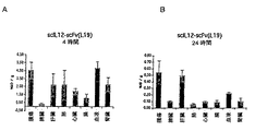
本発明者らが試験した1つの形式は、図1Aに示されるscIL12-scFv(L19)であった。このコンジュゲートは、従来技術の知見と一致する、控えめな腫瘍標的化能力を示した。 One format that we tested was scIL12-scFv (L19) shown in FIG. 1A. This conjugate showed a modest tumor targeting ability consistent with prior art findings.
別の形式では、上記のダイマーSIP(L19)構築物を用いて、図1Bに示されるscIL12-SIP(L19)のホモダイマーを作製した。しかしながら、L19の腫瘍標的化特性を、SIP形式を用いて改善することができるという従来技術の示唆にも関わらず、本発明者らは、腫瘍によるこのコンジュゲートの取込みの増加を観察しなかった。 In another format, the dimer SIP (L19) construct described above was used to make the homodimer of scIL12-SIP (L19) shown in FIG. 1B. However, despite the prior art suggestion that the tumor targeting properties of L19 can be improved using the SIP format, we did not observe an increase in uptake of this conjugate by the tumor. .
別の形式は、IL12のp40およびp35サブユニットのヘテロダイマーであって、各サブユニットがscFv(L19)と融合しているものであり、これは図1Cに示されるscFv(L19)-p35/p40-scFv(L19)へテロダイマーを形成する。このヘテロダイマー形式に関して、本発明者らは、腫瘍によるこのコンジュゲートの取込みの顕著な改善を達成した。 Another format is a heterodimer of the p40 and p35 subunits of IL12, where each subunit is fused to scFv (L19), which is the scFv (L19) -p35 / p shown in FIG. p40-scFv (L19) heterodimer is formed. With respect to this heterodimeric format, we achieved a marked improvement in the uptake of this conjugate by the tumor.
かくして、本発明者らは、IL12のp40およびp35サブユニットを介してヘテロダイマー化した2個のscFvフラグメントからなる新しい抗体-IL12融合タンパク質形式が、完全なIL12活性を保持し、優れた腫瘍標的化能力を示すことを発見した。 Thus, the inventors have shown that a new antibody-IL12 fusion protein format consisting of two scFv fragments heterodimerized via the p40 and p35 subunits of IL12 retains full IL12 activity and is an excellent tumor target It was found to show the ability to convert.
これらの結果は、腫瘍ならびに、例えば関節リウマチ、糖尿病性網膜症、加齢性筋肉変性および血管腫を治療するための、病的な血管新生の他の部位に対するIL12の改善された標的化のための有意な治療的示唆を有する。本発明の有用性は、scFv(L19)へのIL12の融合物だけでなく、他の特異的結合メンバーと他の薬剤および物質とのコンジュゲートにも拡張される。例えば、コンジュゲートを、腫瘍関連抗原(例えば、テネイシンCのアイソフォーム)に特異的な他の抗体フラグメントなどの、IL12サブユニットにコンジュゲートさせたscFv(L19)以外の特異的結合メンバーを用いて構築し、腫瘍標的化および癌治療に用いることができる。より広範な示唆はまた、診断方法ならびに疾患および他の病的状態の予防および治療などの、in vivoでの物質の標的化を伴う様々な他の用途も含む。 These results are due to improved targeting of IL12 to tumors and other sites of pathological angiogenesis, for example, to treat rheumatoid arthritis, diabetic retinopathy, age-related muscle degeneration and hemangiomas Have significant therapeutic implications. The utility of the present invention extends not only to fusions of IL12 to scFv (L19), but also to conjugates of other specific binding members with other drugs and agents. For example, using a specific binding member other than scFv (L19) conjugated to an IL12 subunit, such as other antibody fragments specific for a tumor-associated antigen (eg, tenascin-C isoform). Can be constructed and used for tumor targeting and cancer therapy. The broader implications also include a variety of other applications involving targeting of substances in vivo, such as diagnostic methods and prevention and treatment of diseases and other pathological conditions.
様々な態様での本発明は、新規コンジュゲート、それらを製造する方法、該コンジュゲートまたはその成分をコードする核酸、該コンジュゲートを含む医薬組成物、および治療方法における該コンジュゲートの使用に関する。 The invention in various aspects relates to novel conjugates, methods of making them, nucleic acids encoding the conjugates or components thereof, pharmaceutical compositions comprising the conjugates, and use of the conjugates in therapeutic methods.
一態様においては、本発明は、第1および第2のサブユニットがそれぞれ特異的結合メンバーにコンジュゲートされた、該第1および第2のサブユニットを有するタンパク質を含むコンジュゲートである。 In one aspect, the invention is a conjugate comprising a protein having the first and second subunits, wherein the first and second subunits are each conjugated to a specific binding member.
このタンパク質は、一般的にはダイマーであり、好ましくはヘテロダイマーであり、典型的には、生物活性を有する物質または薬剤、通常は治療剤または診断剤を含む。 The protein is generally a dimer, preferably a heterodimer, and typically includes a biologically active substance or agent, usually a therapeutic or diagnostic agent.
かくして、前記コンジュゲートは、通常、以下の形式:
[特異的結合メンバー]-[第1のサブユニット]-[第2のサブユニット]-[特異的結合メンバー]
を有する。
Thus, the conjugate is typically in the following form:
[Specific binding member]-[first subunit]-[second subunit]-[specific binding member]
Have
特異的結合メンバーは通常は抗体分子、好ましくは一本鎖Fv(scFv)である。一本鎖Fv(scFv)抗体分子は、その小さいサイズのため、本発明において特に好ましく、これは前記コンジュゲートのin vivoでの使用のための生理学的および治療的利点を提供する。さらに、scFvはFc領域を欠いており、これは抗イディオタイプ反応を潜在的に低下させ、これはまた、腫瘍標的化を妨げ非特異的細胞活性化を引き起こし得る補体の活性化とFc受容体との相互作用に関する望ましくない特性を、最小化する。 The specific binding member is usually an antibody molecule, preferably a single chain Fv (scFv). Single chain Fv (scFv) antibody molecules are particularly preferred in the present invention due to their small size, which provides physiological and therapeutic advantages for in vivo use of the conjugates. In addition, scFv lacks the Fc region, which potentially reduces anti-idiotypic responses, which can also interfere with tumor targeting and cause non-specific cell activation and Fc acceptance. Minimize undesirable properties related to body interaction.
従って、前記コンジュゲートの好ましい形式は、
[scFv]-[第1のサブユニット]-[第2のサブユニット]-[scFv]
である。
Accordingly, a preferred form of the conjugate is
[scFv]-[first subunit]-[second subunit]-[scFv]
It is.
あるいは、特異的結合メンバーは、単一ドメイン抗体および/または抗体フラグメントであってもよい。特異的結合メンバーおよび抗体分子を、以下でより詳細に説明する。 Alternatively, the specific binding member may be a single domain antibody and / or antibody fragment. Specific binding members and antibody molecules are described in more detail below.
前記コンジュゲートは、融合タンパク質、すなわち、2つ以上の遺伝子もしくは核酸コード化配列を1つのオープンリーディングフレーム(ORF)に融合したものから得られる翻訳産物であるポリペプチドであるか、またはそれを含むものでありうる。2つの遺伝子またはORFの融合発現産物を、ペプチドリンカー(短い2〜20個、好ましくは2〜15個の残基の一続きのアミノ酸)によりコンジュゲートさせることができる。一実施形態においては、ペプチドリンカーは6残基の配列GSADGG(配列番号15)である。この融合タンパク質は、通常は特異的結合メンバーおよびサブユニットの上流(5'側)に位置する、シグナルペプチド配列を含んでもよい。 The conjugate is or comprises a fusion protein, i.e., a polypeptide that is a translation product obtained by fusing two or more genes or nucleic acid coding sequences into one open reading frame (ORF). It can be a thing. The fusion expression product of two genes or ORFs can be conjugated by a peptide linker (a short stretch of 2-20, preferably 2-15 residues). In one embodiment, the peptide linker is the six residue sequence GSADGG (SEQ ID NO: 15). The fusion protein may comprise a signal peptide sequence, usually located upstream (5 ′) of the specific binding member and subunit.
本発明のコンジュゲートにおいては、一般的には、第1および第2のサブユニットが連結され、例えば、これらは1個以上のジスルフィド結合などを介して共有結合されうる。前記タンパク質はオリゴマー(例えば、ダイマー、好ましくはヘテロダイマー)であってよく、オリゴマー形態で天然に存在してもよく、かくして前記コンジュゲート中の第1および第2のサブユニットはその自然な方法で互いに結合していてもよい。従って、本発明は、コンジュゲートでの天然の形式のタンパク質の使用および維持を可能にする。これは、薬剤の一本鎖変異体を構築または使用する必要性を回避し、薬剤がコンジュゲート中でその完全な活性を保持する可能性を最大化する。さらに、前記サブユニット間のコンジュゲーションにより、該コンジュゲートを都合よく構築し、第1および第2のサブユニットをオリゴマーとして結合させる(例えば、ダイマー化する)ことによって組み立てることができる。かくして、典型的には、第1および第2のサブユニットを特異的結合メンバーにそれぞれ結合させて、該サブユニットを介して結合する一対の特異的結合メンバー-サブユニット構築物(例えば、ヘテロダイマー)を形成させることができる。好ましい実施形態においては、第1のサブユニットを、第1の融合タンパク質として特異的結合メンバーとコンジュゲートさせ、第2のサブユニットを、第2の融合タンパク質として特異的結合メンバーとコンジュゲートさせる。 In the conjugates of the present invention, generally, the first and second subunits are linked, for example, they can be covalently bonded, such as through one or more disulfide bonds. The protein may be an oligomer (e.g., a dimer, preferably a heterodimer) and may occur naturally in oligomeric form, thus the first and second subunits in the conjugate are in their natural way. They may be bonded to each other. Thus, the present invention allows the use and maintenance of native forms of proteins in conjugates. This avoids the need to construct or use single stranded variants of the drug and maximizes the likelihood that the drug retains its full activity in the conjugate. Furthermore, conjugation between the subunits allows the conjugate to be conveniently constructed and assembled by attaching (eg, dimerizing) the first and second subunits as oligomers. Thus, typically a pair of specific binding member-subunit constructs (e.g., heterodimers) that bind the first and second subunits to a specific binding member, respectively, and bind via the subunits. Can be formed. In a preferred embodiment, the first subunit is conjugated as a first fusion protein with a specific binding member and the second subunit is conjugated as a second fusion protein with a specific binding member.
好ましくは、前記コンジュゲートは、250 kDa以下、より好ましくは、200 kDa、150 kDa、125 kDa、120 kDaまたは115 kDa以下の分子量(すなわち、250000、200000、150000、125000、120000または115000以下のMr)を有する。これは、実際に測定された分子量(グリコシル化を伴うか、もしくは伴わない)であってもよく、または、例えば、コンジュゲートの予想分子量(グリコシル化を伴ってもよいが、通常は伴わない)に基づいて評価された値であってもよい。比較的小さいサイズのコンジュゲートでは、組織に浸透し標的部位(例えば、血管新生、腫瘍または疾患の部位)にアクセスするその能力が増大し、かくして、標的へのコンジュゲートの多価(一般的には、二価)結合を依然として達成しながら、その治療効力が増大し、必要とされる用量が低下する。 Preferably, the conjugate has a molecular weight of 250 kDa or less, more preferably 200 kDa, 150 kDa, 125 kDa, 120 kDa or 115 kDa or less (i.e., a Mr of 250,000, 20000, 150000, 125000, 120000 or 115000 or less. ). This may be the actual measured molecular weight (with or without glycosylation) or, for example, the expected molecular weight of the conjugate (with or without glycosylation) It may be a value evaluated based on A relatively small size conjugate increases its ability to penetrate the tissue and access the target site (e.g., angiogenesis, tumor or disease site), and thus the multivalent (generally, conjugate of the conjugate to the target). Is still achieving bivalent) binding, increasing its therapeutic efficacy and reducing the required dose.
一般的には、前記タンパク質および特異的結合メンバーを、前記コンジュゲートの意図される使用に従って選択する。好ましい実施形態においては、前記タンパク質はIL12である。上記で考察したように、IL12は、癌を治療し、腫瘍の増殖および転移を阻害し、ならびに病的な血管新生に関連する他の状態を治療するのに好適である。コンジュゲート中の1つまたは両方の特異的結合メンバーは、新生物(特に、腫瘍)の増殖および/または血管新生に関連するマーカー、例えば、新生物増殖および/または血管新生の部位に位置するマーカーに特異的に結合しうる。細胞外マトリックスはこれらのプロセスの間に再モデリングされるため、細胞外マトリックス成分は、新生物増殖および/または血管新生のマーカーであってもよい。 Generally, the protein and specific binding member are selected according to the intended use of the conjugate. In a preferred embodiment, the protein is IL12. As discussed above, IL12 is suitable for treating cancer, inhibiting tumor growth and metastasis, and treating other conditions associated with pathological angiogenesis. One or both specific binding members in the conjugate are markers associated with neoplastic (particularly tumor) growth and / or angiogenesis, eg, markers located at sites of neoplastic growth and / or angiogenesis It can bind specifically to. Since the extracellular matrix is remodeled during these processes, the extracellular matrix component may be a marker of neoplastic growth and / or angiogenesis.
1つの例は、上記で説明したように、追加のドメインED-Bを含むフィブロネクチンのB-FNアイソフォームである。本発明の特異的結合メンバーは、フィブロネクチン・アイソフォームB-FNのED-Bに特異的に結合するのが好ましい。特異的結合メンバーは、L19のVH CDR1(配列番号25)、VH CDR2(配列番号26)および/もしくはVH CDR3(配列番号27)配列ならびに/またはL19のVL CDR1(配列番号28)、VL CDR2(配列番号29)および/もしくはVL CDR3(配列番号30)配列を含むアミノ酸配列を有してもよい。例えば、特異的結合メンバーは、L19のVH CDR1、VH CDR2および/もしくはVH CDR3を含むアミノ酸配列を有するVHドメイン、ならびにL19のVL CDR1、VL CDR2および/もしくはVL CDR3を含むアミノ酸配列を有するVLドメインを有するscFvであってよい。特異的結合メンバーは、配列番号22に記載のL19 VHドメインのアミノ酸配列との少なくとも60%、65%、70%、75%、80%、85%、90%、95%もしくは100%の配列同一性を有するアミノ酸配列を有するVHドメインを含んでもよく、および/または配列番号23に記載のL19 VLドメインのアミノ酸配列との少なくとも60%、65%、70%、75%、80%、85%、90%、95%もしくは100%の配列同一性を有するアミノ酸配列を有するVLドメインを含む。好ましくは、特異的結合メンバーは、L19 VHドメイン(配列番号22)およびL19 VLドメイン(配列番号23)を含むscFv(L19)である。好ましい実施形態においては、特異的結合メンバーは、配列番号5のアミノ酸配列を有するscFv(L19)である(図10)。 One example is the B-FN isoform of fibronectin containing an additional domain ED-B as described above. The specific binding member of the present invention preferably binds specifically to ED-B of fibronectin isoform B-FN. Specific binding members include L19 VH CDR1 (SEQ ID NO: 25), VH CDR2 (SEQ ID NO: 26) and / or VH CDR3 (SEQ ID NO: 27) sequences and / or L19 VL CDR1 (SEQ ID NO: 28), VL CDR2 ( It may have an amino acid sequence comprising SEQ ID NO: 29) and / or VL CDR3 (SEQ ID NO: 30) sequence. For example, the specific binding member is a VH domain having an amino acid sequence comprising L19 VH CDR1, VH CDR2 and / or VH CDR3, and a VL domain having an amino acid sequence comprising VL CDR1, VL CDR2 and / or VL CDR3 of L19 It may be scFv having The specific binding member is at least 60%, 65%, 70%, 75%, 80%, 85%, 90%, 95% or 100% sequence identical to the amino acid sequence of the L19 VH domain set forth in SEQ ID NO: 22 A VH domain having an amino acid sequence with sex and / or at least 60%, 65%, 70%, 75%, 80%, 85% with the amino acid sequence of the L19 VL domain set forth in SEQ ID NO: 23, It contains a VL domain having an amino acid sequence with 90%, 95% or 100% sequence identity. Preferably, the specific binding member is an scFv (L19) comprising an L19 VH domain (SEQ ID NO: 22) and an L19 VL domain (SEQ ID NO: 23). In a preferred embodiment, the specific binding member is scFv (L19) having the amino acid sequence of SEQ ID NO: 5 (FIG. 10).
別の例は、様々なアイソフォームで存在するテネイシンC(TnC)である。新生物組織においては、さらなるドメインを含有するTnC、特に、ドメインCを含有するアイソフォーム(cTN-C)[33]は、通常の組織よりも幅広く発現される。かくして、本発明のコンジュゲートにおける特異的結合メンバーは、新生物組織に関連するテネイシンCアイソフォーム、特に、cTN-Cに特異的に結合することができる。特異的結合メンバーは、配列番号21に記載の配列を有するTN11 scFvであってもよい(図11)[33]。 Another example is tenascin-C (TnC), which exists in various isoforms. In neoplastic tissues, TnC containing additional domains, especially isoforms containing domain C (cTN-C) [33], are expressed more widely than normal tissues. Thus, specific binding members in the conjugates of the invention can specifically bind to tenascin-C isoforms associated with neoplastic tissues, in particular cTN-C. The specific binding member may be TN11 scFv having the sequence set forth in SEQ ID NO: 21 (FIG. 11) [33].
あるいは、本発明のコンジュゲート中の特異的結合メンバーは、他の腫瘍関連抗原、すなわち、通常の細胞環境(非腫瘍細胞上など)におけるよりも腫瘍環境(腫瘍細胞上など)においてより広く存在する抗原に結合してもよい。 Alternatively, specific binding members in the conjugates of the invention are more widely present in tumor environments (such as on tumor cells) than in other tumor-associated antigens, i.e., in normal cellular environments (such as on non-tumor cells). It may bind to an antigen.
通常、コンジュゲート中の2つの特異的結合メンバーは同一であるか、または少なくとも、同じ標的、抗原もしくはエピトープに対して両方とも特異的なものである。 Usually, the two specific binding members in the conjugate are the same or at least both are specific for the same target, antigen or epitope.
本発明の特に好ましい実施形態においては、前記コンジュゲートは、2つの特異的結合メンバー、好ましくはscFv(L19)とコンジュゲートされ、かつそれらの間にコンジュゲートされているヒトIL12へテロダイマーを含み;ここで、
該へテロダイマーは、第1の(通常はp40)サブユニットおよび第2の(通常はp35)サブユニットを有し;
第1の、またはp40サブユニットは、第1の融合タンパク質として第1の特異的結合メンバーにコンジュゲートされ;
第2の、またはp35サブユニットは、第2の融合タンパク質として第2の特異的結合メンバーにコンジュゲートされている。
In a particularly preferred embodiment of the invention, the conjugate comprises a human IL12 heterodimer conjugated to and conjugated between two specific binding members, preferably scFv (L19); here,
The heterodimer has a first (usually p40) subunit and a second (usually p35) subunit;
The first, or p40 subunit, is conjugated to the first specific binding member as a first fusion protein;
The second, or p35 subunit is conjugated to the second specific binding member as a second fusion protein.
上記のように、第1および第2のサブユニットは、典型的には、共有結合され、例えば、ジスルフィド結合される。 As described above, the first and second subunits are typically covalently bonded, eg, disulfide bonded.
好ましくは、第1の、またはp40サブユニットは、特異的結合メンバーのN末端に融合され、すなわち、該サブユニットは、融合タンパク質およびそれをコードする核酸中で特異的結合メンバーの上流にある。従って、この形態では、p40のN末端は遊離(融合していない)していてもよく、それはその活性を最大化させると考えられる。 Preferably, the first or p40 subunit is fused to the N-terminus of the specific binding member, ie, the subunit is upstream of the specific binding member in the fusion protein and the nucleic acid encoding it. Thus, in this form, the N-terminus of p40 may be free (unfused), which is thought to maximize its activity.
好ましくは、第2の、またはp35サブユニットは、特異的結合メンバーのC末端に融合され、すなわち、該サブユニットは、融合タンパク質およびそれをコードする核酸中で特異的結合メンバーの下流にある。特に、上流のN末端シグナルペプチドを有する特異的結合メンバーを容易に発現させることができ、かくして融合タンパク質の効率的な発現が可能になるため、これにより融合タンパク質の発現を増強することができる。 Preferably, the second or p35 subunit is fused to the C-terminus of the specific binding member, i.e., the subunit is downstream of the specific binding member in the fusion protein and the nucleic acid encoding it. In particular, a specific binding member having an upstream N-terminal signal peptide can be readily expressed, thus allowing efficient expression of the fusion protein, thereby enhancing expression of the fusion protein.
好ましくは、第1の融合タンパク質は、配列番号1に示されるp40-scFv(L19)のアミノ酸配列を有する。好ましくは、第2の融合タンパク質は、配列番号2に示されるscFv(L19)-p35のアミノ酸を有する。 Preferably, the first fusion protein has the amino acid sequence of p40-scFv (L19) shown in SEQ ID NO: 1. Preferably, the second fusion protein has the amino acid scFv (L19) -p35 shown in SEQ ID NO: 2.
本発明のコンジュゲートを、任意の利用可能な方法で、例えば、組換え技術を用いて、例えば、コンジュゲートの全部または一部を融合タンパク質として発現させることにより、製造することができる。 The conjugates of the invention can be produced by any available method, for example using recombinant techniques, for example by expressing all or part of the conjugate as a fusion protein.
例えば、コンジュゲートを、以下:
第1のサブユニットおよび特異的結合メンバーを含む第1の融合タンパク質を発現させること;
第2のサブユニットおよび特異的結合メンバーを含む第2の融合タンパク質を発現させること;ならびに
第1および第2のサブユニットを1つにコンジュゲートさせること、
を含む方法で、製造することができる。
For example, the conjugate can be:
Expressing a first fusion protein comprising a first subunit and a specific binding member;
Expressing a second fusion protein comprising a second subunit and a specific binding member; and conjugating the first and second subunits together,
Can be manufactured by a method including:
通常、この方法は、発現後に第1および第2の融合タンパク質を精製することを含む。 Typically, this method involves purifying the first and second fusion proteins after expression.
通常、この発現を、融合タンパク質をコードする核酸を含む宿主細胞、例えば、HEKもしくはCHO細胞などの培養真核細胞、または大腸菌などの細菌細胞中で行う。従って、発現はそのような宿主細胞を培養することを含んでもよい。第1および第2の融合タンパク質を同じ細胞(例えば、2つの融合タンパク質をコードする核酸で同時トランスフェクトされたか、もしくはそれらを含む細胞)中で発現させる場合、前記サブユニットのヘテロダイマー化もしくはオリゴマー化は、該細胞中で起こるか、または細胞からの融合タンパク質の精製の際に起こってもよい。他の場合、第1および第2の融合タンパク質を別々に発現させ(例えば、異なる細胞中で)、次いで、第1および第2のサブユニットがヘテロダイマー化するか、またはさもなければ結合するように1つに合わせる(混合する)ことができる。 Usually, this expression is performed in a host cell containing a nucleic acid encoding the fusion protein, for example, a cultured eukaryotic cell such as HEK or CHO cell, or a bacterial cell such as E. coli. Thus, expression may include culturing such host cells. When the first and second fusion proteins are expressed in the same cell (e.g., a cell co-transfected with or containing nucleic acids encoding the two fusion proteins), the subunit heterodimerization or oligomer The conversion may occur in the cell or may occur during purification of the fusion protein from the cell. In other cases, the first and second fusion proteins are expressed separately (e.g., in different cells), and then the first and second subunits are heterodimerized or otherwise bound. Can be combined (mixed) into one.
サブユニットを1つにコンジュゲートさせることは、能動的または受動的プロセスであってもよい。コンジュゲーションは、第1および第2のサブユニットが1つにコンジュゲートされる(例えば、結合するか、もしくはオリゴマー化/へテロダイマー化する)条件に第1および第2の融合タンパク質を曝露するか、または供することを含んでもよい。コンジュゲーションは、サブユニット間のジスルフィド結合形成、または別の共有結合の形成を含んでもよい。ジスルフィド結合形成などのコンジュゲーションは、非還元条件下で起こりうるので、従って、コンジュゲーションは第1および第2の融合タンパク質を非還元条件に曝露することを含んでもよい。 Conjugating the subunits into one may be an active or passive process. Conjugation exposes the first and second fusion proteins to conditions in which the first and second subunits are conjugated together (eg, bind or oligomerize / heterodimerize). Or providing. Conjugation may include the formation of disulfide bonds between subunits, or the formation of another covalent bond. Since conjugation such as disulfide bond formation can occur under non-reducing conditions, conjugation may therefore include exposing the first and second fusion proteins to non-reducing conditions.
本発明に従って融合タンパク質を発現させ、コンジュゲートを製造するための好適な方法を、下記実施例で詳細に説明する。 Suitable methods for expressing fusion proteins and producing conjugates according to the present invention are described in detail in the examples below.
さらなる工程として、前記方法は、前記コンジュゲートを医薬組成物に製剤化することを含んでもよい。一般的には、これは前記コンジュゲートを精製し、それを生理学的に許容し得る担体と混合することを含む。医薬組成物は下記により詳細に説明する。 As a further step, the method may comprise formulating the conjugate into a pharmaceutical composition. Generally, this involves purifying the conjugate and mixing it with a physiologically acceptable carrier. The pharmaceutical composition is described in more detail below.
前記コンジュゲートおよびその一部をコードする(例えば、融合タンパク質をコードする)核酸分子も、本発明の一部を形成する。 Nucleic acid molecules that encode the conjugate and portions thereof (eg, encode fusion proteins) also form part of the invention.
一態様においては、本発明は、以下:
特異的結合メンバーおよび第1のタンパク質サブユニットを含む融合タンパク質をコードするヌクレオチド配列を含む第1の核酸分子;ならびに
特異的結合メンバーおよび第2のタンパク質サブユニットを含む融合タンパク質をコードするヌクレオチド配列を含む第2の核酸分子、
を含む組成物である。
In one aspect, the present invention provides the following:
A first nucleic acid molecule comprising a nucleotide sequence encoding a fusion protein comprising a specific binding member and a first protein subunit; and a nucleotide sequence encoding a fusion protein comprising a specific binding member and a second protein subunit. A second nucleic acid molecule comprising,
It is a composition containing this.
前記核酸分子は、融合タンパク質中で特異的結合メンバーとサブユニットがペプチドリンカーにより連結されている、特異的結合メンバーと該サブユニット間のペプチドリンカーをコードしてもよい。コードされうる特定の融合タンパク質、サブユニット、特異的結合メンバーおよびリンカーを、本明細書の他の箇所でより詳細に説明する。特定の実施形態においては、第1および第2のサブユニットは、それぞれIL12ヘテロダイマーサブユニット(典型的には、p40およびp35サブユニット)であり、好ましくは、特異的結合メンバーはscFv、特に、scFv(L19)である。一実施形態においては、第1の核酸分子は、配列番号1に記載のIL12p40-scFv(L19)のアミノ酸配列をコードし、および/または第2の核酸分子は、配列番号2に記載のscFv(L19)-IL12p35のアミノ酸配列をコードする。 The nucleic acid molecule may encode a peptide linker between the specific binding member and the subunit, wherein the specific binding member and subunit are linked by a peptide linker in the fusion protein. Specific fusion proteins, subunits, specific binding members and linkers that can be encoded are described in more detail elsewhere herein. In certain embodiments, the first and second subunits are each IL12 heterodimeric subunits (typically p40 and p35 subunits), preferably the specific binding member is scFv, in particular, scFv (L19). In one embodiment, the first nucleic acid molecule encodes the amino acid sequence of IL12p40-scFv (L19) set forth in SEQ ID NO: 1 and / or the second nucleic acid molecule is the scFv (set in SEQ ID NO: 2 Encodes the amino acid sequence of L19) -IL12p35.
前記核酸分子は、ベクター、例えば、前記ヌクレオチド配列の発現にとって好適なプラスミドであってもよい。かくして、第1および第2の核酸分子は、第1および第2のベクターであってもよい。通常は該ヌクレオチド配列は、転写のためのプロモーターなどの調節エレメントに機能し得る形で連結される。 The nucleic acid molecule may be a vector, eg, a plasmid suitable for expression of the nucleotide sequence. Thus, the first and second nucleic acid molecules may be first and second vectors. Usually, the nucleotide sequence is operably linked to regulatory elements such as a promoter for transcription.
第1および第2の核酸分子を、該核酸分子で同時トランスフェクトされた細胞またはそのような細胞の娘細胞であってよい宿主細胞中に含有させることができる。前記核酸分子を含む細胞、特に、真核細胞、例えば、HEKおよびCHO細胞、または細菌細胞、例えば、大腸菌も、本発明の一部を形成する。 The first and second nucleic acid molecules can be contained in a host cell that can be a cell co-transfected with the nucleic acid molecule or a daughter cell of such a cell. Cells containing said nucleic acid molecules, in particular eukaryotic cells such as HEK and CHO cells, or bacterial cells such as E. coli also form part of the present invention.
前記核酸から発現させた後、融合タンパク質を、サブユニットを介してコンジュゲートさせて、本明細書の他の箇所に記載のような本発明のコンジュゲートを形成させることができる。 After being expressed from the nucleic acid, the fusion protein can be conjugated via a subunit to form a conjugate of the invention as described elsewhere herein.
本発明によるコンジュゲートを、患者に該コンジュゲートを投与することを含む、該患者(典型的には、ヒト患者)における疾患または障害の治療(予防的処置を含んでもよい)方法などの、ヒトまたは動物体の治療方法において用いることができる。 A human, such as a method of treatment (which may include prophylactic treatment) of a disease or disorder in said patient (typically a human patient) comprising administering the conjugate according to the invention to said patient. Or it can use in the treatment method of an animal body.
前記コンジュゲートを用いて治療可能な状態としては、癌、他の腫瘍および新生物状態が挙げられる。前記コンジュゲートを用いて、血管新生を阻害し、それによって、関節リウマチ、糖尿病性網膜症、加齢性筋肉変性、血管腫および癌などの腫瘍を治療することができる。治療は、予防的処置を含んでもよい。前記コンジュゲートを、診断方法、例えば、上記病的状態のいずれかに関連し得る血管新生の標的化および診断において投与することもできる。他の疾患および病的状態を、前記コンジュゲートに含まれるタンパク質治療剤または診断剤の性質、および特異的結合メンバーの特異性に従って、診断および治療することもできる。 Conditions that can be treated using the conjugate include cancer, other tumors, and neoplastic conditions. The conjugates can be used to inhibit angiogenesis, thereby treating tumors such as rheumatoid arthritis, diabetic retinopathy, age-related muscle degeneration, hemangiomas and cancer. Treatment may include prophylactic treatment. The conjugates can also be administered in diagnostic methods such as targeting and diagnosing angiogenesis that can be associated with any of the above pathological conditions. Other diseases and pathological conditions can also be diagnosed and treated according to the nature of the protein therapeutic or diagnostic agent included in the conjugate and the specificity of the specific binding member.
従って、本発明のさらなる態様は、本発明のコンジュゲートを投与することを含む治療方法、そのようなコンジュゲートを含む医薬組成物、および病的状態もしくは疾患の治療のための医薬の製造における、例えば、生理学的に許容し得る担体もしくは賦形剤と共に該コンジュゲートを製剤化することを含む医薬もしくは医薬組成物の製造方法における、そのようなコンジュゲートの使用を提供する。 Accordingly, a further aspect of the present invention is in a method of treatment comprising administering a conjugate of the present invention, a pharmaceutical composition comprising such a conjugate, and the manufacture of a medicament for the treatment of a pathological condition or disease. For example, there is provided the use of such a conjugate in a method of making a medicament or pharmaceutical composition comprising formulating the conjugate with a physiologically acceptable carrier or excipient.
本発明に従えば、提供される組成物を患者に投与することができる。投与は、好ましくは、「治療上有効量」で行われ、これは患者に対する恩恵を示すのに十分なものである。そのような恩恵は、少なくとも1つの症候が少なくとも改善されることであってよい。投与する実際の量、ならびに投与の速度および時間経過は、治療しようとするものの性質および重篤度に依存するであろう。治療の処方、例えば、投薬量などの決定は、一般的な開業医および他の医師の職責の範囲内にある。抗体の好適な用量は当業界でよく知られている[26, 27]。 In accordance with the present invention, the provided composition can be administered to a patient. Administration is preferably carried out in a “therapeutically effective amount”, which is sufficient to show benefit to the patient. Such benefit may be that at least one symptom is at least ameliorated. The actual amount administered, and rate and time-course of administration, will depend on the nature and severity of what is being treated. Determination of treatment prescriptions, such as dosages, is within the responsibilities of general practitioners and other physicians. Suitable doses of antibody are well known in the art [26, 27].
抗体抗原結合ドメインを含むものを始めとする本発明のコンジュゲートを、治療を必要とする患者に対して、任意の好適な経路を介して、通常は血流への注入により、および/または治療しようとする部位、例えば、腫瘍もしくは腫瘍血管系への直接的注入により、投与することができる。投与の正確な用量およびその頻度は、多くの要因、治療の経路、治療しようとする領域(例えば、腫瘍)の大きさおよび位置、抗体の正確な性質(例えば、scFv分子)、ならびにコンジュゲート中に含まれる検出可能な標識もしくは他の分子の性質に依存するであろう。 Conjugates of the invention, including those comprising an antibody antigen binding domain, may be administered to a patient in need of treatment via any suitable route, usually by infusion into the bloodstream, and / or treatment Administration can be by direct injection into the site to be treated, eg, a tumor or tumor vasculature. The exact dose and frequency of administration depends on many factors, the route of treatment, the size and location of the area to be treated (e.g. tumor), the exact nature of the antibody (e.g. scFv molecule), and in the conjugate Will depend on the nature of the detectable label or other molecule contained.
組成物を、治療しようとする状態に応じて、単独で、または他の治療と同時にもしくは連続的に組合わせて、投与することができる。他の治療としては、好適な用量の非ステロイド系抗炎症剤(例えば、アスピリン、パラセタモール、イブプロフェンもしくはケトプロフェン)またはモルヒネなどのアヘン剤などの疼痛緩和薬剤、または制吐剤の投与が挙げられる。 The composition can be administered alone or in combination with other treatments simultaneously or sequentially, depending on the condition to be treated. Other treatments include administration of suitable doses of non-steroidal anti-inflammatory drugs (eg, aspirin, paracetamol, ibuprofen or ketoprofen) or opiates such as opiates such as morphine, or antiemetics.
かくして、本発明による医薬組成物、および本発明に従う使用のための医薬組成物は、活性成分(コンジュゲート)に加えて、製薬上許容し得る賦形剤、担体、バッファー、安定剤または当業者にはよく知られた他の材料を含んでもよい。そのような材料は非毒性であるべきであり、前記活性成分の効力を妨げるべきではない。担体または他の材料の正確な性質は、経口、または例えば、静脈内などの注入によるものであってよい投与経路に依存するであろうは。静脈内注入、または苦痛部位への注入のためには、前記活性成分は、発熱物質を含まず好適なpH、等張性および安定性を有する非経口的に許容し得る水性溶液の形態であるであろう。 Thus, the pharmaceutical composition according to the present invention, and the pharmaceutical composition for use according to the present invention, in addition to the active ingredient (conjugate), are pharmaceutically acceptable excipients, carriers, buffers, stabilizers or those skilled in the art. May include other well-known materials. Such materials should be non-toxic and should not interfere with the efficacy of the active ingredient. The exact nature of the carrier or other material will depend on the route of administration, which may be oral or by injection, for example intravenously. For intravenous injection or injection to the site of pain, the active ingredient is in the form of a parenterally acceptable aqueous solution that is pyrogen free and has a suitable pH, isotonicity and stability. Will.
本発明のコンジュゲートにおいては、前記タンパク質は通常、組換え生産された第1および第2のポリペプチドサブユニットを含む。このサブユニットはグリコシル化されていてもよく、そのようなグリコシル化の程度および性質を、該サブユニットを発現するような好適な宿主細胞の選択により制御することができる。このタンパク質は、生物活性物質、すなわち、それを投与する対象生物の構造またはその機能に影響する物質を含んでもよい。通常、このタンパク質は、診断剤または治療剤を含む。例えば、それは疾患または病的状態の診断、予防または治療に用いられる物質を含んでもよい。前記タンパク質は、例えば診断のための、マーカーまたは標識化剤を含んでもよい。本発明の内容においては、前記タンパク質は通常、例えば、癌および他の腫瘍、関節リウマチ、糖尿病性網膜症、加齢性筋肉変性および血管腫の治療において、病的状態、特に、血管新生を治療または予防するための治療物質を含む。それは、例えば、毒素、酵素、またはサイトカインなどの免疫のメディエーターを含んでもよい。 In the conjugates of the invention, the protein typically comprises first and second polypeptide subunits that are produced recombinantly. This subunit may be glycosylated and the extent and nature of such glycosylation can be controlled by selection of a suitable host cell to express the subunit. The protein may comprise a bioactive substance, i.e. a substance that affects the structure of the target organism to which it is administered or its function. The protein typically includes a diagnostic or therapeutic agent. For example, it may include substances used for diagnosis, prevention or treatment of a disease or pathological condition. The protein may comprise a marker or labeling agent, for example for diagnostic purposes. In the context of the present invention, the protein usually treats pathological conditions, in particular angiogenesis, for example in the treatment of cancer and other tumors, rheumatoid arthritis, diabetic retinopathy, age-related muscle degeneration and hemangiomas. Or contain a therapeutic substance to prevent. It may include immune mediators such as, for example, toxins, enzymes, or cytokines.
前記タンパク質は、天然または組換えのインターロイキン-12(IL12)であってよい。本発明において有用なIL12を、任意の動物、例えば、ヒト、げっ歯類(例えば、ラット、マウス)、ウマ、ウシ、ブタ、ヒツジ、イヌなどから得ることができる。ヒトIL12が、ヒトへの投与のためのコンジュゲートにおいて好ましい。IL12は、40 kDa(p40)サブユニットおよび35 kDa(p35)サブユニットから構成されるヘテロダイマータンパク質として天然に存在する。前記サブユニットの実際の分子量は、例えば、異なる生物種で発現される場合、ならびにタンパク質がグリコシル化されているかどうか、およびそのグリコシル化パターンに応じて様々でありうる。従って、用語「p40」および「p35」は、前記サブユニットがそれぞれ正確に40 kDaおよび35 kDaの分子量を有することを意味するわけではない。むしろ、これらの用語を用いて、IL12の2つのヘテロダイマーサブユニットを同定および識別し、これらはそれらのアミノ酸配列の点でより正確に規定することができる。ヘテロダイマーIL12は、それぞれ、ヒトIL12のp40サブユニットおよびp35サブユニットと相同であるか、または同一である第1および第2のポリペプチドサブユニットを含む。典型的には、IL12の第1のサブユニットは、配列番号3に記載のヒトIL12サブユニットp40のアミノ酸配列との少なくとも60%、65%、70%、75%、80%、85%、90%、95%または100%の配列同一性を有するアミノ酸配列を含む。典型的には、IL12の第2のサブユニットは、配列番号4に記載のヒトIL12サブユニットp35のアミノ酸配列との少なくとも60%、65%、70%、75%、80%、85%、90%、95%または100%の配列同一性を有するアミノ酸配列を含む。本発明のコンジュゲート中のIL12は、IL12の生物活性、例えば、活性化されたTおよびNK細胞に対して増殖因子として作用する能力、NK/リンホカインにより活性化されたキラー細胞の溶解活性を増強する能力、PMBCを休止させることによりIFN-γの産生を刺激する能力、血管新生を阻害する能力(例えば、下流のメディエーターIP-10を介して)ならびに/または腫瘍増殖および/もしくは転移を阻害する能力を保持する。 The protein may be natural or recombinant interleukin-12 (IL12). IL12 useful in the present invention can be obtained from any animal, such as humans, rodents (eg, rats, mice), horses, cows, pigs, sheep, dogs, and the like. Human IL12 is preferred in conjugates for human administration. IL12 exists naturally as a heterodimeric protein composed of a 40 kDa (p40) subunit and a 35 kDa (p35) subunit. The actual molecular weight of the subunit can vary, for example, when expressed in different species, as well as whether the protein is glycosylated and its glycosylation pattern. Thus, the terms “p40” and “p35” do not mean that the subunits have molecular weights of exactly 40 kDa and 35 kDa, respectively. Rather, these terms can be used to identify and distinguish two heterodimeric subunits of IL12, which can be more precisely defined in terms of their amino acid sequences. The heterodimeric IL12 comprises first and second polypeptide subunits that are homologous or identical to the p40 and p35 subunits of human IL12, respectively. Typically, the first subunit of IL12 is at least 60%, 65%, 70%, 75%, 80%, 85%, 90% with the amino acid sequence of human IL12 subunit p40 set forth in SEQ ID NO: 3. Contains amino acid sequences with%, 95% or 100% sequence identity. Typically, the second subunit of IL12 is at least 60%, 65%, 70%, 75%, 80%, 85%, 90% with the amino acid sequence of human IL12 subunit p35 set forth in SEQ ID NO: 4. Contains amino acid sequences with%, 95% or 100% sequence identity. IL12 in the conjugates of the present invention enhances the biological activity of IL12, eg, the ability to act as a growth factor on activated T and NK cells, the lytic activity of killer cells activated by NK / lymphokine Ability to stimulate IFN-γ production by suspending PMBC, ability to inhibit angiogenesis (eg, via downstream mediator IP-10) and / or inhibit tumor growth and / or metastasis Hold the ability.
特異的結合メンバーは、互いに対する結合特異性を有する一対の分子のメンバーである。特異的結合対のメンバーは、天然に由来するか、または全体的もしくは部分的に合成により製造されたものであってもよい。分子対の一方のメンバーは、その表面上に領域、または空洞を有し、それは、前記分子対の他方のメンバーの特定の空間的および極性構成に特異的に結合し、かつ、それゆえそれに対して相補的である。かくして、前記対のメンバーは互いに特異的に結合する特性を有する。 A specific binding member is a member of a pair of molecules that have binding specificity for each other. A member of a specific binding pair may be naturally occurring or wholly or partially synthetically produced. One member of the molecular pair has a region, or cavity, on its surface that specifically binds to and therefore to a particular spatial and polar configuration of the other member of the molecular pair And complementary. Thus, the members of the pair have the property of binding specifically to each other.
前記特異的結合メンバーは通常、抗原結合部位を有する分子を含む。例えば、特異的結合メンバーは、抗原結合部位を含む抗体分子または非抗体タンパク質であってよい。抗原結合部位を、フィブロネクチンもしくはシトクロムBなどの非抗体タンパク質足場(scaffold)上の相補性決定領域(CDR)の配置により[29, 30, 31]、またはタンパク質足場内のループのアミノ酸残基をランダム化するか、もしくは突然変異させることにより提供し、所望の標的に対する結合特異性を付与することができる。タンパク質中に新規結合部位を作製するための足場が詳細に総説されている[31]。少なくとも1つのランダム化されたループを有するフィブロネクチンのIII型ドメインを含むタンパク質(抗体模倣物質)などの、抗体模倣物質のためのタンパク質足場が開示されている[32]。1つ以上のCDR、例えば、HCDRのセットを移植するのに好適な足場を、免疫グロブリン遺伝子スーパーファミリーの任意のドメインメンバーにより提供することができる。この足場はヒトまたは非ヒトタンパク質であってよい。 Said specific binding member usually comprises a molecule having an antigen binding site. For example, a specific binding member can be an antibody molecule or non-antibody protein that includes an antigen binding site. Antigen binding sites can be determined by placement of complementarity-determining regions (CDRs) on non-antibody protein scaffolds such as fibronectin or cytochrome B [29, 30, 31], or random amino acid residues in loops within the protein scaffold Or provided by mutating to confer binding specificity for a desired target. Scaffolds for creating new binding sites in proteins have been reviewed in detail [31]. Protein scaffolds for antibody mimetics have been disclosed, such as proteins comprising a type III domain of fibronectin with at least one randomized loop (antibody mimetics) [32]. A suitable scaffold for transplantation of one or more CDRs, eg, a set of HCDRs, can be provided by any domain member of the immunoglobulin gene superfamily. This scaffold may be a human or non-human protein.
非抗体タンパク質足場の利点は、それが少なくとも一部の抗体分子よりも小さく、および/または製造が容易である足場分子中に抗原結合部位を提供することができることである。小さいサイズの特異的結合メンバーは、細胞に進入する能力、組織中に深く浸透する能力もしくは他の構造物内に標的を到達させる能力、または標的抗原のタンパク質空洞内に結合する能力などの有用な生理学的特性を付与し得る。 An advantage of a non-antibody protein scaffold is that it can provide an antigen binding site in a scaffold molecule that is smaller than and / or easier to manufacture than at least some antibody molecules. Small size specific binding members are useful, such as the ability to enter cells, the ability to penetrate deeply into tissues or reach targets within other structures, or the ability to bind within the protein cavity of a target antigen Physiological properties can be imparted.
非抗体タンパク質足場中での抗原結合部位の使用は、[34]に総説されている。典型的なものは、安定な骨格および1つ以上の可変ループを有するタンパク質であり、このループのアミノ酸配列を特異的に、またはランダムに突然変異させて、標的抗原に結合するための特異性を有する抗原結合部位を作る。そのようなタンパク質としては、S.aureusに由来するプロテインA、トランスフェリン、テトラネクチン、フィブロネクチン(例えば、10番目のフィブロネクチンIII型ドメイン)およびリポカリンのIgG結合ドメインが挙げられる。他の手法としては、シクロチド(分子内ジスルフィド結合を有する小さいタンパク質)に基づく、合成「ミクロボディー」(Selecore GmbH)が挙げられる。 The use of antigen binding sites in non-antibody protein scaffolds is reviewed in [34]. A typical one is a protein with a stable backbone and one or more variable loops that specifically or randomly mutate the amino acid sequence of this loop to provide specificity for binding to the target antigen. Create an antigen binding site that has. Such proteins include protein A derived from S. aureus, transferrin, tetranectin, fibronectin (eg, 10th fibronectin type III domain) and lipocalin IgG binding domain. Other approaches include synthetic “microbodies” (Selecore GmbH) based on cyclotides (small proteins with intramolecular disulfide bonds).
記載のように、CDRを、フィブロネクチンまたはシトクロムBなどの足場により担持させることができるが[29, 30, 31]、本発明のCDRまたはCDRのセットを担持させるための構造は、CDRまたはCDRのセットが、再配置された免疫グロブリン遺伝子によりコードされる天然のVHおよびVL抗体可変ドメインのCDRまたはCDRのセットに対応する位置に位置する抗体の重鎖もしくは軽鎖配列またはその実質的な部分のものであるのが好ましいであろう。免疫グロブリン可変ドメインの構造および位置を、Kabatら[35]および現在ではインターネット上で利用可能なそのアップデートを参照することにより決定することができる(http://immuno.bme.nwu.eduまたは任意の検索エンジンを用いて「Kabat」を発見することができる)。 As described, the CDR can be carried by a scaffold such as fibronectin or cytochrome B [29, 30, 31], but the structure for carrying the CDR or set of CDRs of the present invention is the CDR or CDR Of a heavy or light chain sequence of an antibody or a substantial portion thereof located at a position corresponding to the CDR or set of CDRs of a natural VH and VL antibody variable domain encoded by a rearranged immunoglobulin gene Would be preferred. The structure and location of immunoglobulin variable domains can be determined by reference to Kabat et al. [35] and its updates now available on the Internet (http://immuno.bme.nwu.edu or any Can be used to find "Kabat").
抗体分子は、天然に産生されるか、または部分的もしくは全体的に合成的に産生された免疫グロブリンである。この用語はまた、抗体抗原結合部位を含む任意のポリペプチドまたはタンパク質をも含む。 Antibody molecules are immunoglobulins that are naturally produced, or partially or wholly synthetically produced. The term also includes any polypeptide or protein comprising an antibody antigen binding site.
モノクローナル抗体および他の抗体を取得し、組換えDNA技術を用いて、元の抗体の特異性を保持する他の抗体またはキメラ分子を製造することができる。そのような技術は、抗体の免疫グロブリン可変領域、またはCDRをコードするDNAを、異なる免疫グロブリンの定常領域、または定常領域+フレームワーク領域に導入することを含んでもよい[37, 38, 39]。抗体を産生するハイブリドーマまたは他の細胞を、産生される抗体の結合特異性を変化させても、もしくは変化させなくてもよい遺伝子突然変異または他の変化に供することができる。 Monoclonal antibodies and other antibodies can be obtained and other DNA or chimeric molecules that retain the specificity of the original antibody can be produced using recombinant DNA technology. Such techniques may involve introducing an immunoglobulin variable region of an antibody, or DNA encoding a CDR, into a constant region of a different immunoglobulin, or a constant region plus a framework region [37, 38, 39]. . Hybridomas or other cells that produce antibodies can be subjected to genetic mutations or other changes that may or may not alter the binding specificity of the antibodies produced.
抗体は様々な方法で改変することができるので、用語「抗体分子」は、必要とされる特異性を有する抗体抗原結合部位を有する任意の特異的結合メンバーまたは物質を包含すると解釈されるべきである。かくして、この用語は、天然のものであるか、または全体的もしくは部分的に合成されたものであるかに関わらず、抗体抗原結合部位を含む任意のポリペプチドなどの抗体フラグメントおよび誘導体を包含する。従って、別のポリペプチドに融合された、抗体抗原結合部位を含むキメラ分子、またはその等価物が包含される。キメラ抗体のクローニングおよび発現はよく知られている[40, 41]。 Since antibodies can be modified in a variety of ways, the term “antibody molecule” should be construed to include any specific binding member or substance having an antibody antigen binding site with the required specificity. is there. Thus, this term encompasses antibody fragments and derivatives, such as any polypeptide comprising an antibody antigen-binding site, whether natural or wholly or partially synthesized. . Thus, a chimeric molecule comprising an antibody antigen binding site fused to another polypeptide, or equivalent thereof is included. Cloning and expression of chimeric antibodies are well known [40, 41].
抗体工学の分野で利用可能なさらなる技術により、ヒトおよびヒト化抗体を単離することが可能になっている。例えば、ヒトハイブリドーマを、以前に記載のように作製することができる[36]。特異的結合メンバーを作製するための別の確立された技術であるファージディスプレイが詳細に記載されている[36, 42]。マウス免疫系の他の成分を無傷のままにしながら、マウス抗体遺伝子を不活化し、ヒト抗体遺伝子と機能的に置換したトランスジェニックマウスを用いて、ヒト抗体を単離することができる[43]。 Additional techniques available in the field of antibody engineering have made it possible to isolate human and humanized antibodies. For example, human hybridomas can be made as previously described [36]. Phage display, another established technique for generating specific binding members, has been described in detail [36, 42]. Human antibodies can be isolated using transgenic mice in which the mouse antibody gene is inactivated and functionally replaced with the human antibody gene while leaving other components of the mouse immune system intact [43] .
合成抗体分子を、好適な発現ベクター内で合成し、組み立てたオリゴヌクレオチドを用いて生成された遺伝子からの発現により作製することができる[44, 45]。 Synthetic antibody molecules can be produced by expression from genes generated using the assembled oligonucleotides synthesized in a suitable expression vector [44, 45].
全抗体のフラグメントは結合抗原の機能を実行できることが示されている。抗体フラグメントは、そのサイズが小さく、他の分子および受容体(例えば、Fc受容体)との相互作用が最小化されるため、本発明のコンジュゲートにおいて好ましい。特に好ましいのは、VHドメインおよびVLドメインが、2つのドメインを結合させて抗原結合部位を形成させるペプチドリンカー[46, 47]により連結された一本鎖Fv分子(scFv)である。scFvを、VHおよびVLドメインを連結するジスルフィド架橋の組込みにより安定化することができる[48]。 It has been shown that fragments of whole antibodies can perform the function of the bound antigen. Antibody fragments are preferred in the conjugates of the invention because of their small size and minimal interaction with other molecules and receptors (eg, Fc receptors). Particularly preferred are single chain Fv molecules (scFv) in which the VH and VL domains are linked by a peptide linker [46, 47] that joins the two domains to form an antigen binding site. scFv can be stabilized by the incorporation of disulfide bridges linking the VH and VL domains [48].
別の小さい抗原結合抗体フラグメントは、dAb(ドメイン抗体)、すなわち、抗体重鎖または軽鎖の可変領域である[28]。VH dAbは、ラクダ科動物(例えば、ラクダ、ラマ)において天然に存在し、標的抗原を用いてラクダ科動物を免疫し、抗原特異的B細胞を単離し、個々のB細胞からdAb遺伝子を直接クローニングすることにより産生することができる。dAbは培養細胞中でも産生可能である。その小さいサイズ、良好な溶解性および温度安定性により、dAbは選択および親和性成熟にとって特に生理学的に有用かつ好適になる。 Another small antigen-binding antibody fragment is a dAb (domain antibody), ie, the variable region of an antibody heavy or light chain [28]. VH dAbs occur naturally in camelids (e.g., camels, llamas), immunize camelids with target antigens, isolate antigen-specific B cells, and direct dAb genes directly from individual B cells. It can be produced by cloning. dAb can also be produced in cultured cells. Its small size, good solubility and temperature stability make dAbs particularly physiologically useful and suitable for selection and affinity maturation.
単一ドメイン特異的結合メンバー、特に、dAbなどの単一ドメイン抗体を本発明において用いることができ、Domantis、Phylos、PierisおよびAffibodyから市販されている。 Single domain specific binding members, particularly single domain antibodies such as dAbs, can be used in the present invention and are commercially available from Domantis, Phylos, Pieris and Affibody.
抗原結合部位は、標的抗原の全部または一部に結合し、かつそれに対して相補的である分子の一部である。抗体分子においては、それを抗体抗原結合部位と呼び、それは標的抗原の全部または一部に特異的に結合し、かつそれに対して相補的である抗体の一部を含む。抗原が大きい場合、抗体は、エピトープと呼ばれる、抗原の特定の部分にのみ結合することができる。抗体抗原結合部位を、1個以上の抗体可変ドメインにより提供することができる。好ましくは、抗体抗原結合部位は、抗体の軽鎖可変領域(VL)および抗体の重鎖可変領域(VH)を含む。 An antigen binding site is a part of a molecule that binds to and is complementary to all or part of a target antigen. In an antibody molecule, it is referred to as an antibody antigen binding site, which includes a portion of an antibody that specifically binds to and is complementary to all or part of a target antigen. If the antigen is large, the antibody can only bind to a specific portion of the antigen, called an epitope. The antibody antigen binding site can be provided by one or more antibody variable domains. Preferably, the antibody antigen binding site comprises an antibody light chain variable region (VL) and an antibody heavy chain variable region (VH).
用語「特異的」は、特異的結合対の一方のメンバーが、その特異的結合パートナー以外の分子への有意な結合を示さないであろう状況を指して用いられうる。この用語はまた、例えば、抗原結合部位が、多数の抗原により担持される特定のエピトープに特異的である場合にも適用できるが、その場合、抗原結合部位を担持する特異的結合メンバーは、該エピトープを担持する様々な抗原に結合することができるであろう。 The term “specific” can be used to refer to a situation in which one member of a specific binding pair will not show significant binding to a molecule other than its specific binding partner. This term is also applicable when, for example, the antigen binding site is specific for a particular epitope carried by multiple antigens, in which case the specific binding member carrying the antigen binding site is said to be It would be possible to bind to various antigens carrying the epitope.
L19は、フィブロネクチン・アイソフォームB-FNのED-Bドメインに特異的なヒト組換え抗体である。この抗体およびその配列は以前に記載されている[20]。L19の一本鎖Fvも記載されており、本発明のコンジュゲートにおいて優先的に用いられる。scFv(L19)は、L19 VHドメインおよびL19 VLドメインを含むscFvであって、そのVHおよびVLがペプチドリンカー配列により単一のポリペプチド鎖として結合されているものである。図10に示されるように、VHドメインはVH CDR1、CDR2およびCDR3配列を含み、VLドメインはVL CDR1、CDR2およびCDR3配列を含む。このL19 CDR配列は、以下:
VH CDR 1 SFSMS 配列番号25
VH CDR 2 SISGSSGTTYYADSVKG 配列番号26
VH CDR 3 PFPYFDY 配列番号27
VL CDR 1 RASQSVSSSFLA 配列番号28
VL CDR 2 YASSRAT 配列番号29
VL CDR 3 QQTGRIPPT 配列番号30
である。
L19 is a human recombinant antibody specific for the ED-B domain of fibronectin isoform B-FN. This antibody and its sequence have been previously described [20]. A single chain Fv of L19 has also been described and is used preferentially in the conjugates of the invention. scFv (L19) is an scFv containing an L19 VH domain and an L19 VL domain, and the VH and VL are bound as a single polypeptide chain by a peptide linker sequence. As shown in FIG. 10, the VH domain includes VH CDR1, CDR2, and CDR3 sequences, and the VL domain includes VL CDR1, CDR2, and CDR3 sequences. This L19 CDR sequence is:
VH CDR 3 PFPYFDY SEQ ID NO: 27
VL CDR 3 QQTGRIPPT SEQ ID NO: 30
It is.
VHドメインは配列番号22に記載のアミノ酸配列を有してもよく、VLドメインは配列番号23に記載のアミノ酸配列を有してもよい(図10)。通常、VHおよびVLドメインは、配列番号24の12残基のリンカーなどのペプチドリンカーにより連結されている(図10)。好ましくは、scFv(L19)は、配列番号5に記載のアミノ酸配列を有する。 The VH domain may have the amino acid sequence set forth in SEQ ID NO: 22, and the VL domain may have the amino acid sequence set forth in SEQ ID NO: 23 (FIG. 10). Usually, the VH and VL domains are linked by a peptide linker such as the 12 residue linker of SEQ ID NO: 24 (FIG. 10). Preferably, scFv (L19) has the amino acid sequence set forth in SEQ ID NO: 5.
ここで、本発明の態様および実施形態を、以下の実施例の節において説明する。 Aspects and embodiments of the present invention will now be described in the Examples section below.
ここで、本発明者らは、3つの異なる形式でのIL12-L19コンジュゲートの製造および特性評価について記載し、それはin vivo標的化について本願で特許請求するその形式の顕著な優位性を証明する。 Here we describe the production and characterization of IL12-L19 conjugates in three different formats, which demonstrates the significant advantages of that format claimed herein for in vivo targeting .
図1A、BおよびCに例示される3つの形式は、それぞれ、scIL12-scFv(L19)、scIL12-SIP(L19)ホモダイマーおよびp40-scFv(L19)/scFv(L19)-p35へテロダイマーである。 The three formats illustrated in FIGS. 1A, B and C are scIL12-scFv (L19), scIL12-SIP (L19) homodimer and p40-scFv (L19) / scFv (L19) -p35 heterodimer, respectively.
図1Aに記載されるscIL12-scFv(L19)は、scIL12およびscFv(L19)の融合タンパク質である。このコンジュゲートは、1分子あたり1個のscFv抗原結合部位のため、一価である。 ScIL12-scFv (L19) described in FIG. 1A is a fusion protein of scIL12 and scFv (L19). This conjugate is monovalent because of one scFv antigen binding site per molecule.
図1Bに記載されるscIL12-SIP(L19)ホモダイマーは、scIL12、scFv(L19)およびヒトIgEのCH4ドメインをそれぞれ含む2つの融合タンパク質のホモダイマーである。このコンジュゲートは、2つのCH4ドメイン間のジスルフィド結合を介してダイマー化し、ミニ抗体またはSIP構造を形成する。このコンジュゲートは、1分子あたり2個のscFv(L19)抗原結合部位の存在のため、二価である。 The scIL12-SIP (L19) homodimer described in FIG. 1B is a homodimer of two fusion proteins each containing scIL12, scFv (L19) and the CH4 domain of human IgE. This conjugate dimerizes through a disulfide bond between the two CH4 domains to form a miniantibody or SIP structure. This conjugate is bivalent due to the presence of two scFv (L19) antigen binding sites per molecule.
図1Cに例示されるp40-scFv(L19)/scFv(L19)-p35へテロダイマーは、2個の異なる融合タンパク質のヘテロダイマーである。第1の融合タンパク質は、6アミノ酸のペプチドリンカーGSADGG(配列番号15)を介してscFv(L19)のN末端に融合させたヒトIL12 p40サブユニットを含有する。第2の融合タンパク質は、6アミノ酸のペプチドリンカーGSADGG(配列番号15)を介してヒトIL12 p35サブユニットのN末端に融合させたscFv(L19)を含有する。2つの融合タンパク質を、p40とp35サブユニット間のジスルフィド結合を介してヘテロダイマー化させて、ヘテロダイマーp40-scFv(L19)/scFv(L19)-p35を形成させる。このコンジュゲートは、1分子あたり2個のscFv(L19)抗原結合部位の存在のため、二価である。 The p40-scFv (L19) / scFv (L19) -p35 heterodimer illustrated in FIG. 1C is a heterodimer of two different fusion proteins. The first fusion protein contains the human IL12 p40 subunit fused to the N-terminus of scFv (L19) via a 6 amino acid peptide linker GSADGG (SEQ ID NO: 15). The second fusion protein contains scFv (L19) fused to the N-terminus of the human IL12 p35 subunit via a 6 amino acid peptide linker GSADGG (SEQ ID NO: 15). The two fusion proteins are heterodimerized via a disulfide bond between the p40 and p35 subunits to form the heterodimer p40-scFv (L19) / scFv (L19) -p35. This conjugate is bivalent due to the presence of two scFv (L19) antigen binding sites per molecule.
FLAGタグを有しないscIL12-scFv(L19)
以前に記載の元の融合タンパク質[15]はC末端にFLAGタグ(「scIL12-scFv(L19)-FLAG」)を伴ってクローニングされたため、我々は、チロシンを含むFLAGタグが生体内分布実験における人為的結果の原因となりうる可能性を考慮すべきである。前記タンパク質のチロシンを放射性標識するときに、溶媒に曝露されたFLAGタグのチロシンも同様にヨウ素化されうる。FLAGタグを、後にin vivoでタンパク質分解することができる。この融合タンパク質の腫瘍標的化特性をより良く研究するために、本発明者らはFLAGタグを有しないscIL12-scFv(L19)融合タンパク質をクローニングし、発現させた。
ScIL12-scFv (L19) without FLAG tag
Since the original fusion protein previously described [15] was cloned with a FLAG tag (“scIL12-scFv (L19) -FLAG”) at the C-terminus, we found that a FLAG tag containing tyrosine was used in biodistribution experiments. The potential for human artifacts should be considered. When the tyrosine of the protein is radiolabeled, the tyrosine of the FLAG tag exposed to the solvent can be iodinated as well. The FLAG tag can later be proteolyzed in vivo. To better study the tumor targeting properties of this fusion protein, we cloned and expressed a scIL12-scFv (L19) fusion protein without the FLAG tag.
scIL12-scFv(L19)のクローニングおよび発現
scIL12-scFv(L19)のDNA断片を、鋳型としてscIL12-scFv(L19)-FLAG[15]の遺伝子を含むpCH33ベクターを用いて増幅した。p40の内因性分泌配列にアニーリングし、その5'末端にエンドヌクレアーゼEcoR1の制限部位を付加するプライマーsp40backEco (5' ccg gaattc atg tgt cct cag aag cta acc atc 3')(配列番号6)、およびscIL12-L19融合タンパク質の3'末端に3つの停止コドンを、その後ろにエンドヌクレアーゼNot1の制限部位を付加し、それによってFLAGタグを欠失させるプライマーL19stopNotfor(5' ttt tcc ttt t gcggccgc cta tca tca ttt gat ttc cac ctt ggt ccc 3')(配列番号7)を用いて、PCR反応を実施した。DNA断片を、哺乳動物細胞発現ベクターpcDNA3.1(+)ベクター(Invitrogen, Basel, Switzerland)中に、該ベクターのEcoR1およびNot1制限部位を用いてクローニングした。
Cloning and expression of scIL12-scFv (L19)
The DNA fragment of scIL12-scFv (L19) was amplified using a pCH33 vector containing the gene for scIL12-scFv (L19) -FLAG [15] as a template. primer sp40backEco (5 'ccg gaattc atg tgt cct cag aag cta acc atc 3') (SEQ ID NO: 6), which anneals to the endogenous secretory sequence of p40 and adds a restriction site for the endonuclease EcoR1 at its 5 'end, and scIL12 Primer L19stopNotfor (5 'ttt tcc ttt t gcggccgc cta tca tca ttt which adds 3 stop codons at the 3' end of the -L19 fusion protein followed by a restriction site for endonuclease Not1 and thereby deletes the FLAG tag PCR reaction was performed using gat ttc cac ctt ggt ccc 3 ′) (SEQ ID NO: 7). The DNA fragment was cloned into the mammalian cell expression vector pcDNA3.1 (+) vector (Invitrogen, Basel, Switzerland) using the EcoR1 and Not1 restriction sites of the vector.
HEK293細胞に、前記ベクターをトランスフェクトし、G418(500μg/ml)の存在下で安定なトランスフェクタントを選択した。G418耐性細胞のクローンを、抗原としてヒトフィブロネクチンの組換えED-Bドメインを用いて、ELISAにより、融合タンパク質の発現についてスクリーニングした。この融合タンパク質を、以前に記載のように[19, 20]抗原カラム上でのアフィニティークロマトグラフィーにより細胞培養培地から精製し、4℃で一晩透析することにより脱塩した。この融合タンパク質を、アリコートに分けて、-20℃で凍結させた。scIL12-scFv(L19)融合タンパク質のサイズを、SDS-PAGE上で還元条件下および非還元条件下で、またSuperdex S-200カラム(Amersham Pharmacia Biotech)上でのFPLCゲル濾過による未変性条件下で分析した(図2)。モノマーである元のscIL12-scFv(L19)-FLAG融合タンパク質と違って、IL12-L19タンパク質画分の約3分の1がダイマーであることが示された。 HEK293 cells were transfected with the vector and stable transfectants were selected in the presence of G418 (500 μg / ml). G418 resistant cell clones were screened for fusion protein expression by ELISA using the recombinant ED-B domain of human fibronectin as the antigen. The fusion protein was purified from cell culture medium by affinity chromatography on a [19, 20] antigen column as previously described and desalted by dialysis overnight at 4 ° C. The fusion protein was aliquoted and frozen at -20 ° C. The size of the scIL12-scFv (L19) fusion protein was reduced under reducing and non-reducing conditions on SDS-PAGE and under native conditions by FPLC gel filtration on a Superdex S-200 column (Amersham Pharmacia Biotech). Analyzed (Figure 2). Unlike the original monomeric scIL12-scFv (L19) -FLAG fusion protein, approximately one third of the IL12-L19 protein fraction was shown to be a dimer.
scIL12-scFv(L19)の特性評価
FLAGタグを有しないscIL12-scFv(L19)のin vivoでの標的化を、生体内分布実験を用いて評価した。そこで、融合タンパク質のモノマー画分を、Superdex S-200カラム上でのFPLCゲル濾過により精製した。この画分を、精製後すぐにヨウ素化し、F9マウス奇形癌腫瘍を担持する129SvEv免疫応答性マウス中に注入した。マウスを注入の4および24時間後に犠牲にし、その器官を計量し、放射活性を計測した。代表的な器官および腫瘍における蓄積を、組織1グラムあたりの注入用量%(%ID/g)で表した(図3)。24時間後、腫瘍部位での蓄積を観察することはできなかった。従って、本発明者らは、FLAGタグが生体内分布の結果を妨害しなかったと考える。
Characterization of scIL12-scFv (L19)
In vivo targeting of scIL12-scFv (L19) without the FLAG tag was evaluated using biodistribution experiments. Therefore, the monomer fraction of the fusion protein was purified by FPLC gel filtration on a Superdex S-200 column. This fraction was iodinated shortly after purification and injected into 129SvEv immunoreactive mice bearing F9 mouse teratocarcinoma tumors. Mice were sacrificed 4 and 24 hours after injection, their organs were weighed and radioactivity was measured. Accumulation in representative organs and tumors was expressed as% injected dose per gram of tissue (% ID / g) (Figure 3). After 24 hours, no accumulation at the tumor site could be observed. Therefore, we believe that the FLAG tag did not interfere with the biodistribution results.
scIL12-SIP(L19)ホモダイマー
scFv(L19)の腫瘍標的化特性は、scFv(L19)がヒトIgEのCH4ドメインのおかげでダイマー化され、「小免疫タンパク質(small immune protein)」すなわちSIPとも呼ばれるミニ抗体構造を構築した場合、改善されることが示された。SIP(L19)の腫瘍標的化特性は以前に記載されている[21]。従って、本発明者らは、scIL12-SIP(L19)のホモダイマーを構築して、この形式が腫瘍に対するIL12の標的化をも改善するかどうかを試験した。
scIL12-SIP (L19) homodimer
The tumor targeting property of scFv (L19) is that, when scFv (L19) is dimerized thanks to the CH4 domain of human IgE, constructing a miniantibody structure, also called `` small immune protein '' or SIP, It was shown to be improved. The tumor targeting properties of SIP (L19) have been previously described [21]. Therefore, we constructed a homodimer of scIL12-SIP (L19) to test whether this format also improves IL12 targeting to tumors.
本発明者らは、scIL12-scFv(L19)-FLAG融合タンパク質[22]のC末端にヒトIgE免疫グロブリンのCH4ドメインを融合することにより、scIL12-SIP(L19)融合タンパク質を構築した。 The present inventors constructed a scIL12-SIP (L19) fusion protein by fusing the CH4 domain of human IgE immunoglobulin to the C-terminus of the scIL12-scFv (L19) -FLAG fusion protein [22].
scIL12-L19-SIPホモダイマーのクローニングおよび発現
scIL12-L19フラグメントのC末端へのCH4ドメインの融合を、1ラウンドのPCRおよび既にCH4ドメインを含むベクター中へのそのPCR断片の挿入により実施した。
Cloning and expression of scIL12-L19-SIP homodimer
Fusion of the CH4 domain to the C-terminus of the scIL12-L19 fragment was performed by one round of PCR and insertion of the PCR fragment into a vector already containing the CH4 domain.
scIL12-scFv(L19)断片を、scIL12-scFv(L19)-FLAG[15]の遺伝子を含むpCH33ベクターから増幅した。p40分泌配列の5'末端にアニーリングし、EcoR1をHindIII制限部位に変更するプライマーsp40backHind(5' ccgta aagctt atg tgt cct cag aag cta acc atc 3')(配列番号8)、および3'末端にアニーリングし、FLAGタグを欠失し、BspE1により付着末端を提供するAge1制限部位を導入するプライマーL19forAgeIBsp(5' tgt ggg accggt ttt gat ttc cac ctt ggt ccc 3')(配列番号9)を用いて、制限部位を交換した。一度連結されたAge1/BspE1制限部位はもはやBspE1を用いて再切断することはできない。scIL12配列はBspE1制限部位を含むため、この工程は必須である。ここで、エンドヌクレアーゼHindIIIおよびBspE1を用いて、前記断片を、その5'末端にBspE1制限部位を有するCH4ドメインを既に含む哺乳動物細胞pcDNA3.1(+)発現ベクター[21]中にクローニングした。 The scIL12-scFv (L19) fragment was amplified from the pCH33 vector containing the gene for scIL12-scFv (L19) -FLAG [15]. Primer sp40backHind (5 'ccgta aagctt atg tgt cct cag aag cta acc atc 3') (SEQ ID NO: 8), which anneals to the 5 'end of the p40 secretion sequence and changes EcoR1 to a HindIII restriction site, and anneals to the 3' end , The restriction site using primer L19forAgeIBsp (5 'tgt ggg accggt ttt gat ttc cac ctt ggt ccc 3') (SEQ ID NO: 9) that introduces an Age1 restriction site that lacks the FLAG tag and provides a sticky end by BspE1 Replaced. Once linked, the Age1 / BspE1 restriction site can no longer be recut with BspE1. This step is essential because the scIL12 sequence contains a BspE1 restriction site. Here, using the endonucleases HindIII and BspE1, the fragment was cloned into a mammalian cell pcDNA3.1 (+) expression vector [21] which already contains a CH4 domain with a BspE1 restriction site at its 5 ′ end.
scIL12-SIP(L19)のためのトランスフェクション、発現および精製手順を、scIL12-scFv(L19)融合タンパク質について上記のように実施した。IL12-SIP(L19)融合タンパク質のサイズを、SDS-PAGE上で還元条件および非還元条件下、ならびにSuperdex S-200カラム上でのFPLCゲル濾過による未変性条件下で、同様に分析した(図4)。 Transfection, expression and purification procedures for scIL12-SIP (L19) were performed as described above for the scIL12-scFv (L19) fusion protein. The size of the IL12-SIP (L19) fusion protein was similarly analyzed under reducing and non-reducing conditions on SDS-PAGE and native conditions by FPLC gel filtration on a Superdex S-200 column (Figure Four).
scIL12-SIP(L19)ホモダイマーの特性評価
scIL12-SIP(L19)タンパク質を、ゲル濾過Superdex S-200カラム上での第2工程において精製し、回収後すぐにダイマー画分を放射性ヨウ素化した。標識されたタンパク質を、F9マウス奇形癌腫瘍を担持する129SvEv免疫応答性マウス中に注入した。マウスを注入の4および24時間後に犠牲にし、その器官を計量し、放射活性を計測した。代表的な器官および腫瘍における蓄積を、組織1グラムあたりの注入用量%(%ID/g)で表した(図5)。scIL12-SIP(L19)タンパク質はBIAcoreにおいてED-Bへの改善された結合を示したが、腫瘍による取込みの増加は観察できず、腫瘍による取込みは少ないままであった。
Characterization of scIL12-SIP (L19) homodimer
The scIL12-SIP (L19) protein was purified in a second step on a gel filtration Superdex S-200 column and the dimer fraction was radioiodinated immediately after collection. The labeled protein was injected into 129SvEv immunoresponsive mice bearing F9 mouse teratocarcinoma tumors. Mice were sacrificed 4 and 24 hours after injection, their organs were weighed and radioactivity was measured. Accumulation in representative organs and tumors was expressed as% injected dose per gram of tissue (% ID / g) (Figure 5). The scIL12-SIP (L19) protein showed improved binding to ED-B in BIAcore, but no increase in tumor uptake could be observed and tumor uptake remained low.
p40-scFv(L19)/scFv(L19)-p35へテロダイマー
p40-scFv(L19)/scFv(L19)-p35へテロダイマーは、2つのサブユニットからなる。1つは、IL12のp35サブユニットのN末端に融合されたscFv(L19)断片を含む(scFv(L19)-p35)。2つ目においては、IL12のサブユニットp40は、scFv(L19)のN末端に融合される(p40-scFv(L19))。次いで、融合タンパク質は、p40とp35の間のジスルフィド結合を用いてヘテロダイマーp40-scFv(L19)/scFv(L19)-p35を構築する。
p40-scFv (L19) / scFv (L19) -p35 heterodimer
The p40-scFv (L19) / scFv (L19) -p35 heterodimer consists of two subunits. One contains the scFv (L19) fragment fused to the N-terminus of the p35 subunit of IL12 (scFv (L19) -p35). In the second, the subunit p40 of IL12 is fused to the N-terminus of scFv (L19) (p40-scFv (L19)). The fusion protein then constructs the heterodimer p40-scFv (L19) / scFv (L19) -p35 using the disulfide bond between p40 and p35.
このヘテロダイマー抗体-サイトカイン融合タンパク質コンジュゲートは、グリコシル化されていない場合、114 kDのサイズを有することが予想される。異なるグリコシル化状態のため、前記コンジュゲートの分子量は、異なるグリコシル化パターンのため、それが発現される生物種によって様々でありうる。翻訳後のグリコシル化がされないDNA配列の長さに基づけば、融合タンパク質の理論上のサイズは、p40-L19については65 kDa、L19-p35については49 kDaおよびヘテロダイマーp40-L19/L19-p35については114 kDaであった。 This heterodimeric antibody-cytokine fusion protein conjugate is expected to have a size of 114 kD when not glycosylated. Due to different glycosylation states, the molecular weight of the conjugate may vary depending on the species in which it is expressed due to different glycosylation patterns. Based on the length of the post-translationally unglycosylated DNA sequence, the theoretical size of the fusion protein is 65 kDa for p40-L19, 49 kDa for L19-p35 and the heterodimer p40-L19 / L19-p35 Was 114 kDa.
p40-scFv(L19)/scFv(L19)-p35のクローニングおよび発現
2つのサブユニットp40-scFv(L19)およびscFv(L19)-p35を、2つの異なるベクター(その第1のものは検出のためにmycタグを含み、第2のものは検出のためにhisタグを含む)中に別々にクローニングした。
Cloning and expression of p40-scFv (L19) / scFv (L19) -p35
Two subunits, p40-scFv (L19) and scFv (L19) -p35, containing two different vectors, the first one containing a myc tag for detection and the second one his tag for detection Separately).
p40-scFv(L19)-myc断片を、scIL12-scFv(L19)-FLAGタンパク質をコードする遺伝子を含むpCH33ベクター[15]およびSIP(L19)断片をコードする遺伝子を含むpLBベクター[21]を鋳型として用いてPCR組立により作製した。p40部分を含む第1の断片Aは、鋳型としてpCH33を用いて構築した。IL12のp40サブユニットの分泌配列にアニーリングし、その5'末端にエンドヌクレアーゼHindIIIの制限部位を導入する第1のプライマーHindSp40back(5' ccc aagctt atg tgt cct cag aag cta acc atc 3')(配列番号10)、およびp40の3'末端にアニーリングし、第2の断片Bに融合されるべきリンカー(GSADGG)の一部を付加する第2のプライマーSp40HaLifor(5' acc tcc atc agc gct tcc gga tcg gac cct gca gg 3')(配列番号11)を用いて、PCR反応を実施した。scFv(L19)部分に寄与するこの断片Bは、鋳型のpLB、そして、scFvL19の5'末端にアニーリングし、リンカー(GSADGG)(配列番号15)の一部を付加する第1のプライマーLinkL19back(5' gga agc gct gat gga ggt gag gtg cag ctg ttg gag tc 3')(配列番号12)、およびscFv(L19)の3'末端にアニーリングし、C末端にmycタグを付加する第2のプライマーL19mycstoBamfor(5' att cag atc ctc ttc tga gat gag ttt ttg ttc ttt gat ttc cac ctt ggt ccc ttg 3')(配列番号13)を用いて2ラウンドのPCRで構築した。この断片を、プライマーLinkL19back、ならびに3つの停止コドンおよびBamH1の制限部位を断片Bの5'末端に付加する第3のプライマーmycstoBamfor(5' cgc ggatcc cta tca tca att cag atc ctc ttc tga gat gag ttt 3')(配列番号14)を用いて、第2ラウンドのPCRで仕上げた。鋳型として両断片を用いて、プライマーHindSp40backおよびmycstoBamforを用いるPCR組立を行って、可撓性リンカーGSADGGが連結された抗体フラグメントscFv(L19)のN末端に、p40断片を融合させて、断片p40-GSADGG-L19-mycを作製した。次いで、制限部位としてHindIIIおよびBamH1を用いて、この断片を、選択のためのハイグロマイシン耐性を提供する哺乳動物発現ベクターpcDNA3.1+/Hygro中にクローニングした。CHO細胞に該ベクターをトランスフェクトし、安定なトランスフェクタントをハイグロマイシン(500μg/ml)を用いて選択した。検出のために抗原としての組換えED-Bおよび抗myc抗体を用いるELISAにより、陽性クローンをスクリーニングした。タンパク質を発現させ、以前に記載のように[19, 20]、抗原アフィニティークロマトグラフィーにより細胞培養培地から精製し、4℃で一晩透析することにより脱塩した。融合タンパク質をアリコートに分けて、-20℃で凍結した。SDS PAGEサイズ分析により、このタンパク質が2つの異なるグリコシル化状態にある約50%のモノマーおよび約50%のホモダイマー[23]であることが示された。 p40-scFv (L19) -myc fragment as a template using pCH33 vector [15] containing the gene encoding scIL12-scFv (L19) -FLAG protein and pLB vector [21] containing the gene encoding SIP (L19) fragment As prepared by PCR assembly. The first fragment A containing the p40 portion was constructed using pCH33 as a template. The first primer, HindSp40back (5 'ccc aagctt atg tgt cct cag aag cta acc atc 3'), which anneals to the secretory sequence of the p40 subunit of IL12 and introduces a restriction site for the endonuclease HindIII at its 5 'end (SEQ ID NO: 10), and a second primer Sp40HaLifor (5 'acc tcc atc agc gct tcc gga tcg gac that anneals to the 3' end of p40 and adds a portion of the linker (GSADGG) to be fused to the second fragment B PCR reaction was performed using cct gca gg 3 ′) (SEQ ID NO: 11). This fragment B, which contributes to the scFv (L19) part, is annealed to the template pLB and the 5 'end of scFvL19 and adds the first primer LinkL19back (5 'gga agc gct gat gga ggt gag gtg cag ctg ttg gag tc 3') (SEQ ID NO: 12) 5 ′ att cag atc ctc ttc tga gat gag ttt ttg ttc ttt gat ttc cac ctt ggt ccc ttg 3 ′) (SEQ ID NO: 13) was used for two rounds of PCR. This fragment was combined with primer LinkL19back and a third primer mycstoBamfor (5 'cgc ggatcc cta tca tca att cag atc ctc ttc tga gat gag ttt 3 ') (SEQ ID NO: 14) was used to complete the second round of PCR. Using both fragments as templates, PCR assembly using the primers HindSp40back and mycstoBamfor was performed, and the p40 fragment was fused to the N-terminus of the antibody fragment scFv (L19) to which the flexible linker GSADGG was linked. GSADGG-L19-myc was prepared. This fragment was then cloned into the mammalian expression vector pcDNA3.1 + / Hygro, which provides hygromycin resistance for selection, using HindIII and BamH1 as restriction sites. CHO cells were transfected with the vector and stable transfectants were selected using hygromycin (500 μg / ml). Positive clones were screened by ELISA using recombinant ED-B as antigen and anti-myc antibody for detection. The protein was expressed and purified from cell culture medium by antigen affinity chromatography as previously described [19, 20] and desalted by dialysis overnight at 4 ° C. The fusion protein was divided into aliquots and frozen at -20 ° C. SDS PAGE size analysis showed that this protein was about 50% monomer and about 50% homodimer [23] in two different glycosylation states.
scFv(L19)-p35-Hisフラグメントを、同様に、鋳型として同じプラスミドpCH33およびpLBを用いてPCR組立により作製した。scFv(L19)部分に対応する第1の断片Cを、scFvL19の分泌配列にアニーリングしその5'末端にエンドヌクレアーゼEcoR1の制限部位を導入する第1のプライマーEcoLonSL19back(5' ccg gaattc gct tgt cga cca tgg gct g 3')(配列番号16)およびscFv(L19)の3'末端にアニーリングしリンカー(GSADGG)の一部を導入する第2のプライマーL19HaLifor(5' acc tcc atc agc gct tcc ttt gat ttc cac ctt ggt ccc 3')(配列番号17)を用いて、鋳型pLBから構築した。p35部分を含む第2の断片Dを、成熟p35サブユニットの5'末端にアニーリングしその5'末端にリンカー(GSADGG)の一部を付加する第1のプライマーHaLip35back(5' gga agc gct gat gga ggt agg gtc att cca gtc tct gga 3')(配列番号18)およびp35サブユニットの3'末端にアニーリングしhisタグを付加する第2のプライマーp35hisfor(5' gtg atg gtg atg atg atg ggc gga gct cag ata gcc 3')(配列番号19)を用いて、鋳型pCH33を用いて2ラウンドのPCRで構築した。第2ラウンドのPCRにおいて、3つの停止コドンおよびNot1の制限部位を、プライマーHaLip35backおよび第3のプライマーp35hisstoNot1for(5' ttt tcc ttt t gcggccgc cta tca tca gtg atg gtg atg atg atg ggc 3')(配列番号20)を用いて断片Dの3'末端に付加した。ここで、p35サイトカインサブユニットを、プライマーEcoLonSL19backおよびp35hisstoNot1forを用いるPCR組立によりscFv(L19)フラグメントのC末端に融合させて、L19-GSADGG-p35-hisを作製した。ここで、この断片を、EcoR1およびNot1制限部位を用いて、ネオマイシン耐性をもたらすpcDNA3.1哺乳動物発現ベクター中にクローニングした。HEK293細胞をトランスフェクトし、安定なトランスフェクタントをG418(500μg/ml)を用いて選択した。トランスフェクトされた細胞を、検出のために抗原としてのヒト組換えEDBおよび抗his抗体を用いるELISAにより、融合タンパク質を発現する耐性クローンについてスクリーニングした。融合タンパク質を、以前に記載のように[19, 20]、抗原カラム上でのアフィニティークロマトグラフィーにより細胞培養培地から精製し、4℃で一晩透析することにより脱塩した。SDS PAGE分析により、その発現は非常に弱いことが示され、前記タンパク質はモノマー、ダイマーおよびより高次のオリゴマーからなることが示された。 The scFv (L19) -p35-His fragment was similarly generated by PCR assembly using the same plasmids pCH33 and pLB as templates. First primer EcoLonSL19back (5 'ccg gaattc gct tgt cga cca) that anneals to the secreted sequence of scFvL19 and introduces a restriction site for endonuclease EcoR1 at its 5' end. tgg gct g 3 ') (SEQ ID NO: 16) and second primer L19HaLifor (5' acc tcc atc agc gct tcc ttt gat ttc) that anneals to the 3 'end of scFv (L19) and introduces a portion of the linker (GSADGG) cac ctt ggt ccc 3 ′) (SEQ ID NO: 17) was used to construct from the template pLB. The first fragment HaLip35back (5 'gga agc gct gat gga) that anneals to the 5' end of the mature p35 subunit and adds a portion of the linker (GSADGG) to the 5 'end of the second fragment D containing the p35 portion. ggt agg gtc att cca gtc tct gga 3 ') (SEQ ID NO: 18) and the second primer p35hisfor (5' gtg atg gtg atg atg atg ggc gga gct cag Ata gcc 3 ′) (SEQ ID NO: 19) was used to construct two rounds of PCR using the template pCH33. In the second round of PCR, the three stop codons and the Not1 restriction site were inserted into primer HaLip35back and third primer p35hisstoNot1for (5 'ttt tcc ttt t gcggccgc cta tca tca gtg atg gtg atg atg atg ggc 3') (SEQ ID NO: 20) was added to the 3 ′ end of fragment D. Here, the p35 cytokine subunit was fused to the C-terminus of the scFv (L19) fragment by PCR assembly using primers EcoLonSL19back and p35hisstoNot1for to make L19-GSADGG-p35-his. This fragment was then cloned into a pcDNA3.1 mammalian expression vector conferring neomycin resistance using EcoR1 and Not1 restriction sites. HEK293 cells were transfected and stable transfectants were selected using G418 (500 μg / ml). Transfected cells were screened for resistant clones expressing the fusion protein by ELISA using human recombinant EDB as antigen and anti-his antibody for detection. The fusion protein was purified from cell culture medium by affinity chromatography on an antigen column as previously described [19, 20] and desalted by dialysis overnight at 4 ° C. SDS PAGE analysis showed that its expression was very weak and the protein was composed of monomers, dimers and higher order oligomers.
次いで、scFv(L19)-p35およびp40-scFv(L19)をコードする2つのベクターを用いるHEK293細胞の同時トランスフェクションを同時に実施した。両方の融合タンパク質を含む安定なトランスフェクタントの選択を、G418(500μg/ml)およびハイグロマイシン(500μg/ml)を用いて実施した。細胞を、検出のために抗原としての組換えEDBおよび抗myc抗体ならびに抗his抗体を用いるELISAにより、両タンパク質の発現についてスクリーニングした。融合タンパク質を、以前に記載のように[19, 20]、ED-B抗原カラム上でのアフィニティークロマトグラフィーによる第1工程において精製し、4℃で一晩透析することにより脱塩した。次いで、この第1の画分を、ヘテロダイマー(p40-scFv(L19)/scFv(L19)-p35)として結合しなかった遊離のp40-scFv(L19)フラグメントを洗浄除去するHisTrap HP 1 ml Ni2+カラム(Amersham Biosciences)上での第2工程において精製した。保存のために、0.1%Tween 80をタンパク質に添加した後、アリコートに分けて、-80℃で凍結した。SDS PAGE分析により、非還元条件下では、このタンパク質は2つのグリコシル化状態のダイマーであるが、還元条件下では異なるサイズの2つのヘテロモノマーであってそのp40-L19モノマーがその2つの異なるグリコシル化状態にあることが示された。Superdex S-200カラムを用いるゲル濾過を実施することにより、前記タンパク質は未変性条件下で純粋なダイマーであることが示された(図6)。
Subsequently, co-transfection of HEK293 cells with two vectors encoding scFv (L19) -p35 and p40-scFv (L19) was performed simultaneously. Selection of stable transfectants containing both fusion proteins was performed with G418 (500 μg / ml) and hygromycin (500 μg / ml). Cells were screened for expression of both proteins by ELISA using recombinant EDB and anti-myc antibody and anti-his antibody as antigens for detection. The fusion protein was purified in the first step by affinity chromatography on an ED-B antigen column as previously described [19, 20] and desalted by dialysis overnight at 4 ° C. This first fraction was then washed away with free p40-scFv (L19) fragment that did not bind as a heterodimer (p40-scFv (L19) / scFv (L19) -p35)
p40-scFv(L19)/scFv(L19)-p35の特性評価
前記融合タンパク質の正確な折畳みおよび正確なジスルフィド結合の構築を、精製されたヘテロダイマータンパク質のIL12活性により判定した。
Characterization of p40-scFv (L19) / scFv (L19) -p35 The correct folding and correct disulfide bond assembly of the fusion protein was determined by the IL12 activity of the purified heterodimeric protein.
IL-12活性を、T細胞増殖アッセイ[24]を実施することにより決定した。簡単に述べると、静止期のヒト末梢血単球(PBMC)を、分裂促進因子(フィトヘマグルチニン(PHA)およびIL-2)と共に3日間培養した後、融合タンパク質または標準物としての市販の組換えマウスIL-12(R&D Systems Europe Ltd, Abingdon, United Kingdom)の連続希釈液と共にインキュベートした。続いて、増殖を[3H]チミジン取込みにより測定した。 IL-12 activity was determined by performing a T cell proliferation assay [24]. Briefly, quiescent human peripheral blood monocytes (PBMCs) were cultured with mitogenic factors (phytohemagglutinin (PHA) and IL-2) for 3 days, followed by commercial recombination as a fusion protein or standard. Incubated with serial dilutions of mouse IL-12 (R & D Systems Europe Ltd, Abingdon, United Kingdom). Subsequently, proliferation was measured by [ 3 H] thymidine incorporation.
FPLCゲル濾過Superdex S-200カラム上で精製し、放射性ヨウ素化したp40-scFv(L19)/scFv(L19)-p35タンパク質を用いて、生体内分布実験を実施した。標識されたタンパク質を、F9マウス奇形癌腫瘍を担持する129SvEv免疫応答性マウス中に注入した。マウスを注入の4および24時間後に犠牲にし、その器官を計量し、放射活性を計測した。代表的な器官および腫瘍における蓄積を、1グラムの組織あたりの注入用量%(%ID/g)で表した。ヘテロダイマー形式を用いて、本発明者らは24時間でほぼ10%の腫瘍取込みを達成した(図7)。 Biodistribution experiments were performed using p40-scFv (L19) / scFv (L19) -p35 protein purified on a FPLC gel filtration Superdex S-200 column and radioiodinated. The labeled protein was injected into 129SvEv immunoresponsive mice bearing F9 mouse teratocarcinoma tumors. Mice were sacrificed 4 and 24 hours after injection, their organs were weighed and radioactivity was measured. Accumulation in representative organs and tumors was expressed as% injected dose per gram of tissue (% ID / g). Using a heterodimeric format, we achieved approximately 10% tumor uptake in 24 hours (FIG. 7).
参考文献
引用された全ての参考文献は、参照により本明細書に特に組み入れられるものとする。 ADDIN EN.REFLIST
1. Viti, F.ら、Cancer Res, 1999. 59(2): p. 347-52.
2. Birchler, M.ら、Nat Biotechnol, 1999. 17(10): p. 984-8.
3. Nilsson, F.ら、Cancer Res, 2001. 61(2): p. 711-6.
4. Tsung, K.ら、J Immunol, 1997. 158(7): p. 3359-65.
5. Brunda, M.J.ら、J Exp Med, 1993. 178(4): p. 1223-30.
6. Rodolfo, M.およびM.P. Colombo, Methods, 1999. 19(1): p. 114-20.
7. Nastala, C.L.ら、J Immunol, 1994. 153(4): p. 1697-706.
8. Atkins, M.B.ら、Clin Cancer Res, 1997. 3(3): p. 409-17.
9. Car, B.D.ら、Toxicol Pathol, 1999. 27(1): p. 58-63.
10. Jain, R.K.a.B.L.T., Cancer Res., 1988. 48: p. 7022-32.
11. Folkman, J., Nat Med, 1995. 1(1): p. 27-31.
12. Voest, E.E.ら、J Natl Cancer Inst, 1995. 87(8): p. 581-6.
13. Duda, D.G.ら、Cancer Res, 2000. 60(4): p. 1111-6.
14. Neri, D.,およびMelkko, S., 米国特許出願第10/382,107号,
15. Halin, C.ら、Nat Biotechnol, 2002. 20(3): p. 264-9.
16. Santimaria, M.ら、Clin Cancer Res, 2003. 9(2): p. 571-9.
17. Gillies, S., 米国特許第6,838,260号. 2005.
18. Carnemolla, B.ら、Blood, 2002. 99(5): p. 1659-65.
19. Neri, D.ら、Nat. Biotechnol., 1996. 14: p. 485-490.
20. Tarli, L.ら、Blood, 1999. 94(1): p. 192-8.
21. Borsi, L.ら、Int J Cancer, 2002. 102(1): p. 75-85.
22. Li, E.ら、Protein Eng, 1997. 10(6): p. 731-6.
23. Trinchieri, G., Nat Rev Immunol, 2003. 3(2): p. 133-46.
24. Gately, M.K., R. Chizzonite,およびH.D. Presky, Current Protocols in Immunology, 1995. 6.16.1-6.16.15(John Wiley & Sons).
25. WO01/62298 PCT/IB01/00383.
26. Ledermann J.A.ら(1991) Int J. Cancer 47: 659-664
27. Bagshawe K.D.ら (1991) Antibody, Immunoconjugates and Radiopharmaceuticals 4: 915-922.
28. Holtら (2003) Trends in Biotechnology 21, 484-490
29. Haan & Maggos (2004) BioCentury, 12(5): A1-A6.
30. Koideら、(1998) Journal of Molecular Biology, 284: 1141-1151.
31. Nygrenら、(1997) Current Opinion in Structural Biology, 7: 463-469.
32. WO/0034784
33. WO00/63699
34. Wess, L. In: BioCentury, The Bernstein Report on BioBusiness, 12(42), A1-A7, 2004.
35. Kabat, E.A.ら、Sequences of Proteins of Immunological Interest. 4th Edition. US Department of Health and Human Services. 1987
36. Kontermann, R & Dubel, S, Antibody Engineering, Springer-Verlag New York, LLC; 2001, ISBN: 3540413545.
37. EP-A-184187
38. GB 2188638A
39. EP-A-239400
40. EP-A-0120694
41. EP-A-0125023
42. 国際特許出願WO92/01047
43. Mendez, M.ら、(1997) Nature Genet, 15(2): 146-156.
44. Knappikら、J. Mol. Biol. (2000) 296, 57-86
45. Krebsら、Journal of Immunological Methods 254 2001 67-84
46. Birdら、Science, 242, 423-426, 1988
47. Hustonら、PNAS USA, 85, 5879-5883, 1988
48. Reiter, Y.ら、Nature Biotech, 14, 1239-1245, 1996
49. Koら、J. Immunother. 27(3):232-9 May-Jun 2004
50. King DMら、J Clin Oncol. 2004 Nov 15;22(22):4463-73. Epub 2004 Oct 13.
51. Neal ZCら、Clin Cancer Res. 2004 Jul 15;10(14):4839-47
References All references cited are specifically incorporated herein by reference. ADDIN EN.REFLIST
1. Viti, F. et al., Cancer Res, 1999. 59 (2): p. 347-52.
2. Birchler, M. et al., Nat Biotechnol, 1999. 17 (10): p. 984-8.
3. Nilsson, F. et al., Cancer Res, 2001. 61 (2): p. 711-6.
4. Tsung, K. et al., J Immunol, 1997. 158 (7): p. 3359-65.
5. Brunda, MJ et al., J Exp Med, 1993. 178 (4): p. 1223-30.
6. Rodolfo, M. and MP Colombo, Methods, 1999. 19 (1): p. 114-20.
7. Nastala, CL et al., J Immunol, 1994. 153 (4): p. 1697-706.
8. Atkins, MB et al., Clin Cancer Res, 1997. 3 (3): p. 409-17.
9. Car, BD et al., Toxicol Pathol, 1999. 27 (1): p. 58-63.
10. Jain, RKaBLT, Cancer Res., 1988. 48: p. 7022-32.
11. Folkman, J., Nat Med, 1995. 1 (1): p. 27-31.
12. Voest, EE et al., J Natl Cancer Inst, 1995. 87 (8): p. 581-6.
13. Duda, DG et al., Cancer Res, 2000. 60 (4): p. 1111-6.
14. Neri, D., and Melkko, S., US Patent Application No. 10 / 382,107,
15. Halin, C. et al., Nat Biotechnol, 2002. 20 (3): p. 264-9.
16. Santimaria, M. et al., Clin Cancer Res, 2003. 9 (2): p. 571-9.
17. Gillies, S., US Pat. No. 6,838,260. 2005.
18. Carnemolla, B. et al., Blood, 2002. 99 (5): p. 1659-65.
19. Neri, D. et al., Nat. Biotechnol., 1996. 14: p. 485-490.
20. Tarli, L. et al., Blood, 1999. 94 (1): p. 192-8.
21. Borsi, L. et al., Int J Cancer, 2002. 102 (1): p. 75-85.
22. Li, E. et al., Protein Eng, 1997. 10 (6): p. 731-6.
23. Trinchieri, G., Nat Rev Immunol, 2003. 3 (2): p. 133-46.
24. Gately, MK, R. Chizzonite, and HD Presky, Current Protocols in Immunology, 1995.6.16.1-6.16.15 (John Wiley & Sons).
25. WO01 / 62298 PCT / IB01 / 00383.
26. Ledermann JA et al. (1991) Int J. Cancer 47: 659-664
27. Bagshawe KD et al. (1991) Antibody, Immunoconjugates and Radiopharmaceuticals 4: 915-922.
28. Holt et al. (2003) Trends in Biotechnology 21, 484-490
29. Haan & Maggos (2004) BioCentury, 12 (5): A1-A6.
30.Koide et al. (1998) Journal of Molecular Biology, 284: 1141-1151.
31. Nygren et al. (1997) Current Opinion in Structural Biology, 7: 463-469.
32. WO / 0034784
33. WO00 / 63699
34. Wess, L. In: BioCentury, The Bernstein Report on BioBusiness, 12 (42), A1-A7, 2004.
35. Kabat, EA et al., Sequences of Proteins of Immunological Interest. 4th Edition. US Department of Health and Human Services. 1987
36. Kontermann, R & Dubel, S, Antibody Engineering, Springer-Verlag New York, LLC; 2001, ISBN: 3540413545.
37. EP-A-184187
38.GB 2188638A
39. EP-A-239400
40. EP-A-0120694
41. EP-A-0125023
42. International Patent Application WO92 / 01047
43. Mendez, M. et al. (1997) Nature Genet, 15 (2): 146-156.
44. Knappik et al., J. Mol. Biol. (2000) 296, 57-86
45. Krebs et al., Journal of Immunological Methods 254 2001 67-84
46. Bird et al., Science, 242, 423-426, 1988
47. Huston et al., PNAS USA, 85, 5879-5883, 1988
48. Reiter, Y. et al., Nature Biotech, 14, 1239-1245, 1996
49. Ko et al., J. Immunother. 27 (3): 232-9 May-Jun 2004
50. King DM et al., J Clin Oncol. 2004
51. Neal ZC et al., Clin Cancer Res. 2004
Claims (31)
IL12ヘテロダイマーが第1および第2のサブユニットを有し、
第1のサブユニットが第1の融合タンパク質としてscFvにコンジュゲートされ、そして
第2のサブユニットが第2の融合タンパク質としてscFvにコンジュゲートされている、
前記コンジュゲート。 9. A conjugate according to any one of the preceding claims comprising a human IL12 heterodimer conjugated to and conjugated between two scFvs,
The IL12 heterodimer has first and second subunits;
A first subunit is conjugated to scFv as a first fusion protein, and a second subunit is conjugated to scFv as a second fusion protein;
Said conjugate.
第1および第2の融合タンパク質を発現させること;ならびに
第1および第2のサブユニットをコンジュゲートさせて、ヘテロダイマータンパク質を形成させること、
を含む前記方法。 A method for producing a conjugate according to any one of claims 4 to 17, comprising
Expressing a first and second fusion protein; and conjugating the first and second subunits to form a heterodimeric protein;
Including said method.
scFvおよびヘテロダイマータンパク質の第2のサブユニットを含む融合タンパク質をコードするヌクレオチド配列を含む第2の核酸分子、
を含む組成物。 a first nucleic acid molecule comprising a nucleotide sequence encoding a fusion protein comprising a scFv and a first subunit of a heterodimeric protein; and
a second nucleic acid molecule comprising a nucleotide sequence encoding a fusion protein comprising scFv and a second subunit of a heterodimeric protein;
A composition comprising
Applications Claiming Priority (2)
| Application Number | Priority Date | Filing Date | Title |
|---|---|---|---|
| US68010505P | 2005-05-11 | 2005-05-11 | |
| PCT/EP2006/004114 WO2006119897A2 (en) | 2005-05-11 | 2006-05-03 | Fusion protein of antibody l19 against fibronectin ed-b and interleukin 12 |
Publications (2)
| Publication Number | Publication Date |
|---|---|
| JP2008543278A true JP2008543278A (en) | 2008-12-04 |
| JP2008543278A5 JP2008543278A5 (en) | 2009-05-07 |
Family
ID=37027526
Family Applications (1)
| Application Number | Title | Priority Date | Filing Date |
|---|---|---|---|
| JP2008510449A Pending JP2008543278A (en) | 2005-05-11 | 2006-05-03 | Fusion protein of antibody L19 and interleukin 12 against fibronectin ED-B |
Country Status (15)
| Country | Link |
|---|---|
| US (1) | US8455625B2 (en) |
| EP (1) | EP1891102B1 (en) |
| JP (1) | JP2008543278A (en) |
| KR (1) | KR101281208B1 (en) |
| CN (1) | CN101258162A (en) |
| AT (1) | ATE459648T1 (en) |
| AU (1) | AU2006246061A1 (en) |
| BR (1) | BRPI0609100A2 (en) |
| CA (1) | CA2607954A1 (en) |
| DE (1) | DE602006012667D1 (en) |
| EA (1) | EA011992B1 (en) |
| ES (1) | ES2341126T3 (en) |
| HK (1) | HK1113579A1 (en) |
| MX (1) | MX2007013875A (en) |
| WO (1) | WO2006119897A2 (en) |
Cited By (4)
| Publication number | Priority date | Publication date | Assignee | Title |
|---|---|---|---|---|
| JP2013502209A (en) * | 2009-08-17 | 2013-01-24 | ロシュ グリクアート アーゲー | Targeted immunoconjugate |
| JP2014511147A (en) * | 2011-02-10 | 2014-05-12 | ロシュ グリクアート アーゲー | Improved immunotherapy |
| JP2021513536A (en) * | 2018-02-09 | 2021-05-27 | フィロジェン ソチエタ ペル アツィオーニ | EDB-targeted IL-12 composition |
| JP2023512337A (en) * | 2020-04-14 | 2023-03-24 | フィロジェン ソチエタ ペル アツィオーニ | Dosage units and regimens, uses, methods or formulations of compositions comprising recombinant proteins comprising interleukin-12 and antibodies that bind extra domain B of fibronectin |
Families Citing this family (58)
| Publication number | Priority date | Publication date | Assignee | Title |
|---|---|---|---|---|
| DE19932688B4 (en) | 1999-07-13 | 2009-10-08 | Scil Proteins Gmbh | Design of beta-sheet proteins of gamma-II-crystalline antibody-like |
| DE10324447A1 (en) | 2003-05-28 | 2004-12-30 | Scil Proteins Gmbh | Generation of artificial binding proteins based on ubiquitin |
| EA200802289A1 (en) * | 2006-05-08 | 2009-04-28 | Филоджен Спа | DIRECTED TO TARGET WITH ANTIBODIES OF CYTOKINES FOR THERAPY |
| EP1925664A1 (en) | 2006-11-15 | 2008-05-28 | Scil proteins GmbH | Artificial binding proteins based on a modified alpha helical region of ubiquitin |
| ES2609813T3 (en) | 2007-04-02 | 2017-04-24 | Philogen S.P.A. | New antigen associated with the neovasculature of tumor metastases |
| US10202442B2 (en) | 2007-07-25 | 2019-02-12 | Philogen S.P.A. | Antigen associated with lung cancers and lymphomas |
| AU2008317941B2 (en) * | 2007-10-30 | 2015-01-22 | Philogen S.P.A. | An antigen associated with rheumatoid arthritis |
| US8697081B2 (en) | 2008-04-09 | 2014-04-15 | The Regents Of The University Of Michigan | Method of modulating neovascularization |
| WO2010078916A1 (en) | 2008-12-19 | 2010-07-15 | Philogen S.P.A. | Immunocytokines for tumour therapy with chemotherapeutic agents |
| EP2947096B1 (en) | 2009-08-05 | 2018-11-28 | Philogen S.p.A. | Targeting of bone marrow neovasculature |
| CA2778872C (en) | 2009-12-14 | 2015-06-02 | Scil Proteins Gmbh | A method for identifying hetero-multimeric modified ubiquitin proteins with binding capability to ligands |
| WO2012001196A2 (en) | 2010-06-28 | 2012-01-05 | Proyecto De Biomedicina Cima, S.L. | Alphaviral vectors and the uses thereof for heterologous gene expression |
| SI3489255T1 (en) | 2011-02-10 | 2021-11-30 | Roche Glycart Ag | Mutant interleukin-2 polypeptides |
| EA201892619A1 (en) * | 2011-04-29 | 2019-04-30 | Роше Гликарт Аг | IMMUNOCONJUGATES CONTAINING INTERLEUKIN-2 MUTANT POLYPETIPS |
| WO2012171541A1 (en) | 2011-06-15 | 2012-12-20 | Scil Proteins Gmbh | Human fusion proteins comprising interferons and hetero-dimeric modified ubiquitin proteins |
| JP6146821B2 (en) | 2011-06-15 | 2017-06-14 | ナフィゴ プロテインズ ゲゼルシャフト ミット ベシュレンクテル ハフツングNavigo Proteins GmbH | Dimeric binding protein based on modified ubiquitin |
| ES2706428T3 (en) | 2011-07-27 | 2019-03-28 | Philogen Spa | Immunoconjugate of IL-12 |
| ES2762179T3 (en) * | 2011-09-26 | 2020-05-22 | Philogen Spa | Immunocytokine combination therapy |
| US20150183846A1 (en) | 2012-06-13 | 2015-07-02 | Scil Proteins Gmbh | Human fusion proteins comprising single chain tnfalpha and targeting domains |
| MX364535B (en) | 2012-10-03 | 2019-04-29 | Philogen Spa | Antigens associated with inflammatory bowel disease. |
| WO2014094799A1 (en) | 2012-12-19 | 2014-06-26 | Scil-Proteins-Gmbh | Ubiquitin moieties as a means for prolonging serum half-life |
| WO2014182532A1 (en) * | 2013-05-07 | 2014-11-13 | The Usa, As Represented By The Secretary, Dept. Of Health And Human Services | Mesothelin-specific immunocytokine and use thereof |
| CA2933868A1 (en) * | 2013-12-18 | 2015-06-25 | Intrexon Corporation | Single chain il-12 nucleic acids, polypeptides, and uses thereof |
| GB201409558D0 (en) | 2014-05-29 | 2014-07-16 | Ucb Biopharma Sprl | Method |
| GB201412658D0 (en) | 2014-07-16 | 2014-08-27 | Ucb Biopharma Sprl | Molecules |
| GB201412659D0 (en) | 2014-07-16 | 2014-08-27 | Ucb Biopharma Sprl | Molecules |
| DK3253785T3 (en) | 2015-02-06 | 2019-07-08 | Navigo Proteins Gmbh | HOWEVER THE ENDED EGFR BINDING PROTEINS |
| GB201601077D0 (en) | 2016-01-20 | 2016-03-02 | Ucb Biopharma Sprl | Antibody molecule |
| PE20180797A1 (en) | 2015-07-16 | 2018-05-09 | Philogen Spa | IL22 IMMUNOCONJUGATES |
| GB201601073D0 (en) | 2016-01-20 | 2016-03-02 | Ucb Biopharma Sprl | Antibodies |
| AU2016293063B2 (en) | 2015-07-16 | 2018-11-08 | Navigo Proteins Gmbh | Novel immunoglobulin-binding proteins and their use in affinity purification |
| GB201601075D0 (en) | 2016-01-20 | 2016-03-02 | Ucb Biopharma Sprl | Antibodies molecules |
| CA2991815A1 (en) | 2015-07-20 | 2017-01-26 | Navigo Proteins Gmbh | Novel binding proteins based on di-ubiquitin muteins and methods for generation |
| GB201521382D0 (en) | 2015-12-03 | 2016-01-20 | Ucb Biopharma Sprl | Antibodies |
| GB201521389D0 (en) | 2015-12-03 | 2016-01-20 | Ucb Biopharma Sprl | Method |
| GB201521393D0 (en) | 2015-12-03 | 2016-01-20 | Ucb Biopharma Sprl | Antibodies |
| GB201521383D0 (en) | 2015-12-03 | 2016-01-20 | Ucb Biopharma Sprl And Ucb Celltech | Method |
| GB201521391D0 (en) | 2015-12-03 | 2016-01-20 | Ucb Biopharma Sprl | Antibodies |
| WO2017191252A1 (en) | 2016-05-04 | 2017-11-09 | Navigo Proteins Gmbh | Targeted compounds for the site-specific coupling of chemical moieties comprising a peptide linker |
| SG11201901066UA (en) | 2016-08-11 | 2019-03-28 | Navigo Proteins Gmbh | Novel alkaline stable immunoglobulin-binding proteins |
| MX2019004434A (en) * | 2016-10-17 | 2019-09-26 | Pfizer | Anti-edb antibodies and antibody-drug conjugates. |
| GB201621806D0 (en) | 2016-12-21 | 2017-02-01 | Philogen Spa | Immunocytokines with progressive activation mechanism |
| CN108250302A (en) * | 2016-12-29 | 2018-07-06 | 天津天锐生物科技有限公司 | A kind of multifunctional protein |
| CA3069017A1 (en) | 2017-07-03 | 2019-01-10 | Torque Therapeutics, Inc. | Immunostimulatory fusion molecules and uses thereof |
| EP3706804B1 (en) | 2017-11-07 | 2022-02-23 | Navigo Proteins GmbH | Fusion proteins with specificity for ed-b and long serum half-life for diagnosis or treatment of cancer |
| KR102140557B1 (en) * | 2018-03-30 | 2020-08-03 | 한국과학기술원 | Peptides for forming protein-protein conjugate and the method for forming protein-protein conjugate using the same |
| CA3108951A1 (en) | 2018-08-30 | 2020-03-05 | HCW Biologics, Inc. | Single-chain chimeric polypeptides and uses thereof |
| JP7397874B2 (en) | 2018-08-30 | 2023-12-13 | エイチシーダブリュー バイオロジックス インコーポレイテッド | Multichain chimeric polypeptides and their uses |
| AU2019328567A1 (en) * | 2018-08-30 | 2021-02-25 | HCW Biologics, Inc. | Methods of treating aging-related disorders |
| EP3660039A1 (en) | 2018-11-30 | 2020-06-03 | Philogen S.p.A. | Il2 immunoconjugates |
| WO2020070150A1 (en) | 2018-10-02 | 2020-04-09 | Philogen S.P.A | Il2 immunoconjugates |
| CN113490681A (en) | 2019-02-04 | 2021-10-08 | 塔图大学 | Bispecific extracellular matrix binding peptides and methods of use thereof |
| CN111848811A (en) * | 2019-04-27 | 2020-10-30 | 张晋宇 | Protein heterodimer and application thereof |
| WO2020249757A1 (en) | 2019-06-14 | 2020-12-17 | Philogen S.P.A | Immunoconjugates comprising a single chain diabody and interleukin-15 or interleukin-15 and a sushi domain of interleukin-15 receptor alpha |
| EP3987010A1 (en) | 2019-06-21 | 2022-04-27 | HCW Biologics, Inc. | Multi-chain chimeric polypeptides and uses thereof |
| US20230399371A1 (en) * | 2020-10-29 | 2023-12-14 | Proviva Therapeutics (Hong Kong) Limited | Il-12 compositions and methods of use thereof |
| WO2023131611A1 (en) | 2022-01-04 | 2023-07-13 | Philogen S.P.A. | Combination of an immunocytokine comprising il-12 and a kinase inhibitor |
| WO2024052333A1 (en) | 2022-09-06 | 2024-03-14 | Philochem Ag | Multivalent fibroblast activation protein ligands for targeted delivery applications |
Citations (1)
| Publication number | Priority date | Publication date | Assignee | Title |
|---|---|---|---|---|
| JP2001525423A (en) * | 1997-12-08 | 2001-12-11 | レキシジェン ファーマシューティカルズ コーポレイション | Heterodimeric fusion proteins useful for targeted immunotherapy and general immune stimulation |
-
2006
- 2006-05-03 JP JP2008510449A patent/JP2008543278A/en active Pending
- 2006-05-03 CN CNA2006800158042A patent/CN101258162A/en active Pending
- 2006-05-03 EP EP06742774A patent/EP1891102B1/en active Active
- 2006-05-03 AT AT06742774T patent/ATE459648T1/en active
- 2006-05-03 ES ES06742774T patent/ES2341126T3/en active Active
- 2006-05-03 KR KR1020077028774A patent/KR101281208B1/en active IP Right Grant
- 2006-05-03 BR BRPI0609100-8A patent/BRPI0609100A2/en not_active IP Right Cessation
- 2006-05-03 MX MX2007013875A patent/MX2007013875A/en active IP Right Grant
- 2006-05-03 EA EA200702460A patent/EA011992B1/en not_active IP Right Cessation
- 2006-05-03 WO PCT/EP2006/004114 patent/WO2006119897A2/en not_active Application Discontinuation
- 2006-05-03 DE DE602006012667T patent/DE602006012667D1/en active Active
- 2006-05-03 AU AU2006246061A patent/AU2006246061A1/en not_active Abandoned
- 2006-05-03 CA CA002607954A patent/CA2607954A1/en not_active Abandoned
- 2006-05-03 US US11/913,483 patent/US8455625B2/en active Active
-
2008
- 2008-08-15 HK HK08109061.7A patent/HK1113579A1/en unknown
Patent Citations (1)
| Publication number | Priority date | Publication date | Assignee | Title |
|---|---|---|---|---|
| JP2001525423A (en) * | 1997-12-08 | 2001-12-11 | レキシジェン ファーマシューティカルズ コーポレイション | Heterodimeric fusion proteins useful for targeted immunotherapy and general immune stimulation |
Non-Patent Citations (2)
| Title |
|---|
| INT. J. CANCER, vol. 119, JPN6011046111, 2006, pages 2205 - 12, ISSN: 0002005637 * |
| NAT. BIOTECHNOL., vol. 20, JPN6011046110, 2002, pages 264 - 9, ISSN: 0002005636 * |
Cited By (6)
| Publication number | Priority date | Publication date | Assignee | Title |
|---|---|---|---|---|
| JP2013502209A (en) * | 2009-08-17 | 2013-01-24 | ロシュ グリクアート アーゲー | Targeted immunoconjugate |
| JP2014511147A (en) * | 2011-02-10 | 2014-05-12 | ロシュ グリクアート アーゲー | Improved immunotherapy |
| JP2021513536A (en) * | 2018-02-09 | 2021-05-27 | フィロジェン ソチエタ ペル アツィオーニ | EDB-targeted IL-12 composition |
| JP7279054B2 (en) | 2018-02-09 | 2023-05-22 | フィロジェン ソチエタ ペル アツィオーニ | EDB-targeted IL-12 compositions |
| JP2023512337A (en) * | 2020-04-14 | 2023-03-24 | フィロジェン ソチエタ ペル アツィオーニ | Dosage units and regimens, uses, methods or formulations of compositions comprising recombinant proteins comprising interleukin-12 and antibodies that bind extra domain B of fibronectin |
| JP7438396B2 (en) | 2020-04-14 | 2024-02-26 | フィロジェン ソチエタ ペル アツィオーニ | Dosage units and regimens, uses, methods or formulations of compositions comprising a recombinant protein comprising interleukin-12 and an antibody that binds extra domain B of fibronectin |
Also Published As
| Publication number | Publication date |
|---|---|
| MX2007013875A (en) | 2008-01-28 |
| WO2006119897A3 (en) | 2007-03-15 |
| KR20080009161A (en) | 2008-01-24 |
| ES2341126T3 (en) | 2010-06-15 |
| US8455625B2 (en) | 2013-06-04 |
| CA2607954A1 (en) | 2006-11-16 |
| DE602006012667D1 (en) | 2010-04-15 |
| US20090035255A1 (en) | 2009-02-05 |
| EP1891102B1 (en) | 2010-03-03 |
| CN101258162A (en) | 2008-09-03 |
| AU2006246061A1 (en) | 2006-11-16 |
| HK1113579A1 (en) | 2008-10-10 |
| WO2006119897A2 (en) | 2006-11-16 |
| EP1891102A2 (en) | 2008-02-27 |
| BRPI0609100A2 (en) | 2010-02-17 |
| KR101281208B1 (en) | 2013-07-26 |
| ATE459648T1 (en) | 2010-03-15 |
| EA200702460A1 (en) | 2008-06-30 |
| EA011992B1 (en) | 2009-06-30 |
Similar Documents
| Publication | Publication Date | Title |
|---|---|---|
| US8455625B2 (en) | Fusion protein of antibody L19 against fibronectin ED-B and interleukin 12 | |
| US11274133B2 (en) | IL2 and TNF immunoconjugates | |
| RU2758139C2 (en) | Il2 and mutant tnf immunoconjugates | |
| US20160200789A1 (en) | Il4 conjugated to antibodies against extracellular matrix components | |
| US10858411B2 (en) | IL22 immunoconjugates | |
| US20200270322A1 (en) | Novel interleukin 4 immunoconjugates |
Legal Events
| Date | Code | Title | Description |
|---|---|---|---|
| A521 | Request for written amendment filed |
Free format text: JAPANESE INTERMEDIATE CODE: A523 Effective date: 20090318 |
|
| A621 | Written request for application examination |
Free format text: JAPANESE INTERMEDIATE CODE: A621 Effective date: 20090318 |
|
| A131 | Notification of reasons for refusal |
Free format text: JAPANESE INTERMEDIATE CODE: A131 Effective date: 20110906 |
|
| A601 | Written request for extension of time |
Free format text: JAPANESE INTERMEDIATE CODE: A601 Effective date: 20111013 |
|
| A602 | Written permission of extension of time |
Free format text: JAPANESE INTERMEDIATE CODE: A602 Effective date: 20111020 |
|
| A02 | Decision of refusal |
Free format text: JAPANESE INTERMEDIATE CODE: A02 Effective date: 20120522 |