JP2008529548A - Method for detecting live cells in a sample by using a virus - Google Patents
Method for detecting live cells in a sample by using a virus Download PDFInfo
- Publication number
- JP2008529548A JP2008529548A JP2007555700A JP2007555700A JP2008529548A JP 2008529548 A JP2008529548 A JP 2008529548A JP 2007555700 A JP2007555700 A JP 2007555700A JP 2007555700 A JP2007555700 A JP 2007555700A JP 2008529548 A JP2008529548 A JP 2008529548A
- Authority
- JP
- Japan
- Prior art keywords
- virus
- nucleic acid
- sample
- cell
- detected
- Prior art date
- Legal status (The legal status is an assumption and is not a legal conclusion. Google has not performed a legal analysis and makes no representation as to the accuracy of the status listed.)
- Granted
Links
Images


Classifications
-
- C—CHEMISTRY; METALLURGY
- C12—BIOCHEMISTRY; BEER; SPIRITS; WINE; VINEGAR; MICROBIOLOGY; ENZYMOLOGY; MUTATION OR GENETIC ENGINEERING
- C12Q—MEASURING OR TESTING PROCESSES INVOLVING ENZYMES, NUCLEIC ACIDS OR MICROORGANISMS; COMPOSITIONS OR TEST PAPERS THEREFOR; PROCESSES OF PREPARING SUCH COMPOSITIONS; CONDITION-RESPONSIVE CONTROL IN MICROBIOLOGICAL OR ENZYMOLOGICAL PROCESSES
- C12Q1/00—Measuring or testing processes involving enzymes, nucleic acids or microorganisms; Compositions therefor; Processes of preparing such compositions
- C12Q1/68—Measuring or testing processes involving enzymes, nucleic acids or microorganisms; Compositions therefor; Processes of preparing such compositions involving nucleic acids
- C12Q1/6876—Nucleic acid products used in the analysis of nucleic acids, e.g. primers or probes
- C12Q1/6888—Nucleic acid products used in the analysis of nucleic acids, e.g. primers or probes for detection or identification of organisms
- C12Q1/689—Nucleic acid products used in the analysis of nucleic acids, e.g. primers or probes for detection or identification of organisms for bacteria
-
- C—CHEMISTRY; METALLURGY
- C12—BIOCHEMISTRY; BEER; SPIRITS; WINE; VINEGAR; MICROBIOLOGY; ENZYMOLOGY; MUTATION OR GENETIC ENGINEERING
- C12Q—MEASURING OR TESTING PROCESSES INVOLVING ENZYMES, NUCLEIC ACIDS OR MICROORGANISMS; COMPOSITIONS OR TEST PAPERS THEREFOR; PROCESSES OF PREPARING SUCH COMPOSITIONS; CONDITION-RESPONSIVE CONTROL IN MICROBIOLOGICAL OR ENZYMOLOGICAL PROCESSES
- C12Q1/00—Measuring or testing processes involving enzymes, nucleic acids or microorganisms; Compositions therefor; Processes of preparing such compositions
- C12Q1/68—Measuring or testing processes involving enzymes, nucleic acids or microorganisms; Compositions therefor; Processes of preparing such compositions involving nucleic acids
- C12Q1/6844—Nucleic acid amplification reactions
-
- C—CHEMISTRY; METALLURGY
- C12—BIOCHEMISTRY; BEER; SPIRITS; WINE; VINEGAR; MICROBIOLOGY; ENZYMOLOGY; MUTATION OR GENETIC ENGINEERING
- C12Q—MEASURING OR TESTING PROCESSES INVOLVING ENZYMES, NUCLEIC ACIDS OR MICROORGANISMS; COMPOSITIONS OR TEST PAPERS THEREFOR; PROCESSES OF PREPARING SUCH COMPOSITIONS; CONDITION-RESPONSIVE CONTROL IN MICROBIOLOGICAL OR ENZYMOLOGICAL PROCESSES
- C12Q1/00—Measuring or testing processes involving enzymes, nucleic acids or microorganisms; Compositions therefor; Processes of preparing such compositions
- C12Q1/70—Measuring or testing processes involving enzymes, nucleic acids or microorganisms; Compositions therefor; Processes of preparing such compositions involving virus or bacteriophage
Landscapes
- Chemical & Material Sciences (AREA)
- Life Sciences & Earth Sciences (AREA)
- Organic Chemistry (AREA)
- Proteomics, Peptides & Aminoacids (AREA)
- Health & Medical Sciences (AREA)
- Zoology (AREA)
- Wood Science & Technology (AREA)
- Engineering & Computer Science (AREA)
- Analytical Chemistry (AREA)
- Immunology (AREA)
- Molecular Biology (AREA)
- Bioinformatics & Cheminformatics (AREA)
- Biotechnology (AREA)
- Biophysics (AREA)
- Physics & Mathematics (AREA)
- Genetics & Genomics (AREA)
- Biochemistry (AREA)
- Microbiology (AREA)
- General Engineering & Computer Science (AREA)
- General Health & Medical Sciences (AREA)
- Virology (AREA)
- Chemical Kinetics & Catalysis (AREA)
- Measuring Or Testing Involving Enzymes Or Micro-Organisms (AREA)
- Immobilizing And Processing Of Enzymes And Microorganisms (AREA)
- Analysing Materials By The Use Of Radiation (AREA)
Abstract
Description
本出願は、試料中の生細胞の存在を検出するための方法、及び当該方法における使用のためのキットに関する。 The present application relates to a method for detecting the presence of living cells in a sample and a kit for use in the method.
細菌及び他の生物の迅速な検出は、公衆安全の分野において非常に重要である。食品又は飲料が、有害な細菌及び他の微生物、例えばE.コリ(E.coli)0157による汚染が存在しないことが必須である食品産業において特に重要である。 Rapid detection of bacteria and other organisms is very important in the field of public safety. Food or beverages may contain harmful bacteria and other microorganisms such as E. coli. It is particularly important in the food industry where it is essential that there is no contamination with E. coli 0157.
潜在的に有害な細菌及び他の生物を迅速に同定し、感染を抑制し、動物又は患者に対して正しい処置を施すことを可能にすることも、獣医及び医療診断の分野において非常に重要である。医薬、化粧品及び獣医用調製物に、有害な汚染物質、例えば細菌が存在しないことも重要である。 It is also very important in the field of veterinary and medical diagnostics to be able to quickly identify potentially harmful bacteria and other organisms, control infections and take corrective action on animals or patients. is there. It is also important that pharmaceutical, cosmetic and veterinary preparations are free of harmful pollutants such as bacteria.
いくつかの細菌及び他の微生物に加えて、例えば炭疽菌は、生物兵器剤としての使用の可能性を有する。従って、試料中のそのような生物を迅速に試験することができることは重要である。例えば、炭疽菌胞子の初期の吸入後に有効な処置を施すには6時間しか存在しないと推定されてきた。 In addition to some bacteria and other microorganisms, for example, anthrax has the potential for use as a bioweapon agent. Therefore, it is important to be able to rapidly test such organisms in a sample. For example, it has been estimated that there is only 6 hours of effective treatment following the initial inhalation of anthrax spores.
細菌及び他の生物を検出するためのいくつかの方法が、現在利用可能である。 Several methods are currently available for detecting bacteria and other organisms.
例えば、試料中の細菌の存在を検出するために、従来の培養に基づいた方法を用いることができる。しかし、そのような試験において一般的に遭遇する問題は、試料中の生物の濃度が一般的に非常に低いということである。従って、試験を実施する前に相当な時間試料をインキュベートし、生物を検出可能なレベルまで培養することが必要であり得る。この遅延は、公衆衛生が危険である多くの状況において許容できないものであり得る。 For example, conventional culture-based methods can be used to detect the presence of bacteria in a sample. However, a problem commonly encountered in such tests is that the concentration of organisms in the sample is generally very low. Thus, it may be necessary to incubate the sample for a significant amount of time before conducting the test and to culture the organism to a detectable level. This delay may be unacceptable in many situations where public health is dangerous.
或いは、PCR及び他の核酸増幅技術は、細菌及び他の生物の存在を検出するための、速くそして高感度なDNAに基づいた手段を提供する。これらの技術は、一般的に、増幅反応、例えばポリメラーゼ連鎖反応(PCR)を用いた細菌DNAの検出に頼る。 Alternatively, PCR and other nucleic acid amplification techniques provide a fast and sensitive DNA-based means for detecting the presence of bacteria and other organisms. These techniques generally rely on detection of bacterial DNA using an amplification reaction, such as the polymerase chain reaction (PCR).
しかし、DNAは非常に頑丈な化学種であり、宿主細胞が死んだ場合でも残存することができるので、このような増幅技術に関連する不都合が存在する。 However, there are disadvantages associated with such amplification techniques because DNA is a very robust chemical species and can survive even if the host cell dies.
例えば、家禽生産物が病原細菌、例えばサルモネラ(Salmonella)で汚染され、その後調理された場合に、食品は安全な状態にされるだろう。従来の培養に基づいた検出方法は、安全であることを示すが、当該方法は実施するのに長い時間を要する。一方、より迅速なDNAの方法は、サルモネラ(Salmonella)DNAは試料中にいまだ存在しているので、間違ったポジティブな結果を示すだろう。 For example, if a poultry product is contaminated with a pathogenic bacterium, such as Salmonella, and then cooked, the food will be made safe. Although conventional culture-based detection methods show safety, this method takes a long time to implement. On the other hand, a more rapid DNA method would show a false positive result because Salmonella DNA is still present in the sample.
或いは、WO2003/035889は、マーカー配列を含む改変ファージを、生細胞を検出するために用いる技術について記載する。この方法においては、改変ファージは、試験する試料に導入される。生細胞が存在する場合、当該ファージは細胞機構を用いて複製することができ、これはマーカー配列の検出可能な増大をもたらす。 Alternatively, WO2003 / 035889 describes a technique in which a modified phage containing a marker sequence is used to detect live cells. In this method, the modified phage is introduced into the sample to be tested. When living cells are present, the phage can replicate using cellular machinery, which results in a detectable increase in the marker sequence.
しかし、この技術に関連する不都合が存在する。 However, there are disadvantages associated with this technology.
第一に、ファージは所望のマーカー配列を含むように具体的に改変されなければならないので、マーカー配列を組み込む必要性は、試験の複雑性を必然的に増大させる。従って、このような改変ファージは、このような試験のコスト及び複雑性を増大させるだろう。 First, since the phage must be specifically modified to include the desired marker sequence, the need to incorporate a marker sequence necessarily increases the complexity of the test. Accordingly, such modified phage will increase the cost and complexity of such testing.
更に、マーカー配列は試験する試料に添加されるので、ファージ中のその存在により、試料中のこのマーカー配列の量の任意の増大を定量的に検出する必要がある。これは、偽陽性結果が発生しないことを確実にするために、インキュベーション期間を十分長くする必要があることを意味する。 Furthermore, since the marker sequence is added to the sample to be tested, it is necessary to quantitatively detect any increase in the amount of this marker sequence in the sample due to its presence in the phage. This means that the incubation period needs to be long enough to ensure that no false positive results occur.
従って、潜在的な生物的危険性のための信用できる試験方法であって、容易に操作することができ、低いレベルの偽陽性又は偽陰性結果を確実に有する方法を提供する必要性が存在する。 Thus, there is a need to provide a reliable test method for potential biological hazards that can be easily manipulated and reliably has a low level of false positive or false negative results. .
従って、本発明は、
(i)試料と、生細胞に感染することのできるウイルスを、当該ウイルスが、任意のそのような生細胞内に感染し複製することができる条件下でインキュベートすること;
(ii)細胞中のウイルスの複製により得られる任意の核酸を検出すること、
を含んで成る、試料中の生細胞を検出するための方法を提供する。
Therefore, the present invention
(I) incubating the sample and a virus capable of infecting living cells under conditions that allow the virus to infect and replicate in any such living cell;
(Ii) detecting any nucleic acid obtained by viral replication in the cell;
A method for detecting viable cells in a sample is provided.
具体的にウイルスの複製により得られた核酸を検出することにより、死細胞と単純に比較して、試料中に存在する生細胞が存在することを決定することができる。 Specifically, by detecting a nucleic acid obtained by viral replication, it is possible to determine that there are living cells present in the sample, simply by comparison with dead cells.
特定の実施態様において、本発明は、
(i)試料と、生細胞に感染することのできるウイルスを、当該ウイルスが、任意のそのような生細胞内に感染し複製することができる条件下でインキュベートすること;
(ii)細胞中のウイルスの複製により得られる任意の核酸を検出すること、ただし、当該ウイルスが、マーカー配列を含むように改変された改変ウイルスである場合、検出される核酸は、細胞内でのウイルスの複製により独自に産生されたものである、
を含んで成る、試料中の生細胞を検出するための方法を提供する。
In certain embodiments, the present invention provides
(I) incubating the sample and a virus capable of infecting living cells under conditions that allow the virus to infect and replicate in any such living cell;
(Ii) detecting any nucleic acid obtained by viral replication in the cell, provided that the virus is a modified virus modified to contain a marker sequence, It was originally produced by replication of the virus,
A method for detecting viable cells in a sample is provided.
任意の生細胞が段階(i)の間に感染されることを確実にするために、それらをプレコンディショニング段階(例えば、ウイルスの添加前に、それらを、例えば2.5〜4.5時間の適切な期間、例えば37℃〜42℃のインキュベーション温度にする)にかけることができる。これは、例えば免疫磁気分離法のような貯蔵又は単離手順の結果として、試料がストレスを受けたバクテリアを含む場合に、特に必要であり得る。その後、それらは、感染といくらかの複製が生じるのを可能にする一定の期間、インキュベーション温度にとどめることができる。 To ensure that any viable cells are infected during stage (i), they are pre-conditioned (for example, 2.5-4.5 hours prior to addition of virus). For an appropriate period of time, eg 37 ° C. to 42 ° C. incubation temperature). This may be particularly necessary when the sample contains stressed bacteria as a result of storage or isolation procedures such as immunomagnetic separation. They can then remain at the incubation temperature for a period of time that allows infection and some replication to occur.
用いるウイルスは、一本鎖(ss)又は二本鎖(ds)RNA又はDNAウイルスであることができる。 The virus used can be a single stranded (ss) or double stranded (ds) RNA or DNA virus.
段階(ii)で検出される核酸は、ウイルスそれ自体の中で発生する。この場合、試料中に存在するこの核酸の量の増大を定量的に検出する必要がある。この方法は、マーカー配列を含むようにウイルスを改変する必要がないので、WO03/035889に記載されたものより有利である。 The nucleic acid detected in step (ii) occurs in the virus itself. In this case, it is necessary to quantitatively detect an increase in the amount of this nucleic acid present in the sample. This method is advantageous over that described in WO 03/035889 because it is not necessary to modify the virus to include a marker sequence.
しかしながら、好ましくは、段階(ii)で検出される核酸は、ウイルス自体の中に含まれないが、細胞内でウイルスの複製により独自に産生される。これは、生細胞がウイルスの産生に利用されるので、この特定の配列を有する任意の核酸の検出は、試料中の生細胞の存在の指標であることを意味する。 Preferably, however, the nucleic acid detected in step (ii) is not contained within the virus itself, but is uniquely produced by viral replication within the cell. This means that detection of any nucleic acid having this particular sequence is an indication of the presence of live cells in a sample, since live cells are utilized for virus production.
ウイルスは、野生型ウイルス又は改変ウイルスのいずれかであることができる。ウイルスがマーカー配列を含むように改変された改変ウイルスである場合、検出される核酸は、細胞内におけるウイルスの複製により独自に産生されるものである。 The virus can be either a wild type virus or a modified virus. If the virus is a modified virus that has been modified to include a marker sequence, the nucleic acid to be detected is uniquely produced by viral replication in the cell.
特定の実施態様において、ウイルスは、任意のマーカー配列、例えば標的繰り返し配列を含むように改変されていない。 In certain embodiments, the virus is not modified to include any marker sequence, such as a target repeat sequence.
しかしながら、ウイルスは、他の目的、例えばウイルスの感染性又は特異性を増大させるために改変することができる。このようなウイルスは商業的に入手可能であり、従って、この方法の複雑性やコストを顕著に増大させることなく、天然のウイルスを超える利点を提供することができ、或いはそれらは従来の組み換え方法を用いて作り出すことができる。 However, the virus can be modified for other purposes, such as increasing the infectivity or specificity of the virus. Such viruses are commercially available and can thus provide advantages over native viruses without significantly increasing the complexity and cost of the method, or they can be achieved by conventional recombination methods. Can be created using.
選択される検出オプションは、用いる特定のウイルスに依存するだろう。 The detection option chosen will depend on the particular virus used.
具体的な実施態様において、ウイルスは、RNAウイルス、特に一本鎖RNAウイルスである。 In a specific embodiment, the virus is an RNA virus, in particular a single stranded RNA virus.
具体的な好ましい一本鎖RNAウイルスは、クラスVIのRNAウイルスであり、これはシングルポジティブRNA鎖を含んで成る。これらのウイルスは、逆転写と呼ばれる方法を用いて、それらのRNAを相補的DNA(cDNA)に変換する。RNAのcDNAへの変換は、ウイルスの宿主細胞内の複製を可能にする。 A specific preferred single-stranded RNA virus is a class VI RNA virus, which comprises a single positive RNA strand. These viruses convert their RNA into complementary DNA (cDNA) using a method called reverse transcription. The conversion of RNA to cDNA allows the virus to replicate in the host cell.
しかしながら、上記の段階(ii)で産生されたcDNAの検出は、感染され、ウイルス感染を維持することのできる生細胞が、試料中に存在するという指標を提供する。 However, detection of the cDNA produced in step (ii) above provides an indication that there are living cells in the sample that are infected and capable of maintaining viral infection.
逆転写を実施するのに必要な逆転写酵素は、ビリオン中にしばしば存在する酵素であり、従って、ウイルスは、この必須の酵素を提供するのに宿主細胞に頼る必要はない。しかし、宿主は、ウイルスが、宿主細胞内で感染し、複製することができるように、多くの他の因子を提供しなければならない。 The reverse transcriptase required to perform reverse transcription is an enzyme often present in virions, and thus the virus does not have to rely on the host cell to provide this essential enzyme. However, the host must provide many other factors so that the virus can infect and replicate in the host cell.
例えば、ウイルスの宿主細胞への内在化は、表面タンパク質及びウイルスが相互作用しなければならない細胞膜などの宿主因子を必要とする。更に、逆転写は、ウイルスRNAからcDNAを形成するのに必要なdNTP基質を含む安定な環境を必要とする。 For example, internalization of a virus into a host cell requires host factors such as surface proteins and cell membranes with which the virus must interact. Furthermore, reverse transcription requires a stable environment that includes the dNTP substrate necessary to form cDNA from viral RNA.
宿主細胞が生きておらず、従って上記のもののような追加因子を産生しない場合、ウイルスは宿主細胞内に感染し複製することができないので、例えば増幅反応の標的を形成し得る検出可能なDNAを産生しない。 If the host cell is not alive and therefore does not produce additional factors such as those described above, the virus will not be able to infect and replicate within the host cell, so for example detectable DNA that can form the target of an amplification reaction. Does not produce.
宿主細胞が生きている場合、RNAウイルスは、宿主細胞内に感染、複製し、検出可能なcDNAを産生することができるだろう。宿主細胞が生きていない場合、ウイルスは宿主細胞に感染し、そして/或いはその中で複製することができないので、cDNAは検出されないだろう。 If the host cell is alive, the RNA virus will be able to infect and replicate within the host cell to produce detectable cDNA. If the host cell is not alive, the cDNA will not be detected because the virus will infect the host cell and / or cannot replicate therein.
本発明のこの実施態様においては、段階(i)において、十分長い時間試料をインキュベートし、少なくとも1つのcDNAが宿主細胞により産生されることを確実にすることだけが必要である。その後、それは、PCRなどの従来の増幅反応を用いて検出することができる。しかしながら、多コピーを産生するのに十分に長い時間のインキュベーションを実施することもできる。一般的に、30分〜1時間の期間のインキュベーションは、検出可能な核酸を産生するのに十分であるだろう。 In this embodiment of the invention, it is only necessary in step (i) to incubate the sample for a sufficiently long time to ensure that at least one cDNA is produced by the host cell. It can then be detected using a conventional amplification reaction such as PCR. However, it is also possible to carry out incubations long enough to produce multiple copies. In general, incubation for a period of 30 minutes to 1 hour will be sufficient to produce a detectable nucleic acid.
或いは、ウイルスは、クラスVのウイルスなどのシングルネガティブ鎖RNA分子を含んで成ることができる。生きた宿主細胞を感染させると、当該細胞は、相補的なポジティブRNA鎖、具体的にはmRNA鎖を産生するだろう。しかしながら、上記のとおり、この工程は、感染細胞の安定な環境においてのみ生じ得る。このように産生されたポジティブRNA鎖は、その後、例えば従来の逆転写PCR反応(RT−PCR)を用いて検出することができる。 Alternatively, the virus can comprise a single negative strand RNA molecule, such as a class V virus. When a live host cell is infected, the cell will produce a complementary positive RNA strand, specifically an mRNA strand. However, as mentioned above, this step can only occur in the stable environment of infected cells. The positive RNA strand thus produced can then be detected using, for example, a conventional reverse transcription PCR reaction (RT-PCR).
好ましくは、検出は、RT段階においてシングルプライマーを用いて実施され、単一の相補的DNA分子のみが産生される。 Preferably, the detection is performed with a single primer at the RT stage, producing only a single complementary DNA molecule.
具体的な実施態様において、このcDNAは、その後、cDNA内の領域を増幅するように設計された、従来のPCR増幅により検出される。この増幅の前のRNA鎖の消化は、汚染を最小にするのを確実にするために好ましい。 In a specific embodiment, this cDNA is then detected by conventional PCR amplification designed to amplify a region within the cDNA. Digestion of the RNA strand prior to this amplification is preferred to ensure that contamination is minimized.
或いは、ウイルス核酸自体の量の増大は、生細胞の存在の指標として用いることができる。これは、ウイルスの複製は、生細胞内でのみ起こり得ることによる。この実施態様において、検出される核酸は、レポーター配列以外であり、好ましくは野生型の配列である。 Alternatively, an increase in the amount of viral nucleic acid itself can be used as an indicator of the presence of living cells. This is because viral replication can only occur in living cells. In this embodiment, the nucleic acid to be detected is other than a reporter sequence, preferably a wild type sequence.
例えば、ウイルスが、DNAウイルス、例えば一本鎖クラスIIウイルス、又は二本鎖クラスIウイルスである場合、一般的に、試料中に具体的に生細胞が存在していることを決定するためには、感染後のDNAの全体的な増大を検出する必要があるだろう。これは様々な方法で実施され得るが、おそらくは、定量的PCR試験、例えばTAQMAN(登録商標)試験などを用いてDNAを増幅するのが最も便利である。 For example, if the virus is a DNA virus, such as a single stranded class II virus or a double stranded class I virus, generally to determine that there are specifically live cells in the sample. Would need to detect an overall increase in DNA after infection. This can be done in a variety of ways, but it is probably most convenient to amplify the DNA using a quantitative PCR test such as the TAQMAN® test.
ウイルスがRNAウイルスである場合、RNAの増大は、例えばRT−PCRを用いて検出される必要があり得る。 If the virus is an RNA virus, an increase in RNA may need to be detected using, for example, RT-PCR.
本発明の方法において試験される試料は、任意の試料であることができ、それは原核生物細胞及び/又は下等真核細胞及び/又は高等真核細胞を含む疑いがあるか、或いは含むことが知られている。 The sample to be tested in the method of the invention can be any sample, which is suspected of or contains prokaryotic cells and / or lower eukaryotic cells and / or higher eukaryotic cells. Are known.
本発明の範囲内に含まれる試料の例としては、食品試料、ヒト又は動物の体液又は組織の試料、特に臨床試料、医薬、化粧品又は獣医用調製物、又は薬剤、植物試料、土壌試料、空気試料、水試料及び細胞培養試料が挙げられる。最も好ましくは、試料は食品試料である。 Examples of samples included within the scope of the present invention include food samples, human or animal body fluids or tissue samples, particularly clinical samples, pharmaceuticals, cosmetics or veterinary preparations, or drugs, plant samples, soil samples, air Samples, water samples and cell culture samples are included. Most preferably, the sample is a food sample.
本発明のこの方法は、ウイルスが感染することのできる任意の細胞の生きた例の検出に適用することができる。これらは、原核細胞又は真核細胞を含む。この細胞は、例えば細菌などの微生物であることができる。検出することのできる細菌の具体的な例としては、E.コリ(E.coli)、サルモネラ(Salmonella)、リステリア(Listeria)、カンピロバクター(Campylobacter)、レジオネラ(Legionella)、マイコバクテリウム(Mycobacterium)、スタフィロコッカス(Staphylococcus)、又はストレプトコッカス(Streptococcus)が挙げられる。 This method of the invention can be applied to the detection of live instances of any cell that can be infected by a virus. These include prokaryotic cells or eukaryotic cells. The cell can be a microorganism such as a bacterium. Specific examples of bacteria that can be detected include E. coli. E. coli, Salmonella, Listeria, Campylobacter, Legionella, Mycobacterium, Staphylococcus, or Streptococcus, or Streptococcus.
或いは、細胞は、小さな生物、例えば昆虫、植物又は真菌の中に含まれ得る。 Alternatively, the cells can be contained in small organisms such as insects, plants or fungi.
或いは、それらは、細胞又は細胞株、例えば哺乳動物細胞株、植物及び真菌細胞株の中に存在し得る。細胞株中の生細胞の検出は、例えば医薬試薬又は他の試薬のスクリーニングの際、或いは生物学的分析の際にしばしば実施され、本明細書中に記載の方法は、この関係において有用であり得る。 Alternatively, they can be present in cells or cell lines, such as mammalian cell lines, plant and fungal cell lines. Detection of live cells in a cell line is often performed, for example, during screening of pharmaceutical or other reagents, or during biological analysis, and the methods described herein are useful in this context. obtain.
この方法は、例えば、抗生物質により特定の細菌感染に対して処置された患者からの細胞含有試料を分析する際に特に有用であり得る。これらの患者から得た適切な試料は、本明細書中に記載の方法により試験し、抗生物質処置が効果的であるか否かを決定することができる。 This method can be particularly useful, for example, in analyzing cell-containing samples from patients treated for specific bacterial infections with antibiotics. Appropriate samples from these patients can be tested by the methods described herein to determine if antibiotic treatment is effective.
ウイルスは、野生型ウイルス又は組み換えウイルスであることができる。細胞が生きている場合、ウイルスは、それに感染し、ポジティブRNAの場合には、1つのcDNAを産生することにより、或いは他の場合には、より多くのRNA、特にmRNAを産生することにより複製することができる。改変ウイルスの場合、例えば、細胞内のウイルスの複製において独自に産生される導入されたマーカー配列の相補的配列の検出は、生細胞の存在の明確な指標を提供することができる。適切なマーカー配列としては、抗生物質耐性遺伝子が挙げられる。 The virus can be a wild type virus or a recombinant virus. If the cell is alive, the virus infects it and replicates by producing one cDNA in the case of positive RNA, or in the other case by producing more RNA, especially mRNA. can do. In the case of a modified virus, for example, detection of the complementary sequence of the introduced marker sequence that is uniquely produced in the replication of the virus in the cell can provide a clear indication of the presence of a living cell. Suitable marker sequences include antibiotic resistance genes.
上記の方法は、ウイルスにより感染され得る任意のタイプの細胞の生存性の検出に用いることができるという点で、その性質において一般的である。例えば、試料が、生きたE.コリ(E.coli)細胞に対して試験する必要がある場合、E.コリ(E.coli)0157細胞を感染させることのできる任意のウイルスを用いることができる。 The above method is general in its nature in that it can be used to detect the viability of any type of cell that can be infected by a virus. For example, if the sample is live E. coli. If it is necessary to test against E. coli cells, Any virus capable of infecting E. coli 0157 cells can be used.
いくつかの試料、例えばミルク又は土壌試料の場合、存在する細菌又は他の細胞のタイプの数は、非常に多い。このような試料中の特定のタイプの細胞の生存性の試験は、当然のことながら困難である。しかしながら、本発明の好ましい実施態様において、用いるウイルスは、細胞のタイプに特異的であり、生きた形態でその存在が調査される。言い換えれば、この実施態様において、用いるウイルスは、調査中の細胞タイプの中でのみ感染し複製することができなければならない。 In the case of some samples, such as milk or soil samples, the number of bacteria or other cell types present is very large. Testing the viability of certain types of cells in such samples is of course difficult. However, in a preferred embodiment of the invention, the virus used is specific for the cell type and its presence is investigated in a live form. In other words, in this embodiment, the virus used must be able to infect and replicate only in the cell type under investigation.
例えば、既知の大腸菌ファージは、National Collection fo Industrial and Marine Bacteria(Aberdeen、UK、NCIMB 10359)から入手可能である。Diagnostics Pateur(Sanofi)Watford UKから入手可能なFelix01ファージは、サルモネラ(Salmonella)株、特にサルモネラ・ニューポート(Salmonella Newport)に感染することができる。 For example, known E. coli phages are available from National Collection of Industrial and Marine Bacteria (Aberdeen, UK, NCIMB 10359). Felix 01 phage, available from Diagnostics Patur (Sanofi) Watford UK, can infect Salmonella strains, in particular Salmonella Newport.
この好ましい実施態様においては、試験される細胞が存在し生きていれば、どんなに多くの他のタイプの細菌又は他の細胞が存在していても、用いる具体的なウイルスは、検出可能な核酸のみを産生するだろう。これは、試験される細胞が、それらの生きた形態での存在が決定され得る前に単離される必要がないことを、有利に意味する。 In this preferred embodiment, the specific virus used is only a detectable nucleic acid, no matter how many other types of bacteria or other cells are present, provided that the cell being tested is present and alive. Will produce. This advantageously means that the cells to be tested do not have to be isolated before their existence in live form can be determined.
ウイルスは、個々に又は多特異的混合物として用いることができ、ここで、1つ以上の細胞の生存性が求められる。後者の場合、多数の核酸配列、例えばcDNA配列(個々のウイルスの各特性)の検出が、その後実施される。 The viruses can be used individually or as a multispecific mixture, where the viability of one or more cells is sought. In the latter case, detection of a large number of nucleic acid sequences, for example cDNA sequences (each characteristic of an individual virus) is then carried out.
ウイルスの感染及び複製周期は、しばしば、感染細胞の破裂及びその内容物の放出をもたらすだろう。この場合には、細胞を積極的に溶解又は均質化することなく、上記の方法の段階(ii)で検出される核酸が観察される。しかしながら、試料中の任意の細胞の溶解又は均質化は、必要であれば、段階(ii)の前に実施することができる。 Viral infection and the replication cycle will often result in the rupture of infected cells and the release of their contents. In this case, the nucleic acid detected in step (ii) of the above method is observed without actively lysing or homogenizing the cells. However, lysis or homogenization of any cells in the sample can be performed prior to step (ii), if necessary.
溶解又は均質化は、任意の従来の方法を用いて、例えば94℃まで加熱することにより、超音波処理により、或いは洗剤などの溶解剤の添加により実施することができる。 Dissolution or homogenization can be performed using any conventional method, for example by heating to 94 ° C., by sonication, or by adding a solubilizing agent such as a detergent.
好ましい実施態様において、例えばウイルスにより産生されるcDNA又は複製RNAから得られたDNAを段階(ii)で検出する場合、これは、好ましくは、増幅反応、例えばポリメラーゼ連鎖反応(PCR)を用いて達成される。この場合、周知のプライマー、ポリメラーゼ、ヌクレオチド、及び緩衝液試薬を含む試薬を、段階(i)の最後において、必要であれば細胞の溶解の後に添加し、その後通常の熱サイクルにかけて、存在する任意の標的DNAを増幅する。 In a preferred embodiment, this is preferably achieved using an amplification reaction, such as the polymerase chain reaction (PCR), for example when detecting in step (ii) DNA obtained from virally produced cDNA or replicating RNA. Is done. In this case, reagents including well-known primers, polymerase, nucleotides, and buffer reagents are added at the end of step (i), if necessary after lysis of the cells, and then subjected to normal thermal cycling and any present present. A target DNA is amplified.
次いで、増幅産物を、ゲル電気泳動法のような通常の方法を用いて検出し、その後色素を用いて視覚化することができる。 The amplification product can then be detected using conventional methods such as gel electrophoresis and then visualized using a dye.
好ましくは、増幅産物が、その進行とともに、検出可能なシグナル、特に可視シグナル、例えば蛍光シグナルを生み出す方法において、増幅反応を実施する。そのようなシグナルを生み出す多くの試験形式が、当業界において知られている。それらは、蛍光エネルギー移動(FET)、特に蛍光共鳴エネルギー移動(FRET)が起こるように準備された試薬、例えばDNA結合剤、例えばDNA中に挿入されるとより強く放射線、特に蛍光放射線を放射する挿入色素、並びにプローブ及び蛍光標識を含むプライマーを用いることができる。 Preferably, the amplification reaction is carried out in a way that the amplification product produces a detectable signal, in particular a visible signal, for example a fluorescent signal, as it progresses. Many test formats that produce such signals are known in the art. They emit more intense radiation, especially fluorescent radiation, when inserted into a reagent prepared to cause fluorescence energy transfer (FET), particularly fluorescence resonance energy transfer (FRET), such as a DNA binding agent, eg DNA. Intercalating dyes and primers containing probes and fluorescent labels can be used.
2つの一般的に用いられるタイプのFET又はFRETプローブが存在し、それらはドナーをアクセプタから分離するために核酸プローブの加水分解を利用し、そしてそれらはドナーとアクセプタ分子の空間的関係を変えるためにハイブリダイゼーションを利用する。 There are two commonly used types of FET or FRET probes that utilize the hydrolysis of nucleic acid probes to separate the donor from the acceptor, and they change the spatial relationship between the donor and acceptor molecules. Use hybridization.
加水分解プローブは、TaqMan(登録商標)プローブとして市販されている。これらは、ドナー及びアクセプタ分子で標識されたDNAオリゴヌクレオチドから成る。このプローブは、PCR産物の一本鎖上の特定の領域に結合するように設計されている。PCRプライマーのこの鎖へのアニーリングの後に、Taq酵素は、5’から3’へのポリメラーゼ活性によりDNAを伸長する。Taq酵素は、5’から3’へのエキソヌクレアーゼ活性も示す。TaqMan(登録商標)プローブは、それらがTaq伸長を開始するのを防ぐためにリン酸化により3’末端が保護されている。TaqMan(登録商標)プローブが生成鎖にハイブリダイズすると、伸長するTaq分子はプローブも加水分解することができ、検出の基礎としてドナーをアクセプタから解放する。シグナルは、この場合、累積し、遊離したドナー及びアクセプタ分子の濃度は、増幅反応の各サイクルと共に増大する。 Hydrolysis probes are commercially available as TaqMan® probes. These consist of DNA oligonucleotides labeled with donor and acceptor molecules. This probe is designed to bind to a specific region on a single strand of the PCR product. Following annealing of the PCR primer to this strand, the Taq enzyme extends the DNA by polymerase activity from 5 'to 3'. Taq enzyme also exhibits 5 'to 3' exonuclease activity. TaqMan® probes are protected at the 3 'end by phosphorylation to prevent them from initiating Taq extension. When the TaqMan® probe is hybridized to the product strand, the growing Taq molecule can also hydrolyze the probe, releasing the donor from the acceptor as the basis for detection. The signal accumulates in this case, and the concentration of released donor and acceptor molecules increases with each cycle of the amplification reaction.
ハイブリダイゼーション・プローブは、多くの形態において利用可能である。分子標識は、相補的な5’及び3’配列を有するオリゴヌクレオチドであり、それらはヘアピンループを形成する。末端の蛍光標識は、ヘアピン構造が形成された場合、FRETが生じるように近接している。分子標識の相補的配列へのハイブリダイゼーションの後に、蛍光標識は分離し、そのためFRETは起こらず、これは検出の基礎を形成する。 Hybridization probes are available in many forms. Molecular labels are oligonucleotides with complementary 5 'and 3' sequences that form a hairpin loop. The terminal fluorescent labels are close so that FRET occurs when the hairpin structure is formed. After hybridization of the molecular label to the complementary sequence, the fluorescent label separates, so no FRET occurs, which forms the basis for detection.
標識オリゴヌクレオチドのペアを用いることもできる。これらは、PCR産物の鎖上で近接してハイブリダイズし、FRETが生じることができるようにドナーとアクセプタを一緒にする。促進されたFRETは、検出の基礎である。このタイプの変異形は、標識増幅プライマーを1つの近接プローブと共に用いることを含む。 A pair of labeled oligonucleotides can also be used. These hybridize closely on the strand of the PCR product, bringing the donor and acceptor together so that FRET can occur. Enhanced FRET is the basis for detection. This type of variant involves using a labeled amplification primer with one proximity probe.
それらが生じるような、増幅反応を検出する他の方法が知られているが、これらの任意のものを用いることができる。このような方法の具体的な例は、例えばWO99/28500、英国特許NO.2,338,301、WO99/28501及びWO99/42611に記載されている。 Other methods of detecting amplification reactions are known as they occur, but any of these can be used. Specific examples of such methods are described, for example, in WO 99/28500, British patent no. 2,338,301, WO99 / 28501 and WO99 / 42611.
WO99/28500は、試料中の標的核酸配列の存在を検出するための非常に成功した試験について記載する。この方法においては、DNA二本鎖結合剤及び標的配列に特異的なプローブを、試料に添加する。このプローブは、DNA二本鎖結合剤からの蛍光を吸収することのできる、又はDNA二本鎖結合剤へ蛍光エネルギーを提供することのできる反応性分子を含んで成る。その後、この混合物を、標的核酸が増幅する増幅反応にかけ、増幅工程の間又は後のいずれかにおいて、プローブが標的配列にハイブリダイズする条件を誘導する。試料からの蛍光を観察する。 WO 99/28500 describes a very successful test for detecting the presence of a target nucleic acid sequence in a sample. In this method, a DNA double-strand binding agent and a probe specific for the target sequence are added to the sample. The probe comprises a reactive molecule that can absorb fluorescence from a DNA double-stranded binder or can provide fluorescent energy to the DNA double-stranded binder. This mixture is then subjected to an amplification reaction in which the target nucleic acid is amplified, inducing conditions for the probe to hybridize to the target sequence either during or after the amplification step. Observe the fluorescence from the sample.
この試験の代わりの方法(これは、プローブ上で蛍光標識からの蛍光エネルギーを吸収することができるが、可視光を放射しないDNA二本鎖結合剤を利用する)は、同時係属の英国特許出願No.223563.8に記載されている。これらの試験はいずれも、標的cDNA配列を検出するのに、本発明の方法との関係で用いることができる。 An alternative method of this test, which utilizes a DNA double-stranded binder that can absorb fluorescence energy from a fluorescent label on the probe but does not emit visible light, is a co-pending UK patent application. No. 23563.8. Any of these tests can be used in the context of the method of the present invention to detect the target cDNA sequence.
これらの試験の多くは、当業界で周知なように定量的な方法、例えば増幅反応の各サイクルの間に少なくとも1回増幅混合物からのシグナルを測定することにより実施することができる。 Many of these tests can be performed in a quantitative manner as is well known in the art, for example by measuring the signal from the amplification mixture at least once during each cycle of the amplification reaction.
この方法において反応を実施することにより、試料中に存在する核酸の量を決定することができる。これは、元の試料中の生細胞の量に関連し得るか、或いはそれは、生細胞中のウイルス複製の結果としての核酸の量の増大を測定するのに用いることができる。 By carrying out the reaction in this way, the amount of nucleic acid present in the sample can be determined. This can be related to the amount of living cells in the original sample or it can be used to measure the increase in the amount of nucleic acid as a result of viral replication in the living cells.
上記のとおり、検出される具体的な核酸は、任意の特徴的な配列であることができ、それは生細胞のウイルス感染の結果として産生又は複製される。単一の特異的なウイルスを、この試験において用いる場合、これは、ウイルス由来の任意の配列、又はその調製の間に組み換えウイルス中に導入された任意の他の配列であることができる。ウイルスが、マーカー配列を含むように改変された改変ウイルスである場合、核酸は、細胞内でのウイルスの複製により独自に産生されたものである。 As noted above, the specific nucleic acid to be detected can be any characteristic sequence that is produced or replicated as a result of viral infection of living cells. If a single specific virus is used in this test, this can be any sequence derived from the virus, or any other sequence introduced into the recombinant virus during its preparation. If the virus is a modified virus that has been modified to include a marker sequence, the nucleic acid is uniquely produced by viral replication in the cell.
この方法において、ウイルスの多特異的混合物を用いる場合、試料中の具体的タイプの細胞が生きているかを決定するために、用いる各ウイルスから転写された特徴的配列である核酸配列を測定する必要がある。従って、この場合、細胞内でのウイルスの複製により独自に産生され、用いる各ウイルス中に具体的に導入されたマーカー配列に対応する配列を検出することが望まれ得る。 In this method, when a multispecific mixture of viruses is used, it is necessary to measure the nucleic acid sequence, which is a characteristic sequence transcribed from each virus used, in order to determine if the specific type of cells in the sample is alive There is. Therefore, in this case, it may be desirable to detect a sequence that corresponds to the marker sequence that is uniquely produced by the replication of the virus in the cell and specifically introduced into each virus used.
この場合において、検出される配列がDNA配列である場合、異なるシグナル試薬又はシステムを用いる多重PCR反応を用いて、産生される様々な配列を検出することができる。これは、例えば異なる標識(異なる波長の蛍光を発する標識)を用いて、増幅反応において用いられるプローブ又はプライマーを標識することにより達成され得る。例えばそれぞれの異なる波長の各標識からのシグナルの検討は、波長が重複している場合には、必要であれば適切なシグナル分解能を用いて、実施される。 In this case, if the sequence to be detected is a DNA sequence, the various sequences produced can be detected using a multiplex PCR reaction using different signal reagents or systems. This can be accomplished, for example, by labeling the probes or primers used in the amplification reaction with different labels (labels that fluoresce at different wavelengths). For example, examination of the signal from each label at each different wavelength is performed using an appropriate signal resolution if necessary if the wavelengths overlap.
本発明の方法において、検出される核酸がウイルス自体の中に存在する場合、生細胞の存在を示すのに用いることができるのは、核酸の量の増大のみである。従って、段階(ii)においては、核酸配列の濃度の任意の増加を検出するために、試料を試験する。標的核酸配列の量の増大は、試料中の生きた宿主細胞の存在の指標である。一方、標的核酸配列の量が増大しないことは、試験した試料中に生きた宿主細胞が存在しないという事実の指標である。 In the method of the present invention, if the nucleic acid to be detected is present in the virus itself, only an increase in the amount of nucleic acid can be used to indicate the presence of a living cell. Thus, in step (ii), the sample is tested to detect any increase in the concentration of the nucleic acid sequence. An increase in the amount of the target nucleic acid sequence is an indication of the presence of live host cells in the sample. On the other hand, the lack of increase in the amount of target nucleic acid sequence is an indication of the fact that there are no live host cells in the sample tested.
この実施態様において、インキュベーションの前に、ウイルスの試料への添加により形成される混合物から試料を抽出し、この抽出した試料中の核酸の量を試験して、比較のためにベースラインを提供することが所望され得る。追加の抽出試料は、代わりに或いは更に、インキュベーションの任意の時点において取ることができる。 In this embodiment, prior to incubation, a sample is extracted from the mixture formed by the addition of virus to the sample and the amount of nucleic acid in the extracted sample is tested to provide a baseline for comparison It may be desirable. Additional extracted samples can alternatively or additionally be taken at any point in the incubation.
最も好ましくは、インキュベーションの前及び後に存在する核酸の量を、適切な方法により測定する。例えば、DNA濃度は、定量的増幅反応、例えば定量的PCRを用いて検出することができるが、増幅を伴わないDNAを検出するための他の方法も用いることができる。RNA濃度は、必要であれば、生成されたDNAを検出するためのPCR反応と組み合わせて、RT−PCRを用いて検出することができる。 Most preferably, the amount of nucleic acid present before and after incubation is measured by a suitable method. For example, DNA concentration can be detected using a quantitative amplification reaction, such as quantitative PCR, but other methods for detecting DNA without amplification can also be used. The RNA concentration can be detected using RT-PCR, if necessary, in combination with a PCR reaction for detecting the generated DNA.
インキュベーション前の試料とインキュベーション後の試料は、結果を直接的に比較することができるように、同一の方法で処理しなければならない。最も好ましくは、水コントロールも用いる。 The pre-incubation sample and the post-incubation sample must be processed in the same way so that the results can be directly compared. Most preferably, a water control is also used.
本発明の更なる側面は、上記の方法を実施するためのキットを提供する。キットは、ウイルス、及び核酸を検出するのに必要な1つ以上の試薬を適切に含む。 A further aspect of the invention provides a kit for performing the above method. The kit suitably includes the virus and one or more reagents necessary to detect the nucleic acid.
好ましい実施態様において、キットは、ウイルス、及び細胞中でウイルスの複製により得られた核酸を検出するのに必要な1つ以上の試薬を含むが、ただし、ウイルスが、マーカー配列を含むように改変された改変ウイルスである場合、その1つ以上の試薬は、細胞内でのウイルスの複製により独自に産生された核酸を検出するために必要である。 In a preferred embodiment, the kit contains the virus and one or more reagents necessary to detect the nucleic acid obtained by viral replication in the cell, provided that the virus contains a marker sequence. In the case of a modified virus, the one or more reagents are necessary to detect the nucleic acid uniquely produced by the replication of the virus in the cell.
最も好ましくは、ウイルスはバクテリオファージであるが、試験する細胞の性質に依存して、下等及び/又は高等真核細胞に感染することのできる他のDNA又はRNAウイルスを用いることができる。 Most preferably, the virus is a bacteriophage, but other DNA or RNA viruses capable of infecting lower and / or higher eukaryotic cells can be used, depending on the nature of the cell being tested.
用いるウイルスがRNAウイルスである場合、これは、段階(i)の前に少なくとも部分的に、好ましくは完全に汚染DNAが精製される。このようなウイルスは、本発明の更なる側面を形成する。 If the virus used is an RNA virus, this is at least partly and preferably completely purified of contaminating DNA prior to step (i). Such viruses form a further aspect of the present invention.
適切には、キットは、RNAウイルス、及び当該RNAウイルス内での配列の逆転写により得られるcDNAを検出する1つ以上の試薬を含んで成ることができる。最も好ましくは、1つ以上の試薬は、RNAウイルス内での配列の逆転写により得られる任意のcDNAに特異的な1組のプライマーを含んで成る。 Suitably, the kit may comprise an RNA virus and one or more reagents for detecting cDNA obtained by reverse transcription of sequences within the RNA virus. Most preferably, the one or more reagents comprise a set of primers specific for any cDNA obtained by reverse transcription of the sequence within the RNA virus.
或いは、キットは、DNAウイルス、及び定量的にDNAを検出するのに適した1つ以上の試薬を含んで成ることができる。最も好ましくは、1つ以上の試薬は、ウイルス中のDNA配列に特異的な1組のプライマーを含んで成る。 Alternatively, the kit can comprise a DNA virus and one or more reagents suitable for quantitatively detecting DNA. Most preferably, the one or more reagents comprise a set of primers specific for the DNA sequence in the virus.
或いは、キットは、RNAを検出するためのRT−PCR反応を実施するのに必要とされる、プライマーなどの試薬を含んで成る。 Alternatively, the kit comprises reagents such as primers that are required to perform an RT-PCR reaction to detect RNA.
キットの有力な追加の要素は、核酸配列の検出における使用に適した他の試薬を含んで成る。特に、キットは、上記の方法用いて産生された任意の核酸の検出において用いる挿入色素又はプローブを含んで成ることができる。 A potential additional element of the kit comprises other reagents suitable for use in the detection of nucleic acid sequences. In particular, the kit can comprise an intercalating dye or probe used in the detection of any nucleic acid produced using the methods described above.
プライマーは、適切には、増幅産物が直接的に検出可能な方法で標識することができる。例えば、それらは、蛍光又は上記の他の標識を含むことができる。 The primer can suitably be labeled in such a way that the amplification product is directly detectable. For example, they can include fluorescence or other labels as described above.
更に又は代わりに、キットは、増幅産物に特異的で、産物の検出を助けるために標識されたプローブを含んで成ることができる。それらは、単独又は二重に標識された加水分解又はハイブリダイゼーション・プローブを含んで成ることができる。適切な場合には、それらは、挿入色素又は他のDNA二本鎖結合剤を含むことができ、それらは検出系の要素を形成する。 Additionally or alternatively, the kit can comprise a probe that is specific for the amplification product and labeled to aid in detection of the product. They can comprise single or dual labeled hydrolysis or hybridization probes. Where appropriate, they can contain intercalating dyes or other DNA double-stranded binding agents, which form an element of the detection system.
本発明は、添付の線図を参照して実施例により具体的に記載する。 The invention will now be described by way of example with reference to the accompanying diagrammatic drawings.
実施例1
図1で示された実施態様において、一本鎖ネガティブゲノムを有する、クラスVのウイルス調製物を、試料に適用する。
Example 1
In the embodiment shown in FIG. 1, a class V virus preparation having a single-stranded negative genome is applied to the sample.
試料は、任意の適切な試料であることができるが、ブロス又はリン酸緩衝化生理食塩水(PBS)中の細菌抽出物を含んで成ることができ、ここで、細菌はストレスを加えられておらず、従って感染に対して感受性が強い。試料は、必要であれば希釈され、例えばそれらは6×10⁶〜2×10³細胞ml-1を含むことができる。 The sample can be any suitable sample, but can comprise a bacterial extract in broth or phosphate buffered saline (PBS), where the bacteria are stressed. Are therefore not susceptible to infection. Samples are diluted if necessary, for example, they can contain 6 × 10 6 to 2 × 10 3 cells ml −1 .
適切には、ウイルス調製物は、例えばDNAase酵素を用いた処理により、汚染DNAを除去するために前もって処理される。それらは、遠心分離により精製することもでき、例えば1010ml-1のファージ力価の形成のために、例えば緩衝化ペプトン水(BPW)中に再懸濁する。 Suitably, the virus preparation is pre-treated to remove contaminating DNA, for example by treatment with a DNAase enzyme. They can also be purified by centrifugation, eg resuspended in buffered peptone water (BPW), for example to form a phage titer of 10 10 ml −1 .
次いで、これらの調製物(例えば、約20μlのファージ)を添加して、その後30秒間穏やかに混合する。その後、適切には、混合物を5分間置いておき、ファージ付着を促進する。 These preparations (eg, about 20 μl of phage) are then added and then gently mixed for 30 seconds. Thereafter, suitably the mixture is left for 5 minutes to promote phage attachment.
適切には、その後、インキュベーションは、例えば37℃の湯浴において30分間実施し、その間培養液を穏やかに撹拌することができる。 Suitably, the incubation is then carried out, for example in a 37 ° C. water bath for 30 minutes, during which time the medium can be gently agitated.
この期間に、ファージは生細胞(説明したように)に感染し、ポジティブRNAの多コピーが、それらの細胞内で産生される。 During this period, the phage infects living cells (as described) and multiple copies of positive RNA are produced in those cells.
この手順の終わりに、必要であれば、細胞を94℃まで加熱することにより、細胞を溶解し、得られた混合物をRT−PCRにかけて、ポジティブRNAから相補的DNA配列を生成する。適切には、これは、ポジティブRNAに特異的な単一のプライマーを用いて実施される。 At the end of this procedure, if necessary, the cells are lysed by heating them to 94 ° C. and the resulting mixture is subjected to RT-PCR to generate complementary DNA sequences from the positive RNA. Suitably this is performed with a single primer specific for the positive RNA.
マウス逆転写酵素又はAMV反応における典型的な条件の例は、以下のとおりである:

RNAseによる生成物の処理は、RNAテンプレートを消化して、cDNAのみを残し、これは、通常のPCR反応を用いて増幅し、検出することができる。 Treatment of the product with RNAse digests the RNA template leaving only the cDNA, which can be amplified and detected using a normal PCR reaction.
これらの全ての段階は迅速であり、従って、生細胞を検出する迅速な試験を提供する。 All these steps are rapid, thus providing a rapid test to detect live cells.
実施例2
鎖選択的なMS2のRT−PCR実験
この実験は、鎖指向性(strand directed)RT−PCRを用いてcDNAが産生及び検出されることを示すために実施した。具体的には、この実験は、MS2バクテリオファージにおける、鎖特異的な2段階のRT−PCRを調べた。cDNAは、フォワードプライマーとリバースプライマーを別々に用いて、そして双方を組み合わせて用いて作製した。1つのタイプのRNAのみがMS2中に存在するので、1つのプライマーのみ(例えば、フォワード又はリバース)が、PCRにおいて増幅するであろうDNAを産生すると予測された。
Example 2
Strand-Selective MS2 RT-PCR Experiment This experiment was performed to show that cDNA is produced and detected using strand directed RT-PCR. Specifically, this experiment examined a chain-specific two-step RT-PCR in MS2 bacteriophage. cDNA was made using forward and reverse primers separately, and a combination of both. Since only one type of RNA is present in MS2, it was expected that only one primer (eg, forward or reverse) would produce DNA that would be amplified in PCR.
方法
実験で用いるプライマー及びプローブ配列を、表1に示す。双方のプライマーをDEPC処理水で希釈し、10μMの濃度とした。プローブも、2μMまで希釈した。第一のプローブを0.1×TEで希釈し、第二のプローブをDEPC処理水で希釈した。
Methods Primer and probe sequences used in the experiments are shown in Table 1. Both primers were diluted with DEPC-treated water to a concentration of 10 μM. The probe was also diluted to 2 μM. The first probe was diluted with 0.1 × TE and the second probe was diluted with DEPC-treated water.
RT−段階に関して、3つの反応混合物を、表2の試薬濃度を用いて調製した:
RT反応混合物を、EDPC処理水を用いて71μlとした。次いで、8μlのMS2(4×10⁹/mlの濃度)を各チューブに添加した。試料を混合し、2つのチューブにそれぞれ35μlを含むように分けて、90℃で10分間変性した。その後、チューブを氷上に置き、0.5μlのRT酵素MMuLV(60ユニット/μL、lot# 173)を、各チューブに添加した。試料を穏やかに混合し、cDNAの転写のために、48℃の湯浴中に30分間置いた。 The RT reaction mixture was made up to 71 μl with EDPC treated water. 8 μl of MS2 (4 × 10 9 / ml concentration) was then added to each tube. Samples were mixed, divided into two tubes containing 35 μl each, and denatured at 90 ° C. for 10 minutes. Tubes were then placed on ice and 0.5 μl of RT enzyme MMuLV (60 units / μL, lot # 173) was added to each tube. Samples were mixed gently and placed in a 48 ° C. water bath for 30 minutes for cDNA transfer.
以下の試験試料を、PCR分析のために重複して調製した:
DEPC処理H2O、ネガティブコントロール(ntc)
フォワードプライマーを用いて作製した、4×10倍の希釈系列のcDNA
リバースプライマーを用いて作製した、4×10倍の希釈系列のcDNA
両方のプライマーを用いて作製した、3×10倍の希釈系列のcDNA
MS2の精製DNA産物 1:1000希釈、ポジティブコントロール。
The following test samples were prepared in duplicate for PCR analysis:
DEPC-treated H 2 O, negative control (ntc)
4 x 10-fold dilution series cDNA prepared using forward primer
4 x 10-fold dilution series cDNA prepared using reverse primer
3 x 10-fold dilution series of cDNA made with both primers
MS2 purified DNA product 1: 1000 dilution, positive control.
PCR反応混合物を、Corbettロボットを用いて調製した。各反応における試薬の濃度を、表3に示す。 The PCR reaction mixture was prepared using a Corbett robot. Table 3 shows the concentration of the reagent in each reaction.
PCR混合物(18μl)を、2μlの試験試料と共に、ライトサイクラーのキャピラリーに添加した。キャピラリーをキャップし、3000rpmで短時間回転させた。 The PCR mixture (18 μl) was added to the light cycler capillary along with 2 μl of the test sample. Capillaries were capped and rotated briefly at 3000 rpm.
以下のプログラムを、ライトサイクラー1.0(Roche)において実施した:
ライトサイクラーのデータ分析プログラムを用いて、データを分析した。 Data was analyzed using a light cycler data analysis program.
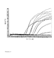
結果を、図2〜5に示す。
図2は、全ての試験試料に対する結果を示す。
図3は、フォワードプライマーを含む試験試料に対する結果を示す。
図4は、リバースプライマーを含む試験試料に対する結果を示す。
図5は、両方のプライマーを含む試験試料に対する結果を示す。
The results are shown in FIGS.
FIG. 2 shows the results for all test samples.
FIG. 3 shows the results for the test sample containing the forward primer.
FIG. 4 shows the results for the test sample containing the reverse primer.
FIG. 5 shows the results for a test sample containing both primers.
結果の概要:
フォワードプライマーの使用は、PCRにおける検出に対して不十分なcDNAを生じさせた。しかし、リバースプライマーは、PCRにおける検出に対して十分なcDNAを産生した。両方のプライマーを用いた場合、cDNAはより高い効率で増幅され、PCR増幅においてより早くに検出された。
Summary of results:
Use of the forward primer resulted in insufficient cDNA for detection in PCR. However, the reverse primer produced sufficient cDNA for detection in PCR. When both primers were used, the cDNA was amplified with higher efficiency and was detected earlier in PCR amplification.
従って、RNAの逆転写により独自に産生されたcDNAは、鎖指向性RT−PCRを用いて特異的に検出できることが実証された。 Therefore, it was demonstrated that cDNA uniquely produced by reverse transcription of RNA can be specifically detected using strand-directed RT-PCR.
Claims (20)
(ii)細胞中のウイルスの複製により得られる任意の核酸を検出すること、ただし、当該ウイルスが、マーカー配列を含むように改変された改変ウイルスである場合、検出される核酸は、細胞内でのウイルスの複製により独自に産生されたものである、
を含んで成る、試料中の生細胞を検出するための方法。 (I) incubating the sample and a virus capable of infecting living cells under conditions that allow the virus to infect and replicate in any such living cell;
(Ii) detecting any nucleic acid obtained by viral replication in the cell, provided that the virus is a modified virus modified to contain a marker sequence, It was originally produced by replication of the virus,
A method for detecting viable cells in a sample comprising.
Applications Claiming Priority (3)
| Application Number | Priority Date | Filing Date | Title |
|---|---|---|---|
| GBGB0503172.9A GB0503172D0 (en) | 2005-02-16 | 2005-02-16 | Detection method |
| GB0503172.9 | 2005-02-16 | ||
| PCT/GB2006/000551 WO2006087559A1 (en) | 2005-02-16 | 2006-02-16 | Method for detecting viable cells in a sample by using a virus |
Publications (2)
| Publication Number | Publication Date |
|---|---|
| JP2008529548A true JP2008529548A (en) | 2008-08-07 |
| JP4903722B2 JP4903722B2 (en) | 2012-03-28 |
Family
ID=34385553
Family Applications (1)
| Application Number | Title | Priority Date | Filing Date |
|---|---|---|---|
| JP2007555700A Expired - Fee Related JP4903722B2 (en) | 2005-02-16 | 2006-02-16 | Method for detecting live cells in a sample by using a virus |
Country Status (12)
| Country | Link |
|---|---|
| US (2) | US8071337B2 (en) |
| EP (1) | EP1851325B1 (en) |
| JP (1) | JP4903722B2 (en) |
| KR (1) | KR20070112796A (en) |
| CN (1) | CN101160410B (en) |
| AT (1) | ATE469239T1 (en) |
| AU (1) | AU2006215415B2 (en) |
| CA (1) | CA2597835A1 (en) |
| DE (1) | DE602006014505D1 (en) |
| DK (1) | DK1851325T3 (en) |
| GB (1) | GB0503172D0 (en) |
| WO (1) | WO2006087559A1 (en) |
Families Citing this family (6)
| Publication number | Priority date | Publication date | Assignee | Title |
|---|---|---|---|---|
| WO2008065047A1 (en) * | 2006-11-29 | 2008-06-05 | Microsens Medtech Ltd | Capture of mycobacteria like micro-organisms |
| US7919234B2 (en) * | 2007-04-05 | 2011-04-05 | Sequella, Inc. | Methods and compositions for determining the pathogenic status of infectious agents |
| WO2013010074A1 (en) | 2011-07-13 | 2013-01-17 | Primeradx, Inc. | Multimodal methods for simultaneous detection and quantification of multiple nucleic acids in a sample |
| FR3108438A1 (en) * | 2020-03-20 | 2021-09-24 | Prodose | METHOD AND DEVICE FOR DETECTIONING AND MONITORING THE PRESENCE, DEVELOPMENT AND PROPAGATION OF INFECTIOUS AGENTS, IN PARTICULAR BACTERIA AND VIRUSES |
| CN112080475B (en) * | 2020-07-30 | 2022-02-11 | 扬州大学 | Vibrio parahaemolyticus bacteriophage and application thereof in detection of content of live cells of Vibrio parahaemolyticus pandemic strain |
| CN112063732B (en) * | 2020-09-17 | 2022-05-31 | 扬州大学 | Rapid quantitative detection method capable of specifically identifying survival cells of enterobacter sakazakii and primers thereof |
Citations (4)
| Publication number | Priority date | Publication date | Assignee | Title |
|---|---|---|---|---|
| US6436652B1 (en) * | 1994-08-12 | 2002-08-20 | Her Majesty The Queen In Right Of Canada, As Represented By The Minister Of National Defence Of Her Majesty's Canadian Government | Method of detecting a pathogen using a virus |
| WO2003035889A2 (en) * | 2001-07-13 | 2003-05-01 | Investigen, Inc. | Compositions and methods for bacteria detection |
| JP2004537279A (en) * | 2001-03-09 | 2004-12-16 | ザ・レジェンツ・オブ・ザ・ユニバーシティ・オブ・カリフォルニア | Cell culture system for infectious hepatitis C virus synthesis |
| WO2005001475A2 (en) * | 2003-04-10 | 2005-01-06 | Kent Voorhees | Apparatus and method for detecting microscopic living organisms using bacteriophage |
Family Cites Families (11)
| Publication number | Priority date | Publication date | Assignee | Title |
|---|---|---|---|---|
| WO1990004041A1 (en) | 1988-10-04 | 1990-04-19 | Dna Plant Technology Corporation | Bacterial detection by phage transduction of detectable phenotype |
| US5888725A (en) * | 1992-09-22 | 1999-03-30 | The Secretary Of State For The Minister Of Agriculture Fisheries And Food In Her Britannic Majesty's Government Of The United Kingdom Of Great Britain And Northern Ireland | Method for identifying target bacteria |
| ATE302264T1 (en) * | 1995-08-25 | 2005-09-15 | Affymetrix Inc A Delaware Corp | RELEASE OF INTRACELLULAR MATERIAL |
| GB9525661D0 (en) | 1995-12-15 | 1996-02-14 | Biotec Diagnostics Limited | Method |
| US5958675A (en) | 1997-04-18 | 1999-09-28 | 3M Innovative Properties Company | Method for detecting bacteria using bacteriophage, contrast-coloring dye and precipitable dye |
| GB9725197D0 (en) | 1997-11-29 | 1998-01-28 | Secr Defence | Detection system |
| GB9725237D0 (en) | 1997-11-29 | 1998-01-28 | Secr Defence | Amplification system |
| GB9803382D0 (en) | 1998-02-19 | 1998-04-15 | Secr Defence | Detection system |
| GB9812768D0 (en) | 1998-06-13 | 1998-08-12 | Zeneca Ltd | Methods |
| CA2501648A1 (en) | 2002-10-15 | 2004-04-29 | Regents Of The University Of Minnesota | Assays to detect or quantify bacterial or viral pathogens and contaminants |
| GB0525072D0 (en) | 2005-12-09 | 2006-01-18 | Enigma Diagnostics Ltd | Fluorescence-based detection methods and apparatus |
-
2005
- 2005-02-16 GB GBGB0503172.9A patent/GB0503172D0/en not_active Ceased
-
2006
- 2006-02-16 JP JP2007555700A patent/JP4903722B2/en not_active Expired - Fee Related
- 2006-02-16 US US11/816,331 patent/US8071337B2/en active Active
- 2006-02-16 AU AU2006215415A patent/AU2006215415B2/en not_active Ceased
- 2006-02-16 CA CA002597835A patent/CA2597835A1/en not_active Abandoned
- 2006-02-16 WO PCT/GB2006/000551 patent/WO2006087559A1/en active Application Filing
- 2006-02-16 CN CN200680012458.2A patent/CN101160410B/en active Active
- 2006-02-16 AT AT06709788T patent/ATE469239T1/en not_active IP Right Cessation
- 2006-02-16 EP EP06709788A patent/EP1851325B1/en active Active
- 2006-02-16 KR KR1020077021152A patent/KR20070112796A/en not_active Application Discontinuation
- 2006-02-16 DE DE602006014505T patent/DE602006014505D1/en active Active
- 2006-02-16 DK DK06709788.1T patent/DK1851325T3/en active
-
2011
- 2011-10-19 US US13/276,772 patent/US8609375B2/en active Active
Patent Citations (4)
| Publication number | Priority date | Publication date | Assignee | Title |
|---|---|---|---|---|
| US6436652B1 (en) * | 1994-08-12 | 2002-08-20 | Her Majesty The Queen In Right Of Canada, As Represented By The Minister Of National Defence Of Her Majesty's Canadian Government | Method of detecting a pathogen using a virus |
| JP2004537279A (en) * | 2001-03-09 | 2004-12-16 | ザ・レジェンツ・オブ・ザ・ユニバーシティ・オブ・カリフォルニア | Cell culture system for infectious hepatitis C virus synthesis |
| WO2003035889A2 (en) * | 2001-07-13 | 2003-05-01 | Investigen, Inc. | Compositions and methods for bacteria detection |
| WO2005001475A2 (en) * | 2003-04-10 | 2005-01-06 | Kent Voorhees | Apparatus and method for detecting microscopic living organisms using bacteriophage |
Non-Patent Citations (5)
| Title |
|---|
| APPLIED AND ENVIRONMENTAL MICROBIOLOGY, vol. 62, no. 4, JPN6011039292, 1996, pages 1133 - 1140, ISSN: 0002083486 * |
| FOOD RESEARCH INTERNATIONAL, vol. 35, JPN6011039291, 2002, pages 863 - 870, ISSN: 0002083485 * |
| JOURNAL OF APPLIED MICROBIOLOGY, vol. 84, JPN6011039290, 1998, pages 661 - 666, ISSN: 0002083484 * |
| JOURNAL OF CHEMICAL TECHNOLOGY AND BIOTECHNOLOGY, vol. 76, JPN6011039293, 2001, pages 683 - 688, ISSN: 0002083487 * |
| PROC. OF SPIE, vol. 5271, JPN6011039294, 2004, pages 13 - 19, ISSN: 0002083488 * |
Also Published As
| Publication number | Publication date |
|---|---|
| US8071337B2 (en) | 2011-12-06 |
| CA2597835A1 (en) | 2006-08-24 |
| AU2006215415B2 (en) | 2011-01-06 |
| EP1851325A1 (en) | 2007-11-07 |
| WO2006087559A1 (en) | 2006-08-24 |
| ATE469239T1 (en) | 2010-06-15 |
| US20080166700A1 (en) | 2008-07-10 |
| EP1851325B1 (en) | 2010-05-26 |
| CN101160410B (en) | 2016-03-30 |
| JP4903722B2 (en) | 2012-03-28 |
| AU2006215415A1 (en) | 2006-08-24 |
| GB0503172D0 (en) | 2005-03-23 |
| US8609375B2 (en) | 2013-12-17 |
| US20120141976A1 (en) | 2012-06-07 |
| DK1851325T3 (en) | 2010-08-30 |
| DE602006014505D1 (en) | 2010-07-08 |
| CN101160410A (en) | 2008-04-09 |
| KR20070112796A (en) | 2007-11-27 |
Similar Documents
| Publication | Publication Date | Title |
|---|---|---|
| CN110551846B (en) | Cpf1 kit for quickly detecting African swine fever virus nucleic acid and detection method thereof | |
| Ballagi-Pordany et al. | The use of mimics as internal standards to avoid false negatives in diagnostic PCR | |
| JPS60501339A (en) | Methods and kits for detecting, identifying or quantifying organisms | |
| Yin et al. | Development of a simple and rapid reverse transcription–loopmediated isothermal amplification (RT‐LAMP) assay for sensitive detection of tilapia lake virus | |
| US8609375B2 (en) | Method for detecting viable cells in a sample by using a virus | |
| CN107988326A (en) | Prawn Acute Hepatic pancreatic necrosis(AHPND)RAA constant temperature fluorescence detection method and reagent | |
| CN106367413A (en) | Nucleic acid amplification method and application | |
| CN107988427A (en) | Prawn hepatopancreatic parvovirus(HPV)RAA constant temperature fluorescence detection method and reagent | |
| CN111394515B (en) | LAMP primer group, fluorescence visualization rapid kit and method for detecting canine parvovirus | |
| Mota et al. | Recombinase polymerase amplification in the molecular diagnosis of microbiological targets and its applications | |
| Yaren et al. | Multiplexed isothermal amplification based diagnostic platform to detect zika, chikungunya, and dengue 1 | |
| CN115461476A (en) | Primer for detecting SARS-CoV-2 novel coronavirus and its kit, detection method and application | |
| Wang et al. | Development of a naked eye CRISPR-Cas12a and-Cas13a multiplex point-of-care detection of genetically modified swine | |
| Qian et al. | Clustered regularly interspaced short palindromic Repeat/Cas12a mediated multiplexable and portable detection platform for GII genotype Porcine Epidemic Diarrhoea Virus Rapid diagnosis | |
| Andreychuk et al. | Armoured exogenous internal control for real-time PCR diagnosis of avian influenza | |
| CN104611420A (en) | Tubercle bacillus detection kit | |
| Wang et al. | Visual and label-free ASFV and PCV2 detection by CRISPR-Cas12a combined with G-quadruplex | |
| KR20120086028A (en) | Differential detection of West nile virus and Japanese encephalitis virus | |
| CN106222293B (en) | Fluorescence quantification PCR primer probe and kit and the method for detecting three kinds of bacillus | |
| JP6616574B2 (en) | Visually identifiable genetic test | |
| CN110964849B (en) | Method for eliminating African swine fever virus detection false positive and kit for detecting African swine fever virus | |
| RU2738358C1 (en) | Set of oligonucleotide primers and fluorescent-labelled probes and method for detecting dna of agents of glanders and melioidosis by pcr method with product detection in real time | |
| RU2699180C1 (en) | Set of oligonucleotide primers and a fluorescent-labeled probe for identifying rnas of burkholderia mallei sap excretion and burkholderia pseudomallei melioidosis based on transcription amplification (nasba) in real time mode | |
| Karuppannan et al. | Recent advances in veterinary diagnostic virology | |
| CN115125313A (en) | Primer pair for detecting pathogenic enterocolitis yersinia and CRISPR/Cas12a detection kit |
Legal Events
| Date | Code | Title | Description |
|---|---|---|---|
| A621 | Written request for application examination |
Free format text: JAPANESE INTERMEDIATE CODE: A621 Effective date: 20090122 |
|
| A131 | Notification of reasons for refusal |
Free format text: JAPANESE INTERMEDIATE CODE: A131 Effective date: 20110802 |
|
| A521 | Request for written amendment filed |
Free format text: JAPANESE INTERMEDIATE CODE: A523 Effective date: 20111102 |
|
| TRDD | Decision of grant or rejection written | ||
| A01 | Written decision to grant a patent or to grant a registration (utility model) |
Free format text: JAPANESE INTERMEDIATE CODE: A01 Effective date: 20111206 |
|
| A01 | Written decision to grant a patent or to grant a registration (utility model) |
Free format text: JAPANESE INTERMEDIATE CODE: A01 |
|
| A61 | First payment of annual fees (during grant procedure) |
Free format text: JAPANESE INTERMEDIATE CODE: A61 Effective date: 20120105 |
|
| R150 | Certificate of patent or registration of utility model |
Ref document number: 4903722 Country of ref document: JP Free format text: JAPANESE INTERMEDIATE CODE: R150 Free format text: JAPANESE INTERMEDIATE CODE: R150 |
|
| FPAY | Renewal fee payment (event date is renewal date of database) |
Free format text: PAYMENT UNTIL: 20150113 Year of fee payment: 3 |
|
| R250 | Receipt of annual fees |
Free format text: JAPANESE INTERMEDIATE CODE: R250 |
|
| R250 | Receipt of annual fees |
Free format text: JAPANESE INTERMEDIATE CODE: R250 |
|
| R250 | Receipt of annual fees |
Free format text: JAPANESE INTERMEDIATE CODE: R250 |
|
| R250 | Receipt of annual fees |
Free format text: JAPANESE INTERMEDIATE CODE: R250 |
|
| R250 | Receipt of annual fees |
Free format text: JAPANESE INTERMEDIATE CODE: R250 |
|
| LAPS | Cancellation because of no payment of annual fees |