JP2008529008A - Methods for diagnosis and prognosis of epithelial cancer - Google Patents
Methods for diagnosis and prognosis of epithelial cancer Download PDFInfo
- Publication number
- JP2008529008A JP2008529008A JP2007553289A JP2007553289A JP2008529008A JP 2008529008 A JP2008529008 A JP 2008529008A JP 2007553289 A JP2007553289 A JP 2007553289A JP 2007553289 A JP2007553289 A JP 2007553289A JP 2008529008 A JP2008529008 A JP 2008529008A
- Authority
- JP
- Japan
- Prior art keywords
- cancer
- cystatin
- epithelial
- antibody
- level
- Prior art date
- Legal status (The legal status is an assumption and is not a legal conclusion. Google has not performed a legal analysis and makes no representation as to the accuracy of the status listed.)
- Pending
Links
Images


Classifications
-
- G—PHYSICS
- G01—MEASURING; TESTING
- G01N—INVESTIGATING OR ANALYSING MATERIALS BY DETERMINING THEIR CHEMICAL OR PHYSICAL PROPERTIES
- G01N33/00—Investigating or analysing materials by specific methods not covered by groups G01N1/00 - G01N31/00
- G01N33/48—Biological material, e.g. blood, urine; Haemocytometers
- G01N33/50—Chemical analysis of biological material, e.g. blood, urine; Testing involving biospecific ligand binding methods; Immunological testing
- G01N33/53—Immunoassay; Biospecific binding assay; Materials therefor
- G01N33/574—Immunoassay; Biospecific binding assay; Materials therefor for cancer
- G01N33/57407—Specifically defined cancers
-
- G—PHYSICS
- G01—MEASURING; TESTING
- G01N—INVESTIGATING OR ANALYSING MATERIALS BY DETERMINING THEIR CHEMICAL OR PHYSICAL PROPERTIES
- G01N33/00—Investigating or analysing materials by specific methods not covered by groups G01N1/00 - G01N31/00
- G01N33/48—Biological material, e.g. blood, urine; Haemocytometers
- G01N33/50—Chemical analysis of biological material, e.g. blood, urine; Testing involving biospecific ligand binding methods; Immunological testing
- G01N33/53—Immunoassay; Biospecific binding assay; Materials therefor
- G01N33/574—Immunoassay; Biospecific binding assay; Materials therefor for cancer
- G01N33/57484—Immunoassay; Biospecific binding assay; Materials therefor for cancer involving compounds serving as markers for tumor, cancer, neoplasia, e.g. cellular determinants, receptors, heat shock/stress proteins, A-protein, oligosaccharides, metabolites
- G01N33/57488—Immunoassay; Biospecific binding assay; Materials therefor for cancer involving compounds serving as markers for tumor, cancer, neoplasia, e.g. cellular determinants, receptors, heat shock/stress proteins, A-protein, oligosaccharides, metabolites involving compounds identifable in body fluids
-
- G—PHYSICS
- G01—MEASURING; TESTING
- G01N—INVESTIGATING OR ANALYSING MATERIALS BY DETERMINING THEIR CHEMICAL OR PHYSICAL PROPERTIES
- G01N33/00—Investigating or analysing materials by specific methods not covered by groups G01N1/00 - G01N31/00
- G01N33/48—Biological material, e.g. blood, urine; Haemocytometers
- G01N33/50—Chemical analysis of biological material, e.g. blood, urine; Testing involving biospecific ligand binding methods; Immunological testing
- G01N33/68—Chemical analysis of biological material, e.g. blood, urine; Testing involving biospecific ligand binding methods; Immunological testing involving proteins, peptides or amino acids
- G01N33/6893—Chemical analysis of biological material, e.g. blood, urine; Testing involving biospecific ligand binding methods; Immunological testing involving proteins, peptides or amino acids related to diseases not provided for elsewhere
-
- G—PHYSICS
- G01—MEASURING; TESTING
- G01N—INVESTIGATING OR ANALYSING MATERIALS BY DETERMINING THEIR CHEMICAL OR PHYSICAL PROPERTIES
- G01N2333/00—Assays involving biological materials from specific organisms or of a specific nature
- G01N2333/81—Protease inhibitors
- G01N2333/8107—Endopeptidase (E.C. 3.4.21-99) inhibitors
- G01N2333/8139—Cysteine protease (E.C. 3.4.22) inhibitors, e.g. cystatin
Abstract
Description
(関連出願の引用)
本出願は、35U.S.C.第119条(e)の下で、2005年1月28日に出願された米国仮特許出願第60/648,110号の利益を主張するものである。
(Citation of related application)
This application is filed in 35U. S. C. It claims the benefit of US Provisional Patent Application No. 60 / 648,110, filed January 28, 2005, under section 119 (e).
(政府の援助)
本研究は、米国国立衛生研究所助成金番号2R37 CA37393により援助を受けたものである。政府は、本発明に対して特定の権利を有する。
(Government assistance)
This study was supported by the National Institutes of Health grant number 2R37 CA37393. The government has certain rights to the invention.
(発明の背景)
癌の生存において最も重要なファクターの1つは、初期段階での検出である。癌の初期事象を検出する臨床検査は、癌の進行に介入しそれを防止する機会を提供する。遺伝子プロファイル作成およびプロテオミクスの開発とともに、特定の癌の診断および予後診断に使用することができる分子マーカーまたは「バイオマーカー」の同定における著しい進歩が認められている。例えば、前立腺癌の場合、(前立腺特異的抗原の)抗原PSAを血中で検出することができ、その抗原により、前立腺癌の存在が示唆される。したがって、前立腺癌のリスクがある男性の血液を、PSAレベルの上昇について迅速、容易、かつ安全にスクリーニングすることができる。
(Background of the Invention)
One of the most important factors in cancer survival is early detection. Laboratory tests that detect early events in cancer provide an opportunity to intervene in and prevent cancer progression. Along with the development of genetic profiling and proteomics, significant advances in the identification of molecular markers or “biomarkers” that can be used for the diagnosis and prognosis of specific cancers have been recognized. For example, in the case of prostate cancer, the antigen PSA (of prostate specific antigen) can be detected in the blood, and that antigen suggests the presence of prostate cancer. Thus, the blood of men at risk for prostate cancer can be screened quickly, easily and safely for elevated PSA levels.
癌の検出の分野で著しい進歩が認められていても、臨床応用の際に容易に使用することができる、様々な癌についての新たなバイオマーカーを同定する必要が当技術分野において依然としてある。例えば、今まで、容易に検出可能なバイオマーカーを使用する乳癌の診断に利用可能な選択肢は比較的少数しかない。特にエストロゲン受容体の下方制御と連動したEGFRの過剰発現は、乳癌患者における予後不良のマーカーである。知られている他の乳癌のマーカーには、血中での高レベルのM2ピルビン酸キナーゼ(M2 PK)(特許文献1)、血中での高ZNF217タンパク質レベル(特許文献2)、診断に有用である、乳癌で新たに同定されたタンパク質PDEBCの差次的発現(特許文献3)がある。CEA、CA−125やHCGなどの細胞表面マーカーは、局所進行性および転移性の膀胱癌患者の血清中で頻繁に上昇し(非特許文献1)、マトリックスメタロプロテイナーゼ−2(非特許文献2)、肝細胞成長因子(非特許文献3)、組織ポリペプチド抗原(非特許文献4)などの腫瘍関連タンパク質の循環レベルに関与する研究から展望が示されている。これらのバイオマーカーは、診断の代替方法を提供するが、それらは広範には使用されない。さらに、いくつかの組織化学的マーカー、遺伝子マーカー、および免疫学的マーカーの使用にもかかわらず、臨床家は、どの腫瘍が他の臓器に転移するかを予測するのに依然として苦労している。
癌のバイオマーカーの同定は、その疾患の診断、予後診断および治療の向上にとって特に重要である。したがって、当技術分野において、迅速、容易、かつ安全に検出することができる代替のバイオマーカーを同定する必要がある。そのようなバイオマーカーは、膀胱癌であり、特に、その疾患の浸潤性かつ潜在的に転移性の段階にある対象の進行または治療の診断、病期判定、またはモニターに使用することができる。 The identification of cancer biomarkers is particularly important for the diagnosis, prognosis and improvement of treatment of the disease. Therefore, there is a need in the art to identify alternative biomarkers that can be detected quickly, easily and safely. Such a biomarker is bladder cancer and can be used in particular for the diagnosis, staging, or monitoring of progression or treatment of a subject at the invasive and potentially metastatic stage of the disease.
(発明の要旨)
本発明は、シスタチンB、シャペロニン10、およびプロフィリンの3種のタンパク質(「上皮性癌マーカー」とも称される)が、上皮起源の癌である膀胱癌の患者の尿中に存在するという驚くべき発見に基づいている。したがって、本発明は、生体試料中でのこれらのマーカーの存在をモニターすることにより、上皮起源の癌の予後評価を行う方法および上皮起源の癌の診断を容易にする方法を対象とする。本発明はまた、治療有効性についてのマーカーをも対象とする。特に、尿中で検出されるシスタチンBの量は疾患状態と相関し、その結果、シスタチンBレベルを使用して、浸潤性膀胱癌の存在を予測することができる。したがって、尿中のシスタチンB、シャペロニン10、および/またはプロフィリンタンパク質のレベルを測定することにより、患者における膀胱癌の診断および予後診断の両方に使用することができる、迅速、容易、かつ安全なスクリーニングがもたらされる。あるいは、これらのマーカーが存在しないことにより、患者が膀胱癌でないことも示唆され得る。
(Summary of the Invention)
The present invention is surprising that three proteins of cystatin B,
一実施形態では、患者における上皮起源の癌の診断を容易にする方法を提供する。その方法は、患者から生体試料、好ましくは排出尿検体を得るステップと、試料中での少なくとも1種の上皮性癌バイオマーカー(シスタチンB、シャペロニン10、またはプロフィリン)の存在の有無を検出するステップとを含み、少なくとも1種の上皮性癌バイオマーカーの存在から、上皮起源の癌が示唆される。
In one embodiment, a method that facilitates diagnosis of cancer of epithelial origin in a patient is provided. The method includes obtaining a biological sample, preferably an excreted urine sample from a patient, and detecting the presence or absence of at least one epithelial cancer biomarker (cystatin B,
生体試料は、例えば、血液、組織(例えば腫瘍または胸部)、血清、糞便、尿、喀痰、脳脊髄液、乳頭吸引液および細胞溶解液の上清から得ることができる。好ましい生体試料の1つは尿である。 Biological samples can be obtained from, for example, blood, tissue (eg, tumor or breast), serum, feces, urine, sputum, cerebrospinal fluid, nipple aspirate, and cell lysate supernatant. One preferred biological sample is urine.
本明細書において、「上皮起源の癌」とは、上皮細胞から生じる癌を指し、その癌には、それだけに限らないが、乳癌、基底細胞癌、腺癌、胃腸の癌、口唇癌、口腔癌、食道癌、小腸癌および胃癌、結腸癌、肝癌、膀胱癌、膵癌、卵巣癌、子宮頸癌、肺癌、扁平上皮癌や基底細胞癌などの乳癌および皮膚癌、前立腺癌、腎細胞癌、ならびに全身の上皮細胞に影響を及ぼす他の既知の癌がある。 As used herein, “epithelial cancer” refers to cancer arising from epithelial cells, including, but not limited to, breast cancer, basal cell carcinoma, adenocarcinoma, gastrointestinal cancer, lip cancer, oral cancer Breast cancer and skin cancer such as esophageal cancer, small intestine cancer and stomach cancer, colon cancer, liver cancer, bladder cancer, pancreatic cancer, ovarian cancer, cervical cancer, lung cancer, squamous cell carcinoma and basal cell cancer, prostate cancer, renal cell cancer, and There are other known cancers that affect epithelial cells throughout the body.
一実施形態では、患者における膀胱癌の診断を容易にする方法を提供する。その方法は、患者から生体試料、好ましくは排出尿検体を得るステップと、試料中での少なくとも1種の上皮性癌バイオマーカー(シスタチンB、シャペロニン10、またはプロフィリン)の存在の有無を検出するステップとを含み、少なくとも1種の上皮性癌バイオマーカーの存在から、膀胱癌が示唆される。
In one embodiment, a method is provided that facilitates diagnosis of bladder cancer in a patient. The method includes obtaining a biological sample, preferably an excreted urine sample from a patient, and detecting the presence or absence of at least one epithelial cancer biomarker (cystatin B,
他の実施形態では、上皮起源の癌を診断する方法を提供する。その方法は、患者由来の生体試料(試験試料)中に存在する少なくとも1種の上皮性癌バイオマーカーのレベルを測定するステップと、少なくとも1種のマーカー(シスタチンB、シャペロニン10、またはプロフィリン)の観察されたレベルを、同じ型の対照試料中に存在するマーカーのレベルと比較するステップとを含む。対照試料と比較して試験試料中のマーカーのレベルが高いことにより、上皮起源の癌が示唆される。
In other embodiments, a method of diagnosing cancer of epithelial origin is provided. The method comprises the steps of measuring the level of at least one epithelial cancer biomarker present in a patient-derived biological sample (test sample) and at least one marker (cystatin B,
好ましい一実施形態では、本発明の方法を癌の初期検出に使用する。例えば、身体検査の間に、医師により患者をスクリーニングすることができる。 In a preferred embodiment, the method of the invention is used for early detection of cancer. For example, a patient can be screened by a physician during a physical examination.
一実施形態では、膀胱癌を診断する方法を提供する。その方法は、患者由来の生体試料(試験試料)中に存在する少なくとも1種の上皮性癌バイオマーカー(シスタチンB、シャペロニン10、またはプロフィリン)のレベルを測定するステップと、少なくとも1種のマーカーの観察されたレベルを、同じ型の対照試料中に存在するマーカーのレベルと比較するステップとを含む。対照試料と比較して試験試料中のマーカーのレベルが高いことにより、膀胱癌が示唆される。
In one embodiment, a method for diagnosing bladder cancer is provided. The method comprises the steps of measuring the level of at least one epithelial cancer biomarker (cystatin B,
一実施形態では、患者において浸潤性膀胱癌を診断する方法を提供する。その方法は、患者から得られた生体試料(試験試料)中に存在するシスタチンB上皮性癌バイオマーカーのレベルを測定するステップと、試験試料中のシスタチンBのレベルを、非浸潤性癌の対照試料中に存在するシスタチンBのレベルと比較するステップとを含む。対照試料中のシスタチンBのレベルと比較して試験試料中のシスタチンBのレベルが高いことにより、浸潤性膀胱癌が示唆される。 In one embodiment, a method for diagnosing invasive bladder cancer in a patient is provided. The method comprises the steps of measuring the level of cystatin B epithelial cancer biomarker present in a biological sample (test sample) obtained from a patient, and determining the level of cystatin B in the test sample as a control for non-invasive cancer. Comparing to the level of cystatin B present in the sample. A high level of cystatin B in the test sample compared to the level of cystatin B in the control sample indicates invasive bladder cancer.
「対照試料」という用語は、癌でないと考えられる「正常な」または「健常な」(複数の)個人から得られた生体試料(例えば、血液、尿、腫瘍)を指す。対照は、当技術分野で周知である方法を使用して選択することができる。対照集団についてレベルが確立した後、試験生体試料からの多数の結果を、既知のレベルと直接比較することができる。 The term “control sample” refers to a biological sample (eg, blood, urine, tumor) obtained from a “normal” or “healthy” individual (s) that is considered not cancerous. Controls can be selected using methods well known in the art. After the level is established for the control population, a number of results from the test biological sample can be directly compared to a known level.
「非浸潤性の対照試料」という用語は、非浸潤型の癌である(複数の)個人から得られた生体試料を指す。対照集団についてレベルが確立した後、試験生体試料からの多数の結果を、既知のレベルと直接比較することができる。 The term “non-invasive control sample” refers to a biological sample obtained from an individual (s) that is non-invasive cancer. After the level is established for the control population, a number of results from the test biological sample can be directly compared to a known level.
「試験試料」という用語は、上皮起源の癌について試験する患者から得られた生体試料を指す。 The term “test sample” refers to a biological sample obtained from a patient being tested for cancer of epithelial origin.
本発明はまた、同じ患者から得られた複数の試験試料中に存在する上皮性癌バイオマーカーのレベルの評価をも意図するものであり、経時的なマーカーの量の直進的増大により、癌の腫瘍の攻撃性(例えば、転移能)の増大が示唆される。したがって、上皮性癌バイオマーカーのレベルは、疾患状態および病期の予測因子として働く。 The present invention is also intended to assess the level of epithelial cancer biomarkers present in multiple test samples obtained from the same patient, with a linear increase in the amount of marker over time, An increase in tumor aggressiveness (eg, metastatic potential) is suggested. Thus, the level of epithelial cancer biomarkers serves as a predictor of disease state and stage.
本発明はさらに、上皮起源の癌(例えば、膀胱癌)である患者を治療するために計画された治療法の治療有効性をモニターする上皮性癌バイオマーカーの評価を意図するものである。 The present invention further contemplates evaluation of epithelial cancer biomarkers that monitor the therapeutic efficacy of therapeutics designed to treat patients with cancer of epithelial origin (eg, bladder cancer).
本発明の一態様では、試験生体試料中に存在する上皮性癌バイオマーカーレベル(例えば、シスタチンB、シャペロニン10、またはプロフィリン)は、試験試料、またはその調製物を、上皮性癌バイオマーカーと特異的に結合する抗体に基づく結合部分、またはその一部分と接触させることによって測定する。
In one aspect of the invention, the level of epithelial cancer biomarker (eg, cystatin B,
抗体に基づくイムノアッセイは、バイオマーカーのレベルを測定するのに好ましい手段である。しかし、当業者に知られている任意の手段を使用して、バイオマーカーのレベルを評価することができる。例えば、SELDI質量分析法を含めた質量分析法によって、バイオマーカーのレベルを評価することができる。 Antibody-based immunoassays are a preferred means for measuring biomarker levels. However, any means known to those skilled in the art can be used to assess the level of the biomarker. For example, biomarker levels can be assessed by mass spectrometry including SELDI mass spectrometry.
さらなる実施形態では、本発明は、生体試料中で少なくとも1種の上皮性癌バイオマーカーを測定する手段を含むキットを提供する。そのキットは、生体試料(例えば、尿試料)を保持する容器、および上皮性癌バイオマーカーと特異的に結合する少なくとも1つの抗体を含む。 In a further embodiment, the present invention provides a kit comprising means for measuring at least one epithelial cancer biomarker in a biological sample. The kit includes a container that holds a biological sample (eg, a urine sample) and at least one antibody that specifically binds to an epithelial cancer biomarker.
一実施形態では、キットは、上皮性癌バイオマーカーと特異的に結合する2つの抗体を含む。一実施形態では、1つの抗体を固相上に固定化し、1つの抗体を検出可能となるように標識する。キットは、抗シスタチンB、抗シャペロニン10、および/または抗プロフィリン抗体を含んでよい。 In one embodiment, the kit includes two antibodies that specifically bind to an epithelial cancer biomarker. In one embodiment, one antibody is immobilized on a solid phase and one antibody is labeled so that it can be detected. The kit may include anti-cystatin B, anti-chaperonin 10, and / or anti-profilin antibody.
本発明の他の態様を下記で開示する。 Other aspects of the invention are disclosed below.
本明細書に組み込まれその一部を構成する添付図面は、本発明の実施形態を示すものであり、記載とともに、本発明の目的、利点、および原理を説明するのに役立つ。 The accompanying drawings, which are incorporated in and constitute a part of this specification, illustrate embodiments of the invention and, together with the description, serve to explain the objects, advantages, and principles of the invention.
(発明の詳細な説明)
シスタチンB、シャペロニン10、およびプロフィリンの3種のタンパク質(本明細書において「上皮性癌マーカー」と称する)が、上皮起源の癌である患者の尿中に存在することを発見した。患者の尿試料中に存在するシスタチンBのレベルは、膀胱癌、特に浸潤性膀胱癌の存在と相関する。
(Detailed description of the invention)
Three proteins, cystatin B,
癌に関して「攻撃的な」または「浸潤性の」という用語は、その境界を越えて隣接組織中へと広がる腫瘍の傾向を指す(Darnell, J.(1990年)、Molecular Cell Biology, Third Ed.、W. H. Freeman、NY)。浸潤性の癌は、腫瘍が特定の臓器に限局している臓器限局性の癌と対比することができる。腫瘍の浸潤性は、腫瘍が被膜の境界および腫瘍が位置する特定の組織の境界を越えて広がることができるようにマトリックス物質および基底膜物質を分解するコラゲナーゼなどのタンパク質分解酵素の合成をしばしば伴う。浸潤性膀胱癌は、筋固有層および/または粘膜固有層中への浸潤性を含む。 The terms “aggressive” or “invasive” with respect to cancer refer to the tendency of a tumor to extend beyond that boundary into adjacent tissues (Darnell, J. (1990), Molecular Cell Biology, Third Ed.). W. H. Freeman, NY). Invasive cancer can be contrasted with organ-confined cancer, where the tumor is confined to a specific organ. Tumor invasiveness often involves the synthesis of proteolytic enzymes such as collagenases that degrade matrix and basement membrane materials so that the tumor can extend beyond the boundaries of the capsule and the specific tissue where the tumor is located . Invasive bladder cancer includes invasiveness into the lamina propria and / or lamina propria.
本明細書において、「転移」という用語は、患者中で元の臓器からさらなる遠隔部位へと癌が広がる状態を指す。腫瘍転移の過程は、局所浸潤および細胞間マトリックスの破壊、血管、リンパ管または他の輸送経路中への侵入、循環中での生存、第2の部位中の管外への遊出および新たな場所での増殖を伴う多段階の事象である(Fidlerら、Adv. Cancer Res.、28、149〜250(1978年)、Liottaら、Cancer Treatment Res.、40、223〜238(1988年)、Nicolson、Biochim. Biophy. Acta、948、175〜224(1988年)およびZetter, N.、Eng. J. Med.、322、605〜612(1990年))。悪性細胞の運動性の増大は、動物ならびにヒトの癌での転移能の亢進と関係している(Hosakaら、Gann、69、273〜276(1978年)およびHaemmerlinら、Int. J. Cancer、27、603〜610(1981年))。 As used herein, the term “metastasis” refers to a condition in which cancer spreads from the original organ to a further distant site in the patient. The process of tumor metastasis includes local invasion and destruction of the intercellular matrix, invasion into blood vessels, lymphatic vessels or other transport pathways, survival in the circulation, extravasation in the second site and new It is a multi-stage event with local growth (Fidler et al., Adv. Cancer Res., 28, 149-250 (1978), Liotta et al., Cancer Treatment Res., 40, 223-238 (1988), Nicolson, Biochim. Biophy. Acta, 948, 175-224 (1988) and Zetter, N., Eng. J. Med., 322, 605-612 (1990)). Increased malignant cell motility is associated with increased metastatic potential in animal and human cancers (Hosaka et al., Gann, 69, 273-276 (1978)) and Haemmerlin et al., Int. J. Cancer, 27, 603-610 (1981)).
本明細書において、「生体試料」とは、患者から得られた尿試料を指す。生体試料は、例えば、血液、組織(例えば腫瘍または胸部)、血清、糞便、尿、喀痰、脳脊髄液、乳頭吸引液および細胞溶解液の上清から得ることができる。好ましい生体試料の1つは尿である。 As used herein, “biological sample” refers to a urine sample obtained from a patient. Biological samples can be obtained from, for example, blood, tissue (eg, tumor or breast), serum, feces, urine, sputum, cerebrospinal fluid, nipple aspirate, and cell lysate supernatant. One preferred biological sample is urine.
好ましい実施形態では、上皮性癌バイオマーカーの分解を防止するように生体試料を処理する。分解を阻害または防止する方法には、それだけに限らないが、プロテアーゼで試料を処理すること、試料を凍結すること、または氷上に試料を置くことがある。好ましくは、分析の前に、マーカーの分解を防止するような条件下で試料を常に維持する。 In a preferred embodiment, the biological sample is treated to prevent degradation of the epithelial cancer biomarker. Methods for inhibiting or preventing degradation include, but are not limited to, treating the sample with a protease, freezing the sample, or placing the sample on ice. Preferably, the sample is always maintained prior to analysis under conditions that prevent degradation of the marker.
本明細書において、「腫瘍試料」とは、腫瘍、例えば、対象、好ましくはヒト対象から得られまたは取り出された(例えば、対象の組織から取り出されまたは抽出された)腫瘍の一部分、一片、一部、一セグメントまたは一画分を指す。 As used herein, a “tumor sample” refers to a portion, piece, piece of a tumor, eg, a tumor obtained or removed from a subject, preferably a human subject (eg, removed or extracted from the tissue of the subject). A part, a segment or a fraction.
本明細書において、シスタチンBとは、GenebankアクセッションNM_000100.2、NP_000091(ヒト(Homosapiens))のタンパク質を指す。その用語はまた、種の変異体、相同体、対立形質の形態、突然変異形態、およびその同等物をも包含する。 As used herein, cystatin B refers to the protein of Genebank Accession NM — 000100.2, NP — 000091 (Homosapiens). The term also encompasses species variants, homologues, allelic forms, mutant forms, and the like.
本明細書において、シャペロニン10とは、Genebankアクセッション、タンパク質、AAA50953(ヒト(Homosapiens))のタンパク質を指す。その用語はまた、種の変異体、相同体、対立形質の形態、突然変異形態、およびその同等物をも包含する。
As used herein,
本明細書において、プロフィリンとは、Genebankアクセッション、タンパク質、A28622(ヒト(Homosapiens))のタンパク質を指す。その用語はまた、種の変異体、相同体、対立形質の形態、突然変異形態、およびその同等物をも包含する。 As used herein, profilin refers to the protein of Genebank Accession, Protein, A28622 (Homosapiens). The term also encompasses species variants, homologues, allelic forms, mutant forms, and the like.
本発明は、患者における上皮起源の癌の診断を容易にする方法を対象とする。一実施形態では、その方法は、患者から生体試料を得るステップと、試料中での少なくとも1種の上皮性癌バイオマーカー(シスタチンB、シャペロニン10、またはプロフィリン)の存在の有無を検出するステップとを含み、少なくとも1種の上皮性癌バイオマーカーの存在から、上皮起源の癌が示される。
The present invention is directed to a method that facilitates the diagnosis of cancer of epithelial origin in a patient. In one embodiment, the method includes obtaining a biological sample from a patient and detecting the presence or absence of at least one epithelial cancer biomarker (cystatin B,
他の実施形態では、その方法は、癌について試験する患者から得られた試験試料中の少なくとも1種の上皮性癌バイオマーカー(シスタチンB、シャペロニン10、またはプロフィリン)のレベルを測定するステップと、観察されたレベルを、対照試料、例えば癌でない個々の患者または個人の集団から得られた試料中で認められた上皮性癌バイオマーカーのレベルと比較するステップとを含む。少なくとも1種の上皮性癌バイオマーカーのレベルが正常な対照中で観察されるレベルより高いことにより、上皮起源の癌の存在が示唆される。バイオマーカーのレベルは、任意の単位で、例えば、デンシトメーター、ルミノメーター、またはElisaプレートリーダーから得られた単位として表すことができる。
In other embodiments, the method comprises measuring the level of at least one epithelial cancer biomarker (cystatin B,
本明細書において、「対照試料中のレベルと比較して高いレベルの試験試料中の少なくとも1種の上皮性癌バイオマーカー」とは、対照試料中に存在する同じバイオマーカーの量より多い、少なくとも1種のバイオマーカーの量を指す。「高いレベル」という用語は、統計上有意なレベル、または対照試料中で認められるレベルを有意に上回るレベルを指す。「高いレベル」は、例えば、1.2倍〜1.9倍高いことであり得る。好ましくは、「高いレベル」は、少なくとも2倍大きく、またはさらに3倍大きい。 As used herein, “at least one epithelial cancer biomarker in a test sample at a higher level compared to the level in a control sample” is at least greater than the amount of the same biomarker present in the control sample. Refers to the amount of one biomarker. The term “high level” refers to a statistically significant level or a level significantly above that found in a control sample. The “high level” can be, for example, 1.2 to 1.9 times higher. Preferably, the “high level” is at least 2 times greater, or even 3 times greater.
「統計上有意な」または「有意に」という用語は、統計上の有意差を指し、一般に、正常を標準偏差の2倍(2SD)上回る、またはそれより高いマーカーの濃度を意味する。 The term “statistically significant” or “significantly” refers to a statistically significant difference and generally refers to the concentration of a marker that is two times the standard deviation (2SD) above or higher than normal.
比較の目的で、試験試料および対照試料は、同じ型のものであり、すなわち同じ生物学的供給源から得られる。対照試料はまた、健常人から得られる生体試料中で正常に認められる同じ濃度の上皮性癌バイオマーカーを含む標準試料でもよい。 For comparison purposes, the test sample and the control sample are of the same type, i.e. obtained from the same biological source. The control sample may also be a standard sample containing the same concentration of epithelial cancer biomarker found normally in a biological sample obtained from a healthy person.
本発明の一態様では、第2の診断ステップを行うことができる。例えば、少なくとも1種の上皮性癌バイオマーカーのレベルから癌の存在が示唆されることが分かっている場合、癌を検出するさらなる方法を行って、癌の存在を確認することができる。超音波、PET走査、MRIまたは任意の他の画像化技術、生検、臨床検査、ダクトグラム(ductogram)や任意の他の方法など、種々のさらなる診断ステップのいずれかを行うことができる。 In one aspect of the invention, a second diagnostic step can be performed. For example, if the level of at least one epithelial cancer biomarker is known to indicate the presence of cancer, additional methods of detecting cancer can be performed to confirm the presence of cancer. Any of a variety of additional diagnostic steps can be performed, such as ultrasound, PET scanning, MRI or any other imaging technique, biopsy, clinical examination, ductogram or any other method.
本発明はさらに、上皮起源の癌であることが疑われ、または上皮起源の癌である患者の予後評価を行う方法を提供する。その方法は、患者から得られた試験生体試料中に存在する少なくとも1種の上皮性癌バイオマーカー(シスタチンB、シャペロニン10、またはプロフィリン)のレベルを測定するステップと、観察されたレベルを、健常人の(同じ型の)生体試料中で正常に認められる少なくとも1種の上皮性癌バイオマーカーレベルの範囲と比較するステップとを含む。高いレベルから、例えば、転移活性についての潜在性が高いことが示唆され、それは予後不良と対応するが、低いレベルから、腫瘍の攻撃性が低いことが示唆され、それは予後良好と対応する。
The present invention further provides a method for prognostic assessment of patients suspected of having epithelial origin or having epithelial origin. The method comprises the steps of measuring the level of at least one epithelial cancer biomarker (cystatin B,
さらに、個々の患者において少なくとも1種の上皮性癌バイオマーカーのレベルを追跡することにより、疾患の進行を評価することができる。例えば、患者におけるシスタチンB、シャペロニン10、またはプロフィリンの発現レベルの変化を経時的に比較することにより、患者の状態の変化をモニターすることができる。少なくとも1種の上皮性癌バイオマーカーのレベルの直進的増大により、腫瘍の浸潤および転移についての潜在性の増大が示唆される。
Furthermore, disease progression can be assessed by following the level of at least one epithelial cancer biomarker in an individual patient. For example, changes in the patient's condition can be monitored by comparing changes in the expression level of cystatin B,
本発明の予後診断方法はまた、癌である患者に適した治療コースの決定にも有用である。治療コースとは、癌の診断または治療後の患者について取られる治療処置を指す。例えば、癌の再発、広がり、または患者の生存の可能性の判定は、より保存的なまたはより根治的な治療の手法を取るべきかどうか、または治療様式を併用すべきかどうかを決定する際の助けとなり得る。例えば、癌の再発の可能性が高いとき、外科的治療の前にまたは後に化学療法、放射線照射、免疫療法、生物学的修飾因子療法、遺伝子治療、ワクチンなどを行い、あるいは患者が治療を受ける期間を調節すると有利となり得る。 The prognostic method of the present invention is also useful for determining a course of treatment suitable for patients with cancer. A course of treatment refers to the therapeutic treatment taken for a patient after diagnosis or treatment of cancer. For example, determining the likelihood of cancer recurrence, spread, or patient survival is when deciding whether to take a more conservative or more curative treatment approach, or to combine treatment modalities Can help. For example, when cancer is likely to recur, chemotherapy, radiation, immunotherapy, biological modifier therapy, gene therapy, vaccines, etc., or patients are treated before or after surgical treatment It may be advantageous to adjust the period.
本発明の方法は、それだけに限らないが、乳癌、基底細胞癌、腺癌、胃腸の癌、口唇癌、口腔癌、食道癌、小腸癌および胃癌、結腸癌、肝癌、膀胱癌、膵癌、卵巣癌、子宮頸癌、肺癌、扁平上皮癌や基底細胞癌などの乳癌および皮膚癌、前立腺癌、腎細胞癌、ならびに全身の上皮細胞に影響を及ぼす他の既知の癌を含めた上皮起源の任意の癌の診断または予後診断に適する。 The methods of the present invention include, but are not limited to, breast cancer, basal cell cancer, adenocarcinoma, gastrointestinal cancer, lip cancer, oral cancer, esophageal cancer, small intestine cancer and gastric cancer, colon cancer, liver cancer, bladder cancer, pancreatic cancer, ovarian cancer Any of epithelial origin, including breast and skin cancers such as cervical cancer, lung cancer, squamous cell carcinoma and basal cell carcinoma, prostate cancer, renal cell carcinoma, and other known cancers that affect systemic epithelial cells Suitable for cancer diagnosis or prognosis.
好ましい一実施形態では、上皮起源の癌は膀胱癌である。 In a preferred embodiment, the cancer of epithelial origin is bladder cancer.
(少なくとも1種の上皮性癌バイオマーカーのレベルの測定)
本明細書に記載の少なくとも1種の上皮性癌バイオマーカーのレベルは、当業者に知られている任意の手段によって測定することができる。本発明では、抗体、または抗体の同等物を使用して、生体試料中で少なくとも1種の上皮性癌バイオマーカータンパク質のレベルを検出することが一般に好ましい。
(Measurement of the level of at least one epithelial cancer biomarker)
The level of at least one epithelial cancer biomarker described herein can be measured by any means known to those of skill in the art. In the present invention, it is generally preferred to use antibodies, or antibody equivalents, to detect the level of at least one epithelial cancer biomarker protein in a biological sample.
一実施形態では、少なくとも1種の上皮性癌バイオマーカータンパク質のレベルは、生体試料を、少なくとも1種の上皮性癌バイオマーカー、または少なくとも1種の上皮性癌バイオマーカーの断片と特異的に結合する抗体に基づく結合部分と接触させることによって測定する。次いで、抗体と上皮性癌バイオマーカーの複合体の形成を、上皮性癌バイオマーカーのレベルの尺度として検出する。 In one embodiment, the level of at least one epithelial cancer biomarker protein specifically binds a biological sample to at least one epithelial cancer biomarker, or a fragment of at least one epithelial cancer biomarker. Measured by contacting with an antibody-based binding moiety. The formation of a complex of antibody and epithelial cancer biomarker is then detected as a measure of the level of the epithelial cancer biomarker.
「抗体に基づく結合部分」または「抗体」という用語には、イムノグロブリン分子およびイムノグロブリン分子の免疫活性のある決定基、例えば、検出する上皮性癌バイオマーカー、例えばシスタチンB、シャペロニン10、またはプロフィリンと特異的に結合する(それと免疫反応する)抗原結合部位を含む分子が含まれる。「抗体に基づく結合部分」という用語には、例えば、任意のアイソタイプ(IgG、IgA、IgM、IgEなど)の抗体全体が含まれるものとし、上皮性癌バイオマーカータンパク質とやはり特異的に反応するその断片が含まれる。従来の技術を使用して、抗体を断片化することができる。したがって、その用語は、特定のタンパク質と選択的に反応することができる抗体分子のタンパク質分解で切断された部分または組換えにより調製された部分のセグメントを含む。そのようなタンパク質分解性断片および/または組換え断片の非限定的な例には、Fab、F(ab’)2、Fab’、Fv、dAbs、ならびにペプチドリンカーにより結合したVLおよびVHドメインを含む単鎖抗体(scFv)がある。scFvを共有結合または非共有結合して、2種以上の結合部位を有する抗体を形成することができる。したがって、「抗体に基づく結合部分」には、ポリクローナル抗体、モノクローナル抗体、または抗体の他の精製調製物、および組換え抗体がある。「抗体に基づく結合部分」という用語にはさらに、抗体分子に由来する少なくとも1つの抗原結合決定基を有するヒト化抗体、二重特異性抗体、およびキメラ分子が含まれるものとする。好ましい実施形態では、抗体に基づく結合部分を検出可能となるように標識する。
The term “antibody-based binding moiety” or “antibody” includes immunoglobulin molecules and immunologically active determinants of immunoglobulin molecules, such as epithelial cancer biomarkers to be detected, such as cystatin B,
本明細書において「標識抗体」は、検出可能な手段によって標識された抗体を含み、それには、それだけに限らないが、酵素標識、放射標識、蛍光標識、および化学発光標識された抗体がある。抗体はまた、c−Myc、HA、VSV−G、HSV、FLAG、V5やHISなどの検出可能なタグで標識することもできる。 As used herein, “labeled antibody” includes antibodies labeled by a detectable means, including, but not limited to, enzyme labeled, radiolabeled, fluorescent labeled, and chemiluminescent labeled antibodies. The antibody can also be labeled with a detectable tag such as c-Myc, HA, VSV-G, HSV, FLAG, V5 or HIS.
少なくとも1種の上皮性癌バイオマーカーの検出に、抗体に基づく結合部分を使用する本発明の診断および予後診断方法では、生体試料中に存在する少なくとも1種の上皮性癌バイオマーカーのレベルは、検出可能となるように標識された抗体から放出されたシグナルの強度と相関する。 In the diagnostic and prognostic methods of the present invention that use an antibody-based binding moiety to detect at least one epithelial cancer biomarker, the level of at least one epithelial cancer biomarker present in the biological sample is: It correlates with the intensity of the signal emitted from the antibody labeled to be detectable.
好ましい一実施形態では、抗体を酵素と結合することによって、抗体に基づく結合部分を検出可能となるように標識する。その酵素は、その基質にさらしたとき、例えば、分光光度的、蛍光的または視覚的な手段によって検出することができる化学的部分が生じるような形で基質と反応する。本発明の抗体を検出可能となるように標識するのに使用することができる酵素には、それだけに限らないが、リンゴ酸デヒドロゲナーゼ、ブドウ球菌性ヌクレアーゼ、δ−V−ステロイドイソメラーゼ、酵母アルコールデヒドロゲナーゼ、α−グリセロリン酸デヒドロゲナーゼ、トリオースリン酸イソメラーゼ、西洋ワサビペルオキシダーゼ、アルカリ性ホスファターゼ、アスパラギナーゼ、グルコースオキシダーゼ、β−ガラクトシダーゼ、リボヌクレアーゼ、ウレアーゼ、カタラーゼ、グルコース−VI−リン酸デヒドロゲナーゼ、グルコアミラーゼおよびアセチルコリンエステラーゼがある。化学発光は、抗体に基づく結合部分を検出するのに使用することができる他の方法である。 In a preferred embodiment, the antibody-based binding moiety is labeled to be detectable by coupling the antibody to an enzyme. When the enzyme is exposed to the substrate, it reacts with the substrate in a manner that produces a chemical moiety that can be detected, for example, by spectrophotometric, fluorescent or visual means. Enzymes that can be used to label antibodies of the present invention to be detectable include, but are not limited to, malate dehydrogenase, staphylococcal nuclease, δ-V-steroid isomerase, yeast alcohol dehydrogenase, α There are glycerophosphate dehydrogenase, triose phosphate isomerase, horseradish peroxidase, alkaline phosphatase, asparaginase, glucose oxidase, β-galactosidase, ribonuclease, urease, catalase, glucose-VI-phosphate dehydrogenase, glucoamylase and acetylcholinesterase. Chemiluminescence is another method that can be used to detect antibody-based binding moieties.
様々な他のイムノアッセイのいずれかを使用して検出を行うこともできる。例えば、抗体を放射標識することにより、放射免疫アッセイの使用を介して抗体を検出することが可能である。γカウンターやシンチレーションカウンターの使用などの手段、またはオートラジオグラフィーによって放射性同位体を検出することができる。本発明の目的に特に有用な同位体は、3H、131I、35S、14Cであり、好ましくは125Iである。 Detection can also be performed using any of a variety of other immunoassays. For example, by radiolabeling the antibody, it is possible to detect the antibody through the use of a radioimmunoassay. The radioactive isotope can be detected by such means as the use of a γ counter or a scintillation counter, or by autoradiography. Particularly useful isotopes for the purposes of the present invention are 3 H, 131 I, 35 S, 14 C, preferably 125 I.
蛍光化合物で抗体を標識することも可能である。蛍光標識抗体を適当な波長の光にさらしたとき、次いでその存在を蛍光により検出することができる。最も一般に使用される蛍光標識化合物は、CYE色素、フルオレセインイソチオシアネート、ローダミン、フィコエリスリン、フィコシアニン、アロフィコシアニン、o−フタルアルデヒド(phthaldehyde)およびフルオレスカミンである。 It is also possible to label the antibody with a fluorescent compound. When the fluorescently labeled antibody is exposed to light of the appropriate wavelength, its presence can then be detected by fluorescence. The most commonly used fluorescent labeling compounds are CYE dyes, fluorescein isothiocyanate, rhodamine, phycoerythrin, phycocyanin, allophycocyanin, o-phthalaldehyde and fluorescamine.
抗体はまた、152Euや、ランタニド系列の他のものなどの蛍光放出金属を使用して、検出可能となるように標識することもできる。ジエチレントリアミン五酢酸(DTPA)やエチレンジアミン四酢酸(EDTA)などの金属キレート基を使用して、これらの金属を抗体と結合することができる。 The antibody can also be labeled to be detectable using a fluorescent emitting metal such as 152 Eu or others of the lanthanide series. These metals can be conjugated to the antibody using metal chelating groups such as diethylenetriaminepentaacetic acid (DTPA) or ethylenediaminetetraacetic acid (EDTA).
抗体はまた、それを化学発光化合物と結合することによって、検出可能となるように標識することもできる。次いで、化学反応の経過中に生じる発光の存在を検出することによって、化学発光抗体の存在を判定する。特に有用な化学発光標識化合物の例は、ルミノール、ルシフェリン、イソルミノール、セロマチックアクリジニウムエステル(theromatic acridinium ester)、イミダゾール、アクリジニウム塩およびシュウ酸エステルである。 An antibody can also be labeled so that it can be detected by coupling it to a chemiluminescent compound. The presence of the chemiluminescent antibody is then determined by detecting the presence of luminescence that occurs during the course of the chemical reaction. Examples of particularly useful chemiluminescent labeling compounds are luminol, luciferin, isoluminol, thermetic acridinium ester, imidazole, acridinium salt and oxalate ester.
上記で述べたように、酵素結合免疫吸着アッセイ(ELISA)、ラジオイムノアッセイ(RIA)、免疫放射測定アッセイ(IRMA)、ウェスタンブロット法や、免疫組織化学などのイムノアッセイによって、少なくとも1種の上皮性癌バイオマーカータンパク質のレベルを検出することができ、これらはそれぞれ、下記でより詳細に説明する。極めて迅速であり得るELISAやRIAなどのイムノアッセイがより一般には好ましい。抗体アレイまたはタンパク質チップを使用することもでき、例えば、参照によりその全体が本明細書に組み込まれている、米国特許出願第20030013208A1号;第20020155493A1号;第20030017515号および米国特許第6,329,209号;第6,365,418号を参照されたい。 As noted above, at least one epithelial cancer can be detected by immunoassays such as enzyme-linked immunosorbent assay (ELISA), radioimmunoassay (RIA), immunoradiometric assay (IRMA), Western blotting, or immunohistochemistry. Biomarker protein levels can be detected, each of which is described in more detail below. Immunoassays such as ELISA and RIA, which can be very rapid, are more generally preferred. Antibody arrays or protein chips can also be used, for example, U.S. Patent Application Nos. 200301313208A1; 20020155493A1; 20030017515 and U.S. Patent No. 6,329, which are incorporated herein by reference in their entirety. 209; 6,365,418.
(イムノアッセイ)
「ラジオイムノアッセイ」とは、標識された(例えば、放射標識された)形態の抗原を使用して、抗原である検出するバイオマーカーの濃度を検出し測定する技術である。抗原の放射標識の例には、3H、14C、および125Iがある。抗原と特異的に結合する抗体との結合について生体試料中の抗原を(例えば放射)標識された抗原と競合させることによって、生体試料中の抗原の濃度を測定する。標識抗原と非標識抗原の間での競合的結合を確実に行うために、標識抗原は、抗体の結合部位を飽和させるのに十分な濃度で存在する。試料中の抗原の濃度が高いほど、抗体と結合する標識抗原の濃度が低い。
(Immunoassay)
“Radioimmunoassay” is a technique that uses a labeled (eg, radiolabeled) form of an antigen to detect and measure the concentration of a biomarker to be detected that is an antigen. Examples of antigen radiolabels include 3 H, 14 C, and 125 I. The concentration of the antigen in the biological sample is determined by competing the antigen in the biological sample with a labeled antigen (eg, radioactivity) for binding to an antibody that specifically binds the antigen. In order to ensure competitive binding between labeled and unlabeled antigen, the labeled antigen is present at a concentration sufficient to saturate the binding site of the antibody. The higher the concentration of antigen in the sample, the lower the concentration of labeled antigen that binds to the antibody.
ラジオイムノアッセイでは、抗体と結合した標識抗原の濃度を決定するために、抗原と抗体の複合体を遊離抗原から分離しなければならない。遊離抗原から抗原と抗体の複合体を分離する1つの方法は、抗同位体抗血清で抗原と抗体の複合体を沈殿させることによるものである。遊離抗原から抗原と抗体の複合体を分離する他の方法は、ホルマリンで死滅させた黄色ブドウ球菌(S. aureus)で抗原と抗体の複合体を沈殿させることによるものである。遊離抗原から抗原と抗体の複合体を分離するさらに他の方法は、セファロースビーズ、ポリスチレン穴、ポリ塩化ビニル穴、またはマイクロタイター穴と抗体を(例えば、共有)結合する「固相ラジオイムノアッセイ」を行うことによるものである。抗体と結合した標識抗原の濃度を、既知濃度の抗原を有する試料に基づく標準曲線と比較することによって、生体試料中の抗原の濃度を決定することができる。 In a radioimmunoassay, the antigen-antibody complex must be separated from the free antigen in order to determine the concentration of labeled antigen bound to the antibody. One method of separating the antigen-antibody complex from the free antigen is by precipitating the antigen-antibody complex with an anti-isotopic antiserum. Another method of separating the antigen-antibody complex from the free antigen is by precipitating the antigen-antibody complex with formalin-killed S. aureus. Yet another method of separating the antigen-antibody complex from the free antigen is to use a “solid phase radioimmunoassay” that binds (eg, covalently) the antibody to Sepharose beads, polystyrene holes, polyvinyl chloride holes, or microtiter holes. Is by doing. By comparing the concentration of labeled antigen bound to the antibody to a standard curve based on a sample having a known concentration of antigen, the concentration of antigen in the biological sample can be determined.
「免疫放射測定アッセイ」(IRMA)とは、抗体試薬を放射標識したイムノアッセイである。IRMAは、タンパク質、例えばウサギ血清アルブミン(RSA)との結合などの技術による多価抗原結合体の作製を必要とする。多価抗原結合体は、1分子当たりに少なくとも2つの抗原残基を有さなければならず、抗原残基は、少なくとも2つの抗体による抗原との結合が可能であるほど十分に離れていなければならない。例えば、IRMAでは、多価抗原結合体を、プラスチック球体などの固体表面と結合することができる。標識していない「試料の」抗原、および抗原に対する放射標識した抗体を、多価抗原結合体で被覆した球体を含む試験管に添加する。試料中の抗原は、抗原抗体結合部位について多価抗原結合体と競合する。適当なインキュベーション時間の後、非結合反応物を洗浄により除去し、固相上の放射活性の量を決定する。結合した放射性抗体の量は、試料中の抗原の濃度に反比例する。 “Immunoradiometric assay” (IRMA) is an immunoassay in which an antibody reagent is radiolabeled. IRMA requires the production of multivalent antigen conjugates by techniques such as binding to proteins such as rabbit serum albumin (RSA). A multivalent antigen conjugate must have at least two antigen residues per molecule, and the antigen residues must be sufficiently separated to allow binding to the antigen by at least two antibodies. Don't be. For example, in IRMA, a multivalent antigen conjugate can be bound to a solid surface such as a plastic sphere. Unlabeled “sample” antigen and radiolabeled antibody to the antigen are added to a test tube containing a sphere coated with a multivalent antigen conjugate. The antigen in the sample competes with the multivalent antigen conjugate for the antigen-antibody binding site. After an appropriate incubation time, unbound reactants are removed by washing and the amount of radioactivity on the solid phase is determined. The amount of bound radioactive antibody is inversely proportional to the concentration of antigen in the sample.
最も一般的なエンザイムイムノアッセイは、「酵素結合免疫吸着アッセイ(ELISA)」である。ELISAは、標識された(例えば、酵素が結合した)形態の抗体を使用して抗原の濃度を検出し測定する技術である。様々な形のELISAがあり、それらは当業者に周知である。ELISAについて当技術分野で知られている標準的な技術は、”Methods in Immunodiagnosis”, 2nd Edition、RoseおよびBigazzi編、John Wiley & Sons、1980年;Campbellら、”Methods and Immunology”、W. A. Benjamin, Inc.、1964年;およびOellerich, M.、1984年、J. Clin. Chem. Clin. Biochem.、22:895〜904に記載されている。 The most common enzyme immunoassay is the “enzyme-linked immunosorbent assay (ELISA)”. ELISA is a technique that detects and measures the concentration of an antigen using a labeled (eg, enzyme-bound) form of the antibody. There are various forms of ELISA, which are well known to those skilled in the art. Standard techniques known in the art for ELISA are “Methods in Immunodiagnosis”, 2nd Edition, edited by Rose and Bigazzi, John Wiley & Sons, 1980; Campbell et al., “Methods and I. A. Benjamin, Inc. 1964; and Oellerich, M .; 1984, J. Am. Clin. Chem. Clin. Biochem. 22: 895-904.
「サンドイッチELISA」では、抗体(例えば、抗シスタチンB、抗シャペロニン10、または抗プロフィリン)を固相(すなわち、マイクロタイタープレート)に結合し、抗原(例えば、シスタチンB、シャペロニン10、および/またはプロフィリン)を含む生体試料にそれをさらす。次いで、固相を洗浄して、結合しなかった抗原を除去する。次いで、(例えば、酵素が結合した)標識抗体を、(存在する場合)結合している抗原と結合して、抗体−抗原−抗体のサンドイッチを形成させる。抗体と結合することができる酵素の例は、アルカリ性ホスファターゼ、西洋ワサビペルオキシダーゼ、ルシフェラーゼ、ウレアーゼ、およびB−ガラクトシダーゼである。酵素結合抗体が基質と反応して、測定することができる着色反応産物が生じる。
In a “sandwich ELISA”, an antibody (eg, anti-cystatin B, anti-chaperonin 10, or anti-profilin) is bound to a solid phase (ie, a microtiter plate) and an antigen (eg, cystatin B,
「競合的ELISA」では、抗体を、抗原(すなわち、少なくとも1種の上皮性癌バイオマーカー)を含む試料とともにインキュベートする。次いで、抗原と抗体の混合物を、抗原(すなわち、少なくとも1種の上皮性癌バイオマーカー)で被覆した固相(例えば、マイクロタイタープレート)と接触させる。試料中に存在する抗原が多いほど、固相との結合に利用可能となる遊離抗体が少ない。次いで、標識された(例えば、酵素が結合した)二次抗体を固相に添加して、固相に結合した一次抗体の量を決定する。 In a “competitive ELISA”, the antibody is incubated with a sample containing the antigen (ie, at least one epithelial cancer biomarker). The mixture of antigen and antibody is then contacted with a solid phase (eg, a microtiter plate) coated with the antigen (ie, at least one epithelial cancer biomarker). The more antigen present in the sample, the less free antibody is available for binding to the solid phase. A labeled (eg, enzyme conjugated) secondary antibody is then added to the solid phase to determine the amount of primary antibody bound to the solid phase.
「免疫組織化学アッセイ」では、アッセイを行うタンパク質に特異的な抗体に組織をさらすことにより、特定のタンパク質について組織切片を試験する。次いで、存在するタンパク質の存在および量を決定するいくつかの方法のいずれかによって抗体を視覚化する。抗体の視覚化に使用する方法の例は、例えば、抗体と結合した酵素(例えば、ルシフェラーゼ、アルカリ性ホスファターゼ、西洋ワサビペルオキシダーゼ、またはβ−ガラクトシダーゼ)による方法、または化学的方法(例えば、DAB/基質色素原)である。組織マイクロアレイを本発明の方法で使用することができることも意図されている。 In an “immunohistochemical assay”, a tissue section is examined for a particular protein by exposing the tissue to an antibody specific for the protein being assayed. The antibody is then visualized by any of several methods to determine the presence and amount of protein present. Examples of methods used for antibody visualization include, for example, methods with an antibody conjugated enzyme (eg, luciferase, alkaline phosphatase, horseradish peroxidase, or β-galactosidase), or chemical methods (eg, DAB / substrate dyes). Hara). It is also contemplated that tissue microarrays can be used in the methods of the present invention.
実施者の好みに従って、本開示に基づいて、他の技術を使用して少なくとも1種の上皮性癌バイオマーカーを検出することができる。そのような技術の1つは、ウェスタンブロット法(Towbin et at., Proc. Nat. Acad. Sci. 76:4350 (1979年))であり、適切に処理した試料をSDS−PAGEゲル上で泳動し、その後それをニトロセルロースフィルターなどの固体支持体に移す。次いで、検出可能となるように標識した抗バイオマーカー抗体を使用して、少なくとも1種の上皮性癌バイオマーカーのレベルを評価することができ、検出可能な標識のシグナルの強度は、存在するバイオマーカーの量に対応する。例えば、デンシトメトリーによってレベルを定量化することができる。 According to the practitioner's preference, based on the present disclosure, other techniques can be used to detect at least one epithelial cancer biomarker. One such technique is Western blotting (Towbin et at., Proc. Nat. Acad. Sci. 76: 4350 (1979)), where appropriately treated samples were run on an SDS-PAGE gel. It is then transferred to a solid support such as a nitrocellulose filter. The level of at least one epithelial cancer biomarker can then be assessed using an anti-biomarker antibody that is labeled to be detectable, and the intensity of the signal of the detectable label is determined by the biomarker present. Corresponds to the amount of marker. For example, the level can be quantified by densitometry.
(質量分析法)
さらに、MALDI/TOF(飛行時間)、SELDI/TOF、液体クロマトグラフィー−質量分析法(LC−MS)、ガスクロマトグラフィー−質量分析法(GC−MS)、高速液体クロマトグラフィー−質量分析法(HPLC−MS)、キャピラリー電気泳動−質量分析法、核磁気共鳴分析法、タンデム質量分析法(例えば、MS/MS、MS/MS/MS、ESI−MS/MSなど)などの質量分析法を使用して、少なくとも1種の上皮性癌バイオマーカーを検出することができる。例えば、参照により本明細書に組み込まれている、米国特許出願第20030199001号、第20030134304号、第20030077616号を参照されたい。
(Mass spectrometry)
Furthermore, MALDI / TOF (time of flight), SELDI / TOF, liquid chromatography-mass spectrometry (LC-MS), gas chromatography-mass spectrometry (GC-MS), high performance liquid chromatography-mass spectrometry (HPLC -MS), capillary electrophoresis-mass spectrometry, nuclear magnetic resonance analysis, tandem mass spectrometry (eg, MS / MS, MS / MS / MS, ESI-MS / MS, etc.) Thus, at least one epithelial cancer biomarker can be detected. See, for example, US Patent Application Nos. 20030199001, 20030134304, 20030077616, which are incorporated herein by reference.
質量分析法は、当技術分野で周知であり、タンパク質などの生体分子の定量および/または同定に使用されている(例えば、Liら、(2000年)、Tibtech、18:151〜160;Rowleyら、(2000年)、Methods、20:383〜397;ならびにKusterおよびMann、(1998年)、Curr. Opin. Structural Biol.、8:393〜400を参照)。さらに、単離されたタンパク質の少なくとも部分的なde novo配列決定を可能にする質量分析技術が開発されている。Chaitら、Science、262:89〜92(1993年);Keoughら、Proc. Natl. Acad. Sci. USA.、96:7131〜6(1999年);Bergman、EXS、88:133〜44(2000年)での総説。 Mass spectrometry is well known in the art and is used for the quantification and / or identification of biomolecules such as proteins (eg, Li et al. (2000) Tibtech, 18: 151-160; Rowley et al. (2000), Methods, 20: 383-397; and Kuster and Mann, (1998), Curr. Opin. Structural Biol., 8: 393-400). In addition, mass spectrometric techniques have been developed that allow at least partial de novo sequencing of isolated proteins. Chait et al., Science, 262: 89-92 (1993); Keouth et al., Proc. Natl. Acad. Sci. USA. 96: 71131-6 (1999); review by Bergman, EXS, 88: 133-44 (2000).
特定の実施形態では、気相イオン分光光度計を使用する。他の実施形態では、レーザー脱離/イオン化質量分析法を使用して試料を分析する。現代のレーザー脱離/イオン化質量分析法(「LDI−MS」)は、2つの主要な変法:マトリックス支援レーザー脱離/イオン化(「MALDI」)質量分析法および表面増強レーザー脱離/イオン化(「SELDI」)で実施することができる。MALDIでは、分析対象を、マトリックスを含む溶液と混合し、1滴の液体を基質の表面上に置く。次いでマトリックス溶液を生体分子と同時に結晶化する。基質を質量分析計中に挿入する。レーザーエネルギーを基質表面に誘導し、そこでそれは脱離し、著しく断片化せずに生体分子をイオン化する。しかし、MALDIは、分析手段としての限界を有する。それは、試料を分画化する手段をもたらさず、マトリックス物質は、特に低分子量の分析対象の検出を干渉し得る。例えば、米国特許第5,118,937号(Hillenkampら)、および米国特許第5,045,694号(BeavisおよびChait)を参照されたい。 In certain embodiments, a gas phase ion spectrophotometer is used. In other embodiments, the sample is analyzed using laser desorption / ionization mass spectrometry. Modern laser desorption / ionization mass spectrometry (“LDI-MS”) has two major variants: matrix-assisted laser desorption / ionization (“MALDI”) mass spectrometry and surface-enhanced laser desorption / ionization ( "SELDI"). In MALDI, an analyte is mixed with a solution containing a matrix and a drop of liquid is placed on the surface of the substrate. The matrix solution is then crystallized simultaneously with the biomolecule. Insert the substrate into the mass spectrometer. Laser energy is directed to the substrate surface where it desorbs and ionizes the biomolecule without significant fragmentation. However, MALDI has limitations as an analytical means. It does not provide a means to fractionate the sample and the matrix material can interfere with the detection of particularly low molecular weight analytes. See, for example, US Pat. No. 5,118,937 (Hillenkamp et al.) And US Pat. No. 5,045,694 (Beavis and Chait).
SELDIでは、基質表面を、それが脱離の過程に積極的に関与するものとなるように修飾する。1つの変法では、対象とするタンパク質と選択的に結合する吸着および/または捕捉用試薬で表面を誘導体化する。他の変法では、レーザーを当てたときに脱離されないエネルギー吸収分子で表面を誘導体化する。他の変法では、対象とするタンパク質と結合し、レーザーと衝突した後に壊れる光分解性の結合を含む分子で表面を誘導体化する。これらの各方法で、誘導体化作用物質は一般に、試料を適用する基質表面上の特定の位置に局在する。例えば、米国特許第5,719,060号およびWO98/59361を参照されたい。例えば、SELDI親和性表面を使用して分析対象を捕捉し、捕捉された分析対象にマトリックスを含む液体を添加してエネルギー吸収物質を供給することによって、その2つの方法を併用することもできる。 In SELDI, the substrate surface is modified so that it is actively involved in the desorption process. In one variation, the surface is derivatized with an adsorption and / or capture reagent that selectively binds to the protein of interest. In another variation, the surface is derivatized with energy absorbing molecules that are not desorbed when exposed to a laser. In another variation, the surface is derivatized with molecules that bind to the protein of interest and contain photodegradable bonds that break after collision with the laser. In each of these methods, the derivatizing agent is generally localized at a specific location on the substrate surface to which the sample is applied. See, for example, US Pat. No. 5,719,060 and WO 98/59361. For example, the two methods can be used in combination by capturing an analyte using a SELDI affinity surface and adding a liquid containing a matrix to the captured analyte to provide an energy absorbing material.
質量分析計に関するさらなる情報を得るには、例えば、Principles of Instrumental Analysis, 3rd edition.、Skoog、Saunders College Publishing、Philadelphia、1985年;およびKirk−Othmer Encyclopedia of Chemical Technology, 4th ed. Vol. 15(John Wiley & Sons、New York、1995年)、1071〜1094頁を参照されたい。 For further information on mass spectrometers, see, for example, Principles of Instrumental Analysis, 3rd edition. , Skog, Saunders College Publishing, Philadelphia, 1985; and Kirk-Othmer Encyclopedia of Chemical Technology, 4 th ed. Vol. 15 (John Wiley & Sons, New York, 1995), pages 1071-1094.
マーカーの存在の検出では通常、シグナル強度の検出を行う。これは、基質と結合したポリペプチドの量および特徴を反映し得る。例えば、特定の実施形態では、第1の試料および第2の試料のスペクトルからのピーク値のシグナル強度を(例えば、視覚的に、コンピュータ分析により、などで)比較して、特定の生体分子の相対量を決定することができる。Biomarker Wizardプログラム(Ciphergen Biosystems, Inc.、カリフォルニア州Fremont)などのソフトウェアプログラムを使用して、質量スペクトルを分析する際の助けとすることができる。質量分析計およびその技術は当業者に周知である。 In detecting the presence of a marker, signal intensity is usually detected. This may reflect the amount and characteristics of the polypeptide bound to the substrate. For example, in certain embodiments, the signal intensity of peak values from the spectra of a first sample and a second sample are compared (eg, visually, by computer analysis, etc.) to determine whether a particular biomolecule The relative amount can be determined. Software programs such as the Biomarker Wizard program (Ciphergen Biosystems, Inc., Fremont, Calif.) Can be used to assist in analyzing mass spectra. Mass spectrometers and their techniques are well known to those skilled in the art.
当業者は、質量分析計の任意の構成要素(例えば、脱離供給源、質量分析器、検出など)および様々な試料調製物を、本明細書に記載されている他の適切な構成成分または調製物、あるいは当技術分野で知られているものと組み合わせることができることを理解している。例えば、いくつかの実施形態では、対照試料は重原子(例えば、13C)を含んでもよく、それによって、同じ質量分析法の実行中に試験試料を既知の対照試料と混合することが可能となる。 Those skilled in the art will recognize any component of the mass spectrometer (eg, desorption source, mass analyzer, detection, etc.) and various sample preparations as well as other suitable components described herein or I understand that it can be combined with a preparation, or anything known in the art. For example, in some embodiments, the control sample may include heavy atoms (eg, 13 C), which allows the test sample to be mixed with a known control sample during the same mass spectrometry run. Become.
好ましい一実施形態では、レーザー脱離飛行時間型(TOF)質量分析計を使用する。レーザー脱離質量分析法では、マーカーが結合した基質を注入系に導入する。イオン化源からのレーザーによりマーカーを脱離させ、気相中へとイオン化する。イオンの光学的な集合によって、生じたイオンを収集し、次いで、飛行時間型質量分析器中で、短い高電圧の領域を介してイオンを加速させ、それを高真空チャンバー中へと押し流す。高真空チャンバーの遠端では、加速されたイオンが異なる時間に感度のよい検出器表面に衝突する。飛行時間はイオンの質量の関数であるので、イオンの形成とイオンの検出器衝突との間の経過時間を使用して、特定の質量対電荷比を有する分子の存在の有無を確認することができる。 In a preferred embodiment, a laser desorption time-of-flight (TOF) mass spectrometer is used. In laser desorption mass spectrometry, a marker-bound substrate is introduced into an injection system. The marker is desorbed by the laser from the ionization source and ionized into the gas phase. The resulting ions are collected by optical assembly of ions, and then accelerated in a time-of-flight mass analyzer through a short, high voltage region that is swept into a high vacuum chamber. At the far end of the high vacuum chamber, the accelerated ions strike the sensitive detector surface at different times. Since the time of flight is a function of the mass of the ion, the elapsed time between the formation of the ion and the detector collision of the ion can be used to check for the presence of a molecule with a specific mass-to-charge ratio. it can.
いくつかの実施形態では、部分的に、プログラム可能なデジタルコンピュータでアルゴリズムを実行することによって、第1または第2の試料中に存在する1種または複数種の生体分子の相対量を決定する。そのアルゴリズムは、第1の質量スペクトルおよび第2の質量スペクトルにおける少なくとも1つのピーク値を識別する。次いで、そのアルゴリズムは、第1の質量スペクトルのピーク値のシグナル強度を、質量スペクトルの第2の質量スペクトルのピーク値のシグナル強度と比較する。相対シグナル強度は、第1および第2の試料中に存在する生体分子の量を示すものである。既知の量の生体分子を含む標準試料を第2の試料として分析して、第1の試料中に存在する生体分子の量をよりうまく定量化することができる。特定の実施形態では、第1および第2の試料中の生体分子の同一性を決定することもできる。 In some embodiments, the relative amount of one or more biomolecules present in the first or second sample is determined in part by executing the algorithm on a programmable digital computer. The algorithm identifies at least one peak value in the first mass spectrum and the second mass spectrum. The algorithm then compares the signal intensity of the peak value of the first mass spectrum with the signal intensity of the peak value of the second mass spectrum of the mass spectrum. Relative signal intensity indicates the amount of biomolecule present in the first and second samples. A standard sample containing a known amount of biomolecules can be analyzed as a second sample to better quantify the amount of biomolecules present in the first sample. In certain embodiments, the identity of biomolecules in the first and second samples can also be determined.
好ましい一実施形態では、少なくとも1種の上皮性癌バイオマーカーのレベルを、MALDI−TOF質量分析法によって測定する。 In a preferred embodiment, the level of at least one epithelial cancer biomarker is measured by MALDI-TOF mass spectrometry.
(抗体)
本発明で使用する抗体は、商業的供給源から得ることができる。あるいは、上皮性癌バイオマーカーポリペプチド、または上皮性癌バイオマーカーポリペプチドの一部分に対して、抗体を産生させることができる。
(antibody)
The antibodies used in the present invention can be obtained from commercial sources. Alternatively, antibodies can be raised against an epithelial cancer biomarker polypeptide or a portion of an epithelial cancer biomarker polypeptide.
本発明で使用する抗体は、例えば、モノクローナル抗体の作製(Campbell, A. M.、Monoclonal Antibodies Technology: Laboratory Techniques in Biochemistry and Molecular Biology、Elsevier Science Publishers、オランダAmsterdam(1984年);St. Grothら、J. Immunology、(1990年)35:1〜21;およびKozborら、Immunology Today、(1983年)4:72)により抗体を作製する標準的な方法を使用して作製することができる。抗体はまた、タンパク質の抗原性のある部分を使用して、当技術分野で周知の方法によりファージディスプレイライブラリーなどの抗体ライブラリーをスクリーニングすることによって容易に得ることもできる。例えば、米国特許第 5,702,892号(U.S.A.Health & Human Services)およびWO01/18058(Novopharm Biotech Inc.)は、バクテリオファージディスプレイライブラリーおよび抗体の結合ドメイン断片を作製するための選択法を開示している。 Antibodies used in the present invention include, for example, production of monoclonal antibodies (Campbell, A. M., Monoclonal Antibodies Technologies, et al .: Laboratory Technologies in Biochemistry, Molecular Biology, Elc. J. Immunology, (1990) 35: 1-21; and Kozbor et al., Immunology Today, (1983) 4:72). Antibodies can also be readily obtained by screening an antibody library such as a phage display library by methods well known in the art using the antigenic portion of the protein. For example, US Pat. Nos. 5,702,892 (USA Health & Human Services) and WO 01/18058 (Novopharm Biotech Inc.) are for creating bacteriophage display libraries and binding domain fragments of antibodies. The selection method is disclosed.
(検出キット)
本発明はまた、膀胱癌を検出し、その予後評価を行い、浸潤性膀胱癌を診断するための市販キットをも対象とする。キットは、当業者に周知のどんな構成のものでもよく、少なくとも1種の上皮性癌バイオマーカーを検出する本発明に記載の1つまたは複数の方法を行うのに有用である。キットは、それが生体試料中の少なくとも1種の上皮性癌バイオマーカーを検出するアッセイを行うための全部ではないが多くの不可欠な試薬を供給する点で便利である。さらに、好ましくは、試験の結果を定量化し、または検証することができるように、所定の量の少なくとも1種の上皮性癌バイオマーカーのタンパク質や核酸など、キット中に含まれる1種または複数種の標準物質と同時にアッセイを行う。
(Detection kit)
The present invention is also directed to a commercially available kit for detecting bladder cancer, evaluating its prognosis, and diagnosing invasive bladder cancer. The kit may be of any configuration known to those skilled in the art and is useful for performing one or more methods according to the present invention for detecting at least one epithelial cancer biomarker. The kit is convenient in that it provides many, but not all, essential reagents for performing assays that detect at least one epithelial cancer biomarker in a biological sample. In addition, preferably, one or more species included in the kit, such as a predetermined amount of at least one epithelial cancer biomarker protein or nucleic acid so that the results of the test can be quantified or verified. Perform the assay simultaneously with the standard.
キットは、少なくとも1種の上皮性癌バイオマーカータンパク質と選択的に結合する抗体や抗体の断片などの少なくとも1種の上皮性癌バイオマーカーのレベルを検出する手段を含む。診断アッセイキットは、少なくとも1種の上皮性癌バイオマーカーに特異的な抗体が患者試料中のバイオマーカーを捕捉し、他のADAM特異的抗体を使用して、捕捉された少なくとも1種の上皮性癌バイオマーカーを検出する標準的な2種の抗体が結合する形式で優先的に処方される。例えば、捕捉用抗体を、固相、例えば、アッセイプレート、アッセイ穴、ニトロセルロース膜、ビーズ、試験紙、または溶出カラムの構成成分の上に固定化する。二次抗体、すなわち検出用抗体には通常、熱量測定用作用物質や放射性同位体などの検出可能な標識でタグを付けている。 The kit includes means for detecting the level of at least one epithelial cancer biomarker, such as an antibody or antibody fragment that selectively binds to at least one epithelial cancer biomarker protein. Diagnostic assay kits wherein at least one epithelial cancer biomarker specific antibody captures the biomarker in a patient sample and other ADAM specific antibodies are used to capture the captured at least one epithelial It is preferentially formulated in a format in which two standard antibodies that detect cancer biomarkers bind. For example, the capture antibody is immobilized on a solid phase, eg, an assay plate, assay hole, nitrocellulose membrane, bead, test paper, or elution column component. Secondary antibodies, ie detection antibodies, are usually tagged with a detectable label, such as a calorimetric agent or a radioisotope.
好ましい一実施形態では、キットは、尿の試料中の少なくとも1種の上皮性癌バイオマーカーのレベルを検出する手段を含む。具体的な実施形態では、キットは、上皮性癌バイオマーカータンパク質と特異的に結合する少なくとも1種の抗上皮性癌バイオマーカー抗体または断片がその上に固定化された「試験紙」を含む。次いで、特異的に結合した上皮性癌バイオマーカータンパク質を、例えば、熱量測定用作用物質または放射性同位体で検出可能となるように標識された二次抗体を使用して検出することができる。 In a preferred embodiment, the kit includes means for detecting the level of at least one epithelial cancer biomarker in a sample of urine. In a specific embodiment, the kit comprises a “test paper” on which is immobilized at least one anti-epithelial cancer biomarker antibody or fragment that specifically binds to an epithelial cancer biomarker protein. The specifically bound epithelial cancer biomarker protein can then be detected, for example, using a calorimetric agent or a secondary antibody labeled so as to be detectable with a radioisotope.
他の実施形態では、アッセイキットは、(それだけに限らないが)下記の技術:競合的なアッセイおよび非競合的なアッセイ、ラジオイムノアッセイ(RIA)、生物発光および化学発光アッセイ、蛍光アッセイ、サンドイッチアッセイ、免疫放射測定アッセイ、ドットブロット、ELISAを含めた酵素結合アッセイ、マイクロタイタープレート、および免疫組織化学を使用してよい。各キットについて、アッセイの範囲、感度、正確性、信頼性、特異性および再現性は、当業者に周知の手段によって確立されている。 In other embodiments, the assay kit comprises (but is not limited to) the following techniques: competitive and non-competitive assays, radioimmunoassay (RIA), bioluminescent and chemiluminescent assays, fluorescent assays, sandwich assays, Immunoradiometric assays, dot blots, enzyme binding assays including ELISA, microtiter plates, and immunohistochemistry may be used. For each kit, assay range, sensitivity, accuracy, reliability, specificity and reproducibility are established by means well known to those skilled in the art.
上記に記載のアッセイキットはさらに、使用説明書、および尿試料を保持する容器を提供する。 The assay kit described above further provides instructions for use and a container for holding a urine sample.
上記または下記で引用した参照文献はすべて、参照により本明細書に組み込まれている。 All references cited above or below are incorporated herein by reference.
下記の実施例により、本発明をさらに説明する。 The following examples further illustrate the invention.
これらの実施例は、本発明を理解する際の助けとなるように提供するものであり、それを限定するものとは解釈されない。 These examples are provided to aid in understanding the present invention and are not to be construed as limiting thereof.
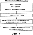
(実施例1)
(移行上皮癌におけるバイオマーカー発見のための排出尿、膀胱癌の組織および細胞系統のプロテオーム分析)
(序論)
上皮起源の癌の診断および管理の際に助けとなる新たなバイオマーカーが必要である。上皮性癌バイオマーカーを発見し分析するための優秀な媒体として尿を使用することができる。二次元ポリアクリルアミドゲル電気泳動(2D PAGE)によるプロテオーム分析は、ヒト検体のプロテオームを分析するのに有効な手段の1つである。2D PAGEを利用して、バイオマーカー発見の方法として排出尿、ヒト膀胱の腫瘍および正常組織、およびヒト由来の膀胱癌細胞系統を分析する。
Example 1
(Proteomic analysis of excreted urine for biomarker discovery in transitional cell carcinoma, tissue and cell lineage of bladder cancer)
(Introduction)
New biomarkers are needed to aid in the diagnosis and management of cancers of epithelial origin. Urine can be used as an excellent medium for discovering and analyzing epithelial cancer biomarkers. Proteome analysis by two-dimensional polyacrylamide gel electrophoresis (2D PAGE) is one effective means for analyzing the proteome of human specimens. 2D PAGE is used to analyze excreted urine, human bladder tumors and normal tissues, and human-derived bladder cancer cell lines as methods of biomarker discovery.
(方法)
(尿)
IRBにより承認された治験計画書の下で、生検を伴う、診断のための膀胱鏡検査前の患者63名、および膀胱癌である臨床的証拠が認められず悪性腫瘍の病歴がない、年齢を合わせた対照患者22名から排出尿検体を収集した。全尿タンパク質を単離し定量した。個々の患者から同量のタンパク質を貯留し、3つの群にした:1.Ta期、高悪性度;2.Ta期、低悪性度;3.正常対照。8名の患者が各群に含まれていた。各群から合計40ngのタンパク質(患者1名当たり5ng)を、2D PAGEによって分析し比較した。
(Method)
(urine)
Under an IRB approved clinical trial protocol, 63 patients with biopsy, before diagnostic cystoscopy, and no clinical evidence of bladder cancer and no history of malignancy Samples of excreted urine were collected from 22 control patients. Total urine protein was isolated and quantified. The same amount of protein from individual patients was pooled into three groups: Ta stage, high grade; 2. Ta stage, low grade; Normal control. Eight patients were included in each group. A total of 40 ng protein from each group (5 ng per patient) was analyzed and compared by 2D PAGE.
(組織)
IRBにより承認された治験計画書の下で、膀胱腫瘍組織および正常な尿路上皮を、T3N1M0期の移行上皮癌患者の膀胱切除検体から採取した。組織検体を液体窒素中で直ちに凍結し、次いで全タンパク質を単離し定量した。それぞれの腫瘍および正常組織から40ngのタンパク質を、2D PAGEによって分析し比較した。
(Organization)
Under a study protocol approved by the IRB, bladder tumor tissue and normal urothelium were collected from cystectomy specimens of patients with transitional cell carcinoma at stage T3N1M0. Tissue specimens were immediately frozen in liquid nitrogen and then total protein was isolated and quantified. 40 ng protein from each tumor and normal tissue was analyzed and compared by 2D PAGE.
(細胞系統)
以前に記載されている2種の細胞系統から分画化タンパク質を単離した:1.MGH−U1、膀胱の高悪性度移行上皮癌から培養され、ヌードマウス中で高度な腫瘍原性がある;2.MGH−U4、重度の尿路上皮異型を有する患者から培養され、ヌードマウス中で非腫瘍原性である。各細胞系統の細胞質、核および膜タンパク質の各分画40ngを、2D PAGEによって分析し比較した。
(Cell line)
Fractionated proteins were isolated from two previously described cell lines: 1. MGH-U1, cultured from high grade transitional cell carcinoma of the bladder and highly tumorigenic in nude mice; MGH-U4, cultured from patients with severe urothelial variants, and non-tumorigenic in nude mice. Each cell line cytoplasmic, nuclear and membrane protein fraction of 40 ng was analyzed and compared by 2D PAGE.
上記の検体すべてで、独特のタンパク質のスポットを単離し、液体クロマトグラフィー質量分析−質量分析法(LCMS−MS)によってそれを分析した。 In all of the above specimens, a unique protein spot was isolated and analyzed by liquid chromatography mass spectrometry-mass spectrometry (LCMS-MS).
(結果)
2D PAGEによる分析から、尿検体の3群を通じて、通常のまたは正常な尿のプロテオームを表す、通常の分子量(MW)および等電点(pI)にあるいくつかのタンパク質のスポットが明らかとなった。同様に、組織検体について、また細胞系統についても通常のプロテオームスペクトルが示された。Ta高悪性度の患者の尿、腫瘍組織およびMGH−U1細胞系統のプロテオームスペクトルから、MW10〜15kD、pI8〜10にあるいくつかの類似したペプチドのスポットが明らかとなり、それらは存在せず、またはそれから、これらのタンパク質のうち3つが、シスタチンBという内因性システインタンパク質分解酵素阻害因子、シャペロニン10という熱ショックタンパク質、およびプロフィリンという細胞骨格タンパク質であると同定された。
(result)
Analysis by 2D PAGE revealed several protein spots at normal molecular weight (MW) and isoelectric point (pI) representing normal or normal urinary proteomes through three groups of urine samples . Similarly, normal proteome spectra were shown for tissue samples and cell lines. Proteomic spectra of urine, tumor tissue and MGH-U1 cell line from Ta high-grade patients reveals several similar peptide spots at MW 10-15 kD, pI8-10, which are absent or Three of these proteins were then identified as an endogenous cysteine proteolytic enzyme inhibitor, cystatin B, a heat shock protein,
(結論)
本発明者らは、上皮起源の癌の新規バイオマーカー3種の発見を実証した。
(Conclusion)
The inventors have demonstrated the discovery of three novel biomarkers of cancer of epithelial origin.
(実施例2)
(膀胱癌組織中でのシスタチンBの免疫染色)
(方法)
正常な膀胱および膀胱癌組織を、マウスモノクローナル抗シスタチンB抗体を使用して免疫染色し、ヘマトキシリンで対比染色した。膀胱癌組織マイクロアレイBL801(US Biomax Inc、メリーランド州Rockville)を使用して免疫染色を行った。組織に脱パラフィン処理を行い、3%過酸化水素のメタノール溶液中で内因性過酸化物のブロッキング処理を行い、Antigen Unmasking Solutionを使用してマイクロウェーブ抗原賦活化を行った。5%正常ウマ血清を使用してブロッキング処理を行い、アビジン/ビオチンキットを使用して内因性ビオチンのブロッキング処理を行った。マウスモノクローナル抗シスタチンB/ステフィンB抗体、クローンA6/2(GeneTex, Inc、テキサス州San Antonio)、その後抗マウスビオチン化二次抗体とともに組織をインキュベートし、ABCキットを使用してそれを増幅し、DABを使用して発色させた。Gill’s Hematoxylin #3(Sigma−Aldrich、ミズーリ州St. Louis)を使用して組織を対比染色し、Tacha’s Bluing Solution(Biocare、カリフォルニア州Concord)を使用して青色に染色した。言及したもの以外、試薬はすべて、Vector Laboratories、カリフォルニア州Burlingameから購入した。画像はすべて、同じ露出時間で取得した。
(Example 2)
(Immunostaining of cystatin B in bladder cancer tissue)
(Method)
Normal bladder and bladder cancer tissues were immunostained using mouse monoclonal anti-cystatin B antibody and counterstained with hematoxylin. Immunostaining was performed using a bladder cancer tissue microarray BL801 (US Biomax Inc, Rockville, MD). The tissue was deparaffinized, endogenous peroxide was blocked in 3% hydrogen peroxide in methanol, and microwave antigen activation was performed using Antigen Unmasking Solution. Blocking was performed using 5% normal horse serum, and endogenous biotin was blocked using an avidin / biotin kit. Incubating the tissue with a mouse monoclonal anti-cystatin B / stefin B antibody, clone A6 / 2 (GeneTex, Inc, San Antonio, Tex.) Followed by an anti-mouse biotinylated secondary antibody and amplifying it using an ABC kit, Color was developed using DAB. Tissues were counterstained using Gill's Hematoxylin # 3 (Sigma-Aldrich, St. Louis, MO) and stained blue using Tacha's Blueing Solution (Biocare, Concord, CA). Except as noted, all reagents were purchased from Vector Laboratories, Burlingame, CA. All images were acquired with the same exposure time.
(結果)
膀胱癌である個人の試料中のシスタチンBのレベルは、正常な膀胱組織の試料中で観察されるレベルより有意に高かった。
(result)
The level of cystatin B in samples of individuals with bladder cancer was significantly higher than that observed in normal bladder tissue samples.
Claims (19)
a.該患者から生体試料を得るステップと、
b.該生体試料中での少なくとも1種の上皮性癌バイオマーカーの存在の有無を検出するステップと
を含み、少なくとも1種の上皮性癌バイオマーカーの存在は、上皮起源の癌を示し、該上皮性癌バイオマーカーが、シスタチンB、シャペロニン10、およびプロフィリンからなる群から選択される、方法。 A method for facilitating patient diagnosis for cancer of epithelial origin, comprising:
a. Obtaining a biological sample from the patient;
b. Detecting the presence or absence of at least one epithelial cancer biomarker in the biological sample, wherein the presence of at least one epithelial cancer biomarker indicates cancer of epithelial origin, The method wherein the cancer biomarker is selected from the group consisting of cystatin B, chaperonin 10, and profilin.
a.該患者から得られた生体試料である試験試料中に存在する少なくとも1種の上皮性癌バイオマーカーのレベルを測定するステップと、
b.該試験試料中の少なくとも1種の上皮性癌バイオマーカーのレベルを、対照試料中に存在する上皮性癌バイオマーカーのレベルと比較するステップと
を含み、該対照試料中の上皮性癌バイオマーカーのレベルと比較して該試験試料中の少なくとも1種の上皮性癌バイオマーカーのレベルが高いことは、上皮起源の癌を示し、該上皮性癌バイオマーカーが、シスタチンB、シャペロニン10、およびプロフィリンからなる群から選択される、方法。 A method for diagnosing cancer of epithelial origin in a patient comprising:
a. Measuring the level of at least one epithelial cancer biomarker present in a test sample that is a biological sample obtained from the patient;
b. Comparing the level of at least one epithelial cancer biomarker in the test sample with the level of the epithelial cancer biomarker present in the control sample, A high level of at least one epithelial cancer biomarker in the test sample relative to the level indicates a cancer of epithelial origin, wherein the epithelial cancer biomarker is cystatin B, chaperonin 10, and profilin A method selected from the group consisting of:
a.該患者から生体試料を得るステップと、
b.該生体試料中でのシスタチンBの存在の有無を検出するステップと
を含み、シスタチンB上皮性癌バイオマーカーの存在が、膀胱癌を示す、方法。 A method for facilitating diagnosis of bladder cancer in a patient comprising:
a. Obtaining a biological sample from the patient;
b. Detecting the presence or absence of cystatin B in the biological sample, wherein the presence of cystatin B epithelial cancer biomarker indicates bladder cancer.
a.該患者から得られた生体試料である試験試料中に存在するシスタチンBのレベルを測定するステップと、
b.該試験試料中のシスタチンBのレベルを、対照試料中に存在するシスタチンB上皮性癌バイオマーカーのレベルと比較するステップと
を含み、該対照試料中のシスタチンBのレベルと比較して該試験試料中のシスタチンBのレベルが高いことは、膀胱癌を示す、方法。 A method for diagnosing bladder cancer in a patient comprising:
a. Measuring the level of cystatin B present in a test sample, which is a biological sample obtained from the patient;
b. Comparing the level of cystatin B in the test sample with the level of cystatin B epithelial cancer biomarker present in the control sample, and comparing the level of cystatin B in the control sample with the test sample A high level of cystatin B in the bladder indicates bladder cancer.
a.該患者から得られた生体試料である試験試料中に存在するシスタチンBのレベルを測定するステップと、
b.該試験試料中のシスタチンBのレベルを、非浸潤性対照試料中に存在するシスタチンBのレベルと比較するステップと
を含み、該非浸潤性対照試料中のシスタチンBのレベルと比較して該試験試料中のシスタチンBのレベルが高いことは、浸潤性膀胱癌を示す、方法。 A method for diagnosing invasive bladder cancer in a patient, comprising:
a. Measuring the level of cystatin B present in a test sample, which is a biological sample obtained from the patient;
b. Comparing the level of cystatin B in the test sample to the level of cystatin B present in the non-invasive control sample, and comparing the level of cystatin B in the non-invasive control sample to the test sample A method wherein high levels of cystatin B are indicative of invasive bladder cancer.
a.試験試料、またはその調製物を、上皮性癌バイオマーカーまたはシスタチンBと特異的に結合する抗体に基づく結合部分と接触させて、抗体−上皮性癌バイオマーカー複合体を形成させるステップと、
b.該複合体の存在を検出し、それによって、存在する上皮性癌バイオマーカーのレベルを測定するステップと
を含む方法によって測定する、請求項9に記載の方法。 The protein level of the epithelial cancer biomarker or the level of cystatin B,
a. Contacting a test sample, or preparation thereof, with an epithelial cancer biomarker or a binding moiety based on an antibody that specifically binds cystatin B to form an antibody-epithelial cancer biomarker complex;
b. 10. The method of claim 9, wherein the method comprises measuring the presence of the complex, thereby measuring the level of epithelial cancer biomarker present.
Applications Claiming Priority (2)
| Application Number | Priority Date | Filing Date | Title |
|---|---|---|---|
| US64811005P | 2005-01-28 | 2005-01-28 | |
| PCT/US2006/003049 WO2006081473A2 (en) | 2005-01-28 | 2006-01-30 | Methods for diagnosis and prognosis of epithelial cancers |
Publications (2)
| Publication Number | Publication Date |
|---|---|
| JP2008529008A true JP2008529008A (en) | 2008-07-31 |
| JP2008529008A5 JP2008529008A5 (en) | 2009-03-19 |
Family
ID=36741098
Family Applications (1)
| Application Number | Title | Priority Date | Filing Date |
|---|---|---|---|
| JP2007553289A Pending JP2008529008A (en) | 2005-01-28 | 2006-01-30 | Methods for diagnosis and prognosis of epithelial cancer |
Country Status (9)
| Country | Link |
|---|---|
| US (7) | US8685659B2 (en) |
| EP (1) | EP1842065B1 (en) |
| JP (1) | JP2008529008A (en) |
| CN (1) | CN101268368A (en) |
| AT (1) | ATE490465T1 (en) |
| AU (1) | AU2006207954A1 (en) |
| CA (1) | CA2595377A1 (en) |
| DE (1) | DE602006018578D1 (en) |
| WO (1) | WO2006081473A2 (en) |
Cited By (1)
| Publication number | Priority date | Publication date | Assignee | Title |
|---|---|---|---|---|
| JP2016095312A (en) * | 2010-06-03 | 2016-05-26 | アイデックス ラボラトリーズ インコーポレイテッドIDEXX Laboratories, Inc. | Markers for renal disease |
Families Citing this family (46)
| Publication number | Priority date | Publication date | Assignee | Title |
|---|---|---|---|---|
| US20060078893A1 (en) | 2004-10-12 | 2006-04-13 | Medical Research Council | Compartmentalised combinatorial chemistry by microfluidic control |
| GB0307428D0 (en) | 2003-03-31 | 2003-05-07 | Medical Res Council | Compartmentalised combinatorial chemistry |
| GB0307403D0 (en) | 2003-03-31 | 2003-05-07 | Medical Res Council | Selection by compartmentalised screening |
| EP1512970A1 (en) * | 2003-09-05 | 2005-03-09 | Nederlandse Organisatie voor toegepast-natuurwetenschappelijk Onderzoek TNO | Method for determining the impact of a multicomponent mixture on the biological profile of a disease |
| US20050221339A1 (en) | 2004-03-31 | 2005-10-06 | Medical Research Council Harvard University | Compartmentalised screening by microfluidic control |
| US7968287B2 (en) | 2004-10-08 | 2011-06-28 | Medical Research Council Harvard University | In vitro evolution in microfluidic systems |
| DE602006018578D1 (en) * | 2005-01-28 | 2011-01-13 | Childrens Medical Center | DIAGNOSIS AND PROGNOSIS OF BUBBLE CANCER. |
| US20100137163A1 (en) | 2006-01-11 | 2010-06-03 | Link Darren R | Microfluidic Devices and Methods of Use in The Formation and Control of Nanoreactors |
| US9562837B2 (en) | 2006-05-11 | 2017-02-07 | Raindance Technologies, Inc. | Systems for handling microfludic droplets |
| US20080003142A1 (en) | 2006-05-11 | 2008-01-03 | Link Darren R | Microfluidic devices |
| US9012390B2 (en) | 2006-08-07 | 2015-04-21 | Raindance Technologies, Inc. | Fluorocarbon emulsion stabilizing surfactants |
| KR100767878B1 (en) * | 2006-11-07 | 2007-10-17 | 전북대학교산학협력단 | A diagnostic composition for hepatocellular carcinoma a diagnostic kit comprising it and diagnostic methods of hepatocellular carcinoma |
| WO2008097559A2 (en) | 2007-02-06 | 2008-08-14 | Brandeis University | Manipulation of fluids and reactions in microfluidic systems |
| EP2126562B1 (en) * | 2007-02-23 | 2014-12-31 | Physicians Choice Laboratory Services, LLC | Clinical intervention directed diagnostic methods |
| US8592221B2 (en) | 2007-04-19 | 2013-11-26 | Brandeis University | Manipulation of fluids, fluid components and reactions in microfluidic systems |
| WO2009017475A1 (en) * | 2007-07-27 | 2009-02-05 | Children's Medical Center Corporation | Methods for diagnosis and prognosis of epithelial cancers |
| JP5120843B2 (en) * | 2008-06-04 | 2013-01-16 | 国立大学法人山梨大学 | Test method and test reagent for autoimmune pancreatitis |
| WO2010009365A1 (en) | 2008-07-18 | 2010-01-21 | Raindance Technologies, Inc. | Droplet libraries |
| EP3415235A1 (en) | 2009-03-23 | 2018-12-19 | Raindance Technologies Inc. | Manipulation of microfluidic droplets |
| US10520500B2 (en) | 2009-10-09 | 2019-12-31 | Abdeslam El Harrak | Labelled silica-based nanomaterial with enhanced properties and uses thereof |
| WO2011079176A2 (en) | 2009-12-23 | 2011-06-30 | Raindance Technologies, Inc. | Microfluidic systems and methods for reducing the exchange of molecules between droplets |
| US10351905B2 (en) | 2010-02-12 | 2019-07-16 | Bio-Rad Laboratories, Inc. | Digital analyte analysis |
| US9366632B2 (en) | 2010-02-12 | 2016-06-14 | Raindance Technologies, Inc. | Digital analyte analysis |
| WO2011100604A2 (en) | 2010-02-12 | 2011-08-18 | Raindance Technologies, Inc. | Digital analyte analysis |
| US9399797B2 (en) | 2010-02-12 | 2016-07-26 | Raindance Technologies, Inc. | Digital analyte analysis |
| EP3447155A1 (en) | 2010-09-30 | 2019-02-27 | Raindance Technologies, Inc. | Sandwich assays in droplets |
| US9364803B2 (en) | 2011-02-11 | 2016-06-14 | Raindance Technologies, Inc. | Methods for forming mixed droplets |
| US9150852B2 (en) | 2011-02-18 | 2015-10-06 | Raindance Technologies, Inc. | Compositions and methods for molecular labeling |
| US8841071B2 (en) | 2011-06-02 | 2014-09-23 | Raindance Technologies, Inc. | Sample multiplexing |
| WO2012167142A2 (en) | 2011-06-02 | 2012-12-06 | Raindance Technolgies, Inc. | Enzyme quantification |
| US8658430B2 (en) | 2011-07-20 | 2014-02-25 | Raindance Technologies, Inc. | Manipulating droplet size |
| US20130115599A1 (en) * | 2011-11-08 | 2013-05-09 | Medical Diagnostic Laboratories, Llc | Increased cip2a expression and bladder cancer in humans |
| EP3495817A1 (en) | 2012-02-10 | 2019-06-12 | Raindance Technologies, Inc. | Molecular diagnostic screening assay |
| WO2013165748A1 (en) | 2012-04-30 | 2013-11-07 | Raindance Technologies, Inc | Digital analyte analysis |
| EP2932273B1 (en) * | 2012-12-11 | 2018-03-21 | Cornell University | Inhibitors of soluble adenylyl cyclase for use in the treatment of prostate cancer |
| EP2986762B1 (en) | 2013-04-19 | 2019-11-06 | Bio-Rad Laboratories, Inc. | Digital analyte analysis |
| US11901041B2 (en) | 2013-10-04 | 2024-02-13 | Bio-Rad Laboratories, Inc. | Digital analysis of nucleic acid modification |
| US9944977B2 (en) | 2013-12-12 | 2018-04-17 | Raindance Technologies, Inc. | Distinguishing rare variations in a nucleic acid sequence from a sample |
| US11193176B2 (en) | 2013-12-31 | 2021-12-07 | Bio-Rad Laboratories, Inc. | Method for detecting and quantifying latent retroviral RNA species |
| US10712349B2 (en) | 2014-04-15 | 2020-07-14 | The Brigham And Women's Hospital, Inc. | Circulating KIM-1 levels for detection of pathologies associated with injury to, or cancer of, the kidney |
| GB201501930D0 (en) | 2015-02-05 | 2015-03-25 | Univ London Queen Mary | Biomarkers for pancreatic cancer |
| US10647981B1 (en) | 2015-09-08 | 2020-05-12 | Bio-Rad Laboratories, Inc. | Nucleic acid library generation methods and compositions |
| TWI822655B (en) * | 2016-03-02 | 2023-11-21 | 美商英代斯實驗公司 | Methods and compositions for the detection and diagnosis of renal disease and periodontal disease |
| US10998178B2 (en) | 2017-08-28 | 2021-05-04 | Purdue Research Foundation | Systems and methods for sample analysis using swabs |
| US20230314408A1 (en) * | 2020-07-02 | 2023-10-05 | Gopath Laboratories Llc | Immune profiling and methods of using same to predict responsiveness to an immunotherapy and treat cancer |
| CN112379093B (en) * | 2020-10-22 | 2023-06-16 | 上海良润生物医药科技有限公司 | Application of CST-Cathepsin complex as tumor diagnosis marker |
Citations (5)
| Publication number | Priority date | Publication date | Assignee | Title |
|---|---|---|---|---|
| JPH10262680A (en) * | 1997-03-25 | 1998-10-06 | Kirin Brewery Co Ltd | Rho target protein human mdi and gene thereof |
| JP2001264329A (en) * | 2000-02-15 | 2001-09-26 | Pfizer Prod Inc | Examination for measuring type ii collagen in urine |
| JP2003292459A (en) * | 2002-01-30 | 2003-10-15 | Pharma Power Biotec Co Ltd | Thermo-responsive mucosa-adhesive medicament-carrier composition |
| WO2004088324A2 (en) * | 2003-03-25 | 2004-10-14 | Proteogenix, Inc. | Proteomic analysis of biological fluids |
| WO2004091383A2 (en) * | 2003-04-01 | 2004-10-28 | Genzyme Corporation | Breast endothelial cell expression patterns |
Family Cites Families (24)
| Publication number | Priority date | Publication date | Assignee | Title |
|---|---|---|---|---|
| US4281061A (en) * | 1979-07-27 | 1981-07-28 | Syva Company | Double antibody for enhanced sensitivity in immunoassay |
| GB2236186B (en) | 1989-08-22 | 1994-01-05 | Finnigan Mat Gmbh | Process and device for laser desorption of analyte molecular ions, especially of biomolecules |
| US5045694A (en) | 1989-09-27 | 1991-09-03 | The Rockefeller University | Instrument and method for the laser desorption of ions in mass spectrometry |
| PT700521E (en) | 1993-05-28 | 2003-10-31 | Baylor College Medicine | METHOD AND MASS SPECTROMETER FOR DESSORING AND IONIZATION OF ANALYZES |
| US5702892A (en) | 1995-05-09 | 1997-12-30 | The United States Of America As Represented By The Department Of Health And Human Services | Phage-display of immunoglobulin heavy chain libraries |
| AU3602097A (en) | 1996-07-15 | 1998-02-09 | Regents Of The University Of California, The | Genes from 20q13 amplicon and their uses |
| NZ516848A (en) | 1997-06-20 | 2004-03-26 | Ciphergen Biosystems Inc | Retentate chromatography apparatus with applications in biology and medicine |
| US6406921B1 (en) | 1998-07-14 | 2002-06-18 | Zyomyx, Incorporated | Protein arrays for high-throughput screening |
| US20030124543A1 (en) | 1999-01-15 | 2003-07-03 | Stuart Susan G. | Breast cancer marker |
| US6358683B1 (en) | 1999-06-03 | 2002-03-19 | The Regents Of The University Of California | Blood-based assays for breast cancer |
| US6939714B2 (en) * | 1999-06-18 | 2005-09-06 | Biotherapies, Inc. | Epithelial cell growth inhibitors |
| AU7262700A (en) | 1999-09-07 | 2001-04-10 | Viventia Biotech Inc. | Enhanced phage display library of human vh fragments and methods for producing same |
| US6998232B1 (en) * | 1999-09-27 | 2006-02-14 | Quark Biotech, Inc. | Methods of diagnosing bladder cancer |
| US20040142361A1 (en) * | 1999-11-30 | 2004-07-22 | Corixa Corporation | Compositions and methods for the therapy and diagnosis of breast cancer |
| US7429466B2 (en) | 2000-01-24 | 2008-09-30 | Hypromatrix, Inc | Methods and arrays for detecting biological molecules |
| CN1463291A (en) | 2001-04-19 | 2003-12-24 | 赛弗根生物系统股份有限公司 | Biomolecule characterization using mass spectormetry and affinity tags |
| US7112408B2 (en) | 2001-06-08 | 2006-09-26 | The Brigham And Women's Hospital, Inc. | Detection of ovarian cancer based upon alpha-haptoglobin levels |
| US20040076955A1 (en) | 2001-07-03 | 2004-04-22 | Eos Biotechnology, Inc. | Methods of diagnosis of bladder cancer, compositions and methods of screening for modulators of bladder cancer |
| US20030013208A1 (en) | 2001-07-13 | 2003-01-16 | Milagen, Inc. | Information enhanced antibody arrays |
| US8068987B2 (en) | 2001-08-13 | 2011-11-29 | Bg Medicine, Inc. | Method and system for profiling biological systems |
| US20030157579A1 (en) * | 2002-02-14 | 2003-08-21 | Kalobios, Inc. | Molecular sensors activated by disinhibition |
| AU2003239152A1 (en) | 2002-04-23 | 2003-11-10 | Millipore Corporation | Sample preparation of biological fluids for proteomic applications |
| DE602006018578D1 (en) * | 2005-01-28 | 2011-01-13 | Childrens Medical Center | DIAGNOSIS AND PROGNOSIS OF BUBBLE CANCER. |
| WO2009017475A1 (en) | 2007-07-27 | 2009-02-05 | Children's Medical Center Corporation | Methods for diagnosis and prognosis of epithelial cancers |
-
2006
- 2006-01-30 DE DE602006018578T patent/DE602006018578D1/en active Active
- 2006-01-30 AU AU2006207954A patent/AU2006207954A1/en not_active Abandoned
- 2006-01-30 EP EP06719762A patent/EP1842065B1/en not_active Not-in-force
- 2006-01-30 US US11/814,939 patent/US8685659B2/en not_active Expired - Fee Related
- 2006-01-30 CN CNA2006800098728A patent/CN101268368A/en active Pending
- 2006-01-30 JP JP2007553289A patent/JP2008529008A/en active Pending
- 2006-01-30 CA CA002595377A patent/CA2595377A1/en not_active Abandoned
- 2006-01-30 WO PCT/US2006/003049 patent/WO2006081473A2/en active Application Filing
- 2006-01-30 AT AT06719762T patent/ATE490465T1/en not_active IP Right Cessation
-
2007
- 2007-07-27 US US11/829,323 patent/US20080064047A1/en not_active Abandoned
-
2013
- 2013-04-17 US US13/864,707 patent/US20140106378A1/en not_active Abandoned
-
2014
- 2014-10-01 US US14/503,619 patent/US20150093767A1/en not_active Abandoned
-
2016
- 2016-02-19 US US15/048,934 patent/US20170023574A1/en not_active Abandoned
-
2017
- 2017-06-05 US US15/614,513 patent/US20180120319A1/en not_active Abandoned
- 2017-08-11 US US15/675,151 patent/US20180188252A1/en not_active Abandoned
Patent Citations (5)
| Publication number | Priority date | Publication date | Assignee | Title |
|---|---|---|---|---|
| JPH10262680A (en) * | 1997-03-25 | 1998-10-06 | Kirin Brewery Co Ltd | Rho target protein human mdi and gene thereof |
| JP2001264329A (en) * | 2000-02-15 | 2001-09-26 | Pfizer Prod Inc | Examination for measuring type ii collagen in urine |
| JP2003292459A (en) * | 2002-01-30 | 2003-10-15 | Pharma Power Biotec Co Ltd | Thermo-responsive mucosa-adhesive medicament-carrier composition |
| WO2004088324A2 (en) * | 2003-03-25 | 2004-10-14 | Proteogenix, Inc. | Proteomic analysis of biological fluids |
| WO2004091383A2 (en) * | 2003-04-01 | 2004-10-28 | Genzyme Corporation | Breast endothelial cell expression patterns |
Cited By (2)
| Publication number | Priority date | Publication date | Assignee | Title |
|---|---|---|---|---|
| JP2016095312A (en) * | 2010-06-03 | 2016-05-26 | アイデックス ラボラトリーズ インコーポレイテッドIDEXX Laboratories, Inc. | Markers for renal disease |
| JP2020073870A (en) * | 2010-06-03 | 2020-05-14 | アイデックス ラボラトリーズ インコーポレイテッドIDEXX Laboratories, Inc. | Marker for renal disease |
Also Published As
| Publication number | Publication date |
|---|---|
| US8685659B2 (en) | 2014-04-01 |
| WO2006081473A8 (en) | 2007-08-23 |
| CA2595377A1 (en) | 2006-08-03 |
| CN101268368A (en) | 2008-09-17 |
| US20090239245A1 (en) | 2009-09-24 |
| US20170023574A1 (en) | 2017-01-26 |
| EP1842065A4 (en) | 2008-12-31 |
| EP1842065B1 (en) | 2010-12-01 |
| WO2006081473A3 (en) | 2007-01-18 |
| WO2006081473A2 (en) | 2006-08-03 |
| US20180120319A1 (en) | 2018-05-03 |
| EP1842065A2 (en) | 2007-10-10 |
| AU2006207954A1 (en) | 2006-08-03 |
| US20150093767A1 (en) | 2015-04-02 |
| US20140106378A1 (en) | 2014-04-17 |
| US20080064047A1 (en) | 2008-03-13 |
| US20180188252A1 (en) | 2018-07-05 |
| DE602006018578D1 (en) | 2011-01-13 |
| ATE490465T1 (en) | 2010-12-15 |
Similar Documents
| Publication | Publication Date | Title |
|---|---|---|
| US20180188252A1 (en) | Methods for diagnosis and prognosis of epithelial cancers | |
| US20190361028A1 (en) | Free ngal as a biomarker for cancer | |
| JP5006802B2 (en) | Cyr61 as a biomarker for cancer diagnosis and prognosis derived from epithelium | |
| US20080268473A1 (en) | Adamts-7 as a Biomarker for Cancers of Epithelial Origin | |
| JP4847873B2 (en) | Methods for diagnosing and prognosing cancer of epithelial origin | |
| WO2009017475A1 (en) | Methods for diagnosis and prognosis of epithelial cancers |
Legal Events
| Date | Code | Title | Description |
|---|---|---|---|
| A521 | Request for written amendment filed |
Free format text: JAPANESE INTERMEDIATE CODE: A523 Effective date: 20090127 |
|
| A621 | Written request for application examination |
Free format text: JAPANESE INTERMEDIATE CODE: A621 Effective date: 20090127 |
|
| A977 | Report on retrieval |
Free format text: JAPANESE INTERMEDIATE CODE: A971007 Effective date: 20101118 |
|
| A131 | Notification of reasons for refusal |
Free format text: JAPANESE INTERMEDIATE CODE: A131 Effective date: 20101130 |
|
| A601 | Written request for extension of time |
Free format text: JAPANESE INTERMEDIATE CODE: A601 Effective date: 20110225 |
|
| A602 | Written permission of extension of time |
Free format text: JAPANESE INTERMEDIATE CODE: A602 Effective date: 20110304 |
|
| A601 | Written request for extension of time |
Free format text: JAPANESE INTERMEDIATE CODE: A601 Effective date: 20110428 |
|
| A602 | Written permission of extension of time |
Free format text: JAPANESE INTERMEDIATE CODE: A602 Effective date: 20110511 |
|
| A521 | Request for written amendment filed |
Free format text: JAPANESE INTERMEDIATE CODE: A523 Effective date: 20110527 |
|
| A131 | Notification of reasons for refusal |
Free format text: JAPANESE INTERMEDIATE CODE: A131 Effective date: 20111108 |
|
| A02 | Decision of refusal |
Free format text: JAPANESE INTERMEDIATE CODE: A02 Effective date: 20120330 |