JP2008516632A - Rapid and sensitive detection of bacteria in blood products, urine, and other fluids - Google Patents
Rapid and sensitive detection of bacteria in blood products, urine, and other fluids Download PDFInfo
- Publication number
- JP2008516632A JP2008516632A JP2007537980A JP2007537980A JP2008516632A JP 2008516632 A JP2008516632 A JP 2008516632A JP 2007537980 A JP2007537980 A JP 2007537980A JP 2007537980 A JP2007537980 A JP 2007537980A JP 2008516632 A JP2008516632 A JP 2008516632A
- Authority
- JP
- Japan
- Prior art keywords
- bacterial
- cells
- atp
- intact
- bacterial cells
- Prior art date
- Legal status (The legal status is an assumption and is not a legal conclusion. Google has not performed a legal analysis and makes no representation as to the accuracy of the status listed.)
- Pending
Links
- 239000012530 fluid Substances 0.000 title claims abstract description 39
- 210000002700 urine Anatomy 0.000 title claims abstract description 9
- 230000014670 detection of bacterium Effects 0.000 title claims description 9
- 239000010836 blood and blood product Substances 0.000 title claims description 7
- 229940125691 blood product Drugs 0.000 title claims description 7
- 238000011896 sensitive detection Methods 0.000 title 1
- -1 urine Substances 0.000 title 1
- 230000001580 bacterial effect Effects 0.000 claims abstract description 364
- 210000004027 cell Anatomy 0.000 claims abstract description 220
- 239000007788 liquid Substances 0.000 claims abstract description 212
- 210000003527 eukaryotic cell Anatomy 0.000 claims abstract description 148
- 241000894006 Bacteria Species 0.000 claims abstract description 131
- 238000000034 method Methods 0.000 claims abstract description 104
- 108090000790 Enzymes Proteins 0.000 claims abstract description 51
- 102000004190 Enzymes Human genes 0.000 claims abstract description 51
- 230000002934 lysing effect Effects 0.000 claims abstract description 33
- 238000006243 chemical reaction Methods 0.000 claims abstract description 30
- 238000001514 detection method Methods 0.000 claims abstract description 26
- IGXWBGJHJZYPQS-SSDOTTSWSA-N D-Luciferin Chemical group OC(=O)[C@H]1CSC(C=2SC3=CC=C(O)C=C3N=2)=N1 IGXWBGJHJZYPQS-SSDOTTSWSA-N 0.000 claims abstract description 22
- CYCGRDQQIOGCKX-UHFFFAOYSA-N Dehydro-luciferin Natural products OC(=O)C1=CSC(C=2SC3=CC(O)=CC=C3N=2)=N1 CYCGRDQQIOGCKX-UHFFFAOYSA-N 0.000 claims abstract description 22
- BJGNCJDXODQBOB-UHFFFAOYSA-N Fivefly Luciferin Natural products OC(=O)C1CSC(C=2SC3=CC(O)=CC=C3N=2)=N1 BJGNCJDXODQBOB-UHFFFAOYSA-N 0.000 claims abstract description 22
- DDWFXDSYGUXRAY-UHFFFAOYSA-N Luciferin Natural products CCc1c(C)c(CC2NC(=O)C(=C2C=C)C)[nH]c1Cc3[nH]c4C(=C5/NC(CC(=O)O)C(C)C5CC(=O)O)CC(=O)c4c3C DDWFXDSYGUXRAY-UHFFFAOYSA-N 0.000 claims abstract description 22
- 238000012544 monitoring process Methods 0.000 claims abstract description 21
- 238000006555 catalytic reaction Methods 0.000 claims abstract description 15
- 210000004369 blood Anatomy 0.000 claims abstract description 12
- 239000008280 blood Substances 0.000 claims abstract description 12
- 239000000523 sample Substances 0.000 claims description 177
- 238000005259 measurement Methods 0.000 claims description 127
- 239000006166 lysate Substances 0.000 claims description 63
- 239000005089 Luciferase Substances 0.000 claims description 52
- 108060001084 Luciferase Proteins 0.000 claims description 51
- 239000011148 porous material Substances 0.000 claims description 27
- 238000000926 separation method Methods 0.000 claims description 26
- 239000011324 bead Substances 0.000 claims description 21
- 238000012360 testing method Methods 0.000 claims description 21
- 238000004891 communication Methods 0.000 claims description 20
- 238000001914 filtration Methods 0.000 claims description 19
- 230000008569 process Effects 0.000 claims description 11
- 241000894007 species Species 0.000 claims description 10
- 210000001124 body fluid Anatomy 0.000 claims description 9
- 239000012141 concentrate Substances 0.000 claims description 9
- 239000010839 body fluid Substances 0.000 claims description 8
- 230000001332 colony forming effect Effects 0.000 claims description 8
- 238000004140 cleaning Methods 0.000 claims description 7
- 241000588724 Escherichia coli Species 0.000 claims description 6
- 210000002381 plasma Anatomy 0.000 claims description 6
- 239000000047 product Substances 0.000 claims description 6
- 241000186216 Corynebacterium Species 0.000 claims description 5
- 241000193996 Streptococcus pyogenes Species 0.000 claims description 5
- 239000002253 acid Substances 0.000 claims description 5
- 239000003795 chemical substances by application Substances 0.000 claims description 5
- 239000002699 waste material Substances 0.000 claims description 5
- 241000193755 Bacillus cereus Species 0.000 claims description 4
- 244000063299 Bacillus subtilis Species 0.000 claims description 4
- 235000014469 Bacillus subtilis Nutrition 0.000 claims description 4
- 241000193468 Clostridium perfringens Species 0.000 claims description 4
- 241000588914 Enterobacter Species 0.000 claims description 4
- 241000589517 Pseudomonas aeruginosa Species 0.000 claims description 4
- 241000607715 Serratia marcescens Species 0.000 claims description 4
- 241000191967 Staphylococcus aureus Species 0.000 claims description 4
- 241000191963 Staphylococcus epidermidis Species 0.000 claims description 4
- 239000000203 mixture Substances 0.000 claims description 4
- 239000002245 particle Substances 0.000 claims description 4
- 230000000717 retained effect Effects 0.000 claims description 4
- 241000588749 Klebsiella oxytoca Species 0.000 claims description 3
- 241000124008 Mammalia Species 0.000 claims description 3
- 210000001185 bone marrow Anatomy 0.000 claims description 3
- 210000003555 cloaca Anatomy 0.000 claims description 3
- 239000003599 detergent Substances 0.000 claims description 3
- 239000011521 glass Substances 0.000 claims description 3
- 238000004519 manufacturing process Methods 0.000 claims description 3
- 239000000463 material Substances 0.000 claims description 3
- 239000003566 sealing material Substances 0.000 claims description 3
- 210000002966 serum Anatomy 0.000 claims description 3
- 210000000130 stem cell Anatomy 0.000 claims description 3
- 206010008631 Cholera Diseases 0.000 claims description 2
- 102000008186 Collagen Human genes 0.000 claims description 2
- 108010035532 Collagen Proteins 0.000 claims description 2
- 102000016359 Fibronectins Human genes 0.000 claims description 2
- 108010067306 Fibronectins Proteins 0.000 claims description 2
- 102000007547 Laminin Human genes 0.000 claims description 2
- 108010085895 Laminin Proteins 0.000 claims description 2
- 108010038807 Oligopeptides Proteins 0.000 claims description 2
- 102000015636 Oligopeptides Human genes 0.000 claims description 2
- 108010072041 arginyl-glycyl-aspartic acid Proteins 0.000 claims description 2
- 230000000903 blocking effect Effects 0.000 claims description 2
- 210000001175 cerebrospinal fluid Anatomy 0.000 claims description 2
- 229920001436 collagen Polymers 0.000 claims description 2
- 210000003743 erythrocyte Anatomy 0.000 claims description 2
- 239000003960 organic solvent Substances 0.000 claims description 2
- 108010016184 phenylalanyl-histidyl-arginyl-arginyl-isoleucyl-lysyl-alanine Proteins 0.000 claims description 2
- 238000005086 pumping Methods 0.000 claims description 2
- 206010011985 Decubitus ulcer Diseases 0.000 claims 1
- 208000004210 Pressure Ulcer Diseases 0.000 claims 1
- 241000282898 Sus scrofa Species 0.000 claims 1
- 239000012466 permeate Substances 0.000 claims 1
- 238000011109 contamination Methods 0.000 abstract description 11
- 229940021317 other blood product in atc Drugs 0.000 abstract description 2
- 239000000243 solution Substances 0.000 description 43
- 229940088598 enzyme Drugs 0.000 description 36
- 230000027455 binding Effects 0.000 description 13
- 230000009089 cytolysis Effects 0.000 description 12
- 230000029586 bacterial cell surface binding Effects 0.000 description 7
- 239000012528 membrane Substances 0.000 description 7
- 230000035945 sensitivity Effects 0.000 description 7
- KFZMGEQAYNKOFK-UHFFFAOYSA-N Isopropanol Chemical compound CC(C)O KFZMGEQAYNKOFK-UHFFFAOYSA-N 0.000 description 6
- 206010040047 Sepsis Diseases 0.000 description 6
- 238000004020 luminiscence type Methods 0.000 description 6
- 231100000582 ATP assay Toxicity 0.000 description 5
- 238000004458 analytical method Methods 0.000 description 5
- 230000008901 benefit Effects 0.000 description 4
- 239000000306 component Substances 0.000 description 4
- XBDQKXXYIPTUBI-UHFFFAOYSA-N dimethylselenoniopropionate Natural products CCC(O)=O XBDQKXXYIPTUBI-UHFFFAOYSA-N 0.000 description 4
- 239000003814 drug Substances 0.000 description 4
- 238000004401 flow injection analysis Methods 0.000 description 4
- 102000007347 Apyrase Human genes 0.000 description 3
- 108010007730 Apyrase Proteins 0.000 description 3
- 150000007513 acids Chemical class 0.000 description 3
- 238000011955 best available control technology Methods 0.000 description 3
- 239000003153 chemical reaction reagent Substances 0.000 description 3
- LIYGYAHYXQDGEP-UHFFFAOYSA-N firefly oxyluciferin Natural products Oc1csc(n1)-c1nc2ccc(O)cc2s1 LIYGYAHYXQDGEP-UHFFFAOYSA-N 0.000 description 3
- 238000002347 injection Methods 0.000 description 3
- 239000007924 injection Substances 0.000 description 3
- JJVOROULKOMTKG-UHFFFAOYSA-N oxidized Photinus luciferin Chemical compound S1C2=CC(O)=CC=C2N=C1C1=NC(=O)CS1 JJVOROULKOMTKG-UHFFFAOYSA-N 0.000 description 3
- YNJBWRMUSHSURL-UHFFFAOYSA-N trichloroacetic acid Chemical compound OC(=O)C(Cl)(Cl)Cl YNJBWRMUSHSURL-UHFFFAOYSA-N 0.000 description 3
- 208000002874 Acne Vulgaris Diseases 0.000 description 2
- OKTJSMMVPCPJKN-UHFFFAOYSA-N Carbon Chemical compound [C] OKTJSMMVPCPJKN-UHFFFAOYSA-N 0.000 description 2
- HEDRZPFGACZZDS-UHFFFAOYSA-N Chloroform Chemical compound ClC(Cl)Cl HEDRZPFGACZZDS-UHFFFAOYSA-N 0.000 description 2
- WQZGKKKJIJFFOK-GASJEMHNSA-N Glucose Natural products OC[C@H]1OC(O)[C@H](O)[C@@H](O)[C@@H]1O WQZGKKKJIJFFOK-GASJEMHNSA-N 0.000 description 2
- LRHPLDYGYMQRHN-UHFFFAOYSA-N N-Butanol Chemical compound CCCCO LRHPLDYGYMQRHN-UHFFFAOYSA-N 0.000 description 2
- FAPWRFPIFSIZLT-UHFFFAOYSA-M Sodium chloride Chemical compound [Na+].[Cl-] FAPWRFPIFSIZLT-UHFFFAOYSA-M 0.000 description 2
- 241000194017 Streptococcus Species 0.000 description 2
- 241000607734 Yersinia <bacteria> Species 0.000 description 2
- 206010000496 acne Diseases 0.000 description 2
- 238000005415 bioluminescence Methods 0.000 description 2
- 230000029918 bioluminescence Effects 0.000 description 2
- 210000002421 cell wall Anatomy 0.000 description 2
- 238000002512 chemotherapy Methods 0.000 description 2
- 238000012258 culturing Methods 0.000 description 2
- 230000034994 death Effects 0.000 description 2
- 231100000517 death Toxicity 0.000 description 2
- 229940079593 drug Drugs 0.000 description 2
- 239000003112 inhibitor Substances 0.000 description 2
- 238000000504 luminescence detection Methods 0.000 description 2
- 230000007935 neutral effect Effects 0.000 description 2
- VLTRZXGMWDSKGL-UHFFFAOYSA-N perchloric acid Chemical compound OCl(=O)(=O)=O VLTRZXGMWDSKGL-UHFFFAOYSA-N 0.000 description 2
- 238000007747 plating Methods 0.000 description 2
- 235000019260 propionic acid Nutrition 0.000 description 2
- IUVKMZGDUIUOCP-BTNSXGMBSA-N quinbolone Chemical compound O([C@H]1CC[C@H]2[C@H]3[C@@H]([C@]4(C=CC(=O)C=C4CC3)C)CC[C@@]21C)C1=CCCC1 IUVKMZGDUIUOCP-BTNSXGMBSA-N 0.000 description 2
- 239000002904 solvent Substances 0.000 description 2
- 210000001082 somatic cell Anatomy 0.000 description 2
- 238000003860 storage Methods 0.000 description 2
- XLYOFNOQVPJJNP-UHFFFAOYSA-N water Substances O XLYOFNOQVPJJNP-UHFFFAOYSA-N 0.000 description 2
- JKMHFZQWWAIEOD-UHFFFAOYSA-N 2-[4-(2-hydroxyethyl)piperazin-1-yl]ethanesulfonic acid Chemical compound OCC[NH+]1CCN(CCS([O-])(=O)=O)CC1 JKMHFZQWWAIEOD-UHFFFAOYSA-N 0.000 description 1
- 241000589291 Acinetobacter Species 0.000 description 1
- 241000193830 Bacillus <bacterium> Species 0.000 description 1
- 241000588807 Bordetella Species 0.000 description 1
- 241000589968 Borrelia Species 0.000 description 1
- 241000606161 Chlamydia Species 0.000 description 1
- 241000193403 Clostridium Species 0.000 description 1
- 208000004232 Enteritis Diseases 0.000 description 1
- 241000233866 Fungi Species 0.000 description 1
- 239000007995 HEPES buffer Substances 0.000 description 1
- 239000012981 Hank's balanced salt solution Substances 0.000 description 1
- 241000589989 Helicobacter Species 0.000 description 1
- 208000032843 Hemorrhage Diseases 0.000 description 1
- 206010061598 Immunodeficiency Diseases 0.000 description 1
- 241000588748 Klebsiella Species 0.000 description 1
- 241000186781 Listeria Species 0.000 description 1
- 208000016604 Lyme disease Diseases 0.000 description 1
- 241000192041 Micrococcus Species 0.000 description 1
- 102000016943 Muramidase Human genes 0.000 description 1
- 108010014251 Muramidase Proteins 0.000 description 1
- 241000186359 Mycobacterium Species 0.000 description 1
- 108010062010 N-Acetylmuramoyl-L-alanine Amidase Proteins 0.000 description 1
- 241000588653 Neisseria Species 0.000 description 1
- 206010028980 Neoplasm Diseases 0.000 description 1
- 108091034117 Oligonucleotide Proteins 0.000 description 1
- ISWSIDIOOBJBQZ-UHFFFAOYSA-N Phenol Chemical compound OC1=CC=CC=C1 ISWSIDIOOBJBQZ-UHFFFAOYSA-N 0.000 description 1
- 229920002873 Polyethylenimine Polymers 0.000 description 1
- 108010039918 Polylysine Proteins 0.000 description 1
- 241000588769 Proteus <enterobacteria> Species 0.000 description 1
- 241000589516 Pseudomonas Species 0.000 description 1
- 206010037660 Pyrexia Diseases 0.000 description 1
- 241000607142 Salmonella Species 0.000 description 1
- 241000607720 Serratia Species 0.000 description 1
- 229920002125 Sokalan® Polymers 0.000 description 1
- 241000191940 Staphylococcus Species 0.000 description 1
- 208000007536 Thrombosis Diseases 0.000 description 1
- JLCPHMBAVCMARE-UHFFFAOYSA-N [3-[[3-[[3-[[3-[[3-[[3-[[3-[[3-[[3-[[3-[[3-[[5-(2-amino-6-oxo-1H-purin-9-yl)-3-[[3-[[3-[[3-[[3-[[3-[[5-(2-amino-6-oxo-1H-purin-9-yl)-3-[[5-(2-amino-6-oxo-1H-purin-9-yl)-3-hydroxyoxolan-2-yl]methoxy-hydroxyphosphoryl]oxyoxolan-2-yl]methoxy-hydroxyphosphoryl]oxy-5-(5-methyl-2,4-dioxopyrimidin-1-yl)oxolan-2-yl]methoxy-hydroxyphosphoryl]oxy-5-(6-aminopurin-9-yl)oxolan-2-yl]methoxy-hydroxyphosphoryl]oxy-5-(6-aminopurin-9-yl)oxolan-2-yl]methoxy-hydroxyphosphoryl]oxy-5-(6-aminopurin-9-yl)oxolan-2-yl]methoxy-hydroxyphosphoryl]oxy-5-(6-aminopurin-9-yl)oxolan-2-yl]methoxy-hydroxyphosphoryl]oxyoxolan-2-yl]methoxy-hydroxyphosphoryl]oxy-5-(5-methyl-2,4-dioxopyrimidin-1-yl)oxolan-2-yl]methoxy-hydroxyphosphoryl]oxy-5-(4-amino-2-oxopyrimidin-1-yl)oxolan-2-yl]methoxy-hydroxyphosphoryl]oxy-5-(5-methyl-2,4-dioxopyrimidin-1-yl)oxolan-2-yl]methoxy-hydroxyphosphoryl]oxy-5-(5-methyl-2,4-dioxopyrimidin-1-yl)oxolan-2-yl]methoxy-hydroxyphosphoryl]oxy-5-(6-aminopurin-9-yl)oxolan-2-yl]methoxy-hydroxyphosphoryl]oxy-5-(6-aminopurin-9-yl)oxolan-2-yl]methoxy-hydroxyphosphoryl]oxy-5-(4-amino-2-oxopyrimidin-1-yl)oxolan-2-yl]methoxy-hydroxyphosphoryl]oxy-5-(4-amino-2-oxopyrimidin-1-yl)oxolan-2-yl]methoxy-hydroxyphosphoryl]oxy-5-(4-amino-2-oxopyrimidin-1-yl)oxolan-2-yl]methoxy-hydroxyphosphoryl]oxy-5-(6-aminopurin-9-yl)oxolan-2-yl]methoxy-hydroxyphosphoryl]oxy-5-(4-amino-2-oxopyrimidin-1-yl)oxolan-2-yl]methyl [5-(6-aminopurin-9-yl)-2-(hydroxymethyl)oxolan-3-yl] hydrogen phosphate Polymers Cc1cn(C2CC(OP(O)(=O)OCC3OC(CC3OP(O)(=O)OCC3OC(CC3O)n3cnc4c3nc(N)[nH]c4=O)n3cnc4c3nc(N)[nH]c4=O)C(COP(O)(=O)OC3CC(OC3COP(O)(=O)OC3CC(OC3COP(O)(=O)OC3CC(OC3COP(O)(=O)OC3CC(OC3COP(O)(=O)OC3CC(OC3COP(O)(=O)OC3CC(OC3COP(O)(=O)OC3CC(OC3COP(O)(=O)OC3CC(OC3COP(O)(=O)OC3CC(OC3COP(O)(=O)OC3CC(OC3COP(O)(=O)OC3CC(OC3COP(O)(=O)OC3CC(OC3COP(O)(=O)OC3CC(OC3COP(O)(=O)OC3CC(OC3COP(O)(=O)OC3CC(OC3COP(O)(=O)OC3CC(OC3COP(O)(=O)OC3CC(OC3CO)n3cnc4c(N)ncnc34)n3ccc(N)nc3=O)n3cnc4c(N)ncnc34)n3ccc(N)nc3=O)n3ccc(N)nc3=O)n3ccc(N)nc3=O)n3cnc4c(N)ncnc34)n3cnc4c(N)ncnc34)n3cc(C)c(=O)[nH]c3=O)n3cc(C)c(=O)[nH]c3=O)n3ccc(N)nc3=O)n3cc(C)c(=O)[nH]c3=O)n3cnc4c3nc(N)[nH]c4=O)n3cnc4c(N)ncnc34)n3cnc4c(N)ncnc34)n3cnc4c(N)ncnc34)n3cnc4c(N)ncnc34)O2)c(=O)[nH]c1=O JLCPHMBAVCMARE-UHFFFAOYSA-N 0.000 description 1
- 238000009825 accumulation Methods 0.000 description 1
- DPKHZNPWBDQZCN-UHFFFAOYSA-N acridine orange free base Chemical compound C1=CC(N(C)C)=CC2=NC3=CC(N(C)C)=CC=C3C=C21 DPKHZNPWBDQZCN-UHFFFAOYSA-N 0.000 description 1
- GZCGUPFRVQAUEE-SLPGGIOYSA-N aldehydo-D-glucose Chemical compound OC[C@@H](O)[C@@H](O)[C@H](O)[C@@H](O)C=O GZCGUPFRVQAUEE-SLPGGIOYSA-N 0.000 description 1
- 208000007502 anemia Diseases 0.000 description 1
- 229940089206 anhydrous dextrose Drugs 0.000 description 1
- 238000002617 apheresis Methods 0.000 description 1
- 238000013459 approach Methods 0.000 description 1
- 230000004888 barrier function Effects 0.000 description 1
- DZBUGLKDJFMEHC-UHFFFAOYSA-N benzoquinolinylidene Natural products C1=CC=CC2=CC3=CC=CC=C3N=C21 DZBUGLKDJFMEHC-UHFFFAOYSA-N 0.000 description 1
- 208000034158 bleeding Diseases 0.000 description 1
- 230000000740 bleeding effect Effects 0.000 description 1
- 210000000601 blood cell Anatomy 0.000 description 1
- 239000012503 blood component Substances 0.000 description 1
- 238000009640 blood culture Methods 0.000 description 1
- 238000010322 bone marrow transplantation Methods 0.000 description 1
- 201000011510 cancer Diseases 0.000 description 1
- 230000008878 coupling Effects 0.000 description 1
- 238000010168 coupling process Methods 0.000 description 1
- 238000005859 coupling reaction Methods 0.000 description 1
- 238000000432 density-gradient centrifugation Methods 0.000 description 1
- 239000008121 dextrose Substances 0.000 description 1
- 238000003745 diagnosis Methods 0.000 description 1
- 238000004090 dissolution Methods 0.000 description 1
- 238000011978 dissolution method Methods 0.000 description 1
- 239000003480 eluent Substances 0.000 description 1
- 238000010828 elution Methods 0.000 description 1
- 238000006911 enzymatic reaction Methods 0.000 description 1
- 239000000706 filtrate Substances 0.000 description 1
- 238000000684 flow cytometry Methods 0.000 description 1
- 238000000799 fluorescence microscopy Methods 0.000 description 1
- 239000008103 glucose Substances 0.000 description 1
- 230000023597 hemostasis Effects 0.000 description 1
- 238000001802 infusion Methods 0.000 description 1
- 230000005764 inhibitory process Effects 0.000 description 1
- 238000011835 investigation Methods 0.000 description 1
- 230000000670 limiting effect Effects 0.000 description 1
- 229960000274 lysozyme Drugs 0.000 description 1
- 235000010335 lysozyme Nutrition 0.000 description 1
- 239000004325 lysozyme Substances 0.000 description 1
- 238000000691 measurement method Methods 0.000 description 1
- 230000007246 mechanism Effects 0.000 description 1
- MYWUZJCMWCOHBA-VIFPVBQESA-N methamphetamine Chemical compound CN[C@@H](C)CC1=CC=CC=C1 MYWUZJCMWCOHBA-VIFPVBQESA-N 0.000 description 1
- 244000005700 microbiome Species 0.000 description 1
- 238000000386 microscopy Methods 0.000 description 1
- 238000002156 mixing Methods 0.000 description 1
- 150000004682 monohydrates Chemical class 0.000 description 1
- 238000006386 neutralization reaction Methods 0.000 description 1
- 230000036963 noncompetitive effect Effects 0.000 description 1
- 230000002085 persistent effect Effects 0.000 description 1
- 239000004584 polyacrylic acid Substances 0.000 description 1
- 229920000656 polylysine Polymers 0.000 description 1
- 230000003389 potentiating effect Effects 0.000 description 1
- 238000000746 purification Methods 0.000 description 1
- 230000035484 reaction time Effects 0.000 description 1
- 230000001105 regulatory effect Effects 0.000 description 1
- 230000002441 reversible effect Effects 0.000 description 1
- 238000012552 review Methods 0.000 description 1
- 239000012488 sample solution Substances 0.000 description 1
- 239000011734 sodium Substances 0.000 description 1
- 239000011780 sodium chloride Substances 0.000 description 1
- 239000001488 sodium phosphate Substances 0.000 description 1
- 229910000162 sodium phosphate Inorganic materials 0.000 description 1
- 238000000527 sonication Methods 0.000 description 1
- 238000010186 staining Methods 0.000 description 1
- 206010043554 thrombocytopenia Diseases 0.000 description 1
- RYFMWSXOAZQYPI-UHFFFAOYSA-K trisodium phosphate Chemical compound [Na+].[Na+].[Na+].[O-]P([O-])([O-])=O RYFMWSXOAZQYPI-UHFFFAOYSA-K 0.000 description 1
- GPRLSGONYQIRFK-MNYXATJNSA-N triton Chemical compound [3H+] GPRLSGONYQIRFK-MNYXATJNSA-N 0.000 description 1
- 241001148471 unidentified anaerobic bacterium Species 0.000 description 1
- 230000000007 visual effect Effects 0.000 description 1
- 239000011534 wash buffer Substances 0.000 description 1
Images


Classifications
-
- C—CHEMISTRY; METALLURGY
- C12—BIOCHEMISTRY; BEER; SPIRITS; WINE; VINEGAR; MICROBIOLOGY; ENZYMOLOGY; MUTATION OR GENETIC ENGINEERING
- C12Q—MEASURING OR TESTING PROCESSES INVOLVING ENZYMES, NUCLEIC ACIDS OR MICROORGANISMS; COMPOSITIONS OR TEST PAPERS THEREFOR; PROCESSES OF PREPARING SUCH COMPOSITIONS; CONDITION-RESPONSIVE CONTROL IN MICROBIOLOGICAL OR ENZYMOLOGICAL PROCESSES
- C12Q1/00—Measuring or testing processes involving enzymes, nucleic acids or microorganisms; Compositions therefor; Processes of preparing such compositions
- C12Q1/66—Measuring or testing processes involving enzymes, nucleic acids or microorganisms; Compositions therefor; Processes of preparing such compositions involving luciferase
-
- C—CHEMISTRY; METALLURGY
- C12—BIOCHEMISTRY; BEER; SPIRITS; WINE; VINEGAR; MICROBIOLOGY; ENZYMOLOGY; MUTATION OR GENETIC ENGINEERING
- C12Q—MEASURING OR TESTING PROCESSES INVOLVING ENZYMES, NUCLEIC ACIDS OR MICROORGANISMS; COMPOSITIONS OR TEST PAPERS THEREFOR; PROCESSES OF PREPARING SUCH COMPOSITIONS; CONDITION-RESPONSIVE CONTROL IN MICROBIOLOGICAL OR ENZYMOLOGICAL PROCESSES
- C12Q1/00—Measuring or testing processes involving enzymes, nucleic acids or microorganisms; Compositions therefor; Processes of preparing such compositions
- C12Q1/02—Measuring or testing processes involving enzymes, nucleic acids or microorganisms; Compositions therefor; Processes of preparing such compositions involving viable microorganisms
- C12Q1/04—Determining presence or kind of microorganism; Use of selective media for testing antibiotics or bacteriocides; Compositions containing a chemical indicator therefor
Landscapes
- Chemical & Material Sciences (AREA)
- Life Sciences & Earth Sciences (AREA)
- Organic Chemistry (AREA)
- Health & Medical Sciences (AREA)
- Engineering & Computer Science (AREA)
- Zoology (AREA)
- Wood Science & Technology (AREA)
- Proteomics, Peptides & Aminoacids (AREA)
- Molecular Biology (AREA)
- Bioinformatics & Cheminformatics (AREA)
- Immunology (AREA)
- Microbiology (AREA)
- Biophysics (AREA)
- Analytical Chemistry (AREA)
- Physics & Mathematics (AREA)
- Genetics & Genomics (AREA)
- Biochemistry (AREA)
- Biotechnology (AREA)
- General Engineering & Computer Science (AREA)
- General Health & Medical Sciences (AREA)
- Toxicology (AREA)
- Measuring Or Testing Involving Enzymes Or Micro-Organisms (AREA)
- Peptides Or Proteins (AREA)
- Investigating Or Analysing Materials By The Use Of Chemical Reactions (AREA)
- Apparatus Associated With Microorganisms And Enzymes (AREA)
Abstract
【要約書】
本発明は、輸血用の血液、血小板、および他の血液製剤、ならびに尿を含む、液体中の細菌を検出する方法を提供する。本方法は、細菌を溶解させてATPを放出し、そのATPを検出することに基づくものである。真核細胞が大量のATPを含むことから、真核細胞の汚染が解決すべき問題である。したがって、本方法の一部は、細菌細胞を溶解させてATPを放出する前に、無傷の細菌細胞から無傷の真核細胞(例えば血小板)を分離、そのATPを、反応を触媒するATP消費酵素と接触させて、酵素触媒反応をモニタリングすることを伴う。一般に、この酵素はルシフェリンであり、反応はルシフェリンが発する検出光によってモニタリングする。本発明の他の方法では、液体試料を、細菌細胞を結合する担持表面と接触させ、細菌細胞を溶解させてATPを放出させ、ATPをATP消費酵素と接触させて、酵素触媒反応をモニタリングするステップ、ことを伴う。また、本方法を実行するための装置も開示する。[Summary]
The present invention provides a method for detecting bacteria in fluids, including blood for transfusion, platelets, and other blood products, and urine. The method is based on dissolving bacteria to release ATP and detecting the ATP. Since eukaryotic cells contain large amounts of ATP, contamination of eukaryotic cells is a problem to be solved. Thus, part of this method involves separating intact eukaryotic cells (eg, platelets) from intact bacterial cells before lysing the bacterial cells and releasing ATP, which ATP consuming enzyme catalyzes the reaction. And monitoring the enzyme-catalyzed reaction. In general, this enzyme is luciferin, and the reaction is monitored by detection light emitted by luciferin. In another method of the invention, a liquid sample is contacted with a support surface that binds bacterial cells, the bacterial cells are lysed to release ATP, and ATP is contacted with an ATP consuming enzyme to monitor the enzyme-catalyzed reaction. Step, with that. An apparatus for performing the method is also disclosed.
Description
米国では、毎年900万以上の血小板ユニットが輸血されている。これらの血小板は機能の損失を避けるために室温で保管されるが、室温においては汚染菌であるあらゆる細菌が急速に増殖するため、細菌による汚染を被りやすい。血小板は、血小板減少とそれによる貧血、および化学療法に起因する出血のリスクの治療のため、化学療法を受けている癌患者にしばしば投与される。これらの患者はまた免疫無防備状態であり、したがって血小板の細菌汚染の危険に著しくさらされている。汚染された血小板による疾病および死亡の件数は、最近になってやっと集計されてきている。 In the United States, more than 9 million platelet units are transfused every year. These platelets are stored at room temperature to avoid loss of function, but at room temperature, any bacteria that are contaminating bacteria grow rapidly and are therefore susceptible to bacterial contamination. Platelets are often administered to cancer patients undergoing chemotherapy for the treatment of thrombocytopenia and resulting anemia, and the risk of bleeding resulting from chemotherapy. These patients are also immunocompromised and are therefore at significant risk of bacterial contamination of platelets. The number of illnesses and deaths from contaminated platelets has only recently been tabulated.
細菌汚染は、過去3年間において輸血に関連する死亡の第一原因である(1)。102〜103CFU/mlという低い細菌汚染レベルは、発熱や血液培養陽性を伴っていた。Johns HopkinsおよびDana−Farberによる研究が明らかにしたところによれば、血小板輸血後の敗血症の確率は、その局部およびその血小板が無作為のドナー由来か、単独のドナーによるアフェレーシスかにより、0.005%〜0.14%である。記録された33,829件の輸血のうち、全部で9件の敗血症が確認された(2、3)。血小板純度の調査では、敗血症の確率は細菌汚染の確率よりも低いようであった。血小板輸血による敗血症の確率は、さまざまな臨床的および規制的理由により、実際よりも低く示されてきたということが広く示唆されてきた。 Bacterial contamination is the leading cause of blood-related deaths in the past three years (1). Low bacterial contamination levels of 10 2 to 10 3 CFU / ml were accompanied by fever and positive blood culture. A study by Johns Hopkins and Dana-Farber revealed that the probability of sepsis after platelet transfusion was 0.005 depending on whether the local and the platelets were from a random donor or apheresis by a single donor. % To 0.14%. Of the 33,829 transfusions recorded, a total of 9 sepsis were confirmed (2, 3). In the investigation of platelet purity, the probability of sepsis appeared to be lower than the probability of bacterial contamination. It has been widely suggested that the probability of sepsis from platelet transfusions has been shown lower than it actually has for various clinical and regulatory reasons.
敗血症のリスクのため、FDAは5日間以上保管された血小板の廃棄を命じた。短期間(1984〜1986)においてFDAは血小板の7日間の保管を許可したが、細菌の増殖に関するデータが問題であると証明されるとこの規則を覆した(4)。 Due to the risk of sepsis, the FDA ordered the disposal of platelets stored for more than 5 days. In a short period (1984-1986), the FDA allowed 7 days storage of platelets, but overturned this rule when data on bacterial growth proved to be a problem (4).
血小板中の細菌の試験を行う利用可能な手段は時間がかかり過ぎるか、感度が十分でないか、もしくは非常に面倒である。血小板から微生物を培養するという方法がある。BACT/ALERTシステムはこのアプローチを用いる。しかし、これには1日から3日の培養期間が必要となる(5)。ほかに、濃厚血小板の試料をグラム染色し、細菌を視覚的に識別するという方法がある。しかし、これには多大な労力が必要となり、1mlあたりたった106コロニー形成単位(CFU)にしか反応しなかった(5)。アクリジン・オレンジ染色および蛍光顕微鏡検査法により、感度は104〜105CFU/mlに改善された(5)。血小板の目視観測による血小板の回転やpH分析、グルコース濃度の変化も使用されてきたが、しかしこれらは十分には高感度でなく、また確実でもない(5)。 Available means of testing for bacteria in platelets are too time consuming, not sensitive enough or very cumbersome. There is a method of culturing microorganisms from platelets. The BACT / ALERT system uses this approach. However, this requires a culture period of 1 to 3 days (5). Another method is to gram-stain a sample of concentrated platelets to visually identify the bacteria. However, this required a great deal of effort and only reacted with 10 6 colony forming units (CFU) per ml (5). Sensitivity was improved to 10 4 to 10 5 CFU / ml by acridine orange staining and fluorescence microscopy (5). Platelet rotation and pH analysis by visual observation of platelets, and changes in glucose concentration have also been used, but these are not sufficiently sensitive and reliable (5).
腸炎エルシニアの検出にはPCR法が用いられた。この方法は非常に高感度であったが、6時間かかり、ひとつの種のみに対して特異的であった(5)。 PCR was used to detect enteritis Yersinia. This method was very sensitive but took 6 hours and was specific for only one species (5).
蛍光抗体もまた、フローサイトメトリーによって細菌の検出に用いられた(5)。これは、高感度となる可能性はあるが、費用がかかり、かなりの時間を必要とし、また抗体が認識する種しか検出しない。 Fluorescent antibodies were also used for bacterial detection by flow cytometry (5). While this can be highly sensitive, it is expensive, requires considerable time, and detects only the species that the antibody recognizes.
細菌を検出するその他の方法は、ハイブリッド化して細菌rRNAとなる標識オリゴヌクレオチドの検出を伴った(6)。しかし、このプロセスには4時間を要した。 Other methods of detecting bacteria involved detection of labeled oligonucleotides that hybridize to bacterial rRNA (6). However, this process took 4 hours.
細菌は細菌ATPのルミネセンス検出によって検出されてきた(7、米国特許番号3,933,592)。細菌は溶解されてそのATPを放出し、ATPはルシフェラーゼおよびルシフェリンとの反応によって検出され、光を発生する。しかし、真核細胞は細菌細胞よりもずっと多くのATPを持つため、真核細胞のわずかな汚染であっても信頼性の低い結果となる。ひとつの方法において、血液細胞はTRITON X−100によって溶解され、破片は細菌から密度勾配遠心によって分離され、勾配の細菌細胞層は抽出・処理されてすべての細菌を溶解し、ルシフェリン−ルシフェラーゼ測定によってATPの測定が行われた(8)。
血小板中の細菌を検出する新しい方法が必要である。その方法は費用がかからず、迅速で、臨床的に有意なすべての細菌種を検出し、高感度、すなわちごくわずかな数の細菌も検出できることが望ましい。その他の液体中における細菌を検出する新しい方法もまた必要である。これらの液体には、輸血用の全血、敗血症の診断のために患者から採取された全血、骨髄移植用の骨髄幹細胞、血清、血漿および尿が含まれる。人間の医学および獣医学の双方において、診断を目的とする、迅速な尿中における細菌の検出が必要である。 New methods for detecting bacteria in platelets are needed. It is desirable that the method be inexpensive, rapid, detect all clinically significant bacterial species, and be sensitive, that is, be able to detect very few bacteria. There is also a need for new methods of detecting bacteria in other liquids. These fluids include whole blood for transfusion, whole blood collected from patients for the diagnosis of sepsis, bone marrow stem cells for bone marrow transplantation, serum, plasma and urine. In both human medicine and veterinary medicine there is a need for the rapid detection of bacteria in the urine for diagnostic purposes.
本発明は、濃厚血小板、他の輸血用血液製剤、診断を目的として分析される、患者から採取された血液、尿およびその他の液体中において、細菌を検出する方法を提供する。本方法を実行するための装置もあわせて提供される。本方法は、液体中の細菌の検出および定量化を5分未満で行うため、ベッドサイドおよび通院時における血中および尿中の細菌の検出を可能にし、血小板中または他の血液製剤における細菌検出を、患者に輸血する直前に行うことを可能にするものである。本方法はまた非常に高感度であり、一部の例では体液試料1mlあたり100未満の細菌細胞の検出を可能とする。本方法は種特異的ではなく、あらゆる細菌の定量化に用いることができる。 The present invention provides a method of detecting bacteria in concentrated platelets, other blood transfusion products, blood, urine and other fluids collected from patients that are analyzed for diagnostic purposes. An apparatus for performing the method is also provided. The method detects and quantifies bacteria in fluids in less than 5 minutes, thus enabling detection of bacteria in the blood and urine at bedside and hospital visits, and detection of bacteria in platelets or other blood products Can be performed immediately before blood transfusion to the patient. The method is also very sensitive, allowing detection of less than 100 bacterial cells per ml body fluid sample in some examples. The method is not species specific and can be used to quantify any bacteria.
ATP検出による液体中の細菌検出における主要な問題のひとつに、ほとんどの液体にもやはり体細胞または他の真核細胞(血小板を含む)が含有しており、これらは大量のATPを持つため、それよりも少量の、細菌中にて発見されるATPを遮蔽してしまうというものがある。本発明のいくつかの方法は、細菌を溶解させるに先立って細菌から無傷の真核細胞(血小板を含む)を分離し、真核細胞からのATP汚染の問題を解決することを含む。これは、細菌細胞が通過できるフィルターによる真核細胞のろ過によって、または細菌細胞を選択的に結合し、真核細胞を結合しない表面に細菌細胞を結合させることによって行われる。この結合表面はまた細菌細胞を濃縮する働きも備え、その検出の感度を向上させる。あるいは、無傷の真核細胞の除去にろ過ステップが用いられた場合、細菌細胞は細菌を捕捉するフィルターを用いた第二のろ過ステップによって濃縮されることができる。 One of the main problems in detecting bacteria in liquids by ATP detection is that most liquids also contain somatic cells or other eukaryotic cells (including platelets), which have a large amount of ATP, There is a smaller amount that shields ATP found in bacteria. Some methods of the invention involve isolating intact eukaryotic cells (including platelets) from bacteria prior to lysing the bacteria to solve the problem of ATP contamination from eukaryotic cells. This is done by filtration of eukaryotic cells through a filter that allows bacterial cells to pass through, or by binding bacterial cells to a surface that selectively binds bacterial cells and does not bind eukaryotic cells. This binding surface also serves to concentrate bacterial cells and improves the sensitivity of their detection. Alternatively, if a filtration step is used to remove intact eukaryotic cells, the bacterial cells can be concentrated by a second filtration step using a filter that traps the bacteria.
本発明の他の方法は、細菌を含有することが疑われる液体試料を細菌を結合する担持表面に接触させることを含むが、この接触ステップは必ずしも無傷の真核細胞から細菌を分離するために用いられるものではない。例えば、真核細胞はまず選択的に試料中に溶解され、つぎにこの試料は細菌結合表面に接触されて細菌を濃縮し、および/または破片および非細菌ATPからこれを除去する。 Another method of the invention involves contacting a liquid sample suspected of containing bacteria with a support surface that binds the bacteria, but this contacting step is not necessarily for separating bacteria from intact eukaryotic cells. Not used. For example, eukaryotic cells are first selectively lysed in a sample, which is then contacted with a bacterial binding surface to concentrate bacteria and / or remove it from debris and non-bacterial ATP.
よって、本発明のひとつの実施形態は、細菌を含有することが疑われる液体試料中の細菌を検出する方法であって、(a)無傷の真核細胞を、液体試料に存在する可能性のある無傷の細菌細胞から分離すること、(b)細菌細胞を溶解させて細菌ATPを液体中に放出し、細菌溶解液を生成すること、(c)細菌溶解液中の細菌ATPをATP消費酵素に接触させて、酵素が反応を触媒するATP測定液を生成すること、および(d)ATP測定液中において酵素触媒反応をモニタリングすることを含む方法を提供する。 Thus, one embodiment of the present invention is a method of detecting bacteria in a liquid sample suspected of containing bacteria, wherein (a) intact eukaryotic cells may be present in the liquid sample. Separating from certain intact bacterial cells; (b) lysing the bacterial cells to release bacterial ATP into the liquid to produce a bacterial lysate; (c) converting bacterial ATP in the bacterial lysate into an ATP-consuming enzyme And (d) monitoring the enzyme-catalyzed reaction in the ATP measurement solution.
本発明のその他の実施形態は、細菌を含有することが疑われる液体試料中の細菌を検出する方法であって、(a)細菌細胞を結合する担持表面に液体試料を接触させて細菌細胞を濃縮し、および/または細菌細胞を液体試料中の他の成分から分離すること、(b)細菌細胞を溶解させて細菌ATPを液体中に放出し、細菌溶解液を生成すること、(c)細菌溶解液中の細菌ATPをATP消費酵素に接触させて、酵素が反応を触媒するATP測定液を生成すること、および(d)ATP測定液中において酵素触媒反応をモニタリングすることを含む方法を提供する。 Another embodiment of the present invention is a method for detecting bacteria in a liquid sample suspected of containing bacteria, comprising: (a) contacting the liquid sample with a support surface that binds the bacterial cells to Concentrating and / or separating bacterial cells from other components in the liquid sample, (b) lysing the bacterial cells and releasing bacterial ATP into the liquid to produce a bacterial lysate (c) Contacting the bacterial ATP in the bacterial lysate with an ATP-consuming enzyme to produce an ATP measurement solution in which the enzyme catalyzes the reaction, and (d) monitoring the enzyme-catalyzed reaction in the ATP measurement solution. provide.
本発明のその他の実施形態は、(a)液体試料中の無傷の細菌細胞から無傷の真核細胞を分離するための器具を受ける保持手段、(b)液体試料中の無傷の細菌細胞から無傷の真核細胞を分離するための器具から流体を受けるように構成される測定容器を形成する流体密封材料、および(c)測定容器に機能的に接続して測定容器内において放出された光を検出する光検出器を含む、液体試料中の細菌を検出するシステムを提供する。 Other embodiments of the invention include: (a) a holding means for receiving an instrument for separating intact eukaryotic cells from intact bacterial cells in the liquid sample; (b) intact from intact bacterial cells in the liquid sample. A fluid-tight material forming a measurement vessel configured to receive fluid from an instrument for separating eukaryotic cells of the cell, and (c) light emitted in the measurement vessel operatively connected to the measurement vessel A system for detecting bacteria in a liquid sample is provided that includes a light detector for detection.
本発明のその他の実施形態は、(a)液体試料中の無傷の細菌細胞を結合する担持表面を受ける保持手段、(b)液体試料中の無傷の細菌細胞を結合するための担持表面から流体を受けるように構成される測定容器を形成する流体密封材料、および(c)測定容器に機能的に接続して測定容器内において放出された光を検出する光検出器を含む、液体試料中の細菌を検出するシステムを提供する。 Other embodiments of the invention include (a) a holding means for receiving a supported surface that binds intact bacterial cells in a liquid sample, (b) a fluid from the supported surface for binding intact bacterial cells in a liquid sample. A fluid-tight material forming a measurement container configured to receive, and (c) a photodetector in a liquid sample operatively connected to the measurement container to detect light emitted in the measurement container A system for detecting bacteria is provided.
本発明のその他の実施形態は、細菌の含有が疑われる液体試料を細菌の検出のために調整するプロセスを提供するもので、このプロセスは、液体試料中に存在する可能性のある無傷の細菌細胞から無傷の真核細胞を分離し、その後の細菌検出試料中における細菌の検出のために実質上真核細胞を含まない細菌検出試料を生成し、細菌検出試料中における細菌の検出は、細菌の溶解および溶解された細菌から放出されたATPのモニタリングを含む。 Other embodiments of the present invention provide a process for preparing a liquid sample suspected of containing bacteria for the detection of bacteria, the process comprising intact bacteria that may be present in the liquid sample. Separating intact eukaryotic cells from the cells and generating a bacterial detection sample that is substantially free of eukaryotic cells for subsequent detection of the bacteria in the bacterial detection sample. And monitoring of ATP released from the lysed bacteria.
本発明のその他の実施形態は、液体試料中に存在する可能性のある無傷の細菌細胞から無傷の真核細胞を分離し、細菌細胞の検査を行う検査サンプルを生成するように構成される器具を提供する。前記器具は、(a)(b)に連結する液体容器、(b)細菌分離部であって(i)真核細胞を阻止し、細菌細胞を通過させる、1ミクロン未満の大きさの細孔によって無傷の細菌細胞を阻止する第二のフィルターと連結する第一のフィルターおよび、(ii)細菌細胞を結合し、真核細胞を結合しない担持表面から選択される細菌分離部を含んで構成される。前記器具を使用すると、液体試料は細菌分離部を通過して液体容器から流れ出て、液体試料中に存在した可能性のある細菌細胞を含む検査サンプルを生成し、この検査サンプルは実質上真核細胞を含まない。 Other embodiments of the present invention provide an instrument configured to separate intact eukaryotic cells from intact bacterial cells that may be present in a liquid sample and to generate a test sample for testing bacterial cells. I will provide a. The instrument comprises (a) a liquid container connected to (b), (b) a bacterial separation section, (i) a pore having a size of less than 1 micron that blocks eukaryotic cells and allows bacterial cells to pass through. A first filter coupled to a second filter that blocks intact bacterial cells by (ii) and (ii) a bacterial separator selected from a support surface that binds bacterial cells and does not bind eukaryotic cells. The Using the instrument, the liquid sample passes through the bacteria separator and out of the liquid container to produce a test sample containing bacterial cells that may have been present in the liquid sample, which is substantially eukaryotic. Does not contain cells.
本発明のその他の実施形態は、(a)細菌細胞を含むことが疑われる試料を保持する容器を受けるように構成されるポートで、この容器は(i)液体通過可能フィルターおよび細菌細胞を結合する担持表面、または(ii)1ミクロン未満の大きさの細孔を有し、無傷の細菌細胞が通過不可能である液体通過可能フィルターを備える容器である、ポートを含む装置を提供する。本装置はまた、(b)ポートと流体連通し、および(c)測定容器と流体連通する通路を含む。本装置はまた、(d)測定容器中に放出される光を検出する、機能的に測定容器に接続する光検出器、および(e)機能的に通路および測定容器に接続し、通路を通じて測定容器に液体を送り込むように構成されるポンプを含む。本装置は、(I)容器をポート上で受けたときに、通路からの溶解液をポートおよび容器の液体通過可能フィルターを通じて送り込み、容器内おいて細菌を溶解し、これによって容器内において細菌ATPを含有する細菌溶解物を生成し、(II)容器の液体通過可能フィルターを通じて容器から細菌溶解物を通路内に送り出し、(III)細菌溶解物内の細菌ATPをルシフェラーゼおよびルシフェリンに接触させてATP測定液を形成し、および(IV)測定容器内におけるATP測定液からの発光をモニタリングする、ように構成される。 Other embodiments of the invention are (a) a port configured to receive a container holding a sample suspected of containing bacterial cells, the container (i) coupling the liquid-passable filter and bacterial cells. Or (ii) a device comprising a port that is a container with a liquid-passable filter having pores less than 1 micron in size and incapable of passing intact bacterial cells. The apparatus also includes (b) a fluid communication with the port, and (c) a passage in fluid communication with the measurement vessel. The apparatus also includes (d) a light detector that is functionally connected to the measurement container to detect light emitted into the measurement container, and (e) is functionally connected to the passage and the measurement container and is measured through the passage. A pump configured to pump liquid into the container. This apparatus (I), when receiving the container on the port, feeds the lysate from the passage through the port and the liquid-passable filter of the container, lyses the bacteria in the container, and thereby the bacteria ATP And (II) pumping the bacterial lysate from the container through the liquid-passable filter of the container into the passage, and (III) contacting the bacterial ATP in the bacterial lysate with luciferase and luciferin. The measurement liquid is formed, and (IV) is configured to monitor light emission from the ATP measurement liquid in the measurement container.
本発明のその他の実施形態は、細菌の含有が疑われる試料中における細菌の有無を判定する装置を提供する。本装置は、(a)細菌細胞の含有が疑われる試料を受け取るための、(b)に連結される容器手段、(b)細菌細胞を溶解させて細菌ATPを液体中に放出させ、細菌溶解液を生成する、(c)に連結される手段、(c)細菌溶解液中の細菌ATPをATP消費発光酵素に接触させ、酵素が発光反応を触媒するATP測定液を生成する、(d)に連結される手段、(d)ATP測定液中の酵素から発生する光を検出する光検出器手段、を含む。 Other embodiments of the present invention provide an apparatus for determining the presence or absence of bacteria in a sample suspected of containing bacteria. This device is (a) a container means connected to (b) for receiving a sample suspected of containing bacterial cells, (b) dissolving bacterial cells and releasing bacterial ATP into the liquid, (C) means connected to (c), (c) contacting bacterial ATP in the bacterial lysate with an ATP-consuming luminescent enzyme, and generating an ATP measurement liquid in which the enzyme catalyzes the luminescent reaction, (d) And (d) a light detector means for detecting light generated from the enzyme in the ATP measurement solution.
本発明のその他の実施形態は、細菌細胞の含有が疑われる試料を受けるように構成され、
(a)細菌細胞を溶解させて細菌ATPを液体中に放出し、細菌溶解液を生成し、(b)細菌溶解液中の細菌ATPをATP消費酵素と接触させて、酵素が反応を触媒するATP測定液を生成し、(c)ATP測定液における酵素触媒反応をモニタリングするステップで構成されるステップを実行する装置を提供する。
Other embodiments of the invention are configured to receive a sample suspected of containing bacterial cells,
(A) Bacterial cells are lysed and bacterial ATP is released into the liquid to produce a bacterial lysate; (b) the bacterial ATP in the bacterial lysate is contacted with an ATP consuming enzyme and the enzyme catalyzes the reaction. An apparatus for producing an ATP measurement liquid and executing a step composed of (c) monitoring an enzyme-catalyzed reaction in the ATP measurement liquid is provided.
定義
本明細書において使用される「真核細胞」という用語は、液体中に含有されることが疑われる有核細胞および、自然発生的な膜に囲まれた真核細胞由来の、例えば血小板などのような、核を持たないATP含有体を含むものとする。
Definitions As used herein, the term “eukaryotic cell” refers to a nucleated cell suspected of being contained in a liquid and a eukaryotic cell surrounded by a naturally occurring membrane, such as platelets. Such as ATP-containing bodies having no nucleus.
本明細書において使用される「フィルター」という用語は、大きさによって粒子や分子の区別された通過を可能にする薄膜または器具を意味する。一般的に、これは特定の公称寸法をもつフィルターに細孔を有することによって達成される。例えば、本発明において特に関連のあるフィルターは、細菌を通過させるには十分に大きく、しかし血小板や関連する液体試料中に存在するその他の真核細胞は通過できないほどに小さい細孔を有している。細菌は一般的には直径が1ミクロンより小さく、血小板は直径約3ミクロンであり、核のある真核細胞は一般的に直径10〜200ミクロンである。 As used herein, the term “filter” refers to a membrane or device that allows for the distinct passage of particles and molecules by size. In general, this is accomplished by having pores in a filter with a specific nominal dimension. For example, a filter that is particularly relevant in the present invention has pores that are large enough to allow bacteria to pass through, but small enough that platelets and other eukaryotic cells present in the associated liquid sample cannot pass through. Yes. Bacteria are typically smaller than 1 micron in diameter, platelets are about 3 microns in diameter, and nucleated eukaryotic cells are typically 10-200 microns in diameter.
本明細書において使用される「濃厚血小板」という用語は、哺乳動物に血小板を輸注する目的で、その哺乳動物に輸血するために使用される、血漿中において濃縮された血液分画を指す。 The term “concentrated platelets” as used herein refers to a blood fraction concentrated in plasma that is used to transfuse a mammal for the purpose of infusion of the platelets.
「細菌を結合する」担持表面とは、接触の状況下において、その担持表面が液体中に存在する細菌の十分な分画を結合し、細菌の検出を可能にすることを意味する。一般的には、これはその液体中に存在する細菌の少なくとも50%または少なくとも90%の細菌である。 By “supporting bacteria” support surface is meant that under contact conditions, the support surface binds a sufficient fraction of bacteria present in the liquid and allows detection of the bacteria. Generally this is at least 50% or at least 90% of the bacteria present in the liquid.
「真核細胞を結合しない」担持表面とは、使用される接触の状況下において、液体中に存在することが疑われる真核細胞の結合が、表面に結合する細菌の検出を妨げないほど十分に低く、細胞が十分に除去されたものを意味する。一般的には、接触の状況下において、この担持表面は液体中の真核細胞の10%未満を、さらに好ましくは1%未満を、さらに好ましくは0.1%を結合し、また、検出不能な数の真核細胞を結合することがもっとも好ましい。 A “non-eukaryotic cell binding” support surface is sufficient that, under the conditions of contact used, binding of eukaryotic cells suspected of being present in the liquid does not interfere with the detection of bacteria bound to the surface. Mean that the cells have been sufficiently removed. Generally, under contact conditions, this supported surface binds less than 10%, more preferably less than 1%, more preferably 0.1% of eukaryotic cells in a liquid and is not detectable. Most preferably, a large number of eukaryotic cells are bound.
「ATPを結合しない」担持表面とは、使用される接触の状況下において、細菌を溶解するのに先立って液体中に存在するATPの結合が、結合したATPの量が表面に結合した細菌の検出を妨げないほど十分に低いことを意味する。一般的には、接触の状況下において、この担持表面は液体中に存在するATPの10%未満を、さらに好ましくは1%未満を、さらに好ましくは0.1%を結合し、また、検出不能な量のATPを結合することがもっとも好ましい。 A “non-ATP binding” supported surface means that under the contact conditions used, the binding of ATP present in the liquid prior to lysis of the bacterium is such that the amount of ATP bound is bound to the surface. Meaning low enough not to interfere with detection. In general, under contact conditions, this supported surface binds less than 10% of ATP present in the liquid, more preferably less than 1%, more preferably less than 0.1% and is not detectable. It is most preferred to bind large amounts of ATP.
説明
本発明の実施形態の一部は、細菌細胞を溶解してATPを放出するのに先立って無傷の真核細胞(例えば血小板)を無傷の細菌細胞から分離し、反応を触媒するATP消費酵素にATPを接触させ、および酵素触媒反応をモニタリングすることを含む。
Description Some embodiments of the invention include an ATP-consuming enzyme that separates intact eukaryotic cells (eg, platelets) from intact bacterial cells and catalyzes the reaction prior to lysing the bacterial cells and releasing ATP. Contacting ATP with and monitoring enzyme-catalyzed reaction.
細菌は溶解されて細菌ATPを液体中に放出し、細菌溶解液を生成し、細菌溶解液中の細菌ATPをATP消費酵素に接触させて、酵素が反応を触媒するATP測定液を生成する。いくつかの実施形態においては、この酵素は存在し、細菌が放出される液体中において役割を果たすことができるため、細菌溶解液およびATP測定液は同一の液体である。いくつかの実施形態においては、例えばルシフェリンなどのその他の必要な補因子が細菌溶解液に加えられて酵素反応を進めさせ、ATP測定液を形成する。いくつかの実施形態においては、細菌溶解液は固定ATP消費酵素に接触し、ATP測定液を形成する。いくつかの実施形態においては、細菌溶解液はATP消費酵素を含有する分離された液体と混合され、ATP測定液を形成する。 Bacteria are dissolved to release bacterial ATP into the liquid to produce a bacterial lysate, and the bacterial ATP in the bacterial lysate is brought into contact with an ATP consuming enzyme to produce an ATP measurement solution in which the enzyme catalyzes the reaction. In some embodiments, the enzyme is present and can play a role in the liquid from which the bacteria are released, so the bacterial lysate and the ATP assay are the same liquid. In some embodiments, other necessary cofactors such as luciferin are added to the bacterial lysate to allow the enzymatic reaction to proceed and form an ATP assay. In some embodiments, the bacterial lysate is contacted with immobilized ATP consuming enzyme to form an ATP assay. In some embodiments, the bacterial lysate is mixed with a separated liquid containing an ATP consuming enzyme to form an ATP assay solution.
いくつかの実施形態においては、無傷の真核細胞は血小板を含む。 In some embodiments, the intact eukaryotic cell comprises platelets.
いくつかの実施形態においては、細菌細胞から無傷の真核細胞を分離するステップは、真核細胞を阻止し、細菌細胞を通過させるフィルターを用いて真核細胞をろ過し、細菌細胞を含有するろ過された液体試料を生成することを含む。 In some embodiments, separating the intact eukaryotic cell from the bacterial cell comprises filtering the eukaryotic cell with a filter that blocks the eukaryotic cell and allows the bacterial cell to pass through, and contains the bacterial cell. Generating a filtered liquid sample.
いくつかの実施形態においては、細菌細胞から無傷の真核細胞を分離するステップは、細菌細胞は結合するが、真核細胞は結合しない担持表面に液体試料を接触させることを含む。 In some embodiments, separating intact eukaryotic cells from bacterial cells comprises contacting the liquid sample with a support surface that binds bacterial cells but not eukaryotic cells.
担持表面は一般的に、すべてのまたはほとんどすべての型の細菌を結合する。いくつかの実施形態においては、担持表面はほとんどの種の細菌を結合する。いくつかの実施形態においては、担持表面は少なくとも5つの属の細菌を結合する。いくつかの実施形態においては、担持表面は以下のすべての細菌種を結合する。セレウス菌、枯草菌,ウェルシュ菌、コリネバクテリア種、大腸菌、エンテロバクタークロアカ、クレブシエラオキシトカ、座瘡プロピオンバクテリウム、緑膿菌、豚コレラ菌、セラチアマルセッセンス、黄色ブドウ球菌、表皮ブドウ球菌、化膿連鎖球菌および緑色連鎖球菌。 The support surface generally binds all or almost all types of bacteria. In some embodiments, the support surface binds most species of bacteria. In some embodiments, the support surface binds at least five genera of bacteria. In some embodiments, the support surface binds all of the following bacterial species: Bacillus cereus, Bacillus subtilis, Clostridium perfringens, Corynebacterium species, E. coli, Enterobacter cloaca, Klebsiella oxytoca, Acne propionium, Pseudomonas aeruginosa, Serratia marcescens, Staphylococcus aureus, Staphylococcus epidermidis, Streptococcus pyogenes and Streptococcus green.
血小板または他の真核細胞を結合することなしに細菌を結合する担持表面には、ポリカチオン(例:ポリエチレンイミンまたはポリリシン)から成るかまたは含有する表面を含む。ポリカチオンは非特異的に外面すなわち、すべてのまたはほとんどすべての細菌種の外膜または細胞壁を結合する。血小板を含む真核細胞を結合することなしに細菌を結合するビードは、GenPoint(ノルウェー、オスロ)から市販されている。GenPoint BUG TRAP C−versionは特に、アシネトバクター属、アルカリゲネス属、バシラス属、ボルデテラ属、ボレリア属、クラミジア属、クロストリジウム属、コリネバクテリウム属、大腸菌(E.coli)、エンテロバクター属、ヘモフィルス属、ヘリコバクター属、クレブシエラ属、リステリア菌、ミクロコッカス、マイコバクテリウム属、ナイセリア、プロピオン酸菌属、プロテウス属、シュードモナス属、サルモネラ菌、セラシア属、連鎖球菌、ブドウ球菌、およびエルシニア属と結合することが報告されている。 Supported surfaces that bind bacteria without binding platelets or other eukaryotic cells include surfaces that consist of or contain polycations (eg, polyethyleneimine or polylysine). Polycations bind nonspecifically to the outer surface, ie the outer membrane or cell wall of all or almost all bacterial species. Beads that bind bacteria without binding eukaryotic cells, including platelets, are commercially available from GenPoint (Oslo, Norway). GenPoint BUG TRAP C-version is notably Acinetobacter, Alkagenes, Bacillus, Bordetella, Borrelia, Chlamydia, Clostridium, Corynebacterium, E. coli, Enterobacter, Hemophilus, Helicobacter Genus, Klebsiella, Listeria, Micrococcus, Mycobacterium, Neisseria, Propionic acid, Proteus, Pseudomonas, Salmonella, Serratia, Streptococcus, Staphylococcus, and Yersinia ing.
細菌の濃縮に使用できるがさらに血小板または他の真核細胞を結合する結合表面には、ガラス、ポリアクリル酸、フィブロネクチン、ラミニン、コラーゲン、Arg−Gly−Aspオリゴペプチド、またはPhe−His−Arg−Arg−Ile−Lys−Ala(SEQ ID NO.1)オリゴペプチドを含む。これらのすべての表面はまた、非特異的に外面、すなわちすべてのまたはほとんどすべての細菌属の外膜または細胞壁に結合する。 Binding surfaces that can be used to enrich bacteria but also bind platelets or other eukaryotic cells include glass, polyacrylic acid, fibronectin, laminin, collagen, Arg-Gly-Asp oligopeptide, or Phe-His-Arg- Arg-Ile-Lys-Ala (SEQ ID NO. 1) oligopeptide. All these surfaces also bind non-specifically to the outer surface, ie the outer membrane or cell wall of all or almost all bacterial genera.
ひとつの実施形態において、担持表面は抗体を含有しない。 In one embodiment, the support surface does not contain antibodies.
ひとつの実施形態において、担持表面は複数個の細菌の属を認識する複数個の抗体を備える。ひとつの実施形態において、担持表面はセレウス菌、枯草菌,ウェルシュ菌、コリネバクテリア種、大腸菌、エンテロバクタークロアカ、クレブシエラオキシトカ、プロピオン酸菌属、緑膿菌、豚コレラ菌、セラチアマルセッセンス、黄色ブドウ球菌、表皮ブドウ球菌、化膿連鎖球菌、および緑色連鎖球菌を集合的に認識する複数個の抗体を備える。 In one embodiment, the support surface comprises a plurality of antibodies that recognize a plurality of bacterial genera. In one embodiment, the support surface is Bacillus cereus, Bacillus subtilis, Clostridium perfringens, Corynebacterium spp. It comprises a plurality of antibodies that collectively recognize S. aureus, S. epidermidis, S. pyogenes, and S. aureus.
本発明の方法のうちのひとつの実施形態において、この方法は、真核細胞をろ過するステップのあとに、細菌を結合する担持表面にろ過された液体試料を接触させることを含む。一部の実施形態において、担持表面は真核細胞を結合しない。これは真核細胞のろ過の予備となる、追加の精製ステップを提供する。しかし、真核細胞はこれらの実施形態における液体からすでにろ過されているため、一部の例においては、真核細胞はろ過された液体試料中に存在することが予期されないため、担持表面は細菌細胞に加えて、害することなく真核細胞を結合することもある。 In one embodiment of the method of the present invention, the method comprises contacting the filtered liquid sample with a support surface that binds bacteria after the step of filtering eukaryotic cells. In some embodiments, the support surface does not bind eukaryotic cells. This provides an additional purification step that is preliminary to eukaryotic cell filtration. However, since eukaryotic cells have already been filtered from the liquid in these embodiments, in some cases it is not expected that eukaryotic cells will be present in the filtered liquid sample, so the supported surface is bacterial. In addition to cells, it may bind eukaryotic cells without harm.
細菌を結合する担持表面を含む本発明の実施形態の一部において、この担持表面はATPを結合しない。 In some of the embodiments of the invention that include a support surface that binds bacteria, the support surface does not bind ATP.
ひとつの実施形態において、液体試料中に存在する可能性のある無傷の細菌細胞から無傷の真核細胞を分離するステップは、細菌を結合する担持表面に液体試料を接触させるステップの前に、真核細胞を阻止し、細菌細胞を通過させるフィルターを使用して液体試料から真核細胞をろ過することを含む。 In one embodiment, the step of separating intact eukaryotic cells from intact bacterial cells that may be present in the liquid sample is performed before the step of contacting the liquid sample with a support surface that binds bacteria. Filtering eukaryotic cells from a liquid sample using a filter that blocks nucleated cells and allows bacterial cells to pass through.
いくつかの実施形態において、細菌細胞は細菌ATPを液体中に放出し、細菌溶解液を生成するために担持表面に結合している間に溶解される。他の実施形態において、細菌細胞を溶解してATPを放出し、細菌溶解液を生成する前に、細菌細胞はまず溶離液を用いて担持表面から溶離される。溶離後、細菌細胞はただちに溶解されてもよいが、または濃縮するためにろ過されるか、またはその他の結合表面に結合され、細胞を溶解する前に濃縮される。 In some embodiments, the bacterial cells release bacterial ATP into the liquid and are lysed while bound to the support surface to produce a bacterial lysate. In other embodiments, the bacterial cells are first eluted from the support surface using an eluent prior to lysing the bacterial cells to release ATP and produce a bacterial lysate. After elution, the bacterial cells may be immediately lysed, or filtered to concentrate, or bound to other binding surfaces and concentrated before lysing the cells.
特定の実施形態において、ATP測定液の量は液体試料よりも少ない。つまり、細菌細胞は溶解前に濃縮されている。この方法は次に、細菌細胞を溶解するステップに先立つ細菌細胞の濃縮を含む。これは、測定の感度を向上させ、液体試料中における低い濃度の細菌細胞を検出することを可能にする。特定の実施形態において、ATP測定液の量は、液体試料の量の少なくとも10分の1かまたは少なくとも100分の1である。 In a specific embodiment, the amount of ATP measurement liquid is smaller than the liquid sample. That is, the bacterial cells are concentrated before lysis. The method then includes enrichment of bacterial cells prior to the step of lysing the bacterial cells. This improves the sensitivity of the measurement and makes it possible to detect low concentrations of bacterial cells in the liquid sample. In certain embodiments, the amount of ATP measurement solution is at least 1/10 or at least 1/100 of the amount of liquid sample.
細菌細胞は、さまざまな方法によって溶解することができる。これには、熱、(例:100°Cまでまたはそれ以上)または洗浄剤との接触、またはこれら二つの組み合わせが含まれる。他の方法は、酸または塩基との接触を含む。トリクロロ酢酸および過塩素酸ならびにおそらく他の酸は、細菌性アピラーゼを変性させる利点を有し、さもなければ放出されたATPを加水分解する(9、10)。細菌細胞はまた、高周波音、粒子との接触(例:ガラス玉)、凍結融解、有機溶媒(例:クロロホルム、フェノール、またはn−ブタノール)、酵素(例:リゾチーム)、またはフレンチプレスによっても溶解することができる。上記の溶解方法または溶解剤の二つまたはそれ以上の組み合わせが用いられることがある。 Bacterial cells can be lysed by various methods. This includes contact with heat, (eg up to 100 ° C. or higher) or detergent, or a combination of the two. Other methods involve contact with acids or bases. Trichloroacetic acid and perchloric acid and possibly other acids have the advantage of denaturing bacterial apyrase, otherwise hydrolyze the released ATP (9, 10). Bacterial cells can also be lysed by high frequency sound, contact with particles (eg glass beads), freeze-thaw, organic solvents (eg chloroform, phenol or n-butanol), enzymes (eg lysozyme) or French press. can do. A combination of two or more of the above dissolution methods or solubilizers may be used.
細菌細胞の溶解に酸または塩基が用いられる場合、溶解ステップの後、酵素を働かせるために測定において使用されるルシフェラーゼまたはその他のATP消費酵素を加える前か、または加えると同時にpHが調整される必要があろう。ルシフェラーゼはまた、補因子としてMg2+を必要とするので、これもまた加える必要があろう。O2への暴露もまた必要である。ルシフェラーゼ反応を、Eはルシフェラーゼ、LH2はルシフェリンとして下記に示す。
E+LH2+ATP+Mg2+→E−LH2−AMP+Mg2++PPi
E−LH2−AMP+O2→E+CO2+AMP+oxyluciferin+photon
If acids or bases are used to lyse bacterial cells, the pH should be adjusted after or simultaneously with the addition of the luciferase or other ATP consuming enzyme used in the measurement to work the enzyme to work the enzyme There will be. Luciferase also requires Mg 2+ as a cofactor, so this will also need to be added. Exposure to O 2 is also necessary. The luciferase reaction is shown below, where E is luciferase and LH 2 is luciferin.
E + LH 2 + ATP + Mg 2+ → E-LH 2 -AMP + Mg 2+ + PP i
E-LH 2 −AMP + O 2 → E + CO 2 + AMP + oxyluciferin + photon
光はATP濃度の指標として検出される。過剰なルシフェリンおよびルシフェラーゼが出現した場合、反応速度はATP濃度に比例する。全面的な正反応が強く望まれるため、有意な阻害因子がない場合は、反応速度に加え、生成された光の合計もATP濃度に比例する。光強度とATP濃度の間の相関関係は、ATP濃度の1000倍の範囲で示されている。全体の反応は、500ミリ秒未満という反応時間で非常に急速に起こる可能性があることが証明されている。(16) Light is detected as an indicator of ATP concentration. When excess luciferin and luciferase appear, the reaction rate is proportional to the ATP concentration. Since an overall positive reaction is strongly desired, in the absence of significant inhibitors, the total light produced is also proportional to the ATP concentration in addition to the reaction rate. The correlation between light intensity and ATP concentration is shown in the range of 1000 times the ATP concentration. It has been demonstrated that the entire reaction can occur very rapidly with a reaction time of less than 500 milliseconds. (16)
オキシルシフェリンは強力な非競争のルシフェラーゼ反応の阻害剤である。半飽和定数0.23μMの場合、ATP濃度が非常に低くてもオキシルシフェリンの蓄積はルミネセンスの急速な減少をもたらす可能性がある(17)。 Oxyluciferin is a potent non-competitive luciferase inhibitor. With a half-saturation constant of 0.23 μM, oxyluciferin accumulation can lead to a rapid decrease in luminescence even at very low ATP concentrations (17).
トリクロロ酢酸を含む一部の溶解剤は、ルシフェラーゼ反応からの光信号をいくぶん減少させることがある。阻害の量は測定によって確定することができ、例えば溶解に続く酸の中和であるTCAなどの一部の例では逆となる可能性もある。 Some lysing agents, including trichloroacetic acid, may reduce the light signal from the luciferase reaction somewhat. The amount of inhibition can be determined by measurement, and may be reversed in some instances, such as TCA, which is acid neutralization following dissolution.
特定の実施形態において、酵素触媒反応をモニタリングするステップは、その反応によって発生する生成物をモニタリングするステップを含む。 In certain embodiments, monitoring the enzyme-catalyzed reaction includes monitoring the product generated by the reaction.
好適な実施形態では、その生成物は光である。 In a preferred embodiment, the product is light.
光が生成物としてモニタリングされる好適な実施形態では、酵素がルシフェラーゼであり、その方法はルシフェラーゼとルシフェリンに細菌ATPを接触させることを含むものである。 In a preferred embodiment where light is monitored as product, the enzyme is luciferase and the method comprises contacting bacterial ATP with luciferase and luciferin.
特定の実施形態において、液体試料は、哺乳動物の体液、例えば、血液、髄液、尿、または濃縮血小板等の血液製剤である。 In certain embodiments, the liquid sample is a mammalian body fluid, eg, a blood product such as blood, spinal fluid, urine, or concentrated platelets.
特定の実施形態において、血液製剤は、全血、血清、血漿、濃縮骨髄幹細胞、または濃縮赤血球である。 In certain embodiments, the blood product is whole blood, serum, plasma, concentrated bone marrow stem cells, or concentrated red blood cells.
1つの実施形態において、体液は尿である。また別の実施形態では体液は髄液である。 In one embodiment, the body fluid is urine. In another embodiment, the body fluid is cerebrospinal fluid.
特定の実施形態において、体液は哺乳動物への輸血用である。 In certain embodiments, the body fluid is for transfusion into a mammal.
本発明の有利な点の一つは、細菌を検出する良好な感度を提供することである。特定の実施形態において、その方法は少なくとも三属の細菌を、液体試料1mlあたり10,000、1、000、または100細菌コロニー形成単位(CFU)のレベルで検出する。 One advantage of the present invention is that it provides good sensitivity for detecting bacteria. In certain embodiments, the method detects at least three genera of bacteria at a level of 10,000, 1,000, or 100 bacterial colony forming units (CFU) per ml of liquid sample.
特定の実施形態において、次に示す種の細菌をそれぞれ1mlあたり10,000CFU検出する:セレウス菌、枯草菌、ウェルシュ菌、コリネバクテリウム属、大腸菌、エンテロバクタークロアカ、クレブシエラオキシトカ、座瘡プロピオン酸菌、緑膿菌、豚コレラ菌、セラチアマルセッセンス、黄色ブドウ球菌、表皮ブドウ球菌、 化膿連鎖球菌および緑色連鎖球菌。他の特定の実施形態の方法は、それら各種を1mlあたり1,000CFUまたは、それら各種を1mlあたり100CFUで検出する。 In certain embodiments, 10,000 CFU of each of the following species are detected per ml: Bacillus cereus, Bacillus subtilis, Clostridium perfringens, Corynebacterium, Escherichia coli, Enterobacter cloaca, Klebsiella oxytoca, Acne propionic acid Fungi, Pseudomonas aeruginosa, hog cholera, Serratia marcescens, Staphylococcus aureus, Staphylococcus epidermidis, Streptococcus pyogenes and Green streptococci. Other particular embodiments of the method detect each of these at 1,000 CFU per ml or at 100 CFU per ml.
真核細胞を阻止し細菌細胞を通過させるフィルターの特定の実施形態において、フィルターの細孔の大きさは1〜10ミクロン、2〜10ミクロン、または4〜10ミクロンである。他の特定な実施形態におけるフィルターの細孔の大きさは、約1ミクロン、約2ミクロン、1〜3ミクロン、1〜5ミクロン、約5ミクロン、または約10ミクロンである。 In certain embodiments of filters that block eukaryotic cells and allow bacterial cells to pass through, the pore size of the filter is 1-10 microns, 2-10 microns, or 4-10 microns. The filter pore size in other specific embodiments is about 1 micron, about 2 microns, 1-3 microns, 1-5 microns, about 5 microns, or about 10 microns.
本発明の一つの実施形態は、細菌を含有する疑いのある液体試料内の細菌の検出方法を提供し、その方法は(a)液体試料を、細菌細胞を結合させて細菌細胞を濃縮し、および/または液体試料内の他の成分から細菌細胞を分離させる担持表面と接触させ、(b)細菌細胞を溶解し細菌ATPを放出し、(c)反応を触媒するATP消費酵素と細菌ATPを接触させ、(d)酵素触媒反応をモニタリングすることを含む。
該方法の特定の実施形態は、細菌細胞を結合する担持表面と液体試料を接触させるステップの前に、液体試料に存在する可能性のある細菌細胞を実質的に溶解することなく、液体試料に存在する可能性のある真核細胞を選択的に溶解することを含む。例えば、室温、中性pH、および適度な濃度のTRITON−X−100は、細菌を溶解することなく血小板と他の体細胞を溶解する。
One embodiment of the present invention provides a method for detecting bacteria in a liquid sample suspected of containing bacteria, the method comprising: (a) consolidating the bacterial cells by binding the bacterial cells; And / or contact with a support surface that separates bacterial cells from other components in the liquid sample, (b) lyses bacterial cells and releases bacterial ATP, and (c) an ATP consuming enzyme and bacterial ATP catalyzing the reaction. Contacting and (d) monitoring the enzyme-catalyzed reaction.
Particular embodiments of the method include the step of contacting the liquid sample with substantially no lysis of bacterial cells that may be present in the liquid sample prior to contacting the liquid sample with a support surface that binds the bacterial cells. Selectively lysing eukaryotic cells that may be present. For example, room temperature, neutral pH, and moderate concentrations of TRITON-X-100 lyse platelets and other somatic cells without lysing bacteria.
真核細胞を選択的に溶解した後に細菌細胞を結合する担持表面と液体を接触させることによって、細菌ATPの測定を妨害する真核細胞酵素と破片から細菌細胞を分離することができるが、これには関連する真核細胞酵素と破片が細菌結合表面に結合しないことが条件である。特に、細菌結合表面がATPに結合しなければ効果的であり、これはバックグラウンドのATPが細菌細胞の溶解で放出されたATPの測定を妨害することがあるためである。また、アピラーゼは放出された時に細菌ATPを加水分解するので、溶解された真核細胞から放出されたアピラーゼを、細菌結合表面に結合させないことも効果的である。 Bacterial cells can be separated from eukaryotic enzymes and debris that interfere with the measurement of bacterial ATP by contacting the liquid with a support surface that binds the bacterial cells after selectively lysing the eukaryotic cells. The requirement is that the relevant eukaryotic enzymes and debris do not bind to the bacterial binding surface. In particular, it is effective if the bacterial binding surface does not bind to ATP, since background ATP may interfere with the measurement of ATP released upon lysis of bacterial cells. In addition, since apyrase hydrolyzes bacterial ATP when released, it is also effective not to bind the apyrase released from the lysed eukaryotic cell to the bacterial binding surface.
細菌を結合する担持表面と液体試料を接触させる方法の特定の実施形態において、その方法は担持表面に接触するステップに先立ち、真核細胞を阻止し細菌細胞を通過させるフィルターを用いて、液体試料に存在する可能性のある無傷の真核細胞を液体試料からろ過することを含む。 In certain embodiments of the method of contacting a liquid sample with a support surface that binds bacteria, the method uses a filter that blocks eukaryotic cells and allows bacterial cells to pass prior to contacting the support surface. Filtering intact eukaryotic cells that may be present in a liquid sample.
特定の実施形態における担持表面は、細菌細胞に結合し真核細胞には結合しない。他の実施形態では、細菌と真核細胞の両方に結合する。 The support surface in certain embodiments binds bacterial cells and not eukaryotic cells. In other embodiments, it binds to both bacteria and eukaryotic cells.
細菌を検出する方法の特定の実施形態において、細菌を含有する疑いのある測定液体試料は5ml以下である。 In a particular embodiment of the method for detecting bacteria, the measuring liquid sample suspected of containing bacteria is 5 ml or less.
特定の実施形態においては、(i)細菌細胞を溶解し細菌ATPを液体に放出させ細菌溶解液を生成し、(ii)ATP消費酵素と細菌溶解液中の細菌ATPを接触させ、酵素が反応を触媒するATP測定液を生成し、(iii)ATP測定液における酵素触媒反応をモニタリングするステップが自動化されている。他の実施形態では、液体試料に存在する可能性のある無傷の細菌細胞から無傷の真核細胞を分離するステップも自動化されている。 In certain embodiments, (i) lysing bacterial cells and releasing bacterial ATP into a liquid to produce a bacterial lysate; (ii) contacting the ATP consuming enzyme with bacterial ATP in the bacterial lysate, the enzyme reacting (Iii) monitoring the enzyme-catalyzed reaction in the ATP measurement solution is automated. In other embodiments, the step of separating intact eukaryotic cells from intact bacterial cells that may be present in the liquid sample is also automated.
特定の実施形態において、前節に参照された溶解、接触およびモニタリングの自動化されたステップは、5分以内または2分以内に終了される。無傷の細菌細胞から無傷の真核細胞を分離するステップも自動化されている特定の実施形態では、該ステップと溶解、接触およびモニタリングのステップの全てが5分以内または2分以内に終了される。 In certain embodiments, the automated steps of lysis, contact and monitoring referenced in the previous section are completed within 5 minutes or 2 minutes. In certain embodiments where the step of separating intact eukaryotic cells from intact bacterial cells is also automated, the steps and the lysis, contact and monitoring steps are all completed within 5 minutes or 2 minutes.
本発明のもう一つの実施形態は、細菌を含有する疑いのある液体試料を細菌検出用に準備するプロセスを提供し、このプロセスは液体試料に存在する可能性のある無傷の細菌細胞から無傷の真核細胞を分離して、その後の細菌検出試料からの細菌検出用に真核細胞を実質的に含まない細菌検出試料を生成し、またこの細菌検出試料からの細菌の検出は、細菌の溶解および溶解された細菌から放出されたATPのモニタリングとで構成される。 Another embodiment of the present invention provides a process for preparing a liquid sample suspected of containing bacteria for bacterial detection, the process being performed from intact bacterial cells that may be present in the liquid sample. Isolating eukaryotic cells to produce a bacterial detection sample that is substantially free of eukaryotic cells for subsequent bacterial detection from the bacterial detection sample, and detection of bacteria from this bacterial detection sample And monitoring of ATP released from lysed bacteria.
特定の実施形態において、無傷の細菌細胞から無傷の真核細胞を分離するステップは、真核細胞を阻止し細菌細胞を通過させるフィルターを用いて真核細胞をろ過することを含む。 In certain embodiments, separating the intact eukaryotic cell from the intact bacterial cell comprises filtering the eukaryotic cell with a filter that blocks the eukaryotic cell and allows the bacterial cell to pass through.
より具体的な実施形態において、真核細胞をろ過した後のプロセスは、液体試料中の無傷の細菌細胞を濃縮することを含む。 In a more specific embodiment, the process after filtering the eukaryotic cells comprises concentrating intact bacterial cells in the liquid sample.
もう一つの実施形態において、無傷の細菌細胞から無傷の真核細胞を分離するステップは、細菌細胞に結合し真核細胞に結合しない担持表面と液体試料を接触させることを含む。 In another embodiment, separating the intact eukaryotic cell from the intact bacterial cell comprises contacting the liquid sample with a support surface that binds to the bacterial cell and does not bind to the eukaryotic cell.
本発明の一つの実施形態は、液体試料中の細菌を検出するシステムを提供し、そのシステムは(a)液体試料中の無傷の細菌細胞から無傷の真核細胞を分離する器具を受ける保持手段、(b)液体試料中の無傷の細菌細胞から無傷の真核細胞を分離する器具からの流量を受けるように構成された測定容器を形成する流体密封材料、および(c)測定容器に機能的に接続され、測定容器内で発光する光を検出する光検出器を含む。 One embodiment of the present invention provides a system for detecting bacteria in a liquid sample, the system comprising: (a) a holding means for receiving an instrument that separates intact eukaryotic cells from intact bacterial cells in the liquid sample. (B) a fluid-sealing material that forms a measurement container configured to receive a flow rate from an instrument that separates intact eukaryotic cells from intact bacterial cells in a liquid sample; and (c) is functional in the measurement container And a photodetector for detecting light emitted in the measurement container.
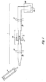
このシステムの実施形態は図1で表現される。システム1は、二つの連動部分11および13の間に設置されたフィルター12を有するろ過器10を含み、これを用いて無傷の真核細胞をフィルターで除去し、無傷の細菌細胞を通過させる。フィルターはチューブ部分20および21の末端に付着し、チューブ部分を設置するクリップ41で固定される。注射器51は、液体試料をシステム内に発射し、チューブ部分20、ろ過器10、チューブ部分21を強行させ測定容器22に送る。無傷の細菌細胞を溶解した後、測定容器のATPをルシフェリンとルシフェラーゼで反応させ光を発生させる。この光は光検出器30で検出される。光検出器は、光電子増倍管またはフォトダイオード等、光の検出に適した装置であればどれを用いても良い。
An embodiment of this system is represented in FIG. The
無傷の細菌細胞から無傷の真核細胞を分離する器具を保持する他の手段10は、器具を直接設置するクリップまたは容器の様な物を含むことができる。 Other means 10 for holding a device that separates intact eukaryotic cells from intact bacterial cells can include things such as clips or containers that directly place the device.
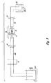
ろ過器10の代わりに、細菌に結合し真核細胞には結合しない担持表面を有す器具15をシステムに含むことができる。図2はこの様な器具15を表し、ビーズ16は細菌結合担持表面を有し、ポート17は図1のチューブ部分20と21の末端に接合するように構成される。液体試料が器具を通過する時に、無傷の細菌細胞は結合し、無傷の真核細胞は通過し、そして分離される。
Instead of the
幾つかの実施形態におけるシステムは、無傷の細菌細胞から無傷の真核細胞を分離する器具を、器具の保持手段の中に含む。 The system in some embodiments includes an instrument in the instrument holding means for separating intact eukaryotic cells from intact bacterial cells.
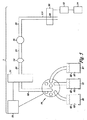
システムの実施形態の幾つかにおいて、システムは(d)無傷の細菌細胞から無傷の真核細胞を分離する器具と流体連通する液体試料貯蔵器を受ける保持手段、および(e)液体試料貯蔵器、無傷の細菌細胞から無傷の真核細胞を分離する器具(分離器具)、および測定容器に機能的に連結され、液体試料貯蔵器から分離器具に、および分離器具から測定容器に液体を送り込むポンプを含む。図3はこのようなシステムを表す。受容器61に保持された試料貯蔵器62と保持された液体試料63が、試料貯蔵器から分離器具10に液体を送り込むポンプ71と測定容器22と共に示されている。
In some of the system embodiments, the system comprises (d) a holding means for receiving a liquid sample reservoir in fluid communication with an instrument that separates intact eukaryotic cells from intact bacterial cells; and (e) a liquid sample reservoir; A device that separates intact eukaryotic cells from intact bacterial cells (separation device) and a pump that is operatively connected to the measurement vessel and that pumps liquid from the liquid sample reservoir to the separation device and from the separation device to the measurement vessel Including. FIG. 3 represents such a system. A
幾つかの実施形態では、測定容器がルシフェラーゼを含む。例えば、ルシフェラーゼを測定容器の壁、または測定容器のビーズに固定化させることができる。もしくはルシフェラーゼを測定容器に溶液として添加することもできる。 In some embodiments, the measurement container contains luciferase. For example, luciferase can be immobilized on the wall of the measurement container or the beads of the measurement container. Alternatively, luciferase can be added to the measurement container as a solution.
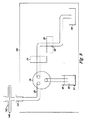
図4は、発明のシステムの実施形態の幾つかに含まれるさまざまな特徴を示す。システムは、細菌測定される液体試料63を含有し、保持手段61に保持される液体試料貯蔵器62を含む。また、図4のシステムは、洗浄液83を含有し、保持手段81に保持される洗浄液受容器82を含む。また、ルシフェラーゼ溶液93を含有し、保持手段91に保持されるルシフェラーゼ溶液受容器92も示されている。受容器に保持される溶液は、ポンプ71によってポンピングされた適切な溶液を通路20または110に出すマルチポート選択弁75に接続される。まず最初に、液体試料が通路20を通して真核細胞をろ過するろ過器10に送り込まれ、次に細菌を結合する担持表面を有す器具15へと送られる。液体試料中の細菌は担持表面に結合する。そして、洗浄液83が通路20および21を通して細菌結合器具15に送り込まれる。洗浄液が溶解剤を含有する場合は、洗浄液が器具15に結合した細菌を溶解し、通路23に送られる細菌溶解液にATPを放出する。
FIG. 4 illustrates various features included in some of the system embodiments of the invention. The system includes a
ルシフェラーゼ溶液93は、マルチポート選択弁とポンプで通路110に送り込むことができる。マルチポート選択弁75およびポンプ71は、プロセッサ72によって制御することができる。Yジャンクション111では、ルシフェラーゼ溶液と細菌溶解液を混合してATP測定液を生成し、図4に流水式室として示される測定容器22に輸送する。測定容器内で発光された光は光検出器30で検出される。
The
未加工または処理済データを光検出器から表示するために、表示部74を光検出器30に機能的に接続してもよい。ある実施形態のシステムは、検出器からのデータを処理する検出器に接続されたプロセッサ73を含む。
A display 74 may be operatively connected to the
図5に示すように、マルチポート選択器から出る通路が一つしかないシステムも可能である。ここで、幾つかの実施形態において、溶解剤を含有する洗浄液とルシフェラーゼ溶液が用いられる場合は、混合用積層区域および連続注入分析(SIA)(11〜15)による分析のように、通路を通してこれらの溶液をポンピングすることができる。121および122等の積層流動区域は、図6に示されるような口径の細いチューブで形成される。細菌ATPともう一つのルシフェラーゼを一つの区域が含有してもよい。この区域を隣接する未混合区域として測定容器22に輸送することができる。フリット等の障壁を越えた双方向または一方向の急速流で区域を混合することができる。任意的に、積層流動区域を気泡で分けることもできる。
As shown in FIG. 5, a system with only one exit from the multiport selector is also possible. Here, in some embodiments, when a cleaning solution and a luciferase solution containing a lysing agent are used, these may be passed through the passageway, such as by analysis with a mixing layered area and continuous injection analysis (SIA) (11-15). The solution can be pumped. Laminar flow zones such as 121 and 122 are formed by tubes having a narrow diameter as shown in FIG. One zone may contain bacterial ATP and another luciferase. This zone can be transported to the measuring
混合されると、ルシフェラーゼ触媒反応によって光が生成され、これが検出器によって検出される。SIAは測定容器内および検出器の前面でATPに接触するルシフェラーゼのためのメカニズムであり、ATPとルシフェラーゼの混合から検出器へ測定液を流す間に遅延の無いようにする。光を発生するルシフェラーゼ反応は急速に衰退するため、ATPとルシフェラーゼの混合から反応を検出する間に遅延の無いように、測定容器の中でルシフェラーゼとATPを混合することが効果的である。これによって、方法の感度が向上する。ATPとルシフェラーゼの初期接触を、測定容器の中または直前で作成する他の手段も可能である。ルシフェラーゼ溶液と細菌溶解液の流動を、図4に示すように測定容器の直前のYコネクション111で接続することによって、ルシフェラーゼ溶液をATP含有細菌溶解液で混合することができる。ルシフェラーゼは測定容器、例えば、測定容器の壁に固定することができ、これによって細菌溶解液のATPが測定容器中のルシフェラーゼに初期接触する。または、ルシフェラーゼ溶液とATP含有細菌溶解液を、図1に示すタイプの測定容器に別々に添加し、測定容器の中で混合してATP測定液を生成することもできる。
Once mixed, light is generated by a luciferase catalyzed reaction, which is detected by a detector. SIA is a mechanism for luciferase that contacts ATP in the measurement container and in front of the detector, so that there is no delay during the flow of the measurement liquid from the mixture of ATP and luciferase to the detector. Since the luciferase reaction that generates light rapidly declines, it is effective to mix luciferase and ATP in the measurement container so that there is no delay while detecting the reaction from the mixture of ATP and luciferase. This improves the sensitivity of the method. Other means of making the initial contact of ATP and luciferase in or just before the measurement vessel are possible. By connecting the flow of the luciferase solution and the bacterial lysate with a
しかし、ルシフェラーゼ反応の衰退時間は、溶解した細菌のATPにルシフェラーゼが接触して発光が検出されるまでの時間よりも5分または約10分以上も長いことが判明されている。しかし、遅延が少ないほど、細菌を検出する感度も高くなる。 However, it has been found that the decay time of the luciferase reaction is 5 minutes or more than about 10 minutes longer than the time until the luciferase comes into contact with the lysed bacterial ATP and the luminescence is detected. However, the smaller the delay, the higher the sensitivity for detecting bacteria.
細いチューブを使った連続注入分析は、細菌および細菌ATPの測定で消費される液体の量を最小化する利点を有す。消費される試料液の量とルシフェラーゼ等の他の試薬の量を共に最小化することができる。 Continuous injection analysis using thin tubes has the advantage of minimizing the amount of liquid consumed in measuring bacteria and bacterial ATP. Both the amount of sample solution consumed and the amount of other reagents such as luciferase can be minimized.
本発明のシステムの幾つかの実施形態は、無傷の細菌細胞から無傷の真核細胞を分離する器具と測定容器との間の流体連通において無傷の細菌細胞を濃縮する器具を含む。無傷の細菌細胞を濃縮する器具は、例えば、細菌細胞を結合する担持表面、または細菌細胞の通過を阻止するフィルターを含む。器具15が細菌結合の担持表面を有す、上記の実施形態を図4に示す。
Some embodiments of the system of the present invention include an instrument that concentrates intact bacterial cells in fluid communication between an instrument that separates intact eukaryotic cells from intact bacterial cells and a measurement container. Devices for concentrating intact bacterial cells include, for example, a support surface that binds bacterial cells, or a filter that prevents the passage of bacterial cells. The above embodiment is shown in FIG. 4 where the
本発明のシステムの幾つかの実施形態は、(d)無傷の細菌細胞から無傷の真核細胞を分離する器具との流体連通における液体試料貯蔵器を受ける保持手段、(e)液体試料貯蔵器、無傷の細菌細胞から無傷の真核細胞を分離する器具(分離器具)、および測定容器に機能的に連結され、液体試料貯蔵器から分離器具、および分離器具から測定容器に液体を送り込むポンプ、(f)洗浄液受容器を受ける保持手段、および(g)無傷の細菌細胞から無傷の真核細胞を分離する器具、測定容器、液体試料貯蔵器、および洗浄液受容器との流体連通におけるマルチポート選択弁であり、このマルチポート選択弁はある位置で液体試料貯蔵器から液体を送り、もう一つの位置で洗浄液受容器から液体を送るように構成されている。 Some embodiments of the system of the present invention comprise (d) a retaining means for receiving a liquid sample reservoir in fluid communication with an instrument that separates intact eukaryotic cells from intact bacterial cells; (e) a liquid sample reservoir A device that separates intact eukaryotic cells from intact bacterial cells (separation device), and a pump that is operatively connected to the measurement vessel and pumps liquid from the liquid sample reservoir and from the separation device to the measurement vessel; (F) a holding means for receiving a wash fluid receptor, and (g) multiport selection in fluid communication with an instrument, measurement vessel, liquid sample reservoir, and wash fluid receptor for separating intact eukaryotic cells from intact bacterial cells. A multiport select valve configured to deliver liquid from a liquid sample reservoir at one location and to deliver liquid from a wash fluid receptacle at another location.
幾つかの実施形態では、プロセッサがポンプとマルチポート選択弁に作動可能に連結され、所定の量の液体を液体試料貯蔵器から分離器具、分離器具から測定容器、そして洗浄液受容器から測定容器に送達するようにプログラムされている。 In some embodiments, a processor is operably coupled to the pump and the multiport select valve to deliver a predetermined amount of liquid from the liquid sample reservoir to the separation instrument, from the separation instrument to the measurement container, and from the wash liquid receiver to the measurement container. Is programmed to deliver.
本発明の一つの実施形態は、液体試料中の細菌を検出するシステムを提供し、そのシステムは(a)液体試料中の無傷の細菌細胞から無傷の真核細胞を分離する器具を受ける保持手段、(b)液体試料中の無傷の細菌細胞を結合する担持表面からの流体を受けるように構成された測定容器を形成する流体密封材料、および(c)測定容器に機能的に接続され、測定容器内で発光する光を検出する光検出器を含む。 One embodiment of the present invention provides a system for detecting bacteria in a liquid sample, the system comprising: (a) a holding means for receiving an instrument that separates intact eukaryotic cells from intact bacterial cells in the liquid sample. (B) a fluid-sealing material forming a measurement container configured to receive fluid from a support surface that binds intact bacterial cells in a liquid sample; and (c) functionally connected to the measurement container for measurement A light detector for detecting light emitted in the container is included.
特定の実施形態では、システムは保持手段(a)無傷の細菌細胞を結合する担持表面を含む。 In certain embodiments, the system includes a retaining means (a) a support surface that binds intact bacterial cells.
特定の実施形態では、担持表面はATPを結合しない。これには、細菌が溶解する前に存在するATP(つまり非細菌性の可能性があるATP)が、無傷の細菌から分離され、それにより細菌が溶解されるときに放出される細菌ATPから分離されるという利点がある。 In certain embodiments, the support surface does not bind ATP. This involves the separation of ATP present before the bacteria are lysed (ie, ATP that may be non-bacterial) from the intact bacteria and thereby released when the bacteria are lysed. There is an advantage of being.
別の特定の実施形態では、担持表面は、液体試料に存在する可能性がある無傷の真核細胞を結合しない。 In another specific embodiment, the support surface does not bind intact eukaryotic cells that may be present in the liquid sample.
別の実施形態では、担持表面は、無傷の真核細胞を結合する。これらの実施形態における真核ATPの汚染は、液体試料が担持表面に接触する前または後に真核細胞を選択的に溶解することによって、あるいは液体試料が担持表面に接触する前または後にろ過などにより真核細胞を除去することによって避けることができる。 In another embodiment, the support surface binds intact eukaryotic cells. Contamination of eukaryotic ATP in these embodiments may be caused by selective lysis of eukaryotic cells before or after the liquid sample contacts the support surface, or by filtration or the like before or after the liquid sample contacts the support surface. It can be avoided by removing eukaryotic cells.
本発明の別の実施形態は、細菌細胞を含有する疑いのある試料を受けるように構成された装置を備え、以下を含むステップを実行する。(a)細菌細胞を溶解して液体中に細菌ATPを放出し、細菌溶解液を生成するステップと、(b)細菌溶解液中の細菌ATPをATP消費酵素と接触させて、酵素が反応を触媒するATP測定液を生成するステップと、(c)ATP測定液中の酵素触媒反応をモニタリングするステップ。 Another embodiment of the invention comprises a device configured to receive a sample suspected of containing bacterial cells and performs steps comprising: (A) lysing bacterial cells to release bacterial ATP into the liquid to produce a bacterial lysate; (b) contacting the bacterial ATP in the bacterial lysate with an ATP consuming enzyme; Generating an ATP measurement liquid to be catalyzed; and (c) monitoring an enzyme-catalyzed reaction in the ATP measurement liquid.
特定の実施形態では、細菌細胞を含有する疑いのある試料は液体試料であり、また装置は、液体試料に存在する可能性がある無傷の細菌細胞から無傷の真核細胞を分離するさらなるステップを、細菌細胞を溶解するステップの前に実行する。 In certain embodiments, the sample suspected of containing bacterial cells is a liquid sample, and the device further steps to separate intact eukaryotic cells from intact bacterial cells that may be present in the liquid sample. Perform before the step of lysing bacterial cells.
特定の実施形態では、細菌溶解液およびATP測定液は、それぞれ1ml未満である。 In certain embodiments, the bacterial lysate and the ATP measurement solution are each less than 1 ml.
特定の実施形態では、装置はステップ(a)、(b)、および(c)を2分未満で実行する。 In certain embodiments, the apparatus performs steps (a), (b), and (c) in less than 2 minutes.
本発明の別の実施形態の例が図7に示される。図7は、液体試料に存在する可能性がある無傷の細菌細胞から真核細胞を分離し、細菌細胞を検査するための検査試料を生成する器具141を示す。器具141は、本例では注射器である液体容器51を含む。注射器51は、無傷の細菌細胞を阻止する細孔サイズが1ミクロン未満の第2のフィルター143に連結する、真核細胞を阻止して細菌細胞を通過させる第1のフィルター145を含む細菌分離部146と連結する。図7におけるフィルター145および143は、フィルター器具144および142に保持される。フィルター器具は、器具をかみ合わせる雌雄のルアーロック端を含む。注射器51は、ルアーロックを第1のフィルター器具144に固定させるルアーロック取付具も含む。このように注射器は、一つの実施形態では細孔サイズが5ミクロンである第1のフィルター145を通して液体試料をポンピングするために使用される。これは血小板を含む真核細胞を阻止するが、細菌を通過させる。第2のフィルター143は、細菌を阻止し、細菌をこの第2のフィルターの外面上で濃縮させる細孔サイズを有する。このように、器具は、液体試料に存在している可能性のある細菌細胞を含有する検査試料を生成するが、検査試料は実質上真核細胞を含まない。
An example of another embodiment of the present invention is shown in FIG. FIG. 7 shows an
無傷の細菌細胞から無傷の真核細胞を分離して検査試料を生成する器具の別の実施形態では、細菌分離成分は、無傷の細菌細胞を阻止する細孔サイズが1ミクロン未満の第2のフィルターに連結する、真核細胞を阻止して細菌細胞を通過させる第1のフィルターである。特定の実施形態では、第1のフィルターは、1〜10ミクロンの細孔サイズを有する。 In another embodiment of the instrument that separates intact eukaryotic cells from intact bacterial cells to produce a test sample, the bacterial separation component comprises a second micropore size of less than 1 micron that blocks intact bacterial cells. It is the 1st filter connected with a filter and blocking a eukaryotic cell and allowing bacterial cells to pass through. In certain embodiments, the first filter has a pore size of 1-10 microns.
別の実施形態では、細菌分離部は、真核細胞を阻止する第1のフィルターと細菌細胞を阻止する第2のフィルターの組み合わせであり、器具は細菌細胞を結合する担持表面を有するビーズを含み、ビーズは第1と第2のフィルターの間に保持される。別の実施形態で、細菌を結合するビーズが、器具の2つのフィルター間に保持される場合、第2のフィルターは、それ自体は細菌を阻止しないが、細菌を結合するビーズを維持できるように、大きい細孔サイズを有する。この実施形態の第2のフィルターは、第1のフィルターと同種のフィルターとすることができる。 In another embodiment, the bacterial separation is a combination of a first filter that blocks eukaryotic cells and a second filter that blocks bacterial cells, and the instrument includes beads having a support surface that binds bacterial cells. , The beads are retained between the first and second filters. In another embodiment, if a bead that binds bacteria is retained between two filters of the instrument, the second filter does not itself block bacteria but can maintain beads that bind bacteria. Have a large pore size. The second filter of this embodiment can be the same type of filter as the first filter.
器具の別の実施形態では、細菌分離部は、細菌細胞を結合して真核細胞を結合しない担持表面である。例えば細菌分離部は、細菌細胞を結合して真核細胞を結合しない面を有するビーズとすることができる。ビーズは2つのフィルター間に保持することができる。少なくとも第1のフィルターは、細菌を通すのに十分な大きさの細孔サイズを有さなければならない。フィルターは、真核細胞も通すのに十分な大きさであるが、ビーズを阻止するのに十分な小ささの細孔サイズを有するフィルターであってもよい。例えばフィルターは、ビーズの通過を阻止するのに十分な小ささの細孔を備える金網から成ることができる。 In another embodiment of the instrument, the bacterial separation is a supported surface that binds bacterial cells and does not bind eukaryotic cells. For example, the bacterial separation part can be a bead having a surface that binds bacterial cells and does not bind eukaryotic cells. The beads can be held between two filters. At least the first filter must have a pore size large enough to allow bacteria to pass through. The filter may be a filter that is large enough to allow eukaryotic cells to pass through, but has a pore size small enough to block beads. For example, the filter can consist of a wire mesh with pores that are small enough to prevent the passage of beads.
本発明の別の実施形態は、図8に図解される装置を備える。この例の装置161は、細菌細胞を含有する疑いのある試料を保持する容器142を受けるよう構成されるポート131を含む。図8に示される例における容器142は、細孔サイズが1ミクロン未満であり、無傷の細菌細胞にとって通過が不可能である液体通過可能フィルター143を備えるフィルター器具である。フィルター器具は、図8の器具161の上部に示されるように液体試料をろ過し、実質上真核細胞を含まない検査試料を生成するために使用することができる。
Another embodiment of the present invention comprises the apparatus illustrated in FIG. The
装置161は、ポートと流体連通し、また測定容器22と流体連通する通路20も有する。光検出器30は、測定容器に機能的に連結され、測定容器内に放射される光を検出する。
The
装置は、通路および測定容器に機能的に接続され、通路を通して測定容器に液体を送り込むよう構成されるポンプ71も有する。
The apparatus also has a
装置161は、容器142がポート131上で受けたときに、通路からの溶解液93を、ポートおよび容器の液体通過可能フィルターを通してポンピングし、容器中の細菌を溶解することによって、容器142中に細菌ATPを含有する細菌溶解物を生成するよう構成される。図8では、溶解液93は、ルシフェラーゼおよびルシフェリンも含有する。この溶液中の溶解剤は洗浄剤とすることができ、そのためpHは中性に近く、ルシフェラーゼおよびルシフェリンと混合可能である。別々の溶解液およびルシフェラーゼ/ルシフェリン溶液も使用できる。図8では、溶解液93はマルチポート選択弁75を通して、通路20およびポート131に、そしてフィルター143上方に送り込まれ、フィルターの外側に存在する細菌を溶解し、容器142中に細菌溶解液を生成する。この例でのポンプは双方向ポンプなので、細菌溶解させるのに適切な時間のあと方向を逆転させ、この例ではルシフェラーゼおよびルシフェリンを含有することからATP測定液でもある細菌溶解液を通路20およびマルチポート選択弁75に逆の方向に通し、光検出器30の測定容器22にポンピングする。光検出器は、測定容器22中のATP測定液からの光の放射をモニタリングする。上記のように、装置161は、容器の液体通過可能フィルターを通して、細菌溶解物を容器から通路内へと送り込み、細菌溶解物中の細菌ATPをルシフェラーゼおよびルシフェリンに接触させてATP測定液を形成し、測定容器内のATP測定液からの光の放射をモニタリングする。
The
いくつかの実施形態では、装置は廃液容器151も含み、液体は器具から廃液容器151にポンピングされる。
In some embodiments, the apparatus also includes a
このように、特定の実施形態では、(a)細菌細胞を含有する疑いのある試料を保持する容器を受けるように構成されたポートと、(b)ポートと流体連通し、(c)と流体連通する通路と、(c)(d)に機能的に接続される測定容器と、(d)測定容器内で放射された光を検出する光検出器と、(e)通路および測定容器に機能的に接続し、通路を通して測定容器に液体を送り込むように構成されたポンプとを備える装置は、ポート上で受ける容器をさらに備える。 Thus, in certain embodiments, (a) a port configured to receive a container holding a sample suspected of containing bacterial cells; (b) in fluid communication with the port; (c) and fluid A communicating path; (c) a measurement container operatively connected to (d); (d) a photodetector for detecting light emitted within the measurement container; and (e) a function for the passage and the measurement container. And a pump configured to connect to the pump and configured to pump liquid through the passage to the measurement container further comprises a container for receiving on the port.
別の実施形態では、装置は、(f)通路および溶解容器と流体連通するマルチポート選択弁と、(g)マルチポート選択弁と流体連通する溶解液容器を受けるための保持手段と、(h)マルチポート選択弁と流体連通する廃液用容器を受けるための保持手段とをさらに備える。 In another embodiment, the apparatus comprises (f) a multiport select valve in fluid communication with the passage and the lysis vessel; (g) a holding means for receiving a lysate vessel in fluid communication with the multiport select valve; And) holding means for receiving a waste container in fluid communication with the multiport selector valve.
液体試料中に存在する可能性のある無傷の細菌細胞から真核細胞を分離し、細菌細胞を検査するための検査試料を生成する器具は、公開された、以下のステップを実行するシステムおよび装置に含まれてもよい。細菌細胞を溶解して細菌ATPを液体中に放出し、細菌溶解液を生成するステップと、細菌溶解液中の細菌ATPをATP消費酵素と接触させ、酵素が発光反応を触媒するATP測定液を生成するステップと、ATP測定液中の酵素が触媒する反応をモニタリングするステップ。この方法では、完全に自動化されたシステムおよび装置は、無傷の細菌細胞から無傷の真核細胞を分離し、細菌細胞を溶解させて細菌ATPを放出させ、細菌ATPをATP消費酵素に接触させて酵素が反応を触媒するATP測定液体を生成し、ATP測定液中の酵素が触媒する反応をモニタリングする。利用者は細菌検査のための液体試料を提供するだけで、システムまたは装置が液体試料を処理し、試料中の細菌の有無を示す結果を提供し、また細菌が存在する場合、任意で試料中の細菌数を提供する。 An instrument for separating eukaryotic cells from intact bacterial cells that may be present in a liquid sample and generating a test sample for testing the bacterial cell is disclosed in the following system and apparatus for performing the following steps May be included. Lysing bacterial cells and releasing bacterial ATP into the liquid to produce a bacterial lysate; contacting bacterial ATP in the bacterial lysate with an ATP-consuming enzyme; and an ATP assay solution in which the enzyme catalyzes a luminescent reaction And a step of monitoring a reaction catalyzed by an enzyme in the ATP measurement solution. In this method, a fully automated system and apparatus separates intact eukaryotic cells from intact bacterial cells, lyses the bacterial cells to release bacterial ATP, and contacts the bacterial ATP with an ATP consuming enzyme. An ATP measurement liquid in which the enzyme catalyzes the reaction is generated, and a reaction catalyzed by the enzyme in the ATP measurement liquid is monitored. The user simply provides a liquid sample for bacterial testing, and the system or device processes the liquid sample and provides results indicating the presence or absence of bacteria in the sample, and optionally, if bacteria are present, Provides a bacterial count of
発明の他の実施形態は、細菌を含有する疑いのあるサンプル中での細菌の有無を決定する装置を提供する。装置に含まれるものは、(a)(b)に接続される、細菌細胞を含有する疑いのあるサンプルを受ける受容手段、(b)(c)に接続される、細菌溶解液を生成する液体に細菌ATPを放出する細菌細胞を溶解する手段、(c)(d)に接続される、酵素が発光反応を触媒するATP測定液を生成するATP消費発光酵素に細菌溶解液中の細菌ATPを接触させる手段、(d)ATP測定液中の酵素によって生産される光を検出する光検出手段である。 Other embodiments of the invention provide an apparatus for determining the presence or absence of bacteria in a sample suspected of containing bacteria. Included in the apparatus is (a) a receiving means connected to (b) for receiving a sample suspected of containing bacterial cells, (b) a liquid for generating a bacterial lysate connected to (c) A means for lysing bacterial cells that release bacterial ATP; (c) the bacterial ATP in the bacterial lysate is connected to (d), and the ATP-consuming luminescent enzyme that produces an ATP measurement solution in which the enzyme catalyzes the luminescent reaction (D) a light detection means for detecting light produced by the enzyme in the ATP measurement solution.
細菌細胞を溶解する手段は、例えば、超音波処理器具またはビーズミルが可能である。しかし、最も好ましくは、本明細書で記述される細菌の疑いのあるサンプルへ溶解液を送達するポンプ、通路および液体貯蔵器のシステム、あるいはその同等物である。 The means for lysing the bacterial cells can be, for example, a sonication instrument or a bead mill. Most preferably, however, is a pump, passageway and liquid reservoir system, or the like, that delivers lysate to a suspected bacterial sample as described herein.
ATP消費発光酵素に細菌溶解液中の細菌ATPを接触させる手段は、細菌細胞を溶解する手段と同じ手段、または重複する手段でよい。好ましくは、ATP消費発光酵素を含有する液体を送達するポンプ、通路および液体貯蔵器のシステムである。溶解剤、ルシフェラーゼおよびルシフェリンを含有する単独の溶液が、無傷の細菌細胞に接触して、細胞を溶解し、同時に細菌ATPをルシフェラーゼに接触させる場合、両手段は同じであり、細菌溶解液およびATP測定液は同じである。 The means for bringing the bacterial ATP in the bacterial lysate into contact with the ATP-consuming luminescent enzyme may be the same means as the means for lysing the bacterial cells or an overlapping means. Preferably, a system of pumps, passages and liquid reservoirs for delivering liquids containing ATP consuming luminescent enzymes. If a single solution containing the lysing agent, luciferase and luciferin is contacted with intact bacterial cells to lyse the cells and at the same time contact bacterial ATP with luciferase, both means are the same, the bacterial lysate and ATP The measuring solution is the same.
本装置のいくつかの実施形態において、装置は、さらに、実質上真核細胞を含まない検査サンプルを生成するために、液体試料中に存在する無傷の真核細胞を液体試料中に存在する無傷の細菌細胞から分離する細胞分離手段から成り、その細胞分離手段は受容手段に接続される。 In some embodiments of the apparatus, the apparatus further includes intact eukaryotic cells present in the liquid sample to produce a test sample that is substantially free of eukaryotic cells. The cell separation means for separating from the bacterial cells is connected to the receiving means.
発明は、以下の限定されない実施例によって説明される。 The invention is illustrated by the following non-limiting examples.
実施例1
細菌結合ビーズを使用した細菌汚染に関する濃厚血小板の測定
Example 1
Determination of concentrated platelets for bacterial contamination using bacterial binding beads
使用した測定方法は、以下の通り。 The measurement method used is as follows.
1. 1.0mlの濃厚血小板サンプルは、GenPoint BUG TRAP C−versionのビーズ(GenPoint、オスロ、ノルウェー)の40マイクロリットルの充填ビーズカラムで、60秒間、双方向でポンピングした。 1. A 1.0 ml concentrated platelet sample was pumped bi-directionally for 60 seconds with a 40 microliter packed bead column of GenPoint BUG TRAP C-version beads (GenPoint, Oslo, Norway).
2. カラムは、ハンクス液(0.185g/lのCaCl2、0.2g/lのMgSO4、0.4g/lのKCl、0.06g/lのKH2PO4、8g/lのNaCl、0.048g/lのNa2HPO4、1.5g/lの無水デキストロース、15.7g/lの一水和デキストロ−ス、4.77g/lのHEPES、1.365g/lの NaH2PO4)である洗浄バッファー、250マイクロリットルで洗い流した。 2. The column is a Hanks solution (0.185 g / l CaCl 2 , 0.2 g / l MgSO 4 , 0.4 g / l KCl, 0.06 g / l KH 2 PO 4 , 8 g / l NaCl, 0 0.048 g / l Na 2 HPO 4 , 1.5 g / l anhydrous dextrose, 15.7 g / l monohydrate dextrose, 4.77 g / l HEPES, 1.365 g / l NaH 2 PO 4 The washing buffer was washed with 250 microliters.
3. 摂氏60度に加熱した水中の40マイクロリットルの0.1%トリクロロ酢酸は、双方向で10秒間カラム上を通過させ、細菌溶解液を作成した。 3. 40 microliters of 0.1% trichloroacetic acid in water heated to 60 degrees Celsius was passed over the column in both directions for 10 seconds to create a bacterial lysate.
4. ルシフェリン・ルシフェラーゼ溶液(10マイクロリットル、pH7.5の50mMのリン酸ナトリウム中に0.2μgのルシフェラーゼ、2μgのルシフェリンを含有)は、系列インジェクション分析および液体ゾーン(Global FIA、フォックス島、ワシントン州)の混合によって細菌溶解液と混合し、一斉に飛び込んでくる光を検出する発光検出器(光子計数器、Electon Tubes、英国)の前を通過したATP測定液を形成した。 4). Luciferin luciferase solution (10 μl, containing 0.2 μg luciferase, 2 μg luciferin in 50 mM sodium phosphate, pH 7.5) for serial injection analysis and liquid zone (Global FIA, Fox Island, WA) The mixture was mixed with the bacterial lysate to form an ATP measurement solution that passed in front of a luminescence detector (photon counter, Electon Tubes, UK) that detects the light entering all at once.
1つの濃厚血小板バッグは4つのサンプルに分け、それぞれPL732バッグに入れた。各バッグには、10CFU/mlの大腸菌、緑膿菌、黄色ブドウ球菌、またはセラチアマルセッセンスを植菌した。4つの対照バッグには細菌を植菌しなかった。室温で36時間後、各バッグから5mlを自動培養システム細菌ロード検出用のBACT/ALERT培養瓶へ移動した。BACT/ALERTを使用して、各サンプルの5mlは、標準好気性、標準嫌気性、活性炭好気性および嫌気性瓶に植菌した。 One concentrated platelet bag was divided into four samples, each placed in a PL732 bag. Each bag was inoculated with 10 CFU / ml of E. coli, Pseudomonas aeruginosa, Staphylococcus aureus, or Serratia marcescens. Four control bags were not inoculated with bacteria. After 36 hours at room temperature, 5 ml from each bag was transferred to a BACT / ALERT culture bottle for automatic culture system bacterial load detection. Using BACT / ALERT, 5 ml of each sample was inoculated into standard aerobic, standard anaerobic, activated carbon aerobic and anaerobic bottles.
各バッグの1.0mlのサンプルは、上記のように細菌ATPの試験も行った。 A 1.0 ml sample of each bag was also tested for bacterial ATP as described above.
結果を表1に示す。 The results are shown in Table 1.
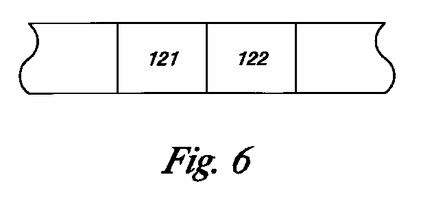
実施例2
真核細胞を細菌から分離し細菌を濃縮する器具
Example 2
An instrument that separates eukaryotic cells from bacteria and concentrates them
5mlの注射器は、直径25mm、細孔サイズ5ミクロンのACRODISC SUPOR薄膜フィルター、直径15mm、細孔サイズ0.2ミクロンのACRODISC SUPOR薄膜フィルターへと順番に接続した。注射器およびフィルターは、ルアーロックコネクタを使用して、注射器〜5ミクロンフィルター〜0.2ミクロンフィルターの順番で接続した。濃厚血小板は、器具との処理に関して検査した。濃厚血小板(3ml)は、注射器に装着し、その後、2つのフィルターを通過させた。1番目のフィルターは血小板および他の真核細胞を除去し、2番目のフィルターは細菌を停止させ、濃縮すると予想される。細孔サイズ5ミクロンのフィルターである1番目のフィルターからのろ液は、血小板および他の真核細胞に関して、粒度測定および顕微鏡検査を行った。1番目のフィルターを通過した血小板や他の真核細胞は見られなかった。これは、1番目のフィルターの微細孔サイズは5ミクロンであり、血小板の直径は約3ミクロンであるため、少々意外であった。 The 5 ml syringe was in turn connected to an ACRODISC SUPOR membrane filter with a diameter of 25 mm and a pore size of 5 microns, an ACRODISC SUPOR membrane filter with a diameter of 15 mm and a pore size of 0.2 microns. The syringe and filter were connected in the order of syringe to 5 micron filter to 0.2 micron filter using a luer lock connector. Thick platelets were examined for treatment with the instrument. Concentrated platelets (3 ml) were attached to a syringe and then passed through two filters. The first filter is expected to remove platelets and other eukaryotic cells, and the second filter is expected to stop and concentrate bacteria. The filtrate from the first filter, a 5 micron pore size filter, was subjected to particle size measurements and microscopy on platelets and other eukaryotic cells. No platelets or other eukaryotic cells passed through the first filter. This was a bit surprising because the first filter had a micropore size of 5 microns and a platelet diameter of about 3 microns.
実施例3
コンピュータ制御細菌検出器具
Example 3
Computer controlled bacteria detection instrument
図8に示される器具が、基本的に作製された。C25Zモデルのマルチポート選択弁(Valco Instruments Co. Inc.、ヒューストン、テキサス州、www.vici.com)は、細穴チューブによって、洗浄液受容器(70%イソプロピルアルコール)およびリーシス/ルシフェラーゼ/ルシフェリン溶液貯蔵器(BACTITER−GLO試薬、Promega、マディソン、ウィスコンシン州、www.promega.com)に接続した。マルチポート弁も、MILIGATポンプ(Global FIA Inc.、サグ・ハーバー、ワシントン州、www.globalfia.com)に接続した。光検出器は、Electon Tubes,Inc.(ロッカウェー、ニュージャージー州、www.electrontubes.com)の光電子増倍管P25232モデルを使用した。弁、ポンプ、および光電子増倍管は、WINDOWS(登録商標) XPソフトウェア搭載のTPC−60SタッチパネルPCによって制御された。光電子増倍管の結果も、PCディスプレイに表示された。 The instrument shown in FIG. 8 was basically made. The C25Z model multi-port selection valve (Valco Instruments Co. Inc., Houston, TX, www.vici.com) is connected to a wash receptacle (70% isopropyl alcohol) and a lysis / luciferase / luciferin solution storage by means of a fine-hole tube. A vessel (BACTITER-GLO reagent, Promega, Madison, WI, www.promega.com). A multiport valve was also connected to the MILIGAT pump (Global FIA Inc., Sag Harbor, Washington, www.globalfia.com). The photodetector is available from Electon Tubes, Inc. A photomultiplier tube P25232 model (Rockaway, NJ, www.electrontubes.com) was used. The valves, pumps, and photomultiplier tubes were controlled by a TPC-60S touch panel PC with WINDOWS XP software. The photomultiplier results were also displayed on the PC display.
器具は、図8に示されるようにポート131も含み、それには、外面に細菌を含有する0.2ミクロンのフィルターを取り付けることができた。 The instrument also included a port 131 as shown in FIG. 8, which could be fitted with a 0.2 micron filter containing bacteria on the outer surface.
1つの実施例において、器具は、150μlのリーシス/ルシフェリン/ルシフェラーゼ溶液がサンプルポートを通過して、細菌を溶解するフィルターの上へポンピングするようプログラムされた。溶液は、30秒間フィルターの上に残した。その後、ポンプの送り込み方向を逆にして、フィルターから光電子増倍管の測定容器へ150μlの液体を送り込んだ。測定は、20秒間行われた。その後、溶液は廃棄物容器へと送り込まれた。そしてプログラムは、ラインを洗浄するため、2.5mlのイソプロピルアルコール溶液がラインを通してポンピングされるよう指示した。 In one example, the instrument was programmed to pump 150 μl of lysis / luciferin / luciferase solution through the sample port and onto a filter that lyses bacteria. The solution was left on the filter for 30 seconds. Thereafter, the pump feeding direction was reversed, and 150 μl of liquid was fed from the filter into the photomultiplier tube measurement container. The measurement was performed for 20 seconds. The solution was then sent to a waste container. The program then instructed 2.5 ml of isopropyl alcohol solution to be pumped through the line to clean the line.
実施例4
実施例3の器具を使用した細菌汚染の測定
Example 4
Measurement of bacterial contamination using the instrument of Example 3
血漿のサンプルは、特定の細菌の種類でスパイクした。その後、3mlの血漿は、60μlのGenPoint BUG TRAP C−versionのビーズ(GenPoint、オスロ、ノルウェー)で5分間培養した。その後、サンプルを細孔サイズ5ミクロンのACRODISC SUPOR薄膜でろ過し、ビーズを捕らえ、濃縮した。ビーズが付着したフィルターは、実施例3で記述した器具のポート上に装着した。実施例3で記述したプログラムを、ビーズ上の細菌を溶解し、溶液から発光を定量化するよう実行した。結果を下記の表2に示す。 Plasma samples were spiked with specific bacterial types. Subsequently, 3 ml of plasma was incubated with 60 μl of GenPoint BUG TRAP C-version beads (GenPoint, Oslo, Norway) for 5 minutes. The sample was then filtered through an ACRODISC SUPOR membrane with a pore size of 5 microns to capture and concentrate the beads. The filter with beads attached was mounted on the port of the instrument described in Example 3. The program described in Example 3 was run to lyse the bacteria on the beads and quantify luminescence from the solution. The results are shown in Table 2 below.
2つの連鎖球菌種のみが背景と同様な発光を生じさせたため、本発光検出法では検出されなかった。発明者は、これは細胞が十分に溶解されなかったためであり、本プロトコルで使用した30分よりも長い時間、同溶解試薬で培養することによって連鎖球菌が検出されるであろうと推測する。 Only two streptococcal species caused luminescence similar to the background and therefore were not detected by this luminescence detection method. The inventor speculates that this was because the cells were not sufficiently lysed and streptococci would be detected by culturing with the same lysis reagent for longer than the 30 minutes used in this protocol.
次に、濃厚血小板のバッグは、10cfu/mlの大腸菌でスパイクし、摂氏37度で培養した。サンプルは1時間ごとに採取し、本実施例の上記の方法によって細菌を測定して相対発光量を得るか、またはcfu/mlを決定するために定量的にめっきした。結果を図9に示す。本器具で決定した相対発光量の結果は、めっきによるコロニー形成単位の判定に近似した。 The thick platelet bag was then spiked with 10 cfu / ml E. coli and cultured at 37 degrees Celsius. Samples were taken every hour and bacteria were measured by the above method of this example to obtain relative luminescence or plated quantitatively to determine cfu / ml. The results are shown in FIG. The result of the relative light emission amount determined by this instrument approximated the determination of the colony forming unit by plating.
これらの結果、グラム陽性、グラム陰性および嫌気性細菌を含む様々な細菌は、1000cfu/ml未満の感度、および通常は100cfu/ml未満で本方法によって検出可能であることが示された。 These results indicated that various bacteria including gram positive, gram negative and anaerobic bacteria can be detected by this method with a sensitivity of less than 1000 cfu / ml and usually less than 100 cfu / ml.
References:
1. Epstein JS, Williams AE, Biswas R, Vostal J. FDA Update at the AABB Annual Meeting 10/15/2001 2001.
2. Morrow JF, Braine HG, Kickler TS, Ness PM, Dick JD, Fuller AK. Septic reactions to platelet transfusions. A persistent problem. JAMA. 1991;266(4):555−558.
3. Barrett BB, Andersen JW, Anderson KC. Strategies for the avoidance of bacterial contamination of blood components. Transfusion. Mar 1993;33(3):228−233.
4. Dumont L. Bacterial Contamination of Platelet Components. Lakewood: COBE BCT Inc.; 1996.
5. Mitchell K−M T, Brecher ME. 1999. Transfusion Medicine Reviews 13:132−144.
6. Chaney R., et al. 1999. Transfusion Medicine 9:177−188.
7. Hanna BA. 1986. Methods in Enzymology 133:22−27.
8. Nilsson LE et al. 1989. J. Bioluminescence and Chemiluminescence 3:101−104.
9. Stanley PE 1989. J. Bioluminescence and Chemiluminescence 4:375−380.
10. Lundin A, Thore A. 1975. Applied Microbiology 30:713−721.
11. Ruzicka J, Hansen EH. 1981. Flow Injection Analysis. J. Wiley and Sons.
12. Karlberg B, Pacey GE. 1989. Flow Injection Analysis: A Practical Guide. Elsevier.
13. Valcarcel M and Luque De Castro MD, Flow−Injection Analysis. Principles and Applications. John Wiley & Sons, New York.
14. U.S. Patent No. 6,613,579.
15. U.S. Patent No. 6,716,391.
16. Lemasters JJ, Hackenbrock CR. 1979. Meth. Enzymol. 56:530−544.
17. Higashi T, et al. 1985. Thrombosis and Haemostasis 53:65−69.
References:
1. Epstein JS, Williams AE, Biswas R, Vostal J. et al. FDA Update at the
2. Morrow JF, Brain HG, Kickler TS, Ness PM, Dick JD, Fuller AK. Separative reactions to platelet transfusions. A persistent problem. JAMA. 1991; 266 (4): 555-558.
3. Barrett BB, Andersen JW, Anderson KC. Strategies for the aviation of bacterial contention of blood components. Transfusion. Mar 1993; 33 (3): 228-233.
4). Dumont L. Bacterial Contention of Platelet Components. Lakewood: COBE BCT Inc. 1996.
5. Mitchell K-MT, Brecher ME. 1999. Transfusion Medicine Reviews 13: 132-144.
6). Chaney R.D. , Et al. 1999. Transfusion Medicine 9: 177-188.
7). Hanna BA. 1986. Methods in Enzymology 133: 22-27.
8). Nilsson LE et al. 1989. J. et al. Bioluminescence and Chemiluminescence 3: 101-104.
9. Stanley PE 1989. J. et al. Bioluminescence and Chemiluminescence 4: 375-380.
10. Lundin A, Thor A. 1975. Applied Microbiology 30: 713-721.
11. Ruzikka J, Hansen EH. 1981. Flow Injection Analysis. J. et al. Wiley and Sons.
12 Karlberg B, Pathey GE. 1989. Flow Injection Analysis: A Practical Guide. Elsevier.
13. Valcarcel M and Luque De Castro MD, Flow-Injection Analysis. Principles and Applications. John Wiley & Sons, New York.
14 U. S. Patent No. 6,613,579.
15. U. S. Patent No. 6,716,391.
16. Lemasters JJ, Hackenblock CR. 1979. Meth. Enzymol. 56: 530-544.
17. Higashi T, et al. 1985. Thrombosis and Haemostasis 53: 65-69.
特許、特許文献、およびその他の引用文献はすべて、参照することにより本明細書に組み込まれる。 All patents, patent documents, and other cited references are incorporated herein by reference.
Claims (82)
(a)前記液体試料中に存在する可能性のある無傷の細菌細胞から無傷の真核細胞を分離するステップと、
(b)前記細菌細胞を溶解して液体中に細菌ATPを放出し、細菌溶解液を生成するステップと、
(c)前記細菌溶解液中の前記細菌ATPをATP消費酵素と接触させて、前記酵素が反応を触媒するATP測定液を生成するステップと、
(d)前記ATP測定液中の酵素触媒反応をモニタリングするステップと、を含む方法。 A method for detecting bacteria in a liquid sample suspected of containing bacteria comprising:
(A) separating intact eukaryotic cells from intact bacterial cells that may be present in the liquid sample;
(B) dissolving the bacterial cells to release bacterial ATP into the liquid to produce a bacterial lysate;
(C) contacting the bacterial ATP in the bacterial lysate with an ATP consuming enzyme to produce an ATP measurement solution in which the enzyme catalyzes a reaction;
(D) monitoring the enzyme-catalyzed reaction in the ATP measurement solution.
前記ろ過した液体試料を、前記細菌を結合する担持表面と接触させるステップをさらに含む、請求項3に記載の方法。 After the step of filtering the eukaryotic cells,
4. The method of claim 3, further comprising contacting the filtered liquid sample with a support surface that binds the bacteria.
前記液体試料を、前記細菌細胞を結合して前記真核細胞を結合しない担持表面と接触させるステップを含む、請求項1に記載の方法。 The step of separating intact eukaryotic cells from bacterial cells is
2. The method of claim 1, comprising contacting the liquid sample with a support surface that binds the bacterial cells and does not bind the eukaryotic cells.
前記真核細胞を阻止して前記細菌細胞が通過できるようにするフィルターを使用して、前記液体試料から前記真核細胞をろ過するステップをさらに含む、請求項13に記載の方法。 Separating intact eukaryotic cells from intact bacterial cells that may be present in the liquid sample comprises contacting the liquid sample with a support surface that binds the bacteria.
14. The method of claim 13, further comprising filtering the eukaryotic cell from the liquid sample using a filter that blocks the eukaryotic cell and allows the bacterial cell to pass through.
前記液体試料を、前記細菌細胞を結合する担持表面と接触させて、前記細菌細胞を濃縮する、および/または前記液体試料中の他の成分から前記細菌細胞を分離するステップと、
前記細菌細胞を溶解して液体中に細菌ATPを放出し、細菌溶解液を生成するステップと、
前記細菌溶解液中の前記細菌ATPをATP消費酵素と接触させて、前記酵素が反応を触媒するATP測定液を生成するステップと、
前記ATP測定液中の酵素触媒反応をモニタリングするステップと、を含む方法。 A method for detecting bacteria in a liquid sample suspected of containing bacteria comprising:
Contacting the liquid sample with a support surface that binds the bacterial cells to concentrate the bacterial cells and / or separate the bacterial cells from other components in the liquid sample;
Lysing the bacterial cells to release bacterial ATP into the liquid to produce a bacterial lysate;
Contacting the bacterial ATP in the bacterial lysate with an ATP consuming enzyme to produce an ATP measurement solution in which the enzyme catalyzes the reaction;
Monitoring the enzyme-catalyzed reaction in the ATP measurement solution.
前記液体試料中に存在する可能性のある細菌細胞を実質上溶解せずに、前記液体試料中に存在する可能性のある真核細胞を選択的に溶解するステップをさらに含む、請求項44に記載の方法。 Prior to contacting the liquid sample with a support surface that binds bacterial cells,
45. The method of claim 44, further comprising selectively lysing eukaryotic cells that may be present in the liquid sample without substantially lysing bacterial cells that may be present in the liquid sample. The method described.
前記真核細胞を阻止して前記細菌細胞が通過できるようにするフィルターを使用して、前記液体試料から、前記液体試料中に存在する可能性のある無傷の真核細胞をろ過するステップをさらに含む、請求項44に記載の方法。 Prior to contacting the liquid sample with a support surface that binds bacterial cells,
Filtering the intact eukaryotic cells that may be present in the liquid sample from the liquid sample using a filter that blocks the eukaryotic cells and allows the bacterial cells to pass through. 45. The method of claim 44, comprising.
(a)液体試料中の無傷の細菌細胞から無傷の真核細胞を分離するための器具を受けるための保持手段と、
(b)液体試料中の無傷の細菌細胞から無傷の真核細胞を分離するための前記器具からの流体を受けるように構成された測定容器を形成する流体密封材料と、
(c)前記測定容器に機能的に接続して前記測定容器内に放射される光を検出する光検出器と、を備えるシステム。 A system for detecting bacteria in a liquid sample,
(A) holding means for receiving an instrument for separating intact eukaryotic cells from intact bacterial cells in a liquid sample;
(B) a fluid sealing material forming a measurement container configured to receive fluid from said instrument for separating intact eukaryotic cells from intact bacterial cells in a liquid sample;
(C) a system comprising a photodetector that is operatively connected to the measurement vessel and detects light emitted into the measurement vessel.
(e)前記液体試料貯蔵器と、無傷の細菌細胞から無傷の真核細胞を分離するための前記器具(分離器具)と、前記測定容器とに機能的に連結して、液体を前記液体試料貯蔵器から前記分離器具に、および前記分離器具から前記測定容器に送り込むポンプとをさらに備える、請求項48に記載のシステム。 (D) holding means for receiving a liquid sample reservoir in fluid communication with the instrument for separating intact eukaryotic cells from intact bacterial cells;
(E) functionally connected to the liquid sample reservoir, the instrument (separation instrument) for separating intact eukaryotic cells from intact bacterial cells, and the measurement container, and the liquid is the liquid sample 49. The system of claim 48, further comprising a pump that pumps from a reservoir to the separation device and from the separation device to the measurement container.
前記無傷の細菌細胞から無傷の真核細胞を分離するための器具と、前記測定容器と、前記液体試料貯蔵器と、前記洗浄液受容器と流体連通するマルチポート選択弁であって、ある位置にある前記液体試料貯蔵器から、および別の位置にある前記洗浄液受容器から、液体を透過させるために構成される前記マルチポート選択弁とをさらに備える、請求項53に記載のシステム。 A holding means for receiving the cleaning fluid receptacle;
A multi-port selection valve in fluid communication with an instrument for separating intact eukaryotic cells from the intact bacterial cells, the measurement container, the liquid sample reservoir, and the wash fluid receiver; 54. The system of claim 53, further comprising the multiport select valve configured to permeate liquid from one liquid sample reservoir and from the wash liquid receiver at another location.
(a)液体試料中の無傷の細菌細胞を結合する担持表面を受けるための保持手段と、
(b)液体試料中の無傷の細菌細胞を結合する前記担持表面からの流体を受けるように構成された測定容器を形成する流体密封材料と、
(c)前記測定容器に機能的に接続して前記測定容器内に放射される光を検出する光検出器と、を備えるシステム。 A system for detecting bacteria in a liquid sample,
(A) holding means for receiving a carrying surface that binds intact bacterial cells in the liquid sample;
(B) a fluid-tight material forming a measurement container configured to receive fluid from the support surface that binds intact bacterial cells in a liquid sample;
(C) a system comprising a photodetector that is operatively connected to the measurement vessel and detects light emitted into the measurement vessel.
前記液体試料中に存在する可能性のある無傷の細菌細胞から無傷の真核細胞を分離し、その後の細菌検出試料中の細菌の検出のために、実質上真核細胞を含まない、前記細菌検出試料を生成するステップを含み、
前記細菌検出試料中の細菌の検出は、前記細菌を溶解するステップと、前記溶解細菌から放出されるATPをモニタリングするステップとを含むプロセス。 A process for preparing a liquid sample suspected of containing bacteria for detecting bacteria comprising:
Separating said intact eukaryotic cells from intact bacterial cells that may be present in said liquid sample, wherein said bacteria are substantially free of eukaryotic cells for subsequent detection of bacteria in said bacterial detection sample; Generating a detection sample,
Detection of bacteria in the bacterial detection sample is a process comprising lysing the bacteria and monitoring ATP released from the lysed bacteria.
(a)前記細菌細胞を溶解して液体中に細菌ATPを放出し、細菌溶解液を生成するステップと、
(b)前記細菌溶解液中の前記細菌ATPをATP消費酵素と接触させて、前記酵素が反応を触媒するATP測定液を生成するステップと、
(c)前記ATP測定液中の酵素触媒反応をモニタリングするステップと、を含むステップを実行するように構成された装置。 A device configured to receive a sample suspected of containing bacterial cells,
(A) dissolving the bacterial cells to release bacterial ATP into the liquid to produce a bacterial lysate;
(B) contacting the bacterial ATP in the bacterial lysate with an ATP consuming enzyme to produce an ATP measurement solution in which the enzyme catalyzes a reaction;
(C) monitoring the enzyme-catalyzed reaction in the ATP measurement liquid, and an apparatus configured to perform the steps.
(b)前記ポートと流体連通し、(c)と流体連通する通路と、
(c)測定容器と、
(d)前記測定容器に機能的に接続して、前記測定容器内で放射された光を検出する光検出器と、
(e)前記通路および測定容器に機能的に接続し、液体を、前記通路を通して前記測定容器にポンピングするように構成されたポンプと、を備える装置であって、
(I)前記容器を前記ポート上で受けたときに、前記通路からの溶解液を、前記ポートおよび前記容器の前記液体通過可能フィルターを通して送り込み、前記容器中の細菌を溶解することによって、前記容器中に細菌ATPを含有する細菌溶解物を生成し、(II)前記容器からの前記細菌溶解物を、前記容器の前記液体通過可能フィルターを通して前記通路に送り込み、(III)前記細菌溶解物中の前記細菌ATPを、ルシフェラーゼおよびルシフェリンと接触させて、ATP測定液を形成し、(IV)前記測定容器中の前記ATP測定液からの光の放射をモニタリングするように構成された装置。 (a) a port configured to receive a container holding a sample suspected of containing bacterial cells, the container comprising: (i) a support surface that binds a liquid-passable filter and bacterial cells; ii) a port that is a container with a liquid-passable filter having a pore size of less than 1 micron and impervious to intact bacterial cells;
(B) fluid communication with the port; and (c) a fluid communication path;
(C) a measurement container;
(D) a photodetector that is operatively connected to the measurement vessel and detects light emitted within the measurement vessel;
(E) a device operably connected to the passage and the measurement vessel and configured to pump liquid through the passage to the measurement vessel,
(I) When the container is received on the port, the lysate from the passage is fed through the port and the liquid-passable filter of the container to lyse bacteria in the container, thereby Producing a bacterial lysate containing bacterial ATP therein, (II) pumping the bacterial lysate from the container through the liquid-passable filter of the container into the passageway; (III) in the bacterial lysate An apparatus configured to contact the bacterial ATP with luciferase and luciferin to form an ATP measurement liquid, and (IV) monitor light emission from the ATP measurement liquid in the measurement container.
前記マルチポート選択弁と流体連通する溶解液容器を受けるための保持手段と、
前記マルチポート選択弁と流体連通する廃液用容器を受けるための保持手段とをさら備える、請求項75に記載の装置。 A multiport select valve in fluid communication with the passage and the measurement vessel;
Holding means for receiving a lysate container in fluid communication with the multiport selector valve;
76. The apparatus of claim 75, further comprising holding means for receiving a waste container in fluid communication with the multiport select valve.
(a)(b)に連結する液体容器と、
(b)細菌分離部であって、(i)細孔サイズが1ミクロン未満であり、無傷の細菌細胞を阻止する第2のフィルターに連結する、真核細胞を阻止して前記細菌細胞が通過できるようにする第1のフィルター、および(ii)前記細菌細胞を結合するが前記真核細胞を結合しない担持表面から選択される細菌分離部とを備え、
使用時に、前記液体試料は、前記液体容器から細菌分離部を通って流れ、前記液体試料中に存在した可能性のある細菌細胞を含有する検査試料を生成し、前記検査試料は、実質上真核細胞を含まない器具。 An instrument configured to separate eukaryotic cells from intact bacterial cells that may be present in a liquid sample to generate a test sample for testing the bacterial cells;
(A) a liquid container connected to (b);
(B) a bacterial separation section, (i) having a pore size of less than 1 micron and connected to a second filter that blocks intact bacterial cells, blocking eukaryotic cells and passing through the bacterial cells A first filter that allows, and (ii) a bacterial separation selected from a support surface that binds the bacterial cells but does not bind the eukaryotic cells,
In use, the liquid sample flows from the liquid container through a bacteria separator to produce a test sample containing bacterial cells that may have been present in the liquid sample, the test sample being substantially true. A device that does not contain nuclear cells.
(a)(b)に接続される、細菌細胞を含有する疑いのある試料を受けるための受容手段と、
(b)(c)に接続される、前記細菌細胞を溶解して細菌ATPを液体に放出し、細菌溶解液を生成するための手段と、
(c)(d)に接続される、前記細菌溶解液中の前記細菌ATPをATP消費発光酵素と接触させて、前記酵素が発光反応を触媒するATP測定液を生成するための手段と、
(d)前記ATP測定液中の前記酵素によって生じる光を検出するための光検出手段と、を備える装置。 An apparatus for determining the presence or absence of bacteria in a sample suspected of containing bacteria,
(A) a receiving means connected to (b) for receiving a sample suspected of containing bacterial cells;
(B) means for lysing said bacterial cells connected to (c) to release bacterial ATP into a liquid and producing a bacterial lysate;
(C) means for contacting the bacterial ATP in the bacterial lysate connected to (d) with an ATP-consuming luminescent enzyme to produce an ATP measurement solution in which the enzyme catalyzes a luminescent reaction;
(D) An apparatus comprising: a light detection means for detecting light generated by the enzyme in the ATP measurement solution.
Applications Claiming Priority (2)
| Application Number | Priority Date | Filing Date | Title |
|---|---|---|---|
| US10/968,203 US7419798B2 (en) | 2004-10-19 | 2004-10-19 | Rapid and sensitive detection of bacteria in blood products, urine, and other fluids |
| PCT/US2005/037449 WO2006044895A1 (en) | 2004-10-19 | 2005-10-19 | Rapid and sensitive detection of bacteria in blood products, urine, and other fluids |
Publications (2)
| Publication Number | Publication Date |
|---|---|
| JP2008516632A true JP2008516632A (en) | 2008-05-22 |
| JP2008516632A5 JP2008516632A5 (en) | 2011-10-20 |
Family
ID=36181241
Family Applications (1)
| Application Number | Title | Priority Date | Filing Date |
|---|---|---|---|
| JP2007537980A Pending JP2008516632A (en) | 2004-10-19 | 2005-10-19 | Rapid and sensitive detection of bacteria in blood products, urine, and other fluids |
Country Status (5)
| Country | Link |
|---|---|
| US (2) | US7419798B2 (en) |
| EP (1) | EP1812584A4 (en) |
| JP (1) | JP2008516632A (en) |
| CA (1) | CA2611448A1 (en) |
| WO (1) | WO2006044895A1 (en) |
Cited By (3)
| Publication number | Priority date | Publication date | Assignee | Title |
|---|---|---|---|---|
| JP2012525850A (en) * | 2009-05-07 | 2012-10-25 | ビオメリュー・インコーポレイテッド | Antibiotic resistance measurement method |
| WO2019097752A1 (en) * | 2017-11-15 | 2019-05-23 | 国立大学法人 富山大学 | Blood sample pretreatment method |
| JPWO2022239867A1 (en) * | 2021-05-14 | 2022-11-17 |
Families Citing this family (29)
| Publication number | Priority date | Publication date | Assignee | Title |
|---|---|---|---|---|
| US7419798B2 (en) * | 2004-10-19 | 2008-09-02 | Zybac Llc | Rapid and sensitive detection of bacteria in blood products, urine, and other fluids |
| US9486274B2 (en) | 2005-09-07 | 2016-11-08 | Ulthera, Inc. | Dissection handpiece and method for reducing the appearance of cellulite |
| US8518069B2 (en) | 2005-09-07 | 2013-08-27 | Cabochon Aesthetics, Inc. | Dissection handpiece and method for reducing the appearance of cellulite |
| US10548659B2 (en) | 2006-01-17 | 2020-02-04 | Ulthera, Inc. | High pressure pre-burst for improved fluid delivery |
| US9011473B2 (en) | 2005-09-07 | 2015-04-21 | Ulthera, Inc. | Dissection handpiece and method for reducing the appearance of cellulite |
| US9358033B2 (en) | 2005-09-07 | 2016-06-07 | Ulthera, Inc. | Fluid-jet dissection system and method for reducing the appearance of cellulite |
| US9248317B2 (en) | 2005-12-02 | 2016-02-02 | Ulthera, Inc. | Devices and methods for selectively lysing cells |
| US7885793B2 (en) | 2007-05-22 | 2011-02-08 | International Business Machines Corporation | Method and system for developing a conceptual model to facilitate generating a business-aligned information technology solution |
| EP2021018A4 (en) * | 2006-05-18 | 2009-06-03 | Daniel G Ericson | Rapid detection of mycobacterium tuberculosis and antimicrobial drug resistance |
| DE102006027114A1 (en) * | 2006-06-12 | 2007-12-13 | Robert Bosch Gmbh | Control device and method for driver assistance |
| US20080131955A1 (en) * | 2006-11-30 | 2008-06-05 | Canon U.S. Life Sciences, Inc. | Method of Separating Target DNA from Mixed DNA |
| WO2008118390A2 (en) * | 2007-03-23 | 2008-10-02 | Wayne State University | Biomarkers for diagnostic and therapeutic methods |
| WO2008118387A2 (en) * | 2007-03-23 | 2008-10-02 | Wayne State University | Erythrocyte atp-release modulators |
| US20090093723A1 (en) * | 2007-10-05 | 2009-04-09 | Cabochon Aesthetics, Inc. | Ultrasound device including dispenser |
| US8439940B2 (en) | 2010-12-22 | 2013-05-14 | Cabochon Aesthetics, Inc. | Dissection handpiece with aspiration means for reducing the appearance of cellulite |
| WO2009129646A1 (en) * | 2008-04-21 | 2009-10-29 | Honeywell International Inc. | Luciferin-luciferase based microdevice for biosensing |
| FI20086237A0 (en) * | 2008-12-23 | 2008-12-23 | Marko Pudas | Method and apparatus for detecting microorganisms in a liquid |
| US8518658B1 (en) * | 2009-04-27 | 2013-08-27 | University Of South Florida | ATP-bioluminescence immunoassay |
| TWI417388B (en) * | 2009-05-08 | 2013-12-01 | Ind Tech Res Inst | Method for regenerating and circulating adenosine triphosphate, method for detecting microorganisms in a sample and kit thereof |
| US20110076706A1 (en) * | 2009-06-26 | 2011-03-31 | Genprime, Inc. | Methods and kits for the rapid detection of microorganisms |
| US11096708B2 (en) | 2009-08-07 | 2021-08-24 | Ulthera, Inc. | Devices and methods for performing subcutaneous surgery |
| US9358064B2 (en) | 2009-08-07 | 2016-06-07 | Ulthera, Inc. | Handpiece and methods for performing subcutaneous surgery |
| WO2012075508A2 (en) * | 2010-12-03 | 2012-06-07 | Blood Cell Storage, Inc. | Processes for isolating microorganisms |
| DE102011077238B4 (en) * | 2011-06-09 | 2016-03-31 | Bayerische Motoren Werke Aktiengesellschaft | Method of detecting microorganisms in a cavity preservative |
| US9260737B2 (en) * | 2011-08-11 | 2016-02-16 | Kyle R. Brandy | Rapid and sensitive detection of bacteria in blood products, urine, and other fluids |
| DE102012109026A1 (en) * | 2012-09-25 | 2014-03-27 | Eads Deutschland Gmbh | Detection device and detection method for the automatic determination of biomass |
| WO2014193410A1 (en) * | 2013-05-31 | 2014-12-04 | Empire Technology Development Llc | Method and device for detecting device colonization |
| WO2014193401A1 (en) | 2013-05-31 | 2014-12-04 | Empire Technology Development Llc | Color change indicator of biofilm formation |
| JP7062552B2 (en) | 2018-08-06 | 2022-05-06 | 株式会社日立製作所 | Microbial test |
Citations (4)
| Publication number | Priority date | Publication date | Assignee | Title |
|---|---|---|---|---|
| JPS6035262A (en) * | 1983-05-16 | 1985-02-23 | パッカ−ド・インストルメント・カンパニ−・インコ−ポレ−テッド | ATP decomposition and measurement and cell decomposition and concentration method |
| JP2000157295A (en) * | 1998-11-25 | 2000-06-13 | Kikkoman Corp | Measurement of number of live cells |
| JP2002514916A (en) * | 1997-01-15 | 2002-05-21 | ニュー ホライズンズ ダイアグノスティックス コーポレーション | Pollutant detection method |
| JP2003284589A (en) * | 2002-03-29 | 2003-10-07 | D M L:Kk | Methods for measuring microorganisms in foods |
Family Cites Families (15)
| Publication number | Priority date | Publication date | Assignee | Title |
|---|---|---|---|---|
| US3933592A (en) | 1965-02-17 | 1976-01-20 | Hazleton Laboratories, Incorporated | Method of detecting living microorganisms |
| US3745090A (en) * | 1970-08-04 | 1973-07-10 | Nasa | Method of detecting and counting bacteria in body fluids |
| US4385113A (en) | 1978-03-20 | 1983-05-24 | Nasa | Rapid, quantitative determination of bacteria in water |
| US4283490A (en) | 1978-07-28 | 1981-08-11 | Plakas Chris J | Method for detection of low level bacterial concentration by luminescence |
| US4704355A (en) | 1985-03-27 | 1987-11-03 | New Horizons Diagnostics Corporation | Assay utilizing ATP encapsulated within liposome particles |
| DK258787D0 (en) * | 1987-05-21 | 1987-05-21 | Foss Electric As N | PROCEDURE FOR DETERMINING BACTERY CONCENTRATION IN A TEST |
| US5624810A (en) | 1995-01-09 | 1997-04-29 | New Horizons Diagnostics Corp. | Method for detection of surfaces contaminants |
| GB9709728D0 (en) | 1997-05-13 | 1997-07-02 | Dynal As | Single step method |
| US20020019062A1 (en) * | 1999-06-18 | 2002-02-14 | Peter Lea | Assay devices |
| GB0001450D0 (en) * | 2000-01-21 | 2000-03-08 | Genpoint As | Cell isolation method |
| US6332049B1 (en) | 2000-01-22 | 2001-12-18 | Global Fia, Inc. | Luminescence detector with liquid-core waveguide |
| US6395504B1 (en) | 2000-09-01 | 2002-05-28 | New Horizons Diagnostics Corp. | Use of phage associated lytic enzymes for the rapid detection of bacterial contaminants |
| US6613579B2 (en) | 2001-02-02 | 2003-09-02 | Global Fia, Inc. | Sequential injection liquid-liquid extraction |
| US6716391B1 (en) | 2003-03-20 | 2004-04-06 | Global Fia, Inc. | Automated chemiluminescence analyzer |
| US7419798B2 (en) * | 2004-10-19 | 2008-09-02 | Zybac Llc | Rapid and sensitive detection of bacteria in blood products, urine, and other fluids |
-
2004
- 2004-10-19 US US10/968,203 patent/US7419798B2/en not_active Expired - Fee Related
-
2005
- 2005-10-19 JP JP2007537980A patent/JP2008516632A/en active Pending
- 2005-10-19 EP EP05812518A patent/EP1812584A4/en not_active Withdrawn
- 2005-10-19 CA CA002611448A patent/CA2611448A1/en not_active Abandoned
- 2005-10-19 WO PCT/US2005/037449 patent/WO2006044895A1/en not_active Ceased
-
2008
- 2008-08-14 US US12/228,602 patent/US20090239248A1/en not_active Abandoned
Patent Citations (4)
| Publication number | Priority date | Publication date | Assignee | Title |
|---|---|---|---|---|
| JPS6035262A (en) * | 1983-05-16 | 1985-02-23 | パッカ−ド・インストルメント・カンパニ−・インコ−ポレ−テッド | ATP decomposition and measurement and cell decomposition and concentration method |
| JP2002514916A (en) * | 1997-01-15 | 2002-05-21 | ニュー ホライズンズ ダイアグノスティックス コーポレーション | Pollutant detection method |
| JP2000157295A (en) * | 1998-11-25 | 2000-06-13 | Kikkoman Corp | Measurement of number of live cells |
| JP2003284589A (en) * | 2002-03-29 | 2003-10-07 | D M L:Kk | Methods for measuring microorganisms in foods |
Cited By (5)
| Publication number | Priority date | Publication date | Assignee | Title |
|---|---|---|---|---|
| JP2012525850A (en) * | 2009-05-07 | 2012-10-25 | ビオメリュー・インコーポレイテッド | Antibiotic resistance measurement method |
| WO2019097752A1 (en) * | 2017-11-15 | 2019-05-23 | 国立大学法人 富山大学 | Blood sample pretreatment method |
| JPWO2019097752A1 (en) * | 2017-11-15 | 2020-12-03 | 国立大学法人富山大学 | Blood sample pretreatment method |
| JPWO2022239867A1 (en) * | 2021-05-14 | 2022-11-17 | ||
| WO2022239867A1 (en) * | 2021-05-14 | 2022-11-17 | 株式会社堀場アドバンスドテクノ | Microorganism detection apparatus and microorganism detection method |
Also Published As
| Publication number | Publication date |
|---|---|
| EP1812584A4 (en) | 2008-05-21 |
| EP1812584A1 (en) | 2007-08-01 |
| US20090239248A1 (en) | 2009-09-24 |
| US20060084127A1 (en) | 2006-04-20 |
| US7419798B2 (en) | 2008-09-02 |
| CA2611448A1 (en) | 2006-04-27 |
| WO2006044895A1 (en) | 2006-04-27 |
Similar Documents
| Publication | Publication Date | Title |
|---|---|---|
| JP2008516632A (en) | Rapid and sensitive detection of bacteria in blood products, urine, and other fluids | |
| US9260737B2 (en) | Rapid and sensitive detection of bacteria in blood products, urine, and other fluids | |
| Palavecino et al. | Detecting bacterial contamination in platelet products | |
| CA1125152A (en) | Selective determination of viable somatic and microbial cells | |
| US9068976B2 (en) | Integrated filtration bioanalyzer | |
| US10119114B2 (en) | Device for the lysis of microorganisms present in an environmental or clinical sample and the extraction of nucleic acids from said microorganisms for analysis | |
| TW565616B (en) | Method and apparatus for detecting bacteria | |
| US20090162891A1 (en) | Effective Monitoring System or Anthrax, Smallpox, or Other Pathogens | |
| US20180128722A1 (en) | Microorganism concentration process and device | |
| CN101819200A (en) | Microorganism detection device, detection method, and sample container used for the device | |
| US9677981B2 (en) | Sample concentrator and method of use | |
| WO2009122216A1 (en) | Assay system based on immunomagnetic separation for detecting microorganisms in a food sample | |
| CN113684118B (en) | An integrated nucleic acid analysis chip | |
| US8846384B2 (en) | System for detecting microbial contamination | |
| EP1593747B2 (en) | Process for the enumeration and identification of microorganisms | |
| US9962694B2 (en) | Multiwell plate | |
| US9428787B2 (en) | Apparatus and method for processing a sample | |
| US20080305538A1 (en) | Rapid and Sensitive Detection of Bacteria in Blood Products, Urine, and Other Fluids | |
| US20160017274A1 (en) | Device and method for treating a filtration medium | |
| US5416002A (en) | Near-real-time microbial monitor | |
| EP0126019A2 (en) | High resolution method of measuring ATP, and concentrating and measuring unicellular organisms | |
| TAEKTO et al. | Oncolytic activity in vitro of streptococci with special reference to “cell-bound” hemolysin | |
| CN205420407U (en) | Witnessed inspections total bacteria count's detect reagent box | |
| US20240376515A1 (en) | Method and apparatus for rapidly testing microorganism | |
| Wallis et al. | Rapid, colorimetric method for the detection of microorganisms in blood culture |
Legal Events
| Date | Code | Title | Description |
|---|---|---|---|
| A621 | Written request for application examination |
Free format text: JAPANESE INTERMEDIATE CODE: A621 Effective date: 20081015 |
|
| A131 | Notification of reasons for refusal |
Free format text: JAPANESE INTERMEDIATE CODE: A131 Effective date: 20110517 |
|
| A601 | Written request for extension of time |
Free format text: JAPANESE INTERMEDIATE CODE: A601 Effective date: 20110817 |
|
| A602 | Written permission of extension of time |
Free format text: JAPANESE INTERMEDIATE CODE: A602 Effective date: 20110826 |
|
| A524 | Written submission of copy of amendment under article 19 pct |
Free format text: JAPANESE INTERMEDIATE CODE: A524 Effective date: 20110831 |
|
| A131 | Notification of reasons for refusal |
Free format text: JAPANESE INTERMEDIATE CODE: A131 Effective date: 20110927 |
|
| A02 | Decision of refusal |
Free format text: JAPANESE INTERMEDIATE CODE: A02 Effective date: 20120306 |