JP2006517189A - Methods of treating malignant tumors in subjects by direct picornavirus-mediated oncolysis - Google Patents
Methods of treating malignant tumors in subjects by direct picornavirus-mediated oncolysis Download PDFInfo
- Publication number
- JP2006517189A JP2006517189A JP2004559490A JP2004559490A JP2006517189A JP 2006517189 A JP2006517189 A JP 2006517189A JP 2004559490 A JP2004559490 A JP 2004559490A JP 2004559490 A JP2004559490 A JP 2004559490A JP 2006517189 A JP2006517189 A JP 2006517189A
- Authority
- JP
- Japan
- Prior art keywords
- virus
- cancer
- cell
- cells
- abnormal
- Prior art date
- Legal status (The legal status is an assumption and is not a legal conclusion. Google has not performed a legal analysis and makes no representation as to the accuracy of the status listed.)
- Pending
Links
Images


Classifications
-
- C—CHEMISTRY; METALLURGY
- C12—BIOCHEMISTRY; BEER; SPIRITS; WINE; VINEGAR; MICROBIOLOGY; ENZYMOLOGY; MUTATION OR GENETIC ENGINEERING
- C12N—MICROORGANISMS OR ENZYMES; COMPOSITIONS THEREOF; PROPAGATING, PRESERVING, OR MAINTAINING MICROORGANISMS; MUTATION OR GENETIC ENGINEERING; CULTURE MEDIA
- C12N7/00—Viruses; Bacteriophages; Compositions thereof; Preparation or purification thereof
-
- A—HUMAN NECESSITIES
- A61—MEDICAL OR VETERINARY SCIENCE; HYGIENE
- A61K—PREPARATIONS FOR MEDICAL, DENTAL OR TOILETRY PURPOSES
- A61K39/00—Medicinal preparations containing antigens or antibodies
- A61K39/12—Viral antigens
-
- A—HUMAN NECESSITIES
- A61—MEDICAL OR VETERINARY SCIENCE; HYGIENE
- A61K—PREPARATIONS FOR MEDICAL, DENTAL OR TOILETRY PURPOSES
- A61K35/00—Medicinal preparations containing materials or reaction products thereof with undetermined constitution
- A61K35/66—Microorganisms or materials therefrom
- A61K35/76—Viruses; Subviral particles; Bacteriophages
- A61K35/768—Oncolytic viruses not provided for in groups A61K35/761 - A61K35/766
-
- A—HUMAN NECESSITIES
- A61—MEDICAL OR VETERINARY SCIENCE; HYGIENE
- A61P—SPECIFIC THERAPEUTIC ACTIVITY OF CHEMICAL COMPOUNDS OR MEDICINAL PREPARATIONS
- A61P1/00—Drugs for disorders of the alimentary tract or the digestive system
-
- A—HUMAN NECESSITIES
- A61—MEDICAL OR VETERINARY SCIENCE; HYGIENE
- A61P—SPECIFIC THERAPEUTIC ACTIVITY OF CHEMICAL COMPOUNDS OR MEDICINAL PREPARATIONS
- A61P1/00—Drugs for disorders of the alimentary tract or the digestive system
- A61P1/18—Drugs for disorders of the alimentary tract or the digestive system for pancreatic disorders, e.g. pancreatic enzymes
-
- A—HUMAN NECESSITIES
- A61—MEDICAL OR VETERINARY SCIENCE; HYGIENE
- A61P—SPECIFIC THERAPEUTIC ACTIVITY OF CHEMICAL COMPOUNDS OR MEDICINAL PREPARATIONS
- A61P13/00—Drugs for disorders of the urinary system
- A61P13/08—Drugs for disorders of the urinary system of the prostate
-
- A—HUMAN NECESSITIES
- A61—MEDICAL OR VETERINARY SCIENCE; HYGIENE
- A61P—SPECIFIC THERAPEUTIC ACTIVITY OF CHEMICAL COMPOUNDS OR MEDICINAL PREPARATIONS
- A61P15/00—Drugs for genital or sexual disorders; Contraceptives
-
- A—HUMAN NECESSITIES
- A61—MEDICAL OR VETERINARY SCIENCE; HYGIENE
- A61P—SPECIFIC THERAPEUTIC ACTIVITY OF CHEMICAL COMPOUNDS OR MEDICINAL PREPARATIONS
- A61P17/00—Drugs for dermatological disorders
-
- A—HUMAN NECESSITIES
- A61—MEDICAL OR VETERINARY SCIENCE; HYGIENE
- A61P—SPECIFIC THERAPEUTIC ACTIVITY OF CHEMICAL COMPOUNDS OR MEDICINAL PREPARATIONS
- A61P35/00—Antineoplastic agents
-
- A—HUMAN NECESSITIES
- A61—MEDICAL OR VETERINARY SCIENCE; HYGIENE
- A61P—SPECIFIC THERAPEUTIC ACTIVITY OF CHEMICAL COMPOUNDS OR MEDICINAL PREPARATIONS
- A61P35/00—Antineoplastic agents
- A61P35/04—Antineoplastic agents specific for metastasis
-
- A—HUMAN NECESSITIES
- A61—MEDICAL OR VETERINARY SCIENCE; HYGIENE
- A61P—SPECIFIC THERAPEUTIC ACTIVITY OF CHEMICAL COMPOUNDS OR MEDICINAL PREPARATIONS
- A61P37/00—Drugs for immunological or allergic disorders
- A61P37/02—Immunomodulators
- A61P37/04—Immunostimulants
-
- A—HUMAN NECESSITIES
- A61—MEDICAL OR VETERINARY SCIENCE; HYGIENE
- A61P—SPECIFIC THERAPEUTIC ACTIVITY OF CHEMICAL COMPOUNDS OR MEDICINAL PREPARATIONS
- A61P43/00—Drugs for specific purposes, not provided for in groups A61P1/00-A61P41/00
-
- C—CHEMISTRY; METALLURGY
- C12—BIOCHEMISTRY; BEER; SPIRITS; WINE; VINEGAR; MICROBIOLOGY; ENZYMOLOGY; MUTATION OR GENETIC ENGINEERING
- C12N—MICROORGANISMS OR ENZYMES; COMPOSITIONS THEREOF; PROPAGATING, PRESERVING, OR MAINTAINING MICROORGANISMS; MUTATION OR GENETIC ENGINEERING; CULTURE MEDIA
- C12N2770/00—MICROORGANISMS OR ENZYMES; COMPOSITIONS THEREOF; PROPAGATING, PRESERVING, OR MAINTAINING MICROORGANISMS; MUTATION OR GENETIC ENGINEERING; CULTURE MEDIA ssRNA viruses positive-sense
- C12N2770/00011—Details
- C12N2770/32011—Picornaviridae
- C12N2770/32311—Enterovirus
- C12N2770/32332—Use of virus as therapeutic agent, other than vaccine, e.g. as cytolytic agent
Landscapes
- Health & Medical Sciences (AREA)
- Life Sciences & Earth Sciences (AREA)
- Chemical & Material Sciences (AREA)
- Medicinal Chemistry (AREA)
- General Health & Medical Sciences (AREA)
- Organic Chemistry (AREA)
- Pharmacology & Pharmacy (AREA)
- Animal Behavior & Ethology (AREA)
- Public Health (AREA)
- Veterinary Medicine (AREA)
- Engineering & Computer Science (AREA)
- Bioinformatics & Cheminformatics (AREA)
- General Chemical & Material Sciences (AREA)
- Nuclear Medicine, Radiotherapy & Molecular Imaging (AREA)
- Chemical Kinetics & Catalysis (AREA)
- Virology (AREA)
- Immunology (AREA)
- Microbiology (AREA)
- Zoology (AREA)
- Genetics & Genomics (AREA)
- Wood Science & Technology (AREA)
- Epidemiology (AREA)
- Mycology (AREA)
- Oncology (AREA)
- Biochemistry (AREA)
- Biotechnology (AREA)
- Biomedical Technology (AREA)
- General Engineering & Computer Science (AREA)
- Urology & Nephrology (AREA)
- Reproductive Health (AREA)
- Dermatology (AREA)
- Endocrinology (AREA)
- Medicines That Contain Protein Lipid Enzymes And Other Medicines (AREA)
- Medicines Containing Material From Animals Or Micro-Organisms (AREA)
- Micro-Organisms Or Cultivation Processes Thereof (AREA)
- Measuring Or Testing Involving Enzymes Or Micro-Organisms (AREA)
- Investigating Or Analysing Biological Materials (AREA)
Abstract
哺乳動物における異常細胞を治療するための方法が提供される。この方法は、α2β1を前記細胞への感染性のために認識するエコーウイルスならびにその改変された形態およびそれらの組合せから選択されるウイルスで哺乳動物を治療することを含む。本発明の方法で用いるためのウイルスのスクリーニング方法、並びに本発明の方法で用いるための医薬組成物も提供される。Methods are provided for treating abnormal cells in a mammal. The method includes treating the mammal with an echovirus that recognizes α 2 β 1 for infectivity to the cells and viruses selected from modified forms and combinations thereof. Also provided are viral screening methods for use in the methods of the invention, as well as pharmaceutical compositions for use in the methods of the invention.
Description
本発明は、ウイルスを利用して異常細胞を殺すことに関する。異常細胞が、ウイルスを用いた治療に対して感受性があるかどうかを確認するために異常細胞をスクリーニングする方法、並びに医薬組成物もまた記載される。本発明は、獣医学的使用、ならびにヒト医療分野において広範囲の適用が見出される。 The present invention relates to killing abnormal cells using viruses. Also described are methods of screening abnormal cells to determine if the abnormal cells are susceptible to treatment with a virus, as well as pharmaceutical compositions. The present invention finds wide application in veterinary use as well as in the human medical field.
卵巣ガンは、女性集団における病的状態の主要な原因である。いくつかの悪性腫瘍が卵巣から生じる。卵巣の上皮ガン腫は最も一般的な婦人科学的悪性腫瘍の1つであり、女性におけるガン死亡の5番目に多い原因であり、全症例の半数が65歳を越える女性において発生している。 Ovarian cancer is a leading cause of morbidity in the female population. Some malignant tumors arise from the ovaries. Ovarian epithelial carcinoma is one of the most common gynecological malignancies and is the fifth most common cause of cancer death in women, with half of all cases occurring in women over 65 years of age.
卵巣ガンの約5%〜10%が家族性であり、3つの異なる遺伝的パターンが同定されている:卵巣ガン単独、卵巣ガンおよび乳ガン、または、卵巣ガンおよび結腸ガンである。卵巣ガンについての最も重要な危険因子は、そのような疾患を有する第一度近親者(母親、娘または姉妹)の家族歴である。卵巣ガンを有する2人以上の第一度近親者を有する女性において、最も大きい危険性が高い。卵巣ガンを有する1人の第一度近親者および1人の第二度近親者を有する女性について、危険性は、若干少なくなる。乳卵巣ガン症候群または部位特異的な卵巣ガンに冒されたほとんどの家系では、遺伝的連鎖が染色体17q21におけるBRCA1遺伝子座に対して見出されている。BRCA2もまた、一部の遺伝性の卵巣ガンおよび乳ガンの原因であり、これは染色体13q12に対する遺伝的連鎖によってマッピングされている。 About 5% to 10% of ovarian cancers are familial and three different genetic patterns have been identified: ovarian cancer alone, ovarian cancer and breast cancer, or ovarian cancer and colon cancer. The most important risk factor for ovarian cancer is the family history of a first-degree relative (mother, daughter or sister) with such disease. The greatest risk is high in women with two or more first-degree relatives with ovarian cancer. For women with one first-degree relative with one ovarian cancer and one second-degree relative, the risk is slightly less. In most families affected by breast ovarian cancer syndrome or site-specific ovarian cancer, genetic linkage has been found for the BRCA1 locus on chromosome 17q21. BRCA2 is also responsible for some hereditary ovarian and breast cancers that have been mapped by genetic linkage to chromosome 13q12.
BRCA1における生殖細胞系変異を有する患者において卵巣ガンを発症することについての生涯にわたる危険性は、一般集団については実質的に増大している。BRCA1における生殖細胞系変異を有する患者の2つの遡及的研究では、これらの女性は、BRCA1陰性の女性と比較した場合、生存期間が延びていることが示唆される。このデータを解釈するときには、BRCA1変異を有する女性の大部分は、おそらくは、卵巣ガンおよび/または乳ガンの病歴を有する家族メンバーを有することを考慮しなければならない。従って、これらの女性はより一層警戒しているかもしれず、また、より早期の検出をもたらし得るガンスクリーニングプログラムに参加を望んでいる可能性がある。危険性が増大した状態にある患者の場合、予防的な卵巣摘出が、出産が完了しているならば、35歳以降では検討されることがある。しかしながら、予防的卵巣摘出の利点は未だに確立されていない。少ない割合の女性が、予防的卵巣摘出の後、卵巣ガンと外観が類似する原発性腹膜ガン腫を発症することがある(Xiao、C.他、2001)。上皮ガン腫は最も一般的なタイプの卵巣ガンである。間質細胞腫瘍および生殖細胞腫瘍は比較的珍しく、症例の10%未満を構成する。
The lifetime risk for developing ovarian cancer in patients with germline mutations in BRCA1 is substantially increased for the general population. Two retrospective studies of patients with germline mutations in BRCA1 suggest that these women have an increased survival when compared to BRCA1-negative women. When interpreting this data, it must be considered that the majority of women with BRCA1 mutations probably have family members with a history of ovarian and / or breast cancer. Thus, these women may be even more alert and may want to participate in cancer screening programs that can result in earlier detection. For patients at increased risk, prophylactic ovariectomy may be considered after
卵巣ガンは、通常、腹膜腔内への局所的脱落、それに続く腹膜における着床によって、また、腸および膀胱への局所的な侵入によって拡大する。この腫瘍が非常に致死的であることは、この疾患の早期段階にある女性において症状がないためである。一次手術での陽性節の発生率は、第I期疾患の患者では24%、第II期疾患の患者では50%、第III期疾患の患者では74%、第IV期疾患の患者では73%もの高さであることが報告されている。腫瘍細胞はまた、横隔膜リンパ腺を遮断し得る。腹膜のリンパ排液の生じる障害は、卵巣ガンにおける腹水症の発生において役割を果たしていると考えられる。また、胸膜への経横隔膜拡大も一般的である。 Ovarian cancer is usually spread by local shedding into the peritoneal cavity, followed by implantation in the peritoneum, and by local invasion into the intestine and bladder. The tumor is very lethal because there are no symptoms in women at an early stage of the disease. The incidence of positive nodes in primary surgery is 24% in patients with stage I disease, 50% in patients with stage II disease, 74% in patients with stage III disease, and 73% in patients with stage IV disease It is reported that the height is too high. Tumor cells can also block the diaphragmatic lymph glands. Disorders resulting from peritoneal lymphatic drainage are thought to play a role in the development of ascites in ovarian cancer. Transdiaphragmatic enlargement to the pleura is also common.
卵巣ガンにおける予後はいくつかの要因によって影響されるが、多変量解析により、最も重要な好都合な要因には、より若い年齢、良好な成績状態、ムチン性細胞および明細胞でない細胞タイプ、より低い段階、良好に分化した腫瘍、何らかの外科的切除の前でのより小さい疾患体積、腹水症の非存在、そして、一次細胞量削減手術の後でのより小さい残存腫瘍が含まれることが示唆される。第I期疾患の患者の場合、最も重要な予後因子は悪性度であり、次いで、密集した付着および大きな体積の腹水症である。第I期患者および第IIA期患者のDNAフローサイトメトリー分析では、危険性の高い患者群が同定され得る。明細胞の組織学を有する患者は、より悪い予後を有するようである。移行上皮細胞ガンの有意な成分を有する患者は、より良好な予後を有するようである。 Prognosis in ovarian cancer is influenced by several factors, but by multivariate analysis, the most important favorable factors are lower age, better performance status, mucinous and non-clear cell types, lower Suggested to include stage, well-differentiated tumor, smaller disease volume before any surgical resection, absence of ascites, and smaller residual tumor after primary cell reduction surgery . In patients with stage I disease, the most important prognostic factor is malignancy, followed by close attachment and large volume ascites. DNA flow cytometric analysis of stage I and stage IIA patients can identify high-risk patient groups. Patients with clear cell histology appear to have a worse prognosis. Patients with a significant component of transitional cell carcinoma appear to have a better prognosis.
卵巣ガン関連抗原CA125は、診断時に測定されたときには予後の有意性を有していないが、第III期または第IV期の疾患を有する患者に対する化学療法の3回目のクールの後の1ヶ月目で測定されたときには生存との大きな相関を有している(Rossmann,M.G.他、2000)。CA125の上昇が化学療法によって正常化する患者については、1よりも大きいその後の上昇したCA125は活発な疾患を非常に予測する。しかし、このことは迅速な治療を命ずるものではない。 The ovarian cancer-associated antigen CA125 has no prognostic significance when measured at the time of diagnosis, but the first month after the third course of chemotherapy for patients with stage III or stage IV disease Has a large correlation with survival (Rossmann, MG et al., 2000). For patients whose elevated CA125 is normalized by chemotherapy, a subsequent elevated CA125 greater than 1 greatly predicts active disease. However, this does not order prompt treatment.
卵巣ガンはその早期段階では無症状であることが多いので、ほとんどの患者は、診断時には疾患が広範囲に広がっている。部分的にはこの結果として、卵巣ガンにおける年間死亡率は発生率の約65%である。適切に切除されなかった第III期患者および第IV期患者の長期間の追跡調査により、白金に基づく混合治療を用いた場合でさえ、5年生存率が10%未満であることが明らかにされている。それにもかかわらず、この疾患の早期段階は、大きな割合の患者において治療可能である。 Because ovarian cancer is often asymptomatic at an early stage, most patients have widespread disease at the time of diagnosis. Partly as a result of this, the annual mortality in ovarian cancer is about 65% of the incidence. Long-term follow-up of stage III and stage IV patients who have not been properly resected reveals that the 5-year survival rate is less than 10% even with platinum-based mixed therapy ing. Nevertheless, the early stages of the disease can be treated in a large proportion of patients.
現在、後期段階の卵巣ガンに対する治療は、腹式子宮全摘出、漿膜表面の慎重な検査、および、白金アナログを含む混合化学療法が通常的には続き、完全に疾患全体を切除する試みが伴う。その場合、生存率は6ヶ月〜40ヶ月の間であり、長期の生存は10パーセント未満である。 Currently, treatment for late-stage ovarian cancer usually involves abdominal hysterectomy, careful examination of the serosal surface, and mixed chemotherapy including platinum analogs, and attempts to completely remove the entire disease . In that case, the survival rate is between 6 and 40 months and the long-term survival is less than 10 percent.
良性および悪性の卵巣腫瘍において示差的に発現する分子を同定することを目的とした研究が進行中である。 Research is ongoing to identify molecules that are differentially expressed in benign and malignant ovarian tumors.
卵巣ガン腫は、インテグリンα2β1を発現することが見出されている(Moser,T.L.他、1996;Cannistra,S.A.他、1995;Bartolazzi,A.他、1993)。α2β1は、1型コラーゲンとの特異的な結合相互作用によってヒト卵巣上皮ガン腫の転移性散在を促進させる(Schiro、J.A.他、1991;Cardarelli,P.M.他、1992)。インテグリンα2β1のアップレギュレーションされた表面発現もまた、ヒト胃ガン腫について以前から観測されている。
Ovarian carcinoma has been found to express integrin α 2 β 1 (Moser, TL et al., 1996; Cannistra, SA et al., 1995; Bartolazzi, A. et al., 1993). α 2 β 1 promotes metastatic dispersal of human ovarian epithelial carcinoma by specific binding interaction with
α2β1と1型コラーゲンとの相互作用は、腹膜播種において、そして同様に、転移において非常に重要な役割を果たしている可能性があり、また、α2β1の過剰発現は、非転移性細胞において転移的性質を誘導することが示されている(Chan,B.M.他、1991)。α2β1の阻止は、1型コラーゲンによる卵巣ガン腫の接着をほとんど阻害することが示されている。
The interaction between α 2 β 1 and
悪性細胞の溶解を、その複製プロセスを介して誘導することができるウイルスは、腫瘍崩壊性ウイルスとして知られている。ほとんどの腫瘍崩壊性ウイルスは、同じ種または細胞系譜における増殖を要求する。ウイルスによる細胞の感染には、ウイルスキャプシドの脱殻を生じさせるか、またはウイルスキャプシドの脱殻と同時である付着および細胞内への取り込み、そして、それに続く細胞内での複製が伴う。 Viruses that can induce lysis of malignant cells through their replication process are known as oncolytic viruses. Most oncolytic viruses require growth in the same species or cell lineage. Infection of cells with viruses results in viral capsid uncoating or attachment and cellular uptake that coincides with viral capsid uncoating and subsequent replication in the cell.
ガン細胞を殺す能力について評価された腫瘍崩壊性ウイルスには、HeLa子宮/子宮頸部ガン細胞株において腫瘍崩壊活性を示したアデノウイルスのエジプト101亜型ウイルス、胃ガン腫、子宮ガン腫および皮膚ガン腫の治療のためのムンプスウイルス、ニューカッスル病ウイルス(NDV)、卵巣ガンを治療するためのインフルエンザウイルス、ならびに、子宮頸部ガン腫を治療するためのアデノウイルス(Nemunaitis J、1999)が含まれている。 Oncolytic viruses evaluated for their ability to kill cancer cells include adenovirus Egypt 101 subtype virus, gastric carcinoma, uterine carcinoma and skin that showed oncolytic activity in HeLa uterine / cervical cancer cell lines Includes mumps virus for the treatment of carcinoma, Newcastle disease virus (NDV), influenza virus for treating ovarian cancer, and adenovirus for treating cervical carcinoma (Nemunatis J, 1999) ing.
他の報告では、アデノウイルスおよび弱毒化ポリオウイルス組換え体が悪性神経膠腫細胞の治療において使用され得ることが示されており(例えば、Andreansky S.S.、1996)、また、レオウイルスは、Rasシグナル伝達経路が活性化されたヒトU87神経膠芽細胞腫細胞およびNIH−3T3細胞において溶解能力を示すことが示されている(例えば、Strong J.E.他、1998)。 Other reports have shown that adenovirus and attenuated poliovirus recombinants can be used in the treatment of malignant glioma cells (eg, Andrewsky SS, 1996) and reoviruses are Have been shown to exhibit lytic capacity in human U87 glioblastoma cells and NIH-3T3 cells in which the Ras signaling pathway is activated (eg, Strong JE et al., 1998).
ワクシニアの腫瘍崩壊物はまた、黒色腫(第II期)患者を治療するために臨床試験で使用されている(Nemunaitis J.、1999)。神経毒性を有しない改変された単純ヘルペスウイルス(HSV)が、脳腫瘍(頭蓋内黒色腫を含む)および皮下ヒト黒色腫の治療に関して有望であるとして報告されており(Randazzo B.R.、1997)、その一方で、アデノウイルス感染は、植物の分裂毒素サポリンによって黒色腫細胞を殺すことを高めることが報告されている(Satyamoorthy K.、1997)。 Vaccinia tumor disintegrates have also been used in clinical trials to treat melanoma (stage II) patients (Nemunatis J., 1999). Modified herpes simplex virus (HSV) without neurotoxicity has been reported as promising for the treatment of brain tumors (including intracranial melanoma) and subcutaneous human melanoma (Randazzo BR, 1997) On the other hand, adenovirus infection has been reported to increase killing of melanoma cells by the plant fission toxin saporin (Satamoorty K., 1997).
アデノウイルスによって認識される標的細胞における受容体はアデノウイルスのタイプ毎に異なる。すなわち、例えば、アデノウイルスのA、C、D、EおよびFの亜群はCAR受容体を認識し、その一方で、アデノウイルス5型(C亜群)、アデノウイルス2型(C亜群)およびアデノウイルス9型(D亜群)は、主要組織適合性クラスII分子、αmβ2インテグリンおよびαvインテグリンをそれぞれ認識する。CAR受容体は、黒色腫細胞株において発現することが知られている。 Receptors in target cells recognized by adenovirus vary for each type of adenovirus. Thus, for example, adenovirus A, C, D, E and F subgroups recognize the CAR receptor, while adenovirus type 5 (C subgroup), adenovirus type 2 (C subgroup) And adenovirus type 9 (D subgroup) recognize major histocompatibility class II molecules, α m β 2 integrin and α v integrin, respectively. The CAR receptor is known to be expressed in melanoma cell lines.
ヘパラン硫酸は、単純ヘルペス1型、単純ヘルペス2型およびヒトヘルペスウイルス7、アデノ関連ウイルス2型によって認識される。ヒトヘルペスウイルス7に対する受容体はCD4であり、一方、エプスタイン・バールウイルスは補体受容体Cr2(CD21)を認識する。ポリオウイルス1型およびポリオウイルス2型はポリオウイルス受容体(Pvr)を細胞接着のために認識し、一方、レオウイルスはシアル酸を認識する。A型インフルエンザウイルスおよびB型インフルエンザウイルスはシアル酸N−アセチルノイラミン酸を細胞接着のために認識する。対照的に、C型インフルエンザウイルスはシアル酸9−O−アセチルノイラミン酸を認識する。ワクシニアウイルスは上皮増殖因子受容体およびヘパラン硫酸の両方を認識する。コクサッキーウイルスのA13、A15、A18およびA21はICAM−1および補体調節タンパク質DAF(CD55)を認識する(例えば、Shafren D.R.他(1997)を参照のこと)。国際特許出願番号PCT/AU00/01461には、ICAM−1を細胞感染性のために認識するコクサッキーウイルスを、ICAM−1を発現する黒色腫細胞を溶解するために対象に投与することが記載される。DAFはまた、エンテロウイルス70によって認識される(例えば、Flint SJ他(2000)、Principles of Virology:molecular biology,pathogenesis and control(ASM Press、Washington)を参照のこと)。
Heparan sulfate is recognized by
継代培養に対する卵巣細胞の適合性、および、ウイルスを検出するためのその潜在的使用を評価する研究が報告されている(Harris,REおよびPindak,FF、1975)。その研究では、正常な卵巣細胞培養物が、ピコルナウイルス(例えば、A型コクサッキーウイルス、B型コクサッキーウイルス、ポリオウイルス、エコーウイルスおよびカルジオウイルス、ならびにそれらの様々な血清型など);パラミクソウイルス(例えば、ニューカッスル病ウイルス、麻疹ウイルス、ジステンパーウイルスなど);アデノウイルスのヒト亜群の血清型3、4、7および21;単純ヘルペスウイルス1型;トガウイルス(例えば、シンドビスおよびマラロ(Mararo)など);レオウイルスの血清型1〜3;およびワクシニアウイルスを含む広範囲の様々なウイルスで攻撃された。研究では、ヒト卵巣由来の細胞を、長期間、細胞培養で成長させることができ、かつ、インビトロでの様々なウイルスの増殖のために、限定されない回数、継代培養することができることが明らかにされ、また、そのような培養物は、ウイルスの病理発生およびウイルス感染の病理学を研究する目的のために有用であり得ることが提案された。報告ではさらに、ポリオウイルスおよびワクシニアなどの一部のウイルスはヒトの胎盤を越え、胎児に感染することが示されているので、培養における正常な卵巣細胞とのウイルス相互作用の研究は催奇性研究を進める手段であり得ることが示唆された。
Studies have been reported to assess the suitability of ovarian cells for subculture and their potential use for detecting viruses (Harris, RE and Pindak, FF, 1975). In that study, normal ovarian cell cultures were treated with picornaviruses (such as type A coxsackie virus, type B coxsackie virus, poliovirus, echovirus and cardiovirus, and their various serotypes); paramyxovirus ( For example, Newcastle disease virus, measles virus, distemper virus); human subgroups of adenoviruses serotypes 3, 4, 7, and 21; herpes
転移による腫瘍拡大は、調節された組織分解と結合した一連の接着/脱着事象を伴う病理学的プロセスである。細胞外マトリックスへの付着および細胞外マトリックスを通過する遊走は、腫瘍が侵入するために不可欠である。進歩が悪性腫瘍の治療においてなされているにもかかわらず、卵巣悪性腫瘍を含むガンの治療は研究に対する大きな課題を提示しており、既存の治療法に代わる方法が依然として求められている。 Tumor enlargement by metastasis is a pathological process involving a series of adhesion / desorption events coupled with regulated tissue degradation. Attachment to and migration through the extracellular matrix is essential for the tumor to enter. Despite advances made in the treatment of malignant tumors, the treatment of cancer, including ovarian malignancies, presents a major challenge to research and there remains a need for alternatives to existing therapies.
本発明は、異常細胞(例えば、インテグリンα2β1を発現するガン細胞など)の著しい殺傷が、α2β1を細胞感染性のために認識するエコーウイルスを利用して達成され得るという観測結果に関連する。 The present invention provides an observation that significant killing of abnormal cells (such as cancer cells expressing integrin α 2 β 1 ) can be achieved using echoviruses that recognize α 2 β 1 for cell infectivity. Related to results.
従って、本発明の1つの態様において、哺乳動物において異常細胞を治療するための方法であって、α2β1を前記細胞への感染性のために認識するエコーウイルスならびにその改変された形態およびそれらの組合せから選択されるウイルスの効果的な量で哺乳動物を治療し、その結果、前記細胞の少なくとも一部がウイルスによって殺されるようにすることを含む方法が提供される。 Accordingly, in one aspect of the invention, a method for treating an abnormal cell in a mammal, comprising an echovirus that recognizes α 2 β 1 for infectivity to said cell and a modified form thereof and There is provided a method comprising treating a mammal with an effective amount of a virus selected from those combinations so that at least some of the cells are killed by the virus.
α2β1を認識する単独ウイルス血清型を哺乳動物に投与することができ、または、α2β1を認識する複数の異なるエコーウイルスを投与することができる。 A single viral serotype that recognizes α 2 β 1 can be administered to a mammal, or multiple different echoviruses that recognize α 2 β 1 can be administered.
用語「異常細胞」は、本発明の目的の場合、細胞がガン細胞であってもなくても、また、細胞が異常な速度で増殖してもしなくても、悪性の細胞、何らかの成長異常を有する細胞、および、その正常な表現型を発現する同じ細胞タイプの対応する正常な細胞と比較してインテグリンα2β1のアップレギュレーションされた異常発現を有する任意の細胞を含むように最も広い意味で理解しなければならない。従って、この用語は、前新生物性細胞および新生物細胞、ならびに、最終的にはガン細胞に発達し得る細胞、または最終的にはガン細胞に発達しなくてもよい細胞を包含する。成長異常は、例えば、良性または悪性の腫瘍であり得る。異常細胞は、通常、悪性の細胞である。一般に、異常細胞は、その異常細胞が見出される周囲の組織と比較した場合、α2β1の発現がアップレギュレーションされている。従って、ウイルスは、典型的には、そのような細胞におけるα2β1に接触する可能性がより大きいので、異常細胞に優先的に感染する。そのため、ウイルスを、異常細胞を効果的に標的化するために使用することができる。 The term “abnormal cell”, for the purposes of the present invention, refers to a malignant cell, any growth abnormality, whether or not the cell is a cancer cell, and whether or not the cell proliferates at an abnormal rate. And the broadest meaning to include any cell that has upregulated abnormal expression of integrin α 2 β 1 compared to a corresponding normal cell of the same cell type that expresses its normal phenotype Must understand. The term thus encompasses pre-neoplastic cells and neoplastic cells, as well as cells that may eventually develop into cancer cells, or cells that may or may not eventually develop into cancer cells. The growth abnormality can be, for example, a benign or malignant tumor. Abnormal cells are usually malignant cells. In general, abnormal cells are up-regulated in the expression of α 2 β 1 when compared to the surrounding tissue where the abnormal cells are found. Thus, viruses typically preferentially infect abnormal cells because they are more likely to contact α 2 β 1 in such cells. As such, viruses can be used to effectively target abnormal cells.
本発明の方法は、患者における卵巣ガン、または、原発性卵巣腫瘍から転移しているガンを治療するために特に好適である。しかしながら、本発明はそのようなガンの治療に限定されず、本明細書中に記載される方法は、黒色腫および前立腺腫瘍、ならびに、乳ガン、結腸ガン、結腸直腸ガン、および、体内の他の部位にそれらから広がっている二次的なガンを含む他のガンの治療における適用が見出される。例えば、ウイルスを哺乳動物の皮膚以外の身体領域における黒色腫ガン細胞に投与することができる。従って、本発明の方法は、エコーウイルスによる感染を通常の場合には伴わない哺乳動物の体内の部位または組織に悪性腫瘍が転移している悪性腫瘍の治療に拡張される。 The methods of the present invention are particularly suitable for treating ovarian cancer in a patient or cancer that has metastasized from a primary ovarian tumor. However, the invention is not limited to the treatment of such cancers, and the methods described herein include melanoma and prostate tumors, as well as breast cancer, colon cancer, colorectal cancer, and other in the body. Applications are found in the treatment of other cancers, including secondary cancers that have spread from them to the site. For example, the virus can be administered to melanoma cancer cells in a body region other than mammalian skin. Thus, the method of the present invention extends to the treatment of malignant tumors where the malignant tumor has metastasized to a site or tissue in the body of a mammal not normally associated with infection with an echovirus.
典型的には、ウイルスは、生きた完全なウイルスとして哺乳動物に投与される。あるいは、例えば、ウイルスゲノムをコードする核酸、またはウイルスの生成のために十分なその核酸を、細胞による取り込みおよび細胞内における生きた完全なウイルスの生成のために投与することができる。核酸は、単一のRNA分子もしくはDNA分子、またはウイルスタンパク質の異なる分子をそれぞれコードする複数のそのような分子を含むことができる。 Typically, the virus is administered to a mammal as a living complete virus. Alternatively, for example, a nucleic acid encoding a viral genome, or that nucleic acid sufficient for the production of a virus, can be administered for cellular uptake and production of a living complete virus within the cell. A nucleic acid can include a single RNA molecule or DNA molecule, or a plurality of such molecules, each encoding a different molecule of a viral protein.
ウイルスはまた、異常細胞をスクリーニングして、例えば、ウイルスが、異常細胞が得られた哺乳動物を治療するために好適であり得るかどうかを確認するために、または、ウイルスを伴わない異なる治療プロトコルがより有益であり得るかどうかを確認するために使用することができる。逆に、異なるエコーウイルスおよび/またはその改変された形態もしくはそれらの組合せを、哺乳動物を治療するために最も適切なウイルスを選択するために、哺乳動物から採取された細胞のサンプルを使用してスクリーニングすることができる。 The virus can also be used to screen abnormal cells, for example, to determine if the virus can be suitable for treating a mammal from which the abnormal cells were obtained, or a different treatment protocol without the virus. Can be used to see if it can be more beneficial. Conversely, different echoviruses and / or modified forms thereof or combinations thereof are used using a sample of cells taken from a mammal to select the most appropriate virus for treating the mammal. Can be screened.
従って、本発明の別の態様において、異常細胞の治療のために哺乳動物にウイルスを投与することを評価するために、ウイルス誘導の細胞死に対する感受性について哺乳動物に由来する異常細胞のサンプルをスクリーニングする方法が提供され、この場合、この方法は、
(a)哺乳動物に由来する異常細胞のサンプルを提供する工程;
(b)ウイルスによる異常細胞への感染を可能にするために十分な期間にわたって異常細胞をウイルスで治療する工程;および
(c)ウイルスが異常細胞の少なくとも一部に感染し、その死を生じさせたかどうかを明らかにする工程
を含み、この場合、ウイルスは、異常細胞への感染性のためにα2β1を認識するエコーウイルスならびにその改変された形態および組合せから選択される。
Accordingly, in another aspect of the present invention, a sample of abnormal cells from a mammal is screened for susceptibility to virus-induced cell death in order to evaluate the administration of the virus to the mammal for the treatment of abnormal cells. In this case, this method is
(A) providing a sample of abnormal cells derived from a mammal;
(B) treating the abnormal cell with the virus for a period of time sufficient to allow the virus to infect the abnormal cell; and (c) causing the virus to infect at least a portion of the abnormal cell and causing its death. In which case the virus is selected from an echovirus that recognizes α 2 β 1 and its modified forms and combinations for infectivity to abnormal cells.
ウイルスはまた、所与のウイルスがサンプルにおける異常細胞の少なくとも一部に感染し、これを殺すことができるかどうかを試験することによって、本発明の方法における使用のために選択することができる。特に、そのような試験では、それぞれのウイルスを異常細胞のサンプルとそれぞれインキュベーションすることによって多数の異なるウイルスをスクリーニングし、そして、異常細胞がウイルスによる感染の結果として殺されているかどうかを明らかにすることが伴い得る。 Viruses can also be selected for use in the methods of the invention by testing whether a given virus can infect and kill at least some of the abnormal cells in the sample. In particular, such tests screen a number of different viruses by incubating each virus with a sample of abnormal cells, respectively, and determine whether the abnormal cells have been killed as a result of infection by the virus. Can be accompanied.
従って、本発明のさらに別の態様において、異常細胞の治療のために哺乳動物にウイルスを投与することを評価するために、哺乳動物に由来する異常細胞に感染し、その死を生じさせる能力についてウイルスをスクリーニングする方法が提供され、この場合、この方法は、
(a)ウイルスを選択する工程;
(b)ウイルスによる異常細胞への感染を可能にするために十分な期間にわたって哺乳動物に由来する異常細胞のサンプルをウイルスで治療する工程;および
(c)ウイルスが異常細胞の少なくとも一部に感染し、その死を生じさせたかどうかを明らかにする工程
を含み、この場合、ウイルスは、異常細胞への感染性のためにα2β1を認識するエコーウイルスならびにその改変された形態およびそれらの組合せから選択される。
Accordingly, in yet another aspect of the present invention, in order to evaluate the administration of a virus to a mammal for the treatment of abnormal cells, the ability to infect abnormal cells derived from a mammal and cause its death. A method is provided for screening for viruses, in which case the method comprises:
(A) selecting a virus;
(B) treating the sample of abnormal cells from the mammal with the virus for a period of time sufficient to allow the virus to infect the abnormal cells; and (c) the virus infects at least a portion of the abnormal cells. And elucidating whether it has caused its death, in which case the virus recognizes α 2 β 1 for its infectivity to abnormal cells and its modified forms and their Selected from combinations.
この方法はまた、異常細胞に感染し、その死を生じさせる選択されたウイルスの能力を、異常細胞の別のサンプルを利用して工程(b)および工程(c)に供された、α2β1を異常細胞感染性のために認識する他のエコーウイルスまたはその改変された形態の能力と比較する工程を含むことができる。 This method also demonstrates the ability of the selected virus to infect and cause death of abnormal cells using α 2 , which was subjected to steps (b) and (c) using another sample of abnormal cells. It may include the step of comparing with other echovirus or modified form of the ability to recognize the beta 1 for abnormal cell infectivity.
細胞の死は、典型的には、ウイルスによる細胞への感染から生じ、ウイルスが細胞内で複製することによる細胞の溶解によるか、または、最も考えられるのは細胞カスパーゼの活性化の結果としての、感染が引き起こすアポトーシスによるかのいずれかによって引き起こされ得る。溶解されると、感染細胞の細胞質ゾルの内容物が、破裂した形質膜から流出し得るし、また、異常細胞に対する免疫応答を誘発することができる細胞表面抗原を含む様々な抗原が放出され得る。従って、本発明の方法に従った哺乳動物における異常細胞の治療は、異常細胞に対抗する哺乳動物の免疫性に対する追加抗原刺激を提供し得る。 Cell death typically results from infection of a cell by a virus and is most likely as a result of cell lysis due to viral replication within the cell, or most likely as a result of cellular caspase activation. Can be caused either by the apoptosis caused by the infection. When lysed, the cytosol contents of infected cells can escape from the ruptured plasma membrane and various antigens can be released, including cell surface antigens that can elicit an immune response against abnormal cells. . Thus, treatment of abnormal cells in a mammal according to the methods of the present invention may provide a boosting antigen to the mammal's immunity against the abnormal cells.
従って、本発明の別の態様において、α2β1を発現する異常細胞に対して哺乳動物における免疫応答を誘導する方法であって、異常細胞への感染性のためにα2β1を認識するエコーウイルスならびにその改変された形態およびそれらの組合せから選択されるウイルスを哺乳動物における異常細胞に感染させることを含み、少なくとも一部の異常細胞の溶解が引き起こされる方法が提供される。 Thus, it recognized in another aspect, a method of inducing an immune response in a mammal against abnormal cells expressing the alpha 2 beta 1, the alpha 2 beta 1 for infectivity of the abnormal cells of the invention Infecting abnormal cells in a mammal with a virus selected from an echovirus and modified forms thereof and combinations thereof, wherein a method is provided wherein lysis of at least some of the abnormal cells is caused.
一般に、ウイルスは、本発明の方法において使用される医薬組成物の形態で提供される。そのため、なおさらなる態様において、哺乳動物における異常細胞を治療するための医薬組成物であって、異常細胞の少なくとも一部がウイルスによって殺されるように異常細胞を治療するためにウイルスを生じさせるための接種物を医薬的に許容され得るキャリアと一緒に含む医薬組成物が提供され、この場合、ウイルスは、異常細胞への感染性のためにα2β1を認識するエコーウイルスならびにその改変された形態およびそれらの組合せから選択される。 Generally, the virus is provided in the form of a pharmaceutical composition used in the methods of the invention. Thus, in a still further aspect, a pharmaceutical composition for treating an abnormal cell in a mammal for producing a virus to treat the abnormal cell such that at least a portion of the abnormal cell is killed by the virus Provided is a pharmaceutical composition comprising an inoculum together with a pharmaceutically acceptable carrier, wherein the virus is an echovirus that recognizes α 2 β 1 for its infectivity to abnormal cells and its modified Selected from forms and combinations thereof.
本発明の別の態様において、哺乳動物における異常細胞をウイルスで治療し、その結果、異常細胞の少なくとも一部が殺されるようにするための医薬品を製造する際にウイルスを生じさせるための接種物の使用が提供され、この場合、ウイルスは、異常細胞への感染性のためにα2β1を認識するエコーウイルスならびにその改変された形態およびそれらの組合せから選択される。 In another aspect of the invention, an inoculum for producing a virus in the manufacture of a medicament for treating abnormal cells in a mammal with a virus so that at least some of the abnormal cells are killed Wherein the virus is selected from an echovirus that recognizes α 2 β 1 and its modified forms and combinations thereof for infectivity to abnormal cells.
本発明のさらに別の態様において、哺乳動物における異常細胞に対する免疫応答を誘導するための医薬品を製造する際にウイルスを生じさせるための接種物の使用が提供され、この場合、ウイルスは、異常細胞への感染性のためにα2β1を認識し、かつそのような細胞を殺すエコーウイルスならびにその改変された形態およびそれらの組合せから選択される。 In yet another aspect of the invention, there is provided the use of an inoculum to produce a virus in the manufacture of a medicament for inducing an immune response against an abnormal cell in a mammal, wherein the virus is an abnormal cell. Selected from Echoviruses that recognize α 2 β 1 and kill such cells and their modified forms and combinations thereof.
典型的には、本発明の方法に従って利用されるエコーウイルスは、エコーウイルスEV1、エコーウイルスEV8およびエコーウイルスEV22からなる群から選択されるエコーウイルスである。前記ウイルスは、通常、一般的な動物エコーウイルスであるが、本発明はそれらに限定されず、異常細胞に感染し、これを殺すことができように操作された組換えウイルス、または、例えば、それ以外にも、異常細胞に感染し、これを殺すその能力を高めるために改変されているウイルスを利用することができる。 Typically, the echovirus utilized according to the method of the present invention is an echovirus selected from the group consisting of Echovirus EV1, Echovirus EV8 and Echovirus EV22. The virus is usually a common animal echovirus, but the present invention is not limited thereto, but is a recombinant virus engineered to infect and kill abnormal cells, or, for example, In addition, viruses that have been modified to increase their ability to infect and kill abnormal cells can be utilized.
同じウイルスを異なる治療クール期間中に哺乳動物に投与することができる。しかしながら、好ましくは、異なるウイルスが、投与された以前のウイルスに対する何らかの免疫応答の潜在的な影響を回避または軽減するために異なる治療クールについては使用される。ウイルスは、例えば、哺乳動物に対して、局所的に、腫瘍内に、または全身的に投与することができる。 The same virus can be administered to the mammal during different courses of treatment. Preferably, however, different viruses are used for different therapeutic courses to avoid or reduce the potential impact of any immune response against the previous virus administered. The virus can be administered, for example, locally to a mammal, intratumorally or systemically.
本発明における哺乳動物は、本発明による治療を必要としている任意の哺乳動物であり得る。典型的には、哺乳動物はヒトである。 The mammal in the present invention may be any mammal in need of treatment according to the present invention. Typically the mammal is a human.
本発明の方法は、異常細胞の別の治療(例えば、従来のガン治療など)の補助として使用することができ、または、他の治療的処置がない場合での治療として使用することができる。特に、本発明の方法は、従来の治療が好適もしくは実際的でない場合に、または、異常細胞の切除が、患者に受け入れがたい傷跡形成または外観の損傷を、特に患者の顔(例えば、患者の鼻または唇など)に残し得る場合に利用することができる。ウイルスは、異常細胞の切除の前および/または切除の後、患者に投与することができる。切除後の投与により、周囲の組織に残る残存する異常細胞を殺すことができる。 The methods of the invention can be used as an adjunct to other treatments of abnormal cells (eg, conventional cancer treatments) or as a treatment in the absence of other therapeutic treatments. In particular, the methods of the present invention may be used when conventional treatment is not preferred or practical, or when abnormal cell resection causes scar formation or appearance damage that is unacceptable to the patient, particularly the patient's face (eg, It can be used when it can be left on the nose or lips. The virus can be administered to the patient prior to and / or after excision of abnormal cells. By administration after excision, remaining abnormal cells remaining in the surrounding tissue can be killed.
従って、本発明の1つまたは複数の実施形態において、早期段階および後期段階の悪性腫瘍の診断の両方の後で使用することができる代替的な治療的処置が提供され、また、手術前に異常細胞を殺し、かつ手術後に残存する異常細胞を殺すことにおける適用がさらに見出される。本明細書中記載されるようなプロトコルを使用して、当業者は、本発明の方法における使用のための好適なウイルスを容易に選択することができ、かつ、どの異常細胞が、その細胞の死をもたらす感染に対して感受性があるかを明らかにすることができる。 Accordingly, in one or more embodiments of the present invention, alternative therapeutic treatments are provided that can be used after both early stage and late stage malignancy diagnosis, and abnormalities prior to surgery are provided. Further applications are found in killing cells and killing abnormal cells that remain after surgery. Using a protocol as described herein, one of ordinary skill in the art can readily select a suitable virus for use in the methods of the invention, and which abnormal cells are identified in that cell. It can be clarified whether it is susceptible to death-causing infections.
本発明のさらに別の態様において、哺乳動物における異常細胞を治療するためのウイルスを生じさせるために哺乳動物に接種物を適用するためのアプリケーターが提供され、この場合、アプリケーターは、接種物が哺乳動物と接触するように接種物を染み込ませた領域を含み、そして、ウイルスは、細胞への感染性のためにα2β1を認識するエコーウイルスならびにその改変された形態およびそれらの組合せから選択される。 In yet another aspect of the invention, an applicator is provided for applying an inoculum to a mammal to generate a virus for treating abnormal cells in the mammal, wherein the applicator is an inoculum that is suckling. Including a region impregnated with the inoculum to contact the animal, and the virus is selected from an echovirus that recognizes α 2 β 1 for its infectivity to cells and modified forms and combinations thereof Is done.
本明細書を通して、語句「含む(comprise)」または変化形(“comprises”または“comprising”など)は、言及された要素、完全体もしくは工程、または、要素、完全体もしくは工程の群を包含し、しかし、任意の他の要素、完全体もしくは工程、または、要素、完全体もしくは工程の群を除外しないことを意味することが理解される。 Throughout this specification, the phrase “comprise” or variations (such as “comprises” or “comprising”) includes the stated element, whole or process, or a group of elements, whole or processes. However, it is understood to mean not excluding any other element, whole or process, or group of elements, whole or processes.
本明細書において述べられる刊行物はすべて、参考として本明細書中に組み込まれる。本明細書に含まれる文書、行為、材料、装置または物品などの議論は、単に、本発明のための背景を提供するという目的のためである。そのような議論は、本出願の各請求項の優先日よりも前にはどこかにでも存在していたので、これらの事項のいずれかまたはすべてが先行技術基盤の一部を形成しているか、または、本発明に関係する分野において共通する一般的な知識であったという承認として理解してはならない。 All publications mentioned in this specification are herein incorporated by reference. The discussion of documents, acts, materials, devices, articles or the like included in this specification is solely for the purpose of providing a context for the invention. Such discussions existed somewhere before the priority date of each claim of this application, so whether any or all of these matters form part of the prior art basis. It should not be understood as an acknowledgment that it was general knowledge common in the field related to the present invention.
本発明の特徴および利点が本発明の好ましい実施形態の下記の記載からさらに明らかになる。
図面の簡単な記述
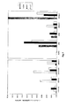
図1は乳ガン細胞の表面における表面発現したICAM−1、CAR、DAFおよびα2β1のレベルのフローサイトメトリー分析を示す。乳ガン細胞は、これらの受容体について特異的な対応するモノクローナル抗体の存在下または非存在下でヤギ抗マウス免疫グロブリンのR−フィコエリトリンコンジュゲート化F(ab’)2フラグメントとインキュベーションされた。コンジュゲートサンプルの幾何平均がエンテロウイルス受容体サンプルの幾何平均から引かれ、これにより、受容体の相対的な発現レベルが明らかにされた。
図2はエンテロウイルスのCAV21、CVB3、EV1、EV7およびPV1による乳ガン細胞の溶解感染を示す。50パーセント終点力価が計算され、腫瘍崩壊は、TCID50/ml終点が104以上であった場合に有意であると見なされた。
図3は直腸結腸ガン細胞の表面における表面発現したICAM−1、CAR、DAFおよびα2β1のレベルのフローサイトメトリー分析を示す。直腸結腸ガン細胞は、これらの受容体について特異的な対応するモノクローナル抗体の存在下または非存在下でヤギ抗マウス免疫グロブリンのR−フィコエリトリンコンジュゲート化F(ab’)2フラグメントとインキュベーションされた。コンジュゲートサンプルの幾何平均がエンテロウイルス受容体サンプルの幾何平均から引かれ、これにより、受容体の相対的な発現レベルが明らかにされた。
図4はエンテロウイルスのCAV21、CVB3、EV1、EV7およびPV1による直腸結腸ガン細胞の溶解感染を示す。50パーセント終点力価が計算され、腫瘍崩壊は、TCID50/ml終点が104以上であった場合に有意であると見なされた。
図5は前立腺ガン細胞または膵臓ガン細胞の表面における表面発現したICAM−1、CAR、DAFおよびα2β1のレベルのフローサイトメトリー分析を示す。前立腺ガン細胞または膵臓ガン細胞は、これらの受容体について特異的な対応するモノクローナル抗体の存在下または非存在下でヤギ抗マウス免疫グロブリンのR−フィコエリトリンコンジュゲート化F(ab’)2フラグメントとインキュベーションされた。コンジュゲートサンプルの幾何平均がエンテロウイルス受容体サンプルの幾何平均から引かれ、これにより、受容体の相対的な発現レベルが明らかにされた。
図6はエンテロウイルスのCAV21、CVB3、EV1、EV7およびPV1による前立腺ガン細胞および膵臓ガン細胞の溶解感染を示す。50パーセント終点力価が計算され、腫瘍崩壊は、TCID50/ml終点が104以上であった場合に有意であると見なされた。
図7は卵巣ガン細胞の表面における表面発現したICAM−1、CAR、DAFおよびα2β1のレベルのフローサイトメトリー分析を示す。卵巣ガン細胞は、これらの受容体について特異的な対応するモノクローナル抗体の存在下または非存在下でヤギ抗マウス免疫グロブリンのR−フィコエリトリンコンジュゲート化F(ab’)2フラグメントとインキュベーションされた。コンジュゲートサンプルの幾何平均がエンテロウイルス受容体サンプルの幾何平均から引かれ、これにより、受容体の相対的な発現レベルが明らかにされた。
図8はエンテロウイルスのCAV21、CVB3、EV1、EV7およびPV1による卵巣ガン細胞の溶解感染を示す。50パーセント終点力価が計算され、腫瘍崩壊は、TCID50/ml終点が104以上であった場合に有意であると見なされた。
図9Aは、EV1の10−1希釈物を72時間感染させた卵巣ガン細胞単層物の顕微鏡写真を示す。このウイルス投入多重度において、細胞株A2780を除いて、すべての細胞株がEV1による腫瘍崩壊の著しいレベルを示した(右側)。図9Bは、EV1の10−1希釈物を72時間感染させた卵巣ガン細胞単層物の顕微鏡写真を示す。細胞株SKOV−3を除いて、すべての細胞株がEV1による腫瘍崩壊の著しいレベルを示した(右側)。
図10はEV1を用いた卵巣ガン細胞への溶解感染を示す。10個の細胞株のうちの7個が、EV1による腫瘍崩壊を受けやすいと見なされる。腫瘍崩壊は、ウイルス力価(TCID50/ml)が104以上であると計算された場合に有意であると見なされた。
図11はEV1結合が抗α2β1の存在下で阻害されることを示す。抗α2β1MAbまたは抗DAF MAbのいずれかの存在下および非存在下での卵巣ガン細胞株に対する[35S]−メチオニン標識EV1の結合。結合した[35S]−メチオニン標識ウイルスのレベルが、1450 Microbeta TRILUX(Wallac、フィンランド)での液体シンチレーション計数によって測定された。
図12は抗α2β1MAbの存在下または非存在下でのEV1による卵巣ガン細胞株のOWA−42およびIGROV−1への溶解感染を示す。感染後72時間、抗α2β1MAbとプレインキュベーションされた細胞は完全に保護されたままであった。細胞の生存が、クリスタルバイオレットのメタノール溶液を用いた染色によって明らかにされた。
図13は抗α2β1MAbの存在下または非存在下でのEV1によるOWA−42卵巣ガン細胞単層物への溶解感染を示す。顕微鏡写真が、感染後の24時間、48時間および72時間において撮影され、これらは、α2β1受容体のモノクローナル抗体遮断によるEV1感染からの細胞の完全な保護を明らかにしている。
図14はDOV13卵巣ガン細胞がリング状インサートの内側で培養され、HeLa細胞(ヒト繊維芽細胞)がリングの外側において培養されたことを示す。EV1による感染後、生存細胞がクリスタルバイオレットのメタノール溶液で染色された。EV1は卵巣ガン細胞に特異的に感染し、その一方で、HeLa細胞は健康なままであった。
図15は黒色腫細胞株SkMel28における表面発現したα2β1のレベルのフローサイトメトリー分析を示す。SkMel28細胞は抗α2β1の存在下または非存在下でヤギ抗マウス免疫グロブリンのR−フィコエリトリンコンジュゲート化F(ab’)2フラグメントとインキュベーションされた。コンジュゲートサンプルの幾何平均がサンプルの幾何平均から引かれ、これにより、変化、従って、受容体の発現が明らかにされた。有意なα2β1発現が、幾何平均における変化により明らかにされる。
図16は抗α2β1MAbまたは抗DAF MAbのいずれかの存在下および非存在下におけるSkMel28黒色腫細胞に対する[35S]−メチオニン標識EV1の結合を示す。結合した[35S]−メチオニン標識ウイルスのレベルが、1450 Microbeta TRILUX(Wallac、フィンランド)での液体シンチレーション計数によって測定された。α2β1遮断はEV1結合の著しい阻害をもたらした。結果は三連サンプルの平均値±標準誤差として表される。
図17はEV1を用いたSkMel28黒色腫細胞への溶解感染を示す。細胞の生存がクリスタルバイオレットのメタノール溶液によって明らかにされた。著しい溶解を認めることができる。
図18はEV1を用いた卵巣ガン多細胞スフェロイドの治療を示す顕微鏡写真である。
図19Aは、1.0×10⁶個のOVHS−1細胞が、リン酸塩緩衝化生理的食塩水(PBS)、UV不活性化エコーウイルスEV1または感染性EV1(105TCID50)のいずれかでのi.p.経路による注射の3週間前に腹腔内(i.p)経路により投与されたSCIDマウスの体重変化を示すヒストグラムである。図19Bは、OVHS−1細胞が注射され、PBS、UV不活性化EV1またはEV1で治療されたマウスと比較される正常なコントロールSCIDマウスの注射後5週間目に撮影された写真を示す。PBSまたはUV不活性化EV1が投与された腫瘍保有マウスにおける腹膜腹水症の発症に留意すること。
The features and advantages of the invention will become more apparent from the following description of preferred embodiments of the invention.
BRIEF DESCRIPTION OF THE FIGURES FIG. 1 shows a flow cytometric analysis of the level of surface expressed ICAM-1, CAR, DAF and α 2 β 1 on the surface of breast cancer cells. Breast cancer cells were incubated with R-phycoerythrin conjugated F (ab ′) 2 fragments of goat anti-mouse immunoglobulin in the presence or absence of corresponding monoclonal antibodies specific for these receptors. The geometric mean of the conjugate sample was subtracted from the geometric mean of the enterovirus receptor sample, which revealed the relative expression level of the receptor.
FIG. 2 shows lytic infection of breast cancer cells by enteroviruses CAV21, CVB3, EV1, EV7 and PV1. A 50 percent endpoint titer was calculated and oncolysis was considered significant if the TCID 50 / ml endpoint was greater than 10 4 .
FIG. 3 shows a flow cytometric analysis of the level of surface expressed ICAM-1, CAR, DAF and α 2 β 1 on the surface of colorectal cancer cells. Colorectal cancer cells were incubated with R-phycoerythrin conjugated F (ab ′) 2 fragments of goat anti-mouse immunoglobulin in the presence or absence of corresponding monoclonal antibodies specific for these receptors. The geometric mean of the conjugate sample was subtracted from the geometric mean of the enterovirus receptor sample, which revealed the relative expression level of the receptor.
FIG. 4 shows lytic infection of colorectal cancer cells by enteroviruses CAV21, CVB3, EV1, EV7 and PV1. A 50 percent endpoint titer was calculated and oncolysis was considered significant if the TCID 50 / ml endpoint was greater than 10 4 .
FIG. 5 shows a flow cytometric analysis of the level of surface expressed ICAM-1, CAR, DAF and α 2 β 1 on the surface of prostate cancer cells or pancreatic cancer cells. Prostate cancer cells or pancreatic cancer cells are incubated with R-phycoerythrin conjugated F (ab ′) 2 fragments of goat anti-mouse immunoglobulin in the presence or absence of corresponding monoclonal antibodies specific for these receptors. It was done. The geometric mean of the conjugate sample was subtracted from the geometric mean of the enterovirus receptor sample, which revealed the relative expression level of the receptor.
FIG. 6 shows lytic infection of prostate and pancreatic cancer cells with enteroviruses CAV21, CVB3, EV1, EV7 and PV1. A 50 percent endpoint titer was calculated and oncolysis was considered significant if the TCID 50 / ml endpoint was greater than 10 4 .
FIG. 7 shows a flow cytometric analysis of the level of surface expressed ICAM-1, CAR, DAF and α 2 β 1 on the surface of ovarian cancer cells. Ovarian cancer cells were incubated with R-phycoerythrin conjugated F (ab ′) 2 fragments of goat anti-mouse immunoglobulin in the presence or absence of corresponding monoclonal antibodies specific for these receptors. The geometric mean of the conjugate sample was subtracted from the geometric mean of the enterovirus receptor sample, which revealed the relative expression level of the receptor.
FIG. 8 shows lytic infection of ovarian cancer cells by enteroviruses CAV21, CVB3, EV1, EV7 and PV1. A 50 percent endpoint titer was calculated and oncolysis was considered significant if the TCID 50 / ml endpoint was greater than 10 4 .
FIG. 9A shows a photomicrograph of an ovarian cancer cell monolayer infected with a 10 −1 dilution of EV1 for 72 hours. At this multiplicity of virus input, with the exception of cell line A2780, all cell lines showed significant levels of oncolysis by EV1 (right side). FIG. 9B shows a photomicrograph of an ovarian cancer cell monolayer infected with a 10 −1 dilution of EV1 for 72 hours. With the exception of cell line SKOV-3, all cell lines showed significant levels of oncolysis by EV1 (right side).
FIG. 10 shows lytic infection of ovarian cancer cells using EV1. Seven of the ten cell lines are considered susceptible to tumor destruction by EV1. Oncolysis was considered significant when the viral titer (TCID 50 / ml) was calculated to be greater than 10 4 .
FIG. 11 shows that EV1 binding is inhibited in the presence of anti-α 2 β 1 . Binding of [ 35 S] -methionine labeled EV1 to ovarian cancer cell lines in the presence and absence of either anti-α 2 β 1 MAb or anti-DAF MAb. The level of bound [ 35 S] -methionine labeled virus was measured by liquid scintillation counting on a 1450 Microbeta TRILUX (Wallac, Finland).
FIG. 12 shows lytic infection of ovarian cancer cell lines to OWA-42 and IGROV-1 by EV1 in the presence or absence of anti-α 2 β 1 MAb. At 72 hours post infection, cells preincubated with anti-α 2 β 1 MAb remained fully protected. Cell survival was revealed by staining with crystal violet in methanol.
FIG. 13 shows lytic infection of OWA-42 ovarian cancer cell monolayers with EV1 in the presence or absence of anti-α 2 β 1 MAb. Micrographs were taken at 24, 48 and 72 hours after infection, which reveals complete protection of the cells from EV1 infection by blocking the α 2 β 1 receptor monoclonal antibody.
FIG. 14 shows that DOV13 ovarian cancer cells were cultured inside the ring insert and HeLa cells (human fibroblasts) were cultured outside the ring. After infection with EV1, viable cells were stained with a solution of crystal violet in methanol. EV1 specifically infected ovarian cancer cells, while HeLa cells remained healthy.
FIG. 15 shows a flow cytometric analysis of the level of surface expressed α 2 β 1 in the melanoma cell line SkMel28. SkMel28 cells were incubated with R-phycoerythrin conjugated F (ab ′) 2 fragment of goat anti-mouse immunoglobulin in the presence or absence of anti-α 2 β 1 . The geometric mean of the conjugate sample was subtracted from the geometric mean of the sample, thereby revealing the change and hence the expression of the receptor. Significant α 2 β 1 expression is revealed by changes in the geometric mean.
FIG. 16 shows binding of [ 35 S] -methionine labeled EV1 to SkMel28 melanoma cells in the presence and absence of either anti-α 2 β 1 MAb or anti-DAF MAb. The level of bound [ 35 S] -methionine labeled virus was measured by liquid scintillation counting on a 1450 Microbeta TRILUX (Wallac, Finland). α 2 β 1 blockade resulted in significant inhibition of EV1 binding. The results are expressed as the average of triplicate samples ± standard error.
FIG. 17 shows lytic infection of SkMel28 melanoma cells with EV1. Cell survival was demonstrated by a solution of crystal violet in methanol. Significant dissolution can be observed.
FIG. 18 is a photomicrograph showing treatment of ovarian cancer multicellular spheroids using EV1.
FIG. 19A shows that 1.0 × 10 6 OVHS-1 cells were either phosphate buffered saline (PBS), UV inactivated echovirus EV1 or infectious EV1 (10 5 TCID 50 ). I. p. 2 is a histogram showing the change in body weight of SCID mice administered by the intraperitoneal (ip)
ウイルスが腫瘍の細胞に感染し、その死を生じさせることができるかどうかを明らかにするために、生検試料を腫瘍から採取することができ、細胞の調製物を、従来の技術を使用して調製し、その後、(i)ウイルス受容体の細胞表面発現を確認することができ、また、(ii)ウイルスで細胞を攻撃し、所定のインキュベーション期間、典型的には約2日間(しかし、これは、使用されるウイルスに依存して変化し得る)、感染および細胞死について細胞をモニターすることができる。α2β1の発現はフローサイトメトリー分析によって容易に確認することができる。数多くのウイルスを、調製された悪性の細胞の種々のアリコートを使用して同時にこの方法でスクリーニングすることができ、そして、より大きな程度の感染性および細胞死を示すウイルスを、生検試料が採取された対象への投与のために選択することができる。同様に、異なる供給源から採取された生検試料に由来する種々の悪性の細胞の調製物を、特定のウイルスを使用するアッセイにおいて用いることができる。生検試料は、一人の個体の異なる部位から、または数多くの個体から採取することができる。 To determine whether the virus can infect tumor cells and cause their death, a biopsy sample can be taken from the tumor, and cell preparations can be obtained using conventional techniques. And then (i) confirm the cell surface expression of the viral receptor, and (ii) attack the cells with the virus, for a predetermined incubation period, typically about 2 days (but This can vary depending on the virus used), and the cells can be monitored for infection and cell death. The expression of α 2 β 1 can be easily confirmed by flow cytometric analysis. Numerous viruses can be simultaneously screened in this way using various aliquots of prepared malignant cells, and a biopsy sample collects viruses that exhibit a greater degree of infectivity and cell death. Can be selected for administration to a given subject. Similarly, various malignant cell preparations derived from biopsy samples taken from different sources can be used in assays using specific viruses. Biopsy samples can be taken from different sites on an individual or from a number of individuals.
本明細書中に記載されるような方法において使用されるウイルスは、望ましくは、受容者において臨床的症状をほとんど生じさせないか、または、ほんの軽微な臨床的症状を生じさせるだけである。そのようなウイルスは、当業者に広く知られている商業的供給源から容易に入手可能であり、上記の様式で本発明の方法におけるその有効性についてスクリーニングすることができる。望ましくは、ウイルスは、通常の場合、エコーウイルスEV1、エコーウイルスEV7、エコーウイルスEV8およびエコーウイルスEV22からなる群から選択されるエコーウイルスである。これらのウイルスはそれぞれが、細胞感染性のためにα2β1を認識する。例えば、EV1は、軽い上部気道疾患、そしてまた胸膜痛に関連している(Fields B.N.他、2000;McCracken A.W.他、1969)。 Viruses used in methods as described herein desirably produce little or no clinical symptoms in the recipient. Such viruses are readily available from commercial sources well known to those skilled in the art and can be screened for their effectiveness in the methods of the invention in the manner described above. Desirably, the virus is an echovirus that is normally selected from the group consisting of Echovirus EV1, Echovirus EV7, Echovirus EV8 and Echovirus EV22. Each of these viruses recognize α 2 β 1 because of cell infectivity. For example, EV1 is associated with mild upper airway disease and also pleural pain (Fields BN et al., 2000; McCracken AW et al., 1969).
α2β1の発現は、中皮において遭遇する優勢なI型コラーゲンマトリックスのために卵巣ガン腫ではアップレギュレーションされると考えられている。数多くの悪性黒色腫はまた、アップレギュレーションされたレベルのα2β1を発現することが示されている(Kramer R.H.およびMarks N、1989;Ramos D.M.他、1990)。EV1およびコラーゲンは、α2β1サブユニットのドメインIにおいて異なる残基を使用してα2β1に付着する(Bergelson J.H.、1993)。インテグリンα2β1はEV1およびコラーゲンを同時に受け入れることができない。しかしながら、ウイルスは、コラーゲンと比較した場合、親和性において10倍の増大でα2β1と結合する(Xing L、2002)。 Expression of α 2 β 1 is believed to be upregulated in ovarian carcinoma due to the predominant type I collagen matrix encountered in the mesothelioma. A number of malignant melanomas have also been shown to express up-regulated levels of α 2 β 1 (Kramer RH and Marks N, 1989; Ramos DM et al., 1990). EV1 and collagen attach to α 2 β 1 using different residues in domain I of the α 2 β 1 subunit (Bergelson JH, 1993). Integrin α 2 β 1 cannot accept EV1 and collagen simultaneously. However, the virus binds α 2 β 1 with a 10-fold increase in affinity when compared to collagen (Xing L, 2002).
所与のウイルスをスクリーニングして、そのウイルスが、悪性の細胞に感染し、その死を生じさせることができるかどうかを確認する目的のために、生検試料から単離された原発性の悪性の細胞よりもむしろ悪性細胞株を使用することができる。 Primary malignant isolated from a biopsy sample for the purpose of screening a given virus to see if it can infect malignant cells and cause their death Malignant cell lines can be used rather than
選択されたウイルスは、好ましくは、ウイルスによる腫瘍の潜在的な感染のための領域を最大限にするために、悪性腫瘍上の多数の部位に対して直接的に注入される。完全なウイルスではなく、ウイルスを生じさせるための核酸を含むウイルスプラスミドまたは他のプラスミドまたは発現ベクターを、治療を達成するための腫瘍細胞による取り込みおよび細胞内における完全なウイルスの生成のために腫瘍内に注入することができる。好適な発現ベクターには、ウイルスを生じさせるために必要なウイルスタンパク質をコードするDNA(例えば、ゲノムDNAまたはcDNA)インサートの発現が可能であるプラスミドが含まれる。発現ベクターは、典型的には、挿入された核酸が機能的に連結される転写調節制御配列を含む。「機能的に連結される」によって、核酸インサートが、インサートの読み枠の変化を伴うことなく、挿入された配列(1つまたは複数)の転写を可能にするための転写調節制御配列に連結されることが意味される。そのような転写調節制御配列には、転写を開始させるためにRNAポリメラーゼの結合を促進させるためのプロモーター、および、転写されたmRNAに対するリボソームの結合を可能にするための発現制御エレメントが含まれる。 The selected virus is preferably injected directly into multiple sites on the malignant tumor to maximize the area for potential infection of the tumor with the virus. Viral plasmids or other plasmids or expression vectors that contain the nucleic acid to generate the virus, rather than the complete virus, are taken up by the tumor cells to achieve therapy and are produced within the tumor for the production of complete viruses within the cells. Can be injected into. Suitable expression vectors include plasmids that are capable of expressing DNA (eg, genomic DNA or cDNA) inserts that encode the viral proteins necessary to generate the virus. Expression vectors typically include transcriptional regulatory control sequences to which the inserted nucleic acid is operably linked. By “operably linked”, the nucleic acid insert is linked to a transcriptional regulatory control sequence to allow transcription of the inserted sequence (s) without changing the reading frame of the insert. Is meant. Such transcriptional regulatory control sequences include a promoter for promoting RNA polymerase binding to initiate transcription and an expression control element to allow ribosome binding to the transcribed mRNA.
より詳細には、本明細書中で使用される用語「調節制御配列」は、所与のDNA配列の転写を行わせ、かつその転写レベルを制御(すなわち、調節)することに関与する任意のDNAを包含することが理解されなければならない。例えば、5’調節制御配列は、プロモーターおよび5’非翻訳リーダー配列を含み得る、コード配列の上流側に位置するDNA配列である。3’調節制御配列は、1つまたは複数のポリアデニル化シグナルを含む好適な転写終結(および/または)調節シグナルを含み得る、コード配列の下流側に位置するDNA配列である。本明細書中で使用される用語「プロモーター」は、転写の開始時にDNA依存性RNAポリメラーゼによって認識され、そのDNA依存性RNAポリメラーゼが(直接的または間接的に)結合する任意のDNA配列を包含する。プロモーターには、転写開始部位、ならびに、転写開始因子およびRNAポリメラーゼに対する結合部位が含まれ、また、プロモーターは、遺伝子発現調節タンパク質が結合し得る様々な他の部位または配列(例えば、エンハンサー)を含むことができる。 More specifically, the term “regulatory control sequence” as used herein refers to any transcription factor that causes transcription of a given DNA sequence and that is involved in controlling (ie, regulating) the level of transcription. It should be understood to encompass DNA. For example, a 5 'regulatory control sequence is a DNA sequence located upstream of a coding sequence that may include a promoter and a 5' untranslated leader sequence. A 3 'regulatory control sequence is a DNA sequence located downstream of a coding sequence that may include a suitable transcription termination (and / or) regulatory signal including one or more polyadenylation signals. The term “promoter” as used herein includes any DNA sequence that is recognized by a DNA-dependent RNA polymerase at the start of transcription and to which the DNA-dependent RNA polymerase binds (directly or indirectly). To do. A promoter includes a transcription initiation site, as well as binding sites for transcription initiation factors and RNA polymerase, and the promoter includes various other sites or sequences (eg, enhancers) to which gene expression regulatory proteins can bind. be able to.
哺乳動物細胞のトランスフェクションのために好適な数多くの発現ベクターがこの分野では知られている。哺乳動物細胞のトランスフェクションのために好適な発現ベクターには、pSV2neo、pEF−PGk.puro、pTk2、ならびに、ポリアデニル化部位および伸長因子1−xプロモーターを含む非複製性のアデノウイルスシャトルベクター、ならびに、サイトメガロウイルス(CMV)プロモーターを最も好ましくは含むpAdEasy型発現ベクターが含まれる(例えば、He他(1998)を参照のこと)。ポリペプチド伸長因子−α2をプロモーターとして用いるプラスミドpEFBOSもまた利用することができる。 Numerous expression vectors suitable for transfection of mammalian cells are known in the art. Suitable expression vectors for transfection of mammalian cells include pSV2neo, pEF-PGk. puro, pTk2, and a non-replicating adenovirus shuttle vector comprising a polyadenylation site and elongation factor 1-x promoter, and a pAdEasy type expression vector most preferably comprising a cytomegalovirus (CMV) promoter (eg, , He et al. (1998)). Plasmid pEFBOS using polypeptide elongation factor-α2 as a promoter can also be utilized.
ウイルスを生じさせるために必要なウイルスタンパク質をコードするcDNAは、例えば、Sambrook他(1989)、Molecular Cloning:A Laboratory Manual(第2版、Cold Spring Harbor Laboratory Press、New York)、およびAusubel他(1994)、Current Protocols in Molecular Biology(米国、第1巻および第2巻)に記載されるようなこの分野で広く知られている組換え技術を使用して、ウイルスRNAゲノムまたはそのフラグメントを逆転写することによって調製し、好適なベクターに組み込むことができる。
CDNAs encoding viral proteins necessary to generate viruses are described in, for example, Sambrook et al. (1989), Molecular Cloning: A Laboratory Manual (2nd edition, Cold Spring Harbor Laboratory Press, New York) et al., 1994 ), Reverse transcription of the viral RNA genome or fragments thereof using recombinant techniques well known in the art as described in Current Protocols in Molecular Biology (USA,
細胞は、cDNAではなく、精製されたビリオンから抽出されたウイルスRNAでトランスフェクションすることができ、または、例えば、RNA転写物を、Ansardi,D.C.他(2001)に記載されるように、バクテリオファージT7のRNAポリメラーゼを利用してxDNAテンプレートからインビトロで作製することができる。同様に、単一のプラスミドまたはRNA分子を、ウイルスタンパク質を発現させ、ウイルスを生じさせるために投与することができ、あるいは、ウイルスタンパク質の異なる分子をコードする複数のプラスミドまたはRNA分子を、細胞をトランスフェクションし、ウイルスを生じさせるために投与することができる。 Cells can be transfected with viral RNA extracted from purified virions, rather than cDNA, or, for example, RNA transcripts can be obtained from Ansardi, D. et al. C. As described elsewhere (2001), it can be generated in vitro from an xDNA template utilizing bacteriophage T7 RNA polymerase. Similarly, a single plasmid or RNA molecule can be administered to express viral proteins and give rise to viruses, or multiple plasmids or RNA molecules encoding different molecules of viral proteins can be transformed into cells. It can be transfected and administered to generate viruses.
プラスミドまたはRNAは、細胞のトランスフェクションを促進させるためのキャリアビヒクルの非存在下で、またはそのようなビヒクルとの組合せでの腫瘍細胞による取り込みのために、局所的によるか、または注入によるかのいずれかで腫瘍に対して直接、投与することができる。好適なキャリアビヒクルには、この分野では従来から知られている油中水型エマルションとして典型的には提供されるリポソームが含まれる。リポソームは、典型的には、脂質(特に、リン脂質、例えば、高い相転移温度のリン脂質など)と、通常の場合には、膜安定性をリポソームに提供するための1つまたは複数のステロイドまたはステロイド前駆体(例えば、コレステロールなど)との組合せを含む。リポソームを提供するために有用な脂質の例には、ホスファチジル化合物、例えば、ホスファチジルグリセロール、ホスファチジルコリン、ホスファチジルセリン、スフィンゴ脂質、ホスファチジルエタノールアミン、セレブロシドおよびガングリオシドなどが含まれる。ジアシルホスファチジルグリセロールは、脂質成分が14個〜18個の炭素原子(より好ましくは16個〜18個の炭素原子)を含有し、飽和している場合、特に好適である。 Plasmid or RNA is either topically or by injection for uptake by tumor cells in the absence of a carrier vehicle or in combination with such vehicle to facilitate transfection of the cell. Either can be administered directly to the tumor. Suitable carrier vehicles include liposomes typically provided as water-in-oil emulsions conventionally known in the art. Liposomes are typically lipids (especially phospholipids such as high phase transition temperature phospholipids) and, in the usual case, one or more steroids to provide membrane stability to the liposomes. Or a combination with a steroid precursor (eg cholesterol). Examples of lipids useful for providing liposomes include phosphatidyl compounds such as phosphatidylglycerol, phosphatidylcholine, phosphatidylserine, sphingolipid, phosphatidylethanolamine, cerebroside and ganglioside. Diacylphosphatidylglycerols are particularly suitable when the lipid component contains 14 to 18 carbon atoms (more preferably 16 to 18 carbon atoms) and is saturated.
リポソームと標的細胞との相互作用は受動的または能動的であり得る。能動的な標的化では、標的細胞により発現される対応するリガンドと結合するか、またはそうでない場合には相互作用する特異的なリガンドをリポソーム膜に取り込むことによるリポソームの改変が伴う。そのようなリガンドには、例えば、モノクローナル抗体またはその結合性フラグメント(例えば、FabフラグメントまたはF(ab’)2フラグメント)、糖成分または糖脂質成分、あるいはウイルスタンパク質が含まれ、α2β1に対して特異的なウイルスタンパク質またはモノクローナル抗体が特に好ましい。 The interaction between the liposome and the target cell can be passive or active. Active targeting involves modification of the liposome by incorporating into the liposome membrane a specific ligand that binds to or otherwise interacts with the corresponding ligand expressed by the target cell. Such ligands include, for example, monoclonal antibodies or binding fragments thereof (eg, Fab fragments or F (ab ′) 2 fragments), sugar components or glycolipid components, or viral proteins, and α 2 β 1 Particularly preferred are specific viral proteins or monoclonal antibodies.
通常、腫瘍の周りの組織はまた、悪性の細胞がそのような組織に存在する可能性を考えると、ウイルスが注入されるか、または、そうでない場合にはウイルスで治療される。腫瘍が比較的進行するまで、腫瘍が検出されない場合、周りの組織には、腫瘍自体の外科的切除の後、ウイルスを注入することができる。 Usually, the tissue around the tumor is also injected with the virus or otherwise treated with the virus, given the possibility that malignant cells are present in such tissue. If the tumor is not detected until the tumor is relatively advanced, the surrounding tissue can be injected with the virus after surgical excision of the tumor itself.
悪性の腫瘍に対して直接的に注入するのではなく、接種物は、腫瘍への送達のために、腫瘍部位に隣接する位置における受容者の血流内への静脈内注射によって全身投与することができる。同様に、適当であると見なされるならば、接種物は、皮下に、腹腔内に、または、例えば、筋肉内に投与することができる。しかしながら、一般には、完全なウイルスが投与されるときには、ウイルスに対する特異的な抗体が存在する可能性、および、それにより、ウイルス送達の代わりの様式の潜在的な低下した効率を考えると、腫瘍内への直接的な注入が好まれる。 Rather than injecting directly against a malignant tumor, the inoculum should be administered systemically by intravenous injection into the recipient's bloodstream at a location adjacent to the tumor site for delivery to the tumor. Can do. Similarly, if deemed appropriate, the inoculum can be administered subcutaneously, intraperitoneally, or, for example, intramuscularly. However, in general, given the complete virus administration, given the potential for the presence of specific antibodies to the virus and thereby the potential reduced efficiency of alternative modes of virus delivery, Direct injection into is preferred.
接種物はまた、単独、または腫瘍内への接種物の直接的な注入との組合せのいずれかで局所的に適用することができる。腫瘍の局所的治療は、接種物と、悪性の細胞に感染させるために接種物の一体性を維持するための好適な医薬的に許容され得るキャリアとを含む医薬組成物の滴下適用によって、またはそのような組成物を含浸させたアプリケーターで腫瘍をなでることによって達成することができる。アプリケーターは、組成物に浸された好適な材料の詰め物またはパッドを含むことができる。皮膚における黒色腫を治療する場合、接種物は、接種物が皮膚と接触するように治療される悪性部位に対して保持されるために適合化されている、接種物を含浸させたアプリケーターによって塗布することができる。この場合、アプリケーターには、黒色腫の周りの皮膚に接着するために、絆創膏の場合などの接着性表面がさらに提供される、接種物を含浸させたパッチまたは詰め物などを含むことができ、それにより接種物を黒色腫との接触状態に保つことができる。典型的には、完全なウイルスが、治療を達成するために哺乳動物に投与される。 The inoculum can also be applied topically either alone or in combination with direct injection of the inoculum into the tumor. Topical treatment of the tumor is by instillation of a pharmaceutical composition comprising the inoculum and a suitable pharmaceutically acceptable carrier for maintaining the integrity of the inoculum to infect malignant cells, or It can be achieved by stroking the tumor with an applicator impregnated with such a composition. The applicator can include a pad or pad of suitable material soaked in the composition. When treating melanoma in the skin, the inoculum is applied by an inoculum impregnated applicator that is adapted to be held against the malignant site being treated so that the inoculum contacts the skin. can do. In this case, the applicator can include a patch or stuffing impregnated with an inoculum, which is further provided with an adhesive surface, such as in the case of a bandage, to adhere to the skin around the melanoma, Can keep the inoculum in contact with the melanoma. Typically, complete virus is administered to a mammal to achieve treatment.
一般に、1つまたは複数の小さい切開部が悪性腫瘍および/または周りの組織に作製されて、それらへのウイルスのための進入部位が提供される。 Generally, one or more small incisions are made in the malignant tumor and / or surrounding tissue to provide an entry site for the virus therein.
卵巣ガンまたは卵巣の近くにおけるガンの場合、エコーウイルスを、カテーテルまたは他の好適な適用器具を使用して、カテーテルまたは選択された器具を対応するファロピウス管に沿って挿入することによって、卵巣または罹患部位に対して直接的に送達することができる。 In the case of ovarian cancer or cancer in the vicinity of the ovary, the echovirus is ovarian or diseased by inserting the catheter or selected instrument along the corresponding fallopian tube using a catheter or other suitable application instrument. It can be delivered directly to the site.
ウイルスおよび/または核酸、または、標的細胞内においてウイルスを生じさせるためのウイルス核酸を含むプラスミドを受容者に接種するために使用される医薬的に許容され得るキャリアは、生理学的食塩水などの液体、または、適切であると見なされる任意の他の従来から知られている生理学的に許容され得る媒体、例えば、薬学的使用のために、また、接種物を治療部位に投与するために好適な市販のゲルなどであり得る。キャリアは、典型的には生理学的pHに緩衝化され、好適な保存剤および/または抗生物質を含有することができる。 A pharmaceutically acceptable carrier used to inoculate a recipient with a virus and / or nucleic acid or a plasmid containing a viral nucleic acid to generate a virus in a target cell is a liquid such as physiological saline. Or any other conventionally known physiologically acceptable medium deemed suitable, eg suitable for pharmaceutical use and for administering the inoculum to the treatment site It may be a commercially available gel or the like. The carrier is typically buffered to a physiological pH and can contain suitable preservatives and / or antibiotics.
接種物は、一般に、1mlの接種物あたり約1×10²プラーク形成ユニット〜約1×10¹⁰プラーク形成ユニットを含有する。好ましくは、接種物は1mlの接種物あたり約1×10⁵プラーク形成ユニット以上を含有する。患者に投与される接種物の量は、患者の全体的な状態、悪性腫瘍の段階および存在位置を、ウイルスで治療される領域の全体的なサイズおよび分布と一緒に考慮に入れて、受け入れられている医療行為に従って主治医または外科医によって容易に決定することができる。典型的には、患者は、最初の用量のウイルスで治療され、続いて、ウイルスの最初の投与に対する患者の応答、ならびに、最初の治療から生じるウイルス感染および悪性細胞の死の程度などの要因が決定されるまで、好適な期間にわたってモニターされ、その後、さらなるウイルスを患者に投与するための決定がなされる。 The inoculum generally contains from about 1 × 10 2 plaque forming units to about 1 × 10 10 plaque forming units per ml inoculum. Preferably, the inoculum contains no less than about 1 × 10 5 plaque forming units per ml inoculum. The amount of inoculum administered to a patient is acceptable, taking into account the patient's overall condition, stage of malignancy and location, along with the overall size and distribution of the area to be treated with the virus. Can be easily determined by the attending physician or surgeon according to the medical practice being performed. Typically, a patient is treated with an initial dose of virus followed by factors such as the patient's response to the initial administration of the virus and the extent of viral infection and malignant cell death resulting from the initial treatment. Until determined, it is monitored over a suitable period of time, after which a decision is made to administer additional virus to the patient.
望ましくは、個体は、所定の間隔で一定の期間、ウイルスで治療される。間隔は、それぞれの状況において適切であると決定されるように、毎日であり得るか、あるいは24時間〜72時間以上までに及び得る。異なるウイルスを、以前に投与されたウイルスに対する何らかの免疫応答の影響を回避または最小限にするために毎回、投与することができ、そして、治療の経過期間は、主治医によって決定され得るように、1週間〜2週間以上に及び得る。最も好ましくは、哺乳動物が以前にさらされてないウイルス、または、哺乳動物が、標準的な技術によって決定され得るような比較的軽微な免疫応答を生じるウイルスが投与される。 Desirably, the individual is treated with the virus for a period of time at predetermined intervals. The interval can be daily or can range from 24 hours to 72 hours or more, as determined to be appropriate in each situation. Different viruses can be administered each time to avoid or minimize the effects of any immune response to the previously administered virus, and the course of treatment can be determined by the attending physician as 1 It can range from weeks to over 2 weeks. Most preferably, a virus has been administered to which the mammal has not been previously exposed, or a virus that produces a relatively minor immune response such that the mammal can be determined by standard techniques.
容易に入手可能な知られているエコーウイルスが本発明の方法において好適に用いられ得るが、従来の技術を使用して改変または操作されたウイルスもまた利用することができる。例えば、ウイルスは、さらなる細胞接着分子を細胞受容体として用いるために改変することができる。一例として、ウイルスは、ペプチドモチーフ「RGD」がウイルスのキャプシド表面に発現されるように部位特異的変異誘発を使用して改変することができる。RGDモチーフはαvインテグリンヘテロ二量体によって認識され、そして、このキャプシド改変は、例えば、ウイルスがインテグリンα2β1(α2β1を有するような黒色腫病巣においてアップレギュレーションされることが示されている細胞接着分子;Natalia P.G.、1997)と結合することを可能にすることができ、これは、潜在的には、標的細胞によるウイルスの高まった取り込みを生じさせる。 Although readily available known echoviruses can be suitably used in the methods of the present invention, viruses modified or engineered using conventional techniques can also be utilized. For example, the virus can be modified to use additional cell adhesion molecules as cell receptors. As an example, the virus can be modified using site-directed mutagenesis such that the peptide motif “RGD” is expressed on the capsid surface of the virus. The RGD motif is recognized by α v integrin heterodimers, and this capsid modification has been shown to be upregulated in melanoma lesions where, for example, the virus has the integrin α 2 β 1 (α 2 β 1 Cell adhesion molecules; Natalia PG, 1997), which potentially results in increased uptake of the virus by the target cells.
本発明の本質がより明確に理解され得るために、次に、その好ましい形態が、下記の非限定的な実施例を参照して記載される。 In order that the nature of the present invention may be more clearly understood, preferred forms thereof will now be described with reference to the following non-limiting examples.
(実施例1:材料および方法)
1.1.細胞株
IGROV−1、A2780、DU145、PC3、AsPC−1、PANC−1、T47−D、MDA−MB361、MDA−MB453、MDA−MB231およびMCF−7のガン細胞株をGarvan Institute(Sydney、New South Wales、オーストラリア)から得た。BT−20、MDA−MB157、SK−BR−3、ZR−75−1、HCT116、LIM2537、SW480、SW620、2008、JAM、OVCA−429、OVCAR−3、OVHS−1、OWA−42、SKOV−3およびDOV13のガン細胞株をPeter MacCullum Cancer Institute(Melbourne、Victoria、オーストラリア)から得た。SkMel28細胞をRalph博士(Department of Biochemistry and Molecular Biology、Monash University、Victoria、オーストラリア)から得た。HeLa細胞をMargery Kennett(Entero−respiratory Laboratory、Fairfield Hospital、Melbourne、Victoria、オーストラリア)から得た。α−MEM培地で培養されるBT−20細胞、ならびに、DMEM培地で培養されるSkMel28細胞およびHeLa細胞を除いて、すべての細胞が、2%〜5%のウシ胎児血清(FCS)および抗生物質を含有するRPMIにおいて標準的な条件(5%CO2雰囲気下で37℃)のもとで培養された。使用された細胞はすべてが、ELISA(Roche Molecular Systems、CA、米国)によってマイコプラズマの存在について定期的に検査された。
Example 1: Materials and Methods
1.1. Cell lines IGROV-1, A2780, DU145, PC3, AsPC-1, PANC-1, T47-D, MDA-MB361, MDA-MB453, MDA-MB231 and MCF-7 cancer cell lines were obtained from Garvan Institute (Sydney, New From South Wales, Australia). BT-20, MDA-MB157, SK-BR-3, ZR-75-1, HCT116, LIM2537, SW480, SW620, 2008, JAM, OVCA-429, OVCAR-3, OVHS-1, OWA-42, SKOV- 3 and DOV13 cancer cell lines were obtained from Peter MacCullum Cancer Institute (Melbourne, Victoria, Australia). SkMel28 cells were obtained from Dr. Ralph (Department of Biochemistry and Molecular Biology, Monash University, Victoria, Australia). HeLa cells were obtained from Margery Kennett (Entero-respiratory Laboratory, Fairfield Hospital, Melbourne, Victoria, Australia). Except for BT-20 cells cultured in α-MEM medium, and SkMel28 cells and HeLa cells cultured in DMEM medium, all cells were 2% -5% fetal calf serum (FCS) and antibiotics Incubated under standard conditions (37 ° C. under 5% CO 2 atmosphere) in RPMI containing All cells used were periodically checked for the presence of mycoplasma by ELISA (Roche Molecular Systems, CA, USA).
1.2.ウイルス
コクサッキーウイルスA21(CAV21)プロトタイプ株(Kuykendall)、コクサッキーウイルスB3(CVB3)プロトタイプ株(Nancy)、エコーウイルス(EV1)プロトタイプ株(Farouk)、エコーウイルス(EV7)プロトタイプ株(Wallace)、およびポリオウイルス1(PV1)プロトタイプ株(Mahoney)をMargery Kennett博士(Enterorespiratory Laboratory、Fairfield Hospital、Melbourne、Victoria、オーストラリア)から得た。すべてのウイルスはHeLa細胞において増殖させられ、力価測定された。
1.2. Virus Coxsackievirus A21 (CAV21) prototype strain (Kuykendall), Coxsackievirus B3 (CVB3) prototype strain (Nancy), Echovirus (EV1) prototype strain (Farouk), Echovirus (EV7) prototype strain (Wallace), and Poliovirus 1 (Wallace) PV1) Prototype strain (Mahoney) was obtained from Dr. Margie Kennett (Entertainsspiratory Laboratory, Fairfield Hospital, Melbourne, Victoria, Australia). All viruses were propagated in HeLa cells and titered.
1.3.モノクローナル抗体(MAb)
抗DAF MAbのVIIIA7(これはDAFの3番目のSCRを認識する)はT.Kinoshita博士(大阪大学、大阪、日本)から得られ、抗DAF mAbのIH4はBruce Loveland博士(Austin Research Institute、Heidelberg、Victoria、オーストラリア)からの譲渡物であった。抗CAR MAbのRmcBはJ.M.Bergelson博士(Dana Farber Cancer Institute、Boston、Massachusetts)から得られた。抗β2−ミクログロブリンMAb918はP.Minor博士(NIBSC、Hertfordshire、英国)から得られた。抗α2β1MAbのAK7(これはα2サブユニットを認識する)およびコントロール抗体の抗GPIV(血小板膜糖タンパク質)MAb PTA−1はGordon Burns教授(Department of Medical Biochemistry and Cancer Research、University of Newcastle、NSW、オーストラリア)から得られた。抗ICAM−1 MAbのIH4はAndrew Boyd博士(Queensland Institute for Medical Research、Queensland、オーストラリア)から得られた。
1.3. Monoclonal antibody (MAb)
Anti-DAF MAb VIIIA7 (which recognizes the third SCR of DAF) is IH4 of anti-DAF mAb obtained from Dr. Kinoshita (Osaka University, Osaka, Japan) was an assignment from Dr. Bruce Loveland (Austin Research Institute, Heidelberg, Victoria, Australia). RmcB of anti-CAR MAb is M.M. Obtained from Dr. Bergelson (Dana Faber Cancer Institute, Boston, Massachusetts). Anti-β2-microglobulin MAb 918 is Obtained from Dr. Minor (NIBSC, Hertfordshire, UK). The anti-α 2 β 1 MAb AK7 (which recognizes the α 2 subunit) and the control antibody anti-GPIV (platelet membrane glycoprotein) MAb PTA-1 were developed by Prof. Gordon Burns (Department of Medical Biochemistry and University Research, University Newcastle, NSW, Australia). Anti-ICAM-1 MAb IH4 was obtained from Dr. Andrew Boyd (Queensland Institute for Medical Research, Queensland, Australia).
1.4.フローサイトメトリー分析
ガン細胞におけるエンテロウイルス受容体の表面発現がフローサイトメトリーによって分析された。分散された細胞(1×10⁶個)を、氷上で20分間、適切なMAb(PBSにおいて希釈された5μg/ml)と20分間インキュベーションした。細胞をPBSで洗浄し、遠心分離によってペレット化し、その後、ヤギ抗マウス免疫グロブリンのR−フィコエリトリンコンジュゲート化F(ab’)2フラグメント(Dako,A/S、デンマーク)の1:50希釈物の100μlに再懸濁した。細胞を再び氷上で20分間インキュベーションし、洗浄し、ペレット化して、PBSに再懸濁し、その後、フローサイトメトリー分析を行った。細胞表面での受容体の発現が、FACStar Analyser(Becton Dickenson、Sydney、オーストラリア)を使用して分析された。
1.4. Flow cytometric analysis Surface expression of enterovirus receptors in cancer cells was analyzed by flow cytometry. Dispersed cells (1 × 10 6 ) were incubated for 20 minutes with the appropriate MAb (5 μg / ml diluted in PBS) on ice for 20 minutes. Cells were washed with PBS and pelleted by centrifugation followed by a 1:50 dilution of an R-phycoerythrin conjugated F (ab ′) 2 fragment of goat anti-mouse immunoglobulin (Dako, A / S, Denmark). Resuspended in 100 μl. The cells were again incubated on ice for 20 minutes, washed, pelleted and resuspended in PBS prior to flow cytometric analysis. Receptor expression on the cell surface was analyzed using a FACStar Analyzer (Becton Dickenson, Sydney, Australia).
1.5.ウイルス感染性アッセイ
ガン細胞株のコンフルエントな単層物に、1%ウシ胎児血清(FCS)を含有するDMEMにおけるCAV21、CVB3、EV1、EV7またはPV1の10倍連続希釈物(100μl/ウエルを三連または四連で)を接種し、これらを5%のCO2環境において37℃で72時間インキュベーションした。細胞の生存を測定するために、プレートを100μl/ウエルのクリスタルバイオレットメタノール溶液(0.1%クリスタルバイオレット、20%メタノール、20%ホルムアルデヒド、リン酸塩緩衝化生理的食塩水(PBS))と24時間インキュベーションして、蒸留水において洗浄した。
1.5. Viral Infectivity Assay Confluent monolayers of cancer cell lines were added to 10-fold serial dilutions of CAV21, CVB3, EV1, EV7 or PV1 in DMEM containing 1% fetal calf serum (FCS) (100 μl / well in triplicate). Or in quadruplicate) and these were incubated for 72 hours at 37 ° C. in a 5% CO 2 environment. To measure cell viability, plates were plated with 100 μl / well crystal violet methanol solution (0.1% crystal violet, 20% methanol, 20% formaldehyde, phosphate buffered saline (PBS)) and 24. Incubate for time and wash in distilled water.
限界希釈アッセイの終点は、50%の試験ユニットに影響を及ぼすウイルスの希釈度である。統計学的手法が、ReedおよびMuenchの方法(参考文献)を使用して終点を計算するために用いられた。終点は50%組織培養感染用量/ミリリットル(TCID50/ml)として表された。 The endpoint of the limiting dilution assay is the virus dilution affecting 50% of the test units. Statistical techniques were used to calculate endpoints using the Reed and Muench method (references). The endpoint was expressed as 50% tissue culture infectious dose / milliliter (TCID 50 / ml).
抗受容体モノクローナル抗体で前処理された細胞単層物が要求される場合、細胞は100μlの抗α2β1AK7MAb(PBSにおいて希釈された20μg/ml)と37℃で1時間インキュベーションされた。その後、細胞単層物は適切なウイルス希釈物の二連のサンプルに接種され、5%のCO2環境において37℃で72時間インキュベーションされ、その後、上記のように染色された。 When cell monolayers pretreated with anti-receptor monoclonal antibodies were required, cells were incubated with 100 μl anti-α 2 β 1 AK7 MAb (20 μg / ml diluted in PBS) for 1 hour at 37 ° C. Cell monolayers were then inoculated into duplicate samples of appropriate virus dilutions, incubated for 72 hours at 37 ° C. in a 5% CO 2 environment, and then stained as described above.
顕微鏡写真が、倒立型顕微鏡を使用して100倍の倍率(Olympus IX−FLA)で、24時間、48時間または72時間において撮影された。 Photomicrographs were taken at 24, 48 or 72 hours at 100x magnification (Olympus IX-FLA) using an inverted microscope.
1.6.ウイルス精製
DOV13細胞のコンフルエントな単層物を含有する6ウエル組織培養プレートに500μlのEV1(感染多重度[moi]=105TCID50/ml)を37℃で1時間にわたって接種した。非結合のウイルスを、メチオニン/システインを含まないDMEM(ICN Biomedical、Ohio、米国)で3回洗浄することによって除き、細胞単層物を1.3mlのこの培地において37℃でさらに2時間インキュベーションし、その後、300μCiの[35S]−メチオニントランスラベル(ICN Biomedical、Ohio、米国)を加えた。感染した単層物を5%のCO2環境において37℃で一晩インキュベーションした。3回の凍結/融解サイクルの後、ウイルス溶解物をBeckmanXL−90超遠心分離器(SW41tiローター)における36,000rpmでの95分間の速度遠心分離によって5%〜30%スクロースグラジェントで精製した。分画物を各チューブの底から集め、液体シンチレーション計数(Wallac 1450 Mirobeta TRILUX、フィンランド)によってモニターして、ウイルス結合アッセイにおいて使用される160Sウイルス最大画分を捜し出した。
1.6. Virus purification 6-well tissue culture plates containing confluent monolayers of DOV13 cells were inoculated with 500 μl of EV1 (multiplicity of infection [moi] = 10 5 TCID 50 / ml) at 37 ° C. for 1 hour. Unbound virus was removed by washing 3 times with DMEM without methionine / cysteine (ICN Biomedical, Ohio, USA) and the cell monolayer was incubated in 1.3 ml of this medium for an additional 2 hours at 37 ° C. Then, 300 μCi of [ 35 S] -methionine translabel (ICN Biomedical, Ohio, USA) was added. Infected monolayers were incubated overnight at 37 ° C. in a 5% CO 2 environment. After three freeze / thaw cycles, the virus lysate was purified on a 5% -30% sucrose gradient by speed centrifugation at 36,000 rpm for 95 minutes in a Beckman XL-90 ultracentrifuge (SW41ti rotor). Fractions were collected from the bottom of each tube and monitored by liquid scintillation counting (Wallac 1450 Mirabeta TRILUX, Finland) to find the 160S virus maximum fraction used in the virus binding assay.
放射能標識されていないEV1ビリオンを、プールされ、リン酸塩緩衝化生理的食塩水(PBS)に対して透析された最大感染画分を用いた並行するグラジェントで精製した。紫外(UV)光で不活性化されたEV1を、6ウエルプレートにおいて、PBSにおける精製EV1の1.0ml/ウエル(5×105TCID50)を15ワットのUV光に30秒間さらすことによって作製した。ウイルスの不活性化はマイクロタイタープレートでの溶解感染性の細胞アッセイによって評価された。 Non-radiolabeled EV1 virions were purified in parallel gradients using the largest infectious fraction pooled and dialyzed against phosphate buffered saline (PBS). EV1 inactivated with ultraviolet (UV) light is made by exposing 1.0 ml / well (5 × 10 5 TCID 50 ) of purified EV1 in PBS to 15 watts of UV light in a 6-well plate for 30 seconds. did. Viral inactivation was assessed by a lytic infectious cell assay in microtiter plates.
1.7.放射能標識されたウイルスの結合アッセイ
1%ウシ血清アルブミン(BSA)を含有する800μlのRPMIに再懸濁された約1×10⁶個の細胞を、20μg/mlのMAb(PBSにおいて希釈された抗α2β1または抗DAF)の存在下、4℃で1時間インキュベーションし、その後、300μl(1×106個)の[35S]−メチオニン標識された160S EV1を加えた。4℃で2時間インキュベーションした後、細胞を血清非含有培地で4回洗浄し、細胞ペレットを200μlの0.2M NaOH−1%SDSに溶解し、その後、結合した[35S]−メチオニン標識ウイルスのレベルを三連サンプルからの液体シンチレーション計数によって測定した(Wallac 1450 Mirobeta TRILUX、フィンランド)。結果が平均値±SEとして表された。
1.7. Radiolabeled virus binding assay Approximately 1 × 10 6 cells resuspended in 800 μl RPMI containing 1% bovine serum albumin (BSA) were diluted with 20 μg / ml MAb (diluted in PBS). the presence of anti-alpha 2 beta 1 or anti-DAF), incubated for 1 hour at 4 ° C., then, 300μl (1 × 10 6 cells) of [35 S] - plus methionine labeled 160S EV1. After 2 hours incubation at 4 ° C., the cells were washed 4 times with serum-free medium and the cell pellet was lysed in 200 μl 0.2 M NaOH-1% SDS and then bound [ 35 S] -methionine labeled virus. Levels were measured by liquid scintillation counting from triplicate samples (Wallac 1450 Mirbeta TRILUX, Finland). Results were expressed as mean ± SE.
1.10.ドデシル硫酸ナトリウム−ポリアクリルアミドゲル電気泳動(SDS−PAGE)
[35S]−メチオニン標識されたウイルス画分をポリアクリルアミドゲル電気泳動(PAGE)によって分析し、オートラジオグラフィーによって可視化した。[35S]−メチオニン標識された160S EV1画分をサンプル還元緩衝液(250mM TRIS、0.2g(w/v)SDS、20%(v/v)グリセロール、10%(v/v)2−メルカプトエタノール、および0.01%(w/v)ブロモフェノールブルー、pH6.8)と95℃で10分間インキュベーションし、ビリオンを変性させた。変性処理された160Sウイルス最大画分を、その後、180Vで45分間、Benchmark染色済み中範囲タンパク質ラダー(GIBCO、米国)と一緒に15%Tris−HClプレキャストゲル(BIORAD Ready−Gel、CA、米国)において分離した。主な構造タンパク質の可視化およびウイルス純度の分析が、96時間の感光を行った後のHyperfilm MP(Amersham International、英国)におけるオートラジオグラフィーによって行われた。
1.10. Sodium dodecyl sulfate-polyacrylamide gel electrophoresis (SDS-PAGE)
[ 35 S] -methionine labeled virus fractions were analyzed by polyacrylamide gel electrophoresis (PAGE) and visualized by autoradiography. The [ 35 S] -methionine labeled 160S EV1 fraction was added to a sample reduction buffer (250 mM TRIS, 0.2 g (w / v) SDS, 20% (v / v) glycerol, 10% (v / v) 2 − Virions were denatured by incubation with mercaptoethanol and 0.01% (w / v) bromophenol blue, pH 6.8) at 95 ° C. for 10 minutes. The denatured 160S virus maximum fraction was then added to a 15% Tris-HCl precast gel (BIORAD Ready-Gel, CA, USA) with Benchmark stained medium range protein ladder (GIBCO, USA) for 45 minutes at 180V. Separated. Visualization of the major structural proteins and analysis of virus purity were performed by autoradiography at Hyperfilm MP (Amersham International, UK) after 96 hours of light exposure.
1.11.細胞の細胞傷害性アッセイ
ヒト末梢血リンパ球(OVHS−1細胞およびDOV−13細胞)の細胞懸濁物をEV1で攻撃し(moi=1.0TCID50/細胞)、37℃で24時間インキュベーションした。細胞の細胞溶解のレベルを、製造者の説明書に従ってCyto−Tox96キット(Promega Corp.、Maddison、WI、米国)を使用することによって評価されるLDH(細胞溶解時に放出される安定な細胞質ゾル酵素)の放出の関数として計算した。
1.11. Cell Cytotoxicity Assay Cell suspensions of human peripheral blood lymphocytes (OVHS-1 and DOV-13 cells) were challenged with EV1 (moi = 1.0 TCID 50 / cell) and incubated at 37 ° C. for 24 hours. . LDH (stable cytosolic enzyme released upon cell lysis) where the level of cell lysis is assessed using the Cyto-Tox96 kit (Promega Corp., Maddison, Wis., USA) according to the manufacturer's instructions. ) As a function of release.
1.12.スフェロイドの培養およびスフェロイド感染性アッセイ
DOV−13細胞を、24ウエルプレートにおいて、5%FCSを含有する1mlのRPMI1640においてウエルあたり500個または5000個の細胞で半固体の0.5%寒天層に接種した。細胞は、スフェロイドを形成させるために5%のCO2環境において37℃で48時間インキュベーションされ、その後、EV1が添加された(105TCID50)。
1.12. Spheroid culture and spheroid infectivity assay DOV-13 cells are seeded in semi-solid 0.5% agar layers at 500 or 5000 cells per well in 1 ml RPMI 1640 containing 5% FCS in 24-well plates. did. Cells were incubated for 48 hours at 37 ° C. in a 5% CO 2 environment to form spheroids, after which EV1 was added (10 5 TCID 50 ).
1.13.SCIDマウスにおける腹腔内腫瘍異種移植片モデル
6週齢〜8週齢のオスBALB/cSCIDマウスを、ニューカッスル大学動物管理倫理委員会によって承認されたプロトコルに従って病原体非含有条件で飼育した。OVHS−1細胞を、0.05%トリプシンを用いて集め、10%FCSを含有するRPMIに再懸濁し、遠心分離によってペレット化した。細胞を洗浄し、PBSに再懸濁し、その後、マウスに1×106個の細胞を200μlにおいて腹腔内(i.p.)注射した。14日後、マウスを3つの群(n=5)に分け、リン酸塩緩衝化生理的食塩水(PBS)で、またはUV不活性化EV1もしくは感染性EV1のいずれかの105TCID50でi.p.治療した。動物は、毎週、体重測定され、腫瘍がその体重の20%を越えた時に屠殺された。治療されたマウスの体重が、腫瘍を有さない健康なBALB/cSCIDマウスと比較された。
1.13. Intraperitoneal tumor xenograft model in SCID mice 6-8 weeks old male BALB / cSCID mice were bred in pathogen-free conditions according to a protocol approved by the University of Newcastle Animal Care Ethics Committee. OVHS-1 cells were collected using 0.05% trypsin, resuspended in RPMI containing 10% FCS and pelleted by centrifugation. The cells were washed and resuspended in PBS, after which the mice were injected intraperitoneally (ip) with 1 × 10 6 cells in 200 μl. After 14 days, the mice were divided into 3 groups (n = 5) and either with phosphate buffered saline (PBS) or with 10 5 TCID 50 of either UV inactivated EV1 or infectious EV1. . p. Treated. Animals were weighed weekly and sacrificed when tumors exceeded 20% of their body weight. The body weight of the treated mice was compared to healthy BALB / cSCID mice without tumors.
1.14.リアルタイムPCRによるウイルス血症の決定
感染マウスから得られた血清を、リアルタイム定量的RT−PCRを使用してウイルス血症について分析した。簡単に記載すると、ウイルスRNAを、製造者の説明書に従って、QIAampウイルスRNAミニキット(Qiagen、Clifton Hill、Victoria、オーストラリア)を使用して10μlの血清から抽出し、40μlの最終体積で溶出した。EV1のウイルスRNAレベルを測定するためのプライマーおよびプローブは、Primer ExpressTM1.5ソフトウエア(Applied Biosystems、Foster City、CA、米国)を使用して設計され、以前に発表されたEV1配列(Genbankアクセション番号AF029859)に基づいていた:フォワードプライマー(5’−CAAGACAGGGACCAAAGAGGAT−3’)、リバースプライマー(5’−CCACTCGCCTGGTTGTAATCA−3’)および6−FAM標識のMGBプローブ(5’−CCAATAGCTTCAACAATT−3’)。ワンステップRT−PCRが、ABI7000配列検出装置においてPlatinum(登録商標)Quantitative RT−PCR ThermoScriptTM One−Step Systemを使用して行われた。標準曲線を作製するために、EV1ウイルスのストック物(1×10⁶TCID50/ml)の10倍希釈物が、最適化された濃度のプライマーおよびプローブを用いて増幅された。25μlの体積において、反応混合物は、1×ThermoScriptTM反応混合物、500nMのフォワードプライマー、900nMのリバースプライマー、250nMのプローブ、500nMのROX、0.5μlのThermoScriptTMPlus/Platinum Taq Mix、および5μlの抽出されたRNAを含んだ。熱サイクル条件は、60℃で30分、その後、95℃で5分、次いで、95℃で15秒および60℃で1分の40サイクルに供された。
1.14. Determination of viremia by real-time PCR Sera obtained from infected mice were analyzed for viremia using real-time quantitative RT-PCR. Briefly, viral RNA was extracted from 10 μl of serum using the QIAamp viral RNA mini kit (Qiagen, Clifton Hill, Victoria, Australia) according to the manufacturer's instructions and eluted in a final volume of 40 μl. Primers and probes for measuring viral RNA levels of EV1 were designed using Primer Express ™ 1.5 software (Applied Biosystems, Foster City, CA, USA) and previously published EV1 sequences (Genbank) Accession number AF029859): forward primer (5'-CAAGACAGGGGACCAAAGAGGAT-3 '), reverse primer (5'-CCACTCGCCTGGTTGTAATCA-3') and 6-FAM labeled MGB probe (5'-CCAATAGCTTCACAAATT-3 ') . One-step RT-PCR was performed using a Platinum® Quantitative RT-PCR ThermoScript ™ One-Step System on an ABI7000 sequence detector. To generate a standard curve, a 10-fold dilution of the EV1 virus stock (1 × 10 6 TCID 50 / ml) was amplified using optimized concentrations of primers and probes. In a 25 μl volume, the reaction mixture consists of 1 × ThermoScript ™ reaction mixture, 500 nM forward primer, 900 nM reverse primer, 250 nM probe, 500 nM ROX, 0.5 μl ThermoScript ™ Plus / Platinum Taq Mix, and 5 μl extraction. RNA was included. Thermal cycling conditions were subjected to 60 ° C for 30 minutes, then 95 ° C for 5 minutes, then 40 cycles of 95 ° C for 15 seconds and 60 ° C for 1 minute.
(実施例2:ガン細胞のウイルス媒介による腫瘍崩壊)
2.1.乳ガン細胞の表面におけるエンテロウイルス受容体の発現
エンテロウイルスによって使用される選択されたエンテロウイルス細胞表面受容体の相対的な発現レベルを明らかにするために、フローサイトメトリー分析が行われた。選択された受容体群は、CAV21によって用いられるICAM−1;EV7、CAV21、CVB3によって用いられるDAF;CVB3によって使用されるCAR;および、EV1によって使用されるインテグリンα2β1からなった。PVR受容体に対するMabを得ることができなかったため、PVRの発現レベルは測定されなかった。
(Example 2: Virus-mediated oncolysis of cancer cells)
2.1. Expression of enterovirus receptors on the surface of breast cancer cells Flow cytometric analysis was performed to reveal the relative expression levels of selected enterovirus cell surface receptors used by enteroviruses. Selected group of receptors is used by CAV21 ICAM-1; CAR used by CVB3;; EV7, CAV21, DAF used by CVB3 and consisted integrin alpha 2 beta 1 used by EV1. The expression level of PVR was not measured because a Mab for the PVR receptor could not be obtained.
9個の乳ガン細胞株が分析され、これらには、BT−29、MCF−7、MDA−MB157、MDA−MB231、MDA−MB361、MDA−MB453、SK−BR−3、T47−DおよびZR−75−1が含まれる。これらの細胞株は、抗ICAM−1(IH4)、抗CAR(RmcB)、抗DAF(VIIIA7)または抗α2β1(AK7)のいずれかとインキュベーションされた。 Nine breast cancer cell lines were analyzed, including BT-29, MCF-7, MDA-MB157, MDA-MB231, MDA-MB361, MDA-MB453, SK-BR-3, T47-D and ZR-. 75-1 is included. These cell lines were incubated with either anti-ICAM-1 (IH4), anti-CAR (RmcB), anti-DAF (VIIIA7) or anti-α 2 β 1 (AK7).
ICAM−1の発現は9個の系統のうちの6個において顕著であり、一方、DAFは、これらすべての細胞株において相対的に低いレベルで発現しているようであった。中程度のレベルのCAR発現が9個の系統のうちの7個において明らかであり、一方、非常に小さいレベルのα2β1発現がこれらの乳ガン細胞株のうちの8個の表面に存在した(図1)。 ICAM-1 expression was prominent in 6 of 9 lines, while DAF appeared to be expressed at relatively low levels in all these cell lines. A moderate level of CAR expression was evident in 7 out of 9 lines, while a very small level of α 2 β 1 expression was present on the surface of 8 of these breast cancer cell lines. (FIG. 1).
2.2.選択されたエンテロウイルスによる乳ガン細胞の腫瘍崩壊
溶解感染性アッセイが、選択されたエンテロウイルス群(CAV21、CVB3、EV1、EV7およびPV1)に対するそれらの感受性を明らかにするために、9個すべての乳ガン細胞株に対して行われた(図2)。細胞株は、1ミリリットルあたり50パーセント終点での組織培養感染用量(TCID50/ml)が104以上であると計算された場合、腫瘍崩壊を非常に受けやすいと見なされた。CAV21およびCVB3は9個の乳ガン細胞株のうちの6個において著しい溶解を誘導した。一般に、乳ガン細胞は、EV1に対する相当の感受性を明らかにした1つの細胞株T47−Dを除いて、エコーウイルス(EV1およびEV7)による溶解感染に対する感受性がなかった。PV1は9個の乳ガン細胞株のうちの8個において実質的な腫瘍崩壊を引き起こした(図2)。
2.2. Tumor lysis of breast cancer cells with selected enteroviruses All nine breast cancer cell lines to allow lytic infectivity assays to reveal their susceptibility to selected enterovirus groups (CAV21, CVB3, EV1, EV7 and PV1) (Fig. 2). A cell line was considered very susceptible to oncolysis if the tissue culture infectious dose (TCID 50 / ml) at the 50 percent endpoint per milliliter was calculated to be 10 4 or higher. CAV21 and CVB3 induced significant lysis in 6 of 9 breast cancer cell lines. In general, breast cancer cells were not susceptible to lytic infection by echoviruses (EV1 and EV7), with the exception of one cell line, T47-D, that demonstrated considerable sensitivity to EV1. PV1 caused substantial oncolysis in 8 of 9 breast cancer cell lines (FIG. 2).
2.3.結腸直腸ガン細胞の表面におけるエンテロウイルス受容体の発現
4つの結腸直腸ガン細胞株(HCT116、LIM2537、SW480およびSW620)が、フローサイトメトリーによって、ICAM−1、CAR、α2β1およびDAFの発現について分析された。著しいレベルのICAM−1発現およびDAF発現がこれらの細胞株のうちの2つにおいて観測された。中程度のレベルのCARが4つすべての系統において発現しているようであり、一方、著しいレベルのα2β1発現は観測されなかった(図3)。
2.3. Expression of Enterovirus receptors on the surface of the colorectal cancer cell four colorectal cancer cell lines (HCT116, LIM2537, SW480 and SW620) are by flow cytometry, the expression of ICAM-1, CAR, α 2
2.4.選択されたエンテロウイルスによる結腸直腸ガン細胞の腫瘍崩壊
CAV21、CVB3、EV1、EV7およびPV1が、4つすべての結腸直腸ガン細胞株において力価測定された。CVB3およびPV1による著しいレベルの腫瘍崩壊がこれらの細胞株のすべてにおいて観測された(図4)。しかしながら、CAV21により誘導される著しい細胞溶解が4つの細胞株のうちの1つ(LIM2573)のみにおいて生じた。この細胞株は最大レベルのICAM−1発現を示した。α2β1の非常に低い発現レベルにもかかわらず、EV1はこれらの細胞株のうちの3つに溶解感染し、一方、すべての細胞がEV7感染に対して治療抵抗性があった。
2.4. Colorectal cancer cell oncolysis by selected enteroviruses CAV21, CVB3, EV1, EV7 and PV1 were titrated in all four colorectal cancer cell lines. Significant levels of oncolysis with CVB3 and PV1 were observed in all of these cell lines (FIG. 4). However, significant cell lysis induced by CAV21 occurred in only one of the four cell lines (LIM2573). This cell line showed the highest level of ICAM-1 expression. Despite the very low level of expression of alpha 2 beta 1, EV1 is dissolved infect three of these cell lines, while all of the cells had treatment-resistant against EV7 infection.
2.5.前立腺ガン細胞および膵臓ガン細胞の表面におけるエンテロウイルス受容体の発現
DU145およびPC3を含む前立腺ガン細胞株、ならびに、AsPC−1およびPANC−1を含む膵臓ガン細胞株が、ICAM−1、DAF、CARおよびα2β1の発現について分析された。著しいレベルのICAM−1が両方の前立腺ガン細胞株および一方の膵臓ガン細胞株において発現していた。中程度のCAR発現およびDAF発現がこれらの細胞株の4つすべてにおいて見出され、一方、α2β1発現は非常に小さいようであった(図5)。
2.5. Expression of enterovirus receptors on the surface of prostate and pancreatic cancer cells Prostate cancer cell lines, including DU145 and PC3, and pancreatic cancer cell lines, including AsPC-1 and PANC-1, are ICAM-1, DAF, CAR and The expression of α 2 β 1 was analyzed. Significant levels of ICAM-1 were expressed in both prostate cancer cell lines and one pancreatic cancer cell line. Moderate CAR and DAF expression was found in all four of these cell lines, while α 2 β 1 expression appeared to be very small (FIG. 5).
2.6.前立腺ガン細胞および膵臓ガン細胞の腫瘍崩壊
エンテロウイルスのCAV21、CVB3、EV1、EV7およびPV1に対する2つの前立腺ガン細胞株および2つの膵臓ガン細胞株の感受性が、マイクロタイタープレートでの溶解感染において調べられた。前立腺ガン細胞株は、DU145の場合、EV7を除くすべてのウイルスに対して感受性があった。PANC−1はCAV21およびPV1によって感染しただけであった。これに対して、もう一方の膵臓ガン細胞株AsPC−1は、EV7を除くすべてのウイルスによる腫瘍崩壊を示した(図6)。
2.6. Oncolysis of prostate and pancreatic cancer cells The sensitivity of two prostate cancer cell lines and two pancreatic cancer cell lines to the enteroviruses CAV21, CVB3, EV1, EV7 and PV1 were examined in lytic infection in microtiter plates. . The prostate cancer cell line was sensitive to all viruses except EV7 in the case of DU145. PANC-1 was only infected by CAV21 and PV1. In contrast, the other pancreatic cancer cell line AsPC-1 showed oncolysis by all viruses except EV7 (FIG. 6).
2.7.卵巣ガン細胞の表面におけるエンテロウイルス受容体の発現
卵巣ガン細胞株が、エンテロウイルス受容体のICAM−1、CAR、DAFおよびα2β1の発現について調べられた。9個の細胞株がこの研究では含まれた:A2780、DOV13、IGROV−1、JAM、OVCA−429、OVHS−1、OWA−42、SKOV−3および2008。著しいレベルのICAM−1が9個の細胞株のうちの2個において発現し、一方、中程度のレベルのCAR発現が9個のうちの6個に存在した。DAFは、1個の卵巣ガン細胞株を除くすべてにおいて高レベル〜中程度のレベルで発現していた。9個の卵巣ガン細胞株のうちの8個がα2β1の中程度のレベル〜高レベルの発現を示し(図7)、さらなる卵巣ガン細胞株(OVCAR−3)は著しいレベルのα2β1を発現していた(データは示されず)。
2.7. Expressing ovarian cancer cell lines enterovirus receptors on the surface of ovarian cancer cells were examined for ICAM-1, CAR, DAF and alpha 2 beta 1 expression enteroviruses receptor. Nine cell lines were included in this study: A2780, DOV13, IGROV-1, JAM, OVCA-429, OVHS-1, OWA-42, SKOV-3 and 2008. Significant levels of ICAM-1 were expressed in 2 of 9 cell lines, while moderate levels of CAR expression were present in 6 of 9. DAF was expressed at high to moderate levels in all but one ovarian cancer cell line. Eight of nine ovarian cancer cell lines show moderate to high expression of α 2 β 1 (FIG. 7), and an additional ovarian cancer cell line (OVCAR-3) has a significant level of α 2. was expressed beta 1 (data not shown).
2.8.卵巣ガン細胞株の腫瘍崩壊
CAV21、CVB3、EV1、EV7およびPV1の腫瘍崩壊能力が9個の卵巣ガン細胞株のそれぞれにおいて評価された(図8)。CAV21の感受性が9個の細胞株のうちの2個において認められ、一方、CVB3は9個の細胞株のうちの7個において著しい溶解を生じさせた。卵巣ガンは、感染したとき、エコーウイルスに対して特に感受性があるようであり、EV7は9個のガン細胞株のうちの4個において死を生じさせ、EV1は10個の細胞株のうちの7個を著しく溶解させた(図9A、図9Bおよび図10)。PV1に対する攻撃されやすさが9個すべての卵巣ガン細胞株にわたって明らかにされた。顕微鏡写真が、EV1に感染した10個すべての系統について撮影され(図9Aおよび図9B)、そして、EV1を用いた10個の卵巣ガン細胞株のマイクロタイタープレートでの溶解感染もまた観測された(図10)。
2.8. Oncolysis of ovarian cancer cell lines The oncolytic ability of CAV21, CVB3, EV1, EV7 and PV1 was evaluated in each of the nine ovarian cancer cell lines (FIG. 8). CAV21 sensitivity was observed in 2 of the 9 cell lines, while CVB3 caused significant lysis in 7 of the 9 cell lines. Ovarian cancer appears to be particularly sensitive to echovirus when infected, EV7 causes death in 4 out of 9 cancer cell lines, and EV1 is out of 10 cell lines. Seven were significantly dissolved (FIGS. 9A, 9B and 10). Susceptibility to PV1 was demonstrated across all nine ovarian cancer cell lines. Micrographs were taken for all 10 lines infected with EV1 (FIGS. 9A and 9B), and lytic infection in microtiter plates of 10 ovarian cancer cell lines with EV1 was also observed. (FIG. 10).
2.9.卵巣ガン細胞株に対するEV1の結合
卵巣ガン細胞株はEV1による腫瘍崩壊を非常に受けやすかったので、EV1の細胞付着の性質を評価するためのさらなる研究が始められた。細胞を抗α2β1(AK7)モノクローナル抗体または抗DAF(VIIIA7)モノクローナル抗体のいずれかとプレインキュベーションし、その後、放射能標識されたEV1を加えて、EV1の宿主細胞結合におけるこれらの受容体の関与を明らかにした。EV1の結合は、試験された10個すべての細胞株において明らかであった。α2β1インテグリンを抗受容体抗体で阻止することによって、EV1の細胞付着が著しく阻害された。モノクローナル抗体VIIIA7を用いた細胞表面受容体DAFの阻止はEV1結合の著しい阻害を生じさせなかった(図11)。
2.9. Binding of EV1 to the ovarian cancer cell line Since the ovarian cancer cell line was very susceptible to tumor destruction by EV1, further studies were started to evaluate the nature of EV1 cell attachment. Cells are preincubated with either anti-α 2 β 1 (AK7) monoclonal antibody or anti-DAF (VIIIA7) monoclonal antibody, followed by the addition of radiolabeled EV1 to bind these receptors in EV1 host cell binding. Revealed the involvement. EV1 binding was evident in all 10 cell lines tested. Blocking α 2 β 1 integrin with an anti-receptor antibody significantly inhibited EV1 cell attachment. Blocking the cell surface receptor DAF with monoclonal antibody VIIIA7 did not cause significant inhibition of EV1 binding (FIG. 11).
2.10.α2β1インテグリンの抗体阻止は卵巣ガン細胞株のEV1感染を阻害する
EV1感染におけるα2β1の機能を評価するために、細胞単層物が抗α2β1(AK7)モノクローナル抗体とプレインキュベーションされた溶解アッセイが行われた。OWA−42およびIGROV−1の卵巣ガン細胞株が分析された。ウイルス感染後の72時間後、細胞単層物は、MAbによる阻止が存在しない場合、EV1の溶解感染に対して非常に感受性があった。α2β1インテグリンのMAb阻止の後では、細胞株における腫瘍崩壊は、EV1の最も低い希釈度においてさえ全く示されなかった(図12)。顕微鏡写真が、OWA−42細胞株の感染後の24時間、48時間および72時間において撮影された(図13)。
2.10. Antibody blocking of α 2 β 1 integrin inhibits EV1 infection of ovarian cancer cell lines. To evaluate the function of α 2 β 1 in EV1 infection, cell monolayers were treated with anti-α 2 β 1 (AK7) monoclonal antibodies. A preincubated lysis assay was performed. OWA-42 and IGROV-1 ovarian cancer cell lines were analyzed. 72 hours after viral infection, the cell monolayer was very sensitive to EV1 lytic infection in the absence of blocking by MAb. After MAb inhibition of α 2 β 1 integrin, no oncolysis in the cell line was shown even at the lowest dilution of EV1 (FIG. 12). Micrographs were taken at 24, 48 and 72 hours after infection with the OWA-42 cell line (FIG. 13).
2.11.非ガン性ヒト細胞はEV1感染に対する感受性がない
実験が、ヒト繊維芽細胞にEV1を感染させることによって明らかにされる、EV1が非ガン性ヒト細胞に対して有する効果を調べるために行われた。簡単に記載すると、6ウエル組織培養プレートが組織培養環状インサートとともに準備され、DOV13細胞がリングの内側に置かれ、ヒト繊維芽細胞のHeLa細胞(CSL(オーストラリア)から得られる)がリングの外側に入れられ、これらは、コンフルエントな単層物が形成されるまで37℃でインキュベーションされた。リングが除かれ、細胞に、37℃で一晩、EV1を感染させた。生存細胞がクリスタルバイオレットのメタノール溶液で染色された。EV1が感染したとき、DOV13卵巣ガン細胞は溶解され、これに対して、HeLa細胞は健全なままであった(図14)。このことは、EV1に対する卵巣ガン細胞の特異的な感受性を明らかにしている。
2.11. Non-cancerous human cells are not susceptible to EV1 infection Experiments were conducted to examine the effects EV1 has on non-cancerous human cells, as revealed by infecting human fibroblasts with EV1. . Briefly, a 6-well tissue culture plate is prepared with a tissue culture circular insert, DOV13 cells are placed inside the ring, and human fibroblast HeLa cells (obtained from CSL (Australia)) are placed outside the ring. They were placed and incubated at 37 ° C. until a confluent monolayer was formed. The ring was removed and the cells were infected with EV1 overnight at 37 ° C. Viable cells were stained with crystal violet in methanol. When EV1 was infected, DOV13 ovarian cancer cells were lysed, whereas HeLa cells remained healthy (FIG. 14). This demonstrates the specific sensitivity of ovarian cancer cells to EV1.
2.12.黒色腫細胞株SkMel28におけるα2β1の発現
黒色腫(皮膚のガン)は、α2β1の発現をアップレギュレーションすることが知られている。黒色腫細胞株SkMel28が、フローサイトメトリーを使用して発現について調べられた。高レベルのα2β1発現が観測された。しかしながら、低いバックグラウンドレベルの結合がコントロールMAbによって示された(図15)。
2.12. Expression of α 2 β 1 in the melanoma cell line SkMel28 Melanoma (skin cancer) is known to up-regulate α 2 β 1 expression. The melanoma cell line SkMel28 was examined for expression using flow cytometry. A high level of α 2 β 1 expression was observed. However, a low background level of binding was shown by the control MAb (FIG. 15).
2.13.SkMel28に対するEV1の結合
SkMel28細胞における表面発現のα2β1に対するEV1付着の性質をさらに調べるために、放射能標識されたウイルスの結合アッセイが行われた。放射能標識されたEV1はこの悪性黒色腫細胞株に著しく結合し、α2β1のMAb阻止は、結合したEV1の量を大幅に激減させた(図16)。
2.13. To further investigate the EV1 properties of adhesion to the alpha 2 beta 1 surface expression in binding SKMEL28 cells EV1 for SKMEL28, binding assays of radiolabeled virus was performed. Radiolabeled EV1 bound significantly to this malignant melanoma cell line, and MAb blockade of α 2 β 1 significantly reduced the amount of EV1 bound (FIG. 16).
2.14.EV1を用いたSkMel28の感染性アッセイ
溶解感染性アッセイが、EV1感染に対するSkMel28の感受性を明らかにするために行われた。この悪性黒色腫細胞株は、EV1が感染したとき、中程度の腫瘍崩壊を示した。クリスタルバイオレット色素は、溶解感染を受けていない細胞によって吸収され、これに対して、染色されないウエルは細胞単層物の完全な溶解を表している(図17)。
2.14. SkMel28 Infectivity Assay with EV1 A lytic infectivity assay was performed to reveal the susceptibility of SkMel28 to EV1 infection. This malignant melanoma cell line showed moderate oncolysis when infected with EV1. Crystal violet dye is absorbed by cells that have not undergone lytic infection, whereas unstained wells represent complete lysis of the cell monolayer (FIG. 17).
2.15.考察
卵巣ガン細胞株は、EV1による溶解感染に対して非常に感受性があることが見出され、試験された10個の細胞株のうちの7個が著しい腫瘍崩壊を示した。卵巣ガン細胞株に対するEV1の結合についてのさらなる研究では、α2β1が、EV1によって使用される一次受容体であることが確認された。放射能標識された結合研究では、α2β1がウイルス結合のために要求されたことがさらに示され、MAb阻止アッセイでは、感受性の卵巣ガン細胞をα2β1モノクローナル抗体(Mab)と前処理することによって、EV1感染が完全に阻害されたことが明らかにされた。DAF MAbのVIIIA7もまた、DAFが、エンテロウイルスのCAV21およびCVB3に関して果たすようにEV1結合において役割を果しているかどうかを明らかにするために、陰性コントロール治療として結合アッセイにおいて使用された。EV1結合の著しい阻止は、抗DAF MAbの前処理によって生じなかった。
2.15. Discussion The ovarian cancer cell line was found to be very sensitive to lytic infection by EV1, with 7 of the 10 cell lines tested showing significant oncolysis. Further studies on EV1 binding to ovarian cancer cell lines confirmed that α 2 β 1 is the primary receptor used by EV1. Radiolabeled binding studies further showed that α 2 β 1 was required for virus binding, and MAb blocking assays showed that sensitive ovarian cancer cells were pre-treated with α 2 β 1 monoclonal antibody (Mab). The treatment revealed that EV1 infection was completely inhibited. The DAF MAb VIIIA7 was also used in a binding assay as a negative control treatment to determine whether DAF plays a role in EV1 binding as it does for the enteroviruses CAV21 and CVB3. No significant blockade of EV1 binding occurred with anti-DAF MAb pretreatment.
卵巣ガン細胞をヒト繊維芽細胞と同時培養し、その後、EV1を感染させた場合、ヒト繊維芽細胞は、ウイルスが卵巣ガン細胞を特異的に溶解した環境においてさえ、EV1感染に対して感受性がないことが明らかにされた。 When ovarian cancer cells are co-cultured with human fibroblasts and subsequently infected with EV1, the human fibroblasts are susceptible to EV1 infection even in environments where the virus specifically lysed ovarian cancer cells. It was revealed that there was no.
黒色腫細胞株に対するEV1媒介による腫瘍崩壊の効果もまた調べられた。データからは、α2β1がSkMel28黒色腫細胞株の表面でアップレギュレーションされたこと、および、この細胞はEV1の溶解感染に対して感受性があったことが明らかにされた。卵巣ガン細胞に対するEV1の結合は、放射能標識された結合アッセイによって示されるように、α2β1相互作用を介してであることが示された。EV1感染について許容性があった残りのガン細胞株は結腸ガン細胞株であり、4つの細胞株のうちの3つが、2つの前立腺ガン細胞株と同様に、非常に感受性があった。これらのガンタイプはともに、卵巣ガン細胞と同じ細胞外マトリックスに遭遇することがあり、従って、腹膜表面に見出されるI型コラーゲンが多い細胞外マトリックスを通過する転移のとき、それらのα2β1発現をアップレギュレーションし得る。 The effect of EV1-mediated oncolysis on melanoma cell lines was also investigated. The data revealed that α 2 β 1 was up-regulated on the surface of the SkMel28 melanoma cell line and that the cells were sensitive to EV1 lytic infection. The binding of EV1 to ovarian cancer cells was shown to be via α 2 β 1 interaction as shown by radiolabeled binding assay. The remaining cancer cell lines that were permissive for EV1 infection were colon cancer cell lines, and three of the four cell lines were very sensitive, as were the two prostate cancer cell lines. Both of these cancer types may encounter the same extracellular matrix as ovarian cancer cells, and therefore their α 2 β 1 when metastasized through the extracellular matrix rich in type I collagen found on the peritoneal surface. Expression can be upregulated.
数多くの変化および/または改変が、広く記載される本発明の精神または範囲から逸脱することなく、具体的な実施形態において示されるような発明に対してなされ得ることが当業者によって理解される。従って、本発明の実施形態は、すべての点で、限定的ではなく、例示であると見なされるべきである。 It will be appreciated by those skilled in the art that numerous changes and / or modifications can be made to the invention as illustrated in the specific embodiments without departing from the spirit or scope of the invention as broadly described. Accordingly, the embodiments of the invention are to be considered in all respects as illustrative and not restrictive.
(実施例3:エコーウイルス(EV2)溶解感染の特異性)
3.1.EV1の相対的病原性
新生物細胞に対して比較される非悪性卵巣細胞のインビトロ培養物に対するEV1の相対的な病原性が調べられた。ヒト乳頭腫ウイルス16E6−E7のオープンリーディングフレームを使用して不死化された正常なヒト卵巣表面上皮(HOSE)細胞(Tsao,S.W.他、1995)が、明細胞卵巣ガン腫株(OVHS−1)および未分化卵巣ガン腫細胞(DOV13)と一緒に、moi5.0〜0.05TCID50/細胞まで及ぶ投入多重度のEV1で攻撃された。感染後48時間において、顕微鏡検査により、両方の卵巣ガン腫株の単層物における全体的な細胞破壊および細胞溶解が、細胞あたり0.05TCID50のEV1もの低いウイルス攻撃においてさえ、明らかにされた。対照的に、HOSE細胞の細胞形態学における検出可能な変化は、最も大きいウイルス攻撃用量においてさえ、観測されなかった。
(Example 3: Specificity of lytic infection of echovirus (EV2))
3.1. Relative pathogenicity of EV1 The relative pathogenicity of EV1 to in vitro cultures of non-malignant ovarian cells compared to neoplastic cells was investigated. Normal human ovarian surface epithelial (HOSE) cells (Tsao, SW et al., 1995) immortalized using the human papillomavirus 16E6-E7 open reading frame are expressed as clear cell ovarian carcinoma lines (OVHS). -1) and undifferentiated ovarian carcinoma cells (DOV13) were challenged with a multiplicity of input EV1 ranging from moi 5.0 to 0.05 TCID 50 / cell. At 48 hours post-infection, microscopic examination revealed total cell destruction and cell lysis in both ovarian carcinoma line monolayers, even at a viral challenge as low as 0.05 TCID 50 EV1 per cell. . In contrast, no detectable change in cell morphology of HOSE cells was observed even at the highest viral challenge dose.
EV1感染の特異性を明らかにするさらなる努力において、正常な末梢血リンパ球(PBL)ならびにOVHS−1細胞およびDOV13細胞がEV1で攻撃された(moi=1.0)。フローサイトメトリー分析により、PBL細胞調製物は表面α2β1をほとんど発現しておらず、一方、両方の卵巣ガン腫細胞株は高レベルのα2β1を発現していることが明らかにされた。PBLおよび卵巣ガン細胞の懸濁物のEV1媒介による細胞溶解が、LDHの放出を測定する細胞の標準的な細胞傷害性アッセイを使用することによって評価された。EV1攻撃は、EV1感染による卵巣培養物の細胞のほとんど完全な細胞溶解をもたらし、一方、バックグラウンドレベルの細胞溶解のみが、同じ負荷用量のEV1にさらされた後のPBLにおいて観測されただけであった。 In further efforts to elucidate the specificity of EV1 infection, normal peripheral blood lymphocytes (PBL) and OVHS-1 and DOV13 cells were attacked with EV1 (moi = 1.0). Flow cytometric analysis reveals that the PBL cell preparation expresses little surface α 2 β 1 while both ovarian carcinoma cell lines express high levels of α 2 β 1 It was done. EV1-mediated cell lysis of suspensions of PBL and ovarian cancer cells was assessed by using standard cell cytotoxicity assays that measure LDH release. EV1 challenge resulted in almost complete cell lysis of cells in ovarian cultures due to EV1 infection, while only background levels of cell lysis were only observed in PBL after exposure to the same loading dose of EV1. there were.
EV1が、検出可能な細胞溶解の非存在下でPBLにおいて増殖感染を開始させたかどうかを明らかにするために、また、バックグラウンドレベルの細胞溶解が非特異的であり、EV1感染により媒介されなかったことを確認するために、PBLおよび2つの卵巣ガン腫株の懸濁物は、EV1が接種され(moi=1.0)、子孫ウイルスの産生についてモニターされた。2つの卵巣ガン細胞株(OVHS−1およびDOV−13)において、EV1力価が最初の細胞結合接種物よりも約104倍増大した。対照的に、子孫ウイルスは48時間のインキュベーションにわたってPBLによって産生されず、観測された感染性は、非特異的に結合した残存する投入接種物と一致した。 To elucidate whether EV1 initiated proliferative infection in PBL in the absence of detectable cell lysis, and also, background levels of cell lysis were non-specific and not mediated by EV1 infection To confirm this, suspensions of PBL and the two ovarian carcinoma lines were inoculated with EV1 (moi = 1.0) and monitored for production of progeny virus. In two ovarian cancer cell lines (OVHS-1 and DOV-13), EV1 titer increased about 104 times greater than the initial cell-bound inoculum. In contrast, no progeny virus was produced by PBL over the 48 hour incubation, and the observed infectivity was consistent with the remaining input inoculum bound non-specifically.
(実施例4:卵巣ガン細胞のエコーウイルス(EV1)溶解)
4.1.インビトロ培養された卵巣ガン細胞スフェロイドのEV1溶解
卵巣ガン細胞の多くのインビトロ培養物は多次元的スフェロイドとして増殖することができる(Casey,R.C.他、2001)。多細胞スフェロイドは、進行した段階の卵巣ガン腫を有する患者の腹水に一般的に見出される多細胞の凝集物を刺激する。卵巣細胞の単層培養物はEV1による溶解感染を非常に受けやすいことが明らかにされているので、多卵巣ガン細胞スフェロイドをEV1で攻撃した。フローサイトメトリー分析により、OVHS−1細胞が単層形成またはスフェロイド形成のいずれかで成長したとしても、EV1細胞受容体α2β1の表面発現レベルが比較可能であったことが明らかにされた。EV1(105TCID50)が、スフェロイドを取り囲む半固体アガロース培地に投与され、スフェロイド形態学の顕微鏡写真画像がウイルス攻撃後の様々な間隔で得られた。図18は、コントロールの非感染スフェロイドが活発に増殖中であり、9日間のインキュベーション期間を通して体積の着実な増大をもたらしていることを示している。対照的に、EV1感染スフェロイドは、接種後の最初の7日の期間中、体積のわずかな減少を示し、著しい構造的解離および細胞破壊がその後の48時間にわたって生じた。データは、EV1が、最初の接種前スフェロイド体積(すなわち、5×102細胞または5×103細胞)にもかかわらず、スフェロイドの成長を遅らせることにおいて効果的であるガン性スフェロイド内の細胞溶解感染に対して増殖性細胞を開始させることを示している。
(Example 4: Echovirus (EV1) lysis of ovarian cancer cells)
4.1. EV1 lysis of ovarian cancer cell spheroids cultured in vitro Many in vitro cultures of ovarian cancer cells can grow as multidimensional spheroids (Casey, RC et al., 2001). Multicellular spheroids stimulate multicellular aggregates commonly found in the ascites of patients with advanced stages of ovarian carcinoma. Since ovarian cell monolayer cultures have been shown to be very susceptible to lytic infection by EV1, multiovarian cancer cell spheroids were challenged with EV1. Flow cytometric analysis revealed that the surface expression level of EV1 cell receptor α 2 β 1 was comparable even if OVHS-1 cells grew either in monolayer formation or spheroid formation . EV1 (10 5 TCID 50 ) was administered to semi-solid agarose media surrounding the spheroids and microscopic images of spheroid morphology were obtained at various intervals after virus challenge. FIG. 18 shows that the control uninfected spheroids are actively proliferating, resulting in a steady increase in volume throughout the 9 day incubation period. In contrast, EV1-infected spheroids showed a slight decrease in volume during the first 7 days after inoculation, with significant structural dissociation and cell destruction occurring over the subsequent 48 hours. The data show that EV1 is effective in slowing spheroid growth despite the initial pre-inoculation spheroid volume (
4.2.ヒト卵巣ガンの腹水モデルに対するエコーウイルス1の効果
転移性卵巣ガンの後期段階において、腫瘍細胞は腹膜腔の至るところに遊走し、かつ/または組織部位から離れてコロニーを形成する。EV1媒介の腫瘍崩壊が、腹膜卵巣ガンの進行した段階に対する効果的な治療であるかどうかを明らかにするために、ヒト卵巣ガン腫の異種移植片を有するSCIDマウス腹水症モデルが用いられた。SCIDマウスには、腹腔内経路によって、2×106個のOVHS−1細胞が、生EV1の投与の14日前に投与された。実験治療様式は、腹腔内経路により注入されたPBS、UV不活性化EV1または生EV1(105TCID50)のいずれかの1回の投薬からなった。卵巣ガン異種移植片を有さないマウスの体重変化に対する、様々な治療を受けているマウスの体重変化が、腹水症負荷の発症のマーカーとして使用された。
4.2. Effect of
治療後3週間目において、PBSまたはUV不活性化EV1が投与されたマウスは体重の著しい増大を示し、しかし、正常なマウスとEV1治療マウスとの間には差が観測されなかった。PBS群またはUV不活性化EV1群の体重は増大し続け、PI4週間目において、腹水の蓄積による実質的な腹部膨満がすべてのマウスにおいて明らかであったが、残りの治療群では明らかではなかった(図19A)。PI5週間目において、PBSおよびUV不活性化EV1に由来するすべてのマウスが過度の腹膜腹水症のために屠殺され、その一方で、検出可能な体重増大または腹水形成が、EV1治療マウスと、卵巣ガン異種移植片を有さない動物との間には観測されなかった(図19B)。この研究の経過期間中を通して、劇的な疾患発症の徴候は、ウイルス接種物の用量よりも10倍〜100倍多い血清ウイルス負荷量が存在する場合でさえ、生EV1が注入されたマウスでは観測されなかった(PI7日目〜14日目において;データは示されず)。
At 3 weeks after treatment, mice receiving PBS or UV inactivated EV1 showed a significant increase in body weight, but no difference was observed between normal and EV1 treated mice. The body weight of the PBS group or UV-inactivated EV1 group continued to increase, and at
4.3.考察
複製可能なウイルスを使用するウイルス腫瘍崩壊法を成功するための主要な必要条件の1つは、新生物細胞に対する大きな好みのほかに、宿主に対する低いウイルス病原性である。
4.3. DISCUSSION One of the major requirements for successful viral oncolysis using replicable viruses is a low viral virulence to the host in addition to great preference for neoplastic cells.
本研究では、ヒト卵巣ガン細胞のインビトロ細胞培養物の溶解感染を誘導する代表的なヒトエコーウイルスの能力が評価された。黒色腫細胞について非常に腫瘍崩壊性があるにもかかわらず、CVA21、およびEV7のプロトタイプ株は、多数のヒト卵巣ガン細胞単層物において増殖性の溶解感染を誘導することにおいてEV1ほど強力ではなかった。モノクローナル抗体阻止研究により、卵巣ガン細胞のEV1媒介による溶解感染が、細胞表面に発現したインテグリンα2β1との特異的なウイルスキャプシド結合によって開始されたことが確認された。インテグリンα2β1はEV1およびコラーゲンの両方と同時に結合することができないので、卵巣ガン細胞のEV1溶解感染は迅速な細胞腫瘍崩壊を媒介するだけでなく、1型コラーゲンとインテグリンα2β1との間での相互作用を妨害することができ、それにより、腹膜表面の全域におけるガン細胞の散在を潜在的に低下させることができる。
In this study, the ability of representative human echoviruses to induce lytic infection of in vitro cell cultures of human ovarian cancer cells was evaluated. Despite being very oncolytic for melanoma cells, CVA21 and EV7 prototype strains are not as potent as EV1 in inducing proliferative lytic infections in many human ovarian cancer cell monolayers It was. Monoclonal antibody blocking studies confirmed that EV1-mediated lytic infection of ovarian cancer cells was initiated by specific viral capsid binding to integrin α 2 β 1 expressed on the cell surface. Since integrin α 2 β 1 cannot bind to both EV1 and collagen simultaneously, EV1 lytic infection of ovarian cancer cells not only mediates rapid cellular oncolysis, but also
EV1攻撃による多細胞の三次元スフェロイドの破壊は、充実性卵巣腫瘍負荷のインビボ低下におけるEV1媒介による腫瘍崩壊の有用性を反映している。卵巣スフェロイドにおける個々の細胞は、単層形成における細胞よりも強固であるようであり、そのため、放射線および化学物質により誘導されるアポトーシスに対する高まった抵抗性を有するということ(Frankel,A.他、1997)を考えると、EV1による卵巣スフェロイドのこの効率的な溶解は見事である。 The destruction of multicellular three-dimensional spheroids by EV1 challenge reflects the utility of EV1-mediated oncolysis in reducing solid ovarian tumor burden in vivo. Individual cells in ovarian spheroids appear to be more robust than cells in monolayer formation and therefore have increased resistance to radiation and chemical induced apoptosis (Frankel, A. et al., 1997). ), This efficient lysis of ovarian spheroids by EV1 is stunning.
治療的な腫瘍崩壊性ウイルスは、悪性の細胞を標的化するための識別機構を有さなければならない。選択的なEV1媒介感染が、EV1は正常な上皮卵巣細胞株および末梢血リンパ球(PBL)の劇的な細胞溶解を誘導することができないことによって強調された。高力価の子孫ウイルスが、PBLの懸濁物からではなく、卵巣ガン細胞から産生することは、非新生物細胞に対するEV1感染の特異性および低い病原性の性質を補強している。 A therapeutic oncolytic virus must have an identification mechanism to target malignant cells. Selective EV1-mediated infection was highlighted by the inability of EV1 to induce dramatic cell lysis of normal epithelial ovarian cell lines and peripheral blood lymphocytes (PBL). The production of high titer progeny virus from ovarian cancer cells, but not from suspensions of PBL, reinforces the specificity and low pathogenic nature of EV1 infection against non-neoplastic cells.
卵巣ガン腫に加えて、悪性の黒色腫細胞もまた、アップレギュレーションされたレベルの表面インテグリンα2β1を発現し、それにより、悪性の黒色腫細胞をEV1攻撃に対して感受性にしている。ちょっとした逆説において、卵巣ガン細胞のEV1感染は、黒色腫細胞におけるCVA21に対する細胞標的化受容体であるICAM−1の増大した表面発現を誘導する(Pietiainen,V.他、2000)。従って、生EV1および生CVA21の両方を含有する治療用調製物による卵巣ガンおよび/または黒色腫の悪性腫瘍の攻撃は、より強力な腫瘍崩壊感染をもたらし得る。 In addition to ovarian carcinoma, malignant melanoma cells also express up-regulated levels of surface integrin α 2 β 1 thereby sensitizing malignant melanoma cells to EV1 challenge. In a little paradox, EV1 infection of ovarian cancer cells induces increased surface expression of ICAM-1, a cell-targeted receptor for CVA21 in melanoma cells (Pietiainen, V. et al., 2000). Thus, attack of ovarian cancer and / or melanoma malignancies with therapeutic preparations containing both live EV1 and live CVA21 may result in a stronger oncolytic infection.
EV1の腹腔内投与は、SCIDマウスの腹膜腔における卵巣腫瘍異種移植片の発達を抑制することにおいて非常に効果的であった。生EV1が注入されたマウスはすべてが、(卵巣ガン異種移植片が注入されていないマウスと比較して)増大した体重増加を示さず、また、検出可能な腹膜腹水の発達を示さなかった。新生物性卵巣細胞のインビボ溶解感染によって生じた子孫EV1が、PI7日目にマウスの血液において検出された(データは示されず)。ウイルス血症EV1は、散らばった疾患を抑えるための魅力的なリザーバーと見なすことができ、そして、有意なレベル(約106TCID50)でのその検出はまた、105TCID50のウイルス投入用量が、腫瘍崩壊能を維持しながら著しく減少させられ得ることを示している。卵巣ガン異種移植片を有さないマウスにおいてウイルス血症EV1をPI7日目に検出できないこと(データは示されず)は、感受性の新生物細胞が存在しない場合、EV1は全身循環から迅速かつ効果的に排除されつつあることを示唆している。
Intraperitoneal administration of EV1 was very effective in suppressing the development of ovarian tumor xenografts in the peritoneal cavity of SCID mice. None of the mice injected with live EV1 showed increased weight gain (as compared to mice not injected with ovarian cancer xenografts) and no detectable peritoneal ascites development. Progeny EV1 generated by in vivo lytic infection of neoplastic ovarian cells was detected in mouse blood on day PI 7 (data not shown). Viremia EV1 can be regarded as an attractive reservoir to control scattered disease and its detection at a significant level (approximately 10 6 TCID 50 ) is also a virus input dose of 10 5 TCID 50 Have been shown to be significantly reduced while maintaining oncolytic potential. The inability to detect viremia EV1 on
全体として、これらの結果は、EV1による腫瘍崩壊治療が、腹膜卵巣ガンを抑えるためにインビトロおよびインビボにおいて非常に効果的であることを強調している。比較的非浸襲性のEV1治療の使用は、混合化学療法が後に続く外科的減量化を伴う現行の治療様式に対する魅力的な代替法として見なすことができる。EV1治療はまた、手術手技の間に放出された新生物細胞の標的化および破壊に集中する腫瘍減量化手術後の補助治療として用いることができる。EV1腫瘍崩壊治療はまた、高レベルのインテグリンα2β1を発現する他のヒト悪性腫瘍の治療における新規な治療法として使用することができる。その上、EV1およびEV8はインテグリンα2β1上の同じ結合エピトープについて競合するので、EV8は、卵巣ガン腫細胞の迅速な溶解感染を誘導するためのEV1に対する代わりの選択となり得る。2つの別個のウイルス血清型が利用できることにより、最初のウイルス投与の結果として生じた保護免疫応答とは無関係に、インテグリンα2β1の標的化による卵巣ガン腫の逐次攻撃が可能になる。EV1に対する強力な抗エンテロウイルス薬(プレコナリル)が利用できること(Pevear,D.C.他、1999)は、非特異的なウイルス複製および散らばったウイルス子孫の直接的な抑制をもたらすので、この治療の魅力をさらに高める。プレコナリルとEV1との間での潜在的な相乗作用はまた、非常に大きいウイルス投入多重度の全身注入、それに続く、ウイルスが悪性の細胞を標的化し、その溶解感染を開始した直後における(遊離ウイルスを不活性化するための)プレコナリルの投与を可能にし得る。 Overall, these results emphasize that oncolytic treatment with EV1 is very effective in vitro and in vivo to suppress peritoneal ovarian cancer. The use of relatively non-invasive EV1 treatment can be viewed as an attractive alternative to current treatment modalities with surgical weight loss followed by mixed chemotherapy. EV1 therapy can also be used as an adjunct therapy after tumor debulking surgery that focuses on targeting and destruction of neoplastic cells released during the surgical procedure. EV1 oncolytic treatment can also be used as a novel therapy in the treatment of other human malignancies that express high levels of integrin α 2 β 1 . Because Moreover, EV1 and EV8 compete for the same binding epitope on integrin alpha 2 beta 1, EV8 may be selected instead of against EV1 to induce rapid dissolution infection of ovarian carcinoma cells. The availability of two distinct viral serotypes allows for sequential attack of ovarian carcinoma by targeting integrin α 2 β 1 regardless of the protective immune response resulting from the initial viral administration. The availability of a potent anti-enteroviral drug (Preconaril) against EV1 (Pevear, DC et al., 1999) leads to non-specific viral replication and direct suppression of scattered viral progeny, making this therapy attractive To further enhance. The potential synergy between pleconaril and EV1 is also a very large viral multiplicity systemic injection followed by the virus targeting malignant cells and immediately following its lytic infection (free virus Administration of pleconaril (to inactivate).
数多くの変化および/または改変が、広く記載される本発明の精神または範囲から逸脱することなく、具体的な実施形態において示されるような発明に対してなされ得ることが当業者によって理解される。従って、本発明の実施形態は、すべての点で、限定的ではなく、例示であると見なされるべきである。 It will be appreciated by those skilled in the art that numerous changes and / or modifications can be made to the invention as illustrated in the specific embodiments without departing from the spirit or scope of the invention as broadly described. Accordingly, the embodiments of the invention are to be considered in all respects as illustrative and not restrictive.

Claims (63)
(a)異常細胞のサンプルを提供する工程;
(b)ウイルスによる異常細胞への感染を可能にするのに十分な期間にわたって異常細胞をウイルスで治療する工程;および
(c)ウイルスが異常細胞の少なくとも一部に感染し、その死を生じさせたかどうかを明らかにする工程;
を含み、ウイルスは、異常細胞への感染性のためにα2β1を認識するエコーウイルスならびにその改変された形態およびそれらの組合せから選択される方法。 A method of screening a sample of abnormal cells from a mammal for susceptibility to cell death induced by the virus to evaluate administering a virus to the mammal for the treatment of the abnormal cell:
(A) providing a sample of abnormal cells;
(B) treating the abnormal cell with the virus for a period of time sufficient to allow the virus to infect the abnormal cell; and (c) causing the virus to infect at least a portion of the abnormal cell and causing its death. The process of clarifying whether or not
And wherein the virus is selected from echoviruses that recognize α 2 β 1 and their modified forms and combinations thereof for infectivity to abnormal cells.
(a)ウイルスを選択する工程;
(b)ウイルスによる異常細胞への感染を可能にするのに十分な期間にわたって哺乳動物に由来する異常細胞のサンプルをウイルスで治療する工程;および
(c)ウイルスが異常細胞の少なくとも一部に感染し、その死を生じさせたかどうかを明らかにする工程;
を含み、ウイルスは、異常細胞への感染性のためにα2β1を認識するエコーウイルスおよびその改変された形態から選択される方法。 In order to evaluate the administration of a virus to a mammal for the treatment of abnormal cells, a method of screening the virus for the ability to infect and cause death of abnormal cells derived from a mammal:
(A) selecting a virus;
(B) treating the sample of abnormal cells from the mammal with the virus for a period of time sufficient to allow the virus to infect the abnormal cells; and (c) the virus infects at least a portion of the abnormal cells. And revealing whether or not it caused the death;
Wherein the virus is selected from echoviruses that recognize α 2 β 1 and modified forms thereof for infectivity to abnormal cells.
Applications Claiming Priority (2)
| Application Number | Priority Date | Filing Date | Title |
|---|---|---|---|
| AU2002953436A AU2002953436A0 (en) | 2002-12-18 | 2002-12-18 | A method of treating a malignancy in a subject via direct picornaviral-mediated oncolysis |
| PCT/AU2003/001688 WO2004054613A1 (en) | 2002-12-18 | 2003-12-18 | A method of treating a malignancy in a subject via direct picornaviral-mediated oncolysis |
Related Child Applications (1)
| Application Number | Title | Priority Date | Filing Date |
|---|---|---|---|
| JP2011151340A Division JP2012046489A (en) | 2002-12-18 | 2011-07-08 | Method of treating malignancy in subject via direct picornaviral-mediated oncolysis |
Publications (1)
| Publication Number | Publication Date |
|---|---|
| JP2006517189A true JP2006517189A (en) | 2006-07-20 |
Family
ID=30004521
Family Applications (2)
| Application Number | Title | Priority Date | Filing Date |
|---|---|---|---|
| JP2004559490A Pending JP2006517189A (en) | 2002-12-18 | 2003-12-18 | Methods of treating malignant tumors in subjects by direct picornavirus-mediated oncolysis |
| JP2011151340A Pending JP2012046489A (en) | 2002-12-18 | 2011-07-08 | Method of treating malignancy in subject via direct picornaviral-mediated oncolysis |
Family Applications After (1)
| Application Number | Title | Priority Date | Filing Date |
|---|---|---|---|
| JP2011151340A Pending JP2012046489A (en) | 2002-12-18 | 2011-07-08 | Method of treating malignancy in subject via direct picornaviral-mediated oncolysis |
Country Status (12)
| Country | Link |
|---|---|
| US (2) | US7485292B2 (en) |
| EP (1) | EP1581257B1 (en) |
| JP (2) | JP2006517189A (en) |
| KR (1) | KR101171295B1 (en) |
| CN (2) | CN1784242B (en) |
| AU (1) | AU2002953436A0 (en) |
| CA (1) | CA2510227C (en) |
| ES (1) | ES2463678T3 (en) |
| HK (2) | HK1082194A1 (en) |
| NZ (1) | NZ541230A (en) |
| WO (1) | WO2004054613A1 (en) |
| ZA (1) | ZA200505389B (en) |
Cited By (1)
| Publication number | Priority date | Publication date | Assignee | Title |
|---|---|---|---|---|
| JP2016529886A (en) * | 2013-07-16 | 2016-09-29 | ディットサン エルティーディーDitesan Ltd | Genetically stable oncolytic RNA virus, process for its production and use thereof |
Families Citing this family (13)
| Publication number | Priority date | Publication date | Assignee | Title |
|---|---|---|---|---|
| AUPQ425699A0 (en) | 1999-11-25 | 1999-12-23 | University Of Newcastle Research Associates Limited, The | A method of treating a malignancy in a subject and a pharmaceutical composition for use in same |
| AU2002953436A0 (en) | 2002-12-18 | 2003-01-09 | The University Of Newcastle Research Associates Limited | A method of treating a malignancy in a subject via direct picornaviral-mediated oncolysis |
| EP1793851B1 (en) | 2004-08-20 | 2016-10-05 | Viralytics Limited | Methods and compositions for treatment of hematologic cancers |
| CN101132798B (en) * | 2005-01-17 | 2011-04-27 | 溶瘤病毒有限公司 | Application of nucleic acid molecule in preparing medicine for treatment of neoplasms and composition |
| US9566307B2 (en) | 2012-04-19 | 2017-02-14 | Kyushu University, National University Corporation | Pharmaceutical composition |
| WO2015080631A1 (en) * | 2013-11-27 | 2015-06-04 | Obschestvo S Ogranichennoy Otvetstvennost`Yu "Panacela Labs" | Improved expression vector for toll-like receptor and agonist and use for treating cancer |
| SG11201607130RA (en) | 2014-02-27 | 2016-09-29 | Viralytics Ltd | Combination method for treatment of cancer |
| CN103981152B (en) * | 2014-04-16 | 2015-01-21 | 武汉博威德生物技术有限公司 | Coxsackievirus and application of coxsackievirus in preparation of anti-tumor drugs |
| US11857584B2 (en) | 2017-03-31 | 2024-01-02 | Hisanobu OGATA | Oncolytic virus growth method and antitumor agent |
| CN109419818B (en) * | 2017-08-24 | 2021-08-10 | 厦门大学 | Echovirus for treating tumors |
| CN107669707A (en) * | 2017-11-16 | 2018-02-09 | 邹罡 | Application of the echovirus as oncolytic virus in antitumor |
| WO2020102285A1 (en) * | 2018-11-13 | 2020-05-22 | Oncorus, Inc. | Encapsulated polynucleotides and methods of use |
| WO2022170219A1 (en) | 2021-02-05 | 2022-08-11 | Iovance Biotherapeutics, Inc. | Adjuvant therapy for cancer |
Family Cites Families (56)
| Publication number | Priority date | Publication date | Assignee | Title |
|---|---|---|---|---|
| JPS54113421A (en) * | 1978-02-24 | 1979-09-05 | Fumiaki Taguchi | Large scale cultivation of virus |
| US5585254A (en) * | 1987-08-21 | 1996-12-17 | University Of Colorado Foundation, Inc. | Autonomous parvovirus gene delivery vehicles and expression vectors |
| WO1990012087A1 (en) | 1989-04-05 | 1990-10-18 | Novacell Corporation | Infectious targetted replication-defective virion |
| CA2051289C (en) | 1990-09-14 | 2002-11-26 | Robert L. Martuza | Viral mediated destruction of neoplastic cells |
| US5173414A (en) * | 1990-10-30 | 1992-12-22 | Applied Immune Sciences, Inc. | Production of recombinant adeno-associated virus vectors |
| CA2115991A1 (en) * | 1991-06-24 | 1993-01-07 | Eckard Wimmer | De novo cell-free synthesis of picornavirus |
| CA2113990A1 (en) * | 1991-07-26 | 1993-02-18 | Frederick L. Moolten | Cancer therapy utilizing malignant cells |
| US5529774A (en) * | 1991-08-13 | 1996-06-25 | The Regents Of The University Of California | In vivo transfer of the HSV-TK gene implanted retroviral producer cells |
| SE503225C2 (en) | 1991-10-30 | 1996-04-22 | Leif Lindholm Konsult Ab | Virus-antibody complex for introduction of virus into mammalian cells |
| US5846945A (en) * | 1993-02-16 | 1998-12-08 | Onyx Pharmaceuticals, Inc. | Cytopathic viruses for therapy and prophylaxis of neoplasia |
| DE69417734T2 (en) | 1993-02-16 | 1999-10-07 | Onyx Pharma Inc | Cytopathic viruses for the therapy and prophylaxis of neoplasia |
| DE69435155D1 (en) | 1993-04-30 | 2008-12-04 | Wellstat Biologics Corp | Compositions for the treatment of cancer by means of viruses |
| US5728379A (en) | 1994-06-23 | 1998-03-17 | Georgetown University | Tumor- or cell-specific herpes simplex virus replication |
| US5585096A (en) | 1994-06-23 | 1996-12-17 | Georgetown University | Replication-competent herpes simplex virus mediates destruction of neoplastic cells |
| US5688773A (en) * | 1994-08-17 | 1997-11-18 | The General Hospital Corporation | Method of selectively destroying neoplastic cells |
| US6071742A (en) | 1997-03-05 | 2000-06-06 | Board Of Regents Of The University Of Nebraska | Coxsackie virus as a vector for delivery of anti-inflammatory cytokines |
| US6110461A (en) * | 1997-08-13 | 2000-08-29 | Oncolytics Biotech Inc. | Reovirus for the treatment of neoplasia |
| US6136307A (en) * | 1997-08-13 | 2000-10-24 | Oncolytics Biotech Inc. | Reovirus for the treatment of cellular proliferative disorders |
| IL135507A0 (en) | 1997-10-09 | 2001-05-20 | Pro Virus Inc | Treatment of neoplasms with viruses |
| WO1999045783A1 (en) | 1998-03-12 | 1999-09-16 | The Trustees Of The University Of Pennsylvania | Producer cells for replication selective viruses in the treatment of malignancy |
| US6060316A (en) * | 1998-06-09 | 2000-05-09 | President And Fellows Of Harvard College | Methods of targeting of viral entry |
| US6264940B1 (en) * | 1998-08-05 | 2001-07-24 | The Research Foundation Of State University Of New York | Recombinant poliovirus for the treatment of cancer |
| JP4623832B2 (en) | 1999-02-05 | 2011-02-02 | アーチ デベロップメント コーポレイション | Treatment of tumors with genetically engineered herpesviruses |
| AU2005201079C1 (en) | 1999-04-15 | 2008-11-06 | Wellstat Biologics Corporation | Treatment of neoplasms with viruses |
| EP1390046A4 (en) | 1999-04-15 | 2005-04-20 | Wellstat Biologics Corp | Treatment of neoplasms with viruses |
| EP1067188A1 (en) | 1999-07-08 | 2001-01-10 | Introgene B.V. | Infection with chimaeric adenoviruses of cells negative for the adenovirus serotype 5 Coxsacki adenovirus receptor (CAR) |
| DE19939095A1 (en) | 1999-08-18 | 2001-02-22 | Univ Eberhard Karls | Vector system for gene transfer derived from Coxsackieviruses |
| ES2260057T3 (en) | 1999-09-17 | 2006-11-01 | Wellstat Biologics Corporation | ONCOLITICAL VIRUS. |
| EP1227828A1 (en) | 1999-11-12 | 2002-08-07 | Oncolytics Biotech, Inc. | Viruses for the treatment of cellular proliferative disorders |
| ES2288488T3 (en) | 1999-11-15 | 2008-01-16 | Onyx Pharmaceuticals, Inc. | ONCOLITICS ADENOVIRUS. |
| AUPQ425699A0 (en) * | 1999-11-25 | 1999-12-23 | University Of Newcastle Research Associates Limited, The | A method of treating a malignancy in a subject and a pharmaceutical composition for use in same |
| AU770517B2 (en) | 1999-11-25 | 2004-02-26 | Viralytics Limited | A method of treating a malignancy in a subject and a pharmaceutical composition for use in same |
| AU2004202292B2 (en) | 1999-11-25 | 2007-06-14 | Viralytics Limited | Method of Treating a Malignancy in a Subject |
| ES2233600T5 (en) | 2000-01-21 | 2009-06-22 | Biovex Limited | VIRUSES OF HERPES. |
| CA2341356C (en) | 2000-04-14 | 2011-10-11 | Transgene S.A. | Poxvirus with targeted infection specificity |
| US7306902B2 (en) | 2002-06-28 | 2007-12-11 | Oncolyties Biotech Inc. | Oncolytic viruses as phenotyping agents for neoplasms |
| AU6814601A (en) | 2000-06-01 | 2001-12-11 | Sloan Kettering Inst Cancer | Use of mutant herpes viruses and anticancer agents in the treatment of cancer |
| CA2430495A1 (en) | 2000-12-01 | 2002-06-06 | University Of Ottawa | Oncolytic virus |
| AU2002318104A1 (en) * | 2001-01-05 | 2002-11-25 | Corixa Corporation | Microparticles and methods for delivery of recombinant viral vaccines |
| JP2004529158A (en) | 2001-05-02 | 2004-09-24 | ラモツト・アツト・テル−アビブ・ユニバーシテイ・リミテツド | Mixed tumoricidal herpesvirus vector |
| US7264814B2 (en) | 2001-05-09 | 2007-09-04 | M's Science Corporation | Composition and method for treating cancer using herpes virus |
| ATE500808T1 (en) | 2001-05-11 | 2011-03-15 | Wellstat Biologics Corp | ONCOLYTIC VIRUS THERAPY |
| CA2352439A1 (en) | 2001-07-17 | 2003-01-17 | Patrick W. K. Lee | Engineering oncolytic viruses |
| EP1409653A4 (en) | 2001-07-23 | 2006-05-03 | Onyx Pharma Inc | Viral mutants that selectively replicate in targeted human cancer cells |
| AU2003216502B2 (en) | 2002-03-01 | 2008-04-10 | Sloan-Kettering Institute For Cancer Research | Prevention of recurrence and metastasis of cancer |
| WO2003082200A2 (en) | 2002-03-27 | 2003-10-09 | Baylor College Of Medicine | Potent oncolytic herpes simplex virus for cancer therapy |
| AU2003223089A1 (en) | 2002-04-29 | 2003-11-17 | Hadasit Medical Research Services And Development Company Ltd. | Compositions and methods for treating cancer with an oncolytic viral agent |
| CA2484398C (en) | 2002-05-09 | 2014-01-21 | Oncolytics Biotech Inc. | Method for reducing pain using oncolytic viruses |
| AU2002953436A0 (en) * | 2002-12-18 | 2003-01-09 | The University Of Newcastle Research Associates Limited | A method of treating a malignancy in a subject via direct picornaviral-mediated oncolysis |
| WO2005002607A2 (en) | 2003-07-07 | 2005-01-13 | Oncolytics Biotech Inc. | Oncolytic reoviruses for the treatment of neoplasms having activated pp2a or rac |
| WO2005007824A2 (en) | 2003-07-08 | 2005-01-27 | Arizona Board Of Regents | Mutants of vaccinia virus as oncolytic agents |
| CN1875028B (en) | 2003-09-26 | 2012-07-04 | 诺瓦帝斯公司 | Seneca valley virus based compositions and methods for treating disease |
| ZA200608222B (en) | 2004-03-11 | 2008-07-30 | Viralytics Ltd | Modified oncolytic viruses |
| WO2005107474A2 (en) | 2004-04-30 | 2005-11-17 | Introgen Therapeutics, Inc. | Oncolytic adenovirus armed with therapeutic genes |
| WO2006002394A2 (en) | 2004-06-24 | 2006-01-05 | New York University | Avirulent oncolytic herpes simplex virus strains engineered to counter the innate host response |
| US8080240B2 (en) | 2004-10-21 | 2011-12-20 | The Penn State Research Foundation | Parvovirus methods and compositions for killing neoplastic cells |
-
2002
- 2002-12-18 AU AU2002953436A patent/AU2002953436A0/en not_active Abandoned
-
2003
- 2003-12-18 CN CN2003801098083A patent/CN1784242B/en not_active Expired - Fee Related
- 2003-12-18 WO PCT/AU2003/001688 patent/WO2004054613A1/en active Application Filing
- 2003-12-18 NZ NZ541230A patent/NZ541230A/en not_active IP Right Cessation
- 2003-12-18 EP EP03779569.7A patent/EP1581257B1/en not_active Expired - Lifetime
- 2003-12-18 ES ES03779569.7T patent/ES2463678T3/en not_active Expired - Lifetime
- 2003-12-18 CN CN201110091011.1A patent/CN102166228B/en not_active Expired - Fee Related
- 2003-12-18 JP JP2004559490A patent/JP2006517189A/en active Pending
- 2003-12-18 US US10/539,219 patent/US7485292B2/en active Active
- 2003-12-18 CA CA2510227A patent/CA2510227C/en not_active Expired - Fee Related
- 2003-12-18 ZA ZA200505389A patent/ZA200505389B/en unknown
- 2003-12-18 KR KR1020057011510A patent/KR101171295B1/en active IP Right Grant
-
2006
- 2006-03-09 HK HK06103056.9A patent/HK1082194A1/en not_active IP Right Cessation
-
2009
- 2009-02-03 US US12/365,120 patent/US20100062020A1/en not_active Abandoned
-
2011
- 2011-07-08 JP JP2011151340A patent/JP2012046489A/en active Pending
- 2011-11-09 HK HK11112125.0A patent/HK1157657A1/en not_active IP Right Cessation
Non-Patent Citations (3)
| Title |
|---|
| JPN6010038850, Virology, 1997, Vol.235, pp.293−301 * |
| JPN6010038851, The Journal of Immunology, 1998, Vol.160, pp.2318−2323 * |
| JPN6010038852, Archiv fur die gesamte Virusforschung, 1961, Vol.11, pp.209−214 * |
Cited By (1)
| Publication number | Priority date | Publication date | Assignee | Title |
|---|---|---|---|---|
| JP2016529886A (en) * | 2013-07-16 | 2016-09-29 | ディットサン エルティーディーDitesan Ltd | Genetically stable oncolytic RNA virus, process for its production and use thereof |
Also Published As
| Publication number | Publication date |
|---|---|
| ES2463678T3 (en) | 2014-05-28 |
| KR101171295B1 (en) | 2012-08-07 |
| EP1581257A1 (en) | 2005-10-05 |
| US20060134778A1 (en) | 2006-06-22 |
| WO2004054613A1 (en) | 2004-07-01 |
| JP2012046489A (en) | 2012-03-08 |
| ZA200505389B (en) | 2006-09-27 |
| CA2510227C (en) | 2015-03-31 |
| HK1157657A1 (en) | 2012-07-06 |
| CN1784242B (en) | 2011-06-08 |
| CN1784242A (en) | 2006-06-07 |
| KR20050115859A (en) | 2005-12-08 |
| US20100062020A1 (en) | 2010-03-11 |
| US7485292B2 (en) | 2009-02-03 |
| NZ541230A (en) | 2008-04-30 |
| CN102166228B (en) | 2014-02-19 |
| HK1082194A1 (en) | 2006-06-02 |
| EP1581257B1 (en) | 2014-04-30 |
| AU2002953436A0 (en) | 2003-01-09 |
| CN102166228A (en) | 2011-08-31 |
| EP1581257A4 (en) | 2007-10-17 |
| CA2510227A1 (en) | 2004-07-01 |
Similar Documents
| Publication | Publication Date | Title |
|---|---|---|
| JP2012046489A (en) | Method of treating malignancy in subject via direct picornaviral-mediated oncolysis | |
| US8114416B2 (en) | Modified oncolytic viruses | |
| US20170348366A1 (en) | Method of treating a malignancy in a subject and a pharmaceutical composition for use in same | |
| AU2018342994B2 (en) | Method for treating cancer with a Coxsackievirus B3 (CVB3) variant | |
| AU2003287773B2 (en) | A method of treating a malignancy in a subject via direct picornaviral-mediated oncolysis | |
| AU770517B2 (en) | A method of treating a malignancy in a subject and a pharmaceutical composition for use in same | |
| AU2015230743A1 (en) | A method of treating a malignancy in a subject and a pharmaceutical composition for use in same | |
| AU2006206053A1 (en) | Method and composition for treatment of neoplasms |
Legal Events
| Date | Code | Title | Description |
|---|---|---|---|
| A621 | Written request for application examination |
Free format text: JAPANESE INTERMEDIATE CODE: A621 Effective date: 20061212 |
|
| A521 | Request for written amendment filed |
Free format text: JAPANESE INTERMEDIATE CODE: A821 Effective date: 20071019 |
|
| A711 | Notification of change in applicant |
Free format text: JAPANESE INTERMEDIATE CODE: A711 Effective date: 20071019 |
|
| A521 | Request for written amendment filed |
Free format text: JAPANESE INTERMEDIATE CODE: A821 Effective date: 20071019 |
|
| A131 | Notification of reasons for refusal |
Free format text: JAPANESE INTERMEDIATE CODE: A131 Effective date: 20100713 |
|
| A601 | Written request for extension of time |
Free format text: JAPANESE INTERMEDIATE CODE: A601 Effective date: 20101008 |
|
| A602 | Written permission of extension of time |
Free format text: JAPANESE INTERMEDIATE CODE: A602 Effective date: 20101018 |
|
| A521 | Request for written amendment filed |
Free format text: JAPANESE INTERMEDIATE CODE: A523 Effective date: 20110112 |
|
| A02 | Decision of refusal |
Free format text: JAPANESE INTERMEDIATE CODE: A02 Effective date: 20110308 |
|
| A521 | Request for written amendment filed |
Free format text: JAPANESE INTERMEDIATE CODE: A821 Effective date: 20110708 Free format text: JAPANESE INTERMEDIATE CODE: A523 Effective date: 20110708 |