JP2006512398A - Methods and reagents for enhanced transduction of viruses into the bladder epithelium - Google Patents
Methods and reagents for enhanced transduction of viruses into the bladder epithelium Download PDFInfo
- Publication number
- JP2006512398A JP2006512398A JP2004565744A JP2004565744A JP2006512398A JP 2006512398 A JP2006512398 A JP 2006512398A JP 2004565744 A JP2004565744 A JP 2004565744A JP 2004565744 A JP2004565744 A JP 2004565744A JP 2006512398 A JP2006512398 A JP 2006512398A
- Authority
- JP
- Japan
- Prior art keywords
- bladder
- composition
- pretreatment
- luminal surface
- transduction
- Prior art date
- Legal status (The legal status is an assumption and is not a legal conclusion. Google has not performed a legal analysis and makes no representation as to the accuracy of the status listed.)
- Withdrawn
Links
- 0 C[N+](*1*C1)[O-] Chemical compound C[N+](*1*C1)[O-] 0.000 description 4
Images


Classifications
-
- A—HUMAN NECESSITIES
- A61—MEDICAL OR VETERINARY SCIENCE; HYGIENE
- A61K—PREPARATIONS FOR MEDICAL, DENTAL OR TOILETRY PURPOSES
- A61K48/00—Medicinal preparations containing genetic material which is inserted into cells of the living body to treat genetic diseases; Gene therapy
- A61K48/0083—Medicinal preparations containing genetic material which is inserted into cells of the living body to treat genetic diseases; Gene therapy characterised by an aspect of the administration regime
-
- A—HUMAN NECESSITIES
- A61—MEDICAL OR VETERINARY SCIENCE; HYGIENE
- A61K—PREPARATIONS FOR MEDICAL, DENTAL OR TOILETRY PURPOSES
- A61K48/00—Medicinal preparations containing genetic material which is inserted into cells of the living body to treat genetic diseases; Gene therapy
-
- A—HUMAN NECESSITIES
- A61—MEDICAL OR VETERINARY SCIENCE; HYGIENE
- A61K—PREPARATIONS FOR MEDICAL, DENTAL OR TOILETRY PURPOSES
- A61K31/00—Medicinal preparations containing organic active ingredients
- A61K31/33—Heterocyclic compounds
- A61K31/335—Heterocyclic compounds having oxygen as the only ring hetero atom, e.g. fungichromin
- A61K31/337—Heterocyclic compounds having oxygen as the only ring hetero atom, e.g. fungichromin having four-membered rings, e.g. taxol
-
- A—HUMAN NECESSITIES
- A61—MEDICAL OR VETERINARY SCIENCE; HYGIENE
- A61K—PREPARATIONS FOR MEDICAL, DENTAL OR TOILETRY PURPOSES
- A61K31/00—Medicinal preparations containing organic active ingredients
- A61K31/70—Carbohydrates; Sugars; Derivatives thereof
- A61K31/7024—Esters of saccharides
-
- A—HUMAN NECESSITIES
- A61—MEDICAL OR VETERINARY SCIENCE; HYGIENE
- A61K—PREPARATIONS FOR MEDICAL, DENTAL OR TOILETRY PURPOSES
- A61K35/00—Medicinal preparations containing materials or reaction products thereof with undetermined constitution
- A61K35/66—Microorganisms or materials therefrom
- A61K35/76—Viruses; Subviral particles; Bacteriophages
- A61K35/761—Adenovirus
-
- A—HUMAN NECESSITIES
- A61—MEDICAL OR VETERINARY SCIENCE; HYGIENE
- A61K—PREPARATIONS FOR MEDICAL, DENTAL OR TOILETRY PURPOSES
- A61K45/00—Medicinal preparations containing active ingredients not provided for in groups A61K31/00 - A61K41/00
- A61K45/06—Mixtures of active ingredients without chemical characterisation, e.g. antiphlogistics and cardiaca
-
- A—HUMAN NECESSITIES
- A61—MEDICAL OR VETERINARY SCIENCE; HYGIENE
- A61K—PREPARATIONS FOR MEDICAL, DENTAL OR TOILETRY PURPOSES
- A61K47/00—Medicinal preparations characterised by the non-active ingredients used, e.g. carriers or inert additives; Targeting or modifying agents chemically bound to the active ingredient
- A61K47/06—Organic compounds, e.g. natural or synthetic hydrocarbons, polyolefins, mineral oil, petrolatum or ozokerite
- A61K47/08—Organic compounds, e.g. natural or synthetic hydrocarbons, polyolefins, mineral oil, petrolatum or ozokerite containing oxygen, e.g. ethers, acetals, ketones, quinones, aldehydes, peroxides
- A61K47/10—Alcohols; Phenols; Salts thereof, e.g. glycerol; Polyethylene glycols [PEG]; Poloxamers; PEG/POE alkyl ethers
-
- A—HUMAN NECESSITIES
- A61—MEDICAL OR VETERINARY SCIENCE; HYGIENE
- A61K—PREPARATIONS FOR MEDICAL, DENTAL OR TOILETRY PURPOSES
- A61K47/00—Medicinal preparations characterised by the non-active ingredients used, e.g. carriers or inert additives; Targeting or modifying agents chemically bound to the active ingredient
- A61K47/06—Organic compounds, e.g. natural or synthetic hydrocarbons, polyolefins, mineral oil, petrolatum or ozokerite
- A61K47/26—Carbohydrates, e.g. sugar alcohols, amino sugars, nucleic acids, mono-, di- or oligo-saccharides; Derivatives thereof, e.g. polysorbates, sorbitan fatty acid esters or glycyrrhizin
-
- A—HUMAN NECESSITIES
- A61—MEDICAL OR VETERINARY SCIENCE; HYGIENE
- A61K—PREPARATIONS FOR MEDICAL, DENTAL OR TOILETRY PURPOSES
- A61K9/00—Medicinal preparations characterised by special physical form
- A61K9/0012—Galenical forms characterised by the site of application
- A61K9/0019—Injectable compositions; Intramuscular, intravenous, arterial, subcutaneous administration; Compositions to be administered through the skin in an invasive manner
-
- A—HUMAN NECESSITIES
- A61—MEDICAL OR VETERINARY SCIENCE; HYGIENE
- A61P—SPECIFIC THERAPEUTIC ACTIVITY OF CHEMICAL COMPOUNDS OR MEDICINAL PREPARATIONS
- A61P35/00—Antineoplastic agents
-
- A—HUMAN NECESSITIES
- A61—MEDICAL OR VETERINARY SCIENCE; HYGIENE
- A61P—SPECIFIC THERAPEUTIC ACTIVITY OF CHEMICAL COMPOUNDS OR MEDICINAL PREPARATIONS
- A61P35/00—Antineoplastic agents
- A61P35/04—Antineoplastic agents specific for metastasis
-
- A—HUMAN NECESSITIES
- A61—MEDICAL OR VETERINARY SCIENCE; HYGIENE
- A61P—SPECIFIC THERAPEUTIC ACTIVITY OF CHEMICAL COMPOUNDS OR MEDICINAL PREPARATIONS
- A61P43/00—Drugs for specific purposes, not provided for in groups A61P1/00-A61P41/00
-
- C—CHEMISTRY; METALLURGY
- C12—BIOCHEMISTRY; BEER; SPIRITS; WINE; VINEGAR; MICROBIOLOGY; ENZYMOLOGY; MUTATION OR GENETIC ENGINEERING
- C12N—MICROORGANISMS OR ENZYMES; COMPOSITIONS THEREOF; PROPAGATING, PRESERVING, OR MAINTAINING MICROORGANISMS; MUTATION OR GENETIC ENGINEERING; CULTURE MEDIA
- C12N7/00—Viruses; Bacteriophages; Compositions thereof; Preparation or purification thereof
-
- C—CHEMISTRY; METALLURGY
- C12—BIOCHEMISTRY; BEER; SPIRITS; WINE; VINEGAR; MICROBIOLOGY; ENZYMOLOGY; MUTATION OR GENETIC ENGINEERING
- C12N—MICROORGANISMS OR ENZYMES; COMPOSITIONS THEREOF; PROPAGATING, PRESERVING, OR MAINTAINING MICROORGANISMS; MUTATION OR GENETIC ENGINEERING; CULTURE MEDIA
- C12N2710/00—MICROORGANISMS OR ENZYMES; COMPOSITIONS THEREOF; PROPAGATING, PRESERVING, OR MAINTAINING MICROORGANISMS; MUTATION OR GENETIC ENGINEERING; CULTURE MEDIA dsDNA viruses
- C12N2710/00011—Details
- C12N2710/10011—Adenoviridae
- C12N2710/10311—Mastadenovirus, e.g. human or simian adenoviruses
- C12N2710/10332—Use of virus as therapeutic agent, other than vaccine, e.g. as cytolytic agent
-
- C—CHEMISTRY; METALLURGY
- C12—BIOCHEMISTRY; BEER; SPIRITS; WINE; VINEGAR; MICROBIOLOGY; ENZYMOLOGY; MUTATION OR GENETIC ENGINEERING
- C12N—MICROORGANISMS OR ENZYMES; COMPOSITIONS THEREOF; PROPAGATING, PRESERVING, OR MAINTAINING MICROORGANISMS; MUTATION OR GENETIC ENGINEERING; CULTURE MEDIA
- C12N2710/00—MICROORGANISMS OR ENZYMES; COMPOSITIONS THEREOF; PROPAGATING, PRESERVING, OR MAINTAINING MICROORGANISMS; MUTATION OR GENETIC ENGINEERING; CULTURE MEDIA dsDNA viruses
- C12N2710/00011—Details
- C12N2710/10011—Adenoviridae
- C12N2710/10311—Mastadenovirus, e.g. human or simian adenoviruses
- C12N2710/10341—Use of virus, viral particle or viral elements as a vector
- C12N2710/10343—Use of virus, viral particle or viral elements as a vector viral genome or elements thereof as genetic vector
-
- Y—GENERAL TAGGING OF NEW TECHNOLOGICAL DEVELOPMENTS; GENERAL TAGGING OF CROSS-SECTIONAL TECHNOLOGIES SPANNING OVER SEVERAL SECTIONS OF THE IPC; TECHNICAL SUBJECTS COVERED BY FORMER USPC CROSS-REFERENCE ART COLLECTIONS [XRACs] AND DIGESTS
- Y02—TECHNOLOGIES OR APPLICATIONS FOR MITIGATION OR ADAPTATION AGAINST CLIMATE CHANGE
- Y02A—TECHNOLOGIES FOR ADAPTATION TO CLIMATE CHANGE
- Y02A50/00—TECHNOLOGIES FOR ADAPTATION TO CLIMATE CHANGE in human health protection, e.g. against extreme weather
- Y02A50/30—Against vector-borne diseases, e.g. mosquito-borne, fly-borne, tick-borne or waterborne diseases whose impact is exacerbated by climate change
Abstract
膀胱上皮内への組換えウイルスの形質導入を増強するための物質および方法を記載する。第1の方法は、膀胱の内腔表面を、形質導入増強剤と腫瘍細胞崩壊性ウイルスとを含む組成物と接触させることを含む。別法として、膀胱の内腔表面を、まず、形質導入増強剤を含む前処理組成物と接触させ、ついで、腫瘍細胞崩壊性ウイルスを含む組成物と接触させることが可能である。形質導入増強剤と腫瘍細胞崩壊性ウイルスとを含む膀胱治療用組成物も記載する。Materials and methods for enhancing transduction of recombinant viruses into the bladder epithelium are described. The first method involves contacting the luminal surface of the bladder with a composition comprising a transduction enhancing agent and an oncolytic virus. Alternatively, the luminal surface of the bladder can be first contacted with a pretreatment composition comprising a transduction enhancer and then contacted with a composition comprising an oncolytic virus. Also described is a composition for treating the bladder comprising a transduction enhancing agent and an oncolytic virus.
Description
本発明は、全般的には、ウイルス療法剤での膀胱癌の治療に関する。本発明は、特に、膀胱上皮への組換え腫瘍細胞崩壊性ウイルスの形質導入を増強するための物質および方法に関する。本出願は、2002年12月26日付け出願の米国特許出願第10/327,869号(その出願の全体を参照により本明細書に組み入れることとする)の一部継続出願である。 The present invention relates generally to the treatment of bladder cancer with viral therapeutic agents. The present invention particularly relates to materials and methods for enhancing transduction of recombinant oncolytic viruses into the bladder epithelium. This application is a continuation-in-part of US patent application Ser. No. 10 / 327,869, filed Dec. 26, 2002, the entire application of which is incorporated herein by reference.
膀胱癌はよく生じる癌であり、50,000を超える新たな症例が毎年診断されている。膀胱癌は、大多数の患者においては、粘膜に限局される表在性疾患である。利用可能な種々の治療方法のうち、腫瘍の経尿道切除術が表在性膀胱癌に対する最も有効な治療方法であるとみなされている。しかし、これらの表在性膀胱腫瘍の70%は内視鏡的切除の後に再発し、20%は、膀胱切除の2年以内に、生命を脅かす浸潤性疾患へと進行する。Raghavanら, “Biology and Management of Bladder Cancer”, N. Engl. J. Med., 322, 16, 1129-1138 (1990)を参照されたい。 Bladder cancer is a common cancer and over 50,000 new cases are diagnosed each year. Bladder cancer is a superficial disease confined to the mucosa in the majority of patients. Of the various treatment methods available, transurethral resection of the tumor is considered the most effective treatment method for superficial bladder cancer. However, 70% of these superficial bladder tumors recur after endoscopic resection, and 20% progress to life-threatening invasive disease within 2 years of cystectomy. See Raghavan et al. , “Biology and Management of Bladder Cancer”, N. Engl. J. Med., 322, 16, 1129-1138 (1990).
遺伝子治療も膀胱癌の治療に用いられている。例えば、, Brewsterら, Eur. Urol. 25, 177-182 (1984); Takahashiら, Proc. Natl. Acad. Sci. USA 88, 5257-5261 (1991); およびRosenberg, J. Clin. Oncol., 10, 180-199 (1992)を参照されたい。 Gene therapy is also used to treat bladder cancer. For example, Brewster et al. , Eur. Urol. 25, 177-182 (1984); Takahashi et al . , Proc. Natl. Acad. Sci. USA 88, 5257-5261 (1991); and Rosenberg , J. Clin. Oncol., 10, 180-199 (1992).
ヒト膀胱組織から誘導された細胞系を使用するin vitro研究は、組換えアデノウイルスの感染後の効率的なトランスジーンの発現を示している(Bassら, Cancer Gene Therapy 2, 2, 97-104 (1995))。in vivo実験も、膀胱内投与後のげっ歯類の膀胱におけるアデノウイルストランスジーンの発現を示している(Bassら, 前掲; Morrisら, J. Urology, 152, 506-550 (1994))。野生型アデノウイルスでのin vitro実験は、ウイルスの付着およびインターナリゼーションがベンジルアルコールによっては影響されないことを示しているが、該ビリオンのアンコーティングの促進を示している(Blixtら, Arch. Virol., 129, 265-277 (1993))。 In vitro studies using cell lines derived from human bladder tissue have shown efficient transgene expression after infection with recombinant adenovirus ( Bass et al. , Cancer Gene Therapy 2, 2, 97-104). (1995)). In vivo experiments also show the expression of adenoviral transgenes in rodent bladder after intravesical administration (Bas et al . , supra; Morris et al. , J. Urology, 152, 506-550 (1994)). In vitro experiments with wild-type adenoviruses show that viral attachment and internalization are not affected by benzyl alcohol, but show enhanced virion uncoating ( Blixt et al ., Arch. Virol , 129, 265-277 (1993)).
in vivo研究は、種々の物質(例えば、アセトン、DMSO、硫酸プロタミン)が、細菌、ウイルスおよび他の病原体から膀胱上皮を防御する防御性「ムチン」層を破壊しうることを示している。例えば、Monsonら, J. Urol., 145, 842-845 (1992) およびParsonsら, J. Urol., 143, 139-142 (1990)を参照されたい。遺伝子導入を増強するために膀胱表面を修飾する方法も開示されている(Siemensら, “Evaluation of Gene Transfer Efficiency by Viral Vectors to Murine Bladder Epithelium”, J. of Urology, 165, 667-671 (2001))。 In vivo studies show that various substances (eg, acetone, DMSO, protamine sulfate) can destroy the protective “mucin” layer that protects the bladder epithelium from bacteria, viruses and other pathogens. See, for example, Monson et al., J. Urol., 145, 842-845 (1992) and Parsons et al., J. Urol., 143, 139-142 (1990). Methods for modifying the bladder surface to enhance gene transfer have also been disclosed ( Siemens et al. , “Evaluation of Gene Transfer Efficiency by Viral Vectors to Murine Bladder Epithelium”, J. of Urology, 165, 667-671 (2001). ).
米国特許第6,165,779号は、運搬増強剤、例えばエタノールまたは界面活性剤を含むバッファー中に製剤化された遺伝子運搬系を開示している。該遺伝子運搬系は、アデノウイルスベクターのような組換えウイルスベクターでありうる。 US Pat. No. 6,165,779 discloses a gene delivery system formulated in a buffer containing a delivery enhancer such as ethanol or a surfactant. The gene delivery system can be a recombinant viral vector such as an adenoviral vector.
しかし、膀胱上皮への直接的で最適なin vivo遺伝子運搬を達成しうる改良された遺伝子治療方法および物質が依然として必要とされている。 However, there remains a need for improved gene therapy methods and materials that can achieve direct and optimal in vivo gene delivery to the bladder epithelium.
本発明の第1の態様においては、膀胱癌の治療方法を提供する。本発明のこの態様においては、該方法は、膀胱の内腔表面を、形質導入増強剤を含む前処理組成物と接触させ、ついで膀胱の内腔表面を、腫瘍細胞崩壊性ウイルスを含む組成物と接触させることを含み、ここで、形質導入増強剤は、親油性置換基を有する単糖、二糖または多糖である。形質導入増強剤は、以下の一般式(I)または以下の一般式(II):

(式中、Xは硫黄または酸素原子であり、R1はアルキル基であり、各R2は、独立して、水素または、
により表される部分であり、ここで、R1はアルキル基である)を有しうる。前処理組成物は酸化剤を更に含みうる。腫瘍細胞崩壊性ウイルスはCG8840のような腫瘍細胞崩壊性アデノウイルスでありうる。該腫瘍細胞崩壊性ウイルス組成物はドセタキセルのような化学療法剤を更に含みうる。 Wherein R 1 is an alkyl group. The pretreatment composition can further comprise an oxidizing agent. The oncolytic virus can be an oncolytic adenovirus such as CG8840. The oncolytic virus composition can further comprise a chemotherapeutic agent such as docetaxel.
本発明の第2の態様においては、膀胱癌の治療方法を提供する。本発明のこの態様においては、該方法は、膀胱の内腔表面を、約0.01〜約0.2重量%のオキシクロロセンナトリウムを含む前処理組成物と接触させ、ついで膀胱の内腔表面を、腫瘍細胞崩壊性ウイルスを含む組成物と接触させることを含む。 In a second aspect of the present invention, a method for treating bladder cancer is provided. In this aspect of the invention, the method comprises contacting the luminal surface of the bladder with a pretreatment composition comprising about 0.01 to about 0.2% by weight sodium oxychlorocene, and then urinating the luminal surface of the bladder to the tumor. Contacting with a composition comprising a cytolytic virus.
本発明の第3の態様においては、膀胱癌の治療方法を提供する。本発明のこの態様においては、該方法は、膀胱の内腔表面を、化学式:
(式中、xおよびyは正の整数である)により表される構造を有する形質導入増強剤を含む前処理組成物と接触させ、ついで膀胱の内腔表面を、腫瘍細胞崩壊性ウイルスを含む組成物と接触させることを含む。本発明の好ましい実施形態においては、xは6であり、yは8〜10であり、前処理組成物は約0.02〜約0.05重量%の形質導入増強剤を含む。 (Wherein x and y are positive integers) are contacted with a pretreatment composition comprising a transduction enhancer having a structure represented by the following, and then the luminal surface of the bladder comprises an oncolytic virus Contacting with the composition. In a preferred embodiment of the invention, x is 6, y is 8-10, and the pretreatment composition comprises about 0.02 to about 0.05% by weight of a transduction enhancer.
本発明の第4の態様においては、膀胱癌の治療方法を提供する。本発明のこの態様においては、該方法は、膀胱の内腔表面を、以下の一般式(I)または以下の一般式(II):
(式中、xは正の整数である)により表される構造を有する形質導入増強剤を含む前処理組成物と接触させ、ついで膀胱の内腔表面を、腫瘍細胞崩壊性ウイルスを含む組成物と接触させることを含む。 A composition comprising a tumor cytolytic virus, wherein the luminal surface of the bladder is contacted with a pretreatment composition comprising a transduction enhancer having a structure represented by: wherein x is a positive integer Including contacting with.
本発明の第5の態様においては、形質導入増強剤と腫瘍細胞崩壊性ウイルスとを含む組成物を提供する。本発明のこの態様においては、形質導入増強剤は、親油性置換基を有する単糖、二糖または多糖である。例えば、形質導入増強剤は、以下の一般式(I)または以下の一般式(II):
(式中、Xは硫黄または酸素原子であり、R1はアルキル基であり、各R2は、独立して、水素または、
により表される部分であり、ここで、R1はアルキル基である)を有する化合物でありうる。該腫瘍細胞崩壊性ウイルス組成物はドセタキセルのような化学療法剤を更に含みうる。膀胱の内腔表面を前記組成物と接触させることを含む膀胱癌の治療方法も提供する。 Wherein R 1 is an alkyl group). The oncolytic virus composition can further comprise a chemotherapeutic agent such as docetaxel. Also provided is a method of treating bladder cancer comprising contacting a luminal surface of a bladder with the composition.
本発明の第6の態様においては、オキシクロロセンナトリウムと腫瘍細胞崩壊性ウイルスとを含む組成物を提供する。腫瘍細胞崩壊性ウイルスはCG8840のような腫瘍細胞崩壊性アデノウイルスでありうる。該腫瘍細胞崩壊性ウイルス組成物はドセタキセルのような化学療法剤を更に含みうる。膀胱の内腔表面を前記組成物と接触させることを含む膀胱癌の治療方法も提供する。 In a sixth aspect of the present invention, a composition comprising sodium oxychlorocene and an oncolytic virus is provided. The oncolytic virus can be an oncolytic adenovirus such as CG8840. The oncolytic virus composition can further comprise a chemotherapeutic agent such as docetaxel. Also provided is a method of treating bladder cancer comprising contacting a luminal surface of a bladder with the composition.
本発明は、膀胱アンブレラ細胞層を、ウイルス遺伝子運搬ビヒクルの感染に対して、無処理の場合より感受性にするための、形質導入増強剤の使用に関する。本発明の典型的な形質導入増強剤には、ドデシル界面活性剤、ドデシルマルトシド、ドデシルアルコールポリオキシエチレンエーテル(すなわち、ポリドカノール)、およびドデシルベンゼンスルホン酸ナトリウム/次亜塩素酸複合体(すなわち、オキシクロロセン)が含まれる。 The present invention relates to the use of a transduction enhancer to make a bladder umbrella cell layer more susceptible to infection by a viral gene delivery vehicle than when untreated. Exemplary transduction enhancers of the present invention include dodecyl surfactant, dodecyl maltoside, dodecyl alcohol polyoxyethylene ether (ie, polydocanol), and sodium dodecylbenzenesulfonate / hypochlorous acid complex (ie, Oxychlorocene).
本発明においては、ウイルス遺伝子運搬ビヒクルを感染させる前に、形質導入増強剤を含む組成物で膀胱の内腔を処理することが可能である。ウイルス遺伝子運搬ビヒクルは、膀胱癌を治療するために試用する腫瘍細胞崩壊性ウイルスでありうる。本発明の実施において使用する腫瘍細胞崩壊性ウイルスには、アデノウイルス、単純ヘルペスウイルス(HSV)、レオウイルス、水疱性口内炎ウイルス(VSV)、ニューカッスル病ウイルス、ワクシニアウイルス、インフルエンザウイルス、西ナイルウイルス、コクサッキーウイルス、ポリオウイルスおよび麻疹ウイルスが含まれるが、これらに限定されるものではない。本発明の実施で特に関心が持たれるのは、特定の組織型(すなわち、膀胱尿路上皮)において優先的な発現を示す腫瘍細胞崩壊性ウイルスである。このタイプの腫瘍細胞崩壊性アデノウイルスは、例えば、Zhangら, “Identification of Human Uroplakin II Promoter and Its Use in the Construction of CG8840, a Urothelium-specific Adenovirus Variant that Eliminates Established Bladder Tumors in Combination with Docetaxel”, Cancer Research, 62, 3743-3750 (2002)および共有米国特許出願第09/814,292号(これを参照により明示的に本明細書に組み入れることとする)に開示されている。腫瘍細胞崩壊性ウイルスとの併用療法において使用する化学療法剤は、例えば、共有米国特許出願第09/814,357号(これを参照により明示的に本明細書に組み入れることとする)に記載されている。 In the present invention, the bladder lumen can be treated with a composition comprising a transduction enhancer prior to infection with the viral gene delivery vehicle. The viral gene delivery vehicle can be an oncolytic virus that is tried to treat bladder cancer. Oncolytic viruses used in the practice of the present invention include adenovirus, herpes simplex virus (HSV), reovirus, vesicular stomatitis virus (VSV), Newcastle disease virus, vaccinia virus, influenza virus, West Nile virus, Coxsackie virus, polio virus and measles virus are included, but are not limited to these. Of particular interest in the practice of the present invention are oncolytic viruses that show preferential expression in specific tissue types (ie, bladder urothelium). This type of oncolytic adenovirus is described, for example, by Zhang et al., “Identification of Human Uroplakin II Promoter and Its Use in the Construction of CG8840, a Urothelium-specific Adenovirus Variant that Eliminates Established Bladder Tumors in Combination with Docetaxel”, Cancer Research, 62, 3743-3750 (2002) and co-owned US patent application Ser. No. 09 / 814,292, which is hereby expressly incorporated herein by reference. Chemotherapeutic agents for use in combination therapy with oncolytic viruses are described, for example, in co-owned US patent application Ser. No. 09 / 814,357, which is expressly incorporated herein by reference. .
あるいは、ウイルス遺伝子運搬ビヒクルは、遺伝子治療において使用するための当技術分野で公知の任意の遺伝子治療運搬ビヒクルでありうる。それらには、アデノウイルス、アデノ随伴ウイルス(AAV)、レンチウイルス、レトロウイルス、ヘルペスウイルスなどが含まれるが、これらに限定されるものではない。典型的な遺伝子治療アデノウイルス剤は米国特許第6,165,779号に開示されている。本発明者らは、種々の化合物の水溶液でのマウス膀胱の前処理が形質導入を膀胱表面の60%以上に一貫して増加させることを見出した(一方、未処理の場合の形質導入率はせいぜい10%である)。 Alternatively, the viral gene delivery vehicle can be any gene therapy delivery vehicle known in the art for use in gene therapy. These include, but are not limited to, adenovirus, adeno-associated virus (AAV), lentivirus, retrovirus, herpes virus, and the like. Exemplary gene therapy adenoviral agents are disclosed in US Pat. No. 6,165,779. We have found that pretreatment of mouse bladder with aqueous solutions of various compounds consistently increases transduction to more than 60% of the bladder surface (while transduction rates when untreated are 10% at most).
形質導入増強剤での膀胱表面の前処理に加えて、本発明は、ウイルス遺伝子運搬ビヒクルと形質導入増強剤との膀胱への共投与、および形質導入増強剤のいずれかと組換えウイルス遺伝子運搬ビヒクルとの共配合製剤(co-formulations)を含む。 In addition to pretreatment of the bladder surface with a transduction enhancer, the present invention provides for co-administration of a viral gene delivery vehicle and a transduction enhancer to the bladder, and any of the transduction enhancer and a recombinant viral gene delivery vehicle. And co-formulations.
膀胱上皮内へのアデノウイルス形質導入に使用する試薬の組成および化学的性質
ウイルス遺伝子運搬ビヒクルによる膀胱内への遺伝子の導入または形質導入を増強するものを見出すために、いくつかのクラスの化合物、界面活性剤および予製(pre-made)試薬を試験した。典型的なウイルス遺伝子治療ビヒクルとして腫瘍細胞崩壊性アデノウイルスCG884を使用した。評価される試薬はそれらの物理的または化学的な特性および構造により分類されうる。
Composition and chemical properties of reagents used to transduce adenovirus into the bladder epithelium In order to find out what enhances the introduction or transduction of genes into the bladder by viral gene delivery vehicles, several classes of compounds, Surfactants and pre-made reagents were tested. Oncolytic adenovirus CG884 was used as a typical viral gene therapy vehicle. Reagents to be evaluated can be classified according to their physical or chemical properties and structure.
第1に、試薬は単一化合物として又は混合試薬(すなわち、化合物の混合物)として分類されうる。評価される単一化合物には、非イオン性界面活性剤、アルコール、重合体およびイオン性界面活性剤が含まれる。評価されるイオン性界面活性剤には、4% ポロキサマー(Poloxamer)407(Pluronic(登録商標)127)、4% ポロキサマー188(Pluronic(登録商標)F68)、0.02%〜0.5% ポリドカノール(Polidocanol)、0.1% n-ドデシル-b-D-グルコピラノシド(これは、糖に基づく界面活性剤としても分類されうる)、0.02〜0.5% n-ドデシル-b-D-マルトシド(これは、糖に基づく界面活性剤としても分類されうる)、0.1% Tween(登録商標)、0.1% Triton(登録商標)X-100、0.1% Forlan(登録商標)C-24(PEGコレステロール)、0.1% デシル-b-マルトシド(これは、糖に基づく界面活性剤としても分類されうる)、0.1% 6-シクロヘキシルヘキシル-β-D-マルトシド(これは、糖に基づく界面活性剤としても分類されうる)、および0.1% Tromboject(登録商標)(テトラデシル硫酸ナトリウム)が含まれる。 First, reagents can be classified as a single compound or as a mixed reagent (ie, a mixture of compounds). Single compounds that are evaluated include nonionic surfactants, alcohols, polymers, and ionic surfactants. The ionic surfactants evaluated include 4% Poloxamer 407 (Pluronic® 127), 4% Poloxamer 188 (Pluronic® F68), 0.02% to 0.5% Polidocanol, 0.1% n-dodecyl-bD-glucopyranoside (which can also be classified as a sugar-based surfactant), 0.02-0.5% n-dodecyl-bD-maltoside (which is also classified as a sugar-based surfactant) 0.1% Tween®, 0.1% Triton® X-100, 0.1% Forlan® C-24 (PEG cholesterol), 0.1% decyl-b-maltoside (which is sugar ), 0.1% 6-cyclohexylhexyl-β-D-maltoside (which can also be classified as a sugar-based surfactant), and 0.1% Tromboject® ( Sodium tetradecyl sulfate) Is included.
評価されるアルコールには、0.1%〜3% ベンジルアルコールおよび10%〜30% エタノールが含まれる。評価される重合体には、0.4% HPMC 2910、0.4% PVA、0.4% PVPおよび100mg/ml ポリリシンが含まれる。評価されるイオン性界面活性剤には、0.1% DC-Chol [コレステリル3b-N-(ジメチルアミノエチル)カルバマート]、ドデシルベンゼンスルホン酸の0.2% ナトリウム塩、および0.1%ドデシル硫酸ナトリウムが含まれる。評価される混合試薬には、In vivo GeneSHUTTLE(商標)(Qbiogene(Carlsbad, CA)から入手可能なDOTAP + コレステロールを含む試薬)、および0.1%〜0.4% オキシクロロセン(ドデシルベンゼンスルホン酸ナトリウム/次亜塩素酸複合体)が含まれる。 Alcohols that are evaluated include 0.1% to 3% benzyl alcohol and 10% to 30% ethanol. The polymers evaluated include 0.4% HPMC 2910, 0.4% PVA, 0.4% PVP and 100 mg / ml polylysine. The ionic surfactants evaluated include 0.1% DC-Chol [cholesteryl 3b-N- (dimethylaminoethyl) carbamate], 0.2% sodium salt of dodecylbenzene sulfonic acid, and 0.1% sodium dodecyl sulfate. Mixed reagents evaluated include In vivo GeneSHUTTLE ™ (a reagent containing DOTAP + cholesterol available from Qbiogene (Carlsbad, Calif.)), And 0.1% to 0.4% oxychlorocene (sodium dodecylbenzenesulfonate / Chlorous acid complex).
げっ歯類の膀胱上皮におけるアデノウイルス媒介遺伝子導入および発現に対するエタノール前処理の効果
げっ歯類の膀胱上皮におけるアデノウイルス媒介遺伝子導入および発現に対するエタノール前処理の効果を評価するための研究を行った。
Effect of ethanol pretreatment on adenovirus-mediated gene transfer and expression in rodent bladder epithelium A study was conducted to evaluate the effect of ethanol pretreatment on adenovirus-mediated gene transfer and expression in rodent bladder epithelium.
試験材料
アデノウイルスの凍結および製剤化のための当技術分野で公知の標準的な条件を用いて、Ad-βgalウイルスを凍結製剤として作製した。ウイルス用のビヒクルはPBS + 10%グリセロールであった。前処理剤は、PBS-10%グリセロール溶液中のそれぞれ5%、10%、15%、20%および30% GLP等級エタノールであった。
Test Materials Ad-βgal virus was made as a frozen formulation using standard conditions known in the art for adenovirus freezing and formulation. The vehicle for the virus was PBS + 10% glycerol. The pretreatments were 5%, 10%, 15%, 20% and 30% GLP grade ethanol in PBS-10% glycerol solution, respectively.
動物
この研究には80匹の雌BALB/cマウスを使用した。雌マウスを選択したのは、尿道カニューレ挿入および膀胱点滴が容易だからである。マウスは実験の開始日に約10〜12週齢であった。
Animals 80 female BALB / c mice were used for this study. Female mice were chosen because urethral cannulation and bladder infusion are easy. Mice were about 10-12 weeks old on the start of the experiment.
処理計画
以下の表に示すとおり、動物を各群に割当てた。
表1のデータに関して、Ad-βgalウイルスの濃度は、光学密度測定による測定では1.3×10¹² vp/mlであった。 For the data in Table 1, the concentration of Ad-βgal virus was 1.3 × 10 12 vp / ml as measured by optical density measurement.
処理方法
1.動物をイソフランで麻酔し、24gカテーテルを尿道を介して膀胱内へ導入した。
Processing method
1. The animals were anesthetized with isoflurane and a 24g catheter was introduced into the bladder via the urethra.
2.残留尿を空にし、それぞれ100〜150μlのPBSを膀胱に3回流した。 2. Residual urine was emptied and 100-150 μl of PBS each was flushed through the bladder three times.
3.試験動物においては、膀胱を0.1mlのそれぞれ5、10、15、20、25または30%エタノール溶液で20分間前処理し、ついで100〜150μlのPBSで3回リンスした。 3. In test animals, bladders were pretreated with 0.1 ml of 5, 10, 15, 20, 25 or 30% ethanol solution for 20 minutes respectively and then rinsed 3 times with 100-150 μl PBS.
4.0.1mlのPBS-10%グリセロール中で希釈したAd-βgalウイルスを膀胱内に投与し、膀胱内に45分間維持した。該ウイルスの漏出を防ぐために及びカテーテルが外れるのを防ぐために、尿道口の周囲に結紮を配置した。 4. Ad-βgal virus diluted in 0.1 ml PBS-10% glycerol was administered intravesically and maintained in the bladder for 45 minutes. A ligature was placed around the urethral orifice to prevent leakage of the virus and to prevent the catheter from coming off.
5.該ウイルスを抜き取り100〜150μlのPBSを膀胱に3回流すことにより、処理を停止した。カテーテルが詰まったら、該ウイルスが尿中にて流出するよう該洗浄工程を回避した。しかし、この手法の使用は、膀胱内のウイルス残留時間の測定を妨げうる。 Five. The treatment was stopped by extracting the virus and flowing 100-150 μl of PBS through the bladder three times. Once the catheter was clogged, the washing step was avoided so that the virus would flow into the urine. However, the use of this technique can interfere with the measurement of virus residence time in the bladder.
測定/決定
処理日に、投与前に動物の臨床状態を観察し、実験期間中には、動物を毎日観察した。
Measurement / Decision On the day of treatment, the animals were observed for clinical status prior to dosing and animals were observed daily during the experimental period.
β-ガラクトシダーゼ活性の評価
処理の48時間後に動物を殺した。膀胱を0.1mlの全器官固定液、2%ニュートラル(Neutral)緩衝ホルマリン、2%グルタルアルデヒド、2mM MgCl2、10 mM PBS(pH 7.4)で満たした。ついで膀胱を摘出し、全器官固定液に1時間浸した。ついで該膀胱を縦方向に切り開き、4℃で24時間リンスし(2 mM MgCl2、0.1%デオキシコラート、0.2% Triton)、X-gal染色溶液内に浸した。その縦方向に開いた膀胱の内腔上皮内でのトランスジーンの発現を実験的に測定した。
Evaluation of β-galactosidase activity Animals were sacrificed 48 hours after treatment. Whole organ fixative bladder 0.1 ml, 2% neutral (Neutral) buffered formalin, 2% glutaraldehyde, filled with 2mM MgCl 2, 10 mM PBS ( pH 7.4). The bladder was then removed and immersed in whole organ fixative for 1 hour. The bladder was then cut longitudinally, rinsed at 4 ° C. for 24 hours (2 mM MgCl 2 , 0.1% deoxycholate, 0.2% Triton) and immersed in X-gal staining solution. The expression of the transgene in the luminal epithelium of the bladder opened vertically was experimentally measured.
組織病理学
全器官固定液中で固定した膀胱を切片化し、組織学的検査のためにヘマトキシリン-エオシンで染色した。
Histopathology Bladder fixed in whole organ fixative was sectioned and stained with hematoxylin-eosin for histological examination.
結果
種々の濃度のエタノール(すなわち、15%、20%、25%および30重量%)での20分間の内腔膀胱表面の前処理は10〜20%の形質導入をもたらした。図1〜4は、エタノールでの前処理後のマウス膀胱の形質導入を示す。図1Aおよび1Bは、15% エタノール溶液での前処理およびそれに続くAd-LacZの感染の後のマウス膀胱を示す写真である。図1Aは膀胱の外表面を示し、図1Bは内腔膀胱表面を示す。図1Cおよび1Dは、20% エタノール溶液での前処理およびそれに続くAd-LacZの感染の後のマウス膀胱を示す写真である。図1Cは膀胱の外表面を示し、図1Dは内腔膀胱表面を示す。図1Eおよび1Fは、25% エタノール溶液での前処理およびそれに続くAd-LacZの感染の後のマウス膀胱を示す写真である。図1Eは膀胱の外表面を示し、図1Dは内腔膀胱表面を示す。図1Gおよび1Hは、30% エタノール溶液での前処理およびそれに続くAd-LacZの感染の後のマウス膀胱を示す写真である。図1Gは膀胱の外表面を示し、図1Hは内腔膀胱表面を示す。図1から理解されうるとおり、より高い濃度のエタノールは、染色による測定で、より高いレベルの形質導入をもたらした。
Results Pretreatment of the luminal bladder surface for 20 minutes with various concentrations of ethanol (
図2Aは、マウス膀胱対照(すなわち、前処理を行っていないもの)の横断面を示す写真である。図2Bおよび2Cは、30% エタノール溶液での前処理およびそれに続くAd-LacZの感染の後のマウス膀胱の横断面を示す写真である。 FIG. 2A is a photograph showing a cross section of a mouse bladder control (ie, without pretreatment). FIGS. 2B and 2C are photographs showing cross sections of mouse bladders after pretreatment with 30% ethanol solution and subsequent infection with Ad-LacZ.
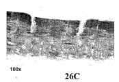
図3A〜3Fは、25% エタノール溶液での前処理およびそれに続くAd-LacZの感染の後のマウス膀胱の横断面を示す写真である。図3Aおよび3Bは、第1マウス膀胱の横断面を示す写真であり、図3Cおよび3Dは、第2マウス膀胱の横断面を示す写真であり、図3Eおよび3Fは、第3マウス膀胱の横断面を示す写真である。図3A、3Cおよび3Eは40倍の倍率で撮影されたものであり、図3B、3Dおよび3Fは100倍の倍率で撮影されたものである。 3A-3F are photographs showing cross sections of mouse bladders after pretreatment with 25% ethanol solution and subsequent infection with Ad-LacZ. 3A and 3B are photographs showing a cross section of the first mouse bladder, FIGS. 3C and 3D are photographs showing a cross section of the second mouse bladder, and FIGS. 3E and 3F are crossings of the third mouse bladder. It is a photograph which shows a surface. Figures 3A, 3C and 3E were taken at 40x magnification, and Figures 3B, 3D and 3F were taken at 100x magnification.
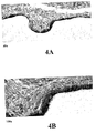
図4A〜4Fは、30% エタノール溶液での前処理およびそれに続くAd-LacZの感染の後のマウス膀胱の横断面を示す写真である。図4Aおよび4Bは、第1マウス膀胱の横断面を示す写真であり、図4Cおよび4Dは、第2マウス膀胱の横断面を示す写真であり、図4Eおよび4Fは、第3マウス膀胱の横断面を示す写真である。図4A、4Cおよび4Eは40倍の倍率で撮影されたものであり、図4B、4Dおよび4Fは100倍の倍率で撮影されたものである。 4A-4F are photographs showing cross sections of mouse bladders after pretreatment with 30% ethanol solution and subsequent infection with Ad-LacZ. 4A and 4B are photographs showing a cross section of the first mouse bladder, FIGS. 4C and 4D are photographs showing a cross section of the second mouse bladder, and FIGS. 4E and 4F are crossings of the third mouse bladder. It is a photograph which shows a surface. 4A, 4C and 4E were taken at a magnification of 40 times, and FIGS. 4B, 4D and 4F were taken at a magnification of 100 times.
げっ歯類の膀胱上皮におけるアデノウイルス媒介遺伝子導入および発現に対する化学物質での前処理の効果
げっ歯類の膀胱上皮におけるアデノウイルス媒介遺伝子導入および発現に対する化学物質での前処理の効果を評価するための研究を行った。
Effect of chemical pretreatment on adenovirus-mediated gene transfer and expression in rodent bladder epithelium To assess the effect of chemical pretreatment on adenovirus-mediated gene transfer and expression in rodent bladder epithelium Conducted research.
試験材料
アデノウイルスの凍結および製剤化のための当技術分野で公知の標準的な条件を用いて、Ad-βgalウイルスをCGIにおいて凍結製剤として作製した。ウイルス用のビヒクルはPBS + 10%グリセロールであった。
Test Material Ad-βgal virus was made as a frozen formulation in CGI using standard conditions known in the art for adenovirus freezing and formulation. The vehicle for the virus was PBS + 10% glycerol.
動物
この研究には152匹の雌BALB/cマウスを使用した。雌マウスを選択したのは、尿道カニューレ挿入および膀胱点滴が容易だからである。マウスは実験の開始日に約10〜12週齢であった。
Animals 152 female BALB / c mice were used for this study. Female mice were chosen because urethral cannulation and bladder infusion are easy. Mice were about 10-12 weeks old on the start of the experiment.
処理計画
以下の表に示すとおり、動物を各群に割当てた。化学物質およびウイルスの投与経路は膀胱内経路であった。
表2におけるデータのAd-βgalウイルスの濃度は、光学密度測定による測定では、1.3 x 10¹² vp/ml(第1調製、粒子:PFU:30)および1 x 1012 vp/ml(第2調製、粒子:PFU:30)であった。 The concentration of Ad-βgal virus in the data in Table 2 is 1.3 x 10 12 vp / ml (first preparation, particles: PFU: 30) and 1 x 10 12 vp / ml (second preparation) as measured by optical density measurement. , Particles: PFU: 30).
処理手法
1.動物をイソフランで麻酔し、24gカテーテルを尿道を介して膀胱内へ導入した。
Processing method
1. The animals were anesthetized with isoflurane and a 24g catheter was introduced into the bladder via the urethra.
2.残留尿を空にし、それぞれ100μlのPBSを膀胱に3回流した。 2. Residual urine was emptied and 100 μl of PBS each was flushed through the bladder three times.
3.被検試薬に基づき、膀胱の前処理を以下のとおりに行った。 3. Based on the test reagent, bladder pretreatment was performed as follows.
ポロキソマー407法:それぞれ100μlで2回洗浄した。第3洗浄を5分間維持し、更にもう1回の洗浄を行った。ウイルス点滴の前に、3回のPBS洗浄を行った。 Poloxomer 407 method: Washed twice with 100 μl each. The third wash was maintained for 5 minutes, and another wash was performed. Three PBS washes were performed before virus infusion.
ポロキソマー188法:それぞれ100μlで2回洗浄した。第3洗浄を5分間維持し、更にもう1回の洗浄を行った。ウイルス点滴の前に、3回のPBS洗浄を行った。 Poloxomer 188 method: Washed twice with 100 μl each. The third wash was maintained for 5 minutes, and another wash was performed. Three PBS washes were performed before virus infusion.
リポフェクタミン2000法:5μlのストックリポフェクタミン(1mg/ml)を195μlのPBS-10%グリセロールに加えた。等容量のAd-βgalウイルスと混合し、15分間インキュベートした。100μlの該混合物を膀胱内に投与し、膀胱内に30分間維持した。 Lipofectamine 2000 method: 5 μl of stock lipofectamine (1 mg / ml) was added to 195 μl of PBS-10% glycerol. Mixed with an equal volume of Ad-βgal virus and incubated for 15 minutes. 100 μl of the mixture was administered intravesically and maintained in the bladder for 30 minutes.
ベンジルアルコール法:それぞれ100μlで2回洗浄した。第3洗浄を15分間維持し、ついで更にもう1回の洗浄を行った。ウイルス点滴の前に、3回のPBS洗浄を行った。 Benzyl alcohol method: Washed twice with 100 μl each. The third wash was maintained for 15 minutes, followed by another wash. Three PBS washes were performed before virus infusion.
オキシクロロセン法:投与計画において記載されているのと同様にして洗浄を行った(すなわち、それぞれ100μlの3回の洗浄、5分間の維持を伴う1回の洗浄、15分間の維持を伴う1回の洗浄)。ウイルス点滴の前に、3回のPBS洗浄を行った。 Oxychlorocene method: Washing was performed as described in the dosing schedule (ie, 100 μl each for 3 washes, 1 wash with 5 minutes of maintenance, 1 with 15 minutes of maintenance 1 Washing). Three PBS washes were performed before virus infusion.
ポリドカノール法:それぞれ100μlで2回洗浄する。第3洗浄を5分間維持し、ついで更にもう1回の洗浄を行った。ウイルス点滴の前に、3回のPBS洗浄を行った。 Poldocanol method: Wash twice with 100 μl each. The third wash was maintained for 5 minutes, followed by another wash. Three PBS washes were performed before virus infusion.
DC-Cho法:それぞれ100μlで2回洗浄した。第3洗浄を5分間維持し、ついで更にもう1回の洗浄を行った。ウイルス点滴の前に、3回のPBS洗浄を行った。 DC-Cho method: Washed twice with 100 μl each. The third wash was maintained for 5 minutes, followed by another wash. Three PBS washes were performed before virus infusion.
0.4% HPMC 2910法:前処理は行わなかった。等容量の該ウイルスをHPMC2910の0.8% 溶液と混合し、該混合物を膀胱内に30分間点滴した。 0.4% HPMC 2910 method: No pretreatment was performed. An equal volume of the virus was mixed with a 0.8% solution of HPMC2910 and the mixture was instilled into the bladder for 30 minutes.
100mg/ml ポリリシン法:前処理は行わなかった。等容量の該ウイルスをポリリシンの100mg/ml溶液と混合し、該混合物を膀胱内に30分間点滴した。 100 mg / ml polylysine method: No pretreatment was performed. An equal volume of the virus was mixed with a 100 mg / ml solution of polylysine and the mixture was instilled into the bladder for 30 minutes.
0.4%ポリビニルアルコール(PVA)法:前処理は行わなかった。等容量の該ウイルスをPVAの0.8%溶液と混合し、該混合物を膀胱内に30分間点滴した。 0.4% polyvinyl alcohol (PVA) method: No pretreatment was performed. An equal volume of the virus was mixed with a 0.8% solution of PVA and the mixture was instilled into the bladder for 30 minutes.
n-ドデシル-β-D-グルコピラノシド法:それぞれ100μlで2回洗浄した。第3洗浄を5分間維持し、ついで更にもう1回の洗浄を行った。ウイルス点滴の前に、3回のPBS洗浄を行った。 n-dodecyl-β-D-glucopyranoside method: Washed twice with 100 μl each. The third wash was maintained for 5 minutes, followed by another wash. Three PBS washes were performed before virus infusion.
0.4% PVP法:前処理は行わなかった。等容量の該ウイルスをPVPの0.8%溶液と混合し、該混合物を膀胱内に30分間点滴した。 0.4% PVP method: No pretreatment was performed. An equal volume of the virus was mixed with a 0.8% solution of PVP and the mixture was instilled into the bladder for 30 minutes.
0.1% コレステロール-シクロデキストリン試薬法:前処理は行わなかった。等容量の該ウイルスをコレステロール-シクロデキストリンの0.2%溶液と混合し、該混合物を膀胱内に30分間点滴した。 0.1% cholesterol-cyclodextrin reagent method: No pretreatment was performed. An equal volume of the virus was mixed with a 0.2% solution of cholesterol-cyclodextrin and the mixture was instilled into the bladder for 30 minutes.
n-ドデシル-β-D-マルトシド法:それぞれ100μlで2回洗浄した。第3洗浄を5分間維持し、ついで更にもう1回の洗浄を行った。ウイルス点滴の前に、3回のPBS洗浄を行った。 n-dodecyl-β-D-maltoside method: Washed twice with 100 μl each. The third wash was maintained for 5 minutes, followed by another wash. Three PBS washes were performed before virus infusion.
ドデシルベンゼンスルホン酸ナトリウム塩法:それぞれ100μlで2回洗浄した。第3洗浄を5分間維持し、ついで更にもう1回の洗浄を行った。ウイルス点滴の前に、3回のPBS洗浄を行った。 Sodium dodecylbenzenesulfonate method: Washed twice with 100 μl each. The third wash was maintained for 5 minutes, followed by another wash. Three PBS washes were performed before virus infusion.
0.1% ドデシル硫酸ナトリウム法:それぞれ100μlで2回洗浄した。第3洗浄を5分間維持し、ついで更にもう1回の洗浄を行った。ウイルス点滴の前に、3回のPBS洗浄を行った。 0.1% sodium dodecyl sulfate method: Washed twice with 100 μl each. The third wash was maintained for 5 minutes, followed by another wash. Three PBS washes were performed before virus infusion.
0.1% Tween20法:それぞれ100μlで2回洗浄した。第3洗浄を5分間維持し、ついで更にもう1回の洗浄を行った。ウイルス点滴の前に、3回のPBS洗浄を行った。 0.1% Tween20 method: Washed twice with 100 μl each. The third wash was maintained for 5 minutes, followed by another wash. Three PBS washes were performed before virus infusion.
0.1% Triton(登録商標)X-100法:それぞれ100μlで2回洗浄した。第3洗浄を5分間維持し、ついで更にもう1回の洗浄を行った。ウイルス点滴の前に、3回のPBS洗浄を行った。 0.1% Triton® X-100 method: Washed twice with 100 μl each. The third wash was maintained for 5 minutes, followed by another wash. Three PBS washes were performed before virus infusion.
0.1% Forlan C-24法:それぞれ100μlで2回洗浄した。第3洗浄を5分間維持し、ついで更にもう1回の洗浄を行った。ウイルス点滴の前に、3回のPBS洗浄を行った。 0.1% Forlan C-24 method: Washed twice with 100 μl each. The third wash was maintained for 5 minutes, followed by another wash. Three PBS washes were performed before virus infusion.
0.1% デシル-b-D-マルトシド法:それぞれ100μlで2回洗浄した。第3洗浄を5分間維持し、ついで更にもう1回の洗浄を行った。ウイルス点滴の前に、3回のPBS洗浄を行った。 0.1% decyl-b-D-maltoside method: Washed twice with 100 μl each. The third wash was maintained for 5 minutes, followed by another wash. Three PBS washes were performed before virus infusion.
0.1% 6-シクロヘキシルヘキシル-b-D-マルトシド法:それぞれ100μlで2回洗浄した。第3洗浄を5分間維持し、ついで更にもう1回の洗浄を行った。ウイルス点滴の前に、3回のPBS洗浄を行った。 0.1% 6-cyclohexylhexyl-b-D-maltoside method: Washed twice with 100 μl each. The third wash was maintained for 5 minutes, followed by another wash. Three PBS washes were performed before virus infusion.
0.1% テトラデシル硫酸ナトリウム(Tromboject(登録商標), Omega Laboratories Ltd.)法:それぞれ100μlで2回洗浄した。第3洗浄を5分間維持し、ついで更にもう1回の洗浄を行った。ウイルス点滴の前に、3回のPBS洗浄を行った。 0.1% sodium tetradecyl sulfate (Tromboject (registered trademark), Omega Laboratories Ltd.) method: Washed twice with 100 μl each. The third wash was maintained for 5 minutes, followed by another wash. Three PBS washes were performed before virus infusion.
0.1% フェニル-β-D-グルコピラノシド法:それぞれ100μlで2回洗浄した。第3洗浄を5分間維持し、ついで更にもう1回の洗浄を行った。ウイルス点滴の前に、3回のPBS洗浄を行った。 0.1% phenyl-β-D-glucopyranoside method: Washed twice with 100 μl each. The third wash was maintained for 5 minutes, followed by another wash. Three PBS washes were performed before virus infusion.
0.1% スクロースモノラウラート法:それぞれ100μlで2回洗浄した。第3洗浄を5分間維持し、ついで更にもう1回の洗浄を行った。ウイルス点滴の前に、3回のPBS洗浄を行った。 0.1% sucrose monolaurate method: Washed twice with 100 μl each. The third wash was maintained for 5 minutes, followed by another wash. Three PBS washes were performed before virus infusion.
0.1% 1-O-ドデシル-rac-グリセロール法:それぞれ100μlで2回洗浄した。第3洗浄を5分間維持し、ついで更にもう1回の洗浄を行った。ウイルス点滴の前に、3回のPBS洗浄を行った。 0.1% 1-O-dodecyl-rac-glycerol method: Washed twice with 100 μl each. The third wash was maintained for 5 minutes, followed by another wash. Three PBS washes were performed before virus infusion.
in vivo geneSHUTTLE(商標)法:4mMのin vivo geneSHUTTLE(商標)をウイルスと混合した。第1日に投与した。60mlの脂質を90mlの水で希釈した。ついで150μlのAd-βgalを加えた。 In vivo geneSHUTTLE ™ method: 4 mM in vivo geneSHUTTLE ™ was mixed with virus. Administered on day 1. 60 ml of lipid was diluted with 90 ml of water. Then 150 μl of Ad-βgal was added.
4.該ウイルスを抜き取り、100μlのPBSを膀胱に3回流すことにより、ウイルス処理(45分間)を停止した。 Four. The virus was extracted and virus treatment (45 minutes) was stopped by flowing 100 μl of PBS through the bladder three times.
測定/決定
処理日に、投与前に動物の臨床状態を観察し、実験期間中には、動物を毎日観察した。
Measurement / Decision On the day of treatment, the animals were observed for clinical status prior to dosing and animals were observed daily during the experimental period.
β-ガラクトシダーゼ活性の評価
処理の48時間後に動物を殺した。膀胱を0.1mlの全器官固定液、2%ニュートラル(Neutral)緩衝ホルマリン、2%グルタルアルデヒド、2mM MgCl2、10 mM PBS(pH 7.4)で満たした。ついで膀胱を摘出し、全器官固定液に1時間浸した。ついで該膀胱を縦方向に切り開き、4℃で24時間リンスし(2 mM MgCl2、0.1%デオキシコラート、0.2% Triton中)、X-gal染色溶液中に浸した。その縦方向に開いた膀胱の内腔上皮内でのトランスジーンの発現を実験的に測定した。
Evaluation of β-galactosidase activity Animals were sacrificed 48 hours after treatment. Whole organ fixative bladder 0.1 ml, 2% neutral (Neutral) buffered formalin, 2% glutaraldehyde, filled with 2mM MgCl 2, 10 mM PBS ( pH 7.4). The bladder was then removed and immersed in whole organ fixative for 1 hour. The bladder was then opened longitudinally, rinsed at 4 ° C. for 24 hours (2 mM MgCl 2 , 0.1% deoxycholate, in 0.2% Triton) and immersed in X-gal staining solution. The expression of the transgene in the luminal epithelium of the bladder opened vertically was experimentally measured.
組織病理学
全器官固定液中で固定した膀胱を切片化し、組織学的検査のためにヘマトキシリン-エオシンで染色した。
Histopathology Bladder fixed in whole organ fixative was sectioned and stained with hematoxylin-eosin for histological examination.
結果
前記実験の結果は以下のとおりに要約されうる。
Results The results of the experiment can be summarized as follows.
4% ポロキサマー407(Pluronic 127)での5分間の膀胱の前処理は<5%の形質導入をもたらした。 Pretreatment of the bladder with 4% poloxamer 407 (Pluronic 127) for 5 minutes resulted in <5% transduction.
リポフェクタミンとウイルスとの混合物(前処理無し)での膀胱の処理は<5%の形質導入をもたらした。 Treatment of the bladder with a mixture of lipofectamine and virus (no pretreatment) resulted in <5% transduction.
In vivo geneSHUTTLE(商標)とウイルスとの混合物(前処理無し)での膀胱の処理は<5%の形質導入をもたらした。 Treatment of the bladder with a mixture of in vivo geneSHUTTLE ™ and virus (no pretreatment) resulted in <5% transduction.
0.1% オキシクロロセンでの5分間の膀胱の前処理は尿路上皮の>90% 形質導入をもたらした。病理学者の報告は、無傷上皮層を伴う軽度の粘膜下浮腫を示した。 Pretreatment of the bladder with 0.1% oxychlorocene for 5 minutes resulted in> 90% transduction of the urothelium. A pathologist's report showed mild submucosal edema with an intact epithelial layer.
0.2% オキシクロロセンでの5分間の膀胱の前処理は尿路上皮の>90% 形質導入をもたらした。病理学者の報告は、最小限度の粘膜下浮腫および血管周囲リンパ球を示した。 Pretreatment of the bladder with 0.2% oxychlorocene for 5 minutes resulted in> 90% transduction of the urothelium. A pathologist's report showed minimal submucosal edema and perivascular lymphocytes.
0.2% オキシクロロセンでの15分間の膀胱の前処理は尿路上皮の>90% 形質導入をもたらした。病理学者の報告は、粘膜下組織における化膿性滲出物、出血および浮腫を伴う限局性(focal)の重度の潰瘍形成を示した。 Pretreatment of the bladder for 15 minutes with 0.2% oxychlorocene resulted in> 90% transduction of the urothelium. A pathologist report showed focal severe ulceration with purulent exudates, bleeding and edema in submucosa.
0.4% オキシクロロセンでの5分間の膀胱の前処理は尿路上皮の>90% 形質導入をもたらした。病理学者の報告は、限局性の大きな潰瘍を伴う中等度の粘膜下浮腫を示した。 Pretreatment of the bladder with 0.4% oxychlorocene for 5 minutes resulted in> 90% transduction of the urothelium. A pathologist's report showed moderate submucosal edema with localized large ulcers.
0.02% ポリドカノールでの5分間の膀胱の前処理は尿路上皮の10〜20% 形質導入をもたらした。病理学者の報告は無傷粘膜を示した。 Pretreatment of the bladder with 0.02% polidocanol for 5 minutes resulted in 10-20% transduction of the urothelium. A pathologist's report showed an intact mucosa.
0.05% ポリドカノールでの5分間の膀胱の前処理は尿路上皮の30〜40% 形質導入をもたらした。病理学者の報告は、最小限度の粘膜下浮腫を示した。 Pretreatment of the bladder with 0.05% polidocanol for 5 minutes resulted in 30-40% transduction of the urothelium. Pathologist reports showed minimal submucosal edema.
0.2% ポリドカノールでの5分間の膀胱の前処理は尿路上皮の50〜80% 形質導入をもたらした。病理学者の報告は、びらんおよび限局性潰瘍ならびに粘膜障害を示した。 Pretreatment of the bladder with 0.2% polidocanol for 5 minutes resulted in 50-80% transduction of the urothelium. Pathologist reports showed erosion and localized ulcers and mucosal damage.
0.02% n-ドデシルβ-D-マルトシドでの5分間の膀胱の前処理は尿路上皮の50〜80% 形質導入をもたらした。病理学者の報告は、有意な病変を示さなかった。 Pretreatment of the bladder for 5 minutes with 0.02% n-dodecyl β-D-maltoside resulted in 50-80% transduction of the urothelium. Pathologist reports did not show significant lesions.
0.05% n-ドデシルβ-D-マルトシドでの5分間の膀胱の前処理は尿路上皮の>90% 形質導入をもたらした。病理学者の報告は、有意な病変を示さなかった。 Pretreatment of the bladder for 5 minutes with 0.05% n-dodecyl β-D-maltoside resulted in> 90% transduction of the urothelium. Pathologist reports did not show significant lesions.
0.2% n-ドデシルβ-D-マルトシドでの5分間の膀胱の前処理は尿路上皮の>90% 形質導入をもたらした。病理学者の報告は、びらん、限局性潰瘍、中等度の粘膜下浮腫および粘膜障害を示した。 Pretreatment of the bladder for 5 minutes with 0.2% n-dodecyl β-D-maltoside resulted in> 90% transduction of the urothelium. Pathologist reports showed erosions, localized ulcers, moderate submucosal edema and mucosal damage.
0.2% ドデシルベンゼンスルホン酸での5分間の膀胱の前処理は、尿路上皮の20〜40% 形質導入をもたらした。 Pretreatment of the bladder with 0.2% dodecylbenzene sulfonic acid for 5 minutes resulted in 20-40% transduction of the urothelium.
前記の結果から理解されうるとおり、最終的な青色染色(LacZ)のレベルによる測定で、いくつかの単一化合物および1つの混合試薬が形質導入の有意な増強を示した。いくつかの他の単一化合物は形質導入の増強をもたらしたものの、そのレベルはより低かった。各被検化学物質の正当性を実証するために、参照体としてのエタノールでの前処理を行った。30%ものエタノール率であっても、僅か10〜20%の形質導入が観察されたに過ぎなかった。「強い反応体」は、10〜20%染色を示すエタノール前処理対照より有意に良好な(すなわち、70〜90%)染色を示す形質導入増強剤であった。弱い反応体は、エタノール対照群と比較して有意に小さな染色面積を示した。 As can be seen from the above results, several single compounds and one mixed reagent showed significant enhancement of transduction as measured by the level of final blue staining (LacZ). Although some other single compounds resulted in enhanced transduction, their levels were lower. In order to verify the validity of each test chemical, pretreatment with ethanol as a reference substance was performed. Only 10-20% transduction was observed even at ethanol levels as high as 30%. “Strong reactants” were transduction enhancers that exhibited significantly better (ie, 70-90%) staining than ethanol pretreated controls that exhibited 10-20% staining. Weak reactants showed a significantly smaller stained area compared to the ethanol control group.
0.02%〜0.5%ポリドカノール、0.02〜0.5% n-ドデシル-b-D-マルトシド、0.1% 6-シクロヘキシルヘキシル-b-D-マルトシド、0.1%〜0.4% オキシクロロセン、0.2% ドデシルベンゼンスルホン酸ナトリウム塩および0.1%ドデシル硫酸ナトリウムでの膀胱表面の前処理後に最強の反応(すなわち、最高レベルの形質導入)が観察された。 0.02% to 0.5% polidocanol, 0.02 to 0.5% n-dodecyl-bD-maltoside, 0.1% 6-cyclohexylhexyl-bD-maltoside, 0.1% to 0.4% oxychlorocene, 0.2% sodium dodecylbenzenesulfonate and 0.1% The strongest response (ie the highest level of transduction) was observed after pretreatment of the bladder surface with sodium dodecyl sulfate.
「弱い反応体」には、0.1% デシル-b-D-マルトシドおよび0.1% Triton(登録商標)X-100が含まれた。 “Weak reactants” included 0.1% decyl-b-D-maltoside and 0.1% Triton® X-100.
理論により束縛されることを望むものではないが、満足しうる結果を与えた形質導入増強試薬の物理的および化学的特性を分析することにより、作用メカニズムを仮定することが可能である。形質導入増強試薬は一般には界面活性剤である。界面活性剤はイオン性または非イオン性でありうる。界面活性剤は、好ましくは、親水性および親油性の両方の部分を有する。該分子の親水性部分は水溶性に寄与し、親油性(すなわち、疎水性)部分は脂質との分子相互作用を助ける。親水性/親油性バランス、すなわち、HLB比は、該分子の各部分の相対サイズの指標である。 While not wishing to be bound by theory, it is possible to postulate the mechanism of action by analyzing the physical and chemical properties of the transduction enhancing reagent that gave satisfactory results. The transduction enhancing reagent is generally a surfactant. Surfactants can be ionic or nonionic. The surfactant preferably has both hydrophilic and lipophilic moieties. The hydrophilic portion of the molecule contributes to water solubility, and the lipophilic (ie, hydrophobic) portion aids molecular interaction with the lipid. The hydrophilic / lipophilic balance, ie the HLB ratio, is an indicator of the relative size of each part of the molecule.
糖に基づく界面活性剤(糖類)
本発明の形質導入増強剤は、親油性置換基を有する糖(例えば、単糖、二糖または多糖)でありうる。形質導入増強剤は、親油性置換基を有する任意の単糖、二糖または多糖でありうる。本発明の好ましい実施形態においては、形質導入増強剤は、親油性置換基を有する二糖である。典型的な二糖には、マルトースまたはスクロースが含まれる。しかし、ラクトース、イソマルトース、トレハロースまたはセロビオースを含む、親油性置換基を有する他の二糖も使用されうる。
Surfactant based on sugar (sugar)
The transduction enhancer of the present invention can be a saccharide having a lipophilic substituent (eg, a monosaccharide, disaccharide or polysaccharide). The transduction enhancer can be any monosaccharide, disaccharide or polysaccharide having a lipophilic substituent. In a preferred embodiment of the invention, the transduction enhancer is a disaccharide having a lipophilic substituent. Typical disaccharides include maltose or sucrose. However, other disaccharides with lipophilic substituents may also be used, including lactose, isomaltose, trehalose or cellobiose.
親油性置換基は、直鎖状(例えば、直鎖状n-アルカンまたはアルケン)または非直鎖状(例えば、環状または分枝アルカンまたはアルケン)でありうる。また、親油性置換基はアルカン酸残基でありうる。親油性置換基の長さは、所望の親水性-親油性バランスが達成されるよう様々の長さとなりうる。種々のマルトシド置換化合物に関する試験は、十分な親油性基の長さが形質導入効率の改善をもたらすことを示した。例えば、n-ドデシル-β-D-マルトシドおよび6-シクロヘキシルヘキシル-β-D-マルトシドは共に、形質導入を有意に増強した。これに対して、n-デシル-β-D-マルトシドは形質導入に対して僅かな効果しかもたらさなかった。 The lipophilic substituent can be linear (eg, linear n-alkane or alkene) or non-linear (eg, cyclic or branched alkane or alkene). The lipophilic substituent can also be an alkanoic acid residue. The length of the lipophilic substituent can be varied to achieve the desired hydrophilic-lipophilic balance. Studies with various maltoside substituted compounds showed that sufficient lipophilic group length resulted in improved transduction efficiency. For example, both n-dodecyl-β-D-maltoside and 6-cyclohexylhexyl-β-D-maltoside significantly enhanced transduction. In contrast, n-decyl-β-D-maltoside had only a minor effect on transduction.
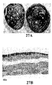
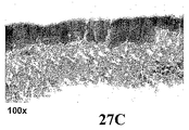
n-ドデシル-β-D-マルトシドでの膀胱の前処理に関する結果を図25〜29に示す。図25Aは、0.02% n-ドデシルβ-D-マルトシド溶液での前処理およびそれに続くAd-LacZの感染の後の第1マウス膀胱の内腔表面を示す写真である。図25Bおよび25Cは、図25Aのマウス膀胱の横断面を示す写真である。図25Bは40倍の倍率で撮影されたものであり、図25Cは100倍の倍率で撮影されたものである。図26Aは、0.02% n-ドデシルβ-D-マルトシド溶液での前処理およびそれに続くAd-LacZの感染の後の第2マウス膀胱の内腔表面を示す写真である。図26Bおよび26Cは、図26Aのマウス膀胱の横断面を示す写真である。図26Bは40倍の倍率で撮影されたものであり、図26Cは100倍の倍率で撮影されたものである。図27Aは、0.05% n-ドデシルβ-D-マルトシド溶液での前処理およびそれに続くAd-LacZの感染の後の第1マウス膀胱の内腔表面を示す写真である。図27Bおよび27Cは、図27Aのマウス膀胱の横断面を示す写真であり、図27Bは40倍の倍率で撮影されたものであり、図27Cは100倍の倍率で撮影されたものである。図28Aは、0.05% n-ドデシルβ-D-マルトシド溶液での前処理およびそれに続くAd-LacZの感染の後の第1マウス膀胱の内腔表面を示す写真である。図28Bおよび28Cは、図28Aのマウス膀胱の横断面を示す写真である。図28Bは40倍の倍率で撮影されたものであり、図28Cは100倍の倍率で撮影されたものである。図29Aは、0.2% n-ドデシルβ-D-マルトシド溶液での前処理およびそれに続くAd-LacZの感染の後の第1マウス膀胱の内腔表面を示す写真である。図29Bおよび29Cは、図29Aのマウス膀胱の横断面を示す写真である。図29Bは40倍の倍率で撮影されたものであり、図29Cは100倍の倍率で撮影されたものである。 The results for bladder pretreatment with n-dodecyl-β-D-maltoside are shown in FIGS. FIG. 25A is a photograph showing the luminal surface of the first mouse bladder after pretreatment with 0.02% n-dodecyl β-D-maltoside solution and subsequent infection with Ad-LacZ. 25B and 25C are photographs showing a cross section of the mouse bladder of FIG. 25A. FIG. 25B was taken at 40 × magnification, and FIG. 25C was taken at 100 × magnification. FIG. 26A is a photograph showing the luminal surface of the second mouse bladder after pretreatment with 0.02% n-dodecyl β-D-maltoside solution and subsequent infection with Ad-LacZ. 26B and 26C are photographs showing a cross section of the mouse bladder of FIG. 26A. FIG. 26B was taken at 40 × magnification, and FIG. 26C was taken at 100 × magnification. FIG. 27A is a photograph showing the luminal surface of the first mouse bladder after pretreatment with 0.05% n-dodecyl β-D-maltoside solution and subsequent infection with Ad-LacZ. 27B and 27C are photographs showing a cross section of the mouse bladder of FIG. 27A, FIG. 27B was taken at 40 × magnification, and FIG. 27C was taken at 100 × magnification. FIG. 28A is a photograph showing the luminal surface of the first mouse bladder after pretreatment with 0.05% n-dodecyl β-D-maltoside solution and subsequent infection with Ad-LacZ. 28B and 28C are photographs showing a cross section of the mouse bladder of FIG. 28A. FIG. 28B was taken at 40 × magnification, and FIG. 28C was taken at 100 × magnification. FIG. 29A is a photograph showing the luminal surface of the first mouse bladder after pretreatment with 0.2% n-dodecyl β-D-maltoside solution and subsequent infection with Ad-LacZ. 29B and 29C are photographs showing a cross section of the mouse bladder of FIG. 29A. FIG. 29B was taken at 40 × magnification, and FIG. 29C was taken at 100 × magnification.
n-ドデシル-β-D-マルトシドおよびn-デシル-β-D-マルトシドの化学式を以下に示す。
(式中、nはそれぞれ11および9である)。6-シクロヘキシルヘキシル-β-D-マルトシドの化学式は以下のとおりである。
(式中、nは6である)。 (Where n is 6).
形質導入実験は、親油性側鎖(すなわち、CH2-CH2)サイズにおける僅かな減少が、形質導入の増強に関する該分子の効力を大きく抑制しうることを示した。前記化合物のすべては水中およびPBSバッファー中の両方において良好な溶解度を有することに注目することが重要である。 Transduction experiments have shown that a slight decrease in lipophilic side chain (ie, CH 2 —CH 2 ) size can greatly suppress the efficacy of the molecule for enhanced transduction. It is important to note that all of the compounds have good solubility both in water and in PBS buffer.
より短い親水性部分を有するこのクラスの界面活性剤における化合物も評価した。n-ドデシル-β-D-グルコピラノシドに関する結果は形質導入の増強をほとんど又は全く示さなかった。n-ドデシル-β-D-グルコピラノシドの化学式は以下のとおりである。
(式中、nは11である)。理論により束縛されることを望むものではないが、分子の親水性および親油性部分の相対サイズは形質導入の増強に影響を及ぼすらしい。したがって、より短い鎖のn-アルキル-β-D-グルコピラノシド(例えば、n-ヘキシル-β-D-グルコピラノシド)は形質導入の改善を示しうる。 (Where n is 11). While not wishing to be bound by theory, it appears that the relative size of the hydrophilic and lipophilic portions of the molecule affects the enhancement of transduction. Thus, shorter chain n-alkyl-β-D-glucopyranoside (eg, n-hexyl-β-D-glucopyranoside) may show improved transduction.
親油性置換基を有する任意の単糖、二糖または多糖を本発明の形質導入増強剤として使用することが可能である。典型的な二糖化合物には、スクロース、ラクトース、マルトース、イソマルトース、トレハロースおよびセロビオースが含まれる。親油性置換基は、好ましくは、アルキルまたはアルケニル基を含む。本発明の好ましい実施形態においては、親油性置換基はアルカン酸残基である。 Any monosaccharide, disaccharide or polysaccharide having a lipophilic substituent can be used as the transduction enhancer of the present invention. Typical disaccharide compounds include sucrose, lactose, maltose, isomaltose, trehalose and cellobiose. The lipophilic substituent preferably comprises an alkyl or alkenyl group. In a preferred embodiment of the invention, the lipophilic substituent is an alkanoic acid residue.
単糖および二糖のβ形態が前記に示されているが、これらの及び他の単糖、二糖または多糖化合物のα形態も本発明において使用することが可能である。本発明の典型的なα糖形質導入増強剤には、n-ドデシル-α-D-マルトシド、n-ヘキシル-α-D-グルコピラノシドおよび6-シクロヘキシルヘキシル-α-D-マルトシドが含まれる。また、DまたはL形態の単糖、二糖または多糖を本発明の形質導入増強剤として使用することが可能である。 Although the beta forms of monosaccharides and disaccharides are shown above, alpha forms of these and other monosaccharides, disaccharides or polysaccharide compounds can also be used in the present invention. Exemplary alpha sugar transduction enhancers of the present invention include n-dodecyl-α-D-maltoside, n-hexyl-α-D-glucopyranoside and 6-cyclohexylhexyl-α-D-maltoside. In addition, D or L forms of monosaccharides, disaccharides or polysaccharides can be used as the transduction enhancer of the present invention.
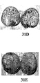
図31A〜31Eは、種々の長さのアルキル側鎖を有するアルキルマルトシドおよびアルキルマルトピラノシド前処理剤で処理されたマウス膀胱の内腔表面を示す写真である。図31Aは、Ad-LacZ(109 vp)の感染の前にn-ドデシル-β-D-マルトシド(C12アルキル側鎖)で処理されたマウス膀胱の内腔表面の写真である。図31Bは、感染の前にトリデシル-β-D-マルトピラノシド(C13アルキル側鎖)で処理されたマウス膀胱の内腔表面の写真である。図31Cは、感染の前にn-テトラドデシル-β-D-マルトシド(C14アルキル側鎖)で処理されたマウス膀胱の内腔表面の写真である。図31Dは、感染の前にn-デシル-β-D-マルトシド(C10アルキル側鎖)で処理されたマウス膀胱の内腔表面の写真である。図31Eは、感染の前にn-オクチル-β-D-マルトピラノシド(C8アルキル側鎖)で処理されたマウス膀胱の内腔表面の写真である。 FIGS. 31A-31E are photographs showing the luminal surface of mouse bladders treated with alkyl maltosides and alkyl maltopyranoside pretreatments with various lengths of alkyl side chains. FIG. 31A is a photograph of the luminal surface of a mouse bladder treated with n-dodecyl-β-D-maltoside (C12 alkyl side chain) prior to infection with Ad-LacZ (10 9 vp). FIG. 31B is a photograph of the luminal surface of a mouse bladder treated with tridecyl-β-D-maltopyranoside (C13 alkyl side chain) prior to infection. FIG. 31C is a photograph of the luminal surface of a mouse bladder treated with n-tetradodecyl-β-D-maltoside (C14 alkyl side chain) prior to infection. FIG. 31D is a photograph of the luminal surface of a mouse bladder treated with n-decyl-β-D-maltoside (C10 alkyl side chain) prior to infection. FIG. 31E is a photograph of the luminal surface of a mouse bladder treated with n-octyl-β-D-maltopyranoside (C8 alkyl side chain) prior to infection.
イオン性アルキル界面活性剤
本発明の形質導入増強化合物として、イオン性アルキル界面活性剤を使用することも可能である。典型的なイオン性アルキル界面活性剤には、
により表される式を有するドデシル硫酸ナトリウムが含まれる。 Sodium dodecyl sulfate having the formula represented by:
もう1つの典型的なイオン性界面活性剤は、
により表される化学式を有するドデシルベンゼンスルホン酸のナトリウム塩である。 Is a sodium salt of dodecylbenzenesulfonic acid having the chemical formula represented by:
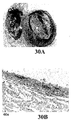
前記のタイプの界面活性剤を評価したところ、それらは、前記の非イオン性試薬と比較して形質導入の増強を示すことが判明した。これらの結果は、ドデシルベンゼンスルホン酸ナトリウム塩に関しては、図30に示されている。図30A〜30Cから理解されうるとおり、ドデシルベンゼンスルホン酸ナトリウム塩はマウス膀胱におけるAd-LacZの形質導入を増強した。図30Aは、デデシルベンゼンスルホン酸溶液の0.2%ナトリウム塩での前処理およびそれに続くAd-LacZの感染の後の第1マウス膀胱の内腔表面を示す写真である。図30Bおよび30Cは、図30Aのマウス膀胱の横断面を示す写真である。図30Bは40倍の倍率で撮影されたものであり、図30Cは100倍の倍率で撮影されたものである。 Evaluation of the types of surfactants revealed that they show enhanced transduction compared to the nonionic reagents. These results are shown in FIG. 30 for dodecylbenzenesulfonic acid sodium salt. As can be seen from FIGS. 30A-30C, dodecylbenzenesulfonate sodium salt enhanced Ad-LacZ transduction in the mouse bladder. FIG. 30A is a photograph showing the luminal surface of the first mouse bladder after pretreatment of decylbenzenesulfonic acid solution with 0.2% sodium salt and subsequent infection with Ad-LacZ. 30B and 30C are photographs showing a cross section of the mouse bladder of FIG. 30A. FIG. 30B was taken at 40 × magnification, and FIG. 30C was taken at 100 × magnification.
図32A〜32Cは、種々の長さのアルキル側鎖を有するアルキル硫酸ナトリウム前処理剤で処理されたマウス膀胱の内腔表面を示す写真である。図32Aは、Ad-LacZ(109 vp)の感染の前にドデシル硫酸ナトリウム(SDS)(C12アルキル側鎖)で処理された膀胱の内腔表面を示す。図32Bは、感染の前にデシル硫酸(C10アルキル側鎖)で処理された膀胱の内腔表面を示す。図32Cは、感染の前にオクチル硫酸ナトリウム(C8アルキル側鎖)で処理された膀胱の内腔表面を示す。 32A-32C are photographs showing the luminal surface of mouse bladder treated with sodium alkyl sulfate pretreatments with various lengths of alkyl side chains. FIG. 32A shows the luminal surface of the bladder treated with sodium dodecyl sulfate (SDS) (C12 alkyl side chain) prior to infection with Ad-LacZ (10 9 vp). FIG. 32B shows the luminal surface of the bladder treated with decyl sulfate (C10 alkyl side chain) prior to infection. FIG. 32C shows the luminal surface of the bladder treated with sodium octyl sulfate (C8 alkyl side chain) prior to infection.
イオン性アルキル界面活性剤は2つの部分(親水性部分および親油性部分)よりなる。該分子のこれらの部分の配置は、前記の糖に基づく増強剤に類似している。本発明においては、前記のものに類似しておりアルキル置換における変更を伴う化合物も使用することが可能である。 The ionic alkyl surfactant consists of two parts: a hydrophilic part and a lipophilic part. The arrangement of these parts of the molecule is similar to the sugar-based enhancers described above. In the present invention, it is also possible to use compounds similar to those described above, with changes in alkyl substitution.
アルキル(エーテル)アルコール
また、本発明においては、形質導入増強化合物としてアルキルエーテル化合物を使用することが可能である。ポリドカノール、すなわち、化学式:
![]()
![]()
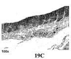
種々の濃度のポリドカノールでの膀胱表面の前処理に関する結果を図19〜24に示す。0.02% ポリドカノールでの膀胱表面の前処理に関する結果を図19および20に示す。図19Aは、0.02% ポリドカノール溶液での前処理およびそれに続くAd-LacZの感染の後の第1マウス膀胱の内腔表面を示す写真である。図19Bおよび19Cは、図19Aのマウス膀胱の横断面を示す写真である。図19Bは40倍の倍率で撮影されたものであり、図19Cは100倍の倍率で撮影されたものである。図20Aは、0.02% ポリドカノール溶液での前処理およびそれに続くAd-LacZの感染の後の第2マウス膀胱の内腔表面を示す写真である。図20Bおよび20Cは、図20Aのマウス膀胱の横断面を示す写真である。図20Bは40倍の倍率で撮影されたものであり、図20Cは100倍の倍率で撮影されたものである。 Results for bladder surface pretreatment with various concentrations of polidocanol are shown in FIGS. The results for pretreatment of the bladder surface with 0.02% polidocanol are shown in FIGS. 19 and 20. FIG. 19A is a photograph showing the luminal surface of the first mouse bladder after pretreatment with 0.02% polidocanol solution and subsequent infection with Ad-LacZ. 19B and 19C are photographs showing a cross section of the mouse bladder of FIG. 19A. FIG. 19B was taken at 40 × magnification, and FIG. 19C was taken at 100 × magnification. FIG. 20A is a photograph showing the luminal surface of the second mouse bladder after pretreatment with 0.02% polidocanol solution and subsequent infection with Ad-LacZ. 20B and 20C are photographs showing a cross section of the mouse bladder of FIG. 20A. FIG. 20B was taken at 40 × magnification, and FIG. 20C was taken at 100 × magnification.
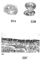
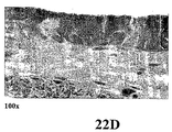
0.05% ポリドカノールでの膀胱表面の前処理に関する結果を図21および22に示す。図21Aおよび21Bは、0.05% ポロドカノール溶液での前処理およびそれに続くAd-LacZの感染の後の第1マウス膀胱のそれぞれ外表面および内腔表面を示す写真である。図21Cおよび21Dは、図21Aのマウス膀胱の横断面を示す写真である。図21Bは40倍の倍率で撮影されたものであり、図21Cは100倍の倍率で撮影されたものである。図22Aおよび22Bは、0.05% ポロドカノール溶液での前処理およびそれに続くAd-LacZの感染の後の第2マウス膀胱のそれぞれ外表面および内腔表面を示す写真である。図22Cおよび22Dは、図22Aのマウス膀胱の横断面を示す写真である。図22Bは40倍の倍率で撮影されたものであり、図22Cは100倍の倍率で撮影されたものである。 Results for pretreatment of the bladder surface with 0.05% polidocanol are shown in FIGS. FIGS. 21A and 21B are photographs showing the outer and luminal surfaces, respectively, of the first mouse bladder after pretreatment with 0.05% polodocanol solution and subsequent infection with Ad-LacZ. 21C and 21D are photographs showing a cross section of the mouse bladder of FIG. 21A. FIG. 21B was taken at 40 × magnification, and FIG. 21C was taken at 100 × magnification. 22A and 22B are photographs showing the outer and luminal surfaces, respectively, of the second mouse bladder after pretreatment with 0.05% polodocanol solution and subsequent infection with Ad-LacZ. 22C and 22D are photographs showing a cross section of the mouse bladder of FIG. 22A. FIG. 22B was taken at 40 × magnification, and FIG. 22C was taken at 100 × magnification.
0.2% ポリドカノールでの膀胱表面の前処理に関する結果を図23および24に示す。図23Aは、0.2% ポロドカノール溶液での前処理およびそれに続くAd-LacZの感染の後の第1マウス膀胱の内腔表面を示す写真である。図23Bおよび23Cは、図23Aのマウス膀胱の横断面を示す写真である。図23Bは40倍の倍率で撮影されたものであり、図23Cは100倍の倍率で撮影されたものである。図24Aは、0.2% ポロドカノール溶液での前処理およびそれに続くAd-LacZの感染の後の第2マウス膀胱の内腔表面を示す写真である。図24Bおよび24Cは、図24Aのマウス膀胱の横断面を示す写真である。図24Bは40倍の倍率で撮影されたものであり、図24Cは100倍の倍率で撮影されたものである。 Results for bladder surface pretreatment with 0.2% polidocanol are shown in FIGS. FIG. 23A is a photograph showing the luminal surface of the first mouse bladder after pretreatment with a 0.2% polodocanol solution and subsequent infection with Ad-LacZ. 23B and 23C are photographs showing a cross section of the mouse bladder of FIG. 23A. FIG. 23B was taken at 40 × magnification, and FIG. 23C was taken at 100 × magnification. FIG. 24A is a photograph showing the luminal surface of the second mouse bladder after pretreatment with 0.2% polodocanol solution and subsequent infection with Ad-LacZ. 24B and 24C are photographs showing a cross section of the mouse bladder of FIG. 24A. FIG. 24B was taken at 40 × magnification, and FIG. 24C was taken at 100 × magnification.
一般式:
(式中、x = 10)を有するTriton(登録商標)X-100も評価したところ、それも形質導入を増強することが判明した。ベンゼン環ではなくシクロヘキサン環を有する類似化合物も本発明の形質導入増強剤として使用されうる。この化合物は以下の化学構造:
(式中、x = 10)を有する。xが任意の正の整数である前記タイプの化合物も本発明において使用されうる。 (Where x = 10). Compounds of the above type where x is any positive integer may also be used in the present invention.
一般構造:
を有する同様のアルキル(エーテル)化合物も商業的に入手可能である。これらの化合物の商品名は「Brij」である。前記に示した化合物は「Brij 56」と称される。Brij 56は化学式C20H42O5を有する。もう1つの商業的に入手可能な化合物「Brij 58」は化学式C56H114O21を有する。 Similar alkyl (ether) compounds having are also commercially available. The trade name for these compounds is “Brij”. The compound shown above is referred to as “Brij 56”. Brij 56 has the chemical formula C 20 H 42 O 5 . Another commercially available compound “Brij 58” has the chemical formula C 56 H 114 O 21 .
前記の任意のアルキル(エーテル)化合物が本発明の形質導入増強剤として使用されうる。 Any of the aforementioned alkyl (ether) compounds can be used as the transduction enhancer of the present invention.
オキシクロロセンナトリウム
約6.5〜6.9のpHの、ドデシルベンゼンスルホン酸のナトリウム塩と次亜塩素酸とを含む組成物(すなわち、オキシクロロセンナトリウム)を評価した。これらの評価において使用したオキシクロロセンナトリウムはClorpactin WCS-90の名称で販売されていた(Guardian Labsにより製造され、Cardinal Healthにより販売されていた)。オキシクロロセンナトリウムは、尿路感染症の治療ならびに腹部および形成手術において使用されている。
Sodium Oxychlorocene A composition comprising a sodium salt of dodecylbenzene sulfonic acid and hypochlorous acid at a pH of about 6.5 to 6.9 (ie sodium oxychlorocene) was evaluated. The sodium oxychlorocene used in these evaluations was sold under the name Clorpactin WCS-90 (manufactured by Guardian Labs and sold by Cardinal Health). Oxychlorocene sodium is used in the treatment of urinary tract infections and in abdominal and plastic surgery.
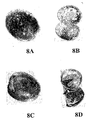
オキシクロロセンナトリウムでの膀胱表面の前処理に関する結果を図8〜18に示す。図8A〜8Nは、0.2% オキシクロロセン溶液での5分間の前処理およびそれに続くAd-LacZ感染の後の7つのマウス膀胱を示す写真である。図8Aおよび8Bは第1膀胱のそれぞれ外表面および内腔表面を示し、図8Cおよび8Dは第2膀胱のそれぞれ外表面および内腔表面を示し、図8Eおよび8Fは第3膀胱のそれぞれ外表面および内腔表面を示し、図8Gおよび8Hは第4膀胱のそれぞれ外表面および内腔表面を示し、図8Iおよび8Jは第5膀胱のそれぞれ外表面および内腔表面を示し、図8Kおよび8Lは第6膀胱のそれぞれ外表面および内腔表面を示し、図8Mおよび8Nは第7膀胱のそれぞれ外表面および内腔表面を示す。 The results for pretreatment of the bladder surface with sodium oxychlorocene are shown in FIGS. 8A-8N are photographs showing 7 mouse bladders after 5 minutes pre-treatment with 0.2% oxychlorocene solution followed by Ad-LacZ infection. 8A and 8B show the outer and luminal surfaces, respectively, of the first bladder, FIGS. 8C and 8D show the outer and luminal surfaces, respectively, of the second bladder, and FIGS. 8E and 8F show the outer surfaces of the third bladder, respectively. 8G and 8H show the outer and lumen surfaces of the fourth bladder, respectively, FIGS. 8I and 8J show the outer and lumen surfaces of the fifth bladder, respectively, and FIGS. 8K and 8L The outer and luminal surfaces of the sixth bladder are shown, respectively, and FIGS. 8M and 8N show the outer and luminal surfaces of the seventh bladder, respectively.
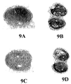
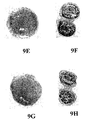
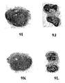
図9A〜9Nは、0.2% オキシクロロセン溶液での15分間の前処理およびそれに続くAd-LacZ感染の後の7つのマウス膀胱を示す写真である。図9Aおよび9Bは第1膀胱のそれぞれ外表面および内腔表面を示し、図9Cおよび9Dは第2膀胱のそれぞれ外表面および内腔表面を示し、図9Eおよび9Fは第3膀胱のそれぞれ外表面および内腔表面を示し、図9Gおよび9Hは第4膀胱のそれぞれ外表面および内腔表面を示し、図9Iおよび9Jは、第5膀胱のそれぞれ外表面および内腔表面を示し、図9Kおよび9Lは、第6膀胱のそれぞれ外表面および内腔表面を示し、図9Mおよび9Nは第7膀胱のそれぞれ外表面および内腔表面を示す。 9A-9N are photographs showing 7 mouse bladders after 15 minutes pre-treatment with 0.2% oxychlorocene solution followed by Ad-LacZ infection. 9A and 9B show the outer and luminal surfaces, respectively, of the first bladder, FIGS. 9C and 9D show the outer and luminal surfaces, respectively, of the second bladder, and FIGS. 9E and 9F show the outer surfaces of the third bladder, respectively. 9G and 9H show the outer and luminal surfaces of the fourth bladder, respectively, FIGS. 9I and 9J show the outer and luminal surfaces of the fifth bladder, respectively, and FIGS. 9K and 9L. Shows the outer and luminal surfaces of the sixth bladder, respectively, and FIGS. 9M and 9N show the outer and luminal surfaces of the seventh bladder, respectively.
図10Aおよび10Bは、それぞれ図8Cおよび8Iのマウス膀胱の横断面を示す写真である。図11Aおよび11Bは、それぞれ図9Cおよび9Iのマウス膀胱の横断面を示す写真である。 10A and 10B are photographs showing cross sections of the mouse bladders of FIGS. 8C and 8I, respectively. FIGS. 11A and 11B are photographs showing cross sections of the mouse bladders of FIGS. 9C and 9I, respectively.
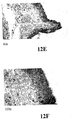
図12A〜12Fは、0.2% オキシクロロセン溶液での5分間の前処理およびそれに続くAd-LacZの感染の後の3つのマウス膀胱の横断面を示す写真である。図12Aおよび12Bは、第1マウス膀胱の横断面を示す写真であり、図12Cおよび12Dは、第2マウス膀胱の横断面を示す写真であり、図12Eおよび12Fは、第3マウス膀胱の横断面を示す写真である。図12A、12Cおよび12Eは40倍の倍率で撮影されたものであり、図12B、12Dおよび12Fは100倍の倍率で撮影されたものである。 FIGS. 12A-12F are photographs showing cross-sections of three mouse bladders after 5 minutes pre-treatment with 0.2% oxychlorocene solution followed by Ad-LacZ infection. 12A and 12B are photographs showing a cross section of the first mouse bladder, FIGS. 12C and 12D are photographs showing a cross section of the second mouse bladder, and FIGS. 12E and 12F are crossings of the third mouse bladder. It is a photograph which shows a surface. FIGS. 12A, 12C and 12E were taken at 40 × magnification, and FIGS. 12B, 12D and 12F were taken at 100 × magnification.
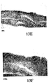
図13A〜13Fは、0.2% オキシクロロセン溶液での15分間の前処理およびそれに続くAd-LacZの感染の後の3つのマウス膀胱の横断面を示す写真である。図13Aおよび13Bは、第1マウス膀胱の横断面を示す写真であり、図13Cおよび13Dは、第2マウス膀胱の横断面を示す写真であり、図13Eおよび13Fは、第3マウス膀胱の横断面を示す写真である。図13A、13Cおよび13Eは40倍の倍率で撮影されたものであり、図13B、13Dおよび13Fは100倍の倍率で撮影されたものである。 FIGS. 13A-13F are photographs showing cross-sections of three mouse bladders after a 15 minute pre-treatment with 0.2% oxychlorocene solution and subsequent infection with Ad-LacZ. 13A and 13B are photographs showing a cross section of the first mouse bladder, FIGS. 13C and 13D are photographs showing a cross section of the second mouse bladder, and FIGS. 13E and 13F are cross sections of the third mouse bladder. It is a photograph which shows a surface. FIGS. 13A, 13C and 13E were taken at 40 × magnification, and FIGS. 13B, 13D and 13F were taken at 100 × magnification.
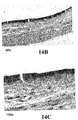
図14Aは、0.1% オキシクロロセン溶液での前処理およびそれに続くAd-LacZの感染の後のマウス膀胱の内腔表面を示す写真である。図14Bおよび14Cは、図14Aのマウス膀胱の横断面を示す写真である。図14Bは40倍の倍率で撮影されたものであり、図14Cは100倍の倍率で撮影されたものである。 FIG. 14A is a photograph showing the luminal surface of a mouse bladder after pretreatment with a 0.1% oxychlorocene solution and subsequent infection with Ad-LacZ. 14B and 14C are photographs showing a cross section of the mouse bladder of FIG. 14A. FIG. 14B was taken at 40 × magnification, and FIG. 14C was taken at 100 × magnification.
図15Aは、0.2% オキシクロロセン溶液での前処理およびそれに続くAd-LacZの感染の後の第1マウス膀胱の内腔表面を示す写真である。図15Bおよび15Cは、図15Aのマウス膀胱の横断面を示す写真である。図15Bは40倍の倍率で撮影されたものであり、図15Cは100倍の倍率で撮影されたものである。 FIG. 15A is a photograph showing the luminal surface of the first mouse bladder after pretreatment with a 0.2% oxychlorocene solution and subsequent infection with Ad-LacZ. 15B and 15C are photographs showing a cross section of the mouse bladder of FIG. 15A. FIG. 15B was taken at 40 × magnification, and FIG. 15C was taken at 100 × magnification.
図16Aは、0.2% オキシクロロセン溶液での前処理およびそれに続くAd-LacZの感染の後の第2マウス膀胱の内腔表面を示す写真である。図16Bおよび16Cは、図16Aのマウス膀胱の横断面を示す写真である。図16Bは40倍の倍率で撮影されたものであり、図16Cは100倍の倍率で撮影されたものである。 FIG. 16A is a photograph showing the luminal surface of the second mouse bladder after pretreatment with 0.2% oxychlorocene solution and subsequent infection with Ad-LacZ. 16B and 16C are photographs showing a cross section of the mouse bladder of FIG. 16A. FIG. 16B was taken at 40 × magnification, and FIG. 16C was taken at 100 × magnification.
図17Aは、0.4% オキシクロロセン溶液での前処理およびそれに続くAd-LacZの感染の後の第1マウス膀胱の内腔表面を示す写真である。図17Bおよび17Cは、図17Aのマウス膀胱の横断面を示す写真である。図17Bは40倍の倍率で撮影されたものであり、図17Cは100倍の倍率で撮影されたものである。 FIG. 17A is a photograph showing the luminal surface of the first mouse bladder after pretreatment with 0.4% oxychlorocene solution and subsequent infection with Ad-LacZ. 17B and 17C are photographs showing a cross section of the mouse bladder of FIG. 17A. FIG. 17B was taken at 40 × magnification, and FIG. 17C was taken at 100 × magnification.
図18Aは、0.4% オキシクロロセン溶液での前処理およびそれに続くAd-LacZの感染の後の第2マウス膀胱の内腔表面を示す写真である。図18Bおよび18Cは、図18Aのマウス膀胱の横断面を示す写真である。図18Bは40倍の倍率で撮影されたものであり、図18Cは100倍の倍率で撮影されたものである。 FIG. 18A is a photograph showing the luminal surface of a second mouse bladder after pretreatment with a 0.4% oxychlorocene solution and subsequent infection with Ad-LacZ. 18B and 18C are photographs showing a cross section of the mouse bladder of FIG. 18A. FIG. 18B was taken at 40 × magnification, and FIG. 18C was taken at 100 × magnification.
親水性単位と親油性単位とを交互に有する重合体
交互または同一の単量体の反復配列を含む重合性化合物も試験した。試験した1つのそのような化合物は、以下の式:
により表される構造を有するポロキサマー407(Pluronic 127)であった。 Poloxamer 407 (Pluronic 127) having a structure represented by:
広範囲のHLB値のポロキサマー重合体が得られる。しかし、試験した化合物の両方は、アデノウイルスの形質導入に対して最小限の効果しか有していなかった。理論により束縛されることを望むものではないが、分離した、より長い親水性鎖および親油性鎖を有する化合物は、膀胱上皮の形質導入の増強において、より有効であると考えられる。 Poloxamer polymers with a wide range of HLB values are obtained. However, both compounds tested had minimal effect on adenoviral transduction. Without wishing to be bound by theory, it is believed that compounds with separate longer hydrophilic and lipophilic chains are more effective at enhancing bladder epithelial transduction.
追加的な形質導入増強化合物
本発明の形質導入増強剤として、追加的な化合物も使用されうる。
Additional transduction enhancing compounds Additional compounds may also be used as the transduction enhancing agents of the present invention.
これらの化合物には、
により表される構造を有するω-ウンデシレニル-β-D-マルトピラノシドが含まれる。
により表される一般構造を有するアルキル-β-D-チオグルコピラノシドのような、糖に基づくチオール性化合物も使用されうる。 Sugar-based thiol compounds such as alkyl-β-D-thioglucopyranoside having the general structure represented by can also be used.
また、
により表される一般構造を有するアルキル-β-D-チオマルトピラノシドも本発明の形質導入増強化合物として使用されうる。 Alkyl-β-D-thiomaltopyranoside having the general structure represented by can also be used as the transduction enhancing compound of the present invention.
さらに、
のような正電荷を有する化合物も使用されうる。 A compound having a positive charge such as can also be used.
また、親油性部分と親水性部分とがカルボキシル結合を介して連結された化合物も使用されうる。このタイプの典型的な化合物は6-O-メチル-n-ヘプチルカルボキシル-α-D-グルコピラノシド:
である。 It is.
側鎖または他の修飾を伴うアルキル基を有する、糖に基づく化合物も使用されうる。このタイプの典型的な化合物には、
により表される構造を有する2-プロピル-1-ペンチル-β-D-マルトピラノシドが含まれる。本発明の形質導入増強剤として、サルコシン化合物も使用されうる。典型的なサルコシン化合物には、
により表される構造を有するアルキルサルコシンナトリウムが含まれる。 Sodium alkylsarcosine having the structure represented by:
種々の置換糖も形質導入増強化合物として使用されうる。形質導入増強化合物として使用されうる典型的な置換糖は、
により表される化学構造を有するスクロースモノアルキルエステルである。このタイプの典型的な化合物には、n = 10である化合物(すなわち、スクロースモノラウラート)が含まれる。 A sucrose monoalkyl ester having a chemical structure represented by Exemplary compounds of this type include compounds where n = 10 (ie sucrose monolaurate).
本発明においては、膀胱の内腔表面を治療する方法を提供する。本発明の好ましい実施形態においては、膀胱カテーテル挿入を用いる点滴により膀胱を処理する。この実施形態においては、まず、膀胱内の尿を除去し、所望により、膀胱をバッファー(例えば、PBS)で洗浄する。ついで、形質導入増強剤を含む組成物を膀胱の内腔表面に(例えば、点滴により)適用する。該形質導入増強溶液を或る所定時間にわたりインキュベートし、または直ちに排出させることが可能である。形質導入増強剤を含む組成物での多重処理を行うことが可能である。形質導入増強剤での処理の後、膀胱の内腔表面をバッファー(例えば、PBS)で洗浄することが可能である。ついで、アデノウイルスを含む溶液を(例えば、点滴により)膀胱内に導入することが可能である。該アデノウイルス含有溶液は直ちに除去することが可能であり、あるいは該溶液を或る時間にわたりインキュベートすることが可能である。アデノウイルスでの処理の後、膀胱表面をバッファー溶液(例えば、PBS)で再び洗浄することが可能である。本発明の好ましい実施形態においては、各処理ごとに、約50〜約500mlの形質導入増強組成物を点滴により膀胱に運搬する。 In the present invention, a method for treating the luminal surface of the bladder is provided. In a preferred embodiment of the invention, the bladder is treated by infusion using bladder catheter insertion. In this embodiment, urine in the bladder is first removed, and the bladder is washed with a buffer (eg, PBS) if desired. A composition comprising a transduction enhancer is then applied to the luminal surface of the bladder (eg, by infusion). The transduction enhancement solution can be incubated for some predetermined time or drained immediately. Multiple treatments with compositions containing transduction enhancers can be performed. Following treatment with the transduction enhancer, the luminal surface of the bladder can be washed with a buffer (eg, PBS). A solution containing adenovirus can then be introduced into the bladder (eg, by infusion). The adenovirus-containing solution can be removed immediately, or the solution can be incubated for a period of time. After treatment with adenovirus, the bladder surface can be washed again with a buffer solution (eg, PBS). In a preferred embodiment of the present invention, about 50 to about 500 ml of the transduction enhancing composition is delivered to the bladder by infusion for each treatment.
あるいは、形質導入増強剤とアデノウイルスとを含む組成物を使用して内腔膀胱表面を処理することが可能である。本発明のこの実施形態においては、まず、膀胱内の尿を除去し、ついで所望により、膀胱をバッファー(例えば、PBS)で洗浄する。ついで、形質導入増強剤とアデノウイルスとを含む組成物を膀胱の内腔表面に適用する。該溶液を或る所定時間にわたりインキュベートし、または直ちに排出させることが可能である。処理後、膀胱の内腔表面を再びバッファー(例えば、PBS)で洗浄することが可能である。 Alternatively, the luminal bladder surface can be treated using a composition comprising a transduction enhancer and an adenovirus. In this embodiment of the invention, the urine in the bladder is first removed, and then the bladder is washed with a buffer (eg, PBS) if desired. A composition comprising a transduction enhancer and adenovirus is then applied to the luminal surface of the bladder. The solution can be incubated for some predetermined time or drained immediately. After treatment, the luminal surface of the bladder can be washed again with a buffer (eg, PBS).
リン酸緩衝食塩水(PBS)が好ましいバッファーであるが、本発明においては任意の他の医薬用バッファーを使用することが可能である。典型的なバッファーには、リン酸ナトリウム/硫酸ナトリウム、Trisバッファー、グリシンバッファー、無菌水、および当技術分野で公知の他のバッファー、例えば、Goodら, Biochemistry 5, 467 (1966) に記載のものが含まれる。バッファーのpHは6.4〜8.4、好ましくは、7〜7.5、最も好ましくは、7.2〜7.4の範囲でありうる。 Phosphate buffered saline (PBS) is the preferred buffer, but any other pharmaceutical buffer can be used in the present invention. Typical buffers include sodium phosphate / sodium sulfate, Tris buffer, glycine buffer, sterile water, and other buffers known in the art, such as those described in Good et al. , Biochemistry 5, 467 (1966). Is included. The pH of the buffer can range from 6.4 to 8.4, preferably 7 to 7.5, and most preferably 7.2 to 7.4.
本発明の形質導入増強剤を含む組成物は、好ましくは、酸化剤をも含む。典型的な酸化剤には、亜塩素酸塩化合物、次亜塩素酸、過酸化水素およびペルオキシ酢酸が含まれるが、これらに限定されるものではない。本発明の好ましい実施形態においては、単一化合物形質導入増強剤のいずれかを酸化剤と組合せ、形質導入剤として使用することが可能である。 The composition containing the transduction enhancer of the present invention preferably also contains an oxidizing agent. Typical oxidants include, but are not limited to, chlorite compounds, hypochlorous acid, hydrogen peroxide and peroxyacetic acid. In a preferred embodiment of the invention, any single compound transduction enhancer can be combined with an oxidizing agent and used as a transducing agent.
前記のとおり、ウイルス遺伝子治療ビヒクルは、腫瘍細胞崩壊性ウイルス、例えば、本明細書中ではCG8840により例示される腫瘍細胞崩壊性アデノウイルスでありうる。該アデノウイルス組成物は更に、ドセタキセルのような化学療法剤を含みうる。該アデノウイルス組成物は、好ましくは、約1×10¹¹〜約1×10¹⁴個のウイルス粒子を含む。 As noted above, the viral gene therapy vehicle can be an oncolytic virus, for example, an oncolytic adenovirus exemplified herein by CG8840. The adenovirus composition may further comprise a chemotherapeutic agent such as docetaxel. The adenovirus composition preferably comprises from about 1 × 10 11 to about 1 × 10 14 virus particles.
種々の追加的な研究を行った。それらを以下に記載する。これらの研究においては、記載されているすべてのパーセント値は、特に示さない限り、重量パーセント値である。 Various additional studies were conducted. They are described below. In these studies, all percentage values listed are weight percentage values unless otherwise indicated.
マウス膀胱尿路上皮への感染力に対する種々の濃度のドデシル-β-D-マルトシドを含有するアデノウイルス製剤の効果
種:
雌Balb/cマウス(Taconic Laboratory)-2匹/群
研究目的:
種々の濃度のドデシル-β-D-マルトシドと共にAd-βgalを製剤化する効果およびマウス膀胱上皮への生じる感染力を試験すること。
Effective species of adenovirus preparations containing various concentrations of dodecyl-β-D-maltoside on the infectivity of mouse urinary tract epithelium :
Female Balb / c mice (Taconic Laboratory)-2 / group
To test the effect of formulating Ad-βgal with various concentrations of dodecyl-β-D-maltoside and the resulting infectivity on mouse bladder epithelium.
用量/経路(ウイルス粒子数/用量):
1×10¹⁰ vp/用量のAd.CMV.LacZ (Lot# 1351.122)。
Dose / route (number of virus particles / dose):
1 × 10 10 vp / dose Ad.CMV.LacZ (Lot # 1351.122).
0.4%、0.2%、0.1%および0.05%のn-ドデシル-β-D-マルトシド(Lot# 100K5308)。 0.4%, 0.2%, 0.1% and 0.05% n-dodecyl-β-D-maltoside (Lot # 100K5308).
膀胱内への点滴の直前に、等容量の2×DDMおよび2×Ad.CMV.LacZを混合した。100μlの該混合物を10分間、20分間および45分間、膀胱内に点滴した。 Just prior to instillation into the bladder, equal volumes of 2 × DDM and 2 × Ad.CMV.LacZ were mixed. 100 μl of the mixture was instilled into the bladder for 10, 20 and 45 minutes.
終点(エンドポイント):
0.1mlの全器官固定液(2%ニュートラル(Neutral)緩衝ホルマリン、2%グルタルアルデヒド、2mM MgCl2、10 mM PBS, pH 7.4)を使用してウイルス感染の48時間後に膀胱を回収した。
End point:
Whole organ fixative 0.1ml was (2% neutral (Neutral) buffered formalin, 2% glutaraldehyde, 2mM MgCl 2, 10 mM PBS , pH 7.4) the bladder 48 h after viral infection using recovered.
結果:
種々の濃度のドデシル-β-D-マルトシドと共にAd-βgalウイルスを製剤化することにより、直線的なアデノウイルス形質導入率が得られた(0.1% > 0.05%> 0.025%)。20分間の点滴では、Ad-βgalウイルスと共に製剤化した0.1% DDMは膀胱内で約80%の遺伝子発現をもたらし、一方、0.05% DDMは約40%の遺伝子発現をもたらした。しかし、45分間の点滴では、Ad-βgalウイルスと共に0.05%〜0.2% DDMで処理されたすべての動物はマウス膀胱内で100%の遺伝子発現を示した。しかし、この製剤化方法における10分間の点滴は、許容しうる形質導入率を達成しなかった。また、DDM前処理の後では、10分間のウイルス点滴で遺伝子形質導入が達成されうることが判明した。
result:
Formulating Ad-βgal virus with various concentrations of dodecyl-β-D-maltoside resulted in linear adenovirus transduction rates (0.1%>0.05%> 0.025%). At 20 minutes of infusion, 0.1% DDM formulated with Ad-βgal virus resulted in about 80% gene expression in the bladder, while 0.05% DDM resulted in about 40% gene expression. However, at 45 minutes infusion, all animals treated with 0.05% to 0.2% DDM with Ad-βgal virus showed 100% gene expression in the mouse bladder. However, a 10 minute infusion in this formulation method did not achieve an acceptable transduction rate. It was also found that after DDM pretreatment, gene transduction can be achieved with 10 minutes of viral infusion.
この研究は、マウス膀胱モデルにおいて、DDMがAd-βgalウイルスと共に製剤化されうることを示している。さらに、DDM前処理を行えば、ウイルス点滴のための時間が45分間から10分間に短縮されうる。 This study shows that DDM can be formulated with Ad-βgal virus in a mouse bladder model. Furthermore, with DDM pretreatment, the time for virus infusion can be reduced from 45 minutes to 10 minutes.
アデノウイルスによるマウス膀胱尿路上皮への感染力に対する種々の希釈剤の効果
種:
雌Balb/cマウス(Taconic Laboratory)-2匹/群
研究目的:
DDM前処理の後のマウス膀胱尿路上皮へのアデノウイルス感染力に対する種々の希釈剤の効果を試験すること(QQ5minQ)。マウス膀胱内への45分間の点滴の前に、該ウイルスを希釈剤で100倍希釈する。
Effect of various diluents on the infectivity of adenovirus to mouse bladder urothelium :
Female Balb / c mice (Taconic Laboratory)-2 / group
To test the effect of different diluents on adenoviral infectivity of mouse urinary tract epithelium after DDM pretreatment (QQ5minQ). The virus is diluted 100-fold with diluent prior to 45 minutes infusion into the mouse bladder.
用量/経路:
1×10⁹ vp/用量のAd.CMV.LacZ (Lot# 1351.122)。
Dose / Route:
1 × 10 9 vp / dose Ad.CMV.LacZ (Lot # 1351.122).
ドデシル-β-D-マルトシド(Lot# 100K5308)。 Dodecyl-β-D-maltoside (Lot # 100K5308).
希釈剤(A):0.9% 食塩注射液(Baxter Lot# 1A1322)、(B)2.5% デキストロースおよび0.45% 食塩注射液(Baxter Lot# C529040)、および(C)Plasma-lyte A注射液pH7.4(Baxter Lot# C558106)、およびARCAバッファー。 Diluent (A): 0.9% saline injection (Baxter Lot # 1A1322), (B) 2.5% dextrose and 0.45% saline injection (Baxter Lot # C529040), and (C) Plasma-lyte A injection pH 7.4 (Baxter Lot # C558106), and ARCA buffer.
終点:
0.1mlの全器官固定液(2%ニュートラル(Neutral)緩衝ホルマリン、2%グルタルアルデヒド、2mM MgCl2、10 mM PBS, pH 7.4)を使用してウイルス感染の48時間後に膀胱を回収した。
end point:
Whole organ fixative 0.1ml was (2% neutral (Neutral) buffered formalin, 2% glutaraldehyde, 2mM MgCl 2, 10 mM PBS , pH 7.4) the bladder 48 h after viral infection using recovered.
結果:
種々の希釈剤で希釈されたウイルスが投与されたマウス膀胱間でAd-βgal遺伝子発現レベルにおける有意な相違は認められなかった。それらの動物はすべて、マウス膀胱内での> 90%のAd-βgal遺伝子発現を示した。
result:
There was no significant difference in Ad-βgal gene expression levels between mouse bladders administered with viruses diluted with various diluents. All of these animals showed> 90% Ad-βgal gene expression in the mouse bladder.
SDラット膀胱上皮上に種々の化学物質で前処理を行った場合のアデノウイルス用量力価測定
種:
雌Female Sprague Dawley(Taconic Laboratory)- 2/群。
Adenovirus dose titer species when pretreated with various chemicals on SD rat bladder epithelium :
Female Female Sprague Dawley (Taconic Laboratory)-2 / group.
研究目的:
SDSまたはDDMでの前処理の後の種々の用量のアデノウイルスによるラット膀胱上皮へのウイルス感染力を試験すること。この研究は、アデノウイルス感染力のためのSDSまたはDDMの残留と膀胱容積との関係も調べた。
Purpose of research:
To test the viral infectivity of rat bladder epithelium with different doses of adenovirus after pretreatment with SDS or DDM. This study also examined the relationship between residual SDS or DDM for adenovirus infectivity and bladder volume.
用量/経路:
4×10⁹および4×10¹⁰ vp/用量のAd.CMV.LacZ (Lot# 1351.122)。
Dose / Route:
4 × 10 9 and 4 × 10 10 vp / dose Ad.CMV.LacZ (Lot # 1351.122).
0.1% ドデシル-β-D-マルトシド(Lot# 100K5308)。 0.1% dodecyl-β-D-maltoside (Lot # 100K5308).
0.1% SDS(Integra Lot# 836011およびLot# BK14J11)。 0.1% SDS (Integra Lot # 836011 and Lot # BK14J11).
400μlのSDSまたはDDMを膀胱内に点滴し(QQ5minQ)、ついでPBSで6回洗浄し、ついでアデノウイルスを15分間点滴した。 400 μl of SDS or DDM was instilled into the bladder (QQ5minQ), then washed 6 times with PBS, then infused with adenovirus for 15 minutes.
終点:
0.2mlの全器官固定液(2%ニュートラル(Neutral)緩衝ホルマリン、2%グルタルアルデヒド、2mM MgCl2、10 mM PBS, pH 7.4)を使用してウイルス感染の48時間後に膀胱を回収した。
end point:
The bladder was harvested 48 hours after viral infection using 0.2 ml whole organ fixative (2% Neutral Buffered Formalin, 2% Glutaraldehyde, 2 mM MgCl 2 , 10 mM PBS, pH 7.4).
結果:
異なるロットのSDSおよびDDMで前処理されたラット膀胱間にAd-βgal遺伝子発現レベルにおける有意な相違は認められなかった。4×10¹⁰ vpアデノウイルスに感染したラット膀胱は > 90%のAd-βgal遺伝子発現レベルを達成し、一方、4×10⁹ vpアデノウイルスに感染したラット膀胱は30%〜50%の範囲のAd-βgal遺伝子発現レベルを示した。ラット膀胱は、マウス膀胱と比較して、同様のAd-βgal遺伝子発現レベルを達成するには10倍多くのアデノウイルスを必要とすることが判明した。SDSおよびDDM前処理は共に、膀胱上皮のGAG層を除去するのに有効であった。
result:
There were no significant differences in Ad-βgal gene expression levels between rat bladders pretreated with different lots of SDS and DDM. Rat urinary bladder infected with 4 × 10 10 vp adenovirus achieved> 90% Ad-βgal gene expression level, while rat urinary bladder infected with 4 × 10 9 vp adenovirus ranged from 30% to 50% The expression level of Ad-βgal gene was shown. Rat bladder was found to require 10 times more adenovirus to achieve similar Ad-βgal gene expression levels compared to mouse bladder. Both SDS and DDM pretreatments were effective in removing the GAG layer of the bladder epithelium.
マウス膀胱尿路上皮へのアデノウイルス感染の前の前処理剤としてのドデシル-β-D-マルトシドの効力
種:
雌Balb/cマウス(Taconic Laboratory)-2匹/群
研究目的:
マウス膀胱尿路上皮へのアデノウイルス感染を増強する前処理剤として、2つの異なる供給業者からのDDMを、大きな動物群において試験すること。
Efficacy of dodecyl-β-D-maltoside as a pretreatment agent prior to adenoviral infection of mouse urinary tract epithelium :
Female Balb / c mice (Taconic Laboratory)-2 / group
To test DDM from two different suppliers in a large group of animals as a pretreatment to enhance adenoviral infection in mouse urinary tract epithelium.
用量/経路:
1×10¹⁰ vp/用量のAd.CMV.LacZ (Lot# 1351.122)。
Dose / Route:
1 × 10 10 vp / dose Ad.CMV.LacZ (Lot # 1351.122).
ドデシル-β-D-マルトシド(Lot # 018H7250およびLot # P21/39/092)。 Dodecyl-β-D-maltoside (Lot # 018H7250 and Lot # P21 / 39/092).
100μlのDDMを膀胱内に点滴し(QQ5minQ)、ついでPBSで6回洗浄し、ついでアデノウイルスを15分間点滴した。 100 μl of DDM was instilled into the bladder (QQ5minQ), then washed 6 times with PBS, then adenovirus was instilled for 15 minutes.
終点:
50μlの全器官固定液(2%ニュートラル(Neutral)緩衝ホルマリン、2%グルタルアルデヒド、2mM MgCl2、10 mM PBS, pH 7.4)を使用してウイルス感染の48時間後に膀胱を回収した。
end point:
The bladder was harvested 48 hours after virus infection using 50 μl whole organ fixative (2% Neutral buffered formalin, 2% glutaraldehyde, 2 mM MgCl 2 , 10 mM PBS, pH 7.4).
結果:
これらの2つの異なるロットのDDMで前処理されたマウス膀胱間でAd-βgal遺伝子発現レベルにおける有意な相違は認められなかった。各場合において、> 90%のAd-βgal遺伝子発現が達成された。したがって、製造業者からのDDMは、マウス膀胱におけるアデノウイルス感染の化学増強剤の良好な候補である。
result:
There were no significant differences in Ad-βgal gene expression levels between mouse bladders pretreated with these two different lots of DDM. In each case,> 90% Ad-βgal gene expression was achieved. Therefore, DDM from the manufacturer is a good candidate for a chemical enhancer of adenovirus infection in mouse bladder.
マウスにおけるSW780+Luc正所性膀胱腫瘍モデルへのアデノウイルス感染の前に使用する化学増強剤としてのドデシル-β-D-マルトシドの効力
種:
雌NCR nu/nuマウス(Taconic Laboratory)- 2匹/群。
Efficacy of dodecyl-β-D-maltoside as a chemical enhancer for use prior to adenovirus infection in SW780 + Luc orthotopic bladder tumor model in mice :
Female NCR nu / nu mice (Taconic Laboratory)-2 / group.
研究目的:
DDM前処理がマウス膀胱尿路上皮におけるAd.Lac Z感染力を増強することを示すこと。正所性腫瘍に対する増強効果を更に確認するために、正所性SW780膀胱腫瘍を担持する2匹のマウスを、DDMで前処理した後または前処理無しで、15分間のAd.LacZの点滴に付した。これらの腫瘍細胞において、Ad.LacZ感染の4日後にAd.LacZ遺伝子発現レベルを調べた。
Purpose of research:
To show that DDM pretreatment enhances Ad.Lac Z infectivity in mouse bladder urothelium. To further confirm the potentiation effect on orthotopic tumors, two mice bearing orthotopic SW780 bladder tumors were given 15 minutes of Ad.LacZ infusion after or without pretreatment with DDM. It was attached. In these tumor cells, Ad.LacZ gene expression levels were examined 4 days after Ad.LacZ infection.
用量/経路:
1x10¹⁰ vp/用量のAd.CMV.LacZ Lot# 1351.122。
Dose / Route:
1 × 10 10 vp / dose Ad.CMV.LacZ Lot # 1351.122.
ドデシル-β-D-マルトシド(Lot#018H7250)。 Dodecyl-β-D-maltoside (Lot # 018H7250).
100μlのDDMを膀胱内に点滴し(QQ5minQ)、ついでPBSで3回洗浄し、ついでアデノウイルスを15分間点滴した。 100 μl of DDM was instilled into the bladder (QQ5minQ), then washed 3 times with PBS, and then adenovirus was instilled for 15 minutes.
終点:
50μlの全器官固定液(2%ニュートラル(Neutral)緩衝ホルマリン、2%グルタルアルデヒド、2mM MgCl2、10 mM PBS, pH 7.4)を使用してウイルス感染の96時間後に膀胱を回収した。
end point:
The bladder was harvested 96 hours after virus infection using 50 μl whole organ fixative (2% Neutral buffered formalin, 2% glutaraldehyde, 2 mM MgCl 2 , 10 mM PBS, pH 7.4).
結果:
DDMで前処理されたマウス膀胱と対照動物との間でAd-βgal遺伝子発現レベルにおける有意な相違が認められた。DDM前処理を行わなかった場合には、膀胱上皮細胞および腫瘍細胞において非常に低いAd-βgal遺伝子発現レベルが認められた。DDM前処理を行った場合には、該上皮細胞のほぼ100%が、死んだ壊死腫瘍細胞以外の膀胱腫瘍内へのAd-βgal遺伝子の形質導入を示した。DDM前処理は正所性腫瘍細胞におけるアデノウイルス感染を増強すると結論づけた。
result:
Significant differences in Ad-βgal gene expression levels were observed between mouse bladder pretreated with DDM and control animals. Without DDM pretreatment, very low Ad-βgal gene expression levels were observed in bladder epithelial cells and tumor cells. When DDM pretreatment was performed, almost 100% of the epithelial cells showed transduction of Ad-βgal gene into bladder tumors other than dead necrotic tumor cells. We conclude that DDM pretreatment enhances adenovirus infection in orthotopic tumor cells.
マウス膀胱尿路上皮へのアデノウイルス感染の前に使用する前処理剤としての、種々の長さのアルキル側鎖および種々のタイプの糖分子を有するDDMおよびSDS亜群化合物の試験
種:
雌Balb/cマウス(Taconic Laboratory)-3〜2匹/群。
Test species of DDM and SDS subgroup compounds with different lengths of alkyl side chains and different types of sugar molecules as pre-treatment agents used prior to adenoviral infection of mouse urinary tract epithelium :
Female Balb / c mice (Taconic Laboratory) —3-2 mice / group.
研究目的:
種々の長さのアルキル側鎖および種々の糖分子を含有するドデシル-β-D-マルトシド(DDM)およびドデシル硫酸ナトリウム(SDS)亜群に属するいくつかの化合物を、マウス膀胱上皮へのアデノウイルス感染を増強するための前処理剤として試験した。すべての化合物を0.1%濃度でPBSに溶解した。
Purpose of research:
Several compounds belonging to the dodecyl-β-D-maltoside (DDM) and sodium dodecyl sulfate (SDS) subgroups containing various lengths of alkyl side chains and various sugar molecules were adenovirused into the mouse bladder epithelium. Tested as a pretreatment to enhance infection. All compounds were dissolved in PBS at 0.1% concentration.
用量/経路:
1×10⁹ vp/用量のAd.CMV.LacZ(Lot #1351.122)。
Dose / Route:
1 × 10 9 vp / dose Ad.CMV.LacZ (Lot # 1351.122).
0.1% ドデシル-β-D-マルトシド(Lot #P21/39/092)および0.1% SDS(Lot #101K0036)はこの研究の陽性対照であった。 0.1% dodecyl-β-D-maltoside (Lot # P21 / 39/092) and 0.1% SDS (Lot # 101K0036) were positive controls for this study.
種々の化学物質での前処理の後(QQ5minQ)、100μlのAd-βgalウイルスを膀胱内投与により膀胱内に15分間点滴した。 After pretreatment with various chemicals (QQ5minQ), 100 μl of Ad-βgal virus was instilled into the bladder for 15 minutes by intravesical administration.
終点:
50μlの全器官固定液(2%ニュートラル(Neutral)緩衝ホルマリン、2%グルタルアルデヒド、2mM MgCl2、10 mM PBS, pH 7.4)を使用してウイルス感染の48時間後に膀胱を回収した。
end point:
The bladder was harvested 48 hours after virus infection using 50 μl whole organ fixative (2% Neutral buffered formalin, 2% glutaraldehyde, 2 mM MgCl 2 , 10 mM PBS, pH 7.4).
結果:
DDM亜群化合物では、C12より長いアルキル側鎖を有する化合物が、最良の増強能を示した。C8、C10、C12、C13およびC14アルキル側鎖を有するDDM亜群化合物を評価した。SDS亜群化合物では、C12より短いアルキル側鎖長を有する化合物が、非常に低いAd-βgal遺伝子発現レベルを示した。C8、C10およびC12アルキル側鎖を有するSDS亜群化合物を評価した。
result:
Of the DDM subgroup compounds, compounds with alkyl side chains longer than C12 showed the best potentiating ability. DDM subgroup compounds with C8, C10, C12, C13 and C14 alkyl side chains were evaluated. For SDS subgroup compounds, compounds with alkyl side chain lengths shorter than C12 showed very low Ad-βgal gene expression levels. SDS subgroup compounds with C8, C10 and C12 alkyl side chains were evaluated.
スクロースモノラウラート以外では、糖分子のタイプは前処理増強剤としてのこれらの化合物の効力に有意な影響を及ぼさないようであった。理論により束縛されることを望むものではないが、現在入手可能なデータに基づけば、二糖鎖がアデノウイルス感染力を増強しうると本発明者らは考えている。 Other than sucrose monolaurate, the type of sugar molecule did not appear to significantly affect the efficacy of these compounds as pretreatment enhancers. While not wishing to be bound by theory, we believe that disaccharide chains can enhance adenovirus infectivity based on currently available data.
パイロット効力研究:マウスにおけるSW780+Luc正所性膀胱腫瘍モデルでのCG8840処理
種:
雌NCR nu/nuマウス(TaconicまたはSimonsen Laboratory)- CG8840には6匹/群およびCG7840には3匹/群。
Pilot efficacy study: CG8840-treated species in SW780 + Luc orthotopic bladder tumor model in mice :
Female NCR nu / nu mice (Taconic or Simonsen Laboratory)-6 / group for CG8840 and 3 / group for CG7840.
研究目的:
膀胱特異的腫瘍細胞崩壊性ウイルスCG8840の効力を前立腺特異的腫瘍細胞崩壊性ウイルスCG7870と共に試験すること。CG8840には2つの処理法(1×10¹⁰および1×10⁸ vp/用量)を用いた。CG7870には1つの処理法(1x1010 vp/用量)を用いた。1用量のウイルスを連続3週間にわたり毎週、膀胱内に運搬した。
Purpose of research:
To test the efficacy of bladder-specific oncolytic virus CG8840 with prostate-specific oncolytic virus CG7870. Two treatments were used for CG8840 (1 × 10 10 and 1 × 10 8 vp / dose). One treatment method (1 × 10 10 vp / dose) was used for CG7870. A dose of virus was delivered into the bladder weekly for 3 consecutive weeks.
用量/経路:
100万個のSW780+Luc細胞クローン#19(P4)。
Dose / Route:
One million SW780 + Luc cell clone # 19 (P4).
CG8840 Lot# 1408.190; CG7870 (Lot # 38.145)。 CG8840 Lot # 1408.190; CG7870 (Lot # 38.145).
0.1% ドデシル-β-D-マルトシド(Lot #018H7250)。 0.1% dodecyl-β-D-maltoside (Lot # 018H7250).
0.2μMフィルターで濾過したPBS中、15mg/ml ルシフェリン(Xenogen cat# XR-1001)基質溶液を調製した。 A 15 mg / ml luciferin (Xenogen cat # XR-1001) substrate solution in PBS filtered through a 0.2 μM filter was prepared.
120μlの1×10⁷細胞/ml SW780+luc細胞を各チューブ内にアリコート化し、点滴前に氷中に維持した。各マウス膀胱ごとに1アリコート。 120 μl of 1 × 10 7 cells / ml SW780 + luc cells were aliquoted into each tube and kept in ice prior to infusion. One aliquot for each mouse bladder.
終点:
マウス膀胱の生イメージを10週間にわたり毎週行った。ヘマトキシリン-エオシン(H&E)染色およびヒトサイトケラチン染色を、それらの入手可能な膀胱および腎臓サンプル上で行った。
end point:
A live image of the mouse bladder was performed weekly for 10 weeks. Hematoxylin-eosin (H & E) staining and human cytokeratin staining were performed on these available bladder and kidney samples.
結果:
CG8840(1×10¹⁰ vp/用量)処理マウスおよび対照動物(CG7870処理群およびウイルス非処理群)の間で腫瘍体積の減少における有意な相違が認められた。1×1010 vp/用量のCG8840で処理された6匹中3匹のマウスが、第2用量の点滴の後に完全な腫瘍退縮を示し、該研究の終了時(第10週)まで無腫瘍状態を維持した。ヒトサイトケラチン染色は、それらのマウス膀胱内に腫瘍細胞が存在しないことを示した。1×108 vp/用量のCG8840の効力は有意ではなく、5匹中1匹のマウスが第5週に不完全な腫瘍退縮を示し、終了時まで腫瘍を維持した。腎臓への腫瘍転移は、腫瘍担持マウスの生存に対する重大な脅威である。腎臓転移を伴うマウスはいずれも、たとえ膀胱腫瘍が無くなったとしても、間もなく死亡するであろう。
result:
Significant differences in tumor volume reduction were observed between CG8840 (1 × 10 10 vp / dose) treated mice and control animals (CG7870 treated group and virus untreated group). Three out of six mice treated with 1 × 10 10 vp / dose of CG8840 showed complete tumor regression after the second dose infusion and remained tumor free until the end of the study (week 10) Maintained. Human cytokeratin staining showed the absence of tumor cells in their mouse bladder. The efficacy of 1 × 10 8 vp / dose CG8840 was not significant, and 1 out of 5 mice showed incomplete tumor regression at week 5 and maintained the tumor until the end. Tumor metastasis to the kidney is a significant threat to the survival of tumor-bearing mice. Any mouse with kidney metastases will soon die, even if the bladder tumor disappears.
マウスにおけるSW780+Luc正所性膀胱腫瘍モデルでのCG8840処理
種:
雌NCR nu/nuマウス(Taconic Laboratory)- 10匹/群。
CG8840-treated species in SW780 + Luc orthotopic bladder tumor model in mice :
Female NCR nu / nu mice (Taconic Laboratory)-10 / group.
研究目的:
この研究は、膀胱腫瘍に対する膀胱特異的腫瘍細胞崩壊性ウイルスCG8840処理法の効力を観察するために計画した。CG8840ウイルスを膀胱内投与により膀胱内にそれぞれ15分間および30分間点滴した。腎臓腫瘍転移を軽減するために、SW780腫瘍細胞移植中に以下の2つの異なるDDM前処理法を試験した:(1)0.1% DDM QQ5minQ、および(2)0.1% DDM Q10min。
Purpose of research:
This study was designed to observe the efficacy of the bladder-specific oncolytic virus CG8840 treatment method against bladder tumors. CG8840 virus was instilled into the bladder by intravesical administration for 15 and 30 minutes, respectively. To reduce renal tumor metastasis, two different DDM pretreatment methods were tested during SW780 tumor cell transplantation: (1) 0.1% DDM QQ5minQ, and (2) 0.1% DDM Q10min.
用量/経路:
100万個のSW780+Luc細胞クローン#19(P4)。
Dose / Route:
One million SW780 + Luc cell clone # 19 (P4).
CG8840 Lot# 1408.190。0.1% ドデシル-β-D-マルトシド(Lot #018H7250)。 CG8840 Lot # 1408.190. 0.1% dodecyl-β-D-maltoside (Lot # 018H7250).
0.2μMフィルターで濾過したPBS中、15mg/ml ルシフェリン(Xenogen cat# XR-1001)基質溶液を調製した。 A 15 mg / ml luciferin (Xenogen cat # XR-1001) substrate solution in PBS filtered through a 0.2 μM filter was prepared.
90μlの1.1×10⁷細胞/ml SW780+luc細胞を各チューブ内にアリコート化し、点滴前に氷中に維持した。各マウス膀胱ごとに1アリコート。 90 μl of 1.1 × 10 7 cells / ml SW780 + luc cells were aliquoted into each tube and kept in ice prior to infusion. One aliquot for each mouse bladder.
終点:
マウス膀胱の生イメージを8週間にわたり毎週行った。ヘマトキシリン-エオシン(H&E)染色およびヒトサイトケラチン染色を、それらの入手可能な膀胱および腎臓サンプル上で行った。
end point:
A live image of the mouse bladder was performed weekly for 8 weeks. Hematoxylin-eosin (H & E) staining and human cytokeratin staining were performed on these available bladder and kidney samples.
結果:
CG8840(15分間および30分間のウイルス処理)処理マウスおよび対照動物の間で腫瘍体積の減少における有意な相違が認められた。特に、15分間のウイルス処理を行った9匹中7匹のマウスは第1ウイルス処理の後に有意な腫瘍体積減少を示した。それらのうちの5匹は該研究の終了時まで無腫瘍であった。3週連続して注入される、1×10¹⁰ vp/用量の3用量でのCG8840処理は、マウス膀胱内の正所性SW780腫瘍を根絶するのに非常に有効であることが判明した。しかし、より長い点滴時間(すなわち、15分間を超える)は腫瘍細胞内のウイルスコピー数を増加させないらしい。
result:
Significant differences in tumor volume reduction were observed between CG8840 (15 min and 30 min virus treatment) treated mice and control animals. In particular, 7 out of 9 mice treated with virus for 15 minutes showed significant tumor volume reduction after the first virus treatment. Five of them were tumor free until the end of the study. CG8840 treatment with 3 doses of 1 × 10 10 vp / dose, infused for 3 consecutive weeks, was found to be very effective in eradicating orthotopic SW780 tumors in mouse bladder. However, longer infusion times (ie, greater than 15 minutes) do not appear to increase virus copy number in tumor cells.
SW780腫瘍細胞移植、0.1% DDM前処理(Q10min洗浄を伴う)では、正所性腫瘍を有するマウスにおける、より軽微な腎臓腫瘍転移が観察された。 With SW780 tumor cell transplantation, 0.1% DDM pretreatment (with Q10min wash), more mild kidney tumor metastasis was observed in mice with orthotopic tumors.
種々の試薬とのアデノウイルスの適合性を確認するために、種々の追加的な研究を行った。これらの研究を以下に記載する。 Various additional studies were performed to confirm the adenovirus compatibility with various reagents. These studies are described below.
研究#1 - エタノールおよび尿とのAd5-LacZの適合性
この研究においては、各サンプルを37℃で1時間インキュベートした後、プラークアッセイを開始した。この研究のデータを以下の表3に示す。
このデータは、生物学的温度における尿、5%エタノールまたはARCAとのインキュベーションの後にAd-LacZがその活性を喪失しないことを示している。しかし、40% エタノールは該ウイルスのほとんどを不活化した。 This data shows that Ad-LacZ does not lose its activity after incubation with urine, 5% ethanol or ARCA at biological temperatures. However, 40% ethanol inactivated most of the virus.
研究#2:エタノールとのAd5-LacZの適合性
この研究では、各サンプルを37℃でインキュベートした後、プラークアッセイを開始した。この研究のデータを以下の表4に示す。
前記結果は、より高い濃度のエタノール(すなわち、> 10%)が部分的ないし完全なアデノウイルス不活化を引き起こすことを示している。 The results indicate that higher concentrations of ethanol (ie> 10%) cause partial to complete adenovirus inactivation.
研究#3 - 選択された増強剤とのアデノウイルスの適合性
この研究では、各サンプルを37℃または25℃でインキュベートした後、プラークアッセイを開始した。この研究のデータを以下の表5に示す。
前記データから理解されうるとおり、ドデシルマルトシドおよびポリドカノールは該実験条件下でアデノウイルスに適合性であり、一方、オキシクロロセンは、インキュベーションの温度および時間の増加により、該ウイルスを不活化しうる。 As can be seen from the data, dodecyl maltoside and polidocanol are compatible with adenovirus under the experimental conditions, while oxychlorocene can inactivate the virus by increasing the temperature and time of incubation. .
研究#4 - デドシル硫酸ナトリウム(SDS)によるアデノウイルスの分解
種々の濃度のSDS中、37℃で15〜30分間インキュベートしたアデノウイルスのサンプルに対して、一連の実験を行った。得られた材料を陰イオン交換クロマトグラフィーにより検査した。0.025% SDS以上のSDS濃度は該ウイルスを迅速に分解することが確認された。0.0125%のSDSは、30分間のインキュベーションの後のAEX法によっては該粒子の質または量を低下させなかった。
Study # 4-Degradation of Adenovirus by Sodium Dedosyl Sulfate (SDS) A series of experiments were performed on adenovirus samples incubated at 37 ° C for 15-30 minutes in various concentrations of SDS. The resulting material was examined by anion exchange chromatography. It was confirmed that an SDS concentration of 0.025% SDS or more rapidly decomposes the virus. 0.0125% SDS did not reduce the quality or quantity of the particles by the AEX method after 30 minutes incubation.
研究#5 - ドデシル硫酸ナトリウム(SDS)によるアデノウイルスの分解
この研究においては、アデノウイルス(Ad-LacZ)調製物(1.0×10¹² vp/ml)をSDSまたはDDMの溶液と混合して、アデノウイルスとこれらの界面活性剤のそれぞれとの混合物を得た。混合の結果、SDSまたはDDMの最終濃度は0.1%となり、該ウイルスは5.0×1011 vp/mlとなった。これらのサンプルを25℃または37℃で1時間インキュベートした後、凍結した。それらをプラークアッセイ分析のためにアッセイサービス業者に送った。この研究の結果を以下の表6に示す。
この実験は、上昇温度でのSDS溶液へのアデノウイルスの相対的に短い暴露が該ウイルスを不活化することを証明した。一方、DDM溶液はアデノウイルスに対して有害な影響を及ぼさなかった。 This experiment demonstrated that a relatively short exposure of adenovirus to the SDS solution at elevated temperature inactivates the virus. On the other hand, DDM solution had no harmful effect on adenovirus.
以下の表(表7)は、典型的な前処理剤の形質導入効率を要約する。表7においては、形質導入効率は以下のとおりに定義される:「0」は0〜10%の形質導入効率を意味する;「+」は10〜25%の形質導入効率を意味する;「++」は25〜50%の形質導入効率を意味する;「+++」は50〜75%の形質導入効率を意味する;「++++」は > 75%の形質導入効率を意味する。
前記の説明は、例示目的として記載した具体例により本発明の原理を教示しているが、本発明の真の範囲から逸脱することなく形態および詳細における種々の変更が施されうることが、この開示の解釈から当業者に理解されるであろう。 Although the foregoing description teaches the principles of the invention by way of example described for purposes of illustration, it is to be understood that various changes in form and detail may be made without departing from the true scope of the invention. Those skilled in the art will appreciate from the interpretation of the disclosure.
本明細書中に引用されているすべての刊行物の全体を参照により本明細書に組み入れることとする。 All publications cited herein are hereby incorporated by reference in their entirety.
Claims (95)
ついで膀胱の内腔表面を、腫瘍細胞崩壊性ウイルスを含む組成物と接触させることを含んでなり、
形質導入増強剤が、親油性置換基を有する単糖、二糖または多糖であることを特徴とする、膀胱癌の治療方法。 Contacting the luminal surface of the bladder with a pretreatment composition comprising a transduction enhancer;
Then contacting the luminal surface of the bladder with a composition comprising an oncolytic virus,
A method for treating bladder cancer, wherein the transduction enhancer is a monosaccharide, disaccharide or polysaccharide having a lipophilic substituent.
ついで膀胱の内腔表面を、腫瘍細胞崩壊性ウイルスを含む組成物と接触させることを含む、膀胱癌の治療方法。 Contacting the luminal surface of the bladder with a pretreatment composition comprising about 0.01 to about 0.2% by weight sodium oxychlorocene,
Next, a method for treating bladder cancer, comprising contacting the luminal surface of the bladder with a composition comprising an oncolytic virus.
ついで膀胱の内腔表面を、腫瘍細胞崩壊性ウイルスを含む組成物と接触させることを含む、膀胱癌の治療方法。 The luminal surface of the bladder has the chemical formula:
Next, a method for treating bladder cancer, comprising contacting the luminal surface of the bladder with a composition comprising an oncolytic virus.
ついで膀胱の内腔表面を、腫瘍細胞崩壊性ウイルスを含む組成物と接触させることを含む、膀胱癌の治療方法。 The luminal surface of the bladder is represented by the following general formula (I) or the following general formula (II):
Next, a method for treating bladder cancer, comprising contacting the luminal surface of the bladder with a composition comprising an oncolytic virus.
形質導入増強剤が、親油性置換基を有する単糖、二糖または多糖であることを特徴とする組成物。 Comprising a transduction enhancing agent and an oncolytic virus,
A composition characterized in that the transduction enhancer is a monosaccharide, disaccharide or polysaccharide having a lipophilic substituent.
ついで膀胱の内腔表面を、腫瘍細胞崩壊性ウイルスを含む組成物と接触させることを含んでなり、
形質導入増強剤が、以下の一般式(I)または以下の一般式(II):
膀胱の内腔表面を該前処理組成物と少なくとも10分間接触させることを特徴とする、膀胱癌の治療方法。 Contacting the luminal surface of the bladder with a pretreatment composition comprising a transduction enhancer;
Then contacting the luminal surface of the bladder with a composition comprising an oncolytic virus,
The transduction enhancer has the following general formula (I) or the following general formula (II):
A method for treating bladder cancer, comprising contacting a luminal surface of a bladder with the pretreatment composition for at least 10 minutes.
ついで膀胱の内腔表面を、腫瘍細胞崩壊性ウイルスを含む組成物と接触させることを含んでなり、
xが少なくとも11であり、
該腫瘍細胞崩壊性ウイルス組成物が少なくとも4×10¹⁰個のウイルス粒子を含むことを特徴とする、膀胱癌の治療方法。 The luminal surface of the bladder is represented by the following general formula (I) or the following general formula (II):
Then contacting the luminal surface of the bladder with a composition comprising an oncolytic virus,
x is at least 11,
A method for treating bladder cancer, wherein the oncolytic virus composition comprises at least 4 × 10 10 viral particles.
形質導入増強剤が、以下の一般式(I)または以下の一般式(II):
形質導入増強剤の濃度が組成物の0.025重量%未満であることを特徴とする組成物。 Comprising a transduction enhancing agent and an oncolytic virus,
The transduction enhancer has the following general formula (I) or the following general formula (II):
A composition wherein the concentration of the transduction enhancer is less than 0.025% by weight of the composition.
Applications Claiming Priority (2)
| Application Number | Priority Date | Filing Date | Title |
|---|---|---|---|
| US10/327,869 US7459154B2 (en) | 2002-12-26 | 2002-12-26 | Methods and reagents for the enhancement of virus transduction in the bladder epithelium |
| PCT/US2003/041379 WO2004060303A2 (en) | 2002-12-26 | 2003-12-24 | Methods and reagents for the enhancement of virus transduction in the bladder epithelium |
Publications (2)
| Publication Number | Publication Date |
|---|---|
| JP2006512398A true JP2006512398A (en) | 2006-04-13 |
| JP2006512398A5 JP2006512398A5 (en) | 2007-02-15 |
Family
ID=32680762
Family Applications (1)
| Application Number | Title | Priority Date | Filing Date |
|---|---|---|---|
| JP2004565744A Withdrawn JP2006512398A (en) | 2002-12-26 | 2003-12-24 | Methods and reagents for enhanced transduction of viruses into the bladder epithelium |
Country Status (9)
| Country | Link |
|---|---|
| US (7) | US7459154B2 (en) |
| EP (1) | EP1583502A4 (en) |
| JP (1) | JP2006512398A (en) |
| KR (1) | KR20050088231A (en) |
| CN (1) | CN1753618A (en) |
| AU (1) | AU2003299972C1 (en) |
| CA (1) | CA2510903A1 (en) |
| NZ (1) | NZ540732A (en) |
| WO (1) | WO2004060303A2 (en) |
Cited By (1)
| Publication number | Priority date | Publication date | Assignee | Title |
|---|---|---|---|---|
| JP2013516978A (en) * | 2010-01-12 | 2013-05-16 | バスキュラー バイオジェニックス リミテッド | Method for producing adenovirus vector and virus preparation produced thereby |
Families Citing this family (11)
| Publication number | Priority date | Publication date | Assignee | Title |
|---|---|---|---|---|
| US7459154B2 (en) * | 2002-12-26 | 2008-12-02 | Cell Genesys, Inc. | Methods and reagents for the enhancement of virus transduction in the bladder epithelium |
| US20050059613A1 (en) * | 2003-07-08 | 2005-03-17 | Bahram Memarzadeh | Compositions and methods for the enhanced uptake of therapeutic agents through the bladder epithelium |
| WO2006076663A2 (en) * | 2005-01-14 | 2006-07-20 | Urigen, Inc. | Kits and improved compositions for treating lower urinary tract |
| EP2077847B1 (en) * | 2006-09-14 | 2016-02-24 | Bahram Memarzadeh | Sucralose monolaurate and sucralose monomyristate, pharmaceutical formulations, diagnostic kits and methods of treatment |
| JP2011520993A (en) * | 2008-05-27 | 2011-07-21 | オンコリティクス バイオテク,インコーポレーテッド | Regulation of interstitial pressure and delivery and distribution of oncolytic viruses |
| DE102011085501A1 (en) * | 2011-10-31 | 2013-05-02 | Söring GmbH | ESU |
| CN108135934A (en) * | 2015-10-19 | 2018-06-08 | 永恒生物科技股份有限公司 | Pass through the method for combination therapy to treat entity or lympha tumour |
| US11497781B2 (en) * | 2016-03-10 | 2022-11-15 | Cg Oncology, Inc. | Methods of treating bladder cancer by combination therapy comprising the oncolytic adenovirus CG0070 and an immune checkpoint inhibitor |
| US11338003B2 (en) | 2017-04-14 | 2022-05-24 | Cg Oncology, Inc. | Methods of treating bladder cancer with an oncolytic virus |
| CN110393808B (en) * | 2019-01-07 | 2022-06-03 | 四川安可康生物医药有限公司 | Immune oncolytic virus combined medicine for enhancing systemic immune response and application thereof |
| CN113717953A (en) * | 2021-08-05 | 2021-11-30 | 北京舜雷科技有限公司 | Application of attenuated flavivirus in oncolytic |
Family Cites Families (15)
| Publication number | Priority date | Publication date | Assignee | Title |
|---|---|---|---|---|
| US169138A (en) * | 1875-10-26 | Improvement in folding lounges | ||
| JPH01245159A (en) * | 1988-03-28 | 1989-09-29 | Gen Corp:Kk | Plate for detecting newcastle disease virus antibody and method for solubilizing antigen virus |
| US5369095A (en) | 1990-02-14 | 1994-11-29 | Alcon Laboratories, Inc. | Compositions and method comprising substituted glycosides as mucus membrane permeation enhancers |
| US6165500A (en) * | 1990-08-24 | 2000-12-26 | Idea Ag | Preparation for the application of agents in mini-droplets |
| US5525519A (en) * | 1992-01-07 | 1996-06-11 | Middlesex Sciences, Inc. | Method for isolating biomolecules from a biological sample with linear polymers |
| US5368844A (en) | 1992-12-16 | 1994-11-29 | Colgate Palmolive Company | Antiplaque, antigingivitis, anticaries oral composition |
| WO1997021348A1 (en) * | 1995-12-08 | 1997-06-19 | Kao Corporation | Germicidal composition |
| US5789244A (en) * | 1996-01-08 | 1998-08-04 | Canji, Inc. | Compositions and methods for the treatment of cancer using recombinant viral vector delivery systems |
| US20020169138A1 (en) * | 1997-10-24 | 2002-11-14 | Southern Research Institute | Delivery vehicles for bioactive agents and uses thereof |
| US6855549B1 (en) * | 1998-11-23 | 2005-02-15 | The University Of Iowa Research Foundation | Methods and compositions for increasing the infectivity of gene transfer vectors |
| CA2404085A1 (en) | 2000-03-24 | 2001-10-04 | Cell Genesys, Inc. | Human urothelial cell specific uroplakin transcriptional regulatory sequences, vectors comprising uroplakin-specific transcriptional regulatory sequences, and methods of use thereof |
| US6911200B2 (en) | 2000-03-24 | 2005-06-28 | Cell Genesys, Inc. | Methods of treating neoplasia with combination of target-cell specific adenovirus, chemotherapy and radiation |
| IL139708A0 (en) * | 2000-11-15 | 2002-02-10 | Amiel Gilad | Process of decellularizing biological matrices and acellular biological matrices useful in tissue engineering |
| AUPR894201A0 (en) * | 2001-11-19 | 2001-12-13 | Women's And Children's Hospital | Respiratory delivery for gene therapy and lentiviral delivery particle |
| US7459154B2 (en) * | 2002-12-26 | 2008-12-02 | Cell Genesys, Inc. | Methods and reagents for the enhancement of virus transduction in the bladder epithelium |
-
2002
- 2002-12-26 US US10/327,869 patent/US7459154B2/en not_active Expired - Lifetime
-
2003
- 2003-12-24 AU AU2003299972A patent/AU2003299972C1/en not_active Expired
- 2003-12-24 WO PCT/US2003/041379 patent/WO2004060303A2/en active Application Filing
- 2003-12-24 JP JP2004565744A patent/JP2006512398A/en not_active Withdrawn
- 2003-12-24 CN CNA2003801076205A patent/CN1753618A/en active Pending
- 2003-12-24 NZ NZ540732A patent/NZ540732A/en not_active IP Right Cessation
- 2003-12-24 KR KR1020057012091A patent/KR20050088231A/en not_active Application Discontinuation
- 2003-12-24 EP EP03800237A patent/EP1583502A4/en not_active Withdrawn
- 2003-12-24 US US10/743,813 patent/US7267815B2/en not_active Expired - Lifetime
- 2003-12-24 CA CA002510903A patent/CA2510903A1/en not_active Abandoned
-
2007
- 2007-08-29 US US11/897,535 patent/US20080138322A1/en not_active Abandoned
- 2007-10-25 US US11/977,781 patent/US20080112931A1/en not_active Abandoned
- 2007-10-25 US US11/978,112 patent/US20080241105A1/en not_active Abandoned
- 2007-10-25 US US11/977,943 patent/US20080112932A1/en not_active Abandoned
-
2008
- 2008-09-23 US US12/284,734 patent/US20090068151A1/en not_active Abandoned
Cited By (1)
| Publication number | Priority date | Publication date | Assignee | Title |
|---|---|---|---|---|
| JP2013516978A (en) * | 2010-01-12 | 2013-05-16 | バスキュラー バイオジェニックス リミテッド | Method for producing adenovirus vector and virus preparation produced thereby |
Also Published As
| Publication number | Publication date |
|---|---|
| EP1583502A4 (en) | 2007-01-17 |
| CA2510903A1 (en) | 2004-07-22 |
| KR20050088231A (en) | 2005-09-02 |
| NZ540732A (en) | 2008-01-31 |
| US20090068151A1 (en) | 2009-03-12 |
| US20080112931A1 (en) | 2008-05-15 |
| WO2004060303A3 (en) | 2005-11-24 |
| US20080138322A1 (en) | 2008-06-12 |
| AU2003299972C1 (en) | 2010-12-16 |
| AU2003299972B2 (en) | 2010-05-13 |
| EP1583502A2 (en) | 2005-10-12 |
| US20040176318A1 (en) | 2004-09-09 |
| US7267815B2 (en) | 2007-09-11 |
| AU2003299972A1 (en) | 2004-07-29 |
| US20080112932A1 (en) | 2008-05-15 |
| US20080241105A1 (en) | 2008-10-02 |
| CN1753618A (en) | 2006-03-29 |
| US20040131590A1 (en) | 2004-07-08 |
| WO2004060303A2 (en) | 2004-07-22 |
| US7459154B2 (en) | 2008-12-02 |
Similar Documents
| Publication | Publication Date | Title |
|---|---|---|
| US20080138322A1 (en) | Methods and reagents for the enhancement of virus transduction in the bladder epithelium | |
| ES2233995T3 (en) | COMPOSITIONS CONTAINING A DETERGENT AND A TUMOR SUPPRESSOR GEN, CODED BY AN ADENOVIRICAL VECTOR. | |
| Goldman et al. | Transfer of the CFTR gene to the lung of nonhuman primates with E1-deleted, E2a-defective recombinant adenoviruses: a preclinical toxicology study | |
| US8133903B2 (en) | Methods of use of inhibitors of phosphodiesterases and modulators of nitric oxide, reactive oxygen species, and metalloproteinases in the treatment of peyronie's disease, arteriosclerosis and other fibrotic diseases | |
| KR20140146062A (en) | Compartmentalized Method of Nucleic Acid Delivery and Compositions and Uses Thereof | |
| JP2011148834A (en) | Method and composition for treatment of interferon-resistant tumor | |
| EP2846821B1 (en) | Hydrodynamic methods for delivering fluids to kidney tissues and related materials and methods | |
| CN100429220C (en) | Methods and compositions for interferon therapy | |
| Kesseli et al. | Adeno-associated virus mediates gene transduction after static cold storage treatment in rodent lung transplantation | |
| Bian et al. | Tumor-targeted gene transfer in vivo via recombinant Newcastle disease virus modified by a bispecific fusion protein | |
| Sferra et al. | Gene transfer to the intestinal tract: a new approach using selective injection of the superior mesenteric artery | |
| Kushwah et al. | Potential of helper-dependent adenoviral vectors in modulating airway innate immunity | |
| US20100113569A1 (en) | Isolated dna fragment of the human a33 promoter and its use to control the expression of a heterologous gene in tumor cells | |
| CN111979204B (en) | Oncolytic vaccinia virus carrying sponge lectin gene, construction method and application | |
| Wang et al. | Construction of exosome-loaded LL-37 and its protection against zika virus infection | |
| Zhang et al. | S-RBD-modified and miR-486-5p-engineered exosomes derived from mesenchymal stem cells alleviate radiation-induced lung injury and long-term pulmonary fibrosis via suppression of ferroptosis | |
| WO2019045049A1 (en) | Renal tubule cell-specific expression vector | |
| Brandon et al. | Effect of administration route on biodistribution and shedding of replication-deficient viral vectors used in gene therapy. A literature study | |
| EP2875831A1 (en) | A method of genic transfer into an eukaryotic cell through a viral vector |
Legal Events
| Date | Code | Title | Description |
|---|---|---|---|
| A521 | Written amendment |
Free format text: JAPANESE INTERMEDIATE CODE: A523 Effective date: 20061222 |
|
| A621 | Written request for application examination |
Free format text: JAPANESE INTERMEDIATE CODE: A621 Effective date: 20061222 |
|
| A761 | Written withdrawal of application |
Free format text: JAPANESE INTERMEDIATE CODE: A761 Effective date: 20090327 |
|
| A521 | Written amendment |
Free format text: JAPANESE INTERMEDIATE CODE: A821 Effective date: 20090327 |