JP2006189592A - Endoscope system - Google Patents
Endoscope system Download PDFInfo
- Publication number
- JP2006189592A JP2006189592A JP2005000990A JP2005000990A JP2006189592A JP 2006189592 A JP2006189592 A JP 2006189592A JP 2005000990 A JP2005000990 A JP 2005000990A JP 2005000990 A JP2005000990 A JP 2005000990A JP 2006189592 A JP2006189592 A JP 2006189592A
- Authority
- JP
- Japan
- Prior art keywords
- light
- adapter
- optical
- tip
- endoscope apparatus
- Prior art date
- Legal status (The legal status is an assumption and is not a legal conclusion. Google has not performed a legal analysis and makes no representation as to the accuracy of the status listed.)
- Pending
Links
Images
Landscapes
- Instruments For Viewing The Inside Of Hollow Bodies (AREA)
- Endoscopes (AREA)
Abstract
Description
本発明は、医療用や工業用に用いられる内視鏡装置に関する。 The present invention relates to an endoscope apparatus used for medical use or industrial use.
近年、ボイラや機械等の管路内、あるいはエンジンの内部の観察、点検等に、細長の挿入部をその内部に挿入して観察、点検を行うことができる内視鏡装置が広く利用されている。
上述の内視鏡装置には、挿入部の先端に荷電結合素子(以後、「CCD」と表記)などの撮像素子を配置し、この撮像素子に結像した画像をモニタに表示して観察、点検等を行うビデオスコープ内視鏡装置がある。
2. Description of the Related Art In recent years, endoscope apparatuses that can be observed and inspected by inserting a long insertion portion into a pipeline of a boiler, a machine, or the like, or inside an engine, have been widely used. Yes.
In the endoscope apparatus described above, an image pickup device such as a charge coupled device (hereinafter referred to as “CCD”) is disposed at the distal end of the insertion portion, and an image formed on the image pickup device is displayed on the monitor for observation. There are videoscope endoscope devices that perform inspections.
このようなビデオスコープ内視鏡においては、その挿入部の先端に、それぞれ異なる光学的作用、たとえば、観察対象を拡大する作用や、挿入部の中心軸線に対して側方に位置する観察対象を観察する作用等を有する先端光学アダプタを、必要に応じて取り付けて所望の観察、点検等を行う多くの技術が提案されている(たとえば、特許文献1参照)。
上述の特許文献1においては、先端光学アダプタにライトガイドにより導かれた光を拡散させる拡散光学系も配置されている。そのため、先端光学アダプタを挿入部の先端から取り外すと、ライトガイドから高輝度の光が外部に拡散されずに照射される。そのため、先端光学アダプタを内視鏡装置に取り付ける際に、ライトガイドから照射される光がまぶしく、先端光学アダプタを取り付けにくいという問題があった。
In the above-mentioned
本発明は、上記の課題を解決するためになされたものであって、被検査対象の検査性を損なうことなく先端光学アダプタの取り付け性を向上することができる内視鏡装置を提供することを目的とする。 The present invention has been made to solve the above-described problem, and provides an endoscope apparatus that can improve the attachment property of the tip optical adapter without impairing the inspection property of the object to be inspected. Objective.
本発明は、上記の課題を解決するため、下記の手段を採用した。
本発明に係る内視鏡装置は、光源と、被検査対象空間内に挿入され前記光源からの光を先端の出射部から出射する挿入部と、該挿入部の先端に着脱可能に取り付けられる先端光学アダプタと、を備える内視鏡装置であって、前記先端光学アダプタの前記挿入部への着脱を検知するアダプタ検知手段と、該アダプタ検知手段の検知に連動して前記出射部から出射する光を制御する出射光制御手段と、を設けたことを特徴とするものである。
In order to solve the above problems, the present invention employs the following means.
An endoscope apparatus according to the present invention includes a light source, an insertion portion that is inserted into a space to be inspected and emits light from the light source from a light emitting portion at the tip, and a tip that is detachably attached to the tip of the insertion portion. An endoscope apparatus comprising: an optical adapter; adapter detection means for detecting attachment / detachment of the distal optical adapter to / from the insertion portion; and light emitted from the emission portion in conjunction with detection by the adapter detection means And an emitted light control means for controlling the above.
このような内視鏡装置によれば、先端光学アダプタの挿入部への着脱を検知するアダプタ検知手段と、アダプタ検知手段の検知に連動して出射部から出射する光を制御する出射光制御手段とを設けたことにより、先端光学アダプタの着脱状況をアダプタ検知手段で検知し、この着脱状況に応じて出射光制御手段が出射部から出光する光を自動的に制御できるようになる。すなわち、先端光学アダプタの着脱操作時に出射部から出光する光の発光を止めたり、あるいは、まぶしくない程度まで減光することができる。 According to such an endoscope apparatus, adapter detection means for detecting attachment / detachment of the distal end optical adapter to / from the insertion portion, and emission light control means for controlling light emitted from the emission portion in conjunction with detection by the adapter detection means With this, the attachment / detachment state of the tip optical adapter is detected by the adapter detection means, and the emitted light control means can automatically control the light emitted from the emission portion according to the attachment / detachment situation. That is, it is possible to stop the emission of light emitted from the emitting portion during the attaching / detaching operation of the tip optical adapter, or to reduce the light to such an extent that it is not dazzling.
上述した内視鏡装置において、前記アダプタ検知手段は、前記挿入部の先端に設けられ前記先端光学アダプタの装着の有無により入切を行うスイッチであることが好ましい。このような内視鏡装置は、先端光学アダプタを所定位置に装着した状態でスイッチが入ることにより、着脱の状態を確実に検知して出射光制御手段を連動させることができる。 In the endoscope apparatus described above, it is preferable that the adapter detection means is a switch that is provided at a distal end of the insertion portion and is turned on / off depending on whether or not the distal optical adapter is attached. In such an endoscope apparatus, when the switch is turned on with the tip optical adapter mounted at a predetermined position, it is possible to reliably detect the attachment / detachment state and interlock the emitted light control means.
上述した内視鏡装置において、前記アダプタ検知手段は、前記挿入部の先端部に離間して設けられた一対の電極と、前記先端光学アダプタに設けられ、該先端光学アダプタが前記挿入部の所定位置に装着された状態で前記一対の電極間を接続して導通させる接続手段と、を備えていることが好ましい。このような内視鏡装置は、先端光学アダプタを所定位置に装着した状態で一対の電極間が接続手段により導通するので、スイッチと同様の作用により着脱の状態を確実に検知し、出射光制御手段を連動させることができる。 In the endoscope apparatus described above, the adapter detection means is provided in a pair of electrodes that are spaced apart from the distal end portion of the insertion portion and the distal end optical adapter, and the distal end optical adapter is a predetermined portion of the insertion portion. It is preferable that a connection means for connecting and connecting the pair of electrodes in a state of being mounted at a position is provided. In such an endoscope apparatus, since the pair of electrodes are electrically connected by the connecting means with the tip optical adapter mounted at a predetermined position, the attachment / detachment state is reliably detected by the same action as the switch, and the emission light control is performed. The means can be linked.
上述した内視鏡装置において、前記アダプタ検知手段は、前記先端光学アダプタに設けられたコイルと、前記挿入部の先端付近に設けられて前記コイルの共振周波数を読み取る手段と、を備えていることが好ましい。このような内視鏡装置は、先端光学アダプタを所定位置に装着した状態で、コイルの共振周波数を読み取る手段が、先端光学アダプタに設けられたコイルの共振周波数を読み取ることで、着脱の状態を確実に検知して出射光制御手段を連動させることができる。 In the above-described endoscope apparatus, the adapter detection means includes a coil provided in the distal optical adapter and a means provided near the distal end of the insertion portion to read the resonance frequency of the coil. Is preferred. In such an endoscope apparatus, the means for reading the resonance frequency of the coil with the tip optical adapter mounted at a predetermined position reads the resonance frequency of the coil provided in the tip optical adapter, thereby removing the attachment / detachment state. The outgoing light control means can be interlocked with reliable detection.
上述した内視鏡装置において、前記アダプタ検知手段は、前記先端光学アダプタに設けられた磁性体と、前記挿入部の先端付近に設けられて前記磁性体から発せられた磁束を読み取る磁束検知手段と、を備えていることが好ましい。このような内視鏡装置は、先端光学アダプタを所定位置に装着した状態で、磁性体から発せられた磁束を読み取る磁束検知手段が、先端光学アダプタに設けられた磁性体の磁束を読み取ることで、着脱の状態を確実に検知して出射光制御手段を連動させることができる。 In the endoscope apparatus described above, the adapter detection means includes a magnetic body provided in the distal optical adapter, and a magnetic flux detection means provided near the distal end of the insertion portion to read a magnetic flux emitted from the magnetic body. Are preferably provided. In such an endoscope apparatus, the magnetic flux detection means for reading the magnetic flux emitted from the magnetic body with the distal optical adapter mounted at a predetermined position reads the magnetic flux of the magnetic body provided on the distal optical adapter. Thus, it is possible to reliably detect the attachment / detachment state and to interlock the emitted light control means.
上述した内視鏡装置において、前記出射光制御手段は、前記アダプタ険知手段から前記先端光学アダプタが取り外された状態の検知信号を受けて、前記光源への電源供給を遮断、または閾値まで出力を低下させる電源制御手段であることが好ましく、これにより、光源の電源供給を制御することにより、先端光学アダプタの出射部から出光する光源の発光を停止または減光させることができる。 In the endoscope apparatus described above, the emitted light control means receives a detection signal of the state where the tip optical adapter is removed from the adapter alerting means, and cuts off the power supply to the light source or outputs it to a threshold value. It is preferable that the power source control unit reduce the power, and by controlling the power supply of the light source, the light emission of the light source emitted from the emission part of the tip optical adapter can be stopped or dimmed.
上述した内視鏡装置において、前記出射光制御手段は、前記アダプタ検知手段の検知に連動して直接、前記光源の電源供給が入切される電源供給手段であることが好ましく、これにより、光源の電源供給を直接入切して、先端光学アダプタの出射部から出光する光源の発光を停止することができる。 In the endoscope apparatus described above, it is preferable that the emitted light control unit is a power supply unit that directly turns on and off the power supply of the light source in conjunction with the detection of the adapter detection unit. It is possible to stop the light emission of the light source that emits light from the emitting part of the tip optical adapter by directly turning on and off the power supply.
上述した内視鏡装置において、前記出射光制御手段は、前記アダプタ検知手段からの検知信号を受けて前記光の光路を遮光または減光する光路制御手段であることが好ましく、これにより、光源で発光し先端光学アダプタの出射部から出光する光量を制御することができる。この場合の出射光制御手段は、光が光路を通過しないよう遮断する遮光手段、または、光路を通過する光量を低減する減光手段のいずれであってもよい。
この場合の光路制御手段は、前記光源の出射端面と、該出射端面から前記挿入部の先端部に導光する導光部材の入射端面との間の光路上に設けられていることが好ましい。
In the endoscope apparatus described above, the emitted light control means is preferably an optical path control means that receives a detection signal from the adapter detection means and blocks or reduces the optical path of the light. It is possible to control the amount of light emitted and emitted from the emitting portion of the tip optical adapter. The emitted light control means in this case may be either a light shielding means for blocking light from passing through the optical path or a light reducing means for reducing the amount of light passing through the optical path.
The optical path control means in this case is preferably provided on the optical path between the emission end face of the light source and the incident end face of the light guide member that guides light from the emission end face to the distal end portion of the insertion portion.
上述した内視鏡装置において、前記光源が特定波長のレーザ光を発するレーザダイオードとされ、該レーザ光を異なる光学特性の光に変換する光学特性変換手段が前記先端光学アダプタに設けられていることが好ましい。このような内視鏡装置とすれば、特定波長のレーザ光による蛍光観察(特定波長光観察)と、光学特性変換手段を介して得られる光による観察(たとえば白色光観察)とが、ひとつの光源を用い、先端光学アダプタを適宜交換することで使い分けできるようになる。
この場合、前記光学特性変換手段が、前記レーザ光を励起光とし異なる波長の光を出射する蛍光体であることが好ましく、さらに、前記蛍光体を設けた前記先端光学アダプタを前記挿入部の先端に装着して白色光を照射すれば、レーザ光源による白色光観察が可能となる。
また、上述した内視鏡装置においては、前記光学特性変換手段が、前記レーザ光を拡散照射する拡散光学部材であることが好ましい。
In the above-described endoscope apparatus, the light source is a laser diode that emits laser light of a specific wavelength, and optical property conversion means for converting the laser light into light having different optical properties is provided in the distal optical adapter. Is preferred. With such an endoscope apparatus, fluorescence observation (specific wavelength light observation) using laser light having a specific wavelength and observation using light obtained through the optical characteristic conversion means (for example, white light observation) By using a light source and appropriately replacing the tip optical adapter, it can be used properly.
In this case, it is preferable that the optical property conversion means is a phosphor that emits light of a different wavelength using the laser light as excitation light, and further, the tip optical adapter provided with the phosphor is connected to the tip of the insertion portion. If it is attached to and irradiated with white light, white light observation with a laser light source becomes possible.
In the endoscope apparatus described above, it is preferable that the optical characteristic conversion means is a diffusion optical member that diffuses and irradiates the laser light.
上述した本発明によれば、先端光学アダプタの着脱状況をアダプタ検知手段で検知し、この着脱状況に応じて出射光制御手段が出射部から出光する光を自動的に制御するので、先端光学アダプタの着脱操作時には、出射部から出光する光がまぶしくならないよう発光を停止または減光することができる。このため、先端光学アダプタの着脱時に露出する出射部から高輝度の光が照射されることはなく、観察に必要な光源を設けて被検査対象の十分な検査性を確保できるとともに、先端光学アダプタの着脱性が向上した内視鏡装置を提供することができる。 According to the present invention described above, since the attachment / detachment status of the tip optical adapter is detected by the adapter detection means, and the emitted light control means automatically controls the light emitted from the emitting portion according to the attachment / detachment status, the tip optical adapter During the attaching / detaching operation, the light emission can be stopped or reduced so that the light emitted from the emitting portion does not become dazzling. For this reason, high-intensity light is not irradiated from the emitting part that is exposed when the tip optical adapter is attached or detached, and a light source necessary for observation can be provided to ensure sufficient inspectability of the subject to be inspected. An endoscope apparatus with improved detachability can be provided.
〔第1の実施の形態〕
以下、本発明の第1の実施の形態について図1から図3を参照して説明する。
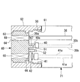
図1は、本発明の第1実施形態に係る内視鏡装置1の全体を示す概略図、図2は内視鏡装置1の全体構成例を示すブロック図、図3は内視鏡2の先端部21を拡大して示す断面図である。
内視鏡装置1は、図1に示すように、被検査対象の像を撮像する内視鏡2と、被検査対象の照明に係るレーザ光を出射する光源装置(光源)3と、内視鏡2により撮像された像の電気信号を画像信号に変換処理する制御装置4と、制御装置4により処理された画像信号を表示するモニタ5と、から概略構成されている。
[First Embodiment]
Hereinafter, a first embodiment of the present invention will be described with reference to FIGS.
FIG. 1 is a schematic diagram showing the
As shown in FIG. 1, an
内視鏡2は、先端光学アダプタ60が着脱可能に取り付けられるとともに、被検査対象の像を撮像するCCDを内蔵した先端部21と、細長で可撓性を有する湾曲部22及び軟性部23からなる挿入部24と、挿入部24の手元側にあって使用者が把持し操作する操作部25と、操作部25から延びる可撓性のユニバーサルコード26と、ユニバーサルコード26の端部に設けられたコネクタ27と、から概略構成されている。なお、ユニバーサルコード26には、後述するライトガイド(光路)及び信号ケーブル等が内蔵されている。
The
コネクタガイド27には、光源装置3に接続される光源コネクタ28が設けられ、この光源コネクタ28を介して、内視鏡挿入部24の先端部21までレーザ光がライトガイドにより導かれるように構成されている。
さらに、コネクタ27には、制御装置4に接続される制御コネクタ29が設けられ、この制御コネクタ29を介して、先端部21に配置したCCDで撮像した被検査対象の像の電気信号が信号ケーブルにより制御装置4へ導かれるように構成されている。
The
Further, the
光源装置3は、図2に示すように、電源供給制御装置31と、電源32と、レーザダイオード33と、集光光学部材34とを具備した構成とされる。
電源供給制御装置31は、電源32の入切や出力調整など各種制御を行うものである。この電源供給制御装置31及び電源32は、特定波長のレーザ光を発光するレーザダイオード33及びアダプタ検知手段として機能するスイッチ40と、電源ケーブル35a及び信号線35bを介して電気的に接続されている。
As illustrated in FIG. 2, the
The power
レーザダイオード33は光源装置3の内部にあり、電源32から電源供給を受ける1個または複数個のレーザダイオード33を適宜組み合わせることにより、特定波長のレーザ光を発するレーザ光源を構成している。また、レーザダイオード33から発光されたレーザ光は、必要に応じて設けられる集光光学部材34を介して、挿入部24内に設けられたライトガイド(導光ファイバ)36の入射端面36aに入射される。このライトガイド36は、入射端面36aに入射されたレーザ光を、先端部21の先端面に設けられた図示しない出射部から外部へ向けて出射するため出射端面36bまで導光する。
The
先端部21の外周面には、先端光学アダプタ60と螺合して着脱可能とする雄ねじ部50が形成されている。また、先端部21には、ライトガイド36の出射端面36bと隣接して、被写体6から反射された観察部からの光を受けるCCD41が設けられている。このCCD41の前面には、必要に応じて観察光学部材42が設けられている。
CCD41で撮像した観察画像の電気信号は、信号ケーブル41aを介して画像処理を行う制御装置4に送られ、さらに、制御装置4からの画像信号がモニタ5に送られて画像表示される。なお、上述した内視鏡2、光源装置3、制御装置4及びモニタ5は、それぞれを別体の装置としてもよいし、あるいは、これらのいずれか複数を適宜組み合わせたり全てを一体化した装置としてもよい。
A
The electric signal of the observation image picked up by the
先端部21には、レーザ光の光学特性を変換可能な先端光学アダプタ60が着脱可能に設けられている。先端光学アダプタ60の着脱状態は、先端部21側に設けたアダプタ検出手段のスイッチ40により自動的に検知される。このスイッチ40は、図3に示すように、先端部21の先端面に設けた凹部であるスイッチ操作口51内に、スイッチ可動部40aを突出させた状態として設置されている。
また、先端部21の先端面には、先端光学アダプタ60を接続する際の円周方向を位置決めするため、先端凹部52が設けられている。
A tip
Further, a
一方、先端光学アダプタ部材60は、先端部21の雄ねじ部50と噛合して接続する雌ねじ部61を有する略円筒形状の外輪部材62と、この外輪部材62の内側に回動可能に嵌合させた円柱部材63とを備えている。
円柱部材63には、照明用窓部64及び観察用窓部65を設けるとともに、円周方向を位置決めする凸部66と、スイッチ40を操作する突起部67とが設けられている。照明用窓部64は、凸部66が先端凹部52に挿入されて円周方向を位置決めした所定の接続位置において、ライトガイド36の出射端面36bと同軸上の前面(観察側)に位置している。また、照明用窓部64には、レーザダイオード33が発光したレーザ光を励起光とし白色発光する蛍光部材、あるいは、レーザ光を拡散照射する拡散光学部材からなる光学特性変換手段68が設けられている。
On the other hand, the distal
The
観察用窓部65は、凸部66が先端凹部52に挿入されて円周方向を位置決めした所定の接続位置において、CCD41及び観察光学部材42と同軸上の前面に位置しており、被写体6から反射された観察部からの光を通す通路となる。
また、上述した所定の接続位置では、突起部67がスイッチ操作口51内に入り込んでスイッチ可動部40aを押し込むので、スイッチ40のスイッチ操作が行われる。このスイッチ操作により、先端光学アダプタ60が所定位置に取り付けられていることを示す電源制御信号、すなわち通常の通電信号が電源供給制御装置31に出力される。
なお、先端光学アダプタ部材60は、円柱部材を所定位置に取り付けて外輪部材62を回転させることにより、雄ねじ部50及び雌ねじ部61が噛合して、内視鏡2の先端部21に固定される。
The
Further, at the predetermined connection position described above, the
The distal
以下、上述した構成の内視鏡装置1について、その作用を説明する、
内視鏡装置1では、観察対象等の諸条件に応じて先端光学アダプタ60を適宜交換して使用する。このとき、先端光学アダプタ60が先端部21の所定位置に取り付けられていると、突起部67がスイッチ可動部40aを押し込んでいるので、スイッチ40から通電信号が出力された状態にある。このため、電源供給制御装置31は、電源32がレーザダイオード33に通常の通電をするよう制御する。
Hereinafter, the operation of the
In the
しかし、たとえば先端光学アダプタ60を交換するため、外輪部材62を回転させて雄ねじ部50及び雌ねじ部61の螺合を解除していくと、外輪部材62と一体に円柱部材63が先端部21の先端面から離間する方向へ移動する。このため、突起部67も円柱部材63と一体に先端面から離間し、スイッチ操作口51から抜け出ることによりスイッチ可動部40aの押し込み操作が解除されるので、スイッチ40の通電信号も解除される。
この結果、電源供給制御装置31は、先端光学アダプタ60が取り外された状態に対応して予め定めた電源32の制御を実施する。この場合の制御としては、電源32をOFFとしてレーザダイオード33への電源供給を完全に停止するか、あるいは、レーザダイオード33への電源供給を所定値以下に低下させる。従って、レーザダイオード33より発光されるレーザ光は、電源供給にの遮断または低減により、完全に停止または出力を弱められるので、先端光学アダプタ60を取り外した先端部21は、レーザ光の出射部となるライトガイド36の出射端面36bから高輝度の光を照射するようなことはない。
However, for example, in order to replace the tip
As a result, the power
すなわち、先端光学アダプタ60の着脱状況をアダプタ検知手段のスイッチ40で検知し、この着脱状況に応じて出射光制御手段となる電源供給制御装置31が出射端面36bから出光するレーザ光を自動的に制御するので、先端光学アダプタ60の着脱操作時には、出射端面36bが設けられている内視鏡2の先端部21から出光するレーザ光がまぶしくならないよう発光を停止または減光することができる。従って、先端光学アダプタ60の着脱時に露出する先端部21の出射端面36bから高輝度のレーザ光が照射されることはなく、しかも、先端光学アダプタ60を取り付けた状態では100%の電源供給を行って十分な光量を確保できる。
That is, the attachment / detachment state of the tip
また、上述したスイッチ40は、先端光学アダプタ60が外れている状態において、凹部となるスイッチ操作口51にスイッチ可動部40aが設置されているので、たとえば指など突起部67以外で押し込むような誤操作により通電信号を出力しにくいものとなる。
また、先端光学アダプタ60が未装着でも高出力のレーザ光を被写体等に当てることなしに、異なる光学特性変換手段68を備えた複数の先端光学アダプタ60の中から適宜選択して最適のもの着脱することにより、たとえば特定波長のレーザ光や白色光のように、複数種類の観察に必要な光を、1種類の光源装置3という小型な装置で実現できる。
Further, since the switch
In addition, even when the tip
続いて、上述した第1の実施の形態に係る変形例を図4に示して説明する。なお、図4は上述した図2に対応するブロック図であり、同様の部分には同じ符号を付し、その詳細な説明は省略する。
この変形例では、内視鏡装置1′の光源装置3′に電源32の制御を行う電源供給制御装置31がなく、先端部21に設けたアダプタ検知手段のスイッチ40が電源32の入切を直接制御するように構成されている。すなわち、電源32とレーザダイオード33とを接続する電源ケーブル35aの途中にスイッチ40を配置し、同スイッチ40が切られてOFFの状態では通電されないように構成されているので、専用の電源供給制御装置31がないことで回路を簡略化することができる。
Subsequently, a modification according to the first embodiment described above will be described with reference to FIG. FIG. 4 is a block diagram corresponding to FIG. 2 described above, and the same parts are denoted by the same reference numerals, and detailed description thereof is omitted.
In this modification, the
〔第2の実施の形態〕
続いて、本発明に係る内視鏡装置について、第2の実施の形態を図5に基づいて説明する。なお、上述した実施の形態と同様の部分には同じ符号を付し、その詳細な説明は省略する。
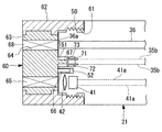
この実施の形態では、上述したスイッチ40のアダプタ検知手段に代えて、内視鏡2に配線された信号線35bの先端に接続して設けられ、スイッチ操作口51内に露出して互いに離間するよう配置された一対の電極71,72と、円柱部材63から突出する突起部67の先端部に取り付けられ、電極71,72間を接続して導通させる接続手段の導電部材73とを備えたアダプタ検知手段が採用されている。なお、電極71,72は、各々が複数の電極部材により構成されてもよい。
[Second Embodiment]
Subsequently, a second embodiment of the endoscope apparatus according to the present invention will be described with reference to FIG. In addition, the same code | symbol is attached | subjected to the part similar to embodiment mentioned above, and the detailed description is abbreviate | omitted.
In this embodiment, instead of the adapter detection means of the
このような構成とすれば、電極71,72と導電部材73とによりスイッチ機構が形成されるので、上述した第1の実施の形態と同様に、先端光学アダプタ60の着脱を検知することができる。なお、この場合の電極71,72は、先端光学アダプタ60が外れている場合の誤操作を防ぐため、スイッチ操作口51のように奥まった部分に設けられていることが望ましい。
このような構成としても、先端部21に先端光学アダプタ60が未装着の時、高出力のレーザ光が発せられることを防止できるので、まぶしさにより着脱操作性が低下することはない。また、高出力のレーザ光が被写体に当たるようなこともなく、さらに、複数の先端光学アダプタ60を選択的に着脱することにより、複数種類の観察に必要な光を、1種類の光源装置3から得ることができる。
With such a configuration, since the switch mechanism is formed by the
Even in such a configuration, it is possible to prevent high-power laser light from being emitted when the distal end
〔第3の実施の形態〕
続いて、本発明に係る内視鏡装置について、第3の実施の形態を図6に基づいて説明する。なお、上述した実施の形態と同様の部分には同じ符号を付し、その詳細な説明は省略する。
この実施の形態では、上述したスイッチ40のアダプタ検知手段に代えて、内視鏡2に配線された信号線35bの先端に接続して先端部21の内部に設けられた共振周波数検知部75と、円柱部材63内に埋設されたコイル76とを備えたアダプタ検知手段が採用されている。
[Third Embodiment]
Subsequently, a third embodiment of the endoscope apparatus according to the present invention will be described with reference to FIG. In addition, the same code | symbol is attached | subjected to the part similar to embodiment mentioned above, and the detailed description is abbreviate | omitted.
In this embodiment, instead of the adapter detection means of the
このような構成とすれば、共振周波数検知部75がコイル76の共振周波数を読み取る手段として機能する。すなわち、先端光学アダプタ60が所定位置に接続されていれば、共振周波数検知部75がコイル76の共振周波数を読み取って検知信号を出力し、先端光学アダプタ60が未装着の時には検知信号の出力はないので、上述したスイッチ40と同様の制御が可能になる。
このような構成としても、先端部21に先端光学アダプタ60が未装着の時、高出力のレーザ光が発せられることを防止できるので、まぶしさにより着脱操作性が低下することはない。また、高出力のレーザ光が被写体に当たるようなこともなく、さらに、複数の先端光学アダプタ60を選択的に着脱することにより、複数種類の観察に必要な光を、1種類の光源装置3から得ることができる。
With such a configuration, the
Even in such a configuration, it is possible to prevent high-power laser light from being emitted when the distal end
〔第4の実施の形態〕
続いて、本発明に係る内視鏡装置について、第4の実施の形態を図7に基づいて説明する。なお、上述した実施の形態と同様の部分には同じ符号を付し、その詳細な説明は省略する。
この実施の形態では、上述したスイッチ40のアダプタ検知手段に代えて、内視鏡2に配線された信号線35bの先端に接続して先端部21の内部に設けられた磁束検知部77と、円柱部材63内に埋設された磁石(磁性体)78とを備えたアダプタ検知手段が採用されている。
[Fourth Embodiment]
Subsequently, a fourth embodiment of the endoscope apparatus according to the present invention will be described with reference to FIG. In addition, the same code | symbol is attached | subjected to the part similar to embodiment mentioned above, and the detailed description is abbreviate | omitted.
In this embodiment, instead of the adapter detection means of the
このような構成とすれば、磁束検知部77が磁石78から発せられた磁束を読み取る磁束検知手段として機能する。すなわち、先端光学アダプタ60が所定位置に接続されていれば、磁束検知部77が磁石78の磁束を読み取って検知信号を出力し、先端光学アダプタ60が未装着の時には検知信号の出力はないので、上述したスイッチ40と同様の制御が可能になる。
このような構成としても、先端部21に先端光学アダプタ60が未装着の時、高出力のレーザ光が発せられることを防止できるので、まぶしさにより着脱操作性が低下することはない。また、高出力のレーザ光が被写体に当たるようなこともなく、さらに、複数の先端光学アダプタ60を選択的に着脱することにより、複数種類の観察に必要な光を、1種類の光源装置3から得ることができる。
With such a configuration, the magnetic flux detector 77 functions as a magnetic flux detector that reads the magnetic flux emitted from the
Even in such a configuration, it is possible to prevent high-power laser light from being emitted when the distal end
〔第5の実施の形態〕
続いて、本発明に係る内視鏡装置について、第5の実施の形態を図8に基づいて説明する。なお、上述した実施の形態と同様の部分には同じ符号を付し、その詳細な説明は省略する。
図8に示すレーザ光出射制御手段は、レーザダイオード33の出射端面36bとライトガイド36の入射端部36aとの間に、レーザ光遮光手段として遮光シャッタ81を設けたものである。この遮光シャッタ81は、レーザ光が通過する光路上で挿抜するためのシャッタ駆動部82と、上述したアダプタ検知手段からの信号に応じて遮光シャッタ81を駆動させるシャッタ制御手段83とを備えている。
[Fifth Embodiment]
Subsequently, a fifth embodiment of the endoscope apparatus according to the present invention will be described with reference to FIG. In addition, the same code | symbol is attached | subjected to the part similar to embodiment mentioned above, and the detailed description is abbreviate | omitted.
The laser light emission control means shown in FIG. 8 is provided with a
このように構成されたレーザ光出射制御手段は、遮光シャッタ81が矢印84で示す方向に移動して光路上で挿抜され、先端光学アダプタ60が装着されている時は遮光シャッタ81を引き抜いて光路を開き、未装着時には遮光シャッタ81が光路を閉じる。
なお、遮光シャッタ81は、光路を完全に閉じる代わりに、レーザ光を減光する絞り状態に切替るようにしてもよく、このような場合には、アダプタ検知手段により先端光学アダプタ60が外れた時、遮光シャッタ81は、光路を全開とする位置から所定の絞り位置に移動してライトガイド36へ出射されるレーザ光を抑えることができる。
The laser light emission control means configured as described above is configured such that the
The
このような構成としても、先端部21に先端光学アダプタ60が未装着の時、高出力のレーザ光が発せられることを防止できるので、まぶしさにより着脱操作性が低下することはない。また、高出力のレーザ光が被写体に当たるようなこともなく、さらに、複数の先端光学アダプタ60を選択的に着脱することにより、複数種類の観察に必要な光を、1種類の光源装置3から得ることができる。
Even in such a configuration, it is possible to prevent high-power laser light from being emitted when the distal end
なお、本発明は上述した実施形態に限定されるものではなく、本発明の要旨を逸脱しない範囲内において適宜変更することができる。 In addition, this invention is not limited to embodiment mentioned above, In the range which does not deviate from the summary of this invention, it can change suitably.
1 内視鏡装置
2 内視鏡
3 光源装置
4 制御装置
5 モニタ
21 先端部
24 挿入部
25 操作部
31 電源供給制御装置
32 電源
33 レーザダイオード
34 集光光学部材
35a 電源ケーブル
35b 信号線
36 ライトガイド
36a 入射端面
36b 出射端面
40 スイッチ(アダプタ検知手段)
40a スイッチ可動部
41 CCD(荷電結合素子)
41a 信号ケーブル(検知信号伝達手段)
42 観察光学部材
51 スイッチ操作口
52 先端凹部
60 先端光学アダプタ
62 外輪部材
63 円柱部材
66 凸部
67 突起部
68 光学特性変換手段
71,72 電極
73 導電部材
75 共振周波数検知部
76 コイル
77 磁束検知部
78 磁石(磁性体)
81 遮光シャッタ
82 シャッタ駆動部
83 シャッタ制御手段
DESCRIPTION OF
40a Switch
41a Signal cable (detection signal transmission means)
42 Observation
81
Claims (13)
前記先端光学アダプタの前記挿入部への着脱を検知するアダプタ検知手段と、該アダプタ検知手段の検知に連動して前記出射部から出射する光を制御する出射光制御手段と、を設けたことを特徴とする内視鏡装置。 An endoscope comprising a light source, an insertion portion that is inserted into the space to be inspected and emits light from the light source from a light emitting portion at the tip, and a tip optical adapter that is detachably attached to the tip portion of the insertion portion A device,
Adapter detecting means for detecting attachment and detachment of the tip optical adapter to and from the insertion portion, and emission light control means for controlling light emitted from the emission portion in conjunction with detection by the adapter detection means. Endoscopic device characterized.
Priority Applications (1)
| Application Number | Priority Date | Filing Date | Title |
|---|---|---|---|
| JP2005000990A JP2006189592A (en) | 2005-01-05 | 2005-01-05 | Endoscope system |
Applications Claiming Priority (1)
| Application Number | Priority Date | Filing Date | Title |
|---|---|---|---|
| JP2005000990A JP2006189592A (en) | 2005-01-05 | 2005-01-05 | Endoscope system |
Publications (2)
| Publication Number | Publication Date |
|---|---|
| JP2006189592A true JP2006189592A (en) | 2006-07-20 |
| JP2006189592A5 JP2006189592A5 (en) | 2008-02-21 |
Family
ID=36796876
Family Applications (1)
| Application Number | Title | Priority Date | Filing Date |
|---|---|---|---|
| JP2005000990A Pending JP2006189592A (en) | 2005-01-05 | 2005-01-05 | Endoscope system |
Country Status (1)
| Country | Link |
|---|---|
| JP (1) | JP2006189592A (en) |
Cited By (6)
| Publication number | Priority date | Publication date | Assignee | Title |
|---|---|---|---|---|
| JP2011094993A (en) * | 2009-10-27 | 2011-05-12 | Hioki Ee Corp | Tachometer |
| JP2015075685A (en) * | 2013-10-10 | 2015-04-20 | オリンパス株式会社 | Optical scanning method, optical scanning device, and optical scanning type observation device |
| US20180136455A1 (en) * | 2015-08-18 | 2018-05-17 | Olympus Corporation | Optical scanning method and optical scanning apparatus |
| CN113907873A (en) * | 2021-10-24 | 2022-01-11 | 青岛海泰新光科技股份有限公司 | Laser output control device embedded in handle of endoscope camera system |
| CN115175632A (en) * | 2020-02-20 | 2022-10-11 | 瑞德医疗机器股份有限公司 | Surgical instrument holding device and surgical assistance device |
| WO2024203754A1 (en) * | 2023-03-28 | 2024-10-03 | テルモ株式会社 | Tomographic image generation device and tomographic image generation system |
Citations (7)
| Publication number | Priority date | Publication date | Assignee | Title |
|---|---|---|---|---|
| JPS5734833A (en) * | 1980-08-07 | 1982-02-25 | Olympus Optical Co | Light source apparatus of endoscope |
| JPH0220817A (en) * | 1988-04-11 | 1990-01-24 | Olympus Optical Co Ltd | Endoscope device |
| JPH06222288A (en) * | 1993-01-28 | 1994-08-12 | Fuji Photo Optical Co Ltd | Endoscope |
| JPH06245901A (en) * | 1993-02-26 | 1994-09-06 | Olympus Optical Co Ltd | Endoscope controller |
| JPH11192207A (en) * | 1997-11-07 | 1999-07-21 | Matsushita Electric Ind Co Ltd | Video scope and portable storage case |
| US20030042493A1 (en) * | 2001-08-31 | 2003-03-06 | Yuri Kazakevich | Solid-state light source |
| JP2004313241A (en) * | 2003-04-11 | 2004-11-11 | Olympus Corp | Optical adapter and endoscopic apparatus |
-
2005
- 2005-01-05 JP JP2005000990A patent/JP2006189592A/en active Pending
Patent Citations (8)
| Publication number | Priority date | Publication date | Assignee | Title |
|---|---|---|---|---|
| JPS5734833A (en) * | 1980-08-07 | 1982-02-25 | Olympus Optical Co | Light source apparatus of endoscope |
| JPH0220817A (en) * | 1988-04-11 | 1990-01-24 | Olympus Optical Co Ltd | Endoscope device |
| JPH06222288A (en) * | 1993-01-28 | 1994-08-12 | Fuji Photo Optical Co Ltd | Endoscope |
| JPH06245901A (en) * | 1993-02-26 | 1994-09-06 | Olympus Optical Co Ltd | Endoscope controller |
| JPH11192207A (en) * | 1997-11-07 | 1999-07-21 | Matsushita Electric Ind Co Ltd | Video scope and portable storage case |
| US20030042493A1 (en) * | 2001-08-31 | 2003-03-06 | Yuri Kazakevich | Solid-state light source |
| JP2005502083A (en) * | 2001-08-31 | 2005-01-20 | スミス アンド ネフュー インコーポレーテッド | Solid light source |
| JP2004313241A (en) * | 2003-04-11 | 2004-11-11 | Olympus Corp | Optical adapter and endoscopic apparatus |
Cited By (9)
| Publication number | Priority date | Publication date | Assignee | Title |
|---|---|---|---|---|
| JP2011094993A (en) * | 2009-10-27 | 2011-05-12 | Hioki Ee Corp | Tachometer |
| JP2015075685A (en) * | 2013-10-10 | 2015-04-20 | オリンパス株式会社 | Optical scanning method, optical scanning device, and optical scanning type observation device |
| US20180136455A1 (en) * | 2015-08-18 | 2018-05-17 | Olympus Corporation | Optical scanning method and optical scanning apparatus |
| US10754143B2 (en) * | 2015-08-18 | 2020-08-25 | Olympus Corporation | Optical scanning method and optical scanning apparatus |
| CN115175632A (en) * | 2020-02-20 | 2022-10-11 | 瑞德医疗机器股份有限公司 | Surgical instrument holding device and surgical assistance device |
| CN115175632B (en) * | 2020-02-20 | 2023-11-21 | 瑞德医疗机器股份有限公司 | Surgical instrument holding device and surgical auxiliary device |
| CN113907873A (en) * | 2021-10-24 | 2022-01-11 | 青岛海泰新光科技股份有限公司 | Laser output control device embedded in handle of endoscope camera system |
| CN113907873B (en) * | 2021-10-24 | 2024-03-29 | 青岛海泰新光科技股份有限公司 | Laser output control device embedded in endoscope camera system handle |
| WO2024203754A1 (en) * | 2023-03-28 | 2024-10-03 | テルモ株式会社 | Tomographic image generation device and tomographic image generation system |
Similar Documents
| Publication | Publication Date | Title |
|---|---|---|
| JP4947975B2 (en) | Endoscope device and endoscope illumination device | |
| JP4812430B2 (en) | Endoscope device | |
| JP5080014B2 (en) | Imaging system | |
| US6962565B2 (en) | Excitation light illuminating probe, video endoscope system, and video endoscope for fluorescence observation | |
| US7769433B2 (en) | Light source apparatus for endoscope | |
| EP2371268A1 (en) | Illuminationg device and endoscopic apparatus | |
| JP5881307B2 (en) | An observation device that can detect the tip position of the insertion section using external light | |
| JPH11244220A (en) | Fluorescent endoscope | |
| JP4547243B2 (en) | Endoscope system | |
| JP2005323737A (en) | Endoscope device | |
| US6389205B1 (en) | Brightness-controlled endoscope illumination system | |
| JP2005058618A (en) | Endoscope and cap | |
| JPH11104059A (en) | Fluorescent observation device | |
| JP2012147860A (en) | Endoscope apparatus, and safety management method of endoscope apparatus | |
| JP2009201543A (en) | Endoscope system | |
| JP2008212348A (en) | Endoscope apparatus | |
| JP4420638B2 (en) | Endoscope | |
| JP2006189592A (en) | Endoscope system | |
| US8562514B2 (en) | Medical apparatus and endoscope system with memory function | |
| JP2005348794A (en) | Light source device for endoscope | |
| JP2009039464A (en) | Illumination device of endoscope | |
| JP4459724B2 (en) | Endoscope light source device | |
| JP2006158735A (en) | Endoscope and endoscope imaging system | |
| JP2010088657A (en) | Endoscope light source device | |
| JP2006014924A (en) | Endoscope system |
Legal Events
| Date | Code | Title | Description |
|---|---|---|---|
| A521 | Written amendment |
Free format text: JAPANESE INTERMEDIATE CODE: A523 Effective date: 20071227 |
|
| A621 | Written request for application examination |
Free format text: JAPANESE INTERMEDIATE CODE: A621 Effective date: 20071227 |
|
| A131 | Notification of reasons for refusal |
Free format text: JAPANESE INTERMEDIATE CODE: A131 Effective date: 20110315 |
|
| A521 | Written amendment |
Free format text: JAPANESE INTERMEDIATE CODE: A523 Effective date: 20110513 |
|
| A131 | Notification of reasons for refusal |
Free format text: JAPANESE INTERMEDIATE CODE: A131 Effective date: 20110906 |
|
| A521 | Written amendment |
Free format text: JAPANESE INTERMEDIATE CODE: A523 Effective date: 20111104 |
|
| A02 | Decision of refusal |
Free format text: JAPANESE INTERMEDIATE CODE: A02 Effective date: 20120207 |


