JP2005295822A - Method for fixing culture schedule and method for culturing cell - Google Patents
Method for fixing culture schedule and method for culturing cell Download PDFInfo
- Publication number
- JP2005295822A JP2005295822A JP2004113018A JP2004113018A JP2005295822A JP 2005295822 A JP2005295822 A JP 2005295822A JP 2004113018 A JP2004113018 A JP 2004113018A JP 2004113018 A JP2004113018 A JP 2004113018A JP 2005295822 A JP2005295822 A JP 2005295822A
- Authority
- JP
- Japan
- Prior art keywords
- culture
- initial
- bone marrow
- schedule setting
- setting method
- Prior art date
- Legal status (The legal status is an assumption and is not a legal conclusion. Google has not performed a legal analysis and makes no representation as to the accuracy of the status listed.)
- Withdrawn
Links
Images
Landscapes
- Micro-Organisms Or Cultivation Processes Thereof (AREA)
Abstract
Description
この発明は、培養スケジュール設定方法および細胞培養方法に関するものである。 The present invention relates to a culture schedule setting method and a cell culture method.
近年、いわゆる再生医療において、術後の生体組織における欠損部の修復速度を高めるために、患者から採取した骨髄液等から間葉系幹細胞を取り出して、βリン酸三カルシウム(β−TCP)やハイドロキシアパタイト(HAP)等の生体組織補填材とともに培養することにより、培養骨に代表される生体組織補填体を製造することが提案されている。生体組織補填体は、移植時に、すでに生体組織補填材を足場にして増殖した多くの間葉系幹細胞を含んでいるので、手術後に体内で細胞を増殖させる方法と比較すると、自家組織に置換されるまでの日数を大幅に短縮することができる(例えば、非特許文献1参照。)。 In recent years, in so-called regenerative medicine, mesenchymal stem cells are extracted from bone marrow fluid collected from a patient in order to increase the repair speed of a defect in a living tissue after surgery, and β tricalcium phosphate (β-TCP) or It has been proposed to produce a body tissue supplement represented by cultured bone by culturing with a body tissue filling material such as hydroxyapatite (HAP). Since the tissue replacement includes many mesenchymal stem cells that have already proliferated using the tissue replacement material as a scaffold at the time of transplantation, it is replaced with autologous tissue compared to the method of proliferating cells in the body after surgery. Can be significantly shortened (see, for example, Non-Patent Document 1).
生体組織補填体を製造するには、一般に、まず、患者の骨髄液を培養容器内で一次培養して内部に含まれている間葉系幹細胞を必要細胞数まで増加させる。この過程において、成長した間葉系幹細胞は、少なくとも1回以上、培養容器から剥がされて、さらに大きな培養容器に移し替えられる。そして、最終的に必要細胞数まで増加したところで、再度、培養容器から剥離させられ、生体組織補填材に付着させられて、二次培養が行われる。これにより、生体組織補填体が製造される(例えば、非特許文献2参照。)。
従来、骨髄液を培養して必要細胞数まで増殖させる一次培養工程において、実際にその骨髄液内に含有されている間葉系幹細胞が必要細胞数まで増殖するか否かについては、培養容器内に形成される間葉系幹細胞のコロニーを目視にて観察し、その大きさを測ることによって判断するのが一般的であった。この場合、コロニーの目視による判断には、一次培養工程がある程度進行する必要があり、少なくとも1週間以上の培養期間が必要であった。 Conventionally, in the primary culture process in which bone marrow fluid is cultured and grown to the required number of cells, whether or not the mesenchymal stem cells actually contained in the bone marrow fluid are grown to the required number of cells is determined in the culture vessel. It was common to make a judgment by visually observing the colony of mesenchymal stem cells formed and measuring the size. In this case, the visual inspection of the colonies required the primary culture process to proceed to some extent, and required a culture period of at least one week.
しかしながら、間葉系幹細胞が必要細胞数まで増殖するか否か、すなわち、骨髄液の品質の良否が、培養開始から1週間経過しなければ判断できないのでは、仮に1週間経過後に必要細胞数まで増殖しないと判断される場合には、それまでの培養工程が無駄になるという不都合がある。また、必要細胞数まで増殖する時期が予測できれば、細胞の培養スケジュールを設定でき、また、最終的な手術日を決定できるので便利である。 However, if the mesenchymal stem cells grow to the required number of cells, that is, whether the quality of the bone marrow fluid can be judged only after one week has elapsed since the start of the culture, When it is determined that the cells do not proliferate, there is a disadvantage that the previous culture process is wasted. In addition, if the time to proliferate to the required number of cells can be predicted, it is convenient because the cell culture schedule can be set and the final operation date can be determined.
この発明は上述した事情に鑑みてなされたものであって、骨髄液の培養開始後早期に骨髄液の品質の良否を判定でき、無駄な培養工程を行うことを防止し、効率よく細胞を培養できる培養スケジュール設定方法および細胞培養方法を提供することを目的としている。 The present invention has been made in view of the above-described circumstances, and can determine the quality of bone marrow fluid at an early stage after the start of culturing bone marrow fluid, prevent unnecessary culture steps from being performed, and efficiently culture cells. An object of the present invention is to provide a culture schedule setting method and a cell culture method.
上記目的を達成するために、本発明は、以下の手段を提供する。
本発明は、培養容器内に骨髄液を播種して培養する際の培養スケジュールを設定する方法であって、培養開始直後の所定期間内に、複数の幹細胞の塊からなる初期コロニーを検出し、培養容器内において検出された初期コロニーの数に基づいて培養を継続するか否かを判断する培養スケジュール設定方法を提供する。
In order to achieve the above object, the present invention provides the following means.
The present invention is a method for setting a culture schedule when seeding and culturing bone marrow in a culture vessel, and detecting an initial colony consisting of a plurality of stem cell masses within a predetermined period immediately after the start of culture, Provided is a culture schedule setting method for determining whether or not to continue culture based on the number of initial colonies detected in a culture vessel.
発明者は、培養開始直後の所定期間内に形成される微細なコロニー(以下、初期コロニーともいう。)の数が、その骨髄液内に含まれる幹細胞の増殖能と相関しているという知見を得た。この初期コロニーは、幹細胞数50個程度、数100μm程度の大きさを有するため目視で観察することはできないが、顕微鏡観察により検出することができる。そこで、培養開始直後の所定期間内に初期コロニーを検出して、その数を計数することにより、幹細胞の増殖能を判断でき、精度よく培養スケジュールを設定することが可能となる。 The inventor has found that the number of fine colonies (hereinafter also referred to as initial colonies) formed within a predetermined period immediately after the start of culture correlates with the proliferative ability of stem cells contained in the bone marrow fluid. Obtained. This initial colony has a size of about 50 stem cells and about several hundred μm, and thus cannot be visually observed, but can be detected by microscopic observation. Therefore, by detecting the initial colonies within a predetermined period immediately after the start of the culture and counting the number, the proliferation ability of the stem cells can be determined, and the culture schedule can be set with high accuracy.
その結果、例えば、幹細胞の増殖能が極めて低いことが判明した場合には、それ以上の培養工程継続を中止して、培養作業の無駄を省くことができ、また、骨髄液の再採取等の次善の策を講じることができる。また、幹細胞の増殖能が低いが、必要細胞数まで達することができる程度の増殖能を有すると判断できる場合には、それに合わせた培養スケジュールを設定し、適正な培養を行うことができる。さらには、移植日の設定や変更等を行うことが可能となる。 As a result, for example, when it is found that the proliferating ability of the stem cells is extremely low, it is possible to stop further culturing process and eliminate waste of culturing work, You can take next best measures. In addition, when it can be determined that the stem cells have a low proliferative ability but can grow to a necessary number of cells, an appropriate culture can be performed by setting a culture schedule according to the proliferative ability. Furthermore, it becomes possible to set or change the transplant date.
上記発明においては、前記培養開始直後の所定期間が3日以内であることが好ましい。
培養開始後2〜3日程度で初期コロニーが形成されることが判明した。このため、この期間に初期コロニーの検出、計数を行うことで、極めて早期に幹細胞の増殖能を判定することができる。
In the said invention, it is preferable that the predetermined period immediately after the said culture | cultivation start is less than 3 days.
It was found that an initial colony was formed about 2 to 3 days after the start of culture. For this reason, the ability to proliferate stem cells can be determined very early by detecting and counting initial colonies during this period.
また、上記発明においては、前記初期コロニーの検出が、培養容器内の骨髄液に光を照射し、骨髄液を透過した光を検出することにより行われることとしてもよい。培養開始直後は、培養容器内の骨髄液に光を照射しても、幹細胞が赤血球等に覆われているために透過する光が少ない。しかしながら、所定の期間、例えば2〜3日経過して初期コロニーが形成されると、該初期コロニーが盛り上がって、それを覆っている赤血球等にひびが入る。このため、照射された光がひびを透過するようになり、この光を観察することによって、初期コロニーを簡易に検出することが可能となる。 In the above invention, the initial colony may be detected by irradiating the bone marrow fluid in the culture container with light and detecting the light transmitted through the bone marrow fluid. Immediately after the start of culturing, even if the bone marrow fluid in the culture vessel is irradiated with light, the stem cells are covered with red blood cells and so on, so that the transmitted light is small. However, when an initial colony is formed after a predetermined period of time, for example, 2 to 3 days, the initial colony rises, and the red blood cells covering the initial colony crack. For this reason, the irradiated light comes to pass through the crack, and the initial colony can be easily detected by observing this light.
また、上記発明においては、前記初期コロニーの検出前に、培養容器内の骨髄液を溶血処理することとしてもよい。
溶血処理により、赤血球の血球成分が除去されるので、初期コロニーの顕微鏡観察が容易になる。
In the above invention, the bone marrow fluid in the culture vessel may be hemolyzed before the detection of the initial colonies.
The hemolysis process removes blood cell components of red blood cells, so that the initial colony can be easily observed with a microscope.
また、上記発明においては、検出された初期コロニーの数が所定のしきい値より少ないときに、培養中止を判断することとしてもよい。培養中止を判断することにより、それ以上の無駄な培養作業が不要となり、コストおよび時間の無駄の発生を防止することができる。
さらに、上記発明においては、検出された初期コロニーの数が所定のしきい値以上のときに、培養継続を判断することとしてもよい。
Moreover, in the said invention, when the number of the initial colonies detected is less than a predetermined threshold value, it is good also as determining a culture stop. By determining to stop culturing, no further unnecessary culturing work is required, and generation of cost and time can be prevented.
Furthermore, in the said invention, it is good also as determining culture | cultivation continuation when the number of the initial colonies detected is more than a predetermined threshold value.
また、上記発明においては、骨髄液提供者の年齢によって前記しきい値を補正することが好ましい。幹細胞の増殖能は、年齢とともに変化するため、一律に同一のしきい値を設定したのでは、実情に沿わない場合があるからである。例えば、一般には、骨髄液提供者の年齢が高まるにつれて細胞増殖能は低下していくことになる。したがって、年齢とともにしきい値を上げ、培養中止または培養継続を判断することにより、培養作業の無駄を省くことができる。 Moreover, in the said invention, it is preferable to correct | amend the said threshold value by the age of a bone marrow fluid donor. This is because the proliferative ability of stem cells changes with age, and if the same threshold value is set uniformly, it may not be in line with the actual situation. For example, generally, as the age of a bone marrow fluid donor increases, the cell proliferating ability decreases. Accordingly, by raising the threshold value with age and determining whether to stop culture or continue culture, waste of culture work can be eliminated.
また、本発明は、上記培養スケジュール設定方法により設定された培養スケジュールに基づいて細胞を培養する細胞培養方法を提供する。
この発明によれば、細胞の増殖能に基づいて設定された培養スケジュールに沿って細胞を培養するので、効率的に培養することができ、また、最終的に製品として出荷可能な時期あるいは移植手術が可能となる時期を精度よく予測することが可能となる。
The present invention also provides a cell culture method for culturing cells based on the culture schedule set by the culture schedule setting method.
According to the present invention, since the cells are cultured according to the culture schedule set based on the proliferation ability of the cells, the cells can be efficiently cultured, and can finally be shipped as a product or transplant surgery. It becomes possible to accurately predict the time when it becomes possible.
本発明によれば、骨髄液の培養開始後早期に骨髄液の品質の良否を判定でき、無駄な培養工程を行うことを防止して、細胞を効率よく培養することができるという効果を奏する。 According to the present invention, it is possible to determine the quality of bone marrow fluid at an early stage after the start of culturing bone marrow fluid, and it is possible to prevent the wasteful culturing process from being performed and to efficiently culture cells.
この発明の一実施形態に係る培養スケジュール設定方法および細胞培養方法について図1〜図5を参照して以下に説明する。
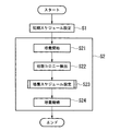
本実施形態に細胞培養方法は、図1に示されるように、培養開始前に暫定的に設定する初期スケジュール設定ステップS1と、該初期スケジュール設定ステップS1において設定された暫定的なスケジュールに従って細胞の培養を開始する培養ステップS2とを含んでいる。また、本実施形態に係る培養スケジュール設定方法は、培養を開始した後に行われる初期コロニー検出ステップS22と培養スケジュール設定ステップS23とを含んでいる。
A culture schedule setting method and a cell culture method according to an embodiment of the present invention will be described below with reference to FIGS.
As shown in FIG. 1, the cell culturing method according to the present embodiment includes an initial schedule setting step S1 that is provisionally set before the start of culture, and a cell schedule according to the provisional schedule set in the initial schedule setting step S1. And a culture step S2 for starting the culture. In addition, the culture schedule setting method according to the present embodiment includes an initial colony detection step S22 and a culture schedule setting step S23 performed after the culture is started.
初期スケジュール設定ステップS1は、骨髄液提供者の年齢や生活習慣等の種々のファクターを考慮して、培地の交換時期や培養容器の切替時期等、さらには移植手術予定日等の培養スケジュールを設定する。
そして、骨髄液提供者から提供された骨髄液を所定の培地と混合して細胞懸濁液を製造し、製造された細胞懸濁液を所定の培養容器内に投入して、前記暫定的な培養スケジュールに基づいて細胞の培養が開始される(ステップS21)。
In the initial schedule setting step S1, taking into account various factors such as the age and lifestyle habits of the bone marrow fluid donor, a culture schedule such as a medium replacement time, a culture container switching time, etc., and a planned transplant surgery date, etc. are set. To do.
Then, a bone marrow fluid provided by a bone marrow fluid provider is mixed with a predetermined medium to produce a cell suspension, and the produced cell suspension is put into a predetermined culture container, Cell culture is started based on the culture schedule (step S21).
ここで、使用される培地は、例えば、MEM(Minimal
Essential Medium:最小必須培地)、FBS(Fetal Bovine Serum:ウシ胎児血清)、抗生剤を84:15:1の配合比率で混合したものである。なお、この配合比率は任意であり、また、ウシ胎児血清に代えてヒト血清を用いてもよい。
また、培養条件は、温度37℃±0.5℃、湿度100%、CO2濃度5%である。
Here, the medium used is, for example, MEM (Minimal
Essential Medium: minimum essential medium), FBS (Fetal Bovine Serum: fetal bovine serum), and antibiotics mixed at a mixing ratio of 84: 15: 1. This mixing ratio is arbitrary, and human serum may be used instead of fetal bovine serum.
The culture conditions are a temperature of 37 ° C. ± 0.5 ° C., a humidity of 100%, and a CO 2 concentration of 5%.
次に、初期コロニー検出ステップS22について説明する。
初期コロニー検出ステップS22は、培養開始直後の所定期間、例えば、2〜3日以内に行われるステップであり、培養容器内に静置された状態で形成される50個以上の幹細胞の塊からなる初期コロニーを検出する。初期コロニーは、数100μm程度の塊であり、目視では観察できないので、以下の方法により検出する。
Next, the initial colony detection step S22 will be described.
The initial colony detection step S22 is a step performed within a predetermined period immediately after the start of culture, for example, within 2 to 3 days, and is composed of a cluster of 50 or more stem cells formed in a stationary state in the culture vessel. Detect initial colonies. The initial colony is a mass of about several hundred μm and cannot be observed with the naked eye, and is detected by the following method.
すなわち、倒立顕微鏡を用いて培養容器内の細胞懸濁液の上方から光を照射し、培養容器の下方に対物レンズを配置して観察する。骨髄液と培地とを混合してなる細胞懸濁液には、赤血球等の血球成分も多く含まれているため、培養初期においては赤血球が全面を膜状に覆っていて上方からの光が通過できない状態になっている。この場合に、初期コロニーが形成されると、該初期コロニーの部分において表面を覆っている赤血球等からなる層(layer)が盛り上がり、その層(layer)に亀裂が生じるため、上方からの光が通過できるようになる。 That is, light is irradiated from above the cell suspension in the culture vessel using an inverted microscope, and an objective lens is placed below the culture vessel for observation. The cell suspension formed by mixing the bone marrow fluid and the medium contains many blood cell components such as erythrocytes. Therefore, in the initial stage of culturing, the erythrocytes cover the entire surface in a film and light from above passes. It is in a state where it cannot be done. In this case, when an initial colony is formed, a layer made of red blood cells or the like covering the surface of the initial colony swells and a crack occurs in the layer (layer). You can pass through.
そこで、倒立顕微鏡によって培養容器を全面にわたって走査し、光が漏れている箇所を探すことにより、初期コロニーが検出されることになる。そして、検出された初期コロニーの数を計数して初期コロニー数Cを求めることにより、初期コロニー検出ステップS22が終了する。 Therefore, an initial colony is detected by scanning the culture vessel over the entire surface with an inverted microscope and searching for a portion where light is leaking. Then, the initial colony detection step S22 is completed by counting the number of detected initial colonies to obtain the initial colony number C.
次に、培養スケジュール設定ステップS23について説明する。
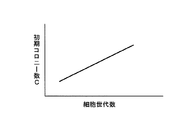
初期コロニー数Cと幹細胞の増殖能とは図2に示される関係がある。図2は、幹細胞が継代可能な細胞世代数と、初期コロニー数との関係を示している。これによれば、初期コロニー数が多いほど細胞世代数が多く、幹細胞の増殖能が高いということがわかる。
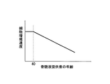
また、図3に、骨髄液提供者の年齢と細胞増殖速度との関係を示す。この図3によれば、細胞提供者の年齢が40歳を超えて高くなるほど、細胞増殖速度が低下していくことがわかる。
Next, the culture schedule setting step S23 will be described.
The relationship between the initial colony number C and stem cell proliferation ability is shown in FIG. FIG. 2 shows the relationship between the number of cell generations that can be passaged by stem cells and the number of initial colonies. This shows that the larger the number of initial colonies, the greater the number of cell generations and the higher the ability of stem cells to proliferate.
FIG. 3 shows the relationship between the age of the bone marrow fluid donor and the cell growth rate. According to FIG. 3, it can be seen that the cell growth rate decreases as the age of the cell provider exceeds 40 years.
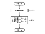
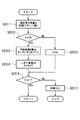
そこで、培養スケジュール設定ステップS23においては、上記事実を考慮して、図4に示されるように、まず、その後の培養を継続するか中止するかの判断を行う(ステップS231)。
すなわち、原則としては、初期コロニー検出ステップS22において検出された初期コロニー数Cが所定のしきい値Xより大きいか否かによって培養を継続するか否かを判断する。初期コロニー数Cがしきい値Xより小さい場合には、細胞増殖能が低いと判断できるため、培養を中止することにより、その後の培養作業の無駄を省くことができる。
Therefore, in the culture schedule setting step S23, in consideration of the above fact, as shown in FIG. 4, it is first determined whether to continue or stop the subsequent culture (step S231).
That is, in principle, it is determined whether or not to continue the culture depending on whether or not the initial colony count C detected in the initial colony detection step S22 is greater than a predetermined threshold value X. When the initial colony number C is smaller than the threshold value X, it can be determined that the cell growth ability is low, so that the culture work can be wasted by stopping the culture.
また、しきい値Xの設定に際しては、骨髄液提供者の年齢Aを考慮している。すなわち、図5に示されるように、骨髄液提供者の年齢Aおよび上記初期コロニー検出ステップS22において計数された初期コロニー数Cが入力され(ステップS2311)、骨髄液提供者の年齢Aが40歳より高いか否かが判断される(ステップS2312)。 In setting the threshold value X, the age A of the bone marrow fluid provider is taken into consideration. That is, as shown in FIG. 5, the age A of the bone marrow fluid donor and the initial colony number C counted in the initial colony detection step S22 are input (step S2311), and the age A of the bone marrow fluid donor is 40 years old. It is determined whether or not it is higher (step S2312).
ステップS2312において骨髄液提供者の年齢Aが40歳を超える場合には、骨髄液提供者の年齢Aに応じて、しきい値Xを補正する年齢係数Bが算出される(ステップS2313)。年齢係数Bは、例えば、B=(10/(Aー20)+1/2)であり、年齢Aが高くなるにしたがって小さくなる係数である。そして、この年齢計数Bを用いてしきい値Xが算出される(ステップS2314)。しきい値Xは、基準となるしきい値X0を年齢係数Bで除した値である。
一方、骨髄液提供者の年齢Aが40歳以下の場合には、ステップS2312において、しきい値Xを基準となるしきい値X0自体とする設定がなされる(ステップS2315)。
When the age A of the bone marrow donor exceeds 40 years old in step S2312, the age coefficient B for correcting the threshold value X is calculated according to the age A of the bone marrow donor (step S2313). The age coefficient B is, for example, B = (10 / (A−20) +1/2), and is a coefficient that decreases as age A increases. Then, a threshold value X is calculated using this age count B (step S2314). Threshold value X is a threshold X 0 as a reference is a value obtained by dividing the age factor B.
On the other hand, age A bone marrow fluid provider if: 40 years old, in step S2312, settings for the threshold X 0 itself as a reference threshold X is made (step S2315).
次いで、上記により設定されたしきい値Xと初期コロニー数Cとが比較される(ステップS2316)。初期コロニー数Cが少ない場合であっても、骨髄液提供者の年齢Aが40歳以下の場合には、十分な細胞増殖能が期待できるので、年齢係数Bを考慮することなくしきい値Xを基準となるしきい値X0に保ち、比較的少ない初期コロニー数Cの場合にも培養継続の判断が可能となる(ステップS2316)。
一方、初期コロニー数Cが多い場合であっても、骨髄液提供者の年齢Aが40歳を超える場合には、細胞増殖能が低く、培養中止の判断をした方がよい場合がある。したがって、しきい値Xを高くして、初期コロニー数Cが十分ではない場合には培養の中止を判断する(ステップS2317)。
Next, the threshold value X set as described above is compared with the initial colony number C (step S2316). Even if the initial colony number C is small, if the age A of the bone marrow fluid donor is 40 years old or less, sufficient cell proliferation ability can be expected, so the threshold value X is set without considering the age coefficient B. maintaining the threshold X 0 as a reference, it becomes possible to determine the continued culture for relatively small initial colony count C (step S2316).
On the other hand, even when the initial colony number C is large, when the age A of the bone marrow fluid donor exceeds 40 years old, the cell proliferating ability is low, and it may be better to judge the culture stop. Therefore, the threshold value X is increased, and when the initial colony number C is not sufficient, it is determined to stop the culture (step S2317).
また、培養スケジュール設定ステップS23においては、初期コロニー数Cがしきい値Xに対して大きい場合には、培養を継続する判断が行われ(ステップS231)、さらに、初期コロニー数Cおよび骨髄提供者の年齢Aによって定まる細胞増殖速度に応じて、その後の1次培養終了までの時間、2次培養に要する時間、手術日までに要する時間を予測し再設定する(ステップS232)。
そして、このように設定された培養スケジュールに基づいて、その後の培養を継続する(ステップS24)ことにより、細胞が培養されることになる。
In the culture schedule setting step S23, when the initial colony number C is larger than the threshold value X, it is determined to continue the culture (step S231). Furthermore, the initial colony number C and the bone marrow donor are determined. In accordance with the cell growth rate determined by age A, the time until the end of the primary culture, the time required for the secondary culture, and the time required until the operation day are predicted and reset (step S232).
And based on the culture schedule set in this way, a cell is cultured by continuing subsequent culture | cultivation (step S24).
本実施形態に係る培養スケジュール設定方法および細胞培養方法によれば、初期コロニー数Cに基づいて培養スケジュールを設定するので、培養開始直後3日以内という、極めて早期に培養を継続するか否かを判断でき、培養を中止する場合には、その後の培養作業を行わずに済むので、時間およびコストの無駄を省くことができる。 According to the culture schedule setting method and the cell culture method according to the present embodiment, since the culture schedule is set based on the initial colony number C, whether or not the culture is continued very early within 3 days immediately after the start of the culture is determined. This can be judged, and when culturing is stopped, subsequent culturing work is not required, so that waste of time and cost can be saved.
また、培養開始直後に検出される初期コロニー数Cによってその後の培養スケジュールを設定できるので、細胞増殖能の高い細胞に対しては、必要細胞数まで迅速に増殖させて移植手術までに要する時間を短縮することができる。また、細胞増殖能の低い細胞に対しては、必要細胞数に達するまでに要する時間が長いことが予想されるので、十分な培養期間を確保して、細胞を確実に増殖させることができる。 In addition, since the subsequent culture schedule can be set based on the initial colony number C detected immediately after the start of culture, for cells with high cell proliferating capacity, the time required for transplantation surgery can be increased by rapidly growing to the required number of cells. It can be shortened. In addition, for cells with low cell growth ability, it is expected that the time required to reach the required number of cells is long, so that a sufficient culture period can be secured and the cells can be reliably proliferated.
なお、本発明に係る 培養スケジュール設定方法および細胞培養方法は、上記実施形態に限定されるものではない。
例えば、上記実施形態においては、初期コロニー検出ステップS22において、培養容器の全面にわたって走査することとしたが、培養容器が大きい場合には、全面にわたる走査に時間がかかることが考えられる。この場合には、培養容器内部を複数の領域に区画して、その領域のいくつかを選択的に走査することにより、領域毎の初期コロニー数を求め、該領域毎の初期コロニー数に基づいて全体の初期コロニー数Cを推定することにしてもよい。
The culture schedule setting method and the cell culture method according to the present invention are not limited to the above embodiment.
For example, in the above-described embodiment, scanning is performed over the entire surface of the culture vessel in the initial colony detection step S22. However, if the culture vessel is large, it may take time to scan over the entire surface. In this case, the inside of the culture container is divided into a plurality of regions, and some of the regions are selectively scanned to obtain the initial colony number for each region, and based on the initial colony number for each region. The total initial colony number C may be estimated.
また、骨髄液を培養する培養容器の他に、サンプル用の小さい培養容器を用いて同条件で培養する場合には、このサンプル用の培養容器に対して全面にわたる走査を行うことにしてもよい。 In addition, when culturing under the same conditions using a small culture vessel for a sample other than the culture vessel for culturing bone marrow fluid, the entire culture vessel for this sample may be scanned. .
また、赤血球の膜に生ずる亀裂の検出は、顕微鏡を扱う観察者が、接眼レンズを通して、あるいは、撮影されモニタに表示された画像を通して行ってもよいが、撮影された画像を処理して亀裂発生部を検出することとすれば、自動的に検出することができる。また、画像処理により亀裂発生部を直接検出する場合の他、亀裂発生により漏れる光が増大することを利用して、対物レンズ側で検出される光の総光量を取得することにより、その総光量に基づいて、初期コロニー数を推定することにしてもよい。 The detection of cracks in the red blood cell membrane may be performed by the observer handling the microscope through an eyepiece lens or through an image taken and displayed on a monitor. If the part is detected, it can be automatically detected. In addition to directly detecting the crack occurrence part by image processing, the total light quantity of the light detected on the objective lens side is obtained by utilizing the fact that the light leaked by the crack occurrence increases. Based on the above, the initial colony number may be estimated.
また、培養開始前に、細胞懸濁液に塩化アンモニウム等の溶血剤を添加して攪拌する溶血処理を施すことにより、赤血球を除去してもよい。このようにすることで、初期コロニーの形状を顕微鏡により直接観察することができる。
また、上記実施形態においては、培養スケジュールの設定ステップにおいて、培養の継続か否かの判断や、各培養工程や移植手術の時間的なスケジュールについて再設定することとしたが、これに加えて、添加する成長因子や栄養剤等の量や種類を変更することにしてもよい。
In addition, before starting culture, red blood cells may be removed by subjecting the cell suspension to a hemolysis treatment in which a hemolytic agent such as ammonium chloride is added and stirred. By doing in this way, the shape of an initial colony can be directly observed with a microscope.
Moreover, in the above embodiment, in the setting step of the culture schedule, it was decided whether or not to continue the culture, and the time schedule of each culture step and transplantation operation was reset, in addition to this, You may decide to change the quantity and kind of a growth factor, a nutrient, etc. to add.
A 骨髄液提供者の年齢
C 初期コロニー数
S22 初期コロニー検出ステップ
S23 培養スケジュール設定ステップ
S2315 培養中止判断ステップ
S24 培養継続判断ステップ
X しきい値
A Age of bone marrow fluid donor C Number of initial colonies S22 Initial colony detection step S23 Culture schedule setting step S2315 Culture stop determination step S24 Culture continuation determination step X Threshold
Claims (8)
培養開始直後の所定期間内に、複数の幹細胞の塊からなる初期コロニーを検出し、
培養容器内において検出された初期コロニーの数に基づいて、培養を継続するか否かの判断を行う培養スケジュール設定方法。 A method for setting a culture schedule when seeding and culturing bone marrow in a culture vessel,
Within a predetermined period immediately after the start of culture, an initial colony consisting of a plurality of stem cell clusters is detected,
A culture schedule setting method for determining whether to continue culturing based on the number of initial colonies detected in a culture vessel.
Priority Applications (1)
| Application Number | Priority Date | Filing Date | Title |
|---|---|---|---|
| JP2004113018A JP2005295822A (en) | 2004-04-07 | 2004-04-07 | Method for fixing culture schedule and method for culturing cell |
Applications Claiming Priority (1)
| Application Number | Priority Date | Filing Date | Title |
|---|---|---|---|
| JP2004113018A JP2005295822A (en) | 2004-04-07 | 2004-04-07 | Method for fixing culture schedule and method for culturing cell |
Publications (1)
| Publication Number | Publication Date |
|---|---|
| JP2005295822A true JP2005295822A (en) | 2005-10-27 |
Family
ID=35328120
Family Applications (1)
| Application Number | Title | Priority Date | Filing Date |
|---|---|---|---|
| JP2004113018A Withdrawn JP2005295822A (en) | 2004-04-07 | 2004-04-07 | Method for fixing culture schedule and method for culturing cell |
Country Status (1)
| Country | Link |
|---|---|
| JP (1) | JP2005295822A (en) |
-
2004
- 2004-04-07 JP JP2004113018A patent/JP2005295822A/en not_active Withdrawn
Similar Documents
| Publication | Publication Date | Title |
|---|---|---|
| JP2020072783A (en) | Tissue grafts and methods for making and using the same | |
| US20220347348A1 (en) | Customized hybrid bone-implant grafts | |
| US20190330582A1 (en) | Perfusion bioreactor | |
| Villa et al. | Effects of cell-attachment and extracellular matrix on bone formation in vivo in collagen-hydroxyapatite scaffolds | |
| JP4928831B2 (en) | Automatic culture equipment | |
| Bodhak et al. | Combinatorial cassettes to systematically evaluate tissue-engineered constructs in recipient mice | |
| Rachman et al. | Biocompatibility of yttria-tetragonal zirconia polycrystal seeded with human adipose derived mesenchymal stem cell | |
| KR100954929B1 (en) | Incubator | |
| JP2008503281A (en) | Stem cell use, tissue engineering methods, dental tissue use, and biological substitute teeth | |
| WO2016090297A1 (en) | Perfusion bioreactor | |
| WO2016161311A1 (en) | In vitro methods for assessing tissue compatibility of a material | |
| CN110129257A (en) | Preparation method, kit and the application of haematogenous women self-generation stem cell | |
| Chatterjea et al. | Streamlining the generation of an osteogenic graft by 3D culture of unprocessed bone marrow on ceramic scaffolds | |
| Coccoluto et al. | Osteoblastic Differentiation and Proliferation of Human Mesenchymal Stem Cells or Osteoblast-like Cells on Bone Scaffolds in Oral and Periodontal Surgery: A Systematic Review of In Vitro Studies | |
| JP2012152154A (en) | Method for producing sheet-like cell-cultured product, method for producing substrate for forming the same, method for removing air-bubble, and gas-detective system | |
| Sano et al. | Evaluation of the Properties and Osteogenic Potential of a Novel Scaffold‐Free Material, Spheroid Blocks Comprising Fused Spheroids of Human Periodontal Ligament Mesenchymal Stem Cells | |
| Finlay et al. | A detailed methodology for a three-dimensional, self-structuring bone model that supports the differentiation of osteoblasts towards osteocytes and the production of a complex collagen-rich mineralised matrix | |
| WO2006057444A1 (en) | Method of automatically evaluating differentiation degree of cells | |
| CN119258279B (en) | Matrix-cell interpolymer biomaterial, preparation method and application | |
| US20200376163A1 (en) | Tissue forms derived from membranous tissue | |
| Kuijper | The first steps toward the creation of a bioink for endochondral bone regeneration | |
| JP4824201B2 (en) | Method for determining appropriateness of transplantation of cultured tissue and method for producing cultured tissue | |
| JP4718042B2 (en) | Method for determining transplantability of cultured cartilage tissue, quality control method, and method for producing cultured cartilage tissue having transplantability | |
| JP4236438B2 (en) | Culture method | |
| JP2005117908A (en) | Method for preventing mistaking of cell |
Legal Events
| Date | Code | Title | Description |
|---|---|---|---|
| A300 | Withdrawal of application because of no request for examination |
Free format text: JAPANESE INTERMEDIATE CODE: A300 Effective date: 20070703 |