JP2005291909A - Container for specimen having treated inner surface and its treating method - Google Patents
Container for specimen having treated inner surface and its treating method Download PDFInfo
- Publication number
- JP2005291909A JP2005291909A JP2004106929A JP2004106929A JP2005291909A JP 2005291909 A JP2005291909 A JP 2005291909A JP 2004106929 A JP2004106929 A JP 2004106929A JP 2004106929 A JP2004106929 A JP 2004106929A JP 2005291909 A JP2005291909 A JP 2005291909A
- Authority
- JP
- Japan
- Prior art keywords
- container
- group
- colloidal silica
- specimen
- groups
- Prior art date
- Legal status (The legal status is an assumption and is not a legal conclusion. Google has not performed a legal analysis and makes no representation as to the accuracy of the status listed.)
- Pending
Links
- 238000000034 method Methods 0.000 title claims description 61
- VYPSYNLAJGMNEJ-UHFFFAOYSA-N Silicium dioxide Chemical compound O=[Si]=O VYPSYNLAJGMNEJ-UHFFFAOYSA-N 0.000 claims abstract description 115
- 239000008119 colloidal silica Substances 0.000 claims abstract description 107
- 125000003277 amino group Chemical group 0.000 claims abstract description 105
- 239000000427 antigen Substances 0.000 claims abstract description 64
- 102000036639 antigens Human genes 0.000 claims abstract description 62
- 108091007433 antigens Proteins 0.000 claims abstract description 62
- 239000003550 marker Substances 0.000 claims abstract description 61
- 125000003178 carboxy group Chemical group [H]OC(*)=O 0.000 claims abstract description 54
- 230000027455 binding Effects 0.000 claims abstract description 23
- 238000002372 labelling Methods 0.000 claims abstract description 14
- 125000002915 carbonyl group Chemical group [*:2]C([*:1])=O 0.000 claims abstract description 10
- 125000003396 thiol group Chemical group [H]S* 0.000 claims abstract description 10
- 238000003018 immunoassay Methods 0.000 claims abstract description 9
- 125000000524 functional group Chemical group 0.000 claims description 37
- 239000002245 particle Substances 0.000 claims description 31
- 238000004381 surface treatment Methods 0.000 claims description 18
- -1 amine compound Chemical class 0.000 claims description 16
- 239000002904 solvent Substances 0.000 claims description 13
- 125000005641 methacryl group Chemical group 0.000 claims description 11
- 125000001841 imino group Chemical group [H]N=* 0.000 claims description 8
- 229920000083 poly(allylamine) Polymers 0.000 claims description 6
- 239000003513 alkali Substances 0.000 claims description 5
- 230000007062 hydrolysis Effects 0.000 claims description 5
- 238000006460 hydrolysis reaction Methods 0.000 claims description 5
- PIICEJLVQHRZGT-UHFFFAOYSA-N Ethylenediamine Chemical compound NCCN PIICEJLVQHRZGT-UHFFFAOYSA-N 0.000 claims description 4
- 229920000768 polyamine Polymers 0.000 claims description 4
- 238000006482 condensation reaction Methods 0.000 claims description 3
- 150000004985 diamines Chemical class 0.000 claims description 3
- RTZKZFJDLAIYFH-UHFFFAOYSA-N ether Substances CCOCC RTZKZFJDLAIYFH-UHFFFAOYSA-N 0.000 claims description 2
- 210000000987 immune system Anatomy 0.000 abstract description 15
- 238000004458 analytical method Methods 0.000 abstract description 3
- 125000005395 methacrylic acid group Chemical group 0.000 abstract description 2
- 239000007790 solid phase Substances 0.000 abstract description 2
- 239000007787 solid Substances 0.000 abstract 1
- 238000005259 measurement Methods 0.000 description 29
- 238000002474 experimental method Methods 0.000 description 20
- 239000000243 solution Substances 0.000 description 15
- 239000000126 substance Substances 0.000 description 13
- YBJHBAHKTGYVGT-ZKWXMUAHSA-N (+)-Biotin Chemical compound N1C(=O)N[C@@H]2[C@H](CCCCC(=O)O)SC[C@@H]21 YBJHBAHKTGYVGT-ZKWXMUAHSA-N 0.000 description 12
- 108090001008 Avidin Proteins 0.000 description 12
- 230000000052 comparative effect Effects 0.000 description 11
- 238000002360 preparation method Methods 0.000 description 11
- 102000004169 proteins and genes Human genes 0.000 description 11
- 108090000623 proteins and genes Proteins 0.000 description 11
- 238000006243 chemical reaction Methods 0.000 description 10
- 230000008569 process Effects 0.000 description 9
- 239000011521 glass Substances 0.000 description 8
- 229920003023 plastic Polymers 0.000 description 8
- 239000004033 plastic Substances 0.000 description 8
- 230000000903 blocking effect Effects 0.000 description 7
- 238000003672 processing method Methods 0.000 description 7
- 241000238366 Cephalopoda Species 0.000 description 6
- 239000007864 aqueous solution Substances 0.000 description 6
- 229960002685 biotin Drugs 0.000 description 6
- 235000020958 biotin Nutrition 0.000 description 6
- 239000011616 biotin Substances 0.000 description 6
- 239000010419 fine particle Substances 0.000 description 6
- 238000001179 sorption measurement Methods 0.000 description 6
- 238000003756 stirring Methods 0.000 description 6
- 229910000859 α-Fe Inorganic materials 0.000 description 6
- 239000004925 Acrylic resin Substances 0.000 description 5
- 229920000178 Acrylic resin Polymers 0.000 description 5
- LFQSCWFLJHTTHZ-UHFFFAOYSA-N Ethanol Chemical compound CCO LFQSCWFLJHTTHZ-UHFFFAOYSA-N 0.000 description 5
- 230000035945 sensitivity Effects 0.000 description 5
- 238000005406 washing Methods 0.000 description 5
- FPQQSJJWHUJYPU-UHFFFAOYSA-N 3-(dimethylamino)propyliminomethylidene-ethylazanium;chloride Chemical compound Cl.CCN=C=NCCCN(C)C FPQQSJJWHUJYPU-UHFFFAOYSA-N 0.000 description 4
- QOSSAOTZNIDXMA-UHFFFAOYSA-N Dicylcohexylcarbodiimide Chemical compound C1CCCCC1N=C=NC1CCCCC1 QOSSAOTZNIDXMA-UHFFFAOYSA-N 0.000 description 4
- JGFZNNIVVJXRND-UHFFFAOYSA-N N,N-Diisopropylethylamine (DIPEA) Chemical compound CCN(C(C)C)C(C)C JGFZNNIVVJXRND-UHFFFAOYSA-N 0.000 description 4
- WYURNTSHIVDZCO-UHFFFAOYSA-N Tetrahydrofuran Chemical compound C1CCOC1 WYURNTSHIVDZCO-UHFFFAOYSA-N 0.000 description 4
- 239000007853 buffer solution Substances 0.000 description 4
- 239000000463 material Substances 0.000 description 4
- 239000000203 mixture Substances 0.000 description 4
- 229920000036 polyvinylpyrrolidone Polymers 0.000 description 4
- 239000001267 polyvinylpyrrolidone Substances 0.000 description 4
- 235000013855 polyvinylpyrrolidone Nutrition 0.000 description 4
- 239000006228 supernatant Substances 0.000 description 4
- 102000004190 Enzymes Human genes 0.000 description 3
- 108090000790 Enzymes Proteins 0.000 description 3
- XEEYBQQBJWHFJM-UHFFFAOYSA-N Iron Chemical compound [Fe] XEEYBQQBJWHFJM-UHFFFAOYSA-N 0.000 description 3
- OKKJLVBELUTLKV-UHFFFAOYSA-N Methanol Chemical compound OC OKKJLVBELUTLKV-UHFFFAOYSA-N 0.000 description 3
- HEMHJVSKTPXQMS-UHFFFAOYSA-M Sodium hydroxide Chemical compound [OH-].[Na+] HEMHJVSKTPXQMS-UHFFFAOYSA-M 0.000 description 3
- 239000008280 blood Substances 0.000 description 3
- 210000004369 blood Anatomy 0.000 description 3
- 238000004364 calculation method Methods 0.000 description 3
- 230000008878 coupling Effects 0.000 description 3
- 238000010168 coupling process Methods 0.000 description 3
- 238000005859 coupling reaction Methods 0.000 description 3
- 239000006185 dispersion Substances 0.000 description 3
- 230000002209 hydrophobic effect Effects 0.000 description 3
- 230000003100 immobilizing effect Effects 0.000 description 3
- 230000003993 interaction Effects 0.000 description 3
- 238000004519 manufacturing process Methods 0.000 description 3
- 239000008055 phosphate buffer solution Substances 0.000 description 3
- 238000000087 superconducting quantum interference device magnetometry Methods 0.000 description 3
- 238000010998 test method Methods 0.000 description 3
- XLYOFNOQVPJJNP-UHFFFAOYSA-N water Substances O XLYOFNOQVPJJNP-UHFFFAOYSA-N 0.000 description 3
- OZAIFHULBGXAKX-UHFFFAOYSA-N 2-(2-cyanopropan-2-yldiazenyl)-2-methylpropanenitrile Chemical compound N#CC(C)(C)N=NC(C)(C)C#N OZAIFHULBGXAKX-UHFFFAOYSA-N 0.000 description 2
- BTJIUGUIPKRLHP-UHFFFAOYSA-N 4-nitrophenol Chemical compound OC1=CC=C([N+]([O-])=O)C=C1 BTJIUGUIPKRLHP-UHFFFAOYSA-N 0.000 description 2
- 238000002965 ELISA Methods 0.000 description 2
- 229910004298 SiO 2 Inorganic materials 0.000 description 2
- DKGAVHZHDRPRBM-UHFFFAOYSA-N Tert-Butanol Chemical compound CC(C)(C)O DKGAVHZHDRPRBM-UHFFFAOYSA-N 0.000 description 2
- 238000002835 absorbance Methods 0.000 description 2
- OHDRQQURAXLVGJ-HLVWOLMTSA-N azane;(2e)-3-ethyl-2-[(e)-(3-ethyl-6-sulfo-1,3-benzothiazol-2-ylidene)hydrazinylidene]-1,3-benzothiazole-6-sulfonic acid Chemical compound [NH4+].[NH4+].S/1C2=CC(S([O-])(=O)=O)=CC=C2N(CC)C\1=N/N=C1/SC2=CC(S([O-])(=O)=O)=CC=C2N1CC OHDRQQURAXLVGJ-HLVWOLMTSA-N 0.000 description 2
- 230000008859 change Effects 0.000 description 2
- 239000003431 cross linking reagent Substances 0.000 description 2
- 238000001514 detection method Methods 0.000 description 2
- 238000010586 diagram Methods 0.000 description 2
- 238000001035 drying Methods 0.000 description 2
- 230000009881 electrostatic interaction Effects 0.000 description 2
- 239000007850 fluorescent dye Substances 0.000 description 2
- NAQMVNRVTILPCV-UHFFFAOYSA-N hexane-1,6-diamine Chemical compound NCCCCCCN NAQMVNRVTILPCV-UHFFFAOYSA-N 0.000 description 2
- 230000007774 longterm Effects 0.000 description 2
- 238000012856 packing Methods 0.000 description 2
- 230000000704 physical effect Effects 0.000 description 2
- 229920000642 polymer Polymers 0.000 description 2
- 238000007781 pre-processing Methods 0.000 description 2
- 239000002244 precipitate Substances 0.000 description 2
- BDERNNFJNOPAEC-UHFFFAOYSA-N propan-1-ol Chemical compound CCCO BDERNNFJNOPAEC-UHFFFAOYSA-N 0.000 description 2
- 229910000077 silane Inorganic materials 0.000 description 2
- 230000009870 specific binding Effects 0.000 description 2
- 238000003860 storage Methods 0.000 description 2
- YLQBMQCUIZJEEH-UHFFFAOYSA-N tetrahydrofuran Natural products C=1C=COC=1 YLQBMQCUIZJEEH-UHFFFAOYSA-N 0.000 description 2
- VLEHUIOENHDIKW-YFKPBYRVSA-N (2s)-2-(prop-2-enoylamino)pentanedioic acid Chemical compound OC(=O)CC[C@@H](C(O)=O)NC(=O)C=C VLEHUIOENHDIKW-YFKPBYRVSA-N 0.000 description 1
- LMDZBCPBFSXMTL-UHFFFAOYSA-N 1-Ethyl-3-(3-dimethylaminopropyl)carbodiimide Substances CCN=C=NCCCN(C)C LMDZBCPBFSXMTL-UHFFFAOYSA-N 0.000 description 1
- HKAVADYDPYUPRD-UHFFFAOYSA-N 1h-pyrazine-2-thione Chemical compound SC1=CN=CC=N1 HKAVADYDPYUPRD-UHFFFAOYSA-N 0.000 description 1
- XYPTZZQGMHILPQ-UHFFFAOYSA-N 2-methyl-6-trimethoxysilylhex-1-en-3-one Chemical compound CO[Si](OC)(OC)CCCC(=O)C(C)=C XYPTZZQGMHILPQ-UHFFFAOYSA-N 0.000 description 1
- OZAIFHULBGXAKX-VAWYXSNFSA-N AIBN Substances N#CC(C)(C)\N=N\C(C)(C)C#N OZAIFHULBGXAKX-VAWYXSNFSA-N 0.000 description 1
- VHUUQVKOLVNVRT-UHFFFAOYSA-N Ammonium hydroxide Chemical compound [NH4+].[OH-] VHUUQVKOLVNVRT-UHFFFAOYSA-N 0.000 description 1
- 235000011330 Armoracia rusticana Nutrition 0.000 description 1
- 240000003291 Armoracia rusticana Species 0.000 description 1
- 229920001342 Bakelite® Polymers 0.000 description 1
- 101100191768 Caenorhabditis elegans pbs-4 gene Proteins 0.000 description 1
- KXDHJXZQYSOELW-UHFFFAOYSA-N Carbamic acid Chemical compound NC(O)=O KXDHJXZQYSOELW-UHFFFAOYSA-N 0.000 description 1
- XDTMQSROBMDMFD-UHFFFAOYSA-N Cyclohexane Chemical compound C1CCCCC1 XDTMQSROBMDMFD-UHFFFAOYSA-N 0.000 description 1
- SXRSQZLOMIGNAQ-UHFFFAOYSA-N Glutaraldehyde Chemical compound O=CCCCC=O SXRSQZLOMIGNAQ-UHFFFAOYSA-N 0.000 description 1
- 102000004890 Interleukin-8 Human genes 0.000 description 1
- 108090001007 Interleukin-8 Proteins 0.000 description 1
- KFZMGEQAYNKOFK-UHFFFAOYSA-N Isopropanol Chemical compound CC(C)O KFZMGEQAYNKOFK-UHFFFAOYSA-N 0.000 description 1
- 238000006845 Michael addition reaction Methods 0.000 description 1
- 239000004793 Polystyrene Substances 0.000 description 1
- 239000006087 Silane Coupling Agent Substances 0.000 description 1
- 108010090804 Streptavidin Proteins 0.000 description 1
- NIXOWILDQLNWCW-UHFFFAOYSA-N acrylic acid group Chemical group C(C=C)(=O)O NIXOWILDQLNWCW-UHFFFAOYSA-N 0.000 description 1
- 238000004220 aggregation Methods 0.000 description 1
- 230000002776 aggregation Effects 0.000 description 1
- 238000005904 alkaline hydrolysis reaction Methods 0.000 description 1
- 235000011114 ammonium hydroxide Nutrition 0.000 description 1
- 238000013459 approach Methods 0.000 description 1
- 239000004637 bakelite Substances 0.000 description 1
- 239000002585 base Substances 0.000 description 1
- 239000011324 bead Substances 0.000 description 1
- 239000011230 binding agent Substances 0.000 description 1
- 230000029918 bioluminescence Effects 0.000 description 1
- 238000005415 bioluminescence Methods 0.000 description 1
- 239000002981 blocking agent Substances 0.000 description 1
- 238000011088 calibration curve Methods 0.000 description 1
- 230000003197 catalytic effect Effects 0.000 description 1
- 238000005119 centrifugation Methods 0.000 description 1
- 239000003795 chemical substances by application Substances 0.000 description 1
- 239000011248 coating agent Substances 0.000 description 1
- 238000000576 coating method Methods 0.000 description 1
- 238000012790 confirmation Methods 0.000 description 1
- 238000001816 cooling Methods 0.000 description 1
- 238000003745 diagnosis Methods 0.000 description 1
- 238000007598 dipping method Methods 0.000 description 1
- 238000002296 dynamic light scattering Methods 0.000 description 1
- 230000000694 effects Effects 0.000 description 1
- 238000005516 engineering process Methods 0.000 description 1
- 238000011156 evaluation Methods 0.000 description 1
- 238000001704 evaporation Methods 0.000 description 1
- 230000008020 evaporation Effects 0.000 description 1
- 238000001917 fluorescence detection Methods 0.000 description 1
- BHEPBYXIRTUNPN-UHFFFAOYSA-N hydridophosphorus(.) (triplet) Chemical compound [PH] BHEPBYXIRTUNPN-UHFFFAOYSA-N 0.000 description 1
- 125000005462 imide group Chemical group 0.000 description 1
- 230000008105 immune reaction Effects 0.000 description 1
- 230000006872 improvement Effects 0.000 description 1
- XKTZWUACRZHVAN-VADRZIEHSA-N interleukin-8 Chemical compound C([C@H](NC(=O)[C@H](CC(O)=O)NC(=O)[C@H](CC=1C2=CC=CC=C2NC=1)NC(=O)[C@@H](NC(C)=O)CCSC)C(=O)N[C@@H](CC(O)=O)C(=O)N[C@@H](CC(O)=O)C(=O)N[C@@H](CC(C)C)C(=O)N[C@@H](CC(N)=O)C(=O)N[C@@H](CC=1C=CC=CC=1)C(=O)N[C@@H]([C@@H](C)O)C(=O)NCC(=O)N[C@@H](CCSC)C(=O)N1[C@H](CCC1)C(=O)N1[C@H](CCC1)C(=O)N[C@@H](C)C(=O)N[C@H](CC(O)=O)C(=O)N[C@H](CCC(O)=O)C(=O)N[C@H](CC(O)=O)C(=O)N[C@H](CC=1C=CC(O)=CC=1)C(=O)N[C@H](CO)C(=O)N1[C@H](CCC1)C(N)=O)C1=CC=CC=C1 XKTZWUACRZHVAN-VADRZIEHSA-N 0.000 description 1
- 229940096397 interleukin-8 Drugs 0.000 description 1
- 229910052742 iron Inorganic materials 0.000 description 1
- 230000001678 irradiating effect Effects 0.000 description 1
- 239000007788 liquid Substances 0.000 description 1
- 230000005389 magnetism Effects 0.000 description 1
- 230000005415 magnetization Effects 0.000 description 1
- 238000000691 measurement method Methods 0.000 description 1
- 238000002156 mixing Methods 0.000 description 1
- 230000003287 optical effect Effects 0.000 description 1
- OXNIZHLAWKMVMX-UHFFFAOYSA-N picric acid Chemical compound OC1=C([N+]([O-])=O)C=C([N+]([O-])=O)C=C1[N+]([O-])=O OXNIZHLAWKMVMX-UHFFFAOYSA-N 0.000 description 1
- 229920002223 polystyrene Polymers 0.000 description 1
- 230000002265 prevention Effects 0.000 description 1
- 239000000047 product Substances 0.000 description 1
- 238000011002 quantification Methods 0.000 description 1
- 238000004445 quantitative analysis Methods 0.000 description 1
- 230000005855 radiation Effects 0.000 description 1
- 238000012827 research and development Methods 0.000 description 1
- 238000003118 sandwich ELISA Methods 0.000 description 1
- 239000011163 secondary particle Substances 0.000 description 1
- 238000004904 shortening Methods 0.000 description 1
- 238000005507 spraying Methods 0.000 description 1
- 239000000758 substrate Substances 0.000 description 1
- 239000004094 surface-active agent Substances 0.000 description 1
- 208000024891 symptom Diseases 0.000 description 1
- 125000000391 vinyl group Chemical group [H]C([*])=C([H])[H] 0.000 description 1
Images


Landscapes
- Investigating Or Analysing Biological Materials (AREA)
Abstract
Description
この発明は、抗原抗体反応を利用して抗体または抗原(以下、検体と云う)を検出する免疫系の分析において使用される内面処理された検体用容器およびその処理方法に関し、更に詳細には、殊に極少量の検体を高い分解能で検出させる超伝導量子干渉素子(Superconducting quantum interference devices(以下、SQUIDと云う))を標識マーカーとして採用した磁気的免疫検査に好適に使用される内面処理された検体用容器およびその処理方法に関するものである。 The present invention relates to an internally-treated sample container used in an analysis of an immune system that detects an antibody or an antigen (hereinafter referred to as a sample) using an antigen-antibody reaction, and a treatment method thereof. In particular, the inner surface treatment is suitably used for magnetic immunoassay adopting superconducting quantum interference devices (hereinafter referred to as SQUID) as a marker for detecting a very small amount of specimen with high resolution. The present invention relates to a specimen container and a processing method thereof.
従来公知の免疫系の検査法としては、下記の[非特許文献1]に記載されるように測定すべき抗原や、該抗原に捕捉される抗体、すなわち検体を直接または間接的に蛍光色素、アイソトープその他の標識化物質で標識化し、この標識化した検体(以下、標識化検体と云う)の蛍光量や放射線量を計測・診断に供する方法が一般的に採用されている。そして近年においては、例えばビオチン化抗体にアビジンと標識化物質との結合物を反応させた酵素を標識化抗体として用いたり、発光触媒酵素を用いる生物発光法等に代表される、所謂エンザイムイムノアッセイ(以下、ELISAと云う)法や、抗体に磁気ビーズを結合させ、磁石を用いて抗原細胞を集める検査方法も提案されている。
そして前記標識化検体は、一般にポリスチレン等のプラスチック製の容器内面に予め固相化された一次抗体に捕捉されることで計測・診断に供される。しかし、このような方法においては、前記標識化検体が一次抗体に捕捉されず、該一次抗体が固相化される容器内面に対して吸着されることもある。このような場合、固相化された一次抗体以外の部分、すなわち容器内面に意図しない標識化検体が吸着されることを意味する。この免疫系の検査法においては、本来特異的な抗原抗体反応で捕捉された検体の量を定量することが求められるが、容器内面に直接的に吸着されてしまう一次抗体の存在や、非特異的、すなわち意図しない分子の該容器内面への吸着が発生してしまい、これが該検体の定量の際にノイズとして測定されてしまう。 The labeled sample is generally used for measurement and diagnosis by being captured by a primary antibody preliminarily immobilized on the inner surface of a plastic container such as polystyrene. However, in such a method, the labeled specimen may not be captured by the primary antibody and may be adsorbed to the inner surface of the container on which the primary antibody is immobilized. In such a case, it means that an unintended labeled specimen is adsorbed on a portion other than the solid-phased primary antibody, that is, the inner surface of the container. In this immune system test method, it is necessary to quantify the amount of the specimen that was originally captured by the specific antigen-antibody reaction. However, the presence of a primary antibody that is directly adsorbed on the inner surface of the container or non-specificity is required. In other words, unintended adsorption of molecules to the inner surface of the container occurs, and this is measured as noise when the sample is quantified.
これに対して、一般的には非特異的な分子の吸着を防止するブロッキングといわれる作業が実施されている。このブロッキングは、一次抗体を固相化した後の容器内面に対して、該内面に吸着し易くかつ測定系に影響を及ぼさないタンパクを接触させて、固相化されていない部分への非特異的な分子の吸着を防止する作業である。しかしこのブロッキングは、前記タンパクの接触に際する、pH、気温または湿度等の僅かな変動によりその効果がばらついてしまうため、非特異的分子に起因するノイズが一定とならず、その結果、測定毎に正確かつ再現性のある結果が得られない問題を内在していた。このような問題を解決するため、以下の[特許文献1]記載の発明「特異結合免疫分析容器」が案出されている。この発明は、「分析に用いられる分子に対して低吸着性である材料で容器を成形し、更にその表面か覆われている基材表面は固相化される分子との結合が可能な官能基を有する」ようにすることで、測定に必要充分である程度のシグナルノイズ(SN)比を達成し得るように非特異的な分子の吸着を抑制し、これにより高感度かつ安定的な免疫系測定を可能にとするものである
一方最近の免疫系測定においては、例えば測定対象者が乳幼児である場合や、または各種症状の早期確認といった場合に正確な医学的データを採取することを目的として、極微量の抗原等を正確かつ精密に測定し得る技術の確立が要求されている。例えば代表的な抗原の1つであるインターロイキン8を蛍光色素を使用する、所謂蛍光検出法においては、その測定限界は5pg/ml程度でその感度が充分ではなく、極微量の抗原を検出することは不可能であった。そしてこれを可能とするため以下の[特許文献2]、[非特許文献2]および[非特許文献3]に示す如く、磁気ナノマーカー(磁性体標識)を用いた免疫系の検査法が案出または提案・研究開発が進められている。これらは何れもSQUIDと呼ばれる超伝導状態で微弱な磁界を計測できるセンサーを用いることで、これまでの光学的な測定方法に比較して100倍以上の測定感度(分解能)を備え、かつ放射性物質を使用する方法に比較して充分な安全性を確保している。
このような状況を考える場合、前述の[特許文献1]に記載の発明「特異結合免疫分析容器」においては、そのSN比、すなわち非特異的な分子の吸着防止が充分とは云えなかった。すなわち標識化検体からの測定信号(シグナル)が、従来の強度に較べて1/100以下となってしまうため、これに対応して非特異的な分子に由来する信号(ノイズ)についても、少なくとも1/100以下とすることが求められる。またこのような極微量の抗原を高い再現性を持って測定するためには、固相化される一次抗体が、該抗原を確実に捕捉し得る「確実性」も必要とされるが、これまで採用されていた各種方法においては、これらの点について殊に考えられていない。殊にその総量が極微量である抗原の場合においては、捕捉されなかった抗体の量が極少ない場合であっても、正確な値と測定値とが大きく乖離してしまい、その結果、再現性が大きく低下してしまう問題を内在している。 When considering such a situation, the invention “specific binding immunoassay container” described in [Patent Document 1] described above cannot be said to have sufficient SN ratio, that is, prevention of nonspecific adsorption of molecules. That is, since the measurement signal (signal) from the labeled specimen is 1/100 or less compared to the conventional intensity, at least the signal (noise) derived from the non-specific molecule is correspondingly corresponding to this. It is required to be 1/100 or less. In addition, in order to measure such a trace amount of antigen with high reproducibility, it is necessary to have “certainty” by which the immobilized primary antibody can reliably capture the antigen. These points are not particularly considered in the various methods employed so far. In particular, in the case of an antigen whose total amount is extremely small, even if the amount of uncaptured antibody is extremely small, the accurate value and the measured value greatly deviate, and as a result, reproducibility. There is a problem that is greatly reduced.
また基本的に免疫系の検査法によって測定される検体は、例えば血液等(検体含有物質)の水系溶液に含まれる場合が多い。このような水系溶液を好適に保持して測定にするためには、該水系溶液に対して高い親和性を発現する親水性を持った、例えば下記の[非特許文献4]に記載のガラス製の検体用容器を使用することが望ましい。そしてガラス製の検体用容器を使用する場合、プラスチック製の検体用容器のように疎水性相互作用によってタンパク質を吸着することもなく、従って前述([0004])したブロッキングを施す手間も回避できる。しかし免疫系の検査においては、その性質上使用済み容器を焼却処分等することが必須とされているため、焼却処理後に残滓が残るガラス製の検体用容器は、実際上その使用が制限されていた。
前記課題を克服し、所期の目的を達成するため、本発明に係る検体用容器は、
固相化された一次抗体に抗原を捕捉させ、更に標識マーカーによって標識化された二次抗体を該抗原に捕捉させることで該抗原を定量化する各種免疫測定法に使用される検体用容器において、
多数のアミノ基、イミノ基、カルボキシル基、カルボニル基、メタクリル基またはメルカプト基が付加されたコロイダルシリカが、容器内面の所要領域を覆うように結合されており、
前記標識マーカーによって標識化された二次抗体は、一次抗体および抗原を介して、前記コロイダルシリカのアミノ基、イミノ基、カルボキシル基、カルボニル基、メタクリル基またはメルカプト基だけに結合されるよう構成したことを特徴とする。
In order to overcome the above-mentioned problems and achieve the intended purpose, the sample container according to the present invention comprises:
In a specimen container used for various immunoassays in which an antigen is captured by a solid-phased primary antibody and a secondary antibody labeled with a labeling marker is captured by the antigen, thereby quantifying the antigen. ,
Colloidal silica to which a large number of amino groups, imino groups, carboxyl groups, carbonyl groups, methacryl groups or mercapto groups are added is bonded so as to cover the required area on the inner surface of the container,
The secondary antibody labeled with the label marker is configured to be bound only to the amino group, imino group, carboxyl group, carbonyl group, methacryl group or mercapto group of the colloidal silica via the primary antibody and the antigen. It is characterized by that.
前記課題を克服し、所期の目的を達成するため、本願の別の発明に係る検体用容器の内面処理方法は、
固相化された一次抗体に抗原を捕捉させ、更に標識マーカーによって標識化された二次抗体を該抗原に捕捉させることで該抗原を定量化する各種免疫測定法に使用される検体用容器の内面処理方法にあって、
検体用容器の容器内面に前処理を実施して所要の官能基を付与し、
別工程でアミノ基、イミノ基、カルボキシル基、カルボニル基、メタクリル基またはメルカプト基を付加した所要粒径のコロイダルシリカを、前記前処理で付与させた官能基に結合させて、前記容器内面の所要領域を被覆させ、
前記標識マーカーによって標識化された二次抗体を、一次抗体および抗原を介して、前記コロイダルシリカのアミノ基、イミノ基、カルボキシル基、カルボニル基、メタクリル基またはメルカプト基だけに結合させるようにしたことを特徴とする。
In order to overcome the above-mentioned problems and achieve the intended purpose, an inner surface treatment method for a specimen container according to another invention of the present application is:
A specimen container used in various immunoassays for quantifying the antigen by capturing the antigen on the immobilized primary antibody and further capturing the secondary antibody labeled with the labeling marker on the antigen. In the inner surface treatment method,
Pretreatment is performed on the inner surface of the sample container to give the required functional groups,
Colloidal silica having a required particle size to which an amino group, imino group, carboxyl group, carbonyl group, methacryl group or mercapto group has been added in a separate step is bonded to the functional group provided in the pretreatment, and the inner surface of the container is required. Covering the area,
The secondary antibody labeled with the label marker is bound only to the amino group, imino group, carboxyl group, carbonyl group, methacryl group or mercapto group of the colloidal silica via the primary antibody and the antigen. It is characterized by.
本発明に係る内面処理された検体用容器およびその処理方法によれば、検体を捕捉する一次抗体を検体用容器の容器内面に固相化させるに際して、該容器内面の所要領域を覆うアミノ基またはカルボキシル基等が多数付加されたコロイダルシリカを介して結合させるようにしたので、測定における高いSN比を達成するために必要不可欠であった該容器内面のブロッキングを不要とすると共に、該一次抗体を捕捉するアミノ基またはカルボキシル基を増加させることで、免疫系の測定で使用される標識マーカーの検体への捕捉量を増大させて感度の高度化を達成し得る。またプラスチック製の検体用容器に、ガラスコーティングを施したと同様に状態となるため、ブラスチックおよびガラスが夫々備える処分容易性および親水性を併有させた検体用容器が得られる。 According to the inner surface-treated sample container and the processing method thereof according to the present invention, when the primary antibody that captures the sample is solid-phased on the inner surface of the sample container, the amino group covering the required region on the inner surface of the container or Since it was made to bind via colloidal silica to which a number of carboxyl groups and the like were added, blocking of the inner surface of the container, which was indispensable for achieving a high S / N ratio in the measurement, was made unnecessary, and the primary antibody was By increasing the number of amino groups or carboxyl groups to be captured, it is possible to increase the amount of the labeled marker used in the measurement of the immune system to be captured on the specimen, thereby achieving higher sensitivity. Further, since the plastic sample container is in the same state as when the glass coating is applied, a sample container having both the ease of disposal and the hydrophilicity of plastic and glass can be obtained.
次に本発明に係る内面処理された検体用容器およびその処理方法につき、好適な実施例を挙げて、添付図面を参照しながらその処理方法と共に以下説明する。本願の発明者は、抗原等を特異的に捕捉する一次抗体(固定用抗体)を検体用容器の内面に固相化するに際し、アミノ基またはカルボキシル基といったタンパク質と結合し得る官能基が多数付加された所要粒径のコロイダルシリカを容器内面の所要領域に結合させ、これにより当該容器における免疫系の測定に必要とされる部位を覆うようにする、すなわちプラスチック製の検体容器をガラスコーティングした状態とすることで、ブラスチック製の検体用容器が備える処分容易性とガラス製の検体用容器が備える親水性とを併有させ、一次抗体を固相化した後に実施されていたブロッキングを実施しなくても、非特異的な分子の該容器内面に対する吸着を皆無に近い状態にし得ることを知見したものである。またコロイダルシリカにアミノ基またはカルボキシル基等を付加することで、容器内面に直接的にアミノ基またはカルボキシル基を付加する場合に比較して、該アミノ基またはカルボキシル基に固相化される一次抗体の量を少なくとも5倍程度以上としつつ、かつその変動を抑制し得ることも併せて知見した。なお本発明で云う容器内面の所要領域とは、免疫系の測定をするに際して、検体用容器内に供給される検体によって覆われて、該検体を捕捉する必要がある領域を指す。 Next, a sample container subjected to an inner surface treatment and a processing method thereof according to the present invention will be described below together with the processing method with reference to the accompanying drawings by giving a preferred embodiment. The inventor of the present application adds a large number of functional groups that can bind to proteins such as amino groups or carboxyl groups when the primary antibody (an immobilizing antibody) that specifically captures an antigen or the like is immobilized on the inner surface of the specimen container. The colloidal silica of the required particle size is bonded to the required area on the inner surface of the container, thereby covering the part of the container that is required for the measurement of the immune system, that is, the plastic specimen container is glass coated By combining the ease of disposal provided by the plastic sample container and the hydrophilicity provided by the glass sample container, the blocking performed after the primary antibody was solid-phased was performed. Even without this, it has been found that adsorption of non-specific molecules to the inner surface of the container can be made almost non-existent. In addition, by adding an amino group or carboxyl group to colloidal silica, the primary antibody is immobilized on the amino group or carboxyl group compared to the case where an amino group or carboxyl group is added directly to the inner surface of the container. It was also found that the fluctuation can be suppressed while making the amount of at least about 5 times or more. The required area on the inner surface of the container according to the present invention refers to an area that is covered with the specimen supplied into the specimen container and needs to be captured when the immune system is measured.
また本発明においては、殊にその優位性が大きく発現する、SQUIDにより測定される磁気マーカーを標識マーカーとして採用して高い感度を達成すると共に、該SQUIDによる測定において測定ノイズ源となる鉄分等の存在を大きく低減し得ることが本願出願人により確認されているアクリル樹脂製の検体用容器を用い、更にアミノ基が使用された場合について説明することとする。なお本実施例では、前述した条件での記載をするが、本発明はこれに限定されるものではない。一般にタンパク質を結合させ得る官能基としては、測定すべき抗原を捕捉する一次抗体を最も好適に結合させるアミノ基およびカルボキシル基が選択され、この他、アミノ基と略同様の構造を有する例えばイミド基や、カルボキシル基と略同様な構造を有するカルボニル基、メタクリル基またはメルカプト基等の、タンパク質の末端をなすアミノ基およびカルボキシル基に対して結合し得る官能基も採用可能である。またこれらの各官能基は、単独使用の他、化学構造が類似して同様の官能基に対して特異的な結合をなし得る官能基同士であれば混合状態での使用も可能である。 Further, in the present invention, the magnetic marker measured by SQUID, which expresses its superiority in particular, is used as a marker marker to achieve high sensitivity, and iron content or the like that becomes a measurement noise source in the measurement by SQUID. The case where an acrylic group is used and the amino group is further used will be described using the acrylic resin specimen container that has been confirmed by the applicant of the present invention to be able to greatly reduce its presence. In the present embodiment, the description is made under the conditions described above, but the present invention is not limited to this. In general, amino groups and carboxyl groups that bind the primary antibody that captures the antigen to be measured are most suitably selected as functional groups that can bind proteins. In addition, for example, imide groups having a structure substantially similar to amino groups. Alternatively, a functional group capable of binding to an amino group and a carboxyl group at the end of the protein, such as a carbonyl group, a methacryl group or a mercapto group having a structure substantially similar to that of the carboxyl group, can also be employed. Each of these functional groups can be used alone or in a mixed state as long as the functional groups are similar in chemical structure and can form specific bonds with similar functional groups.
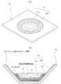
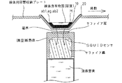
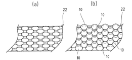
実施例に係る内面処理された検体用容器20は、図1に示す如く、一般的に用いられる形状の検体用容器(図1(a)参照)に対して、処分容易性等の高いプラスチック製の検体用容器に対して本発明に係る内面処理方法を実施することで得られ、容器底面22aと容器側面22bとからなる容器内面22の所要領域に対して、アミノ基が付加された所要粒径のコロイダルシリカ10が結合されている(図1(b)参照)。なお図1(b)においては、コロイダルシリカ10が容器内面22の全面を覆うように構成されているが、実際の検体測定に使用される部分、すなわち検体用容器20に供給される、例えば血液等の検体含有物質(溶液)の総容量に対応した部分だけ、具体的には、少なくとも容器底部22aにだけにコロイダルシリカ10を結合させるようにしてもよい。殊にSQUID測定においては、図2に示すような測定機器が使用されるが、この際、測定器と検体用容器20内に捕捉された標識化された二次抗体、すなわち標識マーカーMである磁気マーカーとの距離は、好適には2mm以下に設定される。このため測定器との距離が離れてしまう容器側面22bにおいては、磁化マーカーが捕捉されていても好適な測定が困難であるため、実際の使用においては殆ど利用されない。
The inner surface-treated
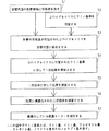
実施例に係る検体用容器20の内面処理方法は、図3に示す如く、前処理工程S1、コロイダルシリカ準備工程S2およびコロイダルシリカ結合工程S3から構成される。そしてこれら一連の処理が完了した後に、通常の免疫系測定に係る各種準備、具体的にはコロイダルシリカ10により覆われた容器内面22への一次抗体ab1の固相化を実施する一次抗体固相化(結合)工程S4、該一次抗体ab1への抗原agの捕捉をなす抗原捕捉工程S5、捕捉された抗原agへの二次抗体ab2、すなわち磁気マーカー(標識マーカー)Mで標識化された二次抗体ab2の捕捉をなす二次抗体捕捉工程S6が実施されて、図4に示すような構造体が容器内面22上に形成される。そしてこれを標識マーカーの測定が可能な装置を使用してその量を測定して、検体の量を算出する標識マーカー測定・検体算出工程S7が実施される。
As shown in FIG. 3, the inner surface treatment method for the
前処理工程S1は、後述([0018])するコロイダルシリカ準備工程S2で準備される多数のアミノ基が付加された所要粒径のコロイダルシリカ10を、容器内面22に対して結合させるための処理を実施する工程である。具体的には、コロイダルシリカ10に付加されるアミノ基と強固に結合する、例えばカルボキシル基やメタクリル基といったアミノ基の結合を許容する官能基(前処理で付与される官能基)を容器内面22に結合(吸着)するものである。この付与は公知の方法によって実施され、例えばアルカリ加水分解を実施した後に、カルボキシル基を有する物質を導入する等することでなされる。
The pretreatment step S1 is a treatment for bonding the
またこれに対して、カルボキシル基を付加したコロイダルシリカ10を容器内面22に対して結合させるためには、コロイダルシリカ10に付加されるカルボキシル基等と強固に結合する、例えばアミノ基といったカルボキシル基の結合を許容する官能基(前処理で付与される官能基)の官能基を容器内面22に結合すればよい。この付与は公知の方法によって実施され、例えばアルカリ加水分解を実施し、続いてポリアリルアミンとの縮合反応とを実施することで5〜15nmol/cm2のアミノ基を容器内面22に結合するものである。なおここでは容器内面22に対して、アミノ基またはカルボキシル基を結合する場合を説明しているが、コロイダルシリカ10に付加されたアミノ基またはカルボキシル基等のタンパク質の末端をなすアミノ基およびカルボキシル基に対して結合し得る官能基を強固に結合させる官能基であれば如何なるものでも採用可能であり、その場合には官能基導入に好適な処理方法が適宜選択されて実施される。
On the other hand, in order to bond the
この前処理によって、容器内面22に吸着(結合)される官能基は、5〜15nmol/cm2の範囲に設定される。この値が範囲外となると、容器内面22に付与された官能基が不安定となり、容器内面22に対するコロイダルシリカ10の安定的な結合が達成されなくなる。また検体用容器20が、本実施例の如く、アクリル樹脂製である場合には、20nmol/cm2を越えるとその化学的骨格構造が崩壊を始める虞が指摘される。なおこの官能基の付与数は、コロイダルシリカ10を多数結合させるために多い方が好ましいが、前述したアクリル樹脂製の検体用容器20の如く、その化学的安定性から算出される官能基の付与限界量を越えない程度とすることが最適である。ここまてでコロイダルシリカ10に対してアミノ基またはカルボキシル基を付加する場合の前処理を述べたが、前述([0013])した各官能基を付加する場合には、該官能基を結合を許容する官能基が、公知の方法によって容器内面22に結合される。
By this pretreatment, the functional group adsorbed (bonded) to the
コロイダルシリカ準備工程S2は、所要粒径のコロイダルシリカ10に対して、アミノ基を付加する工程である。ここで付加されるアミノ基は、容器内面22に付与されたカルボキシル基および一次抗体ab1に対する結合子としての役割を果たすものである。そしてコロイダルシリカ10の粒径は、50〜500nmの範囲に、好ましくは80〜200nmの範囲に設定される。この粒径が50nm未満であると、コロイダルシリカ10が凝集を起こして凝集二次粒子、すなわちその大きさがミクロン単位となってしまって溶媒(後述[0029])中で沈殿してしまい、一方500nmを越えた場合であっても同様に溶媒中で沈殿してしまい、容器内面22に対して好適な結合がなされなくなってしまう。またこのコロイダルシリカ10については、多数の粒子が揃って配列され、粒子の間に画成される隙間に入り込まない粒径、具体的にはある1つの粒子の粒子寸法を1とした場合に、0.16〜6.45の範囲にとなる粒径に設定される。好ましくは各コロイダルシリカ10の粒径が揃っている、所謂単分散状態であることが望まれ、その粒径の多分散度(粒径の標準偏差値を平均粒径で割った値)が0.1以下となるように設定される。具体的には、粒径を±1.5%程度の範囲内となるように揃えればよい。この値が大きくなり個々のコロイダルシリカ10の粒径が不揃いな状態となると、後述([0019])するように規則正しい配列を形成せず、覆うべき容器内面22の所要領域を好適に覆うことが困難になってしまう。
The colloidal silica preparation step S2 is a step of adding an amino group to the
このコロイダルシリカ10に付加されるアミノ基の大きさは、コロイダルシリカ10の大きさ比べて非常に小さなものであるため、コロイダルシリカ10には多数のアミノ基が付加されることになる。また前処理(前述[0015])によって付与され、コロイダルシリカ10に付加されたアミノ基を介して、コロイダルシリカ10を容器内面22上に保持するための官能基であるカルボキシル基についても、コロイダルシリカ10に比べて非常に小さなものであるため、アミノ基が付加されたコロイダルシリカ10を容器内面22上に供給する場合には、コロイダルシリカ10の下方にはやはり多数のカルボキシル基等が存在することになる。更に容器内面22に付与された多数のカルボキシル基等の分子長さおよびコロイダルシリカ10に付加される多数のアミノ基の分子長は、夫々略同等であるため、1つのコロイダルシリカ10が存在する位置には他のコロイダルシリカ10は高さ方向に重複的に存在できなくなる。従ってコロイダルシリカ10は、個々の粒子がその存在位置を確保し、かつ最も充填効率がよくなるように配列され、その事実が図5に示す電子顕微鏡(SEM)写真からも確認されている。
Since the size of the amino group added to the
このような配列においては、多数のコロイダルシリカ10で容器内面22における所要領域が覆われることになるため、このコロイダルシリカ10が容器内面22に対して結合された後にあっては、どのような分子でも容器内面22に結合する事態は殆どなくなる。すなわちブロッキングを実施しなくとも、非特異的な分子の容器内面22への結合は排除されることになる。
In such an arrangement, a required area on the
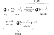
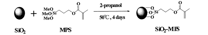
そしてコロイダルシリカ10に対してアミノ基を付加する方法としては、公知の方法が採用可能であるが、本実施例においてはその結合力が強固である3−メタクリルプロピルトリメトキシシラン(3−methacryl propyl trimetoxisilane(以下、MPSと云う))の使用がが好適である。この工程は、図6に示す如く、コロイダルシリカ10の表面に多数のMPSを結合させた(第1段階)後、アミノ基を少なくとも2つ以上備えるアミン化合物を反応させて該MPSにおいてフリーとなっている有機質物質との反応をなすメタクリル基に対して、少なくとも1個の活性なアミノ基を付加させる(第2段階)ものである。具体的には、コロイダルシリカ10およびMPSについては、プロピルアルコールの如き溶媒に所定時間保持することで分散させた状態とされて使用される。
As a method for adding an amino group to the
ここで好適に使用される少なくともアミノ基を2つ以上備えるアミン化合物としては、エチレンジアミンやヘキサメチレンジアミン等のジアミン類、ビス−2−アミノエチルエーテル等のエーテルジアミン類またはポリアリルアミン等のポリアミン類等が挙げられる。またこれらアミン化合物には、コロイダルシリカ10が結合される検体用容器20が備える耐熱性や耐薬品性等への考慮も必要であり、本実施例の如く、その材質がアクリル樹脂である検体用容器20の場合、50℃以下で水またはアルコール等の溶媒に対して溶解可能な物性を備えるアミン化合物の使用が要求される。このような物性をはずれたアミン化合物を使用すると、検体用容器20の構造が崩壊してしまう。
Examples of the amine compound preferably having at least two amino groups used herein include diamines such as ethylenediamine and hexamethylenediamine, ether diamines such as bis-2-aminoethyl ether, and polyamines such as polyallylamine. Is mentioned. These amine compounds also require consideration of heat resistance, chemical resistance, and the like provided in the
そしてこれらのアミン化合物を使用することで、コロイダルシリカ10に付加され、一次抗体ab1と結合するアミノ基の量は、少なくとも5nmol/cm2以上とされる。殊にポリアリルアミン等のアミノ基を一分子中に多数備えるポリアミン類を使用した場合には、更に多くのアミノ基を付加し得る。これは、これまでの検体用容器におけるアミノ基の量が1nmol/cm2程度(参考:[非特許文献5])であったことと比較して顕著な向上が認められる(実験2−1および実験2−2[0046]〜[0052]参照)。なお、これまでの免疫系の測定(磁気マーカー程の感度を有しない光学系の標識マーカー等を使用した測定)においては、一般にアミノ基の量が0.1nmol/cm2程度確保されれば、検体の結合には問題はないことが確認されている。
このアミノ基の量の増加は、以下のように説明される。すなわち(1)アミノ基が結合される部分(ここでは容器内面22に結合したカルボキシル基)の数と、(2)そこにアミノ基が結合できる確率と、(3)1つのアミノ基が結合される部分(カルボキシル基)に対して結合するアミノ基の数とよって決定されている。すなわち(1)、(2)または(3)の何れかが小さければ、その他の要素が大きくても結果的にアミノ基の量は増大しない。従来の検体用容器に直接的にアミノ基を付加する場合を考えると、(1)の数は前処理等によって決定され、(2)の確率は100%、すなわち1を超えることがなく、更に(3)1つのアミノ基が結合される部分には、1つのアミノ基しか結合し得ない。このため(1)と、1以下である(2)によってアミノ基の量が決定され、(1)のアミノ基が結合される部分の数以上のアミノ基が結合されることはない。
This increase in the amount of amino groups is explained as follows. That is, (1) the number of portions to which amino groups are bonded (here, carboxyl groups bonded to the
これに対して本発明に係るコロイダルシリカ10を介して検体用容器にアミノ基を付加する場合を考えると、(1)は従来と同様に前処理等によって決定されるが、図4に記載するようにコロイダルシリカ10には多数のアミノ基が付加されているため、あるアミノ基が結合できなくても同一のコロイダルシリカ10に付加されている他のアミノ基が結合する場合が想定され、(2)はほぼ100%に近い確率、すなわち1となる。(3)については、1つの該アミノ基が結合される部分に結合したコロイダルシリカ10には、前述の如く、多数のアミノ基が付加されているので、1つのアミノ基が結合される部分に多数のアミノ基が結合したのと同じ状態となる。従って(1)と、ほぼ1である(2)と、1以上である(3)との積によってアミノ基の量が決定される。
On the other hand, considering the case where an amino group is added to the specimen container via the
またアミノ基を付加するアミン化合物を、ジアミン類→ポリアミン類と変化させて、一分子のアミン化合物から得られるアミノ基の量を増大させた場合には、(3)の数がアミン化合物一分子中のアミノ基の数で掛けられたものとなり、その差はいっそう顕著となる。更に前述([0019])した如く、アミノ基が付加されるコロイダルシリカ10は、容器内面22の所要領域に隙間のない状態、すなわち最も多くのコロイダルシリカ10を存在させるよう、例えば六方最密充填の形態に従って結合されるため、理論的に最も多くのアミノ基を供給し得る(図5参照)。
In addition, when the amount of the amino group obtained from one molecule of the amine compound is increased by changing the amine compound to which the amino group is added from diamines to polyamines, the number of (3) is one molecule of the amine compound. It is multiplied by the number of amino groups in it, and the difference becomes even more pronounced. Further, as described above ([0019]), the
この他、単に面積的に考えた場合でも、図7に示す如く、球はその表面積(図7(a)参照)が平面、すなわち円(図7(b)参照)の状態に比較して大きなものとなるので、付加されるアミノ基の量は増大する。また容器内面22への結合構造を考えると、従来においてはタンパク質を疎水性相互作用および静電相互作用によって結合させているのに対して、本発明においてはアミノ基の結合を許容する官能基を化学的に結合させている。前述の静電相互作用は等電点によって変動し、この等電点は検体用容器20の材質や、結合時のpH等の外的要因によって容易に変化するものであるので、安定的に高い再現性を達成することが困難であり、この点においても本発明の結合形態が優位である。
In addition, even in the case of simply considering the area, as shown in FIG. 7, the sphere has a large surface area (see FIG. 7A) compared to a plane, that is, a circle (see FIG. 7B). As a result, the amount of added amino groups increases. Further, considering the binding structure to the
このように多数のアミノ基を付加したコロイダルシリカ10を介して、一次抗体ab1を容器内面22に固相化させることで、容器内面22への一次抗体ab1の確実な固相化(結合)を達成している。なお一般に本コロイダルシリカ準備工程S2は、前処理工程S1と同時に実施されており、内面処理された検体用容器の作製時間の短縮に貢献し得る。またここではアミノ基をコロイダルシリカ10に付加した場合を述べたが、カルボキシル基をコロイダルシリカ10に対して付加する場合も 基本的に官能基の違いに由来する差異以外はなく、前述([0018]〜[0027])した内容については全く同じであり、従来技術に比べてその付加量が著しく増大する。具体的な例を挙げると、その結合力が強固であるMPSを使用し、図8に示す如く、コロイダルシリカ10の表面に多数のMPSを結合させて、その端部に存在するメタクリル基をタンパク質結合官能基として使用する方法や、MPSで処理してアミノカルボン酸とのマイケル付加反応を利用する方法や、カルボシルキ基をもつポリマーシランカップリング剤(ポリメタクリル酸ーシランや、ポリアクリル酸−シランカップリング剤等)との反応を利用する方法が挙げられる。この他、公知の方法を用いてもよい。ここまてでコロイダルシリカ10に対してアミノ基またはカルボキシル基を付加する場合を述べたが、前述([0013])した各官能基の場合には、該官能基毎の公知の方法によってなされる。基本的にはアミノ基またはカルボキシル基と化学構造が類似して同様の官能基に対しては、同様の処理が実施される。
Thus, the primary antibody ab1 is immobilized on the
コロイダルシリカ結合工程S3は、前述の前処理工程S1で前処理された検体用容器20に対して、コロイダルシリカ準備工程S2で準備されたアミノ基が付加されたコロイダルシリカ10を供給し、容器内面22に結合されているカルボキシル基と、コロイダルシリカ10とを該アミノ基を介して結合させる工程である。具体的には、アミノ基が付加されたコロイダルシリカ10を、(1)所定の溶媒、例えばリン酸緩衝溶液に分散させ、これを容器内面22に対して滴下またはスプレー塗布する等の手段により供給し、所要時間その状態を保持して溶媒を蒸発させる方法や、あるいは(2)アミノ基が付加されたコロイダルシリカ10自体を直接的に付与等する方法によってなされる。殊に(1)の方法の場合、図9に示す如く、溶媒の蒸発によって徐々に分散状態にある個々のコロイダルシリカ10(図9(a)参照)が互いに接近することになる(図9(b)参照)ため、時間の経過によって自然に図5に示したような充填密度が高い状態に至ることになる(図9(c)参照)。
The colloidal silica bonding step S3 supplies the
ここまでの全工程S1〜S3を経ることで、容器内面22に対する内面処理は完了する。そして、この処理に引き続いて、以後に一次抗体ab1、抗原agおよび標識マーカーMで標識化された二次抗体ab2が供給され、最終的には抗原agに捕捉された標識マーカーMで標識化された二次抗体ab2の量を測定する(ここでは、標識マーカーMとして磁気マーカーが採用されているのでSQUIDを用いる)ことで、免疫系の測定が終了する。そしてコロイダルシリカ10により覆われた容器内面22への一次抗体ab1の固相化を実施する一次抗体固相化工程S4と、一次抗体ab1への抗原agの捕捉をなす抗原捕捉工程S5と、一次抗体ab1へ捕捉された抗原agへの標識化された二次抗体ab2の捕捉をなす二次抗体捕捉工程S6と、検体の量を算出する標識マーカー測定・検体算出工程S7については、従来のELISAと略同様の方法によって実施されるので、詳細な説明は省略する。なお一次抗体ab1および標識化された二次抗体ab2については、多種多様なものが市販されているが、測定すべき抗原agと最も親和性が高く、被捕捉量や捕捉量が高い物質が採用される。
Through all the steps S1 to S3 so far, the inner surface treatment for the
(変更例)
前述の実施例においては標識化された二次抗体ab2として、抗原agに対して特異性を有する二次抗体ab2と標識マーカーMとを単に結合した形態の例を挙げているが(図4参照)、本発明はこれに限定されるのではなく、必要に応じて二次抗体ab2と標識マーカーMとを、別の特異性を有する抗体同士の結合を介して結合させるようにしてもよい。例えば標識マーカーMとして使用される磁気マーカーは、現在においてはその経時変化による安定性が6ヶ月程度であり、長期保存にあまり向いていないことが確認されている。その一方で抗原agの迅速な測定を考える場合、磁気マーカーは予め二次抗体ab2と結合した状態として保存しておくことが望ましい。しかし磁気マーカーは前述の如く、その長期に亘る安定性が低いため、二次抗体ab2と結合させた状態で長期に亘って保存をすると、磁気マーカーの構造が崩壊し、これに伴って該磁気マーカーに結合された二次抗体ab2までもが崩壊、すなわち抗原に捕捉されなくなってしまう。
(Example of change)
In the above-described embodiment, as the labeled secondary antibody ab2, an example in which the secondary antibody ab2 having specificity for the antigen ag and the labeled marker M are simply bound is given (see FIG. 4). However, the present invention is not limited to this, and if necessary, the secondary antibody ab2 and the labeled marker M may be bound via binding between antibodies having different specificities. For example, a magnetic marker used as the marker marker M has a stability of about 6 months with the passage of time, and it has been confirmed that it is not suitable for long-term storage. On the other hand, when considering rapid measurement of the antigen ag, it is desirable to store the magnetic marker in a state in which it is bound to the secondary antibody ab2. However, as described above, since the magnetic marker is low in stability over a long period of time, if the magnetic marker is stored for a long period in a state where it is bound to the secondary antibody ab2, the structure of the magnetic marker is destroyed, and the magnetic Even the secondary antibody ab2 bound to the marker collapses, that is, is not captured by the antigen.
一方、二次抗体ab2は、特定の抗原(タンパク質)に対して特異的に結合する特殊なタンパク質であるため、一般的にそのコストが高いことは周知の事実である。従って、長期保存に際してその構造が崩壊する磁気マーカーとの結合については、コスト的な問題を内在している。そこでこのような問題を解決するために、予め二次抗体ab2とビオチンとを結合させてビオチン化抗体とすると共に、磁気マーカーMについてはビオチンと特異的に結合するアビジンと結合させ、これを測定直前に結合させて標識化させる方法が好適に採用される。このような手法を用いた場合、図10に示すような構造体が容器内面22上に形成されることになる。この場合、ビオチンとアビジンとの結合は特異的かつ迅速であるため、前述の問題は全て解決される。なお、抗原や二次抗体ab2の種類によっては、ビオチンおよびアビジンの配置位置を入れ替えるようにしてもよい。
On the other hand, since the secondary antibody ab2 is a special protein that specifically binds to a specific antigen (protein), it is a well-known fact that its cost is generally high. Therefore, there is an inherent cost problem regarding the binding to a magnetic marker whose structure collapses during long-term storage. Therefore, in order to solve such problems, the secondary antibody ab2 and biotin are bound in advance to obtain a biotinylated antibody, and the magnetic marker M is bound to avidin that specifically binds to biotin and measured. A method of labeling by binding immediately before is preferably employed. When such a method is used, a structure as shown in FIG. 10 is formed on the container
また本発明において、一次抗体ab1の容器内面22上への直接的な固相化は、容器内面22上にコロイダルシリカ10を結合させることで発現される親水性によっても阻害される。これは前述([0027])した如く、一次抗体ab1の容器内面22への固相化の主な駆動力となっている疎水性相互作用を阻害するためである。従って、例えば容器内面22にコロイダルシリカ10を結合させた後に、物理的な要因でコロイダルシリカ10の一部か剥離しても、容器内面22への一次抗体ab1の固相化は最小限に抑制される。またコロイダルシリカ10を採用することで、前述([0012])した如く、プラスチック製の検体容器をガラスコーティングした状態となり親水性を発現するため、血液等の水系溶液に存在する検体の測定実施が好適になされることは云うまでもない。
In the present invention, the direct immobilization of the primary antibody ab1 on the
(実験例)
以下に本発明に係る検体用容器の処理方法の一例と、該処理方法によって得られる検体用容器の一次抗体の捕捉量(度合い)、すなわち標識化された二次抗体の捕捉量についての実験例を示す。なお、本発明に係る内面処理された検体用容器およびその処理方法は、この実験例に限定されるものではない。
(Experimental example)
Hereinafter, an example of a sample container processing method according to the present invention, and an experimental example of the amount of captured primary antibody (degree) of the sample container obtained by the method, that is, the amount of labeled secondary antibody captured Indicates. Note that the inner surface-treated sample container and its processing method according to the present invention are not limited to this experimental example.
(実験1)本発明に係る検体用容器の処理方法の一例について
前処理工程S1:図1に示した形状のアクリル樹脂製の検体用容器を、メタノールで充分に脱脂した後、温度50℃、時間15分間の条件で1規定NaOH水溶液に浸漬し表面処理を行ない、その後にカルボキシル基を結合させた。このとき結合されたカルボキシル基を定量したところ、8〜12nmol/cm2(50〜70個/nm2)であった。なお測定方法は以下の通りである。
(Experiment 1) About an example of the processing method of the sample container according to the present invention Pre-processing step S1: A sample container made of an acrylic resin having the shape shown in FIG. Surface treatment was performed by dipping in a 1N NaOH aqueous solution for 15 minutes, and then carboxyl groups were bonded. The amount of carboxyl groups bonded at this time was determined to be 8 to 12 nmol / cm 2 (50 to 70 / nm 2 ). The measuring method is as follows.
(カルボキシル基の定量方法)
検体用容器をN,N'−ジシクロヘキシルカルボジイミド(DCC)の1%シクロヘキサン溶液中で2時間撹拌し、その後、P−ニトロフェノールを加えて8時間撹拌し、更に4%アンモニア水を加えて8時間撹拌した後、上澄み中の反応したP−ニトロフェノールの400nmにおける吸光度を測定する。なお測定値は、予め作成した検量線によって算出した。
(Quantitative determination method of carboxyl group)
The specimen container was stirred in a 1% cyclohexane solution of N, N′-dicyclohexylcarbodiimide (DCC) for 2 hours, then P-nitrophenol was added and stirred for 8 hours, and further 4% ammonia water was added for 8 hours. After stirring, the absorbance at 400 nm of the reacted P-nitrophenol in the supernatant is measured. The measured value was calculated using a calibration curve prepared in advance.
コロイダルシリカ準備工程S2:例えば下記の[非特許文献6]に記載される如く、粒径120nmのコロイダルシリカ4重量部と、MPS1重量部とを2−プロピルアルコール100重量部を溶媒として使用することで分散させ、温度50℃、時間96時間の条件で撹拌し、その後、遠心洗浄しMPSが付加されたコロイダルシリカ(以下、SiO2−MPS粒子と云う)とした。そしてこのSiO2−MPS粒子1重量部を、分散用の溶媒であるエタノール30重量部に分散させ、更にアミノ基を付加させるアミン化合物としてのエチレンジアミン0.1重量部を加え、室温、時間24時間の条件で撹拌し、更に遠心洗浄後に減圧乾燥させることでアミノ基が付加されたコロイダルシリカを作製した。
コロイダルシリカ結合工程S3:次に図11に示す如く、容器内面1cm2当たりに5mgになるように1−エチル−3−(3−ジメチルアミノプロピル)カルボジイミド塩酸塩(WSC)をpH7.0のリン酸緩衝溶液(以下、PSBと云う)に溶解させた溶液を容器内に供給し、室温、時間1時間の条件で放置した後に該溶液を取除き乾燥する予備処理を実施した後、容器内にPBSを溶媒として5wt%になるように調整したアミノ基が付加されたコロイダルシリカを滴下して、前処理工程S1で容器内面に結合されたカルボキシル基に対して、アミノ基が付加されたコロイダルシリカを結合させた。そしてその後、温温、時間2時間の条件で放置し、緩衝溶液と純水とを個別に使用して洗浄ほ実施した後に室温で乾燥させた。なお滴下は、アミノ基が付加されたコロイダルシリカ/緩衝溶液が、容器底面に万遍なく広がるように行なったが、表面張力の関係で容器側面にも広がることが確認された。このとき結合されたアミノ基を定量したところ、5〜15nmoll/cm2(30〜90個/nm2)であった。なお測定方法は以下の通りである。 Colloidal silica bonding step S3: Next, as shown in FIG. 11, 1-ethyl-3- (3-dimethylaminopropyl) carbodiimide hydrochloride (WSC) was added to phosphorous having a pH of 7.0 so that the amount was 5 mg per 1 cm 2 of the container inner surface. A solution dissolved in an acid buffer solution (hereinafter referred to as PSB) is supplied into the container, and after standing for 1 hour at room temperature, the solution is removed and dried, followed by a preliminary treatment. Colloidal silica added with amino groups adjusted to 5 wt% using PBS as a solvent, and colloidal silica added with amino groups to the carboxyl groups bound to the inner surface of the container in the pretreatment step S1 Were combined. And after that, it was left to stand under the condition of warm temperature and time for 2 hours, washed with buffer solution and pure water separately, and dried at room temperature. The dropping was performed so that the colloidal silica / buffer solution to which the amino group was added spread evenly on the bottom of the container, but it was confirmed that the colloidal silica / buffer solution spread on the side of the container due to the surface tension. The amount of the amino group bonded at this time was determined to be 5 to 15 nmol / cm 2 (30 to 90 / nm 2 ). The measuring method is as follows.
(アミノ基の定量方法)
検体用容器を0.1molのピクリン酸エタノール溶液に入れて測定すべきアミノ基を反応させ、超音波照射後、検体用容器を乾燥させてジイソプロピルエチルアミン(DIEA)の5%ターシャルブタノール液を加えて遠心分離を行ない、上澄み液の吸光度を測定する、所謂Gizin法によって定量した。
(Amino group quantification method)
Place the sample container in a 0.1 mol picric acid ethanol solution to react the amino group to be measured, and after irradiating with ultrasonic waves, dry the sample container and add 5% tert-butanol solution of diisopropylethylamine (DIEA). Then, the mixture was centrifuged, and the absorbance of the supernatant was measured, so that it was quantified by the so-called Gizin method.
一次抗体固相化工程S4:PBS1000μl当たりIgEモノクロナール抗体200μg混合した希釈溶液を、内面処理された検体用容器に200μl供給し、温度37℃、時間3時間の条件で放置し、容器内面にIgEモノクロナール抗体を固相化した。 Primary antibody immobilization step S4: 200 μl of a diluted solution obtained by mixing 200 μg of IgE monoclonal antibody per 1000 μl of PBS is supplied to the inner surface-treated specimen container, left at a temperature of 37 ° C. for 3 hours, and IgE is left on the inner surface of the container. Monoclonal antibody was immobilized.
抗原捕捉工程S5:容器内面をpH7.4のPBSで3回洗浄し、pH7.4のPBS1000μlに所定量のヒトIgE(抗原)を混合した抗原溶液を加え、室温、時間1時間の条件で放置し、抗体抗原反応を起こさせ、一次抗体であるIgEモノクロナール抗体に対してヒトIgEを捕捉させた。反応終了後には0.1%の界面活性剤入りPBSで未反応の抗原を洗い落とす、所謂「洗い」を実施した。 Antigen capture step S5: The inner surface of the container was washed 3 times with PBS of pH 7.4, an antigen solution in which a predetermined amount of human IgE (antigen) was mixed with 1000 μl of PBS of pH 7.4 was added, and the mixture was allowed to stand at room temperature for 1 hour. Then, an antibody-antigen reaction was caused to capture human IgE against the primary antibody IgE monoclonal antibody. After completion of the reaction, so-called “washing” was performed in which unreacted antigen was washed away with PBS containing 0.1% of a surfactant.
二次抗体捕捉工程S6:二次抗体として用いるIgEポリクロナール抗体(以下、単に二次抗体と云う)にビオチンを結合させてビオチン化検出抗体とし、これをを検体用容器に供給して、温度37℃、時間1時間の条件で放置し、抗原に捕捉させた。次に未反応のビオチン化検出抗体をpH7.4のPBSで3回洗浄し、下記の方法により調整したアビジンが結合された磁気マーカー(1gの磁気マーカーにアビジン10mgが結合)を分散させたpH7.4のPBS200μllを検体用容器に供給し、アビジンとビオチンとを結合させて二次抗体を磁気マーカーで標識化させた。なお磁気マーカーの作製方法についても下に記す。
Secondary antibody capture step S6: Biotin is bound to an IgE polyclonal antibody (hereinafter simply referred to as a secondary antibody) used as a secondary antibody to form a biotinylated detection antibody, which is supplied to a sample container, and temperature 37 The mixture was allowed to stand at 1 ° C. for 1 hour to be captured by the antigen. Next, the unreacted biotinylated detection antibody was washed 3 times with PBS of pH 7.4, and a magnetic marker (10 g of avidin bound to 1 g of magnetic marker) bound with avidin prepared by the following method was dispersed. 200 μl of
(アビジンが結合された磁気マーカーの作製方法)
pH7.0のリン酸緩衝溶液5mlに対して、WSC10mgと磁気マーカー10mgとを加え、超音波照射後、アビジン15mggを添加し、氷冷下で1時間撹拌し、更に室温下で6時間撹拌し、その後リン酸緩衝溶液を使用した超音波洗浄・遠心洗浄を3回繰り返して磁気マーカーのカルボキシル基と、アビジンのアミノ基とを反応させた該磁気マーカーへのアビジンの固定化を行なった。なお遠心分離後の上澄み液中の未反応物質のUV測定により、磁気マーカーへのアビジンの固定化量を測定したところ、10mg/gであった。
(Method for producing magnetic marker bound with avidin)
Add 10 mg of WSC and 10 mg of magnetic marker to 5 ml of phosphate buffer solution at pH 7.0, add 15 mgg of avidin after ultrasonic irradiation, stir for 1 hour under ice-cooling, and further stir for 6 hours at room temperature. Thereafter, ultrasonic washing and centrifugal washing using a phosphate buffer solution were repeated three times to immobilize avidin on the magnetic marker in which the carboxyl group of the magnetic marker was reacted with the amino group of avidin. The amount of avidin immobilized on the magnetic marker was measured by UV measurement of unreacted substances in the supernatant after centrifugation, and found to be 10 mg / g.
(磁気マーカーの作製方法)
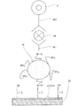
図12に示す如く、メタノール10mlに、マイクロモノマーとしてのポリビニルピロリドン(0.004〜0.04mg範囲内の一定量)にフェライト(Fe2O3)微粒子0.05gを加え、超音波照射し、更に攪拌ほ実施した後に遠心分離を行なうことによってポリビニルピロリドンを吸着したフェライト微粒子を作製し、次いでテトラヒドロフラン(THF)5mlに、N−アクロイル−L−グルタミン酸0.20g(フェライト微粒子に吸着したポリビニルピロリドンのビニル基量の100倍量)と、0〜100倍量の架橋剤トリ(アクロイルオキシ)アミン塩酸塩とを溶解させて準備溶液とし、これに粒子1g当たりに0.2gのポリビニルピロリドンを吸着したフェライト微粒子0.018gと、2,2'−アゾビス(イソブチロニトリル)(AIBN)0.01gとを加え、温度35℃、時間10時間の条件で攪拌し、更に遠心分離(5回繰り返し)を実施して磁気マーカーを得る。なおこの方法で得られたフェライト微粒子のポリマー量は、架橋剤の増加に伴って増大し、最大値はフェライト微粒子1g当たり947mgであった。また磁気マーカーの粒径は、29〜30nmであることが分散溶液の動的光散乱法から測定された。粒子間凝集は起こっておらず、水溶液中で4週間以上分散状態の保持が確認された。更に磁気マーカーの表面に結合したカルボキシル基は、最大97μmol/gであった。
(Method for producing magnetic marker)
As shown in FIG. 12, 0.05 g of ferrite (Fe 2 O 3 ) fine particles were added to 10 ml of methanol and polyvinylpyrrolidone (a fixed amount within a range of 0.004 to 0.04 mg) as a micromonomer, and subjected to ultrasonic irradiation. Further, after stirring, the ferrite fine particles adsorbing polyvinylpyrrolidone were prepared by centrifuging, and then 0.20 g of N-acryloyl-L-glutamic acid (the polyvinylpyrrolidone adsorbed on the ferrite fine particles was added to 5 ml of tetrahydrofuran (THF). (100 times the amount of vinyl groups) and 0 to 100 times the amount of cross-linking agent tri (acryloyloxy) amine hydrochloride are dissolved to prepare a preparation solution, and 0.2 g of polyvinylpyrrolidone is adsorbed per 1 g of the particles. Ferrite fine particles 0.018 g and 2,2′-azobis (isobutyronitrile) (AIBN) Plus the .01G, temperature 35 ° C., and stirred under the conditions of 10 hours, to obtain a magnetic marker further performed centrifuged (repeated 5 times). The polymer amount of the ferrite fine particles obtained by this method increased with an increase in the crosslinking agent, and the maximum value was 947 mg per 1 g of ferrite fine particles. Further, the particle size of the magnetic marker was measured to be 29 to 30 nm from the dynamic light scattering method of the dispersion solution. Aggregation between particles did not occur, and it was confirmed that the dispersion state was maintained for 4 weeks or more in an aqueous solution. Furthermore, the maximum number of carboxyl groups bound to the surface of the magnetic marker was 97 μmol / g.
標識マーカー測定・検体算出工程S7:ここまでの各工程を経た検体用容器を、室温、時間20分の条件で放置した後、SQUID測定を行なった。その結果を参考として、図13に示す。この結果から、IgE抗体重量10pgに対して280pTの残留磁気量が検出されたことが確認された(図13(a)参照)。更にIgE抗体重量1pgの場合には27pT(図13(b)参照)、0.3pgの場合には15pT(図13(c)参照)の残留磁気が検出され、0.3pgといった極微量の抗原の測定が可能であることが確認された。 Labeled marker measurement / sample calculation step S7: The sample container that has undergone each of the above steps is left at room temperature for 20 minutes, and then SQUID measurement is performed. The results are shown in FIG. 13 for reference. From this result, it was confirmed that a residual magnetic quantity of 280 pT was detected for an IgE antibody weight of 10 pg (see FIG. 13A). Furthermore, when the IgE antibody weight is 1 pg, a residual magnetism of 27 pT (see FIG. 13B) is detected, and in the case of 0.3 pg, 15 pT (see FIG. 13C) is detected, and a very small amount of antigen such as 0.3 pg is detected. It was confirmed that measurement of
(実験2−1)内面処理の有無について
本発明に係る内面処理(実験1における[0035]、[0037]および[0038])を実施した検体用容器(実施例1)と、全く何の処理も実施しない検体用容器(比較例1)とにおける一次抗体(抗原)の捕捉量(度合い)、すなわちアミノ基の付加量についての実験を行なった。なお実験方法については、免疫反応の簡便な評価法として用いられるサンドイッチELISA法を採用した。
(Experiment 2-1) Presence / absence of inner surface treatment Sample container (Example 1) in which inner surface treatment according to the present invention ([0035], [0037] and [0038] in Experiment 1) was performed, and no treatment at all Experiments were also conducted on the amount (degree) of primary antibody (antigen) trapped in the sample container (Comparative Example 1), that is, the amount of amino group added. As an experimental method, a sandwich ELISA method, which is used as a simple evaluation method of immune reaction, was adopted.
実施例1および比較例1に係る検体用容器に対して、温度37℃、時間2時間、pH9.5の条件で2.5%グルタルアルデヒドによる処理を行ない、水洗した後、温度37℃、時間2時間、pH7.5の条件でビオチンヒドラジド(10μg/100μl)による処理を行ない、更に表面にブロッキング剤(商品名 ブロックエース;雪印製)を塗布して2時間放置してPBSで洗浄する。次いで前述の処理を行なった各検体用容器を、西洋わさびペルオキシナーゼ(HRP)結合ストレプトアビジンを分散させたブロックエースおよびPBSからなる溶媒中で1時間放置して、PSBで洗浄した後、ペルオキシナーゼの基質(以下、ABTSと云う)溶液100μl供給し、室温、時間20分の条件で放置する作業を実施した。そしてこの作業を各検体用容器毎に4回行なった。そして全作業終了後に各検体用容器の上澄み液について、分光光度計(商品名 UV2000;日本分光製)を用いてABTS溶液を特定する650nmの吸光度を測定した。そして実施例1または比較例1について4回分の吸光度の平均値を算出した。 The sample container according to Example 1 and Comparative Example 1 was treated with 2.5% glutaraldehyde under the conditions of a temperature of 37 ° C., a time of 2 hours, and a pH of 9.5, washed with water, and then a temperature of 37 ° C. for a time. Treat with biotin hydrazide (10 μg / 100 μl) at pH 7.5 for 2 hours, apply a blocking agent (trade name: Block Ace; manufactured by Snow Brand) on the surface, leave it for 2 hours and wash with PBS. Next, each specimen container subjected to the above-mentioned treatment was left in a solvent consisting of block ace in which horseradish peroxynase (HRP) -conjugated streptavidin was dispersed and PBS for 1 hour, washed with PSB, and then peroxynase. The substrate (hereinafter referred to as ABTS) solution (100 μl) was supplied and allowed to stand at room temperature for 20 minutes. This operation was performed four times for each specimen container. And after completion | finish of all operation | movement, the light absorbency of 650 nm which specifies an ABTS solution was measured about the supernatant liquid of each specimen container using the spectrophotometer (brand name UV2000; made by JASCO). And the average value of the light absorbency for 4 times about Example 1 or Comparative Example 1 was calculated.
(実験2−1の結果)
実験2−1から得られる結果を下記の表1に記する。この表1に記載の結果から、本発明に係る内面処理を施した検体用容器は、なにも処理を施さない検体用容器と比較して平均で8倍程度の高い抗原捕捉量を発現することが確認された。

The results obtained from Experiment 2-1 are shown in Table 1 below. From the results shown in Table 1, the sample container subjected to the inner surface treatment according to the present invention expresses an antigen capture amount that is about 8 times higher on average than the sample container not subjected to any treatment. It was confirmed.
(実験2−2)コロイダルシリカの有無について
実験2−1で作製した検体用容器(実施例1)と、コロイダルシリカに対してアミノ基を付加させ、このコロイダルシリカを容器内面に結合させる代わりに、アミノ基を直接的に容器内面に結合させた検体用容器(比較例2)とにおける抗原の捕捉量(度合い)、すなわちアミノ基の付加量についての実験を行なった。
(Experiment 2-2) Presence or absence of colloidal silica Instead of attaching an amino group to the specimen container (Example 1) prepared in Experiment 2-1 and colloidal silica, and bonding this colloidal silica to the inner surface of the container Experiments were conducted on the amount (degree) of antigen capture, that is, the amount of amino groups added, in a specimen container (Comparative Example 2) in which an amino group was directly bonded to the inner surface of the container.
比較例2については、前処理工程S1だけを実施し、コロイダルシリカ準備工程S2およびコロイダルシリカ結合工程S3の代わりに、500mgのWSCを溶解させたPH7.0PBS250mlに浸漬し、室温、時間1時間の条件で撹拌した後、エチレンジアミン2mg(17μmol)を添加して室温、時間12時間の条件で撹拌した。そしてpH7.0のPBSで洗浄・乾燥を実施した。そして前述([0036]および[0039])の方法で、実施例1および比較例2に結合したカルボキシル基およびアミノ基の定量を行なった。
(実験2−2の結果)
実験2−2から得られる結果を下記の表2に記する。この表2に記載の結果から、カルボキシル基の結合量が略同一であっても、コロイダルシリカを用いる場合(実施例1)と、そうでない場合(比較例2)との間では10倍以上の差がついていた。すなわちコロイダルシリカを用いる本発明に係る内面処理方法は、一次抗体を捕捉するためのアミノ基の結合方法として優れていることが確認された。
(Result of Experiment 2-2)
The results obtained from Experiment 2-2 are shown in Table 2 below. From the results shown in Table 2, even when the amount of carboxyl group binding is substantially the same, the colloidal silica is used 10 times or more between the case where colloidal silica is used (Example 1) and the case where it is not (Comparative Example 2). There was a difference. That is, it was confirmed that the inner surface treatment method according to the present invention using colloidal silica is excellent as an amino group binding method for capturing a primary antibody.
(実験2−1および実験2−2の結果のまとめ)
前述の実験2−1および実験2−2(実施例1、比較例1および比較例2)の結果を、一次抗体(抗原)の捕捉量(度合い)を指標とすると共に、比較例1を1に設定して比較すると、下記の表3のように纏めることができた。すなわち本発明の内面処理方法によって処理された検体用容器の検体の捕捉量、言い換えれば一次抗体の固相化量は、従来の検体捕捉に使用される各種処理方法に比較して大きく増大(比較例1に較べて約8倍、比較例2に較べて10〜15倍)していると確認された。これは検体量をこれまでの1/8程度しか含有しない被測定溶液についても、これまでと同様に測定し得ることを意味する。
The results of the above-described Experiment 2-1 and Experiment 2-2 (Example 1, Comparative Example 1 and Comparative Example 2) were used with the capture amount (degree) of the primary antibody (antigen) as an index, and Comparative Example 1 as 1 As a result, the results were summarized as shown in Table 3 below. That is, the amount of sample captured in the sample container treated by the inner surface treatment method of the present invention, in other words, the amount of immobilized primary antibody, is greatly increased compared to various treatment methods used for conventional sample capture (comparison). It was confirmed that the ratio was about 8 times that of Example 1 and 10 to 15 times that of Comparative Example 2. This means that a solution to be measured containing only about 1/8 of the amount of specimen can be measured in the same manner as before.
10 コロイダルシリカ
22 容器内面
ab1 一次抗体
ab2 二次抗体
ag 抗原
M 標識マーカー
10
Claims (18)
多数のアミノ基、イミノ基、カルボキシル基、カルボニル基、メタクリル基またはメルカプト基が付加されたコロイダルシリカ(10)が、容器内面(22)の所要領域を覆うように結合されており、
前記標識マーカー(M)によって標識化された二次抗体(ab2)は、一次抗体(ab1)および抗原(ag)を介して、前記コロイダルシリカ(10)のアミノ基、イミノ基、カルボキシル基、カルボニル基、メタクリル基またはメルカプト基だけに結合されるよう構成した
ことを特徴とする内面処理された検体用容器。 The antigen (ag) is captured by the solid-phased primary antibody (ab1), and the secondary antibody (ab2) labeled with the labeling marker (M) is captured by the antigen (ag). In the specimen container used in various immunoassays for quantifying ag),
Colloidal silica (10) to which a number of amino groups, imino groups, carboxyl groups, carbonyl groups, methacryl groups or mercapto groups have been added is bonded so as to cover the required area of the container inner surface (22),
The secondary antibody (ab2) labeled with the labeling marker (M), via the primary antibody (ab1) and the antigen (ag), the amino group, imino group, carboxyl group, carbonyl of the colloidal silica (10) An inner surface-treated sample container characterized by being configured to be bonded only to a group, a methacryl group or a mercapto group.
検体用容器(20)の容器内面(22)に前処理を実施して所要の官能基を付与し、
別工程でアミノ基、イミノ基、カルボキシル基、カルボニル基、メタクリル基またはメルカプト基を付加した所要粒径のコロイダルシリカ(10)を、前記前処理で付与させた官能基に結合させて、前記容器内面(22)の所要領域を被覆させ、
前記標識マーカー(M)によって標識化された二次抗体(ab2)を、一次抗体(ab1)および抗原(ag)を介して、前記コロイダルシリカ(10)のアミノ基、イミノ基、カルボキシル基、カルボニル基、メタクリル基またはメルカプト基だけに結合させるようにした
ことを特徴とする検体用容器の内面処理方法。 The antigen (ag) is captured by the solid-phased primary antibody (ab1), and the secondary antibody (ab2) labeled with the labeling marker (M) is captured by the antigen (ag). ag) in the inner surface treatment method of the sample container used in various immunoassays for quantifying,
Pretreatment is performed on the container inner surface (22) of the specimen container (20) to give a required functional group,
The colloidal silica (10) having a required particle size to which an amino group, an imino group, a carboxyl group, a carbonyl group, a methacryl group or a mercapto group is added in a separate step is bonded to the functional group provided in the pretreatment, and the container Cover the required area of the inner surface (22),
The secondary antibody (ab2) labeled with the labeled marker (M) is converted into an amino group, imino group, carboxyl group, carbonyl of the colloidal silica (10) via the primary antibody (ab1) and the antigen (ag). A method for treating the inner surface of a specimen container, wherein the group is bonded only to a group, a methacryl group or a mercapto group.
18. The specimen according to claim 17, wherein as the pretreatment, alkali hydrolysis and subsequent condensation reaction with polyallylamine are carried out to give 5-15 nmol / cm < 2 > of amino groups to the inner surface (22) of the container. For treating the inner surface of a container.
Priority Applications (1)
| Application Number | Priority Date | Filing Date | Title |
|---|---|---|---|
| JP2004106929A JP2005291909A (en) | 2004-03-31 | 2004-03-31 | Container for specimen having treated inner surface and its treating method |
Applications Claiming Priority (1)
| Application Number | Priority Date | Filing Date | Title |
|---|---|---|---|
| JP2004106929A JP2005291909A (en) | 2004-03-31 | 2004-03-31 | Container for specimen having treated inner surface and its treating method |
Publications (1)
| Publication Number | Publication Date |
|---|---|
| JP2005291909A true JP2005291909A (en) | 2005-10-20 |
Family
ID=35324997
Family Applications (1)
| Application Number | Title | Priority Date | Filing Date |
|---|---|---|---|
| JP2004106929A Pending JP2005291909A (en) | 2004-03-31 | 2004-03-31 | Container for specimen having treated inner surface and its treating method |
Country Status (1)
| Country | Link |
|---|---|
| JP (1) | JP2005291909A (en) |
Cited By (3)
| Publication number | Priority date | Publication date | Assignee | Title |
|---|---|---|---|---|
| JP2008190946A (en) * | 2007-02-02 | 2008-08-21 | Kyushu Institute Of Technology | Immunoassay using protein adsorption inhibitor and substrate and kit for immunoassay |
| JP2011169886A (en) * | 2010-01-21 | 2011-09-01 | Sysmex Corp | Sample preparation apparatus |
| JPWO2011158759A1 (en) * | 2010-06-16 | 2013-08-19 | ウシオ電機株式会社 | Production apparatus, production method, antibody chip, program and recording medium |
-
2004
- 2004-03-31 JP JP2004106929A patent/JP2005291909A/en active Pending
Cited By (3)
| Publication number | Priority date | Publication date | Assignee | Title |
|---|---|---|---|---|
| JP2008190946A (en) * | 2007-02-02 | 2008-08-21 | Kyushu Institute Of Technology | Immunoassay using protein adsorption inhibitor and substrate and kit for immunoassay |
| JP2011169886A (en) * | 2010-01-21 | 2011-09-01 | Sysmex Corp | Sample preparation apparatus |
| JPWO2011158759A1 (en) * | 2010-06-16 | 2013-08-19 | ウシオ電機株式会社 | Production apparatus, production method, antibody chip, program and recording medium |
Similar Documents
| Publication | Publication Date | Title |
|---|---|---|
| CA1336393C (en) | Test method and reagent kit therefor | |
| CN104316697A (en) | Preparation method of antigen-immobilized immuno-fluorescence slide and immuno-fluoroscence slide prepared thereby | |
| JPS6315551B2 (en) | ||
| AU648625B2 (en) | Test method and reagent kit therefor | |
| JP6118761B2 (en) | Test substance measuring kit and test substance measuring method | |
| CN1258091C (en) | Immunoassay | |
| JP2015072249A (en) | Test substance assay method, test substance assay kit, and test substance assay reagent | |
| JP2005291909A (en) | Container for specimen having treated inner surface and its treating method | |
| EP0682255A1 (en) | Immunoassays using a carbon sol label | |
| JP2010181155A (en) | Method of manufacturing substance fixing substrate | |
| CN114252613B (en) | Composition for detecting HBsAg antigen protein and application method thereof | |
| Guo et al. | Development of a low density colorimetric protein array for cardiac troponin I detection | |
| US20080248591A1 (en) | Gas bubble biosensor | |
| CN1114423A (en) | Immunoassay using carbon sol labels | |
| JP2010164513A (en) | Antibody fixing substrate, manufacturing method and utilization of antibody fixing substrate | |
| CA2414704A1 (en) | Tertiary amine compounds for use in immunoassays | |
| JP2000304749A (en) | Specific binding immunoanalytical container | |
| EP0525178B1 (en) | Elements and methods employing polymeric surface amplification agents | |
| CN102834713A (en) | Method for sensing a chemical | |
| Hahn et al. | Current advances in antibody immobilization on different surfaces and beads | |
| JPH11174057A (en) | Immunity measuring base material | |
| HK1001235B (en) | Test method and reagent kit therefor | |
| HK1001235A (en) | Test method and reagent kit therefor | |
| HK1001234B (en) | Test method and reagent kit therefor | |
| HK1001234A (en) | Test method and reagent kit therefor |
Legal Events
| Date | Code | Title | Description |
|---|---|---|---|
| A625 | Written request for application examination (by other person) |
Free format text: JAPANESE INTERMEDIATE CODE: A625 Effective date: 20061221 |
|
| A711 | Notification of change in applicant |
Free format text: JAPANESE INTERMEDIATE CODE: A711 Effective date: 20080214 |
|
| A521 | Written amendment |
Free format text: JAPANESE INTERMEDIATE CODE: A821 Effective date: 20080305 |
|
| RD02 | Notification of acceptance of power of attorney |
Free format text: JAPANESE INTERMEDIATE CODE: A7422 Effective date: 20080305 |
|
| A521 | Written amendment |
Free format text: JAPANESE INTERMEDIATE CODE: A821 Effective date: 20080214 |
|
| A977 | Report on retrieval |
Effective date: 20081009 Free format text: JAPANESE INTERMEDIATE CODE: A971007 |
|
| A131 | Notification of reasons for refusal |
Effective date: 20081021 Free format text: JAPANESE INTERMEDIATE CODE: A131 |
|
| A02 | Decision of refusal |
Effective date: 20090303 Free format text: JAPANESE INTERMEDIATE CODE: A02 |