JP2005257652A - Detecting apparatus and analyzing method for biological sample - Google Patents
Detecting apparatus and analyzing method for biological sample Download PDFInfo
- Publication number
- JP2005257652A JP2005257652A JP2004073549A JP2004073549A JP2005257652A JP 2005257652 A JP2005257652 A JP 2005257652A JP 2004073549 A JP2004073549 A JP 2004073549A JP 2004073549 A JP2004073549 A JP 2004073549A JP 2005257652 A JP2005257652 A JP 2005257652A
- Authority
- JP
- Japan
- Prior art keywords
- detection
- light
- biological sample
- light source
- image
- Prior art date
- Legal status (The legal status is an assumption and is not a legal conclusion. Google has not performed a legal analysis and makes no representation as to the accuracy of the status listed.)
- Pending
Links
Images
Classifications
-
- G—PHYSICS
- G01—MEASURING; TESTING
- G01N—INVESTIGATING OR ANALYSING MATERIALS BY DETERMINING THEIR CHEMICAL OR PHYSICAL PROPERTIES
- G01N21/00—Investigating or analysing materials by the use of optical means, i.e. using sub-millimetre waves, infrared, visible or ultraviolet light
- G01N21/62—Systems in which the material investigated is excited whereby it emits light or causes a change in wavelength of the incident light
- G01N21/63—Systems in which the material investigated is excited whereby it emits light or causes a change in wavelength of the incident light optically excited
- G01N21/64—Fluorescence; Phosphorescence
- G01N21/645—Specially adapted constructive features of fluorimeters
- G01N21/6456—Spatial resolved fluorescence measurements; Imaging
-
- G—PHYSICS
- G01—MEASURING; TESTING
- G01N—INVESTIGATING OR ANALYSING MATERIALS BY DETERMINING THEIR CHEMICAL OR PHYSICAL PROPERTIES
- G01N21/00—Investigating or analysing materials by the use of optical means, i.e. using sub-millimetre waves, infrared, visible or ultraviolet light
- G01N21/01—Arrangements or apparatus for facilitating the optical investigation
-
- G—PHYSICS
- G01—MEASURING; TESTING
- G01N—INVESTIGATING OR ANALYSING MATERIALS BY DETERMINING THEIR CHEMICAL OR PHYSICAL PROPERTIES
- G01N2201/00—Features of devices classified in G01N21/00
- G01N2201/06—Illumination; Optics
- G01N2201/064—Stray light conditioning
- G01N2201/0646—Light seals
Landscapes
- Health & Medical Sciences (AREA)
- Biochemistry (AREA)
- Physics & Mathematics (AREA)
- Life Sciences & Earth Sciences (AREA)
- Chemical & Material Sciences (AREA)
- Analytical Chemistry (AREA)
- General Health & Medical Sciences (AREA)
- General Physics & Mathematics (AREA)
- Immunology (AREA)
- Pathology (AREA)
- Nuclear Medicine, Radiotherapy & Molecular Imaging (AREA)
- Investigating Or Analysing Materials By The Use Of Chemical Reactions (AREA)
- Investigating Or Analysing Materials By Optical Means (AREA)
Abstract
Description
本発明は、主に生命科学研究および検査を目的とした核酸、タンパク質、脂質、糖類、ビタミン、補酵素等を含む生体試料、あるいは、これに含まれる薬剤などの非生体成分の検出装置および解析方法に関する。 The present invention mainly relates to a biological sample containing nucleic acids, proteins, lipids, saccharides, vitamins, coenzymes, etc. for life science research and testing, or a non-biological component detection device and analysis such as drugs contained therein. Regarding the method.
従来、生命現象の解明のために、生体分子を光学的に標識し、微量な光学的シグナルを暗室内でフィルム等の媒体を用いて検出する方法が一般的に利用されている。最近では、デジタルカメラの高精度化に伴い、フィルムを用いず、デジタル画像としてデータを記録する方法も普及している。しかしながら、光学的シグナルが微弱であることが多いため、、外光を遮断できる暗室を利用することに変わりはない。
なお、暗室設備が利用できない研究機関や教育機関等でも上記の実験ができるように、暗室効果をもつ小型の暗箱も実用化されている。最も簡単な構成は、生体試料等の検出対象に金属または樹脂製のフードをかぶせるものであり、一般的には上部に観察および撮影用の穴が開いているものが多い。さらに高度なものになると、レンズやカメラが取り付けられたもの、内部で操作ができるように手を入れる口が用意されているもの、内部光源が設置されているものなどが、実用化されている。
Conventionally, in order to elucidate life phenomena, a method of optically labeling biomolecules and detecting a small amount of optical signals using a medium such as a film in a dark room is generally used. Recently, as digital cameras have become more accurate, a method of recording data as a digital image without using a film has become widespread. However, since the optical signal is often weak, there is no change in using a dark room that can block outside light.
In addition, a small dark box having a dark room effect has been put into practical use so that the above experiment can be performed even in research institutions and educational institutions where dark room facilities are not available. In the simplest configuration, a detection target such as a biological sample is covered with a metal or resin hood, and generally there are many observation and photographing holes in the upper part. More advanced ones, such as those equipped with a lens or camera, those equipped with a mouth to be able to operate inside, those equipped with an internal light source, etc. have been put into practical use. .
発明者らは、特開2003−240756において、生体分子を分離するための電気泳動装置を内部で駆動できるように、フード部の下部から紫外光を照射するとともに、フード内部への電源供給が可能な構造をもつ観察装置を考案した。
また、これら観察および記録装置によって得られた画像または光学的データは、最終的にはデジタルデータに変換され、コンピュータを利用して解析される。生命科学の分野で扱われるデータとしては、一般に画像データが多いため、解析ソフトウェアもまず画像データを表示できることが基本となっている。画像データのファイル形式としては、ビットマップ、TIFF、JPEGなどの形式が一般的である。画像データは、電気泳動法や薄層クロマトグラフィー法による分離パターン、核酸のハイブリダイゼーションやタンパク質の抗原抗体反応の際に用いる標識物質の発光あるいは発色、自然または意図的に発生させた細胞内の発光現象、酵素による分解発色反応、蛋白質や核酸あるいは脂質、糖類に特異的な呈色反応、各種有機物質の硫酸処理などによる部分炭化像(黄褐色化像)などから得られる。多くの場合、画像内の異なる位置に存在し、かつ異なる形状をもった複数の領域を解析対象とし、その領域における明度、彩度、色成分などを情報として扱う。これらを数学的に解析し、数値データの出力やグラフ化といった作業までが、一般的な解析ソフトウェアの機能となっている。最近では実験の多様化と高度化に伴い、扱うデータの種類や量が膨大となっているため、データ管理に重点を置いたり、ネットワークを介したデータの共有化を可能とするソフトウェアも開発されている。
Further, the image or optical data obtained by these observation and recording devices is finally converted into digital data and analyzed using a computer. Since data handled in the field of life science is generally a large amount of image data, it is fundamental that analysis software can display image data first. As a file format of image data, formats such as bitmap, TIFF, and JPEG are common. Image data includes separation patterns by electrophoresis and thin-layer chromatography, light emission or color development of labeling substances used in nucleic acid hybridization and protein antigen-antibody reactions, and natural or intentionally generated intracellular luminescence. It can be obtained from phenomena, decomposition color reaction by enzyme, color reaction specific to protein, nucleic acid, lipid, saccharide, partial carbonization image (yellowish brown image) by sulfuric acid treatment of various organic substances. In many cases, a plurality of regions that exist at different positions in an image and have different shapes are analyzed, and lightness, saturation, color components, and the like in the regions are handled as information. The functions of mathematical analysis, numerical data output and graphing are functions of general analysis software. Recently, with the diversification and sophistication of experiments, the types and amounts of data handled have become enormous, so software that focuses on data management and enables data sharing via a network has been developed. ing.
上記のごとく、生命科学の分野において、まず良質な光学的データを得るために、暗室あるいは暗箱のような外光遮断設備は必要不可欠である。しかしながら、人が入れるような暗室は、大きなスペースを必要とするだけでなく、一人が使用している間は他者が使用できない場合もあるため、各実験者に十分な量の設備を確保することは困難である。したがって、暗箱のような小型の器具が数多く市販されているが、いずれも金属製あるいは樹脂製となっている。このため、重量が大きく落下時の危険性が高い、通気性および柔軟性が悪く用途が限定される、洗浄または交換がしにくい、遮光空間および装置全体のサイズが変更できないといった問題点を抱えている。 As described above, in the field of life science, in order to obtain high-quality optical data, external light blocking equipment such as a dark room or a dark box is indispensable. However, a darkroom that can be used by a person not only requires a large space, but may not be used by another person while one person is using it. It is difficult. Therefore, many small instruments such as dark boxes are commercially available, but all are made of metal or resin. For this reason, there are problems such as heavy weight, high danger at the time of dropping, poor air permeability and flexibility, limited use, difficulty in cleaning or replacement, and inability to change the size of the light shielding space and the entire device. Yes.
また、解析ソフトウェアについても、生命科学で必要とされるデータの内容が変化してきているため、改良が求められている。とくに、従来は電気泳動法や薄層クロマトグラフィー法、ハイブリダイゼーション法、各種ブロット法、抗原抗体反応などの操作の組み合わせによって、特定の生体分子およびその断片、薬剤などの有無が確認できれば十分であった実験において、最近では遺伝子発現解析も頻繁に行われるに至って、その分子の定量的情報まで求められることが多くなっている。しかしながら、現在市販されている解析ソフトウェアでは、特定領域の濃度情報等を単に積分計算する程度のもの等、必要な精度に到達できないものが多い。また、試料を検出するためのハードウェアと、解析ソフトウェアがそれぞれ単独で実用化されていることも多く、そのためにハードウェアとソフトウェアとの不整合が生じたり、単調な解析作業も自動化できず、同じ操作を反復しなければならないなどの課題も抱えている。 In addition, analysis software needs to be improved because the contents of data required in life sciences are changing. In particular, conventionally, it is sufficient to confirm the presence or absence of a specific biomolecule, its fragment, drug, etc. by a combination of operations such as electrophoresis, thin layer chromatography, hybridization, various blots, and antigen-antibody reactions. In recent experiments, gene expression analysis has frequently been conducted, and quantitative information on the molecule is often required. However, there are many analysis softwares that are currently available on the market that cannot reach the required accuracy, such as those that simply integrate and calculate density information in a specific region. In addition, there are many cases where hardware for detecting a sample and analysis software are put into practical use separately, which causes inconsistencies between hardware and software, and monotonous analysis work cannot be automated. There are also issues such as having to repeat the same operation.
上記のように、現段階では生体試料の検出装置、解析ソフトウェア双方に数々の問題点が残されているが、これらは検出装置としての工夫と、それを処理するための解析ソフトウェアの改良を組み合わせなければ解決できないものもある。したがって本発明では、生体試料の検出装置および解析方法を個別に改良しつつ、それらを組み合わせたシステムとして課題を解決する。また、システムと組み合わせて利用する器具および機器の改良も合わせて行うことで、総合的に精度と迅速性、簡便性を向上させる。 As described above, many problems remain in both the biological sample detection device and the analysis software at the present stage, but these combine the device as a detection device and the improvement of the analysis software for processing it. Some cannot be solved without it. Therefore, the present invention solves the problem as a system combining them while individually improving the detection device and analysis method of the biological sample. In addition, by improving the equipment and equipment used in combination with the system, the accuracy, quickness, and simplicity are improved comprehensively.
定義
以下の説明において、繰り返し使用される表現については、説明の重複を避けるため、次のように定義する。
「紫外光透過性材料」とは、石英などのガラスや、ポリカーボネイト、ポリメチルペンテン、ポリオレフィン、シクロオレフィン、メタクリル(PMMA)などの紫外光透過性樹脂のことを意味する。また、これらの材料に含まれないものであっても、薄いフィルム状にすることで十分な透過性を得られるものであれば、紫外光透過性材料の中に含められる。物理特性としては、生命科学研究で主に用いられる波長250〜390nmの範囲の少なくとも一部の波長に対し、40%以上の透過率を有することが望ましいが、用途によってはこの特性に限定されない。電気泳動装置のような高電圧を要する装置に、これらの材料を適用する場合には、安全のために難燃剤を混合して使用する場合もある。
「紫外光源」とは、通常は水銀灯およびその内部の塗料によって波長変換された紫外蛍光管のことを意味する。この他に、発光ダイオードや半導体レーザーなどの半導体(主としてガリウム、アルミニウム、インジウム、窒素、砒素、リン、亜鉛、セレンなどの元素の一部を含む化合物で、例えばAlGaN、AlGaInNなど)デバイス、非線形光学特性などを有する波長変換材料、有機エレクトロルミネッセンス材料などが含まれる。また、用途によってはガスレーザーのように中型から大型のものであってもよい。さらに、光源自体は紫外だけでなく可視域や赤外域まで波長成分を含むものであっても、バンドパスフィルタとの組み合わせによって紫外成分を中心に取り出せるようにしたものでもよい。波長帯としては、250〜390nmの範囲に発光ピークを有するものが望ましいが、用途によってはこの特性に限定されない。光源には、必要に応じて上記の光源が複数組み合わされたものも含まれる。
「耐熱素材」とは、ガラスや金属、ポリアリレートなどの耐熱樹脂であって、生命科学研究で頻繁に用いられるオートクレーブ使用(例えば120℃)に対して、大きな変形や変性を示さない素材のことを意味する。用途によっては、水の沸点程度まで耐性があればよい場合もある。
Definition In the following description, expressions used repeatedly are defined as follows in order to avoid duplication of description.
The “ultraviolet light transmissive material” means a glass such as quartz, or an ultraviolet light transmissive resin such as polycarbonate, polymethylpentene, polyolefin, cycloolefin, and methacryl (PMMA). Moreover, even if it is not contained in these materials, if it can obtain sufficient transmittance | permeability by making it into a thin film form, it will be included in an ultraviolet light transmissive material. As physical properties, it is desirable to have a transmittance of 40% or more with respect to at least a part of wavelengths in the range of 250 to 390 nm mainly used in life science research, but it is not limited to these properties depending on the application. When these materials are applied to an apparatus that requires a high voltage such as an electrophoresis apparatus, a flame retardant may be mixed for use for safety.
The “ultraviolet light source” means an ultraviolet fluorescent tube which is usually wavelength-converted by a mercury lamp and a paint inside the mercury lamp. In addition, semiconductors such as light-emitting diodes and semiconductor lasers (compounds mainly containing elements such as gallium, aluminum, indium, nitrogen, arsenic, phosphorus, zinc, and selenium, such as AlGaN and AlGaInN) devices, nonlinear optics A wavelength conversion material having characteristics and the like, an organic electroluminescence material, and the like are included. Depending on the application, it may be medium to large in size, such as a gas laser. Further, the light source itself may include a wavelength component not only in the ultraviolet but also in the visible region and the infrared region, or may be one that can be extracted with the ultraviolet component as the center by combination with a bandpass filter. As the wavelength band, one having an emission peak in the range of 250 to 390 nm is desirable, but it is not limited to this characteristic depending on the application. The light source includes a combination of a plurality of light sources as necessary.
“Heat-resistant material” is a heat-resistant resin such as glass, metal, polyarylate, etc. that does not show significant deformation or modification when used in an autoclave (eg 120 ° C) frequently used in life science research. Means. Depending on the application, it may be sufficient to have resistance up to the boiling point of water.
本発明の生体試料検出装置は、主に生体試料からの光学的データを得るための小型暗室装置であるが、単に光学的データを得るためだけでなく、暗室内で行われる一般的な作業を容易かつ高精度に行えるように工夫する。具体的には、遮光のために柔らかく軽い素材によって観察、解析対象の周囲を覆う。これによって、金属あるいは樹脂で形成された暗箱の危険性、大きな重量、通気性の悪さ、サイズの不変性といった問題点を解消できる。遮光用の素材が柔軟であるため、手を入れて操作する際にも非常に扱いやすくなる。装置内部には光源を設置し、生体試料を検出する。光源は通常、紫外光源であるが、目的によって紫外域の波長以外を照射できる光源にも交換可能とする。 The biological sample detection device of the present invention is a small dark room device mainly for obtaining optical data from a biological sample, but it is not only for obtaining optical data but also for general work performed in a dark room. Devise to make it easy and highly accurate. Specifically, the periphery of the object to be observed and analyzed is covered with a soft and light material for light shielding. As a result, it is possible to solve the problems such as the danger of a dark box made of metal or resin, large weight, poor air permeability, and size invariance. Since the material for shading is flexible, it is very easy to handle even when operating with hands. A light source is installed inside the apparatus to detect a biological sample. The light source is usually an ultraviolet light source, but it can be replaced with a light source capable of irradiating wavelengths other than those in the ultraviolet region depending on the purpose.
また、本発明の生体試料解析ソフトウェアは、上記の生体試料検出装置の特性を考慮し、システムとして最適化できることを主眼とする。具体的には、検出装置で蛍光灯を光源とするため、その照度の不均一性を補正できるようにする。また、実験全体の高速化(迅速化、および大量、多数処理化)も重要な課題であり、これも解決する。例えば、検出装置でデータを記録する際に、撮影対象にラベルやバーコード等を貼り付けておき、数値または画像等の情報として、一緒に記録しておく。これを解析ソフトウェアで認識し、自動的に解析を行うようにする。頻繁に行う実験や検査であれば、この方法で解析作業が大幅に高速化される。さらに、膨大なデータを扱う必要性が高まっていることに対応し、データベースシステムとして効率のよい情報検索方法などを実現する。
さらに、上記の検出装置およびソフトウェアと組み合わせて使用する装置および器具類も考案する。目的としては精度の向上、実験の迅速化および簡便化が主なものである。
精度の向上については、上記の照度不均一性の補正だけでなく、検出光源の均一化も重要である。このために、光源からの距離に応じた透過率グラデーションをもつ導光板を利用したり、光源をスライド走査させながら検出を行う方式を利用する。スライド走査させる場合には、照射が部分的に行われるため、全体に対する位置が把握できるように、ソフトウェア側で画像処理を行う。
実験の迅速化については、検出対象の切り出しなどの作業を容易にする、カメラ付処理器具を考案する。また、実験後の処理を迅速化することも重要であるため、光源として紫外光を用いることが多いことを利用して、光触媒素材による簡易清浄化が可能な実験器具を考案する。
実験の簡便化については、組み立て式の実験器具を利用したり、光透過性や耐熱性に優れた材料で形成された実験器具を利用することによって実現させる。また、タイマーによる管理が適当ではない実験も多いため、時間ではなく反応色や色素の移動などの状態を検出して通知や停止を行う状態検出器を考案し、より的確な実験管理を自動的に行う。
The biological sample analysis software of the present invention is mainly designed to be optimized as a system in consideration of the characteristics of the biological sample detection device. Specifically, since the fluorescent lamp is used as the light source in the detection device, the illuminance non-uniformity can be corrected. In addition, speeding up the entire experiment (acceleration, increase in volume and number of processes) is also an important issue, and this will also be solved. For example, when data is recorded by the detection device, a label, a barcode, or the like is pasted on the object to be photographed and recorded together as information such as numerical values or images. This is recognized by analysis software and automatically analyzed. If it is a frequently performed experiment or inspection, this method can greatly speed up the analysis work. Furthermore, in response to the growing need for handling enormous amounts of data, an efficient information retrieval method as a database system is realized.
In addition, devices and instruments for use in combination with the detection devices and software described above are also devised. The main purpose is to improve accuracy, speed up and simplify experiments.
In order to improve accuracy, it is important not only to correct the illuminance non-uniformity but also to make the detection light source uniform. For this purpose, a light guide plate having a transmittance gradation according to the distance from the light source is used, or a method of performing detection while sliding the light source is used. In the case of sliding scanning, since irradiation is partially performed, image processing is performed on the software side so that the position relative to the whole can be grasped.
For speeding up the experiment, we will devise a camera-equipped processing tool that facilitates the task of cutting out the detection target. In addition, since it is important to speed up the processing after the experiment, an experimental instrument capable of simple cleaning with a photocatalytic material is devised by utilizing the fact that ultraviolet light is often used as a light source.
The simplification of the experiment is realized by using an assembly-type experimental instrument or an experimental instrument formed of a material having excellent light transmittance and heat resistance. In addition, because there are many experiments where management by a timer is not appropriate, we devised a state detector that detects and stops the reaction color and dye movement, not the time, and automatically performs more accurate experiment management. To do.
上記のように、本発明の生体試料検出装置では、遮光カバーの一部か全体を柔軟な素材とすることにより、軽量化、省スペース化、形状の自由度の高さ、交換による拡張性などの効果をもたせることができる。さらに光源系の構造を工夫することにより、照射の均一性など実験の精度を高めることができる。また、本発明の生体試料の解析方法は、画像処理を中心として検出装置の機能を補助することに加え、実験の正確さ、時間短縮、ノイズの低減などを実現できる。さらに、これらを補助する器具類を併用することにより、総合システムとして、再現性のある実験を容易かつ正確に行うことができる。 As described above, in the biological sample detection device of the present invention, a part or the whole of the light shielding cover is made of a flexible material, thereby reducing weight, space saving, high degree of freedom of shape, expandability by replacement, etc. The effect can be given. Furthermore, by devising the structure of the light source system, the accuracy of the experiment such as the uniformity of irradiation can be improved. Further, the biological sample analysis method of the present invention can realize the accuracy of the experiment, the time reduction, the noise reduction, and the like in addition to assisting the function of the detection apparatus with a focus on image processing. Furthermore, by using instruments that assist these, a reproducible experiment can be easily and accurately performed as an integrated system.
まず本発明の生体試料検出装置(以下、「本検出装置」と称する)について説明する。
図1(a)に、本検出装置のフレーム構造を示す。上部には金属または樹脂等で形成された天板1が設置され、天板1にはレンズ2が設置される。レンズ2は通常、レンズ筐体3に取り付けられ、焦点距離あるいはサイズが異なるものなど他のレンズと交換したり、垂直方向の位置を調整できる。生体試料の検出には通常、紫外光などの検出光が使用されるため、観察および撮影を目的としているレンズ2は、検出光をカットし、試料からのシグナルを透過する特性をもつものが望ましい。レンズ2の材料自体に検出光をカットする特性がない場合には、レンズ表面に検出光カットのコーティングを行うか、そのような特性をもつ光学フィルタをレンズ2の上方または下方に設置する。なお、像を拡大する必要がなければ、レンズ2は省略され、光学フィルタのみが設置される場合もある。その場合には、レンズ筐体3も必要ではなく、簡易なフィルタ設置器具でよい。レンズ筐体3にはねじが切られているか、ねじ穴が空けられており、垂直方向の位置調整が可能であるようにするが、フレームあるいは内部に設置する試料台(ステージ)で位置調整が可能である場合には、レンズの位置調整は必要ではない場合もある。また、レンズ筐体3は天板1から取り外すことが可能で、その場合には天板1に穴が開いているので、内部の様子を直接観察できる。なお、この穴に脱気用ポンプを取り付けて内部を乾燥および真空化あるいは無埃化させる場合もある。この場合には、フレーム全体を密閉できる素材で覆う方がよい。無埃化させることの利点は、埃に光が散乱されてデータのノイズとなることが防げることである。逆に、この穴から気体を送り込むことにより、小型のドラフトや加圧器として利用する場合もある。実験上、真空化したり、脱酸素化、ホルマリン薫蒸などの殺菌ガス通気、加圧、加温、冷却して検出しなければならない場合もあるため、このような使用法は有効である。
First, the biological sample detection apparatus of the present invention (hereinafter referred to as “the present detection apparatus”) will be described.
FIG. 1A shows the frame structure of the present detection apparatus. A
上部固定部材4は、マジックテープあるいは棒磁石、ゴム磁石、金属板、ばね等の素材で形成され、フレームをカバーする素材を固定するためのものである。カバー素材側にも、これと対応してマジックテープ、磁石、金属板などが取り付けられる。天板1自体が金属で形成されているなど、上部固定部材4が不要な場合もありうる。上部固定部材4はねじ、両面テープ、接着剤等でもよいが、カバー素材を簡単に取り外せなくなるため、あまり好ましくはない。ただし、真空化や加圧等、密閉性が重要である場合には、これらも適宜選択される。また、後述するように、本検出装置ではカバー素材を暗幕のような柔軟な素材にすることが特徴であるため、カバー素材の下部が少なくとも着脱可能であれば、上方にめくり上げるようにして開閉することが可能である。また、上部固定部材4もカーテンレール等、スライド可能なもので形成し、例えば左右にスライドさせて開閉できるようにする。いずれにしても、フレーム構造自体を動かして開閉させる必要はないため、省スペース化には有効である。
The upper fixing member 4 is formed of a material such as a velcro tape, a bar magnet, a rubber magnet, a metal plate, or a spring, and is used to fix a material that covers the frame. Correspondingly, magic tape, magnets, metal plates, etc. are attached to the cover material side. There may be a case where the upper fixing member 4 is unnecessary, for example, the
光源5は通常、紫外光源である。ただし、例えば可視光域や赤外領域で発光または発色するような検出試薬を用いる場合には、白色灯や青色灯など、試薬に適した波長域のものを選択する。とくに生体試料にダメージを与えたくない用途では、これらの光源が使用されることが多い。また、生体試料を自然光のもとでの色彩で撮影したい場合には、出力が安定している白色発光ダイオード(相対的に赤色成分の比率を高めた自然光に近いスペクトルをもつものが望ましい)を使用する場合もある。さらに、白色光のもとで厳密に色彩を記録する目的では、光源を複数色(例えば青緑赤の3原色や、シアン、マゼンダなどからなる4色など)設置しておき、各光源で対象物を撮影あるいは光吸収測定した上で、画像処理によってそれらを合成する。 The light source 5 is usually an ultraviolet light source. However, for example, when using a detection reagent that emits light or develops color in the visible light region or the infrared region, one having a wavelength region suitable for the reagent, such as a white light or a blue light, is selected. These light sources are often used particularly in applications where it is not desired to damage the biological sample. In addition, when you want to photograph a biological sample in color under natural light, use a white light-emitting diode with a stable output (preferably one with a spectrum close to natural light with a relatively high red component ratio). Sometimes used. In addition, for the purpose of recording colors precisely under white light, a plurality of light sources (for example, three primary colors of blue, green, red, and four colors consisting of cyan, magenta, etc.) are installed, and the target is set for each light source. Objects are photographed or light absorption is measured, and then they are synthesized by image processing.
例えば青、緑、赤の3色の光源でそれぞれ撮影(測定)した3つのデータを重ね合わせると、結果的に対象物の3原色成分データが得られるので、白色光のもとで撮影したのと同じ画像(データ)が生成される。この場合、撮影(記録)には、モノクロイメージセンサーや単色のフォトダイオード、あるいはそれらを並べたものを使用すればよいので、カラーフィルタを有する記録デバイスを使用する場合に比べて、機構が単純化されて低コスト化されるだけでなく、精度が高くなる利点がある。複数光源を順に点灯させながら撮影(記録)を繰り返すことによって、映像(動画)として記録することも可能である。また、同色の光源であっても、それぞれ異なる位置から照射すると、対象物の表面に凹凸がある場合には影のでき方が変化するため、これらの画像を複合させて解析することにより、凹凸のイメージ(3次元構造データ)を生成することも可能になる。このような撮影を繰り返すことにより、3次元構造データを映像(動画)として記録することも可能である。 For example, when three data shot (measured) with three light sources of blue, green, and red are superimposed, the three primary color component data of the object is obtained as a result. The same image (data) is generated. In this case, a monochromatic image sensor, a single-color photodiode, or a combination of them can be used for shooting (recording), so the mechanism is simplified compared to when using a recording device with a color filter. As a result, not only is the cost reduced, but there is an advantage that the accuracy is increased. It is also possible to record as a video (moving image) by repeating photographing (recording) while sequentially turning on a plurality of light sources. Also, even if the light source is the same color, if the surface of the object has irregularities when illuminated from different positions, the way the shadows change will change. It is also possible to generate an image (three-dimensional structure data). By repeating such photographing, it is also possible to record the three-dimensional structure data as a video (moving image).
3次元構造データは、レーザー光源による反射測定を利用してスキャニングしたり、試料を結晶化してX線回折測定を行うことなどによっても得られるので、どちらを選択するかは検出対象によっても異なる。検出対象となる生体試料としては核酸、タンパク質、小器官や細胞など微細なものから、歯、指、手、足、顔面のような人体の一部およびそれらの紋様、凹凸など大型のものまで想定される。とくに何らかの化学反応を生じさせたり、切除などの手術を加える場合には、あらかじめ3次元構造データとして記録保存しておくことが望ましく、必要に応じて、3次元構造データをイメージ化したり、加工機に入力して形状を復元できるシステムとなっていれば理想的である。逆にいえば、このようなシステムが確立していれば、元の形状をモデルとしたものを実体として保管しておく必要はなく、必要に応じて復元すればよいことになるので、人体の一部のように大型の構造でありながら変形(老化、手術など)以前の形状をカルテとして保存しておく意義が大きいもの、生体試料のように時間変化(劣化や腐食)しやすいものを扱うには最適である。光源5はできるだけ均一に照射されるよう、レンズ位置を中心に複数の光源が対称に配置される。紫外光源は生体試料の検出だけでなく、殺菌の効果ももつため、紫外線照射器としての目的で対象物を装置内に設置する場合もある。 Since the three-dimensional structure data can be obtained by scanning using reflection measurement with a laser light source, or by performing X-ray diffraction measurement by crystallizing a sample, which one to select depends on the detection target. Biological samples to be detected can range from microscopic samples such as nucleic acids, proteins, organelles and cells to large parts such as teeth, fingers, hands, feet, faces, and their patterns and irregularities. Is done. In particular, when some kind of chemical reaction is caused or surgery such as excision is performed, it is desirable to record and save the data as 3D structure data in advance. It would be ideal if the system could be restored by inputting it into Conversely, if such a system is established, it is not necessary to store the original shape as a model, and it is only necessary to restore it as needed. Handles large-scale structures that have significant significance for pre-deformation (aging, surgery, etc.) preservation as medical charts, and those that easily change over time (deterioration or corrosion), such as biological samples Ideal for. A plurality of light sources are arranged symmetrically around the lens position so that the light source 5 is irradiated as uniformly as possible. Since the ultraviolet light source has not only the detection of a biological sample but also a sterilizing effect, an object may be placed in the apparatus for the purpose of an ultraviolet irradiator.
さらに、紫外などの光源によって高効率で分解浄化する作用をもつ酸化チタンなどの光触媒材の上に対象物を設置する場合もある。光源5は蛍光管であればソケットで取り付けられるため、交換は容易である。また、光源5の位置も目的に応じて変更できるか、光源ユニットとして着脱可能であることが望ましい。例えば特定領域の観察および撮影時には、できるだけ光源5を検出対象の近くに配置し、紫外線による殺菌などの目的で広範囲に照射したい場合には、天板1の近くに配置するなどである。光源5が蛍光管である場合、その裏側には放射線形状などの反射板を設置して指向性をもたせ、領域を絞って集中的に照射するような構造にしてもよい。さらに狭い領域に照射する場合には、光源5は発光ダイオードや半導体レーザーなど、指向性の強い光源が望ましい。逆に、装置内に均一な照射を行いたい場合には、有機エレクトロルミネッセンス素子など、均一な面光源体を下部から照射してもよい。
Furthermore, there is a case where an object is placed on a photocatalyst material such as titanium oxide which has an action of being decomposed and purified with high efficiency by a light source such as ultraviolet rays. If the light source 5 is a fluorescent tube, it can be easily replaced because it is attached with a socket. Moreover, it is desirable that the position of the light source 5 can be changed according to the purpose, or that the light source unit can be attached and detached. For example, when observing and photographing a specific area, the light source 5 is arranged as close as possible to the detection target, and when it is desired to irradiate a wide range for the purpose of sterilization by ultraviolet rays, it is arranged near the
図1(b)は、図1(a)のフレーム構造の断面図である。天板1aに設置されるレンズ2aは、レンズ筐体3aにはめ込まれているが、レンズ2aが円形であれば、レンズ筐体3aも円筒状の形状(水平方向の断面が円形であって、必ずしも一様ではない)となる。対象物の垂直方向の高さが不定であるため、このレンズ位置も上下できることが好ましく、レンズ筐体3aは単なる円環ではなく、円筒状とするのである。レンズ筐体3aと天板1aは、互いにねじ構造を有してかみ合い、回転させることによって垂直方向の位置を変更したり、取り外すことができる。ねじ構造を有さなくとも、他のねじなどによる位置固定が可能であればよい。さらに天板1aの裏側には、紫外光などの検出光をカットするフィルタ4aを取り付けることが望ましい。フィルタ4aも、カメラ用などに市販されているものであれば、フィルタ筐体5aにはめ込まれており、フィルタ筐体5aにはねじ構造が形成されているのが通常であるから、天板1aの裏側にもねじ構造を形成しておけば、回転させて取り付けたり外すことが可能になる。長方形や正方形のフィルタであれば、フィルタ筐体5aもそれに合わせた構造とするか、筐体を用いずに、天板1aに直接取り付けるものとする。フィルタ4aは、レンズの上方に取り付けてもよいが、レンズなしでも観察する必要がある場合が多いため、通常はレンズとは反対側に取り付ける方がよい。図1(b)ではフィルタ4aがフィルタ筐体5aにはまり込むような断面を描いているが、このような構成で、フィルタ4aがスライド挿入できるようになっていると、検出対象に合わせた交換が容易であり、便利である。なお、光源や脚については図1(a)と同様であるため、図1(b)への描画と説明は省略する。
FIG. 1B is a cross-sectional view of the frame structure of FIG. The lens 2a installed on the
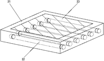
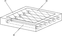
図2は、一般に利用されている照射器の構造を示したものであり、これも面光源体の一種である。照射器21は通常、内部に紫外蛍光管22を複数並行に配置され、上部には紫外光透過フィルタ23が設置されている。これにより下部から紫外光を照射し、上部で生体試料からの光学的シグナルを検出する。蛍光管自体が平面形状ではないため、均一に光を照射することはできない。均一性が重要である場合には、フィルタ23の上部または下部に拡散板を設置してもよいが、照射強度は低下する。フィルタ23は光透過性の高い板状またはフィルム状の素材に、バックグラウンドとなる波長成分をカットするフィルムを貼り付けたり、材料を塗布したものでもよい。また、フィルタ23は固形物に限らず、液状であってもよい。具体的には、主に光透過特性をコントロールする液体を、必要な波長を透過させるガラス、樹脂、フィルム等の素材に封入したものである。この液体は、単一のイオンあるいは可溶性物質を含むものであっても、複数のイオンあるいは可溶性物質を含むものであってもよい。また、液体に不溶性物質の粒子が分散している状態でもよい。液状であることの利点は、中身の液体を交換しやすいこと、それぞれがある特性をもつ液体を混ぜ合わせることによって新たな光透過(吸収)特性をもつフィルタを簡単に作製することができることなどである。
FIG. 2 shows a structure of a generally used irradiator, which is also a kind of surface light source body. The
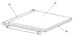
また図3のように、光源を平行に並べるのではなく、L字型あるいは周辺部を囲うように並べてもよく、光源31自体がL字型、U字型、円、球体、長方形などの直線以外の形状であってもよい。このように光源を配置すると、光源の必要数を減少させることができる上、検出対象のバックグラウンドとなる光源の陰が見えなくなるので都合がよい。ただし、検出対象に到達する照射光量を増加させるため、必要な波長に対する透過率の高い導光板32を設けることが望ましい。光源31が紫外光源である場合には、紫外光透過性材料で形成する。導光板32の裏面(検出対象を設置しない側)には、アルミニウムなどの素材で形成された、必要な波長に対する反射率が90%以上の反射材33を配置することが望ましい。反射材33は、酸化による反射率の低下を防ぐために、マグネシウムやフッ素などを、混合させたり表面コーティングに用いる場合もある。検出対象が導光板32よりも小さい場合には、導光板32の表面(検出対象を設置する側)の一部にも反射材を配置することもありうる。光源31と導光板32の間に光ファイバーなどを配置し、導光板32に高効率で光を導入する構造を採用してもよい。また、光源の可視成分など、検出のバックグラウンドとなる波長成分をカットするために、導光板32の端面と光源の間に、不要な成分をカットするフィルタを付与するか、フィルタと同様の効果を有するコーティングを行う場合もある。導光板の面全体をフィルタでカバーすると、大面積化のため非常に高価になるが、端面のみであればコストは非常に低く、検出対象をフィルタ上に設置することもないため、傷もつきにくい。同様の目的で、光源31の表面(光線が出力される部位)にフィルタを付与したり、フィルタと同様の効果を有するコーティングを行ってもよい。さらに、光源と反対側の端面には、光源の有効波長に対する反射板を付与する方がよい。また、導光板32の端面にフィルタを付与することが難しい場合には、上部(検出対象を設置する側)に不要な波長成分をカットするフィルタを必要に応じて配置する。導光板32またはフィルタには、透過率が部分的に異なる透過率グラデーション構造を形成する場合もある。つまり、光源配置形態によって照射される光量が不均一になるため、グラデーション構造を通過する前の光量が小さい部位ほど透過率が高くなるようにして、照射光量の均一性を高めるのである。このような構造は、導光板32の厚み(光源に近い側の厚みが大きくなっており、光線の進入効率を高めることが望ましい)や表面構造(拡散率の変化など)、空泡密度、含有物質密度などを部分的に変化させることで実現される。
Further, as shown in FIG. 3, the light sources may not be arranged in parallel but may be arranged in an L shape or surrounding the peripheral portion, and the light source 31 itself is a straight line such as an L shape, a U shape, a circle, a sphere, or a rectangle. Other shapes may be used. Arranging the light sources in this way is advantageous because the required number of light sources can be reduced and the shade of the light source that is the background of the detection target cannot be seen. However, in order to increase the amount of irradiation light reaching the detection target, it is desirable to provide the
以上のような、下方からの照射手段を用いる場合には、照射器を検出装置内に設置するか、あるいは検出装置を照射器の上に置いて使用することになる。さらには、照射部と検出部が一体化していてもよい。照射器の上に置く場合にはとくに、検出装置が軽量であることが望ましく、後述するような柔軟素材の使用による軽量化が有効となる。照射器の上面には、波長変換シートまたは容器、必要な部分にのみ通光穴の開いたシート、滅菌や不純物の分解などを目的とする光触媒シート(光源の波長に対する効率が高いものが好ましい)、撮影時に焦点を合わせやすくするための蛍光指標付部材等を設置することもある。なおこれらのシートおよび部材は、上方からの照射によっても同じ効果を示すものであれば、単に検出装置内に設置するだけでもよい。また、フィルタを使用する代わりに、光源の出力に一定または任意の周期の変調特性を生じさせたり、照射を極めて瞬間的に行ったり、偏光特性をもたせるなどして、検出したい光学的シグナルの特性を物理的に区別できるようにすれば、フィルタは必要ではない場合もある。 When using irradiation means from below as described above, the irradiator is installed in the detection device, or the detection device is placed on the irradiator. Furthermore, the irradiation unit and the detection unit may be integrated. In particular, when placed on the irradiator, it is desirable that the detection device be lightweight, and it is effective to reduce the weight by using a flexible material as described later. On the upper surface of the irradiator, a wavelength conversion sheet or container, a sheet having a light transmission hole only in a necessary portion, a photocatalytic sheet for sterilization or decomposition of impurities (preferably one having high efficiency with respect to the wavelength of the light source) In some cases, a fluorescent indicator member or the like is installed to facilitate focusing at the time of photographing. Note that these sheets and members may be simply installed in the detection device as long as they exhibit the same effect even when irradiated from above. Also, instead of using a filter, the characteristics of the optical signal that you want to detect can be generated by creating a modulation characteristic with a constant or arbitrary period in the output of the light source, performing irradiation very momentarily, or having a polarization characteristic. Filters may not be necessary if they are physically distinguishable.
図1(a)の天板1には、試料を含む媒体を設置したり検出操作を行うための空間を形成するため、フレームを接続する。フレームは上部フレーム6と下部フレーム7によって構成される。これらのフレームは入れ子構造とし、調整ねじ8を使用して高さ調整が可能である。なお、異なる高さの位置で固定できれば、必ずしも調整ねじ8は必要ではない。フレームも上下に分割しているのではなく、蛇腹状など複数の関節をもつ脚や折りたたみ可能な脚のような構造でもよい。このような構造は、使用しないときや持ち運び時に小さく折りたたむことができ、都合がよい。後述するように、本発明ではフレームを柔軟な素材でカバーするため、高さを変更しても外光が進入するようなスペースは発生しない。
A frame is connected to the
下部フレーム7には、必要に応じて下部固定部材9が設置される。下部固定部材9は、上部固定部材4と同様にフレームのカバー部材を固定するためのものであり、マジックテープ、磁石、金属、粘性物質、接着物質などで形成されている。ただし、検出装置内に試料等を出し入れする場合には、通常は下部を頻繁に開くため、消耗が早いマジックテープよりも板状の磁石を利用するのが最も扱いやすい。下部フレーム7が金属で形成されていれば、下部固定部材9は不要となる場合もある。なお、図1(a)のフレームの脚は4本となっているが、手を入れた場合の操作性の点では両脇の脚はない方が扱いやすいこともあり、手前側の脚は中央1本のみとしてもよい。天板1および上部フレーム6、下部フレーム7は、希酢酸、希硫酸、アルコールなど生命科学の研究で頻繁に利用される薬品に対する耐性が高い素材あるいはコーティング処理によって耐薬品性を付与された素材で作成される。。これらによって形成される空間は、おおよそ幅20〜50cm、奥行20〜50cm、高さ15〜35cm程度であるが、高さ調整可能な構造でもあり、目的に応じて変わりうる。底部は下方からの照射器を利用することもあるため、基本的にオープン構造となっている。ただし、前述したようなシート部材を設置したり、加圧、引圧するために密閉構造が必要となるときは、この限りではない。
A lower fixing member 9 is installed on the lower frame 7 as necessary. The lower fixing member 9 is for fixing the cover member of the frame similarly to the upper fixing member 4 and is formed of a magic tape, a magnet, a metal, a viscous substance, an adhesive substance or the like. However, when a sample or the like is put in and out of the detection apparatus, the lower part is usually opened frequently, so it is easiest to use a plate-like magnet rather than a magic tape that is quickly consumed. If the lower frame 7 is made of metal, the lower fixing member 9 may be unnecessary. In addition, although the leg of the frame in Fig. 1 (a) is four, in terms of operability when putting your hand, it may be easier to handle without the legs on both sides, the front leg is Only one in the center may be used. The
なお、本検出装置には蛍光管が設置されているため、充電機構や発電機構を内蔵している場合を除き、これに電力を供給するための電源コード10が必要となる。電源コードを通じて得られる電力は、まず天板1に設置されたインバータ基板に供給され、インバータ基板によって昇圧等が行われ蛍光管が点灯する。電源コード10からの電力は、検出装置内のソケットあるいはコンセント等にも供給されるようにしておくと、検出装置内で実験機器や照射器等を使用でき、都合がよい。この場合、検出装置内にはコンセントのタップのように、汎用の電源コード挿入口が設けられていることが望ましい。
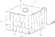
図4に、図2のフレーム構造を遮光用のカバー部材で覆った構造を示す。本検出装置の大きな特徴は、カバー部材41が暗幕のような柔軟な素材で形成されていることである。これにより大幅に軽量化され、安全かつ持ち運びが容易であり、とくにフレームが折りたたみ可能であれば、小さく全体を折りたたむことが可能になる。検出装置内で蛍光管を点灯させたり電気機器を使用すると、装置内の温度が上昇しやすいが、柔軟な素材でカバーされていれば、遮光性と通気性の両立も容易に可能となる。
In addition, since the fluorescent tube is installed in this detection apparatus, the
FIG. 4 shows a structure in which the frame structure of FIG. 2 is covered with a light shielding cover member. A major feature of the present detection apparatus is that the
カバー部材41は、固定部材42によって検出装置の天板およびフレームに固定される。固定部材42は、天板およびフレーム側の固定部材に対応して、マジックテープ、磁石、金属等で形成される。これによって、ワンタッチで容易に開閉できるだけでなく、真上に開くことができることから、省スペース化が実現できる。またカバー部材が汚れたり、劣化した場合など、容易に取り外して洗浄あるいは交換できるようになる。カバー部材が柔軟であるため、検出装置内部に手を入れて処理を行うことも容易であるが、とくに手を入れる部分の周囲の固定部材は、ゴム磁石のように柔軟かつ適度な接着性を有する材料で形成すると、遮光性を保持しつつ、手の動きも阻害されないので、便利である。また、固定部材42が裏と表の両面に設置されていれば、カバー部材41の内面と外面を容易に反転させることができる。例えば片面を吸光度の高い素材にしておき、もう一方の面を逆に反射率の高い素材にすれば、検出時には吸光度の高い面を内側にし、装置内での紫外線による滅菌時には反射率の高い面を内側にすれば効果的である。
The
カバー部材41は柔軟な素材のみに限らず、加温や冷却処理が必要であれば断熱材を用いたり、密閉性が重要であれば金属を用いることもある。なお、本検出装置の四方を柔軟な素材で覆わなければならないわけではなく、内部に実験機器や検出対象などを入れる面または部位のみが柔軟な素材で覆われていて、それ以外の面または部位は金属、樹脂などの堅固な素材で形成してもよい。遮光部材43は、検出装置内に手を入れて操作するときに外光が入らないようにするためのもので、カバー部材41との組み合わせにより、手を入れるスペース44を形成する。カバー部材41と遮光部材43は一体化していてもよい。スペース44には遮光性を高めるため、ゴムやスポンジ等のエラストマー部材で覆うことによって、手との密着性を高める場合もある。カバー部材41が柔軟な素材であるから、手に密着させても可動性は低下しないことが利点である。なお、カバー部材41は遮光に十分な長さがあれば、下部は固定しなくともよい。フード45は、検出装置内部を横から観察したりカメラで撮影するためにカバー部材41に開けられた穴を覆い、遮光性を保つためのものである。必要に応じてマジックテープ、フィルタおよびレンズ等が取り付けられる。また、この穴からポンプで減圧、加圧あるいは吸気、送風、除湿、加湿する場合もある。
The
なお、本検出装置では紫外光を使用することが多いため、カバー部材41を開放する際には、光源への通電が自動的に切れるようにしておくことが望ましい。例えば光源への電力供給回路にマグネットスイッチを配置しておき、カバー部材にはそのスイッチを導通させるためのマグネットを取り付けておくことにより、カバー部材を開放すると、光源への電力供給が停止するようにしておく。あるいは、フォトカプラーやフォトトランジスタなどのように、一定以上の光量を受けると動作するデバイスを使用し、検出装置内の光量変化によって開放を感知し、光源の動作を停止させるような方式であってもよい。
Since this detection apparatus often uses ultraviolet light, it is desirable that the light source is automatically turned off when the
次に、本検出装置による検出方法を説明する。既に述べたように、本検出装置は暗室構造を採用しており、生体試料からの光学的シグナルを検出することが基本となる。検出記録にはデジタルカメラ、ポラロイドカメラ、ビデオカメラ等のカメラあるいは専用の測定装置を使用するのが普通であるが、暗室構造を利用してX線フィルム等の高感度な媒体に記録したものをイメージスキャナー等の画像入力装置で読み込んで使用することも可能である。この場合、図1(a)の天板1には生体試料からのシグナルを透過させるレンズやフィルタではなく、フィルムが感光しない波長のみを透過させるフィルタを取り付けて、内部を観察できることが望ましい。カメラで記録される画像としては静止画像だけでなく、動画像(連続的に撮影される静止画像を含む)も利用される。天板1にディスプレイを設置したり、付近のコンピュータあるいはモニターにカメラを接続して、写真および映像を見ながら操作できるようにしておくと、実験上有用である。
Next, the detection method by this detection apparatus is demonstrated. As already described, this detection apparatus adopts a dark room structure, and is based on detecting an optical signal from a biological sample. For detection recording, digital cameras, polaroid cameras, video cameras and other cameras or dedicated measuring devices are usually used, but those recorded on a highly sensitive medium such as an X-ray film using a dark room structure are used. It can also be read and used by an image input device such as an image scanner. In this case, it is desirable that the
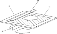
また、撮影だけでなく、高精度な照射手段も必要である。前述したように、一般に用いられているトランスイルミネーターでは、検出対象に対して均一に光を照射することができない上に、蛍光管には寿命や安定性の問題もあり、同じ検出対象からのシグナルであっても、シグナルが変化する可能性もある。さらに、紫外線透過かつ可視光透過の特性を有する光源用フィルタは非常に高価なものであり、大面積のものを作製しにくい。そこでこれらの問題を解決するため、図5に示すような光学スキャナーを考案する。図5の光学スキャナーでは、光透過板51の下方に光源52、上方に検出器53が配置され、それぞれ同一方向にスライド移動しながら、光透過板51上の検出対象54からの光学的シグナルを記録していく。なお、光源と検出器は上下が逆であってもよく、検出に問題なければ横に並んでいてもよい。検出器は通常、スライド方向に垂直な方向に複数の受光素子が並べられた構成を採るが、受光素子の数量を減らすため、光源との間にレンズを配置して集光させる。このように光源が移動するような方法を採れば、光源用フィルタは光源に付与させて、光源とともに移動するようにすればよいことから、面積は非常に小さくできる。紫外線を透過する光透過板51は、紫外光源に対するものであれば紫外光透過性材料で形成されるが、光線を完全に透過させたい場合には、検出対象を設置する位置に穴を開けたり、その位置のみ厚みを小さくする場合もある。目的によっては、波長変換板などを使用したり、表面処理等によって光拡散効果をもたせる場合もある。
In addition to photographing, high-precision irradiation means is also required. As described above, with a commonly used transilluminator, it is not possible to irradiate light uniformly on the detection target, and there are also problems with the life and stability of the fluorescent tube. Even a signal can change the signal. Furthermore, a light source filter having ultraviolet transmission characteristics and visible light transmission characteristics is very expensive and it is difficult to produce a large area filter. In order to solve these problems, an optical scanner as shown in FIG. 5 is devised. In the optical scanner of FIG. 5, a
いずれにせよ、単に紫外光を透過するのみの材料ならば、薄い透明フィルムのようなものでもよく、大面積化は容易である。なお光透過板51は、フィルタ23の場合に記載したように、固形物に限らず、液状のものをその構成要素として含むものであってもよい。光源52は通常は紫外光源であるが、光透過板51が波長変換機能を有している場合、あるいは、検出に必要な光源が紫外光以外の波長を必要とする場合は、この限りではない。光源52は検出対象54の上方または下方から照射を行い、半導体レーザーや発光ダイオードのような指向性の強い光源ならば、横方向から照射を行う場合もある。ここでいう横方向とは、検出対象をスキャニングする面に対し平行に検出光が進入することを意味している。指向性が強く極めて照射範囲の狭い光源では通常、検出対象面を二次元的にスキャニングしなければならないが、横方向からの照射であれば、スキャニング方向は一方向でよい。光源52は一度に検出対象54全体を照射することはできないため、スライド移動や回転移動をしながら全体を照射する。とくにスライド移動させる場合には、照射の均一性が得られるため望ましい。移動範囲は仕様として指定されているが、コンピュータ等によって使用者が入力してもよい。
In any case, as long as the material simply transmits ultraviolet light, it may be a thin transparent film, and the area can be easily increased. As described in the case of the
スキャニングの範囲は容易に変更できるため、とくに検出領域が大面積になるほど、それに応じた面積のフィルタを使用する方式よりも有利となる。検出器53は、光源52の照射によって検出対象54から発せられる光学的シグナル(光源波長を吸収することによる差分シグナルを含む)を検出するためのものであり、シグナルに適したフォトダイオード、イメージセンサー、光電子増倍管などによって構成される。検出器53も、光源52に合わせてスライド移動するが、イメージセンサーやフィルムなど、広範囲を一度に記録できるものであれば、移動は必要でない場合もある。検出時には光源からの光はバックグラウンドとなるため、通常は検出器53には検出対象54からのシグナルのみを透過させるのに適したフィルタを装備させる。ただし、検出器53がフォトダイオードなどで構成され、光源52の波長には感度がないようなものである場合には、フィルタは必要ない。また、フィルタ以外にも、レンズを装備させる場合もある。レンズの利用は接写や微小領域を検出するのに有効であるだけでなく、同一の平面領域に対して垂直方向に焦点を変化させながら検出記録を繰り返すことにより、検出対象54の立体構造情報も得ることができる。これは、定量処理においてはとくに重要なデータとなりうる。
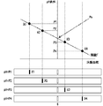
また、図5のように検出光源と検出器が一体化して移動するのではなく、別々に移動または運動してもよい。例えば図6のように、検出対象61に透過光源62から検出光を照射し、光シグナルを受光素子63で検出する。この際、透過光源62は位置を変えずに回転運動を行い、その動きに対応して受光素子63が回転または摺動することによって光シグナルを受光すれば、透過光源62の回転面による検出対象の断面について、光シグナルを検出することができる。透過光源62の回転面をその面と垂直な方向にずらしながら繰り返すか、回転面を回転させるような方法により、このような検出を繰り返せば、最終的には検出対象全体の光シグナルを測定することができる。なお、光源は透過光源に限らず、反射光源64を利用してもよい。この場合も同様に、反射光源64は移動せず回転するのみで、受光素子63がそれに対応して移動することによって、検出対象全体をモニターすることができる。検出対象の表面が均一であれば、単純に光源の回転に合わせて受光素子63が摺動すればよいが、検出対象の表面の凹凸が大きく、しかも反射光を測定する必要がある場合には、検出光の角度だけでは反射角度は決定できないため、受光素子63の位置および角度も一義的には決定できない。このような場合には、反射光源64の一つの角度に対し、受光素子を摺動および/または回転させながら検出を行い、最も光シグナルが大きくなる位置を記録することによって、検出対象表面の凹凸構造を算出することも必要になる。このような測定を、反射光源の角度および位置を変化させながら繰り返すことにより、検出対象の表面構造および光シグナル分布を測定できる。図6のようなシステムは、検出用の光源だけでなく、受光素子も小型化できる。受光素子が小型化できれば、素子自体が低コスト化されるだけでなく、受光素子の受光面へのコーティング処理やフィルタ装着も容易かつ均一に行いやすくなり、低コスト化も可能になる。
本実施例に係る発明は、発明者である青木、相澤、佐藤によってなされたものである。
Since the scanning range can be easily changed, the larger the detection area, the more advantageous than the method using a filter having a corresponding area. The
Further, the detection light source and the detector may not move integrally as shown in FIG. 5, but may move or move separately. For example, as shown in FIG. 6, the detection target 61 is irradiated with detection light from the transmission light source 62, and the light signal is detected by the light receiving element 63. At this time, if the transmissive light source 62 rotates without changing the position, and a light signal is received by the light receiving element 63 rotating or sliding in accordance with the movement, the detection target by the rotating surface of the transmissive light source 62 is detected. A light signal can be detected for each of the cross sections. If such a detection is repeated by shifting the rotation surface of the transmissive light source 62 in a direction perpendicular to the surface or rotating the rotation surface, the optical signal of the entire detection target is finally measured. be able to. The light source is not limited to the transmissive light source, and a reflected light source 64 may be used. In this case as well, the reflected light source 64 does not move but only rotates, and the light receiving element 63 moves correspondingly, whereby the entire detection target can be monitored. If the surface of the detection target is uniform, the light receiving element 63 may simply slide in accordance with the rotation of the light source. However, when the surface of the detection target is large and the reflected light needs to be measured. Since the reflection angle cannot be determined only by the angle of the detection light, the position and angle of the light receiving element 63 cannot be uniquely determined. In such a case, the detection is performed while sliding and / or rotating the light receiving element with respect to one angle of the reflected light source 64, and the position where the light signal becomes the largest is recorded, thereby the unevenness of the detection target surface. It is also necessary to calculate the structure. By repeating such measurement while changing the angle and position of the reflection light source, the surface structure of the detection target and the optical signal distribution can be measured. In the system as shown in FIG. 6, not only the light source for detection but also the light receiving element can be miniaturized. If the light receiving element can be reduced in size, not only the cost of the element itself can be reduced, but also the coating process and the filter mounting on the light receiving surface of the light receiving element can be easily and uniformly performed, and the cost can be reduced.
The invention according to this example was made by the inventors Aoki, Aizawa and Sato.
次に、本発明の生体試料解析方法について説明する。本解析方法は実施例1で述べた検出装置をサポートするものであって、基本的に画像処理を中心としたコンピュータ解析ソフトウェア(以下、「本ソフトウェア」と称する)である。ただし、本ソフトウェアには、コンピュータに使用者がインストールして使用するものだけでなく、ネットワークを介してアクセスされるコンピュータ上で動作するもの、測定器のセンサーや制御デバイスなどに組み込まれたプログラム(アルゴリズム)なども含まれる。本ソフトウェアの各機能を順に説明する。又、いわゆる、ハードウエアに記憶され実行されるものに限らず、ロジックIC化したチップを用いても良く、本実施例で示すソフトウエアに含まれる。 Next, the biological sample analysis method of the present invention will be described. This analysis method supports the detection apparatus described in the first embodiment, and is basically computer analysis software (hereinafter referred to as “this software”) focusing on image processing. However, this software includes not only those that are installed and used by users on computers, but also those that run on computers that are accessed via a network, programs that are built into sensors and control devices of measuring instruments ( Algorithm). Each function of this software will be described in turn. Further, the chip is not limited to what is stored and executed in hardware, but may be a logic IC chip, which is included in the software shown in the present embodiment.
まず本ソフトウェアで取り扱うデータとしては、数値などから構成されるテキストデータ、写真や3次元イメージ(座標データも含む)などの画像データ、映像などの動画像データおよび音声データが中心となる。また、これらのデータ間の関係や作成者、日付、解析処理記録、アクセス(参照)記録などの管理データも含まれる。その他、アナログ入力信号やアナログ信号をサンプリングした量子化信号、ソフトウェアの処理や機器の制御に使用されるプログラムなども含まれる。
本実施例で示すソフトウエアは例えばコンピュータに一時的又は継続的に記憶され、用事、実行され、例えばモニターに実行経過、結果が表示される。
本ソフトウェアはこれらのデータについて削除、追加、修正、置換、更新、変更、演算、図示等の処理を行う基本機能を有する。また、これらの処理を行ったデータを、保存する機能も有する。とくに、主として扱うデータは画像データであるから、画像に対する処理性能の高さは重要である。以下、項目ごとに詳説する。
First, the data handled by this software is mainly text data composed of numerical values, image data such as photographs and three-dimensional images (including coordinate data), moving image data such as video, and audio data. Also included are management data such as the relationship between these data, creator, date, analysis processing record, and access (reference) record. In addition, an analog input signal, a quantized signal obtained by sampling the analog signal, a program used for software processing and device control, and the like are also included.
The software shown in the present embodiment is temporarily or continuously stored in, for example, a computer, and is used and executed. For example, the progress of execution and the result are displayed on a monitor.
This software has basic functions for performing processing such as deletion, addition, modification, replacement, update, change, calculation, and illustration of these data. It also has a function of saving data that has undergone these processes. In particular, since the data handled mainly is image data, high processing performance for images is important. Hereinafter, each item will be described in detail.
画像データの修正処理
画像データを扱う上で、最も基本になるのはサイズ変更および回転処理である。サイズ変更については、画像を選択し、縦横のサイズと解像度を数値入力すると、それに合わせて変換が実行されるのが一般的であり、本ソフトウェアにおいてもこれを可能とする。また、データの整理や発表においては、特定の画像サイズや解像度にすべて揃えたい場合が多い。したがって、上記のような数値入力のみならず、1つの画像を指定し、他に選択された画像をその画像の特性に合わせるという処理も行えるようにする。これにより、揃えたい画像のサイズを調べたり、解像度を確認するというような手間が省かれる。
また、回転処理についても、通常は回転角度を数値入力するのが一般的であるが、これでは回転を実際に行ったうえで、目で見て確認しながら試行錯誤を繰り返さなければならない。この問題については、画像をディスプレイで見ながら、コンピュータのマウスを用いた手の動きに合わせて画像を回転できれば、容易に所望の角度に回転できることになる。したがって、本ソフトウェアで画像を回転させる場合には、数値入力だけでなく、このように画像を見ながら手の動きで回転できる機能ももたせる。画像のサイズ変更についても、画像を見ながら行う方が都合がよい場合もあり、この機能を応用できる。
Image data correction processing In handling image data, the most basic is size change and rotation processing. Regarding the size change, when an image is selected and the vertical and horizontal sizes and resolutions are numerically input, conversion is generally performed in accordance with the size, and this software also enables this. Further, in organizing and presenting data, there are many cases where it is desired to arrange all data to a specific image size and resolution. Therefore, not only the numerical input as described above but also a process of designating one image and matching the other selected image to the characteristics of the image can be performed. This saves the trouble of checking the size of the image to be aligned and checking the resolution.
Also, for rotation processing, it is common to input a numerical value for the rotation angle. In this case, however, after actually rotating, trial and error must be repeated while visually checking. Regarding this problem, if the image can be rotated in accordance with the movement of the hand using the mouse of the computer while viewing the image on the display, it can be easily rotated to a desired angle. Therefore, in the case of rotating an image with this software, not only numerical value input but also a function that can be rotated by hand movement while viewing the image is provided. It may be more convenient to change the size of an image while viewing the image, and this function can be applied.
画像データの重ね合わせ処理
画像を用いた議論は通常、複数の画像を比較しながら行うことが多く、一般的には、縦横に並べて複数の画像を表示する。本ソフトウェアでは、画像をデジタルデータとして扱うことから、複数の画像を並べるだけでなく、重ね合わせて表示する処理も行う。重ね合わせの演算としては、各画像を半透明処理(例えば、それぞれの画像の輝度を、重ね合わせる画像の数で割った値に補正して用いるなど)して加算する、単純に和や積をとる、差分をとる、といった項目がある。このような用途は、例えば異なる時間的タイミングで撮影した複数の画像上で、特定の領域の変位や変形を追跡する場合などがある。実施例1で記述したように、異なる光源や検出器で同一の試料を別々に撮影(記録)することもあり、
この場合は、一乃至複数の撮影手段を具備する。撮影手段としては、CCDイメージセンサーやCMOSイメージセンサーを有するカメラが好適に利用できる。複数の画像を処理する必要がある場合としては、例えば、単色のイメージセンサーを有するカメラを用いて、青、緑、赤の3原色(あるいはこれらの補色の組み合わせなど、カラー画像化に必要な構成であればよい)の光源でそれぞれ撮影し、各画像を合成してカラー画像化する場合がある。単色のセンサーは、カラーフィルタの特性のばらつきや劣化の違いによる影響がなく精度が高い、画素が微細化できる、動画用の高速処理に対応しやすいといった利点があるため、光源を変えて撮影することによって、これらの利点をそのままカラー画像に流用できるのである。
Image data superposition processing Discussions using images are usually performed while comparing a plurality of images, and generally a plurality of images are displayed vertically and horizontally. Since this software handles images as digital data, it not only arranges a plurality of images, but also superimposes them for display. For the calculation of superimposition, each image is semi-transparently processed (for example, the brightness of each image is corrected to a value divided by the number of images to be superimposed) and added. And take the difference. Such applications, for example, a plurality of the image captured at a different time timing, there is a case where tracking displacement and deformation of the particular region. As described in Example 1, the same sample may be separately photographed (recorded) with different light sources and detectors.
In this case, one or a plurality of photographing means are provided. As a photographing means, a camera having a CCD image sensor or a CMOS image sensor can be suitably used. When multiple images need to be processed, for example, using a camera having a single color image sensor, a configuration required for color imaging such as three primary colors of blue, green, and red (or a combination of these complementary colors) May be taken with a light source, and the images may be combined to form a color image. Single-color sensors have the advantage of being highly accurate without being affected by variations in color filter characteristics and differences in degradation, enabling high-precision processing, high-speed processing for moving images, and shooting with different light sources. Thus, these advantages can be used for color images as they are.
さらに、検出方法や条件が異なる複数のシグナルを同一の試料に同時に利用して、あとで画像として重ね合わせるという場合もありうる。例えば、複数のRNAやタンパク質などの発現量を同時に調べるために、それぞれの生体分子に対して異なる検出波長をもつ標識(例えば、タンパク質のそれぞれ異なる側鎖に結合するものを用いる、検出したい複数のRNAとそれぞれ相補的な配列をもつRNAにあらかじめ異なる標識を結合させておく、など)を適用すると、それぞれの標識に適した波長の光源で別々に撮影すると、組織間や細胞間に発現状態の差異があれば、それらを合成することによって、発現状態の差異がわかりやすくイメージ化できる。また、発光検出測定(撮影)と吸収検出測定(撮影)を別々に行うと、同一の状態の試料であっても、吸収部位と発光部位が異なる場合には、それらの部位の関連(物理的な連結状態や化学的なシグナルの連携)を検出することができる。この場合にも、得られた画像を重ね合わせれば、関連状況をより詳細に見ることが可能になる。このように、撮影したものをそれぞれ画像データとしてコンピュータ内に取り込み、画像データとして重ねあわせると、同じ生体内の組織や細胞間の発現状態の差異が色(シグナル)の変化量として表示されることから、モニター上で見やすくなるなどの効果が得られる。もちろん、異なる生体の組織、細胞における差異の比較を行うことも多い。このような場合には、GFPなどの発光タンパク質で波長が異なるものが発現されるように遺伝子を組み込み、発現を検出したい遺伝子とともに発光タンパク質の発現も開始されるようにしておけば、やはり異なるカラー画像として発現状態の差異が得られることになるから、画像を重ね合わせて見やすくするという用途が生じる。 Furthermore, there may be a case where a plurality of signals having different detection methods and conditions are simultaneously used for the same sample and later superimposed as an image. For example, in order to examine the expression level of multiple RNAs and proteins simultaneously, use labels with different detection wavelengths for each biomolecule (for example, those that bind to different side chains of proteins. Applying a different label to RNA having a complementary sequence to the RNA in advance, etc.), and shooting separately with a light source of a wavelength suitable for each label, the expression state between tissues and cells If there is a difference, by synthesizing them, the difference in the expression state can be easily visualized. In addition, when the emission detection measurement (imaging) and absorption detection measurement (imaging) are performed separately, even if the sample is in the same state, if the absorption site and the emission site are different, the relationship between those sites (physical A simple linkage state or chemical signal linkage). In this case as well, the related situation can be seen in more detail by superimposing the obtained images. In this way, when each captured image is taken into a computer as image data and superimposed as image data, the difference in expression state between tissues and cells in the same living body is displayed as the amount of change in color (signal) Therefore, effects such as easy viewing on the monitor can be obtained. Of course, the difference between different living tissues and cells is often compared. In such a case, if a gene is incorporated so that a photoprotein such as GFP with a different wavelength is expressed, and the expression of the photoprotein is started together with the gene whose expression is to be detected, a different color can be obtained. Since the difference in the expression state is obtained as an image, there is a use in which the images are superposed for easy viewing.
3次元的画像処理
光源と検出器(カメラ)、検出対象が同一かつ不変のものであっても、複数の画像を処理することが有効である場合もある。例えば、光源の位置を変えて撮影するか、あらかじめ同一の光源が複数配置されていて、同一の対象を複数の方向から撮影する場合である。一方向から撮影した画像上に輝度の濃淡が現れたとしても、それが検出対象自身がもつ色彩によるものであるか、表面の立体構造によって生じたものであるかを判別することはできない。しかしながら、同一の光源で複数の方向から撮影した場合に濃淡の差異が生じた場合には、その濃淡は検出対象自身がもつ色彩ではなく、表面の立体構造によって影の生じ方が変化したためであると認識できる。このようにして得られた複数の画像については、重ねあわせを行うのではなく、濃淡(影)の変化を検出し、そのデータと光源の角度に基づき、立体イメージを算出して求め、表示するようにする。
Even if the three-dimensional image processing light source, the detector (camera), and the detection target are the same and unchanged, it may be effective to process a plurality of images. For example, it is a case where the position of the light source is changed for shooting or a plurality of the same light sources are arranged in advance and the same object is shot from a plurality of directions. Even if brightness shading appears on an image taken from one direction, it cannot be determined whether it is due to the color of the detection object itself or due to the three-dimensional structure of the surface. However, if there is a difference in shading when images are taken from a plurality of directions with the same light source, the shading is not the color of the detection target itself, but the way in which the shadow is generated changes depending on the three-dimensional structure of the surface. Can be recognized. For a plurality of images obtained in this way, instead of superimposing, a change in shading (shadow) is detected, a three-dimensional image is calculated based on the data and the angle of the light source, and displayed. Like that.
動画(映像)の処理
光学的データが静止画像ではなく動画(映像)として得られている場合には、特定のパターン、形状、色の移動を追跡する機能をもたせる。例えばある部位の変位や拡大状況を追跡して、その部位がどのように変位あるいは変形しているかをベクトル(矢印)群で画像上に示したり、軌跡を表示する機能をもつ。これらのベクトルや軌跡は、使用者の意思で表示したり消去することができる。また、光源に変調特性を与えた場合や、シグナルが時間的周期性をもつ場合もあるため、動画像の中から指定された時間変化特性をもつ部位を消去したり抽出する機能ももつ。指定されなくとも、周期的に変化している部位に対して自動処理を行ってもよい。なお、画像データの重ね合わせのところで述べたように、同じ検出対象に対して、光源波長や位置を変えて測定(撮影)を行う場合があるが、このような波長や位置の変化を周期的に繰り返すことによって、カラー画像や立体画像の動画(映像)を撮影することも可能である。したがって、本ソフトウェアでは、複数の画像からカラー画像あるいは立体画像を合成し、さらに合成された画像を連結して動画(映像)化する処理も行う。
When the processing optical data of a moving image (video) is obtained as a moving image (video) instead of a still image, a function for tracking movement of a specific pattern, shape, and color is provided. For example, it has a function of tracking the displacement or enlargement state of a certain part and showing how the part is displaced or deformed on the image with a vector (arrow) group or displaying a locus. These vectors and trajectories can be displayed and deleted at the user's will. In addition, since a modulation characteristic is given to the light source or the signal may have a temporal periodicity, it has a function of deleting or extracting a part having a specified time change characteristic from a moving image. Even if it is not specified, automatic processing may be performed on a part that changes periodically. In addition, as described in the case of image data superposition, there are cases in which measurement (photographing) is performed on the same detection target while changing the light source wavelength and position, and such changes in wavelength and position are periodically performed. By repeating the above, it is possible to shoot a moving image (video) of a color image or a stereoscopic image. Therefore, in this software, a color image or a three-dimensional image is synthesized from a plurality of images, and further, a process of connecting the synthesized images into a moving image (video) is also performed.
担体上に対する照射光量分布のばらつきの補正処理
実施例1で述べたように、本検出装置は蛍光管あるいは一般の照射器を基本的な光源とする場合があり、その場合には均一な照射ができない。したがって、定量的な処理を行うためには、本ソフトウェアで照射光量を補正する処理を行う必要がある。具体的には、まず本検出装置内に、照射光量分布をモニターできる板材を設置する。この板材は、例えば蛍光アクリル板のように光源に対して発光するか、光源の波長に対して変色する感光フィルムなどの材料で形成される。あるいは、光源の波長の照射光量を部分的あるいは全体的に測定できる装置を使用し、検出対象の各部位の照射光量測定を行うことによって、照射光量分布を得てもよい。照射光量分布を得る範囲としては、少なくとも検出対象を設置する範囲についてのみ行えばよいので、板材のサイズや設置位置、あるいは照射光量測定を行う範囲も、この範囲をカバーできればよい。この場合には、例えば紫外光源に対しては、紫外光に感度を有するフォトダイオードなどの受光素子が光源側となり(あるいは受光素子にケーブルが接続されていて、光源側に向けることができる)、照射光量データのディスプレイが使用者側から見えるように配置された測定器が望ましい。受光素子をスライド走査させながら測定し、自動的に照射光量分布を得る装置でもよい。いずれにせよ、照射光量分布が正確に得やすいものが望ましく、検出対象と同等の素材上に、同等の発光物質が均一に塗布されたものが理想的である。
As described in the first embodiment, the detection apparatus may use a fluorescent tube or a general illuminator as a basic light source. In this case, uniform irradiation is performed. Can not. Therefore, in order to perform quantitative processing, it is necessary to perform processing for correcting the amount of irradiation light with this software. Specifically, first, a plate material capable of monitoring the irradiation light amount distribution is installed in the detection apparatus. The plate material is formed of a material such as a photosensitive film that emits light with respect to a light source, such as a fluorescent acrylic plate, or changes color with respect to the wavelength of the light source. Or you may use the apparatus which can measure the irradiation light quantity of the wavelength of a light source partially or entirely, and may obtain irradiation light quantity distribution by measuring the irradiation light quantity of each site | part of a detection target. As the range for obtaining the irradiation light amount distribution, it is sufficient to carry out at least the range in which the detection target is installed. In this case, for example, for an ultraviolet light source, a light receiving element such as a photodiode having sensitivity to ultraviolet light is on the light source side (or a cable is connected to the light receiving element and can be directed to the light source side) A measuring device arranged so that the display of irradiation light amount data can be seen from the user side is desirable. Measured Slide scanning light-receiving element, it may be automatically obtain illumination light quantity distribution device. In any case, it is desirable that the irradiation light amount distribution is easily obtained accurately, and ideally, the same light emitting substance is uniformly coated on the same material as the detection target.
図6はこのようにして得られる光量分布の一例であり、画像61上で線密度は光量密度を示している。さらに破線で示した部分が検出対象設置領域62である。この領域は本ソフトウェアを使用する際に使用者が指定すればよいが、あらかじめ画像61内に標識が含まれていれば、自動的に認識することも可能である。本ソフトウェアでは、検出対象の画像に対して、画像61上における領域62の光量分布画像データに基づき、光量(輝度あるいは濃度)の補正を行う。
最も簡易な例としては、蛍光アクリル板や感光フィルムのように、光源に対する反射や吸収発光および変色する特性に優れた板材を撮影し、その画像を光量分布画像として用いることである。さらに厳密性を高めたい場合には、受光素子を走査して、個々の部位の光量を検出し、電気信号に変換する。この電気信号の例えば振幅値(又は、振幅値に相当するデジタル値)に対し、予め光源がこの部位に照射した場合の光量値であって、基準となる光量値に対応する電気信号の基準振幅値(又は、振幅値に相当するデジタル値)を記憶手段に記憶し、用時、この基準振幅値を記憶手段から呼び出して、比較し、検出した振幅値が基準振幅値に基づいた所定の閾値内にあれば、補正せず、閾値外であれば、基準値の閾値に補正する為の補正アルゴリズムを設定する。
その後、この光源により、照明された担体上の試料であって、補正アルゴリズム適用エリアの部位に対し、得られる受光光量を補正アルゴリズムによって、値の修正、例えば、光量が少ない場合の補正アルゴリズムは、その受光値が実際の光源の光量よりも少ない場合は、実際の光量パラメータを減算することにより補正する。
また、補正に用いられる画像は照射光量分布画像に限らず、撮影などのデータ記録時にノイズとして含まれてしまうものも利用される。例えば傷がついたフィルタを使用したり、蛍光管などの光源の形状が画像に写りこむことが避けられない場合などである。これらのノイズの位置や形状などの性質が判明する画像を記録しておけば、それに基づいて本ソフトウェアで補正を行い、例えば、画像の狭い領域で、急激に、受光色彩が変化するような場合、この部分を隣接する部位の受光色彩に置き換えることでノイズを消去した画像が得られる。これまでに述べた照射光量分布画像、ノイズ画像などの補正用データは、可能ならば実験者自身が作成してもよい。
FIG. 6 is an example of the light quantity distribution obtained in this way, and the line density on the image 61 indicates the light quantity density. Further, a portion indicated by a broken line is the detection target installation area 62. This area may be specified by the user when using this software, but can be automatically recognized if a sign is included in the image 61 in advance. In this software, the light amount (luminance or density) is corrected based on the light amount distribution image data of the area 62 on the image 61 with respect to the detection target image.
The simplest example is to photograph a plate material excellent in the characteristics of reflection, absorption light emission, and discoloration to a light source, such as a fluorescent acrylic plate or a photosensitive film, and use the image as a light quantity distribution image. In order to further increase the strictness, the light receiving element is scanned to detect the light amount of each part and convert it into an electric signal. For example, an amplitude value (or a digital value corresponding to the amplitude value) of the electric signal is a light amount value when the light source is irradiated on the part in advance, and the reference amplitude of the electric signal corresponding to the reference light amount value A value (or a digital value corresponding to the amplitude value) is stored in the storage means, and in use, the reference amplitude value is called from the storage means and compared, and the detected amplitude value is a predetermined threshold value based on the reference amplitude value. If the value is within the threshold value, no correction is made. If the value is outside the threshold value, a correction algorithm for correcting the threshold value to the reference value is set.
After that, the sample on the carrier illuminated by this light source, and correction of the value of the received light quantity obtained for the part of the correction algorithm application area by the correction algorithm, for example, the correction algorithm when the light quantity is small, If the received light value is smaller than the actual light amount of the light source, correction is made by subtracting the actual light amount parameter.
An image used for correction is not limited to an irradiation light amount distribution image, and an image that is included as noise during data recording such as shooting is also used. For example, there are cases where a damaged filter is used or the shape of a light source such as a fluorescent tube is unavoidably reflected in an image. If you record an image that reveals the nature of the position and shape of these noises, you can make corrections using this software based on the recorded image.For example, when the light-receiving color changes suddenly in a narrow area of the image By replacing this part with the light receiving color of the adjacent part, an image from which noise has been eliminated can be obtained. The correction data such as the irradiation light amount distribution image and the noise image described so far may be created by the experimenter if possible.
また、理論的に導くことが可能であれば、照射光量分布は画像ではなく関数などの数学的データあるいは数値データ、アルゴリズムであってもよい。さらに、補正用データは光量調整やノイズ消去に用いるだけでなく、画像の一部分を抽出/消去したり、特定のパターン、色、形状を検索するなど、必要な情報を選択的に抽出する目的に使用するものであってもよい。とくに検査を主目的とした解析では、後者の方が中心となる場合もある。補正結果は補正画像または数値データとして出力および保存される。補正用データは実験の度に取得あるいは入力する必要はなく、本ソフトウェアが利用可能な領域に保存しておいて、必要なときに呼び出して使用してもよい。なお、いずれの補正に際しても、照射光量とシグナル量は必ずしも比例関係にあるわけではないので、照射光量に対するシグナル量の関数データが実験的あるいは理論的に得られている場合には、その関数データを補正に使用する。すなわち、実際に得られたシグナル量から、この関数データに基づいてまず実際の照射光量を求め、次に照射光量分布に基づいた照射光量の補正を行い、再び関数データを用いて補正照射光量から補正シグナル量を求めるのである。当然ながら、補正前のデータが画像データであれば、補正後のデータも画像として表示、出力するものとする。 Further, as long as it can be theoretically derived, the irradiation light amount distribution may be mathematical data such as a function, numerical data, or an algorithm instead of an image. Furthermore, the correction data is not only used for light intensity adjustment and noise elimination, but also for the purpose of selectively extracting necessary information such as extracting / erasing a part of an image or searching for a specific pattern, color, or shape. It may be used. In particular, in the analysis mainly intended for inspection, the latter may be the center. The correction result is output and stored as a corrected image or numerical data. The correction data does not need to be acquired or input at every experiment, but may be stored in an area where the software can be used and recalled and used when necessary. In any correction, the irradiation light amount and the signal amount are not necessarily in a proportional relationship. Therefore, when the function data of the signal amount with respect to the irradiation light amount is obtained experimentally or theoretically, the function data Is used for correction. That is, from the signal amount actually obtained, the actual irradiation light amount is first obtained based on this function data, then the irradiation light amount is corrected based on the irradiation light amount distribution, and again from the corrected irradiation light amount using the function data. The correction signal amount is obtained. Of course, if the data before correction is image data, the corrected data is also displayed and output as an image.
部分的画像データからの全体イメージ作成とマッピング
図5に示したようなスキャニングシステムを用いれば、光量の均一性は確保されるが、この場合には検出対象のごく限られた領域にしか光照射できないため、目的領域を切り出したい場合など、全体に対する位置づけを把握する必要がある操作のときに扱いにくい。これについても、本ソフトウェアで機能を補う必要がある。具体的には、スキャニングして得られた光学的データ(部分画像データ)を、スキャニング位置に応じて配置し、図7に示すような1枚の全体イメージ71を作成する。全体イメージ71上には通常、複数の光学的シグナル72が分布している。さらに、光源および検出器によって現在の照射(測定)位置で得られる光学的データ(画像)領域73を、破線や色枠で画像上に示す。画像領域73は、検出器から得られるデータを、本ソフトウェアによって全体イメージ作成の際に利用したデータとマッチングさせて認識すればよい。ただし、画像が複雑な場合等は、スキャニングシステムから直接、位置情報を得て認識する場合もありうる。このようにして全体イメージおよび現在照射されている領域が同時に表示されれば、現在照射(測定)されている領域が全体イメージの中でどの位置にあたるかが把握しやすく、切り出しなどの操作を正確に行うことが可能である。
Whole image creation and mapping from partial image data
If the scanning system as shown in FIG. 5 is used, the uniformity of the amount of light is ensured, but in this case, since only a limited area to be detected can be irradiated with light, for example, when it is desired to cut out the target area, It is difficult to handle during operations that require a grasp of the overall position. This also needs to be supplemented with this software. Specifically, the optical data (partial image data) obtained by scanning is arranged according to the scanning position, and one whole image 71 as shown in FIG. 7 is created. A plurality of
さらに、このようなイメージング方法は、紫外、赤外、温度変化など、目に見えないシグナル領域に対して、切り出しなどの作業を行う場合にも有用である。すなわち、目に見えない赤外光などの試料シグナルを発生させる行程と、検出対象上に可視光源などで目に見える指標(部分イメージ)を発生させる行程と、試料シグナルを可視化したものおよび目に見える指標を合成して全体イメージ化する行程を組み合わせることにより、全体イメージを確認しながら作業を行えるのである。なお、全体イメージ71に対して、現在の画像領域73が時間変化している場合には、操作を主体とするならば現在の画像を優先して表示する。
いずれにしても、このようなイメージング方法を用いれば、光源が半導体レーザーのように、極めて狭い領域を照射するものであっても、全体に対する位置を正確に把握できる。したがって、蛍光管では不可能な波長や強力な照射を行うような特殊光源などを利用することも可能になる。大腸菌コロニー群や二次元電気泳動のスポット群のように、細かいスポットが無数に分布しているような検出対象の観察あるいは処理においては、むしろ全体を照射していると目的位置を把握しにくく、限定された領域の照射が有効となる場合もある。
Furthermore, such an imaging method is also useful when performing operations such as clipping on invisible signal regions such as ultraviolet, infrared, and temperature changes. That is, the process of generating a sample signal such as invisible infrared light, the process of generating a visible indicator (partial image) with a visible light source on the detection target, the sample signal visualized and the eye By combining the process of synthesizing the visible indicators into a whole image, you can work while checking the whole image. If the current image area 73 changes with respect to the entire image 71, the current image is preferentially displayed if the operation is mainly performed.
In any case, if such an imaging method is used, even if the light source irradiates a very narrow area like a semiconductor laser, the position relative to the whole can be accurately grasped. Accordingly, it is possible to use a special light source that performs a wavelength or strong irradiation that is impossible with a fluorescent tube. In the observation or processing of the detection target where countless fine spots are distributed, such as E. coli colony group and two-dimensional electrophoresis spot group, it is rather difficult to grasp the target position if the whole is irradiated, In some cases, irradiation of a limited area may be effective.
解析領域の指定
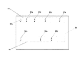
以上のような処理によって得られた、光学的に精度の高い画像に対し、本ソフトウェアでは定量を中心とした解析を行う。この際に重要となるのが、解析領域の指定方法である。本ソフトウェアでは図8に示すような領域指定方法がある。領域指定は具体的には、コンピュータに接続されたマウス等の入力デバイスによって行う。解析対象画像81に対し、まず基本として長方形指定82、楕円(正円)指定83が可能である。また、複雑な形状をした領域に対しては、マウスで複数点をクリックしながら、任意の形状で領域を囲むことも可能である。後から点の数を増減したり、位置を変更すること、各点に最小二乗法などでフィッティングさせた曲線を生成することも可能である。さらに、一つの形状を決定した後、それと相同性の高い形状の領域を自動認識によって指定することも可能とする。これは、画像内から相関の高い領域を発見したり、二次元アレイなど規則的に配置された検出対象を扱うときに便利である。また、解析方向指定線85も指定することが可能で、解析処理によってはこの線に沿って行われる。この線はやはり2点以上を指定することによって決定され、各点にフィッティングさせた曲線を生成することもできる。また、この線を中心に適当な領域幅86を指定することも可能である。
Specify analysis area
In this software, analysis focusing on quantification is performed on the optically accurate image obtained by the above processing. What is important in this case is the method of specifying the analysis region. In this software, there is an area designation method as shown in FIG. Specifically, the area designation is performed by an input device such as a mouse connected to the computer. For the analysis target image 81, first, a rectangle designation 82 and an ellipse (perfect circle)
解析情報の読み込み
通常、測定データ(画像)の解析には、何らかの外部情報が必要である。例えば電気泳動パターン画像であれば、分子サイズマーカーの分子量や標準試料の物質量を入力し、それを元に、検出試料の分子量や物質量を算出する。マーカーや標準試料が一般的なものであれば、あらかじめ数値データが設定されていたり、使用者自身が登録しておくことも可能であるが、その場合でも、実際に使用したものがどれであるかを指定する作業は必要である。このような入力や指定の作業は、とくに検査用途など、同様の作業を繰り返す必要がある場合には面倒であり、しかも入力のミスは致命的になる可能性もある。したがって、本ソフトウェアでは、図8のように、検出対象上または検出対象と一緒に記録(撮影)可能な位置に、解析に必要な情報を記載したラベル87が配置(貼り付けまたは印刷など)されている場合には、これも解析対象とする。ラベル87は文字、画像、記号、バーコード、2次元ドット配列、2次元カラードット配列などで表現されており、いずれも画像解析によって情報を取得できる。本検出装置では、光量の均一化や補正方法を工夫しているため、カラーを使用することによる情報の高密度化はとくに効果的である。
Reading analysis information
Usually, some external information is required for analysis of measurement data (image). For example, in the case of an electrophoresis pattern image, the molecular weight of the molecular size marker and the substance amount of the standard sample are input, and the molecular weight and substance amount of the detection sample are calculated based on the input. If the marker or standard sample is general, numerical data can be set in advance or registered by the user himself, but even in that case, which one is actually used It is necessary to specify this. Such input and designation work is particularly troublesome when it is necessary to repeat the same work, such as for inspection purposes, and an input error can be fatal. Therefore, in this software, as shown in FIG. 8, a label 87 describing information necessary for analysis is arranged (pasted or printed) at a position where it can be recorded (photographed) on or together with the detection target. If this is the case, this is also subject to analysis. The label 87 is represented by a character, an image, a symbol, a barcode, a two-dimensional dot array, a two-dimensional color dot array, etc., and any of them can acquire information by image analysis. In this detection apparatus, since the light quantity is uniformized and a correction method is devised, it is particularly effective to increase the density of information by using a color.
例えば、256階調のカラーで表現されたドットを、縦横それぞれ50列ずつ並べたとすれば、これだけで60万通り以上の情報を入力できることになる。したがって、マーカーの種類だけでなく、マーカーに含まれる数値的情報をそのまま入力できることになり、まったく新規のものをあらかじめ登録することなく使用できるのである。マーカーや標準試料だけでなく、試薬名や試料の数等の実験情報や解析手順情報が記載されていれば、本ソフトウェアで情報を読み取り、自動解析することも可能である。したがって、大規模で単調な解析処理や、ミスが許されない検査用途にはとくに適している。記載される情報には実験条件だけでなく、実験媒体のロット間差などの性質や、実験媒体上にあらかじめ形成されている構造情報、試薬情報なども含まれる。構造情報は、実験媒体が化学反応チップのように、複数の生体分子および化学物質が複雑な流路を経由しながら反応する場合などで重要となる。実験媒体のロット情報については、性質を数値そのもので表現するだけでなく、ある基本値を中心として、それに対する誤差をパーセント表現する場合もある。この場合、情報量が大幅に圧縮される効果がある。
なお、ラベル87には時間的に不変な情報だけではなく、時間的に変化する情報が含まれることもある。具体的には、温度、湿度、pH値、電流値、電気抵抗値、化学物質あるいはイオンの存在や光照射によって変色、発光または変形する材料でラベルの少なくとも一部を構成し、温度などの実験条件、検出対象およびフィルタなどの劣化状況が画像として判別できるようにするなどの工夫が可能になる。ラベルとして集積回路チップなど、光学的情報ではなく、磁性や通信回路を利用して情報が得られるものを利用する場合でも、本ソフトウェアへの情報入力は可能とする。
For example, if dots expressed in 256-tone colors are arranged in 50 columns in both vertical and horizontal directions, it is possible to input over 600,000 types of information. Therefore, not only the type of marker but also the numerical information contained in the marker can be input as it is, and a completely new one can be used without registering in advance. If not only markers and standard samples but also experimental information and analysis procedure information such as reagent names and number of samples are described, it is possible to read the information with this software and perform automatic analysis. Therefore, it is particularly suitable for large-scale monotonous analysis processing and inspection applications where no mistakes are allowed. The information described includes not only experimental conditions but also properties such as differences between lots of experimental media, structure information and reagent information formed in advance on the experimental media. The structural information is important when a plurality of biomolecules and chemical substances react while passing through complicated flow paths, such as a chemical reaction chip as an experimental medium. Regarding the lot information of the experimental medium, not only the property is expressed by the numerical value itself, but also an error relative to the basic value may be expressed as a percentage. In this case, there is an effect that the amount of information is greatly compressed.
Note that the label 87 may include not only information that does not change with time but also information that changes with time. Specifically, the temperature, humidity, pH value, current value, electrical resistance value, chemical substances or the presence of ions, or materials that change color, emit light, or deform due to light irradiation constitute at least part of the label, and experiments on temperature, etc. It is possible to devise such that it is possible to discriminate conditions, detection targets, deterioration conditions such as filters, etc. as images. Information can be input to this software even when using information that can be obtained using magnetism or a communication circuit instead of optical information such as an integrated circuit chip as a label.
電気泳動法の解析方法
ここで、図8のように選択された、あるいは自動的に認識された領域に対する解析例を説明する。本ソフトウェアで解析できる対象は、ドットブロット、スロットブロットやマイクロアレイなどから得られる光シグナルといった検出領域の位置も形状もほぼ一定のもの、電気泳動による分離やその転写によってパターンなど検出領域の位置あるいは検出時間は不定だが形状はほぼ一定のもの、二次元電気泳動やコロニー観察など検出領域の位置も形状も不定のもの、およびこれらの動画像などが挙げられる。当然ながら、位置および形状の自由度が高いほど、解析の難易度は高くなる。
また、同じ画像に対しても、目的に応じて解析方法も変化する。例えば電気泳動分離パターン画像の模式図を図9に示す。図9の画像において、ゲルなどの泳動担体91上に、試料の分離パターン92と、泳動マーカーの分離パターン93が認識されている。試料の分離パターン92には、試料の設置位置92s、バンド92a、92bが存在している。泳動マーカーの分離パターン93には、泳動マーカーの設置位置93s、バンド93a、93b、93c、93dが存在している。
Analysis method of electrophoresis
Here, an analysis example for a region selected as shown in FIG. 8 or automatically recognized will be described. This software can analyze the position and shape of detection areas such as light signals obtained from dot blots, slot blots, microarrays, etc., and the position or detection of detection areas such as patterns by separation or transfer by electrophoresis. Examples include those whose time is indefinite but the shape is almost constant, those in which the position and shape of the detection region are indefinite, such as two-dimensional electrophoresis and colony observation, and moving images thereof. Of course, the higher the degree of freedom of position and shape, the higher the difficulty of analysis.
Also, the analysis method varies depending on the purpose even for the same image. For example, a schematic diagram of an electrophoretic separation pattern image is shown in FIG. In the image of FIG. 9, a
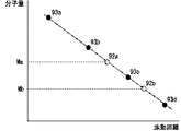
このようなパターン画像に対し、1つのバンドの濃度を定量処理する場合には、1つのバンド領域を選択すればよい。しかし実際には、濃度の定量のためには他の濃度既知のバンドも選択して比較することが必要である。また、あるバンドの分子量を算出する解析では、複数のバンド領域を処理しなければならない。具体的には図1(a)0のように、分子量が既知のマーカーバンド93a、93b、93c、93dについて、泳動距離(またはその対数)と分子量をプロットする。次に、試料バンド92a、92bの泳動距離をマーカーバンドのプロットから生成される曲線上にプロットし、分子量を算出するのである。なお、図10のようなグラフを必ずしも作成する必要はないが、多くの解析ソフトウェアで作成機能を有している。このように、数値データが既知であるコントロール試料を用いて基準関数(単にデータ間を直線補間するものも含む)を作成し、未知試料の計測値をその関数に代入して数値データを内挿、ときとして外挿し算出するという解析は、生命科学分野において極めて一般的な方法であるため、本ソフトウェアでも標準機能として有する。
When quantitatively processing the density of one band for such a pattern image, one band region may be selected. However, in practice, it is necessary to select and compare other bands with known concentrations in order to quantify the concentration. Also, in the analysis for calculating the molecular weight of a certain band, a plurality of band regions must be processed. Specifically, as shown in FIG. 1 (a) 0, the migration distance (or the logarithm thereof) and the molecular weight are plotted for the
同様に電気泳動分離パターンから関数を作成して、分子量以外の数値データを求める場合もある。例えば図11は、タンパク質の電気泳動を一定のpH条件の下で行い、等電点が既知のマーカーバンドa、b、c、dを泳動距離(の関数)に対してプロットして基準関数を求め、さらに未知の試料バンドx、yを基準関数上にプロットしたものである。変性させないタンパク質の電気泳動では、同一のpH条件下であっても、タンパク質の電気的性質に応じて正に帯電したり負に帯電したりするため、泳動距離も正負の値を取り得るが、解析方法は図10の場合と同様である。図11の解析では、試料x、yの分子量ではなく等電点(タンパク質の帯電が0となるpH値)Px、Pyが求められる。また、同一のタンパク質であっても、泳動担体のpH値によって泳動速度が異なることを利用すれば、泳動速度が0となるpH値、すなわち等電点を推定することが可能となる場合もある。例えば図12のように、pH条件が異なる環境で泳動を行い、各pH条件下における泳動距離とpH値の関係をプロットし、内挿あるいは外挿によって泳動距離が0となるpH値を求め、これをそのタンパク質の等電点とするのである。この方法は、pH勾配を有する担体中でタンパク質が等電点に近づくほど泳動が遅くなっていく通常の等電点電気泳動法に比べ、等速で泳動を行うため、迅速に等電点が求められることが利点であり、等電点に近づくと変性するタンパク質に対しても有効である。また、同じ等電点をもつタンパク質であっても、帯電状態や立体構造の違いにより、同一pH条件における泳動距離は異なるので、この方法では別のスポットとして現れることになり、分離が可能であることも大きな利点となる。したがって、各pH条件下における泳動距離(通常はマーカーに対する相対値)も重要なパラメータであり、記録しておくことが望ましい。多種類のタンパク質を含む試料であれば、各pH条件下で分離されたスポットの濃度が同程度のものを同一のタンパク質スポットであると認識する。あるいは、特定のアミノ酸配列や立体構造に特異的に結合する化合物やペプチドに色素や蛍光標識を結合させたものをプローブとし、泳動分離後にこれらのプローブと反応させ、各スポットを識別する方法も利用できる。さらにタンパク質の種類が多い試料では、各pH条件下で泳動を行うだけでは分離できずに重なってしまうスポットも多いため、各pH条件下での泳動後に界面活性剤などを用いてタンパク質を変性させ、その泳動方向と直行する方向に、分子量(分子長)に応じた電気泳動による二次元展開を行う場合もある。二次元展開後には、一次元方向にタンパク質由来の構造および保持電荷に基づく分離、さらにそれと直交する方向に分子量に基づく分離が完了しているので、各pH条件下において同じ分子量とみなせるタンパク質をそれぞれグループ化し、各分子量グループごとに絞って、各pH条件下での分離結果を用いて、前述した等電点解析を行うことによって、実質的には通常の二次元電気泳動法と同等の、等電点と分子量に基づく解析を行うことができる。 Similarly, a function may be created from the electrophoretic separation pattern to obtain numerical data other than the molecular weight. For example, in FIG. 11, protein electrophoresis is performed under a certain pH condition, and marker bands a, b, c, and d having known isoelectric points are plotted with respect to (a function of) the migration distance. Further, the unknown sample bands x and y are plotted on the reference function. In electrophoresis of proteins that are not denatured, even under the same pH conditions, depending on the electrical properties of the protein, it is charged positively or negatively, so the migration distance can also take positive and negative values, The analysis method is the same as in the case of FIG. In the analysis of FIG. 11, not the molecular weights of the samples x and y, but the isoelectric points (pH values at which protein charge becomes 0) Px and Py are obtained. Further, even if the same protein is used, it may be possible to estimate the pH value at which the migration speed becomes 0, that is, the isoelectric point, by utilizing the fact that the migration speed varies depending on the pH value of the electrophoresis carrier. . For example, as shown in FIG. 12, electrophoresis is performed in an environment with different pH conditions, the relationship between the migration distance and the pH value under each pH condition is plotted, and the pH value at which the migration distance becomes 0 is obtained by interpolation or extrapolation. This is the isoelectric point of the protein. In this method, the electrophoresis is performed at a constant speed in comparison with the normal isoelectric focusing method in which the electrophoresis becomes slower as the protein approaches the isoelectric point in a carrier having a pH gradient. It is an advantage that it is required, and it is also effective for proteins that denature as they approach the isoelectric point. In addition, even proteins with the same isoelectric point have different migration distances under the same pH conditions due to differences in charged state and three-dimensional structure, so this method will appear as different spots and can be separated. This is also a big advantage. Therefore, the migration distance (usually relative to the marker) under each pH condition is also an important parameter, and it is desirable to record it. In the case of a sample containing many kinds of proteins, those having the same concentration of spots separated under each pH condition are recognized as the same protein spot. Alternatively, a compound or peptide that specifically binds to a specific amino acid sequence or three-dimensional structure is combined with a dye or fluorescent label as a probe, and reacted with these probes after electrophoretic separation to identify each spot. it can. Furthermore, in many samples with many types of proteins, there are many spots that overlap without being separated only by running under each pH condition. Therefore, after running under each pH condition, the protein is denatured using a surfactant or the like. In some cases, two-dimensional development by electrophoresis according to molecular weight (molecular length) is performed in a direction perpendicular to the migration direction. After two-dimensional development, separation based on protein-derived structure and retained charge in one-dimensional direction, and separation based on molecular weight in the direction perpendicular to it, are complete, so each protein that can be regarded as the same molecular weight under each pH condition By grouping and narrowing down for each molecular weight group, using the results of separation under each pH condition, and performing the isoelectric point analysis described above, it is substantially equivalent to a normal two-dimensional electrophoresis method, etc. Analysis based on electrical point and molecular weight can be performed.
上記の方法も含めて、実際には、泳動担体および緩衝液のpH値を一定に保つことは容易ではない。前述したように、本ソフトウェアでは、図8のラベル87のように、実験環境をモニターするためのラベルも解析対象として含めることから、pH値をモニターするためのリトマス試験紙などのラベルを利用することもある。pH値ラベルは泳動系全体をモニターできるサイズであることが望ましく、pH値の変化(分布)が色変化(分布)など画像的に得られることが望ましいが、pH電極(これを複数並べる場合を含む)のようなデバイスを用いてもよい。なお、泳動環境を厳密に把握し解析を行うためには、電流値、電圧値、温度などのパラメータについても、実験系での分布が得られることが望ましく、探針やホール素子、熱電対、白金測温抵抗体、サーモグラフィーおよびこれらを複数組み合わせたものを利用して、適宜測定を行うものとする。 In practice, including the above method, it is not easy to keep the pH values of the electrophoresis carrier and the buffer solution constant. As described above, this software also includes a label for monitoring the experimental environment as an analysis target, such as the label 87 in FIG. 8, and therefore, a label such as a litmus paper for monitoring the pH value can be used. is there. It is desirable that the pH value label is of a size that allows the entire electrophoresis system to be monitored, and it is desirable that the pH value change (distribution) be obtained graphically, such as a color change (distribution). Devices). In order to accurately grasp and analyze the electrophoresis environment, it is desirable that parameters such as current value, voltage value, temperature, etc. are obtained in an experimental system, such as a probe, a Hall element, a thermocouple, Measurements should be made as appropriate using platinum resistance thermometers, thermography, and combinations of these.
解析ソフトウェアからの制御および通知方法
また、本ソフトウェアでは単に情報を受け取って解析するだけでなく、解析した結果にもとづいて検出器や測定器の動作を変化させる機能をもつ。例えば、電気泳動や大腸菌コロニーの発達、前述した時間変化ラベルのように、時間とともに発光(発色)領域が変化していく実験があるので、本ソフトウェアによって画像の変化をモニターして、特定の状態になったら実験装置や電源の停止、光源の点灯、撮影、音声通知などの制御を行う。使用者があらかじめ画像上に領域を指定しておき、その領域に起こると予想される変化と、それに対する制御方法を指定できるようにしておくことが望ましい。あるいは、検出器(の検出部位)を所定の領域が検出されるようにユーザーが設置しておくことで、領域指定と同等の効果を得てもよい。これらについては、実施例5において詳述する検出器と組み合わせて行うことが望ましい。
また、化学反応チップのような実験媒体であれば、その上で行われる化学反応などをコントロールするための情報を実験(検出)装置側に送信する場合もある。具体的には、実験者が生体分子名あるいは化学物質名(化学式)のような扱いなれた名称と、それらの量、反応式、条件分岐などを入力(プログラミング)すると、本ソフトウェアがそれを翻訳して、実験媒体を制御する装置が認識できる動作命令セット(試薬配置、電圧を印加する位置やタイミングなど)を作成し、装置を動作させる。コンピュータプログラミングにおいて、人間が把握しやすい言語で入力し、コンパイラがそれを機械語に翻訳する機能と同様である。
Control and notification method from analysis software
In addition, this software not only receives and analyzes information, but also has a function to change the operation of the detector and measuring device based on the analysis result. For example, there are experiments in which the light emission (coloring) region changes over time, such as electrophoresis, the development of E. coli colonies, and the time-varying label described above. If it becomes, control such as stopping the experimental device and power supply, turning on the light source, shooting, and voice notification are performed. It is desirable that the user designates an area on the image in advance so that a change expected to occur in the area and a control method for the change can be designated. Alternatively, if the user installs the detector (detection site) so that a predetermined area is detected, an effect equivalent to the area designation may be obtained. These are desirably performed in combination with the detector described in detail in the fifth embodiment.
In the case of an experimental medium such as a chemical reaction chip, information for controlling a chemical reaction performed on the experimental medium may be transmitted to the experiment (detection) apparatus side. Specifically, when an experimenter inputs (programs) a name that is handled like a biomolecule name or chemical substance name (chemical formula) and their quantity, reaction formula, conditional branching, etc., this software translates it. Then, an operation command set (reagent arrangement, voltage application position and timing, etc.) that can be recognized by the apparatus controlling the experimental medium is created, and the apparatus is operated. In computer programming, it is similar to the function of inputting in a language that is easy for humans to understand and for the compiler to translate it into machine language.
詳細な定量方法
さらに、定量解析の上では、画像撮影時に焦点が合わなかった場合にも誤差が生じる。撮影をやり直せない実験もあることから、このような画像に対しても処理を行わなければならない。このとき、上記のラベルや蛍光標識など、焦点のずれの程度が推測できる手がかりが一緒に撮影されている場合には、それらの本来のイメージになるように全体画像を修正する機能をもつ。また、図5に示したスキャニングシステムにおいて、焦点を変えながら複数の光学的データ(画像データを含む)を得た場合には、これらを足し合わせて(積分して)定量データを算出する機能をもつ。また、本検出装置は図4で説明したように、上方からと横方向からの画像記録が可能であるため、1つの検出対象を複数の方向から撮影した画像をもとに、光学的シグナルの三次元構造を推測して、定量データを算出する機能ももつ。
Detailed quantification method
Further, in quantitative analysis, an error occurs even when the image is not focused at the time of image capturing. Since there are some experiments in which shooting cannot be performed again, such an image must be processed. At this time, when a clue that can be used to estimate the degree of defocus, such as the above-described label or fluorescent label, is photographed together, it has a function of correcting the entire image so that it becomes its original image. Further, in the scanning system shown in FIG. 5, when a plurality of optical data (including image data) is obtained while changing the focus, a function for adding (integrating) these and calculating quantitative data is provided. Have. In addition, as described with reference to FIG. 4, the detection apparatus can record images from above and from the lateral direction. Therefore, based on images obtained by photographing one detection target from a plurality of directions, an optical signal can be recorded. It also has a function to estimate quantitative data by inferring a three-dimensional structure.
ネットワーク利用による情報検索および管理方法
なお、タンパク質の場合には、等電点および分子量が求められれば、公共のデータベースにネットワーク接続することにより、タンパク質を同定することができるので、ソフトウェアとしてデータベースを参照できる機能は必要であり、また、LAN内やコンピュータ内にデータベースを作成できる機能も必須である。さらに、このような数値的データだけでなく、画像データおよび論文などの書籍データ、HTMLなどのファイルデータも参照できることが重要である。具体的には、画像データ、動画データ、書籍名、書籍ナンバー、該当ページ、日付、キーワード、文章、著者名、実験者名、実験タイトル、実験記録、ネットワーク上のアドレスなどを入力保存でき、必要に応じてキーワードなどで検索できるデータベースシステムを作成できる機能を有する。本データベースシステムでは画像データも扱うため、ある画像と明度、彩度、形状パターンなどを比較して、相同性の高い順に画像データを並べたり、類似度を数値で表示することもできる。画像にはキーワードなどのデータも付属して管理されているため、キーワード検索も当然可能である。
また、情報量が膨大である場合、単純なキーワードでは情報を絞り込むことができず、また、キーワードを多数入力することも面倒である。同じキーワードであっても、使用者によって必要情報やイメージが異なる場合も多い。そこで、本データベースシステムでは、使用者がキーワードXを指定するとき、あらかじめキーワードXの関連ワード群RXを指定することができる。例えばキーワードXとして「試薬」を指定し、関連ワード群RX={「蛍光」、「標識」、「アルコール」}と指定しておくことにより、使用者が情報検索を行う際に「試薬」を入力すれば、本データベースシステムは、関連ワード群RXに関係する「試薬」の情報のみを検索し、使用者に提供するか、少なくとも関連の深い情報を優先してリスト化するのである。関連ワード群の他、非関連ワード群を指定することも可能とする。非関連ワード群を指定した場合には、それらに関係する情報を除去して使用者に提供する。なお、はじめに関連ワード群を指定しなくとも、あるキーワードに対して検索された情報のうち、使用者が実際に参照した情報に含まれる他のキーワードを統計処理し、自動的に関連ワード群を作成してもよい。
Information search and management method using network
In the case of proteins, if the isoelectric point and molecular weight are determined, the protein can be identified by connecting to a public database via a network, so a function that can refer to the database as software is necessary. The ability to create a database in a LAN or computer is also essential. Furthermore, it is important to be able to refer not only to such numerical data but also to image data, book data such as papers, and file data such as HTML. Specifically, image data, video data, book title, book number, applicable page, date, keyword, text, author name, experimenter name, experiment title, experiment record, network address, etc. can be input and saved It has a function that can create a database system that can be searched with keywords and the like. Since this database system also handles image data, it is possible to compare a certain image with brightness, saturation, shape pattern, etc., and arrange the image data in descending order of homology, or display the degree of similarity numerically. Since data such as keywords are attached to the image and managed, keyword search is naturally possible.
In addition, when the amount of information is enormous, information cannot be narrowed down with a simple keyword, and it is troublesome to input a large number of keywords. Even for the same keyword, the required information and image are often different depending on the user. Therefore, in this database system, when the user specifies the keyword X, the related word group RX of the keyword X can be specified in advance. For example, by specifying “reagent” as the keyword X and specifying the related word group RX = {“fluorescence”, “label”, “alcohol”}, the user can select “reagent” when performing an information search. If entered, the database system searches only the information of “reagents” related to the related word group RX and provides it to the user, or at least lists information that is closely related. In addition to related word groups, non-related word groups can also be specified. When an unrelated word group is specified, information related to the group is removed and provided to the user. Even if the related word group is not specified first, among the information searched for a certain keyword, other keywords included in the information actually referred to by the user are statistically processed to automatically select the related word group. You may create it.
また、検索対象情報が画像である場合、キーワードで検索することは非常に難しい。例えば「青い」というキーワードで検索する場合、どの程度の青色成分を、どれほどの含有率で含む画像を対象とするかは、使用者の主観によるからである。これについて本データベースシステムでは、キーワードに対して基準画像を指定することができる。例えば「青い」というキーワードに対して、電気泳動パターン画像の1つを指定しておくと、その画像の明度、彩度、青色成分含有率などを解析して数値データ化し、データベースに含まれる画像データの中から同程度の特徴をもつ画像を検索し、使用者に提供する。また、基準画像を指定する代わりに、主観テストを行ってもよい。これは、複数の参照画像を使用者に対して表示し、その画像に対する感覚をキーワード入力させるか質問に答えさせることによって、その使用者のキーワードと画像数値データを結びつける方法である。
このような、使用者の主観を数値化してキーワードと結びつける方法は、色に限らず、コントラスト、形状、変化の程度、変位の大きさ、音声の大きさおよび質など、さまざまなデータに用いることができる。使用者がキーワードの登録を行うたびに、これらのデータを解析するのは効率が悪いため、もともとデータ側にも数値化された情報が付加していることが望ましい。このように使用者の主観に基づいたデータベースシステムを使用する場合、使用者はシステム(ソフトウェア)へのアクセス時にパスワード入力や識別デバイス挿入などによるログオンを行い、使用者の識別が可能になるようにしておく。Cookieなどのネットワーク技術によって自動的に識別可能な場合もある。
In addition, when the search target information is an image, it is very difficult to search using keywords. For example, when searching with the keyword “blue”, it is because the subject matter of the user determines how much blue component is included in what content. In this regard, in this database system, a reference image can be designated for a keyword. For example, if one of the electrophoretic pattern images is specified for the keyword “blue”, the brightness, saturation, blue component content, etc. of the image is analyzed and converted into numerical data, and the image included in the database Search for images with similar characteristics from the data and provide them to the user. In addition, a subjective test may be performed instead of specifying the reference image. This is a method in which a plurality of reference images are displayed to a user, and the keyword of the user and image numerical data are linked by allowing a user to input a sense of the image as a keyword or answering a question.
The method of quantifying the user's subjectivity and linking it to the keyword is not limited to color, but can be used for various data such as contrast, shape, degree of change, magnitude of displacement, volume and quality of speech. Can do. Since it is inefficient to analyze these data every time a user registers a keyword, it is desirable that the data side also has information added to the data. When using a database system based on the user's subjectivity in this way, the user can log on by entering a password or inserting an identification device when accessing the system (software) so that the user can be identified. Keep it. In some cases, it can be automatically identified by network technologies such as cookies.
なお、このような主観に基づくデータベースシステムは、文字情報だけでなく画像(動画を含む)や音声情報のデータが急増しているインターネットなどの一般ネットワークシステムにおいても重要なものであり、応用可能である。この場合には、主観に基づくキーワード群には、理化学的な主観のみならず、味覚や聴覚などの感覚、思想、趣味などに関するものも含まれることになる。いずれにせよ、文章や画像、音声に対する感覚は個人の主観によって大きく異なるため、その検索手段としては、個人の主観を先にテストなどによって定量化しておき、使用者ごとに検索条件を変更する必要があるのである。 It should be noted that such a subjective database system is important not only for text information but also for general network systems such as the Internet where images (including moving images) and audio information are rapidly increasing. is there. In this case, the subject matter-based keyword group includes not only physics and chemistry subjects but also those relating to senses such as taste and hearing, thoughts, and hobbies. In any case, the sense of text, images, and speech varies greatly depending on the subjectivity of the individual, so as a search method, it is necessary to quantify the subjectivity of the individual first through tests, etc., and to change the search conditions for each user. There is.
インターネットまで接続範囲を拡大するかどうかは別として、本データベースシステムにも、ネットワークを介して複数の使用者が共有できる機能をもたせる。上記のようなネットワークを介したデータベース管理機能を主眼とする場合、本ソフトウェアは必ずしも使用者側のコンピュータ上で動作するものではなく、ホストとなるコンピュータ上で動作し、使用者がネットワークを介して本ソフトウェアにデータを送信したり受信する方式でもよい。ハードウェアへの負荷が非常に大きい解析を行うために高性能なコンピュータが必要である場合や、データ管理のセキュリティを極めて高くしたい場合、複数の使用者間での情報の同期が重要である場合などには、専用のハードウェア上で動作する方が好ましいからである。 Apart from whether or not to extend the connection range to the Internet, this database system also has a function that can be shared by multiple users via a network. When focusing on the database management function via the network as described above, this software does not necessarily run on the user's computer, but runs on the host computer. A method of transmitting or receiving data to this software may be used. When you need a high-performance computer to perform an analysis with a very heavy load on the hardware, or when you want extremely high security for data management, or when it is important to synchronize information among multiple users This is because it is preferable to operate on dedicated hardware.
なお、複数の実験装置、データ入力装置、コンピュータ、認証システムなどをネットワーク化したシステムとして利用する場合には、ネットワークに接続された各機器がもつ固有のIPアドレスや、機器認証あるいは個人認証に必要な情報入力デバイス(カードなど)やドライバソフトなどにより、本ソフトウェアが使用者や使用機器を認証して、自動的にシステムへのアクセスを制限したり、個別の使用設定を読み込んだり、必要な機能を起動したりすることが望ましい。 In addition, when using multiple experimental devices, data input devices, computers, authentication systems, etc. as a networked system, it is necessary for the unique IP address of each device connected to the network, device authentication, or personal authentication. This software authenticates users and devices using various information input devices (such as cards) and driver software, automatically restricts access to the system, reads individual usage settings, and other necessary functions. It is desirable to start up.
このようにネットワークに接続されて動作するソフトウェア(動作はユーザー側のシステム上で行い、その使用状況のみネットワークを介して情報として取得できるものも含む)においては、ソフトへのアクセス(使用)回数や時間に応じた料金を徴収する課金システムを構成することも可能になる。さらに、料金を徴収するだけではなく、有用な情報を提供し、他のユーザーに利益を与えたユーザーに対しては、使用料金の減額や報酬を供与するということも可能になる。 In software that operates while connected to the network in this way (including operations that are performed on the user's system and only the usage status can be obtained as information via the network), the number of times the software is accessed (used), It is also possible to configure a billing system that collects charges according to time. Furthermore, it is possible not only to collect a fee, but also to provide useful information and provide a user with a reduction in usage fee or a reward to a user who has benefited other users.
また、本ソフトウェアの管理者あるいはユーザー以外の第三者が、広告や販売などを行うことにより、ユーザーのシステム使用料の一部を負担する場合もある。すなわち、ユーザーが意図的にマウスでクリックするなどして、広告などの情報コンテンツを視聴したり、機器を購入したり、アンケートに回答するなどという行為によって利益を得る者が、ユーザーのソフトウェア使用料金の一部を負担したり、謝礼を供与するようにする。ユーザーの意思に関係なく提供される一般の広告などとは異なり、あくまでもユーザーの意思で特定の情報コンテンツを選択することによって、ユーザー自身が情報コンテンツ提供者から利益を供与されるのである。 In addition, the administrator of this software or a third party other than the user may bear a part of the user's system usage fee through advertisements or sales. In other words, a person who makes a profit by viewing the information content such as an advertisement, purchasing a device, or answering a questionnaire by intentionally clicking with the mouse, etc. To pay a part of or give a reward. Unlike general advertisements and the like that are provided regardless of the user's intention, the user himself / herself benefits from the information content provider by selecting specific information content at the user's intention.
前述したように、その情報コンテンツが非常にユーザーにとって有益なものである場合には、逆にユーザーが情報コンテンツ提供者(これもユーザーであってもよい)に料金負担や謝礼などの利益を供与する場合もありうる。とくに生命科学研究のような、極めて限られた目的に使用されるシステムにおいては、このような形態が有効である。情報コンテンツとしては、文字や音声、画像、映像などによる情報、データ、番組などが挙げられる。この際、ユーザーのソフトウェア(システム)へのアクセス記録が管理されていれば、ユーザーが同じ行為を繰り返すことによって、料金負担を重複して行わなければならない事態を防止できる。また、アクセス記録については、料金管理などのためだけではなく、記録そのものに意味がある場合もある。すなわち、実験記録やその情報公開日時、内容、考察やアイディアなどをシステムとして管理できることにより、そのユーザーの成果やアイディアを証明できるシステムとなるのである。さらに、証明システムとしては、そのよう情報提供に関する記録だけではなく、その変更および追加、修正に関する履歴や、他のユーザーがその情報にいつアクセスしたかという情報も記録管理されることが望ましい。 As mentioned above, when the information content is very useful to the user, the user gives the information content provider (which may be the user) a benefit such as a fee or a reward. It is possible that This form is particularly useful in systems used for extremely limited purposes, such as life science research. Information content includes information such as text, sound, images, and video, data, programs, and the like. At this time, if the user's software (system) access record is managed, it is possible to prevent a situation in which the user must repeat the same action to duplicate the charge burden. In addition, the access record may be meaningful not only for fee management but also for the record itself. In other words, by managing the experiment records, information disclosure date and time, contents, considerations, and ideas as a system, it becomes a system that can prove the results and ideas of the user. Furthermore, it is desirable for the certification system to record and manage not only records relating to information provision, but also history of changes, additions and modifications, and information on when other users have accessed the information.
さらに使用状況やアクセス記録を完全に実施するためには、パスワード入力のようなソフトウェアによる方法ではなく、ユーザーが何らかのハードウェア(デバイス)をシステムに対して行使することによって、ユーザーを認識させる方法もありうる。例えば専用のカードをユーザーが保有し、システムにはカードリーダー(情報読み取り器)を備えておく方法であったり、ユーザーの眼球運動や指紋、声紋などの身体的特徴を読み取る装置を利用する場合である。コンピュータで一般的となっているUSBポートのように、任意の時間的タイミングでデバイスの接続を認識できる入力端子(出力も可能であってもよい)がシステムに備えられていれば、ユーザー側がUSBメモリーあるいはUSBキーのような入力デバイスさえ保有しておけば、その他にハードウェアを用意する必要はない。また、システムの使用目的が検査のように、多数の入力対象を前提としている場合には、ICタグ(集積回路チップ)のように、使用者単位ではなく、検査対象ごとに入力デバイスが取り付けられている方がよい。例えば食品ごとにICタグやバーコードなどの入力デバイスが取り付けられていて、食品の配送と検査が同時に進行するような場合では、検査終了後に入力デバイスで対象物を認識させてから結果を入力し、複数の配送先からも同じ入力デバイスによって検査結果を確認するようにしておけば、速やかに誤りなく検査と配送を進行できることになる。
いずれにせよ、上記のようなシステムによって、ユーザーの成果、アイディア、貢献程度から、対象物の検査結果まで、さまざまな情報を詳細に管理し、情報についての証明も行えることになるのである。
本実施例2に係る発明は、発明者である青木によってなされたものである。
In addition, in order to fully implement usage status and access records, there is also a method for recognizing the user by exercising some kind of hardware (device) to the system instead of a software method such as password entry. It is possible. For example, when the user has a dedicated card and the system is equipped with a card reader (information reader), or when using a device that reads the user's eye movements, fingerprints, voiceprints, and other physical features is there. If the system is equipped with an input terminal that can recognize the connection of the device at any time timing (such as output may be possible), such as a USB port that is common in computers, the user can use USB As long as you have an input device such as a memory or USB key, you don't need any other hardware. In addition, if the purpose of the system is premised on a large number of input objects such as inspection, an input device is attached to each inspection object instead of a user unit, such as an IC tag (integrated circuit chip). It is better to have. For example, when an input device such as an IC tag or barcode is attached to each food item and food delivery and inspection proceed simultaneously, the input device recognizes the target object after the inspection is completed, and then inputs the result. If inspection results are confirmed from a plurality of delivery destinations using the same input device, the inspection and delivery can proceed promptly and without error.
In any case, the system as described above enables detailed management of various information, from user achievements, ideas and contributions to inspection results of objects, and proof of information.
The invention according to the second embodiment is made by Aoki who is the inventor.
本検出装置では、扱う検出対象は蛍光など光学的シグナルを発する、あるいはそのような標識を行った生体試料が中心であり、励起光としては紫外光が中心となる。したがって、実験機器あるいは実験器具類も、そのような目的に沿うように最適化することが望ましく、ここではその具体例について説明する。
まず、検出対象を保持する器具類は、原則的に紫外光透過性材料で形成することが望ましく、また、加圧や加温、低温(液体窒素温度など)、各種薬剤(特に、希酢酸、希硫酸、アルコール、フェノール類)に耐えうる素材や、紫外光照射によって高い分解機能を発揮する光触媒材料などで形成する、あるいはコーティングや内蔵など一部に採用することにより、過酷条件下での実験に応用したり、加圧、加温、紫外光照射によって容易に滅菌あるいは浄化や、装置内の温度制御を行うことができる器具を実現できる。
In this detection apparatus, the detection target to be handled is mainly a biological sample that emits an optical signal such as fluorescence or is labeled, and the excitation light is mainly ultraviolet light. Therefore, it is desirable to optimize the experimental equipment or the experimental equipment so as to meet such a purpose, and a specific example thereof will be described here.
First of all, it is desirable that the instruments for holding the detection target are formed of an ultraviolet light transmissive material in principle, and pressurization, heating, low temperature (such as liquid nitrogen temperature), various drugs (particularly dilute acetic acid, Experiments under harsh conditions by using materials that can withstand dilute sulfuric acid, alcohol, phenols), photocatalytic materials that exhibit a high decomposition function when irradiated with ultraviolet light, or by using them in some parts such as coatings and built-in parts. It is possible to realize an instrument that can be applied to the above, or can be easily sterilized or purified by pressurization, heating, or irradiation with ultraviolet light, and the temperature in the apparatus can be controlled.
器具としては、大腸菌の培養などに用いるシャーレ類、試料の分離に使用する電気泳動装置、液体の蛍光試薬を注入するコンテナなどの容器、検出対象を設置する平板およびそれに取っ手を付与したものなどがある。これらの材料と器具の選定により、紫外光によるコロニー観察が容易なシャーレ、液体窒素で冷却し蛍光スペクトルを詳細に観察する環境、検出対象を取り出すことなく紫外光で蛍光標識の確認ができる容器、実験後容易に加熱滅菌ができる保持器具や電気泳動装置、電子レンジやオートクレーブでアガロースなどのゲルを高温融解させそのまま必要な形状に固化できるゲル作製器およびゲル支持体といった器具および機器を構成することができ、これらはいずれも本検出装置内で有効に利用できるものである。 Examples of instruments include petri dishes used for culturing E. coli, electrophoresis devices used for sample separation, containers such as containers for injecting liquid fluorescent reagents, flat plates on which detection targets are installed, and those provided with handles. is there. By selecting these materials and instruments, a petri dish that is easy to observe colonies with ultraviolet light, an environment for observing the fluorescence spectrum in detail by cooling with liquid nitrogen, a container that can confirm fluorescent labels with ultraviolet light without taking out the detection target, Construct equipment and equipment such as a gel preparation device and a gel support that can be easily heat-sterilized after the experiment, a gel preparation device that can melt the gel such as agarose at high temperature in a microwave oven or autoclave, and solidify it as it is. These can all be used effectively in the detection apparatus.
図13(c)に、耐熱素材で形成した補助器具の一例を示す。発明者らは、特願2002−299543号において、電気泳動用のゲルをパック状にし、電子レンジやオートクレーブなどによる加熱で溶解させ、任意の形状に固化させて使用できる構成を考案した。ただし単純にパックのまま電子レンジで急激な加熱を行うと、パックが破裂して危険を伴うこともありうる。そこで、図13(c)のように、耐熱素材で形成された容器1301と、その蓋1302の間に、パック1303の非開放側を挟み、開放側を容器の底に向けるように固定する。この状態で電子レンジやオートクレーブで加熱を行えば、容器が耐熱素材であるため、ゲル1304を短時間で溶解させることができる。ゲル単体で容器内に入れてもよいが、急激な加熱によって飛散する場合もあるので、蓋1302やパック1303の必要性については実験者の判断による。なお、この容器1301に、電気泳動で使用するゲル支持部材(これも耐熱素材で形成する)を設置し、この上でゲル1304が溶解するようにすれば、溶解後にそのまま冷却させ、実験に必要な形状に固化させることも可能である。また、耐熱性の容器1301に、これを直接加熱するヒーターも接続されているような、ゲル溶解器(あるいは滅菌器)を構成することも可能である。ヒーターがペルチェ素子のように冷却も可能なものであれば、加熱後の温度制御によって短時間にゲル化させたり、最適な温度条件でゲル化させることも可能である。 FIG. 13 (c) shows an example of an auxiliary device made of a heat-resistant material. The inventors have devised a configuration in Japanese Patent Application No. 2002-299543 that can be used by forming a gel for electrophoresis into a pack, dissolving it by heating with a microwave oven, an autoclave, or the like, and solidifying it into an arbitrary shape. However, if the pack is heated rapidly in a microwave oven, the pack may burst and be dangerous. Therefore, as shown in FIG. 13C, the non-open side of the pack 1303 is sandwiched between the container 1301 formed of a heat-resistant material and the lid 1302, and the open side is fixed to face the bottom of the container. If heating is performed with a microwave oven or an autoclave in this state, the gel 1304 can be dissolved in a short time because the container is a heat-resistant material. Although the gel alone may be put in the container, it may be scattered by rapid heating, so the necessity of the lid 1302 or the pack 1303 is determined by the experimenter. In addition, if a gel support member (also formed of a heat-resistant material) used for electrophoresis is installed in this container 1301 and the gel 1304 is dissolved thereon, it is allowed to cool as it is after dissolution and is necessary for the experiment. It is also possible to solidify into various shapes. It is also possible to configure a gel dissolver (or sterilizer) in which a heat-resistant container 1301 is connected to a heater that directly heats the container. If the heater can be cooled like a Peltier element, it can be gelled in a short time by controlling the temperature after heating, or gelled under optimum temperature conditions.
また、紫外光や加温によって付加効果をもつ器具類は上記のように多岐にわたっているので、紫外光透過性材料や耐熱素材、耐寒材、耐湿材、耐薬品性素材、光触媒材料、およびそれらを含むものを部品化し、必要に応じて任意の形状を組み立てられる構成にしておくことも好ましい。例えば図13(a)のように、基本となる板状部品131を、接続部品132の溝133にはめ込むような構成とし、任意の形状を作成する。接続部品132の溝133は一方向だけでなく、その裏側や垂直方向にも形成されていることが望ましい。板状部品131に溝が形成されていてもよい。また、実験器具としては液体を使用することが多いため、ゴムや発泡素材など密着性と柔軟性、ないしは弾性の高い素材を接続部に挟み、これをねじ止めするなどで防水性を高めてもよい。基本部品としては板状だけでなく、棒状、円弧状、ケース状のものも用意し、接続部品132も基本部品同士を接続するだけでなく、それ自体が取っ手など機能的な形状をしていてもよい。なお、紫外光透過などの特殊な機能は必要な部位だけ有していればよいので、それ以外の部位は金属など通常の素材で構成してもよい。このような組み立て式構造は、形状の自由度が高いだけでなく、解体して洗浄しやすいという利点もある。容器の角などには不純物が付着しやすく洗浄も困難であるが、解体すれば角構造がなくなり、容易に洗浄できるからである。
In addition, there are a wide variety of appliances that have additional effects due to ultraviolet light and heating, as described above, so ultraviolet light transmissive materials, heat resistant materials, cold resistant materials, moisture resistant materials, chemical resistant materials, photocatalytic materials, and It is also preferable to make it a component that can be assembled into an arbitrary shape if necessary. For example, as shown in FIG. 13A, a basic plate-
さらに、組み立て構造としては3次元的な構造だけではなく、2次元的な構造の組み合わせを行う場合もある。例えば図13(b)のように、さまざまな形状の流路1311、試料の注入や取り出しを行うホール1312、複数の試料や物質の混合および反応を行うリアクター1313などを有するプレート部材を任意に組み合わせ、それぞれの実験に適した系を構成するような場合である。これらに付随して、プレートを互いに接続するための凹凸構造1314や、表面をカバーするためのシール部材も必要となる。プレート部材同士が直接接続されるのではなく、フレームや基板上にプレート部材をはめ込んでいく方式でもよい。とくに、ホールや流路などに、電流あるいはpH値を測定する端子(針)や、液体あるいは気体を出し入れするためのチューブ(管)などを使用したい場合には、プレート部材に初めから装備されている方が便利であり、このようなプレート部材をはめ込んでいく方式が扱いやすい。上記の部材群は、検出や後処理のことを考慮すれば、やはり紫外光透過性材料や耐熱素材で形成されていることが望ましい。流路が部材の内部に形成されていたり、垂直方向に貫通するホールが形成されていてもよく、これらを3次元的に組み合わせて、高密度で効率のよい実験系を構成することも可能である。上記のように、複雑な構造を単純な構造部材の組み合わせで実現させる方法は、とくに加工が難しいマイクロ構造やナノ構造といった、微細な構造においては有効となる。
本実施例3に係る発明は、発明者である青木、相澤によってなされたものである。
Furthermore, as an assembly structure, not only a three-dimensional structure but also a combination of two-dimensional structures may be performed. For example, as shown in FIG. 13 (b), plate members having various shapes of flow paths 1311, holes 1312 for injecting and extracting samples,
The invention according to the third embodiment is made by the inventors Aoki and Aizawa.
また、本検出装置では内部に手を入れて検出対象を操作できるようになっている。上部にはレンズを取り付けて拡大観察することができるが、構造上、拡大率を高めることには限界がある。そこでこれを補うために、図14のようなカメラ付処理器具を使用する。支持体141にはカッター、メス、切り取りチップ、ピンセット、はさみ、レーザー、注射針などの加工具142が取り付けられ、必要に応じて交換可能とする。さらに支持体141または加工具142にはイメージセンサー143が設置されており、これによって映像を記録できる。この映像はケーブル144によってコンピュータあるいはテレビジョン、液晶ディスプレイなどの表示装置に送信されるが、表示装置が支持体141に付属している場合や、映像を通信で送信することができる場合には、ケーブル144は不要である。なお、加工具142にイメージセンサーなどの撮像器を直接取り付ける構造でもよく、その場合には支持体141はとくに必要ではない。
Further, in this detection apparatus, a detection target can be operated by putting a hand inside. A lens can be attached to the upper part for magnified observation, but there is a limit to increasing the magnification rate due to the structure. Therefore, in order to compensate for this, a processing instrument with a camera as shown in FIG. 14 is used. A processing tool 142 such as a cutter, a scalpel, a cutting tip, tweezers, scissors, a laser, and an injection needle is attached to the support 141 and can be exchanged as necessary. Further, an
イメージセンサー143は接写可能であるだけでなく、レンズを有して拡大映像を得ることもできる。これにより、検出対象上の微細な領域に対する操作を、ディスプレイで確認しながら行うことができる。イメージセンサー143は加工具142の先端付近を撮影するだけではなく、必要に応じて加工具全体を撮影できる角度に設置してもよい。例えば、加工具142として前記したものの他、液体を注入あるいは吸引するためのチップを接続した場合、その全体イメージが撮影できれば、画像解析によって液量を算出することも可能である。全体イメージが動画であることから、現在の液量をディスプレイ上に表示して、実験に使用した液量を記録したり、逆に所定の液量になるように操作することによって、定量性を高めることも可能になる。検出には紫外光を使用することが多いため、イメージセンサーの前部に紫外光などの検出光をカットするフィルタが備えられていることが望ましい。なお、イメージセンサーが紫外光や赤外光など非可視光を検出できる特性を有しており、試料からのシグナルもこれらの波長域の発光、発色あるいは吸収によって得られる場合には、むしろ積極的にこれらの光を透過させる必要がある。非可視光を検出できれば、単なる暗室構造では目に見えないために操作できない内容でも扱うことができるため、極めて有用である。
The
また、この処理器具には、検出に必要な光源を付属させることもある。光源としては小型の発光ダイオード、半導体レーザーなどが好ましく、通常は紫外光源であるが、この限りではない。光源が付属していれば、単なる暗室あるいは暗所で操作を行うことができる。光源が紫外域であれば、それをフラッシュとして利用するカメラとしても有用であるから、加工具はとくに付属しなくともよい場合もある。なお、前述した加工具142の中に切り取りチップを含めているが、これは図15に示すような樹脂などで形成された中空の部材151のことであり、検出対象にこれを差し込んで引き抜くと、中空部分152に検出対象の一部が切り取られて引き抜かれるというものである。操作器具に光源を有する場合、この切り取りチップに光が導入されるような構造とすると、検出対象上にチップの像ができるため、目的の部位を正確に切り取ることが可能となる。切り取りチップが十分な光透過性を有している必要があるが、チップの像は必ずしも検出用の波長ではなく可視光であってもよいので、素材の選択肢は広い。
本実施例に係る発明は、発明者である青木によってなされたものである。
In addition, a light source necessary for detection may be attached to the processing instrument. As the light source, a small light emitting diode, a semiconductor laser, or the like is preferable. Usually, an ultraviolet light source is used, but not limited thereto. If a light source is attached, operation can be performed in a simple dark room or dark place. If the light source is in the ultraviolet region, it is useful as a camera that uses it as a flash. In addition, although the cutting tip is included in the processing tool 142 described above, this is a
The invention according to the present embodiment is made by Aoki who is the inventor.
さらに、生命科学の研究においては、同様の実験を複数回実行し、統計的に処理する、あるいは再現性を確認することが、非常に重要となる。その一方で、単調な実験を何度も繰り返すことは、研究者の負担として極めて大きい。最近の実験機器の多くはタイマー機能を有しており、時間的な管理は比較的容易に行うことができる。しかしながら、周囲の温度や湿度、明るさなどの諸条件によっても結果は異なってくるため、時間だけを管理しても意味をなさない場合も多い。また、時間を一定にするのではなく、ある固有の状態をもって実験を終了すべき場合も多い。例えば、電気泳動において、マーカーとなる色素が一定の距離まで泳動したら実験を終了するという場合や、酵素などによる発色反応によって、所定の発色濃度に到達したら実験を終了するという場合などが挙げられる。このような実験においては、管理すべきパラメータは時間ではなく、検出対象のある条件(状態)となっている。しかもこのようなパラメータは、使用者の目的によって異なるものであり、各実験機器ごとに管理機構を付属させることは難しい。 Furthermore, in life science research, it is very important to perform the same experiment several times and statistically process or confirm reproducibility. On the other hand, repeating a monotonous experiment many times is extremely burdensome for researchers. Many recent experimental equipment has a timer function, and temporal management can be performed relatively easily. However, since the results vary depending on various conditions such as ambient temperature, humidity, and brightness, managing only the time often does not make sense. Also, in many cases, the experiment should be terminated with a certain unique state rather than making the time constant. For example, in electrophoresis, the experiment is terminated when the marker dye migrates to a certain distance, or the experiment is terminated when a predetermined color density is reached by a color reaction by an enzyme or the like. In such an experiment, the parameter to be managed is not a time but a condition (state) to be detected. Moreover, such parameters vary depending on the purpose of the user, and it is difficult to attach a management mechanism to each experimental device.
上記のような課題を解決するためには、時間ではなく、所望の状態を検出して通知を行う状態検出器を用いて、実験を行えばよい。なお、状態検出機能と、それに応じた通知などの処理機能は必ずしも一体化されている必要はない。
実験のフローチャートを図16に示す。まず実験者は、状態検出器(またはそのセンサー部)を、検出したい位置に設置する。次に、状態検出器の光源やセンサーの性能が変化している可能性があるため、その補正を行う。これには、標準色や標準サンプルを実際に検出させるなどして行う。補正が完了したら、検出したい状態を設定する。ここでも、基準となる色やサンプルを使用することが望ましい。次に、設定状態に到達した場合に、どのような通知を行うかを設定する。通知方法は音声によるものが一般的であり、その内容まで使用者が設定できるようにしておくと便利である。他には、文字情報を使用者に送信して通知を行うような方法でもよい。なお、検出器の補正、検出状態の設定、通知内容の設定については、とくに必要がなければ省略される。通知内容までの設定が完了していれば、実験を開始する。検出は定められた時間間隔またはプログラムに基づいて繰り返し行われ、検出対象が設定された状態に到達すると、状態検出器による通知が実行される。なお、状態検出器の機能として、他の機器を自動的に停止させたり、動作状態をプログラムに応じて変更するなどの制御を行ってもよい。使用者は通知を受けた段階で実験を完了させればよいが、引き続き別の状態を検出する必要がある場合には、実験を継続させる。
In order to solve the above-described problems, an experiment may be performed using a state detector that detects and notifies a desired state instead of time. It should be noted that the state detection function and the processing function such as notification corresponding thereto do not necessarily have to be integrated.
A flowchart of the experiment is shown in FIG. First, the experimenter installs the state detector (or its sensor unit) at a position where it is desired to be detected. Next, since the performance of the light source and sensor of the state detector may be changed, the correction is performed. This is done by actually detecting standard colors or standard samples. When correction is complete, set the state you want to detect. Again, it is desirable to use a reference color or sample. Next, what kind of notification is to be performed when the set state is reached is set. The notification method is generally by voice, and it is convenient to allow the user to set the content. Alternatively, a method may be used in which character information is sent to the user for notification. Note that detector correction, detection state setting, and notification content setting are omitted unless particularly necessary. If the settings up to the notification content have been completed, the experiment is started. Detection is repeatedly performed based on a predetermined time interval or program, and when the detection target is set, notification by the state detector is executed. In addition, as a function of the state detector, control such as automatically stopping other devices or changing the operation state according to a program may be performed. The user may complete the experiment at the stage of notification, but if it is necessary to detect another state, the experiment is continued.
このように、あらかじめ定められた時間が経過すると動作を停止したり、音声などによって通知を行うタイマー方式ではなく、定められた状態に到達したら動作を停止したり通知を行う方式にしておくと、実験のみならず、検査などの用途にも有利である。例えば、酵素分解反応を利用して血糖値などの血液成分濃度を発色によって測定する場合、あらかじめ測定時間を一定にしてしまうと、成分値が非常に高い検体では、測定時間が経過する前に発色濃度が飽和してしまう。逆に成分値が非常に低い検体では、測定時間が経過しても、測定に必要な発色濃度が得られない。したがって、いずれの場合でも正確な濃度を求めることができない。それに対し本状態検出器の方式によれば、測定に最適な濃度に到達するまでの時間をパラメータとすることができるため、どのような検体を対象にしても、正確な濃度を算出することができるのである。
また、試料によっては、検出光のような刺激によって変性したり分解するなどの影響を受ける場合がある。このような場合には、実験の状態を示唆するマーカー(例えば電気泳動における移動度マーカーなど、試料とは異なる位置で変化や移動を行うもの)を使用し、検出器の設定もそれに合わせて行うことにより、試料に影響を与えることなく使用が可能となる。あるいは、実施例2で述べたように、温度変化など試料そのものではなく実験環境の変化をモニターするラベルを利用することもあるため、本状態検出器も、このような変化をするラベルに対して利用される場合もありうる。
In this way, instead of a timer method that stops the operation when a predetermined time elapses or notifies by voice, etc., if it is a method to stop the operation or notify when it reaches a predetermined state, It is advantageous not only for experiments but also for applications such as inspection. For example, when measuring blood component concentrations such as blood sugar levels by color development using an enzymatic degradation reaction, if the measurement time is set to a certain level in advance, color development occurs before the measurement time elapses for a sample with a very high component value. The concentration is saturated. On the other hand, with a specimen having a very low component value, the color density necessary for measurement cannot be obtained even when the measurement time has elapsed. Therefore, in any case, an accurate concentration cannot be obtained. On the other hand, according to the method of the present state detector, the time required to reach the optimum concentration for measurement can be used as a parameter, so that an accurate concentration can be calculated for any sample. It can be done.
In addition, some samples may be affected by being denatured or decomposed by a stimulus such as detection light. In such a case, use a marker that indicates the state of the experiment (for example, a mobility marker in electrophoresis, which changes or moves at a position different from the sample), and set the detector accordingly. Thus, the sample can be used without affecting the sample. Alternatively, as described in Example 2, since a label for monitoring a change in the experimental environment rather than the sample itself, such as a temperature change, may be used, the present state detector also applies to such a label that changes. It may be used.
上記の状態検出器の具体的な構成を、図17(a)に示す。図17(a)の状態検出器は、筐体171の前面または少なくとも一部に、センサー172を有する。このセンサー172は、フォトダイオード、イメージセンサー、マイクロフォン、圧電センサー(例えば音声による振動を圧力変化として検出する)をはじめとする、光、電磁波あるいは音声などの状態を検出するものであり、必要に応じて複数組み合わせる。なお、センサー172は、筐体171に通ずる柔軟または多関節をもつコード(内部に光ファイバーやミラーなどの導光体を有する)の先端に接続されていてもよい。また、センサー172が光を検出するものである場合、光源173を保有することが望ましい。光源173は発光ダイオード、半導体レーザー、有機エレクトロルミネッセンス素子、蛍光管などから構成され、必要に応じて複数組み合わせる。検出対象あるいは検出領域が微小である場合には、光源173にレンズを付与し、微小領域に光を集中させる場合もある。
A specific configuration of the above state detector is shown in FIG. The state detector in FIG. 17A has a sensor 172 on the front surface or at least a part of the
また、X線、紫外線、赤外線、電波などの電磁波も、広義の光源に含まれるものとする。明るい場所での使用を前提としている場合には、光源173は省略される場合もある。逆に、明るい場所のように外光(ノイズ)が多い場所での測定では、光源173(場合によっては音源などの検出シグナル源も含む)からの出力に変調特性をもたせ、センサーあるいはソフトウェアと連動してノイズを除去するような構成が必要となる場合もある。光源173は、センサー172とともに筐体171あるいはそれに通ずるコード内に収められていてもよいが、検出対象物からの距離を変更したい場合などでは、図17(a)に示したように、支持体174に接続され、ねじ構造175などによって方向を調整できるようにしておく。センサー172と光源173の組み合わせは、検出対象によっても多様に選択される。例えば、検出対象が電気泳動用のマーカー色素や、酵素による分解発色反応のように、可視域で特定の発色をするものであれば、センサー172はフォトダイオード、CCD(あるいはCMOS)イメージセンサーなど、可視域の波長に感光するものが選択される。シグナルに対する感度を高めるため、センサーには所望の波長を選択的に透過させるバンドパスフィルタや、光源からの反射光あるいは回折光をカットするフィルタを設置する場合もある。フィルタとしては、精度の高い光学用フィルタに限らず、例えばウルトラカラー(ロスコ社製)などの有色フィルムでも十分である。また、これらのフィルタが一体化されているセンサーを使用すると都合がよい。
In addition, electromagnetic waves such as X-rays, ultraviolet rays, infrared rays, and radio waves are also included in the light source in a broad sense. If it is assumed that the light source is used in a bright place, the light source 173 may be omitted. Conversely, when measuring in a place with a lot of external light (noise) such as a bright place, the output from the light source 173 (including a detection signal source such as a sound source in some cases) has a modulation characteristic and is linked with a sensor or software. In some cases, a configuration for removing noise is required. The light source 173 may be housed together with the sensor 172 in the
可視域で発色するものに対しては、光源173は白色または検出対象による吸光度が高い波長成分を多く含む発光ダイオード、およびそのような特性を有する発光素子、発光体がよい。センサーと光源が一体化している素子も市販されているので、これを利用してもよい。光源に白色光のような波長特性のないものを利用する場合には、センサー側で特定の波長に対する感度が高くなるように、フィルタなどを使用する方がよい。逆に、光源が特定の波長成分を多く有するものであれば、センサー側はとくに波長を限定する必要はなくなる。例えば、電気泳動において使用される青色の色素を検出する場合には、光源として白色光源を使用するならば、センサー側には黄色(青色色素による吸収シグナルが大きい)のフィルタを設置するのがよく、光源として黄色光源を使用するならば、センサー側はとくにフィルタなどを有する必要はない。また、明るい室内で使用することを前提とすれば、光源173は不要である場合もある。さらに、光源173が必要な場合であっても、センサー172と必ずしも一体化している必要はない。検出対象によっては、光源173とセンサー172の間に検出対象を配置し、透過光の吸収率を測定する方が都合がよい場合も多いからである。検出対象が光励起によって発光するような特性を有する場合には、センサー172は上記と同様のものでよいが、光源は紫外発光ダイオードなど、光励起に適した発光波長をもつものを選択する。検出対象が音声を発するものであれば、センサー172はマイクロフォンなど音声を検出できる素子、検出対象が発熱するものであれば、センサー172は温度計や感熱センサーなど温度変化を検出できるものとするなど、適宜選択する。当然ながら、これらのセンサー、光源を複数有してもよい。 For light that is colored in the visible range, the light source 173 is preferably a light emitting diode that contains white or a wavelength component that has a high absorbance by the detection target, and a light emitting element or a light emitter having such characteristics. An element in which a sensor and a light source are integrated is also commercially available and may be used. When a light source having no wavelength characteristics such as white light is used, it is better to use a filter or the like so that the sensitivity to a specific wavelength is increased on the sensor side. On the contrary, if the light source has many specific wavelength components, it is not necessary to limit the wavelength on the sensor side. For example, when detecting a blue dye used in electrophoresis, if a white light source is used as the light source, a yellow filter (a large absorption signal due to the blue dye) should be installed on the sensor side. If a yellow light source is used as the light source, the sensor side does not need to have a filter or the like. Further, if it is assumed to be used in a bright room, the light source 173 may be unnecessary. Further, even when the light source 173 is necessary, it is not necessarily integrated with the sensor 172. This is because, depending on the detection target, it is often convenient to place the detection target between the light source 173 and the sensor 172 and measure the transmittance of the transmitted light. When the detection target has a characteristic such that light is emitted by light excitation, the sensor 172 may be the same as described above, but a light source having an emission wavelength suitable for light excitation, such as an ultraviolet light emitting diode, is selected. If the detection target emits sound, the sensor 172 can detect sound such as a microphone. If the detection target generates heat, the sensor 172 can detect a temperature change such as a thermometer or a thermal sensor. Select as appropriate. Of course, you may have two or more of these sensors and light sources.
また、センサー172は検出対象から離れている必要はなく、接触していてもよい。例えば、検出対象が液体や気体であり、センサーの表面に付着する物質を検出対象の中で検出するような場合である。とくに、血液などの体液は採取できる量が限られており、これに含まれる癌マーカータンパク質のような微量物質を検出することは非常に難しい。しかも、このような微量物質は、微量であるうちに検出することに大きな意義がある。このような目的では、むしろ血管など体液に接触できる部位に直接センサーを挿入するべきである。なぜなら、体液の循環を利用することによって、センサーと全身をめぐる体液とを接触させることができるため、体液を有限量しか採取できないために微量物質を検出しにくいという問題点が解消されるからである。このようなセンサーの構成は、検出対象物質がタンパク質や化合物、イオンであれば、それと抗原抗体反応や水素結合などによって特異的に結合できるタンパク質(ペプチド)や化合物、イオンなどを表面に配置したものとなり、検出対象物質がRNAのような核酸であれば、それと特異的に結合できる配列をもった核酸を表面に配置したものとなる。単に表面に配置するだけでは十分な選択性が得られない場合や、構造的に難しい場合には、検出対象のイオン、分子、物質を選択的に透過させる透過膜や、検出の妨害となるイオン、分子、物質を透過させない、あるいは分解するような非透過膜や触媒をセンサー部に付与してもよい。検出対象物質が極めて微量である場合には、検出時間を一定にするのではなく、一定量の物質が結合するまで待機する必要があるため、やはり前述したような状態検出器の特性をもったものになる。場合によっては、循環が数回完了しなければ十分に検出できないこともあるので、血流計などによる測定と平行し、センサーが接触した体液の総量と、対象物質の検出量、反応(付着)効率を加味して、濃度に換算する必要がある場合もありうる。結合のシグナルは、酵素反応によるイオンや電子の発生を電気的シグナルとして検出するような、センサーが体内にあってもモニターできる方法が望ましいが、一定時間経過後に体外に取り出して光学的に検出するような方法が利用できる場合には、この限りではない。このようなセンサーは、長時間体内に設置することを前提とした点滴針やカテーテルと併用(これらの器具上に構成されていてもよい)することが望ましい。とくに、酵素のような生体分子をセンサーとして利用する場合には、体内に挿入する方が温度などの環境面で機能しやすく、理想的である。同様に、温度や音声(振動)をモニターするような場合においても、センサー部と検出対象は接触している方がよい場合もある。 Further, the sensor 172 does not need to be separated from the detection target, and may be in contact. For example, the detection target is a liquid or a gas, and a substance that adheres to the surface of the sensor is detected in the detection target. In particular, the amount of body fluid such as blood that can be collected is limited, and it is very difficult to detect trace substances such as cancer marker proteins contained therein. Moreover, such a trace substance has a great significance in detecting it while it is in a trace amount. For this purpose, the sensor should be inserted directly into a site where it can come into contact with bodily fluids such as blood vessels. This is because, by utilizing the circulation of bodily fluids, the sensor can be brought into contact with bodily fluids around the whole body, which eliminates the problem that trace substances are difficult to detect because only a finite amount of bodily fluids can be collected. is there. Such a sensor configuration is such that if the substance to be detected is a protein, compound, or ion, a protein (peptide), compound, ion, or the like that can be specifically bound to it by an antigen-antibody reaction or hydrogen bonding is placed on the surface. Thus, if the substance to be detected is a nucleic acid such as RNA, a nucleic acid having a sequence that can specifically bind to it is arranged on the surface. If sufficient selectivity cannot be obtained by simply placing it on the surface, or if it is structurally difficult, a permeation membrane that selectively permeates ions, molecules, and substances to be detected, or ions that interfere with detection Alternatively, a non-permeable membrane or a catalyst that does not allow molecules or substances to permeate or decomposes may be added to the sensor unit. When the detection target substance is extremely small, it is necessary to wait until a certain amount of substance is bound instead of making the detection time constant. Become a thing. In some cases, sufficient circulation cannot be detected unless the circulation is completed several times. Therefore, the total amount of body fluid contacted by the sensor, the detected amount of the target substance, and the reaction (adhesion) are parallel to the measurement with a blood flow meter. In some cases, it may be necessary to convert the concentration into consideration taking efficiency into account. The binding signal is preferably a method that can be monitored even if the sensor is inside the body, such as detecting the generation of ions or electrons due to enzymatic reaction as an electrical signal, but it is taken out of the body after a certain time and detected optically. This is not the case when such a method is available. Such a sensor is desirably used in combination with an infusion needle or a catheter (which may be configured on these devices) on the premise that the sensor is to be installed in the body for a long time. In particular, when a biomolecule such as an enzyme is used as a sensor, it is ideal to insert it into the body because it functions more easily in terms of environment such as temperature. Similarly, in the case of monitoring temperature and sound (vibration), the sensor unit and the detection target may be preferably in contact with each other.
通常、図17(a)の状態検出器は数cm〜数十cmのサイズとなるので、固定部材176は吸盤、磁石、マジックテープ、両面テープ、ねじ、クリップ、ピンなどでよい。これによって所望の測定箇所に筐体171を固定し、測定を行う。固定部材176と筐体171とは、形状が決まっている電気泳動装置のゲルをモニターする場合など、用途が限定されている場合には一体化していてもよいが、ねじや蝶番などによって互いに可動となる状態で接続されている方が、設置方法の自由度は高くなる。
全体としてこのような小型の構成にできれば、検出器の設置の自由度が高くなるだけでなく、装置を密接して配置できるという利点も生まれる。例えばサブマリン型(横型)の電気泳動装置であれば、棚状の構造物に設置することによって、垂直方向に重ねて同時に使用ができる。通常は、このように設置すると、最上部の泳動装置以外を観察することができなくなるため、条件を変えて使用することは非常に難しいが、各泳動装置に対応させて状態検出器(の少なくともセンサー部)を配置すれば、観察する必要がなくなるので問題はない。垂直型(縦型)の電気泳動装置であれば、水平方向に密接させて複数台を設置できる。要するに、実験者が観察するためにスペースを確保しなければならないという制約がなくなることで、実験装置を高密度に配置できるようになるのである。
Usually, the state detector of FIG. 17A has a size of several centimeters to several tens of centimeters, so the fixing member 176 may be a suction cup, a magnet, a magic tape, a double-sided tape, a screw, a clip, a pin, or the like. As a result, the
If such a small configuration as a whole can be achieved, not only will the degree of freedom of installation of the detector be increased, but also the advantage that the devices can be closely arranged is born. For example, if it is a submarine type (horizontal type) electrophoresis apparatus, it can be used simultaneously by overlapping in the vertical direction by installing it on a shelf-like structure. Normally, if it is installed in this way, it will be impossible to observe other than the uppermost electrophoresis apparatus, so it is very difficult to use under different conditions, but at least one of the state detectors (corresponding to each electrophoresis apparatus). If the sensor part) is arranged, there is no problem because it is not necessary to observe. In the case of a vertical (vertical) electrophoresis apparatus, a plurality of devices can be installed in close contact with each other in the horizontal direction. In short, the experimental device can be arranged at a high density by eliminating the restriction that a space must be secured for the experimenter to observe.
本状態検出器の使用目的は、検出対象に特定の状態変化が生じたことを、上記のセンサー172で検出し、実験者に通知することである。ここからは、図16のフローをより具体的に説明する。まず各設定方法についてであるが、通常、各設定および決定には、入力スイッチ177を利用する。入力スイッチ177は、必要に応じて数値入力や文字入力を行い、その数も適宜配置される。入力スイッチはこのように数値や文字を入力するのではなく、それ自体が設定条件を切り替えるものであってもよい。例えば、センサーの感知レベルや感知項目(波長成分など)があらかじめ複数段階用意されていて、入力スイッチを押すたびにそれらが切り替えられたり、複数の入力スイッチがそれらの感知レベルや感知項目に対応しているような方式である。なお、設定条件が一種類しかない専用検出器であれば、入力スイッチは必要ない。
The purpose of use of this state detector is to detect by the sensor 172 that a specific state change has occurred in the detection target and notify the experimenter. From here, the flow of FIG. 16 will be described more specifically. First, regarding each setting method, the
まず実験者は、光源173の出力補正を行うことが望ましい。具体的には、白紙やミラーなど、反射率が100%(あるいは最大値)と認識できる標準サンプルを用いて、可能なら実験を行う条件下において検出を行い、これを検出値の最大値であるとする設定か、あるいは特定の検出値とする設定を行う。これはセンサーあるいは光源の劣化や実験条件による特性変化などを補正するための設定であるが、所望の検出状態がここまでの厳密性を必要としない場合や、自動的に行われる場合には、この設定作業は省略される。光測定以外の標準サンプルとしては、標準音声や標準温度など、検出したい状態に応じたものを使用する。さらに、光測定であっても、センサー172がイメージセンサーである場合には、特定のパターンや形状あるいはそれらの変化を検出することも可能であるから、標準パターンや標準形状を設定に用いることも必要になる場合がある。イメージセンサーを用いる利点は、このようなパターンや形状認識に加えて、検出器の設置位置の自由度が高いこと、検出領域が広いこと、検出対象の二次元的な分布および変位やその速度に対する検出も行えることなどがある。なお、本状態検出器が主に理化学実験に使用されることを考慮すると、センサー部が蒸発した液体によって曇ることも想定されるため、センサー172に親水性のコーティングを施すなど、液体に対して感度が変化しにくい工夫を加える必要がある。ただし、検出値の設定を曇らせた状態で行ったり、はじめから曇ることを前提として設定されている場合には、この限りではない。 First, it is desirable for the experimenter to correct the output of the light source 173. Specifically, using a standard sample that can be recognized as a reflectance of 100% (or the maximum value) such as a blank sheet or a mirror, detection is performed under the conditions of performing an experiment if possible, and this is the maximum value of the detection value. Or a setting for a specific detection value. This is a setting for correcting deterioration of the sensor or light source or characteristic changes due to experimental conditions, but if the desired detection state does not require strictness so far, or if it is performed automatically, This setting operation is omitted. As a standard sample other than the light measurement, a sample according to a state to be detected such as a standard voice or a standard temperature is used. Furthermore, even in the case of light measurement, when the sensor 172 is an image sensor, it is possible to detect a specific pattern or shape or a change thereof, so that a standard pattern or standard shape can be used for setting. It may be necessary. The advantage of using an image sensor is that, in addition to such pattern and shape recognition, the degree of freedom of the installation position of the detector is high, the detection area is wide, the two-dimensional distribution and displacement of the detection target and its speed. It can also be detected. In consideration of the fact that this state detector is mainly used for physics and chemistry experiments, it is assumed that the sensor part is clouded by the evaporated liquid. Therefore, a hydrophilic coating is applied to the sensor 172. It is necessary to add a device that does not change the sensitivity. However, this is not the case when the detection value is set in a cloudy state or is set on the premise that the detection value is clouded from the beginning.
次に実験者は、検出したい状態変化を、本状態検出器に入力(設定)しなければならない。これには、実際に検出したい状態に至った検出対象を検出させて設定することが望ましいが、所望の検出状態と同等の検出シグナルが得られる標準サンプル(特定の色紙など)を使用してもよい。あるいは、その状態における検出値(絶対値、または最大値に対する
相対値)がわかっている場合には、その値を数値入力してもよい。いずれにせよ、実際の実験では、設定した検出状態(値)をはるかに超えてしまう場合もあるため、条件設定においては単なる数値や標準サンプルの検出だけではなく、その条件に対して程度が上回る(下回る)場合に通知を行うというような、範囲指定も必要となるのが通常である。例えば、特定の発光を検出するのであれば、通常は指定された検出レベル以上になった場合に通知を行えばよいが、特定の色濃度を検出する場合には、反射光の強度は色濃度とともに小さくなるため、指定された検出レベル以下になった場合を通知する必要も生じる。また、色濃度や発光があるレベルよりも低くなった場合を知りたい場合もある。
Next, the experimenter must input (set) the state change to be detected to the state detector. For this purpose, it is desirable to detect and set the detection target that has actually reached the state to be detected, but even if a standard sample (such as a specific colored paper) that can obtain a detection signal equivalent to the desired detection state is used. Good. Alternatively, when the detected value in that state (absolute value or relative value with respect to the maximum value) is known, the value may be input as a numerical value. In any case, in the actual experiment, the set detection state (value) may be far exceeded, so the condition setting is not only a simple numerical value or standard sample detection, but the degree exceeds that condition. Usually, it is also necessary to specify a range such as notifying in the case of (below). For example, if a specific light emission is detected, notification is usually made when the specified detection level is exceeded. However, when a specific color density is detected, the intensity of reflected light is the color density. Therefore, it becomes necessary to notify the case where the detection level falls below the specified detection level. There are also cases where it is desired to know when the color density or light emission is lower than a certain level.
さらに、黄色と青色の発色を検出して、緑色の発色を検出したくないという場合には、もしセンサーが色の識別ではなく単に反射光の強度を検出するのみの構成であれば、検出レベル範囲を指定しておかなければ正しい通知が行われないことになる。すなわち、反射率が黄色>緑色>青色という関係を有しているならば、それぞれの濃度を読み込ませた上で、検出レベル(反射強度)が黄色と緑色の間のある一部の範囲にあれば黄色、青色よりも小さければ青色を通知するというような指定ができるようにしておくのである。このような用途は、例えば電気泳動実験に用いるマーカー色素として3色の色素を含有するものを利用する際に、黄色の色素が検出領域に到達したら通知し、さらに青色の色素が到達したらまた通知するが、他の色素については無視するというような場合が考えられる。センサー172が、それぞれ異なるカラーフィルタを有する複数のフォトダイオードで構成されるなど、複数の色成分を独立して検出できる場合には、各色成分に対して検出範囲を指定することにより、より精度の高い検出が行えるようになる。 Furthermore, if you want to detect yellow and blue color development and do not want to detect green color development, if the sensor is simply configured to detect the intensity of the reflected light instead of identifying the color, the detection level If the range is not specified, correct notification will not be performed. That is, if the reflectance has a relationship of yellow> green> blue, after reading each density, the detection level (reflection intensity) should be in a certain range between yellow and green. If it is smaller than yellow or blue, it is possible to specify that blue is notified. For example, when using a dye containing three color dyes as a marker dye used in an electrophoresis experiment, such a use is notified when a yellow dye reaches the detection region, and further notified when a blue dye arrives. However, there are cases where other dyes are ignored. When a plurality of color components can be detected independently, such as when the sensor 172 includes a plurality of photodiodes each having a different color filter, a more accurate detection can be performed by designating a detection range for each color component. High detection can be performed.
さらに電気泳動などの実験においては、同じようなバンドが到達しても、2番目のものが到達したタイミングだけ知りたいという場合もある。したがって、通知設定としては、同じ検出レベルであっても、それが指定された回数に達したときのみ通知するということも可能であることが望ましい。さらに、同じ検出レベルであっても、それが何回目かによって通知内容を変えられるようにしておくことも必要な場合がある。また、センサー172が複数のセンサー部の複合体であったり、光源173が複数の波長の光を発することができるなど、一次元的な検出レベルの高低だけではなく、複数のシグナルを感知できる構成になっている場合には、通知設定において、どのシグナルに対して感知する動作を行うかを選択できるようにしておくと、精度が向上できる。 Furthermore, in experiments such as electrophoresis, there is a case where it is desired to know only the timing when the second one arrives even if the same band arrives. Therefore, as a notification setting, it is desirable that even when the detection level is the same, it is possible to notify only when it reaches the designated number of times. Furthermore, even if the detection level is the same, it may be necessary to change the notification contents depending on how many times it is detected. In addition, the sensor 172 is a complex of a plurality of sensor units, and the light source 173 can emit light of a plurality of wavelengths. For example, the sensor 172 can detect a plurality of signals as well as high and low one-dimensional detection levels. In the notification setting, the accuracy can be improved by making it possible to select which signal to perform the sensing operation in the notification setting.
なお、上記の複雑な設定が可能になれば、より高い精度で複雑な検出を実現できることは明らかであるが、使いやすさの観点からは、単に切替スイッチなどで複数段階の検出レベル(例えばシグナルの強、中、弱など)を使い分けるのみの方がよい場合もある。さらに、検出レベルの設定においても、入力スイッチを用いるとデジタル信号処理が必要となり、検出器の構成が複雑化したり、細かい設定に対応できない場合も生じるため、入力スイッチ177はデジタル処理的なスイッチではなく、ダイヤル式の調整つまみのようなもので、内部の抵抗やコンデンサなどの素子の特性値を調整する方式としてもよく、これらは適宜選択されるものとする。記憶されている前回設定から変更しない場合や、はじめから設定されている場合には、上記の状態設定は省略される。なお、設定できる状態は必ずしも1種類だけではなく、複数の状態を設定し、記憶させておくことも可能である。
Obviously, if the above complicated setting becomes possible, it is possible to realize complicated detection with higher accuracy. However, from the viewpoint of ease of use, it is possible to simply use a changeover switch or the like to detect a plurality of detection levels (for example, signal levels). In some cases, it is better to use only strong, medium, weak, etc.). Further, when the detection level is set, if an input switch is used, digital signal processing is required, and the configuration of the detector may be complicated or the fine setting may not be supported. Therefore, the
次に実験者は、所望の検出状態に至った場合に、どのような通知信号を受け取るかを設定する。最も一般的には、内蔵された音源を利用した電子音などによる音声通知であるが、複数の状態を検出して異なる通知を受けたい場合などでは、実験者自身が通知信号を設定できることが望ましい。電子音のパターンを変更するのも一つの手段であるが、理想的には音声を録音できることが望ましい。すなわち、前記の手順で設定された(あるいははじめから設定されている)各状態に対し、色やパターン、形状、温度などの状態の特徴がわかりやすいように、実験者自身が音声入力を行って通知音声を設定するのである。このような設定を可能にしておくと、例えば青色の発色や移動を検出した際に、単に「青色である」ことを音声通知するだけでなく、「色素○○が検出された」「○○の化学反応が完了した」といった極めて具体的な通知が可能となったり、複数の検出器を同様の実験に同時使用している場合にも、「番号○○の移動が完了した」というような、検出器同士の識別も可能になるのである。とくに、検出したい状態の設定が多数存在する場合や、非可視光など人間が感知できないシグナルを検出したい場合、高温条件や加圧条件など人間が接近できない条件下で実験を行う場合などには、実験者が独自で設定した音声通知手段を利用できるメリットが大きくなる。音声通知は、実験者が録音した音声そのものではなく、本状態検出器に登録されている音声の組み合わせに変換したものであってもよく、このような登録音声を用いる場合には、設定入力を文字情報として行うことも可能である。もちろん、通知手段は音声だけでなく、光や映像、文字などによるものであってもよいので、実験者自身がそれらを独自に設定できるということ自体に、大きなメリットがあるのである。 Next, the experimenter sets what notification signal is received when the desired detection state is reached. Most commonly, the sound notification is an electronic sound using a built-in sound source, but it is desirable that the experimenter himself can set the notification signal in cases such as when multiple states are detected and different notifications are desired. . Changing the electronic sound pattern is one means, but ideally it is desirable to be able to record sound. That is, for each state set in the above procedure (or set from the beginning), the experimenter himself / herself makes a voice input notification so that the characteristics of the state such as color, pattern, shape, and temperature are easy to understand. The sound is set. If such a setting is made possible, for example, when blue color development or movement is detected, not only is it simply notified that the color is “blue”, but also “dye XX is detected” “XX” It is possible to provide extremely specific notifications such as “The chemical reaction of“ has been completed ”or even when multiple detectors are used for the same experiment at the same time. It is also possible to distinguish between detectors. In particular, when there are many settings for the state that you want to detect, when you want to detect signals that humans cannot sense, such as invisible light, and when you want to conduct experiments under conditions that humans cannot access, such as high temperature conditions or pressurized conditions, The merit of using the voice notification means uniquely set by the experimenter increases. The voice notification may be converted to a combination of voices registered in this state detector, not the voice itself recorded by the experimenter. It can also be performed as text information. Of course, since the notification means may be not only voice but also light, video, text, etc., the fact that the experimenter can set them independently has a great merit.
なお、検出状態の設定と通知方法の設定は、必ずしも順番ではなく、同時に行われてもよい。すなわち、ある状態や標準サンプルを検出させながら、同時に音声などの通知内容を設定すればよいのである。さらに、音声入力に加えて音声認識まで可能になっていれば、ある状態の通知を実験者が受け取り、それに対する本状態検出器の次の動作を、実験者が音声によって指示することも可能になる。 The setting of the detection state and the setting of the notification method are not necessarily in order, and may be performed simultaneously. That is, it is only necessary to set notification contents such as voice while detecting a certain state or standard sample. Furthermore, if voice recognition is possible in addition to voice input, the experimenter can receive a notification of a certain state, and the experimenter can instruct the next operation of the present state detector in response thereto by voice. Become.
実際の検出においては、仮に設定値に到達しても、その瞬間には通知を行わない方がよいこともある。例えば、設定値に到達してもなお検出値が変化を続けており、ピーク値に到達した段階で通知を受けたい場合である。このような場合に対応させるためには、検出器内に簡易な記憶デバイスを有し、過去の検出値と現在値を比較して、ピーク値を認識できる構成にしておく必要がある。 In actual detection, even if the set value is reached, it may be better not to notify at that moment. For example, the detected value continues to change even when the set value is reached, and notification is desired when the peak value is reached. In order to deal with such a case, it is necessary to have a simple storage device in the detector and to be able to recognize the peak value by comparing the past detection value with the current value.
さらに、本状態検出器には、外部との信号の送受信や、電力供給のために使用する外部ケーブル178を接続する、あるいははじめから備えられている構成もありうる。したがって、上記のような各設定、各通知は、外部ケーブル178を通じてコンピュータなどの機器によって行われる場合もある。このような外部機器を利用することによって、本状態検出器では、光源やセンサー、音源などを省略した極めて簡素な構造で実現される。例えば検出に外光、センサーとして外部のカメラ、音源はコンピュータや携帯電話などを利用すれば、本状態検出器の機能はソフトウェアだけでも実現できる、あるいは、光源やセンサーのみを保有して測定値を外部に送信し、判定や通知は外部の機器で行うことが可能になる。 Furthermore, the present state detector may have a configuration in which an external cable 178 used for transmission / reception of signals to / from the outside and power supply is connected or provided from the beginning. Therefore, each setting and each notification as described above may be performed by a device such as a computer through the external cable 178. By using such an external device, this state detector is realized with a very simple structure in which a light source, a sensor, a sound source, and the like are omitted. For example, if external light is used for detection, an external camera is used as a sensor, and a computer or mobile phone is used as the sound source, the function of this state detector can be realized by software alone, or the measured value can be obtained by using only the light source and sensor It can be transmitted to the outside, and determination and notification can be performed by an external device.
さらに、設定条件を複雑化したりプログラム化あるいはアルゴリズム化すること、通知を音声だけでなく光や映像あるいは文字によって行うことなども可能になる。もちろんこれらの高度な処理が、本状態検出器自身で行えるようになっていれば、外部機器は必要ではない。また、本状態検出器に、無線通信手段が備えられている場合には、外部ケーブル178は必要ではなく、各設定や各通知も、携帯電話や携帯情報端末、赤外線リモートコントローラ、無線信号を受信可能な専用機器を通じて行うことも可能であるから、実験者は安心して本状態検出器から離れた場所に移動できるようになる。無線通信機能は必ずしも本状態検出器自身が有している必要はなく、携帯電話や携帯情報端末などの通信機器に接続することで、これらの通信機能を利用する方法でもよい。なお、通知に関しては実験者だけでなく、電源に送信されて実験を停止させたり、検出光源やカメラに送信されて自動的に検出および撮影を行わせるような、外部機器の動作制御に利用される場合もある。 Furthermore, the setting conditions can be complicated, programmed, or algorithmized, and notification can be performed not only by voice but also by light, video, or text. Of course, if these advanced processes can be performed by the state detector itself, an external device is not necessary. In addition, when the state detector is equipped with wireless communication means, the external cable 178 is not necessary, and each setting and notification is received by a mobile phone, a personal digital assistant, an infrared remote controller, or a wireless signal. Since it can be performed through possible dedicated equipment, the experimenter can move to a place away from the state detector with peace of mind. The state detector itself does not necessarily have a wireless communication function, and a method of using these communication functions by connecting to a communication device such as a mobile phone or a personal digital assistant may be used. The notification is used not only by the experimenter but also for controlling the operation of external devices that are sent to the power supply to stop the experiment, or sent to the detection light source or camera to automatically detect and shoot. There is also a case.
なお、センサー172、光源173、およびこれらの処理回路を駆動させるための電源は、乾電池や充電池、太陽電池、燃料電池など、小型化できるものが望ましい。外部ケーブル178がUSBケーブルなど、外部からの電源供給可能なものであってもよい。小型の構成で長時間のモニターを可能にするためには、光源やセンサーへの電力供給を連続ではなく断続的に行う方式がよい。例えば、タイマーICやマイクロコンピュータなどの利用により、光源を高周波点灯させたり、数秒に1回点灯させて測定するというような方式である。測定の間隔は検出対象によって異なるため、これも設定可能であることが望ましい。 Note that the sensor 172, the light source 173, and the power source for driving these processing circuits are preferably those that can be miniaturized, such as dry batteries, rechargeable batteries, solar cells, and fuel cells. The external cable 178 may be a USB cable that can supply power from the outside. In order to enable monitoring for a long time with a small configuration, it is preferable to intermittently supply power to the light source and sensor instead of continuously. For example, by using a timer IC, a microcomputer or the like, the light source is turned on at a high frequency or measured once every few seconds. Since the measurement interval varies depending on the detection target, it is desirable that this can also be set.
以上の基本構成を図17(b)にブロック図としてまとめておく。本状態検出器は、基本的には検出対象を光源で照射し、その反射光をセンサーで検出する。センサーはフォトダイオードやフォトトランジスタなど、微弱な出力を発するものであることが多いため、通常はその出力の増幅部が必要である。増幅部はオペアンプ、トランジスタなどであるが、センサーに組み込まれていてもよい。必要ならば、入力スイッチによって増幅率やシグナルレベル、光源の出力などを変化させることで、検出条件(値)を設定できる。入力スイッチとしては、あらかじめ用意された設定を選択したり、デジタル数値を入力するだけではなく、可変抵抗の値をアナログ的に変化させるような調整ダイアルも含まれる。例えばシグナルの増幅用にオペアンプを利用した帰還回路を応用する場合、増幅率を制御する抵抗を可変(調整可能)にしておくのである。センサーとして硫化カドミウム素子のように、光量に応じて抵抗値が変化するようなものを利用する場合には、これを帰還回路の抵抗としてそのまま利用してもよく、また、オペアンプの出力電圧測定用の抵抗として利用してもよい。検出(増幅)されたシグナルと、設定値(検出条件)とを比較し、設定値に到達していれば、通知部から音声などによって、実験者に通知を行う。光源は外部光源であってもよく、反射測定ではなく透過測定を行う場合などでは、光源は検出器内に組み込まれていない方がよいこともある。設定値がはじめから固定されている場合には、入力スイッチは不要である。 The above basic configuration is summarized as a block diagram in FIG. This state detector basically irradiates a detection target with a light source and detects the reflected light with a sensor. Since a sensor often emits a weak output such as a photodiode or a phototransistor, an amplifying unit for the output is usually required. The amplification unit is an operational amplifier, a transistor, or the like, but may be incorporated in the sensor. If necessary, the detection condition (value) can be set by changing the amplification factor, the signal level, the output of the light source, and the like by an input switch. The input switch includes not only a setting prepared in advance or inputting a digital numerical value, but also an adjustment dial for changing the value of the variable resistor in an analog manner. For example, when a feedback circuit using an operational amplifier is applied for signal amplification, a resistance for controlling the amplification factor is made variable (adjustable). When using a sensor whose resistance value changes according to the amount of light, such as a cadmium sulfide element, it can be used as the resistance of the feedback circuit, or for measuring the output voltage of an operational amplifier. It may be used as a resistance. The detected (amplified) signal is compared with a set value (detection condition). If the set value is reached, a notification is sent from the notification unit to the experimenter by voice or the like. The light source may be an external light source, and it may be better not to incorporate the light source in the detector, for example, when performing transmission measurements rather than reflection measurements. If the set value is fixed from the beginning, no input switch is required.
いずれにしても、このような小型の構成で、指定された色、パターン、形状、温度、およびそれらの変化などの状態を検出し、通知を行うことができれば、本状態検出器は実験用途だけではなく、人間の視覚や聴覚などを補助したり、火災などの災害を検出して最小限に抑制したり、人間が通常は容易に接近できない場所の状態変化を察知するといった幅広い応用が可能となり、しかもこれらの応用例を組み合わせた高度な検出器にすることも可能となるので、有用性は非常に大きい。 In any case, this state detector can be used only for experimental purposes if it can detect and notify the specified color, pattern, shape, temperature, and changes in such a small configuration. Rather, it can be used in a wide range of applications, such as assisting human vision and hearing, detecting disasters such as fires and minimizing them, and detecting changes in places where humans are not normally accessible. Moreover, since it becomes possible to make an advanced detector combining these application examples, the usefulness is very large.
さらに図18(a)に、図17(a)の状態検出器を高度化した事例を示す。図18(a)の状態検出器は、図17(a)の場合と同じようにセンサーや光源類を有するが、筐体1800には検出コード1801が接続されていて、この先端にセンサー1802、光源1803、光源の支持体1804、ねじ1805が設置されている。図18(a)の状態検出器では、装置が高機能化した分だけ大型になっており、センサー部はこのように柔軟なコード構造である方が、固定部材をセンサーおよび/または光源に付与して得られる設置位置の自由度、センサー部のみを独立した検出器として利用しつつ必要なときのみ本体と接続できる汎用性などの利点があり都合がよい。もちろん、この状態検出器を検出対象に接近させて使用できる環境を前提とするのであれば、センサー1802や光源1803は、筐体1800に直接設置されていてもよい。あるいは、検出コード1801は光ファイバなどの光伝導体であって、必要であれば光源1803のみを有し、センサーは筐体1800内に収められていている構成にしてもよい。逆に筐体1800内にはセンサーや光源を一切もたず、図17(a)のように少なくともセンサーを有する状態検出器を筐体1800に接続する(電波や光による通信による接続であってもよい)ような構成とし、筐体1800は検出器から得られた信号に基づいて、単に通電を開始または停止したり、音声通知などの信号処理を行うものであってもよい。いずれにしても、センサー1802、光源1803、光源の支持体1804、ねじ1805、固定部材1806、入力スイッチ1807は、図17(a)で説明した内容と同等の機能を有するので、ここでは説明を省略する。
Further, FIG. 18 (a) shows an example in which the state detector of FIG. 17 (a) is advanced. The state detector in FIG. 18 (a) has sensors and light sources as in FIG. 17 (a), but a detection cord 1801 is connected to the
1808はディスプレイであり、ここに設定情報、検出データ、メッセージ、センサー1802で取得した画像などを表示する。1809は音声出力および音声入力のために開けられた通気孔配列である。1810は電力出力端子であり、外部機器のコンセントのプラグを差し込んで交流電力を得るためのものである。なお、外部機器が直流出力や異なる電圧を必要とするものである場合には、本状態検出器の内部で整流や変圧などの制御を行い、それに適した電力を出力するものとする。図18(a)の状態検出器も、基本的には特定の検出状態を音声で通知するものであるが、このように電力を出力する端子を備えている場合には、定められた状態を検出すると、音声通知ではなく電力の出力を開始したり停止したりすることによって、これに接続された外部機器の動作を制御することも可能である。通常、センサー部によって検出されたシグナルを、内蔵するマイクロコンピュータなどによって処理することになるが、センサー1802がフォトカプラやフォトトランジスタなど、感知したシグナルに応じて電気的応答が変化するものである場合には、とくにデジタル信号処理を行うことなく、電源を遮断するなどの動作が可能になる場合もある。いずれにしても、ディスプレイ1808、通気孔配列1809、電力出力端子1810は必ずしも必要なものではない。1811は外部ケーブルであって、電力出力端子1810を備える場合には、外部ケーブル1811は電源コードのような、電力供給可能なものである必要がある。また、情報伝達にケーブルを使用する場合には、USBケーブルなど情報送信が可能なものである必要がある。いうまでもなく、これらのケーブルは複数あってもよく、それぞれ着脱可能になっていてもよい。
A display 1808 displays setting information, detection data, a message, an image acquired by the sensor 1802, and the like. Reference numeral 1809 denotes a vent hole array opened for voice output and voice input.
なお、図17(a)および図18(a)の状態検出器は、センサー部を固定しておくだけではなく、手に持って動かしながら、目的の状態に至っている箇所を探索したり、シグナル強度の位置による変化を測定するという使用法も可能である。この場合には、センサー部の位置が把握できると便利であるため、赤外線などによる反射光センサーを付属させ、センサー部の動き(変位)を測定できるようにしておくことが望ましい。反射強度が非常に小さい対象を扱う場合には、内部に球体などのセンサー体を有し、そのセンサー体の回転や変位によって、センサー部の変位や運動を検出する方式がよい。センサーが感受するシグナル強度と、位置の関係を記録できれば、本ソフトウェアの説明で述べたような光量分布を測定することも可能になる。 Note that the state detectors in FIGS. 17A and 18A not only fix the sensor unit, but also search for a location that has reached the target state while holding it in the hand, It is also possible to use the method of measuring the change due to the position of the intensity. In this case, since it is convenient to know the position of the sensor unit, it is desirable to attach a reflected light sensor such as an infrared ray so that the movement (displacement) of the sensor unit can be measured. When dealing with an object having a very low reflection intensity, it is preferable to have a sensor body such as a sphere inside and detect the displacement and movement of the sensor unit by the rotation and displacement of the sensor body. If the relationship between the signal intensity perceived by the sensor and the position can be recorded, it is possible to measure the light intensity distribution as described in the explanation of this software.
以上の基本構成を図18(b)にまとめておく。図17(b)との違いは、外部電力を出力制御部で受け、検出状態に応じて、外部実験機器への電力供給状態を変化させることである。電力供給に限らず、プログラムなどの制御情報を供給するものであってもよい。これらの機能の付加により、実験者が離れていても、検出条件に到達すれば自動的に実験機器の制御が行われる。また、表示部を設け、設定値や検出条件、検出値などを表示するようになっていること、通知方法が音声によるものであれば音声入力ができるようになっていることも追加された特徴である。実際に仮測定を行いながら条件設定する場合には、検出値が表示されている方が便利である。必要に応じて外部ケーブルなどが付属するのは、既に述べた通りである。なお、図17(b)や図18(b)に示した構成図はあくまでも一例であるので、これらを組み合わせたり一部を削除したような構成であってもよく、詳細は前述した説明内容に従うものとする。
最後に、本状態検出器の特徴をまとめておくと、(1)実験の状態変化を検出すること、(2)その検出レベルを設定できること、(3)検出に対する通知内容を設定できること、(4)電力または情報の出力端子を有し検出レベルに応じた出力による外部機器の制御を行うこと、が中心となる。
本実施例5に係る発明は、発明者である青木によってなされたものである。
The above basic configuration is summarized in FIG. The difference from FIG. 17B is that external power is received by the output control unit, and the power supply state to the external experimental equipment is changed according to the detection state. Not only power supply but also control information such as a program may be supplied. By adding these functions, even if the experimenter is away, the experimental equipment is automatically controlled when the detection condition is reached. In addition, a display unit is provided to display setting values, detection conditions, detection values, etc., and it is also possible to input voice if the notification method is by voice. It is. When setting conditions while actually performing provisional measurement, it is more convenient to display the detected value. As described above, an external cable or the like is attached if necessary. Note that the configuration diagrams shown in FIG. 17B and FIG. 18B are merely examples, and may be configured such that these are combined or partially deleted, and details follow the above-described description. Shall.
Finally, the characteristics of this state detector are summarized: (1) detection of a change in the state of the experiment, (2) the detection level can be set, (3) notification content for detection can be set, (4 ) The main point is to have an output terminal for power or information and to control an external device by output according to the detection level.
The invention according to the fifth embodiment is made by Aoki who is the inventor.
1 天板
2 レンズ
3 レンズ筐体
4 上部固定部材
5 光源
6 上部フレーム
7 下部フレーム
8 調整ねじ
9 下部固定部材
10 電源コード
1a 天板
2a レンズ
3a レンズ筐体
4a フィルタ
5a フィルタ筐体
21 トランスイルミネーター
22 紫外蛍光管
23 フィルタ
31 光源
32 導光板
33 反射材
41 カバー部材
42 固定部材
43 遮光部材
44 手を入れるスペース
45 フード
51 光透過板
52 光源
53 検出器
54 検出対象
61 画像
62 検出対象設置領域
71 全体イメージ
72 光学的シグナル
73 画像領域
81 解析対象画像
82 長方形指定
83 楕円(正円)指定
84 任意の形状
85 解析方向指定線
86 領域幅
87 ラベル
91 泳動担体
92 試料の分離パターン
93 泳動マーカーの分離パターン
131 板状部品
132 接続部品
133 溝
1311 流路
1312 ホール
1313 リアクター
1314 接続用凹凸構造
1321 容器
1322 蓋
1323 パック
1324 ゲル
141 支持体
142 加工具
143 イメージセンサー
144 ケーブル
151 切り出しチップ
152 中空部分
171 筐体
172 センサー
173 光源
174 支持体
175 ねじ
176 固定部材
177 入力スイッチ
178 外部ケーブル
1800 筐体
1801 検出コード
1802 センサー
1803 光源
1804 支持体
1805 ねじ
1806 固定部材
1807 入力スイッチ
1808 ディスプレイ
1809 通気孔配列
1810 電力出力端子
1811 外部ケーブル
DESCRIPTION OF
Claims (14)
Priority Applications (1)
| Application Number | Priority Date | Filing Date | Title |
|---|---|---|---|
| JP2004073549A JP2005257652A (en) | 2004-03-15 | 2004-03-15 | Detecting apparatus and analyzing method for biological sample |
Applications Claiming Priority (1)
| Application Number | Priority Date | Filing Date | Title |
|---|---|---|---|
| JP2004073549A JP2005257652A (en) | 2004-03-15 | 2004-03-15 | Detecting apparatus and analyzing method for biological sample |
Publications (2)
| Publication Number | Publication Date |
|---|---|
| JP2005257652A true JP2005257652A (en) | 2005-09-22 |
| JP2005257652A5 JP2005257652A5 (en) | 2005-11-04 |
Family
ID=35083495
Family Applications (1)
| Application Number | Title | Priority Date | Filing Date |
|---|---|---|---|
| JP2004073549A Pending JP2005257652A (en) | 2004-03-15 | 2004-03-15 | Detecting apparatus and analyzing method for biological sample |
Country Status (1)
| Country | Link |
|---|---|
| JP (1) | JP2005257652A (en) |
Cited By (13)
| Publication number | Priority date | Publication date | Assignee | Title |
|---|---|---|---|---|
| JP2010060350A (en) * | 2008-09-02 | 2010-03-18 | Yokogawa Electric Corp | Gas analyzer |
| JP2012202989A (en) * | 2011-03-25 | 2012-10-22 | Middleland Sensing Technology Inc | Test strip reading and analyzing system and method |
| JP2014164069A (en) * | 2013-02-25 | 2014-09-08 | Seiko Epson Corp | measuring device |
| WO2015040678A1 (en) * | 2013-09-17 | 2015-03-26 | 富士通株式会社 | Colony examination program, colony examination device, and colony examination method |
| JP2015535609A (en) * | 2012-11-29 | 2015-12-14 | ナンジン ジェンスクリプト カンパニー リミテッド | Biopolymer automatic dyeing method and dyeing device |
| US10060878B2 (en) | 2009-06-29 | 2018-08-28 | Nanjingjinsirui Science & Technology Biology Corporation | System for rapid electrophoresis binding method and related kits and compositions |
| CN109030367A (en) * | 2018-08-28 | 2018-12-18 | 东莞康耐德智能控制有限公司 | A kind of portable integrated form dynamic optical experimental system platform |
| CN112435675A (en) * | 2020-09-30 | 2021-03-02 | 福建星网智慧科技有限公司 | FEC-based audio coding method, device, equipment and medium |
| WO2021063636A1 (en) * | 2019-10-01 | 2021-04-08 | Robert Bosch Gmbh | Device for shielding an optical path and lab-on-a-chip analyzer |
| CN112666099A (en) * | 2020-11-30 | 2021-04-16 | 浙江必利夫检测科技有限公司 | Visible spectrophotometer for detecting aldehydes and detection method thereof |
| CN112754478A (en) * | 2021-02-01 | 2021-05-07 | 浙江警察学院 | Fumigation box for 502 glue and iodine common fuming method |
| CN113168605A (en) * | 2018-11-28 | 2021-07-23 | 佳能株式会社 | Management device, management system, management method, and program for recording of management process using test strip having discolored area |
| JP7567694B2 (en) | 2021-07-07 | 2024-10-16 | 株式会社島津製作所 | Electrophoresis apparatus and method |
-
2004
- 2004-03-15 JP JP2004073549A patent/JP2005257652A/en active Pending
Cited By (17)
| Publication number | Priority date | Publication date | Assignee | Title |
|---|---|---|---|---|
| JP2010060350A (en) * | 2008-09-02 | 2010-03-18 | Yokogawa Electric Corp | Gas analyzer |
| US10060878B2 (en) | 2009-06-29 | 2018-08-28 | Nanjingjinsirui Science & Technology Biology Corporation | System for rapid electrophoresis binding method and related kits and compositions |
| JP2012202989A (en) * | 2011-03-25 | 2012-10-22 | Middleland Sensing Technology Inc | Test strip reading and analyzing system and method |
| JP2015535609A (en) * | 2012-11-29 | 2015-12-14 | ナンジン ジェンスクリプト カンパニー リミテッド | Biopolymer automatic dyeing method and dyeing device |
| JP2014164069A (en) * | 2013-02-25 | 2014-09-08 | Seiko Epson Corp | measuring device |
| WO2015040678A1 (en) * | 2013-09-17 | 2015-03-26 | 富士通株式会社 | Colony examination program, colony examination device, and colony examination method |
| CN105531738A (en) * | 2013-09-17 | 2016-04-27 | 富士通株式会社 | Colony inspection program, colony inspection device, and colony inspection method |
| JPWO2015040678A1 (en) * | 2013-09-17 | 2017-03-02 | 富士通株式会社 | Colony inspection program, colony inspection apparatus and colony inspection method |
| CN109030367A (en) * | 2018-08-28 | 2018-12-18 | 东莞康耐德智能控制有限公司 | A kind of portable integrated form dynamic optical experimental system platform |
| CN113168605A (en) * | 2018-11-28 | 2021-07-23 | 佳能株式会社 | Management device, management system, management method, and program for recording of management process using test strip having discolored area |
| WO2021063636A1 (en) * | 2019-10-01 | 2021-04-08 | Robert Bosch Gmbh | Device for shielding an optical path and lab-on-a-chip analyzer |
| CN112435675A (en) * | 2020-09-30 | 2021-03-02 | 福建星网智慧科技有限公司 | FEC-based audio coding method, device, equipment and medium |
| CN112435675B (en) * | 2020-09-30 | 2024-02-27 | 福建星网智慧科技有限公司 | Audio coding method, device, equipment and medium based on FEC |
| CN112666099A (en) * | 2020-11-30 | 2021-04-16 | 浙江必利夫检测科技有限公司 | Visible spectrophotometer for detecting aldehydes and detection method thereof |
| CN112666099B (en) * | 2020-11-30 | 2024-08-16 | 浙江必利夫检测科技有限公司 | Visible spectrophotometer for detecting aldehydes and detection method thereof |
| CN112754478A (en) * | 2021-02-01 | 2021-05-07 | 浙江警察学院 | Fumigation box for 502 glue and iodine common fuming method |
| JP7567694B2 (en) | 2021-07-07 | 2024-10-16 | 株式会社島津製作所 | Electrophoresis apparatus and method |
Similar Documents
| Publication | Publication Date | Title |
|---|---|---|
| US8655009B2 (en) | Method and apparatus for performing color-based reaction testing of biological materials | |
| US8506901B2 (en) | All-in-one specimen cup with optically readable results | |
| CA2716575C (en) | Optical measuring instrument | |
| RU2600812C2 (en) | Device for fast detection of infectious agents | |
| US9983139B2 (en) | Modular illumination and sensor chamber | |
| US8449842B2 (en) | Molecular reader | |
| US20050051723A1 (en) | Examination systems for biological samples | |
| JP2005257652A (en) | Detecting apparatus and analyzing method for biological sample | |
| JP2020534521A (en) | High dynamic range analyzer for hazardous pollutant testing | |
| CN1667418A (en) | Multifunctional portable unit for measurement, analysis and diagnosis | |
| JP6513802B2 (en) | Laser light coupling for nanoparticle detection | |
| CN105606608A (en) | Image gray-scale processing based data computing method and application thereof in detection field | |
| BR112013025223B1 (en) | TEST APPLIANCE | |
| CN102749444B (en) | Analytical equipment | |
| US20150260656A1 (en) | Method for detecting analytes | |
| US8379201B2 (en) | Method and devices for imaging a sample | |
| Roda et al. | Smartphone-based biosensors for bioanalytics: A critical review | |
| KR101684051B1 (en) | Apparatus for quantative analysis of biological material using information technology device | |
| WO2005111587A1 (en) | Bio-specimen detection device | |
| Zhang et al. | The role played by sensors consisting of smartphone and black box in analytical chemistry: Increase the achievability | |
| US12025498B2 (en) | Trace microanalysis microscope systems and methods | |
| US20230160832A1 (en) | Biological sample quality apparatus | |
| CN111944678A (en) | Portable nucleic acid colorimetric detection device and use method thereof | |
| Shin et al. | A portable fluorescent sensing system using multiple LEDs | |
| CN114166808A (en) | Method and portable intelligent sensing system for visual and quantitative detection of Vc content |
Legal Events
| Date | Code | Title | Description |
|---|---|---|---|
| A521 | Written amendment |
Free format text: JAPANESE INTERMEDIATE CODE: A523 Effective date: 20050823 |

