JP2004524806A - Changing cell membranes for new functions - Google Patents
Changing cell membranes for new functions Download PDFInfo
- Publication number
- JP2004524806A JP2004524806A JP2002507992A JP2002507992A JP2004524806A JP 2004524806 A JP2004524806 A JP 2004524806A JP 2002507992 A JP2002507992 A JP 2002507992A JP 2002507992 A JP2002507992 A JP 2002507992A JP 2004524806 A JP2004524806 A JP 2004524806A
- Authority
- JP
- Japan
- Prior art keywords
- cell
- decorated
- cells
- donor
- contacting
- Prior art date
- Legal status (The legal status is an assumption and is not a legal conclusion. Google has not performed a legal analysis and makes no representation as to the accuracy of the status listed.)
- Pending
Links
- 230000006870 function Effects 0.000 title abstract description 21
- 210000000170 cell membrane Anatomy 0.000 title abstract description 8
- 238000000034 method Methods 0.000 claims abstract description 82
- 108090000623 proteins and genes Proteins 0.000 claims abstract description 55
- 102000004169 proteins and genes Human genes 0.000 claims abstract description 47
- 230000006907 apoptotic process Effects 0.000 claims abstract description 31
- 239000000203 mixture Substances 0.000 claims abstract description 29
- 210000004027 cell Anatomy 0.000 claims description 192
- YBJHBAHKTGYVGT-ZKWXMUAHSA-N (+)-Biotin Chemical compound N1C(=O)N[C@@H]2[C@H](CCCCC(=O)O)SC[C@@H]21 YBJHBAHKTGYVGT-ZKWXMUAHSA-N 0.000 claims description 106
- 239000011616 biotin Substances 0.000 claims description 53
- 229960002685 biotin Drugs 0.000 claims description 53
- 235000020958 biotin Nutrition 0.000 claims description 53
- 108010090804 Streptavidin Proteins 0.000 claims description 42
- 108010039471 Fas Ligand Protein Proteins 0.000 claims description 29
- 102000037865 fusion proteins Human genes 0.000 claims description 27
- 108020001507 fusion proteins Proteins 0.000 claims description 27
- 210000001519 tissue Anatomy 0.000 claims description 26
- 230000028993 immune response Effects 0.000 claims description 23
- 210000000056 organ Anatomy 0.000 claims description 23
- 108090001008 Avidin Proteins 0.000 claims description 21
- 239000003795 chemical substances by application Substances 0.000 claims description 20
- 230000004044 response Effects 0.000 claims description 19
- 238000001727 in vivo Methods 0.000 claims description 18
- 108020004707 nucleic acids Proteins 0.000 claims description 18
- 102000039446 nucleic acids Human genes 0.000 claims description 18
- 150000007523 nucleic acids Chemical class 0.000 claims description 18
- 208000024908 graft versus host disease Diseases 0.000 claims description 17
- 210000004698 lymphocyte Anatomy 0.000 claims description 16
- 210000004881 tumor cell Anatomy 0.000 claims description 16
- 210000002798 bone marrow cell Anatomy 0.000 claims description 14
- 102000004127 Cytokines Human genes 0.000 claims description 12
- 108090000695 Cytokines Proteins 0.000 claims description 12
- 241000251539 Vertebrata <Metazoa> Species 0.000 claims description 12
- 230000034994 death Effects 0.000 claims description 12
- 108010029697 CD40 Ligand Proteins 0.000 claims description 10
- 102100032937 CD40 ligand Human genes 0.000 claims description 10
- 230000000139 costimulatory effect Effects 0.000 claims description 10
- 208000015181 infectious disease Diseases 0.000 claims description 9
- 210000004153 islets of langerhan Anatomy 0.000 claims description 9
- 208000009329 Graft vs Host Disease Diseases 0.000 claims description 8
- 239000003814 drug Substances 0.000 claims description 7
- 238000000338 in vitro Methods 0.000 claims description 7
- 230000001939 inductive effect Effects 0.000 claims description 7
- 239000003446 ligand Substances 0.000 claims description 7
- -1 TNF- [alpha] Proteins 0.000 claims description 6
- 229940079593 drug Drugs 0.000 claims description 6
- 238000004519 manufacturing process Methods 0.000 claims description 6
- 238000005406 washing Methods 0.000 claims description 6
- 102000003814 Interleukin-10 Human genes 0.000 claims description 5
- 108090000174 Interleukin-10 Proteins 0.000 claims description 5
- 206010040070 Septic Shock Diseases 0.000 claims description 4
- 102000004887 Transforming Growth Factor beta Human genes 0.000 claims description 4
- 108090001012 Transforming Growth Factor beta Proteins 0.000 claims description 4
- 230000003110 anti-inflammatory effect Effects 0.000 claims description 4
- 230000002222 downregulating effect Effects 0.000 claims description 4
- 230000036303 septic shock Effects 0.000 claims description 4
- 210000004989 spleen cell Anatomy 0.000 claims description 4
- ZRKFYGHZFMAOKI-QMGMOQQFSA-N tgfbeta Chemical compound C([C@H](NC(=O)[C@H](C(C)C)NC(=O)CNC(=O)[C@H](CCC(O)=O)NC(=O)[C@H](CCCNC(N)=N)NC(=O)[C@H](CC(N)=O)NC(=O)[C@H](CC(C)C)NC(=O)[C@H]([C@@H](C)O)NC(=O)[C@H](CCC(O)=O)NC(=O)[C@H]([C@@H](C)O)NC(=O)[C@H](CC(C)C)NC(=O)CNC(=O)[C@H](C)NC(=O)[C@H](CO)NC(=O)[C@H](CCC(N)=O)NC(=O)[C@@H](NC(=O)[C@H](C)NC(=O)[C@H](C)NC(=O)[C@@H](NC(=O)[C@H](CC(C)C)NC(=O)[C@@H](N)CCSC)C(C)C)[C@@H](C)CC)C(=O)N[C@@H]([C@@H](C)O)C(=O)N[C@@H](C(C)C)C(=O)N[C@@H](CC=1C=CC=CC=1)C(=O)N[C@@H](C)C(=O)N1[C@@H](CCC1)C(=O)N[C@@H]([C@@H](C)O)C(=O)N[C@@H](CC(N)=O)C(=O)N[C@@H](CCC(O)=O)C(=O)N[C@@H](C)C(=O)N[C@@H](CC=1C=CC=CC=1)C(=O)N[C@@H](CCCNC(N)=N)C(=O)N[C@@H](C)C(=O)N[C@@H](CC(C)C)C(=O)N1[C@@H](CCC1)C(=O)N1[C@@H](CCC1)C(=O)N[C@@H](CCCNC(N)=N)C(=O)N[C@@H](CCC(O)=O)C(=O)N[C@@H](CCCNC(N)=N)C(=O)N[C@@H](CO)C(=O)N[C@@H](CCCNC(N)=N)C(=O)N[C@@H](CC(C)C)C(=O)N[C@@H](CC(C)C)C(O)=O)C1=CC=C(O)C=C1 ZRKFYGHZFMAOKI-QMGMOQQFSA-N 0.000 claims description 4
- 229960005486 vaccine Drugs 0.000 claims description 4
- 102000019034 Chemokines Human genes 0.000 claims description 3
- 108010012236 Chemokines Proteins 0.000 claims description 3
- 108010002350 Interleukin-2 Proteins 0.000 claims description 3
- 108090000978 Interleukin-4 Proteins 0.000 claims description 3
- 230000001363 autoimmune Effects 0.000 claims description 3
- 210000002889 endothelial cell Anatomy 0.000 claims description 3
- 230000000770 proinflammatory effect Effects 0.000 claims description 3
- 230000003213 activating effect Effects 0.000 claims description 2
- 239000003102 growth factor Substances 0.000 claims description 2
- 230000003394 haemopoietic effect Effects 0.000 claims description 2
- 210000003850 cellular structure Anatomy 0.000 claims 6
- 230000002045 lasting effect Effects 0.000 claims 4
- 102100031988 Tumor necrosis factor ligand superfamily member 6 Human genes 0.000 claims 2
- 238000004140 cleaning Methods 0.000 claims 2
- 210000002237 B-cell of pancreatic islet Anatomy 0.000 claims 1
- 102100035304 Lymphotactin Human genes 0.000 claims 1
- 230000010261 cell growth Effects 0.000 claims 1
- 210000002064 heart cell Anatomy 0.000 claims 1
- 230000001771 impaired effect Effects 0.000 claims 1
- 108010019677 lymphotactin Proteins 0.000 claims 1
- 238000001415 gene therapy Methods 0.000 abstract description 25
- 210000000987 immune system Anatomy 0.000 abstract description 23
- 230000000694 effects Effects 0.000 abstract description 13
- 230000003828 downregulation Effects 0.000 abstract description 8
- 230000003915 cell function Effects 0.000 abstract description 7
- 108090000765 processed proteins & peptides Proteins 0.000 abstract description 5
- 230000003827 upregulation Effects 0.000 abstract description 5
- 230000002459 sustained effect Effects 0.000 abstract description 4
- 108091029865 Exogenous DNA Proteins 0.000 abstract description 2
- 230000033228 biological regulation Effects 0.000 abstract description 2
- 230000009134 cell regulation Effects 0.000 abstract description 2
- 229920001184 polypeptide Polymers 0.000 abstract description 2
- 102000004196 processed proteins & peptides Human genes 0.000 abstract description 2
- 108091028043 Nucleic acid sequence Proteins 0.000 abstract 1
- 210000001744 T-lymphocyte Anatomy 0.000 description 36
- 235000018102 proteins Nutrition 0.000 description 35
- 102000015212 Fas Ligand Protein Human genes 0.000 description 27
- 206010028980 Neoplasm Diseases 0.000 description 27
- 239000000427 antigen Substances 0.000 description 25
- 102000036639 antigens Human genes 0.000 description 23
- 108091007433 antigens Proteins 0.000 description 23
- 210000002216 heart Anatomy 0.000 description 21
- 210000004988 splenocyte Anatomy 0.000 description 21
- 210000001185 bone marrow Anatomy 0.000 description 20
- 101100044298 Drosophila melanogaster fand gene Proteins 0.000 description 19
- 101150064015 FAS gene Proteins 0.000 description 19
- 101100335198 Pneumocystis carinii fol1 gene Proteins 0.000 description 19
- 108020004414 DNA Proteins 0.000 description 17
- 238000013459 approach Methods 0.000 description 17
- 241000700159 Rattus Species 0.000 description 16
- 208000037265 diseases, disorders, signs and symptoms Diseases 0.000 description 16
- 238000002054 transplantation Methods 0.000 description 14
- 241001465754 Metazoa Species 0.000 description 13
- 230000006287 biotinylation Effects 0.000 description 12
- 238000007413 biotinylation Methods 0.000 description 12
- 230000002519 immonomodulatory effect Effects 0.000 description 12
- 201000010099 disease Diseases 0.000 description 11
- 230000001404 mediated effect Effects 0.000 description 11
- 210000000612 antigen-presenting cell Anatomy 0.000 description 10
- 230000001225 therapeutic effect Effects 0.000 description 10
- 238000011282 treatment Methods 0.000 description 9
- 108700018351 Major Histocompatibility Complex Proteins 0.000 description 8
- 230000005784 autoimmunity Effects 0.000 description 8
- 230000007246 mechanism Effects 0.000 description 8
- 230000004048 modification Effects 0.000 description 8
- 238000012986 modification Methods 0.000 description 8
- 239000002245 particle Substances 0.000 description 8
- 230000020382 suppression by virus of host antigen processing and presentation of peptide antigen via MHC class I Effects 0.000 description 8
- 230000008901 benefit Effects 0.000 description 7
- 239000012636 effector Substances 0.000 description 7
- 239000012634 fragment Substances 0.000 description 7
- 239000013615 primer Substances 0.000 description 7
- 239000006228 supernatant Substances 0.000 description 7
- 230000004083 survival effect Effects 0.000 description 7
- 108010062580 Concanavalin A Proteins 0.000 description 6
- 230000005867 T cell response Effects 0.000 description 6
- 230000004913 activation Effects 0.000 description 6
- 238000010322 bone marrow transplantation Methods 0.000 description 6
- 230000002265 prevention Effects 0.000 description 6
- 206010010144 Completed suicide Diseases 0.000 description 5
- 241000699666 Mus <mouse, genus> Species 0.000 description 5
- 108091008874 T cell receptors Proteins 0.000 description 5
- 102000016266 T-Cell Antigen Receptors Human genes 0.000 description 5
- 241000700605 Viruses Species 0.000 description 5
- 208000035475 disorder Diseases 0.000 description 5
- 238000002347 injection Methods 0.000 description 5
- 239000007924 injection Substances 0.000 description 5
- 230000003993 interaction Effects 0.000 description 5
- 210000000952 spleen Anatomy 0.000 description 5
- 239000013598 vector Substances 0.000 description 5
- 208000023275 Autoimmune disease Diseases 0.000 description 4
- 102000004091 Caspase-8 Human genes 0.000 description 4
- 108090000538 Caspase-8 Proteins 0.000 description 4
- 241000699670 Mus sp. Species 0.000 description 4
- 206010052779 Transplant rejections Diseases 0.000 description 4
- 108060008682 Tumor Necrosis Factor Proteins 0.000 description 4
- 102100040247 Tumor necrosis factor Human genes 0.000 description 4
- 238000000684 flow cytometry Methods 0.000 description 4
- 230000007774 longterm Effects 0.000 description 4
- 239000003226 mitogen Substances 0.000 description 4
- 230000010412 perfusion Effects 0.000 description 4
- 230000002085 persistent effect Effects 0.000 description 4
- 239000000047 product Substances 0.000 description 4
- 239000000243 solution Substances 0.000 description 4
- 238000012360 testing method Methods 0.000 description 4
- 101150013553 CD40 gene Proteins 0.000 description 3
- 102000011727 Caspases Human genes 0.000 description 3
- 108010076667 Caspases Proteins 0.000 description 3
- WQZGKKKJIJFFOK-GASJEMHNSA-N Glucose Natural products OC[C@H]1OC(O)[C@H](O)[C@@H](O)[C@@H]1O WQZGKKKJIJFFOK-GASJEMHNSA-N 0.000 description 3
- 101000914514 Homo sapiens T-cell-specific surface glycoprotein CD28 Proteins 0.000 description 3
- 206010020751 Hypersensitivity Diseases 0.000 description 3
- 102100027213 T-cell-specific surface glycoprotein CD28 Human genes 0.000 description 3
- 102100022203 Tumor necrosis factor receptor superfamily member 25 Human genes 0.000 description 3
- 102100040245 Tumor necrosis factor receptor superfamily member 5 Human genes 0.000 description 3
- 230000004721 adaptive immunity Effects 0.000 description 3
- 230000007815 allergy Effects 0.000 description 3
- 230000000735 allogeneic effect Effects 0.000 description 3
- 230000004075 alteration Effects 0.000 description 3
- 238000004458 analytical method Methods 0.000 description 3
- 230000001640 apoptogenic effect Effects 0.000 description 3
- 230000006399 behavior Effects 0.000 description 3
- 230000009286 beneficial effect Effects 0.000 description 3
- 230000000903 blocking effect Effects 0.000 description 3
- 210000004369 blood Anatomy 0.000 description 3
- 239000008280 blood Substances 0.000 description 3
- 239000006285 cell suspension Substances 0.000 description 3
- 230000001413 cellular effect Effects 0.000 description 3
- 210000004351 coronary vessel Anatomy 0.000 description 3
- 206010012601 diabetes mellitus Diseases 0.000 description 3
- 210000003989 endothelium vascular Anatomy 0.000 description 3
- 239000008103 glucose Substances 0.000 description 3
- 208000018706 hematopoietic system disease Diseases 0.000 description 3
- 230000006698 induction Effects 0.000 description 3
- 239000004033 plastic Substances 0.000 description 3
- 229920003023 plastic Polymers 0.000 description 3
- 230000009696 proliferative response Effects 0.000 description 3
- 231100000331 toxic Toxicity 0.000 description 3
- 230000002588 toxic effect Effects 0.000 description 3
- 238000002255 vaccination Methods 0.000 description 3
- 108010049207 Death Domain Receptors Proteins 0.000 description 2
- 102000009058 Death Domain Receptors Human genes 0.000 description 2
- 102000010170 Death domains Human genes 0.000 description 2
- 108050001718 Death domains Proteins 0.000 description 2
- 241000255581 Drosophila <fruit fly, genus> Species 0.000 description 2
- 229920001917 Ficoll Polymers 0.000 description 2
- 241000238631 Hexapoda Species 0.000 description 2
- 101000914484 Homo sapiens T-lymphocyte activation antigen CD80 Proteins 0.000 description 2
- 101000679903 Homo sapiens Tumor necrosis factor receptor superfamily member 25 Proteins 0.000 description 2
- 241000725303 Human immunodeficiency virus Species 0.000 description 2
- 102000004388 Interleukin-4 Human genes 0.000 description 2
- 206010058467 Lung neoplasm malignant Diseases 0.000 description 2
- 206010025323 Lymphomas Diseases 0.000 description 2
- 102000018697 Membrane Proteins Human genes 0.000 description 2
- 108010052285 Membrane Proteins Proteins 0.000 description 2
- 238000012408 PCR amplification Methods 0.000 description 2
- 230000010799 Receptor Interactions Effects 0.000 description 2
- 102000007056 Recombinant Fusion Proteins Human genes 0.000 description 2
- 108010008281 Recombinant Fusion Proteins Proteins 0.000 description 2
- 102100033732 Tumor necrosis factor receptor superfamily member 1A Human genes 0.000 description 2
- 230000006786 activation induced cell death Effects 0.000 description 2
- 230000002411 adverse Effects 0.000 description 2
- 238000003782 apoptosis assay Methods 0.000 description 2
- 230000005540 biological transmission Effects 0.000 description 2
- 230000030833 cell death Effects 0.000 description 2
- 238000010367 cloning Methods 0.000 description 2
- 239000013599 cloning vector Substances 0.000 description 2
- 239000002299 complementary DNA Substances 0.000 description 2
- 150000001875 compounds Chemical class 0.000 description 2
- 238000010276 construction Methods 0.000 description 2
- 229910000365 copper sulfate Inorganic materials 0.000 description 2
- ARUVKPQLZAKDPS-UHFFFAOYSA-L copper(II) sulfate Chemical compound [Cu+2].[O-][S+2]([O-])([O-])[O-] ARUVKPQLZAKDPS-UHFFFAOYSA-L 0.000 description 2
- 125000000151 cysteine group Chemical group N[C@@H](CS)C(=O)* 0.000 description 2
- 238000003745 diagnosis Methods 0.000 description 2
- 230000004069 differentiation Effects 0.000 description 2
- 230000003511 endothelial effect Effects 0.000 description 2
- 238000002474 experimental method Methods 0.000 description 2
- 239000013604 expression vector Substances 0.000 description 2
- 108010052621 fas Receptor Proteins 0.000 description 2
- 102000018823 fas Receptor Human genes 0.000 description 2
- 230000002349 favourable effect Effects 0.000 description 2
- MHMNJMPURVTYEJ-UHFFFAOYSA-N fluorescein-5-isothiocyanate Chemical compound O1C(=O)C2=CC(N=C=S)=CC=C2C21C1=CC=C(O)C=C1OC1=CC(O)=CC=C21 MHMNJMPURVTYEJ-UHFFFAOYSA-N 0.000 description 2
- 230000004927 fusion Effects 0.000 description 2
- 239000011521 glass Substances 0.000 description 2
- 150000004676 glycans Chemical class 0.000 description 2
- 230000012010 growth Effects 0.000 description 2
- 230000004217 heart function Effects 0.000 description 2
- 210000003494 hepatocyte Anatomy 0.000 description 2
- 230000013632 homeostatic process Effects 0.000 description 2
- 210000002865 immune cell Anatomy 0.000 description 2
- 230000008975 immunomodulatory function Effects 0.000 description 2
- 229960003444 immunosuppressant agent Drugs 0.000 description 2
- 239000003018 immunosuppressive agent Substances 0.000 description 2
- NOESYZHRGYRDHS-UHFFFAOYSA-N insulin Chemical compound N1C(=O)C(NC(=O)C(CCC(N)=O)NC(=O)C(CCC(O)=O)NC(=O)C(C(C)C)NC(=O)C(NC(=O)CN)C(C)CC)CSSCC(C(NC(CO)C(=O)NC(CC(C)C)C(=O)NC(CC=2C=CC(O)=CC=2)C(=O)NC(CCC(N)=O)C(=O)NC(CC(C)C)C(=O)NC(CCC(O)=O)C(=O)NC(CC(N)=O)C(=O)NC(CC=2C=CC(O)=CC=2)C(=O)NC(CSSCC(NC(=O)C(C(C)C)NC(=O)C(CC(C)C)NC(=O)C(CC=2C=CC(O)=CC=2)NC(=O)C(CC(C)C)NC(=O)C(C)NC(=O)C(CCC(O)=O)NC(=O)C(C(C)C)NC(=O)C(CC(C)C)NC(=O)C(CC=2NC=NC=2)NC(=O)C(CO)NC(=O)CNC2=O)C(=O)NCC(=O)NC(CCC(O)=O)C(=O)NC(CCCNC(N)=N)C(=O)NCC(=O)NC(CC=3C=CC=CC=3)C(=O)NC(CC=3C=CC=CC=3)C(=O)NC(CC=3C=CC(O)=CC=3)C(=O)NC(C(C)O)C(=O)N3C(CCC3)C(=O)NC(CCCCN)C(=O)NC(C)C(O)=O)C(=O)NC(CC(N)=O)C(O)=O)=O)NC(=O)C(C(C)CC)NC(=O)C(CO)NC(=O)C(C(C)O)NC(=O)C1CSSCC2NC(=O)C(CC(C)C)NC(=O)C(NC(=O)C(CCC(N)=O)NC(=O)C(CC(N)=O)NC(=O)C(NC(=O)C(N)CC=1C=CC=CC=1)C(C)C)CC1=CN=CN1 NOESYZHRGYRDHS-UHFFFAOYSA-N 0.000 description 2
- 210000003734 kidney Anatomy 0.000 description 2
- 230000005923 long-lasting effect Effects 0.000 description 2
- 201000005202 lung cancer Diseases 0.000 description 2
- 208000020816 lung neoplasm Diseases 0.000 description 2
- 206010025135 lupus erythematosus Diseases 0.000 description 2
- 210000001165 lymph node Anatomy 0.000 description 2
- 230000036210 malignancy Effects 0.000 description 2
- 201000006417 multiple sclerosis Diseases 0.000 description 2
- 230000009826 neoplastic cell growth Effects 0.000 description 2
- 230000001575 pathological effect Effects 0.000 description 2
- 150000003904 phospholipids Chemical class 0.000 description 2
- 229920001282 polysaccharide Polymers 0.000 description 2
- 239000005017 polysaccharide Substances 0.000 description 2
- 230000005522 programmed cell death Effects 0.000 description 2
- 230000002062 proliferating effect Effects 0.000 description 2
- 230000001105 regulatory effect Effects 0.000 description 2
- 230000004043 responsiveness Effects 0.000 description 2
- 108091008146 restriction endonucleases Proteins 0.000 description 2
- 230000019491 signal transduction Effects 0.000 description 2
- JJGWLCLUQNFDIS-GTSONSFRSA-M sodium;1-[6-[5-[(3as,4s,6ar)-2-oxo-1,3,3a,4,6,6a-hexahydrothieno[3,4-d]imidazol-4-yl]pentanoylamino]hexanoyloxy]-2,5-dioxopyrrolidine-3-sulfonate Chemical group [Na+].O=C1C(S(=O)(=O)[O-])CC(=O)N1OC(=O)CCCCCNC(=O)CCCC[C@H]1[C@H]2NC(=O)N[C@H]2CS1 JJGWLCLUQNFDIS-GTSONSFRSA-M 0.000 description 2
- 239000007787 solid Substances 0.000 description 2
- 241000894007 species Species 0.000 description 2
- 210000000130 stem cell Anatomy 0.000 description 2
- 238000003860 storage Methods 0.000 description 2
- 238000006467 substitution reaction Methods 0.000 description 2
- 230000009885 systemic effect Effects 0.000 description 2
- 238000002560 therapeutic procedure Methods 0.000 description 2
- 231100000419 toxicity Toxicity 0.000 description 2
- 230000001988 toxicity Effects 0.000 description 2
- 208000019553 vascular disease Diseases 0.000 description 2
- DIGQNXIGRZPYDK-WKSCXVIASA-N (2R)-6-amino-2-[[2-[[(2S)-2-[[2-[[(2R)-2-[[(2S)-2-[[(2R,3S)-2-[[2-[[(2S)-2-[[2-[[(2S)-2-[[(2S)-2-[[(2R)-2-[[(2S,3S)-2-[[(2R)-2-[[(2S)-2-[[(2S)-2-[[(2S)-2-[[2-[[(2S)-2-[[(2R)-2-[[2-[[2-[[2-[(2-amino-1-hydroxyethylidene)amino]-3-carboxy-1-hydroxypropylidene]amino]-1-hydroxy-3-sulfanylpropylidene]amino]-1-hydroxyethylidene]amino]-1-hydroxy-3-sulfanylpropylidene]amino]-1,3-dihydroxypropylidene]amino]-1-hydroxyethylidene]amino]-1-hydroxypropylidene]amino]-1,3-dihydroxypropylidene]amino]-1,3-dihydroxypropylidene]amino]-1-hydroxy-3-sulfanylpropylidene]amino]-1,3-dihydroxybutylidene]amino]-1-hydroxy-3-sulfanylpropylidene]amino]-1-hydroxypropylidene]amino]-1,3-dihydroxypropylidene]amino]-1-hydroxyethylidene]amino]-1,5-dihydroxy-5-iminopentylidene]amino]-1-hydroxy-3-sulfanylpropylidene]amino]-1,3-dihydroxybutylidene]amino]-1-hydroxy-3-sulfanylpropylidene]amino]-1,3-dihydroxypropylidene]amino]-1-hydroxyethylidene]amino]-1-hydroxy-3-sulfanylpropylidene]amino]-1-hydroxyethylidene]amino]hexanoic acid Chemical compound C[C@@H]([C@@H](C(=N[C@@H](CS)C(=N[C@@H](C)C(=N[C@@H](CO)C(=NCC(=N[C@@H](CCC(=N)O)C(=NC(CS)C(=N[C@H]([C@H](C)O)C(=N[C@H](CS)C(=N[C@H](CO)C(=NCC(=N[C@H](CS)C(=NCC(=N[C@H](CCCCN)C(=O)O)O)O)O)O)O)O)O)O)O)O)O)O)O)N=C([C@H](CS)N=C([C@H](CO)N=C([C@H](CO)N=C([C@H](C)N=C(CN=C([C@H](CO)N=C([C@H](CS)N=C(CN=C(C(CS)N=C(C(CC(=O)O)N=C(CN)O)O)O)O)O)O)O)O)O)O)O)O DIGQNXIGRZPYDK-WKSCXVIASA-N 0.000 description 1
- MZOFCQQQCNRIBI-VMXHOPILSA-N (3s)-4-[[(2s)-1-[[(2s)-1-[[(1s)-1-carboxy-2-hydroxyethyl]amino]-4-methyl-1-oxopentan-2-yl]amino]-5-(diaminomethylideneamino)-1-oxopentan-2-yl]amino]-3-[[2-[[(2s)-2,6-diaminohexanoyl]amino]acetyl]amino]-4-oxobutanoic acid Chemical compound OC[C@@H](C(O)=O)NC(=O)[C@H](CC(C)C)NC(=O)[C@H](CCCN=C(N)N)NC(=O)[C@H](CC(O)=O)NC(=O)CNC(=O)[C@@H](N)CCCCN MZOFCQQQCNRIBI-VMXHOPILSA-N 0.000 description 1
- IQFYYKKMVGJFEH-OFKYTIFKSA-N 1-[(2r,4s,5r)-4-hydroxy-5-(tritiooxymethyl)oxolan-2-yl]-5-methylpyrimidine-2,4-dione Chemical compound C1[C@H](O)[C@@H](CO[3H])O[C@H]1N1C(=O)NC(=O)C(C)=C1 IQFYYKKMVGJFEH-OFKYTIFKSA-N 0.000 description 1
- NHBKXEKEPDILRR-UHFFFAOYSA-N 2,3-bis(butanoylsulfanyl)propyl butanoate Chemical compound CCCC(=O)OCC(SC(=O)CCC)CSC(=O)CCC NHBKXEKEPDILRR-UHFFFAOYSA-N 0.000 description 1
- FWMNVWWHGCHHJJ-SKKKGAJSSA-N 4-amino-1-[(2r)-6-amino-2-[[(2r)-2-[[(2r)-2-[[(2r)-2-amino-3-phenylpropanoyl]amino]-3-phenylpropanoyl]amino]-4-methylpentanoyl]amino]hexanoyl]piperidine-4-carboxylic acid Chemical compound C([C@H](C(=O)N[C@H](CC(C)C)C(=O)N[C@H](CCCCN)C(=O)N1CCC(N)(CC1)C(O)=O)NC(=O)[C@H](N)CC=1C=CC=CC=1)C1=CC=CC=C1 FWMNVWWHGCHHJJ-SKKKGAJSSA-N 0.000 description 1
- 208000032467 Aplastic anaemia Diseases 0.000 description 1
- 108090000397 Caspase 3 Proteins 0.000 description 1
- 102100029855 Caspase-3 Human genes 0.000 description 1
- 206010068051 Chimerism Diseases 0.000 description 1
- 102000029816 Collagenase Human genes 0.000 description 1
- 108060005980 Collagenase Proteins 0.000 description 1
- 208000035473 Communicable disease Diseases 0.000 description 1
- 102000053602 DNA Human genes 0.000 description 1
- 239000003155 DNA primer Substances 0.000 description 1
- 206010011968 Decreased immune responsiveness Diseases 0.000 description 1
- 241001454374 Drosophila <fruit fly, subgenus> Species 0.000 description 1
- 108700022810 Drosophila Hsc70-3 Proteins 0.000 description 1
- 238000002965 ELISA Methods 0.000 description 1
- 102000004190 Enzymes Human genes 0.000 description 1
- 108090000790 Enzymes Proteins 0.000 description 1
- 241000588724 Escherichia coli Species 0.000 description 1
- 101001002657 Homo sapiens Interleukin-2 Proteins 0.000 description 1
- 101000611023 Homo sapiens Tumor necrosis factor receptor superfamily member 6 Proteins 0.000 description 1
- 206010062016 Immunosuppression Diseases 0.000 description 1
- 208000026350 Inborn Genetic disease Diseases 0.000 description 1
- 206010061218 Inflammation Diseases 0.000 description 1
- 102000004877 Insulin Human genes 0.000 description 1
- 108090001061 Insulin Proteins 0.000 description 1
- 102100034343 Integrase Human genes 0.000 description 1
- FYYHWMGAXLPEAU-UHFFFAOYSA-N Magnesium Chemical compound [Mg] FYYHWMGAXLPEAU-UHFFFAOYSA-N 0.000 description 1
- 108010060408 Member 25 Tumor Necrosis Factor Receptors Proteins 0.000 description 1
- 102000003792 Metallothionein Human genes 0.000 description 1
- 108090000157 Metallothionein Proteins 0.000 description 1
- 101800000135 N-terminal protein Proteins 0.000 description 1
- 208000001388 Opportunistic Infections Diseases 0.000 description 1
- 101800001452 P1 proteinase Proteins 0.000 description 1
- 231100000742 Plant toxin Toxicity 0.000 description 1
- 239000004698 Polyethylene Substances 0.000 description 1
- 239000004743 Polypropylene Substances 0.000 description 1
- 239000004793 Polystyrene Substances 0.000 description 1
- 108010092799 RNA-directed DNA polymerase Proteins 0.000 description 1
- 108010039491 Ricin Proteins 0.000 description 1
- 240000004808 Saccharomyces cerevisiae Species 0.000 description 1
- FAPWRFPIFSIZLT-UHFFFAOYSA-M Sodium chloride Chemical compound [Na+].[Cl-] FAPWRFPIFSIZLT-UHFFFAOYSA-M 0.000 description 1
- 206010042602 Supraventricular extrasystoles Diseases 0.000 description 1
- 230000006044 T cell activation Effects 0.000 description 1
- 230000017274 T cell anergy Effects 0.000 description 1
- 230000024932 T cell mediated immunity Effects 0.000 description 1
- 230000006052 T cell proliferation Effects 0.000 description 1
- 102100027222 T-lymphocyte activation antigen CD80 Human genes 0.000 description 1
- 206010043391 Thalassaemia beta Diseases 0.000 description 1
- GLNADSQYFUSGOU-GPTZEZBUSA-J Trypan blue Chemical compound [Na+].[Na+].[Na+].[Na+].C1=C(S([O-])(=O)=O)C=C2C=C(S([O-])(=O)=O)C(/N=N/C3=CC=C(C=C3C)C=3C=C(C(=CC=3)\N=N\C=3C(=CC4=CC(=CC(N)=C4C=3O)S([O-])(=O)=O)S([O-])(=O)=O)C)=C(O)C2=C1N GLNADSQYFUSGOU-GPTZEZBUSA-J 0.000 description 1
- 102000004142 Trypsin Human genes 0.000 description 1
- 108090000631 Trypsin Proteins 0.000 description 1
- 101710187743 Tumor necrosis factor receptor superfamily member 1A Proteins 0.000 description 1
- 102100040403 Tumor necrosis factor receptor superfamily member 6 Human genes 0.000 description 1
- 206010067584 Type 1 diabetes mellitus Diseases 0.000 description 1
- 208000027418 Wounds and injury Diseases 0.000 description 1
- 230000003187 abdominal effect Effects 0.000 description 1
- 230000003044 adaptive effect Effects 0.000 description 1
- 230000033289 adaptive immune response Effects 0.000 description 1
- 102000035181 adaptor proteins Human genes 0.000 description 1
- 108091005764 adaptor proteins Proteins 0.000 description 1
- 208000026935 allergic disease Diseases 0.000 description 1
- 230000000961 alloantigen Effects 0.000 description 1
- 235000001014 amino acid Nutrition 0.000 description 1
- 125000000539 amino acid group Chemical group 0.000 description 1
- 150000001413 amino acids Chemical class 0.000 description 1
- 230000003321 amplification Effects 0.000 description 1
- 238000010171 animal model Methods 0.000 description 1
- 230000002788 anti-peptide Effects 0.000 description 1
- 230000000692 anti-sense effect Effects 0.000 description 1
- 239000002246 antineoplastic agent Substances 0.000 description 1
- 210000000709 aorta Anatomy 0.000 description 1
- 210000004618 arterial endothelial cell Anatomy 0.000 description 1
- 206010003246 arthritis Diseases 0.000 description 1
- 230000003305 autocrine Effects 0.000 description 1
- 230000004888 barrier function Effects 0.000 description 1
- 208000005980 beta thalassemia Diseases 0.000 description 1
- 208000022806 beta-thalassemia major Diseases 0.000 description 1
- 102000023732 binding proteins Human genes 0.000 description 1
- 108091008324 binding proteins Proteins 0.000 description 1
- 230000008512 biological response Effects 0.000 description 1
- 230000015572 biosynthetic process Effects 0.000 description 1
- 150000001615 biotins Chemical class 0.000 description 1
- 230000017531 blood circulation Effects 0.000 description 1
- 210000004899 c-terminal region Anatomy 0.000 description 1
- 229910000389 calcium phosphate Inorganic materials 0.000 description 1
- 239000001506 calcium phosphate Substances 0.000 description 1
- 235000011010 calcium phosphates Nutrition 0.000 description 1
- 230000000747 cardiac effect Effects 0.000 description 1
- 230000003293 cardioprotective effect Effects 0.000 description 1
- 230000004663 cell proliferation Effects 0.000 description 1
- 238000001516 cell proliferation assay Methods 0.000 description 1
- 230000007969 cellular immunity Effects 0.000 description 1
- 210000003169 central nervous system Anatomy 0.000 description 1
- 229960002424 collagenase Drugs 0.000 description 1
- 230000000295 complement effect Effects 0.000 description 1
- 238000004624 confocal microscopy Methods 0.000 description 1
- 230000001276 controlling effect Effects 0.000 description 1
- 238000001816 cooling Methods 0.000 description 1
- 238000012937 correction Methods 0.000 description 1
- 230000004940 costimulation Effects 0.000 description 1
- 230000008878 coupling Effects 0.000 description 1
- 238000010168 coupling process Methods 0.000 description 1
- 238000005859 coupling reaction Methods 0.000 description 1
- 239000012228 culture supernatant Substances 0.000 description 1
- 235000018417 cysteine Nutrition 0.000 description 1
- XUJNEKJLAYXESH-UHFFFAOYSA-N cysteine Natural products SCC(N)C(O)=O XUJNEKJLAYXESH-UHFFFAOYSA-N 0.000 description 1
- 229940127089 cytotoxic agent Drugs 0.000 description 1
- 230000006378 damage Effects 0.000 description 1
- 238000005034 decoration Methods 0.000 description 1
- 230000007423 decrease Effects 0.000 description 1
- 230000003247 decreasing effect Effects 0.000 description 1
- 230000007547 defect Effects 0.000 description 1
- 230000002939 deleterious effect Effects 0.000 description 1
- 238000012217 deletion Methods 0.000 description 1
- 230000037430 deletion Effects 0.000 description 1
- 230000003831 deregulation Effects 0.000 description 1
- 230000001627 detrimental effect Effects 0.000 description 1
- 238000011161 development Methods 0.000 description 1
- 238000010586 diagram Methods 0.000 description 1
- 230000029087 digestion Effects 0.000 description 1
- 230000008482 dysregulation Effects 0.000 description 1
- 230000002526 effect on cardiovascular system Effects 0.000 description 1
- 210000003162 effector t lymphocyte Anatomy 0.000 description 1
- 230000008030 elimination Effects 0.000 description 1
- 238000003379 elimination reaction Methods 0.000 description 1
- 210000003038 endothelium Anatomy 0.000 description 1
- 238000005516 engineering process Methods 0.000 description 1
- 229940088598 enzyme Drugs 0.000 description 1
- 230000017188 evasion or tolerance of host immune response Effects 0.000 description 1
- 230000007717 exclusion Effects 0.000 description 1
- 210000001508 eye Anatomy 0.000 description 1
- GNBHRKFJIUUOQI-UHFFFAOYSA-N fluorescein Chemical compound O1C(=O)C2=CC=CC=C2C21C1=CC=C(O)C=C1OC1=CC(O)=CC=C21 GNBHRKFJIUUOQI-UHFFFAOYSA-N 0.000 description 1
- 230000004077 genetic alteration Effects 0.000 description 1
- 231100000118 genetic alteration Toxicity 0.000 description 1
- 230000009395 genetic defect Effects 0.000 description 1
- 208000016361 genetic disease Diseases 0.000 description 1
- 239000001963 growth medium Substances 0.000 description 1
- 208000014951 hematologic disease Diseases 0.000 description 1
- 210000003958 hematopoietic stem cell Anatomy 0.000 description 1
- 231100000753 hepatic injury Toxicity 0.000 description 1
- 230000009097 homeostatic mechanism Effects 0.000 description 1
- 210000005260 human cell Anatomy 0.000 description 1
- 230000002631 hypothermal effect Effects 0.000 description 1
- 230000008004 immune attack Effects 0.000 description 1
- 230000001900 immune effect Effects 0.000 description 1
- 208000026278 immune system disease Diseases 0.000 description 1
- 230000007233 immunological mechanism Effects 0.000 description 1
- 230000003308 immunostimulating effect Effects 0.000 description 1
- 230000001506 immunosuppresive effect Effects 0.000 description 1
- 230000001976 improved effect Effects 0.000 description 1
- 230000006882 induction of apoptosis Effects 0.000 description 1
- 210000004969 inflammatory cell Anatomy 0.000 description 1
- 230000004054 inflammatory process Effects 0.000 description 1
- 230000002401 inhibitory effect Effects 0.000 description 1
- 208000014674 injury Diseases 0.000 description 1
- 238000011081 inoculation Methods 0.000 description 1
- 229940125396 insulin Drugs 0.000 description 1
- 230000010354 integration Effects 0.000 description 1
- 230000003834 intracellular effect Effects 0.000 description 1
- 238000002955 isolation Methods 0.000 description 1
- 239000004816 latex Substances 0.000 description 1
- 229920000126 latex Polymers 0.000 description 1
- 210000005240 left ventricle Anatomy 0.000 description 1
- 231100000518 lethal Toxicity 0.000 description 1
- 230000001665 lethal effect Effects 0.000 description 1
- 208000032839 leukemia Diseases 0.000 description 1
- 230000000670 limiting effect Effects 0.000 description 1
- 210000004185 liver Anatomy 0.000 description 1
- 229910052749 magnesium Inorganic materials 0.000 description 1
- 239000011777 magnesium Substances 0.000 description 1
- 210000004962 mammalian cell Anatomy 0.000 description 1
- 239000000463 material Substances 0.000 description 1
- 239000011159 matrix material Substances 0.000 description 1
- 239000012528 membrane Substances 0.000 description 1
- 244000000010 microbial pathogen Species 0.000 description 1
- 230000003278 mimic effect Effects 0.000 description 1
- 230000000116 mitigating effect Effects 0.000 description 1
- 230000009456 molecular mechanism Effects 0.000 description 1
- 238000012544 monitoring process Methods 0.000 description 1
- 238000004264 monolayer culture Methods 0.000 description 1
- 210000000822 natural killer cell Anatomy 0.000 description 1
- 230000006654 negative regulation of apoptotic process Effects 0.000 description 1
- 238000003199 nucleic acid amplification method Methods 0.000 description 1
- 238000006384 oligomerization reaction Methods 0.000 description 1
- 239000000082 organ preservation Substances 0.000 description 1
- 238000002559 palpation Methods 0.000 description 1
- 230000003076 paracrine Effects 0.000 description 1
- 230000036961 partial effect Effects 0.000 description 1
- 230000009054 pathological process Effects 0.000 description 1
- 230000007170 pathology Effects 0.000 description 1
- 230000037361 pathway Effects 0.000 description 1
- 210000004976 peripheral blood cell Anatomy 0.000 description 1
- 230000002688 persistence Effects 0.000 description 1
- 230000000144 pharmacologic effect Effects 0.000 description 1
- 230000004962 physiological condition Effects 0.000 description 1
- 230000035790 physiological processes and functions Effects 0.000 description 1
- 239000003123 plant toxin Substances 0.000 description 1
- 229920000573 polyethylene Polymers 0.000 description 1
- 229920000642 polymer Polymers 0.000 description 1
- 229920001155 polypropylene Polymers 0.000 description 1
- 229920002223 polystyrene Polymers 0.000 description 1
- 238000004321 preservation Methods 0.000 description 1
- 230000008569 process Effects 0.000 description 1
- 230000035755 proliferation Effects 0.000 description 1
- 230000002035 prolonged effect Effects 0.000 description 1
- 230000017854 proteolysis Effects 0.000 description 1
- 230000006337 proteolytic cleavage Effects 0.000 description 1
- 238000010926 purge Methods 0.000 description 1
- 108020003175 receptors Proteins 0.000 description 1
- 102000005962 receptors Human genes 0.000 description 1
- 238000011160 research Methods 0.000 description 1
- 230000000717 retained effect Effects 0.000 description 1
- 230000002441 reversible effect Effects 0.000 description 1
- 206010039073 rheumatoid arthritis Diseases 0.000 description 1
- 201000000306 sarcoidosis Diseases 0.000 description 1
- 230000028327 secretion Effects 0.000 description 1
- 208000007056 sickle cell anemia Diseases 0.000 description 1
- 230000008054 signal transmission Effects 0.000 description 1
- 230000011664 signaling Effects 0.000 description 1
- 239000011780 sodium chloride Substances 0.000 description 1
- 238000010186 staining Methods 0.000 description 1
- 238000011476 stem cell transplantation Methods 0.000 description 1
- 230000001629 suppression Effects 0.000 description 1
- 208000024891 symptom Diseases 0.000 description 1
- 210000001550 testis Anatomy 0.000 description 1
- 229940124597 therapeutic agent Drugs 0.000 description 1
- 210000002303 tibia Anatomy 0.000 description 1
- 230000024664 tolerance induction Effects 0.000 description 1
- 239000003053 toxin Substances 0.000 description 1
- 231100000765 toxin Toxicity 0.000 description 1
- 238000012546 transfer Methods 0.000 description 1
- 230000001052 transient effect Effects 0.000 description 1
- 230000032258 transport Effects 0.000 description 1
- 238000011269 treatment regimen Methods 0.000 description 1
- QORWJWZARLRLPR-UHFFFAOYSA-H tricalcium bis(phosphate) Chemical compound [Ca+2].[Ca+2].[Ca+2].[O-]P([O-])([O-])=O.[O-]P([O-])([O-])=O QORWJWZARLRLPR-UHFFFAOYSA-H 0.000 description 1
- 238000005829 trimerization reaction Methods 0.000 description 1
- 239000012588 trypsin Substances 0.000 description 1
- 230000007306 turnover Effects 0.000 description 1
- 210000000689 upper leg Anatomy 0.000 description 1
- 210000003556 vascular endothelial cell Anatomy 0.000 description 1
- 210000005166 vasculature Anatomy 0.000 description 1
- 238000011179 visual inspection Methods 0.000 description 1
- 238000001262 western blot Methods 0.000 description 1
- 210000005253 yeast cell Anatomy 0.000 description 1
Images


Classifications
-
- C—CHEMISTRY; METALLURGY
- C07—ORGANIC CHEMISTRY
- C07K—PEPTIDES
- C07K14/00—Peptides having more than 20 amino acids; Gastrins; Somatostatins; Melanotropins; Derivatives thereof
- C07K14/435—Peptides having more than 20 amino acids; Gastrins; Somatostatins; Melanotropins; Derivatives thereof from animals; from humans
- C07K14/52—Cytokines; Lymphokines; Interferons
- C07K14/54—Interleukins [IL]
- C07K14/55—IL-2
-
- A—HUMAN NECESSITIES
- A61—MEDICAL OR VETERINARY SCIENCE; HYGIENE
- A61K—PREPARATIONS FOR MEDICAL, DENTAL OR TOILETRY PURPOSES
- A61K38/00—Medicinal preparations containing peptides
- A61K38/16—Peptides having more than 20 amino acids; Gastrins; Somatostatins; Melanotropins; Derivatives thereof
- A61K38/17—Peptides having more than 20 amino acids; Gastrins; Somatostatins; Melanotropins; Derivatives thereof from animals; from humans
- A61K38/19—Cytokines; Lymphokines; Interferons
- A61K38/20—Interleukins [IL]
- A61K38/2013—IL-2
-
- A—HUMAN NECESSITIES
- A61—MEDICAL OR VETERINARY SCIENCE; HYGIENE
- A61K—PREPARATIONS FOR MEDICAL, DENTAL OR TOILETRY PURPOSES
- A61K39/00—Medicinal preparations containing antigens or antibodies
- A61K39/0005—Vertebrate antigens
- A61K39/001—Preparations to induce tolerance to non-self, e.g. prior to transplantation
-
- A—HUMAN NECESSITIES
- A61—MEDICAL OR VETERINARY SCIENCE; HYGIENE
- A61K—PREPARATIONS FOR MEDICAL, DENTAL OR TOILETRY PURPOSES
- A61K39/00—Medicinal preparations containing antigens or antibodies
- A61K39/0005—Vertebrate antigens
- A61K39/0011—Cancer antigens
-
- A—HUMAN NECESSITIES
- A61—MEDICAL OR VETERINARY SCIENCE; HYGIENE
- A61K—PREPARATIONS FOR MEDICAL, DENTAL OR TOILETRY PURPOSES
- A61K39/00—Medicinal preparations containing antigens or antibodies
- A61K39/46—Cellular immunotherapy
- A61K39/461—Cellular immunotherapy characterised by the cell type used
-
- A—HUMAN NECESSITIES
- A61—MEDICAL OR VETERINARY SCIENCE; HYGIENE
- A61K—PREPARATIONS FOR MEDICAL, DENTAL OR TOILETRY PURPOSES
- A61K39/00—Medicinal preparations containing antigens or antibodies
- A61K39/46—Cellular immunotherapy
- A61K39/462—Cellular immunotherapy characterized by the effect or the function of the cells
- A61K39/4621—Cellular immunotherapy characterized by the effect or the function of the cells immunosuppressive or immunotolerising
-
- A—HUMAN NECESSITIES
- A61—MEDICAL OR VETERINARY SCIENCE; HYGIENE
- A61K—PREPARATIONS FOR MEDICAL, DENTAL OR TOILETRY PURPOSES
- A61K39/00—Medicinal preparations containing antigens or antibodies
- A61K39/46—Cellular immunotherapy
- A61K39/462—Cellular immunotherapy characterized by the effect or the function of the cells
- A61K39/4622—Antigen presenting cells
-
- A—HUMAN NECESSITIES
- A61—MEDICAL OR VETERINARY SCIENCE; HYGIENE
- A61K—PREPARATIONS FOR MEDICAL, DENTAL OR TOILETRY PURPOSES
- A61K39/00—Medicinal preparations containing antigens or antibodies
- A61K39/46—Cellular immunotherapy
- A61K39/464—Cellular immunotherapy characterised by the antigen targeted or presented
- A61K39/4643—Vertebrate antigens
- A61K39/46434—Antigens related to induction of tolerance to non-self
-
- A—HUMAN NECESSITIES
- A61—MEDICAL OR VETERINARY SCIENCE; HYGIENE
- A61K—PREPARATIONS FOR MEDICAL, DENTAL OR TOILETRY PURPOSES
- A61K47/00—Medicinal preparations characterised by the non-active ingredients used, e.g. carriers or inert additives; Targeting or modifying agents chemically bound to the active ingredient
- A61K47/50—Medicinal preparations characterised by the non-active ingredients used, e.g. carriers or inert additives; Targeting or modifying agents chemically bound to the active ingredient the non-active ingredient being chemically bound to the active ingredient, e.g. polymer-drug conjugates
- A61K47/51—Medicinal preparations characterised by the non-active ingredients used, e.g. carriers or inert additives; Targeting or modifying agents chemically bound to the active ingredient the non-active ingredient being chemically bound to the active ingredient, e.g. polymer-drug conjugates the non-active ingredient being a modifying agent
- A61K47/62—Medicinal preparations characterised by the non-active ingredients used, e.g. carriers or inert additives; Targeting or modifying agents chemically bound to the active ingredient the non-active ingredient being chemically bound to the active ingredient, e.g. polymer-drug conjugates the non-active ingredient being a modifying agent the modifying agent being a protein, peptide or polyamino acid
- A61K47/66—Medicinal preparations characterised by the non-active ingredients used, e.g. carriers or inert additives; Targeting or modifying agents chemically bound to the active ingredient the non-active ingredient being chemically bound to the active ingredient, e.g. polymer-drug conjugates the non-active ingredient being a modifying agent the modifying agent being a protein, peptide or polyamino acid the modifying agent being a pre-targeting system involving a peptide or protein for targeting specific cells
- A61K47/665—Medicinal preparations characterised by the non-active ingredients used, e.g. carriers or inert additives; Targeting or modifying agents chemically bound to the active ingredient the non-active ingredient being chemically bound to the active ingredient, e.g. polymer-drug conjugates the non-active ingredient being a modifying agent the modifying agent being a protein, peptide or polyamino acid the modifying agent being a pre-targeting system involving a peptide or protein for targeting specific cells the pre-targeting system, clearing therapy or rescue therapy involving biotin-(strept) avidin systems
-
- A—HUMAN NECESSITIES
- A61—MEDICAL OR VETERINARY SCIENCE; HYGIENE
- A61K—PREPARATIONS FOR MEDICAL, DENTAL OR TOILETRY PURPOSES
- A61K47/00—Medicinal preparations characterised by the non-active ingredients used, e.g. carriers or inert additives; Targeting or modifying agents chemically bound to the active ingredient
- A61K47/50—Medicinal preparations characterised by the non-active ingredients used, e.g. carriers or inert additives; Targeting or modifying agents chemically bound to the active ingredient the non-active ingredient being chemically bound to the active ingredient, e.g. polymer-drug conjugates
- A61K47/69—Medicinal preparations characterised by the non-active ingredients used, e.g. carriers or inert additives; Targeting or modifying agents chemically bound to the active ingredient the non-active ingredient being chemically bound to the active ingredient, e.g. polymer-drug conjugates the conjugate being characterised by physical or galenical forms, e.g. emulsion, particle, inclusion complex, stent or kit
- A61K47/6901—Conjugates being cells, cell fragments, viruses, ghosts, red blood cells or viral vectors
-
- A—HUMAN NECESSITIES
- A61—MEDICAL OR VETERINARY SCIENCE; HYGIENE
- A61P—SPECIFIC THERAPEUTIC ACTIVITY OF CHEMICAL COMPOUNDS OR MEDICINAL PREPARATIONS
- A61P31/00—Antiinfectives, i.e. antibiotics, antiseptics, chemotherapeutics
-
- A—HUMAN NECESSITIES
- A61—MEDICAL OR VETERINARY SCIENCE; HYGIENE
- A61P—SPECIFIC THERAPEUTIC ACTIVITY OF CHEMICAL COMPOUNDS OR MEDICINAL PREPARATIONS
- A61P35/00—Antineoplastic agents
-
- A—HUMAN NECESSITIES
- A61—MEDICAL OR VETERINARY SCIENCE; HYGIENE
- A61P—SPECIFIC THERAPEUTIC ACTIVITY OF CHEMICAL COMPOUNDS OR MEDICINAL PREPARATIONS
- A61P37/00—Drugs for immunological or allergic disorders
-
- A—HUMAN NECESSITIES
- A61—MEDICAL OR VETERINARY SCIENCE; HYGIENE
- A61P—SPECIFIC THERAPEUTIC ACTIVITY OF CHEMICAL COMPOUNDS OR MEDICINAL PREPARATIONS
- A61P37/00—Drugs for immunological or allergic disorders
- A61P37/02—Immunomodulators
- A61P37/04—Immunostimulants
-
- A—HUMAN NECESSITIES
- A61—MEDICAL OR VETERINARY SCIENCE; HYGIENE
- A61P—SPECIFIC THERAPEUTIC ACTIVITY OF CHEMICAL COMPOUNDS OR MEDICINAL PREPARATIONS
- A61P37/00—Drugs for immunological or allergic disorders
- A61P37/02—Immunomodulators
- A61P37/06—Immunosuppressants, e.g. drugs for graft rejection
-
- A—HUMAN NECESSITIES
- A61—MEDICAL OR VETERINARY SCIENCE; HYGIENE
- A61P—SPECIFIC THERAPEUTIC ACTIVITY OF CHEMICAL COMPOUNDS OR MEDICINAL PREPARATIONS
- A61P7/00—Drugs for disorders of the blood or the extracellular fluid
-
- B—PERFORMING OPERATIONS; TRANSPORTING
- B82—NANOTECHNOLOGY
- B82Y—SPECIFIC USES OR APPLICATIONS OF NANOSTRUCTURES; MEASUREMENT OR ANALYSIS OF NANOSTRUCTURES; MANUFACTURE OR TREATMENT OF NANOSTRUCTURES
- B82Y5/00—Nanobiotechnology or nanomedicine, e.g. protein engineering or drug delivery
-
- C—CHEMISTRY; METALLURGY
- C07—ORGANIC CHEMISTRY
- C07K—PEPTIDES
- C07K14/00—Peptides having more than 20 amino acids; Gastrins; Somatostatins; Melanotropins; Derivatives thereof
- C07K14/195—Peptides having more than 20 amino acids; Gastrins; Somatostatins; Melanotropins; Derivatives thereof from bacteria
- C07K14/36—Peptides having more than 20 amino acids; Gastrins; Somatostatins; Melanotropins; Derivatives thereof from bacteria from Actinomyces; from Streptomyces (G)
-
- C—CHEMISTRY; METALLURGY
- C07—ORGANIC CHEMISTRY
- C07K—PEPTIDES
- C07K14/00—Peptides having more than 20 amino acids; Gastrins; Somatostatins; Melanotropins; Derivatives thereof
- C07K14/435—Peptides having more than 20 amino acids; Gastrins; Somatostatins; Melanotropins; Derivatives thereof from animals; from humans
- C07K14/46—Peptides having more than 20 amino acids; Gastrins; Somatostatins; Melanotropins; Derivatives thereof from animals; from humans from vertebrates
-
- C—CHEMISTRY; METALLURGY
- C07—ORGANIC CHEMISTRY
- C07K—PEPTIDES
- C07K14/00—Peptides having more than 20 amino acids; Gastrins; Somatostatins; Melanotropins; Derivatives thereof
- C07K14/435—Peptides having more than 20 amino acids; Gastrins; Somatostatins; Melanotropins; Derivatives thereof from animals; from humans
- C07K14/52—Cytokines; Lymphokines; Interferons
- C07K14/525—Tumour necrosis factor [TNF]
-
- C—CHEMISTRY; METALLURGY
- C07—ORGANIC CHEMISTRY
- C07K—PEPTIDES
- C07K14/00—Peptides having more than 20 amino acids; Gastrins; Somatostatins; Melanotropins; Derivatives thereof
- C07K14/435—Peptides having more than 20 amino acids; Gastrins; Somatostatins; Melanotropins; Derivatives thereof from animals; from humans
- C07K14/705—Receptors; Cell surface antigens; Cell surface determinants
- C07K14/70575—NGF/TNF-superfamily, e.g. CD70, CD95L, CD153, CD154
-
- C—CHEMISTRY; METALLURGY
- C12—BIOCHEMISTRY; BEER; SPIRITS; WINE; VINEGAR; MICROBIOLOGY; ENZYMOLOGY; MUTATION OR GENETIC ENGINEERING
- C12N—MICROORGANISMS OR ENZYMES; COMPOSITIONS THEREOF; PROPAGATING, PRESERVING, OR MAINTAINING MICROORGANISMS; MUTATION OR GENETIC ENGINEERING; CULTURE MEDIA
- C12N5/00—Undifferentiated human, animal or plant cells, e.g. cell lines; Tissues; Cultivation or maintenance thereof; Culture media therefor
- C12N5/0006—Modification of the membrane of cells, e.g. cell decoration
-
- C—CHEMISTRY; METALLURGY
- C12—BIOCHEMISTRY; BEER; SPIRITS; WINE; VINEGAR; MICROBIOLOGY; ENZYMOLOGY; MUTATION OR GENETIC ENGINEERING
- C12N—MICROORGANISMS OR ENZYMES; COMPOSITIONS THEREOF; PROPAGATING, PRESERVING, OR MAINTAINING MICROORGANISMS; MUTATION OR GENETIC ENGINEERING; CULTURE MEDIA
- C12N5/00—Undifferentiated human, animal or plant cells, e.g. cell lines; Tissues; Cultivation or maintenance thereof; Culture media therefor
- C12N5/0081—Purging biological preparations of unwanted cells
- C12N5/0087—Purging against subsets of blood cells, e.g. purging alloreactive T cells
-
- A—HUMAN NECESSITIES
- A61—MEDICAL OR VETERINARY SCIENCE; HYGIENE
- A61K—PREPARATIONS FOR MEDICAL, DENTAL OR TOILETRY PURPOSES
- A61K35/00—Medicinal preparations containing materials or reaction products thereof with undetermined constitution
- A61K35/12—Materials from mammals; Compositions comprising non-specified tissues or cells; Compositions comprising non-embryonic stem cells; Genetically modified cells
- A61K2035/122—Materials from mammals; Compositions comprising non-specified tissues or cells; Compositions comprising non-embryonic stem cells; Genetically modified cells for inducing tolerance or supression of immune responses
-
- A—HUMAN NECESSITIES
- A61—MEDICAL OR VETERINARY SCIENCE; HYGIENE
- A61K—PREPARATIONS FOR MEDICAL, DENTAL OR TOILETRY PURPOSES
- A61K39/00—Medicinal preparations containing antigens or antibodies
- A61K2039/51—Medicinal preparations containing antigens or antibodies comprising whole cells, viruses or DNA/RNA
- A61K2039/515—Animal cells
- A61K2039/5156—Animal cells expressing foreign proteins
-
- A—HUMAN NECESSITIES
- A61—MEDICAL OR VETERINARY SCIENCE; HYGIENE
- A61K—PREPARATIONS FOR MEDICAL, DENTAL OR TOILETRY PURPOSES
- A61K2239/00—Indexing codes associated with cellular immunotherapy of group A61K39/46
- A61K2239/26—Universal/off- the- shelf cellular immunotherapy; Allogenic cells or means to avoid rejection
-
- A—HUMAN NECESSITIES
- A61—MEDICAL OR VETERINARY SCIENCE; HYGIENE
- A61K—PREPARATIONS FOR MEDICAL, DENTAL OR TOILETRY PURPOSES
- A61K2239/00—Indexing codes associated with cellular immunotherapy of group A61K39/46
- A61K2239/31—Indexing codes associated with cellular immunotherapy of group A61K39/46 characterized by the route of administration
-
- A—HUMAN NECESSITIES
- A61—MEDICAL OR VETERINARY SCIENCE; HYGIENE
- A61K—PREPARATIONS FOR MEDICAL, DENTAL OR TOILETRY PURPOSES
- A61K2239/00—Indexing codes associated with cellular immunotherapy of group A61K39/46
- A61K2239/38—Indexing codes associated with cellular immunotherapy of group A61K39/46 characterised by the dose, timing or administration schedule
-
- A—HUMAN NECESSITIES
- A61—MEDICAL OR VETERINARY SCIENCE; HYGIENE
- A61K—PREPARATIONS FOR MEDICAL, DENTAL OR TOILETRY PURPOSES
- A61K38/00—Medicinal preparations containing peptides
-
- A—HUMAN NECESSITIES
- A61—MEDICAL OR VETERINARY SCIENCE; HYGIENE
- A61K—PREPARATIONS FOR MEDICAL, DENTAL OR TOILETRY PURPOSES
- A61K39/00—Medicinal preparations containing antigens or antibodies
-
- C—CHEMISTRY; METALLURGY
- C07—ORGANIC CHEMISTRY
- C07K—PEPTIDES
- C07K2319/00—Fusion polypeptide
-
- Y—GENERAL TAGGING OF NEW TECHNOLOGICAL DEVELOPMENTS; GENERAL TAGGING OF CROSS-SECTIONAL TECHNOLOGIES SPANNING OVER SEVERAL SECTIONS OF THE IPC; TECHNICAL SUBJECTS COVERED BY FORMER USPC CROSS-REFERENCE ART COLLECTIONS [XRACs] AND DIGESTS
- Y10—TECHNICAL SUBJECTS COVERED BY FORMER USPC
- Y10S—TECHNICAL SUBJECTS COVERED BY FORMER USPC CROSS-REFERENCE ART COLLECTIONS [XRACs] AND DIGESTS
- Y10S530/00—Chemistry: natural resins or derivatives; peptides or proteins; lignins or reaction products thereof
- Y10S530/81—Carrier - bound or immobilized peptides or proteins and the preparation thereof, e.g. biological cell or cell fragment as carrier
- Y10S530/812—Peptides or proteins is immobilized on, or in, an organic carrier
- Y10S530/815—Carrier is a synthetic polymer
- Y10S530/816—Attached to the carrier via a bridging agent
Abstract
外因性DNAの導入なしで遺伝子治療の効果と同様の効果を達成するために、細胞の機能を変更するための、外因性タンパク質を用いた細胞膜の持続的な調節のための方法および組成物が提供される。免疫系を改変するための、DNA配列、これらの配列を具体化するタンパク質およびポリペプチドが、開示される。この調節としては、ダウンレギュレーション、アップレギュレーションおよびアポトーシスが挙げられる。本発明は、表面を改変する方法を提供し、この方法は、以下:(1)装飾された表面を形成するために表面に結合する結合対の1つのメンバーと表面を接触させる工程;および引き続き(2)表面またはその機能を改変し得る薬剤に作動可能に連結された結合対の第2のメンバーを含む組成物と、装飾された表面を接触させる工程、を包含する。Methods and compositions for sustained regulation of cell membranes using exogenous proteins to alter cell function to achieve effects similar to those of gene therapy without the introduction of exogenous DNA Provided. Disclosed are DNA sequences, proteins and polypeptides embodying these sequences, for modifying the immune system. This regulation includes down regulation, up regulation and apoptosis. The present invention provides a method of modifying a surface, comprising: (1) contacting a surface with one member of a binding pair that binds to the surface to form a decorated surface; (2) contacting the decorated surface with a composition comprising a second member of a binding pair operably linked to the surface or an agent capable of modifying its function.
Description
【0001】
(発明の分野)
本発明は、細胞の機能を変更するために細胞膜の持続的な調節に関する。本発明の組成物および方法は、外因性DNAの導入なしで、遺伝子治療の効果に類似した効果を達成する。細胞機能の有用な変更は、アポトーシスの導入である。
【0002】
(発明の背景)
細胞が推定可能な様式で挙動することを誘導することおよび被験体に有益である様式で細胞の挙動を変更することは、長い間、実験的な生物学および医学の目的であった。例えば、正常細胞が正常な機能を保持する間、所望でない細胞が、アポトーシスを起こすようにこれらの挙動を変更するように誘導され得る場合、所望でない細胞の増殖によって引き起こされた疾患を有する患者は、疾患からの解放を得る。同様に、組織拒絶細胞が、除去され得るかまたはこれらの挙動を変更され得る場合、被験体に対して外来の組織を有する移植は、好首尾であり得る。
【0003】
遺伝子治療は、病理学的プロセスまたは生理学的プロセスを直すかまたは改変するために、選択された疾患について提案されている。一般に称される遺伝子治療において、特定のDNAが、組織および器官に導入され、ここで、このDNAは、状態を直すかまたは軽減する種々のタンパク質を生成する。これは、潜在的に危険な発現ベクターおよび不確かな送達効率(これは、しばしば低い)に依存して、予測可能でない治療である。さらに、遺伝子治療は、所望の治療期間後の所望な細胞もしくは組織における維持された発現、または所望でない組織または細胞における遺伝子改変の導入のような、危険な副作用を有すると考えられる。このような有害な影響に起因して、ヒトの臨床試験において発見されるように、遺伝子治療が実行される前に、多くの注意が勧告される。
【0004】
DNAベースの遺伝子治療は、選択されたタンパク質の発現が処置を提供し得る遺伝疾患および他の病理学的状態の処置について遺伝子治療が提供する多大な可能性に起因して、過去数10年の間、熱心な研究の対象となっている。例えば、遺伝子治療は、感染または腫瘍を扱うための免疫応答の能力を強化するか、または自己免疫もしくは外来移植片の拒絶の予防のための免疫応答をダウンレギュレートするかのいずれかのために、免疫調節のために使用され得る。過去20年における多大な努力にも関わらず、疾患の処置に対する遺伝子治療の安全かつ効果的な適用は、非常に限定されている。遺伝子治療の明らかな欠点の中で、宿主のDNA相補体における永続的な変化;不確かな組織特異性;意図される治療の期間を超えるコードされるタンパク質の発現;ならびに高い費用、を引き起こす可能性が存在する。従って、これは、細胞および組織が外来DNAの導入なしで短い期間で目的のタンパク質を発現するように改変され得る場合、非常に有益である。
【0005】
遺伝子治療は、一般に、細胞に核酸を送達する問題に集中しているが、細胞表面分子の重要な役割に関する多数の基本的な知識が、免疫応答(これらは、容易に到達可能であり、そして十分に理解された機能を有する)の細胞におけるシグナル伝達の研究を介して得られている。免疫応答は、外来抗原の存在に反応する、いくつかの異なる細胞型の相互作用を介して調節される。順応性の免疫応答は、病原微生物で満ちた環境における脊椎動物の生存に重要である。先天的な遺伝子欠損、化学療法剤への曝露またはヒト免疫不全ウイルス(HIV)のようなウイルスによる感染に起因して、十分に機能的な免疫応答を欠く個体は、健康な免疫応答を有する個体が容易に抵抗する感染に感受性である。しかし、免疫系は、生物体に有益である様式で必ずしも機能するとは限らない。その調節不全は、自己免疫および腫瘍を導く。免疫系はまた、外来移植片(例えば、移植レシピエント以外の個体から採取された細胞)の移植に対する障壁として機能する。移植は、他の末期疾患における失敗した細胞、組織、または器官の交換を可能にし、一方、骨髄移植は、造血障害、悪性腫瘍、自己免疫障害、および他の疾患を処置し得る。移植が成功するには、順応性の免疫を抑制するか、またはこれらの外来抗原をネイティブとして受容するようにレシピエントの免疫系を「教育する」かのいずれかが必要である。
【0006】
外来抗原に対する免疫応答は、自己主要組織適合複合体(MHC)分子によって提示されるペプチドのような抗原を認識するクローン発現したT細胞レセプター(TCR)を使用するネイティブなT細胞によって開始される。この認識反応は、抗原提示細胞(APC)によって提供される同時刺激シグナルが伴う場合、十分なT細胞活性化を生じると考えられている。生産的なT細胞応答は、現在、3つの別のシグナルを必要とすると理解されている。シグナル1は、抗原提示細胞(APC)上の主要組織適合複合体(MHC)抗原/ペプチドとのT細胞レセプター相互作用によって生成される。シグナル2は、T細胞およびAPC上の同時刺激分子(例えば、B7/CD28およびCD40/CD40L)の関与によって媒介される。シグナル3は、シグナル1およびシグナル2の両方を受け取るT細胞およびAPCによって合成されるサイトカインを介して伝達される。これらの3つのシグナルの伝達は、T細胞およびAPCの、生産的な免疫応答の生成のためのエフェクターへの増殖および分化を駆動する。T細胞応答の間にこれらのシグナルのいずれかが欠如することは、T細胞アネルギーおよび免疫非応答性を生じる。例えば、腫瘍は、これらのシグナルの1つの伝達を阻害することによって、免疫系を回避する。
【0007】
活性化の際に、T細胞は、系からの抗原のクリアランスを担う免疫学的機構を回避するエフェクター細胞に増殖および分化する。次いで、活性化T細胞のほとんどがアポトーシス媒介性「活性化誘導細胞死」(AICD)を起こし、そしてエフェクター活性が鎮静する死の期間が続く。アポトーシスは、細胞がアポトーシスを起こすように運命付けるカスパーゼと呼ばれる酵素の活性化に集中する一連の細胞外シグナルおよび細胞内シグナルを含む、複雑なプロセスである。
【0008】
外来細胞(例えば、骨髄および幹細胞)、組織(例えば、膵臓小島)、および器官(例えば、腎臓、心臓、肝臓)の移植は、選択された末期的な疾患を有する患者についての重要なかつ効率的な治療法となっている。しかし、遺伝的に異なる患者の間の外来移植片の移植(同じ種のメンバーの間の同種移植片または異なる種のメンバー間の異種移植片)は、レシピエントによる移植の免疫学的認識および拒絶を制御する能力によって制限される。
【0009】
骨髄(BM)移植は、造血障害および自己免疫障害ならびに特定の癌についての並外れて有望な処置と見なされている。骨髄移植に対する1つの障害は、宿主のT細胞およびNK細胞によって媒介される移植された組織の拒絶の可能性である。対宿主性移植片病(GvHD)は、骨髄移植の別の可能性のある有害な結果である。移植組織中のドナーT細胞は、宿主の重要な器官に対する免疫応答を高め得、しばしば、宿主の死を導く。従って、対宿主移植片反応およびGvHDは、骨髄移植の臨床的使用を制限し、これらは、そうでなければ種々の疾患を処置するため、そして外来の移植片拒絶を防止するために広範に使用され得る。
【0010】
免疫抑制を引き起こす薬理学的因子は、現在、同種移植片拒絶の制御のための主要なレジメンである。このような薬物は、拒絶エピソードの重篤度を減少させる際に効果的であるが、これらは、非特異的であり、そして持続的な移植片特異的耐性の状態を作製できない。従って、レシピエントのこれらの免疫抑制剤に対する連続的な曝露は、日和見感染および悪性腫瘍の有意に増加した危険に関連している。BM拒絶を防止するためのより選択的かつより持続的な方法を開発する必要性が残っている。
【0011】
さらに、これらの非特異的免疫抑制剤は、宿主において重篤かつ所望でない副作用を誘導し得る。これらの有害な影響は、しばしば、身体が身体自体の特定の部分を「外来」として同定しそしてI型糖尿病、関節炎、狼瘡、および多発性硬化症において観察されるような自己免疫を生じる順応性の免疫攻撃を始める疾患を有する患者の利益を上回る。「外来」自己抗原に耐性である免疫系を「教育」し得ることが、非常に望ましい。
【0012】
免疫系に腫瘍細胞を生物体から除くことを「教育」し得ることはまた、非常に望ましい。T細胞媒介細胞性免疫は、腫瘍に対する最も重要な後天性の応答である。一連の実験的研究によって、腫瘍がいくつかの異なる機構によってT細胞媒介免疫を回避する証拠が提供されている。これらの機構としては、以下が挙げられる:i)腫瘍細胞上のMHC/腫瘍抗原2分子(bimolecular)複合体の不十分な提示またはこのシグナル伝達における欠損に起因する、シグナル1の欠如;ii)腫瘍細胞上の同時刺激分子の非存在に起因する、シグナル2の非存在;iii)T細胞におけるアネルギーの誘導;およびiv)アポトーシスを介するエフェクターT細胞の物理的除去。これらの機構の全ては、大きな腫瘍負荷を有する患者において有効であり得るが、同時刺激の欠如は、最も重要な役割を果たすと考えられている。
【0013】
従って、治療目的のための細胞への核酸の導入なしで、細胞機能を調節するためのより迅速な、選択的かつ持続的な方法を開発する必要性が残っている。生物体への外因性核酸の導入に付帯する多数の危険を伴わずに、遺伝子治療の目的を達成する手段を開発する必要性もまた残っている。多数の細胞機能は、細胞表面でのシグナルの伝達を介して制御されるので、一般的に薬剤を表面に付着させる適用可能な方法が、有用である。このような方法の使用の中には、以下が存在し得る:核酸の細胞への導入なしでの細胞機能の調節;代替的かつ潜在的に好ましい手段による遺伝子治療の目的の達成;および自己免疫を処置することまたは対宿主移植片病を未然に防ぐことに関して応答を減少させるための、あるいは腫瘍または感染を処置することに関して応答を増加させるための生物体の免疫応答の操作。
【0014】
(発明の要旨)
本発明は、表面を改変する方法を提供し、この方法は、以下:(1)装飾された表面を形成するために表面に結合する結合対の1つのメンバーと表面を接触させる工程;および引き続き(2)表面またはその機能を改変し得る薬剤に作動可能に連結された結合対の第2のメンバーを含む組成物と、装飾された表面を接触させる工程、を包含する。結合対は、アビジン/ビオチン、ストレプトアビジン/ビオチンまたは抗原/抗体であり得る。「アビジン」は、アビジン、およびビオチンに対して強い結合を保持するアビジンの任意のフラグメントまたは誘導体を意味する。「ストレプトアビジン」は、ストレプトアビジン、およびビオチンに対する強い結合を保持するストレプトアビジンの任意のフラグメントまたは誘導体を意味する。逆に、「ビオチン」は、アビジンおよびストレプトアビジンに対する強い結合を保持するビオチンの任意のフラグメントまたは誘導体を意味する。
【0015】
表面が細胞表面である場合、薬剤は、アポトーシスを誘導する任意の化合物;免疫系をダウンレギュレートする任意の薬剤;免疫系をアップレギュレートする任意の薬剤;細胞追跡のための任意の接着分子;任意の増殖因子;またはリシンもしくは植物毒素のような毒素に結合された抗体であり得る。表面が細胞表面である場合、工程1の接触は、「装飾された細胞」を生成する。
【0016】
薬剤がアポトーシスを誘導するように選択される場合、薬剤は、FasL、mFasL、TRAIL、TNF−a、TWEAKなどのような死誘導分子であり得る。薬剤が、免疫系をダウンレギュレートするように選択される場合、薬剤は、IL−4、IL−10、TGF−βなどのような抗炎症性サイトカインであり得る。薬剤が、免疫系をアップレギュレートするように選択される場合、薬剤は、B7、CD40Lなどのような同時刺激分子またはIL−2、IL−12、IL−15、リンホタクチン(lymphotactin)およびケモカインのような炎症誘発性のサイトカインであり得る。
【0017】
好ましい実施形態において、結合対の第1のメンバーはビオチンであり、結合対の第2のメンバーは、ストレプトアビジンおよび死誘導分子を含むキメラタンパク質であり、そして表面は、標的細胞、組織、器官または腫瘍を含む。
【0018】
より好ましい実施形態において、結合対の第1のメンバーは、Sulfo−NHS−LC−ビオチンであり、結合対の第2のメンバーは、SA−mFasLであり、そして表面は、Fasレセプターを提示する細胞を含む。結合対の第2のメンバーを構築する際に、ストレプトアビジンは、組換え遺伝子技術を介してFasLの細胞外部分に融合され、そしてキメラタンパク質SA−mFasLが生成される。
【0019】
この構築物は、免疫系を調節する方法において使用され、ここで、処置される被験体は、まずビオチンが投与されて、装飾された細胞を形成し、その後SA−mFasLの投与が続いて、キメラの装飾された細胞を形成する。組織、器官または腫瘍中の、ビオチン化されそしてFasを発現する細胞のみが、アポトーシスを起こす。一般に、これらの細胞は、免疫媒介傷害を起こす細胞を含む。
【0020】
本発明の別の局面において、免疫系のダウンレギュレーションから恩恵を受ける被験体は、まずビオチンが投与されて、装飾された細胞を形成し、次いで、アビジンまたはストレプトアビジンを含む構築物およびダウンレギュレーション分子が投与される。免疫系のダウンレギュレーションから恩恵を受ける被験体としては、自己免疫、対宿主移植片病、アレルギー、敗血性ショックおよび脈管系の病気に罹患する被験体が挙げられる。組織または器官の移植片を受ける被験体はまた、免疫系のダウンレギュレーションから恩恵を受ける。
【0021】
本発明の別の局面において、免疫系のアップレギュレーションから恩恵を受ける被験体は、まずビオチンが投与されて装飾された細胞を形成し、次いで、アビジンまたはストレプトアビジンを含む構築物およびアップレギュレーション分子が投与される。免疫系のアップレギュレーションから恩恵を受ける被験体としては、新生物形成または感染性疾患に罹患する患者が挙げられる。
【0022】
本発明の別の局面において、全身的効果は、局所的に処置され得る標的の単離または局所的に処置され得る標的の選択によるエキソビボでの細胞のビオチン化によって回避され、その後、引き続く第2の接触を用いる患者への装飾された細胞の再導入が続く。
【0023】
本発明の別の局面において、結合対のメンバーは、お互いに共有結合を形成する。好ましい局面において、結合対の第1のメンバーはシステイン残基を含み、そして結合対の第2のメンバーはシステイン残基を含む。2つのメンバーは、結合対の第1のメンバーと結合対の第2のメンバーとの間のジスルフィド結合の形成を嫌う条件下にまず一緒に配置される。引き続く工程において、2つのメンバーは、結合対の第1のメンバーと結合対の第2のメンバーとの間のジスルフィド結合の形成を好む条件下に配置される。
【0024】
本発明の別の局面において、結合対の第1のメンバーは、細胞表面、ウイルス、ガラス粒子の表面、多糖粒子の表面またはプラスチック粒子の表面と接触されて、装飾された表面を形成する。次いで、装飾された表面は、結合対の第2のメンバーと接触され、第2の装飾された表面を形成する。第2の装飾された表面を含む細胞、ウイルスまたは粒子は、治療、診断、細胞分類または細胞同定のために、インビトロまたはインビボのいずれかで、生物体の細胞または細胞成分と接触される。
【0025】
ストレプトアビジンと融合したFasLの細胞外部分を含む組換え核酸(SA−mFasL)が、提供される。次いで、以前にインビボでビオチン化された細胞表面に接着された遺伝子産物が提供される。Fas発現細胞の持続的除去のための遺伝子産物の使用のための方法が、教示される。
【0026】
本発明の方法は、標的化細胞への任意のタンパク質の持続的結合を引き起こすため、細胞遺伝物質の改変なしでDNA遺伝子治療の結果を模倣するために改変され得る。標的細胞へのタンパク質の持続的結合は、タンパク質レベルでの遺伝子治療として考えられ得る。
【0027】
(発明の詳細な説明)
一般的に使用されるような遺伝子治療(すなわち、ゲノムへの外来DNAの導入および組込み)に関連する著しい問題に起因して、遺伝子治療の結果、すなわちDNA導入の制限を伴なわない細胞機能の長期永続する改変を達成するための方法が、探索されていた。遺伝子治療の利点が細胞への核酸の送達よりも外因性タンパク質の送達によって獲得され得るか否かに関する問題が生じた。治療分子の一過性提示または長期持続する提示を達成するための細胞膜の改変を安全に可能にする方法が、臨床における広範囲の適用を有し得る。本願に開示されるように、出願人は、迅速な細胞表面発現の追加された利点を有する方法を発見し、そして本明細書中に開示する。
【0028】
アビジンまたはストレプトアビジンとビオチンとの熱力学的に望ましい会合に起因して、形成される複合体は、核酸の導入を必要とする遺伝子治療によって誘発される効果の期間と競合し得るかまたはこの期間をしのぐ期間、細胞の特徴を変更させ得る。本発明は、表面に薬剤を付着させる方法を教示する。このような薬剤は、任意の一連の免疫調節分子(例えば、死のリガンド、同時刺激分子およびサイトカイン)(例示のためであって、限定ではない)であり得る。このような薬剤を表面(特に、細胞表面)に付着させることが有用である。なぜなら、このような薬剤は、同種異系移植拒絶および異種移植拒絶、対宿主性移植片病を回避するために自己反応性細胞、アロ反応性(alloreactive)細胞および炎症性細胞を物理的または機能的に排除するために、そして自己免疫、敗血症性ショック、炎症、アレルギー、感染、新形成、および脈管疾患を処置するために、使用され得るからである。さらに、本方法は、治療目的のための選択された造血細胞のホーミングに関して、インビボ輸送のための細胞の表面上に規定された機能を有するタンパク質を提示するために使用され得る。
【0029】
(定義)
本願の目的のために、以下の用語は、これらの定義を有する:
「薬剤」とは、重要な生物学的応答を誘発する組成物、あるいは疾患の診断、予防もしくは処置、または細胞、細胞成分またはウイルスの分類もしくは同定において有用である組成物を意味する。
【0030】
「結合対」とは、互いに容易に結合する2つの組成物;互いに親和性を有する2つの組成物;またはそれらの会合が、熱力学的および動力学的に好ましいものである2つの組成物を意味する。
【0031】
「細胞表面」とは、当該分野における通常の意味を有し、細胞膜のリン脂質二重層およびこの二重層と直接または間接的に会合する分子を含む。
【0032】
「装飾した表面(装飾された表面)」とは、結合対のメンバーが結合する表面を意味する。
【0033】
「発現」とは、当該分野における通常の意味に加えて、細胞上または細胞内での機能的タンパク質の配置を意味する。
【0034】
「プラスチック」とは、固体表面に形成され得るポリスチレン、ポリプロピレン、ポリエチレンまたは任意の他のポリマーを意味する。
【0035】
「タンパク質」とは、ネイティブ、非ネイティブ、合成または共有結合によって改変された、タンパク質またはポリペプチドを意味する。
【0036】
「表面」とは、細胞表面、ウイルスの表面、粒子の表面(例えば、ガラス粒子の表面、多糖粒子の表面、プラスチック粒子の表面)、リン脂質二重層の表面、または固体マトリックスの表面を意味する。
【0037】
「標的細胞」とは、アポトーシスに対して標的化される細胞、すなわち、アポトーシスを誘導することが望まれる細胞を意味する。
【0038】
免疫応答の調節の重要な適用は、外来移植片拒絶を回避するため、自己免疫およびGVHDを処置または回避するための、同種異系抗原、異種抗原、および自己抗原に対する寛容の導入である。T細胞は、適応免疫(これは、生成する応答のために3つの異なるシグナル(シグナル1、2および3)を必要とする)の最も重要な細胞である。シグナル1は、APC上でのMHC/ペプチド複合体とのT細胞レセプターの相互作用によって生成される。シグナル2は、同時刺激分子(例えば、T細胞およびAPC上でのB7/CD28およびCD40/CD40L)の関与によって媒介される。シグナル3は、シグナル1およびシグナル2の両方によって受けられるT細胞およびAPCによって作られるサイトカインを介して変換される。抗原に対するT細胞応答の間のこれらのシグナルのいずれかの欠如は、寛容が誘導される最も効果的な機構の1つとしてはたらき得る。活性化の際、T細胞は、1200倍までのクローンの拡大を可能にする抗原駆動の増殖状態を引き起こす。次いで、死までの期間が、その間に95%より多くの活性化T細胞がアポトーシス(プログラムされた細胞死)を引き起こし、一方、残りの細胞が系の中の抗原量が減退するにつれ記憶細胞に分化するという期間を確保する。
【0039】
アポトーシスまたは「プログラムされた細胞死」は、多細胞生物の発生およびホメオスタシスの両方に中心的な役割を果たす。アポトーシスは、いくつかの「死のレセプター」とそれらのリガンド(これは、腫瘍壊死因子(TNF)レセプター/リガンドスーパーファミリーに属する)との間の複数の相互作用からなる最終的なエフェクター機構に収束する複数の独立したシグナル伝達経路によって誘導され得る。最も特徴付けられた死のレセプターは、CD95(「Fas」)、TNFR1(p55)、死のレセプター3(DR3またはApo3/TRAMO)、DR4およびDR5(apo2−TRAIL−R2)である。最終的なエフェクター機構は、カスパーゼ群のタンパク質によって媒介される。
【0040】
同時係属中の国際出願PCT.US01/02256(その教示は、参考として援用される)において、Fas/FasL誘導されたアポトーシスが、免疫学的に特典が与えられた部位(例えば、免疫抗原からの中枢神経系、精巣および眼)を保護する際に中心的な役割を果たすことを開示する。例えば、これらの部位へ移植されたの同種異系組織および異種組織は、拒絶に対して耐性である。非常に重要なことは、主要な役割のアポトーシスが、活性化誘導性の細胞死(AICD)を介して免疫系のホメオスタシスの維持に役割を果たすことである。AICDは、Fas/FasL相互作用によって伝達されるアポトーシスシグナルによって主に媒介される。Fasは、45kDaのであり、システインに富む細胞外ドメインからなるI型の細胞表面タンパク質であり、細胞死を伝達する。Fasは、FasL(約40kDaのII型膜タンパク質)との相互作用によってエフェクター機能を媒介する。T細胞は、体の主な細胞型であり、活性化の際にFasLを発現する。この分子の発現は、アポトーシスを受け易い活性化T細胞を作製する(これは、Fasが同じ細胞上でFasLと係合するような、オートクライン様式で誘導される)。活性化T細胞上のFasはまた、別の活性化細胞上のFasLとパラクライン様式で相互作用し得る。Fas/FasL系によって媒介されるアポトーシスT細胞死のこれら2つの経路は、抗原刺激されるT細胞クローンのサイズを調節するためのホメオスタシス機構としてはたらく。この系の調節解除は、免疫学的障害を引き起こす。
【0041】
Fas/FasL媒介アポトーシスの分子機構は、非常に詳細に研究されている。Fasとの3つのFasL分子の結合によって、c末端の死のドメイン(dds)を介してFasレセプターの3量体化が誘導され、これは次いで、アダプタータンパク質FADD(死のドメインを有するFas結合タンパク質)およびカスパーゼ−8をリクルートする。この3分子複合体(Fas/FADD/カスパーゼ−8)のオリゴマー化によって、次に、タンパク質分解を介して他の下流のカスパーゼ(カスパーゼ−3を含む)を活性化することによってアポトーシスプロセスを開始する、タンパク質酵素カスパーゼ−8の活性カスパーゼ−8へのタンパク質分解性切断が引き起こされる。
【0042】
活性化T細胞のアポトーシスによって、同種異系移植および異種移植(骨髄移植ならびに他の器官の移植および細胞移植を含む)に対する寛容が引き起こされる。活性化T細胞のパージングはまた、アレルギーおよび他の免疫誘導疾患の徴候を緩和する。後者に含まれるのは、多発性硬化症、エリテマトーデス、サルコイドーシスおよび慢性関節リウマチのような自己免疫障害である。多くの障害(いくつかの腫瘍を含む)は、障害の持続を導くリンパ球機能に依存する。多くの造血障害は、外来幹細胞に対する寛容を誘導するように免疫応答が調節され得る場合、骨髄幹細胞移植を用いて処置され得る。これらの障害のうち、白血病、リンパ腫、再生不良性貧血、鎌状血球およびクーリー貧血などがある。これらの障害の全ては、活性化免疫細胞(T細胞を含む)のアポトーシスによって永久的にまたは一時的に制御され得る。
【0043】
タンパク質の迅速な細胞表面の提示を用いる免疫調節のための戦略が提供され、この戦略は、結合対の1つのメンバーを用いた免疫調節タンパク質の機能的部分をコードするキメラcDNAの構築を含む。表1は、このような構築物の要約である。構築物の選択は、例えば、以下のような因子に基づき得る:適応免疫を刺激する外来抗原の性質;要求される緩和が、一時的または永久的であるか否か;死へのコミットメントが所望されるかまたはアポトーシスのさらなるダウンレギュレートが好まれるか否か。以下に列挙される構築物は、本発明の代表のみであり、限定ではないことが、理解される。例えば、これらの構築物は、コアストレプトアビジン(CSA)を用いて作製された。ストレプトアビジンのフラグメントおよび誘導体ならびにストレプトアビジン分子全体は、等価な構築物を作製するために用意に使用されるようなものであり得る。従って、本明細書において、SAは、一般的に、ストレプトアビジン全体、フラグメントのストレプトアビジンまたは誘導体化されたストレプトアビジンを意味するために使用される。当業者は、過度の実験なしに、本発明の教示に従って、公知の免疫調節分子に連結された結合対の第1のメンバーおよび結合対の第2のメンバーを含む構築物を容易に作製し得る。
【0044】
【表1】

【0045】
同時係属中の国際出願PCT/US00/34554(その教示は、参考として本明細書中によって援用される)に開示される発明は、リンパ球および腫瘍細胞のアポトーシスに関する。本発明は、結合対のカップリングを介して細胞表面上で目的の治療剤と共に免疫調節分子を発現させる改善された方法を開示する。アビジン/ビオチンまたはストレプトアビジン/ビオチンは、本発明において有用な結合対の例である。改変されたFasLタンパク質は、そのような組成物の例として詳細に記載される。骨髄細胞、内皮細胞、脾細胞、膵臓小島のような組織、および心臓のような器官は、細胞表面の変更を受け易い細胞の例である。本方法が、抗原に対する免疫応答をダウンレギュレートまたはアップレギュレートするために使用され得ることが、ここで見出された。従ってこの方法は、外来移植片拒絶の予防のため、自己免疫の予防もしくは処置のため、ならびに腫瘍および感染に対する免疫応答のアップレギュレートのために、使用され得る。さらに、免疫調節機能を有するタンパク質以外のタンパク質が、全身性副作を引き起こすことなく細胞に結合され得ることもまた、提供される。
【0046】
本発明をここで、特別の例を用いて記載する。本明細書中に記載される実験手順は、細胞、組織または器官への薬剤の永続性の結合「発現」のための組成物および方法の代表である。詳細に開示される1つの実施形態は、キメラ組成物のSA−mFasLであり、これは、コアストレプトアビジンタンパク質をコードするDNAとFasLをコードするDNAの融合物との融合および適切な産生細胞中でのキメラ組成物(配列番号1)を発現させることによって産生される。
【0047】
標的細胞は、エキソビボまたはインビボで最初にビオチン化され、次いで、SA−mFasLは、アポトーシスを引き起こす(例えば、ドナー特異的の長期の生存および/または寛容の誘導を引き起こす)ために投与される。詳細に記載される別の例は、本発明の方法に従って処理された免疫細胞を伴なう膵臓小島細胞の移植、引続く膵臓インスリン産生の永続である。別の例は、異所性心臓移植の長期生存である。他の例もまた、提供される。以下の実施例の教示に従って、当業者は、他の細胞の膜機能を変更するためにこれらを容易に適用し得る。
【0048】
あるいは、このようなキメラ組成物をコードするDNAが、エキソビボまたはインビボで適用され得、永久的な変更(例えば、活性化T細胞のアポトーシス)を導く。
【0049】
以下の実施例は、本発明が実施に適用される場合に本発明を例示するために開示され、添付の特許請求の範囲の範囲を制限しない。
【0050】
(実施例1.キメラタンパク質の産生のためのコアストレプトアビジンのクローニング)
本発明は、細胞への核酸の導入を伴なわない、外因性タンパク質を発現させるための細胞表面の改変のための技術を開示し、この技術は、i)コアストレプトアビジンおよび所望のタンパク質の機能的ドメインからなるキメラ分子の生成、ii)ビオチンを用いた細胞膜の改変、およびiii)キメラ分子を用いたビオチン化細胞の装飾を含む(図1)。これを達成するために、ストレプトアビジンをコードするゲノムDNAを、PCRにおいて特異的プライマーを用いてS.avidiniiからクローン化した。5’プライマーおよび3’プライマーを、設計して、三次元のフレキシビリティ、適切な折り畳みおよび機能を可能にする選択された制限酵素部位およびアミノ酸のための配列を組込んだ。この遺伝子を、TAクローニングベクターにクローン化し、配列決定し、そして高収量の昆虫発現系(DEStm、Invitrogen)における発現のためにpMT/Bip/V5−HisAベクターにサブクローン化した。
【0051】
(実施例2.産生細胞における発現のためのキメラ遺伝子の構築)
総RNAを、ヒト細胞株または末梢血細胞から調製し、このRNAの2ngを、逆転写酵素に対するプライマーとしてオリゴ(dT)18を用いて第1鎖のDNAに逆転写した。次いで、このcDNA調節物の1/10を、8つの別々のPCR増幅において、ヒトIL−2、IL−4、IL−10、TGF−β、FasL、TNF−α、B7.1およびCD40Lについての3セットのセンスプライマーおよびアンチセンスプライマーを用いて増幅した。5’プライマーおよび3’プライマーを、クローニングのための制限酵素部位および産物の適切な折り畳みを促進するためのいくつかのアミノ酸残基を含むように設計した。これらのプライマーは、目的のこれらの全遺伝子について予想サイズのDNAバンドを増幅した。次いで、これらのPCR産物を、TAクローニングベクター(Invitrogen、San Diego、CA)にクローン化し、そしてこの材料から調製したライブラリーを、PCR増幅において同じオリゴヌクレオチドプライマーを用いてスクリーニングした。陽性のクローンを、適切な制限酵素を用いて消化し、表1に示す各遺伝子についての予想サイズのフラグメント(図2)を放出させた。示した全クローンを配列決定し、予想された特徴を有することを見出した。次いで、これらのクローンを、N末端タンパク質またはC末端タンパク質のいずれかとしてpMT/Bip/V5−HisAベクター中でCSAに融合させ、正確な三次元構造および機能を促進させた(表1の配列番号を参照のこと)。
【0052】
キメラタンパク質を、高収量の昆虫発現系(DEStm、Invitrogen)における発現のためにpMT/Bip/V5−HisAベクターに、Drosophila BiP分泌シグナルとインフレームでサブクローン化した。組換えベクターを、リン酸カルシウムを用いてDrosophila S2細胞に一過性にトランスフェクトした。次いで、細胞を硫酸銅を用いてパルスし、キメラタンパク質の発現を駆動する誘導性メタロチオネインプロモーターを活性化させた。培養培地を、活性化後の種々の時間に収集し、硫酸銅を除去するために透析し、そしてELISAおよびウエスタンブロットを用いてキメラタンパク質について分析した。
【0053】
(実施例3.細胞のビオチン化、組換えタンパク質の発現、およびインビボ発現の時間動力学)
このタンパク質に基づくアプローチの治療的使用は、生理学的条件下の細胞、組織または器官の好首尾のビオチン化ならびにCSAおよび治療能力を有する分子からなる細胞キメラタンパク質のこれらの細胞への結合を必要とする(図1)。
【0054】
ビオチン化のための最適条件を、最初に決定した。単一細胞懸濁物を、ラットの脾臓または骨髄から調製した。100万個の細胞を、室温で30分間、生理食塩水中、種々の濃度(1.5〜150μMの範囲)のEZ−Linkビオチン(Sulfo−NHS−LC−ビオチン、Pierce、Rockford、I1の商標名)中でインキュベートした。遊離のビオチンを除去するために徹底的に洗浄した後、細胞を、培養するか、またはビオチン化のレベルを評価するためにフルオレセイン(APC)標識ストレプトアビジンで染色するために使用した。100%の細胞が、5μM ビオチン濃度においてビオチンについて陽性であった(データは示さず)。
【0055】
能動的に分裂する細胞上でビオチンがどれぐらいの期間存続するか、およびビオチンの至適用量(15μM)を決定するために、ビオチン化脾細胞をT細胞分裂促進因子であるコンカナバリンA(ConA)の存在下で培養して、T細胞増殖を刺激した。ConA刺激した細胞および未刺激の細胞を、培養後、異なる時点で回収し、そしてAPCストレプトアビジンで染色し、そしてフローサイトメトリーで分析した。ストレプトアビジンなしの脾細胞およびビオチンのみの脾細胞をバックグランド染色として用いた。50%を超える細胞が、試験した最長時点である20日間、表面上でビオチンを維持した。能動的に分裂している細胞の大きい(有意な)割合(>35%)がまた、この時点でビオチンを発現した。次に、これを試験して、ビオチン化が脾臓の増殖を妨害するか否かを決定した。96タイタープレートにおいて異なる日数の間、2.5μg/mlのConAで脾臓を活性化した。培養物をトリチウム化したチミジンでパルスし、収集し、そして増殖の指標としてDNA関連放射線活性について分析した。ビオチン化された脾臓は、操作されていない脾臓と比較して類似の反応速度論およびレベルを有するConA活性に応答して増殖した。このことは、1.5〜15μMの間の範囲の濃度でのビオチン化は、細胞の増殖機能に有意な影響を有さない(データ示さず)ことを示唆する。
【0056】
培養物中のビオチン化血管内皮細胞を試験して、細胞表面に対するビオチンの時間反応速度論を決定した。このセットの実験についての理論的根拠は、心臓血管のような組織において代謝回転が最小である分化した細胞の表面上にビオチンがどれくらいの期間存続するかを試験することである。ラットの動脈内皮細胞の単層培養を、室温で30分間、6ウェルプレートにおいて15μM濃度のビオチンで処理した。細胞を大々的に洗浄し、そして培養物の1ウェルをビオチン化後、異なる時点でトリプシンで消化した。単一細胞懸濁液を調製し、そしてAPC−ストレプトアビジンを用いてフローサイトメトリーで分析した。内皮細胞の大きい(有意な)割合(>15%)が、培養物中で、試験した最長期間である20日間ビオチンを発現した。
【0057】
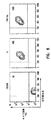
脾細胞を免疫調節機能を有する組み換えタンパク質の1つ(コアストレプトアビジンと融合したFasLの細胞外部分(SA−mFasL))の発現について試験した。脾細胞を上記のようにビオチン化し、20分間100ngのSA−mFasLとともにインキュベートし、そして規定の期間にわたって37℃インキュベーター内で維持し、ここで細胞を抗FasL抗体で染色した。ほとんど全ての細胞が5日目にSA−mFasLを発現し、そして多数の(有意な)細胞(>25%)が、分析した最終時点である10日目にSA−mFasLを発現した(図4)。
【0058】
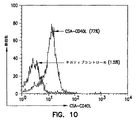
SA−mFasLのような免疫調節分子を使用するための提案されたタンパク質ベースの遺伝子治療の目的の1つは、外部環境におけるBM細胞の移植を容易にすることである。従って、BM細胞を、FasLのビオチン化およびタンパク質レベルの発現について試験した。Bm細胞を標準的なプロトコールを用いて、ラットの大腿骨および頸骨から収集した。単一細胞懸濁物を調製し、次いで上記条件下でビオチン化した。ビオチンを除くために徹底的に洗浄した後、BM細胞を氷上で20分間SA−mFasL(50ng/細胞100万個)を用いて処理した。次いで細胞を徹底的に洗浄して遊離のSA−mFasLを除き、そしてフローサイトメトリー中で、FasLに対する抗体(PE−MFL4)を用いて染色した。図5に示すように、100%のBM細胞がSA−mFasLを発現した。
【0059】
(実施例4.インビボにおける発現の時間反応速度論)
インビボで長期間にわたって細胞膜に組み込みんで存続する親油性色素(CFSE)で脾細胞を標識し、ビオチンまたはSA−mFasLを発現するように改変し、そして同系の動物に静脈内注射した。脾細胞を注射後異なる時点で回収し、そしてフローサイトメトリーでの分析のためにAPC−ストレプトアビジンまたは抗FasL抗体で染色した。図3に示すように、インビボ注射後、試験した最長時点である10日間、40%のCFSE陽性細胞の表面上にビオチンは検出された。同様に、SA−mFasLを発現するように改変されたCSFE陽性細胞は、注射の5日後、この分子の発現について陽性(26%)であった。まとめて考慮すると、これらのデータは、このアプローチがインビボにおける長期のタンパク質の発現について目的の機能を果たすことを可能にすることを明確に実証する。
【0060】
(実施例5.免疫調節分子を発現する標的細胞を用いる、インビトロにおけるアロ反応性免疫応答のブロック)
のアプローチを用いて目的の免疫調節性タンパク質を発現するように改変された細胞を用いて、アロ反応性応答を防ぐことが可能か否かを試験するため、アロ同種異型の脾細胞を、SA−mFasLを発現するように改変し、そしてインビトロ増殖アッセイにおける標的として用いた。要するに、F344ラット由来の脾細胞をビオチン化し、培養上清またはSA−mFasLで処理した。非機能的構築物でトランスフェクトした細胞由来の上清をコントロールとして使用し、そして本出願全体にわたって、S2上清と呼ぶ。次いでこれらの細胞を照射し、そして標準的な5日混合リンパ球培養物におけるアロ反応性応答に対する刺激因子として用いた。PVG.IUラットのリンパ節から収集したリンパ球をレスポンダーとして用いた。SA−mFasLを発現する標的は、SA−mFasLなしの培養物に比べて、リンパ球の増殖反応を完全にブロックした。これらのデータは、ビオチン化脾細胞が、抗原提示細胞として働くこと、および脾細胞の表面上で発現されたSA−mFasLが、インビトロでアロ反応性の免疫応答をブロックすることを実証する。さらに、これらのデータは、ビオチン化が細胞の抗原提示機能を妨害しないことを実証する。なぜならビオチンで装飾されS2コントロール上清で処理された細胞はリンパ球を刺激する能力を妨害しないからである。従って、これらのデータは、細胞の表面上のタンパク質の発現が細胞の機能も発現された分子の機能も妨害しないことを実証する。これは、免疫調節の手段としてタンパク質ベースの発現を確証するのにおいて重要な工程である。
【0061】
(実施例6.SA−mFasLタンパク質の毒性)
一般に、Fasを発現するほとんどの細胞(例えば、活性化リンパ球、または高速分裂する細胞、例えば肝細胞、または所望されない細胞、例えば、腫瘍細胞)が活性化される。しかし、SA−mFasLが毒性であるか否かを確認する必要がある。Fasに対する抗体は、静脈内注射または複腔内注射された場合、選択されたマウス株において劇症肝障害を誘導することが示されている。これは、肝細胞に対するFasの発現に起因すると考えられる。本発明のキメラタンパク質が同様に毒性であるか否かを確認するために、2×10⁷のビオチン−SA−mFasL保有脾細胞、または8×10⁷のビオチン−SA−mFasL保有骨髄細胞を、ラットに複腔内注射または静脈内注射した。この動物を10日間入念にモニターし、次いで全体的な病理的分析および解剖的分析のために屠殺した。表2に示されるように、細胞−ビオチン−SA−mFasLを注射した動物には、顕著な病理は認められなかった。
【0062】
【表2】
このアプローチの主な前提は、DNAベースの遺伝子治療に対する代替としてタンパク質ベースの発現を用いることである。免疫回避のためにBM細胞を改変するこのアプローチによるBM細胞拒絶の防止は、この条件を満たす。骨髄細胞をPVG.R8またはACIラットから回収して、ビオチンまたはSA−mFasLを発現するように改変し、そして致死的に(950cGy)照射したラットに対して0.7〜1×10⁸細胞/動物で投与した。細胞を投与していない照射動物をコントロールとして用いた。細胞を投与していない動物全てが8〜9日以内に死んだ(n=6)が、bio(ビオチン)またはSA−mFasLを発現するように操作したBM細胞を投与された全ての動物(n=12)は、100%キメラであり(図6)、そして期限なく(>100日)生存した。これらのデータは、BM細胞が、その長期移植能力に影響することなく、治療目的の目的のタンパク質を発現するように、この新規なアプローチによって安全に操作されていることを明確に実証する。
【0063】
(実施例8.SA−mFasL発現BM細胞は、インビボでアロ反応性応答をブロックする)
SA−mFasLのような免疫刺激性分子を発現するBM細胞を用いて、インビボにおいて標的細胞として異寛容性(アロ寛容性)(allotolerance)を誘導した。PVG.1UまたはWFラットに、FasLを発現するそれぞれ、0.7−2×10⁸のPVG.R8もしくはACI BM細胞、またはコントロールS2上清で処理したビオチン化細胞をi.v.で投与した。腸間膜リンパ節を注射の60日後に収集し、そして、標準的混合リンパ節アッセイにおけるPVG.R8またはACI細胞に対するレスポンダーとして用いた。ドナー抗原に対する応答はまったく存在しなかった。このインビボ免疫非応答性は、第三部分(party)抗原に対する応答がインタクトであったのでドナー特異的であった。この効果はSA−mFasL特異的であった。なぜなら、コントロールS2上清で処理したPVG.R8細胞を投与された動物が、ナイーブな動物の応答に比べて、ドナーおよび第三部分抗原に対して正常な応答を生じたからである(図7)。これらのデータは、インビトロのブロック反応を確証し、そしてインビボにおけるこのアプローチの免疫調節性効果を直接証明する。
【0064】
(実施例9.血管内皮上でCSAタンパク質を発現するための心臓の改変)
本発明の方法は、器官上でタンパク質ベースの発現をもたらすように適用されてきた。目的のタンパク質をエキソビボで発現するための、宿主でなく、器官の改変は、選択された設定における所望の治療アプローチを示す。エキソビボ操作は、宿主が処置された場合に生じ得る合併症を回避する。この合併症は、所望されない副作用を含み得るがこれに限定されない。従って、37℃でACIラットおよびB10.BRマウスにおいて、ビオチン、ストレプトアビジン、およびSA−mFasLを発現する試験系として心臓を用いた。要するに、切り出した心臓の大動脈を、改変Krebs−Henseleit溶液(KH)を用いて、96mmHgの圧力でLangendorff逆向性灌流システム(retrograde perfusion system)においてカニューレ挿入して、還流した。心臓収縮力を、左心室に挿入したラテックスバルーンでモニターした。ラット心臓についての灌流プロトコールは以下であった:心機能の安定化をさせるためにKHで20分;5μg/ml EZ−Link Sulfo−NHS−Biotin(Pierce)を含有するKHで20分;ビオチン洗い出し(ウオッシュアウト)のためにKHで10分;ストレプトアビジン−FITC(Zymed)またはSA−mFasLのいずれかを0.5μg/ml含有するKHで20分;ストレプトアビジン洗い出しのためにKHで10分。コントロール心臓をKH溶液のみで還流した。左心圧は、灌流の80分の間に、3つの群(n=5/群)全てにおいて98±7〜91±6mmHg低下した。冠血流は、10.4±0.6ml/分(n=15)の基準値(ベースライン値)から有意に低下しなかった。これらのデータは、内皮のビオチン化およびSA−mFasLでの装飾が、心機能に短期間の有害効果を有さないこと、およびFasLは冠動脈に直接毒性ではないことを示す。
【0065】
次に、冠動脈のビオチン化された内皮の裏層が4℃でSA−mFasLで「装飾」され得るか否かを決定した。この温度は、移植の前に体外心臓保存のための臨床設定において広く用いられる。マウス(B10.BR)およびラット(ACI)の心臓をマグネシウム心保護溶液(KH+16mM MgSO4)で停止させ、そして上記のプロトコールに従って4℃で還流した(ビオチン、KH、ストレプトアビジン−FITCまたはSA−mFasL、KH)。血管内皮上のビオチンを、ストレプトアビジン−FITCによって検出し、ここでSA−mFasLを、一次抗体およびFITC標識した適切な二次抗体として、ストレプトアビジン(Zymed)およびFasL(MFL4)に対する抗体を用いて検出した。心臓を取り出し、そして改良ミクロトーム(Polycut S,Reichert−Jung)のステージ上で−80℃で凍結させた。連続的凍結切片(約200ミクロン)を行い、そしてFITC−標識ストレプトアビジンまたは抗体の存在をAxiopan顕微鏡(C.Zeiss)を用いて決定した。この顕微鏡のステージをミクトロームの冷却ブロックにあうように改変した。10×の倍率で心臓切片の画像を得た。蛍光を心臓の脈管構造において検出した。これは、臨床手技において広範に用いられた保存培地中において、低体温条件下における脈管内皮へのビオチンおよびストレプトアビジンの結合の可能性を実証する。SA−mFasLとともに還流した移植内皮におけるSA−mFasLの存在(ただし、コントロールS2上清との灌流ではない)を、FITC−MFL4mAbを用いて確証した。これらのデータは、SA−mFasLが、体外器官保存の条件においてビオチン化を介して冠動脈に導入され得ることを示す。上記の移植の操作は、標準的な保存溶液を用いて、4℃で10〜60分内に実施する。従って、このアプローチは、臨床において用いられる温度、期間、および条件の要件を満たす。
【0066】
(実施例10.同系宿主へ移入した場合、ビオチンまたはSA−mFasLを発現するように改変された心臓は、期限なく生存した)
器官のこのタンパク質ベースの改変が機能に影響するか否かという疑問が生じる。ビオチンまたはSA−mFasLを発現するように改変された心臓を、同系のレシピエントへ異所性に移植した。移植された心臓の毎日の腹部触診によって移植片の生存を評価した。表3に示すように、SA−mFasLで装飾された同系の心臓(n=4;>56日)またはKH灌流コントロール同系心臓(n=4;>74日)を移植された移植レシピエントの生存の間に検出可能な差異は存在しなかった。
【0067】
【表3】
膵臓小島移植は、糖尿病の補正のために広く意図される。本発明の方法が、小島の異種移植の拒絶の防止に有効であり得るか否かを試験するため、ストレプトジン(260mg/kg)の複腔内注射によってBALB/cレシピエントにおいて、糖尿病を誘導した。少なくとも3連続日の間、血糖値が>450mg/dlを示す動物を、非主要組織適合抗原および主要組織適合抗原が異なるC57BL/10小島のレシピエントとして用いた。小島をC57BL/10マウスから収集し、そして標準的プロトコールに従ってFicoll勾配で単離した。脾細胞をC57BL/10(ビオチン化し(15μM)、FasLまたはコントロール上清で装飾し、そして照射した(2000rad))から収集した。100万の脾細胞を、糖尿病Balb/cマウスの腎臓嚢の下で約400の小島細胞とともに同時移植した。移植の後の3日後に開始して血糖値をモニタリングすることによって、小島の生存および機能を評価した。FasL装飾された脾細胞は、9日間をこえて、全ての異種遺伝子小島の生存を延長した。9日でコントロールマウスの血糖値は、ほぼ移植前のレベルまで上昇した。図8は、FasLおよびストレプトアビジンを発現する膵臓細胞を示す。
【0068】
この技術のさらなる実施例において、コラゲナーゼでの消化によってマウス膵臓小島を収集し、そしてFicoll勾配を用いて精製した。小島を一晩培養し、次いでビオチン(5μM)で改変し、そしてSA−mFasL(約100mmg/ml)またはコントロールとしてS2上清で装飾した。共焦点顕微鏡による小島の検査によって、高レベルのビオチンおよびSA−mFasLが示された。SA−mFasLで装飾した小島は、視覚検査およびトリパンブルー排除によって決定されるように、10日以上にわたって培養中で生存していた。
【0069】
まとめて考慮すれば、これらのデータは、心臓のような細胞、組織、および複雑な器官(例えば、心臓)が、検出可能な毒性なしに、長期間にわたってストレプトアビジンとのキメラタンパク質を示すように、ビオチンで改変され得ることを明確に実証する。
【0070】
(実施例12.GVHDの防止)
骨髄移植(BMT)は、種々の遺伝的に遺伝した障害および後天的な血液学的障害を処置する能力を有し、自己免疫抗原および外来移植に対する寛容を誘導する。従って、BMTは遺伝子治療を実施する自然な方法として認められている。しかし、重大な問題は、宿主対移植片病(GVHD)であり、これはほとんどの場合致死的である。T細胞は、GVHDを起こす最も重要な細胞であるとみなされている。GVHDを起こすBM接種における、特定のT細胞の除外を可能にする方法は、広範な臨床適用を有する。従来の遺伝子治療を用いて、T細胞へ「自殺」遺伝子を導入した。一旦GVHDが生じれば、自殺機構が活性化してこれらの細胞を排除する。しかし、従来の遺伝子治療の障害は、臨床におけるこのアプローチの慣用的な使用に対する重要な問題である。この「自殺」アプローチを達成するために、より「非侵襲的」なアプローチが必要である。
【0071】
本発明のこの方法は、「自殺」アプローチまたは免疫下方制御(ダウンレギュレーション)のために十分適切である。骨髄細胞(成熟T細胞を含む)におけるアポトーシス性分子(死亡リガンド)または下方制御分子(抗炎症性サイトカイン;CSA−IL−4、IL−10、TGF−β)の提示は、宿主抗原および活性化のこれらの細胞の認識の際、これらの細胞を物理的にまたは機能的に排除することが予期される。概念の証明として、SA−mFasLを提示するように改変された脾細胞におけるT細胞は、T細胞分裂促進因子(マイトジェン)で刺激された場合、増殖応答を生じないことが示された(図10)。対照的に、改変されていない脾細胞は、活発な増殖応答を生成した。このことは、分裂促進因子に応答するT細胞が、Fasを発現し、そして同じ細胞上でのFasLとFasとの相互作用の際に死んだ(自殺)ことを実証した。これは、骨髄細胞および成熟リンパ球を投与されたラットがGVHDを17〜20日内に発症したことをさらに実証した。FasLを発現する同系脾細胞によるこれらの動物の処置によって、GVHDの防止が得られた。これは、このアプローチが臨床において用いられている他の処置レジメンより優れているという強力なインビボデータである。
【0072】
(実施例13.免疫調節分子を用いる免疫下方制御)
細胞、組織、および器官上で外因性タンパク質を発現する本発明の方法は、これらの分子のいくつかが同時に発現される場合、治療目的のために免疫系を下方制御することにおいて特に有効である。従って、死亡リガンド(SA−mFasL、−TNFα、−TRAIL、など)および抗炎症性分子(CSA−IL−4、−IL−10、−TGF−β、など)の組み合わせを、自己免疫疾患、外来移植片拒絶、および他の免疫に基づく障害を予防/処置するための治療アプローチとして、細胞、組織および器官の表面上で発現する。この方法は、免疫系の下方制御を示す。
【0073】
(実施例14.腫瘍に対するワクチン接種)
腫瘍は、T細胞応答を刺激するシグナルを下方制御することによって免疫系を逃れ得る。有効なT細胞応答は、以下の3つの異なるシグナルを必要とする:シグナル1、シグナル2およびシグナル3。シグナル1は、抗原提示細胞(APC)上の主要組織適合複合体(MHC)抗ペプチドとのT細胞レセプター相互作用によって生成される。シグナル2は、T細胞およびAPC上の同時刺激分子(例えば、B7/CD28およびCD40/CD40L)の結合によって媒介される。シグナル3は、シグナル1およびシグナル2の両方を受けたT細胞およびPACによって合成されたサイトカインによって誘導される。これらの3つのシグナルの誘導が、T細胞およびAPCを駆動して、有意義な免疫応答の生成のためのエフェクターへと増殖および分化させる。腫瘍へのT細胞応答の間のこれらのシグナルのいずれかの欠失は、腫瘍が免疫系を回避する最も有効な機構の1つとして機能し得る。本発明は、外因性タンパク質の発現のために細胞表面を改変するための方法および組成物を提供する。このアプローチは、任意の細胞を、有効な免疫応答の生成のための専門的抗原提示細胞へ変換することが目的である。
【0074】
CSAおよびCD40Lの細胞外部分(構造的可撓性のために操作された合成残基を有する)からなるキメラタンパク質(CSA−CD40L)を、Drosophila(ショウジョウバエ)の産生細胞において産生した。次いでこの分子を脾細胞の表面上の発現のために用いた。フローサイトメトリーにおいてCD40Lに対する抗体によって示されるように、ほとんど全ての脾細胞が、CSA−CD40Lに関して陽性であった(図11)。CD40Lを発現する脾細胞は、APCとして用いた場合、抗原特異的応答を誘導する通常の脾細胞よりもかなり有効であった。これは、治療目的のために細胞表面上で外因性タンパク質を発現する本発明者らのアプローチの実行可能性を明確に実証する。
【0075】
この概念を、肺癌動物モデルで試験する。マウス起源の2つの肺癌株であるLewisおよびLine1ならびにC58腫瘍細胞(ラットリンパ腫)は、腫瘍に対するワクチン接種の目的に用いられる。
【0076】
B7.1(配列番号2)、CD40L(配列番号6)、またはIL2(配列番号4)の核酸に融合したCSAの核酸を含む核酸から生成されたキメラタンパク質を、これらの腫瘍細胞の表面上での提示のために用い、そしてこの細胞を、腫瘍に対するマウスおよびラットのワクチン接種のために用いる。要するに、腫瘍細胞(2×10⁷)は、上記のようにビオチン化される。PBSでの数回の洗浄後、ビオチン化細胞を上記のようなキメラタンパク質とともにインキュベートしこのキメラタンパク質をビオチン化細胞に結合させる。キメラ装飾した細胞を、5〜10,000cGyで照射し、そして腫瘍を保有する動物に注射してワクチンとして使用する。腫瘍の緩解をモニタリングする。上記の実施例に示した結果に基づいて、この方法は、免疫系の上方制御を示すことが予期される。
【0077】
他の腫瘍システムおよびキメラタンパク質が同定され得、そして本発明の方法が、他の腫瘍ワクチンおよび感染に対するワクチンを提供するために適用され得ることが容易に理解され得る。
【0078】
本発明は、種々の好ましい実施例において記載されている。当業者は、本明細書において開示される化合物および方法の改変、偏向物、または置換物が、本明細書の趣旨および範囲から逸脱することなくなされ得ることを容易に認識する。このような改変、偏向物、および置換物の全てが、本発明の特許請求の範囲内であるとみなされる。
【図面の簡単な説明】
【図1】
図1は、新規機能について内因性タンパク質の発現について細胞膜を改変するための方法の概略図である。
【図2】
図2は、示したクローンを含む発現ベクターの制限フラグメント分析を示す。
【図3】
図3は、インビボにおけるビオチン発現を示す。
【図4】
図4は、脾細胞の表面上でのSA−mFasLのインビトロ発現を示す。
【図5】
図5は、骨髄細胞の表面上でのSA−mFasL発現を示す。
【図6】
図6は、致死的に照射された動物におけるドナーキメラ現象を示す。
【図7】
図7は、インビボでのアロ反応性応答におけるSA−mFasLブロッキングで装飾した同種異系細胞のグラフでの要約を示す。
【図8】
図8は、コントロール小島(A)と比較した、FasL(赤)およびストレプトアビジン(B)を発現する膵臓小島細胞を示す。
【図9】
図9は、ConAによって活性化された、SA−mFasLを発現するように改変されたT細胞を示す。
【図10】
図10は、脾細胞上でのSA−CD40Lの発現を示す。[0001]
(Field of the Invention)
The present invention relates to the continuous regulation of cell membranes to alter cell function. The compositions and methods of the present invention achieve effects similar to those of gene therapy without the introduction of exogenous DNA. A useful alteration of cell function is the induction of apoptosis.
[0002]
(Background of the Invention)
Inducing cells to behave in a predictable manner and altering the behavior of cells in a manner that is beneficial to the subject has long been an experimental biological and medical objective. For example, if undesired cells can be induced to alter their behavior to undergo apoptosis while normal cells retain normal function, then patients with diseases caused by undesired cell proliferation will , Get free from disease. Similarly, if tissue-rejecting cells can be removed or their behavior altered, transplantation with foreign tissue to the subject can be successful.
[0003]
Gene therapy has been proposed for selected diseases to correct or alter pathological or physiological processes. In commonly referred to gene therapy, specific DNA is introduced into tissues and organs, where the DNA produces various proteins that ameliorate or alleviate the condition. This is an unpredictable treatment, depending on potentially dangerous expression vectors and uncertain delivery efficiencies, which are often low. In addition, gene therapy is believed to have dangerous side effects, such as sustained expression in desired cells or tissues after a desired period of treatment, or introduction of genetic alterations in unwanted tissues or cells. Due to such adverse effects, a great deal of caution is recommended before gene therapy is performed, as found in human clinical trials.
[0004]
DNA-based gene therapy has been used in the last decades due to the enormous potential that gene therapy offers for the treatment of genetic diseases and other pathological conditions where expression of selected proteins may provide treatment. Meanwhile, it has been the subject of intense research. For example, gene therapy either enhances the ability of the immune response to treat infections or tumors, or down regulates the immune response to prevent autoimmunity or rejection of foreign grafts. Can be used for immunomodulation. Despite the great efforts in the last two decades, the safe and effective application of gene therapy for the treatment of disease has been very limited. Among the obvious disadvantages of gene therapy are the possibility of causing permanent changes in the host DNA complement; uncertain tissue specificity; expression of the encoded protein beyond the intended duration of the therapy; and high costs. Exists. Thus, this is very beneficial if cells and tissues can be modified to express the protein of interest in a short period of time without the introduction of foreign DNA.
[0005]
Although gene therapy generally focuses on the problem of delivering nucleic acids to cells, a great deal of basic knowledge about the important role of cell surface molecules has led to the immune response, which is easily accessible and (With well-understood functions) have been obtained through studies of signal transduction in cells. The immune response is regulated through the interaction of several different cell types in response to the presence of foreign antigens. An adaptive immune response is important for vertebrate survival in an environment full of pathogenic microorganisms. An individual lacking a fully functional immune response due to an innate genetic defect, exposure to a chemotherapeutic agent, or infection by a virus such as the human immunodeficiency virus (HIV) is an individual with a healthy immune response Are susceptible to infection that readily resists. However, the immune system does not always function in a manner that is beneficial to the organism. Its dysregulation leads to autoimmunity and tumors. The immune system also functions as a barrier to transplantation of foreign grafts (eg, cells taken from individuals other than the transplant recipient). Transplantation allows for failed cell, tissue, or organ replacement in other end-stage diseases, while bone marrow transplantation can treat hematopoietic disorders, malignancies, autoimmune disorders, and other diseases. Successful transplantation requires either suppression of adaptive immunity or "educating" the recipient's immune system to accept these foreign antigens natively.
[0006]
An immune response to a foreign antigen is initiated by native T cells that use a clonal expressed T cell receptor (TCR) that recognizes the antigen, such as a peptide presented by a self major histocompatibility complex (MHC) molecule. It is believed that this recognition response, when accompanied by a costimulatory signal provided by an antigen presenting cell (APC), results in sufficient T cell activation. Productive T cell responses are now understood to require three separate signals.
[0007]
Upon activation, T cells proliferate and differentiate into effector cells that evade the immunological mechanisms responsible for clearance of antigen from the system. Most of the activated T cells then undergo apoptosis-mediated "activation-induced cell death" (AICD), followed by a period of death in which effector activity subsides. Apoptosis is a complex process that involves a series of extracellular and intracellular signals that focus on the activation of an enzyme called caspase that causes cells to undergo apoptosis.
[0008]
Transplantation of foreign cells (eg, bone marrow and stem cells), tissues (eg, pancreatic islets), and organs (eg, kidney, heart, liver) is important and efficient for patients with selected terminal illnesses It has become a cure. However, transplantation of a foreign graft between genetically different patients (allograft between members of the same species or xenograft between members of a different species) requires immunological recognition and rejection of the transplant by the recipient. Limited by the ability to control
[0009]
Bone marrow (BM) transplantation is regarded as an exceptionally promising treatment for hematopoietic and autoimmune disorders and certain cancers. One obstacle to bone marrow transplantation is the potential rejection of the transplanted tissue mediated by host T and NK cells. Graft versus host disease (GvHD) is another possible deleterious consequence of bone marrow transplantation. Donor T cells in the transplant can boost the immune response to key organs of the host, often leading to host death. Thus, graft-versus-host reactions and GvHD limit the clinical use of bone marrow transplants, which are otherwise widely used to treat various diseases and to prevent foreign graft rejection Can be done.
[0010]
Pharmacological factors that cause immunosuppression are currently the major regimens for controlling allograft rejection. Although such drugs are effective in reducing the severity of rejection episodes, they are non-specific and cannot create a state of persistent graft-specific resistance. Thus, continuous exposure of recipients to these immunosuppressants is associated with a significantly increased risk of opportunistic infections and malignancies. There remains a need to develop more selective and more persistent methods for preventing BM rejection.
[0011]
In addition, these non-specific immunosuppressants can induce severe and undesirable side effects in the host. These detrimental effects are often adaptive in that the body identifies certain parts of the body itself as "foreign" and produces autoimmunity as observed in type I diabetes, arthritis, lupus, and multiple sclerosis Outweigh the benefits of patients with the disease to initiate an immune attack. It would be highly desirable to be able to "educate" an immune system that is resistant to "foreign" self-antigens.
[0012]
It would also be highly desirable to be able to "educate" the immune system in removing tumor cells from an organism. T cell-mediated cellular immunity is the most important acquired response to tumors. A series of experimental studies have provided evidence that tumors evade T-cell mediated immunity by several different mechanisms. These mechanisms include: i) lack of
[0013]
Thus, there remains a need to develop faster, selective, and sustained methods for regulating cell function without the introduction of nucleic acids into cells for therapeutic purposes. There also remains a need to develop means to accomplish the goals of gene therapy without the numerous risks associated with introducing exogenous nucleic acids into an organism. Since many cell functions are controlled through the transmission of signals at the cell surface, generally applicable methods of attaching drugs to surfaces are useful. Among the uses of such methods may be: modulation of cell function without introduction of nucleic acids into cells; achievement of the purpose of gene therapy by alternative and potentially favorable means; and autoimmunity Manipulation of an organism's immune response to reduce the response with respect to treating or preventing graft-versus-host disease, or to increase the response with respect to treating a tumor or infection.
[0014]
(Summary of the Invention)
The present invention provides a method of modifying a surface, comprising: (1) contacting a surface with one member of a binding pair that binds to the surface to form a decorated surface; (2) contacting the decorated surface with a composition comprising a second member of a binding pair operably linked to the surface or an agent capable of modifying its function. The binding pair can be avidin / biotin, streptavidin / biotin or antigen / antibody. "Avidin" means avidin and any fragments or derivatives of avidin that retain strong binding to biotin. "Streptavidin" means streptavidin and any fragments or derivatives of streptavidin that retain strong binding to biotin. Conversely, "biotin" refers to any fragment or derivative of biotin that retains strong binding to avidin and streptavidin.
[0015]
If the surface is a cell surface, the agent is any compound that induces apoptosis; any agent that down regulates the immune system; any agent that up regulates the immune system; any adhesion molecule for cell tracking Any growth factor; or an antibody conjugated to a toxin, such as ricin or a plant toxin. If the surface is a cell surface, the contact of
[0016]
If the agent is selected to induce apoptosis, the agent can be a death-inducing molecule such as FasL, mFasL, TRAIL, TNF-a, TWEAK, and the like. If the agent is selected to down regulate the immune system, the agent can be an anti-inflammatory cytokine such as IL-4, IL-10, TGF-β, and the like. If the drug is selected to up-regulate the immune system, the drug may be a costimulatory molecule such as B7, CD40L or the like, or of IL-2, IL-12, IL-15, lymphhotactin and chemokines. Such pro-inflammatory cytokines.
[0017]
In a preferred embodiment, the first member of the binding pair is biotin, the second member of the binding pair is a chimeric protein comprising streptavidin and a death inducing molecule, and the surface is a target cell, tissue, organ or Including tumor.
[0018]
In a more preferred embodiment, the first member of the binding pair is Sulfo-NHS-LC-biotin, the second member of the binding pair is SA-mFasL, and the surface is a cell displaying a Fas receptor. including. In constructing the second member of the binding pair, streptavidin is fused to the extracellular portion of FasL via recombinant gene technology, and the chimeric protein SA-mFasL is produced.
[0019]
This construct is used in a method of modulating the immune system, wherein the subject to be treated is first administered biotin to form decorated cells, followed by administration of SA-mFasL, followed by chimeric To form a decorated cell. Only cells that are biotinylated and express Fas in tissues, organs or tumors undergo apoptosis. Generally, these cells include cells that cause immune-mediated injury.
[0020]
In another aspect of the invention, a subject who would benefit from down-regulation of the immune system is first administered biotin to form decorated cells, and then the construct and down-regulation molecules comprising avidin or streptavidin are administered. Is administered. Subjects that benefit from down-regulation of the immune system include subjects with autoimmunity, graft-versus-host disease, allergies, septic shock, and vascular disease. Subjects that receive a tissue or organ graft also benefit from down-regulation of the immune system.
[0021]
In another aspect of the invention, a subject benefiting from up-regulation of the immune system is first administered biotin to form decorated cells, and then administered constructs and up-regulation molecules comprising avidin or streptavidin. Is done. Subjects that would benefit from up-regulation of the immune system include patients with neoplasia or infectious diseases.
[0022]
In another aspect of the invention, systemic effects are avoided by biotinylation of the cells ex vivo by isolation of a target that can be treated locally or selection of a target that can be treated locally, followed by subsequent secondary Re-introduction of the decorated cells into the patient using contact of the cell.
[0023]
In another aspect of the invention, the members of the binding pair form a covalent bond with each other. In a preferred aspect, the first member of the binding pair comprises a cysteine residue and the second member of the binding pair comprises a cysteine residue. The two members are first placed together under conditions that dislike forming a disulfide bond between the first member of the binding pair and the second member of the binding pair. In a subsequent step, the two members are placed under conditions that favor the formation of a disulfide bond between the first member of the binding pair and the second member of the binding pair.
[0024]
In another aspect of the invention, the first member of the binding pair is contacted with a cell surface, a virus, a surface of a glass particle, a surface of a polysaccharide particle or a surface of a plastic particle to form a decorated surface. The decorated surface is then contacted with the second member of the binding pair to form a second decorated surface. The cell, virus or particle comprising the second decorated surface is contacted with cells or cellular components of the organism, either in vitro or in vivo, for therapy, diagnosis, cell sorting or cell identification.
[0025]
A recombinant nucleic acid comprising the extracellular portion of FasL fused to streptavidin (SA-mFasL) is provided. A gene product is then provided that has been attached to the cell surface previously biotinylated in vivo. Methods are taught for use of the gene product for sustained removal of Fas-expressing cells.
[0026]
The methods of the present invention can be modified to cause persistent binding of any protein to the targeted cells and to mimic the results of DNA gene therapy without altering the cellular genetic material. Persistent binding of the protein to the target cells can be considered as gene therapy at the protein level.
[0027]
(Detailed description of the invention)
Due to the significant problems associated with gene therapy (ie, the introduction and integration of foreign DNA into the genome) as commonly used, the consequences of gene therapy, ie, cell function without restriction of DNA transfer, Methods to achieve long-lasting modifications have been sought. A question has arisen as to whether the benefits of gene therapy can be obtained by delivery of exogenous proteins over delivery of nucleic acids to cells. Methods that safely allow modification of cell membranes to achieve transient or long-lasting presentation of therapeutic molecules may have widespread clinical applications. As disclosed herein, Applicants have discovered a method that has the added advantage of rapid cell surface expression and disclose herein.
[0028]
Due to the thermodynamically desired association of avidin or streptavidin with biotin, the complex formed may compete with or during this period of effect induced by gene therapy requiring the introduction of nucleic acids. For a period that surpasses the characteristics of the cells may be altered. The present invention teaches a method for attaching a drug to a surface. Such an agent can be any series of immunomodulatory molecules, such as death ligands, costimulatory molecules and cytokines, for illustration and not limitation. It is useful to attach such agents to surfaces, especially cell surfaces. Because such agents may physically or functionally reduce autoreactive, alloreactive and inflammatory cells to avoid allograft and xenograft rejection, graft versus host disease. Because it can be used to eliminate clinically and to treat autoimmunity, septic shock, inflammation, allergies, infections, neoplasia, and vascular disease. In addition, the method can be used to display proteins with defined functions on the surface of cells for in vivo transport with respect to homing of selected hematopoietic cells for therapeutic purposes.
[0029]
(Definition)
For the purposes of this application, the following terms have these definitions:
By "agent" is meant a composition that elicits an important biological response or composition that is useful in the diagnosis, prevention or treatment of a disease, or in the classification or identification of cells, cellular components or viruses.
[0030]
A "binding pair" is defined as two compositions that readily bind to each other; two compositions that have an affinity for each other; or two compositions whose association is thermodynamically and kinetically favorable. means.
[0031]
"Cell surface" has its ordinary meaning in the art and includes phospholipid bilayers of cell membranes and molecules that are directly or indirectly associated with the bilayer.
[0032]
By "decorated surface (decorated surface)" is meant the surface to which the members of the binding pair bind.
[0033]
"Expression" means the arrangement of a functional protein on or within a cell in addition to its ordinary meaning in the art.
[0034]
"Plastic" means polystyrene, polypropylene, polyethylene or any other polymer that can be formed on a solid surface.
[0035]
"Protein" means a protein or polypeptide that is native, non-native, synthetic or covalently modified.
[0036]
By "surface" is meant a cell surface, a virus surface, a particle surface (eg, a glass particle surface, a polysaccharide particle surface, a plastic particle surface), a phospholipid bilayer surface, or a solid matrix surface. .
[0037]
By "target cell" is meant a cell that is targeted for apoptosis, ie, a cell for which it is desired to induce apoptosis.
[0038]
An important application of modulation of the immune response is the induction of alloantigens, heterologous antigens, and tolerance to self-antigens to avoid foreign graft rejection and to treat or avoid autoimmunity and GVHD. T cells are the most important cells of adaptive immunity, which requires three different signals (signals 1, 2, and 3) for the response to be generated.
[0039]
Apoptosis or "programmed cell death" plays a central role in both the development and homeostasis of multicellular organisms. Apoptosis converges to a final effector mechanism consisting of multiple interactions between several "death receptors" and their ligands, which belong to the tumor necrosis factor (TNF) receptor / ligand superfamily. Can be induced by multiple independent signaling pathways. The most characterized death receptors are CD95 ("Fas"), TNFR1 (p55), death receptor 3 (DR3 or Apo3 / TRAMO), DR4 and DR5 (apo2-TRAIL-R2). The final effector mechanism is mediated by caspase family proteins.
[0040]
Co-pending International Application PCT. In US 01/02256 (the teachings of which are incorporated by reference), Fas / FasL-induced apoptosis may occur in immunologically favored sites such as the central nervous system, testis and eyes from immune antigens. Disclose that it plays a central role in protecting. For example, allogeneic and heterologous tissues transplanted to these sites are resistant to rejection. Of great importance is that a key role, apoptosis, plays a role in maintaining immune system homeostasis through activation-induced cell death (AICD). AICD is mediated primarily by apoptotic signals transmitted by Fas / FasL interactions. Fas is a 45 kDa, type I cell surface protein consisting of a cysteine-rich extracellular domain and transmits cell death. Fas mediates effector functions by interacting with FasL, a type II membrane protein of approximately 40 kDa. T cells are the major cell type in the body and express FasL upon activation. Expression of this molecule creates activated T cells that are susceptible to apoptosis, which is induced in an autocrine fashion, such that Fas engages FasL on the same cell. Fas on activated T cells can also interact in a paracrine fashion with FasL on another activated cell. These two pathways of apoptotic T cell death mediated by the Fas / FasL system serve as homeostasis mechanisms to regulate the size of antigen-stimulated T cell clones. Deregulation of this system causes immunological disorders.
[0041]
The molecular mechanism of Fas / FasL-mediated apoptosis has been studied in great detail. Binding of the three FasL molecules to Fas induces trimerization of the Fas receptor via the c-terminal death domain (dds), which in turn leads to the adapter protein FADD (Fas binding protein with death domain). ) And caspase-8. Oligomerization of this trimolecular complex (Fas / FADD / Caspase-8) then initiates the apoptotic process by activating other downstream caspases (including caspase-3) via proteolysis. Proteolytic cleavage of the protein enzyme caspase-8 to active caspase-8 is caused.
[0042]
Apoptosis of activated T cells causes tolerance to allogeneic and xenografts, including bone marrow transplants and other organ transplants and cell transplants. Purging activated T cells also alleviates the symptoms of allergy and other immune-induced diseases. Included in the latter are autoimmune disorders such as multiple sclerosis, lupus erythematosus, sarcoidosis and rheumatoid arthritis. Many disorders, including some tumors, rely on lymphocyte function leading to the persistence of the disorder. Many hematopoietic disorders can be treated using bone marrow stem cell transplantation if the immune response can be modulated to induce tolerance to foreign stem cells. Among these disorders are leukemia, lymphoma, aplastic anemia, sickle cell and Cooley anemia. All of these disorders can be permanently or temporarily controlled by apoptosis of activated immune cells, including T cells.
[0043]
Provided is a strategy for immunomodulation using rapid cell surface presentation of the protein, which involves the construction of a chimeric cDNA encoding a functional portion of an immunomodulatory protein using one member of a binding pair. Table 1 is a summary of such constructs. The choice of construct may be based, for example, on factors such as: the nature of the foreign antigen that stimulates adaptive immunity; whether the mitigation required is temporary or permanent; a commitment to death is desired Whether or not further down-regulation of apoptosis is preferred. It is understood that the constructs listed below are only representative of the present invention and are not limiting. For example, these constructs were made using core streptavidin (CSA). Streptavidin fragments and derivatives, as well as the entire streptavidin molecule, can be as readily used to generate equivalent constructs. Thus, in this specification, SA is generally used to mean whole streptavidin, a fragment of streptavidin or a derivatized streptavidin. One of skill in the art can readily generate, without undue experimentation, a construct comprising a first member of a binding pair and a second member of a binding pair linked to a known immunomodulatory molecule in accordance with the teachings of the present invention.
[0044]
[Table 1]
[0045]
The invention disclosed in co-pending international application PCT / US00 / 34554, the teachings of which are incorporated herein by reference, relates to apoptosis of lymphocytes and tumor cells. The present invention discloses an improved method for expressing an immunomodulatory molecule with a therapeutic agent of interest on a cell surface via coupling of a binding pair. Avidin / biotin or streptavidin / biotin are examples of binding pairs useful in the present invention. The modified FasL protein is described in detail as an example of such a composition. Bone marrow cells, endothelial cells, spleen cells, tissues such as pancreatic islets, and organs such as the heart are examples of cells susceptible to cell surface alterations. It has now been found that the method can be used to down-regulate or up-regulate an immune response to an antigen. Thus, this method can be used for prevention of foreign graft rejection, for prevention or treatment of autoimmunity, and for up-regulating the immune response to tumors and infections. Furthermore, it is also provided that proteins other than those having immunomodulatory functions can be bound to cells without causing systemic side effects.
[0046]
The invention will now be described by way of a specific example. The experimental procedures described herein are representative of compositions and methods for permanent binding "expression" of an agent to a cell, tissue or organ. One embodiment disclosed in detail is the chimeric composition SA-mFasL, which is a fusion of a DNA encoding the core streptavidin protein with a fusion of the DNA encoding FasL and in a suitable producer cell. Produced by expressing the chimeric composition (SEQ ID NO: 1).
[0047]
Target cells are first biotinylated ex vivo or in vivo, and SA-mFasL is then administered to cause apoptosis (eg, to induce donor-specific long-term survival and / or tolerance induction). Another example described in detail is the transplantation of pancreatic islet cells with immune cells treated according to the method of the invention, followed by perpetuation of pancreatic insulin production. Another example is the long-term survival of an ectopic heart transplant. Other examples are also provided. Following the teachings of the examples below, those skilled in the art can readily apply these to alter the membrane function of other cells.
[0048]
Alternatively, DNA encoding such a chimeric composition can be applied ex vivo or in vivo, leading to permanent alterations (eg, apoptosis of activated T cells).
[0049]
The following examples are disclosed to illustrate the present invention when applied to practice and do not limit the scope of the appended claims.
[0050]
Example 1 Cloning of Core Streptavidin for Production of Chimeric Protein
The present invention discloses a technique for cell surface modification to express an exogenous protein without introducing the nucleic acid into the cell, the technique comprising: i) the function of core streptavidin and the desired protein Generating a chimeric molecule consisting of a specific domain, ii) modifying the cell membrane with biotin, and iii) decorating biotinylated cells with the chimeric molecule (FIG. 1). To accomplish this, genomic DNA encoding streptavidin was converted to S. aureus using specific primers in PCR. avidinii. The 5 'and 3' primers were designed to incorporate sequences for selected restriction enzyme sites and amino acids to allow for three-dimensional flexibility, proper folding and function. This gene was cloned into a TA cloning vector, sequenced, and produced in a high yield insect expression system (DES). tm , Invitrogen) for expression in the pMT / Bip / V5-HisA vector.
[0051]
(Example 2. Construction of chimeric gene for expression in production cells)
Total RNA was prepared from human cell lines or peripheral blood cells, and 2 ng of this RNA was reverse transcribed to first strand DNA using oligo (dT) 18 as a primer for reverse transcriptase. One-tenth of this cDNA regulation was then used in eight separate PCR amplifications for human IL-2, IL-4, IL-10, TGF-β, FasL, TNF-α, B7.1 and CD40L. Amplification was performed using three sets of sense primers and antisense primers. The 5 'and 3' primers were designed to contain restriction sites for cloning and some amino acid residues to facilitate proper folding of the product. These primers amplified DNA bands of the expected size for all of these genes of interest. These PCR products were then cloned into a TA cloning vector (Invitrogen, San Diego, CA), and libraries prepared from this material were screened using the same oligonucleotide primers in PCR amplification. Positive clones were digested with the appropriate restriction enzymes to release fragments of the expected size for each gene shown in Table 1 (Figure 2). All clones shown were sequenced and found to have the expected characteristics. These clones were then fused to CSA in the pMT / Bip / V5-HisA vector as either an N-terminal protein or a C-terminal protein to promote correct three-dimensional structure and function (SEQ ID NO: Table 1). checking).
[0052]
The chimeric protein is transformed into a high yield insect expression system (DES) tm (Invitrogen) was subcloned in frame with the Drosophila BiP secretion signal into the pMT / Bip / V5-HisA vector. The recombinant vector was transiently transfected into Drosophila S2 cells using calcium phosphate. The cells were then pulsed with copper sulfate to activate the inducible metallothionein promoter driving the expression of the chimeric protein. Culture media was collected at various times after activation, dialyzed to remove copper sulfate, and analyzed for chimeric protein using ELISA and Western blot.
[0053]
Example 3 Cell Biotinylation, Recombinant Protein Expression, and Time Kinetics of In Vivo Expression
Therapeutic use of this protein-based approach requires successful biotinylation of cells, tissues or organs under physiological conditions and the binding of cell chimeric proteins consisting of CSA and molecules with therapeutic potential to these cells. (FIG. 1).
[0054]
Optimal conditions for biotinylation were first determined. Single cell suspensions were prepared from rat spleen or bone marrow. One million cells were incubated at room temperature for 30 minutes in saline at various concentrations (ranging from 1.5 to 150 μM) of EZ-Link biotin (Sulfo-NHS-LC-Biotin, Pierce, Rockford, I1). ). After extensive washing to remove free biotin, cells were either cultured or used to stain with fluorescein (APC) -labeled streptavidin to assess the level of biotinylation. 100% of the cells were positive for biotin at 5 μM biotin concentration (data not shown).
[0055]
Biotinylated splenocytes were converted to the T cell mitogen concanavalin A (ConA) to determine how long biotin persists on actively dividing cells and to determine the optimal dose of biotin (15 μM). To stimulate T cell proliferation. ConA stimulated and unstimulated cells were harvested at different time points after culture and stained with APC streptavidin and analyzed by flow cytometry. Splenocytes without streptavidin and spleen cells with biotin only were used as background staining. More than 50% of the cells retained biotin on the surface for the longest time point tested, 20 days. A large (significant) percentage of actively dividing cells (> 35%) also expressed biotin at this time. This was then tested to determine if biotinylation interfered with spleen growth. The spleens were activated with 2.5 μg / ml ConA for different days in 96 titer plates. Cultures were pulsed with tritiated thymidine, harvested, and analyzed for DNA-related radioactivity as an indicator of growth. The biotinylated spleen proliferated in response to ConA activity with similar kinetics and levels as compared to the unengineered spleen. This suggests that biotinylation at concentrations ranging between 1.5 and 15 μM has no significant effect on the proliferative function of the cells (data not shown).
[0056]
Biotinylated vascular endothelial cells in culture were tested to determine the time kinetics of biotin on the cell surface. The rationale for this set of experiments is to test how long biotin persists on the surface of differentiated cells with minimal turnover in tissues such as cardiovascular. Monolayer cultures of rat arterial endothelial cells were treated with biotin at a concentration of 15 μM in 6-well plates for 30 minutes at room temperature. The cells were extensively washed and one well of the culture was digested with trypsin at different time points after biotinylation. Single cell suspensions were prepared and analyzed by flow cytometry using APC-streptavidin. A large (significant) percentage of endothelial cells (> 15%) expressed biotin in culture for the longest period of time tested, 20 days.
[0057]
Splenocytes were tested for expression of one of the recombinant proteins with immunomodulatory function (the extracellular portion of FasL fused to core streptavidin (SA-mFasL)). Splenocytes were biotinylated as above, incubated with 100 ng of SA-mFasL for 20 minutes and maintained in a 37 ° C. incubator for a defined period, where the cells were stained with anti-FasL antibody. Almost all cells expressed SA-mFasL on day 5 and a large number (significant) cells (> 25%) expressed SA-mFasL on
[0058]
One of the goals of the proposed protein-based gene therapy for using immunomodulatory molecules such as SA-mFasL is to facilitate the transplantation of BM cells in the external environment. Therefore, BM cells were tested for FasL biotinylation and expression of protein levels. Bm cells were collected from rat femurs and tibias using standard protocols. Single cell suspensions were prepared and then biotinylated under the conditions described above. After extensive washing to remove biotin, BM cells were treated with SA-mFasL (50 ng / million cells) on ice for 20 minutes. The cells were then washed extensively to remove free SA-mFasL and stained in a flow cytometer with an antibody to FasL (PE-MFL4). As shown in FIG. 5, 100% of the BM cells expressed SA-mFasL.
[0059]
Example 4. In vivo time kinetics of expression
Splenocytes were labeled with a lipophilic dye (CFSE) that persisted in vivo in cell membranes for long periods of time, modified to express biotin or SA-mFasL, and injected intravenously into syngeneic animals. Splenocytes were harvested at different time points after injection and stained with APC-streptavidin or anti-FasL antibody for analysis by flow cytometry. As shown in FIG. 3, biotin was detected on the surface of 40% CFSE-positive cells for the longest time point tested, 10 days after in vivo injection. Similarly, CSFE positive cells modified to express SA-mFasL were positive (26%) for expression of this molecule 5 days after injection. Taken together, these data clearly demonstrate that this approach allows it to perform its intended function for long-term protein expression in vivo.
[0060]
Example 5 Blocking an Alloreactive Immune Response In Vitro Using Target Cells Expressing Immunomodulatory Molecules
To test whether it is possible to prevent an alloreactive response using cells that have been modified to express the immunomodulatory protein of interest using the approach of -Modified to express mFasL and used as a target in in vitro proliferation assays. Briefly, splenocytes from F344 rats were biotinylated and treated with culture supernatant or SA-mFasL. Supernatant from cells transfected with the non-functional construct was used as a control and is referred to as S2 supernatant throughout this application. These cells were then irradiated and used as stimulators for an alloreactive response in a standard 5-day mixed lymphocyte culture. PVG. Lymphocytes collected from the lymph nodes of IU rats were used as responders. Targets expressing SA-mFasL completely blocked the proliferative response of lymphocytes as compared to cultures without SA-mFasL. These data demonstrate that biotinylated splenocytes serve as antigen presenting cells and that SA-mFasL expressed on the surface of splenocytes blocks alloreactive immune responses in vitro. Furthermore, these data demonstrate that biotinylation does not interfere with the antigen-presenting function of the cells. This is because cells decorated with biotin and treated with S2 control supernatant do not interfere with the ability to stimulate lymphocytes. Thus, these data demonstrate that expression of the protein on the surface of the cell does not interfere with the function of the cell or of the expressed molecule. This is an important step in validating protein-based expression as a means of immunomodulation.
[0061]
(Example 6. Toxicity of SA-mFasL protein)
Generally, most cells that express Fas (eg, activated lymphocytes, or rapidly dividing cells, eg, hepatocytes, or unwanted cells, eg, tumor cells) are activated. However, it is necessary to confirm whether SA-mFasL is toxic. Antibodies to Fas have been shown to induce fulminant hepatic injury in selected mouse strains when injected intravenously or intrathecally. This is thought to be due to the expression of Fas on hepatocytes. To determine if the chimeric protein of the present invention was similarly toxic, 2 × 10 7 Biotin-SA-mFasL-containing splenocytes, or 8 × 10 7 Rats were injected intra- or intravenously with biotin-SA-mFasL-bearing bone marrow cells. The animals were monitored closely for 10 days and then sacrificed for gross pathological and anatomical analysis. As shown in Table 2, no significant pathology was observed in animals injected with cell-biotin-SA-mFasL.
[0062]
[Table 2]
The main premise of this approach is to use protein-based expression as an alternative to DNA-based gene therapy. Prevention of BM cell rejection by this approach of modifying BM cells for immune evasion satisfies this condition. Bone marrow cells were PVG. Recovered from R8 or ACI rats, modified to express biotin or SA-mFasL, and 0.7-1 × 10 5 to rats lethal (950 cGy) irradiated 8 Administered in cells / animal. Irradiated animals to which no cells were administered were used as controls. All animals receiving no cells died within 8-9 days (n = 6), while all animals receiving BM cells engineered to express bio (biotin) or SA-mFasL (n = 12) was 100% chimeric (Figure 6) and survived indefinitely (> 100 days). These data clearly demonstrate that BM cells are safely engineered by this novel approach to express a protein of interest for therapeutic purposes without affecting its long-term transplant capacity.
[0063]
Example 8. SA-mFasL expressing BM cells block alloreactive response in vivo
BM cells expressing immunostimulatory molecules such as SA-mFasL were used to induce allotolerance (allotolerance) as target cells in vivo. PVG. 0.7-2 × 10 5 expressing FasL in 1U or WF rats, respectively. 8 PVG. R8 or ACI BM cells, or biotinylated cells treated with control S2 supernatant were iv. v. Was administered. Mesenteric lymph nodes were collected 60 days after injection, and PVG. Used as responder for R8 or ACI cells. There was no response to the donor antigen. This in vivo immune non-responsiveness was donor specific as the response to the third part antigen was intact. This effect was SA-mFasL specific. This is because PVG. This is because animals that received R8 cells developed a normal response to the donor and the third partial antigen as compared to the response of the naive animals (FIG. 7). These data confirm the blocking response in vitro and directly demonstrate the immunomodulatory effects of this approach in vivo.
[0064]
Example 9. Modification of heart to express CSA protein on vascular endothelium
The methods of the present invention have been applied to provide protein-based expression on organs. Modification of the organ, but not the host, to express the protein of interest ex vivo indicates a desired therapeutic approach in the chosen setting. Ex vivo manipulations avoid complications that can occur if the host is treated. This complication can include, but is not limited to, unwanted side effects. Therefore, ACI rats and B10. In BR mice, the heart was used as a test system expressing biotin, streptavidin, and SA-mFasL. Briefly, the excised heart aorta was cannulated with a modified Krebs-Henseleit solution (KH) at a pressure of 96 mmHg in a Langendorff retrograde perfusion system and refluxed. Cardiac contractility was monitored with a latex balloon inserted into the left ventricle. The perfusion protocol for the rat heart was as follows: 20 minutes with KH to stabilize cardiac function; 20 minutes with KH containing 5 μg / ml EZ-Link Sulfo-NHS-Biotin (Pierce); biotin washout (Washout) 10 minutes with KH; 20 minutes with KH containing 0.5 μg / ml of either streptavidin-FITC (Zymed) or SA-mFasL; 10 minutes with KH for washing out streptavidin. Control hearts were perfused with KH solution only. Left heart pressure decreased by 98 ± 7 to 91 ± 6 mmHg in all three groups (n = 5 / group) during 80 minutes of perfusion. Coronary blood flow did not decrease significantly from the baseline value (baseline value) of 10.4 ± 0.6 ml / min (n = 15). These data indicate that endothelial biotinylation and decoration with SA-mFasL has no short-term adverse effects on cardiac function, and that FasL is not directly toxic to coronary arteries.
[0065]
Next, it was determined whether the biotinylated endothelial lining of the coronary arteries could be “decorated” with SA-mFasL at 4 ° C. This temperature is widely used in clinical settings for extracorporeal heart preservation prior to transplantation. Mouse (B10.BR) and rat (ACI) hearts were treated with magnesium cardioprotective solution (KH + 16 mM MgSO 4 ) And refluxed at 4 ° C. according to the protocol described above (biotin, KH, streptavidin-FITC or SA-mFasL, KH). Biotin on vascular endothelium is detected by streptavidin-FITC, where SA-mFasL is used as the primary antibody and a suitable FITC-labeled secondary antibody using antibodies against streptavidin (Zymed) and FasL (MFL4). Detected. Hearts were removed and frozen at -80 ° C on a stage of a modified microtome (Polycut S, Reichert-Jung). Serial frozen sections (about 200 microns) were performed and the presence of FITC-labeled streptavidin or antibody was determined using an Axiopan microscope (C. Zeiss). The stage of this microscope was modified to match the cooling block of the Microtrome. Images of heart sections were obtained at 10 × magnification. Fluorescence was detected in the heart vasculature. This demonstrates the potential for binding of biotin and streptavidin to the vascular endothelium under hypothermic conditions in storage media widely used in clinical procedures. The presence of SA-mFasL in the transplanted endothelium perfused with SA-mFasL (but not perfusion with control S2 supernatant) was confirmed using FITC-MFL4 mAb. These data indicate that SA-mFasL can be introduced into coronary arteries via biotinylation in conditions of extracorporeal organ preservation. The above transplantation operation is performed at 4 ° C. within 10 to 60 minutes using a standard storage solution. Thus, this approach meets the requirements of temperature, duration, and conditions used in clinical practice.
[0066]
(Example 10. When transferred to a syngeneic host, hearts modified to express biotin or SA-mFasL survived indefinitely)
The question arises whether this protein-based modification of the organ affects function. Hearts modified to express biotin or SA-mFasL were ectopically transplanted into syngeneic recipients. Graft survival was assessed by daily abdominal palpation of the transplanted heart. As shown in Table 3, survival of transplant recipients transplanted with syngeneic hearts decorated with SA-mFasL (n = 4;> 56 days) or KH perfused control syngeneic hearts (n = 4;> 74 days) There were no detectable differences between.
[0067]
[Table 3]
Pancreatic islet transplantation is widely intended for the correction of diabetes. To test whether the method of the present invention may be effective in preventing islet xenograft rejection, induce diabetes in BALB / c recipients by intracavitary injection of streptozin (260 mg / kg) did. Animals showing blood glucose levels> 450 mg / dl for at least three consecutive days were used as recipients on C57BL / 10 islets with different minor and major histocompatibility antigens. Islets were collected from C57BL / 10 mice and isolated on a Ficoll gradient according to standard protocols. Splenocytes were collected from C57BL / 10 (biotinylated (15 μM), decorated with FasL or control supernatant and irradiated (2000 rad)). One million spleen cells were co-implanted with approximately 400 islet cells under the kidney sac of diabetic Balb / c mice. Islet survival and function were assessed by monitoring blood glucose levels starting three days after transplantation. FasL-decorated splenocytes prolonged the survival of all heterologous islets beyond 9 days. On the 9th day, the blood glucose level of the control mouse increased to almost the level before transplantation. FIG. 8 shows pancreatic cells expressing FasL and streptavidin.
[0068]
In a further example of this technique, mouse pancreatic islets were collected by digestion with collagenase and purified using a Ficoll gradient. The islets were cultured overnight, then modified with biotin (5 μM) and decorated with SA-mFasL (about 100 mg / ml) or S2 supernatant as a control. Examination of the islets by confocal microscopy showed high levels of biotin and SA-mFasL. Islets decorated with SA-mFasL have survived in culture for more than 10 days as determined by visual inspection and trypan blue exclusion.
[0069]
Taken together, these data indicate that cells, tissues, and complex organs such as the heart (e.g., the heart) exhibit chimeric proteins with streptavidin over time without detectable toxicity. , Clearly demonstrate that it can be modified with biotin.
[0070]
(Example 12. Prevention of GVHD)
Bone marrow transplantation (BMT) has the ability to treat a variety of genetically inherited and acquired hematological disorders and induces tolerance to autoimmune antigens and foreign transplants. Therefore, BMT is recognized as a natural way of performing gene therapy. However, a significant problem is host versus graft disease (GVHD), which is almost always fatal. T cells are considered to be the most important cells that cause GVHD. Methods that allow the elimination of specific T cells in BM inoculations that cause GVHD have broad clinical applications. A "suicide" gene was introduced into T cells using conventional gene therapy. Once GVHD occurs, the suicide mechanism activates and eliminates these cells. However, the hurdles of conventional gene therapy are significant issues for the routine use of this approach in the clinic. To achieve this "suicide" approach, a more "non-invasive" approach is needed.
[0071]
This method of the invention is well suited for a "suicide" approach or immune down-regulation (down-regulation). Presentation of apoptotic molecules (death ligands) or down-regulatory molecules (anti-inflammatory cytokines; CSA-IL-4, IL-10, TGF-β) in bone marrow cells (including mature T cells) is associated with host antigen and activation Upon recognition of these cells, it is expected that these cells will be physically or functionally eliminated. As a proof of concept, it was shown that T cells in splenocytes modified to present SA-mFasL did not produce a proliferative response when stimulated with a T cell mitogen (mitogen) (FIG. 10). ). In contrast, unmodified splenocytes generated a vigorous proliferative response. This demonstrated that T cells responding to the mitogen expressed Fas and died during the interaction of FasL and Fas on the same cells (suicide). This further demonstrated that rats receiving bone marrow cells and mature lymphocytes developed GVHD within 17-20 days. Treatment of these animals with syngeneic splenocytes expressing FasL resulted in prevention of GVHD. This is strong in vivo data that this approach outperforms other treatment regimens used in the clinic.
[0072]
Example 13 Immuno-Downregulation Using Immunomodulatory Molecules
The methods of the present invention for expressing exogenous proteins on cells, tissues and organs are particularly effective in down-regulating the immune system for therapeutic purposes when some of these molecules are expressed simultaneously. . Therefore, a combination of a death ligand (SA-mFasL, -TNFα, -TRAIL, etc.) and an anti-inflammatory molecule (CSA-IL-4, -IL-10, -TGF-β, etc.) can be used for autoimmune diseases, foreign Expressed on the surface of cells, tissues and organs as a therapeutic approach to prevent / treat graft rejection, and other immune-based disorders. This method demonstrates a down-regulation of the immune system.
[0073]
(Example 14. Vaccination against tumor)
Tumors can escape the immune system by down-regulating signals that stimulate T cell responses. An effective T cell response requires three different signals: signal 1,
[0074]
A chimeric protein consisting of CSA and the extracellular portion of CD40L (with synthetic residues engineered for structural flexibility) (CSA-CD40L) was produced in Drosophila (Drosophila) producing cells. This molecule was then used for expression on the surface of splenocytes. Almost all splenocytes were positive for CSA-CD40L, as indicated by antibodies to CD40L in flow cytometry (FIG. 11). Splenocytes expressing CD40L were significantly more effective when used as APCs than normal splenocytes that elicited an antigen-specific response. This clearly demonstrates the feasibility of our approach to expressing exogenous proteins on the cell surface for therapeutic purposes.
[0075]
This concept is tested in a lung cancer animal model. Two lung cancer lines of mouse origin, Lewis and Line1, and C58 tumor cells (rat lymphoma) are used for vaccination against tumors.
[0076]
Chimeric proteins generated from nucleic acids comprising CSA nucleic acids fused to B7.1 (SEQ ID NO: 2), CD40L (SEQ ID NO: 6), or IL2 (SEQ ID NO: 4) nucleic acids are coated on the surface of these tumor cells And the cells are used for vaccination of mice and rats against tumors. In short, tumor cells (2 × 10 7 ) Is biotinylated as described above. After several washes with PBS, the biotinylated cells are incubated with the chimeric protein as described above and the chimeric protein is allowed to bind to the biotinylated cells. The chimeric decorated cells are irradiated with 5-10,000 cGy and injected into tumor-bearing animals to use as vaccine. Monitor tumor regression. Based on the results shown in the above examples, this method is expected to show an up-regulation of the immune system.
[0077]
It can be readily appreciated that other tumor systems and chimeric proteins can be identified, and that the methods of the invention can be applied to provide other tumor vaccines and vaccines against infection.
[0078]
The invention has been described in various preferred embodiments. Those skilled in the art will readily recognize that modifications, variations, or substitutions of the compounds and methods disclosed herein may be made without departing from the spirit and scope of the specification. All such modifications, variations and substitutions are considered to be within the scope of the claims of the present invention.
[Brief description of the drawings]
FIG.
FIG. 1 is a schematic diagram of a method for modifying a cell membrane for expression of an endogenous protein for a novel function.
FIG. 2
FIG. 2 shows restriction fragment analysis of expression vectors containing the indicated clones.
FIG. 3
FIG. 3 shows biotin expression in vivo.
FIG. 4
FIG. 4 shows in vitro expression of SA-mFasL on the surface of splenocytes.
FIG. 5
FIG. 5 shows SA-mFasL expression on the surface of bone marrow cells.
FIG. 6
FIG. 6 shows donor chimerism in lethally irradiated animals.
FIG. 7
FIG. 7 shows a graphical summary of allogeneic cells decorated with SA-mFasL blocking in an alloreactive response in vivo.
FIG. 8
FIG. 8 shows pancreatic islet cells expressing FasL (red) and streptavidin (B) compared to control islets (A).
FIG. 9
FIG. 9 shows T cells activated by ConA and modified to express SA-mFasL.
FIG. 10
FIG. 10 shows the expression of SA-CD40L on splenocytes.
Claims (40)
(a)該表面をビオチンと接触させて、装飾された表面を形成する、工程;
(b)(i)薬剤および(ii)アビジンまたはストレプトアビジンを含むキメラタンパク質を産生する工程;ならびに
(c)該キメラタンパク質を該装飾された表面と接触させて、結合対で装飾された表面を形成する工程であって、ただし、該表面は人体の一部を構成しない、工程、
を包含する、方法。A method of binding a drug to a surface, the method comprising:
(A) contacting the surface with biotin to form a decorated surface;
(B) producing a chimeric protein comprising (i) an agent and (ii) avidin or streptavidin; and (c) contacting the chimeric protein with the decorated surface to reduce the surface decorated with the binding pair. Forming, wherein the surface does not form part of the human body,
A method comprising:
(a)該ドナー細胞をビオチンと接触させて、装飾されたドナー細胞を形成する、工程;ならびに
(b)(i)標的細胞中でアポトーシスを誘導する組成物および(ii)アビジンまたはストレプトアビジンを含むキメラタンパク質と、該装飾されたドナー細胞を接触させて、結合対で装飾されたドナー細胞を形成する、工程、
を包含する、方法。A method of preparing donor cells decorated with binding pairs for use in reducing an alloreactive response to a donor cell in a vertebrate where it is desirable to reduce the alloreactive response. The method comprises:
(A) contacting the donor cells with biotin to form decorated donor cells; and (b) (i) a composition that induces apoptosis in target cells and (ii) avidin or streptavidin. Contacting the decorated donor cell with the chimeric protein comprising the donor protein to form a decorated donor cell with a binding pair.
A method comprising:
(a)該腫瘍細胞をビオチンと接触させて、装飾された腫瘍細胞を形成する、工程;ならびに
(b)(i)同時刺激分子または炎症誘発性サイトカインの機能的ドメインおよび(ii)アビジンまたはストレプトアビジンを含むキメラタンパク質と、該装飾された腫瘍細胞を接触させて、結合対で装飾された腫瘍細胞を形成する、工程、
を包含する、方法。A method of preparing a donor cell decorated with a binding pair for use in increasing an immune response against a tumor cell in a vertebrate where it is desirable to increase the immune response against the tumor cell, the method comprising: The following:
(A) contacting the tumor cells with biotin to form decorated tumor cells; and (b) (i) a functional domain of a costimulatory molecule or pro-inflammatory cytokine and (ii) avidin or strept. Contacting said decorated tumor cells with a chimeric protein comprising avidin to form a decorated tumor cell with a binding pair;
A method comprising:
(a)該ドナー細胞をビオチンと接触させて、装飾されたドナー細胞を形成する、工程;ならびに
(b)(i)活性化リンパ球中でアポトーシスを誘導する組成物および(ii)アビジンまたはストレプトアビジンを含むキメラタンパク質と、該装飾されたドナー細胞を接触させて、結合対で装飾されたドナー細胞を形成する、工程、
を包含する、方法。A method of preparing a donor pair-decorated donor cell for use in reducing graft-versus-host disease in a vertebrate host where it is desirable to reduce graft-versus-host disease. The method comprises:
(A) contacting the donor cells with biotin to form decorated donor cells; and (b) (i) a composition that induces apoptosis in activated lymphocytes, and (ii) avidin or streptocyst. Contacting the decorated donor cell with a chimeric protein comprising avidin to form a decorated donor cell with a binding pair;
A method comprising:
(a)ドナー骨髄細胞をビオチンと接触させて、装飾されたドナー骨髄細胞を形成する、工程;ならびに
(b)(i)活性化リンパ球中でアポトーシスを誘導する組成物および(ii)アビジンまたはストレプトアビジンを含むキメラタンパク質と、該装飾されたドナー骨髄細胞を接触させて、結合対で装飾された骨髄細胞を形成する、工程、を包含する、方法。A method of preparing bone marrow cells decorated with binding pairs for use in rescue vertebrates having impaired hematopoietic capacity, the method comprising:
(A) contacting the donor bone marrow cells with biotin to form decorated donor bone marrow cells; and (b) (i) a composition that induces apoptosis in activated lymphocytes and (ii) avidin or Contacting said decorated donor bone marrow cells with a chimeric protein comprising streptavidin to form a decorated bone marrow cell with a binding pair.
(a)ドナーの細胞成分、細胞、組織、または器官をビオチンと接触させて、装飾されたドナーの細胞成分、細胞、組織、または器官を形成する、工程;ならびに
(b)(i)活性化リンパ球中でアポトーシスを誘導する組成物および(ii)アビジンまたはストレプトアビジンを含むキメラタンパク質と、該装飾された細胞成分、細胞、組織、または器官を接触させて、結合対で装飾された細胞成分、細胞、組織、または器官を形成する、工程、
を包含する、方法。A method of preparing a cell component, cell, tissue or organ decorated with a binding pair for use in preventing rejection of a donor cell component, cell, tissue or organ by a vertebrate recipient. The method comprises:
(A) contacting the donor cell component, cell, tissue, or organ with biotin to form a decorated donor cell component, cell, tissue, or organ; and (b) (i) activating A composition for inducing apoptosis in lymphocytes and (ii) a chimeric protein comprising avidin or streptavidin, and the decorated cell component, cell, tissue or organ is contacted with the cell component decorated with a binding pair Forming a cell, tissue, or organ,
A method comprising:
(a)細胞をビオチンと接触させて、装飾された細胞を形成する、工程;ならびに
(b)(i)TGF−βまたはIL−10の機能的ドメインおよび(ii)アビジンまたはストレプトアビジンを含むキメラタンパク質と、該装飾された細胞を接触させて、結合対で装飾された細胞を形成する、工程、
を包含する、方法。A method of preparing cells decorated with binding pairs for use in down-regulating an immune response in a vertebrate, the method comprising:
(A) contacting the cells with biotin to form a decorated cell; and (b) a chimera comprising (i) a functional domain of TGF-β or IL-10 and (ii) avidin or streptavidin. Contacting the decorated cell with a protein to form a decorated cell with the binding pair;
A method comprising:
Applications Claiming Priority (2)
| Application Number | Priority Date | Filing Date | Title |
|---|---|---|---|
| US21558000P | 2000-06-30 | 2000-06-30 | |
| PCT/US2001/020946 WO2002002751A2 (en) | 2000-06-30 | 2001-07-02 | Alteration of cell membrane |
Publications (2)
| Publication Number | Publication Date |
|---|---|
| JP2004524806A true JP2004524806A (en) | 2004-08-19 |
| JP2004524806A5 JP2004524806A5 (en) | 2008-08-07 |
Family
ID=22803538
Family Applications (1)
| Application Number | Title | Priority Date | Filing Date |
|---|---|---|---|
| JP2002507992A Pending JP2004524806A (en) | 2000-06-30 | 2001-07-02 | Changing cell membranes for new functions |
Country Status (7)
| Country | Link |
|---|---|
| US (4) | US7238360B2 (en) |
| EP (1) | EP1299522B1 (en) |
| JP (1) | JP2004524806A (en) |
| AT (1) | ATE531789T1 (en) |
| AU (2) | AU1676302A (en) |
| CA (1) | CA2413237C (en) |
| WO (1) | WO2002002751A2 (en) |
Cited By (1)
| Publication number | Priority date | Publication date | Assignee | Title |
|---|---|---|---|---|
| JP2020511531A (en) * | 2017-03-10 | 2020-04-16 | ユニヴァーシティ オブ ルイビル リサーチ ファンデーション インコーポレイテッド | Biomaterials that have undergone FasL manipulation with immunomodulatory function |
Families Citing this family (17)
| Publication number | Priority date | Publication date | Assignee | Title |
|---|---|---|---|---|
| US7927602B2 (en) * | 2002-07-23 | 2011-04-19 | University Of Louisville Research Foundation, Inc. | Fas ligand-avidin/streptavidin fusion proteins |
| AU1676302A (en) | 2000-06-30 | 2002-01-14 | Univ Louisville Res Found | Alteration of cell membrane for new functions |
| WO2003106640A2 (en) * | 2002-06-14 | 2003-12-24 | Case Western Reserve University | Cell targeting methods and compositions |
| US7373559B2 (en) * | 2003-09-11 | 2008-05-13 | Copan Systems, Inc. | Method and system for proactive drive replacement for high availability storage systems |
| NZ562946A (en) * | 2005-04-29 | 2011-08-26 | Univ Louisville Res Found | Cell-surface decoration with active agents |
| BRPI0611766A2 (en) | 2005-06-08 | 2011-12-20 | Dana Farber Cancer Inst Inc | methods and compositions for the treatment of persistent infections and cancer by inhibition of the programmed cell death pathway |
| EP1976565A2 (en) * | 2005-12-08 | 2008-10-08 | University of Louisville Research Foundation, Inc. | Immunostimulatory compositions and methods |
| CA2633161A1 (en) * | 2005-12-08 | 2007-06-14 | University Of Louisville Research Foundation, Inc. | Methods and compositions for expanding t regulatory cells |
| CA2648030C (en) * | 2006-03-27 | 2018-02-13 | Children's Hospital & Regional Medical Center | Compositions and methods comprising the use of cell surface displayed homing endonucleases |
| CA3045637A1 (en) * | 2006-12-27 | 2008-07-10 | Emory University | Compositions and methods for the treatment of infections and tumors |
| WO2010063011A2 (en) * | 2008-11-28 | 2010-06-03 | Emory University | Methods for the treatment of infections and tumors |
| US9624469B2 (en) | 2010-07-22 | 2017-04-18 | Cellect Biotherapeutics Ltd. | Regulatory immune cells with enhanced targeted cell death effect |
| EP3313429B1 (en) * | 2015-06-19 | 2021-10-06 | University Of Louisville Research Foundation, Inc. | Immunomodulation for the long term prevention and treatment of autoimmune diseases and foreign tissue rejection |
| ES2876938T3 (en) | 2015-07-29 | 2021-11-15 | Onk Therapeutics Ltd | Modified natural killer cells and natural killer cell lines that have increased cytotoxicity |
| US10906951B2 (en) | 2015-07-29 | 2021-02-02 | Onk Therapeutics Limited | Modified natural killer cells and natural killer cell lines having increased cytotoxicity |
| US11591718B2 (en) | 2018-04-27 | 2023-02-28 | Aimed Bio Inc. | Magnetic-based biopanning method through attachment of magnetic bead to cell |
| US11504335B2 (en) | 2018-06-18 | 2022-11-22 | University Of Kentucky Research Foundation | Increased cell retention in diseased site when cells encapsulated in gelatin methacrylate and polyethylene glycol diacrylate hydrogels |
Citations (1)
| Publication number | Priority date | Publication date | Assignee | Title |
|---|---|---|---|---|
| WO1999017801A2 (en) * | 1997-10-03 | 1999-04-15 | Cancer Research Campaign Technology Limited | Materials and methods relating to the stimulation of cells |
Family Cites Families (19)
| Publication number | Priority date | Publication date | Assignee | Title |
|---|---|---|---|---|
| EP0433442A4 (en) * | 1989-07-06 | 1992-08-05 | Seragen, Inc. | Hybrid molecules |
| US6071716A (en) | 1990-10-01 | 2000-06-06 | Dana-Farber Cancer Institute | DNA encoding, B7, a new member of the IG superfamily with unique expression on activated and neoplastic B cells |
| US5470701A (en) * | 1993-02-24 | 1995-11-28 | The Regents Of The University Of California | Method for determining favorable prognosis in an HIV positive subject using HLA-DR+ /CD38- CD8bright cells |
| US6130316A (en) | 1993-07-26 | 2000-10-10 | Dana Farber Cancer Institute | Fusion proteins of novel CTLA4/CD28 ligands and uses therefore |
| US5858776A (en) | 1993-11-03 | 1999-01-12 | Repligen Corporation | Tumor cells with increased immunogenicity and uses therefor |
| US6218510B1 (en) | 1994-03-02 | 2001-04-17 | Brigham & Woman's Hospital | B7-1 and B7-2 polypeptides |
| DK1666591T3 (en) | 1995-06-29 | 2011-05-23 | Immunex Corp | Cytokine that induces apoptosis |
| WO1997007828A1 (en) | 1995-08-30 | 1997-03-06 | The Regents Of The University Of California | Therapy for cellular accumulation in chronic inflammatory diseases |
| GB9523469D0 (en) | 1995-11-16 | 1996-01-17 | Sandoz Ltd | Organic compounds |
| US6046310A (en) | 1996-03-13 | 2000-04-04 | Protein Design Labs., Inc. | FAS ligand fusion proteins and their uses |
| US6060054A (en) | 1996-04-10 | 2000-05-09 | National Jewish Medical And Research Center | Product for T lymphocyte immunosuppression |
| AU5437098A (en) | 1996-11-15 | 1998-06-03 | Health Research Inc. | A method for inducing apoptosis of primary central nervous system b cell lymphomas |
| US7144991B2 (en) | 1999-06-07 | 2006-12-05 | Aletheon Pharmaceuticals, Inc. | Streptavidin expressed gene fusions and methods of use thereof |
| AU783996B2 (en) | 1999-12-16 | 2006-01-12 | University Of Louisville Research Foundation, Inc. | Compositions and methods for caspase-induced apoptosis |
| CA2396979C (en) | 2000-01-24 | 2015-07-14 | University Of Louisville Research Foundation, Inc. | Immune modulation with death receptor-induced apoptosis |
| US7927602B2 (en) | 2002-07-23 | 2011-04-19 | University Of Louisville Research Foundation, Inc. | Fas ligand-avidin/streptavidin fusion proteins |
| AU1676302A (en) | 2000-06-30 | 2002-01-14 | Univ Louisville Res Found | Alteration of cell membrane for new functions |
| EP1976565A2 (en) | 2005-12-08 | 2008-10-08 | University of Louisville Research Foundation, Inc. | Immunostimulatory compositions and methods |
| CA2633161A1 (en) | 2005-12-08 | 2007-06-14 | University Of Louisville Research Foundation, Inc. | Methods and compositions for expanding t regulatory cells |
-
2001
- 2001-07-02 AU AU1676302A patent/AU1676302A/en active Pending
- 2001-07-02 US US10/312,245 patent/US7238360B2/en not_active Expired - Lifetime
- 2001-07-02 EP EP01984107A patent/EP1299522B1/en not_active Expired - Lifetime
- 2001-07-02 CA CA2413237A patent/CA2413237C/en not_active Expired - Fee Related
- 2001-07-02 AU AU2002216763A patent/AU2002216763B2/en not_active Ceased
- 2001-07-02 WO PCT/US2001/020946 patent/WO2002002751A2/en active IP Right Grant
- 2001-07-02 AT AT01984107T patent/ATE531789T1/en active
- 2001-07-02 JP JP2002507992A patent/JP2004524806A/en active Pending
-
2007
- 2007-06-25 US US11/819,080 patent/US8076096B2/en not_active Expired - Fee Related
-
2011
- 2011-12-12 US US13/316,915 patent/US8728747B2/en not_active Expired - Fee Related
-
2014
- 2014-05-19 US US14/280,940 patent/US9255133B2/en not_active Expired - Fee Related
Patent Citations (1)
| Publication number | Priority date | Publication date | Assignee | Title |
|---|---|---|---|---|
| WO1999017801A2 (en) * | 1997-10-03 | 1999-04-15 | Cancer Research Campaign Technology Limited | Materials and methods relating to the stimulation of cells |
Cited By (3)
| Publication number | Priority date | Publication date | Assignee | Title |
|---|---|---|---|---|
| JP2020511531A (en) * | 2017-03-10 | 2020-04-16 | ユニヴァーシティ オブ ルイビル リサーチ ファンデーション インコーポレイテッド | Biomaterials that have undergone FasL manipulation with immunomodulatory function |
| US11602547B2 (en) | 2017-03-10 | 2023-03-14 | University Of Louisville Research Foundation, Inc. | FasL-engineered biomaterials with immunomodulatory function |
| JP7335821B2 (en) | 2017-03-10 | 2023-08-30 | ユニヴァーシティ オブ ルイビル リサーチ ファンデーション インコーポレイテッド | FasL-engineered biomaterials with immunomodulatory functions |
Also Published As
| Publication number | Publication date |
|---|---|
| US9255133B2 (en) | 2016-02-09 |
| AU1676302A (en) | 2002-01-14 |
| US8728747B2 (en) | 2014-05-20 |
| US7238360B2 (en) | 2007-07-03 |
| EP1299522B1 (en) | 2011-11-02 |
| ATE531789T1 (en) | 2011-11-15 |
| EP1299522A2 (en) | 2003-04-09 |
| US20120213730A1 (en) | 2012-08-23 |
| US20030219419A1 (en) | 2003-11-27 |
| WO2002002751A3 (en) | 2002-10-03 |
| AU2002216763B2 (en) | 2007-03-01 |
| WO2002002751A2 (en) | 2002-01-10 |
| CA2413237A1 (en) | 2002-01-10 |
| US20150086506A1 (en) | 2015-03-26 |
| CA2413237C (en) | 2013-09-10 |
| US8076096B2 (en) | 2011-12-13 |
| US20090074870A1 (en) | 2009-03-19 |
Similar Documents
| Publication | Publication Date | Title |
|---|---|---|
| US9255133B2 (en) | Alteration of cell membrane for new functions using IL-2 and streptavidin | |
| JP3957226B2 (en) | Use of FAS ligands to suppress lymphocyte-mediated immune responses | |
| US8551494B2 (en) | Methods of immune modulation with death receptor-induced apoptosis | |
| AU2002216763A1 (en) | Alteration of cell membrane | |
| KR20080090412A (en) | In vivo cell surface engineering | |
| JP2009521409A (en) | Methods and compositions for proliferating regulatory T cells | |
| JPH06506604A (en) | Free donor cells | |
| EP2267030A1 (en) | Devices, compositions and methods for the protection and repair of cells and tissues | |
| JPH04506061A (en) | CD8-based formulations | |
| AU2006201501A1 (en) | Compositions and methods for caspase-induced apoptosis | |
| WO2006116678A2 (en) | Tissue targeting of stem cells | |
| EP3313429B1 (en) | Immunomodulation for the long term prevention and treatment of autoimmune diseases and foreign tissue rejection | |
| EP1250055B1 (en) | Immune modulation with death receptor-induced apoptosis | |
| WO2017107353A1 (en) | Cancer treatment agent, preparation method and use thereof employing il-12 with stable membrane expression | |
| Brasile et al. | Bioengineering Approach to Immunomodulation | |
| Hancock Friesen | A strategy of local immunomodulation: engineering fas ligand expression by islet allografts to prevent rejection. |
Legal Events
| Date | Code | Title | Description |
|---|---|---|---|
| A521 | Request for written amendment filed |
Free format text: JAPANESE INTERMEDIATE CODE: A523 Effective date: 20080620 |
|
| A621 | Written request for application examination |
Free format text: JAPANESE INTERMEDIATE CODE: A621 Effective date: 20080620 |
|
| A131 | Notification of reasons for refusal |
Free format text: JAPANESE INTERMEDIATE CODE: A131 Effective date: 20110120 |
|
| A601 | Written request for extension of time |
Free format text: JAPANESE INTERMEDIATE CODE: A601 Effective date: 20110318 |
|
| A602 | Written permission of extension of time |
Free format text: JAPANESE INTERMEDIATE CODE: A602 Effective date: 20110328 |
|
| A521 | Request for written amendment filed |
Free format text: JAPANESE INTERMEDIATE CODE: A523 Effective date: 20110719 |
|
| A521 | Request for written amendment filed |
Free format text: JAPANESE INTERMEDIATE CODE: A821 Effective date: 20110719 |
|
| A02 | Decision of refusal |
Free format text: JAPANESE INTERMEDIATE CODE: A02 Effective date: 20120209 |