EP1250055B1 - Immune modulation with death receptor-induced apoptosis - Google Patents
Immune modulation with death receptor-induced apoptosis Download PDFInfo
- Publication number
- EP1250055B1 EP1250055B1 EP01905011A EP01905011A EP1250055B1 EP 1250055 B1 EP1250055 B1 EP 1250055B1 EP 01905011 A EP01905011 A EP 01905011A EP 01905011 A EP01905011 A EP 01905011A EP 1250055 B1 EP1250055 B1 EP 1250055B1
- Authority
- EP
- European Patent Office
- Prior art keywords
- fasl
- chimeric protein
- apoptosis
- cells
- streptavidin
- Prior art date
- Legal status (The legal status is an assumption and is not a legal conclusion. Google has not performed a legal analysis and makes no representation as to the accuracy of the status listed.)
- Expired - Lifetime
Links
Images


Classifications
-
- C—CHEMISTRY; METALLURGY
- C12—BIOCHEMISTRY; BEER; SPIRITS; WINE; VINEGAR; MICROBIOLOGY; ENZYMOLOGY; MUTATION OR GENETIC ENGINEERING
- C12N—MICROORGANISMS OR ENZYMES; COMPOSITIONS THEREOF; PROPAGATING, PRESERVING, OR MAINTAINING MICROORGANISMS; MUTATION OR GENETIC ENGINEERING; CULTURE MEDIA
- C12N5/00—Undifferentiated human, animal or plant cells, e.g. cell lines; Tissues; Cultivation or maintenance thereof; Culture media therefor
- C12N5/0081—Purging biological preparations of unwanted cells
- C12N5/0087—Purging against subsets of blood cells, e.g. purging alloreactive T cells
-
- A—HUMAN NECESSITIES
- A61—MEDICAL OR VETERINARY SCIENCE; HYGIENE
- A61K—PREPARATIONS FOR MEDICAL, DENTAL OR TOILETRY PURPOSES
- A61K47/00—Medicinal preparations characterised by the non-active ingredients used, e.g. carriers or inert additives; Targeting or modifying agents chemically bound to the active ingredient
- A61K47/50—Medicinal preparations characterised by the non-active ingredients used, e.g. carriers or inert additives; Targeting or modifying agents chemically bound to the active ingredient the non-active ingredient being chemically bound to the active ingredient, e.g. polymer-drug conjugates
- A61K47/51—Medicinal preparations characterised by the non-active ingredients used, e.g. carriers or inert additives; Targeting or modifying agents chemically bound to the active ingredient the non-active ingredient being chemically bound to the active ingredient, e.g. polymer-drug conjugates the non-active ingredient being a modifying agent
- A61K47/62—Medicinal preparations characterised by the non-active ingredients used, e.g. carriers or inert additives; Targeting or modifying agents chemically bound to the active ingredient the non-active ingredient being chemically bound to the active ingredient, e.g. polymer-drug conjugates the non-active ingredient being a modifying agent the modifying agent being a protein, peptide or polyamino acid
- A61K47/64—Drug-peptide, drug-protein or drug-polyamino acid conjugates, i.e. the modifying agent being a peptide, protein or polyamino acid which is covalently bonded or complexed to a therapeutically active agent
- A61K47/6415—Toxins or lectins, e.g. clostridial toxins or Pseudomonas exotoxins
-
- A—HUMAN NECESSITIES
- A61—MEDICAL OR VETERINARY SCIENCE; HYGIENE
- A61K—PREPARATIONS FOR MEDICAL, DENTAL OR TOILETRY PURPOSES
- A61K47/00—Medicinal preparations characterised by the non-active ingredients used, e.g. carriers or inert additives; Targeting or modifying agents chemically bound to the active ingredient
- A61K47/50—Medicinal preparations characterised by the non-active ingredients used, e.g. carriers or inert additives; Targeting or modifying agents chemically bound to the active ingredient the non-active ingredient being chemically bound to the active ingredient, e.g. polymer-drug conjugates
- A61K47/51—Medicinal preparations characterised by the non-active ingredients used, e.g. carriers or inert additives; Targeting or modifying agents chemically bound to the active ingredient the non-active ingredient being chemically bound to the active ingredient, e.g. polymer-drug conjugates the non-active ingredient being a modifying agent
- A61K47/62—Medicinal preparations characterised by the non-active ingredients used, e.g. carriers or inert additives; Targeting or modifying agents chemically bound to the active ingredient the non-active ingredient being chemically bound to the active ingredient, e.g. polymer-drug conjugates the non-active ingredient being a modifying agent the modifying agent being a protein, peptide or polyamino acid
- A61K47/66—Medicinal preparations characterised by the non-active ingredients used, e.g. carriers or inert additives; Targeting or modifying agents chemically bound to the active ingredient the non-active ingredient being chemically bound to the active ingredient, e.g. polymer-drug conjugates the non-active ingredient being a modifying agent the modifying agent being a protein, peptide or polyamino acid the modifying agent being a pre-targeting system involving a peptide or protein for targeting specific cells
- A61K47/665—Medicinal preparations characterised by the non-active ingredients used, e.g. carriers or inert additives; Targeting or modifying agents chemically bound to the active ingredient the non-active ingredient being chemically bound to the active ingredient, e.g. polymer-drug conjugates the non-active ingredient being a modifying agent the modifying agent being a protein, peptide or polyamino acid the modifying agent being a pre-targeting system involving a peptide or protein for targeting specific cells the pre-targeting system, clearing therapy or rescue therapy involving biotin-(strept) avidin systems
-
- A—HUMAN NECESSITIES
- A61—MEDICAL OR VETERINARY SCIENCE; HYGIENE
- A61P—SPECIFIC THERAPEUTIC ACTIVITY OF CHEMICAL COMPOUNDS OR MEDICINAL PREPARATIONS
- A61P1/00—Drugs for disorders of the alimentary tract or the digestive system
-
- A—HUMAN NECESSITIES
- A61—MEDICAL OR VETERINARY SCIENCE; HYGIENE
- A61P—SPECIFIC THERAPEUTIC ACTIVITY OF CHEMICAL COMPOUNDS OR MEDICINAL PREPARATIONS
- A61P11/00—Drugs for disorders of the respiratory system
- A61P11/06—Antiasthmatics
-
- A—HUMAN NECESSITIES
- A61—MEDICAL OR VETERINARY SCIENCE; HYGIENE
- A61P—SPECIFIC THERAPEUTIC ACTIVITY OF CHEMICAL COMPOUNDS OR MEDICINAL PREPARATIONS
- A61P19/00—Drugs for skeletal disorders
- A61P19/02—Drugs for skeletal disorders for joint disorders, e.g. arthritis, arthrosis
-
- A—HUMAN NECESSITIES
- A61—MEDICAL OR VETERINARY SCIENCE; HYGIENE
- A61P—SPECIFIC THERAPEUTIC ACTIVITY OF CHEMICAL COMPOUNDS OR MEDICINAL PREPARATIONS
- A61P25/00—Drugs for disorders of the nervous system
-
- A—HUMAN NECESSITIES
- A61—MEDICAL OR VETERINARY SCIENCE; HYGIENE
- A61P—SPECIFIC THERAPEUTIC ACTIVITY OF CHEMICAL COMPOUNDS OR MEDICINAL PREPARATIONS
- A61P29/00—Non-central analgesic, antipyretic or antiinflammatory agents, e.g. antirheumatic agents; Non-steroidal antiinflammatory drugs [NSAID]
-
- A—HUMAN NECESSITIES
- A61—MEDICAL OR VETERINARY SCIENCE; HYGIENE
- A61P—SPECIFIC THERAPEUTIC ACTIVITY OF CHEMICAL COMPOUNDS OR MEDICINAL PREPARATIONS
- A61P3/00—Drugs for disorders of the metabolism
- A61P3/08—Drugs for disorders of the metabolism for glucose homeostasis
- A61P3/10—Drugs for disorders of the metabolism for glucose homeostasis for hyperglycaemia, e.g. antidiabetics
-
- A—HUMAN NECESSITIES
- A61—MEDICAL OR VETERINARY SCIENCE; HYGIENE
- A61P—SPECIFIC THERAPEUTIC ACTIVITY OF CHEMICAL COMPOUNDS OR MEDICINAL PREPARATIONS
- A61P35/00—Antineoplastic agents
-
- A—HUMAN NECESSITIES
- A61—MEDICAL OR VETERINARY SCIENCE; HYGIENE
- A61P—SPECIFIC THERAPEUTIC ACTIVITY OF CHEMICAL COMPOUNDS OR MEDICINAL PREPARATIONS
- A61P37/00—Drugs for immunological or allergic disorders
-
- A—HUMAN NECESSITIES
- A61—MEDICAL OR VETERINARY SCIENCE; HYGIENE
- A61P—SPECIFIC THERAPEUTIC ACTIVITY OF CHEMICAL COMPOUNDS OR MEDICINAL PREPARATIONS
- A61P37/00—Drugs for immunological or allergic disorders
- A61P37/02—Immunomodulators
-
- A—HUMAN NECESSITIES
- A61—MEDICAL OR VETERINARY SCIENCE; HYGIENE
- A61P—SPECIFIC THERAPEUTIC ACTIVITY OF CHEMICAL COMPOUNDS OR MEDICINAL PREPARATIONS
- A61P37/00—Drugs for immunological or allergic disorders
- A61P37/02—Immunomodulators
- A61P37/06—Immunosuppressants, e.g. drugs for graft rejection
-
- B—PERFORMING OPERATIONS; TRANSPORTING
- B82—NANOTECHNOLOGY
- B82Y—SPECIFIC USES OR APPLICATIONS OF NANOSTRUCTURES; MEASUREMENT OR ANALYSIS OF NANOSTRUCTURES; MANUFACTURE OR TREATMENT OF NANOSTRUCTURES
- B82Y5/00—Nanobiotechnology or nanomedicine, e.g. protein engineering or drug delivery
-
- C—CHEMISTRY; METALLURGY
- C07—ORGANIC CHEMISTRY
- C07K—PEPTIDES
- C07K14/00—Peptides having more than 20 amino acids; Gastrins; Somatostatins; Melanotropins; Derivatives thereof
- C07K14/435—Peptides having more than 20 amino acids; Gastrins; Somatostatins; Melanotropins; Derivatives thereof from animals; from humans
- C07K14/705—Receptors; Cell surface antigens; Cell surface determinants
- C07K14/70575—NGF/TNF-superfamily, e.g. CD70, CD95L, CD153, CD154
-
- A—HUMAN NECESSITIES
- A61—MEDICAL OR VETERINARY SCIENCE; HYGIENE
- A61K—PREPARATIONS FOR MEDICAL, DENTAL OR TOILETRY PURPOSES
- A61K35/00—Medicinal preparations containing materials or reaction products thereof with undetermined constitution
- A61K35/12—Materials from mammals; Compositions comprising non-specified tissues or cells; Compositions comprising non-embryonic stem cells; Genetically modified cells
- A61K2035/122—Materials from mammals; Compositions comprising non-specified tissues or cells; Compositions comprising non-embryonic stem cells; Genetically modified cells for inducing tolerance or supression of immune responses
-
- A—HUMAN NECESSITIES
- A61—MEDICAL OR VETERINARY SCIENCE; HYGIENE
- A61K—PREPARATIONS FOR MEDICAL, DENTAL OR TOILETRY PURPOSES
- A61K38/00—Medicinal preparations containing peptides
-
- C—CHEMISTRY; METALLURGY
- C07—ORGANIC CHEMISTRY
- C07K—PEPTIDES
- C07K2319/00—Fusion polypeptide
Definitions
- This invention relates to chimeric proteins for the modulation of the immune system by the induction of apoptosis in selected cells.
- the immune system is critical to the survival of vertebrates in an environment full of pathogenic microorganisms. Individuals lacking an immune response through inborn genetic defects, exposure to chemotherapeutic agents or through infection by such viruses as human immunodeficiency virus (HIV), succumb to infections that an individual with a healthy immune system would readily survive.
- HIV human immunodeficiency virus
- the immune system is not always beneficial to the organism. Its dysregulation leads to a variety of pathogenic conditions, including autoimmunity and tumors.
- the immune system also serves as a barrier for the transplantation of foreign grafts, such as those comprising cells, tissues or organs taken from another individual, a process that can treat a variety of pathogenic disorders, including autoimmunity, replace failed organs in end terminal diseases and treat a variety of hematopoietic disorders via bone marrow transplantation.
- foreign grafts such as those comprising cells, tissues or organs taken from another individual
- pathogenic disorders including autoimmunity
- end terminal diseases treat a variety of hematopoietic disorders via bone marrow transplantation.
- US-A-5 763 223 discloses the cytokine TRAIL which induces the apoptosis of certain target cells. It is shown that a TRAIL-Flag fusion protein causes increased apoptosis in target cells when cross-linked to a surface by a monoclonal antibody against FLAG and that oligomerization of TRAIL increases apoptosis over monomeric TRAIL.
- compositions for inducing apoptosis in selected cells The construction of chimeric proteins comprising apoptosis-inducing protein fused to one of a binding pair capable of binding to the selected cell is taught.
- the binding pair may be biotin/avidin-, or biotin/streptavidin,
- the binding pair is biotin/streptavidin.
- Effector cells are constructed by exposing said effector cells to biotin-, and decorating with an effective amount of said chimeric protein.
- the cells to be killed are exposed to said effector cells.
- Apoptosis may be induced in vivo or ex vivo. In an ex vivo method, it is preferred to attach the chimeric protein to a biotinylated solid matrix.
- the preferred solid matrix is biotin-agarose beads.
- TCRs T-cell receptors
- Apoptosis plays a central role in both the development and homeostasis of multicellular organisms.
- Apoptosis can be induced by multiple independent signaling pathways that converge upon a final effector mechanism consisting of multiple interactions between several "death receptors” and their ligands, which belong to the tumor necrosis factor (TNF) receptor/ligand superfamily.
- the best-characterized death receptors are CD95 ("Fas"), TNFR1 (p55), death receptor 3 (DR3 or Apo3/TRAMO), DR4 and DR5 (apo2-TRAIL-R2).
- the final effector mechanism of apoptosis is the activation of a series of proteinases designated as caspases. The activation of these caspases results in the cleavage of a series of vital cellular proteins and cell death.
- Fas-/FasL- induced apoptosis plays a central role in protecting immunologically privileged sites, such as the central nervous system, testes and eyes from immune attack. Allogeneic and xenogeneic tissues transplanted into these sites, for instance, are resistant to rejection. Of great importance is the major role apoptosis plays in maintaining homeostasis in the immune system via activation-induced cell death (AICD).
- AICD activation-induced cell death
- Fas is a 45 kDa, type I, cell surface protein consisting of a cysteine-rich extracellular domain, which transduce a death signal.
- T cells are the primary cell type in the body that express FasL upon activation. The expression of this molecule makes the activated T cells susceptible to apoptosis. Upon repeated stimulation with antigens, activated T cells up regulate expression of both Fas and FasL on the cell surface and become sensitive to autocrine apoptosis, where Fas engages with FasL expressed on the same cell. Apoptosis may also be induced in a paracrine fashion where Fas of an activated T cell interacts with FasL expressed on another cell.
- apoptotic T cell death control the size of antigen-activated T cell clones and maintain immune homeostasis.
- All lymphocytes, including B cells, and dendritic cells may be subjected to FasL-regulated immune homeostasis.
- Dysregulation of AICD is involved in a series of pathophysiological conditions, including degenerative and autoimmune diseases.
- the autoimmune lymphoproliferative syndrome (ALPS) in humans is an inherited disease of lymphocyte homeostasis and defective apoptosis.
- APS autoimmune lymphoproliferative syndrome
- Fas receptor or FasL are responsible for the pathogenesis of these diseases.
- Fas and FasL are associated with severe lymphoproliferative and autoimmune disorders. These mice also lack antigen-induced peripheral clonal deletion oft cells. Transgenic expression of Fas in lpr / lpr mice results in amelioration of autoimmunity and restoration of AICD-induced clonal deletion of T cells, providing direct evidence for the importance of Fas/FasL-mediated apoptosis in peripheral tolerance to self-antigens.
- the critical role FasL plays in AICD and the importance of AICD in peripheral tolerance form the basis of this patent application to use the death ligands as immunomodulators to induce tolerance both to auto and foreign antigens for the treatment of autoimmunity and prevention of foreign graft rejection.
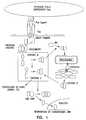
- Fas/FasL-mediated apoptosis is induced by binding of three FasL molecules which induces trimerization of Fas receptor via C-terminus death domains (DDs), which in turn recruit an adapter protein FADD (Fas-associated protein with death domain) and Caspase-8 ( Fig. 1 ).
- DDs C-terminus death domains
- FADD Fas-associated protein with death domain
- Caspase-8 Fig. 1
- the oligomerization of this trimolecular complex, Fas/FAJDD/caspase-8 results in proteolytic cleavage of proenzyme caspase-8 into active caspase-8 that, in turn, initiates the apoptosis process by activating other downstream caspases through proteolysis, including caspase-3.
- Death ligands in general are apoptotic when formed into trimers or higher order of structures. As monomers, they may serve as antiapoptotic agents by competing with the trimers for binding to the death receptors.
- the present invention discloses how to generate death ligands consisting of stable tetramers and deliver these ligands either as purified proteins or exposed on the surface of APCs to induce apoptosis in lymphocytes expressing the counter death receptors.
- death ligands are more effective in inducing apoptosis when in trimers or higher order of structures based upon the available data from similar biological systems.
- the preferred embodiment for the formation of tetramers and higher order of structures is streptavidin or avidin, both of which form tetramers and higher structure under physiological conditions.
- a modified form of streptavidin has been chosen as the preferred molecule in this patent application and is referred to as CSA throughout this document.
- Apoptosis of activated T cells results in tolerance to allografts and xenografis, including bone marrow and other organ transplantation. Purging of activated T cells also relieves the symptoms of allergies and other immune-induced diseases. Included in the latter are autoimmune disorders such as multiple sclerosis, lupus erythematosus, sarcoidosis, diabetes, and rheumatoid arthritis. Many disorders, including some tumors, are dependent on lymphocyte functions that lead to persistence of the disorder. Many hematological disorders could be treated with bone marrow stem cell transplants if the immune response could be regulated so as to induce tolerance to the foreign stem cells. Among these disorders are leukemias, lymphomas, aplastic anemia, sickle cell and Cooley's anemia and the like. All of these disorders may be controlled permanently or temporarily by apoptosis of activated immune cells, including T cells.
- This invention discloses strategies for the induction of tolerance, which comprise the construction of chimeric cDNAs encoding the functional portions of a tetramcr forming molecule operably linked to an apoptosis-inducing molecule.
- Table I is a summary of the proposed constructs. Choice of constructs may be based on factors such as the nature of the foreign antigen provoking adaptive immunity; whether relief sought will be temporary or permanent; whether a commitment to death is desired or addition downstream regulation of apoptosis is preferred.
- the dosage necessary to induce apoptosis is dependent on the affinity and specificity of the apoptotic agent. Ten to 100 molecules of the preferred embodiment per cell, CSA/FasL, is sufficient for the apoptotic effect, while as much as 1,000 to 10,000 molecules of less active constructs may be necessary. The choice of apoptotic agent will depend on the activity desired.
- a preferred production cell for the production of chimeric proteins encoded by the DNA constructs is the Drosophila system that is commercially available.
- Drosophila system that is commercially available.
- those skilled in the art of producing chimeric proteins will recognize that many other expression systems and vectors are suitable for production of the chimeric proteins of this invention. Included in these other systems are Escherichia coli, yeast and mammalian cell cultures.
- compositions and methods to regulate the immune system are representative of compositions and methods to regulate the immune system. These examples were designed to induce tolerance to autoantigens in the case of autoimmunity, allergies, and asthma and foreign antigens in case of allo and xenografts by application of the preferred embodiment to eliminate pathogenic immune cells expressing counter-death receptors. Furthermore, soluble forms of death ligands as monomers with none or minimal apoptotic activity will be used to interfere with apoptosis in neurodegenerative disorders and to promote liver regeneration.
- the most preferred embodiments are chimeric proteins, CSA-FasL, CSA-TNF ⁇ , CSA-TRAIL, and CSA-TWEAK produced by fusion of DNA coding for core streptavidin protein with that DNA coding for the mentioned death ligands and expressing the chimeric protein in a suitable production cell. These molecules are then purified and used either as purified proteins or used to decorate cells or organs to induce apoptosis in immune cells expressing the relevant death receptor.
- cells of interest are first biotinylated ex vivo or in vivo and then CSA-death ligands are administered in order to attach the molecules to the cell surface for apoptosis of target lymphocytes, resulting in, for example, induction of antigen-specific tolerance for the prevention of autoimmunity and foreign graft rejection.
- purified chimeric proteins may be applied ex vivo or in vivo, leading to permanent elimination of antigen-specific pathogenic lymphocytes and the amelioration of the diseased condition.
- Soluble form of FasL and other death ligands consisting of extracellular domains will be used as purified protein with antiapoptotic function to prevent apoptosis in neurons for the treatment of neurodegenerative disorders in which unregulated apoptosis is the pathogenic mechanism.
- This molecule with the chimeric ones will also be used for liver regeneration. All these proteins can also be used to prevent septic shock, allergies, asthma, and food poisoning where activated lymphocytes play a major role.
- Example 1 Cloning of wild type and generation of a membrane-bound form of FasL ,expression in mammalian cells, and test of apoptotic function
- FasL cDNA was cloned from ConA-activated rat T cells or human peripheral blood cells using primers specific for the very 5'- and 3'-untranslated regions of the FasL mRNA in RT-PCR.
- the amplification product was then cloned into the TA cloning vector and several functional cDNA clones were determined by sequence analysis.
- a functional cDNA clone was then subcloned into the pcDNA3 eukaryotic expression vector (pcDNA3) and the recombinant clones containing cDNA in both the sense and the antisense orientations were determined by restriction site analysis.
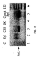
- Figure 2 shows the expression of FasL in different cell types as shown by gel electrophoresis.
- a PCR template (column 1) is shown as a negative control.
- FasL is synthesized as a type II membranous protein, it is also secreted into the milieu as a soluble protein.
- the soluble form is generated from the membranous molecule by matrix metalloproteinases within minutes after its expression on the cell surface.
- FasL is also synthesized and stored as a membranous protein in microvesicles that are excreted into the milieu in selected cell types in response to various stimuli.
- These three different forms of FasLmembranous, soluble, and vesicular-- may differ in their function with respect to apoptosis and immune regulation.
- Apoptosis is primarily mediated by vesicular and membranous forms of FasL whereas the soluble form is ineffective in mediating apoptosis and indeed serves as an antiapoptotic factor by competing with the membranous form for Fas.
- soluble FasL serves as a chemotactic factor for neutrophils whereas membranous and vesicular FasLs lack this function.
- Soluble FasL may, therefore, contribute to graft rejection by I) serving as an antiapoptotic molecule to prevent apoptosis of graft-reactive T cells, and ii) serving as a chemotactic factor for neutrophil infiltration into the graft. It is, therefore, important to separate the antiapoptotic and chemotactic functions of FasL from its apoptotic function for the use of FasL as a tolerogenic molecule.
- rat FasL that is stably expressed on the cell surface by eliminating the putative metalloproteinase site.
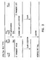
- Oligonucleotide primers specific for FasL were designed by PCR to delete nucleotides encoding 12 amino acids residues thought to be metalloproteinase site ( Fig. 3 ).
- DNA clones for the modified (mFasL) and wild type (wtFasL) were then transfected into PVG.IU rat endothelial cells and monkey Cosl cells for expression. Stable transfectants were selected using G418 and analyzed for the expression of FasL molecules using RT-PCR and flow cytometry.
- RT-PCR analysis revealed high levels of expression of FasL in transfected cells but not in untransfected cells.
- Cos cells were transiently transfected with wtFasL and mFasL.
- Total RNA was prepared three days after transfection and used for RT-PCR with FasL-specific primers.
- the PCR products were digested with Xhol enzyme. Most importantly, restriction digest of the PCR product with XhoI enzyme that was engineered into the cDNA for the modified form resulted in two bands in cells transfected with the mFasL but not wtFasL, confirming the modification.
- the expression of FasL at the protein level was confirmed using PE-labeled FasL mAb MFL4 in flow cytometry.
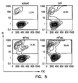
- Transfectants expressing mFasL were much more effective in inducing apoptosis in apoptosis-sensitive human Jurkat cells as compared with transfectants expressing wtFasL ( Figure 5 ).
- COS cells were transiently transfected with wtFasL and mFasL. Three days after transfection, transfectants were co-incubated with Jurkat cells overnight and apoptosis was determined using PI in flow cytometry. Untransfected (Jurkat) and green fluorescence protein (GFP) transfected cells served as negative controls. The percentage of dead cells is shown in the upper quadrants of the histograms. The data is representative of a minimum of four independent experiments. Specific expression increased from 3.2% to 15.4%, while specific apoptosis increased from 6.5% to 23.8%.
- CSA core streptavidin fused in frame with the extracellular domain of FasL lacking the putative metalloproteinase site using the scheme outlined in Figure 6 .
- Genomic DNA was isolated from Streptomyces avidinii (ATCC Cat.#27419) and 0.2 g of this DNA was used as template for amplification using primers specific for 5'-end (CSA-I) and 3'-end (CSA-2) of core streptavidin in PCR.
- the 5'-primer included sequences for BgllI and 6 His residues to allow cloning in frame with the Drosophila secretion signal (BiP) for expression as a secreted protein and purification using NI-affinity columns.
- the PCR product was cloned into the TA cloning vector and several positive clones were identified first screening by PCR and then digestion with restriction enzymes.
- the PCR product was then cloned into the TA cloning vector and several positive clones were identified by restriction digestion.
- S2 cells were transfected transiently with the recombinant CSA/FasL plasmid using calcium phosphate transfection according to the manufacture's instructions (Invitrogen). S2 cells were pulsed with copper sulfate 24 hr after transfection to activate the metallothionein promoter for expression of the chimeric CSA-FasL protein. Culture medium was then collected after a 24-hr induction period and analyzed by ELISA and Western blot. It was seen that mAb MFL4 against FasL (PharMingen, San Diego, CA) detects a band of ⁇ 35 kDa corresponding to the expected molecular weight of the chimeric protein in Western blots performed under stringent denaturing conditions.
- chimeric protein The identity of the chimeric protein was either confirmed using polyclonal antibodies against streptavidin (Zymed Laboratories, San Francisco, CA), which detected the same band (data not shown).
- streptavidin Zymed Laboratories, San Francisco, CA
- CSA forms tetramers that are stable in 1% SDS and 10% mercaptoethanol solution at temperatures under 60°C. Culture supernatants were, therefore, heated at 60 and 100°C before running on SDS-PAGE to test if the chimeric protein forms tetramers.
- mAb MFL4 against FasL (PharMingen, San Diego, CA) detected bands of ⁇ 35 and >100 kDa at 60°C and primarily a band of ⁇ 35 kDa at 100°C.
- CSA-FasL by S2 cells was further confirmed using biotin-coated microwell strip plates in ELISA. Briefly, biotinylated wells were incubated with culture supernatants for 45 min at room temperature, washed extensively, and then incubated with the working concentrations of primary antibodies against streptavidin or FasL (MFL4) for 45 min at room temperature. Wells were then washed extensively and incubated with alkaline phosphatase conjugated secondary antibodies at room temperature for 45 min. Plates were then washed extensively and incubated with alkaline phosphatase substrate. Data was analyzed using an ELISA reader (Victor, Wallac, Gaithersburg, MD).
- Example 3 Expression of chimeric CSA-FasL molecule on the surface of splenocytes for analysis of its function in apoptosis
- chimeric CSA-FasL can be attached to the cell surface under physiological conditions
- 2.5x10 7 PVG.R8 splenocytes were conjugated with EZ-Link Sulfo-NHS-LC-Biotin (15 ⁇ M) in 1 ml of PBS at room temperature for 30 min according to the manufacturer's instructions (Pierce). After several washes in PBS, pH 8.0, 1x10 6 cells were incubated in 0.3-1 ml culture supernatants containing 50-100 ng CSA-FasL or supernatants from S2 cells transfected with a nonfunctional FasL construct for 45 min on ice.
- splenocytes were washed with PBS three times and 1 x 10 6 cells were co-incubated with 1 x 10 6 Jurkat cells in RPMI culture medium overnight for apoptotic activity.
- Apoptosis in Jurkat cells was determined by using propidium iodide (a marker for dead cells), Annexin V-FITC (a marker for early apoptotic events), and human anti-CD3-APC mAb (clone UCHT1; PharMingen) to gate specifically on Jurkat cells using flow cytometry.
- the activated T cells of the patient may be advisable to purge without inducing a permanent effect.
- Many techniques arc known for removal of blood continuously from the body, subjecting it to treatment outside the body with subsequent return.
- a patient suffering from an autoimmune disease will especially benefit from leukophoresis, in which the white cells are continuously collected, followed by treatment with one of the recombinant proteins of this invention.
- Those activated lymphocytes killed via apoptosis will be removed and the leukocytes returned to the patient. In this manner, the pathogenic immune cells are removed from the body of the patient without major side effects of the treatment.
- CSA-FasL conjugated the chimeric CSA-FasL molecule to biotin-agarose beads (ImmunnPure® immobilized immunobiotin; Pierce) and tested the function for apoptosis of Jurkat cells.
- biotin-agarose beads ImmunnPure® immobilized immunobiotin; Pierce
- CSA-FasL induced significant apoptosis (38%) in Jurkat cells as compared with the beads without FasL (15%).
- Treatment of mouse A20 cell line under similar experimental conditions results in >90% death as this cell line is more sensitive to apoptosis.
- ACI splenocytes were conjugated with EZ-Link Sulfo-NHS-LC-Biotin (5 ⁇ M) in 1 ml of PBS at room temperature for 30 rain according to the manufacturer's instructions (Pierce).
- Splenocytes were harvested from B10 mice, biotinylated (15 ⁇ M), decorated with FasL (50-100 ng/10 6 cells) or control S2 supernatant, and irradiated (2000 rads).
- FasL 50-100 ng/10 6 cells
- control S2 supernatant 50-100 ng/10 6 cells
- irradiated 2000 rads.
- One million of splenocytes were co-transplanted with -400 islet cells under the kidney capsule of diabetic BALB/c mice. Survival of islets was assessed by monitoring blood glucose levels starting 3 days after transplantation. FasL-decorated splenocytes prevented the rejection of ⁇ 70% of allogeneic islets (MST>56 day;).
- biotin/streptavidin pair for direction of the constructs to the targeted cells, it is well known that other such pairs exist.
- Avidin can be substituted for streptavidin.
Abstract
Description
- This invention relates to chimeric proteins for the modulation of the immune system by the induction of apoptosis in selected cells.
- The immune system is critical to the survival of vertebrates in an environment full of pathogenic microorganisms. Individuals lacking an immune response through inborn genetic defects, exposure to chemotherapeutic agents or through infection by such viruses as human immunodeficiency virus (HIV), succumb to infections that an individual with a healthy immune system would readily survive. However, the immune system is not always beneficial to the organism. Its dysregulation leads to a variety of pathogenic conditions, including autoimmunity and tumors. The immune system also serves as a barrier for the transplantation of foreign grafts, such as those comprising cells, tissues or organs taken from another individual, a process that can treat a variety of pathogenic disorders, including autoimmunity, replace failed organs in end terminal diseases and treat a variety of hematopoietic disorders via bone marrow transplantation. These applications require either the suppression of the immune system or to "educate" the immune system not to react to self-antigens in the case of autoimmunity and foreign antigens in the case of transplantation.
- At the present time, most of the treatment regimens in the fields of autoimmunity and transplantation primarily relay on the suppression of the immune system via the chronic use of variety of immunosuppressive pharmacological agents. Although these drugs are effective in reducing the severity of autoimmune and rejection episodes, they are nonspecific and fail to create a state of permanent antigen-specific tolerance. Their continuous use is therefore required to keep the pathogenic reactions under control. Continuous exposure of the individual to these immunosuppressive agents is, however, associated with a significantly increased risk of opportunistic infections and malignancies. Additionally, these nonspecific immunosuppressive agents can induce serious and undesirable pharmacological side effects in the host. It would, therefore, be very desirable to be able to "teach" the immune system to tolerate selected antigens of interest for the treatment of autoimmunity and induction of transplantation tolerance. The need remains to develop more selective and long-lasting methods to regulate the immune response.
-
US-A-5 763 223 discloses the cytokine TRAIL which induces the apoptosis of certain target cells. It is shown that a TRAIL-Flag fusion protein causes increased apoptosis in target cells when cross-linked to a surface by a monoclonal antibody against FLAG and that oligomerization of TRAIL increases apoptosis over monomeric TRAIL. -
-
Figure 1 shows the mechanism of Fas/FasL-induced apoptosis. -
Figure 2 shows the expression of Fas/FasL in activated splenocytes. -
Figure 3 shows the schematic diagram of modification of FasL to generate a stable cell surface protein. -
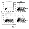
Figure 4 shows the cell surface expression of FasL in Cosl cells. -
Figure 5 shows efficient apoptosis of a human T cells line by Cosl cells expressing the mFasL. -
Figure 6 shows the construction of CSA-FasL construct. -
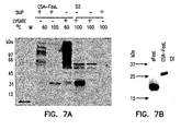
Figure 7 shows the expression of soluble and CSA-FasL by Western blot. -
Figure8 shows binding of CSA-FasL to the surface of splenocytes. -
Figure 9 shows apoptosis of Jurkat cells by cell-surface or agarose beads bound CSA-FasL. -
Figure 10 shows the blockade of alloreactive responses by cells decorated with CSA-FasL. -
Figure 11 shows the blockade of alloreactive responses in vivo by cells decorated with CSA-FasL. - This invention provides compositions for inducing apoptosis in selected cells. The construction of chimeric proteins comprising apoptosis-inducing protein fused to one of a binding pair capable of binding to the selected cell is taught. The binding pair may be biotin/avidin-, or biotin/streptavidin,
- Preferably the binding pair is biotin/streptavidin. Effector cells are constructed by exposing said effector cells to biotin-, and decorating with an effective amount of said chimeric protein. The cells to be killed are exposed to said effector cells. Apoptosis may be induced in vivo or ex vivo. In an ex vivo method, it is preferred to attach the chimeric protein to a biotinylated solid matrix.
- The preferred solid matrix is biotin-agarose beads.
- An important aspect of immune modulation is to induce tolerance to self antigens and foreign graft antigens for the treatment of autoimmunity and prevention of foreign graft rejection. Both foreign graft rejection and autoimmunity are immunological phenomena primarily initiated by the T cells responding to foreign histocompatibility antigens on the graft and auto-antigens in the host, respectively. This response leads to the activation of cells clonally expressing T-cell receptors (TCRs). Activated T cells then differentiate, proliferate, and synthesize a variety of cytokines, which prolong and intensify the reaction. Experimental animals lacking T cells, either due to congenital immunodeficiency or experimental manipulations, do not develop autoimmunity and accept allografts expressing strong histocompatibility differences for indefinite periods of time. Full T cell activation depends on two distinct signals;
signal Signal 1 is transduced via TCR interaction with the MHC/peptide complex and is antigen specific whereassignal 2 is provided by antigen-presenting cells (APCs) in antigen nonspecific fashion. The transduction ofsignal 1 in the absence ofsignal 2 may result in functional silencing (anergy) or physical elimination (apoptosis) of naive T cells. Upon activation, T cells undergo a state of antigen-driven proliferation that allows up to a 1200-fold clonal expansion. A period of death then ensues during which more than 95% of the activated T cells undergo apoptosis (programmed cell death) while the remaining cells differentiate into memory cells as the amount of antigen in the system declines. - Apoptosis plays a central role in both the development and homeostasis of multicellular organisms. Apoptosis can be induced by multiple independent signaling pathways that converge upon a final effector mechanism consisting of multiple interactions between several "death receptors" and their ligands, which belong to the tumor necrosis factor (TNF) receptor/ligand superfamily. The best-characterized death receptors are CD95 ("Fas"), TNFR1 (p55), death receptor 3 (DR3 or Apo3/TRAMO), DR4 and DR5 (apo2-TRAIL-R2). The final effector mechanism of apoptosis is the activation of a series of proteinases designated as caspases. The activation of these caspases results in the cleavage of a series of vital cellular proteins and cell death. In my copending International
- Patent Application,
PCT/US00/34554, filed on December 18, 2000 caspases are disclosed as a useful means to induce apoptosis. - It has now been discovered that Fas-/FasL- induced apoptosis plays a central role in protecting immunologically privileged sites, such as the central nervous system, testes and eyes from immune attack. Allogeneic and xenogeneic tissues transplanted into these sites, for instance, are resistant to rejection. Of great importance is the major role apoptosis plays in maintaining homeostasis in the immune system via activation-induced cell death (AICD). AICD is primarily mediated by apoptotic signals transduced by the Fas/FasL interaction. Fas is a 45 kDa, type I, cell surface protein consisting of a cysteine-rich extracellular domain, which transduce a death signal. Fas mediates effector function by interacting with FasL, a type II membrane protein of about 40 kDa. T cells are the primary cell type in the body that express FasL upon activation. The expression of this molecule makes the activated T cells susceptible to apoptosis. Upon repeated stimulation with antigens, activated T cells up regulate expression of both Fas and FasL on the cell surface and become sensitive to autocrine apoptosis, where Fas engages with FasL expressed on the same cell. Apoptosis may also be induced in a paracrine fashion where Fas of an activated T cell interacts with FasL expressed on another cell. These two modes of apoptotic T cell death control the size of antigen-activated T cell clones and maintain immune homeostasis. All lymphocytes, including B cells, and dendritic cells may be subjected to FasL-regulated immune homeostasis. Dysregulation of AICD is involved in a series of pathophysiological conditions, including degenerative and autoimmune diseases. For example, the autoimmune lymphoproliferative syndrome (ALPS) in humans is an inherited disease of lymphocyte homeostasis and defective apoptosis. In both type I and type II ALPS defects in the Fas receptor or FasL are responsible for the pathogenesis of these diseases. Similarly, congenital mutations causing the lack or low levels of expression of Fas and FasL in the lpr and gld mice, respectively, are associated with severe lymphoproliferative and autoimmune disorders. These mice also lack antigen-induced peripheral clonal deletion oft cells. Transgenic expression of Fas in lpr/lpr mice results in amelioration of autoimmunity and restoration of AICD-induced clonal deletion of T cells, providing direct evidence for the importance of Fas/FasL-mediated apoptosis in peripheral tolerance to self-antigens. The critical role FasL plays in AICD and the importance of AICD in peripheral tolerance form the basis of this patent application to use the death ligands as immunomodulators to induce tolerance both to auto and foreign antigens for the treatment of autoimmunity and prevention of foreign graft rejection.
- The molecular mechanism of death receptors/ligands-induced apoptosis has been elucidated in great details. For example, Fas/FasL-mediated apoptosis is induced by binding of three FasL molecules which induces trimerization of Fas receptor via C-terminus death domains (DDs), which in turn recruit an adapter protein FADD (Fas-associated protein with death domain) and Caspase-8 (
Fig. 1 ). The oligomerization of this trimolecular complex, Fas/FAJDD/caspase-8, results in proteolytic cleavage of proenzyme caspase-8 into active caspase-8 that, in turn, initiates the apoptosis process by activating other downstream caspases through proteolysis, including caspase-3. Death ligands in general are apoptotic when formed into trimers or higher order of structures. As monomers, they may serve as antiapoptotic agents by competing with the trimers for binding to the death receptors. - The present invention discloses how to generate death ligands consisting of stable tetramers and deliver these ligands either as purified proteins or exposed on the surface of APCs to induce apoptosis in lymphocytes expressing the counter death receptors. We hypothesize that death ligands are more effective in inducing apoptosis when in trimers or higher order of structures based upon the available data from similar biological systems. The preferred embodiment for the formation of tetramers and higher order of structures is streptavidin or avidin, both of which form tetramers and higher structure under physiological conditions. A modified form of streptavidin has been chosen as the preferred molecule in this patent application and is referred to as CSA throughout this document.
- Apoptosis of activated T cells results in tolerance to allografts and xenografis, including bone marrow and other organ transplantation. Purging of activated T cells also relieves the symptoms of allergies and other immune-induced diseases. Included in the latter are autoimmune disorders such as multiple sclerosis, lupus erythematosus, sarcoidosis, diabetes, and rheumatoid arthritis. Many disorders, including some tumors, are dependent on lymphocyte functions that lead to persistence of the disorder. Many hematological disorders could be treated with bone marrow stem cell transplants if the immune response could be regulated so as to induce tolerance to the foreign stem cells. Among these disorders are leukemias, lymphomas, aplastic anemia, sickle cell and Cooley's anemia and the like. All of these disorders may be controlled permanently or temporarily by apoptosis of activated immune cells, including T cells.
- This invention discloses strategies for the induction of tolerance, which comprise the construction of chimeric cDNAs encoding the functional portions of a tetramcr forming molecule operably linked to an apoptosis-inducing molecule. Table I is a summary of the proposed constructs. Choice of constructs may be based on factors such as the nature of the foreign antigen provoking adaptive immunity; whether relief sought will be temporary or permanent; whether a commitment to death is desired or addition downstream regulation of apoptosis is preferred. .Those skilled in the art can readily, without undue experimentation, make constructs with any of the death ligands, FasL, TNFα, TRAIL (Apo2 ligand), and TWEAK (Apo3 ligand) , operably linked to a tetramcr-forming molecule.
Those skilled in the art are aware that many pharmaceutical agents enhance apoptosis. Among such agents are bis-indolylmaleimide-8 and quabain. If desired, these agents may be used in conjunction with the chimeric proteins of this invention.TABLE I Construct Insert Made Function Application rsFasL Soluble extracellular portion of rat FasL + Anti-apoptotic Treatment of neuro-degenerative disorders and promotion of liver regeneration mFasL Rat FasL modified to be stably expressed on the cell surface + Apoptosis Tolerance to allografts, xenografts, bone marrow transplantation, treatment of autoimmune diseases, allergies, asthma, septic shock, and other immune-induced diseases, and lymphocyte-dependent disorders, including tumors and promotion of liver regeneration wtFasL Rat wild type molecule Chemotactic, Anti-apoptotic Treatment of neurodegenerative discorders and promotion of liver regeneration CSA-FasL Rat extracellular portion of FasL chimeric protein with core streptavidin + Apoptosis Tolerance to allografis, xenografts, bone marrow transplantation, treatment of autoimmune diseases, allergies, asthma, septic shock, and other immune-induced diseases, and lymphocyte-dependent disorders, including tumors, and promotion of liver regeneration Prevention of early reocclusion after coronary bypass surgery and angioplasty CSA-hFasL Human extracellular portion of Fas chimeric protein with core streptavidin + Apoptosis CSA-TNFα Human extracellular portion of TNF chimeric protein with core streptavidin TNF α cDN A clone d Apoptosis CSA-TWEAK Human extracellular portion of TWEAK chimeric protein with core streptavidin - Apoptosis CSA-TRAIL Human extracellular portion of TRAIL chimeric protein with core streptavidin - Apoptosis - The dosage necessary to induce apoptosis is dependent on the affinity and specificity of the apoptotic agent. Ten to 100 molecules of the preferred embodiment per cell, CSA/FasL, is sufficient for the apoptotic effect, while as much as 1,000 to 10,000 molecules of less active constructs may be necessary. The choice of apoptotic agent will depend on the activity desired.
- A preferred production cell for the production of chimeric proteins encoded by the DNA constructs is the Drosophila system that is commercially available. However, those skilled in the art of producing chimeric proteins will recognize that many other expression systems and vectors are suitable for production of the chimeric proteins of this invention. Included in these other systems are Escherichia coli, yeast and mammalian cell cultures.
- The experimental procedures described herein are representative of compositions and methods to regulate the immune system. These examples were designed to induce tolerance to autoantigens in the case of autoimmunity, allergies, and asthma and foreign antigens in case of allo and xenografts by application of the preferred embodiment to eliminate pathogenic immune cells expressing counter-death receptors. Furthermore, soluble forms of death ligands as monomers with none or minimal apoptotic activity will be used to interfere with apoptosis in neurodegenerative disorders and to promote liver regeneration. The most preferred embodiments are chimeric proteins, CSA-FasL, CSA-TNFα, CSA-TRAIL, and CSA-TWEAK produced by fusion of DNA coding for core streptavidin protein with that DNA coding for the mentioned death ligands and expressing the chimeric protein in a suitable production cell. These molecules are then purified and used either as purified proteins or used to decorate cells or organs to induce apoptosis in immune cells expressing the relevant death receptor. For example, cells of interest are first biotinylated ex vivo or in vivo and then CSA-death ligands are administered in order to attach the molecules to the cell surface for apoptosis of target lymphocytes, resulting in, for example, induction of antigen-specific tolerance for the prevention of autoimmunity and foreign graft rejection. Alternatively, purified chimeric proteins may be applied ex vivo or in vivo, leading to permanent elimination of antigen-specific pathogenic lymphocytes and the amelioration of the diseased condition. Soluble form of FasL and other death ligands consisting of extracellular domains will be used as purified protein with antiapoptotic function to prevent apoptosis in neurons for the treatment of neurodegenerative disorders in which unregulated apoptosis is the pathogenic mechanism. This molecule with the chimeric ones will also be used for liver regeneration. All these proteins can also be used to prevent septic shock, allergies, asthma, and food poisoning where activated lymphocytes play a major role.
- A complete FasL cDNA was cloned from ConA-activated rat T cells or human peripheral blood cells using primers specific for the very 5'- and 3'-untranslated regions of the FasL mRNA in RT-PCR. The amplification product was then cloned into the TA cloning vector and several functional cDNA clones were determined by sequence analysis. A functional cDNA clone was then subcloned into the pcDNA3 eukaryotic expression vector (pcDNA3) and the recombinant clones containing cDNA in both the sense and the antisense orientations were determined by restriction site analysis.
Figure 2 shows the expression of FasL in different cell types as shown by gel electrophoresis. Total RNA was isolated from naive splenocytes (column 2); C58 cells (column 3), endothelial cells (column 4) and ConA-activated splenocytes (column 4). A PCR template (column 1) is shown as a negative control. - Although FasL is synthesized as a type II membranous protein, it is also secreted into the milieu as a soluble protein. The soluble form is generated from the membranous molecule by matrix metalloproteinases within minutes after its expression on the cell surface. FasL is also synthesized and stored as a membranous protein in microvesicles that are excreted into the milieu in selected cell types in response to various stimuli. These three different forms of FasLmembranous, soluble, and vesicular-- may differ in their function with respect to apoptosis and immune regulation. Apoptosis is primarily mediated by vesicular and membranous forms of FasL whereas the soluble form is ineffective in mediating apoptosis and indeed serves as an antiapoptotic factor by competing with the membranous form for Fas. Furthermore, soluble FasL serves as a chemotactic factor for neutrophils whereas membranous and vesicular FasLs lack this function. Soluble FasL may, therefore, contribute to graft rejection by I) serving as an antiapoptotic molecule to prevent apoptosis of graft-reactive T cells, and ii) serving as a chemotactic factor for neutrophil infiltration into the graft. It is, therefore, important to separate the antiapoptotic and chemotactic functions of FasL from its apoptotic function for the use of FasL as a tolerogenic molecule.
- We generated a rat FasL that is stably expressed on the cell surface by eliminating the putative metalloproteinase site. Oligonucleotide primers specific for FasL were designed by PCR to delete nucleotides encoding 12 amino acids residues thought to be metalloproteinase site (
Fig. 3 ). DNA clones for the modified (mFasL) and wild type (wtFasL) were then transfected into PVG.IU rat endothelial cells and monkey Cosl cells for expression. Stable transfectants were selected using G418 and analyzed for the expression of FasL molecules using RT-PCR and flow cytometry. RT-PCR analysis revealed high levels of expression of FasL in transfected cells but not in untransfected cells. Cos cells were transiently transfected with wtFasL and mFasL. Total RNA was prepared three days after transfection and used for RT-PCR with FasL-specific primers. The PCR products were digested with Xhol enzyme. Most importantly, restriction digest of the PCR product with XhoI enzyme that was engineered into the cDNA for the modified form resulted in two bands in cells transfected with the mFasL but not wtFasL, confirming the modification. The expression of FasL at the protein level was confirmed using PE-labeled FasL mAb MFL4 in flow cytometry. - We obtained higher levels of expression of the modified FasL on the surface of transfected COS and endothelial cells as compared with wild type FasL (
Figure 4 is representative of four independent experiments). COS cells were transiently co-transfected with wtFasL, mFasL, and green fluorescence protein (GFP). Cells were analyzed for expression of FasL using PE-labeled MFL4 mAb and GFP. GFP was used as an internal control in order to access the efficiency of transfection. - Transfectants expressing mFasL were much more effective in inducing apoptosis in apoptosis-sensitive human Jurkat cells as compared with transfectants expressing wtFasL (
Figure 5 ). COS cells were transiently transfected with wtFasL and mFasL. Three days after transfection, transfectants were co-incubated with Jurkat cells overnight and apoptosis was determined using PI in flow cytometry. Untransfected (Jurkat) and green fluorescence protein (GFP) transfected cells served as negative controls. The percentage of dead cells is shown in the upper quadrants of the histograms. The data is representative of a minimum of four independent experiments. Specific expression increased from 3.2% to 15.4%, while specific apoptosis increased from 6.5% to 23.8%. - We next generated a construct encoding core streptavidin (CSA) fused in frame with the extracellular domain of FasL lacking the putative metalloproteinase site using the scheme outlined in
Figure 6 . Genomic DNA was isolated from Streptomyces avidinii (ATCC Cat.#27419) and 0.2 g of this DNA was used as template for amplification using primers specific for 5'-end (CSA-I) and 3'-end (CSA-2) of core streptavidin in PCR. The 5'-primer included sequences for BgllI and 6 His residues to allow cloning in frame with the Drosophila secretion signal (BiP) for expression as a secreted protein and purification using NI-affinity columns. The PCR product was cloned into the TA cloning vector and several positive clones were identified first screening by PCR and then digestion with restriction enzymes. We next subcloned the extracellular domain of the FasL without the metalloproteinase site using the wild type FasL cDNA clone as a template and a sense primer to the 5'-end of the extracellular region, FasL6 containing an EcoRl site, and an antisense primer to the 3'-end untranslated region of FasL, FasL2 containing an EcoRI site in frame with CSA, in PCR. The PCR product was then cloned into the TA cloning vector and several positive clones were identified by restriction digestion. Both the core streptavidin and extracellular FasL clones were then sequenced and DNA inserts with accurate sequences were digested out of the TA cloning vector with BgllI-EcoRI for CSA and EcoRI for extracellular FasL. These DNA inserts were then subcloned into the BgIII-EcoRI-cut pMT/BiP/V5-His vector for expression in using the DES system (Invitrogen). - S2 cells were transfected transiently with the recombinant CSA/FasL plasmid using calcium phosphate transfection according to the manufacture's instructions (Invitrogen). S2 cells were pulsed with copper sulfate 24 hr after transfection to activate the metallothionein promoter for expression of the chimeric CSA-FasL protein. Culture medium was then collected after a 24-hr induction period and analyzed by ELISA and Western blot. It was seen that mAb MFL4 against FasL (PharMingen, San Diego, CA) detects a band of∼35 kDa corresponding to the expected molecular weight of the chimeric protein in Western blots performed under stringent denaturing conditions. The identity of the chimeric protein was either confirmed using polyclonal antibodies against streptavidin (Zymed Laboratories, San Francisco, CA), which detected the same band (data not shown). CSA forms tetramers that are stable in 1% SDS and 10% mercaptoethanol solution at temperatures under 60°C. Culture supernatants were, therefore, heated at 60 and 100°C before running on SDS-PAGE to test if the chimeric protein forms tetramers. As shown in
Fig. 9 , mAb MFL4 against FasL (PharMingen, San Diego, CA) detected bands of∼35 and >100 kDa at 60°C and primarily a band of∼35 kDa at 100°C. These bands correspond to expected monomers and tetramers (Figure 7 ). These bands were only detected in S2 cells transfected with CSA-FasL construct (lane FasL) but not cells transfected with a nonfunctional FasL (lane S2; one nucleotide insertion near to 5' end of the cDNA during the cloning process leads to a shift in the reading frame and lack of the protein expression). - The expression of CSA-FasL by S2 cells was further confirmed using biotin-coated microwell strip plates in ELISA. Briefly, biotinylated wells were incubated with culture supernatants for 45 min at room temperature, washed extensively, and then incubated with the working concentrations of primary antibodies against streptavidin or FasL (MFL4) for 45 min at room temperature. Wells were then washed extensively and incubated with alkaline phosphatase conjugated secondary antibodies at room temperature for 45 min. Plates were then washed extensively and incubated with alkaline phosphatase substrate. Data was analyzed using an ELISA reader (Victor, Wallac, Gaithersburg, MD). Both antibodies detected high levels of CSA-FasL in S2 cells expressing CSA-FasL (FasL) but not negative controls (S2). Most importantly, we quantified the amount of CSA-FasL expressed by the transfectants using known amounts of pure streptavidin protein as standard (Pierce). Transfectants expressed 50-100 ng of chimeric protein/mi of culture medium. Taken together, these data clearly demonstrate that we have succeeded to generate a functional chimeric CSA-FasL construct and express it in S2 cells, and developed experimental methods for detection and quantification of the chimeric protein.
- We also generated a construct consisting of the human extracellular portion of FasL with CSA, extracellular portion of rat FasL (
Figure 7b ), and cloned a cDNA for human extracellular portion of TNF- to generate a chimeric CSA-TNFα. - To test whether chimeric CSA-FasL can be attached to the cell surface under physiological conditions, 2.5x10⁷ PVG.R8 splenocytes were conjugated with EZ-Link Sulfo-NHS-LC-Biotin (15 µM) in 1 ml of PBS at room temperature for 30 min according to the manufacturer's instructions (Pierce). After several washes in PBS, pH 8.0, 1x10⁶ cells were incubated in 0.3-1 ml culture supernatants containing 50-100 ng CSA-FasL or supernatants from S2 cells transfected with a nonfunctional FasL construct for 45 min on ice. Cells were washed extensively and used for FasL staining or apoptosis assays. Staining was performed on 1 x 10⁶ cells using APC-streptavidin or a monoclonal antibody (MFL4) conjugated with PE in flow cytometry. All the cells expressed CSAFasL (
Figure 8a ) and streptavidin (Figure 8b ). Supernatant from S2 control was negative for FasL staining. Similarly, splenocytes without biotinylation did not stain with APC-streptavidin, demonstrating specificity. - We next tested decorated splenocytes for apoptosis. Effector splenocytes were washed with PBS three times and 1 x 10⁶ cells were co-incubated with 1 x 10⁶ Jurkat cells in RPMI culture medium overnight for apoptotic activity. Apoptosis in Jurkat cells was determined by using propidium iodide (a marker for dead cells), Annexin V-FITC (a marker for early apoptotic events), and human anti-CD3-APC mAb (clone UCHT1; PharMingen) to gate specifically on Jurkat cells using flow cytometry. Significant apoptosis was detected in the culture where Jurkat cells were co-incubated with splenocytes-FasL as compared with control cultures incubated with S2 supernatant without FasL (38% versus 9%) (
Figure 9 ). - In some clinical situations, it may be advisable to purge the activated T cells of the patient without inducing a permanent effect. Many techniques arc known for removal of blood continuously from the body, subjecting it to treatment outside the body with subsequent return. A patient suffering from an autoimmune disease will especially benefit from leukophoresis, in which the white cells are continuously collected, followed by treatment with one of the recombinant proteins of this invention. Those activated lymphocytes killed via apoptosis will be removed and the leukocytes returned to the patient. In this manner, the pathogenic immune cells are removed from the body of the patient without major side effects of the treatment. We, therefore, conjugated the chimeric CSA-FasL molecule to biotin-agarose beads (ImmunnPure® immobilized immunobiotin; Pierce) and tested the function for apoptosis of Jurkat cells. As shown in Fig. 12, CSA-FasL induced significant apoptosis (38%) in Jurkat cells as compared with the beads without FasL (15%). Treatment of mouse A20 cell line under similar experimental conditions results in >90% death as this cell line is more sensitive to apoptosis.
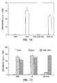
- We next tested cells decorated with CSA-FasL for the induction of apoptosis in responding lymphocytes in mixed lymphocyte culture (MLR), a test frequently used in clinical situations to test the antigenic compatibility between the donor and graft recipients and to test for tolerance. ACI splenocytes were conjugated with EZ-Link Sulfo-NHS-LC-Biotin (5 µM) in 1 ml of PBS at room temperature for 30 rain according to the manufacturer's instructions (Pierce). After several washes in PBS, pH 8.0,1 x 106 cells were incubated in 0.3-1 ml culture supernatants containing 50-100 ng CSA-FasL or supernatants from S2 cells transfected with a nonfunctional FasL construct for 20 min on ice. Cells were washed extensively, irradiated (2000 cGy) and used as targets for PVG.1U lymph node cells at 1:1 ratio (1 x 10⁵ cell/well). Cultures were harvested at various days after pulsing with 1 M/well 3H-thymidine for 18 hours and assessed for proliferation. As shown in Fig. 13, CSA-FasL-decorated cells completely blocked the response. This blockage was CSA-FasL specific as biotinylated cells generated a normal response.
- The potent effect of CSA-FasL-splenocytes to block in vitro alloreactive responses led us to test the role of these cells in blocking alloreactive responses in vivo. PVG.1U rats were immunized i.v. or i.p. with irradiated (2000 cGy) 1 x 10⁷ ACI cells decorated with FasL or control S2 supernatant. Mesenteric lymph nodes were harvested 7 days after injection and used as responders to ACI cells in a standard mixed lymphocyte assay. There was a complete absence of response to donor antigens (
Figure 11 ). This in vivo immune nonresponsiveness was donor specific as the response to third party antigens (LEW) was intact. This effect was FasL-specific as animals receiving ACI cells treated with control S2 supernatant generated a normal response to donor as well as third party antigens as compared with the response of naive animals. - We next tested whether the potent immunosuppressive effect of FasL observed in vitro and in vivo is sufficient for prevention of islet allograft rejection. Diabetes was induced in BALB/c recipients by i.p. injection of streptozocin (260 mg/kg). Animals demonstrating blood glucose levels >450 mg/dl for at least 3 consecutive days were used as recipients of minor+major histocompatibility antigens-disparate BI0 islets. Islets were harvested from C57BL/10 mice and isolated on a Ficoll gradient according to a standard protocol. Splenocytes were harvested from B10 mice, biotinylated (15 µM), decorated with FasL (50-100 ng/106 cells) or control S2 supernatant, and irradiated (2000 rads). One million of splenocytes were co-transplanted with -400 islet cells under the kidney capsule of diabetic BALB/c mice. Survival of islets was assessed by monitoring blood glucose levels starting 3 days after transplantation. FasL-decorated splenocytes prevented the rejection of ∼70% of allogeneic islets (MST>56 day;). In marked contrast, all the recipients co-transplanted with control S2-treated splenocytes and allogeneic islets rejected allografts in a normal tempo (-11 days). Two of the FasL-treated recipients rejected islets in ∼15 days (blood glucose level >250 mg/dl for 3 consecutive days) whereas six recipients currently maintain blood glucose levels of less than 250 mg/dl on 56 days after transplantation. The results presented in this section support our hypothesis that recognition of alloantigens in the presence of FasL leads to functional elimination of alloreactive T cells and prolonged allograft survival. Table II is a summary of these results.
TABLE II Survival of C57BL/10 allogeneic islets in Balb/c mice Group Number Transplantation Survival time, days A 5 Islet 7,8,9,10,10 B 5 Islet+splenocytes- S2 3,3,8,10,10 C 10 Islet+splenocytes- FasL 15, 17, 21, 38,
>56,>56,>56,
>56,>56,>56 - The data presented demonstrate:
- 1. Successful cloning and generation of most of the proposed constructs in Table 1;
- 2. Expression of wtFasL, modified FasL, soluble FasL, and CSA-FasL in mammalian and insect cell lines;
- 3. Purification of the proteins;
- 4. Induction of apoptosis in cell lines using FasL;
- 5. Prevention of alloreactive responses in vitro and in vivo using CSA-FasL as an immune modulator;
- 6. Most importantly, splenocytes expressing CSA-FasL prevented the rejection of islet allograft; and
- 7. Chimeric protein can be produced with human FasL; the cDNA has been cloned and the protein is under construction.
- These findings provide proof of concept for the hypothesis that modified death ligands forming tetramers can be efficiently used for immune modulation to treat autoimmunity and prevent foreign graft rejection. We predict successful application of this approach to the treatment of disorders listed in Table 1 and alluded to in the body of this application.
- While the examples described herein provide the biotin/streptavidin pair for direction of the constructs to the targeted cells, it is well known that other such pairs exist. Avidin can be substituted for streptavidin.
Claims (21)
- A chimeric protein comprising an apoptosis-inducing protein fused to a member of a binding pair that is capable of binding to a selected cell expressing a death receptor, wherein the apoptosis-inducing protein is FasL, TNFα, TWEAK or TRAIL and the member of the binding pair is avidin or streptavidin.
- The chimeric protein of claim 1 wherein the chimeric protein forms tetramers.
- The chimeric protein of any one of claims 1 or 2 wherein the said selected cell expressing a death receptor is an activated lymphocyte.
- Use of the chimeric protein of any one of claims 1, 2 or 3 for the manufacture of a composition for inducing apoptosis in a selected cell expressing a death receptor by exposing said cells to the chimeric protein, thereby inducing immune modulation in a mammal having a condition selected from the group consisting of asthma, allergy, food poisoning, autoimmunity or transplantation of an allogeneic or xenogeneic cell, organ or tissue graft.
- Use of the chimeric protein of claim 1, 2 or 3 for the manufacture of a composition for inducing apoptosis in a selected cell expressing a death receptor by exposing said cells to the chimeric protein, thereby inducing immune modulation in a mammal having a condition selected from the group consisting of diabetes, multiple sclerosis, lupus erythematosis, sarcoidosis or rheumatoid arthritis.
- The use of any one of claims 4 or 5 wherein the selected cell expressing a death receptor is an activated lymphocyte.
- The use of any one of claims 4-6 wherein the selected cell is exposed ex vivo to the chimeric protein of any one of claims 1-3.
- The chimeric protein of any one of claims 1-3 for use as a medicament.
- An effector cell comprising a biotinylated cell decorated with the chimeric protein of any of claims 1 through 3.
- The use of claim 7 wherein the selected cell is exposed to the effector cell of claim 9.
- A substrate comprising a biotinylated solid matrix fused to the chimeric protein of any of claims 1 through 3.
- The substrate of claim 11 wherein the said solid matrix is an agarose bead.
- A method of manufacture of a pharmaceutical for inducing immune tolerance in a mammal comprising exposing a lymphocyte to the substrate of any one of claims 11 or 12.
- The chimeric protein of any of claims 1 through 3, wherein the apoptosis-inducing protein binds to the selected cell expressing a death receptor.
- The chimeric protein of any of claims 1 through 3, wherein the member of the binding pair binds to the selected cell expressing a death receptor.
- The chimeric protein of any of claims 1 to 3, the effector cell of claim 9 or the substrate of claim 11, wherein the member of a binding pair is streptavidin.
- The chimeric protein, effector cell or substrate of claim 16, wherein the streptavidin is core streptavidin.
- The chimeric protein of any of claims 1 to 3, wherein the FasL is modified FasL (mFasL).
- The use of any of claims 4 to 8, wherein the cell expressing a death receptor is biotinylated.
- The use of claim 19, wherein the member of a binding pair is streptavidin.
- The use of claim 20, wherein the streptavidin is core streptavidin.
Applications Claiming Priority (5)
| Application Number | Priority Date | Filing Date | Title |
|---|---|---|---|
| US17803800P | 2000-01-24 | 2000-01-24 | |
| US178038P | 2000-01-24 | ||
| US21558000P | 2000-06-30 | 2000-06-30 | |
| US215580P | 2000-06-30 | ||
| PCT/US2001/002256 WO2001052664A1 (en) | 2000-01-24 | 2001-01-24 | Immune modulation with death receptor-induced apoptosis |
Publications (3)
| Publication Number | Publication Date |
|---|---|
| EP1250055A1 EP1250055A1 (en) | 2002-10-23 |
| EP1250055A4 EP1250055A4 (en) | 2003-04-23 |
| EP1250055B1 true EP1250055B1 (en) | 2010-09-15 |
Family
ID=26873890
Family Applications (1)
| Application Number | Title | Priority Date | Filing Date |
|---|---|---|---|
| EP01905011A Expired - Lifetime EP1250055B1 (en) | 2000-01-24 | 2001-01-24 | Immune modulation with death receptor-induced apoptosis |
Country Status (7)
| Country | Link |
|---|---|
| EP (1) | EP1250055B1 (en) |
| JP (1) | JP4898049B2 (en) |
| AT (1) | ATE481475T1 (en) |
| AU (1) | AU784614B2 (en) |
| CA (1) | CA2396979C (en) |
| DE (1) | DE60143074D1 (en) |
| WO (1) | WO2001052664A1 (en) |
Families Citing this family (3)
| Publication number | Priority date | Publication date | Assignee | Title |
|---|---|---|---|---|
| US7927602B2 (en) | 2002-07-23 | 2011-04-19 | University Of Louisville Research Foundation, Inc. | Fas ligand-avidin/streptavidin fusion proteins |
| AU1676302A (en) | 2000-06-30 | 2002-01-14 | Univ Louisville Res Found | Alteration of cell membrane for new functions |
| US20050084967A1 (en) | 2002-06-28 | 2005-04-21 | Xcyte Therapies, Inc. | Compositions and methods for eliminating undesired subpopulations of T cells in patients with immunological defects related to autoimmunity and organ or hematopoietic stem cell transplantation |
Family Cites Families (6)
| Publication number | Priority date | Publication date | Assignee | Title |
|---|---|---|---|---|
| DK1666591T3 (en) * | 1995-06-29 | 2011-05-23 | Immunex Corp | Cytokine that induces apoptosis |
| WO1997007828A1 (en) * | 1995-08-30 | 1997-03-06 | The Regents Of The University Of California | Therapy for cellular accumulation in chronic inflammatory diseases |
| GB9523469D0 (en) * | 1995-11-16 | 1996-01-17 | Sandoz Ltd | Organic compounds |
| US6046310A (en) * | 1996-03-13 | 2000-04-04 | Protein Design Labs., Inc. | FAS ligand fusion proteins and their uses |
| AU5437098A (en) * | 1996-11-15 | 1998-06-03 | Health Research Inc. | A method for inducing apoptosis of primary central nervous system b cell lymphomas |
| GB9721075D0 (en) * | 1997-10-03 | 1997-12-03 | Cancer Res Campaign Tech | Materials and methods relating to the stimulation of cells |
-
2001
- 2001-01-24 CA CA2396979A patent/CA2396979C/en not_active Expired - Fee Related
- 2001-01-24 DE DE60143074T patent/DE60143074D1/en not_active Expired - Lifetime
- 2001-01-24 EP EP01905011A patent/EP1250055B1/en not_active Expired - Lifetime
- 2001-01-24 AU AU32933/01A patent/AU784614B2/en not_active Ceased
- 2001-01-24 WO PCT/US2001/002256 patent/WO2001052664A1/en active Application Filing
- 2001-01-24 AT AT01905011T patent/ATE481475T1/en not_active IP Right Cessation
- 2001-01-24 JP JP2001552725A patent/JP4898049B2/en not_active Expired - Fee Related
Also Published As
| Publication number | Publication date |
|---|---|
| ATE481475T1 (en) | 2010-10-15 |
| AU784614B2 (en) | 2006-05-11 |
| EP1250055A1 (en) | 2002-10-23 |
| CA2396979C (en) | 2015-07-14 |
| CA2396979A1 (en) | 2001-07-26 |
| EP1250055A4 (en) | 2003-04-23 |
| JP4898049B2 (en) | 2012-03-14 |
| WO2001052664A1 (en) | 2001-07-26 |
| JP2003520214A (en) | 2003-07-02 |
| DE60143074D1 (en) | 2010-10-28 |
| AU3293301A (en) | 2001-07-31 |
Similar Documents
| Publication | Publication Date | Title |
|---|---|---|
| US8551494B2 (en) | Methods of immune modulation with death receptor-induced apoptosis | |
| Nagata | Fas ligand-induced apoptosis | |
| US7659385B2 (en) | Polynucleotides encoding molecules designated LDCAM | |
| EP1666591B1 (en) | Cytokine that induces apoptosis | |
| US9255133B2 (en) | Alteration of cell membrane for new functions using IL-2 and streptavidin | |
| AU2006201501A1 (en) | Compositions and methods for caspase-induced apoptosis | |
| EP1250055B1 (en) | Immune modulation with death receptor-induced apoptosis | |
| JP2003513619A (en) | Gene transfer vector for treating autoimmune diseases and diseases including immunopathogenesis | |
| WO1999021997A1 (en) | Viral encoded semaphorin protein receptor dna and polypeptides | |
| Wajant | Fas signaling | |
| AU724826B2 (en) | Cytokine that includes apoptosis |
Legal Events
| Date | Code | Title | Description |
|---|---|---|---|
| PUAI | Public reference made under article 153(3) epc to a published international application that has entered the european phase |
Free format text: ORIGINAL CODE: 0009012 |
|
| 17P | Request for examination filed |
Effective date: 20020731 |
|
| AK | Designated contracting states |
Kind code of ref document: A1 Designated state(s): AT BE CH CY DE DK ES FI FR GB GR IE IT LI LU MC NL PT SE TR |
|
| AX | Request for extension of the european patent |
Free format text: AL;LT;LV;MK;RO;SI |
|
| RIC1 | Information provided on ipc code assigned before grant |
Ipc: 7C 07K 14/495 B Ipc: 7C 12N 5/06 B Ipc: 7A 61P 35/00 B Ipc: 7A 61P 37/00 B Ipc: 7C 07K 19/00 B Ipc: 7C 12N 15/12 B Ipc: 7C 12N 5/00 A Ipc: 7C 07K 14/525 B Ipc: 7A 61K 47/48 B Ipc: 7C 07K 14/465 B Ipc: 7C 07K 14/36 B Ipc: 7A 61K 35/14 B Ipc: 7C 07K 14/54 B Ipc: 7C 07K 14/705 B Ipc: 7A 61K 35/28 B Ipc: 7C 07K 17/00 B |
|
| A4 | Supplementary search report drawn up and despatched |
Effective date: 20030311 |
|
| RIN1 | Information on inventor provided before grant (corrected) |
Inventor name: SHIRWAN, HAVAL |
|
| GRAP | Despatch of communication of intention to grant a patent |
Free format text: ORIGINAL CODE: EPIDOSNIGR1 |
|
| RIC1 | Information provided on ipc code assigned before grant |
Ipc: A61K 47/48 20060101ALI20100218BHEP Ipc: A61P 37/00 20060101ALI20100218BHEP Ipc: A61K 35/28 20060101ALI20100218BHEP Ipc: A61K 35/14 20060101ALI20100218BHEP Ipc: C07K 14/54 20060101ALI20100218BHEP Ipc: C07K 14/525 20060101ALI20100218BHEP Ipc: C07K 14/36 20060101ALI20100218BHEP Ipc: C07K 19/00 20060101ALI20100218BHEP Ipc: C12N 5/00 20060101AFI20100218BHEP Ipc: A61P 35/00 20060101ALI20100218BHEP Ipc: C07K 14/495 20060101ALI20100218BHEP Ipc: C07K 14/465 20060101ALI20100218BHEP Ipc: C12N 15/12 20060101ALI20100218BHEP Ipc: C07K 14/705 20060101ALI20100218BHEP Ipc: C07K 17/00 20060101ALI20100218BHEP |
|
| GRAS | Grant fee paid |
Free format text: ORIGINAL CODE: EPIDOSNIGR3 |
|
| GRAA | (expected) grant |
Free format text: ORIGINAL CODE: 0009210 |
|
| AK | Designated contracting states |
Kind code of ref document: B1 Designated state(s): AT BE CH CY DE DK ES FI FR GB GR IE IT LI LU MC NL PT SE TR |
|
| REG | Reference to a national code |
Ref country code: GB Ref legal event code: FG4D Ref country code: CH Ref legal event code: EP |
|
| REG | Reference to a national code |
Ref country code: IE Ref legal event code: FG4D |
|
| REF | Corresponds to: |
Ref document number: 60143074 Country of ref document: DE Date of ref document: 20101028 Kind code of ref document: P |
|
| REG | Reference to a national code |
Ref country code: CH Ref legal event code: NV Representative=s name: ING. MARCO ZARDI C/O M. ZARDI & CO. S.A. |
|
| REG | Reference to a national code |
Ref country code: NL Ref legal event code: VDEP Effective date: 20100915 |
|
| PG25 | Lapsed in a contracting state [announced via postgrant information from national office to epo] |
Ref country code: FI Free format text: LAPSE BECAUSE OF FAILURE TO SUBMIT A TRANSLATION OF THE DESCRIPTION OR TO PAY THE FEE WITHIN THE PRESCRIBED TIME-LIMIT Effective date: 20100915 Ref country code: AT Free format text: LAPSE BECAUSE OF FAILURE TO SUBMIT A TRANSLATION OF THE DESCRIPTION OR TO PAY THE FEE WITHIN THE PRESCRIBED TIME-LIMIT Effective date: 20100915 |
|
| PG25 | Lapsed in a contracting state [announced via postgrant information from national office to epo] |
Ref country code: CY Free format text: LAPSE BECAUSE OF FAILURE TO SUBMIT A TRANSLATION OF THE DESCRIPTION OR TO PAY THE FEE WITHIN THE PRESCRIBED TIME-LIMIT Effective date: 20100915 |
|
| PG25 | Lapsed in a contracting state [announced via postgrant information from national office to epo] |
Ref country code: SE Free format text: LAPSE BECAUSE OF FAILURE TO SUBMIT A TRANSLATION OF THE DESCRIPTION OR TO PAY THE FEE WITHIN THE PRESCRIBED TIME-LIMIT Effective date: 20100915 Ref country code: GR Free format text: LAPSE BECAUSE OF FAILURE TO SUBMIT A TRANSLATION OF THE DESCRIPTION OR TO PAY THE FEE WITHIN THE PRESCRIBED TIME-LIMIT Effective date: 20101216 |
|
| PG25 | Lapsed in a contracting state [announced via postgrant information from national office to epo] |
Ref country code: PT Free format text: LAPSE BECAUSE OF FAILURE TO SUBMIT A TRANSLATION OF THE DESCRIPTION OR TO PAY THE FEE WITHIN THE PRESCRIBED TIME-LIMIT Effective date: 20110117 Ref country code: NL Free format text: LAPSE BECAUSE OF FAILURE TO SUBMIT A TRANSLATION OF THE DESCRIPTION OR TO PAY THE FEE WITHIN THE PRESCRIBED TIME-LIMIT Effective date: 20100915 Ref country code: IT Free format text: LAPSE BECAUSE OF FAILURE TO SUBMIT A TRANSLATION OF THE DESCRIPTION OR TO PAY THE FEE WITHIN THE PRESCRIBED TIME-LIMIT Effective date: 20100915 |
|
| PG25 | Lapsed in a contracting state [announced via postgrant information from national office to epo] |
Ref country code: ES Free format text: LAPSE BECAUSE OF FAILURE TO SUBMIT A TRANSLATION OF THE DESCRIPTION OR TO PAY THE FEE WITHIN THE PRESCRIBED TIME-LIMIT Effective date: 20101226 Ref country code: BE Free format text: LAPSE BECAUSE OF FAILURE TO SUBMIT A TRANSLATION OF THE DESCRIPTION OR TO PAY THE FEE WITHIN THE PRESCRIBED TIME-LIMIT Effective date: 20100915 |
|
| PLBE | No opposition filed within time limit |
Free format text: ORIGINAL CODE: 0009261 |
|
| STAA | Information on the status of an ep patent application or granted ep patent |
Free format text: STATUS: NO OPPOSITION FILED WITHIN TIME LIMIT |
|
| 26N | No opposition filed |
Effective date: 20110616 |
|
| PG25 | Lapsed in a contracting state [announced via postgrant information from national office to epo] |
Ref country code: DK Free format text: LAPSE BECAUSE OF FAILURE TO SUBMIT A TRANSLATION OF THE DESCRIPTION OR TO PAY THE FEE WITHIN THE PRESCRIBED TIME-LIMIT Effective date: 20100915 Ref country code: MC Free format text: LAPSE BECAUSE OF NON-PAYMENT OF DUE FEES Effective date: 20110131 |
|
| REG | Reference to a national code |
Ref country code: DE Ref legal event code: R097 Ref document number: 60143074 Country of ref document: DE Effective date: 20110616 |
|
| PG25 | Lapsed in a contracting state [announced via postgrant information from national office to epo] |
Ref country code: LU Free format text: LAPSE BECAUSE OF NON-PAYMENT OF DUE FEES Effective date: 20110124 |
|
| PG25 | Lapsed in a contracting state [announced via postgrant information from national office to epo] |
Ref country code: TR Free format text: LAPSE BECAUSE OF FAILURE TO SUBMIT A TRANSLATION OF THE DESCRIPTION OR TO PAY THE FEE WITHIN THE PRESCRIBED TIME-LIMIT Effective date: 20100915 |
|
| PGFP | Annual fee paid to national office [announced via postgrant information from national office to epo] |
Ref country code: CH Payment date: 20150126 Year of fee payment: 15 Ref country code: IE Payment date: 20150126 Year of fee payment: 15 |
|
| REG | Reference to a national code |
Ref country code: FR Ref legal event code: PLFP Year of fee payment: 16 |
|
| REG | Reference to a national code |
Ref country code: CH Ref legal event code: PL |
|
| PG25 | Lapsed in a contracting state [announced via postgrant information from national office to epo] |
Ref country code: CH Free format text: LAPSE BECAUSE OF NON-PAYMENT OF DUE FEES Effective date: 20160131 Ref country code: LI Free format text: LAPSE BECAUSE OF NON-PAYMENT OF DUE FEES Effective date: 20160131 |
|
| PGFP | Annual fee paid to national office [announced via postgrant information from national office to epo] |
Ref country code: GB Payment date: 20160720 Year of fee payment: 16 Ref country code: DE Payment date: 20160729 Year of fee payment: 16 |
|
| REG | Reference to a national code |
Ref country code: IE Ref legal event code: MM4A |
|
| PGFP | Annual fee paid to national office [announced via postgrant information from national office to epo] |
Ref country code: FR Payment date: 20160722 Year of fee payment: 16 |
|
| PG25 | Lapsed in a contracting state [announced via postgrant information from national office to epo] |
Ref country code: IE Free format text: LAPSE BECAUSE OF NON-PAYMENT OF DUE FEES Effective date: 20160124 |
|
| REG | Reference to a national code |
Ref country code: DE Ref legal event code: R119 Ref document number: 60143074 Country of ref document: DE |
|
| GBPC | Gb: european patent ceased through non-payment of renewal fee |
Effective date: 20170124 |
|
| REG | Reference to a national code |
Ref country code: FR Ref legal event code: ST Effective date: 20170929 |
|
| PG25 | Lapsed in a contracting state [announced via postgrant information from national office to epo] |
Ref country code: FR Free format text: LAPSE BECAUSE OF NON-PAYMENT OF DUE FEES Effective date: 20170131 |
|
| PG25 | Lapsed in a contracting state [announced via postgrant information from national office to epo] |
Ref country code: GB Free format text: LAPSE BECAUSE OF NON-PAYMENT OF DUE FEES Effective date: 20170124 Ref country code: DE Free format text: LAPSE BECAUSE OF NON-PAYMENT OF DUE FEES Effective date: 20170801 |