JP2004523275A - Delivery of therapeutic drugs - Google Patents
Delivery of therapeutic drugs Download PDFInfo
- Publication number
- JP2004523275A JP2004523275A JP2002557301A JP2002557301A JP2004523275A JP 2004523275 A JP2004523275 A JP 2004523275A JP 2002557301 A JP2002557301 A JP 2002557301A JP 2002557301 A JP2002557301 A JP 2002557301A JP 2004523275 A JP2004523275 A JP 2004523275A
- Authority
- JP
- Japan
- Prior art keywords
- day
- release
- therapeutically
- compound
- therapeutically active
- Prior art date
- Legal status (The legal status is an assumption and is not a legal conclusion. Google has not performed a legal analysis and makes no representation as to the accuracy of the status listed.)
- Pending
Links
Images


Classifications
-
- A—HUMAN NECESSITIES
- A61—MEDICAL OR VETERINARY SCIENCE; HYGIENE
- A61L—METHODS OR APPARATUS FOR STERILISING MATERIALS OR OBJECTS IN GENERAL; DISINFECTION, STERILISATION OR DEODORISATION OF AIR; CHEMICAL ASPECTS OF BANDAGES, DRESSINGS, ABSORBENT PADS OR SURGICAL ARTICLES; MATERIALS FOR BANDAGES, DRESSINGS, ABSORBENT PADS OR SURGICAL ARTICLES
- A61L27/00—Materials for grafts or prostheses or for coating grafts or prostheses
- A61L27/50—Materials characterised by their function or physical properties, e.g. injectable or lubricating compositions, shape-memory materials, surface modified materials
- A61L27/54—Biologically active materials, e.g. therapeutic substances
-
- A—HUMAN NECESSITIES
- A61—MEDICAL OR VETERINARY SCIENCE; HYGIENE
- A61F—FILTERS IMPLANTABLE INTO BLOOD VESSELS; PROSTHESES; DEVICES PROVIDING PATENCY TO, OR PREVENTING COLLAPSING OF, TUBULAR STRUCTURES OF THE BODY, e.g. STENTS; ORTHOPAEDIC, NURSING OR CONTRACEPTIVE DEVICES; FOMENTATION; TREATMENT OR PROTECTION OF EYES OR EARS; BANDAGES, DRESSINGS OR ABSORBENT PADS; FIRST-AID KITS
- A61F2/00—Filters implantable into blood vessels; Prostheses, i.e. artificial substitutes or replacements for parts of the body; Appliances for connecting them with the body; Devices providing patency to, or preventing collapsing of, tubular structures of the body, e.g. stents
- A61F2/82—Devices providing patency to, or preventing collapsing of, tubular structures of the body, e.g. stents
- A61F2/86—Stents in a form characterised by the wire-like elements; Stents in the form characterised by a net-like or mesh-like structure
- A61F2/90—Stents in a form characterised by the wire-like elements; Stents in the form characterised by a net-like or mesh-like structure characterised by a net-like or mesh-like structure
- A61F2/91—Stents in a form characterised by the wire-like elements; Stents in the form characterised by a net-like or mesh-like structure characterised by a net-like or mesh-like structure made from perforated sheet material or tubes, e.g. perforated by laser cuts or etched holes
- A61F2/915—Stents in a form characterised by the wire-like elements; Stents in the form characterised by a net-like or mesh-like structure characterised by a net-like or mesh-like structure made from perforated sheet material or tubes, e.g. perforated by laser cuts or etched holes with bands having a meander structure, adjacent bands being connected to each other
-
- A—HUMAN NECESSITIES
- A61—MEDICAL OR VETERINARY SCIENCE; HYGIENE
- A61L—METHODS OR APPARATUS FOR STERILISING MATERIALS OR OBJECTS IN GENERAL; DISINFECTION, STERILISATION OR DEODORISATION OF AIR; CHEMICAL ASPECTS OF BANDAGES, DRESSINGS, ABSORBENT PADS OR SURGICAL ARTICLES; MATERIALS FOR BANDAGES, DRESSINGS, ABSORBENT PADS OR SURGICAL ARTICLES
- A61L31/00—Materials for other surgical articles, e.g. stents, stent-grafts, shunts, surgical drapes, guide wires, materials for adhesion prevention, occluding devices, surgical gloves, tissue fixation devices
- A61L31/14—Materials characterised by their function or physical properties, e.g. injectable or lubricating compositions, shape-memory materials, surface modified materials
- A61L31/16—Biologically active materials, e.g. therapeutic substances
-
- A—HUMAN NECESSITIES
- A61—MEDICAL OR VETERINARY SCIENCE; HYGIENE
- A61P—SPECIFIC THERAPEUTIC ACTIVITY OF CHEMICAL COMPOUNDS OR MEDICINAL PREPARATIONS
- A61P1/00—Drugs for disorders of the alimentary tract or the digestive system
- A61P1/08—Drugs for disorders of the alimentary tract or the digestive system for nausea, cinetosis or vertigo; Antiemetics
-
- A—HUMAN NECESSITIES
- A61—MEDICAL OR VETERINARY SCIENCE; HYGIENE
- A61P—SPECIFIC THERAPEUTIC ACTIVITY OF CHEMICAL COMPOUNDS OR MEDICINAL PREPARATIONS
- A61P17/00—Drugs for dermatological disorders
- A61P17/06—Antipsoriatics
-
- A—HUMAN NECESSITIES
- A61—MEDICAL OR VETERINARY SCIENCE; HYGIENE
- A61P—SPECIFIC THERAPEUTIC ACTIVITY OF CHEMICAL COMPOUNDS OR MEDICINAL PREPARATIONS
- A61P19/00—Drugs for skeletal disorders
- A61P19/04—Drugs for skeletal disorders for non-specific disorders of the connective tissue
-
- A—HUMAN NECESSITIES
- A61—MEDICAL OR VETERINARY SCIENCE; HYGIENE
- A61P—SPECIFIC THERAPEUTIC ACTIVITY OF CHEMICAL COMPOUNDS OR MEDICINAL PREPARATIONS
- A61P29/00—Non-central analgesic, antipyretic or antiinflammatory agents, e.g. antirheumatic agents; Non-steroidal antiinflammatory drugs [NSAID]
-
- A—HUMAN NECESSITIES
- A61—MEDICAL OR VETERINARY SCIENCE; HYGIENE
- A61P—SPECIFIC THERAPEUTIC ACTIVITY OF CHEMICAL COMPOUNDS OR MEDICINAL PREPARATIONS
- A61P31/00—Antiinfectives, i.e. antibiotics, antiseptics, chemotherapeutics
- A61P31/12—Antivirals
-
- A—HUMAN NECESSITIES
- A61—MEDICAL OR VETERINARY SCIENCE; HYGIENE
- A61P—SPECIFIC THERAPEUTIC ACTIVITY OF CHEMICAL COMPOUNDS OR MEDICINAL PREPARATIONS
- A61P35/00—Antineoplastic agents
-
- A—HUMAN NECESSITIES
- A61—MEDICAL OR VETERINARY SCIENCE; HYGIENE
- A61P—SPECIFIC THERAPEUTIC ACTIVITY OF CHEMICAL COMPOUNDS OR MEDICINAL PREPARATIONS
- A61P37/00—Drugs for immunological or allergic disorders
- A61P37/02—Immunomodulators
- A61P37/06—Immunosuppressants, e.g. drugs for graft rejection
-
- A—HUMAN NECESSITIES
- A61—MEDICAL OR VETERINARY SCIENCE; HYGIENE
- A61P—SPECIFIC THERAPEUTIC ACTIVITY OF CHEMICAL COMPOUNDS OR MEDICINAL PREPARATIONS
- A61P39/00—General protective or antinoxious agents
- A61P39/06—Free radical scavengers or antioxidants
-
- A—HUMAN NECESSITIES
- A61—MEDICAL OR VETERINARY SCIENCE; HYGIENE
- A61P—SPECIFIC THERAPEUTIC ACTIVITY OF CHEMICAL COMPOUNDS OR MEDICINAL PREPARATIONS
- A61P43/00—Drugs for specific purposes, not provided for in groups A61P1/00-A61P41/00
-
- A—HUMAN NECESSITIES
- A61—MEDICAL OR VETERINARY SCIENCE; HYGIENE
- A61P—SPECIFIC THERAPEUTIC ACTIVITY OF CHEMICAL COMPOUNDS OR MEDICINAL PREPARATIONS
- A61P7/00—Drugs for disorders of the blood or the extracellular fluid
- A61P7/02—Antithrombotic agents; Anticoagulants; Platelet aggregation inhibitors
-
- A—HUMAN NECESSITIES
- A61—MEDICAL OR VETERINARY SCIENCE; HYGIENE
- A61P—SPECIFIC THERAPEUTIC ACTIVITY OF CHEMICAL COMPOUNDS OR MEDICINAL PREPARATIONS
- A61P9/00—Drugs for disorders of the cardiovascular system
- A61P9/08—Vasodilators for multiple indications
-
- A—HUMAN NECESSITIES
- A61—MEDICAL OR VETERINARY SCIENCE; HYGIENE
- A61F—FILTERS IMPLANTABLE INTO BLOOD VESSELS; PROSTHESES; DEVICES PROVIDING PATENCY TO, OR PREVENTING COLLAPSING OF, TUBULAR STRUCTURES OF THE BODY, e.g. STENTS; ORTHOPAEDIC, NURSING OR CONTRACEPTIVE DEVICES; FOMENTATION; TREATMENT OR PROTECTION OF EYES OR EARS; BANDAGES, DRESSINGS OR ABSORBENT PADS; FIRST-AID KITS
- A61F2/00—Filters implantable into blood vessels; Prostheses, i.e. artificial substitutes or replacements for parts of the body; Appliances for connecting them with the body; Devices providing patency to, or preventing collapsing of, tubular structures of the body, e.g. stents
- A61F2/95—Instruments specially adapted for placement or removal of stents or stent-grafts
-
- A—HUMAN NECESSITIES
- A61—MEDICAL OR VETERINARY SCIENCE; HYGIENE
- A61F—FILTERS IMPLANTABLE INTO BLOOD VESSELS; PROSTHESES; DEVICES PROVIDING PATENCY TO, OR PREVENTING COLLAPSING OF, TUBULAR STRUCTURES OF THE BODY, e.g. STENTS; ORTHOPAEDIC, NURSING OR CONTRACEPTIVE DEVICES; FOMENTATION; TREATMENT OR PROTECTION OF EYES OR EARS; BANDAGES, DRESSINGS OR ABSORBENT PADS; FIRST-AID KITS
- A61F2/00—Filters implantable into blood vessels; Prostheses, i.e. artificial substitutes or replacements for parts of the body; Appliances for connecting them with the body; Devices providing patency to, or preventing collapsing of, tubular structures of the body, e.g. stents
- A61F2/82—Devices providing patency to, or preventing collapsing of, tubular structures of the body, e.g. stents
- A61F2/86—Stents in a form characterised by the wire-like elements; Stents in the form characterised by a net-like or mesh-like structure
- A61F2/90—Stents in a form characterised by the wire-like elements; Stents in the form characterised by a net-like or mesh-like structure characterised by a net-like or mesh-like structure
- A61F2/91—Stents in a form characterised by the wire-like elements; Stents in the form characterised by a net-like or mesh-like structure characterised by a net-like or mesh-like structure made from perforated sheet material or tubes, e.g. perforated by laser cuts or etched holes
- A61F2/915—Stents in a form characterised by the wire-like elements; Stents in the form characterised by a net-like or mesh-like structure characterised by a net-like or mesh-like structure made from perforated sheet material or tubes, e.g. perforated by laser cuts or etched holes with bands having a meander structure, adjacent bands being connected to each other
- A61F2002/91533—Stents in a form characterised by the wire-like elements; Stents in the form characterised by a net-like or mesh-like structure characterised by a net-like or mesh-like structure made from perforated sheet material or tubes, e.g. perforated by laser cuts or etched holes with bands having a meander structure, adjacent bands being connected to each other characterised by the phase between adjacent bands
-
- A—HUMAN NECESSITIES
- A61—MEDICAL OR VETERINARY SCIENCE; HYGIENE
- A61F—FILTERS IMPLANTABLE INTO BLOOD VESSELS; PROSTHESES; DEVICES PROVIDING PATENCY TO, OR PREVENTING COLLAPSING OF, TUBULAR STRUCTURES OF THE BODY, e.g. STENTS; ORTHOPAEDIC, NURSING OR CONTRACEPTIVE DEVICES; FOMENTATION; TREATMENT OR PROTECTION OF EYES OR EARS; BANDAGES, DRESSINGS OR ABSORBENT PADS; FIRST-AID KITS
- A61F2230/00—Geometry of prostheses classified in groups A61F2/00 - A61F2/26 or A61F2/82 or A61F9/00 or A61F11/00 or subgroups thereof
- A61F2230/0002—Two-dimensional shapes, e.g. cross-sections
- A61F2230/0028—Shapes in the form of latin or greek characters
- A61F2230/0054—V-shaped
-
- A—HUMAN NECESSITIES
- A61—MEDICAL OR VETERINARY SCIENCE; HYGIENE
- A61F—FILTERS IMPLANTABLE INTO BLOOD VESSELS; PROSTHESES; DEVICES PROVIDING PATENCY TO, OR PREVENTING COLLAPSING OF, TUBULAR STRUCTURES OF THE BODY, e.g. STENTS; ORTHOPAEDIC, NURSING OR CONTRACEPTIVE DEVICES; FOMENTATION; TREATMENT OR PROTECTION OF EYES OR EARS; BANDAGES, DRESSINGS OR ABSORBENT PADS; FIRST-AID KITS
- A61F2250/00—Special features of prostheses classified in groups A61F2/00 - A61F2/26 or A61F2/82 or A61F9/00 or A61F11/00 or subgroups thereof
- A61F2250/0058—Additional features; Implant or prostheses properties not otherwise provided for
- A61F2250/0067—Means for introducing or releasing pharmaceutical products into the body
-
- A—HUMAN NECESSITIES
- A61—MEDICAL OR VETERINARY SCIENCE; HYGIENE
- A61L—METHODS OR APPARATUS FOR STERILISING MATERIALS OR OBJECTS IN GENERAL; DISINFECTION, STERILISATION OR DEODORISATION OF AIR; CHEMICAL ASPECTS OF BANDAGES, DRESSINGS, ABSORBENT PADS OR SURGICAL ARTICLES; MATERIALS FOR BANDAGES, DRESSINGS, ABSORBENT PADS OR SURGICAL ARTICLES
- A61L2300/00—Biologically active materials used in bandages, wound dressings, absorbent pads or medical devices
- A61L2300/40—Biologically active materials used in bandages, wound dressings, absorbent pads or medical devices characterised by a specific therapeutic activity or mode of action
- A61L2300/416—Anti-neoplastic or anti-proliferative or anti-restenosis or anti-angiogenic agents, e.g. paclitaxel, sirolimus
-
- A—HUMAN NECESSITIES
- A61—MEDICAL OR VETERINARY SCIENCE; HYGIENE
- A61L—METHODS OR APPARATUS FOR STERILISING MATERIALS OR OBJECTS IN GENERAL; DISINFECTION, STERILISATION OR DEODORISATION OF AIR; CHEMICAL ASPECTS OF BANDAGES, DRESSINGS, ABSORBENT PADS OR SURGICAL ARTICLES; MATERIALS FOR BANDAGES, DRESSINGS, ABSORBENT PADS OR SURGICAL ARTICLES
- A61L2300/00—Biologically active materials used in bandages, wound dressings, absorbent pads or medical devices
- A61L2300/60—Biologically active materials used in bandages, wound dressings, absorbent pads or medical devices characterised by a special physical form
- A61L2300/602—Type of release, e.g. controlled, sustained, slow
Landscapes
- Health & Medical Sciences (AREA)
- Life Sciences & Earth Sciences (AREA)
- Public Health (AREA)
- Animal Behavior & Ethology (AREA)
- Engineering & Computer Science (AREA)
- Chemical & Material Sciences (AREA)
- Veterinary Medicine (AREA)
- General Health & Medical Sciences (AREA)
- Medicinal Chemistry (AREA)
- Pharmacology & Pharmacy (AREA)
- Organic Chemistry (AREA)
- Nuclear Medicine, Radiotherapy & Molecular Imaging (AREA)
- General Chemical & Material Sciences (AREA)
- Chemical Kinetics & Catalysis (AREA)
- Bioinformatics & Cheminformatics (AREA)
- Biomedical Technology (AREA)
- Heart & Thoracic Surgery (AREA)
- Transplantation (AREA)
- Cardiology (AREA)
- Immunology (AREA)
- Vascular Medicine (AREA)
- Oral & Maxillofacial Surgery (AREA)
- Molecular Biology (AREA)
- Epidemiology (AREA)
- Dermatology (AREA)
- Optics & Photonics (AREA)
- Physics & Mathematics (AREA)
- Physical Education & Sports Medicine (AREA)
- Rheumatology (AREA)
- Virology (AREA)
- Toxicology (AREA)
- Biochemistry (AREA)
- Hematology (AREA)
- Diabetes (AREA)
- Pain & Pain Management (AREA)
- Oncology (AREA)
- Communicable Diseases (AREA)
- Surgery (AREA)
- Hospice & Palliative Care (AREA)
- Otolaryngology (AREA)
Abstract
Description
【技術分野】
【0001】
1. 発明の分野
本発明は、一般に、医療用装置と方法に関する。さらに具体的には、本発明は、再狭窄を低下または阻止するための血管ステントおよび移植片などの管腔補綴物を提供する。
【0002】
患者の血管のアテローム性動脈硬化性狭窄領域を治療して十分な血流を回復するために、数多くの経皮的血管内手法が開発されている。これらの治療のうち最も成功を収めているのは、経皮経管血管形成術(PTA)である。PTAでは、通常拡張可能なバルーン形態の拡張可能な遠位端部を有するカテーテルを血管の狭窄部位に配置する。血管を拡張して、疾患領域に十分な血流を回復させるために、拡張可能な端部を拡張させる。狭窄領域を開通するための他の手法には、定方向関節切除術、回転方式関節切除術、レーザー血管形成術、ステント術等が挙げられる。これらの手法は広範に許容されているが(単独または併用、特にPTAにステント術を併用)、それらは重大な欠点を有したままである。狭窄領域を開通するためのPTAおよび他の周知の手法の特に一般的な欠点は、再狭窄の出現頻度が高いことである。
【0003】
再狭窄とは、最初に血管形成術が成功を収めた後の動脈の再狭窄を指す。再狭窄は血管形成患者全体のおよそ50%に生じ、内腔開通血管形成手法中に血管壁が損傷される結果である。一部の患者では、損傷部は、血管形成術によって障害を受けた領域の「過形成」と言われる平滑筋細胞増殖によって特徴付けられる修復反応を開始する。この平滑筋細胞の増殖は、血管形成術によって開通された内腔を数週間から数ヶ月以内に再狭窄し、それによって、再狭窄を軽減するために反復的なPTAまたは他の手法を必要とする。
【0004】
過形成を治療し、再狭窄を減少するために、数多くの方法が提案されている。以前に提案された方法には、血管形成術中の長期バルーン拡張、加熱式バルーン(heated balloon)による血管の治療、血管形成術後の放射線による血管の治療、領域のステント術および他の手法が挙げられる。これらの提案は様々なレベルで成功したが、これらの手法はどれも、再狭窄および過形成の発生を全て実質的または完全に回避するという点で成功したと証明されているわけではない。
【0005】
上記に記載する治療の変法または補助的な方法として、再狭窄を阻害するためのPTA後に治療薬を投与することも投与されている。治療的治療は通常、カテーテルを介してまたはステントから薬剤を押出すまたは放出することを必要とする。有望性は大きいが、再狭窄を阻止するための治療薬の送達は成功に達していない。
【0006】
従って、血管形成および他の介入治療の後に生じることがある再狭窄および過形成を減少、阻害または治療するための改善された装置および方法を提供することは大きな進歩となると考えられる。本発明は、これらの必要性および他の必要性の少なくともいくつかを満たす。
【背景技術】
【0007】
2. 背景技術の説明
本発明に使用する例示的な管腔補綴物の十分な説明は、その全開示が参照として本明細書に組み入れられている、2000年5月4日に出願された同時継続中の出願第09/565,560号に記載されている。移植可能な装置および他の装置から活性物質を放出する方法および装置は、米国特許第6,096,070号、同第5,824,049号、同第5,624,411号、同第5,609,629号、同第5,569,463号、同第5,447,724号および同第5,464,650号に記載されている。血管内での薬剤送達のためのステントの使用は、国際公開公報第01/01957号および米国特許第6,099,561号、同第6,071,305号、同第6,063,101号、同第5,997,468号、同第5,980,551号、同第5,980,566号、同第5,972,027号、同第5,968,092号、同第5,951,586号、同第5,893,840号、同第5,891,108号、同第5,851,231号、同第5,843,172号、同第5,837,008号、同第5,769,883号、同第5,735,811号、同第5,700,286号、同第5,679,400号、同第5,649,977号、同第5,637,113号、同第5,591,227号、同第5,551,954号、同第5,545,208号、同第5,500,013号、同第5,464,450号、同第5,419,760号、同第5,411,550号、同第5,342,348号、同第5,286,254号および同第5,163,952号に記載されている。生分解性材料は、米国特許第6,051,276号、同第5,879,808号、同第5,876,452号、同第5,656,297号、同第5,543,158号、同第5,484,584号、同第5,176,907号、同第4,894,231号、同第4,897,268号、同第4,883,666号、同第4,832,686号および同第3,976,071号に記載されている。律速バリアとしてのヒドロシクロシロキサンの用途は、米国特許第5,463,010号に記載されている。ステントをコーティングする方法は米国特許第5,356,433号に記載されている。移植可能な装置の生体適合性を増加するためのコーティングは米国特許第5,463,010号、同第5,112,457および同第5,067,491号に記載されている。エネルギーに基づく装置は、米国特許第6,031,375号、同第5,928,145号、同第5,735,811号、同第5,728,062号、同第5,725,494号、同第5,409,000号、同第5,368,557号、同第5,000,185号および同第4,936,281号に記載されている。その一部は薬剤送達システムに使用されている磁気的手法は、米国特許第5,427,767号、同第5,225,282号、同第5,206,159号、同第5,069,216号、同第4,904,479号、同第4,871,716号、同第4,501,726号、同第4,357,259号、同第4,345,588号および同第4,335,094号に記載されている。
【発明の開示】
【0008】
発明の概要
本発明は、血管内介入後の再狭窄および過形成を阻止するための改善された装置および方法を提供する。特に、本発明は、再狭窄を阻止するために、患者の血管の選択した位置に、高い効率および/または有効性で、プログラムされ、制御された物質の送達を可能にする管腔補綴物を提供する。さらに、本発明は、薬剤流失を最小にし、血管の内皮化に対する妨害をほとんどまたは全く与えない。
【0009】
「血管介入」という用語は、生体の管腔内の狭窄、再狭窄または血栓状態を少なくとも一部解消するために実施することができる種々の矯正手法を含む。通常、矯正手法はバルーン血管形成術を含む。矯正手法は、治療後の血管内腔が拡張されて、治療前に存在した狭窄状態を少なくとも一部軽減する定方向関節切除術、回転方式関節切除術、レーザー血管形成術、ステント術等も含みうる。
【0010】
本発明の第一の局面において、管腔送達補綴物は、生体の管腔内に移植することが可能である足場と、物質を放出するための足場上の手段を含む。物質は、物質送達速度が閾値レベルより低い初期相と、物質送達速度が閾値レベルより高い後期相を含む所定の時間パターンで放出される。
【0011】
本発明の所定の時間パターンは、物質の放出が実質的に高くてもよい後期相に達するまで、少量または最低量の物質を放出することによって、薬剤送達効率を改善している。従って、実質的に事象が発症して平滑筋細胞の増殖(過形成)を生じるときに再狭窄に影響するように、時間遅延型物質放出をプログラムすることができる。本発明は、さらに、物質が直接血流に損失するのを減少するためにステントにバリアを形成する少なくとも初期の細胞化/内皮化後に生じるように、物質放出の時期を調節することによって、物質の流失を最小にすることができる。さらに、所定の時間パターンは物質の負荷および/または物質濃度を低下させることができるだけでなく、薬剤流失の最小化および物質放出の高い効率により、おそらく血管壁の内皮化に対する妨害をほとんどまたは全く与えない。
【0012】
足場は、管腔の開通性をさらに維持するステント形態であっても、または管腔壁の強度をさらに保護または増加する移植片形態であってもよい。足場は放射状に拡張可能および/または自己拡張性であってもよく、好ましくは生体の管腔内の管腔留置に好適である。生体の管腔は、静脈、動脈、大動脈を含み、特に冠動脈および抹消動脈並びに以前に移植された移植片、シャント、フィステル等を含む患者の血管系の任意の血管であってもよい。本発明はまた、他の生体の管腔および器官、神経、腺、輸送管等などの多数の生体内組織器官に適用することもできると考えられる。本発明に使用する例示的なステントは、同時継続中の出願第09/565,560号に記載されている。
【0013】
時間遅延型放出の上記の利点により多種多様の物質を効果的に送達することができると考えられる。物質は、免疫抑制剤、抗炎症剤、抗増殖剤、抗遊走剤、抗線維症剤、抗血栓剤、抗血小板剤、IIb/IIIa剤からなる群より選択される少なくとも1つの薬剤を含んでもよい。好ましくは、薬剤は、ミコフェノール酸、ラパマイシン、シクロスポリンA、シクロヘキシミド、シクロホスファミド、ミゾリビン、メチルプレドニゾロン、アザチオプリン、リボビリン、FK506、トリアゾフリン、メトトレキセート、ザフリンおよびミコフェノール酸モフェチルからなる群より選択される免疫抑制剤である。放出される物質の総量は、典型的には1g〜2000gの範囲であり、好ましくは10g〜1000gの範囲であり、最も好ましくは50g〜500gの範囲である。初期相の放出速度は、典型的には0g/日〜50g/日であり、通常5g/日〜30g/日である。後期相の物質の放出速度はかなり大きく、典型的には5g/日〜200g/日の範囲であり、通常10g/日〜100g/日の範囲である。従って、初期相の放出速度は、典型的には後期相の放出速度の0%〜99%であり、通常0%〜90%であり、好ましくは0%〜75%である。当然のことながら、放出速度は、初期相および後期放出相のどちらかまたは両方の期間中変化してもよい。同じ物質および/または異なる物質を放出するために追加の相が存在してもよい。
【0014】
初期相、後期相および任意の他の追加の相の期間は変化する。典型的には、初期相は、ステントの少なくとも一部の最初の細胞化または内皮化を可能にするほど十分に長く、通常12週間未満であり、さらに通常1時間〜8週間であり、より好ましくは12時間〜2週間であり、最も好ましくは1日〜1週間である。後期相の期間もまた変化してもよく、典型的には4時間〜24週間であり、さらに通常1日〜12週間であり、より好ましくは血管環境において2日〜8週間の期間であり、最も好ましくは血管環境において3日〜50日の期間である。
【0015】
本発明は、罹患しやすい組織部位を調製または治療するための改善された装置および方法に関する。本明細書において使用する罹患しやすい組織部位は、障害(例えば、疾患、医学的状態)の結果として損傷される、もしくは損傷されている可能性のある組織、または血管内介入などの介入性処置中もしくは手法後に損傷されている可能性のある組織を指す。「血管内介入」という用語は、血管、通常冠動脈などの動脈の狭窄、再狭窄または血栓状態を少なくとも部分的に解消するために実施することができる種々の矯正手法を含む。通常、矯正手法はバルーン血管形成術を含む。矯正手法は、治療後の血管内腔が拡張されて、治療前に存在した狭窄状態を少なくとも部分的に軽減する定方向関節切除術、回転方式関節切除術、レーザー血管形成術、ステント術等も含みうる。罹患しやすい組織部位は、生体管腔、器官または局在化した腫瘍に関連する組織を含む。一態様において、本発明の装置および方法は、血管内介入の後に生じる可能性のある再狭窄および/または過形成の形成または進行を低下させる。特に、本発明は生体、特に体内装置とこれを使用する方法に関する。
【0016】
本明細書において使用する「体内」とは、生体内の生体管腔または生体内組織および器官を指す。生体管腔は、静脈、動脈、大動脈を含む、特に冠動脈および抹消動脈を含む患者の血管系の任意の血管および以前に移植された移植片、シャント、フィステル等であってもよい。本発明は、悪性腫瘍の過剰な細胞増殖が認められる胆管などの他の生体管腔に適用することもできる。生体内組織および器官の例には、種々の器官、神経、腺、輸送管等が挙げられる。一態様において、本発明の装置は、血管ステントまたは移植片などの管腔補綴物を含む。別の態様において、本発明の装置は、心ペースメーカーリードもしくはリードチップ、心除細動器リードもしくはリードチップ、心臓弁、縫合糸もしくは針、ペースメーカー、矯正装置、器具、移植物もしくは置換物、または上記のいずれかの一部を含んでもよい。
【0017】
一態様において、本発明の装置および方法は、再狭窄(動脈が、ステント術などの血管介入直後の血管径の約40%〜約80%、通常約50%〜約70%より狭い場合と規定される)の発生を低下および/または阻止すると同時に、好ましくは制御された方法で、少量の細胞化、内皮化または新生内膜の形成を可能にする。
【0018】
一態様において、本発明の装置は、構造物と、構造物に付随している治療能力のある少なくとも1つの薬剤の少なくとも1つの供給源とを含む。一態様において、構造物は拡張可能構造物であってもよい。別の態様において、構造物は実質的に一定のサイズもしくは径を有してもよく、または代替的に、適用および用途に応じて収縮可能な構造物であってもよい。一態様において、構造物は少なくとも1つの表面、通常組織接触面を含む。別の態様において、構造物は組織接触面および別の表面、通常管腔接触面を含む。一態様において、構造物は、2つの表面、通常組織接触面および管腔接触面の間に配置される内側を有してもよい。
【0019】
本発明の装置は、罹患しやすい組織部位を含む生体内に移植することができる拡張可能構造物を含む。代替的にまたは追加的に、本発明の装置は、標的生体部位において拡張を実施するまたは実施しないで移植するように構成された移植可能な装置であってもよい。標的生体部位は罹患しやすい組織部位を含んでも、または部位(例えば、他の生体器官または管腔)、例えば、罹患しやすい組織部位に血液を供給する動脈などの標的生体内部位であってもよい。一態様において、拡張可能構造物は、管腔の開通性をさらに維持するステント形態であっても、または管腔壁の強度をさらに保護または増加する移植片形態であってもよい。本発明の装置の少なくとも一部は、開いた格子(open lattice)から形成される足場または少なくとも実質的に閉じた表面を含んでもよい。一態様において、ステントの少なくとも一部は、開いた格子から形成される足場を含む。拡張可能構造物は、放射状に拡張可能および/または自己拡張性であってもよく、好ましくは生体管腔内の管腔留置に好適である。
【0020】
拡張可能構造物は、金属、ポリマーまたはそれらの組合せなどの任意の好適な材料から製造することができる。一態様において、拡張可能構造物は、ポリマー材料、金属材料またはそれらの組合せからなる群より選択される少なくとも部分的に生分解性の材料から製造することができる。少なくとも部分的に生分解性の材料は、好ましくは経時的に分解する。ポリマー材料の例には、分解が遅く、構造物が分解される前に血管の回復を可能にするポリ-L-乳酸が挙げられる。金属材料の例には、ステンレス鋼などの、体内で分解可能な金属または合金が挙げられる。本発明に使用する例示的なステントは、その全体の開示内容が参照として本明細書に組み入れられている、同時継続中の出願第09/565,560号に記載されている。
【0021】
治療能力のある薬剤の少なくとも一部は、本発明の装置を生体内または生体上に導入直後に、または導入後しばらくしてから罹患しやすい組織部位に利用されるように、構造物に付随している。本明細書において使用する「付随している」という用語は、直接的または間接的に連結、接続、表面に配置、内部に配置、取り付け、粘着、接着、隣接、封入、内部に吸収、表面に吸収されるなどの任意の形態の関連および同様の構成を指す。
【0022】
供給源は、構造物の少なくとも一部に隣接して配置または形成することができる。一態様において、供給源は、構造物の少なくとも一部に隣接して配置または形成することができる。一態様において、供給源は、拡張可能構造物のどちらかの面または両方の表面の少なくとも一部に隣接して、2つの表面の間に配置される構造物の内側、またはそれらの任意の組合せで配置または形成することができる。治療能力のある薬剤と構造物との関連は連続していてもまたは別個のセグメントであってもよい。
【0023】
一態様において、管腔補綴物は、再狭窄および/または過形成の形成または進行を低下させるために、罹患しやすい組織部位を含む患者の血管径内の1つまたは複数の選択した位置に1つまたは複数の治療能力のある薬剤を利用させる。本明細書において使用する治療能力のある薬剤は、治療中の被験者に導入されたとき治療作用を示す、天然の物質もしくは条件と反応する結果被験者の生体に流入した後に治療作用を示すようになる、または導入される別の物質もしくは条件である化合物を含む。本明細書において使用する「利用される」という用語は、物質が、罹患しやすい組織部位などの目的の部位に実際に送達される、目的の部位に使用されるまたは目的の部位に組入れられるかどうかにかかわらず、生体内の位置または標的部位などの体内の位置で物質が利用されることを含む、放出または投与時に物質(例えば、治療能力のある薬剤)を提供したことを意味する。
【0024】
罹患しやすい組織部位への治療能力のある薬剤の送達または罹患しやすい組織部位に治療能力のある薬剤を利用させることは直接的であってもよく、または別の生体部位を介して間接的であってもよい。一態様において、別の生体部位は標的生体内部位、例えば、罹患しやすい組織部位に血液を供給する動脈などの生体内管腔である。
【0025】
本明細書において使用する治療能力のある薬剤は、治療中の生体(例えば、被験者)に導入されたとき治療作用を示す、または、例えば、天然もしくは非天然の物質もしくは条件と反応することによって、被験者の生体に流入(事情によっては、生体表面に暴露)した後に治療作用を示すようになる化合物を含む。天然の条件の例には、pH(例えば、酸性度)、化学物質、体温、塩分のモル浸透圧濃度および伝導性が挙げられ、非天然条件には、磁場、電磁場(ラジオ波放電およびマイクロ波)および超音波などが挙げられる。本明細書において、治療能力のある薬剤または他の化合物のいずれかの化学名は、化合物自体およびそのプロドラッグ(生体内で活性型化合物に変換される前駆体物質)および/または薬学的誘導体、類似物または代謝物(化合物が生体内で直接または他の薬剤もしくは条件(例えば、酵素、化学物質、エネルギー)または環境(例えば、pH)を導入する結果変換する生物活性化合物)を指すために使用される。
【0026】
治療能力のある薬剤は、免疫抑制剤、抗炎症剤、抗増殖剤、抗遊走剤、抗線維症剤、アポトーシス促進剤、カルシウムチャネル遮断剤、抗癌剤、抗体、抗血栓剤、抗血小板剤、IIb/IIIa剤、抗ウィルス剤およびそれらの組合せからなる群より選択される。
【0027】
治療能力のある薬剤の具体的な例として、ミコフェノール酸、ミコフェノール酸モフェチル、ミゾリビン、メチルプレドニゾロン、デキサメサゾン、Certican(商標)、ラパマイシン、Triptolide(商標)、Methotrexate(商標)、Benidipine(商標)、Ascomycin(商標)、Wortmannin(商標)、LY294002、Camptothecin(商標)、Topotecan(商標)、ヒドロキシウレア、Tacrolimus(商標)(FK506)、シクロホスファミド、シクロスポリン、ダクリズマブ、アザチオプリン、プレドニゾン、Gemcitabine(商標)、それらの誘導体および組合せが挙げられる。
【0028】
一態様において、治療能力のある薬剤の供給源は、治療能力のある薬剤部分をポリマーの構造サブユニットとして含むポリマー材料である。治療能力のある薬剤部分は重合され、好適な結合(例えば、エチレン結合)を解して互いに結合されて、治療能力のある薬剤ポリマーを形成する。治療能力のある薬剤ポリマーは、組織または血液などの体液に接触させられると、治療能力のある薬剤ポリマーサブユニットは解離する。または、治療能力のある薬剤ポリマーが、好ましくは表面分解または加水分解によって分解または加水分解すると、治療能力のある薬剤を放出することができ、好ましくはある期間の間罹患しやすい組織部位に治療能力のある薬剤を利用させることができる。治療能力のある薬剤を重合するための方法および化合物の例は、その全体の開示内容が参照として本明細書に組み入れられており、ルトガーズ大学(Rutgers University)に付与された、Kathryn Uhrichによる表題「Polyanhydrides With Therapeutically Useful Degradation Products」の国際公開公報第99/12990号の特許出願に記載されている。治療能力のある薬剤および好適な反応成分単位の例には、酸性触媒エステル化反応におけるミコフェノール酸およびアジピン酸および/またはサリチル酸、酸性触媒エステル化反応におけるミコフェノール酸およびアスピリンおよび/またはアジピン酸、酸性触媒エステル化反応におけるミコフェノール酸および他のNSAIDSおよび/またはアジピン酸が挙げられる。一態様において、治療能力のある薬剤ポリマーをポリマーおよび/または金属の骨格に付随させることができる。
【0029】
本発明の装置は、類似または異なる性能(例えば、放出)プロフィールを有する1つまたは複数の相において治療能力のある薬剤を放出または利用させるように構成することができる。治療能力のある薬剤は、平滑筋細胞の増殖、炎症、免疫応答、高血圧またはこれらの活性化を補足するものの任意の1つ以上を低下させるのに効果的な、1つまたは複数の相および/または送達速度において徐放的、間欠的または持続的であってもよい量を組織に利用させることができる。治療能力のある少なくとも1つの薬剤の任意の1つは、増殖/再狭窄作用の予防または低下、血栓形成の低下または阻止、血小板活性化の低下または阻止、血管攣縮の低下または予防等を含む、1つまたは複数の機能を発揮することができる。
【0030】
組織に利用される治療能力のある薬剤の総量は、一部には、望ましい治療結果のレベルおよび量に依存する。治療能力のある薬剤は、各相が他の相と同様または異なる放出速度および期間を有する1つまたは複数の相において利用されうる。放出速度は事前に規定することができる。一態様において、放出速度は、罹患しやすい組織部位に徐放的レベルの治療能力のある薬剤を提供することができる。別の態様において、放出速度は実質的に一定である。放出速度は経時的に低下および/または増加してもよく、必要に応じて、実質的に非放出速度を含んでもよい。放出速度は複数の速度を含んでもよい。一態様において、複数の放出速度は、実質的に一定、低下、増加、実質的に非放出からなる群より選択される少なくとも2つの速度を含む。
【0031】
利用または放出される治療能力のある薬剤の総量は、典型的には約0.1ug〜約10g、一般に約0.1ug〜約10mg、好ましくは約1ug〜約10mg、より好ましくは約1ug〜約2mg、10ug〜約2mgまたは約50ug〜約1mgの範囲の量である。
【0032】
一態様において、治療能力のある薬剤は、装置の移植時から測定したとき、約1日〜約200日、約1日〜約45日または約7日〜約21日の範囲の期間に放出されうる。
【0033】
一態様において、1日あたりの治療能力のある薬剤の放出速度は、約0.001マイクログラム(ug)〜約200ug、好ましくは約0.5ug〜約200ug、最も好ましくは約1ug〜約60ugの範囲であってもよい。
【0034】
治療能力のある薬剤は初期相および1つ以上後期相において利用されうる。治療能力のある薬剤が異なる相において送達される場合には、初期送達速度は、典型的には後期放出相の約0%〜約99%、通常約0%〜約90%、好ましくは約0%〜75%である。一態様において、初期相における物質の哺乳類組織濃度は、典型的には約0.001ナノグラム(ng)/組織1mg〜約100ug/組織1mg、約1ng/組織1mg〜約100ug/組織1mg、約1ng/組織1mg〜約10ug/組織1mgの範囲内である。後期相における物質の哺乳類組織濃度は、典型的には約0.001ng/組織1mg〜約600ug/組織1mg、好ましくは約1ng/組織1mg〜約10ug/組織1mgの範囲内である。
【0035】
初期相中の送達速度は、典型的には約0.001ng/日〜約50ug/日、通常約0.1ng/日〜約30ug/日、より好ましくは約1ug/日〜約20ug/日の範囲である。後期相における送達速度は、約0.01ng/日〜約200ug/日、通常約1ug/日〜約100ug/日の範囲であってもよい。一態様において、治療能力のある薬剤は、高い効率および/または有効性で、プログラムおよび/または制御された方法で、罹患しやすい組織部位に利用される。さらに、本発明において血管壁の内皮化に対する障害は限定されているかまたは少ない。
【0036】
初期相、後期相および任意の他の追加の相の期間を変化させてもよい。例えば、治療能力のある薬剤の放出は、装置の最初の移植から遅延されてもよい。典型的には、遅延は、標的部位において、治療能力のある薬剤が血管内腔に損失されるのを阻止する程度に十分に細胞化または内皮化形成できるほど十分に長い。典型的には、初期相の期間は、装置の少なくとも一部において最初の細胞化または内皮化を可能にする程度に十分に長い。典型的には、遅延相または放出相であるかどうかにかかわらず、初期相の期間は、通常約12週間未満であり、さらに通常約1時間〜約8週間であり、より好ましくは約12時間〜約4週間であり、約12時間〜約2週間であり、約1日〜約2週間または約1日〜約1週間である。
【0037】
1つまたは複数の後期相の期間もまた変化してもよく、典型的には約4時間〜約24週間、約1日〜約12週間、約2日〜約8週間、より好ましくは約3日〜約50日である。一態様において、明記した期間は血管環境に関する。2つ以上の相が、類似または異なる期間、量および/または放出速度を含んでもよい。例えば、1つのシナリオにおいて、遅延である初期相があり、次に第一の後期放出速度の後期放出相および第二の後期放出速度の第二の後期相等が続いてもよい。
【0038】
一態様において、装置は、治療能力のあるもう1つの薬剤または治療能力のある薬剤の放出および効力のどちらかもしくは両方を可能にする(enabling)および/または増強する別の化合物などのもう1つの化合物をさらに含む。治療能力のある第一の薬剤と同じ方法または異なる方法で、治療能力のあるもう1つの薬剤を拡張可能構造物に付随されてもよい。
【0039】
治療能力のあるもう1つの薬剤は、治療能力のある薬剤によって発生される可能性のある反応および副産物を相殺などの方法で、治療能力のある薬剤と協力して作用することができる。例として、治療能力のある薬剤は、望ましい内皮細胞の形成を低下させることがあり、別の好適な治療能力のある薬剤を加えることによって、多くの内皮化を実施することができる。
【0040】
治療能力のあるもう1つの薬剤は、抗癌剤、化学療法剤、血栓溶解剤、血管拡張剤、抗菌剤または抗生物質抗有糸分裂剤、増殖因子アンタゴニスト、フリーラジカル捕集剤、生物学的製剤、放射性治療剤、放射線不透過剤、放射性標識剤、ヘパリンおよびその誘導体などの抗凝固剤、Thalidomide(商標)などの抗血管形成剤、血管形成剤、PDGF-Bおよび/またはEGF阻害剤、乾癬剤を含む抗炎症剤、リボフラビン、チアゾフリン、ザフリン(zafurin)、アセチルサリチル酸などのシクロオキシゲナーゼ阻害剤を含む抗血小板剤、クロピドグレル(例えば、Plavix(商標))およびチクロプジピン(ticlopdipine) (例えば、ticlid(商標))などのADP阻害剤、シロスタゾール(例えば、Pletal(商標))などのホスホジエステラーゼIII阻害剤、アブシキシマブ(例えば、Rheopro(商標))などの糖タンパク質IIb/IIIa剤、エプチフィバチド(例えば、Integrilin(商標))、ジピリドモル(dipyridmoles) などのアデノシン再取り込み阻害剤、抗酸化剤、酸化窒素ドナーを含む治療剤および/または促進剤、鎮吐剤、制吐剤、それらの誘導体および組合せからなる群より選択される少なくとも1つ化合物を含むことができる。
【0041】
治療能力のあるもう1つの薬剤は、類似または異なる速度および相において、治療能力のある薬剤の前、同時または後に放出されうる。
【0042】
一態様において、もう1つの化合物は、治療能力のある薬剤の放出を実施または改良するために、外部型のエネルギーまたは天然の条件に応答する、可能にする化合物を含む。応答可能な(respondable)化合物を、治療能力のある薬剤、速度制御要素、拡張可能構造物またはそれらの組合せに付随されてもよい。第二の可能にする化合物は、治療能力のある薬剤に連結された磁性粒子から製造することができる。エネルギー源は、治療能力のある薬剤の放出を実施するように、移植後の補綴物の磁場を誘導するための磁気源であってもよい。
【0043】
一態様において、供給源は、治療能力のある薬剤および/またはもう1つの化合物の放出速度に影響を与える速度制御要素を含む。
【0044】
一態様において、速度制御要素を構造物に隣接して配置または形成することができる。一態様において、速度制御要素は、構造物の選択的な1つまたは複数の表面(例えば、管腔または組織接触面)の少なくとも一部に隣接して、または構造物の選択的な内側に、またはそれらの任意の組合せにおいて配置または形成することができる。治療能力のある薬剤または選択的なもう1つの化合物は、速度制御要素に隣接して配置することができる。追加的におよび/または代替的に、一態様において、治療能力のある薬剤または選択的なもう1つの化合物は、一体として基質を形成している速度制御要素内に配置されてもよい。一態様において、例えば、治療能力のある薬剤または選択的なもう1つの化合物がポリマー材料である場合のように、治療能力のある薬剤または選択的なもう1つの化合物自体が速度制御要素である。
【0045】
本明細書において使用する基質という用語は、治療能力のある薬剤の放出に影響を与える、速度制御要素と治療能力のある薬剤(または選択的なもう1つの化合物)および/または治療能力のある薬剤(または選択的なもう1つの化合物)と任意の他の化合物または構造物の関連を指す。一態様において、基質が、速度制御要素と治療能力のある薬剤および/または選択的なもう1つの化合物とを接続するように基質は形成される。一態様において、速度制御要素は、同じまたは異なる材料から製造される多重隣接相を含んでもよい。治療能力のある薬剤または選択的なもう1つの化合物は、速度制御要素層の1つ以上に隣接して存在してもよい。追加的におよび/または代替的に、治療能力のある薬剤または選択的なもう1つの化合物は、速度制御要素層の1つ以上と基質および/または基質界面を形成してもよい。
【0046】
別の態様において、速度制御要素が多重層として存在するとき、2つ以上の層の任意の1つは、独立して、0個、1個または2個以上の複数の化合物を含んでもよい(例えば、治療能力のある少なくとも1つの薬剤、もう1つの化合物)。もう1つの化合物および/または2つ以上の治療能力のある薬剤などの複数の化合物は各々、速度制御要素と共に異なる基質を形成することができる。一態様において、以下に詳細に記載するように、治療能力のある薬剤は、治療能力のある薬剤が治療能力のある薬剤ポリマーである場合のように、基質を形成することができ、それによって罹患しやすい組織部位への活性化合物の放出を制御する。代替的におよび/または追加的に、速度制御要素は、治療能力のある第一の薬剤の放出速度に対して影響を与えることができる治療能力のあるもう1つの薬剤などのもう1つの化合物であってもよい。
【0047】
上記のように、治療能力のある薬剤を、1つまたは複数の方法で構造物(例えば、拡張可能構造物)および速度制御要素のどちらかまたは両方に付随させることができる。治療能力のある薬剤は、拡張可能構造物に隣接して(例えば、構造物上または構造物内に)配置することができる。追加的におよび/または代替的に、治療能力のある薬剤は、望ましい性能(例えば、放出速度)を提供するパターンで、速度制御要素に隣接して(例えば、要素上または要素内に)配置されても、または構造物と速度制御要素の界面内に配置されてもよい。一態様において、本発明の装置は、治療能力のある薬剤を含む外側層を含む。一態様において、治療能力のある薬剤の外側層は、本発明の装置を生体に導入する結果、治療能力のある薬剤の大量放出を提供する。
【0048】
速度制御要素は、非分解性、部分的分解性、実質的に分解性材料またはそれらの組合せから製造することができる。材料は、合成もしくは天然であっても、非ポリマー、ポリマーもしくは金属であっても、またはそれらの組合せであってもよい。例として、経時的に少なくとも部分的に分解する金属材料および高分子量、極性または非極性基、電荷、立体障害基、疎水性部分、親水性部分または両親媒性部分を有する非ポリマーを速度制御要素として使用してもよい。
【0049】
好適な生分解性速度制御要素材料には、ポリ(乳酸)、ポリ(グリコール酸)およびコポリマー、ポリジオキサン、ポリ(エチルグルタメート)、ポリ(ヒドロキシブチレート)、ポリヒドロキシバレレートおよびコポリマー、ポリカプロラクトン、ポリアンヒドリド、ポリ(オルトエステル)、ポリ(イミノカルボネート)、ポリシアノアクリレート、ポリホスファゼン、コポリマーおよび脂肪族ポリエステルまたはポリ-L-乳酸とポリ-e-カプロラクトンのコポリマーを含む好適なコポリマー、それらの混合物、コポリマーおよび組合せが挙げられるが、これらに限定されるわけではない。
【0050】
好適な非分解性または分解が遅い速度制御要素材料には、ポリウレタン、ポリエチレンイミン、セルロースアセテートブチレート、エチレンビニルアルコールコポリマー、シリコン、ポリテトラフルオロエチレン(PTFE)、パリレン、パリラスト、ポリ(メチルメタクリレートブチレート)、ポリ-N-ブチルメタクリレート、ポリ(メチルメタクリレート)、ポリ2-ヒドロキシエチルメタクリレート、ポリエチレングリコールメタクリレート、ポリ塩化ビニル、ポリ(ジメチルシロキサン)、ポリ(テトラフルオロエチレン)、ポリ(酸化エチレン)、ポリエチレンビニルアセテート、ポリカーボネート、ポリアクリルアミドゲル、N-ビニル-2-ピロリドン、無水マレイン酸、ナイロン、セルロースアセテートブチレート(CAB)および他の合成または天然ポリマー物質を含むものなど、それらの混合物、コポリマーおよび組合せが挙げられるが、これらに限定されるわけではない。一態様において、速度制御要素は、シリコン、ポリテトラフルオロエチレン、パリラスト、ポリウレタン、パリレン、セルロースアセテートブチレート、それらの混合物、コポリマーおよび組合せからなる群より選択される材料から製造される。
【0051】
好適な天然材料には、フィブリン、アルブミン、コラーゲン、ゼラチン、グリコソアミノグリカン、オリゴ糖および多糖、コンドロイチン、リン脂質、ホスホリルコリン、糖脂質、タンパク質、アミノ酸、セルロースおよびそれらの混合物、コポリマーまたは組合せが挙げられる。他の好適な材料には、チタン、クロム、ニチノール、金、ステンレス鋼、金属合金またはそれらの組合せおよび治療能力のある薬剤と速度制御要素材料(例えば、非ポリマー化合物)の相互作用の結果として(例えば、化学反応、高分子量、立体障害、疎水性、親水性、両親媒性、熱)、治療能力のある薬剤を放出することができる他の化合物が挙げられる。例として、電気化学的腐食による腐食を促進するために、2つ以上の金属または金属合金と異なる直流電位との組合せを使用することができる。
【0052】
別の態様において、構造物の表面は、クリーニング、エッチングまたは研磨などの物理的改良並びに溶媒処理、下塗りコーティングの適用、界面活性剤の適用、プラズマ処理、イオン衝撃および共有結合などの化学的改良を含む種々の手法のいずれかを使用して事前処理することができる。一態様において、点食による腐食を促進し、腐食によって形成されるピンホールを薬剤放出の開口部として作用させることができる小さい穴またはピンホールを有する金属フィルムまたは合金。一態様において、治療能力のある薬剤を金属または金属合金に接続することができる。
【0053】
治療能力のある薬剤は大量分解または加水分解によって分解することができる。一態様において、速度制御要素は、完全に分解もしくは加水分解するか、または好ましくは、容積強度を維持しながら、速度制御要素の表面が経時的に分解もしくは加水分解する表面分解もしくは加水分解によって分解もしくは加水分解する。別の態様において、疎水性速度制御要素は、望ましい放出速度で治療能力のある薬剤を放出する傾向があるので、好ましい。非分解性速度制御要素は、拡散によって治療能力のある薬剤を放出することができる。例として、速度制御要素が非ポリマー材料から製造されている場合には、治療能力のある薬剤は、治療能力のある薬剤と速度制御要素材料(例えば、非ポリマー材料)の相互作用の結果(例えば、化学反応、立体障害、疎水性、親水性、両親媒性)として放出されうる。一態様において、速度制御要素が、治療能力のある薬剤と少なくとも十分な基質を形成しない場合には、治療能力のある薬剤は、速度制御要素による拡散によって放出されうる。
【0054】
さらに別の態様において、治療能力のある薬剤は、本発明の装置が移植される領域の天然の環境が変化するとき、罹患しやすい組織部位に利用される。例えば、本発明の装置が移植される領域のpHの変化は、直接的(例えば、薬剤ポリマーが、治療能力のある薬剤および速度制御要素の両方を含む基質として作用する場合のように)または基質もしくは非基質のどちらかまたは両方としての速度制御要素に特徴的な侵食もしくは拡散に影響を与えることによって間接的に治療能力のある薬剤の放出を生ずるように、経時的に変化してもよい。例えば、pHが増加または低下すると、速度制御要素の侵食が変化し、初期相および後期相放出を可能にする。
【0055】
速度制御要素は、治療能力のある薬剤の望ましい放出速度を提供するように、十分な厚さであることができる。速度制御要素の厚さの合計は、典型的には約10nm〜約100umの範囲である。厚さはまた、約50nm〜約100um、約100nm〜約50umまたは約100nm〜10umの範囲であってもよい。
【0056】
さらに、本発明の装置の生体適合性を形成または増強するために、供給源および/または装置の最外層に生体適合性(例えば、血液適合性)層を形成することができる。生体適合性層として使用するのに好適な生体適合性材料には、ポリエチレングリコール(PEG)、ポリ酸化エチレン(PEO)、ヒドロゲル、シリコン、ポリウレタン、ヘパリンコーティングが挙げられるが、これらに限定されるわけではない。
【0057】
供給源は、噴霧、浸漬、沈着、塗布、化学的接着などのコーティング方法を使用して、構造物(例えば、補綴物)の少なくとも一部に付随させることができる。このようなコーティングは、均一もしくは間欠的に構造物に適用されても、または無作為もしくは事前に設定されたパターンで適用されてもよい。一態様において、構造物が1つまたは複数の表面および表面の間の選択的な内側を含む場合には、コーティングは補綴物の表面の1つだけに適用されても、またはコーティングは1つの側が厚くてもよい。
【0058】
本発明の装置が複数の化合物(例えば、治療能力のある第一の薬剤および治療能力のあるもう1つの薬剤または可能にする化合物などのもう1つの化合物)を含む供給源を含む場合には、複数の化合物は、同じ層または存在する場合には、異なる層から異なる時期および/または速度で放出されうる。複数の化合物は各々、別の化合物から独立して、介入性処置と同時または介入性処置の後に利用され、互いに同時または逐次的であってもよい。例えば、治療能力のある第1の薬剤(例えば、Triptolide(商標)は、介入性処置の実施時点から1日〜45日の一定期間内で放出され、治療能力のある第二の薬剤(例えば、ミコフェノール酸)は介入性処置から2日〜3ヶ月の期間内で放出されうる。
【0059】
本発明の装置を、キットとは別にまたはキットの一部として、使用説明書(IFU)と共に提供することができる。キットは、装置およびIFUを入れるために使用することができるポーチまたはトレイ、箱、チューブ等などの任意の他の好適なパッケージを含んでもよく、その場合には、IFUは、別個のシートまたは他の通信媒体および/または包装自体に印刷することができる。キットの一態様において、キットは、縁曲げ装置などの搭載用フックおよび/または本発明の装置に永久的または脱離可能に連結することができる拡張可能な部材を含んでもよい。一態様において、キットは、本発明の装置、介入性処置の前、同時または後の第二の化合物の使用に関するIFUおよび選択的に第二の化合物を含むことができる。一態様において、キットは、第二の化合物および/または装置用のIFUを含む場合または含まない場合の本発明の装置と第二の化合物を含む。
【0060】
一態様において、第二の化合物は、治療能力のある薬剤、もう1つの化合物(例えば、治療能力のあるもう1つの薬剤および/または別の可能にするおよび/または増強する化合物)または制吐剤などの生物活性化合物であってもよく、本発明により罹患しやすい組織部位に利用されるものと同様または異なり、本発明の装置(例えば、補綴物)の移植前、同時または後に投与することができる。
【0061】
第二の化合物は、本発明の装置の一部として治療能力のある薬剤の送達に使用されるものと同様または異なる経路から投与することができる。例として、第二の化合物は、経口服用される錠剤の形態、患者の皮膚に留置される経皮パッチの形態であっても、皮下、血流に直接導入することによる全身的、吸入によるまたは任意の他の経路および生体の開口部を経由してもよい。代替的に、第二の化合物はカテーテルによって生体内に利用されてもよい。一態様において、バルーンカテーテル(例えば、灌流)のバルーンは、第二の化合物を生体に灌流する(例えば、灌流カテーテル)ために使用することができ、または第二の化合物でコーティングすることができる。第二の化合物は、介入性処置の前、同時または後に連続的または間欠的に患者に利用されうる。
【0062】
第二の化合物の利用可能期間は通常、治療能力のある薬剤の利用可能期間と比較して、短くてもよい。一態様において、もう1つの化合物は、介入性処置の約200日前〜約200日後まで、介入性処置の約30日前〜約30日後まで、介入性処置の約1日前〜約30日後まで、介入性処置の約200日前〜直前まで、介入性処置の約3ヶ月前〜直前まで、または介入性処置の約7日前〜約24時間前までの範囲の期間内に患者に投与することができる。第二の化合物の利用可能期間は、患者の血中で測定するとき、約1時間〜約120日、約12時間〜約60日または約24時間〜約30日の範囲であってもよい。生物活性化合物の例には、オンダンセトロン(例えば、Zofran(商標))などの鎮吐剤、ドロナビノール(例えば、Marinol(商標))などの制吐剤、ガニセトロン(ganisetron).Hcl (例えば、Kytril(商標))が挙げられる。
【0063】
一態様において、第二の化合物は、生体内に望ましい大量レベル(例えば、初期レベル)の治療能力のある薬剤を提供するために、装置の治療能力のある薬剤と同じであってもよい。装置および第二の化合物から罹患しやすい組織部位に利用される総量は、典型的には約0.1ug〜約10ミリグラム(mg)の範囲であり、好ましくは約10ug〜約2mgの範囲であり、より好ましくは約50ug〜約1.0mgの範囲である。一態様において、単回投与または毎日投与で患者に投与される第二の化合物の量は、約0.5mg〜約5g、約1mg〜約3g、約1g〜約1.5g、約2g〜約3gの範囲である。後者の投与シリーズで提供される第二の化合物の例には、ミコフェノール酸、ラパマイシン並びにそれらのそれぞれのプロドラッグ、代謝物および組合せが挙げられる。一例として、ミコフェノール酸またはラパマイシンは、それぞれ、約1g〜約1.5gおよび約1mg〜約3mgの範囲の1回用量で、およびそれぞれ、約2g〜約3gおよび約2mg〜約6mgの範囲の1日用量で第二の化合物として提供することができる。
【0064】
操作上、罹患しやすい組織部位に治療能力のある薬剤を送達する方法は、血管内腔などの生体内部位に治療能力のある薬剤の供給源を配置する段階を含む。治療能力のある薬剤は罹患しやすい組織部位に放出および/または利用される。一態様において、治療能力のある薬剤の放出は、供給源の配置から事前に設定した期間後に実施される。治療能力のある薬剤の放出の遅延は、血栓事象の発生を減少させるための内膜組織の十分な形成を可能にする程度に十分に長い期間であってもよい。装置は速度制御要素を備えてもよい。一態様において、供給源は速度制御要素を備える。一態様において、治療能力のある薬剤の放出は、供給源の表面分解または加水分解によって生じることができる。さらに別の態様において、治療能力のある薬剤の放出は供給源の大量分解によって生じることができる。別の態様において、治療能力のある薬剤の放出は供給源からの拡散によって生じることができる。一態様において、治療能力のある薬剤の供給源を含み、本発明の任意の1つまたは複数の特徴を組入れた装置は、生体内(例えば、生体管腔)などの生体部位に送達される。生体部位は、罹患しやすい組織部位を含む標的生体部位(標的生体内部位など)または罹患しやすい組織部位に治療能力のある薬剤を直接もしくは間接的に提供する標的部位であってもよい。治療能力のある薬剤は、ある期間の間、好ましくは制御された方法で、罹患しやすい組織部位に利用される。
【0065】
治療方法は、一般に、介入治療と同時またはその後に、治療能力のある少なくとも1つの薬剤および/または選択的なもう1つの化合物を含む供給源を生体内に配置する段階を含む。さらに具体的には、治療能力のある薬剤は、介入治療と同時またはその後に、罹患しやすい組織部位を含む標的生体部位(標的生体内部位など)または罹患しやすい組織部位に治療能力のある薬剤を提供する標的部位に送達することができる。例として、拡張バルーンによる狭窄領域の拡張後、本発明による装置(ステントなど)を血管に送達し、移植する。治療能力のある薬剤は、1つまたは複数の相および/または送達速度において、徐放的、間欠的または持続的であってもよい量が罹患しやすい組織部位に利用されうる。
【0066】
一態様において、罹患しやすい組織部位への治療能力のある薬剤の放出は遅延されてもよい。遅延期間中、実質的な量の治療能力のある薬剤の放出前に、治療能力のある薬剤は全く放出されないか、または少量しか放出されない。典型的には、遅延は、血栓事象の発生を減少させるために、標的部位において十分な内膜組織の形成または細胞化を可能にする程度に十分に長い。
【0067】
一態様において、遅延は、形成された新生内膜が移植された拡張可能構造物を少なくとも部分的に被覆する程度に十分長い。一態様において、治療能力のある薬剤は、装置の移植時から測定して一定の期間において、約1日〜約200日、約1日〜約45日または約7日〜約21日の範囲で放出されうる。一態様において、本発明の方法は、装置から治療能力のある薬剤を放出させるために、装置にエネルギーを誘導する段階をさらに含む。エネルギーは、超音波、磁気共鳴画像形成、磁場、ラジオ周波数、温度変化、電磁、X線、熱、振動、γ線照射、マイクロ波の1つ以上を含んでもよい。一態様において、治療能力のある薬剤は、約0.1ug〜約10g、約0.1ug〜約10mg、約1ug〜約10mg、約1ug〜約2mg、約10ug〜約2mgまたは約50ug〜約1mgの範囲の総量が放出されうる。
【0068】
治療方法の別の態様において、放出は、記載されたような少なくとも1つの別の化合物の放出を含む。もう1つの化合物は、記載されたような治療能力のあるもう1つの薬剤または可能にする化合物であってもよい。もう1つの化合物は治療能力のある薬剤の前、同時、後に放出されても、または治療能力のある薬剤と逐次的に放出されてもよい。
【0069】
一態様において、第二の化合物は、記載されたような介入性処置の前、同時または後に患者に投与することができる。第二の化合物は、記載する経路から、記載する期間において、記載するレベルで投与することができる。
【0070】
発明の詳細な説明
図1A〜図1Cおよび断面図である図2A〜図2Nは、補綴物13などの装置10を例示しており、これは本発明の特徴を具体化しており、罹患しやすい組織部位22を含む生体管腔19などの生体内に移植することができる拡張可能構造物16、および、治療能力のある薬剤28を含む拡張可能構造物16に隣接する供給源25を一般に含む。示されたように、装置10は生体管腔19内に配置される。図に図示されている供給源25は拡張可能構造物の表面に隣接して配置されているが、隣接するという用語は、例示的な図または説明に限定されることを意図したわけではないことが理解されるべきである。
【0071】
拡張可能構造物は、金属、ポリマーまたはそれらの組合せなどの任意の好適な材料から製造することができる。一態様において、拡張可能構造物は、ポリマー材料、金属材料またはそれらの組合せからなる群より選択される少なくとも部分的に生分解性の材料から製造することができる。少なくとも部分的に生分解性の材料は、好ましくは経時的に分解する。ポリマー材料の例には、分解が遅く、構造物が分解される前に血管の回復を可能にするポリ-L-乳酸が挙げられる。金属材料の例には、ステンレス鋼などの、生体内で分解可能な金属または合金が挙げられる。本発明に使用する例示的なステントは、その全体の開示内容が参照として本明細書に組み入れられている、同時継続中の出願第09/565,560号に記載されている。
【0072】
本明細書において使用する治療能力のある薬剤は、治療中の生体(例えば、被験者)に導入されたとき治療作用を示す、または、例えば、天然もしくは非天然の物質もしくは条件と反応することによって、被験者の生体に流入(事情によっては、生体表面に暴露)した後に治療作用を示すようになる少なくとも1つの化合物を含む。天然の条件の例には、pH(例えば、酸性度)、化学物質、体温、塩分および伝導性が挙げられ、非天然条件には、磁場および超音波などが挙げられる。本明細書において、治療能力のある薬剤または他の化合物のいずれかの化学名は、化合物自体およびそのプロドラッグ(生体内で活性型化合物に変換される前駆体物質)および/または薬学的誘導体、類似物または代謝物(化合物が生体内で直接または他の薬剤もしくは条件(例えば、酵素、化学物質、エネルギー)または環境(例えば、pH)を導入する結果変換する生物活性化合物)を指すために使用される。
【0073】
治療能力のある薬剤は、免疫抑制剤、抗炎症剤、抗増殖剤、抗遊走剤、抗線維症剤、アポトーシス促進剤、カルシウムチャネル遮断剤、抗癌剤、抗体、抗血栓剤、抗血小板剤、IIb/IIIa剤、抗ウィルス剤およびそれらの組合せからなる群より選択することができる。
【0074】
治療能力のある薬剤の具体的な例には、ミコフェノール酸、ミコフェノール酸モフェチル、ミゾリビン、メチルプレドニゾロン、デキサメサゾン、Certican(商標)、ラパマイシン、Triptolide(商標)、Methotrexate(商標)、Benidipine(商標)、Ascomycin(商標)、Wortmannin(商標)、LY294002、Camptothecin(商標)、Topotecan(商標)、ヒドロキシウレア、Tacrolimus(商標)(FK506)、シクロホスファミド、シクロスポリン、ダクリズマブ、アザチオプリン、プレドニゾン、Gemcitabine(商標)、それらの誘導体および組合せが挙げられる。
【0075】
ミコフェノール酸は、数種のペニシリウム・ブレビ-コンパクタム(penicillium brevi-compactum)および関連種の発酵によって産生される免疫抑制剤である(The Merk Index、第10版、1983)。ミコフェノール酸は、広域活性スペクトル、特異的な作用様式を有し、大用量で耐容性がよく、副作用が小さい(Epinetteら、Journal of the American Academy of Dermatology、17、962〜971ページ(1987))。ミコフェノール酸は、抗腫瘍、抗ウィルス、抗乾癬、免疫抑制および抗炎症作用(Leeら、Pharmaceutical Research、2、161〜166(1990))、並びに抗菌および抗真菌作用を有することが示されている(Nelsonら、Journal of Medicine Chemistry、33、833〜838ページ(1990))。進行性動脈硬化症の動物試験によりミコフェノール酸は平滑筋細胞の増殖程度を低下させることもできることが証明された(Gregoryら、Transplant Proc.、25、770ページ(1993))ことは、特に本発明の関心対象となる。
【0076】
ミコフェノール酸は、デノボプリン生合成経路のイノシン一リン酸デヒドロゲナーゼおよびグアノシン一リン酸シンセターゼ酵素を阻害することによって作用する。これにより、細胞は細胞周期のG1-S期に蓄積し、従ってDNA合成および細胞増殖(過形成)が阻害される。本明細書において、「ミコフェノール酸」という用語は、ミコフェノール酸自体、そのプロドラッグ(生体内で活性型のミコフェノール酸に変換される前駆体物質)および/または薬学的誘導体またはその代謝物(生体内で直接または他の因子(例えば、酵素、化学物質、エネルギー)を導入する結果、ミコフェノール酸が変換する生物活性化合物)を指す。例えば、ミコフェノール酸モフェチルなどのプロドラッグは、生体に投与されると、生物学的に活性な型のミコフェノール酸に生物変換または代謝的に変換されうる。ミコフェノール酸の数多くの誘導体並びにそれらの薬学的に許容されうる塩が、全て参照として本明細書に組み入れられている、米国特許第4,786,637号、同第4,753,935号、同第4,727,069号、同第4,686,234号、同第3,903,071号および同第3,705,894号において開示されている。
【0077】
ミゾリビンはデノボプリン生合成経路のイノシン一リン酸デヒドロゲナーゼおよびグアノシン一リン酸シンセターゼ酵素を阻害することによって作用する。これにより、細胞は細胞周期のG1-S期に蓄積し、従ってDNA合成および細胞増殖(過形成)が阻害される。
【0078】
メチルプレドニゾンは、急性および慢性炎症を抑制する糖質コルチコイドクラスの合成ステロイドである。また、メチルプレドニゾンは、血管平滑筋形成を低下した。その抗炎症作用は、炎症部位における炎症細胞(マクロファージおよび白血球細胞を含む)の蓄積の阻害、貪食作用、リソソーム酵素放出、および数種の化学的媒介物質の合成および/または放出の阻害を含み、免疫抑制作用は細胞性(遅延型過敏症)免疫反応および免疫応答に影響するより特異的な作用の予防/抑制に関係することもあり、免疫抑制作用はまた抗炎症作用に大きく貢献することもできる。
【0079】
Certican(商標)は、エベロリムス、SDZ-RAD、RAD、RAD666または40-0-(2-ヒドロキシ)エチル-ラパマイシンとしても知られ、強力な免疫抑制および抗炎症剤である。特に、Certican(商標)は、抗原、サイトカイン(IL-2、IL-4およびIL-15)および他の増殖促進性リンフォカインによる刺激に応答したTリンパ球の活性化および増殖を阻害する作用をする。Certican(商標)はまた抗体産生を阻害する。細胞では、Certican(商標)はイムノフィリン、FK結合タンパク質-12(FKBP-12)に結合する。Certican:FKBP-12複合体は、カルシニューリン活性に影響を与えないが、主要な調節キナーゼであるmTORに結合し、その活性化を阻害する。この阻害作用は、サイトカイン駆動性T細胞増殖を抑制し、細胞周期がG1期からS期に進行するのを阻害し、p70s6k、p33cdk2およびp34cdc2の活性化を生じるシグナルを選択的に遮断する。従って、Certican(商標)投与により、T細胞およびB細胞、炎症細胞並びに平滑筋細胞(過形成)の増殖を阻止する。
【0080】
Triptolide(商標)またはトリプジオリド(tripdiolide)、ジテルペン、トリテルペン、ジテルペンエポキシド、ジテルペノイドエポキシド、トリエポキシドもしくはトリテルジウム・ワイフォルジ・フック(tritergium wifordii hook)F(TWHF)などの関連化合物も強力な免疫抑制および抗炎症剤である。具体的には、Triptolide(商標)は、プリン-ボックス/核因子レベルにおける活性化されたT細胞におけるIL-2の発現およびNF-κB媒介性転写活性化を阻害することが知られている。Triptolide(商標)は、一部には、c-IAP2およびc-IAP1誘導の抑制により、腫瘍細胞のアポトーシスを誘発およびアポトーシスの腫瘍壊死因子(TNF-alpFha)誘導を賦活化することができる。Triptolide(商標)は転写活性化を阻害するが、核因子-κBのDNA結合を阻害しない。Triptolide(商標)はまた、PMA誘導性遺伝子腫瘍壊死因子-α、IL-8、マクロファージ炎症性タンパク質-2α、細胞間接着分子-1、インテグリンβ6、血管内皮増殖因子、顆粒球マクロファージコロニー刺激因子(GM-CSF)、GATA-3、fra-1およびNF45の発現を阻害することもできる。Triptolide(商標)は、構成的に発現される細胞周期調節因子およびサイクリンD1、B1、A1、cdc-25、bcl-xおよびc-junなどの生存遺伝子を阻害する。従って、Triptolide(商標)の抗炎症、抗増殖およびアポトーシス促進特性は、核因子-κBシグナリングの阻害並びに細胞周期進行および生存を調節することが知られている遺伝子の阻害に関連する。Triptolide(商標)は、血管平滑筋細胞のc-mycおよびPDGFのmRNA発現を阻害し、それによって平滑筋細胞増殖(過形成)を阻害する。
【0081】
以前はアメトプテリンであったMethotrexate(商標)は、ある種の悪性疾患および重症乾癬の治療に使用されていた免疫抑制および抗増殖剤である。化学的には、Methotrexate(商標)はN-[4[[(2,4-ジアミノ-6-プテリジニル)メチル]メチルアミノ]ベンゾイル]-L-グルタミン酸である。特に、Methotrexate(商標)は、ジヒドロ葉酸レダクターゼを阻害し、それによってDNA合成、修復および細胞複製過程においてジヒドロ葉酸がテトラヒドロ葉酸に還元されるのを阻害する。悪性細胞、骨髄、胎児細胞、頬粘膜および腸粘膜並びに膀胱細胞などの活発に増殖している組織は、一般に、メトトレキセートのこの作用により敏感である。悪性組織における細胞増殖がほとんどの正常組織より大きい場合には、メトトレキセートは、正常な組織に回復不可能な損傷を与えることなく、悪性増殖を損傷することができる。薬剤の約50%は血清タンパク質に可逆的に結合する。吸収後、メトトレキセートは肝および細胞内代謝を受け、加水分解酵素によって変換されてメトトレキセートに戻ることができるポリグルタミン酸塩型に変換される。これらのポリグルタミン酸塩はジヒドロ葉酸レダクターゼおよびチミジンシンテターゼの阻害剤として作用する。
【0082】
ベニジピン(Benidipine)- ベニジピン塩酸塩((±)-R*)-3-[(R*)-1-ベンジル-3-ピペリジル]メチル1,4-ジヒドロ-2,6-ジメチル-4-(m-ニトロフェニル)-3,5-ピリジン塩酸ジカルボキシレート)は長時間作用型L型Ca2+チャネル遮断剤である。Ca2+チャネル遮断剤は、冠動脈および全身の動脈を効果的に拡張する能力のために、虚血性心疾患および全身性高血圧の治療に広範に使用されている。Ca2+チャネル遮断剤は、Ca2+の平滑筋細胞への流入を阻止する際に冠動脈の血流(CBF)を増加する。Ca2+過負荷は細胞の恒常性の維持に有害であるので、Ca2+チャネル遮断剤は、Ca2+過負荷を軽減する際に有効であると考えられている。Ca2+チャネル遮断剤はCa2+の流入を遮断するので、平滑筋細胞の増殖を阻害する。
【0083】
ベニジピンは、高血圧ラットの腎抵抗動脈および循環ショックに供したラットの腸間膜動脈の内皮細胞機能を保護することができる。内皮細胞機能は、虚血性または高血圧ストレス中の臓器機能の保存に重要である。ベニジピンは、心筋虚血および再灌流損傷における心保護作用を有する。心筋虚血は、血小板および白血球細胞の活性化によって内皮細胞機能を損傷するので、ベニジピンは内皮細胞機能不全を軽減し、虚血心における酸化窒素の産生を増加することができる。
【0084】
アスコマイシン(Ascomycin)(分子式C43H69NO12、分子量:792.02; CAS番号104987-12-4)は、大きな抗炎症および免疫抑制作用を有する。アスコマイシンは、炎症性サイトカインの放出を選択的に阻害することが示されている。この薬剤は、細胞質ゾルイムノフィリン受容体マクロフィリン(macrophilin)-12に結合し、得られる複合体がホスファターゼカルシニューリンを阻害し、それによってT細胞活性化およびサイトカインキナーゼを遮断する。それは、Th1サイトカイン(インターロイキン-2およびインターフェロン-γ)およびTh2サイトカイン(インターロイキン-10およびインターロイキン-4)の産生を阻害する。アスコマイシンはまた、肥満細胞を同様に阻害することも証明されている。強力な免疫抑制剤は同種T-リンパ球の増殖を阻害する。それは、大きな親和性で、FKBPに結合し、nM範囲でカルシニューリンホスファターゼを阻害する。
【0085】
アスコマイシンは、カルシニューリン媒介性信号伝達に影響を与える。それは、強力な免疫抑制作用、抗炎症作用および抗菌作用を有する、それぞれ、細菌および真菌の天然産物である。化学構造が異なるにもかかわらず、アスコマイシンは、その作用機構および細胞作用によりタンパク質ホスファターゼカルシニューリンを阻害するマクロライドである。この薬剤は疎水性であり、原形質膜を通過して拡散すると考えられており、一旦細胞内に入ると、アスコマイシンは主要な受容体FKBP12と複合体を形成する。FKBP12は、タンパク質の折りたたみの律速段階となりうる反応である、cis-transプロリル異性化を触媒する、小型で、偏在的な細胞質ゾルタンパク質である。アスコマイシンがFKBP12に結合すると、プロリル-イソメラーゼ活性が阻害される。しかし、この阻害は、細胞には大きな毒性作用にはならず、代わりに、FKBP12-アスコマイシン複合体は、細胞内カルシウムイオン増加に応答してカルモジュリンによって活性化されるカルシニューリン(セリン-スレオニン-特異的タンパク質ホスファターゼ)に結合して、阻害する。カルシニューリン単独およびFKBP12-アスコマイシンとの三重複合体の構造は、高い解像度で解明されているので、この相互作用の分子的性質は今ではかなり詳細に知られている。
【0086】
ワートマニン(Wortmannin)(CAS番号19545-26-7、別名SL-2052、分子式:C23H24O8、式量: 428.4(無水)は大きな抗炎症および免疫抑制作用を有する。真菌代謝物であるワートマニンは、細胞内のカルシウムの可動性に影響することなく、F-met-leu(FMLP)-phe刺激性スーパーオキシド陰イオン産生を阻害することによって、ミオシン軽鎖キナーゼの特異的で、強力な阻害剤であり、好中球活性化の強力な阻害剤である。それは、酵素の直接的な阻害を生じることなく、FMLP刺激性ホスホリパーゼD活性化を阻害する。それはまた、ラット好塩基球性白血病細胞およびヒト好塩基球において、ホスファチジルイノシトール-3キナーゼ(PI3キナーゼ)を阻害し、IgE媒介性ヒスタミン放出を遮断する。
【0087】
ワートマニンはホスファチジルイノシトール3キナーゼ(PI3-K)の強力で、特異的な阻害剤であり、IC50は2nM〜4nMであり、100倍高い濃度のミオシン軽鎖キナーゼを阻害する。PI3-K/Akt信号伝達カスケードの阻害は放射線または血清除去のアポトーシス作用を増強し、サイトカインの抗アポトーシス作用を遮断する。ワートマニンによるPI3-Kの阻害も、インスリン受容体活性化によって誘導される短期代謝作用の多くを遮断する。
【0088】
ホスファチジルイノシトール-3キナーゼは、高親和性免疫グロブリンE受容体(FceRI)の刺激によるヒスタミン分泌を担う信号伝達経路に関与する。ワートマニンは、PI3キナーゼ酵素の触媒サブユニット(110kDa)との直接的な相互作用によりこれらの応答を遮断する。ワートマニンは、ウシ胸腺から部分的に精製したPI3キナーゼの活性を1.0nM程度の低い濃度で阻害し、IC50値は3.0nMであった。阻害は非可逆的であった。ワートマニンは、FceRI媒介性ヒスタミン分泌およびロイコトリエン放出を80%まで阻害し、IC50値はそれぞれ2.0nMおよび3.0nMであった。ワートマニンの追加の機能を以下に示す:免疫抑制作用、強力な抗炎症作用、呼吸バーストおよび好中球の細胞外排出作用並びに副腎クロム親和性細胞におけるカテコールアミン放出などの細胞応答の抑制。最終濃度1Mのワートマニンの0.01%DMSO溶液を使用したとき、血小板の凝集およびセロトニン放出が報告された。
【0089】
ワートマニンは、信号伝達経路を阻害する、真菌タラロマイセス・ワートマンニ(Talaromyces wortmanni)の疎水性ステロイド関連産物であり、例えば、ワートマニンは、ニワトリマクロファージにおいて好中球の刺激、好塩基球白血病細胞によるヒスタミン分泌および酸化窒素産生を阻害する。哺乳類細胞では、いくつかの証拠が、増殖因子活性化PI-3キナーゼがワートマニンによって強力に阻害されることを示している。第一には、ワートマニンは、好塩基球54のPI-3キナーゼ活性の抗原依存的刺激および脂肪細胞のインスリン刺激PI-3キナーゼ活性を遮断する。ワートマニンはまた、PI-3キナーゼによるPIns-(4,5)Pリン酸化に一致して、好中球の刺激されたPIns-(3,4,5)P3産生を阻害し、精製されたp110-p85PI-3キナーゼはインビトロにおいてワートマニンはによって強力に阻害される。最後に、抗ワートマニン抗体および部位特異的突然変異を用いた試験は、ワートマニンは、ウシPI-3キナーゼの活性部位残基、110kDaの触媒サブユニットのリジン802と共有結合複合体を形成することを明らかにしている。この活性部位リジン残基はPI-3キナーゼ活性に必須であり、PIキナーゼ関連タンパク質ファミリーの全てのメンバー全体によく保存されている。
【0090】
LY294002は、大きな抗炎症および免疫抑制作用を生じている。LY294002は、PI-3キナーゼの阻害に寄与するワートマニンの影響を確認するために使用されている場合もあるが、この化合物もmTORを阻害し、他のワートマニン標的を同様に阻害することができる。従って、ワートマニンのさらに酵素特異的な類似物は、この酵素の興味深いファミリーの細胞内機能を探索するための有用な試薬となると考えられる。ワートマニン類似物デメトキシビリジンは、ワートマニンにあまり感受性のないシゾサッカロミセス・ポンベ (Schizosaccharomyces pombe)のまだ同定されていないPI-4キナーゼ活性を阻害することが示されており、さらに大きな特異性を有する類似物を得ることができることを示している。
【0091】
カンプトテシンおよびトポテカン(Hycamtin)-カンプトテシン(分子式:C20H16N2O4、分子量:348.4、CAS番号7689-03-4)およびトポテカン(9-ジメチルアミノエチル-10-ヒドロキシカンプトテシン、HCl塩1H-ピラノ[3',4':6,7]インドリジノ[1,2-b]キノリノ-3,14(4H,12H)-ジオン,4-エチル-4,9-ジヒドロキシ-10-[(ジメチルアミノ)メチル]-,HCl塩(S)分子式:C23H23N3O5・HCl、分子量457.9)を含む、その類似物は抗悪性腫瘍剤であり、トポイソメラーゼIの阻害により細胞毒性作用を発揮すると考えられている。これは、この作用機構を示す唯一の周知のクラスの薬剤である。しかし、多数のクラスの薬剤(例えば、エピポドフィロトキシン)がトポイソメラーゼII(topoII)の阻害によって作動するので、トポイソメラーゼ活性の阻害は未知の作用機構ではない。
【0092】
トポイソメラーゼは、DNA鎖を切断し、鎖が互いの周りを回転して、切断部が再度接続される酵素である。それらは、使用する作用機構の性質により2つのクラスに分類することができる。
【0093】
I型トポイソメラーゼは、約100キロダルトン(kDa)のモノマー部分である。それは、DNAヘリックスの1本鎖を一過的に切断することができる。これはDNAのねじれひずみを低下し、DNAは複製分岐点の前方が解きほぐされる。この酵素は、高度に負のスーパーコイルDNAをほぐすことができる。この酵素の真核細胞型では、酵素とDNA切断部の3'側末端との間にホスホチロシル結合が形成される。この過程では、DNAのホスホジエステル結合がタンパク質に移動する。DNAの構造は操作され、DNAは再接続される。反応は結合の移動しか必要としないので、非可逆的加水分解、エネルギーの導入は必要ない。TopoIは、DNA複製、RNA転写、遺伝子組換え、染色体凝縮/脱凝縮およびウィルス封入において機能すると考えられている。その存在は細胞周期依存的ではなく、静止期および増殖期の細胞に見出されている。しかし、この酵素は、細胞の生存性に必要ないと考えられる。TopoIIは、topoIが存在しない場合は、topoIの機能を満たすと考えられる。topoIおよびtopoIIの両方を欠損する二重突然変異体は、複製および転写が欠損している。
【0094】
topoI酵素を欠損した細胞はカンプトテシンに耐性を示すが、大量のtopoIレベルを含有する細胞はこれらの薬剤に感受性である。カンプトテシンは、酵素の切断-再接続反応の再接続段階を遮断すると考えられ、酵素はDNAに共有結合されたままになる。これにより、DNAにタンパク質結合1本鎖切断部が生じる。
【0095】
トポテカンは、P388白血病、L1210白血病、B16黒色腫、ルイス肺癌およびM5076小神経膠腫症を含む、腹腔内(IP)および静脈内(IV)経由で移植されたいくつかのマウス腫瘍系において優れた抗腫瘍作用(生存期間の延長(ILS)>95%s)を証明した。トポテカンは、IPまたはIV経由で移植された腫瘍に対して、IPまたはIV投与したとき等しく効果的であった。皮下投与は、任意の局所的な組織損傷を生じなかった。この薬剤は、いくつかの腫瘍に経腸的または非経口的に投与したときも等しく効果的であり、マウスでは、バイオアベイラビリティーが高いことを示唆している。
【0096】
腫瘍保有マウスにおけるトポテカンの抗腫瘍作用を、間欠的投与方法を使用することによって増強することができる。結果は、腫瘍モデルがトポテカンによる1回大量治療にどれくらい感受性であるかに依存していた。トポテカンを3時間ごとに4回投与した試験では、IV移植型L1210白血病、IP M5076細網肉腫、SC結腸51およびSC B16黒色腫を含む、1回大量治療にかなり感受性を示した腫瘍において広域的な治療用量範囲が見られた。SC移植型結腸26およびマディソン109肺癌などの、1回大量治療に感受性が低い腫瘍種では、分割投与により、MTDにおいてかなりの程度の阻害が得られた。
【0097】
トポテカンの活性はまた、ヒト腫瘍クローン原性アッセイを使用して試験されている。55例のヒト腫瘍検体を、1ug/mlもしくは10ug/mlの濃度のトポテカンに1時間暴露するか、または連続暴露(0.1ug/mlもしくは1.0ug/ml)としてトポテカンに暴露した。0.1ug/mlの濃度の連続暴露では、乳癌、非小細胞肺癌および卵巣に対して、それぞれ、29%、27%および37%の反応率が見られた。胃癌、結腸癌および腎臓癌並びに中皮腫に対する活性も見られた。ドキソルビシン、5-FUおよびシクロホスファミドとの不完全な交差耐性が観察された。
【0098】
最も有望な新規薬剤クラスの1つには、トポイソメラーゼI阻害剤が挙げられる。このクラスは、中国ヌマミズキ(Chinese Camptotheca acuminata)植物由来の、天然型化合物カンプトテシンに構造上関連している。トポイソメラーゼ-DNA複合体に結合し、ときほぐし後、DNAヘリックスが再構築できないと、細胞死が生じるという点において、トポイソメラーゼI阻害剤は、エトポシドなどのトポイソメラーゼII阻害剤とは異なる。このクラスの最も有望な2つの化合物はイリノテカンとトポテカンであり、第II相臨床試験において、それらは、結直腸癌を含む、種々の癌に対する活性が示された。以前に治療された小細胞肺癌患者(39パーセントという高い反応率)および卵巣癌患者(61パーセントという高い反応率)におけるトポテカンの成功により、この薬剤の第III相臨床試験に対する関心が高まった。
【0099】
ヒドロキシウレア(Hydrea)-ヒドロキシウレア(分子式:CH4N2O2、分子量:76.06、CAS番号127-07-1)は抗悪性腫瘍剤である。それは、ある種類の白血病および他の癌の治療に30年間使用されている入手が容易な薬剤であり、それはまた、鎌状赤血球病の治療に有望となりうる。正確な作用機構は不明であった。ヒドロキシウレアは、RNAまたはタンパク質の合成を阻害しないでDNA合成を速やかに阻害することが知られているが、最近まで、ヒドロキシウレアがどのようにこの作用を行うのかは不明であった。
【0100】
ゲンシタビン(Gemcitabine)(Gemzar)(ゲンシタビン塩酸塩;2'-デオキシ-2',2'-ジフルオロシチジン)は抗悪性腫瘍剤である。ゲンシタビンはプログラムされた細胞死を誘導し、BG-1ヒト卵巣癌細胞のプロテインキナーゼCを活性化する。それは、ゲンシタビンの作用機構がDNAおよびRNA合成の阻害による周知の抗腫瘍ヌクレオシドである。
【0101】
ゲンシタビンは、元は抗ウィルス作用について試験されていたが、以降は抗癌治療剤として開発されているシタラビンに関連するピリミジン代謝拮抗物質である新規デオキシシチジン類似物である。ゲンシタビンは、細胞期特異性を示し、主にDNA合成(S期)の細胞を死滅させ、細胞がG1/S期境界を通過して進行するのも遮断する。ゲンシタビンはプロドラッグであり、細胞内で活性な二リン酸塩(dFdCDP)および三リン酸塩(dFdCTP)ヌクレオシドに代謝される。ゲンシタビンの細胞毒性作用は、dFdCDP介助性のdFdCTPのDNAへの組入れにより発揮され、DNA合成が阻止され、アポトーシスが誘導される。
【0102】
ゲンシタビンは、培養中の種々のマウスおよびヒト腫瘍細胞に対して大きな細胞毒性作用を示す。それは細胞期特異性を示し、主にDNA合成(S期)の細胞を死滅させ、ある種の条件下において、細胞がG1/S期境界を通過して進行するのを遮断する。インビトロにおいて、ゲンシタビンの細胞毒性作用は濃度依存性であり、時間依存性である。
【0103】
動物腫瘍モデルにおいて、ゲンシタビンの抗腫瘍作用は投与計画依存的である。毎日投与すると、ゲンシタビンは、動物を死亡させ、抗腫瘍作用は小さい。しかし、3日目または4日目ごとの投与計画を使用すると、ゲンシタビンは、広範囲のマウス腫瘍に対して優れた抗腫瘍作用を示す非致死的な用量で投与することができる。
【0104】
一態様において、治療能力のある薬剤の供給源は、ポリマーの構造単位として治療能力のある薬剤部分を含むポリマー材料である。治療能力のある薬剤部分は重合されて、好適な結合(例えば、エチレン)を介して互いに結合されて、治療能力のある薬剤ポリマーを形成する。治療能力のある薬剤ポリマーが、組織または血液などの体液に接触させられると、治療能力のある薬剤ポリマーサブユニットは解離する。代替的に、好ましくは、表面分解または加水分解により、治療能力のある薬剤ポリマーが分解または加水分解すると、治療能力のある薬剤が放出され、治療能力のある薬剤は好ましくは経時的に罹患しやすい組織部位に利用される。治療能力のある薬剤を重合するための方法および化合物の例は、その全体の開示内容が参照として本明細書に組み入れられており、ルトガーズ大学(Rutgers University)に付与された、Kathryn Uhrichによる、表題「Polyanhydrides With Therapeutically Useful Degradation Products」の国際公開公報第99/12990号の特許出願に記載されている。治療能力のある薬剤および好適な反応成分単位の例には、酸性触媒エステル化反応におけるミコフェノール酸およびアジピン酸および/またはサリチル酸、酸性触媒エステル化反応におけるミコフェノール酸およびアスピリンおよび/またはアジピン酸、酸性触媒エステル化反応におけるミコフェノール酸および他のNSAIDSおよび/またはアジピン酸が挙げられる。一態様において、治療能力のある薬剤ポリマーをポリマーおよび/または金属の骨格に付随させることができる。
【0105】
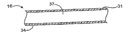
いかなる限定を意図することなく示された拡張可能構造物16は、組織接触面31および管腔接触面34、ならびに選択的に、図2Bに示す内腔を含む内側37を有する。以下の説明は例示目的のためだけであって、補綴物13の実際の形状、サイズ、構成または分布を必ずしも反映していないと考えられる。補綴物は、多数のステントの場合と同様に、連続構造物を有しても、または間欠的な構造物を有してもよい(例えば、ステントの断面は、拡張可能構造物を形成する物質を全く含まず、例えば、一部のステントはスクリーンまたはメッシュ様断面を有する)。供給源は、拡張可能構造物の内側において、図1Bに示す管腔接触面、図1Cに示す組織接触面のどちらかまたは両方の少なくとも一部に隣接して配置または形成されてもよく、またはその任意の組合せであってもよい。
【0106】
治療能力のある薬剤に治療能力のある薬剤を利用させる供給源25は、1つまたは複数の構成の拡張可能構造物に付随している。例えば、基質40が拡張可能構造物16および治療能力のある薬剤28によって形成される場合、または治療能力のある薬剤28が拡張可能構造物16の内側(もしくは、事情によっては、拡張可能構造物16の外側)37に配置される場合には、図2Aおよび図2Bに示す供給源は拡張可能構造物16内にある。
【0107】
ここで、図2Cを参照すると、拡張可能構造物の基質40または内側37からの治療能力のある薬剤28の放出を制御するために、供給源は速度制御要素43をさらに含んでよく、拡張可能構造物16の少なくとも一部の上に形成されてもよい。例として、治療能力のある薬剤が治療能力のある薬剤ポリマーである場合には、供給源は速度制御要素そのものであってもよい。
【0108】
速度制御要素は、非分解性、部分的分解性、実質的に分解性材料またはそれらの組合せから製造することができる。材料は、合成もしくは天然であっても、非ポリマー、ポリマーもしくは金属であっても、またはそれらの組合せであってもよい。例として、経時的に少なくとも部分的に分解する金属材料および高分子量、極性または非極性基、電荷、立体障害基、疎水性部分、親水性部分または両親媒性部分を有する非ポリマーを速度制御要素として使用してもよい。
【0109】
好適な生分解性速度制御要素材料には、ポリ(乳酸)、ポリ(グリコール酸)およびコポリマー、ポリジオキサン、ポリ(エチルグルタメート)、ポリ(ヒドロキシブチレート)、ポリヒドロキシバレレートおよびコポリマー、ポリカプロラクトン、ポリアンヒドリド、ポリ(オルトエステル)、ポリ(イミノカルボネート)、ポリシアノアクリレート、ポリホスファゼン、コポリマーおよび脂肪族ポリエステルまたはポリ-L-乳酸とポリ-e-カプロラクトンのコポリマーを含む好適なコポリマー、それらの混合物、コポリマーおよび組合せが挙げられるが、これらに限定されるわけではない。
【0110】
好適な非分解性または分解が遅い速度制御要素材料には、ポリウレタン、ポリエチレンイミン、セルロースアセテートブチレート、エチレンビニルアルコールコポリマー、シリコン、ポリテトラフルオロエチレン(PTFE)、パリレン、パリラスト、ポリ(メチルメタクリレートブチレート)、ポリ-N-ブチルメタクリレート、ポリ(メチルメタクリレート)、ポリ2-ヒドロキシエチルメタクリレート、ポリエチレングリコールメタクリレート、ポリ塩化ビニル、ポリ(ジメチルシロキサン)、ポリ(テトラフルオロエチレン)、ポリ(酸化エチレン)、ポリエチレンビニルアセテート、ポリカーボネート、ポリアクリルアミドゲル、N-ビニル-2-ピロリドン、無水マレイン酸、ナイロン、セルロースアセテートブチレート(CAB)および他の合成または天然ポリマー物質を含むものなど、それらの混合物、コポリマーおよび組合せが挙げられるが、これらに限定されるわけではない。一態様において、速度制御要素は、シリコン、ポリテトラフルオロエチレン、パリラスト、ポリウレタン、パリレン、セルロースアセテートブチレート、それらの混合物、コポリマーおよび組合せからなる群より選択される材料から製造される。
【0111】
好適な天然材料には、フィブリン、アルブミン、コラーゲン、ゼラチン、グリコソアミノグリカン、オリゴ糖および多糖、コンドロイチン、リン脂質、ホスホリルコリン、糖脂質、タンパク質、アミノ酸、セルロースおよびそれらの混合物、コポリマーまたは組合せが挙げられる。他の好適な材料には、チタン、クロム、ニチノール、金、ステンレス鋼、金属合金またはそれらの組合せおよび治療能力のある薬剤と速度制御要素材料(例えば、非ポリマー化合物)の相互作用の結果として(例えば、化学反応、高分子量、立体障害、疎水性、親水性、両親媒性、熱)、治療能力のある薬剤を放出することができる他の化合物が挙げられる。例として、電気化学的腐食による腐食を促進するために、2つ以上の金属または金属合金と異なる直流電位との組合せを使用することができる。
【0112】
別の態様において、構造物の表面は、クリーニング、エッチングまたは研磨などの物理的改良並びに溶媒処理、下塗りコーティングの適用、界面活性剤の適用、プラズマ処理、イオン衝撃および共有結合などの化学的改良を含む種々の手法のいずれかを使用して事前処理することができる。一態様において、点食による腐食を促進し、腐食によって形成されるピンホールを薬剤放出の開口部として作用させることができる小さい穴またはピンホールを有する金属フィルムまたは合金。一態様において、治療能力のある薬剤を金属または金属合金に接続することができる。
【0113】
本発明の生分解性材料の例は、ポリ-L-乳酸(平均分子量約200,000ダルトン)とポリ-e-カプロラクトン(平均分子量約30,000ダルトン)のコポリマーである。ポリ-e-カプロラクトン(PCL)は、融点59℃〜64℃、分解時間約2時間の半結晶ポリマーである。従って、ポリ-l-乳酸(PLLA)をPCLと組合せて、望ましい放出速度を生じる基質を形成することができる。PCLに対するPLLAの好ましい比は75 : 25(PLLA/PCL)である。その全体の開示内容が参照として本明細書に組み入れられている、Rajasubramanianらによって、ASAIO Journal、40、M584-589ページ(1994)に概説されているように、75 : 25 PLLA/PCLコポリマーブレンドは、拡張可能構造物にPLLA/PLA基質の容易なコーティングを可能にする程度に十分な強度および引っ張り特性を示す。さらに、下層のPCL分がコポリマーブレンドの疎水性を低下し、上層のPLLA分が大部分の多孔性を低下させるので、75 : 25 PLLA/PCLコポリマー基質は、所定の期間制御された薬剤送達を可能にする。
【0114】
分解性材料を大量分解または加水分解によって分解することができる。一態様において、速度制御要素は、完全に分解もしくは加水分解するか、または好ましくは、容積強度を維持しながら、速度制御要素の表面が経時的に分解もしくは加水分解する表面分解もしくは加水分解によって分解もしくは加水分解する。別の態様において、疎水性速度制御要素は、望ましい放出速度で治療能力のある薬剤を放出する傾向があるので、好ましい。非分解性速度制御要素は、拡散によって治療能力のある薬剤を放出することができる。例として、速度制御要素が非ポリマー材料から製造されている場合には、治療能力のある薬剤は、治療能力のある薬剤と速度制御要素材料(例えば、非ポリマー材料)の相互作用の結果(例えば、化学反応、立体障害、疎水性、親水性、両親媒性)として放出されうる。一態様において、速度制御要素が、治療能力のある薬剤と少なくとも十分な基質を形成しない場合には、治療能力のある薬剤は、速度制御要素による拡散によって放出されうる。
【0115】
例として、低分子量および/または組織または血液中で比較的高い親水性を有する速度制御要素は、供給源(例えば、基質)から拡散し、それによって放出される治療能力のある薬剤の表面積または容積を増加し、それによって治療能力のある薬剤の放出速度に影響を与えることができる。
【0116】
さらに別の態様において、治療能力のある薬剤は、本発明の装置が移植される領域の天然の環境が変化するとき、罹患しやすい組織部位に利用される。例えば、本発明の装置が移植される領域のpHの変化は、直接的(例えば、薬剤ポリマーが、治療能力のある薬剤および速度制御要素の両方を含む基質として作用する場合のように)または基質もしくは非基質のどちらかまたは両方としての速度制御要素に特徴的な侵食もしくは拡散に影響を与えることによって間接的に治療能力のある薬剤の放出を生ずるように、経時的に変化してもよい。例えば、pHが増加または低下すると、速度制御要素の侵食が変化し、初期相および後期相放出を可能にする。
【0117】
図2Dは、治療能力のある薬剤28が、拡張可能構造物の組織または管腔接触面の1つと速度制御要素43との間に配置された一態様の特徴を例示する。
【0118】
図2Eに示すように、供給源25は、拡張可能構造物16の組織または管腔接触面の1つの少なくとも一部に隣接して形成され、治療能力のある薬剤28と共に基質40を形成している速度制御要素43を含む。先に記載したように、治療能力のある薬剤ポリマーが基質を形成する場合には、治療能力のある薬剤28自体が速度制御要素として作用することができる。
【0119】
図2Fおよび図2Gに示すように、基質は、速度制御要素43と拡張可能構造物16の間に形成されて、その間におよび/または治療能力のある薬剤28と速度制御要素43の間に基質界面46を形成してもよい。
【0120】
その特徴が図2Hに示されている一態様において、補綴物13の最外層は、治療能力のある薬剤で形成されてもよく、最外層と他の層との間に基質界面46が形成されても、されなくてもよい。ほとんどの図において別個の粒子として示されているが、治療能力のある薬剤28は、図2Hに示すように、基質界面46の一部としての例のように、滑らかな層または層状の粒子を形成してもよい。
【0121】
その特徴が図2Iに示されている別の態様において、第二の速度制御要素49の少なくとも1つの相は基質40上に形成され、罹患しやすい組織部位への治療能力のある薬剤28の放出速度にさらに影響する。第二の速度制御要素49は、第一の速度制御要素43を形成している材料と同じまたは異なる材料製であってもよい。
【0122】
ここで、図2Jおよび図2Kを参照すると、供給源は、例えば、治療能力のある第一の薬剤28および治療能力のあるもう1つの薬剤50または可能にする化合物61(図2N)などのもう1つの化合物50のような複数の化合物を含んでもよい。複数の化合物は各々供給源の同じまたは異なる領域に存在してもよい。例えば、図2Jに示すように、治療能力のある第一の薬剤28は基質40に存在してもよく、治療能力のある第二の薬剤50は、治療能力のある第二の薬剤50および第二の速度制御要素55によって形成される第二の基質52内に存在する。速度制御要素43および55は、同じまたは異なる材料から製造されてもよい。
【0123】
治療能力のあるもう1つの薬剤は、治療能力のある薬剤によって発生される可能性のある反応および副産物を相殺などの方法で、治療能力のある薬剤と協力して作用することができる。例として、治療能力のある薬剤は、望ましい内皮細胞の形成を低下させることがあり、別の好適な治療能力のある薬剤を加えることによって、多くの内皮化を実施することができる。
【0124】
治療能力のあるもう1つの薬剤は、抗癌剤、化学療法剤、血栓溶解剤、血管拡張剤、抗菌剤または抗生物質抗有糸分裂剤、増殖因子アンタゴニスト、フリーラジカル捕集剤、生物学的製剤、放射性治療剤、放射線不透過剤、放射性標識剤、ヘパリンおよびその誘導体などの抗凝固剤、Thalidomide(商標)などの抗血管形成剤、血管形成剤、PDGF-Bおよび/またはEGF阻害剤、乾癬剤を含む抗炎症剤、リボフラビン、チアゾフリン、ザフリン、アセチルサリチル酸などのシクロオキシゲナーゼ阻害剤を含む抗血小板剤、クロピドグレル(例えば、Plavix(商標))およびチクロプジピン(例えば、ticlid(商標))などのADP阻害剤、シロスタゾール(例えば、Pletal(商標))などのホスホジエステラーゼIII阻害剤、アブシキシマブ(例えば、Rheopro(商標))などの糖タンパク質IIb/IIIa剤、エプチフィバチド(例えば、Integrilin(商標))、ジピリドモル(dipyridmoles) などのアデノシン再取り込み阻害剤、抗酸化剤、酸化窒素ドナーを含む治療剤および/または促進剤、鎮吐剤、制吐剤、それらの誘導体および組合せからなる群より選択される少なくとも1つ化合物を含むことができる。
【0125】
治療能力のあるもう1つの薬剤は、類似または異なる速度および相において、治療能力のある薬剤の前、同時または後に放出されうる。
【0126】
その特徴が図2Lおよび図2Mに示される別の態様において、治療能力のある薬剤28は、容器58内の拡張可能構造物16内または構造物上に配置される。速度制御要素43は、治療能力のある薬剤の放出に影響を与えるために、容器58および/または治療能力のある薬剤28に隣接して配置することができる。先に記載したように、例示的な図および説明は「隣接して」という用語を限定することを意味しない。
【0127】
その特徴が図2Nに示されるさらに別の態様において、もう1つの化合物は、治療能力のある薬剤の放出に影響を与えるために、外部エネルギー源または天然の条件に応答可能な可能にする化合物61を含む。応答可能な化合物は、治療能力のある薬剤、速度制御要素、拡張可能構造物またはそれらの組合せに付随させることができる。図2Nに示すように、応答可能な化合物は、治療能力のある薬剤に付随している。可能にする化合物61は、治療能力のある薬剤28に連結された磁気粒子から製造することができる。エネルギー源は、治療能力のある薬剤28を放出させるために、移植後に補綴物13の磁場を誘導するための磁力源であってもよい。磁気粒子61は、磁気ビーズから製造することができ、典型的には約1nm〜約100nmの範囲である。磁力源は、磁気ビーズ61を活性化し、それによって補綴物から治療能力のある薬剤を放出させる、典型的には約0.01T〜約2Tの範囲の強度の磁場に補綴物13を暴露する。別の可能にする化合物は、上記のように、他の構成の補綴物13内に存在してもよい。
【0128】
第二の化合物またはそれらの性能を必要とする、または必要としない他の好適な外部エネルギー源は、第二の化合物の有無によって影響されることはなく、超音波、磁気共鳴画像形成、磁場、ラジオ周波数、温度変化、電磁、X線、放射線、熱、γ線、振動、マイクロ波またはそれらの組合せを含む。
【0129】
例として、20kHz〜100MHzの範囲、好ましくは0.1MHz〜20MHzの範囲の周波数および0.05W/cm2〜10 W/cm2の範囲、好ましくは0.5W/cm2〜5W/cm2の範囲の強度レベルを有する超音波外部エネルギー源を使用することができる。超音波エネルギーは、1mm〜30cmの範囲、好ましくは1cm〜20cmの範囲の距離から補綴物13に向けられてもよい。超音波は、5秒〜30分の範囲、好ましくは1分〜15分の範囲の期間連続的に適用されても、またはパルス状に適用されてもよい。この期間中の補綴物13の温度は36℃〜48℃の範囲である。超音波は補綴物13の多孔性を増加するために使用することができ、それによって、補綴物13から治療能力のある薬剤28の放出を可能にすることができる。他のエネルギー源、例えば、熱または振動を使用して、補綴物もしくはその一部の多孔性を増加することができ、またはこれの構成を変更することができる。
【0130】
さらに、本発明の装置の生体適合性を形成または増強するために、供給源および/または装置の最外層に生体適合性(例えば、血液適合性)層を形成することができる。生体適合性層として使用するのに好適な生体適合性材料には、ポリエチレングリコール(PEG)、ポリ酸化エチレン(PEO)、ヒドロゲル、シリコン、ポリウレタン、ヘパリンコーティングが挙げられるが、これらに限定されるわけではない。
【0131】
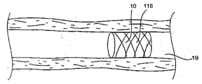
拡張可能構造物16はステント70または移植片であってもよい。拡張可能構造物がステントである場合には、拡張可能構造物16は、図3に示すように通常、少なくとも2つの放射状に拡張可能な、通常円筒のリング状セグメント73を含む。典型的には、拡張可能構造物16は、少なくとも4個、しばしば5個、6個、7個、8個、10個またはそれ以上であるリング状セグメントを有する。リング状セグメントの少なくともいくつかは互いに隣接するが、他のものは他の非リング構造で分離されてもよい。例示的なステント構造物の記載は完璧であることが意図されておらず、本発明に使用できる他の形態のステント設計が当業者に周知であると考えられるべきである。
【0132】
振り返って図3を参照すると、本発明に使用する例示的なステント70(その全体の開示内容が参照として本明細書に組み入れられている、本発明の譲渡人に譲渡された同時係属中の米国特許出願第08/968,318号にさらに詳細に記載されているステントの特徴を具体化している)は4個〜50個のリング状セグメント73を含む(7つは例示されている)。各リング状セグメント73は、S次状リンク76の少なくとも1つによって隣接するリング状セグメントに接続される(3つは例示されている)。各リング状セグメント73は、複数、例えば、6つの支柱/ヒンジ単位を含み、各リング状セグメント73の各6つのヒンジ/支柱構造物のうち2つはS次状リンク76によって隣接するリング状セグメントに接続される。図3に示すステント70は、倒壊型または非拡張型構成であるステント70を示す。
【0133】
本明細書において使用する「放射状に拡張可能な」という用語は、望ましい標的部位に拡張可能構造物16が移植されたときに実施される、径の小さい構成から放射状に拡張された、通常円筒形の構成に変換することができるセグメントを含む。拡張可能構造物16は、弾性、例えば、展性は最小であってもよいので、それを拡張して、標的部位に設定するためには内力の適用を必要とする。典型的には、拡張力は、血管手法用の血管形成カテーテルのバルーンなどのバルーンによって提供することができる。拡張可能構造物16は、好ましくは、ステントの可撓性および捲縮性(crimpability)を増強するために特に有用な、連続する単位セグメントの間にS字状リンクを提供する。
【0134】
代替的に、拡張可能構造物16は自己拡張性であってもよい。(自己拡張性構造物などの)拡張可能構造物16を含む、本発明の装置に使用する構造物は、拘束されていないとき、すなわち、鞘の放射状に拘束する力から開放されたとき、放射状に拡張された望ましい径を有するように、鍛鋼などの弾性材料またはNitinol(商標)合金などの超弾性合金を使用して、本体セグメントを形成することによって提供される。血管内腔に固定しておくためには、拡張可能構造物16は管腔によって部分的に拘束された状態にする。自己拡張性の拡張可能構造物16は、例えば、送達用鞘または管内に拡張可能構造物16を配置し、標的部位において鞘を除去することによって、放射状に拘束された構成で進行し、送達されうる。
【0135】
拡張可能構造物の寸法は目的の用途に依存する。典型的には、拡張可能構造物は、血管適用のためには、約5mm〜約100mm、通常約8mm〜約50mmの範囲の長さである。血管適用のための円筒形状の拡張可能構造物の非拡張時の構成の径は、通常約0.5mm〜約10mm、さらに通常0.8mm〜約8mmの範囲であり、拡張時の構成の径は約1.0mm〜約100mm、好ましくは約2.0mm〜約30mmの範囲である。拡張可能構造物は通常、約0.025mm〜2.0mm、好ましくは約0.05mm〜約0.5mmの範囲の厚さである。
【0136】
拡張可能構造物16などの構造物のリング状セグメントおよび他の構成要素は、生体管腔ステントおよび移植片に使用される従来の材料から製造することができ、典型的には300シリーズステンレス鋼などの展性金属もしくは合金、または例えば、Nitinol(商標)合金、スプリングステンレス鋼等のような超弾性および形状記憶合金などの弾性金属、ポリマー材料などの非金属材料、またはそれらの組合せから製造される。ポリマー材料は、速度制御要素のための優れた材料に関連して記載されているものなどの、実質的に非分解性であるポリマー材料を含んでもよい。または、ポリマー材料は、生分解性速度制御要素材料を参照して記載されているものなどの生分解性または実質的に生分解性ポリマーであってもよい。拡張可能構造物材料が、速度制御要素材料から製造される場合には、拡張可能構造物は、補綴物として、および治療能力のある薬剤の直接供給源として機能することができる。本発明の本体または単位セグメントの追加の構造物は、その全体の開示内容が参照として本明細書に組み入れられている、米国特許第5,195,417号、同第5,102,417号および同第4,776,337号に例示されている。構造物として使用する他の好適な材料には、炭素もしくは炭素繊維、酢酸セルロース、硝酸セルロース、シリコン、ポリエチレンテレフタレート、ポリウレタン、ポリアミド、ポリエステル、ポリオルトエステル、ポリアンヒドリド、ポリエーテルスルホン、ポリカーボネート、ポリテトラフルオロエチレン、または別の生体適合性ポリマー材料、またはそれらの混合物もしくはコポリマー、ポリアンヒドリド、ポリカプロラクトン、ポリヒドロキシブチレートバレレートまたは別の生分解性ポリマー、またはそれらの混合物もはコポリマー、タンパク質、細胞外基質成分、コラーゲン、フィブリンまたは別の生物学的物質、または上記分解性、非分解性、金属またはそれ以外の材料のいずれかの好適な混合物が挙げられる。一態様において、装置は、生分解性構造物および治療能力のある薬剤ポリマーなどのポリマー供給源を含んでもよい。
【0137】
ここで図4を参照すると、所定の期間の治療能力のある薬剤放出の例示的な態様のグラフを示す。本発明の図4に示す所定の速度パターンは、初期相において治療能力のある薬剤を利用させない、またはやや低い送達速度で治療能力のある薬剤を利用させることによって、罹患しやすい組織部位への治療能力のある薬剤の送達効率を改善する。後期相に達したら、治療能力のある薬剤の送達速度は実質的に高くてもよい。従って、初期細胞沈着または増殖(過形成)の少なくとも部分的形成段階の再狭窄(または、場合によっては、他の標的状態)に影響を与えるために、時間遅延型の治療能力のある薬剤の放出をプログラムすることができる。本発明は、さらに、治療能力のある薬剤放出時期を少なくとも初期細胞化の後に設定することによって、治療能力のある薬剤の流出を低下させることができる。さらに、所定の速度パターンは、治療能力のある薬剤の負荷および/または濃度を低下させることができる。所定の速度パターンは、さらに、治療能力のある薬剤の流出を最小にすることおよびその放出効率を増加することにより、血管の内皮化に対する障害をなくすかまたは低下もしくは少なくすることができる。
【0138】
本発明の装置は、同様または異なる性能(例えば、放出)プロフィールを有する1つまたは複数の相で治療能力のある薬剤を放出する、または利用させられるように構成することができる。治療能力のある薬剤は、平滑筋細胞の増殖、炎症、免疫応答、高血圧またはこれらの活性化を補足するものの任意の1つ以上を低下させるのに効果的な、1つまたは複数の相および/または送達速度において徐放的、間欠的または持続的であってもよい量を組織に利用させることができる。治療能力のある少なくとも1つの薬剤の任意の1つは、増殖/再狭窄作用の予防または低下、血栓形成の低下または阻止、血小板活性化の低下または阻止、血管攣縮の低下または予防等を含む、1つまたは複数の機能を発揮することができる。
【0139】
組織に利用される治療能力のある薬剤の総量は、一部には、望ましい治療結果のレベルおよび量に依存する。治療能力のある薬剤は、各相が他の相と同様または異なる放出速度および期間を有する1つまたは複数の相において利用されうる。放出速度は事前に規定することができる。一態様において、放出速度は、罹患しやすい組織部位に徐放的レベルの治療能力のある薬剤を提供することができる。別の態様において、放出速度は実質的に一定である。放出速度は経時的に低下および/または増加してもよく、必要に応じて、実質的に非放出速度を含んでもよい。放出速度は複数の速度を含んでもよい。一態様において、複数の放出速度は、実質的に一定、低下、増加、実質的に非放出からなる群より選択される少なくとも2つの速度を含む。
【0140】
利用または放出される治療能力のある薬剤の総量は、典型的には約0.1ug〜約10g、一般に約0.1ug〜約10mg、好ましくは約1ug〜約10mg、より好ましくは約1ug〜約2mg、10ug〜約2mgまたは約50ug〜約1mgの範囲の量である。
【0141】
一態様において、治療能力のある薬剤は、装置の移植時から測定したとき、約1日〜約200日、約1日〜約45日または約7日〜約21日の範囲の期間に放出されうる。
【0142】
一態様において、1日あたりの治療能力のある薬剤の放出速度は、約0.001マイクログラム(ug)〜約200ug、好ましくは約0.5ug〜約200ug、最も好ましくは約1ug〜約60ugの範囲であってもよい。
【0143】
治療能力のある薬剤は初期相および1つ以上後期相において利用されうる。治療能力のある薬剤が異なる相において送達される場合には、初期送達速度は、典型的には後期放出相の約0%〜約99%、通常約0%〜約90%、好ましくは約0%〜75%である。一態様において、初期相における物質の哺乳類組織濃度は、典型的には約0.001ナノグラム(ng)/組織1mg〜約100ug/組織1mg、約1ng/組織1mg〜約100ug/組織1mg、約1ng/組織1mg〜約10ug/組織1mgの範囲内である。後期相における物質の哺乳類組織濃度は、典型的には約0.001ng/組織1mg〜約600ug/組織1mg、好ましくは約1ng/組織1mg〜約10ug/組織1mgの範囲内である。
【0144】
初期相中の送達速度は、典型的には約0.001ng/日〜約50ug/日、通常約0.1ng/日〜約30ug/日、より好ましくは約1ug/日〜約20ug/日の範囲である。後期相における送達速度は、約0.01ug/日〜約200ug/日、通常約1ug/日〜約100ug/日の範囲であってもよい。一態様において、治療能力のある薬剤は、高い効率および/または有効性で、プログラムおよび/または制御された方法で、罹患しやすい組織部位に利用される。さらに、本発明において血管壁の内皮化に対する障害は限定されているかまたは少ない。
【0145】
初期相、後期相および任意の他の追加の相の期間を変化させてもよい。例えば、治療能力のある薬剤の放出は、装置の最初の移植から遅延されてもよい。典型的には、遅延は、標的部位において、治療能力のある薬剤が血管内腔に損失されるのを阻止する程度に十分に細胞化または内皮化形成できるほど十分に長い。典型的には、初期相の期間は、装置の少なくとも一部において最初の細胞化または内皮化を可能にする程度に十分に長い。典型的には、遅延相または放出相であるかどうかにかかわらず、初期相の期間は、通常約12週間未満であり、さらに通常約1時間〜約8週間であり、より好ましくは約12時間〜約4週間であり、約12時間〜約2週間であり、約1日〜約2週間または約1日〜約1週間である。
【0146】
1つまたは複数の後期相の期間もまた変化してもよく、典型的には約4時間〜約24週間、約1日〜約12週間、約2日〜約8週間、より好ましくは約3日〜約50日である。一態様において、明記した期間は血管環境に関する。2つ以上の相が、類似または異なる期間、量および/または放出速度を含んでもよい。例えば、1つのシナリオにおいて、遅延である初期相があり、次に第一の後期放出速度の後期放出相および第二の後期放出速度の第二の後期相等が続いてもよい。
【0147】
本発明の装置が複数の化合物(例えば、治療能力のある第一の薬剤および治療能力のあるもう1つの薬剤または可能にする化合物などのもう1つの化合物)を含む供給源を含む場合には、複数の化合物は、同じ層または存在する場合には、異なる層から異なる時期および/または速度で放出されうる。複数の化合物は各々、別の化合物から独立して、介入性処置と同時または介入性処置の後に利用され、互いに同時または逐次的であってもよい。例えば、治療能力のある第1の薬剤(例えば、Triptolide(商標))は、介入性処置の実施時点から1日〜45日の一定期間内で放出されうり、治療能力のある第二の薬剤(例えば、ミコフェノール酸)は介入性処置から2日〜3ヶ月の期間内で放出されうる。
【0148】
本発明の装置は、キットとは別にまたはキットの一部として、使用説明書(IFU)と共に提供することができる。キットは、装置およびIFUを入れるために使用することができるポーチまたはトレイ、箱、チューブ等などの任意の他の好適なパッケージを含んでもよく、その場合には、IFUは、別個のシートまたは他の通信媒体および/または包装自体に印刷することができる。キットの一態様において、キットは、縁曲げ装置などの搭載用フックおよび/または本発明の装置に永久的または脱離可能に連結することができる拡張可能な部材を含んでもよい。一態様において、キットは、本発明の装置、介入性処置の前、同時または後の第二の化合物の使用に関するIFUおよび選択的に第二の化合物を含むことができる。一態様において、キットは、第二の化合物および/または装置用のIFUを含む場合または含まない場合の本発明の装置と第二の化合物を含む。
【0149】
一態様において、第二の化合物は、治療能力のある薬剤、もう1つの化合物(例えば、治療能力のあるもう1つの薬剤および/または別の可能にするおよび/または増強する化合物)または制吐剤などの生物活性化合物であってもよく、本発明により罹患しやすい組織部位に利用されるものと同様または異なり、本発明の装置(例えば、補綴物)の移植前、同時または後に投与することができる。
【0150】
第二の化合物は、本発明の装置の一部として治療能力のある薬剤の送達に使用されるものと同様または異なる経路から投与することができる。例として、第二の化合物は、経口服用される錠剤の形態、患者の皮膚に留置される経皮パッチの形態であっても、皮下、血流に直接導入することによる全身的、吸入によるまたは任意の他の経路および生体の開口部を経由してもよい。代替的に、第二の化合物はカテーテルによって生体内に利用されてもよい。一態様において、バルーンカテーテル(例えば、灌流)のバルーンは、第二の化合物を生体に灌流する(例えば、灌流カテーテル)ために使用することができ、または第二の化合物でコーティングすることができる。第二の化合物は、介入性処置の前、同時または後に連続的または間欠的に患者に利用されうる。
【0151】
第二の化合物の利用可能期間は通常、治療能力のある薬剤の利用可能期間と比較して、短くてもよい。一態様において、もう1つの化合物は、介入性処置の約200日前〜約200日後まで、介入性処置の約30日前〜約30日後まで、介入性処置の約1日前〜約30日後まで、介入性処置の約200日前〜直前まで、介入性処置の約3ヶ月前〜直前まで、または介入性処置の約7日前〜約24時間前までの範囲の期間内に患者に投与することができる。第二の化合物の利用可能期間は、患者の血中で測定するとき、約1時間〜約120日、約12時間〜約60日または約24時間〜約30日の範囲であってもよい。生物活性化合物の例には、オンダンセトロン(例えば、Zofran(商標))などの鎮吐剤、ドロナビノール(例えば、Marinol(商標))などの制吐剤、ガニセトロン.Hcl (例えば、Kytril(商標))が挙げられる。
【0152】
一態様において、第二の化合物は、生体内に望ましい大量レベル(例えば、初期レベル)の治療能力のある薬剤を提供するために、装置の治療能力のある薬剤と同じであってもよい。装置および第二の化合物から罹患しやすい組織部位に利用される総量は、典型的には約0.1ug〜約10ミリグラム(mg)の範囲であり、好ましくは約10ug〜約2mgの範囲であり、より好ましくは約50ug〜約1.0mgの範囲である。一態様において、単回投与または毎日投与で患者に投与される第二の化合物の量は、約0.5mg〜約5g、約1mg〜約3g、約1g〜約1.5g、約2g〜約3gの範囲である。後者の投与シリーズで提供される第二の化合物の例には、ミコフェノール酸、ラパマイシン並びにそれらのそれぞれのプロドラッグ、代謝物および組合せが挙げられる。一例として、ミコフェノール酸またはラパマイシンは、それぞれ、約1g〜約1.5gおよび約1mg〜約3mgの範囲の1回用量で、およびそれぞれ、約2g〜約3gおよび約2mg〜約6mgの範囲の1日用量で第二の化合物として提供することができる。
【0153】
治療能力のある薬剤を補綴物にコーティング、噴霧、浸漬、沈着または塗布することによって、拡張可能構造物は治療能力のある薬剤および/または選択的なもう1つの化合物を組み込むことができる。通常、治療能力のある薬剤は溶媒に溶解される。好適な溶媒には、水性溶媒(例えば、pH緩衝水溶液、pH調節水溶液、有機塩水溶液および無機塩水溶液)、アルコール(例えば、メタノール、エタノール、プロパノール、イソプロパノール、ヘキサノールおよびグリコール)、ニトリル(例えば、アセトニトリル、ベンゾニトリルおよびブチロニトリル)、アミド(例えば、ホルムアミドおよびN-ジメチルホルムアミド)、ケトン、エステル、エーテル、DMSO、ガス(例えば、CO2)等が挙げられる。例えば、補綴物に溶液を噴霧するか、または溶液に浸漬して、乾燥すると、治療能力のある薬剤が補綴物の表面に残される。または、速度制御要素材料および治療能力のある薬剤を溶解することによって、速度制御要素材料および治療能力のある薬剤を含む基質溶液を調製することができる。次いで、この基質を補綴物に噴霧、浸漬、沈着または塗布することによって、拡張可能構造物16に基質溶液をコーティングすることができる。例として、ポリマー材料がら基質を製造する場合には、補綴物を心棒上で回転させながら、基質溶液を補綴物に細かく噴霧する。基質コーティングの厚さは、噴霧時間および心棒の回転速度によって制御することができる。基質-薬剤コーティングの厚さは、典型的には約0.01um〜約100umの範囲であり、好ましくは約0.1um〜約50umの範囲である。補綴物に基質コーティングがコーティングされたら、ステントを真空下またはオーブンに入れて、溶媒を留去させることができる。
【0154】
例として、寸法3.0mm×14mmのステンレス鋼Duraflex(商標)(カリフォルニア州に本社があるAvantec Vascular Corporation製)に、25mg/ml治療能力のある薬剤の100%エタノールまたはメタノール溶液を噴霧する。ステントを乾燥し、エタノールを留去して、治療能力のある薬剤をステント表面に残す。75 : 25 PLLA/PCLコポリマー(POLYSCIENCESから市販されている)を1,4ジオキサン(ALDRICH CHEMICALSから市販されている)中で調製する。治療能力のある薬剤を負荷したステントを200rpmで回転する心棒にのせ、10秒間〜30秒間回転させながら、スプレーガン(BINKS MANUFACTURINGから市販されている)で、治療能力のある薬剤を負荷したステントに微細噴霧状態のコポリマーを分注する。次いで、ステントを25℃〜35℃のオーブンに24時間入れ、溶媒を留去させる。
【0155】
操作上、罹患しやすい組織部位に治療能力のある薬剤を送達する方法は、上記の本発明の特徴を組み込んだ管腔補綴物を提供する段階を含む。補綴物は、罹患しやすい組織部位を含む、生体管腔などの生体部位に送達される。補綴物は生体管腔内部に移植される。治療能力のある薬剤はある期間の間罹患しやすい組織部位に利用される。
【0156】
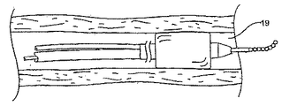
図6A〜図6Fは、治療能力のある薬剤を罹患しやすい組織部位に利用させる方法の特徴を例示している。図に示すように、管状体部103を有する血管内バルーンカテーテル100は、止血用弁および鞘(示していない)を介してガイド用カテーテルにより、大腿動脈106を通って冠血管の大動脈弓112まで導入される。
【0157】
ガイドワイヤー115は通常、罹患しやすい組織部位22、典型的にはバルーン血管形成術で治療する予定の狭窄領域を含む、標的部位118に配置される。通常、バルーンカテーテル100およびガイドワイヤー115は、標的部位に到達するまで、カテーテルの遠位端の前方方向に定期的に延在されるガイドワイヤー115と一体として導入される。
【0158】
標的部位118に達したら、標的部位118の閉塞を拡張するために、バルーン121が膨張される。バルーン血管形成治療が終了したら、バルーン121をしぼませ、ガイドワイヤー115をその場に残す。次いで、バルーン121をガイドワイヤー115から離脱させ、再度ガイドワイヤー115はその場に残す。次いで、本発明による装置10を含む第二のバルーン集成物100'をカテーテル本体に導入する。第二ののバルーン集成物100'が配置されたら、バルーン121を膨張させることによって、バルーン集成物に配置されているステント10などの装置を配置することができる。ステント10が適切に配置されたら、バルーンをしぼませ、カテーテルを離脱させ、ステントをその場に残す。
【0159】
治療方法は、一般に、介入治療と同時またはその後に、治療能力のある少なくとも1つの薬剤および/または選択的なもう1つの化合物を含む供給源を生体内に配置する段階を含む。さらに具体的には、治療能力のある薬剤は、介入治療と同時またはその後に、罹患しやすい組織部位を含む標的生体部位(標的生体内部位など)または罹患しやすい組織部位に治療能力のある薬剤を提供する標的部位に送達することができる。例として、拡張バルーンによる狭窄領域の拡張後、本発明による装置(ステントなど)を血管に送達し、移植する。治療能力のある薬剤は、1つまたは複数の相および/または送達速度において、徐放的、間欠的または持続的であってもよい量が罹患しやすい組織部位に利用されうる。
【0160】
一態様において、罹患しやすい組織部位への治療能力のある薬剤の放出は遅延されてもよい。遅延期間中、実質的な量の治療能力のある薬剤の放出前に、治療能力のある薬剤は全く放出されないか、または少量しか放出されない。典型的には、遅延は、血栓事象の発生を減少させるために、標的部位において十分な内膜組織の形成または細胞化を可能にする程度に十分に長い。
【0161】
一態様において、遅延は、形成された新生内膜が移植された拡張可能構造物を少なくとも部分的に被覆する程度に十分長い。一態様において、治療能力のある薬剤は、装置の移植時から測定して一定の期間において、約1日〜約200日、約1日〜約45日または約7日〜約21日の範囲で放出されうる。一態様において、本発明の方法は、装置から治療能力のある薬剤を放出させるために、装置にエネルギーを誘導する段階をさらに含む。エネルギーは、超音波、磁気共鳴画像形成、磁場、ラジオ周波数、温度変化、電磁、X線、熱、振動、γ線照射、マイクロ波の1つ以上を含んでもよい。一態様において、治療能力のある薬剤は、約0.1ug〜約10g、約0.1ug〜約10mg、約1ug〜約10mg、約1ug〜約2mg、約10ug〜約2mgまたは約50ug〜約1mgの範囲の総量が放出されうる。
【0162】
治療方法の別の態様において、放出は、記載されたような少なくとも1つの別の化合物の放出を含む。もう1つの化合物は、記載されたような治療能力のあるもう1つの薬剤または可能にする化合物であってもよい。もう1つの化合物は治療能力のある薬剤の前、同時、後に放出されても、または治療能力のある薬剤と逐次的に放出されてもよい。
【0163】
一態様において、第二の化合物は、記載されたような介入性処置の前、同時または後に患者に投与することができる。第二の化合物は、記載する経路から、記載する期間において、記載するレベルで投与することができる。
【0164】
治療中の部位の性質に応じて、本発明の装置は、第一のバルーンカテーテルの導入時に、事前に拡張する必要なく、部位に導入することができることが評価されるべきである。
【0165】
一般に、上記のように、異なる補綴物要素と治療方法を組合せることができる。例えば、治療能力のある薬剤を放出するための容器手段を有する補綴物は、さらに、速度制御要素を組み込むことができる。また、本発明の方法は、本明細書において記載する管腔用治療能力のある薬剤の送達治療により狭窄部位を解消するために、バルーン血管形成術および/または他の介入治療を組合せることができる。
【0166】
実施例
実施例1-
寸法約3.0mm×14mmのステンレス鋼Duraflex(商標)ステントに、25mg/mlの治療能力のある薬剤の100%エタノールまたはメタノール溶液を噴霧した。ステントを乾燥し、エタノールを留去して、治療能力のある薬剤をステント表面に残した。75 : 25 PLLA/PCLコポリマー(Polysciencesから市販されている)を1,4ジオキサン(Aldrich Chemicalsから市販されている)中で調製した。治療能力のある薬剤を負荷したステントを200rpmで回転する心棒にのせ、約10秒間〜30秒間回転させながら、スプレーガン(Binks Manufacturingから市販されている)を使用して、コーティング済みのステントに微細噴霧状態のコポリマー溶液を分注した。次いで、ステントを25℃〜35℃のオーブンに24時間入れ、溶媒を留去させた。
【0167】
実施例2-
ステンレス鋼Duraflexステント(3.0mm×14mm)をSS管からレーザー切断した。ステントの表面粗さを増加することによって、治療能力のある薬剤を収容するステントの表面積を増加した。ステントの支柱の接続部に幅10nm×深さ5nmの溝を形成することによって、ステントの表面積および容積をさらに増加することができる。ステントの放射性方向の強度を損なわないために、拡張時応力が小さくなるように、そのようなステント領域に溝を形成した。イソプロピルアルコール、エタノールまたはメタノールなどの低表面張力溶媒で調製した治療能力のある薬剤溶液にステントを浸漬または噴霧することによって、ステントおよびステントの溝に薬剤を負荷した。次いで、ステントを乾燥し、治療能力のある薬剤をステント表面および治療能力のある薬剤の容器として作用する溝に残した。速度制御要素として作用させるために、パリレン(Parylene)をステントに真空蒸着した。1日〜45日の期間、薬剤はステントから溶出した。
【0168】
実施例3-
治療能力のある薬剤をメタノールに溶解し、次いでステントに噴霧した。ステントを乾燥させ、ステントから溶媒を留去させて、治療能力のある薬剤をステントに残した。基質またはバリア(シリコン、ポリウレタン、ポリテトラフルオロエチレン、パリラスト(parylast)、パリレンをステントに噴霧または沈着させ、治療能力のある薬剤を被覆した。治療能力のある薬剤の量を、1日〜45日の放出速度では、約100ミリグラムから2ミリグラムに変化させた。
【0169】
実施例4-
実施例2に記載するように、基質ポリマーおよび治療能力のある薬剤を含む基質溶液をステントにコーティングした。次いで、ステントに速度制御バリア(および/または速度制御バリアとして作用する薬剤を含有しない基質材料)のトップコーティングをコーティングまたは噴霧した。または、速度制御バリアを介して治療能力のある薬剤をステントにコーティングし、次いでトップコーティング(別のバリアまたは基質)を被覆することができる。トップコーティングを使用すると、放出速度がさらに制御され、生体適合性が改善され、および/またはステントの送達または拡張時のスクラッチングおよび亀裂に対する抵抗性が提供される。
【0170】
実施例5-
治療能力のある薬剤は、治療能力のある第二の薬剤(細胞毒性剤、細胞静止剤または乾癬剤)と組合されうる。一方の薬剤は第一のコーティング内または第一のコーティングに結合されるが、他方の薬剤は第二のコーティング内またはコーティング上に存在する。治療能力のある薬剤は、血管内への移植後最初の1週間〜3週間の間放出されるが、治療能力のある第二の薬剤はさらに長期間の間放出されるか、または放出され続ける。
【0171】
実施例6-
個別に異なるコーティングに含まれる多数の治療能力のある薬剤の組合せを基質として使用することができる。コーティングは同時および/または逐次的に多数の薬剤を放出することができる。薬剤は、デノボヌクレオチド合成の阻害剤の治療能力のある薬剤クラスまたは糖質コルチコイド、イムノフィリン結合剤、デオキシスペルグアリン、FTY720、タンパク質剤またはペプチドのクラスから選択することができる。これはまた、他の細胞毒性の添加によりステントに結合される、上記クラスの薬剤の任意の組合せに適用するこもできる。
【0172】
実施例7-
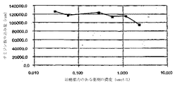
治療能力のある薬剤を15mg/mlの濃度でアセトンに溶解し、CAB(セルロースアセテートブチレート)を15mg/mlの濃度でアセトンに溶解し、その後ミコフェノール酸とCAB溶液を3:1部の基質溶液比で混合することによって、治療能力のある薬剤、ミコフェノール酸および基質ポリマーであるCABをミコフェノール酸負荷量70重量%〜80重量%で含む基質を調製した。治療能力のある薬剤の量は約0.1マイクログラム〜約2mgと変化し、好ましくは600マイクログラムであった。次いで、ステントを各々回転させながら、アトマイザー式噴霧器(EFD manufacturer)で噴霧することによって、2セットのステント(AセットおよびBセット)に基質溶液をコーティングした。各ステントを乾燥させた。次いで、一方の基質コーティングステントに、実施例2に記載するものと同様の方法を使用して、速度制御バリア(約1.1um)としてパリレンをコーティングした。表面をレーザービームに暴露することによってまたは針を接触させることによって、Bセットのステントの上面(パリレン速度制御バリア)に開口部を形成した。開口部サイズは、直径約0.1um〜約100umの範囲であってもよい。Bセットのステントの開口部は直径約10umであった。開口部は、隣の開口部から約0.003インチ〜約2インチ離れていてもよい(ステントの支柱パターンに沿ってトレースすると曲線距離として測定される)。
【0173】
ミコフェノール酸を負荷したステントをブタ血清の溶出溶液中に入れて、1日間〜7日間老化させた。定期的な間隔で試料を血清から取り出し、HPLCで分析した。図7Aおよび図7B(それぞれ、AセットおよびBセットのステントに対応する)に示すデータからわかるように、Aセットのステントはミコフェノール酸の放出速度は直線状であったが、Bセットのステントは、初期相は放出速度は比較的遅い直線状で、その後後期相では、放出は比較的早くなった。
【0174】
実施例8-
2セットのステント、AセットおよびBセットに、実施例2により、250μgおよび300μgのミコフェノール酸をコーティングした。次いで、Aセットに速度制御バリアとして1.7ミクロンのパリレンをコーティングした。Bセットには先ず、ミコフェノール酸をコーティングし、次に律速基質材料としてメチルプレドニゾロンをコーティングし、その後1.3ミクロンのパリレンをコーティングした。次いで、コーティングしたステントに、実施例7に記載するように、インビトロにおいて溶出試験を実施し、溶出したミコフェノール酸の量を測定した。図8Aおよび図8B(それぞれ、AセットおよびBセットのステントに対応する)に示すデータからわかるように、両方のセットは初期相において比較的速い直線状の放出を示し、次に後期相において比較的遅い放出を示した。これは、疎水性の大きなメチルプレドニゾロンが水溶性の大きなミコフェノール酸の律速要素として作用し、ミコフェノール酸およびパリレンコーティングの放出速度を制御することができることを示差していると考えられる。罹患領域が初期に大量の薬剤を必要とし、次いで徐放的な遅い放出を必要とする場合に、これは有用である。
【0175】
実施例9-
細胞培養に対する本発明の治療能力のある薬剤の影響を評価するために、以下に掲載する種々の濃度の治療能力のある薬剤試料を5セット調製し、標準的な手法により、ブタ平滑筋細胞培養物の異なる群に添加した。A、B、C、DおよびEセットは以下の治療能力のある薬剤セットに対応した:それぞれ、ミコフェノール酸&デキサメサゾン、ミコフェノール酸&トリプドリド(Triptolide)、ワートマニンおよびメトトレキセート、トリプドリド(Triptolide)、ミコフェノール酸モフェチル。異なる濃度(0.003、0.031、0.31、1.6および3.1マイクロモル)の異なる試料に組入れられるチミジンの量を測定した。図9A〜図9E(それぞれ、A〜Eセットに対応する)に示すデータからわかるように、、種々のセットのIC50(細胞の50%が増殖を阻止される濃度と規定される)は異なる濃度において出現した。さらにわかるように、ミコフェノール酸モフェチル(基準試料E)は、生物条件(例えば、血液などの体液との接触)がない場合には効果的ではないと考えられる。
【0176】
実施例10-
治療能力のあるもう1つの薬剤群では、種々の濃度(0.003、0.031、0.31、1.6、3.1、31および156マイクロモル)の試料について組み込まれるチミジンの量を測定した。図10A〜図10Bに示すデータからわかるように、それぞれ、ミコフェノール酸およびメチルプレドニゾロンに対応して、これらの治療能力のある薬剤のIC50は1.0マイクロモルであった。
【0177】
実施例11-
治療能力のある種々の薬剤の影響を評価するために、実施例9および10に記載するものと同様の方法を使用して、細胞培養物に、いくつかの治療能力のある薬剤を接触させた。図11A〜図11Bに示すデータからわかるように、それぞれ、トリプドリド(Triptolide)(T)、デキサメサゾン(D)、メトトレキセート(M)およびミコフェノール酸(MA)に対応して、治療能力のある薬剤は有意な細胞死を生じなかった。また、IC50濃度では、細胞のほとんどは50%増殖でも生存していたことがわかる。
【0178】
実施例12-
治療能力のある薬剤を15mg/mlの濃度でアセトンに溶解することによって、治療能力のある薬剤、ミコフェノール酸を調製した。治療能力のある薬剤の量は約0.1ug〜約2mgと変化したが、好ましくは600ugであった。次いで、ステントを回転させながら、アトマイザー式噴霧器(EFD manufacturer)で噴霧することによって、実施例8に記載されているステントに薬剤溶液をコーティングした。ステントを乾燥させた。次いで、ステントをPTCAカテーテルの3つ折りバルーンに配置し、折り目をつけた(crimped)。折り目付け(crimping)後、薬剤は無傷のまま、ステントに接続されていた。Tecoflex疑似血管でステントを拡張させると、薬剤の亀裂はなかった。疑似血管にステントを配置する前に、ステントに流体を流動させても、ステントからの薬剤の離脱は生じなかった。
【0179】
ある種の好ましい態様および方法が本明細書に開示されているが、本発明の真の精神および範囲から逸脱することなく、このような態様および方法の変更および改良を加えられることは前述の開示内容から当業者に明らかになる。従って、上記の説明は、添付の特許請求の範囲によって規定される本発明の範囲を限定するものと考えられるべきではない。
【図面の簡単な説明】
【0180】
(図1)図1A〜図1Cは、本発明の特徴を具体化した、生体管腔移植された装置の断面図である。
(図2)図2A〜図2Nは、線2-2に沿って見た、図1A〜図1Cの送達補綴物の種々の態様の断面図である。
(図3)本発明の装置として使用する例示的ステントの略図である。
(図4)所定の期間にわたる治療能力のある薬剤の放出のグラフである。
(図5)移植後に細胞が増殖した、図1A〜図1Cの補綴物の一態様の部分断面図である。
(図6)図6A〜図6Iは、血管内に図1A〜図1Cの補綴物を配置するための例示的な方法の特徴を例示するものである。
(図7−図11)図7A、図7B、図8A、図8B、図9A〜図9E、図10A、図10B、図11Aおよび図11Bは、治療能力のある種々の薬剤の性能のグラフである。【Technical field】
[0001]
1. Field of the invention
The present invention relates generally to medical devices and methods. More specifically, the present invention provides luminal prostheses, such as vascular stents and grafts, for reducing or preventing restenosis.
[0002]
Numerous percutaneous endovascular techniques have been developed to treat atherosclerotic stenotic areas of a patient's blood vessels to restore sufficient blood flow. The most successful of these treatments is percutaneous transluminal angioplasty (PTA). In PTA, a catheter having an expandable distal end, usually in the form of an expandable balloon, is placed at the site of a stenosis in a blood vessel. The expandable end is expanded to dilate the blood vessel and restore sufficient blood flow to the diseased area. Other techniques for opening the stenotic region include directional arthrectomy, rotary arthrectomy, laser angioplasty, stenting, and the like. While these approaches are widely accepted (alone or in combination, especially PTA with stenting), they still have significant disadvantages. A particularly common drawback of PTA and other well-known techniques for opening a stenotic region is the high frequency of restenosis.
[0003]
Restenosis refers to restenosis of an artery after the first successful angioplasty. Restenosis occurs in approximately 50% of all angioplasty patients and is the result of damage to the vessel wall during the open lumen angioplasty procedure. In some patients, the injury initiates a repair response characterized by smooth muscle cell proliferation, referred to as "hyperplasia," of the area affected by angioplasty. This smooth muscle cell proliferation can cause restenosis of the lumen opened by angioplasty within weeks to months, thereby requiring repetitive PTA or other techniques to reduce restenosis. I do.
[0004]
Numerous methods have been proposed to treat hyperplasia and reduce restenosis. Previously proposed methods include long-term balloon dilation during angioplasty, treatment of blood vessels with a heated balloon, treatment of blood vessels with post-angioplasty radiation, stenting of areas, and other techniques. Can be Although these proposals have been successful at varying levels, none of these approaches has proven successful in substantially or completely avoiding the occurrence of restenosis and hyperplasia.
[0005]
As a variant or an adjunct to the treatment described above, administration of a therapeutic agent after PTA to inhibit restenosis has also been administered. Therapeutic treatment usually involves pushing or releasing the drug through a catheter or from a stent. Although promising, the delivery of therapeutic agents to prevent restenosis has not been successful.
[0006]
Accordingly, it would be a significant advance to provide improved devices and methods for reducing, inhibiting or treating restenosis and hyperplasia that may occur after angioplasty and other interventional treatments. The present invention fulfills at least some of these and other needs.
[Background Art]
[0007]
2. Description of the background art
A full description of an exemplary luminal prosthesis for use in the present invention can be found in co-pending application Ser. No. 09 / 04,2000, filed May 4, 2000, the entire disclosure of which is incorporated herein by reference. / 565,560. Methods and devices for releasing actives from implantable and other devices are disclosed in U.S. Patent Nos. 6,096,070, 5,824,049, 5,624,411, 5,609,629, 5,569,463, 5,447,724 and No. 5,464,650. The use of stents for intravascular drug delivery has been described in WO 01/01957 and U.S. Patent Nos. 6,099,561, 6,071,305, 6,063,101, 5,997,468, 5,980,551, No. 5,980,566, No. 5,972,027, No. 5,968,092, No. 5,951,586, No. 5,893,840, No. 5,891,108, No. 5,851,231, No. 5,843,172, No. 5,837,008, No. 5,769,883, No. No. 5,735,811, No. 5,700,286, No. 5,679,400, No. 5,649,977, No. 5,637,113, No. 5,591,227, No. 5,551,954, No. 5,545,208, No. 5,500,013, No. 5,464,450, No. Nos. 5,419,760, 5,411,550, 5,342,348, 5,286,254 and 5,163,952. Biodegradable materials are U.S. Patent Nos. 6,051,276, 5,879,808, 5,876,452, 5,656,297, 5,543,158, 5,484,584, 5,176,907, 4,894,231, and 4,897,268. No. 4,883,666, No. 4,832,686 and No. 3,976,071. The use of hydrocyclosiloxanes as rate limiting barriers is described in U.S. Patent No. 5,463,010. Methods for coating stents are described in U.S. Patent No. 5,356,433. Coatings for increasing the biocompatibility of implantable devices are described in U.S. Patent Nos. 5,463,010, 5,112,457 and 5,067,491. Energy-based devices are disclosed in U.S. Pat.Nos. 6,031,375, 5,928,145, 5,735,811, 5,728,062, 5,725,494, 5,409,000, 5,368,557, 5,000,185 and 4,936,281. No. Magnetic techniques, some of which are used in drug delivery systems, include U.S. Patent Nos. 5,427,767, 5,225,282, 5,206,159, 5,069,216, 4,904,479, 4,871,716, and Nos. 4,501,726, 4,357,259, 4,345,588 and 4,335,094.
DISCLOSURE OF THE INVENTION
[0008]
Summary of the Invention
The present invention provides improved devices and methods for preventing restenosis and hyperplasia following endovascular intervention. In particular, the present invention provides a luminal prosthesis that allows for highly efficient and / or effective delivery of a programmed and controlled substance to selected locations in a patient's blood vessels to prevent restenosis. provide. Furthermore, the present invention minimizes drug shedding and provides little or no interference with vascular endothelialization.
[0009]
The term "vascular intervention" includes various corrective techniques that can be performed to at least partially relieve a stenosis, restenosis or thrombotic condition in a body lumen. Typically, correction techniques include balloon angioplasty. Correction techniques also include directional arthrectomy, rotational arthrectomy, laser angioplasty, stenting, etc., in which the vascular lumen after treatment is dilated to at least partially reduce the stenosis that existed before treatment. sell.
[0010]
In a first aspect of the present invention, a luminal delivery prosthesis includes a scaffold capable of being implanted within a lumen of a living body, and means on the scaffold for releasing a substance. The substance is released in a predetermined time pattern that includes an early phase in which the substance delivery rate is below the threshold level and a late phase in which the substance delivery rate is above the threshold level.
[0011]
The predetermined time pattern of the present invention improves drug delivery efficiency by releasing a small or minimal amount of a substance until a late phase in which the release of the substance can be substantially higher. Thus, the time-delayed substance release can be programmed to affect restenosis when a substantial event occurs resulting in smooth muscle cell proliferation (hyperplasia). The present invention further provides for the timing of substance release by adjusting the timing of substance release to occur after at least the initial cellularization / endothelialization that forms a barrier to the stent to reduce loss of the substance directly into the bloodstream. Can be minimized. In addition, the predetermined time pattern not only can reduce the substance load and / or substance concentration, but may also provide little or no obstruction to the endothelialization of the vessel wall, due to minimized drug spillage and high efficiency of substance release. Absent.
[0012]
The scaffold may be in the form of a stent that further maintains the patency of the lumen, or in the form of an implant that further protects or increases the strength of the lumen wall. The scaffold may be radially expandable and / or self-expanding, and is preferably suitable for indwelling a body lumen. The body lumen may include any blood vessel of the patient's vasculature including veins, arteries, aorta, especially coronary and peripheral arteries, as well as previously implanted grafts, shunts, fistulas, and the like. It is contemplated that the present invention may also be applied to a number of tissue organs in vivo, such as lumens and organs of other organisms, nerves, glands, transport tubing, and the like. Exemplary stents for use in the present invention are described in co-pending application Ser. No. 09 / 565,560.
[0013]
It is believed that the above advantages of time-delayed release allow a wide variety of substances to be delivered effectively. The substance may include at least one drug selected from the group consisting of immunosuppressants, anti-inflammatory agents, antiproliferative agents, anti-migratory agents, anti-fibrotic agents, anti-thrombotic agents, anti-platelet agents, IIb / IIIa agents. Good. Preferably, the agent is selected from the group consisting of mycophenolic acid, rapamycin, cyclosporin A, cycloheximide, cyclophosphamide, mizoribine, methylprednisolone, azathioprine, ribovirin, FK506, triazofurin, methotrexate, zafrin and mycophenolate mofetil. It is an immunosuppressant. The total amount of material released typically ranges from 1 g to 2000 g, preferably from 10 g to 1000 g, most preferably from 50 g to 500 g. The release rate of the initial phase is typically between 0 g / day and 50 g / day, usually between 5 g / day and 30 g / day. The release rate of late phase substances is quite large, typically in the range of 5 g / day to 200 g / day, usually in the range of 10 g / day to 100 g / day. Thus, the release rate of the early phase is typically 0% to 99% of the release rate of the late phase, usually 0% to 90%, preferably 0% to 75%. It will be appreciated that the release rate may vary during either or both the early and late release phases. Additional phases may be present to release the same and / or different substances.
[0014]
The duration of the early phase, late phase and any other additional phases will vary. Typically, the early phase is long enough to allow for initial cellization or endothelialization of at least a portion of the stent, usually less than 12 weeks, and even more usually 1 hour to 8 weeks, more preferably Is 12 hours to 2 weeks, most preferably 1 day to 1 week. The duration of the late phase may also vary, typically from 4 hours to 24 weeks, more usually from 1 day to 12 weeks, more preferably from 2 days to 8 weeks in the vascular environment, Most preferred is a period of 3 to 50 days in the vascular environment.
[0015]
The present invention relates to improved devices and methods for preparing or treating susceptible tissue sites. As used herein, a susceptible tissue site is a tissue that is damaged or may be damaged as a result of a disorder (eg, a disease, a medical condition), or an interventional treatment such as an endovascular intervention. Refers to tissue that may have been damaged during or after the procedure. The term "intravascular intervention" includes various corrective procedures that can be performed to at least partially relieve a stenosis, restenosis or thrombotic condition of a blood vessel, usually an artery such as a coronary artery. Typically, correction techniques include balloon angioplasty. Correction techniques also include directional arthrectomy, rotational arthrectomy, laser angioplasty, stenting, etc., in which the vascular lumen after treatment is dilated to at least partially reduce the stenosis that existed before treatment. May be included. Vulnerable tissue sites include tissues associated with living lumens, organs or localized tumors. In one aspect, the devices and methods of the present invention reduce the formation or progression of restenosis and / or hyperplasia that may occur following endovascular intervention. In particular, the present invention relates to living organisms, especially intracorporeal devices, and methods of using them.
[0016]
As used herein, "body" refers to a body lumen or a body tissue and organ in a body. The biological lumen may be any blood vessel of the patient's vasculature including veins, arteries, aorta, especially coronary and peripheral arteries, and previously implanted grafts, shunts, fistulas, and the like. The present invention can also be applied to other biological lumens such as bile ducts where excessive cell proliferation of malignant tumors is observed. Examples of tissues and organs in a living body include various organs, nerves, glands, transport vessels, and the like. In one aspect, the devices of the present invention include a luminal prosthesis, such as a vascular stent or graft. In another embodiment, the device of the invention is a cardiac pacemaker lead or lead tip, a cardioverter defibrillator lead or lead tip, a heart valve, a suture or needle, a pacemaker, a correction device, an instrument, an implant or replacement, or Any of the above may be included.
[0017]
In one embodiment, the devices and methods of the present invention define restenosis (where the artery is narrower than about 40% to about 80%, usually about 50% to about 70% of the vessel diameter immediately following vascular intervention such as stenting). Is reduced and / or prevented, while at the same time allowing for the formation of small amounts of cellular, endothelial or neointima, preferably in a controlled manner.
[0018]
In one aspect, a device of the invention comprises a structure and at least one source of at least one therapeutically capable agent associated with the structure. In one aspect, the structure may be an expandable structure. In another aspect, the structure may have a substantially constant size or diameter, or alternatively, may be a shrinkable structure depending on the application and application. In one embodiment, the structure comprises at least one surface, usually a tissue contacting surface. In another embodiment, the structure comprises a tissue contacting surface and another surface, usually a luminal contacting surface. In one embodiment, the structure may have an interior disposed between two surfaces, usually a tissue contacting surface and a luminal contacting surface.
[0019]
The device of the present invention includes an expandable structure that can be implanted into a living body that includes a susceptible tissue site. Alternatively or additionally, the device of the present invention may be an implantable device configured to implant with or without performing dilation at the target biological site. The target biosite may include a susceptible tissue site or may be a site (eg, another living organ or lumen), for example, a target in vivo site such as an artery supplying blood to the susceptible tissue site. Good. In one aspect, the expandable structure may be in the form of a stent that further preserves the patency of the lumen, or in the form of an implant that further protects or increases the strength of the lumen wall. At least some of the devices of the present invention may include a scaffold formed from an open lattice or at least a substantially closed surface. In one aspect, at least a portion of the stent includes a scaffold formed from an open lattice. The expandable structure may be radially expandable and / or self-expanding, and is preferably suitable for lumen placement within a biological lumen.
[0020]
The expandable structure can be manufactured from any suitable material, such as a metal, a polymer, or a combination thereof. In one aspect, the expandable structure can be manufactured from an at least partially biodegradable material selected from the group consisting of a polymeric material, a metallic material, or a combination thereof. The at least partially biodegradable material preferably degrades over time. Examples of polymeric materials include poly-L-lactic acid, which degrades slowly and allows the blood vessel to recover before the structure is degraded. Examples of metallic materials include metals or alloys that can be degraded in the body, such as stainless steel. Exemplary stents for use in the present invention are described in co-pending application Ser. No. 09 / 565,560, the entire disclosure of which is incorporated herein by reference.
[0021]
At least a portion of the therapeutically active agent is associated with the structure so that it may be utilized in a susceptible tissue site immediately or shortly after introduction of the device of the invention into or into a living body. ing. As used herein, the term "associated" refers to, directly or indirectly, connected, connected, disposed on a surface, disposed internally, attached, adhered, adhered, adjacent, encapsulated, absorbed internally, absorbed on a surface, Refers to any form of association and similar configuration, such as absorbed.
[0022]
The source can be located or formed adjacent to at least a portion of the structure. In one aspect, the source can be located or formed adjacent at least a portion of the structure. In one aspect, the source is adjacent to at least a portion of either surface or both surfaces of the expandable structure, inside the structure disposed between the two surfaces, or any combination thereof. Can be arranged or formed. The association between the therapeutic agent and the structure may be continuous or separate segments.
[0023]
In one embodiment, the luminal prosthesis is positioned at one or more selected locations within the vascular diameter of the patient including the susceptible tissue site to reduce the formation or progression of restenosis and / or hyperplasia. One or more therapeutic agents are utilized. As used herein, a therapeutically active agent, when introduced into a subject being treated, exhibits a therapeutic effect, reacts with a natural substance or condition, resulting in a therapeutic effect after flowing into the subject's body. , Or another substance or condition to be introduced. As used herein, the term `` utilized '' refers to whether the substance is actually delivered to, used at, or incorporated into a site of interest, such as a susceptible tissue site. Regardless, means providing the substance (e.g., a therapeutically active agent) at the time of release or administration, including the utilization of the substance at a location in the body or body location, such as a target site.
[0024]
Delivery of a therapeutic agent to a susceptible tissue site or utilization of a therapeutic agent at a susceptible tissue site may be direct or indirect through another biological site. There may be. In one embodiment, the other body site is a target body site, for example, a body lumen such as an artery supplying blood to a susceptible tissue site.
[0025]
As used herein, a therapeutically active agent exhibits a therapeutic effect when introduced into a living organism (eg, a subject) being treated, or by reacting, for example, with a natural or unnatural substance or condition. Includes compounds that have a therapeutic effect after influx into the body of the subject (depending on circumstances, exposure to the surface of the body). Examples of natural conditions include pH (eg, acidity), chemicals, body temperature, osmolarity of salt and conductivity, and non-natural conditions include magnetic fields, electromagnetic fields (radiofrequency discharges and microwaves). ) And ultrasound. As used herein, the chemical name of a therapeutically active agent or any other compound refers to the compound itself and its prodrugs (precursors that are converted into active compounds in vivo) and / or pharmaceutical derivatives, Used to refer to analogs or metabolites (compounds that convert as a result of introducing the compound directly or in vivo with other drugs or conditions (eg, enzymes, chemicals, energy) or the environment (eg, pH) in vivo) Is done.
[0026]
Therapeutic agents include immunosuppressants, anti-inflammatory agents, anti-proliferative agents, anti-migratory agents, anti-fibrotic agents, apoptosis-promoting agents, calcium channel blockers, anti-cancer agents, antibodies, anti-thrombotic agents, anti-platelet agents, IIb / IIIa agents, antiviral agents and combinations thereof.
[0027]
Specific examples of therapeutically active agents include mycophenolic acid, mycophenolate mofetil, mizoribine, methylprednisolone, dexamethasone, Certican ™, rapamycin, Triptolide ™, Methotrexate ™, Benidipine ™, Ascomycin (TM), Wortmannin (TM), LY294002, Camptothecin (TM), Topotecan (TM), hydroxyurea, Tacrolimus (TM) (FK506), cyclophosphamide, cyclosporine, daclizumab, azathioprine, prednisone, Gemcitabine (TM) , Their derivatives and combinations.
[0028]
In one aspect, the source of the therapeutically active drug is a polymeric material that includes a therapeutically active drug moiety as a structural subunit of the polymer. The therapeutically active drug moieties are polymerized and linked together via a suitable linkage (eg, an ethylene linkage) to form a therapeutically active drug polymer. When the therapeutic drug polymer is contacted with a bodily fluid, such as tissue or blood, the therapeutic drug polymer subunit dissociates. Alternatively, when the therapeutically competent drug polymer is degraded or hydrolyzed, preferably by surface degradation or hydrolysis, the therapeutically competent drug can be released, preferably to a site of susceptible tissue for a period of time. Can be used. Examples of methods and compounds for polymerizing therapeutically active agents are described in the title `` Kathryn Uhrich, by Ruthgers University, which is incorporated herein by reference in its entirety and conferred on Rutgers University. Polyanhydrides With Therapeutically Useful Degradation Products, International Patent Publication No. WO 99/12990. Examples of therapeutically active agents and suitable reactant units include mycophenolic acid and adipic acid and / or salicylic acid in acidic catalyzed esterification reactions, mycophenolic acid and aspirin and / or adipic acid in acidic catalyzed esterification reactions, Examples include mycophenolic acid and other NSAIDS and / or adipic acid in acidic catalyzed esterification reactions. In one embodiment, a therapeutically active drug polymer can be associated with a polymer and / or metal backbone.
[0029]
The devices of the present invention can be configured to release or utilize a therapeutically active agent in one or more phases having similar or different performance (eg, release) profiles. Therapeutic agents may have one or more phases and / or effective to reduce smooth muscle cell proliferation, inflammation, immune response, hypertension or any one or more of those that complement their activation. Alternatively, the tissue can utilize an amount that can be sustained release, intermittent or sustained in delivery rate. Any one of the at least one therapeutically capable agent includes preventing or reducing proliferative / restenotic effects, reducing or preventing thrombus formation, reducing or preventing platelet activation, reducing or preventing vasospasm, and the like. It can perform one or more functions.
[0030]
The total amount of therapeutically active agent available to a tissue will depend, in part, on the level and amount of therapeutic result desired. Therapeutic agents can be utilized in one or more phases, each phase having a similar or different release rate and duration than the other phases. The release rate can be predefined. In one aspect, the release rate can provide a sustained release level of a therapeutically active agent to a susceptible tissue site. In another aspect, the release rate is substantially constant. The release rate may decrease and / or increase over time, and may optionally include a substantially non-release rate. The release rate may include multiple rates. In one embodiment, the plurality of release rates includes at least two rates selected from the group consisting of substantially constant, decreasing, increasing, and substantially non-releasing.
[0031]
The total amount of therapeutically active agent utilized or released is typically from about 0.1 ug to about 10 g, generally from about 0.1 ug to about 10 mg, preferably from about 1 ug to about 10 mg, more preferably from about 1 ug to about 2 mg, Amounts range from 10 ug to about 2 mg or about 50 ug to about 1 mg.
[0032]
In one embodiment, the therapeutically active agent is released over a period ranging from about 1 day to about 200 days, about 1 day to about 45 days, or about 7 days to about 21 days, as measured from the time of implantation of the device. sell.
[0033]
In one embodiment, the release rate of the therapeutically active agent per day ranges from about 0.001 microgram (ug) to about 200 ug, preferably about 0.5 ug to about 200 ug, and most preferably about 1 ug to about 60 ug. You may.
[0034]
Therapeutic agents may be utilized in the early phase and one or more late phases. If the therapeutically competent agent is delivered in a different phase, the initial delivery rate will typically be about 0% to about 99%, usually about 0% to about 90%, preferably about 0% of the late release phase. % To 75%. In one embodiment, the mammalian tissue concentration of the substance in the early phase is typically about 0.001 nanogram (ng) / mg tissue to about 100 ug / mg tissue, about 1 ng / mg tissue to about 100 ug / mg tissue, about 1 ng / mg tissue. It is in the range of 1 mg to about 10 ug / mg tissue. Mammalian tissue concentrations of the substance in the late phase are typically in the range of about 0.001 ng / mg tissue to about 600 ug / mg tissue, preferably about 1 ng / mg tissue to about 10 ug / mg tissue.
[0035]
Delivery rates during the initial phase typically range from about 0.001 ng / day to about 50 ug / day, usually about 0.1 ng / day to about 30 ug / day, more preferably about 1 ug / day to about 20 ug / day. is there. Delivery rates in the late phase may range from about 0.01 ng / day to about 200 ug / day, usually about 1 ug / day to about 100 ug / day. In one embodiment, the therapeutically competent agent is applied to the susceptible tissue site in a programmed and / or controlled manner with high efficiency and / or efficacy. Furthermore, in the present invention, the damage to endothelialization of the blood vessel wall is limited or small.
[0036]
The duration of the early phase, late phase, and any other additional phases may be varied. For example, release of a therapeutically active agent may be delayed from the initial implantation of the device. Typically, the delay is long enough to allow cellularization or endothelialization to form enough at the target site to prevent loss of therapeutically active agent into the vascular lumen. Typically, the period of the early phase is long enough to allow initial cellization or endothelialization in at least a portion of the device. Typically, whether in the lag or release phase, the duration of the early phase is usually less than about 12 weeks, more usually about 1 hour to about 8 weeks, and more preferably about 12 hours. From about 12 hours to about 2 weeks, from about 1 day to about 2 weeks, or from about 1 day to about 1 week.
[0037]
The duration of one or more late phases may also vary, typically from about 4 hours to about 24 weeks, from about 1 day to about 12 weeks, from about 2 days to about 8 weeks, more preferably about 3 weeks. Days to about 50 days. In one aspect, the specified period relates to the vascular environment. The two or more phases may include similar or different time periods, amounts and / or release rates. For example, in one scenario, there may be an early phase that is lagging, followed by a late release phase of the first late release rate, a second late phase of the second late release rate, and so on.
[0038]
In one embodiment, the device comprises another compound, such as another compound capable of enhancing and / or enhancing the release and / or efficacy of another therapeutically active agent or therapeutically active agent. Further comprising a compound. Another therapeutically capable agent may be associated with the expandable structure in the same or different manner as the first therapeutically active agent.
[0039]
Another therapeutically capable drug can act in concert with the therapeutically competent drug, such as by offsetting the reactions and by-products that may be generated by the therapeutically competent drug. By way of example, a therapeutically active agent may reduce the formation of desired endothelial cells, and many endothelializations can be performed by adding another suitable therapeutically active agent.
[0040]
Other therapeutic agents are anticancer drugs, chemotherapeutic drugs, thrombolytics, vasodilators, antibacterial or antibiotic antimitotic drugs, growth factor antagonists, free radical scavengers, biologicals, Radiotherapeutic agents, radiopaque agents, radiolabeling agents, anticoagulants such as heparin and its derivatives, antiangiogenic agents such as Thalidomide ™, angiogenic agents, PDGF-B and / or EGF inhibitors, psoriasis agents Anti-inflammatory agents, including riboflavin, thiazofurin, zafurin, antiplatelet agents, including cyclooxygenase inhibitors such as acetylsalicylic acid, clopidogrel (eg, Plavix ™) and ticlopdipine (eg, ticlid ™) ADP inhibitors, such as cilostazol (eg, Pletal ™), phosphodiesterase III inhibitors, such as abciximab (eg, Therapeutic agents including glycoprotein IIb / IIIa agents such as Rheopro ™), adenosine reuptake inhibitors such as eptifibatide (eg Integrilin ™), dipyridmoles, antioxidants, nitric oxide donors and / or The composition may include at least one compound selected from the group consisting of an enhancer, an antiemetic, an antiemetic, a derivative thereof, and a combination thereof.
[0041]
Another therapeutically capable agent may be released at a similar or different rate and phase, before, simultaneously with, or after the therapeutically active agent.
[0042]
In one embodiment, the other compound comprises a compound that responds to external forms of energy or natural conditions to enable or improve the release of a therapeutically active agent. The responsive compound may be associated with a therapeutically active agent, a rate controlling element, an expandable structure, or a combination thereof. The second enabling compound can be made from magnetic particles linked to a therapeutically active agent. The energy source may be a magnetic source for inducing a magnetic field in the prosthesis after implantation to effect the release of a therapeutically capable drug.
[0043]
In one embodiment, the source comprises a rate controlling element that affects the release rate of the therapeutically active agent and / or another compound.
[0044]
In one aspect, the speed control element can be located or formed adjacent to the structure. In one embodiment, the rate controlling element is adjacent to at least a portion of an optional surface or surfaces (e.g., a lumen or tissue contacting surface) of the structure or selectively inside the structure. Alternatively, they can be arranged or formed in any combination thereof. The therapeutically active agent or another optional compound can be located adjacent to the rate controlling element. Additionally and / or alternatively, in one embodiment, the therapeutically competent agent or another optional compound may be located within a rate controlling element that together forms a substrate. In one embodiment, the therapeutically active agent or another optional compound itself is the rate controlling element, such as when the therapeutically active agent or another optional compound is a polymeric material.
[0045]
The term substrate, as used herein, refers to a rate controlling element and a therapeutically active agent (or another selective compound) and / or a therapeutically active agent that affects the release of the therapeutically active agent. (Or another optional compound) and any other compound or structure. In one embodiment, the substrate is formed such that the substrate connects the rate controlling element with the therapeutic agent and / or another selective compound. In one aspect, the speed control element may include multiple adjacent phases made from the same or different materials. The therapeutically active agent or another optional compound may be present adjacent to one or more of the rate controlling element layers. Additionally and / or alternatively, a therapeutically competent agent or another optional compound may form a substrate and / or substrate interface with one or more of the rate controlling element layers.
[0046]
In another embodiment, when the rate controlling element is present as multiple layers, any one of the two or more layers may independently include zero, one, or more than one compound ( For example, at least one therapeutically competent drug, another compound). Multiple compounds, such as another compound and / or two or more therapeutically active agents, can each form a different substrate with the rate controlling element. In one aspect, as described in more detail below, the therapeutically active agent can form a matrix, such as when the therapeutically active agent is a therapeutically active drug polymer, thereby affecting the disease. Control the release of the active compound to the susceptible tissue site. Alternatively and / or additionally, the rate controlling element is another compound, such as another therapeutically competent drug, that can affect the release rate of the first therapeutically competent drug. There may be.
[0047]
As noted above, a therapeutically competent agent can be associated with a structure (eg, an expandable structure) and / or a rate control element in one or more ways. The therapeutically active agent can be located adjacent (eg, on or within the structure) to the expandable structure. Additionally and / or alternatively, the therapeutically active agent is disposed adjacent (eg, on or within the element) in a pattern that provides the desired performance (eg, release rate). Or may be located within the interface between the structure and the speed control element. In one aspect, the device of the present invention includes an outer layer containing a therapeutically active agent. In one aspect, the outer layer of the therapeutically active agent provides a large release of the therapeutically active agent as a result of introducing the device of the present invention into a living organism.
[0048]
The rate control element can be made from a non-degradable, partially degradable, substantially degradable material, or a combination thereof. The material may be synthetic or natural, non-polymer, polymer or metal, or a combination thereof. By way of example, metallic materials that degrade at least partially over time and non-polymers having high molecular weight, polar or non-polar groups, charges, sterically hindered groups, hydrophobic, hydrophilic or amphiphilic moieties can be used as rate controlling elements You may use as.
[0049]
Suitable biodegradable rate controlling element materials include poly (lactic acid), poly (glycolic acid) and copolymers, polydioxane, poly (ethyl glutamate), poly (hydroxybutyrate), polyhydroxyvalerate and copolymer, polycaprolactone , Polyanhydrides, poly (orthoesters), poly (iminocarbonates), polycyanoacrylates, polyphosphazenes, copolymers and suitable copolymers, including aliphatic polyesters or copolymers of poly-L-lactic acid and poly-e-caprolactone, , But are not limited to, mixtures, copolymers and combinations of
[0050]
Suitable non-degradable or slow-degrading rate controlling element materials include polyurethane, polyethylene imine, cellulose acetate butyrate, ethylene vinyl alcohol copolymer, silicone, polytetrafluoroethylene (PTFE), parylene, parlast, poly (methyl methacrylate butyrate). Rate), poly-N-butyl methacrylate, poly (methyl methacrylate), poly 2-hydroxyethyl methacrylate, polyethylene glycol methacrylate, polyvinyl chloride, poly (dimethylsiloxane), poly (tetrafluoroethylene), poly (ethylene oxide), Polyethylene vinyl acetate, polycarbonate, polyacrylamide gel, N-vinyl-2-pyrrolidone, maleic anhydride, nylon, cellulose acetate butyrate (CAB) and other synthetic or natural polymers Such as those comprising quality, mixtures thereof, but copolymers and combinations thereof, but is not limited thereto. In one aspect, the rate controlling element is made from a material selected from the group consisting of silicone, polytetrafluoroethylene, parilast, polyurethane, parylene, cellulose acetate butyrate, mixtures, copolymers and combinations thereof.
[0051]
Suitable natural materials include fibrin, albumin, collagen, gelatin, glycosaminoglycans, oligosaccharides and polysaccharides, chondroitin, phospholipids, phosphorylcholine, glycolipids, proteins, amino acids, cellulose and mixtures, copolymers or combinations thereof. Can be Other suitable materials include titanium, chromium, nitinol, gold, stainless steel, metal alloys or combinations thereof and therapeutically active agents as a result of the interaction of rate controlling element materials (eg, non-polymeric compounds) ( For example, chemical reactions, high molecular weight, steric hindrance, hydrophobicity, hydrophilicity, amphipathicity, heat) and other compounds capable of releasing therapeutically active agents. As an example, a combination of two or more metals or metal alloys with different DC potentials can be used to promote corrosion by electrochemical corrosion.
[0052]
In another embodiment, the surface of the structure is subjected to physical modifications such as cleaning, etching or polishing and chemical modifications such as solvent treatment, application of a primer coating, application of a surfactant, plasma treatment, ion bombardment and covalent bonding. It can be pre-processed using any of a variety of techniques, including: In one aspect, a metal film or alloy having small holes or pinholes that promotes pitting corrosion and allows pinholes formed by the corrosion to act as drug release openings. In one aspect, a therapeutically capable agent can be connected to a metal or metal alloy.
[0053]
Therapeutic agents can be degraded by bulk degradation or hydrolysis. In one aspect, the rate controlling element degrades or hydrolyzes completely or, preferably, degrades by surface degradation or hydrolysis where the surface of the rate controlling element degrades or hydrolyzes over time while maintaining bulk strength. Alternatively, it is hydrolyzed. In another aspect, hydrophobic rate controlling elements are preferred because they tend to release therapeutically active agents at the desired release rate. Non-degradable rate controlling elements can release therapeutically active agents by diffusion. By way of example, if the rate controlling element is made from a non-polymeric material, the therapeutically competent agent will be the result of an interaction between the therapeutically competent agent and the rate controlling element material (eg, non-polymeric material) (eg, , Chemical reactions, steric hindrance, hydrophobic, hydrophilic, amphiphilic). In one aspect, if the rate controlling element does not form at least enough substrate with the therapeutically capable agent, the therapeutically capable agent may be released by diffusion by the rate controlling element.
[0054]
In yet another aspect, a therapeutically active agent is applied to a susceptible tissue site when the natural environment of the area where the device of the invention is implanted changes. For example, a change in the pH of the area where the device of the invention is implanted can be direct (eg, when the drug polymer acts as a substrate containing both a therapeutically active drug and a rate controlling element) or the substrate. Alternatively, it may change over time to affect the erosion or diffusion characteristic of the rate control element, either as a non-substrate or both, indirectly resulting in the release of a therapeutically active agent. For example, as the pH increases or decreases, the erosion of the rate control element changes, allowing early and late phase release.
[0055]
The rate control element can be of sufficient thickness to provide a desired release rate of the therapeutically active agent. The total thickness of the speed control element typically ranges from about 10 nm to about 100 um. The thickness may also range from about 50 nm to about 100 um, about 100 nm to about 50 um, or about 100 nm to 10 um.
[0056]
Additionally, a biocompatible (eg, blood compatible) layer can be formed on the source and / or outermost layer of the device to form or enhance the biocompatibility of the device of the present invention. Suitable biocompatible materials for use as the biocompatible layer include, but are not limited to, polyethylene glycol (PEG), polyethylene oxide (PEO), hydrogel, silicone, polyurethane, heparin coating. is not.
[0057]
The source can be associated with at least a portion of the structure (eg, a prosthesis) using a coating method such as spraying, dipping, depositing, applying, chemical bonding, and the like. Such coatings may be applied to the structure uniformly or intermittently, or may be applied in a random or preset pattern. In one embodiment, if the structure includes one or more surfaces and a selective interior between the surfaces, the coating may be applied to only one of the surfaces of the prosthesis, or the coating may be applied on one side. It may be thick.
[0058]
Where the device of the invention comprises a source comprising a plurality of compounds (eg, a therapeutically active first agent and another therapeutically active agent or another compound, such as an enabling compound), Multiple compounds may be released from the same layer or, if present, different layers at different times and / or rates. Each of the plurality of compounds is utilized independently of another compound, concurrently with or after intervening treatment, and may be concurrent or sequential with one another. For example, a therapeutically competent first agent (e.g., Triptolide (TM) is released within a period of 1 to 45 days from the time of the interventional treatment, and a therapeutically competent second agent (e.g., Mycophenolic acid) can be released within a period of 2 days to 3 months from the interventional treatment.
[0059]
The device of the invention can be provided with instructions for use (IFU) separately or as part of a kit. The kit may include a pouch or tray or any other suitable package that can be used to hold the IFU, such as a box, tube, etc., in which case the IFU may be a separate sheet or other Can be printed on the communication medium and / or on the packaging itself. In one embodiment of the kit, the kit may include a mounting hook, such as an edge bending device, and / or an expandable member that can be permanently or detachably connected to the device of the present invention. In one embodiment, a kit can include a device of the invention, an IFU for use of a second compound before, concurrently with, or after an intervening treatment, and optionally a second compound. In one embodiment, the kit comprises a device of the invention with or without a second compound and / or IFU for the device and a second compound.
[0060]
In one embodiment, the second compound is a therapeutically active agent, another compound (eg, another therapeutically active agent and / or another enabling and / or enhancing compound) or an antiemetic agent, and the like. And may be administered prior to, concurrently with, or after implantation of a device (eg, a prosthesis) of the present invention, similar or different than those utilized for susceptible tissue sites according to the present invention. .
[0061]
The second compound can be administered by the same or different routes as those used for delivery of therapeutically active agents as part of the device of the present invention. By way of example, the second compound may be in the form of a tablet to be taken orally, in the form of a transdermal patch placed on the skin of a patient, subcutaneously, systemically by direct introduction into the bloodstream, by inhalation or It may be via any other route and opening in the body. Alternatively, the second compound may be utilized in vivo by a catheter. In one aspect, the balloon of a balloon catheter (eg, perfusion) can be used to perfuse a second compound into a living body (eg, a perfusion catheter) or can be coated with a second compound. The second compound may be continuously or intermittently utilized in the patient before, simultaneously with, or after the interventional treatment.
[0062]
The availability period of the second compound may generally be short compared to the availability period of the therapeutically competent drug. In one embodiment, the other compound is administered from about 200 days to about 200 days after the interventional treatment, from about 30 days to about 30 days after the interventional treatment, from about 1 day to about 30 days after the interventional treatment. The patient can be administered within a period of time ranging from about 200 days prior to sexual treatment to about 3 months prior to intervening treatment, or from about 7 days prior to intervening treatment to about 24 hours before. The availability period of the second compound may range from about 1 hour to about 120 days, about 12 hours to about 60 days, or about 24 hours to about 30 days, as measured in the blood of the patient. Examples of biologically active compounds include anti-emetics such as ondansetron (eg, Zofran ™), anti-emetics such as dronabinol (eg, Marinel ™), ganisetron.Hcl (eg, Kytril ™ )).
[0063]
In one aspect, the second compound may be the same as the therapeutic agent of the device to provide a desired high level (eg, initial level) of the therapeutic agent in vivo. The total amount available to the susceptible tissue site from the device and the second compound typically ranges from about 0.1 ug to about 10 milligrams (mg), preferably from about 10 ug to about 2 mg. More preferably, it ranges from about 50 ug to about 1.0 mg. In one embodiment, the amount of the second compound administered to the patient in a single dose or in a daily dose is about 0.5 mg to about 5 g, about 1 mg to about 3 g, about 1 g to about 1.5 g, about 2 g to about 3 g. Range. Examples of second compounds provided in the latter dosing series include mycophenolic acid, rapamycin and their respective prodrugs, metabolites and combinations. As an example, mycophenolic acid or rapamycin can be administered in a single dose ranging from about 1 g to about 1.5 g and about 1 mg to about 3 mg, respectively, and in a range of about 2 g to about 3 g and about 2 mg to about 6 mg, respectively. It can be provided as a second compound in a daily dose.
[0064]
Operationally, a method of delivering a therapeutic agent to a susceptible tissue site includes placing a source of the therapeutic agent at an in vivo site, such as a vascular lumen. The therapeutic agent is released and / or utilized at the susceptible tissue site. In one aspect, the release of the therapeutically active agent occurs after a preset period of time from the source configuration. Delaying release of the therapeutically active agent may be for a sufficiently long period of time to allow sufficient formation of intimal tissue to reduce the incidence of thrombotic events. The device may include a speed control element. In one aspect, the source comprises a speed control element. In one aspect, release of the therapeutically active agent can result from surface degradation or hydrolysis of the source. In yet another aspect, the release of the therapeutically active agent can result from massive degradation of the source. In another embodiment, release of the therapeutically active agent can occur by diffusion from the source. In one embodiment, a device comprising a source of a therapeutically competent drug and incorporating any one or more features of the present invention is delivered to a biological site, such as in vivo (eg, a biological lumen). The biological site may be a target biological site (such as a target biological site) that includes a susceptible tissue site or a target site that provides, directly or indirectly, a therapeutically active agent to the susceptible tissue site. The therapeutically active agent is applied to the susceptible tissue site for a period of time, preferably in a controlled manner.
[0065]
The method of treatment generally involves placing a source comprising at least one therapeutically competent agent and / or another optional compound in vivo, at the same time as or after the interventional treatment. More specifically, a therapeutically active agent is administered to a target biologic site (such as a target intracorporeal site) or a susceptible tissue site, including or susceptible to a susceptible tissue site, at or after the interventional treatment. Can be delivered to a target site that provides As an example, after dilatation of a stenotic region with a dilatation balloon, a device (such as a stent) according to the invention is delivered to a blood vessel and implanted. Therapeutic agents can be utilized in one or more phases and / or delivery rates in tissue sites susceptible to an amount that can be sustained release, intermittent or sustained.
[0066]
In one embodiment, release of the therapeutically active agent to the susceptible tissue site may be delayed. During the lag period, no or only a small amount of the therapeutic agent is released before the release of a substantial amount of the therapeutic agent. Typically, the delay is long enough to allow sufficient intimal tissue formation or cellularization at the target site to reduce the incidence of thrombotic events.
[0067]
In one aspect, the delay is long enough that the formed neointima at least partially covers the implanted expandable structure. In one embodiment, the therapeutically competent agent ranges from about 1 day to about 200 days, about 1 day to about 45 days, or about 7 days to about 21 days for a period of time as measured from the time of implantation of the device. Can be released. In one aspect, the method of the present invention further comprises directing energy to the device to release the therapeutically active agent from the device. The energy may include one or more of ultrasound, magnetic resonance imaging, magnetic field, radio frequency, temperature change, electromagnetic, x-ray, heat, vibration, gamma irradiation, microwave. In one embodiment, the therapeutically competent agent ranges from about 0.1 ug to about 10 g, about 0.1 ug to about 10 mg, about 1 ug to about 10 mg, about 1 ug to about 2 mg, about 10 ug to about 2 mg, or about 50 ug to about 1 mg. Can be released.
[0068]
In another embodiment of the method of treatment, the releasing comprises the release of at least one other compound as described. Another compound may be another therapeutically capable agent or enabling compound as described. Another compound may be released before, simultaneously with, or after the therapeutically active agent, or may be released sequentially with the therapeutically active agent.
[0069]
In one aspect, the second compound can be administered to the patient before, simultaneously with, or after the interventional treatment as described. The second compound can be administered from the route described, for the time period described, and at the level described.
[0070]
DETAILED DESCRIPTION OF THE INVENTION
1A-1C and FIGS.2A-2N, which are cross-sectional views, illustrate a
[0071]
The expandable structure can be manufactured from any suitable material, such as a metal, a polymer, or a combination thereof. In one aspect, the expandable structure can be manufactured from an at least partially biodegradable material selected from the group consisting of a polymeric material, a metallic material, or a combination thereof. The at least partially biodegradable material preferably degrades over time. Examples of polymeric materials include poly-L-lactic acid, which degrades slowly and allows the blood vessel to recover before the structure is degraded. Examples of metallic materials include metals or alloys that can be degraded in vivo, such as stainless steel. Exemplary stents for use in the present invention are described in co-pending application Ser. No. 09 / 565,560, the entire disclosure of which is incorporated herein by reference.
[0072]
As used herein, a therapeutically active agent exhibits a therapeutic effect when introduced into a living organism (eg, a subject) being treated, or by reacting, for example, with a natural or unnatural substance or condition. It includes at least one compound that becomes therapeutic after influx into the body of the subject (and, in some circumstances, exposure to the surface of the body). Examples of natural conditions include pH (eg, acidity), chemicals, body temperature, salinity, and conductivity; non-natural conditions include magnetic fields and ultrasound. As used herein, the chemical name of a therapeutically active agent or any other compound refers to the compound itself and its prodrugs (precursors that are converted into active compounds in vivo) and / or pharmaceutical derivatives, Used to refer to analogs or metabolites (compounds that convert as a result of introducing the compound directly or in vivo with other drugs or conditions (eg, enzymes, chemicals, energy) or the environment (eg, pH) in vivo) Is done.
[0073]
Therapeutic agents include immunosuppressants, anti-inflammatory agents, anti-proliferative agents, anti-migratory agents, anti-fibrosis agents, apoptosis promoters, calcium channel blockers, anti-cancer agents, antibodies, anti-thrombotic agents, anti-platelet agents, IIb / IIIa agents, antiviral agents and combinations thereof.
[0074]
Specific examples of therapeutically active agents include mycophenolic acid, mycophenolate mofetil, mizoribine, methylprednisolone, dexamethasone, Certican ™, rapamycin, Triptolide ™, Methotrexate ™, Benidipine ™ Ascomycin ™, Wortmannin ™, LY294002, Camptothecin ™, Topotecan ™, hydroxyurea, Tacrolimus ™ (FK506), cyclophosphamide, cyclosporine, daclizumab, azathioprine, prednisone, Gemcitabine ™ ), Their derivatives and combinations.
[0075]
Mycophenolic acid is an immunosuppressant produced by fermentation of several penicillium brevi-compactum and related species (The Merk Index, 10th edition, 1983). Mycophenolic acid has a broad spectrum of activity, a specific mode of action, is well tolerated at large doses, and has small side effects (Epinette et al., Journal of the American Academy of Dermatology, 17, 962-971 (1987)). ). Mycophenolic acid has been shown to have anti-tumor, anti-viral, anti-psoriatic, immunosuppressive and anti-inflammatory effects (Lee et al., Pharmaceutical Research, 2, 161-166 (1990)), and antibacterial and antifungal effects (Nelson et al., Journal of Medicine Chemistry, 33, 833-838 (1990)). Animal studies of progressive arteriosclerosis have shown that mycophenolic acid can also reduce the degree of smooth muscle cell proliferation (Gregory et al., Transplant Proc., 25, 770 (1993)). Be the subject of invention.
[0076]
Mycophenolic acid acts by inhibiting the inosine monophosphate dehydrogenase and guanosine monophosphate synthetase enzymes of the de novopurine biosynthetic pathway. This causes cells to accumulate during the G1-S phase of the cell cycle, thus inhibiting DNA synthesis and cell growth (hyperplasia). As used herein, the term "mycophenolic acid" refers to mycophenolic acid itself, its prodrug (precursor substance that is converted to active mycophenolic acid in vivo) and / or a pharmaceutical derivative or metabolite thereof. (Bioactive compounds that mycophenolic acid converts as a result of the introduction of other factors (eg, enzymes, chemicals, energy) directly or in vivo). For example, a prodrug such as mycophenolate mofetil can be bioconverted or metabolically converted to a biologically active form of mycophenolic acid when administered to a living organism. Numerous derivatives of mycophenolic acid, as well as pharmaceutically acceptable salts thereof, are U.S. Patent Nos. 4,786,637, 4,753,935, 4,727,069, 4,686,234, all of which are incorporated herein by reference. Nos. 3,903,071 and 3,705,894.
[0077]
Mizoribine acts by inhibiting the inosine monophosphate dehydrogenase and guanosine monophosphate synthetase enzymes of the de novopurine biosynthesis pathway. This causes cells to accumulate during the G1-S phase of the cell cycle, thus inhibiting DNA synthesis and cell growth (hyperplasia).
[0078]
Methylprednisone is a glucocorticoid class synthetic steroid that suppresses acute and chronic inflammation. Methylprednisone also reduced vascular smooth muscle formation. Its anti-inflammatory effects include inhibiting the accumulation of inflammatory cells (including macrophages and white blood cells) at sites of inflammation, phagocytosis, lysosomal enzyme release, and inhibiting the synthesis and / or release of several chemical mediators, Immunosuppressive effects may be related to the prevention / suppression of cellular (delayed-type hypersensitivity) immune responses and more specific effects affecting immune responses, and immunosuppressive effects may also contribute significantly to anti-inflammatory effects it can.
[0079]
Certican ™, also known as everolimus, SDZ-RAD, RAD, RAD666 or 40-0- (2-hydroxy) ethyl-rapamycin, is a potent immunosuppressive and anti-inflammatory agent. In particular, Certican ™ acts to inhibit T lymphocyte activation and proliferation in response to stimulation by antigens, cytokines (IL-2, IL-4 and IL-15) and other pro-proliferative lymphokines . Certican ™ also inhibits antibody production. In cells, Certican ™ binds to immunophilin, FK binding protein-12 (FKBP-12). The Certican: FKBP-12 complex does not affect calcineurin activity, but binds to and inhibits activation of the major regulatory kinase, mTOR. This inhibitory effect suppresses cytokine-driven T cell proliferation, inhibits the cell cycle from progressing from G1 to S phase, and selectively blocks signals that result in activation of p70s6k, p33cdk2 and p34cdc2. Thus, administration of Certican ™ blocks the proliferation of T and B cells, inflammatory cells and smooth muscle cells (hyperplasia).
[0080]
Triptolide ™ or related compounds such as tripdiolide, diterpenes, triterpenes, diterpene epoxides, diterpenoid epoxides, triepoxides or tritergium wifordii hook F (TWHF) are also potent immunosuppressive and anti-inflammatory agents It is. Specifically, Triptolide ™ is known to inhibit IL-2 expression and NF-κB-mediated transcriptional activation in activated T cells at the purine-box / nuclear factor level. Triptolide ™ can induce apoptosis of tumor cells and activate tumor necrosis factor (TNF-alpFha) induction of apoptosis, in part by suppressing c-IAP2 and c-IAP1 induction. Triptolide ™ inhibits transcriptional activation but not nuclear factor-κB DNA binding. Triptolide ™ also provides PMA-inducible gene tumor necrosis factor-α, IL-8, macrophage inflammatory protein-2α, intercellular adhesion molecule-1, integrin β6, vascular endothelial growth factor, granulocyte macrophage colony stimulating factor ( GM-CSF), GATA-3, fra-1 and NF45 expression can also be inhibited. Triptolide ™ inhibits constitutively expressed cell cycle regulators and survival genes such as cyclins D1, B1, A1, cdc-25, bcl-x and c-jun. Thus, the anti-inflammatory, anti-proliferative and pro-apoptotic properties of Triptolide ™ are related to inhibition of nuclear factor-κB signaling and of genes known to regulate cell cycle progression and survival. Triptolide ™ inhibits vascular smooth muscle cell c-myc and PDGF mRNA expression, thereby inhibiting smooth muscle cell proliferation (hyperplasia).
[0081]
Methotrexate ™, formerly amethopterin, is an immunosuppressive and antiproliferative agent that has been used to treat certain malignancies and severe psoriasis. Chemically, Methotrexate ™ is N- [4 [[(2,4-diamino-6-pteridinyl) methyl] methylamino] benzoyl] -L-glutamic acid. In particular, Methotrexate ™ inhibits dihydrofolate reductase, thereby inhibiting the reduction of dihydrofolate to tetrahydrofolate during DNA synthesis, repair and cell replication. Actively proliferating tissues such as malignant cells, bone marrow, fetal cells, buccal and intestinal mucosa and bladder cells are generally more susceptible to this effect of methotrexate. If cell growth in malignant tissue is greater than most normal tissue, methotrexate can damage malignant growth without irreparably damaging normal tissue. About 50% of the drug binds reversibly to serum proteins. After absorption, methotrexate undergoes hepatic and intracellular metabolism and is converted to a polyglutamate form that can be converted back to methotrexate by hydrolytic enzymes. These polyglutamates act as inhibitors of dihydrofolate reductase and thymidine synthetase.
[0082]
Benidipine-Benidipine hydrochloride ((±) -R*) -3-[(R*) -1-benzyl-3-piperidyl]
[0083]
Benidipine can protect endothelial cell function in the renal resistance artery of hypertensive rats and the mesenteric artery of rats subjected to circulatory shock. Endothelial cell function is important in preserving organ function during ischemic or hypertensive stress. Benidipine has cardioprotective effects in myocardial ischemia and reperfusion injury. Because myocardial ischemia impairs endothelial cell function by activating platelets and white blood cells, benidipine can reduce endothelial cell dysfunction and increase nitric oxide production in ischemic hearts.
[0084]
Ascomycin (molecular formula C43H69NO12, Molecular weight: 792.02; CAS No. 104987-12-4) has great anti-inflammatory and immunosuppressive effects. Ascomycin has been shown to selectively inhibit the release of inflammatory cytokines. This agent binds to the cytosolic immunophilin receptor macrorophilin-12, and the resulting complex inhibits phosphatase calcineurin, thereby blocking T cell activation and cytokine kinase. It inhibits the production of Th1 cytokines (interleukin-2 and interferon-γ) and Th2 cytokines (interleukin-10 and interleukin-4). Ascomycin has also been shown to inhibit mast cells as well. Powerful immunosuppressants inhibit the proliferation of allogeneic T-lymphocytes. It binds FKBP with great affinity and inhibits calcineurin phosphatase in the nM range.
[0085]
Ascomycin affects calcineurin-mediated signaling. It is a natural product of bacteria and fungi, with potent immunosuppressive, anti-inflammatory and antibacterial effects, respectively. Despite the different chemical structures, ascomycin is a macrolide that inhibits the protein phosphatase calcineurin by its mechanism of action and cellular action. This drug is thought to be hydrophobic and diffuse across the plasma membrane, and once inside the cell, ascomycin forms a complex with the major receptor, FKBP12. FKBP12 is a small, ubiquitous cytosolic protein that catalyzes cis-trans prolyl isomerization, a reaction that can be the rate-limiting step in protein folding. Binding of ascomycin to FKBP12 inhibits prolyl-isomerase activity. However, this inhibition does not result in a major toxic effect on cells; instead, the FKBP12-ascomycin complex activates calcineurin (serine-threonine-specific), which is activated by calmodulin in response to increased intracellular calcium ions. (Protein phosphatase). Since the structures of calcineurin alone and the triple complex with FKBP12-ascomycin have been elucidated with high resolution, the molecular nature of this interaction is now fairly well known.
[0086]
Wortmannin (CAS No. 19545-26-7, also known as SL-2052, molecular formula: C23H24O8, formula weight: 428.4 (anhydrous) has great anti-inflammatory and immunosuppressive effects. Wortmannin, a fungal metabolite, Is a specific, potent inhibitor of myosin light chain kinase by inhibiting F-met-leu (FMLP) -phe-stimulated superoxide anion production without affecting calcium mobilization of It is a potent inhibitor of neutrophil activation, which inhibits FMLP-stimulated phospholipase D activation without causing direct inhibition of the enzyme.It also inhibits rat basophil leukemia cells and human In basophils, it inhibits phosphatidylinositol-3 kinase (PI3 kinase) and blocks IgE-mediated histamine release.
[0087]
Wortmannin is a potent and specific inhibitor of phosphatidylinositol 3-kinase (PI3-K),50Inhibits myosin light chain kinase at 100-fold higher concentration, 2 nM to 4 nM. Inhibition of the PI3-K / Akt signaling cascade enhances the apoptotic effects of radiation or serum deprivation and blocks the anti-apoptotic effects of cytokines. Inhibition of PI3-K by wortmannin also blocks many of the short-term metabolic effects induced by insulin receptor activation.
[0088]
Phosphatidylinositol-3 kinase is involved in the signaling pathway responsible for histamine secretion upon stimulation of the high affinity immunoglobulin E receptor (FceRI). Wortmannin blocks these responses by direct interaction with the catalytic subunit of the PI3 kinase enzyme (110 kDa). Wortmannin inhibited the activity of PI3 kinase partially purified from bovine thymus at a concentration as low as 1.0 nM, and had an IC50 value of 3.0 nM. Inhibition was irreversible. Wortmannin inhibited FceRI-mediated histamine secretion and leukotriene release by 80% with IC50 values of 2.0 nM and 3.0 nM, respectively. Additional functions of wortmannin include: immunosuppressive effects, potent anti-inflammatory effects, respiratory burst and extracellular neutrophil excretion, and suppression of cellular responses such as catecholamine release in adrenal chromaffin cells. Platelet aggregation and serotonin release were reported when a final concentration of 1 M wortmannin in 0.01% DMSO was used.
[0089]
Wortmannin is a hydrophobic steroid-related product of the fungus Talaromyces wortmanni that inhibits signal transduction pathways, e.g., wortmannin stimulates neutrophils in chicken macrophages, histamine secretion by basophil leukemia cells and Inhibits nitric oxide production. In mammalian cells, some evidence indicates that growth factor-activated PI-3 kinase is strongly inhibited by wortmannin. First, wortmannin blocks antigen-dependent stimulation of PI-3 kinase activity of basophils 54 and insulin-stimulated PI-3 kinase activity of adipocytes. Wortmannin also inhibited neutrophil-stimulated PIns- (3,4,5) P3 production, consistent with PIns- (4,5) P phosphorylation by PI-3 kinase, and purified p110 -p85PI-3 kinase is strongly inhibited by wortmannin in vitro. Finally, studies using anti-wortmannin antibodies and site-directed mutagenesis show that wortmannin forms a covalent complex with the active site residue of bovine PI-3 kinase, lysine 802, a 110 kDa catalytic subunit. Reveals. This active site lysine residue is essential for PI-3 kinase activity and is well conserved across all members of the PI kinase-related protein family.
[0090]
LY294002 has produced significant anti-inflammatory and immunosuppressive effects. Although LY294002 has been used to identify the effects of wortmannin contributing to inhibition of PI-3 kinase, this compound also inhibits mTOR and can similarly inhibit other wortmannin targets. Thus, more enzyme-specific analogs of wortmannin would be useful reagents for exploring intracellular functions of interesting families of this enzyme. The wortmannin analog demethoxyviridine has been shown to inhibit the unidentified PI-4 kinase activity of Schizosaccharomyces pombe, which is less sensitive to wortmannin, and has greater specificity It shows that an analog can be obtained.
[0091]
Camptothecin and Topotecan (Hycamtin) -camptothecin (molecular formula: C20H16NTwoOFour, Molecular weight: 348.4, CAS number 7689-03-4) and topotecan (9-dimethylaminoethyl-10-hydroxycamptothecin, HCl salt 1H-pyrano [3 ', 4': 6,7] indolizino [1,2-b] ] Quinolino-3,14 (4H, 12H) -dione, 4-ethyl-4,9-dihydroxy-10-[(dimethylamino) methyl]-, HCl salt (S) Molecular formula: Ctwenty threeHtwenty threeNThreeOFiveAnalogues, including HCl, molecular weight 457.9) are anti-neoplastic agents and are thought to exert cytotoxic effects by inhibiting topoisomerase I. It is the only well-known class of drugs that exhibit this mechanism of action. However, inhibition of topoisomerase activity is not an unknown mechanism of action since many classes of drugs (eg, epipodophyllotoxin) operate by inhibiting topoisomerase II (topoII).
[0092]
Topoisomerases are enzymes that cut DNA strands, rotate the strands around each other, and reconnect the cuts. They can be divided into two classes depending on the nature of the mechanism of action used.
[0093]
Type I topoisomerase is a monomeric moiety of about 100 kilodaltons (kDa). It can transiently break one strand of a DNA helix. This lowers the torsional strain of the DNA, which is unraveled ahead of the replication branch. This enzyme can unravel highly negative supercoiled DNA. In eukaryotic forms of this enzyme, a phosphotyrosyl bond is formed between the enzyme and the 3 'end of the DNA break. In this process, the phosphodiester bond of DNA is transferred to the protein. The structure of the DNA is manipulated and the DNA is reconnected. Since the reaction only requires the transfer of bonds, there is no need for irreversible hydrolysis or energy introduction. TopoI is thought to function in DNA replication, RNA transcription, genetic recombination, chromosome condensation / decondensation, and virus inclusion. Its presence is not cell cycle dependent and has been found in quiescent and proliferating cells. However, it is believed that this enzyme is not required for cell viability. TopoII is considered to fulfill the function of topoI in the absence of topoI. Double mutants deficient in both topoI and topoII are defective in replication and transcription.
[0094]
Cells deficient in the topoI enzyme are resistant to camptothecin, while cells containing high levels of topoI are sensitive to these drugs. Camptothecin is thought to block the reconnection step of the enzyme cleavage-reconnection reaction, leaving the enzyme covalently attached to DNA. This results in a protein-bound single-strand break in the DNA.
[0095]
Topotecan is superior in several mouse tumor lines implanted via intraperitoneal (IP) and intravenous (IV), including P388 leukemia, L1210 leukemia, B16 melanoma, Lewis lung cancer and M5076 microglioma Demonstrated anti-tumor effect (extended survival (ILS)> 95% s). Topotecan was equally effective when administered IP or IV to tumors implanted via IP or IV. Subcutaneous administration did not cause any local tissue damage. The drug is equally effective when given enterally or parenterally to some tumors, suggesting high bioavailability in mice.
[0096]
The antitumor effect of topotecan in tumor-bearing mice can be enhanced by using an intermittent dosing method. The results depended on how sensitive the tumor model was to a single high dose of topotecan. Topotecan four doses every 3 hours showed broad coverage in tumors that were significantly sensitive to a single high-dose treatment, including IV transplanted L1210 leukemia, IP M5076 reticular sarcoma, SC colon 51 and SC B16 melanoma A wide therapeutic dose range was found. For tumor types that are less sensitive to single-dose therapy, such as SC-grafted colon 26 and
[0097]
Topotecan activity has also been tested using a human tumor clonogenic assay. Fifty-five human tumor specimens were exposed to topotecan at a concentration of 1 ug / ml or 10 ug / ml for 1 hour or as a continuous exposure (0.1 ug / ml or 1.0 ug / ml). Continuous exposure at a concentration of 0.1 ug / ml resulted in 29%, 27% and 37% response rates for breast cancer, non-small cell lung cancer and ovary, respectively. Activity against gastric, colon and kidney cancers and mesothelioma was also seen. Incomplete cross-resistance with doxorubicin, 5-FU and cyclophosphamide was observed.
[0098]
One of the most promising new drug classes includes topoisomerase I inhibitors. This class is structurally related to the natural compound camptothecin from the Chinese dogwood (Chinese Camptotheca acuminata) plant. Topoisomerase I inhibitors differ from topoisomerase II inhibitors, such as etoposide, in that if the DNA helix cannot be reconstituted after binding and unraveling topoisomerase-DNA complexes, cell death will occur. The two most promising compounds in this class are irinotecan and topotecan, and in phase II clinical trials they have shown activity against a variety of cancers, including colorectal cancer. Topotecan's success in previously treated small cell lung cancer patients (response rates as high as 39 percent) and ovarian cancer patients (response rates as high as 61 percent) has increased interest in phase III clinical trials of this drug.
[0099]
Hydroxyurea (Hydrea) -Hydroxyurea (molecular formula: CHFourNTwoOTwo, Molecular weight: 76.06, CAS number 127-07-1) is an antineoplastic agent. It is an easily available drug that has been used for the treatment of certain types of leukemia and other cancers for 30 years, and it may also hold promise for the treatment of sickle cell disease. The exact mechanism of action was unknown. Hydroxyurea is known to rapidly inhibit DNA synthesis without inhibiting RNA or protein synthesis, but until recently it was unknown how hydroxyurea performs this effect.
[0100]
Gemcitabine (Gemzar) (gencitabine hydrochloride; 2'-deoxy-2 ', 2'-difluorocytidine) is an antineoplastic agent. Gencitabine induces programmed cell death and activates protein kinase C in BG-1 human ovarian cancer cells. It is a well-known antitumor nucleoside in which the mechanism of action of gentitabine is by inhibiting DNA and RNA synthesis.
[0101]
Gencitabine is a novel deoxycytidine analog, a pyrimidine antimetabolite related to cytarabine, which was originally tested for antiviral activity but has since been developed as an anticancer therapeutic. Gencitabine exhibits cell-phase specificity, kills cells primarily in DNA synthesis (S phase) and also blocks cells from progressing through the G1 / S phase boundary. Gencitabine is a prodrug and is metabolized intracellularly into active diphosphate (dFdCDP) and triphosphate (dFdCTP) nucleosides. Gencitabine's cytotoxic effect is exerted by the incorporation of dFdCDP-mediated dFdCTP into DNA, which blocks DNA synthesis and induces apoptosis.
[0102]
Gentitabine has significant cytotoxic effects on various mouse and human tumor cells in culture. It exhibits cell phase specificity, killing cells primarily in DNA synthesis (S phase) and, under certain conditions, blocks cells from progressing through the G1 / S phase boundary. In vitro, the cytotoxic effects of genticitabine are concentration-dependent and time-dependent.
[0103]
In animal tumor models, the antitumor effect of genticitabine is dose-dependent. When administered daily, genticitabine kills animals and has small antitumor effects. However, using a dosing schedule every third or fourth day, genticitabine can be administered at non-lethal doses that show excellent antitumor effects against a wide range of mouse tumors.
[0104]
In one aspect, the source of the therapeutic agent is a polymeric material that includes a therapeutic agent moiety as a structural unit of the polymer. The therapeutically active drug moieties are polymerized and linked together via a suitable linkage (eg, ethylene) to form a therapeutically active drug polymer. When the therapeutic drug polymer is contacted with a bodily fluid, such as tissue or blood, the therapeutic drug polymer subunit dissociates. Alternatively, when the therapeutically competent drug polymer degrades or hydrolyzes, preferably by surface degradation or hydrolysis, the therapeutically competent drug is released, and the therapeutically competent drug is preferably susceptible over time. Used for tissue sites. Examples of methods and compounds for polymerizing therapeutically competent drugs are incorporated herein by reference in their entirety and are provided by Kathryn Uhrich, awarded to Rutgers University. It is described in the patent application WO 99/12990 of "Polyanhydrides With Therapeutically Useful Degradation Products". Examples of therapeutically active agents and suitable reactant units include mycophenolic acid and adipic acid and / or salicylic acid in acidic catalyzed esterification reactions, mycophenolic acid and aspirin and / or adipic acid in acidic catalyzed esterification reactions, Examples include mycophenolic acid and other NSAIDS and / or adipic acid in acidic catalyzed esterification reactions. In one embodiment, a therapeutically active drug polymer can be associated with a polymer and / or metal backbone.
[0105]
The
[0106]
A
[0107]
Referring now to FIG.2C, the source may further include a
[0108]
The rate control element can be made from a non-degradable, partially degradable, substantially degradable material, or a combination thereof. The material may be synthetic or natural, non-polymer, polymer or metal, or a combination thereof. By way of example, metallic materials that degrade at least partially over time and non-polymers having high molecular weight, polar or non-polar groups, charges, sterically hindered groups, hydrophobic, hydrophilic or amphiphilic moieties can be used as rate controlling elements You may use as.
[0109]
Suitable biodegradable rate controlling element materials include poly (lactic acid), poly (glycolic acid) and copolymers, polydioxane, poly (ethyl glutamate), poly (hydroxybutyrate), polyhydroxyvalerate and copolymer, polycaprolactone , Polyanhydrides, poly (orthoesters), poly (iminocarbonates), polycyanoacrylates, polyphosphazenes, copolymers and suitable copolymers, including aliphatic polyesters or copolymers of poly-L-lactic acid and poly-e-caprolactone, , But are not limited to, mixtures, copolymers and combinations of
[0110]
Suitable non-degradable or slow-degrading rate controlling element materials include polyurethane, polyethylene imine, cellulose acetate butyrate, ethylene vinyl alcohol copolymer, silicone, polytetrafluoroethylene (PTFE), parylene, parlast, poly (methyl methacrylate butyrate). Rate), poly-N-butyl methacrylate, poly (methyl methacrylate), poly 2-hydroxyethyl methacrylate, polyethylene glycol methacrylate, polyvinyl chloride, poly (dimethylsiloxane), poly (tetrafluoroethylene), poly (ethylene oxide), Polyethylene vinyl acetate, polycarbonate, polyacrylamide gel, N-vinyl-2-pyrrolidone, maleic anhydride, nylon, cellulose acetate butyrate (CAB) and other synthetic or natural polymers Such as those comprising quality, mixtures thereof, but copolymers and combinations thereof, but is not limited thereto. In one aspect, the rate controlling element is made from a material selected from the group consisting of silicone, polytetrafluoroethylene, parilast, polyurethane, parylene, cellulose acetate butyrate, mixtures, copolymers and combinations thereof.
[0111]
Suitable natural materials include fibrin, albumin, collagen, gelatin, glycosaminoglycans, oligosaccharides and polysaccharides, chondroitin, phospholipids, phosphorylcholine, glycolipids, proteins, amino acids, cellulose and mixtures, copolymers or combinations thereof. Can be Other suitable materials include titanium, chromium, nitinol, gold, stainless steel, metal alloys or combinations thereof and therapeutically active agents as a result of the interaction of rate controlling element materials (eg, non-polymeric compounds) ( For example, chemical reactions, high molecular weight, steric hindrance, hydrophobicity, hydrophilicity, amphipathicity, heat) and other compounds capable of releasing therapeutically active agents. As an example, a combination of two or more metals or metal alloys with different DC potentials can be used to promote corrosion by electrochemical corrosion.
[0112]
In another embodiment, the surface of the structure is subjected to physical modifications such as cleaning, etching or polishing and chemical modifications such as solvent treatment, application of a primer coating, application of a surfactant, plasma treatment, ion bombardment and covalent bonding. It can be pre-processed using any of a variety of techniques, including: In one aspect, a metal film or alloy having small holes or pinholes that promotes pitting corrosion and allows pinholes formed by the corrosion to act as drug release openings. In one aspect, a therapeutically capable agent can be connected to a metal or metal alloy.
[0113]
An example of a biodegradable material of the present invention is a copolymer of poly-L-lactic acid (average molecular weight about 200,000 daltons) and poly-e-caprolactone (average molecular weight about 30,000 daltons). Poly-e-caprolactone (PCL) is a semi-crystalline polymer with a melting point of 59 ° C to 64 ° C and a degradation time of about 2 hours. Thus, poly-l-lactic acid (PLLA) can be combined with PCL to form a substrate that produces the desired release rate. The preferred ratio of PLLA to PCL is 75:25 (PLLA / PCL). As reviewed by Rajasubramanian et al., ASAIO Journal, 40, M584-589 (1994), the entire disclosure of which is incorporated herein by reference, a 75:25 PLLA / PCL copolymer blend is , Exhibits sufficient strength and tensile properties to allow easy coating of the PLLA / PLA substrate on the expandable structure. In addition, the 75:25 PLLA / PCL copolymer matrix provides controlled drug delivery for a given period of time, as the lower layer PCL reduces the hydrophobicity of the copolymer blend and the upper layer PLLA reduces most of the porosity. enable.
[0114]
Degradable materials can be degraded by bulk degradation or hydrolysis. In one aspect, the rate controlling element degrades or hydrolyzes completely or, preferably, degrades by surface degradation or hydrolysis where the surface of the rate controlling element degrades or hydrolyzes over time while maintaining bulk strength. Alternatively, it is hydrolyzed. In another aspect, hydrophobic rate controlling elements are preferred because they tend to release therapeutically active agents at the desired release rate. Non-degradable rate controlling elements can release therapeutically active agents by diffusion. By way of example, if the rate controlling element is made from a non-polymeric material, the therapeutically competent agent will be the result of an interaction between the therapeutically competent agent and the rate controlling element material (eg, non-polymeric material) (eg, , Chemical reactions, steric hindrance, hydrophobic, hydrophilic, amphiphilic). In one aspect, if the rate controlling element does not form at least enough substrate with the therapeutically capable agent, the therapeutically capable agent may be released by diffusion by the rate controlling element.
[0115]
By way of example, a rate controlling element having a low molecular weight and / or relatively high hydrophilicity in tissue or blood may diffuse from a source (eg, a substrate) and thereby release the surface area or volume of the therapeutically active agent. And thereby affect the release rate of the therapeutically active agent.
[0116]
In yet another aspect, a therapeutically active agent is applied to a susceptible tissue site when the natural environment of the area where the device of the invention is implanted changes. For example, a change in the pH of the area where the device of the invention is implanted can be direct (eg, when the drug polymer acts as a substrate containing both a therapeutically active drug and a rate controlling element) or the substrate. Alternatively, it may change over time to affect the erosion or diffusion characteristic of the rate control element, either as a non-substrate or both, indirectly resulting in the release of a therapeutically active agent. For example, as the pH increases or decreases, the erosion of the rate control element changes, allowing early and late phase release.
[0117]
FIG. 2D illustrates a feature of one embodiment in which a therapeutically
[0118]
As shown in FIG.2E, a
[0119]
As shown in FIGS.2F and 2G, a substrate is formed between the
[0120]
In one embodiment, the features of which are shown in FIG.2H, the outermost layer of the
[0121]
In another embodiment, the characteristics of which are shown in FIG.2I, at least one phase of the second
[0122]
Referring now to FIGS. 2J and 2K, the source may be another, such as, for example, a therapeutically capable
[0123]
Another therapeutically capable drug can act in concert with the therapeutically competent drug, such as by offsetting the reactions and by-products that may be generated by the therapeutically competent drug. By way of example, a therapeutically active agent may reduce the formation of desired endothelial cells, and many endothelializations can be performed by adding another suitable therapeutically active agent.
[0124]
Other therapeutic agents are anticancer drugs, chemotherapeutic drugs, thrombolytics, vasodilators, antibacterial or antibiotic antimitotic drugs, growth factor antagonists, free radical scavengers, biologicals, Radiotherapeutic agents, radiopaque agents, radiolabeling agents, anticoagulants such as heparin and its derivatives, antiangiogenic agents such as Thalidomide ™, angiogenic agents, PDGF-B and / or EGF inhibitors, psoriasis agents Anti-inflammatory agents, including riboflavin, thiazofurin, zafrin, antiplatelet agents including cyclooxygenase inhibitors such as acetylsalicylic acid, ADP inhibitors such as clopidogrel (e.g., Plavix (TM)) and ticlopdipine (e.g., ticlid (TM)), Phosphodiesterase III inhibitors such as cilostazol (eg, Pletal ™), abciximab (eg, Rheopro ™) Any glycoprotein IIb / IIIa agent, eptifibatide (eg, Integrilin ™), adenosine reuptake inhibitors such as dipyridmoles, antioxidants, therapeutic and / or facilitating agents including nitric oxide donors, antiemetics, The composition may include at least one compound selected from the group consisting of an antiemetic, a derivative thereof, and a combination thereof.
[0125]
Another therapeutically capable agent may be released at a similar or different rate and phase, before, simultaneously with, or after the therapeutically active agent.
[0126]
In another embodiment, the features of which are shown in FIGS. 2L and 2M, the therapeutically
[0127]
In yet another embodiment, the characteristics of which are shown in FIG.2N, another compound is a
[0128]
The second compound or other suitable external energy source that requires or does not require their performance is not affected by the presence or absence of the second compound, including ultrasound, magnetic resonance imaging, magnetic fields, Including radio frequency, temperature change, electromagnetic, X-ray, radiation, heat, gamma, vibration, microwave or a combination thereof.
[0129]
By way of example, a frequency in the
[0130]
Additionally, a biocompatible (eg, blood compatible) layer can be formed on the source and / or outermost layer of the device to form or enhance the biocompatibility of the device of the present invention. Suitable biocompatible materials for use as the biocompatible layer include, but are not limited to, polyethylene glycol (PEG), polyethylene oxide (PEO), hydrogel, silicone, polyurethane, heparin coating. is not.
[0131]
[0132]
Referring back to FIG. 3, an
[0133]
As used herein, the term "radially expandable" refers to a generally cylindrical, radially expanded from a small diameter configuration, performed when the
[0134]
Alternatively, the
[0135]
The dimensions of the expandable structure depend on the intended use. Typically, the expandable structure will have a length in the range of about 5 mm to about 100 mm, usually about 8 mm to about 50 mm for vascular applications. The diameter of the non-expanded configuration of a cylindrical expandable structure for vascular applications typically ranges from about 0.5 mm to about 10 mm, and more typically 0.8 mm to about 8 mm, with the expanded configuration having a diameter of about It ranges from 1.0 mm to about 100 mm, preferably from about 2.0 mm to about 30 mm. The expandable structure typically has a thickness ranging from about 0.025 mm to 2.0 mm, preferably from about 0.05 mm to about 0.5 mm.
[0136]
Ring segments and other components of the structure, such as the
[0137]
Referring now to FIG. 4, there is shown a graph of an exemplary embodiment of therapeutically capable drug release over a period of time. The predetermined rate pattern shown in FIG. 4 of the present invention may be used to treat a susceptible tissue site by not utilizing a therapeutic agent in the early phase, or by utilizing a therapeutic agent at a slightly lower delivery rate. Improve delivery efficiency of capable drugs. Once the late phase is reached, the delivery rate of the therapeutically active agent may be substantially higher. Thus, the release of a time-delayed, therapeutically competent agent to affect restenosis (or, in some cases, other target conditions) at least in part during the initial cell deposition or proliferation (hyperplasia) phase Can be programmed. The present invention can further reduce the outflow of therapeutically competent drugs by setting the time of release of the therapeutically competent drug at least after initial cellization. Further, the predetermined rate pattern can reduce the load and / or concentration of the therapeutically competent drug. The predetermined velocity pattern can further eliminate or reduce or reduce the obstruction to vascular endothelialization by minimizing the outflow of the therapeutically competent drug and increasing its release efficiency.
[0138]
The devices of the present invention can be configured to release or utilize a therapeutically active agent in one or more phases having similar or different performance (eg, release) profiles. Therapeutic agents may have one or more phases and / or effective to reduce smooth muscle cell proliferation, inflammation, immune response, hypertension or any one or more of those that complement their activation. Alternatively, the tissue can utilize an amount that can be sustained release, intermittent or sustained in delivery rate. Any one of the at least one therapeutically capable agent includes preventing or reducing proliferative / restenotic effects, reducing or preventing thrombus formation, reducing or preventing platelet activation, reducing or preventing vasospasm, and the like. It can perform one or more functions.
[0139]
The total amount of therapeutically active agent available to a tissue will depend, in part, on the level and amount of therapeutic result desired. Therapeutic agents can be utilized in one or more phases, each phase having a similar or different release rate and duration than the other phases. The release rate can be predefined. In one aspect, the release rate can provide a sustained release level of a therapeutically active agent to a susceptible tissue site. In another aspect, the release rate is substantially constant. The release rate may decrease and / or increase over time, and may optionally include a substantially non-release rate. The release rate may include multiple rates. In one embodiment, the plurality of release rates includes at least two rates selected from the group consisting of substantially constant, decreasing, increasing, and substantially non-releasing.
[0140]
The total amount of therapeutically active agent utilized or released is typically from about 0.1 ug to about 10 g, generally from about 0.1 ug to about 10 mg, preferably from about 1 ug to about 10 mg, more preferably from about 1 ug to about 2 mg, Amounts range from 10 ug to about 2 mg or about 50 ug to about 1 mg.
[0141]
In one embodiment, the therapeutically active agent is released over a period ranging from about 1 day to about 200 days, about 1 day to about 45 days, or about 7 days to about 21 days, as measured from the time of implantation of the device. sell.
[0142]
In one embodiment, the release rate of the therapeutically active agent per day ranges from about 0.001 microgram (ug) to about 200 ug, preferably about 0.5 ug to about 200 ug, and most preferably about 1 ug to about 60 ug. You may.
[0143]
Therapeutic agents may be utilized in the early phase and one or more late phases. If the therapeutically competent agent is delivered in a different phase, the initial delivery rate will typically be about 0% to about 99%, usually about 0% to about 90%, preferably about 0% of the late release phase. % To 75%. In one embodiment, the mammalian tissue concentration of the substance in the early phase is typically about 0.001 nanogram (ng) / mg tissue to about 100 ug / mg tissue, about 1 ng / mg tissue to about 100 ug / mg tissue, about 1 ng / mg tissue. It is in the range of 1 mg to about 10 ug / mg tissue. Mammalian tissue concentrations of the substance in the late phase are typically in the range of about 0.001 ng / mg tissue to about 600 ug / mg tissue, preferably about 1 ng / mg tissue to about 10 ug / mg tissue.
[0144]
Delivery rates during the initial phase typically range from about 0.001 ng / day to about 50 ug / day, usually about 0.1 ng / day to about 30 ug / day, more preferably about 1 ug / day to about 20 ug / day. is there. Delivery rates in the late phase may range from about 0.01 ug / day to about 200 ug / day, usually about 1 ug / day to about 100 ug / day. In one embodiment, the therapeutically competent agent is applied to the susceptible tissue site in a programmed and / or controlled manner with high efficiency and / or efficacy. Furthermore, in the present invention, the damage to endothelialization of the blood vessel wall is limited or small.
[0145]
The duration of the early phase, late phase, and any other additional phases may be varied. For example, release of a therapeutically active agent may be delayed from the initial implantation of the device. Typically, the delay is long enough to allow cellularization or endothelialization to form enough at the target site to prevent loss of therapeutically active agent into the vascular lumen. Typically, the period of the early phase is long enough to allow initial cellization or endothelialization in at least a portion of the device. Typically, whether in the lag or release phase, the duration of the early phase is usually less than about 12 weeks, more usually about 1 hour to about 8 weeks, and more preferably about 12 hours. From about 12 hours to about 2 weeks, from about 1 day to about 2 weeks, or from about 1 day to about 1 week.
[0146]
The duration of one or more late phases may also vary, typically from about 4 hours to about 24 weeks, from about 1 day to about 12 weeks, from about 2 days to about 8 weeks, more preferably about 3 weeks. Days to about 50 days. In one aspect, the specified period relates to the vascular environment. The two or more phases may include similar or different time periods, amounts and / or release rates. For example, in one scenario, there may be an early phase that is lagging, followed by a late release phase of the first late release rate, a second late phase of the second late release rate, and so on.
[0147]
Where the device of the invention comprises a source comprising a plurality of compounds (eg, a therapeutically active first agent and another therapeutically active agent or another compound, such as an enabling compound), Multiple compounds may be released from the same layer or, if present, different layers at different times and / or rates. Each of the plurality of compounds is utilized independently of another compound, concurrently with or after intervening treatment, and may be concurrent or sequential with one another. For example, a therapeutically competent first agent (eg, Triptolide ™) may be released within a period of 1 to 45 days from the time the interventional procedure is performed, and a therapeutically competent second agent (eg, Triptolide ™) may be released. For example, mycophenolic acid) can be released within a period of two days to three months from the interventional treatment.
[0148]
The device of the invention can be provided with instructions for use (IFU) separately from or as part of the kit. The kit may include a pouch or tray or any other suitable package that can be used to hold the IFU, such as a box, tube, etc., in which case the IFU may be a separate sheet or other Can be printed on the communication medium and / or on the packaging itself. In one embodiment of the kit, the kit may include a mounting hook, such as an edge bending device, and / or an expandable member that can be permanently or detachably connected to the device of the present invention. In one embodiment, a kit can include a device of the invention, an IFU for use of a second compound before, concurrently with, or after an intervening treatment, and optionally a second compound. In one embodiment, the kit comprises a device of the invention with or without a second compound and / or IFU for the device and a second compound.
[0149]
In one embodiment, the second compound is a therapeutically active agent, another compound (eg, another therapeutically active agent and / or another enabling and / or enhancing compound) or an antiemetic agent, and the like. And may be administered prior to, concurrently with, or after implantation of a device (eg, a prosthesis) of the present invention, similar or different than those utilized for susceptible tissue sites according to the present invention. .
[0150]
The second compound can be administered by the same or different routes as those used for delivery of therapeutically active agents as part of the device of the present invention. By way of example, the second compound may be in the form of a tablet to be taken orally, in the form of a transdermal patch placed on the skin of a patient, subcutaneously, systemically by direct introduction into the bloodstream, by inhalation or It may be via any other route and opening in the body. Alternatively, the second compound may be utilized in vivo by a catheter. In one aspect, the balloon of a balloon catheter (eg, perfusion) can be used to perfuse a second compound into a living body (eg, a perfusion catheter) or can be coated with a second compound. The second compound may be continuously or intermittently utilized in the patient before, simultaneously with, or after the interventional treatment.
[0151]
The availability period of the second compound may generally be short compared to the availability period of the therapeutically competent drug. In one embodiment, the other compound is administered from about 200 days to about 200 days after the interventional treatment, from about 30 days to about 30 days after the interventional treatment, from about 1 day to about 30 days after the interventional treatment. The patient can be administered within a period of time ranging from about 200 days prior to sexual treatment to about 3 months prior to intervening treatment, or from about 7 days prior to intervening treatment to about 24 hours before. The availability period of the second compound may range from about 1 hour to about 120 days, about 12 hours to about 60 days, or about 24 hours to about 30 days, as measured in the blood of the patient. Examples of biologically active compounds include an antiemetic such as ondansetron (eg, Zofran ™), an antiemetic such as dronabinol (eg, Marinel ™), ganisetron.Hcl (eg, Kytril ™). No.
[0152]
In one aspect, the second compound may be the same as the therapeutic agent of the device to provide a desired high level (eg, initial level) of the therapeutic agent in vivo. The total amount available to the susceptible tissue site from the device and the second compound typically ranges from about 0.1 ug to about 10 milligrams (mg), preferably from about 10 ug to about 2 mg. More preferably, it ranges from about 50 ug to about 1.0 mg. In one embodiment, the amount of the second compound administered to the patient in a single dose or in a daily dose is about 0.5 mg to about 5 g, about 1 mg to about 3 g, about 1 g to about 1.5 g, about 2 g to about 3 g. Range. Examples of second compounds provided in the latter dosing series include mycophenolic acid, rapamycin and their respective prodrugs, metabolites and combinations. As an example, mycophenolic acid or rapamycin can be administered in a single dose ranging from about 1 g to about 1.5 g and about 1 mg to about 3 mg, respectively, and in a range of about 2 g to about 3 g and about 2 mg to about 6 mg, respectively. It can be provided as a second compound in a daily dose.
[0153]
By coating, spraying, dipping, depositing or applying a therapeutic agent to the prosthesis, the expandable structure can incorporate the therapeutic agent and / or another selective compound. Usually, the therapeutically active agent is dissolved in a solvent. Suitable solvents include aqueous solvents (e.g., aqueous pH buffer, aqueous pH-adjusted, aqueous organic and inorganic salts), alcohols (e.g., methanol, ethanol, propanol, isopropanol, hexanol, and glycol), nitriles (e.g., acetonitrile) , Benzonitrile and butyronitrile), amides (eg, formamide and N-dimethylformamide), ketones, esters, ethers, DMSO, gases (eg, COTwo) And the like. For example, when the prosthesis is sprayed or immersed in the solution and dried, the therapeutically active agent is left on the surface of the prosthesis. Alternatively, a substrate solution containing the rate controlling element material and the therapeutic agent can be prepared by dissolving the rate controlling element material and the therapeutic agent. The
[0154]
As an example, a 3.0 mm x 14 mm stainless steel Duraflex ™ (manufactured by Avantec Vascular Corporation, headquartered in California) is sprayed with a 25 mg / ml solution of a therapeutically active drug in 100% ethanol or methanol. The stent is dried and the ethanol is distilled off, leaving the therapeutically active agent on the stent surface. A 75:25 PLLA / PCL copolymer (commercially available from POLYSCIENCES) is prepared in 1,4 dioxane (commercially available from ALDRICH CHEMICALS). A stent loaded with a therapeutic agent is placed on a mandrel rotating at 200 rpm, and while rotating for 10 to 30 seconds, a spray gun (commercially available from BINKS MANUFACTURING) is used to attach the therapeutic agent-loaded stent to the stent. Dispense the finely sprayed copolymer. The stent is then placed in an oven at 25 ° C. to 35 ° C. for 24 hours to evaporate the solvent.
[0155]
Operationally, a method of delivering a therapeutically active agent to a susceptible tissue site comprises providing a luminal prosthesis incorporating the features of the invention described above. The prosthesis is delivered to a biological site, such as a biological lumen, including a susceptible tissue site. The prosthesis is implanted inside the body lumen. Therapeutic agents are applied to susceptible tissue sites for a period of time.
[0156]
6A-6F illustrate features of a method of utilizing a therapeutically competent drug at a susceptible tissue site. As shown, an
[0157]
The
[0158]
Once at the
[0159]
The method of treatment generally involves placing a source comprising at least one therapeutically competent agent and / or another optional compound in vivo, at the same time as or after the interventional treatment. More specifically, a therapeutically active agent is administered to a target biologic site (such as a target intracorporeal site) or a susceptible tissue site, including or susceptible to a susceptible tissue site, at or after the interventional treatment. Can be delivered to a target site that provides As an example, after dilatation of a stenotic region with a dilatation balloon, a device (such as a stent) according to the invention is delivered to a blood vessel and implanted. Therapeutic agents can be utilized in one or more phases and / or delivery rates in tissue sites susceptible to an amount that can be sustained release, intermittent or sustained.
[0160]
In one embodiment, release of the therapeutically active agent to the susceptible tissue site may be delayed. During the lag period, no or only a small amount of the therapeutic agent is released before the release of a substantial amount of the therapeutic agent. Typically, the delay is long enough to allow sufficient intimal tissue formation or cellularization at the target site to reduce the incidence of thrombotic events.
[0161]
In one aspect, the delay is long enough that the formed neointima at least partially covers the implanted expandable structure. In one embodiment, the therapeutically competent agent ranges from about 1 day to about 200 days, about 1 day to about 45 days, or about 7 days to about 21 days for a period of time as measured from the time of implantation of the device. Can be released. In one aspect, the method of the present invention further comprises directing energy to the device to release the therapeutically active agent from the device. The energy may include one or more of ultrasound, magnetic resonance imaging, magnetic field, radio frequency, temperature change, electromagnetic, x-ray, heat, vibration, gamma irradiation, microwave. In one embodiment, the therapeutically competent agent ranges from about 0.1 ug to about 10 g, about 0.1 ug to about 10 mg, about 1 ug to about 10 mg, about 1 ug to about 2 mg, about 10 ug to about 2 mg, or about 50 ug to about 1 mg. Can be released.
[0162]
In another embodiment of the method of treatment, the releasing comprises the release of at least one other compound as described. Another compound may be another therapeutically capable agent or enabling compound as described. Another compound may be released before, simultaneously with, or after the therapeutically active agent, or may be released sequentially with the therapeutically active agent.
[0163]
In one aspect, the second compound can be administered to the patient before, simultaneously with, or after the interventional treatment as described. The second compound can be administered from the route described, for the time period described, and at the level described.
[0164]
It should be appreciated that, depending on the nature of the site being treated, the device of the present invention can be introduced to the site at the time of introduction of the first balloon catheter without the need for prior expansion.
[0165]
In general, different prosthetic elements and treatment methods can be combined, as described above. For example, a prosthesis having a container means for releasing a therapeutically active agent may further incorporate a rate control element. Also, the methods of the present invention may combine balloon angioplasty and / or other interventional treatments to relieve stenotic sites with the delivery treatment of a luminal therapeutic agent described herein. it can.
[0166]
Example
Example 1
A stainless steel Duraflex ™ stent measuring approximately 3.0 mm × 14 mm was sprayed with a 25 mg / ml solution of a therapeutic drug in 100% ethanol or methanol. The stent was dried and the ethanol was distilled off, leaving the therapeutically competent drug on the stent surface. A 75:25 PLLA / PCL copolymer (commercially available from Polysciences) was prepared in 1,4 dioxane (commercially available from Aldrich Chemicals). The therapeutically loaded drug-loaded stent is placed on a mandrel rotating at 200 rpm and, while spinning for approximately 10-30 seconds, using a spray gun (commercially available from Binks Manufacturing) to micronize the coated stent. The copolymer solution in a spray state was dispensed. The stent was then placed in an oven at 25 ° C. to 35 ° C. for 24 hours to evaporate the solvent.
[0167]
Example 2-
A stainless steel Duraflex stent (3.0 mm x 14 mm) was laser cut from the SS tube. Increasing the surface roughness of the stent has increased the surface area of the stent containing the therapeutic agent. By forming a 10 nm wide by 5 nm deep groove at the connection of the stent struts, the surface area and volume of the stent can be further increased. Grooves were formed in the area of such stents to reduce the stress upon expansion so as not to compromise the radial strength of the stent. Drugs were loaded into the stent and stent grooves by dipping or spraying the stent into a therapeutic drug solution prepared with a low surface tension solvent such as isopropyl alcohol, ethanol or methanol. The stent was then dried, leaving the therapeutic drug on the stent surface and in the groove that served as a container for the therapeutic drug. Parylene was vacuum deposited on the stent to act as a rate control element. The drug eluted from the stent during a period of 1 to 45 days.
[0168]
Example 3-
The therapeutic drug was dissolved in methanol and then sprayed onto the stent. The stent was dried and the solvent was evaporated from the stent, leaving the therapeutically competent drug on the stent. A substrate or barrier (silicone, polyurethane, polytetrafluoroethylene, parylast, parylene, parylene was sprayed or deposited on the stent and coated with the therapeutic agent. The amount of therapeutic agent was increased from 1 to 45 days. The release rate was varied from about 100 milligrams to 2 milligrams.
[0169]
Example 4-
As described in Example 2, a stent was coated with a substrate solution containing a substrate polymer and a therapeutically active agent. The stent was then coated or sprayed with a top coating of a rate controlling barrier (and / or a drug-free substrate material that acts as a rate controlling barrier). Alternatively, the therapeutic agent can be coated on the stent via a rate controlling barrier, followed by a top coating (another barrier or substrate). The use of a top coating further controls the release rate, improves biocompatibility, and / or provides resistance to scratching and cracking during stent delivery or expansion.
[0170]
Example 5-
A therapeutically active agent can be combined with a second therapeutically active agent (a cytotoxic, cytostatic or psoriatic agent). One agent is bound in or on the first coating, while the other agent is in or on the second coating. Therapeutic agent is released during the first one to three weeks after implantation into a blood vessel, while the second therapeutic agent is released or continues to be released for a longer period .
[0171]
Example 6
Combinations of multiple therapeutic agents, individually contained in different coatings, can be used as substrates. The coating can release multiple drugs simultaneously and / or sequentially. The agent may be selected from a therapeutic class of inhibitors of de novo nucleotide synthesis or a class of glucocorticoids, immunophilin binders, deoxyspergualin, FTY720, protein agents or peptides. This can also apply to any combination of the above classes of drugs that are bound to the stent by the addition of other cytotoxic agents.
[0172]
Example 7-
A therapeutic drug is dissolved in acetone at a concentration of 15 mg / ml, CAB (cellulose acetate butyrate) is dissolved in acetone at a concentration of 15 mg / ml, and then mycophenolic acid and the CAB solution are mixed in a 3: 1 part of substrate. Substrates containing the therapeutically active agent, mycophenolic acid and the substrate polymer, CAB, at a mycophenolic acid loading of 70% to 80% by weight were prepared by mixing in solution ratios. The amount of therapeutically active drug varied from about 0.1 microgram to about 2 mg, and was preferably 600 micrograms. The two sets of stents (Set A and Set B) were then coated with the substrate solution by spraying with an atomizer atomizer (EFD manufacturer) while rotating each of the stents. Each stent was dried. One of the matrix-coated stents was then coated with parylene as a rate controlling barrier (about 1.1 um) using a method similar to that described in Example 2. An opening was formed in the top surface of the B-set stent (parylene speed control barrier) by exposing the surface to a laser beam or by contacting a needle. The opening size may range from about 0.1 um to about 100 um in diameter. The opening of the B set stent was about 10 um in diameter. An opening may be between about 0.003 inches and about 2 inches from an adjacent opening (measured as a curvilinear distance when traced along a stent strut pattern).
[0173]
Mycophenolic acid loaded stents were placed in porcine serum elution solution and aged for 1-7 days. At regular intervals, samples were removed from the serum and analyzed by HPLC. As can be seen from the data shown in FIGS. 7A and 7B (corresponding to the A-set and B-set stents, respectively), the A-set stent had a linear mycophenolic acid release rate, while the B-set stent In the early phase, the release rate was linear with a relatively slow release rate, and in the later phase, the release was relatively fast.
[0174]
Example 8-
Two sets of stents, A set and B set, were coated with 250 μg and 300 μg mycophenolic acid according to Example 2. The A set was then coated with 1.7 micron parylene as a rate controlling barrier. Set B was first coated with mycophenolic acid, then with methylprednisolone as the rate limiting substrate material, and then with 1.3 micron parylene. The coated stent was then subjected to an in vitro dissolution test as described in Example 7 to determine the amount of eluted mycophenolic acid. As can be seen from the data shown in FIGS. 8A and 8B (corresponding to the A-set and B-set stents, respectively), both sets showed a relatively fast linear release in the early phase and then compared Showed a slow release. This is thought to indicate that the highly hydrophobic methylprednisolone acts as a rate-limiting factor for the large water-soluble mycophenolic acid and can control the release rate of the mycophenolic acid and parylene coatings. This is useful if the affected area initially requires large doses of the drug and then a slow, slow release.
[0175]
Example 9-
To evaluate the effect of the therapeutic agent of the present invention on cell culture, five sets of therapeutic agent compounds of various concentrations listed below were prepared, and porcine smooth muscle cell culture was performed by a standard method. Were added to different groups of products. The A, B, C, D and E sets corresponded to the following therapeutic drug sets: mycophenolic acid & dexamethasone, mycophenolic acid & triptolide (triptolide), wortmannin and methotrexate, triptolide, micophenol Mofetil phenolate. The amount of thymidine incorporated in different samples at different concentrations (0.003, 0.031, 0.31, 1.6 and 3.1 micromolar) was measured. As can be seen from the data shown in FIGS. 9A-9E (corresponding to sets A-E, respectively), the different sets of IC50s (defined as the concentration at which 50% of the cells are inhibited from growing) have different concentrations. Appeared in. As can further be seen, mycophenolate mofetil (reference sample E) is not considered to be effective in the absence of biological conditions (eg, contact with bodily fluids such as blood).
[0176]
Example 10-
In another group of therapeutic agents, the amount of thymidine incorporated was measured for samples at various concentrations (0.003, 0.031, 0.31, 1.6, 3.1, 31, and 156 micromolar). As can be seen from the data shown in FIGS. 10A-B, corresponding to mycophenolic acid and methylprednisolone, respectively, the IC50 of these therapeutic agents was 1.0 micromolar.
[0177]
Example 11-
To evaluate the effects of various therapeutic agents, cell cultures were contacted with several therapeutic agents using methods similar to those described in Examples 9 and 10. . As can be seen from the data shown in FIGS. No significant cell death occurred. In addition, it can be seen that at the IC50 concentration, most of the cells survived even at 50% proliferation.
[0178]
Example 12-
The therapeutic drug, mycophenolic acid, was prepared by dissolving the therapeutic drug in acetone at a concentration of 15 mg / ml. The amount of therapeutically active drug varied from about 0.1 ug to about 2 mg, but was preferably 600 ug. The stent described in Example 8 was then coated with the drug solution by spraying with an atomizer atomizer (EFD manufacturer) while rotating the stent. The stent was dried. The stent was then placed on a trifold balloon of a PTCA catheter and crimped. After crimping, the drug remained intact and attached to the stent. When the stent was expanded with the Tecoflex simulated vessel, there was no cracking of the drug. Fluid flow through the stent prior to placement of the stent in the simulated vessel did not result in drug release from the stent.
[0179]
While certain preferred embodiments and methods are disclosed herein, it is understood that changes and modifications in such embodiments and methods can be made without departing from the true spirit and scope of the invention. It will be clear to those skilled in the art from the contents. Therefore, the above description should not be taken as limiting the scope of the invention, which is defined by the appended claims.
[Brief description of the drawings]
[0180]
(FIG. 1) FIGS. 1A-1C are cross-sectional views of a body lumen implanted device embodying features of the present invention.
(FIG. 2) FIGS. 2A-2N are cross-sectional views of various embodiments of the delivery prosthesis of FIGS. 1A-1C, taken along line 2-2.
FIG. 3 is a schematic illustration of an exemplary stent for use as the device of the present invention.
FIG. 4 is a graph of the release of a therapeutic drug over a period of time.
FIG. 5 is a partial cross-sectional view of one embodiment of the prosthesis of FIGS. 1A-1C where cells have proliferated after implantation.
6A-6I illustrate features of an exemplary method for placing the prosthesis of FIGS. 1A-1C in a blood vessel.
(FIGS. 7-11) FIGS. 7A, 7B, 8A, 8B, 9A-9E, 10A, 10B, 11A and 11B are graphs of the performance of various therapeutically active agents. is there.
Claims (271)
生体管腔内に移植可能な足場;および
物質を放出するための、足場上の手段。A luminal prosthesis in which a substance is released in a predetermined time pattern including an early phase in which the substance delivery rate is below the threshold level and a late phase in which the substance delivery rate is above the threshold level, the luminal prosthesis comprising:
A scaffold implantable within the body lumen; and a means on the scaffold for releasing the substance.
物質を組み込んでいるまたは物質と結合している、血管内環境において分解を受ける基質を含有する管腔補綴物を提供する段階;および
基質の少なくとも一部が所定の期間で分解し、基質が実質的に分解を開始した後に実質的な物質の放出が開始するように、生体管腔内に補綴物を移植する段階。A method for delivering a substance to a lumen, comprising the following steps:
Providing a luminal prosthesis containing a substrate that is degraded in the intravascular environment, which incorporates or binds the substance; and at least a portion of the substrate degrades over a predetermined period of time, and the substrate is substantially degraded. Implanting the prosthesis into the body lumen such that substantial release of the substance begins after the initial degradation has begun.
構造物および、構造物に付随した、治療能力のある少なくとも1つの薬剤の少なくとも1つの供給源を含む、血管補綴物を提供する段階;
罹患しやすい組織部位を含む患者の血管内に血管補綴物を移植する段階;ならびに
治療能力のある少なくとも1つの薬剤を放出する段階。A method of treating a patient, comprising the following steps:
Providing a vascular prosthesis comprising the structure and at least one source of at least one therapeutically capable agent associated with the structure;
Implanting the vascular prosthesis into the blood vessel of the patient, including the susceptible tissue site; and releasing at least one therapeutically active agent.
血管内腔に、治療能力のある薬剤の供給源を配置する段階;および
罹患しやすい組織部位に、治療能力のある薬剤を放出する段階。A method of delivering a therapeutic agent to a susceptible tissue site in a living body, comprising the steps of:
Placing a source of a therapeutic agent in the lumen of the blood vessel; and releasing the therapeutic agent to the susceptible tissue site.
生体内の標的体内部位に、構造物および、構造物に付随した治療能力のある少なくとも1つの薬剤の少なくとも1つの供給源を含む装置を配置する段階;ならびに
標的体内部位において治療能力のある薬剤を放出する段階。A method of delivering a therapeutic agent to a susceptible tissue site, comprising the steps of:
Disposing a device comprising a structure and at least one source of at least one therapeutically capable agent associated with the structure at a target body site in vivo; and Release stage.
構造物;および
構造物に付随した治療能力のある少なくとも1つの薬剤の少なくとも1つの供給源。A device for in vivo use, comprising:
A structure; and at least one source of at least one therapeutically capable agent associated with the structure.
内腔または組織接触面の少なくとも一方に隣接して配置されている治療能力のある少なくとも1つの薬剤の少なくとも1つの供給源
を含む、生体内使用のための装置。Includes an expandable member having at least one of a lumen and a tissue contacting surface, and at least one source of at least one therapeutically capable drug disposed adjacent at least one of the lumen or the tissue contacting surface , A device for in vivo use.
拡張可能構造物に隣接して配置され、少なくとも1つの層がパリラストまたはパリレンを含みかつ各層の厚さが約50nmから10ミクロンである複数の速度制御要素層を含む、治療能力のある薬剤の供給源
を含む、生体内使用のための装置。A treatment comprising an expandable structure and a plurality of rate control element layers disposed adjacent to the expandable structure, wherein at least one layer comprises parilast or parylene and each layer has a thickness of about 50 nm to 10 microns. A device for in vivo use, comprising a source of a capable drug.
内腔または組織接触面の少なくとも一方に隣接して配置されている治療能力のある薬剤の供給源、ならびに
供給源に隣接して配置されている速度制御要素
を含む、生体内使用のための装置。An expandable structure having a lumen and a tissue contacting surface;
A device for in vivo use comprising a source of a therapeutically capable drug positioned adjacent at least one of a lumen or a tissue contacting surface, and a rate control element positioned adjacent the source .
拡張可能構造物;および
拡張可能構造物に隣接して配置され、治療能力のある薬剤の放出を遅延させるように構成されている治療能力のある薬剤の供給源。An in-vivo device for delivering at least one therapeutically capable agent to a target area of a living body, the device comprising:
An expandable structure; and a source of a therapeutically capable drug disposed adjacent to the expandable structure and configured to delay release of the therapeutically capable agent.
第二の化合物
を含む、罹患しやすい組織部位に治療能力のある薬剤を提供するためのキット。241. A kit for providing a therapeutically active agent to a susceptible tissue site, comprising a device according to any one of claims 74, 150, 238 or 241 and a second compound.
Applications Claiming Priority (8)
| Application Number | Priority Date | Filing Date | Title |
|---|---|---|---|
| US25802400P | 2000-12-22 | 2000-12-22 | |
| US09/782,927 US6471980B2 (en) | 2000-12-22 | 2001-02-13 | Intravascular delivery of mycophenolic acid |
| US09/783,253 US6939375B2 (en) | 2000-12-22 | 2001-02-13 | Apparatus and methods for controlled substance delivery from implanted prostheses |
| US09/782,804 US7018405B2 (en) | 2000-12-22 | 2001-02-13 | Intravascular delivery of methylprednisolone |
| US09/783,254 US20020082678A1 (en) | 2000-12-22 | 2001-02-13 | Intravascular delivery of mizoribine |
| US30838101P | 2001-07-26 | 2001-07-26 | |
| US10/002,595 US20020082679A1 (en) | 2000-12-22 | 2001-11-01 | Delivery or therapeutic capable agents |
| PCT/US2001/049366 WO2002056790A2 (en) | 2000-12-22 | 2001-12-18 | Delivery of therapeutic capable agents |
Publications (2)
| Publication Number | Publication Date |
|---|---|
| JP2004523275A true JP2004523275A (en) | 2004-08-05 |
| JP2004523275A5 JP2004523275A5 (en) | 2005-05-26 |
Family
ID=27567288
Family Applications (1)
| Application Number | Title | Priority Date | Filing Date |
|---|---|---|---|
| JP2002557301A Pending JP2004523275A (en) | 2000-12-22 | 2001-12-18 | Delivery of therapeutic drugs |
Country Status (6)
| Country | Link |
|---|---|
| EP (1) | EP1355588B1 (en) |
| JP (1) | JP2004523275A (en) |
| AT (1) | ATE369817T1 (en) |
| AU (1) | AU2002249826A1 (en) |
| DE (1) | DE60130032D1 (en) |
| WO (1) | WO2002056790A2 (en) |
Cited By (15)
| Publication number | Priority date | Publication date | Assignee | Title |
|---|---|---|---|---|
| JP2005521477A (en) * | 2002-03-27 | 2005-07-21 | アドヴァンスド カーディオヴァスキュラー システムズ, インコーポレイテッド | 40-O- (2-hydroxy) ethyl-rapamycin coated stent |
| WO2006027994A1 (en) * | 2004-09-08 | 2006-03-16 | Kaneka Corporation | Indwelling stent |
| WO2006046561A1 (en) * | 2004-10-26 | 2006-05-04 | Asahi Intecc Co., Ltd. | Staying-in-biological body instrument |
| JP2007284454A (en) * | 2001-02-19 | 2007-11-01 | Novartis Ag | Treatment of tumor |
| JPWO2006027994A1 (en) * | 2004-09-08 | 2008-05-08 | 株式会社カネカ | Indwelling stent |
| JP2009505776A (en) * | 2005-08-30 | 2009-02-12 | ボストン・サイエンティフィク・サイムド・インコーポレーテッド | Bioabsorbable stent |
| JP2009518127A (en) * | 2005-12-06 | 2009-05-07 | タイコ ヘルスケア グループ リミテッド パートナーシップ | Biocompatible surgical composition |
| JP2009538183A (en) * | 2006-05-22 | 2009-11-05 | アボット カーディオヴァスキュラー システムズ インコーポレイテッド | Degradable medical device |
| JP2010520785A (en) * | 2007-03-09 | 2010-06-17 | アイコン メディカル コーポレーション | Bioabsorbable coating for medical devices |
| WO2011024831A1 (en) | 2009-08-26 | 2011-03-03 | 国立大学法人九州大学 | Medical device for placement into a lumen and manufacturing method thereof |
| JP2013500334A (en) * | 2009-07-29 | 2013-01-07 | メドトロニック,インコーポレイテッド | Implantable drug depot with reversible phase change material for the treatment of pain and / or inflammation |
| JP2014176740A (en) * | 2005-04-05 | 2014-09-25 | Elixir Medical Corp | Degradable implantable medical device |
| JP2018531135A (en) * | 2015-10-12 | 2018-10-25 | リフロー メディカル インコーポレイテッド | Stents with protruding drug delivery features, and related systems and methods |
| JP2021502859A (en) * | 2017-11-15 | 2021-02-04 | アルキオーネ・ライフサイエンシズ・インコーポレイテッドAlcyone Lifesciences, Inc. | Drug delivery system and method |
| US11241322B2 (en) | 2014-10-28 | 2022-02-08 | Jimro Co., Ltd. | Drug-eluting stent |
Families Citing this family (97)
| Publication number | Priority date | Publication date | Assignee | Title |
|---|---|---|---|---|
| US7985415B2 (en) | 1997-09-10 | 2011-07-26 | Rutgers, The State University Of New Jersey | Medical devices employing novel polymers |
| US6890546B2 (en) * | 1998-09-24 | 2005-05-10 | Abbott Laboratories | Medical devices containing rapamycin analogs |
| US20030129215A1 (en) * | 1998-09-24 | 2003-07-10 | T-Ram, Inc. | Medical devices containing rapamycin analogs |
| US7713297B2 (en) | 1998-04-11 | 2010-05-11 | Boston Scientific Scimed, Inc. | Drug-releasing stent with ceramic-containing layer |
| US6790228B2 (en) | 1999-12-23 | 2004-09-14 | Advanced Cardiovascular Systems, Inc. | Coating for implantable devices and a method of forming the same |
| US6908624B2 (en) * | 1999-12-23 | 2005-06-21 | Advanced Cardiovascular Systems, Inc. | Coating for implantable devices and a method of forming the same |
| US8460367B2 (en) | 2000-03-15 | 2013-06-11 | Orbusneich Medical, Inc. | Progenitor endothelial cell capturing with a drug eluting implantable medical device |
| US9320829B2 (en) | 2000-03-15 | 2016-04-26 | Orbusneich Medical, Inc. | Progenitor endothelial cell capturing with a drug eluting implantable medical device |
| EP1992317B1 (en) | 2000-08-30 | 2012-02-29 | Johns Hopkins University | Devices for intraocular drug delivery |
| US6613083B2 (en) † | 2001-05-02 | 2003-09-02 | Eckhard Alt | Stent device and method |
| US8741378B1 (en) | 2001-06-27 | 2014-06-03 | Advanced Cardiovascular Systems, Inc. | Methods of coating an implantable device |
| GB0125443D0 (en) | 2001-10-23 | 2001-12-12 | Novartis Ag | Organic Compounds |
| US8506617B1 (en) | 2002-06-21 | 2013-08-13 | Advanced Cardiovascular Systems, Inc. | Micronized peptide coated stent |
| JP2006515186A (en) * | 2002-09-26 | 2006-05-25 | アンジオテック インターナショナル アクツィエン ゲゼルシャフト | Perivascular wrap |
| US20040098106A1 (en) * | 2002-11-14 | 2004-05-20 | Williams Michael S. | Intraluminal prostheses and carbon dioxide-assisted methods of impregnating same with pharmacological agents |
| JP2006506208A (en) * | 2002-11-15 | 2006-02-23 | サインコア,リミテッド・ライアビリティ・カンパニー | Polymer endoprosthesis and method of manufacture |
| JP4987232B2 (en) * | 2002-11-18 | 2012-07-25 | ラットガーズ,ザ・ステート・ユニバーシティ・オブ・ニュージャージー | Medical devices using new polymers |
| US7758880B2 (en) | 2002-12-11 | 2010-07-20 | Advanced Cardiovascular Systems, Inc. | Biocompatible polyacrylate compositions for medical applications |
| US7776926B1 (en) | 2002-12-11 | 2010-08-17 | Advanced Cardiovascular Systems, Inc. | Biocompatible coating for implantable medical devices |
| ES2354605T3 (en) | 2003-02-21 | 2011-03-16 | Sorin Biomedica Cardio S.R.L. | STENTS PRODUCTION PROCEDURE AND THE CORRESPONDING STENT. |
| US7279174B2 (en) | 2003-05-08 | 2007-10-09 | Advanced Cardiovascular Systems, Inc. | Stent coatings comprising hydrophilic additives |
| DE10329260A1 (en) * | 2003-06-23 | 2005-01-13 | Biotronik Meß- und Therapiegeräte GmbH & Co. Ingenieurbüro Berlin | Stent with a coating system |
| US20050118344A1 (en) | 2003-12-01 | 2005-06-02 | Pacetti Stephen D. | Temperature controlled crimping |
| JPWO2005011796A1 (en) * | 2003-08-05 | 2006-09-14 | 株式会社カネカ | Indwelling stent |
| US20050060020A1 (en) * | 2003-09-17 | 2005-03-17 | Scimed Life Systems, Inc. | Covered stent with biologically active material |
| US9114198B2 (en) | 2003-11-19 | 2015-08-25 | Advanced Cardiovascular Systems, Inc. | Biologically beneficial coatings for implantable devices containing fluorinated polymers and methods for fabricating the same |
| US8710045B2 (en) | 2004-01-22 | 2014-04-29 | The Trustees Of Columbia University In The City Of New York | Agents for preventing and treating disorders involving modulation of the ryanodine receptors |
| US8293890B2 (en) | 2004-04-30 | 2012-10-23 | Advanced Cardiovascular Systems, Inc. | Hyaluronic acid based copolymers |
| US9561309B2 (en) | 2004-05-27 | 2017-02-07 | Advanced Cardiovascular Systems, Inc. | Antifouling heparin coatings |
| US7563780B1 (en) | 2004-06-18 | 2009-07-21 | Advanced Cardiovascular Systems, Inc. | Heparin prodrugs and drug delivery stents formed therefrom |
| US20060009839A1 (en) * | 2004-07-12 | 2006-01-12 | Scimed Life Systems, Inc. | Composite vascular graft including bioactive agent coating and biodegradable sheath |
| US20060020328A1 (en) * | 2004-07-23 | 2006-01-26 | Tan Sharon M L | Composite vascular graft having bioactive agent |
| US7494665B1 (en) | 2004-07-30 | 2009-02-24 | Advanced Cardiovascular Systems, Inc. | Polymers containing siloxane monomers |
| US7901451B2 (en) | 2004-09-24 | 2011-03-08 | Biosensors International Group, Ltd. | Drug-delivery endovascular stent and method for treating restenosis |
| US8603634B2 (en) | 2004-10-27 | 2013-12-10 | Abbott Cardiovascular Systems Inc. | End-capped poly(ester amide) copolymers |
| US7604818B2 (en) | 2004-12-22 | 2009-10-20 | Advanced Cardiovascular Systems, Inc. | Polymers of fluorinated monomers and hydrocarbon monomers |
| JP2008530305A (en) | 2005-02-09 | 2008-08-07 | タイコ ヘルスケア グループ エルピー | Synthetic sealant |
| US8044234B2 (en) | 2005-05-05 | 2011-10-25 | Tyco Healthcare Group Lp | Bioabsorbable surgical composition |
| US8936590B2 (en) | 2005-11-09 | 2015-01-20 | The Invention Science Fund I, Llc | Acoustically controlled reaction device |
| US8882747B2 (en) | 2005-11-09 | 2014-11-11 | The Invention Science Fund I, Llc | Substance delivery system |
| US8992511B2 (en) | 2005-11-09 | 2015-03-31 | The Invention Science Fund I, Llc | Acoustically controlled substance delivery device |
| US9067047B2 (en) | 2005-11-09 | 2015-06-30 | The Invention Science Fund I, Llc | Injectable controlled release fluid delivery system |
| US8083710B2 (en) | 2006-03-09 | 2011-12-27 | The Invention Science Fund I, Llc | Acoustically controlled substance delivery device |
| US8273071B2 (en) | 2006-01-18 | 2012-09-25 | The Invention Science Fund I, Llc | Remote controller for substance delivery system |
| US8585684B2 (en) | 2005-11-09 | 2013-11-19 | The Invention Science Fund I, Llc | Reaction device controlled by magnetic control signal |
| US7699834B2 (en) | 2005-11-09 | 2010-04-20 | Searete Llc | Method and system for control of osmotic pump device |
| US20070112421A1 (en) * | 2005-11-14 | 2007-05-17 | O'brien Barry | Medical device with a grooved surface |
| AU2006321913B2 (en) | 2005-12-06 | 2012-08-02 | Covidien Lp | Biocompatible tissue sealants and adhesives |
| EP1957089A4 (en) | 2005-12-06 | 2012-06-20 | Tyco Healthcare | Carbodiimide crosslinking of functionalized polethylene glycols |
| AU2006321721B2 (en) | 2005-12-08 | 2012-07-05 | Covidien Lp | Biocompatible surgical compositons |
| US20080140057A1 (en) | 2006-03-09 | 2008-06-12 | Searete Llc, A Limited Liability Corporation Of State Of The Delaware | Injectable controlled release fluid delivery system |
| US20070224235A1 (en) | 2006-03-24 | 2007-09-27 | Barron Tenney | Medical devices having nanoporous coatings for controlled therapeutic agent delivery |
| US8187620B2 (en) | 2006-03-27 | 2012-05-29 | Boston Scientific Scimed, Inc. | Medical devices comprising a porous metal oxide or metal material and a polymer coating for delivering therapeutic agents |
| WO2007120897A2 (en) * | 2006-04-13 | 2007-10-25 | The Trustees Of Columbia University In The City Of New York | Compositions and intraluminal devices for inhibiting vascular stenosis |
| US9561351B2 (en) | 2006-05-31 | 2017-02-07 | Advanced Cardiovascular Systems, Inc. | Drug delivery spiral coil construct |
| US8815275B2 (en) | 2006-06-28 | 2014-08-26 | Boston Scientific Scimed, Inc. | Coatings for medical devices comprising a therapeutic agent and a metallic material |
| US8771343B2 (en) | 2006-06-29 | 2014-07-08 | Boston Scientific Scimed, Inc. | Medical devices with selective titanium oxide coatings |
| US9028859B2 (en) | 2006-07-07 | 2015-05-12 | Advanced Cardiovascular Systems, Inc. | Phase-separated block copolymer coatings for implantable medical devices |
| WO2008033956A2 (en) | 2006-09-13 | 2008-03-20 | Elixir Medical Corporation | Macrocyclic lactone compounds and methods for their use |
| US8088789B2 (en) | 2006-09-13 | 2012-01-03 | Elixir Medical Corporation | Macrocyclic lactone compounds and methods for their use |
| US10695327B2 (en) | 2006-09-13 | 2020-06-30 | Elixir Medical Corporation | Macrocyclic lactone compounds and methods for their use |
| WO2008033711A2 (en) | 2006-09-14 | 2008-03-20 | Boston Scientific Limited | Medical devices with drug-eluting coating |
| US8067055B2 (en) | 2006-10-20 | 2011-11-29 | Biosensors International Group, Ltd. | Drug-delivery endovascular stent and method of use |
| US20080097591A1 (en) | 2006-10-20 | 2008-04-24 | Biosensors International Group | Drug-delivery endovascular stent and method of use |
| US7981150B2 (en) | 2006-11-09 | 2011-07-19 | Boston Scientific Scimed, Inc. | Endoprosthesis with coatings |
| US8431149B2 (en) | 2007-03-01 | 2013-04-30 | Boston Scientific Scimed, Inc. | Coated medical devices for abluminal drug delivery |
| US8070797B2 (en) | 2007-03-01 | 2011-12-06 | Boston Scientific Scimed, Inc. | Medical device with a porous surface for delivery of a therapeutic agent |
| US8067054B2 (en) | 2007-04-05 | 2011-11-29 | Boston Scientific Scimed, Inc. | Stents with ceramic drug reservoir layer and methods of making and using the same |
| US7976915B2 (en) | 2007-05-23 | 2011-07-12 | Boston Scientific Scimed, Inc. | Endoprosthesis with select ceramic morphology |
| US9056155B1 (en) | 2007-05-29 | 2015-06-16 | Abbott Cardiovascular Systems Inc. | Coatings having an elastic primer layer |
| US7942926B2 (en) | 2007-07-11 | 2011-05-17 | Boston Scientific Scimed, Inc. | Endoprosthesis coating |
| US8002823B2 (en) | 2007-07-11 | 2011-08-23 | Boston Scientific Scimed, Inc. | Endoprosthesis coating |
| US9284409B2 (en) | 2007-07-19 | 2016-03-15 | Boston Scientific Scimed, Inc. | Endoprosthesis having a non-fouling surface |
| US7931683B2 (en) | 2007-07-27 | 2011-04-26 | Boston Scientific Scimed, Inc. | Articles having ceramic coated surfaces |
| US8815273B2 (en) | 2007-07-27 | 2014-08-26 | Boston Scientific Scimed, Inc. | Drug eluting medical devices having porous layers |
| WO2009018340A2 (en) | 2007-07-31 | 2009-02-05 | Boston Scientific Scimed, Inc. | Medical device coating by laser cladding |
| WO2009020520A1 (en) | 2007-08-03 | 2009-02-12 | Boston Scientific Scimed, Inc. | Coating for medical device having increased surface area |
| CN101411878B (en) * | 2007-10-17 | 2010-09-22 | 中国科学院化学研究所 | Method for anchoring growth factor on material with biodegradability |
| US7938855B2 (en) | 2007-11-02 | 2011-05-10 | Boston Scientific Scimed, Inc. | Deformable underlayer for stent |
| US8029554B2 (en) | 2007-11-02 | 2011-10-04 | Boston Scientific Scimed, Inc. | Stent with embedded material |
| US8216632B2 (en) | 2007-11-02 | 2012-07-10 | Boston Scientific Scimed, Inc. | Endoprosthesis coating |
| EP2271380B1 (en) | 2008-04-22 | 2013-03-20 | Boston Scientific Scimed, Inc. | Medical devices having a coating of inorganic material |
| US8263704B2 (en) | 2008-04-23 | 2012-09-11 | Tyco Healthcare Group Lp | Bioabsorbable surgical composition |
| US8932346B2 (en) | 2008-04-24 | 2015-01-13 | Boston Scientific Scimed, Inc. | Medical devices having inorganic particle layers |
| KR101104901B1 (en) * | 2008-05-23 | 2012-01-12 | 김영곤 | manufacturing method of thermo-rod for active drug release the thermo-rod |
| EP2303350A2 (en) | 2008-06-18 | 2011-04-06 | Boston Scientific Scimed, Inc. | Endoprosthesis coating |
| US8231980B2 (en) | 2008-12-03 | 2012-07-31 | Boston Scientific Scimed, Inc. | Medical implants including iridium oxide |
| US8071156B2 (en) | 2009-03-04 | 2011-12-06 | Boston Scientific Scimed, Inc. | Endoprostheses |
| US8287937B2 (en) | 2009-04-24 | 2012-10-16 | Boston Scientific Scimed, Inc. | Endoprosthese |
| US8685433B2 (en) | 2010-03-31 | 2014-04-01 | Abbott Cardiovascular Systems Inc. | Absorbable coating for implantable device |
| ES2659760T3 (en) * | 2011-01-11 | 2018-03-19 | Envision Scientific Private Limited | Biodegradable medical devices and method to control the degradation of biodegradable medical devices |
| US10959715B2 (en) | 2012-10-31 | 2021-03-30 | W. L. Gore & Associates, Inc. | Devices and methods related to deposited support structures |
| US9700441B2 (en) * | 2012-10-31 | 2017-07-11 | W. L. Gore & Associates, Inc. | Devices and methods related to deposited support structures |
| US11744594B2 (en) | 2012-11-16 | 2023-09-05 | W.L. Gore & Associates, Inc. | Space filling devices |
| US11678967B2 (en) | 2017-02-28 | 2023-06-20 | Provasctec Limited | Intravascular cell therapy device |
| US20210393894A1 (en) * | 2018-01-23 | 2021-12-23 | The Regents Of The University Of California | Downstream delivery of agents in solid organ transplantation |
| DE102019132549B4 (en) * | 2019-11-29 | 2022-04-28 | Fraunhofer-Gesellschaft zur Förderung der angewandten Forschung e.V. | Immune modulator arrangement for the treatment of cancer diseases and method for the topical coupling of energy that changes in magnitude and direction |
Family Cites Families (6)
| Publication number | Priority date | Publication date | Assignee | Title |
|---|---|---|---|---|
| JPH05507226A (en) * | 1990-06-15 | 1993-10-21 | コートラック・メディカル・インコーポレーテッド | Drug administration device and method |
| US6054122A (en) * | 1990-11-27 | 2000-04-25 | The American National Red Cross | Supplemented and unsupplemented tissue sealants, methods of their production and use |
| US5419760A (en) * | 1993-01-08 | 1995-05-30 | Pdt Systems, Inc. | Medicament dispensing stent for prevention of restenosis of a blood vessel |
| US5464650A (en) * | 1993-04-26 | 1995-11-07 | Medtronic, Inc. | Intravascular stent and method |
| US6099562A (en) * | 1996-06-13 | 2000-08-08 | Schneider (Usa) Inc. | Drug coating with topcoat |
| CA2340652C (en) * | 1998-08-20 | 2013-09-24 | Cook Incorporated | Coated implantable medical device comprising paclitaxel |
-
2001
- 2001-12-18 EP EP01998066A patent/EP1355588B1/en not_active Revoked
- 2001-12-18 DE DE60130032T patent/DE60130032D1/en not_active Expired - Lifetime
- 2001-12-18 WO PCT/US2001/049366 patent/WO2002056790A2/en active IP Right Grant
- 2001-12-18 AT AT01998066T patent/ATE369817T1/en not_active IP Right Cessation
- 2001-12-18 JP JP2002557301A patent/JP2004523275A/en active Pending
- 2001-12-18 AU AU2002249826A patent/AU2002249826A1/en not_active Abandoned
Cited By (20)
| Publication number | Priority date | Publication date | Assignee | Title |
|---|---|---|---|---|
| JP2007284454A (en) * | 2001-02-19 | 2007-11-01 | Novartis Ag | Treatment of tumor |
| JP2005521477A (en) * | 2002-03-27 | 2005-07-21 | アドヴァンスド カーディオヴァスキュラー システムズ, インコーポレイテッド | 40-O- (2-hydroxy) ethyl-rapamycin coated stent |
| US8961588B2 (en) | 2002-03-27 | 2015-02-24 | Advanced Cardiovascular Systems, Inc. | Method of coating a stent with a release polymer for 40-O-(2-hydroxy)ethyl-rapamycin |
| US8173199B2 (en) | 2002-03-27 | 2012-05-08 | Advanced Cardiovascular Systems, Inc. | 40-O-(2-hydroxy)ethyl-rapamycin coated stent |
| JP4894519B2 (en) * | 2004-09-08 | 2012-03-14 | 株式会社カネカ | Indwelling stent |
| WO2006027994A1 (en) * | 2004-09-08 | 2006-03-16 | Kaneka Corporation | Indwelling stent |
| JPWO2006027994A1 (en) * | 2004-09-08 | 2008-05-08 | 株式会社カネカ | Indwelling stent |
| WO2006046561A1 (en) * | 2004-10-26 | 2006-05-04 | Asahi Intecc Co., Ltd. | Staying-in-biological body instrument |
| JP2014176740A (en) * | 2005-04-05 | 2014-09-25 | Elixir Medical Corp | Degradable implantable medical device |
| JP2009505776A (en) * | 2005-08-30 | 2009-02-12 | ボストン・サイエンティフィク・サイムド・インコーポレーテッド | Bioabsorbable stent |
| JP2009518127A (en) * | 2005-12-06 | 2009-05-07 | タイコ ヘルスケア グループ リミテッド パートナーシップ | Biocompatible surgical composition |
| JP2009538183A (en) * | 2006-05-22 | 2009-11-05 | アボット カーディオヴァスキュラー システムズ インコーポレイテッド | Degradable medical device |
| JP2010520785A (en) * | 2007-03-09 | 2010-06-17 | アイコン メディカル コーポレーション | Bioabsorbable coating for medical devices |
| JP2013500334A (en) * | 2009-07-29 | 2013-01-07 | メドトロニック,インコーポレイテッド | Implantable drug depot with reversible phase change material for the treatment of pain and / or inflammation |
| WO2011024831A1 (en) | 2009-08-26 | 2011-03-03 | 国立大学法人九州大学 | Medical device for placement into a lumen and manufacturing method thereof |
| US11241322B2 (en) | 2014-10-28 | 2022-02-08 | Jimro Co., Ltd. | Drug-eluting stent |
| JP2018531135A (en) * | 2015-10-12 | 2018-10-25 | リフロー メディカル インコーポレイテッド | Stents with protruding drug delivery features, and related systems and methods |
| US11253379B2 (en) | 2015-10-12 | 2022-02-22 | Reflow Medical, Inc. | Stents having protruding drug-delivery features and associated systems and methods |
| JP2021502859A (en) * | 2017-11-15 | 2021-02-04 | アルキオーネ・ライフサイエンシズ・インコーポレイテッドAlcyone Lifesciences, Inc. | Drug delivery system and method |
| JP7322017B2 (en) | 2017-11-15 | 2023-08-07 | アルキオーネ セラピューティクス、インコーポレイテッド | Drug delivery system and method |
Also Published As
| Publication number | Publication date |
|---|---|
| EP1355588B1 (en) | 2007-08-15 |
| EP1355588A2 (en) | 2003-10-29 |
| WO2002056790A3 (en) | 2003-01-23 |
| ATE369817T1 (en) | 2007-09-15 |
| EP1355588A4 (en) | 2004-03-31 |
| WO2002056790A2 (en) | 2002-07-25 |
| AU2002249826A1 (en) | 2002-07-30 |
| DE60130032D1 (en) | 2007-09-27 |
Similar Documents
| Publication | Publication Date | Title |
|---|---|---|
| EP1355588B1 (en) | Device for delivery of therepeutic agents | |
| US20020082679A1 (en) | Delivery or therapeutic capable agents | |
| US20060106453A1 (en) | Delivery of therapeutic capable agents | |
| WO2003009777A2 (en) | Delivery of therapeutic capable agents | |
| US20050125054A1 (en) | Devices delivering therapeutic agents and methods regarding the same | |
| US8992471B2 (en) | Coated devices and method of making coated devices that reduce smooth muscle cell proliferation and platelet activity | |
| JP2005533604A (en) | Apparatus for delivering therapeutic agents and methods related thereto | |
| US6471980B2 (en) | Intravascular delivery of mycophenolic acid | |
| EP1254674B1 (en) | Endovascular stent with coating comprising tacrolimus | |
| JP4617095B2 (en) | Drug eluting stent for controlled drug delivery | |
| KR102410365B1 (en) | Drug-eluting stents that enable the repair of functional endothelial cell layers and methods of use thereof | |
| US20080086198A1 (en) | Nanoporous stents with enhanced cellular adhesion and reduced neointimal formation | |
| JP5758072B2 (en) | Adhesion-promoting temporary shield for coated surfaces | |
| US7018405B2 (en) | Intravascular delivery of methylprednisolone | |
| CN106974751A (en) | Rapamycin reservoir eluting stent | |
| JP2008532692A (en) | Expanded medical device with an opening for delivering multiple active substances | |
| JP2005507708A (en) | Device and method for delivery of a variably controlled substance from an implanted prosthesis | |
| US20050203612A1 (en) | Devices delivering therapeutic agents and methods regarding the same | |
| US20070142898A1 (en) | Intravascular delivery of mizoribine | |
| JP2010259793A (en) | Dual drug stent | |
| JP5717970B2 (en) | Reservoir eluting stent | |
| CN101195048A (en) | Compound medicament washing bracket and method for preparing the same | |
| JP5599601B2 (en) | Primer that promotes adhesion to coated surfaces | |
| EP1493457B1 (en) | Polymer-coated biomedical devices |
Legal Events
| Date | Code | Title | Description |
|---|---|---|---|
| A621 | Written request for application examination |
Free format text: JAPANESE INTERMEDIATE CODE: A621 Effective date: 20040914 |
|
| A131 | Notification of reasons for refusal |
Free format text: JAPANESE INTERMEDIATE CODE: A131 Effective date: 20070330 |
|
| A601 | Written request for extension of time |
Free format text: JAPANESE INTERMEDIATE CODE: A601 Effective date: 20070626 |
|
| A602 | Written permission of extension of time |
Free format text: JAPANESE INTERMEDIATE CODE: A602 Effective date: 20070703 |
|
| A601 | Written request for extension of time |
Free format text: JAPANESE INTERMEDIATE CODE: A601 Effective date: 20070727 |
|
| A602 | Written permission of extension of time |
Free format text: JAPANESE INTERMEDIATE CODE: A602 Effective date: 20070803 |
|
| A601 | Written request for extension of time |
Free format text: JAPANESE INTERMEDIATE CODE: A601 Effective date: 20070829 |
|
| A602 | Written permission of extension of time |
Free format text: JAPANESE INTERMEDIATE CODE: A602 Effective date: 20070905 |
|
| A02 | Decision of refusal |
Free format text: JAPANESE INTERMEDIATE CODE: A02 Effective date: 20071121 |