JP2004521607A - Nucleic acid molecule comprising a nucleic acid sequence encoding a chemokine, neuropeptide precursor or at least one neuropeptide - Google Patents
Nucleic acid molecule comprising a nucleic acid sequence encoding a chemokine, neuropeptide precursor or at least one neuropeptide Download PDFInfo
- Publication number
- JP2004521607A JP2004521607A JP2002500722A JP2002500722A JP2004521607A JP 2004521607 A JP2004521607 A JP 2004521607A JP 2002500722 A JP2002500722 A JP 2002500722A JP 2002500722 A JP2002500722 A JP 2002500722A JP 2004521607 A JP2004521607 A JP 2004521607A
- Authority
- JP
- Japan
- Prior art keywords
- nucleic acid
- sdf
- seq
- acid sequence
- fragment
- Prior art date
- Legal status (The legal status is an assumption and is not a legal conclusion. Google has not performed a legal analysis and makes no representation as to the accuracy of the status listed.)
- Pending
Links
Images


Classifications
-
- C—CHEMISTRY; METALLURGY
- C07—ORGANIC CHEMISTRY
- C07K—PEPTIDES
- C07K14/00—Peptides having more than 20 amino acids; Gastrins; Somatostatins; Melanotropins; Derivatives thereof
- C07K14/435—Peptides having more than 20 amino acids; Gastrins; Somatostatins; Melanotropins; Derivatives thereof from animals; from humans
- C07K14/52—Cytokines; Lymphokines; Interferons
- C07K14/521—Chemokines
- C07K14/522—Alpha-chemokines, e.g. NAP-2, ENA-78, GRO-alpha/MGSA/NAP-3, GRO-beta/MIP-2alpha, GRO-gamma/MIP-2beta, IP-10, GCP-2, MIG, PBSF, PF-4, KC
-
- A—HUMAN NECESSITIES
- A61—MEDICAL OR VETERINARY SCIENCE; HYGIENE
- A61P—SPECIFIC THERAPEUTIC ACTIVITY OF CHEMICAL COMPOUNDS OR MEDICINAL PREPARATIONS
- A61P25/00—Drugs for disorders of the nervous system
-
- A—HUMAN NECESSITIES
- A61—MEDICAL OR VETERINARY SCIENCE; HYGIENE
- A61P—SPECIFIC THERAPEUTIC ACTIVITY OF CHEMICAL COMPOUNDS OR MEDICINAL PREPARATIONS
- A61P25/00—Drugs for disorders of the nervous system
- A61P25/28—Drugs for disorders of the nervous system for treating neurodegenerative disorders of the central nervous system, e.g. nootropic agents, cognition enhancers, drugs for treating Alzheimer's disease or other forms of dementia
-
- A—HUMAN NECESSITIES
- A61—MEDICAL OR VETERINARY SCIENCE; HYGIENE
- A61P—SPECIFIC THERAPEUTIC ACTIVITY OF CHEMICAL COMPOUNDS OR MEDICINAL PREPARATIONS
- A61P31/00—Antiinfectives, i.e. antibiotics, antiseptics, chemotherapeutics
- A61P31/12—Antivirals
- A61P31/14—Antivirals for RNA viruses
- A61P31/18—Antivirals for RNA viruses for HIV
-
- A—HUMAN NECESSITIES
- A61—MEDICAL OR VETERINARY SCIENCE; HYGIENE
- A61P—SPECIFIC THERAPEUTIC ACTIVITY OF CHEMICAL COMPOUNDS OR MEDICINAL PREPARATIONS
- A61P37/00—Drugs for immunological or allergic disorders
-
- A—HUMAN NECESSITIES
- A61—MEDICAL OR VETERINARY SCIENCE; HYGIENE
- A61P—SPECIFIC THERAPEUTIC ACTIVITY OF CHEMICAL COMPOUNDS OR MEDICINAL PREPARATIONS
- A61P7/00—Drugs for disorders of the blood or the extracellular fluid
-
- A—HUMAN NECESSITIES
- A61—MEDICAL OR VETERINARY SCIENCE; HYGIENE
- A61P—SPECIFIC THERAPEUTIC ACTIVITY OF CHEMICAL COMPOUNDS OR MEDICINAL PREPARATIONS
- A61P9/00—Drugs for disorders of the cardiovascular system
-
- A—HUMAN NECESSITIES
- A61—MEDICAL OR VETERINARY SCIENCE; HYGIENE
- A61K—PREPARATIONS FOR MEDICAL, DENTAL OR TOILETRY PURPOSES
- A61K38/00—Medicinal preparations containing peptides
-
- A—HUMAN NECESSITIES
- A61—MEDICAL OR VETERINARY SCIENCE; HYGIENE
- A61K—PREPARATIONS FOR MEDICAL, DENTAL OR TOILETRY PURPOSES
- A61K48/00—Medicinal preparations containing genetic material which is inserted into cells of the living body to treat genetic diseases; Gene therapy
Abstract
Description
【0001】
本発明は、ケモカイン、神経ペプチド前駆体または少なくとも一つの神経ペプチドをコードする核酸配列を含んでなる核酸分子、ならびにこの核酸分子を含有する宿主細胞に関する。さらに、本発明は、ケモカインもしくは神経ペプチドとして機能する、または少なくとも一つの神経ペプチドを含むポリペプチド分子、および少なくとも一つの神経ペプチドを含むその断片、ならびに前記ポリペプチド分子またはその断片の製造方法に関する。さらに、本発明は、抗体、検出方法およびテストキット、ならびに医薬組成物に関する。
【0002】
SDF−1α(間質細胞由来因子1α)および選択的スプライシングから生じるそのイソ型であるSDF−1βは、マウスの骨髄間質細胞株から初めてクローニングされた(Tashiro et al. 1993)。得られたアミノ酸配列の、インターロイキン8(32%)およびマクロファージ炎症タンパク質1α(32%)の配列との確たる相同性と4つの特徴的なシステイン残基の存在に基づき、SDF−1αおよびSDF−1βはCXC(α)ケモカインのグループに属するものとされている。CXC(α)ケモカインは、様々な距離関係にある炎症性サイトカインからなる、インタークリン(intercrine)サイトカイン系のサブグループである。マウスおよびヒトのSDF−1αおよびSDF−1βのcDNA配列は互いに高い相同性を示し、一つの遺伝子の選択的スプライシングから生じたものである。
【0003】
SDF−1の生物学的機能はヒトSDF−1αによって検討された。SDF−1αはB細胞の成熟に必要とされ、Tリンパ球向性を作動し、神経細胞系hNTに細胞死を誘導する。SDF−1αは、Tリンパ球向性HIV1株の結合性補因子であるT細胞のケモカイン受容体CXCR4(LESTR/Fusin)の天然リガンドである。SDF−1αおよびβは、繊維芽細胞および肝細胞においてin vitroでもin vivoでも「増殖停止状態」特異的発現パターンを示す。SDF−1遺伝子が不活性化されたマウスは、B細胞形成の低下、心室中隔の欠損および小脳への細胞移動の欠損を示し、誕生後すぐに死亡する。SDF−1は神経再生において重要な役割を果たす可能性がある。
【0004】
本発明は、その基礎として、SDF−1因子またはその受容体(CXCR4)の欠陥に関連する疾病の診断および/または治療のための新たな手段を提供することを目的とする。
【0005】
本発明によれば、この目的は、以下の配列を含んでなる核酸分子によって達成される:
(1)以下の配列から選択される、ケモカイン、神経ペプチド前駆体または少なくとも一つの神経ペプチドをコードする核酸配列:
(a)配列番号1で表される核酸配列、
(b)配列番号2で表されるアミノ酸配列を有するポリペプチドをコードする核酸配列、
(c)前記(a)に示される配列に対して少なくとも60%の同一性を有する核酸配列、
(d)前記(a)に示される配列のもう一方の鎖とハイブリダイズする、もしくは遺伝コードの縮重を考慮した上でハイブリダイズするものとされる配列、および
(e)前記(a)または前記(b)に示される一の配列の誘導体であって、1以上のヌクレオチドの置換、付加、逆位および/または欠失によって得られ、ケモカイン、神経ペプチド前駆体または少なくとも一つの神経ペプチドをコードする、誘導体;または
(2)前記(a)〜(e)に示される核酸配列の一つと相補的な配列。
【0006】
本明細書において用いられる「ポリペプチド」には、7以上のアミノ酸から構成されるペプチドまたはタンパク質も含まれる。
【0007】
「ケモカイン」とは、比較的小さなタンパク質のファミリーのメンバーを表し、特徴的なシステイン残基の配置に基づいて4つのサブグループ:C、CC、CXCおよびCX3Cに分けられる。これらのケモカインは特異的な受容体に結合する(Rollins, B. J. 1997)。本発明に関して、「ケモカイン」は、特にCXCケモカインファミリーのメンバーに対して用いられる。本発明におけるケモカインは、好ましくは、Koller et al (2001)に記載の条件下でのCa画像実験において、ラットまたはヒトの中枢神経系由来の一次星状細胞および/またはニューロンにおける細胞内カルシウム濃度に1.5倍〜10倍の上昇をもたらすポリペプチド分子とされる。
【0008】
神経系において調節および調整機能を有する、生物学的に活性で生理学上重要なシグナル分子を、「神経ペプチド」という。これらの機能性ドメインとしては、とりわけ、神経伝達、受容体調整、細胞膜の電気生理学的特性の変更および代謝プロセスの各ドメインが挙げられる。神経ペプチドはニューロンで合成され、
その大部分がシナプスで放出される(Siegel et al., 1989)。
【0009】
「神経ペプチド前駆体」とは、タンパク質分解による開裂によって活性型神経ペプチドに変換されるタンパク質前駆体とされる。
【0010】
本発明による核酸分子に含まれる核酸配列は、ゲノムDNA、cDNAまたは合成DNAであってよく、ここで合成DNA配列とは、改変されたヌクレオシド内結合も含むものとされる。
【0011】
本発明による核酸配列に関して「少なくとも60%の同一性を有する」とは、
周知の方法、例えばコンピューターによる配列比較(Altschul et al., 1990)によって決定することができるDNAレベルでの同一性をいう。
【0012】
当業者に周知の「同一性」とは、配列間の一致によって決定される2以上の核酸分子間の関連の程度を意味する。「同一性」のパーセンテージは、ギャップその他の配列の特色を考慮した2以上の配列における同一領域のパーセンテージによって示される。
【0013】
関連のある核酸分子の相互の同一性は、周知の方法を参照して決定することができる。多くの場合、専用のコンピュータープログラムには、特定のカリキュレーションベアリング・アルゴリズム要素が挿入されている。同一性決定の好ましい方法では、ほとんどの場合、検討される配列間の最大の一致が算出される。2つの配列間の同一性の決定のためのコンピュータープログラムとしては、GAPを包含するGCGプログラムパッケージ(Devereux, J., et al., Nucleic Acids Research 12 (12) 387, 1984)、ウイスコンシン大学遺伝学コンピューターグループ(Genetics Computer Group university of Wisconsin, Madison WI)、BLASTP、BLASTNおよびFASTA(Altschul et al., 1990)が挙げられるが、これらに限定されるものではない。BLASTXプログラムはthe National Centre for Biotechnology Information (NCBI)から、または他の供給元(BLAST handbook, Altschul S. et al., NCB NLM NIH Bethesda MD 20894; Altschul et al., 1990)から入手できる。また、周知のSmith Watermanアルゴリズムも同一性の決定に使用できる。
【0014】
核酸配列比較のための好ましいパラメーターとしては以下のものが挙げられる:

また、GAPプラグラムも上記のパラメーターとともに用いるのに好適である。上記のパラメーターは、核酸配列比較のための標準パラメーター(デフォルトパラメーター)である。
【0016】
その他のアルゴリズムの例としては、プログラムハンドブック(ウイスコンシンパッケージ、バージョン9、1997年9月)に記載されているギャップ・オープニング・ペナルティー、ギャップ・エクステンション・ペナルティ、比較マトリックスが挙げられ、これも使用できる。何を選択するかは、実行される比較によって異なり、さらには、実行される比較が、GAPまたはBest Fitが好ましいとされる配列対の間のものであるか、またはFASTAおよびBLASTが好ましいとされる配列および包括的データバンクの間のものであるかによって異なる。上記アルゴリズムで確認した場合における60%の一致は、この報告において60%の同一性であるとされる。さらに高い程度の同一性は相応の妥当性を持つ。
【0017】
「(a)に示される配列のもう一方の鎖とハイブリダイズする配列」とは、(a)に示される配列のもう一方の鎖とストリンジェントな条件下でハイブリダイズする配列をいう。例えば、ハイブリダイゼーションは、42℃にて、5×SSPE、5×デンハート溶液、0.1%SDS、100μg/mlサケ精子DNA、30〜50%ホルムアミドからなるハイブリダイゼーション溶液を用いて行えばよい(Sambrook et al., 1989)。洗浄段階では、2×SSPE、0.1%SDS中42℃で10〜15分の洗浄を2回行なった後、2×SSPE、0.1%SDS中50℃で20分の洗浄を2回行なう。あるいは、洗浄液において、SSPEの代わりにSSCを用いてもよい。
【0018】
驚くべきことに、今般、本発明による核酸分子は、ケモカインのSDFファミリーの新たなメンバーであることが分かった。これをSDF−1γと呼ぶ。ヒトSDF−1γおよびラットSDF−1γに関するSDF−1γcDNAのクローニングおよび特徴づけ、ならびにその核酸配列およびそれから得られるアミノ酸配列は実施例に記載されている。
【0019】
SDF−1γ核酸配列は、SDF−1βの全核酸配列、および同じ読み枠の下流においてSDF−1βのコドン89につながる2572個のヌクレオチドの付加配列からなる。この挿入により、新規なSDF−1γポリペプチドは、119個のアミノ酸を有するアミノ酸配列および理論分子量13.6Kdを有し、この配列は、既知のSDF−1α配列に比べ、カルボキシ末端においてアミノ酸30個だけ長くなっている。SDF−1γは、既知のエクソンIIIとIVの間に新たな別のエクソンIIIaの挿入により生じたものであると考えられる(Shirozu et al., 1995参照)。
【0020】
SDF−1γのアミノ酸配列のカルボキシ末端領域では、2つの塩基性アミノ酸(Lys−Lys、Arg−ArgおよびLys−Arg)の5グループにより、ゴルジ体および分泌小胞の膜結合プロテアーゼの認識パターンが形成され得る。この部位のタンパク質分解による開裂によって、5つの短いペプチド(配列番号5、配列番号6、配列番号8、配列番号9、配列番号10)および1つの短縮されたタンパク質(配列番号7)が生じ、これらのうち、2つのペプチドおよび1つのポリペプチド(配列番号5、配列番号6および配列番号7)は、そのカルボキシ末端にグリシン残基を有する。カルボキシ末端にグリシン残基を有するペプチドは、ペプチジル−α−アミド化モノオキシゲナーゼ(PAM)(カルボキシレートのカルボキシ末端開裂を触媒し、これにより、神経ペプチドに特徴的なα−アミド化カルボキシ末端 (CONH2)を生じる)の可能性のある基質である(Eipper et al., 1992による総説参照)。
【0021】
ラットSDF−1β転写産物(配列番号16参照)は93個のアミノ酸(配列番号17参照)および理論分子量10.5kDを有するタンパク質をコードし、ここで最初の19個のアミノ酸は分泌タンパク質のシグナル配列を表す。全てイソ型として生じる成熟タンパク質の最初の17個のアミノ酸は、CXCR4受容体との結合に必要である(Loetscher et al., 1998, Doranz et al., 1999参照)。カルボキシ末端領域の2つの塩基性アミノ酸(lys89およびarg90)はタンパク質分解による開裂の認識パターンを提供し、これにより、ペンタペプチド(lys89−met93、配列番号18)および短縮されたタンパク質が生じる。
【0022】
本発明の一つの実施形態によれば、本発明による核酸分子は、配列番号1で表される核酸配列に対して、少なくとも80%、好ましくは少なくとも90%、特に好ましくは少なくとも95%の同一性を有する核酸配列を含む。
【0023】
配列番号3で表される核酸配列、または配列番号4で表されるアミノ酸配列を有するポリペプチドをコードする核酸配列を含む核酸分子が特に好ましい。
【0024】
本発明による核酸分子は、発現に好適なプロモーターをさらに含んでもよく、ここでコード核酸配列はそのプロモーターの制御下に置かれる。ここで用いられる「発現に好適なプロモーター」とは、プロモーターエレメントの制御下にある、ケモカイン、神経ペプチド前駆体または少なくとも一つの神経ペプチドをコードする核酸配列の転写(RNA合成)の開始点および開始頻度が宿主生物内で確立するDNA断片を意味する。選択されるプロモーターは、発現に用いられる発現系によって異なる。一般に構成プロモーターが好ましいが、例えばメタロチオネインプロモーターなどの誘導プロモーターも使用できる。本発明を実施するのに有用なプロモーターとしては、とりわけ、ハンセニューラ・ポリモルファ(Hansenula polymorpha)由来のFMD−、MOX−、TPS1−、PMA1−およびDAS−プロモーター、S.セレビシエ(S. cerevisiae)由来のADH1−、PDC1−、GAP1−およびCUP1−プロモーター、アルクスラ・アデニノボランス(Arxula adeninovorans)由来のAXDH−およびASHB4−プロモーター、ならびにソルダリア・マクロスポラ(Sordaria macrospora)由来のNDK1−およびCPC2−プロモーターが挙げられる。
【0025】
本発明による核酸分子は、宿主細胞内での核酸分子の複製および/または宿主細胞ゲノムへの核酸分子の組込みを可能とするベクターの配列をさらに含んでもよい。当技術分野では、多くのクローニングベクターおよび発現ベクターが知られている(Recombinant Gene Expression Protocols, Meth. Mol. Biol. Vol 62, Humana Press, Hew Jersey, USA参照)。宿主細胞内での複製のために用いるベクターは、複製開始点および必要であればさらなる調節領域を含んでいなければならない。ベクターは、λ派生物などのバクテリオファージ、アデノウイルス、ワクシニアウイルス、バキュロウイルス、SV40ウイルス、レトロウイルス、アグロバクテリウム・ツメファシエンス(Agrobacterium tumefasciens)由来のTiプラスミドなどのプラスミド、YACベクターおよびBACベクターから選択することができる。
【0026】
さらに、本発明によれば、少なくとも本発明による核酸分子を含有する宿主細胞が提供され、この宿主細胞は、核酸分子の発現に好適であり、さらには、必要であれば生じたポリペプチド分子のプロセシングに好適な原核または真核細胞である。当技術分野では、膨大な数の原核および真核発現系が知られている。宿主細胞は、例えば、大腸菌(E. coli)および枯草菌(B. subtilis)などの原核細胞から選択してもよいし、真菌細胞、植物細胞、昆虫細胞および哺乳類細胞、例えばCHO細胞、COS細胞もしくはHeLa細胞またはその派生物などの真核細胞から選択してもよい。当技術分野では、例えばあるCHO産生株が知られており、これはCHO細胞に比べてグリコシル化パターンが変更されている。真核細胞は、好ましくは酵母サッカロミセス・セレビシエ、メチロトローフ酵母ハンセニューラ・ポリモルファ、二形性酵母アルクスラ・アデニニボランス(Arxula adeninivorans)または糸状菌ソルダリア・マクロスポラである。
【0027】
さらに、本発明によれば、以下の配列から選択されるアミノ酸配列を含んでなるポリペプチド分子が提供される:
(i)配列番号5、配列番号6、配列番号7、配列番号8、配列番号9および/または配列番号10で表されるアミノ酸配列の一つ、および/またはこれらの配列の2以上の組合せを含むアミノ酸配列、
(ii)配列番号4で表されるアミノ酸配列、
(iii)配列番号4の第20アミノ酸〜第119アミノ酸の配列に相当するアミノ酸配列、
(iv)配列番号22で表されるアミノ酸配列、および
(v)前記(i)、(ii)、(iii)または(iv)に示される配列に対して少なくとも85%の同一性を有するアミノ酸配列。
【0028】
ここで、「少なくとも85%の同一性を有する」とは、周知の方法、例えばコンピューターによる配列比較(Altschul et al., 1999)によって決定されるアミノ酸配列レベルでの一致をいう。
【0029】
ここで「同一性」とは、配列間の一致によって決定される2以上のポリペプチド分子間の関連の程度を意味し、ここで一致という場合、同一としての一致と保存的アミノ酸置換の双方を意味するものとされる。「同一性」のパーセンテージは、ギャップその他の配列の特色を考慮した2以上の配列における同一領域のパーセンテージによって示される。
【0030】
「保存的アミノ酸置換」とは、その置換がポリペプチド分子の(空間的な)構造に極めて限定された影響しか及ぼさない、あるアミノ酸残基から別のアミノ酸残基への置換をいう。基本的に、天然に存在するアミノ酸は、物理化学的に異なる4つの群に分類される。アルギニン、リジンおよびヒスチジンは塩基性アミノ酸群に属する。グルタミン酸およびアスパラギン酸は酸性アミノ酸に属する。無電荷/極性アミノ酸には、グルタミン、アスパラギン、セリン、トレオニンおよびチロシンが含まれる。非極性アミノ酸には、フェニルアラニン、トリプトファン、システイン、グリシン、アラニン、バリン、メチオニン、イソロイシン、ロイシンおよびプロリンが含まれる。ここで、保存的アミノ酸置換は、示されたアミノ酸の、同じ物理化学群に属するアミノ酸による置換を意味する。
【0031】
関連のあるポリペプチド分子の相互の同一性は、周知の方法を参照して決定することができる。同一性決定の好ましい方法は、調べる配列間の最大一致に極めて近い値を示すものである。2つの配列間の同一性の決定のためのコンピュータープログラムとしては、GAPを包含するGCGプログラムパッケージ(Devereux, J., et al., Nucleic Acids Research 12 (12) 387, 1984)、ウイスコンシン大学遺伝学コンピューターグループ(Genetics Computer Group university of Wisconsin, Madison WI)、BLASTP、BLASTNおよびFASTA(Altschul et al., 1990)が挙げられるが、これらに限定されるものではない。BLASTXプログラムはthe National Centre for Biotechnology Information (NCBI)から、または他の供給元(BLAST handbook, Altschul S. et al., NCB NLM NIH Bethesda MD 20894; Altschul et al., 1990)から入手できる。また、周知のSmith Watermanアルゴリズムも同一性の決定に使用できる。
【0032】
配列比較のための好ましいパラメーターとしては下記のものが挙げられる:
また、GAPプログラムも上記のパラメーターとともに用いるのに好適である。上記のパラメーターは、アミノ酸配列比較の標準パラメーター(デフォルトパラメーター)であり、これによって末端のギャップが同一性の値を小さくすることはない。極めて短い配列の場合には、さらに、参照配列と比較する際に予測値を100,000まで高め、ワードサイズを2に減らす必要があるかもしれない。
【0034】
その他のアルゴリズムの例として、プログラムハンドブック(ウイスコンシンパッケージ、バージョン9、1997年9月)に記載されているものをはじめ、ギャップ・オープニング・ペナルティー、ギャップ・エクステンション・ペナルティ、比較レンプレートを使用してもよい。何を選択するかは、実行される比較によって異なり、さらには、実行される比較が、GAPもしくはBest Fitが好ましいとされる配列対の間のものであるか、またはFASTAもしくはBLASTが好ましいとされる配列および包括的データバンクの間のものであるかによって異なる。
【0035】
上記アルゴリズムによって得られる85%の一致は、本明細書において85%の同一性であるとされる。さらに高い程度の同一性は相応の妥当性を持つ。
【0036】
本発明の一つの実施形態によれば、本発明によるポリペプチド分子は、上記(i)、(ii)、(iii)または(iv)に示されるアミノ酸配列と少なくとも90%、好ましくは少なくとも95%の同一性を有する配列を含む。配列番号12または配列番号13で表されるアミノ酸配列を含むポリペプチド分子が特に好ましい。
【0037】
本発明の一つの実施形態によれば、本発明によるポリペプチド分子は、配列番号5、配列番号6および/または配列番号7で表されるアミノ酸配列を含む。配列番号5および配列番号6で表されるアミノ酸配列を含む本発明によるポリペプチド分子が好ましい。
【0038】
他の実施形態によれば、本発明により、本発明による少なくとも一つのポリペプチドを含んでなる融合タンパク質が提供される。
【0039】
少なくとも一つの神経ペプチドを含む、本発明によるポリペプチド分子の断片も同様に本発明に包含される。前記断片は、配列番号5、配列番号6、配列番号7、配列番号8、配列番号9および/または配列番号10で表される少なくとも一つのアミノ酸配列を含むことが好ましい。配列番号5、配列番号6または配列番号7で表されるアミノ酸配列を有する断片が特に好ましい。また、本発明によるポリペプチド分子の断片は、例えばグリコシル化、リン酸化、アセチル化またはアミド化によって修飾されていてもよい。
【0040】
好ましくは、本発明によるペプチド分子、ペプチド分子の融合タンパク質または断片は、Koller (2001)に記載の条件下でのCa画像実験において、ラットまたはヒトの中枢神経系に由来する一次星状細胞および/またはニューロンにおける細胞内カルシウム濃度に1.5〜10倍の上昇をもたらすことが示される。
【0041】
さらに、本発明によれば、本発明によるポリペプチド分子および/またはその断片の製造方法であって、発現および実行する可能性のあるプロセッシングに好適な条件下で本発明による宿主細胞を培養すること、ならびに、所望により、発現したポリペプチド分子または断片を精製すること、を含んでなる方法が提供される。あるいは、ポリペプチド分子およびその断片は、例えばMerrifield合成および/または断片縮合などの化学的および酵素的合成によって得てもよい。化学的、酵素的および組換え作出法の組合せも同様に考えられる。
【0042】
さらに、本発明によれば、本発明のポリペプチド分子および/またはその断片に特異的な抗体が提供される。一般に、特異的抗体は、本発明によるポリペプチド分子または断片、好ましくは好適な高分子量担体分子(多くの場合タンパク質)と結合させたものを用いて、例えばマウスまたはウサギなどの実験動物を免疫化することによって作製することができる。この場合、免疫化は当技術分野で公知の好適なアジュバントによって促進することができる。モノクローナル抗体は、通常、免疫化したマウスから採取した脾臓細胞と腫瘍細胞とを融合し、生じたハイブリドーマを選択することによって得ることができる。効率的に特異的抗体を分泌するハイブリドーマは、生き残っているものを走査することにより判断することができる。あるいは、抗体は組換え技術によって製造してもよく、この組換え抗体の製造では、ハイブリドーマ細胞またはBリンパ球からmRNAを単離し、これに基づいて対応するcDNAを合成し、PCRにより増幅させる。好適なベクター内へ連結し、好適な宿主細胞へ挿入した後、細胞培養残渣または細胞溶解物から回収することができる。組換え抗体により抗体の「ヒト化」が可能であり、その結果、免疫原性が小さくなる。適切な方法は当技術分野で周知である。
【0043】
生物試料における本発明によるポリペプチド分子および/またはその断片の検出において、本発明によれば、ポリペプチド分子および/またはその断片に特異的な試薬を該試料に接触させ、これらの結合を検出することを含んでなる、in vitroにおける方法が提供される。
【0044】
さらに、本発明によれば、本発明によるポリペプチドおよび/または断片に特異的な少なくとも一種の試薬を含んでなる、ポリペプチドまたはその断片を検出するためのテストキットが提供される。このような特異的試薬の例としては、抗体、抗体フラグメント、例えばFabもしくはF(ab)2フラグメントまたは抗体誘導体があり、そのうち、抗体が特に好ましい。抗体、抗体フラグメント、例えばFabもしくはF(ab)2フラグメントまたは抗体誘導体は、モノクローナル起源であってもポリクローナル起源であってもよい。
【0045】
さらに、本発明によれば、生物試料中の本発明によるポリペプチド分子をコードする核酸をin vitroで検出する方法が提供され、該方法は、
前記試料に、検出可能な標識を有する本発明による核酸分子および/またはその断片を接触させる工程、ならびに
前記標識を検出する工程
を含んでなる。
【0046】
さらに、本発明によれば、本発明による少なくとも一種のポリペプチド分子および/もしくはその断片、ならびに/または前記ポリペプチド分子または前記断片の医薬上許容される塩を含有する医薬組成物が提供される。これらの医薬組成物は、医薬上許容される賦形剤および/または希釈剤を含んでもよい。好適な賦形剤または希釈剤は当技術分野で周知である。静脈内、皮下または筋肉内投与に好適な医薬組成物が好ましい。本発明による医薬組成物の一つの実施形態によれば、該組成物はまた、本発明による1以上のポリペプチド分子またはその断片の他、少なくとも一種の抗体を含有する。
【0047】
本発明によれば、これらの医薬組成物は、脱髄性疾患(demyelinating disease)もしくは神経変性疾患(neurodegenerative disease)、または神経系の発達障害(developmental disorder)の治療に使用できる。本発明による医薬組成物のさらなる用途としては、ヒトにおけるHIV感染症、特にHIV脳症の予防または治療がある。同様に、本発明は、造血系、免疫系ならびに心臓・循環系の疾病の治療のための本発明による医薬組成物の使用を包含する。
【0048】
本発明によるポリペプチド分子および/またはその断片に特異的な少なくとも一種の試薬を含有する医薬組成物も、本発明に包含される。この試薬は、好ましくは抗体である。このような医薬組成物は、脱髄性疾患もしくは神経変性疾患、または神経系の発達障害の診断または治療に使用できる。他の用途としては、ヒトにおけるHIV感染症、特にHIV脳症の診断または治療がある。
【0049】
以下に示す図面および実施例により、本発明を説明する。
【0050】
実施例
実施例1:SDF−1γのクローニングおよび配列解析
A.材料および方法
動物実験
成体ウィスターラット(体重200〜250g)をクロラール水和物(350ml/体重kg)の腹腔内投与により麻酔した。大腿上部の坐骨神経を鉗子で一時的に圧迫した(Muller et al., 1986)。この神経経路からRNAを得るため、創傷の前後2〜3mmの組織を摘出し、処置した。動物に対する試験は総て、ドイツ動物保護法の指針に従って行った。
【0051】
RNAの単離
ラット組織からの全RNAをフェノール−グアニジン−チオシアネート法(Chomczynski and Sacchi, 1987)によって単離した。凍結組織標本を、ポリトロン(Brinkmann, Westbury, USA)を用いて、2500rpmで45秒間の均質化を2回行なった。ポリA+RNAを、オリゴ(dT)セルロースクロマトグラフィー(Sambrook et at., 1988)により単離した。
【0052】
cDNA遺伝子バンクの構築
cDNA遺伝子バンクの構築のため、成体マウスの坐骨神経由来の4.5μgポリA+RNAを鋳型として用い、オリゴ(dT)12−18をプライマーとして用いた。cDNAはTimeSaver cDNA合成キット(Pharmacia LKB, Piscataway, NJ)を用いて作製した。このcDNAを、ギガパックIIパッケージング抽出液(Stratagene)を用いて、EcoRIで予め切断しておいたλ−ZAPIIファージ粒子に連結することにより結合させた。得られたcDNA遺伝子バンクの評価の結果、約0.5×10⁶の複雑性が得られた。遺伝子バンクのスクリーニングは、rSDF−1βの3’非翻訳領域(ヌクレオチド743〜1368)由来の放射性標識DNA断片を用いて、標準的な方法により行った(Sambrook et at, 1969)。
【0053】
オリゴヌクレオチド
下記のオリゴヌクレオチドを、GeneAssembler Plus Synthesator (Pharmacia, Piscataway, NJ)を用いて合成した:
MMSE2:5’−ACGCCATGGACGCCAAGGTCG−3’(配列番号19)(rSDF−1β−cDNAのヌクレオチド49〜69に相当);
GAS2:5’−ACTGTAAGGAAGACCCTCTCTCACC−3’(配列番号20)(SDF−1γのヌクレオチド2327〜2303に相当);
GAS3:5’−GTTGAGACTATGCATCGACTCCAAC−3’(配列番号21)(SDF−1γのヌクレオチド2576〜2552に相当)。
【0054】
DNA配列の決定および解析
隣接領域を含むSDF−1βおよびSDF−1γ中の2.5Kb長の挿入物のcDNA両鎖を、T17シーケンシングキット(Pharmacia−LKB)を用いたジデオキシ−DNAシーケンシング法(Sanger et al., 1977)によって配列決定した。
これらの配列はRT−PCR反応からの他の独立したクローンを配列決定することにより確認した。FASTA (Pearson 1980)およびBLAST (Altschul et at., 1990)プログラムを用い、データをEMBLデータバンクと比較した。これらの配列のさらなる包括的解析を、PCGENEソフトパッケージ(Intelligenetics, Mountain View, CA)を用いて行った。
【0055】
RT−PCR
1.5μgの全RNAおよび逆転写酵素Superscript(Gibco, Gaithersburg)を用い、製造業者の説明書に従って逆転写を行った。最初のcDNA鎖をRNアーゼH(Boehringer Mannheim)で消化し、さらにAmplitaqポリメラーゼ(Pertkin Elmer)またはPfuポリメラーゼ(Stratagene, La Jolla) (SDF−1γ用)によるPCR増幅の鋳型としてその1/10量を用いた。
【0056】
B.SDF−1γのクローニングおよびシーケンシング
神経病巣の後に示差的に発現する遺伝子の同定のために、ラット坐骨神経から作製した遺伝子バンクから、2174ヌクレオチドを有するcDNAクローンNT−I−15を単離した。NT−I−15クローンの配列解析により、このクローンがマウスSDF−1β−cDNAの3’非翻訳領域(UTR)(Tashiro et al., 1993参照)と86%の相同性を有することが示された。ストリンジェントな洗浄条件下でのノーザンブロット試験では、NT−I−15は成体ラットの坐骨神経からの2つの転写産物とハイブリダイズした(図2)。小さい方の転写産物は約3Kbの大きさのSDF−1βmRNAに相当し、5.5Kbの大きい転写産物は未知であった。この転写産物をSDF−1γと呼んだ。
【0057】
両転写産物の完全クローンの単離は、遺伝子バンクをスクリーニングすること、および逆転写PCR(RT−PCR)の方法によって行なった。ラットの坐骨神経に由来する遺伝子バンクの、ラットのSDF−1βの3’UTR領域のヌクレオチド734〜1368に相当する、NT−I−15クローンの626ヌクレオチド長のcDNA断片を用いたスクリーニングにより、2819ヌクレオチドの長さを有する完全cDNAクローンが回収され、これにはSDF−1βの全コード領域が含まれていた。
【0058】
NT−I−15の626ヌクレオチド長のcDNA断片を用いたcDNA遺伝子バンクの再度のスクリーニングにより、約3400ヌクレオチドのSDF−1γ不完全クローンを回収し、これはSDF−1βの全3’UTR領域および最後の4コドン(90〜93)だけでなく、コドン90の上流に約1Kbの長さの新しい(非コード)配列を含んでいた。次に、ノーザンブロットで同定された5.5Kbの長さの転写産物が、SDF−1βのコドン89と90の間の2.5Kb長の挿入物によって作出される、選択的にスプライシングされたイソ型を表すものと仮定した。この仮説を実証するため、SDF−G6クローンの5’末端において新たな配列に特異的なアンチセンスプライマー(プライマーGAS2およびGAS3)およびSDF−1βの翻訳開始部位に相当するセンスプライマー(プライマーMNSE2)を用いたRT−PCRにより、新たな配列を作出した。増幅されたPCR断片の配列決定により、コドン89から下流の転写産物がSDF−1β以外の配列を有することが示された。この転写産物は、119アミノ酸のペプチドをコードしており、その最初の89アミノ酸はSDF−1αおよびβの最初の89アミノ酸と同一であった。その後のノーザンブロット解析では、得られた配列が5.5Kb長の転写産物を示すことが確認された。SDF−1βおよびγに共通の3’領域に由来する(図2A)、またはコード領域の全5’領域のSDFイソ型の総てに共通の5’領域に由来するcDNAプローブ(図2B)は3Kb長の(SDF−1β)転写産物および5.5Kb長の(SDF−1γ)転写産物の双方とハイブリダイズしたが、2.5Kb長の挿入物に特異的なcDNAプローブは5.5KbのSDF−1γ転写産物のみとしかハイブリダイズしなかった(図2C)。ラットの坐骨神経では、1.5Kb長のSDF−1α−mRNAは検出することができなかった。
【0059】
ラットのSDF−1βcDNAの両鎖を配列決定した。SDF−1γについては、2572ヌクレオチド長の新たな挿入物および既知のSDF−1βを有する隣接領域を、同様に重ねて配列決定した。SDF−1βに関して導き出されたアミノ酸は、理論分子量10.5Kdを有する93アミノ酸のペプチドであった。最初の19のアミノ酸はタンパク質分泌に関するシグナルペプチドである。SDF−1βに関して導き出されたアミノ酸配列は、SDF−1βの最初の89のアミノ酸残基とSDF−1βとは相同性を示さないカルボキシ末端領域の30の付加的アミノ酸を含む(図3と比較)。119アミノ酸長のSDF−1γペプチドの理論分子量は13.5Kdである。ラットのSDF−1βのアミノ酸配列は、対応するマウスタンパク質と強い相同性(96.8%)を示す(保存的アミノ酸置換を考慮すると98.9%)。新しいSDF−1イソ型であるSDF−1βおよびSDF−1γと既知のSDF−1配列の比較は図4に示されている。
【0060】
実施例2:種々の組織および発達段階でのSDF−1βおよびSDF−1γmRNAの検出
A.ノーザンブロット解析
各10μgの全RNAを、15%ホルムアルデヒドを含有する1.2%アガロースゲルにて分画し、次に、Nytran NY13 N−メンブラン(Schleicher and Schull, Keene, NH)へ常法により転写した。これらのフィルターにUV照射し、メチレンブルーで染色し(Sambrook et al., 1989)、0.5Mリン酸ナトリウム溶液中7%SDSとプレハイブリダイズさせ、同じ溶液中で32P標識cDNAプローブ1〜5×10⁶cpm/mlとハイブリダイズさせた。(i)SDF−1β/γの全3’UTR領域(SDF−1βのヌクレオチド743〜1368)、(ii)SDF−1イソ型の総てをコードする全領域(ヌクレオチド49〜239)および(iii)SDF−1γ特異的挿入物の1702ヌクレオチド長セグメント(SDF−1γcDNAのヌクレオチド625〜2357)に相当するcDNA断片を一方向PCRにより放射性標識した(Sturzl et al., 1991)。ハイブリダイゼーション後、これらのフィルターを2×SSC/1%SDS中60℃で少なくとも15分間、さらに0.1×SSC/1%SDS中60℃で少なくとも15分間洗浄した。これらのフィルターをX線フィルム(X−Omat, Kodak) に露光するか、あるいはBAS1050バイオイメ−ジャー(Fuji)で直接定量した。
【0061】
B.種々の組織でのSDF−1βおよびSDF−1γmRNAの検出
図5Aに示されるノーザンブロットハイブリダイゼーション試験を、32P−dCTPで放射性標識した、成体ラットの種々の組織に由来する全RNAおよびSDF−1β/γの全3’UTR領域由来の602ヌクレオチド長の断片を用いて行った。SDF−1βおよびSDF−1γmRNAの種々の組織間の分布は相補的パターンを示した。βイソ型は主として肝臓、腎臓、脾臓および胸腺で検出されたが、SDF−1γは主として心臓および肺の組織、ならびに神経系の成熟組織に見られた(図5)。SDF−1β転写産物が主としてとりわけ胚および新生の脳組織ならびに坐骨神経点に見られるということは、神経系の発達中のSDF−1発現の示差的調節を示している。SDF−1βもSDF−1γも筋肉および精巣組織では検出できない。
【0062】
C.発達過程における脳および坐骨神経でのSDF−1βおよびSDF−1γmRNAの検出
脳:
SDF−1βおよびSDF−1γの発達段階に特異的な分布の検討のために、ラットの種々の発達段階(17日胚(E17)から成体ラットまで)における脳組織由来のRNAを、SDF−1β/γの共通3’UTR領域の602ヌクレオチド長断片を用いて試験した。E17胚の脳組織では、主としてSDF−1βmRNAが検出されたが、この転写産物量は齢が増すとともに少なくなり、成体ラットの脳組織では検出できなかった。これに対し、SDF−1γ転写産物の量はE17胚では極めて低く、徐々に増え、成体ラットでは最大に達した(図5B)。
【0063】
坐骨神経:
種々の発達段階(出生後1日(P1)から完全成長の齢に達するまで)のラットの坐骨神経から全RNAを単離した。P1では、SDF−1βmRNAが少量検出され、この転写産物量はP4〜P7段階において上昇し、成体ラットの神経組織では検出限界以下に低下した(図5C)。
【0064】
このようにSDF−1βおよびSDF−1γmRNAは、発達中および成体ラットの神経系で異なるパターンを示すようである。SDF−1βイソ型は主として胚または周産期の中枢および末梢神経系に見られるが、SDF−1γは成体ラットの神経系で最も重要な変異体である(図5B、C)。出生後4日から7日の間はグリア細胞の分化およびニューロンの成熟が始まるが、この期間では、SDF−1βとSDF−1γはほぼ同等の量であるようである。
【0065】
D.坐骨神経の病変後のSDF−1βとSDF−1γmRNAの検出
圧迫によって坐骨神経を損傷させた後、神経遠位端でSDF−1βとSDF−1γとのmRNAパターンの小さな変化を観察した。放射性ノーザンブロットフィルターの「多重定量的イメージング」により、SDF−1β量の一時的上昇を測定したところ、神経圧迫後2日で最大175%に達し、その後、圧迫後7日までにレベルが下がり、対照と同レベルになった。試験によれば、神経病変後のSDF−1γmRNAには有意な変化は見られなかった。
【0066】
実施例3: in situ ハイブリダイゼーションによるSDF−1γ転写産物の細胞局在
A. in situ ハイブリダイゼーション
組織標本をTissue Tec II (Miles, Napperville, IL)に包埋し、メチルブタン中で−70℃にて凍結させ、厚さ20μmの切片とした。これらの切片を固定した後、アセチル化し、Angerer et al. (1987)に従って55℃で4時間プレハイブリダイズさせた。(i)SDF−1β/γの全3’UTR領域のサブクローン(SDF−1βのヌクレオチド1758〜2199)、(ii)全5’UTRおよびコード領域の、SDF−1全イソ型総てのサブクローン(SDF−1βcDNAのヌクレオチド1〜596)、および(iii)SDF−1γ特異的挿入物のサブクローン(SDF−1γcDNAのヌクレオチド661〜1313)のin vitro転写産物を、ジゴキシゲニンUTPを用いて、Boehringer MannheimのDIG−RNA標識キットにより作製した。55℃で一晩ハイブリダイズさせた後、RNアーゼ処理(0.6M NaCl、20mMトリスHCl、2mM EDTA、pH8中20μg/ml)を37℃にて20分間行った。次に、これらの切片に対し、2×SSCを用いた各回50℃での20分間の洗浄を3回、および0.2×SSCを用いた各回50℃での20分間の洗浄を3回行なった。ジゴキシゲニンの検出は、製造業者(Boehringer Mannheim)の説明書に従って行った。
【0067】
B.SDF−1γ転写産物の細胞局在
in situハイブリダイゼーションのために、(a)SDF−1β/γの共通3’UTR領域、(b)SDF−1イソ型の総てに共通のコードおよび5’UTR領域および(c)SDF−1γ特異的挿入物からのアンチセンス転写産物をジゴキシゲニン−UTPで標識した。センス転写産物を陰性対照として用いた。
【0068】
成体ラットのCNSでは、有随神経相でも、例えば脳梁でも、大脳灰白質を有する領域で乏突起神経膠細胞の「パールネックレス」様の列として、強く、そして明瞭なハイブリダイゼーションシグナルが見られた(図5A、B)。他のハイブリダイゼーションシグナルは、特に、小脳のプルキニエおよび顆粒ニューロン(図6D、E)、海馬の錐体および顆粒ニューロン(図6G、H)、ならびに新皮質の総ての主要層のニューロン(図7A、B)に関連して見られた。対応するセンス転写産物では、ハイブリダイゼーションシグナルは見られなかった(SDF−1γのセンスプローブについては図6C、F、Iを参照)。
【0069】
SDF−γ特異的アンチセンス転写産物で得られたシグナルは、全SDF−1アンチセンスプローブで得られたハイブリダイゼーションパターンと殆ど同一であったが、このことはSDF−1γイソ型が成体ラットの脳のニューロンおよび乏突起神経膠細胞で発現することを示す。SDF−1β転写産物は、成体ラットの脳に存在する限りにおいて、SDF−1γと同じ領域および細胞集団に存在するようである。
【0070】
成体ラットの坐骨神経の縦方向の切片では、SDF−1β/γの全3’領域からのアンチセンスプローブにより、軸索相近傍のシュワン細胞の典型的な形を示唆する紡錘形ハイブリダイゼーションシグナルが生じた(図8A、B)。坐骨神経の横方向切片のS−100免疫活性とハイブリダイゼーションシグナルとの局在の一致(矢印参照)により、シュワン細胞におけるSDF−1β/γの発現が確認された(図8D、E)。SDF−1γ特異的アンチセンスプローブを用いて成体ラットの坐骨神経の標識パターンを得ると(図8H)、これはSDFイソ型の総てに共通のアンチセンス転写産物のハイブリダイゼーションパターン(図8G)を強く示唆した。SDF−1γRNA(およびおそらくはSDF−1βイソ型も)は、坐骨神経のシュワン細胞でも(矢印参照)液胞細胞でも(図8G、Hの右上の隅参照)生じる。in situハイブリダイゼーション試験の結果は図2および図3のノーザンブロット試験の結果と一致する。SDF−1αmRNAは、成体ラットの脳または坐骨神経のいずれにおいても、SDF−1α特異的3’領域由来のアンチセンス/センス転写産物によって検出されなかった。
【0071】
実施例4:ハンセニューラ・ポリモルファにおけるSDF−1γ産物の発現
A.hSDF−1γ構築物
ハンセニューラ・ポリモルファにおけるSDF−1γの発現のため、Hisタグに対する良好な分析的近似を得ることを目的として、3つの異なる構築物を作製した。これらの構築物は図13に示されている。
【0072】
1.M−mhSDF−1γ−H6(メチオニン/成熟ヒトSDF−1γ/Hisタグ)。この融合タンパク質では、成熟ヒトSDF−1γの配列(配列番号12のアミノ酸20〜119)がN末端メチオニン残基の端部に配置されている。リーダー配列は存在しないので、細胞質局在が予測された。C末端には6個のヒスチジン残基(Hisタグ)が配置されている。
【0073】
2.hSDF−1γ−H6(成熟ヒトSDF−1γ/Hisタグ)。この構築物は、SDF−1γのアミノ酸1〜119(配列番号12)の後にC末端Hisタグを含む。これは結果としてヒト細胞で公知の天然リーダー配列を含んでなる。リーダー配列は異種宿主細胞でも認識されることがあるので、H.ポリモルファが原型のSDF−1γリーダーペプチドを認識するかどうかはこの構築物で調べられる。
【0074】
3.MFα−mhSDF−1γ−H6(サッカロミセス・セレビシエ由来接合因子αのプレプロ配列/成熟ヒトSDF−1γ/Hisタグ)。H.ポリモルファで用いられることの多い、関連の醸造酵母サッカロミセス・セレビシエ由来の接合因子αのプレプロ配列の端部に、成熟SDF−1γの配列(配列番号12のアミノ酸20〜119)を配置した。この構築物は、H.ポリモルファによる分泌に適している。
【0075】
B.発現プラスミドの構築
プラスミドSDF−1γ−PCRII−TOPOは439bp長のSDF−1γ挿入物を含んでおり(図14)、基本構築物として用いた。第一のステップでは、C末端Hisタグの6個のコドンをhSDF−1γ配列に包含させた(hSDF−1γ−H6、図15)。
【0076】
H.ポリモルファにおけるSDF−1γ構築物の後の発現のための基本ベクターとして、発現される外来遺伝子がFMDプロモーターの制御下にある組込型プラスミドpFPMT121(Gellissen, 2000)を挿入した。このプラスミドを基に、以下の発現ベクターを構築した。
【0077】
1.pFPMT−M−mhSDF−1γ−H6。PCRにより、SDF−1γのコード配列がEcoRI制限切断部位(開始コドンの前)およびBamII制限切断部位によって挟まれたDNA断片を作製した。PCRII−TOPO(Invitrogen, Groningen, NL)中のhSDF−1γ−H6を鋳型として用いた。このPCR産物をEcoRI/BamHIで消化し、pFPM121プラスミドの対応する部位の間にクローニングした。得られたプラスミドpFPMT−M−hSDF−1γ−H6の地図は図16に示されている。
【0078】
2.pFPMT−hSDF−1γ−H6。この構築物でも、EcoRI制限部位およびBamHI制限部位で挟まれたPCR産物をまず構築したが、その際、PCRII−TOPO中のhSDF−1γ−H6を再び鋳型DNAとして用いた。このPCR産物をEcoRIおよびBamHIで消化し、pFPM121プラスミドの対応する部位の間にクローニングした。得られたプラスミドpFPMT−hSDF−1γ−H6の地図は図17に示されている。
【0079】
3.pFPMT−MFα−mhSDF−1γ−H6。このプラスミドの作製のため、2つの異なるPCR産物を構築した(図18参照)。最初のPCR産物(PCR1A)は、EcoRI制限部位(開始コドンの前)ともう一方の末端では成熟hSDF−1γ配列の最初のコドンと相同性を有する塩基に挟まれた接合因子αのプレプロ配列のコドンを含んでいた。もう一つのPCR断片(PCR1B)は、最前部では接合因子αのプレプロ配列の最後部と相同性を有する塩基が隣接し、最後部ではBam−II制限部位(停止コドンの末端)が隣接した成熟hSDF−1γの配列を含む。次に、上記のPCR1AおよびPCR1Bの両産物を混合してさらなるPCR反応を行った。プライマーとして、PCR1A由来のフォワードプライマー(EcoRI制限部位を含む)とPCR1B由来のバックワードプライマー(BamII制限部位を含む)を用いた。得られたPCR産物は、EcoRI(開始コドンの前)およびBamHI(停止コドンの後)で挟まれ、mhSDF−1γの配列と融合した接合因子αのプレプロ配列を含んでいた。EcoRI/BamHIで消化した後、その断片をpFPMT121プラスミドの対応する制限部位の間にクローニングした。得られたプラスミドpFPMT−MFα−mhSDF1γ−H6の地図は図19に示されている。
【0080】
C.作製した発現ベクターでのH.ポリモルファの形質転換
株の作出および同定
エレクトロポレーション用のコンピテントH.ポリモルファ細胞の作製のため、5mlのYPD培地をH.ポリモルファRB11の単一コロニー(odc、オロチジン−5−リン酸−デカルボキシラーゼ欠損(ウラシル栄養要求性)H.ポリモルファ株(Weydemann et al., 1995))とともにインキュベートし、37℃で16時間振盪した。次に、2L容エルレンメイヤーフラスコにて100mlのYPD培地にこの前培養物3mlを接種し、37℃にてOD500が0.8〜1となるまでインキュベートした(振盪数140rpm)。細胞の回収は、ベックマン遠心分離機にて50mlファルコンチューブで培養物を遠心分離(4000rpm、6分)ことにより行った。上清を除去した後、細胞を20mlの50mMリン酸カリウムバッファー(pH7.5、予め37℃に温めたもの)に再懸濁し、0.5mlの1M DTTと混合し、35℃で15分間インキュベートした(水浴中)。
【0081】
この細胞を再び遠心沈降させた後(3000rpm、10分、ベックマン遠心分離機)、100ml、次いで50mlのSTMバッファー(270mMサッカロース、10mMトリス−HCl pH7.5、1mM MgCl2)中で洗浄した。さらに遠心分離した後、細胞を0.5mlのSTMバッファーに再懸濁し、そのまま60μlアリコートを形質転換に用いるか、−70℃でその後使用するまで冷凍保存した。
【0082】
H.ポリモルファのコンピテント細胞を、以下のように、構築した(上記)3種の発現プラスミドで形質転換した:コンピテントH.ポリモルファ60μlと1〜2μlの導入用環状プラスミドDNAを混合し、2mm幅のエレクトロポレーションキュベットに移した。エレクトロポレーションは2kV、25μFおよび200オームで行った。次に、細胞を1mlのYPD培地が入った試験管に移し、37℃にて1時間振盪した(傾斜角45°、160rpm)。この回収の後、細胞各330μlをYNB寒天プレート(1%グルコース、ウラシルを含まない)にプレーティングした。これらのプレートを、37℃で、肉眼でウラシル原栄養体コロニーが見えるようになるまでインキュベートした(約1週間)。
【0083】
この際、36個の各ウラシル原栄養体コロニーを4回の継代と2回の安定化によって安定株とした。継代培養では、各2mlのYNB培地(1%グルコース)に形質転換プレートに由来する単一のウラシル原栄養体コロニーを接種し、37℃で2日間インキュベートした(傾斜角45°、振盪数160rpm)。得られた培養物各150μlを2mlの新鮮YNB培地に移し、再び2日間インキュベートした(上記参照)。この操作を4回行った(すなわち、4回継代培養)。安定化のため、最新の継代培養物からの培養物各150μgを2mlのYPD培地に移し、37℃で2日間インキュベートした(上記参照)。次に、これらの培養物のアリコートをYNB寒天プレート(ウラシルを含まない)にプレーティングした。培養ごとに一コロニーを単離し、これを一株とした。
【0084】
D.SDF−1γ産物の発現の誘導および検出
単離後、総ての株に対してMeOH誘導を行い、可溶性細胞内画分を、hSDF−1γ産物の含量に関して、ウエスタンブロット法によって解析した。まず、10ml容試験管にて各2mlのYPD培地に試験する単一コロニー株を接種した後、外来遺伝子の発現を誘導するため、37℃にて16時間インキュベートした(傾斜角45°、振盪数160rpm)。次に、得られた濃厚な増殖培養物150μlを接種物として各々3mlのYNB培地(1%グリセリン)にプレーティングした。37℃で24時間振盪した後、細胞を遠心沈降させ、各々3mlのYNB培地(1%MeOH)中に再懸濁した。次に、再び37℃にて24時間振盪することにより、外来遺伝子の発現を誘導した。
【0085】
誘導培養物に由来する細胞を遠心分離した後、上清のアリコートを4×SAB(8%w/vSDS、40%w/vグリセリン、8mM EDTA pH6.8、250mMトリス pH6.8、0.4%w/vブロムフェノールブルー、40%v/vα−メルカプトエタノール)と混合し、95℃で5分間変性させて培養上清を調製した。
【0086】
細胞内可溶性画分の調製のため、以下のステップを氷上または4℃で行った。誘導培養物由来の細胞ペレットを各々500μlの抽出バッファー(50mMトリス pH7.5、150mM NaCl、0.1%v/vトリトンX100またはPBSバッファー)に再懸濁し、各々12.5μlのPMSFと混合した。次に、これらの試料を、1.5ml容エッペンドルフ管に移した。500μlのガラスビーズを加えた後、Vibraxを用いて2500rpmにて細胞破砕を行った。この上清を新しいエッペンドルフ管に移し、10,000rpmにて10分間遠心分離した(冷却機能を備えたエッペンドルフ遠心機)。この遠心分離上清はいわゆる可溶性細胞内画分と呼ばれる。直接タンパク質ゲル電気泳動のため、これらを1/4量の4×SABと混合し、95℃で5分間変性させるか、後に使用するまでSABを加えずに−20℃で冷凍保存した。
【0087】
PNGアーゼF消化のため、各8μlの無処理の細胞内可溶性画分を1μlの1%SDSと混合し、95℃にて5分間インキュベートした。次に、1μlのPNGアーゼF(2u、Roche)または水を加えた。37℃で16時間インキュベートした後、4μlの4×SABを加え、試料を95℃で5分間変性させ、タンパク質ゲル上で分離した。
【0088】
タンパク質ゲル電気泳動による変性試料の分離は、説明書の記載に従って4〜20%トリシン−SDSゲル(Novex)にて行った。次に、これらのタンパク質バンドを、説明書の記載に従ってSemi−Dry−Blot装置(Trans−Blot SD; Biorad)にてニトロセルロースメンブランに転写した。ウエスタンブロットのため、マウス由来のHisタグ特異的モノクローナル抗体(RGS−His−Antikorper, Qiagen, Hilden, BRD)またはヤギ由来SDF−1特異的ポリクローナル血清(SDF−1(C19);#sc6193;Santa Cruz Biotechnology, USA)を一次抗体(血清)として用いた。ウエスタンブロットは、製造業者の説明書に従い、ウエスタン・ブリーズ・キットマウスまたはヤギ(Novex)を用いて行った。
【0089】
このように、有意な量の特定のhSDF−1γ−H6誘導体を産生した株を、3種の構築物の各々に関して同定することができた。各場合でさらなる産物の解析のため、最も生産性の高い株を選択した。pFPMT−M−mhSDF−1γ−H6ではg7−5/36株が、pFMPT−hSDF−1γ−H6およびpFMPT−MFα−mhSDF−1γ−H6ではg8−28/7株およびg9c−20/6株がそれぞれ選択された。
【0090】
E.産物の解析
g7−5/36株、g8−28/7株およびg9c−20/6株の培養上清では、ウエスタンブロットによって分泌SDF−1γ産物は検出できなかった。これらの株の細胞内可溶性画分では、SDF−1γ産物はマウス由来のHisタグ特異的抗体でも、ヤギ由来のSDF−1γ特異的血清(SDF−1(C19);#sc6193;Santa Cruz Biotechnology, USA)でも同定することができた(図20A、B参照)。対照株(SDF−1γを含まない)の細胞内可溶性画分は、SDF−1γ産物と同定される産物を示さなかった(図20、トラック8(A)、トラック1(B))。
【0091】
ウエスタンブロットで認められた主要なSDF−1γ産物の分子量は、一般に、理論分子量よりもいく分か高い。M−mhSDF−1γ−H6:理論値12,692kDa、測定値約16kDa(図20、トラック2および3(A);トラック3および4(B));hSDF−1γ−H6:理論値14,529kDa、測定値約17kDa(図20、トラック4および5(A)、トラック5および6(B));MFα−mhSDF−1γ−H6:理論値21,468kDa、測定値約30kDa(図20、トラック6および7(A)、トラック7および8(B))。主要バンドは、Hisタグ特異的抗体でも、SDF−1特異的血清でも検出可能であることから、これらのバンドに属するタンパク質はC末端に組み込まれているはずである。さらに、種々の産物の見掛けの分子量は、推定される相対順序M−mhSDF−1γ−H6<hSDF−1γ−H6<MFα−mhSDF−1γ−H6(図20)を示す。
【0092】
M−mhSDF−1γ−H6およびhSDF−1γ−H6のアミノ酸配列は、潜在的なN−グリコシル化部位を含まない。従って、PNGアーゼF消化は特定の主要産物バンドの見掛けの分子量に影響を及ぼさない(図20、トラック2/3および4/5(A)、トラック3/4および5/6(B))。MFα−mhSDF−1γ−H6はMFαプレプロ配列の領域に3つのグリコシル化部位を持ち、これは典型的にはER*においてN−グリコシル化をもたらす。PNGアーゼF消化の後、見掛けの分子量30kDaの産物が誘導されないことは、この産物において関連するものはERに組み込まれていないプレプロ産物であることを示す(図20、トラック6/7(A)、トラック7/8(B))。30kDaより上に、3つの弱いPNGアーゼF感受性バンドが存在するが(トラック7(B))、これはN−脱グリコシル化の後に30kDaより下へ移動する(トラック8(B))。これらのバンドはプロ配列におけるMFα−mhSDF−1γ−H6のN−グリコシル化プロ型として説明することができ、ERに入る際には、これからプレ配列が切断される。
【0093】
実施例5:神経細胞およびグリア細胞のカルシウム濃度に対する組換えヒトSDF−1γの作用
この実施例では、神経細胞およびグリア細胞中のカルシウム濃度に対する、ハンセニューラ・ポリモルファの細胞抽出液中の組換えヒトSDF−1γ(M−mhSDF−1γ−H6)の効果を調べる。カルシウム濃度の評価のため、カルシウムイメージング法を用いた(Koller et al., 2001参照)。
【0094】
カルシウムイメージング実験のため、ラット(ウィスター系)の一次星状細胞および一次皮質ニューロンを、新生ラット(生後0〜1日;星状細胞)から、あるいはラット胚(15日目の胚;皮質ニューロン)から調製し、Koller et al. (2001)に記載のようにして培養した。培養5〜15日後、in vitroにて細胞にカルシウム指示薬Fura−2を1時間かけて充填した。細胞内のFura−2は、遊離のカルシウムと反応して、Fura−2(380nm)とは異なる吸収波長(340nm)を有するFura−カルシウムを形成する。吸光度(F340/F380)により相対的細胞内カルシウム濃度を求め、グラフで表すことができる。この方法により、細胞外刺激(例えば、リガンド/受容体相互作用)によって誘起される細胞内カルシウム濃度の変化を検出することができる。
【0095】
図21はカルシウムイメージング実験の結果を示しており、星状細胞におけるカルシウム濃度に対するSDF−1αおよびSDF−1γの作用が比較されている。SDF−1α(50nM、R&D Systems, Wiesbaden, BDR)の適用後、培養星状細胞のカルシウム濃度は上昇する(図21A)。組換えSDF−1γを含む酵母細胞抽出液の適用後(M−mhSDF−1γ−H6)(全タンパク質36pg)、培養星状細胞では細胞内カルシウムが上昇する(図21B)。しかし、この応答は、SDF−1αに対する応答よりもさらに若干制限されている。
【0096】
対照抽出液の適用後(挿入部を含まないpFPMT121プラスミドで形質転換されたH.ハンセニューラRB11細胞の細胞抽出液に由来するタンパク質22.4μg)、培養星状細胞の細胞内カルシウムの上昇は見られない(図21C)。
【0097】
図21Dは、SDF−1γおよび対照抽出液を用いた細胞内カルシウムにおける上昇の、SDF−1αによって誘起されるカルシウム上昇との比較における定量評価を示している。
【0098】
さらに、CXCR4特異的抗体(モノクローナル抗体12G5;R&D Systems, Wiesbaden, BRD)による細胞のプレインキュベーションが上記のカルシウム応答を低下させ得るかどうかを、従前にSDF−1αにより誘導されたカルシウム反応に関して観察されたようにして(カルシウムイメージング実験の詳細な方法論についてはKoller et al., 2001を参照)試験した。
【0099】
図22は、CXCR4に対する抗体を含まない(A)および含む(B)SDF−1αに関する星状細胞におけるカルシウムイメージング実験の結果を示している。図23は、SDF−1γに関する対応する実験の結果を示している。SDF−1αの適用後(50nM、R&D Systems, Wiesbaden, BRD)、培養星状細胞の細胞内カルシウム濃度は急激に上昇する(図22A)。しかし、モノクローナル抗体12G5と5分間プレインキュベートした後にSDF−1αを加えると、培養星状細胞は約50%低下した細胞内カルシウム流出を示す(図22B)。これらの結果は文献における知見と一致するものであり、中枢神経系由来の星状細胞の細胞内カルシウム濃度に対するSDF−1αまたは−1γの影響がCXCR4受容体により媒介されていることを実証するものである。
【0100】
組換えSDF−1γ(M−mhSDF−1γ−H6;PBSバッファー中の細胞内容物)を含む35μgの酵母抽出液を適用した後、培養星状細胞では測定可能なカルシウム上昇がもたらされる(図23A)。SDF−1αの場合とは違って、CXCR4受容体に対するモノクローナル抗体12G5とともに5分間プレインキュベートした培養星状細胞に対するSDF−1γの適用は、急激に上昇する有意な細胞内カルシウム排出をもたらす(図23B)。
【0101】
CXCR4抗体とともにプレインキュベートした後、ラット脳由来の皮質ニューロンの細胞培養物も、SDF−1γに対する反応と同様に、カルシウム濃度のさらなる上昇を示す。図24は、皮質ニューロンにおける対応するカルシウムイメージング実験の結果を示している。組換えSDF−1γ(M−mhSDF−1γ−H6)を含む酵母抽出液35μgおよび125μg(全タンパク質)を適用したいずれの場合にも、一次皮質ニューロン培養物におけるカルシウム濃度に有意な上昇がもたらされる(図24A)。モノクローナル抗体12G5(CXCR4に対する抗体)とともに5分間プレインキュベートした後、SDF−1γを添加すると、一次皮質ニューロン培養物における急激に上昇する細胞内カルシウム排出がもたらされる(図24B)。
【0102】
上記の結果は、中枢神経系由来のニューロンおよび星状細胞のSDF−1αおよび/またはSDF−1βに対する細胞の生理学的反応が、新たなケモカインSDF−1γに対する反応とは明らかに異なることを実証するものである。
【0103】
実施例6:星状細胞における細胞内カルシウム濃度に対する、SDF−1γおよびそれに合成により製造されたペプチド分解産物に由来するC末端基本ペプチドの作用
まず、SDF−1γのC末端領域における少なくとも30のアミノ酸に相当するアミノ酸配列を有する塩基性ペプチドの添加が、星状細胞のカルシウム濃度にどのように影響するかを調べた。
【0104】
図25は、星状細胞におけるSDF−1γのC末端塩基性ペプチドを用いたカルシウムイメージング実験の結果を示している。SDF−1γのC末端の30アミノ酸を示す合成ペプチド1μg/mlの適用は、培養星状細胞の細胞内カルシウム濃度に弱い影響しか与えない(図25A)。これらの星状細胞をモノクローナル抗体12G5(CXCR4に対する抗体)とともに予め5分間インキュベートしたところ、同じC末端ペプチドの適用により、一次星状細胞において急激に上昇する細胞内カルシウム排出が生じる(図25B)。
【0105】
ペプチドRREEKVG(ペプチド1、配列番号5)、KKEKIG(ペプチド2、配列番号6)、KKKRQ(ペプチド3、配列番号8)、KKRKAAQ(ペプチド4、配列番号9)およびKKKN(ペプチド5、配列番号10)、ならびにアミド化ペプチド1’(RREEKV(NH2))および2’(KKEKI(NH2))を用いたカルシウムイメージング実験では、非アミド化ペプチド1、2および3、ならびにアミド化ペプチド1’および2’の添加が、星状細胞における細胞内カルシウム濃度の上昇をもたらすことが確認された。図26では、1mg/mlのペプチド2(KKEKIG、配列番号6)(図26A)またはペプチド3(KKKRQ、配列番号8)(図26B)の添加が培養一次星状細胞における細胞内カルシウム濃度の有意な上昇をもたらすことが示されている。他方、ペプチド4および5は星状細胞のカルシウム濃度には上昇をもたらさない。
【0106】
これらの結果は、推定神経ペプチドが種々の方法で細胞内カルシウム濃度を調整し得ることを示す。いずれの場合にも、それらは、抗CXCR4抗体による細胞のプレインキュベーション後の、SDF−1γによる細胞内カルシウム濃度の劇的なアップレギュレーションは、SDF−1αおよび/またはSDF−1βケモカインと一致する分子セグメントではなく、SDF−1γのC末端領域によって引き起こされることを示唆している。この知見により、SDF−1γのC末端の特殊かつ特異的な機能が確認され、完全なSDF−1γ分子を用いた場合に得られた結果が裏付けられる。
【0107】
参照文献
【表1】
【図面の簡単な説明】
【図1】
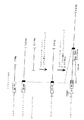
図1は、ラット由来のSDF−1β−およびSDF−1−γ−cDNAのクローニングのための、RT−PCRを用いた方法を示す。5’および3’UTR 領域を直線で示し、コード領域を小さな四角で示している。小さな矢印はプライマーの位置と方向を示す。同一または相同な配列は同じ図形で表されている。SDF−1βのコード領域の最後の4つのコドンは小さな黒四角で表されている。破線はSDF−1γの2572ヌクレオチドの挿入物を示す。SDF−1γの30個のカルボキシ末端コドンは、斜線を付した小さな四角で表されている。
【図2】
図2は、成体ラットの坐骨神経におけるSDF−1βおよびSDF−1γ転写産物を検出するための、ノーザンブロット試験の結果を示す。これらのフィルターを、(A)SDF−1βのヌクレオチド743〜1368に相当し、SDF−1βおよびSDF−1γに共通の3’UTR配列の一部である、626ヌクレオチド長のNT−I−15−cDNAの放射性標識PCR断片、(B)配列番号16のヌクレオチド49〜239に相当する、総てのSDF−1イソ型の共通コード領域である、190ヌクレオチド長のPCR断片、ならびに(C)SDF−1γ−cDNAのヌクレオチド625〜2327に相当し、SDF−1γ特異的挿入物から、プライマーGAS2およびMMSE2で得られたPCR産物のPvuII消化により生じた、1702ヌクレオチド長の断片、とハイブリダイズさせた。
【図3】
図3は、ラット由来の核酸配列、ならびにこれらに由来するSDF−1βおよびSDF−1γ−cDNAのアミノ酸配列を示す。SDF−1γに特異的な挿入物は枠で囲んである。共通のシグナルペプチドの核酸配列には下線を付している。ヌクレオチド(左側)およびアミノ酸(右側)の番号はSDF−1γの配列に相当する。(93)はSDF−1βの最後のアミノ酸を表している。
【図4】
図4は、マウスおよびラットのSDF−1タンパク質のアミノ酸配列の比較を示す。点は同一のアミノ酸を示す。19アミノ酸長のシグナルペプチドは枠で囲んである。
【図5】
図5は、種々の組織(A)および種々の発達段階(B)におけるSDF−1βおよびSDF−1γ転写産物の証拠を示す種々のノーザンブロット試験の結果を示す。(A)坐骨神経(SN)、脳(Br)、肺(Lu)、心臓(HE)、筋肉(Mu)、精巣(Te)、肝臓(Li)、腎臓(Ki)、脾臓(Sp)および胸腺(Th)からの全RNAを含むノーザンブロットフィルターを、SDF−1βおよびSDF−1γに共通の3’−UTR領域由来の放射性標識cDNAプローブとハイブリダイズさせた。(B)発達中のラット脳中のSDF−1βおよびSDF−1γmRNAの検出。17日齢ラット胎児(E17)ならびに誕生後1、4、7、13および20日(P1−20)のラットおよび成体ラット(Ad)の脳からの全RNAを含むノーザンブロットフィルターを、(A)と同様にハイブリダイズさせた。(C)発達過程のラットの坐骨神経のSDF−1βおよびSDF−1γmRNAの検出。1、4、7、14および21日齢のラット(P1−21)、ならびに成体ラット(Ad)の前記神経からの全RNAを含むノーザンブロットフィルターを、(A)と同様にSDF−1β/γプローブとハイブリダイズさせた。上側の矢印はリボゾーム28SRNAの位置を示し、下側のものはメチレンブルー染色したノーザンブロットフィルターによるものを示す。
【図6】
図6は、成体ラットの脳のSDF−1γ−mRNAの細胞局在のためのin situハイブリダイゼーション試験の結果を示す。これらの切片を全SDF−1イソ型の全5’UTRおよびコード配列のサブクローン(A、D、E)、またはSDF−1γ特異的挿入物のサブクローン(B、E、H)に由来するジゴキシゲニン−UTPで標識した596ヌクレオチド長のアンチセンス転写産物とともにインキュベートした。センス転写産物とのハイブリダイゼーションを陰性対照として用いた(C、F、I)。(A、B、C)脳梁の背側灰白層の左右対称スライドにおける標識乏突起神経膠細胞および強く標識されたニューロンの「パールネックレス」様形跡を有する脳梁。(D、E、F)小脳のプルキニエ細胞および顆粒細胞で強いハイブリダイゼーションシグナルが見られ、他のスライドでは弱いシグナルが見られる。(G、H、I)海馬の錐体細胞および顆粒細胞では極めて強いハイブリダイゼーションシグナルが検出される。ラインは10μm。
【図7】
図7は、図6と同じプローブを用い、ラットの新皮質におけるSDF−1γ−mRNAの細胞局在のためのin situハイブリダイゼーション試験の結果を示す。新皮質の前外側(frontolateral)(A)および中外側(mediolateral)(B)領域由来の切片では、総ての新皮質スライド(I〜VI)のニューロンが、総てのSDF−1mRNAイソ型に共通のアンチセンスプローブ(A)でもSDF−1γ特異的プローブでも強く標識されている。
【図8】
図8は、成体ラットの坐骨神経中のSDF−1γ−mRNAの細胞局在のためのin situハイブリダイゼーション試験の結果を示す。これらの切片を、(a)SDF−1βおよびSDF−1γに共通の3’UTR領域(A〜C、E、F)、(b)総てのSDF−1イソ型に共通の5’UTRおよびコード領域(G)、およびSDF−1γ特異的挿入物(H、I)に由来する、センスおよびアンチセンス方向のジゴキシゲニン−UTP標識RNAプローブとハイブリダイズさせた。(A、B、C)縦方向の切片は軸索近傍の紡錘型の顕著なシュワン細胞を示す。(D)S100タンパク質(シュワン細胞のマーカー)に対する抗体で免疫染色した横方向の切片。(E)前記(D)の横方向切片の付近の縦方向切片のハイブリダイズシグナルは、同じ場所でS100に対して免疫陽性の細胞と思われる軸索を取り囲む、標識された半円形のシュワン細胞を示している(D、E中の矢印)。(G、H)アンチセンス転写産物で標識された近傍の両横方向切片(多くの半円形のシュワン細胞(矢印)および上部の右隅の血管壁を示す)のいずれか一方とのハイブリダイゼーション。(C、F、I)センス方向の転写産物とのハイブリダイゼーションを陰性対照として用いた。A、C、G〜I中の直線:100μm、D〜F:10μm。
【図9】
図9は、ラット由来のSDF−1γの核酸配列のコード領域およびそれから誘導されるアミノ酸配列を示す。
【図10】
図10は、ヒトSDF−1γの核酸配列のコード領域およびそれから誘導されるアミノ酸配列を示す。
【図11】
図11は、ヒトおよびラットSDF−1γの核酸配列のコード領域の比較を示す。「hum」:ヒト配列;「rat」:ラット配列。
【図12】
図12は、図11の核酸配列に由来するヒトおよびラットSDF−1γのアミノ酸配列の比較を示す。「hum」:ヒト配列;「rat」:ラット配列。
【図13】
図13は、プラスミドPCRII−TOPO(Invitrogen, Groningen, NL)におけるhSDF−1γおよびhSDF−1γ−H6構築物、ならびにプラスミドpFPMT121における構築物、M−mhSDF−1γ−H6、SDF−1γ−H6およびMFα−mhSDF−1γ−H6を模式的に示す。
【図14】
図14は、hSDF−1γ遺伝子のコード領域を含む439bp長のDNA断片の制限地図を示す。
【図15】
図15は、hSDF−1γ遺伝子およびHisタグのコード領域を含む457bp長のDNA断片の制限地図を示す。
【図16】
図16は、発現プラスミドpFPMT−M−mhSDF−1γ−H6の制限地図を示す。
【図17】
図17は、発現プラスミドpFPMT−hSDF−1γ−H6の制限地図を示す。
【図18】
図18は、発現プラスミドpFPMT−MFα−mhSDF−1γ−H6の作出法を模式的に示す。矢印で示した「P」はPCRプライマーを表す。
【図19】
図19は、発現プラスミドpFPMT−MFα−mhSDF−1γ−H6の制限地図を示す。
【図20】
図20は、(A)SDF−1特異的抗体SDF−1(C19)(Santa Cruz Biotechnology, USA)および(B)Hisタグ特異的抗体(RGS−His抗体、マウスIgG1、Qiagen, Hilden, BRD)を含む、H.ポリモルファの細胞抽出物における発現産物の証拠を示すウエスタンブロット試験の結果を示す。(A)のトラックは、(1)シーブルー・プレステイン・スタンダード、(2)M−mhSDF−1γ−H6、(3)PNGアーゼFで処理したM−mhSDF−1γ−H6、(4)hSDF−1γ−H6、(5)PNGアーゼFで処理したhSDF−1γ−H6、(6)MFα−mhSDF−1γ−H6、(7)PNGアーゼFで処理したMFα−mhSDF−1γ−H6および(8)SDF−1γを含まない細胞抽出液を含む。(B)のトラックは、(1)SDF−1γを含まない細胞抽出液、(2)シーブルー・プレステイン・スタンダード、(3)M−mhSDF−1γ−H6、(4)PNGアーゼFで処理したM−mhSDF−1γ−H6、(5)hSDF−1γ−H6、(6)PNGアーゼFで処理したhSDF−1γ−H6、(7)MFα−mhSDF−1γ−H6および(8)PNGアーゼFで処理したMFα−mhSDF−1γ−H6を含む。
【図21】
図21は、星状細胞のカルシウム濃度に対するSDF−1αおよびSDF−1γの効果の比較を示す:(A)50nM SDF−1α;(B)組換えSDF−1γ(M−mhSDF−1γ−H6)を含む35μg酵母抽出液;(C)22.4μg対照抽出液;(D)SDF−1αによって誘導される細胞内カルシウム上昇に対するSDF−1γおよび対照抽出液による細胞内カルシウムの上昇の定量評価。
【図22】
図22は、CXCR4に対する抗体とのプレインキュベーションを伴わない(A)または伴う(B)、SDF−1αに対する星状細胞におけるカルシウム画像実験の結果を示す。
【図23】
図23は、CXCR4に対する抗体とのプレインキュベーションを伴わない(A)または伴う(B)、SDF−1γに対する星状細胞におけるカルシウム画像実験の結果を示す。
【図24】
図24は、CXCR4に対する抗体とのプレインキュベーションを伴わない(A)または伴う(B)、SDF−1γに対する皮質ニューロンにおけるカルシウム画像実験の結果を示す。
【図25】
図25は、CXCR4に対する抗体とのプレインキュベーションを伴わない(A)または伴う(B)、SDF−1γのC末端塩基性ペプチド(30アミノ酸)に対する星状細胞におけるカルシウム画像実験の結果を示す。
【図26】
図26は、(A)ペプチド2(KKEKIG;配列番号6)および(B)ペプチド3(KKKRQ;配列番号8)に対する星状細胞におけるカルシウム画像実験の結果を示す。[0001]
The present invention relates to a nucleic acid molecule comprising a nucleic acid sequence encoding a chemokine, a neuropeptide precursor or at least one neuropeptide, and a host cell containing the nucleic acid molecule. Furthermore, the present invention relates to polypeptide molecules that function as chemokines or neuropeptides, or that contain at least one neuropeptide, fragments thereof that contain at least one neuropeptide, and methods for producing the polypeptide molecules or fragments thereof. Furthermore, the present invention relates to antibodies, detection methods and test kits, and pharmaceutical compositions.
[0002]
SDF-1α (stromal cell-derived factor 1α) and its isoform resulting from alternative splicing, SDF-1β, were first cloned from a mouse bone marrow stromal cell line (Tashiro et al. 1993). Based on the strong homology of the obtained amino acid sequence with the sequences of interleukin 8 (32%) and macrophage inflammatory protein 1α (32%) and the presence of four characteristic cysteine residues, SDF-1α and SDF- 1β belongs to the group of CXC (α) chemokines. CXC (α) chemokines are a subgroup of the intercrine cytokine system, consisting of inflammatory cytokines in various distances. The mouse and human SDF-1α and SDF-1β cDNA sequences show high homology to each other and result from alternative splicing of one gene.
[0003]
The biological function of SDF-1 was examined by human SDF-1α. SDF-1α is required for B cell maturation, activates T lymphocyte tropism, and induces cell death in the neuronal cell line hNT. SDF-1α is a natural ligand for the T cell chemokine receptor CXCR4 (LESTR / Fusin), which is a binding cofactor of the T lymphotropic HIV1 strain. SDF-1α and β show a “growth arrested” specific expression pattern in fibroblasts and hepatocytes both in vitro and in vivo. Mice in which the SDF-1 gene has been inactivated show reduced B cell formation, loss of ventricular septum and loss of cell migration to the cerebellum, and die shortly after birth. SDF-1 may play an important role in nerve regeneration.
[0004]
The present invention aims, as a basis, to provide a new means for the diagnosis and / or treatment of diseases associated with a defect in the SDF-1 factor or its receptor (CXCR4).
[0005]
According to the invention, this object is achieved by a nucleic acid molecule comprising the following sequence:
(1) a nucleic acid sequence encoding a chemokine, neuropeptide precursor or at least one neuropeptide selected from the following sequences:
(A) a nucleic acid sequence represented by SEQ ID NO: 1,
(B) a nucleic acid sequence encoding a polypeptide having the amino acid sequence represented by SEQ ID NO: 2,
(C) a nucleic acid sequence having at least 60% identity to the sequence shown in (a),
(D) a sequence that hybridizes with the other strand of the sequence shown in (a) or hybridizes in consideration of the degeneracy of the genetic code; and
(E) a derivative of one of the sequences shown in (a) or (b), which is obtained by substitution, addition, inversion and / or deletion of one or more nucleotides, and is a chemokine, neuropeptide precursor Or a derivative that encodes at least one neuropeptide; or
(2) a sequence complementary to one of the nucleic acid sequences shown in the above (a) to (e).
[0006]
As used herein, “polypeptide” also includes a peptide or protein composed of 7 or more amino acids.
[0007]
"Chemokines" represent members of a relatively small family of proteins and are divided into four subgroups based on the arrangement of characteristic cysteine residues: C, CC, CXC and CX.3C. These chemokines bind to specific receptors (Rollins, BJ 1997). In the context of the present invention, "chemokines" are used in particular for members of the CXC chemokine family. The chemokines in the present invention preferably reduce intracellular calcium concentrations in primary astrocytes and / or neurons from the rat or human central nervous system in Ca imaging experiments under the conditions described in Koller et al (2001). Polypeptide molecules that produce a 1.5- to 10-fold increase.
[0008]
Biologically active and physiologically important signal molecules that have regulatory and regulatory functions in the nervous system are called "neuropeptides". These functional domains include, inter alia, the domains of neurotransmission, receptor regulation, alteration of the electrophysiological properties of cell membranes, and metabolic processes. Neuropeptides are synthesized in neurons,
Most of it is released at the synapse (Siegel et al., 1989).
[0009]
A “neuropeptide precursor” is a protein precursor that is converted to an active neuropeptide by proteolytic cleavage.
[0010]
The nucleic acid sequence contained in the nucleic acid molecule according to the present invention may be genomic DNA, cDNA or synthetic DNA, where synthetic DNA sequence is intended to also include modified intranucleoside linkages.
[0011]
“Having at least 60% identity” with respect to a nucleic acid sequence according to the present invention means that
Refers to identity at the DNA level, which can be determined by well-known methods, for example, by computer sequence comparison (Altschul et al., 1990).
[0012]
"Identity," as known to those of skill in the art, refers to the degree of association between two or more nucleic acid molecules, as determined by the match between sequences. "Identity" percentages are indicated by the percentage of identical regions in two or more sequences that take into account gaps and other sequence features.
[0013]
The mutual identities of related nucleic acid molecules can be determined with reference to well-known methods. In many cases, specialized computer programs have specific calculation bearing algorithm elements inserted. In the preferred method of identity determination, in most cases, the largest match between the sequences considered is calculated. Computer programs for determining the identity between two sequences include the GCG program package including GAP (Devereux, J., et al., Nucleic Acids Research 12 (12) 387, 1984), University of Wisconsin Genetics. Computer Groups (Genetics Computer Group University of Wisconsin, Madison WI), BLASTP, BLASTN and FASTA (Altschul et al., 1990), but are not limited thereto. The BLASTX program is available from the National Center for Biotechnology Information (NCBI) or from other sources (BLAST handbook, Altschul S. et al., NCB NLM NIH Bethesda et al., Ed. The well-known Smith Waterman algorithm can also be used to determine identity.
[0014]
Preferred parameters for nucleic acid sequence comparison include the following:
GAP programs are also suitable for use with the above parameters. The above parameters are standard parameters (default parameters) for nucleic acid sequence comparison.
[0016]
Examples of other algorithms include the gap opening penalty, gap extension penalty, and comparison matrix described in the Program Handbook (Wisconsin Package,
[0017]
"The sequence that hybridizes to the other strand of the sequence shown in (a)" refers to a sequence that hybridizes under stringent conditions to the other strand of the sequence shown in (a). For example, hybridization may be performed at 42 ° C. using a hybridization solution composed of 5 × SSPE, 5 × Denhardt's solution, 0.1% SDS, 100 μg / ml salmon sperm DNA, and 30 to 50% formamide ( Sambrook et al., 1989). In the washing step, washing is performed twice in 2 × SSPE, 0.1% SDS at 42 ° C. for 10 to 15 minutes, and then twice in 2 × SSPE, 0.1% SDS at 50 ° C. for 20 minutes. Do. Alternatively, SSC may be used instead of SSPE in the cleaning solution.
[0018]
Surprisingly, it has now been found that the nucleic acid molecules according to the invention are a new member of the SDF family of chemokines. This is called SDF-1γ. The cloning and characterization of the SDF-1γ cDNA for human SDF-1γ and rat SDF-1γ, and its nucleic acid sequence and the amino acid sequence obtained therefrom are described in the Examples.
[0019]
The SDF-1γ nucleic acid sequence consists of the entire nucleic acid sequence of SDF-1β, plus an additional sequence of 2572 nucleotides downstream of the same reading frame to codon 89 of SDF-1β. Due to this insertion, the novel SDF-1γ polypeptide has an amino acid sequence with 119 amino acids and a theoretical molecular weight of 13.6 Kd, which is 30 amino acids at the carboxy terminus compared to the known SDF-1α sequence. Just getting longer. SDF-1γ is thought to have arisen by the insertion of another new exon IIIa between known exons III and IV (see Shirozu et al., 1995).
[0020]
In the carboxy-terminal region of the amino acid sequence of SDF-1γ, five groups of two basic amino acids (Lys-Lys, Arg-Arg and Lys-Arg) form the recognition pattern of Golgi body and secretory vesicle membrane-bound proteases. Can be done. Proteolytic cleavage of this site yields five short peptides (SEQ ID NO: 5, SEQ ID NO: 6, SEQ ID NO: 8, SEQ ID NO: 9, SEQ ID NO: 10) and one shortened protein (SEQ ID NO: 7), Of these, two peptides and one polypeptide (SEQ ID NO: 5, SEQ ID NO: 6 and SEQ ID NO: 7) have a glycine residue at their carboxy terminus. Peptides with a glycine residue at the carboxy terminus can catalyze the carboxy-terminal cleavage of peptidyl-α-amidated monooxygenase (PAM) (carboxylate, which results in the α-amidated carboxy terminus (CONH2)) (See review by Eiper et al., 1992).
[0021]
The rat SDF-1β transcript (see SEQ ID NO: 16) encodes a protein with 93 amino acids (see SEQ ID NO: 17) and a theoretical molecular weight of 10.5 kD, where the first 19 amino acids are the signal sequence of the secreted protein Represents The first 17 amino acids of the mature protein, all occurring as isoforms, are required for binding to the CXCR4 receptor (see Loetscher et al., 1998, Doranz et al., 1999). The two basic amino acids in the carboxy-terminal region (lys89 and arg90) provide a recognition pattern for proteolytic cleavage, resulting in a pentapeptide (lys89-met93, SEQ ID NO: 18) and a shortened protein.
[0022]
According to one embodiment of the invention, the nucleic acid molecule according to the invention has at least 80%, preferably at least 90%, particularly preferably at least 95% identity to the nucleic acid sequence represented by SEQ ID NO: 1. A nucleic acid sequence having
[0023]
Particularly preferred is a nucleic acid molecule comprising a nucleic acid sequence represented by SEQ ID NO: 3 or a nucleic acid sequence encoding a polypeptide having an amino acid sequence represented by SEQ ID NO: 4.
[0024]
A nucleic acid molecule according to the invention may further comprise a promoter suitable for expression, wherein the coding nucleic acid sequence is placed under the control of the promoter. As used herein, a "promoter suitable for expression" refers to the start and start of transcription (RNA synthesis) of a nucleic acid sequence encoding a chemokine, neuropeptide precursor or at least one neuropeptide under the control of a promoter element. A DNA fragment whose frequency is established in the host organism. The promoter selected will depend on the expression system used for expression. In general, constitutive promoters are preferred, but inducible promoters such as, for example, the metallothionein promoter, can also be used. Promoters useful in practicing the present invention include, among others, Hansenula polymorpha (Hansenula polymorpha) -Derived FMD-, MOX-, TPS1-, PMA1- and DAS-promoter; Cerevisiae (S.cerevisiae) -Derived ADH1-, PDC1-, GAP1- and CUP1-promoter, Arkula adeninovorans (Arxula adeninovorans) From AXDH- and ASHB4-promoter and from S. spp.Sordaria macrospora) -Derived NDK1- and CPC2-promoter.
[0025]
A nucleic acid molecule according to the present invention may further comprise a sequence of a vector that allows the nucleic acid molecule to replicate in a host cell and / or to integrate the nucleic acid molecule into the host cell genome. Many cloning and expression vectors are known in the art (see Recombinant Gene Expression Protocols, Meth. Mol. Biol. Vol 62, Humana Press, Hew Jersey, USA). Vectors used for replication in host cells must contain an origin of replication and, if necessary, additional regulatory regions. Vectors include bacteriophages such as λ derivatives, adenovirus, vaccinia virus, baculovirus, SV40 virus, retrovirus, Agrobacterium tumefaciens (Agrobacterium tumefasciences), A YAC vector and a BAC vector.
[0026]
Furthermore, according to the present invention there is provided a host cell containing at least a nucleic acid molecule according to the present invention, which host cell is suitable for the expression of a nucleic acid molecule, and furthermore, if necessary, of the resulting polypeptide molecule. Prokaryotic or eukaryotic cells suitable for processing. A vast number of prokaryotic and eukaryotic expression systems are known in the art. The host cell is, for example, E. coli (E.coli) And Bacillus subtilis (B.subtilis), Or from eukaryotic cells such as fungal cells, plant cells, insect cells and mammalian cells such as CHO cells, COS cells or HeLa cells or derivatives thereof. For example, certain CHO producing strains are known in the art, which have altered glycosylation patterns as compared to CHO cells. Eukaryotic cells are preferably the yeasts Saccharomyces cerevisiae, the methylotrophic yeast Hansenula polymorpha, the dimorphic yeast Alxra adeniborans (Arxula adeninivorans) Or the filamentous fungus Soldaria macrospora.
[0027]
Further, according to the present invention there is provided a polypeptide molecule comprising an amino acid sequence selected from the following sequences:
(I) one of the amino acid sequences represented by SEQ ID NO: 5, SEQ ID NO: 6, SEQ ID NO: 7, SEQ ID NO: 8, SEQ ID NO: 9 and / or SEQ ID NO: 10, and / or a combination of two or more of these sequences Amino acid sequence,
(Ii) an amino acid sequence represented by SEQ ID NO: 4,
(Iii) an amino acid sequence corresponding to the sequence of
(Iv) an amino acid sequence represented by SEQ ID NO: 22, and
(V) An amino acid sequence having at least 85% identity to the sequence shown in (i), (ii), (iii) or (iv).
[0028]
Here, “having at least 85% identity” refers to a match at the amino acid sequence level determined by a well-known method, for example, a computer-based sequence comparison (Altschul et al., 1999).
[0029]
As used herein, "identity" refers to the degree of association between two or more polypeptide molecules, determined by the match between sequences, where a match refers to both matches as identical and conservative amino acid substitutions. Shall mean. "Identity" percentages are indicated by the percentage of identical regions in two or more sequences that take into account gaps and other sequence features.
[0030]
"Conservative amino acid substitution" refers to the substitution of one amino acid residue for another amino acid residue, wherein the substitution has only a limited effect on the (spatial) structure of the polypeptide molecule. Basically, naturally occurring amino acids are classified into four groups that differ physicochemically. Arginine, lysine and histidine belong to the basic amino acid group. Glutamic acid and aspartic acid belong to the acidic amino acids. Uncharged / polar amino acids include glutamine, asparagine, serine, threonine and tyrosine. Non-polar amino acids include phenylalanine, tryptophan, cysteine, glycine, alanine, valine, methionine, isoleucine, leucine and proline. Here, conservative amino acid substitution means substitution of the indicated amino acid by an amino acid belonging to the same physicochemical group.
[0031]
The mutual identity of related polypeptide molecules can be determined with reference to well-known methods. The preferred method of determining identity is one which exhibits a value very close to the maximum match between the sequences examined. Computer programs for determining the identity between two sequences include the GCG program package including GAP (Devereux, J., et al., Nucleic Acids Research 12 (12) 387, 1984), University of Wisconsin Genetics. Computer Groups (Genetics Computer Group University of Wisconsin, Madison WI), BLASTP, BLASTN and FASTA (Altschul et al., 1990), but are not limited thereto. The BLASTX program is available from the National Center for Biotechnology Information (NCBI) or from other sources (BLAST handbook, Altschul S. et al., NCB NLM NIH Bethesda et al., Ed. The well-known Smith Waterman algorithm can also be used to determine identity.
[0032]
Preferred parameters for sequence comparison include the following:
GAP programs are also suitable for use with the above parameters. The above parameters are standard parameters for amino acid sequence comparison (default parameters), so that terminal gaps do not reduce the identity value. For very short sequences, it may also be necessary to increase the predicted value to 100,000 and reduce the word size to 2 when comparing to the reference sequence.
[0034]
Examples of other algorithms include those described in the Program Handbook (Wisconsin Package,
[0035]
An 85% match obtained by the above algorithm is referred to herein as 85% identity. Higher degrees of identity have reasonable relevance.
[0036]
According to one embodiment of the present invention, the polypeptide molecule according to the present invention has at least 90%, preferably at least 95%, the amino acid sequence shown in (i), (ii), (iii) or (iv) above. And sequences having the same identity. Particularly preferred is a polypeptide molecule comprising the amino acid sequence represented by SEQ ID NO: 12 or SEQ ID NO: 13.
[0037]
According to one embodiment of the invention, a polypeptide molecule according to the invention comprises the amino acid sequence represented by SEQ ID NO: 5, SEQ ID NO: 6 and / or SEQ ID NO: 7. Preferred are polypeptide molecules according to the invention comprising the amino acid sequences represented by SEQ ID NO: 5 and SEQ ID NO: 6.
[0038]
According to another embodiment, the present invention provides a fusion protein comprising at least one polypeptide according to the present invention.
[0039]
Fragments of a polypeptide molecule according to the invention, comprising at least one neuropeptide, are likewise included in the invention. Preferably, the fragment comprises at least one amino acid sequence represented by SEQ ID NO: 5, SEQ ID NO: 6, SEQ ID NO: 7, SEQ ID NO: 8, SEQ ID NO: 9 and / or SEQ ID NO: 10. A fragment having the amino acid sequence represented by SEQ ID NO: 5, SEQ ID NO: 6, or SEQ ID NO: 7 is particularly preferred. Also, fragments of a polypeptide molecule according to the present invention may be modified, for example, by glycosylation, phosphorylation, acetylation or amidation.
[0040]
Preferably, a peptide molecule, a fusion protein or fragment of the peptide molecule according to the invention is used in Ca imaging experiments under the conditions described in Koller (2001) in primary astrocytes and / or from rat or human central nervous system. Or a 1.5- to 10-fold increase in intracellular calcium concentration in neurons.
[0041]
Furthermore, according to the present invention, there is provided a method for producing a polypeptide molecule and / or a fragment thereof according to the present invention, comprising culturing a host cell according to the present invention under conditions suitable for expression and possible processing. And, optionally, purifying the expressed polypeptide molecule or fragment. Alternatively, polypeptide molecules and fragments thereof may be obtained by chemical and enzymatic synthesis such as, for example, Merrifield synthesis and / or fragment condensation. Combinations of chemical, enzymatic and recombinant production methods are also contemplated.
[0042]
Further, according to the present invention, there is provided an antibody specific to the polypeptide molecule of the present invention and / or a fragment thereof. In general, specific antibodies are used to immunize a laboratory animal, such as a mouse or rabbit, with a polypeptide molecule or fragment according to the invention, preferably conjugated to a suitable high molecular weight carrier molecule (often a protein). By doing so, it can be produced. In this case, immunization can be facilitated by a suitable adjuvant known in the art. Monoclonal antibodies can usually be obtained by fusing spleen cells collected from immunized mice with tumor cells and selecting the resulting hybridomas. Hybridomas that secrete specific antibodies efficiently can be determined by scanning survivors. Alternatively, the antibody may be produced by recombinant techniques, in which the mRNA is isolated from hybridoma cells or B lymphocytes, the corresponding cDNA is synthesized based on this, and amplified by PCR. After ligation into a suitable vector and insertion into a suitable host cell, it can be recovered from cell culture debris or cell lysate. Recombinant antibodies allow for "humanization" of the antibody, resulting in less immunogenicity. Suitable methods are well known in the art.
[0043]
In the detection of a polypeptide molecule and / or a fragment thereof according to the present invention in a biological sample, according to the present invention, a reagent specific for a polypeptide molecule and / or a fragment thereof is brought into contact with the sample to detect the binding thereof. Provided in an in vitro method.
[0044]
Furthermore, according to the present invention, there is provided a test kit for detecting a polypeptide or a fragment thereof, comprising at least one reagent specific for the polypeptide and / or fragment according to the present invention. Examples of such specific reagents include antibodies, antibody fragments such as Fab or F (ab)2There are fragments or antibody derivatives, of which antibodies are particularly preferred. Antibodies, antibody fragments, eg, Fab or F (ab)2Fragments or antibody derivatives may be of monoclonal or polyclonal origin.
[0045]
Further, according to the present invention, there is provided a method for detecting a nucleic acid encoding a polypeptide molecule according to the present invention in a biological sample in vitro, the method comprising:
Contacting the sample with a nucleic acid molecule according to the invention having a detectable label and / or a fragment thereof, and
Detecting the label
Comprising.
[0046]
Furthermore, according to the present invention there is provided a pharmaceutical composition comprising at least one polypeptide molecule according to the present invention and / or a fragment thereof, and / or a pharmaceutically acceptable salt of said polypeptide molecule or said fragment. . These pharmaceutical compositions may contain pharmaceutically acceptable excipients and / or diluents. Suitable excipients or diluents are well-known in the art. Pharmaceutical compositions suitable for intravenous, subcutaneous or intramuscular administration are preferred. According to one embodiment of the pharmaceutical composition according to the present invention, said composition also comprises at least one antibody in addition to one or more polypeptide molecules or fragments thereof according to the present invention.
[0047]
According to the present invention, these pharmaceutical compositions can be used for the treatment of demyelinating diseases or neurodegenerative diseases, or developmental disorders of the nervous system. A further use of the pharmaceutical composition according to the present invention is in the prevention or treatment of HIV infection in humans, especially HIV encephalopathy. Similarly, the present invention includes the use of a pharmaceutical composition according to the present invention for the treatment of diseases of the hematopoietic system, the immune system as well as the cardiovascular system.
[0048]
Pharmaceutical compositions containing at least one reagent specific for a polypeptide molecule and / or a fragment thereof according to the present invention are also encompassed by the present invention. This reagent is preferably an antibody. Such a pharmaceutical composition can be used for the diagnosis or treatment of a demyelinating disease or a neurodegenerative disease, or a developmental disorder of the nervous system. Another use is in the diagnosis or treatment of HIV infection in humans, especially HIV encephalopathy.
[0049]
The present invention will be described with reference to the drawings and examples below.
[0050]
Example
Example 1: Cloning and sequence analysis of SDF-1γ
A. Materials and methods
Animal experimentation
Adult Wistar rats (200-250 g body weight) were anesthetized by intraperitoneal administration of chloral hydrate (350 ml / kg body weight). The sciatic nerve in the upper thigh was temporarily compressed with forceps (Muller et al., 1986). To obtain RNA from this neural pathway, tissues 2-3 mm before and after the wound were excised and treated. All tests on animals were performed according to the guidelines of the German Animal Protection Act.
[0051]
RNA isolation
Total RNA from rat tissues was isolated by the phenol-guanidine-thiocyanate method (Chomczynski and Sacchi, 1987). Frozen tissue specimens were homogenized twice at 2500 rpm for 45 seconds using a Polytron (Brinkmann, Westbury, USA). Poly A+RNA was isolated by oligo (dT) cellulose chromatography (Sambrook et at., 1988).
[0052]
Construction of cDNA gene bank
For the construction of the cDNA gene bank, 4.5 μg polyA derived from the sciatic nerve of an adult mouse+Using RNA as a template, oligo (dT)12-18Was used as a primer. cDNA was prepared using a TimeSaver cDNA synthesis kit (Pharmacia LKB, Piscataway, NJ). This cDNA was ligated using Gigapack II packaging extract (Stratagene) by ligation to λ-ZAPII phage particles that had been previously cut with EcoRI. As a result of evaluation of the obtained cDNA gene bank, about 0.5 × 10⁶The complexity was obtained. Screening of the gene bank was performed by a standard method using a radiolabeled DNA fragment derived from the 3 'untranslated region (nucleotides 743 to 1368) of rSDF-1β (Sambrook et at, 1969).
[0053]
Oligonucleotide
The following oligonucleotides were synthesized using GeneAssembler Plus Synthesator (Pharmacia, Piscataway, NJ):
MMSE2: 5'-ACGCCATGGACGCCAAGGTCG-3 '(SEQ ID NO: 19) (corresponding to nucleotides 49 to 69 of rSDF-1β-cDNA);
GAS2: 5'-ACTGTAAGGAAGACCCTCTCTCACC-3 '(SEQ ID NO: 20) (corresponding to nucleotides 2327 to 2303 of SDF-1γ);
GAS3: 5'-GTTGAGACTATGCATCGACTCCAAC-3 '(SEQ ID NO: 21) (corresponding to nucleotides 2576 to 2552 of SDF-1γ).
[0054]
Determination and analysis of DNA sequence
Both cDNA strands of the 2.5 Kb long insert in SDF-1β and SDF-1γ including the flanking regions were subjected to the dideoxy-DNA sequencing method (Sanger et al., Using a T17 sequencing kit (Pharmacia-LKB)). 1977).
These sequences were confirmed by sequencing other independent clones from the RT-PCR reaction. The data was compared to the EMBL data bank using the FASTA (Pearson 1980) and BLAST (Altschul et at., 1990) programs. Further comprehensive analysis of these sequences was performed using the PCGENE soft package (Intelligenetics, Mountain View, CA).
[0055]
RT-PCR
Reverse transcription was performed using 1.5 μg of total RNA and the reverse transcriptase Superscript (Gibco, Gaithersburg) according to the manufacturer's instructions. The first cDNA strand was digested with RNase H (Boehringer Mannheim), and 1/10 of the amount was used as a template for PCR amplification with Amplitaq polymerase (Pertkin Elmer) or Pfu polymerase (Stratagene, La Jolla) (for SDF-1γ). Using.
[0056]
B. Cloning and sequencing of SDF-1γ
For the identification of genes that are differentially expressed after a neural lesion, a cDNA clone NT-I-15 with 2174 nucleotides was isolated from a gene bank made from rat sciatic nerve. Sequence analysis of the NT-I-15 clone shows that this clone has 86% homology with the 3 ′ untranslated region (UTR) of mouse SDF-1β-cDNA (see Tashiro et al., 1993). Was. In a Northern blot test under stringent wash conditions, NT-I-15 hybridized with two transcripts from adult rat sciatic nerve (FIG. 2). The smaller transcript corresponded to SDF-1β mRNA about 3 Kb in size, and the larger 5.5 Kb transcript was unknown. This transcript was called SDF-1γ.
[0057]
Isolation of complete clones of both transcripts was performed by screening gene banks and by reverse transcription PCR (RT-PCR). A screening of a gene bank derived from the rat sciatic nerve using a 626 nucleotide long cDNA fragment of the NT-I-15 clone corresponding to nucleotides 734-1368 of the 3′UTR region of the rat SDF-1β resulted in 2819. A complete cDNA clone with a nucleotide length was recovered, which contained the entire coding region of SDF-1β.
[0058]
Rescreening of the cDNA gene bank using the 626 nucleotide long cDNA fragment of NT-I-15 recovered an incomplete SDF-1γ clone of about 3400 nucleotides, which contained the entire 3′UTR region of SDF-1β and It contained a new (non-coding) sequence approximately 1 Kb in length upstream of
[0059]
Both chains of rat SDF-1β cDNA were sequenced. For SDF-1γ, a new insert of 2572 nucleotides in length and a flanking region with a known SDF-1β were similarly overlapped and sequenced. The amino acids derived for SDF-1β were a 93 amino acid peptide with a theoretical molecular weight of 10.5 Kd. The first 19 amino acids are signal peptides for protein secretion. The amino acid sequence derived for SDF-1β contains the first 89 amino acid residues of SDF-1β and 30 additional amino acids in the carboxy-terminal region that show no homology to SDF-1β (compare FIG. 3). . The theoretical molecular weight of a 119 amino acid long SDF-1γ peptide is 13.5 Kd. The amino acid sequence of rat SDF-1β shows strong homology (96.8%) with the corresponding mouse protein (98.9% considering conservative amino acid substitutions). A comparison of the new SDF-1 isoforms SDF-1β and SDF-1γ with the known SDF-1 sequence is shown in FIG.
[0060]
Example 2: Detection of SDF-1β and SDF-1γ mRNA in various tissues and developmental stages
A. Northern blot analysis
Each 10 μg of total RNA was fractionated on a 1.2% agarose gel containing 15% formaldehyde and then transferred to a Nytran NY13 N-membrane (Schleicher and Schull, Keene, NH) in a conventional manner. The filters were irradiated with UV light, stained with methylene blue (Sambrook et al., 1989), prehybridized with 7% SDS in 0.5 M sodium phosphate solution and in the same solution.32P-labeled cDNA probe 1-5 × 10⁶Hybridized with cpm / ml. (I) the entire 3′UTR region of SDF-1β / γ (nucleotides 743 to 1368 of SDF-1β), (ii) the entire region encoding all SDF-1 isoforms (nucleotides 49 to 239) and (iii) 3.) A cDNA fragment corresponding to a 1702 nucleotide long segment of the SDF-1γ specific insert (nucleotides 625 to 2357 of the SDF-1γ cDNA) was radiolabeled by one-way PCR (Sturzl et al., 1991). After hybridization, the filters were washed in 2 × SSC / 1% SDS at 60 ° C. for at least 15 minutes, and further in 0.1 × SSC / 1% SDS at 60 ° C. for at least 15 minutes. These filters were exposed to X-ray film (X-Omat, Kodak) or quantified directly on a BAS1050 Bioimager (Fuji).
[0061]
B. Detection of SDF-1β and SDF-1γ mRNA in various tissues
The Northern blot hybridization test shown in FIG.32This was performed with total RNA from various tissues of adult rats and a 602 nucleotide long fragment from the entire 3'UTR region of SDF-1 [beta] / [gamma], radiolabeled with P-dCTP. The distribution of SDF-1β and SDF-1γ mRNA among various tissues showed a complementary pattern. The β isoform was mainly detected in liver, kidney, spleen and thymus, while SDF-1γ was found mainly in heart and lung tissues, as well as in mature tissues of the nervous system (FIG. 5). The fact that SDF-1β transcripts are found primarily primarily in embryonic and neonatal brain tissue as well as in sciatic nerve points indicates a differential regulation of SDF-1 expression during nervous system development. Neither SDF-1β nor SDF-1γ can be detected in muscle and testis tissue.
[0062]
C. Detection of SDF-1β and SDF-1γ mRNA in brain and sciatic nerve during development
brain:
To examine the distribution of SDF-1β and SDF-1γ specific to developmental stages, RNA from brain tissue at various stages of development (from embryonic day 17 (E17) to adult rats) was analyzed using SDF-1β. The test was performed using a 602 nucleotide long fragment of the common 3'UTR region of / γ. SDF-1β mRNA was mainly detected in the brain tissue of the E17 embryo, but the amount of this transcript decreased with increasing age, and could not be detected in adult rat brain tissue. In contrast, the amount of SDF-1γ transcript was extremely low in E17 embryos, increased gradually, and reached a maximum in adult rats (FIG. 5B).
[0063]
Sciatic nerve:
Total RNA was isolated from sciatic nerves of rats at various stages of development (1 day after birth (P1) to the age of full growth). In P1, a small amount of SDF-1β mRNA was detected, and the amount of this transcript increased in the P4 to P7 stages, and decreased below the detection limit in nerve tissue of adult rats (FIG. 5C).
[0064]
Thus, SDF-1β and SDF-1γ mRNA appear to show different patterns in the developing and adult rat nervous system. The SDF-1β isoform is mainly found in the central and peripheral nervous systems of the embryo or perinatal, whereas SDF-1γ is the most important variant in the adult rat nervous system (FIGS. 5B, C). Glial cell differentiation and neuronal maturation begin between 4 and 7 days after birth, during which time SDF-1β and SDF-1γ appear to be in approximately equal amounts.
[0065]
D. Detection of SDF-1β and SDF-1γ mRNA after sciatic nerve lesion
After injury to the sciatic nerve by compression, small changes in the mRNA patterns of SDF-1β and SDF-1γ were observed at the distal end of the nerve. The transient increase in the amount of SDF-1β was measured by “multiplex quantitative imaging” of a radioactive Northern blot filter, and reached a maximum of 175% 2 days after nerve compression, and then decreased by 7 days after compression. It was at the same level as the control. According to the test, no significant change was found in SDF-1γ mRNA after the nerve lesion.
[0066]
Example 3 in situ Cell localization of SDF-1γ transcript by hybridization
A. in situ Hybridization
Tissue specimens were embedded in Tissue Tec II (Miles, Naperville, Ill.) And frozen in methyl butane at -70 ° C. to obtain sections with a thickness of 20 μm. After fixing these sections, they were acetylated and analyzed by Angerer et al. (1987) for 4 hours at 55 ° C. (I) a subclone of the entire 3′UTR region of SDF-1β / γ (nucleotides 1758-2199 of SDF-1β); (ii) a subclone of all SDF-1 isoforms of the entire 5′UTR and coding region. The in vitro transcripts of the clone (
[0067]
B. Cell localization of SDF-1γ transcript
For in situ hybridization, (a) the common 3 ′ UTR region of SDF-1β / γ, (b) the common coding and 5 ′ UTR region of all SDF-1 isoforms and (c) SDF-1γ Antisense transcripts from specific inserts were labeled with digoxigenin-UTP. The sense transcript was used as a negative control.
[0068]
In the adult rat CNS, a strong and distinct hybridization signal is seen in the paraneural phase, for example in the corpus callosum, as a "pearl necklace" -like row of oligodendrocytes in areas with cerebral gray matter. (FIGS. 5A and 5B). Other hybridization signals include, among others, Purkinje and granule neurons in the cerebellum (FIGS. 6D, E), cone and granule neurons in the hippocampus (FIGS. 6G, H), and neurons in all major layers of the neocortex (FIG. 7A). , B). No hybridization signal was seen in the corresponding sense transcript (see FIGS. 6C, F, I for SDF-1γ sense probe).
[0069]
The signal obtained with the SDF-γ-specific antisense transcript was almost identical to the hybridization pattern obtained with all SDF-1 antisense probes, indicating that the SDF-1γ isoforms were It is shown to be expressed in brain neurons and oligodendrocytes. SDF-1β transcripts appear to be present in the same regions and cell populations as SDF-1γ, as long as they are present in adult rat brain.
[0070]
In longitudinal sections of adult rat sciatic nerve, antisense probes from the entire 3 'region of SDF-1β / γ generated spindle-shaped hybridization signals suggesting the typical shape of Schwann cells near the axonal phase. (FIGS. 8A and 8B). Localization of the S-100 immunoreactivity with the hybridization signal in the transverse section of the sciatic nerve (see arrow) confirmed the expression of SDF-1β / γ in Schwann cells (FIG. 8D, E). The labeling pattern of the adult rat sciatic nerve was obtained using an SDF-1γ specific antisense probe (FIG. 8H), which was the hybridization pattern of antisense transcripts common to all SDF isoforms (FIG. 8G). Strongly suggested. SDF-1γ RNA (and possibly also the SDF-1β isoform) occurs in both Schwann cells (see arrows) and vacuolar cells of the sciatic nerve (see upper right corner of FIG. 8G, H). The results of the in situ hybridization test are consistent with the results of the Northern blot test of FIGS. SDF-1α mRNA was not detected by antisense / sense transcripts from the SDF-1α-specific 3 'region in either adult rat brain or sciatic nerve.
[0071]
Example 4: Expression of SDF-1γ product in Hansenula polymorpha
A. hSDF-1γ construct
For the expression of SDF-1γ in Hansenula polymorpha, three different constructs were made with the aim of obtaining a good analytical approximation to the His tag. These constructs are shown in FIG.
[0072]
1. M-mhSDF-1γ-H6 (methionine / mature human SDF-1γ / His tag). In this fusion protein, the sequence of mature human SDF-1γ (amino acids 20-119 of SEQ ID NO: 12) is located at the end of the N-terminal methionine residue. Since no leader sequence was present, cytoplasmic localization was expected. Six histidine residues (His tags) are arranged at the C-terminus.
[0073]
2. hSDF-1γ-H6 (mature human SDF-1γ / His tag). This construct contains a C-terminal His tag after amino acids 1-119 of SDF-1γ (SEQ ID NO: 12). It consequently comprises the native leader sequence known in human cells. Since the leader sequence may be recognized in heterologous host cells, H. et al. Whether the polymorpha recognizes the native SDF-1γ leader peptide is tested in this construct.
[0074]
3. MFα-mhSDF-1γ-H6 (prepro sequence of mating factor α derived from Saccharomyces cerevisiae / mature human SDF-1γ / His tag). H. The sequence of mature SDF-1γ (amino acids 20-119 of SEQ ID NO: 12) was placed at the end of the prepro sequence of mating factor α from the related brewing yeast Saccharomyces cerevisiae, often used in polymorpha. This construct is described in Suitable for secretion by polymorpha.
[0075]
B. Construction of expression plasmid
Plasmid SDF-1γ-PCRII-TOPO contains a 439 bp long SDF-1γ insert (FIG. 14) and was used as a basic construct. In the first step, the six codons of the C-terminal His tag were included in the hSDF-1γ sequence (hSDF-1γ-H6, FIG. 15).
[0076]
H. As a basic vector for the subsequent expression of the SDF-1γ construct in polymorpha, the integrative plasmid pFPMT121 (Gellissen, 2000) in which the foreign gene to be expressed is under the control of the FMD promoter was inserted. The following expression vectors were constructed based on this plasmid.
[0077]
1. pFPMT-M-mhSDF-1γ-H6. PCR generated a DNA fragment in which the SDF-1γ coding sequence was flanked by an EcoRI restriction cleavage site (before the start codon) and a BamII restriction cleavage site. HSDF-1γ-H6 in PCRII-TOPO (Invitrogen, Groningen, NL) was used as a template. This PCR product was digested with EcoRI / BamHI and cloned between the corresponding sites of the pFPM121 plasmid. A map of the resulting plasmid pFPMT-M-hSDF-1γ-H6 is shown in FIG.
[0078]
2. pFPMT-hSDF-1γ-H6. In this construct as well, a PCR product flanked by EcoRI and BamHI restriction sites was first constructed, with hSDF-1γ-H6 in PCRII-TOPO again being used as template DNA. This PCR product was digested with EcoRI and BamHI and cloned between the corresponding sites of the pFPM121 plasmid. A map of the resulting plasmid pFPMT-hSDF-1γ-H6 is shown in FIG.
[0079]
3. pFPMT-MFα-mhSDF-1γ-H6. For the construction of this plasmid, two different PCR products were constructed (see FIG. 18). The first PCR product (PCR1A) contains the EcoRI restriction site (before the start codon) and the other end of the prepro sequence of mating factor α sandwiched between bases having homology to the first codon of the mature hSDF-1γ sequence. Contains codons. The other PCR fragment (PCR1B) has a mature portion in which the base having homology to the last part of the prepro sequence of mating factor α is adjacent at the forefront, and the Bam-II restriction site (the end of the stop codon) is adjacent at the end. hSDF-1γ sequence. Next, both PCR1A and PCR1B products were mixed to carry out a further PCR reaction. As primers, a forward primer derived from PCR1A (including an EcoRI restriction site) and a backward primer derived from PCR1B (including a BamII restriction site) were used. The resulting PCR product contained the prepro sequence of mating factor α fused to the sequence of mhSDF-1γ, flanked by EcoRI (before the start codon) and BamHI (after the stop codon). After digestion with EcoRI / BamHI, the fragment was cloned between the corresponding restriction sites of the pFPMT121 plasmid. A map of the resulting plasmid pFPMT-MFα-mhSDF1γ-H6 is shown in FIG.
[0080]
C. H. in the prepared expression vector Transformation of polymorpha
Creation and identification of strains
Competent H. for electroporation. For the production of polymorpha cells, 5 ml of YPD medium was It was incubated with a single colony of polymorpha RB11 (odc, orotidine-5-phosphate-decarboxylase deficient (uracil auxotrophy) H. polymorpha strain (Weydemann et al., 1995)) and shaken at 37 ° C. for 16 hours. Next, 100 ml of YPD medium was inoculated with 3 ml of this preculture in a 2 L Erlenmeyer flask, and the OD at 37 ° C.500Was 0.8-1 (shaking number 140 rpm). The cells were collected by centrifuging the culture in a 50 ml Falcon tube using a Beckman centrifuge (4000 rpm, 6 minutes). After removing the supernatant, the cells were resuspended in 20 ml of 50 mM potassium phosphate buffer (pH 7.5, pre-warmed to 37 ° C.), mixed with 0.5 ml of 1 M DTT and incubated at 35 ° C. for 15 minutes (During water bath).
[0081]
After centrifuging the cells again (3000 rpm, 10 minutes, Beckman centrifuge), 100 ml and then 50 ml of STM buffer (270 mM saccharose, 10 mM Tris-HCl pH 7.5, 1 mM MgCl 2).2) Was washed in. After further centrifugation, the cells were resuspended in 0.5 ml of STM buffer and a 60 μl aliquot was used for transformation or stored frozen at −70 ° C. until further use.
[0082]
H. Polymorphic competent cells were transformed with the three expression plasmids (described above) constructed as follows: 60 μl of polymorpha and 1-2 μl of the circular plasmid DNA for introduction were mixed and transferred to a 2 mm-wide electroporation cuvette. Electroporation was performed at 2 kV, 25 μF and 200 ohms. Next, the cells were transferred to a test tube containing 1 ml of YPD medium and shaken at 37 ° C. for 1 hour (tilt angle: 45 °, 160 rpm). After this collection, 330 μl of each cell was plated on a YNB agar plate (1% glucose, without uracil). The plates were incubated at 37 ° C. until macroscopic uracil prototrophy colonies became visible (about one week).
[0083]
At this time, each of the 36 uracil prototrophy colonies was made into a stable strain by four passages and two stabilizations. For subculture, each 2 ml of YNB medium (1% glucose) was inoculated with a single uracil prototrophy colony from the transformation plate and incubated at 37 ° C. for 2 days (tilt 45 °, shaking 160 rpm). ). Each 150 μl of the resulting culture was transferred to 2 ml of fresh YNB medium and incubated again for 2 days (see above). This operation was performed four times (ie, four subcultures). For stabilization, 150 μg of each culture from the latest subculture was transferred to 2 ml of YPD medium and incubated at 37 ° C. for 2 days (see above). Aliquots of these cultures were then plated on YNB agar plates (without uracil). One colony was isolated for each culture and used as one strain.
[0084]
D. Induction and detection of SDF-1γ product expression
After isolation, all strains were subjected to MeOH induction and soluble intracellular fractions were analyzed for hSDF-1γ product content by Western blot. First, a 2 ml YPD medium was inoculated with a single colony strain to be tested in a 10 ml test tube, and then incubated at 37 ° C. for 16 hours to induce expression of a foreign gene (
[0085]
After centrifugation of cells from the induced culture, aliquots of the supernatant were aliquoted into 4 × SAB (8% w / v SDS, 40% w / v glycerin, 8 mM EDTA pH 6.8, 250 mM Tris pH 6.8, 0.4 % W / v bromophenol blue, 40% v / v α-mercaptoethanol), and denatured at 95 ° C for 5 minutes to prepare a culture supernatant.
[0086]
The following steps were performed on ice or at 4 ° C. for the preparation of the intracellular soluble fraction. The cell pellet from the induced culture was resuspended in 500 μl each of extraction buffer (50 mM Tris pH 7.5, 150 mM NaCl, 0.1% v / v Triton X100 or PBS buffer) and mixed with 12.5 μl each of PMSF . These samples were then transferred to 1.5 ml Eppendorf tubes. After adding 500 μl of glass beads, cells were disrupted using Vibrax at 2500 rpm. The supernatant was transferred to a new Eppendorf tube and centrifuged at 10,000 rpm for 10 minutes (Eppendorf centrifuge with cooling function). This centrifuged supernatant is called the so-called soluble intracellular fraction. For direct protein gel electrophoresis, they were mixed with 1 / volume of 4 × SAB and denatured at 95 ° C. for 5 minutes, or stored frozen at −20 ° C. without SAB until later use.
[0087]
For PNGase F digestion, 8 μl of each untreated intracellular soluble fraction was mixed with 1 μl of 1% SDS and incubated at 95 ° C. for 5 minutes. Next, 1 μl PNGase F (2u, Roche) or water was added. After incubation at 37 ° C. for 16 hours, 4 μl of 4 × SAB was added, the sample was denatured at 95 ° C. for 5 minutes and separated on a protein gel.
[0088]
Separation of the denatured sample by protein gel electrophoresis was performed on a 4-20% tricine-SDS gel (Novex) according to the instructions. Next, these protein bands were transferred to a nitrocellulose membrane using a Semi-Dry-Blot apparatus (Trans-Blot SD; Biorad) as described in the instruction manual. For Western blot, a mouse-derived His-tag-specific monoclonal antibody (RGS-His-Anticorper, Qiagen, Hilden, BRD) or a goat-derived SDF-1-specific polyclonal serum (SDF-1 (C19); # sc6193; Santa Cruz) Biotechnology, USA) was used as the primary antibody (serum). Western blots were performed using Western Breeze Kit mice or goats (Novex) according to the manufacturer's instructions.
[0089]
Thus, strains that produced significant amounts of a particular hSDF-1γ-H6 derivative could be identified for each of the three constructs. The most productive strain was selected in each case for further product analysis. In pFPMT-M-mhSDF-1γ-H6, g7-5 / 36 strain, and in pFMPT-hSDF-1γ-H6 and pFMPT-MFα-mhSDF-1γ-H6, g8-28 / 7 strain and g9c-20 / 6 strain. Each was selected.
[0090]
E. FIG. Product analysis
In the culture supernatants of the g7-5 / 36, g8-28 / 7 and g9c-20 / 6 strains, no secreted SDF-1γ product was detectable by Western blot. In the intracellular soluble fractions of these strains, the SDF-1γ product was a mouse-derived His tag-specific antibody or a goat-derived SDF-1γ-specific serum (SDF-1 (C19); # sc6193; Santa Cruz Biotechnology, USA) (see FIGS. 20A and B). The intracellular soluble fraction of the control strain (without SDF-1γ) did not show any products identified as SDF-1γ products (FIG. 20, track 8 (A), track 1 (B)).
[0091]
The molecular weights of the major SDF-1γ products observed on Western blots are generally somewhat higher than the theoretical molecular weights. M-mhSDF-1γ-H6: theoretical value 12,692 kDa, measured value about 16 kDa (FIG. 20,
[0092]
The amino acid sequences of M-mhSDF-1γ-H6 and hSDF-1γ-H6 do not contain a potential N-glycosylation site. Thus, PNGase F digestion does not affect the apparent molecular weight of the particular major product band (FIG. 20,
[0093]
Example 5: Effect of recombinant human SDF-1γ on neuronal and glial calcium levels
In this example, the effect of recombinant human SDF-1γ (M-mhSDF-1γ-H6) in a cell extract of Hansenula polymorpha on calcium concentration in neurons and glial cells is examined. For the evaluation of calcium concentration, a calcium imaging method was used (see Koller et al., 2001).
[0094]
For calcium imaging experiments, primary astrocytes and primary cortical neurons of rats (Wistar strain) were obtained from neonatal rats (0-1 days old; astrocytes) or rat embryos (15-day embryos; cortical neurons). Prepared from Koller et al. (2001). After 5 to 15 days of culture, the cells were filled in vitro with the calcium indicator Fura-2 for 1 hour. Intracellular Fura-2 reacts with free calcium to form Fura-calcium having an absorption wavelength (340 nm) different from Fura-2 (380 nm). The relative intracellular calcium concentration is determined from the absorbance (F340 / F380) and can be represented by a graph. By this method, changes in intracellular calcium concentration induced by extracellular stimulation (eg, ligand / receptor interaction) can be detected.
[0095]
FIG. 21 shows the results of a calcium imaging experiment, in which the effects of SDF-1α and SDF-1γ on calcium concentration in astrocytes are compared. After application of SDF-1α (50 nM, R & D Systems, Wiesbaden, BDR), the calcium concentration of cultured astrocytes increases (FIG. 21A). After application of yeast cell extract containing recombinant SDF-1γ (M-mhSDF-1γ-H6) (36 pg total protein), intracellular calcium is increased in cultured astrocytes (FIG. 21B). However, this response is slightly more restricted than the response to SDF-1α.
[0096]
After application of the control extract (22.4 μg of protein derived from the cell extract of H. Hansenula RB11 cells transformed with the pFPMT121 plasmid without the insert), an increase in intracellular calcium of the cultured astrocytes is observed. No (FIG. 21C).
[0097]
FIG. 21D shows the quantitative evaluation of the increase in intracellular calcium using SDF-1γ and control extract compared to the calcium increase induced by SDF-1α.
[0098]
In addition, it was determined whether preincubation of cells with a CXCR4 specific antibody (monoclonal antibody 12G5; R & D Systems, Wiesbaden, BRD) could reduce the calcium response previously described with respect to the calcium response previously induced by SDF-1α. (See Koller et al., 2001 for detailed methodology of calcium imaging experiments).
[0099]
FIG. 22 shows the results of calcium imaging experiments on astrocytes for SDF-1α without (A) and without (B) antibodies to CXCR4. FIG. 23 shows the results of a corresponding experiment on SDF-1γ. After application of SDF-1α (50 nM, R & D Systems, Wiesbaden, BRD), the intracellular calcium concentration of cultured astrocytes sharply increases (FIG. 22A). However, when SDF-1α was added after preincubation with the monoclonal antibody 12G5 for 5 minutes, cultured astrocytes showed an approximately 50% reduced intracellular calcium efflux (FIG. 22B). These results are consistent with the findings in the literature and demonstrate that the effect of SDF-1α or -1γ on intracellular calcium levels in astrocytes from the central nervous system is mediated by the CXCR4 receptor. It is.
[0100]
After applying 35 μg of yeast extract containing recombinant SDF-1γ (M-mhSDF-1γ-H6; cell contents in PBS buffer), cultured astrocytes result in a measurable increase in calcium (FIG. 23A). ). Unlike SDF-1α, application of SDF-1γ to cultured astrocytes preincubated for 5 minutes with monoclonal antibody 12G5 to the CXCR4 receptor resulted in a rapidly increasing significant intracellular calcium excretion (FIG. 23B). ).
[0101]
After preincubation with the CXCR4 antibody, cell cultures of cortical neurons from rat brain also show a further increase in calcium concentration, similar to the response to SDF-1γ. FIG. 24 shows the results of a corresponding calcium imaging experiment on cortical neurons. Both 35 μg and 125 μg (total protein) of yeast extract containing recombinant SDF-1γ (M-mhSDF-1γ-H6) resulted in a significant increase in calcium concentration in primary cortical neuron cultures. (FIG. 24A). After preincubation with monoclonal antibody 12G5 (antibody to CXCR4) for 5 minutes, the addition of SDF-1γ results in a rapidly increasing intracellular calcium excretion in primary cortical neuron cultures (FIG. 24B).
[0102]
The above results demonstrate that neuronal and astrocyte-derived physiological responses of cells from the central nervous system to SDF-1α and / or SDF-1β are distinctly different from responses to the new chemokine SDF-1γ. Things.
[0103]
Example 6: Effect of C-terminal basic peptide derived from SDF-1γ and its synthetically produced peptide degradation products on intracellular calcium concentration in astrocytes
First, it was investigated how the addition of a basic peptide having an amino acid sequence corresponding to at least 30 amino acids in the C-terminal region of SDF-1γ affects the calcium concentration of astrocytes.
[0104]
FIG. 25 shows the results of a calcium imaging experiment using a C-terminal basic peptide of SDF-1γ in astrocytes. Application of 1 μg / ml of a synthetic peptide representing the C-
[0105]
Peptides RREEKVG (
[0106]
These results indicate that putative neuropeptides can modulate intracellular calcium concentration in various ways. In each case, they show that after pre-incubation of cells with anti-CXCR4 antibody, the dramatic up-regulation of intracellular calcium concentration by SDF-1γ is consistent with SDF-1α and / or SDF-1β chemokines. It is suggested that it is caused by the C-terminal region of SDF-1γ, but not by the segment. This finding confirms the special and specific function of the C-terminus of SDF-1γ, and confirms the results obtained when using the complete SDF-1γ molecule.
[0107]
References
[Table 1]
[Brief description of the drawings]
FIG.
FIG. 1 shows a method using RT-PCR for cloning SDF-1β- and SDF-1-γ-cDNA from rats. The 5 'and 3' UTR regions are indicated by straight lines, and the coding regions are indicated by small squares. Small arrows indicate the position and direction of the primer. Identical or homologous sequences are represented by the same graphic. The last four codons of the coding region of SDF-1β are represented by small black boxes. The dashed line indicates the 2572 nucleotide insert of SDF-1γ. The 30 carboxy-terminal codons of SDF-1γ are represented by small hatched boxes.
FIG. 2
FIG. 2 shows the results of a Northern blot test to detect SDF-1β and SDF-1γ transcripts in adult rat sciatic nerve. These filters were identified as (A) NT-I-15, 626 nucleotides long, corresponding to nucleotides 743 to 1368 of SDF-1β and being part of the 3′UTR sequence common to SDF-1β and SDF-1γ. a radiolabeled PCR fragment of the cDNA, (B) a PCR fragment of 190 nucleotides in length, which is the common coding region of all SDF-1 isoforms, corresponding to nucleotides 49 to 239 of SEQ ID NO: 16; A 1702 nucleotide long fragment corresponding to nucleotides 625 to 2327 of 1γ-cDNA and generated from the SDF-1γ-specific insert by PvuII digestion of the PCR product obtained with primers GAS2 and MMSE2 was hybridized.
FIG. 3
FIG. 3 shows rat-derived nucleic acid sequences and the amino acid sequences of SDF-1β and SDF-1γ-cDNA derived therefrom. Inserts specific for SDF-1γ are boxed. The nucleic acid sequence of the common signal peptide is underlined. The nucleotide (left) and amino acid (right) numbers correspond to the sequence of SDF-1γ. (93) represents the last amino acid of SDF-1β.
FIG. 4
FIG. 4 shows a comparison of the amino acid sequences of mouse and rat SDF-1 proteins. Dots indicate identical amino acids. The 19 amino acid long signal peptide is boxed.
FIG. 5
FIG. 5 shows the results of various Northern blot tests showing evidence of SDF-1β and SDF-1γ transcripts in various tissues (A) and various stages of development (B). (A) Sciatic nerve (SN), brain (Br), lung (Lu), heart (HE), muscle (Mu), testis (Te), liver (Li), kidney (Ki), spleen (Sp) and thymus A Northern blot filter containing total RNA from (Th) was hybridized with a radiolabeled cDNA probe from the 3'-UTR region common to SDF-1β and SDF-1γ. (B) Detection of SDF-1β and SDF-1γ mRNA in the developing rat brain. Northern blot filters containing total RNA from the brains of 17-day-old rat fetuses (E17) and 1, 4, 7, 13 and 20 days after birth (P1-20) and adult rats (Ad) were analyzed using (A) Hybridization was performed in the same manner as described above. (C) Detection of SDF-1β and SDF-1γ mRNA in the sciatic nerve of developing rats. Northern blot filters containing total RNA from the nerves of 1, 4, 7, 14, and 21 day old rats (P1-21) and adult rats (Ad) were subjected to SDF-1β / γ as in (A). Hybridized with the probe. The upper arrow indicates the position of ribosomal 28S RNA, and the lower arrow indicates that obtained by methylene blue-stained Northern blot filter.
FIG. 6
FIG. 6 shows the results of an in situ hybridization test for cellular localization of SDF-1γ-mRNA in adult rat brain. These sections were derived from subclones of all 5 ′ UTRs and coding sequences of all SDF-1 isoforms (A, D, E), or subclones of SDF-1γ specific inserts (B, E, H). Incubation was performed with a 596 nucleotide long antisense transcript labeled with digoxigenin-UTP. Hybridization with the sense transcript was used as a negative control (C, F, I). (A, B, C) Callosal with a "pearl necklace" -like signature of labeled oligodendrocytes and strongly labeled neurons in a symmetrical slide of the dorsal gray layer of the corpus callosum. (D, E, F) Strong hybridization signals are seen in Purkinje and granule cells of the cerebellum, and weak signals are seen in other slides. (G, H, I) Extremely strong hybridization signals are detected in pyramidal and granular cells of the hippocampus. Line is 10 μm.
FIG. 7
FIG. 7 shows the results of an in situ hybridization test for cellular localization of SDF-1γ-mRNA in rat neocortex using the same probe as in FIG. In sections from the frontolateral (A) and mediallateral (B) regions of the neocortex, neurons of all neocortical slides (I-VI) were converted to all SDF-1 mRNA isoforms. Both the common antisense probe (A) and the SDF-1γ-specific probe are strongly labeled.
FIG. 8
FIG. 8 shows the results of an in situ hybridization test for the cellular localization of SDF-1γ-mRNA in the sciatic nerve of adult rats. These sections were identified as (a) the 3 'UTR region (A-C, E, F) common to SDF-1β and SDF-1γ, (b) the 5' UTR common to all SDF-1 isoforms and Hybridized with digoxigenin-UTP-labeled RNA probes in sense and antisense orientation from coding region (G) and SDF-1γ-specific inserts (H, I). (A, B, C) Longitudinal sections show prominent spindle-shaped Schwann cells near axons. (D) Transverse sections immunostained with antibodies to S100 protein (a marker for Schwann cells). (E) The hybridizing signal of the longitudinal section near the transverse section of (D) is a labeled semicircular Schwann cell surrounding an axon that appears to be an immunopositive cell for S100 at the same location. (Arrows in D and E). (G, H) Hybridization with either of the nearby bilateral sections labeled with antisense transcripts (showing many semicircular Schwann cells (arrows) and the upper right vascular wall). Hybridization with the transcript in the (C, F, I) sense direction was used as a negative control. Straight lines in A, C, GI: 100 μm, DF: 10 μm.
FIG. 9
FIG. 9 shows the coding region of the rat SDF-1γ nucleic acid sequence and the amino acid sequence derived therefrom.
FIG. 10
FIG. 10 shows the coding region of the nucleic acid sequence of human SDF-1γ and the amino acid sequence derived therefrom.
FIG. 11
FIG. 11 shows a comparison of the coding regions of the human and rat SDF-1γ nucleic acid sequences. "Hum": human sequence; "rat": rat sequence.
FIG.
FIG. 12 shows a comparison of the amino acid sequences of human and rat SDF-1γ derived from the nucleic acid sequence of FIG. "Hum": human sequence; "rat": rat sequence.
FIG. 13
FIG. 13 shows the hSDF-1γ and hSDF-1γ-H6 constructs in plasmid PCRII-TOPO (Invitrogen, Groningen, NL), and the constructs in plasmid pFPMT121, M-mhSDF-1γ-H6, SDF-1γ-H6 and MFα-mhSDF. -1γ-H6 is schematically shown.
FIG. 14
FIG. 14 shows a restriction map of a 439 bp DNA fragment containing the coding region of the hSDF-1γ gene.
FIG.
FIG. 15 shows a restriction map of a 457 bp DNA fragment containing the coding region of the hSDF-1γ gene and His tag.
FIG.
FIG. 16 shows a restriction map of the expression plasmid pFPMT-M-mhSDF-1γ-H6.
FIG.
FIG. 17 shows a restriction map of the expression plasmid pFPMT-hSDF-1γ-H6.
FIG.
FIG. 18 schematically shows a method for producing the expression plasmid pFPMT-MFα-mhSDF-1γ-H6. "P" shown by an arrow represents a PCR primer.
FIG.
FIG. 19 shows a restriction map of the expression plasmid pFPMT-MFα-mhSDF-1γ-H6.
FIG.
FIG. 20 shows (A) SDF-1 specific antibody SDF-1 (C19) (Santa Cruz Biotechnology, USA) and (B) His tag specific antibody (RGS-His antibody, mouse IgG1, Qiagen, Hilden, BRD) H. Figure 3 shows the results of a Western blot test showing evidence of expression products in cell extracts of polymorpha. The tracks in (A) are (1) Sea Blue Prestained Standard, (2) M-mhSDF-1γ-H6, (3) M-mhSDF-1γ-H6 treated with PNGase F, (4) hSDF Γ-H6, (5) hSDF-1γ-H6 treated with PNGase F, (6) MFα-mhSDF-1γ-H6, (7) MFα-mhSDF-1γ-H6 and (8) ) Contains cell extract without SDF-1γ. Tracks in (B) were treated with (1) SDF-1γ-free cell extract, (2) Sea Blue Prestained Standard, (3) M-mhSDF-1γ-H6, (4) PNGase F M-mhSDF-1γ-H6, (5) hSDF-1γ-H6, (6) hSDF-1γ-H6 treated with PNGase F, (7) MFα-mhSDF-1γ-H6 and (8) PNGase F And MFα-mhSDF-1γ-H6.
FIG. 21
FIG. 21 shows a comparison of the effects of SDF-1α and SDF-1γ on astrocyte calcium concentration: (A) 50 nM SDF-1α; (B) recombinant SDF-1γ (M-mhSDF-1γ-H6). (C) 22.4 μg control extract; (D) Quantitative evaluation of increase in intracellular calcium by SDF-1γ and control extract against increase in intracellular calcium induced by SDF-1α.
FIG. 22
FIG. 22 shows the results of calcium imaging experiments in astrocytes against SDF-1α without (A) or with (B) preincubation with an antibody against CXCR4.
FIG. 23
FIG. 23 shows the results of calcium imaging experiments on astrocytes against SDF-1γ without (A) or with (B) preincubation with an antibody against CXCR4.
FIG. 24
FIG. 24 shows the results of calcium imaging experiments in cortical neurons against SDF-1γ without (A) or with (B) preincubation with antibodies to CXCR4.
FIG. 25
FIG. 25 shows the results of calcium imaging experiments on astrocytes against the C-terminal basic peptide of SDF-1γ (30 amino acids) without (A) or with (B) preincubation with an antibody against CXCR4.
FIG. 26
FIG. 26 shows the results of calcium imaging experiments in astrocytes for (A) peptide 2 (KKEKIG; SEQ ID NO: 6) and (B) peptide 3 (KKKKRQ; SEQ ID NO: 8).
Claims (35)
(1)以下の配列から選択される、ケモカイン、神経ペプチド前駆体または少なくとも一つの神経ペプチドをコードする核酸配列:
(a)配列番号1で表される核酸配列、
(b)配列番号2で表されるアミノ酸配列を有するポリペプチドをコードする核酸配列、
(c)前記(a)に示される配列に対して少なくとも60%の同一性を有する核酸配列、
(d)前記(a)に示される配列のもう一方の鎖とハイブリダイズする、もしくは遺伝コードの縮重を考慮した上でハイブリダイズするものとされる配列、および
(e)前記(a)または前記(b)に示される一の配列の誘導体であって、1以上のヌクレオチドの置換、付加、逆位および/または欠失によって得られ、ケモカイン、神経ペプチド前駆体または少なくとも一つの神経ペプチドをコードする、誘導体;または
(2)前記(a)〜(e)に示される核酸配列の一つと相補的な配列。A nucleic acid molecule having a sequence comprising the following sequence:
(1) a nucleic acid sequence encoding a chemokine, neuropeptide precursor or at least one neuropeptide selected from the following sequences:
(A) a nucleic acid sequence represented by SEQ ID NO: 1,
(B) a nucleic acid sequence encoding a polypeptide having the amino acid sequence represented by SEQ ID NO: 2,
(C) a nucleic acid sequence having at least 60% identity to the sequence shown in (a),
(D) a sequence that hybridizes to the other strand of the sequence shown in (a), or hybridizes in consideration of the degeneracy of the genetic code; and (e) the (a) or A derivative of one of the sequences shown in (b), obtained by substitution, addition, inversion and / or deletion of one or more nucleotides, encoding a chemokine, a neuropeptide precursor or at least one neuropeptide. Or (2) a sequence complementary to one of the nucleic acid sequences shown in (a) to (e) above.
(i)配列番号5、配列番号6、配列番号7、配列番号8、配列番号9および/または配列番号10で表されるアミノ酸配列、および/またはこれらの配列の2以上の組合せを含むアミノ酸配列、
(ii)配列番号4で表されるアミノ酸配列、
(iii)配列番号4の第20アミノ酸〜第119アミノ酸の配列に相当するアミノ酸配列、
(iv)配列番号22で表されるアミノ酸配列、および
(v)前記(i)、(ii)、(iii)または(iv)に示される配列に対して少なくとも85%の同一性を有するアミノ酸配列。A polypeptide molecule comprising an amino acid sequence selected from the following sequences:
(I) an amino acid sequence represented by SEQ ID NO: 5, SEQ ID NO: 6, SEQ ID NO: 7, SEQ ID NO: 8, SEQ ID NO: 9 and / or SEQ ID NO: 10, and / or an amino acid sequence comprising a combination of two or more of these sequences ,
(Ii) an amino acid sequence represented by SEQ ID NO: 4,
(Iii) an amino acid sequence corresponding to the sequence of amino acids 20 to 119 of SEQ ID NO: 4,
(Iv) an amino acid sequence represented by SEQ ID NO: 22, and (v) an amino acid sequence having at least 85% identity to the sequence represented by (i), (ii), (iii) or (iv). .
前記試料に、検出可能な標識を有する請求項1〜6のいずれか一項に記載の核酸分子および/またはその断片を接触させる工程、ならびに
前記標識を検出する工程
を含んでなる、方法。A method for detecting a nucleic acid encoding the polypeptide molecule according to any one of claims 14 to 17 in a biological sample in vitro,
A method comprising: contacting the sample with a nucleic acid molecule and / or a fragment thereof according to any one of claims 1 to 6 having a detectable label; and detecting the label.
Applications Claiming Priority (2)
| Application Number | Priority Date | Filing Date | Title |
|---|---|---|---|
| DE10027383A DE10027383A1 (en) | 2000-06-02 | 2000-06-02 | Nucleic acid molecule comprising a nucleic acid sequence coding for a chemokine, a neuropeptide precursor or at least one neuropeptide |
| PCT/EP2001/006250 WO2001092530A1 (en) | 2000-06-02 | 2001-06-01 | Nucleic acid molecule comprising a nucleic acid sequence coding for an sdf-1 gamma chemokine, a neuropeptide precursor or at least one neuropeptide |
Publications (2)
| Publication Number | Publication Date |
|---|---|
| JP2004521607A true JP2004521607A (en) | 2004-07-22 |
| JP2004521607A5 JP2004521607A5 (en) | 2005-02-17 |
Family
ID=7644466
Family Applications (1)
| Application Number | Title | Priority Date | Filing Date |
|---|---|---|---|
| JP2002500722A Pending JP2004521607A (en) | 2000-06-02 | 2001-06-01 | Nucleic acid molecule comprising a nucleic acid sequence encoding a chemokine, neuropeptide precursor or at least one neuropeptide |
Country Status (9)
| Country | Link |
|---|---|
| US (2) | US20030215792A1 (en) |
| EP (1) | EP1287142B1 (en) |
| JP (1) | JP2004521607A (en) |
| AT (1) | ATE309361T1 (en) |
| AU (1) | AU6233501A (en) |
| DE (2) | DE10027383A1 (en) |
| DK (1) | DK1287142T3 (en) |
| ES (1) | ES2251483T3 (en) |
| WO (1) | WO2001092530A1 (en) |
Families Citing this family (9)
| Publication number | Priority date | Publication date | Assignee | Title |
|---|---|---|---|---|
| US7358044B2 (en) * | 2003-04-16 | 2008-04-15 | Argyll Biotechnologies, Llc. | Prophylactic and therapeutic benefits of a new class of immune stimulating peptides |
| US7776564B2 (en) | 2004-04-30 | 2010-08-17 | Five Prime Therapeutics, Inc. | Stromal cell-derived factor-1 polypeptides, polynucleotides, modulators thereof and methods of use |
| BRPI0617823A2 (en) * | 2005-10-31 | 2011-08-09 | Laboratoires Serono Sa | use of sdf-1 for the treatment and / or prevention of neurological diseases and pharmaceutical composition |
| EP2065463A3 (en) * | 2006-04-07 | 2009-09-02 | Neuro Therapeutics AB | Survival and development of neural cells |
| US20090291887A1 (en) * | 2006-07-07 | 2009-11-26 | Mueller Hans-Werner | Proteins of the SDF-1-Family for the Manufacturing of a Medicament |
| US7696309B2 (en) | 2006-10-23 | 2010-04-13 | The Brigham And Women's Hospital, Inc. | Protease resistant mutants of stromal cell derived factor-1 in the repair of tissue damage |
| US20110034383A1 (en) * | 2007-10-15 | 2011-02-10 | Institut Pasteur | Cxcl12 gamma a chemokine and uses thereof |
| WO2011106234A1 (en) | 2010-02-25 | 2011-09-01 | Provasculon, Inc. | Protease-resistant mutants of stromal cell derived factor-1 in the repair of tissue damage |
| EP2717894B1 (en) | 2011-06-07 | 2018-01-24 | Mesoblast International Sàrl | Methods for repairing tissue damage using protease-resistant mutants of stromal cell derived factor-1 |
Family Cites Families (11)
| Publication number | Priority date | Publication date | Assignee | Title |
|---|---|---|---|---|
| US5350836A (en) * | 1989-10-12 | 1994-09-27 | Ohio University | Growth hormone antagonists |
| US5885794A (en) * | 1991-05-10 | 1999-03-23 | The Salk Institute For Biological Studies | Recombinant production of vertebrate activin receptor polypeptides and identification of receptor DNAs in the activin/TGF-β superfamily |
| JP3367581B2 (en) * | 1993-10-14 | 2003-01-14 | 小野薬品工業株式会社 | Novel polypeptide, method for producing the same, DNA encoding the polypeptide, vector comprising the DNA, and host cells transformed with the vector |
| EP0892807A1 (en) * | 1996-04-10 | 1999-01-27 | Axys Pharmaceuticals, Inc. | Gene family associated with neurosensory defects |
| US6114502A (en) * | 1996-04-10 | 2000-09-05 | Axys Pharmaceuticals, Inc. | Gene family associated with neurosensory defects |
| US6100387A (en) * | 1997-02-28 | 2000-08-08 | Genetics Institute, Inc. | Chimeric polypeptides containing chemokine domains |
| MXPA00003885A (en) * | 1997-10-22 | 2004-04-23 | Inst Genetics Llc | Chimeric polypeptides containing chemokine domains. |
| US6747137B1 (en) * | 1998-02-13 | 2004-06-08 | Genome Therapeutics Corporation | Nucleic acid sequences relating to Candida albicans for diagnostics and therapeutics |
| WO1999055915A2 (en) * | 1998-04-29 | 1999-11-04 | The Government Of The United States Of America As Represented By The Secretary, Department Of Health And Human Services | IDENTIFICATION OF POLYMORPHISMS IN THE PCTG4 REGION OF Xq13 |
| AU5134799A (en) * | 1998-07-30 | 2000-02-21 | Human Genome Sciences, Inc. | 98 human secreted proteins |
| AU2201801A (en) * | 1999-12-16 | 2001-06-25 | Ribotargets Limited | Assays |
-
2000
- 2000-06-02 DE DE10027383A patent/DE10027383A1/en not_active Ceased
-
2001
- 2001-06-01 WO PCT/EP2001/006250 patent/WO2001092530A1/en active IP Right Grant
- 2001-06-01 AU AU62335/01A patent/AU6233501A/en not_active Abandoned
- 2001-06-01 EP EP01936423A patent/EP1287142B1/en not_active Expired - Lifetime
- 2001-06-01 DE DE50108009T patent/DE50108009D1/en not_active Expired - Lifetime
- 2001-06-01 AT AT01936423T patent/ATE309361T1/en not_active IP Right Cessation
- 2001-06-01 DK DK01936423T patent/DK1287142T3/en active
- 2001-06-01 JP JP2002500722A patent/JP2004521607A/en active Pending
- 2001-06-01 ES ES01936423T patent/ES2251483T3/en not_active Expired - Lifetime
-
2002
- 2002-12-02 US US10/308,322 patent/US20030215792A1/en not_active Abandoned
-
2008
- 2008-01-22 US US12/010,221 patent/US20080319165A1/en not_active Abandoned
Also Published As
| Publication number | Publication date |
|---|---|
| EP1287142B1 (en) | 2005-11-09 |
| ES2251483T3 (en) | 2006-05-01 |
| EP1287142A1 (en) | 2003-03-05 |
| WO2001092530A1 (en) | 2001-12-06 |
| DE50108009D1 (en) | 2005-12-15 |
| DE10027383A1 (en) | 2001-12-20 |
| AU6233501A (en) | 2001-12-11 |
| US20030215792A1 (en) | 2003-11-20 |
| DK1287142T3 (en) | 2006-03-13 |
| US20080319165A1 (en) | 2008-12-25 |
| ATE309361T1 (en) | 2005-11-15 |
Similar Documents
| Publication | Publication Date | Title |
|---|---|---|
| JPH11513883A (en) | Human vascular endothelial growth factor 2 | |
| JP2002512524A (en) | Human protein having transmembrane domain and DNA encoding the same | |
| JP2002502589A (en) | 45 human secreted proteins | |
| JPH10513049A (en) | EPH-like receptor ligand | |
| US20080319165A1 (en) | Nucleic acid molecule comprising a nucleic acid sequence coding for a chemokine, a neuropeptide precursor, or at least one neuropeptide | |
| JPH09510103A (en) | Fibroblast growth factor-10 | |
| US20040259789A1 (en) | Method of treating obesity and metabolic disorders related to excess adipose tissue by administration of sFRP-5 peptide | |
| JP2002526073A (en) | A coding sequence for a novel human growth differentiation factor, a polypeptide encoded by the DNA sequence thereof, and a method for producing them. | |
| US6573068B1 (en) | Claudin-50 protein | |
| JP2001514885A (en) | 70 human secreted proteins | |
| WO2000077203A1 (en) | Lipocalin family protein | |
| JP2003525566A (en) | 125 human secreted proteins | |
| JP2002505871A (en) | 31 human secretory proteins | |
| JP2001512971A (en) | Use of ULIP in the diagnosis and treatment of cancer and paraneoplastic neurological syndromes | |
| JP2003521216A (en) | 90 human secreted proteins | |
| JP2003521215A (en) | 83 human secreted proteins | |
| JPH09194502A (en) | New chondroitin sulfate proteoglycan, its core protein, dna coding the same and antibody to the same | |
| JPH11507504A (en) | Fibroblast growth factor 13 | |
| US5861283A (en) | DNA encoding a limbic system-associated membrane protein | |
| JPH0892289A (en) | Protein related to human machado joseph disease, cdna and gene coding for the same protein, vector containing the same dna or gene, host cell transformed with the same manifestation vector, method for diagnosing machado joseph disease and therapeutic agent therefor | |
| JP4315257B2 (en) | Novel chondroitin sulfate proteoglycan, its core protein, DNA encoding it and antibody to it | |
| US20100111913A1 (en) | Method of enhancing migration of neural precursor cells | |
| US20090081227A1 (en) | Human and mammalian stem cell-derived neuron survival factors | |
| WO2001023557A1 (en) | Novel polypeptides and genes encoding the same | |
| JP2003259871A (en) | Secretory protein creg2 expressing brain-specific expression and application of the same |
Legal Events
| Date | Code | Title | Description |
|---|---|---|---|
| A131 | Notification of reasons for refusal |
Free format text: JAPANESE INTERMEDIATE CODE: A131 Effective date: 20070126 |
|
| A601 | Written request for extension of time |
Free format text: JAPANESE INTERMEDIATE CODE: A601 Effective date: 20070426 |
|
| A602 | Written permission of extension of time |
Free format text: JAPANESE INTERMEDIATE CODE: A602 Effective date: 20070508 |
|
| A601 | Written request for extension of time |
Free format text: JAPANESE INTERMEDIATE CODE: A601 Effective date: 20070626 |
|
| A602 | Written permission of extension of time |
Free format text: JAPANESE INTERMEDIATE CODE: A602 Effective date: 20070703 |
|
| A521 | Request for written amendment filed |
Free format text: JAPANESE INTERMEDIATE CODE: A523 Effective date: 20070726 |
|
| A02 | Decision of refusal |
Free format text: JAPANESE INTERMEDIATE CODE: A02 Effective date: 20090424 |
|
| A521 | Request for written amendment filed |
Free format text: JAPANESE INTERMEDIATE CODE: A523 Effective date: 20090824 |
|
| A911 | Transfer to examiner for re-examination before appeal (zenchi) |
Free format text: JAPANESE INTERMEDIATE CODE: A911 Effective date: 20091016 |
|
| A912 | Re-examination (zenchi) completed and case transferred to appeal board |
Free format text: JAPANESE INTERMEDIATE CODE: A912 Effective date: 20091120 |