JP2004504607A - Collection of binding proteins and tags and their use for nested sorting and high-throughput screening - Google Patents
Collection of binding proteins and tags and their use for nested sorting and high-throughput screening Download PDFInfo
- Publication number
- JP2004504607A JP2004504607A JP2002512691A JP2002512691A JP2004504607A JP 2004504607 A JP2004504607 A JP 2004504607A JP 2002512691 A JP2002512691 A JP 2002512691A JP 2002512691 A JP2002512691 A JP 2002512691A JP 2004504607 A JP2004504607 A JP 2004504607A
- Authority
- JP
- Japan
- Prior art keywords
- antibody
- library
- combination
- epitope
- oligonucleotides
- Prior art date
- Legal status (The legal status is an assumption and is not a legal conclusion. Google has not performed a legal analysis and makes no representation as to the accuracy of the status listed.)
- Pending
Links
Images


Classifications
-
- C—CHEMISTRY; METALLURGY
- C40—COMBINATORIAL TECHNOLOGY
- C40B—COMBINATORIAL CHEMISTRY; LIBRARIES, e.g. CHEMICAL LIBRARIES
- C40B30/00—Methods of screening libraries
- C40B30/04—Methods of screening libraries by measuring the ability to specifically bind a target molecule, e.g. antibody-antigen binding, receptor-ligand binding
-
- G—PHYSICS
- G01—MEASURING; TESTING
- G01N—INVESTIGATING OR ANALYSING MATERIALS BY DETERMINING THEIR CHEMICAL OR PHYSICAL PROPERTIES
- G01N33/00—Investigating or analysing materials by specific methods not covered by groups G01N1/00 - G01N31/00
- G01N33/48—Biological material, e.g. blood, urine; Haemocytometers
- G01N33/50—Chemical analysis of biological material, e.g. blood, urine; Testing involving biospecific ligand binding methods; Immunological testing
- G01N33/68—Chemical analysis of biological material, e.g. blood, urine; Testing involving biospecific ligand binding methods; Immunological testing involving proteins, peptides or amino acids
- G01N33/6803—General methods of protein analysis not limited to specific proteins or families of proteins
- G01N33/6845—Methods of identifying protein-protein interactions in protein mixtures
Abstract
捕捉試薬が特異的である、ポリペプチドタグを含むソーティングタンパク質にツールとして使用される抗体のような抗タグ捕捉試薬のアドレス可能コレクションを本明細書で提供する。当該コレクションを使用するネスト化ソーティングの方法をも提供する。その方法には、タグ化分子のライブラリーを作成するため、特有の、事前に選択したポリペプチドをコードする核酸分子のセットを導入することにより分子のタグ化コレクション群を作成し;タグを導入する前または後の何れかにおいて、ライブラリーをNの分割物に分割し;個々の分割物を翻訳し、その個々の分割物をNの捕捉試薬コレクション群の1つと反応させ;目的の分子群と結合したポリペプチドタグに結合した捕捉試薬を同定し、そしてそれにより目的の分子を含む、分割したコレクション群のうちの1つを同定するステップが含まれる。その方法には、更に、タグの新しいセットを加えること、および同じまたは異なるコレクション捕捉試薬群によりソーティングプロセスを繰り返すことが含まれ、それにより、目的のタンパク質または分子が同定される。Provided herein is an addressable collection of anti-tag capture reagents, such as antibodies used as tools for sorting proteins that include a polypeptide tag, wherein the capture reagent is specific. It also provides a nested sorting method that uses the collection. The method involves creating a tagged collection of molecules by introducing a set of unique, preselected polypeptide-encoding nucleic acid molecules to create a library of tagged molecules; Either before or after partitioning the library into N resolutions; translating each resolution and reacting each resolution with one of the N capture reagent collections; Identifying a capture reagent bound to the polypeptide tag bound to and thereby identifying one of the divided collections containing the molecule of interest. The method further includes adding a new set of tags and repeating the sorting process with the same or a different collection of collection capture reagents, thereby identifying the protein or molecule of interest.
Description
【0001】
関連出願
35 U.S.C. §119(e)の下、優先権の米国で利益を、発明の名称“COLLECTIONS OF ANTIBODIES FOR NESTED SORTING AND HIGH THROUGHPUT SCREENING”であるDana Ault−Richeの、2000年7月19日出願、米国仮出願番号60/219183に関し、請求する。国際的な優先権を、米国仮出願番号60/219,183に関し、請求する。許可されるならば、米国仮出願番号60/219,183の内容すべてを引用により本明細書に加える。
【0002】
本発明の分野
本発明は、結合タンパク質のコレクション(本明細書では捕捉試薬と呼ぶ)および多様性ライブラリー(遺伝子ライブラリーを含む)の機能調査のためのその使用方法に関する。当該方法およびコレクションの技術は、ロボットマイクロ−ウェルハイスループットスクリーニングおよびアレイおよび関連技術に組込まれる。
【0003】
本発明の背景
ゲノミクスおよびプロテオミクス
ヒトゲノムプロジェクトは、遺伝学的データの雪崩(avalanche)を生じた。これらのデータの解明により、生物学がより理解され、最終的には、薬物創製の発展を導くこととなる。遺伝子配列情報の有用性により、生物医学的研究が構成される方法および発見の速度が変化している。しかし、ゲノム配列の獲得により、何の遺伝子か、あるいはコード化されたタンパク質はどのように機能するか、どのように細胞および組織が生育するのかが示されるわけではなく、ましてや病因についての洞察および疾患の治癒が提供されるものでもない。配列決定により得られる情報の所産以前に、ゲノムが十分理解され、コード化タンパク質およびその機能が同定されなければならない。
【0004】
そのため、プロテオミクスの出現(emergence)があり、その挑戦により、ヒトゲノムおよび他のゲノムの配列決定の効果により得られた過剰な情報が解明されることとなる。その焦点は、配列により同定される遺伝子の機能を決定することである。しかし、それは、配列決定により遺伝子を同定する仕事は、遺伝子またはコード化タンパク質の機能を発見することよりも簡単である。遺伝子によりコードされるタンパク質を同定するための、生物化学的、遺伝学的および情報科学的アプローチを含む種々のアプローチでは、その同定の試みが為されている。情報科学的アプローチでは、遺伝子配列を、既知または既知とされている機能を有するタンパク質をコードする遺伝子配列と比較するコンピューター探索に基づく遺伝子機能を決定する試みが為されている。遺伝子配列と機能との間の断絶のため、これらのアプローチの成功は制限されている。遺伝子機能の決定は、遺伝学および生物化学の典型的アプローチに依存しつづけている。当該遺伝学的アプローチは、遺伝子機能の破壊、その後のその破壊による効果の観察に基づいており、生物化学的アプローチは、機能と相関する生物化学的変化に基づいている。進歩のため、ハイスループットな分析が必要とされている。
【0005】
ゲノミクスのため、ハイブリダイゼーション反応に依存するハイスループットアレイが、遺伝子を同定する手段として用いられている。プロテオミクスは、ハイスループットの方法論にはまだなじまない。例えば、DNAマイクロアレイが用いられ、所定サンプル中の数千の遺伝子のメッセンジャーRNA(mRNA)の量を決定する。DNAの遺伝子は、タンパク質に翻訳される前に中間体分子としてmRNAに転写される。2つのサンプル由来のmRNAは、2種類の色素を有するポリメラーゼ連鎖反応(PCR)増幅により別々に標識され、混合され、次いで、アレイ上で浸す。このPCR産物は、ヌクレオチドの相補配列を含む核酸を含むアレイ中のスポットに特異的に結合する。色素の割合は、2サンプル中のmRNAの相対量を決定する。次いで、コンピューターアルゴリズムを使用し、データを評価し、インタープリトする。タンパク質は、細胞性調節の中心であり、mRNA発現とタンパク質発現との間に直接相関がないため、このDNAマイクロアレイ分析は、本来的に制限される。タンパク質の活性は、他のタンパク質との相互作用または代謝の結果としてしばしば、その構造の微妙な変化により調節され得る。更に、タンパク質は、異なる半減期を有し、細胞内で区画に分けられている。結果として、細胞のタンパク質状態、またはmRNAと協同する「プロテオーム」に関する情報を得ることは難しい。
【0006】
タンパク質分析技術は、タンパク質分離および検出の組合せに基づく。二次元(2−D)ゲルシステムでは、タンパク質は、一方向には荷電により、そして他の方向には大きさにより分離する。分離後、タンパク質は、ゲルからの切除およびマススペクトロメトリーによる分析により同定される。2−Dゲル方法は1,000を超えるタンパク質を同時に分析するけれども、これらの方法は、巨大サンプルの要求物、低い溶解性、低い感度、結果の不一致およびロースループット(low throughput)により制限される。
【0007】
遺伝子シャッフリングおよびエラー−プロンPCRによるランダム飽和変異誘発(random saturation mutagenesis)のようなタンパク質進化法は、タンパク質の望ましい特性を「進化」させる選択を伴う変異と関連し、それにより、例えば、産業工程で使用するための触媒を作成するため、新規研究試薬を作成するため、および組換え抗体の能力を改善する手段が提供される。可能な構造変異の量は多い。例えば、100アミノ酸を含む比較的小さいタンパク質との可能な組合せの数は20100である。更なる多様性は、合成性、または「非自然性」のアミノ酸を含むことにより提供される。タンパク質評価法は、数兆のタンパク質変異を含む遺伝子のコレクションを作成し得る。これら数兆の中に、望ましい特性を有するタンパク質がある。これらの多様性を生ずる方法を生かす鍵は、これら非常に大きな「乾草堆」の中にある望ましい「針」を見つけ出す能力である。これは、抗生物質の耐性の獲得のような、選択方法論を使用し、固定化捕捉分子に結合させ、および蛍光性の獲得その後の粒子ソート化により、試みられる。進化する特性に依存し、選択スキームが常に可能であるわけではない。ハイスループットロボットシステムを使用する個々の試験は、選択システムに代わるものであるが、これらのシステムは、50万クローンを超える調査のためには非実用的である。これらの方法では、これらの多様性作成法の能力を完全に活用することはできない。
【0008】
タンパク質のサンプル多様性コレクションを同定し、タンパク質およびその機能を同定する新規方法の必要があることは明らかである。そのため、ここでは、タンパク質の多様性コレクションのうちの望ましいタンパク質を同定するための方法および産物を提供することを目的とする。また、ここでは、その方法を行うための産物を提供することを目的とする。
【0009】
本発明の要約
本明細書では、巨大コレクション由来の分子、特にタンパク質および核酸をスクリーニングし同定するための方法および製品を提供する。特に、同定可能タンパク質結合パートナーに特異的に結合する捕捉試薬のコレクション(すなわち、抗体のようなレセプターまたは他のレセプター)を、本明細書ではポリペプチドタグと称し、ここで、個々の捕捉試薬が、エピトープまたはリガンドまたはその部分のような事前に選択したポリペプチドタグに対し高い選択性および特異性で結合するように選択又は設計されている、コレクションを提供する。抗体のような同定可能な捕捉試薬を含むコレクションは、抗体のような捕捉試薬が同定可能(アドレス可能)である限り、液相および固相形式を含む任意の適当な形式で提供される。捕捉試薬のアドレス可能アレイを本明細書で例示する。アレイについて本明細書で例示した方法は、RFタグ、検出可能ビーズ、バー被覆ビーズに結合する抗体のような捕捉試薬を含む任意の他の形式および他の形式で実施され得る。そのコレクションは、ソートするための装置として役立ち、最終的に、目的のタンパク質および遺伝子および他の分子を同定する。
【0010】
エピトープタグのような、事前に選択したポリペプチドタグは、ソートされるタンパク質のような分子に結合される。その結合は、任意の手段により行われ得、スクリーニングされるタンパク質をコードする核酸に、タグをコードする核酸を組込む増幅を伴う増幅スキームまたはライゲーションを用い通常行われる。
【0011】
タンパク質−タグ−標識コレクションを用いソートする方法を本明細書で提供する。本明細書では、ソーティングにより、タンパク質の巨大な多様性コレクションから望ましい特性でタンパク質を同定する方法を提供する。本明細書で提供する結合タンパク質(捕捉試薬)のアドレス可能コレクションおよび方法に重要なのは、既知配列の事前に選択されたポリペプチドタグのセットに結合する、抗体のような捕捉試薬の選択である。ポリペプチドタグは、抗体のような捕捉試薬に特異的に結合する十分な数のアミノ酸を含む。抗体のような捕捉試薬のコレクションは、少なくとも約10、より少なくとも約30、50、100、200、250、またはそれより多く、少なくとも約500、1000またはそれより多い種類の、種々のメンバーのポリペプチドタグのセットに結合する抗体のような捕捉試薬を含む。抗体のような捕捉試薬のコレクションを作成する方法を本明細書で提供する。
【0012】
抗体のようなアドレス可能捕捉試薬コレクションは、当該捕捉試薬と特異的に反応するポリペプチドのアミノ酸配列でタグ化される分子をソートする手段を提供する。当該ソーティングは、コレクション中の抗体のような捕捉試薬と、ソートされる分子のコレクションに導入されるエピトープタグのようなポリペプチドタグとの間での高い特異性に依存する。
【0013】
ある実施態様では、抗体のようなアドレス可能捕捉試薬は、固相上の区別できるアドレス可能位置に提供される、複数の捕捉試薬を含むアレイとして提供される。アレイ上の個々のアドレスは、特異的な、事前に決定したタグに結合する抗体のような捕捉試薬を含む。一般的に、個々の位置の抗体のようなすべての捕捉試薬は、同一であるか実質的に同一であるが、個々の試薬が、事前に設計するか事前に選択したポリペプチドタグを生ずる分子、一般的にはタンパク質に選択的に結合する特異的な高い結合親和性(kaは一般的には少なくとも約10−7から10−9である)を有する必要があるのみである。
【0014】
実施において、ポリペプチドタグでタグ化されるタンパク質を、捕捉試薬のアレイ上で浸すか、適当な結合条件下、ビーズのような同定可能な支持体に結合する捕捉試薬のコレクションと反応させる。特定捕捉試薬に対する、事前に選択したタグの結合特異性の特性により、タンパク質は、事前に選択したタグによりソートされる。次いで、タグの同定が知られているが、それは特定捕捉試薬と反応するためであり、その同定は、カラーコード化またはバーコード化のようなものを含む光学的コード化、またはマイクロ波のような電子的タグ化または周波数(radio frequency)(RF)−タグ化、粒子に対する結合のようなアレイまたはそのアイデンティファイアー中の位置の効果により、知られる。
【0015】
ある実施態様では、抗体は、固相形式で提供され、より好ましくは、個々の位置が同定されるアドレス可能アレイとして構成される。バーコードまたは他の記号または同定の印もまた、抗体の方向付けまたはポジショニングの目的のために固相アレイに含まれ得る。複数のそのアレイが単一マトリクス支持体に含まれ得る。ある実施態様では、アレイが配置され、例えば96ウェル、384ウェル、1536ウェルまたはそれよりも高い密度形式でマッチする大きさである。他の実施態様では、30、100、200、250、500、750、1000または通常の数のような、例えば、30から1000抗体配置を有する24アレイ、個々が配置されている。ある実施態様では、30、100、200、250、500、750、1000または他の通常の数のような、30から1000抗体位置までを有する、例えば96またはそれより多いアレイ、それぞれが配置されている。
【0016】
他の実施態様では、固体支持体は、液相中で扱うことができ、次いで二次元アレイ中に層となるミクロスフェアのような、コード化粒子(ビーズ)を構成する。ミクロスフェアのような粒子は、コード化されており、所望により、カラーまたはバーコード化、化学的コード化、電子的コード化のようにコード化されており、またはそれに結合したビーズおよび捕捉試薬を同定し得る任意の適当なコードを用いコード化される。その捕捉試薬は、支持体上で被覆されるか、または他には当該支持体に結合している。
【0017】
抗体のような捕捉試薬のコレクションは、種々の過程で使用され得るツールであり、以下に限らないが、治療、診断、研究試薬、プロテオミクス親和性マトリクス、改善した触媒を同定するため酵素工学のための、抗体親和性成熟のための、小分子捕捉タンパク質および配列特異的DNA結合タンパク質のための、タンパク質相互作用マッピングのための、および高親和性T細胞レセプターの開発および同定のための、抗体の迅速同定が含まれる(例えば、Shusta et al. (2000) Directed evolution of a stable scaffold for T−cell receptor engineering, Nature Biotechnology 18: 754−759)。
【0018】
エピトープのようなポリペプチドのタグは、化学的結合を含む任意の適当な方法により分子中に導入され得る。それらは、種々の方法によりタンパク質中に導入され得る。これらには、例えば、タグをコードするプライマーを用いる増幅によるか、またはオリゴヌクレオチドのライゲーション、所望によりその後の増幅によるタンパク質をコードする核酸中への導入、またはタグをコードするプラスミドのセット中へのクローニングによる導入が含まれる。例えば、エピトープのようなポリペプチドのタグは、生ずる増幅核酸中にタグを導入するよう設計したプライマーを用いcDNAライブラリーから増幅、典型的にはPCRすることにより、タンパク質中に導入される。複数のそのタグは、最終的に核酸中に導入され、核酸の翻訳においてソーティングし得、望ましいタンパク質をコードする核酸を選択的増幅する配列を提供する。
【0019】
ポリペプチドタグは、抗体のような捕捉試薬が結合するように設計されるか選択されるアミノ酸配列(本明細書では、および本明細書の目的のために、「E」と称し、それは一般的にはエピトープと呼ばれており、任意の捕捉試薬が結合するアミノ酸配列が含まれる)を含む。タグのEタンパク質(エピトープとして本明細書では称しており、抗体に結合するアミノ酸配列に制限されない点に注意)は、捕捉試薬に選択的に結合する十分な数のアミノ酸を含む。また、特定実施態様では、本明細書でディバイダー(D)として称する配列を含み、それには、1またはそれより多いアミノ酸、典型的には、少なくとも3アミノ酸が含まれ、一般的には4から6のアミノ酸が含まれる。エピトープおよびディバイダー配列は、タグをコードするDNAの増幅のため、または他の目的のため、必要であるため、更なるアミノ酸および付加領域を含み得る。以下に示すように、ポリペプチドタグはまた、「C」と称する領域を含み得る。
【0020】
エピトープのような結合対のタグで標識した分子をソーティングするための、抗体コレクションのような捕捉試薬(本明細書ではレセプターと称する)のコレクションを用いる方法を提供する。当該方法は、タグをコードする核酸を添加することによるマスタータグ化ライブラリーを作成すること;Nの反応物中でマスターライブラリーの一部を分割すること;ディバイダー配列をコードする核酸で各反応物を増幅し、翻訳し、Nの翻訳した反応物混合物を作成すること;例えば、ウエスタンブロッティングに使用する条件を用い、抗体の1コレクションと、個々の反応混合物とを反応させること;適当なスクリーニングにより目的のタンパク質を同定し、それにより、目的タンパク質が結合する捕捉試薬の特性によりタンパク質上の特定ポリペプチドタグを同定することを含む。
【0021】
第一のソートは、重要なファクターにより、多様性を減少するように設計する。次いで、標準的なスクリーニング法を用い、新規サブライブラリーをスクリーニングし得る。多様性の更なる減少が望ましいならば、第二のソートを行い得る。抗体(または他のレセプター)の数、Dおよびプールの数および第一のスクリーニングにおけるコレクションの数の適当な選択により、任意の第二のスクリーニングは、生ずるコレクションが単一のタンパク質のみまたは少数のタンパク質のみを含むように設計し得る。
【0022】
目的タンパク質が翻訳された、核酸を含む核酸反応混合物から出発する第二のソートを行い得る。このステップでは、新規セットのポリペプチドタグを、増幅またはライゲーションその後の増幅により核酸に加える。この前またはこれと同時に、エピトープタグのような事前のポリペプチドタグをコードする核酸を、制限酵素などを用いる切断によるか、またはエピトープコード化核酸の一部またはすべてを破壊するプライマーを用いる増幅によるか、いずれかにより取除かれる。新規タグを追加し、生ずる核酸を翻訳し、抗体の単一アドレス可能コレクションと反応させる。ポリペプチドタグによるタンパク質ソート、およびスクリーニングを行い、目的タンパク質を同定する。この時点で、抗体コレクションのアドレス可能位置での分子の多様性は、1である(または1から10のオーダーである)。次いで、目的タンパク質を含む核酸を、同定したポリペプチドタグをコードする核酸を含む核酸分子を増幅し、それにより、目的タンパク質をコードする核酸を産生するタグで増幅する。増幅のためのプライマー、特に第二または更なるソーティングステップを考慮した方法におけるプライマーは、プライマーとして役立ち、それにより、コード化タンパク質から少なくともポリペプチドタグの「E」部分を取除く、タグのすべての部分または重要部分を含み得る。
【0023】
特定のソーティングステップ(ステップi)の場合、El−Emと称するMポリペプチドタグがあり、それは、コレクション中の抗体のような異なる捕捉試薬の数と同数であり、Niのディバイダー領域[式中、Nは、それぞれ個々のディバイダー領域により増幅されるサンプル数であり、iは、少なくとも1である]は、ソーティングステップに関連する。それぞれのソーティングステップにおいて、タグおよびディバイダー領域の数は異なり得る。ここで、Dl−Dnと称するNのディバイダー領域がある。Nはまた、ソーティング過程における第一ステップで使用する複製アレイまたはコレクションの数である。当該過程の第一ステップは、最初の多様性およびMおよびNに依存する特定量により多様性を減少する。
【0024】
例示した実施態様では、マスターライブラリーは、相補性DNA(cDNA)ライブラリーであり、ポリペプチドタグは、ライブラリー中のcDNA分子中に導入されるプライマーまたはオリゴヌクレオチドによりコードされる。これらの方法における第一のステップにおいて、核酸のマスターコレクション(それぞれは、一般的には、核酸分子、エピトープを含む事前に決定したポリペプチド(すなわち、捕捉試薬に特異的に結合する必要のある特定アミノ酸配列)をコードする核酸の3’末端または5’末端のような一端において含まれる)を調製する。マスターコレクション由来のサンプルは、50、100、200、250(または通常96、または多数(96、96×1、96×2...n[式中、nは、1から、10、20、30、40、50、60、70、80、90、100、150、200、300、500、10rのような、必要な数多いプールまでであり、ここで、rは、2またはそれ以上である]))のようなNのプールに分けられる。個々のプールにおいて、nのディバイダー配列(Dn)のうちの1つを用い、特定のDを含むすべての核酸を増幅する。
【0025】
個々の増幅プールを翻訳し、それに含まれるタンパク質を、抗体コレクションのような捕捉試薬コレクションの1つと接触させ、ここで、タグは、個々の捕捉試薬が特異的であり、アドレス可能な2または3次元アレイにおける位置、または同定可能な特定支持体に対する結合の特性(virtue)などによって、知られている。接触後、捕捉試薬タンパク質複合体は、目的タンパク質に特異的なアッセイのような標準的方法を用い、または他の適当な試薬の添加により同定される。比色分析性、発光性、蛍光性および他のアッセイは、予期されるスクリーニングアッセイの中に含まれる。目的タンパク質が結合する、捕捉試薬、すなわち抗体を同定すること、およびプールがその捕捉試薬を含むことにより、オリジナルのDnプールが知られ、当該プール中のエピトープおよび多様性がn×m減少する。FAと称する、エピトープの部分を含み、Eのすべてを含むプライマーのセットを用い、Dmプールを増幅する。これは、同定化Eタグを含み、翻訳したタンパク質中のエピトープを破壊し、そしてポリペプチドタグコード化核酸分子の新規セットをプール中へ導入し、次いで、翻訳し、抗体の単一コレクションと接触するプールのメンバーのみを増幅する;当該コレクションをスクリーニングし複合体を同定する。FB[式中、FBはエピトープのすべてまたは一部である]を含むプライマーを用いる、同定化Eタグをコードする核酸の増幅、その後の翻訳により、目的タンパク質を含むサンプルを生ずる。
【0026】
多様性の更なる減少が望ましいならば、更なるソーティングステップを、MiおよびNiタグ[式中、iは、ソーティングステップの数であり、MおよびNが個々のステップで異なり得ることを表している]を用い行う。それぞれのMおよびNは、多様性の望ましい減少を行い得るように選択され得る。ライブラリーの多様性=Divは、ライブラリー中の異なる遺伝子またはタンパク質の数であり、Niは、特定のソーティングステップに使用するディバイダー配列(個々のディバイダー配列をDnと称する(nは2からNまでであり、典型的には少なくとも約10からNi×Miである))の数であり、ポリペプチドタグの数であり、Miは、コレクション中の、抗体のような異なる捕捉試薬および/または他のレセプターまたはその部分の数であり、そして、個々のポリペプチドタグをEmと称し、ここで、mは2からMであり、好ましくは少なくとも約10からMであり、iは1からQであり、Qは抗体コレクションを用いるソーティングステップの数である。特に、ライブラリーの多様性(Div)、Div=(Ni×Mi)(Ni+1×Mi+1)...(MQ×NQ)、ここで、i、ソーティングステップは1からQである。N、Ni...NQが個々のステップで同じ数であり、M、Mi...MQが個々のステップで同じ数ならば、DIV=(N×M)Q。目的が、1のような望ましいレベルに多様性を減少させることであるならば、Div/(Ni×Mi)(Ni−1×Mi−1)...(MQ×NQ)=望ましいレベルの多様性であり、個々のソートにおけるMおよびNは、結果的に選択される。
【0027】
ここで、例えば、ライブラリー中に106タンパク質が存在し、個々のコレクション(M)中に100種の抗体が存在し、100複製抗体コレクションを用い(N)、2つのソーティングステップ(Q=2)が存在するならば、106(Div)の多様性を有するライブラリーのため、初期マスターコレクションを分割する反応物の数は100となる。一般的に、ソートの数は、1または2である。それはもっと多くてもよいが、最後のステップを、このステップにおいて、ある位置の実質的にすべての分子が同じとなるように、設計する。他に、より少ないソーティングステップ、典型的には1が存在し、それは、実質的に多様性を減少する。他のスクリーニング法を、更なるソーティングステップの代わりに使用し、目的のライブラリーメンバーに相当するタンパク質を同定し得る。この例では、第一のソート後、多様性は減少し、それにより、目的のライブラリーメンバーに相当するタンパク質が100中で約1で存在する;多様性(DIV)は、ファクター104で減少する。第二ソートを行う以外に、他のスクリーニング法を使用し、100中のうちの望ましい1つを同定し得る。
【0028】
コレクションの、抗体のような捕捉試薬メンバーを選択し、調製する方法をも提供する。ポリペプチドタグを設計し、タグに特異的に結合する抗体を調製するための方法を提供する。プライマーおよびプライマーのセットを調製する方法をも提供する。
【0029】
ソーティング過程を行うためのタグを導入するためのオリゴヌクレオチドおよびそのセットをも提供する。プライマーまたは二本鎖(または部分的二本鎖)として使用する実施態様のため、タグ化タンパク質の調製のためライゲーションにより導入される実施態様のための一本鎖である、オリゴヌクレオチドのセットをも提供する。プライマーを設計するための方法をもまた提供する。
【0030】
抗タグ抗体のような、捕捉試薬と結合しているかまたは捕捉試薬で被覆されているアレイまたはビーズのセット(すなわち、粒子支持体)、および捕捉試薬が特異的に結合するポリペプチドタグまたは当該ポリペプチドタグをコードする発現ベクターのセットの組合せを提供する。当該ベクターは、所望により、目的のcDNAライブラリーの挿入のためのマルチクローニングサイトを含む。当該組合せは、更に、サブクローニングに必要な酵素および緩衝液、および目的のサブライブラリーの回収に使用するライブラリーおよびオリゴヌクレオチドプライマーの形質転換のためのコンピテントセルを含み得る。抗タグ抗体のような捕捉試薬で被覆されるかまたは捕捉試薬に結合した2以上のアレイまたはビーズのセット、ポリペプチドタグをコードするオリゴヌクレオチドのセット、目的のcDNAライブラリーに付加する必要のある任意の共通領域、および所望により、ライゲーション、リガーゼ鎖反応(LCR)、ポリメラーゼ連鎖反応(PCR)に使用する任意の酵素および緩衝液を含む組合せ、ならびに/またはライブラリーにおいてcDNAにタグのパネルを付加するために必要な組合せをも提供する。当該組合せは、更に、タグ化cDNAのタンパク質産物のインビトロ転写および翻訳のためのシステム、および所望により目的のサブライブラリーの回収のために使用するオリゴヌクレオチドプライマーを含み得る。研究室で使用するため適当にパッケージしたこれらの組合せを含み、所望により使用のための指示書を含むキットをも提供する。
【0031】
ある実施態様では、抗体のような捕捉試薬のコレクションと、捕捉試薬が選択的に結合するポリペプチドエピトープをコードするオリゴヌクレオチドの組合せを提供する。オリゴヌクレオチドおよび抗体のような捕捉試薬を含み、所望により、指示書および/または更なる試薬を含むキットを提供する。当該組合せには、事前に決定したエピトープのセット、および個々のエピトープをコードするオリゴヌクレオチドのセットに特異的に結合する、抗体のような捕捉試薬のコレクションが含まれる。オリゴヌクレオチドは、一本鎖、二本鎖であるか、または制限エンドヌクレアーゼ切断により生ずる一本鎖オーバーハングのような二本鎖および一本鎖部分を含む。
【0032】
図面の簡単な説明
図1は、ネスト化ソーティングの概念を解説したものである。
図2もまた、ネスト化ソーティングを解説したものである;このソートは、図1で解説したソートと同一であるが、F2およびF3サブライブラリーはアレイに配置されている点が異なる。
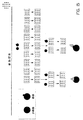
図3は、高多様性遺伝子ライブラリー(high diversity gene libraries)のネスト化ソートのためのツールとしての抗体アレイの使用を解説したものである。
図4は、変異遺伝子のライブラリーを探索するために本明細書で提供した方法の適用を解説したものである。
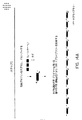
図5は、組換え抗体ライブラリーを構成する方法を解説したものである。
図6は、ポリペプチド(エピトープ)タグを、プライマー付加を用い、組換え抗体に組込むための1つの方法を示す。
図7は、リンカー付加を用いる他のスキームを示したものである。
図8は、組換え抗体ライブラリーを探索するための本明細書の適用を示したものである。
図9は、本明細書において提供されるプライマーのエレメント、および必要とされるプライマーのセットを略図的に示したものである。
図10および11は、EDおよびEDCプライマーを構成するための他の方法を示す;図10では、オリゴヌクレオチドは、固体支持体上で3’から5’へ化学的に合成され、図11の方法では、オリゴヌクレオチドは、オーバーラッピングハイブリダイゼーションに基づき自己アセンブルする。
図12は、アレイで使用するためのハイブリドーマ細胞から産生するイムノグロブリン(Ig)を発見するためのハイスループットスクリーニングを示したものである。
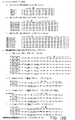
図13(図13Aおよび13B)は、組換えヒト抗体を調製するための抗体鎖増幅用の典型的プライマー(配列番号12−73)を示したものである(McCafferty et al. (1996) Antibody engineering: A practical Approach, Oxford University Press, Oxfordの87−88頁の表33参照、また、Marks et al. (1992) Bio/Technology 10: 779−783およびKay et al. (1996) Phage Display of Peptides and Proteins: A laboratory Manual, Academic Press, San Diego参照)。
図14(A−D)は、抗体作成のための本明細書の方法の使用を示したものである。
図15は、修飾された特異性(または修飾された特異性を有する任意のタンパク質)を用い抗体を同定するための本明細書の方法の使用を示したものである。
図16は、同時抗体探索(simultaneous antibody searches)のために本明細書の方法の使用を示したものである。
図17は、酵素作成プロトコールにおける本明細書の方法の使用を示したものである。
図18は、タンパク質相互作用マッピングプロトコールにおける本明細書の方法の使用を示したものである。
図19は、複数のポリペプチドタグをソーティングに使用するときのタグの数の増加率を示したものである。
【0033】
制限するつもりではなく、明確な開示のため、詳細な説明を以下のサブセクションに分けている。
【0034】
(本発明の詳細な説明)
A.定義
他に特記しなければ、本明細書で使用するすべての技術および科学用語は、本発明が属する分野の当業者が通常理解する意味と同一である。本明細書の用語の定義と異なる場合には、定義は、このセクションに記載の意味に依存する。許されるならば、本明細書の開示で全体を通して言及する、すべての特許、出願、公開出願および他の刊行物ならびにGenBankおよび他のデータベースからの配列を、引用によりすべて組込む。
【0035】
本明細書で使用する場合、ネスト化ソーティングとは、本明細書で提供する抗体のアドレス可能コレクションを使用して、多様性を減少させる過程をいう。
【0036】
本明細書で使用する場合、抗タグ捕捉試薬(または本明細書では捕捉試薬のアドレス可能コレクションという)、抗体のような、タンパク質製剤(すなわち、レセプター)のアドレス可能コレクションは、エピトープ(抗原中のエピトープのようなアミノ酸配列)を含む事前に選択したポリペプチドタグに特異的に結合し、ここで、コレクションのうちの個々のメンバーは、標識され、および/または抗体のような捕捉試薬およびタグの同定を可能となるよう位置付けられる。アドレス可能コレクションは、典型的にはアレイまたはコード可能(codable)コレクションであり、この場合、それぞれの位置は、単一の特異性を有する抗体のようなレセプターを含み、同定可能である。化学性(chemical)、電子性、呈色性、蛍光性または他のタグのような他の個別のアイデンティファイアーが含まれるならば、当該コレクションは、液相中にあり得る。捕捉試薬は、抗体および他の抗タグレセプターを含む。エピトープのような事前に決定したアミノ酸配列に特異的に結合する任意のタンパク質は、捕捉試薬としての使用が予期される。
【0037】
本明細書で使用する場合、一般にタグと称される本明細書のポリペプチドタグは、捕捉試薬に特異的に結合するアミノ酸配列を含む。
【0038】
本明細書で使用する場合、エピトープタグは、抗体のような抗タグ捕捉試薬が特異的に結合するエピトープとして称される本明細書のアミノ酸配列を含むアミノ酸配列をいう。ポリペプチドおよびエピトープタグの場合、それぞれが結合する特異的アミノ酸配列を本明細書では一般的にエピトープという。レセプターに結合する任意のアミノ酸配列が予期される。本明細書の目的の場合、捕捉試薬に特異的に結合する、エピトープタグのエピトープ部分のようなタグのアミノ酸配列を「E」と称し、個々の特有のエピトープはEmである。文脈に依存して、「Em」はまた、エピトープを構成するアミノ酸をコードする核酸配列をいう。エピトープタグのようなポリペプチドタグはまた、ディバイダー領域によりコードされているアミノ酸を含み得る。特に、エピトープタグは、本明細書で提供されるオリゴヌクレオチドによりコードされており、それはタグの導入に使用される。リファレンス(reference)が形成され核酸に関してエピトープタグとなるとき(すなわち、特定レセプターまたはその部分と結合対となる)、それは、リファレンスが形成されるタグをコードする核酸である。単純化のため、個々のポリペプチドタグはEmとして称される;核酸を述べるとき、Emは核酸であり、エピトープをコードする核酸配列をさす;翻訳タンパク質を述べるとき、Emはアミノ酸をさす(実際にはエピトープ)。Eの数は、アドレス可能コレクション中の抗体の数に相当する。「m」は、典型的には少なくとも10、より好ましくは30またはそれ以上、より好ましくは50または100またはそれ以上であり、所望の、かつ実用的な数となり得る。最も好ましい「m」は、約1000またはそれ以上である。
【0039】
本明細書で使用する場合、Dnは、個々のディバイダー配列をいう。本明細書で述べるように、分割が他の方法で行われる特定実施態様では、Dnは任意である。個々のEmのように、Dnは、状況に応じて核酸またはアミノ酸の何れかとなる。個々のDnは、サブセットの核酸を増幅するプライミング部位として役立つ核酸によりコードされるディバイダー配列である。得られた増幅サブセットの核酸は、増幅でプライミング部位として使用するEm配列およびDn配列のコレクションのすべてを含む。本明細書で記載したように、核酸は、個々のEmDnをコードする部分、好ましくは末端部分において含まれる。一般的に、コード化核酸は、ライブラリー中の核酸分子において5’−Em−Dn−3’である。Dは、サブライブラリーを作成する特異的増幅のためのヌクレオチドの任意の特有配列である。巨大なライブラリーの場合、オリジナルライブラリーは、サブライブラリーに分けられ、次いで、マスターライブラリーにタグコード化配列を加える以外に、タグコード化配列を加える。ライブラリーが大きくなればなるほど、当該ライブラリー中の特有配列の特定にはより長い配列が必要となるため、Dの大きさは、ソートされるライブラリーの機能であるといえる。方法における機能はPCR増幅用のプライミング部位として役立つことから、応用に応じた一般的なDは、少なくとも14から16核酸塩基であり、アミノ酸配列をコードしているかもしれないし、またコードしていないかもしれない;Dは2からnであり、ここで、nは0または任意の望ましい数であり、そして一般的には10から10,000、10から1000、50から500、および約100から250である。Dのその数は、106かまたはそれよりも大きくなり得る。ディバイダー配列Dを用い、タグ化マスターライブラリーから「n」のサンプルそれぞれを増幅し、一般的に、最初のソートで使用する、アレイのような抗体コレクションの数に等しくなる。最初のスクリーニングにおいてコレクション(部分(divisions))が多くなればなるほど、アドレス可能位置あたりの多様性は少なくなる。最初の分割数は、ライブラリーの多様性および捕捉試薬の数に基づき選択される。Eが大きくなればなるほど、より小さなDが必要となり、特定の多様性(Div)を有するライブラリーの場合には逆となる。本明細書に使用するように、多様性(Div)は、核酸ライブラリーのようなライブラリーにおける異なる分子の数をいう。多様性は、より大きな任意のライブラリー中の分子の総数とは異なる。多様性が大きくなればなるほど、存在する実際の複製物の数は少なくなる。典型的には、(異なる分子の数)/(総分子数)は約1である。ランダムにタグ化されマスターライブラリーを作成する分子の数は、最初の多様性よりも小さく、マスターライブラリー中の実質的な個々の分子は異なる。
【0040】
本明細書で使用するように、アレイは、3またはそれより多いメンバーを含む抗体のようなエレメントのコレクションをいう。アドレス可能アレイは、当該アレイのメンバーが、典型的には、固相支持体上の位置によるか、同定可能または検出可能標識、色、蛍光、電子のシグナル(すなわち、目的の分子の相互作用を実質的には変えないRF、マイクロ波または他の周波数(frequency))、バーコードまたは他の記号(symbology)、化学的または他のそのような標識といったものの特長により、同定されるものである。ここで、一般に、アレイのメンバーは、固相表面上の別個の同定可能位置に固定されるか、ミクロスフェアまたは他の特定支持体(本明細書ではビーズと称する)に対し付着するように、同定可能標識に直接的または間接的に結合され、でなければ会合され、そして溶液中で懸濁されるかまたは表面上で広げられる。
【0041】
本明細書で使用するように、支持体(また、マトリクス支持体、マトリクス、不溶性支持体または固体支持体と称する)は、目的の分子、典型的には生物学的分子、有機分子または生体特異的リガンドが結合するかまたは接触する任意の固体または半固体または不溶性支持体をいう。その物質には、以下に限らないが、ポリスチレン、ポリカーボネート、ポリプロピレン、ナイロン、ガラス、デキストラン、キチン、砂、軽石、アガロース、ポリサッカライド、デンドリマー、バッキーボール、ポリアクリルアミド、シリコン、ラバー、および固相合成、親和性分離および精製、ハイブリダイゼーション反応、イムノアッセイおよび他のそのような応用のため支持体として使用する他の物質といった、化学的および生物学的分子合成および分析のための親和性物質または支持体として使用する任意の物質が含まれる。本明細書におけるマトリクスは、微粒子であり得るか、マイクロタイターディッシュまたはウェル、ガラススライド、シリコンチップ、ニトロセルロースシート、ナイロンメッシュ、または他のそのような物質のような連続表面型となり得る。微粒子、典型的には粒子であるとき、少なくとも5−10mmまたはそれよりも小さな大きさとなる。本明細書で、集合的に「ビーズ」と称する粒子は、必要ではないが、しばしば球状である。しかし、そのリファレンスは、ランダムな形、針、繊維および伸長化(elongated)を含む任意の形となり得る、マトリクスのジオメトリーを制限しない。おおよそ球状の「ビーズ」、特に、液相中で使用し得るミクロスフェアもまた、予期される。「ビーズ」には、付加構成成分が、本明細書の方法および分析を妨げない限り、磁石を用い分離するための磁性または常磁性粒子(例えば、Dyna beads(Dynal, Oslo, Norway))のような付加構成要素が含まれ得る。
【0042】
本明細書で使用するように、マトリクスまたは支持体粒子は、個別の粒子の形であるマトリクス物質をいう。粒子は任意の形および大きさをしているが、典型的には、少なくとも、100mmまたはそれ未満、50mmまたはそれ未満、10mmまたはそれ未満、1mmまたはそれ未満、100μmまたはそれ未満、50μmまたはそれ未満の寸法であり、典型的には、100mm3またはそれ未満、50mm3またはそれ未満、10mm3またはそれ未満および1mm3またはそれ未満、100μm3またはそれ未満の大きさであり、1立方ミクロンのオーダーとなり得る。その粒子を集合的に「ビーズ」と称する。
【0043】
本明細書で使用するように、レセプターと交換可能的に使用される捕捉試薬とは、所定のリガンドに対する親和性または定義されるアミノ酸配列との親和性を有する分子をいう。捕捉試薬は、天然に生じ得るか、または合成分子であり、核酸、小有機体、タンパク質および特定アミノ酸配列に特異的に結合する複合体を含む任意の分子を含み得る。捕捉試薬は、本分野で抗リガンドとして称され得るレセプターである。本明細書で使用するように、捕捉試薬、レセプターおよび抗リガンドなる用語は交換可能である。捕捉試薬は、不変状態で使用し得るか、または他の種との集合として使用され得る。それらは、特定結合物質またはリンカーを介し、直接的または間接的の何れかで、結合メンバーと共有結合的または非共有結合的に結合または物理的に接触し得る。捕捉試薬の例には、以下に限らないが、抗体、細胞膜レセプター表面レセプターおよび内在化レセプター、モノクローナル抗体および特異的抗原決定基(ウイルス、細胞または他の物質といったもの)を有する抗血清反応性または単離性成分、薬物、ポリヌクレオチド、核酸、ペプチド、補因子、レクチン、糖、ポリサッカライド、細胞、細胞膜、およびオルガネラが含まれる。
【0044】
捕捉試薬の例には、以下に限らないが、
a)酵素および他の触媒ポリペプチド、以下に限らないが、基質が特異的に結合する当該酵素および触媒ポリペプチド部分、結合活性は維持し触媒活性は欠損するよう修飾した酵素;
b)抗原またはアミノ酸配列に特異的に結合する抗体およびその部分;
c)核酸;
d)細胞表面レセプター、オピエートレセプターおよびホルモンレセプターならびにホルモンのようなリガンドに特異的に結合する他のレセプター、
が含まれる。本明細書のコレクションの場合、個々のポリペプチドタグとして、本明細書でいう他の結合パートナーは、基質、抗原配列、核酸結合タンパク質、レセプターリガンドまたはその結合部分をいう。
【0045】
記載するように、分子の対、互いに特異的に結合する一般的なタンパク質が本明細書で予期される。対の1つのメンバーは、タグとして使用され、ライブラリーに結合する核酸によりコードされるポリペプチドであり、他のメンバーは、特異的に結合する任意のものである。捕捉試薬のコレクションには、典型的には少なくとも約3から10アミノ酸である既知または既知の可能性ある定義アミノ酸配列に特異的に結合する、抗体のようなレセプターまたは酵素もしくはその部分、およびその混合物を含む。
【0046】
本明細書で使用するように、抗体とは、免疫グロブリン、天然のものまたは部分的もしくは完全に合成されたものをいい、抗体の特異的結合能を維持する任意の誘導体が含まれる。ここで、抗体には、免疫グロブリン結合ドメインに相同性または実質的に相同性のある結合ドメインを有する任意のタンパク質を含む。本明細書の目的のため、抗体には、軽鎖、および重鎖の可変領域からなる、Fabフラグメントのような抗体フラグメントが含まれる。抗体には、IgG、IgM、IgA、IgDおよびIgEを含む、任意の免疫グロブリンクラスのメンバーが含まれる。また、本明細書では、アミノ酸配列に特異的に結合するレセプターも予期される。
【0047】
ここで、本明細書の目的のため、一般的に本明細書で捕捉試薬/ポリペプチドタグとして称する、任意のセットの結合メンバーの対が、抗体およびエピトープ自体の代わりに使用され得る。本明細書の方法は、特異的相互作用のための、抗体/エピトープタグのような捕捉試薬/ポリペプチドタグに依存し、レセプター/リガンド(エピトープタグ)の任意の組合せが使用し得る。更に、本明細書の目的のため、用いる抗体のような捕捉試薬はその結合部分となり得る。
【0048】
本明細書で使用するように、抗体フラグメントとは、全長よりも短く、全長抗体の特異的結合能の少なくとも一部を保持している抗体の任意の誘導体をいう。抗体フラグメントの例には、以下に限らないが、Fab、Fab’、F(ab)2、単鎖Fvs(scFv)、Fv、dsFvディアボディー(diabody)およびFdフラグメントが含まれる。当該フラグメントには、例えば、ジスルフィド架橋により、同時結合する複数の鎖が含まれ得る。抗体フラグメントは、一般的に、少なくとも約50アミノ酸、および典型的には、少なくとも200アミノ酸を含む。
【0049】
本明細書で使用するように、Fv抗体フラグメントは、非共有結合性相互作用により結合する可変重(VH)ドメインおよび可変軽(VL)ドメインからなる。
【0050】
本明細書で使用するように、dsFvとは、VH−VL対を安定化する、作成分子内ジスルフィド結合を有するFvをいう。
【0051】
本明細書で使用するように、F(ab)2フラグメントは、免疫グロブリンをpH4.0−4.5でペプシンで消化した結果、得られた抗体フラグメントである:それは組換えで作成され得る。
【0052】
本明細書で使用するように、Fabフラグメントは、免疫グロブリンをパパインで消化した結果、得られた抗体フラグメントである:それは組換えで作成され得る。
【0053】
本明細書で使用するように、任意のオーダーでポリペプチドリンカーにより共有結合する可変軽鎖(VL)および可変重鎖(VH)を含む抗体フラグメントをいう。当該リンカーは、2つの可変ドメインが実質的に妨げられることなく架橋するような長さとなる。典型的なリンカーは、溶解度を増加させるため幾つかのGluまたはLys残基がいたるところに分散している(Gly−Ser)n残基である。
【0054】
本明細書で使用するように、ディアボディーは、二量体scFvである:ディアボディーは典型的にscFvよりも短いペプチドリンカーを有し、それらは好ましくは二量体化する。
【0055】
本明細書で使用するように、ヒト化抗体とは、ヒトへ投与しても免疫応答が誘発しないような「ヒト」アミノ酸配列を含むように修飾した抗体をいう。その抗体の調製方法は既知である。例えば、モノクローナル抗体を発現するハイブリドーマは、組換えDNA技術により変化し、非可変領域のアミノ酸組成物がヒト抗体に基づいている抗体を発現する。コンピュータープログラムはその領域を同定するように設計されている。
【0056】
本明細書で使用するように、高分子とは、数百から数百万までの分子量を有する任意の分子をいう。高分子には、ペプチド、タンパク質、ヌクレオチド、核酸、および生体により一般的に合成され、合成的にまたは組換え分子生物学的方法を用いて調製され得る他の分子が含まれる。
【0057】
本明細書で使用するように、「バイオポリマー」なる用語は、結合により結合する、2またはそれより多い単量体サブユニットまたはその誘導体または高分子からなる、高分子群を含む生物学的分子を意味する場合に使用する。バイオポリマーは、例えば、ポリヌクレオチド、ポリペプチド、炭水化物または脂質、またはそれらの誘導体もしくは組合せ、例えばペプチド核酸部分またはグリコプロテイン、それぞれを含む核酸分子であり得る。バイオポリマーには、以下に限らないが、核酸、タンパク質、ポリサッカライド、脂質および他の高分子群が含まれる。核酸には、DNA、RNAおよびそれらのフラグメントが含まれる。核酸は、ゲノムDNA、RNA、ミトコンドリア核酸、クロロプラスト核酸および別々の遺伝学的物質を有する他のオルガネラから誘導され得る。
【0058】
本明細書で使用するように、生体分子は、天然で見られる任意の化合物、またはその誘導体である。生体分子には、以下に限らないが、オリゴヌクレオチド、オリゴヌクレオシド、タンパク質、ペプチド、アミノ酸、ペプチド核酸(PNA)、オリゴサッカライドおよびモノサッカライドが含まれる。
【0059】
本明細書で使用するように、「核酸」なる用語は、デオキシリボ核酸(DNA)およびリボ核酸(RNA)ならびにRNAまたはDNAの何れかの類似体または誘導体のような一本鎖および/または二本鎖ポリヌクレオチドをいう。「核酸」なる用語には、ペプチド核酸(PNA)、ホスホロチオエートDNAのような核酸の類似体および他のその類似体ならびにその誘導体または組合せも含まれる。
【0060】
本明細書で使用するように、「ポリヌクレオチド」なる用語は、少なくとも2つの結合ヌクレオチドまたはヌクレオチド誘導体を含むオリゴマーまたはポリマーをいい、デオキシリボ核酸(DNA)、リボ核酸(RNA)、およびDNAまたはRNA誘導体が含まれ、当該誘導体には、例えば、ヌクレオチド類似体またはホスホジエステル結合、例えばホスホトリエステル結合、ホスホラミデイト(phosphoramidate)結合、ホスホロチオエート結合、チオエステル結合またはペプチド結合(ペプチド核酸)以外の「バックボーン」結合が含まれる。「オリゴヌクレオチド」なる用語はまた、本明細書では「ポリヌクレオチド」と本質的に同義的に使用するが、当業者であれば、オリゴヌクレオチド、例えばPCRプライマーは、一般的には、約50未満から100ヌクレオチドの長さを有することを認識している。
【0061】
ポリヌクレオチド中に含まれるヌクレオチド類似体は、例えば、質量修飾ヌクレオチドであり、それにより、ポリヌクレオチド;ポリヌクレオチドを検出し得る蛍光性、放射活性、発光性または化学発光性の標識のような検出可能標識を含むヌクレオチド;または固体支持体へのポリヌクレオチドの固定化を促進するビオチンまたはチオール基のような反応基を含むヌクレオチドの質量の区別が可能となる。ポリヌクレオチドはまた、選択的切断、例えば、化学的、酵素学的または光分解的な切断である1またはそれより多いバックボーン結合を含み得る。例えば、ポリヌクレオチドは、1またはそれより多いデオキシリボヌクレオチド、その後に続いて、1またはそれより多いリボヌクレオチド、そしてその後に続き得る1またはそれより多いデオキシリボヌクレオチドを含み、その配列は、塩基加水分解により、リボヌクレオチド配列で切断される。ポリヌクレオチドはまた、例えば、ペプチド核酸結合により結合するヌクレオチド、およびホスホジエステル結合または他の適当な結合により結合する3’末端の少なくとも1つのヌクレオチドを含み得、そしてポリメラーゼにより伸長することができる、キメラオリゴヌクレオチドプライマーの切断に比較的耐性のある1つまたはそれより多い結合を含み得る。ペプチド核酸配列は、既知の方法(例えば、Weiler et al., Nucleic acids Res. 25: 2792−2799 (1997)参照)を用い、調製され得る。
【0062】
本明細書に使用するように、オリゴヌクレオチドとは、DNA、RNA、PNAのような核酸類似体、およびその組合せを含むポリマーをいう。本明細書の目的のため、プライマーおよびプローブは一本鎖オリゴヌクレオチドである。
【0063】
本明細書で使用するように、組換えによる産生とは、組換えDNA方法を用いることを意味し、クローン化DNAによりコードされるタンパク質を発現するための分子生物学の既知方法の使用を意味する。
【0064】
本明細書で使用するように、産物に実質的に同一であるとは、目的の特性が、全く変わらないぐらいに十分に類似することを意味し、その結果、実質的に同一の産物は産物の代わりに使用し得る。
【0065】
本明細書で使用するように、2つの核酸配列をいうとき、等価物とは、対象の2つの配列が、同一アミノ酸配列または等価タンパク質をコードすることを意味する。2つのタンパク質またはペプチドについて「等価物」を使用するとき、それは、2つのタンパク質またはペプチドが、当該タンパク質またはペプチドの活性または機能を実質的に変えない保存的アミノ酸置換(例えば、表1、上、参照)のみと共に実質的に同一のアミノ酸配列を有することを意味する。特性について「等価物」というときは、その特性が同程度である必要はないが、その活性は好ましくは実質的に同じである必要がある。2つのヌクレオチド配列をいう際の「相補的」とは、2つのヌクレオチド配列において、対となるヌクレオチドの間で、好ましくは25%未満のミスマッチ、より好ましくは15%未満のミスマッチ、もっとより好ましくは5%未満のミスマッチ、最も好ましくはミスマッチが全くなくハイブリダイズし得ることを意味する。一般的に本明細書において相補的であるとみなすのは、2つの分子が、高緊縮条件下でハイブリダイズする場合である。
【0066】
本明細書で使用するように、特定の緊縮条件下でハイブリダイズするとは、2つの一本鎖DNAフラグメントの間で形成されるハイブリッドの安定性を述べるときに使用し、洗浄ステップより弱いか同程度の緊縮条件下でアニーリングした後の、そのハイブリッドを洗浄するイオン強度および温度の条件をいう。典型的に、高、中、および低緊縮とは、以下の条件またはその条件と同程度の条件を含む:
1)高緊縮:0.1×SSPEまたはSSC、0.1%SDS、65℃
2)中緊縮:0.2×SSPEまたはSSC、0.1%SDS、50℃
3)低緊縮:1.0×SSPEまたはSSC、0.1%SDS、50℃。
等価条件とは、生じたハイブリッドにおいて、実質的に同じ割合のミスマッチを選択する条件をいう。ホルムアルデヒド、FicollおよびDenhardt’s溶液のような成分の添加は、ハイブリダイゼーションが構成されるような温度および反応の速度のようなパラメーターに影響を与える。そのため、42℃での20%ホルムアミドにおける5×SSC中のハイブリダイゼーションは、実質的に、低緊縮条件下の上記ハイブリダイゼーションで再引用した条件と同じである。SSPE、SSCおよびDenhardt’sの方法および脱イオン化ホルムアミドの作成は、例えば、Sambrook et al. (1989) Molecular Cloning, A Laboratory Manual, Cold Spring Harbor Laboratory Press, Chapter 8に記載されている;Sambrook et al. vol. 3, p.B. 13参照、また、通常使用される研究用溶液を記載している多くのカタログも参照。等価の緊縮は、他の緩衝液、塩および温度を用い行われ得ると理解される。
【0067】
用語「実質的に」同一または相同または類似とは、当業者により理解されるように前後関係によって変化し、一般的には、少なくとも70%同一性を意味し、好ましくは少なくとも80%同一性を意味し、より好ましくは少なくとも90%同一性を意味し、そして最も好ましくは少なくとも95%同一性を意味する。
【0068】
本明細書で使用するように、組成物とは、任意の混合物をいう。それは、溶液、懸濁液、液体、粉末、ペースト、水性、非水性またはそれら任意の組合せであり得る。
【0069】
本明細書で使用するように、組合せとは、2つまたはそれより多いアイテムの間の任意の結合をいう。その組合せは、2つの組成物または2つのコレクションのような2つまたはそれより多い別個のアイテムであり得、2つまたはそれより多いアイテムの単一混合物のようなその混合物、またはその任意のバリエーションであり得る。
【0070】
本明細書で使用するように、液体とは、流れることができる任意の組成物をいう。そのため、液体は、半固体、ペースト、溶液、水性混合物、ゲル、ローション、クリームおよび他のそのような組成物の型である組成物を含む。
【0071】
本明細書で使用するように、アミノ酸の適当な保存置換は、当業者に既知であり、生じる分子の生物学的活性を一般的に変えることなく、行い得る。一般に、ポリペプチドの非必須領域における単一アミノ酸置換により、実質的な生物学的活性が変化することはないと当業者は認識している(例えば、Watson et al. Molecular Biology of the Gene, 4th Edition, 1987, The Benjamin/Cummings Pub. co., p.224)。
【0072】
その置換は、好ましくは以下のような表1に開示のものに従って行われる。
【表1】
表1
【0073】
本明細書で使用するように、本明細書で見られる種々のアミノ酸配列で生ずるアミノ酸は、既知の、3文字表記または1文字表記で同定される。種々のDNAフラグメントで生ずるヌクレオチドは、本分野で通常用いる標準的な1文字表示で示す。
【0074】
本明細書中で使用するように、任意の保護基、アミノ酸および他の化合物は、特記しない限り、通常の用法、認識されている略語、またはBiochemical NomenclatureにおけるIUPAC−IUB Commission((1972) Biochem. 11: 1726参照)に従う。
【0075】
本明細書における方法およびコレクションは、抗体捕捉試薬、および抗体が結合するエピトープを含むポリペプチドタグに対する特定リファレンスで例示されるが、本明細書における方法が任意の捕捉試薬および任意のポリペプチドタグで実施し得ると理解される。また、任意の捕捉試薬およびポリペプチドタグのコレクションの組合せにより、本明細書に記載の任意の実施態様での使用が予期されることも理解される。また、物理的アレイの型、またはカラービーズに結合した捕捉試薬のような標識化コレクションの型のいずれであっても、アレイに対するリファレンスが任意のアドレス可能コレクションを含むことを意図されることが理解される。
【0076】
B.オリゴヌクレオチド/プライマーの設計および調製
アレイに対する高多様性ライブラリー(large diversity libraries)のソーティングおよび望ましい性質を有するクローンを含む特定プールの増殖は、特異的ポリペプチドタグでライブラリーを特徴的にタグ化する能力に依存する。オリゴヌクレオチドセットは、化学的に合成され、オーバーラッピング配列をランダムに組合し、そして一緒にライゲーションしてプライマーまたはリンカーのコレクションの酵素学的生成のための鋳型を作成する。
【0077】
オリゴヌクレオチドは、一本鎖または二本鎖の何れかであり、マスターライブラリー中へ組込む方法に依存する。例えば、通常の制限酵素部位を用いるような二本鎖バージョンのライゲーション、その後の共通領域を用いる増幅により組込まれ得るか、またはPCR増幅により組込まれ得、この場合、オリゴヌクレオチドは一本鎖である。
【0078】
1.プライマー
本明細書では、プライマーである核酸分子のセットを提供するか、または二本鎖オリゴヌクレオチドを提供し、それは、プライマー群の二本鎖バージョンおよびプライマー群のセットの組合せであり、および/または二本鎖のものは、本明細書で提供される方法の種々のステップで使用され、および/または用いる実施態様に依存する。分子をタグ化するための方法の幾つかの実施態様で用いられるプライマーは、その方法の実施の中枢となる。当該プライマーは、図9に示すような式を含む、オリゴヌクレオチドを含む。当該プラスミドおよび二本鎖オリゴヌクレオチドは制限酵素部位を含み、それは、以下に示すように、例えば抗体ライブラリーのための目的遺伝子の充分な部分の標的増幅のためである。これらのプライマーは、フォワードまたはリバースプライマーであり、この場合、フォワードプライマーは、PCR増幅の最初のラウンドで使用される。下記のような、および図で示した当該プライマーをセットとして提供する。1またはそれより多い個々のセットの組合せもまた提供する。当該プライマーは、本明細書で提供する方法の中枢となる。
【0079】
2.オリゴヌクレオチド/プライマーの調製
二本鎖または一本鎖オリゴヌクレオチドを構成するための任意の適当な方法を用い得る。多数のオリゴヌクレオチドの調製に適用され得る方法を特に目的としている。2つの方法を図10および11で示しており、以下で考察する。
【0080】
図9は、タグ化ライブラリーの構成のための物理的エレメントおよび目的遺伝子(タンパク質)の同定のためのアドレス可能抗タグ抗体コレクションの使用を示している。4つのオリゴヌクレオチド/プライマーセットは、アドレス可能コレクションに加えて提供され、それは、説明目的のために、アレイ、当該アレイを分析するイメージングシステムまたはリーダーおよび所望により当該リーダーによって回収される情報を処理するソフトウェアとして提供される。示した実施態様では、プライマーセットはEmDnCを含み、ここで、Cは、すべてのオリゴヌクレオチドの間での共通部分であり、異なるEおよび/またはD配列(例えば、D1からDn;ElからEn)を用い、すべてのタグ化核酸の増幅のための領域として役立つ;DCは、異なるD配列(D1からDn)および所望により共通領域用のCを用い、FAECは、異なるFA配列(例えば、FA1からFAn)を用いる;およびFBCは、異なるFB配列(例えば、FB1からFBn)を用いる。個々のFAは、個々のエピトープの部分を含み、プライマーとして役立ち、対応するEmをコードする核酸を増幅するが、生ずる増幅核酸は、Emエピトープを含まない。FBnはFAnに類似するが、エピトープの維持が望ましいならば、Enが含まれ得る点が異なる。
【0081】
図10および図11は、例として、抗体スクリーニングのための、EDおよびEDC、FAおよびFBオリゴヌクレオチド/プライマーを構成するための2種の方法の概略を示す。例えば、nをあわせもつVLFORプライマー、例えばmを有する1,000種のE配列、例えば1,000種のD配列および約13種のJkappaFor配列の合成。これにより、全部で(1,000)(1,000)(13)=13,000,000種のオリゴヌクレオチドが生ずる。進行合成ステップにおいて種々の配列領域をランダムに組合せることにより、プライマーの巨大な多様性コレクションが調製され得る。
【0082】
最初の方法(図10)は、固相合成ストラテジーを使用する。第二の方法(図11)は、オーバーラッピング相補的配列に基づき自己アセンブルするDNA分子の能力を使用する。固相合成は、反応間の基質分子から容易に固定化産物分子が精製され得、反応条件をより調節し得るという利点を有する。自己アセンブリ法は、あまり労働を必要としないという利点がある。
【0083】
図10オリゴヌクレオチドは、固体支持体から3’から5’へ化学的に合成される。対照的に、DNAは、5’から3’へ酵素学的に合成される。VLFORプライマーを作成するため、CおよびD配列は、標準的方法を用い固体支持体から化学的に合成される。更なる合成用にオリゴヌクレオチドを固相に結合するため、強力な求核原子を、アミノリンクの添加により組込み、その後、基質からオリゴヌクレオチドを切断する。アミノリンクは、オリゴヌクレオチドの5’末端に一級アミンを導入する。次いで、アミノリンクにおけるアミノ基は、常磁性ビーズのような固相に、トシル、N−ヒドロキシスクシンイミドまたはヒドラジン基のようなビーズ上のアミン反応基との反応により、結合し得る。生ずるオリゴヌクレオチドは、適当な5’から3’方向であるCおよびD配列により、ビーズに共有結合する。
【0084】
E配列群の混合物を、オリゴヌクレオチドに、DNA「パッチ」の使用により加え、生ずるニックをDNAリガーゼでシールする。非組込みの基質DNAは伸長産物から生成し、Jkappa for配列の混合物をプライマーに添加する。完全VLFORプライマーはビーズから放出されるが、当該ビーズはcDNA合成をプライムするオリゴヌクレオチドの能力を妨げることはない。
【0085】
図11で解説した方法は、オーバーラッピングハイブリダイゼーションに基づき自己アセンブルするオリゴヌクレオチドに依存する。二本鎖DNA分子は、最初に、分子の+および−鎖をコードするオリゴヌクレオチドから作成される。これらのオリゴヌクレオチドを合わせ、ハイブリダイズさせ、ニック化二本鎖DNA分子を作成し、当該分子上のニックはDNAリガーゼの添加によりシールする。シール化分子は、新規DNA分子の酵素学的合成のための鋳型として使用する。DNA合成は、ビオチンのような固体支持体に結合し得る5’末端に基を有するオリゴヌクレオチド、または上記のようなアミノリンク化学を用いプライムされる。
【0086】
酵素学的合成の間の反応基の組込みは、合成完了後の一本鎖分子の精製を可能とする。完全VLFORプライマーは、ビーズから放出され得るが、ビーズは、cDNA合成をプライムするオリゴヌクレオチドの能力を妨げない。
【0087】
C.アドレス可能抗タグレセプターコレクションを用いるネスト化ソーティング
目的タンパク質を同定および選択するための先の方法は、富化の連続ラウンドの間に作成される選択バイアスにより、妨げられる。本明細書で提供するように、選択バイアスは、選択以外のソーティングに基づく同定方法の使用により避けることができる。本明細書のこれらの方法は、複数の実質的に同一性の、好ましくは複製性の、抗体のような製剤のコレクションのような捕捉試薬のコレクションの使用に依存し、その製剤は、標的ライブラリー中のタンパク質に結合するかまたは標的核酸ライブラリーによりコードされる、事前に選択したアミノ酸配列(一般的に、エピトープのような、少なくとも約5から10、典型的には7または8アミノ酸)に特異的に結合する。捕捉試薬またはその結合部分が特異的に結合するアミノ酸配列を含む捕捉試薬およびポリペプチドタグの組合せを提供する。当該タグは、核酸ライブラリー、またはソート化された分子の他のライブラリーのメンバーに結合し得る。
【0088】
1.概要
位置付けアドレス可能アレイのような、アドレス可能抗タグ捕捉試薬コレクションは、高い親和性および特異性を有する、エピトープタグのような、事前選択および/または事前設計したポリペプチドタグに結合する抗体のような、コレクション異種捕捉試薬(collection different capture agents)を含む。典型的なコレクションは、少なくとも約30、より好ましくは100、より好ましくは500、最も好ましくは少なくとも1000の、抗体のような捕捉試薬を含み、その捕捉試薬は、例えば、アレイ上で特定配置を占めることにより、またはバーコード支持体、カラーコード、またはRFタグ標識支持体または他のそのようなアドレス可能形態の支持体に結合する特徴により、アドレス可能である。個々の配置またはアドレスは、単一の特定タグに結合する、抗体のような単一型捕捉試薬を含む。タグ化タンパク質は、アレイ中の抗体のようなレセプターのコレクションと、抗体のようなレセプターとの複合化に適当な条件下、エピトープタグを介し、接触する。結果として、タンパク質は、それぞれ取り付けられたタグによりソートされる。
【0089】
これらアドレス可能抗タグ抗体コレクションは、以下のものに限らないが、治療、診断、試薬およびプロテオミクス親和性マトリクスのための;遺伝子シャッフリング法(gene shuffling methodologies)(これに限らず)のような酵素工学的適用において;改善した触媒の同定のため、抗体親和性成熟(antibody affinity maturation)のため;小分子捕捉タンパク質、配列特異的DNA結合タンパク質の同定のため、単一鎖T細胞レセプター結合タンパク質のため、およびMHCを認識する高親和性分子のため;ならびにタンパク質相互作用マッピングのため、抗体の迅速な同定を含む種々の適用を有する。典型的なプロトコールを図1−4、12、14A−Dおよび15−18に示す。
【0090】
2.ソーティング法
エピトープタグで標識された分子をソーティングに、抗体のようなレセプターコレクションを用いる方法を提供する。当該方法は、タグをコードしている核酸を加えることによりマスタータグ化ライブラリーを作成し;マスターライブラリーの一部をNの反応物に分け、個々の反応物を、ディバイダー配列をコードする核酸で各反応物を増幅し、翻訳してNの翻訳化反応混合物を産生し;個々の反応混合物を、抗体のような捕捉試薬の1コレクションと反応させ;適当なスクリーニングにより目的タンパク質を同定し、それによって、目的のタンパク質におけるタグが結合する捕捉試薬の特徴によりタンパク質における特定EDタグを同定する、ステップを含む。
【0091】
最初のソーティングステップは実質的に多様性を減少させる。望ましいならば、更なるソーティングを行うか、または生じるライブラリーを当業者に既知の任意の方法によりスクリーニングする。所望により、第二のソート、すなわち、目的のタンパク質が翻訳される核酸を含む核酸反応混合物から開始されるソートを行う。このステップにおいて、新規のセットのエピトープタグを、増幅またはライゲーション、その後の増幅により核酸に加える。この前、またはこれと同時に、前のエピトープタグをコードする核酸を、制限酵素といった切断によるか、またはエピトープコード化核酸の一部またはすべてを破壊するプライマーを用いる増幅によるか、何れかにより取除く。新規タグを加え、生ずる核酸を翻訳し、抗体の単一アドレス可能コレクションと反応させる。ポリペプチドタグによるタンパク質ソート、およびスクリーニングを行い、目的のタンパク質を同定する。この点において、抗体コレクションのアドレス可能位置における分子の多様性は1となる(または1から100、典型的には1から10のオーダーとなる)。次いで、目的のタンパク質を含む核酸を、同定化エピトープタグをコードする核酸を含む核酸分子を増幅するタグを用い増幅し、それにより、目的のタンパク質をコードする核酸を生ずる。増幅のためのプライマーは、プライマーとして役立ち、それによりコード化されているタンパク質からエピトープを除くタグのすべてまたはタグの重要部分のみを含む。本明細書により、多様なコレクションのソーティング(すなわち、多様性の減少)が可能となる方法が提供される。1のステップを含むソートは、実質的に多様性を減少する。所望なるソーティングステップの使用により、一般に多様性は10未満、一般に1に減少する。
【0092】
マスターライブラリーの分割
上記のように、本明細書におけるソーティング過程における最初のステップには、マスターライブラリーをNのサブライブラリーに分割することが含まれる。上記のように、「D」配列およびタグはマスターライブラリーに導入され得、次いで、そのマスターライブラリーは、増幅のため別のD’を用い「N」のサブライブラリーに更に分割される。
【0093】
上記のように、「D」の包含は任意であり;分割はマスターライブラリーをサブライブラリーに物理的に分割し、次いで「E」タグコード化または「EC」タグコード化配列をサブライブラリーに導入することにより行い得る。これは、一般的に、最初のライブラリーが非常に大きいとき、生ずるサブライブラリーが大きくなり、タグの分布を確実に均一になるように行う。
【0094】
3.ソーティングするためのマスターライブラリーの作成
このステップにおいて、個々のディバイダー配列に結合する個々のエピトープをコードするタグを、典型的にはcDNAライブラリーであるマスターライブラリーに導入する。核酸に二本鎖DNAフラグメントを加え、組み込む当業者に既知の任意の方法が用い得る。特に、種々の方法が本明細書で予期される。これらには、(1)それらを組込むためのPCR増幅を用いること(本明細書で例示する);(2)それらを直接またはリンカー(以下参照)を介しライゲーションすること、必要ならば、ライゲーション産物は本明細書に記載の他の方法により増幅し得、それは、本明細書の既述の見地から当業者により容易に分割し得る。
【0095】
最初のタグ化において、核酸ライブラリーの構成メンバーにおけるE、EDまたはEDCのセットのオリゴヌクレオチドを加える場合、その目的は、すべてのEmおよびすべてのDnの一様な分布を得ることであり、1のみの個々の型の分子においてそれらを有することである。当該タグは、異なる分子間でランダムに分布しなければならない。分子の数が、タグの数に比べて大きい限り(平均して、コレクション中の約1のみの個々の型の分子が個々のタグを得るように)、そのタグは均一に分布する。本明細書において、タグの数と比較して実質的に過剰である、コレクション中の分子の総数が好ましい。その過剰とは、少なくとも100倍、より好ましくは1000倍である。必要ならば、正確な割合は、経験的に決定され得る。実施においては、多様性よりも反応物中の分子が多いことはない。平均して、それぞれ異なる分子は、異なるタグを有し、1のみのそれぞれ異なる分子がタグ化される。
【0096】
当該方法を実施するため、エピトープ標識分子のライブラリーを、個々のタグが分子においてランダムに分布するように、非標識ライブラリー中へタグをランダムに導入することにより調製する。実験により、タグがcDNAライブラリー中にランダムにおよび均等に導入され得ることが示される。
【0097】
マスターライブラリーを、抗体のn数のアドレス可能コレクション(個々のコレクションはm種のエピトープ特異性を有する抗体を含む)と反応する、D1−Dnとして同定される、プールに分ける。アレイのような個々のコレクションを、コレクションまたは他のそのアイデンティファイアーにおいて、例えば、バーコード、ノーテーションまたはシンボルまたはカラーコード、電子タグまたはカラーもしくは同定可能化学的タグのような他のアイデンティファイアーを含む光学的コードにより、プールの1つと結合させる。その反応は、エピトープが抗体に特異的に結合する条件下で行い、抗体およびエピトープタグ標識分子の生ずる複合体は、望ましい性質を有する分子を特異的に同定するアッセイを用いスクリーニングする。抗体の特定コレクションおよび望ましい性質を有する分子を含む特定タグを伴う抗体は同定され、それによって、特定Dnプールおよび分子上のエピトープタグもまた同定され、それによりn×mのコレクションの多様性を減少する。
【0098】
4.エピトープタグの組込みの方法
他の核酸に、ポリペプチドをコードする核酸分子を結合するか、または他の分子にポリペプチドを結合する、当業者に既知の任意の方法が予期される。例示として種々のその方法を述べる。記載するように、抗体捕捉試薬に対する特定リファレンス、および抗体が結合するエピトープを含むポリペプチドタグを用い述べているが、本明細書の方法は、任意の捕捉試薬およびそのポリペプチドタグを用い実施され得ると認識される。
【0099】
a.タグ導入のために環状プラスミドベクターを作成するライゲーション
上記するように、プライマーをライブラリーメンバーに導入するための増幅プロトコールの使用に加えて、プライマーは、直接のライゲーションにより、例えば、タグおよび他の望ましい配列をコードする核酸を含むプラスミドベクター中への導入により、導入され得る。二本鎖プラスミドベクター中へのcDNAのサブクローニングは、当業者に既知である。1つの方法には、スティッキー末端としても知られている5’または3’オーバーハングを作る部位特異的制限エンドヌクレアーゼで、精製二本鎖プラスミドを消化することが含まれる。二本鎖cDNAを同じ制限エンドヌクレアーゼで消化し、相補的スティッキー末端を作成する。他に、ベクターDNAおよびcDNAの両方に平滑末端を作成し、ライゲーションに使用する。消化したcDNAおよびプラスミドDNAを、適当な緩衝液中のDNAリガーゼ(通常、New England Biolabsから得たT4DNAリガーゼおよび緩衝液を用いる)と共に混合し、16℃でインキュベーションし、ライゲーションさせる。ライゲーション反応物の部分を、種々の方法により、DNA取り込みの能力を有するE. coli中に形質転換する(エレクトロポレーション、または化学的コンピテントセルの熱ショックの2つが通常方法である)。形質転換ミックスのアリコートを、使用するプラスミドに適当な抗生物質を含む半固体培地上にプレートする。環状プラスミドを取り入れた細菌のみが選択培地上でコロニーを形成する。特定メンバーのライブラリーの作成は同様の方法で行うが、ベクター中に挿入されるcDNAは、異なるcDNAクローンの混合物である。これら異なるcDNAクローンを、当業者に既知の種々の方法により作成する。
【0100】
cDNAライブラリーからのタンパク質発現に使用するライブラリーの作成に望ましいcDNAクローンの直接クローニングの場合、異なるスティッキー末端を生ずる2種の制限エンドヌクレアーゼをプラスミドの消化に使用する。cDNAライブラリーメンバーは、cDNAの反対末端においてこれら2つの制限エンドヌクレアーゼ認識部位を含むように作成する。他に、相補的オーバーハングを作る別の制限エンドヌクレアーゼを用いる(例えば、NgoMIVによるプラスミドの消化およびBspEIによるcDNAの消化、何れの場合も5’CCGGオーバーハングを生じ、そのためライゲーションに適合する)。更に、プラスミドベクター中へのcDNAの直接導入により、ベクター中に含まれる調節配列の制御の下、cDNAが作成される。調節配列には、プロモーター、転写の開始および終止の部位、翻訳の開始および終止の配列、またはRNA安定化配列を含み得る。望ましいならば、cDNAの挿入により、発現タンパク質の精製に使用し、親和性試薬をタンパク質の検出に使用し、亜細胞性コンパートメントへのタンパク質の指示に使用し、タンパク質の翻訳後プロセッシングにシグナルを与えるタンパク質エレメントを含む、更なるタンパク質エレメントをコードする配列と同じ翻訳リーディングフレームで、cDNAが位置する。
【0101】
例えば、pBAD/gIIIベクター(Invitrogen, Carlsbad CA)には、アラビノース誘導性プロモーター(araBAD)、リボゾーム結合配列、ATG開始コドン、M13繊維状ファージ遺伝子IIIタンパク質由来のシグナル配列、mycエピトープタグ、ポリヒスチジン領域、rrnB転写ターミネーター、ならびにaraCおよびβ−ラクタマーゼオープンリーディングフレーム、およびColE1複製オリジンが含まれる。cDNAクローンの挿入に有用なクローニング部位は、挿入cDNAクローンが最初に使用酵素で消化されないように、そしてcDNAがベクター中に含まれる望ましいコード領域と同じリーディングフレームとなるように、設計され、および/または選択される。発現ベクター中への単一鎖抗体(scFv)の挿入のためには、SfiIおよびNotI部位の使用が通常である。そのため、scFvの発現用にpBAD/gIIIベクターを修飾するために、オリゴヌクレオチドPDK−28(配列番号6)およびPDK−29(配列番号7)をハイブリダイズし、NcoIおよびHindIII消化pBAD/gIII DNAに挿入する。得られたベクターは、遺伝子IIIリーダー配列およびエピトープタグと同じリーディングフレームでscFv(Amersham−Pharmacia の「Mouse scFv Module」のような標準的方法で作る)を挿入し得る。
【0102】
本明細書で使用する場合、発現タンパク質のライブラリーを、複数のエピトープタグおよびそれらを認識する抗体を用いて更に分割(subdivide)する。複数のエピトープタグを用いタンパク質の発現用ライブラリーを作成するため、上記の技術を僅かに改良したサブクローニング技術を用いる。複数のcDNAクローンは、生ずるライブラリーが別エピトープタグでタグ化されるcDNAクローンを含み、個々のエピトープタグが均等に表されるように、異なるプラスミドベクター(単一型プラスミドベクターの代わりに)の混合物中へ挿入される。挿入cDNAメンバーと融合して翻訳されるエピトープタグとは異なるように、マルチプルプラスミドベクターが作成される。例えば、1000のエピトープタグ配列があれば、1000種のベクターが構成され、250のエピトープタグ配列があれば、250種のベクターが構成される。当業者ならば、これらベクターの構成方法が複数あることを理解する。解説として、pBAD/gIIIプラスミドのmycエピトープコード化領域は、XbaIおよびSalI制限酵素消化により除かれ、巨大な4.1kbフラグメントが単離される。オリゴヌクレオチドPDK−32(配列番号8)とPDK−33(配列番号9)のハイブリダイゼーションにより、XbaIおよびSalIに適合するオーバーハングが作成され、それにより、当該産物は、直接挿入され、HA11抗体のエピトープをコードする(以下の表を参照)。PDK−34(配列番号10)とPDK−35(配列番号11)のハイブリダイゼーション産物の挿入により、挿入cDNAのフレームでFLAG M2エピトープ(以下の表を参照)を有するベクターが生ずる。
【表2】
これら個々のベクターは、ベクター中にcDNAクローンをサブクローニングし得るSfiIおよびNotI制限エンドヌクレアーゼ部位をシェアしている。同様に、更なるオリゴヌクレオチドは、種々のベクターのコレクションを作成するため同じ位置に挿入し得る多種のエピトープタグをコードするように設計され得る。
【0104】
異なるエピトープタグを含むベクターに対応するプラスミドDNAは、当業者に既知の方法を用い調製される(Qiagenカラム、CsCl密度勾配精製など)。個々のプラスミドから精製した二本鎖DNAをOD260または他の方法で定量し、次いで、2つの制限酵素による消化前に同量を合わせ、仔ウシ腸ホスファターゼで処置する(CIP, New England Biolabs)。目的のcDNAクローンはまた、同じ制限酵素で消化する。消化プラスミドDNAおよびcDNAクローンをアガロースゲルで分離し、不必要なスティッキー末端を取除き、標準的方法(Qiagen gel purification kit, GeneClean kitなど)を使用しアガロース切片から精製する。cDNAクローンおよびプラスミド混合物を、T4DNAリガーゼ(New England Biolabs)と3:1のモル比(ベクターへの挿入)で1×リガーゼ緩衝液中で反応させる。典型的に、ライゲーション反応物は、約10ng/μlプラスミドDNAおよび0.5units/μlのT4DNAリガーゼを適当な緩衝液中に含み、16℃で12から16時間インキュベーションする。当該反応物は、滅菌水で8−10倍希釈し、アリコートをエレクトロポレーションによりTOP10F’(InvitrogenからのエレクトロコンピタントE. coli細胞)中に形質転換する。SOC(Sambrook et al. (1989) Molecular Cloning: A Laboratory Manual, 2nd ed. Cold Spring Harbor Laboratory Press; SOCは、pH7で2%(w/v)トリプトン、0.5%(w/v)酵母抽出物、8.5mM NaCl、2.5mM KCl、10mM MgCl2および20mM グルコースである)のような液体培地を添加し、細胞を1時間37℃で回復させる。形質転換混合物のアリコートを100μg/mlアンピシリンを含むLB寒天プレートにプレートする。プレートを37℃で12から16時間インキュベーションし、次いで各クローンを分析する。この分析により、最初の混合物中に存在する個々のエピトープタグは、最終ライブラリー中で均等となることが示される。
【0105】
例えば、EDC配列を含む一連のプラスミドベクターは、当該一連の個々のベクターがEDC配列の単一組合せを含むように、作成される。例えば、1000のE配列は1000のD配列および単一のC配列と組合せて存在するならば、106(1000×1000×1)の組合せが可能であり、それゆえ、106ベクターが作成される。これらの個々のベクターが制限酵素エンドヌクレアーゼ部位をシェアし、ベクター中にcDNAクローンをサブクローニングし得る(好ましくは定方向的(directional))。106すべてのベクターから精製したプラスミドDNAを混合し、次いで、制限エンドヌクレアーゼで消化する。他に、各ベクターを示すDNAを消化し、次いで混合し、レシピエントベクターのプールを作成する。目的のライブラリーを示す二本鎖cDNAを、また、制限エンドヌクレアーゼで消化し、ベクター消化により作成される末端にライゲーションが適合する末端を作成する。これは、ベクターおよびcDNAの消化の場合と同じ酵素を用いることにより、または相補性オーバーハングを生ずるものを用いることにより(例えば、NgoMIVとBspEIの両方により5’CCGGオーバーハングを生じ、そのためライゲーションに適合する)、行い得る。他に、ベクターDNAとcDNAの両方の平滑末端を作成し、ライゲーションに使用する。消化cDNAクローンおよび消化ベクターDNAは、T4DNAリガーゼ、E. coli DNAリガーゼ、Taq DNAリガーゼまたは他の適合可能酵素のようなDNAリガーゼを用い、適当な反応緩衝液中、ライゲーションする。生じたDNAを細菌、酵母に形質転換するか、またはインビトロのRNA転写の鋳型として直接使用する。ベクターの設計は、制限エンドヌクレアーゼ部位におけるcDNAの挿入により、cDNAが発現し得るプロモーター配列の制御下にcDNAが位置するように行う。加えて、ベクターからのタンパク質発現において、エピトープタグに融合したcDNAコード化ポリペプチドを含む融合タンパク質が産生されるように、cDNAをE配列と同じリーディングフレームにする。コード化エピトープタグが、生ずるタンパク質のNまたはC末端の何れかと融合するように、ベクター中にE配列を位置付ける。(制限酵素消化、DNAライゲーション、および形質転換には、例えば、Sambrook et al. (1989) Molecular Cloning: A Laboratory Manual, 2nd ed. Cold Spring Harbor Laboratory Press, Chapter 1参照)
【0106】
b.直鎖状タグ化cDNAを生ずる配列のライゲーション
cDNAライブラリーの作成後、配列を、ライゲーションを介しcDNAクローンに加える。個々のEDC配列組合せを含む直鎖状、二本鎖DNAを種々の方法により作成する(合成、配列を含むプラスミドの消化、より短いオリゴヌクレオチドのアセンブリなど)。異なるEDC配列を含むこれら直鎖状dsDNAを、それぞれが混合物中で均等に示されるように混合する。この混合物を二本鎖cDNAライブラリーと合わせ、適当な緩衝液中、核酸リガーゼを用いライゲーションする。これは、好ましくはDNAリガーゼであるが、EDCタグがRNAで構成されるかまたはRNA/DNAハイブリッド分子であるならばRNAリガーゼを使用し、また、ライブラリーは、RNAまたはRNA/DNAハイブリッドの形となる。1つの実施態様では、EDC配列は両末端で平滑末端となり、さらに、一方のみの末端は、ライゲーションが定方向的に生じ(EDC配列に関し)、E配列がcDNAと同じリーディングフレームでもたらされるように(生ずるタンパク質のNまたはCの何れかの末端において)、リン酸化する。他の実施態様では、EDC配列は、一方の末端で平滑末端となり、ライゲーションが定方向的に生じるように他の末端にオーバーハングを有する(Sambrook et al. (1989) Molecular Cloning: A Laboratory Manual, 2nd ed. Cold Spring Harbor Laboratory Press Chapter 8参照)。EDC配列は、連続的に二本鎖となり、一本鎖中心部分を有する部分的に二本鎖となり得る。
【0107】
他の実施態様では、cDNAライブラリーは、制限エンドヌクレアーゼ部位を含むように作成され、同じ制限酵素部位は、適当な酵素による個々の消化において適合する末端が作成されるように、EDC配列中に含まれる。消化ライブラリーは、適当な緩衝液中でDNAリガーゼを用い消化EDC配列の混合物とライゲーションする。他の実施多様では、cDNAライブラリーは、制限エンドヌクレアーゼ部位を含むように作成し、EDC配列は、cDNAにおいて作成されるオーバーハングに適合するオーバーハングを生ずる制限酵素部位を含むように、消化する。これら2つの適合可能部位のライゲーションにおいて、配列は、オーバーハング作成に使用する何れかの酵素では切断できないように、作成される。この場合、ライゲーション反応の産物は、オーバーハング作成に使用する酵素により消化される。他に、ライゲーション反応は、オーバーハング作成に使用する酵素の存在下、生ずる(Biotechniques 1999 Aug; 27(2): 328−30, 332−4, Biotechniques 1992 Jan; 12 (1): 28, 30)。
【0108】
この方法は、cDNAとcDNAとのライゲーションまたはEDC配列とEDC配列とのライゲーションを減少および/または除去し、そのため、cDNA−EDC産物を富化する。適合可能オーバーハングを作成し得る酵素対には、AgeI/XmaI、AscI/MluI、BspEI/NgoMIV、NcoI/PciIおよび他のもの(New England Biolabs 2000−2001 カタログ p184および218の表の一部)が含まれる。EDC配列およびcDNAは、ライゲーション後、同じリーディングフレームとなるように設計される。そのため、この構成物からのタンパク質発現において、エピトープタグに融合するcDNAコード化ポリペプチドを含む融合タンパク質を産生する。E配列は、コード化エピトープタグが、得られるタンパク質のNまたはC末端のいずれかと融合するように、最終構成物中に位置する。
【0109】
他の実施態様では、cDNA、EDC配列またはその両方が、DNAにハイブリダイズするRNAを伴う領域を含むように、作成される。RNAは、一本鎖DNAオーバーハングが生ずるように、適当なRNAse(タイプ2RNAse Hを含む)による消化により取除かれ得る。このオーバーハングは、上記方法により、または制限エンドヌクレアーゼ消化により、何れかにより生ずる適合可能オーバーハングに結合し得る。更に、オーバーハングおよびフランキング配列は、EDC配列が他のEDC配列にライゲーションされるならば、生ずる配列は特定制限酵素により消化し得る方法で、設計される。同様に、cDNAを他のcDNAにライゲーションさせるならば、生ずる配列は、他の制限酵素により切断し得る。ライゲーション反応は、制限酵素の存在下生じ、またはその後に酵素により処理し、cDNA−cDNAまたはEDC−EDCライゲーションイベントの発生を減少する(上記酵素対および引用文献を参照のこと)。EDC配列およびcDNAは、ライゲーション後、同じリーディングフレームとなるように設計する。そのため、この構成物からのタンパク質発現において、エピトープタグに融合するcDNAコード化ポリペプチドを含む融合タンパク質を産生する。E配列は、コード化エピトープタグが、生じるタンパク質のNまたはC末端の何れかに融合するように、最終構成物中に位置付けられる。他の実施態様では、PCRを用い、cDNA、および特定の熱安定性DNAポリメラーゼにより複製することはできないRNA配列の特定領域を含むPCRプライマーを用いる異なるEDC配列を作成する。そのため、RNAオーバーハングは、同じ方法によりまたは制限酵素消化により生ずる相補的オーバーハングにライゲーションし得るように、維持される。RNAまたはDNAオーバーハングクローニングは、Coljee et al(Nat Biotechnol 2000 Jul; 18 (7): 789−91)に記載されている。
【0110】
他の実施態様では、EDC配列は、cDNAの3’領域やEDC配列の5’領域に相補的なスプリント(splint)オリゴヌクレオチドに対するハイブリダイズによりcDNA配列の近くにもたらされる(Landegen et al., Science 241: 487, 1988)。cDNAとEDCとの結合は、適当な反応条件下、核酸リガーゼにより行う。他の実施態様では、スプリントオリゴヌクレオチドは、cDNAの5’領域およびEDC配列の3’領域に相補的である。何れの場合も、cDNAライブラリーの異なるメンバーは、共通領域(3’または5’末端において)をシェアし、異なるEDC配列もまた共通配列をシェアし(5’または3’末端において)、それにより、単一スプリントオリゴヌクレオチド配列は、cDNAライブラリーの任意のメンバーにハイブリダイズし、また一連のEDC配列のそれぞれにハイブリダイズし得る。これらの個々の実施態様では、スプリントオリゴヌクレオチド、cDNAおよびEDC配列は、一本鎖または二本鎖DNA、またはDNAおよびRNAの組合せであり得る。cDNA、EDC配列およびスプリントオリゴヌクレオチドの混合物は、高温で変性し、二次構造およびハイブリダイゼーションの存在を排除する。次いで、ハイブリダイゼーションが生ずるように当該反応物を冷却する。スプリントオリゴヌクレオチドがモル過剰で存在する場合、3つの望ましいコンポーネントを含むハイブリダイゼーション産物(cDNA、EDCおよびスプリントオリゴヌクレオチド)を得る。核酸リガーゼを添加し、当該反応物を適当な条件下、インキュベーションする。
【0111】
他の実施態様では、スプリントオリゴヌクレオチド、cDNAライブラリーおよびEDC配列を上記の例のように設計する。次いで、リガーゼ鎖反応(ligase chain reaction)(LCR, F. Barany (1991) The Ligase Chain Reaction in a PCR World, PCR Methods and Applications, vol. 1 pp. 5−16参照; また米国特許番号5,494,810参照)は、複数サイクルの、変性、ハイブリダイゼーション、および熱安定性リガーゼによるライゲーションを用い行う。cDNA−EDC産物の増幅の場合、二本鎖cDNAおよび二本鎖EDC配列が必要である。
【0112】
c.タグ導入のためのプライマー伸長およびPCR
他の実施態様では、EDC配列は、cDNAライブラリーの作成に間にcDNAクローンに加えられる。この場合、EDC配列は、望ましいmRNA群にハイブリダイズし得るように設計される。このEDCは、プライマーとして役立ち、RNAは、逆転写酵素(AMV−RT、M−MuLV−RTまたは鋳型としてRNAに相互的なDNAを合成する他の酵素)を用いるDNAの合成に鋳型として役立つ。新規合成cDNAは、RNAに相補的であり、5’末端でEDC配列を有する。DNAポリメラーゼを用いる第二の鎖合成により、RNAの3’末端に対応する末端においてEDCと共に二本鎖DNAが生ずる。この実施態様では、一連のEDC配列のすべてのメンバーが、RNAに対するハイブリダイゼーションのための共通3’末端をシェアする(例えば、遺伝子ファミリーの類似メンバーのライブラリーの場合)。他に、EDC配列は、RNAをランダムプライミングするための3’末端にランダムヌクレオチドの配列を有する(Molecular Cloning: A Laboratory Manual, 2nd edition, Sambrook et al, Chapter 8)。
【0113】
他の実施態様では、ポリメラーゼ連鎖反応(PCR)を用い、EDC配列をcDNAクローンに加える。cDNAライブラリーは、すべてのメンバーが3’末端で共通領域をシェアする方法で作成される(例えば、この共通配列を含むオリゴヌクレオチドにより第一鎖cDNA合成をプライムするか、または二本鎖cDNAクローンにリンカー配列をライゲーションする)。更に、cDNAライブラリーの個々のメンバーは、5’末端に別の共通配列(「C」)をシェアする。一連のEDC配列における個々の特有のメンバーは、cDNA中の共通領域の1つに相補的な共通3’末端を有する。EDC配列のこの混合物は、ポリメラーゼ連鎖反応中の増幅プライマーの1つとして役立つ。cDNAの反対の末端の共通領域に相補的なオリゴヌクレオチドは、第二の増幅プライマーとして役立つ。cDNAライブラリーを、適当な緩衝液条件中で、一連のEDC増幅プライマー、第二のプライマーおよび熱安定性ポリメラーゼ(Taq、Vent、Pfuなど)と合わせ、複数サイクルの、変性、ハイブリダイゼーション、およびDNAポリメライゼーションを行う。他に、cDNAライブラリーを、共通配列の添加後、更に分割し、アリコートを適当な緩衝液条件中で、個々のEDC配列、第二のプライマーおよび熱安定性ポリメラーゼ(Taq、Vent、Pfuなど)と混合し、複数サイクルの、変性、ハイブリダイゼーション、およびDNAポリメライゼーションを行う。
【0114】
d.遺伝子シャッフリングによる挿入
他の実施態様において、EDC配列を、「DNAシャッフリング」または分子ブリーディング(例えば、Gene 1995 Oct 16; 164 (1): 49−53; Proc. Natl. Acad. Sci. U.S.A. 1994 Oct 25; 91 (22): 10747−51; 米国特許番号6,117,679)によりcDNAに加える。一連のEDC配列中の個々のメンバーは、cDNAライブラリーメンバー中の共通領域の1つに相補的な共通3’末端を有する。cDNAライブラリーの作成または変異誘発の間、EDC配列は、PCR反応物中に含まれ、そのEDC配列は、cDNAクローンのフラグメントと共にアセンブルされ得る。
【0115】
e.組換えストラテジー
組換えストラテジーはまた、cDNAクローン中へのタグの導入に使用し得る。例えば、三重らせん体誘発組換えは、cDNAクローンにEDC配列を加えるのに使用する。cDNAライブラリーは、すべてのメンバーが一端において共通配列をシェアする方法で作成する。一連のEDC配列は、cDNAライブラリー中の共通配列に相当相同性のある領域を含むように設計する。EDC配列およびcDNAライブラリーは、第三の相同性オリゴヌクレオチドによる細胞フリー組換えシステム(J Biol Chem 2001 May 25; 276 (21): 18018−23)において組合され、組換えが生じ得る。
【0116】
他の実施態様では、部位特異的組換えが使用され、cDNAクローンにEDC配列が加えられる。部位特異的組換えシステムは、loxP/cre(米国特許番号6,171,861; 米国特許番号6,143,557)、FLP/FRT(Broach et al. Cell 29: 227−234 (1982))、attBおよびattP部位を有するラムダインテグラーゼ(米国特許番号5,888,732)、および多数の他のものを含む。一連のEDC配列およびcDNAライブラリーのメンバーは、リコンビナーゼタンパク質により認識される共通配列を含むように設計される(例えば、loxP部位)。EDC配列およびcDNAライブラリーは、組換えが生じ得る適当な条件下、部位特異的リコンビナーゼ(例えば、creリコンビナーゼ)を含む細胞フリー組換えシステム(Protein Expr Purif 2001 Jun; 22(1): 135−40)中で組合される。他に、組換えイベントは、望ましいリコンビナーゼを発現する細菌、真菌、または高等真核生物細胞のような細胞内で生ずる(例として、米国特許番号5,916,804、6,174,708および6,140,129)。
【0117】
他の実施態様では、細胞中の相同組換えは、EDC配列をcDNAクローンに加えるために使用する。E. coli(Nat Genet 1998 Oct; 20 (2): 123−8)、酵母(Biotechniques 2001 Mar; 30(3): 520−3)、および哺乳類細胞(Cold Spring Harb Symp Quant Biol. 1984; 49: 191−7)を、DNAセグメントの組換えに使用する。EDC配列を、cDNAを含むプラスミドベクター中の2つの分離領域に相同な5’および3’領域の両方を含むように設計する。相同領域の長さは使用する細胞型に依存する。cDNAおよびEDC配列を細胞中に共形質転換し、相同組換えは、細胞中で発現する組換え/修復酵素により行う(例えば、米国特許番号6,238,923参照)。
【0118】
f.トランスポザーゼによる組込み
他の実施態様では、トランスポザーゼは、cDNAクローンへEDC配列を転移させるために使用する。トランスポゾンの組込みは、ランダムにまたは非常に高い特異性で行われ得る。Tn7のようなトランスポゾンは、高部位特異的であり、DNAのセグメントの移動に使用される(Lucklow et al., J. Virol. 67: 4566−4579 (1993))。EDC配列は、逆位反復配列(inverted repeat sequence)(使用するトランスポザーゼに特異的)間に含まれる。cDNAライブラリー(または存在するプラスミドベクター)のメンバーは、トランスポザーゼにより認識される標的配列を含む(例えば、attTn7)。インビトロまたはインビボ転移反応により、EDC配列がこの部位に挿入される。
【0119】
g.スプライシングによる組込み
他の実施態様では、RNAスプライスアクセプターおよびドナー配列が隣接するEDC配列は、転写され翻訳されるmRNA中に組込むような方法で種々の細胞系のゲノム中に挿入される(米国特許番号6,096,717および米国特許番号5,948,677参照)。タンパク質は、これらの有機体から単離され、それゆえ、エピトープタグを含み、抗体のコレクションにより分離されやすい。
【0120】
他の実施態様では、EDC配列は、RNAのトランススプライシングによりライブラリーメンバーに加えられる。RNAはEDC配列を形成し、RNAスプライスアクセプター配列が先に位置するか、またはその後、スプライスドナー配列がcDNAクローンのライブラリーを受け入れる細胞中で発現する。RNAのトランススプライシング(Nat Biotechnol 1999 Mar; 17(3): 246−52、および米国特許番号6,013,487)により、EDC配列はライブラリーメンバーに加えられる。
【0121】
4.第一のソーティングステップ
タンパク質が核酸ライブラリーによりコードされる実施態様でソーティングする場合、タンパク質を、事前に選択されたタグを含む核酸から産生する。少なくとも1つまでの一連のソーティングステップを行う。最初のステップでは、最初のタグが、直接の結合により、またはマスターライブラリーを作成するエピトープEmおよびディバイダー領域Dnをコードするオリゴヌクレオチドのプライマー組込みにより、核酸中に導入される。個々の核酸分子は、mのエピトープのうちの1つおよびnのディバイダーのうちの1つをコードする一端における領域を含む。
【0122】
次のステップでは、nのサンプルのそれぞれが、Dnを含むプライマーにより増幅され、nのセットの増幅核酸サンプルを作成し、ここで、個々のサンプルは、主として、単一のDnおよびすべてのE(El−Em)を含む増幅配列を含む。nのサンプルのそれぞれすべてのアリコートまたは部分が翻訳され、nの翻訳サンプルが産生される。個々の「n」の翻訳反応からのタンパク質は、抗体のような捕捉試薬、コレクションの1つと接触し、この場合、コレクション中の個々の捕捉試薬は特異的にEmと反応し、抗体のような個々の捕捉試薬は、同定され得、捕捉試薬によるポリペプチドタグへの特異的結合により捕捉試薬タンパク質複合体が産生される。
【0123】
生ずる複合体は、好ましくは色素生産的、発光的、蛍光的のレポーターを用い、スクリーニングされ、目的のタンパク質に結合するものに同定され、それにより、目的のタンパク質に結合するEmおよびDnを同定する。
【0124】
5.第二のソーティングステップ
ソートすべきタンパク質の多様性により、複数の可能なタンパク質が最初のソート後同定されるならば、更なるソーティングステップを用い得る。他に通常のまたは他のスクリーニング法は、同定タンパク質から目的タンパク質を同定するために使用し得る。この段階での多様性が比較的低い(例えば、1から約5000など)ならば、同定Dnを含むサンプルは、通常のまたは標準のスクリーニング方法を用いスクリーニングし得るか、または更に多様性を減少させるため第二のソーティングステップを行い得る。
【0125】
そのため、第一のソート後の多様性が実際に高い(例えば、約100以上、または500以上または103以上、または応用および望ましい結果に応じて、高すぎて当業者が他の方法によりスクリーニングできないと思うならば、いくらでも)ならば、更なるソーティングステップを行う。
【0126】
これら更なるステップの場合、同定Dnを含むサンプル中の核酸は、結合核酸の増幅に十分であるが、Epの再導入には不十分な、個々のエピトープコード化タグ(それぞれEpと称する)の部分(FApと称する)をそれぞれ含む一連のプライマーのセットで増幅され、この場合、個々のプライマーは、式HO−FA−EPのヌクレオチド配列を含むかその配列であり、更にこの場合、pは1からmの整数である。この増幅により、核酸中にエピトープコード化配列の別の1つを導入し、サブライブラリーメンバー中で分散するすべてのエピトープを再び含むcDNAクローン(オリジナルのサブライブラリー)のコレクションを作成する。
【0127】
この第二のソーティングステップでは、増幅を新規セットのタグに導入するならば、コンカテマー形成は、低濃度のFAプライマーを用い、その後、FAプライマーにより導入される共通領域をコードする過剰のプライマーにより最小化され得る。FAプライマーを使用の後、共通プライマーが組込みのためFAプライマーと拮抗する。C領域が鋳型核酸分子中に組込まれるからである。
【0128】
他に、上記のように、新規セットのエピトープコード化配列は、鋳型に対するリンカーを介してライゲーションされ得る。これを行うため、この鋳型は、特有制限酵素で切断され、リンカーはライゲーションされる。これにより、エピトープコード化酵素の存在が除かれ、新規セットのエピトープと置換される。ライゲーション後、共通領域で増幅され得る。他の方法もまた使用し得る。
【0129】
第二ソーティングステップのためのサブライブラリーの作成において、マスターライブラリーに関し、平均して、個々異なる分子は異なるタグを有し、それぞれの種類のうちの1つがタグ化されることを確実にする条件を使用する必要がある。このラウンドでは、平均して、1つのタグは、異なる分子のそれぞれに結合する。しかし、このラウンドでは、多様性は、最初のソーティングステップにより多様性をm×nに減少しているため、非常に低くなる。サブライブラリーメンバー中において、ポリペプチドタグコード化配列に結合し分散する、上記任意の方法を使用し得る。
【0130】
適当な化学量論を選択することにより、異なるタグが、ライブラリー中の個々の異なるメンバーと確実に一体化する(get on)。エピトープコード化分子のメンバーは、サブライブラリー中の分子の数に比して小さくなり、それにより、異なる分子群の間での一様に確実に分布し、それによって、任意の特定タグが特定ライブラリーメンバーとなる見込みは小さい。最初のソーティングステップおよびマスターライブラリーの調製に関し、好ましい割合および濃度が、それらを変えおよび試験することにより経験的に決定され得る。
【0131】
生ずるサブライブラリー中の核酸が翻訳され、例えば、ウエスタンブロッティング条件下で翻訳タンパク質が、抗体のような捕捉試薬のコレクションの1つ(または多数のその複製物)と接触し、捕捉試薬タンパク質複合体を形成する。複合体中のタンパク質をスクリーニングし、目的のタンパク質に結合するエピトープに結合する、抗体またはレセプターのような捕捉試薬遺伝子座(locus)(または座位(loci))を同定し、それにより、「E」、目的のタンパク質と結合するエピトープ配列を同定する。Eqと称する同定「E」、エピトープ配列を含むサブライブラリー中の核酸分子は、式5’FBs3’(または5’CFBs3’)を含むプライマーで特異的に増幅し、ここで、個々のFBは、エピトープ配列のEm部分を用いる結合核酸の増幅に重要であり、Emのすべてまたは一部を含む。これは、目的の核酸分子を特異的に増幅する。
【0132】
要約すると、多様性(Div)は、ライブラリー中の異なる分子の総数(すなわち、108)、N=D1−Dnの除数(その数は、102のような捕捉試薬の異なるコレクションの数である);M=103のような異なるエピトープタグ(および捕捉試薬)El−Emの数と均しい。当該方法を開始するため、マスタータグ化ライブラリーを調製し、N回分割する。Nのサンプルの部分を翻訳し、Mの捕捉試薬(ソート1)をそれぞれ含むNのアレイにスポットする。このステージでは、M×N=105である。この第二のソートの場合、103のような「M」、新規エピトープを使用し、核酸を翻訳し、抗体のような103の捕捉試薬の1つのアレイにソートし、それにより、多様性は108に減少する。結果として、アレイにおける個々の遺伝子座(粒子同定可能支持体への結合を供するならばコレクションのメンバー)は、単一型のタンパク質および単一捕捉試薬を有する。ソーティングステップの数は、任意の望ましい数であり得るが、典型的には1または2である。ソートの数がより高くなれば、第一のソートにおける検出アッセイの感度は非常に高くなる。多様性の結果として、目的のタンパク質の濃度が低くなるためである。上記のように、MおよびNは、個々のソーティングステップで異なり得る。
【0133】
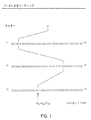
分子のコレクション、特にタンパク質、DNA、小分子のコレクションおよび他のコレクションの種類をソートし得る、ネスト化ソーティングの方法を図1−18に示す。ネスト化ソーティングの概念を図1に示す。この例では、cDNAのような74,088種のアイテムを含むマスターコレクションを、42のサブライブラリー(F1サブライブラリー)中に当該コレクションをランダムに分割することにより、探索する。42のF1サブライブラリーが、例えば、プローブとの結合またはプローブとの反応により、またはタンパク質−タンパク質特異的相互作用により、目的のアイテムを含むものを同定した後、そのグループを更にランダムに42の新規サブライブラリー(F2サブライブラリー)に分割し、目的のアイテムを含むサブライブラリーを再び同定する。目的のアイテムを含むF2サブライブラリーの最終分割により、1つのみのアイテムをそれぞれ含む42の新規グループを作成する。目的のアイテムは、ソーティング系列に基づき唯一同定され得る。
【0134】
示した例では、目的のアイテムは、5番目のF1サブライブラリー、31番目のF2サブライブラリー、および16番目のF3サブライブラリーで同定した。マスターコレクションの74,088アイテムのうち、1つのみがソート系列F15/F231/F316となる。
【0135】
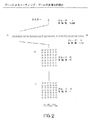
図2で解説するソートは、図1で解説するソートと同一であるが、F2およびF3サブライブラリーはアレイ中に配列される点が異なる。この図は、また、ソートの結果として個々のサブライブラリー中のアイテムの多様性が減少し;例示のマスターコレクションが74,088アイテムを含むこと、42のF1サブライブラリーがそれぞれ1,764アイテムを含むこと、42のF2サブライブラリーが42のアイテムを含むこと、そして42のF3サブライブラリーがアイテムを1つのみ含むことを示す。最初の2つの図は、ネスト化ソーティングに基づく理論的探索を示している。
【0136】
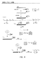
図3は、高多様性遺伝子ライブラリーのネスト化ソートのツールとして、抗体アレイのような捕捉試薬アレイの使用を示す。マスター遺伝子ライブラリーを最初にランダムにサブライブラリーの数に、PCRのような分離増幅反応のにより、分割する。増幅反応には、マスター鋳型プールから遺伝子の異なるサブライブラリー(F1サブライブラリー)を特異的に増幅するプライマーの適当な設計により、事前に選択したエピトープをコードし、遺伝子中に組込まれる特有ヌクレオチド配列のセットを使用する。これらの増幅反応は、例えば、適合性サーモサイクラーを用い96ウェル(または384ウェルまたはそれよりも高い密度)で行う。
【0137】
個々のウェル中の増幅遺伝子をタンパク質産物に翻訳し、次いで、それぞれからサンプルを適用し、アレイのような捕捉試薬コレクションを分離する(すなわち、96ウェルプレート中の個々のウェルからのタンパク質を96捕捉試薬アレイの1つに適用する)。アレイにおいて、抗体のような捕捉試薬にに対する結合により、タンパク質を、プライマーを使用しタンパク質に加える既知特有アミノ酸配列(エピトープ)を認識するアレイ上の定義位置にソートする。ソーティングの後、目的のタンパク質を含むアレイ上のアドレスを同定し、アレイ中のスポットに結合するエピトープコード化配列を有するタンパク質からのサブライブラリーからの核酸を、例えばPCRで増幅する。
【0138】
この第二の増幅ステップの間、既知エピトープの新規セットを核酸に組込み、それにより、更なる捕捉試薬アレイ(F3)を用い更にソートされ得る。
【0139】
図3の表は、PCRによる最初の分割の数がどれくらいか、およびアレイを組合し、例えば100万(106)から10億(109)種を超える遺伝子を含む遺伝子ライブラリーを探索できる捕捉試薬の数はどれくらいか、を示す。例えば、最初の遺伝子ライブラリーを、増幅により100のF1サブライブラリーに分割し得、次いで100種のエピトープを認識する捕捉試薬を有する2つのアレイを使用し更に分割し得る。最初の遺伝子ライブラリーが106種の遺伝子を含むならば、サブライブラリー中のF3アドレスは単一型の遺伝子を含む(106/100/100/100=1)。PCR増幅により1000のF1サブライブラリーに分割され、次いでF2およびF3サブライブラリーを作成する1,000種のエピトープを認識する捕捉試薬を有する2アレイを用い更に分割される最初の遺伝子ライブラリーは、109種の遺伝子(109/1,000/1,000/1,000=1)の探索に使用し得る。
【0140】
サブライブラリー中への遺伝子ライブラリー分割は、プライマーの対を用い、DNA配列を特異的に増幅するPCR増幅反応の能力に基づく。両方のプライマーは、鋳型DNAの何れかの末端において配列にハイブリダイズする必要があるが、鋳型配列のサブセットは、プライマー対を用い増幅し得、この場合、その一方のプライマーは、すべての鋳型配列に共通であり、他方のプライマーは、目的の遺伝子配列に特異的である。例えば、特定遺伝子は、しばしば、目的の遺伝子に特異的である一方のプライマーおよびcDNA分子のすべてに共通なオリゴ(dA)テイルにハイブリダイズする他方のプライマーを用い、cDNAライブラリーから増幅する。
【0141】
6.単一の融合タンパク質中の複数タグの使用
本明細書で提供するシステムはエピトープタグを使用し、scFvのライブラリーのようなタンパク質ライブラリーを更に分割する。例えば、1000タグおよび109scFvのライブラリーでは、それぞれのタグに対し106のscFvが存在する。目的のscFvのような単一のライブラリーメンバーを同定するため、多数の個々のscFv(106)をスクリーニングするか、1以上の小部分(subdivision)を用いる。より多数のタグを用いると、ライブラリーは、より少ないステップで少数のタンパク質に減少し得る。
【0142】
組合せのアプローチを用い、小セットの捕捉試薬タグ対を多数セットとして有効に用い得る。単一のscFv融合タンパク質のようなタンパク質中に複数のタグを組込むことにより、より少ないタグのよりよい使用が行い得る。比較として、300の捕捉試薬タグ対があり、個々のメンバーに結合する単一のタグを伴う109メンバーのライブラリーがあるならば、300タグは、個々の型のタグが3.3×10⁶メンバーに結合するように109メンバーを分割する。1/3のタグをそれぞれ3つの部位で使用するような組合せ形態で個々のメンバーに組込まれる3つのタグによると、合計100×100×100(または106)の組合せがある。これら106のタグ組合せを用い、109メンバーをタグあたり1000メンバーに分割する。そのため、限られた数のタグによる単一ステップにおいて、ライブラリーは有効に再分割(subdivide)する。
【0143】
最も間単な実施態様では、部位Xでxタグ、部位Yでyタグおよび部位Zでzタグの例を考慮する。これらのタグを個々に使用するならば、x+y+zの組合せがある。これらのタグを組合せて使用するならば、(x)(y)(z)の組合せがある。個々の部位(x,yおよびz)におけるタグの数が総数(n)の1/3であると仮定すると、個々の使用の場合、C=(n/3)×3=nであるか、タグの存在と同じぐらい多くの総数の組合せ(C)が存在し、ここで、コンビナトリアル的使用の場合、C=(n/3)3となる。個々の部位での各タグ数が増加するので、コンビナトリアルタグの数は、より高い割合で増加する(図19参照)。より多数の有効なタグを用い、タグあたりのライブラリーのメンバー数は減少する。初期ライブラリーにおけるタグあたりのより少ないメンバーは、より少ない連続的ラウンドのスクリーニングを生ずるか、またはハイスループットスクリーニングで評価するクローンのより少ない数を生ずるか、いずれかである。
【0144】
単一タグを用いるか、または組合せで複数タグを用いるか、いずれであっても、手順は実質的に同じである。発現ライブラリー由来のタンパク質は、タグに対する、抗体のような捕捉試薬に結合するエピトープタグの特性により更に分割される(subdivide)。上記で存在する例では(3つのタグを組合せて使用する)、個々のライブラリーメンバーは、3種の抗タグ捕捉試薬に結合する。個々のコンビナトリアルタグは、それ自身のセットのアドレスを、単一アドレスの代わりのアレイにおいて、有する。例えば、部位Xの1−100、部位Yの101−200および部位Zの201−300で合計300タグが存在するならば、典型的なコンビナトリアルタグは、アドレスX27−Y132−Z289を有する。他のコンビナトリアルタグもまたは、捕捉試薬のようなX27抗タグ捕捉試薬、またはY132またはZ289捕捉試薬を使用するが、他の組合せは、3つすべてを使用するわけではない。抗原が、個々のタグが結合する3つの捕捉試薬に結合するライブラリーメンバーに結合するならば、コンビナトリアルタグは、知られることとなり、ライブラリーメンバーはオリジナルライブラリーから回収され得る。
【0145】
コンビナトリアルタグを伴う特定ライブラリープールの回収は、単一タグを伴うライブラリープールを回収する実質的な方法で行う。本明細書に記載のように、ライブラリーから部分母集団(subpopulation)を回収する1つの方法にポリメラーゼ連鎖反応を使用する。例示のため、3つすべてのタグが発現タンパク質のC末端にあると仮定すると、Xタグは、scFvのようなライブラリーメンバーに最も近く、次いで、Yタグ、更にZタグが近くなる。cDNAのコーディング鎖におけるDNAセグメントのオーダーは、5’共通>scFv>X>Y>Z3’となる。
【0146】
特定の部分母集団は、共通プライマーおよびZ289タグに相当するプライマーで開始する連続ラウンドPCRにより回収され得る。この反応由来の産物を、共通プライマーおよびY132タグプライマーを用いる次の反応に使用する。この反応由来の産物を、共通プライマーおよびX27プライマーを用いる、その後の反応に使用する。3つの連続ラウンドの増幅の後、当該産物はすべて、x27−Y132−Z289の組合せで初めにタグ化されていた、scFvのようなライブラリーメンバーに相当する。当業者ならば、ライブラリーが複数のネスト化共通配列を有している限り、複数種の共通プライマーが異なるラウンドで使用されることを理解している。当業者ならばまた、複数タグがコード化DNAの反対側末端にあり、それゆえ発現タンパク質の反対側末端に存在し得ることを理解している。発現エピトープタグは直鎖状であり、ジスルフィド結合で束縛され、骨格(scaffold)構造で束縛され、融合タンパク質のループで発現し、連続であるか、または可動性もしくは非可動性リンカー配列により分離され得ることも理解される。
【0147】
1つの実施態様では、例えば、単一骨格融合タンパク質は、挿入エピトープタグと共に複数部位を含む。これは、エピトープを空間的に分離し、互いの干渉なしにすべて認識し得る。下記の基準をタンパク質骨格を選択する際に考慮する:1)抗原性の高い傾向を有するアミノ酸にさらされる表面をより簡単に同定する既知結晶構造、2)目的cDNAライブラリーに融合するための遊離NおよびC末端、3)種々のタンパク質発現システムにおける高レベルの産生および溶解性(特にE. coliペリプラズム)、4)インビトロ転写/翻訳の許容力、5)ジスルフィド結合の非存在、6)野生型タンパク質が単量体であること、7)scFvの溶解性または機能性を増加する許容性を有すること。結晶構造を使用すると、エピトープタグライブラリーの挿入のために位置が選択される。これらの部位は、自然では、比較的直線状であるエピトープを空間的に分離する(例えば、αヘリックスの一端、β鎖間のターンまたはヘリックス間のループ)。
【0148】
D.抗体の調製
1.アドレス可能抗タグ抗体の抗体およびコレクション
本明細書における方法は、分子、特にタンパク質のライブラリー(またはコレクション)に結合するポリペプチドタグに特異的に結合する抗体のような捕捉試薬の能力に依存する。特定タグに対する個々の抗体(またはコレクション中の他のレセプター)の特異性は、既知であるか、またはアレイにおける遺伝子座のすべての抗体が特定エピトープタグに特異的となるように、例えば抗体をアレイすることにより、容易に確かめられる。
【0149】
他に、例えば、カラービーズまたはバーコード化ビーズまたは支持体を含む所望によりコード化タグに対する結合により、個々の抗体が同定され得るか、または例えば、マイクロリアクターを電子タグまたはバーコード化支持体(例えば、米国特許番号6,025,129、米国特許番号6,017,496、米国特許番号5,972,639、米国特許番号5,961,923、米国特許番号5,925,562、米国特許番号5,874,214、米国特許番号5,751,629、米国特許番号5,741,462参照)、または化学タグ(米国特許番号5,432,018、米国特許番号5,547,839参照)またはカラータグに供することにより、または物理的にアドレス可能アレイの代わりに使用し得るアドレス可能方法により、電子タグに結合し得る。例えば、個々の抗体型は、カラーコードタグ(すなわち、カラーソート可能ビーズ)または周波数(radio−frequency)タグ(RF)のような、IRORI MICROKANS(商標)およびMICROTUBES(商標)ミクロリアクター(米国特許番号6,025,129、米国特許番号6,017,496、米国特許番号5,972,639、米国特許番号5,961,923、米国特許番号5,925,562、米国特許番号5,874,214、米国特許番号5,751,629、米国特許番号5,741,462、国際PCT出願番号WO98/31732、国際PCT出願WO98/15825参照、また米国特許番号6,087,186参照)のような電子タグと会合する支持体マトリクスに結合し得る。本明細書で提供する方法およびコレクションの場合、個々の型の抗体は、MICROKANまたはMICROTUBEマイクロリアクター支持体マトリクスおよび会合RFタグ、バーコード、カラー、カラービーズまたは抗体のようなレセプターを同定する役割をする他のアイデンティファイアー、および抗体のようなレセプターが結合するエピトープタグに結合し得る。
【0150】
本明細書の例示的目的のため、リファレンスから、抗体および当該抗体が特異的に結合するエピトープをコードするタグを作成する。特異的に結合する任意対の分子が予期され;本明細書の目的のため、抗体のような分子をレセプターと称し、およびそれに結合するリガンドのような分子はエピトープであることが理解される。当該エピトープは、典型的に、抗体のようなレセプターまたはその特異的結合フラグメントに特異的に結合するアミノ酸の短い配列である。
【0151】
また、本明細書の例示のために、リファレンスから、位置付けアレイを作成する。しかし、その他の同定方法が本明細書の方法による使用に容易に適用され得ることが理解される。抗体のようなレセプターの同定(すなわち、エピトープタグ特異性)が既知であることのみが必要である。二次元アレイまたは三次元アレイの何れかであり、また所望により、コード化ビーズまたはカラー化支持体またはRFタグまたはほかの形式に結合する、アドレス可能レセプター(すなわち、抗体)のコレクションが生じ、それを本明細書の方法に用い得る。
【0152】
抗体のコレクションとポリペプチドタグ標識分子のライブラリーとを反応させ、次いで、スクリーニングアッセイを行い、望ましい性質のエピトープ標識分子が結合する抗体のコレクションのメンバーを同定することにより、分子のライブラリーの多様性の減少が生ずる。抗体の個々のコレクションは、多様性の減少をもたらすためのソーティング装置として役立つ。当該過程を複数回繰り返すことにより、多様性の迅速なおよび実質的な減少をもたらし得る。
【0153】
2.捕捉試薬の調製
ソートの質は、ソーティングアレイを作成する抗体のような捕捉試薬のコレクションの質に依存する。結合親和性および特異性の必要性に加え、アレイ中の捕捉試薬(抗体)により結合するエピトープは、増幅反応(PCR)のプライミング部位として使用されるE、FAおよびFB配列を決定する。図12は、コレクション中の使用のために抗体産生に使用するためのハイブリドーマ細胞から産生する免疫グロブリン(Ig)を発見するためのハイスループットスクリーニングの概略を示している。
【0154】
ハイブリドーマ細胞は、非免疫化マウスか、またはランダムジスルフィド束縛化七量体エピトープのライブラリーまたは他のランダムペプチドライブラリーを発現するタンパク質で免疫化されたマウスのいずれかから作成される。安定なハイブリドーマ細胞は、高Ig産生およびエピトープ結合のため最初にスクリーニングされる。Ig産生は、ヤギ抗マウスIgG抗体を用いるELISAアッセイにより培養上清中で測定される。エピトープ結合はまた、免疫に使用するハプテンの混合物(エピトープタグ化タンパク質)がELISAプレートに固定化されるELISAアッセイにより測定され、培養上清の結合IgGは、ヤギ抗マウスIgG抗体を用い測定される。両アッセイは、96ウェル型または他の適当な型で行う。例えば、約10,000ハイブリドーマがこれらのスクリーニングから選択される。
【0155】
次に、Igは、固定化Ig結合タンパク質(プロテインA、GまたはL)と共にフィルターを含む96ウェルまたはより高密度精製プレートを用い別々に精製する。精製Igの量は、96ウェルまたはより高密度プレート型の標準的プロテインアッセイを用い測定する。個々の培養物由来の低ミクログラム量のIgが、この精製法の使用に予想される。
【0156】
精製Igを、標準的ピン型アレイシステムを用いニトロセルロースフィルター上に別々にスポットする。当該精製Igがまた合わされて、同量の個々のIgを有する混合物を生ずる。混合Igを、ランダムジスルフィド束縛化七量体エピトープを発現するバクテリオファージのライブラリーをパン(pan)する固相支持体として使用される常磁性ビーズに結合する。バッチパンニングは、精製Igに対し、ファージ発現エピトープのファージディスプレイライブラリーを富化する。この富化は、ファージライブラリーの多様性を劇的に減少する。
【0157】
次いで、富化したファージディスプレイライブラリーは、精製Igのアレイに結合し、緊縮的に洗浄する。Ig結合ファージは、電荷結合素子(CCD)に基づくイメージングシステムで検出可能な化学ルミネセンスシグナルを生ずる抗ファージ抗体HRP結合体で染色することにより検出する。最も強いシグナルを生ずるアレイ中のスポットを除外し、ファージを溶出し増殖させる。回収ファージにより発現するエピトープは、DNA配列決定により同定し、親和性および特異性で更に評価する。この方法は、同系エピトープを認識する高親和性、高特異性抗体のコレクションを生ずる。連続スクリーニングは、質を改善した抗体のより大きなコレクションを生ずる。
【0158】
3.抗タグ捕捉試薬アレイの調製
個々のスポットは、単一の特異性を有する抗体のような複数の捕捉試薬を含む。個々のスポットは、検出に適当な大きさである。1から300ミクロンのオーダー、典型的には1から100、1から50および1から10ミクロンのスポットは、アレイの大きさ、標的分子および他のパラメーターに依存する。一般的に、スポットは、50から300ミクロンである。アレイの調製において、十分な量が、望ましい特性を有するタンパク質の検出のために機能的にカバーされる表面に送達される。一般的に、アレイ調製のため送達された抗体含有混合物の体積は、ナノリッター体積(1から約99ナノリッターまで)であり、一般的に約1ナノリッターまたはそれ未満であり、典型的には、約50と200ピコリッターとの間である。これは、非常におおよそであるが、スポットあたり約1000万から100,000分子あり、この場合、個々のスポットは、単一のエピトープを認識する抗体のような捕捉試薬を有する。例えば、1000万分子が存在し、遺伝子座と反応するタンパク質混合物中に1000種のものがあるならば、スポットあたり104の個々の型の分子が存在する。アレイの大きさおよび個々のスポットは、スクリーニングステップ中の陽性反応が、好ましくは、アレイ全体または複数のアレイ(24、96またはそれより多いアレイのような)を同時にイメージすることによりイメージされ得るようになる。
【0159】
KODAKペーパープラスゼラチンのような支持体(典型的支持体は以下参照)または他の適当なマトリクスを使用し得、次いで、インクジェットおよびスタンピングテクノロジーまたは他の適当な分配方法および装置を使用し、再現可能なようにアレイをプリントする。当該アレイを、例えば、ピエゾまたはインクジェットプリンターまたは他のそのようなナノリッターまたはより少量の分配装置を用いプリントする。例えば、1000スポットを有するアレイをプリントし得る。24または48、96またはそれより多い複数の複製アレイを、通常96ウェルプレートの大きさのシート上に置くことができる。
【0160】
本明細書で予期できる実施態様では、抗体アレイの複製をそれぞれ伴うアレイのシートである。これらは、例えば、ピエゾまたはインクジェット分配システムを用い調製される。多数、例えば1000は、例えば、1000種のホール(500μMホールを有するスタンプなど)を伴うプリントヘッドを使用し、同時にプリントされ得る。それは、例えば、1000種の抗体のような捕捉試薬でそれぞれ満たされる、1000ホールのような多くのホールを有するモールド化プラスチックから作成され得る。個々のホールはリザーバ(reservoir)に結合し得、そのリザーバは、抗体のような捕捉試薬をプリントヘッド中に最終的に分配する、小さいサイズの導管(conduit)に結合する。シートにおける個々のアレイは、空間的に分離され得、および/またはプラスチックリッジのような物理的バリアまたは疎水性バリアのような化学的バリア(すなわち、疎水性バリアにより分離されるヒドロゲル)により分離され得る。アレイを有するシートは、通常、96ウェルプレートまたはそれよりも高い密度の大きさであり得る。個々のアレイは、事前に選択したセットのエピトープタグに特異的な複数のアドレス可能抗タグ抗体を含む。例えば、33×33アレイは、おおよそ1000抗体を含み、個々のアレイ上の個々のスポットは、事前決定した単一のエピトープに特異的結合する抗体を含む。バリアにより分離される複数のアレイを用い得る。
【0161】
表面上に抗体を分配する場合、その目的は、機能性表面適用範囲(functional surface coverage)であり、それにより、スクリーニングした望ましいタンパク質が検出可能となる。これを行うため、出発コレクションから、例えば、約1から2mg/mlを使用し、抗体あたり約500ピコリッターをアレイ上のスポットごとに置く。正確な量は経験的に決定し得、表面および検出方法の感度のような幾つかの変数に依存し得る。当該抗体は、例えば、スルフヒドリル結合により好ましくは表面上のアミドに共有結合する。他の例示の分配および固定化システムには、以下に限らないが、例えば、Genometrix(ガラス上でプリントするためのシステムを有する)から;Illumina(支持体としてファイバーオプティックスケーブルのチップを用いる)から;Texas Instruments(チップ表面プラズモン共鳴を有する(すなわち、タンパク質誘導化ゴールド))から;Microfab Technologies, Plano TXからのようなインジェクトシステム;Incyte, Pal Alto, CA, Protogene, Mountain View, Ca, Packard BioSciences, Meriden CT、および適当な支持体表面にタンパク質を分配および固定化するための他のシステムから利用可能なシステムが含まれる。ブラント(blunt)およびクイルピン(quill pin)、ソレノイドおよびピエゾナノリッターディスペンサーおよび他のものといった他のシステムもまた予期される。
【0162】
4.他のコレクションの調製
捕捉試薬を、同定可能なビーズまたは粒子支持体に結合する。例えば、捕捉試薬は、所望により、Luminex, Austin Txからのようなコード化ミクロスフェアに結合し、カプセル化した蛍光性色素を含む。色素をカプセル化するミクロスフェアは、任意の適当な物質から調製され(例えば、国際PCT出願番号WO01/13119およびWO99/19515参照、以下の記載参照)、スチレン−エチレン−ブチレン−スチレンブロックコポリマー、ホモポリマー、ゼラチン、ポリスチレン、ポリカーボネート、ポリエチレン、ポリプロピレン、樹脂、ガラス、および任意の他の適当な支持体(マトリクス物質)を含み、直径、約1ナノメーターから約10ミリメーターの大きさとなる。10種の濃度のうち例えば2種の色素の組合せの特性より、複数の、特有の蛍光によりそれぞれ同定可能なミクロスフェア(この例では100)を生ずる。
【0163】
他に、クロモホアまたはカラー色素または他のカラー物質の組合せをカプセル化し、ミクロスフェアまたは他の粒子中にカプセル化する種々の異なる色を生じ、次いで、抗体のような捕捉試薬の支持体として使用する。抗体のような個々の捕捉試薬を特定のカラー化ビーズに結合し、それにより同定可能となる。抗体のような、結合捕捉試薬と共にビーズを生じた後、エピトープタグ化分子との反応は液相中で行い得る。エピトープと反応するビーズを同定し、ビーズの色の結果から特定エピトープが既知となる。次いで、その結合分子が誘導されるサブライブラリーを同定する。
【0164】
E.抗体を固定化するための支持体
抗体を固定化するための支持体は、リガンドおよび他の分子の固定化が知られる任意の不溶性物質であり、親和性クロマトグラフィーのような、生物学的活性物質の固定化での、多くの化学合成および分離において、およびタンパク質、アミノ酸および他の有機分子およびポリマーを含む生体分子の化学的合成の間に、使用される。適当な支持体には、生体適合性ポリマーを含む任意の物質が含まれ、抗体物質の結合のための支持体マトリクスとして作用し得る。当該支持体物質は、化学的または生物学的スクリーニング反応を干渉しないように選択される。
【0165】
本明細書での使用も予期される支持体には、ミクロプレートおよびビーズ(例えば、Amersham, Arlington Heights, ILから商業的に入手可能であり、Nuclear Technology, Inc., Sand Carlos, CAおよびPackard, Meriden, CTからのプラスチックシンチレーションビーズ、およびLuminex Corporation, Austin, TXからのカラー化ビーズベース化支持体(ミクロスフェア中にカプセル化される蛍光粒子))のようなフルオロホア含有または含浸支持体が含まれる(米国出願番号09/147,710を基礎とする国際PCT出願番号WO/0114589参照、米国出願番号09/022,537を基礎とする国際PCT出願番号WO/0113119参照)。例えば、Luminexからのミクロスフェアは、蛍光粒子のカプセル化の特性により内部的にカラーコード化されており、液体アレイとして提供され得る。抗体(エピトープ)のような捕捉試薬は、任意の適当な方法により直接または間接的に結合され、ビーズおよび結合タンパク質の表面との結合または相互作用は、それらが結合するビーズのカラーの特性により同定され得る。検出は、任意の手段により実施可能であり、カラー化(呈色)反応およびビーズのカラーの特性(呈色性)によりミクロスフェア(ビーズ)の色において検出可能な変化を生ずる色素生産的または蛍光的ディテクターまたはレポーターと組合わせ得る。ビーズに基づくアレイの場合、抗タグ捕捉試薬は、別反応においてカラーコード化ビーズに結合する。ビーズのコードは、それに結合する抗体のような捕捉試薬を同定する。次いで、ビーズを混合し得、その後、結合ステップを溶液中で行い得る。次いで、それらは、例えば、透明な蓋で微小作製(microfabricate)フローチャンバー(ビーズの単一層のみから二次元アレイを形成し得る)中にパッケージすることにより、アレイされ得る。タンパク質が結合するビーズが同定され、それにより、捕捉試薬およびタグが同定される。当該ビーズを例えば、CCDカメラでイメージ化し、反応するビーズを同定する。そのビーズのコードを同定し、それにより、捕捉試薬を同定し、次に、ポリペプチドタグおよび最終的に目的タンパク質を同定する。
【0166】
当該支持体はまた、比較的不活性のポリマーであり得、それは、イオン放射することによりグラフトし得、ポリスチレン、または誘導され、そして支持体として使用され得るポリマーのような他のもののコーティーングの結合が可能となる。単量体の放射線グラフトは、支持体上で生ずる表面特性の多様性を可能とする(例えば、Maeji et al. (1994) Reactive Polymers 22: 203−212; およびBerg et al. (1989) J. Am. Chem. Soc. 111: 8024−8026参照)。例えば、ポリエチレンおよびポリプロピレンのようなポリマーに対し、ビニル単量体のような単量体の放射線分解グラフティングまたは単量体の混合物は、異なる表面特性を有する合成物を生ずる。これらの方法を用い、ペプチドおよび他の分子の合成のための不溶性支持体にポリマーをグラフトする。
【0167】
支持体は、典型的には、固体、多孔性、変形可能、または堅固であり、任意の必要な構造およびジオメトリー(ビーズ、ペレット、ディスク、キャピラリー、中空ファイバー、針、固体ファイバー、ランダム型、薄フィルムおよび膜、および最も好ましくはアドレス可能座位を有する固体表面型を含むがこれらに限らない)を有する不溶性サブストレートである。支持体はまた、捕捉試薬抗体および他の分子が支持体を扱う目的のため付着しない、テフロンストリップのような不活性ストリップまたは他の物質を含み得、同定可能記号を含み得る。
【0168】
その支持体の調製および使用は当業者に既知であり、そのような多くの物質および調製が知られている。例えば、アガロースおよびセルロースのような天然に生ずる物質は、各ソースから単離され得、既知のプロトコールに従い加工され、合成物質は、既知プロトコールに従い調製され得る。これら物質には、以下に限らないが、無機質、天然ポリマーおよび合成ポリマーが含まれ、以下に限らないが、セルロース、セルロース誘導体、アクリル性樹脂、ガラス、シリカゲル、ポリスチレン、ゼラチン、ポリビニルピロリドン、ビニルおよびアクリルアミドのコポリマー、ジビニルベンゼン等との架橋するポリスチレン(Merrifield (1964) Biochemistry 3: 1385−1390参照)、ポリアクリルアミド、ラテックスゲル、ポリスチレン、デキストラン、ポリアクリルアミド、ラバー、シリコン、プラスチック、ニトロセルロース、セルロース、天然のスポンジ(natural sponge)、および他の多くのものが含まれる。支持体の選択は、溶解度のような物理的および化学的性質、官能基、機構的安定性、表面エリア膨張性、疎水性または親水性特性および目的の使用により、少なくとも一部、決定される。
【0169】
1.天然支持体物質
天然に生ずる支持体には、以下に限らないが、アガロース、他のポリサッカライド、コラーゲン、セルロースおよびそれらの誘導体、ガラス、シリカおよびアルミナが含まれる。単離、修飾および支持体としての使用に適当な処置方法は、当業者に既知である(例えば、Hermanson et al. (1992) Immobilized Affinity Ligand Techniques, Academic Press, Inc., San Diego参照)。アガロースのようなゲルは、容易に本明細書の使用に適用し得る。ポリペプチド、タンパク質および炭水化物のような天然ポリマー;半導特性を有する、シリコンのようなメタロイドおよびゲルマニウムもまた本明細書の使用に適用し得る。白金、金、ニッケル、銅、亜鉛、スズ、パラジウム、銀のような金属もまた本明細書の使用に適用可能である。目的の他の支持体には、Pt−PtO、Si−SiO、Au−AuO、TiO2、Cu−CuOなどのような金属およびメタロイドの酸化物が含まれる。また、原子間力顕微鏡で観察する分子の調製に使用するような、ニオブ酸リチウム、ヒ化ガリウム、およびリン化インジウムのような半導体化合物、およびニッケル被覆雲母表面(例えば、III et al. (1993) Biophys J. 64: 919参照)もまた、支持体として使用し得る。そのマトリクス物質の調製のための方法は既知である。
【0170】
例えば、米国特許番号4,175,183は、水不溶性ヒドロキシアルキル化架橋化再生化セルロースおよびその調製の方法が記載されている。試薬の密接な化学量論特性を用い産物を調製する方法を記載する。ゲルクロマトグラフィーの直接の産物の使用およびイオン交換体の調製での中間体としての使用も記載されている。
【0171】
2.合成支持体
当業者に既知の無数の合成支持体および方法が存在する。合成支持体は、典型的に、官能性マトリクスの多量体化により、または合成単量体および天然で発生するマトリクス単量体またはアガロースのような多量体由来の2以上の単量体由来の共重合により、作製される。
【0172】
合成マトリクスには、以下に限らないが、アクリルアミド、デキストラン誘導体およびデキストラン共重合体、アガロース−ポリアクリルアミドブレンド、種々の官能基を有する他のポリマーおよびコポリマー、メタクリレート誘導体およびコポリマー、ポリスチレンおよびポリスチレンコポリマー(例えば、Merrifield (1964) Biochemistry 3: 1385−1390; Berg et al. (1990) in Innovation Perspect. Solid Phase Synth. Collect. Pap., Int. Symp., 1st, Epton, Roger (Ed), pp. 453−459; Berg et al. (1989) in Pept., Proc. Eur. Pept. Symp., 20th, Jung, G. et al. (Eds), pp. 196−198; Berg et al. (1989) J. Am. Chem. Soc. 111: 8024−8026; Kent et al. (1979) Isr. J. Chem. 17: 243−247; Kent et al. (1978) J. Org Chem. 43: 2845−2852; Mitchell et al. (1976) Tetrahedron Lett. 42: 3795−3798; 米国特許番号4,507,230; 米国特許番号4,006,117;および米国特許番号5,389,449)が含まれる。その支持体マトリクスの調製のための方法は当業者に既知である。
【0173】
合成支持体には、ポリビニルアルコール、アクリレートおよびアクリル酸(ポリエチレンコアクリル酸、ポリエチレンコメタクリル酸、ポリエチレンコエチルアクリレート、ポリエチレンコメチルアクリレート、ポリプロピレンコアクリル酸、ポリプロピレンコメチルアクリル酸、ポリプロピレンコエチルアクリレート、ポリプロピレンコメチルアクリレート、ポリエチレンコビニルアセテート、ポリプロピレンコビニルアセテートなど)のようなポリマーおよびコポリマーから作製されるもの、およびポリエチレンコマレイン酸無水物、ポリプロピレンコマレイン酸無水物などのような酸無水物を含むものも含まれる。リポソームもまた、親和性精製のための固体支持体として使用される(Powell et al. (1989) Biotechnol. Bioeng. 33: 173)。
【0174】
例えば、米国特許番号5,403,750はポリウレタンに基づくポリマーの調製について記載している。米国特許番号4,241,537は、鎖伸長ポリオールから調製する親水性ポリウレタンゲル組成物を含む植物培養培地について記載している;ランダム共重合は、プレポリマーが室温で液体であるように、50%までのプロピレンオキシド単位で行い得る。米国特許番号3,939,123は、35%までのポリ(プロピレンオキシ)グリコールまたはポリ(ブチレンオキシ)グリコールを有するポリ(エチレンオキシ)グリコールを含むイソシアネート末端化プレポリマーの、わずかに架橋したポリウレタンポリマーについて記載している。これらのポリマーにおいて、有機性ポリアミンは架橋剤として使用される。他の支持体およびその調製は、米国特許番号4,177,038、4,175,183、4,439,585、4,485,227、4,569,981、5,092,992、5,334,640、5,328,603に記載されている。
【0175】
米国特許番号4,162,355は、少なくとも1つのペンダントハロメチル基を有するアミンイミド(aminimide)およびビニルのポリマーである、親和性クロマトグラフィーの使用に適当なポリマーについて記載している。親和性クロマトグラフィー中で結合するための部位を提供するアミンリガンドは、ペンダントハロメチル基の一部との反応によりポリマーに結合し、ペンダントハロメチル基の残りはペンダント親水性基を含むアミンと反応する。このポリマーを有するサブストレートを被覆する方法もまた述べられている。典型的なアミンイミドは、1,1−ジメチル−1−(2−ヒドロキシオクチル)アミンメタクリルイミドであり、ビニル化合物は、クロロメチルスチレンである。
【0176】
米国特許番号4,171,412は、D−アミノ酸単位を含む共有結合D−アミノ酸またはペプチドを有する、好ましくはマクロポーラス特性の親水性ポリマーゲルに基づく特異的支持体について述べている。基礎支持体は、アクリル酸およびメタクリル酸のヒドロキシアルキルエステルまたはヒドロキシアルキルアミドの共重合により調製され、それに伴い、アクリレートまたはメタクリレートコモノマーの架橋は、ジアミン、アミノ酸またはジカルボン酸との反応により修飾され、生ずるカルボキシ末端またはアミノ末端基は、アミノ酸のD−類似体またはペプチドで縮合される。D−アミノ酸を含むペプチドはまた、担体の表面上で段階的に合成され得る。
【0177】
米国特許番号4,178,439は、陽イオン交換体およびその調製方法について記載している。米国特許番号4,180,524は、シリカ支持体上での化学合成について記載している。
【0178】
Immobilized Artificial Membranes(IAM; 例えば、米国特許番号4,931,498および4,927,879)もまた使用し得る。IAMは、細胞膜環境を真似ており、好ましくは細胞膜と会合する分子との結合に使用され得る(例えば、Pidgeon et al. (1990) Enzyme Microb. Technol. 12: 149)。
【0179】
当該支持体の中で、国際PCT出願番号WO00/04389、WO00/04382およびWO00/04390に記載のもの;マトリクス物質で被覆されるKODAKフィルム支持体が本明細書で予期される(目的の他の支持体の場合米国特許番号5,744,305および5,556,752参照)。目的のものは、Luminex(Austin、TX)からのもののような、カラー化「ビーズ」である。
【0180】
3.固定化および活性化
多くの方法が、固体または液体支持体へのタンパク質および他の生体分子の固定化に開発された(例えば、Mosbach (1976) Methods in Enzymology 44; Weetall (1975) Immobilized Enzymes, Antigens, Antibodies, and Peptides; and Kennedy et al. (1983) Solid Phase Biochemistry, Analytical and Synthetic Aspects, Scouten, ed., pp. 253−391参照;一般的には、Affinity Techniques. Enzyme Purification: Part B. Methods in Enzymology, Vol. 34, ed. W. B. Jakoby, M. Wilchek, Acad. Press, N. Y. (1974); Immobilized Biochemicals and Affinity Chromatography, Advances in Experimental Medicine and Biology, vol. 42, ed. R. Dunalap, Plenum Press, N. Y. (1974)参照)。
【0181】
最も通常的に使用される方法の中には、吸収および吸着または支持体への共有結合があり、その結合は直接かリンカーを介するか何れかであり、例えば、多くのジスルフィド結合、チオエーテル結合、束縛ジスルフィド結合(hindered disulfide bonds)、および当業者に既知のアミンおよびチオール基のような遊離反応基間の共有結合である(例えば、the PIERCE CATALOG, ImmunoTechnology Catalog & Handbook, 1992−1993(試薬の調製および使用が記載されており、その試薬の市場の源を提供する)およびWong (1993) Chemistry of Protein Conjugation and Cross Linking, CRC Press参照;また、DeWitt et al. (1993) Proc. Natl. Acad. Sci. U.S.A. 90: 6909; Zuckermann et al. (1992) J. Am. Chem. Soc. 114: 10646; Kurth et al. (1994) J. Am. Chem. Soc. 116: 2661; Ellman et al. (1994) Proc. Natl. Acad. Sci. U.S.A. 91: 4708; Sucholeiki (1994) Tetrahedron Lttrs. 35: 7307; およびSu−Sun Wang (1976) J. Org. Chem. 41: 3258, Padwa et al. (1971) J. Org. Chem. 41: 3550 およびVedejs et al. (1984) J. Org. Chem. 49: 575(感光性リンカーについて記載している)参照)。
【0182】
固定化を行うため、タンパク質または他の生体分子の溶液は、アルミナ、炭素、イオン交換樹脂、セルロース、ガラスまたはセラミックのような支持体物質と接触させる。過フッ化炭化水素ポリマーは、生体分子が吸着により結合する支持体として使用する(米国特許番号3,843,443、公開された国際PCT出願WO/8603840)。
【0183】
種々の方法が、タンパク質および核酸を含む生物学的分子、固体支持体に対する分子を結合するために知られている(例えば、米国特許番号5451683参照)。例えば、米国特許番号4,681,870は、シリカ支持体上への遊離アミノ基またはカルボキシル基の導入方法が記載されている。その後、これらの基は、カルボジイミド存在下、タンパク質または他の抗リガンドのような他の基に共有結合し得る。他に、シリカマトリクスは、アルカリ条件下、ハロゲン化シアンによる処置によって活性化され得る。抗リガンドは、活性化表面への添加において表面に共有結合する。他の方法は、ビオチン、アビジンおよびエクステンダーの複数層の連続適用を介するポリマー表面の修飾を含む(例えば、米国特許番号4,282,287参照);他の方法は、光感受性非天然アミノ酸群をポリペプチド鎖に組込むことにより、およびその産物を低エネルギー紫外光にさらすことにより、ポリペプチド鎖が固体サブストレートに結合する、光活性化を含む(例えば、米国特許番号4,762,881参照)。オリゴヌクレオチドはまた、ソラレン化合物のような光化学的に活性な試薬、およびサブストレートに光試薬(photoreagent)を結合するカップリング剤を使用し結合する(例えば、米国特許番号4,542,102および米国特許番号4,562,157参照)。光試薬の光化学反応により、核酸分子はサブストレートに結合し、表面結合プローブを供する。
【0184】
ガラス、合成ポリマーおよび架橋ポリサッカライドのような化学的に活性化された固体マトリクス支持体に対する、タンパク質もしくは他の生体分子または有機性分子粒子もしくは生物学的粒子の共有結合は、よりしばしば、固定化技術に使用される。分子または生物学的粒子は、マトリクス支持体に直接結合し得るか、または金属のようなリンカーを介し結合し得る(例えば、米国特許番号4,179,402、およびSmith et al. (1992) Methods: A Companion to Methods in Enz. 4: 73−78)。この方法の例は、アガロースのようなポリサッカライド支持体の臭化シアン活性化である。酵素固定化および親和性クロマトグラフィーのためのペルフルオロカーボンポリマーに基づく支持体の使用は米国特許番号4,885,250に記載されている。この方法では、生体分子は、米国特許番号4,954,444に記載のペルフルオロオクチルプロピルイソシアネートのようなペルフルオロアルキル化剤との反応により最初に修飾する。次いで、修飾タンパク質は、フルオロカーボン支持体上に吸着され、固定化される。
【0185】
支持体の活性化および使用は、既知であり、任意の既知の方法により実施し得る(例えば、Hermanson et al. (1992) Immobilized Affinity Ligand Techniques, Academic Press, Inc. San Diego)。例えば、アミノ酸のカップリングは、当分野で知られている技術により行われ得、例えば、Stewart and Young, 1984, Solid Phase Synthesis, Second Edition, Pierce Chemical Co., Rockfordで供されている。
【0186】
分子はまた、例えば、IgG結合配列のような分子の天然結合部位、または金属イオンを結合する遺伝学的修飾タンパク質を用い、Co(III)のような動力学的不活性金属イオン結合を介し支持体に結合し得る(例えば、Smith et al. (1992) Methods: A Companion to Methods in Enzymology 4, 73 (1992); III et al. (1993) Biophys J. 64: 919; Loetscher et al. (1992) J. Chromatography 595: 113−199; 米国特許番号5,443,816; Hale (1995) Analytical Biochme. 231: 46−49参照)。
【0187】
固体支持体に分子および生物学的粒子を結合するための他の適当な方法が当業者に既知である(例えば、米国特許番号5,416,193)。これらのリンカーには、タンパク質および核酸などの分子を支持体に化学的に結合させるのに適当なリンカーが含まれ、以下に限らないが、ジスルフィド結合、チオエーテル結合、束縛ジスルフィド結合、アミンおよびチオール基のような遊離反応基間の共有結合が含まれる。これらの結合を、異種二機能性試薬を用いて作り、当該部分の一方または両方の反応性チオール基を作製し、次いで、当該一部分上のチオール基を、反応性マレイミド基またはチオール基が他で結合する反応性チオール基またはアミン基と反応させる。他のリンカーは、より酸性の細胞内コンパートメント中で切断される、ビスマレイミドトキシ(bismaleimideothoxy)プロパン、酸不安定性トランスフェリン結合体およびアジピン酸ジヒドラジドのような酸切断可能リンカー;UVまたは可視光にさらされると切断するクロスリンカー、およびヒトIgG1の不変領域からCH1、CH2およびCH3のような可変ドメイン(various domain)のようなリンカーを含む(Batra et al. (1993) Molecular Immunol. 30: 379−386)。
【0188】
現在、好ましい結合は、分子または生物学的粒子を支持体表面に吸着させることにより実施する直接の結合である。他の好ましい結合は、光にさらされることにより達成され得る光切断可能結合である(例えば、Baldwin et al. (1995) J. Am. Chem. Soc. 117: 5588; Goldmacher et al. (1992) Bioconj. Chem. 3: 104−107(これらのリンカーは引用により、本明細書に組込む)参照)。光切断可能リンカーは、切断波長が結合部分を損傷しないように、選択される。光切断可能リンカーは、光にさらされることにより切断されるリンカーである(例えば、Hazum et al. (1981) in Pept., Proc. Eur. Pept. Symp., 16th, Brunfeldt, K (Ed), pp. 105−110(システインの光切断可能保護基としてニトロベンジル基の使用を記載している); Yen et al. (1989) Makromol. Chem 190: 69−82(ヒドロキシプロピルメタクリルアミドコポリマー、グリシンコポリマー、フルオレセインコポリマーおよびメチルローダミンコポリマーを含む水溶性光切断可能コポリマーを記載している); Goldmacher et al. (1992) Bioconj. Chem. 3: 104−107(UV光近く(350nm)にさらされることにより光分解崩壊するクロスリンカーおよび試薬を記載している); およびSenter et al. (1985) Photochem. Photobiol 42: 231−237(光切断可能結合を生ずるニトロベンジルオキシカルボニルクロリドを記載する))。他のリンカーには、フルオライド不安定性リンカー(例えば、Rodolph et al. (1995) J. Am. Chem. Soc. 117: 5712参照)、および酸不安定性リンカー(例えば、Kick et al. (1995) J. Med. Chem. 38: 1427)が含まれる。選択リンカーは、特定の適用に依存し、必要に応じ、経験的に選択され得る。
【0189】
F.ライブラリー由来の望ましい性質のタンパク質を同定するための方法の使用1.捕捉試薬のアレイイング
エピトープタグが特異的に結合する捕捉試薬分子は、例えば、同定可能なビーズ、ミクロスフェアのような支持体、または固体表面に結合される。結合は、イオン性、共有結合性、物理的、ファンデルワールス結合のような任意の適当な結合を介し実施され得る。それは、直接または適当なリンカーを介して実施され得る。例示的目的のために、表面でのアレイイングを記載する。
【0190】
グリセロール(1−20%vol/vol)における0.1M PBS(リン酸緩衝食塩水、pH7.4)の緩衝液中で濃度1−2mg/mlの精製抗体1μlを、膜(UltraBind膜、Pall Gelman;FASTにニトロセルロース被覆スライド、Schleicher & Schuell)、化学的不活性化ガラススライド、スーパーアルデヒドスライド(Telechem)、ポリリシン被覆ガラス、活性化ガラス、または特異的薄膜および自己アセンブル化モノレイヤー(国際PCT出願番号WO00/04389、WO00/04382およびWO00/04390)上に、自動化アレイイングツール(例えば、Microsys; PixSys NQ; Cartesian Technologies; BioChip Arrayer; Packard Instrument Company; Total Array System; BioRobotics; Affymetrix 417 Arrayer; Affymetrixおよび他から利用可能なシステムなど)を用い、スポットする。当該スポットを、適当な時間、1−2分間またはそれ以上、典型的には30分から1時間、風乾し得る。2つの膜の結合を記載する。UltraBind膜(Pall Gelman)は、一次アミンと反応する活性化アルデヒド基を含み、膜と抗体のような捕捉試薬との間に共有結合を形成する。非反応アルデヒドは、50mM PBS、pH7.4、2%ウシ血清アルブミン(BSA)の溶液のような適当なブロッキング溶液で、またはBBSA−T(最終濃度0.05%(vol:vol)となるように加えたTween−20(ポリオキシエチレンソルビタンモノラウレート、Sigma)を伴う1×リン酸緩衝食塩水(PBS)に希釈するBlocker BSA(Pierce)のようなタンパク質含有溶液)で、約30分のような適当な時間、インキュベーションすることによりブロックする。そのフィルターはPBSでリンスする。
【0191】
抗体のような捕捉試薬はまた、この使用のために修飾し、パーソナルコンピューター(PC)のようなコンピューターにつないだ、例えばインジェクトプリンター(すなわち、Canon model BJC 8200、カラーインジェクトプリンター)を用い、例えばニトロセルロースペーパー(Schliecher & Schuell)のような膜上に、置くことができる。その修飾には、プリントヘッドからのカラーインクカートリッジの取除き、およびプリントヘッド中のインクパッドリザーバウェル上にシール化方法にフィットするようにハンドカットした、例えば1ミリリッターピペットチップとの置換えが含まれる。抗体溶液を、インクパッドリザーバ上にシートされるピペットチップリザーバ中にピペットする。
【0192】
修飾したプリンターを使用し、プリントしたイメージを、例えば、Microsoft PowerPointで作製する。次いで、当該イメージを、フィットするようにカットしたニトロセルロースフィルター上にプリントし、次いで、プリントするペーパーのシートの中央を覆うようにテープする。次いで、ペーパーのセットを、プリンター直前のプリンターにフィードする。
【0193】
抗体のような精製捕捉試薬をまた、FASTニトロセルロース被覆スライド(Schleicher & Schuell)にスポットし得る。ニトロセルロースは、非共有結合性吸着によりタンパク質と結合する。ニトロセルロースは、cm2あたり約100μg結合する。抗体のような捕捉試薬の結合後、残りの結合部位は、30分間のような適当な時間、50mM PBS、pH7.4、2%ウシ血清アルブミン(BSA)またはBBSA−Tの溶液と共にインキュベーションすることによりブロックする。
【0194】
ニトロセルロースへの抗体の直接結合から非方向性結合が生ずる。活性化固定化抗体分子の割合は、抗体捕捉タンパク質(プロテインA、プロテインG、または抗IgGモノクローナル抗体など)で被覆されるニトロセルロースに結合することにより、増加し得る。抗体捕捉タンパク質は、タグ化抗体のようなライブラリータンパク質の適用前に、アレイヤー(arrayer)を用い、ニトロセルロースに結合する。ビオチニル化抗体はまた、アビジンまたはストレプアビジン(strepavidin)で被覆した表面上に被覆し得る。スポットの大きさおよび間隔は、使用するフィルターおよびアッセイの感度に依存して調節され得る。典型的なスポットは、直径、約300−500μmであり、500−800μmのピッチを有する。
【0195】
抗体はまた、活性化ガラスサブストレート上にプリントされ得る。プリント前に、ガラスは、Aquasonic Cleaing Solution(VWR)中で5分間、ウォームタップウォーター(warm tap water)中の1:10希釈の洗浄剤で連続的に超音波により洗浄し、蒸留水および100%メタノール(HPLC品質)で数回リンスし、その後、45℃のクラス100オーブン中で乾燥させる。クリーンガラスは、10分間、3−アミノプロピルトリエトキシシラン(APTS)(無水エタノール中5%vol/vol)の溶液中に浸漬することにより、化学的に機能化(functionalize)する。次いで、当該ガラスを、95%エタノールでリンスし、その後、風乾し、次いで、2時間、キュアするまで真空オーブン中で80℃に加熱する。次いで、当該表面を、抗体、またはビオチンで当該抗体に結合するアビジンまたはストレプアビジン中の第一級アミンまたは遊離スルフヒドリル基に結合するように、更に修飾し得る。アミン反応性表面を作製するため、機能化ガラスを、20分間、室温で、ビス[スルホスクシンイミジル]サブストレート(BS3)(PBS、pH7.4中、5mg/ml)で処理する。N−ヒドロキシスクシンイミド(NHS)−活性化ガラス表面を、蒸留水でリンスし、37℃のダストフリークラスの100オーブン中に15分間、乾燥するまで置く。抗体を直接この表面に結合させ得るか、または当該表面を、抗体に結合するプロテインA、プロテインGまたは抗IgGモノクローナル抗体またはアビジン/ストレプアビジンのようなタンパク質で被覆し、ビオチニル化タンパク質に結合させる。スルフヒドリル反応性表面を作製するため、機能化ガラスを、室温で20分間、スルホスクシンイミジル4−[N−マレイミドメチル]−シクロヘキサン−1−カルボキシレート(Sulfo−SMCC)で処理する。マレイミド活性化ガラス表面を蒸留水でリンスし、37℃のダストフリークラスの100オーブン中に15分間乾燥するまで置く。ビオチニル化表面を作製するため、機能化ガラスを、室温で20分間、EZ−リンクSulfo−NHS−LC−ビオチン(Pierce)で処理する。ビオチニル化ガラス表面を蒸留水でリンスし、37℃のダストフリークラスの100オーブン中に15分間乾燥するまで置く。上記と同じ固定化ストラテジーを、無機薄膜上で形成される自己アセンブル化モノレイヤー中で使用し得る。
【0196】
2.変異遺伝子のライブラリーからの遺伝子の同定のための例示的使用
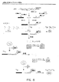
図4は、変異遺伝子のライブラリーの探索するための本明細書の方法の使用を示している。種々の方法による特定遺伝子領域の変異は、しばしば、組換え抗体の結合親和性を改善するエラープロンPCRまたは遺伝子シャッフリング変異誘発技術により生ずる変異遺伝子のような、変異遺伝子によりコードされるタンパク質の特性の改善に使用される。表面ディスプレイによる選択と結合するこの技術は、数オーダーのマグニチュード(magnitude)による抗体の結合親和性の改善のために使用される。変異はまた、酵素の触媒特性の改善のために使用される。本明細書の方法は、望ましい特性を有するタンパク質をコードする変異遺伝子をスクリーニングおよび同定する手段を提供する。
【0197】
最初に、種々の機能性ドメインを含むオリゴヌクレオチドのセットを遺伝子の3’末端に加え、遺伝子および配列(「エピトープ」ではEと称し、「ディバイダー」では、Dと称し、「共通」ではCと称する)の更なるセットにハイブリダイズするヌクレオチドの配列を含むプライマーを組込むことにより変異が生ずる。EDC配列は、核酸の機能によりそれぞれ定義される配列のセットを構成する。記載したように、E配列は、コレクション中の抗体により特異的に認識されるエピトープをコードする。それらは、変異させた遺伝子のコーディング配列とインフレーム(in−frame)で組込まれ、親タンパク質との融合として発現される。D配列は、エピトープの下流域の特定配列セットである。それらは、マスター群を「分割する」特定プライミン部位として役立つ。それらは、非コード配列であり得、発現化変異タンパク質の一部となる必要はない。C配列は、すべての遺伝子に対し「共通」な配列であり、すべての遺伝子鋳型の同時PCR増幅としての手段を提供する。前記したように、特定実施態様では、Dおよび/またはC配列は任意である。重要なことに、EおよびD配列は、生ずるDNA分子間にランダムに分布する。例えば、100E配列および100D配列を合わし、10,000(100×100=10,000)の特有のタグ化cDNA分子を作製する。同様に、1,000E配列および1,000D配列を合わし、1,000,000(1,000×1,000=1,000,000)の特有のタグ化cDNA分子を作製する。
【0198】
E、CおよびD配列を、変異誘発分子の末端に加える前、または後、遺伝子中の定義領域は、種々の標準的方法により変異誘発する。変異手順は、E、D、C配列中に変異を生じないようにする。変異誘発が完了した後、変異DNAを最初のPCR反応物セットに鋳型として加え、F1サブライブラリーを作製する。鋳型DNAに加えて、D、Cプライマーセットを、それぞれのPCRが種々DNA配列に相補的なプライマーを含むように、別々に加える。例えば、図4では、第二PCRチューブは、100D配列(D2)のうちの1つのみを含むD、Cプライマーを含む以外、チューブのレスト(rest)は同一である。この説明では、チューブ50は、100のD配列(D50)のうちの一種を含むこと以外、F1反応チューブのレストは、同一である。生ずるPCR増幅産物は、遺伝子間にランダムに分布する100種のE配列のすべてを含むが、100のD配列1つのみを含む。解説において、PCRチューブ50は、同じD50配列、同じC配列を有し、分子(ED50C)間にランダムに分布する異なるE配列を有するすべてのサブライブラリーDNA分子(F150)を生ずる。
【0199】
作製されたF1DNA分子は、転写−翻訳抽出を用いインビトロで発現する。プロモーター、リボゾーム結合部位、およびインビトロ転写および翻訳の実施のための当業者に既知の他の調節配列を含む適当な調節DNA配列を、タグ化工程の間にDNAフラグメント中に組み込む。図4に解説のように、F150DNA分子の発現により、異なるエピトープタグを含むタンパク質のコレクションを生ずる。細菌または他のインビボシステムで産生されるタンパク質もまた使用され得る。
【0200】
生ずる発現化タンパク質を、エピトープのそれぞれに選択的に結合する抗体とエピトープとの間で結合が可能な条件下、アレイ型のような抗体コレクションでインキュベートする。これは、抗体に対するタンパク質の特異的結合の結果である。抗体をアレイに配置するならば、これにより、タンパク質同系エピトープに結合する固定化抗体を含むアレイ上の位置にタグ化タンパク質を分布する結果となる。結合の後、アレイを洗浄し、プローブし、そしてルシフェラーゼを用いるような、例えば酵素学的標識により当業者に既知の任意の方法により分析する。例えば、分析は、フォトマルチプライヤーチューブのようなディテクター、光ダイオードアレイまたは好ましくは放出する光を検出する電荷結合素子(CCD)に基づくイメージングディテクターを用い光子コレクションにより実施し得る。光子は、局所的酵素学的化学ルミネセンス、特に生体ルミネセンス反応により生じ得る。光子コレクションが好ましい。比較的安価で、非常に感度がよく、その感度は、収集時間を増やすことにより増幅し得るからである。
【0201】
例として、探索を用い、活性の増加を供与するルシフェラーゼ酵素に対する変異を同定するならば、当該アレイは洗浄し、サブストレート中に浸し、次いで、光子の出力を増加することによる測定としてルシフェラーゼ活性の増加に関し分析する。アレイ中の「最も明るいスポット」は、最も好都合の変異を有する酵素と結合する。
【0202】
他の例として、探索を用い、抗原に対する抗体の親和性の増加を同定するならば、当該アレイを洗浄し、次いで、タグ化抗原でインキュベーションする。抗原をビオチン、または抗体−HRP複合体でタグ化するならば、タグが定義エピトープであるならば、抗原のタグを用い、ストレプアビジン結合HRPのような二次検出試薬に結合させる。再び、「最も明るいスポット」は、最も多い抗原量を結合する、最も高い親和性を有する変異抗体を含む。
【0203】
「最も明るいスポット」の位置およびそのスポットにおける抗体のエピトープ結合特異性を知るため、目的変異遺伝子と結合するE配列を同定する。ソートのこの時点で、目的の遺伝子の鋳型(図4に示すような)は、F150サブライブラリー中にあることおよびE23配列を含むことが判る(F150/F223)。
【0204】
E23配列を含む遺伝子は、F150サブライブラリーからの鋳型DNAおよびE23配列(FA23 E C)に相当する配列を有するPCRプライマーを用い増幅し得る。マスターライブラリーを最初に分けるために使用するD Cセットのプライマーのように、FA E Cセットのプライマーを用い、特異的E配列を含む鋳型を増幅させ、同時に増幅遺伝子中にE配列を再分布する。FA E Cプライマーは、3つの機能性領域から構成される。FA領域は、鋳型中に存在するE配列の上流フラグメント(Fragment A)に相当する配列を含む。FA領域は、ハイブリダイゼーション特異性を供与するが、翻訳においては、エピトープ結合特異性を供与しない任意量のE配列を含む。前の通り、E領域は、エピトープ配列をコードし、C領域は、増幅の共通領域をコードする。FAおよびE配列は遺伝子のコード領域とインフレーム(in−frame)である。生ずる増幅遺伝子は、F2サブライブラリー(F223)を示す。
【0205】
F2サブライブラリーからの増幅遺伝子をインビトロで発現させ、抗体アレイでインキュベーションし、再プローブ化し、分析する。前の通り、このアレイ中の「明るいスポット」は、目的変異遺伝子と結合するE配列を同定する。ソートにおけるこの点において、目的の遺伝子(図4に示すように)から、F150およびF223サブライブラリー中にあること、およびE45配列(F150/F223/F345)を含むことが判る。この情報から、E45配列(FB45 C)に特異的なプライマーを用い増幅し得る特定遺伝子が同定される。FB Cプライマーは、2つの機能性領域からなる。FB領域は、鋳型中に存在するE配列の下流フラグメント(Fragment B)に相当する配列を含む。FBは、Eのすべてまたは一部を含み得る;Cは任意である。FBは、ハイブリダイゼーション特異性を供与するEコード化配列までおよびすべてを含む任意の部分を含む。前の通り、C領域は、増幅のための共通配列をコードする。生ずる増幅化遺伝子は、F3サブライブラリー(F345)を表す。
【0206】
G.組換え抗体の同定
技術の他の適用は、組換え抗体の同定のための使用である。望ましい特性を有する抗体を組換え抗体遺伝子の巨大なプールから選別する。組換え抗体ライブラリーを構成するための標準的方法の概観を図5に示している。最初のステップには、脾臓細胞(spleenocyte)または末梢血リンパ球(PBL)から単離したmRNAから組換え抗体遺伝子をクローニングすることが含まれる。機能性抗体フラグメントは、可変重(VH)鎖および可変軽(VL)鎖遺伝子の遺伝学的クローニングおよび組換えにより作製され得る。VHおよびVL鎖遺伝子は、脾臓細胞またはPBLから単離したmRNAをcDNAに最初に逆転写することによりクローニングする。VHおよびVL鎖遺伝子の特異的増幅は、これらの遺伝子に隣接する共通領域に相当するPCRプライマーのセットで行う。VHおよびVL鎖遺伝子をリンカーDNA配列と合わせる。単一鎖抗体フラグメント(scFv)の典型的リンカー配列は、アミノ酸(Gly4Ser)3をコードする。VH−リンカー−VL遺伝子は、PCRでアセンブルされ、増幅され、その産物は、直接転写および翻訳され得、発現プラスミド中にクローニングされ、次いで、インビトロまたはインビボの何れかで発現され、機能性組換え抗体フラグメントを産生する。
【0207】
組換え抗体ライブラリー構成の方法は、本明細書のソーティング方法を用いる使用に適用され得る。これは、E D C配列を、VH鎖およびリンカー配列でアセンブルする前のVL鎖遺伝子中に組込むことにより、行う。組換え抗体ライブラリーをE D C配列でタグ化した後、F1サブライブラリーへ分け、その後、上記のようなアレイでスクリーニングすることによりソートする。
【0208】
2種の方法は、E D C配列の増幅VL鎖遺伝子中への組込みに関し、解説している。第一の方法では、E D C配列は、第一鎖cDNA合成プライマーの部分であり、第二の方法におけるcDNA合成(図6)の間に組込まれ、E D C配列は、二本鎖DNAリンカー分子の添加によりcDNA合成(図7)後に組込まれる。
【0209】
図6は、どのようにE D C配列が、プライマー組込みによりVL鎖遺伝子に置かれるのかを示している。VH鎖遺伝子は、標準的方法を用いクローニングする。脾臓細胞またはPBLから単離されるmRNAは、普遍的オリゴdTプライマーまたはIG遺伝子特異的プライマーを用いcDNAに変換される。VH遺伝子は、次いで、これらの遺伝子に隣接する共通配列に相補的なプライマーセットを用い特異的に増幅される。VHBACKプライマーはまた、アセンブル化遺伝子のインビトロ転写および翻訳に必要なプロモーター配列を含み、および/または以下に限らないが細菌、酵母、昆虫および哺乳類細胞のような細胞中でインビボ発現するためプラスミドベクター中にサブクローニングし得る。
【0210】
VL遺伝子は、VL遺伝子(Jkappa for)およびE D C配列に隣接する下流共通配列に相補的な配列のセットを含む逆転写プライマー(VLFOR)のセットを使用しクローニングする。EDC配列は、VLFORプライマー中の配列のために5’からJkappa向きに位置する。cDNAの第二鎖を、VL遺伝子(Vkappa back)の上流共通領域に対し相補的な配列を含むオリゴヌクレオチド(VLBACK)を使用しプライムする。第二鎖cDNA合成後、VL遺伝子をVLBACKおよびVLFOR−Cプライマーの組み合わせで増幅する。VLFOR−Cプライマーは、EDC配列のC領域に相補的な配列からなる。
【0211】
VHおよびVL遺伝子の増幅の後、当該フラグメントを、制限酵素で消化し、リンカーを有するオーバーラッピング末端を作る。VH−リンカーVLフラグメントを、DNAリガーゼでシールし、次いでVHBACKおよびVLFOR−Cプライマーを用い増幅する。
【0212】
図7で示す第二の方法では、VH遺伝子は、上記のように増幅する。この方法は、VL遺伝子第一鎖合成が、特有制限部位5’からJkappa for配列までを含むオリゴヌクレオチドでプライムする、最初とは異なる。この制限部位を、特有の結合力のある末端を制限酵素消化により生じ得るように、生ずるcDNAの3’−末端に組込む。当該リンカーを切断cDNAと混合し、リガーゼでシールし、次いで、VHBACKおよびVLFOR−Cプライマーの組合せで増幅させる。
【0213】
図8は、組換え抗体ライブラリーを探索するための方法を概説する。VHおよびVL遺伝子を上記のようにクローニングし、EDC配列を抗体遺伝子の3’末端に加え、マスターライブラリーを作製する。F1サブライブラリーを、DCセットのPCRプライマーを用い作製する。当該解説は、100のF1サブライブラリーを示し、F12、F150およびF199のDCプライマーを示し、F150反応からの増幅産物を示す。
【0214】
F150サブライブラリー遺伝子の転写および翻訳は、含まれているエピトープ(E配列)によりランダムにグループ化され得る、抗体のような種々の組換え捕捉試薬を作製する。発現タンパク質をアレイ上で浸し、特異的エピトープタグを結合する抗体を含むアレイ中のスポット上にソートし得る。サブライブラリーF150からのscFvをアレイに結合させた後、標識抗原をアレイ上で浸す。抗原における標識は、ストレプアビジン結合HRPのような第二検出試薬への結合に使用されるビオチンのような化学的タグであり得、または当該抗原をエピトープタグ化し得、検出は、抗エピトープ抗体HRP複合体で行い得る。結合後、当該アレイを洗浄し、プローブ化し、分析する。分析は、典型的には、CCDに基づくイメージングディテクターを用い光子コレクションにより行い、光子は、典型的には、局所性酵素学的化学ルミネセンス反応により生ずる。再び、「最も明るいスポット」が、最も多い量の抗原に結合する最も親和性のある組換え抗体を含む。
【0215】
「最も明るいスポット」の位置およびそのスポット中の抗体のエピトープ結合特異性を知るため、目的の組換え抗体と結合するE配列を同定する。ソートにおけるこの点で、目的の遺伝子の鋳型(図8に示すような)が、F150サブライブラリー中に位置し、E23配列を含むことがわかる。
【0216】
E23配列を含む遺伝子を、F150サブライブラリーからの鋳型DNAおよびE23配列(FA23 E C)に相当する配列を有するPCRプライマーを用い、増幅し得る。マスターライブラリーを最初に分けるために使用するD Cセットのプライマーのように、FA E Cセットのプライマーを、特異的E配列を含む増幅鋳型に使用し、同時に増幅遺伝子中のE配列を再分布する。FA23ECプライマーを用い、F150サブライブラリー由来の鋳型DNAを増幅する。生ずる増幅遺伝子は、F2サブライブラリー、F223を示す。目的の抗体の最初の整列(lineage)はF150/F223である。
【0217】
F2サブライブラリーからの増幅遺伝子は、インビトロまたはインビボシステム中で発現し、抗体アレイと共にインキュベーションし、再プローブ化し、分析する。前の通り、このアレイ中の「明るいスポット」は、目的の組換え抗体遺伝子と結合するE配列を同定する。ソートのこの点において、目的の遺伝子(図8に解説するような)は、F150およびF223サブライブラリー中にあり、E45配列(F150/F223/F345)を含むことが判る。この情報は、E45配列(FB45C)に特異的なプライマーを用い増幅し得る特定遺伝子を同定する。この生ずる増幅遺伝子は、単一型の組換え抗体を含むF3サブライブラリー(F34577)を示す。
【0218】
H.結合抗原の検出
結合ポリペプチドタグ化分子は、当業者に既知の任意の適当な方法により検出され得、それは、標的分子の機能を有する。例示の検出方法は、ホースラディッシュペルオキシダーゼ(HRP)システムおよびルシフェリン/ルシフェラーゼシステム、アルカリホスファターゼ(AP)、標識抗体、フルオロホアおよびアイソトープのような化学ルミネセンスおよび生体ルミネセンス生成試薬の使用を含む。これらは、フィルム、光子コレクション、スキャンニングレーザー、ウエーブガイド、エリプソメトリー、CCDおよび他のイメージング手段を用い検出し得る。
【0219】
記載の通り、アドレス可能抗タグ捕捉試薬コレクションの使用には、以下に限らないが、単一抗原または複数抗原を見つけることを含む(これらに限らない)scFVを同定する組換え抗体scFvライブラリーの探索;タグ化変異ライブラリーを含む変異ライブラリーの探索;エラープロンPCRによる変異;小分子バインダーの探索、抗体親和性増加の探索、遺伝子特性の促進の探索(AP、HRP、ルシフェラーゼ、GFP)のための遺伝子シャッフリングによる変異;配列特異的DNA結合タンパク質の探索;タンパク質−タンパク質相互作用のためのcDNAライブラリーの探索;および任意の他のそのような応用が含まれる。
【0220】
実施例
以下の実施例は、解説目的のためだけに含まれるものであり、本発明の範囲を限定することを目的とするものではない。
【0221】
実施例1
抗タグ抗体コレクションの調製
A.抗体−タグ対のコレクションの作成
ペプチドタグに結合する抗体のコレクションを使用し、タグに結合する分子をソートする。ポリペプチドタグに特異的に結合する抗体のコレクションは、種々の方法により生じ得る。2例を以下に述べる。
【0222】
1.ハイブリドーマスクリーニング
第一の例では、アレイに対し高親和性および高特異性の抗体を、ペプチドエピトープのランダムコレクションを発現するファージディスプレイライブラリーに対し個々のハイブリドーマ細胞のランダム選択化コレクションをスクリーニングすることにより、同定する。ハイブリドーマ細胞は、骨髄腫細胞と、天然(非免疫化)マウスから単離された脾臓細胞(spleenocyte)との融合により作製する。適当な培養を行った後、おおよそ10−30,000の個々の細胞クローン(モノクローナル)が単離され、96ウェルプレートに別々に増殖させる。このコレクションからの培養上清を、有意な量の抗体を分泌する培養物を同定するための抗IgG抗体を用いるELISAによりスクリーニングする。抗体産生が低い培養は中止する。このモノクローナルコレクション由来の抗体を、ハイスループット96ウェル精製方法を用い培養上清から別々に親和性精製し、当該量を精製し、定量した。
【0223】
精製抗体を、フィルター上へのロボットスポッティングによりアレイし、また、次いで、常磁性ビーズに結合させて別々に混合し、ランダムシステイン束縛七量体アミノ酸配列(random cysteine−constrained heptameric amino acid sequence)をディスプレイする繊維状M13バクテリオファージライブラリーから高親和性エピトープをパンニングするサブストレートを作成する。当該ファージライブラリーを、ファージライブラリーを抗体被覆ビーズと混合し、ビーズからの遊離性結合ファージ(loosely−bound phage)を洗浄することにより高親和性エピトープをディスプレイするファージとして富化する(パンニング)。数ラウンドのパンニングにより、コレクション中に存在するモノクローナル抗体に強固に結合するファージを含むライブラリーが高富化される。高親和性ファージ抗体対を分離および同定するため、富化ファージライブラリーを、高緊縮結合条件下でアレイ化抗体を含むフィルターでインキュベーションする。フィルターにおける抗体に結合するファージを、HRP結合抗ファージ抗体および化学ルミネセンスサブストレートで染色し、ルミネセンスシグナルを生じさせることにより同定する。当該シグナルは、ハイレゾリューションCCDカメライメージング装置を用いを定量する。高親和性結合ファージを、フィルターから回収し、増殖させる。個々のスポットから回収した幾つかの独立ファージクローンを配列決定し、相当する抗体として共通高親和性エピトープを同定する。
【0224】
a.ハイブリドーマの作成
ハイブリドーマ細胞は、当業者に既知の方法(例えば、Harlow et al. (1988) Antibodies: A Laboratory Manual, Cold Spring Harbor Laboratory, Cold Spring Harbor)により調製する。ハイブリドーマ細胞は、マウス脾臓細胞(spleenocyte)とマウス骨髄腫細胞の融合により作製する。融合の場合、非免疫化マウスの脾臓から単離した抗体産生細胞を、骨髄腫細胞と混合し、融合する。他に、ハイブリドーマ細胞は、異なるエピトープタグの混合物を含む組換えタンパク質(例えば、ジヒドロフォレートレダクターゼ、DHFR)で事前に免疫化したマウスから単離した脾臓細胞(spleenocyte)から作製し、担体に結合する(すなわち、Keyhole limpet hemocyanin、KLH)。当該エピトープタグは、DHFR遺伝子との遺伝学的融合物の一部として発現するランダムシステイン束縛化(constraine)ペプチドである。当該ランダムペプチドは、合成変性オリゴヌクレオチドからアセンブルされるDNA挿入物によりコードされており、繊維状バクテリオファージM13の遺伝子IIIタンパク質(gIII)中へクローニングする。ペプチドライブラリーをコードするDNAは、商業的に利用可能である(Ph.D.−C7CTM Disulfide Constrained Peptide Library Kit, New England Biolabs)。Ph.D.−C7CTMライブラリーはおおよそ3.7×10⁹種のペプチドを含む。
【0225】
融合後、細胞を選択培地中に希釈し、マルチウェル組織培養ディッシュ中にプレートする。マウス骨髄腫細胞の自然で(healthy)、迅速な分割培養物を、20%胎児ウシ血清(FBS)および2×OPIを含む培地20ml中に希釈する。培地は、典型的にDulbecco’s修飾Eagle’s(DME)またはRPMI1640培地である。培地の成分は既知である(例えば、Harlow et al. (1988) Antibodies: A Laboratory Manual, Cold Spring Harbor Laboratory, Cold Spring Harbor)。抗体産生細胞は、マウスからの脾臓の無菌切除および細胞中への脾臓破壊、および2×OPI培地での洗浄による巨大組織の切除によって、調製される。典型的なマウス脾臓は、おおよそ5×10⁷から2×10⁸リンパ球を含む。調製されるハイブリドーマが任意の抗原に対する免疫によって富化されることはないため、1を超えるマウスからの脾臓を使用し、細胞を混合する。当数の脾臓細胞および骨髄腫細胞を遠心分離(400×gで5分間)でペレットとし、当該ペレットを、血清のない培地5mlに別々に再懸濁し、次いで合わせる。43%ポリエチレングリコール(PEG)溶液を加え、0.84%とする。当該細胞をPEG含有培地中にゆっくりと再懸濁し、次いで、400×gで5分間の遠心分離により再ペレット化し、20%FBSを含む培地5ml中の再懸濁液により洗浄し、再ペレットし、20%FBS、1×OPIおよび1×AH(AHは選択培地である;1×AHは5.8μMアザセリンおよび0.1mMヒポキサンチン)の上清培地で2回洗浄する。細胞を37℃でCO2インキュベーター中でインキュベーションする。クローンは4日後、顕微鏡で見ることができる。
【0226】
b.ハイブリドーマ細胞の単離
安定ハイブリドーマを、乏しい培地(poor medium)中で数日間増殖させることにより選択する。次いで、培地を新しい培地と置換え、単一ハイブリドーマを制限希釈クローニングにより単離する。ハイブリドーマ細胞は非常に低いプレーティング効率を有するため、単一細胞クローニングは、フィーダー細胞またはコンディション化培地の存在下、行う。新しく単離した脾臓細胞を、通常組織培地条件下では増殖せず、ハイブリドーマ細胞の拡大の間に喪失するフィーダー細胞と同様に使用し得る。この手順では、脾臓は、外観的にマウスから取除き、破壊する。放出細胞を、10%FBSを含む培地中で繰り返し洗浄する。脾臓は、典型的に、106細胞/mlで100ml生ずる。当該フィーダー細胞を96ウェルプレートにウェルあたり50μlでプレートし、24時間増殖させた。自然のハイブリドーマ細胞を、20%FBS、2×OPIを含む培地中に、mlあたり20細胞の濃度に希釈する。細胞は、できるだけ遊離のクランプとなる。希釈化ハイブリドーマ細胞50μlをフィーダー細胞に加え、最終体積100μlとする。クローンは、4日で現れ始める。他に、単一細胞は、単一細胞を個々にピペッティングすることによる単一細胞ピッキングにより単離し得る。単一細胞はまた、軟寒天中での増殖により得られ得る。一旦、自然な安定な培養が行われると、当該細胞は、10%FBSで上清化したDME(またはRPMI1640)培地中での増殖により維持される。安定な細胞を、ジメチルスルホキシド(DMSO)のような凍結防止剤を含む培地中でゆっくりと凍結させることにより液体窒素中で保存し得る。細胞により生ずる特定量の抗体を、ELISA法により培地上清中の抗体量を測定することにより決定する。
【0227】
2.ハイブリドーマ培養上清からの抗体精製
個々の培養上清からの抗体精製を親和性結合により行う。親和性結合サブストレートの数が利用可能である。下記の手順は、固定化プロテインL(Pierce)を含む商業的に利用可能なサブストレートに基づいており、製造者指示手順に従っている。端的に、培養上清を1:1で結合緩衝液(0.1M ホスフェート、0.15M 塩化ナトリウム(NaCl)、pH7.2)で希釈し、0.2mlまでの希釈サンプルを、結合緩衝液を事前に装備したReacti−BindTM Protein L Coated plate(Pierce)に適用する。ウェルを3×0.2ml結合緩衝液で洗浄する。2×0.1mlの溶出緩衝液(0.1M グリシン、pH2.8)で結合抗体を溶出させ、1M Tris、pH7.5、20μlと合わせる。96ウェルフィルタープレート(Nalge Nunc)と組合せてSephadex G−25ゲル濾過を用い精製抗体を脱塩する。
【0228】
ファージパンニングサブストレートを作製するため、上記のように別々に精製した抗体をあわせ得る。他に、精製抗体混合物は、プール化培養上清からバッチ精製により得られ得る。乏しい培地上清からの抗体の精製はまた、親和性結合により行う。多数の親和性結合サブストレートが利用可能である。下記の手順は、固定化プロテインL(Pierce)を含む商業的に利用可能なサブストレートに基づいており、製造者が指示する手順に従う。端的に、培養上清を結合緩衝液で1:1に希釈し、4mlまでの希釈サンプルを、結合緩衝液で事前に装備したAffinity PackTM Immobilized Protein L Column (Pierce)に適用する。結合緩衝液20mlでカラムを洗浄し、または250nmの吸収がバックグラウンドに帰する(return)まで洗浄する。結合抗体を6−10mlの溶出緩衝液で溶出し、1M Tris、pH7.5を含む1ml画分中に回収する。吸収280nmおよび乏しい適当画分により結合タンパク質の放出をモニターする。ExcelluloseTM Desalting Column(Pierce)を用い精製抗体を脱塩する。
【0229】
3.フィルター上への抗体のアレイイング
個々のハイブリドーマ培養物から精製する抗体を、自動化アレイイングツール(PixSys NQナノリッター分配ワークステーション、Cartesian Technologies; BioChip Arrayer; Packard Instrument Company; Total Array System; BioRobotics; Affymetrix 417 Arrayer; Affymetrix)を用い、1μg−1mg/ml濃度で0.1M PBS(リン酸緩衝食塩水)、pH7.4の緩衝液中、1μlを膜(UltraBind膜、Pall Gelman;FASTニトロセルロース被覆スライド、Schleicher & Schuell)上にスポットする。当該スポットを1−2分間、風乾する。UltraBind膜は、第一級アミンと反応する活性化アルデヒド基を含み、膜と抗体との間で共有結合を形成する。非反応アルデヒドは、30分間、50mM PBS、pH7.4、2%ウシ血清アルブミン(BSA)の溶液でインキュベーションすることによりブロックされる。当該フィルターは、50mM PBSでリンスし得、次いで完全に風乾させる。
【0230】
4.常磁性ビーズにおけるファージディスプレイライブラリーのパンニング
繊維状バクテリオファージM13の遺伝子IIIタンパク質(gIII)に対するN末端遺伝学的融合の一部として発現するランダムシステイン束縛化ペプチドを含むファージライブラリーは、本質的に記載されているように構成される(Kay et al. (1996) Phage Display of Peptides and Proteins: A Laboratory Manual, Academic Press, San Diego)。ランダムペプチドは、合成変性オリゴヌクレオチドからアセンブルされるDNA挿入物によりコードされており、gIIIにクローニングされる。これらのライブラリーは、商業的に利用可能である(Ph.D.−C7CTM Disulfide Constrained Peptide Library Kit, New England Biolabs)。Ph.D.−C7CTMライブラリーは、おおよそ3.7×10⁹独立クローンを含む。
【0231】
Ph.D.−C7CTMライブラリーからの2×10¹¹ファージビリオンを、マウスIgGのFcドメイン(Dynal; このモノクローナルはヒト抗体に結合しない)に特異的な精製抗体300μgおよびヒトIgG4モノクローナル抗体と合わせ、TBST(50mM Tris−HCl(pH7.4)、150mM NaCl、0.1%Tween−20)で最終体積0.2mlとする。抗体の最終濃度はおおよそ10nMである。室温で20分間インキュベーションする。
【0232】
ファージ抗体溶液をDynabeads Pan Mouse IgG(Dynal)と合わせる。当該ビーズを、PBS、pH7.4、0.1%BSA、0.02%アジド化ナトリウム中の懸濁液として供給する。ビーズを、ファージと混合する前に数回、TBS(50mM Tris−HCl(pH7.4)、150mM NaCl)で洗浄する。当該ビーズは磁石(Magnetic Particle Concentrator, Dynal)の適用により溶液から分離する。ファージ抗体溶液を加え、0.1μg/107ビーズの濃度とし、ゆっくりとチルティングおよびローテーションしつつ4℃で30分間インキュベーションする。ヒト抗体の包含により、Dynabeadsに固定化したヒト抗体に結合するファージの選択を妨げる。加えて、ブロッカーとしての溶解ヒト細胞からのヒトタンパク質の包含は、ヒト細胞中にも存在するファージエピトープの選択を妨げる。当該選択抗体ファージ対は、試験するサンプル中に天然に存在するタンパク質と拮抗しない。
【0233】
当該方法の次のステップで、磁石を使用し液体から取除き、TBST1mlの洗浄緩衝液中にビーズを懸濁する。洗浄ステップを10回繰り返す。最後の洗浄ステップ後、0.2M グリシンHCl、pH2.2、1mg/mlBSA(1ml)中にビーズを懸濁し、液体回収前に室温で10分間インキュベーションすることにより、捕捉ファージを溶出する。回収液体のpHは1M Tris、pH9.1(0.15ml)を加えることにより直ちに中性化する。溶出液の少量のアリコートを、LB−Tetプレート上でER2738大腸菌(E. coli)を伝染させることにより力価する。
【0234】
ER2738 E. coliのmid−log培養物20mlの添加により溶出液を増幅し、4.5時間LB−Tet中で増殖させる。10,000rpm、10分間の遠心分離によりE. coli細胞からファージビリオンを分離し、新しいチューブに移す。繰り返して、80%を超える上清を新しいチューブに移す。1/6体積のPEG/NaCl(20% w/vポリエチレングリコール−8000、2.5M NaCl)を添加し、その後、一夜4℃で沈殿させることにより濃縮する。当該ファージを10,000rpmで15分間遠心分離することにより回収し、当該ペレットをTBS1ml中に再懸濁する。微小遠心分離チューブ中でPEG/NaClを用いファージを再沈殿し、当該ペレットをTBS0.2ml、0.02%アジド化ナトリウム中に再懸濁した。微小遠心分離を1分間行い、任意の残りの物質を取除く。上清は増幅化溶出液である。増幅化溶出液を力価し、上記のように3回パンニングを繰り返す。個々のラウンドのパンニングおよび増幅により、ファージのプールは、抗体に結合するファージとして富化する。インプットとして使用するファージの濃度を一定に保つならば、回収されるファージの数は増大する。ファージは、4℃で保存するか、滅菌グリセロールで1:1に希釈し−20℃で保存する。
【0235】
5.ファージによる抗体アレイの染色
個々の培養上清から調製するアレイ化抗体を含むフィルターを富化ファージライブラリーでプローブする。この方法は、標準的ウェスタンブロッティングまたはドットブロッティング手順と類似する。端的に、ブロック化フィルターは、TBST、pH7.4、0.1%v/v Tween−20、1mg/ml BSA中で再水和し、1時間4℃でインキュベーションする。ファージを加え、濃度2×10¹¹ファージ/mlとし、室温で30分間フィルターと共にインキュベーションする。ハイブリダイゼーション溶液を回収し、当該フィルターをブロッキング溶液(TBST、pH7.4、0.1% v/v Tween−20、1mg/ml BSAおよびヒト細胞由来可溶性タンパク質)で大量に洗浄する。ブロッキング溶液に、1mg/mlストック濃度から、ブロッキング緩衝液中で1:100,000から1:500,000希釈したHRP結合抗M13抗体を加え、1時間ゆっくりと振盪しつつインキュベーションする。少なくとも4から6回、TBSTで膜を洗浄する。当該ブロットを、SuperSignal West Femto Substrate Working Solution (Pierce)中に5分間、完全に濡らす。当該フィルターを、オートラジオグラフィーフィルム(Kodak)にさらすことによりイメージし得るか、またはホスホイメージャー(BioRad)のようなイメージング装置または電荷結合素子(CCD)カメラ(Alphalnnotech; Kodak)のようなイメージング装置を用いイメージ化し得る。
【0236】
6.フィルターからのファージの回収およびエピトープの配列決定
ファージは、イメージングから同定されたファージを含むスポットを切除することによりフィルターから回収され得る。ファージは、0.2M グリシン−HCl、pH2.2、1mg/ml BSA(0.5ml)中のフィルター切片を再懸濁し、液体を回収する前に10分間室温でインキュベーションすることにより、フィルターから溶出させる。回収液のpHは、1M Tris、pH9.1(0.075ml)を加えることにより直ちに中性化される。溶出液の少量のアリコートを、LB−Tetプレート上でER2738 E. coliを伝染させることにより力価する。単離プラーク(典型的に10プラーク)をDNA単離のためにピックし、共通エピトープの定義のために配列決定する。プラークは、滅菌ピペットチップまたは爪楊枝を用い自然なmid−log培養物から新しく1:100に希釈したER2738 E. coli細胞の培養物1mlを接種し、振盪しつつ4から5時間37℃でインキュベーションすることにより増幅する。ファージは、30秒間微小遠心分離により回収し、新しいチューブに上清0.5mlを移し、PEG/NaCl0.2mlを加え、ゆっくりと10分間混合した後、室温で静置し得る。ファージを、遠心分離の最高速度で10分間、微小遠心管中で遠心分離することによりファージをペレットとする。任意の残りの上清を廃棄し、当該ペレットをイオジン緩衝液0.1mlおよびエタノール0.25ml中に完全に懸濁し、単一鎖DNAを沈殿させる。DNAペレットを70%エタノールで洗浄し、風乾する。DNAを標準的方法で配列決定する。
【0237】
B.選択的感染
ファージディスプレイのような選択的感染技術を用い、タンパク質−タンパク質対の相互作用を同定する。これらシステムは、細菌と感染ウイルス(ファージ)との間の感染プロセスを仲介する、タンパク質−タンパク質相互作用の必要性に有利となる。繊維状M13ファージは、細菌のF線毛に最初に結合することによりE. coliに通常感染する。当該ウイルスは、traA遺伝子によりコードされるF線毛タンパク質の個別領域における線毛に結合する。この結合は、ファージのチップにおけるマイナーコートタンパク質(プロテイン3)により仲介される。F線毛タンパク質におけるファージ結合部位(traA遺伝子における13アミノ酸配列)を工作し、ファージ結合部位のランダム混合物を発現する細菌の巨大な群を作製する。
【0238】
ファージコートタンパク質(プロテイン3)をまた、工作し、別種の単一鎖抗体構造のライブラリーを示す。細菌の感染およびウイルスのインターナリゼーションは、そのため、適当な抗体ペプチドエピトープ相互作用により仲介する。細菌およびウイルスDNAに適当な抗生物質耐性マーカーを置くことにより、個々のコロニーは、抗体およびそれに相当するペプチドエピトープの両方の遺伝子を含むように選択し得る。組換え抗体ファージは、非免疫化マウスから調製したライブラリーをディスプレイし、traA遺伝子中のファージ結合部位中にランダムペプチド配列を含む細菌株は商業的に利用可能である(Biolnvent, Lund, Sweden)。組換え抗体ライブラリーの作製を以下に記載する。
【0239】
C.抗体の発現および精製
ハイブリドーマ上清からの抗体の精製は、親和性結合により行う。多くの親和性結合サブストレートが利用可能である。下記の手順は、固定化タンパク質L(Pierce)を含む商業的に利用可能なサブストレートに基き、製造者が指示する手順に従う。端的に、培養上清を結合緩衝液(0.1M ホスフェート、0.15M塩化ナトリム(NaCl)、pH7.2)で1:1に希釈し、4mlまでの希釈サンプルを、結合緩衝液を事前に装備したAffinity PackTM Immobilized Protein L Column(Pierce)に適用する。カラムを結合緩衝液20mlで洗浄し、または250nmの吸収がバックグラウンドに帰する(return)まで洗浄する。結合抗体を6−10mlの溶出緩衝液(0.1M グリシン、pH2.8)で溶出し、1M Tris、pH7.5(100μl)を含む1ml画分中に回収する。吸収280nmおよび乏しい適当画分により結合タンパク質の放出をモニターする。ExcelluloseTM Desalting Column(Pierce)を用い精製抗体を脱塩する。当該精製を適当にスケールし得る。他に、抗体は、プロテインA(またはプロテインG)、HiTrapカラム(Amersham Pharmacia)およびFPLCクロマトグラフシステム(Amersham Pharmacia)を使用する親和性クロマトグラフィーにより精製し得る。製造者が指示するプロトコールに従う。
【0240】
組換え抗体は、発現し、記載のように精製する(McCafferty et al. (1996) Antibody engineering: A practical Approach, Oxford University Press, Oxford)。端的に、組換え抗体をコードする遺伝子を誘導可能プロモーターを含む発現プラスミド中にクローニングする。活性組換え抗体の産生は、多数の分子内ジスルフィド結合の形成に依存する。細菌細胞質の環境は縮小し、そのため、ジスルフィド結合形成が妨げられる。この問題の1つの解決は、分泌シグナルペプチドを、ペリプラズムの非縮小環境への輸送を指示する抗体に遺伝学的に融合することである(Hanes et al. (1997) Proc. Natl. Acad. Sci. U.S.A. 94: 4937−4942)。
【0241】
他に、抗体は、不溶性のインクルージョンボディーとして発現し、ジスルフィド結合の形成を促進する条件下、インビトロでリフォールディングし得る。適当な抗生物質を含むLB培地0.5Lに接種し、32℃で10時間振盪する。産生培地(リッターあたり、3g硫酸アンモニウム、2.5gリン酸カルシウム、30gカゼイン、0.25g硫酸マグネシウム、0.1mg塩化カルシウム、10ml M−63塩濃度、0.2ml MAZU 204 Antifoam(Mazer Chemicals)、30gグルコース、0.1mgビオチン、1mgニコチンアミド、適当な抗生物質、pH7.4)9.5リッターを接種するためにスターター培地を使用する。pH7.2でChemap(など)ファーメンターを使用し、発酵させ、分あたり培地に対し1:1v/vでエアレーションし、32℃で800rpmで振盪する。600nmでの吸収が18−20に到達したとき、42℃で1時間加熱し、次いで10分間10℃に冷却し、その後、細胞ペーストを7,000×gで10分間遠心分離することにより細胞ペーストを回収する。回収は、典型的に、10リッター発酵物から200−300gウェット細胞ペーストであり、凍結を維持する。
【0242】
組換え抗体を、2.5リッター細胞溶解緩衝液(50mM Tris−HCl、pH8.0、1.0mM EDTA、100mM KCl、0.1mM フェニルメチルスルホニルフロリド;PMSF)中に再懸濁することにより、融解細胞ペースから可溶性にし、4℃で維持する。再懸濁細胞をManton−Gaulin細胞ホモジナイザーに3回通過させ、次いで、24,300×gで30分間、6℃で遠心分離により不溶性抗体を回収する。当該ペレットを細胞溶解緩衝液1.2リッター中に再懸濁し、ホモジナイゼーションおよび回収を上記のように5回繰り返す。洗浄したペレットを凍結保存し得る。組換え抗体を、細胞ペレットのグラムあたり変性緩衝液(6M 塩酸グアニジン、50mM Tris−HCl、pH8.0、10mM 塩化カルシウム、50mM 塩化カリウム)6ml中に再可溶化することにより、復元する。24,300×g、45分間、6℃での遠心分離から上清を、変性緩衝液で280nmで光学密度25に希釈し、そして冷(4−10℃)リフォルディング緩衝液(50mM Tris−HCl、pH8.0、10mM 塩化カルシウム、50mM 塩化カリウム、0.1mM PMSF)中にゆっくりと希釈し、1:10希釈を2時間に渡り行う。当該溶液は、少なくとも20時間4℃で静置し、その後、0.45μm微小孔膜を通し濾過する。次いで、当該濾液は、約500mlに濃縮し、その後、HPLCを使用し最終精製する。
【0243】
濾液は、伝導率がHPLC緩衝液AのものにマッチするまでHPLC緩衝液A(60mM MOPS、0.5mM 酢酸カルシウム、pH6.5)で透析する。透析サンプル(60mgまで)を21.5mm×150mmポリアスパラギン酸PolyCATカラムに負荷し、HPLC緩衝液Aで平衡化し、HPLC緩衝液AとB(HPLC緩衝液Bは、60mM MOPS、0.5mM 酢酸カルシウム、pH7.5)との間の50分間直線状勾配でカラムから溶出させる。残りのタンパク質はHPLC緩衝液C(60mM MOPS、100mM酢酸カルシウム、pH7.5)で溶出させる。回収画分をSDS−PAGEで分析する。
【0244】
D.エピトープタグによるタンパク質の捕捉およびその検出のための例示アレイおよびその使用
実施例6にも記載の通り、本明細書の方法の機能化を解説するため、例えば、ヒトインフルエンザウイルス赤血球凝集素(HA)タンパク質エピトープのような異なるペプチドエピトープに対し特異的な捕捉抗体(アミノ酸配列YPYDVPDYAを有する)を、タグに、例えばscFvに使用する。例えば、ヒトフィブロネクチン(HFN)に対する抗原特異性を有するscFvをHAエピトープでタグ化し、そのため、HAペプチドに特異的な抗体により認識され、HFNの抗原特異性を有する分子(HA−HFN)を作製する。
【0245】
ニトロセルロース膜のような膜上に抗HAタグ捕捉抗体を含む、捕捉抗体を置いた(deposit)後、それらを室温および適当な湿度で適当な時間乾燥させる(例えば、10分間から3時間であり、それは、経験的に決定され得る)。乾燥後、置いた、乾燥した抗HA捕捉抗体で膜をブロックし、必要ならば、最終濃度0.05%(vol:vol)となるように加えるTween−20(ポリオキシエチレンソルビタンモノラウレート;Sigma)を有するリン酸緩衝食塩水(PBS)中に1×となるまで希釈した、Blocker BSA(Pierce)のようなタンパク質含有溶液でブロックし、膜に非特異的に結合するタンパク質により生ずるバックグラウンドシグナルを除く。本明細書に含まれる後の記載に関し、BBSA−Tとしてブロッキング試薬が言及され、0.05%(vol:vol)Tween−20を有するPBSをPBS−Tとして言及する。ブロッキング時間は、例えば、30分間から3時間の間で変化し得る。この手順の下記のその後の全インキュベーション(洗浄を除く)の場合、インキュベーション時間は、約20分間から2時間の間で変化する。同様に、インキュベーション温度は、室温から約37℃の間で変化し得る。すべての場合において、正確な条件が経験的に決定され得る。
【0246】
置かれた抗HA捕捉抗体を含む膜のブロッキング後、ペプチドエピトープタグ化scFvを伴うインキュベーションを行い得る。精製scFv(または、誘導によるscFvの発現を目的とするプラスミド構成物を保持するE. coli培養物からのscFvの精製の間に得られる細菌性培養物上清、または異なる粗亜細胞性画分)はBBSA−T中で種々の濃度(例えば、0.1と100μg/mlとの間)に希釈され得る。次いで、置いた抗ペプチドタグ捕捉抗体を有する膜を、このHA−HFN scFv抗原溶液と共にインキュベーションする。置いた抗HA捕捉抗体および結合HA−HFN scFv抗原を有する膜を、1またはそれより多い回数(例えば、3回)、PBSTで、適当な時間(例えば、洗浄あたり3−5分間)、種々の温度で洗浄する。
【0247】
置いた抗HA捕捉抗体および結合HA−HFN scFcv抗原を伴う膜を、PBS−Tで複数回(典型的には3回)、適当な時間(例えば、典型的には洗浄あたり3から5分間)、種々の温度で洗浄する。置いた抗HA捕捉抗体および結合HA−HFN scFvを有する膜を、証明目的のために、ビオチン化ヒトフィブロネクチン(Bio−HFN)(捕捉HA−HFN scFvにより認識される抗原である)と共にインキュベーションする。Bio−HFNを連続的にBBSA−Tで希釈する(例えば、1から10μg/ml)。生ずる膜を適当な回数(典型的には3)PBS−Tで、適当な時間(典型的には、洗浄あたり3から5分間)、種々の温度で洗浄し、次いで、連続的に希釈した(例えば、BBSA−T中1:1000から1:100,000)Neutravidin・HRPO(Pierce)と共にインキュベーションする。生ずる膜を前の通り洗浄し、PBSでリンスし、SupersignalTM ELISA Femto Stable Peroxide SolutionおよびSupersignalTM ELISA Femto Lumino Enhancer Solution (Pierce)で現像(develope)し、次いで、例えば、Kodak Image Station 440CFまたは他のイメージングシステムのようなイメージングシステムを用いイメージする。1:1のペルオキシド溶液:ルミノール混合物を調製し、少量をイメージステーションのプラテンにプレートする。
【0248】
次いで、膜を、アレイサイドを下にプラテンの中央に置き、膜の抗体含有部分の表面領域をカメラレンズのイメージング範囲の中央に位置させる。この方法で、プラテンに存在する少量のディベロッパーは、スライドの抗体含有部分の表面エリア全体に接触し得る。当該イメージステーションカバーを、抗体アレイイメージ捕捉のため閉じる。カメラフォーカス(ズーム)は、イメージする膜の大きさに依存して変化する。露光時間は、現像膜から発出するシグナル強度(明るさ)に依存して変化し得る。カメラf−ストップセッティングは、1.2と16との間で無制限に調節し得る。
【0249】
アレイイメージのアーカイビングおよび分析は、例えば、Kodak ID3.5.2ソフトウェアパッケージを用い行い得る。目的の領域(ROI)を、捕捉抗体のグループをフレームするソフトウェアを用い描く(アレイ上の既知の位置でプリントする)。正味の、合計の、最小値の、最大値のおよび平均の強度を表す、ROI値数、同様に標準偏差およびROIピクセルエリアは、例えば、ソフトウェアにより自動的に算出される。これらのデータを、例えば、Microsoft Excelに、統計分析のために変換する。
【0250】
実施例2
タグ化cDNAライブラリーの調製およびプライマーの調製
ターゲットに対する抗体のアレイを、ソーティング装置として使用する。cDNAライブラリー由来のタンパク質をアレイの表面上で浸し、ライブラリータンパク質と遺伝学的に融合するペプチドエピトープを特異的認識し結合する抗体を含むスポットに結合する。このシステムの鍵は、比較的多数の遺伝子(おおよそ106から109)上の比較的小数のタグ(おおよそ1,000)にランダムに結合し均等に分布する能力である。タグの、ライブラリー中の遺伝子間での均等な分布を確実とするため、タグは、PCR増幅前に遺伝子中に組込まれる。これらの仕事を行うため、種々の方法を本明細書に記載する。
【0251】
cDNAライブラリー作成のため、メッセンジャーRNA(mRNA)を細胞から最初に単離し、次いで、第二ステップでDNAに変換する。第一ステップでは、酵素RNA依存DNAポリメラーゼ(逆転写酵素;RTase)を用いRNA:DNA二重鎖分子生ずる。次いでRNA鎖を、DNA依存DNAポリメラーゼ(DNAポリメラーゼまたはKlenow fragmentのようなポリメラーゼのフラグメント)を用い新規に合成したDNA鎖により、置換する。
【0252】
ある方法は、タグのためのDNA配列を含む第一鎖cDNA合成のためのプライマーのコレクションの使用に依存する。この場合、プライマーは単一鎖オリゴヌクレオチドであり、タグは組込まれ、その後、第二鎖cDNAを合成する。第二鎖cDNA合成の後、生ずる分子をPCRで増幅する。他の方法では、DNA:DNA二重分子は、定義されたヌクレオチドオーバーハングを生ずるよう切断される新規物質の3’末端における特有制限酵素切断を組込むプライマーを使用し、作製する。リンカーDNA分子のコレクションは、相補的オーバーハングを含み、タグのためのDNA配列を、cDNAライブラリーのDNA分子上にライゲーションし、次いでPCR増幅する。第二の方法では、リンカーは二本鎖分子であり、タグは、第二鎖cDNA合成後、組込まれる。何れの方法も、プライマーまたはリンカーの何れかとして分子の巨大多様性コレクションの生成に依存する。これらの分子の調製を以下に記載する。
【0253】
A.方法I:プライマー伸長
ライブラリー構成は、mRNAの単離から始まる。mRNAの直接の単離を、オリゴdTセルロースを用い親和性精製により行う。この方法のための試薬を含むキットは多数の供給者(Invitrogen、Stratagene、Clonetech、Ambion、Promega、Pharmacia)から商業的に利用可能であり、製造者が指示する方法に従い単離する。加えて、多数組織から単離したmRNAはまた、これらの供給者から直接得られ得る。
【0254】
cDNAライブラリー構成物は、本質的に記載のように行う(Sambrook et al. (1989) Molecular Cloning: A Laboratory Manual, 2nd ed. Cold Spring Harbor Laboratory Press)。第一鎖合成は、以下のものを4℃で最終体積50μlで混合することにより行う;10μg mRNA(poly(A)+RNA)、10μg VLFOR−共通プライマーミックス(VLFOR−共通は以下に記載する)、50mM Tris−HCl、pH7.6、70mM 塩化カリウム、10mM 塩化マグネシウム、dNTPミックス(それぞれ1mM)、4mM ジチオスレイトール、25ユニットRNaseインヒビター、60ユニットマウス逆転写酵素(Pharmacia)。1時間、37℃でインキュベーションする。第二鎖合成の場合、以下の混合物を第一鎖合成溶液に直接加え、最終体積142μlとし;5mM 塩化マグネシウム、70mM Tris−HCl、pH7.4、10mM 硫酸アンモニウム、1ユニットRNAseH、45ユニットE. coli DNAポリメラーゼI、そして室温で15分間インキュベーションする。この混合物に、0.5M EDTA、pH8.0 5μlを加え、反応を止める。最終体積を150μlとする。新規合成cDNAを同体積のフェノール:クロロホルムでの抽出により精製し、非組込みdNTPを、10mM 塩化ナトリウムを含むTE緩衝液(10mM Tris−HCl、1mM EDTA)、pH7.6で平衡化したSephadex G−50によるクロマトグラフィーで分離する。溶出DNAを、0.1×体積の3M 酢酸ナトリムの添加により沈殿させ、2体積のエタノールで、25℃で少なくとも15分間インキュベーションし、12,000g、15分間、4℃の遠心分離により回収し、70%エタノールで洗浄し、風乾し、次いで、TE(pH7.6)80μlに溶解する。
【0255】
他の方法では、固相合成(McPherson et al. (1995) PCR 2: A Practical Approach. Oxford University Press, Oxford)を用いるcDNAライブラリーの生成を含む。この方法では、第一鎖cDNA合成として使用するプライマーは固体支持体(常磁性ビーズ、アガロース、またはポリアクリルアミド)に結合する。mRNAは、固定化オリゴヌクレオチドプライマーにハイブリダイズすることにより捕捉し、逆転写する。cDNAの固定は、緩衝液およびプライマーの交換の容易化に利点となる。更に、固相に固定化したcDNAは、異なるセットのプライマーを用い、同一ライブラリーでの複数回増幅を可能とするcDNAの安定性を増加する。固相PCRを用いるプライマー生成を本明細書に記載する;そのプライマーを作製する任意の方法が予期される。
【0256】
B.方法II:リンカー融合
方法Iに関し、ライブラリー構成は、mRNAの単離から始まる。mRNAの直接の単離は、オリゴdTセルロースを使用する親和性精製により行う。この方法のための試薬を含むキットは多数の供給者(Invitrogen、Stratagene、Clonetech、Ambion、Promega、Pharmacia)から商業的に利用可能であり、製造者が指示する方法に従い単離する。加えて、多数組織から精製したmRNAはまた、これらの供給者から直接得られ得る。
【0257】
cDNAライブラリー構成物は、本質的に記載のように行う(Sambrook et al. (1989) Molecular Cloning: A Laboratory Manual, 2nd ed. Cold Spring Harbor Laboratory Press)。第一鎖合成は、以下のものを4℃で最終体積50μlで混合することにより行う;10μg mRNA(poly(A)+RNA)、10μg 5’制限配列−オリゴ(dT)12−18プライマー、50mM Tris−HCl、pH7.6、70mM 塩化カリウム、10mM 塩化マグネシウム、dNTPミックス(それぞれ1mM)、4mM ジチオスレイトール、25ユニットRNaseインヒビター、60ユニットマウス逆転写酵素(Pharmaica)。1時間、37℃でインキュベーションする。第二鎖合成の場合、以下の混合物を第一鎖合成溶液に直接加え、最終体積142μlとし;5mM 塩化マグネシウム、70mM Tris−HCl、pH7.4、10mM 硫酸アンモニウム、1ユニットRNAseH、45ユニットE. coli DNAポリメラーゼI、オリゴ(dT)プライマーの5’末端部位を認識する制限酵素1U、そして室温で15分間インキュベーションする。この混合物に、0.5M EDTA、pH8.0(5μl)を加え、反応を止める。最終体積を150μlとする。新規合成cDNAを同体積のフェノール:クロロホルムでの抽出により精製し、非組込みdNTPを、10mM 塩化ナトリウムを含むTE緩衝液(10mM Tris−HCl、1mM EDTA)、pH7.6で平衡化したSephadex G−50によるクロマトグラフィーで分離する。溶出DNAを、0.1×体積の3M 酢酸ナトリム(pH5.2)の添加により沈殿させ、2体積のエタノールで、25℃で少なくとも15分間インキュベーションし、12,000g、15分間、4℃の遠心分離により回収し、70%エタノールで洗浄し、風乾し、次いで、TE(pH7.6)80μlに溶解し、DNA濃度を260nm吸収で測定する。次いで、cDNAライブラリーを、cDNA分子の3’末端を消化する制限に特有リンカーを加えることによりタグ化する。リンカーは下記のように調製し、同数のcDNAおよびリンカー分子、10U T4 DNAリガーゼ(100U/μl)、1μl 10mM ATP、1μl ライゲーション緩衝液(0.5M Tris−HCl、pH7.6、100mM MgCl2、100mM DTT、500μg BSA)を含み、水で最終体積10μlとした反応物中で精製cDNAにライゲーションし、4時間16℃でインキュベーションする。ライゲーション後、cDNAを、リンカー特異的プライマーを用い増幅する。PCR条件は、35μl 水、5μl Taq緩衝液(100mM Tris−HCl、pH8.3、500mM KCl、15mM MgCl2、および0.01%(w/v)ゼラチン)、1.5μl 5mM dNTPミックス(濃度1.25mMのそれぞれのdNTPを有するdATP、dCTP、dGTP、dTTPの等モル)、2.5μl リンカー特異的プライマー(10pmol/μl)、2.5μl VHBACKプライマー(10pmol/μl)、2.5μl cDNAであり、2滴のミネラルオイルをオーバーレイする。94℃に加熱し、Taq DNAポリメラーゼを添加する。94℃1分間、57℃1分間、72℃2分間の30サイクルを用い増幅する。PCR反応物に、7.5M 酢酸アンモニウムを、最終濃度2Mとなるように加え、1体積のイソプロパノールを添加することによりDNAを沈殿させ、25℃で10分間インキュベーションする。遠心分離(13,000rpm、10分間)によりDNAをペレットとし、0.3M 酢酸ナトリム100μl中にペレットを溶解させ、2.5体積のエタノールを加えることにより再沈殿させる。−20℃で30分間インキュベーションする。遠心分離(13,000rpm、10分間)によりペレットとし、70%エタノールでペレットをリンスする。真空で10分間乾燥させ、次いで、10−100μl TE緩衝液中に乾燥ペレットを溶解させ0.2−1.0mg/mlとする。DNA濃度は、260nm吸収で測定する。
【0258】
実施例3
組換え抗体
抗体は、治療、診断および基礎研究に用いられる高価な試薬である。高い特異性、高い親和性の抗体による迅速な同定を可能とする新規技術の必要性がある。最も高価な抗体は、疾患処置に直接使用できるものである。治療抗体は、医薬の分野で受け入れられたものとなった。組換え抗体は、非ヒトモノクローナル抗体よりも免疫性の低い抗体を作成するヒト抗体遺伝子から作製され得る。例えば、ハーセプチン、p185HER2/neu腫瘍性タンパク質のエクトドメイン(ectodomain)に結合する組換えヒト抗体は、乳癌処置として受け入れられており、その治療に重要となっている。
【0259】
治療性抗体の他の例には、腎臓移植拒絶の処置のためのOKT3;ジゴキシン毒性の処置のためのDigibind;血管形成合併症(angioplasty complication)の処置のためのReoPro;結腸癌の処置のためのPanorex;非Hodgkin’sリンパ種の処置のためのRituxan;急性腎移植拒絶の処置のためのZenapax;小児性感染疾患の処置のためのSynagis;腎移植拒絶の処置のためのSimulect;クローン病の処置のためのRemicadeが含まれる。治療抗体を発見するための最近の方法は、強烈に、骨の折れる作業であり、時間を必要とする。
【0260】
抗体が、医学診断産業を変える。サブストレートに対する抗体の特異性は、前立腺特異的抗原、小分子代謝および薬物のような広範囲のタンパク質疾患マーカーの臨床試験に使用し得る。新規の抗体に基づく診断ツールは、疾患段階および徴候予測のよりよい診断評価を行う医師の手助けとなる。
【0261】
抗体はまた、タンパク質精製、サンプル中の特定タンパク質および他の生体分子の量の測定、タンパク質修飾の同定および測定、および細胞中のタンパク質の位置の同定に使用される強力な研究試薬である。複合調節および細胞中のシグナリングシステムの現在の知識は、広くは、研究抗体の利用可能性に帰する。
【0262】
我々の身体の免疫防御システムの一部として、抗体は、他のタンパク質(抗原)を特異的に認識し強固に結合するように設計される。身体では、多数の多様性の抗体構造体を産生するコンビナトリアル遺伝子シャッフリングのエレガントなシステムが発達している。我々の身体は、陰性選択(アポトーシス)および陽性選択(クローン拡大)の組合せを使用し、有用な抗体を同定し、数十億の非有用性構造体を排除する。抗原に対する抗体の結合は、更に、「親和性成熟(affinity maturation)」として知られる選択の第二相で精製される。この方法では、更なる多様性が、クローン拡大により選択される、思いがけない身体変異により生ずる(すなわち、高親和性の抗体を発現する細胞は、より弱い抗体を産生する細胞よりもより早く増殖する)。
【0263】
抗体は、化学的架橋により強力に同時に保持される4種のタンパク質鎖(2つのより長い「重」鎖および2つのより短い「軽」鎖)を構成する。抗体による最大範囲の抗原認識は、抗体分子の末端の抗原認識部位中の構造可変部(「重」および「軽」鎖が一体となる(「可変領域」と呼ぶ))により行われる。免疫システムの抗体産生細胞は、DNAを再配列し、可変重鎖(VH)および可変軽鎖(VL)遺伝子の単一組合せを生ずる。
【0264】
抗体アセンブリの過程は、組換えDNA技術を使用し行われ得る。VHおよびVL鎖遺伝子に隣接する共通DNA配列は、ヒト細胞の群から精製したmRNAからPCR遺伝子増幅し得るプライミング領域として役立ち得、増幅遺伝子は、組換えの天然過程を真似る試験管中でランダムにアセンブルし得る。アセンブル組換え抗体遺伝子は、10億種類以上の組合せを典型的に含む、コレクションまたは「ライブラリー」を形成する。
【0265】
ライブラリー中で望ましい抗体クローンを同定するため、種々の選択スキームが開発されている。タンパク質ディスプレイ技術は、ゲノム型(遺伝物質またはDNA)を表現型(遺伝物質またはタンパク質の構造発現)とリンクさせる。ウイルスまたは細胞の表面上でタンパク質を発現する能力は、親和性選択技術と結合し得る。この強力な組合せは、最も高い親和性によりタンパク質が巨大多様性群(10億種以上の構造変異体をしばしば含む)から選び出されることを可能とする。
【0266】
繊維状バクテリオファージディスプレイシステムでは、抗体遺伝子ライブラリーは、細菌性ウイルス(ファージ)の先端部で発現し、高親和性抗体のディスプレイは、固定化抗原に結合することにより選択される。繰り返しのラウンドの選択は、望ましい特性を含む抗体を富化する。しかし、ファージディスプレイは、細菌性細胞のDNA取り込み能力および人工選択バイアスにより制限される。
【0267】
リボゾームディスプレイでは、クローニングした抗体遺伝子をmRNAに転写し、次いで、リボゾームとの結合を介し同系列mRNAとの結合を維持するように、インビトロで翻訳する。抗体−リボゾーム−mRNA複合体は、親和性精製により選択し、PCRにより増幅する。繰り返しラウンドの選択は、望ましい特性を含む抗体を富化する。他のアプローチは、転写タンパク質に対するmRNAのピューロマイシン共有結合により作製されるmRNA−タンパク質融合物を使用し、生ずるハイブリッド分子を親和性富化により選択する。
【0268】
組換え抗体cDNAライブラリーのタグ化
以下で、組換え抗体cDNAライブラリーをタグ化するための方法を記載する。タグ化プライマーVLFORは、5種の機能性単位を含む(Jkappa for、エピトープ、D、および共通)(図10および11)。Jkappa for領域は、免疫グロブリン遺伝子をコードするmRNAに位置する共通配列を特異的に認識し増幅する機能を有する。天然免疫グロブリン分子は、2つの同一重鎖(H鎖)および2つの同一軽鎖(L鎖)からなる。B細胞は、別々のmRNA分子としてHおよびL鎖遺伝子を発現する。HおよびL鎖mRNAは機能領域:可変領域および定常領域からなる。可変重鎖領域(VH)は、可変性、多様性、および結合性遺伝子(VDJ組換えと称する)の組換えとして作成する。可変軽鎖領域(VL)は、可変性および結合性遺伝子(VJ組換えと称する)の組換えにより作製される。結合遺伝子は、軽鎖の共通領域の前にある。
【0269】
Jkappa for配列は、同一であり、多数のVL遺伝子の増幅に使用する25種のDNA配列のセットを構成する。これらの配列は、通常、組換え抗体ライブラリーの作製に使用され、PCRによるVL遺伝子の増幅開始のプライマーとして役立つ。
【0270】
機能性領域「D」は、特異的PCR増幅を配列に供することによりライブラリーを「分割する」のに使用される配列をいう。それらは、既知配列からなる。例として、配列5’−GATC(A)(T)GATC(G)TC(C)GA(A)G−3’配列番号1があり、括弧のある位置は変化する。D配列をコードするオリゴヌクレオチドはお互いの中におよびデータベース中の既知配列中に同一性を有する最小の配列を提供し、PCR間で特異的増幅が最大となるように設計する。タグ中にこれらの配列を組込むことにより、ライブラリーが、異なる配列に特異的になプライマーを用いるPCR増幅により分割され得る。例えば、ライブラリーが上記配列でタグ化されるならば、配列5’−GATC(A)(T)GATC(G)TC(C)GA(A)G−3’配列番号2を含むプライマーは、タグ化分子の一群を特異的に増幅し、その一方、配列5’−GATC(G)(G)GATC(A)TC(A)GA(A)G−3’配列番号3を含むプライマーはタグ化分子の別群を増幅する。
【0271】
機能性領域「エピトープ」は、アレイ中、抗体のような捕捉試薬により特異的に認識されるペプチド「エピトープ」をコードする配列を含む。これらの配列は、機能性ペプチドタグが生ずるように、インフレームでJkappa for配列と結合する。終止配列がエピトープに続く。
【0272】
機能性領域「共通」(C)は、転写および翻訳のための終止配列を含む非可変性配列を含む。この配列は、すべてのタグに共通であるため、タグ化cDNAライブラリー中の分子の全コレクションの増幅に使用され得る。プライマー/リンカーコレクションを作製するために使用し得る、可能な数の種々の配列は、極端に大きく、容易に推測され得る。
【0273】
B.プライマー作製のための固相PCRおよび他の方法
プライマーを作製するための固相PCRは、この方法における使用として例示される。この方法では、上流オリゴヌクレオチドは、固相に結合する(常磁性ビーズ、アガロース、またはポリアクリルアミドのように)。結合は、オリゴヌクレオチドの5’末端にアミノリンクを最初に結合することにより行い、その後、シンセサイザー支持体からオリゴヌクレオチドを切断する。次いで、アミノリンクを、NHS−、トシル−、またはヒドラジン反応基を含む活性化固相と反応させ得る。
【0274】
他の方法は、4の機能性領域のミクロスケールの化学的DNA合成により別々に合成される(+)鎖および(−)鎖オリゴヌクレオチドを用いことを含む。オリゴヌクレオチドを、オーバーラッピング領域を含むように設計し、それによって、等量で混合すると、それらはハイブリダイゼーションにより合わされ、「ニックの入った」二本鎖DNA分子のコレクションを形成する。当該ニックは、酵素学的にDNAリガーゼでシールされる。シール化二本鎖分子は、プライマーとしてビオチン化オリゴヌクレオチドを使用し、DNA合成の鋳型として使用する。プライマーの一本鎖分子を作製するため、ビオチン化鎖は、ストレプアビジン被覆常磁性ビーズに結合することにより精製する。非ビオチン化鎖は、変性後分離される。
【0275】
実施例4
組換え抗体ライブラリーの構成
A.組換え抗体の調製
組換え抗体ライブラリーは、当業者に既知の方法で調製される(例えば、McCafferty et al. (1996) Phage Display of Peptides and Proteins: A laboratory Manual, Academic Press, San Diego参照;McCafferty et al. (1996) Antibody engineering: A practical Approach, Oxford University Press, Oxford)。機能性抗体フラグメントは、マウスまたはヒト由来の可変性重(VH)鎖および可変性軽(VL)鎖遺伝子の遺伝学的クローニングおよび組換えにより作製され得る。VHおよびVL鎖遺伝子は、脾臓組織から単離したポリ(A)RNAを逆転写し、次いでPCRによりVHおよびVL鎖遺伝子を増幅する特異的プライマーを使用することによりクローニングする。VHおよびVL鎖遺伝子は、リンカー領域により結合される(一本鎖抗体フラグメント、scFvを生ずる典型的リンカーは、アミノ酸配列(Gly4Ser)3をコードするDNA配列を含む)。VH−リンカー−VL遺伝子がアセンブルされ、PCR増幅した後、産物を転写し、直接翻訳するか、または発現プラスミド中にクローニングし、インビボまたはインビトロのいずれかで発現する。
【0276】
ライブラリー構成は、mRNAの単離から開始する。mRNAの直接単離は、オリゴdTセルロースを使用し親和性精製により行う。この方法のための試薬を含むキットは多数の供給者(Invitrogen、Stratagene、Clonetech、Ambion、Promega、Pharmacia)から商業的に利用可能であり、製造者が指示する方法に従い単離する。加えて、多数組織から単離したmRNAはまた、これらの供給者から直接得られ得る。第一鎖cDNA合成は、本質的には上記の通りである。
【0277】
VHおよびVL鎖遺伝子の増幅は、これらの遺伝子に隣接する共通配列に相当するPCRプライマーのセットで行われる(McCafferty et al. (1996) Antibody engineering: A practical Approach, Oxford University Press, Oxford)。0.5ml微小遠心分離チューブ中に以下を混合する:35μl 水、5μl Taq緩衝液(100mM Tris−HCl、pH8.3、500mM KCl、15mM MgCl2、および0.01%(w/v)ゼラチン)、1.5μl 5mM dNTPミックス(濃度1.25mMのそれぞれのdNTPを有するdATP、dCTP、dGTP、dTTPの等モル混合物)、2.5μl FORプライマー(10pmol/μl)、2.5μl BACKプライマー(10pmol/μl)。当該混合物をUV光254nmに5分間さらす。新しい0.5mlチューブ中、47.5μl照射混合物を2.5μlcDNAに加え、所望により、2滴のミネラルオイルをオーバーレイする。94℃に加熱し、Taq DNAポリメラーゼ1Uを添加する。94℃1分間、57℃1分間、72℃2分間の30サイクルを用い増幅する。低融点アガロースゲル中での電気泳動によりプライマーから増幅DNAを単離および精製する。VHおよびVL鎖DNAの量を算出する。マウス抗体ライブラリーの場合、以下の反応物を調製する:VHおよびVL鎖DNAおよびリンカーDNAそれぞれ約50ng、2.5μl Taq緩衝液、2μl 5mM dNTPミックス、水で25μlとし、1U Taq DNAポリメラーゼ(1U/μl)。94℃1.5分間、65℃3分間の20サイクルを用い増幅する。
【0278】
当該反応物に、25μlの以下の混合物を加える:2.5μl Taq緩衝液、2μl 5mM dNTP、5μl VHBACKプライマー(10pmol/μl)、5μlのVLFORプライマー(10pmol/μl)、水および1U Taq DNAポリメラーゼ。94℃1分間、50℃1分間72℃2分間の30サイクル行い、最終伸長ステップ72℃で10分間行い、増幅する。低融点アガロースゲル中での電気泳動によりプライマーから増幅したDNAを単離および精製する。更なる増幅は、遺伝子またはクローニングのための制限酵素部位の効率的転写および翻訳に必要なDNA配列を発現プラスミド中に組込むプライマーを用い行う。当該増幅は、本質的には上述している。増幅後、DNAは精製し、転写/翻訳するか、または制限酵素により消化し、クローニングする。
【0279】
B.組換え抗体の発現および精製
E. coli S30システムを用いるインビトロ転写/翻訳の場合(McPherson et al. (1995) PCR 2: A Practical Approach. Oxford University Press, Oxford; Mattheakis et al. (1994) Proc. Natl. Acad. Sci. U.S.A. 91; 9022−9026)、T7 RNAポリメラーゼ開始部位を含む上流プライマー、および所望により位置付けられた
【化1】
【0280】
ウサギ網状赤血球ライゼートを有するインビトロ転写/翻訳の場合(Makeyev et al. (1999) FEBS Letters 444: 177−180)、アセンブル化VH−リンカー−VL遺伝子フラグメントを、T7VHおよびVLFORプライマーそれぞれ250nMを含む新しいPCR混合物中で増幅し、94℃1分間、64℃1分間、72℃1.5分間の25サイクルで増幅する。上流プライマーT7VHは、配列
【化2】
【0281】
他に、組換え抗体は、例えば、以下に限らないが、細菌、酵母、昆虫および哺乳類システムおよび細胞のような、種々の発現システムでインビボで発現し得る。E. coliの発現は上記している。
【0282】
実施例5
scFvの作製および産生
HFN7.1ハイブリドーマ(HFN7.1はATCC受け入れ番号CRL−1606で寄託されている)および10F7MNハイブリドーマ(10F7MNはATCC受け入れ番号HB−8162で寄託されている)は、American Tissue type collectionから得られる。HFN7.1により産生されるIgGは、ヒトフィブロネクチンを認識し、10F7MNにより産生されるIgGはヒトグリコホリン−MNを認識する。細胞は、培養物(Covance, Richmond CA)中の増殖により増大し、凍結ペレットとして提供する。メッセンジャーRNAは、製造者指示書に従い、mRNA direct kit(Qiagen)を用い調製する。精製mRNA500ngを滅菌RNAseフリーH2O中に25ng/μlで希釈し、65℃で10分間変性させ、次いで、氷上で5分間冷却する。第一鎖cDNAは、”Mouse scFv Module”(Amersham Pharmacia)中に記載される試薬および方法を使用し作製する。
【0283】
このキットはまた、一本鎖フラグメント可変性抗原結合分子の作製のため記載されている通りに(例えば、米国特許番号4,946,778(記載されているscFvの構成を記載している)参照)、本質的に使用する。端的に、免疫グロブリン重および軽鎖遺伝子の可変領域は、Pfu Turboポリメラーゼによる30サイクル(Stratagene、94℃、1:00;55℃、1:00;72℃、1:00)で増幅し、産物は、2%アガロースゲルで分離し、DNAは、フェノール/クロロホルム抽出および沈殿によりアガロース切片から精製する。重および軽鎖フラグメントの定量の後、それらを、7サイクルの増幅(94℃、1:00;63℃、4:00)によりリンカー(Amersham−PharmaciaによるMouse scFv Module中に提供されている)とアセンブルする。プライマーを添加し、更なる30サイクル(94℃、1:00;55℃、1:00;72℃、1:00)行い、SfiIおよびNotI制限酵素部位をscFvに結合させる。
【0284】
殆どのscFv構成プロトコールに使用するSfiIおよびNotI部位に適合させるマルチクローニングサイトの改変により、scFvの発現用にpBAD/gIIIベクター(Invitrogen)を修飾する。オリゴヌクレオチドPDK−28およびPDK−29をハイブリダイズし、T4DNAリガーゼのライゲーションにより、NcoIおよびHindIII消化pBAD/gIIIDNA中に挿入する。得られるベクター(pBADmyc)は、遺伝子IIIリーダー配列およびエピトープタグと同じリーディングフレームのscFvの挿入物となる。pBAD/gIIIベクターの他の特徴は、強固に調節された発現のためのアラビノース誘導性プロモーター(araBAD)、リボゾーム結合配列、ATG開始コドン、E. coliペリプラズム中のscFv発現のためのM13繊維状ファージ遺伝子IIIタンパク質由来シグナル配列、9E10モノクローナル抗体により認識するためのmycエピトープタグ、金属キレートカラム上の精製のためのポリヒスチジン領域、rrnB転写ターミネーター、ならびにaraCおよびβ−ラクタマーゼオープンリーディングフレーム、および複製のColE1オリジンを含む。
【0285】
更なるベクターを、mycエピトープの位置にHAエピトープ(HA11、12CA5またはHA7モノクローナル抗体との融合タンパク質を認識するためのpBADHA)またはFLAGエピトープ(FLAG−M2抗体との融合タンパク質を認識するためのpBADM2)を含むように作製する。
【0286】
ハイブリドーマおよびpBADmyc発現ベクターから誘導されるscFvを、SfiおよびNotIで連続的に消化し、アガロースゲルで分離する。DNAフラグメントをゲル切片から精製し、T4DNAリガーゼを使用してライゲーションする。E. coliへの形質転換後、アンピシリン含有LB寒天培地で一晩増殖させ、個々のコロニーを100μg/mlアンピシリン含有2×YT培地(YT培地は、0.5%酵母抽出物、0.5%NaCl、0.8%バクト−トリプトン)中に接種し、250rpmで一夜、37℃で振盪する。培養物を0.2%アラビノース含有2×YT中に2倍希釈し、250rpmで更に4時間、30℃で振盪する。次いで、培養物を標準的ELISAで、抗原に対する反応性でスクリーニングする。
【0287】
端的に、96ウェルポリスチレンプレートを、0.1M NaHCO3、pH8.6、4℃中、10μg/ml抗原(Sigma)で一夜被覆する。プレートを50mM Tris、150mM NaCl、0.05%Tween−20、pH7.4(TBST)で二回リンスし、TBST(3%NFM−TBST)中の3%非脂質乾燥ミルクで、1時間37℃でブロックする。当該プレートを、TBSTで4回洗浄し、浄化していない培養物40μlを、5×PBS中10%NFM10μlを含むウェルに添加する。37℃1時間のインキュベーション後、プレートをTBSTで4回洗浄する。mycエピトープタグを認識する9E10モノクローナル(Covance)を、3%NFM−TBST中0.5μg/mlに希釈し、ウェル中、1時間37℃でインキュベーションする。プレートをTBSTで4回洗浄し、ホースラディッシュペルオキシダーゼ結合ヤギ抗マウスIgG(Jackson Immunoresearch、3%NFM−TBST中、1:2500)と共に1時間、37℃でインキュベーションする。TBSTにより更に4回洗浄した後、ウェルをO−フェニレンジアミンサブストレート(Sigma、0.05シトレートホスフェート緩衝液pH5.0中、0.4mg/ml)で現像し、3N HClで停止する。プレートは492nmでマイクロプレートリーダーで測定する。上記0.5ODユニットの示数を顕在化する培養物を陽性と評価し、更なる抗原のパネルに対する反応性の欠如を再試験する。他の抗原に対する反応性を欠き、特定抗原に対する反応は繰返し生ずるクローンを増殖させ、DNAを調製し、scFvを標準的方法によりpBADHAおよびpBADM2ベクターにサブクローニングする。
【0288】
精製scFvの巨大スケール調製の場合、誘導化培地からの浸透性ショック液体を金属キレートと反応させ、ポリヒスチジンタグ化scFvを捕捉する。端的に、望ましいクローンを表す単一コロニーを100μg/mlアンピシリン含有2×YT 400ml中に接種し、250rpmで一夜、37℃で浸透する。培地を、0.1%アラビノースおよび100μg/mlアンピシリンを含む2×YT800mlに希釈する。この培養物を250rpmで4時間30℃で振盪し、scFvを発現させ得る。細菌を3000×g、4℃、15分間でペレットとし、20%シュクロース、20mM Tris−HCl、2.5mM EDTA、pH8.0中に5.0ODユニット(600nm吸収)となるように再懸濁する。細胞は氷上で20分間インキュベーションし、次いで、3000×g、4℃、10分間でペレットとする。上清を取除き、取っておく。20mM Tris−HCl、2.5mM EDTA、pH8.0中に5.0ODユニットで再懸濁後、細胞を氷上で10分間インキュベーションし、3000×g、4℃、10分間でペレットとする。このステップの上清を上記の上清と合わし、NaCl、イミダゾール、MgCl2を最終濃度がそれぞれ1M、10mM、および10mMとなるように加える。ニッケル−ニトリル酢酸アガロースビーズ(Ni−NTA、Qiagen)を、合わせた上清と共に一夜4℃で攪拌する。ビーズを3000×g、4℃、10分間の遠心分離により回収し、50mM NaH2PO4、20mM イミダゾール、300mM NaCl、pH8.0中に再懸濁し、カラムに負荷する。樹脂をパックし、この洗浄緩衝液をフロースルーした後、scFvは、50mM NaH2PO4、250mM イミダゾール、300mM NaCl、50mM EDTA、pH8.0の連続的0.5ml画分で溶出する。画分をSDS−PAGEで分析し、GelCode Blue(Pierce−Endogen)で染色し、十分な量のscFvを含むものをプールし、PBSで一夜4℃で透析する。精製scFvを、修飾Lowry assay(Pierce−Endogen)を用い、製造者指示書に従い、定量化し、使用するまでPBS+20%グリセロール中、−80℃で保存する。
【0289】
実施例6
抗体を捕捉するためのアレイの調製およびその使用
サンドイッチアッセイELISAキット
ヒトサイトカイン検出に利用可能な酵素結合免疫吸着検定法(ELISA)CytoSetsTMキットを、特定実験の「サンドイッチアッセイ」を生ずるように使用した。「サンドイッチ」は、結合捕捉抗体、精製サイトカイン抗原、ディテクター抗体、およびストレプトアビジン・HRPOからなる。これらのキットは、BioSourceから得られ、以下のヒトサイトカインの検出に使用される:ヒト腫瘍ネクローシスファクターα(Hu TNF−α; catalog#CHC1754、lot# 001901)およびヒトインターロイキン6(Hu IL−6; catalog#CHC1264、lot# 002901)。
【0290】
抗タグ捕捉抗体
scFv機能および特異性の微小アレイ分析の場合、赤血球凝集素(インフルエンザウイルス赤血球凝集素エピトープYPYDVPDYAに特異的なHA.11;Covance catalog#MMS−101P, lot#139027002)およびMyc(Myc腫瘍タンパク質のEQKLISEEDLアミノ酸領域に特異的な9E10; Covance catalog#MMS−150P, lot#139048002)に特異的な捕捉抗体を使用した。陰性対象マウスIgG抗体(FLOPC−21; Sigma catalog#M3645)もまたこれらのアッセイに含んだ。
【0291】
修飾インクジェットプリンターまたはピン型マイクロアレイプリンターの何れかによりプリントするためのCytoSetsTM捕捉抗体の調製
修飾インクジェットプリンターまたはピン型マイクロアレイプリンター(以下参照)を使用するCytoSetsTM抗体をプリントする前に、これらのキットからの捕捉抗体をグリセロール中に希釈し、1−2mg/mlとし、最終グリセロール濃度は1%から10%とした。典型的にこれらの混合物は、バルクで作成され、4℃で微小遠心分離チューブ中で保存した。
【0292】
ピン型マイクロアレイプリンターによるプリントのための抗ペプチドタグ捕捉抗体の調製
特定scFv上に存在するペプチドタグに特異的な捕捉抗体を連続的な2倍希釈により調製した。捕捉抗体ストック(1mg/ml)を最終濃度20%グリセロールとなるように希釈し、典型的に最終捕捉抗体濃度が800から6ig/mlとなった。捕捉抗体希釈をバルクで調製し、微小遠心分離チューブ中で4℃で保存し、プリンティング直前に96ウェルマイクロタイタープレート(VWR catalog#62406−241)中に負荷した。他に、捕捉抗体希釈は、プリンティング直前に96ウェルマイクロタイタープレートで直接行った。
【0293】
修飾インクジェットプリンターを用いる捕捉抗体のプリンティング
CytoSetsTM捕捉抗体は、この適応のために修飾したインクジェットプリンター(Canon model BJC 8200 color inkjet)でプリントした。六色インクカートリッジを最初にプリントヘッドから取除いた。次いで、1ミリリッターピペットチップを、プリントヘッド中のインクパッドリザーバウェル上をシール形態で適合するように切断する。次いで、グリセロール中の種々の濃度の捕捉抗体を、インクパッドリザーバ上にシールするピペットチップ中にピペットした(典型的に、ブラックインクリザーバのパッドを使用した)。
【0294】
修飾プリンターを用いるプリントイメージ作製のため、Microsoft PowerPointを使用し、白黒で種々のオンスクリーンイメージを作製した。次いで、イメージを、プリンターペーパーの8.5×11のセンターにフィットしテープするように切断したニトロセルロースペーパー(Schleicher and Schuell (S&S) Protran BA85、孔サイズ0.45μm、VWR, catalog # 10402588, log# CF0628−1)にプリントした。この二つのペーパーセットを、プリンティング直前にプリンター中に手でフィードした。イメージのプリンティング後、抗体を室温で30分間乾燥させた。次いで、ニトロセルロースをプリントペーパーから取除き、下記のように処理した(Basic protocol for antibody and antigen incubations: FAST slides and nitrocellulose filters printed with CytoSetsTM capture antibodies参照)。
【0295】
ピン型マイクロアレイプリンターを使用する捕捉抗体プリンティング
捕捉抗体希釈は、ピン−プリンター−スタイルマイクロアレイヤー(MicroSys 5100; Cartesian Technologies; TeleChem ArrayltTM Chipmaker 2 microspotting pins, catalog # CMP2)を用い、ニトロセルローススライド(Schleicher and Schuell FASTTM slides; VWR catalog # 10484182, lot # EMDZ018)上にプリントした。プリンティングは、製造者プリンティングソフトウェアプログラム(Cartesian Technologies’ AxSys version 1,7,0,79)および単一ピン(数実験の場合)、または4つのピン(数実験の場合)を用い行った。典型的なプリントプログラムパラメーターは以下のとおりであった:ソースウェルドウェル時間3秒;タッチオフ16回;0.5mmピッチでプリントしたミクロスポット;スライドに対するピンダウンシード(10 mm/secで開始、20mm/secでトップ、1000mm/sec2で加速);スライドドウェル時間5ミリセカンド;洗浄サイクル(2移動+リンスタンクに5mm;真空乾燥5秒);最後に真空乾燥5秒。マイクロアレイパターンを、特定マイクロアレイ配置に適するように事前にプログラムした(会社内で(in−house))。多くの場合、複製アレイを単一スライド上にプリントし、その後、複数アナライトパラメーター(1例のような)の分析をその単一プリントスライド上で行う。次に、そのスライドから得られる実験データの量を最大化する。捕捉抗体希釈物を含むマイクロタイタープレート(殆どの実験で96ウェル、幾つかの実験では384ウェル)を、スライド上へのプリンティングのため、マイクロアレイプリンターに負荷した。Chipmaker2ピン用の1nl/マイクロスポットの、報告されているプリント体積(ポスト−タッチ−オフ、上記参照)に基づき、捕捉抗体濃度は、プリントマイクロスポット中で典型的に800から6pg/マイクロスポットの範囲で含まれる。
【0296】
プリンティングは、マイクロアレイプリンター製造者により推奨されるような50−55%相対湿度(RH)で行った。RHは、マイクロアレイプリンターに組込まれたポータブルの加湿機により50−55%に維持した。プリンティング時間の平均は5−15分間であり;プリント時間は、プリントする特定マイクロアレイに依存した。プリンティングが終了したとき、スライドをプリンターから取除き、室温およびRHで30分間乾燥させた。
【0297】
ブロッキング試薬、PBSおよびPBS−T
捕捉抗体プリンティング後、スライドのブロッキングを、リン酸緩衝食塩水(PBS)(BupHTM modified Dulbecco’s PBS packs; Pierce catalog #28374)中に希釈したBlocker BSATM(10% or 10×stock; Pierce catalog # 37525)を用い行った。次いで、Tween−20(ポリオキシエチレン−ソルビタンモノラウレート;Sigma catalog #P−7949)を加え、最終濃度0.05%(vol:vol)とした。生じたブロッカーを以下、BBSA−Tと称し、その一方、生じた、0.05%(vol:vol)Tween−20を有するPBSをPBS−Tと称する。
【0298】
FASTスライドのためのチャンバーアセンブリーのインキュベーション
単一FASTスライド上の捕捉抗体の個々のマイクロアレイの単離のため、スロット化アルミニウムブロックを機械加工し、FASTTMスライドの大きさにマッチさせた。Silicone isolator gaskets(Grace BioLabs; VWR catalog#s10485011 および10485012)を手で切断し、スロット化アルミニウムブロックに適合させた。次いで、プリント化スライド、ガスケットおよびアルミニウムブロックからなる「サンドイッチ」をアセンブルし、バインダークリップ中0.75で同時に保持した。1つの抗体ミクロアレイを単離する、1つの単離チャンバーのための最小および最大体積は、50−200μlであった。
【0299】
抗体および抗原インキュベーションのためのベーシックプロトコール:CytoSetTM捕捉抗体でプリントされたFASTスライドおよびニトロセルロースフィルター
FASTスライドまたはニトロセルロースフィルター上にCytoSetsTM捕捉抗体をプリンティングした後、これらの支持体メディアは、記載のように乾燥させた。次いで、スライドおよびフィルターを、BBSA−Tで、30分間から1時間、室温(フィルター)または37℃(スライド)でブロックした。すべてのインキュベーションをオービタルテーブル(室温インキュベーション)上で、またはシェイキングインキュベーター(37℃インキュベーション)中で行った。
【0300】
次いで、精製、組換えサイトカイン抗原(個々のキットに含まれる)を、BBSA−T中に種々の濃度(典型的には1−10ng/ml)で希釈した。次いで、CytoSetsTM捕捉抗体を含むスライドまたはフィルターを、この抗原溶液と共に、室温(フィルター)または37℃(スライド)でインキュベーションした。次いで、スライドおよびフィルターを、3回PBS−Tで、洗浄あたり3−5分間、室温で洗浄した。次いで、結合抗原と共に捕捉抗体を含む、これらのスライドおよびフィルターを、BBSA−T中に1:2500希釈したディテクター抗体(個々のキットに含まれる)と共に、室温(フィルター)または37℃(スライド)でインキュベーションした。次いで、スライドおよびフィルターを上記のようにPBS−Tで洗浄した。
【0301】
次いで、捕捉抗体、結合抗原、および結合ディテクター抗体を含む、これらのスライドおよびフィルターを、BBSA−T中1:2500希釈したストレプトアビジン・HRPOと共に、1時間、室温(フィルター)または37℃(スライド)でインキュベーションした。次いで、スライドおよびフィルターを上記のようにPBS−Tで洗浄した。次いで、スライドおよびフィルターを現像させ、下記のようにイメージ化した。
【0302】
抗体および抗原インキュベーションのためのベーシックプロトコール:抗ペプチドタグ捕捉抗体でプリントされたFASTスライド
FASTスライド上への抗ペプチドタグ捕捉抗体のプリンティング後、スライドを記載のように乾燥させた。次いで、スライドをBBSA−Tで、30分間から1時間、37℃、シェイキングインキュベーター(37℃インキュベーション)中でブロックした。
【0303】
次いで、ペプチドタグを含む精製scFvを、BBSA−T中で種々の濃度(典型的には0.1と100ig/ml)で希釈した。次いで、抗ペプチドタグ捕捉抗体を含むスライドを、この抗原溶液と共に1時間、37℃でインキュベーションした。次いで、スライドを3回、PBS−Tで、洗浄あたり3−5分間、室温で洗浄した。
【0304】
次いで、抗ペプチドタグ捕捉抗体および結合scFvを含むスライドを、BBSA−Tで種々濃度(典型的には1−10ig/ml)に希釈されたビオチニル化ヒトフィブロネクチンまたはビオチニル化ヒトグリコホリン(抗原として)と共に、1時間37℃でインキュベーションした。次いで、スライドを上記のようにPBS−Tで洗浄した。
【0305】
次いで、抗ペプチドタグ捕捉抗体、結合scFv、および結合ビオチニル化抗原を含むスライドを、BBSA−Tで1:1000または1:100,000に希釈されたニュートラアビジン(Neutravidin)・HRPOと共に、1時間37℃でインキュベーションした。次いで、スライドを下記のように現像し、イメージ化した。
【0306】
抗体マイクロアレイを含むFASTTMスライドおよびニトロセルロースフィルターの現像およびイメージ化
PBS−T中での洗浄後、抗ペプチドタグ抗体、結合scFv、抗原、およびニュートラアビジン(Neutravidin)・HRPOを含むスライド、またはCytoSetsTM抗体、結合サイトカイン抗原、ディテクター抗体、およびストレプトアビジン・HRPOを含むニトロセルロースフィルターをPBSでリンスし、次いで、製造者の推奨にしたがって、SupersignalTM ELISA Femto Stable Peroxide Solution および SupersignalTM ELISA Femto Luminol Enhancer Solution(Pierce catalog #37075)で現像した。
【0307】
FASTTMスライドおよびフィルターを、Kodak Image Station 440CFを使用しイメージ化した。ペルオキシド溶液:ルミノールの1:1混合物を調製し、小体積のこの混合物をイメージステーションのプラテンに置く。次いで、スライドを、プラテンの中央に個々に置き(マイクロアレイ−サイドダウン)、それによって、スライドのニトロセルロース含有部分の表面領域(ミクロアレイを含む)をカメラレンズのイメージング範囲の中央に位置させる。この方法で、プラテンに存在する少量のディベロッパーは、スライドのニトロセルロース含有部分の表面領域全体に接触し得る。ニトロセルロースフィルターを、プラテン上の幾分多量のディベロッパーを用い、同じ方法で処理した。次いで、イメージステーションカバーを閉じ、マイクロアレイイメージを捕捉した。カメラフォーカス(ズーム)は、フィルター用に、75mm(最大;FASTTMスライドの場合)または25mmにセットした。露光時間は、30秒から5分間の範囲とした。カメラf−ストップセッティングは、1.2と8の範囲とした(イメージステーションf−ストップセッティングは、1.2と16の範囲で無制限に調節する)。
【0308】
マイクロアレイイメージのアーカイビングおよび分析
マイクロアレイイメージのアーカイビングおよび分析は、例えば、Kodak 1D3.5.2ソフトウェアパッケージを用い行う。目的の領域(ROI)を、典型的に、4(2×2)または64(8×8)マイクロスポットのグループ中で、捕捉抗体のグループをフレームするように描く(マイクロアレイ上の既知の位置でプリントする)。正味の、合計の、最小値の、最大値のおよび平均の強度を表す、ROI値数、同様に標準偏差およびROIピクセルエリアは、ソフトウェアにより自動的に算出される。次いで、これらのデータを、Microsoft Excelに、統計分析のために変換する。
【0309】
結果
ヒト腫瘍ネクローシスファクターα(TNF−α)捕捉抗体(CytoSetsTMキットから)の2つのマイクロアレイ型パターンを、Microsoft PowerPointを使用する修飾インクジェットプリンターでニトロセルロース上にプリントした。TNF−α捕捉抗体をプリンティングのために1%グリセロール中に1.25ng/mlに希釈した。乾燥後、フィルターをBBSA−Tでブロックした。次いで、マイクロアレイを、抗原として精製組換えヒトTNF−α(5.65ng/ml)でプローブとした。次いで、フィルターをPBS−Tで洗浄した。次いで、ディテクター抗体およびストレプトアビジン・HRPOを結合抗原の検出に使用した。PBS−T中で洗浄後、マイクロアレイを、化学ルミネセンスを用い現像し、Kodak Image Station 440CFを使用しイメージ化した。ハイレゾリューションイメージは、50μm未満の見えるサイズでジェレイチャー(gerature)した。
【0310】
インターロイキン6(IL−6)捕捉抗体(CytoSetsTMキットから)の単一マイクロアレイを、図で示すパターンをプリントするようプログラムしたピン型マイクロアレイプリンター(4ピンプリントパターン)により、FASTTMスライド上にプリントした。IL−6捕捉抗体を10%グリセロール中0.5mg/mlに希釈した。捕捉抗体の1ナノリッターマイクロスポットは、500pg/マイクロスポットを含むようにプリントした。乾燥後、スライドをBBSA−Tでブロックした。次いで、マイクロアレイを、抗原として精製組換えヒトIL−6(5ng/ml)でプローブした。次いで、スライドをPBS−Tで洗浄した。次いで、ディテクター抗体およびストレプトアビジン・HRPOを結合抗体の検出に使用した。PBS−T中での洗浄後、マイクロアレイを、化学ルミネセンスを用い現像し、Kodak Image Station 440CFでイメージ化した。当該方法により300μmスポットに相当するアレイ特徴サイズを有する明るいイメージを生ずる。更なる実験では、捕捉抗体または抗原の希釈は、抗原結合量と作製シグナルとの間の直接の関係に相当するシグナルを増加または減少した。
【0311】
マイクロアレイ(8×8マイクロスポット)の抗ペプチドタグ捕捉抗体(インフルエンザウイルス赤血球凝集素エピトープYPYDVPDYAに特異的なHA.11; Myc腫瘍タンパク質のEQKLISEEDLアミノ酸領域に特異的な9E10;およびFLOPC−21、特異性の知られていない陰性対照抗体)を、図に示すパターンでプリントするようにプログラムしたピン型マイクロアレイプリンター(4ピンプリントパターン)を用い、FASTTMスライド上にプリントした。捕捉抗体は、20%グリセロール中に0.5mg/mlに希釈した。1ナノリッターマイクロスポットは、500、250、125および62.5pg/マイクロスポットの連続2倍希釈を含むようにプリントした。乾燥後、フィルターをBBSA−Tでブロックした。次いで、マイクロアレイは、アラビノース導入によりHA−HFN scFvの発現を指示するプラスミド構成物を保有するE. coli株から回収したペリプラズムライゼートおよび培養上清のアリコートで連続的にプローブした。次いで、スライドをPBS−Tで洗浄した。次いで、マイクロアレイをビオチニル化ヒトフィブロネクチン(3.3ig/ml)でプローブした。PBS−Tで洗浄後、マイクロアレイを過剰のニュートラアビジン(Neutravidin)・HRPOでプローブした(1:1000)。PBS−Tで洗浄後、マイクロアレイを、化学ルミネセンスを用い現像し、Kodak Image Station 440CFでイメージ化した。
【0312】
ヒトインターロイキン−6(IL−6)捕捉抗体(CytoSetsTMキットから)のマイクロアレイを、図に示したパターンをプリントするようにプログラムしたピン型マイクロアレイプリンター(4ピンプリントパターン)で、FASTTMスライドおよび4種の表面上にプリントした。ヒトIL−6捕捉抗体は、20%グリセロール中に希釈し、300、100、33、11、3.6、1、0.3および0.1pg/マイクロスポットの範囲で連続3倍希釈となるようにプリントした。ヒトインターフェロンα(IFN−α)に特異的な陰性対照捕捉抗体もまた、50pg/マイクロスポットでプリントした。乾燥後、スライドをBBSA−Tでブロックした。次いで、マイクロアレイを、抗原として精製組換えヒトIL−6(5ng/ml)でプローブした。次いで、スライドをPBS−Tで洗浄した。次いでディテクター抗体およびストレプトアビジン・HRPOを結合抗原の検出に使用した。PBS−Tでの洗浄後、マイクロアレイを、化学ルミネセンスを用い現像し、Kodak Image Station 440CFでイメージ化した。シグナルは、1pg/スポットおよびそれより多い濃度を含むスポットで見られた。
【0313】
修飾は当業者にとって明らかであり、そのため、本発明は添付の特許請求の範囲によってのみ制限されることを意図している。
【図面の簡単な説明】
【図1】図1は、ネスト化ソーティングの概念を解説したものである。
【図2】図2もまた、ネスト化ソーティングを解説したものである。
【図3】図3は、高多様性遺伝子ライブラリー(high diversity gene libraries)のネスト化ソートのためのツールとしての抗体アレイの使用を解説したものである。
【図4】図4は、変異遺伝子のライブラリーを探索するために本明細書で提供した方法の適用を解説したものである。
【図5】図5は、組換え抗体ライブラリーを構成する方法を解説したものである。
【図6】図6は、ポリペプチド(エピトープ)タグを、プライマー付加を用い、組換え抗体に組込むための1つの方法を示す。
【図7】図7は、リンカー付加を用いる他のスキームを示したものである。
【図8】図8は、組換え抗体ライブラリーを探索するための本明細書の適用を示したものである。
【図9】図9は、本明細書において提供されるプライマーのエレメント、および必要とされるプライマーのセットを略図的に示したものである。
【図10】図10は、EDおよびEDCプライマーを構成するための他の方法を示す。
【図11】図11は、EDおよびEDCプライマーを構成するための他の方法を示す。
【図12】図12は、アレイで使用するためのハイブリドーマ細胞から産生するイムノグロブリン(Ig)を発見するためのハイスループットスクリーニングを示したものである。
【図13】図13は、組換えヒト抗体を調製するための抗体鎖増幅用の典型的プライマー(配列番号12−73)を示したものである
【図14】図14は、抗体作成のための本明細書の方法の使用を示したものである。
【図15】図15は、修飾された特異性(または修飾された特異性を有する任意のタンパク質)を用い抗体を同定するための本明細書の方法の使用を示したものである。
【図16】図16は、同時抗体探索(simultaneous antibody searches)のために本明細書の方法の使用を示したものである。
【図17】図17は、酵素作成プロトコールにおける本明細書の方法の使用を示したものである。
【図18】図18は、タンパク質相互作用マッピングプロトコールにおける本明細書の方法の使用を示したものである。
【図19】図19は、複数のポリペプチドタグをソーティングに使用するときのタグの数の増加率を示したものである。[0001]
Related application
35 U.S. S. C. No. 119 (e), filed on Jul. 19, 2000, filed on Jul. 19, 2000, filed on Jul. 19, 2000, filed in Dana Alt-Rich, entitled "COLLECTIONS OF ANTIBODYES FOR NESTED SORTING AND HIGH HIGH THROUGHPUT SCREENING," US Pat. Request for number 60/219183. International priority is claimed in US Provisional Application No. 60 / 219,183. If permitted, the entire contents of US Provisional Application No. 60 / 219,183 are incorporated herein by reference.
[0002]
Field of the invention
The present invention relates to collections of binding proteins (referred to herein as capture reagents) and methods of using the same to investigate the function of diversity libraries (including gene libraries). The techniques of the methods and collections are incorporated into robotic micro-well high-throughput screening and arrays and related technologies.
[0003]
Background of the invention
Genomics and proteomics
The Human Genome Project has produced an avalanche of genetic data. The elucidation of these data will lead to a better understanding of biology and ultimately to the development of drug creation. The usefulness of gene sequence information has changed the way biomedical research is structured and the speed of discovery. However, the acquisition of genomic sequences does not indicate what genes or encoded proteins function, how cells and tissues grow, much less insight into pathogenesis and more. It does not provide a cure for the disease. Before the outcome of the information obtained by sequencing, the genome must be well understood and the encoded protein and its function identified.
[0004]
Thus, there is the emergence of proteomics, a challenge that will shed light on the excess information gained by the effects of sequencing the human genome and other genomes. The focus is on determining the function of the gene identified by the sequence. However, it is easier to identify a gene by sequencing than to discover the function of the gene or the encoded protein. Various approaches to identifying proteins encoded by genes, including biochemical, genetic and informatics approaches, have attempted to identify them. Informatics approaches attempt to determine gene function based on a computer search that compares the gene sequence to a gene sequence encoding a protein having a known or known function. The break between gene sequences and function limits the success of these approaches. Determining gene function continues to rely on typical approaches in genetics and biochemistry. The genetic approach is based on disruption of gene function, followed by observation of the effects of that disruption, and the biochemical approach is based on biochemical changes that correlate with function. Advances require high-throughput analysis.
[0005]
For genomics, high-throughput arrays that rely on hybridization reactions have been used as a means to identify genes. Proteomics is not yet familiar with high-throughput methodologies. For example, a DNA microarray is used to determine the amount of messenger RNA (mRNA) for thousands of genes in a given sample. DNA genes are transcribed into mRNA as intermediate molecules before being translated into proteins. MRNA from the two samples is separately labeled by polymerase chain reaction (PCR) amplification with the two dyes, mixed, and then soaked on the array. The PCR product specifically binds to a spot in the array containing a nucleic acid containing a complementary sequence of nucleotides. The percentage of dye determines the relative amount of mRNA in the two samples. The data is then evaluated and interpreted using computer algorithms. This DNA microarray analysis is inherently limited because proteins are central to cellular regulation and there is no direct correlation between mRNA expression and protein expression. The activity of a protein can often be modulated by subtle changes in its structure, as a result of interaction with other proteins or metabolism. In addition, proteins have different half-lives and are compartmentalized within cells. As a result, it is difficult to obtain information about the protein status of the cell, or the "proteome" that cooperates with mRNA.
[0006]
Protein analysis techniques are based on a combination of protein separation and detection. In a two-dimensional (2-D) gel system, proteins separate by charge in one direction and size in the other. After separation, the proteins are identified by excision from the gel and analysis by mass spectrometry. Although 2-D gel methods analyze more than 1,000 proteins simultaneously, these methods are limited by large sample requirements, low solubility, low sensitivity, discrepancies in results, and low throughput. .
[0007]
Protein evolution methods, such as gene shuffling and random saturation mutagenesis by error-pron PCR, involve mutations with selections that "evolve" the desired properties of the protein, thus, for example, in industrial processes. Means are provided for making catalysts for use, for making new research reagents, and for improving the capacity of recombinant antibodies. The amount of possible structural variation is large. For example, the number of possible combinations with a relatively small protein containing 100 amino acids is 20100It is. Additional diversity is provided by including synthetic or "unnatural" amino acids. Protein evaluation methods can create a collection of genes containing trillions of protein mutations. Among these trillions are proteins with desirable properties. The key to exploiting these diversity methods is the ability to find the desired "needle" in these very large "hay banks". This is attempted by using selection methodologies, such as acquiring antibiotic resistance, binding to immobilized capture molecules, and acquiring fluorescence followed by particle sorting. Depending on evolving characteristics, selection schemes are not always possible. Individual tests using high-throughput robotic systems are alternatives to selection systems, but these systems are impractical for studies over 500,000 clones. These methods cannot take full advantage of the power of these diversity methods.
[0008]
Clearly, there is a need for new methods of identifying sample diversity collections of proteins and identifying proteins and their functions. Thus, it is an object herein to provide methods and products for identifying a desired protein from a diverse collection of proteins. It is also an object here to provide a product for performing the method.
[0009]
SUMMARY OF THE INVENTION
Provided herein are methods and products for screening and identifying molecules, particularly proteins and nucleic acids, from large collections. In particular, a collection of capture reagents (ie, a receptor such as an antibody or other receptor) that specifically binds to an identifiable protein binding partner is referred to herein as a polypeptide tag, where the individual capture reagents are Providing a collection that has been selected or designed to bind with high selectivity and specificity to a preselected polypeptide tag, such as an epitope or ligand or portion thereof. A collection containing an identifiable capture reagent, such as an antibody, is provided in any suitable format, including liquid and solid phase formats, so long as the capture reagent, such as the antibody, is identifiable (addressable). An addressable array of capture reagents is illustrated herein. The methods exemplified herein for the array can be performed in any other and other formats, including capture reagents such as RF tags, detectable beads, antibodies that bind to bar-coated beads. The collection serves as a device for sorting and ultimately identifies proteins and genes of interest and other molecules.
[0010]
A preselected polypeptide tag, such as an epitope tag, is attached to a molecule, such as a protein, to be sorted. The conjugation can be performed by any means, and is usually performed using an amplification scheme or ligation involving amplification that incorporates the nucleic acid encoding the tag into the nucleic acid encoding the protein to be screened.
[0011]
Provided herein is a method of sorting using a protein-tag-label collection. Herein, sorting provides a method of identifying proteins with desirable properties from a large diversity collection of proteins. Critical to the addressable collection and method of binding proteins (capture reagents) provided herein is the selection of capture reagents, such as antibodies, that bind to a pre-selected set of polypeptide tags of known sequence. Polypeptide tags contain a sufficient number of amino acids to specifically bind to a capture reagent, such as an antibody. The collection of capture reagents, such as antibodies, can be at least about 10, more at least about 30, 50, 100, 200, 250, or more, at least about 500, 1000 or more types of polypeptides of various members. Include a capture reagent such as an antibody that binds to the set of tags. Provided herein are methods of making a collection of capture reagents, such as antibodies.
[0012]
Addressable capture reagent collections, such as antibodies, provide a means of sorting molecules that are tagged with the amino acid sequence of a polypeptide that specifically reacts with the capture reagent. The sorting relies on high specificity between capture reagents, such as antibodies in the collection, and polypeptide tags, such as epitope tags, that are introduced into the collection of molecules to be sorted.
[0013]
In certain embodiments, addressable capture reagents, such as antibodies, are provided as an array that includes a plurality of capture reagents provided at distinct addressable locations on a solid phase. Each address on the array contains a capture reagent, such as an antibody, that binds to a specific, predetermined tag. Generally, all capture reagents, such as antibodies at individual positions, are identical or substantially identical, but the individual reagents are molecules that produce a pre-designed or pre-selected polypeptide tag. , Generally a specific high binding affinity (kaIs generally at least about 10-7From 10-9).
[0014]
In practice, a protein tagged with a polypeptide tag is immersed on an array of capture reagents or reacted with a collection of capture reagents that binds to an identifiable support, such as beads, under appropriate binding conditions. Due to the binding specificity properties of the preselected tag for a particular capture reagent, the proteins are sorted by the preselected tag. The identity of the tag is then known, because it reacts with a particular capture reagent, the identification of which may be optically coded, including such as color coded or bar coded, or like microwave. It is known by the effect of the position of the array or its identifier in the identifier or radio frequency (RF) -tagging, binding to particles, etc.
[0015]
In certain embodiments, the antibodies are provided in a solid phase format, and more preferably, are configured as an addressable array in which individual locations are identified. Barcodes or other symbols or identification indicia may also be included on the solid phase array for the purpose of antibody orientation or positioning. Multiple such arrays can be included in a single matrix support. In some embodiments, the arrays are arranged and sized to match, for example, in 96 well, 384 well, 1536 well or higher density formats. In other embodiments, 24 arrays, such as 30, 100, 200, 250, 500, 750, 1000 or a normal number, for example, 24 arrays with 30 to 1000 antibody arrangements, are individually arranged. In one embodiment, an array having from 30 to 1000 antibody positions, such as 30, 100, 200, 250, 500, 750, 1000 or other common numbers, eg, 96 or more, each is arranged I have.
[0016]
In another embodiment, the solid support can be handled in a liquid phase, and then constitutes coded particles (beads), such as microspheres, layered in a two-dimensional array. The particles, such as microspheres, are coded and optionally coded, such as color or bar coded, chemical coded, electronic coded, or have beads and capture reagents attached thereto. It is encoded using any suitable code that can be identified. The capture reagent is coated on or otherwise bound to the support.
[0017]
Collections of capture reagents, such as antibodies, are tools that can be used in a variety of processes, including, but not limited to, therapeutics, diagnostics, research reagents, proteomic affinity matrices, and enzyme engineering to identify improved catalysts. Antibodies for antibody affinity maturation, for small molecule capture proteins and sequence-specific DNA binding proteins, for protein interaction mapping, and for the development and identification of high affinity T cell receptors. Includes rapid identification (eg, Shusta et al. (2000) Directed evolution of a stable scaffold for T-cell receptor engineering, Nature Biotechnology 18: 754-75).
[0018]
Polypeptide tags, such as epitopes, can be introduced into a molecule by any suitable method, including chemical bonding. They can be introduced into proteins by various methods. These include, for example, by amplification using primers encoding the tag or by ligation of oligonucleotides, if desired, subsequent amplification into a nucleic acid encoding the protein, or into a set of plasmids encoding the tag. Includes introduction by cloning. For example, a polypeptide tag, such as an epitope, is introduced into a protein by amplification, typically PCR, from a cDNA library using primers designed to introduce the tag into the resulting amplified nucleic acid. The plurality of tags can ultimately be introduced into the nucleic acid and sorted in translation of the nucleic acid, providing a sequence that selectively amplifies the nucleic acid encoding the desired protein.
[0019]
A polypeptide tag is an amino acid sequence that is designed or selected to bind a capture reagent, such as an antibody (herein and for the purposes of this specification, referred to as "E", Are called epitopes and include the amino acid sequence to which any capture reagent binds). The E protein of the tag (referred to herein as the epitope and not limited to the amino acid sequence that binds to the antibody) contains a sufficient number of amino acids to selectively bind to the capture reagent. Also, certain embodiments include a sequence referred to herein as a divider (D), which includes one or more amino acids, typically at least 3 amino acids, and generally 4 to 6 amino acids. Amino acids are included. Epitope and divider sequences may include additional amino acids and additional regions as necessary for amplification of the DNA encoding the tag or for other purposes. As shown below, the polypeptide tag may also include a region designated "C".
[0020]
Methods for using a collection of capture reagents (referred to herein as receptors), such as an antibody collection, for sorting molecules labeled with a binding pair tag, such as an epitope, are provided. The method comprises creating a master tagged library by adding a nucleic acid encoding a tag; dividing a portion of the master library in N reactants; each reaction with a nucleic acid encoding a divider sequence. Amplifying and translating the product to produce a N-translated reaction mixture; for example, reacting a collection of antibodies with individual reaction mixtures using the conditions used for western blotting; appropriate screening Identifying the specific polypeptide tag on the protein by the properties of the capture reagent to which the protein of interest binds.
[0021]
The first sort is designed to reduce diversity due to important factors. The new sublibrary can then be screened using standard screening methods. If a further reduction in diversity is desired, a second sort can be performed. With an appropriate choice of the number of antibodies (or other receptors), the number of D and pools and the number of collections in the first screen, any second screen will be performed if the resulting collection is only a single protein or a small number of proteins. It can be designed to include only
[0022]
A second sort can be performed starting from a nucleic acid reaction mixture containing the nucleic acid into which the protein of interest has been translated. In this step, a new set of polypeptide tags is added to the nucleic acid by amplification or ligation followed by amplification. Prior to or simultaneously with this, the nucleic acid encoding the prior polypeptide tag, such as an epitope tag, may be cleaved using a restriction enzyme or the like, or amplified using primers that destroy some or all of the epitope-encoding nucleic acid. Or removed by either. New tags are added, the resulting nucleic acid is translated, and reacted with a single addressable collection of antibodies. The target protein is identified by performing protein sorting and screening using the polypeptide tag. At this point, the diversity of the molecules at the addressable positions of the antibody collection is one (or on the order of one to ten). The nucleic acid containing the protein of interest is then amplified with the nucleic acid molecule containing the nucleic acid encoding the identified polypeptide tag, and thereby the tag producing the nucleic acid encoding the protein of interest. Primers for amplification, especially primers in a method that allows for a second or additional sorting step, serve as primers, thereby removing at least the "E" portion of the polypeptide tag from the encoded protein, without any Parts or important parts.
[0023]
For a particular sorting step (step i), El-EmThere is an M polypeptide tag, called N, which is the same as the number of different capture reagents, such as antibodies in the collection,iWhere N is the number of samples amplified by each individual divider region and i is at least 1) is associated with the sorting step. In each sorting step, the number of tags and divider regions can be different. Where Dl-DnThere are N divider regions, referred to as N is also the number of duplicate arrays or collections to use in the first step in the sorting process. The first step in the process reduces the diversity by the initial diversity and the specific quantities dependent on M and N.
[0024]
In the illustrated embodiment, the master library is a complementary DNA (cDNA) library, and the polypeptide tags are encoded by primers or oligonucleotides introduced into the cDNA molecules in the library. In a first step in these methods, a master collection of nucleic acids (each of which is generally a nucleic acid molecule, a predetermined polypeptide containing an epitope (ie, a specific polypeptide that needs to specifically bind to a capture reagent). Amino acid sequence) at one end, such as the 3 'end or 5' end of the nucleic acid encoding the amino acid sequence). Samples from the master collection are 50, 100, 200, 250 (or usually 96, or many (96, 96 × 1, 96 × 2... N, where n is 1 to 10, 20, 30, 30). , 40, 50, 60, 70, 80, 90, 100, 150, 200, 300, 500, 10r, Where r is 2 or more], divided into N pools such as: In each pool, n divider sequences (Dn) Is used to amplify all nucleic acids containing a particular D.
[0025]
The individual amplification pools are translated and the proteins contained therein are contacted with one of a collection of capture reagents, such as an antibody collection, wherein the tags are specific for the individual capture reagents and are 2 or 3 addressable. It is known, for example, by its position in a dimensional array, or its virtues of binding to a particular support that can be identified. After contacting, the capture reagent protein complex is identified using standard methods, such as assays specific for the protein of interest, or by addition of other suitable reagents. Colorimetric, luminescent, fluorescent and other assays are among the possible screening assays. By identifying the capture reagent, ie, antibody, to which the protein of interest binds, and by including the capture reagent in the pool, the original DnThe pool is known and the epitope and diversity in the pool is reduced by n × m. Using a set of primers, referred to as FA, containing the portion of the epitope and containing all of E,mAmplify the pool. It contains an identifying E tag, destroys epitopes in the translated protein, and introduces a new set of polypeptide tag-encoding nucleic acid molecules into a pool, which is then translated and contacted with a single collection of antibodies. Only the members of the pool that amplify are screened; the collection is screened to identify complexes. Amplification of the nucleic acid encoding the identified E-tag using primers containing FB, where FB is all or part of an epitope, followed by translation, yields a sample containing the protein of interest.
[0026]
If a further reduction in diversity is desired, a further sorting step is requirediAnd NiTags are used, where i is the number of sorting steps, indicating that M and N can be different for each step. Each M and N can be selected to provide the desired reduction in diversity. Diversity of the library = Div is the number of different genes or proteins in the library, NiIs the divider array used for a particular sorting step (the individual divider arrays aren(N is from 2 to N, typically at least about 10 to Ni× Mi)), The number of polypeptide tags, and MiIs the number of different capture reagents, such as antibodies, and / or other receptors or portions thereof in the collection, and the individual polypeptide tagsmWhere m is 2 to M, preferably at least about 10 to M, i is 1 to Q, and Q is the number of sorting steps using the antibody collection. In particular, the diversity of the library (Div), Div = (Ni× Mi) (Ni + 1× Mi + 1). . . (MQ× NQ), Where i is the sorting step from 1 to Q. N, Ni. . . NQAre the same number in each step, and M, Mi. . . MQIs the same number in each step, DIV = (N × M)Q. If the goal is to reduce diversity to a desired level such as 1, then Div / (Ni× Mi) (Ni-1× Mi-1). . . (MQ× NQ) = Desired level of diversity, M and N in each sort are consequently selected.
[0027]
Here, for example, 106If the protein is present, there are 100 antibodies in each collection (M), using a 100 replicating antibody collection (N) and two sorting steps (Q = 2), 106Due to the library with (Div) diversity, the number of reactants dividing the initial master collection will be 100. Generally, the number of sorts is one or two. It may be more, but the last step is designed so that in this step substantially all the molecules at a certain position are the same. Otherwise, there are fewer sorting steps, typically one, which substantially reduces diversity. Other screening methods can be used instead of an additional sorting step to identify proteins that correspond to library members of interest. In this example, after the first sort, the diversity is reduced so that there is a protein corresponding to the library member of interest at about 1 in 100; diversity (DIV) is a factor of 104To decrease. Other than performing a second sort, other screening methods may be used to identify the desired one of the 100.
[0028]
Also provided is a method of selecting and preparing a capture reagent member, such as an antibody, of the collection. Methods are provided for designing polypeptide tags and preparing antibodies that specifically bind to the tags. Methods for preparing primers and sets of primers are also provided.
[0029]
Oligonucleotides and sets thereof for introducing tags for performing the sorting process are also provided. For embodiments used as primers or double stranded (or partially double stranded), a set of oligonucleotides that are single stranded for an embodiment introduced by ligation for the preparation of a tagged protein may also be used. provide. Also provided are methods for designing primers.
[0030]
An array or set of beads (ie, a particle support) conjugated to or coated with a capture reagent, such as an anti-tag antibody, and a polypeptide tag or polypeptide to which the capture reagent specifically binds A combination of a set of expression vectors encoding a peptide tag is provided. The vector optionally contains a multiple cloning site for insertion of the cDNA library of interest. The combination may further include enzymes and buffers required for subcloning, and competent cells for transformation of the library and oligonucleotide primers used to recover the sublibrary of interest. A set of two or more arrays or beads coated with or bound to a capture reagent such as an anti-tag antibody, a set of oligonucleotides encoding a polypeptide tag, need to be added to the cDNA library of interest Add panels of tags to cDNAs in any common region and, optionally, combinations including any enzymes and buffers used for ligation, ligase chain reaction (LCR), polymerase chain reaction (PCR), and / or libraries It also provides the combination needed to do so. The combination may further comprise a system for the in vitro transcription and translation of the protein product of the tagged cDNA and, if desired, oligonucleotide primers used for recovery of the sublibrary of interest. Also provided are kits containing these combinations suitably packaged for use in the laboratory and, optionally, instructions for use.
[0031]
In one embodiment, a combination of a collection of capture reagents, such as antibodies, and an oligonucleotide encoding a polypeptide epitope to which the capture reagent selectively binds is provided. A kit is provided that includes a capture reagent, such as an oligonucleotide and an antibody, and optionally includes instructions and / or additional reagents. The combination includes a predetermined set of epitopes and a collection of capture reagents, such as antibodies, that specifically bind to the set of oligonucleotides encoding the individual epitopes. Oligonucleotides can be single-stranded, double-stranded, or include double-stranded and single-stranded portions, such as single-stranded overhangs resulting from restriction endonuclease cleavage.
[0032]
BRIEF DESCRIPTION OF THE FIGURES
FIG. 1 illustrates the concept of nested sorting.
FIG. 2 also illustrates nested sorting; this sort is the same as the sort described in FIG. 1, except that the F2 and F3 sublibraries are arranged in an array.
FIG. 3 illustrates the use of antibody arrays as a tool for nested sorting of high diversity gene libraries.
FIG. 4 illustrates the application of the method provided herein to search a library of mutant genes.
FIG. 5 illustrates a method for constructing a recombinant antibody library.
FIG. 6 shows one method for incorporating a polypeptide (epitope) tag into a recombinant antibody using primer addition.
FIG. 7 shows another scheme using linker addition.
FIG. 8 illustrates the application of the present specification to search a recombinant antibody library.
FIG. 9 schematically illustrates the elements of the primers provided herein and the required set of primers.
FIGS. 10 and 11 show another method for constructing ED and EDC primers; in FIG. 10, oligonucleotides are chemically synthesized 3 ′ to 5 ′ on a solid support, and the method of FIG. In, oligonucleotides self-assemble based on overlapping hybridization.
FIG. 12 shows a high-throughput screen for finding immunoglobulins (Ig) produced from hybridoma cells for use in an array.
FIGS. 13 (FIGS. 13A and 13B) show typical primers (SEQ ID NOS: 12-73) for amplifying antibody chains for preparing recombinant human antibodies (McCafferty et al. (1996) Antibody engineering). : Apical Approach, Oxford University Press, Oxford, pp. 87-88, Table 33, and Marks et al. Proteins: see A Laboratory Manual, Academic Press, San Diego).
FIG. 14 (AD) illustrates the use of the methods herein for making antibodies.
FIG. 15 illustrates the use of the methods herein to identify antibodies using modified specificity (or any protein with modified specificity).
FIG. 16 illustrates the use of the method herein for simultaneous antibody searches.
FIG. 17 illustrates the use of the method herein in an enzyme preparation protocol.
FIG. 18 illustrates the use of the methods herein in a protein interaction mapping protocol.
FIG. 19 shows the rate of increase in the number of tags when a plurality of polypeptide tags are used for sorting.
[0033]
The detailed description has been divided into the following subsections for clarity of disclosure rather than limiting.
[0034]
(Detailed description of the present invention)
A. Definition
Unless defined otherwise, all technical and scientific terms used herein have the same meaning as commonly understood by one of ordinary skill in the art to which this invention belongs. In the event that it differs from the definition of a term herein, the definition depends on the meaning set forth in this section. Where permitted, all patents, applications, published applications and other publications, as well as sequences from GenBank and other databases, referred to throughout this disclosure are incorporated by reference in their entirety.
[0035]
As used herein, nested sorting refers to the process of reducing diversity using an addressable collection of antibodies provided herein.
[0036]
As used herein, an addressable collection of protein preparations (ie, receptors), such as an anti-tag capture reagent (or referred to herein as an addressable collection of capture reagents), an antibody, is an epitope (e.g., (E.g., an amino acid sequence such as an epitope) specifically binds to a preselected polypeptide tag, wherein individual members of the collection are labeled and / or labeled with a capture reagent such as an antibody and / or a tag. It is positioned to enable identification. The addressable collection is typically an array or a codable collection, where each location contains and identifies a receptor, such as an antibody with a single specificity. The collection can be in a liquid phase if other individual identifiers are included, such as chemical, electronic, chromogenic, fluorescent or other tags. Capture reagents include antibodies and other anti-tag receptors. Any protein that specifically binds to a predetermined amino acid sequence, such as an epitope, is expected to be used as a capture reagent.
[0037]
As used herein, a polypeptide tag herein, commonly referred to as a tag, comprises an amino acid sequence that specifically binds a capture reagent.
[0038]
As used herein, an epitope tag refers to an amino acid sequence that includes the amino acid sequence herein, referred to as an epitope to which an anti-tag capture reagent, such as an antibody, specifically binds. In the case of polypeptides and epitope tags, the specific amino acid sequence to which each binds is generally referred to herein as an epitope. Any amino acid sequence that binds to the receptor is expected. For the purposes of this specification, the amino acid sequence of a tag, such as the epitope portion of an epitope tag, that specifically binds to a capture reagent is referred to as “E”, and each unique epitope is represented by EmIt is. Depending on the context, "Em"Also refers to a nucleic acid sequence that encodes the amino acids that make up the epitope. Polypeptide tags, such as epitope tags, can also include amino acids encoded by the divider region. In particular, the epitope tag is encoded by the oligonucleotide provided herein, which is used for the introduction of the tag. When a reference is formed and becomes an epitope tag with respect to the nucleic acid (ie, forms a binding pair with a particular receptor or portion thereof), it is the nucleic acid encoding the tag for which the reference is formed. For simplicity, the individual polypeptide tags are EmWhen describing nucleic acids, EmIs a nucleic acid and refers to a nucleic acid sequence encoding an epitope; when describing a translated protein, EmRefers to an amino acid (actually an epitope). The number E corresponds to the number of antibodies in the addressable collection. "M" is typically at least 10, more preferably 30 or more, more preferably 50 or 100 or more, and can be any desired and practical number. Most preferred "m" is about 1000 or more.
[0039]
As used herein, DnRefers to the individual divider sequences. As described herein, in certain embodiments where splitting is performed in other ways, DnIs optional. Individual EmLike DnIs either a nucleic acid or an amino acid, depending on the situation. Individual DnIs a divider sequence encoded by a nucleic acid that serves as a priming site for amplifying a subset of the nucleic acids. The resulting amplified subset of nucleic acids is used as a priming site for amplification in E. coli.mArray and DnContains all of the collection in the array. As described herein, a nucleic acid is an individual EmDn, Preferably at the terminal portion. Generally, the encoding nucleic acid will have a 5'-E in the nucleic acid molecules in the library.m-Dn-3 '. D is any unique sequence of nucleotides for specific amplification to create a sub-library. In the case of large libraries, the original library is divided into sub-libraries and then, in addition to adding the tag coding sequence to the master library, the tag coding sequence is added. The size of D can be said to be a function of the library being sorted, as larger libraries require longer sequences to identify unique sequences in the library. A typical D for the application is at least 14 to 16 nucleobases and may or may not encode an amino acid sequence, as the function in the method serves as a priming site for PCR amplification. D may be from 2 to n, where n is 0 or any desired number, and is generally from 10 to 10,000, from 10 to 1000, from 50 to 500, and from about 100 to 250. It is. The number of D is 106Or larger. Using the divider sequence D, amplify each of the "n" samples from the tagged master library and will generally equal the number of antibody collections, such as an array, used in the first sort. The more collections (divisions) in the initial screening, the less diversity per addressable location. The initial number of splits is selected based on the diversity of the library and the number of capture reagents. The larger E is, the smaller D is required, and vice versa for libraries with a particular diversity (Div). As used herein, diversity (Div) refers to the number of different molecules in a library, such as a nucleic acid library. Diversity differs from the total number of molecules in any larger library. The greater the diversity, the lower the number of actual duplicates present. Typically, (number of different molecules) / (total number of molecules) is about 1. The number of molecules that are randomly tagged to create a master library is less than the original diversity, and the individual molecules in the master library that are substantially different.
[0040]
As used herein, an array refers to a collection of elements, such as antibodies, containing three or more members. Addressable arrays are those in which the members of the array are typically dependent on location on a solid support, or an identifiable or detectable label, color, fluorescence, electronic signal (ie, the interaction of a molecule of interest). It is identified by features such as RF, microwave or other frequencies that do not substantially change, bar codes or other symbols, chemical or other such labels. Here, generally, the members of the array are immobilized at distinct identifiable locations on a solid surface or attached to microspheres or other specific support (referred to herein as beads), Attached directly or indirectly to the identifiable label, otherwise associated and suspended in solution or spread on a surface.
[0041]
As used herein, a support (also referred to as a matrix support, matrix, insoluble support or solid support) is a molecule of interest, typically a biological, organic or biospecific molecule. Refers to any solid or semi-solid or insoluble support to which the target ligand binds or contacts. The materials include, but are not limited to, polystyrene, polycarbonate, polypropylene, nylon, glass, dextran, chitin, sand, pumice, agarose, polysaccharide, dendrimer, buckyball, polyacrylamide, silicon, rubber, and solid-phase synthesis Affinity substances or supports for chemical and biological molecular synthesis and analysis, such as affinity separation and purification, other substances used as supports for hybridization reactions, immunoassays and other such applications Any substance used as is included. The matrices herein can be microparticles or can be of a continuous surface type such as microtiter dishes or wells, glass slides, silicon chips, nitrocellulose sheets, nylon mesh, or other such materials. When microparticles, typically particles, will be at least 5-10 mm or smaller in size. Particles collectively referred to herein as "beads" are often, but not necessarily, spherical. However, the reference does not limit the geometry of the matrix, which can be of any shape, including random shapes, needles, fibers and elongated. Also contemplated are generally spherical “beads”, particularly microspheres that can be used in the liquid phase. "Beads" include magnetic or paramagnetic particles (eg, Dyna beads (Dynal, Oslo, Norway)) for separation using a magnet, unless additional components interfere with the methods and assays herein. Various additional components may be included.
[0042]
As used herein, a matrix or support particle refers to a matrix material that is in the form of discrete particles. The particles can be of any shape and size, but are typically at least 100 mm or less, 50 mm or less, 10 mm or less, 1 mm or less, 100 μm or less, 50 μm or less Is typically 100 mm3Or less, 50mm3Or less, 10 mm3Or less and 1 mm3Or less, 100 μm3Or smaller and can be on the order of one cubic micron. The particles are collectively referred to as "beads".
[0043]
As used herein, a capture reagent used interchangeably with a receptor refers to a molecule that has an affinity for a given ligand or with a defined amino acid sequence. Capture reagents can be naturally occurring or synthetic molecules, and can include any molecule, including nucleic acids, small organisms, proteins and complexes that specifically bind to a particular amino acid sequence. Capture reagents are receptors that can be referred to in the art as anti-ligands. As used herein, the terms capture reagent, receptor and anti-ligand are interchangeable. The capture reagent can be used in an invariant state, or can be used as an aggregate with other species. They can be covalently or non-covalently bound or physically contacted with the binding member, either directly or indirectly, via a specific binding substance or linker. Examples of capture reagents include, but are not limited to, antibodies, cell membrane receptor surface receptors and internalizing receptors, monoclonal antibodies and antiserum reactive with specific antigenic determinants (such as viruses, cells or other substances) or Includes isolating components, drugs, polynucleotides, nucleic acids, peptides, cofactors, lectins, sugars, polysaccharides, cells, cell membranes, and organelles.
[0044]
Examples of capture reagents include, but are not limited to:
a) Enzymes and other catalytic polypeptides, including but not limited to, those enzymes and catalytic polypeptide portions to which the substrate specifically binds, enzymes modified to maintain binding activity and lack catalytic activity;
b) antibodies and portions thereof that specifically bind to an antigen or amino acid sequence;
c) nucleic acids;
d) cell surface receptors, opiate and hormone receptors and other receptors that specifically bind ligands such as hormones;
Is included. For the collections herein, as individual polypeptide tags, other binding partners referred to herein refer to substrates, antigen sequences, nucleic acid binding proteins, receptor ligands or binding portions thereof.
[0045]
As described, pairs of molecules, general proteins that specifically bind to each other, are contemplated herein. One member of the pair is a polypeptide that is used as a tag and is encoded by a nucleic acid that binds to the library, and the other member is any that specifically binds. The collection of capture reagents includes a receptor or enzyme, such as an antibody, or a portion thereof, and a mixture thereof, that specifically binds to a known or possibly known defined amino acid sequence, typically at least about 3 to 10 amino acids. including.
[0046]
As used herein, an antibody refers to an immunoglobulin, natural or partially or completely synthesized, and includes any derivative that maintains the specific binding ability of the antibody. Here, the antibody includes any protein having a binding domain homologous or substantially homologous to an immunoglobulin binding domain. For the purposes herein, antibodies include antibody fragments, such as Fab fragments, consisting of the light chain and heavy chain variable regions. Antibodies include members of any of the immunoglobulin classes, including IgG, IgM, IgA, IgD and IgE. Also, herein, a receptor that specifically binds to an amino acid sequence is expected.
[0047]
Here, for the purposes herein, any set of binding member pairs, commonly referred to herein as capture reagents / polypeptide tags, may be used in place of the antibody and epitope itself. The methods herein rely on capture reagents / polypeptide tags, such as antibodies / epitope tags, for specific interactions, and any combination of receptors / ligands (epitope tags) can be used. Further, for the purposes herein, the capture reagent, such as the antibody used, can be its binding moiety.
[0048]
As used herein, an antibody fragment refers to any derivative of an antibody that is shorter than full length and retains at least a portion of the specific binding ability of the full length antibody. Examples of antibody fragments include, but are not limited to, Fab, Fab ', F (ab)2, Single chain Fvs (scFv), Fv, dsFv diabody and Fd fragments. The fragment can include multiple chains that are linked together, for example, by disulfide bridges. Antibody fragments generally contain at least about 50 amino acids, and typically at least 200 amino acids.
[0049]
As used herein, an Fv antibody fragment is a variable weight (V) that binds by non-covalent interactions.H) Domain and variable light (VL) Consisting of domains.
[0050]
As used herein, dsFv is VH-VLRefers to an Fv having an intramolecular disulfide bond that stabilizes a pair.
[0051]
As used herein, F (ab)2The fragment is an antibody fragment obtained by digesting immunoglobulin with pepsin at pH 4.0-4.5: it can be made recombinantly.
[0052]
As used herein, a Fab fragment is an antibody fragment obtained by digesting immunoglobulin with papain: it can be made recombinantly.
[0053]
As used herein, a variable light chain (V) covalently linked in any order by a polypeptide linker.L) And the variable heavy chain (VH). The linker is of a length such that the two variable domains bridge without substantial hindrance. A typical linker has several Glu or Lys residues dispersed throughout it to increase solubility (Gly-Ser)nResidue.
[0054]
As used herein, diabodies are dimeric scFvs: diabodies typically have shorter peptide linkers than scFv, and they preferably dimerize.
[0055]
As used herein, a humanized antibody refers to an antibody that has been modified to include a "human" amino acid sequence such that administration to a human does not elicit an immune response. Methods for preparing the antibody are known. For example, a hybridoma expressing a monoclonal antibody is altered by recombinant DNA technology to express an antibody in which the amino acid composition of the non-variable region is based on a human antibody. Computer programs are designed to identify that region.
[0056]
As used herein, macromolecule refers to any molecule having a molecular weight of hundreds to millions. Macromolecules include peptides, proteins, nucleotides, nucleic acids, and other molecules that are commonly synthesized by living organisms and can be prepared synthetically or using recombinant molecular biological methods.
[0057]
As used herein, the term "biopolymer" refers to a biological molecule comprising a group of macromolecules consisting of two or more monomeric subunits or derivatives or macromolecules joined by bonds. It is used when it means. The biopolymer can be, for example, a nucleic acid molecule comprising a polynucleotide, polypeptide, carbohydrate or lipid, or a derivative or combination thereof, eg, a peptide nucleic acid moiety or a glycoprotein. Biopolymers include, but are not limited to, nucleic acids, proteins, polysaccharides, lipids and other macromolecules. Nucleic acids include DNA, RNA and fragments thereof. Nucleic acids can be derived from genomic DNA, RNA, mitochondrial nucleic acids, chloroplast nucleic acids and other organelles with separate genetic material.
[0058]
As used herein, a biomolecule is any compound found in nature, or a derivative thereof. Biomolecules include, but are not limited to, oligonucleotides, oligonucleosides, proteins, peptides, amino acids, peptide nucleic acids (PNA), oligosaccharides and monosaccharides.
[0059]
As used herein, the term "nucleic acid" refers to single-stranded and / or double-stranded, such as deoxyribonucleic acid (DNA) and ribonucleic acid (RNA) and analogs or derivatives of either RNA or DNA. Refers to a strand polynucleotide. The term "nucleic acid" also includes analogs of nucleic acids such as peptide nucleic acids (PNA), phosphorothioate DNA and other analogs thereof and derivatives or combinations thereof.
[0060]
As used herein, the term "polynucleotide" refers to an oligomer or polymer comprising at least two linked nucleotides or nucleotide derivatives, deoxyribonucleic acid (DNA), ribonucleic acid (RNA), and DNA or RNA derivatives. Such derivatives include, for example, "backbones" other than nucleotide analogs or phosphodiester bonds, eg, phosphotriester bonds, phosphoramidate bonds, phosphorothioate bonds, thioester bonds or peptide bonds (peptide nucleic acids). Includes bindings. Although the term "oligonucleotide" is also used herein interchangeably with "polynucleotide", those of ordinary skill in the art will appreciate that oligonucleotides, such as PCR primers, are generally less than about 50 To 100 nucleotides in length.
[0061]
Nucleotide analogs contained in a polynucleotide are, for example, mass-modified nucleotides, thereby making the polynucleotide detectable, such as a fluorescent, radioactive, luminescent or chemiluminescent label capable of detecting the polynucleotide. It is possible to distinguish the mass of nucleotides containing a label; or nucleotides containing a reactive group such as a biotin or thiol group that facilitates immobilization of the polynucleotide on a solid support. Polynucleotides can also contain one or more backbone linkages that are selective cleavage, eg, chemical, enzymatic or photolytic cleavage. For example, a polynucleotide comprises one or more deoxyribonucleotides, followed by one or more ribonucleotides, and one or more deoxyribonucleotides that may follow, the sequence of which is determined by base hydrolysis. Is cleaved at the ribonucleotide sequence. Polynucleotides can also include, for example, nucleotides linked by peptide nucleic acid bonds, and at least one nucleotide at the 3 'end linked by a phosphodiester bond or other suitable bond, and can be extended by a polymerase. It may include one or more bonds that are relatively resistant to cleavage of the oligonucleotide primer. Peptide nucleic acid sequences can be prepared using known methods (see, for example, Weiler et al., Nucleic acids Res. 25: 2792-2799 (1997)).
[0062]
As used herein, oligonucleotide refers to a polymer comprising DNA, RNA, nucleic acid analogs such as PNA, and combinations thereof. For purposes herein, primers and probes are single-stranded oligonucleotides.
[0063]
As used herein, recombinant production means using recombinant DNA methods and means using known methods of molecular biology to express the protein encoded by the cloned DNA. I do.
[0064]
As used herein, substantially identical to a product means that the properties of interest are sufficiently similar that they do not change at all, so that a substantially identical product is Can be used instead of
[0065]
As used herein, when referring to two nucleic acid sequences, equivalents means that the two sequences of interest encode the same amino acid sequence or equivalent protein. When “equivalents” are used for two proteins or peptides, it means that the two proteins or peptides do not substantially alter the activity or function of the protein or peptide (eg, Table 1, above, ) Alone with the same amino acid sequence. When referring to a property as "equivalent", the properties need not be similar, but their activities preferably need to be substantially the same. “Complementary” when referring to two nucleotide sequences refers to preferably less than 25% mismatch, more preferably less than 15% mismatch, and even more preferably between paired nucleotides in the two nucleotide sequences. It means that it can hybridize with less than 5% mismatch, most preferably no mismatch. Generally considered complementary herein is when two molecules hybridize under high stringency conditions.
[0066]
As used herein, hybridizing under certain stringent conditions is used to describe the stability of the hybrid formed between two single-stranded DNA fragments and is less than or equal to the washing step. This refers to the ionic strength and temperature conditions under which the hybrid is washed after annealing under moderate stringency conditions. Typically, high, medium, and low stringency include the following conditions, or conditions similar thereto:
1) High stringency: 0.1 × SSPE or SSC, 0.1% SDS, 65 ° C.
2) Medium stringency: 0.2 × SSPE or SSC, 0.1% SDS, 50 ° C.
3) Low stringency: 1.0 × SSPE or SSC, 0.1% SDS, 50 ° C.
Equivalent conditions are conditions that select substantially the same proportion of mismatches in the resulting hybrid. The addition of components such as formaldehyde, Ficoll and Denhardt's solution affects parameters such as the temperature at which hybridization is constructed and the rate of the reaction. Thus, hybridization in 5 × SSC in 20% formamide at 42 ° C. is substantially the same as the conditions cited above in the above hybridization under low stringency conditions. The methods of SSPE, SSC and Denhardt's and the preparation of deionized formamide are described, for example, in Sambrook et al. (1989) Molecular Cloning, A Laboratory Manual, Cold Spring Harbor Laboratory Press, Chapter 8; Sambrook et al. vol. 3, p. B. 13 as well as many catalogs describing commonly used laboratory solutions. It is understood that equivalent stringency can be performed using other buffers, salts and temperatures.
[0067]
The term "substantially" identical or homologous or similar will vary in context as understood by those skilled in the art and generally means at least 70% identity, preferably at least 80% identity. Mean, more preferably at least 90% identity, and most preferably at least 95% identity.
[0068]
As used herein, a composition refers to any mixture. It can be a solution, suspension, liquid, powder, paste, aqueous, non-aqueous or any combination thereof.
[0069]
As used herein, a combination refers to any connection between two or more items. The combination may be two or more separate items, such as two compositions or two collections, or a mixture thereof, such as a single mixture of two or more items, or any variation thereof. Can be
[0070]
As used herein, liquid refers to any composition that can flow. As such, liquids include compositions that are in the form of semi-solids, pastes, solutions, aqueous mixtures, gels, lotions, creams and other such compositions.
[0071]
As used herein, appropriate conservative substitutions of amino acids are known to those of skill in the art and can be made without generally altering the biological activity of the resulting molecule. In general, those skilled in the art will recognize that a single amino acid substitution in a non-essential region of a polypeptide will not alter substantial biological activity (eg, Watson et al. Molecular Biology of the Gene, 4th). Edition, 1987, The Benjamin / Cummings Pub. Co., P. 224).
[0072]
The substitution is preferably made according to those disclosed in Table 1 below.
[Table 1]
Table 1
[0073]
As used herein, amino acids occurring in the various amino acid sequences found herein are identified by a known three letter or one letter code. The nucleotides that occur in the various DNA fragments are indicated by the standard one-letter designations commonly used in the art.
[0074]
As used herein, any protecting groups, amino acids and other compounds, unless otherwise indicated, are referred to in conventional usage, recognized abbreviations, or IUPAC-IUB Commission ((1972) Biochem.) In Biochemical Nomenclature. 11: 1726).
[0075]
Although the methods and collections herein are exemplified with an antibody capture reagent and a specific reference to a polypeptide tag that includes the epitope to which the antibody binds, the methods herein may be used with any capture reagent and any polypeptide tag. It is understood that it can be implemented. It is also understood that the combination of any capture reagent and collection of polypeptide tags is contemplated for use in any of the embodiments described herein. It is also understood that references to the array, whether in the form of a physical array, or in the form of a labeled collection, such as a capture reagent bound to colored beads, are intended to include any addressable collection. Is done.
[0076]
B. Oligonucleotide / primer design and preparation
Sorting of large diversity libraries on the array and propagation of specific pools containing clones with desirable properties depends on the ability to characteristically tag the library with specific polypeptide tags. The oligonucleotide sets are chemically synthesized, randomly combining overlapping sequences, and ligating together to create a template for enzymatic generation of a collection of primers or linkers.
[0077]
Oligonucleotides are either single-stranded or double-stranded, depending on how they are incorporated into the master library. For example, it may be integrated by ligation of a double-stranded version, such as using a normal restriction enzyme site, followed by amplification using a common region, or by PCR amplification, where the oligonucleotide is single-stranded. .
[0078]
1. Primer
Provided herein is a set of nucleic acid molecules that are primers, or a double stranded oligonucleotide, which is a combination of a double stranded version of a primer group and a set of primer groups, and / or This strand is used in various steps of the methods provided herein and / or depends on the embodiment used. Primers used in some embodiments of the method for tagging molecules are central to the performance of the method. The primer comprises an oligonucleotide having the formula as shown in FIG. The plasmid and the double-stranded oligonucleotide contain a restriction enzyme site, for example, for target amplification of a sufficient portion of the gene of interest for an antibody library, as described below. These primers are forward or reverse primers, where the forward primer is used in the first round of PCR amplification. The primers as described below and shown in the figure are provided as a set. Combinations of one or more individual sets are also provided. Such primers are central to the methods provided herein.
[0079]
2. Preparation of oligonucleotides / primers
Any suitable method for constructing double- or single-stranded oligonucleotides can be used. It is specifically aimed at methods that can be applied to the preparation of large numbers of oligonucleotides. Two methods are illustrated in FIGS. 10 and 11 and are discussed below.
[0080]
FIG. 9 shows the use of a collection of addressable anti-tag antibodies for identification of the physical elements and the gene of interest (protein) for the construction of a tagged library. Four oligonucleotide / primer sets are provided in addition to the addressable collection, which, for illustrative purposes, process the array, the imaging system or reader analyzing the array, and optionally the information collected by the reader. Provided as software. In the embodiment shown, the primer set is EmDnC, where C is a common part between all oligonucleotides and differs in E and / or D sequence (eg, D1To DnElTo En) Serves as a region for the amplification of all tagged nucleic acids; DCs use different D sequences (D1To Dn) And, if desired, C for the common region, and the FAECs use different FA sequences (eg, FA1To FAn); And the FBC uses different FB sequences (eg, FB1From FBn) Is used. Each FA contains a portion of an individual epitope, serves as a primer, andmIs amplified, but the resulting amplified nucleic acid is EmContains no epitope. FBnIs FAn, But if epitope maintenance is desired,nIs different.
[0081]
FIGS. 10 and 11 outline two methods for constructing ED and EDC, FA and FB oligonucleotides / primers, by way of example, for antibody screening. For example, V with nLFORPrimers, such as 1,000 E sequences with m, such as 1,000 D sequences and about 13 J sequenceskappaSynthesis of For sequence. This yields a total of (1,000) (1,000) (13) = 13,000,000 oligonucleotides. By randomly combining various sequence regions in an advanced synthesis step, a large diversity collection of primers can be prepared.
[0082]
The first method (FIG. 10) uses a solid phase synthesis strategy. The second method (FIG. 11) uses the ability of DNA molecules to self-assemble based on overlapping complementary sequences. Solid phase synthesis has the advantage that the immobilized product molecule can be easily purified from the substrate molecule during the reaction and the reaction conditions can be more adjusted. The self-assembly method has the advantage of requiring less labor.
[0083]
Figure 10 Oligonucleotides are chemically synthesized 3 'to 5' from a solid support. In contrast, DNA is synthesized enzymatically from 5 'to 3'. VLFORTo make the primers, the C and D sequences are chemically synthesized from a solid support using standard methods. To attach the oligonucleotide to the solid phase for further synthesis, a strong nucleophile is incorporated by the addition of an amino link, which then cleaves the oligonucleotide from the substrate. Amino links introduce a primary amine at the 5 'end of the oligonucleotide. The amino group in the aminolink can then be attached to a solid phase, such as a paramagnetic bead, by reaction with an amine reactive group on the bead, such as a tosyl, N-hydroxysuccinimide or hydrazine group. The resulting oligonucleotide is covalently linked to the beads with the appropriate 5 'to 3' orientation of the C and D sequences.
[0084]
The mixture of E sequences is added to the oligonucleotide by use of a DNA "patch" and the resulting nick is sealed with DNA ligase. Non-integrated substrate DNA is generated from the extension product andkappa forThe mixture of sequences is added to the primer. Complete VLFORPrimers are released from the beads, but the beads do not interfere with the ability of the oligonucleotide to prime cDNA synthesis.
[0085]
The method described in FIG. 11 relies on oligonucleotides self-assembling based on overlapping hybridization. Double-stranded DNA molecules are first created from oligonucleotides encoding the + and-strands of the molecule. These oligonucleotides are combined and hybridized to create a nicked double-stranded DNA molecule, where the nick on the molecule is sealed by the addition of DNA ligase. The sealed molecule is used as a template for the enzymatic synthesis of a new DNA molecule. DNA synthesis is primed using oligonucleotides with groups at the 5 'end that can be attached to a solid support such as biotin, or amino link chemistry as described above.
[0086]
Incorporation of reactive groups during enzymatic synthesis allows for purification of single-stranded molecules after synthesis is complete. Complete VLFORPrimers can be released from the beads, but the beads do not interfere with the ability of the oligonucleotide to prime cDNA synthesis.
[0087]
C. Nested sorting using addressable anti-tag receptor collections
Previous methods for identifying and selecting a protein of interest are hampered by selection bias created during successive rounds of enrichment. As provided herein, selection bias can be avoided by using a sorting-based identification method other than selection. These methods herein rely on the use of a collection of capture reagents, such as a plurality of substantially identical, preferably replicating, collections of preparations, such as antibodies, wherein the preparations are A preselected amino acid sequence (typically at least about 5 to 10, typically 7 or 8 amino acids, such as an epitope) that binds to a protein in the rally or is encoded by a target nucleic acid library Specific binding. A combination of a capture reagent and a polypeptide tag comprising an amino acid sequence to which the capture reagent or binding portion thereof specifically binds is provided. The tag can bind to a member of a nucleic acid library, or other library of sorted molecules.
[0088]
1. Overview
Addressable anti-tag capture reagent collections, such as positioned addressable arrays, can be used to generate antibodies with high affinity and specificity, such as antibodies that bind to preselected and / or predesigned polypeptide tags, such as epitope tags. , Collection heterogeneous capture agents. A typical collection contains at least about 30, more preferably 100, more preferably 500, and most preferably at least 1000 capture reagents, such as antibodies, which occupy a particular location on the array, for example. Or by a barcode support, a color code, or a feature that binds to an RF tag label support or other such addressable form of support. Each configuration or address contains a single type of capture reagent, such as an antibody, that binds to a single specific tag. The tagged protein contacts the collection of receptors, such as antibodies, in the array via an epitope tag under conditions suitable for conjugation with the receptors, such as antibodies. As a result, the proteins are sorted by their respective attached tags.
[0089]
These addressable anti-tag antibody collections include, but are not limited to, therapies, diagnostics, reagents, and enzyme engineering such as, but not limited to, gene shuffling methods (for example, but not limited to), gene shuffling methods. For the identification of improved catalysts, for antibody affinity maturation; for the identification of small molecule capture proteins, sequence specific DNA binding proteins, for single chain T cell receptor binding proteins And for high affinity molecules recognizing MHC; and for protein interaction mapping, has a variety of applications including rapid identification of antibodies. Exemplary protocols are shown in FIGS. 1-4, 12, 14A-D and 15-18.
[0090]
2. Sorting method
It provides a method of using a receptor collection such as an antibody for sorting molecules labeled with an epitope tag. The method creates a master tagged library by adding nucleic acids encoding the tag; dividing a portion of the master library into N reactants and separating each reactant from the nucleic acid encoding the divider sequence. Amplifying and translating each reaction to produce a translation reaction mixture of N; reacting each reaction mixture with one collection of capture reagents, such as antibodies; identifying the protein of interest by appropriate screening; Thereby identifying a particular ED tag in the protein by the characteristics of the capture reagent to which the tag in the protein of interest binds.
[0091]
The first sorting step substantially reduces diversity. If desired, further sorting is performed or the resulting library is screened by any method known to those skilled in the art. Optionally, a second sort is performed, ie, a sort initiated from a nucleic acid reaction mixture containing the nucleic acid into which the protein of interest is translated. In this step, a new set of epitope tags is added to the nucleic acid by amplification or ligation, followed by amplification. Prior to or simultaneously with this, the nucleic acid encoding the previous epitope tag is removed either by cleavage, such as with a restriction enzyme, or by amplification using primers that destroy some or all of the epitope-encoding nucleic acid. . New tags are added, the resulting nucleic acid is translated, and reacted with a single addressable collection of antibodies. The target protein is identified by performing protein sorting and screening using the polypeptide tag. In this regard, the diversity of the molecules at the addressable positions of the antibody collection will be 1 (or of the order of 1 to 100, typically 1 to 10). The nucleic acid containing the protein of interest is then amplified using a tag that amplifies a nucleic acid molecule containing the nucleic acid encoding the identified epitope tag, thereby producing a nucleic acid encoding the protein of interest. Primers for amplification serve as primers and comprise all or only a significant portion of the tag, excluding the epitope from the encoded protein. The present specification provides a method that allows sorting of diverse collections (ie, reduced diversity). Sorting including one step substantially reduces diversity. With the use of the desired sorting step, the diversity is generally reduced to less than 10, generally to 1.
[0092]
Split Master Library
As mentioned above, the first step in the sorting process herein involves dividing the master library into N sub-libraries. As described above, the "D" sequences and tags can be introduced into a master library, which is then further divided into "N" sublibraries using another D 'for amplification.
[0093]
As described above, the inclusion of "D" is optional; the partitioning physically divides the master library into sub-libraries and then subdivides the "E" tag-encoded or "EC" tag-encoded sequences into sub-libraries. Can be carried out. This is generally done to ensure that when the initial library is very large, the resulting sub-library will be large and the distribution of tags will be uniform.
[0094]
3. Creating a master library for sorting
In this step, tags encoding individual epitopes that bind to individual divider sequences are introduced into a master library, typically a cDNA library. Any method known to those skilled in the art can be used to add and incorporate double-stranded DNA fragments into nucleic acids. In particular, various methods are contemplated herein. These include (1) using PCR amplification to incorporate them (exemplified herein); (2) ligating them directly or via a linker (see below), if necessary, the ligation product. Can be amplified by other methods described herein, which can be easily resolved by those skilled in the art from the point of view of the specification.
[0095]
If the initial tagging adds oligonucleotides of a set of E, ED or EDC in a member of a nucleic acid library, the purpose ismAnd all DnAnd have them in only one individual type of molecule. The tags must be randomly distributed between different molecules. As long as the number of molecules is large relative to the number of tags (on average, only about one individual type of molecule in a collection gets an individual tag), the tags are evenly distributed. Herein, a total number of molecules in the collection that is substantially in excess compared to the number of tags is preferred. The excess is at least 100 times, more preferably 1000 times. If necessary, the exact proportions can be determined empirically. In practice, there are no more molecules in the reactants than diversity. On average, each different molecule has a different tag, and only one different molecule is tagged.
[0096]
To perform the method, a library of epitope-tagged molecules is prepared by randomly introducing the tags into an unlabeled library such that the individual tags are randomly distributed in the molecule. Experiments show that tags can be introduced randomly and evenly into a cDNA library.
[0097]
Reacting the master library with n number of addressable collections of antibodies, each collection containing antibodies with m epitope specificities, D1-DnDivide into pools, identified as: Individual collections, such as arrays, can be used in collections or other identifiers thereof, for example, barcodes, notations or symbols or color codes, electronic tags or other identifiers such as color or identifiable chemical tags Is coupled to one of the pools by an optical code containing The reaction is performed under conditions in which the epitope specifically binds to the antibody, and the resulting complex of antibody and epitope-tagged molecule is screened using an assay that specifically identifies molecules having the desired properties. Antibodies with a particular collection of antibodies and a particular tag comprising a molecule having the desired properties are identified, thereby providing a particular DnEpitope tags on pools and molecules are also identified, thereby reducing the diversity of the nxm collection.
[0098]
4. Methods for incorporation of epitope tags
Any method known to those of skill in the art for attaching a nucleic acid molecule encoding a polypeptide to another nucleic acid, or attaching a polypeptide to another molecule, is contemplated. Various methods are described as examples. Although described, using a specific reference to the antibody capture reagent and a polypeptide tag comprising the epitope to which the antibody binds, the methods herein are performed using any capture reagent and its polypeptide tag. It is recognized to gain.
[0099]
a. Ligation to create a circular plasmid vector for tag introduction
As described above, in addition to using amplification protocols to introduce primers into library members, the primers can be directly ligated, for example, into a plasmid vector containing nucleic acids encoding the tag and other desired sequences. By introduction, it can be introduced. Subcloning of the cDNA into a double-stranded plasmid vector is known to those skilled in the art. One method involves digesting the purified double-stranded plasmid with a site-specific restriction endonuclease that creates a 5 'or 3' overhang, also known as a sticky end. The double-stranded cDNA is digested with the same restriction endonuclease to create complementary sticky ends. In addition, blunt ends are created in both vector DNA and cDNA and used for ligation. The digested cDNA and plasmid DNA are mixed with DNA ligase (usually using T4 DNA ligase and buffer from New England Biolabs) in a suitable buffer, incubated at 16 ° C. and ligated. Portions of the ligation reaction are converted by various methods into E. coli having the ability to incorporate DNA. E. coli (electroporation or heat shock of chemically competent cells, two of the usual methods). Aliquots of the transformation mix are plated on semi-solid media containing the appropriate antibiotic for the plasmid used. Only bacteria that have taken up the circular plasmid will form colonies on the selection medium. Creation of a library of specific members is performed in a similar manner, except that the cDNA inserted into the vector is a mixture of different cDNA clones. These different cDNA clones are made by various methods known to those skilled in the art.
[0100]
In the case of direct cloning of a cDNA clone desired to create a library for use in protein expression from a cDNA library, two restriction endonucleases that produce different sticky ends are used for digestion of the plasmid. cDNA library members are created to include these two restriction endonuclease recognition sites at the opposite ends of the cDNA. Alternatively, another restriction endonuclease is used that creates a complementary overhang (e.g., digestion of the plasmid with NgoMIV and digestion of the cDNA with BspEI, each of which results in a 5 'CCGG overhang and is therefore compatible with ligation). Further, by direct introduction of the cDNA into the plasmid vector, the cDNA is produced under the control of regulatory sequences contained in the vector. Regulatory sequences can include promoters, transcription start and stop sites, translation start and stop sequences, or RNA stabilizing sequences. Insertion of cDNA, if desired, can be used to purify expressed proteins, use affinity reagents to detect proteins, direct proteins to subcellular compartments, and signal post-translational processing of proteins The cDNA is located in the same translational reading frame as the sequence encoding the additional protein element, including the protein element.
[0101]
For example, the pBAD / gIII vector (Invitrogen, Carlsbad CA) includes an arabinose inducible promoter (araBAD), a ribosome binding sequence, an ATG initiation codon, a signal sequence derived from the M13 filamentous phage gene III protein, a myc epitope tag, and a polyhistidine region. , RrnB transcription terminator, and araC and β-lactamase open reading frames, and the ColEl replication origin. A cloning site useful for insertion of a cDNA clone is designed so that the inserted cDNA clone is not first digested with the enzymes used and that the cDNA is in the same reading frame as the desired coding region contained in the vector, and / or Or selected. For insertion of a single chain antibody (scFv) into an expression vector, the use of SfiI and NotI sites is common. Therefore, to modify the pBAD / gIII vector for the expression of scFv, oligonucleotides PDK-28 (SEQ ID NO: 6) and PDK-29 (SEQ ID NO: 7) were hybridized and NcoI and HindIII digested pBAD / gIII DNA. insert. The resulting vector can insert the scFv (created by standard methods such as "Mouse scFv Module" from Amersham-Pharmacia) in the same reading frame as the gene III leader sequence and epitope tag.
[0102]
As used herein, a library of expressed proteins is further subdivided using multiple epitope tags and antibodies that recognize them. In order to prepare a library for protein expression using a plurality of epitope tags, a subcloning technique which is a slightly improved technique described above is used. The plurality of cDNA clones may be of different plasmid vectors (instead of a single type plasmid vector) such that the resulting library contains cDNA clones tagged with different epitope tags, such that the individual epitope tags are equally represented. Inserted into the mixture. Multiple plasmid vectors are created that are distinct from the epitope tag that is translated by fusion with the inserted cDNA member. For example, if there are 1000 epitope tag sequences, 1000 kinds of vectors are constructed, and if there are 250 epitope tag sequences, 250 kinds of vectors are constructed. One skilled in the art will understand that there are multiple ways to construct these vectors. As an illustration, the myc epitope coding region of the pBAD / gIII plasmid is removed by XbaI and SalI restriction enzyme digestion and a large 4.1 kb fragment is isolated. Hybridization of the oligonucleotides PDK-32 (SEQ ID NO: 8) and PDK-33 (SEQ ID NO: 9) created an overhang compatible with XbaI and SalI, whereby the product was directly inserted and the HA11 antibody Encodes an epitope (see table below). Insertion of the hybridization product of PDK-34 (SEQ ID NO: 10) and PDK-35 (SEQ ID NO: 11) results in a vector having the FLAG M2 epitope (see table below) in frame with the inserted cDNA.
[Table 2]
These individual vectors share SfiI and NotI restriction endonuclease sites into which the cDNA clone can be subcloned. Similarly, additional oligonucleotides can be designed to encode a variety of epitope tags that can be inserted at the same location to create a collection of different vectors.
[0104]
Plasmid DNA corresponding to vectors containing different epitope tags is prepared using methods known to those skilled in the art (Qiagen columns, CsCl density gradient purification, etc.). Double-stranded DNA purified from individual plasmids is quantified by OD260 or other methods, then combined before digestion with the two restriction enzymes and treated with calf intestinal phosphatase (CIP, New England Biolabs). The cDNA clone of interest is also digested with the same restriction enzymes. The digested plasmid DNA and cDNA clones are separated on an agarose gel, unwanted sticky ends are removed, and purified from agarose sections using standard methods (Qiagen gel purification kit, GeneClean kit, etc.). The cDNA clone and plasmid mixture are reacted with T4 DNA ligase (New England Biolabs) at a 3: 1 molar ratio (insertion into the vector) in 1 × ligase buffer. Typically, the ligation reaction contains about 10 ng / μl plasmid DNA and 0.5 units / μl T4 DNA ligase in a suitable buffer and is incubated at 16 ° C. for 12 to 16 hours. The reaction is diluted 8-10-fold with sterile water and an aliquot is transformed by electroporation into TOP10F '(electrocompetent E. coli cells from Invitrogen). SOC (Sambrook et al. (1989) Molecular Cloning: A Laboratory Manual, 2)nd ed. Cold Spring Harbor Laboratory Press; SOC is 2% (w / v) tryptone, 0.5% (w / v) yeast extract at
[0105]
For example, a series of plasmid vectors containing EDC sequences is created such that each individual vector in the series contains a single combination of EDC sequences. For example, if 1000 E sequences are present in combination with 1000 D sequences and a single C sequence, 106(1000 × 1000 × 1) combinations are possible and therefore 106A vector is created. These individual vectors may share restriction endonuclease sites and allow subcloning of the cDNA clones into the vector (preferably directed). 106The plasmid DNA purified from all vectors is mixed and then digested with restriction endonucleases. Alternatively, the DNA representing each vector is digested and then mixed to create a pool of recipient vectors. The double-stranded cDNA representing the library of interest is also digested with restriction endonucleases to create a ligation compatible end to the end created by vector digestion. This can be done by using the same enzymes as in the vector and cDNA digestion, or by using one that produces a complementary overhang (eg, both NgoMIV and BspEI result in a 5 ′ CCGG overhang, thus ligation Fit), can be done. In addition, blunt ends for both vector DNA and cDNA are created and used for ligation. The digested cDNA clone and digested vector DNA were obtained from T4 DNA ligase, E. coli. Ligation is performed in a suitable reaction buffer using a DNA ligase such as E. coli DNA ligase, Taq DNA ligase or other compatible enzymes. The resulting DNA is transformed into bacteria, yeast, or used directly as a template for in vitro RNA transcription. The vector is designed such that the cDNA is positioned under the control of a promoter sequence capable of expressing the cDNA by inserting the cDNA at a restriction endonuclease site. In addition, the cDNA is placed in the same reading frame as the E sequence, such that upon expression of the protein from the vector, a fusion protein comprising the cDNA encoding polypeptide fused to the epitope tag is produced. The E sequence is positioned in the vector such that the encoded epitope tag is fused to either the N or C terminus of the resulting protein. (Restriction enzyme digestion, DNA ligation, and transformation are described, for example, in Sambrook et al. (1989) Molecular Cloning: A Laboratory Manual, 2).nd ed. Cold Spring Harbor Laboratory Press, Chapter 1)
[0106]
b. Ligation of sequences resulting in linearly tagged cDNA
After creation of the cDNA library, sequences are added to the cDNA clone via ligation. Linear, double-stranded DNA containing individual EDC sequence combinations is produced by various methods (synthesis, digestion of plasmids containing the sequence, assembly of shorter oligonucleotides, etc.). These linear dsDNAs containing different EDC sequences are mixed such that each is equally represented in the mixture. This mixture is combined with the double-stranded cDNA library, and ligated with nucleic acid ligase in an appropriate buffer. This is preferably a DNA ligase, but uses an RNA ligase if the EDC tag is composed of RNA or is an RNA / DNA hybrid molecule, and the library is in the form of an RNA or RNA / DNA hybrid. It becomes. In one embodiment, the EDC sequence is blunt-ended at both ends, and furthermore, only one end is such that ligation occurs directionally (with respect to the EDC sequence) and the E sequence is brought in the same reading frame as the cDNA. Phosphorylate (at either the N or C terminus of the resulting protein). In another embodiment, the EDC sequence is blunt at one end and has an overhang at the other end so that ligation occurs in a directed manner (Sambrook et al. (1989) Molecular Cloning: A Laboratory Manual, 2nd ed. Cold Spring Harbor Laboratory Press Chapter 8). The EDC sequence can be continuously double stranded and partially double stranded with a single stranded central portion.
[0107]
In another embodiment, a cDNA library is created that contains restriction endonuclease sites, and the same restriction sites are inserted into the EDC sequence so that compatible digests are created in individual digestions with the appropriate enzyme. included. The digestion library is ligated with the mixture of digested EDC sequences using DNA ligase in a suitable buffer. In other implementation variants, a cDNA library is created that contains restriction endonuclease sites, and the EDC sequence is digested to contain restriction enzyme sites that produce overhangs that match the overhangs created in the cDNA. . In the ligation of these two compatible sites, a sequence is created that cannot be cleaved by any of the enzymes used to create the overhang. In this case, the product of the ligation reaction is digested with the enzymes used to make the overhang. Alternatively, the ligation reaction occurs in the presence of the enzyme used to create the overhang (Biotechniques 1999 Aug; 27 (2): 328-30, 332-4, Biotechniques 1992 Jan; 12 (1): 28, 30). .
[0108]
This method reduces and / or eliminates ligation between cDNA and cDNA or ligation between EDC and EDC sequences, thus enriching the cDNA-EDC product. Enzyme pairs that can make compatible overhangs include AgeI / XmaI, AscI / MluI, BspEI / NgoMIV, NcoI / PciI, and others (part of the tables in New England Biolabs 2000-2001 catalogs p184 and 218). included. The EDC sequence and cDNA are designed to be in the same reading frame after ligation. Thus, upon expression of a protein from this construct, a fusion protein containing the cDNA-encoding polypeptide fused to the epitope tag is produced. The E sequence is located in the final construct such that the encoded epitope tag is fused to either the N- or C-terminus of the resulting protein.
[0109]
In other embodiments, the cDNA, EDC sequence, or both, is created to include a region with RNA that hybridizes to DNA. RNA can be removed by digestion with an appropriate RNAse (including
[0110]
In another embodiment, the EDC sequence is brought into close proximity to the cDNA sequence by hybridization to a splint oligonucleotide complementary to the 3 'region of the cDNA or the 5' region of the EDC sequence (Landogen et al., Science). 241: 487, 1988). The binding between cDNA and EDC is performed by nucleic acid ligase under appropriate reaction conditions. In another embodiment, the splint oligonucleotide is complementary to the 5 'region of the cDNA and the 3' region of the EDC sequence. In each case, different members of the cDNA library share a common region (at the 3 'or 5' end), and different EDC sequences also share a common sequence (at the 5 'or 3' end), thereby A single splint oligonucleotide sequence can hybridize to any member of a cDNA library and to each of a series of EDC sequences. In these individual embodiments, the splint oligonucleotide, cDNA and EDC sequences can be single- or double-stranded DNA, or a combination of DNA and RNA. The mixture of cDNA, EDC sequence and splint oligonucleotide denatures at elevated temperatures, eliminating secondary structure and the presence of hybridization. The reaction is then cooled so that hybridization occurs. If the splint oligonucleotide is present in molar excess, a hybridization product (cDNA, EDC and splint oligonucleotide) containing the three desired components is obtained. Nucleic acid ligase is added and the reaction is incubated under appropriate conditions.
[0111]
In another embodiment, splint oligonucleotides, cDNA libraries and EDC sequences are designed as in the examples above. Then, a ligase chain reaction (LCR, F. Barany (1991) The Ligase Chain Reaction in a PCR World, PCR Methods and Applications, see US Pat. No. 5, vol. , 810) employ multiple cycles of denaturation, hybridization, and ligation with thermostable ligase. For amplification of cDNA-EDC products, double-stranded cDNA and double-stranded EDC sequences are required.
[0112]
c. Primer extension and PCR for tag introduction
In another embodiment, the EDC sequence is added to the cDNA clone during construction of the cDNA library. In this case, the EDC sequence is designed to hybridize to the desired group of mRNAs. This EDC serves as a primer and the RNA serves as a template for the synthesis of DNA using reverse transcriptase (AMV-RT, M-MuLV-RT or other enzymes that synthesize DNA reciprocal to RNA as a template). The newly synthesized cDNA is complementary to RNA and has an EDC sequence at the 5 'end. Second strand synthesis using a DNA polymerase results in double stranded DNA with EDC at the end corresponding to the 3 'end of the RNA. In this embodiment, all members of the set of EDC sequences share a common 3 'end for hybridization to RNA (e.g., for a library of similar members of a gene family). In addition, the EDC sequence has a sequence of random nucleotides at the 3 'end for random priming of RNA (Molecular Cloning: A Laboratory Manual, 2).nd edition, Sambrook et al, Chapter 8).
[0113]
In another embodiment, the EDC sequence is added to the cDNA clone using the polymerase chain reaction (PCR). A cDNA library is created in a manner in which all members share a common region at the 3 'end (eg, prime first-strand cDNA synthesis with an oligonucleotide containing this consensus sequence, or Ligation of the linker sequence). In addition, individual members of the cDNA library share another consensus sequence ("C") at the 5 'end. Each unique member in the series of EDC sequences has a common 3 'end that is complementary to one of the common regions in the cDNA. This mixture of EDC sequences serves as one of the amplification primers during the polymerase chain reaction. Oligonucleotides complementary to the common region at the opposite end of the cDNA serve as a second amplification primer. The cDNA library is combined with a series of EDC amplification primers, a second primer and a thermostable polymerase (Taq, Vent, Pfu, etc.) in appropriate buffer conditions and subjected to multiple cycles of denaturation, hybridization, and DNA. Perform polymerisation. Alternatively, the cDNA library is further divided after addition of the consensus sequence, and aliquots are placed in appropriate buffer conditions with individual EDC sequences, second primers and thermostable polymerases (Taq, Vent, Pfu, etc.). And perform multiple cycles of denaturation, hybridization, and DNA polymerization.
[0114]
d. Insertion by gene shuffling
In other embodiments, the EDC sequence is referred to as "DNA shuffling" or molecular bleeding (eg, Gene 1995 Oct 16; 164 (1): 49-53; Proc. Natl. Acad. Sci. USA 1994 Oct. 25; 91 (22): 10747-51; U.S. Patent No. 6,117,679). Individual members in the set of EDC sequences have a common 3 'end that is complementary to one of the common regions in the cDNA library members. During construction or mutagenesis of the cDNA library, the EDC sequence is included in the PCR reaction, and the EDC sequence can be assembled with fragments of the cDNA clone.
[0115]
e. Recombination strategy
Recombination strategies can also be used to introduce tags into cDNA clones. For example, triple helix induced recombination is used to add EDC sequences to a cDNA clone. A cDNA library is created in a way that all members share a common sequence at one end. A series of EDC sequences are designed to include regions of considerable homology to the consensus sequences in the cDNA library. The EDC sequence and cDNA library can be combined in a cell-free recombination system with a third homologous oligonucleotide (J Biol Chem 2001 May 25; 276 (21): 18018-23) to allow recombination to occur.
[0116]
In another embodiment, site-specific recombination is used to add EDC sequences to the cDNA clone. Site-specific recombination systems include loxP / cre (US Pat. No. 6,171,861; US Pat. No. 6,143,557), FLP / FRT (Broach et al. Cell 29: 227-234 (1982)), Lambda integrase with attB and attP sites (US Pat. No. 5,888,732), and numerous others. A series of EDC sequences and members of a cDNA library are designed to include a consensus sequence recognized by the recombinase protein (eg, a loxP site). The EDC sequence and the cDNA library are prepared under a suitable condition under which recombination can occur, using a cell-free recombination system (Protein Expr Purif 2001 Jun; 22 (1): 135-40) containing a site-specific recombinase (eg, cre recombinase). ) Is united in. Alternatively, recombination events occur within cells such as bacterial, fungal, or higher eukaryotic cells that express the desired recombinase (see, for example, US Pat. Nos. 5,916,804, 6,174,708 and 6). , 140, 129).
[0117]
In another embodiment, homologous recombination in the cell is used to add EDC sequences to the cDNA clone. E. FIG. E. coli (Nat Genet 1998 Oct; 20 (2): 123-8), yeast (Biotechniques 2001 Mar; 30 (3): 520-3), and mammalian cells (Cold Spring Harb Simp Quant Biol. 1984; 49:49). 7) is used for recombination of DNA segments. The EDC sequence is designed to include both 5 'and 3' regions homologous to the two separate regions in the plasmid vector containing the cDNA. The length of the homologous region depends on the cell type used. The cDNA and EDC sequences are co-transformed into cells, and homologous recombination is performed with a recombination / repair enzyme expressed in the cells (see, eg, US Pat. No. 6,238,923).
[0118]
f. Integration by transposase
In another embodiment, transposase is used to transfer EDC sequences to a cDNA clone. Transposon integration can be done randomly or with very high specificity. Transposons such as Tn7 are highly site-specific and are used for the transfer of segments of DNA (Lucklow et al., J. Virol. 67: 4566-4579 (1993)). The EDC sequence is contained between an inverted repeat sequence (specific for the transposase used). Members of the cDNA library (or existing plasmid vector) include a target sequence that is recognized by the transposase (eg, attTn7). EDC sequences are inserted at this site by in vitro or in vivo transfer reactions.
[0119]
g. Embedded by splicing
In other embodiments, EDC sequences flanked by RNA splice acceptor and donor sequences are inserted into the genome of various cell lines in such a way as to integrate into the transcribed and translated mRNA (US Pat. 096,717 and US Patent No. 5,948,677). Proteins are isolated from these organisms and therefore contain an epitope tag and are susceptible to separation by a collection of antibodies.
[0120]
In another embodiment, the EDC sequence is added to the library member by trans-splicing of the RNA. The RNA forms the EDC sequence and is either preceded by an RNA splice acceptor sequence or subsequently expressed in a cell in which the splice donor sequence accepts a library of cDNA clones. By trans-splicing of RNA (Nat Biotechnol 1999 Mar; 17 (3): 246-52, and U.S. Patent No. 6,013,487), EDC sequences are added to library members.
[0121]
4. First sorting step
If the proteins are sorted in an embodiment encoded by a nucleic acid library, the proteins are produced from nucleic acids containing a preselected tag. Perform at least one series of sorting steps. In the first step, the first tag is used to determine whether direct binding or the epitope EmAnd the divider area DnIs introduced into a nucleic acid by incorporation of an oligonucleotide encoding the primer. Each nucleic acid molecule contains a region at one end that encodes one of the m epitopes and one of the n dividers.
[0122]
In the next step, each of the n samples is DnTo generate n sets of amplified nucleic acid samples, where each sample is primarily a single DnAnd all E (El-Em). Every aliquot or portion of each of the n samples is translated, producing n translated samples. The protein from each "n" translation reaction is contacted with one of the collection reagents, such as antibodies, in the collection, where the individual capture reagents in the collection are specifically E.mAnd individual capture reagents, such as antibodies, can be identified, and specific binding to the polypeptide tag by the capture reagent produces a capture reagent protein complex.
[0123]
The resulting complex is screened, preferably using a chromogenic, luminescent, or fluorescent reporter, and identified as binding to the protein of interest, thereby allowing E. coli to bind to the protein of interest.mAnd DnIs identified.
[0124]
5. Second sorting step
If, due to the diversity of proteins to be sorted, a plurality of possible proteins are identified after the initial sorting, an additional sorting step may be used. Other conventional or other screening methods can be used to identify the protein of interest from the identified protein. If the diversity at this stage is relatively low (eg, from 1 to about 5000, etc.),nMay be screened using conventional or standard screening methods, or may undergo a second sorting step to further reduce diversity.
[0125]
Therefore, the diversity after the first sort is actually high (eg, about 100 or more, or 500 or more or 103If so, or depending on the application and the desired result, and if it is too high for a person skilled in the art to screen otherwise, the further sorting step is performed.
[0126]
For these additional steps, the identification DnThe nucleic acids in the sample containing the individual epitope-encoding tags (E.sub.E, respectively) that are sufficient for amplification of the bound nucleic acids but not for the reintroduction of Epp(FA)p), Each of which comprises the formula HO-FA-E.PAnd wherein p is an integer from 1 to m. This amplification introduces another one of the epitope coding sequences into the nucleic acid and creates a collection of cDNA clones (the original sublibrary) that again contain all the epitopes dispersed among the sublibrary members.
[0127]
In this second sorting step, if amplification is introduced into a new set of tags, concatamer formation is minimized by using a low concentration of FA primers, followed by an excess of primers encoding the common region introduced by the FA primers. Can be After using the FA primer, the common primer antagonizes the FA primer for integration. This is because the C region is incorporated into the template nucleic acid molecule.
[0128]
Alternatively, as described above, the new set of epitope coding sequences can be ligated via a linker to the template. To do this, the template is cut with a unique restriction enzyme and the linker is ligated. This removes the presence of the epitope encoding enzyme and replaces it with a new set of epitopes. After ligation, they can be amplified in a common region. Other methods may also be used.
[0129]
In the creation of a sub-library for the second sorting step, on the master library, on average, each different molecule has a different tag, ensuring that one of each type is tagged You need to use a condition. In this round, on average, one tag binds to each of the different molecules. However, in this round, the diversity is very low because the initial sorting step has reduced the diversity to m × n. Any of the methods described above for binding and dispersing polypeptide tag encoding sequences in sublibrary members can be used.
[0130]
Choosing the appropriate stoichiometry ensures that different tags are get on with each different member in the library. The members of the epitope-encoding molecule are small relative to the number of molecules in the sub-library, thereby ensuring a uniform distribution between different groups of molecules, thereby identifying any particular tag The prospect of becoming a library member is small. For the initial sorting step and preparation of the master library, the preferred ratios and concentrations can be determined empirically by varying and testing them.
[0131]
The nucleic acid in the resulting sub-library is translated, for example, under Western blotting conditions, the translated protein is contacted with one (or a number of its duplicates) of a collection of capture reagents, such as an antibody, and the capture reagent protein complex To form The proteins in the complex are screened to identify a capture reagent locus (or loci), such as an antibody or receptor, that binds to an epitope that binds to the protein of interest, thereby producing an "E" Identify the epitope sequence that binds to the protein of interest. EqIdentification "E", the nucleic acid molecule in the sub-library containing the epitope sequence, has the formula 5'FBs3 '(or 5' CFBs3 ') specifically amplified with the primers containingmImportant for the amplification of bound nucleic acids usingmIncluding all or part of This specifically amplifies the nucleic acid molecule of interest.
[0132]
In summary, diversity (Div) is the total number of different molecules in a library (ie, 108), N = D1-DnDivisor (the number is 102Is the number of different collections of capture reagents such as3Epitope tags (and capture reagents) E such asl-EmThe number and the average. To begin the method, a master tagged library is prepared and split N times. Translate a portion of the N samples and spot on N arrays, each containing M capture reagents (sort 1). In this stage, M × N = 105It is. For this second sort, 103"M", such as, using a novel epitope, translating a nucleic acid, such as an antibody.3Sorted into one array of capture reagents, so that diversity is 108To decrease. As a result, each locus in the array (a member of the collection if provided for binding to a particle-identifiable support) has a single type of protein and a single capture reagent. The number of sorting steps can be any desired number, but is typically one or two. The higher the number of sorts, the greater the sensitivity of the detection assay in the first sort. This is because the concentration of the target protein is reduced as a result of the diversity. As mentioned above, M and N can be different for individual sorting steps.
[0133]
A method of nested sorting that can sort a collection of molecules, particularly a collection of proteins, DNA, small molecules and other collections, is shown in FIGS. 1-18. The concept of nested sorting is shown in FIG. In this example, a master collection containing 74,088 items such as cDNA is searched by randomly dividing the collection into 42 sub-libraries (F1 sub-library). After the 42 F1 sub-libraries identified those containing the item of interest, for example, by binding to or reaction with a probe, or by protein-protein specific interaction, the group was further randomized to 42 Split into a new sublibrary (F2 sublibrary) and identify again the sublibrary containing the item of interest. The final partitioning of the F2 sub-library containing the desired items creates 42 new groups, each containing only one item. The item of interest can be uniquely identified based on the sorting sequence.
[0134]
In the example shown, the item of interest was identified in the fifth F1 sublibrary, the 31st F2 sublibrary, and the 16th F3 sublibrary. Of the 74,088 items in the master collection, only one is sorted series F15/ F231/ F316Becomes
[0135]
The sort described in FIG. 2 is the same as the sort described in FIG. 1, except that the F2 and F3 sublibraries are arranged in an array. This figure also shows that the diversity of the items in the individual sublibraries has been reduced as a result of the sorting; the exemplary master collection contains 74,088 items, and 42 F1 sublibraries have 1,764 items each. , 42 F2 sub-libraries contain 42 items, and 42 F3 sub-libraries contain only one item. The first two figures show a theoretical search based on nested sorting.
[0136]
FIG. 3 shows the use of a capture reagent array, such as an antibody array, as a tool for nested sorting of a high diversity gene library. The master gene library is first randomly divided into sublibrary numbers by a separate amplification reaction such as PCR. In the amplification reaction, by appropriate design of primers that specifically amplify different sublibraries of genes (F1 sublibrary) from the master template pool, the unique nucleotides encoding the preselected epitope and incorporated into the gene Use a set of arrays. These amplification reactions are performed, for example, in a 96-well (or 384-well or higher density) using a compatible thermocycler.
[0137]
Translate the amplified genes in individual wells into protein products, then apply samples from each and separate a collection of capture reagents, such as an array (ie, capture 96 proteins from individual wells in a 96-well plate) Apply to one of the reagent arrays). In an array, binding to a capture reagent, such as an antibody, sorts the proteins into defined locations on the array that recognize known unique amino acid sequences (epitope) that are added to the protein using primers. After sorting, the address on the array containing the protein of interest is identified, and nucleic acids from a sub-library from proteins having epitope coding sequences that bind to spots in the array are amplified, for example, by PCR.
[0138]
During this second amplification step, a new set of known epitopes is incorporated into the nucleic acid, which can be further sorted using an additional capture reagent array (F3).
[0139]
The table in FIG. 3 combines the number of initial splits by PCR and the array, for example, one million (106) To 1 billion (109) Indicate how many capture reagents can search a gene library containing more than one species of gene. For example, an initial gene library can be split into 100 F1 sublibraries by amplification, and then further split using two arrays with capture reagents recognizing 100 epitopes. The first gene library is 106F3 addresses in the sublibrary contain a single type of gene if they contain genes of the species (106/ 100/100/100 = 1). The first gene library divided by PCR amplification into 1000 F1 sublibraries and then further subdivided using two arrays with capture reagents recognizing 1,000 epitopes making up the F2 and F3 sublibraries was , 109Seed gene (109/ 1,000 / 1,000 / 1,000 = 1).
[0140]
Dividing a gene library into sublibraries is based on the ability of a PCR amplification reaction to specifically amplify a DNA sequence using a pair of primers. While both primers need to hybridize to the sequence at either end of the template DNA, a subset of the template sequence can be amplified using a primer pair, in which case one of the primers will The other primer is specific to the gene sequence of interest. For example, a particular gene is often amplified from a cDNA library using one primer specific for the gene of interest and the other primer that hybridizes to an oligo (dA) tail common to all of the cDNA molecules.
[0141]
6. Using multiple tags in a single fusion protein
The systems provided herein use epitope tags to further partition protein libraries, such as scFv libraries. For example, 1000 tags and 109In the scFv library, 106Of scFv exist. To identify a single library member such as the scFv of interest, a number of individual scFvs (106) Are screened or one or more subdivisions are used. With more tags, the library can be reduced to fewer proteins in fewer steps.
[0142]
Using a combinatorial approach, a small set of capture reagent tag pairs can be effectively used as a large set. By incorporating multiple tags into a protein, such as a single scFv fusion protein, better use of fewer tags can be made. As a comparison, there are 300 capture reagent tag pairs, 10 with a single tag binding to each member.9If there is a library of members, 300 tags are 3.3 × 10 for each type of tag610 to join members9Split members. According to three tags incorporated into individual members in a combination such that one third of the tags are used at three sites each, a total of 100 × 100 × 100 (or 106). These 106Using the tag combination of 109Divide members into 1000 members per tag. Thus, in a single step with a limited number of tags, the library effectively subdivides.
[0143]
In the simplest embodiment, consider the example of an x tag at site X, a y tag at site Y, and a z tag at site Z. If these tags are used individually, there is an x + y + z combination. If these tags are used in combination, there is a combination of (x) (y) (z). Assuming that the number of tags at each site (x, y and z) is 1/3 of the total number (n), for individual use, C = (n / 3) × 3 = n, There are as many total combinations (C) as there are tags, where for combinatorial use, C = (n / 3)3Becomes As the number of tags at each site increases, the number of combinatorial tags increases at a higher rate (see FIG. 19). With more effective tags, the number of library members per tag is reduced. Fewer members per tag in the initial library either result in fewer consecutive rounds of screening, or lower numbers of clones to evaluate in high-throughput screening.
[0144]
Whether using a single tag or using multiple tags in combination, the procedure is substantially the same. Proteins from expression libraries are further subdivided by the properties of the epitope tag, which binds to a capture reagent, such as an antibody, for the tag. In the example present above (using a combination of three tags), each library member binds to three anti-tag capture reagents. Each combinatorial tag has its own set of addresses in an array instead of a single address. For example, if there are a total of 300 tags at site X 1-100, site Y 101-200 and site Z 201-300, a typical combinatorial tag would have address X27-Y132-Z289. Other combinatorial tags also use an X27 anti-tag capture reagent, such as a capture reagent, or a Y132 or Z289 capture reagent, but other combinations do not use all three. If the antigen binds to a library member that binds to the three capture reagents to which the individual tags bind, the combinatorial tag will be known and the library member can be recovered from the original library.
[0145]
Recovery of a particular library pool with a combinatorial tag is performed in a substantial manner to recover a library pool with a single tag. As described herein, the polymerase chain reaction is used in one method of recovering a subpopulation from a library. As an example, assuming that all three tags are at the C-terminus of the expressed protein, the X tag is closest to the library member, such as the scFv, then the Y tag, and then the Z tag. The order of the DNA segments in the cDNA coding strand is 5 'common> scFv> X> Y> Z3'.
[0146]
Particular subpopulations can be recovered by successive round PCR starting with a common primer and a primer corresponding to the Z289 tag. The product from this reaction is used for the next reaction using the common primer and the Y132 tag primer. The product from this reaction is used in subsequent reactions using the common primer and the X27 primer. After three consecutive rounds of amplification, the products all correspond to library members, such as scFv, that were originally tagged with the x27-Y132-Z289 combination. Those skilled in the art understand that as long as the library has more than one nested consensus sequence, more than one type of common primer will be used in different rounds. One of skill in the art also understands that multiple tags are at opposite ends of the encoding DNA, and thus can be at opposite ends of the expressed protein. The expressed epitope tag is linear, constrained by disulfide bonds, constrained by a scaffold structure, expressed in a loop of the fusion protein, and either continuous or separated by a mobile or immobile linker sequence. It is also understood that gain.
[0147]
In one embodiment, for example, a single scaffold fusion protein comprises multiple sites with an inserted epitope tag. This spatially separates the epitopes and allows them to all be recognized without interference with each other. The following criteria are taken into account when selecting a protein scaffold: 1) a known crystal structure that more easily identifies surfaces that are exposed to amino acids that have a high tendency to antigenicity; 2) release for fusion to the cDNA library of interest. 3) high levels of production and solubility in various protein expression systems (especially E. coli periplasm), 4) tolerance of in vitro transcription / translation, 5) absence of disulfide bonds, 6) wild type The protein is monomeric and 7) has the capacity to increase the solubility or functionality of the scFv. Using the crystal structure, a position is selected for insertion of the epitope tag library. These sites spatially separate epitopes that are relatively linear in nature (eg, one end of an α-helix, a turn between β-strands or a loop between helices).
[0148]
D. Preparation of antibodies
1. Antibodies and collections of addressable anti-tag antibodies
The methods herein rely on the ability of a capture reagent, such as an antibody, to specifically bind to a polypeptide tag that binds to a molecule, especially a library (or collection) of proteins. The specificity of an individual antibody (or other receptor in the collection) for a particular tag is known or the antibody is arrayed such that all antibodies at a locus in the array are specific for a particular epitope tag. By doing so, it can be easily confirmed.
[0149]
Alternatively, individual antibodies can be identified, for example, by binding to an optionally coded tag, including, for example, colored beads or bar-coded beads or a support, or, for example, an electronic tag or a bar-coded support ( For example, US Patent No. 6,025,129, US Patent No. 6,017,496, US Patent No. 5,972,639, US Patent No. 5,961,923, US Patent No. 5,925,562, US Patent No. 5,874,214, US Pat. No. 5,751,629, US Pat. No. 5,741,462), or a chemical tag (US Pat. No. 5,432,018, US Pat. No. 5,547,839) or By providing color tags or addressable methods that can be used instead of physically addressable arrays Ri, it can be coupled to an electronic tag. For example, individual antibody types are available in IRORI MICROKANS ™ and MICROTUBS ™ microreactors, such as color-coded tags (ie, color-sortable beads) or radio-frequency tags (RF) (US Patent No. 6,025,129; U.S. Patent No. 6,017,496; U.S. Patent No. 5,972,639; U.S. Patent No. 5,961,923; U.S. Patent No. 5,925,562; U.S. Patent No. 5,874,214. US Pat. No. 5,751,629; US Pat. No. 5,741,462; International PCT Application No. WO 98/31732; International PCT Application WO 98/15825; and US Pat. No. 6,087,186). It can bind to a support matrix associated with the tag. For the methods and collections provided herein, each type of antibody is responsible for identifying receptors such as a MICROCAN or MICROTUBE microreactor support matrix and associated RF tags, barcodes, collars, colored beads or antibodies. Other identifiers, and epitope tags to which receptors such as antibodies bind.
[0150]
For illustrative purposes herein, a tag is created from a reference that encodes an antibody and an epitope to which the antibody specifically binds. Any pair of molecules that specifically binds is contemplated; for the purposes of this specification, molecules such as antibodies are referred to as receptors, and it is understood that molecules such as ligands that bind thereto are epitopes. Such epitopes are typically short sequences of amino acids that specifically bind to a receptor, such as an antibody, or a specific binding fragment thereof.
[0151]
Also, for purposes of illustration herein, a positioning array is created from the reference. However, it is understood that other identification methods can be readily applied for use with the methods herein. It is only necessary that the identity of the receptor such as an antibody (ie, the epitope tag specificity) be known. A collection of addressable receptors (ie, antibodies) that is either a two-dimensional array or a three-dimensional array, and optionally binds to an encoded bead or colored support or RF tag or other format, is generated. Can be used in the methods herein.
[0152]
By reacting the collection of antibodies with a library of polypeptide-tagged molecules and then performing screening assays to identify members of the collection of antibodies to which the epitope-tagged molecules of the desired property bind, Sex is reduced. Individual collections of antibodies serve as sorting devices to effect diversity reduction. Repeating the process multiple times can result in a rapid and substantial reduction in diversity.
[0153]
2. Preparation of capture reagent
The quality of the sort depends on the quality of the collection of capture reagents, such as antibodies, that make up the sorting array. In addition to the need for binding affinity and specificity, the epitope bound by the capture reagent (antibody) in the array determines the E, FA and FB sequences used as priming sites in the amplification reaction (PCR). FIG. 12 shows a schematic of a high-throughput screen for finding immunoglobulins (Ig) produced from hybridoma cells for use in antibody production for use during collection.
[0154]
Hybridoma cells are generated from either unimmunized mice or mice immunized with a protein expressing a library of random disulfide-bound heptamer epitopes or other random peptide libraries. Stable hybridoma cells are first screened for high Ig production and epitope binding. Ig production is measured in the culture supernatant by an ELISA assay using goat anti-mouse IgG antibody. Epitope binding is also measured by an ELISA assay in which a mixture of haptens used for immunization (epitope-tagged protein) is immobilized on an ELISA plate, and bound IgG in the culture supernatant is measured using a goat anti-mouse IgG antibody. . Both assays are performed in a 96-well format or other suitable format. For example, about 10,000 hybridomas are selected from these screens.
[0155]
Next, the Igs are separately purified using a 96-well or higher density purification plate containing filters with immobilized Ig binding proteins (protein A, G or L). The amount of purified Ig is measured using a standard protein assay in a 96-well or higher density plate format. Low microgram quantities of Ig from individual cultures are expected for use in this purification method.
[0156]
Purified Ig is spotted separately on nitrocellulose filters using a standard pin array system. The purified Igs are also combined to yield a mixture having the same amount of individual Igs. The mixed Ig is bound to paramagnetic beads used as a solid support to pan a library of bacteriophages expressing random disulfide-tethered heptamer epitopes. Batch panning enriches phage display libraries of phage expressed epitopes on purified Ig. This enrichment dramatically reduces the diversity of the phage library.
[0157]
The enriched phage display library is then bound to an array of purified Ig and washed extensively. Ig-binding phage are detected by staining with an anti-phage antibody HRP conjugate that produces a detectable chemiluminescent signal with a charge-coupled device (CCD) based imaging system. The spots in the array that produce the strongest signal are eliminated and the phages are eluted and grown. Epitopes expressed by the recovered phage are identified by DNA sequencing and further evaluated for affinity and specificity. This method results in a collection of high affinity, high specificity antibodies that recognize cognate epitopes. Sequential screening results in a larger collection of antibodies with improved quality.
[0158]
3. Preparation of anti-tag capture reagent array
Each spot contains multiple capture reagents, such as antibodies with a single specificity. Each spot is of a suitable size for detection. Spots on the order of 1 to 300 microns, typically 1 to 100, 1 to 50 and 1 to 10 microns, depend on array size, target molecules and other parameters. Typically, the spot is between 50 and 300 microns. In preparing the array, a sufficient amount is delivered to a surface that is functionally covered for the detection of proteins having the desired properties. Generally, the volume of the antibody-containing mixture delivered for array preparation is nanoliter volumes (from 1 to about 99 nanoliters), typically about 1 nanoliter or less, typically , About 50 and 200 picoliters. This is very roughly, but about 10 million to 100,000 molecules per spot, where each spot has a capture reagent such as an antibody that recognizes a single epitope. For example, if there are 10 million molecules and there are 1000 species in the protein mixture that reacts with the locus, then 10 per spot4There are individual types of molecules. The array size and individual spots are such that a positive reaction during the screening step can be imaged, preferably by imaging the entire array or multiple arrays (such as 24, 96 or more arrays) simultaneously. become.
[0159]
A support such as KODAK paper plus gelatin (see below for typical supports) or other suitable matrices can be used, and then reproducible using inkjet and stamping technologies or other suitable dispensing methods and equipment. Print the array as shown. The array is printed, for example, using a piezo or inkjet printer or other such nanoliter or smaller dispensing device. For example, an array with 1000 spots can be printed. 24 or 48, 96 or more replicate arrays can be placed on a sheet, usually 96-well plate sized.
[0160]
In an embodiment contemplated herein, is a sheet of the array with each replication of the antibody array. These are prepared using, for example, a piezo or inkjet dispensing system. A large number, eg, 1000, can be printed simultaneously, for example, using a printhead with 1000 holes (such as a stamp with 500 μM holes). It can be made, for example, from a molded plastic with many holes, such as 1000 holes, each filled with a capture reagent, such as 1000 antibodies. Individual holes can bind to a reservoir, which couples to a small sized conduit that ultimately distributes capture reagents, such as antibodies, into the printhead. The individual arrays in the sheet can be spatially separated and / or separated by a physical barrier such as a plastic ridge or a chemical barrier such as a hydrophobic barrier (ie, a hydrogel separated by a hydrophobic barrier). obtain. Sheets with arrays can typically be 96-well plates or higher density sizes. Each array contains a plurality of addressable anti-tag antibodies specific for a pre-selected set of epitope tags. For example, a 33x33 array contains approximately 1000 antibodies, and each spot on each array contains antibodies that specifically bind to a single predetermined epitope. Multiple arrays separated by barriers may be used.
[0161]
When distributing antibodies on a surface, the purpose is a functional surface coverage, which allows the desired protein screened to be detected. To do this, from the starting collection, for example, using about 1-2 mg / ml, place about 500 picoliters per antibody per spot on the array. The exact amount can be determined empirically and can depend on several variables, such as the surface and the sensitivity of the detection method. The antibody is preferably covalently linked to an amide on the surface, for example, by a sulfhydryl bond. Other exemplary dispensing and immobilizing systems include, but are not limited to, from, for example, Genometrix (which has a system for printing on glass); Illumina (using a fiber optic cable tip as a support). From Texas Instruments (with chip surface plasmon resonance (ie, protein-derivatized gold)); an injection system such as from Microfab Technologies, Plano TX; Incyte, Pal Alto, CA, Protogene, Mountain View, California, Canada , Meriden CT, and other systems for partitioning and immobilizing proteins on a suitable support surface It includes the ability of the system. Other systems are also envisioned, such as blunt and quill pins, solenoids and piezo nanoliter dispensers, and others.
[0162]
4. Preparation of other collections
The capture reagent is bound to an identifiable bead or particle support. For example, the capture reagent optionally includes an encapsulated fluorescent dye that is bound to an encoded microsphere such as from Luminex, Austin Tx. The microspheres encapsulating the dye are prepared from any suitable material (see, for example, International PCT Application Nos. WO 01/13119 and WO 99/19515, see below), styrene-ethylene-butylene-styrene block copolymer, homopolymer. It includes polymers, gelatin, polystyrene, polycarbonate, polyethylene, polypropylene, resins, glass, and any other suitable support (matrix material) and is about 1 nanometer to about 10 millimeters in diameter. For example, the characteristics of a combination of two dyes out of the ten concentrations result in a plurality of microspheres (100 in this example) each identifiable by unique fluorescence.
[0163]
In addition, the chromophores or combinations of color dyes or other colorants are encapsulated, producing a variety of different colors for encapsulation in microspheres or other particles, which are then used as supports for capture reagents such as antibodies. . Individual capture reagents, such as antibodies, are bound to specific colored beads, thereby allowing identification. After generating the beads with a binding capture reagent, such as an antibody, the reaction with the epitope-tagged molecule can be performed in the liquid phase. Beads that react with the epitope are identified, and the particular epitope is known from the results of the color of the beads. The sub-library from which the binding molecule is derived is then identified.
[0164]
E. FIG. Support for immobilizing antibodies
Supports for immobilizing antibodies are any insoluble materials known to immobilize ligands and other molecules, and many of the immobilized biologically active materials, such as affinity chromatography, It is used in chemical synthesis and separation, and during the chemical synthesis of biomolecules, including proteins, amino acids and other organic molecules and polymers. Suitable supports include any material, including biocompatible polymers, and can act as a support matrix for the binding of the antibody material. The support material is selected so as not to interfere with the chemical or biological screening reaction.
[0165]
Supports also contemplated for use herein include microplates and beads (e.g., commercially available from Amersham, Arlington Heights, Ill. And available from Nuclear Technology, Inc., Sand Carls, CA and Packard, NY). Plastic scintillation beads from Meriden, CT and fluorophore-containing or impregnated supports such as colored bead-based supports (fluorescent particles encapsulated in microspheres) from Luminex Corporation, Austin, TX. (See International PCT Application No. WO / 0114589 based on US Application No. 09 / 147,710; International PCT Application based on US Application No. 09 / 022,537) No. WO / 0113119). For example, microspheres from Luminex are internally color coded due to the encapsulation properties of the fluorescent particles and can be provided as a liquid array. Capture reagents such as antibodies (epitope) are bound directly or indirectly by any suitable method, and the binding or interaction with the surface of the beads and binding proteins is identified by the color characteristics of the beads to which they bind. Can be done. Detection can be performed by any means, including a chromogenic (fluorescent) reaction and a chromogenic or fluorescent dye that produces a detectable change in the color of the microspheres (beads) due to the color properties of the beads (chromogenicity). It can be combined with a target detector or reporter. In the case of bead-based arrays, the anti-tag capture reagent binds to the color-coded beads in a separate reaction. The bead's code identifies the capture reagent, such as an antibody, that binds to it. The beads can then be mixed, after which the binding step can be performed in solution. They can then be arrayed, for example, by packaging in a microfabricated flow chamber with a transparent lid (which can form a two-dimensional array from only a single layer of beads). The beads to which the protein binds are identified, thereby identifying the capture reagents and tags. The beads are imaged with, for example, a CCD camera, and the beads that react are identified. The code of the bead is identified, thereby identifying the capture reagent, and then the polypeptide tag and ultimately the protein of interest.
[0166]
The support can also be a relatively inert polymer, which can be grafted by ion emission, polystyrene, or other coatings such as polymers that can be derived and used as a support. Coupling becomes possible. Radiation grafting of monomers allows for a variety of surface properties to occur on the support (see, for example, Maeji et al. (1994) Reactive Polymers 22: 203-212; and Berg et al. (1989) J. Am. Am. Chem. Soc. 111: 8024-8026). For example, radiolytic grafting of a monomer, such as a vinyl monomer, or a mixture of monomers versus a polymer, such as polyethylene and polypropylene, results in a composite having different surface properties. Using these methods, polymers are grafted onto insoluble supports for the synthesis of peptides and other molecules.
[0167]
The support is typically solid, porous, deformable, or rigid, and has any required structure and geometry (beads, pellets, discs, capillaries, hollow fibers, needles, solid fibers, random, thin, Insoluble substrates having films and membranes, and most preferably including, but not limited to, solid surface types having addressable loci. The support may also include an inert strip, such as a Teflon strip, or other material to which capture reagent antibodies and other molecules do not attach for purposes of handling the support, and may include an identifiable symbol.
[0168]
The preparation and use of the support is known to those skilled in the art, and many such materials and preparations are known. For example, naturally occurring materials such as agarose and cellulose can be isolated from each source and processed according to known protocols, and synthetic materials can be prepared according to known protocols. These materials include, but are not limited to, inorganic, natural and synthetic polymers, including but not limited to cellulose, cellulose derivatives, acrylic resins, glass, silica gel, polystyrene, gelatin, polyvinylpyrrolidone, vinyl and Acrylamide copolymer, polystyrene cross-linkable with divinylbenzene, etc. (see Merrifield (1964) Biochemistry 3: 1383-1390), polyacrylamide, latex gel, polystyrene, dextran, polyacrylamide, rubber, silicon, plastic, nitrocellulose, cellulose, Includes natural sponges, and many others. The choice of support will be determined at least in part by the use of physical and chemical properties such as solubility, functional groups, mechanical stability, surface area swellability, hydrophobic or hydrophilic properties and purpose.
[0169]
1. Natural support material
Naturally occurring supports include, but are not limited to, agarose, other polysaccharides, collagen, cellulose and derivatives thereof, glass, silica and alumina. Methods of treatment suitable for isolation, modification and use as supports are known to those of skill in the art (see, for example, Hermanson et al. (1992) Immobilized Affinity Ligand Technologies, Academic Press, Inc., San Diego). Gels such as agarose can be readily adapted for use herein. Natural polymers such as polypeptides, proteins and carbohydrates; metalloids such as silicon and germanium, which have semiconducting properties, are also applicable for use herein. Metals such as platinum, gold, nickel, copper, zinc, tin, palladium, silver are also applicable for use herein. Other supports for purposes include oxides of metals and metalloids, such as Pt-PtO, Si-SiO, Au-AuO, TiO2, Cu-CuO, and the like. Also, semiconductor compounds such as lithium niobate, gallium arsenide, and indium phosphide, and nickel-coated mica surfaces (eg, III et al. (1993)) used for preparing molecules for observation with an atomic force microscope. ) Biophys J. 64: 919) can also be used as a support. Methods for the preparation of the matrix material are known.
[0170]
For example, U.S. Patent No. 4,175,183 describes a water-insoluble hydroxyalkylated cross-linked regenerated cellulose and a method for its preparation. Methods for preparing products using the close stoichiometric properties of reagents are described. The use of the direct products of gel chromatography and as intermediates in the preparation of ion exchangers has also been described.
[0171]
2. Synthetic support
There are countless synthetic supports and methods known to those skilled in the art. Synthetic supports are typically co-polymerized from functionalized matrices or from two or more monomers from synthetic monomers and multimers such as naturally occurring matrix monomers or agarose. It is produced by polymerization.
[0172]
Synthetic matrices include, but are not limited to, acrylamide, dextran derivatives and dextran copolymers, agarose-polyacrylamide blends, other polymers and copolymers with various functional groups, methacrylate derivatives and copolymers, polystyrene and polystyrene copolymers (eg, , Merrifield (1964) Biochemistry 3: 1385-1390; Berg et al. (1990) in Innovation Perspective. Solid Phase Synth. Collect. P.E.P., Int. Berg et al. (1989) in Pept., Proc. Eur. Pept. Symp., 20th, Jung, G. et al. (Eds), pp. 196-198; Berg et al. (1989) J. Am. Chem. Soc. 111: 8024-8026; Kent et al. Isr. J. Chem. 17: 243-247; Kent et al. (1978) J. Org Chem. 43: 2845-2852; Mitchell et al. (1976) Tetrahedron Lett. 42: 3795-3798; U.S. Patent No. 4,006,117; and U.S. Patent No. 5,389,449). Methods for the preparation of the support matrix are known to those skilled in the art.
[0173]
Synthetic supports include polyvinyl alcohol, acrylate and acrylic acid (polyethylene coacrylic acid, polyethylene comethacrylic acid, polyethylene coethyl acrylate, polyethylene comethyl acrylate, polypropylene coacrylic acid, polypropylene comethyl acrylic acid, polypropylene coethyl acrylate, Those made from polymers and copolymers such as polypropylene comethyl acrylate, polyethylene covinyl acetate, polypropylene covinyl acetate, etc., and acid anhydrides such as polyethylene comaic anhydride, polypropylene comaic anhydride, etc. What is included is also included. Liposomes are also used as solid supports for affinity purification (Powell et al. (1989) Biotechnol. Bioeng. 33: 173).
[0174]
For example, US Pat. No. 5,403,750 describes the preparation of polymers based on polyurethane. U.S. Pat. No. 4,241,537 describes a plant culture medium comprising a hydrophilic polyurethane gel composition prepared from a chain-extended polyol; random copolymerization is carried out such that the prepolymer is liquid at room temperature. % Of propylene oxide units. U.S. Pat. No. 3,939,123 discloses a slightly crosslinked polyurethane polymer of an isocyanate-terminated prepolymer comprising up to 35% poly (propyleneoxy) glycol or poly (ethyleneoxy) glycol with poly (butyleneoxy) glycol. Is described. In these polymers, organic polyamines are used as crosslinking agents. Other supports and their preparation are described in U.S. Pat. Nos. 4,177,038, 4,175,183, 4,439,585, 4,485,227, 4,569,981, 5,092,992,5. 334, 640, 5, 328, 603.
[0175]
U.S. Patent No. 4,162,355 describes polymers suitable for use in affinity chromatography, which are amineimide and vinyl polymers having at least one pendant halomethyl group. Amine ligands that provide sites for binding in affinity chromatography bind to the polymer by reaction with some of the pendant halomethyl groups, while the rest of the pendant halomethyl groups react with amines containing pendant hydrophilic groups. I do. A method for coating a substrate having this polymer is also described. A typical amine imide is 1,1-dimethyl-1- (2-hydroxyoctyl) amine methacrylimide, and the vinyl compound is chloromethylstyrene.
[0176]
U.S. Patent No. 4,171,412 describes a specific support based on a hydrophilic polymer gel, preferably with macroporous character, having covalently linked D-amino acids or peptides containing D-amino acid units. The base support is prepared by copolymerization of hydroxyalkyl esters or hydroxyalkylamides of acrylic acid and methacrylic acid, whereby the crosslinking of acrylate or methacrylate comonomers is modified by reaction with diamines, amino acids or dicarboxylic acids, resulting in The carboxy or amino terminal group is fused with a D-analog of an amino acid or a peptide. Peptides containing D-amino acids can also be synthesized stepwise on the surface of the carrier.
[0177]
U.S. Patent No. 4,178,439 describes a cation exchanger and a method for its preparation. U.S. Patent No. 4,180,524 describes chemical synthesis on a silica support.
[0178]
Immobilized Artificial Membranes (IAM; for example, US Pat. Nos. 4,931,498 and 4,927,879) may also be used. IAM mimics the cell membrane environment and can be used to bind molecules that are preferably associated with the cell membrane (eg, Pidgeon et al. (1990) Enzyme Microb. Technology. 12: 149).
[0179]
Among such supports, those described in International PCT Application Nos. WO 00/04389, WO 00/04382 and WO 00/04390; KODAK film supports coated with a matrix material are contemplated herein (other objects of interest). For the support see U.S. Patent Nos. 5,744,305 and 5,556,752). Of interest are colored "beads", such as those from Luminex (Austin, TX).
[0180]
3. Immobilization and activation
Many methods have been developed for the immobilization of proteins and other biomolecules on solid or liquid supports (eg, Mosbach (1976) Methods in Enzymology 44; Wetall (1975) Immobilized Enzymes, Antigens, Antigens, Aid. ; and Kennedy et al (1983) Solid Phase Biochemistry, Analytical and Synthetic Aspects, Scouten, ed, pp 253-391; see generally, Affinity Techniques Enzyme Purification:.... Part B. Methods in Enzymology, Vol.4, ed., WB Jakoby, M. Wilchek, Acad. Press, NY (1974); Immobilized Biochemicals and Affinity Pharmaceuticals Chromatography, Adv. Press, NY (1974)).
[0181]
Among the most commonly used methods are absorption and adsorption or covalent attachment to a support, which is either directly or via a linker, such as many disulfide bonds, thioether bonds, Covalent bonds between hindered disulphide bonds and free reactive groups such as amine and thiol groups known to those skilled in the art (see, for example, the PIERCE CATALOG, ImmunoTechnology Catalog & Handbook, 1992-1999, Reagents). And uses are described, providing a market source for the reagents) and Wong (1993) Chemistry of Protein Conjugation and Cross Linking, CRC Pre. See also Des Witt et al. (1993) Proc. Natl. Acad. Sci. USA 90: 6909; Zuckermann et al. (1992) J. Am. Chem. Soc. 114: 10646; et al. (1994) J. Am. Chem. Soc. 116: 2661; Ellman et al. (1994) Proc. Natl. Acad. Sci. U.S.A. 91: 4708; Sucholeiki. 35: 7307; and Su-Sun Wang (1976) J. Org. Chem. 41: 3258, Padwa et al. (1971) J. Org. Chem. 41: 3550 and Ved. . See 575 (which describes photosensitive linkers)):.. Js et al (1984) J. Org Chem 49.
[0182]
To effect immobilization, a solution of the protein or other biomolecule is contacted with a support material such as alumina, carbon, ion exchange resin, cellulose, glass or ceramic. Fluorohydrocarbon polymers are used as supports to which biomolecules bind by adsorption (US Pat. No. 3,843,443, published International PCT application WO / 8603840).
[0183]
Various methods are known for attaching biological molecules, including proteins and nucleic acids, molecules to solid supports (see, eg, US Pat. No. 5,451,683). For example, US Pat. No. 4,681,870 describes a method for introducing free amino or carboxyl groups onto a silica support. These groups can then be covalently linked to other groups, such as proteins or other anti-ligands, in the presence of carbodiimide. Alternatively, the silica matrix can be activated by treatment with a cyanogen halide under alkaline conditions. The antiligand is covalently attached to the surface upon addition to the activated surface. Other methods involve modifying the surface of the polymer through the sequential application of multiple layers of biotin, avidin and extender (see, eg, US Pat. No. 4,282,287); Includes photoactivation where the polypeptide chain binds to a solid substrate by incorporation into the polypeptide chain and by exposing the product to low energy ultraviolet light (see, eg, US Pat. No. 4,762,881). . Oligonucleotides can also be attached using photochemically active reagents, such as psoralen compounds, and coupling agents that attach photoreagents to the substrate (eg, US Pat. No. 4,542,102 and US Pat. Patent No. 4,562,157). The photochemical reaction of the photoreagent binds the nucleic acid molecule to the substrate, providing a surface-bound probe.
[0184]
Covalent attachment of proteins or other biomolecules or organic molecular or biological particles to chemically activated solid matrix supports such as glass, synthetic polymers and crosslinked polysaccharides is more often immobilized. Used for technology. The molecule or biological particle can be directly attached to the matrix support or can be attached via a linker such as a metal (eg, US Pat. No. 4,179,402, and Smith et al. (1992) Methods). : A Companion to Methods in Enz. 4: 73-78). An example of this method is cyanogen bromide activation of a polysaccharide support such as agarose. The use of perfluorocarbon polymer-based supports for enzyme immobilization and affinity chromatography is described in US Pat. No. 4,885,250. In this method, a biomolecule is first modified by reaction with a perfluoroalkylating agent such as perfluorooctylpropyl isocyanate described in U.S. Patent No. 4,954,444. The modified protein is then adsorbed and immobilized on a fluorocarbon support.
[0185]
Activation and use of the support is known and can be performed by any known method (eg, Hermanson et al. (1992) Immobilized Affinity Ligand Technologies Technologies, Academic Press, Inc. San Diego). For example, coupling of amino acids can be performed by techniques known in the art, for example, Stewart and Young, 1984, Solid Phase Synthesis, Second Edition, Pierce Chemical Co. , Rockford.
[0186]
The molecule can also be supported via a kinetic inert metal ion bond, such as Co (III), using, for example, the natural binding site of the molecule, such as an IgG binding sequence, or a genetically modified protein that binds a metal ion. (Eg, Smith et al. (1992) Methods: A Companion to Methods in
[0187]
Other suitable methods for attaching molecules and biological particles to a solid support are known to those skilled in the art (eg, US Pat. No. 5,416,193). These linkers include suitable linkers for chemically linking molecules such as proteins and nucleic acids to a support, including but not limited to disulfide bonds, thioether bonds, tethered disulfide bonds, amine and thiol groups. And covalent bonds between free reactive groups. These linkages are made with a heterobifunctional reagent to create a reactive thiol group on one or both of the moieties, and then the thiol group on the moiety is replaced by a reactive maleimide or thiol group on the other. React with the reactive thiol or amine groups to be bound. Other linkers are cleaved in the more acidic intracellular compartments, acid cleavable linkers such as bismaleimidotoxy propane, acid labile transferrin conjugates and adipic dihydrazide; exposed to UV or visible light And a human linker1From the invariant region of
[0188]
Currently, the preferred attachment is a direct attachment performed by adsorbing molecules or biological particles to the surface of the support. Other preferred linkages are photocleavable linkages that can be achieved by exposure to light (eg, Baldwin et al. (1995) J. Am. Chem. Soc. 117: 5588; Goldmacher et al. (1992). Bioconj. Chem. 3: 104-107 (these linkers are incorporated herein by reference)). The photocleavable linker is selected such that the cleavage wavelength does not damage the binding moiety. Photocleavable linkers are linkers that are cleaved by exposure to light (eg, Hazum et al. (1981) in Pept., Proc. Eur. Pept. Symp., 16th, Brunfeldt, K (Ed), pp. 105-110 (describes the use of a nitrobenzyl group as a photocleavable protecting group for cysteine); Yen et al. (1989) Makromol. Chem 190: 69-82 (hydroxypropyl methacrylamide copolymer, glycine copolymer) Goldmacher et al. (1992) Bioconj. Chem. 3: 104-107 (UV light). (1985) describe crosslinkers and reagents that degrade photolytically upon exposure to nearby (350 nm)); and Center et al. (1985) Photochem. Photobiol 42: 231-237 (Nitrobenzyloxy to generate a photocleavable bond). Carbonyl chloride is described))). Other linkers include fluoride labile linkers (see, eg, Rodolph et al. (1995) J. Am. Chem. Soc. 117: 5712), and acid labile linkers (eg, Kick et al. (1995) J. Med. Chem. 38: 1427). The selection linker depends on the particular application and can be selected empirically as needed.
[0189]
F. Use of Methods to Identify Proteins of Desired Properties from Libraries Arraying capture reagents
The capture reagent molecule to which the epitope tag specifically binds is bound, for example, to an identifiable bead, a support such as a microsphere, or a solid surface. The conjugation can be performed via any suitable bond, such as ionic, covalent, physical, Van der Waals bonds. It can be performed directly or via a suitable linker. For illustrative purposes, surface arraying is described.
[0190]
1 μl of purified antibody at a concentration of 1-2 mg / ml in a buffer of 0.1 M PBS (phosphate buffered saline, pH 7.4) in glycerol (1-20% vol / vol) was applied to a membrane (UltraBind membrane, Pall Gelman). FAST on nitrocellulose coated slides, Schleicher & Schuell, chemically inactivated glass slides, superaldehyde slides (Telechem), polylysine coated glass, activated glass, or specific thin films and self-assembled monolayers (International PCT application) Automated arraying tools (eg, Microsys; PixSys NQ; Cartesian Technologies; BioChi) on the numbers WO00 / 04389, WO00 / 04382 and WO00 / 04390. Arrayer; Packard Instrument Company; Total Array System; BioRobotics; Affymetrix 417 Arrayer; using the system, etc.) available from Affymetrix and other, spotting. The spot can be air dried for a suitable time, 1-2 minutes or more, typically 30 minutes to 1 hour. The binding of the two membranes is described. UltraBind membranes (Pall Gelman) contain activated aldehyde groups that react with primary amines and form a covalent bond between the membrane and a capture reagent such as an antibody. The unreacted aldehyde is in a suitable blocking solution such as a solution of 50 mM PBS, pH 7.4, 2% bovine serum albumin (BSA), or BBSA-T (final concentration 0.05% (vol: vol)). Tween-20 (polyoxyethylene sorbitan monolaurate, Sigma) plus a protein-containing solution such as Blocker BSA (Pierce) diluted in 1 × phosphate buffered saline (PBS) for about 30 minutes. Block by incubation for an appropriate period of time. The filter is rinsed with PBS.
[0191]
Capture reagents, such as antibodies, may also be modified for this use and tethered to a computer, such as a personal computer (PC), for example using an injector printer (ie, Canon model BJC 8200, a color injector printer), For example, it can be placed on a membrane such as nitrocellulose paper (Schliecher & Schuell). The modifications include removing the color ink cartridge from the printhead and replacing it with a hand-cut, e.g., 1 milliliter pipette tip, to fit the sealing method onto the ink pad reservoir well in the printhead. It is. Pipette the antibody solution into a pipette tip reservoir that is sheeted onto the ink pad reservoir.
[0192]
Using a modified printer, the printed image is made, for example, with Microsoft PowerPoint. The image is then printed on a nitrocellulose filter that has been cut to fit and then taped over the center of the sheet of paper to be printed. The set of papers is then fed to a printer immediately before the printer.
[0193]
Purified capture reagents, such as antibodies, can also be spotted on FAST nitrocellulose coated slides (Schleicher & Schuell). Nitrocellulose binds to proteins by non-covalent adsorption. Nitrocellulose is cm2About 100 μg per binding. After binding of a capture reagent, such as an antibody, the remaining binding sites are incubated with a solution of 50 mM PBS, pH 7.4, 2% bovine serum albumin (BSA) or BBSA-T for an appropriate time, such as 30 minutes. Block by
[0194]
Non-directional binding results from direct binding of the antibody to nitrocellulose. The percentage of activated immobilized antibody molecules can be increased by binding to nitrocellulose coated with an antibody capture protein (such as protein A, protein G, or anti-IgG monoclonal antibody). The antibody capture protein is bound to nitrocellulose using an arrayer prior to application of a library protein, such as a tagged antibody. Biotinylated antibodies may also be coated on surfaces that have been coated with avidin or streptavidin. Spot size and spacing can be adjusted depending on the filter used and the sensitivity of the assay. Typical spots are about 300-500 μm in diameter and have a pitch of 500-800 μm.
[0195]
Antibodies can also be printed on activated glass substrates. Prior to printing, the glass is continuously ultrasonically washed with a 1:10 dilution of detergent in warm tap water for 5 minutes in Aquasonic Cleaning Solution (VWR), distilled water and 100% Rinse several times with methanol (HPLC quality) and then dry in a
[0196]
2. Exemplary Use for Identification of Genes from a Library of Mutant Genes
FIG. 4 illustrates the use of the method herein to search a library of mutant genes. Mutation of a particular gene region by various methods often results in the properties of the protein encoded by the mutated gene, such as a mutated gene generated by error-prone PCR or gene shuffling mutagenesis techniques, to improve the binding affinity of the recombinant antibody. Used for improvement. This technique, coupled with selection by surface display, is used for improving the binding affinity of antibodies by a few orders of magnitude. Mutations are also used to improve the catalytic properties of the enzyme. The methods herein provide a means for screening and identifying mutant genes that encode proteins having desirable properties.
[0197]
First, a set of oligonucleotides containing various functional domains was added to the 3 'end of the gene, and the gene and sequence (E for "epitope", D for "divider" and C for "common". Mutations are generated by incorporating primers containing sequences of hybridizing nucleotides into a further set of The EDC sequences make up a set of sequences each defined by the function of the nucleic acid. As described, the E sequence encodes an epitope that is specifically recognized by the antibodies in the collection. They are integrated in-frame with the coding sequence of the mutated gene and expressed as a fusion with the parent protein. The D sequence is a specific sequence set downstream of the epitope. They serve as specific primin sites that "split" the master population. They can be non-coding sequences and need not be part of the expressed mutein. The C sequence is a "common" sequence for all genes and provides a means for simultaneous PCR amplification of all gene templates. As noted above, in certain embodiments, the D and / or C sequences are optional. Importantly, the E and D sequences are randomly distributed among the resulting DNA molecules. For example, the 100E and 100D sequences are combined to produce 10,000 (100 × 100 = 10,000) unique tagged cDNA molecules. Similarly, the 1,000E and 1,000D sequences are combined to create 1,000,000 (1,000 x 1,000 = 1,000,000) unique tagged cDNA molecules.
[0198]
Before or after the E, C and D sequences are added to the end of the mutagenic molecule, defined regions in the gene are mutagenized by various standard methods. The mutation procedure ensures that no mutations occur in the E, D, C sequences. After mutagenesis is complete, the mutated DNA is added as a template to the initial set of PCR reactions to create an F1 sublibrary. In addition to the template DNA, D and C primer sets are added separately, such that each PCR contains primers complementary to various DNA sequences. For example, in FIG. 4, the second PCR tube contains a 100D sequence (D2The rest of the tubes is identical, except that they contain D, C primers containing only one of In this description, the
[0199]
The generated F1 DNA molecule is expressed in vitro using transcription-translation extraction. Appropriate regulatory DNA sequences, including promoters, ribosome binding sites, and other regulatory sequences known to those of skill in the art for performing in vitro transcription and translation, are incorporated into the DNA fragment during the tagging step. As explained in FIG. 4, F150Expression of the DNA molecule results in a collection of proteins that contain different epitope tags. Proteins produced in bacteria or other in vivo systems can also be used.
[0200]
The resulting expressed protein is incubated with an antibody collection, such as an array, under conditions that allow binding between the epitope and an antibody that selectively binds to each of the epitopes. This is the result of specific binding of the protein to the antibody. If the antibodies are placed on an array, this will result in the distribution of the tagged protein at locations on the array that contain immobilized antibodies that bind to a protein cognate epitope. After binding, the array is washed, probed, and analyzed by any method known to those skilled in the art, such as by using luciferase, eg, by enzymatic labeling. For example, the analysis can be performed by photon collection using a detector such as a photomultiplier tube, a photodiode array, or preferably an imaging detector based on a charge coupled device (CCD) that detects the emitted light. Photons can result from local enzymatic chemiluminescence, particularly bioluminescence reactions. Photon collection is preferred. This is because it is relatively inexpensive and very sensitive, and the sensitivity can be amplified by increasing the collection time.
[0201]
As an example, if the search is used to identify a mutation to a luciferase enzyme that confers increased activity, the array can be washed, immersed in the substrate, and then measured for luciferase activity as a measure by increasing photon output. Analyze the increase. The "brightest spot" in the array binds the enzyme with the most favorable mutation.
[0202]
As another example, if the search is used to identify an increase in the affinity of the antibody for the antigen, the array is washed and then incubated with the tagged antigen. If the antigen is tagged with biotin, or an antibody-HRP complex, if the tag is a defined epitope, the tag of the antigen is used to bind to a secondary detection reagent, such as streptavidin-conjugated HRP. Again, the "brightest spot" contains the variant antibody with the highest affinity that binds the highest amount of antigen.
[0203]
In order to know the position of the “brightest spot” and the epitope binding specificity of the antibody at that spot, the E sequence that binds to the mutant gene of interest is identified. At this point in the sort, the template for the gene of interest (as shown in FIG. 4) is F150It is found to be in the sublibrary and to contain the E23 sequence (F150/ F223).
[0204]
The gene containing the E23 sequence is F150Template DNA and E23 sequence (FA23 It can be amplified using PCR primers having a sequence corresponding to EC). A template containing the specific E sequence is amplified using the primers of the FAEC set, such as the primers of the DC set used to initially divide the master library, while simultaneously redistributing the E sequence into the amplified gene I do. The FAEC primer is composed of three functional regions. The FA region contains a sequence corresponding to the upstream fragment (Fragment A) of the E sequence present in the template. The FA region contains any amount of the E sequence that confers hybridization specificity but does not confer epitope binding specificity in translation. As before, the E region codes for the epitope sequence and the C region codes for the common region of amplification. The FA and E sequences are in-frame with the coding region of the gene. The resulting amplified gene is an F2 sublibrary (F223).
[0205]
Amplified genes from the F2 sublibrary are expressed in vitro, incubated with an antibody array, reprobed and analyzed. As before, "bright spots" in this array identify E sequences that bind to the mutant gene of interest. At this point in the sort, from the gene of interest (as shown in FIG. 4), F150And F223Being in the sublibrary and the E45 sequence (F150/ F223/ F345). From this information, the E45 sequence (FB45 Specific genes that can be amplified using primers specific to C) are identified. The FBC primer consists of two functional regions. The FB region contains a sequence corresponding to a downstream fragment (Fragment B) of the E sequence present in the template. FB may include all or part of E; C is optional. The FB includes any portion up to and including the E coding sequence that confers hybridization specificity. As before, the C region encodes a consensus sequence for amplification. The resulting amplified gene is an F3 sublibrary (F345).
[0206]
G. FIG. Identification of recombinant antibodies
Another application of the technology is the use for identification of recombinant antibodies. Antibodies with the desired properties are selected from a large pool of recombinant antibody genes. An overview of a standard method for constructing a recombinant antibody library is shown in FIG. The first step involves cloning a recombinant antibody gene from mRNA isolated from spleenocytes or peripheral blood lymphocytes (PBL). Functional antibody fragments have a variable weight (VH) Chain and variable light (VL) Can be made by genetic cloning and recombination of the chain gene. VHAnd VLChain genes are cloned by first reverse transcribing mRNA isolated from spleen cells or PBL into cDNA. VHAnd VLSpecific amplification of chain genes is performed with a set of PCR primers corresponding to the common region adjacent to these genes. VHAnd VLCombine the strand genes with the linker DNA sequence. A typical linker sequence for a single chain antibody fragment (scFv) is an amino acid (Gly4Ser)3Code. VH-Linker-VLThe gene is assembled and amplified by PCR, the product of which can be directly transcribed and translated, cloned into an expression plasmid, and then expressed either in vitro or in vivo to produce a functional recombinant antibody fragment .
[0207]
The methods of constructing a recombinant antibody library can be adapted for use with the sorting methods herein. This allows the EDC sequence to beHV before assembling with chain and linker sequencesLThis is done by incorporation into the chain gene. After tagging the recombinant antibody library with the EDC sequence, it is divided into F1 sublibraries and then sorted by screening on an array as described above.
[0208]
Two methods involve amplification of the EDC sequence VLExplains integration into the chain gene. In the first method, the EDC sequence is part of the first-strand cDNA synthesis primer and is incorporated during cDNA synthesis in the second method (FIG. 6), and the EDC sequence is a double-stranded DNA. It is incorporated after cDNA synthesis (FIG. 7) by the addition of a linker molecule.
[0209]
FIG. 6 shows how the EDC sequence was altered by primer incorporation into VLIndicates whether it is placed in the chain gene. VHThe chain gene is cloned using standard methods. MRNA isolated from spleen cells or PBL is converted to cDNA using universal oligo dT primers or IG gene specific primers. VHThe genes are then specifically amplified using a primer set complementary to the consensus sequence flanking these genes. VHBACKPrimers also include a promoter sequence necessary for in vitro transcription and translation of the assembled gene and / or in a plasmid vector for in vivo expression in cells such as, but not limited to, bacterial, yeast, insect and mammalian cells. It can be subcloned.
[0210]
VLThe gene is VLGene (Jkappa for) And a reverse transcription primer containing a set of sequences complementary to the downstream consensus sequence flanking the EDC sequence (VLClones using a set of (FOR). The EDC sequence is VLFOR5 'to J for the sequence in the primerkappaIt is located in the direction. The second strand of the cDNA isLGene (Vkappa back) Containing the sequence complementary to the upstream common region (VLBACK) To prime. After the second strand cDNA synthesis, VLGene VLBACKAnd VLFOR-CAmplify with a combination of primers. VLFOR-CThe primer consists of a sequence complementary to the C region of the EDC sequence.
[0211]
VHAnd VLAfter amplification of the gene, the fragment is digested with restriction enzymes to create overlapping ends with a linker. VH-Linker VLThe fragment is sealed with DNA ligase and thenHBACKAnd VLFOR-CAmplify using primers.
[0212]
In the second method shown in FIG.HThe gene is amplified as described above. This method uses VLThe first strand synthesis of the genekappa forPrimed with an oligonucleotide containing up to the sequence, different from the beginning. This restriction site is incorporated at the 3'-end of the resulting cDNA so that unique cohesive ends can be generated by restriction enzyme digestion. The linker is mixed with the cleaved cDNA, sealed with ligase, and thenHBACKAnd VLFOR-CAmplify with a combination of primers.
[0213]
FIG. 8 outlines a method for searching a recombinant antibody library. VHAnd VLThe gene is cloned as described above and an EDC sequence is added to the 3 'end of the antibody gene to create a master library. An F1 sub-library is made using the DC set of PCR primers. The commentary shows 100 F1 sublibraries, F12, F150And F199DC primers, F150Shows the amplification products from the reaction.
[0214]
F150The transcription and translation of the sublibrary gene creates various recombinant capture reagents, such as antibodies, that can be randomly grouped by the epitope (E sequence) involved. Expressed proteins can be soaked on the array and sorted on spots in the array containing antibodies that bind a specific epitope tag. Sub-library F150After binding the scFv from the array to the array, the labeled antigen is soaked on the array. The label on the antigen can be a chemical tag, such as biotin, used for binding to a second detection reagent, such as streptavidin-conjugated HRP, or the epitope can be tagged with the antigen, and detection can be achieved by using the anti-epitope antibody HRP. This can be done in a complex. After binding, the array is washed, probed, and analyzed. The analysis is typically performed by photon collection using a CCD-based imaging detector, and the photons are typically generated by a local enzymatic chemiluminescence reaction. Again, the "brightest spot" contains the highest affinity recombinant antibody that binds the highest amount of antigen.
[0215]
To know the location of the "lightest spot" and the epitope binding specificity of the antibody in that spot, the E sequence that binds to the recombinant antibody of interest is identified. At this point in the sort, the template for the gene of interest (as shown in FIG. 8) is F150It is located in the sublibrary and is found to contain the E23 sequence.
[0216]
The gene containing the E23 sequence was replaced with F150Template DNA and E23 sequence (FA23 It can be amplified using PCR primers having a sequence corresponding to EC). The primers of the FAEC set, such as the primers of the DC set used to initially partition the master library, are used for the amplification template containing the specific E sequence while simultaneously redistributing the E sequences in the amplified gene. I do. FA23Using EC primer, F150Amplify the template DNA from the sub-library. The resulting amplified gene is an F2 sublibrary, F223Is shown. The first lineage of the antibody of interest is F150/ F223It is.
[0217]
Amplified genes from the F2 sublibrary are expressed in in vitro or in vivo systems, incubated with antibody arrays, reprobed, and analyzed. As before, the "bright spots" in this array identify E sequences that bind to the recombinant antibody gene of interest. At this point in the sort, the gene of interest (as described in FIG. 8) is F150And F223In the sublibrary, the E45 sequence (F150/ F223/ F345). This information is stored in the E45 sequence (FB45Specific genes that can be amplified are identified using primers specific to C). The resulting amplified gene is an F3 sub-library (F34577).
[0218]
H. Detection of bound antigen
The binding polypeptide-tagged molecule can be detected by any suitable method known to those of skill in the art, which has the function of the target molecule. Exemplary detection methods include the use of horseradish peroxidase (HRP) and luciferin / luciferase systems, alkaline phosphatase (AP), labeled antibodies, chemiluminescent and bioluminescent generating reagents such as fluorophores and isotopes. These can be detected using films, photon collection, scanning lasers, waveguides, ellipsometry, CCDs and other imaging means.
[0219]
As described, use of the collection of addressable anti-tag capture reagents includes, but is not limited to, finding a single antigen or multiple antigens. Search; mutation library including tagged mutation library; mutation by error-prone PCR; search for small molecule binder, search for increase in antibody affinity, search for promotion of gene characteristics (AP, HRP, luciferase, GFP) Mutations by gene shuffling; searching for sequence-specific DNA binding proteins; searching for cDNA libraries for protein-protein interactions; and any other such applications.
[0220]
Example
The following examples are included for illustrative purposes only and are not intended to limit the scope of the invention.
[0221]
Example 1
Preparation of anti-tag antibody collection
A. Creating a collection of antibody-tag pairs
Using a collection of antibodies that bind to the peptide tag, sort the molecules that bind to the tag. A collection of antibodies that specifically bind to a polypeptide tag can be generated by various methods. Two examples are described below.
[0222]
1. Hybridoma screening
In the first example, antibodies with high affinity and high specificity for the array are identified by screening a random selected collection of individual hybridoma cells against a phage display library expressing a random collection of peptide epitopes. I do. Hybridoma cells are generated by fusion of myeloma cells with spleenocytes isolated from native (non-immunized) mice. After appropriate culture, approximately 10-30,000 individual cell clones (monoclonal) are isolated and grown separately in 96-well plates. Culture supernatants from this collection are screened by ELISA using anti-IgG antibodies to identify cultures that secrete significant amounts of antibody. Cultures with low antibody production are stopped. The antibodies from this monoclonal collection were separately affinity purified from the culture supernatant using a high-throughput 96-well purification method, and the amounts were purified and quantified.
[0223]
Purified antibodies are arrayed by robotic spotting on filters, and then combined with paramagnetic beads and mixed separately to display a random cysteine-constrained heptameramic amino acid sequence. A substrate is created that pans high affinity epitopes from a filamentous M13 bacteriophage library. The phage library is enriched as a phage displaying a high affinity epitope by mixing the phage library with antibody-coated beads and washing the loosely-bound phage from the beads (panning). . Several rounds of panning enrich the library with phage that bind tightly to the monoclonal antibodies present in the collection. To separate and identify high affinity phage antibody pairs, the enriched phage library is incubated with a filter containing the arrayed antibodies under high stringency binding conditions. Phage that bind to the antibody in the filter are identified by staining with an HRP-conjugated anti-phage antibody and a chemiluminescent substrate and generating a luminescent signal. The signal is quantified using a high resolution CCD camera imaging device. High affinity binding phage are recovered from the filters and grown. Several independent phage clones recovered from individual spots are sequenced to identify a common high affinity epitope as the corresponding antibody.
[0224]
a. Creating a hybridoma
Hybridoma cells are prepared by methods known to those of skill in the art (eg, Harlow et al. (1988) Antibodies: A Laboratory Manual, Cold Spring Harbor Laboratory, Cold Spring Harbor). Hybridoma cells are produced by fusion of mouse spleenocytes and mouse myeloma cells. In the case of fusion, antibody-producing cells isolated from the spleen of a non-immunized mouse are mixed with myeloma cells and fused. Alternatively, hybridoma cells are made from spleenocytes isolated from mice previously immunized with a recombinant protein (eg, dihydrofolate reductase, DHFR) containing a mixture of different epitope tags, and bound to a carrier. (Ie, Keyhole limpet hemocyanin, KLH). The epitope tag is a random cysteine constrained peptide expressed as part of a genetic fusion with the DHFR gene. The random peptide is encoded by a DNA insert assembled from a synthetically modified oligonucleotide and cloned into the gene III protein (gIII) of filamentous bacteriophage M13. DNA encoding a peptide library is commercially available (Ph.D.-C7C).TM Disulfide Constrained Peptide Library Kit, New England Biolabs). Ph. D. -C7CTMLibrary is about 3.7 × 10⁹Species of peptides.
[0225]
After fusion, cells are diluted in selective media and plated in multi-well tissue culture dishes. A healthy, rapid split culture of mouse myeloma cells is diluted in 20 ml of medium containing 20% fetal bovine serum (FBS) and 2 × OPI. The medium is typically Dulbecco's modified Eagle's (DME) or RPMI 1640 medium. The components of the medium are known (eg, Harlow et al. (1988) Antibodies: A Laboratory Manual, Cold Spring Harbor Laboratory, Cold Spring Harbor). Antibody-producing cells are prepared by aseptic resection of spleens from mice and spleen destruction into cells, and dissection of large tissues by washing with 2 × OPI medium. A typical mouse spleen is approximately 5 x 10⁷From 2 × 10⁸Including lymphocytes. The spleens from more than one mouse are used and the cells mixed since the prepared hybridomas are not enriched by immunity to any antigen. Equivalent numbers of spleen and myeloma cells are pelleted by centrifugation (400 × g for 5 minutes), and the pellets are separately resuspended in 5 ml of serum-free medium and then combined. Add 43% polyethylene glycol (PEG) solution to 0.84%. The cells are slowly resuspended in PEG-containing medium, then repelleted by centrifugation at 400 × g for 5 minutes, washed and repelleted in 5 ml of medium containing 20% FBS. Wash twice with supernatant medium, 20% FBS, 1 × OPI and 1 × AH (AH is the selective medium; 1 × AH is 5.8 μM azaserine and 0.1 mM hypoxanthine). Cells are incubated at 37 ° C in CO2Incubate in incubator. Clones can be viewed under a microscope after 4 days.
[0226]
b. Hybridoma cell isolation
Stable hybridomas are selected by growing for several days in poor medium. The medium is then replaced with fresh medium and single hybridomas are isolated by limiting dilution cloning. Since hybridoma cells have very low plating efficiency, single cell cloning is performed in the presence of feeder cells or conditioned media. Freshly isolated spleen cells do not normally grow under tissue culture conditions and can be used as well as feeder cells that are lost during hybridoma cell expansion. In this procedure, the spleen is visually removed from the mouse and destroyed. The released cells are repeatedly washed in a medium containing 10% FBS. The spleen typically contains 106Make up 100 ml at cells / ml. The feeder cells were plated at 50 μl per well in a 96 well plate and grown for 24 hours. Dilute native hybridoma cells to a concentration of 20 cells per ml in medium containing 20% FBS, 2 × OPI. The cells become as free a clamp as possible. Add 50 μl of diluted hybridoma cells to the feeder cells to a final volume of 100 μl. Clones begin to appear in four days. Alternatively, single cells can be isolated by single cell picking by pipetting single cells individually. Single cells can also be obtained by growth in soft agar. Once a natural stable culture has taken place, the cells are maintained by growth in DME (or RPMI 1640) medium, which has been clarified with 10% FBS. Stable cells can be stored in liquid nitrogen by slow freezing in media containing cryoprotectants such as dimethylsulfoxide (DMSO). The specific amount of antibody produced by the cells is determined by measuring the amount of antibody in the culture supernatant by ELISA.
[0227]
2. Antibody purification from hybridoma culture supernatant
Antibody purification from individual culture supernatants is performed by affinity binding. The number of affinity binding substrates is available. The procedure below is based on a commercially available substrate containing immobilized protein L (Pierce) and follows the manufacturer's instructions. Briefly, the culture supernatant is diluted 1: 1 with binding buffer (0.1 M phosphate, 0.15 M sodium chloride (NaCl), pH 7.2), and up to 0.2 ml of the diluted sample is diluted with binding buffer. Reacti-Bind equipped in advanceTM Applies to Protein L Coated plate (Pierce). Wash wells with 3 × 0.2 ml binding buffer. The bound antibody is eluted with 2 x 0.1 ml of elution buffer (0.1 M glycine, pH 2.8) and combined with 20 μl of 1 M Tris, pH 7.5. The purified antibody is desalted using Sephadex G-25 gel filtration in combination with a 96-well filter plate (Nalge Nunc).
[0228]
To prepare a phage panning substrate, the antibodies separately purified as described above can be combined. Alternatively, a purified antibody mixture can be obtained from a pooled culture supernatant by batch purification. Purification of antibodies from poor media supernatants is also performed by affinity binding. Numerous affinity binding substrates are available. The procedure below is based on a commercially available substrate containing immobilized protein L (Pierce) and follows the manufacturer's instructions. Briefly, culture supernatants are diluted 1: 1 with binding buffer and up to 4 ml of diluted samples are loaded into an Affinity Pack pre-equipped with binding buffer.TM Applies to Immobilized Protein L Column (Pierce). Wash the column with 20 ml of binding buffer or until the absorbance at 250 nm returns to background. The bound antibody is eluted with 6-10 ml of elution buffer and collected in a 1 ml fraction containing 1 M Tris, pH 7.5. Monitor the release of bound protein by absorbance at 280 nm and poor appropriate fractions. ExcelluloseTM The desalted antibody is desalted using Desalting Column (Pierce).
[0229]
3. Arraying antibodies on filters
Automated arraying tools (PixSys NQ nanoliter distribution workstation, Cartesian Technologies; BioChip Arrayer; Packard Instrument Company; Total Array System; Total Array System; Total Array System); 1 μl at a concentration of -1 mg / ml in 0.1 M PBS (phosphate buffered saline), pH 7.4 buffer is spotted on a membrane (UltraBind membrane, Pall Gelman; FAST nitrocellulose coated slide, Schleicher & Schuell). . The spot is air dried for 1-2 minutes. UltraBind membranes contain activated aldehyde groups that react with primary amines and form a covalent bond between the membrane and the antibody. Unreacted aldehydes are blocked by incubation with a solution of 50 mM PBS, pH 7.4, 2% bovine serum albumin (BSA) for 30 minutes. The filters can be rinsed with 50 mM PBS and then allowed to air dry completely.
[0230]
4. Panning of phage display library on paramagnetic beads
A phage library containing random cysteine-tethered peptides expressed as part of an N-terminal genetic fusion to the gene III protein (gIII) of filamentous bacteriophage M13 is constructed essentially as described ( Kay et al., (1996) Page Display of Peptides and Proteins: A Laboratory Manual, Academic Press, San Diego. Random peptides are encoded by DNA inserts assembled from synthetic degenerate oligonucleotides and cloned into gIII. These libraries are commercially available (Ph.D.-C7CTM Disulfide Constrained Peptide Library Kit, New England Biolabs). Ph. D. -C7CTMThe library is approximately 3.7 x 10⁹Includes independent clones.
[0231]
Ph. D. -
[0232]
Combine the phage antibody solution with Dynabeads Pan Mouse IgG (Dynal). The beads are supplied as a suspension in PBS, pH 7.4, 0.1% BSA, 0.02% sodium azide. The beads are washed several times with TBS (50 mM Tris-HCl (pH 7.4), 150 mM NaCl) before mixing with the phage. The beads are separated from the solution by the application of a magnet (Magnetic Particle Concentrator, Dynal). A phage antibody solution was added, and 0.1 μg / 107Incubate at 4 ° C. for 30 minutes with slow tilting and rotation to the concentration of beads. The inclusion of human antibodies prevents selection of phage that bind to human antibodies immobilized on Dynabeads. In addition, inclusion of human proteins from lysed human cells as blockers prevents selection of phage epitopes that are also present in human cells. The selected antibody phage pair does not antagonize proteins naturally present in the sample being tested.
[0233]
In the next step of the method, the liquid is removed from the liquid using a magnet and the beads are suspended in 1 ml of TBST wash buffer. Repeat the washing step 10 times. After the last washing step, the captured phages are eluted by suspending the beads in 0.2 M glycine HCl, pH 2.2, 1 mg / ml BSA (1 ml) and incubating at room temperature for 10 minutes before liquid collection. The pH of the recovered liquid is immediately neutralized by adding 1 M Tris, pH 9.1 (0.15 ml). A small aliquot of the eluate is titrated by transmitting ER2738 E. coli on LB-Tet plates.
[0234]
ER2738E. The eluate is amplified by the addition of 20 ml of a mid-log culture of E. coli and grown in LB-Tet for 4.5 hours. E. coli was centrifuged at 10,000 rpm for 10 minutes. Separate phage virions from E. coli cells and transfer to new tubes. Repeat, transfer more than 80% of the supernatant to a new tube. Add 1/6 volume of PEG / NaCl (20% w / v polyethylene glycol-8000, 2.5 M NaCl), then concentrate by precipitation overnight at 4 ° C. The phage is recovered by centrifugation at 10,000 rpm for 15 minutes and the pellet is resuspended in 1 ml TBS. Phage were reprecipitated using PEG / NaCl in a microcentrifuge tube, and the pellet was resuspended in 0.2 ml of TBS, 0.02% sodium azide. Perform microcentrifugation for 1 minute to remove any remaining material. The supernatant is the amplified eluate. Titer the amplified eluate and repeat panning three times as described above. With each round of panning and amplification, the pool of phage is enriched as phage binding to antibodies. If the concentration of phage used as input is kept constant, the number of phage recovered will increase. Phages are stored at 4 ° C or diluted 1: 1 with sterile glycerol and stored at -20 ° C.
[0235]
5. Phage staining of antibody arrays
A filter containing an arrayed antibody prepared from each culture supernatant is probed with the enriched phage library. This method is similar to standard Western or dot blotting procedures. Briefly, the blocked filters are rehydrated in TBST, pH 7.4, 0.1% v / v Tween-20, 1 mg / ml BSA and incubated for 1 hour at 4 ° C. Phage was added and the concentration was 2 × 10¹¹Phage / ml and incubate with the filter for 30 minutes at room temperature. The hybridization solution is collected and the filter is washed extensively with a blocking solution (TBST, pH 7.4, 0.1% v / v Tween-20, 1 mg / ml BSA and human cell-derived soluble protein). HRP-conjugated anti-M13 antibody diluted from 1: 100,000 to 1: 500,000 in blocking buffer from a 1 mg / ml stock concentration to the blocking solution is added and incubated for 1 hour with gentle shaking. Wash membrane with TBST at least 4 to 6 times. The blot is completely wetted for 5 minutes in SuperSignal West Femto Substrate Working Solution (Pierce). The filter can be imaged by exposing it to autoradiographic film (Kodak), or an imaging device such as a phosphoimager (BioRad) or an imaging device such as a charge-coupled device (CCD) camera (Alphalnnotech; Kodak). Can be used to image.
[0236]
6. Recovery of phage from filters and sequencing of epitopes
Phage can be recovered from the filter by ablating spots containing phage identified from imaging. Phage were eluted from the filter by resuspending the filter sections in 0.2 M glycine-HCl, pH 2.2, 1 mg / ml BSA (0.5 ml) and incubating at room temperature for 10 minutes before collecting the liquid. Let it. The pH of the recovery solution is immediately neutralized by adding 1 M Tris, pH 9.1 (0.075 ml). A small aliquot of the eluate was placed on an LB-Tet plate using ER2738E. E. coli to be transmitted. Isolated plaques (typically 10 plaques) are picked for DNA isolation and sequenced for the definition of a common epitope. Plaques were prepared using a sterile pipette tip or toothpick and freshly diluted 1: 100 ER2738 E. coli from natural mid-log cultures. E. coli cells are inoculated with 1 ml of culture and amplified by incubating at 37 ° C. with shaking for 4 to 5 hours. Phage can be collected by microcentrifugation for 30 seconds, transfer 0.5 ml of supernatant to a new tube, add 0.2 ml of PEG / NaCl, mix gently for 10 minutes, and then stand at room temperature. The phage is pelleted by centrifuging the phage in a microfuge at the maximum speed of centrifugation for 10 minutes. Discard any remaining supernatant and completely suspend the pellet in 0.1 ml of iodine buffer and 0.25 ml of ethanol to precipitate single-stranded DNA. The DNA pellet is washed with 70% ethanol and air dried. DNA is sequenced by standard methods.
[0237]
B. Selective infection
Protein-protein pair interactions are identified using selective infection techniques such as phage display. These systems favor the need for protein-protein interactions that mediate the infection process between bacteria and infectious virus (phage). Filamentous M13 phage binds E. coli by first binding to bacterial F pili. coli. The virus binds to pili in discrete regions of the F pilus protein encoded by the traA gene. This binding is mediated by a minor coat protein (protein 3) on the phage chip. The phage binding site in the F pilus protein (13 amino acid sequence in the traA gene) is engineered to create a large population of bacteria expressing a random mixture of phage binding sites.
[0238]
The phage coat protein (Protein 3) has also been engineered to represent a library of alternative single chain antibody structures. Bacterial infection and viral internalization are therefore mediated by appropriate antibody peptide epitope interactions. By placing appropriate antibiotic resistance markers on bacterial and viral DNA, individual colonies can be selected to contain both the antibody and the corresponding peptide epitope genes. Recombinant antibody phage displays libraries prepared from non-immunized mice, and bacterial strains containing random peptide sequences in the phage binding site in the traA gene are commercially available (Biolnvent, Lund, Sweden). . The preparation of a recombinant antibody library is described below.
[0239]
C. Antibody expression and purification
Purification of the antibody from the hybridoma supernatant is performed by affinity binding. Many affinity binding substrates are available. The following procedure is based on commercially available substrates containing immobilized protein L (Pierce) and follows the manufacturer's instructions. Briefly, the culture supernatant is diluted 1: 1 with binding buffer (0.1 M phosphate, 0.15 M sodium chloride (NaCl), pH 7.2) and up to 4 ml of the diluted sample is added to the binding buffer beforehand. Equipped Affinity PackTM Applies to Immobilized Protein L Column (Pierce). The column is washed with 20 ml of binding buffer or until the absorbance at 250 nm returns to background. The bound antibody is eluted with 6-10 ml of elution buffer (0.1 M glycine, pH 2.8) and collected in a 1 ml fraction containing 1 M Tris, pH 7.5 (100 μl). Monitor the release of bound protein by absorbance at 280 nm and poor appropriate fractions. ExcelluloseTM The desalted antibody is desalted using Desalting Column (Pierce). The purification can be scaled appropriately. Alternatively, antibodies can be purified by affinity chromatography using Protein A (or Protein G), HiTrap columns (Amersham Pharmacia) and FPLC chromatographic systems (Amersham Pharmacia). Follow the protocol specified by the manufacturer.
[0240]
Recombinant antibodies are expressed and purified as described (McCafferty et al. (1996) Antibody engineering: A practical Approach, Oxford University Press, Oxford). Briefly, the gene encoding the recombinant antibody is cloned into an expression plasmid containing an inducible promoter. Production of active recombinant antibodies relies on the formation of multiple intramolecular disulfide bonds. The environment of the bacterial cytoplasm is reduced, thereby preventing disulfide bond formation. One solution to this problem is to genetically fuse the secretory signal peptide to an antibody that directs transport of the periplasm to the non-reduced environment (Hanes et al. (1997) Proc. Natl. Acad. Sci. U.S.A. 94: 4937-4942).
[0241]
Alternatively, antibodies can be expressed as an insoluble inclusion body and refolded in vitro under conditions that promote disulfide bond formation. Inoculate 0.5 L of LB medium containing the appropriate antibiotic and shake at 32 ° C. for 10 hours. Production medium (per liter, 3 g ammonium sulfate, 2.5 g calcium phosphate, 30 g casein, 0.25 g magnesium sulfate, 0.1 mg calcium chloride, 10 ml M-63 salt concentration, 0.2 ml MAZU 204 Antifoam (Mazer Chemicals), 30 g glucose, Starter medium is used to inoculate 9.5 liters of 0.1 mg biotin, 1 mg nicotinamide, appropriate antibiotic, pH 7.4). Ferment using a Chemmap (and the like) fermenter at pH 7.2, aerate the media at 1: 1 v / v per minute, and shake at 32 ° C at 800 rpm. When the absorbance at 600 nm reached 18-20, heat the cells at 42 ° C. for 1 hour, then cool to 10 ° C. for 10 minutes, and then centrifuge the cell paste at 7,000 × g for 10 minutes. Collect. Harvest is typically 200-300 g wet cell paste from a 10 liter fermentation, which remains frozen.
[0242]
By resuspending the recombinant antibody in 2.5 liter cell lysis buffer (50 mM Tris-HCl, pH 8.0, 1.0 mM EDTA, 100 mM KCl, 0.1 mM phenylmethylsulfonyl fluoride; PMSF) Solubilize from the thawed cell pace and maintain at 4 ° C. The resuspended cells are passed through a Manton-Gaulin cell homogenizer three times, and then the insoluble antibody is recovered by centrifugation at 24,300 × g for 30 minutes at 6 ° C. The pellet is resuspended in 1.2 liters of cell lysis buffer and the homogenization and recovery is repeated 5 times as described above. The washed pellet may be stored frozen. The recombinant antibody is reconstituted by resolubilizing in 6 ml of denaturing buffer (6 M guanidine hydrochloride, 50 mM Tris-HCl, pH 8.0, 10 mM calcium chloride, 50 mM potassium chloride) per gram of cell pellet. The supernatant from the centrifugation at 24,300 × g, 45 min, 6 ° C., is diluted with denaturing buffer at 280 nm to an optical density of 25 and cold (4-10 ° C.) refolding buffer (50 mM Tris-HCl) , PH 8.0, 10 mM calcium chloride, 50 mM potassium chloride, 0.1 mM PMSF) and perform a 1:10 dilution over 2 hours. The solution is left at 4 ° C. for at least 20 hours and then filtered through a 0.45 μm microporous membrane. The filtrate is then concentrated to about 500 ml before final purification using HPLC.
[0243]
The filtrate is dialyzed against HPLC buffer A (60 mM MOPS, 0.5 mM calcium acetate, pH 6.5) until the conductivity matches that of HPLC buffer A. The dialyzed sample (up to 60 mg) was loaded onto a 21.5 mm × 150 mm polyaspartic acid PolyCAT column, equilibrated with HPLC buffer A, and HPLC buffers A and B (HPLC buffer B was 60 mM MOPS, 0.5 mM calcium acetate , PH 7.5) with a linear gradient for 50 minutes. The remaining protein is eluted with HPLC buffer C (60 mM MOPS, 100 mM calcium acetate, pH 7.5). The collected fraction is analyzed by SDS-PAGE.
[0244]
D. Exemplary arrays for protein capture and detection by epitope tags and uses thereof
As described in Example 6, to illustrate the functionalization of the methods herein, capture antibodies (amino acids) specific for different peptide epitopes, such as, for example, human influenza virus hemagglutinin (HA) protein epitopes (With the sequence YPYDVPDYA) is used for tags, for example scFv. For example, scFv having antigen specificity for human fibronectin (HFN) is tagged with an HA epitope, and thus, a molecule (HA-HFN) which is recognized by an antibody specific for the HA peptide and has the antigen specificity of HFN is produced. .
[0245]
After depositing the capture antibodies, including the anti-HA tag capture antibody, on a membrane such as a nitrocellulose membrane, they are dried at room temperature and suitable humidity for a suitable amount of time (eg, 10 minutes to 3 hours. , Which can be determined empirically). After drying, the membrane is blocked with a dried anti-HA capture antibody that has been placed and, if necessary, Tween-20 (polyoxyethylene sorbitan monolaurate; added to a final concentration of 0.05% (vol: vol); Background caused by proteins that block with a protein-containing solution, such as Blocker BSA (Pierce), diluted to 1 × in phosphate buffered saline (PBS) with Sigma) and bind nonspecifically to membranes. Exclude signals. For the rest of the description contained herein, the blocking reagent is referred to as BBSA-T, and PBS with 0.05% (vol: vol) Tween-20 is referred to as PBS-T. The blocking time can vary, for example, between 30 minutes and 3 hours. For all subsequent incubations (excluding washing) described below of this procedure, the incubation time varies between about 20 minutes to 2 hours. Similarly, incubation temperatures can vary from room temperature to about 37 ° C. In all cases, the exact conditions can be determined empirically.
[0246]
Following blocking of the membrane containing the placed anti-HA capture antibody, incubation with the peptide epitope-tagged scFv can be performed. Bacterial culture supernatant obtained during purification of scFv from E. coli cultures carrying purified scFv (or plasmid constructs for inducible expression of scFv, or different crude subcellular fractions) ) Can be diluted in BBSA-T to various concentrations (eg, between 0.1 and 100 μg / ml). The membrane with the placed anti-peptide tag capture antibody is then incubated with the HA-HFN scFv antigen solution. The membrane with the placed anti-HA capture antibody and bound HA-HFN scFv antigen was placed in PBST one or more times (eg, three times) for a suitable amount of time (eg, 3-5 minutes per wash) for various times. Wash at temperature.
[0247]
The membrane with the placed anti-HA capture antibody and conjugated HA-HFN scFcv antigen is washed multiple times (typically three times) with PBS-T for an appropriate amount of time (eg, typically 3 to 5 minutes per wash). Wash at various temperatures. The membrane with the placed anti-HA capture antibody and bound HA-HFN scFv is incubated with biotinylated human fibronectin (Bio-HFN), which is the antigen recognized by the capture HA-HFN scFv, for demonstration purposes. Dilute Bio-HFN serially with BBSA-T (eg, 1 to 10 μg / ml). The resulting membrane was washed with an appropriate number of times (typically 3) PBS-T for an appropriate amount of time (typically 3 to 5 minutes per wash) at various temperatures, and then serially diluted ( For example, incubation with 1: 1000 to 1: 100,000 in BBSA-T) Neutravidin HRPO (Pierce). The resulting membrane is washed as before, rinsed with PBS, and SupersignalTM ELISA Femto Stable Peroxide Solution and SupersignalTM Develop with an ELISA Femto Lumino Enhancer Solution (Pierce) and then image using an imaging system such as, for example, Kodak Image Station 440CF or other imaging system. Prepare a 1: 1 peroxide solution: luminol mixture and plate a small amount onto the platen of the image station.
[0248]
The membrane is then placed with the array side down in the center of the platen and the surface area of the antibody containing portion of the membrane is located in the center of the camera lens imaging area. In this way, a small amount of the developer present on the platen can contact the entire surface area of the antibody-containing portion of the slide. The image station cover is closed for antibody array image capture. The camera focus (zoom) changes depending on the size of the film to be imaged. The exposure time can vary depending on the signal intensity (brightness) emitted from the developed film. The camera f-stop setting can be adjusted indefinitely between 1.2 and 16.
[0249]
Archiving and analysis of array images can be performed, for example, using the Kodak ID 3.5.2 software package. A region of interest (ROI) is drawn (printed at a known location on the array) using software to frame a group of capture antibodies. The number of ROI values, as well as the standard deviation and the ROI pixel area, representing the net, total, minimum, maximum and average intensities are, for example, automatically calculated by the software. These data are converted into, for example, Microsoft Excel for statistical analysis.
[0250]
Example 2
Preparation of tagged cDNA library and preparation of primers
An array of antibodies to the target is used as a sorting device. Proteins from the cDNA library are soaked on the surface of the array and bound to spots containing antibodies that specifically recognize and bind to peptide epitopes that are genetically fused to the library protein. The key to this system is the relatively large number of genes (approximately 106From 109) Is the ability to randomly bind and distribute evenly over a relatively small number of tags (approximately 1,000). Tags are incorporated into genes before PCR amplification to ensure even distribution of the tags among the genes in the library. Various methods are described herein to perform these tasks.
[0251]
To create a cDNA library, messenger RNA (mRNA) is first isolated from cells and then converted to DNA in a second step. In the first step, an RNA: DNA duplex molecule is generated using the enzyme RNA-dependent DNA polymerase (reverse transcriptase; RTase). The RNA strand is then replaced by a newly synthesized DNA strand using a DNA-dependent DNA polymerase (a DNA polymerase or a fragment of a polymerase such as Klenow fragment).
[0252]
One method relies on the use of a collection of primers for first-strand cDNA synthesis, including the DNA sequence for the tag. In this case, the primer is a single-stranded oligonucleotide, the tag is incorporated, and then the second-strand cDNA is synthesized. After second strand cDNA synthesis, the resulting molecule is amplified by PCR. In another method, DNA: DNA duplexes are made using primers that incorporate a unique restriction enzyme cleavage at the 3 'end of the novel material that is cleaved to produce a defined nucleotide overhang. The collection of linker DNA molecules contains complementary overhangs, and the DNA sequence for the tag is ligated onto DNA molecules of a cDNA library and then PCR amplified. In the second method, the linker is a double-stranded molecule and the tag is incorporated after second-strand cDNA synthesis. Both methods rely on the generation of a large diversity collection of molecules, either as primers or linkers. The preparation of these molecules is described below.
[0253]
A. Method I: Primer extension
Library construction begins with mRNA isolation. Direct isolation of mRNA is performed by affinity purification using oligo dT cellulose. Kits containing reagents for this method are commercially available from a number of suppliers (Invitrogen, Stratagene, Clonetech, Ambion, Promega, Pharmacia) and are isolated according to the manufacturer's instructions. In addition, mRNA isolated from multiple tissues can also be obtained directly from these suppliers.
[0254]
cDNA library constructs are performed essentially as described (Sambrook et al. (1989) Molecular Cloning: A Laboratory Manual, 2).nd ed. Cold Spring Harbor Laboratory Press). First strand synthesis is performed by mixing the following at 4 ° C. in a final volume of 50 μl; 10 μg mRNA (poly (A)+RNA), 10 μg VLFOR-Common primer mix (VLFOR-Common are described below), 50 mM Tris-HCl, pH 7.6, 70 mM potassium chloride, 10 mM magnesium chloride, dNTP mix (1 mM each), 4 mM dithiothreitol, 25 units RNase inhibitor, 60 units mouse reverse transcriptase ( Pharmacia). Incubate for 1 hour at 37 ° C. For second strand synthesis, the following mixture is added directly to the first strand synthesis solution to a final volume of 142 μl; 5 mM magnesium chloride, 70 mM Tris-HCl, pH 7.4, 10 mM ammonium sulfate, 1 unit RNAse H, 45 units E.C. E. coli DNA polymerase I and incubate at room temperature for 15 minutes. The reaction is stopped by adding 5 μl of 0.5 M EDTA, pH 8.0 to this mixture. Bring the final volume to 150 μl. The newly synthesized cDNA was purified by extraction with the same volume of phenol: chloroform, and the non-incorporated dNTP was separated from the Sephadex G-equilibrated with TE buffer containing 10 mM sodium chloride (10 mM Tris-HCl, 1 mM EDTA), pH 7.6. Separation by chromatography on 50. The eluted DNA was precipitated by the addition of 0.1 × volume of 3M sodium acetate, incubated with 2 volumes of ethanol at 25 ° C. for at least 15 minutes, and collected by centrifugation at 12,000 g for 15 minutes at 4 ° C. Wash with 70% ethanol, air dry, then dissolve in 80 μl of TE (pH 7.6).
[0255]
Another method involves the generation of a cDNA library using solid phase synthesis (McPherson et al. (1995) PCR 2: A Practical Approach. Oxford University Press, Oxford). In this method, primers used for first strand cDNA synthesis are bound to a solid support (paramagnetic beads, agarose, or polyacrylamide). The mRNA is captured by hybridizing to the immobilized oligonucleotide primer and reverse transcribed. Immobilization of cDNA is advantageous for facilitating buffer and primer exchange. Furthermore, the immobilized cDNA on the solid phase increases the stability of the cDNA that allows multiple amplifications with the same library using different sets of primers. Primer generation using solid-phase PCR is described herein; any method of making the primer is contemplated.
[0256]
B. Method II: Linker fusion
For Method I, library construction begins with mRNA isolation. Direct isolation of mRNA is performed by affinity purification using oligo dT cellulose. Kits containing reagents for this method are commercially available from a number of suppliers (Invitrogen, Stratagene, Clonetech, Ambion, Promega, Pharmacia) and are isolated according to the manufacturer's instructions. In addition, mRNA purified from multiple tissues can also be obtained directly from these suppliers.
[0257]
cDNA library constructs are performed essentially as described (Sambrook et al. (1989) Molecular Cloning: A Laboratory Manual, 2).nd ed. Cold Spring Harbor Laboratory Press). First strand synthesis is performed by mixing the following at 4 ° C. in a final volume of 50 μl; 10 μg mRNA (poly (A)+RNA), 10
[0258]
Example 3
Recombinant antibody
Antibodies are expensive reagents used in therapy, diagnosis and basic research. There is a need for new technologies that allow rapid identification with high specificity, high affinity antibodies. The most expensive antibodies are those that can be used directly for disease treatment. Therapeutic antibodies have become accepted in the pharmaceutical field. Recombinant antibodies can be made from human antibody genes, which make antibodies less immunogenic than non-human monoclonal antibodies. For example, Herceptin, p185HER2 / neuRecombinant human antibodies that bind to the ectodomain of an oncogenic protein have been accepted as a treatment for breast cancer and have become important in its treatment.
[0259]
Other examples of therapeutic antibodies include OKT3 for the treatment of renal transplant rejection; Digibind for the treatment of digoxin toxicity; ReoPro for the treatment of angioplastic complications; Panorex; Rituxan for the treatment of non-Hodgkin's lymphoma; Zenapax for the treatment of acute renal transplant rejection; Synagis for the treatment of pediatric infectious diseases; Simulect for the treatment of renal transplant rejection; Crohn's disease Remicade for the treatment of Recent methods for discovering therapeutic antibodies are intense, laborious and time consuming.
[0260]
Antibodies change the medical diagnostics industry. The specificity of the antibody for the substrate can be used for clinical testing of a wide range of protein disease markers such as prostate specific antigen, small molecule metabolism and drugs. New antibody-based diagnostic tools will help physicians make better diagnostic assessments of disease stage and symptom prediction.
[0261]
Antibodies are also powerful research reagents used for protein purification, measuring the amount of specific proteins and other biomolecules in a sample, identifying and measuring protein modifications, and locating proteins in cells. The current knowledge of complex regulation and signaling systems in cells is broadly attributed to the availability of research antibodies.
[0262]
As part of our body's immune defense system, antibodies are designed to specifically recognize and tightly bind other proteins (antigens). In the body, an elegant system of combinatorial gene shuffling has been developed that produces a large number of diverse antibody structures. Our bodies use a combination of negative selection (apoptosis) and positive selection (clone expansion) to identify useful antibodies and eliminate billions of non-useful structures. The binding of the antibody to the antigen is further purified in a second phase of the selection known as "affinity maturation". In this way, additional diversity results from unexpected body mutations selected by clonal expansion (ie, cells expressing high affinity antibodies grow faster than cells producing weaker antibodies). ).
[0263]
Antibodies make up four protein chains (two longer “heavy” chains and two shorter “light” chains) that are held together strongly by chemical crosslinking. The maximum range of antigen recognition by an antibody is performed by structural variable portions ("heavy" and "light" chains are integrated (referred to as "variable region")) in the antigen recognition site at the end of the antibody molecule. Antibody-producing cells of the immune system rearrange the DNA to produce a variable heavy chain (VH) And the variable light chain (VL) Produces a single combination of genes.
[0264]
The process of antibody assembly can be performed using recombinant DNA technology. VHAnd VLThe consensus DNA sequence adjacent to the strand gene can serve as a priming region that can amplify the PCR gene from mRNA purified from a population of human cells, and the amplified gene can be randomly assembled in a test tube that mimics the natural process of recombination . Assembled recombinant antibody genes form a collection or "library" that typically contains over one billion combinations.
[0265]
Various selection schemes have been developed to identify the desired antibody clones in the library. Protein display technology links a genomic type (genetic material or DNA) to a phenotype (genetic material or protein structural expression). The ability to express a protein on the surface of a virus or cell can be coupled with affinity selection techniques. This strong combination allows proteins with the highest affinity to be selected from a large diversity group (often containing more than one billion structural variants).
[0266]
In a filamentous bacteriophage display system, an antibody gene library is expressed at the tip of a bacterial virus (phage) and the display of high affinity antibodies is selected by binding to immobilized antigen. Selection of rounds of repetition enriches for antibodies containing the desired properties. However, phage display is limited by the ability of bacterial cells to take up DNA and artificial selection bias.
[0267]
In ribosome display, the cloned antibody gene is transcribed into mRNA, which is then translated in vitro so as to maintain binding to the cognate mRNA through binding to the ribosome. The antibody-ribosome-mRNA complex is selected by affinity purification and amplified by PCR. Selection of repeated rounds enriches the antibody for the desired properties. Another approach uses mRNA-protein fusions made by puromycin covalent attachment of mRNA to the transcribed protein and selects the resulting hybrid molecule by affinity enrichment.
[0268]
Tagging of recombinant antibody cDNA library
The following describes a method for tagging a recombinant antibody cDNA library. Tagged primer VLFORContains five functional units (Jkappa for, Epitope, D, and common) (FIGS. 10 and 11). Jkappa forThe region has a function of specifically recognizing and amplifying a common sequence located in the mRNA encoding the immunoglobulin gene. A natural immunoglobulin molecule consists of two identical heavy chains (H chains) and two identical light chains (L chains). B cells express heavy and light chain genes as separate mRNA molecules. H and L chain mRNAs consist of functional regions: variable and constant regions. Variable heavy chain region (VH) Is made as a recombination of the variability, diversity and binding genes (referred to as VDJ recombination). Variable light chain region (VL) Are made by recombination of the variable and binding genes (referred to as VJ recombination). The connecting gene precedes the common region of the light chain.
[0269]
Jkappa forThe sequence is identical and multiple VLA set of 25 DNA sequences used for gene amplification is constructed. These sequences are usually used to generate a recombinant antibody library, and the VLServes as a primer for initiation of gene amplification.
[0270]
Functional region "D" refers to a sequence used to "split" a library by subjecting the sequence to specific PCR amplification. They consist of a known sequence. As an example, there is the sequence 5'-GATC (A) (T) GATC (G) TC (C) GA (A) G-3 'SEQ ID NO: 1, where the positions in parentheses change. Oligonucleotides encoding the D sequence provide the smallest sequence having identity within each other and among known sequences in the database, and are designed to maximize specific amplification between PCRs. By incorporating these sequences into tags, the library can be split by PCR amplification using primers specific for different sequences. For example, if the library is tagged with the above sequence, a primer comprising the
[0271]
A functional region "epitope" comprises a sequence encoding a peptide "epitope" that is specifically recognized by a capture reagent, such as an antibody, in an array. These sequences are J-frame in-frame so that a functional peptide tag results.kappa forJoin with an array. A termination sequence follows the epitope.
[0272]
The functional region "common" (C) contains non-variable sequences including termination sequences for transcription and translation. Since this sequence is common to all tags, it can be used to amplify the entire collection of molecules in a tagged cDNA library. The possible number of different sequences that can be used to make a primer / linker collection is extremely large and can be easily deduced.
[0273]
B. Solid-phase PCR and other methods for making primers
Solid phase PCR for making primers is exemplified for use in this method. In this method, the upstream oligonucleotide is bound to a solid phase (such as paramagnetic beads, agarose, or polyacrylamide). The conjugation is performed by first attaching an amino link to the 5 'end of the oligonucleotide, after which the oligonucleotide is cleaved from the synthesizer support. The amino link can then be reacted with an activated solid phase containing NHS-, tosyl-, or hydrazine reactive groups.
[0274]
Another method involves using (+) strand and (−) strand oligonucleotides that are separately synthesized by microscale chemical DNA synthesis of four functional regions. Oligonucleotides are designed to include overlapping regions, so that when mixed in equal amounts, they are combined by hybridization to form a collection of "nicked" double-stranded DNA molecules. The nick is enzymatically sealed with DNA ligase. The sealed double-stranded molecule uses a biotinylated oligonucleotide as a primer and is used as a template for DNA synthesis. To generate single-stranded molecules of the primers, the biotinylated strand is purified by binding to streptavidin-coated paramagnetic beads. Non-biotinylated chains are separated after denaturation.
[0275]
Example 4
Construction of recombinant antibody library
A. Preparation of recombinant antibodies
Recombinant antibody libraries are prepared by methods known to those of skill in the art (see, for example, McCafferty et al. (1996) Page Display of Peptides and Proteins: A laboratory Manual, Academic Press, San Francisco, California, Canada). 1996) Antibody engineering: A Practical Approach, Oxford University Press, Oxford). Functional antibody fragments are derived from mouse or human variable heavy (VH) Chain and variable light (VL) Can be made by genetic cloning and recombination of the chain gene. VHAnd VLThe strand gene reverse transcribes poly (A) RNA isolated from spleen tissue and thenHAnd VLIt is cloned by using specific primers to amplify the strand gene. VHAnd VLThe chain genes are joined by a linker region (a typical linker that yields a single-chain antibody fragment, scFv, has an amino acid sequence (Gly4Ser)3DNA sequence that encodes). VH-Linker-VLAfter the gene has been assembled and PCR amplified, the product is transcribed and translated directly or cloned into an expression plasmid and expressed either in vivo or in vitro.
[0276]
Library construction starts with mRNA isolation. Direct isolation of mRNA is performed by affinity purification using oligo dT cellulose. Kits containing reagents for this method are commercially available from a number of suppliers (Invitrogen, Stratagene, Clonetech, Ambion, Promega, Pharmacia) and are isolated according to the manufacturer's instructions. In addition, mRNA isolated from multiple tissues can also be obtained directly from these suppliers. First strand cDNA synthesis is essentially as described above.
[0277]
VHAnd VLAmplification of chain genes is performed with a set of PCR primers corresponding to common sequences adjacent to these genes (McCafferty et al. (1996) Antibody engineering: A practical Approach, Oxford University Press, Oxford). Mix the following in a 0.5 ml microcentrifuge tube: 35 μl water, 5 μl Taq buffer (100 mM Tris-HCl, pH 8.3, 500 mM KCl, 15 mM MgCl2And 0.01% (w / v) gelatin), 1.5
[0278]
Add 25 μl of the following mixture to the reaction: 2.5 μl Taq buffer, 2
[0279]
B. Expression and purification of recombinant antibodies
E. FIG. In vitro transcription / translation using the E. coli S30 system (McPherson et al. (1995) PCR 2: A Practical Approach. Oxford University Press, Oxford; Matthekis et al. (1994) Sci. Proc. A. 91; 9022-9026), an upstream primer containing a T7 RNA polymerase start site, and optionally positioned
Embedded image
[0280]
For in vitro transcription / translation with rabbit reticulocyte lysate (Makeyev et al. (1999) FEBS Letters 444: 177-180), assembled VH-Linker-VLThe gene fragments are amplified in a new PCR mixture containing 250 nM of each of the T7VH and VLFOR primers, and are amplified in 25 cycles of 1 minute at 94 ° C, 1 minute at 64 ° C, 1.5 minutes at 72 ° C. The upstream primer T7VH has the sequence
Embedded image
[0281]
Alternatively, the recombinant antibodies can be expressed in vivo in a variety of expression systems, such as, but not limited to, bacterial, yeast, insect and mammalian systems and cells. E. FIG. The expression of E. coli is described above.
[0282]
Example 5
Production and production of scFv
The HFN7.1 hybridoma (HFN7.1 has been deposited under ATCC accession number CRL-1606) and the 10F7MN hybridoma (10F7MN has been deposited under ATCC accession number HB-8162) are obtained from the American Tissue type collection. The IgG produced by HFN7.1 recognizes human fibronectin, and the IgG produced by 10F7MN recognizes human glycophorin-MN. Cells are expanded by growth in culture (Covance, Richmond CA) and provided as frozen pellets. Messenger RNA is prepared using the mRNA direct kit (Qiagen) according to the manufacturer's instructions. 500 ng of purified mRNA is used for sterile RNAse-free H2Dilute at 25 ng / μl in O, denature at 65 ° C. for 10 minutes, then cool on ice for 5 minutes. First strand cDNA is made using the reagents and methods described in "Mouse scFv Module" (Amersham Pharmacia).
[0283]
The kit can also be used as described for making single chain fragment variable antigen binding molecules (see, eg, US Pat. No. 4,946,778, which describes the construction of the described scFv). ), Essentially use. Briefly, the variable regions of the immunoglobulin heavy and light chain genes were amplified in 30 cycles (Stratagene, 94 ° C, 1:00; 55 ° C, 1:00; 72 ° C, 1:00) with Pfu Turbo polymerase. Is separated on a 2% agarose gel and DNA is purified from agarose sections by phenol / chloroform extraction and precipitation. After quantification of the heavy and light chain fragments, they were combined with a linker (provided in Mouse scFv Module by Amersham-Pharmacia) by 7 cycles of amplification (94 ° C, 1:00; 63 ° C, 4:00). Assemble. Primers are added and an additional 30 cycles (94 ° C, 1:00; 55 ° C, 1:00; 72 ° C, 1:00) are performed to allow the SfiI and NotI restriction sites to bind to the scFv.
[0284]
The pBAD / gIII vector (Invitrogen) is modified for scFv expression by altering the multiple cloning site to accommodate the SfiI and NotI sites used for most scFv construction protocols. Oligonucleotides PDK-28 and PDK-29 are hybridized and inserted into NcoI and HindIII digested pBAD / gIII DNA by ligation of T4 DNA ligase. The resulting vector (pBADmyc) is an insert of the scFv in the same reading frame as the gene III leader sequence and epitope tag. Other features of the pBAD / gIII vector include the arabinose inducible promoter (araBAD) for tightly regulated expression, a ribosome binding sequence, the ATG start codon, E. coli. a signal sequence from the M13 filamentous phage gene III protein for expression of scFv in the E. coli periplasm, a myc epitope tag for recognition by the 9E10 monoclonal antibody, a polyhistidine region for purification on a metal chelating column, an rrnB transcription terminator, and Contains the araC and β-lactamase open reading frames, and the ColE1 origin of replication.
[0285]
Additional vectors may include an HA epitope (pBADHA for recognizing a fusion protein with the HA11, 12CA5 or HA7 monoclonal antibody) or a FLAG epitope (pBADM2 for recognizing a fusion protein with the FLAG-M2 antibody) at the position of the myc epitope. It is made to include
[0286]
The scFvs derived from the hybridoma and pBADmyc expression vectors are sequentially digested with Sfi and NotI and separated on agarose gel. DNA fragments are purified from gel slices and ligated using T4 DNA ligase. E. FIG. After transformation into E. coli, the cells were grown overnight on LB agar medium containing ampicillin, and individual colonies were grown in 2 × YT medium containing 100 μg / ml ampicillin (YT medium was 0.5% yeast extract, 0.5% NaCl, 0.8% Bacto-Tryptone) and shaken at 250 rpm overnight at 37 ° C. The culture is diluted 2-fold in 2 × YT containing 0.2% arabinose and shaken at 30 ° C. at 250 rpm for an additional 4 hours. The cultures are then screened in a standard ELISA for reactivity against the antigen.
[0287]
Briefly, a 96-well polystyrene plate was loaded with 0.1 M NaHCO3, PH 8.6, 4 ° C., overnight with 10 μg / ml antigen (Sigma). Plates are rinsed twice with 50 mM Tris, 150 mM NaCl, 0.05% Tween-20, pH 7.4 (TBST) and 3% non-lipid dry milk in TBST (3% NFM-TBST) for 1 hour at 37 ° C. Block with. The plate is washed four times with TBST and 40 μl of unpurified culture is added to wells containing 10 μl of 10% NFM in 5 × PBS. After 1 hour incubation at 37 ° C., the plates are washed four times with TBST. 9E10 monoclonal (Covance), which recognizes the myc epitope tag, is diluted to 0.5 μg / ml in 3% NFM-TBST and incubated in the wells for 1 hour at 37 ° C. The plates are washed four times with TBST and incubated with horseradish peroxidase-conjugated goat anti-mouse IgG (Jackson Immunoresearch, 1: 2500 in 3% NFM-TBST) for 1 hour at 37 ° C. After another 4 washes with TBST, the wells are developed with O-phenylenediamine substrate (Sigma, 0.4 mg / ml in 0.05 citrate phosphate buffer pH 5.0) and stopped with 3N HCl. Plates are read at 492 nm in a microplate reader. Cultures that reveal a reading of 0.5 OD units above are scored as positive and retested for lack of reactivity to a panel of additional antigens. Clones that lack reactivity to other antigens and repeat responses to a particular antigen are grown, DNA is prepared, and the scFv is subcloned into pBADHA and pBADM2 vectors by standard methods.
[0288]
For large scale preparation of purified scFv, osmotic shock fluid from derivatized media is reacted with metal chelates to capture polyhistidine-tagged scFv. Briefly, a single colony representing the desired clone is inoculated into 400 ml of 2 × YT containing 100 μg / ml ampicillin and infiltrated at 250 rpm overnight at 37 ° C. The medium is diluted to 800
[0289]
Example 6
Preparation of arrays for capturing antibodies and their use
Sandwich assay ELISA kit
Enzyme-linked immunosorbent assay (ELISA) available for detection of human cytokines CytoSetsTMThe kit was used to generate a "sandwich assay" for a particular experiment. A "sandwich" consists of a binding capture antibody, a purified cytokine antigen, a detector antibody, and streptavidin-HRPO. These kits are obtained from BioSource and used for the detection of the following human cytokines: human tumor necrosis factor α (HuTNF-α; catalog # CHC1754, lot # 001901) and human interleukin 6 (Hu IL-6). Catalog # CHC1264, lot # 002901).
[0290]
Anti-tag capture antibody
For microarray analysis of scFv function and specificity, hemagglutinin (HA.11 specific for influenza virus hemagglutinin epitope YPYDVPDYA; Covance catalog # MMS-101P, lot # 139027002) and Myc (Myc oncoprotein EQKLISEEDL) A capture antibody specific to 9E10 specific to the amino acid region; Covance catalog # MMS-150P, lot # 13908002) was used. A negative control mouse IgG antibody (FLOPC-21; Sigma catalog # M3645) was also included in these assays.
[0291]
CytoSets for printing with either a modified inkjet printer or a pin-type microarray printerTMPreparation of capture antibody
CytoSets using a modified inkjet printer or pin microarray printer (see below)TMBefore printing the antibodies, the captured antibodies from these kits were diluted in glycerol to 1-2 mg / ml and the final glycerol concentration was 1% to 10%. Typically, these mixtures were made in bulk and stored at 4 ° C. in microcentrifuge tubes.
[0292]
Preparation of anti-peptide tag capture antibody for printing by pin-type microarray printer
Capture antibodies specific for the peptide tag present on a particular scFv were prepared by serial two-fold dilution. The capture antibody stock (1 mg / ml) was diluted to a final concentration of 20% glycerol, typically giving a final capture antibody concentration of 800 to 6 ig / ml. Capture antibody dilutions were prepared in bulk, stored at 4 ° C in microcentrifuge tubes, and loaded into 96-well microtiter plates (VWR catalog # 62406-241) immediately prior to printing. Alternatively, the capture antibody dilution was performed directly in a 96-well microtiter plate just prior to printing.
[0293]
Printing captured antibodies using a modified inkjet printer
CytoSetsTMCapture antibodies were printed on an inkjet printer (Canon model BJC 8200 color inkjet) modified for this application. The six color ink cartridge was first removed from the printhead. The 1 milliliter pipette tip is then cut to fit over the ink pad reservoir well in the printhead in a sealed fashion. Various concentrations of the capture antibody in glycerol were then pipetted into a pipette tip that sealed onto the ink pad reservoir (typically using a pad of the black ink reservoir).
[0294]
For printing images using a modified printer, various on-screen images were prepared in black and white using Microsoft PowerPoint. The image was then cut into a 8.5 × 11 center of printer paper and cut to tape, nitrocellulose paper (Schleicher and Schuell (S & S) Protran BA85, pore size 0.45 μm, VWR, catalog # 10402588, log). # CF0628-1). The two paper sets were hand fed into the printer just before printing. After image printing, the antibodies were dried at room temperature for 30 minutes. The nitrocellulose was then removed from the print paper and processed as described below (Basic protocol for antibody and antigen incubations: FAST slides and nitrocellulose filters printed with Setsets)TM (see capture antibodies).
[0295]
Capture antibody printing using a pin-type microarray printer
Capture antibody dilution was performed using a pin-printer-style microarrayer (MicroSys 5100; Cartesian Technologies; TeleChem Arraylt).TM Using
[0296]
Printing was performed at 50-55% relative humidity (RH) as recommended by the microarray printer manufacturer. RH was maintained at 50-55% by a portable humidifier built into the microarray printer. The average printing time was 5-15 minutes; the print time was dependent on the particular microarray to be printed. When printing was finished, the slides were removed from the printer and dried for 30 minutes at room temperature and RH.
[0297]
Blocking reagent, PBS and PBS-T
After capture antibody printing, slide blocking was performed using phosphate buffered saline (PBS) (BupHTM Blocker BSA diluted in modified Dulbecco's PBS packs; Pierce catalog # 28374)TM(10% or 10 × stock; Pierce catalog # 37525). Next, Tween-20 (polyoxyethylene-sorbitan monolaurate; Sigma catalog # P-7949) was added to a final concentration of 0.05% (vol: vol). The resulting blocker is hereinafter referred to as BBSA-T, while the resulting PBS with 0.05% (vol: vol) Tween-20 is referred to as PBS-T.
[0298]
Incubation of chamber assembly for FAST slides
For isolation of individual microarrays of capture antibodies on a single FAST slide, slotted aluminum blocks were machined and FASTTMMatched to slide size. Silicone isolator gaskets (Grace BioLabs; VWR catalog #s 10485011 and 10485012) were cut by hand and fitted to slotted aluminum blocks. The "sandwich" consisting of the printed slide, gasket and aluminum block was then assembled and held simultaneously at 0.75 in the binder clip. The minimum and maximum volume for one isolation chamber to isolate one antibody microarray was 50-200 μl.
[0299]
Basic protocol for antibody and antigen incubation: CytoSetTMFAST slides and nitrocellulose filters printed with capture antibodies
CytoSets on FAST slides or nitrocellulose filtersTMAfter printing the capture antibody, these support media were dried as described. Slides and filters were then blocked with BBSA-T for 30 minutes to 1 hour at room temperature (filter) or 37 ° C. (slide). All incubations were performed on orbital tables (room temperature incubation) or in a shaking incubator (37 ° C. incubation).
[0300]
The purified, recombinant cytokine antigen (included in each kit) was then diluted at various concentrations (typically 1-10 ng / ml) in BBSA-T. Next, CytoSetsTMSlides or filters containing the capture antibodies were incubated with the antigen solution at room temperature (filters) or at 37 ° C. (slides). The slides and filters were then washed three times with PBS-T for 3-5 minutes per wash at room temperature. These slides and filters, which contain the capture antibody along with the bound antigen, were then incubated with the detector antibody (included in each kit) diluted 1: 2500 in BBSA-T at room temperature (filter) or 37 ° C. (slide). Incubate. The slides and filters were then washed with PBS-T as described above.
[0301]
These slides and filters containing capture antibody, bound antigen, and bound detector antibodies were then combined with streptavidin-HRPO diluted 1: 2500 in BBSA-T for 1 hour at room temperature (filter) or 37 ° C. (slide). And incubated. The slides and filters were then washed with PBS-T as described above. The slides and filters were then developed and imaged as described below.
[0302]
Basic protocol for antibody and antigen incubation: FAST slides printed with anti-peptide tag capture antibody
After printing of the anti-peptide tag capture antibody on FAST slides, the slides were dried as described. The slides were then blocked with BBSA-T for 30 minutes to 1 hour in a shaking incubator (37 ° C incubation) at 37 ° C.
[0303]
The purified scFv containing the peptide tag was then diluted in BBSA-T at various concentrations (typically 0.1 and 100 ig / ml). The slide containing the anti-peptide tag capture antibody was then incubated with this antigen solution for 1 hour at 37 ° C. The slides were then washed three times with PBS-T for 3-5 minutes per wash at room temperature.
[0304]
The slide containing the anti-peptide tag capture antibody and the conjugated scFv was then diluted with biotinylated human fibronectin or biotinylated human glycophorin (as antigen) diluted in BBSA-T to various concentrations (typically 1-10 ig / ml). For 1 hour at 37 ° C. The slides were then washed with PBS-T as described above.
[0305]
Slides containing anti-peptide tag capture antibody, bound scFv, and bound biotinylated antigen were then incubated with Neutravidin HRPO diluted 1: 1000 or 1: 100,000 in BBSA-T for 1 hour 37 Incubated at ° C. The slide was then developed and imaged as described below.
[0306]
FAST including antibody microarrayTMDevelopment and imaging of slide and nitrocellulose filters
After washing in PBS-T, slides containing anti-peptide tag antibody, bound scFv, antigen and Neutravidin HRPO, or CytoSetsTMA nitrocellulose filter containing the antibody, bound cytokine antigen, detector antibody, and streptavidin-HRPO is rinsed with PBS, then Supersignal according to the manufacturer's recommendations.TM ELISA Femto Stable Peroxide Solution and SupersignalTM Developed with ELISA Femto Luminol Enhancer Solution (Pierce catalog # 37075).
[0307]
FASTTMSlides and filters were imaged using a Kodak Image Station 440CF. Prepare a 1: 1 mixture of peroxide solution: luminol and place a small volume of this mixture on the platen of the image station. The slides are then individually placed in the center of the platen (microarray-side down), thereby centering the surface area (including the microarray) of the nitrocellulose-containing portion of the slide in the center of the imaging area of the camera lens. In this way, a small amount of the developer present on the platen can contact the entire surface area of the nitrocellulose-containing portion of the slide. The nitrocellulose filter was treated in the same manner with some developer on the platen. The image station cover was then closed and the microarray image was captured. Camera focus (zoom) is 75mm (maximum; FAST) for filterTMSlide) or 25 mm. The exposure time ranged from 30 seconds to 5 minutes. The camera f-stop setting was in the range of 1.2 and 8 (the image station f-stop setting was adjusted indefinitely in the range of 1.2 and 16).
[0308]
Archiving and analysis of microarray images
Archiving and analysis of microarray images is performed, for example, using the Kodak 1D 3.5.2 software package. A region of interest (ROI) is drawn to frame a group of capture antibodies, typically in groups of 4 (2 × 2) or 64 (8 × 8) microspots (at known locations on the microarray). Print). The number of ROI values, as well as the standard deviation and ROI pixel area, representing the net, total, minimum, maximum and average intensities are calculated automatically by the software. These data are then converted to Microsoft Excel for statistical analysis.
[0309]
result
Human tumor necrosis factor α (TNF-α) capture antibody (CytoSets)TM(From the kit) were printed on nitrocellulose with a modified inkjet printer using Microsoft PowerPoint. The TNF-α capture antibody was diluted to 1.25 ng / ml in 1% glycerol for printing. After drying, the filters were blocked with BBSA-T. The microarray was then probed with purified recombinant human TNF-α (5.65 ng / ml) as antigen. Then, the filter was washed with PBS-T. The detector antibody and streptavidin-HRPO were then used to detect bound antigen. After washing in PBS-T, the microarray was developed using chemiluminescence and imaged using Kodak Image Station 440CF. High resolution images were gelatinized at a visible size of less than 50 μm.
[0310]
Interleukin 6 (IL-6) capture antibody (CytoSets)TMThe single microarray (from the kit) was FAST by a pin microarray printer (4-pin printed pattern) programmed to print the pattern shown in the figure.TMPrinted on slides. The IL-6 capture antibody was diluted to 0.5 mg / ml in 10% glycerol. One nanoliter microspots of the capture antibody were printed to include 500 pg / microspot. After drying, slides were blocked with BBSA-T. The microarray was then probed with purified recombinant human IL-6 (5 ng / ml) as antigen. The slide was then washed with PBS-T. The detector antibody and streptavidin-HRPO were then used to detect bound antibody. After washing in PBS-T, the microarray was developed using chemiluminescence and imaged on a Kodak Image Station 440CF. The method produces a bright image with an array feature size corresponding to a 300 μm spot. In further experiments, dilution of the capture antibody or antigen increased or decreased the signal corresponding to a direct relationship between the amount of antigen bound and the generated signal.
[0311]
Microarray (8 × 8 microspots) anti-peptide tag capture antibody (HA.11 specific for influenza virus hemagglutinin epitope YPYDVPDYA; 9E10 specific for the EQKLISEEDL amino acid region of Myc oncoprotein; and FLOPC-21, specificity Using a pin-type microarray printer (4-pin print pattern) programmed to print the negative control antibody known asTMPrinted on slides. Capture antibody was diluted to 0.5 mg / ml in 20% glycerol. One nanoliter microspots were printed to contain serial two-fold dilutions of 500, 250, 125 and 62.5 pg / microspot. After drying, the filters were blocked with BBSA-T. The microarray was then transformed into E. coli harboring a plasmid construct that directs the expression of HA-HFN scFv by arabinose transfer. Aliquots of periplasmic lysate and culture supernatant recovered from the E. coli strain were continuously probed. The slide was then washed with PBS-T. The microarray was then probed with biotinylated human fibronectin (3.3 ig / ml). After washing with PBS-T, the microarray was probed with an excess of Neutravidin HRPO (1: 1000). After washing with PBS-T, the microarray was developed using chemiluminescence and imaged with Kodak Image Station 440CF.
[0312]
Human interleukin-6 (IL-6) capture antibody (CytoSets)TMThe microarray (from the kit) is FAST on a pin-type microarray printer (4-pin print pattern) programmed to print the pattern shown.TMPrinted on slides and four surfaces. The human IL-6 capture antibody is diluted in 20% glycerol to give a serial three-fold dilution in the range of 300, 100, 33, 11, 3.6, 1, 0.3 and 0.1 pg / microspot. Printed on. A negative control capture antibody specific for human interferon α (IFN-α) was also printed at 50 pg / microspot. After drying, slides were blocked with BBSA-T. The microarray was then probed with purified recombinant human IL-6 (5 ng / ml) as antigen. The slide was then washed with PBS-T. The detector antibody and streptavidin-HRPO were then used to detect bound antigen. After washing with PBS-T, the microarray was developed using chemiluminescence and imaged on a Kodak Image Station 440CF. Signal was seen at spots containing 1 pg / spot and higher concentrations.
[0313]
Modifications will be apparent to those skilled in the art, and thus the invention is intended to be limited only by the appended claims.
[Brief description of the drawings]
FIG. 1 illustrates the concept of nested sorting.
FIG. 2 also illustrates nested sorting.
FIG. 3 illustrates the use of an antibody array as a tool for nested sorting of a high diversity gene library.
FIG. 4 illustrates the application of the methods provided herein to search a library of mutated genes.
FIG. 5 illustrates a method for constructing a recombinant antibody library.
FIG. 6 shows one method for incorporating a polypeptide (epitope) tag into a recombinant antibody using primer addition.
FIG. 7 illustrates another scheme using linker addition.
FIG. 8 illustrates the application of the present specification to search a recombinant antibody library.
FIG. 9 schematically illustrates the elements of the primers provided herein and the required set of primers.
FIG. 10 shows another method for constructing ED and EDC primers.
FIG. 11 shows another method for constructing ED and EDC primers.
FIG. 12 shows a high-throughput screen for finding immunoglobulins (Ig) produced from hybridoma cells for use in an array.
FIG. 13 shows typical primers (SEQ ID NOs: 12-73) for antibody chain amplification to prepare recombinant human antibodies.
FIG. 14 illustrates the use of the methods herein for making antibodies.
FIG. 15 illustrates the use of the methods herein to identify antibodies using modified specificity (or any protein with modified specificity).
FIG. 16 illustrates the use of the methods herein for simultaneous antibody searches.
FIG. 17 illustrates the use of the methods herein in an enzyme preparation protocol.
FIG. 18 illustrates the use of the methods herein in a protein interaction mapping protocol.
FIG. 19 shows the rate of increase in the number of tags when a plurality of polypeptide tags are used for sorting.
Claims (98)
ひとつの事前に選択したポリペプチドをコードするヌクレオチド配列を個々に含む複数のオリゴヌクレオチド
を含む、組合せであって、ここで、
該オリゴヌクレオチドによりコードされる該事前に選択したポリペプチドが、該捕捉試薬が結合するポリペプチドを含み、そして
該オリゴヌクレオチドが、一本鎖、二本鎖または部分的に二本鎖である、
組合せ。A combination comprising a plurality of capture reagents, wherein each capture reagent specifically binds to one polypeptide, and a plurality of oligonucleotides individually comprising a nucleotide sequence encoding a single preselected polypeptide. ,here,
The preselected polypeptide encoded by the oligonucleotide comprises a polypeptide to which the capture reagent binds, and the oligonucleotide is single-stranded, double-stranded, or partially double-stranded;
combination.
請求項1の組合せ。The individual oligonucleotides include at least two regions, wherein the regions encode a divider region that includes a nucleotide sequence that includes a sequence specific to the target library, and an amino acid sequence to which a capture reagent in the collection binds. The epitope coding region
The combination of claim 1.
5’−Em−3’
[式中、個々のEは、捕捉試薬が結合するアミノ酸配列をコードし、ここで、個々のアミノ酸配列は当該セットの中で特有であり、
mは、独立して、2またはそれより多い整数である。]
を含む、請求項1の組合せ。The oligonucleotide has the formula:
5'-E m -3 '
Wherein each E encodes an amino acid sequence to which the capture reagent binds, wherein each amino acid sequence is unique within the set;
m is independently an integer of 2 or more. ]
The combination of claim 1, comprising:
式:
5’C−Em3’
[式中、該共通領域は、該セット中のそれぞれのオリゴヌクレオチドによりシェアされ、当該共通領域を含むヌクレオチド配列を含む核酸分子を増幅するための特有のプライミング部位として役立つのに十分な長さである。]
を含む、請求項24の該オリゴヌクレオチドのセット。Each oligonucleotide further comprises a common region C, and the formula:
5'C-E m 3 '
Wherein the consensus region is shared by each oligonucleotide in the set and is of sufficient length to serve as a unique priming site for amplifying a nucleic acid molecule comprising a nucleotide sequence comprising the consensus region. is there. ]
25. The set of oligonucleotides of claim 24, comprising:
5’−Dn−Em−3’
[式中、個々のDは、該オリゴヌクレオチドのセットの中で特有の配列であって、少なくとも約10ヌクレオチドを含み、
個々のEは、捕捉試薬が結合するアミノ酸配列をコードし、ここで、個々のアミノ酸配列は該セットの中で特有であり、
nおよびmのそれぞれは、独立して、2またはそれより多い整数である。]
を含む、請求項1の組合せ。The oligonucleotide has the formula:
5'-D n -E m -3 '
Wherein each D is a unique sequence within the set of oligonucleotides and comprises at least about 10 nucleotides;
Each E encodes an amino acid sequence to which the capture reagent binds, wherein each amino acid sequence is unique within the set;
Each of n and m is independently an integer of 2 or more. ]
The combination of claim 1, comprising:
5’−Dn−Em−3’
[式中、個々のDは、該オリゴヌクレオチドセットの中の特有の配列であって、少なくとも約10ヌクレオチドを含み、
個々のEは、エピトープを含むアミノ酸配列をコードし、
個々のエピトープは、該セットの中で特有であり、
個々のエピトープは、捕捉試薬が結合する配列であり、
nおよびmは、それぞれ独立して、2またはそれより多い整数であり、そして
該オリゴヌクレオチドは、一本鎖、二本鎖および/または部分的に二本鎖である。]
を含むオリゴヌクレオチドのセット。formula:
5'-D n -E m -3 '
Wherein each D is a unique sequence in the set of oligonucleotides, comprising at least about 10 nucleotides;
Each E encodes an amino acid sequence containing an epitope,
Each epitope is unique within the set,
Each epitope is the sequence to which the capture reagent binds,
n and m are each independently an integer of 2 or more, and the oligonucleotide is single-stranded, double-stranded and / or partially double-stranded. ]
A set of oligonucleotides comprising
5’C−Dn−Em3’
[式中、該共通領域は、該セット中のそれぞれのオリゴヌクレオチドによりシェアされ、そして該共通領域を含むヌクレオチド配列を含む核酸分子を増幅するための特有プライミング部位として役立つのに十分な長さである。]
を含む、請求項38のオリゴヌクレオチドのセット。Each oligonucleotide further comprises a common region C;
5'C-D n -E m 3 '
Wherein the consensus region is shared by each oligonucleotide in the set and is of sufficient length to serve as a unique priming site for amplifying a nucleic acid molecule comprising a nucleotide sequence comprising the consensus region. is there. ]
39. The set of oligonucleotides of claim 38, comprising:
5’C−Dn3’
[式中、Cは、該セットの中のすべてのオリゴヌクレオチドに共通のヌクレオチド配列である。]
のオリゴヌクレオチドのもう一つのセット
を含む、オリゴヌクレオチドのセットの組合せ。The set of oligonucleotides of claim 38, and the formula:
5'C-D n 3 '
Wherein C is a nucleotide sequence common to all oligonucleotides in the set. ]
A combination of sets of oligonucleotides, comprising another set of oligonucleotides.
5’C−Dn3’
[式中、Cは、該セットの中のすべてのオリゴヌクレオチドに共通のヌクレオチド配列である。]
のオリゴヌクレオチドのもう一つのセットを含む、オリゴヌクレオチドのセットの組合せ。43. The set of oligonucleotides of claim 42 and the formula:
5'C-D n 3 '
Wherein C is a nucleotide sequence common to all oligonucleotides in the set. ]
A combination of sets of oligonucleotides, comprising another set of oligonucleotides.
5’C−Ep−FAs3’
[式中、Epは、El−Emエピトープコード化オリゴヌクレオチドの1つであり、
FAsは、プライマーとして使用されるとき、Epを含む核酸を増幅するためには十分であるが、Emによりコードされるエピトープをコードするには不十分なEp部分を含むヌクレオチドの配列を含み、
sおよびpは、それぞれ2またはそれより多く、mまでの整数である。]
のオリゴヌクレオチドのもう一つのセットを含む、オリゴヌクレオチドのセットの組合せ。The set of oligonucleotides of claim 43 and the formula:
5'C-E p -FA s 3 '
Wherein, E p is one of E l -E m epitope-encoding oligonucleotide,
FA s, when used as a primer, is sufficient to amplify a nucleic acid comprising a E p, the sequence of nucleotides that includes poor E p moieties in encoding an epitope encoded by E m Including
s and p are each an integer up to 2 or more and up to m. ]
A combination of sets of oligonucleotides, comprising another set of oligonucleotides.
5’C−Ep−FAs3’
[式中、Epは、El−Emエピトープコード化オリゴヌクレオチドの1つであり、
個々のFAsは、プライマーとして使用されるとき、Epを含む核酸を増幅するためには十分であるが、Emによりコードされるエピトープをコードするには不十分なEp部分を含むヌクレオチド配列を含み、
sおよびpは、それぞれ2またはそれより多く、mまでの整数である。]
のオリゴヌクレオチドのもう一つのセットを含む、オリゴヌクレオチドのセットの組合せ。45. The set of oligonucleotides of claim 44, and the formula:
5'C-E p -FA s 3 '
Wherein, E p is one of E l -E m epitope-encoding oligonucleotide,
Nucleotides individual FA s, when used as a primer, it is sufficient to amplify a nucleic acid comprising a E p, including insufficient E p moieties in encoding an epitope encoded by E m Contains an array,
s and p are each an integer up to 2 or more and up to m. ]
A combination of sets of oligonucleotides, comprising another set of oligonucleotides.
5’C−FBz−3’
[式中、zは、2からMの整数であり、
Cは、該セット中の個々のオリゴヌクレオチドに共通の領域であり、
個々のFBzは、Epを含む核酸を増幅するためには十分であるEp部分およびそれぞれのEpを少なくとも含むヌクレオチド配列を含む。]
のオリゴヌクレオチドのもう一つのセットを含む、オリゴヌクレオチドのセットの組合せ。46. The set of oligonucleotides of claim 45, and the formula:
5'C-FB z -3 '
Wherein z is an integer from 2 to M;
C is a region common to the individual oligonucleotides in the set,
Individual FB z contains at least comprising nucleotide sequences E p portion and each of the E p is sufficient to amplify a nucleic acid that comprises a E p. ]
A combination of sets of oligonucleotides, comprising another set of oligonucleotides.
5’−FBz−3’
[式中、zは、2からMの整数であり、
個々のFBzは、Epを含む核酸を増幅するためには十分であるEp部分および個々のEpを少なくとも含むヌクレオチド配列を含む。]
のオリゴヌクレオチドのもう一つのセットを含む、オリゴヌクレオチドのセットの組合せ。47. The set of oligonucleotides of claim 46, and the formula:
5'-FB z -3 '
Wherein z is an integer from 2 to M;
Individual FB z contains at least comprising nucleotide sequences E p portion and each E p is sufficient to amplify a nucleic acid that comprises a E p. ]
A combination of sets of oligonucleotides, comprising another set of oligonucleotides.
a)請求項1の組合せ、および
b)ソートの結果を分析するためのソフトウエアを有するコンピューターシステム
を含む、当該システム。A system for sorting a collection of molecules,
A system comprising: a) the combination of claim 1; and b) a computer system having software for analyzing the results of the sorting.
a)請求項2の組合せ、および
b)ソートの結果を分析するためのソフトウエアを有するコンピューターシステム
を含む、当該システム。A system for sorting a collection of molecules,
A system comprising: a) the combination of claim 2; and b) a computer system having software for analyzing the results of the sorting.
該支持体に結合した捕捉試薬の複数のアレイを含む複数のアレイであって、ここで、
個々の捕捉試薬が、事前に選択したポリペプチドに特異的に結合し、
該捕捉試薬が、区別できる位置に固定化されており、こここで、個々の位置の捕捉試薬は、該事前に選択されたポリペプチドの1つに特異的に結合し、そして
複数存在する個々のアレイが、他のものの複製物である、
当該アレイ。A plurality of arrays, including a support for binding capture reagents; and a plurality of arrays of capture reagents bound to the support, wherein:
Each capture reagent specifically binds to a preselected polypeptide;
The capture reagent is immobilized at a distinguishable location, wherein the capture reagent at each location specifically binds to one of the pre-selected polypeptides and includes a plurality of individual An array of is a copy of another,
The array.
該オリゴヌクレオチドは、式:
5’−Em−3’
であり、
個々のEは、捕捉試薬が特異的に結合するアミノ酸配列をコードし、
それぞれのそのアミノ酸配列は、該セットの中で特有であり、そして
mは、独立して、2またはそれより多い整数である、
タグ化ライブラリーを作成するための方法。The method comprising incorporating a respective one of a set of oligonucleotides comprising a region E m, respectively, in the nucleic acid molecule in a library of nucleic acid molecules to create a tagged library, where
The oligonucleotide has the formula:
5'-E m -3 '
And
Each E encodes an amino acid sequence to which the capture reagent specifically binds,
Each of its amino acid sequences is unique within the set and m is independently an integer of 2 or more;
A method for creating a tagging library.
該捕捉試薬が抗体である、
請求項63の方法。E encodes the epitope to which the antibody binds, and the capture reagent is an antibody;
64. The method of claim 63.
a)請求項63の方法によりタグ化ライブラリーを作成すること、
b)該ライブラリーまたはそのサブライブラリーを翻訳すること、
b)翻訳されたライブラリーまたはサブライブラリー由来のタンパク質を、捕捉試薬のコレクションと接触させ、タグ化タンパク質と捕捉試薬との複合体を作成すること、ここで、それぞれの捕捉試薬は、Emをコードするポリペプチドに特異的に結合し、そしてそれぞれの捕捉試薬は同定可能(identifiable)であり、
c)該複合化捕捉試薬をスクリーニングし、目的の翻訳タンパク質に結合するものを同定し、それにより、目的のタンパク質に結合するEmを同定すること、
を含む、当該方法。A method for screening a nucleic acid library, comprising:
a) creating a tagged library according to the method of claim 63;
b) translating the library or a sub-library thereof;
b) contacting a protein from the translated library or sub-library with a collection of capture reagents to create a complex of the tagged protein and the capture reagent, wherein each capture reagent is Em Specifically binds to a polypeptide encoding and each capture reagent is identifiable;
c) screening the complexing capture reagent, to identify those that bind to the desired translated protein, thereby identifying the E m which binds to the protein of interest,
The method comprising:
を更に含む、請求項67の方法。d) isolating a nucleic acid molecule encoding a E m which bind to the target protein,
68. The method of claim 67, further comprising:
a)請求項38のオリゴヌクレオチドのセットのうちのそれぞれの1つを、Nのメンバーを含むマスターコレクションを作成するための個々の核酸分子の一端に組込むことにより、核酸分子のタグ化コレクションを作成すること、
b)Dnを含むプライマーによりn個のサンプルをそれぞれ増幅し、n個セットの増幅核酸反応物を作成すること、ここで、それぞれの反応物は、単一のDnおよびすべてのEmを含む増幅配列を含む、
c)個々のサンプルを翻訳し、n個の翻訳したサンプルを作成すること、
d)それぞれの翻訳した反応物由来のタンパク質を、n個のコレクションの捕捉試薬のうちの1つと接触させ、その複合体を作成すること、ここで、該コレクション中のそれぞれの捕捉試薬は、Emによりコードされるアミノ酸配列と特異的に反応し、それぞれの抗体が同定され得る、
e)該複合体をスクリーニングし、目的のタンパク質と結合するものを同定すること、これにより、目的のタンパク質をコードする核酸分子に結合するEmおよびDnを同定すること、
を含む当該方法。A method for nested sorting, wherein
a) creating a tagged collection of nucleic acid molecules by incorporating each one of the set of oligonucleotides of claim 38 at one end of an individual nucleic acid molecule to create a master collection comprising N members. To do,
b) amplifying each of the n samples with primers containing D n to create a set of amplified nucleic acid reactants, where each reactant is a single D n and all Em Including an amplified sequence,
c) translating the individual samples to produce n translated samples;
d) contacting the protein from each translated reaction with one of the n collections of capture reagents to form a complex thereof, wherein each capture reagent in the collection comprises E m , specifically reacting with the amino acid sequence encoded by m , each antibody can be identified,
screening e) complex, to identify those that bind to proteins of interest, thereby identifying the E m and D n that bind to a nucleic acid molecule encoding a protein of interest,
The method comprising:
それにより、Em配列のうちの異なる1つを核酸に導入し、すべてのEm配列を再び含むサブライブラリーを作成することを更に含む、請求項75の方法。Is sufficient to amplify the bound nucleic acid, a set of all of primers having insufficient E m portions respectively to reintroduce E m, identified E m, nucleic acids in a sample containing D n further comprising amplifying, where the individual primers, wherein E m -FA s [formula, m and s is an integer of up respectively more than 2 or M, a number of epitope tag. ]
78. The method of claim 75, further comprising introducing a different one of the Em sequences into the nucleic acid and creating a sub-library that again contains all Em sequences.
該サブライブラリー中の核酸を翻訳すること、
捕捉試薬のコレクションを、該翻訳したタンパク質と接触させること、
目的のタンパク質に結合したEmによりコードされるアミノ酸配列に結合する捕捉試薬をスクリーニングおよび同定し、それにより、Emを同定すること、および
該サブライブラリー中の同定したEmタグを特異的に増幅すること
を含む、請求項77の方法。Furthermore,
Translating the nucleic acid in the sub-library;
Contacting a collection of capture reagents with the translated protein;
The capture reagent that binds to the amino acid sequence encoded screened and identified by E m bound to the protein of interest, thereby identifying the E m, and specifically E m tags identified in the sub-library 78. The method of claim 77, comprising amplifying the nucleic acid.
ここで、個々のエピトープタグが、n個のディバイダー領域から選択されるディバイダー領域、およびm個のエピトープから選択されるエピトープ領域を含み、個々のディバイダー領域が、少なくとも約3アミノ酸を含み、
個々のエピトープ領域が、抗体が特異的に結合できるエピトープを構成するのに十分な数のアミノ酸を含む、
当該コレクション。A collection of molecules, wherein each molecule is labeled with one of a set of epitope tags,
Wherein each epitope tag comprises a divider region selected from n divider regions, and an epitope region selected from m epitopes, wherein each divider region comprises at least about 3 amino acids;
Each epitope region comprises a sufficient number of amino acids to constitute an epitope to which an antibody can specifically bind;
The collection.
エピトープをコードするヌクレオチド配列を核酸ライブラリーのメンバーに結合させる、
該ライブラリーを翻訳し、結合エピトープタグを有するコード化タンパク質を作成すること、
結合エピトープタグを有する該翻訳したライブラリーを、エピトープに特異的に結合する捕捉試薬のコレクションと接触させること、
を含む、当該方法。A method for sorting a nucleic acid library, comprising:
Binding the nucleotide sequence encoding the epitope to a member of the nucleic acid library,
Translating the library to create an encoded protein having a binding epitope tag;
Contacting the translated library with a binding epitope tag with a collection of capture reagents that specifically binds the epitope;
The method comprising:
Applications Claiming Priority (2)
| Application Number | Priority Date | Filing Date | Title |
|---|---|---|---|
| US21918300P | 2000-07-19 | 2000-07-19 | |
| PCT/US2001/022821 WO2002006834A2 (en) | 2000-07-19 | 2001-07-18 | Nested sorting and high throughput screening |
Publications (1)
| Publication Number | Publication Date |
|---|---|
| JP2004504607A true JP2004504607A (en) | 2004-02-12 |
Family
ID=22818211
Family Applications (1)
| Application Number | Title | Priority Date | Filing Date |
|---|---|---|---|
| JP2002512691A Pending JP2004504607A (en) | 2000-07-19 | 2001-07-18 | Collection of binding proteins and tags and their use for nested sorting and high-throughput screening |
Country Status (6)
| Country | Link |
|---|---|
| US (1) | US20020137053A1 (en) |
| EP (1) | EP1301632A2 (en) |
| JP (1) | JP2004504607A (en) |
| AU (1) | AU2001278968A1 (en) |
| CA (1) | CA2415328A1 (en) |
| WO (1) | WO2002006834A2 (en) |
Cited By (3)
| Publication number | Priority date | Publication date | Assignee | Title |
|---|---|---|---|---|
| JP2009512441A (en) * | 2005-10-19 | 2009-03-26 | イントレキソン コーポレーション | Methods for producing modular fusion protein expression products |
| JP2012523568A (en) * | 2009-04-07 | 2012-10-04 | ネクサス・ディーエックス・インコーポレイテッド | Handheld scanner system and method for reading point-of-care test results |
| WO2024071112A1 (en) * | 2022-09-27 | 2024-04-04 | 国立大学法人浜松医科大学 | Antibody production method |
Families Citing this family (23)
| Publication number | Priority date | Publication date | Assignee | Title |
|---|---|---|---|---|
| CN1654956A (en) | 2000-05-04 | 2005-08-17 | 耶鲁大学 | High density protein arrays for screening of protein activity |
| DE10054055A1 (en) | 2000-10-31 | 2002-05-23 | Nmi Univ Tuebingen | Methods for analyzing proteins |
| EP1392342B1 (en) * | 2001-05-11 | 2015-09-23 | Yale University | Global analysis of protein activities using proteome chips |
| US7618788B2 (en) | 2002-05-10 | 2009-11-17 | Millipore Corporation | Proteome epitope tags and methods of use thereof in protein modification analysis |
| US7460960B2 (en) | 2002-05-10 | 2008-12-02 | Epitome Biosystems, Inc. | Proteome epitope tags and methods of use thereof in protein modification analysis |
| WO2004046164A2 (en) * | 2002-05-10 | 2004-06-03 | Engeneos, Inc. | Unique recognition sequences and methods of use thereof in protein analysis |
| WO2004035742A2 (en) * | 2002-10-15 | 2004-04-29 | Abmetrix, Inc. | Sets of digital antibodies directed against short epitopes, and methods using same |
| AU2003291677A1 (en) * | 2002-10-30 | 2004-05-25 | Pointilliste, Inc. | Methods for producing polypeptide-tagged collections and capture systems containing the tagged polypeptides |
| WO2004071641A2 (en) * | 2003-02-10 | 2004-08-26 | Pointilliste, Inc. | Self-assembling arrays and uses thereof |
| EP1606629A2 (en) * | 2003-03-26 | 2005-12-21 | President And Fellows Of Harvard College | Determination of proteins and/or other molecules using mass spectroscopy |
| EP1618199A2 (en) * | 2003-04-10 | 2006-01-25 | Aristex | Affinity purification system using troponin molecules as affinity ligands |
| TWI323745B (en) * | 2003-06-11 | 2010-04-21 | Neupro Technology Co Ltd | Method of microwave-assisted protein array fabrication |
| EP1548126A1 (en) * | 2003-12-22 | 2005-06-29 | Bio-Rad Pasteur | Solid support for control nucleic acid, and application thereof to nucleic acid detection |
| US7977119B2 (en) * | 2004-12-08 | 2011-07-12 | Agilent Technologies, Inc. | Chemical arrays and methods of using the same |
| US7763423B2 (en) * | 2005-09-30 | 2010-07-27 | Pacific Biosciences Of California, Inc. | Substrates having low density reactive groups for monitoring enzyme activity |
| US7855057B2 (en) | 2006-03-23 | 2010-12-21 | Millipore Corporation | Protein splice variant/isoform discrimination and quantitative measurements thereof |
| US8975216B2 (en) * | 2006-03-30 | 2015-03-10 | Pacific Biosciences Of California | Articles having localized molecules disposed thereon and methods of producing same |
| US20080050747A1 (en) * | 2006-03-30 | 2008-02-28 | Pacific Biosciences Of California, Inc. | Articles having localized molecules disposed thereon and methods of producing and using same |
| EP1849816B1 (en) | 2006-04-26 | 2008-06-25 | Borealis Technology Oy | Crosslinkable polyolefin composition comprising high molecular weight silanol condensation catalyst |
| US20090252745A1 (en) * | 2006-09-15 | 2009-10-08 | Duke University | Amino acids in the HCV core polypeptide domain 3 and correlation with steatosis |
| US8501406B1 (en) | 2009-07-14 | 2013-08-06 | Pacific Biosciences Of California, Inc. | Selectively functionalized arrays |
| US20210016283A1 (en) * | 2018-03-12 | 2021-01-21 | Arbor Biotechnologies, Inc. | Ultrahigh throughput protein discovery |
| CN110006873B (en) * | 2019-04-08 | 2021-11-23 | 重庆市环卫集团有限公司 | Environmental pollutant detection method based on three-dimensional micro-nano structure enhanced Raman spectrum |
Family Cites Families (39)
| Publication number | Priority date | Publication date | Assignee | Title |
|---|---|---|---|---|
| US68272A (en) * | 1867-08-27 | Improvement in ooen-huskee | ||
| US110933A (en) * | 1871-01-10 | Improvement in steam vacuum-pumps | ||
| US137053A (en) * | 1873-03-25 | Improvement in dies for forming wrench-heads | ||
| US10672A (en) * | 1854-03-21 | Jordan l | ||
| US115225A (en) * | 1871-05-23 | Improvement in cotton-bale ties | ||
| US3599A (en) * | 1844-05-25 | Power-loom | ||
| US58269A (en) * | 1866-09-25 | Improvement in the manufacture of knives and forks | ||
| US110932A (en) * | 1871-01-10 | Improvement in neck-yokes of harness | ||
| US5132242A (en) * | 1987-07-15 | 1992-07-21 | Cheung Sau W | Fluorescent microspheres and methods of using them |
| US5084398A (en) * | 1987-11-20 | 1992-01-28 | Creative Biomolecules | Selective removal of immune complexes |
| US5198346A (en) * | 1989-01-06 | 1993-03-30 | Protein Engineering Corp. | Generation and selection of novel DNA-binding proteins and polypeptides |
| US5639603A (en) * | 1991-09-18 | 1997-06-17 | Affymax Technologies N.V. | Synthesizing and screening molecular diversity |
| US5573905A (en) * | 1992-03-30 | 1996-11-12 | The Scripps Research Institute | Encoded combinatorial chemical libraries |
| US5652128A (en) * | 1993-01-05 | 1997-07-29 | Jarvik; Jonathan Wallace | Method for producing tagged genes, transcripts, and proteins |
| JP3509100B2 (en) * | 1993-01-21 | 2004-03-22 | プレジデント アンド フェローズ オブ ハーバード カレッジ | Method for measuring toxicity of compound utilizing mammalian stress promoter and diagnostic kit |
| WO1996024847A1 (en) * | 1995-02-10 | 1996-08-15 | Smithkline Beecham Corporation | A process for identifiying pharmaceutically active agents using an epitope-tagged library |
| GB9624927D0 (en) * | 1996-11-29 | 1997-01-15 | Oxford Glycosciences Uk Ltd | Gels and their use |
| US6165709A (en) * | 1997-02-28 | 2000-12-26 | Fred Hutchinson Cancer Research Center | Methods for drug target screening |
| US6723512B2 (en) * | 1997-08-29 | 2004-04-20 | Selective Genetics Inc. | Methods using genetic package display for detecting and identifying protein-protein interactions that facilitate internalization and transgene expression and cells or tissues competent for the same and methods for evolving gene delivery vectors |
| US20030148263A1 (en) * | 1997-08-29 | 2003-08-07 | Selective Genetics, Inc. | Methods and compositions using genetic package display |
| US6451527B1 (en) * | 1997-08-29 | 2002-09-17 | Selective Genetics, Inc. | Methods using genetic package display for selecting internalizing ligands for gene delivery |
| US6589730B1 (en) * | 1997-08-29 | 2003-07-08 | Selective Genetics, Inc. | Methods for identifying protein-protein interactions by selective transduction |
| AU740541B2 (en) * | 1997-08-29 | 2001-11-08 | Selective Genetics, Inc. | Methods using phage display for selecting internalizing ligands for gene delivery |
| EP1040204B1 (en) * | 1997-12-19 | 2004-03-03 | Affymetrix, Inc. | Exploiting genomics in the search for new drugs |
| US6232066B1 (en) * | 1997-12-19 | 2001-05-15 | Neogen, Inc. | High throughput assay system |
| WO1999039210A1 (en) * | 1998-01-29 | 1999-08-05 | Miller, Samuel | High density arrays for proteome analysis and methods and compositions therefor |
| EP1068216B1 (en) * | 1998-05-15 | 2003-12-10 | Isis Innovation Limited | Libraries of oligomers labelled with different tags |
| US6576478B1 (en) * | 1998-07-14 | 2003-06-10 | Zyomyx, Inc. | Microdevices for high-throughput screening of biomolecules |
| US6406921B1 (en) * | 1998-07-14 | 2002-06-18 | Zyomyx, Incorporated | Protein arrays for high-throughput screening |
| US6682942B1 (en) * | 1998-07-14 | 2004-01-27 | Zyomyx, Inc. | Microdevices for screening biomolecules |
| US20030138973A1 (en) * | 1998-07-14 | 2003-07-24 | Peter Wagner | Microdevices for screening biomolecules |
| US6197599B1 (en) * | 1998-07-30 | 2001-03-06 | Guorong Chin | Method to detect proteins |
| US6468476B1 (en) * | 1998-10-27 | 2002-10-22 | Rosetta Inpharmatics, Inc. | Methods for using-co-regulated genesets to enhance detection and classification of gene expression patterns |
| US6480791B1 (en) * | 1998-10-28 | 2002-11-12 | Michael P. Strathmann | Parallel methods for genomic analysis |
| US6403309B1 (en) * | 1999-03-19 | 2002-06-11 | Valigen (Us), Inc. | Methods for detection of nucleic acid polymorphisms using peptide-labeled oligonucleotides and antibody arrays |
| US6406840B1 (en) * | 1999-12-17 | 2002-06-18 | Biomosaic Systems, Inc. | Cell arrays and the uses thereof |
| US6686154B2 (en) * | 2000-06-05 | 2004-02-03 | Zyomyx, Inc. | Screening of phage displayed peptides without clearing of the cell culture |
| US20030143612A1 (en) * | 2001-07-18 | 2003-07-31 | Pointilliste, Inc. | Collections of binding proteins and tags and uses thereof for nested sorting and high throughput screening |
| CA2472030A1 (en) * | 2002-01-24 | 2003-07-31 | Pointilliste, Inc. | Use of collections of binding sites for sample profiling and other applications |
-
2001
- 2001-07-18 AU AU2001278968A patent/AU2001278968A1/en not_active Abandoned
- 2001-07-18 US US09/910,120 patent/US20020137053A1/en not_active Abandoned
- 2001-07-18 JP JP2002512691A patent/JP2004504607A/en active Pending
- 2001-07-18 EP EP01957199A patent/EP1301632A2/en not_active Withdrawn
- 2001-07-18 WO PCT/US2001/022821 patent/WO2002006834A2/en not_active Application Discontinuation
- 2001-07-18 CA CA002415328A patent/CA2415328A1/en not_active Abandoned
Cited By (4)
| Publication number | Priority date | Publication date | Assignee | Title |
|---|---|---|---|---|
| JP2009512441A (en) * | 2005-10-19 | 2009-03-26 | イントレキソン コーポレーション | Methods for producing modular fusion protein expression products |
| JP2012523568A (en) * | 2009-04-07 | 2012-10-04 | ネクサス・ディーエックス・インコーポレイテッド | Handheld scanner system and method for reading point-of-care test results |
| US9222875B2 (en) | 2009-04-07 | 2015-12-29 | Nexus Dx, Inc. | Hand-held scanner systems and methods for reading point of care test results |
| WO2024071112A1 (en) * | 2022-09-27 | 2024-04-04 | 国立大学法人浜松医科大学 | Antibody production method |
Also Published As
| Publication number | Publication date |
|---|---|
| CA2415328A1 (en) | 2002-01-24 |
| EP1301632A2 (en) | 2003-04-16 |
| AU2001278968A1 (en) | 2002-01-30 |
| WO2002006834A3 (en) | 2002-10-10 |
| WO2002006834A9 (en) | 2002-07-18 |
| US20020137053A1 (en) | 2002-09-26 |
| WO2002006834A2 (en) | 2002-01-24 |
Similar Documents
| Publication | Publication Date | Title |
|---|---|---|
| JP2004504607A (en) | Collection of binding proteins and tags and their use for nested sorting and high-throughput screening | |
| US20030143612A1 (en) | Collections of binding proteins and tags and uses thereof for nested sorting and high throughput screening | |
| US20040048311A1 (en) | Use of collections of binding sites for sample profiling and other applications | |
| JP4829457B2 (en) | Protein mimetics of antibody mimics and other binding proteins | |
| US7972993B2 (en) | Identification of protein binding sites | |
| US5866341A (en) | Compositions and methods for screening drug libraries | |
| EP1309861B1 (en) | Functional protein arrays | |
| US20050260653A1 (en) | Nucleic-acid programmable protein arrays | |
| Breitling et al. | High-density peptide arrays | |
| Walter et al. | High-throughput screening of surface displayed gene products | |
| JP2009183292A (en) | Protein scaffold for antibody mimic and other binding protein | |
| JP2001524926A (en) | Method for synthesizing a heterogeneous library of oligomers | |
| WO2002103363A2 (en) | Selection by avidity capture | |
| AU699568B2 (en) | A method for identification of biologically active peptides and nucleic acids | |
| Konthur et al. | High-throughput applications of phage display in proteomic analyses | |
| US6927025B1 (en) | Methods for protein screening | |
| Schultz et al. | The combinatorial library: a multifunctional resource | |
| JP4437193B2 (en) | Nucleic acid library and protein library | |
| AU2003212842A1 (en) | Use of collections of binding sites for sample profiling and other applications | |
| US20030166010A1 (en) | Custom ligand design for biomolecular filtration and purification for bioseperation | |
| AU754377B2 (en) | Methods for protein screening | |
| Palzkill | Protein Arrays and Protein Chips | |
| Biro | Design and Production of Specifically and with High Affinity Reacting Peptides | |
| Liotta | Hot Methods Clinic: PHAGE EPITOPE DISPLAY LIBRARIES |
Legal Events
| Date | Code | Title | Description |
|---|---|---|---|
| A621 | Written request for application examination |
Free format text: JAPANESE INTERMEDIATE CODE: A621 Effective date: 20040520 |
|
| A131 | Notification of reasons for refusal |
Free format text: JAPANESE INTERMEDIATE CODE: A131 Effective date: 20070703 |
|
| A02 | Decision of refusal |
Free format text: JAPANESE INTERMEDIATE CODE: A02 Effective date: 20080205 |