JP2004500013A - Novel G protein-coupled receptor - Google Patents
Novel G protein-coupled receptor Download PDFInfo
- Publication number
- JP2004500013A JP2004500013A JP2000577297A JP2000577297A JP2004500013A JP 2004500013 A JP2004500013 A JP 2004500013A JP 2000577297 A JP2000577297 A JP 2000577297A JP 2000577297 A JP2000577297 A JP 2000577297A JP 2004500013 A JP2004500013 A JP 2004500013A
- Authority
- JP
- Japan
- Prior art keywords
- amino acid
- receptor
- nucleic acid
- seq
- substance
- Prior art date
- Legal status (The legal status is an assumption and is not a legal conclusion. Google has not performed a legal analysis and makes no representation as to the accuracy of the status listed.)
- Withdrawn
Links
Images


Classifications
-
- C—CHEMISTRY; METALLURGY
- C07—ORGANIC CHEMISTRY
- C07K—PEPTIDES
- C07K14/00—Peptides having more than 20 amino acids; Gastrins; Somatostatins; Melanotropins; Derivatives thereof
- C07K14/435—Peptides having more than 20 amino acids; Gastrins; Somatostatins; Melanotropins; Derivatives thereof from animals; from humans
- C07K14/705—Receptors; Cell surface antigens; Cell surface determinants
-
- A—HUMAN NECESSITIES
- A61—MEDICAL OR VETERINARY SCIENCE; HYGIENE
- A61P—SPECIFIC THERAPEUTIC ACTIVITY OF CHEMICAL COMPOUNDS OR MEDICINAL PREPARATIONS
- A61P7/00—Drugs for disorders of the blood or the extracellular fluid
-
- A—HUMAN NECESSITIES
- A61—MEDICAL OR VETERINARY SCIENCE; HYGIENE
- A61P—SPECIFIC THERAPEUTIC ACTIVITY OF CHEMICAL COMPOUNDS OR MEDICINAL PREPARATIONS
- A61P7/00—Drugs for disorders of the blood or the extracellular fluid
- A61P7/06—Antianaemics
-
- A—HUMAN NECESSITIES
- A01—AGRICULTURE; FORESTRY; ANIMAL HUSBANDRY; HUNTING; TRAPPING; FISHING
- A01K—ANIMAL HUSBANDRY; CARE OF BIRDS, FISHES, INSECTS; FISHING; REARING OR BREEDING ANIMALS, NOT OTHERWISE PROVIDED FOR; NEW BREEDS OF ANIMALS
- A01K2217/00—Genetically modified animals
- A01K2217/05—Animals comprising random inserted nucleic acids (transgenic)
Abstract
本発明は、Gタンパク質共役受容体のスーパーファミリーに属する、新規に同定された受容体に関する。本発明はまた、受容体をコードするポリヌクレオチドにも関する。本発明はさらに、受容体の媒介する疾患の診断および治療の標的として、受容体ポリペプチドおよびポリヌクレオチドを使用する方法に関する。本発明はさらに、診断および治療用のアゴニストおよびアンタゴニストを同定するための、受容体ポリペプチドおよびポリヌクレオチドを使用した薬物スクリーニング法に関する。本発明はさらに、受容体ポリペプチドおよびポリヌクレオチドをベースとしたアゴニストおよびアンタゴニストを包含する。本発明はさらに、受容体ポリペプチドおよびポリヌクレオチドを製造する方法に関する。The present invention relates to newly identified receptors belonging to the G protein-coupled receptor superfamily. The invention also relates to a polynucleotide encoding the receptor. The invention further relates to methods of using receptor polypeptides and polynucleotides as targets for the diagnosis and treatment of receptor-mediated diseases. The invention further relates to drug screening methods using receptor polypeptides and polynucleotides to identify diagnostic and therapeutic agonists and antagonists. The invention further encompasses agonists and antagonists based on receptor polypeptides and polynucleotides. The invention further relates to methods for producing receptor polypeptides and polynucleotides.
Description
【0001】
(発明の分野)
本発明は、Gタンパク質共役受容体のスーパーファミリーに属する、新規に同定した受容体に関する。本発明はまた、受容体をコードするポリヌクレオチドにも関する。本発明はさらに、受容体媒介または関連疾患の診断および治療の標的として、受容体ポリペプチドおよびポリヌクレオチドを使用する方法に関する。本発明はさらに、診断および治療用のアゴニストおよびアンタゴニストを同定するための、受容体ポリペプチドおよびポリヌクレオチドを使用した薬物スクリーニング法に関する。本発明はさらに、受容体ポリペプチドおよびポリヌクレオチドをベースとしたアゴニストおよびアンタゴニストを包含する。本発明はさらに、受容体ポリペプチドおよびポリヌクレオチドを製造する手順に関する。
【0002】
(発明の背景)
Gタンパク質共役受容体
Gタンパク質共役受容体(G−protein coupled receptor; GPCR)は、細胞内へのシグナルの伝達に関与する主要なクラスのタンパク質を構成する。GPCRは、3つの構造ドメイン、即ち、アミノ末端細胞外ドメインと、7回膜貫通セグメント、3つの細胞外ループおよび3つの細胞内ループを含む膜貫通ドメインと、カルボキシ末端細胞内ドメインとを有する。リガンドがGPCRの細胞外部分に結合すると、シグナルが細胞内に伝達され、細胞の生物学的または生理学的特性が変化する。GPCRは、Gタンパク質およびエフェクター(Gタンパク質により調節される細胞内酵素およびチャネル)と共に、細胞内二次メッセンジャーの状態を細胞外入力に連結するモジュラーシグナル伝達系の成分である。
【0003】
GPCR遺伝子および遺伝子産物は、疾病の病因の可能性がある(Spiegelら(1993)J.Clin.Invest.92:1119−1125;McKusickら(1993)J.Med.Genet.30:1−26)。
ロドプシン遺伝子およびV2バソプレッシン受容体遺伝子の特異的欠陥により、様々な形の網膜色素変性症(Nathansら(1992)Annu.Rev.Genet.26:403−424)および腎性尿崩症(Holtzmanら(1993)Hum.Mol.Genet.2:1201−1204)が引き起こされることが示された。これらの受容体は、中枢神経系および末梢生理学的プロセスの両方に非常に重要である。進化学的解析により、これらの受容体の祖先は、当初、複雑な体制プランおよび神経系と協奏して発達したことが示唆される。
【0004】
GPCRタンパク質スーパーファミリーは、次の5つのファミリーに分類できる。ファミリーI、ロドプシンおよびβ2−アドレナリン受容体を典型とし、現在200以上の独特なメンバーにより示される受容体(Dohlmanら(1991)Annu.Rev.Biochem.60:653−688)。ファミリーII、副甲状腺ホルモン/カルシトニン/セレクチン受容体ファミリー(Juppnerら(1991)Science 254:1024−1026;Linら(1991)Science 254:1022−1024。ファミリーIII、代謝指向型グルタミン酸受容体ファミリー(Nakanishi(1992)Science 258 597:603)。ファミリーIV、D.discoideumの走化性および発達に重要なcAMP受容体ファミリー(Kleinら(1988)Science 241:1467−1472)。ファミリーV、STE2などの真菌接合フェロモン受容体(Kurjan(1992)Annu.Rev.Biochem.61:1097−1129)。
【0005】
7つの推定疎水性部を示し、GPCRには無関係のようである他のタンパク質も少数存在する。それらはGタンパク質に結合することが示されていない。ショウジョウバエは、十分に研究されたがGPCRである証拠を示さない、光受容体特異的タンパク質である、bride of sevenless(boss)の7回膜貫通セグメントタンパク質を発現する(Hartら(1993)Proc.Natl.Acad.Sci.USA 90:5047−5051)。ショウジョウバエの遺伝子frizzled(fz)も、7回膜貫通セグメントを有するタンパク質であると考えられる。bossのように、fzは、Gタンパク質に結合することが示されていない(Vinsonら(1989)Nature 338:263−264)。
【0006】
Gタンパク質は、グアニンヌクレオチドに結合している、α、βおよびγサブユニットからなるヘテロ三量体タンパク質のファミリーを示す。これらのタンパク質は、通常、7回膜貫通セグメントを含む受容体などの細胞表面受容体に連結している。リガンドがGPCRに結合した後に、立体構造の変化がGタンパク質に伝達され、これによりαサブユニットは、結合しているGDP分子をGTPと交換し、βγサブユニットから解離する。GTP結合形のαサブユニットは、典型的には、エフェクター調節部分として機能し、cAMP(例えば、アデニル酸シクラーゼの活性化により)、ジアシルグリセロールまたはイノシトールリン酸などの二次メッセンジャーを発生する。ヒトにおいて20以上の異なる型のαサブユニットが知られている。これらのサブユニットは、プールのより少ないβおよびγサブユニットと会合する。哺乳動物Gタンパク質の例は、Gi、Go、Gq、GsおよびGtを含む。Gタンパク質は、Lodishら、Molecular Cell Biology(Scientific American Books Inc.、ニューヨーク、N.Y.、1995)に十分に記載され、その内容は参照することによって本明細書に組み込まれる。GPCR、Gタンパク質およびGタンパク質連結エフェクターおよび二次メッセンジャー系は、G protein Linked Receptor Fact Book、Watsonら編、Academic Press(1994)に論評されている。
【0007】
プリン受容体
プリン、および特にアデノシンおよびアデニンヌクレオチドは、細胞表面受容体を介して媒介される広範な薬理効果を有する。一般的な論評については、G protein Linked Receptor Fact Bookにおけるアデノシンおよびアデニンヌクレオチド、Watsonら(編)、Academic Press 1994、p.19−31参照。
【0008】
ATPの作用には、平滑筋活性の調節、腸平滑筋弛緩および膀胱収縮の刺激、血管内皮から遊離された場合のADPによる血小板活性化の刺激、および中枢神経系の興奮効果を含む。アデノシンの作用には、血管拡張、気管支収縮、免疫抑制、血小板凝集阻害、心抑制、侵害受容求心神経刺激、神経伝達物質遊離阻害、前および後シナプス抑制作用、運動活性の減少、呼吸抑制、睡眠誘導、不安寛解、およびホルモンなどの因子の遊離阻害を含む。
【0009】
アデノシンおよびアデニンヌクレオチドに別個の受容体が存在する。メチルキサンチン類(例えばテオフィリンおよびカフェイン)などのそのような類似体の臨床作用は、アデノシン受容体と拮抗することによりその作用を達成すると考えられている。アデノシンは、アデニンヌクレオチド受容体に親和性が低く、アデニンヌクレオチドは、アデノシン受容体に親和性が低い。
【0010】
A1、A2A、A2B、およびA3と称される、4つの承認されているアデノシン受容体のサブタイプが存在する。さらに、A4受容体は、2−フェニルアミノアデノシンによる標識に基づいて提唱されている(Cornfieldら(1992)Mol.Pharmacol.42:552−561)。
【0011】
P2X受容体は、ATP作動性カチオンチャネルである(Neuropharmacology 36(1997)参照)。P2X受容体の提唱されている形態は、2回膜貫通領域、大きな細胞外ループ、および細胞内N末端およびC末端である。
【0012】
P2Yと称される多くのクローニングされた受容体が、Gタンパク質共役ファミリーのメンバーであることが提唱されている。UDP、UTP、ADPおよびATPがアゴニストとして同定された。現在までに、P2Y1−7は特徴づけらているが、P2Y7はロイコトリエンB4受容体であり得ると提唱されている(Yokomizoら(1997)Nature 387:620−624)。しかし、P2Y1、2、4および6は、P2Y受容体のGタンパク質共役ファミリーのメンバーであることが広く受け入れられている。
【0013】
造血細胞系HELから少なくとも3つのP2プリン受容体が、細胞内カルシウム動員により、および光親和性標識により同定されている(Akbarら(1996)J.Biochem.271:18363−18567)。
【0014】
A1アデノシン受容体は、アデニルシクラーゼの阻害能に鑑みて名付けられた。この受容体は、心臓、脂肪、腎臓、胃および膵臓などの多くの末梢組織に分布している。それらはまた、例えば、腸および輸精管などの末梢神経にも見られる。それらは、大脳皮質、海馬、小脳、視床および線条体を含む中枢神経系、並びに、いくつかの細胞系に高いレベルで存在する。アゴニストおよびアンタゴニストは、G protein Linkeed Receptor Facts Bookの22ページに見られ、これは参照することによって本明細書に組み込まれるものとする。これらの受容体は、アデニルシクラーゼおよび電位依存性カルシウムチャネルを阻害し、Gi/Goクラスであると示唆されている百日咳毒素感受性Gタンパク質を介してカリウムチャネルを活性化すると報告されている。A1受容体はまた、ホスホリパーゼCの活性化を誘導し、この経路を他の受容体が活性化する能力を増強することが報告されている。
【0015】
A2Aアデノシン受容体は、線条体、嗅結節および側坐核などの脳に見られる。末梢では、A2受容体は、血管拡張、免疫抑制、血小板凝集阻害、およびグルコース生成を媒介する。アゴニストおよびアンタゴニストは、本明細書に参考として取り込んだ、上記に引用したG PROTEIN LINKED RECEPTOR FACTS BOOKの25ページに見られる。この受容体はG8を介してアデニルシクラーゼの活性化を媒介する。
【0016】
A2B受容体は、ヒト脳およびラット腸および膀胱に存在することが示された。アゴニストおよびアンタゴニストは、参照することにより本明細書に組み込んだ、上記に引用したG protein Linked Receptor Facts Bookの27ページに議論される。この受容体はG8を介してcAMPの刺激を媒介する。
【0017】
A3アデノシン受容体は、精巣、肺、腎臓、心臓、中枢神経系(大脳皮質、線条体および嗅球を含む)に発現される。アゴニストおよびアンタゴニストの議論は、本明細書に参考として取り込んだ、上記に引用したG PROTEIN LINKED RECEPTOR FACTS BOOKの28ページに見られる。この受容体は、Gi/Goクラスであると示唆されている百日咳毒素感受性Gタンパク質を介してアデニルシクラーゼの阻害を媒介する。
【0018】
P2Yプリン受容体は、ATPおよびADPに類似の親和性を示すが、AMPには親和性が低い。この受容体は、taeni caeciなどの平滑筋および血管組織に見られ、ここで内皮依存性一酸化窒素の遊離により、血管拡張を誘導する。それはトリ赤血球にも示された。アゴニストおよびアンタゴニストは、本明細書に参照することにより組み込まれた、上記に引用したG PROTEIN LINKED RECEPTOR FACTS BOOKの30ページに議論される。この受容体は、Gi/Goクラスであると示唆されている百日咳毒素非感受性Gタンパク質を介したホスホイノシチド代謝の活性化を介して機能する。
【0019】
従って、GPCR、および特にプリン受容体が、薬物作用および開発の主要な標的である。従って、以前には不明であったGPCRを同定し特徴づけることは、医薬開発分野にとって価値がある。本発明は、プリン受容体に相同性を有する、以前には同定されていなかったヒトGPCRを提供することにより従来技術を前進させる。
【0020】
(発明の概要)
本発明の目的は、新規GPCRを同定することである。
【0021】
本発明のさらなる目的は、GPCR媒介疾患の治療および診断に適用可能な受容体アッセイにおける試薬または標的として有用である、新規GPCRポリペプチドを提供することである。
【0022】
本発明のさらなる目的は、GPCR媒介疾患の治療および診断に適用可能な受容体アッセイにおける標的および試薬として有用であり、組換え法による新規受容体ポリペプチドの製造に有用な新規GPCR受容体ポリペプチドに対応するポリヌクレオチドを提供することである。
【0023】
本発明の具体的な目的は、新規受容体のアゴニストおよびアンタゴニストとして作用し、新規受容体の発現を調節する化合物を同定することである。
【0024】
本発明のさらに具体的な目的は、GPCR関連疾患の治療および診断のために、受容体の発現を調節する化合物を提供することである。
【0025】
従って、本発明は、2838、14618および15334受容体と称される、新規GPCRの同定に基づく。
【0026】
本発明は、配列番号1に示したアミノ酸配列を有するポリペプチドを含む単離2838受容体ポリペプチドを提供する。
本発明は、配列番号3に示したアミノ酸配列を有するポリペプチドを含む単離14618受容体ポリペプチドを提供する。
本発明は、配列番号5に示したアミノ酸配列を有するポリペプチドを含む単離15334受容体ポリペプチドを提供する。
【0027】
本発明はまた、配列番号2に示した配列を有する単離2838受容体核酸分子を提供する。
本発明はまた、配列番号4に示した配列を有する単離14618受容体核酸分子を提供する。
本発明はまた、配列番号6に示した配列を有する15334受容体核酸分子を提供する。
【0028】
本発明はまた、配列番号1に示したアミノ酸配列に実質的に相同であるアミノ酸配列を有する変異体ポリペプチドを提供する。
本発明はまた、配列番号3に示したアミノ酸配列に実質的に相同であるアミノ酸配列を有する変異体ポリペプチドを提供する。
本発明はまた、配列番号5に示したアミノ酸配列に実質的に相同であるアミノ酸配列を有する変異体ポリペプチドを提供する。
【0029】
本発明はまた、配列番号2に示したヌクレオチド配列に実質的に相同である変異体核酸配列を提供する。
本発明はまた、配列番号4に示したヌクレオチド配列に実質的に相同である変異体核酸配列を提供する。
本発明はまた、配列番号6に示したヌクレオチド配列に実質的に相同である変異体核酸配列を提供する。
【0030】
本発明は、配列番号1に示したポリペプチド断片および配列番号2に示したヌクレオチド配列、並びに、実質的に相同なポリペプチドまたは核酸の断片を提供する。
本発明はまた、配列番号3に示したポリペプチド断片および配列番号4に示したヌクレオチド配列、並びに、実質的に相同なポリペプチドまたは核酸の断片を提供する。
本発明はまた、配列番号5に示したポリペプチド断片および配列番号6に示したヌクレオチド配列、並びに、実質的に相同なポリペプチドまたは核酸の断片を提供する。
【0031】
本発明はさらに、上記の核酸分子を含む核酸作成物を提供する。好ましい実施形態において、本発明の核酸分子は、調節配列に作動可能に連鎖している。
【0032】
本発明はまた、受容体核酸分子およびポリペプチドを発現するためのベクターおよび宿主細胞、および特に組換えベクターおよび宿主細胞を提供する。
【0033】
本発明はまた、ベクターおよび宿主細胞の製造方法、およびそれを使用して、受容体核酸分子およびポリペプチドを調製する方法を提供する。
【0034】
本発明はまた、受容体ポリペプチドおよび断片に選択的に結合する、抗体またはその抗原結合断片を提供する。
【0035】
本発明はまた、受容体ポリペプチドまたは核酸(RNAまたはDNA)の発現または活性を調節する化合物をスクリーニングする方法を提供する。
【0036】
本発明はまた、特にスクリーニングした化合物を使用して、受容体ポリペプチドまたは核酸の発現または活性を調節する方法を提供する。受容体ポリペプチドまたは核酸の異常な活性または発現に関連した状態を治療するために調節を使用し得る。
【0037】
本発明はまた、疾病診断を含む、生物学的サンプル中での受容体ポリペプチドまたは核酸分子の有無およびレベルを決定するためのアッセイを提供する。
【0038】
本発明はまた、疾病診断を含む、受容体ポリペプチドまたは核酸分子における変異の存在を決定するためのアッセイを提供する。
【0039】
またさらなる実施形態において、本発明は、本発明の核酸およびポリペプチドのヌクレオチドおよび/またはアミノ酸配列をそれぞれ含む、コンピューターで解読可能な手段を提供する。
【0040】
(発明の詳細な説明)
受容体機能/シグナル伝達経路
2838、14618および15334受容体タンパク質は、シグナル伝達経路に関与するGPCRである。本明細書に使用した「シグナル伝達経路」は、リガンドがGPCR(2838、14618または15334タンパク質)に結合した時の、細胞機能/活性の調節(例えば刺激または阻害)を意味する。このような機能の例は、例えば、ホスファチジルイノシトール4,5−二リン酸(PIP2)、イノシトール1,4,5−三リン酸(IP3)およびアデニル酸シクラーゼなどの、シグナル伝達経路に関与する細胞内分子の動員;形質膜の分極;分子の生成または分泌;細胞成分の構造変化;細胞増殖、例えばDNAの合成;細胞移動;細胞分化;および細胞生存を含む。2838受容体タンパク質は、胸腺、リンパ節、脾臓、精巣、大腸および末梢血リンパ球(活性化Tヘルパー細胞−1、活性化Tヘルパー細胞−2、CD3(CD4およびCD8の両方)、活性化B細胞、および顆粒球を含むがこれに限定されない)に発現されるので、2838受容体タンパク質シグナル伝達経路に関与する細胞は、これらの組織および細胞を含むがこれに限定されない。14618受容体タンパク質は、乳房、骨格筋、甲状腺、リンパ節、脾臓および末梢血リンパ球(CD34+細胞、休止B細胞、および巨核球を含むがこれに限定されない)に発現されるので、14618受容体タンパク質シグナル伝達経路に関与する細胞は、これらの組織および細胞に由来する細胞を含むがこれに限定されない。15334受容体タンパク質は、大腸、膵臓、扁桃腺、リンパ節、脾臓、胸腺、副腎、心臓および末梢血細胞(図17に示したもの、巨核球、および赤血球を含むがこれに限定されない)に発現されるので、15334受容体タンパク質シグナル伝達経路に関与する細胞は、これらの組織および細胞に由来する細胞を含むがこれに限定されない。
【0041】
受容体タンパク質により媒介される応答は、細胞の種類により変化する。例えば、ある細胞では、リガンドの受容体タンパク質への結合は、ホスファチジルイノシトールまたはサイクリックAMP代謝および代謝回転を介して、化合物の遊離、チャネルの開閉、細胞接着、移動、分化等などの活性を刺激し得るが、別の細胞では、リガンドの結合は、異なる結果を引き起こす。受容体タンパク質により調節される細胞活性/応答に関わらず、タンパク質がGPCRであり、および、細胞において、Gタンパク質と相互作用して、様々な細胞内シグナル伝達経路で、例えば、ホスファチジルイノシトールまたはサイクリックAMP代謝および代謝回転を介して、1つ以上の二次シグナルを発生することは不変である。
【0042】
本明細書に使用した「ホスファチジルイノシトール代謝回転および代謝」は、ホスファチジルイノシトール4,5−二リン酸(PIP2)の代謝回転および代謝に関与する分子、並びに、これらの分子の活性を意味する。PIP2は、形質膜の細胞質ゾルリーフレットに見られるリン脂質である。いくつかの細胞では、リガンドの受容体への結合により、形質膜酵素ホスホリパーゼCが活性化され、これは次いで、PIP2を加水分解して、1,2−ジアシルグリセロ−ル(DAG)およびイノシトール1,4,5−三リン酸(IP3)を生成できる。一旦形成されたIP3が、小胞体表面に拡散し得ると、そこで、IP3受容体、例えばIP3結合部位を含むカルシウムチャネルタンパク質に結合できる。IP3結合は、チャネルの開口を誘導し、カルシウムイオンを細胞質に遊離できる。IP3はまた、特異的キナーゼによりリン酸化されて、イノシトール1,3,4,5−四リン酸(IP4)を形成でき、この分子は、細胞外媒体から細胞質へのカルシウム流入を引き起こすことができる。IP3およびIP4は、その後、非常に迅速に、不活性産物であるイノシトール1,4−二リン酸(IP2)およびイノシトール1,3,4−三リン酸にそれぞれ加水分解され得る。これらの不活性産物は細胞によりリサイクルされてPIP2を合成できる。PIP2の加水分解により生じる他の二次メッセンジャー、すなわち1,2−ジアシルグリセロール(DAG)は、細胞膜に留まり、そこで作用して酵素プロテインキナーゼCを活性化できる。プロテインキナーゼCは、通常、細胞の細胞質に可溶形で見られるが、細胞内カルシウム濃度が増加すると、この酵素は、形質膜に移動し、そこでDAGにより活性化され得る。異なる細胞でのプロテインキナーゼCの活性化は、グリコーゲンシンターゼのリン酸化、または様々な転写因子(例えばNF−kB)のリン酸化といった様々な細胞応答をもたらす。本明細書に使用した「ホスファチジルイノシトール活性」なる語は、PIP2の活性またはその代謝物の活性を意味する。
【0043】
受容体が関与し得る別のシグナル伝達経路は、cAMP代謝回転経路である。本明細書に使用した「サイクリックAMP代謝回転および代謝」は、サイクリックAMP(cAMP)の代謝回転および代謝に関与する分子、並びに、これらの分子の活性を意味する。サイクリックAMPは、特定のGタンパク質共役受容体のリガンド誘導刺激に応答して産生される二次メッセンジャーである。cAMPシグナル伝達経路では、リガンドのGPCRへの結合により、cAMPの合成を触媒する、酵素アデニルシクラーゼが活性化され得る。新規に合成されたcAMPは、次に、cAMP依存性プロテインキナーゼを活性化できる。この活性化キナーゼは、電位作動性カリウムチャネルタンパク質または会合タンパク質をリン酸化でき、よってカリウムチャネルは活動電位中に開口できなくなる。カリウムチャネルが開口できないことにより、カリウムの外向き流は減少し、これにより通常ニューロンの膜は再分極し、長期の膜の脱分極が起こる。
【0044】
ポリペプチド
本発明は、新規G共役タンパク質受容体の発見に基づく。特に、発現配列タグ(expressed sequence tag; EST)を、Gタンパク質共役受容体配列に対する相同性に基づいて選択した。このESTを、それが含む配列を基にしたプライマーを設計するために使用し、B細胞cDNAライブラリーから2838cDNA、肝臓および脾臓cDNAライブラリーから14618cDNA、および脾臓cDNAライブラリーから15334cDNAを同定するために使用した。陽性クローンをシークエンスにかけ、重複断片を集合させた。集合した配列の解析により、3つのクローニングcDNA分子がGタンパク質共役受容体をコードすることが判明した。
【0045】
従って、本発明は、配列番号1に示した推定アミノ酸配列を有する新規GPCRに関する。
本発明はまた、配列番号3に示した推定アミノ酸配列を有する新規GPCRに関する。
本発明はまた、配列番号5に示した推定アミノ酸配列を有する新規GPCRに関する。
【0046】
配列表は、2838ヌクレオチド配列(配列番号2)および推定2838アミノ酸配列(配列番号1)を示す。アミノ酸1〜約24番目が、アミノ末端細胞外ドメインを構成し、アミノ酸約25〜292番目が、膜貫通ドメインにおよぶ領域を構成し、アミノ酸約293〜319番目が、カルボキシ末端細胞内ドメインを構成すると予測される。膜貫通ドメインは、7回膜貫通セグメント、3つの細胞外ループおよび3つの細胞内ループを含む。膜貫通部は、アミノ酸約25〜アミノ酸約49番目、アミノ酸約56〜アミノ酸約79番目、アミノ酸約100〜アミノ酸約117番目、アミノ酸約138〜アミノ酸約159番目、アミノ酸約187〜アミノ酸約210番目、アミノ酸約224〜アミノ酸約248番目、およびアミノ酸約268〜アミノ酸約292番目に見られる。全膜貫通ドメインにおよぶ領域内には、3つの細胞内ループおよび3つの細胞外ループが存在する。3つの細胞内ループが、アミノ酸約50〜アミノ酸約55番目、アミノ酸約118〜アミノ酸約137番目、およびアミノ酸約211〜アミノ酸約223番目に見られる。3つの細胞外ループが、アミノ酸約80〜アミノ酸約99番目、アミノ酸約160〜アミノ酸約186番目、およびアミノ酸約249〜アミノ酸約267番目に見られる。
【0047】
配列表は、14618ヌクレオチド配列(配列番号4)および推定14618アミノ酸配列(配列番号3)を示す。アミノ酸1〜約28番目が、アミノ末端細胞外ドメインを構成し、アミノ酸約29〜297番目が、膜貫通ドメインにおよぶ領域を構成し、アミノ酸約298〜337番目が、カルボキシ末端細胞内ドメインを構成すると予測される。膜貫通ドメインは、7回膜貫通セグメント、3つの細胞外ループおよび3つの細胞内ループを含む。膜貫通部は、アミノ酸約29〜アミノ酸約49番目、アミノ酸約60〜アミノ酸約84番目、アミノ酸約103〜アミノ酸約127番目、アミノ酸約142〜アミノ酸約161番目、アミノ酸約194〜アミノ酸約217番目、アミノ酸約231〜アミノ酸約247番目、およびアミノ酸約276〜アミノ酸約297番目に見られる。全膜貫通ドメインにおよぶ領域内には、3つの細胞内ループおよび3つの細胞外ループが存在する。3つの細胞内ループが、アミノ酸約50〜アミノ酸約59番目、アミノ酸約128〜アミノ酸約141、およびアミノ酸約218〜アミノ酸約230に見られる。3つの細胞外ループが、アミノ酸約85〜アミノ酸約102番目、アミノ酸約162〜アミノ酸約193番目、およびアミノ酸約248〜アミノ酸約275番目に見られる。
【0048】
配列表は、15334ヌクレオチド配列(配列番号6)および推定15334アミノ酸配列(配列番号5)を示す。アミノ酸1〜約25番目が、アミノ末端細胞外ドメインを構成し、アミノ酸約26〜299番目が、膜貫通ドメインにおよぶ領域を構成し、アミノ酸約300〜372番目が、カルボキシ末端細胞内ドメインを構成すると予測される。膜貫通ドメインは、7回膜貫通セグメント、3つの細胞外ループおよび3つの細胞内ループを含む。膜貫通部は、アミノ酸約26〜アミノ酸約48番目、アミノ酸約56〜アミノ酸約77番目、アミノ酸約99〜アミノ酸約115番目、アミノ酸約140〜アミノ酸約157番目、アミノ酸約188〜アミノ酸約209番目、アミノ酸約235〜アミノ酸約259番目、アミノ酸約277〜アミノ酸約299番目に見られる。全膜貫通ドメインにおよぶ領域内には、3つの細胞内ループおよび3つの細胞外ループが存在する。3つの細胞内ループが、アミノ酸約49〜アミノ酸約55番目、アミノ酸約116〜アミノ酸約139番目、およびアミノ酸約210〜アミノ酸約234番目に見られる。3つの細胞外ループが、アミノ酸約78〜アミノ酸約98番目、アミノ酸約158〜アミノ酸約187番目、およびアミノ酸約260〜アミノ酸約276番目に見られる。
【0049】
「2838受容体ポリペプチド」または「2838受容体タンパク質」は、配列番号1のポリペプチドを意味する。「14618受容体ポリペプチド」または「14618受容体タンパク質」は、配列番号3のポリペプチドを意味する。「15334受容体ポリペプチド」または「15334受容体タンパク質」は、配列番号5のポリペプチドを意味する。しかし、「受容体タンパク質」または「受容体ポリペプチド」なる語は、本明細書に記載した2838、14618または15334ポリペプチドの多くの変異体、並びに、全長2838、14618または15334ポリペプチドおよび変異体から得られた断片をさらに含む。
【0050】
従って、本発明は、単離または精製2838、14618および15334受容体ポリペプチドおよびその変異体および断片を提供する。
【0051】
2838ポリペプチドは、3つの主な構造ドメインを示す319残基のタンパク質である。アミノ酸末端細胞外ドメインは、配列番号1の残基1〜約24番目に存在すると同定された。膜貫通ドメインは、配列番号1の約25〜約292番目の残基内に存在すると同定された。カルボキシ末端細胞内ドメインは、配列番号1の約293〜319番目の残基内に存在すると同定された。膜貫通ドメインは、膜におよぶ7つの部分を含む。膜貫通部は、アミノ酸約25〜アミノ酸約49、アミノ酸約56〜アミノ酸約79番目、アミノ酸約100〜アミノ酸約117番目、アミノ酸約138〜アミノ酸約159番目、アミノ酸約187〜アミノ酸約210番目、アミノ酸約224〜アミノ酸約248番目、およびアミノ酸約268〜アミノ酸約292番目に見られる。全膜貫通ドメインにおよぶ領域内に、3つの細胞内ループおよび3つの細胞外ループが存在する。3つの細胞内ループが、アミノ酸50〜アミノ酸約55番目、アミノ酸約118〜アミノ酸約137番目、およびアミノ酸約211〜アミノ酸約223番目に見られる。3つの細胞外ループが、アミノ酸約80〜アミノ酸約99番目、アミノ酸約160〜アミノ酸約186番目、およびアミノ酸約249〜アミノ酸約267番目に見られる。
【0052】
膜貫通ドメインは、GPCRシグナル伝達サインであるDRFを残基118〜120番目に含む。配列は、GPCRにおける不変アミノ酸である、アルギニンを残基119に含む。
【0053】
BLAST探索に基づき、最も高い相同性がプリン受容体に示された。
【0054】
14618ポリペプチドは、3つの主な構造ドメインを示す、337残基のタンパク質である。アミノ末端細胞外ドメインが、配列番号3の残基1〜約28番目内に存在すると同定される。膜貫通ドメインが、配列番号3の約29〜約297番目の残基内に存在すると同定される。カルボキシ末端細胞内ドメインが、配列番号3の約298〜337番目の残基内に存在すると同定される。膜貫通ドメインは、膜におよぶ7つの部を含む。膜貫通部は、アミノ酸約29〜アミノ酸約49番目、アミノ酸約84〜アミノ酸約60番目、アミノ酸約103〜アミノ酸約127番目、アミノ酸約142〜アミノ酸約161番目、アミノ酸約194〜アミノ酸約217番目、アミノ酸約231〜アミノ酸約247番目、およびアミノ酸約276〜アミノ酸約297番目に見られる。全膜貫通ドメインにおよぶ領域内に、3つの細胞内ループおよび3つの細胞外ループが存在する。3つの細胞内ループが、アミノ酸50〜アミノ酸約59番目、アミノ酸約128〜アミノ酸約141番目、およびアミノ酸約218〜アミノ酸約230番目に見られる。3つの細胞外ループが、アミノ酸約85〜アミノ酸約102番目、アミノ酸約162〜アミノ酸約193番目、およびアミノ酸約248〜アミノ酸約275番目に見られる。
【0055】
膜貫通ドメインは、GPCRシグナル伝達サインのFRCを残基121〜123番目に含む。配列は、GPCRにおける不変アミノ酸であるアルギニンを残基122に含む。
【0056】
BLAST探索に基づき、最も高い相同性がプリン受容体に示された。
【0057】
15334ポリペプチドは、3つの主な構造ドメインを示す、372残基のタンパク質である。アミノ末端細胞外ドメインが、配列番号5の残基1〜約25番目内に存在すると同定される。膜貫通ドメインが、配列番号5の約26〜約299番目の残基内に存在すると同定される。カルボキシ末端細胞内ドメインが、配列番号5の約300〜372番目の残基内に存在すると同定される。膜貫通ドメインは、膜におよぶ7つの部を含む。膜貫通部は、アミノ酸約26〜アミノ酸約48番目、アミノ酸約56〜アミノ酸約77番目、アミノ酸約99〜アミノ酸約115番目、アミノ酸約140〜アミノ酸約157番目、アミノ酸約188〜アミノ酸約209番目、アミノ酸約235〜アミノ酸約259番目、およびアミノ酸約277〜アミノ酸約299番目に見られる。全膜貫通ドメインにおよぶ領域内に、3つの細胞内ループおよび3つの細胞外ループが存在する。3つの細胞内ループが、アミノ酸49〜アミノ酸約55番目、アミノ酸約116〜アミノ酸約139番目、およびアミノ酸約210〜アミノ酸約234番目に見られる。3つの細胞外ループが、アミノ酸約78〜アミノ酸約98番目、アミノ酸約158〜アミノ酸約187番目、およびアミノ酸約260〜アミノ酸約276番目に見られる。
【0058】
膜貫通ドメインは、GPCRシグナル伝達サインのDRYを残基118〜120番目に含む。配列は、GPCRにおける不変アミノ酸であるアルギニンを残基119に含む。
【0059】
BLAST探索に基づき、最も高い相同性がプリン受容体に示された。
【0060】
本明細書に使用したように、ポリペプチドは、組換えおよび非組換え細胞から単離した場合に細胞物質を実質的に含まない場合に、または化学合成した場合に化学物質前駆体または他の化学物質を実質的に含まない場合に、「単離」または「精製」されたという。しかし、ポリペプチドを、細胞中で通常会合していない別のポリペプチドと結合でき、依然として「単離」または「精製」と考えることができる。
【0061】
受容体ポリペプチドは精製して均一にすることができる。しかし、ポリペプチドを精製して均一にしていない調製物も有用であり、単離形のポリペプチドを含むと考えられると理解される。重要な特徴は、調製物には、かなりの量の他の成分の存在下でさえ、ポリペプチドの望む機能が可能であることである。従って、本発明は、様々な純度を包含する。
【0062】
1つの実施形態において、「細胞物質を実質的に含まない」なる語は、約30%以下(乾燥重量で)の他のタンパク質(すなわち、汚染タンパク質)、約20%以下の他のタンパク質、約10%以下の他のタンパク質、または約5%以下の他のタンパク質を有する受容体ポリペプチドの調製物を含む。受容体ポリペプチドを組換え生産した場合、それは実質的に培養培地も含まず、すなわち、培養培地は、約20%以下、約10%以下、または約5%以下の容量のタンパク質調製物を示す。
【0063】
受容体ポリペプチドは、それが膜調製物の一部であるか、または精製し、次いで膜小胞またはリポソームで復元される場合に単離されたと考える。
【0064】
「実質的に化学物質前駆体または他の化学物質を含まない」なる語は、その合成に関与する化学物質前駆体または他の化学物質から分離されている、受容体ポリペプチドの調製物を含む。1つの実施形態において、「実質的に化学物質前駆体または他の化学物質を含まない」なる語は、約30%以下(乾燥重量で)の化学物質前駆体または他の化学物質、約20%以下の化学物質前駆体または他の化学物質、約10%以下の化学物質前駆体または他の化学物質、または約5%以下の化学物質前駆体または他の化学物質の調製物を含む。
【0065】
1つの実施形態において、受容体ポリペプチドは、配列番号1に示したアミノ酸配列を含む。しかし、本発明は、配列変異体も含む。変異体は、生物の同じ遺伝子座、すなわち対立遺伝子変異体によりコードされた実質的に相同なタンパク質を含む。受容体は、WI−7921に近位の、第2染色体に位置する。変異体はまた、生物の他の遺伝子座から得られるが、配列番号1の2838受容体タンパク質に実質的に相同である、タンパク質も包含する。変異体はまた、2838受容体タンパク質に実質的に相同であるが、別の生物から得られた、すなわち相同分子種であるタンパク質も含む。変異体はまた、化学合成により製造された2838受容体タンパク質に実質的に相同なタンパク質も含む。変異体はまた、組換え法により製造された2838受容体タンパク質に実質的に相同なタンパク質も含む。しかし、変異体は、本発明より前に開示された全てのアミノ酸配列を排除すると理解する。
【0066】
別の実施形態において、受容体ポリペプチドは、配列番号3に示したアミノ酸配列を含む。しかし、本発明はまた、配列変異体も包含する。変異体は対立遺伝子変異体を含む。変異体はまた、生物の他の遺伝子座から得られるが、配列番号3の14618受容体タンパク質に実質的に相同なタンパク質も包含する。変異体はまた、実質的に相同な相同分子種も含む。変異体はまた、化学合成により製造された14618受容体タンパク質に実質的に相同なタンパク質も含む。変異体はまた、組換え法により製造された14618受容体タンパク質に実質的に相同なタンパク質も含む。しかし、変異体は、本発明より前に開示された全てのアミノ酸配列を排除すると理解する。
【0067】
別の実施形態において、受容体ポリペプチドは、配列番号5に示したアミノ酸配列を含む。しかし、本発明はまた、配列変異体も包含する。変異体は対立遺伝子変異体を含む。受容体は、SHGC−30262に近位の第12染色体に位置する。変異体はまた、生物の他の遺伝子座から得られるが、配列番号5の15334受容体タンパク質に実質的に相同なタンパク質も包含する。変異体はまた、実質的に相同な相同分子種も含む。変異体はまた、化学合成により製造された15334受容体タンパク質に実質的に相同なタンパク質も含む。変異体はまた、組換え法により製造された15334受容体タンパク質に実質的に相同なタンパク質も含む。しかし、変異体は、本発明より前に開示された全てのアミノ酸配列を排除すると理解する。
【0068】
本明細書に使用したように、2つのタンパク質(またはタンパク質の領域)は、アミノ酸配列が、少なくとも40〜45%、45〜50%、50〜55%、55〜60%、典型的には少なくとも70〜75%、より典型的には少なくとも80〜85%、最も典型的には少なくとも90〜95%またはそれ以上相同性である場合に、2838タンパク質に実質的に相同である。本発明による、実質的に相同なアミノ酸配列は、以下により完全に記載したようにストリンジェントな条件下で、配列番号2に示した配列の核酸配列またはその一部にハイブリッド形成する核酸配列によりコードされる。
【0069】
本明細書に使用したように、2つのタンパク質(またはタンパク質の領域)は、アミノ酸配列が、少なくとも35〜40%、40〜45%、45〜50%、55〜60%、典型的には少なくとも70〜75%、より典型的には少なくとも80〜85%、最も典型的には少なくとも90〜95%またはそれ以上相同性である場合に、14618タンパク質に実質的に相同である。本発明による、実質的に相同なアミノ酸配列は、以下により完全に記載したようにストリンジェントな条件下で、配列番号4に示した配列の核酸配列またはその一部にハイブリッド形成する核酸配列によりコードされる。
【0070】
本明細書に使用したように、2つのタンパク質(またはタンパク質の領域)は、アミノ酸配列が、少なくとも40〜45%、45〜50%、50〜55%、55〜60%、典型的には少なくとも70〜75%、より典型的には少なくとも80〜85%、最も典型的には少なくとも90〜95%またはそれ以上相同性である場合に、15334タンパク質に実質的に相同である。本発明による、実質的に相同なアミノ酸配列は、以下により完全に記載したようにストリンジェントな条件下で、配列番号6に示した配列の核酸配列またはその一部にハイブリッド形成する核酸配列によりコードされる。
【0071】
2つのアミノ酸配列の、または2つの核酸の相同性%を決定するために、配列を、最適に比較するためにアラインメントを作成する(例えば、他のタンパク質または核酸と最適にアラインメントを作成するために、そのタンパク質または核酸の配列に、ギャップを導入できる)。次いで、対応するアミノ酸位置またはヌクレオチド位置のアミノ酸残基またはヌクレオチドを比較する。ある配列の位置が、他の配列の対応する位置において同じアミノ酸残基またはヌクレオチドにより占められる場合、分子はその位置で相同である。本明細書に使用したように、アミノ酸または核酸「相同性」は、アミノ酸または核酸「同一性」に等価である。2つの配列間の相同性%は、配列により共有される同一位置の数の関数である(すなわち、相同性%は、同一位置の数/全位置数×100に等価である)。
【0072】
本発明はまた、同一度は低いが、十分な類似性を有し、よって2838、14618または15334ポリペプチドにより実施される同機能の1つ以上を実施する、ポリペプチドを包含する。類似性は、保存されたアミノ酸置換により決定される。かかる置換は、ポリペプチド中のあるアミノ酸を、類似の特徴をもつ別のアミノ酸に置換するものである。保存的置換は、表現型的にサイレントのようである。保存的置換として典型的なものは、脂肪族アミノ酸Ala、Val、Leu、およびIle間の互いの置換であり;ヒドロキシル残基SerおよびThrの交換、酸性残基AspおよびGluの交換、アミド残基AsnおよびGln間の置換、塩基性残基LysおよびArgの交換、並びに、芳香族残基Phe、Tyr間の置換である。どのアミノ酸変化が、表現型的にサイレントであるかに関する指針はBowieら(1990)Science 247:1306−1310に見られる。
【0073】
【表1】

同一性および類似性の両方が、容易に計算できる(コンピュータ−分子生物学、Lesk,A.M.編、オックスフォード大学出版、ニューヨーク、1988;バイオコンピューティング;情報科学およびゲノムプロジェクト、Smith.D.W.編、Academic Press、ニューヨーク、1993;配列データのコンピューター解析、第1部、Griffin,A.M.およびGriffin,H.G.編、Humana Press、ニュージャージー、1994;分子生物学の配列解析、von Heinje,G.、Academic Press、1987;および配列解析プライマー、Gribskov,M.およびDevereux,J.編、M Stockton Press、ニューヨーク、1991)。
【0075】
かかる数学的アルゴリズムの好ましい非制限的な例は、Karlinら(1993)Proc.Natl.Acad.Sci.USA 90:5873−5877に記載されている。かかるアルゴリズムは、Altschul(1997)Nucleic Acids Res.25:3389−3402に記載のように、NBLASTおよびXBLAST(バージョン2.0)に取り込まれている。BLASTおよびギャップを入れたBLASTプログラムを使用する場合、それぞれのプログラム(例えばNBLAST)のデフォールトパラメータを使用できる。http://www.ncbi.nlm.nih.gov.参照。1つの実施形態において、配列比較のパラメータは、スコア=100、語長=12で設定できるか、または不変である(例えば、W=5またはW=20)。
【0076】
好ましい実施形態において、2つのアミノ酸配列間の同一性%は、BLOSUM62マトリックスまたはPAM250マトリックス、およびギャップ負荷16、14、12、10、8、6または4および長さ負荷1、2、3、4、5または6を使用して、GCGソフトウェアパッケージ(http://www.gcg.comで入手可能)のGAPプログラムに取り込んだNeedlemanら(1970)(J.Mol.Biol.48:444−453)のアルゴリズムを使用して決定する。さらに別の好ましい実施形態において、2つのヌクレオチド配列間の同一性%は、NWSgapdna.CMPマトリックスおよびギャップ負荷40、50、60、70または80および長さ負荷1、2、3、4、5または6を使用して、GCGソフトウェアパッケージ(Devereuxら(1984)Nucleic Acids Res.12(1):387)(http://www.gcg.comで入手可能)のGAPアルゴリズムを使用して決定する。
【0077】
配列の比較に使用する数学的アルゴリズムの別の好ましい非制限的な例は、MyersおよびMiller、CABIOS(1989)のアルゴリズムである。かかるアルゴリズムは、CGC配列アラインメントソフトウェアパッケージの一部であるALIGN(バージョン2.0)に取り込まれている。ALIGNプログラムをアミノ酸配列の比較に使用する場合、PAM120負荷残基テーブル、ギャップ長さペナルティー12、およびギャップペナルティー4を使用できる。配列解析のさらなるアルゴリズムが当分野で公知であり、Torellisら(1994)Comput.Appl.Biosci.10:3−5に記載のADVANCEおよびADAM;およびPearsonら(1988)PNAS 85:2444−8に記載のFASTAを含む。
【0078】
変異体ポリペプチドは、アミノ酸配列において、1つ以上の置換、欠失、挿入、逆位、融合、および切断短縮、或いはこれらの任意の組合せが異なり得る。
【0079】
変異体ポリペプチドは、完全に機能し得るか、または1つ以上の活性が欠失し得る。従って、今回の場合では、変異は、例えば、リガンド結合、膜結合、Gタンパク質結合およびシグナル伝達に対応する1つ上の領域の機能に影響を及ぼし得る。
【0080】
完全に機能する変異体は、典型的には、保存的変異、すなわち重要でない残基または重要でない領域の変異のみを含む。機能的変異体はまた、機能を全く変化させないかまたは取るに足りない変化をもたらす、類似アミノ酸の置換を含み得る。別に、かかる置換は、ある程度機能に正または負に影響を及ぼし得る。
【0081】
非機能的変異体は、典型的には、1つ以上の非保存的アミノ酸置換、欠失、挿入、逆位、または切断短縮、或いは重要な残基または重量な領域の置換、挿入、逆位、または欠失を含む。
【0082】
示したように、変異体は、天然型であっても、受容体ポリペプチドに有用で新規な特徴を付与するために、組換え手段または化学合成により製造してもよい。これは、タンパク質凝集を防ぐことにより、医薬製剤の免疫原性を防ぐことを含む。
【0083】
有用な変異はさらに、リガンド結合特徴の変化を含む。例えば、1つの実施形態は、リガンドの結合をもたらすが、遊離または遅延放出はもたらさない、結合部位での変化を含む。同じ部位でのさらに有用な変異は、リガンドへのより高い親和性をもたらし得る。有用な変異はまた、別のリガンドへのより高い親和性を与える変化を含む。別の有用な変異は、リガンドへの結合は可能であるが、リガンドによる活性化は防ぐものである。別の有用な変異は、適切なGタンパク質による結合の減少または増加、または、受容体が通常会合しているGタンパク質とは異なるGタンパク質による結合を与える、膜貫通Gタンパク質結合/シグナル伝達ドメインの変異を含む。別の有用な変異は、1つ以上のドメインまたは小領域が、作動可能に、別のGタンパク質共役受容体の1つ以上のドメインまたは小領域に融合している、融合タンパク質を提供する。
【0084】
機能に必須なアミノ酸は、部位特異的変異誘発またはアラニン走査型変異誘発などの、当分野で公知の方法により同定できる(Cunninghamら(1989)Science 244:1081−1085)。後者の手順は、分子のどの残基にも1つのアラニン変異を導入する。次いで、得られた変異分子を、受容体結合またはin vitroまたはin vivo増殖活性などの生物活性について試験する。リガンド−受容体結合に重要な部位はまた、結晶化、核磁気共鳴または光親和性標識などの構造解析により決定できる(Smithら(1992)J.Mol.Biol.224:899−904;de Vosら(1992)Science 255:306−312)。
【0085】
実質的な相同性は、全核酸またはアミノ酸配列またはこれらの配列の断片にあり得る。
【0086】
従って、本発明は、2838受容体タンパク質のポリペプチド断片を含む。断片は、配列番号1に示したアミノ酸配列から得られ得る。しかし、本発明はまた、本明細書に記載した2838受容体タンパク質の変異体の断片も包含する。
【0087】
従って、本発明はまた、14618受容体タンパク質のポリペプチド断片も含む。断片は、配列番号3に示したアミノ酸配列から得られ得る。しかし、本発明はまた、本明細書に記載した14618受容体タンパク質の変異体の断片も包含する。
【0088】
従って、本発明はまた、15334受容体タンパク質のポリペプチド断片も含む。断片は、配列番号5に示したアミノ酸配列から得られ得る。しかし、本発明はまた、本明細書に記載した15334受容体タンパク質の変異体の断片も包含する。
【0089】
しかし、本発明が関する断片は、本発明より前に開示され得る断片を含むとは捉えない。
【0090】
断片は、例えば、Gタンパク質またはリガンドへの結合能などのタンパク質の1つ以上の生物活性、並びに、受容体抗体を作成するために免疫原として使用できる断片も保持し得る。
【0091】
2838受容体(例えば、8、12、15、20、30、35、36、37、38、39、40、50、100またはそれ以上のアミノ酸長であるペプチド)の生物学的に活性な断片は、例えば、細胞外または細胞内ドメインまたはループ、1つ以上の膜貫通部、またはその一部、Gタンパク質結合部位、またはGPCRサイン、グリコシル化部位、プロテインキナーゼCおよびカゼインキナーゼIIリン酸化部位、N−ミリストイル化およびアミド化部位などのドメインまたはモチーフを含む。かかるドメインまたはモチーフは、慣用的なコンピューター化相同探索手順により同定できる。
【0092】
可能な断片は、1)配列番号1のアミノ酸1〜アミノ酸約24番目の全アミノ末端細胞外ドメインを含む可溶性断片、またはその一部、2)配列番号1のアミノ酸約293〜アミノ酸319番目の全カルボキシ末端細胞内ドメインを含むペプチド、またはその一部、3)アミノ酸約25〜アミノ酸約292番目の全膜貫通ドメインにおよぶ領域を含むペプチド、またはその一部、4)アミノ酸約25〜アミノ酸約49番目、アミノ酸約56〜アミノ酸約79番目、アミノ酸約100〜アミノ酸約117番目、アミノ酸約138〜アミノ酸約159番目、アミノ酸約187〜アミノ酸約210番目、アミノ酸約224〜アミノ酸約248番目、およびアミノ酸約268〜アミノ酸約292番目の特定の膜貫通部のいずれか、またはその一部、5)アミノ酸約50〜アミノ酸約55番目、アミノ酸約118〜アミノ酸約137番目、アミノ酸約211〜アミノ酸約223番目、アミノ酸約80〜アミノ酸約99番目、アミノ酸約160〜アミノ酸約186番目、およびアミノ酸約249〜アミノ酸約267番目の3つの細胞内または3つの細胞外ループのいずれか、またはその一部を含むがこれに限定されない。断片はさらに、1つ以上の膜貫通部および添付細胞外または細胞内ループまたは1つ以上の膜貫通部、および添付細胞内または細胞外ループとカルボキシ末端ドメインと組合せたアミノ末端ドメインなどの、上記断片の組合せを含む。従って、任意の上記断片を組合せることができる。他の断片は、アミノ酸約6〜319番目の成熟タンパク質を含む。他の断片は、リン酸化部位、グリコシル化部位、およびミリストイル化部位およびGPCRサイン配列を含む配列などの、本明細書に記載の様々な機能的部位を含む。断片は、例えば、機能的部位から一方または両方向に伸長して、5、10、15、20、30、40、50または100までのアミノ酸を包含できる。さらに、断片は、上記の特定のドメインの小断片を含み得、この小断片は、それが由来するドメインの機能を保持する。断片はまた、アミノ酸1〜アミノ酸約264までの7つ以上のアミノ酸のアミノ酸配列を含む。断片はまた、抗原性断片および特に図2の高い抗原性係数を有することが示された断片を含む。
【0093】
従って、可能な断片は、リガンド結合部位を規定する断片、グリコシル化部位を規定する断片、膜会合を規定する断片、リン酸化部位を規定する断片、Gタンパク質との相互作用およびシグナル伝達を規定する断片、およびミリストイル化部位を規定する断片を含む。これにより、関連した機能を提供するまたは関連した機能を同定できる別個の断片が意図される。好ましい実施形態において、断片はリガンド結合部位を含む。
【0094】
14618受容体の生物学的に活性な断片(例えば、9、12、15、20、30、35、36、37、38、39、40、50、100またはそれ以上のアミノ酸長であるペプチド)は、例えば、細胞外または細胞内ドメインまたはループ、1つ以上の膜貫通部、またはその一部、Gタンパク質結合部位、またはGPCRサイン、グリコシル化部位、およびcAMPおよびcGMP依存性プロテインキナーゼC、およびカゼインキナーゼIIリン酸化部位などのドメインまたはモチーフを含み得る。
【0095】
可能な断片は、1)配列番号3のアミノ酸約1〜アミノ酸約28番目の全アミノ末端細胞外ドメインを含む可溶性ペプチド、またはその一部、2)配列番号3のアミノ酸約298〜アミノ酸337番目の全カルボキシ末端細胞内ドメインを含むペプチド、またはその一部、3)アミノ酸約29〜アミノ酸約297番目の全膜貫通ドメインにおよぶ領域を含むペプチド、またはその一部、4)アミノ酸約29〜アミノ酸約49番目、アミノ酸約60〜アミノ酸約84番目、アミノ酸約103〜アミノ酸約127番目、アミノ酸約142〜アミノ酸約161番目、アミノ酸約194〜アミノ酸約217番目、アミノ酸約231〜アミノ酸約247番目、およびアミノ酸約276〜アミノ酸約297番目の特定の膜貫通部のいずれか、またはその一部、5)アミノ酸約50〜アミノ酸約59番目、アミノ酸約128〜アミノ酸約141番目、アミノ酸約218〜アミノ酸約230番目、アミノ酸約85〜アミノ酸約102番目、アミノ酸約162〜アミノ酸約193番目、およびアミノ酸約248〜アミノ酸約275番目の3つの細胞内または3つの細胞外ループのいずれか、またはその一部を含むがこれに限定されない。断片はさらに、1つ以上の膜貫通部および添付細胞外または細胞内ループまたは1つ以上の膜貫通部、および添付細胞内または細胞外ループとカルボキシ末端ドメインと組合せたアミノ末端ドメインなどの、上記断片の組合せを含む。従って、任意の上記断片を組合せることができる。他の断片は、アミノ酸約6〜337番目の成熟タンパク質を含む。他の断片は、リン酸化部位、グリコシル化部位、およびGPCRサイン配列を含む配列などの、本明細書に記載の様々な機能的部位を含む。断片は、例えば、機能的部位から一方または両方向に伸長して、5、10、15、20、30、40、50または100個までのアミノ酸を包含できる。さらに、断片は、上記の特定のドメインの小断片を含み得、この小断片は、それが由来するドメインの機能を保持する。断片はまた、アミノ酸1〜アミノ酸約244番目のうち8つ以上のアミノ酸のアミノ酸配列を含む。
【0096】
断片はまた、抗原性断片および特に図2の高い抗原性係数を有することが示された断片を含む。
【0097】
従って、可能な断片は、リガンド結合部位を規定する断片、グリコシル化部位を規定する断片、膜会合を規定する断片、リン酸化部位を規定する断片、Gタンパク質との相互作用およびシグナル伝達を規定する断片、およびミリストイル化部位を規定する断片を含む。これにより、関連した機能を提供するまたは関連した機能を同定できる別個の断片が意図される。好ましい実施形態において、断片はリガンド結合部位を含む。
【0098】
生物学的に活性な断片(例えば、8、12、15、20、30、35、36、37、38、39、40、50、100個またはそれ以上のアミノ酸長であるペプチド)は、例えば、細胞外または細胞内ドメインまたはループ、1つ以上の膜貫通部、またはその一部、Gタンパク質結合部位、またはGPCRサイン、グリコシル化部位、cAMP、cGMP、プロテインキナーゼC、およびカゼインキナーゼIIリン酸化部位、およびN−ミリストイル化部位などのドメインまたはモチーフを含み得る。
【0099】
可能な断片は、1)配列番号5のアミノ酸約1〜アミノ酸約23番目の全アミノ末端細胞外ドメインを含む可溶性ペプチド、またはその一部、2)配列番号5のアミノ酸約300〜アミノ酸372番目の全カルボキシ末端細胞内ドメインを含むペプチド、またはその一部、3)アミノ酸約26〜アミノ酸約299番目の全膜貫通ドメインにおよぶ領域を含むペプチド、またはその一部、4)アミノ酸約26〜アミノ酸約48番目、アミノ酸約56〜アミノ酸約77番目、アミノ酸約99〜アミノ酸約115番目、アミノ酸約140〜アミノ酸約157番目、アミノ酸約188〜アミノ酸約209番目、アミノ酸約235〜アミノ酸約259番目、およびアミノ酸約277〜アミノ酸約299番目の特定の膜貫通部のいずれか、またはその一部、5)アミノ酸約49〜アミノ酸約55番目、アミノ酸約78〜アミノ酸約98番目、アミノ酸約116〜アミノ酸約139番目、アミノ酸約158〜アミノ酸約187番目、アミノ酸約210〜アミノ酸約234番目、およびアミノ酸約260〜アミノ酸約276番目の3つの細胞内または3つの細胞外ループのいずれか、またはその一部を含むがこれに限定されない。断片はさらに、1つ以上の膜貫通部および添付細胞外または細胞内ループまたは1つ以上の膜貫通部、および添付細胞内または細胞外ループとカルボキシ末端ドメインと組合せたアミノ末端ドメインなどの、上記断片の組合せを含む。従って、任意の上記断片を組合せることができる。他の断片は、アミノ酸約6〜372番目の成熟タンパク質を含む。他の断片は、リン酸化部位、グリコシル化部位、ミリストイル化部位およびGPCRサイン配列を含む配列などの、本明細書に記載の様々な機能的部位を含む。断片は、例えば、機能的部位から一方または両方向に伸長して、5、10、15、20、30、40、50または100個までのアミノ酸を包含できる。さらに、断片は、上記の特定のドメインの小断片を含み得、この小断片は、それが由来するドメインの機能を保持する。断片はまた、7つ以上のアミノ酸のアミノ酸配列を含む。
【0100】
断片はまた、抗原性断片および特に図2の高い抗原性係数を有することが示された断片を含む。
【0101】
これらの領域は、コンピューター化相同性解析を含む公知の方法により同定できる。
【0102】
従って、可能な断片は、リガンド結合部位を規定する断片、グリコシル化部位を規定する断片、膜会合を規定する断片、リン酸化部位を規定する断片、Gタンパク質との相互作用およびシグナル伝達を規定する断片、およびミリストイル化部位を規定する断片を含む。これにより、関連した機能を提供するまたは関連した機能を同定できる別個の断片が意図される。好ましい実施形態において、断片はリガンド結合部位を含む。
【0103】
本発明はまた、免疫原性特性を有する2838受容体断片を提供する。これらは、2838受容体タンパク質および変異体のエピトープ含有部分を含む。これらのエピトープ含有ペプチドは、受容体ポリペプチドまたは領域または断片に特異的に結合する抗体を生じるのに有用である。これらのペプチドは、少なくとも8、12、少なくとも14、または少なくとも15〜約30個のアミノ酸を含み得る。
【0104】
本発明はまた、免疫原性特性を有する14618受容体断片を提供する。これらは、14618受容体タンパク質および変異体のエピトープ含有部分を含む。これらのエピトープ含有ペプチドは、受容体ポリペプチドまたは領域または断片に特異的に結合する抗体を生じるのに有用である。これらのペプチドは、少なくとも9、12、少なくとも14、または少なくとも約15〜約30個のアミノ酸を含み得る。
【0105】
本発明はまた、免疫原性特性を有する15334受容体断片を提供する。これらは、15334受容体タンパク質および変異体のエピトープ含有部分を含む。これらのエピトープ含有ペプチドは、受容体ポリペプチドまたは領域または断片に特異的に結合する抗体を生じるのに有用である。これらのペプチドは、少なくとも8、12、少なくとも14、または少なくとも約15〜約30個のアミノ酸を含み得る。
【0106】
抗体の産生に使用できる抗原性ポリペプチドの非制限的な例は、アミノ末端細胞外ドメインまたは任意の細胞外ループから得られるペプチドを含む。高い抗原性係数を有する領域を、図2、6および10に示す。
【0107】
エピトープ含有受容体およびポリペプチドは、任意の慣用的な手段により製造し得る(Houghten(1985)Proc.Natl.Acad.Sci.USA 82:5131−5135)。同時多ペプチド合成が米国特許第4,631,211号に記載されている。
【0108】
断片は別個(他のアミノ酸またはポリペプチドに融合されていない)であっても、大きなポリペプチド内にあってもよい。さらに、数個の断片が、1つのより大きなポリペプチド内に含まれ得る。1つの実施形態において、宿主での発現に設計した断片は、受容体断片のアミノ末端に融合した異種プレポリペプチドおよびプロポリペプチド領域および断片のカルボキシ末端に融合した追加の領域を有し得る。
【0109】
従って、本発明は、キメラまたは融合タンパク質を提供する。これらは、受容体タンパク質に実質的に相同ではないアミノ酸配列を有する異種タンパク質に作動可能に連鎖した受容体タンパク質を含む。「作動可能に連鎖した」は、受容体タンパク質および異種タンパク質がインフレーム融合することを示す。異種タンパク質を、受容体タンパク質のN末端またはC末端に融合できる。
【0110】
1つの実施形態において、融合タンパク質は、それ自体受容体の機能に影響を及ぼさない。例えば、融合タンパク質は、受容体配列がGST配列のC末端に融合している、GST融合タンパク質であり得る。他の種類の融合タンパク質は、酵素的融合タンパク質、例えばβ−ガラクトシダーゼ融合物、酵母二重ハイブリッド融合物、ポリHis融合物およびIg融合物を含むがこれに限定されない。かかる融合タンパク質、特にポリHis融合物は、組換え受容体タンパク質の精製を容易にし得る。ある宿主細胞(例えば哺乳動物宿主細胞)で、タンパク質の発現および/または分泌は、異種シグナル配列の使用により増加し得る。それ故、別の実施形態において、融合タンパク質は、異種シグナル配列をそのN末端に含む。
【0111】
EP−A−O464 533は、様々な割合の免疫グロブリン定常領域を含む融合タンパク質を開示する。Fcは、治療および診断に有用であり、従って、例えば、薬物動態特性は向上する(EP−A0232 262)。薬物発見において、例えば、ヒトタンパク質を、アンタゴニストを同定するためのハイスループットスクリーニングアッセイの目的で、Fcタンパク質と融合させる。Bennettら(1995)J.Mol.Recog.8:52−58およびJohansonら(1995)J.Biol.Chem.270、16:9459−9471。従って、本発明はまた、受容体ポリペプチド、並びに、様々な割合の様々なサブクラス(IgG、IgM、IgA、IgE)の免疫グロブリンの重鎖または軽鎖の定常領域を含む、可溶性融合タンパク質を包含する。免疫グロブリンとして好ましいのは、ヒトIgG、特にIgG1の重鎖の定常部であり、ここでは融合はヒンジ領域で起こる。ある用途では、例えば、融合タンパク質を免疫化の抗原として使用する場合などでは、融合タンパク質を意図する目的に使用した後にFcを除去することが望ましい。特定の実施形態において、Fc部は、切断配列(これも取り込まれている)により単純に除去でき、およびXa因子で切断できる。
【0112】
キメラまたは融合タンパク質は、標準的な組換えDNA技法により製造できる。例えば、異なるタンパク質配列をコードするDNA断片を、慣用的な技法に従って、共にインフレームで連結する。別の実施形態において、融合遺伝子は、自動DNA合成器を含む慣用的な技法により合成できる。別に、PCRによる遺伝子断片の増幅は、アンカープライマーを使用して実施でき、これは2つの連続的な遺伝子断片間の相補的なオーバーハングをもたらし、これは続いてアニーリングし、再増幅してキメラ遺伝子配列を作成できる(Ausubelら(1992)分子生物学の現在のプロトコル参照)。さらに、すでに融合部分(例えばGSTタンパク質)をコードする、多くの発現ベクターが、市販されている。受容体タンパク質をコードする核酸を、発現ベクターにクローニングして、融合部分を受容体タンパク質にインフレームで連結できる。
【0113】
別の形の融合タンパク質は、受容体機能に直接影響を及ぼすものである。従って、受容体ポリペプチドは、本発明により包含され、ここで1つ以上の受容体ドメイン(またはその一部)が、別のGタンパク質共役受容体または別の種類の受容体由来の相同的ドメイン(またはその一部)により置換されている。従って、様々な順列が可能である。アミノ末端細胞外ドメイン、またはその小領域(例えばリガンド結合)は、別のリガンド結合受容体タンパク質由来のドメインまたは小領域と置換できる。別に、全膜貫通ドメイン、または7つの部またはループのいずれか、またはその一部、例えば、Gタンパク質結合/シグナル伝達を置換できる。最後に、カルボキシ末端細胞内ドメインまたは小領域を置換できる。従って、1つ以上の天然ドメインまたは小領域が置換されたキメラ受容体を形成できる。
【0114】
単離された2838受容体タンパク質は、リンパ節、胸腺、脾臓、精巣、大腸、および末梢血リンパ球由来などの、それを天然に発現する細胞から、並びに、活性化Tヘルパー細胞(1および2)、低酸素Hep3B細胞、CD3細胞(CD4およびCD8の両方)、活性化B細胞、ジャーカット細胞などの、図15に示したように有意に発現されている細胞から精製でき、とりわけ、それを発現するように変化させた(組換え)細胞から精製できるか、または既知のタンパク質合成法を使用して合成できる。
【0115】
単離された14618受容体タンパク質は、乳房、骨格筋、リンパ節、脾臓および血液末梢リンパ球などの、それを天然に発現する細胞、並びに、それを発現するように変化させた(組換え)細胞から精製した細胞CD34+細胞および巨核球を含むがこれに限定されない、遺伝子が図13および14に示したように有意に発現されている組織から精製できるか、または既知のタンパク質合成法を使用して合成できる。
【0116】
単離された15334受容体タンパク質は、大腸、胎盤、膵臓、扁桃腺、リンパ節、脾臓、末梢血細胞、胸腺、副腎および心臓などの、それを天然に発現する細胞、並びに、それを発現するように変化させた(組換え)細胞から精製した、K562細胞、赤芽球、および巨核球を含む、図16〜19に示した組織から精製できる、または既知のタンパク質合成法を使用して合成できる。
【0117】
1つの実施形態において、タンパク質は、組換えDNA技法により製造される。例えば、受容体ポリペプチドをコードする核酸分子を、発現ベクターにクローニングし、発現ベクターを、宿主細胞に導入し、タンパク質を宿主細胞で発現させる。次いで、タンパク質を細胞から、適切な精製スキームにより、標準的なタンパク質精製技法を使用して単離できる。
【0118】
ポリペプチドはしばしば、20の天然アミノ酸と一般的に称される20のアミノ酸以外のアミノ酸も含む。さらに、多くのアミノ酸(末端アミノ酸を含む)は、天然プロセス(例えばプロセシングおよび他の翻訳後修飾)または当分野で公知の化学修飾技法により修飾され得る。ポリペプチドで天然で起こる一般的な修飾は、基本的な教科書、詳細な研究書および研究文献に記載され、それらは当業者に公知である。
【0119】
従って、ポリペプチドはまた、置換アミノ酸残基が、遺伝子コードによりコードされていないものである、置換基が含まれない、成熟ポリペプチドが別の化合物(例えばポリペプチドの半減期を増加させる化合物(例えばポリエチレングリコール))と融合していない、または、追加のアミノ酸が成熟ポリペプチド(リーダーまたは分泌配列または成熟ポリペプチドまたはプロタンパク質配列の精製用の配列)に融合している、誘導体または類似体も包含する。
【0120】
既知の修飾は、アセチル化、アシル化、ADP−リボシル化、アミド化、フラビンの共有結合的付着、ヘム部分の共有結合的付着、ヌクレオチドまたはヌクレオチド誘導体の共有結合的付着、脂質または脂質誘導体の共有結合的付着、ホスファチジルイノシトールの共有結合的付着、架橋、環化、ジスルフィド結合形成、ジメチル化、共有結合的架橋の形成、シスチンの形成、ピログルタメートの形成、ホルミル化、γカルボキシル化、グリコシル化、GPIアンカー形成、ヒドロキシル化、ヨウ素化、メチル化、ミリストイル化、酸化、タンパク質分解的プロセシング、リン酸化、プレニル化、ラセミ化、セレノイル化、硫化、トランスファーRNAの媒介するアミノ酸のタンパク質への付加、例えばアルギニル化、およびユビキチン結合を含むがこれに限定されない。
【0121】
かかる修飾は、当業者には公知であり、科学文献に非常に詳細に記載されている。数個の特に一般的な修飾、グリコシル化、脂質付着、硫化、グルタミン酸残基のγ−カルボキシル化、ヒドロキシル化およびADPリボシル化は、例えば、最も基本的な教科書、例えば、タンパク質−構造的および分子的特性、第2版、T.E.Creighton、W.H.Freeman and Company、ニューヨーク(1993)に記載されている。多くの詳細な論評が、この主題に関して、例えば、Wold,F、翻訳後共有結合的タンパク質修飾、B.C.Johnson編、Academic Press、ニューヨーク1−12(1983);Seifterら(1990)Meth.Enzymol.182:626−646)およびRattanら(1992)Ann.N.Y.Acad.Sci.663:48−62により入手できる。
【0122】
これも公知であるように、ポリペプチドは必ずしも全体的に直鎖状ではない。例えば、ポリペプチドは、ユビキチン結合の結果として分枝し得、および、それらは一般的に、天然プロセシング事象および天然に起こらないヒトによる操作によりもたらされる事象を含む、翻訳後事象の結果として、分枝を伴いまたは伴わずに、環状であり得る。環状、分枝および分枝環状ポリペプチドは、非翻訳天然プロセスにより、および合成法により合成され得る。
【0123】
修飾は、ペプチド骨格、アミノ酸側鎖およびアミノまたはカルボキシ末端を含む、ポリペプチド中のいずれにおいても起こり得る。ポリペプチド中のアミノまたはカルボキシ基、または両方の、共有結合的修飾による遮断は、天然および合成ポリペプチドにおいて一般的である。例えば、タンパク質分解プロセシングの前の、E.コリ中で作成されたポリペプチドのアミノ末端残基は、ほとんど不変的にN−ホルミルメチオニンである。
【0124】
修飾は、タンパク質の調製法の関数であり得る。組換えポリペプチドでは、例えば、修飾は、宿主細胞の翻訳後修飾能およびポリペプチドのアミノ酸配列の修飾シグナルにより決定される。従って、グリコシル化を望む場合、ポリペプチドは、グリコシル化宿主、一般には真核細胞で発現すべきである。昆虫細胞はしばしば、哺乳動物細胞と同じ翻訳後グリコシル化を行うので、この理由から、昆虫細胞発現系は、天然パターンのグリコシル化を有する哺乳動物タンパク質を効率的に発現するように開発されている。類似の考慮が他の修飾にも適用される。
【0125】
同じ種類の修飾が、同程度または異なる程度で、あるポリペプチドの数個の部位に存在し得る。また、あるポリペプチドは、1種類以上の修飾を含み得る。
【0126】
ポリペプチドの使用
受容体ポリペプチドは、2838、14618および15334受容体タンパク質、領域または断片に特異的な抗体を製造するのに有用である。高い抗原性係数スコアを有する領域を図2、6および10に示す。
【0127】
受容体ポリペプチド、変異体および断片(本発明より前に開示され得るものを含む)は、GPCRに関連した生物学的アッセイに有用である。かかるアッセイは、GPCR関連状態の診断および治療に有用な任意の既知のGPCR機能または活性または特性を含む。
【0128】
受容体ポリペプチドはまた、薬物スクリーニングアッセイ、細胞を基にしたまたは細胞非含有系に有用である。細胞を基にした系は、生検または細胞培養液中に増殖した、天然細胞であり得、すなわち、受容体タンパク質を通常発現する細胞であり得る。しかし、1つの実施形態において、細胞を基にしたアッセイは、受容体タンパク質を発現する組換え宿主細胞を含む。
【0129】
化合物のポリペプチドとの相互作用能の決定はまた、リガンドまたは生物学的に活性なその一部の、ポリペプチドへの結合能と比較した、試験化合物のポリペプチドへの優先的結合能を決定することを含み得る。
【0130】
ポリペプチドは、受容体活性を調節する化合物の同定に使用できる。かかる化合物は、例えば、既知のリガンドへの親和性または結合率を増加または減少させ得るか、リガンドと受容体への結合について競合し得るか、または受容体に結合したリガンドを置換し得る。2838、14618および15334タンパク質および適切な変異体および断片をハイスループットスクリーンに使用して、候補化合物を受容体への結合能についてアッセイできる。これらの化合物はさらに、受容体活性に対する化合物の効果を決定するために、機能的受容体に対してスクリーニングできる。所望の程度まで受容体を活性化(アゴニスト)または不活性化(アンタゴニスト)する化合物を同定できる。調節法は、in vitroで(例えば物質と共に細胞を培養することにより)または別に、in vivoで(例えば、物質を被検者に投与することにより)実施できる。例は、上記で議論したようなプリン類似体を含む。
【0131】
受容体ポリペプチドを使用して、受容体タンパク質と、受容体タンパク質と通常相互作用する標的分子の間の相互作用を刺激または阻害する能力について化合物をスクリーニングできる。標的は、受容体タンパク質が通常相互作用する、リガンドまたはシグナル経路の成分であり得る(例えば、cAMPまたはホスファチジルイノシトール代謝回転および/またはアデニル酸シクラーゼ、またはホスホリパーゼC活性化に関与する、Gタンパク質または他の相互作用物質)。アッセイは、受容体タンパク質または断片が標的分子と相互作用でき、タンパク質と標的の間の複合体形成を検出できるか、またはGタンパク質リン酸化、サイクリックAMPまたはホスファチジルイノシトール代謝回転、およびアデニル酸シクラーゼまたはホスホリパーゼC活性化などのシグナル伝達の任意の会合効果といった、受容体タンパク質と標的の間の相互作用の生化学的結果を検出できる条件下で、受容体タンパク質と候補化合物を合わせる段階を含む。
【0132】
タンパク質の標的分子への結合能の決定はまた、リアルタイム二分子相互作用解析(BIA)などの技術を使用して達成できる。Sjolander,SおよびUrbaniczky,C(1991)Anal.Chem.63:2338−2345およびSzaboら(1995)Curr.Opin.Struct.Biol.5:699−705。本明細書に使用した「BIA」は、相互作用物質を全く標識することなく、リアルタイムで生物特異的相互作用を研究する技術である(BIAcore(登録商標))。光学現象表面プラズモン共鳴(SPR)の変化を、生物学的分子間のリアルタイム反応の指標として使用できる。
【0133】
本発明の試験化合物は、生物学的ライブラリー;空間的に扱うことのできる平行固相または液相ライブラリー;脱回旋の必要な合成ライブラリー法;「1ビーズ1化合物」ライブラリー法;およびアフィニティークロマトグラフィー選択を使用した合成ライブラリー法を含む、当分野で公知の組合せライブラリーの多くの任意のアプローチを使用して得ることができる。生物学的ライブラリーアプローチは、ポリペプチドライブラリーに限定され、他の4つのアプローチは、ポリペプチド、非ペプチドオリゴマー、または小分子化合物ライブラリーに適用できる(Lam,K.S.(1997)Anticancer Drug Des.12:145)。
【0134】
分子ライブラリーの合成法の例は、当分野に、例えば、DeWittら(1993)Proc.Natl.Acad.Sci.USA 90:6909;Erbら(1994)Proc.Natl.Acad.Sci.USA 91:11422;Zuckermannら(1994)J.Med.Chem.37:2678;Choら(1993)Science 261:1303;Carellら(1994)Angew.Chem.Int.Ed.Engl.33:2059;Carellら(1994)Angew.Chem.Int.Ed.Engl.33:2061;およびGallopら(1994)J.Med.Chem.37:1233に見られる。化合物ライブラリーは、溶液中(例えば、Houghten(1992)Biotechniques 13:412−421)、またはビーズ(Lam(1991)Nature 354:82−84)、チップ(Fodor(1993)Nature 364:555−556)、細菌(Lander USP 5,223,409)、胞子(Ladner USP ’409)、プラスミド(Cullら(1992)Proc.Natl.Acad.Sci.USA 89:1865−1869)またはファージ(ScottおよびSmith(1990)Science 249:386−390);(Devlin(1990)Science 249:404−406);Cwirlaら(1990)Proc.Natl.Acad.Sci.97:6378−6382);(Felici(1991)J.Mol.Biol.222:301−310);(Ladner上記)に提示され得る。
【0135】
候補化合物は、例えば、1)プリン類似体、2)Ig尾融合ペプチドを含む可溶性ペプチドなどのペプチドおよびランダムペプチドライブラリーのメンバー(例えば、Lamら(1991)Nature 354:82−84;Houghtenら(1991)Nature 354:84−86参照)およびDおよびL立体配置アミノ酸から作成したコンビナトリアルケミストリー由来分子ライブラリー;3)ホスホペプチド(例えば、ランダムおよび部分変性指向ホスホペプチドライブラリーのメンバー、例えば、Songyangら(1993)Cell 72:767−778参照);4)抗体(例えば、ポリクローナル、モノクローナル、ヒト化、抗イディオタイプ、キメラ、および単鎖抗体、並びに、Fab、F(ab’)2、Fab発現ライブラリー断片、および抗体のエピトープ結合断片);および5)小有機および無機分子(例えばコンビナトリアルおよび天然産物ライブラリーから得られた分子)を含む。
【0136】
1つの候補化合物は、リガンド結合に競合する、可溶性全長受容体または断片である。他の候補化合物は、変異受容体、または、受容体機能に影響を及ぼし従ってリガンドと競合する変異を含む適切な断片を含む。従って、リガンドと例えば高い親和性で競合する断片、またはリガンドと結合するが遊離はしない断片は、本発明に包含される。
【0137】
本発明は、受容体活性を調節(刺激または阻害)する化合物を同定する他の末端点を提供する。アッセイは、典型的には、受容体活性を示す、シグナル伝達経路の事象のアッセイを含む。従って、受容体タンパク質依存性シグナルカスケードに応答してアップまたはダウンレギュレートされる遺伝子の発現を、アッセイできる。1つの実施形態において、かかる遺伝子の調節領域は、ルシフェラーゼなどの容易に検出可能なマーカーに作動可能に連鎖し得る。別に、受容体タンパク質のリン酸化、または受容体タンパク質標的も測定できる。
【0138】
化合物の結合および/または活性化はまた、アミノ末端細胞外ドメイン、またはその一部、全膜貫通ドメインまたは小領域、例えば、7回膜貫通セグメントのいずれかまたは細胞外または細胞内ループのいずれかおよびカルボキシ末端細胞内ドメイン、またはその一部が、異種ドメインまたは小領域により置換され得る、キメラ受容体タンパク質の使用によりスクリーニングできる。例えば、天然受容体により認識され、異なるGタンパク質と相互作用する、Gタンパク質結合領域を使用できる。従って、異なるセットのシグナル伝達成分が、活性化の末端点アッセイとして利用できる。別に、全膜貫通部分または小領域(例えば、膜貫通部または細胞内または細胞外ループ)を、アミノ末端細胞外ドメインおよび/またはGタンパク質結合領域の由来する宿主細胞とは異なる、宿主細胞に特異的な全膜貫通部分または小領域と置換できる。これにより、受容体が由来する特異的宿主細胞以外でアッセイを実施できる。別に、アミノ末端細胞外ドメイン(および/または他のリガンド結合領域)は、異なるリガンドに結合しているドメイン(および/または他の結合領域)により置換でき、従って、異種アミノ末端細胞外ドメイン(または領域)に相互作用するが、依然としてシグナル伝達は引き起こす試験化合物についてのアッセイが提供される。最後に、活性化は、天然シグナル伝達経路2の一部である転写調節配列に作動可能に連鎖した容易に検出可能なコード領域を含むレポーター遺伝子により検出できる。
【0139】
受容体ポリペプチドはまた、受容体と相互作用する化合物の発見のために設計された方法における競合的結合アッセイに有用である。従って、化合物を、化合物がポリペプチドと結合するかまたは別様に相互作用できる条件下で、受容体ポリペプチドに曝露させる。可溶性受容体ポリペプチドも混合物に加える。試験化合物が可溶性受容体ポリペプチドと相互作用すれば、形成される複合体の量または受容体標的の活性を減少させる。この種類のアッセイは、特に、受容体の特異的領域に相互作用する化合物を探索する場合に有用である。従って、標的受容体領域と競合する可溶性ポリペプチドを、目的の領域に対応するペプチド配列を含むように設計する。
【0140】
細胞非含有薬物スクリーニングアッセイを実施するために、受容体タンパク質、または断片、またはその標的分子を固定して、一方または両方のタンパク質の複合体非形成形からの複合体の分離を容易にし、並びに、アッセイの自動化に適応させることが望ましい。
【0141】
タンパク質をマトリックス上に固定する技法は、薬物スクリーニングアッセイに使用できる。1つの実施形態において、タンパク質がマトリックスに結合できるドメインの加わった融合タンパク質を提供できる。例えば、グルタチオン−S−トランスフェラーゼ/2838、14168および15334融合タンパク質を、グルタチオンセファロースビーズ(シグマケミカル、セントルイス、ミズーリ州)またはグルタチオン由来マイクロタイタープレートに吸着でき、次いで細胞溶解液(例えば35S標識)および候補化合物と合わせ、混合物を、複合体形成の実施条件下(例えば塩およびpHにおける生理学的条件)でインキュベートする。インキュベート後、ビーズを洗浄して全ての非結合標識を除去し、マトリックスを固定し、放射標識を直接的にまたは複合体の解離した後に上清で決定する。別に、複合体をマトリックスから解離し、SDS−PAGEにより分離し、ビーズ画分に見られる受容体結合タンパク質のレベルを、標準的な電気泳動技法を使用してゲルから定量できる。例えば、ポリペプチドまたはその標的分子を、当分野で公知の技法を使用して、ビオチンおよびストレプトアビジンの複合体を利用して固定できる。別に、タンパク質と反応するがタンパク質のその標的分子への結合は妨害しない抗体を、プレートのウェルに誘導体化して、タンパク質を、抗体複合体によりウェルで捕獲できる。受容体結合タンパク質および候補化合物の調製物を、受容体タンパク質提示ウェル中でインキュベートし、ウェルに捕獲された複合体の量を定量できる。GST固定複合体について上記した複合体に加えて、かかる複合体を検出する方法は、受容体タンパク質標的分子と反応性であるか、または受容体タンパク質と反応性で標的分子と競合する、抗体を使用した複合体の検出;並びに、標的分子に関連した酵素活性の検出に依拠した酵素結合アッセイを含む。
【0142】
これらの薬物スクリーニングアッセイに従って同定した受容体タンパク質活性のモジュレーターを使用して、例えば、リンパ節、胸腺、脾臓、精巣、大腸、および末梢血リンパ球(Tヘルパー細胞(1および2)、CD3+(CD4およびCD8)細胞、B細胞および顆粒球を含むがこれに限定されない)などにおいて、2838タンパク質を発現する細胞を治療することにより、受容体経路により媒介される疾患に罹患した被検者を治療できる。これらの薬物スクリーニングアッセイに従って同定した受容体タンパク質活性のモジュレーターを使用して、例えば、乳房、骨格筋、脾臓および末梢血リンパ球並びにCD34+細胞および巨核球などにおいて、14618タンパク質を発現する細胞を治療することにより、受容体経路により媒介される疾患に罹患した被検者を治療できる。これらの薬物スクリーニングアッセイに従って同定した受容体タンパク質活性のモジュレーターを使用して、例えば、リンパ節、扁桃腺、膵臓、大腸、脾臓、末梢血細胞、胸腺、副腎および心臓並びに巨核球および赤芽球などにおいて、15334タンパク質を発現する細胞を治療することにより、受容体経路により媒介される疾患に罹患した被検者を治療できる。これらの治療法は、かかる治療の必要な被検者に、本明細書に記載した医薬組成物中のタンパク質活性のモジュレーターを投与する段階を含む。
【0143】
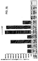
図13は、様々な正常ヒト組織における14618受容体の発現を示す。遺伝子は、乳房および骨格筋で高度に発現されている。従って、これらの組織に関与する疾患における遺伝子の発現は、特に関連性がある。有意な発現が、甲状腺、胎盤、胎児腎臓、胎児心臓、およびリンパ節でも生じる。従って、遺伝子の発現は、同様にこれらの組織に関与する疾患にも関連性がある。さらに、図13に示したように、より低いレベルの発現が、様々な他の組織で見られる。従って、遺伝子の発現は、これらの組織に関与する疾患にも関連性がある。
【0144】
14618受容体は、活性化を伴うまたは伴わない様々な造血細胞にも発現している。この遺伝子は、CD34+骨髄細胞にも高度に発現している。従って、この遺伝子の発現は、様々な血液細胞前駆体に関連性がある。それ故、この遺伝子の発現は、任意の主要な血液細胞型の欠損に関与する疾患、すなわち好中球減少症、血小板減少症または貧血に関連している。この遺伝子はまた、可動化末梢血細胞巨核球(G−CSFで可動化)にも高度に発現している。従って、この遺伝子の発現は、血小板減少症などの血小板機能に関与する疾患に関連がある。有意な発現が、可動化末梢血細胞リンパ球、可動化骨髄CD34−細胞、および臍帯血CD434−細胞にも見られる。従って、この遺伝子の発現は、これらの細胞の機能に関連があり、それ故、免疫機能または炎症に関与する疾患にも関連がある。さらに、遺伝子の発現は、活性化末梢血単核細胞にも生じる。活性化B細胞は、異なって遺伝子を発現する。従って、遺伝子の発現は、免疫機能および/または炎症に関与する疾患に関連がある。
【0145】
2838受容体は、図15に示した細胞および組織に発現している。高レベルの発現が、リンパ節および胸腺に示される。従って、遺伝子の発現は、特に、これらの組織に関与する疾患に関連している。極めて高い発現が、CD8細胞および活性化B細胞に見られる。高発現が、Tヘルパー細胞(1および2)、CD4細胞およびジャーカット細胞(T細胞系)にも生じている。発現は、活性化B細胞および活性化Tヘルパー細胞で異なる。発現は、これらの両方の細胞型の活性化時に増加する。従って、遺伝子の発現は、免疫機能および炎症に関与する疾患に関連がある。この遺伝子はまた、顆粒球で有意に発現している。従って、この遺伝子の発現は、これらの細胞に関与する疾患に関連がある。
【0146】
図16は、正常ヒト組織における15334受容体の発現を示す。この遺伝子は、リンパ節、扁桃腺および膵臓で高度に発現している。従って、この遺伝子の発現は、これらの組織に関与する疾患に特に関連している。この遺伝子の発現はまた、大腸、精巣、胎盤、胎児心臓、および脾臓でも高い。従って、この遺伝子の発現は、これらの組織に関与する疾患にも関連している。さらに、この図は、数個の他の組織における低い発現レベルを示す。従って、この遺伝子の発現は、これらの組織に関与する疾患に関連性があり得る。15334受容体の発現は、様々な造血細胞でも研究されている(図17〜19)。極めて高い発現が、初期巨核球および赤芽球に生じる。従って、この遺伝子の発現は、赤血球分化および巨核球分化に関連し、従って、貧血および血小板減少症の治療に関連している。さらに、この遺伝子の発現は、活性化B細胞に比較して、休止B細胞で有意に増加している。従って、この遺伝子の発現は、B細胞免疫機能に関連している。さらに、低いレベルの発現が、様々な他の造血系統の細胞に見られる。図17参照。造血細胞における系統限定的様式でのこの遺伝子の発現により、発現は、系統細胞、赤血球/赤血球、または巨核球/血小板の発達の調節に関連している。
【0147】
脾臓に関連した疾患は、非特異的急性脾炎、鬱血性巨脾、および脾臓梗塞を含む脾腫大;新生物、先天性異常、および破裂を含むがこれに限定されない。脾腫大に関連した疾患は、非特異的脾炎、感染性単核球症、結核、腸チフス、ブルセラ症、サイトメガロウイルス、梅毒、マラリア、ヒストプラスマ症、トキソプラスマ症、カラアザール、トリパノソーマ症、住血吸虫症、レーシュマニア症、およびエキノコックス症などの感染;肝硬変、門脈または脾静脈血栓症、および心不全などの部分高血圧に関連した鬱血性状態;ホジキン病、非ホジキンリンパ腫/白血病、多発性骨髄腫、骨髄増殖疾患、溶血性貧血、および血小板減少性紫斑病などのリンパ血行性疾患;慢性関節リウマチおよび全身性エリテマトーデスなどの免疫−炎症疾患;ゴーシェ病、ニーマン・ピット病、およびムコ多糖症などの沈着病;および、アミロイド症、原発性新生物および嚢胞、および続発性新生物などの他の状態を含む。
【0148】
肺に関連した疾患は、先天性異常;無気肺;肺鬱血および浮腫(血行力学的肺浮腫および微小血管損傷、成人呼吸促迫症候群(び慢性肺胞傷害)、肺塞栓症、出血、および梗塞により引き起こされる浮腫を含む)、および肺性高血圧および血管硬化などの、血管を起源とする疾病;肺気腫、慢性気管支炎、気管支喘息、および気管支拡張症などの慢性閉塞性肺疾患;塵肺、サルコイドーシス、特発性肺繊維症、剥離性間質性肺炎、過敏症肺臓炎、肺好酸球増加症(好酸球増加症を伴う肺浸潤)、器質性肺炎を伴う閉塞性細気管支炎、び慢性肺出血症候群(グッドパスチャー症候群、特発性肺ヘモジデリン沈着症および他の出血症候群を含む)、コラーゲン血管疾患における肺併発、および肺胞蛋白症などの、散在性間質性(浸潤性、拘束性)疾病;薬物誘導肺疾患、放射線誘導肺疾患、および肺移植などの治療合併症;気管支原性癌(腫瘍随伴症候群、細気管支肺胞上皮癌、気管支カルチノイド、種々の腫瘍および転移性腫瘍などの神経内分泌腫瘍を含む)などの腫瘍;炎症性胸水、非炎症性胸水、気胸症および胸膜腫瘍(孤立性繊維症(胸膜繊維腫)および悪性中皮腫を含む)を含む、胸膜の病態を含むがこれに限定されない。
【0149】
大腸に関連した疾患は、閉塞症および狭窄症、メッケル憩室、先天性神経節欠損巨大大腸−ヒルシュスプルング病などの先天性異常;下痢および赤痢、感染性全腸炎(ウイルス性胃腸炎、細菌性全腸炎を含む)、壊死性全腸炎、抗生物質性大腸炎(偽膜性大腸炎)、および膠原性およびリンパ性大腸炎、種々の腸炎症疾患(寄生虫および原虫を含む)、後天性免疫不全症候群、移植、薬物誘導腸損傷、放射線全腸炎、好中球減少症大腸炎(盲腸炎)、および分流性大腸炎などの全腸炎;クローン病および潰瘍性大腸炎などの特発性炎症性腸疾患;非新生物性ポリープ、腺腫、家族性症候群、大腸直腸発癌、大腸直腸癌、およびカルチノイド腫瘍を含むがこれに限定されない。
【0150】
肝臓に関連した疾患は、肝臓損傷;黄疸および胆汁鬱帯、例えばビリルビンおよび胆汁形成;肝不全および肝硬変、例えば、硬変、門脈圧亢進症(腹水、門脈体静脈短絡を含む)および脾腫大;ウイルス性肝炎(A〜E型肝炎感染および他の肝炎ウイルスによる汗腺を含む)、臨床病理学的症候群(例えば保因状態、無症候性汗腺、急性ウイルス肝炎、慢性ウイルス肝炎、および劇症肝炎)などの感染性疾患;自己免疫肝炎;薬物および毒素誘導肝疾患、例えばアルコール性肝疾患;先天的代謝異常および小児肝疾患、例えば血色素症、ウィルソン病、α1−アンチトリプシン欠損症、および新生児肝炎;続発性胆汁性肝硬変、原発性胆汁性肝硬変、原発性硬化性胆道炎および胆道樹の異常などの肝内胆道疾患;肝臓への血流障害(肝動脈欠損および門脈閉塞および血栓症を含む)、肝臓を通る血流障害(受動鬱血および小葉中心性壊死および肝紫斑病を含む)、肝静脈流出閉塞(肝静脈血栓症(バッド・キアリ症候群)および静脈閉塞性疾患を含む)などの循環器疾患;子癇前症および子癇、妊娠の急性脂肪肝、および肝内胆汁鬱帯などの、妊娠に関連した肝疾患;骨髄移植後の薬物毒性、移植片宿主疾患および肝拒絶、および肝同種移植片への非免疫学的傷害などの、臓器または骨髄移植の肝合併症;結節過形成、腺腫、および悪性腫瘍(肝臓の原発性腫瘍および転移性腫瘍を含む)などの腫瘍および腫瘍状態を含むがこれに限定されない。
【0151】
子宮および子宮内膜の関連した疾患は、月経周期の子宮内膜組織学;無排卵周期、不適切な黄体期、経口避妊薬および誘発性子宮内膜変化、および閉経期および閉経後変化などの、機能的子宮内膜疾患;慢性子宮内膜炎などの炎症;腺筋症;子宮内膜ポリープ;子宮内膜過形成;子宮内膜癌などの悪性腫瘍;悪性混合ミュラー腫瘍などの、混合ミュラーおよび間葉腫瘍;平滑筋腫、平滑筋肉腫、および子宮内膜間質腫瘍を含む、子宮筋の腫瘍を含むがこれに限定されない。
【0152】
脳に関連した疾患は、ニューロンに関連した疾患、および膠細胞、星状膠細胞、乏突起膠細胞、上衣細胞、および小膠細胞などの膠細胞に関連した疾患;脳浮腫、頭蓋内圧上昇およびヘルニア形成、および水頭;神経管、前脳異常、後頭蓋窩以上、および脊髄空洞症および脊髄水腫症などの、奇形および発達疾患;出世時脳損傷;低酸素、虚血、および梗塞(低血圧、低灌流、および局所的血液供給の閉塞に由来する低流動状態−全体的脳虚血および限局的脳虚血−の梗塞を含む)、頭蓋内出血(脳内(実質細胞内)出血、クモ膜下出血およびイチゴ状動脈瘤破裂を含む)、および血管形成異常、高血圧性脳疾患(ラクナ梗塞、細隙出血および高血圧性脳症を含む);急性髄膜炎(急性化膿(細菌)髄膜炎および急性無菌(ウイルス)髄膜炎を含む)、急性焦点化膿性感染(脳膿瘍、硬膜下膿胸、および硬膜外膿瘍を含む)、慢性細菌髄膜脳炎(結核およびマイコバクテリウム症、神経梅毒および神経ボレリア症(ライム病)を含む)、ウイルス髄膜脳炎(節足動物媒介(Arbo)ウイルス脳炎、単純ヘルペスウイルス1型、単純ヘルペスウイルス2型、水痘帯状疱疹ウイルス(帯状ヘルペス)、サイトメガロウイルス、灰白髄炎、狂犬病を含む)およびヒト免疫不全ウイルス1(HIV−1髄膜脳炎(亜急性脳炎)、空胞性ミエロパシー、エイズ関連ミエロパシー、末梢神経障害、および小児エイズ、進行性多巣性白質脳症、亜急性硬化性全脳炎、真菌髄膜脳炎、神経系の他の感染病などの感染;伝染性海綿様脳症(プリオン病);脱髄疾患(多発性硬化症、多発性硬化症変形、急性散在性脳脊髄炎および急性壊死性出血性脳脊髄炎、および脱髄を伴う他の疾病;大脳皮質に影響を及ぼす変性疾患(アルツハイマー病およびピック病を含む)、大脳基底核および脳幹の変性疾患(パーキンソン症、特発性パーキンソン病(振戦麻痺)、進行性核上麻痺、皮質基底変性を含む)、多系統萎縮(線条体黒質変性、シャイ−ドレーガー症候群およびオリーブ橋小脳萎縮症およびハンチントン病を含む)などの、変性疾患;脊髄小脳失調症(フリードライヒ運動失調症および毛細血管拡張性運動失調症を含む)を含む脊髄小脳変性、運動ニューロンに影響を及ぼす変性疾患(筋萎縮性側索硬化症(運動ニューロン疾患)、延髄脊髄萎縮症(ケネディー症候群)、および進行性脊髄性筋萎縮症を含む);白質ジストロフィー(クラッベ病、異染性白質ジストロフィー、副腎白質ジストロフィー、ペリツェウス・メルツバッハー病およびカナバン病を含む)、ミトコンドリア脳筋症(リー病および他のミトコンドリア脳筋症を含む)などの先天性代謝障害;ビタミン欠乏症(例えばチアミン(ビタミンB1)欠乏症およびビタミンB12欠乏症)、神経学的代謝妨害後遺症(低血糖、高血糖、および肝性脳症を含む)、中毒疾患(一酸化炭素、メタノール、エタノールを含む)、および照射(メトトレキサートと照射の併用から誘発される損傷を含む)を含む、中毒および後天性代謝疾患;繊維性(び慢性)星状細胞腫および多型膠芽細胞腫、毛様性星状細胞腫、多形黄色星状細胞腫を含む神経膠腫、および脳幹神経膠腫、乏突起細胞腫、および上皮細胞腫および関連脳室周囲塊病変、神経腫瘍、低分化新生物(髄芽細胞腫を含む)、他の実質腫瘍(原発性脳リンパ腫を含む)、胚細胞腫瘍、および松果体実質腫瘍、髄膜腫、転移性腫瘍、腫瘍随伴症候群、末梢神経鞘腫瘍(シュワン細胞腫、神経線維腫症、および悪性末梢神経鞘腫瘍(悪性神経鞘腫)を含む)、および神経皮膚症候群(母斑症)(1型神経繊維腫症(NF1)および2型神経線維腫症(NF2)を含む神経線維腫症、結節性硬化、およびフォン・ヒッペル−リンダウ病を含む)などの腫瘍を含むがこれに限定されない。
【0153】
T細胞に関連した疾患は、細胞性過敏症、例えば、遅延型過敏症およびT細胞性細胞毒性、および移植片拒絶;自己免疫疾患、例えば、全身性エリテマトーデス、シェーグレン症候群、全身性硬化症、炎症性ミオパシー、混合性結合性組織病、および結節性多発性動脈炎および他の脈管炎;胸腺形成不全、重症複合性免疫不全疾患、およびエイズなどの、原発性免疫不全を含むがこれに限定されない、免疫不全症候群;白血球減少症;白血球増多症、急性非特異的リンパ節炎、および慢性非特異的リンパ節炎を含むがこれに限定されない、反応性(炎症性)白血球増殖;急性リンパ芽球性白血病/リンパ腫、末梢T細胞および末梢T細胞リンパ腫を含むナチュラルキラー細胞新生物、分類不可能な成人T細胞白血病/リンパ腫、菌状息肉腫およびセザリー症候群、およびホジキン病などの、前駆T細胞新生物といったリンパ系新生物を含むがこれに限定されない、白血球の新生物による増殖を含むがこれに限定されない。
【0154】
皮膚の疾患は、色素沈着およびメラニン形成細胞の疾患(白斑、雀卵斑、肝斑、黒子、母斑細胞性母斑、異形成性母斑、および悪性黒色腫を含むがこれに限定されない);上皮腫瘍(脂漏性角化症、黒色表皮腫、繊維上皮ポリープ、上皮嚢腫、角化棘細胞腫、および付属器(付属体)腫瘍を含むがこれに限定されない);悪性前および悪性表皮腫瘍(光線性角化症、扁平上皮癌、基底細胞癌、およびメルケル細胞を含むがこれに限定されない);真皮腫瘍(良性繊維性組織球腫、隆起性皮膚繊維肉腫、黄色腫、および真皮血管腫瘍を含むがこれに限定されない);皮膚へ細胞移入する腫瘍(組織球症X、菌状息肉腫(皮膚T細胞リンパ腫)および肥満細胞症を含むがこれに限定されない);表皮成熟疾患(魚鱗癬を含むがこれに限定されない);急性炎症性皮膚疾患(蕁麻疹、急性湿疹皮膚炎、および多型紅斑を含むがこれに限定されない);慢性炎症性皮膚疾患(乾癬、扁平苔せん、およびエリテマトーデスを含むがこれに限定されない);水疱様膨隆(水疱性)疾患(天疱瘡、類天疱瘡、疱疹性皮膚炎および非炎症性水疱様膨隆疾患:表皮水疱症およびポルフィリン症を含むがこれに限定されない);表皮付属器疾患(尋常性ざ瘡を含むがこれに限定されない);皮下脂肪組織炎(結節性紅斑および硬結性紅斑を含むがこれに限定されない);および感染および侵襲(例えば、いぼ、伝染性軟属腫、膿か疹、表在真菌感染、および節足動物咬合、刺針、および侵襲)を含むがこれに限定されない。
【0155】
正常な骨髄では、骨髄球シリーズ(多形核細胞)は、細胞成分の約60%を構成し、赤血球は20〜30%を構成する。リンパ球、単球、細網細胞、形質細胞および巨核球は、共に10〜20%を構成する。リンパ球は、正常な骨髄の5〜15%を構成する。骨髄では、細胞型を、赤血球の前駆体(赤芽球)、マクロファージ(単芽球)、血小板(巨核球)、多形核白血球(骨髄芽球)、およびリンパ球(リンパ芽球)が1つの顕微鏡視野で見えるように加えて混合する。さらに、幹細胞が、異なる細胞系のために存在し、並びに、前駆幹細胞が、異なる系統の委任前駆細胞のために存在する。様々な種類の細胞および各段階は、当業者には公知であり、例えば、免疫学、免疫病理学および免疫、第5版、Sellら、SimonおよびSchuster(1996)の42ページ(図2〜8)に見られ、これは骨髄に見られる細胞型のその教義のために参考として取込む。従って、本発明は、これらの細胞から生じる疾患に関する。これらの疾患は、以下を含むがこれに限定されない:造血幹細胞に関与する疾患;委任リンパ系前駆細胞;BおよびT細胞を含むリンパ球;委任骨髄前駆体(単球、顆粒球、巨核球を含む);および委任赤血球前駆体に関する疾患。これらは、白血病(Bリンパ系白血病、Tリンパ系白血病、未分化白血病を含む);赤白血病、巨核芽球性白血病、単球性白血病;[白血病は、分化を伴いおよび伴わずに包含される];慢性および急性骨髄芽球性白血病、慢性および急性リンパ急性白血病、慢性および急性骨髄性白血病、リンパ腫、骨髄異常形成症候群、慢性および急性骨髄性白血病、骨髄単球性白血病;慢性および急性骨髄芽球性白血病、慢性および急性骨髄性白血病、慢性および急性前骨髄球性白血病、慢性および急性骨髄球性白血病、単球−マクロファージ系統の血液学的悪性疾患、例えば、若年性慢性骨髄性白血病;続発性AML、前駆血液学的疾患;不応性貧血;再生不良性貧血;反応性皮膚血管内リンパ腫;樹状細胞の発現変化に関連した繊維性疾患、全身性硬化を含む疾患、E−M症候群、流行性毒性油症候群、好酸球性筋膜炎局在形の強皮症、ケロイド、および繊維性大腸疾患;血管腫様悪性繊維性組織球腫;原発性頭頸部扁平上皮癌を含む癌;カポジ肉腫を含む肉腫;哺乳動物繊維腺腫を含む、繊維腺腫および葉状腫瘍;間質腫瘍;組織球腫を含む葉状腫瘍;赤芽球症;神経線維腫症;血管内皮の疾患;脱髄、特に古い病巣において;血管原性浮腫、血管疾患、アルツハイマー病およびパーキンソン病;T細胞リンパ腫;B細胞リンパ腫を含むがこれに限定されない。
【0156】
心臓に関連した疾患は、心不全(心肥大、左側心不全および右側心不全を含むがこれに限定されない);虚血性心疾患(狭心症、心筋梗塞、慢性虚血性心疾患、および急性心臓死を含むがこれに限定されない);高血圧性心疾患(全身性(左側)高血圧性心疾患および肺性(右側)高血圧性心疾患を含むがこれに限定されない);心臓弁膜症(石灰化大動脈狭窄、先天性二尖大動脈弁の石灰化、および僧帽状輪状石灰化などの石灰化、および僧帽弁の粘液腫性変性(僧帽弁逸脱)、リウマチ熱およびリウマチ性心疾患、感染性心内膜炎、および非感染組織過形成、例えば、非細菌性血栓性心内膜炎および全身性エリテマトーデスの心内膜炎(リブマン・ザックス病)、カルチノイド心疾患、および人工弁の合併症により引き起こされる弁変性を含むがこれに限定されない)、心筋疾患(拡張型心筋症、肥大型心筋症、収縮性心筋症、および心筋炎を含むがこれに限定されない);心膜疾患(心膜滲出および心膜血腫および心外膜炎(急性心外膜炎および治癒心外膜炎を含む)およびリウマチ様心疾患を含むがこれに限定されない);新生物性心疾患(原発性心腫瘍、例えば、粘液腫、脂肪腫、乳頭繊維弾性腫、横紋筋腫、および肉腫、および非心性新生物の心臓効果を含むがこれに限定されない);鬱血性心不全(左−右短絡−後期チアノーゼ、例えば、心房中隔欠損症、心室中隔欠損症、動脈管開存、および房室中隔欠損、右−左短絡−初期チアノーゼ、例えば、ファロー四微症、大動脈転位、動脈幹、三尖弁閉鎖、および全肺静脈接続異常、閉塞性先天性異常、例えば、大動脈の縮窄、肺動脈弁狭窄および閉鎖症、および大動脈弁狭窄および閉鎖症、および心臓移植に関する疾患を含むがこれに限定されない)を含むがこれに限定されない。
【0157】
血管に関与する障害には、以下が含まれるが、これらに限定されない:損傷に対する血管壁の反応(内皮機能障害および内皮活性化ならびに内膜肥厚など);先天性奇形を含む血管疾患(動静脈瘻、アテローム性動脈硬化症、および高血圧性血管疾患(高血圧など)など)(これらに限定されない);炎症性疾患−−脈管炎(巨細胞性(側頭)動脈炎、高安動脈炎、結節性多発性動脈炎(古典的)、川崎病(皮膚粘膜リンパ節症候群)、微視的多発性血管炎(微視的多発性動脈炎、過敏症、または白血球破砕性血管炎)、ヴェグナー肉芽腫症、閉塞性血栓血管炎(バーガー病)、他の障害に関連する脈管炎および感染性動脈炎など);レイノー病;動脈瘤および解離(腹部大動脈瘤、梅毒性動脈瘤、および動脈解離など);静脈およびリンパの疾患(拡張蛇行静脈、血栓性静脈炎および静脈血栓症、上大静脈の閉塞(上大静脈症候群)、下大静脈の閉塞(下大静脈症候群)、ならびにリンパ管炎およびリンパ水腫など);良性腫瘍および腫瘍様病態を含む腫瘍(血管腫、リンパ管腫、グロムス腫瘍(グロムス血管腫)、血管拡張症、細菌性血管腫症など)および中程度(低い悪性度の境界)の腫瘍(カポジ肉腫および血管内皮腫など)、および悪性腫瘍(血管肉腫および血管外皮細胞腫など);および血管疾患における治療介入の病理学(バルーン血管形成および関連技術)および血管置換(冠状動脈バイパス術)が含まれる。
【0158】
赤血球に関連する障害には、以下の貧血が含まれるが、これらに限定されない:遺伝性球状赤血球症、赤血球酵素欠乏による溶血性疾患(グルコース−6−ホスフェートデヒドロゲナーゼ欠乏症)、鎌状赤血球病、サラセミア症候群、発作性夜間血色素尿症、免疫溶血性貧血、および赤血球に対する外傷に起因する溶血性貧血を含む溶血性貧血;および巨赤芽球性貧血を含む赤血球生成の減少による貧血(ビタミンB12欠乏による貧血(悪性貧血);葉酸欠乏による貧血、鉄欠乏性貧血、慢性病による貧血、再生不良性貧血、赤芽球癆、および他の形態の骨髄不全など)。
【0159】
胸腺に関与する障害には、発達障害(胸腺形成不全症または無形成症を伴うディ・ジョージ症候群);胸腺嚢胞;胸腺形成不全症(胸腺内のリンパ濾胞の出現に関与し、リンパ濾胞過形成を発症する);胸腺腫(生殖細胞腫を含む)、リンパ腫、ホジキン病、およびカルチノイドなどが含まれる。胸腺腫には、良性または被膜を有する胸腺腫、および胸腺癌と呼ばれる悪性胸腺腫I型(浸潤性胸腺腫)またはII型を含むことができる。
【0160】
B細胞に関与する障害には、B細胞前駆体新生物(リンパ芽球性白血病/リンパ腫など)が含まれるがこれらに限定されない。末梢性B細胞新生物には、以下が含まれるが、これらに限定されない:慢性リンパ球性白血病/小リンパ球性リンパ腫、濾胞性リンパ腫、び慢性大B細胞型リンパ腫、バーキットリンパ腫、形質細胞性真性物、多発性骨髄腫および関連する状態、リンパ形質細胞性リンパ腫(ヴァルデンストレームマクログロブリン血症)、外套細胞性リンパ腫、辺縁層リンパ腫(MALT腫)、および毛様細胞白血病。
【0161】
腎臓に関連する障害には、以下が含まれるがこれらに限定されない:先天性奇形(腎臓の嚢胞性疾患(嚢胞性腎形成異常、常染色体優性(成人)腎多嚢胞病、常染色体劣性(幼児)腎多嚢胞病)(これらに限定されない)および腎髄質の嚢胞性疾患(髄質性海綿腎、ネフロン肺結核−尿毒症性嚢胞性疾患複合体、)(これらに限定されない);後天性(透析関連)嚢胞性疾患(孤立性嚢胞など));糸球体損傷の病状を含む糸球体疾患(in situ免疫複合体蓄積を含むがこれに限定されない)(抗GBM腎炎、ヘイマン腎炎、移植抗原に対する抗体、循環免疫複合体腎炎、糸球体細胞に対する抗体、糸球体腎炎における細胞性免疫、第二経路の活性化、上皮細胞損傷、および細胞および可溶性メディエーターを含む糸球体損傷のメディエーターに関与する病態、急性糸球体腎炎(急性増殖性(連鎖球菌後、感染後)糸球体腎炎(連鎖球菌感染後糸球体腎炎および非連鎖球菌感染後急性糸球体腎炎を含むがこれらに限定されない)、急速進行性(半月体形成性)糸球体腎炎、ネフローゼ症候群、膜性糸球体腎炎(膜性腎症)、微小変化疾患(リポイドネフローゼ)、巣状分節状糸球体硬化症、膜性増殖性糸球体腎炎、IgA腎症(バージャー病)、層状増殖性および壊死性糸球体腎炎(層状糸球体腎炎)、遺伝性腎炎(アルポート症候群および薄膜疾患(両性家族性血尿)が含まれるがこれらに限定されない)など)、慢性糸球体腎炎、全身性疾患に関連する糸球体損傷(全身性紅斑性狼瘡、ヘーノホ−シェーンライン紫斑病、細菌性心内膜炎、糖尿病性糸球体硬化症、アミロイドーシス、繊維性および免疫タクトイド糸球体腎炎、ならびに他の全身性障害が含まれるがこれらに限定されない))(これらに限定されない));急性尿細管壊死および尿細管間質性腎炎を含む細管および間質に影響を与える疾患(腎盂腎炎および逆流性腎症、急性腎盂腎炎、慢性腎盂腎炎、および逆流性腎症が含まれるがこれらに限定されない)、薬物および毒素によって誘導される尿細管間質性腎炎(急性薬物誘導性間質性腎炎、鎮痛薬乱用による腎症、非ステロイド性抗炎症薬に関連する腎症が含まれるが、これらに限定されない)、および他の尿細管間質性疾患(尿酸腎症、高カルシウム血症および腎石灰沈着症ならびに多発性骨髄腫が含まれるが、これらに限定されない);良性腎硬化症、悪性高血圧症、および促進性腎硬化症、腎動脈狭窄症、および血栓性微小血管症を含む血管の疾患(古典的(幼児期)溶血性尿毒症症候群、成人溶血性尿毒症症候群/血栓性血小板減少性紫斑病、特発性HUS/TTPが含まれるが、これらに限定されない)、他の血管疾患(アテローム硬化性虚血性腎疾患、鎌状赤血球腎症、汎発性皮質性壊死、および腎梗塞が含まれるが、これらに限定されない);尿路閉塞(閉塞性尿路疾患);尿路結石症(尿結石);および腎臓腫瘍(良性腫瘍(腎乳頭腺種、腎線維腫または過誤腫(腎肥大性(renomedullary)間細胞腫瘍)、血管筋脂肪腫、および膨大細胞腫などならびに悪性腫瘍(腎盂の尿路上皮癌を含む腎細胞癌(副腎腫、腎臓腺癌)を含む))が含まれるが、これらに限定されない)。
【0162】
乳房の疾患には、以下が含まれるが、これらに限定されない:発育障害;炎症(急性乳腺炎、乳管周辺の乳腺炎、乳周辺の乳腺炎(再発性乳輪下膿瘍、乳管扁平上皮細胞化生)、乳管拡張症、脂肪壊死、肉芽腫性乳腺炎、およびシリコン乳房植込みに関連する病態が含まれるが、これらに限定されない);繊維細胞の変化;増殖性乳房疾患(上皮性過形成、硬化性腺症、および小管乳頭腫が含まれるが、これらに限定されない);腫瘍(間質腫瘍(線維腺種、葉状腫瘍、および肉腫など)および上皮性腫瘍(大管乳頭腫など))が含まれるが、これらに限定されない);in situでの腺管癌(パジェット病を含む)およびin situでの小葉癌を含むin situ(非浸潤性)癌および浸潤性(浸潤)癌(浸潤性腺管癌(非特異型)、浸潤性小葉癌、髄葉癌、膠様(粘液性)癌腫、管状腺癌、および浸潤性乳頭状癌が含まれるが、これらに限定されない)、および種々の悪性新生物を含む乳癌。
【0163】
男性の胸部の疾患には、女性化乳房および癌が含まれるが、これらに限定されない。
【0164】
精巣および精巣上体に関与する障害には、以下が含まれるが、これらに限定されない:奇形(停留睾丸など)、退行性病変(萎縮など)、炎症((非特異性精巣上体炎および精巣炎など)、肉芽腫性(自己免疫)睾丸炎、および特異性炎症(淋病、おたふく風邪、結核、および梅毒を含むがこれらに限定されない))、ねじれを含む血管障害、生殖細胞のねじれを含む精巣腫瘍(精上皮腫、精母細胞精上皮腫、胚性癌腫、卵黄嚢腫瘍絨毛腫、奇形腫、および混合腫瘍を含むが、これらに限定されない)、性索−性腺支質の腫瘍(ライディッヒ(間質性)細胞腫瘍およびセルトーリ細胞腫瘍(男性ホルモン細胞産生腫)ならびに精巣リンパ腫を含むがこれらに制限されない)、鞘膜の週の損傷。
【0165】
前立腺に関与する障害には、以下が含まれるが、これらに限定されない:炎症、良性肥厚(例えば、結節性再生(良性前立腺肥大または肥厚))、および腫瘍(癌など)。
【0166】
甲状腺に関与する障害には、以下が含まれるが、これらに限定されない:甲状腺機能亢進症;甲状腺機能低下症(クレチン症および粘液水腫が含まれるが、これらに限定されない);甲状腺炎(橋本病、亜急性(肉芽腫性)甲状腺炎、亜急性リンパ球性(無痛)甲状腺炎が含まれるが、これらに限定されない);グレーヴズ病;び慢性および多結節性甲状腺炎(び慢性無毒性(単一性)甲状腺炎および多結性甲状腺炎が含まれるが、これらに限定されない);腺腫、他の良性腫瘍、および癌を含むがこれらに限定されない甲状腺新生物(乳頭状癌、濾胞腺癌、髄様癌、および退形性癌が含まれるが、これらに限定されない);および両性器(cogenital)奇形。
【0167】
骨格筋に関連する障害には、横紋筋肉腫などの腫瘍が含まれる。
【0168】
膵臓に関連する障害には、以下が含まれる:両性器奇形などの膵外分泌機能障害(異所性膵炎が含まれるがこれに限定されない);嚢胞(偽性嚢胞が含まれるがこれらに限定されない);腫瘍(嚢胞性腫瘍および膵臓癌が含まれるがこれらに限定されない);内分泌性膵臓(真性糖尿病など);島細胞腫瘍(膵島細胞腺種、ガストリン産生腫瘍、および他の稀な島細胞腫瘍が含まれるがこれらに限定されない)。
【0169】
小腸に関連する障害には、以下が含まれるが、これらに限定されない:吸収不良症候群(腹腔スプルー、熱帯性スプルー(感染後スプルー)など)、ホウィップル病、ジサッカリダーゼ(ラクターゼ)欠乏症、無βリポタンパク質血症、腺腫および腺癌を含む小腸の腫瘍。
【0170】
血小板減少(血小板減少症)に関連する障害には、特発性血小板減少性紫斑病(優勢特発性血小板減少性紫斑病を含む)、薬物誘導性血小板減少症、HIV関連血小板減少症、および血栓性微小血管症;血栓性血小板減少性紫斑病および溶血性尿毒症症候群が含まれる。
【0171】
T細胞前駆体新生物に関連する障害には、Tリンパ芽球前駆体白血病/リンパ腫が含まれる。末梢性T細胞およびナチュラルキラー細胞新生物に関連する障害には、慢性Tリンパ球性白血病、大顆粒球リンパ性白血病、菌状息肉腫およびセザリー症候群、末梢性T細胞リンパ腫、不特定の血管免疫芽球性T細胞リンパ腫、血管中心性リンパ腫(NK/T細胞リンパ腫4a)、腸管T細胞リンパ腫、成人T細胞白血病/リンパ腫、および未分化大細胞リンパ腫が含まれる。
【0172】
卵巣に関連する疾患には、例えば、以下が含まれる:多嚢胞卵巣、スタイン−リーヴェンサール症候群、腹膜偽粘液腫、および間質性卵胞莢膜増殖症;卵巣腫瘍(体腔上皮腫瘍、漿液性腫瘍、粘液性腫瘍、子宮内腫瘍、明細胞腺癌、嚢腺線維腫、ブレンナー腫瘍、表面上皮腫瘍など);生殖細胞腫瘍(成熟(良性)奇形腫、単胚葉奇形腫、未熟型悪性奇形腫、未分化胚細胞腫、内胚葉洞腫瘍、絨毛癌など);性索−吻合部腫瘍(顆粒膜細胞−卵胞膜細胞腫、テコーマ−線維腫、男性ホルモン産生細胞腫、丘細胞腫瘍、および性腺芽腫など);および転移性腫瘍(クルーケンベルク腫瘍など)。
【0173】
骨形成細胞には、骨芽前駆細胞、骨芽細胞、および骨細胞が含まれる。骨疾患は、これらの細胞が任意のその発達段階の間に骨格に影響を与え得るので複雑である。したがって、骨障害は、種々の症状を有し、身体の1つ、複数、または全ての骨に関与し得る。このような障害には、以下が含まれる:先天性奇形、軟骨形成不全、および壊死性小人症、異常な基質に関連する疾患(I型コラーゲン疾患など)、骨粗鬆症、パジェット病、くる病、骨軟化症、高代謝回転骨形成異常、低代謝回転の再生不良疾患、骨壊死、化膿性骨髄炎、結核性骨髄炎、骨腫、類骨骨腫、骨芽細胞腫、骨肉腫、骨軟骨腫、軟骨腫、軟骨粘液線維腫、軟骨肉腫、線維性皮質欠損症、線維性骨異形成、線維肉腫、悪性線維性組織球腫、ユーイング肉腫、未分化神経外胚葉性腫瘍、巨細胞腫、および転移性腫瘍。
【0174】
したがって、受容体ポリペプチドは、特に上記で開示のような受容体タンパク質の異常な発現または活性によって特徴づけられる受容体関連障害の治療に有用である。1つの実施形態では、本方法は、薬剤(例えば、本明細書中に記載のスクリーニングアッセイによって同定される薬剤)またはタンパク質の発現もしくは活性を調節する(例えば、アップレギュレートするかダウンレギュレートする)薬剤の組み合わせの投与を包含する。別の実施形態では、本発明は、タンパク質発現の減少または異常を補正するための治療として、タンパク質を投与するステップを包含する。
【0175】
タンパク質活性の刺激は、タンパク質が異常にダウンレギュレートされる、そして/またはタンパク質活性の増加がおそらく有利な効果を有する条件において望ましい。同様に、タンパク質活性の阻害は、タンパク質が異常にアップレギュレートされる、そして/またはタンパク質活性の減少がおそらく有利な効果を有する条件において望ましい。このような条件の一例では、被験体は、異常な発達または細胞分裂によって特徴づけられる障害を有する。このような条件の別の例では、被験体は、異常な造血反応によって特徴づけられる増殖疾患(例えば、癌)または障害を有する。このような条件の別の例では、患者の組織再生を達成することが望ましい(例えば、被験体が脳および脊髄損傷を有する場合、調節した様式で神経組を再生することが望ましい)。
【0176】
本発明のさらに別の態様では、本発明のタンパク質を「おとりタンパク質(bait protein)」として、2ハイブリッドアッセイまたは3ハイブリッドアッセイ(米国特許第5,283,317号、Zervosら、1993、Cell、72、223〜232、Maduraら、1993、J.Biol.Chem.、268、12046〜12054、Bartelら、1993、Biotechniques、14、920〜924、Iwabuchiら、1993、Oncogene、8、1693〜1696、およびBrent WO 94/10300を参照のこと)で使用して、本発明のタンパク質と結合するか相互作用する他のタンパク質(捕獲タンパク質)を同定し、その活性を調節することができる。
【0177】
2838受容体ポリペプチドはまた、特に、造血細胞(ヘルパーT細胞(1および2)、B細胞、および顆粒球など)において受容体タンパク質によって媒介される疾患または疾患の素因の診断用の標的を獲得するのに有用である。14618受容体ポリペプチドはまた、特に、乳房、骨格、筋肉、およびリンパ節ならびにCD34+前駆細胞およびこれらの前駆体によって産生される細胞(赤血球、リンパ球、および巨核球系)において受容体タンパク質によって媒介される疾患または疾患の素因の診断用の標的を獲得するのに有用である。15334受容体ポリペプチドはまた、特に、膵臓、脾臓、扁桃腺、およびリンパ節ならびに巨核球および赤芽球系において受容体タンパク質によって媒介される疾患または疾患の素因の診断用の標的を獲得するのに有用である。したがって、本方法は、細胞、組織、または器官における受容体タンパク質の存在またはそのレベルの同定を提供する。本方法には、相互作用を検出することができるような、生物学的サンプルと受容体タンパク質との相互作用が可能な化合物との接触を含む。
【0178】
受容体タンパク質検出用の1つの薬剤は、受容体タンパク質に選択的に結合することができる抗体である。生物学的サンプルには、被験体から単離した組織、細胞、および生物学的体液、ならびに被験体に存在する組織、細胞、および体液が含まれる。
【0179】
受容体タンパク質はまた、種々の受容体タンパク質を有する患者における活動性疾患または疾患の素因の診断用の標的を提供する。したがって、受容体タンパク質を生物学的サンプルから単離し、異常な受容体タンパク質となる遺伝子変異の存在についてアッセイすることができる。これには、(異常なスプライシングの結果としての)アミノ酸の置換、欠失、挿入、再配列、および不適切な翻訳後修飾が含まれる。分析法には、電気泳動での移動度の変化、トリプティックペプチドの変化、細胞ベースのアッセイまたは無細胞アッセイにおける受容体活性の変化、リガンドまたは抗体結合パターンの変化、等電点の変化、直接的アミノ酸配列決定、およびタンパク質の変異の検出に有用な任意の他の公知のアッセイ技術が含まれる。
【0180】
受容体タンパク質検出用のin vitro技術には、酵素結合免疫測定法(ELISA)、ウェスタンブロット、免疫沈澱および免疫蛍光が含まれる。あるいは、タンパク質を、被験体への標識抗受容体抗体の移入によって、被験体においてin vivoで検出することができる。例えば、抗体を、被験体中での存在および位置が標準的な画像技術で検出することができる放射性マーカーで標識することができる。被験体において発現する受容体タンパク質の対立遺伝子変異型を検出する方法およびサンプルにおける受容体タンパク質の断片を検出する方法が特に有用である。
【0181】
受容体ポリペプチドはまた、薬理ゲノム学的分析に有用である。薬理ゲノム学は、患者における薬物の性質の変化および異常な作用による、薬物反応における臨床的に有意な遺伝的変異型を取扱う。Eichelbaum、1997、Clin.Exp.Pharmacol.Physio.、23(10−11)、983〜985、1996、およびLinder、1997、Clin.Chem.、43(2)、254〜266を参照のこと。これらの変異体の臨床的結果により、一定の個体において治療薬の毒性が増し、個体の代謝の変異型の結果として一定の個体において薬物治療が失敗する。したがって、個体の遺伝子型は、治療化合物が身体に作用する方法または身体が化合物を代謝する方法を同定することができる。さらに、薬物代謝酵素活性は、薬物作用の強度および持続時間の両方に影響を与える。したがって、個体の薬理ゲノム学により、個体の遺伝子型に基づいた予防または治療に有効な化合物およびこのような化合物の有効投薬量の選択が可能になる。いくつかの薬物代謝酵素における遺伝的多形の発見により、何故患者によって予想される薬物効果を得ることができないのか、薬物効果の悪化を示すのか、または標準的な薬物投薬量で毒性が強くなるのかが説明されている。強い代謝物の遺伝子型および弱い代謝物の遺伝子型において、多形を発現することができる。したがって、遺伝的多形により、1つの集団における1つまたは複数の受容体機能が別の集団の受容体機能と異なる受容体タンパク質の対立遺伝子タンパク質変異型を得ることができる。したがって、ポリペプチドで、標的が治療様式に影響を与えることができる遺伝的素因を確認できる。したがって、リガンドベースの治療において、多形により、アミノ末端細胞外ドメインおよび/またはリガンド結合において多少活性なリガンド結合領域、および受容体の活性化を得ることができる。したがって、リガンドの投薬量は、多形を含む所与の集団内での治療効果を最大にするために改変するする必要があるだろう。
【0182】
受容体ポリペプチドはまた、臨床試験および他の治療中の治療効果の監視に有用である。したがって、遺伝子発現、タンパク質レベル、または受容体活性を増加または減少させるように設計された薬剤の治療効果を、終点標的として受容体ポリペプチドを用いて、治療クールにわたり監視することができる。監視ステップは、例えば、以下であり得る:(i)薬物投与前に被験体から投与前サンプルを得るステップ、(ii)投与前サンプルにおける特定のタンパク質の発現または活性レベルを検出するステップ、(iii)被験体から1つまたは複数の投与後サンプルを得るステップ、(iv)投与後サンプルにおけるタンパク質の発現または活性レベルを検出するステップ、(v)投与前サンプルにおけるタンパク質と投与後サンプルにおけるタンパク質の発現または活性レベルを比較するステップ、(vi)以上により、被験体に対する薬剤投与を増加または減少させるステップ。
【0183】
受容体ポリペプチドはまた、受容体関連疾患の治療に有用である。したがって、治療法には、リガンド結合を競合する受容体タンパク質の可溶性受容体または断片の使用が含まれる。これらの受容体または断片は、有効な競合を得るためにリガンドに対する高い親和性を有し得る。
【0184】
抗体
本発明はまた、2838、14618、および15334受容体タンパク質およびその変異型および断片に選択的に結合する抗体を提供する。抗体は、受容体タンパク質と実質的に相同ではない他のタンパク質にも結合するとしても、選択的に結合すると考える。これらの他のタンパク質は、受容体タンパク質断片またはドメインと相同性を共有する。特異的領域におけるこの保存により、相同的配列によって両タンパク質と結合する抗体が得られる。この場合、受容体タンパク質に結合する抗体は、依然として選択的であることが理解されるであろう。
【0185】
抗体を作製するために、ポリクローナル抗体およびモノクローナル抗体調製の標準的な技術を使用し、単離された受容体ポリペプチドを免疫原として使用して、抗体を作製する。全長タンパク質または抗原ペプチド断片のいずれかを使用することができる。高い抗原性指数を有する領域を、図2、図6、および図10に示す。
【0186】
抗体を、これらの領域またはこれらの領域の不連続な断片から調製することが好ましい。しかし、抗体を、本明細書中に記載のペプチドの任意の領域から調製することができる。好ましい断片は、リガンド結合を減少させるか完全に阻害する抗体を作製する。受容体の全てまたは受容体の一部(例えば、細胞内カルボキシ末端ドメイン、アミノ末端細胞外ドメイン、全膜貫通ドメインもしくは特異的セグメント、任意の細胞内もしくは細胞外ループ、または上記の任意の部分)に対する抗体を産生することができる。抗体を、特定の機能部位(リガンド結合部位、Gタンパク結合部位、またはリン酸化、グリコシル化、もしくはミリスチル化された部位)に対して産生することもできる。
【0187】
抗原性2838断片は、典型的には、少なくとも8つの連続したアミノ酸残基を含む。しかし、抗原ペプチドは、連続する少なくとも12、14アミノ酸残基、少なくとも15アミノ酸残基、少なくとも20アミノ酸残基、または少なくとも30アミノ酸残基を含むことができる。1つの実施形態では、断片は、タンパク質表面上に位置する領域(例えば、親水性領域)に対応する。しかし、これらの断片は、本発明の前に開示され得る任意の断片を包含すると解釈されない。
【0188】
抗原性14168断片は、典型的には、少なくとも9つの連続したアミノ酸残基を含む。しかし、抗原ペプチドは、連続する少なくとも12、14アミノ酸残基、少なくとも15アミノ酸残基、少なくとも20アミノ酸残基、または少なくとも30アミノ酸残基を含むことができる。1つの実施形態では、断片は、タンパク質表面上に位置する領域(例えば、親水性領域)に対応する。しかし、これらの断片は、本発明の前に開示され得る任意の断片を包含すると解釈されない。
【0189】
抗原性15334断片は、典型的には、少なくとも8つの連続したアミノ酸残基を含む。しかし、抗原ペプチドは、連続する少なくとも12、14アミノ酸残基、少なくとも15アミノ酸残基、少なくとも20アミノ酸残基、または少なくとも30アミノ酸残基を含むことができる。1つの実施形態では、断片は、タンパク質表面上に位置する領域(例えば、親水性領域)に対応する。しかし、これらの断片は、本発明の前に開示され得る任意の断片を包含すると解釈されない。
【0190】
抗体は、ポリクローナルまたはモノクローナルであり得る。インタクトな抗体またはその断片(例えば、FabまたはF(ab’)2)を使用することができる。
【0191】
抗体の検出可能な物質とのカップリング(すなわち、物理的結合)によって検出を容易にすることができる。検出可能な物質の例には、種々の酵素、置換基、蛍光物質、発光物質、生物発光物質、および放射性物質が含まれる。適切な酵素の例には、西洋ワサビペルオキシダーゼ、アルカリホスファターゼ、β−ガラクトシダーゼ、またはアセチルコリンエステラーゼが含まれる。適切な置換基複合体の例として、ストレプトアビジン/ビオチンおよびアビジン/ビオチンが含まれる。適切な蛍光物質の例として、ウンベリフェロン、フルオレセイン、フルオレセインイソチオシアネート、ローダミン、ジクロロトリアジニルアミンフルオレセイン、ダンシルクロリド、またはフィコエリトリンが含まれる。発光物質の例として、ルミノールが含まれる。生物発光物質の例として、ルシフェラーゼ、ルシフェリン、およびエクオリンが含まれる。適切な放射性物質の例として、125I、131I、35S、または3Hが含まれる。
【0192】
適切な免疫原性調製物は、天然もしくは組換えて発現させたタンパク質もしくは化学合成ペプチド由来であり得る。
【0193】
抗体の用途
抗体を使用して、標準的な技術(アフィニティークロマトグラフィーまたは免疫沈降)によって受容体タンパク質を単離することができる。抗体は、細胞由来の天然の受容体および宿主において発現させた組換えて産生させた受容体タンパク質の生成を容易にすることができる。
【0194】
抗体は、生物の種々の組織の間および正常な発達時の受容体の発現パターンを決定するために、細胞または組織における受容体タンパク質の存在の検出に有用である。
【0195】
発現量および発現パターンを評価するために、抗体を使用して、in situ、in
vitro、または細胞溶解物中、または上清中の受容体タンパク質を検出することができる。
【0196】
抗体を使用して、発達中の異常な組織の分布または異常な発現を評価することができる。
【0197】
全長受容体タンパク質の循環断片の抗体検出を使用して、受容体代謝回転を同定することができる。
【0198】
さらに、抗体を使用して、疾患の活動段階などの疾患段階または受容体機能に関連する疾患に対する素因を有する個体における受容体発現を評価することができる。不適切な組織の分布、発達の発現、または受容体タンパク質の発現レベルによって障害が起こる場合、正常な受容体タンパク質に対する抗体を調製することができる。障害が、受容体タンパク質における特異的変異によって特徴づけられる場合、この変異タンパク質に特異的な抗体を使用して、特異的変異受容体タンパク質の存在をアッセイすることができる。
【0199】
抗体を使用して、生物の種々の組織中の細胞の正常および異常な細胞下の局在性を評価することもできる。抗体は、全受容体または受容体の一部(例えば、アミノ末端の細胞外ドメインまたは細胞外ループの一部)に対して作製することができる。
【0200】
診断的用途を、遺伝子試験においてだけでなく、治療様式の監視においても適用することができる。したがって、治療が受容体発現レベルまたは異常な受容体の存在および異常な組織の分布または発達発現の回収を最終目的とする場合、受容体または関連断片に指向する抗体を使用して、治療効率を監視することができる。
【0201】
したがって、抗体を診断に使用して、例えば、所与の治療計画の効果を同定するために、臨床試験法の一部として組織におけるタンパク質レベルを監視することができる。
【0202】
さらに、抗体は、薬理ゲノム学的分析に有用である。したがって、多形受容体タンパク質に対して調製した抗体を使用して、治療計画の修正が必要な個体を同定することができる。
【0203】
抗体はまた、電気泳動での移動度、等電点、トリプティックペプチド消化、および当業者に公知の他の物理学的アッセイによって分析される異常な受容体タンパク質用の免疫学的マーカーとしての診断ツールとして有用である。
【0204】
抗体はまた、組織分類に有用である。したがって、特異的受容体タンパク質が特異的組織における発現に対応している場合、この受容体タンパク質に特異的な抗体を使用して組織の型を同定することができる。
【0205】
抗体はまた、法医学的同定に有用である。したがって、個体が特異的多形タンパク質が得られる特異的遺伝的多形性に対応している場合、多形タンパク質に特異的なタンパク質を、同定の補助として使用することができる。
【0206】
抗体はまた、受容体機能の阻害(例えば、リガンド結合の阻害)に有用である。
【0207】
これらの用途を、治療が受容体機能の阻害を含むような治療状況に適用することができる。抗体を使用して、例えば、リガンド結合を阻害することができる。機能に必要な部位を含む特異的断片または細胞に会合したインタクトな受容体に対して抗体を調製することができる。
【0208】
完全なヒト抗体は、ヒト患者の治療に特に望ましい。このヒト抗体作製技術の概要は、Lonberg and Huzar、1995、Int.Rev.Immunol.、13、65〜93を参照こと。このヒト抗体およびヒトモノクローナル抗体の作製技術およびこのような抗体の作製プロトコールの詳細な考察は、例えば、米国特許第5,625,126号、同第5,633,425号、同第5,569,825号、同第5,661,016号、および同第5,545,806号を参照のこと。
【0209】
本発明はまた、生物学的サンプル中の受容体タンパク質の存在を検出するための抗体を用いたキットを含む。キットは、生物学的サンプル中の受容体タンパク質検出用の抗体(標識抗体または標識可能な抗体など)および化合物または薬剤、サンプル中の受容体タンパク質の量を同定する手段、およびサンプル中の受容体タンパク質量と標準とを比較する手段を含むことができる。化合物または薬剤を、適切な容器に封入することができる。さらに、キットは、受容体タンパク質を検出するためのキットを使用するための指示を含む。
【0210】
ポリヌクレオチド
配列番号2のヌクレオチド配列を、ヒト全長cDNAの配列決定によって得た。
配列番号4のヌクレオチド配列を、ヒト全長cDNAの配列決定によって得た。
配列番号6のヌクレオチド配列を、ヒト全長cDNAの配列決定によって得た。
【0211】
特別に開示したcDNAは、コード領域ならびに5’および3’非翻訳配列(配列番号2、配列番号4、および配列番号6)を含む。
【0212】
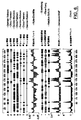
ヒト2838受容体cDNAは、約1617ヌクレオチド長であり、約319アミノ酸残基長である全長タンパク質をコードする。核酸は、脾臓、精巣、結腸、および末梢血リンパ球ならびに図15に記載の組織において発現する。配列番号1のアミノ酸の構造分析を、図3にヒドロパシープロットとして示す。図は、7回膜貫通セグメント、アミノ末端の細胞外ドメイン、およびカルボキシ末端の細胞内ドメインの推定構造を示す。
【0213】
ヒト14618受容体cDNAは、約1358ヌクレオチド長であり、約337アミノ酸残基長である全長タンパク質をコードする。核酸は、脾臓、および末梢血リンパ球、乳房、骨格筋、および図13に記載の他の組織ならびに図14に記載の種々の造血細胞、特に、巨核球およびCD34+細胞において発現する。配列番号3のアミノ酸の構造分析を、図7にヒドロパシープロットとして示す。図は、7回膜貫通セグメント、アミノ末端の細胞外ドメイン、およびカルボキシ末端の細胞内ドメインの推定構造を示す。
【0214】
ヒト153340受容体cDNAは、約2559ヌクレオチド長であり、約372アミノ酸残基長である全長タンパク質をコードする。核酸は、リンパ節、扁桃腺、膵臓、結腸、脾臓、末梢血細胞、胸腺、副腎、および心臓ならびに図17〜図19に記載の巨核球および赤血球系において発現する。配列番号5のアミノ酸の構造分析を、図11にヒドロパシープロットとして示す。図は、7回膜貫通セグメント、アミノ末端の細胞外ドメイン、およびカルボキシ末端の細胞内ドメインの推定構造を示す。
【0215】
本明細書中で使用される、用語「膜貫通セグメント」は、原形質膜に渡された疎水性へリックスを含む構造アミノ酸モチーフをいう。2838の全膜貫通ドメインは、アミノ酸約25〜アミノ酸約292番目にわたる。14618の全膜貫通ドメインは、アミノ酸約29〜アミノ酸約297番目にわたる。15334の全膜貫通ドメインは、アミノ酸約26〜アミノ酸約299番目にわたる。セグメントが7回膜をわたり、このドメイン中に3つの細胞内ループおよび3つの細胞外ループが存在する。
【0216】
本発明は、2838受容体タンパク質をコードする単離ポリペプチドを提供する。用語「2838ポリペプチド」または「2838核酸」は、配列番号2に記載の配列をいう。
【0217】
本発明は、14618受容体タンパク質をコードする単離ポリペプチドを提供する。用語「14618ポリペプチド」または「14618核酸」は、配列番号4に記載の配列をいう。
【0218】
本発明は、15334受容体タンパク質をコードする単離ポリペプチドを提供する。用語「15334ポリペプチド」または「15334核酸」は、配列番号6に記載の配列をいう。
【0219】
用語「受容体ポリヌクレオチド」または「受容体核酸」は、さらに、2838、14618、および15334ポリヌクレオチドの変異型および断片を含む。
【0220】
「単離」受容体核酸は、天然の受容体核酸供給源に存在する他の核酸から分離したものである。好ましくは、「単離」核酸は、核酸が誘導される生物のゲノムDNA中の核酸(すなわち、核酸の5’および3’末端に位置する配列)に天然に隣接する配列を含まない。しかし、いくつかの隣接ヌクレオチド配列(例えば、5KBまで)を含むことができる。重要な点は、核酸を隣接配列から単離して、その核酸を本明細書中に記載の特定の操作(組換え発現、プローブおよびプライマーの調製など)および受容体核酸配列に特異的な他の用途に供することができることである。
【0221】
さらに、「単離」核酸分子(cDNAまたはRNA分子など)は、他の細胞物質、組換え技術によって作製した場合は培養培地、または化学合成した場合は化合物の前駆体もしくは他の薬品を実質的に含まない。しかし、核酸分子を他のコード配列または調節配列と融合し、依然として「単離」とみなすことができる。
【0222】
例えば、ベクター中に含まれる組換えDNA分子を、「単離」とみなす。単離DNA分子のさらなる例には、異種宿主細胞中に維持された組換えDNA分子または溶液中の(部分的または実質的に)精製されたDNA分子が含まれる。単離RNA分子には、本発明の単離DNA分子のin vivoまたはin vitroRNA転写物が含まれる。本発明の単離核酸分子には、さらに、このような合成分子が含まれる。
【0223】
いくつかの例では、単離物質は、組成物(例えば、他の物質を含む粗抽出物)、緩衝系または試薬混合物の一部を形成する。他の環境では、物質を、例えば、PAGEまたはカラムクロマトグラフィー(HPLCなど)で同定によって本質的に均質であるように精製することができる。好ましくは、単離核酸は全ての高分子種の少なくとも50、80、もしくは90%(モルベースに基づく)を含む。
【0224】
受容体ポリヌクレオチドは、成熟タンパク質+さらなるアミノまたはカルボキシル末端アミノ酸、または成熟ポリペプチド内のアミノ酸(例えば、成熟形態が1つを超えるポリペプチド鎖を有する場合)をコードすることができる。このような配列は、前駆体から成熟形態へのタンパク質のプロセシングに役割を果たすか、タンパク質輸送を容易にするか、タンパク質の半減期を延長または短縮するか、アッセイまたは産生のためのタンパク質の操作を容易にすることなどができる。一般的に、in situでの場合のように、さらなるアミノ酸を、細胞酵素によって成熟タンパク質から離れてプロセシングすることができる。
【0225】
受容体ポリヌクレオチドには、以下が含まれるが、これらに限定されない:成熟ポリペプチドのみをコードする配列、成熟ポリペプチドおよびさらなるコード配列(リーダー配列または分泌配列(例えば、プレプロまたはプロタンパク質配列)など)をコードする配列、さらなるコード配列を含むか含まない成熟配列をコードする配列+さらなる非コード配列(例えば、転写、mRNAプロセシング(イントロンならびにスプライシングおよびポリアデニル化シグナルを含む)、リボゾーム結合およびmRNAの安定性の役割を果たす転写するが翻訳されない配列などの非コード5’および3’配列)。さらに、ポリヌクレオチドを、例えば、精製を容易にするペプチドをコードするマーカー配列に融合することができる。
【0226】
受容体ポリヌクレオチドは、クローニングまたは化学合成技術またはその組み合わせによって作製されるRNAの形態(mRNAなど)またはDNAの形態(cDNAおよびゲノムDNA)で存在し得る。核酸、特にDNAは、二本鎖または一本鎖であり得る。一本鎖核酸は、コード鎖(センス鎖)または非コード鎖(アンチセンス鎖)であり得る。
【0227】
1つの受容体核酸は、ヒト2838cDNAに対応する配列番号2に示したヌクレオチド配列を含む。
1つの受容体核酸は、ヒト14618cDNAに対応する配列番号4に示したヌクレオチド配列を含む。
1つの受容体核酸は、ヒト15334cDNAに対応する配列番号6に示したヌクレオチド配列を含む。
【0228】
1つの実施形態では、受容体核酸は、コード領域のみを含む。
【0229】
本発明は、さらに、遺伝コードの縮重のために配列番号2に記載のヌクレオチド配列と異なり、配列番号2に示すヌクレオチド配列によってコードされるタンパク質と同一のタンパク質をコードする、変異型受容体ポリヌクレオチドおよびその断片を提供する。
【0230】
本発明は、さらに、遺伝コードの縮重のために配列番号4に記載のヌクレオチド配列と異なり、配列番号4に示すヌクレオチド配列によってコードされるタンパク質と同一のタンパク質をコードする、変異型受容体ポリヌクレオチドおよびその断片を提供する。
【0231】
本発明は、さらに、遺伝コードの縮重のために配列番号6に記載のヌクレオチド配列と異なり、配列番号6に示すヌクレオチド配列によってコードされるタンパク質と同一のタンパク質をコードする、変異型受容体ポリヌクレオチドおよびその断片を提供する。
【0232】
本発明はまた、本明細書中に記載の変異型ポリペプチドをコードする受容体核酸分子を提供する。このようなポリヌクレオチドは、天然に存在し得るか(対立遺伝子変異型(同一の遺伝子座)、ホモログ(異なる遺伝子座)、およびオーソログ(異なる生物)など)、組換えDNA法または化学合成によって構築することができる。このような天然に存在しない変異型を、ポリヌクレオチド、細胞、または生物に適応する変異誘発技術によって作製することができる。したがって、上記のように、変異型は、ヌクレオチドの置換、欠失、逆位、および挿入を含むことができる。
【0233】
コード領域および非コード領域のいずれかまたは両方で変異体を得ることができる。変異体により、保存的および非保存的アミノ酸置換を得ることができる。
【0234】
典型的には、変異型は、配列番号2、4、および6ならびにその相補物からなる群から選択される核酸分子と実質的に同一である。
【0235】
オーソログ、ホモログ、および対立遺伝子変異型を、当該分野で公知の方法を用いて同定することができる。2838変異型は、配列番号2で示したヌクレオチド配列またはこの配列の断片に40〜45%、45〜50%、50〜55%、55〜60%、少なくとも約65%、典型的には少なくとも70〜75%、より典型的には少なくとも80〜85%、最も典型的には少なくとも約90〜95%またはそれ以上相同な受容体をコードするヌクレオチド配列を含む。配列番号2に示すヌクレオチド配列またはその配列の断片にストリンジェントな条件下でハイブリッド形成することができるこのような核酸分子を、容易に同定することができる。
【0236】
14618変異型は、配列番号4で示したヌクレオチド配列またはこの配列の断片に40〜45%、45〜50%、50〜55%、55〜60%、少なくとも約65%、典型的には少なくとも70〜75%、より典型的には少なくとも80〜85%、最も典型的には少なくとも約90〜95%またはそれ以上相同な受容体をコードするヌクレオチド配列を含む。配列番号4に示すヌクレオチド配列またはその配列の断片にストリンジェントな条件下でハイブリッド形成することができるこのような核酸分子を、容易に同定することができる。
【0237】
15334変異型は、配列番号6で示したヌクレオチド配列またはこの配列の断片に、45〜50%、50〜55%、55〜60%、少なくとも約65%、典型的には少なくとも70〜75%、より典型的には少なくとも80〜85%、最も典型的には少なくとも約90〜95%またはそれ以上相同な受容体をコードするヌクレオチド配列を含む。配列番号6に示すヌクレオチド配列またはその配列の断片にストリンジェントな条件下でハイブリッド形成することができるこのような核酸分子を、容易に同定することができる。
【0238】
ストリンジェントなハイブリッド形成は、それが一般的な相同性(ポリA配列、または全てまたはほとんどのタンパク質、すべてのGPCR、全てのファミリーI GPCRまたは全てのプリン受容体に共通な配列など)による場合、実質的な相同性を示さないことが理解される。さらに、変異体には、本発明以前に開示されている可能性のあるいかなる核酸配列も含まれないことが理解される。
【0239】
本明細書中で使用される、用語「ストリンジェントな条件下でハイブリッド形成する」は、互いに少なくとも50〜55%、55%相同な受容体ポリペプチドをコードするヌクレオチド配列が互いに典型的にハイブリッド形成したままであるハイブリッド形成および洗浄条件を記載することを意図する。これは、少なくとも約65%、少なくとも約70%、少なくとも約75%、少なくとも約80%、少なくとも約90%、少なくとも約95%またはそれ以上互いに同一な配列が互いにハイブリッド形性したままのような条件であり得る。このようなストリンジェントな条件は、当業者に公知であり、Current Protocols in Molecular Biology、John Wiley & Sons、N.Y.、1989、6.3.1〜6.3.6(参考として援用される)に見出すことができる。ストリンジェントなハイブリッド形成条件の一例は、約45℃の6×塩化ナトリウム/クエン酸ナトリウム(SSC)中でのハイブリッド形成およびその後の50〜65℃の0.2×SSC、0.1%SDS中での1回または複数回の洗浄である。別の非限定的な例では、核酸分子を約45℃の6×塩化ナトリウム/クエン酸ナトリウム(SSC)中でのハイブリッド形成、その後室温での0.2×SSC、0.1%SDS中での低ストリンジェンシー洗浄、42℃でのでの0.2×SSC、0.1%SDS中での中程度のストリンジェンシー洗浄、または65℃でのでの0.2×SSC、0.1%SDS中での高ストリンジェンシー洗浄である。1つの実施形態では、ストリンジェントな条件下で配列番号2、4、および6の配列とハイブリッド形成する単離受容体核酸分子は、天然に存在する核酸分子に対応する。本明細書中で使用される、用語「天然に存在する」核酸分子は、天然に存在する核酸分子(例えば、天然のタンパク質をコードする)を有するRNAまたはDNAをいう。
【0240】
当業者に理解されるように、経験的および不安定化剤(ホルムアミドなど)または変性剤(SDSなど)のイオン強度、温度、および濃度に依存して、正確な条件を同定することができる。望ましいハイブリッド形成条件同定において考慮される他の因子には、核酸配列の長さ、塩基組成、ハイブリッド形成配列と他の同一配列内の配列のサブセットの発生頻度との間のミスマッチ%が含まれる。したがって、1つまたは複数のこれらのパラメーターを変化させる一方で、2つの核酸分子について同程度の同一性および類似性を維持することによって、等価の条件を同定することができる。
【0241】
本発明はまた、ストリンジェントな条件下で、配列番号2、4、および6ならびに配列番号2、4、および6の相補物からなる群から選択される核酸配列とハイブリッド形成する一本鎖または二本鎖断片または部分を含む単離核酸を提供する。1つの実施形態では、核酸は、配列番号2、4、および6ならびにその相補物からなる群から選択されるヌクレオチド配列の一部からなる。本発明の核酸断片は、少なくとも約15、好ましくは少なくとも18、20、23、または25ヌクレオチド長であり、30、40、50、100、200、またはそれ以上のヌクレオチド長であり得る。本明細書中に記載の抗原タンパク質またはポリペプチドをコードするより長い断片(例えば、30またはそれ以上のヌクレオチド長)が有用である。
【0242】
さらに、本発明は、全長受容体ポリヌクレオチドの断片を含むポリヌクレオチドを提供する。断片は、一本鎖または二本鎖であり、DNAまたはRNAを含み得る。断片は、コード配列または非コード配列のいずれか由来であり得る。
【0243】
1つの実施形態では、1〜およそヌクレオチド990由来の単離2838受容体核酸は、少なくとも16ヌクレオチド長であり、ストリンジェントな条件下で配列番号2のヌクレオチド配列を含む核酸分子とハイブリッド形成する。別の実施形態では、およそヌクレオチド1487〜1617由来の核酸は、少なくとも20ヌクレオチドである。他の実施形態では、核酸は、少なくとも40、50、100、250、または500ヌクレオチド長またはそれ以上である。
【0244】
別の実施形態では、およそヌクレオチド1〜およびヌクレオチド911のヌクレオチド由来の単離14618受容体核酸は、少なくとも8ヌクレオチド長であり、ストリンジェントな条件下で配列番号4のヌクレオチド配列を含む核酸分子とハイブリッド形成する。他の実施形態では、核酸は、少なくとも40、50、100、250、または500ヌクレオチド長またはそれ以上である。
【0245】
別の実施形態では、ヌクレオチド1〜およそヌクレオチド1355由来の単離15334受容体核酸は、少なくとも18ヌクレオチド長であり、ストリンジェントな条件下で配列番号6のヌクレオチド配列を含む核酸分子とハイブリッド形成する。別の実施形態では、およそヌクレオチド868〜およそヌクレオチド1355由来の核酸は、少なくとも11ヌクレオチドである。他の実施形態では、核酸は、少なくとも40、50、100、250、または500ヌクレオチド長またはそれ以上である。
【0246】
別の実施形態では、単離2838受容体核酸は、アミノ酸1〜アミノ酸319の全コード領域をコードする。別の実施形態では、単離2838受容体核酸は、およそアミノ酸6〜およそアミノ酸319の成熟タンパク質に対応する配列をコードする。別の実施形態では、単離14618受容体核酸は、アミノ酸1〜アミノ酸337の全コード領域をコードする。別の実施形態では、単離14618受容体核酸は、およそアミノ酸6〜およそアミノ酸337の成熟タンパク質に対応する配列をコードする。別の実施形態では、単離15334受容体核酸は、アミノ酸1〜アミノ酸372の全コード領域をコードする。別の実施形態では、単離15334受容体核酸は、およそアミノ酸6〜およそアミノ酸372の成熟タンパク質に対応する配列をコードする。
【0247】
3つ全ての受容体の他の断片は、本明細書中に記載のアミノ酸断片をコードするヌクレオチド配列を含む。さらに、断片は、特異的ドメインまたは本明細書中に記載の部位のサブ断片を含むことができる。断片はまた、上記の特異的アミノ酸配列またはその断片に対応する核酸配列を含む。本発明の核酸断片は、本発明以前に開示されている可能性のある断片を含むように構築されない。
【0248】
しかし、受容体断片には、全遺伝子を含まない任意の核酸配列を含むことが理解される。
【0249】
2838受容体核酸断片は、さらに、本明細書中に記載のドメイン、本明細書中にも記載のサブ領域、および特異的機能部位に対応する配列を含む。2838受容体核酸断片は、1〜およそ24のアミノ酸残基を含むアミノ末端細胞外ドメインを含むポリペプチド、膜貫通ドメインをわたす領域を含むポリペプチド(およそ25〜およそ292のアミノ酸残基)、カルボキシ末端の細胞内ドメインを含むポリペプチド(およそ293〜およそ319のアミノ酸残基)、およびGタンパク質受容体シグナルをコードするポリペプチド(118〜120またはおよそ107〜およそ123の周囲のアミノ酸残基)をコードする核酸、任意の7回膜貫通セグメント、細胞外または細胞内ループ、グリコシル化部位、リン酸化部位、ミリスチル化部位、およびアミド化部位をコードする核酸分子を含む。
【0250】
14618受容体核酸断片は、1〜およそ28のアミノ酸残基を含むアミノ末端細胞外ドメインを含むポリペプチド、膜貫通ドメインをわたす領域を含むポリペプチド(およそ29〜およそ297のアミノ酸残基)、カルボキシ末端の細胞内ドメインを含むペプチド(およそ298〜およそ337のアミノ酸残基)、およびGタンパク質受容体シグナルをコードするポリペプチド(120〜122またはおよそ110〜およそ132の周囲のアミノ酸残基)をコードする核酸、任意の7回膜貫通セグメント、細胞外または細胞内ループ、グリコシル化部位、およびリン酸化部位をコードする核酸分子を含む。
【0251】
15334受容体核酸断片は、1〜約25番目のアミノ酸残基を含むアミノ末端細胞外ドメインを含むポリペプチド、膜貫通ドメインをわたす領域を含むポリペプチド(およそ26〜およそ299番目のアミノ酸残基)、カルボキシ末端の細胞内ドメインを含むペプチド(およそ300〜およそ372番目のアミノ酸残基)、およびGタンパク質受容体シグナルをコードするポリペプチド(118〜120番目またはおよそ110〜およそ130番目の周囲のアミノ酸残基)をコードする核酸、任意の7回膜貫通セグメント、細胞外または細胞内ループ、グリコシル化部位、プロテインキナーゼC、cAMP、cGMP、およびカゼインキナーゼIIリン酸化部位、およびミリスチル化部位をコードする核酸分子を含む。
【0252】
ドメインの位置がコンピュータ分析によって予想されている場合、当業者は、これらのドメインを構築するアミノ酸残基が、ドメインを定義するために使用した基準に依存して変化し得ることを認識するであろう。
【0253】
受容体核酸断片はまた、ドメイン、セグメント、ループ、および上記の他の機能的部位の組み合わせを含む。したがって、例えば、受容体核酸は、アミノ末端細胞外ドメインおよび1つの膜貫通セグメントに対応する配列を含み得る。当業者は、多くの置換が可能であることを認識しているであろう。
【0254】
ドメインまたは部位の位置がコンピュータ分析によって予想されている場合、当業者は、これらのドメインを構築するアミノ酸残基が、ドメインを定義するために使用した基準に依存して変化し得ることを認識するであろう。
【0255】
本発明はまた、本明細書中に記載の受容体領域を保有するエピトープをコードする受容体核酸断片を提供する。
【0256】
単離受容体ポリヌクレオチド配列、特に断片は、DNAプローブおよびプライマーとして有用である。
【0257】
例えば、オリゴヌクレオチドプローブを合成するために、公知のヌクレオチド配列を用いて受容体遺伝子のコード領域を単離することができる。次いで、標識プローブを使用して、cDNAライブラリー、ゲノムDNAライブラリー、またはmRNAをスクリーニングし、コード領域に対応する核酸を単離することができる。さらに、PCR反応でプライマーを使用して、受容体遺伝子の特異的領域をクローニングすることができる。
【0258】
典型的には、プローブ/プライマーは、実質的に精製したオリゴヌクレオチドを含む。2838オリゴヌクレオチドは、典型的には、ストリンジェントな条件下で、配列番号2のセンス鎖またははアンチセンス鎖の少なくともそれぞれおよそ16および20個、典型的にはおよそ25、より典型的には40、50、または75個の連続したヌクレオチドとハイブリッド形成する1〜990番目および1487〜1617番目のヌクレオチド配列領域または他の受容体ポリヌクレオチドを含む。14618オリゴヌクレオチドは、典型的には、ストリンジェントな条件下で、配列番号4のセンス鎖またははアンチセンス鎖の少なくともおよそ9、12、典型的にはおよそ25、より典型的には40、50、または75個の連続したヌクレオチドとハイブリッド形成する1〜911番目のヌクレオチド配列領域または他の受容体ポリヌクレオチドを含む。15334オリゴヌクレオチドは、典型的には、ストリンジェントな条件下で、配列番号6のセンス鎖またははアンチセンス鎖の少なくともおよそ11、12、典型的にはおよそ25、より典型的には40、50、または75個の連続したヌクレオチドとハイブリッド形成する868〜1355番目のヌクレオチド配列領域または他の受容体ポリヌクレオチドを含む。15334オリゴヌクレオチドはまた、典型的には、ストリンジェントな条件下で、配列番号6のセンス鎖またははアンチセンス鎖の少なくともおよそ18、典型的にはおよそ25、より典型的には40、50、または75個の連続したヌクレオチドとハイブリッド形成する1〜1355番目のヌクレオチド配列領域または他の受容体ポリヌクレオチドを含む。
【0259】
ポリヌクレオチドの使用
本発明の核酸配列を、さらに、「検索配列」として使用して、例えば、他のファミリーメンバーまたは関連配列の同定のために、公的なデータベースによって検索を行うことができる。このような検索を、Altschulら、1990、J.Mol.Biol.、215、403〜10のNBLASTおよびXBLASTプログラム(バージョン2.0)を使用して行うことができる。本発明の核酸分子に相同なヌクレオチド配列を得るために、NBLASTプログラム(スコア=100、長さ=12)を具備したBLASTヌクレオチド検索を行うことができる。比較用のギャップアラインメントを得るために、Altschulら、1997、Nucleic Acids Res.、25(17)、3389〜3402に記載のように、Gapped BLASTを利用することができる。BLASTおよびGapped BLASTプログラムを利用する場合、それぞれのプログラムのデフォルトパラメータ(例えば、XBLASTおよびNBLAST)を使用することができる。http://www.ncbi.nlm.nih.gov.を参照のこと。
【0260】
本発明の核酸断片により、アッセイ(以下に記載のアッセイなど)におけるプローブまたはプライマーが得られる。「プローブ」は、塩基特異的様式で、核酸の相補鎖とハイブリッド形成するオリゴヌクレオチドである。このようなプローブには、Nielsenら、1991、Science、254、1497〜1500に記載のポリペプチド核酸が含まれる。典型的には、プローブは、配列番号2、4、および6ならびにその相補物からなる群から選択される核酸の少なくとも約15、典型的には、約20〜25、より典型的には40、50、75の連続したヌクレオチドと高ストリンジェントな条件下でハイブリッド形成するヌクレオチド配列領域を含む。より典型的には、プローブは、さらに、標識(例えば、放射性同位体、蛍光化合物、酵素、または酵素の補因子)を含む。
【0261】
本明細書中で使用される、用語「プライマー」は、周知の方法(例えば、PCR、LCR)(本明細書中に記載されているが、これに限定されない)を使用するテンプレート指向性DNA合成の開始点として作用する一本鎖オリゴヌクレオチドをいう。プライマーの適切な長さは、特定の用途に依存するが、典型的には、約15〜30ヌクレオチドの範囲である。用語「プライマー部位」は、プライマーがハイブリッド形成する標的DNA領域をいう。用語「プライマー対」は、増幅される核酸配列の5’末端にハイブリッド形成する5’(上流)プライマーおよび増幅される配列の相補物にハイブリッド形成する3’(下流)プライマーのセットをいう。
【0262】
受容体ポリヌクレオチドは、プローブ、プライマー、および生物学的アッセイに有用である。
【0263】
本明細書中に記載のアッセイなどにおいて、ポリヌクレオチドを使用してGPCRの性質または機能を評価する場合、全長cDNAの全てまたはそれ未満が有用である。この場合、本発明以前に公知であり得る断片も含まれる。したがって、例えば、GPCR機能に特異的に指向するアッセイ(アゴニストまたはアンタゴニスト活性の評価)は、公知の断片の使用を含む。さらに、受容体機能の診断的アッセイ法を、任意の断片(本発明以前に公知であり得る断片を含む)を用いて行うこともできる。同様に、受容体機能不全の治療を含む方法では、当該分野で公知であり得る断片を含む全ての断片が含まれる。
【0264】
2838受容体ポリヌクレオチドは、配列番号1に記載のポリペプチドをコードする全長cDNAおよびゲノムクローンを単離し、配列番号1に示したものと同一のポリペプチドを産生する変異型または本明細書中に記載の他の変異型に対応するcDNAおよびゲノムクローンを単離するためのcDNAおよびゲノムDNA用のハイブリッド形成プローブとして有用である。変異体を、配列番号1に示したポリペプチドを単離したものと同一の組織および生物、同一の生物由来の異なる組織、または異なる生物由来の異なる組織から単離することができる。
【0265】
14628受容体ポリヌクレオチドは、配列番号3に記載のポリペプチドをコードする全長cDNAおよびゲノムクローンを単離し、配列番号3に示したものと同一のポリペプチドを産生する変異型または本明細書中に記載の他の変異型に対応するcDNAおよびゲノムクローンを単離するためのcDNAおよびゲノムDNA用のハイブリッド形成プローブとして有用である。変異体を、配列番号3に示したポリペプチドを単離したものと同一の組織および生物、同一の生物由来の異なる組織、または異なる生物由来の異なる組織から単離することができる。
【0266】
15334受容体ポリヌクレオチドは、配列番号5に記載のポリペプチドをコードする全長cDNAおよびゲノムクローンを単離し、配列番号5に示したものと同一のポリペプチドを産生する変異型または本明細書中に記載の他の変異型に対応するcDNAおよびゲノムクローンを単離するためのcDNAおよびゲノムDNA用のハイブリッド形成プローブとして有用である。変異体を、配列番号5に示したポリペプチドを単離したものと同一の組織および生物、同一の生物由来の異なる組織、または異なる生物由来の異なる組織から単離することができる。
【0267】
この方法は、発達段階で調節される遺伝子およびcDNAの単離に有用であるので、生物の発達における異なる点で同一の組織または異なる組織で発現させることができる。
【0268】
プローブは、受容体をコードする遺伝子の全長に沿った任意の配列に対応することができる。したがって、プローブは、5’非コード領域、コード領域、および3’非コード領域由来であり得る。しかし、プローブは、本発明以前に公知であり得る任意の配列に対応するように構築されない。
【0269】
2838核酸プローブは、例えば、配列番号1の全長cDNAまたはその断片(少なくとも11、16、18、20、30、50、100、250、または500ヌクレオチド長のオリゴヌクレオチドなど)であり、ストリンジェントな条件下でmRNAまたはDNAと特異的にハイブリッド形成するのに十分であり得る。14618核酸プローブは、例えば、配列番号3の全長cDNAまたはその断片(少なくとも8、12、15、30、50、100、250、または500ヌクレオチド長のオリゴヌクレオチドなど)であり、ストリンジェントな条件下でmRNAまたはDNAと特異的にハイブリッド形成するのに十分であり得る。15334核酸プローブは、例えば、配列番号5の全長cDNAまたはその断片(少なくとも11、12、15、18、30、50、100、250、または500ヌクレオチド長のオリゴヌクレオチドなど)であり、ストリンジェントな条件下でmRNAまたはDNAと特異的にハイブリッド形成するのに十分であり得る。
【0270】
本明細書中に記載のポリヌクレオチドの断片はまた、本明細書中に記載のより長い断片または全長ポリヌクレオチドの合成に有用である。例えば、断片を、mRNAの任意の部分にハイブリッド形成することができ、より長いまたは全長cDNAを産生することができる。
【0271】
断片はまた、所望の長さおよび配列のアンチセンス分子の合成に有用である。
【0272】
本発明のアンチセンス核酸を、当該分野で公知の化学合成および酵素ライゲーション反応を用いて構築した配列番号2、4、および6のヌクレオチド配列を用いて設計することができる。例えば、アンチセンス核酸(例えば、アンチセンスオリゴヌクレオチド)を、天然に存在するヌクレオチドまたは分子の生物学的安定性を増加させるかアンチセンス核酸とセンス各案との間に形成された二重鎖の物理学的安定性を増加させるように設計された種々の修飾ヌクレオチド(例えば、ホスホロチオエート誘導体およびアクリジン置換ヌクレオチドを使用することができる)を用いて化学合成することができる。アンチセンスヌクレオチドの作製に使用することができる修飾ヌクレオチドの例には、以下が含まれる:5−フルオロウラシル、5−ブロモウラシル、5−クロロウラシル、5−ヨードウラシル、ヒポキサンチン、キサンチン、4−アセチルシトシン、5−(カルボキシヒドロキシルメチル)ウラシル、5−カルボキシメチルアミノメチル−2−チオウリジン、5−カルボキシメチルアミノメチルウラシル、ジヒドロウラシル、β−D−ガラクトシルケオシン、イノシン、N6−イソペンテニルアデニン、1−メチルグアニン、1−メチルイノシン、2,2−ジメチルグアニン、2−メチルアデニン、2−メチルグアニン、3−メチルシトシン、5−メチルシトシン、N6−アデニン、7−メチルグアニン、5−メチルアミノメチルウラシル、5−メトキシアミノメチル−2−チオウラシル、β−D−マンノシルケオシン、5’−メトキシカルボキシメチルウラシル、5−メトキシウラシル、2−メトキシチオ−N6−イソペンテニルアデニン、ウラシル−5−オキシ酢酸(v)、ワイブトキソシン、プソイドウラシル、ケオシン、2−チオシトシン、5−メチル−2−チオウラシル、2−チオウラシル、4−チオウラシル、5−メチルウラシル、ウラシル−5−オキシ酢酸メチルエステル、ウラシル−5−オキサ酢酸(v)、5−メチル−2−チオウラシル、3−(3−アミノ−3−N−2−カルボキシプロピル)ウラシル、(acp3)w、および2,6−ジアミノプリン。あるいは、核酸がアンチセンス方向(すなわち、挿入核酸から転写されたRNAは、目的の標的核酸に対してアンチセンス方向である)にサブクローニングされている発現ベクターを用いて、アンチセンス核酸を生物学的に作製することができる。
【0273】
さらに、本発明の核酸分子を、例えば、分子の安定性、ハイブリッド形成、または溶解性を改良するために、塩基部分、糖部分、またはリン酸塩バックボーンで改変することができる。例えば、核酸のデオキシリボースホスフェートバックボーンを、ペプチド核酸を作製するように改変することができる(Hyrupら、1996、Bioorganic & Medicinal Chemistry、4、5を参照のこと)。本明細書中で使用される、用語「ペプチド核酸」または「PNA」は、デオキシリボースホスフェートバックボーンが偽ペプチドバックボーンと置換され、たった4つの天然の核酸塩基が残されるのみである核酸模倣物(例えば、DNA模倣物)をいう。PNAの中性バックボーンは、低イオン強度の条件下で、DNAおよびRNAと特異的にハイブリッド形成されることが示されている。PNAオリゴマー合成を、Hyrupら、1996、前出、Perry−O’Keefeら、1996、Proc.Natl.Acad.Sci.USA、93、14670に記載の標準的な固相ペプチド合成プロトコールを用いて行うことができる。さらに、親油性または他の補助基との結合、PNA−DNAキメラの形成、もしくはリポソームの使用、または当該分野で公知の他の薬物送達技術によって、PNAを、例えば、その安定性、特異性、または細胞取り込みを促進するように改変することができる。PNA−DNAキメラの合成を、Hyrup、1996、前出、Finnら、1996、Nucleic Acids Res.、24(17)、3357〜63、Magら、1989、Nucleic Acids Res.、17、5973、およびPeterserら、1975、Bioorganic Med.Chem.Lett.、5、1119に記載のように行うことができる。
【0274】
本発明の核酸分子および断片には、ペプチドなどの他の付加基(in vivoで宿主細胞受容体を標的するため)または細胞膜を通過する輸送を促進する因子(例えば、Letsingerら、1989、Proc.Natl.Acad.Sci.USA、86、6553〜6556、Lemaitreら、1987、Proc.Natl.Acad.Sci.USA、84、648〜652、PCT公開番号WO 88/0918を参照のこと)または血液脳関門(例えば、PCT公開番号WO 89/10134を参照のこと)も含まれ得る。さらに、オリゴヌクレオチドを、ハイブリッド形成誘発切断剤(例えば、Krolら、1988、Bio−Techniques、6、958〜976を参照のこと)または挿入剤(例えば、Zon、1988、Pharm Res.、5、539〜549を参照のこと)を用いて修飾することができる。
【0275】
受容体ポリヌクレオチドはまた、受容体ポリヌクレオチドの任意の所与の領域を増幅させるためのPCRプライマーとして有用である。
【0276】
受容体ポリヌクレオチドはまた、組換えベクターの構築に有用である。このようなベクターには、受容体ポリペプチドの一部または全部を発現する発現ベクターが含まれる。ベクターはまた、受容体遺伝子および遺伝子産物のin situ発現を変更するための別のポリヌクレオチド配列(細胞ゲノムなど)への組込みに使用されている挿入ベクターを含む。例えば、内因性受容体コード配列を、1つまたは複数の特異的に組込んだ変異を含むコード配列の全部または一部と相同組換えを介して置換することができる。
【0277】
受容体ポリヌクレオチドはまた、抗原ペプチドの発現に有用である。高い抗原性指数を有するペプチド領域を、図2、図6、および図10に示す。
【0278】
受容体ポリヌクレオチドはまた、in situハイブリッド形成法(FISH(この技術の概説として、Vermaら、1988、Human Chromosomes:A Manual of Basic Techniques、Pergamon Press、New Yorkを参照のこと)など)および体細胞ハイブリッドのPCRマッピングによる受容体ポリヌクレオチドの染色体上の位置の同定用のプローブとして有用である。染色体に対するこの配列のマッピングは、疾患に関連する遺伝子を有するこれらの関連配列における重要な最初の段階である。
【0279】
染色体マッピング用の試薬をそれぞれ使用して、単一の染色体またはその染色体上の単一の部位をマークすることができ、複数の部位および/または複数の染色体のマーク用に試薬のパネルを使用することができる。マッピングには、遺伝子の非コード領域に対応する試薬が好ましい。コード配列は遺伝子ファミリー内で保存される傾向が強いので、染色体マッピング中の交差ハイブリッド形成の機会が増加する。
【0280】
一旦配列が正確な染色体位置にマッピングされると、染色体上の配列の物理学的位置は、遺伝子マップデータに対応し得る。(このようなデータは、例えば、V.McKusick、Mendelian Inheritance in Man(Johns Hopkins University Welch Medical Libraryによりオンラインで利用可能である)に見出される)。次いで、同一の染色体領域にマッピングされた遺伝子および疾患との間の関係を、例えば、Egelandら、1987、Nature、325、783〜787に記載の結合分析(物理的に隣接する遺伝子の同時遺伝)によって同定することができる。
【0281】
さらに、特定の遺伝子に関する疾患を罹患している個体と罹患していない個体との間のDNA配列の相違を同定することができる。罹患個体の一部または全部で変異が認められるが、罹患していない個体には全く認められない場合、おそらく、変異は特定の疾患の原因因子である。罹患および非罹患個体の比較には、一般に、染色体における構造の変化(可視的形態の染色体であるか、そのDNA配列に基づいたPCRを用いて検出可能な欠失または翻訳など)の最初の検索が含まれる。最終的に、何人かの個体由来の遺伝子の完全な配列決定を行って、変異の存在の確認および多形由来の変異の区別を行うことができる。
【0282】
受容体ポリヌクレオチドプローブはまた、組織分布に関する受容体およびその変異型をコードする遺伝子の存在パターン(例えば、遺伝子重複が存在するかどうかおよび重複が組織の全部または部分集合のみに存在するのかどうか)の同定に有用である。遺伝子は天然存在するか、細胞、組織、または生物に外因的に移入され得る。
【0283】
受容体ポリヌクレオチドはまた、本明細書中に記載のポリヌクレオチドをコードする遺伝子から作製したmRNAの全部または一部に対応するリボザイムの設計に有用である。
【0284】
受容体ポリヌクレオチドはまた、受容体ポリヌクレオチドおよびポリペプチドの一部または全部を発現する宿主細胞の構築に有用である。
【0285】
受容体ポリヌクレオチドはまた、受容体ポリヌクレオチドおよびポリペプチドの一部または全部を発現するトランスジェニック動物の構築に有用である。
【0286】
受容体ポリヌクレオチドはまた、受容体ポリペプチドの一部または全部を発現するベクターの作製に有用である。
【0287】
受容体ポリヌクレオチドはまた、受容体核酸の発現レベルの同定用のハイブリッド形成プローブとして有用である。したがって、プローブを使用して、細胞、組織、および生物における受容体核酸の存在の検出またはレベルの同定を行うことができる。そのレベルが同定される核酸は、DNAまたはRNAであり得る。したがって、本明細書中に記載のポリペプチドに対応するプローブを使用して、所与の細胞、組織、または生物における遺伝子のコピー数を評価することができる。特に、これは受容体遺伝子の増幅が存在する場合に適切である。
【0288】
あるいは、プローブをin situハイブリッド形成で使用して、受容体遺伝子の増加したコピーの位置、染色体エレメント上の位置、または受容体遺伝子が正常に見出されない染色体への組込み位置(例えば、均一に染色された領域)を評価することができる。
【0289】
これらの用途は、正常な結果と比較した受容体発現の増加または減少に関連する疾患(増殖疾患、分化もしくは発育疾患、または造血疾患など)の診断に関する。
【0290】
したがって、本発明は、試験サンプルを被験体から得て、核酸(例えば、mRNA、ゲノムDNA)を検出し、核酸の存在が核酸の異常な発現または活性に関連する疾患または障害の発症リスクを有する被験体を診断する、受容体核酸の異常な発現または活性に関連する疾患または障害の同定法を提供する。
【0291】
本発明の1つの態様は、個体が異常な核酸の発現または活性に関与する疾患または障害を有するかどうかまたは疾患または障害の発症のリスクを有するかどうかを同定するために、生物学的サンプル(例えば、血液、血清、細胞、組織)における核酸の発現ならびに活性を同定するための診断的アッセイに関する。このようなアッセイを、予知および予言目的で使用して、核酸分子の発現または活性に特徴づけられるか関連する障害の発症前に個体を予防的に治療することができる。
【0292】
mRNAのin vitroでの検出技術には、ノーザンハイブリッド形成およびin situハイブリッド形成が含まれる。DNAのin vitroでの検出技術には、サザンハイブリッド形成およびin situハイブリッド形成が含まれる。
【0293】
プローブを、被験体由来の細胞サンプル中の受容体をコードする核酸(例えば、mRNAまたはゲノムDNA)のレベルの測定または受容体が変異されている場合はその同定などによる、受容体タンパク質を発現する細胞または組織の同定用の診断試験キットの一部として使用することができる。
【0294】
核酸発現アッセイは、受容体核酸発現を調整する化合物(例えば、アンチセンス、ポリペプチド、ペプチド模倣物、小分子、または他の薬物)を同定するための薬物スクリーニングに有用である。細胞を、候補化合物と接触させ、mRNAの発現を同定する。候補化合物の存在下での受容体mRNAの発現レベルを、候補化合物の非存在下での受容体mRNAの発現レベルと比較する。次いで、候補化合物を、この比較に基づいて核酸発現のモジュレーターとして同定し、例えば、これを使用して、異常な核酸発現によって特徴づけられる障害を治療することができる。モジュレーターは、核酸に直接結合するか、核酸発現に影響を与える他の細胞成分との相互作用などによって間接的に発現を調整することができる。
【0295】
修飾法を、in vitro(例えば、薬剤と細胞との培養による)または患者もしくはトランスジェニック動物においてin vivo(例えば、患者への薬剤投与による)で行うことができる。
【0296】
したがって、本発明は、受容体遺伝子の核酸発現に関連する障害の治療に使用することができる化合物の同定法を提供する。この方法は、典型的には、化合物の受容体核酸の発現調整能力をアッセイし、それにより、望ましくない受容体核酸発現によって特徴づけられる障害の治療に使用することができる化合物を同定するステップを包含する。
【0297】
アッセイを、細胞ベース系および無細胞系で行うことができる。細胞ベースのアッセイは、受容体核酸を天然に発現する細胞または特異的な核酸配列を発現するように遺伝子操作された組換え細胞を含む。
【0298】
あるいは、候補化合物を、患者またはトランスジェニック動物においてin vivoでアッセイすることができる。
【0299】
受容体核酸発現アッセイには、核酸レベル(mRNAレベルなど)の直接的アッセイ、またはシグナル経路(サイクリックAMPまたはホスフェチジルイノシトールの代謝回転など)に関連する付随的な化合物に対するアッセイを含むことができる。さらに、受容体タンパク質シグナル経路に対して上方またはダウンレギュレートされる遺伝発現もアッセイすることができる。この実施形態では、これらの遺伝子の調節領域を、ルシフェラーゼなどの受容体遺伝子に作動可能に連結させることができる。
【0300】
したがって、受容体遺伝子発現のモジュレーターを、細胞を候補化合物と接触させてmRNAの発現を同定する方法において同定することができる。候補化合物の存在下での受容体mRNAの発現レベルを、候補化合物の非存在下での受容体mRNAの発現レベルと比較する。次いで、候補化合物を、この比較を基本とした核酸発現のモジュレーターとして同定し、これを使用して異常な核酸発現によって特徴づけられる疾患の治療に使用することができる。候補化合物の存在下でのmRNAの発現が、その非存在下での発現より統計的に有意に高い場合、候補化合物を核酸発現の刺激物質と同定する。候補化合物の存在下でのmRNAの発現が、その非損財貨での発現より統計的に有意に低い場合、候補化合物を核酸発現のインヒビターと同定する。
【0301】
したがって、本発明は、標的として核酸を用い、受容体核酸発現を調整するための遺伝子モジュレーターとして薬物スクリーニングによって同定された化合物を用いた治療法を提供する。調整には、アップレギュレート(すなわち、活性化または作用)またはダウンレギュレート(抑制または拮抗)または核酸活性に対する効果(例えば、核酸が変異されているか不適当に修飾されている場合)が含まれる。治療は、核酸の異常な発現または活性によって特徴づけられる障害の治療である。
【0302】
あるいは、受容体核酸発現のモジュレーターは、薬物または小分子が受容体核酸発現を阻害する限り、本明細書中に記載のスクリーニングアッセイを用いて同定した小分子または薬物であり得る。
【0303】
受容体ポリヌクレオチドはまた、臨床試験または治療計画における受容体遺伝子の発現または活性に対する化合物の調整効果の監視に有用である。したがって、遺伝子発現パターンは、化合物(特に、患者が体性を示すことができる化合物)での継続的な治療効果のバロメーターとして役割を果たすことができる。遺伝子発現パターンはまた、化合物に対する罹患した細胞の生理学的反応の指標となるマーカーとしての役割を果たすことができる。したがって、このような監視により、この化合物または患者が体性を示さない別の化合物の投薬量の増加が可能である。同様に、核酸の発現レベルが所望のレベルより低下する場合、化合物の投与を適当に減少させることができる。
【0304】
監視ステップは、例えば、以下であり得る:(i)薬物投与前に被験体から投与前サンプルを得るステップ、(ii)投与前サンプルにおける本発明の特異的mRNAまたはゲノムDNAの発現レベルを検出するステップ、(iii)被験体から1つまたは複数の投与後サンプルを得るステップ、(iv)投与後サンプルにおけるmRNAまたはゲノムDNAの発現または活性レベルを検出するステップ、(v)投与前サンプル中のmRNAまたはゲノムDNAと投与後サンプル中のmRNAまたはゲノムDNAの発現または活性レベルを比較するステップ、(vi)以上により、被験体に対する薬剤投与を増加または減少させるステップ。
【0305】
受容体ポリヌクレオチドはまた、受容体核酸における定量的変化、特に、病変を誘導する定量的変化の診断に有用である。ポリヌクレオチドを使用して、受容体遺伝子および遺伝発現産物(mRNAなど)の変異を検出することができる。ポリヌクレオチドを、受容体遺伝子の天然に存在する遺伝子変異を検出するためのハイブリッド形成プローブとして使用することができるので、変異を有する患者に変異による障害のリスクかあるかどうかを同定することができる。変異には、遺伝子中の1つまたは複数のヌクレオチドの欠失、付加、または置換、染色体の再配列(逆位または転座など)、ゲノムDNAの修飾(異常なメチル化パターンなど)、または遺伝子コピー数の変化(増幅など)が含まれる。疾患がレポータータンパク質の過剰発現、発現不良、または発現の変化に起因する場合、機能不全に関連する受容体遺伝子の変異体態の検出により、活動性疾患または疾患の感受性についての診断ツールが得られる。
【0306】
受容体遺伝子の変異を、種々の技術によって核酸レベルで検出することができる。ゲノムDNAを、直接分析することも分析前にPCRを用いて増幅することもできる。RNAまたはcDNAを、同一の方法で使用することができる。
【0307】
一定の実施形態では、変異の検出には、ポリメラーゼ連鎖反応(PCR)(例えば、米国特許第4,683,195号および同第4,683、202号を参照のこと)(アンカーPCRまたはPACE PCRなど)またはライゲーション連鎖反応(LCR)(例えば、Landegranら、1988、Science、241、1077〜1080およびNakazawaら、1994、PNAS、91、360〜364を参照のこと)におけるプローブ/プライマーの使用が含まれる。後者は、遺伝子中の点変異の検出に特に有用であり得る(Abravayaら、1995、Nucleic Acids Res.、23、675〜682を参照のこと)。この方法には、以下のステップを含むことができる:サンプルの細胞からの核酸(例えば、ゲノム、mRNAまたは両方)を単離するステップ、遺伝子のハイブリッド形成および増幅が起こる条件下で、核酸サンプルと1つまたは複数のプライマーとを接触させるステップ、および増幅産物の存在または非存在を検出するステップ、または増幅産物のサイズを検出してコントロールサンプルの長さと比較するステップ。検出および挿入を、正常な遺伝子型と比較した増幅産物のサイズの変化によって検出することができる。点変異を、増幅DNAの正常なRNAまたはアンチセンスDNA配列とのハイブリッド形成によって同定することができる。
【0308】
PCRおよび/またはLCRが本明細書中に記載の変異の検出に使用される任意の技術と組み合わせた予備増幅ステップとして望ましいことが意図される。
【0309】
代替増幅法には、以下が含まれる:自己持続配列複製(self sustained sequence replication)(Guatelliら、1990、Proc.Natl.Acad.Sci.USA、87、1874〜1878)、転写増幅系(transcriptional amplification system)(Kwohら、1989、Proc.Natl.Acad.Sci.USA、86、1173〜1177)、Q−βレプリカーゼ(Lizardiら、1988、Bio/Technology、6、1197)、または任意の他の核酸増幅法、およびその後の当業者に周知の技術を用いた増幅法の検出。これらの検出スキームは、核酸分子が非常に少数で存在する場合、核酸分子の検出に特に有用である。
【0310】
あるいは、受容体遺伝子の変異を、例えば、ゲル電気泳動によって同定された制限酵素消化バターンの変化によって直接同定することができる。
【0311】
さらに、配列特異的リボザイム(米国特許第5,498,531号)を使用して、リボザイム切断部位の発生または喪失による特異的変異の存在を得ることができる。
【0312】
完全に適合した配列を、ヌクレアーゼ切断消化アッセイまたは融点の相違によって区別することができる。
【0313】
特定の位置での配列変化を、ヌクレアーゼ保護アッセイ(RNaseおよびS1保護など)または化学的切断アッセイによって評価することもできる。
【0314】
さらに、変異株の受容体遺伝子と野生型遺伝子との間の配列の相違を、直接的DNA配列決定によって同定することができる。診断アッセイ(1995、Biothechniques、19、448)を行う場合、種々の自動化配列決定法(質量分析法による配列決定(例えば、PCT国際公開番号WO 94/16101、Cohenら、1996、Adv.Chromatogr.、36、127〜162、およびGriffinら、1993、Appl.Biochem.Biotechnol.、38、147〜159を参照のこと)を含む)を利用することができる。
【0315】
他の遺伝子変異の検出法には、以下が含まれる:切断因子からの保護を使用してRNA/RNAまたはRNA/DNA二重鎖における不適合塩基を検出する方法(Myersら、1985、Science、230、1242)、Cottonら、1988、PNAS、85、4397、Saleebaら、1992、Meth.Enzymol.、217、286〜295、変異体および野生型核酸の電気泳動の移動度を比較する方法(Oritaら、1989、PNAS、86、2766、Cottonら、1993、Mutat.Res.、285、125〜144、およびHayashiら、1992、Genet.Anal.Tech.Appl.、9、73〜79)、および変性剤の勾配を含むポリアクリルアミドゲルにおける変異体または野生型断片の移動度を変性剤勾配ゲル電気泳動を用いて評価する方法(Myersら、1985、Nature、313、495)。アッセイの感度を、2次構造が配列の変化によってより感受性を有する(DNAよりも)RNAの使用によって増加させることができる。1つの実施形態では、本発明の方法は、ヘテロ二重鎖分析を利用して、電気泳動の移動度の変化を基本として二本鎖ヘテロ二重鎖を分離する(Keenら、1991、Trends Genet.、7、5)。他の点変異の検出法には、選択的オリゴヌクレオチドハイブリッド形成、選択的増幅、および選択的プライマー伸長が含まれる。
【0316】
1つの実施形態では、遺伝子変異を、サンプルおよびコントロール核酸(例えば、DNAまたはRNA)の数百または数千のオリゴヌクレオチドプローブ(Croninら、1996、Human Mutation 7:244−255;Kozaiら、1996、Nature Medicine 2:753−759)を含む高密度アレイとのハイブリッド形成によって同定することができる。例えば、遺伝子変異を、Croninら、前出に記載の発光DNAプローブを含む2次元アレイにおいて同定することができる。簡単に述べれば、プローブの第1のハイブリッド形成アレイを使用して、サンプルおよびコントロールにおけるDNAの長い鎖をスキャンして連続的重複プローブの直鎖状アレイの作製による配列間の塩基の変化を同定することができる。このステップにより、点変異の同定が可能である。このステップの後、検出される全ての変異型または変異体に相補的な小さな特異的プローブの使用による特異的変異の特徴づけを可能にする第2のハイブリッド形成アレイを行う。各変異アレイは、平行プローブ対、野生型遺伝子に相補的な遺伝子、および変異遺伝子に相補的な遺伝子からなる。
【0317】
受容体ヌクレオチドはまた、疾患の発症に必要ではないが治療様式に影響を与える遺伝子型についての個体の試験に有用である。したがって、ポリヌクレオチドを使用して、個体の遺伝子型と治療に使用される化合物に対する固体の反応との間の関係を研究することができる。この場合、例えば、リガンドに対する親和性を変化させる受容体遺伝子の変異により、受容体を活性化させる標準濃度のリガンドで薬物効果が過剰となり得るかまたは薬物効果が減少し得る。したがって、本明細書中に記載の受容体ポリヌクレオチドを使用して、治療に適切な化合物または投薬計画を選択するための個体における受容体遺伝子の変異の含有量を評価することができる。
【0318】
治療に影響を与える遺伝子変異を示すポリヌクレオチドは、個体の治療の変更に使用することができる診断標的を提供する。したがって、これらの多形を含む組換え細胞および動物の産生により、治療化合物および投薬計画の効果的な臨床的設計が可能となる。
【0319】
本方法には、コントロール被験体由来のコントロール生物学的サンプルを得るステップと、mRNAまたはゲノムDNAの存在が生物学的サンプルにおいて検出されるように、コントロールサンプルとmRNAまたはゲノムDNAを検出することができる化合物または薬剤と接触させるステップと、コントロールサンプル中のmRNAまたはゲノムDNAの存在と試験サンプル中のmRNAまたはゲノムDNAの存在とを比較するステップとを含む。
【0320】
受容体ポリヌクレオチドはまた、配列が個々の染色体を用いて染色体上の特定の位置に同定される場合、染色体同定に有用である。第1に、DNA配列を、in situまたは他の染色体特異的ハイブリッド形成によって染色体に適合させる。配列はまた、所望の種由来の個々の染色体を含む体細胞ハイブリッドのPCRスクリーニングに使用することができるPCRプライマーの調製によって特異的染色体に対応させることができる。プライマーに相同な遺伝子を含む染色体を含むハイブリッドのみが、増幅断片を産生する。染色体断片を使用して、半局在化を行うことができる。他のストラテジーには、標識流動分類染色体を用いた予備スクリーニングおよび染色体特異的ライブラリーとのハイブリッド形成による予備選択が含まれる。さらなるマッピングストラテジーには、伝統的に使用しているプローブよりも短いプローブを用いたハイブリッド形成が可能な蛍光in situハイブリッド形成が含まれる。染色体マッピング用の試薬をそれぞれ使用して、単一の染色体または染色体上の単一の部位を作製することができ、試薬のパネルを、複数の部位および/または複数の染色体の作製に使用することができる。実際に、遺伝子の非コード領域に対応する試薬は、マッピングでの使用に好ましい。コード配列は、遺伝子ファミリー内で保存される傾向が強いので、染色体マッピング中の交差ハイブリッド形成の機会を増加させる。
【0321】
受容体ポリヌクレオチドを使用して、小さな生物学的サンプル由来の個体を同定することもできる。これを、例えば、制限断片長の多形(RFLP)を用いて行って、個体を同定することができる。したがって、本明細書中に記載のポリヌクレオチドは、RFLPのDNAマーカーとして有用である(米国特許第5,272,057号を参照のこと)。
【0322】
さらに、受容体配列を使用して、個体のゲノム中の選択断片の実際のDNA配列を同定する別の技術を得ることができる。したがって、本明細書中に記載の受容体配列を使用して、配列の5’および3’末端由来の2つのPCRプライマーを調製することができる。次いで、これらのプライマーを使用して、その後の配列決定用の個体由来のDNAを増幅することができる。
【0323】
この様式で調製した個体由来の対応するDNA配列のパネルにより、個体がこのようなDNA配列の固有のセットを有するものとしての、固有の個体の同定を得ることができる。ヒトにおける対立遺伝子変異型は、各500塩基あたり約1つの頻度で起こると評価される。対立遺伝子変異型は、これらの配列のコード領域にいくらかの程度で起こり、非コード領域ではより高い程度で起こる。受容体配列を使用して、個体および組織由来のこのような配列を得ることができる。配列には、ヒトゲノムの固有の断片が存在する。本明細書中に記載の各配列を、いくらかの程度で、個体由来のDNAを同定目的で比較することができる標準として使用することができる。
【0324】
配列由来の試薬のパネルを使用して、個体についての固有の同定データベースを作成する場合、これらの試薬を後に使用して個体由来の組織を同定することができる。この固有の同定データベースを使用して、生きているか死んでいる個体の正の同定を、非常に小さな組織サンプルから作製することができる。
【0325】
受容体ポリヌクレオチドを、法医学的同定目的に使用することもできる。PCR技術を使用して、非常に小さな生物学的サンプル(1本の毛嚢、体液(例えば、血液、唾液、または精液))から得たDNA配列を増幅することができる。ついで、増幅配列を、サンプルの供給源の同定を可能にする標準と比較することができる。
【0326】
したがって、受容体ポリヌクレオチドを使用して、例えば、別の「同定マーカー」(すなわち、特定の個体に固有の別のDNA配列)の提供によってDNAベースの法医学的同定の信頼性を高めることができる、ヒトゲノム中の特異的遺伝子座に標的化されたポリヌクレオチド試薬(例えば、PCRプライマー)を得ることができる。上記のように、実際の塩基配列上方制限酵素によって得られた断片によって形成されたパターンとは別の正確なパターンとしての同定に使用することができる。非コード領域に標的化された配列は、より多数の多形が非コード領域で生じ、この技術を用いた個体の区別が容易になるので、特に有用である。断片は、少なくとも12塩基である。
【0327】
さらに、受容体ポリヌクレオチドを使用して、特異的組織を同定するためのポリヌクレオチド試薬(例えば、例えば、in situハイブリッド形成技術において使用することができる標識プローブまたは標識可能なプローブ)を得ることができる。これは、法病理学者が未知の起源の組織を与えられた場合、有用である。受容体プローブのパネルを使用して、種および/または器官の型によって組織を同定することができる。
【0328】
類似の様式で、プライマーおよびプローブを使用して、汚染について組織培養物をスクリーニングする(すなわち、培養物中の異なる細胞型の混合物の存在をスクリーニングする)ことができる。
【0329】
あるいは、受容体ポリヌクレオチドを使用してアンチセンスまたはリボザイム構築物によって受容体遺伝子配列の転写または翻訳を直接阻害することができる。したがって、異常に高いか望ましくない受容体遺伝子発現によって特徴づけられる障害では、核酸を治療に直接使用することができる。
【0330】
したがって、受容体ポリヌクレオチドは、細胞、組織、および生物における受容体遺伝子発現をコントロールするためのアンチセンスとして有用である。DNAアンチセンスポリヌクレオチドを、転写に関与する遺伝子領域に相補的になるように設計し、転写を阻害して受容体タンパク質の産生を阻害する。アンチセンスRNAまたはDNAポリヌクレオチドはmRNAとハイブリッド形成するので、受容体タンパク質へのmRNAの翻訳が阻害される。
【0331】
核酸発現の阻害に有用なアンチセンス分子の例として、配列番号2、4、または6の3’非翻訳領域に相補的な開始コドンおよびアンチセンス分子も含む、配列番号2、4、または6の5’非翻訳領域の断片に相補的なアンチセンス分子が含まれる。
【0332】
あるいは、アンチセンス分子のクラスを使用して、受容体核酸の発現を減少させるためにmRNAを不活化することができる。したがって、これらの分子により、異常なまたは望ましくない受容体核酸発現によって特徴づけられる障害を治療することができる。この技術には、mRNAの翻訳能力を減少させるmRNA中の1つまたは複数の領域に相補的なヌクレオチド配列を含むリボザイムによる切断が含まれる。可能な領域には、コード領域、特に、受容体タンパク質の触媒活性および他の機能的活性(リガンド結合など)に対応するコード領域が含まれる。
【0333】
受容体ポリヌクレオチドによれば、受容体遺伝子発現が異常な細胞を有する患者の遺伝子治療用のベクターも得られる。したがって、ex vivoで操作し、患者に戻した患者の細胞を含む組換え細胞を、患者を治療するための所望の受容体タンパク質を産生する個体に移入する。
【0334】
本発明はまた、生物学的サンプル中の受容体核酸の存在の検出用のキットを含む。例えば、キットは、生物学的サンプル中の受容体核酸を検出することができる標識されているか標識可能な核酸または薬剤などの試薬、サンプル中の受容体核酸の量を同定する手段、およびサンプル中の受容体核酸の量と標準とを比較する手段を含むことができる。化合物または薬剤を、適切な容器に封入することができる。キットは、さらに、受容体mRNAまたはDNAの検出用のキットの使用についての指示を含むことができる。
【0335】
コンピュータで読み込み可能な手段
本発明のヌクレオチド配列またはアミノ酸配列を、その使用を容易にするために種々の培地中でも得られる。本明細書中で使用される、用語「提供された」は、本発明のヌクレオチド配列またはアミノ酸配列を含む、単離核酸またはアミノ酸分子以外の生産物をいう。このような生産物により、当業者が天然または精製形態で存在するヌクレオチド配列またはアミノ酸配列またはそのサブセットの試験(例えばオープンリーディングフレーム(ORF)のサブセットに直接適用できない手段を使用して生産物を試験することができる形態でのヌクレオチド配列またはアミノ酸配列が得られる。
【0336】
この実施形態の1つの適用では、本発明のヌクレオチド配列またはアミノ酸配列を、コンピュータで読み込み可能な媒体に記録することができる。本明細書中で使用される、用語「コンピュータ読み込み可能媒体」は、コンピュータで直接読み込みおよびアクセルすることができる任意の媒体をいう。このような媒体には、以下が含まれるが、これらに限定されない:磁気記憶媒体(フロッピーディスク、ハードディスク記憶媒体、および磁気テープなど)、光記憶媒体(CD−POMなど)、電気的記憶媒体(RAMおよびROMなど)、およびこれらのカテゴリーのハイブリッド(磁気/光記憶媒体など)。当業者は、現在公知の任意のコンピュータ媒体をいかに使用して媒体に記憶された本発明のヌクレオチド配列またはアミノ酸配列を有するコンピュータ読み込み可能媒体を含む生産物を製造することができるかを容易に認識する。
【0337】
本明細書中で使用される、用語「記録された」は、コンピュータ読み込み可能媒体上の情報の記憶プロセスをいう。当業者は、本発明のヌクレオチド配列またはアミノ酸配列情報を含む生産物の製造のために、コンピュータ読み込み可能媒体上の情報の現在公知の任意の読み込み法を容易に適用することができる。
【0338】
本発明のヌクレオチド配列またはアミノ酸配列情報を記憶させたコンピュータ読み込み可能媒体の製造のために、当業者は種々のデータ記憶構造を利用可能である。データ記憶構造の選択は、一般に、記憶情報のアクセスのために選択される手段に基づく。さらに、種々のデータ処理プログラムおよびフォーマットを使用して、コンピュータ読み込み可能媒体上の本発明のヌクレオチド配列情報を記憶することができる。配列情報を、市販のソフトウェア(WordPerfectおよびMicroSoft Wordなど)でフォーマットした文書処理テキストファイルとして処理するか、データベースアプリケーション(DB2、Sybase、Oracleなど)で記憶されたASCIIファイルの形態で処理することができる。当業者は、本発明のヌクレオチド配列情報が記録されたコンピュータ読み込み可能媒体を得るために、任意の数のデータ処理構築フォーマット(例えば、テキストファイルまたはデータベース)を容易に適用することができる。
【0339】
コンピュータ読み込み可能形態でのヌクレオチド配列またはアミノ酸配列の獲得によって、当業者は、種々の目的に配列情報に日常的にアクセスすることができる。例えば、当業者は、データ記憶手段内に記憶された配列情報を用いて標的配列または標的構造モチーフを比較するために、コンピュータ読み込み可能形態の本発明のヌクレオチド配列またはアミノ酸配列を使用することができる。このような手段を使用して、特定の標的配列または標的モチーフに適合する本発明の配列の断片または領域を同定することができる。
【0340】
本明細書中で使用される、「標的配列」は、6つまたはそれ以上のヌクレオチドまたは2つまたはそれ以上のアミノ酸の任意のDNA配列またはアミノ酸配列であり得る。当業者は、標的配列が長いほど、標的配列はデータベースで無作為に発生しなくなることを容易に理解することができる。標的配列の最も好ましい配列長は、約10〜100アミノ酸または約30〜300ヌクレオチド残基である。しかし、遺伝子発現およびタンパク質のプロセシングに関与する配列などの商業的に重要な断片はより短くても良いことが十分に認識される。
【0341】
本明細書中で使用される、「標的構造モチーフ」または「モチーフ」は、配列を標的モチーフの折りたたみの際に形成される3次元高次構造に基づいて選択された任意の合理的に選択された配列または配列の組み合わせをいう。当該分野で公知の種々の標的モチーフが存在する。タンパク質標的モチーフには、酵素活性部位およびシグナル配列が含まれるが、これらに限定されない。核酸標的配列には、プロモーター配列、ヘアピン構造および誘導発現エレメント(タンパク質結合配列)が含まれるが、これらに限定されない。
【0342】
分析および他の配列との比較用のコンピュータ読み取り可能媒体において得られる配列情報に、当業者が可能なコンピュータソフトウェアを公的に利用可能である。種々の公知のアルゴリズムが公的に開示されており、種々の市販の検索手段実行用のソフトウェアが本発明のコンピュータベースのシステムに使用するか、使用することができる。このようなソフトウェアには、MacPattern(EMBL)、BLASTNおよびBLASTX(NCBIA)が含まれるが、これらに限定されない。
【0343】
例えば、SybaseシステムでBLAST(Altschulら、1990、J.Mol.Biol.、215、403〜410)およびBLAZE(Brutlagら、1993、Comp.Chem.、17、203〜207)検索アルゴリズム実行ソフトウェアを使用して、ORFまたは他のライブラリー由来のタンパク質に相同性を有する本発明の配列のオープンリーディングフレーム(ORF)を同定することができる。このようなORFは、たんぱく質コード断片であり、種々の反応で使用される酵素などの商業的に重要なタンパク質の製造および商業的に有用な代謝産物の製造に有用である。
【0344】
ベクター/宿主細胞
本発明はまた、受容体ポリヌクレオチドを含むベクターも提供する。用語「ベクター」は、受容体ポリヌクレオチドを輸送することができる媒介物(好ましくは核酸分子)をいう。ベクターが核酸分子である場合、受容体ポリヌクレオチドをベクター核酸に共有結合させる。本発明のこの態様では、ベクターには、プラスミド、一本鎖または二本鎖ファージ、一本鎖または二本鎖RNAまたはDNAウイルスベクター、または人工染色体(BAC、PAC、YAC、OR MACなど)が含まれる。
【0345】
ベクターを、受容体ポリヌクレオチドのさらなるコピーを複製および産生する染色体外エレメントとして宿主中に維持することができる。あるいは、ベクターを、宿主細胞ゲノムに組込み、宿主細胞の複製時に受容体ポリヌクレオチドのさらなるコピーを産生することができる。
【0346】
本発明は、受容体ポリヌクレオチドの維持用のベクター(クローニングベクター)または発現用のベクター(発現ベクター)を提供する。ベクターは、原核細胞または真核細胞またはその両方(シャトルベクター)で機能することができる。
【0347】
発現ベクターは、ポリヌクレオチドの転写が宿主中で行われるようにベクター中で受容体に作動可能に連結されたシス作用調節領域を含む。ポリヌクレオチドを、転写に影響を与えることができる別のポリヌクレオチドを有する宿主細胞に組込むことができる。したがって、第2のポリヌクレオチドは、ベクター由来の受容体ポリヌクレオチドの転写を可能にするシス調節制御コントロール領域と相互作用するトランス作用因子を提供することができる。あるいは、トランス作用因子を、宿主によって供給することができる。最終的にトランス作用因子を、ベクター自体から産生することができる。
【0348】
しかし、いくつかの実施形態では、受容体ポリヌクレオチドの転写および/または翻訳を無細胞系で得ることができることが理解される。
【0349】
本明細書中に記載のポリヌクレオチドを作動可能に連結することができる調節配列は、mRNA転写指向用のプロモーターを含む。これらには、λバクテリオファージ、lac、TRP、およびTAC由来のプロモーター、E.coli由来のプロモーター、SV40由来の初期および後期プロモーター、CMV媒介初期プロモーター、アデノウイルス初期および後期プロモーター、およびレトロウイルス長末端反復が含まれるが、これらに限定されない。
【0350】
転写を促進する調節領域に加えて、発現ベクターは、転写を調整する領域(リプレッサー結合部位およびエンハンサーなど)も含むことができる。例として、SV40エンハンサー、サイトメガロウイルス前初期エンハンサー、ポリオーマエンハンサー、アデノウイルスエンハンサー、およびレトロウイルスLTRエンハンサーが含まれる。
【0351】
転写開始および調節部位を含むことに加えて、発現ベクターはまた、転写終結に必要な配列および転写領域に翻訳のためのリボゾーム結合部位を含むことができる。他の発現用の調節制御エレメントは、開始および終結コドンならびにポリアデニル化シグナルを含む。当業者は、発現ベクターにおいて有用な多数の調節配列を認識しているであろう。このような調節配列は、例えば、Sambrookら、Molecular Cloning:A Laboratory Manual、第2版、Cold Spring Harbor Laboratory Press、Cold Spring Harbor、NY、1989に記載されている。
【0352】
種々の発現ベクターを使用して、受容体ポリヌクレオチドを発現させることができる。このようなベクターには、以下が含まれる:染色体、エピソーム、およびウイルス由来のベクター(例えば、細菌プラスミド、バクテリオファージ、酵母エピソーム、酵母染色体エレメント(酵母人工染色体を含む)、ウイルス(バキュロウイルス、パポバウイルス(SV40など)、ワクシニアウイルス、アデノウイルス、ポックスウイルス、仮性狂犬病ウイルス、およびレトロウイルス)由来のベクター)。ベクターはまた、これらの供給源の組み合わせ(プラスミドおよびバクテリオファージ遺伝子エレメント由来のベクター(例えば、コスミドおよびファージミド))由来であり得る。原核生物宿主および真核生物宿主用の適切なクローニングおよび発現ベクターは、Sambrookら、Molecular Cloning:A Laboratory Manual、第2版、Cold Spring Harbor Laboratory Press、Cold Spring Harbor、NY、1989に記載されている。
【0353】
調節配列は、温度、栄養素の添加、または外因性因子(ホルモンまたは他のリガンド)などにより1つまたは複数の宿主細胞における構築発現(すなわち、組織特異的)または1つまたは複数の細胞型における誘導発現を提供することができる。原核生物宿主および真核生物宿主における構築発現および誘導発現を得るための種々のベクターは、当業者に周知である。
【0354】
受容体ポリヌクレオチドを、周知の方法によってベクター核酸に挿入することができる。一般に、最終的に発現されるDNA配列を、1つまたは複数の制限酵素でのDNA配列および発現ベクターの切断および得られた断片のライゲーションによって発現ベクターに連結する。制限酵素消化およびその後のライゲーションの手順は、当業者に周知である。
【0355】
周知の技術を用いて、適切なポリヌクレオチドを含むベクターを、増殖または発現用の適切な宿主細胞に移入することができる。細菌細胞には、E.coli、Streptomyces、およびSalmonella typhimuriumが含まれるが、これらに限定されない。真核細胞には、酵母、昆虫細胞(Drosophilaなど)、動物細胞(COSおよびCHO細胞など)、および植物細胞が含まれるが、これらに限定されない。
【0356】
本明細書中に記載のように、融合タンパク質としてポリペプチドを発現することが望ましい。したがって、本発明は、受容体ポリペプチドの産生が可能な融合ベクターを提供する。融合ベクターは、組換えタンパク質発現の増加、組換えタンパク質の溶解性の増加、および例えば、親和性精製用のリガンドとしての作用によるタンパク質精製の補助が可能である。タンパク質分解部位を融合部分の結合部に移入して、所望のポリペプチドを最終的に融合部分から分離することができる。タンパク質分解酵素には、第Xa因子、トロンビン、およびエンテロキナーゼが含まれるが、これらに限定されない。典型的な融合発現ベクターには、標的組換えタンパク質を標的化するために、それぞれ、グルタチオンS−トランスフェラーゼ(GST)、マルトースE結合タンパク質、またはプロテインAに融合するpGEX(Smithら、1988、Gene、67、31〜40)、pMAL(New England Biolabs、Beverly、MA)、およびpRIT5(Pharmacia、Piscataway、NJ)が含まれる。適切な誘導性非融合E.coli発現ベクターの例には、pTrc(Amannら、1988、Gene、69、301〜315)およびpET 11d(Studierら、1990、Gene Expression Technology:Methods in Enzymology、185、60〜89)が含まれる。
【0357】
宿主細胞の組換えタンパク質分解能力を阻害されている遺伝的背景を得ることによって、宿主細胞中の組換えタンパク質発現を最小にすることができる。(Gottesman、Gene Expression Technology:Methods in Enzymology、185、Academic Press、San Diego、California、1990、119〜128)。あるいは、目的のポリヌクレオチド配列を、特定の宿主細胞(例えば、E.coli)に使用するための好ましいコドンを得るために変化させることができる。(Wadaら、1992、Nucleic Acids Res.、20、2111〜2118)。
【0358】
受容体ポリヌクレオチドを、酵母で操作可能な発現ベクターによって発現させることもできる。酵母(例えば、S,cerevisiae)での発現用のベクターの例には、pYepSec1(Baldariら、1987、EMBO J.、6、229〜234)、pMFa(Kurjanら、1982、Cell、30、933〜943)、pJRY88(Schultzら、1987、Gene、54、113〜123)、およびpYES2(Invitorogen Corporation、San Diego、CA)が含まれる。
【0359】
受容体ポリヌクレオチドを、例えば、バキュロウイルス発現ベクターを用いて、昆虫細胞中で発現させることもできる。培養昆虫細胞(例えば、Sf9細胞)におけるタンパク質発現に利用可能なバキュロウイルスベクターには、pAc系(Smithら、1983、Mol.Cell Biol.、3、2156〜2165)およびpVL系(Lucklowら、1989、Virology、170、31〜39)が含まれる。
【0360】
本発明の一定の実施形態では、本明細書中に記載のポリヌクレオチドを、哺乳動物の発現ベクターを用いて哺乳動物細胞中で発現させる。哺乳動物発現ベクターの例には、pCDM8(Seed、1987、Nature、329、840)およびpMT2PC(Kaufmanら、1987、EMBO J.、6、187〜195)が含まれる。
【0361】
本明細書中に列挙した発現ベクターは、受容体ポリヌクレオチドの発現に有用な当業者が利用可能な周知のベクターのみによって得られる。当業者は、本明細書中に記載のポリヌクレオチドの増殖または発現の維持に適切な他のベクターを認識しているであろう。これらは、例えば、Sambrookら、Molecular Cloning:A Laboratory Manual、第2版、Cold Spring Harbor Laboratory、Cold Spring Harbor Press、Cold Spring Harbor、NY、1989に見出される。
【0362】
本発明はまた、本明細書中に記載の核酸配列を逆方向でベクターにクローニングされるが、アンチセンスRNAの転写が可能な調節配列に作動可能に連結されるベクターを含む。したがって、アンチセンス転写を、コード領域および非コード領域を含む、本明細書中に記載のポリヌクレオチド配列の全部または一部を作製することができる。このアンチセンスRNAの発現を、センスRNAの発現(調節配列、構造または誘導発現、組織特異的発現)に関連する上記の各パラメーターに供する。
【0363】
本発明はまた、本明細書中に記載のベクターを含む組換え宿主細胞に関する。したがって、宿主細胞には、原核細胞、下等真核細胞(酵母など)、他の真核細胞(昆虫など)、および高等真核細胞(哺乳動物細胞など)が含まれる。
【0364】
組換え宿主細胞を、当業者が容易に利用可能な技術によって、細胞への本明細書中に記載のベクター構築物の移入によって調製する。これらの技術には、以下が含まれるが、これらに限定されない:リン酸カリウムトランスフェクション、DEAE−デキストラン媒介トランスフェクション、陽イオン性脂質媒介トランスフェクション、エレクトロポレーション、形質導入、感染、リポフェクション、およびSambrookら、Molecular Cloning:A Laboratory Manual、第2版、Cold Spring Harbor Laboratory、Cold Spring Harbor Press、Cold Spring Harbor、NY、1989に記載の他の技術。
【0365】
宿主細胞は、1つを超えるベクターを含むことができる。したがって、異なるヌクレオチド配列を同一の細胞の異なるベクターに移入することができる。同様に、受容体ポリヌクレオチドを、単独または受容体ポリヌクレオチドと無関係の他のポリヌクレオチド(発現ベクターのトランス活性化を与えるもの)と共に移入することができる。1つを超えるベクターが細胞に移入された場合、ベクターを、単独で移入するか、同時に移入されるか、受容体ポリヌクレオチドベクターと結合することができる。
【0366】
バクテリオファージおよびウイルスベクターの場合、これらを、標準的な感染法および形質導入法によって封入またはカプセル化されたウイルスに移入することができる。ウイルスベクターは、複製コンピテントまたは複製欠損である。ウイルス複製が欠損している場合、宿主中で欠損を補足する機能が得られるように複製される。
【0367】
ベクターは、一般に、組換えベクター構築物を含む細胞の亜集団の選択が可能な選択マーカーを含む。マーカーは、本明細書中に記載のポリヌクレオチドを含むベクター中に含まれるか、異なるベクター中に存在し得る。マーカーには、原核宿主細胞についてはテトラサイクリンまたはアンピシリン耐性遺伝子が含まれ、真核宿主細胞についてはジヒドロ葉酸レダクターゼまたはネオマイシン耐性遺伝子が含まれる。しかしながら、いずれのマーカーは表現型特徴が効果的であろう選択を提供する。
【0368】
成熟タンパク質を、適切な調節配列の調節下で細菌、酵母、哺乳動物細胞、および他の細胞中に産生することができる一方で、無細胞転写および翻訳系を用い、本明細書中に記載のDNA構築物由来のRNAを用いて、これらのタンパク質を産生することもできる。
【0369】
ポリペプチドの分泌を所望する場合、適切な分泌シグナルをベクターに組込む。シグナル配列は、受容体ポリペプチドに対して内因性であるか、これらのポリペプチドに対して異種であり得る。
【0370】
ポリペプチドが培地中に分泌されない場合、タンパク質を、標準的な破壊法(凍解、超音は処理、機械的破壊、溶解剤の使用などを含む)によって宿主細胞から単離することができる。次いで、ポリペプチドを回収して、周知の精製法(硫酸アンモニウム沈殿、酸抽出、陰イオンまたは陽イオン交換クロマトグラフィー、ホスホセルロースクロマトグラフィー、疎水性相互作用クロマトグラフィー、アフィニティークロマトグラフィー、ヒドロキシアパタイトクロマトグラフィー、レクチンクロマトグラフィー、または高速液体クロマトグラフィーを含む)によって精製することとができる。
【0371】
本明細書中に記載のポリペプチドの組換え産生における宿主細胞に依存して、ポリペプチドは、細胞に依存して種々のグリコシル化パターンを有し、細菌で産生される場合はおそらく非グリコシル化パターンであり得ることも理解される。さらに、ポリペプチドは、宿主媒介プロセスの結果として、最初に修飾されたメチオニンを含み得る場合がある。
【0372】
ベクターおよび宿主細胞の使用
「宿主細胞」および「組換え宿主細胞」は、本発明の特定の細胞だけてなく、このような細胞の子孫または潜在的子孫をいう。変異または環境の影響によって後継世代に一定の修飾が起こり得るので、実際、このような子孫は、親細胞と同一ではないが、本明細書中で使用される用語の範囲内に依然として含まれることが理解される。
【0373】
本明細書中に記載のポリペプチドを発現する宿主細胞、特に組換え宿主細胞は、種々の用途がある。第1に、この細胞は、さらに精製して所望の量の受容体タンパク質または断片を産生することができる受容体タンパク質またはポリペプチドの産生に有用である。したがって、発現ベクターを含む宿主細胞は、ポリペプチド産生に有用である。
【0374】
宿主細胞はまた、受容体または受容体断片を含む細胞ベースのアッセイの構築に有用である。したがって、天然の受容体を発現する組換え宿主細胞は、受容体機能を刺激または阻害する化合物のアッセイに有用である。これには、リガンド結合、転写または翻訳レベルでの遺伝子発現、Gタンパク質相互作用、およびシグナル伝達経路の化合物が含まれる。
【0375】
宿主細胞はまた、これらの機能が影響を受ける受容体変異の同定に有用である。変異が自然発生して疾患を引き起こす場合、変異を含む宿主細胞は、天然の受容体に対するその効果によって示すことができる変異受容体に対する所望の効果(例えば、機能の刺激または阻害)を有する化合物のアッセイに有用である。
【0376】
組換え宿主細胞はまた、異種アミノ末端細胞外ドメイン(または他の結合領域)によって活性化を活性化または抑制する化合物を評価するための、本明細書中に記載のキメラポリペプチドの発現に有用である。あるいは、全膜貫通ドメイン(またはその一部)にわたる異種領域を使用して、任意の所与の宿主細胞に対する所望のアミノ末端細胞外ドメイン(または他の結合領域)の効果を評価することができる。この実施形態では、特異的宿主に適合する全膜貫通ドメイン(またはその一部)にわたる領域を使用してキメラベクターを作製する。あるいは、異種カルボキシ末端の細胞内(例えば、シグナル伝達)ドメインを、宿主細胞に移入することができる。
【0377】
さらに、1つまたは複数の種々の機能(例えば、リガンド結合またはGタンパク質結合)を増加させるかまたは減少させるように操作し、この機能を使用して個体中の受容体タンパク質を増加または置換するように変異受容体を設計することができる。したがって、宿主細胞により、異常な受容体の置換または治療効果が得られる異常な受容体の提供によって治療の利点を得ることができる。1つの実施形態では、細胞により異常に活性な受容体が得られる。
【0378】
別の実施形態では、細胞により異常に不活性な受容体が得られる。これらの受容体は、個体における内因性受容体と競合することができる。
【0379】
別の実施形態では、リガンドの内因性受容体と競合させるために、活性化することができない受容体を発現する細胞を個体に移入する。例えば、過剰なリガンドが治療様式の一部である場合、このリガンドを治療の特定の点で不活性にする必要があり得る。リガンドと競合するが、受容体の活性化に影響を与え得ない細胞が有利であろう。
【0380】
宿主細胞ゲノムにおける内因性受容体ポリヌクレオチド配列のin situでの変化が可能な相同組換え宿主細胞を産生することもできる。宿主細胞には、安定な細胞株、in vivoでの細胞、またはクローニング微生物が含まれるが、これらに限定されない。この技術は、WO 93/09222、WO 91/12650、WO 91/06667、米国特許第5,272,071号、および同第5,641,670号により完全に記載されている。簡単に述べれば、受容体遺伝子に対して近位または遠位の受容体ポリヌクレオチドまたは配列に対応する特異的ポリヌクレオチド配列を、遺伝子発現が影響を受け得る場合、相同組換えによって宿主細胞に組込む。1つの実施形態では、内因性配列の発現を増加させるか減少させる調節配列を移入する。したがって、受容体タンパク質を通常は産生しない細胞中に産生させることができる。あるいは、受容体タンパク質の発現の増加を、特定のレベルでタンパク質を正常に産生する細胞に影響を与えることができる。さらに、発現を、特異的調節配列の移入によって減少または消滅させることができる。調節配列は、受容体タンパク質配列と異種であるか、発現に影響を与える所望の変異を有する相同性配列であり得る。あるいは、全遺伝子を検出することができる。調節配列は、宿主細胞に特異的であるか、ある細胞型よりも機能し得る。なおさらに、特異的変異を、任意の所望の遺伝子領域に移入して変異受容体タンパク質を産生することができる。このような変異を、例えば、リガンド結合部位などの特異的な機能的領域に移入することができる。
【0381】
1つの実施形態では、宿主細胞は、受容体遺伝子の変化を含むトランスジェニック動物の作製に使用することができる、受精卵母細胞または胚幹細胞であり得る。あるいは、宿主細胞は、細胞の特異的サブセットを生じ、動物中のトランスジェニック組織の作製に使用することができる幹細胞または他の初期組織前駆体であり得る。相同組換えベクターについては、Thomasら、1987、Cell、51、503も参照のこと。ベクターを、胚幹細胞に移入し(例えば、エレクトロポレーションによる)、移入遺伝子が内因性受容体遺伝子で相同組換えされている細胞を選択する(例えば、Li eら、1992、Cell、69、915を参照のこと)。次いで、選択した細胞を、動物(マウス)の芽細胞に注射して、凝集キメラを形成させる(例えば、Bradley、Teratocarcinomas and Embryomic Stem Cells:A practical Approach、E.J.Robertson編、IRL、Oxford、1987、113〜152を参照のこと)。次いで、キメラ胚を、適切な偽妊娠雌乳母に移植し、胚を出産させる。その生殖細胞に相同組換えDNAを保有する子孫を使用して、この動物の全細胞が導入遺伝子の生殖系列伝達によって相同組換えDNAを含む動物を繁殖させる。相同組換えベクターおよび相同組換え動物の構築法は、Bradley、1991、Current Opinion in Biotechnology、2、823〜829およびPCT国際公開番号WO90/11354、WO 91/01140、およびWO 93/04169にさらに記載されている。
【0382】
遺伝子操作した宿主細胞を使用して、非ヒトトランスジェニック動物を作成することができる。トランスジェニック動物は、動物の1つまたは複数の細胞が導入遺伝子を含む哺乳動物(例えば、ラットまたはマウスなどのげっ歯類)であることが好ましい。導入遺伝子は、トランスジェニック動物が発生し、トランスジェニック動物の1つまたは複数の細胞型または組織における成熟動物のゲノムに残存する細胞のゲノムに組込まれる外因性DNAである。これらの動物は、受容体タンパク質機能の研究ならびに受容体タンパク質活性のモジュレーターの同定および評価に有用である。
【0383】
トランスジェニック動物の他の例には、非ヒト霊長類、ヒツジ、イヌ、ウシ、ヤギ、ニワトリ、および両生類が含まれる。
【0384】
1つの実施形態では、宿主細胞は、受容体ポリヌクレオチド配列が移入されている受精卵母細胞または胚幹細胞である。
【0385】
トランスジェニック動物を、マイクロインジェクション、レトロウイルス感染などによる受精卵母細胞の雄性前核への核酸の移入および偽妊娠雌乳母動物中での卵母細胞の発生によって作製することができる。任意の受容体ヌクレオチド配列を、導入遺伝子として非ヒト動物(マウスなど)のゲノムに移入することができる。
【0386】
任意の調節配列または発現ベクターに有用な他の配列は、トランスジェニック配列の一部を形成することができる。これには、まだ含まれていない場合、イントロン配列およびポリアデニル化シグナルを含む。特定の細胞に受容体タンパク質発現を指向させるために、組織特異的調節配列を導入遺伝子に作動可能に連結させることができる。
【0387】
胚操作およびマイクロインジェクションを介したトランスジェニック動物(マウスなど)の作製法は、当該分野で慣習的であり、例えば、Lederらに付与された米国特許第4,736,866号、同第4,870,009号、Wagnerらに付与された米国特許第4,873,191号、およびHogan、Manipulating the Mouse Embryo、Cold Spring Harbor Laboratory Press、Cold Spring Harbor、NY、1986に記載されている。他のトランスジェニック動物作製用の類似の方法を使用する。初代トランスジェニック動物を、そのゲノムにおける導入遺伝子の存在および/または動物の組織または細胞中のトランスジェニックmRNAの発現に基づいて同定することができる。次いで、初代トランスジェニック動物を使用して、導入遺伝子を保有するさらなる動物を繁殖させることができる。さらに、導入遺伝子を保有するトランスジェニック動物を、他の導入遺伝子を保有する他のトランスジェニック動物とさらに繁殖させることができる。トランスジェニック動物には、全動物または動物中の組織が本明細書中に記載の相同組換え宿主細胞を用いて作製した動物も含まれる。
【0388】
別の実施形態では、導入遺伝子の発現の調節が可能な選択された系を含む非ヒトトランスジェニック動物を作製することができる。このような系の一例は、バクテリオファージP1のcre/loxPリコンビナーゼ系である。cre/loxPリコンビナーゼ系の記載については、例えば、Laksoら、1992、PNAS、89、6232〜6236を参照のこと。リコンビナーゼ系の別の例は、S.cerevisiaeのFLPリコンビナーゼ系である(O’Gormanら、1991、Science、251、1351〜1355)。cre/loxPリコンビナーゼ系を使用して導入遺伝子の発現を調節する場合、Creリコンビナーゼおよびおよび選択されたタンパク質の両方をコードする導入遺伝を含む動物が必要である。このような動物を、例えば、一方は選択されたタンパク質をコードする導入遺伝子を含み、他方はリコンビナーゼをコードする導入遺伝子を含む2つの導入遺伝子動物の交配によって、「二重」導入遺伝子動物の構築物から得ることができる。
【0389】
本明細書中に記載の非ヒト導入遺伝子動物のクローンを、Wilmutら、1997、Nature、385、810〜813ならびにPCT国際公開番号WO 97/07668およびWO 97/07669に記載の方法にしたがって産生することもできる。簡単に述べれば、トランスジェニック動物由来の細胞(例えば、体細胞)を単離して、増殖サイクルを抜け出てG0期に入るように誘導することができる。次いで、例えば、電気パルスの使用によって、静止期細胞が単離された同種の動物由来の除核卵母細胞に静止期細胞を融合させることができる。次いで、再構築した卵母細胞を培養して桑実胚または芽細胞まで発達させ、偽妊娠雌乳母動物に移す。この雌乳母から誕生した子孫は、細胞(例えば、体細胞)が単離された動物クローンである。
【0390】
本明細書中に記載のポリペプチドを発現する組換え細胞を含むトランスジェニック動物は、in vivoでの本明細書中に記載のアッセイの構築に有用である。したがって、in vivoで存在し、リガンド結合、受容体の活性化、およびシグナル伝達に影響を与え得る種々の生理学的因子は、in vitroでの無細胞または細胞ベースのアッセイからは明白ではない。したがって、in vivoでの受容体機能(リガンド相互作用を含む)、受容体機能およびリガンド相互作用に対する特異的変異受容体の影響、キメラ受容体の影響を評価するための非ヒトトランスジェニック動物の獲得に有用である。1つまたは複数の受容体機能を実質的または完全に除去する変異である無発現変異の影響を評価することも可能である。
【0391】
一般に、トランスジェニック動物の作製法は、本発明の核酸配列(トランスジェニック動物において受容体タンパク質を発現することができる核酸配列)を培養細胞またはin vivoで細胞に移入するステップを包含する。in vivoで移入した場合、核酸を、1つまたは複数の細胞型、すなわち、1つまたは複数の組織型が受容体タンパク質をコードする核酸を発現するようにインタクトな生物に移入する。あるいは、トランスジェニック動物の作製について本明細書中に記載のように、培養細胞(胚幹細胞など)のトランスフェクションによって生物中の実質的に全ての細胞に核酸を移入することができ、この細胞を用いて完全なトランスジェニック生物を作製することができる。記載のように、さらなる実施形態では、宿主細胞は、受精卵母細胞であり得る。次いで、このような細胞をメス乳母動物において発生させて、トランスジェニック生物を作製する。
【0392】
医薬組成物
受容体核酸分子、タンパク質(特に、アミノ末端細胞外ドメインなどの断片)、タンパク質のモジュレーター、および抗体(本明細書中で「活性化合物」ともいう)を、被験体(例えば、ヒト)への投与に適切な医薬組成物に組込むことができる。このような組成物は、典型的には、核酸分子、タンパク質、モジュレーター、または抗体および薬学的に受容可能なキャリアを含む。
【0393】
本明細書中で使用される、用語「薬学的に受容可能なキャリア」は、薬学的投与に適合する任意および全ての溶媒、分散媒、被覆物、抗菌薬および抗真菌薬、等張剤および吸収遅延剤などを含むことを意図する。薬学的有効成分用のこのような媒体および薬剤は、当該分野で周知である。任意の従来の媒体または薬剤が活性化合物と適合する範囲外で、このような媒体を、本発明の組成物中で使用することができる。補助的に活性な化合物を、この組成物に組込むこともできる。本発明の医薬組成物は、その意図する投与経路と適合するように処方される。投与経路の例には、非経口(例えば、静脈内)、皮内、皮下、経口(例えば、吸入)、経皮(局所)、経粘膜、および直腸投与が含まれる。非経口、皮内、または皮下への適用に使用される溶液または懸濁液には、以下の成分が含まれる:滅菌希釈剤(注射用の水、生理食塩水、不揮発油、ポリエチレングリコール、グリセリン、プロピレングリコール、または他の合成溶媒など);抗菌薬(ベンジルアルコールまたはメチルパラベンなど);抗酸化剤(アスコルビン酸または亜硫酸水素ナトリウムなど);キレート剤(エチレンジアミン四酢酸など);緩衝液(酢酸塩、クエン酸塩、またはリン酸塩など)および弾力性調整剤(塩化ナトリウムまたはデキストロースなど)。pHを、酸または塩基(塩酸または水酸化ナトリウム)で調整することができる。非経口用調製物を、ガラスまたはプラスチック製のアンプル、使い捨てシリンジ、または複数回投与用バイアル中に封入することができる。
【0394】
注射用に適切な医薬組成物には、滅菌水溶液(水溶性の場合)または分散液および滅菌注射用溶液または分散液の即席の調製用の滅菌粉末が含まれる。静脈内投与では、適切なキャリアには、生理食塩水、静菌水溶液、Cremophor ELTM(BASF、Parsippany、NJ)、またはリン酸緩衝化生理食塩水(PBS)が含まれる。全ての場合、組成物は滅菌されていなければならず、シリンジでの扱いやすい範囲の液体であるべきである。組成物は、製造および保存条件下で安定であり、微生物(細菌および真菌など)の汚染に反して保存されなければならない。キャリアは、例えば、水、エタノール、ポリオール(例えば、グリコール、プロピレングリコール、および液体ポリエチレングリコールなど)を含む溶媒または分散媒およびその適切な混合物であり得る。適切な流動性を、例えば、被覆物(レシチンなど)の使用、分散されている場合は必要な粒子サイズの維持、および界面活性剤の使用によって維持することができる。微生物作用の保護を、種々の抗菌薬および抗真菌薬(例えば、パラベン、クロロブタノール、フェノール、アスコルビン酸、チメロサールなど)によって達成することができる。多くの場合、組成物中に等張剤(例えば、糖、ポリアルコール(マンニトールなど)、ソルビトール、塩化ナトリウム)を含むことが好ましい。吸収遅延剤(例えば、モノステアリン酸アルミニウムおよびゼラチン)の添加によって、注射可能な組成物の吸収を延長させることができる。
【0395】
滅菌注射用溶液を、上記成分の1つまたは組み合わせを含む適切な溶液中への、必要量の活性化合物(例えば、受容体タンパク質または抗受容体抗体)の組込み、および必要ならばその後濾過滅菌することによって調製することができる。一般に、分散液を、ベースとなる分散媒および上記の必要な他の成分を含む滅菌賦形剤への活性化合物の組込みによって調製することができる。滅菌注射用溶液調製用の滅菌粉末の場合、好ましい調製法は、先に滅菌濾過した溶液から活性成分および任意のさらなる所望の成分の粉末を得るための減圧乾燥および凍結乾燥である。
【0396】
経口用組成物には、不活性希釈剤または食用キャリアが含まれる。これらを、ゼラチンカプセルに封入するか、錠剤に圧搾することができる。経口投与では、薬剤を、胃で残存するために腸溶形態に含め、さらに公知の方法でGI管の特定の領域で放出されるように被覆または混合することができる。経口治療投与の目的で、活性化合物を、補形薬と共に組込んで、錠剤、トローチ、またはカプセルの形態で使用することができる。経口用組成物を、うがい薬としての用途で液体キャリアを用いて調製することもでき、液体キャリア中の化合物を経口で適用し、すすいで、吐き出すか飲み込む。薬学的に適合可能な結合剤および/またはアジュバント物質を、組成物の一部として含むことができる。錠剤、丸薬、カプセル、トローチなどは、以下の任意の成分または類似の性質の化合物を含むことができる:結合剤(微結晶性セルロ−ス、トラガカントガム、またはゼラチンなど);補形薬(デンプンまたはラクトース、崩壊薬(アルギン酸など)、Primogel、またはコーンスターチなど);滑剤(ステアリン酸マグネシウムまたはSterotesなど);潤滑剤(コロイド状シリコンジオキシドなど);甘味料(スクロースまたはサッカリンなど);香味料(ペパーミント、サリチル酸メチル、またはレンジ香味料など)。
【0397】
吸入投与では、化合物は、適切な噴霧剤(例えば、二酸化炭素などのガス)を含む圧縮コンテナまたはディスペンサーまたは噴霧器からエアゾールスプレーの形態で送達される。
【0398】
全身投与はまた、経粘膜または経皮的手段であり得る。経粘膜または経皮投与では、浸透させるバリアに適切な浸透剤を処方物に使用する。一般に、このような浸透剤は公知であり、例えば、経粘膜投与では、界面活性剤、胆汁酸塩、およびフシジン酸誘導体が含まれる。経粘膜投与を、鼻内噴霧または座薬の使用によって行うことができる。経皮投与では、活性化合物を、一般に当該分野で公知の軟膏、軟膏、ゲル、またはクリームに処方する。
【0399】
化合物を、直腸送達用の座薬(例えば、ココアバターおよび他のグリセリドなどの従来の座薬基剤を用いる)または持続的浣腸の形態に調製することもできる。
【0400】
1つの実施形態では、活性化合物を、移植およびマイクロカプセル化送達系を含む身体の迅速な排泄から化合物を保護するキャリア(放出調節処方物など)と共に調製する。生分解性で生体適合性のポリマー(エチレンビニルアセテート、ポリ無水物、ポリグリコール酸、コラーゲン、ポリオルソエステル、およびポリ乳酸)を使用することができる。このような処方物の調製法は、当業者に明らかである。物質を、Alza CorporationおよびNova Pharmaceuticals Inc.から購入することもできる。リポソーム懸濁液(ウイルス抗原に対するモノクローナル抗体により感染細胞をターゲッティングされたリポソームを含む)を、薬学的に受容可能なキャリアとして使用することもできる。これらを、当業者に周知の方法(例えば、米国特許第4,522,811号)にしたがって調製することができる。
【0401】
投与が容易で投薬量が均一な投薬単位形態での経口または非経口組成物を処方するのに特に有利である。本明細書中で使用される「投薬単位形態」は、治療を受ける被験体用の単位用量として適切な物理的分散単位をいう。予め秤量した量の活性化合物を含む各単位は、必要な薬学的キャリアに関連する所望の治療効果が得られるように計算した。本発明の投薬単位形態の仕様は、活性化合物の固有の特徴および達成すべき特定の治療効果、ならびに個体治療用の活性化合物のような複合材の当該分野での本来の制限に影響されるか依存する。
【0402】
本発明の核酸分子を、ベクターに挿入し、これを遺伝子治療のベクターとして使用することができる。遺伝子治療ベクターを、例えば、静脈内注射、局所投与(米国特許第5,328,470号)または定位固定注射(例えば、Chenら、1994、PNAS、91、3054〜3057を参照のこと)によって被験体に送達させることができる。遺伝子治療ベクターの薬学的調製には条件を満たした希釈剤中に遺伝子治療ベクターを含むことができるか、遺伝子送達賦形剤が埋め込まれた放出遅延基質を含むことができる。あるいは、完全な遺伝子送達ベクターを組換え細胞(例えば、レトロウイルスベクター)からインタクトで産生することができる場合、薬学的調製物は、遺伝子送達系が得られる1つまたは複数の細胞を含むことができる。
【0403】
医薬組成物には、投与の支持と共にコンテナ、パック、またはディスペンサーを含むことができる。
【0404】
本明細書中で定義のように、治療有効量(すなわち、有効投薬量)のタンパク質またはポリペプチドは、約0.001〜30mg/kg体重、好ましくは焼く0.01〜25mg/kg体重、より好ましくは0.1〜20mg/kg体重、さらにより好ましくは1〜10mg/kg、2〜9mg/kg、3〜8mg/kg、4〜7mg/kg、または5〜6mg/kgの範囲である。
【0405】
当業者は、一定の因子が、患者の効果的な治療に必要な投薬量に影響を植え得ることを認識し、この因子には、以下が含まれるが、これらに限定されない:疾患または障害の重症度、薬歴、身体全体の健康および/または被験体の年齢。さらに、治療有効量のタンパク質、ポリペプチド、または抗体での被験体の治療を、一連の治療に含むことができる。好ましい例として、被験体を、約0.1〜20mg/kg体重を1週間に1回約1週間〜10週間、好ましくは2〜8週間、よりこのましくは3〜7週間、さらにより好ましくは約4、5、または6週間の範囲のタンパク質またはポリペプチドで治療する。治療に使用される有効量の抗体、タンパク質、またはポリペプチドを、特定の治療クールで増加または減少させることができることも認識される。投薬量が変化して、本明細書中に記載の診断アッセイの結果から明白になり得る。
【0406】
本発明は、発現または活性を調整する因子を含む。因子は、小分子であり得る。例えば、このような小分子には、ペプチド、ペプチド模倣物、アミノ酸、アミノ酸アナログ、ポリヌクレオチド、ポリヌクレオチドアナログ、ヌクレオチド、ヌクレオチドアナログ、または約10,000g/モル未満の分子量を有する無機化合物(すなわち、ヘテロ有機化合物および有機金属化合物を含む)、5,000g/モル未満の分子量を有する有機化合物または無機化合物、1,000g/モル未満の分子量を有する有機化合物または無機化合物、500g/モル未満の分子量を有する有機化合物または無機化合物、および塩、エステル、ならびにこのような化合物の薬学的に受容可能な他の形態が含まれるが、これらに限定されない。
【0407】
適切な用量の小分子が通常の技術を有する医師、獣医、または研究者の知識の範囲内で多数の因子に依存することが認識される。小分子の用量は、例えば、治療を受けるサンプルの同一性、サイズ、および条件に依存し、さらに、組成物の投与経路、および適用できるならば、本発明の核酸またはポリペプチドを有する小分子に実施者が所望する効果に依存して変化する。例として、投与量には、ミリグラムまたはマイクログラム量の小分子/キログラムの被験体サンプルの重さ(例えば、約1μg/kg〜500mg/kg、約100μg/kg〜5mg/kg、約1μg/kg〜50mg/kg)が含まれる。小分子の適切量は、調整される発現または活性に関連する小分子の効力に依存することがさらに理解される。このような適切用量を、本明細書中に記載のアッセイを用いて同定することができる。本発明のポリペプチドまたは核酸の発現または活性を調整するために、1つまたは複数のこれらの小分子が動物(例えば、ヒト)に投与される場合、医師、獣医、または研究者は、例えば、最初は比較的低用量で処方して、その後適切な反応が得られるまで段階的に増加させる。さらに、任意の特定の動物についての特定の用量レベルは使用した特異的化合物の活性、被験体の年齢、体重、身体全体の健康、性、および被験体の食事、投与時間、投与経路、排泄速度、および任意の薬物の組み合わせ、および調整される発現または活性の程度を含む種々の因子に依存することが理解される。
【0408】
本発明を、多くの異なる形態で実施することができるが、本明細書中に記載の実施形態に制限されることを意図しない。むしろ、この開示が当業者に本発明を完全に伝達するようにこれらの実施形態を提供する。本発明の多数の修正および他の実施形態が当業者に予想されるが、本発明は、上記の記載に示した技術の利益を有する。特定の用語を使用したが、これらは、特記しない限り当該分野で使用されている。
【図面の簡単な説明】
【図1】
図1は、Prositeデータベースのタンパク質パターンに対する2838受容体の比較を示し、特に、7回膜貫通セグメントロドプシンスーパーファミリーに対して高いスコアを示す。下線領域は、GPCRサイン、および特にGPCRに保存されたアルギニン残基の位置を示す。最もよく保存されている配列は、アスパラギン酸、アルギニンおよびチロシン(DRY)コドンである。DRYは、シグナル伝達に関与する。アルギニンは不変である。アスパラギン酸は、数個のGPCRに保存的に配置されている。今回の場合では、アルギニンが配列DRFに見られ、これはロドプシンスーパーファミリー受容体のGPCRのDRYまたは不変アルギニンの位置に適合している。
【図2】
図2は、2838アミノ酸配列の解析:αβターンおよびコイル領域;親水性;両親媒性領域;可動領域;抗原性係数;および表面確率プロットを示す。
【図3】
図3は、2838受容体疎水性プロットを示す。アミノ酸は、1〜319に対応し、7回膜貫通セグメントを示す。
【図4】
図4は、特定の機能的部位に対応するアミノ酸の2838オープンリーディングフレームの解析を示す。グリコシル化部位が、アミノ酸5〜8番目に見られ、これはアミノ末端細胞外ドメインに相当する。第二のグリコシル化部位が、アミノ酸171〜174に見られ、これは第二細胞外ループに相当する。プロテインキナーゼCリン酸化部位が、アミノ酸134〜136番目に見られ、これは第二細胞内ループにある。第二のプロテインキナーゼCリン酸化部位が、アミノ酸178〜180番目に見られ、これは第二細胞外ループにある。カゼインキナーゼIIリン酸化部位がアミノ酸6〜9番目に見られ、これはカルボキシ末端細胞内ドメインにある。第二カゼインキナーゼIIリン酸化部位が、アミノ酸95〜98番目に見られ、これは第一細胞外ループにある。N−ミリストイル化部位が、アミノ酸34〜39番目に見られ、これは第一膜貫通部にある。第二のN−ミリストイル化部位がアミノ酸107〜112番目に見られ、これは第三膜貫通部にある。第三のN−ミリストイル化部位がアミノ酸140〜145番目に見られ、これは第四膜貫通部にある。アミド化部位が、アミノ酸209〜212番目に見られ、これは第五膜貫通部および第三細胞内ループにおよぶ。さらに、位置がGPCRに対応し、不変アルギニンを含むアミノ酸が、アミノ酸118〜120番目の配列DRFに見られる。
【図5】
図5は、Prositeデータベースのタンパク質パターンに対する14618受容体の比較を示し、特に7回膜貫通セグメントロドプシンスーパーファミリーに対して高いスコアを示す。下線領域は、GPCRサイン、および特にGPCRに保存されたアルギニン残基の位置を示す。最もよく保存されている配列は、アスパラギン酸、アルギニン、チロシン(DRY)コドンである。DRYは、シグナル伝達に関与する。アルギニンは不変である。アスパラギン酸は、数個のGPCRに保存的に配置されている。今回の場合では、アルギニンが配列DRFに見られ、これはロドプシンスーパーファミリー受容体のGPCRのDRYまたは不変アルギニンの位置に適合している。
【図6】
図6は、14618アミノ酸配列の解析:αβターンおよびコイル領域;親水性;両親媒性領域;可動領域;抗原性係数;および表面確率プロットを示す。
【図7】
図7は、14618受容体疎水性プロットを示す。アミノ酸は1〜337番目に対応し、7回膜貫通セグメントを示す。
【図8】
図8は、特定の機能的部位に対応するアミノ酸の14618オープンリーディングフレームの解析を示す。グリコシル化部位が、アミノ酸6〜9番目に見られ、これはアミノ末端細胞外ドメインに相当する。第二のグリコシル化部位が、アミノ酸169〜172番目に見られ、これは第二細胞外ループに相当する。第三のグリコシル化部位が、アミノ酸180〜183番目に見られ、これは第二の細胞外ループに相当する。第四のグリコシル化部位が、アミノ酸224〜227番目に見られ、これは第三の細胞内ループに相当する。第五のグリコシル化部位が、アミノ酸262〜265番目に見られ、これは第三の細胞外ループに相当する。cAMPおよびcGMP依存性プロテインキナーゼリン酸化部位が、アミノ酸304〜307、310〜313番目、および323〜326に見られ、全てカルボキシ末端細胞内ドメインにある。プロテインキナーゼCリン酸化部位が、アミノ酸136〜138番目に見られ、これは第二の細胞内ループに相当する。第二および第三のプロテインキナーゼCリン酸化部位が、アミノ酸220〜222番目および227〜229番目に見られ、両方共が、第三の細胞内ループに相当する。第四のプロテインキナーゼCリン酸化部位が、アミノ酸308〜310番目に見られ、これはカルボキシ末端細胞内ドメインに相当する。カゼインキナーゼIIリン酸化部位がアミノ酸13〜16番目および17〜20番目に見られ、両方共がアミノ末端細胞外ドメインにある。第三のカゼインキナーゼIIリン酸化部位が、カルボキシ末端細胞内ドメインに相当する、アミノ酸326〜329番目に見られる。微小体C末端標的シグナルが、アミノ酸335〜338番目に見られ、これはカルボキシ末端細胞内ドメインに相当する。さらに、位置がGPCRサインに対応し、不変アルギニンを含むアミノ酸が、アミノ酸120〜122番目の配列FRCに見られる。
【図9】
図9は、Prositeデータベースのタンパク質パターンに対する15334受容体の比較を示し、特に7回膜貫通セグメントロドプシンスーパーファミリーに対して高いスコアを示す。下線領域は、GPCRサイン、および特にGPCRに保存されたアルギニン残基の位置を示す。最もよく保存されている配列は、アスパラギン酸、アルギニン、チロシン(DRY)コドンである。DRYは、シグナル伝達に関与する。アルギニンは不変である。アスパラギン酸は、数個のGPCRに保存的に配置されている。今回の場合では、アルギニンが配列DRFに見られ、これはロドプシンスーパーファミリー受容体のGPCRのDRYまたは不変アルギニンの位置に適合する。
【図10】
図10は、15334アミノ酸配列の解析:αβターンおよびコイル領域;親水性;両親媒性領域;可動領域;抗原性係数;および表面確率プロットを示す。
【図11】
図11は、15334受容体疎水性プロットを示す。アミノ酸は7回膜貫通セグメントを示す。
【図12】
図12は、特定の機能的部位に対応するアミノ酸の15334オープンリーディングフレームの解析を示す。グリコシル化部位が、アミノ酸4〜7番目および9〜12番目に見られ、これはアミノ末端細胞外ドメインにある。グリコシル化部位がまた、アミノ酸251〜254番目に見られ、これは第六膜貫通部にある。さらなるグリコシル化部位が、アミノ酸323〜326番目に見られ、これはカルボキシ末端ドメインにある。cAMPおよびcGMP依存性プロテインキナーゼリン酸化部位が、アミノ酸229〜232番目に見られ、これは第三の細胞内ループにある。プロテインキナーゼCリン酸化部位が、アミノ末端ドメインに相当する、アミノ酸21〜23番目、第三細胞内ループに相当する211〜213番目、226〜228および232〜234、およびカルボキシ末端細胞内ドメインに相当する307〜309番目および332〜334番目に見られる。カゼインキナーゼIIリン酸化部位が、第二細胞外ループにあるアミノ酸178〜181番目、および、カルボキシ末端細胞内ドメインにある342〜345番目に見られる。N−ミリストイル化部位が、アミノ末端細胞外ドメインにあるアミノ酸36〜41番目、第六膜貫通部および第三細胞外ループにおよぶ258〜263番目、並びに、カルボキシ末端細胞内ドメインに相当する324〜329番目に見られる。さらに、GPCRサイン、DRYは、アミノ酸118〜120番目に見られ、これは第二の細胞内ループにある。
【図13】
図13は、様々な正常なヒト組織における14618受容体の発現を示す。
【図14】
図14は、様々な組織および造血系の細胞における14618受容体の発現を示す。
【図15】
図15は、造血系の細胞を含む、様々な細胞および組織における2838受容体の発現を示す。
【図16】
図16は、正常なヒト組織における15334受容体の発現を示す。
【図17】
図17は、様々な組織および造血系細胞における15334受容体の発現を示す。
【図18】
図18は、赤血球および巨核球を含む、様々な造血細胞/細胞系における15334受容体の発現を示す。
【図19】
図19は、赤血球および巨核球を含む、様々な造血細胞または細胞系における15334受容体の発現を示す。[0001]
(Field of the Invention)
The present invention relates to newly identified receptors belonging to the G protein-coupled receptor superfamily. The invention also relates to a polynucleotide encoding the receptor. The invention further relates to methods of using receptor polypeptides and polynucleotides as targets for the diagnosis and treatment of receptor-mediated or related diseases. The invention further relates to drug screening methods using receptor polypeptides and polynucleotides to identify diagnostic and therapeutic agonists and antagonists. The invention further encompasses agonists and antagonists based on receptor polypeptides and polynucleotides. The invention further relates to procedures for producing receptor polypeptides and polynucleotides.
[0002]
(Background of the Invention)
G protein-coupled receptor
G-protein coupled receptors (GPCRs) constitute a major class of proteins involved in transducing signals into cells. GPCRs have three structural domains, an amino-terminal extracellular domain, a transmembrane domain containing seven transmembrane segments, three extracellular loops and three intracellular loops, and a carboxy-terminal intracellular domain. When the ligand binds to the extracellular portion of the GPCR, a signal is transmitted intracellularly, altering the biological or physiological properties of the cell. GPCRs, along with G proteins and effectors (intracellular enzymes and channels regulated by G proteins), are components of a modular signaling system that links the state of intracellular second messengers to extracellular inputs.
[0003]
GPCR genes and gene products have a potential etiology for disease (Spiegel et al. (1993) J. Clin. Invest. 92: 1119-1125; McKusick et al. (1993) J. Med. Genet. 30: 1-26). .
Due to specific defects in the rhodopsin gene and the V2 vasopressin receptor gene, various forms of retinitis pigmentosa (Nathans et al. (1992) Annu. Rev. Genet. 26: 403-424) and renal diabetes insipidus (Holtzman et al. 1993) Hum.Mol.Genet.2: 1201-1204) was shown to be caused. These receptors are very important for both central nervous system and peripheral physiological processes. Evolutionary analysis suggests that the ancestors of these receptors initially developed in concert with complex organizational plans and the nervous system.
[0004]
The GPCR protein superfamily can be classified into the following five families. Receptors typified by Family I, rhodopsin and β2-adrenergic receptors and currently represented by more than 200 unique members (Dohlman et al. (1991) Annu. Rev. Biochem. 60: 653-688). Family II, parathyroid hormone / calcitonin / selectin receptor family (Juppner et al. (1991) Science @ 254: 1024-1026; Lin et al. (1991) Science @ 254: 1022-1024. Family III, metabotropic glutamate receptor family (Nakanishi). (1992) Science {258} 597: 603) Family IV, a family of cAMP receptors important for chemotaxis and development of D. discoideum (Klein et al. (1988) Science @ 241: 1467-1472). Fungi such as Family V, STE2. Conjugated pheromone receptor (Kurjan (1992) Annu. Rev. Biochem. 61: 1097-1129).
[0005]
There are a few other proteins that show seven putative hydrophobicities and appear to be unrelated to GPCRs. They have not been shown to bind to G proteins. Drosophila expresses a seven-transmembrane segment protein of the bridge @ of \ sevenless (boss), a photoreceptor-specific protein that has been thoroughly studied but does not show evidence of a GPCR (Hart et al. (1993) Proc. Natl.Acad.Sci.USA@90: 5047-5051). The Drosophila gene frizzled (fz) is also considered to be a protein with seven transmembrane segments. Like boss, fz has not been shown to bind to G proteins (Vinson et al. (1989) Nature 338: 263-264).
[0006]
G proteins represent a family of heterotrimeric proteins consisting of α, β and γ subunits, linked to guanine nucleotides. These proteins are typically linked to cell surface receptors, such as those containing seven transmembrane segments. After the ligand binds to the GPCR, the conformational change is transmitted to the G protein, which causes the α subunit to exchange the bound GDP molecule for GTP and dissociate from the βγ subunit. The GTP-linked form of the α subunit typically functions as an effector regulatory moiety, generating a second messenger such as cAMP (eg, by activation of adenylate cyclase), diacylglycerol or inositol phosphate. More than 20 different types of α subunits are known in humans. These subunits are associated with fewer β and γ subunits in the pool. Examples of mammalian G proteins include Gi, Go, Gq, Gs and Gt. The G protein is fully described in Lodish et al., Molecular Cell Biology (Scientific American Books Inc, New York, NY, 1995), the contents of which are incorporated herein by reference. GPCRs, G-proteins and G-protein coupled effectors and second messenger systems are reviewed in G \ protein \ Linked \ Receptor \ Fact \ Book, edited by Watson et al., Academic Press (1994).
[0007]
Purine receptor
Purines, and especially adenosine and adenine nucleotides, have a wide range of pharmacological effects mediated through cell surface receptors. For a general review, see Adenosine and Adenine Nucleotides in G Protein Linked Receptor Fact Book, Watson et al. (Eds.), Academic Press 1994, p. See 19-31.
[0008]
The effects of ATP include modulation of smooth muscle activity, stimulation of intestinal smooth muscle relaxation and bladder contraction, stimulation of platelet activation by ADP when released from vascular endothelium, and central nervous system excitatory effects. The effects of adenosine include vasodilation, bronchoconstriction, immunosuppression, platelet aggregation inhibition, cardiac suppression, nociceptive afferent stimulation, neurotransmitter release inhibition, pre- and post-synaptic inhibitory effects, decreased motor activity, respiratory depression, sleep Induction, anxiolysis, and inhibition of release of factors such as hormones.
[0009]
There are separate receptors for adenosine and adenine nucleotides. The clinical effects of such analogs, such as methylxanthines (eg, theophylline and caffeine), are believed to achieve their effects by antagonizing adenosine receptors. Adenosine has low affinity for adenine nucleotide receptors, and adenine nucleotides have low affinity for adenosine receptors.
[0010]
A1, A2A, A2B, And A3There are four approved adenosine receptor subtypes, referred to as: Furthermore, A4The receptor has been proposed based on labeling with 2-phenylaminoadenosine (Cornfield et al. (1992) Mol. Pharmacol. 42: 552-561).
[0011]
P2XThe receptor is an ATP-operated cation channel (see Neuropharmacology # 36 (1997)). P2XThe proposed forms of the receptor are a double transmembrane region, a large extracellular loop, and intracellular N- and C-termini.
[0012]
P2YMany cloned receptors, referred to as, have been proposed to be members of the G-protein coupled family. UDP, UTP, ADP and ATP have been identified as agonists. To date, P2Y1-7Is characterized, but P2Y7Has been proposed to be a leukotriene B4 receptor (Yokomiso et al. (1997) Nature # 387: 620-624). But P2Y1,2,4and6Is P2YIt is widely accepted that it is a member of the G-protein coupled family of receptors.
[0013]
At least 3 Ps from the hematopoietic cell line HEL2Purine receptors have been identified by intracellular calcium mobilization and by photoaffinity labeling (Akbar et al. (1996) J. Biochem. 271: 18363-18567).
[0014]
A1Adenosine receptors were named for their ability to inhibit adenyl cyclase. This receptor is distributed on many peripheral tissues such as heart, fat, kidney, stomach and pancreas. They are also found in peripheral nerves such as, for example, intestines and vas deferens. They are present at high levels in the central nervous system, including the cerebral cortex, hippocampus, cerebellum, thalamus and striatum, and in some cell lines. Agonists and antagonists can be found on page 22 of G \ protein \ Linked \ Receptor \ Facts \ Book, which is incorporated herein by reference. These receptors inhibit adenyl cyclase and voltage-gated calcium channels,i/ GoIt has been reported to activate potassium channels via a pertussis toxin-sensitive G protein that has been suggested to be a class. A1Receptors have also been reported to induce the activation of phospholipase C and enhance the ability of other receptors to activate this pathway.
[0015]
A2AAdenosine receptors are found in the brain, such as in the striatum, olfactory tubercle and nucleus accumbens. In the periphery, A2Receptors mediate vasodilation, immunosuppression, platelet aggregation inhibition, and glucose production. Agonists and antagonists are found on
[0016]
A2BThe receptor has been shown to be present in human brain and rat intestine and bladder. Agonists and antagonists are discussed on page 27 of G Protein Protein Linked Receptor Factors Book, cited above, which is incorporated herein by reference. This receptor is G8Mediates the stimulation of cAMP.
[0017]
A3Adenosine receptors are expressed on testis, lung, kidney, heart, central nervous system (including cerebral cortex, striatum and olfactory bulb). A discussion of agonists and antagonists can be found on page 28 of G PROTEIN LINKED RECEPTOR FACTS BOOK, which is incorporated herein by reference. This receptor is Gi/ GoIt mediates the inhibition of adenyl cyclase via a pertussis toxin-sensitive G protein that has been suggested to be a class.
[0018]
P2YPurine receptors show similar affinity for ATP and ADP, but have lower affinity for AMP. This receptor is found in smooth muscle and vascular tissues, such as taeni caeci, where it induces vasodilation by release of endothelium-dependent nitric oxide. It was also shown on avian red blood cells. Agonists and antagonists are discussed on page 30 of G PROTEIN LINKED RECEPTOR FACTS BOOK, cited above, which is incorporated by reference herein. This receptor is Gi/ GoIt functions through activation of phosphoinositide metabolism through the pertussis toxin-insensitive G protein, which is suggested to be a class.
[0019]
Thus, GPCRs, and especially purine receptors, are major targets for drug action and development. Thus, identifying and characterizing previously unknown GPCRs is valuable for the field of drug development. The present invention advances the prior art by providing previously unidentified human GPCRs having homology to purine receptors.
[0020]
(Summary of the Invention)
It is an object of the present invention to identify new GPCRs.
[0021]
It is a further object of the present invention to provide novel GPCR polypeptides that are useful as reagents or targets in receptor assays applicable to the treatment and diagnosis of GPCR-mediated diseases.
[0022]
A further object of the present invention is a novel GPCR receptor polypeptide useful as a target and reagent in a receptor assay applicable to the treatment and diagnosis of GPCR-mediated diseases and useful for the production of a novel receptor polypeptide by recombinant methods Is to provide a polynucleotide corresponding to
[0023]
A specific object of the present invention is to identify compounds that act as agonists and antagonists of novel receptors and modulate the expression of novel receptors.
[0024]
A more specific object of the present invention is to provide compounds that modulate receptor expression for the treatment and diagnosis of GPCR-related diseases.
[0025]
Accordingly, the present invention is based on the identification of new GPCRs, termed 2838, 14618 and 15334 receptors.
[0026]
The present invention provides an isolated 2838 receptor polypeptide comprising a polypeptide having the amino acid sequence set forth in SEQ ID NO: 1.
The present invention provides an isolated 14618 receptor polypeptide comprising a polypeptide having the amino acid sequence set forth in SEQ ID NO: 3.
The present invention provides an isolated 15334 receptor polypeptide comprising a polypeptide having the amino acid sequence set forth in SEQ ID NO: 5.
[0027]
The present invention also provides an isolated 2838 receptor nucleic acid molecule having the sequence set forth in SEQ ID NO: 2.
The present invention also provides an isolated 14618 receptor nucleic acid molecule having the sequence set forth in SEQ ID NO: 4.
The present invention also provides a 15334 receptor nucleic acid molecule having the sequence shown in SEQ ID NO: 6.
[0028]
The present invention also provides a variant polypeptide having an amino acid sequence substantially homologous to the amino acid sequence shown in SEQ ID NO: 1.
The present invention also provides a variant polypeptide having an amino acid sequence substantially homologous to the amino acid sequence shown in SEQ ID NO: 3.
The present invention also provides a variant polypeptide having an amino acid sequence substantially homologous to the amino acid sequence shown in SEQ ID NO: 5.
[0029]
The invention also provides a variant nucleic acid sequence that is substantially homologous to the nucleotide sequence set forth in SEQ ID NO: 2.
The invention also provides a variant nucleic acid sequence that is substantially homologous to the nucleotide sequence shown in SEQ ID NO: 4.
The present invention also provides variant nucleic acid sequences that are substantially homologous to the nucleotide sequence set forth in SEQ ID NO: 6.
[0030]
The present invention provides the polypeptide fragment set forth in SEQ ID NO: 1 and the nucleotide sequence set forth in SEQ ID NO: 2, as well as substantially homologous polypeptide or nucleic acid fragments.
The present invention also provides the polypeptide fragment set forth in SEQ ID NO: 3 and the nucleotide sequence set forth in SEQ ID NO: 4, as well as substantially homologous polypeptide or nucleic acid fragments.
The present invention also provides the polypeptide fragment set forth in SEQ ID NO: 5 and the nucleotide sequence set forth in SEQ ID NO: 6, as well as substantially homologous polypeptide or nucleic acid fragments.
[0031]
The invention further provides a nucleic acid construct comprising a nucleic acid molecule as described above. In a preferred embodiment, a nucleic acid molecule of the invention is operably linked to a regulatory sequence.
[0032]
The invention also provides vectors and host cells, and especially recombinant vectors and host cells, for expressing the receptor nucleic acid molecules and polypeptides.
[0033]
The invention also provides methods for producing vectors and host cells, and methods of using the same to prepare receptor nucleic acid molecules and polypeptides.
[0034]
The present invention also provides antibodies or antigen-binding fragments thereof that selectively bind to receptor polypeptides and fragments.
[0035]
The invention also provides a method of screening for a compound that modulates the expression or activity of a receptor polypeptide or nucleic acid (RNA or DNA).
[0036]
The invention also provides methods of modulating the expression or activity of a receptor polypeptide or nucleic acid, particularly using the screened compounds. Modulation may be used to treat conditions associated with abnormal activity or expression of a receptor polypeptide or nucleic acid.
[0037]
The present invention also provides assays for determining the presence, absence, and level of a receptor polypeptide or nucleic acid molecule in a biological sample, including disease diagnosis.
[0038]
The invention also provides assays for determining the presence of a mutation in a receptor polypeptide or nucleic acid molecule, including disease diagnosis.
[0039]
In yet a further embodiment, the invention provides a computer readable means comprising a nucleotide and / or amino acid sequence of a nucleic acid and polypeptide of the invention, respectively.
[0040]
(Detailed description of the invention)
Receptor function / signaling pathway
The 2838, 14618 and 15334 receptor proteins are GPCRs involved in signal transduction pathways. As used herein, “signal transduction pathway” means the modulation (eg, stimulation or inhibition) of cell function / activity when a ligand binds to a GPCR (2838, 14618 or 15334 protein). Examples of such functions include, for example,
[0041]
Responses mediated by receptor proteins vary with cell type. For example, in some cells, binding of a ligand to a receptor protein stimulates activities such as compound release, channel opening, cell adhesion, migration, differentiation, etc., via phosphatidylinositol or cyclic AMP metabolism and turnover. However, in another cell, ligand binding causes different results. Regardless of the cellular activity / response that is regulated by the receptor protein, the protein is a GPCR and interacts with the G protein in the cell to effect various intracellular signaling pathways, such as phosphatidylinositol or cyclic. Generating one or more secondary signals via AMP metabolism and turnover is unchanged.
[0042]
As used herein, "phosphatidylinositol turnover and metabolism" refers to
[0043]
Another signaling pathway in which the receptor may be involved is the cAMP turnover pathway. “Cyclic AMP turnover and metabolism” as used herein refers to molecules involved in cyclic AMP (cAMP) turnover and metabolism, and the activity of these molecules. Cyclic AMP is a second messenger produced in response to ligand-induced stimulation of specific G protein-coupled receptors. In the cAMP signaling pathway, binding of a ligand to a GPCR can activate the enzyme adenyl cyclase, which catalyzes cAMP synthesis. The newly synthesized cAMP can then activate cAMP-dependent protein kinase. This activating kinase can phosphorylate voltage-gated potassium channel proteins or associated proteins, thus preventing potassium channels from opening during action potentials. The inability to open potassium channels reduces the outward flow of potassium, which usually repolarizes the neuronal membrane and causes long-term membrane depolarization.
[0044]
Polypeptide
The present invention is based on the discovery of a novel G-coupled protein receptor. In particular, an expressed sequence tag (EST) was selected based on homology to the G protein-coupled receptor sequence. This EST was used to design primers based on the sequences it contained, to identify 2838 cDNA from B cell cDNA libraries, 14618 cDNA from liver and spleen cDNA libraries, and 15334 cDNA from spleen cDNA libraries. used. Positive clones were sequenced to assemble overlapping fragments. Analysis of the assembled sequence revealed that the three cloned cDNA molecules encode a G protein-coupled receptor.
[0045]
Accordingly, the present invention relates to a novel GPCR having the deduced amino acid sequence shown in SEQ ID NO: 1.
The present invention also relates to a novel GPCR having the deduced amino acid sequence shown in SEQ ID NO: 3.
The present invention also relates to a novel GPCR having the deduced amino acid sequence shown in SEQ ID NO: 5.
[0046]
The sequence listing shows a 2838 nucleotide sequence (SEQ ID NO: 2) and a deduced 2838 amino acid sequence (SEQ ID NO: 1).
[0047]
The sequence listing shows a 14618 nucleotide sequence (SEQ ID NO: 4) and a deduced 14618 amino acid sequence (SEQ ID NO: 3).
[0048]
The sequence listing shows the 15334 nucleotide sequence (SEQ ID NO: 6) and the deduced 15334 amino acid sequence (SEQ ID NO: 5).
[0049]
“2838 receptor polypeptide” or “2838 receptor protein” refers to the polypeptide of SEQ ID NO: 1. “14618 receptor polypeptide” or “14618 receptor protein” refers to the polypeptide of SEQ ID NO: 3. “15334 receptor polypeptide” or “15334 receptor protein” refers to the polypeptide of SEQ ID NO: 5. However, the term “receptor protein” or “receptor polypeptide” refers to many variants of the 2838, 14618 or 15334 polypeptides described herein, as well as full length 2838, 14618 or 15334 polypeptides and variants. Further comprising a fragment obtained from
[0050]
Accordingly, the present invention provides isolated or purified 2838, 14618 and 15334 receptor polypeptides and variants and fragments thereof.
[0051]
2838 polypeptide is a 319 residue protein that exhibits three major structural domains. The amino acid terminal extracellular domain was identified as occurring at
[0052]
The transmembrane domain contains a GPCR signaling signature, DRF, at residues 118-120. The sequence contains arginine at residue 119, an invariant amino acid in the GPCR.
[0053]
Based on a BLAST search, the highest homology was shown for purine receptors.
[0054]
14618 polypeptide is a 337 residue protein that shows three major structural domains. The amino-terminal extracellular domain is identified as being within
[0055]
The transmembrane domain contains the FRC of the GPCR signaling signature at residues 121-123. The sequence includes an invariant amino acid in the GPCR, arginine, at residue 122.
[0056]
Based on a BLAST search, the highest homology was shown for purine receptors.
[0057]
15334 polypeptide is a 372 residue protein that exhibits three major structural domains. The amino terminal extracellular domain is identified as being within
[0058]
The transmembrane domain contains the DRY of the GPCR signaling signature at residues 118-120. The sequence includes an invariant amino acid in the GPCR, arginine, at residue 119.
[0059]
Based on a BLAST search, the highest homology was shown for purine receptors.
[0060]
As used herein, a polypeptide may be substantially free of cellular material when isolated from recombinant and non-recombinant cells, or may be a chemical precursor or other chemical compound when chemically synthesized. "Isolated" or "purified" if it is substantially free of chemicals. However, a polypeptide can bind to another polypeptide that is not normally associated in the cell and can still be considered "isolated" or "purified."
[0061]
The receptor polypeptide can be purified to homogeneity. However, it is understood that preparations in which the polypeptide has not been purified to homogeneity are also useful and are considered to include the polypeptide in isolated form. An important feature is that the preparation is capable of the desired function of the polypeptide, even in the presence of significant amounts of other components. Thus, the present invention encompasses various purities.
[0062]
In one embodiment, the term "substantially free of cellular material" refers to about 30% or less (by dry weight) of other proteins (ie, contaminating proteins), about 20% or less of other proteins, Include preparations of receptor polypeptides that have 10% or less of other proteins, or about 5% or less of other proteins. When the receptor polypeptide is produced recombinantly, it is substantially free of culture medium, ie, the culture medium represents a protein preparation in a volume of about 20% or less, about 10% or less, or about 5% or less. .
[0063]
A receptor polypeptide is considered isolated when it is part of a membrane preparation or is purified and then reconstituted with membrane vesicles or liposomes.
[0064]
The term "substantially free of chemical precursors or other chemicals" includes preparations of receptor polypeptides that are separated from the chemical precursors or other chemicals involved in its synthesis. . In one embodiment, the term "substantially free of chemical precursors or other chemicals" refers to about 30% or less (by dry weight) of chemical precursors or other chemicals, about 20% Including the following chemical precursors or other chemicals, up to about 10% chemical precursors or other chemicals, or up to about 5% chemical precursors or other chemical preparations.
[0065]
In one embodiment, the receptor polypeptide comprises the amino acid sequence set forth in SEQ ID NO: 1. However, the invention also includes sequence variants. Variants include substantially homologous proteins encoded by the same locus in an organism, ie, an allelic variant. The receptor is located on
[0066]
In another embodiment, the receptor polypeptide comprises the amino acid sequence set forth in SEQ ID NO: 3. However, the invention also encompasses sequence variants. Variants include allelic variants. Variants also include proteins that are obtained from other loci of the organism, but that are substantially homologous to the 14618 receptor protein of SEQ ID NO: 3. Variants also include substantially homologous species. Variants also include proteins substantially homologous to the 14618 receptor protein produced by chemical synthesis. Variants also include proteins substantially homologous to the 14618 receptor protein produced by recombinant methods. However, it is understood that the variant excludes all amino acid sequences disclosed before the present invention.
[0067]
In another embodiment, the receptor polypeptide comprises the amino acid sequence set forth in SEQ ID NO: 5. However, the invention also encompasses sequence variants. Variants include allelic variants. The receptor is located on
[0068]
As used herein, two proteins (or regions of a protein) have an amino acid sequence of at least 40-45%, 45-50%, 50-55%, 55-60%, typically at least Substantially homologous to the 2838 protein when 70-75%, more typically at least 80-85%, most typically at least 90-95% or more homologous. According to the present invention, a substantially homologous amino acid sequence is encoded by a nucleic acid sequence that hybridizes under stringent conditions to the nucleic acid sequence of SEQ ID NO: 2, or a portion thereof, as described more fully below. Is done.
[0069]
As used herein, two proteins (or regions of a protein) have an amino acid sequence of at least 35-40%, 40-45%, 45-50%, 55-60%, typically at least Substantially homologous to 14618 protein when 70-75%, more typically at least 80-85%, most typically at least 90-95% or more homologous. According to the invention, a substantially homologous amino acid sequence is encoded by a nucleic acid sequence that hybridizes under stringent conditions to the nucleic acid sequence of SEQ ID NO: 4, or a portion thereof, as described more fully below. Is done.
[0070]
As used herein, two proteins (or regions of a protein) have an amino acid sequence of at least 40-45%, 45-50%, 50-55%, 55-60%, typically at least Substantially homologous to the 15334 protein when 70-75%, more typically at least 80-85%, and most typically at least 90-95% or more homologous. According to the present invention, the substantially homologous amino acid sequence is encoded by a nucleic acid sequence that hybridizes under stringent conditions to the nucleic acid sequence of SEQ ID NO: 6, or a portion thereof, as described more fully below. Is done.
[0071]
To determine the percent homology of two amino acid sequences or of two nucleic acids, an alignment is created to optimally compare the sequences (eg, to optimally align with other proteins or nucleic acids). Gaps can be introduced into the protein or nucleic acid sequence). The amino acid residues or nucleotides at corresponding amino acid positions or nucleotide positions are then compared. When a position in one sequence is occupied by the same amino acid residue or nucleotide at the corresponding position in another sequence, then the molecules are homologous at that position. As used herein, amino acid or nucleic acid “homology” is equivalent to amino acid or nucleic acid “identity”. The percent homology between two sequences is a function of the number of identical positions shared by the sequences (ie, the percent homology is equivalent to the number of identical positions / total number of positions x 100).
[0072]
The invention also encompasses polypeptides that have a lower degree of identity but sufficient similarity, and thus perform one or more of the same functions performed by a 2838, 14618, or 15334 polypeptide. Similarity is determined by conserved amino acid substitutions. Such substitutions replace one amino acid in a polypeptide with another amino acid having similar characteristics. Conservative substitutions appear phenotypically silent. Typical of conservative substitutions are the substitutions of the aliphatic amino acids Ala, Val, Leu, and Ile with each other; exchange of hydroxyl residues Ser and Thr, exchange of acidic residues Asp and Glu, amide residue. Substitutions between Asn and Gln, exchange of the basic residues Lys and Arg, and substitutions between the aromatic residues Phe, Tyr. Guidance as to which amino acid changes are phenotypically silent can be found in Bowie et al. (1990) Science # 247: 1306-1310.
[0073]
[Table 1]
Both identity and similarity can be readily calculated (computer-molecular biology, Lesk, AM, ed., Oxford University Press, New York, 1988; biocomputing; informatics and genomics projects, Smith, D .; W. Ed., Academic @ Press, New York, 1993; Computer Analysis of Sequence Data,
[0075]
Preferred non-limiting examples of such mathematical algorithms are described in Karlin et al. (1993) Proc. Natl. Acad. Sci. USA $ 90: 5873-5877. Such an algorithm is described in Altschul (1997) Nucleic Acids Res. 25: 3389-3402, as described in NBLAST and XBLAST (version 2.0). When using BLAST and GLASTed BLAST programs, the default parameters of the respective programs (eg, NBLAST) can be used.http: // www. ncbi. nlm. nih. gov.reference. In one embodiment, the sequence comparison parameters can be set with a score = 100, wordlength = 12, or are invariant (eg, W = 5 or W = 20).
[0076]
In a preferred embodiment, the percent identity between the two amino acid sequences is a BLOSUM62 or PAM250 matrix, and a gap loading of 16, 14, 12, 10, 8, 6 or 4 and a length loading of 1, 2, 3, 4, Needleman et al. (1970) (J. Mol. Biol. 48: 444-453) incorporated into the GAP program of the GCG software package (available at http://www.gcg.com) using 5 or 6. Determined using an algorithm. In yet another preferred embodiment, the percent identity between the two nucleotide sequences is determined by the NWS gapdna. Using a CMP matrix and gap loads 40, 50, 60, 70 or 80 and
[0077]
Another preferred non-limiting example of a mathematical algorithm used for comparing sequences is the algorithm of Myers and Miller, CABIOS (1989). Such an algorithm has been incorporated into ALIGN (version 2.0), which is part of the CGC sequence alignment software package. When using the ALIGN program for comparing amino acid sequences, a PAM120 loading residue table, a gap length penalty of 12, and a gap penalty of 4 can be used. Additional algorithms for sequence analysis are known in the art and include Torellis et al. (1994) Comput. Appl. Biosci. 10: 3-5; and FASTA as described by Pearson et al. (1988) PNAS # 85: 2444-8.
[0078]
Variant polypeptides can differ in amino acid sequence by one or more substitutions, deletions, insertions, inversions, fusions, and truncations, or any combination thereof.
[0079]
A variant polypeptide may be fully functional or may lack one or more activities. Thus, in this case, the mutation may affect the function of the region above which corresponds, for example, to ligand binding, membrane binding, G protein binding and signal transduction.
[0080]
Fully functioning variants typically include only conservative mutations, ie, mutations of unimportant residues or unimportant regions. Functional variants may also include substitutions of similar amino acids that result in no change or a negligible change in function. Alternatively, such substitutions may positively or negatively affect function to some extent.
[0081]
Non-functional variants typically comprise one or more non-conservative amino acid substitutions, deletions, insertions, inversions, or truncations, or substitutions, insertions, inversions of important residues or heavy regions. , Or deletions.
[0082]
As indicated, variants may be native or produced by recombinant means or chemical synthesis to confer useful and novel features on the receptor polypeptide. This involves preventing the immunogenicity of the pharmaceutical formulation by preventing protein aggregation.
[0083]
Useful mutations further include altering the ligand binding characteristics. For example, one embodiment includes a change at the binding site that results in binding of the ligand, but not free or delayed release. More useful mutations at the same site may result in higher affinity for the ligand. Useful mutations also include those that give a higher affinity for another ligand. Another useful mutation is one that allows binding to the ligand, but prevents activation by the ligand. Another useful mutation is a transmembrane G-protein binding / signaling domain that reduces or increases binding by the appropriate G protein, or confers binding by a G protein different from the G protein with which the receptor is normally associated. Including mutations. Another useful mutation provides a fusion protein, wherein one or more domains or small regions are operably fused to one or more domains or small regions of another G protein-coupled receptor.
[0084]
Amino acids essential for function can be identified by methods known in the art, such as site-directed mutagenesis or alanine scanning mutagenesis (Cunningham et al. (1989) Science # 244: 1081-1085). The latter procedure introduces one alanine mutation at every residue in the molecule. The resulting mutant molecules are then tested for biological activity, such as receptor binding or in vitro or in vivo proliferative activity. Sites important for ligand-receptor binding can also be determined by structural analysis such as crystallization, nuclear magnetic resonance or photoaffinity labeling (Smith et al. (1992) J. Mol. Biol. 224: 899-904; de @ Vos). (1992) Science $ 255: 306-312).
[0085]
Substantial homology can be in the entire nucleic acid or amino acid sequence or fragments of these sequences.
[0086]
Accordingly, the present invention includes polypeptide fragments of the 2838 receptor protein. The fragment can be obtained from the amino acid sequence shown in SEQ ID NO: 1. However, the invention also encompasses fragments of the 2838 receptor protein variants described herein.
[0087]
Accordingly, the present invention also includes polypeptide fragments of the 14618 receptor protein. The fragment can be obtained from the amino acid sequence shown in SEQ ID NO: 3. However, the invention also encompasses fragments of the variant of 14618 receptor protein described herein.
[0088]
Accordingly, the present invention also includes polypeptide fragments of the 15334 receptor protein. The fragment can be obtained from the amino acid sequence shown in SEQ ID NO: 5. However, the present invention also encompasses fragments of the variants of the 15334 receptor protein described herein.
[0089]
However, fragments related to the present invention are not considered to include fragments that can be disclosed before the present invention.
[0090]
Fragments can also retain one or more biological activities of the protein, such as, for example, the ability to bind to a G protein or ligand, as well as fragments that can be used as immunogens to generate receptor antibodies.
[0091]
Biologically active fragments of the 2838 receptor (eg, a peptide that is 8, 12, 15, 20, 30, 35, 36, 37, 38, 39, 40, 50, 100 or more amino acids in length) For example, extracellular or intracellular domains or loops, one or more transmembrane regions or parts thereof, G protein binding sites, or GPCR signatures, glycosylation sites, protein kinase C and casein kinase II phosphorylation sites, N -Contains domains or motifs such as myristoylation and amidation sites. Such domains or motifs can be identified by conventional computerized homology search procedures.
[0092]
Possible fragments include: 1) a soluble fragment comprising the entire amino terminal extracellular domain from
[0093]
Thus, possible fragments are those that define a ligand binding site, those that define a glycosylation site, those that define a membrane association, those that define a phosphorylation site, interactions with G proteins, and signal transduction. Fragments, and fragments defining myristoylation sites. This contemplates separate fragments that can provide or identify a related function. In a preferred embodiment, the fragment comprises a ligand binding site.
[0094]
Biologically active fragments of the 14618 receptor (eg, peptides that are 9, 12, 15, 20, 30, 35, 36, 37, 38, 39, 40, 50, 100 or more amino acids in length) For example, extracellular or intracellular domains or loops, one or more transmembrane regions or parts thereof, G protein binding sites, or GPCR signatures, glycosylation sites, and cAMP and cGMP-dependent protein kinase C, and casein It may include a domain or motif such as a kinase II phosphorylation site.
[0095]
Possible fragments include: 1) a soluble peptide comprising the entire amino terminal extracellular domain from about
[0096]
Fragments also include antigenic fragments and particularly those shown to have a high antigenicity coefficient in FIG.
[0097]
Thus, possible fragments are those that define a ligand binding site, those that define a glycosylation site, those that define a membrane association, those that define a phosphorylation site, interactions with G proteins, and signal transduction. Fragments, and fragments defining myristoylation sites. This contemplates separate fragments that can provide or identify a related function. In a preferred embodiment, the fragment comprises a ligand binding site.
[0098]
Biologically active fragments (eg, peptides that are 8, 12, 15, 20, 30, 35, 36, 37, 38, 39, 40, 50, 100 or more amino acids in length) include, for example, Extracellular or intracellular domains or loops, one or more transmembrane sites or parts thereof, G protein binding sites or GPCR signatures, glycosylation sites, cAMP, cGMP, protein kinase C, and casein kinase II phosphorylation sites , And a domain or motif such as an N-myristoylation site.
[0099]
Possible fragments include: 1) a soluble peptide comprising the entire amino terminal extracellular domain from about
[0100]
Fragments also include antigenic fragments and particularly those shown to have a high antigenicity coefficient in FIG.
[0101]
These regions can be identified by known methods, including computerized homology analysis.
[0102]
Thus, possible fragments are those that define a ligand binding site, those that define a glycosylation site, those that define a membrane association, those that define a phosphorylation site, interactions with G proteins, and signal transduction. Fragments, and fragments defining myristoylation sites. This contemplates separate fragments that can provide or identify a related function. In a preferred embodiment, the fragment comprises a ligand binding site.
[0103]
The invention also provides a 2838 receptor fragment having immunogenic properties. These include the epitope-containing portions of the 2838 receptor protein and variants. These epitope-containing peptides are useful for raising antibodies that specifically bind to a receptor polypeptide or region or fragment. These peptides can include at least 8, 12, at least 14, or at least 15 to about 30 amino acids.
[0104]
The invention also provides a 14618 receptor fragment having immunogenic properties. These include the 14618 receptor protein and the epitope-containing portion of the variant. These epitope-containing peptides are useful for raising antibodies that specifically bind to a receptor polypeptide or region or fragment. These peptides can include at least 9, 12, at least 14, or at least about 15 to about 30 amino acids.
[0105]
The invention also provides a 15334 receptor fragment having immunogenic properties. These include the epitope containing portion of the 15334 receptor protein and variants. These epitope-containing peptides are useful for raising antibodies that specifically bind to a receptor polypeptide or region or fragment. These peptides can include at least 8, 12, at least 14, or at least about 15 to about 30 amino acids.
[0106]
Non-limiting examples of antigenic polypeptides that can be used for the production of antibodies include peptides derived from the amino-terminal extracellular domain or any extracellular loop. Regions with high antigenicity coefficients are shown in FIGS.
[0107]
Epitope-containing receptors and polypeptides can be produced by any conventional means (Houghten (1985) Proc. Natl. Acad. Sci. USA 82: 5131-5135). Simultaneous multipeptide synthesis is described in U.S. Patent No. 4,631,211.
[0108]
Fragments can be separate (not fused to other amino acids or polypeptides) or can be within a larger polypeptide. In addition, several fragments may be comprised within one larger polypeptide. In one embodiment, fragments designed for expression in a host may have heterologous pre- and pro-polypeptide regions fused to the amino terminus of the receptor fragment and additional regions fused to the carboxy terminus of the fragment.
[0109]
Accordingly, the present invention provides chimeric or fusion proteins. These include receptor proteins operably linked to a heterologous protein having an amino acid sequence that is not substantially homologous to the receptor protein. "Operably linked" indicates that the receptor protein and the heterologous protein are fused in-frame. Heterologous proteins can be fused to the N-terminus or C-terminus of the receptor protein.
[0110]
In one embodiment, the fusion protein does not itself affect receptor function. For example, the fusion protein can be a GST fusion protein, wherein the receptor sequence is fused to the C-terminus of the GST sequence. Other types of fusion proteins include, but are not limited to, enzymatic fusion proteins, such as β-galactosidase fusions, yeast double hybrid fusions, poly-His fusions and Ig fusions. Such fusion proteins, particularly poly-His fusions, may facilitate purification of the recombinant receptor protein. In certain host cells (eg, mammalian host cells), protein expression and / or secretion may be increased through the use of a heterologous signal sequence. Therefore, in another embodiment, the fusion protein includes a heterologous signal sequence at its N-terminus.
[0111]
EP-A-O464 533 discloses a fusion protein comprising variable proportions of immunoglobulin constant regions. Fc is useful for therapy and diagnosis, and thus, for example, has improved pharmacokinetic properties (EP-A 0232 @ 262). In drug discovery, for example, human proteins are fused to Fc proteins for the purpose of high-throughput screening assays to identify antagonists. Bennett et al. Mol. Recog. 8: 52-58 and Johanson et al. Biol. Chem. 270, 16: 9449-9471. Thus, the present invention also encompasses soluble fusion proteins comprising the receptor polypeptides, as well as the constant regions of the immunoglobulin heavy or light chains of various proportions of the various subclasses (IgG, IgM, IgA, IgE). I do. Preferred as immunoglobulins are the constant regions of the heavy chains of human IgG, especially IgG1, where fusion occurs at the hinge region. In some applications, for example, when using the fusion protein as an antigen for immunization, it is desirable to remove Fc after using the fusion protein for its intended purpose. In certain embodiments, the Fc portion can simply be removed by a cleavage sequence (also incorporated) and cleavable with factor Xa.
[0112]
Chimeric or fusion proteins can be produced by standard recombinant DNA techniques. For example, DNA fragments encoding different protein sequences are ligated together in frame, according to conventional techniques. In another embodiment, the fusion gene can be synthesized by conventional techniques, including automatic DNA synthesizers. Alternatively, amplification of a gene fragment by PCR can be performed using anchor primers, which results in a complementary overhang between two consecutive gene fragments, which is subsequently annealed, reamplified and Gene sequences can be generated (see Ausubel et al. (1992) Current Protocols in Molecular Biology). In addition, many expression vectors are commercially available that already encode a fusion moiety (eg, a GST protein). The nucleic acid encoding the receptor protein can be cloned into an expression vector and the fusion moiety ligated in-frame to the receptor protein.
[0113]
Another form of fusion protein is one that directly affects receptor function. Thus, receptor polypeptides are encompassed by the present invention wherein one or more of the receptor domains (or portions thereof) is a homologous domain from another G protein-coupled receptor or another type of receptor. (Or part of it). Thus, various permutations are possible. The amino-terminal extracellular domain, or a small region thereof (eg, ligand binding) can be replaced with a domain or small region from another ligand binding receptor protein. Alternatively, the entire transmembrane domain, or any of the seven parts or loops, or parts thereof, for example, can replace G protein binding / signalling. Finally, the carboxy-terminal intracellular domain or small region can be replaced. Thus, chimeric receptors can be formed in which one or more natural domains or subregions have been replaced.
[0114]
The isolated 2838 receptor protein is derived from cells that naturally express it, such as from lymph nodes, thymus, spleen, testis, colon, and peripheral blood lymphocytes, and from activated T helper cells (1 and 2). ), Hypoxic Hep3B cells, CD3 cells (both CD4 and CD8), activated B cells, Jurkat cells, etc., can be purified from significantly expressed cells as shown in FIG. It can be purified from cells that have been altered to express (recombinant), or can be synthesized using known protein synthesis methods.
[0115]
The isolated 14618 receptor protein is a cell that naturally expresses it, such as breast, skeletal muscle, lymph nodes, spleen and blood peripheral lymphocytes, and is altered to express it (recombinant) Cell CD34 purified from cells+Genes, including but not limited to cells and megakaryocytes, can be purified from tissues in which the gene is significantly expressed as shown in FIGS. 13 and 14, or can be synthesized using known protein synthesis methods.
[0116]
The isolated 15334 receptor protein is a cell that naturally expresses it, such as the large intestine, placenta, pancreas, tonsils, lymph nodes, spleen, peripheral blood cells, thymus, adrenal gland, and heart, as well as to express it. Purified from transformed (recombinant) cells, including K562 cells, erythroblasts, and megakaryocytes, can be purified from the tissues shown in FIGS. 16-19, or can be synthesized using known protein synthesis methods. .
[0117]
In one embodiment, the protein is produced by recombinant DNA technology. For example, a nucleic acid molecule encoding a receptor polypeptide is cloned into an expression vector, the expression vector is introduced into a host cell, and the protein is expressed in the host cell. The protein can then be isolated from the cells by a suitable purification scheme using standard protein purification techniques.
[0118]
Polypeptides often also contain amino acids other than the 20 amino acids, commonly referred to as the 20 natural amino acids. In addition, many amino acids, including terminal amino acids, can be modified by natural processes (eg, processing and other post-translational modifications) or by chemical modification techniques known in the art. General modifications that occur naturally in polypeptides are described in basic textbooks, detailed research texts and research literature, which are known to those skilled in the art.
[0119]
Thus, a polypeptide can also be one in which the substituted amino acid residue is not encoded by the genetic code, is free of substituents, and the mature polypeptide is another compound (eg, a compound that increases the half-life of the polypeptide (eg, Derivatives or analogs that are not fused to the mature polypeptide (leader or secretory sequence or a sequence for purification of the mature polypeptide or proprotein sequence). Include.
[0120]
Known modifications include acetylation, acylation, ADP-ribosylation, amidation, covalent attachment of flavin, covalent attachment of a heme moiety, covalent attachment of nucleotides or nucleotide derivatives, sharing of lipids or lipid derivatives. Covalent attachment, covalent attachment of phosphatidylinositol, cross-linking, cyclization, disulfide bond formation, dimethylation, formation of covalent cross-links, formation of cystine, formation of pyroglutamate, formylation, gamma carboxylation, glycosylation, GPI anchoring, hydroxylation, iodination, methylation, myristoylation, oxidation, proteolytic processing, phosphorylation, prenylation, racemization, selenoylation, sulfidation, transfer RNA-mediated addition of amino acids to proteins, such as Arginylation and ubiquitin binding Including but not limited to this.
[0121]
Such modifications are known to those skilled in the art and have been described in great detail in the scientific literature. Several particularly common modifications, glycosylation, lipid attachment, sulfidation, gamma-carboxylation, hydroxylation and ADP-ribosylation of glutamate residues are described, for example, in the most basic textbooks, e.g., protein-structural and molecular. Characteristics, 2nd edition, T.M. E. FIG. Creighton, W.C. H. Freeman and Company, New York (1993). A number of in-depth reviews have been made on this subject, for example, Wold, F, Post-translational covalent protein modifications, C. Edited by Johnson, Academic @ Press, New York 1-12 (1983); Seifter et al. (1990) Meth. Enzymol. 182: 626-646) and Rattan et al. (1992) Ann. N. Y. Acad. Sci. 663: 48-62.
[0122]
As is also known, polypeptides are not necessarily entirely linear. For example, polypeptides may branch off as a result of ubiquitin binding, and they generally become uncoupled as a result of post-translational events, including those resulting from natural processing events and non-naturally occurring human manipulations. It may be cyclic, with or without branches. Cyclic, branched and branched cyclic polypeptides can be synthesized by non-translated natural processes and by synthetic methods.
[0123]
Modifications can occur anywhere in the polypeptide, including the peptide backbone, amino acid side chains, and the amino or carboxy terminus. Blocking of amino or carboxy groups, or both, in a polypeptide by covalent modifications is common in natural and synthetic polypeptides. For example, E. coli before proteolytic processing. The amino terminal residue of polypeptides made in E. coli is almost invariably N-formylmethionine.
[0124]
The modification can be a function of the method of preparing the protein. For recombinant polypeptides, for example, modification is determined by the ability of the host cell to modify post-translationally and modification signals of the amino acid sequence of the polypeptide. Thus, if glycosylation is desired, the polypeptide should be expressed in a glycosylation host, generally a eukaryotic cell. Because insect cells often perform the same post-translational glycosylation as mammalian cells, for this reason, insect cell expression systems have been developed to efficiently express mammalian proteins with native patterns of glycosylation. . Similar considerations apply to other modifications.
[0125]
The same type of modification may be present at the same or varying degrees at several sites in a polypeptide. Also, certain polypeptides may contain one or more modifications.
[0126]
Use of polypeptides
Receptor polypeptides are useful for producing antibodies specific to the 2838, 14618 and 15334 receptor proteins, regions or fragments. Regions with high antigenicity coefficient scores are shown in FIGS.
[0127]
Receptor polypeptides, variants and fragments, including those that may be disclosed prior to the present invention, are useful in biological assays related to GPCRs. Such assays include any known GPCR function or activity or property that is useful in diagnosing and treating GPCR-related conditions.
[0128]
Receptor polypeptides are also useful in drug screening assays, cell-based or cell-free systems. Cell-based systems can be natural cells, that is, cells that normally express the receptor protein, grown in biopsy or cell culture. However, in one embodiment, the cell-based assay involves a recombinant host cell that expresses the receptor protein.
[0129]
Determining the ability of the compound to interact with the polypeptide also determines the preferential ability of the test compound to bind to the polypeptide, as compared to the ability of the ligand or a biologically active portion thereof to bind to the polypeptide. Can include:
[0130]
Polypeptides can be used to identify compounds that modulate receptor activity. Such compounds can, for example, increase or decrease the affinity or binding rate of a known ligand, compete with the ligand for binding to the receptor, or displace the ligand bound to the receptor. Candidate compounds can be assayed for their ability to bind to the receptor using the 2838, 14618, and 15334 proteins and appropriate variants and fragments in a high-throughput screen. These compounds can be further screened against functional receptors to determine the effect of the compound on receptor activity. Compounds that activate (agonist) or inactivate (antagonist) the receptor to the desired extent can be identified. The modulation method can be performed in vitro (eg, by culturing the cells with the substance) or separately, in vivo (eg, by administering the substance to a subject). Examples include purine analogs as discussed above.
[0131]
The receptor polypeptide can be used to screen compounds for the ability to stimulate or inhibit the interaction between the receptor protein and a target molecule that normally interacts with the receptor protein. The target may be a ligand or a component of the signaling pathway with which the receptor protein normally interacts (eg, a G protein or other protein involved in cAMP or phosphatidylinositol turnover and / or adenylate cyclase, or phospholipase C activation). Interactors). The assay can detect whether the receptor protein or fragment can interact with the target molecule and detect complex formation between the protein and the target, or G protein phosphorylation, cyclic AMP or phosphatidylinositol turnover, and adenylate cyclase or Combining the receptor protein and the candidate compound under conditions that allow detection of the biochemical consequences of the interaction between the receptor protein and the target, such as any associated effects of signaling, such as phospholipase C activation.
[0132]
Determining the ability of a protein to bind to a target molecule can also be accomplished using techniques such as real-time bimolecular interaction analysis (BIA). Sjolander, S and Urbaniczky, C (1991) Anal. Chem. 63: 2338-2345 and Szabo et al. (1995) Curr. Opin. Struct. Biol. 5: 699-705. As used herein, "BIA" is a technique for studying biospecific interactions in real time without labeling any interacting substances (BIAcore (R)). Changes in the optical phenomenon surface plasmon resonance (SPR) can be used as an indicator of real-time reactions between biological molecules.
[0133]
The test compound of the present invention may be a biological library; a parallel solid or liquid phase library that can be spatially handled; a synthetic library method requiring deconvolution; a "one bead one compound" library method; It can be obtained using any of the many approaches to combinatorial libraries known in the art, including synthetic library methods using affinity chromatography selection. Biological library approaches are limited to polypeptide libraries, and the other four approaches are applicable to polypeptide, non-peptide oligomer, or small molecule compound libraries (Lam, KS (1997) Anticancer Drug). Des. 12: 145).
[0134]
Examples of methods for synthesizing molecular libraries are described in the art, for example, in DeWitt et al. (1993) Proc. Natl. Acad. Sci. USA $ 90: 6909; Erb et al. (1994) Proc. Natl. Acad. Sci. USA $ 91: 11422; Zuckermann et al. Med. Chem. 37: 2678; Cho et al. (1993) Science @ 261: 1303; Carell et al. (1994) Angew. Chem. Int. Ed. Engl. 33: 2059; Carell et al. (1994) Angew. Chem. Int. Ed. Engl. 33: 2061; and Gallop et al. (1994) J. Am. Med. Chem. 37: 1233. Compound libraries may be in solution (eg, Houghten (1992) Biotechniques 13: 412-421) or beads (Lam (1991) Nature 354: 82-84), chips (Fodor (1993) Nature 364: 555-556). , Bacteria (Lander USP 5,223,409), spores (Ladner USP '409), plasmids (Cul et al. (1992) Proc. Natl. Acad. Sci. USA 89: 1865-1869) or phage (Scott and Smith (1990). ) Science 249: 386-390); (Devlin (1990) Science 249: 404-406); Cwirla et al. (1990) Proc. Natl. Acad. Sci. 97: 6378-6382); (Felici (1991) J. Mol. Biol. 222: 301-310); (Ladner supra).
[0135]
Candidate compounds include, for example, 1) purine analogs, 2) peptides such as soluble peptides, including Ig tail fusion peptides, and members of random peptide libraries (eg, Lam et al. (1991) Nature 354: 82-84; Houghten et al. ( 1991) Nature {354: 84-86) and combinatorial chemistry-derived molecular libraries made from D and L configuration amino acids; 3) phosphopeptides (eg, members of random and partially modified directed phosphopeptide libraries, eg, Songyang et al. (1993) Cell # 72: 767-778); 4) Antibodies (e.g., polyclonal, monoclonal, humanized, anti-idiotype, chimeric, and single-chain antibodies, and Fab, F (ab ')).2, Fab expression library fragments, and epitope-binding fragments of antibodies); and 5) small organic and inorganic molecules (eg, molecules obtained from combinatorial and natural product libraries).
[0136]
One candidate compound is a soluble full-length receptor or fragment that competes for ligand binding. Other candidate compounds include mutant receptors or appropriate fragments containing mutations that affect receptor function and thus compete with the ligand. Thus, fragments that compete with the ligand, eg, with high affinity, or that bind but do not release the ligand, are encompassed by the present invention.
[0137]
The present invention provides other endpoints for identifying compounds that modulate (stimulate or inhibit) receptor activity. Assays typically include assays for signaling pathway events that exhibit receptor activity. Thus, the expression of genes that are up- or down-regulated in response to a receptor protein-dependent signal cascade can be assayed. In one embodiment, the regulatory region of such a gene may be operably linked to a readily detectable marker such as luciferase. Alternatively, phosphorylation of the receptor protein or receptor protein target can be measured.
[0138]
The binding and / or activation of the compound may also involve the amino-terminal extracellular domain, or a portion thereof, the entire transmembrane domain or small region, eg, any of the seven transmembrane segments or any of the extracellular or intracellular loops. Screening can be accomplished through the use of chimeric receptor proteins, where the carboxy-terminal intracellular domain, or a portion thereof, can be replaced by a heterologous domain or small region. For example, a G protein binding region that is recognized by a natural receptor and interacts with a different G protein can be used. Thus, different sets of signaling components can be used as endpoint assays for activation. Alternatively, the entire transmembrane portion or subregion (eg, the transmembrane or intracellular or extracellular loop) may be specific to a host cell that is different from the host cell from which the amino-terminal extracellular domain and / or G protein binding region is derived. Whole transmembrane part or small region. This allows the assay to be performed on a host other than the specific host cell from which the receptor is derived. Alternatively, the amino-terminal extracellular domain (and / or other ligand binding region) can be replaced by a domain that binds a different ligand (and / or other binding region), and thus the heterologous amino-terminal extracellular domain (or Assays are provided for test compounds that interact with the same region but still cause signal transduction. Finally, activation can be detected by a reporter gene that contains an easily detectable coding region operably linked to a transcriptional regulatory sequence that is part of
[0139]
Receptor polypeptides are also useful in competitive binding assays in methods designed for the discovery of compounds that interact with the receptor. Thus, the compound is exposed to the receptor polypeptide under conditions that allow the compound to bind or otherwise interact with the polypeptide. A soluble receptor polypeptide is also added to the mixture. If the test compound interacts with the soluble receptor polypeptide, it reduces the amount of complex formed or the activity of the receptor target. This type of assay is particularly useful when searching for compounds that interact with specific regions of the receptor. Accordingly, soluble polypeptides that compete with the target receptor region are designed to include the peptide sequence corresponding to the region of interest.
[0140]
Immobilizing the receptor protein, or fragment, or target molecule thereof, to facilitate separation of the complex from the uncomplexed form of one or both proteins, to perform a cell-free drug screening assay; and It is desirable to adapt to the automation of the assay.
[0141]
Techniques for immobilizing proteins on a matrix can be used in drug screening assays. In one embodiment, a fusion protein can be provided that includes a domain that allows the protein to bind to a matrix. For example, the glutathione-S-transferase / 2838, 14168 and 15334 fusion proteins can be adsorbed to glutathione sepharose beads (Sigma Chemical, St. Louis, Mo.) or glutathione-derived microtiter plates, and then cell lysates (eg,35The S-label) and the candidate compound are combined and the mixture is incubated under the conditions under which the complex is formed (eg, physiological conditions at salt and pH). After incubation, the beads are washed to remove any unbound label, the matrix is fixed, and the radiolabel is determined on the supernatant, either directly or after dissociation of the complex. Alternatively, the complexes can be dissociated from the matrix, separated by SDS-PAGE, and the levels of receptor binding protein found in the bead fraction can be quantified from the gel using standard electrophoresis techniques. For example, a polypeptide or its target molecule can be immobilized utilizing a complex of biotin and streptavidin using techniques known in the art. Alternatively, antibodies that react with the protein but do not interfere with the binding of the protein to its target molecule can be derivatized into wells of the plate and the protein captured by the antibody complex in the wells. Preparations of the receptor binding protein and the candidate compound are incubated in the receptor protein display well, and the amount of complex captured in the well can be quantified. In addition to the conjugates described above for GST-immobilized conjugates, methods for detecting such conjugates include antibodies that are reactive with the receptor protein target molecule or that compete with the receptor molecule for reactivity with the receptor protein. Detection of the complex used; as well as enzyme binding assays that rely on the detection of enzyme activity associated with the target molecule.
[0142]
Using modulators of receptor protein activity identified according to these drug screening assays, for example, lymph nodes, thymus, spleen, testes, colon, and peripheral blood lymphocytes (T helper cells (1 and 2), CD3+By treating cells that express the 2838 protein, including but not limited to (CD4 and CD8) cells, B cells and granulocytes, a subject afflicted with a disease mediated by the receptor pathway is treated. Can be treated. Using modulators of receptor protein activity identified according to these drug screening assays, for example, breast, skeletal muscle, spleen and peripheral blood lymphocytes and CD34+By treating cells that express 14618 protein, such as in cells and megakaryocytes, a subject suffering from a disease mediated by the receptor pathway can be treated. Using modulators of receptor protein activity identified according to these drug screening assays, e.g., in lymph nodes, tonsils, pancreas, colon, spleen, peripheral blood cells, thymus, adrenal gland and heart and megakaryocytes and erythroblasts , 15334 protein can be treated to treat a subject suffering from a disease mediated by the receptor pathway. These therapies include administering to a subject in need of such treatment a modulator of a protein activity in a pharmaceutical composition described herein.
[0143]
FIG. 13 shows the expression of 14618 receptor in various normal human tissues. The gene is highly expressed in breast and skeletal muscle. Therefore, gene expression in diseases involving these tissues is particularly relevant. Significant expression also occurs in the thyroid, placenta, fetal kidney, fetal heart, and lymph nodes. Thus, gene expression is also relevant for diseases involving these tissues. In addition, lower levels of expression are seen in various other tissues, as shown in FIG. Thus, gene expression is also relevant for diseases involving these tissues.
[0144]
The 14618 receptor is also expressed on various hematopoietic cells with or without activation. This gene is CD34+It is also highly expressed in bone marrow cells. Thus, the expression of this gene is relevant for various blood cell precursors. Therefore, the expression of this gene is associated with a disease involving the deficiency of any major blood cell type, namely neutropenia, thrombocytopenia or anemia. This gene is also highly expressed on mobilized peripheral blood cell megakaryocytes (mobilized with G-CSF). Thus, the expression of this gene is relevant for diseases involving platelet function, such as thrombocytopenia. Significant expression was observed in mobilized peripheral blood cell lymphocytes, mobilized bone marrow CD34−Cells and cord blood CD434−It is also found on cells. Thus, the expression of this gene is related to the function of these cells, and therefore also to diseases involving immune function or inflammation. In addition, gene expression also occurs in activated peripheral blood mononuclear cells. Activated B cells express genes differently. Thus, gene expression is associated with diseases involving immune function and / or inflammation.
[0145]
The 2838 receptor is expressed on the cells and tissues shown in FIG. High levels of expression are shown in lymph nodes and thymus. Thus, gene expression is particularly relevant for diseases involving these tissues. Extremely high expression is found on CD8 cells and activated B cells. High expression has also occurred in T helper cells (1 and 2), CD4 cells and Jurkat cells (T cell line). Expression differs between activated B cells and activated T helper cells. Expression increases upon activation of both these cell types. Thus, gene expression is associated with diseases involving immune function and inflammation. This gene is also significantly expressed on granulocytes. Thus, expression of this gene is relevant for diseases involving these cells.
[0146]
FIG. 16 shows the expression of the 15334 receptor in normal human tissues. This gene is highly expressed in lymph nodes, tonsils and pancreas. Thus, expression of this gene is particularly relevant for diseases involving these tissues. Expression of this gene is also high in colon, testis, placenta, fetal heart, and spleen. Thus, expression of this gene is also associated with diseases involving these tissues. In addition, this figure shows low expression levels in several other tissues. Thus, expression of this gene may be relevant for diseases involving these tissues. Expression of the 15334 receptor has also been studied in various hematopoietic cells (FIGS. 17-19). Extremely high expression occurs in early megakaryocytes and erythroblasts. Thus, the expression of this gene is associated with erythroid differentiation and megakaryocyte differentiation, and thus with the treatment of anemia and thrombocytopenia. In addition, the expression of this gene is significantly increased in resting B cells compared to activated B cells. Thus, the expression of this gene is associated with B cell immune function. In addition, low levels of expression are found in cells of various other hematopoietic lineages. See FIG. Due to the expression of this gene in a lineage-restricted manner in hematopoietic cells, expression is associated with the regulation of lineage cell, erythroid / erythroid, or megakaryocyte / platelet development.
[0147]
Diseases associated with the spleen include, but are not limited to, splenomegaly, including nonspecific acute spleenitis, congestive spleen, and splenic infarction; neoplasms, birth defects, and ruptures. Diseases associated with splenomegaly include nonspecific spleenitis, infectious mononucleosis, tuberculosis, typhoid fever, brucellosis, cytomegalovirus, syphilis, malaria, histoplasmosis, toxoplasmosis, kala azar, trypanosomiasis, schistosomiasis Infections such as cirrhosis, leishmaniasis, and echinococcosis; congestive conditions associated with partial hypertension such as cirrhosis, portal or splenic vein thrombosis, and heart failure; Hodgkin's disease, non-Hodgkin's lymphoma / leukemia, multiple myeloma, Lymphocytic disorders such as myeloproliferative disorders, hemolytic anemia, and thrombocytopenic purpura; immuno-inflammatory disorders such as rheumatoid arthritis and systemic lupus erythematosus; deposits such as Gaucher's disease, Niemann-Pitt's disease, and mucopolysaccharidosis Disease; and other conditions such as amyloidosis, primary neoplasms and cysts, and secondary neoplasms Including the.
[0148]
Pulmonary-related diseases include congenital abnormalities; atelectasis; pulmonary congestion and edema (hemodynamic pulmonary edema and microvascular injury, adult respiratory distress syndrome (chronic alveolar injury), pulmonary embolism, bleeding, and infarction And vascular-derived diseases, such as pulmonary hypertension and vascular sclerosis; chronic obstructive pulmonary diseases, such as emphysema, chronic bronchitis, bronchial asthma, and bronchiectasis; pneumoconiosis, sarcoidosis, Idiopathic pulmonary fibrosis, detached interstitial pneumonia, hypersensitivity pneumonitis, pulmonary eosinophilia (pulmonary infiltration with eosinophilia), obstructive bronchiolitis with organic pneumonia, chronic lung bleeding Disseminated interstitial (invasive, restrictive), including syndromes (including Goodpasture's syndrome, idiopathic pulmonary hemosiderinosis and other bleeding syndromes), pulmonary involvement in collagen vascular disease, and alveolar proteinosis Diseases; therapeutic complications such as drug-induced lung disease, radiation-induced lung disease, and lung transplantation; bronchogenic carcinomas (paraneoplastic syndromes, bronchiolar alveolar epithelial carcinoma, bronchial carcinoid, various tumors and metastatic tumors Tumors (including endocrine tumors); including pleural conditions, including inflammatory pleural effusion, non-inflammatory pleural effusion, pneumothorax and pleural tumors (including solitary fibrosis (pleural fibroma) and malignant mesothelioma) It is not limited to this.
[0149]
Diseases associated with the large intestine include congenital abnormalities such as obstruction and stenosis, Meckel's diverticulum, congenital ganglion-deficient giant colon-Hirschsprung's disease; diarrhea and dysentery, infectious enteritis (viral gastroenteritis, bacterial Colitis, necrotizing colitis, antibiotic colitis (pseudomembraneous colitis), and collagenous and lymphoid colitis, various intestinal inflammatory diseases (including parasites and protozoa), acquired immunodeficiency Enterocolitis such as syndrome, transplantation, drug-induced bowel injury, radiation enteritis, neutropenic colitis (colitis), and divergent colitis; idiopathic inflammatory bowel diseases such as Crohn's disease and ulcerative colitis; Including, but not limited to, non-neoplastic polyps, adenomas, familial syndrome, colorectal carcinogenesis, colorectal cancer, and carcinoid tumors.
[0150]
Diseases associated with the liver include liver damage; jaundice and biliary depressants such as bilirubin and bile formation; liver failure and cirrhosis such as cirrhosis, portal hypertension (including ascites, portal venous shunt) and splenomegaly Large; viral hepatitis (including sweat glands due to hepatitis A to E infection and other hepatitis viruses), clinicopathological syndromes (eg, carrier status, asymptomatic sweat glands, acute viral hepatitis, chronic viral hepatitis, and fulminant) Infectious diseases such as hepatitis); autoimmune hepatitis; drug and toxin-induced liver diseases such as alcoholic liver disease; congenital metabolic disorders and pediatric liver diseases such as hemochromatosis, Wilson's disease, α1-Intrahepatic biliary diseases such as anti-trypsin deficiency and neonatal hepatitis; secondary biliary cirrhosis, primary biliary cirrhosis, primary sclerosing cholangitis and abnormalities of the biliary tree; impaired blood flow to the liver (hepatic artery Deficiency and portal vein occlusion and thrombosis), impaired blood flow through the liver (including passive congestion and centrilobular necrosis and hepatic purpura), hepatic venous outflow obstruction (hepatic venous thrombosis (Bad-Chiari syndrome) and Cardiovascular diseases (including venous obstructive disease); preeclampsia and eclampsia, pregnancy-related liver diseases such as acute fatty liver in pregnancy, and intrahepatic cholestasis; drug toxicity after bone marrow transplantation, grafts Hepatic complications of organ or bone marrow transplantation, such as host disease and liver rejection, and non-immunological injury to liver allografts; nodular hyperplasia, adenomas, and malignancies (primary and metastatic tumors of the liver Tumors) And including tumor states it is not limited thereto.
[0151]
Related diseases of the uterus and endometrium include endometrial histology of the menstrual cycle; aovulatory cycles, inappropriate luteal phase, oral contraceptives and induced endometrial changes, and menopausal and postmenopausal changes. Inflammation such as chronic endometritis; adenomyosis; endometrial polyp; endometrial hyperplasia; malignant tumors such as endometrial cancer; mixed Mullers such as malignant mixed Muller tumors And mesenchymal tumors; including, but not limited to, tumors of the myometrium, including leiomyomas, leiomyosarcoma, and endometrial stromal tumors.
[0152]
Diseases associated with the brain include diseases associated with neurons, and diseases associated with glial cells such as glial cells, astrocytes, oligodendrocytes, ependymal cells, and microglia; cerebral edema, elevated intracranial pressure and Hernia formation, and hydrocephalus; neural tube, forebrain abnormalities, occipital fossa above and malformation and developmental diseases, such as syringomyelia and medulla medulla; hypoxia, ischemia, and infarction (hypotension) , Hypoperfusion, and low flow conditions resulting from blockage of the local blood supply-including infarction of global and focal cerebral ischemia-), intracranial hemorrhage (intracerebral (intracellular) hemorrhage, arachnoid membrane) Inferior hemorrhage and ruptured strawberry aneurysm) and angiodysplasia, hypertensive brain disease (including lacunar infarction, slit haemorrhage and hypertensive encephalopathy); acute meningitis (acute suppurative (bacterial) meningitis and Acute sterile (viral) meningitis Acute focal purulent infections (including brain abscess, subdural empyema, and epidural abscess), chronic bacterial meningoencephalitis (tuberculosis and mycobacteriosis, neurosyphilis and neuroborreliosis (Lyme disease) Includes), viral meningoencephalitis (including arthropod-borne (Arbo) virus encephalitis, herpes simplex virus type 1, herpes simplex virus type 2, varicella-zoster virus (herpes zoster), cytomegalovirus, poliomyelitis, rabies) ) And human immunodeficiency virus 1 (HIV-1 meningoencephalitis (subacute encephalitis), vacuolar myelopathy, AIDS-related myelopathy, peripheral neuropathy, and childhood AIDS, progressive multifocal leukoencephalopathy, subacute sclerosing panencephalitis) Infections, such as fungal meningoencephalitis, other infectious diseases of the nervous system; infectious spongiform encephalopathy (prion disease); demyelinating disease (multiple sclerosis, multiple sclerosis deformity) Acute sporadic encephalomyelitis and acute necrotizing hemorrhagic encephalomyelitis, and other diseases with demyelination; degenerative diseases affecting the cerebral cortex (including Alzheimer's disease and Pick's disease), degeneration of the basal ganglia and brainstem Diseases (including Parkinson's disease, idiopathic Parkinson's disease (tremor palsy), progressive supranuclear palsy, cortical basal degeneration), multiple system atrophy (striatal substantia nigra degeneration, Shy-Drager syndrome and Olive bridge cerebellar atrophy and Degenerative diseases such as Huntington's disease; spinocerebellar degeneration including spinocerebellar ataxia (including Friedreich's ataxia and ataxia telangiectasia), degenerative diseases affecting motor neurons (muscular atrophy) Lateral sclerosis (motor neuron disease), medullary spinal cord atrophy (Kennedy syndrome), and progressive spinal muscular atrophy; Congenital metabolic disorders such as Obbe's disease, metachromatic leukodystrophy, adrenoleukodystrophy, Peritzus-Merzbacher disease and Canavan disease), mitochondrial encephalomyopathy (including Leigh's disease and other mitochondrial encephalomyopathy); vitamins Deficiency (eg thiamine (vitamin B1) Deficiency and vitamin B12Deficiency), sequelae of neurological metabolic disturbances (including hypoglycemia, hyperglycemia, and hepatic encephalopathy), toxic diseases (including carbon monoxide, methanol, and ethanol), and radiation (induced from a combination of methotrexate and radiation) Poisoning and acquired metabolic disorders, including damage; fibrous (broad) astrocytomas and neurons, including glioblastoma multiforme, pilocytic astrocytoma, xanthocytoma multiforme Gliomas and brain stem gliomas, oligodendrocytomas, and epitheliomas and associated periventricular mass lesions, neuronal tumors, poorly differentiated neoplasms (including medulloblastomas), other parenchymal tumors (primary brain Lymphoma), germ cell tumors, and pineal parenchymal tumors, meningioma, metastatic tumors, paraneoplastic syndromes, peripheral schwannoma (schwannoma, neurofibromatosis, and malignant peripheral schwannoma (malignant) Schwannoma)), and neural skin Tumors such as the syndrome (nevulopathy), including neurofibromatosis, including type 1 neurofibromatosis (NF1) and type 2 neurofibromatosis (NF2), tuberous sclerosis, and von Hippel-Lindau disease Including, but not limited to.
[0153]
Diseases associated with T cells include cellular hypersensitivity, eg, delayed type hypersensitivity and T cell cytotoxicity, and graft rejection; autoimmune diseases, eg, systemic lupus erythematosus, Sjögren's syndrome, systemic sclerosis, inflammation Polymyopathy, mixed connective tissue disease, and polyarteritis nodosa and other vasculitis; including but not limited to primary immunodeficiency, such as thymic dysplasia, severe combined immunodeficiency disease, and AIDS Reactive (inflammatory) leukocyte proliferation, including, but not limited to, immunodeficiency syndrome; leukopenia; leukocytosis, acute nonspecific lymphadenitis, and chronic nonspecific lymphadenitis Natural killer cell neoplasms, including blastic leukemia / lymphoma, peripheral T-cell and peripheral T-cell lymphoma, unclassifiable adult T-cell leukemia / lymphoma, mycosis fungoides and Such as fine Sezary syndrome, and Hodgkin's disease, including lymphoid neoplasm such progenitor T-cell neoplasms are not limited to, including growth by neoplastic white blood cells is not limited thereto.
[0154]
Skin disorders include pigmentation and melanocyte disease (including, but not limited to, vitiligo, sparrow egg, melasma, lentigo, nevus cell nevus, dysplastic nevus, and malignant melanoma) Epithelial tumors (including, but not limited to, seborrheic keratosis, melanoma, melanoma, fibroepithelial polyps, epithelial cysts, keratocystoma, and adnexal tumors); premalignant and malignant epidermis Tumors (including but not limited to actinic keratosis, squamous cell carcinoma, basal cell carcinoma, and Merkel cells); dermal tumors (benign fibrous histiocytoma, raised dermal fibrosarcoma, xanthomas, and dermal vessels) Tumors (including but not limited to tumors); tumors that transfer cells to the skin (including but not limited to histiocytosis X, mycosis fungoides (cutaneous T-cell lymphoma) and mastocytosis); epidermal maturation disorders (fish scales) Including but not limited to Acute inflammatory skin diseases (including but not limited to urticaria, acute eczema dermatitis, and erythema multiforme); chronic inflammatory skin diseases (including but not limited to psoriasis, lichen planus, and lupus erythematosus) Blister-like bulge (bullous) disease (including, but not limited to, pemphigus, pemphigoid, herpetic dermatitis and non-inflammatory bullous bulge disease); epidermal appendiceal disease ( Subcutaneous panniculitis (including, but not limited to, erythema nodosum and erythema nodosum); and infection and invasion (eg, warts, molluscum contagiosum, pus) Rash, superficial fungal infections, and arthropod occlusions, needle sticks, and invasions).
[0155]
In normal bone marrow, the myeloid series (polymorphonuclear cells) make up about 60% of the cellular components and red blood cells make up 20-30%. Lymphocytes, monocytes, reticulocytes, plasma cells and megakaryocytes together make up 10-20%. Lymphocytes make up 5-15% of normal bone marrow. In the bone marrow, the cell type is one of red blood cell precursors (erythroblasts), macrophages (monoblasts), platelets (megakaryocytes), polymorphonuclear leukocytes (myeloblasts), and lymphocytes (lymphoblasts). Add and mix as visible in one microscope field. In addition, stem cells exist for different cell lines, and progenitor stem cells exist for committed progenitor cells of different lineages. Various types of cells and stages are known to those of skill in the art and are described, for example, in Immunology, Immunopathology and Immunity, 5th Edition, Cell et al., Simon and Schuster (1996), page 42 (FIGS. 2-8). ), Which is incorporated by reference for its doctrine of the cell types found in the bone marrow. Thus, the present invention relates to diseases arising from these cells. These diseases include, but are not limited to: diseases involving hematopoietic stem cells; committed lymphoid progenitor cells; lymphocytes including B and T cells; committed myeloid progenitors (monocytes, granulocytes, megakaryocytes; And diseases involving committed erythroid precursors. These include leukemias (including B lymphocytic leukemia, T lymphoblastic leukemia, undifferentiated leukemia); erythroleukemia, megakaryoblastic leukemia, monocytic leukemia; [leukemia is encompassed with and without differentiation. Chronic and acute myeloblastic leukemia, chronic and acute lymphoblastic leukemia, chronic and acute myeloid leukemia, lymphoma, myelodysplastic syndrome, chronic and acute myeloid leukemia, myelomonocytic leukemia; chronic and acute myeloblastic Leukemia, chronic and acute myeloid leukemia, chronic and acute promyelocytic leukemia, chronic and acute myeloid leukemia, hematologic malignancies of the monocyte-macrophage lineage, such as juvenile chronic myeloid leukemia; Refractory anemia; aplastic anemia; reactive cutaneous intravascular lymphoma; fibrotic diseases associated with altered dendritic cell expression; systemic Diseases including metaplasia, EM syndrome, epidemic toxic oil syndrome, eosinophilic fasciitis localized scleroderma, keloid, and fibrous bowel disease; hemangioma-like malignant fibrous histiocytoma; primary Sarcoma, including Kaposi's sarcoma; fibroadenoma and phyllodes, including mammalian fibroadenoma; stromal tumor; phyllodes, including histiocytoma; erythroblastosis; neurofibromatosis Vascular endothelial disease; demyelination, especially in old lesions; including but not limited to angiogenic edema, vascular disease, Alzheimer's and Parkinson's disease; T-cell lymphoma; B-cell lymphoma.
[0156]
Heart-related disorders include, but are not limited to, heart failure (including, but not limited to, cardiac hypertrophy, left heart failure and right heart failure); ischemic heart disease (including angina, myocardial infarction, chronic ischemic heart disease, and acute cardiac death) Hypertensive heart disease (including but not limited to systemic (left) and pulmonary (right) hypertensive heart disease); valvular heart disease (calcified aortic stenosis, congenital) Calcification of the bicuspid aortic valve and calcifications such as mitral annular calcification, and myxoid degeneration of the mitral valve (mitral valve prolapse), rheumatic fever and rheumatic heart disease, infectious endocardium Inflammation, and valves caused by uninfected tissue hyperplasia, such as nonbacterial thrombotic endocarditis and systemic lupus erythematosus endocarditis (Ribman-Sachs disease), carcinoid heart disease, and complications of artificial valves Degeneration Myocardial disease (including, but not limited to, dilated cardiomyopathy, hypertrophic cardiomyopathy, contractile cardiomyopathy, and myocarditis); pericardial disease (pericardial effusion and pericardial hematoma and Pericarditis (including but not limited to acute and cured pericarditis) and rheumatoid heart disease); neoplastic heart disease (primary heart tumors such as myxoma, fat) Tumors, including, but not limited to, cardiac effects of papillary fibroelastomas, rhabdomyomas, and sarcomas, and non-cardiac neoplasms); congestive heart failure (left-right shunt-late cyanosis, eg, atrial septal defect) Septum defect, ventricular septal defect, patent ductus arteriosus, and atrioventricular septal defect, right-left shunt-early cyanosis such as tetralogy of Fallot, aortic transposition, arterial trunk, tricuspid valve closure, and total pulmonary vein connection Abnormalities, obstructive birth defects, e.g., aorta Coarctation, pulmonary valve stenosis and atresia, and aortic stenosis and atresia, and including disorders involving cardiac transplantation but not limited to) including but not limited to a.
[0157]
Disorders involving blood vessels include, but are not limited to, the response of the vessel wall to injury (such as endothelial dysfunction and endothelial activation and intimal hyperplasia); vascular diseases including congenital malformations (arteriovenous Fistulas, atherosclerosis, and hypertensive vascular diseases such as, but not limited to, hypertension; inflammatory diseases-vasculitis (giant cell (temporal) arteritis, Takayasu arteritis, nodules Polyarteritis polytitis (classic), Kawasaki disease (mucocutaneous lymph node syndrome), microscopic polyangiitis (microscopic polyarteritis, irritability, or leukocyte disrupting vasculitis), Wegner granulomas Disease, obstructive thromboangitis (Burger disease), vasculitis and infectious arteritis associated with other disorders, etc .; Raynaud's disease; aneurysms and dissections (abdominal aortic aneurysms, syphilitic aneurysms, and arterial dissections, etc.) ); Veins and lymph Diseases (such as varicose veins, thrombophlebitis and venous thrombosis, obstruction of the superior vena cava (superior vena cava syndrome), obstruction of the inferior vena cava (inferior vena cava syndrome), and lymphangitis and lymphedema); benign Tumors, including tumors and tumor-like conditions (such as hemangiomas, lymphangioma, glomus tumors (glomus hemangiomas), vasodilation, bacterial hemangiomatosis) and moderate (low-grade borderline) tumors (Kaposi's sarcoma) And malignant tumors (such as hemangiosarcomas and hemangiopericytomas); and the pathology of therapeutic intervention in vascular disease (balloon angioplasty and related techniques) and vascular replacement (coronary artery bypass surgery) .
[0158]
Disorders associated with red blood cells include, but are not limited to, the following anemias: hereditary spherocytosis, hemolytic disease due to red blood cell enzyme deficiency (glucose-6-phosphate dehydrogenase deficiency), sickle cell disease, thalassemia. Hemolytic anemia, including syndrome, paroxysmal nocturnal hemoglobinuria, immunohemolytic anemia, and hemolytic anemia due to trauma to red blood cells; and anemia due to reduced erythropoiesis, including megaloblastic anemia (due to vitamin B12 deficiency) Anemia (pernicious anemia); such as anemia due to folate deficiency, iron deficiency anemia, anemia due to chronic illness, aplastic anemia, erythroblastosis, and other forms of bone marrow failure.
[0159]
Disorders involving the thymus include developmental disorders (Dys George syndrome with thymic dysplasia or aplasia); thymic cysts; thymic dysplasia (involved in the appearance of lymph follicles in the thymus; lymph follicular hyperplasia Thymoma (including germ cell tumors), lymphoma, Hodgkin's disease, and carcinoid. Thymomas can include benign or capsular thymoma, and malignant thymoma type I (invasive thymoma) or type II called thymic carcinoma.
[0160]
Disorders involving B cells include, but are not limited to, B cell precursor neoplasms (such as lymphoblastic leukemia / lymphoma). Peripheral B cell neoplasms include, but are not limited to: chronic lymphocytic leukemia / small lymphocytic lymphoma, follicular lymphoma, chronic large B cell lymphoma, Burkitt lymphoma, plasma cells Sexual genuine matter, multiple myeloma and related conditions, lymphoplasmacytic lymphoma (Waldenstroem macroglobulinemia), mantle cell lymphoma, marginal layer lymphoma (MALT tumor), and hairy cell leukemia.
[0161]
Kidney-related disorders include, but are not limited to, congenital malformations (cystic disease of the kidney (cystic nephropathy, autosomal dominant (adult) renal polycystic disease, autosomal recessive (infant) ) Polycystic kidney disease (but not limited to) and cystic disease of the renal medulla (medullary sponge kidney, nephron pulmonary tuberculosis-uremic cystic disease complex) (but not limited to); acquired (dialysis-related) ) Cystic diseases (such as solitary cysts)); glomerular diseases including but not limited to glomerular damage pathologies (including but not limited to in situ immune complex accumulation) (anti-GBM nephritis, Hayman nephritis, antibodies to transplantation antigens, Circulating immune complex nephritis, antibodies to glomerular cells, cellular immunity in glomerulonephritis, activation of alternative pathways, epithelial cell damage, and mediators of glomerular damage including cellular and soluble mediators Glomerulonephritis (acute proliferative (post-streptococcal, post-infection) glomerulonephritis (including, but not limited to, post-streptococcal and post-streptococcal glomerulonephritis) ), Rapidly progressive (crescent-forming) glomerulonephritis, nephrotic syndrome, membranous glomerulonephritis (membranous nephropathy), minimal change disease (lipoid nephrosis), focal segmental glomerulosclerosis, membranous proliferation These include, but are not limited to, spontaneous glomerulonephritis, IgA nephropathy (Berger's disease), stratified proliferative and necrotic glomerulonephritis (lamellar glomerulonephritis), hereditary nephritis (Alport syndrome and thin film disease (bisexual familial hematuria)) ), Chronic glomerulonephritis, glomerular damage associated with systemic disease (systemic lupus erythematosus, Henopho-Schönlein purpura, bacterial endocarditis, diabetic glomerulosclerosis, amyloid Tubules, including but not limited to cis, fibrotic and immunotactoid glomerulonephritis, and other systemic disorders)) (including but not limited to) acute tubular necrosis and tubulointerstitial nephritis Diseases that affect the interstitium (including but not limited to pyelonephritis and reflux nephropathy, acute pyelonephritis, chronic pyelonephritis, and reflux nephropathy), tubular interstitium induced by drugs and toxins Nephritis (including, but not limited to, acute drug-induced interstitial nephritis, nephropathy due to analgesic abuse, nephropathy associated with nonsteroidal anti-inflammatory drugs), and other tubular interstitial diseases (Including, but not limited to, uric acid nephropathy, hypercalcemia and nephrocalcinosis, and multiple myeloma); benign renal sclerosis, malignant hypertension, and accelerated renal sclerosis Vascular diseases including renal artery stenosis and thrombotic microangiopathy (classical (infant) hemolytic uremic syndrome, adult hemolytic uremic syndrome / thrombotic thrombocytopenic purpura, idiopathic HUS / TTP (Including but not limited to), other vascular diseases (including, but not limited to, atherosclerotic ischemic kidney disease, sickle cell nephropathy, generalized cortical necrosis, and renal infarction); Urinary tract obstruction (obstructive uropathy); urolithiasis (urinary calculi); and kidney tumors (benign tumors (renal papillary gland types, renal fibromas or hamartomas (renal hypertrophic intercellular tumors)), Angiomyolipomas and malignant tumors, including but not limited to renal cell carcinomas including urothelial carcinoma of the renal pelvis (adrenal gland, renal adenocarcinoma) and the like.
[0162]
Breast diseases include, but are not limited to: developmental disorders; inflammation (acute mastitis, periductal mastitis, peri-ductal mastitis (recurrent subaductal abscess, ductal squamous epithelium) Cell metaplasia), ductal ectasia, steatosis necrosis, granulomatous mastitis, and conditions associated with, but not limited to, silicone breast implantation); fibrocyte changes; proliferative breast disease (epithelial Tumors (including, but not limited to, hyperplasia, sclerosing adenosis, and canalicular papillomas); tumors (such as stromal tumors (such as fibroids, phyllodes tumors, and sarcomas) and epithelial tumors (such as large papillomas) ); In situ (non-invasive) and invasive (invasive) cancers, including ductal carcinomas in situ (including Paget's disease) and lobular carcinomas in situ ( Invasive ductal carcinoma (nonspecific) Invasive lobular carcinoma, Zuihagan, mucinous (mucinous) carcinoma, tubular carcinoma, and includes but is invasive papillary carcinoma, but are not limited to), and breast include various malignant neoplasms.
[0163]
Diseases of the male breast include, but are not limited to, gynecomastia and cancer.
[0164]
Disorders involving the testis and epididymis include, but are not limited to: malformations (such as quiescent testes), degenerative lesions (such as atrophy), inflammation ((nonspecific epididymitis and testis) Inflammation), granulomatous (autoimmune) orchitis, and specific inflammation (including but not limited to gonorrhea, mumps, tuberculosis, and syphilis)), vascular disorders including torsion, including germ cell twist Testicular tumors (including but not limited to seminomas, spermatocyte seminomas, embryonal carcinomas, yolk sac tumor choriomas, teratomas, and mixed tumors), tumors of the sex cord-gonad stroma (Leydig (Interstitial) cell tumors and Sertoli cell tumors (androgenic cell tumors) and, including but not limited to testicular lymphomas), damage to the sheath membrane week.
[0165]
Disorders involving the prostate include, but are not limited to, inflammation, benign hyperplasia (eg, nodular regeneration (benign prostate hyperplasia or hyperplasia)), and tumors (such as cancer).
[0166]
Disorders involving the thyroid gland include, but are not limited to: hyperthyroidism; hypothyroidism (including but not limited to cretinism and myxedema); thyroiditis (Hashimoto's disease) Subacute (granulomatous) thyroiditis, subacute lymphocytic (painless) thyroiditis, including but not limited to); Graves' disease; chronic and multinodular thyroiditis Thyroiditis and polycystic thyroiditis, including, but not limited to) adenomas, other benign tumors, and thyroid neoplasms, including but not limited to cancer (papillary carcinoma, follicular carcinoma, And cogenital malformations, including, but not limited to, medullary and regressive cancers).
[0167]
Disorders related to skeletal muscle include tumors such as rhabdomyosarcomas.
[0168]
Pancreatic-related disorders include: exocrine pancreatic dysfunction, including but not limited to ectopic pancreatitis; cysts, including but not limited to pseudogenous cysts Tumors (including but not limited to cystic tumors and pancreatic cancers); endocrine pancreas (such as diabetes mellitus); islet cell tumors (islet cell gland types, gastrin producing tumors, and other rare islet cell tumors) , But is not limited thereto).
[0169]
Disorders associated with the small intestine include, but are not limited to, malabsorption syndromes (such as peritoneal sprue and tropical sprue (post-infection sprue)), Whipple's disease, disaccharidase (lactase) deficiency, beta-lipoprotein-free Tumors of the small intestine, including bloodemia, adenomas and adenocarcinoma.
[0170]
Disorders associated with thrombocytopenia (thrombocytopenia) include idiopathic thrombocytopenic purpura (including predominant idiopathic thrombocytopenic purpura), drug-induced thrombocytopenia, HIV-associated thrombocytopenia, and thrombotic Microangiopathy; includes thrombotic thrombocytopenic purpura and hemolytic uremic syndrome.
[0171]
Disorders associated with T cell precursor neoplasms include T lymphoblastic precursor leukemia / lymphoma. Disorders associated with peripheral T cell and natural killer cell neoplasms include chronic T lymphocytic leukemia, large granulocyte lymphocytic leukemia, mycosis fungoides and Sezary syndrome, peripheral T cell lymphoma, unspecified vascular immunity Blastic T-cell lymphoma, angiocentric lymphoma (NK / T-cell lymphoma4a), Intestinal T-cell lymphoma, adult T-cell leukemia / lymphoma, and anaplastic large cell lymphoma.
[0172]
Ovarian-related diseases include, for example, polycystic ovaries, Stein-Levensal syndrome, peritoneal pseudomyxoma, and interstitial follicular capsular hyperplasia; ovarian tumors (coelomic tumors, serous tumors) Tumors, mucinous tumors, intrauterine tumors, clear cell adenocarcinomas, cystic fibroids, Brenner tumors, surface epithelial tumors, etc .; germ cell tumors (mature (benign) teratomas, monodermal teratomas, immature malignant teratomas , Anaplastic germ cell tumors, endoderm sinus tumors, choriocarcinomas, etc .; sex cord-anastomosis tumors (granulosa cell-alveolar cell tumors, tecoma-fibromas, androgenic cell tumors, cumulus cell tumors, and gonads And metastatic tumors (such as the Krukenberg tumor).
[0173]
Osteogenic cells include osteoprogenitor cells, osteoblasts, and osteocytes. Bone disease is complicated because these cells can affect the skeleton during any of its stages of development. Thus, bone disorders have various symptoms and may involve one, more, or all bones of the body. Such disorders include: congenital malformations, chondrodysplasia and necrotizing dwarfism, diseases associated with abnormal substrates (such as type I collagen disease), osteoporosis, Paget's disease, rickets, Osteomalacia, high turnover bone dysplasia, poor turnover poor regenerative disease, osteonecrosis, suppurative osteomyelitis, tuberculous osteomyelitis, osteoma, osteoid osteoma, osteoblastoma, osteosarcoma, osteochondral Tumors, chondromas, chondrocyte fibromas, chondrosarcomas, fibrous cortical defects, fibrous dysplasias, fibrosarcomas, malignant fibrous histiocytomas, Ewing sarcomas, undifferentiated neuroectodermal tumors, giant cell tumors, And metastatic tumors.
[0174]
Accordingly, receptor polypeptides are particularly useful for treating receptor-related disorders characterized by aberrant expression or activity of a receptor protein as disclosed above. In one embodiment, the method modulates (eg, up-regulates or down-regulates) the expression or activity of an agent (eg, an agent identified by a screening assay described herein) or a protein. )) The administration of the drug combination. In another embodiment, the invention comprises administering the protein as a treatment to correct for a decrease or abnormality in protein expression.
[0175]
Stimulation of protein activity is desirable in conditions where the protein is abnormally down-regulated and / or where increased protein activity would likely have a beneficial effect. Similarly, inhibition of protein activity is desirable in conditions where the protein is abnormally up-regulated and / or where a decrease in protein activity would likely have a beneficial effect. In one example of such a condition, the subject has a disorder characterized by abnormal development or cell division. In another example of such a condition, the subject has a proliferative disease (eg, cancer) or disorder characterized by an abnormal hematopoietic response. In another example of such conditions, it is desirable to achieve tissue regeneration in the patient (eg, if the subject has brain and spinal cord injury, it is desirable to regenerate the nervous system in a controlled manner).
[0176]
In yet another aspect of the invention, the proteins of the invention are referred to as "bait proteins" in a two-hybrid assay or a three-hybrid assay (US Pat. No. 5,283,317; Zervos et al., 1993, Cell, 72). Madura et al., 1993, J. Biol. Chem., 268, 12046-12054, Bartel et al., 1993, Biotechniques, 14, 920-924, Iwabuchi et al., 1993, Oncogene, 8, 1693-1696, and Brent (WO 94/10300) can be used to identify other proteins that bind or interact with the proteins of the invention (capture proteins) and modulate their activity.
[0177]
The 2838 receptor polypeptide also gains a target for the diagnosis of a disease or a predisposition to a disease mediated by receptor proteins, particularly in hematopoietic cells, such as helper T cells (1 and 2), B cells, and granulocytes. Useful to do. The 14618 receptor polypeptide is also useful for, among other things, breast, skeletal, muscle, and lymph nodes and CD34.+It is useful to obtain a target for the diagnosis of a disease or a predisposition to a disease mediated by receptor proteins in progenitor cells and cells produced by these precursors (erythroid, lymphocyte, and megakaryocyte lineages). The 15334 receptor polypeptide also obtains a target for diagnosis of a disease or a predisposition to a disease mediated by receptor proteins, particularly in the pancreas, spleen, tonsils, and lymph nodes and megakaryocyte and erythroid systems. Useful for Thus, the method provides for the identification of the presence or level of a receptor protein in a cell, tissue, or organ. The method includes contacting the biological sample with a compound capable of interacting with the receptor protein such that the interaction can be detected.
[0178]
One agent for receptor protein detection is an antibody that can selectively bind to a receptor protein. Biological samples include tissues, cells, and biological fluids isolated from a subject, as well as tissues, cells, and fluids present in a subject.
[0179]
Receptor proteins also provide targets for the diagnosis of an active disease or a predisposition to a disease in a patient having various receptor proteins. Thus, a receptor protein can be isolated from a biological sample and assayed for the presence of a genetic mutation that results in an abnormal receptor protein. This includes amino acid substitutions, deletions, insertions, rearrangements (as a result of aberrant splicing), and inappropriate post-translational modifications. Assays include changes in electrophoretic mobility, changes in tryptic peptides, changes in receptor activity in cell-based or cell-free assays, changes in ligand or antibody binding patterns, changes in isoelectric point, direct Amino acid sequencing and any other known assay technique useful for detecting protein mutations are included.
[0180]
In vitro techniques for receptor protein detection include enzyme linked immunosorbent assays (ELISAs), western blots, immunoprecipitations and immunofluorescence. Alternatively, the protein can be detected in the subject in vivo by transfer of the labeled anti-receptor antibody to the subject. For example, the antibody can be labeled with a radioactive marker whose presence and location in a subject can be detected by standard imaging techniques. Particularly useful are methods for detecting allelic variants of a receptor protein expressed in a subject and for detecting fragments of the receptor protein in a sample.
[0181]
Receptor polypeptides are also useful for pharmacogenomic analysis. Pharmacogenomics deals with clinically significant genetic variants in drug response due to altered nature of the drug and abnormal effects in the patient. Eichelbaum, 1997, Clin. Exp. Pharmacol. Physio. , 23 (10-11), 983-985, 1996, and Linder, 1997, Clin. Chem. , 43 (2), 254-266. The clinical consequences of these variants result in increased toxicity of the therapeutic agent in certain individuals, and failure of drug treatment in certain individuals as a result of mutant forms of individual metabolism. Thus, the genotype of the individual can identify how the therapeutic compound acts on the body or how the body metabolizes the compound. In addition, drug metabolizing enzyme activity affects both the intensity and duration of drug action. Thus, the pharmacogenomics of an individual allows the selection of a compound that is effective for prevention or treatment and an effective dosage of such compound based on the genotype of the individual. Discovery of genetic polymorphisms in some drug-metabolizing enzymes may not be able to achieve the drug effect expected by patients, indicate worse drug effect, or increase toxicity at standard drug dosages Is explained. Polymorphisms can be expressed in strong and weak metabolite genotypes. Thus, genetic polymorphism can result in allelic protein variants of a receptor protein in which one or more receptor functions in one population differ from those of another population. Thus, the polypeptide can identify a genetic predisposition that the target can influence the treatment modality. Thus, in ligand-based therapies, polymorphisms can result in amino-terminal extracellular domains and / or ligand binding regions that are more or less active in ligand binding, and receptor activation. Thus, the dosage of the ligand will need to be modified to maximize the therapeutic effect within a given population, including the polymorph.
[0182]
Receptor polypeptides are also useful for monitoring therapeutic effects during clinical trials and other treatments. Thus, the therapeutic effect of an agent designed to increase or decrease gene expression, protein levels, or receptor activity can be monitored over a course of therapy using the receptor polypeptide as an endpoint target. The monitoring steps can be, for example, (i) obtaining a pre-dose sample from the subject prior to drug administration, (ii) detecting the expression or activity level of a particular protein in the pre-dose sample, (iii) B.) Obtaining one or more post-dose samples from the subject; (iv) detecting the level of protein expression or activity in the post-dose sample; and (v) expressing the protein in the pre-dose sample and the protein in the post-dose sample. Or comparing the level of activity, (vi) increasing or decreasing drug administration to the subject by the above.
[0183]
Receptor polypeptides are also useful for treating receptor-related diseases. Thus, therapies include the use of soluble receptors or fragments of the receptor protein that compete for ligand binding. These receptors or fragments may have a high affinity for the ligand in order to obtain effective competition.
[0184]
antibody
The present invention also provides antibodies that selectively bind to the 2838, 14618, and 15334 receptor proteins and variants and fragments thereof. Antibodies are considered to selectively bind, even if they also bind to other proteins that are not substantially homologous to the receptor protein. These other proteins share homology with receptor protein fragments or domains. This conservation in specific regions results in antibodies that bind to both proteins by homologous sequences. In this case, it will be appreciated that antibodies that bind to the receptor protein are still selective.
[0185]
Antibodies are produced using standard techniques for polyclonal and monoclonal antibody preparation, using the isolated receptor polypeptide as an immunogen to generate antibodies. Either the full-length protein or the antigenic peptide fragment can be used. Regions with a high antigenic index are shown in FIG. 2, FIG. 6, and FIG.
[0186]
Preferably, antibodies are prepared from these regions or from discontinuous fragments of these regions. However, antibodies can be prepared from any of the regions of the peptides described herein. Preferred fragments produce antibodies that reduce or completely inhibit ligand binding. All or part of the receptor (eg, intracellular carboxy-terminal domain, amino-terminal extracellular domain, whole transmembrane domain or specific segment, any intracellular or extracellular loop, or any part described above) Antibodies can be produced. Antibodies can also be raised to specific functional sites (ligand binding sites, G protein binding sites, or phosphorylated, glycosylated, or myristylated sites).
[0187]
Antigenic 2838 fragments typically contain at least 8 contiguous amino acid residues. However, an antigenic peptide can include contiguous at least 12, 14 amino acid residues, at least 15 amino acid residues, at least 20 amino acid residues, or at least 30 amino acid residues. In one embodiment, a fragment corresponds to a region located on the protein surface (eg, a hydrophilic region). However, these fragments are not to be construed as including any fragments that may be disclosed prior to the present invention.
[0188]
An antigenic 14168 fragment typically contains at least 9 contiguous amino acid residues. However, an antigenic peptide can comprise contiguous at least 12, 14, at least 15, at least 20, or at least 30 amino acid residues. In one embodiment, a fragment corresponds to a region located on the protein surface (eg, a hydrophilic region). However, these fragments are not to be construed as including any fragments that may be disclosed prior to the present invention.
[0189]
An antigenic 15334 fragment typically contains at least 8 contiguous amino acid residues. However, an antigenic peptide can include contiguous at least 12, 14 amino acid residues, at least 15 amino acid residues, at least 20 amino acid residues, or at least 30 amino acid residues. In one embodiment, a fragment corresponds to a region located on the protein surface (eg, a hydrophilic region). However, these fragments are not to be construed as including any fragments that may be disclosed prior to the present invention.
[0190]
Antibodies can be polyclonal or monoclonal. Intact antibodies or fragments thereof (e.g., Fab or F (ab ')2) Can be used.
[0191]
Detection can be facilitated by coupling (ie, physical association) of the antibody with a detectable substance. Examples of detectable substances include various enzymes, substituents, fluorescent materials, luminescent materials, bioluminescent materials, and radioactive materials. Examples of suitable enzymes include horseradish peroxidase, alkaline phosphatase, β-galactosidase, or acetylcholinesterase. Examples of suitable substituent conjugates include streptavidin / biotin and avidin / biotin. Examples of suitable fluorescent materials include umbelliferone, fluorescein, fluorescein isothiocyanate, rhodamine, dichlorotriazinylamine fluorescein, dansyl chloride, or phycoerythrin. Examples of luminescent materials include luminol. Examples of bioluminescent substances include luciferase, luciferin, and aequorin. Examples of suitable radioactive materials include:125I,131I,35S, or3H is included.
[0192]
Suitable immunogenic preparations can be derived from naturally or recombinantly expressed proteins or chemically synthesized peptides.
[0193]
Antibody applications
The antibodies can be used to isolate the receptor protein by standard techniques (affinity chromatography or immunoprecipitation). Antibodies can facilitate the production of natural receptors from cells and recombinantly produced receptor proteins expressed in hosts.
[0194]
Antibodies are useful for detecting the presence of a receptor protein in a cell or tissue to determine the expression pattern of the receptor among various tissues of the organism and during normal development.
[0195]
In order to evaluate the expression level and the expression pattern, the antibody is used in situ, in
Receptor proteins can be detected in vitro, or in cell lysates, or in supernatants.
[0196]
The antibodies can be used to assess the distribution or abnormal expression of abnormal tissues during development.
[0197]
Antibody detection of circulating fragments of the full-length receptor protein can be used to identify receptor turnover.
[0198]
In addition, antibodies can be used to assess receptor expression in individuals who are predisposed to a disease stage, such as the stage of activity of the disease, or a disease associated with receptor function. If the disorder is caused by inappropriate tissue distribution, developmental expression, or receptor protein expression levels, antibodies to normal receptor proteins can be prepared. If the disorder is characterized by a specific mutation in the receptor protein, an antibody specific for the mutant protein can be used to assay for the presence of a specific mutant receptor protein.
[0199]
Antibodies can also be used to assess the normal and abnormal subcellular localization of cells in various tissues of an organism. Antibodies can be raised against the entire receptor or a portion of the receptor (eg, a portion of the amino-terminal extracellular domain or extracellular loop).
[0200]
Diagnostic applications can be applied not only in genetic testing, but also in the monitoring of treatment modalities. Therefore, if the treatment is aimed at recovering receptor expression levels or the presence of abnormal receptors and abnormal tissue distribution or developmental expression, antibodies directed against the receptor or related fragments may be used to increase therapeutic efficiency. Can be monitored.
[0201]
Thus, antibodies can be used for diagnosis, for example, to monitor protein levels in tissues as part of a clinical trial to identify the effects of a given treatment regimen.
[0202]
In addition, antibodies are useful for pharmacogenomic analysis. Thus, antibodies prepared against the polymorphic receptor protein can be used to identify individuals in need of treatment plan modification.
[0203]
Antibodies are also diagnostic tools as immunological markers for abnormal receptor proteins that are analyzed by electrophoretic mobility, isoelectric point, tryptic peptide digestion, and other physical assays known to those skilled in the art. Useful as
[0204]
Antibodies are also useful for tissue classification. Thus, where a specific receptor protein corresponds to expression in a specific tissue, an antibody specific for this receptor protein can be used to identify the type of tissue.
[0205]
Antibodies are also useful for forensic identification. Thus, if an individual corresponds to a specific genetic polymorphism that results in a specific polymorphic protein, a protein specific to the polymorphic protein can be used as an aid in identification.
[0206]
Antibodies are also useful for inhibiting receptor function (eg, inhibiting ligand binding).
[0207]
These uses can be applied to therapeutic situations where treatment involves inhibition of receptor function. Antibodies can be used, for example, to inhibit ligand binding. Antibodies can be prepared against specific fragments containing the sites necessary for function or against the intact receptor associated with the cell.
[0208]
Fully human antibodies are particularly desirable for treating human patients. An overview of this technology for producing human antibodies is described in Lonberg and Huzar, 1995, Int. Rev .. Immunol. , 13, 65-93. For a detailed discussion of techniques for producing human and human monoclonal antibodies and protocols for producing such antibodies, see, for example, U.S. Patent Nos. 5,625,126, 5,633,425, and 5,569. Nos., 825, 5,661, 016, and 5,545, 806.
[0209]
The invention also includes kits using the antibodies for detecting the presence of a receptor protein in a biological sample. The kit comprises an antibody (such as a labeled or labelable antibody) and a compound or agent for detecting a receptor protein in a biological sample, a means for identifying the amount of receptor protein in the sample, and a receptor in the sample. Means for comparing the amount of protein to a standard can be included. The compound or agent can be enclosed in a suitable container. Further, the kit includes instructions for using the kit for detecting a receptor protein.
[0210]
Polynucleotide
The nucleotide sequence of SEQ ID NO: 2 was obtained by sequencing human full length cDNA.
The nucleotide sequence of SEQ ID NO: 4 was obtained by sequencing human full length cDNA.
The nucleotide sequence of SEQ ID NO: 6 was obtained by sequencing human full length cDNA.
[0211]
Specifically disclosed cDNAs include a coding region and 5 'and 3' untranslated sequences (SEQ ID NO: 2, SEQ ID NO: 4, and SEQ ID NO: 6).
[0212]
The human 2838 receptor cDNA is about 1617 nucleotides long and encodes a full-length protein that is about 319 amino acid residues long. Nucleic acids are expressed in spleen, testis, colon, and peripheral blood lymphocytes and in the tissues described in FIG. The structural analysis of the amino acid of SEQ ID NO: 1 is shown in FIG. 3 as a hydropathy plot. The figure shows the putative structure of the seven transmembrane segments, the amino-terminal extracellular domain, and the carboxy-terminal intracellular domain.
[0213]
The human 14618 receptor cDNA is about 1358 nucleotides in length and encodes a full-length protein that is about 337 amino acid residues in length. Nucleic acids can be found in spleen and peripheral blood lymphocytes, breast, skeletal muscle, and other tissues described in FIG. 13 and various hematopoietic cells described in FIG. 14, particularly megakaryocytes and CD34.+Expressed in cells. The structural analysis of the amino acid of SEQ ID NO: 3 is shown in FIG. 7 as a hydropathy plot. The figure shows the putative structure of the seven transmembrane segments, the amino-terminal extracellular domain, and the carboxy-terminal intracellular domain.
[0214]
The human 153340 receptor cDNA is about 2559 nucleotides in length and encodes a full-length protein that is about 372 amino acid residues in length. Nucleic acids are expressed in lymph nodes, tonsils, pancreas, colon, spleen, peripheral blood cells, thymus, adrenal gland, and heart, and the megakaryocyte and erythroid systems described in FIGS. The structural analysis of the amino acid of SEQ ID NO: 5 is shown in FIG. 11 as a hydropathy plot. The figure shows the putative structure of the seven transmembrane segments, the amino-terminal extracellular domain, and the carboxy-terminal intracellular domain.
[0215]
As used herein, the term "transmembrane segment" refers to a structural amino acid motif that includes a hydrophobic helix that has been passed through the plasma membrane. The entire transmembrane domain of 2838 extends from about
[0216]
The present invention provides an isolated polypeptide encoding a 2838 receptor protein. The term “2838 polypeptide” or “2838 nucleic acid” refers to the sequence set forth in SEQ ID NO: 2.
[0217]
The present invention provides an isolated polypeptide encoding a 14618 receptor protein. The term “14618 polypeptide” or “14618 nucleic acid” refers to the sequence set forth in SEQ ID NO: 4.
[0218]
The present invention provides an isolated polypeptide encoding a 15334 receptor protein. The term “15334 polypeptide” or “15334 nucleic acid” refers to the sequence set forth in SEQ ID NO: 6.
[0219]
The term "receptor polynucleotide" or "receptor nucleic acid" further includes variants and fragments of the 2838, 14618, and 15334 polynucleotides.
[0220]
An "isolated" receptor nucleic acid is one which is separated from other nucleic acids present in the natural source of the receptor nucleic acid. Preferably, an "isolated" nucleic acid does not include sequences that naturally flank the nucleic acid in genomic DNA of the organism from which the nucleic acid is derived (i.e., the sequences located at the 5 'and 3' ends of the nucleic acid). However, it can include some contiguous nucleotide sequences (eg, up to 5 KB). Importantly, the nucleic acid can be isolated from adjacent sequences and the nucleic acid can be isolated from specific operations described herein (such as recombinant expression, preparation of probes and primers) and other nucleic acids specific for the receptor nucleic acid sequence. It can be used for applications.
[0221]
In addition, an "isolated" nucleic acid molecule (such as a cDNA or RNA molecule) is capable of substantially substituting other cellular material, the culture medium if produced by recombinant techniques, or the precursor or other drug of a compound when chemically synthesized. Not included. However, the nucleic acid molecule can be fused with other coding or regulatory sequences and still be considered "isolated".
[0222]
For example, a recombinant DNA molecule contained in a vector is considered "isolated." Further examples of an isolated DNA molecule include a recombinant DNA molecule maintained in a heterologous host cell or a (partially or substantially) purified DNA molecule in solution. An isolated RNA molecule includes an in vivo or in vitro RNA transcript of the isolated DNA molecule of the present invention. The isolated nucleic acid molecules of the present invention further include such synthetic molecules.
[0223]
In some instances, the isolated material forms part of a composition (eg, a crude extract containing other materials), a buffer system, or a reagent mixture. In other circumstances, the material can be purified to be essentially homogeneous by identification, for example, by PAGE or column chromatography (such as HPLC). Preferably, the isolated nucleic acid contains at least 50, 80, or 90% (on a molar basis) of all macromolecular species.
[0224]
The receptor polynucleotide can encode the mature protein plus additional amino or carboxyl terminal amino acids, or amino acids within the mature polypeptide (eg, where the mature form has more than one polypeptide chain). Such sequences may play a role in the processing of the protein from the precursor to the mature form, facilitate protein transport, extend or shorten the half-life of the protein, or manipulate the protein for assay or production. Can be facilitated. Generally, as in the in situ, additional amino acids can be processed away from the mature protein by cellular enzymes.
[0225]
Receptor polynucleotides include, but are not limited to: sequences encoding only the mature polypeptide, mature polypeptides and additional coding sequences (eg, leader or secretory sequences (eg, prepro or proprotein sequences)) ), Sequences encoding mature sequences with or without additional coding sequences plus additional non-coding sequences (eg, transcription, mRNA processing (including introns and splicing and polyadenylation signals), ribosome binding and mRNA stability) Non-coding 5 'and 3' sequences, such as transcribed but not translated sequences that play a role in sex. In addition, the polynucleotide can be fused, for example, to a marker sequence encoding a peptide that facilitates purification.
[0226]
The receptor polynucleotide may be in the form of RNA (such as mRNA) or in the form of DNA (cDNA and genomic DNA) made by cloning or chemical synthesis techniques or a combination thereof. Nucleic acids, especially DNA, can be double-stranded or single-stranded. Single-stranded nucleic acids can be the coding strand (sense strand) or the non-coding strand (antisense strand).
[0227]
One receptor nucleic acid comprises the nucleotide sequence shown in SEQ ID NO: 2 corresponding to human 2838 cDNA.
One receptor nucleic acid comprises the nucleotide sequence shown in SEQ ID NO: 4 corresponding to human 14618 cDNA.
One receptor nucleic acid comprises the nucleotide sequence shown in SEQ ID NO: 6 corresponding to human 15334 cDNA.
[0228]
In one embodiment, the receptor nucleic acid comprises only the coding region.
[0229]
The present invention further relates to a mutant receptor polyprotein which differs from the nucleotide sequence set forth in SEQ ID NO: 2 due to the degeneracy of the genetic code and encodes the same protein as the protein encoded by the nucleotide sequence set forth in SEQ ID NO: 2. Nucleotides and fragments thereof are provided.
[0230]
The present invention further provides a mutant receptor polyprotein which differs from the nucleotide sequence set forth in SEQ ID NO: 4 due to the degeneracy of the genetic code and encodes the same protein as the protein encoded by the nucleotide sequence set forth in SEQ ID NO: 4. Nucleotides and fragments thereof are provided.
[0231]
The present invention further provides a mutant receptor polyprotein which differs from the nucleotide sequence set forth in SEQ ID NO: 6 due to the degeneracy of the genetic code and encodes the same protein as the protein encoded by the nucleotide sequence set forth in SEQ ID NO: 6. Nucleotides and fragments thereof are provided.
[0232]
The invention also provides a receptor nucleic acid molecule encoding a variant polypeptide described herein. Such polynucleotides can be naturally occurring (such as allelic variants (identical loci), homologs (different loci), and orthologs (different organisms)), constructed by recombinant DNA methods or chemical synthesis. can do. Such non-naturally occurring variants can be made by mutagenesis techniques adapted to the polynucleotide, cell, or organism. Thus, as described above, variants can include nucleotide substitutions, deletions, inversions, and insertions.
[0233]
Mutants can be obtained in either or both the coding and non-coding regions. Variants can result in conservative and non-conservative amino acid substitutions.
[0234]
Typically, the variant is substantially identical to a nucleic acid molecule selected from the group consisting of SEQ ID NOs: 2, 4, and 6, and complements thereof.
[0235]
Orthologs, homologs, and allelic variants can be identified using methods known in the art. The 2838 variant has 40-45%, 45-50%, 50-55%, 55-60%, at least about 65%, typically at least about 70%, of the nucleotide sequence set forth in SEQ ID NO: 2 or a fragment of this sequence.ヌ ク レ オ チ ド 75%, more typically at least 80-85%, most typically at least about 90-95% or more of the nucleotide sequence encoding the homologous receptor. Such nucleic acid molecules capable of hybridizing under stringent conditions to the nucleotide sequence set forth in SEQ ID NO: 2 or a fragment of that sequence can be readily identified.
[0236]
The 14618 variant has 40-45%, 45-50%, 50-55%, 55-60%, at least about 65%, typically at least about 70%, of the nucleotide sequence set forth in SEQ ID NO: 4 or a fragment of this sequence.ヌ ク レ オ チ ド 75%, more typically at least 80-85%, most typically at least about 90-95% or more of the nucleotide sequence encoding the homologous receptor. Such nucleic acid molecules capable of hybridizing under stringent conditions to the nucleotide sequence set forth in SEQ ID NO: 4, or a fragment of that sequence, can be readily identified.
[0237]
The 15334 variant has the nucleotide sequence set forth in SEQ ID NO: 6, or a fragment of this sequence, having 45-50%, 50-55%, 55-60%, at least about 65%, typically at least 70-75%, More typically, it comprises a nucleotide sequence encoding a receptor that is at least 80-85%, most typically at least about 90-95% or more homologous. Such nucleic acid molecules that are capable of hybridizing under stringent conditions to the nucleotide sequence set forth in SEQ ID NO: 6, or a fragment of that sequence, can be readily identified.
[0238]
Stringent hybridization occurs when it is due to general homology (such as polyA sequences or sequences common to all or most proteins, all GPCRs, all family I GPCRs or all purine receptors). It is understood that they show no substantial homology. It is further understood that variants do not include any nucleic acid sequences that may have been disclosed prior to the present invention.
[0239]
As used herein, the term "hybridizes under stringent conditions" refers to nucleotide sequences encoding receptor polypeptides that are at least 50-55%, 55% homologous to each other typically hybridize to each other. It is intended to describe hybridization and washing conditions that remain as is. This is a condition such that sequences that are at least about 65%, at least about 70%, at least about 75%, at least about 80%, at least about 90%, at least about 95% or more identical to each other remain hybridizable to each other. Can be Such stringent conditions are known to those skilled in the art, and are described in Current Protocols in Molecular Biology, John Wiley & Sons, NJ. Y. , 1989, 63.1-6.3.6 (incorporated by reference). One example of stringent hybridization conditions is hybridization in 6 × sodium chloride / sodium citrate (SSC) at about 45 ° C. followed by 0.2 × SSC, 0.1% SDS at 50-65 ° C. One or more washes. In another non-limiting example, nucleic acid molecules are hybridized in 6 × sodium chloride / sodium citrate (SSC) at about 45 ° C., followed by 0.2 × SSC, 0.1% SDS at room temperature. Low stringency wash, 0.2 × SSC at 42 ° C., moderate stringency wash in 0.1% SDS, or 0.2 × SSC at 65 ° C. in 0.1% SDS High stringency wash. In one embodiment, the isolated receptor nucleic acid molecule that hybridizes under stringent conditions to the sequences of SEQ ID NOs: 2, 4, and 6 corresponds to a naturally occurring nucleic acid molecule. As used herein, the term “naturally occurring” nucleic acid molecule refers to RNA or DNA having a naturally occurring nucleic acid molecule (eg, encoding a naturally occurring protein).
[0240]
As will be appreciated by those skilled in the art, the exact conditions can be identified empirically and depending on the ionic strength, temperature, and concentration of the destabilizing agent (such as formamide) or denaturing agent (such as SDS). Other factors considered in identifying the desired hybridization conditions include the length, base composition, and percent mismatch between the hybridizing sequence and the frequency of occurrence of a subset of the sequences within other identical sequences. Thus, by changing one or more of these parameters while maintaining the same degree of identity and similarity for the two nucleic acid molecules, equivalent conditions can be identified.
[0241]
The present invention also provides a single-stranded or double-stranded nucleic acid that hybridizes under stringent conditions to a nucleic acid sequence selected from the group consisting of SEQ ID NOs: 2, 4, and 6 and the complement of SEQ ID NOs: 2, 4, and 6. Provided is an isolated nucleic acid comprising the present strand fragment or portion. In one embodiment, the nucleic acid consists of a portion of a nucleotide sequence selected from the group consisting of SEQ ID NOs: 2, 4, and 6, and complements thereof. A nucleic acid fragment of the invention is at least about 15, preferably at least 18, 20, 23, or 25 nucleotides in length, and may be 30, 40, 50, 100, 200, or more nucleotides in length. Longer fragments (eg, 30 or more nucleotides in length) encoding the antigenic proteins or polypeptides described herein are useful.
[0242]
Further, the invention provides a polynucleotide comprising a fragment of the full length receptor polynucleotide. Fragments are single-stranded or double-stranded and may include DNA or RNA. Fragments can be derived from either coding or non-coding sequences.
[0243]
In one embodiment, the isolated 2838 receptor nucleic acid from 1 to about nucleotide 990 is at least 16 nucleotides in length and hybridizes under stringent conditions to a nucleic acid molecule comprising the nucleotide sequence of SEQ ID NO: 2. In another embodiment, the nucleic acid from about nucleotides 1487-1617 is at least 20 nucleotides. In other embodiments, the nucleic acids are at least 40, 50, 100, 250, or 500 nucleotides in length or longer.
[0244]
In another embodiment, the isolated 14618 receptor nucleic acid from about
[0245]
In another embodiment, an isolated 15334 receptor nucleic acid from
[0246]
In another embodiment, the isolated 2838 receptor nucleic acid encodes the entire coding region from
[0247]
Other fragments of all three receptors include nucleotide sequences that encode the amino acid fragments described herein. In addition, fragments can include specific domains or subfragments of the sites described herein. Fragments also include the nucleic acid sequences corresponding to the specific amino acid sequences described above or fragments thereof. The nucleic acid fragments of the present invention are not constructed to include fragments that may have been disclosed prior to the present invention.
[0248]
However, it is understood that receptor fragments include any nucleic acid sequence that does not include the entire gene.
[0249]
The 2838 receptor nucleic acid fragment further comprises sequences corresponding to the domains described herein, subregions described herein, and specific functional sites. The 2838 receptor nucleic acid fragment comprises a polypeptide comprising an amino-terminal extracellular domain comprising from 1 to about 24 amino acid residues, a polypeptide comprising a region spanning the transmembrane domain (about 25 to about 292 amino acid residues), The polypeptide containing the terminal intracellular domain (approximately 293 to approximately 319 amino acid residues), and the polypeptide encoding the G protein receptor signal (amino acid residues around 118 to 120 or approximately 107 to approximately 123). Includes nucleic acid molecules encoding the encoding nucleic acid, any seven transmembrane segments, extracellular or intracellular loops, glycosylation sites, phosphorylation sites, myristylation sites, and amidation sites.
[0250]
The 14618 receptor nucleic acid fragment includes a polypeptide containing an amino-terminal extracellular domain containing 1 to about 28 amino acid residues, a polypeptide containing a region spanning the transmembrane domain (about 29 to about 297 amino acid residues), carboxy Encodes a peptide containing a terminal intracellular domain (about 298 to about 337 amino acid residues), and a polypeptide encoding a G protein receptor signal (about 120 to 122 or about 110 to about 132 amino acid residues). Nucleic acid molecules encoding any seven transmembrane segments, extracellular or intracellular loops, glycosylation sites, and phosphorylation sites.
[0251]
The 15334 receptor nucleic acid fragment is a polypeptide containing an amino-terminal extracellular domain containing
[0252]
Where the locations of the domains are predicted by computer analysis, one skilled in the art will recognize that the amino acid residues that make up these domains may vary depending on the criteria used to define the domains. Would.
[0253]
Receptor nucleic acid fragments also include combinations of domains, segments, loops, and other functional sites described above. Thus, for example, a receptor nucleic acid can include sequences corresponding to the amino-terminal extracellular domain and one transmembrane segment. One skilled in the art will recognize that many substitutions are possible.
[0254]
Where the locations of domains or sites are predicted by computer analysis, one skilled in the art will recognize that the amino acid residues that make up these domains may vary depending on the criteria used to define the domains. Will.
[0255]
The invention also provides a receptor nucleic acid fragment encoding an epitope bearing a receptor region described herein.
[0256]
Isolated receptor polynucleotide sequences, especially fragments, are useful as DNA probes and primers.
[0257]
For example, to synthesize an oligonucleotide probe, the coding region of the receptor gene can be isolated using a known nucleotide sequence. The labeled probe can then be used to screen a cDNA library, genomic DNA library, or mRNA to isolate the nucleic acid corresponding to the coding region. In addition, primers can be used in PCR reactions to clone specific regions of the receptor gene.
[0258]
Typically, a probe / primer comprises a substantially purified oligonucleotide. The 2838 oligonucleotide typically comprises, under stringent conditions, at least about 16 and 20, respectively typically about 25, and more typically about 40, sense or antisense strands of SEQ ID NO: 2. , 50, or 75 contiguous nucleotides, from 1 to 990 and 1487-1617, or other receptor polynucleotides. The 14618 oligonucleotide typically has at least about 9,12, typically about 25, more typically about 40,50 of the sense or antisense strand of SEQ ID NO: 4 under stringent conditions. Or the nucleotide sequence region 1-911 or another receptor polynucleotide that hybridizes to 75 contiguous nucleotides. The 15334 oligonucleotide typically has at least about 11,12, typically about 25, more typically about 40,50 of the sense or antisense strand of SEQ ID NO: 6 under stringent conditions. Or the nucleotide sequence region 868-1355 or another receptor polynucleotide that hybridizes to 75 contiguous nucleotides. The 15334 oligonucleotide also typically will have, under stringent conditions, at least about 18, typically about 25, more typically about 40, 50, or more of the sense or antisense strand of SEQ ID NO: 6. Or the nucleotide sequence region 1-1355 or another receptor polynucleotide that hybridizes to 75 contiguous nucleotides.
[0259]
Use of polynucleotides
The nucleic acid sequences of the present invention can further be used as "search sequences" to search against public databases, for example, for identification of other family members or related sequences. Such searches are described in Altschul et al., 1990, J. Am. Mol. Biol. , 215, 403-10 using the NBLAST and XBLAST programs (version 2.0). To obtain nucleotide sequences homologous to the nucleic acid molecules of the present invention, a BLAST nucleotide search with the NBLAST program (score = 100, length = 12) can be performed. To obtain a gap alignment for comparison, see Altschul et al., 1997, Nucleic Acids Res. , 25 (17), 3389-3402, Gapped @ BLAST can be used. When utilizing BLAST and Gapped @ BLAST programs, the default parameters of the respective programs (eg, XBLAST and NBLAST) can be used. http: // www. ncbi. nlm. nih. gov. checking ...
[0260]
The nucleic acid fragments of the invention provide probes or primers in assays (such as those described below). A "probe" is an oligonucleotide that hybridizes in a base-specific manner to the complement of a nucleic acid. Such probes include the polypeptide nucleic acids described in Nielsen et al., 1991, Science, 254, 1497-1500. Typically, the probe is at least about 15, typically about 20-25, more typically 40, of a nucleic acid selected from the group consisting of SEQ ID NOs: 2, 4, and 6, and complements thereof. It contains a nucleotide sequence region that hybridizes under high stringency conditions with 50, 75 contiguous nucleotides. More typically, the probe further comprises a label (eg, a radioisotope, a fluorescent compound, an enzyme, or an enzyme cofactor).
[0261]
As used herein, the term “primer” refers to template-directed DNA synthesis using well-known methods (eg, but not limited to, PCR, LCR). A single-stranded oligonucleotide that acts as a starting point for The appropriate length of a primer depends on the particular application, but typically ranges from about 15 to 30 nucleotides. The term "primer site" refers to the region of the target DNA to which the primer hybridizes. The term "primer pair" refers to a set of 5 '(upstream) primers that hybridize to the 5' end of the nucleic acid sequence to be amplified and 3 '(downstream) primers that hybridize to the complement of the sequence to be amplified.
[0262]
Receptor polynucleotides are useful for probes, primers, and biological assays.
[0263]
When using polynucleotides to assess the properties or function of a GPCR, such as in the assays described herein, all or less than the full-length cDNA is useful. In this case, fragments that may be known before the present invention are also included. Thus, for example, assays specifically directed to GPCR function (evaluation of agonist or antagonist activity) involve the use of known fragments. In addition, diagnostic assays for receptor function can be performed using any fragment, including those that may be known prior to the present invention. Similarly, methods involving treatment of receptor dysfunction include all fragments, including those that may be known in the art.
[0264]
The 2838 receptor polynucleotide can be obtained by isolating a full-length cDNA and a genomic clone encoding the polypeptide set forth in SEQ ID NO: 1 and producing a polypeptide having the same form as that set forth in SEQ ID NO: 1 or a mutant form thereof. Useful as hybridization probes for cDNA and genomic DNA to isolate cDNA and genomic clones corresponding to the other variants described. Variants can be isolated from the same tissue and organism from which the polypeptide set forth in SEQ ID NO: 1 was isolated, a different tissue from the same organism, or a different tissue from a different organism.
[0265]
The 14628 receptor polynucleotide can be obtained by isolating a full-length cDNA and a genomic clone encoding the polypeptide shown in SEQ ID NO: 3 and producing a polypeptide having the same structure as shown in SEQ ID NO: 3 or a mutant form thereof. Useful as hybridization probes for cDNA and genomic DNA to isolate cDNA and genomic clones corresponding to the other variants described. Variants can be isolated from the same tissue and organism from which the polypeptide set forth in SEQ ID NO: 3 was isolated, a different tissue from the same organism, or a different tissue from a different organism.
[0266]
The 15334 receptor polynucleotide can be obtained by isolating a full-length cDNA and a genomic clone encoding the polypeptide shown in SEQ ID NO: 5, and producing a polypeptide having the same form as that shown in SEQ ID NO: 5 or a mutant form thereof. Useful as hybridization probes for cDNA and genomic DNA to isolate cDNA and genomic clones corresponding to the other variants described. Variants can be isolated from the same tissue and organism from which the polypeptide set forth in SEQ ID NO: 5 is isolated, a different tissue from the same organism, or a different tissue from a different organism.
[0267]
This method is useful for isolating genes and cDNAs that are regulated at developmental stages, so that they can be expressed in the same or different tissues at different points in the development of an organism.
[0268]
A probe can correspond to any sequence along the entire length of the gene encoding the receptor. Thus, probes can be from the 5 'non-coding region, the coding region, and the 3' non-coding region. However, probes are not constructed to correspond to any sequence that may be known prior to the present invention.
[0269]
The 2838 nucleic acid probe is, for example, a full-length cDNA of SEQ ID NO: 1 or a fragment thereof (such as an oligonucleotide having a length of at least 11, 16, 18, 20, 30, 50, 100, 250, or 500 nucleotides) under stringent conditions. And may be sufficient to specifically hybridize to mRNA or DNA below. The 14618 nucleic acid probe is, for example, a full-length cDNA of SEQ ID NO: 3 or a fragment thereof (such as an oligonucleotide having a length of at least 8, 12, 15, 30, 50, 100, 250, or 500 nucleotides) under stringent conditions. It may be sufficient to specifically hybridize to mRNA or DNA. The 15334 nucleic acid probe is, for example, a full-length cDNA of SEQ ID NO: 5 or a fragment thereof (such as an oligonucleotide having a length of at least 11, 12, 15, 18, 30, 50, 100, 250, or 500 nucleotides) under stringent conditions. And may be sufficient to specifically hybridize to mRNA or DNA below.
[0270]
Fragments of the polynucleotides described herein are also useful for the synthesis of longer fragments or full-length polynucleotides described herein. For example, a fragment can hybridize to any portion of the mRNA, producing a longer or full-length cDNA.
[0271]
Fragments are also useful for the synthesis of antisense molecules of desired length and sequence.
[0272]
The antisense nucleic acids of the invention can be designed using the nucleotide sequences of SEQ ID NOs: 2, 4, and 6 constructed using chemical synthesis and enzyme ligation reactions known in the art. For example, antisense nucleic acids (eg, antisense oligonucleotides) can be used to increase the biological stability of naturally occurring nucleotides or molecules or to reduce the duplex formed between the antisense nucleic acid and the sense scheme. A variety of modified nucleotides designed to increase physical stability (eg, phosphorothioate derivatives and acridine substituted nucleotides can be used) can be chemically synthesized. Examples of modified nucleotides that can be used to make antisense nucleotides include: 5-fluorouracil, 5-bromouracil, 5-chlorouracil, 5-iodouracil, hypoxanthine, xanthine, 4-acetyl Cytosine, 5- (carboxyhydroxylmethyl) uracil, 5-carboxymethylaminomethyl-2-thiouridine, 5-carboxymethylaminomethyluracil, dihydrouracil, β-D-galactosylkeosin, inosine, N6-isopentenyladenine, 1 -Methylguanine, 1-methylinosine, 2,2-dimethylguanine, 2-methyladenine, 2-methylguanine, 3-methylcytosine, 5-methylcytosine, N6-adenine, 7-methylguanine, 5-methylaminomethyl Uracil 5-methoxyaminomethyl-2-thiouracil, β-D-mannosylkeosin, 5′-methoxycarboxymethyluracil, 5-methoxyuracil, 2-methoxythio-N6-isopentenyladenine, uracil-5-oxyacetic acid (v) , Wife toxosin, pseudouracil, keosin, 2-thiocytosine, 5-methyl-2-thiouracil, 2-thiouracil, 4-thiouracil, 5-methyluracil, uracil-5-oxyacetic acid methyl ester, uracil-5-oxaacetic acid (v) , 5-methyl-2-thiouracil, 3- (3-amino-3-N-2-carboxypropyl) uracil, (acp3) w, and 2,6-diaminopurine. Alternatively, the antisense nucleic acid can be biologically transformed using an expression vector in which the nucleic acid is subcloned in the antisense direction (ie, the RNA transcribed from the inserted nucleic acid is in the antisense direction relative to the target nucleic acid of interest). Can be manufactured.
[0273]
In addition, the nucleic acid molecules of the invention can be modified with a base moiety, sugar moiety, or phosphate backbone, for example, to improve the stability, hybridization, or solubility of the molecule. For example, the deoxyribose phosphate backbone of a nucleic acid can be modified to make peptide nucleic acids (see Hyrup et al., 1996, Bioorganic &
[0274]
Nucleic acid molecules and fragments of the invention may include other additional groups such as peptides (to target host cell receptors in vivo) or factors that enhance transport across cell membranes (eg, Letsinger et al., 1989, Proc. Natl.Acad.Sci.USA, 86,6553-6556, Lemaitre et al., 1987, Proc.Natl.Acad.Sci.USA, 84,648-652, PCT Publication No. WO 88/09918) or blood brain. A barrier (see, for example, PCT Publication No. WO 89/10134) may also be included. In addition, oligonucleotides can be combined with hybridization-triggered cleavage agents (see, eg, Krol et al., 1988, Bio-Techniques, 6, 958-976) or intercalators (eg, Zon, 1988, Pharm @ Res., 5, 539). 549).
[0275]
Receptor polynucleotides are also useful as PCR primers for amplifying any given region of the receptor polynucleotide.
[0276]
Receptor polynucleotides are also useful for constructing recombinant vectors. Such vectors include expression vectors that express part or all of the receptor polypeptide. Vectors also include insertion vectors that have been used for integration into another polynucleotide sequence (such as the cell genome) to alter in situ expression of the receptor gene and gene product. For example, the endogenous receptor coding sequence can be replaced via homologous recombination with all or part of the coding sequence containing one or more specifically incorporated mutations.
[0277]
Receptor polynucleotides are also useful for expressing antigenic peptides. Peptide regions with high antigenic indices are shown in FIG. 2, FIG. 6, and FIG.
[0278]
Receptor polynucleotides may also be used in in situ hybridization (see FISH (for a review of this technology, see Verma et al., 1988, Human Chromosomes: A Manual Manual of Basic Technologies, Pergamon Press, New York, and the like)). It is useful as a probe for identification of the chromosomal location of the receptor polynucleotide by PCR mapping of the hybrid. Mapping this sequence to the chromosome is an important first step in those related sequences that have genes associated with the disease.
[0279]
Each reagent for chromosome mapping can be used to mark a single chromosome or a single site on that chromosome, using a panel of reagents for multiple sites and / or multiple chromosome marks be able to. For mapping, reagents corresponding to non-coding regions of the gene are preferred. Because coding sequences are more likely to be conserved within a gene family, the chance of cross-hybridization during chromosome mapping increases.
[0280]
Once a sequence has been mapped to a precise chromosomal location, the physical location of the sequence on the chromosome may correspond to the genetic map data. (Such data is found, for example, in V. McKusick, Mendelian Inheritance in Man (available online by Johns Hopkins University, Welch, Medical, Library)). The relationship between the gene mapped to the same chromosomal region and the disease can then be determined, for example, by the binding analysis described in Egelland et al., 1987, Nature, 325, 783-787 (simultaneous inheritance of physically adjacent genes). Can be identified by
[0281]
In addition, differences in DNA sequence between individuals suffering from and not suffering from a disease associated with a particular gene can be identified. If the mutation is found in some or all of the affected individuals but not in the unaffected individuals, then the mutation is probably the causative agent of the particular disease. Comparison of affected and unaffected individuals generally involves an initial search for structural changes in the chromosome, such as a chromosome in visible form or a deletion or translation detectable using PCR based on its DNA sequence. Is included. Ultimately, complete sequencing of genes from some individuals can be performed to confirm the presence of the mutation and to distinguish mutations from polymorphisms.
[0282]
Receptor polynucleotide probes may also include the presence pattern of the gene encoding the receptor and its variants with respect to tissue distribution (eg, whether gene duplication is present and whether the duplication is present in all or only a subset of tissues). It is useful for identification of The gene may be naturally occurring or exogenously transferred to the cell, tissue, or organism.
[0283]
Receptor polynucleotides are also useful in designing ribozymes corresponding to all or a portion of an mRNA made from a gene encoding a polynucleotide described herein.
[0284]
Receptor polynucleotides are also useful in constructing host cells that express some or all of the receptor polynucleotides and polypeptides.
[0285]
Receptor polynucleotides are also useful for constructing transgenic animals that express part or all of the receptor polynucleotides and polypeptides.
[0286]
Receptor polynucleotides are also useful for making vectors that express some or all of the receptor polypeptide.
[0287]
Receptor polynucleotides are also useful as hybridization probes for identifying expression levels of receptor nucleic acids. Accordingly, probes can be used to detect or identify the presence of a receptor nucleic acid in cells, tissues, and organisms. The nucleic acid whose level is identified can be DNA or RNA. Accordingly, probes corresponding to the polypeptides described herein can be used to assess the copy number of a gene in a given cell, tissue, or organism. In particular, this is appropriate where amplification of the receptor gene is present.
[0288]
Alternatively, the probe may be used in situ hybridization to locate an increased copy of the receptor gene, a location on a chromosomal element, or a location on the chromosome where the receptor gene is not normally found (eg, uniformly stained). Area).
[0289]
These uses relate to the diagnosis of diseases associated with increased or decreased receptor expression compared to normal results, such as proliferative, differentiation or developmental disorders, or hematopoietic disorders.
[0290]
Accordingly, the present invention provides for obtaining a test sample from a subject, detecting a nucleic acid (eg, mRNA, genomic DNA) and having a risk of developing a disease or disorder where the presence of the nucleic acid is associated with abnormal expression or activity of the nucleic acid. Methods for diagnosing a subject and identifying a disease or disorder associated with abnormal expression or activity of a receptor nucleic acid are provided.
[0291]
One aspect of the present invention is to determine whether an individual has a disease or disorder associated with abnormal nucleic acid expression or activity or is at risk of developing a disease or disorder by analyzing a biological sample ( For example, diagnostic assays for identifying the expression and activity of nucleic acids in blood, serum, cells, tissues). Such assays can be used for prognostic and predictive purposes to treat an individual prophylactically before the onset of a disorder characterized or associated with the expression or activity of a nucleic acid molecule.
[0292]
Techniques for detecting mRNA in vitro include northern hybridization and in situ hybridization. Techniques for detecting DNA in vitro include Southern hybridization and in situ hybridization.
[0293]
The probe expresses a receptor protein, such as by measuring the level of nucleic acid (eg, mRNA or genomic DNA) encoding the receptor in a cell sample from the subject or identifying the receptor if it has been mutated. It can be used as part of a diagnostic test kit for cell or tissue identification.
[0294]
Nucleic acid expression assays are useful for drug screening to identify compounds that modulate receptor nucleic acid expression (eg, antisense, polypeptides, peptidomimetics, small molecules, or other drugs). The cells are contacted with a candidate compound and mRNA expression is identified. The level of receptor mRNA expression in the presence of the candidate compound is compared to the level of receptor mRNA expression in the absence of the candidate compound. The candidate compound is then identified as a modulator of nucleic acid expression based on this comparison and can be used, for example, to treat a disorder characterized by aberrant nucleic acid expression. A modulator can directly bind to a nucleic acid or regulate its expression indirectly, such as by interaction with other cellular components that affect nucleic acid expression.
[0295]
Modifications can be performed in vitro (eg, by culturing the drug with cells) or in vivo in a patient or transgenic animal (eg, by administering the drug to a patient).
[0296]
Accordingly, the present invention provides methods for identifying compounds that can be used in the treatment of disorders associated with receptor gene nucleic acid expression. The method typically involves assaying the compound's ability to modulate the expression of a receptor nucleic acid, thereby identifying the compound that can be used in the treatment of a disorder characterized by unwanted receptor nucleic acid expression. Include.
[0297]
Assays can be performed in cell-based and cell-free systems. Cell-based assays include cells that naturally express the receptor nucleic acid or recombinant cells that have been genetically engineered to express a specific nucleic acid sequence.
[0298]
Alternatively, candidate compounds can be assayed in vivo in patients or transgenic animals.
[0299]
Receptor nucleic acid expression assays may include direct assays at the nucleic acid level (such as mRNA levels) or assays for ancillary compounds involved in signal pathways (such as cyclic AMP or phosphatidylinositol turnover). it can. In addition, genetic expression that is up- or down-regulated with respect to the receptor protein signaling pathway can be assayed. In this embodiment, the regulatory regions of these genes can be operably linked to a receptor gene such as luciferase.
[0300]
Thus, modulators of receptor gene expression can be identified in a method of contacting a cell with a candidate compound to identify expression of mRNA. The level of receptor mRNA expression in the presence of the candidate compound is compared to the level of receptor mRNA expression in the absence of the candidate compound. Candidate compounds are then identified as modulators of nucleic acid expression based on this comparison and can be used in the treatment of diseases characterized by abnormal nucleic acid expression. If expression of the mRNA in the presence of the candidate compound is statistically significantly higher than expression in its absence, the candidate compound is identified as a stimulator of nucleic acid expression. If the expression of the mRNA in the presence of the candidate compound is statistically significantly lower than its expression in the non-goods, the candidate compound is identified as an inhibitor of nucleic acid expression.
[0301]
Accordingly, the present invention provides a therapeutic method using a nucleic acid as a target and a compound identified by drug screening as a gene modulator for regulating receptor nucleic acid expression. Modulation includes up-regulation (ie, activation or action) or down-regulation (suppression or antagonism) or effects on nucleic acid activity (eg, when the nucleic acid is mutated or improperly modified). . Treatment is the treatment of a disorder characterized by abnormal expression or activity of a nucleic acid.
[0302]
Alternatively, the modulator of receptor nucleic acid expression can be a small molecule or drug identified using the screening assays described herein, so long as the drug or small molecule inhibits receptor nucleic acid expression.
[0303]
Receptor polynucleotides are also useful for monitoring the modulating effects of compounds on receptor gene expression or activity in clinical trials or treatment regimes. Thus, the gene expression pattern can serve as a barometer of the continued therapeutic effect of the compound, especially a compound that can show somaticity in the patient. Gene expression patterns can also serve as markers that are indicative of the physiological response of affected cells to the compound. Thus, such monitoring allows for an increase in the dosage of this compound or another compound to which the patient does not show somaticity. Similarly, if the expression level of the nucleic acid falls below the desired level, administration of the compound can be reduced appropriately.
[0304]
The monitoring step can be, for example, the following: (i) obtaining a pre-dose sample from the subject prior to drug administration; (ii) detecting the expression level of a specific mRNA or genomic DNA of the present invention in the pre-dose sample. (Iii) obtaining one or more post-dose samples from the subject; (iv) detecting the level of mRNA or genomic DNA expression or activity in the post-dose sample; (v) mRNA in the pre-dose sample. Or comparing the expression or activity levels of the mRNA or genomic DNA in the sample after administration with the genomic DNA, (vi) increasing or decreasing drug administration to the subject by the above.
[0305]
Receptor polynucleotides are also useful for diagnosing quantitative changes in receptor nucleic acids, particularly those that induce pathology. Polynucleotides can be used to detect mutations in receptor genes and gene expression products (such as mRNA). The polynucleotide can be used as a hybridization probe to detect naturally-occurring genetic mutations in the receptor gene, so that it is possible to identify whether a patient with the mutation is at risk for a disorder caused by the mutation . Mutations include deletions, additions, or substitutions of one or more nucleotides in a gene, chromosomal rearrangements (such as inversions or translocations), genomic DNA modifications (such as abnormal methylation patterns), or Includes changes in copy number (such as amplification). If the disease is due to overexpression, underexpression, or altered expression of the reporter protein, detecting mutant forms of the receptor gene associated with dysfunction may provide a diagnostic tool for active disease or disease susceptibility .
[0306]
Mutations in the receptor gene can be detected at the nucleic acid level by various techniques. Genomic DNA can be analyzed directly or amplified using PCR before analysis. RNA or cDNA can be used in the same way.
[0307]
In certain embodiments, the detection of the mutation includes polymerase chain reaction (PCR) (see, eg, US Pat. Nos. 4,683,195 and 4,683,202) (anchor PCR or PACE PCR). Or the use of probes / primers in the ligation chain reaction (LCR) (see, for example, Landegran et al., 1988, Science, 241, 1077-1080 and Nakazawa et al., 1994, PNAS, 91, 360-364). It is. The latter may be particularly useful for detecting point mutations in genes (see Abravaya et al., 1995, Nucleic Acids Res., 23, 675-682). The method can include the steps of: isolating nucleic acid (eg, genomic, mRNA, or both) from the cells of the sample, combining the nucleic acid sample under conditions in which gene hybridization and amplification occur. Contacting with one or more primers and detecting the presence or absence of the amplification product, or detecting the size of the amplification product and comparing it to the length of a control sample. Detection and insertion can be detected by a change in size of the amplified product relative to the normal genotype. Point mutations can be identified by hybridization of the amplified DNA to normal RNA or antisense DNA sequences.
[0308]
It is contemplated that PCR and / or LCR is desirable as a pre-amplification step in combination with any of the techniques used for detecting mutations described herein.
[0309]
Alternative amplification methods include: self-sustained sequence replication (Guatelli et al., 1990, Proc. Natl. Acad. Sci. USA, 87, 1874-1874), a transcriptional amplification system. system (Kwoh et al., 1989, Proc. Natl. Acad. Sci. USA, 86, 1173-1177), Q-beta replicase (Lizardi et al., 1988, Bio / Technology, 6, 1197), or any other nucleic acid. Amplification method and subsequent detection of amplification method using techniques well known to those skilled in the art. These detection schemes are particularly useful for detecting nucleic acid molecules when the nucleic acid molecules are present in very small numbers.
[0310]
Alternatively, mutations in the receptor gene can be identified directly, for example, by altering the restriction enzyme digestion pattern identified by gel electrophoresis.
[0311]
In addition, sequence-specific ribozymes (US Pat. No. 5,498,531) can be used to obtain the presence of specific mutations due to the occurrence or loss of a ribozyme cleavage site.
[0312]
Perfectly matched sequences can be distinguished by nuclease cleavage digestion assays or differences in melting points.
[0313]
Sequence changes at specific locations can also be assessed by nuclease protection assays (such as RNase and S1 protection) or chemical cleavage assays.
[0314]
In addition, sequence differences between the mutant receptor gene and the wild-type gene can be identified by direct DNA sequencing. When performing diagnostic assays (1995, Biotechniques, 19, 448), various automated sequencing methods (such as sequencing by mass spectrometry (eg, PCT International Publication No. WO 94/16101, Cohen et al., 1996, Adv. Chromatogr., 36, 127-162, and Griffin et al., 1993, Appl. Biochem. Biotechnol., 38, 147-159).
[0315]
Other methods for detecting gene mutations include: Methods for detecting mismatched bases in RNA / RNA or RNA / DNA duplexes using protection from cleavage factors (Myers et al., 1985, Science, 230 Cotton et al., 1988, PNAS, 85, 4397, Saleeba et al., 1992, Meth. Enzymol. 217, 286-295, a method for comparing the electrophoretic mobilities of mutant and wild-type nucleic acids (Orita et al., 1989, PNAS, 86, 2766, Cotton et al., 1993, Mutat. Res., 285, 125-144). And Hayashi et al., 1992, Genet. Anal. Tech. Appl., 9, 73-79), and denaturant gradient gel electrophoresis on the mobility of mutant or wild-type fragments on polyacrylamide gels containing denaturant gradients. (Myers et al., 1985, Nature, 313, 495). The sensitivity of the assay can be increased by using RNA (rather than DNA), where the secondary structure is more sensitive to sequence changes. In one embodiment, the method of the invention utilizes heteroduplex analysis to separate double-stranded heteroduplexes based on changes in electrophoretic mobility (Keen et al., 1991, Trends @ Genet). , 7, 5). Other methods for detecting point mutations include selective oligonucleotide hybridization, selective amplification, and selective primer extension.
[0316]
In one embodiment, genetic mutations are performed using hundreds or thousands of oligonucleotide probes (Cronin et al., 1996, Human Mutation 7: 244-255; Kozai et al., 1996) of sample and control nucleic acids (eg, DNA or RNA). Nature {Medicine} 2: 753-759). For example, genetic mutations can be identified in a two-dimensional array containing luminescent DNA probes as described in Cronin et al., Supra. Briefly, a first hybridization array of probes is used to scan long strands of DNA in samples and controls to identify base changes between sequences due to the creation of a linear array of consecutively overlapping probes. can do. This step allows the identification of point mutations. This step is followed by a second hybridization array that allows for the characterization of specific mutations by using small specific probes complementary to all variants or variants detected. Each mutation array consists of a parallel probe pair, a gene complementary to the wild-type gene, and a gene complementary to the mutant gene.
[0317]
Receptor nucleotides are also useful in testing individuals for genotypes that are not required for the onset of the disease, but affect the mode of treatment. Thus, polynucleotides can be used to study the relationship between an individual's genotype and the solid response to a compound used in therapy. In this case, for example, a mutation in the receptor gene that alters the affinity for the ligand may result in excess or reduced drug effect at a standard concentration of ligand that activates the receptor. Thus, the receptor polynucleotides described herein can be used to assess the content of a mutation in a receptor gene in an individual to select an appropriate compound or dosage regimen for treatment.
[0318]
Polynucleotides that show a genetic mutation that affects treatment provide a diagnostic target that can be used to alter the treatment of an individual. Thus, the production of recombinant cells and animals containing these polymorphisms allows for effective clinical design of therapeutic compounds and dosage regimes.
[0319]
The method includes obtaining a control biological sample from the control subject and detecting the control sample and the mRNA or genomic DNA such that the presence of the mRNA or genomic DNA is detected in the biological sample. Contacting with a possible compound or agent and comparing the presence of mRNA or genomic DNA in a control sample with the presence of mRNA or genomic DNA in a test sample.
[0320]
Receptor polynucleotides are also useful for chromosome identification where sequences are identified at specific locations on the chromosome using individual chromosomes. First, the DNA sequence is matched to the chromosome by in situ or other chromosome-specific hybridization. The sequence can also correspond to a specific chromosome by preparing PCR primers that can be used for PCR screening of somatic cell hybrids containing individual chromosomes from the desired species. Only hybrids containing chromosomes containing the gene homologous to the primer will produce an amplified fragment. Chromosomal fragments can be used to perform semi-localization. Other strategies include pre-screening using labeled flow-sorted chromosomes and pre-selection by hybridization with a chromosome-specific library. Additional mapping strategies include fluorescent in situ hybridization, which allows hybridization using shorter probes than traditionally used probes. Each of the reagents for chromosome mapping can be used to create a single chromosome or a single site on a chromosome, and a panel of reagents can be used to create multiple sites and / or multiple chromosomes Can be. In fact, reagents corresponding to non-coding regions of the gene are preferred for use in mapping. Coding sequences are more likely to be conserved within a gene family, thus increasing the chance of cross-hybridization during chromosome mapping.
[0321]
Receptor polynucleotides can also be used to identify individuals from small biological samples. This can be done, for example, using restriction fragment length polymorphism (RFLP) to identify the individual. Thus, the polynucleotides described herein are useful as RFLP DNA markers (see US Pat. No. 5,272,057).
[0322]
In addition, another technique for using the receptor sequence to identify the actual DNA sequence of a selected fragment in the genome of an individual can be obtained. Thus, using the receptor sequences described herein, two PCR primers from the 5 'and 3' ends of the sequence can be prepared. These primers can then be used to amplify DNA from the individual for subsequent sequencing.
[0323]
A panel of corresponding DNA sequences from an individual prepared in this manner can provide the identification of a unique individual as having the unique set of such DNA sequences. Allelic variants in humans are estimated to occur at a frequency of approximately one for every 500 bases. Allelic variants occur to some extent in the coding regions of these sequences and to a greater extent in non-coding regions. Receptor sequences can be used to obtain such sequences from individuals and tissues. Sequences have unique fragments of the human genome. Each of the sequences described herein can, to some extent, be used as a standard against which DNA from an individual can be compared for identification purposes.
[0324]
If a panel of reagents from the sequence is used to create a unique identification database for an individual, these reagents can later be used to identify tissue from the individual. Using this unique identification database, positive identities of living or dead individuals can be generated from very small tissue samples.
[0325]
Receptor polynucleotides can also be used for forensic identification purposes. PCR techniques can be used to amplify DNA sequences obtained from very small biological samples (single hair follicles, body fluids (eg, blood, saliva, or semen)). The amplified sequence can then be compared to a standard allowing identification of the source of the sample.
[0326]
Thus, receptor polynucleotides can be used to increase the reliability of DNA-based forensic identification, for example, by providing another "identification marker" (ie, another DNA sequence unique to a particular individual). A polynucleotide reagent (eg, a PCR primer) targeted to a specific locus in the human genome can be obtained. As described above, it can be used for identification as an accurate pattern different from the pattern formed by the fragment obtained by the actual base sequence upper restriction enzyme. Sequences targeted to non-coding regions are particularly useful because a greater number of polymorphisms will occur in the non-coding regions, making it easier to distinguish individuals using this technique. Fragments are at least 12 bases.
[0327]
In addition, receptor polynucleotides can be used to obtain polynucleotide reagents (eg, labeled or labelable probes that can be used in in situ hybridization techniques) to identify specific tissues. it can. This is useful if forensic pathologists are given tissue of unknown origin. A panel of receptor probes can be used to identify tissue by species and / or organ type.
[0328]
In a similar manner, primers and probes can be used to screen tissue cultures for contamination (ie, to screen for the presence of a mixture of different cell types in the culture).
[0329]
Alternatively, the receptor polynucleotide can be used to directly inhibit transcription or translation of the receptor gene sequence by an antisense or ribozyme construct. Thus, in disorders characterized by abnormally high or undesirable receptor gene expression, the nucleic acids can be used directly in therapy.
[0330]
Thus, receptor polynucleotides are useful as antisense to control receptor gene expression in cells, tissues, and organisms. A DNA antisense polynucleotide is designed to be complementary to a gene region involved in transcription and inhibits transcription to inhibit receptor protein production. The antisense RNA or DNA polynucleotide hybridizes to the mRNA, thereby inhibiting translation of the mRNA into a receptor protein.
[0331]
Examples of antisense molecules useful for inhibiting nucleic acid expression include those of SEQ ID NO: 2, 4, or 6, which also include an initiation codon complementary to the 3 'untranslated region of SEQ ID NO: 2, 4, or 6, and the antisense molecule. Antisense molecules complementary to fragments of the 5 'untranslated region are included.
[0332]
Alternatively, a class of antisense molecules can be used to inactivate mRNA to reduce expression of a receptor nucleic acid. Thus, these molecules can treat disorders characterized by abnormal or undesirable receptor nucleic acid expression. This technique involves cleavage with a ribozyme that contains a nucleotide sequence that is complementary to one or more regions in the mRNA that reduce the ability of the mRNA to translate. Possible regions include coding regions, particularly those corresponding to the catalytic activity and other functional activities of the receptor protein, such as ligand binding.
[0333]
According to the receptor polynucleotide, a vector for gene therapy of a patient having cells with abnormal receptor gene expression can also be obtained. Thus, the recombinant cells, including the cells of the patient, manipulated ex vivo and returned to the patient, are transferred to an individual that produces the desired receptor protein for treating the patient.
[0334]
The invention also includes a kit for detecting the presence of a receptor nucleic acid in a biological sample. For example, the kit comprises a reagent, such as a labeled or labelable nucleic acid or agent, capable of detecting the receptor nucleic acid in the biological sample, a means for identifying the amount of the receptor nucleic acid in the sample, and Means for comparing the amount of the receptor nucleic acid with a standard. The compound or agent can be enclosed in a suitable container. The kit can further include instructions for using the kit for the detection of receptor mRNA or DNA.
[0335]
Computer readable means
The nucleotide or amino acid sequences of the present invention can also be obtained in various media to facilitate their use. As used herein, the term "provided" refers to a product other than an isolated nucleic acid or amino acid molecule that comprises a nucleotide or amino acid sequence of the present invention. Such a product allows one of skill in the art to test the nucleotide sequence or amino acid sequence or a subset thereof present in natural or purified form (e.g., test the product using a means that is not directly applicable to a subset of the open reading frame (ORF)). A nucleotide or amino acid sequence is obtained in a form that can be performed.
[0336]
In one application of this embodiment, a nucleotide or amino acid sequence of the invention can be recorded on a computer readable medium. As used herein, the term "computer-readable medium" refers to any medium that can be directly read and accessed by a computer. Such media include, but are not limited to, magnetic storage media (such as floppy disks, hard disk storage media, and magnetic tape), optical storage media (such as CD-POM), and electrical storage media (such as CD-POM). And hybrids of these categories (such as magnetic / optical storage media). One skilled in the art will readily recognize how any currently known computer media can be used to produce a product comprising a computer readable medium having a nucleotide or amino acid sequence of the present invention stored on the medium. I do.
[0337]
As used herein, the term "recorded" refers to the process of storing information on a computer-readable medium. One of skill in the art can readily apply any currently known method of reading information on computer readable media for the manufacture of a product comprising nucleotide or amino acid sequence information of the present invention.
[0338]
Various data storage structures are available to those skilled in the art for the manufacture of computer readable media having stored nucleotide or amino acid sequence information of the present invention. The choice of data storage structure is generally based on the means chosen for accessing the stored information. In addition, various data processing programs and formats can be used to store the nucleotide sequence information of the invention on computer readable media. The sequence information can be processed as a document processing text file formatted by commercially available software (eg, WordPerfect and MicroSoft @ Word) or in the form of an ASCII file stored by a database application (eg, DB2, Sybase, Oracle). . One of skill in the art can readily apply any number of data processing construction formats (eg, text files or databases) to obtain a computer readable medium having recorded thereon the nucleotide sequence information of the present invention.
[0339]
By obtaining nucleotide or amino acid sequences in computer readable form, one of ordinary skill in the art can routinely access sequence information for a variety of purposes. For example, one skilled in the art can use the nucleotide or amino acid sequences of the present invention in computer readable form to compare target sequences or target structural motifs using the sequence information stored in the data storage means. . Such means can be used to identify fragments or regions of the sequences of the present invention that match a particular target sequence or target motif.
[0340]
As used herein, a "target sequence" can be any DNA or amino acid sequence of six or more nucleotides or two or more amino acids. One of ordinary skill in the art can readily appreciate that the longer the target sequence, the less frequently the target sequence will occur in the database. The most preferred sequence length of the target sequence is about 10-100 amino acids or about 30-300 nucleotide residues. However, it is well recognized that commercially important fragments, such as sequences involved in gene expression and protein processing, may be shorter.
[0341]
As used herein, a “target structural motif” or “motif” is any rationally selected sequence selected based on the three-dimensional conformation formed upon folding of the target motif. Or a combination of sequences. There are various target motifs known in the art. Protein target motifs include, but are not limited to, an enzyme active site and a signal sequence. Nucleic acid target sequences include, but are not limited to, promoter sequences, hairpin structures, and inducible expression elements (protein binding sequences).
[0342]
Computer software available to those of skill in the art is publicly available for sequence information obtained on computer readable media for analysis and comparison to other sequences. A variety of known algorithms are publicly disclosed and a variety of commercially available software for performing search means can be used or can be used in the computer-based system of the present invention. Such software includes, but is not limited to, MacPattern (EMBL), BLASTN, and BLASTX (NCBIA).
[0343]
For example, using the BLAST (Altschul et al., 1990, J. Mol. Biol., 215, 403-410) and BLAZE (Brutlag et al., 1993, Comp. Chem., 17, 203-207) search algorithm execution software with the Sybase system. One can then identify open reading frames (ORFs) of the sequences of the invention that have homology to the ORF or proteins from other libraries. Such ORFs are protein-encoding fragments and are useful for the production of commercially important proteins such as enzymes used in various reactions and for the production of commercially useful metabolites.
[0344]
Vector / host cell
The invention also provides a vector comprising the receptor polynucleotide. The term "vector" refers to an agent (preferably a nucleic acid molecule) that can transport a receptor polynucleotide. If the vector is a nucleic acid molecule, the receptor polynucleotide is covalently linked to the vector nucleic acid. In this aspect of the invention, the vector includes a plasmid, a single- or double-stranded phage, a single- or double-stranded RNA or DNA viral vector, or an artificial chromosome (BAC, PAC, YAC, OR @ MAC, etc.). included.
[0345]
The vector can be maintained in the host as an extrachromosomal element that replicates and produces additional copies of the receptor polynucleotide. Alternatively, the vector can be integrated into the host cell genome and produce additional copies of the receptor polynucleotide upon replication of the host cell.
[0346]
The present invention provides a vector (cloning vector) or a vector (expression vector) for maintaining the receptor polynucleotide. Vectors can function in prokaryotic or eukaryotic cells or in both (shuttle vectors).
[0347]
An expression vector contains a cis-acting regulatory region operably linked to a receptor in the vector such that transcription of the polynucleotide occurs in the host. A polynucleotide can be integrated into a host cell that has another polynucleotide that can affect transcription. Thus, the second polynucleotide can provide a trans-acting factor that interacts with a cis-regulatory control region that allows transcription of the vector-derived receptor polynucleotide. Alternatively, the trans-acting factor can be supplied by the host. Finally, the trans-acting factor can be produced from the vector itself.
[0348]
However, it is understood that in some embodiments, transcription and / or translation of the receptor polynucleotide can be obtained in a cell-free system.
[0349]
Regulatory sequences capable of operably linking the polynucleotides described herein include promoters for directing mRNA transcription. These include promoters from lambda bacteriophage, lac, TRP, and TAC; coli, early and late promoters from SV40, CMV-mediated early promoter, adenovirus early and late promoters, and retroviral long terminal repeats, but are not limited to these.
[0350]
In addition to regulatory regions that promote transcription, expression vectors can also include regions that regulate transcription, such as repressor binding sites and enhancers. Examples include the SV40 enhancer, the cytomegalovirus immediate early enhancer, the polyoma enhancer, the adenovirus enhancer, and the retrovirus LTR enhancer.
[0351]
In addition to including transcription initiation and regulatory sites, expression vectors can also include ribosome binding sites for translation at the sequences and transcription regions necessary for transcription termination. Other regulatory control elements for expression include start and stop codons and a polyadenylation signal. One skilled in the art will recognize many regulatory sequences useful in expression vectors. Such regulatory sequences are described, for example, in Sambrook et al., Molecular Cloning: A Laboratory Manual, Second Edition, Cold Spring Harbor Laboratory Press, Cold Spring Harbor, NY, 1989.
[0352]
Various expression vectors can be used to express the receptor polynucleotide. Such vectors include: vectors derived from chromosomes, episomes, and viruses (eg, bacterial plasmids, bacteriophages, yeast episomes, yeast chromosome elements (including yeast artificial chromosomes), viruses (baculovirus, papovavirus). (Such as SV40), vaccinia virus, adenovirus, poxvirus, pseudorabies virus, and retrovirus). Vectors can also be derived from combinations of these sources, such as vectors derived from plasmid and bacteriophage genetic elements, such as cosmids and phagemids. Suitable cloning and expression vectors for prokaryotic and eukaryotic hosts are described in Sambrook et al., Molecular Cloning: A Laboratory Manual, Second Edition, Cold Spring Harbor Laboratory Press, Cold Spring, Harb. .
[0353]
Regulatory sequences may be constructed in one or more host cells (ie, tissue-specific) or induced in one or more cell types, such as by temperature, addition of nutrients, or exogenous factors (hormones or other ligands). Expression can be provided. Various vectors for obtaining constructed and inducible expression in prokaryotic and eukaryotic hosts are well known to those of skill in the art.
[0354]
Receptor polynucleotides can be inserted into vector nucleic acids by well-known methods. Generally, the final expressed DNA sequence is ligated to the expression vector by cleavage of the DNA sequence and expression vector with one or more restriction enzymes, and ligation of the resulting fragments. Restriction enzyme digestion and subsequent ligation procedures are well known to those skilled in the art.
[0355]
Using well known techniques, vectors containing the appropriate polynucleotide can be transferred to a suitable host cell for propagation or expression. Bacterial cells include E. coli. coli, Streptomyces, and Salmonella typhimurium. Eukaryotic cells include, but are not limited to, yeast, insect cells (such as Drosophila), animal cells (such as COS and CHO cells), and plant cells.
[0356]
As described herein, it is desirable to express the polypeptide as a fusion protein. Accordingly, the present invention provides a fusion vector capable of producing a receptor polypeptide. Fusion vectors can increase recombinant protein expression, increase the solubility of the recombinant protein, and aid in protein purification, for example, by acting as a ligand for affinity purification. A proteolytic site can be introduced at the junction of the fusion moiety to ultimately separate the desired polypeptide from the fusion moiety. Proteolytic enzymes include, but are not limited to, factor Xa, thrombin, and enterokinase. Typical fusion expression vectors include pGEX fused to glutathione S-transferase (GST), maltose E binding protein, or protein A (Smith et al., 1988, Gene, respectively) to target the target recombinant protein. 67, 31-40), pMAL (New \ England \ Biolabs, Beverly, MA), and pRIT5 (Pharmacia, Piscataway, NJ). Suitable inducible non-fusion E. Examples of E. coli expression vectors include pTrc (Amann et al., 1988, Gene, 69, 301-315) and pET @ 11d (Studier et al., 1990, Gene @ Expression Technology: Methods @ in @ Enzymology, 185, 60-89).
[0357]
By obtaining a genetic background in which the host cell's ability to degrade a recombinant protein can be minimized, expression of the recombinant protein in the host cell can be minimized. (Gottesman, Gene Expression Technology: Methods in Enzymology, 185, Academic Press, San Diego, California, 1990, 119-128). Alternatively, the polynucleotide sequence of interest can be altered to obtain preferred codons for use in a particular host cell (eg, E. coli). (Wada et al., 1992, Nucleic Acids Res., 20, 2111-2118).
[0358]
The receptor polynucleotide can also be expressed by an expression vector operable in yeast. Examples of vectors for expression in yeast (eg, S, cerevisiae) include pYepSec1 (Baldari et al., 1987, EMBO @ J., 6, 229-234), pMFa (Kurjan et al., 1982, Cell, 30, 933-933). 943), pJRY88 (Schultz et al., 1987, Gene, 54, 113-123), and pYES2 (Invitrogen Corporation, San Diego, CA).
[0359]
Receptor polynucleotides can also be expressed in insect cells, for example, using a baculovirus expression vector. Baculovirus vectors that can be used for protein expression in cultured insect cells (eg, Sf9 cells) include the pAc system (Smith et al., 1983, Mol. Cell @ Biol., 3, 2156-2165) and the pVL system (Lucklow et al., 1989). , Virology, 170, 31-39).
[0360]
In certain embodiments of the present invention, the polynucleotides described herein are expressed in mammalian cells using a mammalian expression vector. Examples of mammalian expression vectors include pCDM8 (Seed, 1987, Nature, 329, 840) and pMT2PC (Kaufman et al., 1987, EMBO @ J., 6, 187-195).
[0361]
The expression vectors listed herein can be obtained only from well-known vectors available to those skilled in the art that are useful for the expression of the receptor polynucleotide. One skilled in the art will recognize other vectors suitable for maintaining the growth or expression of the polynucleotides described herein. These are found, for example, in Sambrook et al., Molecular Cloning: A Laboratory Manual, 2nd edition, Cold Spring Harbor Laboratory, Cold Spring Harbor Press, Cold Spring Harbor, Y19N, Y.
[0362]
The present invention also includes a vector wherein the nucleic acid sequence described herein is cloned into the vector in the reverse orientation, but operably linked to regulatory sequences capable of transcribing the antisense RNA. Thus, antisense transcription can be made to produce all or a portion of a polynucleotide sequence described herein, including coding and non-coding regions. The expression of the antisense RNA is subjected to the above-mentioned parameters relating to the expression of the sense RNA (regulatory sequence, structure or inducible expression, tissue-specific expression).
[0363]
The invention also relates to a recombinant host cell comprising a vector as described herein. Thus, host cells include prokaryotic cells, lower eukaryotic cells (such as yeast), other eukaryotic cells (such as insects), and higher eukaryotic cells (such as mammalian cells).
[0364]
Recombinant host cells are prepared by transfer of the vector constructs described herein to the cells by techniques readily available to one of skill in the art. These techniques include, but are not limited to: potassium phosphate transfection, DEAE-dextran mediated transfection, cationic lipid mediated transfection, electroporation, transduction, infection, lipofection, and Sambrook et al., Molecular Cloning: A Laboratory Manual, 2nd edition, Cold Spring Harbor Laboratory, Cold Spring Harbor Press, Cold Spring Harbor, NY, 1989, and techniques described in Cold Spring Harbor, NY.
[0365]
A host cell can contain more than one vector. Thus, different nucleotide sequences can be transferred to different vectors in the same cell. Similarly, the receptor polynucleotide can be transferred alone or with other polynucleotides unrelated to the receptor polynucleotide, which provide for transactivation of the expression vector. If more than one vector has been transferred to the cell, the vectors can be transferred alone, co-transferred, or combined with a receptor polynucleotide vector.
[0366]
In the case of bacteriophage and viral vectors, these can be transferred to encapsulated or encapsulated viruses by standard infection and transduction methods. Viral vectors are replication competent or replication defective. When viral replication is defective, it is replicated in the host to provide the function that complements the defect.
[0367]
Vectors generally include a selectable marker that allows for the selection of a subpopulation of cells containing the recombinant vector construct. The marker can be included in a vector comprising the polynucleotide described herein, or can be in a different vector. Markers include the tetracycline or ampicillin resistance gene for prokaryotic host cells, and the dihydrofolate reductase or neomycin resistance gene for eukaryotic host cells. However, any markers provide a selection for which phenotypic characteristics would be effective.
[0368]
The mature protein can be produced in bacteria, yeast, mammalian cells, and other cells under the control of appropriate regulatory sequences, while using a cell-free transcription and translation system, as described herein. These proteins can also be produced using RNA from a DNA construct.
[0369]
If secretion of the polypeptide is desired, an appropriate secretion signal is incorporated into the vector. Signal sequences can be endogenous to the receptor polypeptides or heterologous to these polypeptides.
[0370]
If the polypeptide is not secreted into the medium, the protein can be isolated from the host cells by standard disruption methods (freezing, sonication includes processing, mechanical disruption, use of lysing agents, etc.). The polypeptide is then recovered and purified by well-known purification methods (ammonium sulfate precipitation, acid extraction, anion or cation exchange chromatography, phosphocellulose chromatography, hydrophobic interaction chromatography, affinity chromatography, hydroxyapatite chromatography, (Including lectin chromatography or high performance liquid chromatography).
[0371]
Depending on the host cell in recombinant production of the polypeptides described herein, the polypeptides may have different glycosylation patterns depending on the cell, and possibly non-glycosylated when produced in bacteria. It is also understood that it can be a pattern. Further, the polypeptide may be capable of containing an initially modified methionine as a result of a host-mediated process.
[0372]
Use of vectors and host cells
"Host cell" and "recombinant host cell" refer to the particular cell of the invention as well as the progeny or potential progeny of such a cell. In fact, such progeny will not be identical to their parent cells, but will still be within the scope of the terms used herein, as certain modifications may occur in subsequent generations due to mutations or environmental influences. Is understood.
[0373]
Host cells that express the polypeptides described herein, particularly recombinant host cells, have various uses. First, the cells are useful for the production of a receptor protein or polypeptide that can be further purified to produce the desired amount of the receptor protein or fragment. Thus, host cells containing the expression vector are useful for polypeptide production.
[0374]
Host cells are also useful for constructing cell-based assays that include the receptor or receptor fragment. Thus, recombinant host cells expressing the native receptor are useful for assaying compounds that stimulate or inhibit receptor function. This includes compounds of ligand binding, gene expression at the transcription or translation level, G protein interactions, and signaling pathways.
[0375]
Host cells are also useful for identifying receptor mutations that affect these functions. Where the mutation occurs spontaneously and causes disease, the host cell containing the mutation can be characterized by a compound having the desired effect on the mutant receptor (eg, stimulating or inhibiting function) that can be exhibited by its effect on the natural receptor. Useful for assays.
[0376]
Recombinant host cells are also useful for expressing chimeric polypeptides described herein to evaluate compounds that activate or suppress activation by a heterologous amino-terminal extracellular domain (or other binding region). It is. Alternatively, heterologous regions spanning the entire transmembrane domain (or a portion thereof) can be used to assess the effect of a desired amino-terminal extracellular domain (or other binding region) on any given host cell. . In this embodiment, a chimeric vector is created using regions spanning the entire transmembrane domain (or a portion thereof) that is compatible with the specific host. Alternatively, a heterologous carboxy-terminal intracellular (eg, signal transduction) domain can be transferred to a host cell.
[0377]
In addition, one or more of the various functions (eg, ligand binding or G protein binding) may be manipulated to increase or decrease, and this function may be used to increase or replace a receptor protein in the individual. Mutant receptors can be designed. Thus, therapeutic benefits can be obtained by the host cell by providing an abnormal receptor that results in replacement of the abnormal receptor or a therapeutic effect. In one embodiment, the cells provide an abnormally active receptor.
[0378]
In another embodiment, the cells provide an abnormally inactive receptor. These receptors can compete with endogenous receptors in the individual.
[0379]
In another embodiment, cells expressing a receptor that cannot be activated are transferred to the individual to compete with the endogenous receptor for the ligand. For example, if an excess of ligand is part of a treatment modality, it may be necessary to render the ligand inactive at certain points of treatment. Cells that compete with the ligand but cannot affect receptor activation would be advantageous.
[0380]
Homologous recombinant host cells capable of in situ alteration of the endogenous receptor polynucleotide sequence in the host cell genome can also be produced. Host cells include, but are not limited to, stable cell lines, cells in vivo, or cloning microorganisms. This technique is more fully described in WO 93/09222, WO 91/12650, WO 91/06667, U.S. Pat. Nos. 5,272,071 and 5,641,670. Briefly, a specific polynucleotide sequence corresponding to a receptor polynucleotide or sequence proximal or distal to the receptor gene is integrated into the host cell by homologous recombination if gene expression can be affected . In one embodiment, a regulatory sequence that increases or decreases the expression of the endogenous sequence is introduced. Thus, the receptor protein can be produced in cells that do not normally produce. Alternatively, increased expression of the receptor protein can affect cells that normally produce the protein at a particular level. In addition, expression can be reduced or eliminated by the introduction of specific regulatory sequences. The regulatory sequence may be heterologous to the receptor protein sequence or a homologous sequence having the desired mutation that affects expression. Alternatively, all genes can be detected. Regulatory sequences may be specific to a host cell or may function more than certain cell types. Still further, specific mutations can be introduced into any desired gene region to produce a mutant receptor protein. Such a mutation can be introduced, for example, into a specific functional region, such as a ligand binding site.
[0381]
In one embodiment, the host cell can be a fertilized oocyte or embryonic stem cell, which can be used to create a transgenic animal that contains a change in the receptor gene. Alternatively, a host cell can be a stem cell or other early tissue precursor that gives rise to a specific subset of cells and can be used to generate transgenic tissue in an animal. See also Thomas et al., 1987, Cell, 51, 503 for homologous recombination vectors. The vector is transferred to embryonic stem cells (eg, by electroporation), and cells in which the transferred gene has been homologously recombined with the endogenous receptor gene are selected (eg, Li e et al., 1992, Cell, 69, 915). checking). The selected cells are then injected into animal (mouse) blasts to form an aggregation chimera (e.g., Bradley, Teratocarcinomas and And Embromic & Stem Cells: A Practical Approach, JJ Robertson, ed. 1987, 113-152). The chimeric embryo is then implanted into a suitable pseudopregnant female nursing mother and gives birth to the embryo. Progeny that carry the homologous recombinant DNA in their germ cells are used to breed animals that contain the homologous recombinant DNA by germline transmission of the transgene, with all cells of the animal. Methods for constructing homologous recombination vectors and animals are further described in Bradley, 1991, Current Opinion in Biotechnology, 2, 823-829, and PCT International Publication Nos. WO 90/11354, WO 91/01140, and WO 93/04169. Have been.
[0382]
The genetically engineered host cells can be used to create non-human transgenic animals. Preferably, the transgenic animal is a mammal (eg, a rodent such as a rat or mouse) in which one or more cells of the animal contain the transgene. A transgene is exogenous DNA that the transgenic animal develops and integrates into the genome of the cells that remain in the genome of the mature animal in one or more cell types or tissues of the transgenic animal. These animals are useful for studying receptor protein function and for identifying and evaluating modulators of receptor protein activity.
[0383]
Other examples of transgenic animals include non-human primates, sheep, dogs, cows, goats, chickens, and amphibians.
[0384]
In one embodiment, the host cell is a fertilized oocyte or embryonic stem cell into which the receptor polynucleotide sequence has been transferred.
[0385]
Transgenic animals can be made by transfer of nucleic acids into the male pronucleus of fertilized oocytes, such as by microinjection, retroviral infection, and generation of oocytes in pseudopregnant female nanny. Any receptor nucleotide sequence can be transferred as a transgene into the genome of a non-human animal (such as a mouse).
[0386]
Any regulatory sequences or other sequences useful in expression vectors can form part of the transgenic sequence. This includes intron sequences and polyadenylation signals, if not already included. A tissue-specific regulatory sequence can be operably linked to the transgene to direct receptor protein expression to particular cells.
[0387]
Methods for producing transgenic animals (such as mice) via embryo manipulation and microinjection are conventional in the art and are described, for example, in U.S. Pat. No. 870,009, U.S. Pat. No. 4,873,191 to Wagner et al., And Hogan, Manipulating the Mouse Mouse Embryo, Cold Spring Harbor Laboratory, Press, Cold Spring Harbor, N.Y. Similar methods for producing other transgenic animals are used. Primary transgenic animals can be identified based on the presence of the transgene in their genome and / or expression of the transgenic mRNA in animal tissues or cells. The primary transgenic animal can then be used to breed additional animals carrying the transgene. In addition, transgenic animals carrying the transgene can be further propagated with other transgenic animals carrying other transgenes. Transgenic animals also include animals in which whole animals or tissues in the animals have been produced using the homologous recombination host cells described herein.
[0388]
In another embodiment, a transgenic non-human animal can be generated that includes a selected system capable of regulating the expression of the transgene. One example of such a system is the cre / loxP recombinase system of bacteriophage P1. For a description of the cre / loxP recombinase system, see, for example, Lakso et al., 1992, PNAS, 89, 6232-6236. Another example of a recombinase system is the S. recombinase system. cerevisiae FLP recombinase system (O'Gorman et al., 1991, Science, 251, 1351-1355). If the cre / loxP recombinase system is used to regulate transgene expression, animals containing transgenes encoding both the Cre recombinase and the selected protein are required. Such animals are constructed, for example, by mating two transgene animals, one containing a transgene encoding a selected protein and the other containing a transgene encoding a recombinase, to construct a "double" transgene animal. Can be obtained from
[0389]
Clones of the non-human transgene animals described herein are produced according to the methods described in Wilmut et al., 1997, Nature, 385, 810-813 and PCT International Publication Nos. WO 97/07668 and WO 97/07669. You can also. Briefly, cells (eg, somatic cells) from a transgenic animal are isolated and escaped from the growth cycle to G0You can be guided to enter the period. The quiescent cells can then be fused to enucleated oocytes from the same animal from which the quiescent cells were isolated, for example, by use of an electrical pulse. The reconstructed oocytes are then cultured to develop into a morula or blast and transferred to a pseudopregnant female nursing animal. The offspring born from the female nanny is an animal clone from which cells (eg, somatic cells) have been isolated.
[0390]
Transgenic animals containing recombinant cells that express the polypeptides described herein are useful for constructing the assays described herein in vivo. Thus, the various physiological factors that exist in vivo and may affect ligand binding, receptor activation, and signal transduction are not evident from cell-free or cell-based assays in vitro. Thus, obtaining non-human transgenic animals to evaluate receptor function (including ligand interaction) in vivo, the effects of specific mutant receptors on receptor function and ligand interaction, and the effects of chimeric receptors Useful for It is also possible to evaluate the effect of a silent mutation, a mutation that substantially or completely eliminates one or more receptor functions.
[0391]
In general, the method of producing a transgenic animal involves transferring the nucleic acid sequence of the present invention (a nucleic acid sequence capable of expressing a receptor protein in the transgenic animal) to cultured cells or cells in vivo. When transferred in vivo, the nucleic acid is transferred to an intact organism such that one or more cell types, ie, one or more tissue types, express the nucleic acid encoding the receptor protein. Alternatively, nucleic acids can be transferred to substantially all cells in an organism by transfection of cultured cells (such as embryonic stem cells), as described herein for the production of transgenic animals. Can be used to make a complete transgenic organism. As described, in further embodiments, the host cell can be a fertilized oocyte. Such cells are then developed in a female nanny to create a transgenic organism.
[0392]
Pharmaceutical composition
Administration of receptor nucleic acid molecules, proteins (especially fragments such as the amino-terminal extracellular domain), modulators of proteins, and antibodies (also referred to herein as "active compounds") to a subject (eg, a human) Can be incorporated into suitable pharmaceutical compositions. Such compositions typically include the nucleic acid molecule, protein, modulator, or antibody and a pharmaceutically acceptable carrier.
[0393]
As used herein, the term "pharmaceutically acceptable carrier" refers to any and all solvents, dispersion media, coatings, antibacterial and antifungal agents, isotonic agents and the like that are compatible with pharmaceutical administration. It is intended to include absorption delaying agents and the like. Such vehicles and agents for the active pharmaceutical ingredient are well known in the art. To the extent that any conventional media or agent is compatible with the active compound, such media can be used in the compositions of the present invention. Supplementary active compounds can also be incorporated into the compositions. A pharmaceutical composition of the invention is formulated to be compatible with its intended route of administration. Examples of routes of administration include parenteral (eg, intravenous), intradermal, subcutaneous, oral (eg, inhalation), transdermal (topical), transmucosal, and rectal administration. Solutions or suspensions used for parenteral, intradermal or subcutaneous application include the following ingredients: Sterile diluents (water for injection, saline, fixed oils, polyethylene glycols, glycerin) Propylene glycol, or other synthetic solvents); antimicrobial agents (such as benzyl alcohol or methyl paraben); antioxidants (such as ascorbic acid or sodium bisulfite); chelating agents (such as ethylenediaminetetraacetic acid); buffers (acetates, Citrates or phosphates) and elasticity modifiers such as sodium chloride or dextrose. pH can be adjusted with acids or bases, hydrochloric acid or sodium hydroxide. The parenteral preparation can be enclosed in ampoules, disposable syringes or multiple dose vials made of glass or plastic.
[0394]
Pharmaceutical compositions suitable for injectable use include sterile aqueous solutions (where water soluble) or dispersions and sterile powders for the extemporaneous preparation of sterile injectable solutions or dispersion. For intravenous administration, suitable carriers include physiological saline, aqueous bacteriostatic, Cremophor @ ELTM(BASF, Parsippany, NJ), or phosphate buffered saline (PBS). In all cases, the composition must be sterile and should be a syringeable range of liquids. The composition must be stable under the conditions of manufacture and storage and must be preserved against the contaminating action of microorganisms such as bacteria and fungi. The carrier can be a solvent or dispersion medium containing, for example, water, ethanol, polyol (eg, glycol, propylene glycol, and liquid polyethylene glycol, and the like) and suitable mixtures thereof. Proper fluidity can be maintained, for example, by use of a coating (such as lecithin), maintenance of the required particle size when dispersed, and use of surfactants. Protection of the action of microorganisms can be achieved by various antibacterial and antifungal agents, for example, parabens, chlorobutanol, phenol, ascorbic acid, thimerosal, and the like. In many cases, it will be preferable to include isotonic agents, for example, sugars, polyalcohols such as mannitol, sorbitol, sodium chloride in the composition. Addition of an absorption delaying agent, such as aluminum monostearate and gelatin, can prolong the absorption of the injectable compositions.
[0395]
Sterile injectable solutions are incorporated with a required amount of active compound (eg, a receptor protein or anti-receptor antibody) in a suitable solution containing one or a combination of the above ingredients, and then, if necessary, filter sterilized. Can be prepared. Generally, dispersions are prepared by incorporating the active compound into a sterile vehicle that contains a basic dispersion medium and the required other ingredients from those enumerated above. In the case of sterile powders for the preparation of sterile injectable solutions, the preferred preparation methods are vacuum drying and lyophilization to obtain a powder of the active ingredient and any further desired ingredients from the previously sterile filtered solution.
[0396]
Oral compositions include an inert diluent or an edible carrier. These can be enclosed in gelatin capsules or compressed into tablets. For oral administration, the drug can be included in an enteric form to remain in the stomach and further coated or mixed for release in certain areas of the GI tract in a known manner. For the purpose of oral therapeutic administration, the active compound can be incorporated with excipients and used in the form of tablets, troches, or capsules. Oral compositions can also be prepared using a liquid carrier for use as a gargle, with the compound in the liquid carrier being applied orally and rinsed, exhaled or swallowed. Pharmaceutically compatible binding agents, and / or adjuvant materials can be included as part of the composition. Tablets, pills, capsules, troches and the like can contain any of the following ingredients or compounds of a similar nature: binders such as microcrystalline cellulose, tragacanth or gelatin; excipients such as starch or Lactose, disintegrants (such as alginic acid), Primogel, or corn starch; lubricants (such as magnesium stearate or Sterotes); lubricants (such as colloidal silicon dioxide); sweeteners (such as sucrose or saccharin); , Methyl salicylate, or microwave flavors).
[0397]
For inhaled administration, the compounds are delivered in the form of an aerosol spray from compressed container or dispenser or nebulizer which contains a suitable propellant (eg, a gas such as carbon dioxide).
[0398]
Systemic administration can also be by transmucosal or transdermal means. For transmucosal or transdermal administration, penetrants appropriate to the barrier to be permeated are used in the formulation. In general, such penetrants are known, for example, for transmucosal administration, include surfactants, bile salts, and fusidic acid derivatives. Transmucosal administration can be accomplished through the use of nasal sprays or suppositories. For transdermal administration, the active compounds are formulated into ointments, salves, gels, or creams as generally known in the art.
[0399]
The compounds can also be prepared in the form of suppositories for rectal delivery (eg, with conventional suppository bases such as cocoa butter and other glycerides) or sustained enemas.
[0400]
In one embodiment, the active compounds are prepared with carriers that will protect the compound against rapid elimination from the body, including implants and microencapsulated delivery systems, such as modified release formulations. Biodegradable and biocompatible polymers (ethylene vinyl acetate, polyanhydrides, polyglycolic acid, collagen, polyorthoesters, and polylactic acid) can be used. The preparation of such formulations will be apparent to those skilled in the art. The material was purchased from Alza Corporation and Nova Pharmaceuticals Inc. You can also purchase from. Liposomal suspensions (including liposomes targeted to infected cells by monoclonal antibodies to viral antigens) can also be used as pharmaceutically acceptable carriers. These can be prepared according to methods known to those skilled in the art (eg, US Pat. No. 4,522,811).
[0401]
It is particularly advantageous to formulate oral or parenteral compositions in dosage unit form for ease of administration and uniform dosage. "Dosage unit form" as used herein refers to a physically dispersed unit suitable as a unit dose for the subject to be treated. Each unit containing a pre-weighed amount of active compound was calculated to give the desired therapeutic effect in relation to the required pharmaceutical carrier. Will the dosage unit specifications of the invention be influenced by the unique characteristics of the active compound and the particular therapeutic effect to be achieved, as well as the inherent limitations in the art of composites such as active compounds for individual treatment? Dependent.
[0402]
The nucleic acid molecule of the present invention is inserted into a vector, which can be used as a vector for gene therapy. Gene therapy vectors are tested, for example, by intravenous injection, topical administration (U.S. Pat. No. 5,328,470) or stereotactic injection (see, for example, Chen et al., 1994, PNAS, 91, 3054-3057). It can be delivered to the body. Pharmaceutical preparations of the gene therapy vector can include the gene therapy vector in a qualified diluent, or can include a slow release substrate with the gene delivery excipient embedded therein. Alternatively, if the complete gene delivery vector can be produced intact from recombinant cells (eg, a retroviral vector), the pharmaceutical preparation can include one or more cells from which the gene delivery system can be obtained. it can.
[0403]
Pharmaceutical compositions can include containers, packs, or dispensers together with support for administration.
[0404]
As defined herein, a therapeutically effective amount (ie, an effective dosage) of a protein or polypeptide is from about 0.001 to 30 mg / kg body weight, preferably 0.01 to 25 mg / kg body weight, and more. Preferably it is in the range of 0.1-20 mg / kg body weight, even more preferably in the range of 1-10 mg / kg, 2-9 mg / kg, 3-8 mg / kg, 4-7 mg / kg, or 5-6 mg / kg.
[0405]
One of skill in the art will recognize that certain factors can affect the dosage required for effective treatment of a patient, including but not limited to the following: Severity, medication history, overall physical health and / or subject age. In addition, treatment of a subject with a therapeutically effective amount of a protein, polypeptide, or antibody can be included in the course of treatment. As a preferred example, the subject is treated with about 0.1-20 mg / kg body weight once a week for about 1 week-10 weeks, preferably 2-8 weeks, more preferably 3-7 weeks, even more preferably Is treated with a protein or polypeptide for a range of about 4, 5, or 6 weeks. It is also recognized that the effective amount of an antibody, protein, or polypeptide used for treatment can be increased or decreased in a particular course of treatment. Variations in dosage may be apparent from the results of the diagnostic assays described herein.
[0406]
The invention includes factors that modulate expression or activity. An agent can be a small molecule. For example, such small molecules include peptides, peptidomimetics, amino acids, amino acid analogs, polynucleotides, polynucleotide analogs, nucleotides, nucleotide analogs, or inorganic compounds having a molecular weight of less than about 10,000 g / mol (ie, Hetero-organic compounds and organometallic compounds), organic or inorganic compounds having a molecular weight of less than 5,000 g / mol, organic or inorganic compounds having a molecular weight of less than 1,000 g / mol, molecular weight of less than 500 g / mol. Include, but are not limited to, organic or inorganic compounds and salts, esters, and other pharmaceutically acceptable forms of such compounds.
[0407]
It will be appreciated that an appropriate dose of a small molecule will depend on a number of factors within the knowledge of a skilled physician, veterinarian, or researcher. The dose of the small molecule will depend, for example, on the identity, size, and conditions of the sample to be treated, and will further depend on the route of administration of the composition and, if applicable, the small molecule having a nucleic acid or polypeptide of the invention. It varies depending on the effect desired by the practitioner. By way of example, the dosage may include milligram or microgram amounts of small molecule / kilogram of the subject sample weight (eg, about 1 μg / kg to 500 mg / kg, about 100 μg / kg to 5 mg / kg, about 1 μg / kg). 5050 mg / kg). It is further understood that the appropriate amount of a small molecule will depend on the potency of the small molecule in relation to the expression or activity to be modulated. Such an appropriate dose can be identified using the assays described herein. When one or more of these small molecules is administered to an animal (eg, a human) to modulate the expression or activity of a polypeptide or nucleic acid of the invention, a physician, veterinarian, or researcher can, for example, Initially prescribed at relatively low doses, then escalated until appropriate response is achieved. In addition, the particular dose level for any particular animal will be the activity of the specific compound used, the age, weight, general health, sex of the subject, and the subject's diet, time of administration, route of administration, excretion rate And any combination of drugs and various factors including the degree of expression or activity to be modulated.
[0408]
The invention may be embodied in many different forms, but is not intended to be limited to the embodiments described herein. Rather, these embodiments are provided so that this disclosure will fully convey the invention to those skilled in the art. While numerous modifications and other embodiments of the present invention will occur to those skilled in the art, the present invention has the benefit of the techniques described above. Although specific terms have been used, they are used in the art unless otherwise specified.
[Brief description of the drawings]
FIG.
FIG. 1 shows a comparison of the 2838 receptor against the protein pattern of the Prosite database, showing particularly high scores for the seven transmembrane segment rhodopsin superfamily. The underlined region indicates the GPCR signature and, in particular, the position of arginine residues conserved in the GPCR. The most conserved sequences are aspartic acid, arginine and tyrosine (DRY) codons. DRY is involved in signal transduction. Arginine is unchanged. Aspartic acid is conservatively located in several GPCRs. In this case, arginine is found in the sequence DRF, which matches the position of the DRY or invariant arginine of the rhodopsin superfamily receptor GPCR.
FIG. 2
FIG. 2 shows a 2838 amino acid sequence analysis: αβ turn and coil region; hydrophilicity; amphipathic region; mobile region; antigenicity coefficient; and surface probability plot.
FIG. 3
FIG. 3 shows a 2838 receptor hydrophobicity plot. Amino acids correspond to 1-319 and represent seven transmembrane segments.
FIG. 4
FIG. 4 shows an analysis of the 2838 open reading frame of amino acids corresponding to specific functional sites. Glycosylation sites are found at amino acids 5-8, which correspond to the amino-terminal extracellular domain. A second glycosylation site is found at amino acids 171-174, which corresponds to the second extracellular loop. A protein kinase C phosphorylation site is found at amino acids 134-136, which is in the second intracellular loop. A second protein kinase C phosphorylation site is found at amino acids 178-180, which is in the second extracellular loop. A casein kinase II phosphorylation site is found at amino acids 6-9, which is in the carboxy-terminal intracellular domain. A second casein kinase II phosphorylation site is found at amino acids 95-98, which is in the first extracellular loop. An N-myristoylation site is found at amino acids 34-39, which is at the first transmembrane site. A second N-myristoylation site is found at amino acids 107-112, which is at the third transmembrane. A third N-myristoylation site is found at amino acids 140-145, which is at the fourth transmembrane. An amidation site is found at amino acids 209-212, which extends to the fifth transmembrane and third intracellular loop. In addition, amino acids containing invariant arginine are found in the sequence DRF at amino acids 118-120 at positions corresponding to GPCRs.
FIG. 5
FIG. 5 shows a comparison of the 14618 receptor against the protein pattern of the Prosite database, showing a particularly high score for the seven transmembrane segment rhodopsin superfamily. The underlined region indicates the GPCR signature and, in particular, the position of arginine residues conserved in the GPCR. The most conserved sequences are the aspartic acid, arginine, tyrosine (DRY) codons. DRY is involved in signal transduction. Arginine is unchanged. Aspartic acid is conservatively located in several GPCRs. In this case, arginine is found in the sequence DRF, which matches the position of the DRY or invariant arginine of the rhodopsin superfamily receptor GPCR.
FIG. 6
FIG. 6 shows the analysis of 14618 amino acid sequence: αβ turn and coil region; hydrophilicity; amphipathic region; mobile region; antigenicity coefficient; and surface probability plot.
FIG. 7
FIG. 7 shows the 14618 receptor hydrophobicity plot. Amino acids correspond to
FIG. 8
FIG. 8 shows an analysis of the 14618 open reading frame of amino acids corresponding to specific functional sites. A glycosylation site is found at amino acids 6-9, which corresponds to the amino-terminal extracellular domain. A second glycosylation site is found at amino acids 169-172, which corresponds to the second extracellular loop. A third glycosylation site is found at amino acids 180-183, which corresponds to the second extracellular loop. A fourth glycosylation site is found at amino acids 224-227, which corresponds to a third intracellular loop. A fifth glycosylation site is found at amino acids 262-265, which corresponds to the third extracellular loop. cAMP and cGMP-dependent protein kinase phosphorylation sites are found at amino acids 304-307, 310-313, and 323-326, all in the carboxy-terminal intracellular domain. A protein kinase C phosphorylation site is found at amino acids 136-138, which corresponds to a second intracellular loop. Second and third protein kinase C phosphorylation sites are found at amino acids 220-222 and 227-229, both corresponding to the third intracellular loop. A fourth protein kinase C phosphorylation site is found at amino acids 308-310, which corresponds to the carboxy-terminal intracellular domain. Casein kinase II phosphorylation sites are found at amino acids 13-16 and 17-20, both in the amino-terminal extracellular domain. A third casein kinase II phosphorylation site is found at amino acids 326-329, corresponding to the carboxy-terminal intracellular domain. A microscopic C-terminal targeting signal is found at amino acids 335-338, which corresponds to the carboxy-terminal intracellular domain. In addition, amino acids containing invariant arginine are found in the sequence FRC from amino acids 120-122 at positions corresponding to the GPCR signature.
FIG. 9
FIG. 9 shows a comparison of the 15334 receptor to the protein pattern of the Prosite database, showing a high score, especially for the seven transmembrane segment rhodopsin superfamily. The underlined region indicates the GPCR signature and, in particular, the position of arginine residues conserved in the GPCR. The most conserved sequences are the aspartic acid, arginine, tyrosine (DRY) codons. DRY is involved in signal transduction. Arginine is unchanged. Aspartic acid is conservatively located in several GPCRs. In this case, arginine is found in the sequence DRF, which matches the position of the DRY or invariant arginine in the GPCR of the rhodopsin superfamily receptor.
FIG. 10
FIG. 10 shows a 15334 amino acid sequence analysis: αβ turn and coil region; hydrophilicity; amphipathic region; mobile region; antigenicity coefficient;
FIG. 11
FIG. 11 shows a 15334 receptor hydrophobicity plot. Amino acids represent seven transmembrane segments.
FIG.
FIG. 12 shows an analysis of the 15334 open reading frame of amino acids corresponding to specific functional sites. Glycosylation sites are found at amino acids 4-7 and 9-12, which are in the amino-terminal extracellular domain. A glycosylation site is also found at amino acids 251-254, which is at the sixth transmembrane. An additional glycosylation site is found at amino acids 323-326, which is in the carboxy-terminal domain. A cAMP and cGMP-dependent protein kinase phosphorylation site is found at amino acids 229-232, which is in the third intracellular loop. Protein kinase C phosphorylation sites correspond to
FIG. 13
FIG. 13 shows the expression of 14618 receptor in various normal human tissues.
FIG. 14
FIG. 14 shows the expression of 14618 receptor in cells of various tissues and hematopoietic lineages.
FIG.
FIG. 15 shows the expression of the 2838 receptor in various cells and tissues, including cells of the hematopoietic lineage.
FIG.
FIG. 16 shows the expression of the 15334 receptor in normal human tissues.
FIG.
FIG. 17 shows the expression of the 15334 receptor in various tissues and hematopoietic cells.
FIG.
FIG. 18 shows the expression of the 15334 receptor in various hematopoietic cells / cell lines, including red blood cells and megakaryocytes.
FIG.
FIG. 19 shows the expression of the 15334 receptor in various hematopoietic cells or cell lines, including red blood cells and megakaryocytes.
Claims (43)
(b)配列番号1に示したアミノ酸配列の対立遺伝子変異体のアミノ酸配列、配列番号3に示したアミノ酸配列の対立遺伝子変異体のアミノ酸配列、および配列番号5に示したアミノ酸配列の対立遺伝子変異体のアミノ酸配列と、
(c)配列番号1に示したアミノ酸配列の配列変異体であって、ストリンジェントな条件下で、配列番号2に示した核酸分子にハイブリッドする核酸分子によりコードされる配列変異体のアミノ酸配列、配列番号3に示したアミノ酸配列の配列変異体であって、ストリンジェントな条件下で、配列番号4に示した核酸分子にハイブリッドする核酸分子によりコードされる配列変異型のアミノ酸配列、および配列番号5に示したアミノ酸配列の配列変異体であって、ストリンジェントな条件下で配列番号6に示した核酸分子にハイブリッドする核酸分子によりコードされる配列変異体のアミノ酸配列と、
(d)アミノ酸1〜264のうち少なくとも8つの連続したアミノ酸を含む配列番号1に示したアミノ酸配列の断片、アミノ酸1〜244のうち少なくとも9つの連続したアミノ酸を含む配列番号3に示したアミノ酸配列の断片、および少なくとも8つ連続したアミノ酸を含む配列番号5に示したアミノ酸配列の断片と、
(e)配列番号1に示したアミノ酸約6〜アミノ酸約319番目までの成熟受容体ポリペプチドのアミノ酸配列、配列番号3に示したアミノ酸約6〜アミノ酸約337番目までの成熟受容体ポリペプチドのアミノ酸配列、および配列番号5に示したアミノ酸約6〜アミノ酸約372番目までの成熟受容体ポリペプチドのアミノ酸配列と、
(f)アミノ酸約1〜アミノ酸約24番目までの配列番号1に示したポリペプチドのアミノ酸配列、アミノ酸約1〜アミノ酸約28番目までの配列番号3に示したポリペプチドのアミノ酸配列、およびアミノ酸約1〜アミノ酸約25番目までの配列番号5に示したポリペプチドのアミノ酸配列と、
(g)(a)〜(f)のポリペプチドのいずれか1つのエピトープ含有領域のアミノ酸配列と
からなる群から選択されたアミノ酸配列を有する単離ポリペプチド。(A) the amino acid sequence shown in SEQ ID NO: 1, the amino acid sequence shown in SEQ ID NO: 3, and the amino acid sequence shown in SEQ ID NO: 5,
(B) the amino acid sequence of the allelic variant of the amino acid sequence shown in SEQ ID NO: 1, the amino acid sequence of the allelic variant of the amino acid sequence shown in SEQ ID NO: 3, and the allelic variation of the amino acid sequence shown in SEQ ID NO: 5 The amino acid sequence of the body,
(C) a sequence variant of the amino acid sequence shown in SEQ ID NO: 1, which under stringent conditions is encoded by a nucleic acid molecule that hybridizes to the nucleic acid molecule shown in SEQ ID NO: 2, A sequence variant of the amino acid sequence set forth in SEQ ID NO: 3, wherein the amino acid sequence is a variant of the sequence encoded by a nucleic acid molecule that hybridizes under stringent conditions to the nucleic acid molecule set forth in SEQ ID NO: 4; An amino acid sequence of a sequence variant of the amino acid sequence shown in SEQ ID NO: 5, which is encoded by a nucleic acid molecule that hybridizes under stringent conditions to the nucleic acid molecule shown in SEQ ID NO: 6,
(D) a fragment of the amino acid sequence shown in SEQ ID NO: 1 containing at least 8 consecutive amino acids of amino acids 1-264, and the amino acid sequence shown in SEQ ID NO: 3 containing at least 9 consecutive amino acids of amino acids 1-244 A fragment of the amino acid sequence shown in SEQ ID NO: 5 comprising at least 8 contiguous amino acids;
(E) the amino acid sequence of the mature receptor polypeptide from about amino acid 6 to about amino acid 319 of SEQ ID NO: 1, and the amino acid sequence of the mature receptor polypeptide from about amino acid 6 to amino acid 337 of SEQ ID NO: 3 An amino acid sequence and the amino acid sequence of the mature receptor polypeptide from about amino acid 6 to about amino acid 372 shown in SEQ ID NO: 5,
(F) the amino acid sequence of the polypeptide shown in SEQ ID NO: 1 from about amino acid 1 to amino acid 24, the amino acid sequence of the polypeptide shown in SEQ ID NO: 3 from amino acid 1 to amino acid 28, and the amino acid An amino acid sequence of the polypeptide shown in SEQ ID NO: 5 from 1 to about amino acid 25;
(G) an isolated polypeptide having an amino acid sequence selected from the group consisting of the amino acid sequence of the epitope-containing region of any one of the polypeptides (a) to (f).
(b)配列番号1に示したアミノ酸配列をコードするヌクレオチド配列、配列番号3に示したアミノ酸配列をコードするヌクレオチド配列、および配列番号5に示したアミノ酸配列をコードするヌクレオチド配列と、
(c)(a)または(b)のヌクレオチド配列のいずれかに相補的なヌクレオチド配列と
からなる群から選択されたヌクレオチド配列を有する単離された核酸分子。(A) the nucleotide sequence shown in SEQ ID NO: 2, the nucleotide sequence shown in SEQ ID NO: 4, and the nucleotide sequence shown in SEQ ID NO: 6,
(B) a nucleotide sequence encoding the amino acid sequence shown in SEQ ID NO: 1, a nucleotide sequence encoding the amino acid sequence shown in SEQ ID NO: 3, and a nucleotide sequence encoding the amino acid sequence shown in SEQ ID NO: 5,
(C) an isolated nucleic acid molecule having a nucleotide sequence selected from the group consisting of: a nucleotide sequence complementary to either the nucleotide sequence of (a) or (b).
(b)(a)のヌクレオチド配列に相補的なヌクレオチド配列と
からなる群から選択されたヌクレオチド配列を有する単離された核酸分子。(A) a nucleotide sequence encoding the amino acid sequence of a sequence variant of the amino acid sequence shown in SEQ ID NO: 1 which hybridizes under stringent conditions to the nucleotide sequence shown in SEQ ID NO: 2, the nucleotide sequence shown in SEQ ID NO: 4 A nucleotide sequence encoding an amino acid sequence of the sequence variant of the amino acid sequence shown in SEQ ID NO: 3 and a nucleotide sequence shown in SEQ ID NO: 6, which hybridize under stringent conditions. A nucleotide sequence encoding an amino acid sequence of a sequence variant of the amino acid sequence shown in SEQ ID NO: 5,
(B) an isolated nucleic acid molecule having a nucleotide sequence selected from the group consisting of: a nucleotide sequence complementary to the nucleotide sequence of (a).
(b)(a)のヌクレオチド配列に相補的なヌクレオチド配列と
からなる群から選択されたヌクレオチド配列を有する単離された核酸分子。(A) a nucleotide sequence encoding a fragment of the amino acid sequence shown in SEQ ID NO: 1 containing at least 8 consecutive amino acids of amino acids 1-264, SEQ ID NO: 3 containing at least 9 consecutive amino acids of amino acids 1-244 A nucleotide sequence encoding a fragment of the amino acid sequence shown in SEQ ID NO: 5, and a nucleotide sequence encoding a fragment of the amino acid sequence shown in SEQ ID NO: 5 comprising at least eight consecutive amino acids;
(B) an isolated nucleic acid molecule having a nucleotide sequence selected from the group consisting of: a nucleotide sequence complementary to the nucleotide sequence of (a).
Applications Claiming Priority (3)
| Application Number | Priority Date | Filing Date | Title |
|---|---|---|---|
| US17386998A | 1998-10-16 | 1998-10-16 | |
| US42018799A | 1999-10-18 | 1999-10-18 | |
| PCT/US1999/024368 WO2000023588A2 (en) | 1998-10-16 | 1999-10-18 | G-protein coupled receptors |
Publications (2)
| Publication Number | Publication Date |
|---|---|
| JP2004500013A true JP2004500013A (en) | 2004-01-08 |
| JP2004500013A5 JP2004500013A5 (en) | 2006-11-09 |
Family
ID=26869619
Family Applications (1)
| Application Number | Title | Priority Date | Filing Date |
|---|---|---|---|
| JP2000577297A Withdrawn JP2004500013A (en) | 1998-10-16 | 1999-10-18 | Novel G protein-coupled receptor |
Country Status (5)
| Country | Link |
|---|---|
| US (1) | US20030040052A1 (en) |
| EP (1) | EP1123397A2 (en) |
| JP (1) | JP2004500013A (en) |
| CA (1) | CA2344605A1 (en) |
| WO (1) | WO2000023588A2 (en) |
Families Citing this family (17)
| Publication number | Priority date | Publication date | Assignee | Title |
|---|---|---|---|---|
| WO1999042582A1 (en) * | 1998-02-19 | 1999-08-26 | Smithkline Beecham Corporation | Ammaid, a g protein coupled receptor |
| US20030017528A1 (en) | 1998-11-20 | 2003-01-23 | Ruoping Chen | Human orphan G protein-coupled receptors |
| KR100926209B1 (en) * | 1998-11-20 | 2009-11-09 | 아레나 파마슈티칼스, 인크. | Human Orphan G Protein-Coupled Receptors |
| USRE42190E1 (en) | 1998-11-20 | 2011-03-01 | Arena Pharmaceuticals, Inc. | Method of identifying a compound for inhibiting or stimulating human G protein-coupled receptors |
| US7816492B2 (en) | 1998-11-20 | 2010-10-19 | Arena Pharmaceuticals, Inc. | Human G protein-coupled receptors |
| EP1157038A4 (en) * | 1999-02-26 | 2005-01-19 | Smithkline Beecham Corp | Cloning of a p2y-like 7tm receptor (axor17) |
| WO2001004292A1 (en) * | 1999-07-13 | 2001-01-18 | Merck Patent Gmbh | G-protein coupled receptor and dna sequences thereof |
| GB9923893D0 (en) * | 1999-10-08 | 1999-12-08 | Pfizer Ltd | Novel polypeptide |
| GB0001700D0 (en) * | 2000-01-25 | 2000-03-15 | Glaxo Group Ltd | Novel protein |
| AU2001248755A1 (en) * | 2000-04-12 | 2001-10-23 | Takeda Chemical Industries Ltd. | Novel g protein-coupled receptor protein and dna thereof |
| GB0010960D0 (en) * | 2000-05-05 | 2000-06-28 | Glaxo Group Ltd | Assay |
| AU2001264721A1 (en) * | 2000-05-18 | 2001-11-26 | Incyte Genomics, Inc. | G-protein coupled receptors |
| EP1366152A2 (en) * | 2000-08-31 | 2003-12-03 | Incyte Genomics, Inc. | G-protein coupled receptors |
| WO2002038607A2 (en) * | 2000-11-07 | 2002-05-16 | Paradigm Therapeutics Limited | A 'bach' g protein coupled receptor polypeptide and polynucleotide encoding this receptor |
| DE602004013280T2 (en) * | 2003-02-18 | 2009-02-19 | Astrazeneca Ab | SCREENING TESTS FOR CANNABINOID-LIGAND ARTIFICIAL MODULATORS OF GPR55 |
| GB0312845D0 (en) * | 2003-06-04 | 2003-07-09 | Paradigm Therapeutics Ltd | Use of componds in medicine |
| US7524638B1 (en) | 2003-06-27 | 2009-04-28 | Osi Pharmaceuticals, Inc. | Methods for identification of modulators of OSGPR114 or OSGPR78 activity, and their use in the treatment of disease |
Family Cites Families (2)
| Publication number | Priority date | Publication date | Assignee | Title |
|---|---|---|---|---|
| US6403305B1 (en) * | 1997-02-06 | 2002-06-11 | Cornell Research Foundation, Inc. | Methods of identifying peptide agonists or negative antagonists of a G protein coupled receptor |
| WO1999042582A1 (en) * | 1998-02-19 | 1999-08-26 | Smithkline Beecham Corporation | Ammaid, a g protein coupled receptor |
-
1999
- 1999-10-18 WO PCT/US1999/024368 patent/WO2000023588A2/en not_active Application Discontinuation
- 1999-10-18 CA CA002344605A patent/CA2344605A1/en not_active Abandoned
- 1999-10-18 JP JP2000577297A patent/JP2004500013A/en not_active Withdrawn
- 1999-10-18 EP EP99970684A patent/EP1123397A2/en not_active Ceased
-
2002
- 2002-06-11 US US10/167,192 patent/US20030040052A1/en not_active Abandoned
Also Published As
| Publication number | Publication date |
|---|---|
| US20030040052A1 (en) | 2003-02-27 |
| WO2000023588A8 (en) | 2001-03-08 |
| WO2000023588A2 (en) | 2000-04-27 |
| WO2000023588A3 (en) | 2000-07-27 |
| WO2000023588B1 (en) | 2000-09-14 |
| EP1123397A2 (en) | 2001-08-16 |
| CA2344605A1 (en) | 2000-04-27 |
Similar Documents
| Publication | Publication Date | Title |
|---|---|---|
| US6146876A (en) | 22025, a novel human cyclic nucleotide phosphodiesterase | |
| JP2004500013A (en) | Novel G protein-coupled receptor | |
| US6586205B1 (en) | 43239 a novel GPCR-like molecule and uses thereof | |
| US20030129644A1 (en) | 14274 receptor, a novel G-protein coupled receptor related to the EDG receptor family | |
| WO2000023588A9 (en) | G-protein coupled receptors | |
| US20060099687A1 (en) | 14926 Receptor, a novel G-protein coupled receptor | |
| US20030166042A1 (en) | Novel seven-transmembrane proteins/G-protein coupled receptors | |
| EP1127126B1 (en) | G-protein coupled receptors, homologous to ebv-induced gpcr 2 (ebi-2). methods to seek for ligands thereof | |
| WO2001049847A9 (en) | 26904, 38911, and 39404, seven-transmembrane proteins/g-protein coupled receptors | |
| US20010044130A1 (en) | 39406 protein, a novel seven transmembrane protein | |
| JP2003532369A (en) | 14400 receptor which is a G protein-coupled receptor | |
| US20030017539A1 (en) | Novel nucleic acid sequences encoding G-protein coupled receptors | |
| JP2003504023A (en) | 16405 receptor, a novel G protein-coupled receptor | |
| US20020115150A1 (en) | 14275 receptor, a novel G-protein coupled receptor related to the EDGreceptor family | |
| US20020042058A1 (en) | 18057 protein, a novel seven transmembrane protein | |
| US20030162247A1 (en) | 32164 protein, a novel seven transmembrane protein | |
| JP2003517154A (en) | 2871 receptor, G protein-coupled receptor and method of using the same | |
| US20030162172A1 (en) | 2871 receptor, a novel G-protein coupled receptor | |
| JP2005511024A (en) | 15625 receptor, novel G protein coupled receptor | |
| WO2000032774A9 (en) | 12216 receptor, a g-protein coupled receptor | |
| WO2001059116A9 (en) | 19459 protein, a human g-protein coupled seven transmembrane receptor | |
| AU1211100A (en) | G-protein coupled receptors | |
| US20030224417A1 (en) | 14400, 2838, 14618, 15334, 14274, 32164, 39404, 38911, 26904, 31237, 18057, 16405, 32705, 23224, 27423, 32700, 32712 and 12216, novel seven-transmembrane proteins/G-protein coupled receptors | |
| JP2005519637A (en) | MID241 receptor, a novel G protein coupled receptor |
Legal Events
| Date | Code | Title | Description |
|---|---|---|---|
| A521 | Written amendment |
Free format text: JAPANESE INTERMEDIATE CODE: A523 Effective date: 20060922 |
|
| A621 | Written request for application examination |
Free format text: JAPANESE INTERMEDIATE CODE: A621 Effective date: 20060922 |
|
| A761 | Written withdrawal of application |
Free format text: JAPANESE INTERMEDIATE CODE: A761 Effective date: 20070328 |
|
| A521 | Written amendment |
Free format text: JAPANESE INTERMEDIATE CODE: A821 Effective date: 20070328 |