JP2004307350A - Artificial cartilage - Google Patents
Artificial cartilage Download PDFInfo
- Publication number
- JP2004307350A JP2004307350A JP2003098989A JP2003098989A JP2004307350A JP 2004307350 A JP2004307350 A JP 2004307350A JP 2003098989 A JP2003098989 A JP 2003098989A JP 2003098989 A JP2003098989 A JP 2003098989A JP 2004307350 A JP2004307350 A JP 2004307350A
- Authority
- JP
- Japan
- Prior art keywords
- ultrasonic
- cartilage
- group
- artificial cartilage
- khz
- Prior art date
- Legal status (The legal status is an assumption and is not a legal conclusion. Google has not performed a legal analysis and makes no representation as to the accuracy of the status listed.)
- Granted
Links
Images


Classifications
-
- C—CHEMISTRY; METALLURGY
- C12—BIOCHEMISTRY; BEER; SPIRITS; WINE; VINEGAR; MICROBIOLOGY; ENZYMOLOGY; MUTATION OR GENETIC ENGINEERING
- C12N—MICROORGANISMS OR ENZYMES; COMPOSITIONS THEREOF; PROPAGATING, PRESERVING, OR MAINTAINING MICROORGANISMS; MUTATION OR GENETIC ENGINEERING; CULTURE MEDIA
- C12N5/00—Undifferentiated human, animal or plant cells, e.g. cell lines; Tissues; Cultivation or maintenance thereof; Culture media therefor
- C12N5/06—Animal cells or tissues; Human cells or tissues
- C12N5/0602—Vertebrate cells
- C12N5/0652—Cells of skeletal and connective tissues; Mesenchyme
- C12N5/0655—Chondrocytes; Cartilage
-
- A—HUMAN NECESSITIES
- A61—MEDICAL OR VETERINARY SCIENCE; HYGIENE
- A61P—SPECIFIC THERAPEUTIC ACTIVITY OF CHEMICAL COMPOUNDS OR MEDICINAL PREPARATIONS
- A61P19/00—Drugs for skeletal disorders
-
- C—CHEMISTRY; METALLURGY
- C12—BIOCHEMISTRY; BEER; SPIRITS; WINE; VINEGAR; MICROBIOLOGY; ENZYMOLOGY; MUTATION OR GENETIC ENGINEERING
- C12N—MICROORGANISMS OR ENZYMES; COMPOSITIONS THEREOF; PROPAGATING, PRESERVING, OR MAINTAINING MICROORGANISMS; MUTATION OR GENETIC ENGINEERING; CULTURE MEDIA
- C12N13/00—Treatment of microorganisms or enzymes with electrical or wave energy, e.g. magnetism, sonic waves
-
- C—CHEMISTRY; METALLURGY
- C12—BIOCHEMISTRY; BEER; SPIRITS; WINE; VINEGAR; MICROBIOLOGY; ENZYMOLOGY; MUTATION OR GENETIC ENGINEERING
- C12N—MICROORGANISMS OR ENZYMES; COMPOSITIONS THEREOF; PROPAGATING, PRESERVING, OR MAINTAINING MICROORGANISMS; MUTATION OR GENETIC ENGINEERING; CULTURE MEDIA
- C12N2500/00—Specific components of cell culture medium
- C12N2500/05—Inorganic components
- C12N2500/10—Metals; Metal chelators
- C12N2500/20—Transition metals
- C12N2500/24—Iron; Fe chelators; Transferrin
- C12N2500/25—Insulin-transferrin; Insulin-transferrin-selenium
-
- C—CHEMISTRY; METALLURGY
- C12—BIOCHEMISTRY; BEER; SPIRITS; WINE; VINEGAR; MICROBIOLOGY; ENZYMOLOGY; MUTATION OR GENETIC ENGINEERING
- C12N—MICROORGANISMS OR ENZYMES; COMPOSITIONS THEREOF; PROPAGATING, PRESERVING, OR MAINTAINING MICROORGANISMS; MUTATION OR GENETIC ENGINEERING; CULTURE MEDIA
- C12N2500/00—Specific components of cell culture medium
- C12N2500/30—Organic components
- C12N2500/42—Organic phosphate, e.g. beta glycerophosphate
-
- C—CHEMISTRY; METALLURGY
- C12—BIOCHEMISTRY; BEER; SPIRITS; WINE; VINEGAR; MICROBIOLOGY; ENZYMOLOGY; MUTATION OR GENETIC ENGINEERING
- C12N—MICROORGANISMS OR ENZYMES; COMPOSITIONS THEREOF; PROPAGATING, PRESERVING, OR MAINTAINING MICROORGANISMS; MUTATION OR GENETIC ENGINEERING; CULTURE MEDIA
- C12N2501/00—Active agents used in cell culture processes, e.g. differentation
- C12N2501/30—Hormones
- C12N2501/38—Hormones with nuclear receptors
- C12N2501/39—Steroid hormones
Landscapes
- Health & Medical Sciences (AREA)
- Engineering & Computer Science (AREA)
- Life Sciences & Earth Sciences (AREA)
- Bioinformatics & Cheminformatics (AREA)
- Chemical & Material Sciences (AREA)
- Organic Chemistry (AREA)
- Wood Science & Technology (AREA)
- Zoology (AREA)
- Genetics & Genomics (AREA)
- Biomedical Technology (AREA)
- Biotechnology (AREA)
- General Health & Medical Sciences (AREA)
- General Engineering & Computer Science (AREA)
- Biochemistry (AREA)
- Rheumatology (AREA)
- Microbiology (AREA)
- Cell Biology (AREA)
- Medicinal Chemistry (AREA)
- Veterinary Medicine (AREA)
- Public Health (AREA)
- Animal Behavior & Ethology (AREA)
- Pharmacology & Pharmacy (AREA)
- Nuclear Medicine, Radiotherapy & Molecular Imaging (AREA)
- General Chemical & Material Sciences (AREA)
- Chemical Kinetics & Catalysis (AREA)
- Physical Education & Sports Medicine (AREA)
- Micro-Organisms Or Cultivation Processes Thereof (AREA)
- Prostheses (AREA)
- Medicines Containing Material From Animals Or Micro-Organisms (AREA)
- Materials For Medical Uses (AREA)
Abstract
Description
【0001】
【発明の属する技術分野】
本発明は、未分化細胞への物理的・機械的刺激負荷による分化誘導作用に関する。さらに詳しくは、未分化細胞への超音波照射により、未分化細胞から軟骨細胞への分化を促進することで、軟骨細胞移植、軟骨組織移植、骨軟骨細胞移植、骨軟骨組織移植等の治療に使用するため作製した、人工軟骨を製造する方法及びその装置に関するものである。
【0002】
【従来の技術】
軟骨傷害の治療には主として運動制限などの保存療法が行われているが、しばしば保存療法では効果が見られない場合もあり、その場合には手術による治療も行われている。
【0003】
軟骨障害部位の手術には、非障害部位から軟骨組織あるいは骨軟骨組織を採取しその組織を移植する方法(非特許文献1:Hangody et al.:Foot Ankle Int Oct;18(10):628−34, 1997)や、非障害部位より採取した軟骨細胞を体外培養して増殖させ、障害部位へ移植する方法(非特許文献2:Brittberg et al.:New Eng. J. Med. 331, 889−895, 1994)などが行われていた。しかし、非障害部位から軟骨組織を採取する方法では、その採取量に限りがあり、また採取した軟骨細胞を体外培養で増殖させる場合にも、十分な量の軟骨細胞を得るまで長期間を要していた。
【0004】
一方、培養軟骨細胞に超音波を照射すると、軟骨基質の生合成が高まることが報告されている(非特許文献3:Parvizi et al.: J Orthop. Res., 17(4):488−94, 1999、 非特許文献4:Nishikori et al.: J Biomed. Master Res., 59(2) 201−206, 2002、 非特許文献5:Zi−Jun Zhang et al.: 4th Symposium International Cartilage Repair Society, Tront, 15−18/Jun, 2002)。
【0005】
このような作用をもたらす超音波は、Duarteらにより骨折治療への研究が行われ(特許文献1:米国特許第4,530,360号)、その後米国Exogen社により超音波骨折治療機セーフスTM(SAFHSTM)が開発されている。超音波骨折治療機セーフスTM(SAFHSTM)は、臨床試験において、脛骨骨幹部骨折(非特許文献6:Heckman et al.: J Bone and Jiont Surg, 76A: 25−34, 1994)ならび橈骨遠位端骨折(非特許文献7:Kristiansen et al.: J Bone and Joint Surg, 79A: 961−973, 1997)などの治癒促進効果が証明されている。
【0006】
また、Pittengerらにより、未分化間葉系細胞をIn vitroで脂肪細胞、軟骨細胞および骨細胞へ分化させ得ることができ、これら3種類の細胞への分化をもたらす培地組成も知られている(非特許文献8:SCIENCE 284(2): 143−147, 1999、特許文献2:WO 98/32333)。さらに、未分化間葉系細胞を骨分化誘導倍地中で培養する際に、超音波骨折治療機セーフスTM(SAFHSTM)による超音波を照射することで骨分化が促進されることも報告されている(非特許文献9:岡田他:第4回 日本組織工学会、川崎、7月6−7日、2001)。
【0007】
【特許文献1】
米国特許第4,530,360号
【特許文献2】
WO 98/32333号パンフレット
【非特許文献1】
Foot Ankle Int Oct;18(10):628−34, 1997
【非特許文献2】
New Eng. J. Med. 331, 889−895, 1994
【非特許文献3】
J Orthop. Res., 17(4):488−94, 1999
【非特許文献4】
J Biomed. Master Res., 59(2) 201−206, 2002
【非特許文献5】
4th Symposium International Cartilage Repair Society, Tront, 15−18/Jun, 2002
【非特許文献6】
J Bone and Jiont Surg, 76A: 25−34, 1994
【非特許文献7】
J Bone and Joint Surg, 79A: 961−973, 1997
【非特許文献8】
SCIENCE 284(2): 143−147, 1999
【非特許文献9】
岡田 他:第4回 日本組織工学会、川崎、7月6−7日、2001
【0008】
【発明が解決しようとする課題】
本発明は、軟骨傷害の治療のうち、運動制限などの保存療法に効果が無く手術による治療を行う際、必要となる十分な量の軟骨細胞を短期間に得る方法及びその装置を提供するものである。
【0009】
【課題を解決するための手段】
本発明者らはかかる課題に対して鋭意検討した結果、培養未分化間葉系細胞へ超音波照射という機械的・力学的刺激を与えることにより軟骨分化を促進することを見出し、本発明に到達したものである。
【0010】
すなわち、本発明は、未分化細胞に対して超音波を照射することを特徴とする細胞分化誘導方法に基づき、未分化間葉系細胞を軟骨分化誘導培地で培養し、且つ超音波を照射することを特徴とする人工軟骨製造方法を提供するものである。特に照射する超音波が超音波パルスであり、該超音波の周波数が20kHz〜10MHz、バースト幅が10μsec〜1msec、繰返し周期が5Hz〜10kHz、超音波出力が5〜120mW/cm2であること、更に該超音波の周波数が1.5MHz、バースト幅が200μsec、繰返し周期が1.0kHz、超音波出力が5〜120mW/cm2であることを特徴とする人工軟骨製造方法を提供するものである。
【0011】
また本発明は、未分化間葉系細胞を培養した培養容器、該容器に対して超音波を照射する超音波トランスデューサー、該超音波の制御をする制御手段、該超音波トランスデューサー及び該培養容器を水槽内で接触設置するための保持水槽を備えた人工軟骨製造装置を提供するものである。
【0012】
また本発明は、該制御手段が、該制御手段から超音波パルスを出力制御する手段であることを特徴とし、該超音波の周波数が20kHz〜10MHz、バースト幅が10μsec〜1msec、繰返し周期が5Hz〜10kHz、超音波出力が5〜120mW/cm2であることを特徴とする人工軟骨製造装置を提供するものである。
【0013】
【発明の実施の形態】
本発明は、未分化細胞の分化誘導、特に骨髄に由来する未分化間葉系細胞を軟骨細胞に分化誘導し、人工軟骨を製造する方法、およびそれに使用する超音波照射装置、すなわち人工軟骨製造装置に関するものである。
【0014】
超音波を照射する超音波トランスデューサーは圧電セラミック製であり、電圧をかけると微小な歪みが生じる性質を利用して、超音波波形合成装置で作られた電気信号をトランスデューサーの圧電セラミックに与え、トランスデューサー表面に縦方向の振動(超音波)を作り出している。
【0015】
使用する超音波は低出力超音波パルスであり、超音波周波数が20kHz〜10MHz、バースト幅が10μsec〜1msec、繰り返し周期が5Hz〜10kHz、超音波出力が5〜120mW/cm2の範囲のものが分化誘導効果が期待できるものであり、その中でも特に、超音波周波数が1.5MHz、バースト幅が200μsec、繰り返し周期が1.0kHz、超音波出力が5〜120mW/cm2のもの、特に出力が30mW/cm2が好ましい。
【0016】
超音波の制御手段は、かかる超音波トランスデューサーから照射する超音波を合成する超音波波形合成機能、出力制御、記憶演算手段、電源のON/OFF制御機能を備える。各機能は本体部に一体内蔵させても、超音波トランスデューサー側と本体部に分割しても良い。
【0017】
未分化培養細胞の容器は、本発明では実験の都合上ポリプロピレンチューブを使用したが、材質、形状については細胞培養に使用可能であれば適用可能である。超音波トランスデューサー培養容器は超音波を確実に照射させる為に水槽中に接触固定させる。
【0018】
図1に本発明の人工軟骨製造装置の超音波照射に係るブロックダイアグラムを示す。電源、記憶演算装置、入出力装置、発信装置及び超音波波形合成装置で構成される本体部分、および超音波トランスデューサーを内蔵した超音波生成装置から構成される。実施例では、6つの超音波生成装置を水槽内にフレームで固定し、上記培養ヒト未分化間葉系細胞を入れたポリプロピレンチューブに超音波を照射する装置を使用した。
【0019】
1)電源:
電池は充電不可能な高容量タイプのリチウム電池を用いる。電源スイッチはなく、前面パネルの操作により電源が入り、所定時間の超音波照射が完了すると自動的に電源が切れる。なお、超音波生成装置も同じ電池から電力を供給される。
【0020】
操作スイッチ、液晶表示盤、ブザーの他に、接続ケーブルを通して超音波生成装置の制御信号の送信と電力供給が行われる。
【0021】
2)記憶演算装置:
プリント基板にはCPU(中央制御装置)が搭載されており、超音波照射時間の管理ばかりでなく、超音波発生装置の動作や装着状態の監視および自己診断を行う。また、バックアップメモリに使用記録が記憶され、必要に応じて取り出せるよう設計する。
【0022】
3)超音波波形合成装置:
超音波発生装置の制御信号としては、超音波パルスバースト巾200μsec、繰返し周期1KHzの信号を送る。発振装置で1.5MHzの超音波信号を生成する。制御信号と1.5MHz超音波信号を合成して、超音波周波数:1.5MHz、超音波出力:0〜150mW/cm2の範囲で可変、バースト幅:200μsec、繰返し周波数:1kHzの信号特性を有する波形の電気信号を生成する。
【0023】
4)超音波生成装置
超音波生成装置は、圧電セラミック製のトランスデューサーからなり、本体と接続ケーブルで結合されている。圧電セラミックには電圧をかけると微小な歪みが生じる性質がある。これを利用して超音波波形合成装置で作られた電気信号をトランスデューサー内部の圧電セラミックに与え、トランスデューサー表面に縦方向の振動(超音波)を作り出して軟骨細胞に照射する。圧電セラミックの背面には、超音波吸収体を貼り付け、超音波もれを防止する。
【0024】
【実施例】
1.超音波照射による軟骨分化誘導能の検証
[細胞培養]
ヒト未分化間葉系細胞を、15mLのポリプロピレンチューブ内で懸濁し、常温下1,500RPM、5分間遠心沈降後、37℃、5%CO2環境下で24時間静置し、その後、培地交換を行い、引き続き培養を行った。培地には軟骨形成分化用基礎培地(DMEM−high glucose, 0.17mM Ascorbic acid−2−phosphate, 0.35mM Proline, 1mM Sodium pyruvate, 0.1μM Dexamethasone, 6.25μg/mL Bovine insulin, 6.25μg/mL Transferrin, 6.25μg/mL Selenous acid, 5.33μg/mL Linoleic acid, 1.25mg/mL Bovine serum albumin)を使用した。
【0025】
[試験群]
かかる培養ヒト未分化間葉系細胞を、以下の3グループに分けて、超音波照射による軟骨分化誘導能を検証した。
Group 1 (n=6) :TGF−β3無添加群
Group 2 (n=6) :TGF−β3添加による軟骨分化誘導群
Group 3 (n=6) :TGF−β3添加+超音波刺激群
グループ1として無処置群(TGF−β3無添加群)、TGF−β3を10 ng/mL存在下で培養を行った軟骨分化誘導群(グループ2)、及びグループ2と同じ条件で培養しかつ超音波を照射したグループ3の3群とした。各群n=6とし、培養は37℃、5%CO2下で行い、軟骨形成分化用基礎培地は3日ごとに交換した。
【0026】
[超音波照射条件]
超音波周波数:1.5MHz、バースト幅:200μsec、繰り返し周期:1.0kHz、超音波出力:120mW/cm2の超音波パルスを、グループ3の超音波照射群の培養ヒト未分化間葉系細胞を入れたポリプロピレンチューブに対して、直接、水中に設置した超音波生成装置(図2)の上におき、1日20分間、10日間毎日照射した。
【0027】
[測定方法]
1)ペレットサイズの測定
10日間の培養後、実体顕微鏡下にてペレットの写真撮影を行った(n=6)。ペレットサイズはその最大断面積とした。
【0028】
2)組織学的検討
ペレットを10%ホルマリンにて48時間固定後、脱水、パラフィン包埋し、アルシアンブルー染色、抗アグリカン抗体染色を行った。
【0029】
3)DNA、総タンパク質、及びアグリカンの定量
ペレットはPBSにて2回洗浄後、4℃、24時間、4Mグアニジン塩酸にて抽出し、可溶性抽出画分は、タンパク質及びアグリカンの定量に使用した。また抽出残渣はProteinase K 処理後、DNA定量に使用した。尚、可溶性抽出画分には、DNAは殆ど検出されない。
【0030】
[結果]
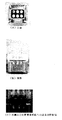
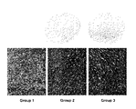
アルシアンブルー染色(軟骨基質である酸性ムコ多糖染色法)では、Group 1のペレットは、微かに染色されたが、分化誘導群であるGroup 2、3では強く染色された。超音波刺激群(Group 3)では極性の強い染色が見られた(図3)。
【0031】
抗アグリカン抗体による免疫染色では、Group 1 のペレットは微かに染色されたが、Group 2、3では強く染色された(図4)。超音波刺激群(Group 3)では極性の強い染色が見られた。免疫染色パターンはアルシアンブルー染色と同様であった。
【0032】
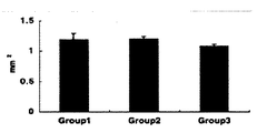
ペレットサイズは、Group 1 (1.19±0.10mm2)、Group 2 (1.19±0.05mm2)、Group 3 (1.08±0.04mm2)、であった。分化誘導による差は認めなかったが、超音波刺激群(Group 3)では7%の縮小が認められた(図5)。
【0033】
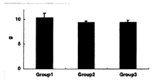
DNA量は、Group 1 (10.4±0.85μg)、Group 2 (9.36±0.28μg)、Group 3 (9.38±0.42μg)であった。軟骨分化誘導群(Group 2, 3)では、ともに約10%のDNA量低下が見られた。超音波刺激によるDNA産生への影響は認められなかった(図6)。
【0034】
総タンパク質量は、Group 1 (4.52±0.06μg)、Group 2 (4.76±0.47μg)、Group 3 (5.22±0.19μg)であった。TGF−β3無添加群(Group 1)と比較して、Group 2, 3 ではそれぞれ約5%、15%の増加が見られた(図7)。
【0035】
アグリカン量は、Group 1 (0.34±0.38ng)、Group 2 (1.38±0.76ng)、Group 3 (2.55±1.15ng)であった。アグリカン量は分化誘導により約4倍に増加した。さらに超音波刺激により84%の増加を見た(図8)。
【0036】
2.超音波出力と軟骨分化誘導能の検証
上記超音波照射装置を用いて、超音波出力と分化誘導能の指標としてアグリカン量との関係を調べた。
【0037】
超音波周波数:1.5MHz、バースト幅:200μsec、繰り返し周期:1.0kHz、超音波出力:0、30、60、120mW/cm2の4段階の出力で超音波パルスを、グループ3の超音波照射群の培養ヒト未分化間葉系細胞を入れたポリプロピレンチューブに対して、直接、水中に設置した超音波生成装置の上におき、1日20分間、10日間毎日照射した。コントロール群(−)として上記グループ1の結果についても図9に示した。
【0038】
超音波出力とアグリカン量について検討したところ、コントロール群 (図中で(−)表示)、0mW/cm2に比べ、超音波刺激群で有意なアグリカン量増加が見られた(120mW/cm2でP<0.05)。また、もっともアグリカン量増加のみられた超音波出力は30mWであった。(図9)
【0039】
【発明の効果】
本発明は、患者本人より採取した未分化間葉系細胞を、軟骨分化誘導培地をもちいた体外培養系にて培養する際に、超音波を照射することにより、従来の培養法よりも迅速に人工軟骨を作製することが出来る。
【0040】
従来から行われている、供給源の限られた軟骨細胞を軟骨障害部位へ移植する方法に替わり、患者自身の体から豊富に得ることのできる未分化間葉系細胞を利用し、これを速やかに軟骨細胞へ分化させることで人工軟骨を作成し、軟骨修復に使用し得ることができる。この方法を用いれば、同種移植を行う際、常に問題となる拒絶反応を回避することができる。
【図面の簡単な説明】
【図1】本発明の人工軟骨製造装置のブロックダイヤグラム。
【図2】本発明の人工軟骨製造装置の超音波生成装置部分の外観図。
【図3】ペレットのアルシアンブルー染色の結果。
【図4】アグリカンの免疫染色の結果。
【図5】各培養群のペレットサイズの結果。
【図6】各培養群のDNA量の結果。
【図7】各培養群の総タンパク質量の結果。
【図8】各培養群のアグリカン量の結果。
【図9】超音波出力とアグリカン量の関係。[0001]
BACKGROUND OF THE INVENTION
The present invention relates to a differentiation-inducing action by physical / mechanical stimulation load on undifferentiated cells. More specifically, by promoting the differentiation from undifferentiated cells to chondrocytes by irradiating undifferentiated cells with ultrasonic waves, it can be used for the treatment of chondrocyte transplantation, cartilage tissue transplantation, osteochondral cell transplantation, osteochondral tissue transplantation, etc. The present invention relates to a method and an apparatus for producing artificial cartilage produced for use.
[0002]
[Prior art]
In the treatment of cartilage injury, conservative therapy such as exercise restriction is mainly performed. However, the conservative therapy is often not effective, and in that case, treatment by surgery is also performed.
[0003]
For the operation of a cartilage disorder site, a method of collecting cartilage tissue or osteochondral tissue from a non-injury site and transplanting the tissue (Non-Patent Document 1: Hangody et al .: Foot Angle Int Oct; 18 (10): 628- 34, 1997), or a method of in vitro culture and proliferating chondrocytes collected from non-injured sites and transplanting them to the injured sites (Non-patent Document 2: Brittberg et al .: New Eng. J. Med. 331, 889- 895, 1994). However, in the method of collecting cartilage tissue from a non-injured site, the amount of collection is limited, and even when the collected chondrocytes are grown in vitro, it takes a long time to obtain a sufficient amount of chondrocytes. Was.
[0004]
On the other hand, it has been reported that when cultured chondrocytes are irradiated with ultrasound, biosynthesis of the cartilage matrix is increased (Non-patent Document 3: Parvisi et al .: J Orthop. Res., 17 (4): 488-94. , 1999, non-patent Document 4:.. nishikori et al .: J Biomed Master Res, 59 (2) 201-206, 2002, non-patent Document 5: Zi-Jun Zhang et al .: 4 th Symposium International Cartilage Repair Society , Tront, 15-18 / Jun, 2002).
[0005]
Ultrasound that produces such effects has been studied for fracture treatment by Duarte et al. (Patent Document 1: U.S. Pat. No. 4,530,360), and thereafter, the ultrasonic fracture treatment machine Safes TM (by US Exogen). SAFHS ™ ) has been developed. Ultrasound Fracture Machine Safes ™ (SAFHS ™ ) has been used in clinical trials for tibial shaft fractures (Non-Patent Document 6: Heckman et al .: J Bone and Jiont Surg, 76A: 25-34, 1994) and distal radius. Healing promotion effects such as end fractures (Non-Patent Document 7: Kristiansen et al .: J Bone and Joint Surg, 79A: 961-973, 1997) have been proven.
[0006]
In addition, Pittenger et al. Are able to differentiate undifferentiated mesenchymal cells into adipocytes, chondrocytes and bone cells in vitro, and a medium composition that causes differentiation into these three types of cells is also known ( Non-Patent Document 8: SCIENCE 284 (2): 143-147, 1999, Patent Document 2: WO 98/32333). Furthermore, when culturing undifferentiated mesenchymal cells in a bone differentiation-inducing medium, it has also been reported that bone differentiation is promoted by irradiating ultrasonic waves with the ultrasonic fracture treatment machine Safes TM (SAFHS TM ). (Non-Patent Document 9: Okada et al .: 4th Japan Society for Tissue Engineering, Kawasaki, July 6-7, 2001).
[0007]
[Patent Document 1]
US Pat. No. 4,530,360 [Patent Document 2]
WO 98/32333 pamphlet [Non-patent document 1]
Foot Angle Int Oct; 18 (10): 628-34, 1997
[Non-Patent Document 2]
New Eng. J. et al. Med. 331, 889-895, 1994
[Non-Patent Document 3]
J Orthop. Res. 17 (4): 488-94, 1999.
[Non-Patent Document 4]
J Biomed. Master Res. , 59 (2) 201-206, 2002
[Non-Patent Document 5]
4 th Symposium International Cartilage Repair Society, Tront, 15-18 / Jun, 2002
[Non-Patent Document 6]
J Bone and Jiont Surg, 76A: 25-34, 1994
[Non-Patent Document 7]
J Bone and Joint Surg, 79A: 961-973, 1997
[Non-Patent Document 8]
SCIENCE 284 (2): 143-147, 1999
[Non-patent document 9]
Okada et al: 4th Japan Society for Tissue Engineering, Kawasaki, July 6-7, 2001
[0008]
[Problems to be solved by the invention]
The present invention provides a method and apparatus for obtaining a sufficient amount of chondrocytes required in a short period of time when performing a surgical treatment because there is no effect on conservative therapy such as exercise restriction in the treatment of cartilage injury, and an apparatus therefor It is.
[0009]
[Means for Solving the Problems]
As a result of intensive studies on such problems, the present inventors have found that the cartilage differentiation is promoted by applying mechanical / mechanical stimulation called ultrasonic irradiation to cultured undifferentiated mesenchymal cells. It is a thing.
[0010]
That is, the present invention is based on a cell differentiation induction method characterized by irradiating undifferentiated cells with ultrasound, culturing undifferentiated mesenchymal cells in a cartilage differentiation induction medium, and irradiating with ultrasound. The present invention provides a method for producing artificial cartilage. In particular, the ultrasonic wave to be irradiated is an ultrasonic pulse, the frequency of the ultrasonic wave is 20 kHz to 10 MHz, the burst width is 10 μsec to 1 msec, the repetition period is 5 Hz to 10 kHz, and the ultrasonic output is 5 to 120 mW / cm 2 . Furthermore, the present invention provides a method for producing an artificial cartilage, wherein the ultrasonic frequency is 1.5 MHz, the burst width is 200 μsec, the repetition period is 1.0 kHz, and the ultrasonic output is 5 to 120 mW / cm 2. .
[0011]
The present invention also provides a culture container in which undifferentiated mesenchymal cells are cultured, an ultrasonic transducer that irradiates the container with ultrasonic waves, a control means that controls the ultrasonic waves, the ultrasonic transducer, and the culture An artificial cartilage manufacturing apparatus provided with a holding water tank for placing a container in contact with the water tank is provided.
[0012]
In the present invention, the control means is means for controlling the output of ultrasonic pulses from the control means. The frequency of the ultrasonic waves is 20 kHz to 10 MHz, the burst width is 10 μsec to 1 msec, and the repetition period is 5 Hz. The present invention provides an artificial cartilage manufacturing apparatus characterized by having an output of 10 to 10 kHz and an ultrasonic output of 5 to 120 mW / cm 2 .
[0013]
DETAILED DESCRIPTION OF THE INVENTION
The present invention relates to a method for inducing differentiation of undifferentiated cells, in particular, for inducing differentiation of undifferentiated mesenchymal cells derived from bone marrow into chondrocytes and producing artificial cartilage, and an ultrasonic irradiation apparatus used therefor, that is, producing artificial cartilage It relates to the device.
[0014]
Ultrasonic transducers that irradiate ultrasonic waves are made of piezoelectric ceramic. Using the property that minute distortion occurs when voltage is applied, an electrical signal generated by an ultrasonic waveform synthesizer is applied to the piezoelectric ceramic of the transducer. , Creating longitudinal vibrations (ultrasound) on the transducer surface.
[0015]
The ultrasonic wave used is a low-power ultrasonic pulse having an ultrasonic frequency of 20 kHz to 10 MHz, a burst width of 10 μsec to 1 msec, a repetition period of 5 Hz to 10 kHz, and an ultrasonic output of 5 to 120 mW / cm 2. Differentiation-inducing effects can be expected. Among them, in particular, the ultrasonic frequency is 1.5 MHz, the burst width is 200 μsec, the repetition period is 1.0 kHz, and the ultrasonic output is 5 to 120 mW / cm 2 , especially the output. 30 mW / cm 2 is preferred.
[0016]
The ultrasonic control means includes an ultrasonic waveform synthesis function for synthesizing ultrasonic waves emitted from the ultrasonic transducer, output control, storage calculation means, and power ON / OFF control function. Each function may be integrated in the main body, or may be divided into the ultrasonic transducer side and the main body.
[0017]
In the present invention, a polypropylene tube is used as a container for undifferentiated cultured cells in the present invention. However, the material and shape of the undifferentiated cultured cell are applicable as long as they can be used for cell culture. The ultrasonic transducer culture vessel is fixed in contact with the water tank in order to reliably irradiate ultrasonic waves.
[0018]
FIG. 1 shows a block diagram relating to ultrasonic irradiation of the artificial cartilage production apparatus of the present invention. The main unit is composed of a power source, a storage operation device, an input / output device, a transmission device, and an ultrasonic waveform synthesizer, and an ultrasonic generation device incorporating an ultrasonic transducer. In the Examples, six ultrasonic generators were fixed in a water tank with a frame, and an apparatus for irradiating ultrasonic waves onto a polypropylene tube containing the cultured human undifferentiated mesenchymal cells was used.
[0019]
1) Power supply:
The battery is a high-capacity lithium battery that cannot be charged. There is no power switch, the power is turned on by operating the front panel, and the power is automatically turned off when ultrasonic irradiation for a predetermined time is completed. The ultrasonic generator is also supplied with power from the same battery.
[0020]
In addition to the operation switch, liquid crystal display panel, and buzzer, the control signal of the ultrasonic generator is transmitted and the power is supplied through the connection cable.
[0021]
2) Memory arithmetic unit:
A CPU (central control unit) is mounted on the printed circuit board, and not only management of the ultrasonic irradiation time but also monitoring of the operation and mounting state of the ultrasonic generator and self-diagnosis are performed. In addition, the usage record is stored in the backup memory, and it is designed so that it can be taken out if necessary.
[0022]
3) Ultrasonic waveform synthesizer:
As a control signal for the ultrasonic generator, a signal having an ultrasonic pulse burst width of 200 μsec and a repetition period of 1 KHz is sent. An ultrasonic signal of 1.5 MHz is generated by the oscillation device. By combining the control signals and 1.5MHz ultrasonic signals, an ultrasonic frequency: 1.5MHz, ultrasonic power: adjustable from 0~150mW / cm 2, the burst width: 200 .mu.sec, repetition frequency: a 1kHz signal characteristic An electric signal having a waveform is generated.
[0023]
4) Ultrasonic generator The ultrasonic generator is composed of a transducer made of piezoelectric ceramic, and is coupled to the main body by a connection cable. Piezoelectric ceramics have the property of generating minute distortions when a voltage is applied. Using this, an electrical signal generated by the ultrasonic waveform synthesizer is applied to the piezoelectric ceramic inside the transducer, and longitudinal vibrations (ultrasound) are created on the transducer surface to irradiate the chondrocytes. An ultrasonic absorber is attached to the back surface of the piezoelectric ceramic to prevent ultrasonic leakage.
[0024]
【Example】
1. Verification of ability to induce cartilage differentiation by ultrasonic irradiation [cell culture]
Human undifferentiated mesenchymal cells are suspended in a 15 mL polypropylene tube, centrifuged at room temperature at 1,500 RPM for 5 minutes, and then left to stand at 37 ° C. in a 5% CO 2 environment for 24 hours. And continued culture. The medium includes a basal medium for chondrogenic differentiation (DMEM-high glucose, 0.17 mM Ascorbic acid-2-phosphate, 0.35 mM Proline, 1 mM Sodium pyruvate, 0.1 μM Dexamethasone, 6.25 μg / mL Bovine 25 μg / mL Bovine in 25 μg / mL Bovine). / ML Transferrin, 6.25 μg / mL Selenous acid, 5.33 μg / mL Linoleic acid, 1.25 mg / mL Bovine serum albumin).
[0025]
[Test group]
Such cultured human undifferentiated mesenchymal cells were divided into the following three groups, and their ability to induce cartilage differentiation by ultrasonic irradiation was verified.
Group 1 (n = 6): TGF-β3 non-added group Group 2 (n = 6): Cartilage differentiation induction group by adding TGF-β3 Group 3 (n = 6): TGF-β3 added +
[0026]
[Ultrasonic irradiation conditions]
Ultrasonic frequency: 1.5 MHz, burst width: 200 μsec, repetition period: 1.0 kHz, ultrasonic output: 120 mW / cm 2 ultrasonic pulse of cultured human undifferentiated mesenchymal cells in the ultrasonic irradiation group of
[0027]
[Measuring method]
1) Measurement of pellet size After 10 days of culture, the pellet was photographed under a stereomicroscope (n = 6). The pellet size was the maximum cross-sectional area.
[0028]
2) Histological examination The pellet was fixed with 10% formalin for 48 hours, dehydrated, embedded in paraffin, and stained with Alcian blue and anti-aggrecan antibody.
[0029]
3) Quantitative DNA, total protein, and aggrecan pellets were washed twice with PBS, then extracted with 4M guanidine hydrochloride for 24 hours at 4 ° C., and the soluble extract fraction was used for quantification of protein and aggrecan. The extraction residue was used for DNA quantification after Proteinase K treatment. In the soluble extract fraction, almost no DNA is detected.
[0030]
[result]
In Alcian blue staining (acid mucopolysaccharide staining method that is a cartilage matrix), the
[0031]
In the immunostaining with anti-aggrecan antibody,
[0032]
Pellet size is, Group 1 (1.19 ± 0.10mm 2 ), Group 2 (1.19 ± 0.05mm 2), Group 3 (1.08 ± 0.04mm 2), was. Although no difference due to differentiation induction was observed, a 7% reduction was observed in the ultrasound stimulation group (Group 3) (FIG. 5).
[0033]
The amount of DNA was Group 1 (10.4 ± 0.85 μg), Group 2 (9.36 ± 0.28 μg), and Group 3 (9.38 ± 0.42 μg). In the cartilage differentiation induction group (
[0034]
The total protein amount was Group 1 (4.52 ± 0.06 μg), Group 2 (4.76 ± 0.47 μg), Group 3 (5.22 ± 0.19 μg). Compared to the group without TGF-β3 (Group 1),
[0035]
The amount of aggrecan was Group 1 (0.34 ± 0.38 ng), Group 2 (1.38 ± 0.76 ng), and Group 3 (2.55 ± 1.15 ng). The amount of aggrecan increased about 4 times by differentiation induction. Furthermore, an increase of 84% was observed by ultrasonic stimulation (FIG. 8).
[0036]
2. Verification of ultrasonic output and cartilage differentiation inducing ability Using the above-described ultrasonic irradiation apparatus, the relationship between ultrasonic output and aggrecan amount as an index of differentiation inducing ability was examined.
[0037]
Ultrasonic frequency: 1.5 MHz, burst width: 200 μsec, repetition period: 1.0 kHz, ultrasonic output: 0, 30, 60, 120 mW / cm 2 , and ultrasonic pulses of group 3 A polypropylene tube containing cultured human undifferentiated mesenchymal cells in the irradiation group was directly placed on an ultrasonic generator placed in water and irradiated daily for 20 minutes for 10 days. The results of
[0038]
Was studied ultrasonic output and aggrecan amount, the control group (in the drawing (-) display), compared to 0 mW / cm 2, ultrasound stimulation group at significant aggrecan content increase was seen (at 120 mW / cm 2 P <0.05). Moreover, the ultrasonic output in which the amount of aggrecan was most increased was 30 mW. (Fig. 9)
[0039]
【The invention's effect】
In the present invention, when undifferentiated mesenchymal cells collected from the patient himself / herself are cultured in an in vitro culture system using a cartilage differentiation-inducing medium, ultrasonic irradiation is performed, thereby making the method faster than conventional culture methods. Artificial cartilage can be produced.
[0040]
Instead of the conventional method of transplanting chondrocytes with a limited supply source to the site of cartilage injury, undifferentiated mesenchymal cells that can be obtained abundantly from the patient's own body are used, and this is promptly performed. Artificial cartilage can be prepared by differentiating into chondrocytes and used for cartilage repair. By using this method, it is possible to avoid a rejection reaction that is always a problem when allogeneic transplantation is performed.
[Brief description of the drawings]
FIG. 1 is a block diagram of an artificial cartilage production apparatus according to the present invention.
FIG. 2 is an external view of an ultrasonic generation device portion of the artificial cartilage production apparatus of the present invention.
FIG. 3 shows the result of Alcian blue staining of the pellet.
FIG. 4 shows the results of immunostaining of aggrecan.
FIG. 5 shows the result of pellet size of each culture group.
FIG. 6 shows the result of DNA amount in each culture group.
FIG. 7 shows the result of total protein amount in each culture group.
FIG. 8 shows the results of the amount of aggrecan in each culture group.
FIG. 9 shows the relationship between the ultrasonic output and the amount of aggrecan.
Claims (6)
Priority Applications (3)
| Application Number | Priority Date | Filing Date | Title |
|---|---|---|---|
| JP2003098989A JP4676679B2 (en) | 2003-04-02 | 2003-04-02 | Artificial cartilage |
| US10/674,414 US20040197908A1 (en) | 2003-04-02 | 2003-10-01 | Artificial cartilage |
| CA002444359A CA2444359A1 (en) | 2003-04-02 | 2003-10-03 | Artificial cartilage |
Applications Claiming Priority (1)
| Application Number | Priority Date | Filing Date | Title |
|---|---|---|---|
| JP2003098989A JP4676679B2 (en) | 2003-04-02 | 2003-04-02 | Artificial cartilage |
Publications (2)
| Publication Number | Publication Date |
|---|---|
| JP2004307350A true JP2004307350A (en) | 2004-11-04 |
| JP4676679B2 JP4676679B2 (en) | 2011-04-27 |
Family
ID=33095200
Family Applications (1)
| Application Number | Title | Priority Date | Filing Date |
|---|---|---|---|
| JP2003098989A Expired - Lifetime JP4676679B2 (en) | 2003-04-02 | 2003-04-02 | Artificial cartilage |
Country Status (3)
| Country | Link |
|---|---|
| US (1) | US20040197908A1 (en) |
| JP (1) | JP4676679B2 (en) |
| CA (1) | CA2444359A1 (en) |
Cited By (2)
| Publication number | Priority date | Publication date | Assignee | Title |
|---|---|---|---|---|
| JP2006192181A (en) * | 2005-01-17 | 2006-07-27 | Teijin Pharma Ltd | Renal disease treatment apparatus |
| JP2008029317A (en) * | 2005-08-24 | 2008-02-14 | Olympus Terumo Biomaterials Corp | Method and apparatus for producing cultured bone |
Families Citing this family (4)
| Publication number | Priority date | Publication date | Assignee | Title |
|---|---|---|---|---|
| WO2010022508A1 (en) * | 2008-08-26 | 2010-03-04 | Intelligentnano Inc. | Enhanced animal cell growth using ultrasound |
| US8962290B2 (en) | 2008-08-26 | 2015-02-24 | Intelligentnano Inc. | Enhanced animal cell growth using ultrasound |
| US9012192B2 (en) | 2008-08-26 | 2015-04-21 | Intelligentnano Inc. | Ultrasound enhanced growth of microorganisms |
| CN116439231B (en) * | 2023-06-09 | 2023-09-19 | 山东第一医科大学(山东省医学科学院) | Ultrasonic stimulation method for improving preservation effect of in-vitro bone cartilage tissue |
Citations (2)
| Publication number | Priority date | Publication date | Assignee | Title |
|---|---|---|---|---|
| JP2001526942A (en) * | 1997-12-31 | 2001-12-25 | ファーマソニックス,インコーポレイテッド | Methods, systems, and kits for intravascular nucleic acid delivery |
| WO2002076508A1 (en) * | 2001-03-22 | 2002-10-03 | Chromos Molecular Systems, Inc. | Methods for delivering nucleic acid molecules into cells and assessment thereof |
Family Cites Families (2)
| Publication number | Priority date | Publication date | Assignee | Title |
|---|---|---|---|---|
| BR8107560A (en) * | 1981-11-19 | 1983-07-05 | Luiz Romariz Duarte | ULTRASONIC STIMULATION OF BONE FRACTURE CONSOLIDATION |
| US5326357A (en) * | 1992-03-18 | 1994-07-05 | Mount Sinai Hospital Corporation | Reconstituted cartridge tissue |
-
2003
- 2003-04-02 JP JP2003098989A patent/JP4676679B2/en not_active Expired - Lifetime
- 2003-10-01 US US10/674,414 patent/US20040197908A1/en not_active Abandoned
- 2003-10-03 CA CA002444359A patent/CA2444359A1/en not_active Abandoned
Patent Citations (2)
| Publication number | Priority date | Publication date | Assignee | Title |
|---|---|---|---|---|
| JP2001526942A (en) * | 1997-12-31 | 2001-12-25 | ファーマソニックス,インコーポレイテッド | Methods, systems, and kits for intravascular nucleic acid delivery |
| WO2002076508A1 (en) * | 2001-03-22 | 2002-10-03 | Chromos Molecular Systems, Inc. | Methods for delivering nucleic acid molecules into cells and assessment thereof |
Cited By (3)
| Publication number | Priority date | Publication date | Assignee | Title |
|---|---|---|---|---|
| JP2006192181A (en) * | 2005-01-17 | 2006-07-27 | Teijin Pharma Ltd | Renal disease treatment apparatus |
| JP4533758B2 (en) * | 2005-01-17 | 2010-09-01 | 帝人ファーマ株式会社 | Kidney disease treatment device |
| JP2008029317A (en) * | 2005-08-24 | 2008-02-14 | Olympus Terumo Biomaterials Corp | Method and apparatus for producing cultured bone |
Also Published As
| Publication number | Publication date |
|---|---|
| JP4676679B2 (en) | 2011-04-27 |
| US20040197908A1 (en) | 2004-10-07 |
| CA2444359A1 (en) | 2004-10-02 |
Similar Documents
| Publication | Publication Date | Title |
|---|---|---|
| Barba et al. | Adipose-derived mesenchymal cells for bone regereneration: state of the art | |
| US20070299539A1 (en) | Ultrasound accelerated tissue engineering process | |
| WO2003042376A1 (en) | Methods for preparing artificial cartilage | |
| Liu et al. | The effect of low-intensity pulsed ultrasound on the osseointegration of titanium dental implants | |
| JP6958846B1 (en) | Method for producing synovial membrane-derived mesenchymal stem cells and method for producing cell preparation for joint treatment | |
| Cao et al. | Effect of low-intensity pulsed ultrasound on the biological behavior of osteoblasts on porous titanium alloy scaffolds: An in vitro and in vivo study | |
| Tawonsawatruk et al. | Evaluation of native mesenchymal stem cells from bone marrow and local tissue in an atrophic nonunion model | |
| CN102068318B (en) | Manufacturing method of biological tooth root bracket material | |
| JP4676679B2 (en) | Artificial cartilage | |
| Zhao et al. | Oral cavity-derived stem cells and preclinical models of jaw-bone defects for bone tissue engineering | |
| Chen et al. | Low-intensity pulsed ultrasound promotes mesenchymal stem cell transplantation-based articular cartilage regeneration via inhibiting the TNF signaling pathway | |
| WO2015017772A1 (en) | Stem cell-based preparations and methods for dermal and connective tissue reconstruction and rejuvenation | |
| Min et al. | Low intensity ultrasound as a supporter of cartilage regeneration and its engineering | |
| JP4703943B2 (en) | Agent for imparting resistance to external stimuli | |
| JP4533758B2 (en) | Kidney disease treatment device | |
| US20060236747A1 (en) | Sonic activation of strain sensitive cells | |
| Graziano et al. | Sinus lift augmentation using pulp stem cells: A case report and histological evaluation | |
| US20110177576A1 (en) | Shock wave cell treatment device and method to enhance cell replication | |
| JP2005304918A (en) | Wound treatment device | |
| US11813293B2 (en) | Fat-depleted adipose tissue and a device and method for preparing the same | |
| Moinnes et al. | Ultrasound accelerated bone tissue engineering monitored with magnetic resonance microscopy | |
| Elkhenany et al. | Important considerations in the therapeutic application of stem cells in bone healing and regeneration | |
| Mishra | Stem Cells: A Step Ahead in Regenerative Dentistry with Accent on Orthodontics | |
| JP2008029317A (en) | Method and apparatus for producing cultured bone | |
| KR100957586B1 (en) | Method and apparatus for improvement of proliferation or activity of anterior cruciate ligament cell using sonic vibration |
Legal Events
| Date | Code | Title | Description |
|---|---|---|---|
| A621 | Written request for application examination |
Free format text: JAPANESE INTERMEDIATE CODE: A621 Effective date: 20060119 |
|
| A131 | Notification of reasons for refusal |
Free format text: JAPANESE INTERMEDIATE CODE: A131 Effective date: 20090728 |
|
| A521 | Request for written amendment filed |
Free format text: JAPANESE INTERMEDIATE CODE: A523 Effective date: 20090925 |
|
| A02 | Decision of refusal |
Free format text: JAPANESE INTERMEDIATE CODE: A02 Effective date: 20100511 |
|
| A521 | Request for written amendment filed |
Free format text: JAPANESE INTERMEDIATE CODE: A523 Effective date: 20100802 |
|
| A521 | Request for written amendment filed |
Free format text: JAPANESE INTERMEDIATE CODE: A821 Effective date: 20100802 |
|
| A911 | Transfer to examiner for re-examination before appeal (zenchi) |
Free format text: JAPANESE INTERMEDIATE CODE: A911 Effective date: 20100903 |
|
| TRDD | Decision of grant or rejection written | ||
| A01 | Written decision to grant a patent or to grant a registration (utility model) |
Free format text: JAPANESE INTERMEDIATE CODE: A01 Effective date: 20110104 |
|
| A01 | Written decision to grant a patent or to grant a registration (utility model) |
Free format text: JAPANESE INTERMEDIATE CODE: A01 |
|
| A61 | First payment of annual fees (during grant procedure) |
Free format text: JAPANESE INTERMEDIATE CODE: A61 Effective date: 20110128 |
|
| FPAY | Renewal fee payment (event date is renewal date of database) |
Free format text: PAYMENT UNTIL: 20140204 Year of fee payment: 3 |
|
| R150 | Certificate of patent or registration of utility model |
Ref document number: 4676679 Country of ref document: JP Free format text: JAPANESE INTERMEDIATE CODE: R150 Free format text: JAPANESE INTERMEDIATE CODE: R150 |
|
| R250 | Receipt of annual fees |
Free format text: JAPANESE INTERMEDIATE CODE: R250 |
|
| R250 | Receipt of annual fees |
Free format text: JAPANESE INTERMEDIATE CODE: R250 |
|
| R250 | Receipt of annual fees |
Free format text: JAPANESE INTERMEDIATE CODE: R250 |
|
| R250 | Receipt of annual fees |
Free format text: JAPANESE INTERMEDIATE CODE: R250 |
|
| R250 | Receipt of annual fees |
Free format text: JAPANESE INTERMEDIATE CODE: R250 |
|
| R250 | Receipt of annual fees |
Free format text: JAPANESE INTERMEDIATE CODE: R250 |
|
| R250 | Receipt of annual fees |
Free format text: JAPANESE INTERMEDIATE CODE: R250 |
|
| R250 | Receipt of annual fees |
Free format text: JAPANESE INTERMEDIATE CODE: R250 |
|
| R250 | Receipt of annual fees |
Free format text: JAPANESE INTERMEDIATE CODE: R250 |
|
| R250 | Receipt of annual fees |
Free format text: JAPANESE INTERMEDIATE CODE: R250 |
|
| EXPY | Cancellation because of completion of term |