JP2004113108A - Method for collecting dna - Google Patents
Method for collecting dna Download PDFInfo
- Publication number
- JP2004113108A JP2004113108A JP2002280660A JP2002280660A JP2004113108A JP 2004113108 A JP2004113108 A JP 2004113108A JP 2002280660 A JP2002280660 A JP 2002280660A JP 2002280660 A JP2002280660 A JP 2002280660A JP 2004113108 A JP2004113108 A JP 2004113108A
- Authority
- JP
- Japan
- Prior art keywords
- cdna fragment
- pcr
- sequence
- primer
- cdna
- Prior art date
- Legal status (The legal status is an assumption and is not a legal conclusion. Google has not performed a legal analysis and makes no representation as to the accuracy of the status listed.)
- Pending
Links
- 0 N*1CCCCC1 Chemical compound N*1CCCCC1 0.000 description 1
Images


Classifications
-
- C—CHEMISTRY; METALLURGY
- C12—BIOCHEMISTRY; BEER; SPIRITS; WINE; VINEGAR; MICROBIOLOGY; ENZYMOLOGY; MUTATION OR GENETIC ENGINEERING
- C12Q—MEASURING OR TESTING PROCESSES INVOLVING ENZYMES, NUCLEIC ACIDS OR MICROORGANISMS; COMPOSITIONS OR TEST PAPERS THEREFOR; PROCESSES OF PREPARING SUCH COMPOSITIONS; CONDITION-RESPONSIVE CONTROL IN MICROBIOLOGICAL OR ENZYMOLOGICAL PROCESSES
- C12Q1/00—Measuring or testing processes involving enzymes, nucleic acids or microorganisms; Compositions therefor; Processes of preparing such compositions
- C12Q1/68—Measuring or testing processes involving enzymes, nucleic acids or microorganisms; Compositions therefor; Processes of preparing such compositions involving nucleic acids
- C12Q1/6844—Nucleic acid amplification reactions
- C12Q1/6853—Nucleic acid amplification reactions using modified primers or templates
- C12Q1/6855—Ligating adaptors
-
- C—CHEMISTRY; METALLURGY
- C12—BIOCHEMISTRY; BEER; SPIRITS; WINE; VINEGAR; MICROBIOLOGY; ENZYMOLOGY; MUTATION OR GENETIC ENGINEERING
- C12Q—MEASURING OR TESTING PROCESSES INVOLVING ENZYMES, NUCLEIC ACIDS OR MICROORGANISMS; COMPOSITIONS OR TEST PAPERS THEREFOR; PROCESSES OF PREPARING SUCH COMPOSITIONS; CONDITION-RESPONSIVE CONTROL IN MICROBIOLOGICAL OR ENZYMOLOGICAL PROCESSES
- C12Q2525/00—Reactions involving modified oligonucleotides, nucleic acids, or nucleotides
- C12Q2525/10—Modifications characterised by
- C12Q2525/191—Modifications characterised by incorporating an adaptor
Abstract
Description
【0001】
【発明の属する技術分野】
本発明は、目的とするDNA断片を回収する方法に関し、特に、様々な種類のcDNA断片が混在するcDNA断片の群から目的とするcDNA断片を回収するDNA回収法に関する。
【0002】
【従来の技術】
従来より、様々なcDNA断片が混在するcDNA断片の群から、目的とするcDNA断片を回収するDNA回収法が、いくつか知られている。
例えば、平板ゲルを利用して、cDNA断片の群から目的とするcDNA断片を回収する方法がある(例えば、非特許文献1参照。)。即ち、cDNA断片の群について、例えばアクリルアミドゲル電気泳動を行う。その後、アクリルアミドゲル中のcDNA断片をエチジウムブロミドで染色し、UVトランスイルミネータによりアクリルアミドゲルを照らしてcDNA断片を検出する。そして、分画されたcDNA断片の群から目的のcDNA断片を含むアクリルアミドゲルを切り出し、そのゲルから当該cDNA断片を回収する。
【0003】
また、例えば、キャピラリー電気泳動を利用して、cDNA断片の群から目的とするcDNA断片を回収する方法もある(例えば、特許文献1〜3参照。)。即ち、予め蛍光標識したcDNA断片の群をキャピラリーカラムに供試し電気泳動を行う。そして、検出されたピークに基づいて、目的のcDNA断片が泳動されて来る時間を推測する。次に、再びキャピラリーカラムで電気泳動を行い、その推定時間をもとに目的のcDNA断片を回収する。
【0004】
【非特許文献1】
A new method for sequencing DNA.
Proc.Natl.Acad.Sci. 1977,74:560−564
【特許文献1】
特開2000−88803号公報
【特許文献2】
特開平7−181164号公報
【特許文献3】
特開平6−138037号公報
【0005】
【発明が解決しようとする課題】
しかしながら、前者の方法では、目的とするcDNA断片が少量しか存在しない場合、ゲル電気泳動後、cDNA断片を染色しても、UVトランスイルミネータ下でそれを検出することができないため、目的とするcDNA断片を回収できないという問題がある。
一方、後者の方法では、蛍光標識したcDNA断片を使用しているので、目的とするcDNA断片が少量であっても、これを検出することは可能である。しかしながら、この方法では、cDNA断片を目視しながら回収することができないため、目的のcDNA断片がキャピラリーカラムの先端部分に現れる時間を予想して回収する。そのため、回収したcDNA断片には、目的とするcDNA断片に加え、目的物とほぼ同じくらいの長さのcDNA断片が混在する可能性がある。また、サイズ分画能が平板ゲルを使用した場合と比較すると良くないため、回収したcDNA断片の中に目的物以外のものが混在したり、また、目的とするcDNA断片の回収率の低下を招くこともある。
【0006】
本発明はかかる現状に鑑みてなされたものであって、様々な種類のcDNA断片が混在するcDNA断片の群から目的とするcDNA断片をより確実に回収することができるDNA回収法を提供することを目的とする。
【0007】
【課題を解決するための手段、作用及び効果】
その解決手段は、一端に第1アダプター配列を有し他端に第2アダプター配列を有するcDNA断片の群について、上記第1アダプター配列に相補的な配列を有すると共に標識物質を有する第1プライマーと、上記第2アダプター配列に相補的な配列を有する第2プライマーとを用いて、PCR反応を行う第1PCR工程と、上記第1PCR工程で増幅されたcDNA断片の群についてゲル電気泳動を行う電気泳動工程と、上記電気泳動工程の結果に基づいて、ゲルから目的のcDNA断片を含むゲルを切り出し、当該cDNA断片を回収する回収工程と、を備えることを特徴とするDNA回収法である。
【0008】
本発明のDNA回収法は、両端にアダプター配列を有するcDNA断片の群から、目的とするcDNA断片を回収する方法である。まず、cDNA断片の一端にある第1アダプター配列に相補的な配列を有すると共に標識物質を有する第1プライマーと、cDNA断片の他端にある第2アダプター配列に相補的な配列を有する第2プライマーとを用いて、PCR反応を行う。そして、増幅されたcDNA断片の群(PCR産物)をゲル電気泳動する。その後、その電気泳動の結果に基づいて、ゲルから目的のcDNA断片を含むゲルを切り出し、そこからcDNA断片を回収する。
【0009】
このような方法によれば、予めゲル電気泳動前にcDNA断片の群についてPCR反応を行うので、cDNA断片の群を多量に増幅することができる。従って、目的とするcDNA断片が低濃度であっても、それを大幅に増幅することができ、ゲル電気泳動においてそれを検出することができる。
また、このPCR反応では、標識物質を有するプライマーを用いているので、PCR産物も標識物質を有することになる。従って、PCR反応を行っても依然として目的とするcDNA断片が比較的少ない場合であっても、電気泳動においてこの標識物質を認識することで、目的とするcDNA断片のゲル中の位置を容易に検出することができる。
さらに、cDNA断片の群をゲル電気泳動によって分画するため、例えばキャピラリーカラムによる電気泳動に比べ、サイズ分解能を向上させ、目的とするcDNA断片のみをより特異的に回収することができる。
【0010】
ここで、サンプルとする様々なcDNA断片からなるcDNA断片の群については、一端に第1アダプター配列を有し他端に第2アダプター配列を有するものであれば、いかなるものであってよい。即ち、cDNA断片の由来は問われず、動植物細胞から抽出したmRNAから作製したものであっても、ウイルスや微生物から抽出したmRNAから作製したものであってもよい。また、cDNA断片の群は、抽出に用いた細胞で発現されたほとんどすべての遺伝子が含まれる群であっても、その発現された遺伝子の一部だけが含まれる群であってもよい。また、cDNA断片は、全長cDNAの両端にアダプター配列が付いたものでも、全長cDNAの一部分をなす断片の両端にアダプター配列が付いたものでもよい。また、第1アダプター配列及び第2アダプター配列は、それぞれいかなる塩基配列からなるものであっても構わないが、PCRの効率等を考慮して設計されたものであるのが好ましい。即ち、これらのアダプター配列がそれぞれ15塩基前後であると、安定したPCR反応を行うことができ、効率よくcDNA断片を増幅することができる。
【0011】
第1プライマーは、第1アダプター配列に相補的な配列を有し、かつ、標識物質を有するものであれば、いかなるものであってもよい。ここでいう相補的な配列とは、第1アダプター配列に対し100%相補的な配列に限られず、PCR反応においてcDNA断片を増幅されることができる程度に実質的に相補的な配列も含まれる。また、第1プライマーは、第1アダプター配列に相補的な配列のみからなるものに限られず、第1アダプター配列に相補的な配列に、さらに他の配列が繋がったものであってもよい。第1プライマーは、第1アダプター配列全体に対応するものに限られず、第1アダプター配列の一部に対応するものでもよい。
【0012】
また、標識物質は、ゲル電気泳動において検出感度が高いものであれば、いずれのものを使用することもできる。例えば、6−カルボキシフルオレッセイン(以下、FAMと称す。)、4,7,2’,4’,5’,7’−ヘキサクロロ−6−カルボキシフルオレッセイン(以下、HEXと称す。)、NED(アプライドバイオシステムズジャパン社)、6−カルボキシ−X−ローダミン(以下、Roxと称す。)等の蛍光物質などを使用することができる。これらの標識物質は、例えば、プライマーDNAの末端(例えば5’末端)に結合させればよい。
【0013】
第2プライマーは、第2アダプター配列に相補的な配列を有するものであれば、いかなるものであってもよい。ここでいう相補的な配列も、第2アダプター配列に対し100%相補的な配列に限られず、PCR反応においてcDNA断片を増幅させることができる程度に実質的に相補的な配列も含まれる。また、第2プライマーについても、第2アダプター配列に相補的な配列のみからなるものに限られず、第2アダプター配列に相補的な配列に、さらに他の配列が繋がったものであってもよい。また、第2プライマーについても、第2アダプター配列全体に対応するものに限られず、第2アダプター配列の一部に対応するものでもよい。さらに、上述の第1プライマーだけでなく、この第2プライマーにも、上述の標識物質が結合されていても構わない。
【0014】
次に、その他一般にPCR反応で用いられる種々の試薬について説明する。
DNAポリメラーゼは、PCR反応においてDNA鎖を変性させる際の高温に短時間加熱されても永久的には不活性化されず、しかも、高温における活性を有するものが好適である。例えば、サーモコッカス・リトラリス(Thermococcus litoralis)、バチルス・ステアロサーモフィルス(Bacillus stearothermophilus)、メタノサーマス・フェルビドゥス(Methanothermus fervidus)、サーマス・アクアティクス(Thermus aquaticus)、T.フラブス(T.flavus)、T.ラクテウス(T.lacteus)、T.ルベンス(T.rubens)、T.ルバー(T.ruber)などの高熱菌由来のDNAポリメラーゼや、デスルフロコッカス・モビリス(Desulfurococcus mobilis)、メタノバクテリウム・サーモオートトロフィルクム(Methanobacterium thermoautotrophilcum)、スルホロブス・ソルファタリクス(Sulfolobus solfataricus)、S.アシドカルダリウス(S.acidocaldarius)、サーモプラスマ・アシドフィルム(Thermoplasma acidophilum)、ピロコッカス・コダカラエンシス(Pyrococcus kodakaraensis KOD1株)などの高熱性古細菌由来のDNAポリメラーゼなどが挙げられる。これらのうち、入手容易性等の理由から、サーマス・アクアティクス(Thermus aquaticus)由来のDNAポリメラーゼ(Taq DNAポリメラーゼ)、サーモコッカス・リトラリス(Thermococcus litoralis)由来のDNAポリメラーゼ、あるいは、ピロコッカス・コダカラエンシス(Pyrococcus kodakaraensis KOD1株)由来のDNAポリメラーゼを利用するのが好ましい。
【0015】
さらに、PCR反応液には、DNAポリメラーゼによる核酸増幅前の活性を阻害するために、DNAポリメラーゼに特異的な抗体を混合してもよい。この抗体には、モノクローナル抗体、ポリクローナル抗体、組換え法により製造された抗体、化学的または組換え法により製造された抗体フラグメント(例えば、Fabフラグメント)が挙げられる。これらのうち、モノクローナル抗体を用いるのが特に好ましい。例えば、Taq DNAポリメラーゼに対する公知のモノクローナル抗体は、約20℃〜40℃においてTaq DNAポリメラーゼの酵素活性を阻害することができると共に、PCRの熱的サイクルにおける高温によって不活性化される。
【0016】
また、PCR反応は、一般に、4種類のdNTP、即ち、dATP、dCTP、dGTP及びdTTPの存在化において行う。
さらに、PCR反応は、一般に、適当な緩衝剤を含む反応液中で行われる。効率よく核酸を増幅させるためである。緩衝液は、使用するDNAポリメラーゼ等により、反応の最適条件を得るため適宜変更することができる。例えば、pHを適当に調整したトリス系の緩衝液に、塩化カリウムや塩化マグネシウムを加えた緩衝液を利用することができる。
また、PCR反応液には、5%〜10%のDMSOと1%〜2%のベタインを添加してもよい。鋳型DNAとなるcDNA断片が二字構造を有する場合に産物が増幅されにくいという問題を、最小限に留める効果を有するものである。
【0017】
電気泳動工程では、アクリルアミドゲル電気泳動やアガロースゲル電気泳動など、公知の平板ゲル電気泳動などにより、cDNA断片(PCR産物)を電気泳動し、cDNA断片を分画する。電気泳動には、公知の電気泳動装置を利用すればよい。
【0018】
回収工程は、電気泳動工程の結果に基づいて、ゲルから目的とするcDNA断片を含むゲルを切り出し、そのDNA断片を回収する工程であれば、いかなる方法を利用することもできる。
即ち、例えば、後述する実施例に示すように、標識物質を検出することができるDNAシークエンサーにcDNA断片を供試して電気泳動工程を行い、その解析結果から、回収するcDNA断片を決定する。次に、再度同一サンプルを電気泳動して、目的とするcDNA断片が検出されたときに、その部分のゲルを切り出し、そこからcDNA断片を回収する方法が挙げられる。
また、ゲル電気泳動後、標識物質を検出することができるスキャナー上にゲルを載置し、その解析結果から回収するcDNA断片を決定する。そして、その部分のゲルを切り出し、そこからcDNA断片を回収する方法が挙げられる。
【0019】
ところで、両端にアダプター配列を有するcDNA断片の群は、例えば、次のようにして作製することができる。
即ち、細胞から抽出したmRNAから、その5’末端にタグ物質を付加されたcDNA断片を合成する工程と、得られたcDNA断片を第1制限酵素によって切断する工程と、上記タグ物質に高親和性を有する高親和性物質を結合させることにより、タグ物質を有するcDNA断片を回収する工程と、回収されたcDNA断片に、上記第1制限酵素の酵素切断部位の配列に相補的な配列を有する第1アダプター配列を結合させる工程と、上記第1アダプター配列を結合させたcDNA断片を第2制限酵素によって切断する工程と、上記高親和性物質を結合させて、上記タグ物質を有するcDNA断片を除去し、上記タグ物質を有しないcDNA断片を回収する工程と、上記タグ物質を有しないcDNA断片に、上記第2制限酵素の酵素切断部位の配列に相補的な配列を有する第2アダプター配列を結合させる工程と、を備える方法によって、両端にアダプター配列を有するcDNA断片の群を作製することができる。
さらに、このようなcDNA断片の群について、上記第1アダプター配列に相補的な配列を有しかつその3’末端に任意の2塩基配列であるNNを有するプライマーと、上記第2アダプターの配列に相補的な配列を有しかつその3’末端に任意の2塩基配列であるNNを有するプライマーとを用いて、PCR反応を行う工程により、上記のcDNA断片の群を小規模なcDNA断片の群に分類することもできる。
【0020】
このような方法によってcDNA断片の群を作製すれば、発現されたほとんど全ての遺伝子、即ち、公知の遺伝子も未知の遺伝子も同様に、その群の中に含まれるようにすることが可能である。従って、遺伝子解析等において有効に活用することができる。また、解析が容易となるように、cDNA断片の群を複数の小規模な群に分類することができる。
【0021】
なお、タグ物質とタグ物質に高親和性を有する高親和性物質とは、互いに高親和性をもって特異的に結合することが可能な結合対を構成する物質を示す。タグ物質と高親和性物質の組合せとしては、例えば、ビオチンとストレプトアビジン、ビオチンとアビジン、FIGTとFITC抗体、DIGとアンタイDIG、プロテインAとマウスIgG、ラテックス粒子等が挙げられる。また、各組合せにおいて、いずれをタグ物質として使用しても、いずれを高親和性物質として使用してもよい。
【0022】
また、制限酵素とは、一般的に、制限エンドヌクレアーゼとも称される酵素であり、特定の配列において二本鎖DNAを加水分解し切断する酵素である。上記の方法においては、適切な断片を得るために2種類の制限酵素(第1制限酵素及び第2制限酵素)を組み合わせて使用する。使用する制限酵素は、cDNA断片を識別可能な長さを有する断片に切断することが可能なものが好ましい。また、合成されたcDNA断片のより多くを、好ましくは、ほとんど全てを切断するような酵素が好ましい。また、制限酵素は、4塩基認識酵素を使用しても6塩基認識酵素を使用してもよいが、特に、上記の理由から、4塩基認識酵素の使用が好ましい。
【0023】
また、アダプター配列は、PCR増幅の際に用いるプライマーを結合させるために用いるものであるが、使用する制限酵素に応じて設計される。即ち、第1制限酵素の酵素切断部位に結合させるための第1アダプター配列は、第1制限酵素の酵素切断部位に相補的な配列を有し、また、第2制限酵素の酵素切断部位に結合させるための第2アダプター配列は、第2制限酵素の酵素切断部位に相補的な配列を有する。
【0024】
また、プライマーセットは、cDNA断片をPCRにより増幅するために使用する1組のプライマーからなる。ここで使用される「任意の2塩基配列であるNN」は、A、T、G、Cから任意に選択される配列である。ここで、各任意の配列を2塩基としたのは、当該方法の簡便性と解析精度を考慮した結果である。つまり、各任意の配列を2塩基とすることにより、最大256種類のプライマーセットが得られるため、cDNA断片の群を複数の群に分類することができる。なお、この任意の2塩基配列NNを、一方のプライマーまたは両方のプライマーについて3塩基以上とすることも可能である。それによってプライマーの種類が増え、プライマーセットを最大1024個や最大4096個とすることができる。
【0025】
さらに、上記のいずれかに記載のDNA回収法であって、前記回収工程で回収したcDNA断片について、前記第1アダプター配列に相補的な配列を有する第3プライマーと、前記第2アダプター配列に相補的な配列を有する第4プライマーとを用いて、再度PCR反応を行う第2PCR工程を備えることを特徴とするDNA回収法とすると良い。
【0026】
本発明では、回収工程で回収したcDNA断片について、再度PCR反応を行う第2PCR工程を備える。このため、回収したcDNA断片が少量しかなくても、それを大幅に増幅させることができる。
なお、このPCR工程で使用する第3プライマーは、第1アダプター配列に相補的な配列を有するものであればよく、例えば、上記の第1プライマーを利用しても良いが、このPCR工程では標識物質は特に要しない。同様に、第4プライマーは、第2アダプター配列に相補的な配列を有するものであればよく、例えば、上記の第2プライマーを利用しても良い。
【0027】
さらに、上記のDNA回収法であって、前記第2PCR工程において、RecAタンパク質及びこのRecAタンパク質を改変したタンパク質であってこのRecAタンパク質と類似する機能を有するRecA改変タンパク質の少なくともいずれかを含む相同的組換えタンパク質を反応液に混合して、PCRを行うことを特徴とするDNA回収法とすると良い。
【0028】
本発明によれば、第2PCR工程において、RecAタンパク質等の相同組み換えタンパク質を反応液に混合してPCRを行い、回収したcDNA断片を増幅させる。
このようにPCRを行えば、cDNA断片の収量を減少させることなく、副産物(非特異的なPCR産物)の増幅を低く抑えることができる。即ち、上記の相同組み換えタンパク質が存在することにより、プライマーがcDNA断片の非特異的な領域に結合してプライマー伸長反応を起こすことが抑制されるため、非特異的なPCR産物の増幅を抑制することができる。
【0029】
また、本発明では、上記のように特異性が高いため、アニーリング温度等、プライマー伸長反応の温度条件を変えても、回収したcDNA断片を特異的に増幅させることができる。即ち、従来のPCR方法では、アニーリング温度等、プライマー伸長反応の温度を低く設定すると、回収したcDNA断片だけでなく、副産物も多量に増幅されることとなるが、本発明によれば、回収したcDNA断片をより特異的に増幅させることが可能となる。
これらの結果、目的とするcDNA断片をさらに確実に回収することができる。
【0030】
ここで、上記の相同組み換えタンパク質は、RecAタンパク質、及び、RecAタンパク質を改変したものであって RecAタンパク質と類似する機能を有するRecA改変タンパク質、の少なくともいずれかを含むものであれば、いかなるものを用いてもよい。RecA改変タンパク質としては、例えば、RecAタンパク質をコードする遺伝子から、部位特異的変位誘発等により作出された遺伝子の産物であって、1または数個のアミノ酸が欠損、置換若しくは付加されたアミノ酸配列からなり、かつ、RecAタンパク質と類似する機能を有するものが挙げられる。また、RecAタンパク質のタンパク質断片であって、タンパク質と類似する機能を有するもの(RecAフラグメント)などであってよい。
【0031】
また、RecAタンパク質等は、上述したDNAポリメラーゼと同様に、PCR反応においてDNA鎖を変性させる際の高温に短時間加熱されても永久的には不活性化されず、しかも、高温における活性を有するものが好適である。例えば、入手容易性等の理由から、大腸菌(E.coli)由来のRecAタンパク質を利用するのが好ましい。
【0032】
なお、上記の相同組み換えタンパク質は、プライマー20pmolあたり、1ng〜1000ngの範囲で混合するのが好ましい。このような範囲でPCRを行えば、より効率よく特異的に、cDNA断片を増幅することができるからである。
【0033】
さらに、上記のいずれかに記載のDNA回収法であって、前記回収工程後、または、前記第2PCR工程を有する場合にはこの第2PCR工程後、前記cDNA断片をプラスミドベクターに連結し、組換え体プラスミドを形成する連結工程と、上記組換え体プラスミドを大腸菌に導入する導入工程と、を備えることを特徴とするDNA回収法とすると良い。
【0034】
本発明のDNA回収法は、第2PCR工程を有しない場合には回収工程後、または、第2PCR工程を有する場合には第2PCR工程後、目的のcDNA断片をプラスミドベクターに連結し、組換え体プラスミドを形成する連結工程を備える。そしてさらに、この組換え体プラスミドを大腸菌に導入する導入工程を備える。
このように、回収したcDNA断片をプラスミドベクターに連結し、大腸菌に導入しておけば、cDNA断片の構造解析をする場合などに有効である。即ち、形質転換したその大腸菌を培養し、それからcDNA断片を有するプラスミドDNAを抽出すれば、これを例えば塩基配列の決定等の構造解析に用いることができる。
【0035】
【発明の実施の形態】
(実施例1)
以下、本発明の実施例を、図を参照しつつ説明する。
本実施例においては、まず、特定の細胞において発現された遺伝子を以下のように分類する。まず、抽出したmRNAからcDNA群を合成する。そして、得られたcDNA群を適切な2つの制限酵素によって切断し、また、両端にアダプター配列を結合して、識別可能なだけの長さを有しかつ両端にアダプター配列を有するcDNA断片の群を作成する。その後、得られたcDNA断片の群を、複数種類のプライマーセットを用いて複数の群に分類する。
【0036】
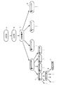
この分類の手法を図1を参照しつつ説明する。発現されたmRNAからなる群1から公知の手法によりcDNAの群2を合成する。これを適切な2つの制限酵素によって切断してcDNA断片の群3を得る。そして、このcDNA断片の両末端の各2塩基、全4塩基の配列に応じて、即ち、4塩基がA、T、G、Cのいずれであるかにより、cDNA断片の群3を分類する。例えば、5’末端の塩基(図1中に黒塗りで示す。)を区別することで、群3を4種類の群4に分類することができる。そして、次の2番目の塩基(図1中に黒塗りで示す。)を区別することで、群4をそれぞれ4種類の群5に分類することができる。さらに、3’末端の2番目の塩基(図1中に黒塗りで示す。)を区別することで、群5をそれぞれ4種類の群6に分類することができる。またさらに、3’末端の塩基(図1中に黒塗りで示す。)を区別することで、群6をそれぞれ4種類の群7に分類することができる。
【0037】
次に、抽出したmRNAの群1から複数のcDNA断片の群7を調製する手法について、図2を参照しつつ具体的に説明する。図2における各アルファベットは塩基配列を構成する塩基を示すが、N、M、W、X、Y及びZは任意の塩基を示し、XとY及びWとZは、互いに相補的に結合する。
【0038】
まず、試験対象となる特定の細胞から公知の手法によりmRNA11を抽出する。本実施例では、酵母から、Fast Track 2.0 kit(Invitrogen社製)を用いて、総mRNA11を20μg抽出した。
【0039】
次に、抽出されたmRNA11の3’末端側のポリAテイルに相補的なオリゴdTプライマーをビオチン(タグ物質)13で標識化する。そして、これをプライマーとして用いてcDNA12を合成する。具体的には、抽出した20μgのmRNA11を、0.8μl中で100pmoleの5’−ビオチン化オリゴdTプライマー(BRL社製)と混合し、65℃で5分間インキュベートした。続いて、それを水冷した後、逆転写バッファー20.0μl中で、最終濃度5mMのMgCl2と0.5mMのdNTPミックス(BRL社製)と10mMのDTT(BRL社製)を42℃で60分間インキュベートした。続いて、二本鎖合成バッファー150.0μl中で最終濃度0.27mMのdNTPミックス(BRL社製)と1.33mMのDTT(BRL社製)と20.0ユニットのE.coliリガーゼ(BRL社製)と40.0ユニットのE.coli DNAポリメラーゼ(BRL社製)と2.0ユニットのRNaseH(BRL社製)を16℃で120分間インキュベートし、続いて、70℃で15分間インキュベートし反応を止めた。
【0040】
次に、合成されたcDNA12を第1制限酵素を用いて切断する。具体的には、100μl中で最終濃度20ユニットの制限酵素MspI(宝酒造社製)と10μgのcDNA12とを、37℃で360分間反応させた。なお、MspIは、4塩基認識制限酵素である。その後、エタノールを用いてcDNA断片を公知の手法により精製した。
【0041】
次に、ストレプトアビジン(高親和性物質)14を用いてビオチン13を捕捉し、切断されたcDNA断片の3’末端側のみを回収する。具体的には、反応液に磁気ビーズに固定したストレプトアビジン14(ダイナル社製)に対して、ビオチン13を結合し、産物を得た。
【0042】
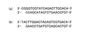
次に、回収されたcDNA断片の5’末端側に、第1制限酵素の認識切断部位に相補的な配列を有する第1アダプター配列15を結合させる。本実施例で使用した第1アダプター配列15について、図3(a)に示す。具体的には、15μlのT4 DNAリガーゼバッファー中でCG突出末端(即ち、制限酵素MspIの切断断片部位の相補的配列である)を有する5.0μgの第1アダプター(BRL社製)と、10ユニットのT4 DNAリガーゼ(NEB社製)を用いてライゲ−ションを行った。
【0043】
次に、このcDNA断片を第2制限酵素を用いて切断する。具体的には、200μl中で最終濃度50ユニットの制限酵素MseI(NEB社製)とcDNA断片とを、37℃で360分間反応させた。なお、このMseIも、4塩基認識制限酵素である。その後、エタノールを用いてcDNA断片を公知の手法により精製した。
【0044】
次に、ストレプトアビジン14を用いてビオチン13を捕捉し、切断されたcDNA断片の3’末端側を除去し、切断されたcDNA断片の5’末端側を回収する。
そして、回収されたcDNA断片の3’末端側に、第2制限酵素の認識切断部位に相補的な配列を有する第2アダプター配列16を接合させる。本実施例で使用した第2アダプター配列16について、図3(b)に示す。具体的には、10μlのT4 DNAリガーゼバッファー中でTA突出末端(即ち、制限酵素MseIの切断断片部位の相補的配列である)を有する10pmoleの第2アダプター配列(BRL社製)と、10ユニットのT4 DNAリガーゼ(NEB社製)を用いてライゲーションを行った。
以上の処理によって、両末端に既知配列を含むcDNA断片17の群が構築される。
【0045】
次に、このcDNA断片17の群について、プライマーセットを用いてPCR反応を行う。このプライマーセットには、図4(a)に示すように、第1アダプター配列に相補的な配列と、増幅させる方向にさらに2塩基の配列(図4中にNNで示す。)を有するプライマーと、図4(b)に示すように、第2アダプター配列に相補的な配列と、増幅させる方向にさらに2塩基の配列を有するプライマー(図4中にNNで示す。)とを使用する。それぞれのプライマーの増幅される方向に付与した2塩基は、A、T、G、Cの4種類の塩基からなる全ての組合せによって設計されるので、最大256種類のプライマーセットが考えられる。従って、これらのプライマーセットを用いて、cDNA断片17の群についてPCRを行うことにより、複数種類のcDNA断片18の群に分類すると共に、PCR増幅を行うことが可能である。なお、PCRは、公知の手法により行えばよい。
【0046】
以上のようにして得た複数種類のcDNA断片18の群は、それぞれ本実施例において、目的とするcDNA断片を回収するためのサンプルとして利用することができる。
【0047】
次に、上述のようにして作成した小規模なcDNA断片の群の1つをサンプルとし、以下に示す手法により、目的とするcDNA断片を回収する。この手法については、図5を参照しつつ説明する。
なお、本実施例でサンプルとした小規模なcDNA断片の群は、オリゴヌクレオチド1からなるプライマーと、オリゴヌクレオチド2からなるプライマーを用いて、分類したものである。
【0048】
オリゴヌクレオチド1:
5’−aagtctgatacgacccggac−3’
オリゴヌクレオチド2:
5’−acgactgtagtccaagtaata−3’
【0049】
まず、第1PCR工程において、作成したcDNA断片の群21について、第1アダプター配列15に相補的な配列を有し、かつ、標識物質が結合された第1プライマーと、第2アダプター配列16に相補的な配列を有する第2プラーマーとを用いて、PCR反応により増幅させる。
具体的には、第1アダプター配列15に相補的な配列を有する上記オリゴヌクレオチド1に、蛍光物質である近赤外蛍光色素IRD−800が結合された第1プライマーと、第2アダプター配列16に相補的な配列を有する上記オリゴヌクレオチド2からなる第2プライマーとを用いて、PCR反応を行った。第1,第2プライマーは、それぞれ公知の手法により合成すればよい。
【0050】
また、PCR反応は、パーキンエルマー社製の GeneAmp 2400 を用いて行った。PCR条件は、Stepdown PCR(Biotechniques,1996,20:478−485を参照されたい。)により行った。使用した酵素(DNAポリメラーゼ)は、東洋紡社製の KOD Dash酵素である。このDNAポリメラーゼは、ピロコッカス・コダカラエンシス(Pyrococcus kodakaraensis KOD1株)に由来するものである。反応液の組成は、添付のマニュアルに従った。
【0051】
次に、電気泳動工程において、増幅したcDNA断片の群22(PCR産物)についてゲル電気泳動を行う。
具体的には、増幅したcDNA断片の群22を、蛍光色素の読み取れるDNAシークエンサー(LI−COR社製LIC−4200L(S)−1)に供試し、サイズ分画のためにアクリルアミドゲル電気泳動を行った。アクリルアミドゲル23の組成は、上記DNAシークエンサーに付属のマニュアルに従った。
【0052】
次に、回収工程において、電気泳動工程の結果に基づいて、ゲルから目的のcDNA断片24を含むゲルを切り出し、当該cDNA断片24を回収する。
具体的には、アクリルアミドゲル電気泳動の解析結果から、抽出するcDNA断片24を決定した。なお、このcDNA断片24は、既知の遺伝子であるA遺伝子由来のものであり、長さは202bp(両アダプター配列を含む。)である。そして、再度同一サンプルをアクリルアミドゲル電気泳動し、目的とするcDNA断片24が解析画面上に出現するとすぐに電気泳動を終了して、検出器のレーザーが当たる部分のゲルを切り出した。
そして、切り出したアクリルアミドゲルを、緩衝液中において65℃で一晩反応させ、アクリルアミドゲルからcDNA断片24を回収した。なお、このcDNA断片24の回収には、Omega Bio−tek社製のキット(E.Z.N.A.Poly−Gel DNA extraction kit)を使用した。
【0053】
次に、第2PCR工程において、回収したcDNA断片24について、第1アダプター配列15に相補的な配列を有する第3プライマーと、第2アダプター配列16に相補的な配列を有する第4プライマーとを用いて、再度PCR反応を行う。
具体的には、フォワード側には、第1アダプター配列に相補的な配列を有すると共に制限酵素NotI部位の配列を有する第3プライマーを、リバース側には、第2アダプター配列に相補的な配列を有すると共に制限酵素SpeI部位の配列を有する第4プライマーを用いて、PCR反応を行った。第3プライマーは、オリゴヌクレオチド3からなり、第4プレイマーは、オリゴヌクレオチド4からなる。
【0054】
オリゴヌクレオチド3:
5’−tagcggccgcaagtctgatacgacccggac−3’
オリゴヌクレオチド4:
5’−gaactagtaacgactgtagtccaagtaata−3’
【0055】
また、このPCR工程においても、PCR反応は、パーキンエルマー社製の GeneAmp2400 を用い、Stepdown PCR(Biotechniques,1996,20:478−485を参照されたい。)の条件により行った。使用した酵素(DNAポリメラーゼ)は、東洋紡社製の KOD Dash 酵素である。反応液の組成は、添付のマニュアルに従った。
【0056】
次に、連結工程において、第2PCR工程で増幅したcDNA断片24の産物をプラスミドベクター25に連結し、組換え体プラスミド26を形成する。
具体的には、増幅したcDNA断片24の産物を制限酵素NotIとSpeIで処理した上で、プラスミドベクター(pbluescriptII)25に連結し、組換え体プラスミド26を形成した。この連結は、宝酒造社製のライゲーションキットver.2 を使用し、その添付マニュアルに従って行った。
その後、導入工程において、組換え体プラスミド26を大腸菌に導入する。
具体的には、コンピテントセルとして E.coli DH5αを使用し、公知の手法により、組換え体プラスミド26を大腸菌に導入した。
以上のようにすれば、cDNA断片の群21から目的とするcDNA断片24をより確実に回収することができる。
【0057】
次に、他の既知の遺伝子(B遺伝子、C遺伝子及びD遺伝子)由来のcDNA断片についても、それぞれ上記の手法により回収した。
各cDNA断片の回収にあたり、サンプルには、上記A遺伝子を回収したcDNA断片の群とは、それぞれ異なるcDNAの群を前述の方法により作成した。
また、サンプルとしたcDNA断片の群が異なることに伴い、第1プライマー〜第4プライマーも、その塩基配列が若干異なるものを使用した。
具体的には、B遺伝子由来のcDNA断片の回収には、第1プライマーとしてオリゴヌクレオチド5を、第2プライマーとしてオリゴヌクレオチド6を、第3プライマーとしてオリゴヌクレオチド7を、第4プライマーとしてオリゴヌクレオチド8を使用した。
また、C遺伝子由来のcDNA断片の回収には、第1プライマーとしてオリゴヌクレオチド9を、第2プライマーとしてオリゴヌクレオチド10を、第3プライマーとしてオリゴヌクレオチド11を、第4プライマーとしてオリゴヌクレオチド12を使用した。
また、D遺伝子由来のcDNA断片の回収には、第1プライマーとしてオリゴヌクレオチド13を、第2プライマーとしてオリゴヌクレオチド14を、第3プライマーとしてオリゴヌクレオチド15を、第4プライマーとしてオリゴヌクレオチド16を使用した。
【0058】
オリゴヌクレオチド5:
5’−aagtctgatacgacccggtt−3’
オリゴヌクレオチド6:
5’−acgactgtagtccaagtaagt−3’
オリゴヌクレオチド7:
5’−tagcggccgcaagtctgatacgacccggtt−3’
オリゴヌクレオチド8:
5’−gaactagtaacgactgtagtccaagtaagt−3’
【0059】
オリゴヌクレオチド9:
5’−aagtctgatacgacccggtc−3’
オリゴヌクレオチド10:
5’−acgactgtagtccaagtaagg−3’
オリゴヌクレオチド11:
5’−tagcggccgcaagtctgatacgacccggtc−3’
オリゴヌクレオチド12:
5’−gaactagtaacgactgtagtccaagtaagg−3’
【0060】
オリゴヌクレオチド13:
5’−aagtctgatacgacccggta−3’
オリゴヌクレオチド14:
5’−acgactgtagtccaagtaacg−3’
オリゴヌクレオチド15:
5’−tagcggccgcaagtctgatacgacccggta−3’
オリゴヌクレオチド16:
5’−gaactagtaacgactgtagtccaagtaacg−3’
【0061】
次に、形質転換した大腸菌から、公知の手法により組換え体プラスミドを抽出した。このプラスミドの抽出は、複数個のコロニーについて行った。そして、抽出したそれぞれの組換え体プラスミドについて、組み込まれたcDNA断片の塩基配列を決定した。このような遺伝子解析を、遺伝子A〜遺伝子D由来の各cDNA断片について同様に行った。その結果を表1に示す。なお、かっこ内の数値は、各cDNA断片の長さ(両アダプター配列を含む。)を表す。
【0062】
【表1】

表1から明らかなように、A遺伝子由来のcDNA断片の回収においては、遺伝子解析を行った組換え体プラスミドのうち、約67%が目的とするcDNA断片を含むものであった。また、B遺伝子由来のcDNA断片の回収においては、遺伝子解析を行った組換え体プラスミドのうち、約57%が目的とするcDNA断片を含むものであった。また、C遺伝子由来のcDNA断片の回収においては、遺伝子解析を行った組換え体プラスミドのうち、100%が目的とするcDNA断片を含むものであった。同様に、D遺伝子由来のcDNA断片の回収においては、遺伝子解析を行った組換え体プラスミドのうち、100%が目的とするcDNA断片を含むものであった。
この結果から、本実施例の方法に従ってcDNA断片の群から目的とするcDNA断片を回収すれば、より確実に目的のcDNA断片を回収することができることが判る。
【0064】
以上で説明したように、本実施例のDNA回収法によれば、選択したcDNA断片の群について予めPCR反応を行うので、そのcDNA断片の群を多量に増幅することができる。従って、目的とするcDNA断片が低濃度であっても、それを大幅に増幅することができ、ゲル電気泳動においてそれを検出することができる。
また、このPCR反応では、標識物質を有するプライマーを用いているので、PCR産物も標識物質を含むことになる。従って、目的とするcDNA断片が少なかったとしても、電気泳動においてこの標識物質を認識することで、目的とするcDNA断片のゲル中の位置を容易に検出することができる。
さらに、cDNA断片の群をゲル電気泳動によって分画するため、例えばキャピラリーカラムによる電気泳動に比べ、サイズ分解能を向上させ、目的とするcDNA断片のみをより特異的に回収することができる。
【0065】
また、第1アダプター配列及び第2アダプター配列は、それぞれ15塩基前後としてあるので、安定したPCR反応を行い、効率よくcDNA断片を増幅することができる。
また、標識物質として、蛍光物質である近赤外蛍光色素IRD−800を使用しているので、ゲル電気泳動においてcDNA断片の検出感度を向上させることできる。
【0066】
また、本実施例では、回収工程で回収したcDNA断片について、再度PCR反応を行う第2PCR工程を備える。このため、回収したcDNA断片が少量しかなくても、それを大幅に増幅させることができる。
【0067】
さらに、本実施例では、第2PCR工程後、cDNA断片をプラスミドベクターに連結し、組換え体プラスミドを形成する連結工程と、さらに、この組換え体プラスミドを大腸菌に導入する導入工程とを備える。このため、cDNA断片の構造解析をする場合などに有効である。即ち、形質転換したその大腸菌を培養し、それからcDNA断片を有するプラスミドDNAを抽出すれば、これを容易に構造解析(例えば、塩基配列の決定)に用いることができる。
【0068】
(実施例2)
次いで、第2の実施例について説明する。なお、上記実施例1と同様な部分の説明は、省略または簡略化する。
上記の方法を用いて目的とするcDNA断片を回収しても、回収率のやや低いサンプルもあった(表1参照)。これは、目的のcDNA断片の量そのものが少ないためである。従って、目的のcDNA断片が存在する所のゲルのみを正確に切り出すことができれば、他のcDNA断片の混入は少なくなり、目的のcDNA断片の回収率も向上するはずである。そこで、目的のcDNA断片の回収率の向上させるため、以下の方法を用いて実験を行った。
【0069】
上記実施例1と同様にして第1PCR工程までの作業を行った。
そして、電気泳動工程において、上記実施例1と同様にアクリルアミドゲル電気泳動を行った。その後は、再度同一サンプルを電気泳動するのではなく、最初に電気泳動を行ったゲルについて、濾紙を使用してゲルをゲル板から剥がした。続いて、濾紙にくっついたゲルを蛍光色素の検出できるスキャナー上に載置し、ゲル全体をイメージ化した。そうすることにより、より低濃度のcDNA断片につても、そのcDNA断片を含むゲルのみを正確に回収することができた。
回収工程後は、上記実施例1と同様にして、第2PCR工程と連結工程と導入工程を行った。
【0070】
また、上記実施例1と同様に、形質転換した大腸菌から、公知の手法により組換え体プラスミドを抽出し、挿入されたcDNA断片の塩基配列を決定した。このような遺伝子解析を、遺伝子E由来のcDNA断片について同様に行った。
なお、E遺伝子由来のcDNA断片の回収にあたり、サンプルには、上記A〜D遺伝子を回収したcDNA断片の群とは異なるcDNAの群を前述の方法により作成した。
また、サンプルとしたcDNA断片の群が異なることに伴い、第1プライマー〜第4プライマーも、その塩基配列が若干異なるものを使用した。具体的には、第1プライマーとしてオリゴヌクレオチド17を、第2プライマーとしてオリゴヌクレオチド18を、第3プライマーとしてオリゴヌクレオチド19を、第4プライマーとしてオリゴヌクレオチド20を使用した。
【0071】
オリゴヌクレオチド17:
5’−aagtctgatacgacccggta−3’
オリゴヌクレオチド18:
5’−acgactgtagtccaagtaacg−3’
オリゴヌクレオチド19:
5’−tagcggccgcaagtctgatacgacccggta−3’
オリゴヌクレオチド20:
5’−gaactagtaacgactgtagtccaagtaacg−3’
【0072】
その結果、比較例として、実施例1と同様にしてE遺伝子由来のcDNA断片を回収した場合には、遺伝子解析を行った組換え体プラスミドのうち、約80%が目的とするcDNA断片を含むものであった。これに対し、本実施例の方法によりE遺伝子由来のcDNA断片を回収した場合には、遺伝子解析を行った組換え体プラスミドのうち、約100%が目的とするcDNA断片を含むものであった。
【0073】
この結果から、本実施例の方法に従ってcDNA断片の群から目的とするcDNA断片を回収すれば、上記実施例1の方法に比べ、さらに確実に目的のcDNA断片を回収することができることが判る。
また、その他、上記実施例1と同様な部分については、同様な効果を奏する。
【0074】
(実施例3)
次いで、第3の実施例について説明する。なお、上記各実施例のいずれかと同様な部分の説明は、省略または簡略化する。
実施例1及び実施例2では、既知の遺伝子由来のcDNA断片を回収して解析したが、本実施例では、未知の遺伝子由来のcDNA断片を回収して解析する。未知の遺伝子由来のcDNA断片を解析する場合、回収したcDNA断片が目的のものであるか否かを判断するために、2つの方法が考えられる。
【0075】
即ち、1つは、cDNA断片の群をマーカーと共にゲル電気泳動した場合に目的とするcDNA断片の長さとマーカーの長さとが一致するなど、目的のcDNA断片の長さが正確に判る場合には、回収して解析したcDNA断片の長さから、それが目的のcDNA断片か否かを判断する。
【0076】
具体的には、上記各実施例1,2と同様にして第1PCR工程までの作業を行った。その後、増幅されたcDNA断片の群について、アクリルアミドゲル電気泳動を行った。そして、分画されたcDNA断片のうち、200bpの長さのマーカーにちょうど一致する200bpの長さ(両アダプター配列を含む。)のcDNA断片に着目し、これを回収の目的とするcDNA断片とした。その後、上記実施例1と同様にして、目的のcDNA断片を回収した。
回収工程後は、第2PCR工程を行った。本実施例では、PCR反応液にさらにE.coliのRecAタンパク質をプライマー20pmolあたり、500ngの割合で混合した。
その後は、上記実施例1,2と同様に、連結工程と導入工程を行った。
なお、第1PCR工程に用いた第1プライマーはオリゴヌクレオチド21からなり、第2プライマーはオリゴヌクレオチド22からなる。また、第2PCR工程に用いた第3プライマーはオリゴヌクレオチド23からなり、第4プライマーはオリゴヌクレオチド24からなる。
【0077】
オリゴヌクレオチド21:
5’−aagtctgatacgacccggtt−3’
オリゴヌクレオチド22:
5’−acgactgtagtccaagtaagt−3’
オリゴヌクレオチド23:
5’−tagcggccgcaagtctgatacgacccggtt−3’
オリゴヌクレオチド24:
5’−gaactagtaacgactgtagtccaagtaagt−3’
【0078】
次に、上記各実施例1,2と同様に、形質転換した大腸菌から、公知の手法により組換え体プラスミドを抽出し、挿入されたcDNA断片の塩基配列を決定した。その結果、塩基配列の長さが200bpである場合には、目的のcDNA断片であると判断し、塩基配列の長さが200bpでない場合には、目的のcDNA断片ではないと判断した。また、比較例として、第2PCR工程でRecAタンパク質を加えず、DNAポリメラーゼ(KOD Dash)だけでPCR反応を行い、その他の工程は上記と同様にした場合も、目的のcDNA断片の回収率を調査した。
また、別の比較例として、第2PCR工程でRecAタンパク質の代わりにE.coliのSSBタンパク質(single strand DNA binding protein)を加えてPCR反応を行い、その他の工程は上記と同様にした場合も、目的のcDNA断片の回収率を調査した。
これらの結果を表2に示す。
【0079】
【表2】
表2から明らかなように、200bpのcDNA断片の回収においては、解析を行った組換え体プラスミドのうち、約33.30%が目的とするcDNA断片を含むものであった。これに対し、第2PCR工程でRecAタンパク質を加えなかった場合には、この回収率が約8.30%まで低下した。また、第2PCR工程でRecAタンパク質の代わりにSSBタンパク質を加えた場合には、この回収率が1%以下にまで低下した。
この結果から、第2PCR工程においてRecAタンパク質を加えることにより、目的とするcDNA断片の回収率を大幅に向上させることができることが判る。この理由は、RecAタンパク質を反応液に混合してPCRを行うと、cDNA断片の収量を減少させることなく、副産物(非特異的なPCR産物)の増幅を低く抑えることができるためであると考えられる。
【0081】
このように、本実施例では、第2PCR工程において、RecAタンパク質を反応液に混合してPCRを行い、cDNA断片の群を増幅させる。このため、cDNA断片の群の収量を減少させることなく、副産物(非特異的なPCR産物)の増幅を低く抑えることができる。即ち、上記の相同組み換えタンパク質が存在することにより、プライマーがcDNA断片の非特異的な領域に結合してプライマー伸長反応を起こすことが抑制されるため、非特異的なPCR産物の増幅を抑制することができる。
また、上記のように特異性が高いため、アニーリング温度等、プライマー伸長反応の温度条件を変えても、cDNA断片の群を特異的に増幅させることができる。即ち、従来のPCR方法では、アニーリング温度等、プライマー伸長反応の温度を低く設定すると、cDNA断片の群だけでなく、副産物も多量に増幅されることとなるが、本実施例によれば、cDNA断片の群をより特異的に増幅させることが可能となる。
これらの結果、より確実に目的のcDNA断片を回収することができる。
【0082】
さらに、本実施例では、RecAタンパク質を、プライマー20pmolあたり、1ng〜1000ngの範囲で混合しているので、より効率よく特異的に、cDNA断片を増幅することができる。
【0083】
次に、未知の遺伝子由来のcDNA断片を回収して解析するもう一つの方法について説明する。cDNA断片の群をゲル電気泳動しても、目的とするcDNA断片の長さが正確には判らない場合には、回収して解析したcDNA断片の塩基配列に基づいて適当な一組のプライマーを作製し、RT−PCRにより解析したcDNA断片が目的のものであるか否かを判断する。
【0084】
具体的には、上記各実施例1,2と同様にして第1PCR工程までの作業を行った。その後、増幅されたcDNA断片の群について、アクリルアミドゲル電気泳動を行った。そして、分画されたcDNA断片のうち、約230bpの長さ(両アダプター配列を含む。)のcDNA断片に着目し、これを回収の目的とするcDNA断片とした。その後、上記実施例1と同様にして、目的のcDNA断片を回収した。
回収工程後は、第2PCR工程を行った。
その後は、上記実施例1,2と同様に、連結工程と導入工程を行った。
なお、第1PCR工程に用いた第1プライマーはオリゴヌクレオチド25からなり、第2プライマーはオリゴヌクレオチド26からなる。また、第2PCR工程に用いた第3プライマーはオリゴヌクレオチド27からなり、第4プライマーはオリゴヌクレオチド28からなる。
【0085】
オリゴヌクレオチド25:
5’−aagtctgatacgacccgggt−3’
オリゴヌクレオチド26:
5’−acgactgtagtccaagtaagt−3’
オリゴヌクレオチド27:
5’−tagcggccgcaagtctgatacgacccgggt−3’
オリゴヌクレオチド28:
5’−gaactagtaacgactgtagtccaagtaagt−3’
【0086】
次に、上記各実施例1,2と同様に、形質転換した大腸菌から、公知の手法により組換え体プラスミドを抽出し、挿入されたcDNA断片の塩基配列を決定した。そして、回収率の高かったcDNA断片の塩基配列に基づいて一組のプライマーを作製した。これらのプライマーは、オリゴヌクレオチド29とオリゴヌクレオチド30からなる。
【0087】
オリゴヌクレオチド29:
5’−ccagcaacctacaacaaca−3’
オリゴヌクレオチド30:
5’−caactggtgcatcgccttcat−3’
【0088】
次に、これらのプライマーを用いて、サンプルとした元のcDNA断片の群についてPCR反応を行った。また、比較例として、同一のサンプルについて、RecAタンパク質を加えず、DNAポリメラーゼ(KOD Dash)だけでPCR反応を行った。また、別の比較例として、同一のサンプルについて、RecAタンパク質の代わりにSSBタンパク質を加えてPCR反応を行った。続いて、これらの反応物についてゲル電気泳動を行った。その結果を図6に示す。
【0089】
図6から明らかなように、RT−PCRによる抽出断片の確認においても、PCR条件により良好な結果が得られる場合と、そうでない場合に分かれた。即ち、レーン2に示すように、RecAタンパク質をPCR反応液に添加すると、目的のcDNA断片が特異的に増幅された。これに対し、レーン1に示すように、KOD Dash(DNAポリメラーゼ)のみでPCR反応を行った場合には、レーン全体がスメア状になり、目的のDNA断片のみを増幅させることができなかった。また、レーン3に示すように、RecAタンパク質の代わりにSSBタンパク質を添加した場合には、スメア状になることは改善されたが、エキストラバンドが見られた。このことは目的物以外のDNA断片の増幅が考えられる。
【0090】
この結果から、RT−PCRによる目的とするcDNA断片の確認においても、RecAタンパク質を加えることにより、目的とするcDNA断片だけを特異的に増幅させることができることが判る。また、回収したcDNA断片の確認にも効果がある。
【0091】
以上において、本発明の実施の形態を実施例に即して説明したが、本発明は上記各実施例に限定されるものではなく、その要旨を逸脱しない範囲で、適宜変更して適用できることは言うまでもない。
【0092】
【配列表】
【図面の簡単な説明】
【図1】実施例1に関し、目的とするcDNA断片を回収するためのサンプルとなるcDNA断片群の作成方法についての概要を示す説明図である。
【図2】実施例1に関し、目的とするcDNA断片を回収するためのサンプルとなるcDNA断片群の作成方法についての詳細を示す説明図である。
【図3】実施例1に関し、(a)は第1アダプター配列を示す説明図であり、(b)は第2アダプター配列を示す説明図である。
【図4】実施例1に関し、256種類のcDNA断片群に分類するためのプライマーセットの配列を示す説明図である。
【図5】実施例1に関し、cDNA断片の群から目的とするcDNA断片を回収する方法を示す説明図である。
【図6】実施例3に関し、RT−PCRで増幅したcDNA断片の群を電気泳動した結果を示す、図面に変わる写真である。[0001]
TECHNICAL FIELD OF THE INVENTION
The present invention relates to a method for recovering a target DNA fragment, and more particularly to a method for recovering a target cDNA fragment from a group of cDNA fragments in which various types of cDNA fragments are mixed.
[0002]
[Prior art]
Conventionally, several DNA recovery methods for recovering a target cDNA fragment from a group of cDNA fragments in which various cDNA fragments are mixed are known.
For example, there is a method of recovering a target cDNA fragment from a group of cDNA fragments using a slab gel (for example, see Non-Patent Document 1). That is, for the group of cDNA fragments, for example, acrylamide gel electrophoresis is performed. Thereafter, the cDNA fragment in the acrylamide gel is stained with ethidium bromide, and the cDNA fragment is detected by illuminating the acrylamide gel with a UV transilluminator. Then, an acrylamide gel containing the target cDNA fragment is cut out from the group of fractionated cDNA fragments, and the cDNA fragment is recovered from the gel.
[0003]
Further, for example, there is a method of recovering a target cDNA fragment from a group of cDNA fragments using capillary electrophoresis (for example, see
[0004]
[Non-patent document 1]
A \ new \ method \ for \ sequencing \ DNA.
Proc. Natl. Acad. Sci. 771977, 74: 560-564
[Patent Document 1]
JP 2000-88803 A
[Patent Document 2]
JP-A-7-181164
[Patent Document 3]
JP-A-6-138037
[0005]
[Problems to be solved by the invention]
However, in the former method, when only a small amount of the target cDNA fragment is present, even if the cDNA fragment is stained after gel electrophoresis, it cannot be detected under a UV transilluminator, There is a problem that fragments cannot be collected.
On the other hand, in the latter method, since a fluorescently labeled cDNA fragment is used, even if the target cDNA fragment is in a small amount, it can be detected. However, in this method, since the cDNA fragment cannot be collected while being visually observed, the cDNA fragment is collected in anticipation of the time at which the target cDNA fragment appears at the tip of the capillary column. Therefore, in the recovered cDNA fragment, in addition to the target cDNA fragment, there is a possibility that a cDNA fragment having substantially the same length as the target substance is present. In addition, since the size fractionation ability is not as good as when using a slab gel, the recovered cDNA fragments may contain other than the target product, and the recovery rate of the target cDNA fragment may be reduced. May be invited.
[0006]
The present invention has been made in view of the above situation, and provides a DNA recovery method capable of more reliably recovering a target cDNA fragment from a group of cDNA fragments in which various types of cDNA fragments are mixed. With the goal.
[0007]
Means for Solving the Problems, Functions and Effects
The solution comprises, for a group of cDNA fragments having a first adapter sequence at one end and a second adapter sequence at the other end, a first primer having a sequence complementary to the first adapter sequence and having a labeling substance; A first PCR step of performing a PCR reaction using a second primer having a sequence complementary to the second adapter sequence, and an electrophoresis of performing gel electrophoresis on a group of cDNA fragments amplified in the first PCR step A method for recovering DNA, comprising: a step of cutting out a gel containing a target cDNA fragment from the gel based on a result of the electrophoresis step, and collecting the cDNA fragment.
[0008]
The DNA recovery method of the present invention is a method for recovering a target cDNA fragment from a group of cDNA fragments having adapter sequences at both ends. First, a first primer having a sequence complementary to the first adapter sequence at one end of the cDNA fragment and having a labeling substance, and a second primer having a sequence complementary to the second adapter sequence at the other end of the cDNA fragment Is used to carry out a PCR reaction. Then, a group of the amplified cDNA fragments (PCR products) is subjected to gel electrophoresis. Thereafter, based on the result of the electrophoresis, a gel containing the desired cDNA fragment is cut out from the gel, and the cDNA fragment is recovered therefrom.
[0009]
According to such a method, the group of cDNA fragments is subjected to a PCR reaction before gel electrophoresis, so that the group of cDNA fragments can be amplified in a large amount. Therefore, even if the target cDNA fragment is at a low concentration, it can be greatly amplified and detected by gel electrophoresis.
In this PCR reaction, since a primer having a labeling substance is used, the PCR product also has a labeling substance. Therefore, even if the target cDNA fragment is still relatively small even after the PCR reaction, the position of the target cDNA fragment in the gel can be easily detected by recognizing this labeling substance in electrophoresis. can do.
Furthermore, since the group of cDNA fragments is fractionated by gel electrophoresis, the size resolution can be improved as compared with, for example, electrophoresis using a capillary column, and only the target cDNA fragment can be recovered more specifically.
[0010]
Here, the group of cDNA fragments composed of various cDNA fragments to be used as a sample may be any as long as it has a first adapter sequence at one end and a second adapter sequence at the other end. That is, the origin of the cDNA fragment is not limited, and it may be prepared from mRNA extracted from animal or plant cells, or may be prepared from mRNA extracted from viruses or microorganisms. The group of cDNA fragments may be a group containing almost all genes expressed in the cells used for extraction or a group containing only a part of the expressed genes. The cDNA fragment may be a full-length cDNA having an adapter sequence at both ends, or a fragment forming a part of the full-length cDNA having an adapter sequence at both ends. In addition, the first adapter sequence and the second adapter sequence may be composed of any base sequences, but are preferably designed in consideration of PCR efficiency and the like. That is, when each of these adapter sequences has about 15 bases, a stable PCR reaction can be performed, and a cDNA fragment can be efficiently amplified.
[0011]
The first primer may be any as long as it has a sequence complementary to the first adapter sequence and has a labeling substance. The term “complementary sequence” as used herein is not limited to a sequence that is 100% complementary to the first adapter sequence, but also includes a sequence that is substantially complementary to the extent that a cDNA fragment can be amplified in a PCR reaction. . Further, the first primer is not limited to a sequence consisting of only a sequence complementary to the first adapter sequence, and may be a sequence in which another sequence is linked to a sequence complementary to the first adapter sequence. The first primer is not limited to one corresponding to the entire first adapter sequence, and may be one corresponding to a part of the first adapter sequence.
[0012]
Further, any labeling substance can be used as long as it has high detection sensitivity in gel electrophoresis. For example, 6-carboxyfluorescein (hereinafter, referred to as FAM), 4,7,2 ', 4', 5 ', 7'-hexachloro-6-carboxyfluorescein (hereinafter, referred to as HEX), Fluorescent substances such as NED (Applied Biosystems Japan) and 6-carboxy-X-rhodamine (hereinafter referred to as Rox) can be used. These labeling substances may be bound to the end (for example, 5 'end) of the primer DNA.
[0013]
The second primer may be any as long as it has a sequence complementary to the second adapter sequence. The term “complementary sequence” as used herein is not limited to a sequence that is 100% complementary to the second adapter sequence, and includes a sequence that is substantially complementary to the extent that a cDNA fragment can be amplified in a PCR reaction. Also, the second primer is not limited to a sequence comprising only a sequence complementary to the second adapter sequence, and may be a sequence wherein another sequence is linked to a sequence complementary to the second adapter sequence. Also, the second primer is not limited to one corresponding to the entire second adapter sequence, and may be one corresponding to a part of the second adapter sequence. Furthermore, not only the above-mentioned first primer but also this second primer may be bound with the above-mentioned labeling substance.
[0014]
Next, various other reagents generally used in a PCR reaction will be described.
It is preferable that the DNA polymerase is not permanently inactivated even if it is heated to a high temperature for denaturing a DNA chain in a PCR reaction for a short time, and has an activity at a high temperature. For example, Thermococcus litoralis (Thermococcus litoralis), Bacillus stearothermophilus (Bacillus stearothermophilus), Methanothermas Ferbidus (Methanothermus fervidus), Thermus Aquatics (Thermus aquaticus), T .; Flabs (T. flavus), T .; Lacteus (T. lacteus), T .; Rubens (T. rubens), T .; Rubah (T. rubber) And DNA polymerases derived from hyperthermophiles, such as Desulfurococcus mobilis (Desulfurococcus mobilis), Methanobacterium thermoautotrophilcum (Methanobacterium thermoautotrophilcum), Sulfolobus solfatarics (Sulfolobus solfataricus), S.P. Acidocardarius (S. acidocardarius), Thermoplasma Acid Film (Thermoplasma acidophilum), Pyrococcus kodakaraensis (Pyrococcus Kodakaraensis(KOD1 strain) and DNA polymerases derived from highly thermophilic archaebacteria. Of these, Thermus Aquatics (Thermus aquaticus) -Derived DNA polymerase (Taq @ DNA polymerase), Thermococcus litoralis (Thermococcus litoralis) -Derived DNA polymerase or Pyrococcus kodakaraensis (Pyrococcus KodakaraensisIt is preferable to use a DNA polymerase derived from (KOD1 strain).
[0015]
Furthermore, in order to inhibit the activity before nucleic acid amplification by the DNA polymerase, an antibody specific to the DNA polymerase may be mixed in the PCR reaction solution. The antibodies include monoclonal antibodies, polyclonal antibodies, antibodies produced by recombinant methods, and antibody fragments produced by chemical or recombinant methods (eg, Fab fragments). Of these, the use of monoclonal antibodies is particularly preferred. For example, known monoclonal antibodies to Taq DNA polymerase can inhibit the enzymatic activity of Taq DNA polymerase at about 20 ° C to 40 ° C and are inactivated by high temperatures in the thermal cycle of PCR.
[0016]
In addition, the PCR reaction is generally performed in the presence of four types of dNTPs, that is, dATP, dCTP, dGTP, and dTTP.
Furthermore, the PCR reaction is generally performed in a reaction solution containing an appropriate buffer. This is for efficiently amplifying the nucleic acid. The buffer can be appropriately changed depending on the DNA polymerase to be used and the like in order to obtain optimum conditions for the reaction. For example, a buffer obtained by adding potassium chloride or magnesium chloride to a Tris-based buffer whose pH has been appropriately adjusted can be used.
Further, 5% to 10% of DMSO and 1% to 2% of betaine may be added to the PCR reaction solution. This has the effect of minimizing the problem that the product is difficult to be amplified when the cDNA fragment serving as the template DNA has a two-letter structure.
[0017]
In the electrophoresis step, cDNA fragments (PCR products) are electrophoresed by known plate gel electrophoresis such as acrylamide gel electrophoresis or agarose gel electrophoresis, and the cDNA fragments are fractionated. A known electrophoresis device may be used for electrophoresis.
[0018]
In the recovery step, any method can be used as long as it is a step of cutting out a gel containing the target cDNA fragment from the gel based on the result of the electrophoresis step and recovering the DNA fragment.
That is, for example, as shown in the examples described later, the cDNA fragment is used as a sample in a DNA sequencer capable of detecting a labeling substance, the electrophoresis step is performed, and the cDNA fragment to be recovered is determined from the analysis result. Next, the same sample is electrophoresed again, and when a target cDNA fragment is detected, a gel of the portion is cut out and the cDNA fragment is recovered therefrom.
After the gel electrophoresis, the gel is placed on a scanner capable of detecting a labeling substance, and the cDNA fragment to be recovered is determined from the analysis result. Then, a method of cutting out the gel of the portion and recovering the cDNA fragment therefrom can be mentioned.
[0019]
By the way, a group of cDNA fragments having adapter sequences at both ends can be prepared, for example, as follows.
That is, a step of synthesizing a cDNA fragment having a tag substance added to its 5 ′ end from mRNA extracted from cells, a step of cleaving the obtained cDNA fragment with a first restriction enzyme, and a step of having a high affinity for the tag substance. Recovering a cDNA fragment having a tag substance by binding a high-affinity substance having a property, and having the sequence complementary to the sequence of the enzyme cleavage site of the first restriction enzyme in the recovered cDNA fragment. A step of binding the first adapter sequence, a step of cleaving the cDNA fragment bound to the first adapter sequence with a second restriction enzyme, and a step of binding the high-affinity substance to the cDNA fragment having the tag substance. Removing the cDNA fragment without the tag substance, and removing the cDNA fragment without the tag substance by the enzyme cleavage of the second restriction enzyme. A step of bonding the second adapter sequence having a sequence complementary to the sequence of, the method comprising the can be made a group of cDNA fragments having adapter sequences on both ends.
Further, with respect to such a group of cDNA fragments, a primer having a sequence complementary to the first adapter sequence and having at its 3 ′ end an NN of any two base sequences, By performing a PCR reaction using a primer having a complementary sequence and having an NN having an arbitrary two-base sequence at the 3 ′ end, the above-mentioned group of cDNA fragments is reduced to a group of small-scale cDNA fragments. Can also be classified.
[0020]
When a group of cDNA fragments is prepared by such a method, almost all expressed genes, that is, both known and unknown genes can be included in the group. . Therefore, it can be effectively used in gene analysis and the like. In addition, the group of cDNA fragments can be classified into a plurality of small groups so as to facilitate analysis.
[0021]
Note that the tag substance and the high-affinity substance having a high affinity for the tag substance refer to substances forming a binding pair capable of specifically binding to each other with high affinity. Examples of the combination of the tag substance and the high affinity substance include biotin and streptavidin, biotin and avidin, FIGT and FITC antibody, DIG and anti-DIG, protein A and mouse IgG, latex particles, and the like. In each combination, any one may be used as a tag substance or any one may be used as a high affinity substance.
[0022]
In addition, a restriction enzyme is an enzyme generally called a restriction endonuclease, which is an enzyme that hydrolyzes and cuts double-stranded DNA at a specific sequence. In the above method, two types of restriction enzymes (a first restriction enzyme and a second restriction enzyme) are used in combination to obtain an appropriate fragment. The restriction enzyme used is preferably one capable of cleaving a cDNA fragment into fragments having a discernable length. Also preferred are enzymes that cleave more, preferably almost all, of the synthesized cDNA fragments. As the restriction enzyme, a 4-base recognition enzyme or a 6-base recognition enzyme may be used, but in particular, the use of a 4-base recognition enzyme is preferable for the above-mentioned reason.
[0023]
The adapter sequence, which is used to bind primers used for PCR amplification, is designed according to the restriction enzyme used. That is, the first adapter sequence for binding to the enzyme cleavage site of the first restriction enzyme has a sequence complementary to the enzyme cleavage site of the first restriction enzyme, and also binds to the enzyme cleavage site of the second restriction enzyme. The second adapter sequence has a sequence complementary to the enzyme cleavage site of the second restriction enzyme.
[0024]
The primer set includes a set of primers used for amplifying a cDNA fragment by PCR. As used herein, “NN that is an arbitrary two-base sequence” is a sequence arbitrarily selected from A, T, G, and C. Here, the reason that each arbitrary sequence is set to 2 bases is a result in consideration of the simplicity and analysis accuracy of the method. That is, by setting each arbitrary sequence to 2 bases, a maximum of 256 types of primer sets can be obtained, so that the group of cDNA fragments can be classified into a plurality of groups. In addition, this arbitrary two-base sequence NN can be three or more bases for one primer or both primers. As a result, the types of primers are increased, and a maximum of 1024 or 4096 primer sets can be used.
[0025]
Further, in the DNA recovery method according to any one of the above, the cDNA fragment recovered in the recovery step comprises a third primer having a sequence complementary to the first adapter sequence and a second primer complementary to the second adapter sequence. It is preferable to provide a DNA recovery method including a second PCR step of performing a PCR reaction again using a fourth primer having a specific sequence.
[0026]
The present invention includes a second PCR step of performing a PCR reaction again on the cDNA fragment recovered in the recovery step. Therefore, even if the recovered cDNA fragment is only a small amount, it can be greatly amplified.
The third primer used in this PCR step may be any one having a sequence complementary to the first adapter sequence. For example, the above-mentioned first primer may be used. No substance is required. Similarly, the fourth primer only needs to have a sequence complementary to the second adapter sequence. For example, the above-mentioned second primer may be used.
[0027]
Furthermore, in the above-mentioned DNA recovery method, in the second PCR step, a homologue containing at least one of a RecA protein and a modified RecA protein having a function similar to the RecA protein, which is a protein obtained by modifying the RecA protein. It is preferable to use a DNA recovery method characterized by mixing the recombinant protein with the reaction solution and performing PCR.
[0028]
According to the present invention, in the second PCR step, a homologous recombinant protein such as a RecA protein or the like is mixed with a reaction solution, PCR is performed, and the recovered cDNA fragment is amplified.
By performing PCR in this way, amplification of by-products (non-specific PCR products) can be kept low without reducing the yield of cDNA fragments. That is, since the presence of the homologous recombinant protein suppresses the primer from binding to the non-specific region of the cDNA fragment to cause a primer extension reaction, the amplification of the non-specific PCR product is suppressed. be able to.
[0029]
Further, in the present invention, since the specificity is high as described above, even if the temperature conditions of the primer extension reaction such as the annealing temperature are changed, the recovered cDNA fragment can be specifically amplified. That is, in the conventional PCR method, when the temperature of the primer extension reaction such as the annealing temperature is set low, not only the recovered cDNA fragment but also by-products are amplified in a large amount. It becomes possible to amplify the cDNA fragment more specifically.
As a result, the desired cDNA fragment can be more reliably recovered.
[0030]
Here, any of the above homologous recombinant proteins is a RecA protein and a modified version of the RecA protein, as long as it contains at least one of a RecA modified protein having a function similar to that of the RecA protein. May be used. As the modified RecA protein, for example, a product of a gene created by site-specific displacement induction or the like from a gene encoding the RecA protein, wherein one or several amino acids are deleted, substituted or added, And those having a function similar to that of the RecA protein. Further, it may be a protein fragment of the RecA protein, which has a function similar to that of the protein (RecA fragment).
[0031]
RecA protein and the like are not permanently inactivated even when heated to a high temperature for denaturing a DNA strand in a PCR reaction for a short time, and have activity at a high temperature, similarly to the DNA polymerase described above. Those are preferred. For example, E. coli (E. FIG. coli) Is preferred.
[0032]
The above homologous recombinant proteins are preferably mixed in a range of 1 ng to 1000 ng per 20 pmol of primer. This is because by performing PCR in such a range, the cDNA fragment can be more efficiently and specifically amplified.
[0033]
Further, in the DNA recovery method according to any of the above, after the recovery step, or after the second PCR step if the method has the second PCR step, the cDNA fragment is ligated to a plasmid vector, It is preferable that the method comprises a ligation step for forming a somatic plasmid and an introduction step for introducing the recombinant plasmid into Escherichia coli.
[0034]
In the DNA recovery method of the present invention, the target cDNA fragment is ligated to a plasmid vector after the recovery step when the method does not have the second PCR step, or after the second PCR step when the method has the second PCR step, and And a ligation step for forming a plasmid. The method further includes an introduction step of introducing the recombinant plasmid into Escherichia coli.
Thus, linking the recovered cDNA fragment to a plasmid vector and introducing it into Escherichia coli is effective for analyzing the structure of the cDNA fragment. That is, if the transformed Escherichia coli is cultured and a plasmid DNA having a cDNA fragment is extracted therefrom, it can be used for structural analysis such as determination of a base sequence.
[0035]
BEST MODE FOR CARRYING OUT THE INVENTION
(Example 1)
Hereinafter, embodiments of the present invention will be described with reference to the drawings.
In this example, first, genes expressed in specific cells are classified as follows. First, a cDNA group is synthesized from the extracted mRNA. Then, the obtained cDNA group is cleaved with two appropriate restriction enzymes, and an adapter sequence is ligated at both ends to form a group of cDNA fragments having a discernible length and having an adapter sequence at both ends. Create Thereafter, the obtained group of cDNA fragments is classified into a plurality of groups using a plurality of types of primer sets.
[0036]
The method of this classification will be described with reference to FIG.
[0037]
Next, a method for preparing a plurality of cDNA fragment groups 7 from the extracted
[0038]
First,
[0039]
Next, an oligo dT primer complementary to the poly A tail at the 3 'end of the extracted
[0040]
Next, the synthesized
[0041]
Next,
[0042]
Next, the
[0043]
Next, this cDNA fragment is cleaved using a second restriction enzyme. Specifically, a restriction enzyme MseI (manufactured by NEB) at a final concentration of 50 units and a cDNA fragment in 200 μl were reacted at 37 ° C. for 360 minutes. This MseI is also a 4-base recognition restriction enzyme. Thereafter, the cDNA fragment was purified by a known method using ethanol.
[0044]
Next,
Then, a
By the above processing, a group of cDNA fragments 17 containing known sequences at both ends is constructed.
[0045]
Next, a PCR reaction is performed on this group of cDNA fragments 17 using a primer set. As shown in FIG. 4 (a), the primer set includes a sequence complementary to the first adapter sequence, and a primer having a two-base sequence (indicated by NN in FIG. 4) in the amplification direction. As shown in FIG. 4 (b), a sequence complementary to the second adapter sequence and a primer (shown by NN in FIG. 4) having a sequence of two more bases in the amplification direction are used. Since the two bases provided in the direction in which each primer is amplified are designed by all combinations of four types of bases, A, T, G, and C, a maximum of 256 types of primer sets can be considered. Therefore, by performing PCR on the group of cDNA fragments 17 using these primer sets, it is possible to classify the group into a plurality of types of cDNA fragments 18 and perform PCR amplification. The PCR may be performed by a known method.
[0046]
The group of a plurality of types of cDNA fragments 18 obtained as described above can be used as a sample for recovering a target cDNA fragment in this example.
[0047]
Next, one of the group of small-scale cDNA fragments prepared as described above is used as a sample, and a target cDNA fragment is recovered by the following method. This technique will be described with reference to FIG.
The group of small-scale cDNA fragments used as samples in this example is classified using a primer consisting of
[0048]
Oligonucleotide 1:
5'-aagtctgatacgacccggac-3 '
Oligonucleotide 2:
5'-acgactgtaggtccaagtaata-3 '
[0049]
First, in the first PCR step, the prepared
Specifically, a first primer in which a near-infrared fluorescent dye IRD-800 as a fluorescent substance is bound to the
[0050]
The PCR reaction was performed using {GeneAmp {2400} manufactured by PerkinElmer Inc. PCR conditions were performed by Stepdown @ PCR (see Biotechniques, 1996, 20: 478-485). The enzyme (DNA polymerase) used was Toyobo's KOD Dash enzyme. This DNA polymerase is Pyrococcus kodakaraensis (Pyrococcus Kodakaraensis(KOD1 strain). The composition of the reaction solution was in accordance with the attached manual.
[0051]
Next, in the electrophoresis step, gel electrophoresis is performed on the amplified cDNA fragment group 22 (PCR product).
Specifically, the amplified
[0052]
Next, in the recovery step, a gel containing the
Specifically, the
Then, the excised acrylamide gel was reacted in a buffer solution at 65 ° C. overnight, and
[0053]
Next, in the second PCR step, for the recovered
Specifically, a third primer having a sequence complementary to the first adapter sequence and a sequence of a restriction enzyme NotI site is provided on the forward side, and a sequence complementary to the second adapter sequence is provided on the reverse side. A PCR reaction was carried out using the fourth primer having the restriction enzyme SpeI site sequence. The third primer consists of
[0054]
Oligonucleotide 3:
5'-tagcgggccgcaagtctgatacgacccggac-3 '
Oligonucleotide 4:
5'-gaactagtaacgactgtaggtccaagtaata-3 '
[0055]
Also in this PCR step, the PCR reaction was carried out using {GeneAmp2400} manufactured by PerkinElmer under the conditions of Stepdown \ PCR (see Biotechniques, 1996, 20: 478-485). The enzyme (DNA polymerase) used was {KOD} Dash} enzyme manufactured by Toyobo. The composition of the reaction solution was in accordance with the attached manual.
[0056]
Next, in the ligation step, the product of the
Specifically, after the amplified
Thereafter, in an introduction step, the
Specifically, {E. Using E. coli @ DH5α, the
By doing so, the desired
[0057]
Next, cDNA fragments derived from other known genes (B gene, C gene, and D gene) were also collected by the above-described method.
Upon collection of each cDNA fragment, in the sample, a group of cDNAs different from the group of cDNA fragments from which the A gene was recovered was prepared by the above-described method.
Further, as the groups of cDNA fragments used as samples differ, the first to fourth primers used have slightly different base sequences.
Specifically, to recover a cDNA fragment derived from the B gene,
For the recovery of the cDNA fragment derived from the C gene, oligonucleotide 9 was used as the first primer, oligonucleotide 10 was used as the second primer,
For the recovery of the cDNA fragment derived from the D gene,
[0058]
Oligonucleotide 5:
5'-aagtctgatacgaccccggtt-3 '
Oligonucleotide 6:
5'-acgactgtaggtccaagtaagt-3 '
Oligonucleotide 7:
5'-tagcggccgcaagtctgatacgacccgggtt-3 '
Oligonucleotide 8:
5'-gaactagtaacgactgtaggtccaagtaagt-3 '
[0059]
Oligonucleotide 9:
5'-aagtctgatacgaccccggtc-3 '
Oligonucleotide 10:
5'-acgactgtaggtccaagtaagg-3 '
Oligonucleotide 11:
5'-tagcggccgcaagtctgatacgaccccggtc-3 '
Oligonucleotide 12:
5'-gaactagtaacgactgtaggtccaagtaagg-3 '
[0060]
Oligonucleotide 13:
5'-aagtctgatacgaccccggta-3 '
Oligonucleotide 14:
5'-acgactgtaggtccaagtaacg-3 '
Oligonucleotide 15:
5'-tagcggccgcaagtctgatacgaccccggta-3 '
Oligonucleotide 16:
5'-gaactagtaacgactgtaggtccaagtaacg-3 '
[0061]
Next, a recombinant plasmid was extracted from the transformed E. coli by a known method. The extraction of this plasmid was performed on a plurality of colonies. Then, the base sequence of the integrated cDNA fragment was determined for each of the extracted recombinant plasmids. Such gene analysis was similarly performed on each cDNA fragment derived from gene A to gene D. Table 1 shows the results. The values in parentheses indicate the length of each cDNA fragment (including both adapter sequences).
[0062]
[Table 1]
As is clear from Table 1, in the recovery of the cDNA fragment derived from the A gene, about 67% of the recombinant plasmids subjected to the gene analysis contained the cDNA fragment of interest. In recovering the cDNA fragment derived from the B gene, about 57% of the recombinant plasmids subjected to the gene analysis contained the cDNA fragment of interest. In recovering the cDNA fragment derived from the C gene, 100% of the recombinant plasmids subjected to the gene analysis contained the cDNA fragment of interest. Similarly, in recovery of the cDNA fragment derived from the D gene, 100% of the recombinant plasmids subjected to the gene analysis contained the cDNA fragment of interest.
From this result, it can be seen that the target cDNA fragment can be more reliably recovered if the target cDNA fragment is recovered from the group of cDNA fragments according to the method of this example.
[0064]
As described above, according to the DNA recovery method of this example, a PCR reaction is performed in advance on a group of selected cDNA fragments, so that the group of cDNA fragments can be amplified in a large amount. Therefore, even if the target cDNA fragment is at a low concentration, it can be greatly amplified and detected by gel electrophoresis.
In this PCR reaction, since a primer having a labeling substance is used, the PCR product also contains the labeling substance. Therefore, even if the target cDNA fragment is small, the position of the target cDNA fragment in the gel can be easily detected by recognizing this labeling substance in electrophoresis.
Furthermore, since the group of cDNA fragments is fractionated by gel electrophoresis, the size resolution can be improved as compared with, for example, electrophoresis using a capillary column, and only the target cDNA fragment can be recovered more specifically.
[0065]
In addition, since the first adapter sequence and the second adapter sequence each have about 15 bases, a stable PCR reaction can be performed, and a cDNA fragment can be efficiently amplified.
Further, since the near-infrared fluorescent dye IRD-800, which is a fluorescent substance, is used as a labeling substance, the sensitivity of detecting a cDNA fragment in gel electrophoresis can be improved.
[0066]
In addition, the present example includes a second PCR step of performing a PCR reaction again on the cDNA fragment recovered in the recovery step. Therefore, even if the recovered cDNA fragment is only a small amount, it can be greatly amplified.
[0067]
Furthermore, the present example includes, after the second PCR step, a ligation step of ligating the cDNA fragment to a plasmid vector to form a recombinant plasmid, and further, an introduction step of introducing the recombinant plasmid into Escherichia coli. This is effective when analyzing the structure of a cDNA fragment. That is, if the transformed Escherichia coli is cultured and a plasmid DNA having a cDNA fragment is extracted therefrom, it can be easily used for structural analysis (for example, determination of a base sequence).
[0068]
(Example 2)
Next, a second embodiment will be described. The description of the same parts as in the first embodiment will be omitted or simplified.
Even when the target cDNA fragment was recovered using the above method, there were some samples whose recovery rate was slightly lower (see Table 1). This is because the amount of the target cDNA fragment itself is small. Therefore, if only the gel where the target cDNA fragment is present can be accurately cut out, the contamination of other cDNA fragments should be reduced, and the recovery rate of the target cDNA fragment should be improved. Therefore, in order to improve the recovery rate of the target cDNA fragment, an experiment was performed using the following method.
[0069]
Operations up to the first PCR step were performed in the same manner as in Example 1 above.
Then, in the electrophoresis step, acrylamide gel electrophoresis was performed as in Example 1 above. Thereafter, instead of electrophoresing the same sample again, the gel on which electrophoresis was performed first was peeled off from the gel plate using a filter paper. Subsequently, the gel attached to the filter paper was placed on a scanner capable of detecting a fluorescent dye, and the entire gel was imaged. By doing so, even with a lower concentration of the cDNA fragment, only the gel containing the cDNA fragment could be accurately recovered.
After the recovery step, the second PCR step, the ligation step, and the introduction step were performed in the same manner as in Example 1 above.
[0070]
In the same manner as in Example 1, a recombinant plasmid was extracted from the transformed Escherichia coli by a known method, and the nucleotide sequence of the inserted cDNA fragment was determined. Such gene analysis was similarly performed on the cDNA fragment derived from gene E.
In the recovery of the cDNA fragment derived from the E gene, a group of cDNAs different from the group of the cDNA fragments from which the above A to D genes were recovered was prepared as a sample by the method described above.
Further, as the groups of cDNA fragments used as samples differ, the first to fourth primers used have slightly different base sequences. Specifically,
[0071]
Oligonucleotide 17:
5'-aagtctgatacgaccccggta-3 '
Oligonucleotide 18:
5'-acgactgtaggtccaagtaacg-3 '
Oligonucleotide 19:
5'-tagcggccgcaagtctgatacgaccccggta-3 '
Oligonucleotide 20:
5'-gaactagtaacgactgtaggtccaagtaacg-3 '
[0072]
As a result, as a comparative example, when a cDNA fragment derived from the E gene was recovered in the same manner as in Example 1, about 80% of the recombinant plasmid subjected to the gene analysis contained the target cDNA fragment. Was something. In contrast, when the cDNA fragment derived from the E gene was recovered by the method of this example, about 100% of the recombinant plasmids subjected to the gene analysis contained the cDNA fragment of interest. .
[0073]
From this result, it can be seen that, when the target cDNA fragment is recovered from the group of cDNA fragments according to the method of the present embodiment, the target cDNA fragment can be recovered more reliably than the method of the above-described Example 1.
In addition, the same effects as those of the first embodiment are obtained.
[0074]
(Example 3)
Next, a third embodiment will be described. The description of the same parts as those in any of the above embodiments will be omitted or simplified.
In Example 1 and Example 2, cDNA fragments derived from a known gene were collected and analyzed. In this example, cDNA fragments derived from an unknown gene were collected and analyzed. When analyzing a cDNA fragment derived from an unknown gene, two methods are conceivable in order to determine whether or not the recovered cDNA fragment is the target.
[0075]
That is, one is that when the length of the target cDNA fragment is known accurately, such as when the length of the target cDNA fragment matches the length of the marker when the group of cDNA fragments is subjected to gel electrophoresis together with the marker, From the length of the collected and analyzed cDNA fragment, it is determined whether or not it is the target cDNA fragment.
[0076]
Specifically, operations up to the first PCR step were performed in the same manner as in Examples 1 and 2 above. Thereafter, acrylamide gel electrophoresis was performed on the group of amplified cDNA fragments. Then, among the fractionated cDNA fragments, a cDNA fragment having a length of 200 bp (including both adapter sequences) that exactly matches the marker having a length of 200 bp was focused on, and this was used as a cDNA fragment to be collected. did. Thereafter, the target cDNA fragment was recovered in the same manner as in Example 1 above.
After the recovery step, a second PCR step was performed. In this example, the PCR reaction solutionE. FIG. coliOf RecA proteins were mixed at a ratio of 500 ng per 20 pmol of primer.
After that, the connecting step and the introducing step were performed in the same manner as in Examples 1 and 2.
Note that the first primer used in the first PCR step is composed of
[0077]
Oligonucleotide 21:
5'-aagtctgatacgaccccggtt-3 '
Oligonucleotide 22:
5'-acgactgtaggtccaagtaagt-3 '
Oligonucleotide 23:
5'-tagcggccgcaagtctgatacgacccgggtt-3 '
Oligonucleotide 24:
5'-gaactagtaacgactgtaggtccaagtaagt-3 '
[0078]
Next, in the same manner as in Examples 1 and 2, a recombinant plasmid was extracted from the transformed E. coli by a known method, and the nucleotide sequence of the inserted cDNA fragment was determined. As a result, when the length of the nucleotide sequence was 200 bp, it was determined to be the target cDNA fragment. When the length of the nucleotide sequence was not 200 bp, it was determined to be not the target cDNA fragment. Also, as a comparative example, the recovery rate of the target cDNA fragment was investigated when the PCR reaction was performed only with DNA polymerase (KOD @ Dash) without adding the RecA protein in the second PCR step and the other steps were the same as described above. did.
As another comparative example, in place of the RecA protein in the second PCR step,E. FIG. coliThe SSB protein (single strand DNA binding protein) was added to perform a PCR reaction, and the other steps were performed in the same manner as described above, and the recovery rate of the target cDNA fragment was examined.
Table 2 shows the results.
[0079]
[Table 2]
As is clear from Table 2, in the recovery of the 200 bp cDNA fragment, about 33.30% of the analyzed recombinant plasmids contained the cDNA fragment of interest. In contrast, when the RecA protein was not added in the second PCR step, the recovery rate decreased to about 8.30%. When the SSB protein was added in place of the RecA protein in the second PCR step, the recovery rate was reduced to 1% or less.
From this result, it is found that the addition of the RecA protein in the second PCR step can greatly improve the recovery rate of the target cDNA fragment. This is thought to be because if the RecA protein is mixed with the reaction solution and PCR is performed, amplification of by-products (non-specific PCR products) can be kept low without reducing the yield of cDNA fragments. Can be
[0081]
As described above, in this example, in the second PCR step, the RecA protein is mixed with the reaction solution, and PCR is performed to amplify a group of cDNA fragments. Therefore, amplification of by-products (non-specific PCR products) can be kept low without reducing the yield of the group of cDNA fragments. That is, since the presence of the homologous recombinant protein suppresses the primer from binding to the non-specific region of the cDNA fragment to cause a primer extension reaction, the amplification of the non-specific PCR product is suppressed. be able to.
Further, since the specificity is high as described above, even if the temperature conditions of the primer extension reaction such as the annealing temperature are changed, the group of cDNA fragments can be specifically amplified. That is, in the conventional PCR method, when the temperature of the primer extension reaction such as the annealing temperature is set low, not only a group of cDNA fragments but also by-products are amplified in large amounts. It becomes possible to amplify a group of fragments more specifically.
As a result, the target cDNA fragment can be more reliably recovered.
[0082]
Furthermore, in this example, since the RecA protein is mixed in the range of 1 ng to 1000 ng per 20 pmol of the primer, the cDNA fragment can be more efficiently and specifically amplified.
[0083]
Next, another method for collecting and analyzing a cDNA fragment derived from an unknown gene will be described. If the length of the cDNA fragment of interest is not known accurately by gel electrophoresis of the group of cDNA fragments, an appropriate set of primers is prepared based on the base sequence of the collected and analyzed cDNA fragment. It is determined whether the cDNA fragment prepared and analyzed by RT-PCR is the target.
[0084]
Specifically, operations up to the first PCR step were performed in the same manner as in Examples 1 and 2 above. Thereafter, acrylamide gel electrophoresis was performed on the group of amplified cDNA fragments. Then, among the fractionated cDNA fragments, a cDNA fragment having a length of about 230 bp (including both adapter sequences) was focused on and used as a cDNA fragment to be collected. Thereafter, the target cDNA fragment was recovered in the same manner as in Example 1 above.
After the recovery step, a second PCR step was performed.
After that, the connecting step and the introducing step were performed in the same manner as in Examples 1 and 2.
The first primer used in the first PCR step was composed of
[0085]
Oligonucleotide 25:
5'-aagtctgatacgaccccgggt-3 '
Oligonucleotide 26:
5'-acgactgtaggtccaagtaagt-3 '
Oligonucleotide 27:
5'-tagcggccgcaagtctgatacgacccgggt-3 '
Oligonucleotide 28:
5'-gaactagtaacgactgtaggtccaagtaagt-3 '
[0086]
Next, in the same manner as in Examples 1 and 2, a recombinant plasmid was extracted from the transformed E. coli by a known method, and the nucleotide sequence of the inserted cDNA fragment was determined. Then, a set of primers was prepared based on the nucleotide sequence of the cDNA fragment having a high recovery rate. These primers consist of oligonucleotide 29 and oligonucleotide 30.
[0087]
Oligonucleotide 29:
5'-ccagcaacctacaacaaca-3 '
Oligonucleotide 30:
5'-caactgggtgcatcgccttcat-3 '
[0088]
Next, using these primers, a PCR reaction was performed on the original group of cDNA fragments used as the sample. As a comparative example, the same sample was subjected to a PCR reaction using only a DNA polymerase (KOD @ Dash) without adding the RecA protein. As another comparative example, the same sample was subjected to a PCR reaction by adding an SSB protein instead of the RecA protein. Subsequently, gel electrophoresis was performed on these reactants. FIG. 6 shows the result.
[0089]
As is evident from FIG. 6, in the confirmation of the extracted fragment by RT-PCR, there were cases where good results were obtained depending on the PCR conditions and cases where good results were not obtained. That is, as shown in
[0090]
From these results, it can be seen that, even in the confirmation of the target cDNA fragment by RT-PCR, the addition of the RecA protein enables the specific amplification of only the target cDNA fragment. It is also effective for confirming the recovered cDNA fragment.
[0091]
In the above, the embodiments of the present invention have been described with reference to the examples. However, the present invention is not limited to each of the above examples, and may be appropriately modified and applied without departing from the gist thereof. Needless to say.
[0092]
[Sequence list]
[Brief description of the drawings]
FIG. 1 is an explanatory diagram showing an outline of a method for preparing a cDNA fragment group serving as a sample for recovering a target cDNA fragment in Example 1.
FIG. 2 is an explanatory diagram showing details of a method for preparing a cDNA fragment group serving as a sample for recovering a target cDNA fragment in Example 1.
FIGS. 3A and 3B are explanatory diagrams showing a first adapter sequence, and FIG. 3B is an explanatory diagram showing a second adapter sequence.
FIG. 4 is an explanatory diagram showing the sequence of a primer set for classifying into 256 types of cDNA fragment groups in Example 1.
FIG. 5 is an explanatory diagram showing a method for recovering a target cDNA fragment from a group of cDNA fragments in Example 1.
FIG. 6 is a photograph instead of a drawing, showing the result of electrophoresis of a group of cDNA fragments amplified by RT-PCR in Example 3.
Claims (4)
上記第1PCR工程で増幅されたcDNA断片の群についてゲル電気泳動を行う電気泳動工程と、
上記電気泳動工程の結果に基づいて、ゲルから目的のcDNA断片を含むゲルを切り出し、当該cDNA断片を回収する回収工程と、
を備えることを特徴とするDNA回収法。For a group of cDNA fragments having a first adapter sequence at one end and a second adapter sequence at the other end, a first primer having a sequence complementary to the first adapter sequence and having a labeling substance; A first PCR step of performing a PCR reaction using a second primer having a sequence complementary to the sequence;
An electrophoresis step of performing gel electrophoresis on the group of cDNA fragments amplified in the first PCR step,
Based on the result of the electrophoresis step, a gel containing the cDNA fragment of interest is cut out from the gel, and a collecting step of collecting the cDNA fragment,
A DNA recovery method, comprising:
前記回収工程で回収したcDNA断片について、前記第1アダプター配列に相補的な配列を有する第3プライマーと、前記第2アダプター配列に相補的な配列を有する第4プライマーとを用いて、再度PCR反応を行う第2PCR工程を備える
ことを特徴とするDNA回収法。The method for recovering DNA according to claim 1, wherein
Using the third primer having a sequence complementary to the first adapter sequence and the fourth primer having a sequence complementary to the second adapter sequence, a PCR reaction was again performed on the cDNA fragment recovered in the recovery step. A DNA recovery method, comprising a second PCR step of performing the following.
前記第2PCR工程において、RecAタンパク質及びこのRecAタンパク質を改変したタンパク質であってこのRecAタンパク質と類似する機能を有するRecA改変タンパク質の少なくともいずれかを含む相同的組換えタンパク質を反応液に混合して、PCRを行う
ことを特徴とするDNA回収法。The method for recovering DNA according to claim 2, wherein
In the second PCR step, a homologous recombinant protein containing at least one of a RecA protein and a modified version of the RecA protein and a RecA modified protein having a function similar to the RecA protein is mixed in the reaction solution, A DNA recovery method comprising performing PCR.
前記回収工程後、または、前記第2PCR工程を有する場合には第2PCR工程後、前記cDNA断片をプラスミドベクターに連結し、組換え体プラスミドを形成する連結工程と、
上記組換え体プラスミドを大腸菌に導入する導入工程と、
を備えることを特徴とするDNA回収法。The method for recovering DNA according to any one of claims 1 to 3, wherein
After the collecting step, or after the second PCR step if the method has the second PCR step, ligating the cDNA fragment to a plasmid vector to form a recombinant plasmid,
An introduction step of introducing the recombinant plasmid into E. coli,
A DNA recovery method, comprising:
Priority Applications (4)
| Application Number | Priority Date | Filing Date | Title |
|---|---|---|---|
| JP2002280660A JP2004113108A (en) | 2002-09-26 | 2002-09-26 | Method for collecting dna |
| PCT/JP2003/012214 WO2004029292A1 (en) | 2002-09-26 | 2003-09-25 | Method of recovering dna |
| EP03798488A EP1548128A4 (en) | 2002-09-26 | 2003-09-25 | Method of recovering dna |
| US11/090,844 US20050260641A1 (en) | 2002-09-26 | 2005-03-25 | Methods for DNA recovery |
Applications Claiming Priority (1)
| Application Number | Priority Date | Filing Date | Title |
|---|---|---|---|
| JP2002280660A JP2004113108A (en) | 2002-09-26 | 2002-09-26 | Method for collecting dna |
Publications (1)
| Publication Number | Publication Date |
|---|---|
| JP2004113108A true JP2004113108A (en) | 2004-04-15 |
Family
ID=32040491
Family Applications (1)
| Application Number | Title | Priority Date | Filing Date |
|---|---|---|---|
| JP2002280660A Pending JP2004113108A (en) | 2002-09-26 | 2002-09-26 | Method for collecting dna |
Country Status (4)
| Country | Link |
|---|---|
| US (1) | US20050260641A1 (en) |
| EP (1) | EP1548128A4 (en) |
| JP (1) | JP2004113108A (en) |
| WO (1) | WO2004029292A1 (en) |
Cited By (1)
| Publication number | Priority date | Publication date | Assignee | Title |
|---|---|---|---|---|
| JP2005253394A (en) * | 2004-03-12 | 2005-09-22 | Aisin Seiki Co Ltd | Method for obtaining gene fragment |
Families Citing this family (1)
| Publication number | Priority date | Publication date | Assignee | Title |
|---|---|---|---|---|
| US7700281B2 (en) | 2004-06-30 | 2010-04-20 | Usb Corporation | Hot start nucleic acid amplification |
Family Cites Families (13)
| Publication number | Priority date | Publication date | Assignee | Title |
|---|---|---|---|---|
| US5223414A (en) * | 1990-05-07 | 1993-06-29 | Sri International | Process for nucleic acid hybridization and amplification |
| US5510473A (en) * | 1990-11-09 | 1996-04-23 | The United States Of American As Represented By The Secretary Of Health And Human Services | Cloning of the recA gene from thermus aquaticus YT-1 |
| AU2187295A (en) * | 1994-03-18 | 1995-10-09 | General Hospital Corporation, The | Cleaved amplified rflp detection methods |
| RU2111254C1 (en) * | 1994-07-11 | 1998-05-20 | Институт молекулярной биологии им.В.А.Энгельгардта РАН | Method of detection of differentially expressing template rnas and cloning the corresponding cdna fragments |
| US5712126A (en) * | 1995-08-01 | 1998-01-27 | Yale University | Analysis of gene expression by display of 3-end restriction fragments of CDNA |
| US5716784A (en) * | 1996-02-05 | 1998-02-10 | The Perkin-Elmer Corporation | Fluorescence detection assay for homogeneous PCR hybridization systems |
| JPH09220099A (en) * | 1996-02-15 | 1997-08-26 | Aisin Kosumosu Kenkyusho:Kk | Detection of nucleic acid using probe |
| US6060245A (en) * | 1996-12-13 | 2000-05-09 | Stratagene | Methods and adaptors for generating specific nucleic acid populations |
| US6214548B1 (en) * | 1997-01-29 | 2001-04-10 | The United States Of America As Represented By The Secretary Of The Army | Diagnostic methods for Cyclospora |
| JP3925981B2 (en) * | 1997-02-28 | 2007-06-06 | 株式会社アイシン・コスモス研究所 | Nucleic acid detection method using nucleic acid detection probe |
| US6090556A (en) * | 1997-04-07 | 2000-07-18 | Japan Science & Technology Corporation | Method for quantitatively determining the expression of a gene |
| US6287825B1 (en) * | 1998-09-18 | 2001-09-11 | Molecular Staging Inc. | Methods for reducing the complexity of DNA sequences |
| AUPQ439699A0 (en) * | 1999-12-01 | 1999-12-23 | University Of Sydney, The | Detection of nucleic acid fragments |
-
2002
- 2002-09-26 JP JP2002280660A patent/JP2004113108A/en active Pending
-
2003
- 2003-09-25 WO PCT/JP2003/012214 patent/WO2004029292A1/en active Application Filing
- 2003-09-25 EP EP03798488A patent/EP1548128A4/en not_active Withdrawn
-
2005
- 2005-03-25 US US11/090,844 patent/US20050260641A1/en not_active Abandoned
Cited By (1)
| Publication number | Priority date | Publication date | Assignee | Title |
|---|---|---|---|---|
| JP2005253394A (en) * | 2004-03-12 | 2005-09-22 | Aisin Seiki Co Ltd | Method for obtaining gene fragment |
Also Published As
| Publication number | Publication date |
|---|---|
| EP1548128A4 (en) | 2006-11-22 |
| WO2004029292A1 (en) | 2004-04-08 |
| US20050260641A1 (en) | 2005-11-24 |
| EP1548128A1 (en) | 2005-06-29 |
Similar Documents
| Publication | Publication Date | Title |
|---|---|---|
| US10697013B1 (en) | Methods for analyzing nucleic acids from single cells | |
| CN107541546B (en) | Compositions, methods, systems, and kits for target nucleic acid enrichment | |
| JP4918409B2 (en) | Nucleic acid sequence amplification method | |
| KR20190133711A (en) | Multiple Detection Method of Molecules | |
| US20160177359A1 (en) | Method for controlled dna fragmentation | |
| US8202691B2 (en) | Uniform fragmentation of DNA using binding proteins | |
| US11174511B2 (en) | Methods and compositions for selecting and amplifying DNA targets in a single reaction mixture | |
| US20050260641A1 (en) | Methods for DNA recovery | |
| O’Connell et al. | Quantitative measurement of mRNA expression by competitive RT-PCR | |
| JP2004180561A (en) | Method for analyzing gene expression of prokaryote | |
| US20240117423A1 (en) | Quantitative detection and analysis of molecules | |
| JP2000093175A (en) | Synthesis of nucleic acid | |
| WO2023150640A1 (en) | Methods selectively depleting nucleic acid using rnase h | |
| JP5253703B2 (en) | Gene fragment acquisition method | |
| Cooper et al. | PCR-Based Full-Length cDNA Cloning Utilizing the Universal-Adaptor/Specific DOS Primer-Pair Strategy | |
| JP2005218301A (en) | Method for base sequencing of nucleic acid |
Legal Events
| Date | Code | Title | Description |
|---|---|---|---|
| A621 | Written request for application examination |
Free format text: JAPANESE INTERMEDIATE CODE: A621 Effective date: 20040419 |
|
| A131 | Notification of reasons for refusal |
Free format text: JAPANESE INTERMEDIATE CODE: A131 Effective date: 20070410 |
|
| A521 | Written amendment |
Free format text: JAPANESE INTERMEDIATE CODE: A523 Effective date: 20070608 |
|
| A02 | Decision of refusal |
Free format text: JAPANESE INTERMEDIATE CODE: A02 Effective date: 20070724 |