ES2904359T3 - Lyophilized polymeric compositions for mixing with platelet-rich plasma to form implants for tissue repair and/or compositions for intra-articular therapeutic injections - Google Patents
Lyophilized polymeric compositions for mixing with platelet-rich plasma to form implants for tissue repair and/or compositions for intra-articular therapeutic injections Download PDFInfo
- Publication number
- ES2904359T3 ES2904359T3 ES15752804T ES15752804T ES2904359T3 ES 2904359 T3 ES2904359 T3 ES 2904359T3 ES 15752804 T ES15752804 T ES 15752804T ES 15752804 T ES15752804 T ES 15752804T ES 2904359 T3 ES2904359 T3 ES 2904359T3
- Authority
- ES
- Spain
- Prior art keywords
- chitosan
- prp
- platelet
- kda
- formulations
- Prior art date
- Legal status (The legal status is an assumption and is not a legal conclusion. Google has not performed a legal analysis and makes no representation as to the accuracy of the status listed.)
- Active
Links
Classifications
-
- A—HUMAN NECESSITIES
- A61—MEDICAL OR VETERINARY SCIENCE; HYGIENE
- A61K—PREPARATIONS FOR MEDICAL, DENTAL OR TOILETRY PURPOSES
- A61K9/00—Medicinal preparations characterised by special physical form
- A61K9/0012—Galenical forms characterised by the site of application
- A61K9/0019—Injectable compositions; Intramuscular, intravenous, arterial, subcutaneous administration; Compositions to be administered through the skin in an invasive manner
- A61K9/0024—Solid, semi-solid or solidifying implants, which are implanted or injected in body tissue
-
- A—HUMAN NECESSITIES
- A61—MEDICAL OR VETERINARY SCIENCE; HYGIENE
- A61K—PREPARATIONS FOR MEDICAL, DENTAL OR TOILETRY PURPOSES
- A61K31/00—Medicinal preparations containing organic active ingredients
- A61K31/70—Carbohydrates; Sugars; Derivatives thereof
- A61K31/715—Polysaccharides, i.e. having more than five saccharide radicals attached to each other by glycosidic linkages; Derivatives thereof, e.g. ethers, esters
- A61K31/716—Glucans
- A61K31/722—Chitin, chitosan
-
- A—HUMAN NECESSITIES
- A61—MEDICAL OR VETERINARY SCIENCE; HYGIENE
- A61K—PREPARATIONS FOR MEDICAL, DENTAL OR TOILETRY PURPOSES
- A61K33/00—Medicinal preparations containing inorganic active ingredients
- A61K33/06—Aluminium, calcium or magnesium; Compounds thereof, e.g. clay
-
- A—HUMAN NECESSITIES
- A61—MEDICAL OR VETERINARY SCIENCE; HYGIENE
- A61K—PREPARATIONS FOR MEDICAL, DENTAL OR TOILETRY PURPOSES
- A61K33/00—Medicinal preparations containing inorganic active ingredients
- A61K33/06—Aluminium, calcium or magnesium; Compounds thereof, e.g. clay
- A61K33/10—Carbonates; Bicarbonates
-
- A—HUMAN NECESSITIES
- A61—MEDICAL OR VETERINARY SCIENCE; HYGIENE
- A61K—PREPARATIONS FOR MEDICAL, DENTAL OR TOILETRY PURPOSES
- A61K33/00—Medicinal preparations containing inorganic active ingredients
- A61K33/14—Alkali metal chlorides; Alkaline earth metal chlorides
-
- A—HUMAN NECESSITIES
- A61—MEDICAL OR VETERINARY SCIENCE; HYGIENE
- A61K—PREPARATIONS FOR MEDICAL, DENTAL OR TOILETRY PURPOSES
- A61K33/00—Medicinal preparations containing inorganic active ingredients
- A61K33/42—Phosphorus; Compounds thereof
-
- A—HUMAN NECESSITIES
- A61—MEDICAL OR VETERINARY SCIENCE; HYGIENE
- A61K—PREPARATIONS FOR MEDICAL, DENTAL OR TOILETRY PURPOSES
- A61K35/00—Medicinal preparations containing materials or reaction products thereof with undetermined constitution
- A61K35/12—Materials from mammals; Compositions comprising non-specified tissues or cells; Compositions comprising non-embryonic stem cells; Genetically modified cells
- A61K35/14—Blood; Artificial blood
- A61K35/16—Blood plasma; Blood serum
-
- A—HUMAN NECESSITIES
- A61—MEDICAL OR VETERINARY SCIENCE; HYGIENE
- A61K—PREPARATIONS FOR MEDICAL, DENTAL OR TOILETRY PURPOSES
- A61K35/00—Medicinal preparations containing materials or reaction products thereof with undetermined constitution
- A61K35/12—Materials from mammals; Compositions comprising non-specified tissues or cells; Compositions comprising non-embryonic stem cells; Genetically modified cells
- A61K35/14—Blood; Artificial blood
- A61K35/19—Platelets; Megacaryocytes
-
- A—HUMAN NECESSITIES
- A61—MEDICAL OR VETERINARY SCIENCE; HYGIENE
- A61K—PREPARATIONS FOR MEDICAL, DENTAL OR TOILETRY PURPOSES
- A61K45/00—Medicinal preparations containing active ingredients not provided for in groups A61K31/00 - A61K41/00
- A61K45/06—Mixtures of active ingredients without chemical characterisation, e.g. antiphlogistics and cardiaca
-
- A—HUMAN NECESSITIES
- A61—MEDICAL OR VETERINARY SCIENCE; HYGIENE
- A61K—PREPARATIONS FOR MEDICAL, DENTAL OR TOILETRY PURPOSES
- A61K47/00—Medicinal preparations characterised by the non-active ingredients used, e.g. carriers or inert additives; Targeting or modifying agents chemically bound to the active ingredient
- A61K47/30—Macromolecular organic or inorganic compounds, e.g. inorganic polyphosphates
- A61K47/36—Polysaccharides; Derivatives thereof, e.g. gums, starch, alginate, dextrin, hyaluronic acid, chitosan, inulin, agar or pectin
-
- A—HUMAN NECESSITIES
- A61—MEDICAL OR VETERINARY SCIENCE; HYGIENE
- A61K—PREPARATIONS FOR MEDICAL, DENTAL OR TOILETRY PURPOSES
- A61K9/00—Medicinal preparations characterised by special physical form
- A61K9/14—Particulate form, e.g. powders, Processes for size reducing of pure drugs or the resulting products, Pure drug nanoparticles
- A61K9/19—Particulate form, e.g. powders, Processes for size reducing of pure drugs or the resulting products, Pure drug nanoparticles lyophilised, i.e. freeze-dried, solutions or dispersions
-
- A—HUMAN NECESSITIES
- A61—MEDICAL OR VETERINARY SCIENCE; HYGIENE
- A61L—METHODS OR APPARATUS FOR STERILISING MATERIALS OR OBJECTS IN GENERAL; DISINFECTION, STERILISATION OR DEODORISATION OF AIR; CHEMICAL ASPECTS OF BANDAGES, DRESSINGS, ABSORBENT PADS OR SURGICAL ARTICLES; MATERIALS FOR BANDAGES, DRESSINGS, ABSORBENT PADS OR SURGICAL ARTICLES
- A61L27/00—Materials for grafts or prostheses or for coating grafts or prostheses
- A61L27/14—Macromolecular materials
- A61L27/20—Polysaccharides
-
- A—HUMAN NECESSITIES
- A61—MEDICAL OR VETERINARY SCIENCE; HYGIENE
- A61L—METHODS OR APPARATUS FOR STERILISING MATERIALS OR OBJECTS IN GENERAL; DISINFECTION, STERILISATION OR DEODORISATION OF AIR; CHEMICAL ASPECTS OF BANDAGES, DRESSINGS, ABSORBENT PADS OR SURGICAL ARTICLES; MATERIALS FOR BANDAGES, DRESSINGS, ABSORBENT PADS OR SURGICAL ARTICLES
- A61L27/00—Materials for grafts or prostheses or for coating grafts or prostheses
- A61L27/36—Materials for grafts or prostheses or for coating grafts or prostheses containing ingredients of undetermined constitution or reaction products thereof, e.g. transplant tissue, natural bone, extracellular matrix
- A61L27/3604—Materials for grafts or prostheses or for coating grafts or prostheses containing ingredients of undetermined constitution or reaction products thereof, e.g. transplant tissue, natural bone, extracellular matrix characterised by the human or animal origin of the biological material, e.g. hair, fascia, fish scales, silk, shellac, pericardium, pleura, renal tissue, amniotic membrane, parenchymal tissue, fetal tissue, muscle tissue, fat tissue, enamel
- A61L27/3616—Blood, e.g. platelet-rich plasma
-
- A—HUMAN NECESSITIES
- A61—MEDICAL OR VETERINARY SCIENCE; HYGIENE
- A61L—METHODS OR APPARATUS FOR STERILISING MATERIALS OR OBJECTS IN GENERAL; DISINFECTION, STERILISATION OR DEODORISATION OF AIR; CHEMICAL ASPECTS OF BANDAGES, DRESSINGS, ABSORBENT PADS OR SURGICAL ARTICLES; MATERIALS FOR BANDAGES, DRESSINGS, ABSORBENT PADS OR SURGICAL ARTICLES
- A61L27/00—Materials for grafts or prostheses or for coating grafts or prostheses
- A61L27/50—Materials characterised by their function or physical properties, e.g. injectable or lubricating compositions, shape-memory materials, surface modified materials
- A61L27/54—Biologically active materials, e.g. therapeutic substances
-
- A—HUMAN NECESSITIES
- A61—MEDICAL OR VETERINARY SCIENCE; HYGIENE
- A61P—SPECIFIC THERAPEUTIC ACTIVITY OF CHEMICAL COMPOUNDS OR MEDICINAL PREPARATIONS
- A61P19/00—Drugs for skeletal disorders
- A61P19/02—Drugs for skeletal disorders for joint disorders, e.g. arthritis, arthrosis
-
- C—CHEMISTRY; METALLURGY
- C08—ORGANIC MACROMOLECULAR COMPOUNDS; THEIR PREPARATION OR CHEMICAL WORKING-UP; COMPOSITIONS BASED THEREON
- C08B—POLYSACCHARIDES; DERIVATIVES THEREOF
- C08B37/00—Preparation of polysaccharides not provided for in groups C08B1/00 - C08B35/00; Derivatives thereof
- C08B37/0006—Homoglycans, i.e. polysaccharides having a main chain consisting of one single sugar, e.g. colominic acid
- C08B37/0024—Homoglycans, i.e. polysaccharides having a main chain consisting of one single sugar, e.g. colominic acid beta-D-Glucans; (beta-1,3)-D-Glucans, e.g. paramylon, coriolan, sclerotan, pachyman, callose, scleroglucan, schizophyllan, laminaran, lentinan or curdlan; (beta-1,6)-D-Glucans, e.g. pustulan; (beta-1,4)-D-Glucans; (beta-1,3)(beta-1,4)-D-Glucans, e.g. lichenan; Derivatives thereof
- C08B37/0027—2-Acetamido-2-deoxy-beta-glucans; Derivatives thereof
- C08B37/003—Chitin, i.e. 2-acetamido-2-deoxy-(beta-1,4)-D-glucan or N-acetyl-beta-1,4-D-glucosamine; Chitosan, i.e. deacetylated product of chitin or (beta-1,4)-D-glucosamine; Derivatives thereof
-
- C—CHEMISTRY; METALLURGY
- C08—ORGANIC MACROMOLECULAR COMPOUNDS; THEIR PREPARATION OR CHEMICAL WORKING-UP; COMPOSITIONS BASED THEREON
- C08J—WORKING-UP; GENERAL PROCESSES OF COMPOUNDING; AFTER-TREATMENT NOT COVERED BY SUBCLASSES C08B, C08C, C08F, C08G or C08H
- C08J3/00—Processes of treating or compounding macromolecular substances
- C08J3/12—Powdering or granulating
-
- C—CHEMISTRY; METALLURGY
- C08—ORGANIC MACROMOLECULAR COMPOUNDS; THEIR PREPARATION OR CHEMICAL WORKING-UP; COMPOSITIONS BASED THEREON
- C08J—WORKING-UP; GENERAL PROCESSES OF COMPOUNDING; AFTER-TREATMENT NOT COVERED BY SUBCLASSES C08B, C08C, C08F, C08G or C08H
- C08J3/00—Processes of treating or compounding macromolecular substances
- C08J3/20—Compounding polymers with additives, e.g. colouring
- C08J3/205—Compounding polymers with additives, e.g. colouring in the presence of a continuous liquid phase
- C08J3/21—Compounding polymers with additives, e.g. colouring in the presence of a continuous liquid phase the polymer being premixed with a liquid phase
-
- C—CHEMISTRY; METALLURGY
- C08—ORGANIC MACROMOLECULAR COMPOUNDS; THEIR PREPARATION OR CHEMICAL WORKING-UP; COMPOSITIONS BASED THEREON
- C08L—COMPOSITIONS OF MACROMOLECULAR COMPOUNDS
- C08L5/00—Compositions of polysaccharides or of their derivatives not provided for in groups C08L1/00 or C08L3/00
- C08L5/08—Chitin; Chondroitin sulfate; Hyaluronic acid; Derivatives thereof
-
- A—HUMAN NECESSITIES
- A61—MEDICAL OR VETERINARY SCIENCE; HYGIENE
- A61K—PREPARATIONS FOR MEDICAL, DENTAL OR TOILETRY PURPOSES
- A61K47/00—Medicinal preparations characterised by the non-active ingredients used, e.g. carriers or inert additives; Targeting or modifying agents chemically bound to the active ingredient
- A61K47/06—Organic compounds, e.g. natural or synthetic hydrocarbons, polyolefins, mineral oil, petrolatum or ozokerite
- A61K47/26—Carbohydrates, e.g. sugar alcohols, amino sugars, nucleic acids, mono-, di- or oligo-saccharides; Derivatives thereof, e.g. polysorbates, sorbitan fatty acid esters or glycyrrhizin
-
- A—HUMAN NECESSITIES
- A61—MEDICAL OR VETERINARY SCIENCE; HYGIENE
- A61L—METHODS OR APPARATUS FOR STERILISING MATERIALS OR OBJECTS IN GENERAL; DISINFECTION, STERILISATION OR DEODORISATION OF AIR; CHEMICAL ASPECTS OF BANDAGES, DRESSINGS, ABSORBENT PADS OR SURGICAL ARTICLES; MATERIALS FOR BANDAGES, DRESSINGS, ABSORBENT PADS OR SURGICAL ARTICLES
- A61L2400/00—Materials characterised by their function or physical properties
- A61L2400/06—Flowable or injectable implant compositions
-
- A—HUMAN NECESSITIES
- A61—MEDICAL OR VETERINARY SCIENCE; HYGIENE
- A61L—METHODS OR APPARATUS FOR STERILISING MATERIALS OR OBJECTS IN GENERAL; DISINFECTION, STERILISATION OR DEODORISATION OF AIR; CHEMICAL ASPECTS OF BANDAGES, DRESSINGS, ABSORBENT PADS OR SURGICAL ARTICLES; MATERIALS FOR BANDAGES, DRESSINGS, ABSORBENT PADS OR SURGICAL ARTICLES
- A61L2430/00—Materials or treatment for tissue regeneration
- A61L2430/24—Materials or treatment for tissue regeneration for joint reconstruction
-
- C—CHEMISTRY; METALLURGY
- C08—ORGANIC MACROMOLECULAR COMPOUNDS; THEIR PREPARATION OR CHEMICAL WORKING-UP; COMPOSITIONS BASED THEREON
- C08J—WORKING-UP; GENERAL PROCESSES OF COMPOUNDING; AFTER-TREATMENT NOT COVERED BY SUBCLASSES C08B, C08C, C08F, C08G or C08H
- C08J2305/00—Characterised by the use of polysaccharides or of their derivatives not provided for in groups C08J2301/00 or C08J2303/00
- C08J2305/08—Chitin; Chondroitin sulfate; Hyaluronic acid; Derivatives thereof
Abstract
Una solución inyectable que comprende una composición polimérica liofilizada que comprende quitosano, un activador de coágulos y al menos un lioprotector reconstituido en plasma rico en plaquetas (PRP), producto sanguíneo o combinaciones de los mismos que al inyectarse: i) en el tejido se solidifica para formar un implante para la reparación de tejidos; o ii) en una articulación articular se mezcla con fluidos intraarticulares, en el que dicho quitosano tiene un número de peso molecular de 20 a 125 kDa, en el que el al menos un lioprotector se selecciona del grupo que consiste en monosacárido, poliol, disacárido, trisacárido, oligosacárido/polisacárido, excipiente de alto peso molecular, aminoácido, proteína y una combinación de los mismos; en el que el activador del coágulo se selecciona del grupo que consiste en cloruro de calcio, gluconato de calcio, acetato de calcio, carbonato de calcio, glubionato de calcio, glucepato de calcio, lactato de calcio, lactobionato de calcio, fosfato de calcio y combinaciones de los mismos; y en el que el producto sanguíneo se selecciona del grupo que consiste en PRP, PPP, PRF, suspensión de plaquetas, lisado de plaquetas y combinaciones de los mismos.An injectable solution comprising a lyophilized polymeric composition comprising chitosan, a clot activator and at least one lyoprotectant reconstituted in platelet-rich plasma (PRP), blood product or combinations thereof that when injected: i) solidifies in the tissue to form an implant for tissue repair; or ii) in an articular joint is mixed with intraarticular fluids, wherein said chitosan has a molecular weight number of 20 to 125 kDa, wherein the at least one lyoprotectant is selected from the group consisting of monosaccharide, polyol, disaccharide , trisaccharide, oligosaccharide/polysaccharide, high molecular weight excipient, amino acid, protein, and a combination thereof; wherein the clot activator is selected from the group consisting of calcium chloride, calcium gluconate, calcium acetate, calcium carbonate, calcium glubionate, calcium gluzepate, calcium lactate, calcium lactobionate, calcium phosphate, and combinations thereof; and wherein the blood product is selected from the group consisting of PRP, PPP, PRF, platelet suspension, platelet lysate, and combinations thereof.
Description
DESCRIPCIÓNDESCRIPTION
Composiciones poliméricas liofilizadas para mezclar con plasma rico en plaquetas para formar implantes para la reparación de tejidos y/o composiciones para inyecciones terapéuticas intraarticularesLyophilized polymeric compositions for mixing with platelet-rich plasma to form implants for tissue repair and/or compositions for intra-articular therapeutic injections
CAMPOCOUNTRYSIDE
La presente divulgación se refiere a composiciones poliméricas liofilizadas reconstituidas en plasma rico en plaquetas (PRP) o sangre para formar implantes para la reparación de tejidos y/o composiciones para inyecciones terapéuticas intraarticulares.The present disclosure relates to lyophilized polymeric compositions reconstituted in platelet-rich plasma (PRP) or blood to form implants for tissue repair and/or compositions for intra-articular therapeutic injections.
ANTECEDENTESBACKGROUND
El quitosano (CS) se ha utilizado como andamio para la ingeniería de tejidos duros y blandos [Arca, H.C. y S. Senel, Chitosan Based Systems for Tissue Engineering Part 1: Hard Tissues FABAD J. Pharm. Sci., 2008a. 33: págs. 35 a 49, Arca, H.C. y S. Senel, Chitosan Based Systems for Tissue Engineering Part II: Soft Tissues. FABAD J. Pharm. Sci., 2008b. 33: págs. 211 a 216]. Se fabricaron andamios liofilizados con quitosano puro [Nettles, D.L., S.H. Elder, y J.A. Gilbert, Potential use of chitosan as a cell scaffold material for cartilage tissue engineering. Tissue engineering, 2002. 8(6): págs. 1009 a 1016, Concaro, S., et al., Effect of cell seeding concentration on the quality of tissue engineered constructs loaded with adult human articular chondrocytes. Journal of tissue engineering and regenerative medicine, 2008. 2(1): págs. 14 a 21], quitosano reticulado [Hoffmann, B., et al., Glutaraldehyde and oxidised dextran as crosslinker reagents for chitosan-based scaffolds for cartilage tissue engineering. Journal of Materials Science-Materials in Medicine, 2009. 20(7): págs. 1495 a 1503], quitosano modificado [Li, Z., et al., Preparation and evaluation of thiolated chitosan scaffolds for tissue engineering. Journal of Biomedical Materials Research Part A, 2010a. 92A(3): págs. 973 a 978], y de quitosano mezclado con glicosaminoglicanos [Chen, Y.-L., et al., Composite chondroitin-6-sulfate/dermatan sulfate/chitosan scaffolds for cartilage tissue engineering. Biomaterials, 2007. 28(14): págs. 2294 a 2305], polisacáridos [Li, Z. y M. Zhang, Chitosan-alginate as scaffolding material for cartilage tissue engineering. Journal of biomedical materials research. Part A, 2005. 75(2): págs. 485 a 493], polipéptidos [Gong, Z., et al., Use of synovium-derived stromal cells and chitosan/collagen type I scaffolds for cartilage tissue engineering. Biomedical materials (Bristol, Inglaterra), 2010. 5(5): pág. 055005] o polímeros sintéticos [Martel-Estrada, S.A., et al., In vitro bioactivity of chitosan/poly (D,L-lactide-co-glycolide) composites. Materials Letters, 2011. 65(1): págs. 137 a 141]. También se desarrollaron varios andamios liofilizados que combinan quitosano y cerámicas bioactivas, vidrios bioactivos o vitrocerámicas [Costa-Pinto, A.R., R.L. Reis, y N.M. Neves, Scaffolds Based Bone Tissue Engineering: The Role of Chitosan Tissue Engineering Part B-Reviews, 2011. 17(5): págs. 331 a 347]. Los andamios liofilizados compuestos por quitosano de diferente peso molecular, grado de desacetilación (DDA) y alto o bajo contenido en calcio se implantaron previamente en defectos condilares osteocondrales de conejo para la reparación del cartílago [Abarrategi, A., et al., Chitosan scaffolds for osteochondral tissue regeneration. Journal of Biomedical Materials Research Part A, 2010. 95A(4): págs. 1132 a 1141].Chitosan (CS) has been used as a scaffold for hard and soft tissue engineering [Arca, HC and S. Senel, Chitosan Based Systems for Tissue Engineering Part 1: Hard Tissues FABAD J. Pharm. Sci., 2008a. 33: pgs. 35-49, Arca, HC and S. Senel, Chitosan Based Systems for Tissue Engineering Part II: Soft Tissues. FABAD J. Pharm. Sci., 2008b. 33: pgs. 211 to 216]. Freeze-dried scaffolds were made from pure chitosan [Nettles, DL, SH Elder, and JA Gilbert, Potential use of chitosan as a cell scaffold material for cartilage tissue engineering. Tissue engineering, 2002. 8(6): pgs. 1009-1016, Concaro, S., et al., Effect of cell seeding concentration on the quality of tissue engineered constructs loaded with adult human articular chondrocytes. Journal of tissue engineering and regenerative medicine, 2008. 2(1): pgs. 14-21], crosslinked chitosan [Hoffmann, B., et al., Glutaraldehyde and oxidised dextran as crosslinker reagents for chitosan-based scaffolds for cartilage tissue engineering. Journal of Materials Science-Materials in Medicine, 2009. 20(7): pgs. 1495-1503], modified chitosan [Li, Z., et al., Preparation and evaluation of thiolated chitosan scaffolds for tissue engineering. Journal of Biomedical Materials Research Part A, 2010a. 92A(3): pgs. 973 to 978], and chitosan mixed with glycosaminoglycans [Chen, Y.-L., et al., Composite chondroitin-6-sulfate/dermatan sulfate/chitosan scaffolds for cartilage tissue engineering. Biomaterials, 2007. 28(14): pgs. 2294-2305], polysaccharides [Li, Z. and M. Zhang, Chitosan-alginate as scaffolding material for cartilage tissue engineering. Journal of biomedical materials research. Part A, 2005. 75(2): pgs. 485-493], polypeptides [Gong, Z., et al., Use of synovium-derived stromal cells and chitosan/collagen type I scaffolds for cartilage tissue engineering. Biomedical materials (Bristol, England), 2010. 5(5): p. 055005] or synthetic polymers [Martel-Estrada, SA, et al., In vitro bioactivity of chitosan/poly ( D,L-lactide-co-glycolide) composites. Materials Letters, 2011. 65(1): pgs. 137 to 141]. Several freeze-dried scaffolds combining chitosan and bioactive ceramics, bioactive glasses, or glass-ceramics have also been developed [Costa-Pinto, AR, RL Reis, and NM Neves, Scaffolds Based Bone Tissue Engineering: The Role of Chitosan Tissue Engineering Part B-Reviews, 2011. 17(5): pp. 331 to 347]. Lyophilized scaffolds composed of chitosan of different molecular weight, degree of deacetylation (DDA) and high or low calcium content were previously implanted in rabbit osteochondral condylar defects for cartilage repair [Abarrategi, A., et al., Chitosan scaffolds for osteochondral tissue regeneration. Journal of Biomedical Materials Research Part A, 2010. 95A(4): pgs. 1132 to 1141].
El quitosano se liofilizó en presencia de lioprotectores para diferentes aplicaciones. Una estructura de bicapa de esponja de quitosano adherida a una película de quitosano se liofilizó en presencia de cloruro de sodio, glucosa o sacarosa para cultivar fibroblastos dérmicos neofetales humanos [Ma, J., et al., A preliminary in vitro study on the fabrication and tissue engineering applications of a novel chitosan bilayer material as a scaffold of human neofetal dermal fibroblasts. Biomaterials, 2001. 22(4): págs. 331 a 336]. Los andamios de quitosano/poli (etilenglicol)-ppirofosfato cálcico se liofilizaron en presencia de sacarosa, glucosa o fructosa [Wang, J.W. y M.H. Hon, Sugarmediated chitosan/poly(ethylene glycol)-beta-dicalcium pyrophosphate composite: mechanical and microstructural properties. Journal of biomedical materials research. Part A, 2003a. 64(2): págs. 262 a 272]. Se prepararon microesferas de quitosano liofilizadas con sacarosa, maltosa o trehalosa para la administración nasal de fármacos [Cho, H.J., et al., Evaluation of protein stability and in vitro permeation of lyophilized polysaccharides-based microparticles for intranasal protein delivery. International journal of pharmaceutics, 2011. 416(1): págs. 77 a 84]. Se liofilizaron obleas de quitosano y xerogeles a base de quitosano tiolado en presencia de glicerol y manitol como posibles sistemas de administración bucal [Ayensu, I., J.C. Mitchell y J.S. Boateng, In vitro characterisation of chitosan based xerogels for potential buccal delivery of proteins. Carbohydrate Polymers, 2012b. 89(3): págs. 935 a 941, Ayensu, I., J.C. Mitchell y J.S. Boateng, Development and physico-mechanical characterisation of lyophilised chitosan wafers as potential protein drug delivery systems via the buccal mucosa. Colloids and Surfaces B-Biointerfaces, 2012a. 91: págs. 258 a 265]. El manitol se utilizó como agente de carga para las inserciones vaginales liofilizadas de quitosano/alginato [Abruzzo, A., et al., Chitosan/alginate complexes for vaginal delivery of chlorhexidine digluconate. Carbohydrate Polymers, 2013. 91(2): págs. 651 a 658].Chitosan was lyophilized in the presence of lyoprotectants for different applications. A chitosan sponge bilayer structure adhered to a chitosan film was lyophilized in the presence of sodium chloride, glucose, or sucrose to culture human neofetal dermal fibroblasts [Ma, J., et al., A preliminary in vitro study on the fabrication and tissue engineering applications of a novel chitosan bilayer material as a scaffold of human neofetal dermal fibroblasts. Biomaterials, 2001. 22(4): pgs. 331 to 336]. Chitosan/poly(ethylene glycol)-calcium pyrophosphate scaffolds were lyophilized in the presence of sucrose, glucose, or fructose [Wang, JW, and MH Hon, Sugarmediated chitosan/poly ( ethylene glycol)-beta-dicalcium pyrophosphate composite: mechanical and microstructural properties. Journal of biomedical materials research. Part A, 2003a. 64(2): pp. 262 to 272]. Lyophilized chitosan microspheres containing sucrose, maltose, or trehalose were prepared for nasal drug delivery [Cho, HJ, et al., Evaluation of protein stability and in vitro permeation of lyophilized polysaccharides-based microparticles for intranasal protein delivery. International journal of pharmaceuticals, 2011. 416(1): pgs. 77 to 84]. Chitosan wafers and thiolated chitosan-based xerogels were freeze-dried in the presence of glycerol and mannitol as potential buccal delivery systems [Ayensu, I., JC Mitchell, and JS Boateng, In vitro characterization of chitosan-based xerogels for potential buccal delivery of proteins. Carbohydrate Polymers , 2012b. 89(3): pp. 935-941, Ayensu, I., JC Mitchell, and JS Boateng, Development and physico-mechanical characterization of lyophilised chitosan wafers as potential protein drug delivery systems via the buccal mucosa. Colloids and Surfaces B-Biointerfaces, 2012a. 91: pp. 258 to 265]. Mannitol was used as a bulking agent for lyophilized chitosan/alginate vaginal inserts [Abruzzo, A., et al., Chitosan/alginate complexes for vaginal delivery of chlorhexidine digluconate. Carbohydrate Polymers, 2013. 91(2): pgs. 651 to 658].
Apósitos para heridas que contienen acetato de quitosano liofilizado (documentos US7371403, US7482503, US7897832) se distribuyen regularmente a las tropas de combate. Estos apósitos se probaron ampliamente en modelos animales preclínicos [Burkatovskaya, M., et al., Use of chitosan bandage to prevent fatal infections developing from highly contaminated wounds in mice. Biomaterials, 2006. 27(22): págs. 4157 a 4164 Gustafson, S.B., et al., Chitosan dressing provides hemostasis in swine femoral arterial injury model. Prehospital Emergency Care, 2007. 11(2): págs. 172 a 178; Sohn, V.Y., et al., Efficacy of Three Topical Hemostatic Agents Applied by Medics in a Lethal Groin Injury Model. Journal of Surgical Research, 2009. 154(2): págs. 258 a 261; Burkatovskaya, M., et al., Effect of chitosan acetate bandage on wound healing in infected and noninfected wounds in mice. Wound Repair and Regeneration, 2008. 16(3): págs. 425 a 431; Kozen, B.G., et al., An alternative hemostatic dressing: Comparison of CELOX, HemCon and QuikClot. Academic Emergency Medicine, 2008. 15(1): págs. 74 a 81; Arnaud, F., et al., Comparison of 10 hemostatic dressings in a groin puncture modelin swine. Journal of Vascular Surgery, 2009. 50(3): págs. 632 a 639; Dai, T., et al., Chitosan Acetate Bandage as a Topical Antimicrobial Dressing for Infected Burns. Antimicrobial Agents and Chemotherapy, 2009. 53(2): págs. 393 a 400; Devlin, J.J., et al., COMPARISON OF ChitFlex (R), CELOX (TM), AND QuikClot (R) IN CONTROL OF HEMORRHAGE. Journal of Emergency Medicine, 2011. 41(3): págs. 237 a 245; Littlejohn, L.F., et al., Comparison of Celox-A, ChitoFlex, WoundStat, and Combat Gauze Hemostatic Agents Versus Standard Gauze Dressing in Control of Hemorrhage in a Swine Model of Penetrating Trauma. Academic Emergency Medicine, 2011. 18(4): págs. 340 a 350] pero los datos clínicos son menos extensos [Wedmore, I., et al., A special report on the chitosan-based hemostatic dressing: Experience in current combat operations. Journal of Trauma-Injury Infection and Critical Care, 2006. 60(3): págs. 655 a 658; Brown, M.A., M.R. Daya, y J.A. Worley, EXPERIENCE WITH CHITOSAN DRESSINGS IN A CIVILIAN EMS SYSTEM. Journal of Emergency Medicine, 2009. 37(1): págs. 1 a 7; Cox, E.D., et al., New hemostatic agents in the combat setting. Transfusion, 2009. 49: págs. 248S a 255S; Granville-Chapman, J., N. Jacobs y M.J. Midwinter, Pre-hospital haemostatic dressings: A systematic review. Injury-International Journal of the Care of the Injured, 2011. 42(5): págs. 447 a 459]. También se han descrito apósitos liofilizados de quitosano mezclados y cargados de antimicrobianos [Yeo, J.H., et al., The effects of Pva/chitosan/fibroin (PCF)-blended spongy sheets on wound healing in rats. Biological & pharmaceutical bulletin, 2000. 23(10): págs. 1220 a 1223; Rossi, S., et al., Wound dressings based on chitosans and hyaluronic acid for the release of chlorhexidine diacetate in skin ulcer therapy. Pharmaceutical development and technology, 2007. 12(4): págs. 415 a 422; Ong, S.-Y., et al., Development of a chitosan-based wound dressing with improved hemostatic and antimicrobial properties. Biomaterials, 2008. 29(32): págs. 4323 a 4332].Wound dressings containing lyophilized chitosan acetate (US7371403, US7482503, US7897832) are regularly distributed to combat troops. These dressings have been extensively tested in preclinical animal models [Burkatovskaya, M., et al., Use of chitosan bandage to prevent fatal infections developing from highly contaminated wounds in mice. Biomaterials, 2006. 27(22): pgs. 4157-4164 Gustafson, SB, et al., Chitosan dressing provides hemostasis in swine femoral arterial injury model. Prehospital Emergency Care, 2007. 11(2): pgs. 172 to 178; Sohn, VY, et al., Efficacy of Three Topical Hemostatic Agents Applied by Medics in a Lethal Groin Injury Model. Journal of Surgical Research, 2009. 154(2): pgs. 258 to 261; Burkatovskaya, M., et al., Effect of chitosan acetate bandage on wound healing in infected and noninfected. wounds in mice. Wound Repair and Regeneration, 2008. 16(3): pgs. 425 to 431; Kozen, BG, et al., An alternative hemostatic dressing: Comparison of CELOX, HemCon and QuikClot. Academic Emergency Medicine, 2008. 15(1): pgs. 74 to 81; Arnaud, F., et al., Comparison of 10 hemostatic dressings in a groin puncture modelin swine. Journal of Vascular Surgery, 2009. 50(3): pgs. 632 to 639; Dai, T., et al., Chitosan Acetate Bandage as a Topical Antimicrobial Dressing for Infected Burns. Antimicrobial Agents and Chemotherapy, 2009. 53(2): pgs. 393 to 400; Devlin, JJ, et al., COMPARISON OF ChitFlex ( R), CELOX ( TM), AND QuikClot ( R) IN CONTROL OF HEMORRHAGE. Journal of Emergency Medicine, 2011. 41(3): pgs. 237 to 245; Littlejohn, LF, et al., Comparison of Celox-A, ChitoFlex, WoundStat, and Combat Gauze Hemostatic Agents Versus Standard Gauze Dressing in Control of Hemorrhage in a Swine Model of Penetrating Trauma. Academic Emergency Medicine, 2011. 18(4): pgs. 340-350] but clinical data is less extensive [Wedmore, I., et al., A special report on the chitosan-based hemostatic dressing: Experience in current combat operations. Journal of Trauma-Injury Infection and Critical Care, 2006. 60(3): pgs. 655 to 658; Brown, MA, MR Daya, and JA Worley, EXPERIENCE WITH CHITOSAN DRESSINGS IN A CIVILIAN EMS SYSTEM. Journal of Emergency Medicine, 2009. 37(1): pgs. 1 to 7; Cox, ED, et al., New hemostatic agents in the combat setting. Transfusion, 2009. 49: pgs. 248S to 255S; Granville-Chapman, J., N. Jacobs, and MJ Midwinter, Pre-hospital haemostatic dressings: A systematic review. Injury-International Journal of the Care of the Injured, 2011. 42(5): pgs. 447 to 459]. Antimicrobial-loaded blended chitosan lyophilized dressings have also been described [Yeo, JH, et al., The effects of Pva/chitosan/fibroin ( PCF)-blended spongy sheets on wound healing in rats. Biological & pharmaceutical bulletin, 2000. 23(10): pgs. 1220 to 1223; Rossi, S., et al., Wound dressings based on chitosans and hyaluronic acid for the release of chlorhexidine diacetate in skin ulcer therapy. Pharmaceutical development and technology, 2007. 12(4): pgs. 415 to 422; Ong, S.-Y., et al., Development of a chitosan-based wound dressing with improved hemostatic and antimicrobial properties. Biomaterials , 2008. 29(32): pgs. 4323 to 4332].
En todos los casos anteriores, los andamios/recubrimientos se diseñaron para permanecer sólidos en el momento de su uso/implantación. Se utilizó un enfoque diferente de gelificación in situ para formulaciones de hidrogeles termoestables de quitosano que contienen diferentes agentes gelificantes (1,2-propanediol, glicerol, trehalosa o manitol) (documento US2009004230) [Schuetz, Y.B., R. Gurny, y O. Jordan, A novel thermoresponsive hydrogel based on chitosan. European journal of pharmaceutics and biopharmaceutics: official journal of Arbeitsgemeinschaft fur Pharmazeutische Verfahrenstechnik e.V, 2008. 68(1): págs. 19 a 25; Patois, E., et al., Novel thermosensitive chitosan hydrogels: in vivo evaluation. Journal of biomedical materials research. Part A, 2009. 91(2): págs. 324 a 330]. En este caso, las tortas de quitosano liofilizadas se reconstituyeron y solubilizaron al añadir agua fría bajo agitación magnética a 4 °C. Las propiedades termogelantes se conservaron en las formulaciones que contenían quitosano (59% de DDA-410 kDa o 63% de DDA-1220 kDa) con 8% o 10% de trehalosa, así como con 10% de manitol tras la liofilización [Schuetz, Y.B., R. Gurny, y O. Jordan, A novel thermoresponsive hydrogel based on chitosan. European journal of pharmaceutics and biopharmaceutics: official journal of Arbeitsgemeinschaft fur Pharmazeutische Verfahrenstechnik e.V, 2008. 68(1): págs. 19 a 25]. Las formulaciones liofilizadas que contienen 1,4% (p/v) de quitosano y 8% de trehalosa reconstituida en agua demostraron ser termosensibles y residir por vía subcutánea hasta 3 meses in vivo en la rata, donde indujeron una leve inflamación [Patois, E., et al., Novel thermosensitive chitosan hydrogels: in vivo evaluation. Journal of biomedical materials research. Part A, 2009. 91(2): págs. 324 a 330].In all of the above cases, the scaffolds/coverings were designed to remain solid at the time of use/deployment. A different in situ gelation approach was used for chitosan thermosetting hydrogel formulations containing different gelling agents (1,2-propanediol, glycerol, trehalose, or mannitol) (US2009004230) [Schuetz, YB, R. Gurny, and O. Jordan, A novel thermoresponsive hydrogel based on chitosan. European journal of pharmaceutics and biopharmaceutics: official journal of Arbeitsgemeinschaft fur Pharmazeutische Verfahrenstechnik eV, 2008. 68(1): pgs. 19 to 25; Patois, E., et al., Novel thermosensitive chitosan hydrogels: in vivo evaluation. Journal of biomedical materials research. Part A, 2009. 91(2): pgs. 324 to 330]. In this case, the lyophilized chitosan cakes were reconstituted and solubilized by adding cold water under magnetic stirring at 4 °C. The thermogelling properties were preserved in formulations containing chitosan (59% DDA-410 kDa or 63% DDA-1220 kDa) with 8% or 10% trehalose as well as 10% mannitol after lyophilization [Schuetz, YB, R. Gurny, and O. Jordan, A novel thermoresponsive hydrogel based on chitosan. European journal of pharmaceutics and biopharmaceutics: official journal of Arbeitsgemeinschaft fur Pharmazeutische Verfahrenstechnik eV, 2008. 68(1): pgs. 19 to 25]. Lyophilized formulations containing 1.4% (w/v) chitosan and 8% trehalose reconstituted in water were shown to be thermosensitive and to reside subcutaneously for up to 3 months in vivo in the rat, where they induced mild inflammation [Patois, E ., et al., Novel thermosensitive chitosan hydrogels: in vivo evaluation. Journal of biomedical materials research. Part A, 2009. 91(2): pgs. 324 to 330].
El uso de plasma rico en plaquetas (PRP) en ortopedia y medicina deportiva se revisó recientemente [Fortier, L.A., C.H. Hackett, y B.J. Cole, The Effects of Platelet-Rich Plasma on Cartilage: Basic Science and Clinical Application. Operative Techniques in Sports Medicine, 2011b. 19(3): págs. 154 a 159; Kon, E., et al., Platelet-rich plasma (PRP) to treat sports injuries: evidence to support its use. Knee Surgery Sports Traumatology Arthroscopy, 2011b. 19(4): págs. 516 a 527; Lopez-Vidriero, E., et al., The Use of Platelet-Rich Plasma in Arthroscopy and Sports Medicine: Optimizing the Healing Environment. Arthroscopy-the Journal of Arthroscopic and Related Surgery, 2010. 26(2): págs. 269 a 278; Andia, I., M. Sánchez y N. Maffulli, Joint pathology and platelet-rich plasma therapies. Expert Opinion on Biological Therapy, 2012. 12(1): págs. 7 a 22; Ahmad, Z., et al., The role of platelet rich plasma in musculoskeletal science. JRSM short reports, 2012. 3(6): pág. 40, 8 páginas; Smyth, N.A., et al., Establishing proof of concept: El plasma rico en plaquetas y el concentrado de aspirado de médula ósea pueden mejorar la reparación del cartílago tras el tratamiento quirúrgico de las lesiones osteocondrales del astrágalo. World journal of orthopedics, 2012. 3(7): págs. 101 a 108; Smyth, N.A., et al., Platelet-Rich Plasma in the Pathologic Processes of Cartilage: Review of Basic Science Evidence. Arthroscopy-the Journal of Arthroscopic and Related Surgery, 2013. 29 (8): págs.The use of platelet-rich plasma (PRP) in orthopedics and sports medicine was recently reviewed [Fortier, LA, CH Hackett, and BJ Cole, The Effects of Platelet-Rich Plasma on Cartilage: Basic Science and Clinical Application. Operative Techniques in Sports Medicine, 2011b. 19(3): pp. 154 to 159; Kon, E., et al., Platelet-rich plasma ( PRP) to treat sports injuries: evidence to support its use. Knee Surgery Sports Traumatology Arthroscopy, 2011b. 19(4): pp. 516 to 527; Lopez-Vidriero, E., et al., The Use of Platelet-Rich Plasma in Arthroscopy and Sports Medicine: Optimizing the Healing Environment. Arthroscopy-the Journal of Arthroscopic and Related Surgery, 2010. 26(2): pgs. 269 to 278; Andia, I., M. Sánchez, and N. Maffulli, Joint pathology and platelet-rich plasma therapies. Expert Opinion on Biological Therapy, 2012. 12(1): pgs. 7 to 22; Ahmad, Z., et al., The role of platelet rich plasma in musculoskeletal science. JRSM short reports, 2012. 3(6): p. 40, 8 pages; Smyth, NA, et al., Establishing proof of concept : Platelet-rich plasma and bone marrow aspirate concentrate may improve cartilage repair after surgical treatment of osteochondral lesions of the talus. World journal of orthopedics, 2012. 3(7): pgs. 101 to 108; Smyth, NA, et al., Platelet-Rich Plasma in the Pathologic Processes of Cartilage: Review of Basic Science Evidence. Arthroscopy-the Journal of Arthroscopic and Related Surgery, 2013. 29(8):pp.
1399 a 1409; Kon, E., et al., PRP-Augmented Scaffolds for Cartilage Regeneration: A Systematic Review. Operative Techniques in Sports Medicine, 2013. 21(2): págs. 108 a 115; Zhu, Y., et al., Basic science and clinical application of platelet-rich plasma for cartilage defects and osteoarthritis: a review. Osteoarthritis and Cartilage, 2013a. 21(11): págs. 1627 a 1637]. Se propuso un sistema de clasificación que comprende cuatro familias de PRP [Zumstein, M.A., T. Bielecki y D.M.D. Ehrenfest, The Future of Platelet Concentrates in Sports Medicine: Platelet-Rich Plasma, Platelet-Rich Fibrin, and the Impact of Scaffolds and Cells on the Long-term Delivery of Growth Factors. Operative Techniques in Sports Medicine, 2011. 19(3): págs. 190 a 197]. Las dos primeras familias son la fibrina rica en plaquetas (PRF), materiales sólidos de fibrina en los que están presentes los leucocitos (Leucocitos-PRF) o ausentes (Pura-PRF). Las dos últimas familias son suspensiones líquidas de plaquetas que contienen leucocitos (Leucocitos-PRP) o que carecen de leucocitos (Pura-PRP), que pueden ser activadas por trombina, cloruro de calcio (CaCh), gluconato de calcio u otros activadores para formar geles. Los lisados plaquetarios se produjeron por medio de la descongelación del PRP antes de su uso, lo que rompe las plaquetas y libera factores de crecimiento derivados de las mismas [Del Fante, C., et al., Platelet Lysate Mucohadesive Formulation to Treat Oral Mucositis in Graft Versus Host Patients: A New Therapeutic Approach. AAPS PharmSciTech, 2011. 12(3): págs. 893 a 899; Sandri, G., et al., Platelet lysate formulations based on mucoadhesive polymers for the treatment of corneal lesions. Journal of Pharmacy and Pharmacology, 2011. 63(2): págs. 189 a 198; Rossi, S., et al., "Sponge-like" dressings based on biopolymers for the delivery of platelet lysate to skin chronic wounds International journal of pharmaceutics, 2013. 440(2): págs. 207 a 215].1399 to 1409; Kon, E., et al., PRP-Augmented Scaffolds for Cartilage Regeneration: A Systematic Review. Operative Techniques in Sports Medicine, 2013. 21(2): pgs. 108 to 115; Zhu, Y., et al., Basic science and clinical application of platelet-rich plasma for cartilage defects and osteoarthritis: a review. Osteoarthritis and Cartilage, 2013a. 21(11): pp. 1627 to 1637]. A classification system comprising four families of PRP has been proposed [Zumstein, MA, T. Bielecki, and DMD Ehrenfest, The Future of Platelet Concentrates in Sports Medicine: Platelet-Rich Plasma, Platelet-Rich Fibrin, and the Impact of Scaffolds and Cells on the Long-term Delivery of Growth Factors. Operative Techniques in Sports Medicine, 2011. 19(3): pgs. 190 to 197]. The first two families are platelet-rich fibrin (PRF), solid fibrin materials in which leukocytes are present (Leukocyte-PRF) or absent (Pure-PRF). The last two families are liquid platelet suspensions containing leukocytes (Leukocytes-PRP) or lacking leukocytes (Pure-PRP), which can be activated by thrombin, calcium chloride (CaCh), calcium gluconate or other activators to form gels. Platelet lysates were produced by thawing PRP before use, which breaks down platelets and releases growth factors derivatives thereof [Del Fante, C., et al., Platelet Lysate Mucohadesive Formulation to Treat Oral Mucositis in Graft Versus Host Patients: A New Therapeutic Approach. AAPS PharmSciTech, 2011. 12(3): pgs. 893 to 899; Sandri, G., et al., Platelet lysate formulations based on mucoadhesive polymers for the treatment of corneal lesions. Journal of Pharmacy and Pharmacology, 2011. 63(2): pgs. 189 to 198; Rossi, S., et al., "Sponge-like" dressings based on biopolymers for the delivery of platelet lysate to skin chronic wounds International journal of pharmaceutics, 2013. 440(2): pgs. 207 to 215].
In vitro, se demostró que los liberados de PRP activados aumentan la proliferación de fibroblastos de la piel, la sinovia y los tendones [Anitua, E., et al., Fibroblastic response to treatment with different preparations rich in growth factors. Cell proliferation, 2009. 42(2): págs. 162 a 170; Anitua, E., et al., Autologous preparations rich in growth factors promote proliferation and induce VEGF and HGF production by human tendon cells in culture. Journal of orthopaedic research : publicación oficial de la Orthopaedic Research Society, 2005. 23(2): págs. 281 a 286], la secreción de ácido hialurónico (AH) y el factor de crecimiento hepatocitario (HGF) de los fibroblastos sinoviales aislados de pacientes con OA (osteoartritis) [Anitua, E., et al., Platelet-released growth factors enhance the secretion of hyaluronic acid and induce hepatocyte growth factor production by synovial fibroblasts from arthritic patients. Rheumatology (Oxford, England), 2007. 46(12): págs. 1769 a 1772], la proliferación, el proteoglicano (Pg ) y la síntesis de colágeno de los condrocitos [Park, S.I., et al., Time-sequential modulation in expression of growth factors from platelet-rich plasma (PRP) on the chondrocyte cultures. Mol Cell Biochem, 2012. 361(1-2) : págs. 9 a 17] y de condrocitos incrustados en perlas de alginato [Akeda, K., et al., Platelet-rich plasma stimulates porcine articular chondrocyte proliferation and matrix biosynthesis. Osteoarthritis and Cartilage, 2006. 14(12): págs. 1272 a 1280] y a disminuir la expresión de la ciclooxigenasa-2 (COX-2) y del receptor de quimioquinas (CXCR4) en los condrocitos [Bendinelli, P., et al., Molecular Basis of AntiInflammatory Action of Platelet-Rich Plasma on Human Chondrocytes: Mechanisms of NF-kappa B Inhibition Via HGF. Journal of Cellular Physiology, 2010. 225(3): págs. 757 a 766]. El PRP aumentó la proliferación celular tanto de los condrocitos de oveja como de las células madre mesenquimales [Drengk, A., et al., Influence of platelet-rich plasma on chondrogenic differentiation and proliferation of chondrocytes and mesenchymal stem cells. Cells Tissues Organs, 2009. 189(5) : págs. 317 a 326]. Los condrocitos sembrados en microportadores de gelatina, mezclados con PRP y activados con CaCh forman construcciones estructuralmente estables [Pettersson, S., et al., Human articular chondrocytes on macroporous gelatin microcarriers form structurally stable constructs with blood-derived biological glues in vitro. Journal of tissue engineering and regenerative medicine, 2009. 3(6): págs. 450 a 460]. El suplemento del medio de cultivo de células madre mesenquimales (MSC) humanas con PRP no activado aumentó la proliferación y la expresión de Runx, Sox9 y agrecano [Mishra, A., et al., Buffered Platelet-Rich Plasma Enhances Mesenchymal Stem Cell Proliferation and Chondrogenic Differentiation. Tissue Engineering Part C-Methods, 2009. 15(3): págs. 431 a 435]. La adición de PRP al medio de cultivo de construcciones bifásicas compuestas por condrocitos bovinos sembrados sobre un sustrato cerámico poroso mejoró la formación de cartílago [Petrera, M., et al., Supplementation With Platelet-Rich Plasma Improves the In Vitro Formation of Tissue-Engineered Cartilage With Enhanced Mechanical Properties. Arthroscopy-the Journal of Arthroscopic and Related Surgery, 2013. 29 (10): págs. 1685 a 1692]. Los injertos compuestos por condrocitos, fibrinógeno y PRP permanecieron viables durante un período de cultivo de 3 semanas [Sitek, P., et al., PRP-fibrinogen gel-like chondrocyte carrier stabilized by TXA-preliminary study Cell Tissue Banking 2013. 14 (1): págs. In vitro, activated PRP releases were shown to increase the proliferation of fibroblasts from skin, synovium, and tendons [Anitua, E., et al., Fibroblastic response to treatment with different preparations rich in growth factors. Cell proliferation, 2009. 42(2): pgs. 162 to 170; Anitua, E., et al., Autologous preparations rich in growth factors promote proliferation and induce VEGF and HGF production by human tendon cells in culture. Journal of orthopedic research : Official publication of the Orthopedic Research Society, 2005. 23(2): pgs. 281-286], secretion of hyaluronic acid (HA) and hepatocyte growth factor (HGF) from synovial fibroblasts isolated from patients with OA (osteoarthritis) [Anitua, E., et al., Platelet-released growth factors enhance the secretion of hyaluronic acid and induces hepatocyte growth factor production by synovial fibroblasts from arthritic patients. Rheumatology ( Oxford, England), 2007. 46(12): pgs. 1769 to 1772], proliferation, proteoglycan (P g ), and collagen synthesis of chondrocytes [Park, SI, et al., Time-sequential modulation in expression of growth factors from platelet-rich plasma ( PRP) on the chondrocyte cultures. Mol Cell Biochem, 2012. 361(1-2): pgs. 9 to 17] and chondrocytes embedded in alginate beads [Akeda, K., et al., Platelet-rich plasma stimulates porcine articular chondrocyte proliferation and matrix biosynthesis. Osteoarthritis and Cartilage, 2006. 14(12): pgs. 1272 to 1280] and to decrease the expression of cyclooxygenase-2 (COX-2) and the chemokine receptor (CXCR4) in chondrocytes [Bendinelli, P., et al., Molecular Basis of AntiInflammatory Action of Platelet-Rich Plasma on Human Chondrocytes: Mechanisms of NF-kappa B Inhibition Via HGF. Journal of Cellular Physiology, 2010. 225(3): pgs. 757 to 766]. PRP increased cell proliferation of both sheep chondrocytes and mesenchymal stem cells [Drengk, A., et al., Influence of platelet-rich plasma on chondrogenic differentiation and proliferation of chondrocytes and mesenchymal stem cells. Cells Tissues Organs, 2009. 189(5): pgs. 317 to 326]. Chondrocytes seeded on gelatin microcarriers mixed with PRP and activated with CaCh form structurally stable constructs [Pettersson, S., et al., Human articular chondrocytes on macroporous gelatin microcarriers form structurally stable constructs with blood-derived biological glues in vitro. Journal of tissue engineering and regenerative medicine, 2009. 3(6): pgs. 450 to 460]. Supplementation of human mesenchymal stem cell (MSC) culture medium with non-activated PRP increased proliferation and expression of Runx, Sox9, and aggrecan [Mishra, A., et al., Buffered Platelet-Rich Plasma Enhances Mesenchymal Stem Cell Proliferation and Chondrogenic Differentiation. Tissue Engineering Part C-Methods, 2009. 15(3): pgs. 431 to 435]. The addition of PRP to the culture medium of biphasic constructs composed of bovine chondrocytes seeded on a porous ceramic substrate improved cartilage formation [Petrera, M., et al., Supplementation With Platelet-Rich Plasma Improves the In Vitro Formation of Tissue- Engineered Cartilage With Enhanced Mechanical Properties. Arthroscopy-the Journal of Arthroscopic and Related Surgery, 2013. 29(10):pp. 1685 to 1692]. Grafts composed of chondrocytes, fibrinogen, and PRP remained viable during a 3-week culture period [Sitek, P., et al., PRP-fibrinogen gel-like chondrocyte carrier stabilized by TXA-preliminary study Cell Tissue Banking 2013. 14 ( 1): pp.
133 a 140]. El PRP inhibió los efectos proinflamatorios de la IL-1p en cultivos de condrocitos humanos [van Buul, G.M., et al., Platelet-Rich Plasma Releasate Inhibits Inflammatory Processes in Osteoarthritic Chondrocytes American Journal of Sports Medicine, 2013. 39 (11): págs. 2362 a 2370; Wu, C.-C., et al., Regenerative potentials of platelet-rich plasma enhanced by collagen in retrieving pro-inflammatory cytokine-inhibited chondrogenesis Biomaterials, 2011. 32(25): págs. 5847 a 5854]. Los liberados de lisado de plaquetas activadas tuvieron un fuerte efecto en la proliferación de condrocitos de OA, así como en la expresión de Sox9 y agrecano [Spreafico, A., et al., Biochemical Investigation of the Effects of Human Platelet Releasates on Human Articular Chondrocytes. Journal of Cellular Biochemistry, 2009. 108(5): págs. 1153 a 1165]. El lisado de plaquetas aumentó la migración de progenitores corticoesponjosos humanos e indujo su diferenciación condrogénica en cultivos de pellets de alta densidad [Krueger, J.P., et al., Human platelet-rich plasma stimulates migration and chondrogenic differentiation of human subchondral progenitor cells. Journal of Orthopaedic Research, 2012. 30(6): págs. 845 a 852]. Los lisados de plaquetas también aumentaron la diferenciación condrogénica de las células estromales de la médula ósea humana [Zaky, S.H., et al., Platelet lysate favours in vitro expansion of human bone marrow stromal cells for bone and cartilage engineering J Tiss Eng Reg Med, 2008. 2 (8): págs. 472 a 481].133 to 140]. PRP inhibited the proinflammatory effects of IL-1p in cultured human chondrocytes [van Buul, GM, et al., Platelet-Rich Plasma Releasate Inhibits Inflammatory Processes in Osteoarthritic Chondrocytes American Journal of Sports Medicine, 2013. 39 (11): p. 2362 to 2370; Wu, C.-C., et al., Regenerative potentials of platelet-rich plasma enhanced by collagen in retrieving pro-inflammatory cytokine-inhibited chondrogenesis Biomaterials, 2011. 32(25): pgs. 5847 to 5854]. Activated platelet lysate releases had a strong effect on OA chondrocyte proliferation as well as Sox9 and aggrecan expression [Spreafico, A., et al., Biochemical Investigation of the Effects of Human Platelet Releasates on Human Articular Chondrocytes. Journal of Cellular Biochemistry, 2009. 108(5): pgs. 1153 to 1165]. Platelet lysate increased the migration of human cortico-spongy progenitors and induced their chondrogenic differentiation in high-density pellet cultures [Krueger, JP, et al., Human platelet-rich plasma stimulates migration and chondrogenic differentiation of human subchondral progenitor cells. Journal of Orthopedic Research, 2012. 30(6): pgs. 845 to 852]. Platelet lysates also increased chondrogenic differentiation of human bone marrow stromal cells [Zaky, SH, et al., Platelet lysate favors in vitro expansion of human bone marrow stromal cells for bone and cartilage engineering J Tiss Eng Reg Med, 2008. 2 (8): pp. 472 to 481].
En modelos preclínicos, se mezclaron condrocitos de conejo con PRP, se activó el PRP con trombina/CaCh y se inyectó por vía subcutánea para formar nódulos de cartílago dorsal después de 2 meses [Wu, W., et al., Autologous injectable tissue-engineered cartilage by using platelet-rich plasma: Experimental study in a rabbit model. Journal of Oral and Maxillofacial Surgery, 2007b. 65 (10): págs. 1951 a 1957], lo que sugiere que se podría utilizar un enfoque similar para la reparación del cartílago [Wu, W., et al., Platelet-rich plasma - A promising cell carrier for microinvasive articular cartilage repair. Medical Hypotheses, 2009. 72(4): págs. 455 a 457]. Andamios de bicapa de colágeno liofilizados cargados con PRP [Qi, Y.Y., et al., Local Delivery of Autologous Platelet in Collagen Matrix Synergistically Stimulated In-situ Articular Cartilage Repair, en 13th International Conference on Biomedical Engineering, Vols 1-3, C.T. Lim y J.C.H. Goh, Editores. 2009a. págs. 1289 a 1292; Qi, Y.Y., et al., Local Delivery of Autologous Platelet in Collagen Matrix Simulated In Situ Articular Cartilage Repair. Cell Transplantation, 2009b.In preclinical models, rabbit chondrocytes were mixed with PRP, PRP was activated with thrombin/CaCh, and injected subcutaneously to form dorsal cartilage nodules after 2 months [Wu, W., et al., Autologous injectable tissue- engineered cartilage by using platelet-rich plasma: Experimental study in a rabbit model. Journal of Oral and Maxillofacial Surgery, 2007b. 65 (10): pp. 1951 to 1957], suggesting that a similar approach could be used for cartilage repair [Wu, W., et al., Platelet-rich plasma - A promising cell carrier for microinvasive articular cartilage repair. Medical Hypotheses, 2009. 72(4): pgs. 455 to 457]. PRP-loaded lyophilized collagen bilayer scaffolds [Qi, YY, et al., Local Delivery of Autologous Platelet in Collagen Matrix Synergistically Stimulated In-situ Articular Cartilage Repair, in 13th International Conference on Biomedical Engineering, Vols 1-3, CT Lim and JCH Goh, Editors. 2009a. p. 1289 to 1292; Qi, YY, et al., Local Delivery of Autologous Platelet in Collagen Matrix Simulated In Situ Articular Cartilage Repair. Cell Transplantation, 2009b.
18(10-11): págs. 1161 a 1169] y andamios de ácido poliláctico-glicólico (PLGA) cargados con PRP junto con trombina/CaCl2 [Sun, Y., et al., The regenerative effect of platelet-rich plasma on healing in large osteochondral defects. International orthopaedics, 2010. 34(4): págs. 589 a 597] mejora de la cicatrización en modelos de defectos del surco rotuliano de conejo. La carga de PRP en un andamio bifásico condujo a una mejora de las puntuaciones histológicas en un modelo de defecto osteocondral en el cóndilo porcino [Betsch, M., et al., Bone Marrow Aspiration Concéntrate and Platelet Rich Plasma for Osteochondral Repair in a Porcine Osteochondral Defect Model. PLOS ONE, 2013. 8(8): pág. e71602]. La microfractura aumentada con PRP mejoró la curación en un modelo de defecto crónico del cóndilo femoral medial de rata [Hapa, O., et al., Does platelet-rich plasma enhance micro fracture treatment for chronic focal chondral defects? Un estudio in vivo llevado a cabo en un modelo de rata. Acta Orthop Traum Turc, 2013. 47(3): págs. 201 a 207]. El hidrogel conjugado de gelatina-poliglicol-tiramina (GPT) se utilizó como andamio junto con condrocitos autólogos y PRP para tratar los defectos del xifoides en ratas [Lee, H.-R., et al., Platelet-rich plasma loaded in situ-formed hydrogel enhances hyaline cartilage regeneration by CB1 upregulation. Journal of Biomedical Materials Research Part A, 2012a. 100A(11): págs. 3099 a 3107] y defectos del surco rotuliano de conejo [Lee, H.-R., et al., Platelet-rich plasma loaded hydrogel scaffold enhances chondrogenic differentiation and maturation with up-regulation of CB1 and CB2. Journal of Controlled Release, 2012b. 159(3): págs. 332 a 337]. El PRP utilizado en forma de gel junto con la microfractura fue más eficaz en comparación con las inyecciones líquidas de PRP no activado en un modelo de defecto condilar de oveja [Milano, G., et al., The effect of platelet rich plasma combined with microfractures on the treatment of chondral defects: an experimental study in a sheep model. Osteoarthritis and cartilage 2010. 18(7): págs. 971 a 980]. Se observó una buena cicatrización cuando se utilizó una membrana de ácido hialurónico junto con PRP y fragmentos de cartílago en un modelo de defecto osteocondral troclear en conejos adultos [Marmotti, A., et al., One-step osteochondral repair with cartilage fragments in a composite scaffold. Knee Surg Sports Traumatol Arthrosc, 2012. 20(12): págs. 2590 a 2601]. Se preparó un andamio tridimensional a partir de PRP y se cargó con células estromales derivadas de la médula ósea para tratar con éxito defectos trocleares osteocondrales en conejos [Xie, X., et al., Comparative evaluation of MSCs from bone marrow and adipose tissue seeded in PRP-derived scaffold for cartilage regeneration. Biomaterials, 2012. 33(29): págs. 7008 a 7018]. Sin embargo, en un modelo de defecto condilar de oveja, un andamio biomimético de 3 capas funcionó mejor por sí solo que cuando se impregnó el andamio con p Rp activado con CaCh [Kon, E., et al., Platelet autologous growth factors decrease the osteochondral regeneration capability of a collagen-hydroxyapatite scaffold in a sheep model. Bmc Musculoskeletal Disorders, 2010b. 11]. La matriz ósea desmineralizada rehidratada con PRP no mejoró la reparación osteocondral en el astrágalo de cabras [van Bergen, C.J.A., et al., Demineralized bone matrix and platelet-rich plasma do not improve healing of osteochondral defects of the talus: an experimental goat study. Osteoarthritis and Cartilage, 2013. 21(11): págs. 1746 a 1754]. El PRP tampoco mejoró la cicatrización en el cóndilo femoral medial de conejos NZW inmaduros [serra, I.C., et al., Effect of autologous platelet-rich plasma on the repair of full-thickness articular defects in rabbits. Knee Surgery Sports Traumatology Arthroscopy, 2013. 21(8): págs.18(10-11): pp. 1161 to 1169] and PRP-loaded polylactic-glycolic acid (PLGA) scaffolds together with thrombin/CaCl2 [Sun, Y., et al., The regenerative effect of platelet-rich plasma on healing in large osteochondral defects. International orthopedics, 2010. 34(4): pgs. 589-597] improving healing in rabbit patellar sulcus defect models. PRP loading on a biphasic scaffold led to improved histologic scores in a model of osteochondral defect in the porcine condyle [Betsch, M., et al., Bone Marrow Aspiration Concentrate and Platelet Rich Plasma for Osteochondral Repair in a Porcine Osteochondral Defect Model. PLOS ONE, 2013. 8(8): p. e71602]. PRP-augmented microfracture enhanced healing in a rat chronic medial femoral condyle defect model [Hapa, O., et al., Does platelet-rich plasma enhance micro fracture treatment for chronic focal chondral defects? An in vivo study carried out in a rat model. Acta Orthop Traum Turc, 2013. 47(3): pgs. 201 to 207]. Gelatin-polyglycol-tyramine (GPT) conjugate hydrogel was used as a scaffold together with autologous chondrocytes and PRP to treat xiphoid defects in rats [Lee, H.-R., et al., Platelet-rich plasma loaded in situ -formed hydrogel enhances hyaline cartilage regeneration by CB1 upregulation. Journal of Biomedical Materials Research Part A, 2012a. 100A(11): pgs. 3099 to 3107] and rabbit patellar groove defects [Lee, H.-R., et al., Platelet-rich plasma loaded hydrogel scaffold enhances chondrogenic differentiation and maturation with up-regulation of CB1 and CB2. Journal of Controlled Release, 2012b. 159(3): pp. 332 to 337]. PRP used in gel form in conjunction with microfracture was more effective compared to non-activated PRP liquid injections in a sheep condylar defect model [Milano, G., et al., The effect of platelet rich plasma combined with microfractures on the treatment of chondral defects: an experimental study in a sheep model. Osteoarthritis and cartilage 2010. 18(7): pgs. 971 to 980]. Good healing was observed when a hyaluronic acid membrane was used together with PRP and cartilage fragments in a trochlear osteochondral defect model in adult rabbits [Marmotti, A., et al., One-step osteochondral repair with cartilage fragments in a composite scaffold. Knee Surg Sports Traumatol Arthrosc, 2012. 20(12): pgs. 2590 to 2601]. A three-dimensional scaffold was prepared from PRP and loaded with bone marrow-derived stromal cells to successfully treat osteochondral trochlear defects in rabbits [Xie, X., et al., Comparative evaluation of MSCs from bone marrow and adipose tissue seeded in PRP-derived scaffold for cartilage regeneration. Biomaterials, 2012. 33(29): pgs. 7008 to 7018]. However, in a sheep condylar defect model, a 3-layer biomimetic scaffold performed better on its own than when the scaffold was impregnated with CaCh-activated p R p [Kon, E., et al., Platelet autologous growth factors Decrease the osteochondral regeneration capability of a collagen-hydroxyapatite scaffold in a sheep model. Bmc Musculoskeletal Disorders, 2010b. eleven]. PRP-rehydrated demineralized bone matrix did not improve osteochondral repair in the talus of goats [van Bergen, CJA, et al., Demineralized bone matrix and platelet-rich plasma do not improve healing of osteochondral defects of the talus: an experimental goat study . Osteoarthritis and Cartilage, 2013. 21(11): pgs. 1746 to 1754]. PRP also did not improve healing in the medial femoral condyle of immature NZW rabbits [serra, IC, et al., Effect of autologous platelet-rich plasma on the repair of full-thickness articular defects in rabbits. Knee Surgery Sports Traumatology Arthroscopy, 2013. 21(8): pgs.
1730 a 1736]. Las inyecciones intraarticulares de plasma condicionado autólogo mejoran la cicatrización en modelos de defectos condilares en ovejas [Milano, G., et al., Repeated Platelet Concentrate Injections Enhance Reparative Response of Microfractures in the Treatment of Chondral Defects of the Knee: An Experimental Study in an Animal Model. Arthroscopy-the Journal of Arthroscopic and Related Surgery, 2012. 28(5): págs. 688 a 701; Milano, G., et al., The effect of autologous conditioned plasma on the treatment of focal chondral defects of the knee. An experimental study. International journal of immunopathology and pharmacology, 2011. 24(1 Suppl 2): págs. 117 a 124]. Se utilizaron inyecciones intraarticulares de PRP embebido en microesferas de hidrogel de gelatina en un modelo de OA de conejo [Saito, M., et al., Intraarticular administration of platelet-rich plasma with biodegradable gelatin hydrogel microspheres prevents osteoarthritis progression in the rabbit knee. Clinical and Experimental Rheumatology, 2009. 27(2): págs. 201 a 207]. Se utilizaron inyecciones intraarticulares de PRP en un modelo de artritis inflamatoria en cerdos para restaurar el fenotipo cartilaginoso y disminuir la inflamación [Lippross, S., et al., Intraarticular Injection of Platelet-Rich Plasma Reduces Inflammation in a Pig Model of Rheumatoid Arthritis of the Knee Joint Arthritis Rheumatism, 2011. 63(11): págs. 3344 a 3353]. En un modelo de defecto meniscal de conejo, la gelatina reticulada y liofilizada cargada con lisado de plaquetas mejoró la cicatrización [Ishida, K., et al. The regenerative effects of platelet-rich plasma on meniscal cells in vitro and its in vivo application with biodegradable gelatin hydrogel. Tissue engineering, 2007. 13(5): págs. 1103 a 1112]. El PRP se cargó en andamios de gelatinaester hialurónica pero no mejoró la reparación en dos modelos diferentes de defectos de menisco en conejo [Zellner, J., et al., Stem cell-based tissue-engineering for treatment of meniscal tears in the avascular zone. Journal of Biomedical Materials Research Part B: Applied Biomaterials, 2013. 101(7):1133 a 1142; Zellner, J., et al., Role of mesenchymal stem cells in tissue engineering of meniscus. Journal of Biomedical Materials Research Part A, 2010.1730 to 1736]. Intraarticular injections of autologous conditioned plasma improve healing in sheep models of condylar defects [Milano, G., et al., Repeated Platelet Concentrate Injections Enhance Reparative Response of Microfractures in the Treatment of Chondral Defects of the Knee: An Experimental Study in an Animal Model. Arthroscopy-the Journal of Arthroscopic and Related Surgery, 2012. 28(5): pgs. 688 to 701; Milano, G., et al., The effect of autologous conditioned plasma on the treatment of focal chondral defects of the knee. An experimental study. International journal of immunopathology and pharmacology, 2011. 24(1 Suppl 2): pgs. 117 to 124]. Intraarticular injections of PRP embedded in gelatin hydrogel microspheres were used in a rabbit model of OA [Saito, M., et al., Intraarticular administration of platelet-rich plasma with biodegradable gelatin hydrogel microspheres prevents osteoarthritis progression in the rabbit knee. Clinical and Experimental Rheumatology , 2009. 27(2): pgs. 201 to 207]. Intraarticular injections of PRP were used in a pig model of inflammatory arthritis to restore the cartilaginous phenotype and decrease inflammation [Lippross, S., et al., Intraarticular Injection of Platelet-Rich Plasma Reduces Inflammation in a Pig Model of Rheumatoid Arthritis of the Knee Joint Arthritis Rheumatism, 2011. 63(11): pgs. 3344 to 3353]. In a rabbit meniscal defect model, lyophilized cross-linked gelatin loaded with platelet lysate improved healing [Ishida, K., et al. The regenerative effects of platelet-rich plasma on meniscal cells in vitro and its in vivo application with biodegradable gelatin hydrogel. Tissue engineering, 2007. 13(5): pgs. 1103 to 1112]. PRP was loaded onto hyaluronic gelatin ester scaffolds but did not improve repair in two different rabbit meniscal defect models [Zellner, J., et al., Stem cell-based tissue-engineering for treatment of meniscal tears in the avascular zone . Journal of Biomedical Materials Research Part B: Applied Biomaterials, 2013. 101(7):1133-1142; Zellner, J., et al., Role of mesenchymal stem cells in tissue engineering of meniscus. Journal of Biomedical Materials Research Part A, 2010.
94A(4): págs. 1150 a 1161].94A(4): pgs. 1150 to 1161].
Dos ensayos clínicos que investigan el PRP para la cicatrización de meniscos están publicados en www.clinicaltrials.gov (Identificadores: NCT00961597 y NCT01991353).Two clinical trials investigating PRP for meniscal healing are published at www.clinicaltrials.gov (Identifiers: NCT00961597 and NCT01991353).
Las combinaciones de quitosano y productos derivados de la sangre han sido descritas anteriormente por otros. Se utilizaron procedimientos de liofilización para preparar formatos sólidos de quitosano (andamios o apósitos para heridas) combinados con productos derivados de la sangre en los siguientes 3 trabajos. En Oktay et al, se cargaron esponjas sólidas liofilizadas de quitosano con PRP activado con un 10% de CaCh y sangre autóloga (que contenía trombina) y se implantaron en un defecto craneal donde indujeron una reacción inflamatoria [Oktay, E.O., et al., Effects of platelet-rich plasma and chitosan combination on bone regeneration in experimental rabbit cranial defects. The Journal of oral implantology, 2010. 36(3): págs. 175 a 184]. Se utilizaron dos procedimientos de fabricación diferentes para preparar andamios sólidos que contenían PRP no activado y quitosano en Kutlu et al. [Kutlu, B., et al., Platelet-rich plasma-loaded chitosan scaffolds: Preparation and growth factor release kinetics. Journal of Biomedical Materials Research Part B-Applied Biomaterials, 2013. 101B(1): págs. 28 a 35]. Para el primer procedimiento, el quitosano se disolvió en ácido acético y se mezcló con volúmenes crecientes de PRP antes de la liofilización. Para el segundo procedimiento, se dejaron caer volúmenes crecientes de PRP sobre andamios de quitosano liofilizados. En el documento Rossi et al. (US20110280952), se prepararon apósitos sólidos tipo esponja compuestos de lisado de plaquetas y glutamato/glicina de quitosano o de lisado de plaquetas y glutamato/glicina/glicerofosfato de quitosano para su administración en heridas cutáneas crónicas [Rossi, S., et al., "Sponge-like" dressings based on biopolymers for the delivery of platelet lysate to skin chronic wounds. International journal of pharmaceutics, 2013. 440(2): págs. 207 a 215]. En todos los casos anteriores, las formulaciones de quitosano eran sólidas y estaban diseñadas para permanecer así inmediatamente después de la implantación. En Ezodini-Erdanaki et al, se mezcló polvo de quitosano esterilizado en forma sólida con una gota de sangre autóloga y se implantó en un defecto tibial en un modelo de rata [Ezoddini-Ardakani, F., et al., Histologic evaluation of chitosan as an accelerator of bone regeneration in microdrilled rat tibias. Dental research journal, 2012. 9(6): págs. 694 a 699]. En el documento Sandri et al. (WO2010064267), se prepararon compuestos de glutamato de quitosano e hidroxipropilmetilcelulosa mezclados con lisado de plaquetas para aplicaciones de curación de heridas [Sandri, G., et al., Platelet lysate formulations based on mucoadhesive polymers for the treatment of corneal lesions. Journal of Pharmacy and Pharmacology, 2011. 63(2): págs. 189 a 198]. En Bi et al, se utilizaron compuestos inyectables de quitosano/ácido cítrico/glucosa mezclados con polvo de fosfato tricálcico y PRP activado con trombina bovina y CaCl2 al 10% en un modelo de defecto óseo de cabra [Bi, L., et al., Reconstruction of goat tibial defects using an injectable tricalcium phosphate/chitosan in combination with autologous platelet-rich plasma. Biomaterials, 2010.Combinations of chitosan and blood products have been previously described by others. Lyophilization procedures were used to prepare chitosan solid formats (scaffolds or wound dressings) combined with blood-derived products in the following 3 works. In Oktay et al, PRP-activated lyophilized solid chitosan sponges were loaded with 10% CaCh and autologous blood (containing thrombin) and implanted into a cranial defect where they induced an inflammatory reaction [Oktay, EO, et al., Effects of platelet-rich plasma and chitosan combination on bone regeneration in experimental rabbit cranial defects. The Journal of oral implantology, 2010. 36(3): pgs. 175 to 184]. Two different fabrication procedures were used to prepare solid scaffolds containing non-activated PRP and chitosan in Kutlu et al. [Kutlu, B., et al., Platelet-rich plasma-loaded chitosan scaffolds: Preparation and growth factor release kinetics. Journal of Biomedical Materials Research Part B-Applied Biomaterials, 2013. 101B(1): pgs. 28 to 35]. For the first procedure, chitosan was dissolved in acetic acid and mixed with increasing volumes of PRP before lyophilization. For the second procedure, increasing volumes of PRP were dropped onto freeze-dried chitosan scaffolds. In the document Rossi et al. (US20110280952), sponge-like solid dressings composed of platelet lysate and chitosan glutamate/glycine or platelet lysate and chitosan glutamate/glycine/glycerophosphate were prepared for administration to chronic skin wounds [Rossi, S., et al. , "Sponge-like" dressings based on biopolymers for the delivery of platelet lysate to skin chronic wounds. International journal of pharmaceuticals, 2013. 440(2): pgs. 207 to 215]. In all of the above cases, the chitosan formulations were solid and designed to remain solid immediately after implantation. In Ezodini-Erdanaki et al, sterilized chitosan powder was mixed in solid form with a drop of autologous blood and implanted into a tibial defect in a rat model [Ezoddini-Ardakani, F., et al., Histologic evaluation of chitosan as an accelerator of bone regeneration in microdrilled rat tibiae. Dental research journal, 2012. 9(6): pgs. 694 to 699]. In the document Sandri et al. (WO2010064267), chitosan glutamate and hydroxypropylmethylcellulose compounds mixed with platelet lysate were prepared for wound healing applications [Sandri, G., et al. , Platelet lysate formulations based on mucoadhesive polymers for the treatment of corneal lesions. Journal of Pharmacy and Pharmacology, 2011. 63(2): pgs. 189 to 198]. In Bi et al, injectable chitosan/citric acid/glucose compounds mixed with tricalcium phosphate powder and PRP activated with bovine thrombin and 10% CaCl2 were used in a goat bone defect model [Bi, L., et al. , Reconstruction of goat tibial defects using an injectable tricalcium phosphate/chitosan in combination with autologous platelet-rich plasma. Biomaterials, 2010.
31(12): págs. 3201 a 3211].31(12): pp. 3201 to 3211].
Ninguna de las técnicas anteriores describe formulaciones solubles de quitosano liofilizado y PRP concentrado que sean fisiológicas, viables y activas. En Ezodini-Erdanaki et al. se utiliza sangre entera. En Sandri et al., se informó que el pH del vehículo de glutamato de quitosano era ácido a 5,5, el lisado de plaquetas (que contiene plaquetas rotas y factores de crecimiento derivados de las plaquetas) se diluye dos veces durante la preparación y el procedimiento de preparación implica el almacenamiento de los factores de crecimiento derivados de las plaquetas durante 2 semanas a 4 °C, lo que más que probablemente destruye la actividad [Sandri, G., et al., Platelet lysate formulations based on mucoadhesive polymers for the treatment of corneal lesions. Journal of Pharmacy and Pharmacology, 2011. 63(2): págs. 189 a 198]. En Bi et al, los activadores del coágulo son CaCh al 10%, que tiene una osmolalidad de ~2500 mOsm (muy por encima de los ~300 mOsm fisiológicos), y trombina bovina, que se ha relacionado con graves coagulopatías [Bi, L., et al., Reconstruction of goat tibial defects using an injectable tricalcium phosphate/chitosan in combination with autologous platelet-rich plasma. Biomaterials, 2010. 31(12): págs. 3201 a 3211].None of the prior art discloses soluble formulations of lyophilized chitosan and concentrated PRP that are physiological, viable and active. In Ezodini-Erdanaki et al. whole blood is used. In Sandri et al., the pH of chitosan glutamate vehicle was reported to be acidic at 5.5, platelet lysate (containing disrupted platelets and platelet-derived growth factors) is diluted twice during preparation and the preparation procedure involves storage of platelet-derived growth factors for 2 weeks at 4°C, which more than likely destroys activity [Sandri, G., et al., Platelet lysate formulations based on mucoadhesive polymers for the treatment of corneal lesions. Journal of Pharmacy and Pharmacology, 2011. 63(2): pgs. 189 to 198]. In Bi et al, the clot activators are 10% CaCh, which has an osmolality of ~2500 mOsm (well above the physiological ~300 mOsm), and bovine thrombin, which has been associated with severe coagulopathies [Bi, L ., et al., Reconstruction of goat tibial defects using an injectable tricalcium phosphate/chitosan in combination with autologous platelet-rich plasma. Biomaterials, 2010. 31(12): pgs. 3201 to 3211].
Existe la necesidad de formulaciones estables de quitosano liofilizado para su reconstitución y activación simultánea en PRP para formar soluciones fisiológicas inyectables, preferentemente que se gelifiquen in situ para de este modo formar implantes de tejido que conserven el volumen. También se necesitan formulaciones que no se retraigan y que se adhieran a las superficies de los tejidos. Las soluciones líquidas del polímero quitosano con sangre entera y PRP se han tratado en los documentos (US8258117, WO2008064487, WO2011060555, WO2011060545). Sin embargo, la mezcla de un polímero liofilizado directamente en el PRP o la sangre para formar implantes de tejido no lo ha hecho. Ninguna de las técnicas mencionadas anteriormente satisface estas necesidades. Hasta donde sabemos, no hay publicaciones que describan la solubilización directa de formulaciones de quitosano liofilizado en PRP o en sangre para formar implantes gelificantes viables no retráctiles in situ.There is a need for stable formulations of lyophilized chitosan for simultaneous reconstitution and activation in PRP to form injectable physiological solutions, preferably that gel in situ to thereby form volume-preserving tissue implants. There is also a need for formulations that do not shrink and adhere to tissue surfaces. Liquid solutions of the polymer chitosan with whole blood and PRP have been discussed in documents (US8258117, WO2008064487, WO2011060555, WO2011060545). However, mixing a freeze-dried polymer directly into PRP or blood to form tissue implants has not. None of the techniques mentioned above satisfy these needs. To the best of our knowledge, there are no publications describing the direct solubilization of lyophilized chitosan formulations in PRP or in blood to form viable non-shrink gelling implants in situ .
La investigación sobre la viscosuplementación se centró principalmente en el uso del ácido hialurónico en diversas formas [Huskin, J.P., et al., Multicentre, prospective, open study to evaluate the safety and efficacy of hylan G-F 20in knee osteoarthritis subjects presenting with pain following arthroscopic meniscectomy. Knee Surgery Sports Traumatology Arthroscopy, 2008. 16(8): págs. 747 a 752; Conrozier, T., et al., Prospective, multi-centre, randomised evaluation of the safety and efficacy of five dosing regimens of viscosupplementation with hylan G-F 20 in patients with symptomatic tibio-femoral osteoarthritis: a pilot study. Archives of Orthopaedic and Trauma Surgery, 2009.Viscosupplementation research focused primarily on the use of hyaluronic acid in various forms [Huskin, JP, et al., Multicentre, prospective, open study to evaluate the safety and efficacy of hylan GF 20in knee osteoarthritis subjects presenting with pain following arthroscopic meniscectomy. Knee Surgery Sports Traumatology Arthroscopy, 2008. 16(8): pgs. 747 to 752; Conrozier, T., et al., Prospective, multi-centre, randomized evaluation of the safety and efficacy of five dosing regimens of viscosupplementation with hylan GF 20 in patients with symptomatic tibio-femoral osteoarthritis: a pilot study. Archives of Orthopedic and Trauma Surgery, 2009.
129(3): págs. 417 a 423; Noel, E., et al., Efficacy and safety of Hylan G-F 20 in shoulder osteoarthritis with an intact rotator cuff. Open-label prospective multicenter study. Joint Bone Spine, 2009. 76(6): págs. 670 a 673; Wang, Y., et al., Effects of Hylan G-F 20 supplementation on cartilage preservation detected by magnetic resonance imaging in osteoarthritis of the knee: a two-year single-blind clinical trial. Bmc Musculoskeletal Disorders, 2011. 12], con algunos estudios que informan sobre el uso de inyecciones intraarticulares de PRP para el tratamiento de la OA o la enfermedad del cartílago [Wang-Saegusa, A., et al., Infiltration of plasma rich in growth factors for osteoarthritis of the knee short-term effects on function and quality of life. Archives of Orthopaedic and Trauma Surgery, 2011.129(3): pp. 417 to 423; Noel, E., et al., Efficacy and safety of Hylan GF 20 in shoulder osteoarthritis with an intact rotator cuff. Open-label prospective multicenter study. Joint Bone Spine, 2009. 76(6): pgs. 670 to 673; Wang, Y., et al. , Effects of Hylan GF 20 supplementation on cartilage preservation detected by magnetic resonance imaging in osteoarthritis of the knee: a two-year single-blind clinical trial. Bmc Musculoskeletal Disorders, 2011. 12], with some studies reporting the use of intra-articular injections of PRP for the treatment of OA or cartilage disease [Wang-Saegusa, A., et al., Infiltration of plasma rich in growth factors for osteoarthritis of the knee short-term effects on function and quality of life. Archives of Orthopedic and Trauma Surgery, 2011.
131(3): págs. 311 a 317; Napolitano, M., et al., Autologous platelet gel for tissue regeneration in degenerative disorders of the knee. Blood Transfusion, 2012. 10(1): págs. 72 a 77; Sánchez, M., et al, Intra-articular injection of an autologous preparation rich in growth factors for the treatment of knee OA: a retrospective cohort study. Clinical and Experimental Rheumatology, 2008. 26(5): págs. 910 a 913; Kon, E., et al., Platelet-rich plasma: intra-articular knee injections produced favorable results on degenerative cartilage lesions. Knee Surgery Sports Traumatology Arthroscopy, 2010a. 18(4): págs. 472 a 479; Kon, E., et al., Platelet-Rich Plasma Intra-Articular Injection Versus Hyaluronic Acid Viscosupplementation as Treatments for Cartilage Pathology: From Early Degeneration to Osteoarthritis. Arthroscopy-the Journal of Arthroscopic and Related Surgery, 2011a. 27(11): págs. 1490 a 1501; Filardo, G., et al., Platelet-rich plasma intra-articular knee injections for the treatment of degenerative cartilage lesions and osteoarthritis.I Knee Surgen/ Sports Traumatology Arthroscopy, 2011. 19(4): págs. 528 a 535; Patel, S., et al., Treatment With Platelet-Rich Plasma Is More Effective Than Placebo for Knee Osteoarthritis A Prospective, Double-Blind, Randomized Trial. American Journal of Sports Medicine, 2013. 41(2): págs. 356 a 364; Gobbi, A., et al., Platelet-rich plasma treatment in symptomatic patients with knee osteoarthritis: preliminary results in a group of active patients. Sports health, 2012. 4(2): págs. 162 a 172; Hart, R., et al., Platelet-rich plasma in patients with tibiofemoral cartilage degeneration. Archives of Orthopaedic and Trauma Surgery, 2013. 133 (9): págs. 1295 a 1301]. En el único ensayo controlado aleatorio publicado hasta la fecha, se comprobó que una única inyección de PRP filtrado con leucocitos y activado con CaCh aliviaba los síntomas de la OA de rodilla en su fase inicial a los 6 meses [Patel, S., et al., Treatment With Platelet-Rich Plasma Is More Effective Than Placebo for Knee Osteoarthritis A Prospective, Double-Blind, Randomized Trial. American Journal of Sports Medicine, 2013. 41(2): págs. 356 a 364]. Varios ensayos clínicos que investigan el efecto de las inyecciones intraarticulares de PRP sobre la OA o la degeneración del cartílago están publicados en www.clinicaltrials.gov (identificadores NCT01418755, NCT01670578, NCT01270412, NCT02012530).131(3): pp. 311 to 317; Napolitano, M., et al., Autologous platelet gel for tissue regeneration in degenerative disorders of the knee. Blood Transfusion, 2012. 10(1): pgs. 72 to 77; Sánchez, M., et al, Intra-articular injection of an autologous preparation rich in growth factors for the treatment of knee OA: a retrospective cohort study. Clinical and Experimental Rheumatology, 2008. 26(5): pgs. 910 to 913; Kon, E., et al., Platelet-rich plasma: intra-articular knee injections produced favorable results on degenerative cartilage lesions. Knee Surgery Sports Traumatology Arthroscopy, 2010a. 18(4): pp. 472 to 479; Kon, E., et al., Platelet-Rich Plasma Intra-Articular Injection Versus Hyaluronic Acid Viscosupplementation as Treatments for Cartilage Pathology: From Early Degeneration to Osteoarthritis. Arthroscopy-the Journal of Arthroscopic and Related Surgery, 2011a. 27(11): pp. 1490 to 1501; Filardo, G., et al., Platelet-rich plasma intra-articular knee injections for the treatment of degenerative cartilage. lesions and osteoarthritis. I Knee Surgen/ Sports Traumatology Arthroscopy, 2011. 19(4): pgs. 528 to 535; Patel, S., et al., Treatment With Platelet-Rich Plasma Is More Effective Than Placebo for Knee Osteoarthritis A Prospective, Double-Blind, Randomized Trial. American Journal of Sports Medicine, 2013. 41(2): pgs. 356 to 364; Gobbi, A., et al. , Platelet-rich plasma treatment in symptomatic patients with knee osteoarthritis: preliminary results in a group of active patients. Sports health, 2012. 4(2): pgs. 162 to 172; Hart, R., et al., Platelet-rich plasma in patients with tibiofemoral cartilage degeneration. Archives of Orthopedic and Trauma Surgery, 2013. 133 (9): pgs. 1295 to 1301]. In the only randomized controlled trial published to date, a single injection of CaCh-activated leukocyte-filtered PRP was found to relieve symptoms of early knee OA at 6 months [Patel, S., et al ., Treatment With Platelet-Rich Plasma Is More Effective Than Placebo for Knee Osteoarthritis A Prospective, Double-Blind, Randomized Trial. American Journal of Sports Medicine, 2013. 41(2): pgs. 356 to 364]. Several clinical trials investigating the effect of intra-articular PRP injections on OA or cartilage degeneration are published on www.clinicaltrials.gov (identifiers NCT01418755, NCT01670578, NCT01270412, NCT02012530).
Existe la necesidad de: formulaciones de quitosano liofilizadas que contengan lioprotectores para la estabilidad del almacenamiento, pero que sigan siendo fisiológicas tras la reconstitución con PRP; formulaciones que se puedan reconstituir (rehidratar) de forma rápida, preferentemente completa y fácil en PRP para formar compuestos homogéneos inyectables de quitosano/PRP; formulaciones que contengan un activador de coágulos para la gelificación in situ si es necesario; formulaciones de quitosano con al menos una de las siguientes características: 1) Tortas liofilizadas con propiedades mecánicas adecuadas para el almacenamiento y el transporte; 2) Tortas que se reconstituyen, preferentemente de forma completa y rápida, en PRP, PPP, sangre total o agua, de acuerdo con lo necesario; 3) La coagulación no se ve inhibida por los componentes de las tortas cuando se necesita formar un implante híbrido sólido de quitosano/PRP; 4) Los implantes híbridos son sólidos y estables para resistir la carga mecánica in vivo; 5) Los implantes híbridos inhiben la retracción del coágulo mediada por las plaquetas para rellenar los defectos del tejido; 6) Los implantes híbridos son homogéneos, preferentemente sin separación de fases de los componentes poliméricos y sanguíneos que mejoran, preferentemente, las respuestas in vivo; 7) Las mezclas son viscosas y pastosas para aplicaciones de reparación de tejidos; 8) Las mezclas reconstituidas tienen propiedades fisiológicas para la implantación in vivo. There is a need for: lyophilized chitosan formulations that contain lyoprotectants for storage stability, but remain physiological after reconstitution with PRP; formulations that can be rapidly, preferably completely and easily reconstituted (rehydrated) in PRP to form homogeneous injectable chitosan/PRP compounds; formulations containing a clot activator for gelation in situ if necessary; chitosan formulations with at least one of the following characteristics: 1) freeze-dried cakes with mechanical properties suitable for storage and transportation; 2) Cakes that are reconstituted, preferably completely and rapidly, in PRP, PPP, whole blood, or water, as needed; 3) Coagulation is not inhibited by the components of the cakes when it is necessary to form a solid chitosan/PRP hybrid implant; 4) Hybrid implants are robust and stable to resist mechanical loading in vivo ; 5) Hybrid implants inhibit platelet-mediated clot retraction to fill tissue defects; 6) Hybrid implants are homogeneous, preferably without phase separation of polymeric and blood components which preferably improve responses in vivo; 7) Blends are viscous and pasty for tissue repair applications; 8) The reconstituted mixtures have physiological properties for implantation in vivo.
El estado de la técnica mencionado anteriormente no aborda el PRP mezclado con soluciones poliméricas para proporcionar una viscosuplementación eficaz sin requerir ninguna solidificación. Se espera que las formulaciones fisiológicas de quitosano liofilizado reconstituidas con PRP proporcionen viscosuplementación (debido a la presencia de quitosano) y proporcionen una liberación lenta de factores derivados de las plaquetas en la cavidad articular por medio de la unión del quitosano al PRP. Se necesitan formulaciones de quitosano liofilizado diseñadas para la viscosuplementación que comprendan al menos uno de los siguientes criterios de rendimiento: 1) Buenas propiedades mecánicas de las tortas para su almacenamiento y envío; 2) Reconstitución o rehidratación de las tortas, preferentemente completa, más preferentemente completa y rápida; 3) Las mezclas reconstituidas son viscosas para la viscosuplementación intraarticular; 4) Las mezclas reconstituidas tienen propiedades fisiológicas adecuadas para las inyecciones intraarticulares.The prior art mentioned above does not address PRP mixed with polymer solutions to provide effective viscosupplementation without requiring any solidification. Physiological formulations of lyophilized chitosan reconstituted with PRP are expected to provide viscosupplementation (due to the presence of chitosan) and to provide slow release of platelet-derived factors into the joint cavity through the binding of chitosan to PRP. Lyophilized chitosan formulations designed for viscosupplementation are needed that comprise at least one of the following performance criteria: 1) Good mechanical properties of cakes for storage and shipping; 2) Reconstitution or rehydration of the cakes, preferably complete, more preferably complete and rapid; 3) Reconstituted blends are viscous for intra-articular viscosupplementation; 4) The reconstituted mixtures have adequate physiological properties for intra-articular injections.
BREVE SUMARIOBRIEF SUMMARY
En un aspecto, se proporciona una solución inyectable que comprende una composición polimérica liofilizada que comprende quitosano y al menos un lioprotector. La composición se reconstituye en plasma rico en plaquetas (PRP) y/o productos derivados de la sangre para formar: i) al menos un implante viable in situ solidificante y no retráctil para la reparación de tejidos; ii) una composición para inyección terapéutica intraarticular. El al menos un lioprotector se selecciona del grupo que consiste en monosacárido, poliol, disacárido, trisacárido, oligosacárido/polisacárido, excipiente de alto peso molecular, aminoácido, proteína y combinaciones de los mismos. La solución inyectable no comprende fosfato de glicerina.In one aspect, an injectable solution is provided comprising a lyophilized polymeric composition comprising chitosan and at least one lyoprotectant. The composition is reconstituted in platelet-rich plasma (PRP) and/or blood-derived products to form: i) at least one solidifying, non-retractable in situ viable implant for tissue repair; ii) a composition for intra-articular therapeutic injection. The at least one lyoprotectant is selected from the group consisting of monosaccharide, polyol, disaccharide, trisaccharide, oligosaccharide/polysaccharide, high molecular weight carrier, amino acid, protein, and combinations thereof. The injectable solution does not contain glycerol phosphate.
La sangre y/o los productos derivados de la sangre se seleccionan del grupo que consiste en PRP, PPP, PRF, plasma autólogo condicionado, suspensión de plaquetas, lisado de plaquetas y combinaciones de los mismos.The blood and/or blood-derived products are selected from the group consisting of PRP, PPP, PRF, autologous conditioned plasma, platelet suspension, platelet lysate, and combinations thereof.
El al menos un lioprotector se selecciona del grupo que consiste en monosacárido, poliol, disacárido, trisacárido, oligosacárido/polisacárido, excipiente de alto peso molecular, aminoácido, proteína y combinaciones de los mismos. The at least one lyoprotectant is selected from the group consisting of monosaccharide, polyol, disaccharide, trisaccharide, oligosaccharide/polysaccharide, high molecular weight carrier, amino acid, protein, and combinations thereof.
Preferentemente, el monosacárido se selecciona del grupo que consiste en glucosa, fructosa, fucosa, galactosa, manosa, ribosa, xilosarabinosa y combinaciones de las mismas. Preferentemente, el disacárido se selecciona del grupo formado por lactosa, maltosa, sacarosa, trehalosa, celobiosa, melibiosa y combinaciones de los mismos. Preferentemente, el trisacárido se selecciona entre maltotriosa, rafinosa y combinaciones de los mismos. Preferentemente, el poliol se selecciona entre manitol, sorbitol, xilitol, inositol y combinaciones de los mismos. Preferentemente, el aminoácido se selecciona del grupo que consiste en histidina, glicina, arginina, alanina, ácido glutámico, lisina, fenilalanina y combinaciones de los mismos. Preferentemente, el oligosacárido/polisacárido se selecciona del grupo formado por dextrano, ciclodextrina, maltodextrina, hidroxietilalmidón, ficol, celulosa, hidroxipropiletilcelulosa, inulina y combinaciones de los mismos. Preferentemente, la proteína se selecciona del grupo que consiste en albúmina de suero bovino (BSA), caseína, globulina, lactoalbúmina, lactato deshidrogenasa (LDH), lisozima, mioglobina, ovoalbúmina y combinaciones de las mismas. Preferably, the monosaccharide is selected from the group consisting of glucose, fructose, fucose, galactose, mannose, ribose, xylosarabinose, and combinations thereof. Preferably, the disaccharide is selected from the group consisting of lactose, maltose, sucrose, trehalose, cellobiose, melibiose, and combinations thereof. Preferably, the trisaccharide is selected from maltotriose, raffinose, and combinations thereof. Preferably, the polyol is selected from mannitol, sorbitol, xylitol, inositol, and combinations thereof. Preferably, the amino acid is selected from the group consisting of histidine, glycine, arginine, alanine, glutamic acid, lysine, phenylalanine, and combinations thereof. Preferably, the oligosaccharide/polysaccharide is selected from the group consisting of dextran, cyclodextrin, maltodextrin, hydroxyethyl starch, ficol, cellulose, hydroxypropylethylcellulose, inulin, and combinations thereof. Preferably, the protein is selected from the group consisting of bovine serum albumin (BSA), casein, globulin, lactalbumin, lactate dehydrogenase (LDH), lysozyme, myoglobin, ovalbumin, and combinations thereof.
Preferentemente, la cantidad de al menos un lioprotector es de aproximadamente 0,1% a aproximadamente 30%, más preferentemente de aproximadamente 0,5% a aproximadamente 10% y aún más preferentemente de aproximadamente 0,5% a aproximadamente 6% p/v.Preferably, the amount of at least one lyoprotectant is from about 0.1% to about 30%, more preferably from about 0.5% to about 10%, and even more preferably from about 0.5% to about 6% w/v. .
El quitosano en la composición de quitosano liofilizado tiene un número de peso molecular de aproximadamente 20 a aproximadamente 125 kDa y preferentemente de aproximadamente 30 a aproximadamente 100 kDa. Preferentemente, la concentración de quitosano en la composición de quitosano liofilizado es de aproximadamente 0,25% a aproximadamente 10%, más preferentemente de aproximadamente 0,25% a aproximadamente 5% y aún más preferentemente de aproximadamente 0,25% a aproximadamente 2,5% p/v.The chitosan in the lyophilized chitosan composition has a molecular weight number of from about 20 to about 125 kDa, and preferably from about 30 to about 100 kDa. Preferably, the concentration of chitosan in the lyophilized chitosan composition is from about 0.25% to about 10%, more preferably from about 0.25% to about 5%, and even more preferably from about 0.25% to about 2, 5% w/v.
La composición de quitosano liofilizado comprende además al menos un activador del coágulo seleccionado del grupo que consiste en cloruro de calcio, gluconato de calcio, acetato de calcio, carbonato de calcio, glubionato de calcio, glucepato de calcio, lactato de calcio, lactobionato de calcio, fosfato de calcio y combinaciones de los mismos.The lyophilized chitosan composition further comprises at least one clot activator selected from the group consisting of calcium chloride, calcium gluconate, calcium acetate, calcium carbonate, calcium glubionate, calcium glycepate, calcium lactate, calcium lactobionate , calcium phosphate and combinations thereof.
En otro aspecto, se proporciona una composición de quitosano liofilizado que preferentemente tiene al menos una, más preferentemente más de una, aún más preferentemente todas las siguientes características generales: 1-Torta liofilizada sólida y homogénea con buenas propiedades mecánicas para el envío (Evaluada por el aspecto de las tortas); 2- Reconstitución rápida y completa, preferentemente en menos de 5 minutos, más preferentemente en menos de 2 minutos, en al menos una de las siguientes sustancias: PRP, plasma pobre en plaquetas (PPP), sangre o agua, de acuerdo con lo necesario (Evaluada por inspección visual tras la reconstitución).In another aspect, a lyophilized chitosan composition is provided which preferably has at least one, more preferably more than one, even more preferably all of the following general characteristics: 1- Solid and homogeneous lyophilized cake with good mechanical properties for shipment (Assessed by the appearance of the cakes); 2- Rapid and complete reconstitution, preferably in less than 5 minutes, more preferably in less than 2 minutes, in at least one of the following substances: PRP, platelet-poor plasma (PPP), blood or water, as necessary (Assessed by visual inspection after reconstitution).
A fin de mezclar la composición de quitosano liofilizado con PRP o sangre, la composición tiene preferentemente al menos una, más preferentemente más de una, aún más preferentemente todas las características siguientes: 3- La mezcla no inhibe la coagulación cuando es necesario formar un implante sólido (en una realización, evaluada con tromboelastografía); 4- La mezcla coagulada (preferentemente un implante) es mecánicamente estable (en una realización, evaluada con una prueba de aplastamiento manual); 5- La mezcla coagulada (preferentemente un implante) inhibe la retracción del coágulo que se produce con la sangre o el PRP solos (en una realización, evaluada con mediciones de expresión de líquidos); 6- Se consigue una buena mezcla sin separación de fases de los componentes poliméricos y sanguíneos (en una realización evaluada con histología); 7- La mezcla, antes de la reconstitución, es viscosa y pastosa para aplicaciones de reparación de tejidos o una suspensión viscosa en el caso de la viscosuplementación intraarticular (en una realización evaluada con la prueba de fluidez); 8- La mezcla tiene propiedades cercanas a las fisiológicas, preferentemente de unos 165 mOsm a unos 660 mOsm, más preferentemente de unos 195 mOsm a unos 550 mOsm, aún más preferentemente unos 330 mOsm, preferentemente de unos pH 64 a aproximadamente pH 7,9, más preferentemente de aproximadamente pH 6,9 a aproximadamente pH 7,9, aún más preferentemente de aproximadamente pH 7,4 después de la reconstitución para la implantación in vivo o las inyecciones intraarticulares (en una realización evaluada con mediciones de osmolalidad y pH).In order to mix the lyophilized chitosan composition with PRP or blood, the composition preferably has at least one, more preferably more than one, even more preferably all of the following characteristics: 3- The mixture does not inhibit coagulation when it is necessary to form an implant solid (in one embodiment, evaluated with thromboelastography); 4- The coagulated mixture (preferably an implant) is mechanically stable (in one embodiment, evaluated with a manual crush test); 5- The coagulated mixture (preferably an implant) inhibits clot retraction that occurs with blood or PRP alone (in one embodiment, assessed with fluid expression measurements); 6- A good mixture is achieved without phase separation of the polymeric and blood components (in one embodiment evaluated with histology); 7- The mixture, before reconstitution, is viscous and pasty for tissue repair applications or a viscous suspension in the case of intra-articular viscosupplementation (in one embodiment evaluated with the flow test); 8- The mixture has properties close to physiological, preferably from about 165 mOsm to about 660 mOsm, more preferably from about 195 mOsm to about 550 mOsm, even more preferably about 330 mOsm, preferably from about pH 64 to about pH 7.9 , more preferably from about pH 6.9 to about pH 7.9, even more preferably from about pH 7.4 after reconstitution for in vivo implantation or intra-articular injections (in one embodiment evaluated with pH and osmolality measurements) .
En otro aspecto, se proporciona un procedimiento para preparar la solución inyectable que comprende una composición polimérica liofilizada que comprende quitosano que comprende las etapas de:In another aspect, there is provided a process for preparing the injectable solution comprising a lyophilized polymeric composition comprising chitosan comprising the steps of:
a) poner en contacto el quitosano con agua para formar una mezcla acuosa,a) contacting the chitosan with water to form an aqueous mixture,
b) poner en contacto la mezcla acuosa con al menos un lioprotector,b) contacting the aqueous mixture with at least one lyoprotectant,
c) poner en contacto la mezcla acuosa con al menos un activador de coágulos,c) contacting the aqueous mixture with at least one clot activator,
d) esterilizar el quitosano, el al menos un lioprotector y el al menos un activador de coágulos, individualmente, antes de mezclar o después de añadir el al menos un lioprotector y el al menos un activador de coágulos a dicha mezcla acuosa de quitosano y agua; yd) sterilizing the chitosan, the at least one lyoprotectant and the at least one clot activator, individually, before mixing or after adding the at least one lyoprotectant and the at least one clot activator to said aqueous mixture of chitosan and water ; Y
e) liofilizar la mezcla acuosa que contiene al menos un lioprotector y, opcionalmente, al menos un activador del coágulo.e) freeze-drying the aqueous mixture containing at least one lyoprotectant and, optionally, at least one clot activator.
El quitosano en la composición de polímero/quitosano liofilizado tiene un número de peso molecular de aproximadamente 20 a aproximadamente 125 kDa y aún más preferentemente de aproximadamente 30 a aproximadamente 100 kDa. Preferentemente, la concentración de quitosano en la composición de quitosano liofilizado es inferior a aproximadamente el 10%, más preferentemente inferior a aproximadamente el 5% y aún más preferentemente inferior al 2,5%.The chitosan in the lyophilized chitosan/polymer composition has a molecular weight number of from about 20 to about 125 kDa, and even more preferably from about 30 to about 100 kDa. Preferably, the concentration of chitosan in the lyophilized chitosan composition is less than about 10%, more preferably less than about 5%, and even more preferably less than 2.5%.
El al menos un lioprotector se selecciona del grupo que consiste en monosacárido, poliol, disacárido, trisacárido, oligosacárido/polisacárido, excipiente de alto peso molecular, aminoácido, proteína y combinaciones de los mismos. Preferentemente, el monosacárido se selecciona del grupo que consiste en glucosa, fructosa, fucosa, galactosa, manosa, ribosa, xilosa, arabinosa y combinaciones de las mismas. Preferentemente, el disacárido se selecciona del grupo formado por lactosa, maltosa, sacarosa, trehalosa, celobiosa, melibiosa y combinaciones de las mismas. Preferentemente, el trisacárido se selecciona entre maltotriosa, rafinosa y combinaciones de las mismas. Preferentemente, el poliol se selecciona entre manitol, sorbitol, xilitol, inositol y combinaciones de los mismos. Preferentemente, el aminoácido se selecciona del grupo que consiste en histidina, glicina, arginina, alanina, ácido glutámico, lisina, fenilalanina y combinaciones de los mismos. Preferentemente, el oligosacárido/polisacárido se selecciona del grupo formado por dextrano, ciclodextrina, maltodextrina, hidroxietilalmidón, ficol, celulosa, hidroxipropiletilcelulosa, inulina y combinaciones de los mismos. Preferentemente, la proteína se selecciona del grupo que consiste en albúmina de suero bovino (BSA), caseína, globulina, lactoalbúmina, lactato deshidrogenasa (LDH), lisozima, mioglobina, ovoalbúmina y combinaciones de las mismas.The at least one lyoprotectant is selected from the group consisting of monosaccharide, polyol, disaccharide, trisaccharide, oligosaccharide/polysaccharide, high molecular weight carrier, amino acid, protein, and combinations thereof. Preferably, the monosaccharide is selected from the group consisting of glucose, fructose, fucose, galactose, mannose, ribose, xylose, arabinose, and combinations thereof. Preferably, the disaccharide is selected from the group consisting of lactose, maltose, sucrose, trehalose, cellobiose, melibiose, and combinations thereof. Preferably, the trisaccharide is selected from maltotriose, raffinose, and combinations thereof. Preferably, the polyol is selected from mannitol, sorbitol, xylitol, inositol, and combinations thereof. Preferably, the amino acid is selected from the group consisting of histidine, glycine, arginine, alanine, glutamic acid, lysine, phenylalanine, and combinations thereof. Preferably, the oligosaccharide/polysaccharide is selected from the group consisting of dextran, cyclodextrin, maltodextrin, hydroxyethyl starch, ficol, cellulose, hydroxypropylethylcellulose, inulin, and combinations thereof. Preferably, the protein is selected from the group consisting of bovine serum albumin (BSA), casein, globulin, lactalbumin, lactate dehydrogenase (LDH), lysozyme, myoglobin, ovalbumin, and combinations thereof.
Preferentemente, la cantidad de al menos un lioprotector es de aproximadamente 0,5% a aproximadamente 30%, más preferentemente de aproximadamente 0,5% a aproximadamente 10% y aún más preferentemente de aproximadamente 0,5% a aproximadamente 6% p/v.Preferably, the amount of at least one lyoprotectant is from about 0.5% to about 30%, more preferably from about 0.5% to about 10%, and even more preferably from about 0.5% to about 6% w/v. .
El al menos un activador del coágulo se selecciona del grupo que consiste en cloruro de calcio, gluconato de calcio, acetato de calcio, carbonato de calcio, glubionato de calcio, glucepato de calcio, lactato de calcio, lactobionato de calcio, fosfato de calcio y combinaciones de los mismos.The at least one clot activator is selected from the group consisting of calcium chloride, calcium gluconate, calcium acetate, calcium carbonate, calcium glubionate, calcium gluzepate, calcium lactate, calcium lactobionate, calcium phosphate and combinations of them.
En otra realización, se pueden preparar diferentes formulaciones estériles de quitosano para la liofilización. Las variantes pueden incluir el peso molecular promedio del quitosano (Mw), el peso molecular promedio en número (Mn), el grado de desacetilación (DDA), la concentración y los niveles de protonación, como se describe en los diferentes ejemplos. Otras variables pueden ser el procedimiento de mezclado, la concentración y el procedimiento de adición del al menos un activador del coágulo (preferentemente una sal metálica, más preferentemente un haluro metálico, aún más preferentemente CaCh), la concentración de lioprotector (preferentemente seleccionado del grupo formado por trehalosa, manitol y sacarosa), la concentración de sal (preferentemente una sal metálica, más preferentemente un haluro metálico, aún más preferentemente NaCl) y la concentración de tampón (preferentemente histidina). En una realización, se añade un trazador para la obtención de imágenes, preferentemente un trazador de rodaminaquitosano esterilizado por filtro a las tortas, preferentemente en una proporción final del 0,01% (vol de trazador/vol de solución) para la obtención de imágenes.In another embodiment, different sterile formulations of chitosan can be prepared for lyophilization. Variants may include chitosan average molecular weight (Mw), number average molecular weight (Mn), degree of deacetylation (DDA), concentration, and protonation levels, as described in the various examples. Other variables may be the mixing procedure, the concentration and the addition procedure of the at least one clot activator (preferably a metal salt, more preferably a metal halide, even more preferably CaCh), the concentration of lyoprotectant (preferably selected from the group formed by trehalose, mannitol and sucrose), the salt concentration (preferably a metal salt, more preferably a metal halide, even more preferably NaCl) and the buffer concentration (preferably histidine). In one embodiment, an imaging tracer, preferably a filter-sterilized rhodamine chitosan tracer, is added to the cakes, preferably in a final ratio of 0.01% (vol tracer/vol solution) for imaging. .
En otro aspecto, la solución inyectable que comprende una composición de quitosano liofilizada reconstituida en sangre o un producto sanguíneo seleccionado del grupo que consiste en PRP, PPP, PRF, plasma autólogo condicionado, suspensión de plaquetas y lisado de plaquetas y combinaciones de los mismos se utiliza para preparar implantes para la reparación de tejidos o la inyección intraarticular. Preferentemente, la reparación del tejido se selecciona del grupo que consiste en la reparación del menisco, la reparación del cartílago, la reparación del hueso, la reparación del manguito de los rotadores, la epicondilitis, la reparación del ligamento/tendón, la lesión aguda, la tendinopatía, el desgarro, la reparación del músculo, la cirugía oral/maxilofacial, la reparación de la piel, el tratamiento de la herida, el tratamiento de la úlcera y las combinaciones de las mismas.In another aspect, the injectable solution comprising a lyophilized chitosan composition reconstituted in blood or a blood product selected from the group consisting of PRP, PPP, PRF, autologous conditioned plasma, platelet suspension, and platelet lysate, and combinations thereof are used to prepare implants for tissue repair or intra-articular injection. Preferably, the tissue repair is selected from the group consisting of meniscus repair, cartilage repair, bone repair, rotator cuff repair, epicondylitis, ligament/tendon repair, acute injury, tendinopathy, tear, muscle repair, oral/maxillofacial surgery, skin repair, wound treatment, ulcer treatment, and combinations thereof.
En otro aspecto, el PRP y el PPP se utilizan para probar las características de rendimiento de las tortas liofilizadas. En una realización preferente, la sangre entera anticoagulada se centrifuga, preferentemente a unos 160 g durante unos 10 minutos, preferentemente a temperatura ambiente, para de este modo dar lugar a un sobrenadante. El sobrenadante se recoge junto con los primeros 2 mm de eritrocitos y se centrifuga de nuevo a unos 400 g durante unos 10 minutos, preferentemente a temperatura ambiente, a fin de separar el PRP (fondo de 1,5 mL del tubo, clasificado como Leucocito-PRP, que también contiene una fracción de eritrocitos) y el PPP (plasma claro).In another aspect, PRP and PPP are used to test the performance characteristics of freeze-dried cakes. In a preferred embodiment, the anticoagulated whole blood is centrifuged, preferably at about 160 g for about 10 minutes, preferably at room temperature, to thereby give rise to a supernatant. The supernatant is collected together with the first 2 mm of erythrocytes and centrifuged again at about 400 g for about 10 minutes, preferably at room temperature, in order to separate the PRP (bottom of 1.5 mL of the tube, classified as Leukocyte -PRP, which also contains a fraction of erythrocytes) and PPP (clear plasma).
En otra realización, para probar la reconstitución de las tortas, se pipetea aproximadamente 1 mL de PRP o PPP (preferente para la evaluación visual porque es claro) en cada vial que contiene la torta liofilizada. La mezcla se lleva a cabo, preferentemente, por remolino o por medio de la agitación enérgica durante 10 segundos en presencia (o ausencia) de tres bolas de acero inoxidable de 0,39 g. En una realización, las tortas liofilizadas se resolvieron en un plazo de entre 3 y 5 minutos. También se registraron el pH y la osmolalidad de las mezclas reconstituidas para determinar si se acercaban a las fisiológicas.In another embodiment, to test cake reconstitution, approximately 1 mL of PRP or PPP (preferred for visual assessment because it is clear) is pipetted into each vial containing the lyophilized cake. Mixing is preferably carried out by vortexing or vigorous stirring for 10 seconds in the presence (or absence) of three 0.39 g stainless steel balls. In one embodiment, freeze-dried cakes resolved within 3-5 minutes. The pH and osmolality of the reconstituted mixtures were also recorded to determine if they were close to physiological.
En otra realización, para probar el rendimiento de las tortas, se puede pipetear aproximadamente 1 mL de PRP en cada vial que contenga la torta liofilizada. Las formulaciones se mezclaron por remolino o por agitación vigorosa durante 10 segundos en presencia o ausencia de tres bolas de acero inoxidable de 0,39 g.In another embodiment, to test the performance of the cakes, approximately 1 mL of PRP can be pipetted into each vial containing the lyophilized cake. The formulations were mixed by vortexing or vigorous agitation for 10 seconds in the presence or absence of three 0.39 g stainless steel balls.
En otra realización, las propiedades de coagulación de las formulaciones se pueden probar por medio de tromboelastografía (TEG). La mezcla no inhibe la coagulación cuando hay que formar un implante sólido.In another embodiment, the coagulation properties of the formulations can be tested by means of thromboelastography (TEG). The mixture does not inhibit coagulation when a solid implant is to be formed.
En otra realización, la retención del volumen del coágulo híbrido se puede evaluar por medio de la medición de la expresión de líquido de los coágulos híbridos que se produce tras la retracción del coágulo. Las mezclas coaguladas (implantes) deben inhibir en gran medida la retracción del coágulo que se produce con la sangre o el PRP solos para rellenar completamente los defectos del tejido.In another embodiment, hybrid clot volume retention can be assessed by measuring the hybrid clot fluid expression that occurs upon clot retraction. Coagulated mixtures (implants) must greatly inhibit the clot retraction that occurs with blood or PRP alone to completely fill tissue defects.
La dispersión del quitosano en los coágulos híbridos se puede evaluar por medio de histología. Se debe lograr una buena mezcla sin separación de fases del polímero y los componentes sanguíneos para garantizar una biodegradabilidad oportuna y respuestas biológicas beneficiosas in situ. The dispersion of chitosan in the hybrid clots can be evaluated by means of histology. Good mixing without phase separation of the polymer and blood components must be achieved to ensure timely biodegradability and beneficial biological responses in situ.
Las propiedades pastosas de las formulaciones se pueden evaluar con una prueba de fluidez. Preferentemente, las mezclas deben tener propiedades de manipulación adecuadas, que serán preferentemente viscosas y pastosas para aplicaciones de reparación de tejidos o una suspensión viscosa en el caso de la viscosuplementación intraarticular Las propiedades mecánicas de las formulaciones se pueden evaluar con una prueba de aplastamiento. Preferentemente, las mezclas coaguladas (implantes) deben ser mecánicamente estables para soportar la carga en los lugares de implantación.The pasty properties of the formulations can be evaluated with a flow test. The blends should preferably have suitable handling properties, which will preferably be viscous and pasty for tissue repair applications or a viscous suspension in the case of intra-articular viscosupplementation. The mechanical properties of the formulations can be evaluated with a crush test. Preferably, the coagulated mixtures (implants) should be mechanically stable to support the load at the implantation sites.
Las propiedades de manipulación de las formulaciones se pueden probar ex vivo en un modelo de defecto de menisco y en un modelo de defecto de cartílago. Preferentemente, las mezclas se deben poder administrar fácilmente en los defectos de los tejidos con los aparatos habituales de un quirófano.The handling properties of the formulations can be tested ex vivo in a meniscal defect model and in a cartilage defect model. Preferably, the mixtures should be easily deliverable into tissue defects with standard operating room apparatus.
El aclaramiento in vivo de las formulaciones liofilizadas se puede evaluar en un modelo de defecto condral de conejo y en un modelo de implantación subcutánea de conejo. Preferentemente, las mezclas deben ser biodegradables y se deben eliminar a tiempo sin inducir efectos nocivos tales como la inflamación crónica. Más preferentemente, los implantes híbridos compuestos de quitosano/PRP deben ser retenidos durante más tiempo que el PRP in vivo para modular los eventos de cicatrización de la herida.The in vivo clearance of the lyophilized formulations can be evaluated in a rabbit chondral defect model and in a rabbit subcutaneous implantation model. Preferably, the mixtures should be biodegradable and should be disposed of in time without inducing harmful effects such as chronic inflammation. More preferably, hybrid chitosan/PRP composite implants should be retained longer than PRP in vivo to modulate wound healing events.
Los implantes híbridos de Quitosano/PRP se pueden inyectar in vivo en defectos de menisco y defectos de cartílago agudos y crónicos para modular los mecanismos de curación y mejorar la reparación.Hybrid Chitosan/PRP implants can be injected in vivo into acute and chronic meniscus defects and cartilage defects to modulate healing mechanisms and enhance repair.
BREVE DESCRIPCIÓN DE LAS FIGURASBRIEF DESCRIPTION OF THE FIGURES
Las Figuras 1A y 1B muestran varias tortas de quitosano liofilizadas y las pruebas de las mismas del Ejemplo 1. Las Figuras 2A y 2B muestran las tortas de quitosano liofilizadas y las pruebas de las mismas del Ejemplo 2. Las Figuras 3A y 3B muestran coágulos híbridos preparados con formulaciones liofilizadas y soluciones líquidas del Ejemplo 3. Figures 1A and 1B show various freeze-dried chitosan cakes and proofs thereof from Example 1. Figures 2A and 2B show freeze-dried chitosan cakes and proofs thereof from Example 2. Figures 3A and 3B show hybrid coagulum prepared with lyophilized formulations and liquid solutions of Example 3.
Las Figuras 4A y 4B muestran las tortas de quitosano liofilizadas y los coágulos híbridos del Ejemplo 4. Figures 4A and 4B show the lyophilized chitosan cakes and hybrid coagulum of Example 4.
Las Figuras 5A y 5B muestran la prueba de fluidez y el TEG de las distintas formulaciones de quitosano del Ejemplo 5. Figures 5A and 5B show the flow test and the TEG of the different chitosan formulations of Example 5.
La Figura 6A muestra varias tortas de quitosano liofilizadas y coágulos híbridos del Ejemplo 6. Figure 6A shows various freeze-dried chitosan cakes and hybrid coagulum from Example 6.
La Figura 6B representa las pruebas de TEG y de expresión de líquidos de varias formulaciones bajo diversas condiciones de prueba del Ejemplo 6. Figure 6B depicts TEG and liquid expression tests of various formulations under various test conditions of Example 6.
La Figura 6C muestra la prueba de fluidez y la implantación ex vivo de las formulaciones de quitosano del Ejemplo 6. Figure 6C shows the flow test and ex vivo implantation of the chitosan formulations of Example 6.
La Figura 6D representa la prueba de resistencia mecánica de los coágulos híbridos del Ejemplo 6. Figure 6D represents the mechanical resistance test of the hybrid clots of Example 6.
La Figura 7A muestra los coágulos híbridos del Ejemplo 7. Figure 7A shows the hybrid clots of Example 7.
La Figura 7B muestra coágulos híbridos sometidos a varias pruebas del Ejemplo 7. Figure 7B shows hybrid clots subjected to various tests of Example 7.
La Figura 7C muestra los resultados histológicos del Día 1 de los implantes de quitosano/PRP liofilizados inyectados en conejos NZW del Ejemplo 7. Figure 7C shows the histological results of Day 1 of lyophilized chitosan/PRP implants injected into NZW rabbits from Example 7.
La Figura 7D muestra los resultados histológicos del Día 3 de los implantes de quitosano/PRP liofilizados inyectados en conejos NZW del Ejemplo 7. Figure 7D shows the histological results of Day 3 of lyophilized chitosan/PRP implants injected into NZW rabbits from Example 7.
La Figura 7E muestra los resultados macroscópicos de los implantes de quitosano/PRP liofilizados inyectados en conejos NZW del Ejemplo 7. Figure 7E shows the macroscopic results of lyophilized chitosan/PRP implants injected into NZW rabbits from Example 7.
La Figura 8A muestra el aspecto de las tortas y la solubilidad de varias tortas de quitosano liofilizado del Ejemplo 8. Figure 8A shows the cake appearance and solubility of various lyophilized chitosan cakes from Example 8.
La Figura 8B muestra la prueba de expresión de líquidos de varias tortas de quitosano liofilizadas del Ejemplo 8. La Figura 8C muestra las pruebas de TEG y la histología del coágulo de varias tortas de quitosano liofilizadas del Ejemplo 8. Figure 8B shows the liquid expression assay of various freeze-dried chitosan cakes from Example 8. Figure 8C shows the TEG assays and clot histology of various freeze-dried chitosan cakes from Example 8.
La Figura 8D muestra la aplicación de los híbridos de quitosano/PRP a los defectos meniscales quirúrgicos del Ejemplo 8. Figure 8D shows the application of the chitosan/PRP hybrids to the surgical meniscal defects of Example 8.
La Figura 8E muestra los resultados del día 1 y del día 21 tras la implantación de los híbridos de quitosano/PRP en los defectos meniscales del Ejemplo 8. Figure 8E shows the results from day 1 and day 21 after implantation of the chitosan/PRP hybrids in the meniscal defects of Example 8.
La Figura 9A muestra la implantación de los implantes de quitosano/PRP en los defectos crónicos del cartílago del Ejemplo 9. Figure 9A shows the implantation of the chitosan/PRP implants in the chronic cartilage defects of Example 9.
La Figura 9B muestra los resultados del día 21 tras la implantación de los híbridos de quitosano/PRP en los defectos crónicos del cartílago del ejemplo 9. Figure 9B shows the results at day 21 after implantation of the chitosan/PRP hybrids in the chronic cartilage defects of Example 9.
DESCRIPCIÓN DETALLADA DE LAS FIGURASDETAILED DESCRIPTION OF THE FIGURES
A continuación con referencia a la Figura 1A, las flechas blancas señalan las partículas de quitosano no disueltas después de reconstituir las tortas de quitosano liofilizadas con PRP mediante el uso de un procedimiento de remolino (1A1) y por medio de la mezcla de con perlas de acero inoxidable (1A2). La dispersión de quitosano en los coágulos híbridos de PRP no fue homogénea para ninguna de las formulaciones (1A3 y 1A4 representan los resultados de la formulación Núm. 4). El rectángulo en 1A3 se sometió a una gran ampliación que dio lugar a 1A4 y muestra los agregados de quitosano. Formulación Núm. 4: 0,56% (p/v) CS 80,6% de DdA Mw380 kDa con 7% (p/v) de trehalosa y 45 mM de CaCh.Referring now to Figure 1A, white arrows point to undissolved chitosan particles after reconstituting freeze-dried chitosan cakes with PRP by using a vortex method (1A1) and by mixing with PRP beads. stainless steel (1A2). The dispersion of chitosan in the PRP hybrid clots was not homogeneous for any of the formulations (1A3 and 1A4 represent the results of formulation No. 4). The rectangle in 1A3 underwent high magnification resulting in 1A4 and shows the chitosan aggregates. Formulation No. 4: 0.56% (w/v) CS 80.6% D d A Mw380 kDa with 7% (w/v) trehalose and 45 mM CaCh.
A continuación con referencia a la Figura 1B, la expresión de líquido de los coágulos híbridos (formulaciones Núms.Referring now to Figure 1B, the fluid expression of the hybrid clots (Formulation Nos.
2, 3, 4, 5, 6) fue menor que para el PRP solo (1B1). El implante de quitosano/PRP liofilizado (formulación Núm. 3) se detectó en la parte superior de los agujeros de microperforación 10 días después del tratamiento en un modelo de reparación de cartílago de conejo (1B2). Formulación Núm. 2: 0,67% (p/v) CS 80,6% de DDA Mw341 kDa con 201 mM de NaCl activado después de la reconstitución con CaCh líquido; Formulación Núm. 3: 0,56% (p/v) CS 80,6% de DDA Mw 389 kDa con 6,3% (p/v) de sacarosa y 45 mM de CaCh; Formulación Núm. 4: 0,56% (p/v) CS 80,6% de DDA Mw 380 kDa con 7% (p/v) de trehalosa y 45 mM de CaCh; Formulación Núm. 5: 0,56% (p/v) CS 80,6% de DDA Mw400 kDa con 5,2% (p/v) de sacarosa, 45 mM de CaCh y 33 mM de histidina; Formulación Núm. 6: 0,56% (p/v) CS 80,6% de DDA Mw391 kDa con 5,8% (p/v) de trehalosa, 45 mM de CaCh y 33 mM de histidina.2, 3, 4, 5, 6) was lower than for PRP alone (1B1). The lyophilized chitosan/PRP implant (formulation No. 3) was detected at the top of the microperforation holes 10 days after treatment in a model of rabbit cartilage repair (1B2). Formulation No. 2: 0.67% (w/v) CS 80.6% DDA Mw341 kDa with 201 mM activated NaCl after reconstitution with liquid CaCh; Formulation No. 3: 0.56% (w/v) CS 80.6% DDA Mw 389 kDa with 6.3% (w/v) sucrose and 45 mM CaCh; Formulation No. 4: 0.56% (w/v) CS 80.6% DDA Mw 380 kDa with 7% (w/v) trehalose and 45 mM CaCh; Formulation No. 5: 0.56% (w/v) CS 80.6% DDA Mw400 kDa with 5.2% (w/v) sucrose, 45 mM CaCh and 33 mM histidine; Formulation No. 6: 0.56% (w/v) CS 80.6% DDA Mw391 kDa with 5.8% (w/v) trehalose, 45 mM CaCh and 33 mM histidine.
A continuación con referencia a la Figura 2A, se representan las tortas de quitosano liofilizadas obtenidas con la formulación Núm. 1 (2A1) y la formulación Núm. 14 (2A2). La dispersión de quitosano en los coágulos híbridos no fue homogénea para ninguna de las formulaciones (2A3 y 2A4 muestran la formulación Núm. 1). El rectángulo en 2A3 se sometió a una gran ampliación que dio como resultado 2A4, que muestra la presencia de quitosano, mientras que la región por encima del rectángulo en 2A3 no. Formulación Núm. 1: 0,56% (p/v) CS 80,6% de DDA Mn 151 kDa con 42,2 mM de CaCh; Formulación Núm. 14: 0,56% (p/v) CS 80,6% de d Da Mn 148 kDa con 10% (p/v) de trehalosa y 42,2 mM de CaCh.Referring now to Figure 2A, freeze-dried chitosan cakes obtained with formulation No. 1 (2A1) and formulation No. 14 (2A2) are shown. The chitosan dispersion in the hybrid coagulum was not homogeneous for any of the formulations (2A3 and 2A4 show formulation No. 1). The rectangle in 2A3 was subjected to high magnification resulting in 2A4, which shows the presence of chitosan, while the region above the rectangle in 2A3 did not. Formulation No. 1: 0.56% (w/v) CS 80.6% DDA Mn 151 kDa with 42.2 mM CaCh; Formulation No. 14: 0.56% (w/v) CS 80.6% d D at Mn 148 kDa with 10% (w/v) trehalose and 42.2 mM CaCh.
A continuación con referencia a la Figura 2B, la coagulación de los híbridos de quitosano/PRP liofilizados fue normal en presencia de un lioprotector del 2% (p/v) (Formulación Núm. 10 mostrada en 2B1) pero fue inhibida en presencia de un lioprotector del 8% (p/v) o del 10% (p/v) (Formulaciones Núm. 13 y 14 mostradas en 2B2 y 2B3 respectivamente). Formulación Núm. 10: 0,56% (p/v) CS 80,6% de DDA Mn 162 kDa con 2% (p/v) de trehalosa y 42,2 mM de CaCh; Formulación Núm. 13: 0,56% (p/v) CS 80,6% de DDA con 8% (p/v) de trehalosa y 42,2 mM de CaCh; Formulación Núm. 14: 0,56% (p/v) CS 80,6% de DDA Mn 148 kDa con 10% (p/v) de trehalosa y 42,2 mM de CaCl2.Referring now to Figure 2B, coagulation of lyophilized chitosan/PRP hybrids was normal in the presence of a 2% (w/v) lyoprotectant (Formulation No. 10 shown in 2B1) but was inhibited in the presence of a 2% (w/v) lyoprotectant. 8% (w/v) or 10% (w/v) lyoprotectant (Formulations No. 13 and 14 shown in 2B2 and 2B3 respectively). Formulation No. 10: 0.56% (w/v) CS 80.6% DDA Mn 162 kDa with 2% (w/v) trehalose and 42.2 mM CaCh; Formulation No. 13: 0.56% (w/v) CS 80.6% DDA with 8% (w/v) trehalose and 42.2 mM CaCh; Formulation No. 14: 0.56% (w/v) CS 80.6% DDA Mn 148 kDa with 10% (w/v) trehalose and 42.2 mM CaCl2.
A continuación con referencia a las Figuras 3A y 3B, los agregados de quitosano no se dispersaron en los coágulos híbridos liofilizados preparados en tubos de vidrio (Formulaciones Núms. 1 y 2 mostradas en 3A1 y 3A2 respectivamente) ni en los defectos meniscales (Formulaciones Núms. 1 y 2 mostradas en 3B1 y 3B2 respectivamente). Los coágulos híbridos preparados con soluciones líquidas eran homogéneos tanto si se preparaban en tubos de vidrio (formulaciones líquidas Núm. 3 y 4 mostradas en 3A3 y 3A4 respectivamente) como en defectos meniscales (formulaciones líquidas Núm. 3 y 4 mostradas en 3B3 y 3B4 respectivamente). Las líneas blancas discontinuas en 3A1 a 3A4 representan el borde inferior de los coágulos híbridos en tubos de vidrio. Las líneas blancas discontinuas en 3B1 a 3B4 representan los bordes de los defectos meniscales. El trazador de rodamina-quitosano aparece blanco bajo epifluorescencia. Formulación Núm. 1: 0,56% (p/v) CS 80,6% de DDA Mn 159 kDa con 130 mM de NaCl y 42,2 mM de CaCh; Formulación Núm. 2: 0,56% (p/v) CS 80,6% de DDA Mn 162 kDa con 2% (p/v) de trehalosa y 42,2 mM de CaCh; Formulación líquida Núm. 3: 0,56% (p/v) CS 80,6% de DDA Mn 163 kDa con 42 mM de NaCl y 45 mM de CaCh después de la mezcla con PRP; Formulación líquida Núm. 4: 0,56% (p/v) de CS 80,6% de DDA Mn 145 kDa con 2% (p/v) de trehalosa y 45 mM de CaCh después de la mezcla con PRP. Referring next to Figures 3A and 3B, chitosan aggregates did not disperse in the freeze-dried hybrid clots prepared in glass tubes (Formulations Nos. 1 and 2 shown in 3A1 and 3A2 respectively) nor in the meniscal defects (Formulations Nos. 1 and 2 shown at 3B1 and 3B2 respectively). Hybrid clots prepared with liquid solutions were homogeneous whether they were prepared in glass tubes (liquid formulations Nos. 3 and 4 shown in 3A3 and 3A4 respectively) or in meniscal defects (liquid formulations Nos. 3 and 4 shown in 3B3 and 3B4 respectively). ). Dashed white lines in 3A1 to 3A4 represent the lower edge of hybrid clots in glass tubes. The white dashed lines in 3B1 to 3B4 represent the edges of the meniscal defects. The rhodamine-chitosan tracer appears white under epifluorescence. Formulation No. 1: 0.56% (w/v) CS 80.6% DDA Mn 159 kDa with 130 mM NaCl and 42.2 mM CaCh; Formulation No. 2: 0.56% (w/v) CS 80.6% DDA Mn 162 kDa with 2% (w/v) trehalose and 42.2 mM CaCh; Liquid Formulation No. 3: 0.56% (w/v) CS 80.6% DDA Mn 163 kDa with 42 mM NaCl and 45 mM CaCh after mixing with PRP; Liquid formulation No. 4: 0.56% (w/v) CS 80.6% DDA Mn 145 kDa with 2% (w/v) trehalose and 45 mM CaCh after mixing with PRP.
A continuación con referencia a la Figura 4A, se obtuvieron tortas de quitosano liofilizadas con la formulación Núm. 6 (4A1) y la formulación Núm. 10 (4A2). Formulación Núm. 6: 0,56% (p/v) CS 80,6% de DDA Mn 183 kDa con 6% (p/v) de trehalosa, 3,8 mM de histidina y 42,2 mM de CaCh; Formulación Núm. 10: 0,56% (p/v) CS 80,6% de DDA Mn 167 kDa con 6% (p/v) de manitol, 3,8 mM de histidina y 42,2 mM de CaCh;Referring now to Figure 4A, lyophilized chitosan cakes with formulation No. 6 (4A1) and formulation No. 10 (4A2) were obtained. Formulation No. 6: 0.56% (w/v) CS 80.6% DDA Mn 183 kDa with 6% (w/v) trehalose, 3.8 mM histidine and 42.2 mM CaCh; Formulation No. 10: 0.56% (w/v) CS 80.6% DDA Mn 167 kDa with 6% (w/v) mannitol, 3.8 mM histidine and 42.2 mM CaCh;
A continuación con referencia a la Figura 4B, la dispersión de quitosano en los coágulos híbridos fue homogénea cuando se utilizó quitosano de Mn medio para preparar las tortas liofilizadas (Formulación Núm. 18 con CS 82,5% de DDA Mn 38 kDa mostrado en 4B3 y 4B4), pero no cuando se utilizó quitosano de alto Mn (Formulación Núm. 3 con CS 80,6% de DDA Mn 131 kDa mostrado en 4B1 y 4B2) o de bajo Mn (Formulación Núm. 23 con CS 84,4% de DDA Mn 11 kDa mostrado en 4B5 y 4B6). El trazador de rodamina-quitosano aparece blanco bajo epifluorescencia en las Figuras. Formulación Núm. 3: 0,56% (p/v) CS 80,6% de DDA Mn 131 kDa con 2% (p/v) de trehalosa y 42,2 mM de CaCh; Formulación Núm. 18: 0,56% (p/v) CS 82,5% de DDA Mn 38 kDa con 2% (p/v) de manitol, 3,8 mM de histidina y 42,2 mM de CaCh; Formulación Núm. 23: 0,56% (p/v) CS 84,4% de DDA Mn 11 kDa con 2% (p/v) de trehalosa y 42,2 mM de CaCl2.Referring next to Figure 4B, the chitosan dispersion in the hybrid coagulum was homogeneous when medium Mn chitosan was used to prepare the lyophilized cakes (Formulation No. 18 with CS 82.5% DDA Mn 38 kDa shown in 4B3 and 4B4), but not when high Mn (Formulation No. 3 with CS 80.6% DDA Mn 131 kDa shown in 4B1 and 4B2) or low Mn chitosan (Formulation No. 23 with 84.4% CS) were used. of DDA Mn 11 kDa shown in 4B5 and 4B6). The rhodamine-chitosan tracer appears white under epifluorescence in the Figures. Formulation No. 3: 0.56% (w/v) CS 80.6% DDA Mn 131 kDa with 2% (w/v) trehalose and 42.2 mM CaCh; Formulation No. 18: 0.56% (w/v) CS 82.5% DDA Mn 38 kDa with 2% (w/v) mannitol, 3.8 mM histidine and 42.2 mM CaCh; Formulation No. 23: 0.56% (w/v) CS 84.4% DDA Mn 11 kDa with 2% (w/v) trehalose and 42.2 mM CaCl2.
A continuación con referencia a la Figura 5A, el aumento de la concentración de quitosano del 0,56% (p/v) al 1% (p/v) o del Mn de quitosano de 32 kDa a 56 kDa mejoró las propiedades pastosas de las formulaciones liofilizadas de acuerdo con una prueba de fluidez en una placa de plástico inclinada (compárense 5A1 con 5A2 y 5A1 con 5A3). Las flechas negras en 5A1 a 5A4 señalan la fluidez del PRP sin quitosano. Los óvalos negros de 5A1 a 5A4 señalan la fluidez de las diferentes formulaciones de quitosano-PRP.Referring now to Figure 5A, increasing the chitosan concentration from 0.56% (w/v) to 1% (w/v) or the chitosan Mn from 32 kDa to 56 kDa improved the pasty properties of lyophilized formulations according to a plastic slant plate flow test (compare 5A1 to 5A2 and 5A1 to 5A3). Black arrows in 5A1 to 5A4 point to the flowability of PRP without chitosan. The black ovals from 5A1 to 5A4 indicate the flowability of the different chitosan-PRP formulations.
A continuación con referencia a la Figura 5B, las formulaciones que contenían 0,56% (p/v) de quitosano Mn 32 kDa coagularon en forma de 1 fase de manera similar a los controles de sólo PRP (5B1). El aumento del Mn de quitosano o de su concentración indujo un mecanismo de coagulación de dos fases, como revelan los trazados de TEG (5B2 a 5B4). Formulación Núm. 1: 0,56% (p/v) CS 81,2% de DDA Mn 32 kDa con 2% (p/v) de trehalosa y 42,2 mM de CaCh; Formulación Núm. 2: 0,56% (p/v) CS 81,2% de DDA Mn 32 kDa con 6% (p/v) de trehalosa y 42,2 mM de CaCh; Formulación Núm. 3: 0,56% (p/v) CS 81,2% de DDA Mn 32 kDa con 2% (p/v) de manitol y 42,2 mM de CaCh; Formulación Núm. 4: 0,56% (p/v) Cs 81,2% de DDA Mn 32 kDa con 6% (p/v) de manitol y 42,2 mM de CaCh; Formulación Núm. 5: 1% (p/v) CS 81,2% de DDA Mn 32 kDa con 2% (p/v) de trehalosa y 42,2 mM de CaCh; Formulación Núm. 6: 1% (p/v) CS 81,2% de DDA Mn 32 kDa con 6% (p/v) de trehalosa y 42,2 mM de CaCh; Formulación Núm.7: 1% (p/v) CS 81,2% de DDA M„ 32 kDa con 2% (p/v) de manitol y 42,2 mM de CaCh; Formulación Núm.8: 1% (p/v) CS 81,2% de DDA Mn 32 kDa con 6% (p/v) de manitol y 42,2 mM de CaCh; Formulación Núm. 13: 0,56% (p/v) CS 80,1% de DDA Mn 56 kDa con 2% (p/v) de trehalosa y 42,2 mM de CaCh; Formulación Núm. 14: 0,56% (p/v) CS 80,1% de DDA Mn 56 kDa con 6% (p/v) de trehalosa y 42,2 mM de CaCh; Formulación Núm. 15: 0,56% (p/v) CS 80,1% de DDA Mn 56 kDa con 2% (p/v) de manitol y 42,2 mM de CaCh; Formulación Núm. 16: 0,56% (p/v) CS 80,1% de DDA Mn 56 kDa con 6% (p/v) de manitol y 42,2 mM de CaCh; Formulación Núm.17: 1% (p/v) CS 80,1% de DDA Mn 56 kDa con 2% (p/v) de trehalosa y 42,2 mM de CaCh; Formulación Núm.18: 1% (p/v) CS 80,1% de DDA Mn 56 kDa con 6% (p/v) de trehalosa y 42,2 mM de CaCh; Formulación Núm. 19: 1% (p/v) CS 80,1% de DDA Mn 56 kDa con 2% (p/v) de manitol y 42,2 mM de CaCh; Formulación Núm. 20: 1% (p/v) Cs 80,1% de DDA Mn 56 kDa con 6% (p/v) de manitol y 42,2 mM de CaCh.Referring next to Figure 5B, formulations containing 0.56% (w/v) chitosan Mn 32 kDa coagulated in a 1-stage manner similar to PRP-only controls (5B1). Increasing chitosan Mn or its concentration induced a two-phase coagulation mechanism, as revealed by TEG tracings (5B2 to 5B4). Formulation No. 1: 0.56% (w/v) CS 81.2% DDA Mn 32 kDa with 2% (w/v) trehalose and 42.2 mM CaCh; Formulation No. 2: 0.56% (w/v) CS 81.2% DDA Mn 32 kDa with 6% (w/v) trehalose and 42.2 mM CaCh; Formulation No. 3: 0.56% (w/v) CS 81.2% DDA Mn 32 kDa with 2% (w/v) mannitol and 42.2 mM CaCh; Formulation No. 4: 0.56% (w/v) Cs 81.2% DDA Mn 32 kDa with 6% (w/v) mannitol and 42.2 mM CaCh; Formulation No. 5: 1% (w/v) CS 81.2% DDA Mn 32 kDa with 2% (w/v) trehalose and 42.2 mM CaCh; Formulation No. 6: 1% (w/v) CS 81.2% DDA Mn 32 kDa with 6% (w/v) trehalose and 42.2 mM CaCh; Formulation No. 7: 1% (w/v) CS 81.2% DDA M„ 32 kDa with 2% (w/v) mannitol and 42.2 mM CaCh; Formulation No. 8: 1% (w/v) CS 81.2% DDA Mn 32 kDa with 6% (w/v) mannitol and 42.2 mM CaCh; Formulation No. 13: 0.56% (w/v) CS 80.1% DDA Mn 56 kDa with 2% (w/v) trehalose and 42.2 mM CaCh; Formulation No. 14: 0.56% (w/v) CS 80.1% DDA Mn 56 kDa with 6% (w/v) trehalose and 42.2 mM CaCh; Formulation No. 15: 0.56% (w/v) CS 80.1% DDA Mn 56 kDa with 2% (w/v) mannitol and 42.2 mM CaCh; Formulation No. 16: 0.56% (w/v) CS 80.1% DDA Mn 56 kDa with 6% (w/v) mannitol and 42.2 mM CaCh; Formulation No. 17: 1% (w/v) CS 80.1% DDA Mn 56 kDa with 2% (w/v) trehalose and 42.2 mM CaCh; Formulation No. 18: 1% (w/v) CS 80.1% DDA Mn 56 kDa with 6% (w/v) trehalose and 42.2 mM CaCh; Formulation No. 19: 1% (w/v) CS 80.1% DDA Mn 56 kDa with 2% (w/v) mannitol and 42.2 mM CaCh; Formulation No. 20: 1% (w/v) C s 80.1% DDA Mn 56 kDa with 6% (w/v) mannitol and 42.2 mM CaCh.
A continuación con referencia a la Figura 6A, se obtuvieron tortas de quitosano liofilizadas con la formulación Núm. 9 (6A1) y la formulación Núm. 11 (6A2). Se comprobó que la dispersión de quitosano en los coágulos híbridos era mayoritariamente homogénea tanto si se utilizaba quitosano Mn 28 kDa (6A3) como quitosano Mn 56 kDa (6A4) (Formulaciones Núm. 12 y Núm. 16 mostradas en 6A3 y 6A4 respectivamente). Formulación Núm. 9: 1% (p/v) CS 80,5% de DDA Mn 28 kDa con 2% (p/v) de trehalosa y 42,2 mM de CaCh; Formulación Núm. 11: 1% (p/v) c S 80,5% de DDA Mn 28 kDa con 2% (p/v) de manitol y 42,2 mM de CaCh; Formulación Núm. 12: 1% (p/v) c S 80,5% de DDA Mn 28 kDa con 6% (p/v) de manitol y 42,2 mM de CaCh; Formulación Núm. 16: 1% (p/v) CS 81,8% de DDA Mn 56 kDa con 6% (p/v) de manitol y 42,2 mM de CaCh.Referring now to Figure 6A, freeze-dried chitosan cakes were obtained with Formulation No. 9 (6A1) and Formulation No. 11 (6A2). It was found that the dispersion of chitosan in the hybrid coagulum was mostly homogeneous whether using chitosan Mn 28 kDa (6A3) or chitosan Mn 56 kDa (6A4) (Formulations No. 12 and No. 16 shown in 6A3 and 6A4 respectively). Formulation No. 9: 1% (w/v) CS 80.5% DDA Mn 28 kDa with 2% (w/v) trehalose and 42.2 mM CaCh; Formulation No. 11: 1% (w/v) c S 80.5% DDA Mn 28 kDa with 2% (w/v) mannitol and 42.2 mM CaCh; Formulation No. 12: 1% (w/v) c S 80.5% DDA Mn 28 kDa with 6% (w/v) mannitol and 42.2 mM CaCh; Formulation No. 16: 1% (w/v) CS 81.8% DDA Mn 56 kDa with 6% (w/v) mannitol and 42.2 mM CaCh.
A continuación con referencia a la Figura 6B, los trazados de TEG mostraron un mecanismo de coagulación de 2 fases (6B1 y 6B2). La expresión de líquido de los coágulos híbridos fue mayoritariamente ausente (0% de expresión de líquido) con quitosano/PRP liofilizado frente a un 80% de pérdida de volumen con PRP solo (6B3). Formulación Núm. 9: 1% (p/v) CS 80,5% de DDA Mn 28 kDa con 2% (p/v) de trehalosa y 42,2 mM de CaCh; Formulación Núm.Referring now to Figure 6B, the TEG traces showed a 2-phase coagulation mechanism (6B1 and 6B2). Fluid expression of hybrid clots was mostly absent (0% fluid expression) with lyophilized chitosan/PRP versus 80% volume loss with PRP alone (6B3). Formulation No. 9: 1% (w/v) CS 80.5% DDA Mn 28 kDa with 2% (w/v) trehalose and 42.2 mM CaCh; Formulation No.
10: 1% (p/v) Cs 80,5% de DDA Mn 28 kDa con 6% (p/v) de trehalosa y 42,2 mM de CaCh; Formulación Núm. 11: 1% (p/v) CS 80,5% de DDA Mn 28 kDa con 2% (p/v) de manitol y 42,2 mM de CaCh; Formulación Núm. 12: 1% (p/v) CS 80,5% de DDA Mn 28 kDa con 6% (p/v) de manitol y 42,2 mM de CaCh; Formulación Núm. 13: 1% (p/v) c S 80,1% de DDA Mn 56 kDa con 2% (p/v) de manitol y 42,2 mM de CaCh; Formulación Núm. 14: 1% (p/v) CS 80,1% de DDA Mn 56 kDa con 6% (p/v) de manitol y 42,2 mM de CaCh; Formulación Núm. 15: 1% (p/v) c S 81,8% de DDA Mn 56 kDa con 2% (p/v) de manitol y 42,2 mM de CaCh; Formulación Núm. 16: 1% (p/v) CS 81,8% de DDA Mn 56 kDa con 6% (p/v) de manitol y 42,2 mM de CaCh.10: 1% (w/v) C s 80.5% DDA Mn 28 kDa with 6% (w/v) trehalose and 42.2 mM CaCh; Formulation No. 11: 1% (w/v) CS 80.5% DDA Mn 28 kDa with 2% (w/v) mannitol and 42.2 mM CaCh; Formulation No. 12: 1% (w/v) CS 80.5% DDA Mn 28 kDa with 6% (w/v) mannitol and 42.2 mM CaCh; Formulation No. 13: 1% (w/v) c S 80.1% DDA Mn 56 kDa with 2% (w/v) mannitol and 42.2 mM CaCh; Formulation No. 14: 1% (w/v) CS 80.1% DDA Mn 56 kDa with 6% (w/v) mannitol and 42.2 mM CaCh; Formulation No. 15: 1% (w/v) c S 81.8% DDA Mn 56 kDa with 2% (w/v) mannitol and 42.2 mM CaCh; Formulation No. 16: 1% (w/v) CS 81.8% DDA Mn 56 kDa with 6% (w/v) mannitol and 42.2 mM CaCh.
A continuación con referencia a la Figura 6C, todas las formulaciones (Núms. 1 a 16) eran pastosas en comparación con el PRP (Formulaciones Núms. 9 a 16 mostradas en 6C1). Las flechas negras en 6C1 señalan la fluidez del PRP sin quitosano. Los óvalos negros en 6C1 señalan la fluidez de diferentes formulaciones de quitosano-PRP. Los coágulos híbridos se administraron ex vivo en los defectos de cartílago creados en las articulaciones de los cerdos mediante el uso de una jeringa equipada con una aguja de calibre 20, donde se solidificaron (las formulaciones Núms. 9, 10, 11, 12, 15 y 16 mostradas en 6C2). Formulación Núm. 9: 1% (p/v) CS 80,5% de d Da Mn 28 kDa con 2% (p/v) de trehalosa y 42,2 mM de CaCh; Formulación Núm. 10: 1% (p/v) CS 80,5% de DDA Mn 28 kDa con 6% (p/v) de trehalosa y 42,2 mM de CaCh; Formulación Núm. 11: 1% (p/v) CS 80,5% de DDA Mn 28 kDa con 2% (p/v) de manitol y 42,2 mM de CaCh; Formulación Núm. 12: 1% (p/v) Cs 80,5% de DDA Mn 28 kDa con 6% (p/v) de manitol y 42,2 mM de CaCh; Formulación Núm. 13: 1% (p/v) c S 80,1% de DDA Mn 56 kDa con 2% (p/v) de manitol y 42,2 mM de CaCh; Formulación Núm. 14: 1% (p/v) CS 80,1% de DDA Mn 56 kDa con 6% (p/v) de manitol y 42,2 mM de CaCh; Formulación Núm. 15: 1% (p/v) CS 81,8% de DDA Mn 56 kDa con 2% (p/v) de manitol y 42,2 mM de CaCh; Formulación Núm. 16: 1% (p/v) Cs 81,8% de DDA Mn56 kDa con 6% (p/v) de manitol y 42,2 mM de CaCh. Referring next to Figure 6C, all formulations (#1-16) were pasty compared to PRP (Formulations #9-16 shown in 6C1). Black arrows in 6C1 point to the flowability of PRP without chitosan. The black ovals in 6C1 indicate the flowability of different chitosan-PRP formulations. Hybrid clots were administered ex vivo into cartilage defects created in the joints of pigs by use of a syringe fitted with a 20-gauge needle, where they solidified (Formulations Nos. 9, 10, 11, 12, 15 and 16 shown in 6C2). Formulation No. 9: 1% (w/v) CS 80.5% d D at Mn 28 kDa with 2% (w/v) trehalose and 42.2 mM CaCh; Formulation No. 10: 1% (w/v) CS 80.5% DDA Mn 28 kDa with 6% (w/v) trehalose and 42.2 mM CaCh; Formulation No. 11: 1% (w/v) CS 80.5% DDA Mn 28 kDa with 2% (w/v) mannitol and 42.2 mM CaCh; Formulation No. 12: 1% (w/v) C s 80.5% DDA Mn 28 kDa with 6% (w/v) mannitol and 42.2 mM CaCh; Formulation No. 13: 1% (w/v) c S 80.1% DDA Mn 56 kDa with 2% (w/v) mannitol and 42.2 mM CaCh; Formulation No. 14: 1% (w/v) CS 80.1% DDA Mn 56 kDa with 6% (w/v) mannitol and 42.2 mM CaCh; Formulation No. 15: 1% (w/v) CS 81.8% DDA Mn 56 kDa with 2% (w/v) mannitol and 42.2 mM CaCh; Formulation No. 16: 1% (w/v) C s 81.8% DDA Mn56 kDa with 6% (w/v) mannitol and 42.2 mM CaCh.
A continuación con referencia a la Figura 6D, los coágulos híbridos que contenían un 2% (p/v) de lioprotector (Formulación Núm. 3 mostrada en 6D1 y 6D2) tenían una mayor resistencia mecánica en comparación con los coágulos híbridos preparados con un 6% (p/v) de lioprotector (Formulación Núm. 4 mostrada en 6D3 y 6D4). Formulación Núm. 3: 1% (p/v) CS 81,2% de DDA Mn 32 kDa con 2% (p/v) de manitol y 42,2 mM de CaCh; Formulación Núm. 4: 1% (p/v) CS 81,2% de DDA Mn 32 kDa con 6% (p/v) de manitol y 42,2 mM de CaCh. A continuación con referencia a la Figura 7A, se prepararon coágulos híbridos sin la ayuda de perlas de acero inoxidable (7A1 a 7A4) y mezclándolos con tres perlas de acero inoxidable de 0,39 g (7A5 a 7A8). Formulación Núm.Referring next to Figure 6D, hybrid clots containing 2% (w/v) lyoprotectant (Formulation No. 3 shown in 6D1 and 6D2) had higher mechanical strength compared to hybrid clots prepared with a 6D. % (w/v) of lyoprotectant (Formulation No. 4 shown in 6D3 and 6D4). Formulation No. 3: 1% (w/v) CS 81.2% DDA Mn 32 kDa with 2% (w/v) mannitol and 42.2 mM CaCh; Formulation No. 4: 1% (w/v) CS 81.2% DDA Mn 32 kDa with 6% (w/v) mannitol and 42.2 mM CaCh. Referring now to Figure 7A, hybrid coagulum were prepared without the aid of stainless steel beads (7A1 to 7A4) and by mixing them with three 0.39 g stainless steel beads (7A5 to 7A8). Formulation No.
15: 0,56% (p/v) CS 80,6% de DDA Mn 41 kDa con 2% (p/v) de manitol y 42,2 mM de CaCh; Formulación Núm. 19: 1% (p/v) c S Núm. 80,6% de DDA Mn 41 kDa con 2% (p/v) manitol y 42,2 mM de CaCh; Formulación Núm. 23: 0,56% (p/v) CS 80,6% de DDA Mn 89 kDa con 2% (p/v) de manitol y 42,2 mM de CaCh; Formulación Núm. 27: 1% (p/v) CS 80,6% de DDA Mn 108 kDa con 2% (p/v) de manitol y 42,2 mM de CaCh.15: 0.56% (w/v) CS 80.6% DDA Mn 41 kDa with 2% (w/v) mannitol and 42.2 mM CaCh; Formulation No. 19: 1% (w/v) c S No. 80.6% DDA Mn 41 kDa with 2% (w/v) mannitol and 42.2 mM CaCh; Formulation No. 23: 0.56% (w/v) CS 80.6% DDA Mn 89 kDa with 2% (w/v) mannitol and 42.2 mM CaCh; Formulation No. 27: 1% (w/v) CS 80.6% DDA Mn 108 kDa with 2% (w/v) mannitol and 42.2 mM CaCh.
A continuación con referencia a la Figura 7B, los resultados de la prueba de aplastamiento, el % de expresión de líquidos, la fluidez y la amplitud máxima para las formulaciones reconstituidas sin y con perlas de acero y para el control de PRP de 2 donantes diferentes se representan en la 7B. Formulación Núm. 15: 0,56% (p/v) CS 80,6% de DDA Mn 41 kDa con 2% (p/v) de manitol y 42,2 mM de CaCh; Formulación Núm. 19: 1% (p/v) CS Núm. 80,6% de DDA Mn 41 kDa con 2% (p/v) manitol y 42,2 mM de CaCh; Formulación Núm. 23: 0,56% (p/v) CS 80,6% de DDA Mn 89 kDa con 2% (p/v) de manitol y 42,2 mM de CaCh; Formulación Núm. 27: 1% (p/v) CS 80,6% de DDA Mn 108 kDa con 2% (p/v) de manitol y 42,2 mM de CaCh.Referring to Figure 7B below, the results of the crush test, % liquid expression, flowability and peak amplitude for the reconstituted formulations without and with steel beads and for the PRP control from 2 different donors they are depicted in 7B. Formulation No. 15: 0.56% (w/v) CS 80.6% DDA Mn 41 kDa with 2% (w/v) mannitol and 42.2 mM CaCh; Formulation No. 19: 1% (w/v) CS No. 80.6% DDA Mn 41 kDa with 2% (w/v) mannitol and 42.2 mM CaCh; Formulation No. 23: 0.56% (w/v) CS 80.6% DDA Mn 89 kDa with 2% (w/v) mannitol and 42.2 mM CaCh; Formulation No. 27: 1% (w/v) CS 80.6% DDA Mn 108 kDa with 2% (w/v) mannitol and 42.2 mM CaCh.
A continuación con referencia a la Figura 7C, los implantes subcutáneos de quitosano/PRP liofilizados inyectados en la espalda de conejos NZW mostraron quimiotaxis de leucocitos hacia los implantes al día (7C1, 7C2, 7C3 y 7C4) post-inyección. Los controles con sólo PRP atrajeron muchos menos leucocitos a 1 día (7C5 y 7C6) después de la inyección. Formulación Núm. 13: 0,56% (p/v) CS 80,6% de DDA Mn 41 kDa con 2% (p/v) de trehalosa y 42,2 mM de CaCh; Formulación Núm. 14: 0,56% (p/v) CS 80,6% de DDA Mn 41 kDa con 6% (p/v) de trehalosa y 42,2 mM de CaCl2.Referring now to Figure 7C, lyophilized chitosan/PRP subcutaneous implants injected into the back of NZW rabbits showed leukocyte chemotaxis towards the implants at day (7C1, 7C2, 7C3 and 7C4) post-injection. PRP-only controls attracted significantly fewer leukocytes at 1 day (7C5 and 7C6) after injection. Formulation No. 13: 0.56% (w/v) CS 80.6% DDA Mn 41 kDa with 2% (w/v) trehalose and 42.2 mM CaCh; Formulation No. 14: 0.56% (w/v) CS 80.6% DDA Mn 41 kDa with 6% (w/v) trehalose and 42.2 mM CaCl2.
A continuación con referencia a la Figura 7D, los implantes subcutáneos de quitosano/PRP liofilizados inyectados en la espalda de conejos NZW mostraron quimiotaxis de leucocitos hacia los implantes a los 3 días (7D1, 7D2, 7D3 y 7D4) post-inyección. Los controles con sólo PRP atrajeron muchos menos leucocitos a los 3 días (7D5 y 7D6) después de la inyección. Formulación Núm. 13: 0,56% (p/v) CS 80,6% de DDA Mn 41 kDa con 2% (p/v) de trehalosa y 42,2 mM de CaCh; Formulación Núm. 14: 0,56% (p/v) CS 80,6% de DDA Mn 41 kDa con 6% (p/v) de trehalosa y 42,2 mM de CaCl2.Referring now to Figure 7D, lyophilized chitosan/PRP subcutaneous implants injected into the backs of NZW rabbits showed leukocyte chemotaxis towards the implants at 3 days (7D1, 7D2, 7D3 and 7D4) post-injection. PRP-only controls attracted significantly fewer leukocytes at 3 days (7D5 and 7D6) after injection. Formulation No. 13: 0.56% (w/v) CS 80.6% DDA Mn 41 kDa with 2% (w/v) trehalose and 42.2 mM CaCh; Formulation No. 14: 0.56% (w/v) CS 80.6% DDA Mn 41 kDa with 6% (w/v) trehalose and 42.2 mM CaCl2.
A continuación con referencia a la Figura 7E, los híbridos de quitosano/PRP liofilizados se retuvieron in vivo durante al menos 14 días post-implantación (7E1, 7E2 y 7E3) mientras que los controles de PRP recalcificados están presentes sólo hasta 3 días post-implantación (7E4 muestra el control de PRP en el día 1). Formulación Núm. 13: 0,56% (p/v) CS 80,6% de d Da Mn 41 kDa con 2% (p/v) de trehalosa y 42,2 mM de CaCh; Formulación Núm. 14: 0,56% (p/v) CS 80,6% de DDA Mn 41 kDa con 6% (p/v) de trehalosa y 42,2 mM de CaCh.Referring next to Figure 7E, the lyophilized chitosan/PRP hybrids were retained in vivo for at least 14 days post-implantation (7E1, 7E2 and 7E3) while the recalcified PRP controls are present only up to 3 days post-implantation. implantation (7E4 shows PRP control at day 1). Formulation No. 13: 0.56% (w/v) CS 80.6% d Da Mn 41 kDa with 2% (w/v) trehalose and 42.2 mM CaCh; Formulation No. 14: 0.56% (w/v) CS 80.6% DDA Mn 41 kDa with 6% (w/v) trehalose and 42.2 mM CaCh.
A continuación con referencia a la Figura 8A, se obtuvieron tortas de quitosano liofilizado con CS Mn 43 kDa y 85 % de DDA (8A1) y CS Mn 36 kDa y 80 % de DDA (8A2) con una concentración de CS del 1% (p/v) y una concentración de trehalosa del 1% (p/v). Las tortas de quitosano liofilizado eran completamente solubles cuando se mezclaban (8A3 y 8A4).Next referring to Figure 8A, lyophilized chitosan cakes were obtained with CS Mn 43 kDa and 85% DDA (8A1) and CS Mn 36 kDa and 80% DDA (8A2) with a CS concentration of 1% ( w/v) and a trehalose concentration of 1% (w/v). The lyophilized chitosan cakes were completely soluble when mixed (8A3 and 8A4).
A continuación con referencia a la Figura 8B, los híbridos de quitosano/PRP no expresaron ningún líquido, mientras que los controles de sólo PRP expresaron más del 80% de su peso en suero (8B1, 8B2, 8B3y 8B4).Referring now to Figure 8B, the chitosan/PRP hybrids did not express any fluid, while the PRP-only controls expressed more than 80% of their weight in serum (8B1, 8B2, 8B3 and 8B4).
A continuación con referencia a la Figura 8C, los híbridos de quitosano/PRP tuvieron una disminución del tiempo de reacción del coágulo y de la amplitud máxima del coágulo, medidos por tromboelastografía (8C1 y 8C2). La dispersión del CS en los coágulos híbridos fue homogénea (8C3 y 8C4).Referring now to Figure 8C, the chitosan/PRP hybrids had decreased clot reaction time and maximum clot amplitude as measured by thromboelastography (8C1 and 8C2). The dispersion of the CS in the hybrid clots was homogeneous (8C3 and 8C4).
A continuación con referencia a la Figura 8D, se crearon defectos quirúrgicos en la porción anterior del menisco medial en ovejas mediante el uso de una hoja de bisturí (8D1). Los defectos se alargaron hasta una longitud de 10 mm (8D2). Los defectos se rasparon (8D3). Los defectos se operaron sin apretar y se colocaron previamente agujas de calibre 18 para crear canales de trepanación desde la periferia del menisco hasta el desgarro (8D4 y 8D5). Los híbridos de quitosano/PRP se entregaron al desgarro del menisco a través de los canales de trepanación (8D6). A continuación con referencia a la Figura 8E, los híbridos de quitosano/PRP residieron en las lágrimas durante al menos 24 horas después de la cirugía (8E1 y 8E2). A los 21 días, en el postoperatorio, los bordes de los desgarros de menisco tratados con híbridos de quitosano/PRP estaban bien apuestos (8E3 y 8E4).Referring now to Figure 8D, surgical defects were created in the anterior portion of the medial meniscus in sheep using a scalpel blade (8D1). The defects elongated to a length of 10 mm (8D2). The defects were scraped (8D3). The defects were operated on loosely and 18-gauge needles were previously placed to create burr channels from the periphery of the meniscus to the tear (8D4 and 8D5). The chitosan/PRP hybrids were delivered to the meniscus tear through the trephination channels (8D6). Referring next to Figure 8E, the chitosan/PRP hybrids resided in tears for at least 24 hours after surgery (8E1 and 8E2). At 21 days postoperatively, the edges of meniscal tears treated with chitosan/PRP hybrids were well apposed (8E3 and 8E4).
A continuación con referencia a la Figura 9A, se crearon defectos de sólo cartílago de 4 mm X 4 mm en la tróclea de conejos NZW (9A1). Se cerraron las rodillas y se dejó que los defectos se desarrollaran hasta el estado crónico durante 1 mes (9A2). Los defectos se desbridaron y se perforaron 4 agujeros de microperforación de 0,9 mm de diámetro a través del hueso subcondral a una profundidad de ~ 4 mm. Se trató una rodilla por medio de la inyección del implante de Quitosano/PRP (CS Mn 40 kDa y 80% de DDA con una concentración de CS del 1% (p/v) y una concentración de trehalosa del 2% (p/v)) sobre el defecto microperforado (9A4). La rodilla contralateral se trató por medio de la inyección de PRP recalcificado como control sobre el defecto microperforado (9A3).Referring now to Figure 9A, 4 mm X 4 mm cartilage-only defects were created in the trochlea of NZW rabbits (9A1). The knees were closed and the defects were allowed to develop to the chronic state for 1 month (9A2). The defects were debrided and 4 microdrill holes of 0.9 mm diameter were drilled through the subchondral bone to a depth of ~4 mm. A knee was treated by injection of the Chitosan/PRP implant (CS Mn 40 kDa and 80% DDA with a CS concentration of 1% (w/v) and a trehalose concentration of 2% (w/v). )) on the microperforated defect (9A4). The contralateral knee was treated by injection of recalcified PRP as a control over the microperforated defect (9A3).
A continuación con referencia a la Figura 9B, se llevó a cabo la evaluación del aspecto macroscópico de los defectos (9B1 y 9B2) y la evaluación histológica (9B3 y 9B4) después de 21 días de curación. Los cuadrados negros discontinuos en 9B1 y 9B2 muestran los bordes del defecto de cartílago crónico.Next, referring to Fig. 9B, evaluation of the macroscopic appearance of the defects (9B1 and 9B2) and histological evaluation (9B3 and 9B4) were carried out after 21 days of healing. The dashed black squares in 9B1 and 9B2 show the borders of the chronic cartilage defect.
DESCRIPCIÓN DETALLADADETAILED DESCRIPTION
En una realización preferente, en un procedimiento, el quitosano a temperatura ambiente se pesó en tubos Falcon de 15 mL y se añadió ddH2O y HCl 1N a cada tubo. La concentración de quitosano osciló entre el 0,42% y el 2% (p/v). La concentración de HCl osciló entre 12 y 57 mM. Los tubos se colocaron en un rotador y se agitaron durante toda la noche a temperatura ambiente para garantizar una disolución completa.In a preferred embodiment, in one procedure, room temperature chitosan was weighed into 15 mL Falcon tubes and ddH2O and 1N HCl were added to each tube. The chitosan concentration ranged between 0.42% and 2% (w/v). The HCl concentration ranged from 12 to 57 mM. The tubes were placed on a rotator and shaken overnight at room temperature to ensure complete dissolution.
Se utilizaron dos procedimientos de esterilización para la esterilización de la solución de quitosano: 1) Autoclave durante 10 minutos para quitosano Mn > 100 kDa o 2) Filtración para quitosano Mn< 100 kDa.Two sterilization procedures were used for the sterilization of the chitosan solution: 1) Autoclaving for 10 minutes for chitosan Mn > 100 kDa or 2) Filtration for chitosan Mn < 100 kDa.
Bajo una campana de flujo laminar, se añadió CaCl2270 mM esterilizado por filtro a la solución de quitosano hasta una concentración final de 45 mM o de 42,2 mM. Se añadieron trehalosa y manitol al 15% (p/v) esterilizados por filtro o sacarosa y trehalosa al 20% (p/v) esterilizados en autoclave, lo que dio lugar a una concentración de lioprotector que osciló entre el 0 y el 10% (p/v). Se añadió NaCl 5 M en autoclave de acuerdo con lo necesario para conseguir una concentración final entre 130 y 201 mM. Se añadió histidina esterilizada por filtro de acuerdo con lo necesario para alcanzar una concentración final de 3,8, 33 o 39 mM. Se añadió un trazador de rodamina-quitosano esterilizado por filtro en una proporción final del 0,01% (vol de trazador/vol de solución) para la obtención de imágenes.Under a laminar flow hood, filter-sterilized CaCl2270 mM was added to the chitosan solution to a final concentration of 45 mM or 42.2 mM. Filter-sterilized 15% (w/v) trehalose and mannitol or autoclaved 20% (w/v) sucrose and trehalose were added, resulting in a lyoprotectant concentration ranging from 0 to 10%. (w/w). Autoclaved 5M NaCl was added as needed to achieve a final concentration between 130 and 201 mM. Filter-sterilized histidine was added as needed. to reach a final concentration of 3.8, 33 or 39 mM. A filter-sterilized rhodamine-chitosan tracer was added at a final ratio of 0.01% (vol tracer/vol solution) for imaging.
Después de mezclar bien con un vórtex hasta conseguir una solución homogénea, se distribuyeron alícuotas de 1 mL en viales de vidrio de 3 mL o 10 mL para la liofilización mediante el uso de una membrana en la parte superior de los viales para mantener la esterilidad. Alternativamente, se distribuyeron alícuotas más pequeñas de 30o pL en viales de vidrio de 2 mL para su liofilización. El ciclo de liofilización consistió en: 1) Congelación en rampa a -40 °C en 1 hora y luego isoterma 2 horas a -40 °C, 2) -40 °C durante 48 horas y 3) Calentamiento en rampa a 30 °C en 12 horas y luego isoterma 6 horas a 30 °C, a 300 o 100 militorrs. Las tortas se evaluaron visualmente después de la liofilización. De acuerdo con el Criterio 1 anterior, las tortas liofilizadas deben ser homogéneas, sólidas y presentar buenas propiedades mecánicas para su almacenamiento y envío.After mixing well with a vortex until a homogeneous solution was achieved, 1 mL aliquots were distributed into 3 mL or 10 mL glass vials for lyophilization using a membrane on top of the vials to maintain sterility. Alternatively, smaller 30o pL aliquots were dispensed into 2mL glass vials for lyophilization. The lyophilization cycle consisted of: 1) Ramp freezing at -40 °C in 1 hour and then isotherm for 2 hours at -40 °C, 2) -40 °C for 48 hours and 3) Ramp heating at 30 °C in 12 hours and then isotherm 6 hours at 30 °C, at 300 or 100 millitorrs. The cakes were visually evaluated after lyophilization. According to Criterion 1 above, freeze-dried cakes must be homogeneous, solid and have good mechanical properties for storage and shipment.
Se recolectó sangre entera anticoagulada de donantes de conejo, oveja y humanos y se colocó en tubos Vacutainer. El anticoagulante fue el citrato dextroso ácido (13 mM de citrato trisódico dihidratado; 7 mM de ácido cítrico; 24 mM de dextrosa en sangre) o el citrato de sodio (12,9 mM de citrato trisódico dihidratado en sangre).Anticoagulated whole blood was collected from rabbit, sheep, and human donors and placed in Vacutainer tubes. The anticoagulant was acid dextrose citrate (13 mM trisodium citrate dihydrate; 7 mM citric acid; 24 mM dextrose in blood) or sodium citrate (12.9 mM trisodium citrate dihydrate in blood).
Los tubos Vacutainer que contenían sangre entera anticoagulada se centrifugaron en una centrífuga ACE E-Z PRP™ a 160 g durante 10 minutos a temperatura ambiente. El sobrenadante se recolectó junto con los primeros 2 mm aproximadamente de eritrocitos y se centrifugó de nuevo a 400 g durante 10 minutos a temperatura ambiente a fin de separar el plasma rico en plaquetas (1,5 mL del fondo del tubo, clasificado como Leucocito-PRP, que también contiene una fracción de eritrocitos) y el plasma pobre en plaquetas (plasma claro).Vacutainer tubes containing anticoagulated whole blood were centrifuged in an ACE E-Z PRP™ centrifuge at 160g for 10 minutes at room temperature. The supernatant was collected together with the first approximately 2 mm of erythrocytes and centrifuged again at 400 g for 10 minutes at room temperature in order to separate the platelet-rich plasma (1.5 mL from the bottom of the tube, classified as Leukocyte- PRP, which also contains a fraction of erythrocytes) and platelet-poor plasma (clear plasma).
Para probar la reconstitución de las tortas y la solubilización del quitosano, se pipeteó 1 mL de PRP o PPP (preferente para la evaluación visual porque es claro frente al PRP que contiene eritrocitos) en cada vial que contenía las tortas liofilizadas. La mezcla se efectuó por remolino o por agitación vigorosa durante 10 segundos en presencia o ausencia de tres bolas de acero inoxidable de 0,39 g. Se registró la facilidad de solubilización de las tortas. De acuerdo con el Criterio 2 anterior, las tortas se deben reconstituir rápida y fácilmente en PRP, PPP, sangre o agua, de acuerdo con lo necesario. También se registraron el pH y la osmolalidad de las mezclas reconstituidas para determinar si se acercan a las fisiológicas. De acuerdo con el Criterio 8 anterior, las mezclas reconstituidas deben tener propiedades cercanas a las fisiológicas para la implantación in vivo o las inyecciones intraarticularesTo test cake reconstitution and chitosan solubilization, 1 mL of PRP or PPP (preferred for visual assessment because it is clear versus erythrocyte-containing PRP) was pipetted into each vial containing the lyophilized cakes. Mixing was effected by vortexing or vigorous stirring for 10 seconds in the presence or absence of three 0.39 g stainless steel balls. The ease of solubilization of the cakes was recorded. In accordance with Criterion 2 above, cakes should be quickly and easily reconstituted in PRP, PPP, blood, or water, as needed. The pH and osmolality of the reconstituted mixtures were also recorded to determine if they were close to physiological. According to Criterion 8 above, reconstituted mixtures should have near physiological properties for in vivo implantation or intra-articular injections.
Para probar el rendimiento de las tortas, se pipeteó 1 mL de PRP en cada vial que contenía la torta liofilizada. La mezcla se efectuó por remolino o por agitación vigorosa durante 10 segundos en presencia o ausencia de tres bolas de acero inoxidable de 0,39 g.To test the performance of the cakes, 1 mL of PRP was pipetted into each vial containing the lyophilized cake. Mixing was effected by vortexing or vigorous stirring for 10 seconds in the presence or absence of three 0.39 g stainless steel balls.
Las propiedades de coagulación se midieron por medio de la carga de 360 pl de cada formulación en un vaso de TEG inmediatamente después de la reconstitución y el registro de los trazados de TEG durante 1 hora. De acuerdo con el Criterio 3 anterior, la coagulación no se debe inhibir cuando se requiera la gelificación in situ. Coagulation properties were measured by loading 360 pl of each formulation into a TEG beaker immediately after reconstitution and recording TEG traces for 1 hour. In accordance with Criterion 3 above, coagulation should not be inhibited when gelling in situ is required.
Las propiedades mecánicas de las formulaciones se evaluaron con una prueba de aplastamiento manual. Tras una hora de coagulación, cada coágulo híbrido se sometió a un aplastamiento manual y se calificó su resistencia mecánica en una escala de 0 (débil) a 4+ (fuerte). De acuerdo con el Criterio 4 anterior, los implantes híbridos de quitosano/PRP deben ser mecánicamente estables para soportar la carga en los lugares de implantación.The mechanical properties of the formulations were evaluated with a manual crush test. After one hour of coagulation, each hybrid clot was subjected to manual crushing and its mechanical strength was rated on a scale of 0 (weak) to 4+ (strong). According to Criterion 4 above, hybrid chitosan/PRP implants must be mechanically stable to support the load at the implantation sites.
La retención del volumen del coágulo híbrido se evaluó por medio de la dispensación de las formulaciones reconstituidas en tubos de vidrio a 37 °C. Después de 60 minutos, se cuantificó la expresión de líquidos de los coágulos híbridos por medio de la medición del peso. De acuerdo con el Criterio 5 anterior, los implantes híbridos de quitosano/PRP deben ser capaces de rellenar los defectos del tejido sin sufrir una retracción mediada por las plaquetas.Hybrid clot volume retention was assessed by dispensing reconstituted formulations into glass tubes at 37°C. After 60 minutes, the fluid expression of the hybrid clots was quantified by weight measurement. In accordance with Criterion 5 above, hybrid chitosan/PRP implants must be able to fill tissue defects without undergoing platelet-mediated shrinkage.
La dispersión del quitosano frente a la agregación en los coágulos híbridos se evaluó por medio de histología. Por ejemplo, los coágulos híbridos que contenían el trazador de rodamina-quitosano se fijaron en Formalina Neutral Tamponada (NBF) al 10% y se observaron secciones gruesas de hoja de afeitar con microscopía de epifluorescencia. Los coágulos híbridos se fijaron en NBF (formalina neutral tamponada) al 10% y se recolectaron secciones de parafina de 5 pm para la tinción con Safranin O/Fast Green. De acuerdo con el Criterio 6 anterior, se debe lograr una buena mezcla sin separación de fases del polímero y los componentes sanguíneos para garantizar una óptima respuesta in vivo y una biodegradabilidad oportuna.Chitosan dispersion versus aggregation in the hybrid clots was evaluated by histology. For example, hybrid clots containing the rhodamine-chitosan tracer were fixed in 10% Neutral Buffered Formalin (NBF) and thick razor-blade sections were observed with epifluorescence microscopy. Hybrid clots were fixed in 10% NBF (neutral buffered formalin) and 5 pm paraffin sections were harvested for Safranin O/Fast Green staining. According to Criterion 6 above, good mixing without phase separation of the polymer and blood components must be achieved to ensure optimal in vivo response and timely biodegradability.
Las propiedades pastosas de las formulaciones se evaluaron con una prueba de fluidez. La fluidez se evaluó por medio de la colocación de una gota de 30 pl de cada formulación en un trozo de plástico rígido fijado en un ángulo determinado (38 grados) inmediatamente después de la reconstitución y la toma de fotografías en momentos determinados. De acuerdo con el Criterio 7 anterior, las mezclas deben tener propiedades de manipulación apropiadas que serían viscosas y pastosas para aplicaciones de reparación de tejidos o una suspensión viscosa en el caso de la viscosuplementación intraarticular.The pasty properties of the formulations were evaluated with a flow test. Flowability was assessed by placing a 30 pl drop of each formulation on a piece of rigid plastic fixed at a predetermined angle (38 degrees) immediately after reconstitution and taking photographs at predetermined times. In accordance with Criterion 7 above, blends should have appropriate handling properties which would be viscous and pasty for tissue repair applications or a viscous suspension in the case of intra-articular viscosupplementation.
Las propiedades de manipulación de las formulaciones se probaron ex vivo en un modelo de defecto meniscal. Por ejemplo, se utilizó una cuchilla de afeitar recta para tomar secciones transversales de ~0,5 mm del menisco del cerdo y se creó un colgajo horizontal hacia la superficie femoral (superior) del menisco. Se utilizó un punzón de biopsia de 4 mm para crear un defecto de espesor parcial hacia la superficie tibial (inferior) del menisco. Los meniscos se envolvieron en una película de plástico húmeda y se colocaron a 37 °C durante al menos 30 minutos antes del inicio del experimento. Se inyectaron formulaciones de quitosano liofilizadas reconstituidas con PRP en los defectos de menisco de espesor parcial mediante el uso de una jeringa provista de una aguja de calibre 20 y se cerró el colgajo inmediatamente. Los meniscos se volvieron a envolver inmediatamente y se sellaron con una película de plástico húmeda y se colocaron a 37 °C durante 1 hora. Los meniscos se fijaron en NBF al 10% y se observaron secciones gruesas de hoja de afeitar con microscopía de epifluorescencia. Las secciones de parafina se tiñeron con Safranin O/Fast Green. The handling properties of the formulations were tested ex vivo in a meniscal defect model. For example, a straight razor blade was used to take ~0.5 mm cross sections of the meniscus of the pig and a horizontal flap was created to the femoral (superior) surface of the meniscus. A 4mm biopsy punch was used to create a partial thickness defect towards the tibial (inferior) surface of the meniscus. The menisci were wrapped in moist plastic film and placed at 37°C for at least 30 min before the start of the experiment. PRP-reconstituted lyophilized chitosan formulations were injected into the partial-thickness meniscus defects using a syringe fitted with a 20-gauge needle and the flap closed immediately. The menisci were immediately rewrapped and sealed with moist plastic film and placed at 37°C for 1 hour. The menisci were fixed in 10% NBF and thick razor-blade sections were observed with epifluorescence microscopy. Paraffin sections were stained with Safranin O/Fast Green.
Las propiedades de manipulación de las formulaciones se probaron ex vivo en un modelo de defecto de cartílago. Se utilizaron punzones de biopsia (8 mm de diámetro) y cuchillas quirúrgicas planas para crear defectos de cartílago en los cóndilos y la tróclea de los cerdos. Las articulaciones se colocaron en una cámara húmeda a 37 °C durante al menos 30 minutos antes del inicio del experimento. Las fórmulas de quitosano liofilizadas y reconstituidas en PRP se inyectaron en los defectos de cartílago con una jeringa y una aguja de calibre 20. Las juntas se sellaron inmediatamente en la cámara húmeda y se colocaron a 37 °C durante 1 hora. A continuación, se inspeccionaron las articulaciones para determinar si se producía la coagulación in situ. The handling properties of the formulations were tested ex vivo in a cartilage defect model. Biopsy punches (8 mm diameter) and flat surgical blades were used to create cartilage defects in the condyles and trochlea of pigs. The joints were placed in a humid chamber at 37 °C for at least 30 min before the start of the experiment. The PRP-reconstituted freeze-dried chitosan formulations were injected into the cartilage defects using a 20-gauge syringe and needle. The joints were immediately sealed in the humid chamber and placed at 37°C for 1 hour. The joints were then inspected to determine if coagulation occurred in situ.
En otra realización, para probar el aclaramiento in vivo de las formulaciones liofilizadas, se crearon defectos condrales de 3,5 mm X 4,5 mm bilateralmente en la tróclea de dos conejos NZW de 19 meses de edad. Se perforaron cuatro agujeros de microperforación a través del hueso subcondral con una broca de 0,9 mm a una profundidad de unos 4 mm. Se preparó PRP autólogo a partir de sangre de conejo extraída inmediatamente antes de la cirugía. Tras la creación del defecto, la torta de quitosano liofilizada se reconstituyó con 1 mL de PRP mediante el uso del procedimiento de mezcla de perlas y el implante (1 gota colgante) se entregó sobre el sitio del defecto y se dejó solidificar in situ durante ~5 min antes de cerrar la rodilla. En la rodilla contralateral, el quitosano liofilizado se mezcló con 1 mL de sangre fresca recolectada inmediatamente antes de su reconstitución y entrega. La residencia del implante se evaluó a los 10 días y a los 21 días.In another embodiment, to test the in vivo clearance of the lyophilized formulations, 3.5mm X 4.5mm chondral defects were created bilaterally in the trochlea of two 19 month old NZW rabbits. Four micro-drill holes were drilled through the subchondral bone with a 0.9 mm drill bit to a depth of about 4 mm. Autologous PRP was prepared from rabbit blood drawn immediately before surgery. Following defect creation, the lyophilized chitosan cake was reconstituted with 1 mL PRP by using the bead mix procedure and the implant (1 hanging drop) delivered over the defect site and allowed to solidify in situ for ~ 5 min before closing the knee. In the contralateral knee, lyophilized chitosan was mixed with 1 mL of fresh blood collected immediately prior to reconstitution and delivery. Implant residence was assessed at 10 days and 21 days.
En otra realización, se utilizó un segundo modelo de conejo que permitía el ensayo simultáneo de varias formulaciones diferentes de quitosano para probar la biodegradabilidad in vivo. Se preparó PRP autólogo a partir de sangre de conejo extraída inmediatamente antes de la cirugía. Cada torta liofilizada se reconstituyó en 300 pL de PRP sin ayuda de la mezcla de perlas y se inyectó por vía subcutánea en la espalda de los conejos mediante el uso de una jeringa provista de una aguja SubQ. Los controles fueron PRP recalcificados sin quitosano. La residencia del implante y el reclutamiento celular se evaluaron a los 1, 3, 7 y 14 días después de la inyección.In another embodiment, a second rabbit model was used that allowed simultaneous testing of several different chitosan formulations to test biodegradability in vivo. Autologous PRP was prepared from rabbit blood drawn immediately before surgery. Each lyophilized cake was reconstituted in 300 pL PRP without the aid of the bead mix and injected subcutaneously into the back of rabbits using a syringe fitted with a SubQ needle. Controls were PRP recalcified without chitosan. Implant residence and cell recruitment were assessed at 1, 3, 7, and 14 days after injection.
En otra realización, se utilizó un modelo de reparación de menisco de oveja para probar la retención del implante híbrido y el efecto de los implantes en la reparación del tejido meniscal. Se inyectaron implantes híbridos de quitosano liofilizado, un activador de coágulos, un lioprotector y PRP autólogo en defectos de menisco creados quirúrgicamente. La retención del implante se evaluó a 1 día y la reparación del tejido se evaluó a los 21 días de la intervención.In another embodiment, a sheep meniscus repair model was used to test hybrid implant retention and the effect of the implants on meniscal tissue repair. Hybrid implants of lyophilized chitosan, a clot activator, a lyoprotectant, and autologous PRP were injected into surgically created meniscal defects. Implant retention was assessed at 1 day and tissue repair was assessed at 21 days postoperatively.
En otra realización, se desarrolló un modelo de reparación crónica del cartílago en el conejo y se utilizó para probar el efecto de los implantes híbridos de reparación osteocondral. Se crearon defectos quirúrgicos en la tróclea de los conejos NZW y se dejó que progresaran hasta la fase crónica. Los defectos de cartílago se trataron con implantes híbridos compuestos por quitosano liofilizado, un activador de coágulos, un lioprotector y PRP autólogo. La cicatrización se evaluó a los 21 días del postoperatorio.In another embodiment, a chronic cartilage repair model was developed in the rabbit and used to test the effect of hybrid osteochondral repair implants. Surgical defects were created in the trochlea of NZW rabbits and allowed to progress to the chronic phase. Cartilage defects were treated with hybrid implants composed of lyophilized chitosan, a clot activator, a lyoprotectant, and autologous PRP. Healing was evaluated 21 days postoperatively.
EJEMPLO 1EXAMPLE 1
I-Preparación de formulaciones de quitosanoI-Preparation of chitosan formulations
Formulaciones sin lioprotectores ni tampón: Peso molecular promedio del quitosano Mw 500 kDa, medido por GPC como se describe en [Nguyen, S., F.M. Winnik, y M.D. Buschmann, Improved reproducibility in the determination of the molecular weight of chitosan by analytical size exclusion chromatography. Carbohydrate Polymers, 2009. 75(3): págs. 528 a 533], y el 80,6% de d Da se disolvió en HCl durante la noche a temperatura ambiente para obtener una concentración final de quitosano del 0,56% o del 0,67% (p/v). Las soluciones se esterilizaron en autoclave durante 10 minutos y se enfriaron en hielo. El Mw del quitosano después del autoclave estaba entre 319 y 403 kDa. Se añadió NaCl 5M esterilizado en autoclave y CaCh 270 mM esterilizado por filtro, de acuerdo con lo necesario, antes de dispensar en viales individuales de 10 mL para la liofilización.Formulations without lyoprotectants and buffer: Average molecular weight of chitosan Mw 500 kDa, measured by GPC as described in [Nguyen, S., FM Winnik, and MD Buschmann, Improved reproducibility in the determination of the molecular weight of chitosan by analytical size exclusion chromatography. Carbohydrate Polymers, 2009. 75(3): pgs. 528-533], and 80.6% dDa was dissolved in HCl overnight at room temperature to obtain a final chitosan concentration of 0.56% or 0.67% (w/v). Solutions were autoclaved for 10 minutes and chilled on ice. The Mw of chitosan after autoclaving was between 319 and 403 kDa. Autoclaved 5M NaCl and filter-sterilized 270 mM CaCh were added, as needed, before dispensing into individual 10 mL vials for lyophilization.
Formulaciones con lioprotectores y tampón: El quitosano (Mw 500 kDa, 80,6% de DDA) se disolvió en HCl durante una noche a temperatura ambiente para obtener una concentración final de quitosano de 0,56% o 0,67% (p/v). Se añadió sacarosa al 20% (p/v) o trehalosa al 20% (p/v) en autoclave, de acuerdo con lo necesario. Las soluciones se esterilizaron en autoclave durante 10 minutos y se enfriaron en hielo. El Mw del quitosano después del autoclave estaba entre 342 y 421 kDa. Se añadió CaCh 270 mM esterilizado por filtro y L-histidina 200 mM de reserva, de acuerdo con lo necesario, antes de dispensar en viales individuales de 10 mL para su liofilización.Formulations with lyoprotectants and buffer: Chitosan (Mw 500 kDa, 80.6% DDA) was dissolved in HCl overnight at room temperature to obtain a final chitosan concentration of 0.56% or 0.67% (w/w). v). 20% (w/v) sucrose or 20% (w/v) trehalose was added in autoclave, as needed. Solutions were autoclaved for 10 minutes and chilled on ice. The Mw of chitosan after autoclaving was between 342 and 421 kDa. Filter-sterilized 270 mM CaCh and 200 mM stock L-histidine were added, as needed, before dispensing into individual 10 mL vials for lyophilization.
De acuerdo con las Tablas 1 y 2, se ajustó la concentración de HCl para que todas las formulaciones tuvieran un pH teórico de 6,6. La concentración del tampón de histidina se ajustó para que coincidiera con el contenido global de monómero en las tortas. Las concentraciones de lioprotectores se ajustaron para que todas las formulaciones tuvieran una osmolalidad teórica de 350 mOsm.According to Tables 1 and 2 , the concentration of HCl was adjusted so that all formulations had a pH theoretical 6.6. The histidine buffer concentration was adjusted to match the overall monomer content in the cakes. Lyoprotectant concentrations were adjusted so that all formulations had a theoretical osmolality of 350 mOsm.
2-Ciclo de liofílización2-Freeze-drying cycle
El ciclo de liofilización consistió en: 1) Congelación en rampa a -40 °C en 1 hora y luego isoterma 2 horas a -40 °C, 2) -40 °C durante 48 horas y 3) Calentamiento en rampa a 30 °C en 12 horas y luego isoterma 6 horas a 30 °C, a 300 militorrs. The lyophilization cycle consisted of: 1) Ramp freezing at -40 °C in 1 hour and then isotherm for 2 hours at -40 °C, 2) -40 °C for 48 hours and 3) Ramp heating at 30 °C in 12 hours and then isotherm 6 hours at 30 °C, at 300 millitorrs.

_ _

___ _ _ -Aspecto de las tortas ___ _ _ -Aspect of the cakes
El aspecto de las tortas se calificó en una escala de -(encogida, en forma de lámina o agrietada) a 3+ (forma sólida y voluminosa homogénea). Las tortas que obtuvieron una puntuación de 2+ o 3+ se consideraron aceptables.The appearance of the cakes was rated on a scale from - (shrunken, sheet-like or cracked) to 3+ (solid and voluminous homogeneous form). Cakes that scored 2+ or 3+ were considered acceptable.
Las formulaciones que contenían NaCl tenían superficies desiguales y la formulación Núm. 1 se encogió significativamente durante la liofilización. Las formulaciones que contenían tampón de histidina (33 a 39 mM) presentaban superficies agrietadas e irregulares.The NaCl-containing formulations had uneven surfaces and formulation #1 shrank significantly during lyophilization. Formulations containing histidine buffer (33 to 39 mM) exhibited cracked and uneven surfaces.
Las formulaciones que contenían sacarosa o trehalosa con y sin CaCh tenían una superficie blanca y lisa y estaban ligeramente deprimidas en la parte superior. La presencia de un lioprotector ayuda a obtener tortas mecánicamente estables.Formulations containing sucrose or trehalose with and without CaCh had a smooth white surface and were slightly depressed at the top. The presence of a lyoprotectant helps to obtain mechanically stable cakes.
4- Aislamiento de PRP de conejo4- Rabbit PRP isolation
Se extrajo sangre entera de conejos NZW y se mezcló con anticoagulante de citrato de ácido (ACD) (8,5 mL de sangre por 1,5 mL de ACD).Whole blood was drawn from NZW rabbits and mixed with acid citrate (ACD) anticoagulant (8.5 mL of blood per 1.5 mL of ACD).
La sangre se centrifugó en una centrifugadora ACE E-Z PRP™ a 160 g durante 10 minutos a temperatura ambiente. Las fracciones sobrenadantes que contienen plasma y la capa leucocitaria, así como los primeros 1 a 2 mm de la capa eritrocitaria, se extrajeron mediante el uso de una aguja roma de 2 A pulgadas (calibre 18) conectada a una jeringa de 10 mL.Blood was centrifuged in an ACE E-Z PRP™ centrifuge at 160g for 10 minutes at room temperature. The supernatant fractions containing plasma and the buffy coat, as well as the first 1 to 2 mm of the erythrocyte layer, were withdrawn using a 2 A inch (18 gauge) blunt needle attached to a 10 mL syringe.
El plasma y la capa leucocitaria se centrifugaron además a 400 g durante 10 minutos a temperatura ambiente a fin de separar el plasma rico en plaquetas (PRP) del plasma pobre en plaquetas (PPP).Plasma and buffy coat were further centrifuged at 400 g for 10 minutes at room temperature in order to separate platelet-rich plasma (PRP) from platelet-poor plasma (PPP).
5- Reconstitución de las tortas5- Reconstitution of the cakes
Las tortas se reconstituyeron con 1 mL de PRP solamente (Formulaciones Núm. 3 a Núm. 6) o con 1 mL de PRP y luego se activaron con 200 pL de CaCh al 3% (p/v) (Formulaciones Núm. 2, Núm. 7 y Núm. 8).The cakes were reconstituted with 1 mL of PRP alone (Formulations No. 3 to No. 6) or with 1 mL of PRP and then activated with 200 pL of 3% (w/v) CaCh (Formulations No. 2, No. 7 and No. 8).
Se probaron dos procedimientos de mezcla diferentes: Agitar el vial durante 10 segundos y aspirar-expulsar dos veces con una jeringa equipada con una aguja o mezclar con tres perlas de acero de 0,39 g durante 10 segundos. Se observaron partículas de quitosano no disueltas después de la reconstitución con los dos procedimientos de mezcla probados (Figuras 1A1 y 1A2).Two different mixing procedures were tested: Shake the vial for 10 seconds and aspirate-extract twice with a syringe equipped with a needle or mix with three 0.39 g steel beads for 10 seconds. Undissolved chitosan particles were observed after reconstitution with the two mixing procedures tested ( Figures 1A1 and 1A2 ).
6- Expresión de líquidos6- Expression of liquids
Las formulaciones reconstituidas en PRP se dispensaron en tubos de vidrio a 37 °C. Después de 60 minutos, se cuantificó la expresión de líquidos y la pérdida de volumen de los coágulos híbridos por medio de la medición del peso.Reconstituted PRP formulations were dispensed into glass tubes at 37°C. After 60 minutes, fluid expression and volume loss of hybrid clots were quantified by weight measurement.
Todas las formulaciones que se probaron coagularon. El activador del coágulo se puede añadir directamente a la torta liofilizada.All formulations that were tested coagulated. The coagulum activator can be added directly to the freeze-dried cake.
Todos los coágulos híbridos expresaron menos líquido que el PRP solo (Figura 1B1).All hybrid clots expressed less fluid than PRP alone ( Figure 1B1 ).
7- Homogeneidad de los coágulos7- Homogeneity of clots
Los coágulos híbridos se fijaron en NBF al 10% y las secciones de parafina se tiñeron con Safranin O/Fast Green para evaluar la dispersión del quitosano en los coágulos.Hybrid clots were fixed in 10% NBF and paraffin sections stained with Safranin O/Fast Green to assess chitosan dispersion in the clots.
Los agregados de quitosano no se dispersaron por los coágulos híbridos en ninguna de las formulaciones (Figuras 1A3 y 1A4). Chitosan aggregates were not dispersed by the hybrid coagulum in any of the formulations ( Figures 1A3 and 1A4 ) .
8 Modelo de reparación de cartílago in vivo8 In Vivo Cartilage Repair Model
Se probaron dos formulaciones (Núm. 3 y Núm. 4) in vivo en un modelo de reparación de cartílago de conejo.Two formulations (#3 and #4) were tested in vivo in a rabbit cartilage repair model.
Se crearon defectos condrales de 3,5 mm X 4,5 mm bilateralmente en la tróclea de dos conejos NZW de 19 meses. Se perforaron cuatro agujeros de microperforación a través del hueso subcondral con una broca de 0,9 mm hasta una profundidad de ~4 mm.Chondral defects of 3.5 mm X 4.5 mm were created bilaterally in the trochlea of two 19-month-old NZW rabbits. Four micro-drill holes were drilled through the subchondral bone with a 0.9mm drill bit to a depth of ~4mm.
El PRP autólogo se preparó a partir de sangre de conejo extraída inmediatamente antes de la cirugía, como se describe en la sección 4- Aislamiento de PRP de conejo. Tras la creación del defecto, la torta de quitosano liofilizada se reconstituyó con 1 mL de PRP mediante el uso del procedimiento de mezcla de perlas y el implante (1 gota colgante) se entregó sobre el sitio del defecto y se dejó solidificar in situ durante ~5 min antes de cerrar la rodilla. En la rodilla contralateral, el quitosano liofilizado se mezcló con 1 mL de sangre fresca recolectada inmediatamente antes de su reconstitución y entrega. Autologous PRP was prepared from rabbit blood drawn immediately prior to surgery, as described in section 4 - Isolation of Rabbit PRP. Following defect creation, the lyophilized chitosan cake was reconstituted with 1 mL PRP by using the bead mix procedure and the implant (1 hanging drop) delivered over the defect site and allowed to solidify in situ for ~ 5 min before closing the knee. In the contralateral knee, lyophilized chitosan was mixed with 1 mL of fresh blood collected immediately prior to reconstitution and delivery.
A los 10 días del postoperatorio, se observaron implantes híbridos de quitosano/PRP liofilizados en la superficie de los agujeros de microperforación, junto con un infiltrado inflamatorio (Figura 1B2). Los implantes híbridos se liberaron a los 21 días del postoperatorio.At 10 postoperative days, freeze-dried chitosan/PRP hybrid implants were observed on the surface of the micro-drill holes, along with an inflammatory infiltrate ( Figure 1B2 ). Hybrid implants were released 21 days postoperatively.
Tabla 3. Rendimiento de las 10 formulaciones diferentes. Table 3. Performance of the 10 different formulations.

En el Ejemplo 1, se requieren lioprotectores para obtener tortas mecánicamente estables para el almacenamiento y el envío, pero la adición de tampón a las tortas induce el agrietamiento de la superficie. El activador de coágulos se puede añadir directamente a las tortas liofilizadas para inducir la coagulación de las mezclas de quitosano/PRP in situ. Sin embargo, las tortas liofilizadas preparadas con quitosano de alto peso molecular no se disolvieron fácil y completamente en el PRP. Los híbridos de quitosano/PRP liofilizados no indujeron una inflamación crónica tras su implantación en un modelo de defecto condral agudo de conejo y se eliminaron a los 21 días in vivo. In Example 1 , lyoprotectants are required to obtain mechanically stable cakes for storage and shipping, but the addition of buffer to the cakes induces cracking of the surface. Clot activator can be added directly to freeze-dried cakes to induce coagulation of chitosan/PRP mixtures in situ. However, the freeze-dried cakes prepared with high molecular weight chitosan did not dissolve easily and completely in the PRP. Lyophilized chitosan/PRP hybrids did not induce chronic inflammation after implantation in a rabbit acute chondral defect model and were cleared by 21 days in vivo.
EJEMPLO 2EXAMPLE 2
1- Preparación de formulaciones de quitosano1- Preparation of chitosan formulations
Peso molecular promedio del quitosano Mn 211 kDa, medido por GPC como se describe en [Nguyen, S., F.M. Winnik, y M.D. Buschmann, Improved reproducibility in the determination of the molecular weight of chitosan by analytical size exclusion chromatography. Carbohydrate Polymers, 2009. 75(3): págs. 528 a 533] y el 80,6% de DDA se disolvió en HCl durante la noche a temperatura ambiente para obtener una concentración final de quitosano del 0,56% o del 0,42% (p/v). Las soluciones se esterilizaron en autoclave durante 10 minutos y se enfriaron en hielo. El Mn del quitosano después del autoclave estaba entre 112 y 160 kDa. Se añadieron sacarosa al 20% (p/v) en autoclave, trehalosa al 20% (p/v) y NaCl 5M, así como CaCh 270 mM esterilizado por filtro, de acuerdo con lo necesario. Se añadió un trazador de rodamina-quitosano esterilizado por filtro (Mn 143 kDa, 80,0% de DDA) antes de dispensarlo en viales individuales de 10 mL para su liofilización.Average molecular weight of chitosan Mn 211 kDa, measured by GPC as described in [Nguyen, S., FM Winnik, and MD Buschmann, Improved reproducibility in the determination of the molecular weight of chitosan by analytical size exclusion chromatography. Carbohydrate Polymers, 2009. 75(3): pgs. 528 to 533] and 80.6% DDA was dissolved in HCl overnight at room temperature to obtain a final chitosan concentration of 0.56% or 0.42% (w/v). Solutions were autoclaved for 10 minutes and chilled on ice. The Mn of chitosan after autoclaving was between 112 and 160 kDa. Autoclaved 20% (w/v) sucrose, 20% (w/v) trehalose, and 5M NaCl, as well as filter-sterilized 270 mM CaCh, were added as needed. Filter-sterilized rhodamine-chitosan tracer (Mn 143 kDa, 80.0% DDA) was added before dispensing into individual 10 mL vials for lyophilization.
De acuerdo con la Tabla 4, se ajustó la concentración de HCl de forma que todas las formulaciones tuvieran un pH objetivo teórico de 6,45. La concentración de NaCl se ajustó de forma que todas las formulaciones tuvieran una osmolalidad teórica de 350 mOsm. La concentración de lioprotector se ajustó entre el 1 y el 10% (p/v).According to Table 4 , the HCl concentration was adjusted such that all formulations had a theoretical target pH of 6.45. The NaCl concentration was adjusted so that all formulations had a theoretical osmolality of 350 mOsm. The concentration of lyoprotectant was adjusted between 1 and 10% (w/v).
2- Ciclo de liofilización2- Lyophilization cycle
El ciclo de liofilización consistió en: 1) Congelación en rampa a -40 °C en 1 hora y luego isoterma 2 horas a -40 °C, 2) -40 °C durante 48 horas y 3) Calentamiento en rampa a 30 °C en 12 horas y luego isoterma 6 horas a 30 °C, a 100 militorrs. The lyophilization cycle consisted of: 1) Ramp freezing at -40 °C in 1 hour and then isotherm for 2 hours at -40 °C, 2) -40 °C for 48 hours and 3) Ramp heating at 30 °C in 12 hours and then isotherm 6 hours at 30 °C, at 100 millitorrs.


_ ___ _ ____ ____ ____ ____ ____ ____ _____ ____ _ ___ _ ____ ____ ____ ____ ____ ____ _____ ____ 3- Aspecto de las tortas3- Appearance of the cakes
Las formulaciones que contenían sólo quitosano tenían forma de lámina (Figura 2A1). Las formulaciones que contenían NaCl sólo se redujeron significativamente durante la liofilización.Formulations containing only chitosan were sheet-shaped ( FIG. 2A1 ). Formulations containing NaCl were only significantly reduced during freeze-drying.
Las formulaciones que contenían un 2% (p/v) o más de sacarosa o trehalosa eran más voluminosas, lo que confirma que se requieren lioprotectores para obtener coágulos mecánicamente estables. Las tortas eran más voluminosas cuando se utilizaban concentraciones crecientes de lioprotectores (Figura 2A2).Formulations containing 2% (w/v) or more sucrose or trehalose were more bulky, confirming that lyoprotectants are required to obtain mechanically stable clots. The cakes were more voluminous when increasing concentrations of lyoprotectants were used ( Figure 2A2 ).
4- Aislamiento de PRP de oveja4- Isolation of PRP from sheep
Se extrajo sangre entera de ovejas cruzadas de Arcott y se mezcló con anticoagulante de citrato de ácido (ACD) (8,5 mL de sangre por 1,5 mL de ACD).Whole blood was drawn from Arcott crossbred ewes and mixed with acid citrate (ACD) anticoagulant (8.5 mL of blood per 1.5 mL of ACD).
La sangre se centrifugó en una centrifugadora ACE E-Z PRP™ a 160 g durante 10 minutos a temperatura ambiente. Las fracciones sobrenadantes que contienen plasma y la capa leucocitaria, así como los primeros 1 a 2 mm de la capa eritrocitaria, se extrajeron mediante el uso de una aguja roma de 2 A pulgadas (calibre 18) conectada a una jeringa de 10 mL.Blood was centrifuged in an ACE E-Z PRP™ centrifuge at 160g for 10 minutes at room temperature. The supernatant fractions containing plasma and the buffy coat, as well as the first 1 to 2 mm of the erythrocyte layer, were withdrawn using a 2 A inch (18 gauge) blunt needle attached to a 10 mL syringe.
El plasma y la capa leucocitaria se centrifugaron además a 400 g durante 10 minutos a temperatura ambiente a fin de separar el plasma rico en plaquetas (PRP) del plasma pobre en plaquetas (PPP).Plasma and buffy coat were further centrifuged at 400 g for 10 minutes at room temperature in order to separate platelet-rich plasma (PRP) from platelet-poor plasma (PPP).
5- Reconstitución de las tortas5- Reconstitution of the cakes
Las tortas se reconstituyeron con 1 mL de PRP y se mezclaron con tres perlas de acero de 0,39 g durante 10 segundos. Se utilizaron dos donantes de ovejas diferentes para probar cada torta.The cakes were reconstituted with 1 mL PRP and mixed with three 0.39 g steel beads for 10 seconds. Two different sheep donors were used to test each cake.
Se observaron partículas de quitosano no disueltas tras la reconstitución.Undissolved chitosan particles were observed after reconstitution.
6- Tromboelastografía (TEG) 6- Thromboelastography ( TEG)
Se cargaron 360 pl de cada formulación en un vaso de TEG inmediatamente después de la mezcla y se registraron los trazados de t Eg durante 1 hora.360 pl of each formulation was loaded into a TEG beaker immediately after mixing and t Eg traces were recorded for 1 hour.
Las formulaciones que contenían sólo quitosano no coagularon de forma reproducible.Formulations containing only chitosan did not coagulate reproducibly.
La coagulación fue inhibida para las formulaciones que contenían sólo NaCl.Coagulation was inhibited for formulations containing only NaCl.
Las formulaciones que contenían 2% (p/v) de sacarosa o trehalosa coagularon normalmente y tuvieron un tiempo de reacción del coágulo (R) que osciló entre 9 y 18 minutos y una amplitud máxima (MA) entre 55 y 75 mm (Figura 2B1). Formulations containing 2% (w/v) sucrose or trehalose coagulated normally and had a clot reaction time (R) ranging from 9 to 18 minutes and a maximum amplitude (MA) between 55 and 75 mm ( Figure 2B1). ) .
La coagulación se inhibió en 3 de los 8 casos de formulaciones que contenían 8% (p/v) de sacarosa o trehalosa. En los otros 5 casos, se observó una disminución de la amplitud máxima (AM) entre 14 y 20 mm (Figura 2B2). Coagulation was inhibited in 3 of the 8 cases of formulations containing 8% (w/v) sucrose or trehalose. In the other 5 cases, a decrease in the maximum amplitude (MA) between 14 and 20 mm was observed ( Figure 2B2 ) .
La coagulación se inhibió en 5 de los 8 casos de formulaciones que contenían 10% (p/v) de sacarosa o trehalosa (Figura 2B3). En los otros 3 casos, se observó una disminución de la amplitud máxima (AM) entre 9 y 24 mm.Coagulation was inhibited in 5 of the 8 cases of formulations containing 10% (w/v) sucrose or trehalose ( FIG. 2B3 ) . In the other 3 cases, a decrease in maximum amplitude (MA) between 9 and 24 mm was observed.
6- Expresión de líquidos6- Expression of liquids
Las formulaciones reconstituidas en PRP se dispensaron en tubos de vidrio a 37 °C. Después de 60 minutos, se cuantificó la expresión de líquidos y la pérdida de volumen de los coágulos híbridos por medio de la medición del peso.Reconstituted PRP formulations were dispensed into glass tubes at 37°C. After 60 minutes, fluid expression and volume loss of hybrid clots were quantified by weight measurement.
Todos los coágulos híbridos probados expresaron menos líquido que el PRP solo.All hybrid clots tested expressed less fluid than PRP alone.
7- Homogeneidad de los coágulos7- Homogeneity of clots
Los coágulos híbridos se fijaron en NBF al 10% y se observaron secciones gruesas de hoja de afeitar con microscopía de epifluorescencia para evaluar la dispersión del quitosano en los coágulos.Hybrid clots were fixed in 10% NBF and thick razor blade sections were observed with epifluorescence microscopy to assess chitosan dispersion in the clots.
Los agregados de quitosano no se dispersaron en los coágulos híbridos que contenían sacarosa o trehalosa (Figuras 2A3 y 2A4). La dispersión fue mejor en las formulaciones desprovistas de lioprotectores. Chitosan aggregates did not disperse in the hybrid clots containing sucrose or trehalose ( Figures 2A3 and 2A4 ). The dispersion was better in the formulations without lyoprotectants.
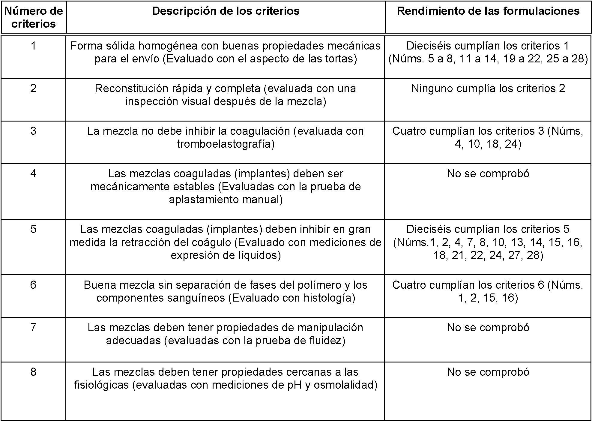
En el Ejemplo 2, el aumento de las concentraciones de lioprotectores mejora la estabilidad mecánica de las tortas, pero también inhibe la coagulación de las mezclas de quitosano/PRP. Las tortas liofilizadas que contienen quitosano de alto peso molecular no se disuelven fácil y completamente en el PRP.In Example 2 , increasing the concentrations of lyoprotectants improves the mechanical stability of the cakes, but also inhibits the coagulation of the chitosan/PRP mixtures. Freeze-dried cakes containing high molecular weight chitosan do not dissolve easily and completely in PRP.
EJEMPLO 3EXAMPLE 3
I-Preparación de formulaciones de quitosanoI-Preparation of chitosan formulations
El quitosano (Mn 211 kDa, 80,6% de DDA) se disolvió en HCl durante la noche a temperatura ambiente para obtener una concentración final de quitosano del 0,56% (p/v). Las soluciones se esterilizaron en autoclave durante 10 minutos y se enfriaron en hielo. El Mn del quitosano después del autoclave fue de 151 y 162 kDa. Se añadió trehalosa al 20% (p/v) esterilizada en autoclave y NaCl 5m , así como CaCh 270 mM esterilizado por filtro, de acuerdo con lo necesario. Se añadió un trazador de rodamina-quitosano esterilizado por filtro (Mn 143 kDa, 80,0% de DDA) antes de dispensarlo en viales individuales de 10 mL para su liofilización.Chitosan (Mn 211 kDa, 80.6% DDA) was dissolved in HCl overnight at room temperature to obtain a final chitosan concentration of 0.56% (w/v). Solutions were autoclaved for 10 minutes and chilled on ice. The Mn of chitosan after autoclaving was 151 and 162 kDa. Autoclaved 20% (w/v) trehalose and 5M NaCl as well as filter-sterilized 270mM CaCh were added as needed. Filter-sterilized rhodamine-chitosan tracer (Mn 143 kDa, 80.0% DDA) was added before dispensing into individual 10 mL vials for lyophilization.
De acuerdo con la Tabla 6, se ajustó la concentración de HCl de forma que todas las formulaciones tuvieran un pH objetivo teórico de 6,45. La concentración de NaCl se ajustó de forma que la formulación tuviera una osmolalidad teórica de 350 mOsm. La concentración de lioprotector se fijó en el 2% (p/v) para la formulación Núm. 2.According to Table 6 , the HCl concentration was adjusted such that all formulations had a theoretical target pH of 6.45. The NaCl concentration was adjusted so that the formulation had a theoretical osmolality of 350 mOsm. The concentration of lyoprotectant was set at 2% (w/v) for formulation No. 2.
El ciclo de liofilización fue idéntico al descrito en el Ejemplo 2, Sección 2-Ciclo de liofilización. The lyophilization cycle was identical to that described in Example 2, Section 2 - Lyophilization Cycle.

______ _ __ ____ _ 2- Aislamiento de PRP humano2- Isolation of human PRP
Se extrajo sangre entera de un donante humano y se mezcló con una solución de citrato de sodio al 3,8% (p/v) (9 mL de sangre por 1 mL de citrato de sodio).Whole blood was collected from a human donor and mixed with a 3.8% (w/v) sodium citrate solution (9 mL blood per 1 mL sodium citrate).
La sangre se centrifugó en una centrifugadora ACE E-Z PRP™ a 160 g durante 10 minutos a temperatura ambiente. Las fracciones sobrenadantes que contienen plasma y la capa leucocitaria, así como los primeros 1-2 mm de la capa eritrocitaria, se extrajeron mediante el uso de una aguja roma de 2 A pulgadas (calibre 18) conectada a una jeringa de 10 mL.Blood was centrifuged in an ACE E-Z PRP™ centrifuge at 160g for 10 minutes at room temperature. The supernatant fractions containing plasma and the buffy coat, as well as the first 1-2 mm of the erythrocyte layer, were withdrawn using a 2 A inch (18 gauge) blunt needle attached to a 10 mL syringe.
El plasma y la capa leucocitaria se centrifugaron además a 400 g durante 10 minutos a temperatura ambiente a fin de separar el plasma rico en plaquetas (PRP) del plasma pobre en plaquetas (PPP).Plasma and buffy coat were further centrifuged at 400 g for 10 minutes at room temperature in order to separate platelet-rich plasma (PRP) from platelet-poor plasma (PPP).
3- Aspecto de las tortas3- Appearance of the cakes
La formulación que contiene NaCl sólo se encogió significativamente durante la liofilización.The NaCl-containing formulation only shrunk significantly during freeze-drying.
La formulación que contenía 2% (p/v) de trehalosa era mecánicamente estable y cumplía los criterios de rendimiento 1.The formulation containing 2% (w/v) trehalose was mechanically stable and met performance criteria 1.
4- Reconstitución de las tortas4- Reconstitution of the cakes
Las tortas se reconstituyeron con 1 mL de PRP y se mezclaron con tres perlas de acero de 0,39 g durante 10 segundos.The cakes were reconstituted with 1 mL PRP and mixed with three 0.39 g steel beads for 10 seconds.
Se observaron partículas de quitosano no disueltas tras la reconstitución.Undissolved chitosan particles were observed after reconstitution.
5- Preparación y mezcla de formulaciones líquidas5- Preparation and mixing of liquid formulations
También se prepararon formulaciones líquidas de quitosano para probarlas en paralelo con las formulaciones liofilizadas (Tabla 7). Las soluciones se esterilizaron en autoclave durante 10 minutos y se enfriaron en hielo. El Mn del quitosano después del autoclave fue de 145 y 163 kDa.Liquid chitosan formulations were also prepared for testing in parallel with the lyophilized formulations ( Table 7 ). Solutions were autoclaved for 10 minutes and chilled on ice. The Mn of chitosan after autoclaving was 145 and 163 kDa.
Se mezclaron 400 pl de formulación líquida de quitosano con 800 pl de PRP y se activó con 240 pl de CaCh al 3% (p/v).400 μl of liquid chitosan formulation were mixed with 800 μl of PRP and activated with 240 μl of CaCh at 3% (w/v).

6- Homogeneidad de los coágulos6- Homogeneity of the clots
Las formulaciones liofilizadas reconstituidas y las formulaciones líquidas se dispensaron en tubos de vidrio a 37 °C y se dejaron coagular durante 1 hora.Reconstituted lyophilized formulations and liquid formulations were dispensed into glass tubes at 37°C and allowed to clot for 1 hour.
Los coágulos híbridos se fijaron en NBF al 10% y se observaron secciones gruesas de hoja de afeitar con microscopía de epifluorescencia para evaluar la dispersión del quitosano en los coágulos.Hybrid clots were fixed in 10% NBF and thick razor blade sections were observed with epifluorescence microscopy to assess chitosan dispersion in the clots.
Los agregados de quitosano no se dispersaron en los coágulos híbridos liofilizados (Figura 3A1 y 3A2).Chitosan aggregates were not dispersed in the lyophilized hybrid clots ( Figure 3A1 and 3A2 ).
El quitosano estaba bien disperso dentro de los coágulos híbridos preparados con soluciones líquidas (Figura 3A3 y 3A4).Chitosan was well dispersed within hybrid clots prepared with liquid solutions ( Figure 3A3 and 3A4 ).
7- Implantación ex vivo en defectos de menisco7- Ex vivo implantation in meniscus defects
Se utilizó una cuchilla de afeitar recta para tomar secciones transversales de ~0,5 mm del menisco del cerdo y se creó un colgajo horizontal hacia la superficie femoral (superior) del menisco.A straight razor blade was used to take ~0.5 mm transverse sections of the pig meniscus and a horizontal flap was created towards the femoral (superior) surface of the meniscus.
Se utilizó un punzón de biopsia de 4 mm para crear un defecto de espesor parcial hacia la superficie tibial (inferior) del menisco. A 4mm biopsy punch was used to create a partial thickness defect towards the tibial (inferior) surface of the meniscus.
Los meniscos se envolvieron en una película de plástico húmeda y se colocaron a 37 °C durante al menos 30 minutos antes del inicio del experimento.The menisci were wrapped in moist plastic film and placed at 37°C for at least 30 min before the start of the experiment.
Las formulaciones liofilizadas reconstituidas y las formulaciones líquidas se inyectaron en los defectos de menisco de espesor parcial mediante el uso de una jeringa provista de una aguja de calibre 20 y el colgajo se cerró inmediatamente.Reconstituted lyophilized formulations and liquid formulations were injected into partial thickness meniscal defects using a syringe fitted with a 20-gauge needle and the flap closed immediately.
Los meniscos se volvieron a envolver inmediatamente y se sellaron con una película de plástico húmeda y se colocaron a 37 °C durante 1 hora.The menisci were immediately rewrapped and sealed with moist plastic film and placed at 37°C for 1 hour.
El quitosano/PRP liofilizado y las formulaciones líquidas se implantaron con éxito ex vivo en defectos meniscales donde coagularon in situ. Lyophilized chitosan/PRP and liquid formulations were successfully implanted ex vivo into meniscal defects where they coagulated in situ.
Se fijaron meniscos de cerdo en NBF al 10% y se observaron secciones gruesas de cuchilla con microscopía de epifluorescencia para evaluar la dispersión de quitosano en los coágulos.Pig menisci were fixed in 10% NBF and thick knife sections were observed with epifluorescence microscopy to assess chitosan dispersion in the clots.
El quitosano se agregaba y no se dispersaba por los defectos meniscales en las formulaciones liofilizadas (Figuras 3B1 y 3B2).Chitosan aggregated and did not disperse through meniscal defects in the lyophilized formulations ( Figures 3B1 and 3B2 ).
El quitosano estaba bien disperso dentro de los defectos meniscales para las formulaciones líquidas (Figuras 3B3 y 3B4).Chitosan was well dispersed within the meniscal defects for the liquid formulations ( Figures 3B3 and 3B4 ).
Tabla 8. Rendimiento de las 2 formulaciones liofilizadas diferentes. Table 8. Performance of the 2 different lyophilized formulations.

En el Ejemplo 3, aunque las formulaciones líquidas de quitosano se pueden mezclar fácilmente con PRP, la reconstitución de las formulaciones de quitosano liofilizadas en PRP es mucho más difícil. Las tortas liofilizadas que contenían quitosano de alto peso molecular no se disolvían fácil y completamente en el PRP, pero aun así se podían implantar ex vivo en un modelo de defecto meniscal mediante el uso de un aparato de quirófano estándar.In Example 3 , although liquid chitosan formulations can be easily mixed with PRP, reconstitution of lyophilized chitosan formulations in PRP is much more difficult. The high molecular weight chitosan-containing lyophilized cakes did not readily and completely dissolve in PRP, but could still be implanted ex vivo into a meniscal defect model using standard operating room apparatus.
EJEMPLO 4EXAMPLE 4
I-Preparación de formulaciones de quitosanoI-Preparation of chitosan formulations
Formulaciones con Quitosano Mn > 100 kDa: Los quitosanos (Mn 211 kDa, 80,6% de DDA y Mn 105 kDa, 81,2% de DDA) se disolvieron en HCl durante una noche a temperatura ambiente para obtener una concentración final de quitosano del 0,56% (p/v). Las soluciones se esterilizaron en autoclave durante 10 minutos y se enfriaron en hielo. Se añadieron trehalosa 15% (p/v) esterilizada por filtro, manitol 15% (p/v), CaCh 270 mM, tampón L-histidina 55 mM pH 6,5 (preparado por medio de la mezcla de 10 mL de L-histidina 0,017% p/v y 10 mL de HCl 30 mM) y NaCl 5M esterilizado en autoclave, de acuerdo con lo necesario. Se añadió un trazador de rodamina-quitosano esterilizado por filtro (Mn 110 kDa, 80,2% de DDA) antes de dispensarlo en viales individuales de 3 mL para su liofilización. Formulaciones con Quitosano Mn< 100 kDa: Los quitosanos (Mn 38 kDa, 82,5% de DDA, Mn 11 kDa, 84,4% de DDA y Mn 4 kDa, 80,2% de DDA) se disolvieron en HCl durante la noche a temperatura ambiente para obtener una concentración final de quitosano del 0,56% (p/v). Las soluciones se filtraron y esterilizaron. Se añadieron trehalosa 15% (p/v) esterilizada por filtro, manitol 15% (p/v), CaCh 270 mM, NaCl 5M y tampón de histidina 55 mM pH 6,5 (preparado por medio de la mezcla de 10 mL de L-histidina 0,017% p/v y 10 mL de HCl 30 mM), de acuerdo con lo necesario. Se añadió el trazador de rodamina-quitosano esterilizado por filtro (Mn 40 kDa, 80,0% de DDA o Mn 10 kDa, 81,9% de DDA) antes de dispensarlo en viales individuales de 3 mL para su liofilización.Formulations with Chitosan Mn > 100 kDa: Chitosans (Mn 211 kDa, 80.6% DDA and Mn 105 kDa, 81.2% DDA) were dissolved in HCl overnight at room temperature to obtain a final chitosan concentration. 0.56% (w/v). Solutions were autoclaved for 10 minutes and chilled on ice. Filter-sterilized 15% (w/v) trehalose, 15% (w/v) mannitol, 270 mM CaCh, 55 mM L-histidine buffer pH 6.5 (prepared by mixing 10 mL of L- histidine 0.017% w/v and 10 mL of 30 mM HCl) and 5M NaCl autoclaved as needed. Filter-sterilized rhodamine-chitosan tracer (Mn 110 kDa, 80.2% DDA) was added before dispensing into individual 3 mL vials for lyophilization. Formulations with Chitosan Mn< 100 kDa: The chitosans (Mn 38 kDa, 82.5% DDA, Mn 11 kDa, 84.4% DDA and Mn 4 kDa, 80.2% DDA) were dissolved in HCl during the overnight at room temperature to obtain a final chitosan concentration of 0.56% (w/v). The solutions were filtered and sterilized. Filter-sterilized trehalose 15% (w/v), mannitol 15% (w/v), CaCh 270 mM, NaCl 5M and histidine buffer 55 mM pH 6.5 (prepared by mixing 10 mL of L-histidine 0.017% w/v and 10 mL of 30 mM HCl), as needed. Filter-sterilized rhodamine-chitosan tracer (Mn 40 kDa, 80.0% DDA or Mn 10 kDa, 81.9% DDA) was added before dispensing into individual 3 mL vials for lyophilization.
De acuerdo con la Tabla 9, la concentración de HCl se ajustó para que todas las formulaciones tuvieran una relación HCl: glucosamina de 0,6. La concentración de NaCl se ajustó de forma que la formulación tuviera una osmolalidad teórica de 350 mOsm. Se eligió una concentración más baja de histidina (3,8 mM frente a los 33 a 39 mM de los ejemplos anteriores) para evitar el agrietamiento de las tortas. La concentración de lioprotector se fijó en un 2% o 6% (p/v) suficiente para proporcionar una torta estable pero sin impedir la coagulación.According to Table 9 , the HCl concentration was adjusted so that all formulations had an HCl: glucosamine ratio of 0.6. The NaCl concentration was adjusted so that the formulation had a theoretical osmolality of 350 mOsm. A lower concentration of histidine (3.8 mM vs. 33 to 39 mM in previous examples) was chosen to avoid cracking of the cakes. The concentration of lyoprotectant was set at 2% or 6% (w/v) sufficient to provide a stable cake but without impeding coagulation.
El ciclo de liofilización fue idéntico al descrito en el Ejemplo 2, Sección 2-Ciclo de liofilización.The lyophilization cycle was identical to that described in Example 2, Section 2 - Lyophilization Cycle.
Tabla 9. Formulaciones liofilizadas que contienen el activador de la coagulación (CaCh) para ser reconstituidas directamente con PRP. Table 9. Lyophilized formulations containing the coagulation activator (CaCh) to be directly reconstituted with PRP.


2- Aspecto de las tortas2- Appearance of the cakes
Las formulaciones sin lioprotector se encogieron significativamente durante la liofilización.Formulations without lyoprotectant shrunk significantly during lyophilization.
El tampón de histidina utilizado a una concentración de 3,8 mM no indujo el agrietamiento de las tortas como se vio anteriormente en el Ejemplo 1 con concentraciones más altas de 33 a 39 mM.Histidine buffer used at a concentration of 3.8 mM did not induce cracking of the cakes as seen above in Example 1 at higher concentrations of 33 to 39 mM.
Las tortas eran más voluminosas cuando se utilizaban mayores concentraciones de lioprotectores. Las tortas que contenían manitol eran más voluminosas que las que contenían trehalosa (Figura 4A1 y 4A2). The cakes were more voluminous when higher concentrations of lyoprotectants were used. The mannitol-containing cakes were more voluminous than those containing trehalose ( Figure 4A1 and 4A2).
3- Reconstitución de las tortas3- Reconstitution of the cakes
El PRP y el PPP humano se extrajeron como se ha descrito anteriormente en el Ejemplo 3, Sección 2-Aislamiento del PRP humano.Human PRP and PPP were extracted as described above in Example 3, Section 2-Isolation of Human PRP.
Las tortas se reconstituyeron con 1 mL de PRP o 1 mL de PPP y se mezclaron con tres perlas de acero de 0,39 g durante 10 segundos.The cakes were reconstituted with 1 mL PRP or 1 mL PPP and mixed with three 0.39 g steel beads for 10 seconds.
Los quitosanos de medio y bajo Mn se disolvieron mejor que los quitosanos de mayor Mn, especialmente en presencia de lioprotectores.Medium and low Mn chitosans dissolved better than higher Mn chitosans, especially in the presence of lyoprotectants.
4-Expresión de líquidos4-Expression of liquids
Las formulaciones reconstituidas en PRP se dispensaron en tubos de vidrio a 37 °C. Después de 60 minutos, se cuantificó la expresión de líquidos de los coágulos híbridos por medio de la medición del peso.Reconstituted PRP formulations were dispensed into glass tubes at 37°C. After 60 minutes, it quantified the fluid expression of hybrid clots by weight measurement.
Todos los coágulos híbridos expresaron menos líquido que el PRP solo.All hybrid clots expressed less fluid than PRP alone.
5- Homogeneidad de los coágulos5- Homogeneity of the clots
Los coágulos híbridos se fijaron en NBF al 10% y se observaron secciones gruesas de hoja de afeitar con microscopía de epifluorescencia para evaluar la dispersión del quitosano en los coágulos.Hybrid clots were fixed in 10% NBF and thick razor blade sections were observed with epifluorescence microscopy to assess chitosan dispersion in the clots.
El quitosano se agrega en la mayoría de los coágulos híbridos preparados con quitosano de alto Mn (Figuras 4B1 y 4B2).Chitosan is added in most hybrid clots prepared with high-Mn chitosan ( Figures 4B1 and 4B2 ).
El quitosano se dispersó bien dentro de la mayoría de los coágulos híbridos cuando se utilizó quitosano de Mn medio (Figuras 4B3 y 4B4). Chitosan was well dispersed within most of the hybrid clots when medium Mn chitosan was used ( Figures 4B3 and 4B4).
Los eritrocitos presentes en el PRP sedimentaron hacia el fondo de los coágulos dejando una banda de quitosano en la superficie del coágulo cuando se utilizaron los quitosanos de menor Mn (Figuras 4B5& 4B6). The erythrocytes present in the PRP sedimented towards the bottom of the clots leaving a chitosan band on the clot surface when the lower Mn chitosans were used (Figures 4B5 & 4B6).
6- Implantación ex vivo en defectos de menisco6- Ex vivo implantation in meniscus defects
Las formulaciones de quitosano/PRP liofilizadas se implantaron con éxito ex vivo en defectos meniscales donde coagularon in situ como se describe en el Ejemplo 3, Sección 7- Implantación ex vivo en defectos meniscales.Lyophilized chitosan/PRP formulations were successfully implanted ex vivo into meniscal defects where they coagulated in situ as described in Example 3, Section 7 - Ex vivo Implantation into Meniscal Defects.
Tabla 10. Rendimiento de las 30 formulaciones diferentes. Table 10 . Performance of the 30 different formulations.

En el Ejemplo 4, la disminución del peso molecular del quitosano mejora la solubilidad de las tortas en el PRP, pero que sólo el quitosano de peso molecular medio (Mn 38 kDa) produjo coágulos híbridos de quitosano/PRP que eran homogéneos sin ninguna separación de fase que ocurre a un peso molecular más bajo o agregación que ocurre a un peso molecular más alto.In Example 4 , lowering the molecular weight of chitosan improves the solubility of cakes in PRP, but only medium molecular weight chitosan (Mn 38 kDa) produced hybrid chitosan/PRP coagulum that were homogeneous without any separation of PRP. phase that occurs at a lower molecular weight or aggregation that occurs at a higher molecular weight.
EJEMPLO 5EXAMPLE 5
I-Preparación de formulaciones de quitosanoI-Preparation of chitosan formulations
Los quitosanos de Mn medio (Mn 56 kDa, 80,1% de DDA y Mn 32 kDa, 81,2% de DDA) se disolvieron en HCl durante la noche a temperatura ambiente para obtener una concentración final de quitosano de 0,56%, 1% o 2% (p/v). Las soluciones se filtraron y esterilizaron. Se añadieron trehalosa 15% (p/v) esterilizada por filtro, manitol 15% (p/v) y CaCl2270 mM, de acuerdo con lo necesario. Se añadió un trazador de rodamina-quitosano esterilizado por filtro (Mn 40 kDa, 80,0% de DDA) antes de dispensarlo en viales individuales de 3 mL para su liofilización.Medium Mn chitosans (Mn 56 kDa, 80.1% DDA and Mn 32 kDa, 81.2% DDA) were dissolved in HCl for overnight at room temperature to obtain a final concentration of chitosan of 0.56%, 1% or 2% (w/v). The solutions were filtered and sterilized. Filter-sterilized trehalose 15% (w/v), mannitol 15% (w/v), and CaCl 2 270 mM were added as needed. Filter-sterilized rhodamine-chitosan tracer (Mn 40 kDa, 80.0% DDA) was added before dispensing into individual 3 mL vials for lyophilization.
De acuerdo con la Tabla 11, la concentración de HCl se ajustó para que todas las formulaciones tuvieran una relación HCl: glucosamina de 0,6. La concentración de lioprotector se fijó en un 2% o 6% (p/v) suficiente para proporcionar una torta estable pero sin impedir la coagulación.According to Table 11 , the HCl concentration was adjusted so that all formulations had an HCl: glucosamine ratio of 0.6. The concentration of lyoprotectant was set at 2% or 6% (w/v) sufficient to provide a stable cake but without impeding coagulation.
El ciclo de liofilización fue idéntico al descrito en el Ejemplo 2, Sección 2-Ciclo de liofilizaciónThe lyophilization cycle was identical to that described in Example 2, Section 2 - Lyophilization Cycle
Tabla 11. Formulaciones liofilizadas que contienen el activador de la coagulación (CaCh) para ser reconstituidas directamente con PRP. Table 11 . Lyophilized formulations containing the coagulation activator (CaCh) to be directly reconstituted with PRP.


2- Aspecto de las tortas2- Appearance of the cakes
Las tortas eran más voluminosas cuando se utilizaban mayores concentraciones de lioprotectores. Las tortas que contenían manitol eran más voluminosas que las que contenían trehalosa.The cakes were more voluminous when higher concentrations of lyoprotectants were used. The mannitol-containing cakes were bulkier than those containing trehalose.
3- Reconstitución de las tortas3- Reconstitution of the cakes
El PRP y el PPP humano se extrajeron de 2 donantes humanos diferentes, como se ha descrito anteriormente en el Ejemplo 3, Sección 2-Aislamiento del PRP humano.Human PRP and PPP were extracted from 2 different human donors, as described above in Example 3, Section 2-Isolation of Human PRP.
Las tortas se reconstituyeron con 1 mL de PRP o 1 mL de PPP y se mezclaron con tres perlas de acero de 0,39 g durante 10 segundos.The cakes were reconstituted with 1 mL PRP or 1 mL PPP and mixed with three 0.39 g steel beads for 10 seconds.
Las formulaciones que contenían 2% (p/v) de quitosano no se solubilizaron bien.Formulations containing 2% (w/v) chitosan did not solubilize well.
Las tortas que contenían quitosano de Mn 32 kDa fueron más fáciles de reconstituir en comparación con las tortas que contenían quitosano de Mn 56 kDa, tanto a una concentración de quitosano del 0,56% como del 1% (p/v).Cakes containing 32 kDa Mn chitosan were easier to reconstitute compared to cakes containing 56 kDa Mn chitosan, both at 0.56% and 1% (w/v) chitosan concentration.
4- Tromboelastografía (TEG) 4- Thromboelastography ( TEG)
Se cargaron 360 pl de cada formulación en un vaso de TEG inmediatamente después de la mezcla y se registraron los trazados de t Eg durante 1 hora.360 pl of each formulation was loaded into a TEG beaker immediately after mixing and t Eg traces were recorded for 1 hour.
Las formulaciones que contenían un 0,56% (p/v) de quitosano Mn 32 kDa coagularon de manera monofásica de forma similar a los controles de sólo PRP (Figura 5B1). Formulations containing 0.56% (w/v) Mn 32 kDa chitosan coagulated in a monophasic manner similar to PRP-only controls ( Figure 5B1).
El aumento de la Mn o de la concentración de quitosano indujo un mecanismo de coagulación de dos fases, como lo revelan los trazados de TEG (Figuras 5B2 y 5B3). Increasing the Mn or chitosan concentration induced a two-phase coagulation mechanism, as revealed by TEG tracings ( Figures 5B2 and 5B3).
En todos los casos, la adición de un 6% (p/v) de lioprotector dio coágulos más blandos con una amplitud máxima menor (MA entre 24 y 61 mm) que la adición de un 2% (p/v) de lioprotector (MA entre 51 y 84 mm).In all cases, the addition of 6% (w/v) lyoprotectant gave softer clots with a smaller maximum amplitude (MA between 24 and 61 mm) than the addition of 2% (w/v) lyoprotectant ( MA between 51 and 84 mm).
5- Prueba de solvencia5- Solvency test
La fluidez se evaluó por medio de la colocación de una gota de 30 pL de cada formulación en una pieza rígida de plástico fijada en un ángulo determinado (38 grados) inmediatamente después de la reconstitución y la toma de fotografías en momentos determinados.Flowability was assessed by placing a 30 pL drop of each formulation on a rigid piece of plastic fixed at a predetermined angle (38 degrees) immediately after reconstitution and taking photographs at predetermined times.
El aumento de la concentración de quitosano mejoró las propiedades pastosas de las formulaciones (Compárense las Figuras 5A1 y 5A2). El aumento del Mn de quitosano mejoró las propiedades pastosas de las formulaciones (Compárense las Figuras 5A1 y 5A3).Increasing the chitosan concentration improved the pasty properties of the formulations (Compare Figures 5A1 and 5A2 ). The increase in chitosan Mn improved the pasty properties of the formulations (Compare Figures 5A1 and 5A3 ).
Tabla 12. Rendimiento de las 24 formulaciones diferentes. Table 12. Performance of the 24 different formulations.


En el Ejemplo 5, las propiedades pastosas de las formulaciones se pueden mejorar por medio del aumento de la concentración de quitosano o el Mn de quitosano. Las tortas liofilizadas que contienen quitosano de peso molecular medio se pueden reconstituir fácilmente en PRP siempre que la concentración de quitosano sea inferior al 2% (p/v).In Example 5, the pasty properties of the formulations can be improved by increasing the chitosan concentration or the chitosan Mn. Lyophilized cakes containing medium molecular weight chitosan can be easily reconstituted into PRP as long as the chitosan concentration is less than 2% (w/v).
EJEMPLO 6EXAMPLE 6
1-Preparación de formulaciones de quitosano1-Preparation of chitosan formulations
Cinco quitosanos diferentes de Mn medio (Mn 56 kDa, 80,1% de DDA, Mn 56 kDa, 81,8% de DDA, Mn 32 kDa, 81,2% de DDA, Mn 30 kDa, 81,0% de DDA y Mn 28 kDa, 80,5% de DDA) se disolvieron en HCl durante la noche a temperatura ambiente para obtener una concentración final de quitosano del 1% (p/v). Las soluciones se filtraron y esterilizaron. Se añadieron trehalosa 15% (p/v) esterilizada por filtro, manitol 15% (p/v) y CaCh 270 mM, de acuerdo con lo necesario. Se añadió el trazador de Rodamina-quitosano esterilizado por filtro (Mn 40 kDa, 80,0% de DDA o Mn 110 kDa, 80,2% de DDA) antes de dispensarlo en viales individuales de 3 mL para su liofilización.Five different chitosans of average Mn (Mn 56 kDa, 80.1% DDA, Mn 56 kDa, 81.8% DDA, Mn 32 kDa, 81.2% DDA, Mn 30 kDa, 81.0% DDA and Mn 28 kDa, 80.5% DDA) were dissolved in HCl overnight at room temperature to obtain a final chitosan concentration of 1% (w/v). The solutions were filtered and sterilized. Filter-sterilized 15% (w/v) trehalose, 15% (w/v) mannitol, and 270 mM CaCh were added as needed. Filter-sterilized Rhodamine-chitosan tracer (Mn 40 kDa, 80.0% DDA or Mn 110 kDa, 80.2% DDA) was added before dispensing into individual 3 mL vials for lyophilization.
De acuerdo con la Tabla 13, la concentración de HCl se ajustó para que todas las formulaciones tuvieran una relación HCl: glucosamina de 0,6. La concentración de lioprotector se fijó en un 2% o 6% (p/v) suficiente para proporcionar una torta estable pero sin impedir la coagulación.According to Table 13, the HCl concentration was adjusted so that all formulations had an HCl: glucosamine ratio of 0.6. The concentration of lyoprotectant was set at 2% or 6% (w/v) sufficient to provide a stable cake but without impeding coagulation.
El ciclo de liofilización fue idéntico al descrito en el Ejemplo 2, Sección 2-Ciclo de liofilización. The lyophilization cycle was identical to that described in Example 2, Section 2 - Lyophilization Cycle.

2- Aspecto de las tortas2- Appearance of the cakes
Las tortas eran más voluminosas cuando se utilizaban mayores concentraciones de lioprotectores. Las tortas que contenían manitol eran más voluminosas que las que contenían trehalosa (Figuras 6A1 y 6A2).The cakes were more voluminous when higher concentrations of lyoprotectants were used. The mannitol-containing cakes were bulkier than those containing trehalose ( Figures 6A1 and 6A2 ).
3- Reconstitución de las tortas3- Reconstitution of the cakes
El PRP y el PPP humano se extrajeron como se ha descrito anteriormente en el Ejemplo 3, Sección 2-Aislamiento del PRP humano.Human PRP and PPP were extracted as described above in Example 3, Section 2-Isolation of Human PRP.
Las tortas se reconstituyeron con 1 mL de PRP o 1 mL de PPP y se mezclaron con tres perlas de acero de 0,39 g durante 10 segundos.The cakes were reconstituted with 1 mL PRP or 1 mL PPP and mixed with three 0.39 g steel beads for 10 seconds.
Todas las formulaciones se disolvieron bien y cumplieron los criterios de rendimiento 2.All formulations dissolved well and met performance criteria 2.
4- Tromboelastografía (TEG) 4- Thromboelastography ( TEG)
Se cargaron 360 pl de cada formulación en un vaso de TEG inmediatamente después de la mezcla y se registraron los trazados de t Eg durante 1 hora.360 pl of each formulation was loaded into a TEG beaker immediately after mixing and t Eg traces were recorded for 1 hour.
Todas las formulaciones indujeron un mecanismo de coagulación de 2 fases, como lo revelan los trazados de TEG (Figuras 6B1y 6B2). All formulations induced a 2-phase coagulation mechanism, as revealed by TEG tracings ( Figures 6B1 and 6B2).
En todos los casos, la adición de un 6% (p/v) de lioprotector dio coágulos más blandos con una amplitud máxima menor (MA entre 37 y 67 mm) que la adición de un 2% (p/v) de lioprotector (MA entre 68 y 79 mm).In all cases, the addition of 6% (w/v) lyoprotectant gave softer clots with a smaller maximum amplitude (MA between 37 and 67 mm) than the addition of 2% (w/v) lyoprotectant ( MA between 68 and 79 mm).
5- Prueba de solvencia5- Solvency test
La fluidez se evaluó por medio de la colocación de una gota de 30 pL de cada formulación en una pieza rígida de plástico fijada en un ángulo determinado (38 grados) inmediatamente después de la reconstitución y la toma de fotografías en momentos determinados.Flowability was assessed by placing a 30 pL drop of each formulation on a rigid piece of plastic fixed at a predetermined angle (38 degrees) immediately after reconstitution and taking photographs at predetermined times.
Todas las formulaciones tenían propiedades pastosas en comparación con el PRP solo (Figura 6C1). All formulations had pasty properties compared to PRP alone ( Figure 6C1).
6- Expresión de líquidos6- Expression of liquids
Las formulaciones reconstituidas en PRP se dispensaron en tubos de vidrio a 37 °C. Después de 60 minutos, se cuantificó la expresión de líquidos de los coágulos híbridos por medio de la medición del peso.Reconstituted PRP formulations were dispensed into glass tubes at 37°C. After 60 minutes, the fluid expression of the hybrid clots was quantified by weight measurement.
Todos los coágulos híbridos expresaron menos líquido que el PRP solo (Figura 6B3). All hybrid clots expressed less fluid than PRP alone ( Figure 6B3).
7- Homogeneidad de los coágulos7- Homogeneity of clots
Los coágulos híbridos se fijaron en NBF al 10% y se observaron secciones gruesas de hoja de afeitar con microscopía de epifluorescencia para evaluar la dispersión del quitosano en los coágulos.Hybrid clots were fixed in 10% NBF and thick razor blade sections were observed with epifluorescence microscopy to assess chitosan dispersion in the clots.
El quitosano se dispersó en los coágulos híbridos para todas las formulaciones (Figura 6A3 y 6A4) Chitosan was dispersed in the hybrid clots for all formulations ( Figure 6A3 and 6A4).
8- Prueba de aplastamiento8- Crush test
Después de 1 hora de coagulación, cada coágulo híbrido fue sometido a una prueba de aplastamiento y se calificó su resistencia mecánica.After 1 hour of coagulation, each hybrid clot was subjected to a crush test and its mechanical strength was rated.
0 = El coágulo no pudo se pudo manipular sin desintegrarse.0 = The clot could not be handled without disintegrating.
+ = El coágulo se rompió fácilmente y el aspecto de aplastamiento fue de múltiples fragmentos (más de 5 fragmentos).+ = The clot broke easily and the crushed appearance was multiple fragments (more than 5 fragments).
++ = El coágulo era relativamente firme y el aspecto aplastado era de múltiples fragmentos (3 a 5 fragmentos). ++ = El coágulo era firme y elástico, el aspecto aplastado era de 2 a 3 fragmentos.++ = The clot was relatively firm and the crushed appearance was multi-fragmented (3 to 5 fragments). ++ = The clot was firm and elastic, the crushed appearance was 2 to 3 fragments.
++++ = El coágulo era firme y elástico, el aspecto aplastado era de 2 fragmentos (a veces todavía conectados) o sólo había un agujero en el centro del coágulo.++++ = The clot was firm and elastic, the crushed appearance was 2 fragments (sometimes still connected) or there was only a hole in the center of the clot.
La adición de un 6% (p/v) de lioprotector disminuyó la resistencia mecánica del coágulo en comparación con el 2% (p/v) de lioprotector (Compárense las Figuras 6D1 y 6D2 con las 6D3 y 6D4).The addition of 6% (w/v) lyoprotectant decreased the mechanical strength of the clot compared to 2% (w/v) lyoprotectant (Compare Figures 6D1 and 6D2 with 6D3 and 6D4 ).
9- Implantación ex vivo en defectos de cartílago9- Ex vivo implantation in cartilage defects
Se utilizaron punzones de biopsia de 8 mm y cuchillas quirúrgicas planas para crear defectos de cartílago en el cóndilo y la tróclea del cerdo.8mm biopsy punches and flat surgical blades were used to create cartilage defects in the condyle and trochlea of the pig.
Las articulaciones se colocaron en una cámara húmeda a 37 °C durante al menos 30 minutos antes del inicio del experimento.The joints were placed in a humid chamber at 37 °C for at least 30 min before the start of the experiment.
Las formulaciones liofilizadas reconstituidas de quitosano/PRP se inyectaron en los defectos del cartílago.The reconstituted lyophilized formulations of chitosan/PRP were injected into the cartilage defects.
Las uniones se sellaron inmediatamente en la cámara húmeda y se colocaron a 37 °C durante 1 hora.Joints were immediately sealed in the humid chamber and placed at 37°C for 1 hour.
Las formulaciones de quitosano/PRP liofilizadas se implantaron con éxito ex vivo en defectos de cartílago mediante el uso de una jeringa y una aguja de calibre 20, donde se coagularon in situ (Figura 6C2). Lyophilized chitosan/PRP formulations were successfully implanted ex vivo into cartilage defects using a 20-gauge syringe and needle, where they coagulated in situ ( Figure 6C2).
Tabla 14. Rendimiento de las 16 formulaciones diferentes. Table 14 . Performance of the 16 different formulations.

En el Ejemplo 6, se pueden utilizar diferentes lotes de polvo de quitosano con Mn similar para preparar tortas que tendrán características de rendimiento equivalentes. Las tortas de quitosano con altas concentraciones de lioprotector producen híbridos de quitosano/PRP que son indeseablemente blandos.In Example 6, different batches of similar Mn chitosan powder can be used to prepare cakes that will have equivalent performance characteristics. Chitosan cakes with high concentrations of lyoprotectant produce chitosan/PRP hybrids that are undesirably soft.
EJEMPLO 7EXAMPLE 7
1-Preparación de formulaciones de quitosano1-Preparation of chitosan formulations
Cuatro quitosanos Mn diferentes (Mn 10 kDa, 80,6% de DDA, Mn 41 kDa, 80,6% de DDA, Mn 89 kDa, 80,6% de DDA y Mn 108 kDa, 80,6% de DDA) se disolvieron en HCl durante la noche a temperatura ambiente para obtener concentraciones finales de quitosano de 0,56% (p/v), 1% (p/v) y 2% (p/v), esta última concentración sólo preparada para el quitosano Mn 10 kDa. Las soluciones se filtraron y esterilizaron. Se añadieron trehalosa 15% (p/v) esterilizada por filtro, manitol 15% (p/v) y CaCh 270 mM, de acuerdo con lo necesario. Se añadió el trazador de rodaminaquitosano esterilizado por filtro (Mn 10 kDa, 81,9% de DDA, Mn 40 kDa, 80,0% de DDA o Mn 110 kDa, 80,2% de DDA) antes de dispensarlo en viales individuales de 3 mL o 2 mL para su liofilización.Four different Mn chitosans (Mn 10 kDa, 80.6% DDA, Mn 41 kDa, 80.6% DDA, Mn 89 kDa, 80.6% DDA and Mn 108 kDa, 80.6% DDA) were dissolved in HCl overnight at room temperature to obtain final chitosan concentrations of 0.56% (p/v), 1% (p/v) and 2% (p/v), the latter concentration only prepared for chitosan Mn 10 kDa. The solutions were filtered and sterilized. Filter-sterilized 15% (w/v) trehalose, 15% (w/v) mannitol, and 270 mM CaCh were added as needed. Filter-sterilized rhodamine chitosan tracer (Mn 10 kDa, 81.9% DDA, Mn 40 kDa, 80.0% DDA, or Mn 110 kDa, 80.2% DDA) was added before dispensing into individual vials of 3 mL or 2 mL for lyophilization.
De acuerdo con la Tabla 15, la concentración de HCl se ajustó para que todas las formulaciones tuvieran una relación HCl: glucosamina de 0,6. La concentración de lioprotector se fijó en un 2%, 4% o 6% (p/v), suficiente para proporcionar una torta estable pero sin impedir la coagulación.According to Table 15 , the HCl concentration was adjusted so that all formulations had an HCl: glucosamine ratio of 0.6. The lyoprotectant concentration was set at 2%, 4%, or 6% (w/v), sufficient to provide a stable cake but without impeding coagulation.
El ciclo de liofilización fue idéntico al descrito en el Ejemplo 2, Sección 2-Ciclo de liofilización. The lyophilization cycle was identical to that described in Example 2, Section 2 - Lyophilization Cycle.
Tabla 15. Formulaciones con CaCh reconstituidas directamente con PRP. Table 15. Formulations with CaCh reconstituted directly with PRP.


2- Aspecto de las tortas2- Appearance of the cakes
Las tortas eran más voluminosas cuando se utilizaban mayores concentraciones de lioprotectores. Las tortas que contenían manitol eran más voluminosas que las que contenían trehalosa.The cakes were more voluminous when higher concentrations of lyoprotectants were used. The mannitol-containing cakes were bulkier than those containing trehalose.
3- Reconstitución de las tortas3- Reconstitution of the cakes
El PRP y el PPP humano se extrajeron como se ha descrito anteriormente en el Ejemplo 3, Sección 2-Aislamiento del PRP humano.Human PRP and PPP were extracted as described above in Example 3, Section 2-Isolation of Human PRP.
Las tortas se reconstituyeron con 1 mL de PRP o 1 mL de PPP y se mezclaron a mano sin la ayuda de perlas de acero durante 10 segundos. Para cuatro de las formulaciones (Núms. 15, 19, 23, 27), se utilizaron también tres perlas de acero de 0,39 g para reconstituir las tortas, a fin de compararlas con los resultados obtenidos anteriormente.Cakes were reconstituted with 1 mL PRP or 1 mL PPP and mixed by hand without the aid of steel beads for 10 seconds. For four of the formulations (Nos. 15, 19, 23, 27), three 0.39 g steel beads were also used to reconstitute the cakes for comparison with previously obtained results.
Las formulaciones preparadas con quitosano Mn 10 kDa al 0,56% (p/v) y al 1% (p/vol) se disolvieron completamente. Las formulaciones preparadas con quitosano Mn 10 kDa al 2% (p/vol) y con quitosano Mn 41 kDa al 0,56% y al 1% (p/vol) se disolvieron bien. Las formulaciones preparadas con quitosano Mn 89 kDa y 108 kDa eran más gruesas y difíciles de manejar.The formulations prepared with 0.56% (w/v) and 1% (w/vol) chitosan Mn 10 kDa dissolved completely. The formulations prepared with 2% (w/vol) Mn 10 kDa chitosan and with 0.56% and 1% (w/vol) Mn 41 kDa chitosan dissolved well. Formulations prepared with 89 kDa and 108 kDa Mn chitosan were thicker and more difficult to handle.
4- Tromboelastografía (TEG) 4- Thromboelastography ( TEG)
Se cargaron 360 pl de cada formulación en un vaso de TEG inmediatamente después de la mezcla y se registraron los trazados de TEG durante 1 hora.360 pl of each formulation was loaded into a TEG beaker immediately after mixing and TEG traces were recorded for 1 hour.
Las formulaciones que contienen quitosano Mn 10 kDa al 0,56% (p/v) indujeron un trazado de coagulación en una fase. El aumento de la concentración de quitosano y de Mn indujo un mecanismo de coagulación en dos fases, como revelan los trazados de TEG.Formulations containing 0.56% (w/v) Mn 10 kDa chitosan induced a one-stage coagulation pattern. Increasing chitosan and Mn concentration induced a two-phase coagulation mechanism, as revealed by TEG tracings.
El tiempo de reacción del coágulo fue alto para las formulaciones que contenían quitosano Mn 10 kDa y corto para las formulaciones que contenían quitosano Mn 108 kDa, quedando las formulaciones de 40 kDa en un punto intermedio.Clot reaction time was high for formulations containing 10 kDa Mn chitosan and short for formulations containing 108 kDa Mn chitosan, with 40 kDa formulations falling somewhere in between.
La amplitud máxima fue mayor para los coágulos híbridos que contenían un 2% (p/v) de lioprotector en comparación con los coágulos híbridos que contenían un 4% o un 6% (p/v) de lioprotector.The maximum amplitude was greater for hybrid clots containing 2% (w/v) lyoprotectant compared to hybrid clots containing 4% or 6% (w/v) lyoprotectant.
5- Prueba de solvencia5- Solvency test
La fluidez se evaluó por medio de la colocación de una gota de 30 pL de cada formulación en una pieza rígida de plástico fijada en un ángulo determinado (38 grados) inmediatamente después de la reconstitución y la toma de fotografías en momentos determinados.Flowability was assessed by placing a 30 pL drop of each formulation on a rigid piece of plastic fixed at a predetermined angle (38 degrees) immediately after reconstitution and taking photographs at predetermined times.
Las formulaciones que contenían quitosano Mn 10 kDa al 0,56% (p/vol) y al 1% (p/vol) eran líquidas.The formulations containing 0.56% (w/vol) and 1% (w/vol) chitosan Mn 10 kDa were liquid.
Todas las demás formulaciones tenían propiedades pastosas en comparación con el PRP solo.All other formulations had pasty properties compared to PRP alone.
6- Expresión de líquidos6- Expression of liquids
Las formulaciones reconstituidas en PRP se dispensaron en tubos de vidrio a 37 °C. Después de 60 minutos, se cuantificó la expresión de líquidos de los coágulos híbridos por medio de la medición del peso.Reconstituted PRP formulations were dispensed into glass tubes at 37°C. After 60 minutes, the fluid expression of the hybrid clots was quantified by weight measurement.
Todos los coágulos híbridos expresaron menos líquido que el PRP solo.All hybrid clots expressed less fluid than PRP alone.
7- Homogeneidad de los coágulos7- Homogeneity of clots
Se observaron grandes agregados de quitosano en la mayoría de los coágulos híbridos preparados con quitosano Mn 89 kDa y 108 kDa (Figura 7A3, 7A4, 7A7 y 7A8). El quitosano se dispersó bien en la mayoría de los coágulos híbridos cuando se utilizó quitosano Mn 41 kDa (Figura 7Al, 7A2, 7A5 y 7A6). Los eritrocitos presentes en el PRP sedimentaron hacia el fondo de los coágulos dejando una banda de quitosano en la superficie del coágulo cuando se utilizó el quitosano Mn 10 kDa.Large aggregates of chitosan were observed in most of the hybrid clots prepared with Mn 89 kDa and 108 kDa chitosan (Figure 7A3, 7A4, 7A7 and 7A8). Chitosan dispersed well in most of the hybrid clots when Mn 41 kDa chitosan was used (Figure 7Al, 7A2, 7A5 and 7A6). The erythrocytes present in the PRP sedimented towards the bottom of the clots leaving a band of chitosan on the surface of the clot when the 10 kDa Mn chitosan was used.
8- Prueba de aplastamiento8- Crush test
Después de 1 hora de coagulación, cada coágulo híbrido fue sometido a una prueba de aplastamiento y se calificó su resistencia mecánica.After 1 hour of coagulation, each hybrid clot was subjected to a crush test and its mechanical strength was rated.
0 = El coágulo no se pudo manipular sin desintegrarse. 0 = Clot could not be handled without disintegrating.
+ = El coágulo se rompió fácilmente y el aspecto de aplastamiento fue de múltiples fragmentos (más de 5 fragmentos).+ = The clot broke easily and the crushed appearance was multiple fragments (more than 5 fragments).
++ = El coágulo era relativamente firme y el aspecto aplastado era de múltiples fragmentos (3 a 5 fragmentos). ++ = El coágulo era firme y elástico, el aspecto aplastado era de 2 a 3 fragmentos.++ = The clot was relatively firm and the crushed appearance was multi-fragmented (3 to 5 fragments). ++ = The clot was firm and elastic, the crushed appearance was 2 to 3 fragments.
++++ = El coágulo era firme y elástico, el aspecto aplastado era de 2 fragmentos (a veces todavía conectados) o sólo había un agujero en el centro del coágulo.++++ = The clot was firm and elastic, the crushed appearance was 2 fragments (sometimes still connected) or there was only a hole in the center of the clot.
La adición de un 6% (p/v) de lioprotector disminuyó la resistencia mecánica del coágulo.The addition of 6% (w/v) of lyoprotectant decreased the mechanical resistance of the clot.
9- Implantación ex vivo en defectos de cartílago9- Ex vivo implantation in cartilage defects
Se utilizaron punzones de biopsia de 8 mm y cuchillas quirúrgicas planas para crear defectos de cartílago en el cóndilo, la tróclea y la meseta tibial del cerdo.8-mm biopsy punches and flat surgical blades were used to create cartilage defects in the condyle, trochlea, and tibial plateau of the pig.
Las articulaciones se colocaron en una cámara húmeda a 37 °C durante al menos 30 minutos antes del inicio del experimento.The joints were placed in a humid chamber at 37 °C for at least 30 min before the start of the experiment.
Las formulaciones liofilizadas reconstituidas de quitosano/PRP se inyectaron en los defectos del cartílago.The reconstituted lyophilized formulations of chitosan/PRP were injected into the cartilage defects.
Las uniones se sellaron inmediatamente en la cámara húmeda y se colocaron a 37 °C durante 1 hora.Joints were immediately sealed in the humid chamber and placed at 37°C for 1 hour.
Las formulaciones de quitosano/PRP liofilizadas se implantaron con éxito ex vivo en defectos de cartílago mediante el uso de una jeringa y una aguja de calibre 20, donde se coagularon in situ. Lyophilized chitosan/PRP formulations were successfully implanted ex vivo into cartilage defects using a 20-gauge syringe and needle, where they coagulated in situ.
10- Reconstitución de las formulaciones liofilizadas sin perlas frente a la mezcla con perlas10- Reconstitution of the lyophilized formulations without pearls versus the mixture with pearls
El aspecto histológico de los coágulos híbridos fue similar tanto si las tortas liofilizadas se reconstituyeron sin la ayuda de perlas de acero inoxidable como si se mezclaron con tres perlas de acero inoxidable de 0,39 g (compárense las Figuras 7A1, 7A2, 7A3 y 7A4 con la 7A5, 7A6, 7A7 y 7A8).The histological appearance of the hybrid clots was similar whether the lyophilized cakes were reconstituted without the aid of stainless steel beads or mixed with three 0.39 g stainless steel beads (compare Figures 7A1, 7A2, 7A3 and 7A4). with 7A5, 7A6, 7A7 and 7A8 ).
Las características de rendimiento de las formulaciones liofilizadas fueron similares para los coágulos híbridos preparados sin la ayuda de las perlas de acero inoxidable o por medio de la mezcla con tres perlas de acero inoxidable de 0,39 g (véase la Tabla de la Figura 7B).The performance characteristics of the lyophilized formulations were similar for the hybrid clots prepared without the aid of the stainless steel beads or by mixing with three 0.39 g stainless steel beads (see Table Figure 7B) . .
11- Osmolalidad de las formulaciones reconstituidas con PRP11- Osmolality of reconstituted formulations with PRP
Las formulaciones de quitosano liofilizado que contenían manitol tenían una osmolalidad más alta que las formulaciones liofilizadas que contenían trehalosa. La osmolalidad aumentó con la concentración de lioprotector. Las formulaciones que contenían un 2% (p/vol) de trehalosa tenían una osmolalidad entre 443 y 495 mOsm. Las formulaciones que contenían 2% (p/vol) de manitol tenían una osmolalidad entre 526 y 582 mOsm. Las formulaciones que contenían un 4% (p/vol) de trehalosa tenían una osmolalidad entre 516 y 564 mOsm. Las formulaciones que contenían 4% (p/vol) de manitol tenían una osmolalidad entre 608 y 665 mOsm. Las formulaciones que contenían un 6% (p/vol) de trehalosa tenían una osmolalidad entre 595 y 631 mOsm. Las formulaciones que contenían 6% (p/vol) de manitol tenían una osmolalidad entre 759 y 823 mOsm.The mannitol-containing lyophilized chitosan formulations had a higher osmolality than the trehalose-containing lyophilized formulations. The osmolality increased with the concentration of lyoprotectant. Formulations containing 2% (w/vol) trehalose had an osmolality between 443 and 495 mOsm. Formulations containing 2% (w/vol) mannitol had an osmolality between 526 and 582 mOsm. Formulations containing 4% (w/vol) trehalose had an osmolality between 516 and 564 mOsm. Formulations containing 4% (w/vol) mannitol had an osmolality between 608 and 665 mOsm. Formulations containing 6% (w/vol) trehalose had an osmolality between 595 and 631 mOsm. Formulations containing 6% (w/vol) mannitol had an osmolality between 759 and 823 mOsm.
12- Implantación subcutánea de formulaciones de quitosano liofilizadas12- Subcutaneous implantation of lyophilized chitosan formulations
Se probaron in vivo varias formulaciones de baja y alta osmolalidad en un modelo de implante subcutáneo de conejo (Tabla 16). Various low and high osmolality formulations were tested in vivo in a rabbit subcutaneous implant model (Table 16).

Se afeitó el pelo del lomo de los conejos NZW y se desinfectó la piel con 3 pases de Baxedin® y luego con 3 pases alternados de proviodina e isopropanol al 70%.NZW rabbits were shaved back and disinfected with 3 passes of Baxedin® followed by 3 alternating passes of proviodine and 70% isopropanol.
El PRP autólogo se preparó a partir de sangre de conejo extraída inmediatamente antes de la cirugía, como se ha descrito anteriormente en la sección del Ejemplo 14- Aislamiento del PRP de conejo. Cada torta de quitosano liofilizada de 300 pl se reconstituyó con 300 pl de PRP sin ayuda de perlas para la mezcla.Autologous PRP was prepared from rabbit blood drawn immediately prior to surgery, as described above in the Example 14 -Isolation of Rabbit PRP section. Each 300 μl lyophilized chitosan cake was reconstituted with 300 μl PRP without the aid of beads for mixing.
Se utilizó una jeringa de 1 cc equipada con una aguja Sub Q para administrar 150 pl de cada implante bajo la piel de la espalda del conejoA 1 cc syringe fitted with a Sub Q needle was used to deliver 150 µl of each implant under the skin of the rabbit's back.
Los controles de PRP se calcificaron nuevamente con 42,2mM de CaCh antes de la inyección.PRP controls were recalcified with 42.2mM CaCh prior to injection.
Los lugares de inyección se variaron sistemáticamente en cada animal para evitar resultados dependientes del lugar. Se sacrificó a los animales a los 1 (Figuras 7C1 a 7C6), 3 (Figuras 7D1 a 7D6), 7 y 14 días después de la inyección (Figuras 7E2y 7E3). Injection sites were systematically varied for each animal to avoid site-dependent results. Animals were sacrificed at 1 (Figures 7C1 to 7C6) , 3 (Figures 7D1 to 7D6), 7 and 14 days after injection (Figures 7E2 and 7E3).
En el día 1, los implantes de quitosano parecían estar prácticamente intactos. En algunos casos, los eritrocitos presentes en el PRP eran visibles dentro de los implantes. Los glóbulos blancos fueron atraídos por los implantes y se encontraron principalmente en la periferia de los mismos (Figuras 7C1,7C2, 7C3 y 7C4).On day 1, the chitosan implants appeared to be virtually intact. In some cases, the erythrocytes present in the PRP were visible within the implants. White blood cells were attracted to the implants and were found mainly in the periphery of the implants (Figures 7C1, 7C2, 7C3 and 7C4 ).
Al tercer día, los implantes de quitosano/PRP estaban parcialmente degradados y los glóbulos blancos invadían los implantes (Figuras 7D1, 7D2, 7D3 y 7D4). On the third day, the chitosan/PRP implants were partially degraded and white blood cells invaded the implants (Figures 7D1, 7D2, 7D3 and 7D4).
Hubo un efecto del tiempo dado que el reclutamiento de glóbulos blancos se incrementó en el día 3 en comparación con el día 1 (Compárense las Figuras 7D1 a 7D4 con las Figuras 7C1 a 7C4).There was a time effect as white blood cell recruitment was increased on day 3 compared to day 1 (Compare Figures 7D1 to 7D4 with Figures 7C1 to 7C4 ).
Los híbridos de quitosano/PRP fueron residentes in vivo hasta 14 días después de la inyección (Figuras 7E1, 7E2 y 7E3).The chitosan/PRP hybrids were resident in vivo for up to 14 days after injection (Figures 7E1, 7E2 and 7E3) .
Los controles de PRP recalcificado sólo se visualizaron hasta 3 días después de la inyección (la Figura 7E4 muestra el control de PRP a 1 día) y no indujeron mucho reclutamiento celular (Figuras 7C5, 7C6, 7D5, 7D6 y 7E4).Recalcified PRP controls were only visible up to 3 days after injection ( Figure 7E4 shows PRP control at 1 day) and did not induce much cell recruitment ( Figures 7C5, 7C6, 7D5, 7D6 and 7E4 ).
Tabla 17. Rendimiento de las 40 formulaciones diferentes. Table 17 . Performance of the 40 different formulations.

En el Ejemplo 7, la mezcla con perlas de acero inoxidable no es necesaria para la reconstitución de las tortas de quitosano liofilizado con PRP. Las formulaciones que contienen altas concentraciones de lioprotector tienen una alta osmolalidad y atraen más leucocitos al implantarse in vivo. También se comprobó que los híbridos de quitosano y PRP se retenían durante más tiempo que los controles de PRP recalcificado únicamente in vivo. 8 formulaciones específicas (Núms. 13, 15, 17, 19, 35 a 38) cumplían todas las características de rendimiento predefinidas.In Example 7 , mixing with stainless steel beads is not necessary for reconstitution of PRP freeze-dried chitosan cakes. Formulations containing high concentrations of lyoprotectant have high osmolality and attract more leukocytes upon implantation in vivo. The chitosan-PRP hybrids were also found to be retained longer than recalcified-only PRP controls in vivo. 8 specific formulations (Nos. 13, 15, 17, 19, 35 to 38) met all predefined performance characteristics.
EJEMPLO 8EXAMPLE 8
1-Preparación de formulaciones de quitosano1-Preparation of chitosan formulations
Dos quitosanos diferentes (Mn 43 kDa, 85% de DDA y Mn 36 kDa, 80% de DDA) se disolvieron en HCl durante la noche a temperatura ambiente para obtener concentraciones finales de quitosano del 1% (p/v). Las soluciones se filtraron y esterilizaron. Se añadió trehalosa filtrada al 15% (p/v) y 270 mM de CaCh, de acuerdo con lo necesario. A algunos de los viales se les añadió un trazador de Rodamina-quitosano esterilizado por filtro (Mn 40 kDa, 80,0% de DDA) antes de dispensarlos en viales individuales de 3 mL para su liofilización.Two different chitosans (Mn 43 kDa, 85% DDA and Mn 36 kDa, 80% DDA) were dissolved in HCl overnight at room temperature to obtain final chitosan concentrations of 1% (w/v). The solutions were filtered and sterilized. Filtered 15% (w/v) trehalose and 270 mM CaCh were added as needed. Filter-sterilized Rhodamine-chitosan tracer (Mn 40 kDa, 80.0% DDA) was added to some of the vials before dispensing into individual 3 mL vials for lyophilization.
De acuerdo con la Tabla 18, la concentración de HCl se ajustó para que todas las formulaciones tuvieran una relación HCl: glucosamina de 0,6. La concentración de lioprotector se fijó en el 1% (p/v) para tener una osmolalidad cercana a la fisiológica, proporcionar una torta estable pero no impedir la coagulación.According to Table 18, the HCl concentration was adjusted so that all formulations had an HCl: glucosamine ratio of 0.6. The lyoprotectant concentration was set at 1% (w/v) to have an osmolality close to physiological, provide a stable cake but not prevent coagulation.
El ciclo de liofilización fue idéntico al descrito en el Ejemplo 2, Sección 2-Ciclo de liofilización. The lyophilization cycle was identical to that described in Example 2, Section 2 - Lyophilization Cycle.

2- Aspecto de las tortas2- Appearance of the cakes
Las tortas tenían una superficie lisa y un aspecto agradable sin ningún tipo de colapso. Se observó una ligera retracción de todas las tortas en los frascos de vidrio tras la liofilización (Figuras 8A1 y 8A2).The cakes had a smooth surface and a nice appearance without any collapse. A slight shrinkage of all the cakes in the glass jars was observed after lyophilization (FIGS. 8A1 and 8A2) .
3- Reconstitución de las tortas3- Reconstitution of the cakes
El PRP y el PPP humano se extrajeron como se ha descrito anteriormente en el Ejemplo 3, Sección 2-Aislamiento del PRP humano.Human PRP and PPP were extracted as described above in Example 3, Section 2-Isolation of Human PRP.
Las tortas se reconstituyeron con 1 mL de PRP o 1 mL de PPP y se mezclaron a mano sin la ayuda de perlas de acero durante 10 segundos.Cakes were reconstituted with 1 mL PRP or 1 mL PPP and mixed by hand without the aid of steel beads for 10 seconds.
Las formulaciones tuvieron buena solubilidad y se disolvieron completamente (Figuras 8A3y 8A4).The formulations had good solubility and dissolved completely (FIGS. 8A3 and 8A4) .
4- Tromboelastografía (TEG) 4- Thromboelastography ( TEG)
Se cargaron 360 pl de cada formulación en un vaso de TEG inmediatamente después de la mezcla y se registraron los trazados de t Eg durante 1 hora.360 pl of each formulation was loaded into a TEG beaker immediately after mixing and t Eg traces were recorded for 1 hour.
El tiempo de reacción del coágulo y la amplitud máxima fueron menores para las formulaciones de quitosano/PRP en comparación con el control PRP (Figuras 8C1y 8C2). Clot reaction time and peak amplitude were lower for the chitosan/PRP formulations compared to the PRP control (Figures 8C1 and 8C2).
5- Prueba de solvencia5- Solvency test
La fluidez se evaluó por medio de la colocación de una gota de 30 pL de cada formulación en una pieza rígida de plástico fijada en un ángulo determinado (38 grados) inmediatamente después de la reconstitución y la toma de fotografías en momentos determinados.Flowability was assessed by placing a 30 pL drop of each formulation on a rigid piece of plastic fixed at a predetermined angle (38 degrees) immediately after reconstitution and taking photographs at predetermined times.
Las formulaciones tenían propiedades pastosas en comparación con el control PRP.The formulations had pasty properties compared to the PRP control.
6- Expresión de líquidos6- Expression of liquids
Las formulaciones reconstituidas en PRP se dispensaron en tubos de vidrio a 37 °C. Después de 60 minutos, se cuantificó la expresión de líquidos de los coágulos híbridos por medio de la medición del peso.Reconstituted PRP formulations were dispensed into glass tubes at 37°C. After 60 minutes, the fluid expression of the hybrid clots was quantified by weight measurement.
Los coágulos híbridos no expresaron ningún líquido mientras que los controles PRP expresaron más del 80% de su peso en suero (Figuras 8B1, 8B2, 8B3 y 8B4). Hybrid clots did not express any fluid while PRP controls expressed more than 80% of their weight in serum (Figures 8B1, 8B2, 8B3 and 8B4).
7- Homogeneidad de los coágulos7- Homogeneity of clots
Los coágulos híbridos se fijaron en NBF al 10% y se observó la dispersión de quitosano mediante el uso de microscopía epifluorescente.Hybrid clots were fixed in 10% NBF and chitosan dispersion was observed using epifluorescent microscopy.
El quitosano estaba bien disperso dentro de los coágulos híbridos (Figuras 8C3y 8C4). Chitosan was well dispersed within the hybrid coagulum (Figures 8C3 and 8C4).
8- Prueba de aplastamiento8- Crush test
Después de 1 hora de coagulación, cada coágulo híbrido se sometió a una prueba de aplastamiento y se calificó la resistencia mecánica como se describe en el Ejemplo 7, Sección 8 - Prueba de aplastamiento.After 1 hour of coagulation, each hybrid clot was subjected to a crush test and mechanical strength was rated as described in Example 7, Section 8 - Crush Test.
Los coágulos híbridos tenían una buena resistencia mecánica.The hybrid clots had good mechanical strength.
11- Osmolalidad de las formulaciones reconstituidas con PRP11- Osmolality of reconstituted formulations with PRP
La formulación que contiene Quitosano Mn 43 kDa, 85% de DDA tuvo una osmolalidad de 457 mOsm al ser reconstituida. La formulación que contenía Quitosano Mn 36 kDa, 80% de DDA tenía una osmolalidad de 444 mOsm tras la reconstitución.The formulation containing Chitosan Mn 43 kDa, 85% DDA had an osmolality of 457 mOsm when reconstituted. The formulation containing Chitosan Mn 36 kDa, 80% DDA had an osmolality of 444 mOsm after reconstitution.
12- Implantación in vivo en defectos de menisco12- In vivo implantation in meniscus defects
Las dos formulaciones descritas anteriormente, así como los controles de sólo PRP, se probaron en un modelo de reparación del menisco de oveja.The two formulations described above, as well as the PRP-only controls, were tested in a sheep meniscal repair model.
En la mañana de la cirugía, se extrajo PRP de la sangre de oveja como se describe en el Ejemplo 2, Sección 4-Aislamiento de PRP de oveja.On the morning of surgery, PRP was extracted from sheep blood as described in Example 2, Section 4-Isolation of Sheep PRP.
Se llevó a cabo una artrotomía de 1,5 cm de longitud para acceder al espacio articular femorotibial medial y se llevó a cabo una incisión horizontal en la cápsula articular medial para acceder al 1/3 anterior del menisco.A 1.5 cm long arthrotomy was performed to access the medial femorotibial joint space and a horizontal incision was made in the medial joint capsule to access the anterior 1/3 of the meniscus.
Se cremó un desgarro de 10 mm a 1/3 de la longitud entre los bordes capsular y libre (más cerca de la cápsula) mediante el uso de una hoja de bisturí Núm. 11 para crear una herida punzante (Figura 8D1) que se alargó con una cuchilla de empuje de menisco (Figura 8D2). A 10mm tear was cremated at 1/3 the length between the capsular and free edges (closer to the capsule) by using a No. 11 scalpel blade to create a puncture wound (Figure 8D1) that was lengthened with a meniscus pusher blade (Figure 8D2).
El desgarro y la membrana sinovial se rasparon para crear un espacio en 3D para que el implante FD de quitosano/PRP se adhiriera sin interrumpir las fibras circunferenciales que imparten las tensiones de aro (Figura 8D3). The tear and synovial membrane were scraped to create a 3D space for the chitosan/PRP FD implant to adhere to without disrupting the circumferential fibers that impart hoop stresses (Figure 8D3).
Se colocaron dos suturas de polipropileno 3-0 en forma de colchón horizontal alrededor del desgarro del menisco (Figura 8D4). Two 3-0 polypropylene sutures were placed in a horizontal cushion around the meniscus tear (Figure 8D4).
Se crearon dos canales de trepanación desde la periferia del menisco hasta el desgarro con la colocación de dos agujas de calibre 18 separadas ~2mm (Figura 8D5). Two trephination channels were created from the periphery of the meniscus to the tear by placing two 18-gauge wires ~2mm apart (Figure 8D5).
Las tortas de quitosano se reconstituyeron con 1 mL de PRP autólogo y se mezclaron vigorosamente durante 10 segundos.Chitosan cakes were reconstituted with 1 mL autologous PRP and mixed vigorously for 10 seconds.
La mezcla de quitosano/PRP se aspiró con una jeringa de 1 cc.The chitosan/PRP mixture was aspirated with a 1 cc syringe.
El material híbrido de quitosano/PRP se extrusó en los canales y en el desgarro mientras se sacaban las agujas de calibre 18 (Figura 8D6). The chitosan/PRP hybrid material was extruded into the channels and tear while the 18-gauge needles were withdrawn (Figure 8D6).
Las suturas se apretaron 5 minutos después del parto con una tensión suficiente para aponer los bordes del desgarro meniscal.The sutures were tightened 5 minutes postpartum with sufficient tension to appose the edges of the meniscal tear.
Se suturó la cápsula articular y se repitió el procedimiento con la otra rodilla de acuerdo con el diseño del estudio. Los controles de sólo PRP se calcificaron nuevamente con 42,2mM de CaCh inmediatamente antes de la inyección. Se sacrificó a los animales a los 1 y 21 días después de la cirugía de inyección.The joint capsule was sutured and the procedure was repeated with the other knee according to the study design. PRP-only controls were recalcified with 42.2mM CaCh immediately before injection. Animals were sacrificed at 1 and 21 days after injection surgery.
En el día 1, el quitosano/PRP era residente en las lágrimas (Figuras 8E1y 8E2). On day 1, chitosan/PRP was resident in the tears (Figures 8E1 and 8E2).
En el día 21, los bordes de las lágrimas tratadas con quitosano/PRP estaban bien apuestos (Figuras 8E3y 8E4). En el Ejemplo 8, se demostró que las formulaciones de quitosano/PRP se pueden inyectar en los defectos del menisco in vivo mediante el uso de instrumentación quirúrgica estándar, que los híbridos de quitosano/PRP son residentes en los desgarros del menisco y que los desgarros tratados con híbridos de quitosano/PRP tienen bordes bien apuestos después de 21 días de curación.On day 21, the edges of the chitosan/PRP treated tears were well apposed (Figures 8E3 and 8E4). In Example 8 , it was shown that chitosan/PRP formulations can be injected into meniscal defects in vivo using standard surgical instrumentation, that chitosan/PRP hybrids are resident in meniscal tears, and that meniscal tears treated with chitosan/PRP hybrids have well-appointed edges after 21 days of curing.
EJEMPLO 9EXAMPLE 9
I-Preparación de la formulación de quitosanoI-Preparation of the chitosan formulation
El quitosano (Mn 40 kDa, 80% de DDA) se disolvió en HCI durante la noche a temperatura ambiente para obtener concentraciones finales de quitosano del 1% (p/v). La solución se filtró y se esterilizó. Se añadió trehalosa filtrada al 15% (p/v) y 270 mM de CaCh, de acuerdo con lo necesario. Se añadió a los viales un trazador de Rodaminaquitosano esterilizado por filtro (Mn 40 kDa, 80,0% de DDA) antes de dispensarlo en viales individuales de 2 mL para su liofilización.Chitosan (Mn 40 kDa, 80% DDA) was dissolved in HCl overnight at room temperature to obtain final chitosan concentrations of 1% (w/v). The solution was filtered and sterilized. Filtered 15% (w/v) trehalose and 270 mM CaCh were added as needed. A filter-sterilized Rhodaminechitosan tracer (Mn 40 kDa, 80.0% DDA) was added to the vials before dispensing into individual 2 mL vials for lyophilization.
De acuerdo con la Tabla 19, la concentración de HCI se ajustó para que las formulaciones tuvieran una relación HCl: glucosamina de 0,6. La concentración de lioprotector se fijó en el 2% (p/v).According to Table 19, the HCl concentration was adjusted so that the formulations had an HCl: glucosamine ratio of 0.6. The concentration of lyoprotectant was set at 2% (w/v).
El ciclo de liofilización fue idéntico al descrito en el Ejemplo 2, Sección 2-Ciclo de liofilización. The lyophilization cycle was identical to that described in Example 2, Section 2 - Lyophilization Cycle.
CL QCCL OO CL QCCL O O
0 0 E 0 0 I
+ 0 o -* 0 + 0 or -* 0
" 0O " 0 OR
(c /> oo 0 <N O ( c /> or or 0 <NO
o0 oo 'O _o0 3 Eo 0 o o 'O _o0 3 E
oeither
nn 00

8-Modelo de reparación crónica del cartílago in vivo8-In vivo chronic cartilage repair model
Se crearon defectos condrales de 4 mm * 4 mm bilateralmente en la tróclea de tres conejos NZW de 9 meses de edad (Figura 9A1), se suturaron las rodillas y se dejó que los defectos se desarrollaran hasta la fase crónica durante 1 mes (Figura 9A2). Chondral defects of 4 mm * 4 mm were created bilaterally in the trochlea of three 9-month-old NZW rabbits (Figure 9A1), the knees were sutured, and the defects were allowed to develop to the chronic phase for 1 month (Figure 9A2). ).
Se volvieron a abrir las rodillas, se desbridaron los defectos y se perforaron cuatro agujeros de microperforación a través del hueso subcondral con una broca de 0,9 mm hasta una profundidad de ~4 mm.The knees were reopened, the defects debrided, and four microdrill holes were drilled through the subchondral bone with a 0.9 mm drill bit to a depth of ~4 mm.
El PRP autólogo se preparó a partir de sangre de conejo extraída inmediatamente antes de la cirugía, como se describe en el Ejemplo 1, sección 4-Aislamiento de PRP de conejo. Tras la creación del defecto, la torta de quitosano liofilizada se reconstituyó con 300 pl de PRP, se mezcló vigorosamente durante 10 segundos y se colocó el implante (1 gota colgante) sobre el lugar del defecto y se dejó solidificar in situ durante ~5 min antes de cerrar la rodilla (Figura 9A4). Autologous PRP was prepared from rabbit blood drawn immediately prior to surgery, as described in Example 1, section 4-Isolation of Rabbit PRP. Following defect creation, the lyophilized chitosan cake was reconstituted with 300 μl PRP, mixed vigorously for 10 seconds, and the implant (1 hanging drop) was placed over the defect site and allowed to solidify in situ for ~5 min. before closing the knee (Figure 9A4).
Se administró PRP recalcificado en la rodilla contralateral como control (Figura 9A3). Recalcified PRP was administered to the contralateral knee as a control (Figure 9A3).
A los 21 días del postoperatorio, los tejidos de reparación en los lados tratados y de control tenían un aspecto diferente (Figuras 9B1y 9B2). At 21 days postoperatively, the repair tissues on the treated and control sides had a different appearance (Figures 9B1 and 9B2).
Se observó un mayor reclutamiento de células y una amplia remodelación ósea en la rodilla tratada con quitosano/PRP (Figura 9B4), lo cual estuvo ausente en la rodilla de control (Figura 9B3). Greater cell recruitment and extensive bone remodeling were observed in the chitosan/PRP treated knee (FIG. 9B4) , which was absent in the control knee (FIG. 9B3).
El Ejemplo 9, proporcionó que los implantes híbridos de quitosano/PRP se pueden entregar in vivo a defectos crónicos de cartílago, donde estimulan el reclutamiento celular y la remodelación ósea, características previamente asociadas con una mejor reparación. Example 9 provided that hybrid chitosan/PRP implants can be delivered in vivo to chronic cartilage defects, where they stimulate cell recruitment and bone remodeling, features previously associated with improved repair.
Basándonos en lo anterior, se pudo determinar qué composiciones de quitosano cumplían al menos una, en algunos casos más de una, y en algunos casos todas nuestras características de rendimiento predefinidas. Los criterios que se cumplieron incluyen: 1) Tortas mecánicamente estables para su almacenamiento y envío (Figuras 6A1y 6A2); 2) Reconstitución rápida, fácil y completa en PRP (Figuras 6A1y 6A2); 3) Coagulación in situ conseguida y no inhibida (Figuras 6B1y 6B2); 4) Implantes híbridos de quitosano/PRP capaces de soportar la carga mecánica post implantación (Figuras 6D1y 6D2); 5) Inhibición de la retracción del coágulo mediada por las plaquetas para rellenar los defectos del tejido (Figura 6B3); 6) Buena mezcla sin separación de fases de los polímeros y los componentes sanguíneos (Figuras 6A3y 6A4); 7) Formulaciones viscosas y pastosas para aplicaciones de reparación de tejidos (Figura 6C1) y 8) Propiedades cercanas a las fisiológicas para aplicaciones in vivo (Ejemplo 7). Los híbridos de quitosano/PRP residen durante al menos 14 días para estimular con éxito la reparación del tejido in vivo, en contraste con el PRP recalcificado, que sólo se eliminó en 3 días (Figuras 7C, 7D y 7E). Además, los híbridos de quitosano/PRP se utilizaron in vivo en modelos animales para tratar defectos de menisco (Figuras 8D y 8E), defectos agudos de cartílago (Figura 1B) y defectos crónicos de cartílago (Figuras 9A y 9B). Los ejemplos de realizaciones preferidas para la implantación de tejidos y la gelificación in situ son : 1) Quitosano de un peso molecular entre aproximadamente Mn 28 y aproximadamente 56 kDa a una concentración no superior a aproximadamente 1% (p/v) y no superior a aproximadamente 4% (p/v) de lioprotector o 2) Quitosano de un peso molecular entre aproximadamente Mn 89 y aproximadamente 108 kDa a una concentración no superior a aproximadamente 0,56% (p/v) y no superior a aproximadamente 4% (p/v) de lioprotector. Otras formulaciones probadas que cumplían algunos de los criterios predefinidos contenían quitosano de un peso molecular entre aproximadamente Mn 4 y aproximadamente 211 kDa en un intervalo de concentración de aproximadamente 0,42 y aproximadamente 2% (p/v), entre aproximadamente 1 y aproximadamente 10% de lioprotector (sacarosa, trehalosa, manitol), una sal (NaCI) o un tampón (histidina).Based on the above, it was possible to determine which chitosan compositions met at least one, in some cases more than one, and in some cases all of our predefined performance characteristics. Criteria that were met include: 1) Mechanically stable cakes for storage and shipping (Figures 6A1 and 6A2) ; 2) Quick, easy and complete reconstitution in PRP (Figures 6A1 and 6A2) ; 3) Coagulation in situ achieved and not inhibited (FIGS. 6B1 and 6B2) ; 4) Hybrid chitosan/PRP implants capable of withstanding post-implantation mechanical load (Figures 6D1 and 6D2); 5) Inhibition of platelet-mediated clot retraction to fill tissue defects (FIG. 6B3) ; 6) Good mixing without phase separation of the polymers and blood components (Figures 6A3 and 6A4); 7) Viscous and pasty formulations for tissue repair applications (FIG. 6C1) and 8) Close to physiological properties for in vivo applications (Example 7) . Chitosan/PRP hybrids reside for at least 14 days to successfully stimulate tissue repair in vivo, in contrast to recalcified PRP, which was only cleared within 3 days (Figures 7C, 7D and 7E). Furthermore, chitosan/PRP hybrids were used in vivo in animal models to treat meniscus defects (Figures 8D and 8E), acute cartilage defects (Figure 1B ), and chronic cartilage defects (Figures 9A and 9B). Examples of preferred embodiments for tissue implantation and in situ gelation are: 1) Chitosan of a molecular weight between about Mn 28 and about 56 kDa at a concentration of no more than about 1% (w/v) and no more than about 4% (w/v) lyoprotectant or 2) Chitosan of a molecular weight between about Mn 89 and about 108 kDa at a concentration of not more than about 0.56% (w/v) and not more than about 4% ( w/v) of lyoprotectant. Other formulations tested that met some of the predefined criteria contained chitosan with a molecular weight between about Mn 4 and about 211 kDa in a concentration range of about 0.42 and about 2% (w/v), between about 1 and about 10 % lyoprotectant (sucrose, trehalose, mannitol), a salt (NaCl), or a buffer (histidine).
El alcance de las reivindicaciones no debe estar limitado por las realizaciones preferentes expuestas en los ejemplos, sino que debe recibir la interpretación más amplia que sea coherente con la descripción en su conjunto. The scope of the claims should not be limited by the preferred embodiments set forth in the examples, but should be given the broadest interpretation consistent with the description as a whole.
Claims (15)
Applications Claiming Priority (2)
| Application Number | Priority Date | Filing Date | Title |
|---|---|---|---|
| US201461942544P | 2014-02-20 | 2014-02-20 | |
| PCT/CA2015/050129 WO2015123778A1 (en) | 2014-02-20 | 2015-02-20 | Freeze-dried polymer compositions for mixing with platelet rich plasma to form implants for tissue repair and/or compositions for therapeutic intra-articular injections |
Publications (1)
| Publication Number | Publication Date |
|---|---|
| ES2904359T3 true ES2904359T3 (en) | 2022-04-04 |
Family
ID=53877481
Family Applications (1)
| Application Number | Title | Priority Date | Filing Date |
|---|---|---|---|
| ES15752804T Active ES2904359T3 (en) | 2014-02-20 | 2015-02-20 | Lyophilized polymeric compositions for mixing with platelet-rich plasma to form implants for tissue repair and/or compositions for intra-articular therapeutic injections |
Country Status (7)
| Country | Link |
|---|---|
| US (2) | US11285100B2 (en) |
| EP (1) | EP3107590B1 (en) |
| JP (1) | JP6721514B2 (en) |
| AU (2) | AU2015221389B2 (en) |
| CA (1) | CA2939974C (en) |
| ES (1) | ES2904359T3 (en) |
| WO (1) | WO2015123778A1 (en) |
Families Citing this family (12)
| Publication number | Priority date | Publication date | Assignee | Title |
|---|---|---|---|---|
| EP2121048B9 (en) | 2007-02-19 | 2016-02-24 | Marine Polymer Technologies, Inc. | Hemostatic compositions and therapeutic regimens |
| US9259357B2 (en) | 2014-04-16 | 2016-02-16 | Loma Linda University | Composition, preparation, and use of chitosan shards for biomedical applications |
| US10550187B2 (en) * | 2014-10-24 | 2020-02-04 | Incept, Llc | Extra luminal scaffold |
| US10633645B2 (en) | 2015-12-30 | 2020-04-28 | General Electric Company | Calcium controlled activation of platelets via electrical stimulation |
| EP3448394B1 (en) * | 2016-04-27 | 2023-09-13 | Anika Therapeutics, Inc. | Compositions for use in treating tendon degeneration |
| EP3471742A4 (en) * | 2016-06-16 | 2020-03-04 | Eye Care International, LLC | Compositions and methods of treating dry eye syndrome and other traumatized non-keratinized epithelial surfaces |
| TW201914595A (en) * | 2017-09-27 | 2019-04-16 | 法國里爾中央醫學中心 | Process for preparing a platelet lysate fraction, platelet lysate fraction and its use for treating disorders of the central nervous system |
| CN109847097A (en) * | 2018-12-10 | 2019-06-07 | 安徽徽科生物工程技术有限公司 | Intrinsic bioremediation film, preparation method and application |
| CN110585240B (en) * | 2019-10-10 | 2021-10-29 | 石开生物科技(广州)有限公司 | Freeze-dried preparation containing regeneration factor and preparation method and application thereof |
| CN110812526B (en) * | 2019-10-31 | 2022-03-15 | 中国医学科学院输血研究所 | PRP-chitosan-silk fibroin composite material and preparation method thereof |
| US11903961B2 (en) | 2020-08-18 | 2024-02-20 | Amrita Vishwa Vidyapeetham | Hemostatic agent and method of production thereof |
| CN117368454B (en) * | 2023-12-08 | 2024-04-23 | 普迈德(北京)科技有限公司 | Thromboelastography rapid activated blood coagulation reagent card, preparation method and application |
Family Cites Families (13)
| Publication number | Priority date | Publication date | Assignee | Title |
|---|---|---|---|---|
| US3360440A (en) * | 1966-04-13 | 1967-12-26 | Haab Walter | Cold water reconstitutable microbiological medium, process for preparation and use, ad product |
| EP1294414B1 (en) * | 2000-06-29 | 2006-03-15 | Biosyntech Canada Inc. | Composition and method for the repair and regeneration of cartilage and other tissues |
| US7482503B2 (en) | 2001-06-14 | 2009-01-27 | Providence Health System-Oregon | Wound dressing and method for controlling severe, life-threatening bleeding |
| US7371403B2 (en) | 2002-06-14 | 2008-05-13 | Providence Health System-Oregon | Wound dressing and method for controlling severe, life-threatening bleeding |
| US7897832B2 (en) | 2001-06-14 | 2011-03-01 | Hemcon Medical Technologies, Inc. | Compositions, assemblies, and methods applied during or after a dental procedure to ameliorate fluid loss and/or promote healing, using a hydrophilic polymer sponge structure such as chitosan |
| US6896809B2 (en) * | 2002-12-20 | 2005-05-24 | Providence Health System - Oregon | Methods for purifying chitosan |
| PL1978925T3 (en) | 2005-12-23 | 2010-01-29 | Laboratoire Medidom S A | Thermosetting neutralized chitosan composition forming a hydrogel, lyophilizate, and processes for producing the same |
| CA2672936A1 (en) | 2006-11-30 | 2008-06-05 | Bio Syntech Canada Inc. | Method for in situ solidification of blood-polymer compositions for regenerative medicine and cartilage repair applications |
| US20110033547A1 (en) | 2007-07-06 | 2011-02-10 | Aarhus Universitet | Dehydrated chitosan nanoparticles |
| WO2010064267A1 (en) | 2008-12-05 | 2010-06-10 | Universita' Degli Studi Di Pavia | Platelets lysate and bioadesive compositions thereof for the treatment of mucositis |
| ES2691723T3 (en) * | 2009-11-19 | 2018-11-28 | Ortho Regenerative Technologies Inc. | Formulations of soluble physiological chitosan combined with platelet rich plasma (PRP) for tissue repair |
| WO2011060545A1 (en) | 2009-11-19 | 2011-05-26 | Corporation De L'ecole Polytechnique De Montreal | Specific blood:chitosan mixing ratios producing a viscous paste-like implant with good handling properties for tissue repair |
| CA3075578C (en) * | 2013-04-26 | 2022-07-26 | New York Society For The Ruptured And Crippled Maintaining The Hospital For Special Surgery | A multi-component non-biodegradable implant, a method of making and a method of implantation |
-
2015
- 2015-02-20 JP JP2016553370A patent/JP6721514B2/en active Active
- 2015-02-20 AU AU2015221389A patent/AU2015221389B2/en active Active
- 2015-02-20 EP EP15752804.3A patent/EP3107590B1/en active Active
- 2015-02-20 CA CA2939974A patent/CA2939974C/en active Active
- 2015-02-20 WO PCT/CA2015/050129 patent/WO2015123778A1/en active Application Filing
- 2015-02-20 ES ES15752804T patent/ES2904359T3/en active Active
- 2015-02-20 US US15/119,397 patent/US11285100B2/en active Active
-
2019
- 2019-03-13 AU AU2019201746A patent/AU2019201746B2/en active Active
-
2022
- 2022-02-07 US US17/665,753 patent/US20220151917A1/en active Pending
Also Published As
| Publication number | Publication date |
|---|---|
| US11285100B2 (en) | 2022-03-29 |
| AU2019201746A1 (en) | 2019-04-04 |
| CA2939974A1 (en) | 2015-08-27 |
| CA2939974C (en) | 2022-08-02 |
| WO2015123778A9 (en) | 2016-03-10 |
| EP3107590B1 (en) | 2021-11-17 |
| JP6721514B2 (en) | 2020-07-15 |
| JP2017506116A (en) | 2017-03-02 |
| AU2019201746B2 (en) | 2021-03-25 |
| AU2015221389B2 (en) | 2018-12-13 |
| EP3107590A1 (en) | 2016-12-28 |
| US20170049696A1 (en) | 2017-02-23 |
| US20220151917A1 (en) | 2022-05-19 |
| AU2015221389A1 (en) | 2016-09-22 |
| EP3107590A4 (en) | 2017-08-02 |
| WO2015123778A1 (en) | 2015-08-27 |
Similar Documents
| Publication | Publication Date | Title |
|---|---|---|
| ES2904359T3 (en) | Lyophilized polymeric compositions for mixing with platelet-rich plasma to form implants for tissue repair and/or compositions for intra-articular therapeutic injections | |
| JP4566515B2 (en) | Drug delivery matrix to promote wound healing | |
| ES2260241T3 (en) | COMPOSITION AND PROCEDURE FOR THE REPAIR AND REGENERATION OF CARTILAGO AND OTHER FABRICS. | |
| ES2357224T3 (en) | PLASMA PROTEIN MATRICES AND METHODS FOR PREPARATION. | |
| JP6487448B2 (en) | FGF-18 formulation in alginate / collagen hydrogel | |
| CA2514474A1 (en) | Freeze-dried fibrin matrices and methods for preparation thereof | |
| BR122021024709B1 (en) | COMPOSITION AND KIT | |
| US20240115760A1 (en) | Lyophilized polymer scaffold compositions, processes for preparation and use in anabolic wound repair | |
| ES2427730T3 (en) | Composition for the treatment of rheumatoid arthritis | |
| Sidler et al. | Transcutaneous treatment with vetdrop® sustains the adjacent cartilage in a microfracturing joint defect model in sheep | |
| ES2423904T3 (en) | Lyophilized fibrin matrices and methods of preparation thereof | |
| Benea | Innovative Materials and Techniques for Osteochondral Repair | |
| WO2023237799A1 (en) | Fibrin gel and method for obtaining same | |
| WO2018159768A1 (en) | Composition for treating fibrocartilage tissue damage |