EP4582544A1 - Light-responsive modified opsin - Google Patents
Light-responsive modified opsin Download PDFInfo
- Publication number
- EP4582544A1 EP4582544A1 EP23860450.8A EP23860450A EP4582544A1 EP 4582544 A1 EP4582544 A1 EP 4582544A1 EP 23860450 A EP23860450 A EP 23860450A EP 4582544 A1 EP4582544 A1 EP 4582544A1
- Authority
- EP
- European Patent Office
- Prior art keywords
- amino acid
- opsin
- seq
- protein
- acid sequence
- Prior art date
- Legal status (The legal status is an assumption and is not a legal conclusion. Google has not performed a legal analysis and makes no representation as to the accuracy of the status listed.)
- Pending
Links
Images








Classifications
-
- A—HUMAN NECESSITIES
- A61—MEDICAL OR VETERINARY SCIENCE; HYGIENE
- A61P—SPECIFIC THERAPEUTIC ACTIVITY OF CHEMICAL COMPOUNDS OR MEDICINAL PREPARATIONS
- A61P27/00—Drugs for disorders of the senses
- A61P27/02—Ophthalmic agents
-
- A—HUMAN NECESSITIES
- A61—MEDICAL OR VETERINARY SCIENCE; HYGIENE
- A61K—PREPARATIONS FOR MEDICAL, DENTAL OR TOILETRY PURPOSES
- A61K48/00—Medicinal preparations containing genetic material which is inserted into cells of the living body to treat genetic diseases; Gene therapy
- A61K48/005—Medicinal preparations containing genetic material which is inserted into cells of the living body to treat genetic diseases; Gene therapy characterised by an aspect of the 'active' part of the composition delivered, i.e. the nucleic acid delivered
-
- A—HUMAN NECESSITIES
- A61—MEDICAL OR VETERINARY SCIENCE; HYGIENE
- A61K—PREPARATIONS FOR MEDICAL, DENTAL OR TOILETRY PURPOSES
- A61K48/00—Medicinal preparations containing genetic material which is inserted into cells of the living body to treat genetic diseases; Gene therapy
- A61K48/0075—Medicinal preparations containing genetic material which is inserted into cells of the living body to treat genetic diseases; Gene therapy characterised by an aspect of the delivery route, e.g. oral, subcutaneous
-
- C—CHEMISTRY; METALLURGY
- C07—ORGANIC CHEMISTRY
- C07K—PEPTIDES
- C07K14/00—Peptides having more than 20 amino acids; Gastrins; Somatostatins; Melanotropins; Derivatives thereof
- C07K14/435—Peptides having more than 20 amino acids; Gastrins; Somatostatins; Melanotropins; Derivatives thereof from animals; from humans
-
- C—CHEMISTRY; METALLURGY
- C07—ORGANIC CHEMISTRY
- C07K—PEPTIDES
- C07K14/00—Peptides having more than 20 amino acids; Gastrins; Somatostatins; Melanotropins; Derivatives thereof
- C07K14/435—Peptides having more than 20 amino acids; Gastrins; Somatostatins; Melanotropins; Derivatives thereof from animals; from humans
- C07K14/46—Peptides having more than 20 amino acids; Gastrins; Somatostatins; Melanotropins; Derivatives thereof from animals; from humans from vertebrates
- C07K14/47—Peptides having more than 20 amino acids; Gastrins; Somatostatins; Melanotropins; Derivatives thereof from animals; from humans from vertebrates from mammals
-
- C—CHEMISTRY; METALLURGY
- C07—ORGANIC CHEMISTRY
- C07K—PEPTIDES
- C07K14/00—Peptides having more than 20 amino acids; Gastrins; Somatostatins; Melanotropins; Derivatives thereof
- C07K14/435—Peptides having more than 20 amino acids; Gastrins; Somatostatins; Melanotropins; Derivatives thereof from animals; from humans
- C07K14/705—Receptors; Cell surface antigens; Cell surface determinants
-
- C—CHEMISTRY; METALLURGY
- C07—ORGANIC CHEMISTRY
- C07K—PEPTIDES
- C07K14/00—Peptides having more than 20 amino acids; Gastrins; Somatostatins; Melanotropins; Derivatives thereof
- C07K14/435—Peptides having more than 20 amino acids; Gastrins; Somatostatins; Melanotropins; Derivatives thereof from animals; from humans
- C07K14/705—Receptors; Cell surface antigens; Cell surface determinants
- C07K14/72—Receptors; Cell surface antigens; Cell surface determinants for hormones
- C07K14/723—G protein coupled receptor, e.g. TSHR-thyrotropin-receptor, LH/hCG receptor, FSH receptor
-
- C—CHEMISTRY; METALLURGY
- C12—BIOCHEMISTRY; BEER; SPIRITS; WINE; VINEGAR; MICROBIOLOGY; ENZYMOLOGY; MUTATION OR GENETIC ENGINEERING
- C12N—MICROORGANISMS OR ENZYMES; COMPOSITIONS THEREOF; PROPAGATING, PRESERVING, OR MAINTAINING MICROORGANISMS; MUTATION OR GENETIC ENGINEERING; CULTURE MEDIA
- C12N5/00—Undifferentiated human, animal or plant cells, e.g. cell lines; Tissues; Cultivation or maintenance thereof; Culture media therefor
- C12N5/06—Animal cells or tissues; Human cells or tissues
- C12N5/0602—Vertebrate cells
- C12N5/0618—Cells of the nervous system
- C12N5/0619—Neurons
-
- A—HUMAN NECESSITIES
- A61—MEDICAL OR VETERINARY SCIENCE; HYGIENE
- A61K—PREPARATIONS FOR MEDICAL, DENTAL OR TOILETRY PURPOSES
- A61K38/00—Medicinal preparations containing peptides
-
- A—HUMAN NECESSITIES
- A61—MEDICAL OR VETERINARY SCIENCE; HYGIENE
- A61K—PREPARATIONS FOR MEDICAL, DENTAL OR TOILETRY PURPOSES
- A61K48/00—Medicinal preparations containing genetic material which is inserted into cells of the living body to treat genetic diseases; Gene therapy
-
- C—CHEMISTRY; METALLURGY
- C12—BIOCHEMISTRY; BEER; SPIRITS; WINE; VINEGAR; MICROBIOLOGY; ENZYMOLOGY; MUTATION OR GENETIC ENGINEERING
- C12N—MICROORGANISMS OR ENZYMES; COMPOSITIONS THEREOF; PROPAGATING, PRESERVING, OR MAINTAINING MICROORGANISMS; MUTATION OR GENETIC ENGINEERING; CULTURE MEDIA
- C12N2510/00—Genetically modified cells
-
- C—CHEMISTRY; METALLURGY
- C12—BIOCHEMISTRY; BEER; SPIRITS; WINE; VINEGAR; MICROBIOLOGY; ENZYMOLOGY; MUTATION OR GENETIC ENGINEERING
- C12N—MICROORGANISMS OR ENZYMES; COMPOSITIONS THEREOF; PROPAGATING, PRESERVING, OR MAINTAINING MICROORGANISMS; MUTATION OR GENETIC ENGINEERING; CULTURE MEDIA
- C12N2529/00—Culture process characterised by the use of electromagnetic stimulation
- C12N2529/10—Stimulation by light
-
- C—CHEMISTRY; METALLURGY
- C12—BIOCHEMISTRY; BEER; SPIRITS; WINE; VINEGAR; MICROBIOLOGY; ENZYMOLOGY; MUTATION OR GENETIC ENGINEERING
- C12N—MICROORGANISMS OR ENZYMES; COMPOSITIONS THEREOF; PROPAGATING, PRESERVING, OR MAINTAINING MICROORGANISMS; MUTATION OR GENETIC ENGINEERING; CULTURE MEDIA
- C12N2750/00—MICROORGANISMS OR ENZYMES; COMPOSITIONS THEREOF; PROPAGATING, PRESERVING, OR MAINTAINING MICROORGANISMS; MUTATION OR GENETIC ENGINEERING; CULTURE MEDIA ssDNA viruses
- C12N2750/00011—Details
- C12N2750/14011—Parvoviridae
- C12N2750/14111—Dependovirus, e.g. adenoassociated viruses
- C12N2750/14141—Use of virus, viral particle or viral elements as a vector
- C12N2750/14143—Use of virus, viral particle or viral elements as a vector viral genome or elements thereof as genetic vector
Definitions
- the present disclosure relates to a protein including an amino acid sequence of an opsin, a nucleic acid molecule including a nucleic acid encoding the amino acid sequence of the protein, a nucleic acid construct including the nucleic acid molecule, a cell including the nucleic acid construct, and a medicament including the cell.
- Opsin is a photoreceptive protein that is ubiquitous in animals and is classified into three types based on its photoreactivity. Vertebrate rhodopsin, a kind of an opsin, forms an active state upon light irradiation (photostimulation) but does not return to its original dark state by a light reaction or a thermal reaction. On the other hand, many opsins other than animal rhodopsin, such as channel rhodopsin, can form an active state upon light irradiation (photostimulation) and then return to a dark state by a thermal reaction.
- the prevent inventors found that even in vertebrate rhodopsin, introduction of a single mutation at position 188 results in acquisition of a photocycle and photoreversibility and thus that a residue at position 188 contributes to diversification of photoreactivities of an opsin by controlling recovery from an active state to an original dark state.
- amino acids corresponding to an amino acid belonging to a portion of a N-terminal domain (positions 1 to 11) and an amino acid belonging to a portion of an extracellular third loop (positions 278 to 285) when aligned with SEQ ID NO: 1 can be amino acids corresponding to positions 2 and 282 in an amino acid sequence of an opsin when aligned in SEQ ID NO: 1 and preferably can include modification of these amino acids to cysteine. Such a modification can enhance a thermal stability of an opsin of the present disclosure.
- a method for introducing a vector can be selected according to a type of a vector and a host, etc. Specific examples thereof are not particularly limited and include, for example, a known method such as a protoplast method, a competent method when a bacterium is used as a host (see, for example, Japanese Laid-Open Publication No. H11-29599 ).
- a known method such as a protoplast method
- a competent method when a bacterium is used as a host
- an expression vector is used as an active ingredient for a visual function regenerating agent or a visual function loss preventive agent of the present disclosure, it can be introduced, for example, by intraocular injection of the above-described AAV vector.
- a host to which an expression vector is introduced can be any host that is compatible with the expression vector and can be transformed, and specific examples are not particularly limited and include a known natural cell or an artificially established cell such as a bacterial cell, a yeast cell, an animal cell, or an insect cell (see Japanese Laid-Open Publication No. H11-29599 ), or an animal such as human or mouse.
- a transformant can be cultured by selecting a nutrient medium from known nutrient media and adjusting a temperature, a pH of the nutrient medium, a culture time, etc. as appropriate, depending on, for example, a type of transformant so as to easily obtain a large amount of a chimeric protein (see, for example, Japanese Laid-Open Publication No. H11-29599 ).
- a nucleic acid molecule that may be used in the present disclosure may be a nucleic acid molecule encoding a bistable opsin such as a vertebrate non-visual opsin and an invertebrate opsin.
- a nucleic acid encoding opsin to be used in the present disclosure may be advantageously used even if it does not strictly correspond to the microbial opsin, the vertebrate non-visual opsin, or the invertebrate opsin, as long as it is a nucleic acid molecule encoding a functional equivalent that has an equivalent function to that of the opsin that can be advantageously used.
- the present disclosure provides a composition, a compound, a medicament, or a method for regenerating vision or for treating, preventing, or inhibiting progression of a visual disorder or disease using a protein including an amino acid sequence of opsin, the protein including a modification at an amino acid corresponding to position 188 when aligned with SEQ ID NO: 1 in the amino acid sequence of the opsin, a nucleic acid molecule encoding the protein, or a nucleic acid construct including the nucleic acid molecule.
- the present disclosure may provide a method for regenerating vision in a subject or for treating, preventing, or inhibiting progression of a visual disorder or disease in a subject, the method including administering a protein of the present disclosure to the subject.
- the present disclosure may also provide a use of an opsin of the present disclosure or a nucleic acid molecule encoding the opsin to produce a composition or a medicament for regenerating vision or for treating, preventing, or inhibiting progression of a visual disorder or disease in a subject.
- the opsin of the present disclosure may have an activity of not releasing retinal upon photoreception.
- examples of a disease, a disorder, or a symptom that may be targeted by opsin of the present disclosure or a nucleic acid molecule encoding the protein, or a nucleic acid construct containing the nucleic acid molecule include, but are not limited to, a vesicular transport disorder, diabetes, diabetic retinopathy, myopia, macular degeneration (e.g., age-related macular degeneration), glaucoma, cataract, a viral infection, corneal dystrophy, retinoblastoma, Alzheimer's disease, Parkinson's disease, a lifestyle-related disease, stroke, hearing loss, arrhythmia, heart failure, motor paralysis, autonomic imbalance, depression, anxiety neurosis, dysuria and dyschezia, and rehabilitation.
- a vesicular transport disorder e.g., diabetes-related macular degeneration
- macular degeneration e.g., age-related macular degeneration
- glaucoma e.g., cataract
- cataract
- opsin, a nucleic acid molecule encoding the protein, or a nucleic acid construct containing the nucleic acid molecule of the present disclosure can be a medicament for a disease, disorder, or symptom in human, or for a disease, disorder, or symptom in an animal other than human.
- a protein including an amino acid sequence of an opsin of the present disclosure can also be used for optogenetics and can be used to regulate only a specific neural activity in an animal of interest.
- Optogenetics is a technology by which a gene encoding a light-driven ion channel, pump, or enzyme is introduced to express a light-driven protein, thereby transforming a target cell into a light-regulatable form. This allows reversible, immediate, and bioorthogonal manipulation/regulation and analysis of a specific cell (group) on the millisecond timescale while an animal is still alive.
- opsin of the present disclosure or a nucleic acid molecule encoding it can also be provided as a kit in combination with a companion diagnostic drug.
- a composition containing an active ingredient of a medicament of the present disclosure can exert a therapeutic effect by administering it to the subject when a disease or disorder such as a retinal-related disease is developed or when such a disease or disorder is expected to develop. Therefore, the composition of the present disclosure can be used for diagnosing or testing a genetic status of a subject or genes that the subject has in combination with a companion diagnostic drug for a prior diagnosis of a vision-related disorder, disease, or impairment and then administered only to a subject for whom the composition of the present disclosure is expected to be effective.
- a composition or a medicament of the present disclosure can also be provided as a nucleic acid medicament.
- a polynucleotide in the case of a gene therapy or a gene treatment using a nucleic acid medicament of the present disclosure, can be introduced into a genome of a cell to restore or modify a gene and/or gene expression, for example, a therapeutic method in which a normal gene is introduced with a vector or another delivery system that can be introduced into a human cell, such as various viral vectors can be used.
- a method for introducing the vector can be selected as appropriate depending on a type of a vector or a host, etc.
- an expression vector is used as an active ingredient of a composition of the present disclosure, for example, an AAV vector can be introduced by intraocular injection.
- the phrase "gene therapy” refers to insertion of a nucleic acid sequence (e.g., a transgene as defined herein) into a cell and/or a tissue of an individual to treat a disease.
- the transgene may be a functional mutant allele that replaces or supplements a defective allele.
- the gene therapy also includes insertion of a transgene that inhibits, reduces, or decreases an expression, activity, or function of an endogenous gene or protein such as a gene or protein that is inhibited in nature, i.e., undesirable or aberrant (e.g., pathogenic).
- Such a transgene may also be exogenous.
- Such an exogenous molecule or sequence is understood to be a molecule or sequence that is not normally present in a cell, a tissue, and/or an individual to be treated. Both acquired and congenital diseases are suitable for the gene therapy.
- the gene therapy vector is a viral vector, e.g., including a virus, a viral capsid, a viral genome, etc.
- the gene therapy vector is a naked polynucleotide, e.g., an episome.
- the gene therapy vector includes a polynucleotide complex.
- the gene therapy vector is a chimeric adeno-associated virus containing genetic factors derived from two or more serotypes.
- an AAV vector containing a rep gene derived from AAV1 and a cap gene derived from AAV2 (referred to as AAV1/2 or AAV RC1/2) may be used as a gene therapy vector for delivering a polynucleotide for a therapeutic protein of the present disclosure to a cell or a cell in a patient in need thereof.
- Gao et al. "Novel adeno-associated viruses from rhesus monkeys as vectors for human gene therapy", PNAS 99(18): 11854-11859, Sep. 3, 2002 is incorporated herein by reference for an AAV vector and a chimeric virion useful as the gene therapy vectors and their construction pair and use.
- the gene therapy vector is a viral vector that has been pseudotyped (e.g., engineered) to target a specific cell (e.g., a retinal cell).
- a viral vector that has been pseudotyped (e.g., engineered) to target a specific cell (e.g., a retinal cell).
- a non-gene-mediated approach typically utilizes an adapter that recognizes both a wild-type (non-modified) viral surface protein and a target cell.
- a soluble pseudoreceptor for a wild-type virus
- a polymer such as polyethylene glycol
- an antibody or a portion thereof have been used as a virus-binding domain of the adapter
- a natural peptide or a vitamin ligand and an antibody or a portion thereof have been used as a cell-binding domain of the adapter.
- retargeting of the viral vector to a target cell can be accomplished by binding a vector:adapter complex to a protein expressed on a surface of the target cell, such as a cell surface protein.
- a viral capsid is modified by a heterologous "scaffold" and then linked to an adapter.
- the adapter binds to the scaffold and a target cell (see, Arnold et al. (2006) Mol. Ther. 5:125-132 ; Ponnazhagen et al. (2002) J. Virol. 76:12900-907 ; WO 97/05266 ).
- the scaffold such as (1) an Fc binding molecule that binds to Fc of an antibody adapter (e.g., Fc receptor, protein A, etc.), (2) (strept)avidin that binds to a biotinylated adapter, (3) biotin that binds to an adapter fused to (strept)avidin, and (4) a protein:protein binding pair that forms an isometric peptide bond, such as SpyCatcher that binds to a SpyTagged adapter, etc. has been incorporated in Ad ( Pereboeva et al. (2007) Gene Therapy 14: 627-637 ; Park et al. (2008) Biochemical and Biophysical Research Communications 366: 769-774 ; Henning et al.
- Ad Pereboeva et al. (2007) Gene Therapy 14: 627-637 ; Park et al. (2008) Biochemical and Biophysical Research Communications 366: 769-774 ; Henning et al.
- a targeting ligand is inserted or bound directly to a viral capsid, i.e., a protein viral capsid is modified to express a heterologous ligand.
- the ligand redirects, e.g., binds to, a receptor or a marker that is preferentially or exclusively expressed on a target cell. This has been used in a poxvirus ( Guse et al. (2011) Expert Opinion on Biological Therapy 11:595-608 ; Galmiche et al. (1997) Journal of General Virology 78:3019-3027 ; Paul et al.
- a gene therapy vector described herein contains a naked polynucleotide.
- a polynucleotide encoding a therapeutic polypeptide may be injected, for example, near the eye, directly into an organ for depot formation, or intravenously.
- An additional well-known method for enhancing delivery of the naked polynucleotide includes, but is not limited to, electroporation, ultrasonic perforation, use of a gene gun to eject polynucleotide-coated gold particles, magnetic particles, and hydraulic delivery.
- the gene therapy vector described herein is a polynucleotide complex, for example, without limitation, nanoparticles (e.g., polynucleotide self-assembled nanoparticles, polymer-based self-assembled nanoparticles, inorganic nanoparticles, lipid nanoparticles, semiconductive/metallic nanoparticles), gel and hydrogel, a polynucleotide complex containing a cation and an anion, microparticles, and any combination thereof.
- nanoparticles e.g., polynucleotide self-assembled nanoparticles, polymer-based self-assembled nanoparticles, inorganic nanoparticles, lipid nanoparticles, semiconductive/metallic nanoparticles
- gel and hydrogel e.g., a polynucleotide complex containing a cation and an anion, microparticles, and any combination thereof.
- the polynucleotide disclosed herein can be formulated as self-assembling nanoparticles.
- a polynucleotide may be used to make nanoparticles that may be used in a delivery system for a polynucleotide (see, for example, International Patent Application Publication No. 2012125987 which is incorporated herein by reference in its entirety).
- the polynucleotide self-assembled nanoparticles can include a core composed of the polynucleotide disclosed herein and a polymer shell.
- the polymer shell may be any of polymers described herein and is known in the art.
- the polymer shell may be used to protect the polynucleotides in the core.
- the microsponges may be complexed with a drug for forming an outer layer and facilitating uptake by a cell such as polycationic polyethyleneimine (PEI).
- PEI polycationic polyethyleneimine
- This complex can form particles each of which has a diameter of 250 nm, which allows the particles to be stable at a high temperature (150°C) ( Grabow and Jaegar, Nature Materials 2012, 11:269-269 ; incorporated herein by reference in its entirety).
- these microsponges may exhibit an exceptional degree of protection from degradation by a ribonuclease.
- polymer-based self-assembling nanoparticles for example, without limitation, microsponges may be fully programmable nanoparticles. Geometry, size, and stoichiometry of the nanoparticles can be precisely controlled to produce optimal nanoparticles for delivering a cargo, e.g., without limitation, a polynucleotide.
- the polynucleotide can be formulated into inorganic nanoparticles ( U.S. Patent No. 8,257,745 which is incorporated herein by reference in its entirety).
- the inorganic nanoparticles can include, but are not limited to, a clay-like material that can be swollen with water.
- the inorganic nanoparticles can include a synthetic smectite clay formed of a simple silicate (see, for example, U.S. Patent Nos. 5,585,108 and 8,257,745 , each of which is incorporated herein by reference in their entirety).
- the polynucleotides may be formulated into water-dispersible nanoparticles containing a semiconducting material or a metallic material ( U.S. Patent Application Publication No. 20120228565 , which is incorporated herein by reference in its entirety) or formed into magnetic nanoparticles ( U.S. Patent Application Publication No. U.S. Patent Application Publication Nos. 20120265001 and 20120283503 , which are incorporated herein by reference in their entirety).
- the water-dispersible nanoparticles can be hydrophobic nanoparticles or hydrophilic nanoparticles.
- the hydrogel may be an aptamer-functionalized hydrogel.
- the aptamer-functionalized hydrogel may be programmed to release one or a plurality of polynucleotides using polynucleotide hybridization ( Battig et al., J. Am. Chem. Society. 2012 134:12410-12413 ; which is incorporated herein by reference in its entirety).
- the polynucleotide may be encapsulated within lipid nanoparticles, which may then be encapsulated within a hydrogel.
- the polynucleotide may be encapsulated within a fibrin gel, a fibrin hydrogel, or a fibrin glue.
- the polynucleotide may be formulated into lipid nanoparticles or rapid-excluded lipid nanoparticles before being encapsulated within a fibrin gel, a fibrin hydrogel, or a fibrin glue.
- the polynucleotide may be formulated as a lipoplex before being encapsulated in a fibrin gel, a hydrogel, or a fibrin glue.
- the fibrin gel, the hydrogel, and the glue include two components, that is, a fibrinogen solution and a calcium-rich thrombin solution (see, e.g., Spicer and Mikos, Journal of Controlled Release 2010. 148: 49-55 ; Kidd et al. Journal of Controlled Release 2012. 157:80-85 , each of which is incorporated herein by reference in its entirety).
- Concentrations of the component of the fibrin gel, the hydrogel, and/or the glue can be varied to change a property, a mesh size of the network, and/or a degradation property of the gel, the hydrogel, and/or the glue, for example, without limitation, can change a release property of the fibrin gel, the hydrogel, and/or the glue (see, e.g., Spicer and Mikos, Journal of Controlled Release 2010. 148: 49-55 ; Kidd et al. Journal of Controlled Release 2012. 157:80-85 ; Catelas et al. Tissue Engineering 2008. 14:119-128 , each of which is incorporated herein by reference in its entirety).
- This feature may be advantageous when being used to deliver the polynucleotide disclosed herein (see, for example, Kidd et al. Journal of Controlled Release 2012. 157:80-85 ; Catelas et al. Tissue Engineering 2008. 14:119-128 , each of which is incorporated herein by reference in its entirety).
- the polynucleotide disclosed herein can contain a cation or an anion.
- a formulation includes a metal cation, for example, without limitation, Zn2+, Ca2+, Cu2+, Mg+, and a combination thereof.
- the formulation may include a polymer and a polynucleotide complexed with a metal cation (see, for example, U.S. Patent Nos. 6,265,389 and 6,555,525 , each of which is incorporated herein by reference in their entirety).
- the polynucleotide can be formulated into nanoparticles and/or microparticles. These nanoparticles and/or microparticles can be molded into shape with any size and chemical. As an example, the nanoparticles and/or the microparticles can be produced using the PRINT (registered trademark) technology by LIQUIDA TECHNOLOGIES.RTM (Morrisville, N.C.) (see, International Patent Application Publication No. 2007024323 which is incorporated herein by reference in its entirety).
- the polynucleotide can be formulated into a nano-jacket and a nanoliposome by Keystone Nano (State College, PA).
- the nano-jacket is produced from a compound naturally found in vivo including calcium and phosphate or a compound that also includes a small amount of silicate.
- the nano jacket may range in size from 5 to 50 nm and can be used to deliver a hydrophilic compound and a hydrophobic compound, for example, without limitation, a polynucleotide, a major construct, and/or a polynucleotide.
- the nanoliposome is produced from a lipid, for example, without limitation, a lipid that naturally occurs in the body.
- the nanoliposome may range in size from 60 to 80 nm and can be used to deliver a hydrophilic compound and a hydrophobic compound, for example, without limitation, a polynucleotide, a major construct, and/or a polynucleotide.
- the polynucleotide disclosed herein is formulated into a nanoliposome, for example, without limitation, a ceramide nanoliposome.
- the polynucleotide e.g., DNA
- the promoter is a tissue-specific promoter that drives gene expression in a specific tissue.
- the tissue-specific promoter is a liver-specific enhancer/promoter derived from a SERPINA 1 and/or a TTR promoter.
- the promoter is a CMV promoter.
- the promoter is a ubiquitin C promoter.
- the polynucleotide also includes a "gene locus targeting nucleic acid sequence".
- a sequence targeting a gene locus allows a polynucleotide encoding a therapeutic protein to be incorporated into a genome of a recipient host cell.
- the gene locus targeting sequence includes an adjacent homologous arm to enable homologous recombination.
- the gene locus targeting sequence includes a guide RNA sequence and a type II Cas enzyme (i.e., a CRISPR-Cas9 method) that promotes incorporation.
- the gene locus targeting sequence includes a guide zinc finger nuclease (ZFN) recognition sequence to promote incorporation.
- ZFN guide zinc finger nuclease
- the gene locus targeting sequence includes a transcription activator-like effector nuclease (TALEN) recognition sequence to promote incorporation.
- the gene locus targeting sequence includes a single residue pair nucleotide code used by a BuD-derived nuclease to promote incorporation.
- cell therapy using a cell including the composition of the present disclosure includes a therapy in which a retinal cell including the composition of the present disclosure is transplanted.
- the cell including the composition of the present disclosure may be administered with an additional drug in addition to the cell.
- an additional drug may be a drug commonly used in ophthalmic therapy (e.g., a steroid, an antibiotic, an antibacterial substance, NSAID).
- Such an additional drug may be included as a medicament in a cellular medicament of the present disclosure or may be provided in a separately administered form. In the case of a separately provided or administered form, it is provided as a kit or a combination drug. When used as the kit or the combination drug, it may be combined with a package insert describing how to use it.
- the present disclosure is preferably, but is not limited to, administered to a subject prior to or immediately after onset of the disease, disorder, or symptom as described above, e.g., within one year, preferably within six months, three months, or one month from the onset (e.g., development of a subjective symptom).
- a protein, a nucleic acid molecule, a nucleic acid construct, and/or a cell of the present disclosure, or a medicament containing them are administered once during a therapeutic period.
- a medicament of the present disclosure is effective when being administered once, and thus patient compliance is considered to be good.
- a vector when a medicament of the present disclosure is administered to the eye, a vector may be used in a unit dose of about 0.01 ⁇ 10 11 to about 100 ⁇ 10 11 vg/eye, for example, a lower limit thereof may be about 0.01 ⁇ 10 11 vg/eye, about 0.02 ⁇ 10 11 vg/eye, about 0.03 ⁇ 10 11 vg/eye, about 0.04 ⁇ 10 11 vg/eye, about 0.05 x 10 11 vg/eye, about 0.06 ⁇ 10 11 vg/eye, about 0.07 ⁇ 10 11 vg/eye, about 0.08 ⁇ 10 11 vg/eye, about 0.09 ⁇ 10 11 vg/eye, about 0.1 ⁇ 10 11 vg/eye, about 0.2 ⁇ 10 11 vg/eye, about 0.3 ⁇ 10 11 vg/eye, about 0.4 ⁇ 10 11 vg/eye, about 0.5 ⁇ 10 11 vg/eye, etc.
- an upper limit thereof may be about 2 ⁇ 10 11 vg/eye, about 3 ⁇ 10 11 vg/eye, about 4 ⁇ 10 11 vg/eye, about 5 ⁇ 10 11 vg/eye, about 6 ⁇ 10 11 vg/eye, about 7 ⁇ 10 11 vg/eye, about 8 ⁇ 10 11 vg/eye, about 9 ⁇ 10 11 vg/eye, about 10 ⁇ 10 11 vg/eye, about 15 ⁇ 10 11 vg/eye, about 20 ⁇ 10 11 vg/eye, about 30 ⁇ 10 11 vg/eye, about 40 ⁇ 10 11 vg/eye, about 50 ⁇ 10 11 vg/eye, about 100 ⁇ 10 11 vg/eye, etc.
- a vector when a medicament of the present disclosure is administered to systemically (intravenous injection) or locally (intramuscular, intracerebral, intraaural, etc.), a vector may be used in a unit dose of about 0.1 ⁇ 10 11 to about 1000 ⁇ 10 11 vg/kg, for example, a lower limit thereof may be about 0.1 ⁇ 10 11 vg/eye, about 0.2 ⁇ 10 11 vg/eye, about 0.3 ⁇ 10 11 vg/eye, about 0.4 ⁇ 10 11 vg/eye, about 0.5 ⁇ 10 11 vg/eye, about 0.6 ⁇ 10 11 vg/eye, about 0.7 ⁇ 10 11 vg/eye, about 0.8 ⁇ 10 11 vg/eye, about 0.9 ⁇ 10 11 vg/eye, about 1.0 ⁇ 10 11 vg/kg, etc.
- an upper limit thereof may be about 20 ⁇ 10 11 vg/eye, about 30 ⁇ 10 11 vg/eye, about 40 ⁇ 10 11 vg/eye, about 50 ⁇ 10 11 vg/eye, about 60 ⁇ 10 11 vg/eye, about 70 ⁇ 10 11 vg/eye, about 80 ⁇ 10 11 vg/eye, about 90 ⁇ 10 11 vg/eye, about 100 ⁇ 10 11 vg/eye, about 150 ⁇ 10 11 vg/eye, about 200 ⁇ 10 11 vg/eye, about 300 ⁇ 10 11 vg/eye, about 400 ⁇ 10 11 vg/eye, about 500 ⁇ 10 11 vg/eye, about 1000 ⁇ 10 11 vg/kg, etc.
- two or more of an opsin of the present disclosure, or a protein, a nucleic acid molecule, a nucleic acid construct, and/or a cell including the opsin, or a medicament including them for prevention or inhibition of progression of a visual disorder can be used in combination.
- the same active ingredient can be used in a combined application or different active ingredients can be used in combination.
- the thus-transfected cells were collected by centrifugation, suspended in a Buffer A (50 mM HEPES, 140 mM NaCl, 3 mM MgCl2, pH 6.5), and added with 11-cis or all-trans retinal to reconstitute a photopigment.
- Buffer A 50 mM HEPES, 140 mM NaCl, 3 mM MgCl2, pH 6.5
- 11-cis or all-trans retinal to reconstitute a photopigment.
- Buffer A 50 mM HEPES, 140 mM NaCl, 3 mM MgCl2, pH 6.5
- Rho1D4 anti-bovine rhodopsin monoclonal antibody
- UV-visible absorption spectra were recorded using a UV-visible spectrophotometer (UV-2450, UV-2400, manufactured by SHIMADZU CORPORATION).
- UV-2450 UV-2400, manufactured by SHIMADZU CORPORATION
- samples were kept at 0°C, 20°C, or 37°C using cell holders equipped with temperature-controlled circulating water baths.
- the samples were irradiated with either yellow light from a 1 kW tungsten halogen lamp (Master HILUX-HR; RIKEN) through a Y-52 cutoff filter (TOSHIBA CORPORATION) or UV light through a UV D-36 glass filter (AGC TECHNO GLASS CO., LTD.).
- a time-resolved CCD spectrophotometer (C10000 system, HAMAMATSU PHOTONICS K.K.) was used (Sakai et al.). Spectra were obtained from the sample of the G188C mutant in the dark and at different time points after irradiation (170 ⁇ s, yellow light from a xenon flashlamp through a Y-52 cutoff filter). A temperature of the sample was maintained at 37°C by a temperature controller (pqod, QUANTUM Northwest). A change in absorbance at ⁇ max was plotted as a function of time and fitted with a monoexponential function to determine a time constant for recovery to an original dark state.
- Retinal isomers in the rhodopsin samples were analyzed by high-performance liquid chromatography (LC-10ATvp; Shimadzu) using a silica column (YMC-Pack SIL, particle size: 3 ⁇ m, 150 ⁇ 6.0 mm, YMC) as described previously (Tsutsui et al., 2007).
- Gi ⁇ was prepared by mixing rat Gi ⁇ 1 expressed in an E. coli strain BL21 (Lee et al., 1994) and Gt ⁇ purified from bovine retina (Tachibanaki et al., 1997). All assay procedures were performed at 0°C.
- An assay mixture was composed of 10 nM a dye, 600 nM a G protein, 50 mM HEPES (pH 7.0), 140 mM NaCl, 5 mM MgCl2, 1 mM DTT, 0.01% DDM, 1 ⁇ M [35S]GTP ⁇ S, and 2 ⁇ M GDP.
- Wild type and G188C mutant bovine rhodopsins were reconstituted with 11-cis retinal, purified, and then mixed with a G protein solution and stored in the dark or irradiated with yellow light (>500 nm) for 1 min and then UV light for 1 min or yellow light for 1 min again.
- a [35S]GTP ⁇ S solution was added to the mixture of the rhodopsin and the G protein to initiate a GDP/GTP ⁇ S exchange reaction. After incubation in the dark for the selected time, an aliquot (20 ⁇ L) was taken from the sample into 200 ⁇ L of a stop solution (20 mM Tris/Cl [pH 7.4], 100 mM NaCl, 25 mM MgCl2, 1 ⁇ MGTP ⁇ S, 2 ⁇ M GDP) and immediately filtered through a nitrocellulose membrane to trap [35S]GTP ⁇ S bound to the G protein. An amount of such [35S]GTP ⁇ S was quantified by measuring a membrane with a liquid scintillation counter (Tri-Carb 2910 TR; PerkinElmer).
- cAMP in HEK293T cells was measured using the GloSensor cAMP assay (Promega) according to the manufacturer's instruction and the previous report (Bailes and Lucas, 2013).
- the HEK293T cells were seeded in a 96-well plate at a density of 20,000 cells/well in a low serum medium (D-MEM/F12 containing 0.25% FBS). After 24 hours of culture, the cells were transfected with 50 ng of a rhodopsin plasmid and 50 ng of a Glosensor 22F plasmid per well by a polyethyleneimine transfection method.
- the medium was replaced by an equilibrated medium containing a 2% dilution of a GloSensor cAMP reagent stock solution in the CO2-independent medium (Thermo Fisher Scientific), 10% FBS, and 5 ⁇ M retinal. After equilibration at room temperature for 2 hours, luminescence from the cells was measured using a microplate reader (Spect raMax L, Molecular Devices).
- the cells were first treated with 2 ⁇ M forskolin to increase cAMP-dependent luminescence to a plateau level, followed by stimulation with yellow light from a 1 kW tungsten halogen lamp through a Y-52 cutoff filter for 30 seconds.
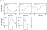
- FIG. 1 shows thermal stability of wild-type ( FIG. 1A ), G188C mutant ( FIG. 1B ), and G188C/N2C/D282C ( FIG. 1C ) mutant bovine rhodopsins that had been incubated with 11-cis-retinal and then purified. Absorption spectra were recorded after incubation in the dark at 37°C for 0, 5, 10, 15, or 20 min (Curves 1 to 5, respectively).
- FIG. 1 shows thermal stability of wild-type ( FIG. 1A ), G188C mutant ( FIG. 1B ), and G188C/N2C/D282C ( FIG. 1C ) mutant bovine rhodopsins that had been incubated with 11-cis-retinal and then purified. Absorption spectra were recorded after incubation in the dark at 37°C for 0, 5, 10, 15, or 20 min (Curves 1 to 5, respectively).
- FIG. 1D shows a schematic diagram of a change in retinal configuration in the wild-type bovine rhodopsin.
- a dark state, a Meta II, and a Meta III contain 11-cis-15-anti-retinal, all-trans-15-anti-retinal, and all-trans-15-syn retinal, respectively.
- FIG. 1E and F show absorption spectra of N2C/D282C ( FIG. 1E ) and G188C/N2C/D282C ( FIG. 1F ) mutant bovine rhodopsins that had been incubated with 11-cis-retinal and then purified.
- the spectra were recorded at 20°C in the dark (Curve 1) and at 0, 5, 15, 30, 60, and 120 min (Curves 2 to 7, respectively) after irradiation with yellow light.
- the inset shows differential spectra (Curves 1 to 5, respectively) each obtained by subtracting the spectra immediately after irradiation (Curve 2 in E and F) from each of the spectra measured after irradiation (Curves 3 to 7 in E and F).
- FIG. 1G shows absorption spectra of the G188C/N2C/D282C mutant as measured at 37°C.
- FIG. 1H shows an isomeric composition of retinal in the G188C/N2C/D282C mutant. Chromophores were extracted from samples before light irradiation and at 0, 5, and 60 minutes after irradiation with yellow light at 20°C and analyzed for retinal configuration by high-performance liquid chromatography (HPLC).
- the G188C mutant was found to have very low thermal stability than that of the wild type. That is, the G188C mutant gradually decayed during incubation in the dark at 37°C ( FIG. 1B ), whereas the wild type was extremely stable under the same conditions ( FIG. 1A ). Therefore, the thermal stability of the G188C mutant was improved and molecular characteristics of the mutant was analyzed in detail. According to previous reports (Xie et al., 2003; Standfuss et al., 2007), two cysteine residues (N2C/D282C) were introduced into the mutant and the resultant was measured for a thermal decay rate when incubated at 37°C in the dark.
- G188E and G188R mutants of human rhodopsin have been known to be unable to form a photopigment after reconstitution with 11-cis-retinal (Sung et al., 1993). Therefore, 16 other mutations were introduced at position 188 of bovine rhodopsin, reconstituted with 11-cis retinal, and then purified to prepare mutant proteins. The photopigment was successfully detected in eight of these mutants. The ⁇ max of the mutants was blue-shifted from the wild type (500 nm), except for G188D (509 nm) (Table 3).
- FIG. 2 shows a light reaction, a retinal configuration, and a G protein activation of the G188C mutant of bovine rhodopsin.
- FIGs. 2A and B show absorption spectra of the wild-type ( FIG. 2A ) and the G188C mutant ( FIG. 2B ) bovine rhodopsin that had been incubated with 11-cis-retinal at 0°C and then purified.
- the spectra were recorded in the dark (Curve 1), after irradiation with yellow light (>500 nm) (Curve 2), after subsequent irradiation with UV light (360 nm) (Curve 3), and after reirradiation with yellow light (Curve 4).
- the inset shows spectral changes of the wild type ( FIG. 2A ) or the G188C mutant ( FIG. 2B ) upon irradiation with yellow light (Curve 1), subsequent irradiation with UV (Curve 2), and reirradiation with yellow light (Curve 3).
- Differential spectra were calculated from the spectra shown in FIGs. 2A and 2B.
- FIG. 2C and D show isomeric compositions of retinal in the wild type ( FIG. 2C ) and the G188C mutant ( FIG. 2D ).
- Chromophores were extracted from samples before light irradiation, after irradiation with yellow light, after subsequent irradiation with UV, after reirradiation with yellow light at 0°C and analyzed for retinal configuration by high-performance liquid chromatography (HPLC).
- FIG. 2E shows a Gi-type G protein activation ability of the wild type. The activation ability was measured in the dark (closed circles) and after irradiation with yellow light (open circles).
- FIG. 2F shows a Gi-type G protein activation ability of the G188C mutant.
- FIG. 2G shows absorption spectra (0°C) of the G188C/N2C/D282C mutant that had been incubated with 11-cis retinal and then purified.
- the spectra were recorded in the dark (Curve 1), after irradiation with yellow light (>500 nm) (Curve 2), after subsequent irradiation with UV (360 nm) (Curve 3), after reirradiation with yellow light (Curve 4), and after reirradiation with UV (Curve 5).
- the inset shows spectral changes caused by irradiation with yellow light (Curve 1), subsequent irradiation with UV (Curve 2), reirradiation with yellow light (Curve 3), and reirradiation with UV (Curve 4).
- Differential spectra were calculated based on the spectra shown in FIG. 2G .
- the wild-type and the G188C mutant were cooled to 0°C to prevent a thermal reaction of the Meta II and measured for spectral changes caused by irradiation with yellow light and subsequent UV.
- irradiation with yellow light produced the Meta II and subsequent irradiation with UV caused a shift of the spectrum to the visible region and a blue shift of the ⁇ max (about 470 nm) from the original dark state ( FIG. 2A ).
- Previous studies have revealed that this state is equivalent to the Meta III ( FIG. 1D ).
- irradiation with yellow light produced the Meta II and subsequent irradiation with UV caused a shift of the spectrum to the visible region and made the ⁇ max almost the same as that in the original dark state ( FIG. 2B ).
- Reirradiation with yellow light resulted in formation of a state in which the spectrum nearly overlapped with that induced by the initial yellow light irradiation (Curve 4 in FIG. 2B ).
- Spectral changes induced by irradiation with UV and reirradiation with yellow light were mirror images of each other (Curves 2 and 3 in the inset of FIG. 2B ).
- the G188C/N2C/D282C mutant could also show interconversion between the original dark state and the Meta II upon irradiation with yellow light at 0°C and UV ( FIG. 2G ). These data revealed that the G188C mutant has acquired photoreversibility between the dark state and the Meta II.
- FIG. 5 shows formation of a photopigment in the G188C mutant of bovine rhodopsin after incubation with all-trans retinal.
- Absorption spectra of the wild type ( FIG. 5A ) or the G188C mutant ( FIG. 5B ) that had been prepared by adding all-trans retinal to a suspension of a rhodopsin-expressing cell membrane at 0°C and then purifying are shown.
- FIG. 5E shows differential spectra each calculated by subtracting each of the spectra measured 3, 6, 15, 30, 60, and 120 min after addition of 11-cis-retinal (Curves 3 to 8 in FIG. 5D ) from the spectrum immediately after addition of 11-cis-retinal (Curve 2 in FIG. 5D ) (Curves 1 to 6, respectively).
- FIG. 5F shows spectra measured before (Curve 1) and at 0, 0.5, 1, 2, 6, 12, and 16 hours after addition of 1.1 ⁇ M all-trans retinal (Curves 2 to 8).
- the differential spectra in FIG. 5G were calculated by subtracting the spectrum immediately after addition of all-trans retinal (Curve 2 in FIG.
- FIG. 5L is a graph showing regeneration processes of photopigment formation by the N2C/D282C and the G188C/N2C/D282C induced by addition of all-trans retinal shown in FIGs. 5F and 5J as monitored by a change in absorbance at 500 nm.
- All-trans retinal was added to a suspension of a rhodopsin-expressing cell membrane and then the wild-type and the G188C mutant were purified.
- the absorption spectrum of the wild type had few peaks in the visible and near-ultraviolet regions ( FIG. 5A ).
- the absorption spectrum of the G188C mutant had a peak in the visible region (Curve 1 in FIG. 5B ), which was due to predominant uptake of 11-cis and 9-cis retinal rather than all-trans retinal ( FIG. 5C ).
- bovine rhodopsin N2C/D282C As in the above example, the G188C mutant, the E122Q/G188C mutant, and a chimeric opsin in which intracellular second and third loops (amino acid numbers 140 to 152 and 225 to 251 of Gene ID: 509933, respectively) had been replaced by those of a mouse histamine H2 receptor (amino acid numbers 121 to 134 and 203 to 232 of Gene ID: 15466, respectively) was expressed in human-derived cultured cells HEK293.
- the modified luciferase Promega, GloSensor
- the cultured cells were irradiated with yellow light and luminescence derived from the modified luciferase was compared.
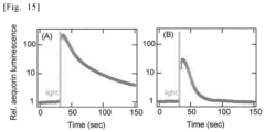
- a transient increase and then an immediate decrease in luminescence was observed with bovine rhodopsin N2C/D282C to which a G188C mutation ( FIG. 6B ) or an E122Q/G188C mutation ( FIG. 6C ) had been introduced compared to bovine rhodopsin N2C/D282C ( FIG. 6A ) in which the intracellular second and third loops had been replaced.
- the G188C and E122Q/G188C mutants were expressed in human-derived cultured cells HEK293.
- modified luciferase Promega, GloSensor
- the cultured cells were irradiated with yellow light and luminescence derived from the modified luciferase was compared.
- the cultured cells were irradiated with yellow light and luminescence derived from the modified luciferase was compared.
- a transient increase and then an immediate decrease in luminescence was observed with human rhodopsin N2C/N282C to which a G188C mutation ( FIG. 8E ) had been introduced compared to human rhodopsin N2C/N282C in which the intracellular second and third loops had been replaced ( FIG. 8D ).
- FIG. 8E a G188C mutation
- FIG. 8D This result indicates that introduction of the G188C mutation allows human rhodopsin to be modified so as to increase an intracellular cAMP concentration for a short period of time and return it with light.
- the G188C mutant was expressed in human-derived cultured cells HEK293.
- the modified luciferase Promega, GloSensor
- the cultured cells were irradiated with yellow light and luminescence derived from the modified luciferase was compared.
- amino acid substitutions were made in a N-terminal domain (positions 1 to 34) and a C-terminal domain (positions 308 and later) when aligned with SEQ ID NO: 1.
- a G6A mutation or a V337A mutation was introduced into the human rhodopsin G188C/N2C/D282C mutant and expressed in human-derived cultured cells HEK293.
- the modified luciferase Promega, GloSensor
- the cultured cells were irradiated with yellow light and luminescence derived from the modified luciferase was compared.
- a control retina without treatment showed no response from RGCs detected by the MEA ( FIG. 14A ).
- the treated retinae repeatedly showed clear light-induced responses ( FIGs. 14B , C, D).
- a visual evoked potential (VEP) from the visual cortex is examined to investigate whether photoreception in the retina is transmitted to the visual cortex.
- VEP visual evoked potential
- a rd1 mouse of which both eyes had been treated with a G188C mutant construct linked to AAV-DJ-CAGGS and a control EGFP virus (AAV-DJ-CAGGS-EGFP) were used.
- AAV-DJ-CAGGS-EGFP virus AAV-DJ-CAGGS-EGFP
- a light/dark box transition test was performed to investigate whether ectopic expression of the G188C mutant in the degenerative retina leads to a behavioral change following visual recovery.
- Rodents are nocturnal and tend to remain in the dark according to their visual function because they are anxious in a bright environment, while blind animals spend almost half their time in a light area and almost half in a dark area.
- a mouse treated with the G188C mutant spent significantly less time in the light area compared to an untreated rd1 mutant mouse, indicating a visual recovery in its behavior.
- the G188C mutant and the chimeric opsin in which intracellular second and third loops had been replaced by those of the human histamine H2 receptor as in Example 8 were expressed in mouse hippocampal neurons to attempt light manipulation of axonal branching and elongation in neurons.
- axonal branching and elongation were suppressed in hippocampal neurons that had been transfected with the G188C mutant irradiated with blue light for 30 minutes compared to those not irradiated.
- hippocampal neurons that had been transfected with the G188C mutant chimeric opsin irradiated with blue light for 30 minutes showed enhanced axonal branching and elongation compared to those not irradiated.
- an evolutionary mechanism of nature that gave a photocycle property to a specific opsin can be used to artificially evolve another opsin to have the photocycle property as well.
- the present disclosure provides techniques available in production of various molecular tools and in industries based on such techniques (e.g., pharmaceuticals).
Landscapes
- Health & Medical Sciences (AREA)
- Life Sciences & Earth Sciences (AREA)
- Chemical & Material Sciences (AREA)
- Organic Chemistry (AREA)
- General Health & Medical Sciences (AREA)
- Engineering & Computer Science (AREA)
- Medicinal Chemistry (AREA)
- Genetics & Genomics (AREA)
- Zoology (AREA)
- Molecular Biology (AREA)
- Biochemistry (AREA)
- Proteomics, Peptides & Aminoacids (AREA)
- Biophysics (AREA)
- Gastroenterology & Hepatology (AREA)
- Toxicology (AREA)
- Biomedical Technology (AREA)
- Animal Behavior & Ethology (AREA)
- Veterinary Medicine (AREA)
- Pharmacology & Pharmacy (AREA)
- Bioinformatics & Cheminformatics (AREA)
- Public Health (AREA)
- Cell Biology (AREA)
- Biotechnology (AREA)
- Immunology (AREA)
- Chemical Kinetics & Catalysis (AREA)
- Epidemiology (AREA)
- Endocrinology (AREA)
- Ophthalmology & Optometry (AREA)
- General Chemical & Material Sciences (AREA)
- Nuclear Medicine, Radiotherapy & Molecular Imaging (AREA)
- Wood Science & Technology (AREA)
- Neurology (AREA)
- General Engineering & Computer Science (AREA)
- Microbiology (AREA)
- Neurosurgery (AREA)
- Peptides Or Proteins (AREA)
- Medicines That Contain Protein Lipid Enzymes And Other Medicines (AREA)
Applications Claiming Priority (2)
| Application Number | Priority Date | Filing Date | Title |
|---|---|---|---|
| JP2022138707 | 2022-08-31 | ||
| PCT/JP2023/031644 WO2024048688A1 (ja) | 2022-08-31 | 2023-08-30 | 光応答改変型オプシン類 |
Publications (1)
| Publication Number | Publication Date |
|---|---|
| EP4582544A1 true EP4582544A1 (en) | 2025-07-09 |
Family
ID=90099743
Family Applications (1)
| Application Number | Title | Priority Date | Filing Date |
|---|---|---|---|
| EP23860450.8A Pending EP4582544A1 (en) | 2022-08-31 | 2023-08-30 | Light-responsive modified opsin |
Country Status (5)
| Country | Link |
|---|---|
| EP (1) | EP4582544A1 (https=) |
| JP (1) | JPWO2024048688A1 (https=) |
| KR (1) | KR20250061742A (https=) |
| CN (1) | CN120225677A (https=) |
| WO (1) | WO2024048688A1 (https=) |
Families Citing this family (1)
| Publication number | Priority date | Publication date | Assignee | Title |
|---|---|---|---|---|
| WO2025214408A1 (en) * | 2024-04-10 | 2025-10-16 | Skyline Therapeutics (Shanghai) Co., Ltd. | Optogenetic modulation for vision restoration |
Family Cites Families (12)
| Publication number | Priority date | Publication date | Assignee | Title |
|---|---|---|---|---|
| US5585108A (en) | 1994-12-30 | 1996-12-17 | Nanosystems L.L.C. | Formulations of oral gastrointestinal therapeutic agents in combination with pharmaceutically acceptable clays |
| CA2224907A1 (en) | 1995-07-25 | 1997-02-13 | Introgene B.V. | Methods and means for targeted gene delivery |
| US6265389B1 (en) | 1995-08-31 | 2001-07-24 | Alkermes Controlled Therapeutics, Inc. | Microencapsulation and sustained release of oligonucleotides |
| JP3521382B2 (ja) | 1997-02-27 | 2004-04-19 | 日本たばこ産業株式会社 | 細胞間接着及びシグナル伝達を媒介する細胞表面分子 |
| US6649138B2 (en) | 2000-10-13 | 2003-11-18 | Quantum Dot Corporation | Surface-modified semiconductive and metallic nanoparticles having enhanced dispersibility in aqueous media |
| EP1474109B1 (en) | 2001-12-21 | 2010-08-25 | Alcon, Inc. | Use of synthetic inorganic nanoparticles as carriers for ophthalmic drugs |
| CN102016814B (zh) | 2005-06-17 | 2013-10-23 | 北卡罗来纳大学查珀尔希尔分校 | 纳米粒子制备方法、系统及材料 |
| US9782342B2 (en) | 2010-10-11 | 2017-10-10 | Wichita State University | Composite magnetic nanoparticle drug delivery system |
| WO2012125987A2 (en) | 2011-03-17 | 2012-09-20 | Massachusetts Institute Of Technology | Delivery system |
| US20120283503A1 (en) | 2011-04-29 | 2012-11-08 | The Johns Hopkins University | Nanoparticle loaded stem cells and their use in mri guided hyperthermia |
| WO2020206098A1 (en) * | 2019-04-03 | 2020-10-08 | Regenxbio Inc. | Gene therapy for eye pathologies |
| JP7696675B2 (ja) | 2021-03-10 | 2025-06-23 | ダイハツ工業株式会社 | 車両用制御装置 |
-
2023
- 2023-08-30 JP JP2024544545A patent/JPWO2024048688A1/ja active Pending
- 2023-08-30 EP EP23860450.8A patent/EP4582544A1/en active Pending
- 2023-08-30 CN CN202380062525.5A patent/CN120225677A/zh active Pending
- 2023-08-30 WO PCT/JP2023/031644 patent/WO2024048688A1/ja not_active Ceased
- 2023-08-30 KR KR1020257010345A patent/KR20250061742A/ko active Pending
Also Published As
| Publication number | Publication date |
|---|---|
| WO2024048688A1 (ja) | 2024-03-07 |
| JPWO2024048688A1 (https=) | 2024-03-07 |
| KR20250061742A (ko) | 2025-05-08 |
| CN120225677A (zh) | 2025-06-27 |
Similar Documents
| Publication | Publication Date | Title |
|---|---|---|
| US20250262328A1 (en) | Gene Therapy for the Treatment of CNGB1-linked Retinitis Pigmentosa | |
| Petit et al. | Restoration of vision in the pde6β-deficient dog, a large animal model of rod-cone dystrophy | |
| JP7299632B2 (ja) | 網膜疾患の予防および進行抑制、視覚認知行動機能の改善、および視覚機能強化 | |
| US12606606B2 (en) | Nucleic acid construct that encodes chimeric rhodopsin | |
| CA3234028A1 (en) | A modified mglur6 promoter and methods of use | |
| Smith et al. | Gene supplementation therapy for recessive forms of inherited retinal dystrophies | |
| EP3508212B1 (en) | Rhodopsin gene therapy for regenerating visual function or for preventing deterioration in visual function | |
| RS66227B1 (sr) | Kompozicije i postupci za lečenje poremećaja mrežnjače | |
| EP4582544A1 (en) | Light-responsive modified opsin | |
| US11771741B2 (en) | Nucleic acid construct that encodes chimeric rhodopsin | |
| EP4215212A1 (en) | Composition for treating or preventing diseases, disorders, or conditions associated with endoplasmic reticulum stress or all-trans-retinal, protecting retinal thickness, or suppressing reduction in retinal thickness or progress of reduction in retinal thickness | |
| CN112689675B (zh) | 光响应性蛋白及其应用 | |
| WO2020059675A1 (ja) | 改変チャネルロドプシン | |
| CN115397991B (zh) | 用于颜色识别的光响应性蛋白及其应用 | |
| HK40124146A (zh) | 光响应改造型视蛋白类 | |
| HK40097616A (en) | Composition for treating or preventing diseases, disorders, or conditions associated with endoplasmic reticulum stress or all-trans-retinal, protecting retinal thickness, or suppressing reduction in retinal thickness or progress of reduction in retinal thickness | |
| HK40097920A (zh) | 用於治疗或预防与内质网应激或全反式视黄醛相关的疾病、障碍或症状、或者用於保护视网膜厚度、或者用於抑制视网膜厚度的萎缩或萎缩进展的组合物 | |
| HK40074775A (en) | Nucleic acid construct that encodes chimeric rhodopsin | |
| WO2021193732A1 (ja) | 改変光受容クロライドチャネル | |
| WO2022011081A1 (en) | Cell lines that produce human retinoschisin proteins and uses thereof |
Legal Events
| Date | Code | Title | Description |
|---|---|---|---|
| STAA | Information on the status of an ep patent application or granted ep patent |
Free format text: STATUS: THE INTERNATIONAL PUBLICATION HAS BEEN MADE |
|
| PUAI | Public reference made under article 153(3) epc to a published international application that has entered the european phase |
Free format text: ORIGINAL CODE: 0009012 |
|
| STAA | Information on the status of an ep patent application or granted ep patent |
Free format text: STATUS: REQUEST FOR EXAMINATION WAS MADE |
|
| 17P | Request for examination filed |
Effective date: 20250321 |
|
| AK | Designated contracting states |
Kind code of ref document: A1 Designated state(s): AL AT BE BG CH CY CZ DE DK EE ES FI FR GB GR HR HU IE IS IT LI LT LU LV MC ME MK MT NL NO PL PT RO RS SE SI SK SM TR |
|
| REG | Reference to a national code |
Ref country code: HK Ref legal event code: DE Ref document number: 40122239 Country of ref document: HK |
|
| DAV | Request for validation of the european patent (deleted) | ||
| DAX | Request for extension of the european patent (deleted) |