EP4579669A2 - Validierungsverfahren und systeme für sequenzvariantenaufrufe - Google Patents
Validierungsverfahren und systeme für sequenzvariantenaufrufe Download PDFInfo
- Publication number
- EP4579669A2 EP4579669A2 EP25176100.3A EP25176100A EP4579669A2 EP 4579669 A2 EP4579669 A2 EP 4579669A2 EP 25176100 A EP25176100 A EP 25176100A EP 4579669 A2 EP4579669 A2 EP 4579669A2
- Authority
- EP
- European Patent Office
- Prior art keywords
- sequence
- subset
- error rate
- read
- reads
- Prior art date
- Legal status (The legal status is an assumption and is not a legal conclusion. Google has not performed a legal analysis and makes no representation as to the accuracy of the status listed.)
- Pending
Links
Images


Classifications
-
- G—PHYSICS
- G16—INFORMATION AND COMMUNICATION TECHNOLOGY [ICT] SPECIALLY ADAPTED FOR SPECIFIC APPLICATION FIELDS
- G16B—BIOINFORMATICS, i.e. INFORMATION AND COMMUNICATION TECHNOLOGY [ICT] SPECIALLY ADAPTED FOR GENETIC OR PROTEIN-RELATED DATA PROCESSING IN COMPUTATIONAL MOLECULAR BIOLOGY
- G16B20/00—ICT specially adapted for functional genomics or proteomics, e.g. genotype-phenotype associations
- G16B20/20—Allele or variant detection, e.g. single nucleotide polymorphism [SNP] detection
-
- G—PHYSICS
- G16—INFORMATION AND COMMUNICATION TECHNOLOGY [ICT] SPECIALLY ADAPTED FOR SPECIFIC APPLICATION FIELDS
- G16B—BIOINFORMATICS, i.e. INFORMATION AND COMMUNICATION TECHNOLOGY [ICT] SPECIALLY ADAPTED FOR GENETIC OR PROTEIN-RELATED DATA PROCESSING IN COMPUTATIONAL MOLECULAR BIOLOGY
- G16B30/00—ICT specially adapted for sequence analysis involving nucleotides or amino acids
-
- G—PHYSICS
- G16—INFORMATION AND COMMUNICATION TECHNOLOGY [ICT] SPECIALLY ADAPTED FOR SPECIFIC APPLICATION FIELDS
- G16B—BIOINFORMATICS, i.e. INFORMATION AND COMMUNICATION TECHNOLOGY [ICT] SPECIALLY ADAPTED FOR GENETIC OR PROTEIN-RELATED DATA PROCESSING IN COMPUTATIONAL MOLECULAR BIOLOGY
- G16B40/00—ICT specially adapted for biostatistics; ICT specially adapted for bioinformatics-related machine learning or data mining, e.g. knowledge discovery or pattern finding
-
- G—PHYSICS
- G16—INFORMATION AND COMMUNICATION TECHNOLOGY [ICT] SPECIALLY ADAPTED FOR SPECIFIC APPLICATION FIELDS
- G16B—BIOINFORMATICS, i.e. INFORMATION AND COMMUNICATION TECHNOLOGY [ICT] SPECIALLY ADAPTED FOR GENETIC OR PROTEIN-RELATED DATA PROCESSING IN COMPUTATIONAL MOLECULAR BIOLOGY
- G16B50/00—ICT programming tools or database systems specially adapted for bioinformatics
Definitions
- the present disclosure relates generally to the field of data related to biological samples, such as sequence data. More particularly, the disclosure relates to techniques for validating sequence variant calls based on sequencing data acquired during sequencing operations.
- NGS Next-generation sequencing
- SNVs single nucleotide variants
- Indels small insertions and deletions
- MNVs multiple nucleotide variants
- CNVs gene amplifications
- the NGS test also includes a RNA workflow for the identification of splice variants and gene fusions.
- a sequence variant is identified when a sample nucleic acid sequence is determined to different from a reference or baseline sequence at one or more base pair positions along the sequence. Identification of one or more sequence variants may in turn be used to characterize a patient sample, diagnose a clinical condition, and/or classify disease (e.g., cancer) progression.
- disease e.g., cancer
- sequence variants are complex.
- Certain sequencing techniques experience false positives in connection with variant calling. For example, the technique may incorrectly determine that a variant is present in a sample sequence at a particular location (base pair) and/or incorrectly identify the type of variant, which leads to false positives in identified sequence variants.
- False positive sequence variants may be the result of error introduced into the sample itself at the sample preparation stage and/or may be the result of systematic errors introduced during amplification or sequence acquisition. Further, certain types of samples (e.g., FFPE samples) may be more prone to error.
- a need remains for sequencing methods and systems that can accurately identify DNA variants while reducing a number of false positives in an efficient and cost-effective manner.
- chromosome refers to the heredity-bearing gene carrier of a living cell, which is derived from chromatin strands comprising DNA and protein components (especially histones).
- chromatin strands comprising DNA and protein components (especially histones).
- the conventional internationally recognized individual human genome chromosome numbering system is employed herein.
- site refers to a unique position (e.g., chromosome ID, chromosome position and orientation) on a reference genome.
- a site may be a residue, a sequence tag, or a segment's position on a sequence.
- locus may be used to refer to the specific location of a nucleic acid sequence or polymorphism on a reference chromosome.
- sample refers to a sample, typically derived from a biological fluid, cell, tissue, organ, or organism containing a nucleic acid or a mixture of nucleic acids containing at least one nucleic acid sequence that is to be sequenced and/or phased.
- samples include, but are not limited to sputum/oral fluid, amniotic fluid, blood, a blood fraction, fine needle biopsy samples (e.g., surgical biopsy, fine needle biopsy, etc.), urine, peritoneal fluid, pleural fluid, tissue explant, organ culture and any other tissue or cell preparation, or fraction or derivative thereof or isolated therefrom.
- samples can be taken from any organism having chromosomes, including, but not limited to dogs, cats, horses, goats, sheep, cattle, pigs, etc.
- the sample may be used directly as obtained from the biological source or following a pretreatment to modify the character of the sample.
- pretreatment may include preparing plasma from blood, diluting viscous fluids and so forth.
- Methods of pretreatment may also involve, but are not limited to, filtration, precipitation, dilution, distillation, mixing, centrifugation, freezing, lyophilization, concentration, amplification, nucleic acid fragmentation, inactivation of interfering components, the addition of reagents, lysing, etc.
- sequence includes or represents a strand of nucleotides coupled to each other.
- the nucleotides may be based on DNA or RNA. It should be understood that one sequence may include multiple sub-sequences. For example, a single sequence (e.g., of a PCR amplicon) may have 350 nucleotides.
- the sample read may include multiple sub-sequences within these 350 nucleotides. For instance, the sample read may include first and second flanking subsequences having, for example, 20-50 nucleotides.
- the first and second flanking sub-sequences may be located on either side of a repetitive segment having a corresponding sub-sequence (e.g., 40-100 nucleotides).
- Each of the flanking sub-sequences may include (or include portions of) a primer sub-sequence (e.g., 10-30 nucleotides).
- primer sub-sequence e.g. 10-30 nucleotides.
- sequences may be given different labels (e.g., target sequence, primer sequence, flanking sequence, genomic sequence, sample sequence, reference sequence, and the like). Other terms, such as "allele,” may be given different labels to differentiate between like objects.
- paired-end sequencing refers to sequencing methods that sequence both ends of a target fragment. Paired-end sequencing may facilitate detection of genomic rearrangements and repetitive segments, as well as gene fusions and novel transcripts. Methodology for paired-end sequencing are described in PCT publication WO07010252 , PCT application Serial No. PCTGB2007/003798 and US patent application publication US 2009/0088327 , each of which is incorporated by reference herein.
- a series of operations may be performed as follows; (a) generate clusters of nucleic acids; (b) linearize the nucleic acids; (c) hybridize a first sequencing primer and carry out repeated cycles of extension, scanning and deblocking, as set forth above; (d) invert the target nucleic acids on the flow cell surface by synthesizing a complimentary copy; (e) linearize the resynthesized strand; and (f) hybridize a second sequencing primer and carry out repeated cycles of extension, scanning and deblocking, as set forth above.
- the inversion operation can be carried out be delivering reagents as set forth above for a single cycle of bridge amplification.
- reference genome refers to any particular known genome sequence, whether partial or complete, of any organism which may be used to reference identified sequences from a subject and relative to which one or more sequence variants may be determined.
- reference genome used for human subjects as well as many other organisms is found at the National Center for Biotechnology Information at ncbi.nlm.nih.gov.
- a "genome” or genomic sequence refers to the complete genetic information of an organism or virus, expressed in nucleic acid sequences.
- a genome includes both the genes and the noncoding sequences of the DNA. The reference sequence may be larger than the reads that are aligned to it.
- the reference sequence may include sequence information for a subset of the genome that aligns with a targeted sequencing panel.
- the reference genome is a consensus sequence or other combination derived from multiple individuals. That is, the reference sequence may be a hypothetical or representative sequence. However, in certain applications, the reference sequence may be taken from a particular individual.
- the reference sequence is a normal sequence and the sample of interest is a matched tumor sequence from the same individual.
- a reference sequence is taken at a first time point and the sample sequence is taken at a second, subsequent, time point.
- a reference sequence may be used as a basis relative to which sequence variants are determined.
- the reference sequence may be provided as a stored data file that may be accessed and/or operated on according to processor-executed instructions. Further, a system as provided herein may include a stored set of different reference sequences that may be selected based on user input related to the sample of interest and/or the sequencing type (whole genome, targeted sequencing). In one embodiment, a sample from an individual user may sequenced, and an appropriate reference sequence may be accessed (e.g., from a cloud computing environment) as an input to a sequence variant operation on the genomic sequence data.
- read refers to a collection of sequence data that describes a fragment of a nucleotide template sample or reference.
- the fragment may be a fragment generated during sample preparation.
- read may refer to a sample read (from a biological sample of interest) and/or a reference read (a sequence read acquired as part of sequencing a reference sample).
- a read may represent a short sequence of contiguous base pairs in the sample or reference.
- the read may be represented symbolically by the base pair sequence (in ATCG) of the sample or reference fragment. It may be stored in a memory device and processed as appropriate to determine whether the read matches or has differences relative to a reference sequence or meets other criteria.
- a sequence read may be obtained directly from a sequencing apparatus or may be accessed from stored sequence information concerning the sample.
- a read is a DNA sequence of sufficient length (e.g., at least about 25 bp) that can be used to identify a larger sequence or region, e.g., that can be aligned, e.g., stitched together, and specifically assigned to a chromosome or genomic region or gene as part of genome assembly.
- the terms "sample read”, “sample sequence” or “sample fragment” refer to sequence data of a genomic sequence of interest from a sample.
- the sample read includes sequence data from a PCR amplicon having a forward and reverse primer sequence. The sequence data can be obtained from any appropriate sequence methodology.
- the sample read can be, for example, from a sequencing-by-synthesis (SBS) reaction, a sequencing-by-ligation reaction, or any other suitable sequencing methodology for which it is desired to determine the length and/or identity of a repetitive element.
- SBS sequencing-by-synthesis
- the sample read can be a consensus (e.g., averaged or weighted) or collapsed sequence derived from multiple sample reads.
- NGS Next-generation sequencing
- Illumina sequencing by synthesis technology
- Ion Torrent sequencing single-molecule real-time sequencing
- SOLiD sequencing sequencing by ligation
- the length of each read may vary from about 30 bp to more than 10,000 bp.
- an Illumina sequencing method using SOLiD sequencer generates nucleic acid reads of about 50 bp.
- Ion Torrent Sequencing generates nucleic acid reads of up to 400 bp and 454 pyrosequencing generates nucleic acid reads of about 700 bp.
- single-molecule real-time sequencing methods may generate reads of 10,000 bp to 15,000 bp. Therefore, in certain embodiments, the reads as provided herein have a length of 30-100 bp, 50-200 bp, or 50-400 bp.
- aligning refers to the process of comparing a read or tag to a reference sequence and thereby determining whether the reference sequence contains the read sequence. If the reference sequence contains the read, the read may be mapped to the reference sequence or, in certain embodiments, to a particular location in the reference sequence. In some cases, alignment simply tells whether or not a read is a member of a particular reference sequence (i.e., whether the read is present or absent in the reference sequence). In some cases, an alignment additionally indicates a location in the reference sequence where the read or tag maps to. For example, if the reference sequence is the whole human genome sequence, an alignment may indicate that a read is present on a particular chromosome, and may further indicate that the read is on a particular strand and/or site of the chromosome.
- variant refers to a nucleic acid sequence that is different from a reference sequence.
- Typical nucleic acid sequence variant includes without limitation single nucleotide polymorphism (SNP), short deletion and insertion polymorphisms (Indel), copy number variation (CNV), microsatellite markers or short tandem repeats and structural variation.
- Variants may also occur at homopolymer regions with at least 4 repetitive nucleotides, e.g., AAAA, GGGG, CCCC, TTTT.
- Somatic variant calling, sequence variant calling, or variant calling as provided herein refers to identification and/or validation of sequence variants present in a sample of interest. In one embodiment, variant calling may be used to characterize cancer progression. For example, a single nucleotide variation might be seen in a certain percentage of the reads covering a given base.
- micro-indel refers to the insertion and/or the deletion of bases in the DNA of an organism.
- a micro-indel represents an indel that results in a net change of 1 to 50 nucleotides. In coding regions of the genome, unless the length of an indel is a multiple of 3, it will produce a frameshift mutation.
- Indels can be contrasted with point mutations. An indel inserts and deletes nucleotides from a sequence, while a point mutation is a form of substitution that replaces one of the nucleotides without changing the overall number in the DNA. Indels can also be contrasted with a Tandem Base Mutation (TBM), which may be defined as substitution at adjacent nucleotides (primarily substitutions at two adjacent nucleotides, but substitutions at three adjacent nucleotides have been observed.
- TBM Tandem Base Mutation
- variant frequency or “variant allele frequency” represents the relative frequency of an allele (variant of a gene) at a particular locus in a population, expressed as a fraction or percentage.
- fraction or percentage may be the fraction of all chromosomes in the population that carry that allele.
- sample variant frequency represents the relative frequency of an allele/variant at a particular locus/position along a genomic sequence of interest over a "population" corresponding to the number of reads and/or samples obtained for the genomic sequence of interest from an individual.
- a baseline variant frequency represents the relative frequency of an allele/variant at a particular locus/position along one or more baseline genomic sequences where the "population" corresponding to the number of reads and/or samples obtained for the one or more baseline genomic sequences from a population of normal individuals.
- position refers to a location or coordinate of one or more nucleotides within a sequence of nucleotides.
- position also refer to a location or coordinate of one or more base pairs in a sequence of nucleotides.
- haplotype refers to a combination of alleles at adjacent sites on a chromosome that are inherited together.
- a haplotype may be one locus, several loci, or an entire chromosome depending on the number of recombination events that have occurred between a given set of loci, if any occurred.
- threshold refers to a numeric or non-numeric value that is used as a cutoff to characterize a sample, a nucleic acid, or portion thereof (e.g., a read).
- a threshold may be varied based upon empirical analysis. The threshold may be compared to a measured or calculated value to determine whether the source giving rise to such value suggests should be classified in a particular manner. Threshold values can be identified empirically or analytically. The choice of a threshold is dependent on the level of confidence that the user wishes to have to make the classification. The threshold may be chosen for a particular purpose (e.g., to balance sensitivity and selectivity).
- threshold indicates a point at which a course of analysis may be changed and/or a point at which an action may be triggered.
- a threshold is not required to be a predetermined number. Instead, the threshold may be, for instance, a function that is based on a plurality of factors. The threshold may be adaptive to the circumstances. Moreover, a threshold may indicate an upper limit, a lower limit, or a range between limits.
- a "likelihood score” is a score per variant site given the error rate estimate according to the disclosed embodiments, and may also be based in part on an alternative read count (count of number of variant sample reads) and a total read count for the variant site in question.
- an error rate is based on a total count of sequence reads determined to have sequence errors as provided herein.
- a biological sample having a high total count may be considered to have a higher error rate than another biological sample having a lower total count
- Allele quality (AQ) is the quality score of observed allele frequency in test sample against baseline or reference samples.
- UMIs are similar to bar codes, which are commonly used to distinguish reads of one sample from reads of other samples, but UMIs are instead used to distinguish nucleic acid template fragments from another when many fragments from an individual sample are sequenced together.
- the UMIs may be single or double-stranded, and may be at least 5 bases, at least 6 bases, at least 7 bases, at least 8 bases, or more.
- the UMIs are 5-8 bases, 5-10 bases, 5-15 bases, 5-25 bases, 8-10 bases, 8-12 bases, 8-15 bases, or 8-25 bases in length, etc.
- the UMIs are no more than 30 bases, no more than 25 bases, no more than 20 bases, no more than 15 bases in length.
- the method also includes the step of identifying errors in the genomic sequence data based on sequence disagreement within a first subset of the plurality of sequence reads associated with a first unique molecular identifier, sequence disagreement between the first subset and a second subset of the plurality of sequence reads having a second unique molecular identifier complementary to the first unique molecular identifier, or both, to generate an error rate of the genomic sequence data.
- the method also includes the steps of identifying a plurality of potential sequence variants in the genomic sequence data relative to a reference sequence; classifying false positive sequence variants of the plurality of potential sequence variants based on the error rate of the genomic sequence data; and eliminating the false positive sequence variants from the plurality of potential sequence variants to yield a plurality of sequence variants.
- a computer-implemented method is provided. The method is performed under control of a processor executing instructions.
- the method includes the step of receiving genomic sequence data of a first biological sample, wherein the genomic sequence data comprises a plurality of sequence reads, each sequence read being associated with a unique molecular identifier of a plurality of unique molecular identifiers.
- the method also includes the step of identifying first sequence differences within a first subset of the plurality of sequence reads associated with a first unique molecular identifier.
- the method also includes the step of collapsing the first subset to yield a collapsed first subset sequence read, wherein the collapsing comprises eliminating sequence differences present in a minority of the sequencing reads of the first subset.
- sequencing device configured to identify sequence variants in genomic sequence data of a biological sample.
- the device includes a memory device including executable application instructions stored therein and a processor configured to execute the application instructions stored in the memory device.
- the application instructions comprise instructions that cause the processor to receive genomic sequence data of a biological sample, wherein the genomic sequence data comprises a plurality of sequence reads, each sequence read being associated with a unique molecular identifier of a plurality of unique molecular identifiers; identify a plurality of errors in the genomic sequence data based on sequence disagreement between sequence reads associated with each unique molecular identifier of the plurality of unique molecular identifiers to generate an error rate of the genomic sequence data; identify a plurality of potential sequence variants in the genomic sequence data relative to a reference sequence; and determine a validity of the plurality of potential sequence variants based at least in part on the error rate.
- FIG. 1 is a schematic workflow diagram 10 showing a sample preparation and sequence acquisition workflow.
- a template 12 derived from a biological sample of interest undergoes library preparation (step 14) to incorporate one or more UMIs 16.
- the template 12 may represent a plurality of nucleic acid fragments.
- Each template 12 incorporates an individual UMI 16 (which may include one or more identifier sequences) of a plurality of UMIs, such that the different source templates 12 are each associated with distinguishable UMIs 16 have different sequences.
- the depicted diagram 10 is shown in the context of forked paired-end sequencing adapters including unique molecular identifiers (UMIs) 16 configured to couple to the 5' and 3' ends of a nucleic acid template fragment 12 and such that the template 12 is flanked by different portions 16a, 16b of the UMI 16.
- UMIs unique molecular identifiers
- the positive strand 20a includes a first UMI sequence or sequences while the negative strand 20b includes a second UMI sequence complementary to the first.
- the first UMI sequence and the second UMI sequence may be considered to be part of a single UMI 16 or different UMIs 16.
- the sequences of the positive strand 20a and the negative strand 20b may be associated with one another.
- genomic sequence data of the sample is acquired by any suitable sequencing technique, depicted here as paired-end sequencing (step 26).
- Paired-end sequencing yields a plurality of sequence reads 28, which may be in turn divided or separated by template source via the respective UMIs 16.
- a first read group 30 including a first subset of the acquired sequence reads 28 may be associated with a first UMI 16 while a second read group 32 including a second subset of the acquired sequence reads 28 may be associated within a second UMI 16 complementary to the first UMI 16.
- the complementary UMIs may also be considered to be a single UMI.
- sequence reads on the same strand within a single read group should be identical to one another, as the associated UMI 16 links a subset of the sequence reads 28 to a single source template 12.
- Deviation or differences within the group are indicative of sample preparation or sequence acquisition errors. Identification and elimination of outlier reads within a read group to collapse the read group to a consensus sequence or collapsed sequence (step 40) may serve to prevent introduced sequence errors from propagating into the sequence data to yield false positive variants.
- outlier differences such as difference 42, that are not present in other sequence reads within the first read group 30, may be considered to be due to sequence error. Any identified differences or variations within a read group are provided as input to determining an overall error rate for the sample.
- any differences that pass through consensus sequence building may further be compared to sequence reads associated with a complementary strand of the UMI 16. That is, the sequences of the first read group 30 and the second read group 32 may be assembled as a duplex. Again, any differences between the groups 30, 32 may be identified before a consensus duplex of the complementary strands is assembled (step 47). Such differences may also be tracked as part of the error rate.
- the collapsed simplex or duplex groups may be stitched together at overlapping regions (step 48) to generate a collapsed longer fragment as part of sequence assembly. Stitching may be used to determine a frequency of any potential sequence variants.
- FIG. 2 is a flow diagram of a method 50 of receiving genomic sequence data of a biological sample, wherein the genomic sequence data comprises a plurality of sequence reads, each sequence read being associated with a unique molecular identifier of a plurality of unique molecular identifiers; The method includes the step of receiving genomic sequence data of an individual biological sample (block 52).
- the received sequence data may be received subsequent to sample preparation and sequencing of the biological sample as provided herein. Further, the received genomic sequence data may be stored or retrospective sequence data.
- the genomic sequence data may include include customer information, biological sample organism information, biological sample type information (e.g. information identifying whether the sample is fresh, frozen, or preserved), tissue type, sequence device type, and sequencing assay type (whole genome, targeted panel).
- the genomic sequence data is operated on to determine an error rate of the genomic sequence data (block 54).
- the error rate is characteristic of the sample itself and its associated genomic sequence data. Accordingly, the error rate may be calculated de novo for each sequencing run of a biological sample of interest. An error rate for samples taken from a same individual at different times may exhibit different characteristic error rates that depend on sample preparation variabilities, sequencing device settings, etc.
- the mapped sample reads are analyzed relative to the reference sequence to identify potential sequence variants.
- the results of the analysis identify the potential variant call, a sample variant frequency, a reference sequence and a position within the genomic sequence of interest at which the variant occurred.
- the assigned reads that have been called for the genetic locus may undergo analysis to identify the SNPs of the assigned reads.
- the assigned reads may be analyzed to identify or characterize the polymorphic repetitive DNA elements within the sample reads.
- a warning or flag may be assigned to the sample read.
- the potential sequence variants are operated on by a function that takes into account the determined error rate to distinguish between true positives and false positives (block 58).
- a likelihood score is determined based on a likelihood ratio:
- the validated sequence variants may be provided (block 60) to a user.
- the validated sequence may be provided as a generated report, e.g., stored as a report file or displayed on a graphical user interface for user interaction.
- the validation operation may also report or store a corresponding indication (e.g., a negative indicator, a no call indicator, an in-valid call indicator) as part of the report.
- the validation also may provide the likelihood score related to a degree of confidence that the variant call is correct or the invalid call designation is correct.
- FIG. 3 is a flow diagram of a method 64 that operates on received genomic sequence data of a biological sample (block 66) to determine sequence variants.
- the genomic sequence data includes sequences of UMIs, whereby each sequence read is associated with one UMI of a plurality of UMIs used in the sequencing run.
- the sequence reads may be separated into read groups, whereby each read group is a subset of the sequence reads that are associated with a common UMI (block 70). Accordingly, each sequence read should be present in only one read group. Once separated, errors in the genomic sequence data are identified based on sequence disagreement between the subset of sequence reads within the read group.
- Each sequence read for a particular UMI should be identical. Further, for paired end sequencing, sequenced strands in both directions should align.
- an overall error rate of the genomic sequence data may be determined (block 74).
- the error rate may in turn be used to identify and/or validate sequence variants in the genomic sequence data (block 76).
- FIG. 4 is a flow diagram of a method 80 for generating an error rate as provided herein.
- the method 80 operates on received genomic sequence data of a biological sample (block 82) that has been separated into subsets based on a common unique molecular identifier (block 84).
- sequence differences within the subset are identified (block 86).
- the collapsed sequence may be determined based on a majority voting rule, whereby sequence differences that are in a minority of sequence reads in a particular subset (i.e., read group) are designated as sequence errors (block 88) but sequence differences that are in a majority of the sequence reads pass through to build the consensus or collapsed sequence (block 90).
- the error rate is identified (block 92). However, not all sequence differences in each subgroup necessarily contribute to the error rate. Sequence differences in the majority of sequence reads (see difference 46 of FIG. 1 ) are distinguished from sequence differences in the minority.
- the error rate may, additionally or alternatively, be stratified based on a type of nucleotide change. In this manner, systemic error that is biased towards particular nucleotide changes is identified.
- FIG. 5 is a panel of error rates separated out by type of change. The error rates are compared between different sample types, including 24 single cell free DNA (cfDNA) BRN samples, nucleosome prep of seven cancer cell lines and 6 0.2% zoo mix samples, and genomic pipDNA including three healthy samples and 21 HD753 titrated samples. Further, the inputs to the error rate determination are separated by duplex, simplex, stitched, and unstitched sequence reads in various combination. As noted with reference to FIG. 1 , duplex building and stitching corrects errors in template sequences by eliminating sequence differences that are associated with error.
- the error rates of each type of error vary based on sample type. For example, in cell free DNA and nucleosomePrep, deamination and resultant G to A errors are present in relatively higher levels. Oxidation is dominant in pipDNA, resulting in observed higher error rates of G to T changes. Accordingly, in certain embodiments, certain biological sample types may be associated with particular characteristic errors. In one embodiment, the sequence variant determination may include a weighting factor to weigh against potential variants that are associated with error for the sample type in question.
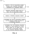
- FIG. 6 is a flow diagram of a method 100 of determining stratified error rates, as shown in FIG. 5 .
- sequence reads that are part of a single read group, individual reads having sequence differences within the group and in a minority of strands are eliminated to correct the template. These eliminated sequence reads may be further analyzed to identify the types of erroneous sequence changes that occur at each locus (block 102). The nucleotide change forming the erroneous sequence change is considered relative to the majority sequence read in the group to identify the type of nucleotide change.
- the type of change may be binned as a G>A change.
- the change may be a single nucleotide change or an indel. This process is applied to all individual read groups including minority sequence reads having sequence differences to generate stratified error rates of each type of nucleotide change throughout the genomic sequence data (block 104), whereby the nucleotide changes are based on disagreement within the genomic sequence data itself. Using the stratified error rates, a potential sequence variant may be validated.
- the potential sequence variant in the genomic sequence data is classified according to the type of nucleotide change relative to a reference sequence (block 108).
- the error rates are calculated using a measure internal to the genomic sequence data (internal sequence disagreement between sequence reads of a read group as provided herein)
- the sequence variants are determined relative to a reference sequence. If the potential variant sequence is a G>A change relative to the reference sequence, the G>A error rate (and not the other error rates for the other types of nucleotide changes) are used to determine that the potential sequence variant is a true positive or a false positive (block 110), e.g., as part of a likelihood ratio determination.
- a biological sample having a relatively low G>A error rate may validate a G>A sequence variant while the same biological sample, with a relatively high G>T error rate may apply more stringent conditions to validating potential G>T sequence variants.
- a weighting factor for each type of error may be generated based on the stratified error rates.
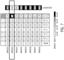
- FIG. 7 shows a comparison of error rates in different cell free DNA samples relative to one another and associated specificity of sequence variant identification of each sample.
- the highlighted sample, BRN022 exhibits a significant increase in C>T errors relative to the sample cohort.
- the sample cohort generally shows relatively higher C>T errors relative to other error types, which are indicative of C>T or G>A deamination changes.
- the specificity in the sample with high C>T or G>A error rates is about or greater than 99.95%, indicating a high specificity in the context of a biological sample and genomic sequence data having a high sequence error rate.
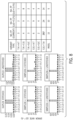
- FIG. 8 shows stratified error rates for a variety of error types for the high error rate sample of FIG. 7 for duplex and simplex (positive and negative) data, stitched and unstitched.
- the template correction in the stitched data appears to be associated with different error identification relative to the unstitched data.
- the positive and negative strand errors appear to correlate, with the C>T error appearing as G>A in the opposing strand.
- the identified peak in T>C error appears as a peak in A>G error in the opposing strand.
- the identified high error C>T and G>A changes are examined relative to a default technique that does not calculate error rate as provided herein.
- the default technique identified 257 C>T and G>A false positives in the BRN022 sample, while the stratified error rate method identified 24 and 14 (depending on the limit of detection thresholds), showing a significant decrease in false positive identification for a high error rate sample.
- FIG. 9 is a plot showing improved specificity relative to a decision tree technique.
- a technique may be a technique as provided in PCT publication WO2018093780 and that involves one or more quality scores based on weighting fragment types.
- the disclosed techniques may determine error rates on an per-sample basis rather than using a predetermined weighting factor. For example, certain samples may exhibit higher error in positive strands vs. negative strands. Accordingly, the error may also be stratified based on fragment type as calculated de novo.
- the error rate techniques as provided herein, the likelihood model result in higher specificity relative to a decision tree technique for all three sample types examined.
- FIG. 9 the error rate techniques as provided herein, the likelihood model, result in higher specificity relative to a decision tree technique for all three sample types examined.
- 10 is a table showing sensitivity and specificity results relative to a default decision tree technique for the nucleosomePrep samples, including a percentage of zoo mix, showing sensitivity in line with the decision tree technique.
- the likelihood (based on error rate) technique exhibits high specificity, indicating an improvement in variant calling and a reduction in false positive identification.
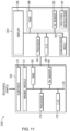
- FIG. 11 is a schematic diagram of a sequencing device 160 that may be used in conjunction with the disclosed embodiments for acquiring sequencing data that is used to identify and/or validate sequence variant calls as provided herein.
- the sequence device 160 may be implemented according to any sequencing technique, such as those incorporating sequencing-by-synthesis methods described in U.S. Patent Publication Nos. 2007/0166705 ; 2006/0188901 ; 2006/0240439 ; 2006/0281109 ; 2005/0100900 ; U.S. Pat. No. 7,057,026 ; WO 05/065814 ; WO 06/064199 ; WO 07/010,251 , the disclosures of which are incorporated herein by reference in their entireties.
- sequencing by ligation techniques may be used in the sequencing device 160.
- Such techniques use DNA ligase to incorporate oligonucleotides and identify the incorporation of such oligonucleotides and are described in U.S. Pat. No. 6,969,488 ; U.S. Pat. No. 6,172,218 ; and U.S. Pat. No. 6,306,597 ; the disclosures of which are incorporated herein by reference in their entireties.
- Some embodiments can utilize nanopore sequencing, whereby target nucleic acid strands, or nucleotides exonucleolytically removed from target nucleic acids, pass through a nanopore.
- sequencing based on detection of released protons can use an electrical detector and associated techniques that are commercially available from Ion Torrent (Guilford, CT, a Life Technologies subsidiary) or sequencing methods and systems described in US 2009/0026082 A1 ; US 2009/0127589 A1 ; US 2010/0137143 A1 ; or US 2010/0282617 A1 , each of which is incorporated herein by reference in its entirety.
- Particular embodiments can utilize methods involving the real-time monitoring of DNA polymerase activity.
- Nucleotide incorporations can be detected through fluorescence resonance energy transfer (FRET) interactions between a fluorophore-bearing polymerase and y-phosphate-labeled nucleotides, or with zeromode waveguides as described, for example, in Levene et al. Science 299, 682-686 (2003 ); Lundquist et al. Opt. Lett. 33, 1026-1028 (2008 ); Korlach et al. Proc. Natl. Acad. Sci. USA 105, 1176-1181 (2008 ), the disclosures of which are incorporated herein by reference in their entireties.
- FISSEQ fluorescent in situ sequencing
- MPSS Massively Parallel Signature Sequencing
- the sequencing device 160 may be a HiSeq, MiSeq, or HiScanSQ from Illumina (La Jolla, CA). In other embodiment, the sequencing device 160 may be configured to operate using a CMOS sensor with nanowells fabricated over photodiodes such that DNA deposition is aligned one-to-one with each photodiode.
- the sequencing device 160 may be "one-channel" a detection device, in which only two of four nucleotides are labeled and detectable for any given image.
- thymine may have a permanent fluorescent label
- adenine uses the same fluorescent label in a detachable form.
- Guanine may be permanently dark, and cytosine may be initially dark but capable of having a label added during the cycle. Accordingly, each cycle may involve an initial image and a second image in which dye is cleaved from any adenines and added to any cytosines such that only thymine and adenine are detectable in the initial image but only thymine and cytosine are detectable in the second image.
- the sequencing device 160 includes a separate sample processing device 162 and an associated computer 164. However, as noted, these may be implemented as a single device. Further, the associated computer 164 may be local to or networked with the sample processing device 162.
- the biological sample may be loaded into the sample processing device 162 on a sample substrate 170, e.g., a flow cell or slide, that is imaged to generate sequence data. For example, reagents that interact with the biological sample fluoresce at particular wavelengths in response to an excitation beam generated by an imaging module 172 and thereby return radiation for imaging.
- the imaging module detection optics may be based upon any suitable technology, and may be, for example, a charged coupled device (CCD) sensor that generates pixilated image data based upon photons impacting locations in the device.
- CCD charged coupled device
- any of a variety of other detectors may also be used including, but not limited to, a detector array configured for time delay integration (TDI) operation, a complementary metal oxide semiconductor (CMOS) detector, an avalanche photodiode (APD) detector, a Geiger-mode photon counter, or any other suitable detector.
- TDI mode detection can be coupled with line scanning as described in U.S. Patent No. 7,329,860 , which is incorporated herein by reference.
- Other useful detectors are described, for example, in the references provided previously herein in the context of various nucleic acid sequencing methodologies.
- the imaging module 172 may be under processor control, e.g., via a processor 174, and the sample receiving device 162 may also include I/O controls 176, an internal bus 78, non-volatile memory 180, RAM 182 and any other memory structure such that the memory is capable of storing executable instructions, and other suitable hardware components that may be similar to those described with regard to FIG. 11 .
- the associated computer 164 may also include a processor 184, I/O controls 186, a communications module 184, and a memory architecture including RAM 188 and non-volatile memory 190, such that the memory architecture is capable of storing executable instructions 192.
- the hardware components may be linked by an internal bus 194, which may also link to the display 196.
- certain redundant hardware elements may be eliminated.
Landscapes
- Physics & Mathematics (AREA)
- Life Sciences & Earth Sciences (AREA)
- Health & Medical Sciences (AREA)
- Engineering & Computer Science (AREA)
- Bioinformatics & Cheminformatics (AREA)
- Medical Informatics (AREA)
- Biophysics (AREA)
- Theoretical Computer Science (AREA)
- Biotechnology (AREA)
- Bioinformatics & Computational Biology (AREA)
- Spectroscopy & Molecular Physics (AREA)
- Evolutionary Biology (AREA)
- General Health & Medical Sciences (AREA)
- Proteomics, Peptides & Aminoacids (AREA)
- Chemical & Material Sciences (AREA)
- Analytical Chemistry (AREA)
- Databases & Information Systems (AREA)
- Bioethics (AREA)
- Genetics & Genomics (AREA)
- Molecular Biology (AREA)
- Computer Vision & Pattern Recognition (AREA)
- Software Systems (AREA)
- Public Health (AREA)
- Evolutionary Computation (AREA)
- Epidemiology (AREA)
- Data Mining & Analysis (AREA)
- Artificial Intelligence (AREA)
- Measuring Or Testing Involving Enzymes Or Micro-Organisms (AREA)
- Error Detection And Correction (AREA)
- Apparatus Associated With Microorganisms And Enzymes (AREA)
Applications Claiming Priority (3)
| Application Number | Priority Date | Filing Date | Title |
|---|---|---|---|
| US201762593095P | 2017-11-30 | 2017-11-30 | |
| PCT/US2018/063372 WO2019108972A1 (en) | 2017-11-30 | 2018-11-30 | Validation methods and systems for sequence variant calls |
| EP18822211.1A EP3718113B1 (de) | 2017-11-30 | 2018-11-30 | Validierungsverfahren und systeme für sequenz-variant-calls |
Related Parent Applications (2)
| Application Number | Title | Priority Date | Filing Date |
|---|---|---|---|
| EP18822211.1A Division-Into EP3718113B1 (de) | 2017-11-30 | 2018-11-30 | Validierungsverfahren und systeme für sequenz-variant-calls |
| EP18822211.1A Division EP3718113B1 (de) | 2017-11-30 | 2018-11-30 | Validierungsverfahren und systeme für sequenz-variant-calls |
Publications (2)
| Publication Number | Publication Date |
|---|---|
| EP4579669A2 true EP4579669A2 (de) | 2025-07-02 |
| EP4579669A3 EP4579669A3 (de) | 2025-09-17 |
Family
ID=64744960
Family Applications (2)
| Application Number | Title | Priority Date | Filing Date |
|---|---|---|---|
| EP25176100.3A Pending EP4579669A3 (de) | 2017-11-30 | 2018-11-30 | Validierungsverfahren und systeme für sequenzvariantenaufrufe |
| EP18822211.1A Active EP3718113B1 (de) | 2017-11-30 | 2018-11-30 | Validierungsverfahren und systeme für sequenz-variant-calls |
Family Applications After (1)
| Application Number | Title | Priority Date | Filing Date |
|---|---|---|---|
| EP18822211.1A Active EP3718113B1 (de) | 2017-11-30 | 2018-11-30 | Validierungsverfahren und systeme für sequenz-variant-calls |
Country Status (10)
| Country | Link |
|---|---|
| US (2) | US12040047B2 (de) |
| EP (2) | EP4579669A3 (de) |
| JP (1) | JP7013490B2 (de) |
| KR (1) | KR102356323B1 (de) |
| CN (1) | CN110870016B (de) |
| AU (2) | AU2018375785A1 (de) |
| CA (1) | CA3067425C (de) |
| ES (1) | ES3040876T3 (de) |
| IL (1) | IL271235B2 (de) |
| WO (1) | WO2019108972A1 (de) |
Families Citing this family (19)
| Publication number | Priority date | Publication date | Assignee | Title |
|---|---|---|---|---|
| EP3907297A1 (de) | 2011-04-15 | 2021-11-10 | The Johns Hopkins University | Sicheres sortierungssystem |
| AU2013338393C1 (en) | 2012-10-29 | 2024-07-25 | The Johns Hopkins University | Papanicolaou test for ovarian and endometrial cancers |
| EP3597772A1 (de) | 2013-04-17 | 2020-01-22 | Agency For Science, Technology And Research | Verfahren zur erzeugung erweiterter sequenzauslesungen |
| WO2017027653A1 (en) | 2015-08-11 | 2017-02-16 | The Johns Hopkins University | Assaying ovarian cyst fluid |
| IL272470B2 (en) | 2017-08-07 | 2025-08-01 | Univ Johns Hopkins | Methods and materials for assessing and treating cancer |
| CN115516108A (zh) | 2020-02-14 | 2022-12-23 | 约翰斯霍普金斯大学 | 评估核酸的方法和材料 |
| WO2021195594A1 (en) * | 2020-03-26 | 2021-09-30 | San Diego State University (SDSU) Foundation, dba San Diego State University Research Foundation | Compositions and methods for treating or ameliorating infections |
| IL299042A (en) | 2020-07-08 | 2023-02-01 | Illumina Inc | Beads as transpososome carriers |
| JP2023543541A (ja) | 2020-08-06 | 2023-10-17 | イルミナ インコーポレイテッド | ビーズ連結トランスポソームを使用するrna及びdna配列決定ライブラリの調製 |
| EP4232600A2 (de) | 2020-10-21 | 2023-08-30 | Illumina, Inc. | Sequenzierungsvorlagen mit mehreren einsätzen und zusammensetzungen und verfahren zur verbesserung des sequenzierungsdurchsatzes |
| US11880416B2 (en) * | 2020-10-21 | 2024-01-23 | International Business Machines Corporation | Sorting documents according to comprehensibility scores determined for the documents |
| AU2022246579A1 (en) | 2021-03-30 | 2023-09-21 | Illumina, Inc. | Improved methods of isothermal complementary dna and library preparation |
| KR20230164668A (ko) | 2021-03-31 | 2023-12-04 | 일루미나, 인코포레이티드 | 오류 수정을 위한 고유 분자 식별자를 이용한 트랜스포존-기반 기술을 사용하는 방향성 태그먼트화 시퀀싱 라이브러리 제조 방법 |
| KR20240099457A (ko) * | 2021-11-10 | 2024-06-28 | 앨버트 아인슈타인 컬리지 오브 메디신 | 체세포 dna 돌연변이 및 dna 손상 프로파일을 측정하는 방법 및 이에 적합한 진단 키트 |
| KR102529553B1 (ko) * | 2022-03-21 | 2023-05-10 | 주식회사 아이엠비디엑스 | 핵산 서열 분석에서 위양성 변이를 판별하는 방법 |
| US20230392201A1 (en) * | 2022-06-06 | 2023-12-07 | Element Biosciences, Inc. | Methods for assembling and reading nucleic acid sequences from mixed populations |
| WO2025006570A2 (en) * | 2023-06-30 | 2025-01-02 | Illumina, Inc. | Modifying sequencing cycles or imaging during a sequencing run to meet customized coverage estimation |
| WO2025212664A1 (en) * | 2024-04-01 | 2025-10-09 | Guardant Health, Inc. | Small variant calling with error-rate based model |
| KR20250159980A (ko) * | 2024-05-03 | 2025-11-11 | 주식회사 테라젠바이오 | 포르말린 고정 파라핀 포매된 샘플에서 생성된 dna 시컨싱 데이터에서 딥러닝을 이용한 체세포 변이와 인공 변이의 구별 방법 및 이를 이용한 장치 |
Citations (21)
| Publication number | Priority date | Publication date | Assignee | Title |
|---|---|---|---|---|
| US6172218B1 (en) | 1994-10-13 | 2001-01-09 | Lynx Therapeutics, Inc. | Oligonucleotide tags for sorting and identification |
| US6306597B1 (en) | 1995-04-17 | 2001-10-23 | Lynx Therapeutics, Inc. | DNA sequencing by parallel oligonucleotide extensions |
| US20050100900A1 (en) | 1997-04-01 | 2005-05-12 | Manteia Sa | Method of nucleic acid amplification |
| WO2005065814A1 (en) | 2004-01-07 | 2005-07-21 | Solexa Limited | Modified molecular arrays |
| US6969488B2 (en) | 1998-05-22 | 2005-11-29 | Solexa, Inc. | System and apparatus for sequential processing of analytes |
| US7001792B2 (en) | 2000-04-24 | 2006-02-21 | Eagle Research & Development, Llc | Ultra-fast nucleic acid sequencing device and a method for making and using the same |
| US7057026B2 (en) | 2001-12-04 | 2006-06-06 | Solexa Limited | Labelled nucleotides |
| WO2006064199A1 (en) | 2004-12-13 | 2006-06-22 | Solexa Limited | Improved method of nucleotide detection |
| US20060240439A1 (en) | 2003-09-11 | 2006-10-26 | Smith Geoffrey P | Modified polymerases for improved incorporation of nucleotide analogues |
| US20060281109A1 (en) | 2005-05-10 | 2006-12-14 | Barr Ost Tobias W | Polymerases |
| WO2007010251A2 (en) | 2005-07-20 | 2007-01-25 | Solexa Limited | Preparation of templates for nucleic acid sequencing |
| WO2007010252A1 (en) | 2005-07-20 | 2007-01-25 | Solexa Limited | Method for sequencing a polynucleotide template |
| US20070166705A1 (en) | 2002-08-23 | 2007-07-19 | John Milton | Modified nucleotides |
| US7329860B2 (en) | 2005-11-23 | 2008-02-12 | Illumina, Inc. | Confocal imaging methods and apparatus |
| US20090026082A1 (en) | 2006-12-14 | 2009-01-29 | Ion Torrent Systems Incorporated | Methods and apparatus for measuring analytes using large scale FET arrays |
| US20090088327A1 (en) | 2006-10-06 | 2009-04-02 | Roberto Rigatti | Method for sequencing a polynucleotide template |
| US20090127589A1 (en) | 2006-12-14 | 2009-05-21 | Ion Torrent Systems Incorporated | Methods and apparatus for measuring analytes using large scale FET arrays |
| US20100137143A1 (en) | 2008-10-22 | 2010-06-03 | Ion Torrent Systems Incorporated | Methods and apparatus for measuring analytes |
| US20100282617A1 (en) | 2006-12-14 | 2010-11-11 | Ion Torrent Systems Incorporated | Methods and apparatus for detecting molecular interactions using fet arrays |
| WO2014142831A1 (en) | 2013-03-13 | 2014-09-18 | Illumina, Inc. | Methods and systems for aligning repetitive dna elements |
| WO2018093780A1 (en) | 2016-11-16 | 2018-05-24 | Illumina, Inc. | Validation methods and systems for sequence variant calls |
Family Cites Families (9)
| Publication number | Priority date | Publication date | Assignee | Title |
|---|---|---|---|---|
| EP2602734A1 (de) * | 2011-12-08 | 2013-06-12 | Koninklijke Philips Electronics N.V. | Robuste Variantenidentifizierung und Validierung |
| EP2828218B9 (de) * | 2012-03-20 | 2021-04-07 | University Of Washington Through Its Center For Commercialization | Verfahren zur verminderung der fehlerrate einer massiv parallelen dna-sequenzierung unter verwendung einer duplex-konsens-sequenzierung |
| CA2872141C (en) | 2012-05-31 | 2016-01-19 | Board Of Regents, The University Of Texas System | Method for accurate sequencing of dna |
| US11261494B2 (en) * | 2012-06-21 | 2022-03-01 | The Chinese University Of Hong Kong | Method of measuring a fractional concentration of tumor DNA |
| JP6571665B2 (ja) * | 2013-12-28 | 2019-09-04 | ガーダント ヘルス, インコーポレイテッド | 遺伝的バリアントを検出するための方法およびシステム |
| EP3143537B1 (de) * | 2014-05-12 | 2023-03-01 | Roche Diagnostics GmbH | Seltene variant-calls in der ultratiefen sequenzierung |
| US11085084B2 (en) * | 2014-09-12 | 2021-08-10 | The Board Of Trustees Of The Leland Stanford Junior University | Identification and use of circulating nucleic acids |
| US10844428B2 (en) * | 2015-04-28 | 2020-11-24 | Illumina, Inc. | Error suppression in sequenced DNA fragments using redundant reads with unique molecular indices (UMIS) |
| MX2018010362A (es) | 2016-02-29 | 2019-03-28 | Found Medicine Inc | Metodos y sistemas para evaluar carga mutacional de tumores. |
-
2018
- 2018-11-30 AU AU2018375785A patent/AU2018375785A1/en not_active Abandoned
- 2018-11-30 CN CN201880043471.7A patent/CN110870016B/zh active Active
- 2018-11-30 CA CA3067425A patent/CA3067425C/en active Active
- 2018-11-30 EP EP25176100.3A patent/EP4579669A3/de active Pending
- 2018-11-30 JP JP2019568644A patent/JP7013490B2/ja active Active
- 2018-11-30 ES ES18822211T patent/ES3040876T3/es active Active
- 2018-11-30 US US16/206,552 patent/US12040047B2/en active Active
- 2018-11-30 KR KR1020197038490A patent/KR102356323B1/ko active Active
- 2018-11-30 WO PCT/US2018/063372 patent/WO2019108972A1/en not_active Ceased
- 2018-11-30 IL IL271235A patent/IL271235B2/en unknown
- 2018-11-30 EP EP18822211.1A patent/EP3718113B1/de active Active
-
2021
- 2021-11-15 AU AU2021269294A patent/AU2021269294B2/en active Active
-
2024
- 2024-05-15 US US18/664,975 patent/US20240304280A1/en active Pending
Patent Citations (22)
| Publication number | Priority date | Publication date | Assignee | Title |
|---|---|---|---|---|
| US6172218B1 (en) | 1994-10-13 | 2001-01-09 | Lynx Therapeutics, Inc. | Oligonucleotide tags for sorting and identification |
| US6306597B1 (en) | 1995-04-17 | 2001-10-23 | Lynx Therapeutics, Inc. | DNA sequencing by parallel oligonucleotide extensions |
| US20050100900A1 (en) | 1997-04-01 | 2005-05-12 | Manteia Sa | Method of nucleic acid amplification |
| US6969488B2 (en) | 1998-05-22 | 2005-11-29 | Solexa, Inc. | System and apparatus for sequential processing of analytes |
| US7001792B2 (en) | 2000-04-24 | 2006-02-21 | Eagle Research & Development, Llc | Ultra-fast nucleic acid sequencing device and a method for making and using the same |
| US7057026B2 (en) | 2001-12-04 | 2006-06-06 | Solexa Limited | Labelled nucleotides |
| US20060188901A1 (en) | 2001-12-04 | 2006-08-24 | Solexa Limited | Labelled nucleotides |
| US20070166705A1 (en) | 2002-08-23 | 2007-07-19 | John Milton | Modified nucleotides |
| US20060240439A1 (en) | 2003-09-11 | 2006-10-26 | Smith Geoffrey P | Modified polymerases for improved incorporation of nucleotide analogues |
| WO2005065814A1 (en) | 2004-01-07 | 2005-07-21 | Solexa Limited | Modified molecular arrays |
| WO2006064199A1 (en) | 2004-12-13 | 2006-06-22 | Solexa Limited | Improved method of nucleotide detection |
| US20060281109A1 (en) | 2005-05-10 | 2006-12-14 | Barr Ost Tobias W | Polymerases |
| WO2007010252A1 (en) | 2005-07-20 | 2007-01-25 | Solexa Limited | Method for sequencing a polynucleotide template |
| WO2007010251A2 (en) | 2005-07-20 | 2007-01-25 | Solexa Limited | Preparation of templates for nucleic acid sequencing |
| US7329860B2 (en) | 2005-11-23 | 2008-02-12 | Illumina, Inc. | Confocal imaging methods and apparatus |
| US20090088327A1 (en) | 2006-10-06 | 2009-04-02 | Roberto Rigatti | Method for sequencing a polynucleotide template |
| US20090026082A1 (en) | 2006-12-14 | 2009-01-29 | Ion Torrent Systems Incorporated | Methods and apparatus for measuring analytes using large scale FET arrays |
| US20090127589A1 (en) | 2006-12-14 | 2009-05-21 | Ion Torrent Systems Incorporated | Methods and apparatus for measuring analytes using large scale FET arrays |
| US20100282617A1 (en) | 2006-12-14 | 2010-11-11 | Ion Torrent Systems Incorporated | Methods and apparatus for detecting molecular interactions using fet arrays |
| US20100137143A1 (en) | 2008-10-22 | 2010-06-03 | Ion Torrent Systems Incorporated | Methods and apparatus for measuring analytes |
| WO2014142831A1 (en) | 2013-03-13 | 2014-09-18 | Illumina, Inc. | Methods and systems for aligning repetitive dna elements |
| WO2018093780A1 (en) | 2016-11-16 | 2018-05-24 | Illumina, Inc. | Validation methods and systems for sequence variant calls |
Non-Patent Citations (6)
| Title |
|---|
| COCKROFT ET AL., J. AM. CHEM. SOC., vol. 130, 2008, pages 818 - 820 |
| HEALY, NANOMED., vol. 2, 2007, pages 459 - 481 |
| KORLACH ET AL., PROC. NATL. ACAD. SCI. USA, vol. 105, 2008, pages 1176 - 1181 |
| LEVENE ET AL., SCIENCE, vol. 299, 2003, pages 682 - 686 |
| LUNDQUIST ET AL., OPT. LETT., vol. 33, 2008, pages 1026 - 1028 |
| SONIMELLER, CLIN. CHEM., vol. 53, 2007, pages 1996 - 2001 |
Also Published As
| Publication number | Publication date |
|---|---|
| CN110870016B (zh) | 2024-09-06 |
| NZ759168A (en) | 2024-04-26 |
| KR20200013709A (ko) | 2020-02-07 |
| AU2021269294B2 (en) | 2023-12-14 |
| ES3040876T3 (en) | 2025-11-05 |
| WO2019108972A1 (en) | 2019-06-06 |
| CN110870016A (zh) | 2020-03-06 |
| JP7013490B2 (ja) | 2022-02-15 |
| US20240304280A1 (en) | 2024-09-12 |
| EP3718113B1 (de) | 2025-07-02 |
| CA3067425C (en) | 2023-10-31 |
| AU2018375785A1 (en) | 2019-12-12 |
| JP2020524499A (ja) | 2020-08-20 |
| IL271235B2 (en) | 2024-12-01 |
| IL271235B1 (en) | 2024-08-01 |
| EP4579669A3 (de) | 2025-09-17 |
| US20190206510A1 (en) | 2019-07-04 |
| CA3067425A1 (en) | 2019-06-06 |
| IL271235A (en) | 2020-01-30 |
| EP3718113A1 (de) | 2020-10-07 |
| AU2021269294A1 (en) | 2021-12-09 |
| EP3718113C0 (de) | 2025-07-02 |
| KR102356323B1 (ko) | 2022-01-26 |
| US12040047B2 (en) | 2024-07-16 |
Similar Documents
| Publication | Publication Date | Title |
|---|---|---|
| AU2021269294B2 (en) | Validation methods and systems for sequence variant calls | |
| AU2023251452B2 (en) | Validation methods and systems for sequence variant calls | |
| JP2020534011A (ja) | 圧縮分子タグ付き核酸配列データを用いた融合の検出のための方法 | |
| US20250084470A1 (en) | Methods for partner agnostic gene fusion detection | |
| EP4147242B1 (de) | Genomsequenzierungs- und -detektionstechniken | |
| HK40121794A (en) | Validation methods and systems for sequence variant calls | |
| WO2024163553A9 (en) | Methods for detecting gene level copy number variation in brca1 and brca2 | |
| HK40012524B (en) | Validation methods and systems for sequence variant calls | |
| HK40012524A (en) | Validation methods and systems for sequence variant calls | |
| HK40010599A (en) | Validation methods and systems for sequence variant calls |
Legal Events
| Date | Code | Title | Description |
|---|---|---|---|
| PUAI | Public reference made under article 153(3) epc to a published international application that has entered the european phase |
Free format text: ORIGINAL CODE: 0009012 |
|
| STAA | Information on the status of an ep patent application or granted ep patent |
Free format text: STATUS: THE APPLICATION HAS BEEN PUBLISHED |
|
| AC | Divisional application: reference to earlier application |
Ref document number: 3718113 Country of ref document: EP Kind code of ref document: P |
|
| AK | Designated contracting states |
Kind code of ref document: A2 Designated state(s): AL AT BE BG CH CY CZ DE DK EE ES FI FR GB GR HR HU IE IS IT LI LT LU LV MC MK MT NL NO PL PT RO RS SE SI SK SM TR |
|
| REG | Reference to a national code |
Ref country code: DE Ref legal event code: R079 Free format text: PREVIOUS MAIN CLASS: G16B0040000000 Ipc: G16B0020200000 |
|
| PUAL | Search report despatched |
Free format text: ORIGINAL CODE: 0009013 |
|
| AK | Designated contracting states |
Kind code of ref document: A3 Designated state(s): AL AT BE BG CH CY CZ DE DK EE ES FI FR GB GR HR HU IE IS IT LI LT LU LV MC MK MT NL NO PL PT RO RS SE SI SK SM TR |
|
| RIC1 | Information provided on ipc code assigned before grant |
Ipc: G16B 20/20 20190101AFI20250808BHEP Ipc: G16B 40/00 20190101ALI20250808BHEP |
|
| REG | Reference to a national code |
Ref country code: HK Ref legal event code: DE Ref document number: 40121794 Country of ref document: HK |