EP4578866A1 - Method for predicting cell membrane permeability of cyclic peptide - Google Patents
Method for predicting cell membrane permeability of cyclic peptide Download PDFInfo
- Publication number
- EP4578866A1 EP4578866A1 EP23857360.4A EP23857360A EP4578866A1 EP 4578866 A1 EP4578866 A1 EP 4578866A1 EP 23857360 A EP23857360 A EP 23857360A EP 4578866 A1 EP4578866 A1 EP 4578866A1
- Authority
- EP
- European Patent Office
- Prior art keywords
- cyclic peptide
- cell membrane
- peptide
- membrane permeability
- acquired
- Prior art date
- Legal status (The legal status is an assumption and is not a legal conclusion. Google has not performed a legal analysis and makes no representation as to the accuracy of the status listed.)
- Pending
Links
Images




Classifications
-
- C—CHEMISTRY; METALLURGY
- C07—ORGANIC CHEMISTRY
- C07K—PEPTIDES
- C07K1/00—General methods for the preparation of peptides, i.e. processes for the organic chemical preparation of peptides or proteins of any length
-
- G—PHYSICS
- G16—INFORMATION AND COMMUNICATION TECHNOLOGY [ICT] SPECIALLY ADAPTED FOR SPECIFIC APPLICATION FIELDS
- G16B—BIOINFORMATICS, i.e. INFORMATION AND COMMUNICATION TECHNOLOGY [ICT] SPECIALLY ADAPTED FOR GENETIC OR PROTEIN-RELATED DATA PROCESSING IN COMPUTATIONAL MOLECULAR BIOLOGY
- G16B15/00—ICT specially adapted for analysing two-dimensional [2D] or three-dimensional [3D] molecular structures, e.g. structural or functional relations or structure alignment
- G16B15/20—Protein or domain folding
-
- G—PHYSICS
- G16—INFORMATION AND COMMUNICATION TECHNOLOGY [ICT] SPECIALLY ADAPTED FOR SPECIFIC APPLICATION FIELDS
- G16B—BIOINFORMATICS, i.e. INFORMATION AND COMMUNICATION TECHNOLOGY [ICT] SPECIALLY ADAPTED FOR GENETIC OR PROTEIN-RELATED DATA PROCESSING IN COMPUTATIONAL MOLECULAR BIOLOGY
- G16B15/00—ICT specially adapted for analysing two-dimensional [2D] or three-dimensional [3D] molecular structures, e.g. structural or functional relations or structure alignment
- G16B15/30—Drug targeting using structural data; Docking or binding prediction
Definitions
- the present invention relates to a method for predicting cell membrane permeability of a cyclic peptide based on a structure of the cyclic peptide.
- cyclosporin A molecular weight: 1202.6
- cyclosporin A molecular weight: 1202.6
- a cyclic peptide having a molecular weight of about 1000 will be able to permeate the cell membranes.
- a cyclic peptide can compensate for the low cell membrane permeability of an antibody pharmaceutical, which is known to be highly effective as a biopharmaceutical, while exhibiting the same level of specific protein binding properties as those of the antibody pharmaceutical, there is a possibility that the cyclic peptide can be used to create a novel pharmaceutical.
- a cell membrane permeable cyclic peptide having a molecular weight of more than 1000 it is difficult to design a cell membrane permeable cyclic peptide having a molecular weight of more than 1000, and there are few examples in which the cell membrane permeable cyclic peptide has been used for industrial purposes such as pharmaceuticals.
- Non-Patent Documents 1,2 and 3 a side chain structure of a cyclic peptide
- Non-Patent Document 3 a side chain structure of a cyclic peptide
- Non-Patent Document 4 a study has been reported that showed a correlation between the polarity of a cyclic peptide as a parameter and the cell membrane permeability.
- cyclosporin A changes its structure to different ones in water and in the cell membrane, and adopts a structure in the cell membrane that is advantageous for the cell membrane permeability, thereby increasing the cell membrane permeability
- a cyclic peptide compound having cell membrane permeability can be obtained.
- the structure of the cyclic peptide can be acquired by acquiring positional structural information of the cyclic peptide by two-dimensional 1 H-NMR measurement and then carrying out structuring by computational chemistry based on the acquired positional structural information.
- Variable temperature NMR, J-coupling, or the like can also be used.
- the J-coupling is an interaction of a target proton in NMR with a non-equivalent proton on the same carbon or an adjacent carbon. As a result, signals of the target proton appear split.
- the correlation between a coupling constant and a dihedral angle is expressed by the Karplus equation, and in a case where the coupling constant is known, the dihedral angle can be obtained.
- the solvent used in the two-dimensional 1 H-NMR measurement is not particularly limited, and the two-dimensional 1 H-NMR measurement is preferably carried out in dimethyl sulfoxide, dimethylformamide, dimethylacetamide, dichloromethane, chloroform, water, methanol, ethanol, propanol, tetrahydrofuran, acetonitrile, or a mixture thereof, and more preferably carried out in dimethyl sulfoxide, chloroform, water, or a mixture thereof.
- the computational chemistry is a molecular dynamics method.
- the molecular dynamics method include, but are not particularly limited to, a classical molecular dynamics (MD) method, a replica exchange MD method, and a first-principles MD method.
- MD classical molecular dynamics
- the molecular dynamics method is a technique for calculating a dynamic behavior of a system consisting of a large number of atoms in contact with a heat bath at a certain temperature by numerically solving the Newton equation based on an interaction between atoms.
- the molecular dynamics method is divided into a classical MD method and a first-principles MD method, depending on how the interaction between atoms is given.
- the molecular dynamics method is called a classical MD method.
- the molecular dynamics method is called a first-principles MD method.
- the structure of the cyclic peptide can be acquired by molecular dynamics calculation.
- the method for structuring the initial structure by molecular dynamics calculation can be carried out using software Chem3D, software Open Babel, or the like.
- the structure of the cyclic peptide can be acquired by X-ray crystallography.
- the cyclic peptide is made into a solution using an appropriate solvent, and the solution is concentrated and crystallized to obtain crystals, which are then irradiated with X-rays using an X-ray irradiation device.
- the obtained diffraction pattern can be subjected to structure optimization/refinement using computational chemistry to acquire the structure of the cyclic peptide.
- the second step is a step of calculating a molecular shape factor r which is calculated by Expression (1) after a step of carrying out an ellipsoidal approximation for obtaining each of axis lengths a, b, and c in a case where an axis length in a longest axis direction of a main chain structure is denoted by a, and axis lengths in two other directions which are orthogonal to a and are orthogonal to each other are denoted by b and c in the structure acquired in the first step.
- r 2 b 2 + c 2 a 2 + b 2 + c 2 + a 2
- the first step and the second step can be carried out, for example, as follows.
- a target cyclic peptide was dissolved in DMSO-d6 to prepare a solution having a concentration of 5 mg/mL.
- a sample tube used was a SIGEMI tube (BMS-005B), and a sample volume was set to 400 ⁇ L.
- COSY cosygpppgf, 128 integrations
- TOCSY melvphpp, 128 integrations, expansion time of 80 msec
- NOESY noesygpphpp, 64 integrations, expansion time of 150 msec, 300 msec).
- variable temperature 1 H-NMR measurement (zg, a total of 64 times) was carried out at each of 25°C, 30°C, 35°C, 40°C, 45°C, and 50°C, and a ⁇ NH/T (ppb/K) value was calculated from a change in chemical shift value depending on the temperature.
- the calculation of the MD method can be carried out using, for example, AmberTools 16.
- a GAFF force field can be used for van der Waals interactions, and RESP charges calculated by Gaussian 09 can be used for charges.
- the NMR data (appropriately selected from the main chain dihedral angle and the HH distance) can be used as the restraint condition using the NMR restraint option implemented in AmberTools 16.
- Calculation of the structure of the cyclic peptide can be carried out according to the following procedure.
- the three-dimensional coordinates of atoms belonging to the main chain of the cyclic peptide are represented by (X a,1 , X a,2 , X a,3 ).
- a is a label that identifies the atoms belonging to the main chain, and takes an integer from 1 to N.
- N is the total number of atoms belonging to the main chain of the cyclic peptide.
- the r value is calculated for the three-dimensional coordinates.
- the r value can be calculated according to the following procedure.
- the first step and the second step can be carried out, for example, as follows.
- a two-dimensionally drawn structural formula of the cyclic peptide is input into Chem3D to create a three-dimensional structure.
- the structure optimization is carried out using, for example, a quantum chemical calculation method (B3LYP/6-31G*, software: Gaussian) to obtain a locally stable structure.
- a quantum chemical calculation method B3LYP/6-31G*, software: Gaussian
- an electrostatic field for generating a cyclic peptide is obtained by a quantum chemical calculation method (B3LYP/6-31G*, software: Gaussian)
- a point charge (RESP charge) is assigned to each atom so as to reproduce the electrostatic field.
- the state of covalent bonds between the atoms is analyzed (Amber), and van der Waals parameters (gaff2) are assigned to each atom. These charges and van der Waals parameters are collectively referred to as a force field.
- a molecular dynamics (MD) simulation is carried out in chloroform (software: Gromacs and plumed).
- the MD simulation employs a replica exchange MD method in which temperatures higher than room temperature are also used in addition to room temperature as temperatures at the time of the simulation.
- the temperatures used are six types (six types of replicas) and are as shown in Table 17 of Examples.
- the present temperature is applied only to the cyclic peptide and 298 K is always applied to chloroform present around the cyclic peptide.
- the calculation for 300 ns is carried out using a replica exchange MD method to determine the most stable structure.
- the method described in the first embodiment is applied to the present most stable structure to obtain the inertia tensor, the principal moments of inertia, a, b, and c, and then the r value.
- the molecular shape factor r is preferably 0.4 to 0.55.
- the cell membrane permeability may be determined using a polar surface area (including, but not limited to, tPSA, 3D-PSA, and EPSA) or a hydrophobicity index (including, but not limited to, cLogP and cLogD), in addition to the range of values of the molecular shape factor r.
- a polar surface area including, but not limited to, tPSA, 3D-PSA, and EPSA
- a hydrophobicity index including, but not limited to, cLogP and cLogD
- n pieces of Xaa's each independently represent any amino acid residue or any amino acid analog residue
- Amino acid refers to a molecule containing both an amino group and a carboxyl group.
- the amino acid may be any of a natural amino acid or an unnatural amino acid and may be any of D- or L-isomers.
- the amino acid may be an ⁇ -amino acid.
- the ⁇ -amino acid refers to a molecule containing an amino group and a carboxyl group which are bonded to a carbon designated as an ⁇ -carbon.
- the natural amino acid represents any of alanine (A), arginine (R), asparagine (N), cysteine (C), aspartic acid (D), glutamine (Q), glutamic acid (E), glycine (G), histidine (H), isoleucine (I), leucine (L), lysine (K), methionine (M), phenylalanine (F), proline (P), serine (S), threonine (T), tryptophan (W), tyrosine (Y), or valine (V).
- the unnatural amino acid refers to an amino acid other than the above-mentioned 20 types of natural amino acids.
- amino acid analog examples include, but are not particularly limited to, a ⁇ -amino acid, and an amino acid in which an amino group or a carboxyl group is similarly substituted with a reactive group (for example, a primary amine is substituted with a secondary or tertiary amine, or a carboxyl group is substituted with an ester).
- a reactive group for example, a primary amine is substituted with a secondary or tertiary amine, or a carboxyl group is substituted with an ester.
- the ⁇ -amino acid refers to a molecule containing both an amino group and a carboxyl group in a ⁇ configuration.
- the amino acid analog is racemic. Either the D-isomer of the amino acid analog may be used, or the L-isomer of the amino acid analog may be used.
- the amino acid analog may contain a chiral center in the R or S configuration.
- the amino group (singular or plural) of the ⁇ -amino acid analog may be substituted with a protective group such as tert-butyloxycarbonyl (BOC group), 9-fluorenylmethyloxycarbonyl (FMOC), or tosyl.
- BOC group tert-butyloxycarbonyl
- FMOC 9-fluorenylmethyloxycarbonyl
- tosyl tosyl.
- the carboxylic acid functional group of the ⁇ -amino acid analog may be protected, for example, as an ester derivative thereof.
- a salt of the amino acid analog may be used.
- the cyclic peptide is non-ionic in a physiological environment.
- non-ionic in a physiological environment is meant that the peptide does not have a substituent having a charge in a physiological environment.
- the method for producing a cyclic peptide is not particularly limited.
- the cyclic peptide may be produced by a method using a cell-free translation system, or may be produced by a chemical synthesis method of a peptide.
- the chemical synthesis of a peptide can generally be carried out using an automated peptide synthesizer.
- the peptide may be synthesized by either a solid phase synthesis method or a liquid phase synthesis method, among which a solid phase synthesis method is preferable.
- the solid phase synthesis of a peptide is known to those skilled in the art, and involves, for example, an esterification reaction between a hydroxyl group of a resin having a hydroxyl group and a carboxyl group of a first amino acid (usually a C-terminal amino acid of a desired peptide) in which an ⁇ -amino group is protected with a protective group.
- a known dehydration condensation agent such as 1-mesitylenesulfonyl-3-nitro-1,2,4-triazole (MSNT), dicyclohexylcarbodiimide (DCC), or diisopropylcarbodiimide (DIC) can be used as an esterification catalyst.
- MSNT 1-mesitylenesulfonyl-3-nitro-1,2,4-triazole
- DCC dicyclohexylcarbodiimide
- DIC diisopropylcarbodiimide
- Examples of the resin for solid phase synthesis include a Merrifield resin, an MBHA resin, a Cl-Trt resin, a SASRIN resin, a Wang resin, a Rink amide resin, an HMFS resin, an Amino-PEGA resin, and an HMPA-PEGA resin (all manufactured by Merck Sigma-Aldrich Co., LLC). These resins may be washed with a solvent (dimethylformamide (DMF), 2-propanol, methylene chloride, or the like) before use.
- a solvent dimethylformamide (DMF), 2-propanol, methylene chloride, or the like
- the protection of an ⁇ -carboxy group can be carried out using a methyl ester, an ethyl ester, a benzyl ester, a tert-butyl ester, a cyclohexyl ester, or the like.
- a hydroxyl group of serine or threonine can be protected with a benzyl group or a tert-butyl group
- a hydroxyl group of tyrosine can be protected with a 2-bromobenzyloxycarbonyl group or a tert-butyl group.
- condensing agent examples include dicyclohexylcarbodiimide (DCC), diisopropylcarbodiimide (DIC), 1-ethyl-3-(3-dimethylaminopropyl) carbodiimide (EDC or WSC), (1H-benzotriazol-1-yloxy)tris(dimethylamino)phosphonium hexafluorophosphate (BOP), and 1-[bis(dimethylamino)methyl]-1H-benzotriazolium-3-oxide hexafluorophosphate (HBTU).
- DCC dicyclohexylcarbodiimide
- DIC diisopropylcarbodiimide
- EDC or WSC 1-ethyl-3-(3-dimethylaminopropyl) carbodiimide
- BOP (1H-benzotriazol-1-yloxy)tris(dimethylamino)phosphonium hexafluorophosphate
- Examples of the method for cyclization of the peptide include cyclization using an amide bond, a carbon-carbon bond, a thioether bond, a disulfide bond, an ester bond, a thioester bond, a lactam bond, a bond through a triazole structure, a bond through a fluorophore structure, and the like.
- the synthesis step and the cyclization reaction step of the peptide compound may be separate or may proceed consecutively.
- the cyclization can be carried out by methods known to those skilled in the art, for example, methods described in WO2013/100132 , WO2008/117833 , WO2012/074129 , and the like.
- X 1 represents chlorine, bromine, or iodine.
- the cyclic peptide can be used as a pharmaceutical product, a cosmetic product, a drug delivery system (DDS) material, and the like, without being limited thereto.
- DDS drug delivery system
- compound 1 Structures of compound 1, compound 2, cyclosporin A, and isocyclosporin are shown below.
- the compound 1 and compound 2 are non-ionic in a physiological environment and contain a sulfur atom in the main chain structure of the cyclic peptide.
- Isocyclosporin (commercially available product, manufactured by FUJIFILM Wako Pure Chemical Corporation)
- the solid phase synthesis of a peptide was carried out using an automated peptide synthesizer (Syro I, manufactured by Biotage AB). The synthesis was carried out by setting a resin for solid phase synthesis, an N-methyl-2-pyrrolidone (NMP) solution of Fmoc amino acid (0.5 mol/L), an NMP solution of cyano-hydroxyimino-acetic acid ethyl ester (1 mol/L) and diisopropylethylamine (0.1 mol/L), an NMP solution of diisopropylcarbodiimide (1 mol/L), an NMP solution of piperidine (20% v/v), and an NMP solution of anhydrous acetic acid (20% v/v) in a peptide synthesizer.
- NMP N-methyl-2-pyrrolidone
- Fmoc amino acid 0.5 mol/L
- NMP NMP solution of cyano-hydroxyimino-acetic acid ethyl
- a solution (0.5 mol/L) of 1 molar equivalent of tris(2-carboxyethyl)phosphine (TCEP) was added thereto, followed by stirring at room temperature for 1 hour.
- N-methyl-2-pyrrolidone, diisopropylethylamine, diisopropylcarbodiimide, piperidine, and anhydrous acetic acid were obtained from FUJIFILM Wako Pure Chemical Corporation.
- Ethyl cyanohydroxyiminoacetate was obtained from Tokyo Chemical Industry Co., Ltd.
- MS mass spectrum
- ACQUITY SQD LC/MS System manufactured by Waters Corporation, ionization method: electrospray ionization (ESI) method.
- FIG. 4 An ellipsoid diagram for the compound 1 is shown in Fig. 4 .
- FIG. 6 An ellipsoid diagram for cyclosporin A is shown in Fig. 6 .
- Example 3 r value is obtained from structure by MD calculation
- a two-dimensionally drawn structural formula of the cyclic peptide is input into Chem3D to create a three-dimensional structure.
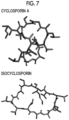
- Fig. 7 shows three-dimensionally structured cyclosporin A and isocyclosporin.
- the structure optimization is carried out by a quantum chemical calculation method (B3LYP/6-31G*, software: Gaussian) to obtain locally stable structures.
- a quantum chemical calculation method (B3LYP/6-31G*, software: Gaussian)
- a point charge (RESP charge) is assigned to each atom so as to reproduce the electrostatic field.
- Amber the state of covalent bonds between the atoms is analyzed (Amber), and van der Waals parameters (gaff2) are assigned to each atom. These charges and van der Waals parameters are collectively referred to as a force field.
- Fig. 8 shows structures of cyclosporin A and isocyclosporin after structure optimization by MD calculation using the three-dimensional structures created by Chem3D as initial structures.
- the MD simulation employs a replica exchange MD method in which temperatures higher than room temperature are also used in addition to room temperature as temperatures at the time of the simulation.
- the temperatures used are six types (six types of replicas), and are as shown in the table below.
- the present temperature is applied only to the cyclic peptide and 298 K is always applied to chloroform present around the cyclic peptide.
- Temperature of CsA/chloroform [K] Lineage 1 298/298 Lineage 2 348/298 Lineage 3 398/298 Lineage 4 448/298 Lineage 5 498/298 Lineage 6 548/298
- the main chain structure by MD calculation and the main chain structure by NMR + MD calculation are shown in Fig. 10 .
- the main chain structure by MD calculation was a main chain structure (RMSD ⁇ 1 ⁇ ) similar to the structure determined conformation by carrying out NMR measurement in chloroform and MD calculation restrained by the NMR data.
- Example 2 The method described in Example 2 was applied to the present most stable structure to obtain the inertia tensor, the principal moments of inertia, a, b, and c, and then the r value.
- r value 0.494714
- r value 0.716695
- FIG. 12 An ellipsoid diagram for isocyclosporin is shown in Fig. 12 .
- the insert was washed by being immersed in a Hank's Balanced Salt Solution (HBSS) (phenol red-free), 200 ⁇ L of a sample prepared at 10 ⁇ mol/L/HBSS was added thereto, and the insert was allowed to stand in a low-adsorption 24-well plate containing 900 ⁇ L of HBSS (37°C, 5% CO 2 ). After 2 hours, each liquid of the upper layer (apical) and the lower layer (basal) of the insert (10 ⁇ L for apical and 500 ⁇ L for basal) was recovered. After testing, no leakage was confirmed with Lucifer Yellow, which is a non-permeable fluorescent dye.
- HBSS Hank's Balanced Salt Solution
- the device used was LC/MS/MS (triple quadrupole type).
- a permeability coefficient P app which represents the membrane permeability, was calculated from each quantitative value.
- P app V / C 0 ⁇ 1 / S ⁇ dC / dt
- the cyclic peptide with a molecular shape factor (r) in a range of 0.4 to 0.6 was found to have high cell membrane permeability.
Landscapes
- Chemical & Material Sciences (AREA)
- Life Sciences & Earth Sciences (AREA)
- Spectroscopy & Molecular Physics (AREA)
- Physics & Mathematics (AREA)
- Health & Medical Sciences (AREA)
- Engineering & Computer Science (AREA)
- Bioinformatics & Cheminformatics (AREA)
- General Health & Medical Sciences (AREA)
- Biophysics (AREA)
- Biotechnology (AREA)
- Theoretical Computer Science (AREA)
- Bioinformatics & Computational Biology (AREA)
- Crystallography & Structural Chemistry (AREA)
- Evolutionary Biology (AREA)
- Medical Informatics (AREA)
- Medicinal Chemistry (AREA)
- Organic Chemistry (AREA)
- Pharmacology & Pharmacy (AREA)
- Analytical Chemistry (AREA)
- Biochemistry (AREA)
- Genetics & Genomics (AREA)
- Molecular Biology (AREA)
- Proteomics, Peptides & Aminoacids (AREA)
- Peptides Or Proteins (AREA)
Abstract
Description
- The present invention relates to a method for predicting cell membrane permeability of a cyclic peptide based on a structure of the cyclic peptide.
- In recent years, a cyclic peptide has been attracting attention primarily in the field of pharmaceuticals due to having resistance to metabolic enzymes and specific protein binding properties. In addition, cyclosporin A (molecular weight: 1202.6), which is a cyclic peptide composed of 11 amino acid residues, permeates the intestinal membranes and cell membranes, so it is expected that a cyclic peptide having a molecular weight of about 1000 will be able to permeate the cell membranes. This indicates that, in a case where a cyclic peptide can compensate for the low cell membrane permeability of an antibody pharmaceutical, which is known to be highly effective as a biopharmaceutical, while exhibiting the same level of specific protein binding properties as those of the antibody pharmaceutical, there is a possibility that the cyclic peptide can be used to create a novel pharmaceutical. Unfortunately, it is difficult to design a cell membrane permeable cyclic peptide having a molecular weight of more than 1000, and there are few examples in which the cell membrane permeable cyclic peptide has been used for industrial purposes such as pharmaceuticals.
- Cyclization of a peptide has long been known as a method for increasing the cell membrane permeability. For example, it has been suggested that a cyclic peptide, which has a ring structure introduced into a main chain thereof, has increased cell membrane permeability in part because the polarity of an amide group is offset by the formation of intramolecular hydrogen bonds (Non-Patent Document 1). In addition, the improvement of cell membrane permeability has also been demonstrated for a cyclic peptide having a staple structure (Non-Patent Document 2). Furthermore, it has been proposed to enhance the cell membrane permeability by controlling a side chain structure of a cyclic peptide (
Patent Documents -
- Patent Document 1:
WO2018/225864A - Patent Document 2:
WO2020/122182A - Patent Document 3:
WO2015/030014A -
- Non-Patent Document 1: Nat. Chem. 2016, 8, 1105-1111
- Non-Patent Document 2: Proc. Natl. Acad. Sci. USA 2013, 110, E3445
- Non-Patent Document 3: Acc. Chem. Res. 2008, 41, 1331-1342, Nat. Chemical Biology 2011, 7, 810-817
- Non-Patent Document 4: ACS Med. Chem. Lett. 2014, 5, 1167-1172, J. Med. Chem. 2018, 61, 4189-4202, J. Med. Chem. 2018, 61, 11169-11182
- Non-Patent Document 5: J. Am. Chem. Soc. 2006, 128, 14073-14080, J. Chem. Inf. Model. 2016, 56, 1547-1562, J. Phys. Chem. B 2018, 122, 2261-2276
- The above-mentioned related art methods are not necessarily effective as guidelines for designing a cyclic peptide with cell membrane permeability, and further design techniques for cell membrane permeation are required. In addition, significant costs are being spent on exploratory research, and it is desired to predict the characteristics of a cyclic peptide in advance without preparing the cyclic peptide. An object of the present invention is to provide a method for predicting cell membrane permeability of a cyclic peptide, which enables versatile design of a cyclic peptide with cell membrane permeability.
- As a result of extensive studies to achieve the foregoing object, the present inventors have found that, in a case where a molecular shape factor r for a structure of a cyclic peptide, which is calculated by Expression (1) defined in the present specification, is in a range of 0.4 to 0.6, the cyclic peptide has high cell membrane permeability. The present invention has been completed based on the above findings. According to the present invention, the following inventions are provided.
- <1> A method for predicting cell membrane permeability of a cyclic peptide, the method comprising a first step of acquiring a structure of the cyclic peptide; a second step of calculating a molecular shape factor r which is calculated by Expression (1) after a step of carrying out an ellipsoidal approximation for obtaining each of axis lengths a, b, and c in a case where an axis length in a longest axis direction of a main chain structure is denoted by a, and axis lengths in two other directions which are orthogonal to a and are orthogonal to each other are denoted by b and c in the structure acquired in the first step; and

- <2> The method according to <1>, in which, in the first step, the structure of the cyclic peptide is acquired by X-ray crystallography.
- <3> The method according to <1>, in which, in the first step, the structure of the cyclic peptide is acquired by molecular dynamics calculation.
- <4> The method according to <1>, in which, in the first step, the structure of the cyclic peptide is acquired by acquiring positional structural information of the cyclic peptide by two-dimensional 1H-NMR measurement and then carrying out structuring by computational chemistry based on the acquired positional structural information.
- <5> The method according to <4>, in which the two-dimensional 1H-NMR measurement is a measurement by at least one of nuclear Overhauser effect spectroscopy, also referred to as NOESY, or rotating frame nuclear Overhauser effect spectroscopy, also referred to as ROESY.
- <6> The method according to <4>, in which the two-dimensional 1H-NMR measurement is carried out at a temperature of 20°C to 60°C.
- <7> The method according to <4>, in which the two-dimensional 1H-NMR measurement is carried out in dimethyl sulfoxide, dimethylformamide, dimethylacetamide, dichloromethane, chloroform, water, methanol, ethanol, propanol, tetrahydrofuran, or acetonitrile.
- <8> The method according to <4>, in which the computational chemistry is a molecular dynamics method.
- <9> The method according to any one of <1> to <8>, in which the cyclic peptide is non-ionic in a physiological environment.
- <10> The method according to any one of <1> to <8>, in which the main chain structure of the cyclic peptide contains a sulfur atom.
- According to the present invention, a cyclic peptide compound having cell membrane permeability can be obtained.
-
-
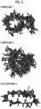
Fig. 1 shows initial structures ofcompound 1,compound 2, and cyclosporin A. -
Fig. 2 shows final structures ofcompound 1,compound 2, and cyclosporin A. -
Fig. 3 shows final structures ofcompound 1,compound 2, and cyclosporin A. -
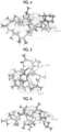
Fig. 4 shows an ellipsoid forcompound 1. -
Fig. 5 shows an ellipsoid forcompound 2. -
Fig. 6 shows an ellipsoid for cyclosporin A. -
Fig. 7 shows three-dimensionally structured cyclosporin A and isocyclosporin. -
Fig. 8 shows structures of cyclosporin A and isocyclosporin after structure optimization by MD calculation using three-dimensional structures as initial structures. -
Fig. 9 shows structures of most stabilized cyclosporin A and isocyclosporin. -
Fig. 10 shows a main chain structure by MD calculation and a main chain structure by NMR + MD calculation. -
Fig. 11 shows an ellipsoid for cyclosporin A. -
Fig. 12 shows an ellipsoid for isocyclosporin. - Hereinafter, the present invention will be described in more detail.
- In the present specification, "to" shows a range including numerical values described before and after "to" as a minimum value and a maximum value, respectively.
- The method for predicting cell membrane permeability of a cyclic peptide according to the embodiment of the present invention includes
- a first step of acquiring a structure of a cyclic peptide;
- a second step of calculating a molecular shape factor r which is calculated by Expression (1) after a step of carrying out an ellipsoidal approximation for obtaining each of axis lengths a, b, and c in a case where an axis length in a longest axis direction of a main chain structure is denoted by a, and axis lengths in two other directions which are orthogonal to a and are orthogonal to each other are denoted by b and c in the structure acquired in the first step; and

- a third step of determining that the cyclic peptide having the molecular shape factor r in a range of 0.4 to 0.6 has cell membrane permeability.
- According to the prediction method according to the embodiment of the present invention, it is possible to grasp in advance a cyclic peptide having high intracellular permeability before synthesis, and it is possible to design a cell membrane-permeable peptide, which has been difficult to do in the related art. The cyclic peptide compound obtained by the prediction method according to the embodiment of the present invention can be used as molecular design knowledge for pharmaceuticals, bioimaging, and culture medium components for cell culture. In addition, according to the present invention, it is possible to reduce research costs.
- The first step is a step of acquiring the structure of the cyclic peptide.
- In the first step, for example,
- (1) acquisition of the structure of the cyclic peptide by two-dimensional 1H-NMR measurement and computational chemistry,
- (2) acquisition of the structure of the cyclic peptide by molecular dynamics calculation, or
- (3) acquisition of the structure of the cyclic peptide by X-ray crystallography
- In the first step, the structure of the cyclic peptide can be acquired by acquiring positional structural information of the cyclic peptide by two-dimensional 1H-NMR measurement and then carrying out structuring by computational chemistry based on the acquired positional structural information.
- The two-dimensional 1H-NMR measurement is preferably a measurement by at least one of NOESY (nuclear Overhauser effect spectroscopy) or ROESY (rotating frame nuclear Overhauser effect spectroscopy).
- Variable temperature NMR, J-coupling, or the like can also be used. The J-coupling is an interaction of a target proton in NMR with a non-equivalent proton on the same carbon or an adjacent carbon. As a result, signals of the target proton appear split. In addition, the correlation between a coupling constant and a dihedral angle is expressed by the Karplus equation, and in a case where the coupling constant is known, the dihedral angle can be obtained.
- The two-dimensional 1H-NMR measurement is preferably carried out at a temperature of -40°C to 80°C, more preferably carried out at a temperature of 0°C to 80°C, and still more preferably carried out at a temperature of 20°C to 60°C.
- The solvent used in the two-dimensional 1H-NMR measurement is not particularly limited, and the two-dimensional 1H-NMR measurement is preferably carried out in dimethyl sulfoxide, dimethylformamide, dimethylacetamide, dichloromethane, chloroform, water, methanol, ethanol, propanol, tetrahydrofuran, acetonitrile, or a mixture thereof, and more preferably carried out in dimethyl sulfoxide, chloroform, water, or a mixture thereof.
- It is preferable that the computational chemistry is a molecular dynamics method. Examples of the molecular dynamics method include, but are not particularly limited to, a classical molecular dynamics (MD) method, a replica exchange MD method, and a first-principles MD method. The molecular dynamics method is a technique for calculating a dynamic behavior of a system consisting of a large number of atoms in contact with a heat bath at a certain temperature by numerically solving the Newton equation based on an interaction between atoms. The molecular dynamics method is divided into a classical MD method and a first-principles MD method, depending on how the interaction between atoms is given. In a case where the interaction between atoms is given by known functions including parameters such as a charge of each atom, a Van der Waals parameter, and a bond length of a covalent bond, the molecular dynamics method is called a classical MD method. In a case where the interaction between atoms is calculated by a molecular orbital method, which explicitly treats electrons, the molecular dynamics method is called a first-principles MD method. There is usually a single heat bath for controlling the temperature of the system used in the classical MD method and the first-principles MD method. However, it is possible to introduce a plurality of heat baths having different temperatures and use the heat baths to accelerate the dynamic behavior of the system, which is called a replica exchange MD method.
- In the first step, the structure of the cyclic peptide can be acquired by molecular dynamics calculation.
- The method for structuring the initial structure by molecular dynamics calculation (creating a 3D molecular model from a 2D structural formula) can be carried out using software Chem3D, software Open Babel, or the like.
- In the first step, the structure of the cyclic peptide can be acquired by X-ray crystallography. The cyclic peptide is made into a solution using an appropriate solvent, and the solution is concentrated and crystallized to obtain crystals, which are then irradiated with X-rays using an X-ray irradiation device. The obtained diffraction pattern can be subjected to structure optimization/refinement using computational chemistry to acquire the structure of the cyclic peptide.
- The second step is a step of calculating a molecular shape factor r which is calculated by Expression (1) after a step of carrying out an ellipsoidal approximation for obtaining each of axis lengths a, b, and c in a case where an axis length in a longest axis direction of a main chain structure is denoted by a, and axis lengths in two other directions which are orthogonal to a and are orthogonal to each other are denoted by b and c in the structure acquired in the first step.

- In a case where the structure of the cyclic peptide is acquired by two-dimensional 1H-NMR measurement and computational chemistry in the first step, the first step and the second step can be carried out, for example, as follows.
- First, a target cyclic peptide was dissolved in DMSO-d6 to prepare a solution having a concentration of 5 mg/mL. A sample tube used was a SIGEMI tube (BMS-005B), and a sample volume was set to 400 µL. For 2D-NMR measurement (600 MHz Cryo system, manufactured by Bruker Corporation), the following three types of measurements were carried out for structure assignment: COSY (cosygpppgf, 128 integrations), TOCSY (melvphpp, 128 integrations, expansion time of 80 msec), and NOESY (noesygpphpp, 64 integrations, expansion time of 150 msec, 300 msec). The variable temperature 1H-NMR measurement (zg, a total of 64 times) was carried out at each of 25°C, 30°C, 35°C, 40°C, 45°C, and 50°C, and a ΔδNH/T (ppb/K) value was calculated from a change in chemical shift value depending on the temperature.
- Next, the structure of the cyclic peptide was determined by restraining the structure generated by the molecular dynamics (MD) method using NMR data.
- The calculation of the MD method can be carried out using, for example, AmberTools 16. A GAFF force field can be used for van der Waals interactions, and RESP charges calculated by Gaussian 09 can be used for charges. The NMR data (appropriately selected from the main chain dihedral angle and the HH distance) can be used as the restraint condition using the NMR restraint option implemented in AmberTools 16. Calculation of the structure of the cyclic peptide can be carried out according to the following procedure.
- (1) 1,000 initial structures having different conformations are prepared for a linear peptide before cyclization of a target cyclic peptide.
- (2) Each linear initial structure is cyclized, and then the restraint based on the NMR data is applied at each step. The order is (i) cyclization/short-range HH distance, (ii) medium-range HH distance, and (iii) long-range HH distance, each of which is calculated over 0.2 ns. With this restraint, the 1,000 structures of the cyclic peptide are deformed to match the NMR data as closely as possible within a range in which each structure can occur as a molecule.
- (3) Among the 1,000 structures obtained, the structures are assigned priorities in order of satisfying the NMR data. The top 10 are drawn to determine the final structures.
- In the structure having the highest priority, the three-dimensional coordinates of atoms belonging to the main chain of the cyclic peptide are represented by (Xa,1, Xa,2, Xa,3).
- Here, a is a label that identifies the atoms belonging to the main chain, and takes an integer from 1 to N. N is the total number of atoms belonging to the main chain of the cyclic peptide.
- The r value is calculated for the three-dimensional coordinates. The r value can be calculated according to the following procedure.
- (1) Using three-dimensional coordinates as an input, the inertia tensor (a 3 × 3 matrix) is calculated according to the following expression.

- (2) Eigenvalues of the inertia tensor are calculated. The obtained three eigenvalues are referred to as principal moments of inertia and are represented by (I1, I2, I3).
- (3) Using the principal moments of inertia as an input, each of axis lengths a, b, and c (a > b > c) of an ellipsoid with a uniform distribution is calculated according to the following expression.


- (4) Using each of axis lengths of the ellipsoid as an input, the molecular shape factor (r) is calculated according to the following expression.

- In a case where the structure of the cyclic peptide is acquired by molecular dynamics calculation in the first step, the first step and the second step can be carried out, for example, as follows.
- First, a two-dimensionally drawn structural formula of the cyclic peptide is input into Chem3D to create a three-dimensional structure. Using the present three-dimensional structure as an initial structure, the structure optimization is carried out using, for example, a quantum chemical calculation method (B3LYP/6-31G*, software: Gaussian) to obtain a locally stable structure. In the locally stable structure, an electrostatic field for generating a cyclic peptide is obtained by a quantum chemical calculation method (B3LYP/6-31G*, software: Gaussian), and a point charge (RESP charge) is assigned to each atom so as to reproduce the electrostatic field. Next, the state of covalent bonds between the atoms is analyzed (Amber), and van der Waals parameters (gaff2) are assigned to each atom. These charges and van der Waals parameters are collectively referred to as a force field.
- Next, under the present force field, using the present locally stable structure as an initial structure, a molecular dynamics (MD) simulation is carried out in chloroform (software: Gromacs and plumed). As an efficient method for efficiently exploring a wide conformation space, the MD simulation employs a replica exchange MD method in which temperatures higher than room temperature are also used in addition to room temperature as temperatures at the time of the simulation. The temperatures used are six types (six types of replicas) and are as shown in Table 17 of Examples. The present temperature is applied only to the cyclic peptide and 298 K is always applied to chloroform present around the cyclic peptide. The calculation for 300 ns is carried out using a replica exchange MD method to determine the most stable structure. The method described in the first embodiment is applied to the present most stable structure to obtain the inertia tensor, the principal moments of inertia, a, b, and c, and then the r value.
- The third step is a step of determining that the cyclic peptide having the molecular shape factor r in a range of 0.4 to 0.6 has cell membrane permeability.
- The molecular shape factor r is preferably 0.4 to 0.55.
- In the present invention, the cell membrane permeability may be determined using a polar surface area (including, but not limited to, tPSA, 3D-PSA, and EPSA) or a hydrophobicity index (including, but not limited to, cLogP and cLogD), in addition to the range of values of the molecular shape factor r.
- The cyclic peptide of the present invention is preferably a peptide represented by Formula (1).

- In the formula, n pieces of Xaa's each independently represent any amino acid residue or any amino acid analog residue,
- m pieces of Xbb's each independently represent any amino acid residue or any amino acid analog residue, and
- n + m represents an integer of 5 to 50.
- n + m represents an integer of 5 to 50, more preferably an integer of 5 to 20, and still more preferably an integer of 9 to 11.
- Amino acid refers to a molecule containing both an amino group and a carboxyl group. The amino acid may be any of a natural amino acid or an unnatural amino acid and may be any of D- or L-isomers. The amino acid may be an α-amino acid. The α-amino acid refers to a molecule containing an amino group and a carboxyl group which are bonded to a carbon designated as an α-carbon.
- The natural amino acid represents any of alanine (A), arginine (R), asparagine (N), cysteine (C), aspartic acid (D), glutamine (Q), glutamic acid (E), glycine (G), histidine (H), isoleucine (I), leucine (L), lysine (K), methionine (M), phenylalanine (F), proline (P), serine (S), threonine (T), tryptophan (W), tyrosine (Y), or valine (V).
- The unnatural amino acid refers to an amino acid other than the above-mentioned 20 types of natural amino acids.
- The amino acid analog refers to a molecule that is structurally similar to an amino acid and can be used instead of an amino acid in the production of a cyclic peptide.
- Examples of the amino acid analog include, but are not particularly limited to, a β-amino acid, and an amino acid in which an amino group or a carboxyl group is similarly substituted with a reactive group (for example, a primary amine is substituted with a secondary or tertiary amine, or a carboxyl group is substituted with an ester). The β-amino acid refers to a molecule containing both an amino group and a carboxyl group in a β configuration.
- In one example, the amino acid analog is racemic. Either the D-isomer of the amino acid analog may be used, or the L-isomer of the amino acid analog may be used. In addition, the amino acid analog may contain a chiral center in the R or S configuration. Further, the amino group (singular or plural) of the β-amino acid analog may be substituted with a protective group such as tert-butyloxycarbonyl (BOC group), 9-fluorenylmethyloxycarbonyl (FMOC), or tosyl. Further, the carboxylic acid functional group of the β-amino acid analog may be protected, for example, as an ester derivative thereof. In addition, a salt of the amino acid analog may be used.
- Preferably, the cyclic peptide is non-ionic in a physiological environment. By non-ionic in a physiological environment is meant that the peptide does not have a substituent having a charge in a physiological environment.
- Preferably, the main chain structure of the cyclic peptide contains a sulfur atom.
- The method for producing a cyclic peptide is not particularly limited. The cyclic peptide may be produced by a method using a cell-free translation system, or may be produced by a chemical synthesis method of a peptide. The chemical synthesis of a peptide can generally be carried out using an automated peptide synthesizer.
- The peptide may be synthesized by either a solid phase synthesis method or a liquid phase synthesis method, among which a solid phase synthesis method is preferable. The solid phase synthesis of a peptide is known to those skilled in the art, and involves, for example, an esterification reaction between a hydroxyl group of a resin having a hydroxyl group and a carboxyl group of a first amino acid (usually a C-terminal amino acid of a desired peptide) in which an α-amino group is protected with a protective group. A known dehydration condensation agent such as 1-mesitylenesulfonyl-3-nitro-1,2,4-triazole (MSNT), dicyclohexylcarbodiimide (DCC), or diisopropylcarbodiimide (DIC) can be used as an esterification catalyst. Next, the protective group of the α-amino group of the first amino acid is eliminated, a second amino acid in which all functional groups of a main chain except a carboxy group are protected is added, and the carboxy group is activated to bond the first amino acid and the second amino acid. Further, the α-amino group of the second amino acid is deprotected, a third amino acid in which all functional groups of a main chain except a carboxy group are protected is added, and the carboxy group is activated to bond the second amino acid and the third amino acid. This process is repeated and in a case where a peptide having a desired length is synthesized, all functional groups are deprotected. Examples of the resin for solid phase synthesis include a Merrifield resin, an MBHA resin, a Cl-Trt resin, a SASRIN resin, a Wang resin, a Rink amide resin, an HMFS resin, an Amino-PEGA resin, and an HMPA-PEGA resin (all manufactured by Merck Sigma-Aldrich Co., LLC). These resins may be washed with a solvent (dimethylformamide (DMF), 2-propanol, methylene chloride, or the like) before use. Examples of the protective group for the α-amino group include a benzyloxycarbonyl (Cbz or Z) group, a tert-butoxycarbonyl (Boc) group, a fluorenylmethoxycarbonyl (Fmoc) group, a benzyl group, an allyl group, and an allyloxycarbonyl (Alloc) group. The Cbz group can be deprotected by hydrofluoric acid, hydrogenation, or the like, the Boc group can be deprotected by trifluoroacetic acid (TFA), and the Fmoc group can be deprotected by a treatment with piperidine. The protection of an α-carboxy group can be carried out using a methyl ester, an ethyl ester, a benzyl ester, a tert-butyl ester, a cyclohexyl ester, or the like. As for other functional groups of amino acids, a hydroxyl group of serine or threonine can be protected with a benzyl group or a tert-butyl group, and a hydroxyl group of tyrosine can be protected with a 2-bromobenzyloxycarbonyl group or a tert-butyl group. An amino group in a side chain of lysine and a carboxy group of glutamic acid or aspartic acid can be protected in the same manner as the α-amino group and the α-carboxy group. The activation of the carboxy group can be carried out using a condensing agent. Examples of the condensing agent include dicyclohexylcarbodiimide (DCC), diisopropylcarbodiimide (DIC), 1-ethyl-3-(3-dimethylaminopropyl) carbodiimide (EDC or WSC), (1H-benzotriazol-1-yloxy)tris(dimethylamino)phosphonium hexafluorophosphate (BOP), and 1-[bis(dimethylamino)methyl]-1H-benzotriazolium-3-oxide hexafluorophosphate (HBTU). Cleavage of a peptide chain from the resin can be carried out by a treatment with an acid such as TFA or hydrogen fluoride (HF).
- Examples of the method for cyclization of the peptide include cyclization using an amide bond, a carbon-carbon bond, a thioether bond, a disulfide bond, an ester bond, a thioester bond, a lactam bond, a bond through a triazole structure, a bond through a fluorophore structure, and the like. The synthesis step and the cyclization reaction step of the peptide compound may be separate or may proceed consecutively. The cyclization can be carried out by methods known to those skilled in the art, for example, methods described in
WO2013/100132 ,WO2008/117833 ,WO2012/074129 , and the like. The cyclization portion is not limited, and may be any of a bond between an N-terminal and a C-terminal of a peptide, a bond between an N-terminal of a peptide and a side chain of another amino acid residue, a bond between a C-terminal of a peptide and a side chain of another amino acid residue, or a bond between side chains of amino acid residues, in which two or more of these bonds may be used in combination. - The method for thioether cyclization of a peptide is not particularly limited. For example, the peptide can be cyclized by including the following functional groups in a side chain or main chain of the peptide. The positions of
functional groups functional groups functional groups functional groups functional groups functional groups 
- In the formula, X1 represents chlorine, bromine, or iodine.
- The synthesis step and the cyclization reaction step of the peptide compound may be separate or may proceed consecutively. The cyclization can be carried out by methods known to those skilled in the art, for example, methods described in
WO2013/100132 ,WO2008/117833 ,WO2012/074129 , and the like. - The cyclic peptide can be used as a pharmaceutical product, a cosmetic product, a drug delivery system (DDS) material, and the like, without being limited thereto.
- The present invention will be described with reference to the following examples, but the present invention is not limited thereto.
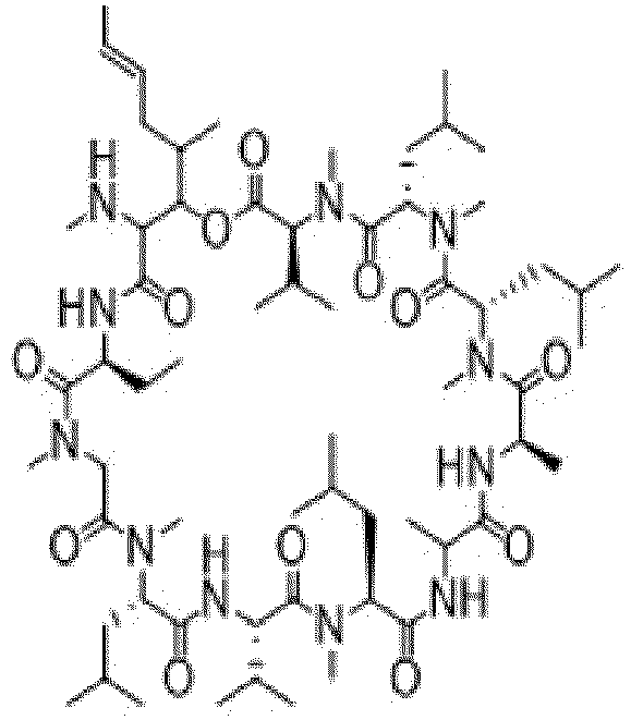
- Structures of
compound 1,compound 2, cyclosporin A, and isocyclosporin are shown below. Thecompound 1 andcompound 2 are non-ionic in a physiological environment and contain a sulfur atom in the main chain structure of the cyclic peptide.

-

-
- The solid phase synthesis of a peptide was carried out using an automated peptide synthesizer (Syro I, manufactured by Biotage AB). The synthesis was carried out by setting a resin for solid phase synthesis, an N-methyl-2-pyrrolidone (NMP) solution of Fmoc amino acid (0.5 mol/L), an NMP solution of cyano-hydroxyimino-acetic acid ethyl ester (1 mol/L) and diisopropylethylamine (0.1 mol/L), an NMP solution of diisopropylcarbodiimide (1 mol/L), an NMP solution of piperidine (20% v/v), and an NMP solution of anhydrous acetic acid (20% v/v) in a peptide synthesizer. A cycle consisting of Fmoc deprotection (20 minutes), washing with NMP, condensation of Fmoc amino acids (1 hour), and washing with NMP as one cycle was repeated to elongate the peptide chain. After elongation of the peptide, the deprotection of the Fmoc group was carried out, and chloroacetic acid was condensed in the same manner as with amino acids.
- In order to cleave off a linear peptide from the resin, a solution of trifluoroacetic acid:triisopropylsilane:dichloromethane = 5:2.5:92.5 (mass ratio) corresponding to 5 times the amount of the resin was added to the resin, followed by shaking at room temperature for 2 hours. The reaction liquid was recovered by filtration. The reaction was further repeated once using the above solution of trifluoroacetic acid:triisopropylsilane:dichloromethane, and the reaction liquid was recovered by filtration. The recovered reaction liquids were all combined, the solvent was distilled off under reduced pressure, and the residue was thoroughly dried to obtain a crude purified product of a linear peptide.
- The crude purified product of the linear peptide was dissolved in acetonitrile (10 mL) and a solution (10 mL) of 0.1 mol/L TEAB (tetraethylammonium hydrogen carbonate) buffer:pure water = 1:9 (mass ratio), and the solution was adjusted to a pH of 8.5 ± 0.1. A solution (0.5 mol/L) of 1 molar equivalent of tris(2-carboxyethyl)phosphine (TCEP) was added thereto, followed by stirring at room temperature for 1 hour. After confirming the disappearance of the linear peptide as the raw material by LC/MS analysis (Acquity UPLC/SQD, manufactured by Waters Corporation), the solvent was distilled off under reduced pressure to obtain a crude purified product of a cyclic peptide.
- The purification of the obtained crude purified product was carried out by liquid chromatography. Finally, a desired cyclic peptide was obtained as a freeze-dried powder.
- Column: X Select CSH Prep C18 5 µm OBD (19 x 250 mm), manufactured by Waters Corporation
- Column temperature: 40°C
- Flow rate: 20 ml/min
- Detection wavelength: 220 nm, 254 nm
- Solvent: liquid A: 0.1% formic acid-water
- liquid B: 0.1% formic acid-acetonitrile
- Fmoc-amino acids were obtained from Watanabe Chemical Industries, Ltd.
- N-methyl-2-pyrrolidone, diisopropylethylamine, diisopropylcarbodiimide, piperidine, and anhydrous acetic acid were obtained from FUJIFILM Wako Pure Chemical Corporation.
- Ethyl cyanohydroxyiminoacetate was obtained from Tokyo Chemical Industry Co., Ltd.
- The mass spectrum (MS) was measured using an ACQUITY SQD LC/MS System (manufactured by Waters Corporation, ionization method: electrospray ionization (ESI) method).
- Retention time (RT) was measured using an ACQUITY SQD LC/MS System (manufactured by Waters Corporation) and shown in minutes (min).
- Column: BEH C18, 1.7 µm, 2.1 x 30 mm, manufactured by Waters Corporation
- Solvent: liquid A: 0.1% formic acid-water
- liquid B: 0.1% formic acid-acetonitrile
- Gradient cycle: 0.00 min (liquid A/liquid B = 95/5), 2.00 min (liquid A/liquid B = 5/95), 3.00 min (liquid A/liquid B = 95/5)
- Flow rate: 0.5 mL/min
- Column temperature: room temperature
- Detection wavelength: 254 nm
- The measurement results of LC/MS of the
compound 1 and thecompound 2 are shown below.[Table 1] LC/MS analysis Observed MS (posi) LC/MS analysis Retention Time (min) Compound 11190.9 2.10 Compound 21202.5 1.89 - The cyclic peptide was dissolved in DMSO-d6 to prepare a solution having a concentration of 5 mg/mL. A sample tube used was a SIGEMI tube (BMS-005B), and a sample volume was set to 400 µL. For 2D-NMR measurement (600 MHz Cryo system, manufactured by Bruker Corporation), the following three types of measurements were carried out for structure assignment: COSY (cosygpppgf, 128 integrations), TOCSY (melvphpp, 128 integrations, expansion time of 80 msec), and NOESY (noesygpphpp, 64 integrations, expansion time of 150 msec, 300 msec). The variable temperature 1H-NMR measurement (zg, a total of 64 times) was carried out at each of 25°C, 30°C, 35°C, 40°C, 45°C, and 50°C, and a ΔδNH/T (ppb/K) value was calculated from a change in chemical shift value depending on the temperature.
- The chemical shift data of amide protons by variable temperature NMR and the distance data between amide groups (S: 1.8 to 2.7 Å, M: 1.8 to 3.5 Å, W: 1.8 to 5.0 Å) are shown in the tables below.

[Table 2] Compound 1: Amide group A Amide group B S, M, W Leu7 NH MeAla6 NMe M C-terminal NH MeAla9 NMe W Leu7 NH Leu7 α W Leu7 NH MeAla6 α W C-terminal NH C-terminal γ W C-terminal NH C-terminal α W C-terminal NH MeAla9 α M Leu2 NH MeLeul NMe W Leu2 NH Leu2 α W Leu2 NH MeLeul α M MeLeu3 α MeLeu3 δ M MeLeu3 α MeLeu4 NMe S MeLeu4 α MeLeu4 δ M MeAla8 α MeAla8 β M MeAla8 α MeAla9 NMe S MeLeu4 α MeAla5 NMe S MeAla6 α MeAla6 β M MeAla6 α MeAla6 NMe W MeAla9 α MeAla9 β M MeLeu1 α MeLeul δ M MeLeu1 α MeLeu1 NMe W MeAla9 α MeAla9 NMe W C-terminal α C-terminal γ W C-terminal α C-terminal β W C-terminal α Piperidine 2,6 S Leu2 α Leu2 δ M Leu2 α MeLeu3 NMe S MeAla5 α MeAla5 β S MeAla5 α MeAla6 NMe S Leu7 α Leu7 δ M Leu7 α MeAla8 NMe S [Table 3] ΔδNH/T [ppb/K] Leu2 NH 1.3 Ala7 NH 3.8 C-terminal NH 2.7 
[Table 4] Compound 2: Amide group A Amide group B S, M, W Leu2 NH MeLeul α W Leu2 NH Leu2 α W Leu2 NH Leu2 β W Leu2 NH Leu2 γ W Ala6 NH Ala6 α W Ala6 NH Ala6 β W Ala6 NH Piperidine 2, 6 W Ala6 NH MeLeu5 NMe W Ala6 NH MeLeu4 NMe W C-terminal NH C-terminal α W C-terminal NH C-terminal β W C-terminal NH C-terminal γ W C-terminal NH MeAla9 α W Leu2 α MeLeu3 NMe W MeLeu3 α MeLeu4 NMe W MeLeu4 α MeLeu4 NMe W MeLeu4 α MeLeu5 NMe W Ala6 α MeLeu7 NMe W Ala6 α Pro8 α W MeLeu7 α Pro8 δ W [Table 5] Variable temperature NMR ΔδNH/T [ppb/K] Leu2 NH 1.2 Ala6 NH 0.3 C-terminal NH 1.8 -

[Table 6] Cyclosporin A: Amide group A Amide group B S, M, W Abu2 NH Abu2 γ W Abu2 NH Abu2 β W MeBmtl β Abu2 NH W Abu2 NH Abu2 α W MeBmtl α Abu2 NH M Ala7 NH Ala7 β M Ala7 NH MeVal11 NMe W Ala7 NH Ala7 α W MeLeu6 α Ala7 NH S MeBmtl α Ala7 NH W Val5 NH Val5 γ W Val5 NH Val5 β W Val5 NH MeLeu4 NMe W Val5 NH Val5 α S MeLeu4 α Val5 NH W D-Ala8 NH D-Ala8 β W MeLeu6 g D-Ala8 NH W D-Ala8 NH MeVal11 NMe W Ala7 α D-Ala8 NH W D-Ala8 NH D-Ala8 α W MeLeu9 α MeLeu9 δ W MeLeu9 α MeLeu10 δ W MeLeu9 α MeLeu9 β W MeLeu9 α MeLeu 10 γ W MeBmt1α MeBmtl δCH3 W MeBmtl α MeBmtl δ W MeBmtl α MeBmtl β W S, M, W MeLeu4 α MeLeu4 δ M MeLeu4 α MeLeu4 γ W MeLeu4 α MeLeu4 β W MeVal11 α MeVal11 γ W MeVal11 α MeVal11 β W MeVal11 α MeBmt1 NMe S MeLeu10 α MeLeu10 δ M MeLeu10 α MeLeu10 β W MeLeu10 α MeLeu10 γ W MeLeu10 α MeLeu10 β W MeLeu10 α MeVal11 NCH3 S Abu2 α Abu2 γ W Abu2 α Abu2 β M Abu2 α Sar3 NMe S MeLeu6 α MeBmtl δ CH3 W MeLeu6 α MeLeu6 δ W MeLeu6 α MeLeu6 β W MeLeu6 α MeLeu6 γ W D-Ala8 α D-Ala8 β W D-Ala8 α MeLeu9 NMe S Sar3 α MeLeu4 NMe S Val5 α Val5 γ W Val5 α Val5 β W Val5 α MeLeu6 NMe S Ala7 α Ala7 β M [Table 7] Variable temperature NMR ΔδNH/T [ppb/K] Abu2 NH 3.5 Val5 NH 1.7 Ala7 NH 3.6 D-Ala8 NH 1.0 - Next, the structure of the cyclic peptide was determined by restraining the structure generated by the molecular dynamics (MD) method using NMR data.
- The calculation of the MD method was carried out using AmberTools 16. A GAFF force field was used for interactions, and RESP charges calculated by Gaussian 09 were used for charges. The NMR data (the HH distance) was used as the restraint condition using the NMR restraint option implemented in AmberTools 16. The procedure for calculating the structure of the cyclic peptide is as follows.
- (1) 1,000 initial structures having different conformations are prepared for a linear peptide before cyclization of a target cyclic peptide.
Among the 1,000 initial structures, the initial structures ofcompound 1,compound 2, and cyclosporin A are shown inFig. 1 . - (2) Each linear initial structure is cyclized, and then the restraint based on the NMR data is applied at each step. The order is (i) cyclization/short-range HH distance, (ii) medium-range HH distance, and (iii) long-range HH distance, each of which is calculated over 0.2 ns.
- (3) Among the 1,000 structures obtained, the structures are assigned priorities in order of satisfying the NMR data. The top 10 are drawn to determine the final structures.
- Among the top 10 final structures, the structures of
compound 1,compound 2, and cyclosporin A are shown inFig. 2 . - The final structures of
compound 1,compound 2, and cyclosporin A are shown inFig. 3 . - In the structure having the highest priority, the three-dimensional coordinates of atoms belonging to the main chain of the cyclic peptide are represented by (Xa,1, Xa,2, Xa,3).
- Here, a is a label that identifies the atoms belonging to the main chain, and takes an integer from 1 to N. N is the total number of atoms belonging to the main chain of the cyclic peptide.
- The r value is calculated for the three-dimensional coordinates. The r value can be calculated according to the following procedure.
- (1) Using three-dimensional coordinates as an input, the inertia tensor (a 3 × 3 matrix) is calculated according to the following expression.

- (2) Eigenvalues of the inertia tensor are calculated. The obtained three eigenvalues are referred to as principal moments of inertia and are represented by (I1, I2, I3).
- (3) Using the principal moments of inertia as an input, each of axis lengths a, b, and c (a > b > c) of an ellipsoid with a uniform distribution is calculated according to the following expression.


- (4) Using each of axis lengths of the ellipsoid as an input, the molecular shape factor (r) is calculated according to the following expression.

- The coordinate data (X, Y, Z of each atom) for the final structure of the
compound 1 are shown below.[Table 8] a atom_type Xa,1 Xa,2 Xa,3 1 H -5.783 1.855 0.782 2 H -6.279 3.392 0.047 3 H -4.56 3.012 0.234 4 H -9.118 -1.31 -2.274 5 H -7.941 -2.438 -2.978 6 H -7.743 -0.695 -3.209 7 H -8.703 -3.878 -1.276 8 H -8.128 -4.314 0.325 9 H -9.057 -2.832 0.107 10 H -6.524 -2.462 0.366 11 H -3.62 5.067 -6.262 12 H -2 4.922 -5.591 13 H -2.882 3.478 -6.078 14 H -4.265 5.679 -2.624 15 H -4.425 6.344 -4.243 16 H -2.842 6.255 -3.482 17 H -4.566 3.858 -4.304 18 H -6.574 0.861 -2.715 19 H -8.821 1.018 -1.652 20 H -8.11 2.624 -1.777 21 H -8.141 1.788 -0.226 22 H 2.419 -0.281 1.576 23 H 4.481 -1.775 3.123 24 H 5.195 -1.616 0.69 25 H 5.126 0.136 0.799 26 H 9.234 1.73 0.372 27 H 9.21 1.184 2.055 28 H 4.799 -0.835 6.179 29 H 5.523 -1.44 4.685 30 H 7.245 -0.919 6.429 31 H 7.48 0.029 4.963 32 H 6.161 1.004 7.546 33 H 7.729 1.502 6.924 34 H 5.929 3.183 6.391 35 H 6.669 2.52 4.932 36 H 3.985 1.68 6.16 37 H 4.229 2.537 4.619 38 H 7.031 -1.731 2.311 39 H 6.87 -0.013 2.702 40 H -3.714 3.948 -1.471 41 H -1.746 3.955 -3.117 42 H -2.45 2.534 -3.883 43 H -0.775 1.389 -2.058 44 H -2.383 0.652 -2.156 45 H -1.46 0.566 -0.646 46 H -0.851 1.57 1.069 47 H 0.275 3.14 2.716 48 H -0.666 4.473 2.055 49 H -2.785 3.266 2.68 50 H -0.897 1.364 4.167 51 H -1.957 0.929 2.831 52 H -2.643 1.427 4.37 53 H 0.034 4.863 -0.09 54 H 0.672 4.133 -1.569 55 H 1.763 4.495 -0.219 56 H 2.565 2.729 -1.578 57 H 3.748 1.964 1.063 58 H 4.127 0.671 -1.775 59 H 2.998 -0.188 -0.734 60 H 1.152 0.471 -2.441 61 H 2.489 2.178 -3.7 62 H 1.909 0.896 -4.758 63 H 3.59 0.918 -4.241 64 H 1.732 -1.467 -3.93 65 H 1.909 -1.844 -2.22 66 H 3.336 -1.528 -3.214 67 H 5.343 3.504 1.857 68 H 7.087 5.392 1.089 69 H 5.94 5.667 -0.22 70 H 6.591 3.484 -1.511 71 H 7.349 1.898 -1.273 72 H 8.277 3.378 -0.97 73 H -4.387 -2.698 -0.471 74 H -4.081 -4.739 -2.597 75 H -2.712 -1.983 -2.502 76 H -2.136 -3.277 -3.53 77 H -4.958 -2.094 -3.656 78 H -4.157 -0.962 -5.748 79 H -2.517 -1.496 -5.404 80 H -3.338 -0.381 -4.306 81 H -5.063 -3.244 -5.886 82 H -4.944 -4.372 -4.544 83 H -3.529 -4.034 -5.546 84 H -0.989 -5.873 0.441 85 H -2.32 -5.611 -3.408 86 H -1.117 -6.787 -2.885 87 H -0.603 -5.164 -3.378 88 H -2.618 3.827 5.097 89 H -1.838 5.057 4.107 90 H -0.863 3.882 4.983 91 H 1.277 -6.691 -0.11 92 H 1.09 -6.223 -1.794 93 H 0.092 -7.515 -1.115 94 H 0.917 -1.893 0.386 95 H 0.601 -5.677 2.039 96 H -0.829 -4.722 2.487 97 H 0.78 -4.285 3.104 98 H 0.123 -0.617 2.335 99 H -0.109 -2.089 3.282 100 H -1.113 -1.785 1.865 101 H 5.296 5.772 2.775 102 H 6.78 7.634 2.059 103 H 5.175 8.234 2.465 104 H 5.652 8.069 0.78 105 H 3.55 6.479 0.362 106 H 3.142 6.836 2.039 107 H 3.281 5.166 1.505 108 C -5.534 2.583 0.005 109 C -8.05 -1.463 -2.497 110 C -8.308 -3.478 -0.346 111 O -5.646 -0.465 0.03 112 O -6.406 -4.306 -2.364 113 C -7.001 -2.722 -0.592 114 N -7.23 -1.422 -1.293 115 C -6.464 -0.347 -0.904 116 C -2.985 4.459 -5.621 117 C -3.791 5.736 -3.601 118 C -3.584 4.344 -4.213 119 C -6.649 1.019 -1.625 120 C -8.012 1.652 -1.297 121 N 2.927 -1.095 1.947 122 C 4.265 -0.883 2.513 123 C 4.223 0.427 3.335 124 C 5.328 -0.773 1.374 125 S 7.983 -0.319 0.575 126 C 8.596 1.328 1.162 127 N 4.775 0.53 4.575 128 O 3.646 1.42 2.813 129 C 5.43 -0.551 5.317 130 C 6.815 -0.117 5.821 131 C 6.727 1.181 6.624 132 C 6.046 2.272 5.795 133 C 4.672 1.807 5.302 134 C -4.534 2.061 -2.235 135 N -5.527 1.93 -1.298 136 O -4.586 1.407 -3.299 137 C 6.761 -0.751 1.902 138 C -3.355 3.044 -1.988 139 N -2.35 2.437 -1.07 140 C -2.691 3.444 -3.328 141 C -1.706 1.201 -1.5 142 C -1.971 3.154 0.041 143 C -0.772 2.654 0.889 144 O -2.578 4.199 0.351 145 N 0.484 2.832 0.106 146 C -0.678 3.395 2.244 147 C -1.841 3.076 3.211 148 C -1.832 1.612 3.669 149 C 1.335 1.769 -0.026 150 C 0.757 4.145 -0.473 151 C 2.659 1.946 -0.808 152 O 1.058 0.665 0.501 153 N 3.691 2.419 0.136 154 C 3.076 0.611 -1.477 155 C 2.201 0.257 -2.703 156 C 2.569 1.115 -3.921 157 C 2.302 -1.238 -3.033 158 C 4.491 3.488 -0.168 159 C 5.635 3.802 0.837 160 N 6.797 2.932 0.493 161 O 4.356 4.133 -1.223 162 C 6.033 5.3 0.807 163 C 7.28 2.921 -0.883 164 C 7.428 2.246 1.502 165 O 7.031 2.33 2.684 166 C -6.007 -3.54 -1.47 167 N -4.685 -3.282 -1.252 168 C -3.621 -3.897 -2.052 169 C -2.535 -4.4 -1.075 170 O -2.554 -4.013 0.113 171 C -3.035 -2.865 -3.063 172 C -4.038 -2.432 -4.158 173 C -3.475 -1.246 -4.951 174 C -4.413 -3.594 -5.087 175 N -1.559 -5.277 -1.484 176 C -0.485 -5.513 -0.474 177 C -1.388 -5.726 -2.854 178 C -1.786 4.018 4.422 179 C 0.551 -6.549 -0.908 180 C 0.189 -4.133 -0.189 181 O 0.441 -3.371 -1.145 182 N 0.494 -3.788 1.1 183 C 0.909 -2.394 1.368 184 C 0.241 -4.662 2.237 185 C 2.333 -2.332 1.969 186 C -0.112 -1.677 2.274 187 O 2.91 -3.322 2.451 188 C 5.191 6.18 1.756 189 C 5.733 7.615 1.765 190 C 3.702 6.164 1.392 - The coordinate data of atoms (X, Y, Z of each atom) of the main chain of the
compound 1 are shown below.[Table 9] a atom_type Xa,1 Xa,2 Xa,3 135 N -5.527 1.93 -1.298 119 C -6.649 1.019 -1.625 115 C -6.464 -0.347 -0.904 114 N -7.23 -1.422 -1.293 113 C -7.001 -2.722 -0.592 166 C -6.007 -3.54 -1.47 167 N -4.685 -3.282 -1.252 168 C -3.621 -3.897 -2.052 169 C -2.535 -4.4 -1.075 175 N -1.559 -5.277 -1.484 176 C -0.485 -5.513 -0.474 180 C 0.189 -4.133 -0.189 182 N 0.494 -3.788 1.1 183 C 0.909 -2.394 1.368 185 C 2.333 -2.332 1.969 121 N 2.927 -1.095 1.947 122 C 4.265 -0.883 2.513 124 C 5.328 -0.773 1.374 137 C 6.761 -0.751 1.902 125 S 7.983 -0.319 0.575 126 C 8.596 1.328 1.162 164 C 7.428 2.246 1.502 160 N 6.797 2.932 0.493 159 C 5.635 3.802 0.837 158 C 4.491 3.488 -0.168 153 N 3.691 2.419 0.136 151 C 2.659 1.946 -0.808 149 C 1.335 1.769 -0.026 145 N 0.484 2.832 0.106 143 C -0.772 2.654 0.889 142 C -1.971 3.154 0.041 139 N -2.35 2.437 -1.07 138 C -3.355 3.044 -1.988 134 C -4.534 2.061 -2.235 - The values of all the components (3 x 3) of the inertia tensor for the
compound 1 are shown below.[Table 10] I11 I21 I31 331.3524 -129.1 -158.085 I12 I22 I32 -129.1 816.8712 -10.5221 I13 I23 I33 -158.085 -10.5221 1032.943 - The values of all the components (3) of the principal moments of inertia for the
compound 1 are shown below.
I1 = 267.343, I2 = 845.672, I3 = 1068.152 - The values of a, b, and c for the
compound 1 are shown below.
a = 11.00294, b = 6.001367, c = 1.816242 - The r value for the
compound 1 is shown below.
r value: 0.529463 - An ellipsoid diagram for the
compound 1 is shown inFig. 4 . - The coordinate data (X, Y, Z of each atom) for the final structure of the
compound 2 are shown below.[Table 11] a atom_type Xa,1 Xa,2 Xa,3 1 N 1.698 5.023 -4.054 2 C 0.698 5.763 -4.64 3 C 2.308 3.826 -4.687 4 C 0.254 7.032 -3.893 5 O 0.174 5.419 -5.681 6 C 3.815 4.079 -4.97 7 C 2.077 2.562 -3.811 8 N -0.146 6.738 -2.474 9 C 1.301 8.19 -4.015 10 C 4.1 5.2 -6 11 O 2.116 2.611 -2.593 12 N 1.679 1.4 -4.478 13 C -1.62 6.637 -2.293 14 C 0.768 6.551 -1.412 15 C 1.572 8.654 -5.475 16 C 5.615 5.418 -6.133 17 C 3.507 4.882 -7.384 18 C 0.662 0.512 -3.844 19 C 1.971 1.14 -5.914 20 O 1.973 6.49 -1.588 21 C 0.185 6.452 0.011 22 C 0.319 9.273 -6.13 23 C 2.724 9.676 -5.5 24 C -0.778 0.801 -4.386 25 C 1.064 -0.978 -3.993 26 S -0.252 4.705 0.462 27 C -1.274 2.261 -4.218 28 O 0.432 -1.739 -4.707 29 C 1.429 3.956 0.436 30 C -2.632 2.44 -4.913 31 C -1.423 2.662 -2.74 32 C 1.335 2.459 0.763 33 C 0.851 2.168 2.217 34 C 0.769 0.643 2.539 35 N 1.757 2.816 3.182 36 O 1.485 0.147 3.397 37 N -0.086 -0.15 1.762 38 C 1.378 3.051 4.494 39 C -1.305 0.351 1.075 40 C -0.013 -1.633 1.827 41 O 0.203 3.05 4.818 42 C 2.526 3.25 5.528 43 C -1.459 -0.251 -0.342 44 C -0.146 -2.293 0.435 45 N 3.793 3.682 4.876 46 C 2.15 4.16 6.736 47 C -1.403 -1.794 -0.314 48 C 3.865 5.109 4.451 49 C 4.935 2.869 4.826 50 C 4.711 1.351 4.597 51 O 6.058 3.347 4.823 52 N 5.468 0.924 3.416 53 C 5.227 0.494 5.774 54 C 6.554 0.001 3.788 55 C 5.181 1.413 2.145 56 C 6.611 -0.007 5.325 57 C 5.681 0.563 0.951 58 O 4.432 2.365 1.988 59 N 5.111 -0.816 1.023 60 C 5.405 1.205 -0.442 61 C 5.932 -1.947 0.899 62 C 3.649 -0.905 1.267 63 C 6.215 2.504 -0.698 64 C 5.268 -3.345 1.2 65 O 7.12 -1.833 0.635 66 C 7.692 2.196 -1.016 67 C 5.599 3.301 -1.858 68 N 4.411 -3.775 0.098 69 C 6.263 -4.496 1.452 70 C 3.133 -4.242 0.267 71 C 2.526 -5.03 -0.908 72 O 2.512 -4.035 1.29 73 N 2.325 -4.158 -2.107 74 C 3.311 -6.334 -1.225 75 C 3.391 -3.608 -2.852 76 C 0.914 -4.094 -2.572 77 C 3.038 -2.529 -3.923 78 O 4.555 -3.915 -2.648 79 N 2.252 -1.382 -3.369 80 C 4.236 -2.056 -4.821 81 C 2.76 -0.737 -2.131 82 C 5.394 -1.305 -4.098 83 C 6.759 -1.889 -4.499 84 C 5.375 0.197 -4.443 85 H 2.039 5.346 -3.153 86 H 1.792 3.639 -5.636 87 H -0.641 7.35 -4.439 88 H 4.299 4.354 -4.027 89 H 4.297 3.155 -5.309 90 H 0.962 9.05 -3.424 91 H 2.26 7.877 -3.586 92 H 3.66 6.138 -5.641 93 H -2.022 7.602 -1.959 94 H -2.108 6.352 -3.234 95 H -1.867 5.867 -1.549 96 H 1.882 7.79 -6.076 97 H 6.112 4.517 -6.515 98 H 5.826 6.244 -6.822 99 H 6.058 5.671 -5.162 100 H 3.83 5.629 -8.12 101 H 3.828 3.895 -7.737 102 H 2.411 4.91 -7.36 103 H 0.651 0.702 -2.766 104 H 1.902 0.069 -6.139 105 H 1.248 1.673 -6.544 106 H 2.986 1.474 -6.161 107 H 0.942 6.822 0.713 108 H -0.699 7.094 0.097 109 H 0.571 9.711 -7.104 110 H -0.451 8.512 -6.308 111 H -0.107 10.064 -5.501 112 H 2.467 10.573 -4.924 113 H 3.638 9.243 -5.075 114 H 2.947 9.983 -6.53 115 H -0.812 0.537 -5.451 116 H -1.485 0.126 -3.888 117 H -0.563 2.943 -4.701 118 H 2.068 4.473 1.161 119 H 1.858 4.087 -0.561 120 H -2.971 3.481 -4.834 121 H -2.556 2.196 -5.98 122 H -3.395 1.792 -4.465 123 H -1.789 3.692 -2.662 124 H -2.139 2.012 -2.226 125 H -0.466 2.621 -2.208 126 H 2.327 2.029 0.607 127 H 0.683 1.991 0.02 128 H -0.159 2.578 2.37 129 H 2.742 2.883 2.939 130 H -1.298 1.443 0.997 131 H -2.178 0.072 1.678 132 H 0.941 -1.947 2.269 133 H -0.819 -1.994 2.478 134 H 2.682 2.251 5.951 135 H -0.656 0.128 -0.985 136 H -2.409 0.077 -0.782 137 H 0.747 -2.064 -0.158 138 H -0.187 -3.384 0.542 139 H 1.868 5.164 6.397 140 H 1.291 3.742 7.276 141 H 2.99 4.252 7.435 142 H -2.301 -2.181 0.186 143 H -1.413 -2.185 -1.338 144 H 2.879 5.482 4.149 145 H 4.258 5.718 5.276 146 H 4.557 5.198 3.601 147 H 3.655 1.134 4.411 148 H 5.279 1.062 6.713 149 H 4.558 -0.363 5.926 150 H 7.51 0.335 3.362 151 H 6.321 -1.006 3.422 152 H 7.384 0.693 5.669 153 H 6.843 -1.003 5.724 154 H 6.768 0.496 1.079 155 H 5.641 0.478 -1.229 156 H 4.334 1.42 -0.534 157 H 3.461 -1.301 2.273 158 H 3.18 0.079 1.181 159 H 3.178 -1.562 0.53 160 H 6.182 3.142 0.195 161 H 4.666 -3.205 2.11 162 H 7.778 1.544 -1.895 163 H 8.243 3.121 -1.222 164 H 8.186 1.698 -0.173 165 H 6.162 4.226 -2.034 166 H 5.601 2.712 -2.781 167 H 4.563 3.579 -1.624 168 H 4.851 -3.945 -0.804 169 H 5.724 -5.424 1.679 170 H 6.891 -4.656 0.568 171 H 6.921 -4.254 2.294 172 H 1.544 -5.347 -0.538 173 H 2.846 -6.874 -2.059 174 H 4.358 -6.137 -1.485 175 H 3.31 -6.996 -0.35 176 H 0.3 -4.864 -2.089 177 H 0.491 -3.111 -2.336 178 H 0.861 -4.256 -3.655 179 H 2.379 -3.073 -4.611 180 H 4.647 -2.948 -5.312 181 H 3.846 -1.435 -5.636 182 H 1.942 -0.608 -1.412 183 H 3.542 -1.344 -1.66 184 H 3.179 0.248 -2.363 185 H 5.299 -1.409 -3.013 186 H 7.572 -1.361 -3.985 187 H 6.819 -2.947 -4.213 188 H 6.922 -1.811 -5.58 189 H 6.187 0.716 -3.919 190 H 5.511 0.353 -5.52 191 H 4.428 0.655 -4.144 - The coordinate data of atoms (X, Y, Z of each atom) of the main chain of the
compound 2 are shown below.[Table 12] a atom_type Xa,1 Xa,2 Xa,3 1 N 1.698 5.023 -4.054 3 C 2.308 3.826 -4.687 7 C 2.077 2.562 -3.811 12 N 1.679 1.4 -4.478 18 C 0.662 0.512 -3.844 25 C 1.064 -0.978 -3.993 79 N 2.252 -1.382 -3.369 77 C 3.038 -2.529 -3.923 75 C 3.391 -3.608 -2.852 73 N 2.325 -4.158 -2.107 71 C 2.526 -5.03 -0.908 70 C 3.133 -4.242 0.267 68 N 4.411 -3.775 0.098 64 C 5.268 -3.345 1.2 61 C 5.932 -1.947 0.899 59 N 5.111 -0.816 1.023 57 C 5.681 0.563 0.951 55 C 5.181 1.413 2.145 52 N 5.468 0.924 3.416 50 C 4.711 1.351 4.597 49 C 4.935 2.869 4.826 45 N 3.793 3.682 4.876 42 C 2.526 3.25 5.528 38 C 1.378 3.051 4.494 35 N 1.757 2.816 3.182 33 C 0.851 2.168 2.217 32 C 1.335 2.459 0.763 29 C 1.429 3.956 0.436 26 S -0.252 4.705 0.462 21 C 0.185 6.452 0.011 14 C 0.768 6.551 -1.412 8 N -0.146 6.738 -2.474 4 C 0.254 7.032 -3.893 2 C 0.698 5.763 -4.64 - The values of all the components (3 x 3) of the inertia tensor for the
compound 2 are shown below.[Table 13] I11 I21 I31 753.2114 117.8032 -88.5084 I12 I22 I32 117.8032 456.9073 -6.27265 I13 I23 I33 -88.5084 -6.27265 526.609 - The values of all the components (3) of the principal moments of inertia for the
compound 2 are shown below.
I1 = 410.2238, I2 = 507.0146, I3 = 819.4893 - The values of a, b, and c for the
compound 2 are shown below.
a = 8.208138, b = 7.289691, c = 2.680939 - The r value for the
compound 2 is shown below.
r value: 0.792042 - An ellipsoid diagram for the
compound 2 is shown inFig. 5 . - The coordinate data (X, Y, Z of each atom) for the final structure of cyclosporin A are shown below.
[Table 14] a atom_type Xa,1 Xa,2 Xa,3 1 N 1.43 -0.813 -2.378 2 C 1.642 -2.092 -3.076 3 C 1.486 -1.878 -4.585 4 C 0.611 -3.168 -2.61 5 O -0.615 -2.96 -2.771 6 N 1.053 -4.355 -2.093 7 C 2.432 -4.586 -1.666 8 C 0.084 -5.48 -1.944 9 C 0.585 -6.717 -2.732 10 C -0.469 -7.843 -2.845 11 C -1.603 -7.474 -3.81 12 C 0.2 -9.154 -3.278 13 C -0.076 -5.848 -0.44 14 O 0.779 -6.579 0.095 15 N -1.132 -5.359 0.299 16 C -1.304 -5.859 1.663 17 C -2.213 -4.49 -0.237 18 C -3.409 -5.339 -0.745 19 C -4.416 -4.527 -1.592 20 C -3.861 -4.195 -2.985 21 C -5.747 -5.283 -1.708 22 C -2.674 -3.519 0.893 23 O -3.758 -3.687 1.485 24 N -1.838 -2.487 1.246 25 C -0.624 -2.172 0.504 26 C -2.251 -1.613 2.374 27 C -1.094 -1.335 3.39 28 C -1.627 -0.51 4.569 29 C -0.452 -2.636 3.883 30 C -2.805 -0.261 1.849 31 O -2.015 0.566 1.309 32 N -4.128 0.043 1.993 33 C -5.065 -0.883 2.636 34 C -4.513 1.473 1.754 35 C -4.16 2.294 3.017 36 O -4.771 2.091 4.098 37 C -6.026 1.733 1.417 38 O -6.846 1.405 2.531 39 C -6.509 0.994 0.136 40 C -5.972 1.697 -1.116 41 C -8.055 0.91 0.099 42 C -8.561 0.067 -1.038 43 C -9.479 0.471 -1.924 44 C -10.003 -0.377 -3.041 45 N -3.194 3.242 2.909 46 C -2.89 4.157 4.026 47 C -3.571 5.521 3.734 48 O -3.028 6.329 2.938 49 C -1.362 4.328 4.178 50 C -0.989 5.122 5.422 51 N -4.76 5.867 4.313 52 C -5.322 7.173 3.906 53 C -5.762 7.081 2.426 54 O -6.586 6.209 2.097 55 C -5.645 4.979 5.057 56 N -5.219 7.93 1.491 57 C -4.241 8.964 1.814 58 C -5.612 7.747 0.073 59 C -6.315 9.01 -0.487 60 C -7.163 8.742 -1.752 61 C -8.397 7.883 -1.446 62 C -7.581 10.072 -2.394 63 C -4.4 7.312 -0.795 64 O -4.305 7.644 -1.989 65 N -3.453 6.514 -0.203 66 C -2.335 5.993 -0.996 67 C -2.32 4.452 -0.908 68 O -2.799 3.869 0.1 69 C -0.958 6.62 -0.556 70 C -1.013 8.148 -0.659 71 C -0.537 6.164 0.844 72 N -1.782 3.725 -1.934 73 C -1.252 4.328 -3.154 74 C -1.767 2.236 -1.837 75 C -2.514 1.591 -3.032 76 C -2.441 0.046 -3.049 77 C -3.144 -0.59 -1.844 78 C -3.018 -0.499 -4.361 79 C -0.298 1.752 -1.777 80 O 0.472 1.873 -2.758 81 N 0.133 1.189 -0.612 82 C 1.538 0.756 -0.453 83 C 1.77 -0.664 -1.063 84 O 2.248 -1.602 -0.401 85 C 1.948 0.812 1.017 86 H 1.718 -2.791 -5.128 87 H 0.464 -1.592 -4.821 88 H 2.155 -1.093 -4.934 89 H 2.669 -2.437 -2.86 90 H 1.087 -0.008 -2.908 91 H 1.467 -7.114 -2.222 92 H 0.902 -6.404 -3.732 93 H -0.905 -8 -1.846 94 H -2.34 -8.273 -3.858 95 H -1.221 -7.311 -4.815 96 H -2.115 -6.566 -3.496 97 H -0.532 -9.957 -3.342 98 H 0.966 -9.45 -2.565 99 H 0.668 -9.05 -4.254 100 H -0.86 -5.121 -2.378 101 H -3.927 -5.754 0.124 102 H -3.028 -6.184 -1.328 103 H -4.612 -3.581 -1.065 104 H -5.61 -6.242 -2.204 105 H -6.173 -5.468 -0.725 106 H -6.466 -4.708 -2.287 107 H -2.906 -3.679 -2.928 108 H -3.719 -5.103 -3.569 109 H -4.557 -3.56 -3.529 110 H -1.8 -3.895 -1.069 111 H -0.326 -0.734 2.88 112 H -2.405 -1.049 5.105 113 H -2.044 0.436 4.23 114 H -0.826 -0.292 5.272 115 H -1.187 -3.273 4.37 116 H 0.335 -2.42 4.602 117 H -0.011 -3.194 3.06 118 H -3.036 -2.174 2.899 119 H -4.575 7.956 4.079 120 H -6.191 7.386 4.538 121 H -5.56 9.767 -0.722 122 H -6.952 9.434 0.297 123 H -6.532 8.202 -2.471 124 H -8.12 6.916 -1.032 125 H -8.971 7.705 -2.353 126 H -9.051 8.378 -0.73 127 H -8.159 9.898 -3.299 128 H -6.709 10.666 -2.659 129 H -8.196 10.661 -1.714 130 H -6.33 6.908 0.09 131 H -0.2 6.261 -1.271 132 H -1.727 8.558 0.052 133 H -0.038 8.579 -0.442 134 H -1.314 8.459 -1.656 135 H -0.408 5.084 0.874 136 H 0.408 6.625 1.123 137 H -1.284 6.436 1.588 138 H -2.541 6.31 -2.032 139 H -3.519 6.228 0.777 140 H -2.078 1.966 -3.962 141 H -3.559 1.918 -3.018 142 H -1.379 -0.241 -3.008 143 H -3.003 -1.668 -1.863 144 H -4.213 -0.39 -1.864 145 H -2.745 -0.213 -0.904 146 H -2.911 -1.58 -4.399 147 H -4.076 -0.26 -4.454 148 H -2.501 -0.075 -5.219 149 H -2.275 1.994 -0.89 150 H 2.963 0.445 1.136 151 H 1.897 1.831 1.395 152 H 1.293 0.191 1.625 153 H 2.152 1.464 -1.04 154 H -0.521 1.036 0.167 155 H 2.45 -5.44 -0.987 156 H 3.099 -4.804 -2.514 157 H 2.816 -3.712 -1.132 158 H -0.596 -6.672 1.821 159 H -1.111 -5.074 2.408 160 H -2.326 -6.221 1.814 161 H -0.824 -2.08 -0.569 162 H 0.154 -2.934 0.64 163 H -0.229 -1.221 0.854 164 H -4.91 -0.921 3.723 165 H -6.086 -0.554 2.457 166 H -4.946 -1.891 2.226 167 H -3.891 1.824 0.914 168 H -6.101 2.831 1.233 169 H -6.369 1.656 3.353 170 H -6.114 -0.035 0.151 171 H -6.381 2.702 -1.199 172 H -4.887 1.781 -1.093 173 H -6.251 1.148 -2.012 174 H -8.478 1.917 0.04 175 H -8.395 0.48 1.049 176 H -11.084 -0.504 -2.96 177 H -9.543 -1.363 -3.042 178 H -9.812 0.09 -4.009 179 H -3.299 3.693 4.936 180 H -0.975 4.833 3.289 181 H -0.899 3.338 4.214 182 H 0.091 5.217 5.506 183 H -1.412 6.123 5.384 184 H -1.355 4.635 6.325 185 H -5.781 5.331 6.089 186 H -5.256 3.961 5.076 187 H -6.629 4.956 4.579 188 H -3.918 9.448 0.893 189 H -3.359 8.54 2.309 190 H -4.666 9.739 2.467 191 H -0.692 5.241 -2.935 192 H -0.57 3.624 -3.631 193 H -2.052 4.585 -3.864 194 H -2.802 3.466 1.985 195 H -8.151 -0.941 -1.117 196 H -9.89 1.478 -1.849 - The coordinate data of atoms (X, Y, Z of each atom) of the main chain of cyclosporin A are shown below.
[Table 15] a atom_type Xa,1 Xa,2 Xa,3 1 N 1.43 -0.813 -2.378 2 C 1.642 -2.092 -3.076 4 C 0.611 -3.168 -2.61 6 N 1.053 -4.355 -2.093 8 C 0.084 -5.48 -1.944 13 C -0.076 -5.848 -0.44 15 N -1.132 -5.359 0.299 17 C -2.213 -4.49 -0.237 22 C -2.674 -3.519 0.893 24 N -1.838 -2.487 1.246 26 C -2.251 -1.613 2.374 30 C -2.805 -0.261 1.849 32 N -4.128 0.043 1.993 34 C -4.513 1.473 1.754 35 C -4.16 2.294 3.017 45 N -3.194 3.242 2.909 46 C -2.89 4.157 4.026 47 C -3.571 5.521 3.734 51 N -4.76 5.867 4.313 52 C -5.322 7.173 3.906 53 C -5.762 7.081 2.426 56 N -5.219 7.93 1.491 58 C -5.612 7.747 0.073 63 C -4.4 7.312 -0.795 65 N -3.453 6.514 -0.203 66 C -2.335 5.993 -0.996 67 C -2.32 4.452 -0.908 72 N -1.782 3.725 -1.934 74 C -1.767 2.236 -1.837 79 C -0.298 1.752 -1.777 81 N 0.133 1.189 -0.612 82 C 1.538 0.756 -0.453 83 C 1.77 -0.664 -1.063 - The values of all the components (3 x 3) of the inertia tensor for cyclosporin A are shown below.
[Table 16] I11 I21 I31 751.9926 218.4299 113.1031 I12 I22 I32 218.4299 317.4003 -117.045 I13 I23 I33 113.1031 -117.045 773.4686 - The values of all the components (3) of the principal moments of inertia for cyclosporin A are shown below.
I1 = 187.0868, I2 = 768.3052, I3 = 887.4694 - The values of a, b, and c for cyclosporin A are shown below.
a = 10.54819, b = 4.816724, c = 2.268403 - The r value for cyclosporin A is shown below.
r value: 0.475683 - An ellipsoid diagram for cyclosporin A is shown in
Fig. 6 . - The r values obtained in Example 2 are shown in the section of <Summary of results> which will be described later.
- Determination of the r value was carried out using cyclosporin A and isocyclosporin.
- First, a two-dimensionally drawn structural formula of the cyclic peptide is input into Chem3D to create a three-dimensional structure.
Fig. 7 shows three-dimensionally structured cyclosporin A and isocyclosporin. - Using the three-dimensional structures as initial structures, the structure optimization is carried out by a quantum chemical calculation method (B3LYP/6-31G*, software: Gaussian) to obtain locally stable structures. In the locally stable structures, an electrostatic field for generating a cyclic peptide is obtained by a quantum chemical calculation method (B3LYP/6-31G*, software: Gaussian), and a point charge (RESP charge) is assigned to each atom so as to reproduce the electrostatic field. Next, the state of covalent bonds between the atoms is analyzed (Amber), and van der Waals parameters (gaff2) are assigned to each atom. These charges and van der Waals parameters are collectively referred to as a force field.
-
Fig. 8 shows structures of cyclosporin A and isocyclosporin after structure optimization by MD calculation using the three-dimensional structures created by Chem3D as initial structures. - Next, under the present force field, using the present locally stable structures as initial structures, molecular dynamics (MD) simulations are carried out in chloroform (software: Gromacs and plumed). As an efficient method for efficiently exploring a wide conformation space, the MD simulation employs a replica exchange MD method in which temperatures higher than room temperature are also used in addition to room temperature as temperatures at the time of the simulation. The temperatures used are six types (six types of replicas), and are as shown in the table below. The present temperature is applied only to the cyclic peptide and 298 K is always applied to chloroform present around the cyclic peptide.
[Table 17] Temperature of CsA/chloroform [K] Lineage 1298/298 Lineage 2348/298 Lineage 3 398/298 Lineage 4 448/298 Lineage 5 498/298 Lineage 6 548/298 - In a case where the calculation for 300 ns was carried out by the present replica exchange MD method, the most stable structure at room temperature was calculated. The structures of most stabilized cyclosporin A and isocyclosporin are shown in
Fig. 9 . - The main chain structure by MD calculation and the main chain structure by NMR + MD calculation are shown in
Fig. 10 . The main chain structure by MD calculation was a main chain structure (RMSD < 1 Å) similar to the structure determined conformation by carrying out NMR measurement in chloroform and MD calculation restrained by the NMR data. - The method described in Example 2 was applied to the present most stable structure to obtain the inertia tensor, the principal moments of inertia, a, b, and c, and then the r value.
- The coordinate data (X, Y, Z of each atom) for the most stable structure of cyclosporin A are shown below.
[Table 18] a atom_type Xa,1 Xa,2 Xa,3 1 C -1.35 -0.743 0.892 2 C -0.252 -2.214 6.788 3 C -1.636 -1.606 8.722 4 C -0.164 -2.449 5.252 5 O 0.735 -2.356 7.533 6 C -3.042 -1.994 9.201 7 C -1.368 -0.099 8.998 8 H -0.883 -2.219 9.244 9 C 1.27 -2.752 4.817 10 H -0.818 -3.311 5.023 11 N -0.685 -1.305 4.478 12 H -3.153 -1.8 10.266 13 H -3.8 -1.422 8.671 14 H -3.229 -3.051 9.026 15 O -1.603 0.75 8.108 16 N -0.897 0.298 10.223 17 H 1.69 -3.545 5.43 18 H 1.3 -3.059 3.774 19 H 1.893 -1.867 4.928 20 C -2.011 -1.216 4.162 21 H 0 -0.634 4.11 22 C -0.758 1.761 10.483 23 C -0.429 -0.627 11.249 24 O -2.847 -1.996 4.668 25 C -2.399 -0.197 3.06 26 H -1.269 2.26 9.649 27 C -1.435 2.18 11.809 28 C 0.753 2.131 10.526 29 H -1.262 -1.086 11.804 30 H 0.169 -1.433 10.813 31 H 0.2 -0.084 11.954 32 C -3.784 0.459 3.265 33 H -1.617 0.573 2.971 34 N -2.369 -0.957 1.77 35 C -2.92 1.768 11.923 36 H -1.351 3.268 11.9 37 H -0.872 1.759 12.647 38 N 1.377 2.673 9.42 39 O 1.402 1.896 11.563 40 H -4.514 -0.313 3.524 41 H -4.098 0.889 2.309 42 C -3.816 1.564 4.345 43 C -3.4 -1.972 1.553 44 C -3.788 2.411 10.833 45 H -2.979 0.674 11.799 46 C -3.455 2.111 13.32 47 C 2.759 3.13 9.574 48 C 0.678 3.049 8.161 49 C -5.132 2.349 4.244 50 C -3.64 0.994 5.756 51 H -2.989 2.264 4.148 52 H -2.954 -2.958 1.37 53 H -4.012 -2.055 2.449 54 H -4.039 -1.718 0.699 55 O -0.435 0.086 1.153 56 C -1.333 -1.499 -0.466 57 H -3.48 2.091 9.841 58 H -3.723 3.497 10.874 59 H -4.831 2.134 10.966 60 H -2.863 1.625 14.093 61 H -4.487 1.786 13.428 62 H -3.421 3.183 13.497 63 H 2.864 4.162 9.223 64 H 3.46 2.505 9.002 65 H 3.023 3.07 10.63 66 H -0.17 2.355 8.014 67 C 1.633 2.871 6.951 68 C 0.15 4.509 8.246 69 H -5.247 2.793 3.257 70 H -5.158 3.151 4.979 71 H -5.988 1.702 4.424 72 H -4.372 0.214 5.953 73 H -3.77 1.774 6.501 74 H -2.649 0.571 5.895 75 C 0.025 -2.229 -0.674 76 H -2.156 -2.228 -0.517 77 N -1.573 -0.523 -1.544 78 O 1.85 3.822 6.157 79 N 2.264 1.672 6.756 80 H -0.202 4.696 9.267 81 C -0.982 4.831 7.242 82 H 0.987 5.191 8.07 83 H 0.814 -1.464 -0.642 84 C 0.08 -2.919 -2.042 85 C 0.277 -3.229 0.461 86 H -0.766 0.01 -1.882 87 C -2.849 -0.252 -1.96 88 C 3.214 1.622 5.601 89 C 1.866 0.446 7.435 90 H -0.668 4.482 6.25 91 C -1.194 6.349 7.162 92 C -2.292 4.123 7.61 93 H -0.718 -3.652 -2.145 94 H -0.017 -2.191 -2.843 95 H 1.029 -3.434 -2.168 96 H 0.298 -2.727 1.427 97 H 1.233 -3.73 0.322 98 H -0.498 -3.993 0.492 99 O -3.825 -0.88 -1.514 100 C -3.044 0.908 -2.982 101 H 3.772 2.571 5.638 102 C 4.239 0.454 5.684 103 C 2.353 1.579 4.308 104 H 1.901 -0.396 6.745 105 H 2.513 0.213 8.292 106 H 0.841 0.528 7.796 107 H -0.28 6.852 6.853 108 H -1.972 6.589 6.441 109 H -1.494 6.756 8.126 110 H -3.065 4.36 6.882 111 H -2.648 4.442 8.588 112 H -2.172 3.042 7.628 113 H -3.21 1.824 -2.386 114 C -4.284 0.659 -3.877 115 N -1.799 1.166 -3.745 116 C 5.053 0.364 4.386 117 H 3.688 -0.491 5.791 118 C 5.173 0.622 6.889 119 N 2.474 2.587 3.393 120 O 1.524 0.649 4.148 121 H -4.933 -0.044 -3.345 122 H -3.98 0.165 -4.806 123 C -5.097 1.932 -4.208 124 C -1.1 2.318 -3.473 125 C -1.303 0.125 -4.643 126 H 5.76 -0.46 4.441 127 H 5.619 1.277 4.209 128 H 4.405 0.195 3.528 129 H 5.768 1.528 6.795 130 H 4.616 0.683 7.821 131 H 5.857 -0.221 6.96 132 C 3.494 3.631 3.484 133 C 1.427 2.737 2.342 134 C -6.35 1.559 5.011 135 H -5.427 2.372 -3.253 136 C -4.266 2.988 -4.947 137 O 1.58 3.199 2.732 138 C 0.324 2.522 -4.037 139 H -0.32 -0.246 -4.33 140 H -1.223 0.49 -5.676 141 H -2.001 -0.711 -4.639 142 H 3.279 4.347 4.289 143 H 4.482 3.194 3.667 144 H 3.538 4.162 2.533 145 H 0.753 1.876 2.467 146 C 2.093 2.698 0.95 147 C 0.648 4.089 2.587 148 H -6.957 0.835 -4.472 149 H -6.082 1.123 -5.972 150 H -6.961 2.438 -5.203 151 H -3.447 3.344 -4.327 152 H -3.851 2.585 -5.869 153 H -4.887 3.843 -5.207 154 H 0.637 1.711 -4.704 155 H 0.371 3.464 -4.594 156 N 1.278 2.606 -2.908 157 N 1.882 1.584 0.187 158 O 2.822 3.631 0.547 159 O 0.058 4.062 3.877 160 H 1.397 4.913 2.543 161 C -0.431 4.372 1.512 162 C 1.573 3.945 -2.409 163 C 1.582 1.438 -2.268 164 H 1.211 0.864 0.49 165 C 2.59 1.405 -1.093 166 H 0.746 4.017 4.573 167 C -1.59 3.346 1.549 168 H 0.062 4.297 0.528 169 C -0.96 5.803 1.673 170 H 0.639 4.462 -2.161 171 H 2.194 3.905 -1.514 172 H 2.089 4.536 -3.177 173 O 1.051 0.367 -2.667 174 H 3.299 2.244 -1.168 175 C 3.379 0.072 -1.112 176 H -2.109 3.42 2.508 177 C -2.555 3.534 0.409 178 H -1.168 2.332 1.491 179 H -0.154 6.529 1.568 180 H -1.715 6.02 0.921 181 H -1.407 5.935 2.655 182 H 3.866 -0.027 -2.086 183 H 2.674 -0.76 -1.032 184 C 4.417 -0.01 -0.003 185 C -3.877 3.682 0.563 186 H -2.132 3.537 -0.595 187 H 5.141 0.798 -0.083 188 H 3.948 0.058 0.977 189 H 4.957 -0.953 -0.052 190 H -4.31 3.685 1.564 191 C -4.835 3.851 -0.576 192 H -5.573 3.046 -0.591 193 H -4.314 3.855 -1.531 194 H -5.39 4.786 -0.485 195 N -1.483 -1.893 7.291 196 H -2.271 -1.739 6.657 - The coordinate data of atoms (X, Y, Z of each atom) of the main chain of cyclosporin A are shown below.
[Table 19] a atom_type Xa,1 Xa,2 Xa,3 1 C -1.35 -0.743 0.892 56 C -1.333 -1.499 -0.466 77 N -1.573 -0.523 -1.544 87 C -2.849 -0.252 -1.96 100 C -3.044 0.908 -2.982 115 N -1.799 1.166 -3.745 124 C -1.1 2.318 -3.473 138 C 0.324 2.522 -4.037 156 N 1.278 2.606 -2.908 163 C 1.582 1.438 -2.268 165 C 2.59 1.405 -1.093 157 N 1.882 1.584 0.187 146 C 2.093 2.698 0.95 133 C 1.427 2.737 2.342 119 N 2.474 2.587 3.393 103 C 2.353 1.579 4.308 88 C 3.214 1.622 5.601 79 N 2.264 1.672 6.756 67 C 1.633 2.871 6.951 48 C 0.678 3.049 8.161 38 N 1.377 2.673 9.42 28 C 0.753 2.131 10.526 22 C -0.758 1.761 10.483 16 N -0.897 0.298 10.223 7 C -1.368 -0.099 8.998 3 C -1.636 -1.606 8.722 195 N -1.483 -1.893 7.291 2 C -0.252 -2.214 6.788 4 C -0.164 -2.449 5.252 11 N -0.685 -1.305 4.478 20 C -2.011 -1.216 4.162 25 C -2.399 -0.197 3.06 34 N -2.369 -0.957 1.77 - The values of all the components (3 x 3) of the inertia tensor for cyclosporin A are shown below.
[Table 20] I11 I21 I31 802.3409 -62.7456 -34.5588 I12 I22 I32 -62.7456 812.9554 26.37697 I13 I23 I33 -34.5588 26.37697 200.5268 - The values of all the components (3) of the principal moments of inertia for cyclosporin A are shown below.
I1 = 197.6983, I2 = 744.7818, I3 = 873.343 - The values of a, b, and c for cyclosporin A are shown below.
a = 10.37343, b = 4.971582, c = 2.288593 - The r value for cyclosporin A is shown below.
r value: 0.494714 - An ellipsoid diagram for cyclosporin A is shown in
Fig. 11 . - The coordinate data (X, Y, Z of each atom) for the most stable structure of isocyclosporin are shown below.
[Table 21] a atom_type Xa,1 Xa,2 Xa,3 1 C 0.598 -0.424 2.132 2 N 0.104 -0.766 0.803 3 C 0.19 -2.017 0.246 4 C 0.835 -3.194 1.038 5 C 0.651 -4.51 0.242 6 C 1.324 -5.743 0.881 7 C 0.914 -7.016 0.128 8 C 2.851 -5.607 0.931 9 N 0.338 -3.326 2.431 10 C -1.083 -3.612 2.619 11 C 1.241 -3.255 3.456 12 C 0.798 -3.483 4.93 13 C 1.701 -4.532 5.586 14 N 0.884 -2.215 5.684 15 C -0.133 -1.301 5.609 16 C -0.032 0.015 6.416 17 C -0.201 1.227 5.478 18 N 1.202 0.109 7.201 19 C 1.186 0.412 8.542 20 C 2.608 0.526 9.185 21 C 2.529 0.761 10.718 22 C 2.517 -0.531 11.572 23 C 1.365 -1.469 11.197 24 C 3.864 -1.266 11.534 25 N 3.337 1.618 8.493 26 C 2.625 2.891 8.371 27 C 4.575 1.509 7.903 28 C 5.475 0.255 8.111 29 C 6.823 0.683 8.769 30 C 7.744 -0.525 8.978 31 C 6.578 1.419 10.091 32 N 5.749 -0.368 6.803 33 C 4.794 -1.04 6.117 34 O 3.643 -1.22 6.607 35 O 5.017 2.444 7.203 36 O 0.141 0.585 9.176 37 O -1.146 -1.525 4.916 38 O 2.446 -2.974 3.223 39 O -0.184 -2.207 -0.931 40 C -0.358 0.363 -0.061 41 C 1.436 1.213 0.648 42 C -2.315 2.024 -0.332 43 C -3.486 2.668 0.422 44 C -1.511 3.086 -1.093 45 C 0.911 1.192 0.395 46 N 1.632 0.92 -1.528 47 C 1.04 0.212 -2.665 48 C 2.877 1.724 -1.699 49 C 3.965 1.034 -2.58 50 C 4.292 -0.364 -2.038 51 C 5.23 1.9 -2.617 52 C 2.484 3.132 -2.187 53 O 3.295 4.099 -1.699 54 C 2.893 5.493 -1.872 55 C 4.128 6.328 -2.288 56 C 5.324 6.135 -1.349 57 C 4.511 6.001 -3.755 58 C 5.575 6.913 -4.302 59 C 6.704 6.487 -4.883 60 C 7.753 7.392 -5.445 61 C 2.181 5.958 -0.551 62 N 1.907 7.408 -0.602 63 C 0.517 7.766 -0.858 64 C 2.978 5.653 0.741 65 N 3.111 4.341 1.104 66 C 3.791 3.959 2.342 67 C 2.758 3.472 3.406 68 C 1.852 4.597 3.887 69 C 4.811 2.838 2.04 70 N 5.763 2.515 2.979 71 C 5.984 3.236 4.23 72 C 6.673 1.387 2.719 73 C 6.498 0.254 3.755 74 N 5.387 -0.546 3.716 75 C 4.294 -0.324 2.773 76 C 5.154 -1.628 4.731 77 C 6.343 -2.619 4.832 78 C 6.591 -3.415 3.529 79 C 7.977 -4.072 3.566 80 C 5.497 -4.462 3.284 81 O 7.386 0.096 4.627 82 O 4.746 2.186 0.972 83 O 3.447 6.574 1.438 84 O 1.503 3.373 -2.887 85 O 1.341 2.021 0.446 86 H -0.139 -0.652 2.915 87 H 1.523 -0.962 2.356 88 H 0.817 0.644 2.161 89 H 1.913 -2.974 1.137 90 H -0.42 -4.705 0.125 91 H 1.043 -4.352 -0.765 92 H 0.959 -5.834 1.918 93 H 1.249 -6.981 -0.906 94 H 1.353 -7.897 0.592 95 H 0.167 -7.137 0.126 96 H 3.147 -4.756 1.539 97 H 3.262 -5.476 -0.067 98 H 3.298 -6.5 1.362 99 H -1.259 -4.669 2.867 100 H -1.505 -2.987 3.413 101 H -1.612 -3.397 1.691 102 H -0.252 -3.814 4.971 103 H 1.599 -5.492 5.085 104 H 2.741 -4.223 5.522 105 H 1.441 -4.663 6.635 106 H 1.787 -1.984 6.111 107 H -0.858 0.023 7.147 108 H 0.648 1.322 4.802 109 H -0.279 2.146 6.054 110 H -1.102 1.109 4.883 111 H 2.105 0.006 6.732 112 H 3.13 -0.419 8.972 113 H 1.617 1.326 10.93 114 H 3.376 1.382 11.029 115 H 2.355 -0.205 12.611 116 H 1.296 -2.289 11.909 117 H 0.419 -0.934 11.19 118 H 1.514 -1.901 10.209 119 H 3.852 -2.117 12.21 120 H 4.678 -0.608 11.835 121 H 4.088 -1.642 10.536 122 H 1.784 2.819 7.668 123 H 2.224 3.196 9.342 124 H 3.324 3.643 8.007 125 H 4.986 -0.499 8.749 126 H 7.312 1.381 8.074 127 H 7.292 -1.249 9.653 128 H 8.689 -0.209 9.41 129 H 7.956 -1.028 8.037 130 H 5.985 2.317 9.933 131 H 6.053 0.785 10.805 132 H 7.523 1.715 10.54 133 H 6.624 -0.125 6.318 134 H -0.786 -0.104 -0.96 135 H -2.08 0.547 1.231 136 H -0.951 1.893 1.354 137 H -2.736 1.32 -1.069 138 H -3.127 3.372 1.17 139 H -4.134 3.209 -0.263 140 H -4.083 1.914 0.93 141 H -2.167 3.67 -1.735 142 H -0.744 2.639 -1.721 143 H -1.021 3.77 -0.403 144 H 1.783 0.083 -3.448 145 H 0.679 -0.779 -2.368 146 H 0.202 0.777 -3.095 147 H 3.307 1.836 -0.686 148 H 3.59 0.935 -3.611 149 H 4.682 -0.297 -1.025 150 H 5.045 -0.844 -2.659 151 H 3.411 -1.001 -2.021 152 H 5.592 2.092 -1.61 153 H 6.017 1.395 -3.173 154 H 5.04 2.857 -3.098 155 H 2.147 5.517 -2.688 156 H 3.82 7.385 -2.246 157 H 6.187 6.678 -1.728 158 H 5.102 6.505 -0.351 159 H 5.593 5.084 -1.275 160 H 4.836 4.958 -3.828 161 H 3.608 6.09 -4.376 162 H 5.387 7.983 -4.218 163 H 6.894 5.416 -4.963 164 H 8.715 7.227 -4.956 165 H 7.904 7.201 6.509 166 H 7.485 8.439 -5.323 167 H 1.232 5.386 -0.49 168 H 2.248 7.826 0.274 169 H 0.207 7.418 -1.848 170 H -0.189 7.339 -0.122 171 H 0.413 8.853 -0.834 172 H 2.633 3.588 0.6 173 H 4.296 4.868 2.708 174 H 3.291 3.036 4.258 175 H 2.157 2.673 2.963 176 H 1.309 5.036 3.054 177 H 2.43 5.387 4.361 178 H 1.127 4.225 4.607 179 H 5.547 2.721 5.096 180 H 7.06 3.328 4.409 181 H 5.564 4.24 4.177 182 H 6.489 1.047 1.693 183 H 7.715 1.734 2.788 184 H 3.781 -1.268 2.58 185 H 3.554 0.384 3.175 186 H 4.659 0.083 1.829 187 H 4.265 -2.177 4.387 188 H 7.25 -2.072 5.099 189 H 6.135 -3.314 5.653 190 H 6.578 -2.7 2.691 191 H 8.051 -4.78 4.389 192 H 8.171 -4.613 2.642 193 H 8.757 -3.325 3.693 194 H 5.484 -5.199 4.085 195 H 5.681 -4.99 2.351 196 H 4.512 -4.003 3.228 - The coordinate data of atoms (X, Y, Z of each atom) of the main chain of isocyclosporin are shown below.
[Table 22] a atom type Xa,1 Xa,2 Xa,3 2 N 0.104 -0.766 0.803 40 C -0.358 0.363 -0.061 45 C 0.911 1.192 -0.395 46 N 1.632 0.92 -1.528 48 C 2.877 1.724 -1.699 52 C 2.484 3.132 -2.187 53 O 3.295 4.099 -1.699 54 C 2.893 5.493 -1.872 61 C 2.181 5.958 -0.551 64 C 2.978 5.653 0.741 65 N 3.111 4.341 1.104 66 C 3.791 3.959 2.342 69 C 4.811 2.838 2.04 70 N 5.763 2.515 2.979 72 C 6.673 1.387 2.719 73 C 6.498 0.254 3.755 74 N 5.387 -0.546 3.716 76 C 5.154 -1.628 4.731 33 C 4.794 -1.04 6.117 32 N 5.749 -0.368 6.803 28 C 5.475 0.255 8.111 27 C 4.575 1.509 7.903 25 N 3.337 1.618 8.493 20 C 2.608 0.526 9.185 19 C 1.186 0.412 8.542 18 N 1.202 0.109 7.201 16 C -0.032 0.015 6.416 15 C -0.133 -1.301 5.609 14 N 0.884 -2.215 5.684 12 C 0.798 -3.483 4.93 11 C 1.241 -3.255 3.456 9 N 0.338 -3.326 2.431 4 C 0.835 -3.194 1.038 3 C 0.19 -2.017 0.246 - The values of all the components (3 x 3) of the inertia tensor for isocyclosporin are shown below.
[Table 23] I11 I21 I31 626.3276 -57.4717 -43.5872 I12 I22 I32 -57.4717 549.1581 117.5674 I13 I23 I33 -43.5872 117.5674 376.3207 - The values of all the components (3) of the principal moments of inertia for isocyclosporin are shown below.
I1 = 316.2555, I2 = 546.2999, I3 = 689.2509 - The values of a, b, and c for isocyclosporin are shown below.
a = 8.221633, b = 5.810782, c = 3.569731 - The r value for isocyclosporin is shown below.
r value: 0.716695 - An ellipsoid diagram for isocyclosporin is shown in
Fig. 12 . - 300 µL of MDCK II cells (ECACC standard cell line) at a density of 1.0 × 10⁶ cells/mL were seeded in an insert (dedicated for a 24-well plate, pore diameter: 3.0 µm, manufactured by Corning Incorporated), and cultured at 37°C in a 5% CO2 environment. After 3 days, the electric resistance value of the cell layer (measuring device: Millicell (registered trademark) ERS-2 (manufactured by Millipore Corporation), electrode: ENDOHM-6 (manufactured by WPI, Inc.)) was measured, and it was confirmed that the cell layer had high barrier properties (> 100 Ω·cm2).
- The insert was washed by being immersed in a Hank's Balanced Salt Solution (HBSS) (phenol red-free), 200 µL of a sample prepared at 10 µmol/L/HBSS was added thereto, and the insert was allowed to stand in a low-adsorption 24-well plate containing 900 µL of HBSS (37°C, 5% CO2). After 2 hours, each liquid of the upper layer (apical) and the lower layer (basal) of the insert (10 µL for apical and 500 µL for basal) was recovered. After testing, no leakage was confirmed with Lucifer Yellow, which is a non-permeable fluorescent dye.
- The device used was LC/MS/MS (triple quadrupole type).
- Eluent:
- A) 5 mmol/L ammonium formate, 0.2% formic acid/H2O
- B) 0.1% formic acid/MeCN
- Flow rate: 0.5 mL/min
- Injection volume: 2 µL
- Column: ACQUITY UPLC BEH C18 Column, 1.7 µm, 2.1 mm × 50 mm (manufactured by Waters Corporation)
- Temperature: 70°C
- Gradient (%B): 2% (0 to 0.5 min) → 98% (2 to 3 min) → 2% (3 to 5 min) Ionization: ESI
- Detection mode: MRM (positive)
- Based on the calculation expression shown in the following expression, a permeability coefficient Papp, which represents the membrane permeability, was calculated from each quantitative value.

- V: basal volume (0.9 mL)
- C0: initial concentration (10 µM)
- S: Surface area of single layer membrane (0.33 cm2)
- dC/dt: concentration change at basal [µM/s]
-
[Table 24] Molecular shape factor (r) Cell membrane permeability Papp x 10-6 cm/s Using NMR data Calculation Compound 1 0.53 None 1.1 Compound 20.79 None 0.04 Cyclosporin A 0.48 0.49 2.1 Isocyclosporin None 0.72 0.2 - The cyclic peptide with a molecular shape factor (r) in a range of 0.4 to 0.6 was found to have high cell membrane permeability.
Claims (10)
- A method for predicting cell membrane permeability of a cyclic peptide, the method comprising:a first step of acquiring a structure of the cyclic peptide;a second step of calculating a molecular shape factor r which is calculated by Expression (1) after a step of carrying out an ellipsoidal approximation for obtaining each of axis lengths a, b, and c in a case where an axis length in a longest axis direction of a main chain structure is denoted by a, and axis lengths in two other directions which are orthogonal to a and are orthogonal to each other are denoted by b and c in the structure acquired in the first step; and
 a third step of determining that the cyclic peptide having the molecular shape factor r in a range of 0.4 to 0.6 has cell membrane permeability.
 - The method according to claim 1,
wherein, in the first step, the structure of the cyclic peptide is acquired by X-ray crystallography. - The method according to claim 1,
wherein, in the first step, the structure of the cyclic peptide is acquired by molecular dynamics calculation. - The method according to claim 1,
wherein, in the first step, the structure of the cyclic peptide is acquired by acquiring positional structural information of the cyclic peptide by two-dimensional 1H-NMR measurement and then carrying out structuring by computational chemistry based on the acquired positional structural information. - The method according to claim 4,
wherein the two-dimensional 1H-NMR measurement is a measurement by at least one of nuclear Overhauser effect spectroscopy, also referred to as NOESY, or rotating frame nuclear Overhauser effect spectroscopy, also referred to as ROESY. - The method according to claim 4,
wherein the two-dimensional 1H-NMR measurement is carried out at a temperature of 20°C to 60°C. - The method according to claim 4,
wherein the two-dimensional 1H-NMR measurement is carried out in dimethyl sulfoxide, dimethylformamide, dimethylacetamide, dichloromethane, chloroform, water, methanol, ethanol, propanol, tetrahydrofuran, or acetonitrile. - The method according to claim 4,
wherein the computational chemistry is a molecular dynamics method. - The method according to any one of claims 1 to 8,
wherein the cyclic peptide is non-ionic in a physiological environment. - The method according to any one of claims 1 to 8,
wherein the main chain structure of the cyclic peptide contains a sulfur atom.
Applications Claiming Priority (2)
| Application Number | Priority Date | Filing Date | Title |
|---|---|---|---|
| JP2022132148 | 2022-08-23 | ||
| PCT/JP2023/030224 WO2024043251A1 (en) | 2022-08-23 | 2023-08-23 | Method for predicting cell membrane permeability of cyclic peptide |
Publications (2)
| Publication Number | Publication Date |
|---|---|
| EP4578866A1 true EP4578866A1 (en) | 2025-07-02 |
| EP4578866A4 EP4578866A4 (en) | 2025-12-03 |
Family
ID=90013428
Family Applications (1)
| Application Number | Title | Priority Date | Filing Date |
|---|---|---|---|
| EP23857360.4A Pending EP4578866A4 (en) | 2022-08-23 | 2023-08-23 | METHOD FOR PREDICTING THE CELL MEMBRANE PERMEABILITY OF A CYCLICAL PEPTIDE |
Country Status (4)
| Country | Link |
|---|---|
| US (1) | US20250232831A1 (en) |
| EP (1) | EP4578866A4 (en) |
| JP (1) | JPWO2024043251A1 (en) |
| WO (1) | WO2024043251A1 (en) |
Families Citing this family (2)
| Publication number | Priority date | Publication date | Assignee | Title |
|---|---|---|---|---|
| TW202417465A (en) * | 2022-08-23 | 2024-05-01 | 日商富士軟片股份有限公司 | Cyclic peptide or salt thereof, and MDMX inhibitor |
| TW202417032A (en) * | 2022-08-23 | 2024-05-01 | 日商富士軟片股份有限公司 | Cyclic peptide or salt thereof and MDMX inhibitor |
Family Cites Families (8)
| Publication number | Priority date | Publication date | Assignee | Title |
|---|---|---|---|---|
| JP5605602B2 (en) | 2007-03-26 | 2014-10-15 | 国立大学法人 東京大学 | Method for synthesizing cyclic peptide compound |
| EP2647721B1 (en) | 2010-12-03 | 2019-02-27 | The University of Tokyo | Peptide with safer secondary structure, peptide library, and production methods for same |
| TW202446781A (en) | 2011-12-28 | 2024-12-01 | 日商中外製藥股份有限公司 | Method for producing peptide compounds |
| JP6754997B2 (en) | 2013-08-26 | 2020-09-16 | 国立大学法人 東京大学 | A large cyclic peptide, a method for producing the same, and a screening method using a large cyclic peptide library. |
| CN110869544B (en) | 2017-06-09 | 2024-03-08 | 中外制药株式会社 | Cyclic peptide compounds with high membrane permeability and libraries containing the same |
| WO2020122182A1 (en) | 2018-12-12 | 2020-06-18 | 中外製薬株式会社 | Amino acid having functional group capable of intermolecular hydrogen bonding, peptide compound containing same and method for production thereof |
| EP4087857B1 (en) * | 2020-01-06 | 2023-11-01 | Bristol-Myers Squibb Company | Immunomodulators |
| JP7057003B1 (en) * | 2021-02-26 | 2022-04-19 | 国立大学法人東京工業大学 | Predictor, trained model generator, predictor, trained model generator, predictor, and trained model generator |
-
2023
- 2023-08-23 JP JP2024542835A patent/JPWO2024043251A1/ja active Pending
- 2023-08-23 WO PCT/JP2023/030224 patent/WO2024043251A1/en not_active Ceased
- 2023-08-23 EP EP23857360.4A patent/EP4578866A4/en active Pending
-
2025
- 2025-02-21 US US19/059,967 patent/US20250232831A1/en active Pending
Also Published As
| Publication number | Publication date |
|---|---|
| JPWO2024043251A1 (en) | 2024-02-29 |
| WO2024043251A1 (en) | 2024-02-29 |
| US20250232831A1 (en) | 2025-07-17 |
| EP4578866A4 (en) | 2025-12-03 |
Similar Documents
| Publication | Publication Date | Title |
|---|---|---|
| US20250232831A1 (en) | Method for predicting cell membrane permeability of cyclic peptide | |
| Mutter et al. | Template-assembled synthetic proteins with four-helix-bundle topology. Total chemical synthesis and conformational studies | |
| IE913582A1 (en) | Cyclopeptides | |
| ES2890676T3 (en) | Process for the production of D-arginyl-2,6-dimethyl-L-tyrosyl-L-lysyl-L-phenylalaninamide | |
| Horikawa et al. | Syntheses of potent Leu-enkephalin analogs possessing β-hydroxy-α, α-disubstituted-α-amino acid and their characterization to opioid receptors | |
| CN106029689A (en) | Beta-hairpin peptidomimetics as selective elastase inhibitors | |
| Kawai et al. | Conformational analysis by NMR spectrometry of the highly substituted cyclic tetrapeptides, chlamydocin and Ala4-chlamydocin. Evidence for a unique amide bond sequence in dimethyl sulfoxide-d6 | |
| Pentelute et al. | Total chemical synthesis and X-ray structure of kaliotoxin by racemic protein crystallography | |
| CN101270153B (en) | Cyclo-pentapeptide and synthesizing method | |
| Jeremic et al. | Synthesis and conformational analysis of 18-membered Aib-containing cyclohexapeptides | |
| Mierke et al. | Enkephalin analogues containing β‐naphthylalanine at the fourth position | |
| Jung et al. | Template‐free self‐assembling fullerene and lipopeptide conjugates of alamethicin form voltage‐dependent ion channels of remarkable stability and activity | |
| Jacobsen et al. | Synthesis of cyclic peptide analogues of the 3 10 helical Pro138-Gly144 segment of human aquaporin-4 by olefin metathesis | |
| Rossi et al. | New antitumour cyclic astin analogues: Synthesis, conformation and bioactivity | |
| EP4631955A1 (en) | Purification method and production method of cyclic peptide | |
| EP0700928A1 (en) | Nucleic acid-binding oligomers for therapy and diagnostics | |
| EP3950700A1 (en) | Peptide complex and production method therefor, and use of said peptide complex | |
| Olma et al. | The biological consequences of replacing hydrophobic amino acids in deltorphin I with amphiphilic α‐hydroxymethylamino acids | |
| López-Martínez et al. | Proline cis–trans isomerization and its implications for the dimerization of analogues of cyclopeptide stylostatin 1: a combined computational and experimental study | |
| CN105566447A (en) | Peptoid antagonist of anti-apoptosis protein and synthesis method and application of peptoid antagonist | |
| Matera-Witkiewicz et al. | Short-chain oligopeptides with copper (II) binding properties: The impact of specific structural modifications on the copper (II) coordination abilities | |
| WO2023137326A2 (en) | De novo designed membrane-traversing and orally bioavailable macrocycles | |
| Gach-Janczak et al. | Synthesis and activity of opioid peptidomimetics with β2-and β3-amino acids | |
| Miskolzie et al. | An NMR conformational analysis of cyclic bradykinin mimics. Evidence for a β-turn | |
| Degelaen et al. | Cyclic peptides. 0. Synthesis and a structural study of an ion-binding cyclic peptide analog of valinomycin, cyclo (L-Ala-Gly-D-Phe-L-Pro) 3 |
Legal Events
| Date | Code | Title | Description |
|---|---|---|---|
| STAA | Information on the status of an ep patent application or granted ep patent |
Free format text: STATUS: THE INTERNATIONAL PUBLICATION HAS BEEN MADE |
|
| PUAI | Public reference made under article 153(3) epc to a published international application that has entered the european phase |
Free format text: ORIGINAL CODE: 0009012 |
|
| STAA | Information on the status of an ep patent application or granted ep patent |
Free format text: STATUS: REQUEST FOR EXAMINATION WAS MADE |
|
| 17P | Request for examination filed |
Effective date: 20250220 |
|
| AK | Designated contracting states |
Kind code of ref document: A1 Designated state(s): AL AT BE BG CH CY CZ DE DK EE ES FI FR GB GR HR HU IE IS IT LI LT LU LV MC ME MK MT NL NO PL PT RO RS SE SI SK SM TR |
|
| REG | Reference to a national code |
Ref country code: DE Ref legal event code: R079 Free format text: PREVIOUS MAIN CLASS: C07K0002000000 Ipc: G16B0015300000 |
|
| DAV | Request for validation of the european patent (deleted) | ||
| DAX | Request for extension of the european patent (deleted) | ||
| A4 | Supplementary search report drawn up and despatched |
Effective date: 20251105 |
|
| RIC1 | Information provided on ipc code assigned before grant |
Ipc: G16B 15/30 20190101AFI20251030BHEP Ipc: G16B 15/20 20190101ALI20251030BHEP Ipc: C07K 1/00 20060101ALI20251030BHEP |