EP4534690A2 - Systeme und verfahren zum gezielten nukleinsäureeinfang - Google Patents
Systeme und verfahren zum gezielten nukleinsäureeinfang Download PDFInfo
- Publication number
- EP4534690A2 EP4534690A2 EP25156820.0A EP25156820A EP4534690A2 EP 4534690 A2 EP4534690 A2 EP 4534690A2 EP 25156820 A EP25156820 A EP 25156820A EP 4534690 A2 EP4534690 A2 EP 4534690A2
- Authority
- EP
- European Patent Office
- Prior art keywords
- nucleic acid
- bridge
- probe
- adaptor
- sequence
- Prior art date
- Legal status (The legal status is an assumption and is not a legal conclusion. Google has not performed a legal analysis and makes no representation as to the accuracy of the status listed.)
- Pending
Links
Images


Classifications
-
- C—CHEMISTRY; METALLURGY
- C12—BIOCHEMISTRY; BEER; SPIRITS; WINE; VINEGAR; MICROBIOLOGY; ENZYMOLOGY; MUTATION OR GENETIC ENGINEERING
- C12Q—MEASURING OR TESTING PROCESSES INVOLVING ENZYMES, NUCLEIC ACIDS OR MICROORGANISMS; COMPOSITIONS OR TEST PAPERS THEREFOR; PROCESSES OF PREPARING SUCH COMPOSITIONS; CONDITION-RESPONSIVE CONTROL IN MICROBIOLOGICAL OR ENZYMOLOGICAL PROCESSES
- C12Q1/00—Measuring or testing processes involving enzymes, nucleic acids or microorganisms; Compositions therefor; Processes of preparing such compositions
- C12Q1/68—Measuring or testing processes involving enzymes, nucleic acids or microorganisms; Compositions therefor; Processes of preparing such compositions involving nucleic acids
- C12Q1/6813—Hybridisation assays
-
- C—CHEMISTRY; METALLURGY
- C12—BIOCHEMISTRY; BEER; SPIRITS; WINE; VINEGAR; MICROBIOLOGY; ENZYMOLOGY; MUTATION OR GENETIC ENGINEERING
- C12Q—MEASURING OR TESTING PROCESSES INVOLVING ENZYMES, NUCLEIC ACIDS OR MICROORGANISMS; COMPOSITIONS OR TEST PAPERS THEREFOR; PROCESSES OF PREPARING SUCH COMPOSITIONS; CONDITION-RESPONSIVE CONTROL IN MICROBIOLOGICAL OR ENZYMOLOGICAL PROCESSES
- C12Q1/00—Measuring or testing processes involving enzymes, nucleic acids or microorganisms; Compositions therefor; Processes of preparing such compositions
- C12Q1/68—Measuring or testing processes involving enzymes, nucleic acids or microorganisms; Compositions therefor; Processes of preparing such compositions involving nucleic acids
- C12Q1/6869—Methods for sequencing
- C12Q1/6874—Methods for sequencing involving nucleic acid arrays, e.g. sequencing by hybridisation
-
- C—CHEMISTRY; METALLURGY
- C12—BIOCHEMISTRY; BEER; SPIRITS; WINE; VINEGAR; MICROBIOLOGY; ENZYMOLOGY; MUTATION OR GENETIC ENGINEERING
- C12Q—MEASURING OR TESTING PROCESSES INVOLVING ENZYMES, NUCLEIC ACIDS OR MICROORGANISMS; COMPOSITIONS OR TEST PAPERS THEREFOR; PROCESSES OF PREPARING SUCH COMPOSITIONS; CONDITION-RESPONSIVE CONTROL IN MICROBIOLOGICAL OR ENZYMOLOGICAL PROCESSES
- C12Q1/00—Measuring or testing processes involving enzymes, nucleic acids or microorganisms; Compositions therefor; Processes of preparing such compositions
- C12Q1/68—Measuring or testing processes involving enzymes, nucleic acids or microorganisms; Compositions therefor; Processes of preparing such compositions involving nucleic acids
- C12Q1/6813—Hybridisation assays
- C12Q1/6832—Enhancement of hybridisation reaction
-
- C—CHEMISTRY; METALLURGY
- C12—BIOCHEMISTRY; BEER; SPIRITS; WINE; VINEGAR; MICROBIOLOGY; ENZYMOLOGY; MUTATION OR GENETIC ENGINEERING
- C12Q—MEASURING OR TESTING PROCESSES INVOLVING ENZYMES, NUCLEIC ACIDS OR MICROORGANISMS; COMPOSITIONS OR TEST PAPERS THEREFOR; PROCESSES OF PREPARING SUCH COMPOSITIONS; CONDITION-RESPONSIVE CONTROL IN MICROBIOLOGICAL OR ENZYMOLOGICAL PROCESSES
- C12Q1/00—Measuring or testing processes involving enzymes, nucleic acids or microorganisms; Compositions therefor; Processes of preparing such compositions
- C12Q1/68—Measuring or testing processes involving enzymes, nucleic acids or microorganisms; Compositions therefor; Processes of preparing such compositions involving nucleic acids
- C12Q1/6876—Nucleic acid products used in the analysis of nucleic acids, e.g. primers or probes
- C12Q1/6883—Nucleic acid products used in the analysis of nucleic acids, e.g. primers or probes for diseases caused by alterations of genetic material
- C12Q1/6886—Nucleic acid products used in the analysis of nucleic acids, e.g. primers or probes for diseases caused by alterations of genetic material for cancer
-
- C—CHEMISTRY; METALLURGY
- C12—BIOCHEMISTRY; BEER; SPIRITS; WINE; VINEGAR; MICROBIOLOGY; ENZYMOLOGY; MUTATION OR GENETIC ENGINEERING
- C12Q—MEASURING OR TESTING PROCESSES INVOLVING ENZYMES, NUCLEIC ACIDS OR MICROORGANISMS; COMPOSITIONS OR TEST PAPERS THEREFOR; PROCESSES OF PREPARING SUCH COMPOSITIONS; CONDITION-RESPONSIVE CONTROL IN MICROBIOLOGICAL OR ENZYMOLOGICAL PROCESSES
- C12Q1/00—Measuring or testing processes involving enzymes, nucleic acids or microorganisms; Compositions therefor; Processes of preparing such compositions
- C12Q1/68—Measuring or testing processes involving enzymes, nucleic acids or microorganisms; Compositions therefor; Processes of preparing such compositions involving nucleic acids
- C12Q1/6806—Preparing nucleic acids for analysis, e.g. for polymerase chain reaction [PCR] assay
-
- C—CHEMISTRY; METALLURGY
- C12—BIOCHEMISTRY; BEER; SPIRITS; WINE; VINEGAR; MICROBIOLOGY; ENZYMOLOGY; MUTATION OR GENETIC ENGINEERING
- C12Q—MEASURING OR TESTING PROCESSES INVOLVING ENZYMES, NUCLEIC ACIDS OR MICROORGANISMS; COMPOSITIONS OR TEST PAPERS THEREFOR; PROCESSES OF PREPARING SUCH COMPOSITIONS; CONDITION-RESPONSIVE CONTROL IN MICROBIOLOGICAL OR ENZYMOLOGICAL PROCESSES
- C12Q1/00—Measuring or testing processes involving enzymes, nucleic acids or microorganisms; Compositions therefor; Processes of preparing such compositions
- C12Q1/68—Measuring or testing processes involving enzymes, nucleic acids or microorganisms; Compositions therefor; Processes of preparing such compositions involving nucleic acids
- C12Q1/6813—Hybridisation assays
- C12Q1/6816—Hybridisation assays characterised by the detection means
- C12Q1/6818—Hybridisation assays characterised by the detection means involving interaction of two or more labels, e.g. resonant energy transfer
-
- C—CHEMISTRY; METALLURGY
- C12—BIOCHEMISTRY; BEER; SPIRITS; WINE; VINEGAR; MICROBIOLOGY; ENZYMOLOGY; MUTATION OR GENETIC ENGINEERING
- C12Q—MEASURING OR TESTING PROCESSES INVOLVING ENZYMES, NUCLEIC ACIDS OR MICROORGANISMS; COMPOSITIONS OR TEST PAPERS THEREFOR; PROCESSES OF PREPARING SUCH COMPOSITIONS; CONDITION-RESPONSIVE CONTROL IN MICROBIOLOGICAL OR ENZYMOLOGICAL PROCESSES
- C12Q2525/00—Reactions involving modified oligonucleotides, nucleic acids, or nucleotides
- C12Q2525/10—Modifications characterised by
- C12Q2525/161—Modifications characterised by incorporating target specific and non-target specific sites
-
- C—CHEMISTRY; METALLURGY
- C12—BIOCHEMISTRY; BEER; SPIRITS; WINE; VINEGAR; MICROBIOLOGY; ENZYMOLOGY; MUTATION OR GENETIC ENGINEERING
- C12Q—MEASURING OR TESTING PROCESSES INVOLVING ENZYMES, NUCLEIC ACIDS OR MICROORGANISMS; COMPOSITIONS OR TEST PAPERS THEREFOR; PROCESSES OF PREPARING SUCH COMPOSITIONS; CONDITION-RESPONSIVE CONTROL IN MICROBIOLOGICAL OR ENZYMOLOGICAL PROCESSES
- C12Q2563/00—Nucleic acid detection characterized by the use of physical, structural and functional properties
- C12Q2563/149—Particles, e.g. beads
-
- C—CHEMISTRY; METALLURGY
- C12—BIOCHEMISTRY; BEER; SPIRITS; WINE; VINEGAR; MICROBIOLOGY; ENZYMOLOGY; MUTATION OR GENETIC ENGINEERING
- C12Q—MEASURING OR TESTING PROCESSES INVOLVING ENZYMES, NUCLEIC ACIDS OR MICROORGANISMS; COMPOSITIONS OR TEST PAPERS THEREFOR; PROCESSES OF PREPARING SUCH COMPOSITIONS; CONDITION-RESPONSIVE CONTROL IN MICROBIOLOGICAL OR ENZYMOLOGICAL PROCESSES
- C12Q2600/00—Oligonucleotides characterized by their use
- C12Q2600/154—Methylation markers
Definitions
- Nucleic acid target capture methods can allow specific genes, exons, and other genomic regions of interest to be enriched, e.g., for targeted sequencing.
- target capture-based sequencing methods can involve cumbersome lengthy protocols and costly processes, as well as a low on-target rate for a small capture panel (e.g., less than 500 probes).
- current methods for nucleic acid target capture can be ill-suited for low input and damaged DNA because of a low recovery rate.
- Bisulfite conversion can be a useful technique to study the methylation pattern of nucleic acid molecules.
- bisulfite conversion can damage nucleic acids by creating truncations for example. If a next-generation sequencing (NGS) DNA library is treated with bisulfite, a substantial amount of the nucleic acids can be damaged and be unable to be recovered in the subsequent amplification steps, and thereby provide a low recovery rate.
- NGS next-generation sequencing
- converted DNA can be a difficult input for conventional adaptor-ligation based library construction.
- Methylation analysis in cell-free DNA holds great potential for early cancer detection.
- the tumor content is estimated to be less than 0.1%, often down to 0.01% or lower, and therefore requires a highly sensitive assay.
- WGBS whole genome bisulfite sequencing
- RRBS reduced representation bisulfite sequencing
- affinity-based enrichment large targeted panels containing 10,000 or more of potential methylation markers.
- TMS Targeted Methylation Sequencing
- TMS provides the most sensitive and specific analysis of methylation markers.
- the sensitivity and specificity of conventional TMS is compromised by low efficiency and low recovery of target enrichment, and further hampered by background noise associated with large panels.
- a method comprising: obtaining a template nucleic acid molecule comprising an adaptor at a 5' end or a 3' end of the template nucleic acid molecule; hybridizing a first target specific region of a first bridge probe to a first target sequence of the template nucleic acid molecule, wherein a first adaptor landing sequence of the first bridge probe is bound to a first bridge binding sequence of an adaptor anchor probe; and hybridizing a second target specific region of a second bridge probe to a second target sequence of the template nucleic acid molecule, wherein a second adaptor landing sequence of the second bridge probe is bound to a second bridge binding sequence of the adaptor anchor probe.
- the method can further comprise hybridizing an adaptor primer to the adaptor attached to the 3' end of the template nucleic acid molecule hybridized to the first bridge probe and the second bridge probe; and extending a 3' end of the adaptor primer, thereby generating an extension product.
- the method can further comprise sequencing the extension product.
- the method can further comprise hybridizing the first landing sequence of the first bridge probe to the first bridge binding sequence of the adaptor anchor probe.
- the method can further comprise hybridizing the second landing sequence of the second bridge probe to the second bridge binding sequence of the adaptor anchor probe.
- the adaptor anchor probe can further comprise a spacer located between the first bridge binding sequence and the second bridge binding sequence.
- the adaptor can comprise molecular barcodes.
- the first bridge probe can comprise a binding moiety.
- the binding moiety can be attached to a support.
- the support can be a bead.
- the bead can be a streptavidin bead.
- the binding moiety can be a biotin.
- the template nucleic acid molecule can comprise single-stranded DNA.
- the template nucleic acid molecule can comprise cell-free nucleic acid from a biological sample.
- the cell-free nucleic acid can comprise cell-free DNA.
- the cell-free DNA can comprise circulating tumor DNA.
- the template nucleic acid molecule can comprise damaged DNA.
- a method comprising: hybridizing a first target specific region of a first bridge probe to a first target sequence of a template nucleic acid molecule, wherein a first adaptor landing sequence of the first bridge probe is bound to a first bridge binding sequence of an adaptor anchor probe; hybridizing a second target specific region of a second bridge probe to a second target sequence of the template nucleic acid molecule, wherein a second adaptor landing sequence of the second bridge probe is bound to a second bridge binding sequence of the adaptor anchor probe, thereby generating a template nucleic acid molecule hybridized to the first bridge probe and the second bridge probe; and treating the template nucleic acid molecule with a methylation assay reagent, after the hybridizing of the first target specific region and the hybridizing of the second target specific region.
- the method can further comprise attaching an adaptor to a 5' end or a 3' end of the template nucleic acid molecule prior to the hybridizing the first bridge probe and the hybridizing the second bridge probe.
- the method can further comprise hybridizing an adaptor primer to the adaptor attached to the 3' end of the template nucleic acid molecule hybridized to the first bridge probe and the second bridge probe; and extending a 3' end of the adaptor primer, thereby generating an extension product.
- the method can further comprise sequencing the extension product.
- the hybridizing of the adaptor primer can be performed prior to treatment with the bisulfite.
- the hybridizing of the adaptor primer can be performed after treatment with the bisulfite.
- the adaptor primer can be designed based on the adaptor after treatment with the bisulfite, wherein non-methylated cytosine in the adaptor is converted to uracil during the treatment.
- the first adaptor landing sequence of the first bridge probe can be bound to the first bridge binding sequence of the adaptor anchor probe before the hybridizing to the first target specific region.
- the first adaptor landing sequence of the first bridge probe can be bound to the first bridge binding sequence of the adaptor anchor probe after the hybridizing to the first target specific region.
- the second adaptor landing sequence of the second bridge probe can be bound to the second bridge binding sequence of the adaptor anchor probe before the hybridizing to the second target specific region.
- the second adaptor landing sequence of the second bridge probe can be bound to the second bridge binding sequence of the adaptor anchor probe after the hybridizing to the second target specific region.
- the method can further comprise hybridizing the first landing sequence of the first bridge probe to the first bridge binding sequence of the adaptor anchor probe.
- the method can further comprise hybridizing the second landing sequence of the second bridge probe to the second bridge binding sequence of the adaptor anchor probe.
- the adaptor anchor probe can further comprise a spacer located between the first bridge binding sequence and the second bridge binding sequence.
- the adaptor can comprise molecular barcodes.
- the adaptor anchor probe can comprise a binding moiety.
- the binding moiety can be attached to a support.
- the support can be a bead.
- the bead can be a streptavidin bead.
- the binding moiety can be a biotin.
- the first bridge probe can comprise a binding moiety.
- the binding moiety can be attached to a support.
- the support can be a bead.
- the bead can be a streptavidin bead.
- the binding moiety can be a biotin.
- the template nucleic acid molecule can comprise single-stranded DNA.
- the template nucleic acid molecule can comprise cell-free nucleic acid from a biological sample.
- the cell-free nucleic acid can comprise cell-free DNA.
- the cell-free DNA can comprise circulating tumor DNA.
- the template nucleic acid molecule can comprise damaged DNA.
- composition comprising: a template nucleic molecule, wherein a 5' end or a 3' end of the template nucleic molecule is attached to an adaptor; a first bridge probe, wherein a first target specific region of a first bridge probe is hybridized to a first target sequence of the template nucleic acid molecule; a second bridge probe, wherein a second target specific region of a second bridge probe is hybridized to a second target sequence of the template nucleic acid molecule; and an adaptor anchor probe, wherein a first bridge binding sequence of the adaptor anchor probe is bound to a first adaptor landing sequence of the first bridge probe and a second bridge binding sequence of the adaptor anchor probe is bound to a second adaptor landing sequence of the second bridge probe.
- a nucleic acid complex comprising: a template nucleic molecule, wherein a 5' end or a 3' end of the template nucleic molecule is attached to an adaptor, wherein a first target sequence of the template nucleic acid molecule is hybridized to a first target specific region of a first bridge probe and a second target sequence of the template nucleic acid molecule is hybridized to a second target specific region of a second bridge probe, and wherein a first adaptor landing sequence of the first bridge probe is bound to a first bridge binding sequence of an adaptor anchor probe and a second adaptor landing sequence of the second bridge probe is bound to a second bridge binding sequence of the adaptor anchor probe.
- a composition comprising the nucleic acid complex.
- a method of sequential enrichment comprising obtaining a sample comprising a plurality of nucleic acid molecules; performing a first target enrichment to enrich for nucleic acid molecules comprising sequences corresponding to a first panel of one or more genome regions, thereby generating a first enriched sample comprising nucleic acids enriched for sequences corresponding to the first panel of one or more genome regions and a remaining sample comprising nucleic acids depleted for sequences corresponding to the first panel of one or more genome regions; and performing a second target enrichment upon the remaining sample to enrich for nucleic acid molecules comprising sequences corresponding to a second panel of one or more genome regions, thereby generating a second enriched sample comprising nucleic acids enriched for sequences corresponding to the second panel of one or more genome regions; wherein the first panel of one or more genome regions and the second panel of one or more genome regions are different.
- the first analysis is a first sequence analysis
- the second analysis is a second sequence analysis, wherein the first sequence analysis is performed at a different depth of sequencing than the second sequence analysis.
- a target enrichment for a genome region of the panel of one or more genome regions comprises a target enrichment by hybridization.
- a target enrichment for a genome region of the panel of one or more genome regions hybridizing a first target specific region of a first bridge probe to a first target sequence of a molecule with a sequence corresponding to the genome region, wherein a first adaptor landing sequence of the first bridge probe is bound to a first bridge binding sequence of an adaptor anchor probe; and hybridizing a second target specific region of a second bridge probe to a second target sequence of the molecule with a sequence corresponding to the genome region, wherein a second adaptor landing sequence of the second bridge probe is bound to a second bridge binding sequence of the adaptor anchor probe.
- the adaptor anchor probe comprises a binding moiety.
- or second panel of genomic regions comprises promoter regions.
- the method further comprises attaching adaptors to the 5' end or the 3' ends of nucleic acid molecules of the plurality of nucleic acid molecules, thereby generating a library of nucleic acid molecules comprising adaptors.
- the number of informative reads of the sequencing reaction is at least 60%, 65%, 70%, 75%, 80%, 85% , 90% or 95% of the number of informative reads that could be obtained from the sample if it was subjected to a single target enrichment to enrich for nucleic acid molecules comprising sequences corresponding to a second panel of one or more genome regions.
- the method further comprises performing a third target enrichment upon and a second remaining sample, comprising nucleic acids depleted for sequences corresponding to the first panel and second panel of one or more genome regions, to enrich for nucleic acid molecules comprising sequences corresponding to a third panel of one or more genome regions, thereby generating a third enriched sample comprising nucleic acids enriched for sequences corresponding to the third panel of one or more genome regions; wherein the first panel of one or more genome regions, the second panel of one or more genome regions, and the third panel of one or more genome regions are different.
- the method further comprises hybridizing a third target specific region of a third bridge probe to a third target sequence of the molecule with a sequence corresponding to the genome region, wherein a third adaptor landing sequence of the third bridge probe is bound to a third bridge binding sequence of the adaptor anchor probe.
- tissue-specific methylation changes in cancer genomes can be used for sensitive detection of circulating tumor (ctDNA) in plasma from early stage or recurrent cancer patients.
- ctDNA circulating tumor
- the sensitivity of methylation analyses may be compromised by low efficiency in recovering methylation markers in the process, and the specificity is sometimes further hampered by the approach of including noisy non-specific markers to compensate for the low detection sensitivity.
- the actionable mutation can directly provide information to guide treatment selection and further increase assay specificity.
- the yield of cfDNA from limited clinical blood samples can be of low quantity, which can be a major challenge for performing multiple analyses from one sample, thus an assay that can detect both methylation and mutation can provide improvements for clinical research and diagnostic assays.
- SICON-SEQ also termed Point-n-SEQ
- the systems and methods disclosed herein allow efficient capture and enrichment of nucleic acid materials.
- SICON-SEQ/Point-n-SEQ can be performed for capture enrichment after library construction by attachment of adaptors to template nucleic acid materials.
- SICON-SEQ can be performed before library construction.
- SICON -SEQ can be performed without the library construction by adaptor attachment.
- SICON-SEQ methods disclosed herein can allow a short turn-around time and simple workflow.
- SICON-SEQ can be used to handle low input samples such cell-free DNA (cfDNA), therefore can be suitable for methylation sequencing analysis.
- a kit comprising: a bridge probe that comprises a target specific region which hybridizes to a target sequence of a template nucleic acid molecule; an adaptor anchor probe that comprises a bridge binding sequence which hybridizes to an adaptor landing sequence of the bridge probe; and an adaptor configured to be attached to a 5' end or a 3' end of the template nucleic acid molecule.
- the bridge probes can further comprise linkers that connect the target specific region and the adaptor landing sequence.
- the adaptor anchor can comprise one or more spacers in between the bridge binding sequences. The presence of the one or more spacers can improve the efficiency of the hybridization capture and increase the specificity of the capture.
- the template nucleic acid can be captured and enriched from low-input samples such as cell-free DNA (cfDNA) and circulating tumor DNA (ctDNA).
- the capture and enrichment can be done by the indirect association with adaptor anchor probe through hybridization with bridge probe.
- the bridge probe and/or adaptor anchor probe can comprise one or more binding moieties.
- the binding moiety can be a biotin.
- the binding moieties can be attached to a support.
- the support can be ad.
- the bead can be a streptavidin bead.
- a kit comprising: a bridge probe that comprises a target specific region which hybridizes to a target sequence of a template nucleic acid molecule; an adaptor anchor probe that comprises a bridge binding sequence which hybridizes to an adaptor landing sequence of the bridge probe; and an adaptor configured to be attached to a 5' end or a 3' end of the template nucleic acid molecule.
- the bisulfite treatment can occur before detachment of a target specific sequence of the bridge probe.
- the unmethylated cytosines in the TS and TSR sites can be protected from conversion to uracil during bisulfite treatment that occurs after hybridization of the TS and TSR of the capture probe or bridge probe to the template.
- the hybridized template can be treated with bisulfite during which the non-methylated cytosines in the hybridized TSR-TS region are not converted to uracil, whereas a non-methylated cytosine in the single stranded area is converted to uracil.
- the protection against conversion of cytosines to uracils at the TS area can allow for the use of probes designed to anneal to the non-bisulfite converted DNA.
- a template can comprise one or more uracils after bisulfite treatment.
- a primer annealing to an adaptor can use the template comprising the one or more uracils for strand extension.
- the extended strand can comprise one or more adenines that are base-paired to the one or more uracils.
- the extension product can be denatured from the template.
- a primer can be annealed to the extension product in the region comprising the one or more adenines and extended. The primer can be used in amplification of the template with, e.g., an adaptor primer.
- the methylation treatment or enrichment can be applied to the template nucleic acid molecules before the attachment of the adaptors.
- the methylation treatment or enrichment can be applied to the template nucleic acid molecules after the attachment of the adaptor.
- the methylation treatment or enrichment can be applied to the template nucleic acid molecules after the attachment of the first adaptor to the template.
- the methylation treatment or enrichment can be applied to the template nucleic acid molecules after the attachment of the second adaptor to the template.
- the methylation treatment or enrichment can be performed to the template nucleic acid molecules before the attachment of the adaptors.
- the methylation treatment or enrichment can be applied to the template nucleic acid molecules after the attachment of the adaptor.
- the methylation treatment or enrichment can be applied to the template nucleic acid molecules after the attachment of the first adaptor to the template.
- the methylation treatment or enrichment can be applied to the template nucleic acid molecules after the attachment of the second adaptor to the template.
- the bridge probe, or adaptor anchor probe can comprise a label.
- the disclosed methods can further comprise capturing to the bridge probe, the adaptor anchor probe, or the hybridization complex comprising template nucleic acid molecule, bridge probe, and adaptor anchor probe by the label.
- the label can be biotin.
- the label can be a nucleic acid sequence, such as poly A or Poly T, or specific sequence.
- the nucleic acid sequence can be about 5 to 30 bases in length.
- the nucleic acid sequence can comprise DNA and/or RNA.
- the label can be at the 3' end of the bridge probe, or adaptor anchor probe.
- the label can be a peptide, or modified nucleic acid that can be recognized by antibody such as 5-Bromouridine, and biotin.
- the label can be conjugated to the bridge probe, or adaptor anchor probe by reactions such as "click” chemistry.
- Click chemistry can allow for the conjugation of a reporter molecule like fluorescent dye to a biomolecule like DNA.
- Click Chemistry can be a reaction between and azide and alkyne that can yield a covalent product (e.g., 1,5-disubstituted 1,2,3-triazole). Copper can serve as a catalyst.
- the label can be captured on a solid support.
- the solid support can be magnetic.
- the solid support can comprise a bead, flow cell, glass, plate, device comprising one or more microfluidic channels, or a column.
- the solid support can be a magnetic bead.
- the solid support (e.g., bead) can comprise (e.g., by coated with) one or more capture moieties that can bind the label.
- the capture moiety can be streptavidin, and the streptavidin can bind biotin.
- the capture moiety can be an antibody.
- the antibody can bind the label.
- the capture moiety can be a nucleic acid, e.g., a nucleic acid comprising DNA and/or RNA.
- the nucleic acid capture moiety can bind a sequence on, e.g., an adaptor anchor probe or bridge probe.
- an anti-RNA/DNA hybrid antibody bound to a solid surface can be used as a capture moiety.
- the label and the capture moiety can bind through one or more covalent or non-covalent bonds.
- the solid support can be washed to remove, e.g., unbound template from the sample. In some cases, no wash step is performed.
- the wash can be stringent or gentle.
- the captured bridge probe or adaptor anchor probe that are hybridized to template nucleic acid molecule can be eluted, e.g., by adding free biotin to the sample when the label is biotin and the capture moiety is streptavidin.
- Extension steps can be performed while the bridge probe or adaptor anchor probe are captured on a solid support or after elution of the bridge probe (and hybridized template) or adaptor anchor probe (and indirectly hybridized template) are eluted from the solid support.
- the low-input samples can have from 200 pg to 10 ng of nucleic acid materials.
- the low-input samples can have less than 10 ng of nucleic acid materials.
- the low-input sample can less than 10 ng, 5 ng, 1 ng, 100 pg, 50 pg, 25 pg, or less of the nucleic acid materials.
- the input samples can have 1 ng, 10 ng, 20 ng, 30 ng, 40 ng, 50 ng, or more of nucleic acid molecule.
- the input samples can have less than 50 ng, 40 ng, 30 ng, 20 ng, 10 ng, 1 ng, or less of nucleic acid materials.
- the capture and enrichment can be done by target probe hybridization.
- the template nucleic acid can be damaged.
- the damaged nucleic acid can comprise altered or missing bases, and/or modified backbone.
- the template nucleic acid can be damaged by oxidation, radiation, or random mutation.
- the template nucleic acid can be damaged by bisulfite treatment.
- the template nucleic acid can comprise a target sequence.
- the target sequence is an exon.
- the target sequence is can be an intron.
- the target sequence can comprise a promoter.
- the target sequence can be previously known.
- the target sequence can be partially known previously.
- the target sequence can be previously unknown.
- the target sequence can comprise a chromosome, chromosome arm, or a gene.
- the gene can be gene associated with a condition, e.g., cancer.
- the template nucleic acid molecule can be dephosphorylated before hybridization to, e.g, reduce the rate of self-ligation.
- Bridge probe can be used to hybridize a template nucleic acid molecule with target sequence and an adaptor anchor probe.
- the bridge probe can further allow indirect association an adaptor anchor probe and template and thereby facilitating their attachment.
- the ligation rate of a free adaptor anchor probe and template can be very low because of the randomness of the interaction.
- a hybridized bridge probe can increase the probability of ligation between adaptor anchor probe and a template compared to that with a free adaptor anchor probe.
- the bridge probe can comprise DNA.
- the bridge probe can comprise of RNA.
- the bridge probe can comprise of uracil and methylated cytosine. The bridge probe might not comprise of uracil.
- bridge probes can be used to anneal to multiple target sequences in a sample.
- the bridge probes can be designed to have similar melting temperatures.
- the melting temperatures for a set of bridge probes can be within about 15°C, within about 10oC, within about 5°C, or within about 2°C.
- the melting temperature for one or more bridge probes can be about 75°C, about 70°C, about 65°C, about 60°C, about 55°C, about 50°C, about 45°C, or about 40°C.
- the melting temperature for the bridge probe can be about 40°C to about 75°C, about 45°C to about 70°C, 45°C to about 60°C, or about 52°C to about 58°C.
- a hybridization temperature to form the multiple bridge probe assembly can be higher than the melting temperature of a single bridge probe. The higher temperature can result in a better capture specificity by reducing nonspecific hybridization that can occur at lower temperature.
- the hybridization temperature can be about 5°C, about 10°C, about 15°C, or about 20°C higher than the melting temperature of individual bridge probe.
- the hybridization temperature can be about 5°C to about 20°C higher than the melting temperature of a bridge probe, or about 5°C to about 20°C higher than an average melting temperature of a plurality of bridge probes.
- the bridge probe can further comprise a label.
- the label can be fluorescent.
- the fluorescent label can be organic fluorescent dye, metal chelate, carbon nanotube, quantum dot, gold particle, or fluorescent mineral.
- the label can be radioactive.
- the label can be biotin.
- the bridge probe can bind to labeled nucleic acid binder molecule.
- the nucleic acid binder molecule can be antibody, antibiotic, histone, antibody, or nuclease.
- the bridge probe can be blocked at the 3' and/or 5' end.
- the bridge probe can lack a 5' phosphate.
- the bridge probe can lack a 3' OH.
- the bridge probe can comprise a 3'ddC, 3'inverted dT, 3'C3 spacer, 3' amino, or 3' phosphorylation.
- the adaptor anchor probe can comprise spacers in between the BBSs.
- the presence of the one or more spacers can improve the efficiency of the hybridization capture and increase the specificity of the capture.
- the adaptor anchor probe can comprise about 400 nucleotides, about 200 nucleotides, about 120 nucleotides, about 100 nucleotides, about 90 nucleotides, about 80 nucleotides, about 70 nucleotides, about 50 nucleotides, about 40 nucleotides, about 30 nucleotides, about 20 nucleotides, or about 10 nucleotides.
- the adaptor anchor probe can be about 20 to about 70 nucleotides.
- the melting temperature of adaptor anchor probe to the bridge probe can be about 65°C, about 60°C, about 55°C, about 50°C, about 45°C. or about 45°C to about 70°C.
- ligases examples include CircLigase, CircLigase II, E. coli DNA ligase, T3 DNA ligase, T4 DNA ligase, T7 DNA ligase, DNA ligase I, DNA ligase II, DNA ligase III, DNA ligase IV, Taq DNA ligase, or Tth DNA ligase.
- methods for the bioinformatic analysis of sequencing data For example, methods of excluding molecules with incomplete bisulfite conversion, and methods of analyzing methylation patterns in samples with very low disease molecule content.
- methylation haplotype load may be introduced in an effort to take into account the differences in methylation patterns in molecules of a region.
- MHL represents an average measure across an admixture of molecules, with weights added to account for block lengths.
- FIG. 11 shows the molecule methylation scatter pattern of DMR1 in a normal colon tissue ( FIG. 11A ) and a colon cancer tissue genomic DNA ( FIG. 11B ). It demonstrates a DMR where there is no hyper-methylated DNA molecule in normal colon tissue and a large amount of hyper-methylated molecules in colon cancer tissue.
- FIG.12A and 12B show the molecule methylation scatter pattern of DMR2 in a normal colon tissue and a colon cancer tissue genomic DNA respectively. It demonstrates a DMR where there are some hyper-methylated DNA molecules in normal colon tissue ( FIG. 12A ) and a larger amount of hyper-methylated molecules in colon cancer tissue ( FIG. 12B ).
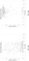
- FIG. 13 shows the molecule methylation scatter pattern of DMR1 and DMR2 in plasma cfDNA from a healthy individual ( FIG. 13A ) and a colon cancer patient ( FIG. 13B ).
- the counts of hyper-methylated molecules illustrated in the upper part of FIG. 13B from each DMR are the basis for disease detection from liquid biopsy.
- Filtering for hypermethylated molecules and robust signal may ensure that only the robust hyper-methylated molecules are counted for each DMR. This may improve the quality of analysis, and/or the sensitivity.
- a method of sequential enrichment may comprise obtaining a sample comprising a plurality of nucleic acid molecules and performing a first target enrichment to enrich for nucleic acid molecules comprising sequences corresponding to a first panel of one or more genome regions, thereby generating a first enriched sample comprising nucleic acids enriched for sequences corresponding to the first panel of one or more genome regions.
- the first target enrichment may also generate a remaining sample (or a first remaining sample) comprising nucleic acids depleted for sequences corresponding to the first panel of one or more genome regions.
- This remaining sample may be used for performing a second target enrichment upon the remaining sample to enrich for nucleic acid molecules comprising sequences corresponding to a second panel of one or more genome regions, thereby generating a second enriched sample comprising nucleic acids enriched for sequences corresponding to the second panel of one or more genome regions.
- the first panel of one or more genome regions and the second panel of one or more genome regions are generally different.
- third, fourth, or further rounds of target enrichment may be performed with third, fourth or further panels of genome regions.
- a panel of one or more genome regions may comprise a panel of 1-50,000, 5-10000, or 5-5000 genome regions associated with mutation hotspots, oncogenes, tumor suppressor genes, oncogene exons, tumor suppressor exons, or regulatory regions.
- a panel of one or more genome regions may comprise a panel of 5-5000 genome regions associated with differentially methylated regions, with epigenetic modifications, with introns, with promoters, or with other regulatory sequences.
- a panel comprises 50-500 genome regions associated with hypermethylation in cancer.
- Point-n-Seq is a pre amplification and pre conversion enrichment technology
- the enriched samples may be analyzed by sequencing, or may be bisulfide treated (or enzymatically treated) prior to sequencing to assess methylation.
- a first enriched sample may be analyzed by sequencing to assess mutations while a second enriched sample is bisulfide ( or enzymatical) treated prior to sequencing to assess methylation.
- a first enriched sample and a second enriched sample are both assessed by straightforward sequencing to access genomic alteration, however the samples may be sequenced at different depths.
- an analysis of a first enriched sample may be performed prior to performing a second target enrichment step. The results of the analysis of the first enriched sample may be used to select a second panel for the second enrichment step.
- the amplified nucleic acid products generated using the methods and kits described herein can be analyzed for one or more nucleic acid features.
- the one or more nucleic acid features can be one or more methylation events.
- the methylation can be methylation of a cytosine in a CpG dinucleotide.
- the methylated base can be a 5-methylcytosine.
- a cytosine in a non-CpG context can be methylated.
- the methylated or unmethylated cytosines can be in a CpG island.
- a CpG island can be a region of a genome with a high frequency of CpG sites.
- the CpG island can be at least 200 bp, or about 300 to about 3000 bp.
- the CpG island can be a CpG dinucleotide content of at least 60%.
- the CpG island can be in a promoter region of a gene.
- the methylation can be 5-hmC (5-hydroxymethylcytosine), 5-fC (5-formylcytosine), or 5-caC (5-carboxylcytosine).
- the methods and kits described herein can be used to detect methylation patterns, e.g., of DNA from a solid tissue or from a biological fluid, e.g., plasma, serum, urine, or saliva comprising, e.g., cell-free DNA.
- the cancer can be, e.g., colon cancer, breast cancer, liver cancer, bladder cancer, Wilms cancer, ovarian cancer, esophageal cancer, prostate cancer, bone cancer, or hepatocellular carcinoma, glioblastoma, breast cancer, squamous cell lung cancer, thyroid carcinoma, or leukemia (see e.g., Jin and Liu (2016) DNA methylation in human disease. Genes & Diseases, 5:1-8 ).
- the condition can be Beckwith-Wiedemann Syndrome, Prader-Willi syndrome, or Angelman syndrome.
- the methods and kits described herein can be used to determine methylation haplotype information and can be used to determine tissue or cell origin of cell-free DNA (see e.g., Seioighe et al, (2016) DNA methylation haplotypes as cancer markers. Nature Genetics 50, 1062-1063 ; international PCT application publication no. WO2015116837 ; U.S. patent application publication no. 20170121767 ).
- the methods and kits described herein can be used to detect methylation levels, e.g., of cell-free DNA, in subjects with cancer and subjects without cancer (see e.g., Vidal et al. A DNA methylation map of human cancer at single base-pair resolution.
- the methods and kits described herein can be used to determine methylation levels or to determine fractional contributions of different tissues to a cell-free DNA mixture (see e.g., international PCT application publication no. WO2016008451 ).
- the methods and kits described herein can be used for tissue of origin of cell-free DNA, e.g., in plasma, e.g., based on comparing patterns and abundance of methylation haplotypes (see e.g., Tang et al., (2016) Tumor origin detection with tissue-specific miRNA and DNA methylation markers. Bioinformatics 34, 398-406 ; international PCT application publication no.
- the disclosed methods can be used for monitoring of a condition.
- the condition can be disease.
- the disease can be a cancer, a neurological disease (e.g., Alzheimer's disease), immunodeficiency, skin disease, autoimmune disease (e.g., Ocular Behcet's disease), infection (e.g., viral infection), or metabolic disease.
- the cancer can be in remission. Since the disclosed methods can use cfDNA and ctDNA to detect low level of abnormalities, the present disclosure can provide relatively noninvasive method of monitoring diseases.
- the disclosed methods can be used for monitoring a treatment or therapy.
- the treatment or therapy can be used for a condition, e.g., a disease, e.g., cancer, or for any condition disclosed herein.
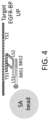
- a synergistic indirect capture of nucleic acid for sequencing (SICON-SEQ) experiment was carried out with two bridge probes with different sequences and an adaptor anchor probe/universal anchor probe (UP, SEQ ID NO: 1).
- the two bridge probes (EGFR-BP2, SEQ ID NO: 2 and EGFR-BP3, SEQ ID NO: 3) were designed to target EGFR genomic sequence.
- Each bridge probe comprised a targeting sequence (TS1 or TS2) region of about 25bp, a linker comprising at least 15 thymine, and a landing sequence (LS1 or LS2, italicized) having 20 bp that were designed to be complementary to the bridge binding sequence on the adaptor anchor probe.
- the hybridization assemblies were incubated with streptavidin beads (Thermo Fisher Dynabeads M270 Streptavidin) at room temperature for 10 min.
- streptavidin beads Thermo Fisher Dynabeads M270 Streptavidin
- Thermo Fisher Dynabeads M270 Streptavidin was incubated with streptavidin beads (Thermo Fisher Dynabeads M270 Streptavidin) at room temperature for 10 min.
- the clean-up was conducted with three washes (wash 1: 5X SSPE, 1%SDS; wash 2: 2X SSPE, 0.1% SDS; wash 3: 0.1X SSPE, 0.01% triton).
- the non-synergistic direct method involved hybridization of a biotinylated capture probe (120bp, SEQ ID NO. 6) comprising target specific sequence (hatched line, FIG. 5A ).
- the synergistic direct method involved hybridization of four short biotinylated capture probes (SEQ ID NOS. 7-10), and each contains 25bp of target specific sequences (hatched line, FIG. 5B ).
- the synergistic indirect method utilized four short bridge probes (SEQ ID NOS. 12-15) without biotin ( FIG. 5C ), and each comprised the same target specific sequences of as one of the capture probes used in the synergistic direct method.
- Non-synergistic direct 6 EGFR-bio Synergistic, direct 7 EGFR-bioP1 8 EGFR-bioP2 9 EGFR-bioP3 10 EGFR-bioP4 Synergistic, indirect 11 UP 12 EGFR-BP1 13 EGFR-BP2 14 EGFR-BP3 15 EGFR-BP4 Non-synergistic, indirect 11 UP 16 EGFR-BP2 17 EGFR Fw CCCGTCGCTATCAAGGAATTAAGA 18 P7 primer CAAGCAGAAGAC GGCATACGAGAT 19 P5 primer AATGATACGGCGACCACCGA
- Capture efficiency and the background noise were determined for either hybridization capture.
- the background noise was calculated by normalizing the qPCR result to the average background signal.
- the capture efficiency was not largely influenced by the presence of spacer, but the background noise of the capture hybridization without spacers was about 100-fold higher than the capture with spacer (Table 5). Hence, it suggests that the spacers in universal anchor probe played a significant role in enabling a highly specific (low background) capture. TABLE 5.
- Capture performance of hybridization with universal anchor probes with or without spacers Capture Efficiency Background UP-spacer 75.8% 1.1X 70.7% 0.9X UP-no spacer 66.0% 93.7X 66.0% 107.6X
- NGS next generation sequencing
- FIGS. 8A-8B show the coverage by SICON-SEQ and IDT xGen Hybridization and Wash Kit over areas of different percentage of GC contents.
- SICON targeted methylation sequencing (SICON-TMS) assay was conducted as illustrated in FIGS. 2A and 2B .
- the sample cfDNA were extracted from 3-5 ml of plasma from difference non-cancerous individuals and interrogated for 120 different differential methylated regions (DMRs).
- FIGS. 13A and 13B show the molecule methylation scatter pattern of DMR1 and DMR2 in health individual's plasma cfDNA and a colon cancer patient's plasma cfDNA respectively.
- the counts of hyper-methylated molecules illustrated in the upper part of FIG. 13B from each DMR may be used as the basis for disease detection from liquid biopsy.
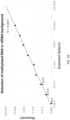
- a Point-n Seq colorectal cancer (CRC) panel covering 100 methylation markers was designed in 3 steps. First, approximately 1000 CRC-specific markers were identified from public databases. Secondly, makers with high background signal in baseline cfDNA of healthy population were eliminated. Finally, the list was finalized to contain the most differentiating markers between patient and healthy cfDNA. The capture of the SICON CRC panel was highly efficient resulting in high uniformity (94% > 0.5X, 100% >0.2X) and on-target rate (>80%). For 20ng cfDNA input, more than 1000 deduped informative reads were obtained for each marker on average, despite the high GC content (> 80%). The output of informative reads was linear to the cfDNA input ranging from 1ng to 40ng.
Landscapes
- Chemical & Material Sciences (AREA)
- Life Sciences & Earth Sciences (AREA)
- Organic Chemistry (AREA)
- Proteomics, Peptides & Aminoacids (AREA)
- Health & Medical Sciences (AREA)
- Engineering & Computer Science (AREA)
- Zoology (AREA)
- Wood Science & Technology (AREA)
- Analytical Chemistry (AREA)
- Immunology (AREA)
- Genetics & Genomics (AREA)
- General Engineering & Computer Science (AREA)
- Biophysics (AREA)
- Biotechnology (AREA)
- Microbiology (AREA)
- Molecular Biology (AREA)
- Physics & Mathematics (AREA)
- General Health & Medical Sciences (AREA)
- Biochemistry (AREA)
- Bioinformatics & Cheminformatics (AREA)
- Chemical Kinetics & Catalysis (AREA)
- Pathology (AREA)
- Oncology (AREA)
- Hospice & Palliative Care (AREA)
- Measuring Or Testing Involving Enzymes Or Micro-Organisms (AREA)
- Apparatus Associated With Microorganisms And Enzymes (AREA)
Applications Claiming Priority (5)
| Application Number | Priority Date | Filing Date | Title |
|---|---|---|---|
| US202062968847P | 2020-01-31 | 2020-01-31 | |
| US202062987232P | 2020-03-09 | 2020-03-09 | |
| US202062988859P | 2020-03-12 | 2020-03-12 | |
| EP21747390.9A EP4097231B1 (de) | 2020-01-31 | 2021-02-01 | Systeme und verfahren zum gezielten nukleinsäureeinfang |
| PCT/US2021/016089 WO2021155374A2 (en) | 2020-01-31 | 2021-02-01 | Systems and methods for targeted nucleic acid capture |
Related Parent Applications (2)
| Application Number | Title | Priority Date | Filing Date |
|---|---|---|---|
| EP21747390.9A Division EP4097231B1 (de) | 2020-01-31 | 2021-02-01 | Systeme und verfahren zum gezielten nukleinsäureeinfang |
| EP21747390.9A Division-Into EP4097231B1 (de) | 2020-01-31 | 2021-02-01 | Systeme und verfahren zum gezielten nukleinsäureeinfang |
Publications (2)
| Publication Number | Publication Date |
|---|---|
| EP4534690A2 true EP4534690A2 (de) | 2025-04-09 |
| EP4534690A3 EP4534690A3 (de) | 2025-07-23 |
Family
ID=77079995
Family Applications (2)
| Application Number | Title | Priority Date | Filing Date |
|---|---|---|---|
| EP25156820.0A Pending EP4534690A3 (de) | 2020-01-31 | 2021-02-01 | Systeme und verfahren zum gezielten nukleinsäureeinfang |
| EP21747390.9A Active EP4097231B1 (de) | 2020-01-31 | 2021-02-01 | Systeme und verfahren zum gezielten nukleinsäureeinfang |
Family Applications After (1)
| Application Number | Title | Priority Date | Filing Date |
|---|---|---|---|
| EP21747390.9A Active EP4097231B1 (de) | 2020-01-31 | 2021-02-01 | Systeme und verfahren zum gezielten nukleinsäureeinfang |
Country Status (8)
| Country | Link |
|---|---|
| US (2) | US20230193380A1 (de) |
| EP (2) | EP4534690A3 (de) |
| JP (2) | JP7732991B2 (de) |
| KR (1) | KR20220133984A (de) |
| CN (1) | CN115103909A (de) |
| CA (1) | CA3162647A1 (de) |
| GB (1) | GB2612412B (de) |
| WO (1) | WO2021155374A2 (de) |
Families Citing this family (5)
| Publication number | Priority date | Publication date | Assignee | Title |
|---|---|---|---|---|
| WO2020106906A1 (en) * | 2018-11-21 | 2020-05-28 | Avida Biomed, Inc. | Methods for targeted nucleic acid library formation |
| US20250179567A1 (en) * | 2022-02-18 | 2025-06-05 | Agilent Technologies, Inc. | Systems and methods for targeted nucleic acid capture and barcoding |
| US20250369045A1 (en) * | 2022-12-20 | 2025-12-04 | Illumina, Inc. | Multivalent assemblies for enhanced target hybridization |
| CN116574780B (zh) * | 2023-02-24 | 2023-11-10 | 苏州唯善生物科技有限公司 | 一种单荧光通道同时检测两种miRNA的引物探针设计方法、试剂盒及定量检测方法 |
| WO2025221668A1 (en) * | 2024-04-19 | 2025-10-23 | Agilent Technologies, Inc. | Methods of preparing nucleic acids for sequencing and methylation analysis |
Citations (7)
| Publication number | Priority date | Publication date | Assignee | Title |
|---|---|---|---|---|
| WO2005019477A2 (en) | 2003-08-12 | 2005-03-03 | Epigenomics Ag | Methods and compositions for differentiating tissues or cell types using epigenetic markers |
| WO2014043763A1 (en) | 2012-09-20 | 2014-03-27 | The Chinese University Of Hong Kong | Non-invasive determination of methylome of fetus or tumor from plasma |
| WO2015116837A1 (en) | 2014-01-30 | 2015-08-06 | The Regents Of The University Of California | Methylation haplotyping for non-invasive diagnosis (monod) |
| WO2016008451A1 (en) | 2014-07-18 | 2016-01-21 | The Chinese University Of Hong Kong | Methylation pattern analysis of tissues in dna mixture |
| US20170121767A1 (en) | 2014-04-14 | 2017-05-04 | Yissum Research Development Company Of The Hebrew University Of Jerusalem Ltd. | Method and kit for determining the tissue or cell origin of dna |
| US20170175205A1 (en) | 2015-12-17 | 2017-06-22 | Illumina, Inc. | Distinguishing methylation levels in complex biological samples |
| WO2018119216A1 (en) | 2016-12-21 | 2018-06-28 | The Regents Of The University Of California | Deconvolution and detection of rare dna in plasma |
Family Cites Families (6)
| Publication number | Priority date | Publication date | Assignee | Title |
|---|---|---|---|---|
| WO2010030683A1 (en) * | 2008-09-09 | 2010-03-18 | Rosetta Inpharmatics Llc | Methods of generating gene specific libraries |
| US20100167952A1 (en) * | 2008-11-06 | 2010-07-01 | Thomas Albert | Suppression of secondary capture in microarray assays |
| US20100331204A1 (en) * | 2009-02-13 | 2010-12-30 | Jeff Jeddeloh | Methods and systems for enrichment of target genomic sequences |
| KR20130113447A (ko) * | 2010-09-24 | 2013-10-15 | 더 보드 어브 트러스티스 어브 더 리랜드 스탠포드 주니어 유니버시티 | 고정된 프라이머들을 이용하여 표적 dna의 직접적인 캡쳐, 증폭 및 서열화 |
| US9695416B2 (en) | 2012-07-18 | 2017-07-04 | Siemens Healthcare Diagnostics Inc. | Method of normalizing biological samples |
| CN112601823A (zh) * | 2018-06-12 | 2021-04-02 | 安可济控股有限公司 | 用于形成连接产物的方法和组合物 |
-
2021
- 2021-02-01 CA CA3162647A patent/CA3162647A1/en active Pending
- 2021-02-01 WO PCT/US2021/016089 patent/WO2021155374A2/en not_active Ceased
- 2021-02-01 KR KR1020227029940A patent/KR20220133984A/ko active Pending
- 2021-02-01 CN CN202180011915.0A patent/CN115103909A/zh active Pending
- 2021-02-01 EP EP25156820.0A patent/EP4534690A3/de active Pending
- 2021-02-01 JP JP2022546396A patent/JP7732991B2/ja active Active
- 2021-02-01 EP EP21747390.9A patent/EP4097231B1/de active Active
- 2021-02-01 GB GB2211994.5A patent/GB2612412B/en active Active
-
2022
- 2022-07-29 US US17/816,198 patent/US20230193380A1/en active Pending
-
2024
- 2024-04-30 US US18/651,023 patent/US20240287601A1/en active Pending
-
2025
- 2025-08-21 JP JP2025138504A patent/JP2025169409A/ja active Pending
Patent Citations (7)
| Publication number | Priority date | Publication date | Assignee | Title |
|---|---|---|---|---|
| WO2005019477A2 (en) | 2003-08-12 | 2005-03-03 | Epigenomics Ag | Methods and compositions for differentiating tissues or cell types using epigenetic markers |
| WO2014043763A1 (en) | 2012-09-20 | 2014-03-27 | The Chinese University Of Hong Kong | Non-invasive determination of methylome of fetus or tumor from plasma |
| WO2015116837A1 (en) | 2014-01-30 | 2015-08-06 | The Regents Of The University Of California | Methylation haplotyping for non-invasive diagnosis (monod) |
| US20170121767A1 (en) | 2014-04-14 | 2017-05-04 | Yissum Research Development Company Of The Hebrew University Of Jerusalem Ltd. | Method and kit for determining the tissue or cell origin of dna |
| WO2016008451A1 (en) | 2014-07-18 | 2016-01-21 | The Chinese University Of Hong Kong | Methylation pattern analysis of tissues in dna mixture |
| US20170175205A1 (en) | 2015-12-17 | 2017-06-22 | Illumina, Inc. | Distinguishing methylation levels in complex biological samples |
| WO2018119216A1 (en) | 2016-12-21 | 2018-06-28 | The Regents Of The University Of California | Deconvolution and detection of rare dna in plasma |
Non-Patent Citations (5)
| Title |
|---|
| HAO ET AL.: "DNA methylation markers for diagnosis and prognosis of common cancers", PROC. NATL. ACAD. SCI., 2017 |
| POON: "Differential DNA Methylation between Fetus and Mother as a Strategy for Detecting Fetal DNA in Maternal Plasma", CLINICAL CHEMISTRY, vol. 48, 2002, pages 35 - 41, XP093237303, DOI: 10.1093/clinchem/48.1.35 |
| SEIOIGHE ET AL.: "DNA methylation haplotypes as cancer markers.", NATURE GENETICS, vol. 50, 2018, pages 1062 - 1063 |
| TANG ET AL.: "Tumor origin detection with tissue-specific miRNA and DNA methylation markers.", BIOINFORMATICS, vol. 34, 2018, pages 398 - 406, XP093054365, DOI: 10.1093/bioinformatics/btx622 |
| VIDAL ET AL.: "A DNA methylation map of human cancer at single base-pair resolution.", ONCOGENOMICS, vol. 36, pages 5648 - 5657 |
Also Published As
| Publication number | Publication date |
|---|---|
| GB2612412B (en) | 2025-10-10 |
| GB202211994D0 (en) | 2022-09-28 |
| CN115103909A (zh) | 2022-09-23 |
| WO2021155374A3 (en) | 2021-09-10 |
| EP4534690A3 (de) | 2025-07-23 |
| GB2612412A (en) | 2023-05-03 |
| US20240287601A1 (en) | 2024-08-29 |
| EP4097231B1 (de) | 2025-03-19 |
| CA3162647A1 (en) | 2021-08-05 |
| JP7732991B2 (ja) | 2025-09-02 |
| JP2025169409A (ja) | 2025-11-12 |
| KR20220133984A (ko) | 2022-10-05 |
| EP4097231A2 (de) | 2022-12-07 |
| US20230193380A1 (en) | 2023-06-22 |
| WO2021155374A2 (en) | 2021-08-05 |
| JP2023512522A (ja) | 2023-03-27 |
| EP4097231C0 (de) | 2025-03-19 |
| EP4097231A4 (de) | 2024-04-03 |
Similar Documents
| Publication | Publication Date | Title |
|---|---|---|
| EP4097231B1 (de) | Systeme und verfahren zum gezielten nukleinsäureeinfang | |
| EP3884047B1 (de) | Verfahren zur gezielten bildung von nukleinsäurebibliotheken | |
| Ondraskova et al. | Electrochemical biosensors for analysis of DNA point mutations in cancer research | |
| CN110129436A (zh) | Dna甲基化的数字序列分析 | |
| US11261479B2 (en) | Methods and compositions for enrichment of target nucleic acids | |
| EP2982762B1 (de) | Verfahren zur nukleinsäureamplifikation mittels eines allelspezifischen reaktiven primers | |
| US20160046993A1 (en) | Nanoprobe-based genetic testing | |
| CN114787385A (zh) | 用于检测核酸修饰的方法和系统 | |
| EP4320276B1 (de) | Verfahren zur krankheitserkennung | |
| US20250179567A1 (en) | Systems and methods for targeted nucleic acid capture and barcoding | |
| US20240240240A1 (en) | Enhancer oligonucleotides for nucleic acid hybridization | |
| US11680290B2 (en) | Efficient methods and compositions for multiplex target amplification PCR | |
| HK40078903B (zh) | 靶向核酸文库形成的方法 | |
| HK40078903A (en) | Methods for targeted nucleic acid library formation |
Legal Events
| Date | Code | Title | Description |
|---|---|---|---|
| PUAI | Public reference made under article 153(3) epc to a published international application that has entered the european phase |
Free format text: ORIGINAL CODE: 0009012 |
|
| STAA | Information on the status of an ep patent application or granted ep patent |
Free format text: STATUS: REQUEST FOR EXAMINATION WAS MADE |
|
| REG | Reference to a national code |
Ref country code: DE Ref legal event code: R079 Free format text: PREVIOUS MAIN CLASS: C12Q0001683200 Ipc: C12N0015110000 |
|
| 17P | Request for examination filed |
Effective date: 20250210 |
|
| AC | Divisional application: reference to earlier application |
Ref document number: 4097231 Country of ref document: EP Kind code of ref document: P |
|
| AK | Designated contracting states |
Kind code of ref document: A2 Designated state(s): AL AT BE BG CH CY CZ DE DK EE ES FI FR GB GR HR HU IE IS IT LI LT LU LV MC MK MT NL NO PL PT RO RS SE SI SK SM TR |
|
| RIC1 | Information provided on ipc code assigned before grant |
Ipc: C12Q 1/6832 20180101ALI20250404BHEP Ipc: C12Q 1/6874 20180101ALI20250404BHEP Ipc: C12Q 1/6853 20180101ALI20250404BHEP Ipc: C12Q 1/6806 20180101ALI20250404BHEP Ipc: C40B 40/06 20060101ALI20250404BHEP Ipc: C12N 15/11 20060101AFI20250404BHEP |
|
| PUAL | Search report despatched |
Free format text: ORIGINAL CODE: 0009013 |
|
| AK | Designated contracting states |
Kind code of ref document: A3 Designated state(s): AL AT BE BG CH CY CZ DE DK EE ES FI FR GB GR HR HU IE IS IT LI LT LU LV MC MK MT NL NO PL PT RO RS SE SI SK SM TR |
|
| RIC1 | Information provided on ipc code assigned before grant |
Ipc: C12N 15/11 20060101AFI20250613BHEP Ipc: C40B 40/06 20060101ALI20250613BHEP Ipc: C12Q 1/6806 20180101ALI20250613BHEP Ipc: C12Q 1/6853 20180101ALI20250613BHEP Ipc: C12Q 1/6874 20180101ALI20250613BHEP Ipc: C12Q 1/6832 20180101ALI20250613BHEP |